EP3280441B1 - Anticorps anti-sortiline et leurs méthodes d'utilisation - Google Patents
Anticorps anti-sortiline et leurs méthodes d'utilisation Download PDFInfo
- Publication number
- EP3280441B1 EP3280441B1 EP16777311.8A EP16777311A EP3280441B1 EP 3280441 B1 EP3280441 B1 EP 3280441B1 EP 16777311 A EP16777311 A EP 16777311A EP 3280441 B1 EP3280441 B1 EP 3280441B1
- Authority
- EP
- European Patent Office
- Prior art keywords
- sortilin
- amino acid
- antibody
- seq
- acid sequence
- Prior art date
- Legal status (The legal status is an assumption and is not a legal conclusion. Google has not performed a legal analysis and makes no representation as to the accuracy of the status listed.)
- Active
Links
- 238000000034 method Methods 0.000 title claims description 82
- 102100032889 Sortilin Human genes 0.000 claims description 539
- 108010014657 sortilin Proteins 0.000 claims description 537
- 125000003275 alpha amino acid group Chemical group 0.000 claims description 250
- 108010012809 Progranulins Proteins 0.000 claims description 224
- 102000019204 Progranulins Human genes 0.000 claims description 221
- 241000282414 Homo sapiens Species 0.000 claims description 149
- 210000004027 cell Anatomy 0.000 claims description 148
- 108090000623 proteins and genes Proteins 0.000 claims description 103
- 101000868139 Homo sapiens Sortilin Proteins 0.000 claims description 93
- 108090000765 processed proteins & peptides Proteins 0.000 claims description 90
- 239000000427 antigen Substances 0.000 claims description 84
- 108091007433 antigens Proteins 0.000 claims description 83
- 102000036639 antigens Human genes 0.000 claims description 83
- 102000004169 proteins and genes Human genes 0.000 claims description 83
- 230000007423 decrease Effects 0.000 claims description 60
- 208000037265 diseases, disorders, signs and symptoms Diseases 0.000 claims description 58
- 238000006467 substitution reaction Methods 0.000 claims description 54
- 102000008394 Immunoglobulin Fragments Human genes 0.000 claims description 52
- 108010021625 Immunoglobulin Fragments Proteins 0.000 claims description 52
- 230000014509 gene expression Effects 0.000 claims description 48
- 239000012634 fragment Substances 0.000 claims description 46
- 201000010099 disease Diseases 0.000 claims description 39
- 238000011282 treatment Methods 0.000 claims description 37
- 101000868164 Mus musculus Sortilin Proteins 0.000 claims description 23
- 238000000338 in vitro Methods 0.000 claims description 22
- 230000001965 increasing effect Effects 0.000 claims description 22
- 150000007523 nucleic acids Chemical class 0.000 claims description 21
- 230000000770 proinflammatory effect Effects 0.000 claims description 21
- 230000003247 decreasing effect Effects 0.000 claims description 20
- 238000010494 dissociation reaction Methods 0.000 claims description 20
- 230000005593 dissociations Effects 0.000 claims description 20
- 239000013598 vector Substances 0.000 claims description 20
- 201000011240 Frontotemporal dementia Diseases 0.000 claims description 19
- 208000035475 disorder Diseases 0.000 claims description 19
- 102000039446 nucleic acids Human genes 0.000 claims description 19
- 108020004707 nucleic acids Proteins 0.000 claims description 19
- 208000024827 Alzheimer disease Diseases 0.000 claims description 18
- 201000004810 Vascular dementia Diseases 0.000 claims description 14
- 208000014674 injury Diseases 0.000 claims description 12
- 206010010904 Convulsion Diseases 0.000 claims description 11
- 230000002401 inhibitory effect Effects 0.000 claims description 11
- 208000032578 Inherited retinal disease Diseases 0.000 claims description 10
- 208000032430 Retinal dystrophy Diseases 0.000 claims description 10
- 208000030886 Traumatic Brain injury Diseases 0.000 claims description 10
- 201000006321 fundus dystrophy Diseases 0.000 claims description 10
- 208000017532 inherited retinal dystrophy Diseases 0.000 claims description 10
- 230000009529 traumatic brain injury Effects 0.000 claims description 10
- 206010012289 Dementia Diseases 0.000 claims description 9
- 208000020431 spinal cord injury Diseases 0.000 claims description 9
- 208000018737 Parkinson disease Diseases 0.000 claims description 8
- 206010002026 amyotrophic lateral sclerosis Diseases 0.000 claims description 8
- 230000006378 damage Effects 0.000 claims description 8
- 102000004584 Somatomedin Receptors Human genes 0.000 claims description 7
- 108010017622 Somatomedin Receptors Proteins 0.000 claims description 7
- 108010033576 Transferrin Receptors Proteins 0.000 claims description 7
- 102000007238 Transferrin Receptors Human genes 0.000 claims description 7
- 102100021943 C-C motif chemokine 2 Human genes 0.000 claims description 6
- 102100032367 C-C motif chemokine 5 Human genes 0.000 claims description 6
- 208000010412 Glaucoma Diseases 0.000 claims description 6
- 102100034221 Growth-regulated alpha protein Human genes 0.000 claims description 6
- 101000897480 Homo sapiens C-C motif chemokine 2 Proteins 0.000 claims description 6
- 101000797762 Homo sapiens C-C motif chemokine 5 Proteins 0.000 claims description 6
- 101001069921 Homo sapiens Growth-regulated alpha protein Proteins 0.000 claims description 6
- 108090001005 Interleukin-6 Proteins 0.000 claims description 6
- 208000006011 Stroke Diseases 0.000 claims description 6
- 108060008682 Tumor Necrosis Factor Proteins 0.000 claims description 6
- 102000000852 Tumor Necrosis Factor-alpha Human genes 0.000 claims description 6
- 208000027418 Wounds and injury Diseases 0.000 claims description 6
- 206010064930 age-related macular degeneration Diseases 0.000 claims description 6
- 230000008499 blood brain barrier function Effects 0.000 claims description 6
- 210000001218 blood-brain barrier Anatomy 0.000 claims description 6
- 208000002780 macular degeneration Diseases 0.000 claims description 6
- 201000007737 Retinal degeneration Diseases 0.000 claims description 5
- 208000002552 acute disseminated encephalomyelitis Diseases 0.000 claims description 5
- 201000006417 multiple sclerosis Diseases 0.000 claims description 5
- 201000002212 progressive supranuclear palsy Diseases 0.000 claims description 5
- 230000004258 retinal degeneration Effects 0.000 claims description 5
- MZOFCQQQCNRIBI-VMXHOPILSA-N (3s)-4-[[(2s)-1-[[(2s)-1-[[(1s)-1-carboxy-2-hydroxyethyl]amino]-4-methyl-1-oxopentan-2-yl]amino]-5-(diaminomethylideneamino)-1-oxopentan-2-yl]amino]-3-[[2-[[(2s)-2,6-diaminohexanoyl]amino]acetyl]amino]-4-oxobutanoic acid Chemical compound OC[C@@H](C(O)=O)NC(=O)[C@H](CC(C)C)NC(=O)[C@H](CCCN=C(N)N)NC(=O)[C@H](CC(O)=O)NC(=O)CNC(=O)[C@@H](N)CCCCN MZOFCQQQCNRIBI-VMXHOPILSA-N 0.000 claims description 4
- 102000003746 Insulin Receptor Human genes 0.000 claims description 4
- 108010001127 Insulin Receptor Proteins 0.000 claims description 4
- 108010071134 CRM197 (non-toxic variant of diphtheria toxin) Proteins 0.000 claims description 3
- 101800001649 Heparin-binding EGF-like growth factor Proteins 0.000 claims description 3
- 101710172064 Low-density lipoprotein receptor-related protein Proteins 0.000 claims description 3
- 208000036110 Neuroinflammatory disease Diseases 0.000 claims description 3
- 230000001594 aberrant effect Effects 0.000 claims description 3
- GBVKRUOMSUTVPW-AHNVSIPUSA-N chembl1089636 Chemical compound N([C@H]([C@@H](OC(=O)CCC(=O)N[C@@H](C(O)C)C(=O)N[C@@H](CC=1C=CC=CC=1)C(=O)N[C@@H](CC=1C=CC=CC=1)C(=O)N[C@@H](CC=1C=CC(O)=CC=1)C(=O)NCC(=O)NCC(=O)N[C@@H](CO)C(=O)N[C@@H](CCCNC(N)=N)C(=O)NCC(=O)N[C@@H](CCCCNC(=O)CCC(=O)O[C@H]([C@@H](NC(=O)C=1C=CC=CC=1)C=1C=CC=CC=1)C(=O)O[C@@H]1C(=C2[C@@H](OC(C)=O)C(=O)[C@]3(C)[C@@H](O)C[C@H]4OC[C@]4([C@H]3[C@H](OC(=O)C=3C=CC=CC=3)[C@](C2(C)C)(O)C1)OC(C)=O)C)C(=O)N[C@@H](CCCNC(N)=N)C(=O)N[C@@H](CC(N)=O)C(=O)N[C@@H](CC(N)=O)C(=O)N[C@@H](CC=1C=CC=CC=1)C(=O)N[C@@H](CCCCNC(=O)CCC(=O)O[C@H]([C@@H](NC(=O)C=1C=CC=CC=1)C=1C=CC=CC=1)C(=O)O[C@@H]1C(=C2[C@@H](OC(C)=O)C(=O)[C@]3(C)[C@@H](O)C[C@H]4OC[C@]4([C@H]3[C@H](OC(=O)C=3C=CC=CC=3)[C@](C2(C)C)(O)C1)OC(C)=O)C)C(=O)N[C@@H]([C@@H](C)O)C(=O)N[C@@H](CCC(O)=O)C(=O)N[C@@H](CCC(O)=O)C(=O)N[C@@H](CC=1C=CC(O)=CC=1)C(O)=O)C(=O)O[C@@H]1C(=C2[C@@H](OC(C)=O)C(=O)[C@]3(C)[C@@H](O)C[C@H]4OC[C@]4([C@H]3[C@H](OC(=O)C=3C=CC=CC=3)[C@](C2(C)C)(O)C1)OC(C)=O)C)C=1C=CC=CC=1)C(=O)C1=CC=CC=C1 GBVKRUOMSUTVPW-AHNVSIPUSA-N 0.000 claims description 3
- 239000003937 drug carrier Substances 0.000 claims description 3
- 230000028709 inflammatory response Effects 0.000 claims description 3
- 230000003959 neuroinflammation Effects 0.000 claims description 3
- 108010046239 paclitaxel-Angiopep-2 conjugate Proteins 0.000 claims description 3
- MCYTYTUNNNZWOK-LCLOTLQISA-N penetratin Chemical compound C([C@H](NC(=O)[C@H](CC=1C2=CC=CC=C2NC=1)NC(=O)[C@H]([C@@H](C)CC)NC(=O)[C@H](CCCCN)NC(=O)[C@@H](NC(=O)[C@H](CCC(N)=O)NC(=O)[C@@H](N)CCCNC(N)=N)[C@@H](C)CC)C(=O)N[C@@H](CCC(N)=O)C(=O)N[C@@H](CC(N)=O)C(=O)N[C@@H](CCCNC(N)=N)C(=O)N[C@@H](CCCNC(N)=N)C(=O)N[C@@H](CCSC)C(=O)N[C@@H](CCCCN)C(=O)N[C@@H](CC=1C2=CC=CC=C2NC=1)C(=O)N[C@@H](CCCCN)C(=O)N[C@@H](CCCCN)C(N)=O)C1=CC=CC=C1 MCYTYTUNNNZWOK-LCLOTLQISA-N 0.000 claims description 3
- 108010043655 penetratin Proteins 0.000 claims description 3
- 108010011110 polyarginine Proteins 0.000 claims description 3
- 230000001737 promoting effect Effects 0.000 claims description 3
- 238000010361 transduction Methods 0.000 claims description 3
- 230000026683 transduction Effects 0.000 claims description 3
- 230000029663 wound healing Effects 0.000 claims description 3
- 208000035143 Bacterial infection Diseases 0.000 claims description 2
- 208000004575 Infectious Arthritis Diseases 0.000 claims description 2
- 108091028043 Nucleic acid sequence Proteins 0.000 claims description 2
- 206010040070 Septic Shock Diseases 0.000 claims description 2
- 230000003376 axonal effect Effects 0.000 claims description 2
- 230000007845 axonopathy Effects 0.000 claims description 2
- 208000025255 bacterial arthritis Diseases 0.000 claims description 2
- 208000022362 bacterial infectious disease Diseases 0.000 claims description 2
- 230000006724 microglial activation Effects 0.000 claims description 2
- 201000008482 osteoarthritis Diseases 0.000 claims description 2
- 239000008194 pharmaceutical composition Substances 0.000 claims description 2
- 230000036303 septic shock Effects 0.000 claims description 2
- 102000018710 Heparin-binding EGF-like Growth Factor Human genes 0.000 claims 1
- 125000000539 amino acid group Chemical group 0.000 description 192
- 230000027455 binding Effects 0.000 description 181
- 108010025020 Nerve Growth Factor Proteins 0.000 description 131
- 235000001014 amino acid Nutrition 0.000 description 111
- 230000003993 interaction Effects 0.000 description 101
- 241000699670 Mus sp. Species 0.000 description 92
- 150000001413 amino acids Chemical class 0.000 description 79
- 235000018102 proteins Nutrition 0.000 description 79
- 229940024606 amino acid Drugs 0.000 description 78
- 102000015336 Nerve Growth Factor Human genes 0.000 description 67
- 229940053128 nerve growth factor Drugs 0.000 description 67
- 102000007072 Nerve Growth Factors Human genes 0.000 description 64
- 102000004196 processed proteins & peptides Human genes 0.000 description 61
- 230000001413 cellular effect Effects 0.000 description 54
- 230000000875 corresponding effect Effects 0.000 description 49
- 108060003951 Immunoglobulin Proteins 0.000 description 46
- 108090000099 Neurotrophin-4 Proteins 0.000 description 46
- 102000018358 immunoglobulin Human genes 0.000 description 46
- 108090000715 Brain-derived neurotrophic factor Proteins 0.000 description 44
- 102000004219 Brain-derived neurotrophic factor Human genes 0.000 description 44
- 229940077737 brain-derived neurotrophic factor Drugs 0.000 description 44
- DZHSAHHDTRWUTF-SIQRNXPUSA-N amyloid-beta polypeptide 42 Chemical compound C([C@@H](C(=O)N[C@@H](C)C(=O)N[C@@H](CCC(O)=O)C(=O)N[C@@H](CC(O)=O)C(=O)N[C@H](C(=O)NCC(=O)N[C@@H](CO)C(=O)N[C@@H](CC(N)=O)C(=O)N[C@@H](CCCCN)C(=O)NCC(=O)N[C@@H](C)C(=O)N[C@H](C(=O)N[C@@H]([C@@H](C)CC)C(=O)NCC(=O)N[C@@H](CC(C)C)C(=O)N[C@@H](CCSC)C(=O)N[C@@H](C(C)C)C(=O)NCC(=O)NCC(=O)N[C@@H](C(C)C)C(=O)N[C@@H](C(C)C)C(=O)N[C@@H]([C@@H](C)CC)C(=O)N[C@@H](C)C(O)=O)[C@@H](C)CC)C(C)C)NC(=O)[C@H](CC=1C=CC=CC=1)NC(=O)[C@@H](NC(=O)[C@H](CC(C)C)NC(=O)[C@H](CCCCN)NC(=O)[C@H](CCC(N)=O)NC(=O)[C@H](CC=1N=CNC=1)NC(=O)[C@H](CC=1N=CNC=1)NC(=O)[C@@H](NC(=O)[C@H](CCC(O)=O)NC(=O)[C@H](CC=1C=CC(O)=CC=1)NC(=O)CNC(=O)[C@H](CO)NC(=O)[C@H](CC(O)=O)NC(=O)[C@H](CC=1N=CNC=1)NC(=O)[C@H](CCCNC(N)=N)NC(=O)[C@H](CC=1C=CC=CC=1)NC(=O)[C@H](CCC(O)=O)NC(=O)[C@H](C)NC(=O)[C@@H](N)CC(O)=O)C(C)C)C(C)C)C1=CC=CC=C1 DZHSAHHDTRWUTF-SIQRNXPUSA-N 0.000 description 39
- NFGXHKASABOEEW-UHFFFAOYSA-N 1-methylethyl 11-methoxy-3,7,11-trimethyl-2,4-dodecadienoate Chemical compound COC(C)(C)CCCC(C)CC=CC(C)=CC(=O)OC(C)C NFGXHKASABOEEW-UHFFFAOYSA-N 0.000 description 38
- 229920001184 polypeptide Polymers 0.000 description 38
- 101710137189 Amyloid-beta A4 protein Proteins 0.000 description 37
- 102100022704 Amyloid-beta precursor protein Human genes 0.000 description 37
- 101710151993 Amyloid-beta precursor protein Proteins 0.000 description 37
- 101800001814 Neurotensin Proteins 0.000 description 37
- 102400001103 Neurotensin Human genes 0.000 description 37
- PCJGZPGTCUMMOT-ISULXFBGSA-N neurotensin Chemical compound C([C@@H](C(=O)N[C@@H]([C@@H](C)CC)C(=O)N[C@@H](CC(C)C)C(O)=O)NC(=O)[C@H]1N(CCC1)C(=O)[C@H](CCCN=C(N)N)NC(=O)[C@H](CCCN=C(N)N)NC(=O)[C@H]1N(CCC1)C(=O)[C@H](CCCCN)NC(=O)[C@H](CC(N)=O)NC(=O)[C@H](CCC(O)=O)NC(=O)[C@H](CC=1C=CC(O)=CC=1)NC(=O)[C@H](CC(C)C)NC(=O)[C@H]1NC(=O)CC1)C1=CC=C(O)C=C1 PCJGZPGTCUMMOT-ISULXFBGSA-N 0.000 description 37
- 210000002966 serum Anatomy 0.000 description 37
- 102100029470 Apolipoprotein E Human genes 0.000 description 36
- 101710095339 Apolipoprotein E Proteins 0.000 description 36
- 108010013563 Lipoprotein Lipase Proteins 0.000 description 36
- 102100022119 Lipoprotein lipase Human genes 0.000 description 36
- 102000005962 receptors Human genes 0.000 description 36
- 108020003175 receptors Proteins 0.000 description 36
- 108090000742 Neurotrophin 3 Proteins 0.000 description 32
- 102000004230 Neurotrophin 3 Human genes 0.000 description 32
- 229940032018 neurotrophin 3 Drugs 0.000 description 32
- 102100029946 Sialic acid-binding Ig-like lectin 7 Human genes 0.000 description 31
- 101710187830 Tumor necrosis factor receptor superfamily member 1B Proteins 0.000 description 31
- 239000002158 endotoxin Substances 0.000 description 30
- 229920006008 lipopolysaccharide Polymers 0.000 description 30
- 238000003556 assay Methods 0.000 description 25
- 102000003683 Neurotrophin-4 Human genes 0.000 description 24
- 102100038955 Proprotein convertase subtilisin/kexin type 9 Human genes 0.000 description 24
- 101710180553 Proprotein convertase subtilisin/kexin type 9 Proteins 0.000 description 24
- 230000000694 effects Effects 0.000 description 24
- 229940097998 neurotrophin 4 Drugs 0.000 description 24
- 102100033857 Neurotrophin-4 Human genes 0.000 description 22
- 238000004519 manufacturing process Methods 0.000 description 22
- 108010071619 Apolipoproteins Proteins 0.000 description 21
- 102000007592 Apolipoproteins Human genes 0.000 description 21
- 210000004556 brain Anatomy 0.000 description 21
- 230000035772 mutation Effects 0.000 description 21
- 210000004408 hybridoma Anatomy 0.000 description 20
- -1 CD98hc Proteins 0.000 description 19
- 230000006870 function Effects 0.000 description 18
- 230000004048 modification Effects 0.000 description 18
- 238000012986 modification Methods 0.000 description 18
- 230000015556 catabolic process Effects 0.000 description 17
- 238000001727 in vivo Methods 0.000 description 17
- 108010047041 Complementarity Determining Regions Proteins 0.000 description 16
- 238000006731 degradation reaction Methods 0.000 description 16
- 210000002569 neuron Anatomy 0.000 description 15
- 108020004414 DNA Proteins 0.000 description 14
- 241001465754 Metazoa Species 0.000 description 14
- 230000032683 aging Effects 0.000 description 14
- 241000699666 Mus <mouse, genus> Species 0.000 description 13
- 230000003143 atherosclerotic effect Effects 0.000 description 13
- 230000004071 biological effect Effects 0.000 description 13
- 238000005516 engineering process Methods 0.000 description 13
- 230000002829 reductive effect Effects 0.000 description 13
- 208000019553 vascular disease Diseases 0.000 description 13
- 108010087819 Fc receptors Proteins 0.000 description 12
- 102000009109 Fc receptors Human genes 0.000 description 12
- 108091007491 NSP3 Papain-like protease domains Proteins 0.000 description 12
- 230000009286 beneficial effect Effects 0.000 description 12
- 230000000903 blocking effect Effects 0.000 description 12
- 230000030833 cell death Effects 0.000 description 12
- 208000024891 symptom Diseases 0.000 description 12
- 210000001519 tissue Anatomy 0.000 description 12
- 230000012202 endocytosis Effects 0.000 description 11
- 125000003729 nucleotide group Chemical group 0.000 description 11
- 230000032258 transport Effects 0.000 description 11
- 238000002965 ELISA Methods 0.000 description 10
- 108060003393 Granulin Proteins 0.000 description 10
- 230000006907 apoptotic process Effects 0.000 description 10
- HVYWMOMLDIMFJA-DPAQBDIFSA-N cholesterol Chemical compound C1C=C2C[C@@H](O)CC[C@]2(C)[C@@H]2[C@@H]1[C@@H]1CC[C@H]([C@H](C)CCCC(C)C)[C@@]1(C)CC2 HVYWMOMLDIMFJA-DPAQBDIFSA-N 0.000 description 10
- 102000017941 granulin Human genes 0.000 description 10
- 238000003780 insertion Methods 0.000 description 10
- 230000037431 insertion Effects 0.000 description 10
- ROHFNLRQFUQHCH-YFKPBYRVSA-N L-leucine Chemical compound CC(C)C[C@H](N)C(O)=O ROHFNLRQFUQHCH-YFKPBYRVSA-N 0.000 description 9
- 108010001831 LDL receptors Proteins 0.000 description 9
- 206010035226 Plasma cell myeloma Diseases 0.000 description 9
- 238000013459 approach Methods 0.000 description 9
- 239000012636 effector Substances 0.000 description 9
- 239000007924 injection Substances 0.000 description 9
- 238000002347 injection Methods 0.000 description 9
- 230000020796 long term synaptic depression Effects 0.000 description 9
- 201000000050 myeloid neoplasm Diseases 0.000 description 9
- 230000016273 neuron death Effects 0.000 description 9
- 102000040430 polynucleotide Human genes 0.000 description 9
- 108091033319 polynucleotide Proteins 0.000 description 9
- 239000002157 polynucleotide Substances 0.000 description 9
- 239000002243 precursor Substances 0.000 description 9
- 239000000047 product Substances 0.000 description 9
- 238000002198 surface plasmon resonance spectroscopy Methods 0.000 description 9
- 102100024640 Low-density lipoprotein receptor Human genes 0.000 description 8
- 241001529936 Murinae Species 0.000 description 8
- 238000000540 analysis of variance Methods 0.000 description 8
- 230000004927 fusion Effects 0.000 description 8
- 239000003446 ligand Substances 0.000 description 8
- 239000002773 nucleotide Substances 0.000 description 8
- 238000012360 testing method Methods 0.000 description 8
- 102000006496 Immunoglobulin Heavy Chains Human genes 0.000 description 7
- 108010019476 Immunoglobulin Heavy Chains Proteins 0.000 description 7
- KDXKERNSBIXSRK-UHFFFAOYSA-N Lysine Natural products NCCCCC(N)C(O)=O KDXKERNSBIXSRK-UHFFFAOYSA-N 0.000 description 7
- 102100033725 Tumor necrosis factor receptor superfamily member 16 Human genes 0.000 description 7
- 238000004458 analytical method Methods 0.000 description 7
- 239000005557 antagonist Substances 0.000 description 7
- 206010003246 arthritis Diseases 0.000 description 7
- 238000004113 cell culture Methods 0.000 description 7
- 210000003169 central nervous system Anatomy 0.000 description 7
- 239000003795 chemical substances by application Substances 0.000 description 7
- 230000001684 chronic effect Effects 0.000 description 7
- 230000001086 cytosolic effect Effects 0.000 description 7
- 238000001943 fluorescence-activated cell sorting Methods 0.000 description 7
- 230000003834 intracellular effect Effects 0.000 description 7
- 239000000203 mixture Substances 0.000 description 7
- 238000012545 processing Methods 0.000 description 7
- 230000009467 reduction Effects 0.000 description 7
- 235000000346 sugar Nutrition 0.000 description 7
- 208000001072 type 2 diabetes mellitus Diseases 0.000 description 7
- 102000004190 Enzymes Human genes 0.000 description 6
- 108090000790 Enzymes Proteins 0.000 description 6
- 101000801254 Homo sapiens Tumor necrosis factor receptor superfamily member 16 Proteins 0.000 description 6
- ZDXPYRJPNDTMRX-VKHMYHEASA-N L-glutamine Chemical compound OC(=O)[C@@H](N)CCC(N)=O ZDXPYRJPNDTMRX-VKHMYHEASA-N 0.000 description 6
- KDXKERNSBIXSRK-YFKPBYRVSA-N L-lysine Chemical compound NCCCC[C@H](N)C(O)=O KDXKERNSBIXSRK-YFKPBYRVSA-N 0.000 description 6
- 208000001089 Multiple system atrophy Diseases 0.000 description 6
- 230000001154 acute effect Effects 0.000 description 6
- 239000002671 adjuvant Substances 0.000 description 6
- 125000003295 alanine group Chemical group N[C@@H](C)C(=O)* 0.000 description 6
- 210000004899 c-terminal region Anatomy 0.000 description 6
- 238000000423 cell based assay Methods 0.000 description 6
- 238000004132 cross linking Methods 0.000 description 6
- 125000000151 cysteine group Chemical class N[C@@H](CS)C(=O)* 0.000 description 6
- 210000001163 endosome Anatomy 0.000 description 6
- 229940088598 enzyme Drugs 0.000 description 6
- 239000013604 expression vector Substances 0.000 description 6
- 230000013595 glycosylation Effects 0.000 description 6
- 238000006206 glycosylation reaction Methods 0.000 description 6
- 239000001963 growth medium Substances 0.000 description 6
- 230000016784 immunoglobulin production Effects 0.000 description 6
- 229940072221 immunoglobulins Drugs 0.000 description 6
- 230000002093 peripheral effect Effects 0.000 description 6
- 238000002823 phage display Methods 0.000 description 6
- 210000002381 plasma Anatomy 0.000 description 6
- 238000000159 protein binding assay Methods 0.000 description 6
- 230000028327 secretion Effects 0.000 description 6
- 239000000243 solution Substances 0.000 description 6
- 230000004083 survival effect Effects 0.000 description 6
- 230000008733 trauma Effects 0.000 description 6
- 241000588724 Escherichia coli Species 0.000 description 5
- DHMQDGOQFOQNFH-UHFFFAOYSA-N Glycine Chemical compound NCC(O)=O DHMQDGOQFOQNFH-UHFFFAOYSA-N 0.000 description 5
- 101001027324 Homo sapiens Progranulin Proteins 0.000 description 5
- 208000023105 Huntington disease Diseases 0.000 description 5
- 108010054477 Immunoglobulin Fab Fragments Proteins 0.000 description 5
- 102000001706 Immunoglobulin Fab Fragments Human genes 0.000 description 5
- 102000013463 Immunoglobulin Light Chains Human genes 0.000 description 5
- 108010065825 Immunoglobulin Light Chains Proteins 0.000 description 5
- 102000004889 Interleukin-6 Human genes 0.000 description 5
- OUYCCCASQSFEME-QMMMGPOBSA-N L-tyrosine Chemical compound OC(=O)[C@@H](N)CC1=CC=C(O)C=C1 OUYCCCASQSFEME-QMMMGPOBSA-N 0.000 description 5
- 101001027325 Mus musculus Progranulin Proteins 0.000 description 5
- 102000007339 Nerve Growth Factor Receptors Human genes 0.000 description 5
- 108010076504 Protein Sorting Signals Proteins 0.000 description 5
- 241000700159 Rattus Species 0.000 description 5
- 101000868166 Rattus norvegicus Sortilin Proteins 0.000 description 5
- 241000283984 Rodentia Species 0.000 description 5
- KZSNJWFQEVHDMF-UHFFFAOYSA-N Valine Chemical compound CC(C)C(N)C(O)=O KZSNJWFQEVHDMF-UHFFFAOYSA-N 0.000 description 5
- 230000015572 biosynthetic process Effects 0.000 description 5
- 210000004369 blood Anatomy 0.000 description 5
- 239000008280 blood Substances 0.000 description 5
- 238000012217 deletion Methods 0.000 description 5
- 230000037430 deletion Effects 0.000 description 5
- 238000011161 development Methods 0.000 description 5
- 230000018109 developmental process Effects 0.000 description 5
- 231100000673 dose–response relationship Toxicity 0.000 description 5
- 230000002209 hydrophobic effect Effects 0.000 description 5
- 230000001900 immune effect Effects 0.000 description 5
- 238000001802 infusion Methods 0.000 description 5
- 230000037041 intracellular level Effects 0.000 description 5
- 210000000265 leukocyte Anatomy 0.000 description 5
- 125000005647 linker group Chemical group 0.000 description 5
- 125000001360 methionine group Chemical group N[C@@H](CCSC)C(=O)* 0.000 description 5
- 238000011201 multiple comparisons test Methods 0.000 description 5
- 238000002360 preparation method Methods 0.000 description 5
- 230000001105 regulatory effect Effects 0.000 description 5
- 239000000126 substance Substances 0.000 description 5
- 230000001225 therapeutic effect Effects 0.000 description 5
- KZNQNBZMBZJQJO-UHFFFAOYSA-N 1-(2-azaniumylacetyl)pyrrolidine-2-carboxylate Chemical compound NCC(=O)N1CCCC1C(O)=O KZNQNBZMBZJQJO-UHFFFAOYSA-N 0.000 description 4
- 102100027165 Alpha-2-macroglobulin receptor-associated protein Human genes 0.000 description 4
- 108090000672 Annexin A5 Proteins 0.000 description 4
- 102000004121 Annexin A5 Human genes 0.000 description 4
- 239000004475 Arginine Substances 0.000 description 4
- 102000003886 Glycoproteins Human genes 0.000 description 4
- 108090000288 Glycoproteins Proteins 0.000 description 4
- 241000282412 Homo Species 0.000 description 4
- 108010091358 Hypoxanthine Phosphoribosyltransferase Proteins 0.000 description 4
- 206010022489 Insulin Resistance Diseases 0.000 description 4
- DCXYFEDJOCDNAF-REOHCLBHSA-N L-asparagine Chemical compound OC(=O)[C@@H](N)CC(N)=O DCXYFEDJOCDNAF-REOHCLBHSA-N 0.000 description 4
- AGPKZVBTJJNPAG-WHFBIAKZSA-N L-isoleucine Chemical compound CC[C@H](C)[C@H](N)C(O)=O AGPKZVBTJJNPAG-WHFBIAKZSA-N 0.000 description 4
- LRQKBLKVPFOOQJ-YFKPBYRVSA-N L-norleucine Chemical compound CCCC[C@H]([NH3+])C([O-])=O LRQKBLKVPFOOQJ-YFKPBYRVSA-N 0.000 description 4
- 102000004895 Lipoproteins Human genes 0.000 description 4
- 108090001030 Lipoproteins Proteins 0.000 description 4
- 208000008589 Obesity Diseases 0.000 description 4
- 241000283973 Oryctolagus cuniculus Species 0.000 description 4
- 239000002202 Polyethylene glycol Substances 0.000 description 4
- 108020004511 Recombinant DNA Proteins 0.000 description 4
- 210000001744 T-lymphocyte Anatomy 0.000 description 4
- 108010062497 VLDL Lipoproteins Proteins 0.000 description 4
- 230000000890 antigenic effect Effects 0.000 description 4
- 150000001720 carbohydrates Chemical class 0.000 description 4
- 235000012000 cholesterol Nutrition 0.000 description 4
- 235000018417 cysteine Nutrition 0.000 description 4
- 230000001419 dependent effect Effects 0.000 description 4
- 230000029087 digestion Effects 0.000 description 4
- 210000004602 germ cell Anatomy 0.000 description 4
- 239000003102 growth factor Substances 0.000 description 4
- 230000003053 immunization Effects 0.000 description 4
- 208000015181 infectious disease Diseases 0.000 description 4
- 230000005764 inhibitory process Effects 0.000 description 4
- 210000004698 lymphocyte Anatomy 0.000 description 4
- 210000003712 lysosome Anatomy 0.000 description 4
- 230000001868 lysosomic effect Effects 0.000 description 4
- 230000007246 mechanism Effects 0.000 description 4
- 239000003900 neurotrophic factor Substances 0.000 description 4
- 235000020824 obesity Nutrition 0.000 description 4
- 238000001543 one-way ANOVA Methods 0.000 description 4
- 210000000056 organ Anatomy 0.000 description 4
- 239000002245 particle Substances 0.000 description 4
- 210000003024 peritoneal macrophage Anatomy 0.000 description 4
- 239000013612 plasmid Substances 0.000 description 4
- 229920001223 polyethylene glycol Polymers 0.000 description 4
- 238000000746 purification Methods 0.000 description 4
- 238000003127 radioimmunoassay Methods 0.000 description 4
- 238000003259 recombinant expression Methods 0.000 description 4
- 108010090849 Amyloid beta-Peptides Proteins 0.000 description 3
- 102000013455 Amyloid beta-Peptides Human genes 0.000 description 3
- 208000009137 Behcet syndrome Diseases 0.000 description 3
- 208000006547 Central Nervous System Lupus Vasculitis Diseases 0.000 description 3
- 108010004103 Chylomicrons Proteins 0.000 description 3
- 108091026890 Coding region Proteins 0.000 description 3
- 206010009900 Colitis ulcerative Diseases 0.000 description 3
- 108091035707 Consensus sequence Proteins 0.000 description 3
- 208000020406 Creutzfeldt Jacob disease Diseases 0.000 description 3
- 208000003407 Creutzfeldt-Jakob Syndrome Diseases 0.000 description 3
- 208000010859 Creutzfeldt-Jakob disease Diseases 0.000 description 3
- 208000011231 Crohn disease Diseases 0.000 description 3
- 206010067889 Dementia with Lewy bodies Diseases 0.000 description 3
- BWGNESOTFCXPMA-UHFFFAOYSA-N Dihydrogen disulfide Chemical compound SS BWGNESOTFCXPMA-UHFFFAOYSA-N 0.000 description 3
- 208000001860 Eye Infections Diseases 0.000 description 3
- 102100029098 Hypoxanthine-guanine phosphoribosyltransferase Human genes 0.000 description 3
- 108010067060 Immunoglobulin Variable Region Proteins 0.000 description 3
- 102000017727 Immunoglobulin Variable Region Human genes 0.000 description 3
- 208000022559 Inflammatory bowel disease Diseases 0.000 description 3
- 108010065805 Interleukin-12 Proteins 0.000 description 3
- 102000013462 Interleukin-12 Human genes 0.000 description 3
- 206010061246 Intervertebral disc degeneration Diseases 0.000 description 3
- AYFVYJQAPQTCCC-GBXIJSLDSA-N L-threonine Chemical compound C[C@@H](O)[C@H](N)C(O)=O AYFVYJQAPQTCCC-GBXIJSLDSA-N 0.000 description 3
- 201000002832 Lewy body dementia Diseases 0.000 description 3
- 102100029185 Low affinity immunoglobulin gamma Fc region receptor III-B Human genes 0.000 description 3
- 241000124008 Mammalia Species 0.000 description 3
- 206010028980 Neoplasm Diseases 0.000 description 3
- 108010032605 Nerve Growth Factor Receptors Proteins 0.000 description 3
- 241000288906 Primates Species 0.000 description 3
- 206010057190 Respiratory tract infections Diseases 0.000 description 3
- 208000007014 Retinitis pigmentosa Diseases 0.000 description 3
- 206010040047 Sepsis Diseases 0.000 description 3
- 208000009106 Shy-Drager Syndrome Diseases 0.000 description 3
- 102100025639 Sortilin-related receptor Human genes 0.000 description 3
- 101710126735 Sortilin-related receptor Proteins 0.000 description 3
- 102100040347 TAR DNA-binding protein 43 Human genes 0.000 description 3
- 101710150875 TAR DNA-binding protein 43 Proteins 0.000 description 3
- 201000006704 Ulcerative Colitis Diseases 0.000 description 3
- 230000002159 abnormal effect Effects 0.000 description 3
- 238000012867 alanine scanning Methods 0.000 description 3
- 230000004075 alteration Effects 0.000 description 3
- 210000003719 b-lymphocyte Anatomy 0.000 description 3
- 210000000170 cell membrane Anatomy 0.000 description 3
- 208000011235 central nervous system lupus Diseases 0.000 description 3
- 238000012512 characterization method Methods 0.000 description 3
- 239000002738 chelating agent Substances 0.000 description 3
- 238000006243 chemical reaction Methods 0.000 description 3
- 238000003776 cleavage reaction Methods 0.000 description 3
- 206010009887 colitis Diseases 0.000 description 3
- 239000000356 contaminant Substances 0.000 description 3
- 210000003618 cortical neuron Anatomy 0.000 description 3
- 208000013044 corticobasal degeneration disease Diseases 0.000 description 3
- 239000003431 cross linking reagent Substances 0.000 description 3
- 231100000599 cytotoxic agent Toxicity 0.000 description 3
- 239000002254 cytotoxic agent Substances 0.000 description 3
- 229940127089 cytotoxic agent Drugs 0.000 description 3
- 230000007812 deficiency Effects 0.000 description 3
- 208000018180 degenerative disc disease Diseases 0.000 description 3
- 210000002889 endothelial cell Anatomy 0.000 description 3
- 201000006517 essential tremor Diseases 0.000 description 3
- 208000011323 eye infectious disease Diseases 0.000 description 3
- 229910052731 fluorine Inorganic materials 0.000 description 3
- 230000012010 growth Effects 0.000 description 3
- 230000036541 health Effects 0.000 description 3
- 238000002649 immunization Methods 0.000 description 3
- 238000003018 immunoassay Methods 0.000 description 3
- 230000008676 import Effects 0.000 description 3
- 230000001976 improved effect Effects 0.000 description 3
- 230000006872 improvement Effects 0.000 description 3
- 230000001939 inductive effect Effects 0.000 description 3
- 208000027866 inflammatory disease Diseases 0.000 description 3
- 208000021600 intervertebral disc degenerative disease Diseases 0.000 description 3
- 238000007912 intraperitoneal administration Methods 0.000 description 3
- 238000006317 isomerization reaction Methods 0.000 description 3
- 206010025135 lupus erythematosus Diseases 0.000 description 3
- 210000002540 macrophage Anatomy 0.000 description 3
- 201000004792 malaria Diseases 0.000 description 3
- 210000004962 mammalian cell Anatomy 0.000 description 3
- 238000005259 measurement Methods 0.000 description 3
- 230000001404 mediated effect Effects 0.000 description 3
- 239000002609 medium Substances 0.000 description 3
- 208000030159 metabolic disease Diseases 0.000 description 3
- 229910052751 metal Inorganic materials 0.000 description 3
- 239000002184 metal Substances 0.000 description 3
- 150000002739 metals Chemical class 0.000 description 3
- 238000010172 mouse model Methods 0.000 description 3
- 230000010807 negative regulation of binding Effects 0.000 description 3
- 201000003077 normal pressure hydrocephalus Diseases 0.000 description 3
- 230000001575 pathological effect Effects 0.000 description 3
- 230000007170 pathology Effects 0.000 description 3
- 229920000642 polymer Polymers 0.000 description 3
- 230000004481 post-translational protein modification Effects 0.000 description 3
- 230000008569 process Effects 0.000 description 3
- 230000017854 proteolysis Effects 0.000 description 3
- 201000000306 sarcoidosis Diseases 0.000 description 3
- 230000007017 scission Effects 0.000 description 3
- 230000011664 signaling Effects 0.000 description 3
- 241000894007 species Species 0.000 description 3
- 150000008163 sugars Chemical class 0.000 description 3
- 229910052717 sulfur Inorganic materials 0.000 description 3
- 230000009885 systemic effect Effects 0.000 description 3
- 230000036962 time dependent Effects 0.000 description 3
- 230000000451 tissue damage Effects 0.000 description 3
- 238000012546 transfer Methods 0.000 description 3
- 230000009261 transgenic effect Effects 0.000 description 3
- 238000013519 translation Methods 0.000 description 3
- OUYCCCASQSFEME-UHFFFAOYSA-N tyrosine Natural products OC(=O)C(N)CC1=CC=C(O)C=C1 OUYCCCASQSFEME-UHFFFAOYSA-N 0.000 description 3
- 229910052720 vanadium Inorganic materials 0.000 description 3
- 230000003966 vascular damage Effects 0.000 description 3
- YBJHBAHKTGYVGT-ZKWXMUAHSA-N (+)-Biotin Chemical compound N1C(=O)N[C@@H]2[C@H](CCCCC(=O)O)SC[C@@H]21 YBJHBAHKTGYVGT-ZKWXMUAHSA-N 0.000 description 2
- 108091032973 (ribonucleotides)n+m Proteins 0.000 description 2
- IVNJKQPHHPMONX-WCCKRBBISA-N 2-aminoacetic acid;(2s)-2-amino-5-(diaminomethylideneamino)pentanoic acid Chemical compound NCC(O)=O.OC(=O)[C@@H](N)CCCNC(N)=N IVNJKQPHHPMONX-WCCKRBBISA-N 0.000 description 2
- GANZODCWZFAEGN-UHFFFAOYSA-N 5-mercapto-2-nitro-benzoic acid Chemical compound OC(=O)C1=CC(S)=CC=C1[N+]([O-])=O GANZODCWZFAEGN-UHFFFAOYSA-N 0.000 description 2
- 108010085238 Actins Proteins 0.000 description 2
- 102000007469 Actins Human genes 0.000 description 2
- 244000303258 Annona diversifolia Species 0.000 description 2
- 235000002198 Annona diversifolia Nutrition 0.000 description 2
- 102100040197 Apolipoprotein A-V Human genes 0.000 description 2
- CIWBSHSKHKDKBQ-JLAZNSOCSA-N Ascorbic acid Chemical compound OC[C@H](O)[C@H]1OC(=O)C(O)=C1O CIWBSHSKHKDKBQ-JLAZNSOCSA-N 0.000 description 2
- 102100032412 Basigin Human genes 0.000 description 2
- 108010064528 Basigin Proteins 0.000 description 2
- 241000283690 Bos taurus Species 0.000 description 2
- 241000283707 Capra Species 0.000 description 2
- 241000699800 Cricetinae Species 0.000 description 2
- 102000053602 DNA Human genes 0.000 description 2
- RTZKZFJDLAIYFH-UHFFFAOYSA-N Diethyl ether Chemical compound CCOCC RTZKZFJDLAIYFH-UHFFFAOYSA-N 0.000 description 2
- 108010016626 Dipeptides Proteins 0.000 description 2
- 238000012286 ELISA Assay Methods 0.000 description 2
- 241000724791 Filamentous phage Species 0.000 description 2
- 239000004471 Glycine Substances 0.000 description 2
- 102400001369 Heparin-binding EGF-like growth factor Human genes 0.000 description 2
- 108010046315 IDL Lipoproteins Proteins 0.000 description 2
- 108010073807 IgG Receptors Proteins 0.000 description 2
- 102000009786 Immunoglobulin Constant Regions Human genes 0.000 description 2
- 108010009817 Immunoglobulin Constant Regions Proteins 0.000 description 2
- 108700005091 Immunoglobulin Genes Proteins 0.000 description 2
- 102100020881 Interleukin-1 alpha Human genes 0.000 description 2
- 108010082786 Interleukin-1alpha Proteins 0.000 description 2
- XEEYBQQBJWHFJM-UHFFFAOYSA-N Iron Chemical compound [Fe] XEEYBQQBJWHFJM-UHFFFAOYSA-N 0.000 description 2
- 102000036770 Islet Amyloid Polypeptide Human genes 0.000 description 2
- 108010041872 Islet Amyloid Polypeptide Proteins 0.000 description 2
- QNAYBMKLOCPYGJ-REOHCLBHSA-N L-alanine Chemical compound C[C@H](N)C(O)=O QNAYBMKLOCPYGJ-REOHCLBHSA-N 0.000 description 2
- 238000008214 LDL Cholesterol Methods 0.000 description 2
- 108010003718 LDL-Receptor Related Protein-Associated Protein Proteins 0.000 description 2
- ROHFNLRQFUQHCH-UHFFFAOYSA-N Leucine Natural products CC(C)CC(N)C(O)=O ROHFNLRQFUQHCH-UHFFFAOYSA-N 0.000 description 2
- 102100029205 Low affinity immunoglobulin gamma Fc region receptor II-b Human genes 0.000 description 2
- 239000004472 Lysine Substances 0.000 description 2
- 102000018697 Membrane Proteins Human genes 0.000 description 2
- 108010052285 Membrane Proteins Proteins 0.000 description 2
- 108010006519 Molecular Chaperones Proteins 0.000 description 2
- 102000005431 Molecular Chaperones Human genes 0.000 description 2
- 108091008604 NGF receptors Proteins 0.000 description 2
- 101800001607 Neuromedin N Proteins 0.000 description 2
- 108090000189 Neuropeptides Proteins 0.000 description 2
- 229910019142 PO4 Inorganic materials 0.000 description 2
- 102000029797 Prion Human genes 0.000 description 2
- 108091000054 Prion Proteins 0.000 description 2
- 108010057464 Prolactin Proteins 0.000 description 2
- 102000003946 Prolactin Human genes 0.000 description 2
- 102100021923 Prolow-density lipoprotein receptor-related protein 1 Human genes 0.000 description 2
- 206010060862 Prostate cancer Diseases 0.000 description 2
- 208000000236 Prostatic Neoplasms Diseases 0.000 description 2
- 239000004365 Protease Substances 0.000 description 2
- 108010029485 Protein Isoforms Proteins 0.000 description 2
- 102000001708 Protein Isoforms Human genes 0.000 description 2
- 240000004808 Saccharomyces cerevisiae Species 0.000 description 2
- MTCFGRXMJLQNBG-UHFFFAOYSA-N Serine Natural products OCC(N)C(O)=O MTCFGRXMJLQNBG-UHFFFAOYSA-N 0.000 description 2
- 108010003723 Single-Domain Antibodies Proteins 0.000 description 2
- 239000004473 Threonine Substances 0.000 description 2
- 108010000499 Thromboplastin Proteins 0.000 description 2
- IQFYYKKMVGJFEH-XLPZGREQSA-N Thymidine Chemical compound O=C1NC(=O)C(C)=CN1[C@@H]1O[C@H](CO)[C@@H](O)C1 IQFYYKKMVGJFEH-XLPZGREQSA-N 0.000 description 2
- 102100030859 Tissue factor Human genes 0.000 description 2
- 238000010162 Tukey test Methods 0.000 description 2
- 108010073929 Vascular Endothelial Growth Factor A Proteins 0.000 description 2
- 102000005789 Vascular Endothelial Growth Factors Human genes 0.000 description 2
- 108010019530 Vascular Endothelial Growth Factors Proteins 0.000 description 2
- DZBUGLKDJFMEHC-UHFFFAOYSA-N acridine Chemical compound C1=CC=CC2=CC3=CC=CC=C3N=C21 DZBUGLKDJFMEHC-UHFFFAOYSA-N 0.000 description 2
- 230000003213 activating effect Effects 0.000 description 2
- 238000001042 affinity chromatography Methods 0.000 description 2
- 230000009824 affinity maturation Effects 0.000 description 2
- 239000000556 agonist Substances 0.000 description 2
- 235000004279 alanine Nutrition 0.000 description 2
- 125000000217 alkyl group Chemical group 0.000 description 2
- 230000009435 amidation Effects 0.000 description 2
- 238000007112 amidation reaction Methods 0.000 description 2
- 210000004102 animal cell Anatomy 0.000 description 2
- 230000003110 anti-inflammatory effect Effects 0.000 description 2
- 238000003782 apoptosis assay Methods 0.000 description 2
- 230000005756 apoptotic signaling Effects 0.000 description 2
- 125000003118 aryl group Chemical group 0.000 description 2
- 210000001130 astrocyte Anatomy 0.000 description 2
- 230000001580 bacterial effect Effects 0.000 description 2
- 230000033228 biological regulation Effects 0.000 description 2
- 210000005013 brain tissue Anatomy 0.000 description 2
- 239000000872 buffer Substances 0.000 description 2
- 239000000969 carrier Substances 0.000 description 2
- 230000008859 change Effects 0.000 description 2
- 238000012412 chemical coupling Methods 0.000 description 2
- 239000003153 chemical reaction reagent Substances 0.000 description 2
- 238000002983 circular dichroism Methods 0.000 description 2
- 230000000295 complement effect Effects 0.000 description 2
- 150000001875 compounds Chemical class 0.000 description 2
- 238000009833 condensation Methods 0.000 description 2
- 230000005494 condensation Effects 0.000 description 2
- 230000021615 conjugation Effects 0.000 description 2
- 230000002596 correlated effect Effects 0.000 description 2
- 239000013078 crystal Substances 0.000 description 2
- XUJNEKJLAYXESH-UHFFFAOYSA-N cysteine Natural products SCC(N)C(O)=O XUJNEKJLAYXESH-UHFFFAOYSA-N 0.000 description 2
- 231100000433 cytotoxic Toxicity 0.000 description 2
- 230000001472 cytotoxic effect Effects 0.000 description 2
- 210000004443 dendritic cell Anatomy 0.000 description 2
- 230000004069 differentiation Effects 0.000 description 2
- 239000000539 dimer Substances 0.000 description 2
- 238000006471 dimerization reaction Methods 0.000 description 2
- 150000004662 dithiols Chemical class 0.000 description 2
- VYFYYTLLBUKUHU-UHFFFAOYSA-N dopamine Chemical compound NCCC1=CC=C(O)C(O)=C1 VYFYYTLLBUKUHU-UHFFFAOYSA-N 0.000 description 2
- 230000002255 enzymatic effect Effects 0.000 description 2
- 238000013467 fragmentation Methods 0.000 description 2
- 238000006062 fragmentation reaction Methods 0.000 description 2
- 235000021588 free fatty acids Nutrition 0.000 description 2
- 208000037584 hereditary sensory and autonomic neuropathy Diseases 0.000 description 2
- 102000054121 human GRN Human genes 0.000 description 2
- 210000005260 human cell Anatomy 0.000 description 2
- 229910052739 hydrogen Inorganic materials 0.000 description 2
- 125000002887 hydroxy group Chemical group [H]O* 0.000 description 2
- FDGQSTZJBFJUBT-UHFFFAOYSA-N hypoxanthine Chemical compound O=C1NC=NC2=C1NC=N2 FDGQSTZJBFJUBT-UHFFFAOYSA-N 0.000 description 2
- 238000003384 imaging method Methods 0.000 description 2
- 230000002163 immunogen Effects 0.000 description 2
- 230000037449 immunogenic cell death Effects 0.000 description 2
- 230000005847 immunogenicity Effects 0.000 description 2
- 238000001114 immunoprecipitation Methods 0.000 description 2
- 230000002637 immunotoxin Effects 0.000 description 2
- 239000002596 immunotoxin Substances 0.000 description 2
- 229940051026 immunotoxin Drugs 0.000 description 2
- 231100000608 immunotoxin Toxicity 0.000 description 2
- 230000001771 impaired effect Effects 0.000 description 2
- 238000011534 incubation Methods 0.000 description 2
- 239000003112 inhibitor Substances 0.000 description 2
- NOESYZHRGYRDHS-UHFFFAOYSA-N insulin Chemical compound N1C(=O)C(NC(=O)C(CCC(N)=O)NC(=O)C(CCC(O)=O)NC(=O)C(C(C)C)NC(=O)C(NC(=O)CN)C(C)CC)CSSCC(C(NC(CO)C(=O)NC(CC(C)C)C(=O)NC(CC=2C=CC(O)=CC=2)C(=O)NC(CCC(N)=O)C(=O)NC(CC(C)C)C(=O)NC(CCC(O)=O)C(=O)NC(CC(N)=O)C(=O)NC(CC=2C=CC(O)=CC=2)C(=O)NC(CSSCC(NC(=O)C(C(C)C)NC(=O)C(CC(C)C)NC(=O)C(CC=2C=CC(O)=CC=2)NC(=O)C(CC(C)C)NC(=O)C(C)NC(=O)C(CCC(O)=O)NC(=O)C(C(C)C)NC(=O)C(CC(C)C)NC(=O)C(CC=2NC=NC=2)NC(=O)C(CO)NC(=O)CNC2=O)C(=O)NCC(=O)NC(CCC(O)=O)C(=O)NC(CCCNC(N)=N)C(=O)NCC(=O)NC(CC=3C=CC=CC=3)C(=O)NC(CC=3C=CC=CC=3)C(=O)NC(CC=3C=CC(O)=CC=3)C(=O)NC(C(C)O)C(=O)N3C(CCC3)C(=O)NC(CCCCN)C(=O)NC(C)C(O)=O)C(=O)NC(CC(N)=O)C(O)=O)=O)NC(=O)C(C(C)CC)NC(=O)C(CO)NC(=O)C(C(C)O)NC(=O)C1CSSCC2NC(=O)C(CC(C)C)NC(=O)C(NC(=O)C(CCC(N)=O)NC(=O)C(CC(N)=O)NC(=O)C(NC(=O)C(N)CC=1C=CC=CC=1)C(C)C)CC1=CN=CN1 NOESYZHRGYRDHS-UHFFFAOYSA-N 0.000 description 2
- 238000002955 isolation Methods 0.000 description 2
- 238000000111 isothermal titration calorimetry Methods 0.000 description 2
- 238000005304 joining Methods 0.000 description 2
- 108010045069 keyhole-limpet hemocyanin Proteins 0.000 description 2
- 210000004185 liver Anatomy 0.000 description 2
- 230000033001 locomotion Effects 0.000 description 2
- 230000004777 loss-of-function mutation Effects 0.000 description 2
- 125000003588 lysine group Chemical group [H]N([H])C([H])([H])C([H])([H])C([H])([H])C([H])([H])C([H])(N([H])[H])C(*)=O 0.000 description 2
- 238000013507 mapping Methods 0.000 description 2
- 230000004060 metabolic process Effects 0.000 description 2
- DFTAZNAEBRBBKP-UHFFFAOYSA-N methyl 4-sulfanylbutanimidate Chemical compound COC(=N)CCCS DFTAZNAEBRBBKP-UHFFFAOYSA-N 0.000 description 2
- 125000002496 methyl group Chemical group [H]C([H])([H])* 0.000 description 2
- 210000000274 microglia Anatomy 0.000 description 2
- 239000003607 modifier Substances 0.000 description 2
- 210000001616 monocyte Anatomy 0.000 description 2
- 238000002703 mutagenesis Methods 0.000 description 2
- 231100000350 mutagenesis Toxicity 0.000 description 2
- 210000004165 myocardium Anatomy 0.000 description 2
- 108010068617 neonatal Fc receptor Proteins 0.000 description 2
- 230000001537 neural effect Effects 0.000 description 2
- 208000015122 neurodegenerative disease Diseases 0.000 description 2
- 230000006576 neuronal survival Effects 0.000 description 2
- 230000007935 neutral effect Effects 0.000 description 2
- 210000000440 neutrophil Anatomy 0.000 description 2
- 238000011275 oncology therapy Methods 0.000 description 2
- 230000001590 oxidative effect Effects 0.000 description 2
- 230000035515 penetration Effects 0.000 description 2
- 239000010452 phosphate Substances 0.000 description 2
- NBIIXXVUZAFLBC-UHFFFAOYSA-K phosphate Chemical compound [O-]P([O-])([O-])=O NBIIXXVUZAFLBC-UHFFFAOYSA-K 0.000 description 2
- 229910052698 phosphorus Inorganic materials 0.000 description 2
- 230000000861 pro-apoptotic effect Effects 0.000 description 2
- 230000005522 programmed cell death Effects 0.000 description 2
- 229940097325 prolactin Drugs 0.000 description 2
- XJMOSONTPMZWPB-UHFFFAOYSA-M propidium iodide Chemical compound [I-].[I-].C12=CC(N)=CC=C2C2=CC=C(N)C=C2[N+](CCC[N+](C)(CC)CC)=C1C1=CC=CC=C1 XJMOSONTPMZWPB-UHFFFAOYSA-M 0.000 description 2
- 125000006239 protecting group Chemical group 0.000 description 2
- 229940076372 protein antagonist Drugs 0.000 description 2
- ZCCUUQDIBDJBTK-UHFFFAOYSA-N psoralen Chemical compound C1=C2OC(=O)C=CC2=CC2=C1OC=C2 ZCCUUQDIBDJBTK-UHFFFAOYSA-N 0.000 description 2
- 230000005180 public health Effects 0.000 description 2
- 238000002708 random mutagenesis Methods 0.000 description 2
- 230000010076 replication Effects 0.000 description 2
- 230000004044 response Effects 0.000 description 2
- 238000012552 review Methods 0.000 description 2
- 201000000980 schizophrenia Diseases 0.000 description 2
- 238000012216 screening Methods 0.000 description 2
- 238000000926 separation method Methods 0.000 description 2
- 210000002027 skeletal muscle Anatomy 0.000 description 2
- 239000007787 solid Substances 0.000 description 2
- 210000000952 spleen Anatomy 0.000 description 2
- 238000010186 staining Methods 0.000 description 2
- 230000035882 stress Effects 0.000 description 2
- 230000002889 sympathetic effect Effects 0.000 description 2
- 210000000225 synapse Anatomy 0.000 description 2
- 230000002195 synergetic effect Effects 0.000 description 2
- 108010026424 tau Proteins Proteins 0.000 description 2
- FYSNRJHAOHDILO-UHFFFAOYSA-N thionyl chloride Chemical compound ClS(Cl)=O FYSNRJHAOHDILO-UHFFFAOYSA-N 0.000 description 2
- 210000003412 trans-golgi network Anatomy 0.000 description 2
- 238000013518 transcription Methods 0.000 description 2
- 230000035897 transcription Effects 0.000 description 2
- UFTFJSFQGQCHQW-UHFFFAOYSA-N triformin Chemical compound O=COCC(OC=O)COC=O UFTFJSFQGQCHQW-UHFFFAOYSA-N 0.000 description 2
- 238000001262 western blot Methods 0.000 description 2
- 229910052727 yttrium Inorganic materials 0.000 description 2
- HDTRYLNUVZCQOY-UHFFFAOYSA-N α-D-glucopyranosyl-α-D-glucopyranoside Natural products OC1C(O)C(O)C(CO)OC1OC1C(O)C(O)C(O)C(CO)O1 HDTRYLNUVZCQOY-UHFFFAOYSA-N 0.000 description 1
- TZCPCKNHXULUIY-RGULYWFUSA-N 1,2-distearoyl-sn-glycero-3-phosphoserine Chemical compound CCCCCCCCCCCCCCCCCC(=O)OC[C@H](COP(O)(=O)OC[C@H](N)C(O)=O)OC(=O)CCCCCCCCCCCCCCCCC TZCPCKNHXULUIY-RGULYWFUSA-N 0.000 description 1
- VILCJCGEZXAXTO-UHFFFAOYSA-N 2,2,2-tetramine Chemical compound NCCNCCNCCN VILCJCGEZXAXTO-UHFFFAOYSA-N 0.000 description 1
- FALRKNHUBBKYCC-UHFFFAOYSA-N 2-(chloromethyl)pyridine-3-carbonitrile Chemical compound ClCC1=NC=CC=C1C#N FALRKNHUBBKYCC-UHFFFAOYSA-N 0.000 description 1
- WYMDDFRYORANCC-UHFFFAOYSA-N 2-[[3-[bis(carboxymethyl)amino]-2-hydroxypropyl]-(carboxymethyl)amino]acetic acid Chemical compound OC(=O)CN(CC(O)=O)CC(O)CN(CC(O)=O)CC(O)=O WYMDDFRYORANCC-UHFFFAOYSA-N 0.000 description 1
- 125000000979 2-amino-2-oxoethyl group Chemical group [H]C([*])([H])C(=O)N([H])[H] 0.000 description 1
- VXGRJERITKFWPL-UHFFFAOYSA-N 4',5'-Dihydropsoralen Natural products C1=C2OC(=O)C=CC2=CC2=C1OCC2 VXGRJERITKFWPL-UHFFFAOYSA-N 0.000 description 1
- QFVHZQCOUORWEI-UHFFFAOYSA-N 4-[(4-anilino-5-sulfonaphthalen-1-yl)diazenyl]-5-hydroxynaphthalene-2,7-disulfonic acid Chemical compound C=12C(O)=CC(S(O)(=O)=O)=CC2=CC(S(O)(=O)=O)=CC=1N=NC(C1=CC=CC(=C11)S(O)(=O)=O)=CC=C1NC1=CC=CC=C1 QFVHZQCOUORWEI-UHFFFAOYSA-N 0.000 description 1
- PQYGLZAKNWQTCV-HNNXBMFYSA-N 4-[N'-(2-hydroxyethyl)thioureido]-L-benzyl EDTA Chemical compound OCCNC(=S)NC1=CC=C(C[C@@H](CN(CC(O)=O)CC(O)=O)N(CC(O)=O)CC(O)=O)C=C1 PQYGLZAKNWQTCV-HNNXBMFYSA-N 0.000 description 1
- PKERXAQSXFSAIU-GVDBMIGSSA-N 4-[[1-[(2S,3S,5R)-2-(hydroxymethyl)-5-(5-methyl-2,4-dioxopyrimidin-1-yl)oxolan-3-yl]triazol-4-yl]methoxy]benzenesulfonamide Chemical compound CC1=CN(C(=O)NC1=O)[C@H]2C[C@@H]([C@H](O2)CO)N3C=C(N=N3)COC4=CC=C(C=C4)S(=O)(=O)N PKERXAQSXFSAIU-GVDBMIGSSA-N 0.000 description 1
- FWMNVWWHGCHHJJ-SKKKGAJSSA-N 4-amino-1-[(2r)-6-amino-2-[[(2r)-2-[[(2r)-2-[[(2r)-2-amino-3-phenylpropanoyl]amino]-3-phenylpropanoyl]amino]-4-methylpentanoyl]amino]hexanoyl]piperidine-4-carboxylic acid Chemical compound C([C@H](C(=O)N[C@H](CC(C)C)C(=O)N[C@H](CCCCN)C(=O)N1CCC(N)(CC1)C(O)=O)NC(=O)[C@H](N)CC=1C=CC=CC=1)C1=CC=CC=C1 FWMNVWWHGCHHJJ-SKKKGAJSSA-N 0.000 description 1
- TVZGACDUOSZQKY-LBPRGKRZSA-N 4-aminofolic acid Chemical compound C1=NC2=NC(N)=NC(N)=C2N=C1CNC1=CC=C(C(=O)N[C@@H](CCC(O)=O)C(O)=O)C=C1 TVZGACDUOSZQKY-LBPRGKRZSA-N 0.000 description 1
- 108091007504 ADAM10 Proteins 0.000 description 1
- 102000016954 ADP-Ribosylation Factors Human genes 0.000 description 1
- 108010053971 ADP-Ribosylation Factors Proteins 0.000 description 1
- 108010078606 Adipokines Proteins 0.000 description 1
- 102000014777 Adipokines Human genes 0.000 description 1
- WQVFQXXBNHHPLX-ZKWXMUAHSA-N Ala-Ala-His Chemical compound C[C@H](N)C(=O)N[C@@H](C)C(=O)N[C@@H](Cc1cnc[nH]1)C(O)=O WQVFQXXBNHHPLX-ZKWXMUAHSA-N 0.000 description 1
- YYSWCHMLFJLLBJ-ZLUOBGJFSA-N Ala-Ala-Ser Chemical compound C[C@H](N)C(=O)N[C@@H](C)C(=O)N[C@@H](CO)C(O)=O YYSWCHMLFJLLBJ-ZLUOBGJFSA-N 0.000 description 1
- YYAVDNKUWLAFCV-ACZMJKKPSA-N Ala-Ser-Gln Chemical compound [H]N[C@@H](C)C(=O)N[C@@H](CO)C(=O)N[C@@H](CCC(N)=O)C(O)=O YYAVDNKUWLAFCV-ACZMJKKPSA-N 0.000 description 1
- 108700028369 Alleles Proteins 0.000 description 1
- 101710126837 Alpha-2-macroglobulin receptor-associated protein Proteins 0.000 description 1
- 208000037259 Amyloid Plaque Diseases 0.000 description 1
- 108010043324 Amyloid Precursor Protein Secretases Proteins 0.000 description 1
- 102000002659 Amyloid Precursor Protein Secretases Human genes 0.000 description 1
- 102000000412 Annexin Human genes 0.000 description 1
- 108050008874 Annexin Proteins 0.000 description 1
- 102000005666 Apolipoprotein A-I Human genes 0.000 description 1
- 108010059886 Apolipoprotein A-I Proteins 0.000 description 1
- 108010061118 Apolipoprotein A-V Proteins 0.000 description 1
- BHSYMWWMVRPCPA-CYDGBPFRSA-N Arg-Arg-Ile Chemical compound CC[C@H](C)[C@@H](C(O)=O)NC(=O)[C@H](CCCN=C(N)N)NC(=O)[C@@H](N)CCCN=C(N)N BHSYMWWMVRPCPA-CYDGBPFRSA-N 0.000 description 1
- PTVGLOCPAVYPFG-CIUDSAMLSA-N Arg-Gln-Asp Chemical compound [H]N[C@@H](CCCNC(N)=N)C(=O)N[C@@H](CCC(N)=O)C(=O)N[C@@H](CC(O)=O)C(O)=O PTVGLOCPAVYPFG-CIUDSAMLSA-N 0.000 description 1
- 206010003445 Ascites Diseases 0.000 description 1
- PTNFNTOBUDWHNZ-GUBZILKMSA-N Asn-Arg-Met Chemical compound [H]N[C@@H](CC(N)=O)C(=O)N[C@@H](CCCNC(N)=N)C(=O)N[C@@H](CCSC)C(O)=O PTNFNTOBUDWHNZ-GUBZILKMSA-N 0.000 description 1
- MECFLTFREHAZLH-ACZMJKKPSA-N Asn-Glu-Cys Chemical compound C(CC(=O)O)[C@@H](C(=O)N[C@@H](CS)C(=O)O)NC(=O)[C@H](CC(=O)N)N MECFLTFREHAZLH-ACZMJKKPSA-N 0.000 description 1
- KHCNTVRVAYCPQE-CIUDSAMLSA-N Asn-Lys-Asn Chemical compound [H]N[C@@H](CC(N)=O)C(=O)N[C@@H](CCCCN)C(=O)N[C@@H](CC(N)=O)C(O)=O KHCNTVRVAYCPQE-CIUDSAMLSA-N 0.000 description 1
- DCXYFEDJOCDNAF-UHFFFAOYSA-N Asparagine Natural products OC(=O)C(N)CC(N)=O DCXYFEDJOCDNAF-UHFFFAOYSA-N 0.000 description 1
- 102000014461 Ataxins Human genes 0.000 description 1
- 108010078286 Ataxins Proteins 0.000 description 1
- 101800001288 Atrial natriuretic factor Proteins 0.000 description 1
- 102400001282 Atrial natriuretic peptide Human genes 0.000 description 1
- 101800001890 Atrial natriuretic peptide Proteins 0.000 description 1
- 101710192393 Attachment protein G3P Proteins 0.000 description 1
- 108090001008 Avidin Proteins 0.000 description 1
- 102100035080 BDNF/NT-3 growth factors receptor Human genes 0.000 description 1
- 241000894006 Bacteria Species 0.000 description 1
- DWRXFEITVBNRMK-UHFFFAOYSA-N Beta-D-1-Arabinofuranosylthymine Natural products O=C1NC(=O)C(C)=CN1C1C(O)C(O)C(CO)O1 DWRXFEITVBNRMK-UHFFFAOYSA-N 0.000 description 1
- ZOXJGFHDIHLPTG-UHFFFAOYSA-N Boron Chemical compound [B] ZOXJGFHDIHLPTG-UHFFFAOYSA-N 0.000 description 1
- 108091003079 Bovine Serum Albumin Proteins 0.000 description 1
- 201000006474 Brain Ischemia Diseases 0.000 description 1
- 206010006187 Breast cancer Diseases 0.000 description 1
- 208000026310 Breast neoplasm Diseases 0.000 description 1
- 125000001433 C-terminal amino-acid group Chemical group 0.000 description 1
- QCMYYKRYFNMIEC-UHFFFAOYSA-N COP(O)=O Chemical class COP(O)=O QCMYYKRYFNMIEC-UHFFFAOYSA-N 0.000 description 1
- 102400000113 Calcitonin Human genes 0.000 description 1
- 108060001064 Calcitonin Proteins 0.000 description 1
- OYPRJOBELJOOCE-UHFFFAOYSA-N Calcium Chemical compound [Ca] OYPRJOBELJOOCE-UHFFFAOYSA-N 0.000 description 1
- 102000000584 Calmodulin Human genes 0.000 description 1
- 108010041952 Calmodulin Proteins 0.000 description 1
- 241000282472 Canis lupus familiaris Species 0.000 description 1
- 101710169873 Capsid protein G8P Proteins 0.000 description 1
- 208000005623 Carcinogenesis Diseases 0.000 description 1
- 208000024172 Cardiovascular disease Diseases 0.000 description 1
- 206010008120 Cerebral ischaemia Diseases 0.000 description 1
- 102000019034 Chemokines Human genes 0.000 description 1
- 108010012236 Chemokines Proteins 0.000 description 1
- 108010077544 Chromatin Proteins 0.000 description 1
- 108010004942 Chylomicron Remnants Proteins 0.000 description 1
- 108010005939 Ciliary Neurotrophic Factor Proteins 0.000 description 1
- 102100031614 Ciliary neurotrophic factor Human genes 0.000 description 1
- KRKNYBCHXYNGOX-UHFFFAOYSA-K Citrate Chemical compound [O-]C(=O)CC(O)(CC([O-])=O)C([O-])=O KRKNYBCHXYNGOX-UHFFFAOYSA-K 0.000 description 1
- 108020004705 Codon Proteins 0.000 description 1
- 241000699802 Cricetulus griseus Species 0.000 description 1
- 102000015833 Cystatin Human genes 0.000 description 1
- 102000004127 Cytokines Human genes 0.000 description 1
- 108090000695 Cytokines Proteins 0.000 description 1
- FBPFZTCFMRRESA-FSIIMWSLSA-N D-Glucitol Natural products OC[C@H](O)[C@H](O)[C@@H](O)[C@H](O)CO FBPFZTCFMRRESA-FSIIMWSLSA-N 0.000 description 1
- FBPFZTCFMRRESA-KVTDHHQDSA-N D-Mannitol Chemical compound OC[C@@H](O)[C@@H](O)[C@H](O)[C@H](O)CO FBPFZTCFMRRESA-KVTDHHQDSA-N 0.000 description 1
- FBPFZTCFMRRESA-JGWLITMVSA-N D-glucitol Chemical compound OC[C@H](O)[C@@H](O)[C@H](O)[C@H](O)CO FBPFZTCFMRRESA-JGWLITMVSA-N 0.000 description 1
- WQZGKKKJIJFFOK-QTVWNMPRSA-N D-mannopyranose Chemical compound OC[C@H]1OC(O)[C@@H](O)[C@@H](O)[C@@H]1O WQZGKKKJIJFFOK-QTVWNMPRSA-N 0.000 description 1
- HMFHBZSHGGEWLO-SOOFDHNKSA-N D-ribofuranose Chemical compound OC[C@H]1OC(O)[C@H](O)[C@@H]1O HMFHBZSHGGEWLO-SOOFDHNKSA-N 0.000 description 1
- 102000016928 DNA-directed DNA polymerase Human genes 0.000 description 1
- 108010014303 DNA-directed DNA polymerase Proteins 0.000 description 1
- 108090000626 DNA-directed RNA polymerases Proteins 0.000 description 1
- 102000004163 DNA-directed RNA polymerases Human genes 0.000 description 1
- 239000004375 Dextrin Substances 0.000 description 1
- 229920001353 Dextrin Polymers 0.000 description 1
- 102100039673 Disintegrin and metalloproteinase domain-containing protein 10 Human genes 0.000 description 1
- 101100393884 Drosophila melanogaster Glut1 gene Proteins 0.000 description 1
- KCXVZYZYPLLWCC-UHFFFAOYSA-N EDTA Chemical compound OC(=O)CN(CC(O)=O)CCN(CC(O)=O)CC(O)=O KCXVZYZYPLLWCC-UHFFFAOYSA-N 0.000 description 1
- 108010067770 Endopeptidase K Proteins 0.000 description 1
- 102100031375 Endothelial lipase Human genes 0.000 description 1
- 101710087274 Endothelial lipase Proteins 0.000 description 1
- 241000283086 Equidae Species 0.000 description 1
- 208000009386 Experimental Arthritis Diseases 0.000 description 1
- 102000010834 Extracellular Matrix Proteins Human genes 0.000 description 1
- 108010037362 Extracellular Matrix Proteins Proteins 0.000 description 1
- 108010021472 Fc gamma receptor IIB Proteins 0.000 description 1
- 241000282326 Felis catus Species 0.000 description 1
- 108090001126 Furin Proteins 0.000 description 1
- 102100035233 Furin Human genes 0.000 description 1
- 108010010803 Gelatin Proteins 0.000 description 1
- 102000004878 Gelsolin Human genes 0.000 description 1
- 108090001064 Gelsolin Proteins 0.000 description 1
- 241000699694 Gerbillinae Species 0.000 description 1
- WQWMZOIPXWSZNE-WDSKDSINSA-N Gln-Asp-Gly Chemical compound [H]N[C@@H](CCC(N)=O)C(=O)N[C@@H](CC(O)=O)C(=O)NCC(O)=O WQWMZOIPXWSZNE-WDSKDSINSA-N 0.000 description 1
- YYOBUPFZLKQUAX-FXQIFTODSA-N Glu-Asn-Glu Chemical compound [H]N[C@@H](CCC(O)=O)C(=O)N[C@@H](CC(N)=O)C(=O)N[C@@H](CCC(O)=O)C(O)=O YYOBUPFZLKQUAX-FXQIFTODSA-N 0.000 description 1
- WQZGKKKJIJFFOK-GASJEMHNSA-N Glucose Natural products OC[C@H]1OC(O)[C@H](O)[C@@H](O)[C@@H]1O WQZGKKKJIJFFOK-GASJEMHNSA-N 0.000 description 1
- SXRSQZLOMIGNAQ-UHFFFAOYSA-N Glutaraldehyde Chemical compound O=CCCCC=O SXRSQZLOMIGNAQ-UHFFFAOYSA-N 0.000 description 1
- ZWZWYGMENQVNFU-UHFFFAOYSA-N Glycerophosphorylserin Natural products OC(=O)C(N)COP(O)(=O)OCC(O)CO ZWZWYGMENQVNFU-UHFFFAOYSA-N 0.000 description 1
- 244000068988 Glycine max Species 0.000 description 1
- 235000010469 Glycine max Nutrition 0.000 description 1
- 108010017213 Granulocyte-Macrophage Colony-Stimulating Factor Proteins 0.000 description 1
- 102100039620 Granulocyte-macrophage colony-stimulating factor Human genes 0.000 description 1
- 108010010234 HDL Lipoproteins Proteins 0.000 description 1
- 102000015779 HDL Lipoproteins Human genes 0.000 description 1
- 208000031886 HIV Infections Diseases 0.000 description 1
- 208000037357 HIV infectious disease Diseases 0.000 description 1
- 102000019267 Hepatic lipases Human genes 0.000 description 1
- 108050006747 Hepatic lipases Proteins 0.000 description 1
- 102100026122 High affinity immunoglobulin gamma Fc receptor I Human genes 0.000 description 1
- 102100035108 High affinity nerve growth factor receptor Human genes 0.000 description 1
- 101000836956 Homo sapiens Alpha-2-macroglobulin receptor-associated protein Proteins 0.000 description 1
- 101000889974 Homo sapiens Apolipoprotein A-V Proteins 0.000 description 1
- 101000596896 Homo sapiens BDNF/NT-3 growth factors receptor Proteins 0.000 description 1
- 101000894895 Homo sapiens Beta-secretase 1 Proteins 0.000 description 1
- 101000913074 Homo sapiens High affinity immunoglobulin gamma Fc receptor I Proteins 0.000 description 1
- 101000878605 Homo sapiens Low affinity immunoglobulin epsilon Fc receptor Proteins 0.000 description 1
- 101000917826 Homo sapiens Low affinity immunoglobulin gamma Fc region receptor II-a Proteins 0.000 description 1
- 101000917824 Homo sapiens Low affinity immunoglobulin gamma Fc region receptor II-b Proteins 0.000 description 1
- 101000917858 Homo sapiens Low affinity immunoglobulin gamma Fc region receptor III-A Proteins 0.000 description 1
- 101000917839 Homo sapiens Low affinity immunoglobulin gamma Fc region receptor III-B Proteins 0.000 description 1
- 101001043562 Homo sapiens Low-density lipoprotein receptor-related protein 2 Proteins 0.000 description 1
- 101000963759 Homo sapiens Melanocortin-2 receptor accessory protein Proteins 0.000 description 1
- 101000896414 Homo sapiens Nuclear nucleic acid-binding protein C1D Proteins 0.000 description 1
- 101001043564 Homo sapiens Prolow-density lipoprotein receptor-related protein 1 Proteins 0.000 description 1
- 101000914514 Homo sapiens T-cell-specific surface glycoprotein CD28 Proteins 0.000 description 1
- 102000016252 Huntingtin Human genes 0.000 description 1
- 108050004784 Huntingtin Proteins 0.000 description 1
- 208000035150 Hypercholesterolemia Diseases 0.000 description 1
- 206010020751 Hypersensitivity Diseases 0.000 description 1
- UGQMRVRMYYASKQ-UHFFFAOYSA-N Hypoxanthine nucleoside Natural products OC1C(O)C(CO)OC1N1C(NC=NC2=O)=C2N=C1 UGQMRVRMYYASKQ-UHFFFAOYSA-N 0.000 description 1
- 102100026120 IgG receptor FcRn large subunit p51 Human genes 0.000 description 1
- 101710177940 IgG receptor FcRn large subunit p51 Proteins 0.000 description 1
- IOVUXUSIGXCREV-DKIMLUQUSA-N Ile-Leu-Phe Chemical compound CC[C@H](C)[C@H](N)C(=O)N[C@@H](CC(C)C)C(=O)N[C@H](C(O)=O)CC1=CC=CC=C1 IOVUXUSIGXCREV-DKIMLUQUSA-N 0.000 description 1
- IPFKIGNDTUOFAF-CYDGBPFRSA-N Ile-Val-Arg Chemical compound CC[C@H](C)[C@H](N)C(=O)N[C@@H](C(C)C)C(=O)N[C@H](C(O)=O)CCCN=C(N)N IPFKIGNDTUOFAF-CYDGBPFRSA-N 0.000 description 1
- DGAQECJNVWCQMB-PUAWFVPOSA-M Ilexoside XXIX Chemical compound C[C@@H]1CC[C@@]2(CC[C@@]3(C(=CC[C@H]4[C@]3(CC[C@@H]5[C@@]4(CC[C@@H](C5(C)C)OS(=O)(=O)[O-])C)C)[C@@H]2[C@]1(C)O)C)C(=O)O[C@H]6[C@@H]([C@H]([C@@H]([C@H](O6)CO)O)O)O.[Na+] DGAQECJNVWCQMB-PUAWFVPOSA-M 0.000 description 1
- 102000018071 Immunoglobulin Fc Fragments Human genes 0.000 description 1
- 108010091135 Immunoglobulin Fc Fragments Proteins 0.000 description 1
- 102000012745 Immunoglobulin Subunits Human genes 0.000 description 1
- 108010079585 Immunoglobulin Subunits Proteins 0.000 description 1
- 206010061218 Inflammation Diseases 0.000 description 1
- 102000004877 Insulin Human genes 0.000 description 1
- 108090001061 Insulin Proteins 0.000 description 1
- 108010050904 Interferons Proteins 0.000 description 1
- 102000014150 Interferons Human genes 0.000 description 1
- 102000013691 Interleukin-17 Human genes 0.000 description 1
- 108050003558 Interleukin-17 Proteins 0.000 description 1
- 108090001007 Interleukin-8 Proteins 0.000 description 1
- ODKSFYDXXFIFQN-BYPYZUCNSA-P L-argininium(2+) Chemical compound NC(=[NH2+])NCCC[C@H]([NH3+])C(O)=O ODKSFYDXXFIFQN-BYPYZUCNSA-P 0.000 description 1
- QIVBCDIJIAJPQS-VIFPVBQESA-N L-tryptophane Chemical compound C1=CC=C2C(C[C@H](N)C(O)=O)=CNC2=C1 QIVBCDIJIAJPQS-VIFPVBQESA-N 0.000 description 1
- LCPYQJIKPJDLLB-UWVGGRQHSA-N Leu-Leu Chemical compound CC(C)C[C@H](N)C(=O)N[C@H](C(O)=O)CC(C)C LCPYQJIKPJDLLB-UWVGGRQHSA-N 0.000 description 1
- 108090001060 Lipase Proteins 0.000 description 1
- 102000001851 Low Density Lipoprotein Receptor-Related Protein-1 Human genes 0.000 description 1
- 108010015340 Low Density Lipoprotein Receptor-Related Protein-1 Proteins 0.000 description 1
- 102100038007 Low affinity immunoglobulin epsilon Fc receptor Human genes 0.000 description 1
- 102100021922 Low-density lipoprotein receptor-related protein 2 Human genes 0.000 description 1
- 102000009151 Luteinizing Hormone Human genes 0.000 description 1
- 108010073521 Luteinizing Hormone Proteins 0.000 description 1
- 208000015439 Lysosomal storage disease Diseases 0.000 description 1
- 241000282553 Macaca Species 0.000 description 1
- 101710125418 Major capsid protein Proteins 0.000 description 1
- 101710156564 Major tail protein Gp23 Proteins 0.000 description 1
- 229930195725 Mannitol Natural products 0.000 description 1
- 102400001156 Medin Human genes 0.000 description 1
- 101800003015 Medin Proteins 0.000 description 1
- 102100040147 Melanocortin-2 receptor accessory protein Human genes 0.000 description 1
- 208000026139 Memory disease Diseases 0.000 description 1
- 102100040243 Microtubule-associated protein tau Human genes 0.000 description 1
- 208000019022 Mood disease Diseases 0.000 description 1
- 102000016943 Muramidase Human genes 0.000 description 1
- 108010014251 Muramidase Proteins 0.000 description 1
- 241000699660 Mus musculus Species 0.000 description 1
- 101100477979 Mus musculus Sort1 gene Proteins 0.000 description 1
- 241000282339 Mustela Species 0.000 description 1
- 108010062010 N-Acetylmuramoyl-L-alanine Amidase Proteins 0.000 description 1
- NQTADLQHYWFPDB-UHFFFAOYSA-N N-Hydroxysuccinimide Chemical compound ON1C(=O)CCC1=O NQTADLQHYWFPDB-UHFFFAOYSA-N 0.000 description 1
- 230000004988 N-glycosylation Effects 0.000 description 1
- 102100029166 NT-3 growth factor receptor Human genes 0.000 description 1
- 101150111783 NTRK1 gene Proteins 0.000 description 1
- 206010028851 Necrosis Diseases 0.000 description 1
- 102400001104 Neuromedin N Human genes 0.000 description 1
- 102000003797 Neuropeptides Human genes 0.000 description 1
- 238000000636 Northern blotting Methods 0.000 description 1
- 101710163270 Nuclease Proteins 0.000 description 1
- 108020005187 Oligonucleotide Probes Proteins 0.000 description 1
- 102000016979 Other receptors Human genes 0.000 description 1
- 102000019280 Pancreatic lipases Human genes 0.000 description 1
- 108050006759 Pancreatic lipases Proteins 0.000 description 1
- 108090000526 Papain Proteins 0.000 description 1
- 108090000284 Pepsin A Proteins 0.000 description 1
- 102000057297 Pepsin A Human genes 0.000 description 1
- 102000035195 Peptidases Human genes 0.000 description 1
- 108091005804 Peptidases Proteins 0.000 description 1
- WEMYTDDMDBLPMI-DKIMLUQUSA-N Phe-Ile-Lys Chemical compound CC[C@H](C)[C@@H](C(=O)N[C@@H](CCCCN)C(=O)O)NC(=O)[C@H](CC1=CC=CC=C1)N WEMYTDDMDBLPMI-DKIMLUQUSA-N 0.000 description 1
- KIQUCMUULDXTAZ-HJOGWXRNSA-N Phe-Tyr-Tyr Chemical compound N[C@@H](Cc1ccccc1)C(=O)N[C@@H](Cc1ccc(O)cc1)C(=O)N[C@@H](Cc1ccc(O)cc1)C(O)=O KIQUCMUULDXTAZ-HJOGWXRNSA-N 0.000 description 1
- ABLZXFCXXLZCGV-UHFFFAOYSA-N Phosphorous acid Chemical group OP(O)=O ABLZXFCXXLZCGV-UHFFFAOYSA-N 0.000 description 1
- 108010001014 Plasminogen Activators Proteins 0.000 description 1
- 102000001938 Plasminogen Activators Human genes 0.000 description 1
- 241000276498 Pollachius virens Species 0.000 description 1
- 108010071690 Prealbumin Proteins 0.000 description 1
- ONIBWKKTOPOVIA-UHFFFAOYSA-N Proline Natural products OC(=O)C1CCCN1 ONIBWKKTOPOVIA-UHFFFAOYSA-N 0.000 description 1
- 102000006437 Proprotein Convertases Human genes 0.000 description 1
- 108010044159 Proprotein Convertases Proteins 0.000 description 1
- 102000003890 RNA-binding protein FUS Human genes 0.000 description 1
- 108090000292 RNA-binding protein FUS Proteins 0.000 description 1
- 239000012980 RPMI-1640 medium Substances 0.000 description 1
- 102100030086 Receptor tyrosine-protein kinase erbB-2 Human genes 0.000 description 1
- 101710100968 Receptor tyrosine-protein kinase erbB-2 Proteins 0.000 description 1
- 206010039085 Rhinitis allergic Diseases 0.000 description 1
- 108091028664 Ribonucleotide Proteins 0.000 description 1
- PYMYPHUHKUWMLA-LMVFSUKVSA-N Ribose Natural products OC[C@@H](O)[C@@H](O)[C@@H](O)C=O PYMYPHUHKUWMLA-LMVFSUKVSA-N 0.000 description 1
- 101150058068 SLC2A1 gene Proteins 0.000 description 1
- 229910006124 SOCl2 Inorganic materials 0.000 description 1
- 229920002684 Sepharose Polymers 0.000 description 1
- QMCDMHWAKMUGJE-IHRRRGAJSA-N Ser-Phe-Val Chemical compound [H]N[C@@H](CO)C(=O)N[C@@H](CC1=CC=CC=C1)C(=O)N[C@@H](C(C)C)C(O)=O QMCDMHWAKMUGJE-IHRRRGAJSA-N 0.000 description 1
- DKGRNFUXVTYRAS-UBHSHLNASA-N Ser-Ser-Trp Chemical compound [H]N[C@@H](CO)C(=O)N[C@@H](CO)C(=O)N[C@@H](CC1=CNC2=C1C=CC=C2)C(O)=O DKGRNFUXVTYRAS-UBHSHLNASA-N 0.000 description 1
- 108010071390 Serum Albumin Proteins 0.000 description 1
- 102000007562 Serum Albumin Human genes 0.000 description 1
- 102000054727 Serum Amyloid A Human genes 0.000 description 1
- 108700028909 Serum Amyloid A Proteins 0.000 description 1
- BQCADISMDOOEFD-UHFFFAOYSA-N Silver Chemical compound [Ag] BQCADISMDOOEFD-UHFFFAOYSA-N 0.000 description 1
- 108020004682 Single-Stranded DNA Proteins 0.000 description 1
- 241000282887 Suidae Species 0.000 description 1
- 102000019197 Superoxide Dismutase Human genes 0.000 description 1
- 108010012715 Superoxide dismutase Proteins 0.000 description 1
- 108091008874 T cell receptors Proteins 0.000 description 1
- 102000016266 T-Cell Antigen Receptors Human genes 0.000 description 1
- 102100027213 T-cell-specific surface glycoprotein CD28 Human genes 0.000 description 1
- WDLRUFUQRNWCPK-UHFFFAOYSA-N Tetraxetan Chemical compound OC(=O)CN1CCN(CC(O)=O)CCN(CC(O)=O)CCN(CC(O)=O)CC1 WDLRUFUQRNWCPK-UHFFFAOYSA-N 0.000 description 1
- RYYWUUFWQRZTIU-UHFFFAOYSA-N Thiophosphoric acid Chemical class OP(O)(S)=O RYYWUUFWQRZTIU-UHFFFAOYSA-N 0.000 description 1
- COYHRQWNJDJCNA-NUJDXYNKSA-N Thr-Thr-Thr Chemical compound C[C@@H](O)[C@H](N)C(=O)N[C@@H]([C@@H](C)O)C(=O)N[C@@H]([C@@H](C)O)C(O)=O COYHRQWNJDJCNA-NUJDXYNKSA-N 0.000 description 1
- AYFVYJQAPQTCCC-UHFFFAOYSA-N Threonine Natural products CC(O)C(N)C(O)=O AYFVYJQAPQTCCC-UHFFFAOYSA-N 0.000 description 1
- 108010034949 Thyroglobulin Proteins 0.000 description 1
- 102100033504 Thyroglobulin Human genes 0.000 description 1
- 102100021398 Transforming growth factor-beta-induced protein ig-h3 Human genes 0.000 description 1
- 102000009190 Transthyretin Human genes 0.000 description 1
- HDTRYLNUVZCQOY-WSWWMNSNSA-N Trehalose Natural products O[C@@H]1[C@@H](O)[C@@H](O)[C@@H](CO)O[C@@H]1O[C@@H]1[C@H](O)[C@@H](O)[C@@H](O)[C@@H](CO)O1 HDTRYLNUVZCQOY-WSWWMNSNSA-N 0.000 description 1
- 229940122618 Trypsin inhibitor Drugs 0.000 description 1
- 101710162629 Trypsin inhibitor Proteins 0.000 description 1
- QIVBCDIJIAJPQS-UHFFFAOYSA-N Tryptophan Natural products C1=CC=C2C(CC(N)C(O)=O)=CNC2=C1 QIVBCDIJIAJPQS-UHFFFAOYSA-N 0.000 description 1
- 101710187888 Tumor necrosis factor receptor superfamily member 16 Proteins 0.000 description 1
- 108091005906 Type I transmembrane proteins Proteins 0.000 description 1
- KHPLUFDSWGDRHD-SLFFLAALSA-N Tyr-Tyr-Pro Chemical compound C1C[C@@H](N(C1)C(=O)[C@H](CC2=CC=C(C=C2)O)NC(=O)[C@H](CC3=CC=C(C=C3)O)N)C(=O)O KHPLUFDSWGDRHD-SLFFLAALSA-N 0.000 description 1
- 101150117115 V gene Proteins 0.000 description 1
- 108091034435 VPS10 family Proteins 0.000 description 1
- 241000251539 Vertebrata <Metazoa> Species 0.000 description 1
- 108010031770 Vesicular Transport Adaptor Proteins Proteins 0.000 description 1
- 102000005456 Vesicular Transport Adaptor Proteins Human genes 0.000 description 1
- ULHRKLSNHXXJLO-UHFFFAOYSA-L Yo-Pro-1 Chemical compound [I-].[I-].C1=CC=C2C(C=C3N(C4=CC=CC=C4O3)C)=CC=[N+](CCC[N+](C)(C)C)C2=C1 ULHRKLSNHXXJLO-UHFFFAOYSA-L 0.000 description 1
- 239000002253 acid Substances 0.000 description 1
- 230000002378 acidificating effect Effects 0.000 description 1
- 150000007513 acids Chemical class 0.000 description 1
- 230000004913 activation Effects 0.000 description 1
- 239000008186 active pharmaceutical agent Substances 0.000 description 1
- 125000002015 acyclic group Chemical group 0.000 description 1
- 102000035181 adaptor proteins Human genes 0.000 description 1
- 108091005764 adaptor proteins Proteins 0.000 description 1
- 239000000654 additive Substances 0.000 description 1
- 230000000996 additive effect Effects 0.000 description 1
- 239000003329 adenohypophysis hormone Substances 0.000 description 1
- 239000000478 adipokine Substances 0.000 description 1
- 230000004931 aggregating effect Effects 0.000 description 1
- 230000002776 aggregation Effects 0.000 description 1
- 238000004220 aggregation Methods 0.000 description 1
- 125000003342 alkenyl group Chemical group 0.000 description 1
- 239000002168 alkylating agent Substances 0.000 description 1
- 208000026935 allergic disease Diseases 0.000 description 1
- 201000010105 allergic rhinitis Diseases 0.000 description 1
- 230000007815 allergy Effects 0.000 description 1
- HDTRYLNUVZCQOY-LIZSDCNHSA-N alpha,alpha-trehalose Chemical compound O[C@@H]1[C@@H](O)[C@H](O)[C@@H](CO)O[C@@H]1O[C@@H]1[C@H](O)[C@@H](O)[C@H](O)[C@@H](CO)O1 HDTRYLNUVZCQOY-LIZSDCNHSA-N 0.000 description 1
- HMFHBZSHGGEWLO-UHFFFAOYSA-N alpha-D-Furanose-Ribose Natural products OCC1OC(O)C(O)C1O HMFHBZSHGGEWLO-UHFFFAOYSA-N 0.000 description 1
- 102000003802 alpha-Synuclein Human genes 0.000 description 1
- 108090000185 alpha-Synuclein Proteins 0.000 description 1
- 229940037003 alum Drugs 0.000 description 1
- 229940059260 amidate Drugs 0.000 description 1
- 150000001412 amines Chemical group 0.000 description 1
- 229960003896 aminopterin Drugs 0.000 description 1
- PLOPBXQQPZYQFA-AXPWDRQUSA-N amlintide Chemical compound C([C@@H](C(=O)NCC(=O)N[C@@H](C)C(=O)N[C@@H]([C@@H](C)CC)C(=O)N[C@@H](CC(C)C)C(=O)N[C@@H](CO)C(=O)N[C@@H](CO)C(=O)N[C@@H]([C@@H](C)O)C(=O)N[C@@H](CC(N)=O)C(=O)N[C@@H](C(C)C)C(=O)NCC(=O)N[C@@H](CO)C(=O)N[C@@H](CC(N)=O)C(=O)N[C@@H]([C@@H](C)O)C(=O)N[C@@H](CC=1C=CC(O)=CC=1)C(N)=O)NC(=O)[C@H](CC(N)=O)NC(=O)[C@H](CC(N)=O)NC(=O)[C@H](CO)NC(=O)[C@H](CO)NC(=O)[C@H](CC=1NC=NC=1)NC(=O)[C@@H](NC(=O)[C@H](CC(C)C)NC(=O)[C@H](CC=1C=CC=CC=1)NC(=O)[C@H](CC(N)=O)NC(=O)[C@H](C)NC(=O)[C@H](CC(C)C)NC(=O)[C@H](CCCNC(N)=N)NC(=O)[C@H](CCC(N)=O)NC(=O)[C@@H](NC(=O)[C@H](C)NC(=O)[C@H]1NC(=O)[C@H]([C@@H](C)O)NC(=O)[C@H](C)NC(=O)[C@H]([C@@H](C)O)NC(=O)[C@H](CC(N)=O)NC(=O)[C@@H](NC(=O)[C@@H](N)CCCCN)CSSC1)[C@@H](C)O)C(C)C)C1=CC=CC=C1 PLOPBXQQPZYQFA-AXPWDRQUSA-N 0.000 description 1
- 108010064539 amyloid beta-protein (1-42) Proteins 0.000 description 1
- 230000000202 analgesic effect Effects 0.000 description 1
- 230000026925 anterograde axon cargo transport Effects 0.000 description 1
- 210000000628 antibody-producing cell Anatomy 0.000 description 1
- 239000003963 antioxidant agent Substances 0.000 description 1
- 235000006708 antioxidants Nutrition 0.000 description 1
- 230000001640 apoptogenic effect Effects 0.000 description 1
- PYMYPHUHKUWMLA-WDCZJNDASA-N arabinose Chemical compound OC[C@@H](O)[C@@H](O)[C@H](O)C=O PYMYPHUHKUWMLA-WDCZJNDASA-N 0.000 description 1
- PYMYPHUHKUWMLA-UHFFFAOYSA-N arabinose Natural products OCC(O)C(O)C(O)C=O PYMYPHUHKUWMLA-UHFFFAOYSA-N 0.000 description 1
- ODKSFYDXXFIFQN-UHFFFAOYSA-N arginine Natural products OC(=O)C(N)CCCNC(N)=N ODKSFYDXXFIFQN-UHFFFAOYSA-N 0.000 description 1
- 235000010323 ascorbic acid Nutrition 0.000 description 1
- 229960005070 ascorbic acid Drugs 0.000 description 1
- 239000011668 ascorbic acid Substances 0.000 description 1
- 235000009582 asparagine Nutrition 0.000 description 1
- 229960001230 asparagine Drugs 0.000 description 1
- 125000000613 asparagine group Chemical group N[C@@H](CC(N)=O)C(=O)* 0.000 description 1
- 208000006673 asthma Diseases 0.000 description 1
- 230000003542 behavioural effect Effects 0.000 description 1
- 230000008901 benefit Effects 0.000 description 1
- 102000015736 beta 2-Microglobulin Human genes 0.000 description 1
- 108010081355 beta 2-Microglobulin Proteins 0.000 description 1
- SRBFZHDQGSBBOR-UHFFFAOYSA-N beta-D-Pyranose-Lyxose Natural products OC1COC(O)C(O)C1O SRBFZHDQGSBBOR-UHFFFAOYSA-N 0.000 description 1
- WQZGKKKJIJFFOK-VFUOTHLCSA-N beta-D-glucose Chemical compound OC[C@H]1O[C@@H](O)[C@H](O)[C@@H](O)[C@@H]1O WQZGKKKJIJFFOK-VFUOTHLCSA-N 0.000 description 1
- IQFYYKKMVGJFEH-UHFFFAOYSA-N beta-L-thymidine Natural products O=C1NC(=O)C(C)=CN1C1OC(CO)C(O)C1 IQFYYKKMVGJFEH-UHFFFAOYSA-N 0.000 description 1
- 108700006666 betaIG-H3 Proteins 0.000 description 1
- 230000001588 bifunctional effect Effects 0.000 description 1
- 238000012575 bio-layer interferometry Methods 0.000 description 1
- 238000011953 bioanalysis Methods 0.000 description 1
- 230000009141 biological interaction Effects 0.000 description 1
- 230000005540 biological transmission Effects 0.000 description 1
- 229960002685 biotin Drugs 0.000 description 1
- 235000020958 biotin Nutrition 0.000 description 1
- 239000011616 biotin Substances 0.000 description 1
- 229910052796 boron Inorganic materials 0.000 description 1
- 229940098773 bovine serum albumin Drugs 0.000 description 1
- 239000008366 buffered solution Substances 0.000 description 1
- 108091006374 cAMP receptor proteins Proteins 0.000 description 1
- BBBFJLBPOGFECG-VJVYQDLKSA-N calcitonin Chemical compound N([C@H](C(=O)N[C@@H](CC(C)C)C(=O)NCC(=O)N[C@@H](CCCCN)C(=O)N[C@@H](CC(C)C)C(=O)N[C@@H](CO)C(=O)N[C@@H](CCC(N)=O)C(=O)N[C@@H](CCC(O)=O)C(=O)N[C@@H](CC(C)C)C(=O)N[C@@H](CC=1NC=NC=1)C(=O)N[C@@H](CCCCN)C(=O)N[C@@H](CC(C)C)C(=O)N[C@@H](CCC(N)=O)C(=O)N[C@@H]([C@@H](C)O)C(=O)N[C@@H](CC=1C=CC(O)=CC=1)C(=O)N1[C@@H](CCC1)C(=O)N[C@@H](CCCNC(N)=N)C(=O)N[C@@H]([C@@H](C)O)C(=O)N[C@@H](CC(N)=O)C(=O)N[C@@H]([C@@H](C)O)C(=O)NCC(=O)N[C@@H](CO)C(=O)NCC(=O)N[C@@H]([C@@H](C)O)C(=O)N1[C@@H](CCC1)C(N)=O)C(C)C)C(=O)[C@@H]1CSSC[C@H](N)C(=O)N[C@@H](CO)C(=O)N[C@@H](CC(N)=O)C(=O)N[C@@H](CC(C)C)C(=O)N[C@@H](CO)C(=O)N[C@@H]([C@@H](C)O)C(=O)N1 BBBFJLBPOGFECG-VJVYQDLKSA-N 0.000 description 1
- 229960004015 calcitonin Drugs 0.000 description 1
- 239000011575 calcium Substances 0.000 description 1
- 229910052791 calcium Inorganic materials 0.000 description 1
- 201000011510 cancer Diseases 0.000 description 1
- 230000036952 cancer formation Effects 0.000 description 1
- 150000004657 carbamic acid derivatives Chemical class 0.000 description 1
- 125000002837 carbocyclic group Chemical group 0.000 description 1
- 235000014633 carbohydrates Nutrition 0.000 description 1
- 125000004432 carbon atom Chemical group C* 0.000 description 1
- 125000003178 carboxy group Chemical group [H]OC(*)=O 0.000 description 1
- 231100000504 carcinogenesis Toxicity 0.000 description 1
- NSQLIUXCMFBZME-MPVJKSABSA-N carperitide Chemical compound C([C@H]1C(=O)NCC(=O)NCC(=O)N[C@@H](CCCNC(N)=N)C(=O)N[C@@H](CCSC)C(=O)N[C@@H](CC(O)=O)C(=O)N[C@@H](CCCNC(N)=N)C(=O)N[C@H](C(NCC(=O)N[C@@H](C)C(=O)N[C@@H](CCC(N)=O)C(=O)N[C@@H](CO)C(=O)NCC(=O)N[C@@H](CC(C)C)C(=O)NCC(=O)N[C@@H](CSSC[C@@H](C(=O)N1)NC(=O)[C@H](CO)NC(=O)[C@H](CO)NC(=O)[C@H](CCCNC(N)=N)NC(=O)[C@H](CCCNC(N)=N)NC(=O)[C@H](CC(C)C)NC(=O)[C@@H](N)CO)C(=O)N[C@@H](CC(N)=O)C(=O)N[C@@H](CO)C(=O)N[C@@H](CC=1C=CC=CC=1)C(=O)N[C@@H](CCCNC(N)=N)C(=O)N[C@@H](CC=1C=CC(O)=CC=1)C(O)=O)=O)[C@@H](C)CC)C1=CC=CC=C1 NSQLIUXCMFBZME-MPVJKSABSA-N 0.000 description 1
- 230000001364 causal effect Effects 0.000 description 1
- 239000013592 cell lysate Substances 0.000 description 1
- 230000009087 cell motility Effects 0.000 description 1
- 230000002032 cellular defenses Effects 0.000 description 1
- 230000004700 cellular uptake Effects 0.000 description 1
- 206010008118 cerebral infarction Diseases 0.000 description 1
- 239000007795 chemical reaction product Substances 0.000 description 1
- 230000031154 cholesterol homeostasis Effects 0.000 description 1
- 210000003483 chromatin Anatomy 0.000 description 1
- 238000004587 chromatography analysis Methods 0.000 description 1
- 239000013611 chromosomal DNA Substances 0.000 description 1
- 210000000349 chromosome Anatomy 0.000 description 1
- 230000004087 circulation Effects 0.000 description 1
- 239000003086 colorant Substances 0.000 description 1
- 230000001447 compensatory effect Effects 0.000 description 1
- 230000024203 complement activation Effects 0.000 description 1
- 239000008139 complexing agent Substances 0.000 description 1
- 238000004590 computer program Methods 0.000 description 1
- 239000000470 constituent Substances 0.000 description 1
- 238000010276 construction Methods 0.000 description 1
- 239000013068 control sample Substances 0.000 description 1
- 208000029078 coronary artery disease Diseases 0.000 description 1
- 230000001054 cortical effect Effects 0.000 description 1
- 238000007821 culture assay Methods 0.000 description 1
- 125000000392 cycloalkenyl group Chemical group 0.000 description 1
- 125000000753 cycloalkyl group Chemical group 0.000 description 1
- 108050004038 cystatin Proteins 0.000 description 1
- 230000003436 cytoskeletal effect Effects 0.000 description 1
- 239000005547 deoxyribonucleotide Substances 0.000 description 1
- 125000002637 deoxyribonucleotide group Chemical group 0.000 description 1
- 230000001627 detrimental effect Effects 0.000 description 1
- 235000019425 dextrin Nutrition 0.000 description 1
- 238000000502 dialysis Methods 0.000 description 1
- 235000005911 diet Nutrition 0.000 description 1
- 230000037213 diet Effects 0.000 description 1
- 238000000113 differential scanning calorimetry Methods 0.000 description 1
- 238000010790 dilution Methods 0.000 description 1
- 239000012895 dilution Substances 0.000 description 1
- 230000003292 diminished effect Effects 0.000 description 1
- 150000002016 disaccharides Chemical class 0.000 description 1
- 125000002228 disulfide group Chemical group 0.000 description 1
- 150000002019 disulfides Chemical class 0.000 description 1
- NAGJZTKCGNOGPW-UHFFFAOYSA-N dithiophosphoric acid Chemical class OP(O)(S)=S NAGJZTKCGNOGPW-UHFFFAOYSA-N 0.000 description 1
- 229960003638 dopamine Drugs 0.000 description 1
- 230000003291 dopaminomimetic effect Effects 0.000 description 1
- 230000003828 downregulation Effects 0.000 description 1
- 101150081397 dps gene Proteins 0.000 description 1
- 230000009977 dual effect Effects 0.000 description 1
- 230000008482 dysregulation Effects 0.000 description 1
- 210000002472 endoplasmic reticulum Anatomy 0.000 description 1
- NPUKDXXFDDZOKR-LLVKDONJSA-N etomidate Chemical compound CCOC(=O)C1=CN=CN1[C@H](C)C1=CC=CC=C1 NPUKDXXFDDZOKR-LLVKDONJSA-N 0.000 description 1
- 230000001747 exhibiting effect Effects 0.000 description 1
- 238000002474 experimental method Methods 0.000 description 1
- 210000002744 extracellular matrix Anatomy 0.000 description 1
- 230000002349 favourable effect Effects 0.000 description 1
- 238000005206 flow analysis Methods 0.000 description 1
- 238000000684 flow cytometry Methods 0.000 description 1
- 239000012530 fluid Substances 0.000 description 1
- 108091006047 fluorescent proteins Proteins 0.000 description 1
- 102000034287 fluorescent proteins Human genes 0.000 description 1
- 238000009472 formulation Methods 0.000 description 1
- 210000005153 frontal cortex Anatomy 0.000 description 1
- 108020001507 fusion proteins Proteins 0.000 description 1
- 102000037865 fusion proteins Human genes 0.000 description 1
- 239000000499 gel Substances 0.000 description 1
- 238000001502 gel electrophoresis Methods 0.000 description 1
- 239000008273 gelatin Substances 0.000 description 1
- 229920000159 gelatin Polymers 0.000 description 1
- 235000019322 gelatine Nutrition 0.000 description 1
- 235000011852 gelatine desserts Nutrition 0.000 description 1
- 238000003500 gene array Methods 0.000 description 1
- 102000054767 gene variant Human genes 0.000 description 1
- 239000008103 glucose Substances 0.000 description 1
- ZDXPYRJPNDTMRX-UHFFFAOYSA-N glutamine Natural products OC(=O)C(N)CCC(N)=O ZDXPYRJPNDTMRX-UHFFFAOYSA-N 0.000 description 1
- 210000002288 golgi apparatus Anatomy 0.000 description 1
- 230000010005 growth-factor like effect Effects 0.000 description 1
- 229940093915 gynecological organic acid Drugs 0.000 description 1
- 239000000833 heterodimer Substances 0.000 description 1
- 230000000971 hippocampal effect Effects 0.000 description 1
- 210000001320 hippocampus Anatomy 0.000 description 1
- 239000000710 homodimer Substances 0.000 description 1
- 229940088597 hormone Drugs 0.000 description 1
- 239000005556 hormone Substances 0.000 description 1
- 208000033519 human immunodeficiency virus infectious disease Diseases 0.000 description 1
- 229920001477 hydrophilic polymer Polymers 0.000 description 1
- 238000012872 hydroxylapatite chromatography Methods 0.000 description 1
- 230000002631 hypothermal effect Effects 0.000 description 1
- 230000028993 immune response Effects 0.000 description 1
- 210000000987 immune system Anatomy 0.000 description 1
- 230000036039 immunity Effects 0.000 description 1
- 229940124452 immunizing agent Drugs 0.000 description 1
- 229940127121 immunoconjugate Drugs 0.000 description 1
- 238000000099 in vitro assay Methods 0.000 description 1
- 238000011065 in-situ storage Methods 0.000 description 1
- 238000010348 incorporation Methods 0.000 description 1
- 230000006698 induction Effects 0.000 description 1
- 230000004054 inflammatory process Effects 0.000 description 1
- 230000000977 initiatory effect Effects 0.000 description 1
- 238000007689 inspection Methods 0.000 description 1
- 229940125396 insulin Drugs 0.000 description 1
- 229940047124 interferons Drugs 0.000 description 1
- 108090000681 interleukin 20 Proteins 0.000 description 1
- 239000007928 intraperitoneal injection Substances 0.000 description 1
- 230000003447 ipsilateral effect Effects 0.000 description 1
- 229910052742 iron Inorganic materials 0.000 description 1
- 208000028867 ischemia Diseases 0.000 description 1
- 238000012933 kinetic analysis Methods 0.000 description 1
- 210000001865 kupffer cell Anatomy 0.000 description 1
- 101150066555 lacZ gene Proteins 0.000 description 1
- 210000001821 langerhans cell Anatomy 0.000 description 1
- 108010091798 leucylleucine Proteins 0.000 description 1
- 210000004558 lewy body Anatomy 0.000 description 1
- 230000000670 limiting effect Effects 0.000 description 1
- 230000008604 lipoprotein metabolism Effects 0.000 description 1
- 238000011068 loading method Methods 0.000 description 1
- 230000004807 localization Effects 0.000 description 1
- 230000006738 locomotor deficit Effects 0.000 description 1
- 101150084157 lrp-1 gene Proteins 0.000 description 1
- 229940040129 luteinizing hormone Drugs 0.000 description 1
- 210000001165 lymph node Anatomy 0.000 description 1
- 230000006674 lysosomal degradation Effects 0.000 description 1
- 239000004325 lysozyme Substances 0.000 description 1
- 229960000274 lysozyme Drugs 0.000 description 1
- 235000010335 lysozyme Nutrition 0.000 description 1
- 230000002101 lytic effect Effects 0.000 description 1
- 150000002671 lyxoses Chemical class 0.000 description 1
- 238000012423 maintenance Methods 0.000 description 1
- 230000014759 maintenance of location Effects 0.000 description 1
- 239000000594 mannitol Substances 0.000 description 1
- 235000010355 mannitol Nutrition 0.000 description 1
- 238000004949 mass spectrometry Methods 0.000 description 1
- 239000000463 material Substances 0.000 description 1
- 238000002844 melting Methods 0.000 description 1
- 230000008018 melting Effects 0.000 description 1
- 102000006240 membrane receptors Human genes 0.000 description 1
- 108020004084 membrane receptors Proteins 0.000 description 1
- 108020004999 messenger RNA Proteins 0.000 description 1
- 230000002025 microglial effect Effects 0.000 description 1
- 230000004898 mitochondrial function Effects 0.000 description 1
- 238000010369 molecular cloning Methods 0.000 description 1
- 150000002759 monoacylglycerols Chemical class 0.000 description 1
- 239000000178 monomer Substances 0.000 description 1
- 229940035032 monophosphoryl lipid a Drugs 0.000 description 1
- 150000002772 monosaccharides Chemical class 0.000 description 1
- 210000002161 motor neuron Anatomy 0.000 description 1
- YIEDSISPYKQADU-UHFFFAOYSA-N n-acetyl-n-[2-methyl-4-[(2-methylphenyl)diazenyl]phenyl]acetamide Chemical compound C1=C(C)C(N(C(C)=O)C(=O)C)=CC=C1N=NC1=CC=CC=C1C YIEDSISPYKQADU-UHFFFAOYSA-N 0.000 description 1
- 230000017074 necrotic cell death Effects 0.000 description 1
- 210000005036 nerve Anatomy 0.000 description 1
- 210000000653 nervous system Anatomy 0.000 description 1
- 230000003988 neural development Effects 0.000 description 1
- 230000004770 neurodegeneration Effects 0.000 description 1
- RZMLVIHXZGQADB-YLUGYNJDSA-N neuromedin n Chemical compound C([C@@H](C(=O)N[C@@H]([C@@H](C)CC)C(=O)N[C@@H](CC(C)C)C(O)=O)NC(=O)[C@H]1N(CCC1)C(=O)[C@@H](NC(=O)[C@@H](N)CCCCN)[C@@H](C)CC)C1=CC=C(O)C=C1 RZMLVIHXZGQADB-YLUGYNJDSA-N 0.000 description 1
- 230000014511 neuron projection development Effects 0.000 description 1
- 230000007996 neuronal plasticity Effects 0.000 description 1
- 230000004112 neuroprotection Effects 0.000 description 1
- 239000002858 neurotransmitter agent Substances 0.000 description 1
- 229910052757 nitrogen Inorganic materials 0.000 description 1
- 239000002736 nonionic surfactant Substances 0.000 description 1
- 231100000252 nontoxic Toxicity 0.000 description 1
- 230000003000 nontoxic effect Effects 0.000 description 1
- 238000010606 normalization Methods 0.000 description 1
- 239000002777 nucleoside Substances 0.000 description 1
- 150000003833 nucleoside derivatives Chemical class 0.000 description 1
- 239000002751 oligonucleotide probe Substances 0.000 description 1
- 238000002515 oligonucleotide synthesis Methods 0.000 description 1
- 150000007524 organic acids Chemical class 0.000 description 1
- 235000005985 organic acids Nutrition 0.000 description 1
- 210000002997 osteoclast Anatomy 0.000 description 1
- 210000001672 ovary Anatomy 0.000 description 1
- 229940116369 pancreatic lipase Drugs 0.000 description 1
- 229940055729 papain Drugs 0.000 description 1
- 235000019834 papain Nutrition 0.000 description 1
- 210000004738 parenchymal cell Anatomy 0.000 description 1
- 230000007310 pathophysiology Effects 0.000 description 1
- 230000037361 pathway Effects 0.000 description 1
- 230000007903 penetration ability Effects 0.000 description 1
- 229940111202 pepsin Drugs 0.000 description 1
- 239000000813 peptide hormone Substances 0.000 description 1
- 238000010647 peptide synthesis reaction Methods 0.000 description 1
- 210000005105 peripheral blood lymphocyte Anatomy 0.000 description 1
- 210000001428 peripheral nervous system Anatomy 0.000 description 1
- 239000000546 pharmaceutical excipient Substances 0.000 description 1
- 239000012071 phase Substances 0.000 description 1
- 125000002467 phosphate group Chemical group [H]OP(=O)(O[H])O[*] 0.000 description 1
- 150000004713 phosphodiesters Chemical class 0.000 description 1
- 230000010399 physical interaction Effects 0.000 description 1
- 210000002826 placenta Anatomy 0.000 description 1
- 229940127126 plasminogen activator Drugs 0.000 description 1
- 108010054442 polyalanine Proteins 0.000 description 1
- 238000003752 polymerase chain reaction Methods 0.000 description 1
- 229920000036 polyvinylpyrrolidone Polymers 0.000 description 1
- 239000001267 polyvinylpyrrolidone Substances 0.000 description 1
- 235000013855 polyvinylpyrrolidone Nutrition 0.000 description 1
- 230000006659 positive regulation of apoptotic process Effects 0.000 description 1
- 229910052700 potassium Inorganic materials 0.000 description 1
- 230000003389 potentiating effect Effects 0.000 description 1
- 230000002265 prevention Effects 0.000 description 1
- 125000002924 primary amino group Chemical group [H]N([H])* 0.000 description 1
- 230000001686 pro-survival effect Effects 0.000 description 1
- 238000004393 prognosis Methods 0.000 description 1
- 230000008741 proinflammatory signaling process Effects 0.000 description 1
- 210000001176 projection neuron Anatomy 0.000 description 1
- 230000000069 prophylactic effect Effects 0.000 description 1
- 238000011321 prophylaxis Methods 0.000 description 1
- 210000002307 prostate Anatomy 0.000 description 1
- 230000001681 protective effect Effects 0.000 description 1
- 108020001580 protein domains Proteins 0.000 description 1
- 238000002818 protein evolution Methods 0.000 description 1
- 230000012743 protein tagging Effects 0.000 description 1
- 230000004850 protein–protein interaction Effects 0.000 description 1
- 230000002797 proteolythic effect Effects 0.000 description 1
- 210000000449 purkinje cell Anatomy 0.000 description 1
- 238000011002 quantification Methods 0.000 description 1
- 230000002285 radioactive effect Effects 0.000 description 1
- 230000008707 rearrangement Effects 0.000 description 1
- 230000006798 recombination Effects 0.000 description 1
- 238000005215 recombination Methods 0.000 description 1
- 238000011084 recovery Methods 0.000 description 1
- 238000004064 recycling Methods 0.000 description 1
- 230000022983 regulation of cell cycle Effects 0.000 description 1
- 230000026267 regulation of growth Effects 0.000 description 1
- 238000011160 research Methods 0.000 description 1
- 230000003938 response to stress Effects 0.000 description 1
- 238000003757 reverse transcription PCR Methods 0.000 description 1
- 239000002336 ribonucleotide Substances 0.000 description 1
- 125000002652 ribonucleotide group Chemical group 0.000 description 1
- 238000002702 ribosome display Methods 0.000 description 1
- 239000000523 sample Substances 0.000 description 1
- 238000009738 saturating Methods 0.000 description 1
- 238000013391 scatchard analysis Methods 0.000 description 1
- 230000003248 secreting effect Effects 0.000 description 1
- 150000003341 sedoheptuloses Chemical class 0.000 description 1
- 230000035945 sensitivity Effects 0.000 description 1
- 210000001044 sensory neuron Anatomy 0.000 description 1
- 238000004904 shortening Methods 0.000 description 1
- 230000019491 signal transduction Effects 0.000 description 1
- 229910052709 silver Inorganic materials 0.000 description 1
- 239000004332 silver Substances 0.000 description 1
- 210000003491 skin Anatomy 0.000 description 1
- 229910052708 sodium Inorganic materials 0.000 description 1
- 239000011734 sodium Substances 0.000 description 1
- PTLRDCMBXHILCL-UHFFFAOYSA-M sodium arsenite Chemical compound [Na+].[O-][As]=O PTLRDCMBXHILCL-UHFFFAOYSA-M 0.000 description 1
- 238000002415 sodium dodecyl sulfate polyacrylamide gel electrophoresis Methods 0.000 description 1
- 239000007790 solid phase Substances 0.000 description 1
- 239000000600 sorbitol Substances 0.000 description 1
- 230000009870 specific binding Effects 0.000 description 1
- 210000000278 spinal cord Anatomy 0.000 description 1
- 238000009987 spinning Methods 0.000 description 1
- 230000006641 stabilisation Effects 0.000 description 1
- 238000011105 stabilization Methods 0.000 description 1
- 239000003381 stabilizer Substances 0.000 description 1
- 238000010561 standard procedure Methods 0.000 description 1
- 238000007619 statistical method Methods 0.000 description 1
- 210000000130 stem cell Anatomy 0.000 description 1
- 230000004936 stimulating effect Effects 0.000 description 1
- 230000004960 subcellular localization Effects 0.000 description 1
- 238000007920 subcutaneous administration Methods 0.000 description 1
- 239000007929 subcutaneous injection Substances 0.000 description 1
- 238000010254 subcutaneous injection Methods 0.000 description 1
- 239000000758 substrate Substances 0.000 description 1
- 229940014800 succinic anhydride Drugs 0.000 description 1
- 150000005846 sugar alcohols Chemical class 0.000 description 1
- 239000006228 supernatant Substances 0.000 description 1
- 238000003786 synthesis reaction Methods 0.000 description 1
- 230000008685 targeting Effects 0.000 description 1
- 102000013498 tau Proteins Human genes 0.000 description 1
- 210000001550 testis Anatomy 0.000 description 1
- 125000003396 thiol group Chemical group [H]S* 0.000 description 1
- 229940104230 thymidine Drugs 0.000 description 1
- 229960002175 thyroglobulin Drugs 0.000 description 1
- 210000001685 thyroid gland Anatomy 0.000 description 1
- 231100000331 toxic Toxicity 0.000 description 1
- 230000002588 toxic effect Effects 0.000 description 1
- 230000001988 toxicity Effects 0.000 description 1
- 231100000419 toxicity Toxicity 0.000 description 1
- 239000003053 toxin Substances 0.000 description 1
- 231100000765 toxin Toxicity 0.000 description 1
- 108700012359 toxins Proteins 0.000 description 1
- 230000002103 transcriptional effect Effects 0.000 description 1
- 238000011830 transgenic mouse model Methods 0.000 description 1
- 230000014616 translation Effects 0.000 description 1
- 150000003626 triacylglycerols Chemical class 0.000 description 1
- 108010064892 trkC Receptor Proteins 0.000 description 1
- 230000001228 trophic effect Effects 0.000 description 1
- 239000002753 trypsin inhibitor Substances 0.000 description 1
- 229910052721 tungsten Inorganic materials 0.000 description 1
- 239000013603 viral vector Substances 0.000 description 1
- 230000003612 virological effect Effects 0.000 description 1
- 230000003442 weekly effect Effects 0.000 description 1
- 230000037314 wound repair Effects 0.000 description 1
- 150000003742 xyloses Chemical class 0.000 description 1
- 210000005253 yeast cell Anatomy 0.000 description 1
Images
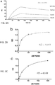
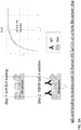
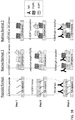


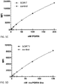
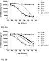
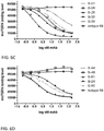
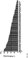
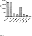


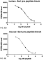
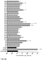


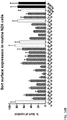
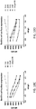
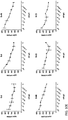

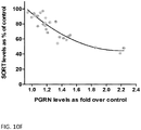


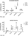
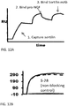
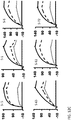
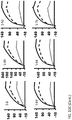
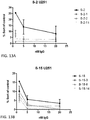
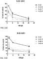


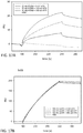
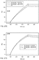
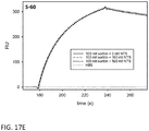
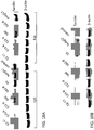
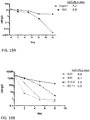
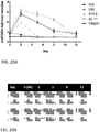
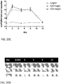

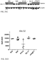
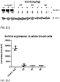
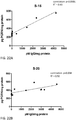
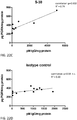
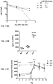
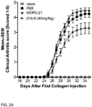


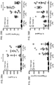
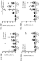

Classifications
-
- C—CHEMISTRY; METALLURGY
- C07—ORGANIC CHEMISTRY
- C07K—PEPTIDES
- C07K16/00—Immunoglobulins [IGs], e.g. monoclonal or polyclonal antibodies
- C07K16/18—Immunoglobulins [IGs], e.g. monoclonal or polyclonal antibodies against material from animals or humans
- C07K16/28—Immunoglobulins [IGs], e.g. monoclonal or polyclonal antibodies against material from animals or humans against receptors, cell surface antigens or cell surface determinants
- C07K16/2863—Immunoglobulins [IGs], e.g. monoclonal or polyclonal antibodies against material from animals or humans against receptors, cell surface antigens or cell surface determinants against receptors for growth factors, growth regulators
-
- A—HUMAN NECESSITIES
- A61—MEDICAL OR VETERINARY SCIENCE; HYGIENE
- A61K—PREPARATIONS FOR MEDICAL, DENTAL OR TOILETRY PURPOSES
- A61K31/00—Medicinal preparations containing organic active ingredients
- A61K31/70—Carbohydrates; Sugars; Derivatives thereof
- A61K31/7088—Compounds having three or more nucleosides or nucleotides
-
- A—HUMAN NECESSITIES
- A61—MEDICAL OR VETERINARY SCIENCE; HYGIENE
- A61P—SPECIFIC THERAPEUTIC ACTIVITY OF CHEMICAL COMPOUNDS OR MEDICINAL PREPARATIONS
- A61P19/00—Drugs for skeletal disorders
- A61P19/02—Drugs for skeletal disorders for joint disorders, e.g. arthritis, arthrosis
-
- A—HUMAN NECESSITIES
- A61—MEDICAL OR VETERINARY SCIENCE; HYGIENE
- A61P—SPECIFIC THERAPEUTIC ACTIVITY OF CHEMICAL COMPOUNDS OR MEDICINAL PREPARATIONS
- A61P21/00—Drugs for disorders of the muscular or neuromuscular system
- A61P21/02—Muscle relaxants, e.g. for tetanus or cramps
-
- A—HUMAN NECESSITIES
- A61—MEDICAL OR VETERINARY SCIENCE; HYGIENE
- A61P—SPECIFIC THERAPEUTIC ACTIVITY OF CHEMICAL COMPOUNDS OR MEDICINAL PREPARATIONS
- A61P25/00—Drugs for disorders of the nervous system
-
- A—HUMAN NECESSITIES
- A61—MEDICAL OR VETERINARY SCIENCE; HYGIENE
- A61P—SPECIFIC THERAPEUTIC ACTIVITY OF CHEMICAL COMPOUNDS OR MEDICINAL PREPARATIONS
- A61P25/00—Drugs for disorders of the nervous system
- A61P25/08—Antiepileptics; Anticonvulsants
-
- A—HUMAN NECESSITIES
- A61—MEDICAL OR VETERINARY SCIENCE; HYGIENE
- A61P—SPECIFIC THERAPEUTIC ACTIVITY OF CHEMICAL COMPOUNDS OR MEDICINAL PREPARATIONS
- A61P25/00—Drugs for disorders of the nervous system
- A61P25/14—Drugs for disorders of the nervous system for treating abnormal movements, e.g. chorea, dyskinesia
- A61P25/16—Anti-Parkinson drugs
-
- A—HUMAN NECESSITIES
- A61—MEDICAL OR VETERINARY SCIENCE; HYGIENE
- A61P—SPECIFIC THERAPEUTIC ACTIVITY OF CHEMICAL COMPOUNDS OR MEDICINAL PREPARATIONS
- A61P25/00—Drugs for disorders of the nervous system
- A61P25/28—Drugs for disorders of the nervous system for treating neurodegenerative disorders of the central nervous system, e.g. nootropic agents, cognition enhancers, drugs for treating Alzheimer's disease or other forms of dementia
-
- A—HUMAN NECESSITIES
- A61—MEDICAL OR VETERINARY SCIENCE; HYGIENE
- A61P—SPECIFIC THERAPEUTIC ACTIVITY OF CHEMICAL COMPOUNDS OR MEDICINAL PREPARATIONS
- A61P27/00—Drugs for disorders of the senses
- A61P27/02—Ophthalmic agents
-
- A—HUMAN NECESSITIES
- A61—MEDICAL OR VETERINARY SCIENCE; HYGIENE
- A61P—SPECIFIC THERAPEUTIC ACTIVITY OF CHEMICAL COMPOUNDS OR MEDICINAL PREPARATIONS
- A61P27/00—Drugs for disorders of the senses
- A61P27/02—Ophthalmic agents
- A61P27/06—Antiglaucoma agents or miotics
-
- A—HUMAN NECESSITIES
- A61—MEDICAL OR VETERINARY SCIENCE; HYGIENE
- A61P—SPECIFIC THERAPEUTIC ACTIVITY OF CHEMICAL COMPOUNDS OR MEDICINAL PREPARATIONS
- A61P29/00—Non-central analgesic, antipyretic or antiinflammatory agents, e.g. antirheumatic agents; Non-steroidal antiinflammatory drugs [NSAID]
-
- A—HUMAN NECESSITIES
- A61—MEDICAL OR VETERINARY SCIENCE; HYGIENE
- A61P—SPECIFIC THERAPEUTIC ACTIVITY OF CHEMICAL COMPOUNDS OR MEDICINAL PREPARATIONS
- A61P31/00—Antiinfectives, i.e. antibiotics, antiseptics, chemotherapeutics
- A61P31/04—Antibacterial agents
-
- A—HUMAN NECESSITIES
- A61—MEDICAL OR VETERINARY SCIENCE; HYGIENE
- A61P—SPECIFIC THERAPEUTIC ACTIVITY OF CHEMICAL COMPOUNDS OR MEDICINAL PREPARATIONS
- A61P43/00—Drugs for specific purposes, not provided for in groups A61P1/00-A61P41/00
-
- A—HUMAN NECESSITIES
- A61—MEDICAL OR VETERINARY SCIENCE; HYGIENE
- A61P—SPECIFIC THERAPEUTIC ACTIVITY OF CHEMICAL COMPOUNDS OR MEDICINAL PREPARATIONS
- A61P9/00—Drugs for disorders of the cardiovascular system
- A61P9/10—Drugs for disorders of the cardiovascular system for treating ischaemic or atherosclerotic diseases, e.g. antianginal drugs, coronary vasodilators, drugs for myocardial infarction, retinopathy, cerebrovascula insufficiency, renal arteriosclerosis
-
- C—CHEMISTRY; METALLURGY
- C07—ORGANIC CHEMISTRY
- C07K—PEPTIDES
- C07K16/00—Immunoglobulins [IGs], e.g. monoclonal or polyclonal antibodies
- C07K16/18—Immunoglobulins [IGs], e.g. monoclonal or polyclonal antibodies against material from animals or humans
- C07K16/28—Immunoglobulins [IGs], e.g. monoclonal or polyclonal antibodies against material from animals or humans against receptors, cell surface antigens or cell surface determinants
- C07K16/286—Immunoglobulins [IGs], e.g. monoclonal or polyclonal antibodies against material from animals or humans against receptors, cell surface antigens or cell surface determinants against neuromediator receptors, e.g. serotonin receptor, dopamine receptor
-
- C—CHEMISTRY; METALLURGY
- C07—ORGANIC CHEMISTRY
- C07K—PEPTIDES
- C07K2317/00—Immunoglobulins specific features
- C07K2317/10—Immunoglobulins specific features characterized by their source of isolation or production
- C07K2317/14—Specific host cells or culture conditions, e.g. components, pH or temperature
-
- C—CHEMISTRY; METALLURGY
- C07—ORGANIC CHEMISTRY
- C07K—PEPTIDES
- C07K2317/00—Immunoglobulins specific features
- C07K2317/20—Immunoglobulins specific features characterized by taxonomic origin
- C07K2317/21—Immunoglobulins specific features characterized by taxonomic origin from primates, e.g. man
-
- C—CHEMISTRY; METALLURGY
- C07—ORGANIC CHEMISTRY
- C07K—PEPTIDES
- C07K2317/00—Immunoglobulins specific features
- C07K2317/30—Immunoglobulins specific features characterized by aspects of specificity or valency
- C07K2317/31—Immunoglobulins specific features characterized by aspects of specificity or valency multispecific
-
- C—CHEMISTRY; METALLURGY
- C07—ORGANIC CHEMISTRY
- C07K—PEPTIDES
- C07K2317/00—Immunoglobulins specific features
- C07K2317/30—Immunoglobulins specific features characterized by aspects of specificity or valency
- C07K2317/34—Identification of a linear epitope shorter than 20 amino acid residues or of a conformational epitope defined by amino acid residues
-
- C—CHEMISTRY; METALLURGY
- C07—ORGANIC CHEMISTRY
- C07K—PEPTIDES
- C07K2317/00—Immunoglobulins specific features
- C07K2317/50—Immunoglobulins specific features characterized by immunoglobulin fragments
- C07K2317/55—Fab or Fab'
-
- C—CHEMISTRY; METALLURGY
- C07—ORGANIC CHEMISTRY
- C07K—PEPTIDES
- C07K2317/00—Immunoglobulins specific features
- C07K2317/50—Immunoglobulins specific features characterized by immunoglobulin fragments
- C07K2317/56—Immunoglobulins specific features characterized by immunoglobulin fragments variable (Fv) region, i.e. VH and/or VL
-
- C—CHEMISTRY; METALLURGY
- C07—ORGANIC CHEMISTRY
- C07K—PEPTIDES
- C07K2317/00—Immunoglobulins specific features
- C07K2317/70—Immunoglobulins specific features characterized by effect upon binding to a cell or to an antigen
- C07K2317/76—Antagonist effect on antigen, e.g. neutralization or inhibition of binding
-
- C—CHEMISTRY; METALLURGY
- C07—ORGANIC CHEMISTRY
- C07K—PEPTIDES
- C07K2317/00—Immunoglobulins specific features
- C07K2317/90—Immunoglobulins specific features characterized by (pharmaco)kinetic aspects or by stability of the immunoglobulin
- C07K2317/92—Affinity (KD), association rate (Ka), dissociation rate (Kd) or EC50 value
-
- C—CHEMISTRY; METALLURGY
- C07—ORGANIC CHEMISTRY
- C07K—PEPTIDES
- C07K2319/00—Fusion polypeptide
- C07K2319/30—Non-immunoglobulin-derived peptide or protein having an immunoglobulin constant or Fc region, or a fragment thereof, attached thereto
-
- C—CHEMISTRY; METALLURGY
- C07—ORGANIC CHEMISTRY
- C07K—PEPTIDES
- C07K2319/00—Fusion polypeptide
- C07K2319/32—Fusion polypeptide fusions with soluble part of a cell surface receptor, "decoy receptors"
Definitions
- This present disclosure relates to anti-Sortilin antibodies and therapeutic uses of such antibodies.
- Sortilin is a Type I transmembrane protein that acts both as a receptor of several ligands, and in the sorting of select cargo from the trans-Golgi network (TGN) to late endosomes and lysosomes for degradation.
- Sortilin harbors a large extracellular domain that is part of the VPS10 family, homologous to yeast VPS10P, and contains of a 10-blade beta-propeller structure and a cysteine- rich 10CC module ( Nykjaer, A et al., (2012) Trends Neurosci 35: 261-270 ; and Zheng, Y et al.. (2011) PLoS One 6: e21023 ).
- Sortilin may be shed by activity of ADAM10 or gamma-Secretase ( ⁇ 5%) ( Nykjaer, A et al., (2012) Trends Neurosci 35: 261-270 ; and Willnow, TE et al., (2011) Curr Opin Lipidol 22: 79-85 ).
- Sortilin binds the secreted protein Progranulin (PGRN) and targets it for lysosomal degradation, thus negatively regulating extracellular levels of PGRN ( Hu, F er al. (2010) Neuron 68, 654-667 .
- PGRN Progranulin
- deficiency of Sortilin significantly increases plasma PGRN levels both in mouse models in vivo and human cells in vitro ( Carrasquillo, M. M et al., (2010) Am J Hum Genet 87, 890-897 ; Lee, W.C et al., (2014) 23 , 1467-1478).
- Sortilin a polymorphism in Sortilin was shown to be strongly assoiated with PGRN serum levles in humans ( Carrasquillo MM e al., 2010), Am J Hum Genet. 10;87(6): 890-7 ).
- Progranulin is a secreted, growth factor-like, trophic, and anti-inflammatory protein, which also plays a role as an adipokine involved in diet-induced obesity and insulin resistance ( Nguyen DA et al., (2013). Trends in Endocrinology and Metabolism, 24, 597- 606 ). Progranulin deficiency accounts for roughly 25% of all heritable forms of frontotemporal dementia (FTD), an early-onset neurodegenerative disease.
- FTD frontotemporal dementia
- PGRN patients with heterozygous loss-of-function mutations in PGRN have ⁇ 50% reduced extracellular levels of the protein and they will invariably develop FTD, making PGRN a causal gene for the disease ( Baker, M et al., (2006) Nature 442, 916-919 ; Carecchio M et al., (2011) J Alzheim0 ers Dis 27, 781-790 ; Cruts, M et al., (2008) Trends Genet 24, 186-194 ; Galimberti, D et al., (2010) J Alzheimers Dis 19, 171-177 ).
- PGRN mutant alleles have been identified in Alzheimer's disease patients ( Seelaar, H et al., (2011).
- PGRN acts protective in several disease models with increased PGRN levels accelerating behavioral recovery from ischemia ( Tao, J et al., (2012) Brain Res 1436, 130-136 ; Egashira, Y. et al., (2013). J Neuroinflammation 10, 105 ), suppressing locomotor deficits in a Parkinson's disease model ( Van Kampen, J.M et al. (2014). PLoS One 9, e97032 ), attenuating pathology in a model of amyotriphic lateral scleoris ( Laird, A.S et al., (2010).
- Sortilin also binds directly to pro-neurotrophins, such as pro-nerve growth factor (pro-NGF), pro-BDNF, pro-neurotrophin-3, etc. , which harbor a pro-domain and are typically pro-apoptotic.
- pro-neurotrophins such as pro-nerve growth factor (pro-NGF), pro-BDNF, pro-neurotrophin-3, etc. , which harbor a pro-domain and are typically pro-apoptotic.
- Such pro-neurotrophin precursors are released during stress, and Sortilin is involved in regulating their release as well as binding on the receiving cell and stimulation of apoptosis in conjunction with p75NTR ( Willnow, TE et al., (2008) Nat Rev Neurosci 9: 899-909 ; Nykjaer, A et al., Trends Neurosci 35: 261-270 ; and Nykjaer, A et al., (2004) Nature 427: 843-848 ; Hiroko Yano et al., (2009) J Neurosci.; 29: 14790-14802 . Teng H.K., et al., J. Neurosci. 25:5455-5463(2005 )).
- Sortilin also binds to p75NTR directly ( Skeldal S et al., (2012) J Biol Chem.; 287:43798 ). Sortilin also binds to neurotensin in a region that partially overlaps with Progranulin binding ( Quistgaard, EM et al., (2009) Nat Struct Mol Biol 16: 96-98 ; and Zheng, Y et al., PLoS One 6: e21023 ). Sortilin also interacts with the Trk receptors NTRK1, NTRK2, and NTRK3; and can regulate their anterograde axonal transport and signaling ( Vaegter, CB et al., (2011) Nat. Neurosci.
- Sortilin also interacts with and regulates the processing and trafficking of amyloid precursor protein and the resulting production of pathological beta amyloid peptides ( Gustafsen C et al., (2013). J Neurosci. 2;33(1):64-71 .
- Sortilin has also been shown to bind to apolipoproteins and lipoprotein lipase, and thus deficiency leads to reduced VLDL release from liver and reduced cholesterol ( Willnow, TE et al., (2011) Curr Opin Lipidol 22: 79-85 ; Kjolby, M et al., (2010) Cell Metab 12: 213-223 ; Nilsson, SK et al., (2007) Biochemistry 46: 3896-3904 .; Nilsson, SK et al., (2008) J Biol Chem 283: 25920-25927 ; and Klinger, SC et al., (2011) J Cell Sci 124: 1095-1105 ).
- Sortilin has also been implicated in binding to APP directly ( Gustafsen, C et al., (2013) J.Neurosc. 33:64-71 ) and also to the APP processing enzyme BACE1 ( Gustafsen, C et al., (2013) J.Neurosc. 33:64-71 ; and Finan, GM et al., J Biol Chem 286: 12602-12616 ), Sortilin also binds to apolipoprotein E (APOE), to the A beta peptide ( Carlo, AS et al., (2013) J, Neurosc, 33: 358-370 ), and to PCSK9 ( Gustafsen et al, (2014) Cell Metab, 19: 310-318 ).
- APOE apolipoprotein E
- Sortilin has also been shown to bind to and regulate extracellular levels of PCSK9, which directs low-density lipoprotein receptor for degradation in lysosomes, resulting in increased levels of LDL cholesterol ( Gustafsen C et al., (2014). Cell Metab. 2014 Feb 4;19(2):310-8 ).
- the amino-termmal extracellular domain of Sortilin When present at intracellular vesicles such as endosomes, the amino-termmal extracellular domain of Sortilin is directed towards the lumen, where cargo of the vesicle is present.
- the carboxy-terminal intracellular/cytoplasmic domain of Sortilin binds to a series of adaptor proteins, which regulate its trafficking from the surface and within intracellular compartments.
- AP2 a clathrin adaptor to modulate endocytosis from the cell surface
- Retromer Complex/API which modulate movement from early endosomes to Golgi for recycling
- GGA Golgi-localizing, gamma-ear containing, ADP-ribosylation factor binding
- Sortilin can bind to ligands at its luminal domain, while engaging the cytoplasmic adaptors that determine its destination to determine intracellular fates, such as degradation for Progranulin and other factors.
- Progranulin, Sortilin and its multiple ligands have been shown to be involved in various diseases, disorders, and conditions, such as frontotemporal dementia, amyotrophic lateral sclerosis, amyotrophic lateral sclerosis-frontotemporal dementia phenotypes, Alzheimer's disease, Parkinson's disease, depression, neuropsyciatric disorders, vascular dementia, seizures, retinal dystrophy, age related macular degeneration, glaucoma, traumatic brain injury, aging, seizures, wound healing, stroke, arthritis, and atherosclerotic vascular diseases.
- frontotemporal dementia amyotrophic lateral sclerosis, amyotrophic lateral sclerosis-frontotemporal dementia phenotypes, Alzheimer's disease, Parkinson's disease, depression, neuropsyciatric disorders, vascular dementia, seizures, retinal dystrophy, age related macular degeneration, glaucoma, traumatic brain injury, aging, seizures, wound healing, stroke, arthritis, and atherosclerotic vascular diseases.
- Sortilin proteins specifically bind Sortilin proteins and block the binding of Sortilin to its ligands, such as Progranulin, or otherwise modulate the effective concentration of the ligands, in order to treat one or more diseases, disorders, and conditions associated with Sortilin activity.
- the present disclosure is generally directed to anti-Sortilin antibodies and therapeutic uses of such antibodies.
- Certain aspects of the present disclosure are based, at least in part, on the identification of anti-Sortilin antibodies that are capable of blocking the interaction between Sortilin and Progranulin ( see , e.g. , Example 4 ), anti-Sortilin antibodies that are capable of increasing extracellular levels of Progranulin secreted by cells in culture and from cells in the brains of mouse models of Alzheimer's disease ( see, e.g. , Example 5 ), anti-Sortilin antibodies that are capable of decreasing cellular levels of Sortilin in human primary monocytes ( see, e.g. , Example 25 ), and anti-Sortilin antibodies that are capable of increasing cellular levels of Progranulin in vivo ( see, e.g.
- Example 26 Example 26 .
- one class of antibodies was shown to be capable of increasing extracellular levels of Progranulin without being able to block the interaction between Sortilin and Progranulin (e.g. , antibodies S-2, S-15, and S-22) while a second class of antibodies was shown to be capable of both blocking the interaction between Sortilin and Progranulin and increasing extracellular levels of Progranulin (e.g. , antibodies S-8, S-49, and S-60).
- anti-Sortilin antibodies capable of increasing extracellular levels of Progranulin were surprisingly shown to also reduce cellular levels of Sortilin, such as cell surface levels of Sortilin ( see, e.g. , Example 5 ).
- cellular levels of Sortilin were shown to be strongly and inversely correlated with increases in cellular levels of Progranulin ( see, e.g. , FIG. 10B- 10E and FIG. 23B and 23C ).
- Anti-Sortilin antibodies capable of increasing extracellular levels of Progranulin are also capable of blocking pro-NGF binding to Sortilin ( see, e.g.
- a first aspect of the present invention relates to an isolated (e.g., monoclonal) anti-Sortilin antibody that binds to human Sortilin, wherein the anti-Sortilin antibody increases extracellular levels of Progranulin and decreases cell surface levels of Sortilin, wherein the anti-Sortilin antibody comprises a light chain variable domain and a heavy chain variable domain, wherein the light chain variable domain comprises: (a) an HVR-L1 comprising the amino acid sequence of RASQSX 1 X 2 SNLA (SEQ ID NO:508), wherein X 1 is V or I and X 2 is S or G; (b) an HVR-L2 comprising the amino acid sequence of GASTRAT (SEQ ID NO:29); and (c) an HVR-L3 comprising the amino acid sequence of QQARLGPWT (SEQ ID NO:55); and wherein the heavy chain variable domain comprises: (a) an HVR-H1 comprising the amino acid sequence of YTX 1 X 2
- the anti-Sortilin antibody has at least one, two, three, four, five, or six HVRs selected from (i) HVR-L1 comprising the amino acid sequence of any of the HVR-L1 sequences from an antibody selected from the group consisting of S-15, S-15-1, S-15-2, S-15-3, S-15-4, S-15-5, S-15-6, S-15-6-1, S-15-6-2, S-15-6-3, S-15-6-4, S-15-6-5, S-15-6-6, 5-15-6-7, S-15-6-8, S-15-6-9, S-15-6-10, S-15-6-11, S-15-6-12, S-15-6-13, S-15-7, S-15-8, S-15-9, S-15-10, S-15-10-1, S-15-10-2, S-15-10-3, S-15-10-4, S-15-10-5, S-15-10-6, S-1S-10-7, S-15-10-8.
- HVR-L1 comprising the amino acid sequence of any of the HVR-L1 sequences from an antibody selected from the group consisting of S-15, S-15-1,
- S-15-10-9 S-15-10-10, S-15-10-11, S-15-10-12, S-15-10-13, S-15-10-14, S-15-10-15, S-15-10-16, S-15-10-17, S-15-10-18, S-15-10-19, S-15-10-20, S-15-10-21.
- HVR-L2 comprising the amino acid sequence of any of the HVR-L2 sequences from an antibody selected from the group consisting of S-15, S-15-1, S-15-2, S-15-3, S-15-4, S-15-5, S-15-6, S-15-6-1, S-15-6-2, S-15-6-3, S-15-6-4, S-15-6-5, S-15-6-6, S-15-6-7, S-15-6-8, S-15-6-9, S-15-6-10, S-15-6-11, S-15-6-12, S-15-6-13, S-15-7, S-15-8, S-15-9, S-15-10, S-15-10-1, S-15-10-2, S-15-10-3, S-15-10-4, S-15-10-5, S-15-10-6, S-15-10-7, S-15-10-8, S-15-10-9, S-15-10-10, S-15-10-11, S-15-10-12, S-15-10-13, S-15-10-14, S-15-10-15, S-15-10-16, S-15-10-17,
- HVR-H1 comprising the amino acid sequence of any of the HVR-H1 sequences from an antibody selected from the group consisting of S-15, S-15-1, S-15-2, S-15-3, S-15-4, S-15-5, S-15-6, S-15-6-1, S-15-6-2, S-15-6-3, S-15-6-4, S-15-6-5, S-15-6-6, S-15-6-7, S-15-6-8, S
- anti-Sortitin antibody comprises a light chain variable region of any one of the antibodies sequences from an antibody selected from the group consisting of S-15, S-15-1, S-15-2, S-15-3, S-15-4, S-15-5, S-15-6, S-15-6-1, S-15-6-2, S-15-6-3, S-15-6-4, S-15-6-5, S-15-6-6, S-15-6-7, S-15-6-8, S-15-6-9, S-15-6-10, S-15-6-11, S-15-6-12, S-15-6-13, S-15-7, S-15-8, S-15-9, S-15-10, S-15-10-1, S-15-10-2, S-15-10-3, S-15-10-4, S-15-10-5, S-15-10-6, S-15-10-7, S-15-10-8, S-15-10-9, S-15-10-10, S-15-10-11, S-15-10-12, S-15-10-13, S-15-10-14, S-15-10-15, S-15-10-16, S-15-10-17, S-15-10-18, S-15-10-19, S-15-10-20, S-15-10-21, S-15-11, S-15-10-20,
- Another aspect of the present invention relates to an isolated (e.g., monoclonal) anti-Sortilin antibody that binds to human Sortilin, wherein the anti-Sortilin antibody increases extracellular levels of Progranulin and decreases cell surface levels of Sortilin, wherein the anti-Sortilin antibody comprises a light chain variable domain and a heavy chain variable domain, wherein the light chain variable domain comprises: (a) an HVR-L1 comprising the amino acid sequence of RSSQX 1 LLX 2 SNGYNYLD (SEQ ID NO:580), wherein X 1 is S or G and X 2 is H or R; (b) an HVR-L2 comprising the amino acid sequence of LGSNRXS (SEQ ID NO:581), wherein X is A or V; and (c) an HVR-L3 comprising the amino acid sequence of MQQQETPLT (SEQ ID NO:100); and wherein the heavy chain variable domain comprises: (a) an HVR-H1 comprising
- the anti-Sortilin antibody has at least one, two, three, four, five, or six HVRs selected from (i) HVR-L1 comprising the amino acid sequence of any of the HVR-L1 sequences from an antibody selected from the group consisting of S-60, S-60-1, S-60-2, S-60-3, S-60-4, S-60-5, S-60-6, S-60-7, S-60-8, and S-60-9; (ii) HVR-L2 comprising the amino acid sequence of any of the HVR-L2 sequences from an antibody selected from the group consisting of S-60, S-60-1, S-60-2, S-60-3, S-60-4, S-60-5, S-60-6, S-60-7, S-60-8, and S-60-9; (iii) HVR-L3 comprising the amino acid sequence of any of the HVR-L3 sequences from an antibody selected from the group consisting of S-60, S-60-1, S-60-2, S-60-3.
- HVR-H1 comprising the amino acid sequence of any of the HVR-H1 sequences from an antibody selected from the group consisting of S-60, S-60-1, S-60-2, S-60-3, S-60-4, S-60-5, S-60-6, S-60-7, S-60-8, and S-60-9
- HVR-H2 comprising the amino acid sequence of any of the HVR-H2 sequences from an antibody selected from the group consisting of S-60, S-60-1, S-60-2, S-60-3, S-60-4, S-60-5, S-60-6, S-60-7, S-60-8, and S-60-9
- HVR-H3 comprising the amino acid sequence of any of the HVR-H3 sequences from an antibody selected from the group consisting of S-60, S-60-1, S-60-2, S-60-3, S-60-4, S-60-5, S-60-6, S-60-7, S-60-8, and S-60-9.
- anti-Sortilin antibody comprises a light chain variable region of any one of the antibodies sequences from an antibody selected from the group consisting of S-60, S-60-1, S-60-2, S-60-3, S-60-4, S-60-5, S-60-6, S-60-7, S-60-8, and S-60-9; and/or a heavy chain variable region of any one of the antibodies sequences from an antibody selected from the group consisting of S-60, S-60-1, S-60-2, S-60-3, S-60-4, S-60-5, S-60-6, S-60-7, S-60-8, and S-60-9.
- the anti-Sortilin antibody has an IgG1, IgG2, IgG3, or IgG4 isotype.
- the anti-Soitilin antibody has a human or mouse IgG1 isotype and comprises one or more amino acid substitutions in the Fc region at a residue position selected from the group consisting of: N297A, N297Q, D265A, L234A, L235A, C226S, C229S, P238S, E233P, L234V, P238A, A327Q, A327G, P329A, K322A, L234F, L235E, P331S, T394D, A330L, M252Y, S254T, T256E, and any combination thereof, wherein the numbering of the residues is according to EU or Kabat numbering; (b) the numbering of the residues is according to EU or Kabat numbering;
- the Fc region further comprises one or more additional amino acid substitutions at a position selected from the group consisting of A330L, L234F; L235E, P331S, and any combination thereof, wherein the numbering of the residues is according to EU or Kabat numbering;
- the Fc region further comprises one or more additional amino acid substitutions at a position selected from the group consisting of M252Y, S254T,T256E, and any combination thereof, wherein the numbering of the residues is according to EU or Kabat numbering; or
- the Fc region further comprises a S228P amino acid substitution according to EU or Kabat numbering.
- the anti-Sortilin antibody decreases expression of one or more pro-inflammatory mediators selected from the group consisting of IL-6, IL12p70, IL12p40, IL-1 ⁇ , TNF- ⁇ , CXCL1, CCL2, CCL3, CCL4, and CCL5.
- the anti-Sortilin antibody has a dissociation constant (K D ) for human Sortilin, mouse Sortilin, or both, that ranges from about 100 nM to about 0.005 nM, or less than 0.005 nM.
- the anti-Sortitin antibody has dissociation constant (K D ) for human Sortilin that ranges from about 70.4 nM to about 0.005 nM, or less than 0.005 nM. In certain embodiments that may be combined with any of the preceding embodiments, the anti-Sortilin antibody has dissociation constant (K D ) for mouse Sortilin that ranges from about 40.3 nM to about 0.07 nM, or less than 0.07 nM.
- aspects of the present disclosure relate to an isolated nucleic acid comprising a nucleic acid sequence encoding the anti-Sortilin antibody of any of the preceding embodiments.
- Other aspects of the present disclosure relate to a vector comprising the nucleic acid of any of the preceding embodiments.
- Other aspects of the present disclosure relate to an isolated host cell comprising the vector of any of the preceding embodiments.
- Other aspects of the present disclosure relate to a pharmaceutical composition comprising the anti-Sortilin antibody of any of the preceding embodiments, and a pharmaceutically acceptable carrier.
- aspects of the present disclosure relate to an in vitro method of increasing extracellular levels of Progranulin, decreasing cell surface levels of Sortilin on one or more cells, and/or decreasing expression of one or more pro-inflammatory mediators, comprising contacting one or more cells with an anti-Sortilin antibody of the invention.
- the anti-Sortilin antibody binds specifically to both human and mouse Sortilin;
- the anti-Sortilin antibody is a human antibody;
- the anti-Sortilin antibody is a humanized antibody;
- the anti-Sortilin antibody is a bispecific antibody;
- the anti-Sortilin antibody is a bispecific antibody recognizing a first antigen and a second antigen;
- the anti-Sortilin antibody is a bispecific antibody recognizing a first antigen and a second antigen, wherein the first antigen is a Sortilin protein and the second antigen is an antigen facilitating transport across the blood-brain-barrier, and, optionally, wherein the second antigen is selected from the group consisting of Sortilin, transferrin receptor (TR), insulin receptor (HIR), insulin-like growth factor receptor (IGFR), low-density lipoprotein receptor related proteins 1
- the anti-Sortilin antibody binds to a conformational epitope of Sortilin. In certain embodiments that may be combined with any of the preceding embodiments, the anti-Sortilin antibody binds to one or more amino acids within amino acid residues selected from the group consisting of: i. amino acid residues 314-338 of SEQ ID NO: 1, or amino acid residues on a mammalian Sortilin protein corresponding to amino acid residues 314-338 of SEQ ID NO: 1; ii.
- amino acid residues 237-247 of SEQ ID NO: 1 amino acid residues on a mammalian Sortilin protein corresponding to amino acid residues 237-247 of SEQ ID NO: 1; iii. amino acid residues 237-247 and 314-338 of SEQ ID NO: 1, or amino acid residues on a mammalian Sortilin protein corresponding to amino acid residues 237-247 and 314-338 of SEQ ID NO: 1; and iv. amino acid residues 207-231 of SEQ ID NO: 1, or amino acid residues on a mammalian Sortilin protein corresponding to amino acid residues 207-231 of SEQ ID NO: 1.
- the anti-Sortilin antibody binds to one or more amino acid residues selected from the group consisting of T218, Y222, S223, D227, S242, K243, S316, R325, and S595 of SEQ ID NO: 1, or one or more amino acid residues on a mammalian Sortilin protein corresponding to amino acid residues T218, Y222, S223, S227, S242, K243, S316, R325, and S595 of SEQ ID NO: 1.
- the anti-Sortilin antibody comprises at least one, two, three, four, five, or six HVRs of an antibody selected from the group consisting of: S-15 and S-60.
- the anti-Sortilin antibody comprises: (a) an HVR-L1 comprising an amino acid sequence selected from the group consisting of SEQ ID NOs: 9 or 10; (b) an HVR-L2 comprising an amino acid sequence selected from the group consisting of SEQ ID NOs: 29 or 30; (c) an HVR-L3 comprising an amino acid sequence selected from the group consisting of SEQ ID NOs: 55 or 100; (d) an HVR-H1 comprising an amino acid sequence selected from the group consisting of SEQ ID NOs: 129 or 142; (e) an HVR-H2 comprising an amino acid sequence selected from the group consisting of SEQ ID NOs: 155 or 170; or (
- the anti-Sortilin antibody comprises a light chain variable domain and a heavy chain variable domain, wherein the light chain variable domain comprises: (a) an HVR-L1 comprising an amino acid sequence selected from the group consisting of SEQ ID NOs: 9 or 10, or an amino acid sequence with at least about 95% homology to an amino acid sequence selected from the group consisting of SEQ ID NOs: 9 or 10; (b) an HVR-L2 comprising an amino acid sequence selected from the group consisting of SEQ ID NOs: 29 or 30, or an amino acid sequence with at least about 95% homology to an amino acid sequence selected from the group consisting of SEQ ID NOs: 29 or 30; and (c) an HVR-L3 comprising an amino acid sequence selected from the group consisting of SEQ ID NOs: 55 or 100, or an amino acid sequence with at least about 95% homology to an amino acid sequence selected from the group consisting of SEQ ID NOs: 55 or 100; and wherein the heavy
- the anti-Sortilin antibody competes with S-60, for binding to Sortilin; or (b) the anti-Sortilin antibody competes with S-15, for binding to Sortilin or the anti-Sortilin antibody which binds essentially the same Sortilin epitope as an antibody selected from the group consisting of: S-15 and S-60.
- the anti-Sortilin antibody comprises a light chain variable domain and/or a heavy chain variable domain comprising an amino acid sequence selected from the group consisting of SEQ ID NOs: 342, 343, 430 and 431.
- the anti-Sortilin antibody is of the IgG class the IgM class, or the IgA class; or (b) the anti-Sortilin antibody has an IgG1, IgG2, IgG3, or IgG4 isotype; (c) the anti-Sortilin antibody has a human or mouse IgG1 isotype and comprises one or more amino acid substitutions in the Fc region at a residue position selected from the group consisting of: N297A, N297Q, D265A, L234A, L235A, C226S, C229S, P238S, E233P, L234V, P238A, A327Q, A327G, P329A, K322A, L234F, L235E, P331S, T394D, A330L, M252Y, S254T, T256E, and any combination thereof, wherein the
- the Fc region of one or more of (c), (d), and (e) further comprises: (a) one or more additional amino acid substitutions at a position selected from the group consisting of A330L, L234F; L235E, P331S, and any combination thereof, wherein the numbering of the residues is according to EU or Kabat numbering; (b) one or more additional amino acid substitutions at a position selected from the group consisting of M252Y, S254T, T256E, and any combination thereof, wherein the numbering of the residues is according to EU or Kabat numbering; or (c) a S228P amino acid substitution according to EU or Kabat numbering.
- the anti-Sortilin antibody has a dissociation constant (K D ) for human Sortilin, mouse Sortilin, or both, that ranges from about 100 nM to about 0.005 nM, or less than 0.005 nM; (b) the anti-Sortilin antibody has dissociation constant (K D ) for human Sortilin that ranges from about 70.4 nM to about 0.005 nM, or less than 0.005 nM; or (c) the anti-Sortilin antibody has dissociation constant (K D ) for mouse Sortilin that ranges from about 40.3 nM to about 0.07 nM, or less than 0.07 nM. In certain embodiments that may be combined with any of the preceding embodiments, the anti-Sortilin antibody is the anti-Sortilin antibody of any of the preceding embodiments.
- an anti-Sortilin antibody of any of the preceding embodiments for use in preventing, reducing risk, or treating an individual having a disease, disorder, or injury selected from the group consisting of frontotemporal dementia, progressive supranuclear palsy, Alzheimer's disease, vascular dementia, seizures, retinal dystrophy, amyotrophic lateral sclerosis, traumatic brain injury, a spinal cord injury, dementia, stroke, Parkinson's disease, acute disseminated encephalomyelitis, retinal degeneration, age related macular degeneration, glaucoma, multiple sclerosis, septic shock, bacterial infection, arthritis, and osteoarthritis.
- a disease, disorder, or injury selected from the group consisting of frontotemporal dementia, progressive supranuclear palsy, Alzheimer's disease, vascular dementia, seizures, retinal dystrophy, amyotrophic lateral sclerosis, traumatic brain injury, a spinal cord injury, dementia, stroke, Parkinson's disease, acute disseminated ence
- aspects of the present invention relate to an anti-Sortiiin antibody of any of the preceding embodiments for use in a method of treatment comprising inhibiting one or more of neuroinflammation, axonopathy characterized by short axonal outgrowth and aberrant branching, microglial activation, and inflammatory response.
- Other aspects of the present disclosure relate to an anti-Soititin antibody of any of the preceding embodiments for use in promoting wound healing.
- Other aspects of the present disclosure relate to an anti-Sortilin antibody of any of the preceding embodiments for use in preventing, reducing risk, or treating an individual having arthritis.
- the anti-Sortilin antibody comprises two or more anti-Sortilin antibodies.
- the term "preventing” includes providing prophylaxis with respect to occurrence or recurrence of a particular disease, disorder, or condition in an individual.
- An individual may be predisposed to, susceptible to a particular disease, disorder, or condition, or at risk of developing such a disease, disorder, or condition, but has not yet been diagnosed with the disease, disorder, or condition.
- an individual "at risk” of developing a particular disease, disorder, or condition may or may not have detectable disease or symptoms of disease, and may or may not have displayed detectable disease or symptoms of disease prior to the treatment methods described herein.
- At risk denotes that an individual has one or more risk factors, which are measurable parameters that correlate with development of a particular disease, disorder, or condition, as known in the art. An individual having one or more of these risk factors has a higher probability of developing a particular disease, disorder, or condition than an individual without one or more of these risk factors.
- treatment refers to clinical intervention designed to alter the natural course of the individual being treated during the course of clinical pathology. Desirable effects of treatment include decreasing the rate of progression, ameliorating or palliating the pathological state, and remission or improved prognosis of a particular disease, disorder, or condition.
- An individual is successfully "treated", for example, if one or more symptoms associated with a particular disease, disorder, or condition are mitigated or eliminated.
- an “effective amount” refers to at least an amount effective, at dosages and for periods of time necessary, to achieve the desired therapeutic or prophylactic result.
- An effective amount can be provided in one or more administrations.
- a “therapeutically effective amount” is at least the minimum concentration required to effect a measurable improvement of a particular disease, disorder, or condition.
- a therapeutically effective amount herein may vary according to factors such as the disease state, age, sex, and weight of the patient, and the ability of the Sortilin protein antagonist to elicit a desired response in the individual.
- a therapeutically effective amount is also one in which any toxic or detrimental effects of the Sortilin protein antagonist are outweighed by the therapeutically beneficial effects.
- administration "in conjunction" with another compound or composition includes simultaneous administration and/or administration at different times.
- Administration in conjunction also encompasses administration as a co-formulation or administration as separate compositions, including at different dosing frequencies or intervals, and using the same route of administration or different routes of administration.
- An "individual” for purposes of treatment, prevention, or reduction of risk refers to any animal classified as a mammal, including humans, domestic and farm animals, and zoo, sport, or pet animals, such as dogs, horses, rabbits, cattle, pigs, hamsters, gerbils, mice, ferrets, rats, cats, and the like.
- the individual is human.
- immunoglobulin (Ig) is used interchangeably with “antibody” herein.
- antibody herein is used in the broadest sense and specifically covers monoclonal antibodies, polyclonal antibodies, multispecific antibodies (e.g. bispecific antibodies) formed from at least two intact antibodies, and antibody fragments so long as they exhibit the desired biological activity.
- the basic 4-chain antibody unit is a heterotetrameric glycoprotein composed of two identical light (L) chains and two identical heavy (H) chains. The pairing of a V H and V L together forms a single antigen-binding site.
- L light
- H heavy
- immunoglobulins can be assigned to different classes or isotypes. There are five classes of immunoglobulins: IgA, IgD, IgE, IgG, and IgM, having heavy chains designated alpha (" ⁇ "), delta (“ ⁇ "), epsilon (“ ⁇ ”), gamma (“y”) and mu (“ ⁇ ”), respectively.
- the ⁇ and a classes are further divided into subclasses (isotypes) on the basis of relatively minor differences in the CH sequence and function, e.g., humans express the following subclasses: IgG1, IgG2, IgG3, IgG4, IgA1, and IgA2.
- subclasses immunoglobulins
- the subunit structures and three dimensional configurations of different classes of immunoglobulins are well known and described generally in, for example, Abbas et al., Cellular and Molecular Immunology, 4th ed. (W.B. Saunders Co., 2000 ).
- “Native antibodies” are usually heterotetrameric glycoproteins of about 150,000 daltons, composed of two identical light (L) chains and two identical heavy (H) chains. Each light chain is linked to a heavy chain by one covalent disulfide bond, while the number of disulfide linkages varies among the heavy chains of different immunoglobulin isotypes. Each heavy and light chain also has regularly spaced intrachain disulfide bridges. Each heavy chain has at one end a variable domain (V H ) followed by a number of constant domains.
- V H variable domain
- Each light chain has a variable domain at one end (V L ) and a constant domain at its other end; the constant domain of the light chain is aligned with the first constant domain of the heavy chain, and the light chain variable domain is aligned with the variable domain of the heavy chain. Particular amino acid residues are believed to form an interface between the light chain and heavy chain variable domains.
- an “isolated” antibody such as an anti-Sortilin antibody of the present disclosure, is one that has been identified, separated and/or recovered from a component of its production environment (e.g., naturally or recombinantly).
- the isolated polypeptide is free of association with all other contaminant components from its production environment.
- Contaminant components from its production environment such as those resulting from recombinant transfected cells, are materials that would typically interfere with research, diagnostic or therapeutic uses for the antibody, and may include enzymes, hormones, and other proteinaceous or non-proteinaceous solutes.
- the polypeptide will be purified: (1) to greater than 95% by weight of antibody as determined by, for example, the Lowry method, and in some embodiments, to greater than 99% by weight; (2) to a degree sufficient to obtain at least 15 residues of N-terminal or internal amino acid sequence by use of a spinning cup sequenator, or (3) to homogeneity by SDS-PAGE under nonreducing or reducing conditions using Coomassie blue or, preferably, silver stain.
- Isolated antibody includes the antibody in situ within recombinant T-cells since at least one component of the antibody's natural environment will not be present. Ordinarily, however, an isolated polypeptide or antibody will be prepared by at least one purification step.
- variable region refers to the amino-terminal domains of the heavy or light chain of the antibody.
- variable domains of the heavy chain and light chain may be referred to as "V H " and “V L ", respectively. These domains are generally the most variable parts of the antibody (relative to other antibodies of the same class) and contain the antigen binding sites.
- variable refers to the fact that certain segments of the variable domains differ extensively in sequence among antibodies, such as anti-Sortilin antibodies of the present disclosure,.
- the V domain mediates antigen binding and defines the specificity of a particular antibody for its particular antigen.
- HVRs hypervariable regions
- FR framework regions
- the variable domains of native heavy and light chains each comprise four FR regions, largely adopting a beta-sheet configuration, connected by three HVRs, which form loops connecting, and in some cases forming part of, the beta-sheet structure.
- the HVRs in each chain are held together in close proximity by the FR regions and, with the HVRs from the other chain, contribute to the formation of the antigen binding site of antibodies ( see Kabat et al., Sequences of Immunological Interest, Fifth Edition, National Institute of Health, Bethesda, MD (1991 )).
- the constant domains are not involved directly in the binding of antibody to an antigen, but exhibit various effector functions, such as participation of the antibody in antibody-dependent-cellular toxicity.
- the term "monoclonal antibody” as used herein refers to an antibody, such as an anti-Sortilin antibody of the present disclosure, obtained from a population of substantially homogeneous antibodies, i.e., the individual antibodies comprising the population are identical except for possible naturally occurring mutations and/or post-translation modifications (e.g., isomerizations, amidations) that may be present in minor amounts. Monoclonal antibodies are highly specific, being directed against one or more antigenic sites. In some embodiments, an antibody of the present disclosure can be a bispecific antibody. In contrast to polyclonal antibody preparations which typically include different antibodies directed against different determinants (epitopes), each monoclonal antibody is directed against a single determinant on the one or more antigenic sites.
- the modifier "monoclonal” indicates the character of the antibody as being obtained from a substantially homogeneous population of antibodies, and is not to be construed as requiring production of the antibody by any particular method.
- the monoclonal antibodies to be used in accordance with the present disclosure may be made by a variety of techniques, including, for example, phage-display technologies (see, e.g., Clackson et al., Nature, 352:624-628 (1991 ); Marks et al., J. Mol. Biol. 222:581-597 (1992 ); Sidhu et al., J. Mol. Biol. 338(2): 299-310 (2004 ); Lee et al., J. Mol. Biol.
- full-length antibody “intact antibody” or “whole antibody” are used interchangeably to refer to an antibody, such as an anti-Sortilin antibody of the present disclosure, in its substantially intact form, as opposed to an antibody fragment.
- whole antibodies include those with heavy and light chains including an Fc region.
- the constant domains may be native sequence constant domains (e.g., human native sequence constant domains) or amino acid sequence variants thereof.
- the intact antibody may have one or more effector functions.
- antibody fragment comprises a portion of an intact antibody, preferably the antigen binding and/or the variable region of the intact antibody.
- antibody fragments include Fab, Fab', F(ab') 2 and Fv fragments; diabodies; linear antibodies ( see U.S. Patent 5,641,870 , Example 2; Zapata et al., Protein Eng. 8(10):1057-1062 (1995 )); single-chain antibody molecules and multispecific antibodies formed from antibody fragments.
- Papain digestion of antibodies produces two identical antigen-binding fragments, called “Fab” fragments, and a residual " Fc " fragment, a designation reflecting the ability to crystallize readily.
- the Fab fragment consists of an entire L chain along with the variable region domain of the H chain (V H ), and the first constant domain of one heavy chain (C H 1).
- Each Fab fragment is monovalent with respect to antigen binding, i.e., it has a single antigen-binding site.
- Pepsin treatment of an antibody yields a single large F(ab') 2 fragment which roughly corresponds to two disulfide linked Fab fragments having different antigen-binding activity and is still capable of cross-linking antigen.
- Fab' fragments differ from Fab fragments by having a few additional residues at the carboxy terminus of the C H 1 domain including one or more cysteines from the antibody hinge region.
- Fab'-SH is the designation herein for Fab' in which the cysteine residue(s) of the constant domains bear a free thiol group.
- F(ab') 2 antibody fragments originally were produced as pairs of Fab' fragments which have hinge cysteines between them. Other chemical couplings of antibody fragments are also known.
- the Fc fragment comprises the carboxy-terminal portions of both H chains held together by disulfides.
- the effector functions of antibodies are determined by sequences in the Fc region, the region which is also recognized by Fc receptors (FcR) found on certain types of cells.
- Fv is the minimum antibody fragment which contains a complete antigen-recognition and -binding site. This fragment consists of a dimer of one heavy- and one light-chain variable region domain in tight, non-covalent association. From the folding of these two domains emanate six hypervariable loops (3 loops each from the H and L chain) that contribute the amino acid residues for antigen binding and confer antigen binding specificity to the antibody. However, even a single variable domain (or half of an Fv comprising only three HVRs specific for an antigen) has the ability to recognize and bind antigen, although at a lower affinity than the entire binding site.
- Single-chain Fv also abbreviated as “sFv” or “scFv” are antibody fragments that comprise the VH and VL antibody domains connected into a single polypeptide chain.
- the sFv polypeptide further comprises a polypeptide linker between the V H and V L domains which enables the sFv to form the desired structure for antigen binding.
- Fully fragments of antibodies such as anti-Sortilin antibodies of the present disclosure, comprise a portion of an intact antibody, generally including the antigen binding or variable region of the intact antibody or the F region of an antibody which retains or has modified FcR binding capability.
- antibody fragments include linear antibody, single-chain antibody molecules and multispecific antibodies formed from antibody fragments.
- diabodies refers to small antibody fragments prepared by constructing sFv fragments (see preceding paragraph) with short linkers (about 5-10) residues) between the V H and V L domains such that inter-chain but not intra-chain pairing of the V domains is achieved, thereby resulting in a bivalent fragment, i.e., a fragment having two antigen-binding sites.
- Bispecific diabodies are heterodimers of two "crossover" sFv fragments in which the V H and V L domains of the two antibodies are present on different polypeptide chains.
- Diabodies are described in greater detail in, for example, EP 404,097 ; WO 93/11161 ; Hollinger et al., Proc. Nat'l Acad. Sci. USA 90:6444-48 (1993 ).
- a "chimeric antibody” refers to an antibody (immunoglobulin), such as an anti-Sortilin antibody of the present disclosure, in which a portion of the heavy and/or light chain is identical with or homologous to corresponding sequences in antibodies derived from a particular species or belonging to a particular antibody class or subclass, while the remainder of the chain(s) is(are) identical with or homologous to corresponding sequences in antibodies derived from another species or belonging to another antibody class or subclass, as well as fragments of such antibodies, so long as they exhibit the desired biological activity ( U.S. Patent No. 4,816,567 ; Morrison et al., Proc. Nat'l Acad. Sci.
- Chimeric antibodies of interest herein include PRIMATIZED® antibodies wherein the antigen-binding region of the antibody is derived from an antibody produced by, e.g., immunizing macaque monkeys with an antigen of interest.
- PRIMATIZED® antibodies wherein the antigen-binding region of the antibody is derived from an antibody produced by, e.g., immunizing macaque monkeys with an antigen of interest.
- humanized antibody is used a subset of "chimeric antibodies.”
- Humanized forms of non-human (e.g., murine) antibodies are chimeric antibodies that contain minimal sequence derived from non-human immunoglobulin.
- a humanized antibody is a human immunoglobulin (recipient antibody) in which residues from an HVR of the recipient are replaced by residues from an HVR of a non-human species (donor antibody) such as mouse, rat, rabbit or non-human primate having the desired specificity, affinity, and/or capacity.
- donor antibody such as mouse, rat, rabbit or non-human primate having the desired specificity, affinity, and/or capacity.
- FR residues of the human immunoglobulin are replaced by corresponding non-human residues.
- humanized antibodies may comprise residues that are not found in the recipient antibody or in the donor antibody.
- a humanized antibody will comprise substantially all of at least one, and typically two, variable domains, in which all or substantially all of the hypervariable loops correspond to those of a non-human immunoglobulin sequence, and all or substantially all of the FR regions are those of a human immunoglobulin sequence, although the FR regions may include one or more individual FR residue substitutions that improve antibody performance, such as binding affinity, isomerization, immunogenicity, and the like.
- the number of these amino acid substitutions in the FR is typically no more than 6 in the H chain, and in the L chain, no more than 3.
- the humanized antibody optionally will also comprise at least a portion of an immunoglobulin constant region (Fc), typically that of a human immunoglobulin.
- Fc immunoglobulin constant region
- FIG. 1 For further details, see, e.g., Jones et al., Nature 321:522-525 (1986 ); Riechmann et al., Nature 332:323-329 (1988 ); and Presta, Curr. Op. Struct. Biol. 2:593-596 (1992 ). See also, for example, Vaswani and Hamilton, Ann. Allergy, Asthma & Immunol. 1:105-115 (1998 ); Harris, Biochem. Soc. Transactions 23:1035-1038 (1995 ); Hurle and Gross, Curr. Op. Biotech. 5:428-433 (1994 ); and U.S. Patent Nos. 6,982,321 and 7,087,409 .
- a "human antibody” is one that possesses an amino-acid sequence corresponding to that of an antibody, such as an anti-Sortilin antibody of the present disclosure, produced by a human and/or has been made using any of the techniques for making human antibodies as disclosed herein. This definition of a human antibody specifically excludes a humanized antibody comprising non-human antigen-binding residues.
- Human antibodies can be produced using various techniques known in the art, including phage-display libraries. Hoogenboom and Winter, J. Mol. Biol., 227:381 (1991 ); Marks et al., J. Mol. Biol., 222:581 (1991 ).
- Human antibodies can be prepared by administering the antigen to a transgenic animal that has been modified to produce such antibodies in response to antigenic challenge, but whose endogenous loci have been disabled, e.g., immunized xenomice ( see, e.g., U.S. Patent Nos.
- hypervariable region when used herein refers to the regions of an antibody-variable domain, such as that of an anti-Sortilin antibody of the present disclosure, that are hypervariable in sequence and/or form structurally defined loops.
- antibodies comprise six HVRs; three in the VH (HI, H2, H3), and three in the VL (L1, L2, L3).
- H3 and L3 display the most diversity of the six HVRs, and H3 in particular is believed to play a unique role in conferring fine specificity to antibodies.
- HVR delineations are in use and are encompassed herein.
- the HVRs that are EU or Kabat complementarity-determining regions (CDRs) are based on sequence variability and are the most commonly used (Kabat et al., supra ) . Chothia refers instead to the location of the structural loops ( Chothia and Lesk J. Mol. Biol. 196:901-917 (1987 )).
- the AbM HVRs represent a compromise between the EU or Kabat CDRs and Chothia structural loops, and are used by Oxford Molecular's AbM antibody-modeling software.
- the "contact" HVRs are based on an analysis of the available complex crystal structures. The residues from each of these HVRs are noted below.
- HVRs may comprise "extended HVRs" as follows: 24-36 or 24-34 (L1), 46-56 or 50-56 (L2), and 89-97 or 89-96 (L3) in the VL, and 26-35 (H1), 50-65 or 49-65 (a preferred embodiment) (H2), and 93-102, 94-102, or 95-102 (H3) in the VH.
- the variable-domain residues are numbered according to EU or Kabat et al., supra, for each of these extended-HVR definitions.
- Framework or "FR” residues are those variable-domain residues other than the HVR residues as herein defined.
- variable-domain residue-numbering as in EU or Kabat or “amino-acid-position numbering as in EU or Kabat,” and variations thereof, refers to the numbering system used for heavy-chain variable domains or light-chain variable domains of the compilation of antibodies in EU or Kabat et al., supra. Using this numbering system, the actual linear amino acid sequence may contain fewer or additional amino acids corresponding to a shortening of, or insertion into, a FR or HVR of the variable domain.
- a heavy-chain variable domain may include a single amino acid insert (residue 52a according to Kabat) after residue 52 of H2 and inserted residues ( e.g., residues 82a, 82b, and 82c, etc. according to Kabat) after heavy-chain FR residue 82.
- the EU or Kabat numbering of residues may be determined for a given antibody by alignment at regions of homology of the sequence of the antibody with a "standard" Kabat numbered sequence.
- the EU or Kabat numbering system is generally used when referring to a residue in the variable domain (approximately residues 1-107 of the light chain and residues 1-113 of the heavy chain) (e.g., Kabat et al., Sequences of Immunological Interest. 5th Ed. Public Health Service, National Institutes of Health, Bethesda, Md. (1991 )).
- the "EU or Kabt numbering system” or "EU index” is generally used when referring to a residue in an immunoglobulin heavy chain constant region ( e.g., the EU index reported in Kabat et al., supra ) .
- the "EU index as in Kabat” refers to the residue numbering of the human IgG1 EU antibody.
- references to residue numbers in the variable domain of antibodies means residue numbering by the Kabat numbering system. Unless stated otherwise herein, references to residue numbers in the constant domain of antibodies means residue numbering by the EU or Kabat numbering system ( e.g., see United States Patent Publication No. 2010-280227 ).
- acceptor human framework is a framework comprising the amino acid sequence of a VL or VH framework derived from a human immunoglobulin framework or a human consensus framework.
- An acceptor human framework "derived from” a human immunoglobulin framework or a human consensus framework may comprise the same amino acid sequence thereof, or it may contain pre-existing amino acid sequence changes. In some embodiments, the number of pre-existing amino acid changes are 10 or less, 9 or less, 8 or less, 7 or less, 6 or less, 5 or less, 4 or less, 3 or less, or 2 or less.
- VL acceptor human framework is identical in sequence to the VL human immunoglobulin framework sequence or human consensus framework sequence.
- a "human consensus framework” is a framework that represents the most commonly occurring amino acid residues in a selection of human immunoglobulin VL or VH framework sequences.
- the selection of human immunoglobulin VL or VH sequences is from a subgroup of variable domain sequences.
- the subgroup of sequences is a subgroup as in Kabat et al., Sequences of Proteins of Immunological Interest, 5th Ed. Public Health Service, National Institutes of Health, Bethesda, MD (1991 ). Examples include for the VL, the subgroup may be subgroup kappa I, kappa II, kappa III or kappa IV as in Kabat et al., supra. Additionally, for the VH, the subgroup may be subgroup I, subgroup II, or subgroup III as in Kabat et al., supra.
- amino-acid modification at a specified position, e.g., of an anti-Sortilin antibody of the present disclosure, refers to the substitution or deletion of the specified residue, or the insertion of at least one amino acid residue adjacent the specified residue. Insertion "adjacent" to a specified residue means insertion within one to two residues thereof. The insertion may be N-terminal or C-terminal to the specified residue.
- the preferred amino acid modification herein is a substitution.
- an “affinity-matured” antibody such as an anti-Sortilin antibody of the present disclosure, is one with one or more alterations in one or more HVRs thereof that result in an improvement in the affinity of the antibody for antigen, compared to a parent antibody that does not possess those alteration(s).
- an affinity-matured antibody has nanomolar or even picomolar affinities for the target antigen.
- Affinity-matured antibodies are produced by procedures known in the art. For example, Marks et al., Bio/Technology 10:779-783 (1992 ) describes affinity maturation by VH- and VL-domain shuffling.
- Random mutagenesis of HVR and/or framework residues is described by, for example: Barbas et al. Proc Nat. Acad. Sci. USA 91:3809-3813 (1994 ); Schier et al. Gene 169:147-155 (1995 ); Yelton et al. J. Immunol. 155:1994-2004 (1995 ); Jackson et al., J. Immunol. 154(7):3310-9 (1995 ); and Hawkins et al, J. Mol. Biol. 226:889-896 (1992 ).
- the term “specifically recognizes” or “specifically binds” refers to measurable and reproducible interactions such as attraction or binding between a target and an antibody, such as an anti-Sortilin antibody of the present disclosure, that is determinative of the presence of the target in the presence of a heterogeneous population of molecules including biological molecules.
- an antibody such as an anti-Sortilin antibody of the present disclosure, that specifically or preferentially binds to a target or an epitope is an antibody that binds this target or epitope with greater affinity, avidity, more readily, and/or with greater duration than it binds to other targets or other epitopes of the target.
- an antibody (or a moiety) that specifically or preferentially binds to a first target may or may not specifically or preferentially bind to a second target.
- “specific binding” or “preferential binding” does not necessarily require (although it can include) exclusive binding.
- An antibody that specifically binds to a target may have an association constant of at least about 10 3 M -1 or 10 4 M -1 , sometimes about 10 5 M -1 or 10 6 M -1 , in other instances about 10 6 M -1 or 10 7 M -1 , about 10 8 M -1 to 10 9 M -1 , or about 10 10 M -1 to 10 11 M -1 or higher.
- immunoassay formats can be used to select antibodies specifically immunoreactive with a particular protein.
- solid-phase ELISA immunoassays are routinely used to select monoclonal antibodies specifically immunoreactive with a protein. See, e.g., Harlow and Lane (1988) Antibodies, A Laboratory Manual, Cold Spring Harbor Publications, New York , for a description of immunoassay formats and conditions that can be used to determine specific immunoreactivity.
- an “interaction" between a Sortilin protein and a second protein encompasses, without limitation, protein-protein interaction, a physical interaction, a chemical interaction, binding, covalent binding, and ionic binding.
- an antibody “inhibits interaction” between two proteins when the antibody disrupts, reduces, or completely eliminates an interaction between the two proteins.
- An antibody of the present disclosure, or fragment thereof "inhibits interaction” between two proteins when the antibody or fragment thereof binds to one of the two proteins.
- blocking antibody, an "antagonist” antibody, or an “inhibitory” antibody is an antibody, such as an anti-Sortilin antibody of the present disclosure, that inhibits or reduces one or more biological activities of the antigen it binds, such as interactions with one or more proteins.
- blocking antibodies, antagonist antibodies, or inhibitory antibodies substantially or completely inhibit one or more biological activities or interactions of the antigen.
- Antibody effector functions refer to those biological activities attributable to the Fc region (a native sequence Fc region or amino acid sequence variant Fc region) of an antibody, and vary with the antibody isotype.
- Fc region herein is used to define a C-terminal region of an immunoglobulin heavy chain, including native-sequence Fc regions and variant Fc regions.
- the human IgG heavy-chain Fc region is usually defined to stretch from an amino acid residue at position Cys226, or from Pro230, to the carboxyl-terminus thereof.
- the C-terminal lysine (residue 447 according to the EU or Kabat numbering system) of the Fc region may be removed, for example, during production or purification of the antibody, or by recombinantly engineering the nucleic acid encoding a heavy chain of the antibody.
- composition of intact antibodies may comprise antibody populations with all K447 residues removed, antibody populations with no K447 residues removed, and antibody populations having a mixture of antibodies with and without the K447 residue.
- Suitable native-sequence Fc regions for use in the antibodies of the present disclosure include human IgG1, IgG2, IgG3 and IgG4.
- a “native sequence Fc region” comprises an amino acid sequence identical to the amino acid sequence of an Fc region found in nature.
- Native sequence human Fc regions include a native sequence human IgG1 Fc region (non-A and A allotypes); native sequence human IgG2 Fc region; native sequence human IgG3 Fc region; and native sequence human IgG4 Fc region as well as naturally occurring variants thereof.
- a “variant Fc region” comprises an amino acid sequence which differs from that of a native sequence Fc region by virtue of at least one amino acid modification, preferably one or more amino acid substitution(s).
- the variant Fc region has at least one amino acid substitution compared to a native sequence Fc region or to the Fc region of a parent polypeptide, e.g. from about one to about ten amino acid substitutions, and preferably from about one to about five amino acid substitutions in a native sequence Fc region or in the Fc region of the parent polypeptide.
- the variant Fc region herein will preferably possess at least about 80% homology with a native sequence Fc region and/or with an Fc region of a parent polypeptide, and most preferably at least about 90% homology therewith, more preferably at least about 95% homology therewith.
- Fc receptor or “FcR” describes a receptor that binds to the Fc region of an antibody.
- the preferred FcR is a native sequence human FcR.
- a preferred FcR is one which binds an IgG antibody (a gamma receptor) and includes receptors of the FcyRI, FcyRII, and FcyRIII subclasses, including allelic variants and alternatively spliced forms of these receptors, FcyRII receptors include FcyRIIA (an “activating receptor”) and Fc ⁇ RIIB (an “inhibiting receptor”), which have similar amino acid sequences that differ primarily in the cytoplasmic domains thereof.
- Activating receptor FcyRIIA contains an immunoreceptor tyrosine-based activation motif ("ITAM”) in its cytoplasmic domain.
- Inhibiting receptor FcyRIIB contains an immunoreceptor tyrosine-based inhibition motif ("ITIM") in its cytoplasmic domain.
- ITAM immunoreceptor tyrosine-based activation motif
- ITIM immunoreceptor tyrosine-based inhibition motif
- Binding to FcRn in vivo and serum half-life of human FcRn high-affinity binding polypeptides can be assayed, e.g., in transgenic mice or transfected human cell lines expressing human FcRn, or in primates to which the polypeptides having a variant Fc region are administered.
- WO 2004/42072 (Presta ) describes antibody variants with improved or diminished binding to FcRs. See also, e.g., Shields et al., J. Biol. Chem. 9(2):6591-6604 (2001 ).
- percent (%) amino acid sequence identity and “homology” with respect to a peptide, polypeptide or antibody sequence refers to the percentage of amino acid residues in a candidate sequence that are identical with the amino acid residues in the specific peptide or polypeptide sequence, after aligning the sequences and introducing gaps, if necessary, to achieve the maximum percent sequence identity, and not considering any conservative substitutions as part of the sequence identity. Alignment for purposes of determining percent amino acid sequence identity can be achieved in various ways that are within the skill in the art, for instance, using publicly available computer software such as BLAST, BLAST-2, ALIGN or MEGALIGNTM (DNASTAR) software. Those skilled in the art can determine appropriate parameters for measuring alignment, including any algorithms known in the art needed to achieve maximal alignment over the full length of the sequences being compared.
- an "isolated" nucleic acid molecule encoding an antibody is a nucleic acid molecule that is identified and separated from at least one contaminant nucleic acid molecule with which it is ordinarily associated in the environment in which it was produced.
- the isolated nucleic acid is free of association with all components associated with the production environment.
- the isolated nucleic acid molecules encoding the polypeptides and antibodies herein is in a form other than in the form or setting in which it is found in nature. Isolated nucleic acid molecules therefore are distinguished from nucleic acid encoding the polypeptides and antibodies herein existing naturally in cells.
- vector is intended to refer to a nucleic acid molecule capable of transporting another nucleic acid to which it has been linked.
- plasmid refers to a circular double stranded DNA into which additional DNA segments may be ligated.
- phage vector refers to a viral vector, wherein additional DNA segments may be ligated into the viral genome.
- viral vector is capable of autonomous replication in a host cell into which they are introduced (e.g., bacterial vectors having a bacterial origin of replication and episomal mammalian vectors).
- vectors e.g., non-episomal mammalian vectors
- vectors can be integrated into the genome of a host cell upon introduction into the host cell, and thereby are replicated along with the host genome.
- certain vectors are capable of directing the expression of genes to which they are operatively linked.
- Such vectors are referred to herein as "recombinant expression vectors," or simply, "expression vectors.”
- expression vectors of utility in recombinant DNA techniques are often in the form of plasmids.
- plasmid and vector may be used interchangeably as the plasmid is the most commonly used form of vector.
- Polynucleotide or “nucleic acid,” as used interchangeably herein, refer to polymers of nucleotides of any length, and include DNA and RNA.
- the nucleotides can be deoxyribonucleotides, ribonucleotides, modified nucleotides or bases, and/or their analogs, or any substrate that can be incorporated into a polymer by DNA or RNA polymerase or by a synthetic reaction.
- a polynucleotide may comprise modified nucleotides, such as methylated nucleotides and their analogs. If present, modification to the nucleotide structure may be imparted before or after assembly of the polymer.
- the sequence of nucleotides may be interrupted by non-nucleotide components.
- a polynucleotide may comprise modification(s) made after synthesis, such as conjugation to a label.
- modifications include, for example, "caps," substitution of one or more of the naturally occurring nucleotides with an analog, internucleotide modifications such as, for example, those with uncharged linkages (e.g., methyl phosphonates, phosphotriesters, phosphoamidates, carbamates, etc. ) and with charged linkages ( e.g., phosphorothioates, phosphorodithioates, etc. ), those containing pendant moieties, such as, for example, proteins ( e.g.
- intercalators e.g., acridine, psoralen, etc.
- chelators e.g., metals, radioactive metals, boron, oxidative metals, etc.
- alkylators those with modified linkages (e.g., alpha anomeric nucleic acids, etc. ) , as well as unmodified forms of the polynucleotides
- any of the hydroxyl groups ordinarily present in the sugars may be replaced, for example, by phosphonate groups, phosphate groups, protected by standard protecting groups, or activated to prepare additional linkages to additional nucleotides, or may be conjugated to solid or semi-solid supports.
- the 5' and 3' terminal OH can be phosphorylated or substituted with amines or organic capping group moieties of from 1 to 20 carbon atoms.
- Other hydroxyls may also be derivatized to standard protecting groups.
- Polynucleotides can also contain analogous forms of ribose or deoxyribose sugars that are generally known in the art, including, for example, 2'-O-methyl-, 2'-O-allyl-, 2'-fluoro- or 2'-azido-ribose, carbocyclic sugar analogs, ⁇ -anomeric sugars, epimeric sugars such as arabinose, xyloses or lyxoses, pyranose sugars, furanose sugars, sedoheptuloses, acyclic analogs, and basic nucleoside analogs such as methyl riboside.
- One or more phosphodiester linkages may be replaced by alternative linking groups.
- linking groups include, but are not limited to, embodiments wherein phosphate is replaced by P(O)S ("thioate”), P(S)S ("dithioate”), (O)NR2 ("amidate"), P(O)R, P(O)OR', CO, or CH2 ("formacetal"), in which each R or R' is independently H or substituted or unsubstituted alkyl (1-20 C) optionally containing an ether (-0-) linkage, aryl, alkenyl, cycloalkyl, cycloalkenyl or araldyl. Not all linkages in a polynucleotide need be identical. The preceding description applies to all polynucleotides referred to herein, including RNA and DNA.
- a "host cell” includes an individual cell or cell culture that can be or has been a recipient for vector(s) for incorporation of polynucleotide inserts.
- Host cells include progeny of a single host cell, and the progeny may not necessarily be completely identical (in morphology or in genomic DNA complement) to the original parent cell due to natural, accidental, or deliberate mutation.
- a host cell includes cells transfected in vivo with a polynucleotide(s) of the present disclosure.
- Carriers as used herein include pharmaceutically acceptable carriers, excipients, or stabilizers that are nontoxic to the cell or mammal being exposed thereto at the dosages and concentrations employed. Often the physiologically acceptable carrier is an aqueous pH buffered solution.
- physiologically acceptable carriers include buffers such as phosphate, citrate, and other organic acids; antioxidants including ascorbic acid; low molecular weight (less than about 10 residues) polypeptide; proteins, such as serum albumin, gelatin, or immunoglobulins; hydrophilic polymers such as polyvinylpyrrolidone; amino acids such as glycine, glutamine, asparagine, arginine or lysine; monosaccharides, disaccharides, and other carbohydrates including glucose, mannose, or dextrins; chelating agents such as EDTA; sugar alcohols such as mannitol or sorbitol; salt-forming counterions such as sodium; and/or nonionic surfactants such as TWEENTM, polyethylene glycol (PEG), and PLURONICSTM.
- buffers such as phosphate, citrate, and other organic acids
- antioxidants including ascorbic acid
- proteins such as serum albumin,
- apoptosis refers to gene-directed process of intracellular cell destruction. Apoptosis is distinct from necrosis; it includes cytoskeletal disruption, cytoplasmic shrinkage and condensation, expression of phosphatidylserine on the outer surface of the cell membrane and blebbing, resulting in the formation of cell membrane bound vesicles or apoptotic bodies. The process is also referred to as "programmed cell death.” During apoptosis, characteristic phenomena such as curved cell surfaces, condensation of nuclear chromatin, fragmentation of chromosomal DNA, and loss of mitochondrial function are observed.
- apoptosis such as staining cells with Annexin V, propidium iodide, DNA fragmentation assay and YO-PRO-1 (Invitrogen).
- staining with Annexin V and propidium iodide may be used, and the combined percentages of the Annexin V+/PI+, Annexin V+/PI- and Annexin V-/PI+ populations are considered as dead cells.
- an “antibody” is a reference to from one to many antibodies, such as molar amounts, and includes equivalents thereof known to those skilled in the art, and so forth.
- the present disclosure provides isolated (e.g., monoclonal) antibodies that bind to an epitope within a Sortilin protein of the present disclosure.
- Sortilin proteins of the present disclosure include, without limitation, a mammalian Sortilin protein, human Sortilin protein, mouse Sortilin protein, and rat Sortilin protein.
- Sortilin is variously referred to as sortilin 1, sort1, 100 kDa NT receptor, glycoprotein 95 (GP95), Progranulin receptor (PGRN-R), and neurotensin receptor 3 (NT-3 or NTR-3).
- Sortilin is an 831 amino acid protein that encodes a type I membrane receptor.
- Various Sortilin homologs are known, including without limitation, human Sortilin, rat Sortilin, and mouse Sortilin.
- the amino acid sequence of human Sortilin is set forth below as SEQ ID NO: 1 (with key amino acid residues predicted to participate in Progranulin binding depicted in bold and, and the predicted pro-NGF binding region underlined):
- amino acid sequence of mouse Sortilin is set forth in SEQ ID NO: 2:
- amino acid sequence of rat Sortilin is set forth in SEQ ID NO: 3.
- the Sortilin is a preprotein that includes a signal sequence. In some embodiments, the Sortilin is a mature protein. In some embodiments, the mature Sortilin protein does not include a signal sequence. In some embodiments, the mature Sortilin protein is expressed on a cell.
- Sortilin proteins of the present disclosure include several domains, including without limitation, a signal sequence, a propeptide, a luminal domain, a Vps10p domain, a 10 CC domain, a transmembrane domain and a cytoplasmic domain. Additionally, proteins of the present disclosure are expressed at high levels in a number of tissues, including without limitation, the brain, spinal cord, heart and skeletal muscle, thyroid, placenta, and testis.
- Sortilin is a member of the VpslOp family of sorting receptors, which also includes, without limitation, sorting protein-related receptor with A-type repeats (SorLA), sortilin-related receptor CNS expressed 1 (SorCS1), sortilin-related receptor CNS expressed 2 (SorCS2), and sortilin-related receptor CNS expressed 3 (SorCS3).
- the luminal region of Sortilin aligns with each of the two luminal domains in yeast VpslOp (VpslOp domains).
- the hallmark of the VpslOp domain is an amino-terminal propeptide and a carboxy-terminal segment that contains 10 conserved cysteine (10CC) residues.
- Other receptors of the VpslOp family share a VpslOp domain, which is situated at the amino-terminus, and contain additional ectodomains.
- the Vps10p family of sorting receptors has diverse functions both within the nervous system and elsewhere.
- the receptors have been shown to be multifunctional, binding several different ligands, including without limitation, Progranulin (PGRN), pro-nerve growth factor (Pro-NGF), nerve growth factor (NGF), PCSK9, pro-neurotrophins, neurotrophins, pro-neurotrophin-3 (pro-NT3), pro-neurotrophin-4/5, pro-brain-derived neurotrophic factor (Pro-BDNF), brain-derived neurotrophic factor (BDNF), neurotrophin-3 (NT3), neurotrophin-4/5, neurotensin, p75NTR, Sortilin propeptide (Sort-pro), amyloid precursor protein (APP), lipoprotein lipase (LpL), apolipoproteins, apolipoprotein AV (APOA5), apolipoprotein E (APOE 2, 3, 4), receptor-associated protein (RAP), and elements of the plasminogen activ
- Sortilin proteins of the present disclosure have been shown to mediate rapid endocytosis of lipoprotein lipase, neurotensin, and the pro-form of nerve growth factor; and to target proteins for transport from the Golgi to late endosomes. Further, Sortilin proteins of the present disclosure have been shown to form a complex with p75 on the cell membrane and be essential to pro-nerve growth factor (NGF)-induced neuronal death. It has also been recently shown that members of the Vps10p receptor family interact with members of the neurotrophin family, which includes NGF, brain derived neurotrophic factor, neurotrophin-3, and neurotrophin-4/5, or the pro-domain form of a neurotrophin (pro-neurotrophin). Sortilin proteins of the present disclosure have also been shown to bind to and regulate extracellular levels of PCSK9, which destines low-density lipoprotein receptor for degradation in lysosomes, resulting in increased levels of LDL cholesterol.
- PCSK9 destines low-den
- a "Sortilin" protein of the present disclosure includes, without limitation, a mammalian Sortilin protein, human Sortilin protein, primate Sortilin protein, mouse Sortilin protein, and rat Sortilin protein. Additionally, anti-Sortilin antibodies of the present disclosure may bind an epitope within one or more of a mammalian Sortilin protein, human Sortilin protein, primate Sortilin, mouse Sortilin protein, and rat Sortilin protein.
- Sortilin proteins of the present disclosure contain several domains, such as a Vps10p domain that contains an Asp-box motif, a ten-bladed beta-propeller structure, and a hydrophobic loop; and a 10 CC domain.
- Sortilin proteins of the present disclosure contain a Vps10p domain that includes a ten-bladed beta-propeller structure and is located within amino acid residues 78-611 of human Sortilin (SEQ ID NO: 1) or amino acid residues of a mammalian Sortilin that correspond to amino acid residues 78-611 of SEQ ID NO: 1.
- amino acid residues 190-220 of human Sortilin SEQ ID NO: 1
- amino acid residues of a mammalian Sortilin that correspond to amino acid residues 190-220 of SEQ ID NO: 1 are located within the Vps10p domain.
- VpslOp domains of the present disclosure may include an Asp-box motif.
- Asp-box motifs have the following sequence: (S/T)-X-(D/N)-X-X-X-X-(W/F/Y) (SEQ ID NO: 4), or X-X-(S/T)-X-(D/N)-X-G-X-(T/S)-(W/F/Y)-X (SEQ ID NO: 5), where X represents any amino acid.
- the Asp-box motif is located at amino acid residues 200-207 (SSDFAKNF (SEQ ID NO:694)).
- an Asp-box motif is located at amino acid residues 200-207 of human Sortilin (SEQ ID NO : 1) or amino acid residues of a mammalian Sortilin that correspond to amino acid residues 200-207 of SEQ ID NO: 1.
- Sortilin proteins of the present disclosure contain a 10CC domain that is located within amino acid residues 610-757 of human Sortilin (SEQ ID NO: 1) or amino acid residues of a mammalian Sortilin that correspond to amino acid residues 610-757 of SEQ ID NO: 1.
- amino acid residues 592-593, 610-660, and/or 667-749 of human Sortilin (SEQ ID NO: 1) or amino acid residues of a mammalian Sortilin that correspond to amino acid residues 592-593, 610-660, and/or 667-749 of SEQ ID NO: 1 are located within the 10CC domain of Sortilin.
- Sortilin proteins of the present disclosure contain a hydrophobic loop within the VpslOp domain that is located within amino acid residues 130-141 of human Sortilin (SEQ ID NO: 1) or amino acid residues of a mammalian Sortilin that correspond to amino acid residues 130-141 of SEQ ID NO: 1.
- Sortilin proteins of the present disclosure can interact with (e.g., bind to) one or more proteins including, without limitation, Progranulin protein; neurotrophins, such as pro-neurotrophins, pro-neurotrophin-3, neurotrophin-3, pro-neurotrophin-4/5, neurotrophin-4/5, pro-nerve growth factor (Pro-NGF), nerve growth factor (NGF), pro-brain-derived neurotrophic factor (Pro-BDNF), and brain-derived neurotrophic factor (BDNF); neurotensin, p75, lipoprotein lipase (LpL), apolipoprotein AV (APOA5), apolipoprotein E (APOE), amyloid precursor protein, A beta peptide, PCSK9, p75NTR, and receptor associated protein (RAP).
- neurotrophins such as pro-neurotrophins, pro-neurotrophin-3, neurotrophin-3, pro-neurotrophin-4/5, neurotrophin-4/5, pro-ner
- Sortilin proteins of the present disclosure have been shown to interact ( e.g., bind to) directly with Progranulin and mediate the degradation of Progranulin ( e.g., Zheng, Y et al., (2011) PLoS ONE 6(6): e21023).
- Progranulin is variously referred to as PGRN, proepithelin, granulin-epithelin precursor, PC (prostate cancer) cell-derived growth factor (PCDGF), and acrogranin.
- Progranulin is a 593 amino acid protein that encodes a 68.5 kD a secreted glycoprotein that has 7.5 repeats of smaller granulin (epithelin) motifs, ranging from 6-25 kDa, which can be proteolytically cleaved from the precursor PGRN.
- Progranulin cleavage products include, without limitation, granulin A/ Epithelins 1, granulin B Epithelins 2, granulin C, granulins D, granulin E, granulin F, granulin G and any other known peptide products derived from Progranulin.
- Progranulin is widely expressed, and in non-neuronal cells has been associated with a variety of events, such as cell cycle regulation and cell motility, wound repair, inflammation, induction of growth factors such as vascular endothelial growth factor (VEGF), and tumorigenesis.
- VEGF vascular endothelial growth factor
- Progranulin is also widely expressed in early neural development, but becomes restricted in later development to defined neuronal populations, such as cortical neurons, hippocampal pyramidal neurons, and Purkinje cells.
- FTD frontotemporal dementia
- Progranulin has been shown to promote neuronal survival and enhance neurite outgrowth in cortical and motor neurons.
- Progranulin is not a neurotrophin, or a member of the neurotrophin family, it has been referred to as a neurotrophic factor because of its ability to promote neuronal survival.
- Progranulin which include over 70 different mutations, such as loss-of-function mutations
- FTD frontotemporal dementia
- Progranulin levels in plasma are reduced with patients with FTD mutations.
- Progranulin mutations account for 25% of familial FTD.
- low levels of Progranulin are seen in some FTD patients without Progranulin mutations, and Progranulin levels are altered in Alzheimer's disease and ALS.
- Progranulin may be generally involved in degenerative diseases.
- Progranulin has also been shown that complete loss of Progranulin leads to a Neuronal Lipoid Fuscinosis (NPL) phenotype. Accordingly, it is believe that individuals with various lysosomal storage disorders may respond to increased levels of Progranulin.
- Progranulin is widely expressed, and in the central nervous system is produced by neurons and microglia.
- Progranulin is also generally thought to have an anti-inflammatory role in macrophages and microglia, and a pro-survival role in neurons.
- anti-Sortilin antibodies of the present disclosure that increase Progranulin levels, decrease cell surface levels of Sortilin, and/or block the interaction ( e.g., binding) between Sortilin and Progranulin would be beneficial for preventing, lowering the risk of, or treating conditions and/or diseases associated with decreased levels of Progranulin expression and/or activity, cell death (e.g., neuronal cell death), frontotemporal dementia, Alzheimer's disease, vascular dementia, seizures, retinal dystrophy, a traumatic brain injury, a spinal cord injury, long-term depression, atherosclerotic vascular diseases, undesirable symptoms of normal aging, dementia, mixed dementia, Creutzfeldt-Jakob disease, normal pressure hydrocephalus, amyotrophic lateral sclerosis, Huntington's disease, taupathy disease, stroke, acute trauma, chronic trauma, lupus, acute and chronic colitis, Crohn's disease, inflammatory bowel disease, ulcerative colitis, malaria, essential tremor, central nervous system
- cell death
- anti-Sortilin antibodies of the present disclosure that increase Progranulin levels, decrease cell surface levels of Sortilin, and/or block the interaction (e.g., binding) between Sortilin and Progranulin may inhibit interaction between Sortilin and Progranulin, may induce one or more Progranulin activities, may reduce the endosomal internalization of Progranulin, or fragments thereof, and/or may increase the effective concentration of Progranulin.
- anti-Sortilin antibodies of the present disclosure that increase Progranulin levels, decrease cell surface levels of Sortilin, and/or block the interaction (e.g., binding) between Sortilin and Progranulin bind to one or more amino acids within amino acid residues 131-138, 175-181, 190-220, 199-220, 190-211, 196-207, 196-199, 200-207, 203-207, 207-231, 207-227, 212-221, 233-243, 237-247, 237-260, 297-317, 314-338, 367-391, 429-443, 623-632, and/or 740-749 of human Sortilin (SEQ ID NO: 1); or within amino acid residues of a mammalian Sortilin that corresponds to amino acid residues 131-138, 175-181, 190-220, 199-220, 190-211, 196-207, 196-199, 200-207, 203-207, 207-2
- anti-Sortilin antibodies of the present disclosure that increase Progranulin levels, decrease cell surface levels of Sortilin, and/or block the interaction (e.g., binding) between Sortilin and Progranulin may bind one or more amino acids of amino acid residues His131, Va1132, Prol33, Leu134, Val135, Ile136, Met137, Thr138, Arg196, Phe198, Arg199, Phe203, Lys205, Phe207, Thr210, Thr218, Tyr222, Ser223, Ser227, Ser242, Lys243, Lys248, Lys254, Lys260, Ser305, Phe306, Gly307, Arg311, Phe314, Ser316, Arg325, Arg326, Ile327, Phe350,Tyr351, Ser352, Ile353, Asn373, Ser379, Arg382, Tyr386, Ser595, and/or Glu700 of human Sortilin (SEQ ID NO: 1); or of amino acid residues of
- Sortilin proteins of the present disclosure have been shown to interact ( e.g., bind) directly with pro-neurotrophins (e.g., pro-NGF), which harbor a pro-domain and are typically pro-apoptotic ( e.g., Andersen, OS et al. (2010) The Journal of Biological Chemistry, 285, 12210-12222 ).
- pro-NGF pro-neurotrophins
- Sortilin proteins have been shown to be involved in regulating their release as well as binding on the receiving cell. This binding may be mediated through a linear epitope on Sortilin that corresponds to amino acid residues 163-174 of SEQ ID NO: 1.
- Neurotrophins are a family of dimeric peptide hormone proteins that induce neuronal cell survival, development, and function. Neurotrophins belong to a class of growth factors, which are secreted proteins that are capable of signaling particular cells to survive, differentiate, or grow. Growth factors such as neurotrophins that promote the survival of neurons are known as neurotrophic factors. Neurotrophic factors are secreted by target tissues and act by preventing the associated neuron from initiating programmed cell death. Neurotrophins can also induce differentiation of progenitor cells to form neurons. Neurotrophins of the present disclosure are synthesized intracellularly as 30-35 kDa precursor proteins, containing a signal peptide and glycosylation sites.
- neurotrophin During processing precursor proteins are also cleaved at a di-basic cleavage site by the calcium-dependent serine protease furin and other members of the prohormone convertase family, within the Golgi apparatus.
- the N-terminal part of this cleavage is the mature neurotrophin of 118-120 amino acids and a biologically active 12-14 kDa C-terminal product ( Seidah et al, Biochem. J. (1996) 314:951-960 ).
- neurotrophin and “neurotrophic factor” may be used interchangeably.
- Neurotrophins of the present disclosure include, without limitation, the structurally related factors nerve growth factor (NGF), brain-derived neurotrophic factor (BDNF), neurotrophin-3 (NT-3), and neurotrophin-4 (NT-4).
- NGF nerve growth factor
- BDNF brain-derived neurotrophic factor
- NT-3 neurotrophin-3
- NT-4 neurotrophin-4
- a "pro-neurotrophin” may refer to any pro-peptide of the neurotrophin family, including without limitation, pro-NGF, pro-BDNF, pro-neurotrophin-3 and pro-neurotrophin-4/5.
- pro-neurotrophins play a pathological role in aging, seizures, retinal dystrophy, traumatic brain injury, spinal cord injury, and long-term depression (e.g., Beattie, MS et al., (2002) Neuron 36, 375-386 ; Volosin, M et al. (2006) J. Neurosci. 26, 7756 -7766 ; Nykjaer, A et al., (2005) Curr. Opin. Neurobiol. 15, 49 -57 ; Jansen, P et al., (2007) Nat. Neurosci. 10, 1449-1457 ; Volosin, M et al., (2008) J. Neurosci.
- Nerve growth factor is a small, secreted protein that homodimerizes and is incorporated into a larger complex. NGF has nerve growth stimulating activity and the complex is involved in the regulation of growth, maintenance, survival, and the differentiation of sympathetic and certain sensory neurons. Mutations in NGF have been associated with hereditary sensory and autonomic neuropathy, type 5 (HSAN5), and dysregulation of NGF expression is associated with allergic rhinitis. Without wishing to be bound by theory, it is also believed that pro-NGF can induce apoptosis and long-term depression.
- HSAN5 hereditary sensory and autonomic neuropathy
- pro-NGF can induce apoptosis and long-term depression.
- BDNF Brain-derived neurotrophic factor
- cortical neurons a secreted protein induced by cortical neurons, and is necessary for survival of striatal neurons in the brain.
- Expression of BDNF is reduced in both Alzheimer's disease and Huntington disease patients.
- BDNF may play a role in the regulation of stress response and in the biology of mood disorders. Multiple transcript variants encoding distinct isoforms have been described for this gene. Without wishing to be bound by theory, it is also believed that pro-BDNF can induce apoptosis and long term depression.
- Neurotrophin-3 is a secreted protein that is necessary for survival and function of multiple peripheral and central nervous system neurons.
- a neurotrophin-3 gene variant is associated with severe forms of schizophrenia and pro-neurotrophin-3 (pro-NT3) induces sympathetic neuron death.
- anti-Sortilin antibodies of the present disclosure may also inhibit ( e.g., block) the interaction between Sortilin and neurotrophins of the present disclosure (e.g., pro-neurotrophins).
- Such antibodies may be beneficial for preventing, lowering the risk of, or treating conditions and/or diseases associated with decreased levels of neurotrophin expression and/or activity, cell death (e.g., neuronal cell death), ffrontotemporal dementia, Alzheimer's disease, vascular dementia, seizures, retinal dystrophy, a traumatic brain injury, a spinal cord injury, long-term depression, atherosclerotic vascular diseases, undesirable symptoms of normal aging, dementia, mixed dementia, Creutzfeldt-Jakob disease, normal pressure hydrocephalus, amyotrophic lateral sclerosis, Huntington's disease, taupathy disease, stroke, acute trauma, chronic trauma, lupus, acute and chronic colitis, Crohn's disease, inflammatory bowel disease, ulcerative colitis, malaria, essential tremor
- Anti-Sortilin antibodies of the present disclosure that inhibit ( e.g., block) the interaction between Sortilin and neurotrophins of the present disclosure may also prevent cell death (e.g., apoptosis) induced by pro-neurotrophins.
- anti-Sortilin antibodies of the present disclosure that inhibit (e.g., block) the interaction between Sortilin and a neurotrophin of the present disclosure bind one or more amino acids within amino acid residues within amino acid residues 131-138, 175-181, 190-220, 199-220, 190-211, 196-207, 196-199, 200-207, 203-207, 207-231, 207-227, 212-221, 233-243, 237-247, 237-260, 297-317, 314-338, 367-391, 429-443, 623-632, and/or 740-749 of human Sortilin (SEQ ID NO: 1); or within amino acid residues of a mammalian Sortilin that corresponds to amino acid residues 131-138, 175-181, 190-220, 199-220, 190-211, 196-207, 196-199, 200-207, 203-207, 207-231, 207-227, 212-221
- anti-Sortilin antibodies of the present disclosure that inhibit (e.g., block) the interaction between Sortilin and a neurotrophin of the present disclosure bind one or more amino acids of amino acid residues His131, Va1132, Prol33, Leu134, Val135, Ile136, Met137, Thr138, Arg196, Phe198, Arg199, Phe203, Lys205, Phe207, Thr210, Thr218, Tyr222, Ser223, Ser227, Ser242, Lys243, Lys248, Lys254, Lys260, Ser305, Phe306, Gly307, Arg311, Phe314, Ser316, Arg325, Arg326, Ile327, Phe350,Tyr351, Ser352, Ile353, Asn373, Ser379, Arg382, Tyr386, Ser595, and/or Glu700 of human Sortilin (SEQ ID NO: 1); or of amino acid residues of a mammalian Sortilin that corresponds to one or
- Sortilin proteins of the present disclosure have been shown to interact (e.g., bind) with neurotensin within the beta-propeller structure of Sortilin, and an important contact has been shown to at serine 283 of human Sortilin ( e.g., Quistgaard, EM, et al. (2009) Nature Structural and Molecular Biology, 16 p96-98 ). This residue is also has also been shown to be important for Progranulin binding.
- the neurotensin binding site is within the centrally located tunnel of the beta-propeller structure of Sortilin, whereas the pro-neurotrophin domain is on the surface of the beta-propeller structure. Pro-neurotrophin binding to Sortilin is partly inhibited by neurotensin, as the binding regions are not very far apart.
- Neurotensin is a 13 amino acid neuropeptide that is implicated in the regulation of luteinizing hormone and prolactin release. Neurotensin has been shown to have significant interaction with the dopaminergic system. Neurotensin is synthesized as part of a 169-170 amino acid precursor protein that also contains the related neuropeptide neuromedin N. The peptide coding domains are located in tandem near the carboxyl terminal end of the precursor and are bounded and separated by paired basic amino acid (lysine-arginine) processing sites. Neurotensin shares significant sequence similarity in its 6 C-terminal amino acid residues with several other neuropeptides, including neuromedin N.
- neurotensin In the central nervous system, neurotensin has the dual function of a neuromodulator of dopamine transmission and of anterior pituitary hormone secretion. It also shows potent hypothermic and analgesic effects in the brain. Neurotensin has also been implicated in the pathophysiology of schizophrenia, Huntington, and Parkinson diseases. Neurotensin has also been shown to provide neuroprotection in experimental models of cerebral ischemia.
- anti-Sortilin antibodies of the present disclosure may also inhibit (e.g., block) the interaction between Sortilin and neurotensin.
- Such antibodies would be beneficial for preventing, lowering the risk of, or treating conditions and/or diseases associated with decreased levels of neurotensin expression and/or activity, cell death (e.g., neuronal cell death), frontotemporal dementia, Alzheimer's disease, vascular dementia, seizures, retinal dystrophy, a traumatic brain injury, a spinal cord injury, long-term depression, atherosclerotic vascular diseases, and/or undesirable symptoms of normal aging.
- anti-Sortilin antibodies of the present disclosure that inhibit ( e.g., block) the interaction between Sortilin and neurotensin bind one or more amino acids within amino acid residues 131-138, 175-181, 190-220, 199-220, 190-211, 196-207, 196-199, 200-207, 203-207, 207-231, 207-227, 212-221, 233-243, 237-247, 237-260, 297-317, 314-338, 367-391, 429-443, 623-632, and/or 740-749 of human Sortilin (SEQ ID NO: 1); or within amino acid residues of a mammalian Sortilin that corresponds to amino acid residues 131-138, 175-181, 190-220, 199-220, 190-211, 196-207, 196-199, 200-207, 203-207, 207-231, 207-227, 212-221, 233-243, 237-24
- anti-Sortilin antibodies of the present disclosure that inhibit ( e.g., block) the interaction between Sortilin and neurotensin bind one or more amino acids of amino acid residues His131, Va1132, Prol33, Leu134, Val135, Ile136, Met137, Thr138, Arg196, Phe198, Arg199, Phe203, Lys205, Phe207, Thr210, Thr218, Tyr222, Ser223, Ser227, Ser242, Lys243, Lys248, Lys254, Lys260, Ser305, Phe306, Gly307, Arg311, Phe314, Ser316, Arg325, Arg326, Ile327, Phe350,Tyr351, Ser352, Ile353, Asn373, Ser379, Arg382, Tyr386, Ser595, and/or Glu700 of human Sortilin (SEQ ID NO: 1); or of amino acid residues of a mammalian Sortilin that corresponds to one or more amino acid residue
- Sortilin proteins of the present disclosure have been shown to interact ( e.g., bind) with the low affinity nerve growth factor (NGF) receptor (p75) within the 10CC domain of Sortilin or the hydrophobic loop of the VpslOp domain of Sortilin. As disclosed herein, Sortilin proteins of the present disclosure can function as a co-receptor with p75 for pro-neurotrophins, which induce apoptotic signaling.
- NGF nerve growth factor
- Low affinity NGF receptor is variously referred to as p75, p75 neurotrophin receptor, p75 NGF receptor, p75 NTR , LNGFR, NGFR, CD271, Gp80-LNGFR, and TNFRSF16, and p75 NTR .
- P75 is one of the two receptor types for neurotrophins of the present disclosure. P75 has been shown that binds and serves as a "sink" for neurotrophins. It has also been shown, however, that, in the absence of a co-expressed TrkA (the other receptor type for neurotrophins), p75 can induce cell death (e.g., apoptosis) in cells. As such and without wishing to be bound by theory, it is believed that blocking the interaction between Sortilin and p75 reduces the production of a p75 immunogenic cell death-dependent cell death signal.
- Sortilin inhibiting interaction (e.g., binding) of Sortilin to p75 would decrease the effective concentration of pro-neurotrophins and thus the resulting neuronal cell death.
- anti-Sortilin antibodies of the present disclosure may also inhibit (e.g., block) the interaction between Sortilin and p75.
- Such antibodies would be beneficial for preventing, lowering the risk of, or treating conditions and/or diseases associated with decreased levels of p75 expression and/or activity, cell death (e.g., neuronal cell death), frontotemporal dementia, Alzheimer's disease, vascular dementia, seizures, retinal dystrophy, a traumatic brain injury, a spinal cord injury, long-term depression, atherosclerotic vascular diseases, and/or undesirable symptoms of normal aging.
- Anti-Sortilin antibodies of the present disclosure that inhibit ( e.g., block) the interaction between Sortilin and p75 would also decrease the effective concentrations of pro-neurotrophins required for apoptotic signaling by p75, reduce the internalization of p75, or fragments thereof, destined for endosomes, reduce the production of a p75 immunogenic cell death-dependent cell death signal in cells, and prevent cell death ( e.g., apoptosis) induced by p75.
- anti-Sortilin antibodies of the present disclosure that inhibit (e.g., block) the interaction between Sortilin and p75 bind one or more amino acids within amino acid residues 131-138, 175-181, 190-220, 199-220, 190-211, 196-207, 196-199, 200-207, 203-207, 207-231, 207-227, 212-221, 233-243, 237-247, 237-260, 297-317, 314-338, 367-391, 429-443, 623-632, and/or 740-749 of human Sortilin (SEQ ID NO: 1); or within amino acid residues of a mammalian Sortilin that corresponds to amino acid residues 131-138, 175-181, 190-220, 199-220, 190-211, 196-207, 196-199, 200-207, 203-207, 207-231, 207-227, 212-221, 233-243, 237-247,
- anti-Sortilin antibodies of the present disclosure that inhibit ( e.g., block) the interaction between Sortilin and p75 bind one or more amino acids of amino acid residues His131, Va1132, Prol33, Leu134, Val135, Ile136, Met137, Thr138, Arg196, Phe198, Arg199, Phe203, Lys205, Phe207, Thr210, Thr218, Tyr222, Ser223, Ser227, Ser242, Lys243, Lys248, Lys254, Lys260, Ser305, Phe306, Gly307, Arg311, Phe314, Ser316, Arg325, Arg326, Ile327, Phe350,Tyr351, Ser352, Ile353, Asn373, Ser379, Arg382, Tyr386, Ser595, and/or Glu700 of human Sortilin (SEQ ID NO: 1); or of amino acid residues of a mammalian Sortilin that corresponds to one or more amino acid residues
- Amyloid precursor protein (APP) Amyloid precursor protein
- Sortilin proteins of the present disclosure have been shown to interact (e.g., bind) with amyloid precursor protein (APP). As disclosed herein, Sortilin proteins of the present disclosure can function to change the subcellular localization of APP and to increase its processing to the A beta peptides and intra-cellular fragments which are associated with Alzheimer's disease.
- APP amyloid precursor protein
- Amyloid precursor protein is an integral membrane protein expressed in many tissues and concentrated in the synapses of neurons. Its primary function is not known, though it has been implicated as a regulator of synapse formation, neural plasticity and iron export. APP is best known as the precursor molecule whose proteolysis generates beta amyloid (A ⁇ ), a 37 to 49 amino acid peptide whose amyloid fibrillar form is the primary component of amyloid plaques found in the brains of Alzheimer's disease patients.
- a ⁇ beta amyloid
- Sortilin inhibiting interaction (e.g., binding) of Sortilin to APP would increase the effective concentration of pro-neurotrophins. Moreover, blocking the interaction between Sortilin and APP would result in reduced endosomal internalization of APP and its processing to the A beta peptide.
- anti-Sortilin antibodies of the present disclosure may also inhibit (e.g., block) the interaction between Sortilin and APP.
- Such antibodies would be beneficial for preventing, lowering the risk of, or treating conditions and/or diseases associated with abnormal levels or processing of APP expression and/or activity, cell death (e.g., neuronal cell death), frontotemporal dementia, Alzheimer's disease, vascular dementia, seizures, retinal dystrophy, a traumatic brain injury, a spinal cord injury, long-term depression, atherosclerotic vascular diseases, and/or undesirable symptoms of normal aging.
- anti-Sortilin antibodies of the present disclosure that inhibit (e.g., block) the interaction between Sortilin and APP bind one or more amino acids within amino acid residues 131-138, 175-181, 190-220, 199-220, 190-211, 196-207, 196-199, 200-207, 203-207, 207-231, 207-227, 212-221, 233-243, 237-247, 237-260, 297-317, 314-338, 367-391, 429-443, 623-632, and/or 740-749 of human Sortilin (SEQ ID NO: 1); or within amino acid residues of a mammalian Sortilin that corresponds to amino acid residues 131-138, 175-181, 190-220, 199-220, 190-211, 196-207, 196-199, 200-207, 203-207, 207-231, 207-227, 212-221, 233-243, 237-247, 2
- anti-Sortilin antibodies of the present disclosure that inhibit ( e.g., block) the interaction between Sortilin and APP bind one or more amino acids of amino acid residues His131, Va1132, Prol33, Leu134, Val135, Ile136, Met137, Thr138, Arg196, Phe198, Arg199, Phe203, Lys205, Phe207, Thr210, Thr218, Tyr222, Ser223, Ser227, Ser242, Lys243, Lys248, Lys254, Lys260, Ser305, Phe306, Gly307, Arg311, Phe314, Ser316, Arg325, Arg326, Ile327, Phe350,Tyr351, Ser352, Ile353, Asn373, Ser379, Arg382, Tyr386, Ser595, and/or Glu700 of human Sortilin (SEQ ID NO: 1); or of amino acid residues of a mammalian Sortilin that corresponds to one or more amino acid residues His
- Sortilin proteins of the present disclosure have been shown to interact ( e.g., bind) with the lipoprotein lipase (LpL). As disclosed herein, Sortilin proteins of the present disclosure bind to and modify the degradation of LpL.
- LpL lipoprotein lipase
- Lipoprotein lipase is a secreted enzyme that has EC 3.1.1.34 activity.
- Lipoprotein lipase is a member of the lipase gene family, which includes pancreatic lipase, hepatic lipase, and endothelial lipase. It is a water-soluble enzyme that hydrolyzes triglycerides in lipoproteins, such as those found in chylomicrons and very low-density lipoproteins (VLDL), into two free fatty acids and one monoacylglycerol molecule.
- Lipoprotein lipase is also involved in promoting the cellular uptake of chylomicron remnants, cholesterol-rich lipoproteins, and free fatty acids.
- Lipoprotein lipase is attached to the luminal surface of endothelial cells in capillaries, and is most widely distributed in adipose, heart, and skeletal muscle tissue. Lipoprotein lipase is secreted from parenchymal cells as a glycosylated homodimer, and is then translocated through the extracellular matrix and across endothelial cells to the capillary lumen.
- anti-Sortilin antibodies of the present disclosure may also inhibit ( e.g., block) the interaction between Sortilin and lipoprotein lipase.
- Such antibodies would be beneficial for preventing, lowering the risk of, or treating conditions and/or diseases associated with decreased levels of lipoprotein lipase expression and/or activity, vascular dementia, and/or atherosclerotic vascular diseases.
- anti-Sortilin antibodies of the present disclosure that inhibit ( e.g., block) the interaction between Sortilin and lipoprotein lipase bind one or more amino acids within amino acid residues 131-138, 175-181, 190-220, 199-220, 190-211, 196-207, 196-199, 200-207, 203-207, 207-231, 207-227, 212-221, 233-243, 237-247, 237-260, 297-317, 314-338, 367-391, 429-443, 623-632, and/or 740-749 of human Sortilin (SEQ ID NO: 1); or within amino acid residues of a mammalian Sortilin that corresponds to amino acid residues 131-138, 175-181, 190-220, 199-220, 190-211, 196-207, 196-199, 200-207, 203-207, 207-231, 207-227, 212-221, 233-243,
- anti-Sortilin antibodies of the present disclosure that inhibit ( e.g., block) the interaction between Sortilin and lipoprotein lipase bind one or more amino acids of amino acid residues His131, Va1132, Prol33, Leu134, Val135, Ile136, Met137, Thr138, Arg196, Phe198, Arg199, Phe203, Lys205, Phe207, Thr210, Thr218, Tyr222, Ser223, Ser227, Ser242, Lys243, Lys248, Lys254, Lys260, Ser305, Phe306, Gly307, Arg311, Phe314, Ser316, Arg325, Arg326, Ile327, Phe350,Tyr351, Ser352, Ile353, Asn373, Ser379, Arg382, Tyr386, Ser595, and/or Glu700 of human Sortilin (SEQ ID NO: 1); or of amino acid residues of a mammalian Sortilin that corresponds to one or more
- Sortilin proteins of the present disclosure have been shown to interact ( e.g., bind) with the apolipoprotein AV (APOA5). As disclosed herein, Sortilin proteins of the present disclosure bind to and modify the degradation of APOA5.
- APOA5 apolipoprotein AV
- Apolipoprotein AV is variously referred to as apolipoprotein A-V, APOA5, APOAV, and RAP3.
- Apolipoprotein AV is an important regulator of plasma triglyceride levels, which is a major risk factor for atherosclerotic vascular diseases, such as coronary artery disease. It is a component of several lipoprotein fractions including VLDL, HDL, and chylomicrons. Apolipoprotein AV may affect lipoprotein metabolism by interacting with LDL-R gene family receptors.
- anti-Sortilin antibodies of the present disclosure may also inhibit (e.g., block) the interaction between Sortilin and apolipoprotein AV.
- Such antibodies would be beneficial for preventing, lowering the risk of, or treating conditions and/or diseases associated with abnormal levels of apolipoprotein AV expression and/or activity, vascular dementia, and/or atherosclerotic vascular diseases.
- anti-Sortilin antibodies of the present disclosure that inhibit ( e.g., block) the interaction between Sortilin and apolipoprotein AV bind one or more amino acids within amino acid residues 131-138, 175-181, 190-220, 199-220, 190-211, 196-207, 196-199, 200-207, 203-207, 207-231, 207-227, 212-221, 233-243, 237-247, 237-260, 297-317, 314-338, 367-391, 429-443, 623-632, and/or 740-749 of human Sortilin (SEQ ID NO: 1); or within amino acid residues of a mammalian Sortilin that corresponds to amino acid residues 131-138, 175-181, 190-220, 199-220, 190-211, 196-207, 196-199, 200-207, 203-207, 207-231, 207-227, 212-221, 233-243
- anti-Sortilin antibodies of the present disclosure that inhibit ( e.g., block) the interaction between Sortilin and apolipoprotein AV bind one or more amino acid of amino acid residues His131, Va1132, Prol33, Leu134, Val135, Ile136, Met137, Thr138, Arg196, Phe198, Arg199, Phe203, Lys205, Phe207, Thr210, Thr218, Tyr222, Ser223, Ser227, Ser242, Lys243, Lys248, Lys254, Lys260, Ser305, Phe306, Gly307, Arg311, Phe314, Ser316, Arg325, Arg326, Ile327, Phe350,Tyr351, Ser352, Ile353, Asn373, Ser379, Arg382, Tyr386, Ser595, and/or Glu700 of human Sortilin (SEQ ID NO: 1); or of amino acid residues of a mammalian Sortilin that corresponds to one or
- Sortilin proteins of the present disclosure have been shown to interact ( e.g., bind) with the apolipoprotein E (APOE2, APOE3, APOE4). As disclosed herein, Sortilin proteins of the present disclosure bind to and modify the degradation and transport of APOE as well as agents that APOE carries such as the A beta peptide.
- Apolipoprotein E is a class of apolipoprotein found in the chylomicron and Intermediate-density lipoprotein (IDLs) that is essential for the normal catabolism of triglyceride-rich lipoprotein constituents.
- IDLs Intermediate-density lipoprotein
- APOE is primarily produced by the liver and macrophages, and mediates cholesterol metabolism in an isoform-dependent manner.
- APOE is mainly produced by astrocytes, and transports cholesterol to neurons via ApoE receptors, which are members of the low density lipoprotein receptor gene family.
- ApoE receptors are members of the low density lipoprotein receptor gene family.
- APOE variants are associates with increased risk of Alzheimer's disease and Cardio vascular disorders, as well as with damage to the blood brain barrier.
- APOE was also shown to transport the A beta peptide into neurons via the Sortilin receptor.
- anti-Sortilin antibodies of the present disclosure may also inhibit (e.g., block) the interaction between Sortilin and apolipoprotein E.
- Such antibodies would be beneficial for preventing, lowering the risk of, or treating conditions and/or diseases associated with abnormal levels of apolipoprotein E expression and/or activity, vascular dementia, and/or atherosclerotic vascular diseases.
- anti-Sortilin antibodies of the present disclosure that inhibit ( e.g., block) the interaction between Sortilin and apolipoprotein E bind one or more amino acids within amino acid residues 131-138, 175-181, 190-220, 199-220, 190-211, 196-207, 196-199, 200-207, 203-207, 207-231, 207-227, 212-221, 233-243, 237-247, 237-260, 297-317, 314-338, 367-391, 429-443, 623-632, and/or 740-749 of human Sortilin (SEQ ID NO: 1); or within amino acid residues of a mammalian Sortilin that corresponds to amino acid residues 131-138, 175-181, 190-220, 199-220, 190-211, 196-207, 196-199, 200-207, 203-207, 207-231, 207-227, 212-221, 233-243,
- anti-Sortilin antibodies of the present disclosure that inhibit ( e.g., block) the interaction between Sortilin and apolipoprotein E bind one or more amino acid of amino acid residues His131, Va1132, Prol33, Leu134, Val135, Ile136, Met137, Thr138, Arg196, Phe198, Arg199, Phe203, Lys205, Phe207, Thr210, Thr218, Tyr222, Ser223, Ser227, Ser242, Lys243, Lys248, Lys254, Lys260, Ser305, Phe306, Gly307, Arg311, Phe314, Ser316, Arg325, Arg326, Ile327, Phe350,Tyr351, Ser352, Ile353, Asn373, Ser379, Arg382, Tyr386, Ser595, and/or Glu700 of human Sortilin (SEQ ID NO: 1); or of amino acid residues of a mammalian Sortilin that corresponds to one or more
- Sortilin proteins of the present disclosure have been shown to interact (e.g., bind) with receptor-associate protein (RAP).
- RAP receptor-associate protein
- Receptor-associate protein is variously referred to as RAP, low-density lipoprotein receptor-related protein associated protein 1, LRPAP1, A2MRAP, A2RAP, HBP44, and MRAP.
- Receptor-associate protein is a chaperone protein involved in trafficking certain members of the LDL receptor family, including without limitation, LRP1 and LRP2.
- Receptor-associate protein is a glycoprotein that binds to the alpha-2-macroglobulin receptor, as well as to other members of the low-density lipoprotein receptor family.
- Receptor-associate protein may also be under the regulatory control of calmodulin.
- anti-Sortilin antibodies of the present disclosure may also inhibit (e.g., block) the interaction between Sortilin and RAP.
- Such antibodies would be beneficial for preventing, lowering the risk of, or treating conditions and/or diseases associated with decreased levels of RAP expression and/or activity, vascular dementia, and/or atherosclerotic vascular diseases.
- anti-Sortilin antibodies of the present disclosure that inhibit ( e.g., block) the interaction between Sortilin and RAP bind one or more amino acids within amino acid residues 131-138, 175-181, 190-220, 199-220, 190-211, 196-207, 196-199, 200-207, 203-207, 207-231, 207-227, 212-221, 233-243, 237-247, 237-260, 297-317, 314-338, 367-391, 429-443, 623-632, and/or 740-749 of human Sortilin (SEQ ID NO: 1); or within amino acid residues of a mammalian Sortilin that corresponds to amino acid residues 131-138, 175-181, 190-220, 199-220, 190-211, 196-207, 196-199, 200-207, 203-207, 207-231, 207-227, 212-221, 233-243, 237-247,
- anti-Sortilin antibodies of the present disclosure that inhibit (e.g., block) the interaction between Sortilin and RAP bind one or more amino acids of amino acid residues His131, Va1132, Prol33, Leu134, Val135, Ile136, Met137, Thr138, Arg196, Phe198, Arg199, Phe203, Lys205, Phe207, Thr210, Thr218, Tyr222, Ser223, Ser227, Ser242, Lys243, Lys248, Lys254, Lys260, Ser305, Phe306, Gly307, Arg311, Phe314, Ser316, Arg325, Arg326, Ile327, Phe350,Tyr351, Ser352, Ile353, Asn373, Ser379, Arg382, Tyr386, Ser595, and/or Glu700 of human Sortilin (SEQ ID NO: 1); or of amino acid residues of a mammalian Sortilin that corresponds to one or more amino acid residues His131
- Sortilin proteins of the present disclosure have been shown to interact (e.g., bind) with Proprotein convertase subtilisin/kexin type 9 (PCSK9), and secrete it to the circulation.
- PCSK9 Proprotein convertase subtilisin/kexin type 9
- PCSK9 is a member of the proteinase K subfamily of secretory proteases. PCSK9 plays a major regulatory role in cholesterol homeostasis. It binds to the low-density lipoprotein receptor (LDLR), inducing LDLR degradation and a reduction in the ability of LDLR to remove cholesterol, which may lead to hypercholesterolemia.
- LDLR low-density lipoprotein receptor
- anti-Sortilin antibodies of the present disclosure may also inhibit (e.g., block) the interaction between Sortilin and PCSK9 and secretion of PCSK9.
- Such antibodies would be beneficial for preventing, lowering the risk of, or treating conditions and/or diseases associated with increased levels of PCSK9 expression and/or activity, vascular dementia, and/or atherosclerotic vascular diseases.
- anti-Sortilin antibodies of the present disclosure that inhibit ( e.g., block) the interaction between Sortilin and PCSK9 bind one or more amino acids within amino acid residues 131-138, 175-181, 190-220, 199-220, 190-211, 196-207, 196-199, 200-207, 203-207, 207-231, 207-227, 212-221, 233-243, 237-247, 237-260, 297-317, 314-338, 367-391, 429-443, 623-632, and/or 740-749 of human Sortilin (SEQ ID NO: 1); or within amino acid residues of a mammalian Sortilin that corresponds to amino acid residues 131-138, 175-181, 190-220, 199-220, 190-211, 196-207, 196-199, 200-207, 203-207, 207-231, 207-227, 212-221, 233-243, 237-247
- anti-Sortilin antibodies of the present disclosure that inhibit (e.g., block) the interaction between Sortilin and PCSK9 bind one or more amino acids of amino acid residues His131, Va1132, Prol33, Leu134, Val135, Ile136, Met137, Thr138, Arg196, Phe198, Arg199, Phe203, Lys205, Phe207, Thr210, Thr218, Tyr222, Ser223, Ser227, Ser242, Lys243, Lys248, Lys254, Lys260, Ser305, Phe306, Gly307, Arg311, Phe314, Ser316, Arg325, Arg326, Ile327, Phe350,Tyr351, Ser352, Ile353, Asn373, Ser379, Arg382, Tyr386, Ser595, and/or Glu700 of human Sortilin (SEQ ID NO: 1); or of amino acid residues of a mammalian Sortilin that corresponds to one or more amino acid residues His
- Anti-Sortilin antibodies of the present disclosure can inhibit one or more activities of a Sortilin protein, including, but not limited to, increasing Progranulin levels (e.g., extracellular levels of Progranulin and/or cellular levels of Progranulin), decreasing cellular levels of Sortilin (e.g., cell surface levels of Sortilin, intracellular levels of Sortilin, and/or total levels of Sortilin), inhibiting the interaction (e.g., binding) with Progranulin, and/or inhibiting interaction ( e.g., binding) with one or more of pro-neurotrophins of the present disclosure (pro-neurotrophin-3, pro-neurotrophin-4/5, pro-NGF, pro-BDNF, etc.
- pro-neurotrophins of the present disclosure pro-neurotrophin-3, pro-neurotrophin-4/5, pro-NGF, pro-BDNF, etc.
- neurotrophins of the present disclosure neurotrophins of the present disclosure (neurotrophin-3, neurotrophin-4/5, NGF, BDNF, etc. ) , neurotensin, p75, Sortilin propeptide (Sort-pro), amyloid precursor protein (APP), the A beta peptide, lipoprotein lipase (LpL), apolipoprotein AV (APOA5), apolipoprotein E (APOE), and receptor associated protein (RAP), decreasing secretion of PCSK9, deacreasing production of beta amyloid peptide.
- Sortilin propeptide Sert-pro
- APP amyloid precursor protein
- LpL lipoprotein lipase
- APOA5 apolipoprotein AV
- APOE apolipoprotein E
- RAP receptor associated protein
- anti-Sortilin antibodies of the present disclosure increase extracellular levels of Progranulin and/or cellular levels of Progranulin in vitro or in vivo (e.g., in the brain, blood, and/or peripheral organs of an individual).
- an anti-Sortilin antibody increases extracellular levels of Progranulin if it induces an increase of 20% or more in extracellular levels of Progranulin as measured by any in vitro cell-based assays or in tissue-based (such as brain tissue-based) assays described herein or known in the art.
- an anti-Sortilin antibody increases cellular levels of Progranulin if it induces an increase of 20% or more in cellular levels of Progranulin as measured by any in vitro cell-based assays or in tissue-based (such as brain tissue-based) assays described herein or known in the art.
- anti-Sortilin antibodies of the present disclosure decrease cellular levels of Sortilin in vitro or in vivo (e.g., in the brain, and/or peripheral organs of an individual).
- a decrease in cellular levels of Sortilin comprises a decrease in cell surface levels of Sortilin.
- an anti-Sortilin antibody decreases cell surface levels of Sortilin if it induces a decrease of 20% or more in cell surface levels of Sortilin as measured by any in vitro cell-based assays or suitable in vivo model described herein or known in the art.
- a decrease in cellular levels of Sortilin comprises a decrease in intracellular levels of Sortilin.
- an anti-Sortilin antibody decreases intracellular levels of Sortilin if it induces a decrease of 20% or more in intracellular levels of Sortilin as measured by any in vitro cell-based assays or suitable in vivo model described herein or known in the art.
- a decrease in cellular levels of Sortilin comprises a decrease in total levels of Sortilin.
- an anti-Sortilin antibody decreases total levels of Sortilin if it induces a decrease of 20% or more in total levels of Sortilin as measured by any in vitro cell-based assays or suitable in vivo model described herein or known in the art.
- anti-Sortilin antibodies of the present disclosure increase Progranulin levels and/or decrease cellular levels of Sortilin without inhibiting the interaction (e.g., binding) between Sortilin and Progranulin. Accordingly, in some embodiments, anti-Sortilin antibodies of the present disclosure do not inhibit the interaction ( e.g., binding) between Sortilin and Progranulin.
- an anti-Sortilin antibody does not inhibit the interaction (e.g., binding) between Sortilin and Progranulin if it decreases Progranulin binding to Sortilin by 20% or less at saturating antibody concentrations ( e.g., 67 nM) any in vitro assay or cell-based culture assay described herein or known in the art.
- anti-Sortilin antibodies of the present disclosure may decrease cellular levels of Sortilin (e.g., cell surface levels of Sortilin, intracellular levels of Sortilin, and/or total levels of Sortilin) by inducing Sortilin degradation. Accordingly, in some embodiments, anti-Sortilin antibodies of the present disclosure induce Sortilin degradation.
- Sortilin e.g., cell surface levels of Sortilin, intracellular levels of Sortilin, and/or total levels of Sortilin
- levels of Progranulin may refer to expression levels of the gene encoding Progranulin; to expression levels of one or more transcripts encoding Progranulin; to expression levels of Progranulin protein; and/or to the amount of Progranulin protein secreted from cells and/or present within cells.
- levels of Sortilin may refer to expression levels of the gene encoding Sortilin; to expression levels of one or more transcripts encoding Sortilin; to expression levels of Sortilin protein; and/or to the amount of Sortilin protein present within cells and/or on the cell surface. Any methods known in the art for measuring levels of gene expression, transcription, translation, and/or protein abundance or localization may used to determine the levels of Progranulin and Sortilin.
- anti-Sortilin antibodies of the present disclosure can be used to prevent, reduce risk of, or treat cell death (e.g., neuronal cell death), frontotemporal dementia, Alzheimer's disease, vascular dementia, seizures, retinal dystrophy, a traumatic brain injury, a spinal cord injury, long-term depression, atherosclerotic vascular diseases, undesirable symptoms of normal aging, dementia, mixed dementia, Creutzfeldt-Jakob disease, normal pressure hydrocephalus, amyotrophic lateral sclerosis, Huntington's disease, taupathy disease, stroke, acute trauma, chronic trauma, lupus, acute and chronic colitis, Crohn's disease, inflammatory bowel disease, ulcerative colitis, malaria, essential tremor, central nervous system lupus, Behcet's disease, Parkinson's disease, dementia with Lewy bodies, multiple system atrophy, intervertebral disc degeneration, Shy-Drager syndrome, progressive supranuclear palsy, cortical basal ganglionic degeneration
- the discontinuous Sortilin epitope comprises two or more peptides, three or more peptides, four or more peptides, five or more peptides, six or more peptides, seven or more peptide, eight or more peptides, nine or more peptides, or 10 or more peptides.
- each of the peptides comprise five or more, six or more, seven or more, eight or more, nine or more, 10 or more, 11 or more, 12 or more, 13 or more 14 or more, 15 or more, 16 or more, 17 or more, 18 or more, 19 or more, 20 or more, 21 or more, 22 or more, 23 or more, 24 or more, 25 or more, 26 or more, 27 or more, 28 or more, 29 or more, or 30 or more amino acid residues of the amino acid sequence of SEQ ID NO: 1; or five or more, six or more, seven or more, eight or more, nine or more, 10 or more, 11 or more, 12 or more, 13 or more 14 or more, 15 or more, 16 or more, 17 or more, 18 or more, 19 or more, 20 or more, 21 or more, 22 or more, 23 or more, 24 or more, 25 or more, 26 or more, 27 or more, 28 or more, 29 or more, or 30 or more amino acid residues on a mammalian Sortilin protein corresponding to
- Certain aspects of the preset disclosure provide anti-Sortilin antibodies that bind to one or more amino acids within amino acid residues 131-138 of human Sortilin (SEQ ID NO: 1); or within amino acid residues on a mammalian Sortilin corresponding to amino acid residues 131-138 of SEQ ID NO: 1; one or more amino acids within amino acid residues 175-181 of human Sortilin (SEQ ID NO: 1); or within amino acid residues on a mammalian Sortilin corresponding to amino acid residues 175-181 of SEQ ID NO: 1; one or more amino acids within amino acid residues 207-231 of human Sortilin (SEQ ID NO: 1); or within amino acid residues on a mammalian Sortilin corresponding to amino acid residues 207-231 of SEQ ID NO: 1; one or more amino acids within amino acid residues 207-227 of human Sortilin (SEQ ID NO: 1); or within amino acid residues on a mammalian Sortilin corresponding to amino acid
- anti-Sortilin antibodies of the present disclosure bind to one or more amino acids within amino acid residues 190-220 of human Sortilin (SEQ ID NO: 1); or within amino acid residues on a mammalian Sortilin corresponding to amino acid residues 190-220 of SEQ ID NO: 1.
- the anti-Sortilin antibodies bind to one or more amino acids within amino acid residues 199-220, 190-211, 196-207, 196-199, 200-207, and/or 203-207 of human Sortilin; or within amino acid residues on a mammalian Sortilin corresponding to amino acid residues 199-220, 190-211, 196-207, 196-199, 200-207, and/or 203-207 of SEQ ID NO: 1.
- the anti-Sortilin antibodies bind to one or more amino acids of amino acid residues His131, Va1132, Prol33, Leu134, Val135, Ile136, Met137, Thr138, Arg196, Phe198, Arg199, Phe203, Lys205, Phe207, Thr210, Thr218, Tyr222, Ser223, Ser227, Ser242, Lys243, Lys248, Lys254, Lys260, Ser305, Phe306, Gly307, Arg311, Phe314, Ser316, Arg325, Arg326, Ile327, Phe350,Tyr351, Ser352, Ile353, Asn373, Ser379, Arg382, Tyr386, Ser595, and/or Glu700 of human Sortilin (SEQ ID NO: 1); or of amino acid residues on a mammalian Sortilin corresponding to amino acid residues His131, Va1132, Prol33, Leu134, Val135, Ile136, Met137, Thr138
- anti-Sortilin antibodies that bind to an epitope having amino acid residues (S/T)-X-(D/N)-X-X-X-X-(W/F/Y), where X represents any amino acid (SEQ ID NO: 4).
- anti-Sortilin antibodies of the present disclosure increase extracellular levels of Progranulin, increase cellular levels of Progranulin, and/or decrease cell surface levels of Sortilin. In certain embodiments, the anti-Sortilin antibodies decrease cellular levels of Sortilin and do not inhibit the interaction between Sortilin and Progranulin. In certain embodiments, the anti-Sortilin antibodies decrease cellular levels of Sortilin and inhibit the interaction between Sortilin and Progranulin. In certain embodiments, the anti-Sortilin antibodies decrease cellular levels of Sortilin and increase cellular levels of Progranulin.
- the anti-Sortilin antibodies increase cellular levels of Progranulin and do not inhibit the interaction between Sortilin and Progranulin. In certain embodiments, the anti-Sortilin antibodies inhibit the interaction between Sortilin and Progranulin and increase cellular levels of Progranulin. In certain embodiments, the anti-Sortilin antibodies increase levels of Progranulin in vivo. In certain embodiments, the anti-Sortilin antibodies increase levels of Progranulin in vivo without decreasing cellular levels of Sortilin. In certain embodiments, the anti-Sortilin antibodies increase levels of Progranulin in brain. In certain embodiments, the anti-Sortilin antibodies increase cellular levels of Progranulin in blood.
- the anti-Sortilin antibodies increase cellular levels of Progranulin in one or more peripheral organs. In certain embodiments, the anti-Sortilin antibodies decrease cellular levels of Sortilin in vivo. In certain embodiments, the anti-Sortilin antibodies decrease cellular levels of Sortilin in brain. In certain embodiments, the anti-Sortilin antibodies decrease cellular levels of Sortilin in one or more peripheral organs. In certain embodiments, the anti-Sortilin antibodies inhibit the interaction between Sortilin and Progranulin and do not decrease cellular levels of Sortilin. In certain embodiments, the anti-Sortilin antibodies increase cellular levels of Progranulin and do not decrease cellular levels of Sortilin.
- the anti-Sortilin antibodies increase extracellular levels of Progranulin and do not inhibit the interaction between Sortilin and Progranulin. In certain embodiments, the anti-Sortilin antibodies increase extracellular levels of Progranulin and inhibit the interaction between Sortilin and Progranulin. In certain embodiments, the anti-Sortilin antibodies decrease cellular levels of Sortilin, increase extracellular levels of Progranulin, and do not inhibit the interaction between Sortilin and Progranulin. In certain embodiments, the anti-Sortilin antibodies decrease cellular levels of Sortilin, increase extracellular levels of Progranulin, and inhibit the interaction between Sortilin and Progranulin.
- the anti-Sortilin antibodies decrease cellular levels of Sortilin, increase cellular levels of Progranulin, and do not inhibit the interaction between Sortilin and Progranulin. In certain embodiments, the anti-Sortilin antibodies decrease cellular levels of Sortilin, increase cellular levels of Progranulin, and inhibit the interaction between Sortilin and Progranulin. In certain embodiments, the anti-Sortilin antibodies decrease cellular levels of Sortilin, increase extracellular levels of Progranulin, increase cellular levels of Progranulin, and do not inhibit the interaction between Sortilin and Progranulin.
- the anti-Sortilin antibodies decrease cellular levels of Sortilin, increase extracellular levels of Progranulin, inhibit the interaction between Sortilin and Progranulin, and do not increase cellular levels of Progranulin. In certain embodiments, the anti-Sortilin antibodies decrease cellular levels of Sortilin, increase cellular levels of Progranulin, inhibit the interaction between Sortilin and Progranulin, and do not increase extracellular levels of Progranulin. In certain embodiments, the anti-Sortilin antibodies increase cellular levels of Progranulin, increase extracellular levels of Progranulin, inhibit the interaction between Sortilin and Progranulin, and do not decrease cellular levels of Sortilin.
- the anti-Sortilin antibodies decrease cellular levels of Sortilin, increase extracellular levels of Progranulin, do not increase cellular levels of Progranulin, and do not inhibit the interaction between Sortilin and Progranulin. In certain embodiments, the anti-Sortilin antibodies decrease cellular levels of Sortilin, increase cellular levels of Progranulin, increase extracellular levels of Progranulin, and inhibit the interaction between Sortilin and Progranulin. Extracellular levels of Progranulin, cellular levels of Progranulin, cell surface levels of Sortilin, or both may be measured by any cell-based assay known in the art and/or disclosed herein.
- extracellular levels of Progranulin, cell surface levels of Sortilin, or both can be measured by utilizing an in vitro cell assay.
- the anti-Sortilin antibodies also inhibit interaction (e.g., binding) between Sortilin and Progranulin.
- the anti-Sortilin antibodies also inhibit the transport and secretion of PCSK9.
- the anti-Sortilin antibodies also inhibit the production of the beta amyloid peptide.
- the anti-Sortilin antibodies also inhibit interaction ( e.g., binding) between Sortilin and pro-nerve growth factor (pro-NGF).
- the anti-Sortilin antibodies also inhibit interaction (e.g., binding) between Sortilin and one or more proteins selected from a pro-neurotrophin, a neurotrophin, nerve growth factor (NGF), pro-brain-derived neurotrophic factor (pro-BDNF), brain-derived neurotrophic factor (BDNF), pro-neurotrophin-3, neurotrophin-3, pro- neurotrophin-4/5, neurotrophin-4/5, neurotensin, neurotensin, p75, Sortilin propeptide (Sort-pro), amyloid precursor protein (APP), the A beta peptide, lipoprotein lipase (LpL), apolipoprotein AV (APOA5), apolipoprotein E (APOE), PCSK9, and receptor associated protein (RAP).
- a pro-neurotrophin e.g., binding
- Sortilin propeptide Sort-pro
- APP amyloid precursor protein
- APP amyloid precursor protein
- LpL lip
- anti-Sortilin antibodies that bind to one or more amino acids within amino acid residues 130-141 or 592-757 of human Sortilin (SEQ ID NO: 1); or within amino acid residues on a mammalian Sortilin corresponding to amino acid residues 130-141 or 592-757 of SEQ ID NO: 1.
- the anti-Sortilin antibodies bind to one or more amino acids within amino acid residues 610-666, 592-593, and/or 667-749 of human Sortilin; or within amino acid residues on a mammalian Sortilin corresponding to amino acid residues 610-666, 592-593, and/or 667-749 of SEQ ID NO: 1.
- the anti-Sortilin antibodies bind to one or more amino acids within amino acid residues 130-141 of human Sortilin; or within amino acid residues on a mammalian Sortilin corresponding to amino acid residues 130-141 of SEQ ID NO: 1.
- the anti-Sortilin antibodies bind to one or more amino acids of amino acid residues His131, Va1132, Prol33, Leu134, Val135, Ile136, Met137, Thr138, Arg196, Phe198, Arg199, Phe203, Lys205, Phe207, Thr210, Thr218, Tyr222, Ser223, Ser227, Ser242, Lys243, Lys248, Lys254, Lys260, Ser305, Phe306, Gly307, Arg311, Phe314, Ser316, Arg325, Arg326, Ile327, Phe350,Tyr351, Ser352, Ile353, Asn373, Ser379, Arg382, Tyr386, Ser595, and/or Glu700 of human Sortilin (SEQ ID NO: 1); or of amino acid residues on a mammalian Sortilin corresponding to amino acid residues His131, Va1132, Prol33, Leu134, Val135, Ile136, Met137, Thr138
- the anti-Sortilin antibodies inhibit interaction (e.g., binding) between a Sortilin protein of the present disclosure and one or more proteins selected from Progranulin, a pro-neurotrophin, a neurotrophin, pro-neurotrophin-3, neurotrophin-3, pro-neurotrophin-4/5, neurotrophin-4/5, pro-nerve growth factor (pro-NGF), nerve growth factor (NGF), pro-brain-derived neurotrophic factor (pro-BDNF), brain-derived neurotrophic factor (BDNF), neurotensin, p75, Sortilin propeptide (Sort-pro),amyloid precursor protein (APP), the A beta peptide, lipoprotein lipase (LpL), apolipoprotein AV (APOA5), apolipoprotein E (APOE), PCSK9, and receptor associated protein (RAP), and/or naturally occurring variants.
- the anti-Sortilin antibodies inhibit interaction (e.g., binding
- anti-Sortilin antibodies that bind to an epitope having one or more amino acids of amino acid residues His131, Val132, Prol33, Leu134, Val135, Ile136, Met137, Thr138, Arg196, Phe198, Arg199, Phe203, Lys205, Phe207, Thr210, Thr218, Tyr222, Ser223, Ser227, Ser242, Lys243, Lys248, Lys254, Lys260, Ser305, Phe306, Gly307, Arg311, Phe314, Ser316, Arg325, Arg326, Ile327, Phe350,Tyr351, Ser352, Ile353, Asn373, Ser379, Arg382, Tyr386, Ser595, and/or Glu700 of human Sortilin (SEQ ID NO: 1); or of amino acid residues on a mammalian Sortilin corresponding to amino acid residues His131, Va1132, Pro133, Leu134, Val135, Ile136
- the anti-Sortilin antibodies inhibit interaction (e.g., binding) between a Sortilin protein of the present disclosure and one or more proteins selected from Progranulin, a pro-neurotrophin, a neurotrophin, pro-neurotrophin-3, neurotrophin-3, pro-neurotrophin-4/5, neurotrophin-4/5, pro-nerve growth factor (pro-NGF), nerve growth factor (NGF), pro-brain-derived neurotrophic factor (pro-BDNF), brain-derived neurotrophic factor (BDNF), neurotensin, p75, Sortilin propeptide (Sort-pro), amyloid precursor protein (APP), the A beta peptide, lipoprotein lipase (LpL), apolipoprotein AV (APOA5), apolipoprotein E (APOE), PCSK9, and receptor associated protein (RAP).
- a Sortilin protein of the present disclosure and one or more proteins selected from Progranulin, a pro-neurotrophin,
- the anti-Sortilin antibodies inhibit interaction (e.g., binding) between a Sortilin protein of the present disclosure and Progranulin.
- the anti-Sortilin antibodies induce one or more Progranulin activities.
- the anti-Sortilin antibodies reduce endosomal internalization of Progranulin, or fragments thereof.
- the anti-Sortilin antibodies increase the effective concentration of Progranulin.
- the anti-Sortilin antibodies inhibit interaction (e.g., binding) between a Sortilin protein of the present disclosure and a neurotrophin of the present disclosure, such as a pro-neurotrophin, pro-neurotrophin-3, pro-neurotrophin-4/5, pro-NGF, pro-BDNF, neurotrophin-3, neurotrophin-4/5, NGF, and BDNF.
- a neurotrophin of the present disclosure such as a pro-neurotrophin, pro-neurotrophin-3, pro-neurotrophin-4/5, pro-NGF, pro-BDNF, neurotrophin-3, neurotrophin-4/5, NGF, and BDNF.
- the anti-Sortilin antibodies inhibit interaction ( e.g., binding) between a Sortilin protein of the present disclosure and neurotensin.
- the anti-Sortilin antibodies inhibit interaction ( e.g., binding) between a Sortilin protein of the present disclosure and p75.
- the anti-Sortilin antibodies inhibit interaction (e.g., binding) between a Sortilin protein of the present disclosure and a Sort-pro. In other embodiments, the anti-Sortilin antibodies inhibit interaction ( e.g., binding) between a Sortilin protein of the present disclosure and APP. In other embodiments, the anti-Sortilin antibodies inhibit the production of the A beta peptide. In other embodiments, the anti-Sortilin antibodies inhibit the transport and secretion of PCSK9. In other embodiments, the anti-Sortilin antibodies inhibit interaction ( e.g., binding) between a Sortilin protein of the present disclosure and LpL.
- the anti-Sortilin antibodies inhibit interaction (e.g., binding) between a Sortilin protein of the present disclosure and APOA5. In other embodiments, the anti-Sortilin antibodies inhibit interaction ( e.g., binding) between a Sortilin protein of the present disclosure and APOE. In other embodiments, the anti-Sortilin antibodies inhibit interaction ( e.g., binding) between a Sortilin protein of the present disclosure and RAP.
- anti-Sortilin antibodies of the present disclosure bind to a Sortilin protein of the present disclosure and/or naturally occurring variants. In certain preferred embodiments, the anti-Sortilin antibodies bind to human Sortilin.
- anti-Sortilin antibodies of the present disclosure bind to a Sortilin protein of the present disclosure expressed on the surface of cell and the naked antibodies inhibit interaction (e.g., binding) between the Sortilin protein and Progranulin, a pro-neurotrophin, a neurotrophin, pro-neurotrophin-3, neurotrophin-3, pro-neurotrophin-4/5, neurotrophin-4/5, pro-nerve growth factor (pro-NGF), nerve growth factor (NGF), pro-brain-derived neurotrophic factor (pro-BDNF), brain-derived neurotrophic factor (BDNF), neurotensin, p75, Sortilin propeptide (Sort-pro), amyloid precursor protein (APP), the A beta peptide, lipoprotein lipase (LpL), apolipoprotein AV (APOA5), apolipoprotein E (APOE), PCSK9, and receptor associated protein (RAP).
- a pro-neurotrophin a neurotrophin, pro-n
- anti-Sortilin antibodies of the present disclosure that bind to a Sortilin protein of the present inhibit interaction (e.g., binding) between the Sortilin protein and Progranulin, a pro-neurotrophin, a neurotrophin, pro-neurotrophin-3, neurotrophin-3, pro-neurotrophin-4/5, neurotrophin-4/5, pro-nerve growth factor (pro-NGF), nerve growth factor (NGF), pro-brain-derived neurotrophic factor (pro-BDNF), brain-derived neurotrophic factor (BDNF), neurotensin, p75, Sortilin propeptide (Sort-pro), amyloid precursor protein (APP), the A beta peptide, lipoprotein lipase (LpL), apolipoprotein AV (APOA5), apolipoprotein E (APOE), PCSK9, and receptor associated protein (RAP) by reducing the effective levels of Sortilin that is available to interact with these proteins either on the cell surface or inside
- anti-Sortilin antibodies of the present disclosure that bind to a Sortilin protein of the present inhibit interaction (e.g., binding) between the Sortilin protein and Progranulin, a pro-neurotrophin, a neurotrophin, pro-neurotrophin-3, neurotrophin-3, pro-neurotrophin-4/5, neurotrophin-4/5, pro-nerve growth factor (pro-NGF), nerve growth factor (NGF), pro-brain-derived neurotrophic factor (pro-BDNF), brain-derived neurotrophic factor (BDNF), neurotensin, p75, Sortilin propeptide (Sort-pro), amyloid precursor protein (APP), the A beta peptide, lipoprotein lipase (LpL), apolipoprotein AV (APOA5), apolipoprotein E (APOE), PCSK9, and receptor associated protein (RAP) by inducing degradation of Sortilin.
- Progranulin e.g., binding
- anti-Sortilin antibodies of the present disclosure competitively inhibit binding of at least one antibody selected from any of the antibodies listed in Tables 1-3 and 11-15. In some embodiments, anti-Sortilin antibodies of the present disclosure competitively inhibit binding of at least one antibody selected from S-1, S-2, S-2-1, S-2-2, S-2-3, S-2-4, S-2-5, S-2-6, S-2-7, S-2-8, S-2-9, S-2-10, S-2-11, S-2-12, S-2-13, S-2-14, S-2-15, S-3, S-4, S-5, S-6, S-7, S-8, S-9, S-10, S-11, S-12, S-13, S-14, S-15, S-15-1, S-15-2, S-15-3, S-15-4, S-15-5, S-15-6, S-15-6-1, S-15-6-2, S-15-6-3, S-15-6-4, S-15-6-5, S-15-6-6, S-15-6-7, S-15-6-8, S-15-6-9, S-15-6-10, S-15-6-11, S
- an anti-Sortilin antibody of the present disclosure competes with one or more antibodies selected from S-1, S-2, S-2-1, S-2-2, S-2-3, S-2-4, S-2-5, S-2-6, S-2-7, S-2-8, S-2-9, S-2-10, S-2-11, S-2-12, S-2-13, S-2-14, S-2-15, S-3, S-4, S-5, S-6, S-7, S-8, S-9, S-10, S-11, S-12, S-13, S-14, S-15, S-15-1, S-15-2, S-15-3, S-15-4, S-15-5, S-15-6, S-15-6-1, S-15-6-2, S-15-6-3, S-15-6-4, S-15-6-5, S-15-6-6, S-15-6-7, S-15-6-8, S-15-6-9, S-15-6-10, S-15-6-11, S-15-6-12, S-15-6-13, S-15-7, S-15-8, S-15-9, S-15-10, S-15-10-1, S-15-10-2, S-15-10-3, S-15-10
- an anti-Sortilin antibody of the present disclosure competes with one or more antibodies selected from S-1, S-2, S-2-1, S-2-2, S-2-3, S-2-4, S-2-5, S-2-6, S-2-7, S-2-8, S-2-9, S-2-10, S-2-11, S-2-12, S-2-13, S-2-14, S-2-15, S-3, S-4, S-5, S-6, S-7, S-8, S-9, S-10, S-11, S-12, S-13, S-14, S-15, S-15-1, S-15-2, S-15-3, S-15-4, S-15-5, S-15-6, S-15-6-1, S-15-6-2, S-15-6-3, S-15-6-4, S-15-6-5, S-15-6-6, S-15-6-7, S-15-6-8, S-15-6-9, S-15-6-10, S-15-6-11, S-15-6-12, S-15-6-13, S-15-7, S-15-8, S-15-9, S-15-10, S-15-10-1, S-15-10-2, S-15-10-3, S-15-10
- an anti-Sortilin antibody of the present disclosure that reduces the binding of one or more antibodies selected from S-1, S-2, S-2-1, S-2-2, S-2-3, S-2-4, S-2-5, S-2-6, S-2-7, S-2-8, S-2-9, S-2-10, S-2-11, S-2-12, S-2-13, S-2-14, S-2-15, S-3, S-4, S-5, S-6, S-7, S-8, S-9, S-10, S-11, S-12, S-13, S-14, S-15, S-15-1, S-15-2, S-15-3, S-15-4, S-15-5, S-15-6, S-15-6-1, S-15-6-2, S-15-6-3, S-15-6-4, S-15-6-5, S-15-6-6, S-15-6-7, S-15-6-8, S-15-6-9, S-15-6-10, S-15-6-11, S-15-6-12, S-15-6-13, S-15-7, S-15-8, S-15-9, S-15-10, S-15-10-1, S-15-10-2, S-15-10-3,
- the anti-Sortilin antibody is present in excess by an amount that ranges from about 1.5-fold to 100-fold, or greater than 100-fold compared to the amount of the one or more antibodies selected from S-1, S-2, S-2-1, S-2-2, S-2-3, S-2-4, S-2-5, S-2-6, S-2-7, S-2-8, S-2-9, S-2-10, S-2-11, S-2-12, S-2-13, S-2-14, S-2-15, S-3, S-4, S-5, S-6, S-7, S-8, S-9, S-10, S-11, S-12, S-13, S-14, S-15, S-15-1, S-15-2, S-15-3, S-15-4, S-15-5, S-15-6, S-15-6-1, S-15-6-2, S-15-6-3, S-15-6-4, S-15-6-5, S-15-6-6, S-15-6-7, S-15-6-8, S-15-6-9, S-15-6-10, S-15-6-11, S-15-6-12, S-15-6-13, S-15-7, S-15-8,
- the anti-Sortilin antibody is present in an amount that is about a 2-fold, 3-fold, 4-fold, 5-fold, 6-fold, 7-fold, 8-fold, 9-fold, 10-fold, 15-fold, 20-fold, 25-fold, 30-fold, 35-fold, 40-fold, 45-fold, 50-fold, 55-fold, 60-fold, 65-fold, 70-fold, 75-fold, 80-fold, 85-fold, 90-fold, 95-fold, or 100-fold excess compared to the amount of the one or more antibodies selected from S-1, S-2, S-2-1, S-2-2, S-2-3, S-2-4, S-2-5, S-2-6, S-2-7, S-2-8, S-2-9, S-2-10, S-2-11, S-2-12, S-2-13, S-2-14, S-2-15, S-3, S-4, S-5, S-6, S-7, S-8, S-9, S-10, S-11, S-12, S-13, S-14, S-15, S-15-1, S-15-2, S-15-3
- anti-Sortilin antibodies of the present disclosure bind to an epitope of human Sortilin that is the same as or overlaps with the Sortilin epitope bound by at least one antibody selected from any of the antibodies listed in Tables 1-3 and 11-15 .
- anti-Sortilin antibodies of the present disclosure bind to an epitope of human Sortilin that is the same as or overlaps with the Sortilin epitope bound by at least one antibody selected from S-1, S-2, S-2-1, S-2-2, S-2-3, S-2-4, S-2-5, S-2-6, S-2-7, S-2-8, S-2-9, S-2-10, S-2-11, S-2-12, S-2-13, S-2-14, S-2-15, S-3, S-4, S-5, S-6, S-7, S-8, S-9, S-10, S-11, S-12, S-13, S-14, S-15, S-15-1, S-15-2, S-15-3, S-15-4, S-15-5, S-15-6, S-15-6-1, S-15-6-2, S-15-6-3, S-15-6-4, S-15-6-5, S-15-6-6, S-15-6-7, S-15-6-8, S-15-6-9, S-15-6-10, S-15-6-11, S-15-6-12, S-15-6-13, S-15-7, S-15-8,
- anti-Sortilin antibodies of the present disclosure bind essentially the same Sortilin epitope bound by at least one antibody selected from any of the antibodies listed in Tables 1-3 and 11-15 .
- anti-Sortilin antibodies of the present disclosure bind essentially the same Sortilin epitope bound by at least one antibody selected from S-1, S-2, S-2-1, S-2-2, S-2-3, S-2-4, S-2-5, S-2-6, S-2-7, S-2-8, S-2-9, S-2-10, S-2-11, S-2-12, S-2-13, S-2-14, S-2-15, S-3, S-4, S-5, S-6, S-7, S-8, S-9, S-10, S-11, S-12, S-13, S-14, S-15, S-15-1, S-15-2, S-15-3, S-15-4, S-15-5, S-15-6, S-15-6-1, S-15-6-2, S-15-6-3, S-15-6-4, S-15-6-5, S-15-6-6, S-15-6-7, S-15
- anti-Sortilin antibodies of the present disclosure compete with one or more antibodies selected from S-6, S-8, S-49, S-60, S-60-1, S-60-2, S-60-3, S-60-4, S-60-5, S-60-6, S-60-7, S-60-8, S-60-9, S-63, S-72, S-83, and any combination thereof for binding to Sortilin.
- anti-Sortilin antibodies of the present disclosure compete with one or more antibodies selected from S-5, S-45, S-64, S-65, and any combination thereof for binding to Sortilin.
- anti-Sortilin antibodies of the present disclosure compete with one or more antibodies selected from S-5, S-30, S-60, S-60-1, S-60-2, S-60-3, S-60-4, S-60-5, S-60-6, S-60-7, S-60-8, S-60-9, and any combination thereof for binding to Sortilin.
- anti-Sortilin antibodies of the present disclosure compete with one or more antibodies selected from S-2, S-2-1, S-2-2, S-2-3, S-2-4, S-2-5, S-2-6, S-2-7, S-2-8, S-2-9, S-2-10, S-2-11, S-2-12, S-2-13, S-2-14, S-2-15, S-16, S-17, S-18, S-19, S-20, S-21, S-22, S-22-1, S-22-2, S-22-3, S-22-4, S-22-5, S-22-6, S-22-7, S-22-8, S-22-9, S-28, S-29, S-53, S-56, S-82, and any combination thereof for binding to Sortilin.
- anti-Sortilin antibodies of the present disclosure compete with one or more antibodies selected from S-1, S-3, S-4, S-6, S-7, S-9, S-10, S-11, S-13, S-14, S-15, S-15-1, S-15-2, S-15-3, S-15-4, S-15-5, S-15-6, S-15-6-1, S-15-6-2, S-15-6-3, S-15-6-4, S-15-6-5, S-15-6-6, S-15-6-7, S-15-6-8, S-15-6-9, S-15-6-10, S-15-6-11, S-15-6-12, S-15-6-13, S-15-7, S-15-8, S-15-9, S-15-10, S-15-10-1, S-15-10-2, S-15-10-3, S-15-10-4, S-15-10-5, S-15-10-6, S-15-10-7, S-15-10-8, S-15-10-9, S-15-10-10, S-15-10-11, S-15-10-12, S-15-10-13, S-15-10-14, S-15-10-15, S-15-10-16, S-15-10-17, S-15-10-18, S-15
- anti-Sortilin antibodies of the present disclosure compete with one or more antibodies selected from S-5, S-12, S-24, S-25, S-30, S-33, S-34, S-35, S-36, S-37, S-39, S-40, S-41, S-42, S-43, S-44, S-45, S-47, S-60, S-60-1, S-60-2, S-60-3, S-60-4, S-60-5, S-60-6, S-60-7, S-60-8, S-60-9, S-63, S-64, S-65, S-66, S-67, S-72, S-76, S-78, S-81, S-83, S-84, and any combination thereof for binding to Sortilin.
- anti-Sortilin antibodies of the present disclosure compete with one or more antibodies selected from S-8, S-49, S-50, and any combination thereof for binding to Sortilin.
- anti-Sortilin antibodies of the present disclosure compete with one or more antibodies selected from S-1, S-2, S-2-1, S-2-2, S-2-3, S-2-4, S-2-5, S-2-6, S-2-7, S-2-8, S-2-9, S-2-10, S-2-11, S-2-12, S-2-13, S-2-14, S-2-15, S-3, S-4, S-5, S-6, S-7, S-8, S-9, S-10, S-11, S-12, S-13, S-14, S-15, S-15-1, S-15-2, S-15-3, S-15-4, S-15-5, S-15-6, S-15-6-1, S-15-6-2, S-15-6-3, S-15-6-4, S-15-6-5, S-15-6-6, S-15-6-7, S-15-6-8, S-15-6-9, S-15-6-10, S-15-6-11,
- Any suitable competition assay or Sortilin binding assay known in the art such as BIAcore analysis, ELISA assays, or flow cytometry, may be utilized to determine whether an anti-Sortilin antibody competes with one or more antibodies selected from S-1, S-2, S-2-1, S-2-2, S-2-3, S-2-4, S-2-5, S-2-6, S-2-7, S-2-8, S-2-9, S-2-10, S-2-11, S-2-12, S-2-13, S-2-14, S-2-15, S-3, S-4, S-5, S-6, S-7, S-8, S-9, S-10, S-11, S-12, S-13, S-14, S-15, S-15-1, S-15-2, S-15-3, S-15-4, S-15-5, S-15-6, S-15-6-1, S-15-6-2, S-15-6-3, S-15-6-4, S-15-6-5, S-15-6-6, S-15-6-7, S-15-6-8, S-15-6-9, S-15-6-10, S-15-6-11, S-15-6-12, S-15-6-13
- immobilized Sortilin or cells expressing Sortilin on the cell surface are incubated in a solution comprising a first labeled antibody that binds to Sortilin (e.g., human or non-human primate) and a second unlabeled antibody that is being tested for its ability to compete with the first antibody for binding to Sortilin.
- the second antibody may be present in a hybridoma supernatant.
- immobilized Sortilin or cells expressing Sortilin is incubated in a solution comprising the first labeled antibody but not the second unlabeled antibody.
- the competition assay that is utilized is one or more of the competition assays described in Examples 1 , 3 , or 4 .
- anti-Sortilin antibodies of the present disclosure comprise (a) a light chain variable region comprising at least one, two, or three HVRs selected from HVR-L1, HVR-L2, and HVR-L3 of any one of the antibodies listed in Tables 1-3 and 11-15 , or selected from S-1, S-2, S-2-1, S-2-2, S-2-3, S-2-4, S-2-5, S-2-6, S-2-7, S-2-8, S-2-9, S-2-10, S-2-11, S-2-12, S-2-13, S-2-14, S-2-15, S-3, S-4, S-5, S-6, S-7, S-8, S-9, S-10, S-11, S-12, S-13, S-14, S-15, S-15-1, S-15-2, S-15-3, S-15-4, S-15-5, S-15-6, S-15-6-1, S-15-6-2, S-15-6-3, S-15-6-4, S-15-6-5, S-15-6-6, S-15-6-7, S-15-6-8, S-15-6-9, S-15-6
- the HVR-L1, HVR-L2, HVR-L3, HVR-H1, HVR-H2, and HVR-H3 comprise EU or Kabat CDR, Chothia CDR, or Contact CDR sequences as shown in Tables 1-3 and 11-15 , or from an antibody selected from S-1, S-2, S-2-1, S-2-2, S-2-3, S-2-4, S-2-5, S-2-6, S-2-7, S-2-8, S-2-9, S-2-10, S-2-11, S-2-12, S-2-13, S-2-14, S-2-15, S-3, S-4, S-5, S-6, S-7, S-8, S-9, S-10, S-11, S-12, S-13, S-14, S-15, S-15-1, S-15-2, S-15-3, S-15-4, S-15-5, S-15-6, S-15-6-1, S-15-6-2, S-15-6-3, S-15-6-4, S-15-6-5, S-15-6-6, S-15-6-7, S-15-6-8, S-15-6-9, S-15-6-10, S
- anti-Sortilin antibodies of the present disclosure comprise at least one, two, three, four, five, or six HVRs selected from (i) HVR-L1 comprising the amino acid sequence of any of the HVR-L1 sequences listed in Tables 1-3 and 11-15 , or from an antibody selected from S-1, S-2, S-2-1, S-2-2, S-2-3, S-2-4, S-2-5, S-2-6, S-2-7, S-2-8, S-2-9, S-2-10, S-2-11, S-2-12, S-2-13, S-2-14, S-2-15, S-3, S-4, S-5, S-6, S-7, S-8, S-9, S-10, S-11, S-12, S-13, S-14, S-15, S-15-1, S-15-2, S-15-3, S-15-4, S-15-5, S-15-6, S-15-6-1, S-15-6-2, S-15-6-3, S-15-6-4, S-15-6-5, S-15-6-6, S-15-6-7, S-15-6-8, S-15-6-9, S-15
- anti-Sortilin antibodies of the present disclosure comprise a light chain variable domain and a heavy chain variable domain, wherein the light chain variable domain comprises one or more of: (a) an HVR-L1 comprising an amino acid sequence selected from the group consisting of SEQ ID NOs:6-16, 20-22, 24-25, 512, 584, and 585, or an amino acid sequence with at least about 95% homology to an amino acid sequence selected from the group consisting of SEQ ID NOs: 6-16, 20-22, 24-25, 512, 584, and 585; (b) an HVR-L2 comprising an amino acid sequence selected from the group consisting of SEQ ID NOs:26-40 and 586, or an amino acid sequence with at least about 95% homology to an amino acid sequence selected from the group consisting of SEQ ID NOs:26-40 and 586; and (c) an HVR-L3 comprising an amino acid sequence selected from the group consisting of SEQ ID NOs:41-50, 52, 54-56, 58-62
- anti-Sortilin antibodies of the present disclosure comprise a light chain variable region of any one of the antibodies listed in Tables 1-3 and 11-15 , or selected from S-1, S-2, S-2-1, S-2-2, S-2-3, S-2-4, S-2-5, S-2-6, S-2-7, S-2-8, S-2-9, S-2-10, S-2-11, S-2-12, S-2-13, S-2-14, S-2-15, S-3, S-4, S-5, S-6, S-7, S-8, S-9, S-10, S-11, S-12, S-13, S-14, S-15, S-15-1, S-15-2, S-15-3, S-15-4, S-15-5, S-15-6, S-15-6-1, S-15-6-2, S-15-6-3, S-15-6-4, S-15-6-5, S-15-6-6, S-15-6-7, S-15-6-8, S-15-6-9, S-15-6-10, S-15-6-11, S-15-6-12, S-15-6-13, S-15-7, S-15-8, S-15-9, S-15-10, S-1, S-2
- anti-Sortilin antibodies of the present disclosure comprise a light chain variable region and/or a heavy chain variable domain comprising an amino acid sequence selected from any of SEQ ID NOs:317-334, 337-338, 341-345, 348-357, 360-365, 368-372, 375-376, 379-380, 389-392, 395-402, 407-414, 421-422, 425-433, 436-444, 447-448, 451-461, 464-465, 470-479, 604-692.
- any of the antibodies of the present disclosure may be produced by a cell line.
- the cell line may be a yeast cell line.
- the cell line may be a mammalian cell line.
- the cell line may be a hybridoma cell line. Any cell line known in the art suitable for antibody production may be used to produce an antibody of the present disclosure. Exemplary cell lines for antibody production are described throughout the present disclosure.
- the anti-Sortilin antibody is an anti-Sortilin monoclonal antibody selected from S-1, S-2, S-2-1, S-2-2, S-2-3, S-2-4, S-2-5, S-2-6, S-2-7, S-2-8, S-2-9, S-2-10, S-2-11, S-2-12, S-2-13, S-2-14, S-2-15, S-3, S-4, S-5, S-6, S-7, S-8, S-9, S-10, S-11, S-12, S-13, S-14, S-15, S-15-1, S-15-2, S-15-3, S-15-4, S-15-5, S-15-6, S-15-6-1, S-15-6-2, S-15-6-3, S-15-6-4, S-15-6-5, S-15-6-6, S-15-6-7, S-15-6-8, S-15-6-9, S-15-6-10, S-15-6-11, S-15-6-12, S-15-6-13, S-15-7, S-15-8, S-15-9, S-15-10, S-15-10-1, S-15-10-2, S-15-10-3,
- anti-Sortilin antibodies of the present disclosure competitively inhibit binding of at least one antibody selected from S-5, S-6, S-8, S-45, S-49, S-60, S-60-1, S-60-2, S-60-3, S-60-4, S-60-5, S-60-6, S-60-7, S-60-8, S-60-9, S-63, S-64, S-65, S-72, and S-83.
- anti-Sortilin antibodies of the present disclosure bind to an epitope of human Sortilin that is the same as or overlaps with the Sortilin epitope bound by at least one antibody selected from S-5, S-6, S-8, S-45, S-49, S-60, S-60-1, S-60-2, S-60-3, S-60-4, S-60-5, S-60-6, S-60-7, S-60-8, S-60-9, S-63, S-64, S-65, S-72, and S-83.
- anti-Sortilin antibodies of the present disclosure bind essentially the same Sortilin epitope bound by at least one antibody selected from S-5, S-6, S-8, S-45, S-49, S-60, S-60-1, S-60-2, S-60-3, S-60-4, S-60-5, S-60-6, S-60-7, S-60-8, S-60-9, S-63, S-64, S-65, S-72, and S-83.
- anti-Sortilin antibodies of the present disclosure comprise (a) a light chain variable region comprising at least one, two, or three HVRs selected from HVR-L1, HVR-L2, and HVR-L3 selected from S-5, S-6, S-8, S-45, S-49, S-60, S-60-1, S-60-2, S-60-3, S-60-4, S-60-5, S-60-6, S-60-7, S-60-8, S-60-9, S-63, S-64, S-65, S-72, and S-83; and/or (b) a heavy chain variable region comprising at least one, two, or three HVRs selected from HVR-H1, HVR-H2, and HVR-H3 selected from S-5, S-6, S-8, S-45, S-49, S-60, S-60-1, S-60-2, S-60-3, S-60-4, S-60-5, S-60-6, S-60-7, S-60-8, S-60-9, S-63, S-64, S-65, S-72, and
- the HVR-L1, HVR-L2, HVR-L3, HVR-H1, HVR-H2, and HVR-H3 comprise EU or Kabat CDR, Chothia CDR, or Contact CDR sequences from an antibody selected from S-5, S-6, S-8, S-45, S-49, S-60, S-60-1, S-60-2, S-60-3, S-60-4, S-60-5, S-60-6, S-60-7, S-60-8, S-60-9, S-63, S-64, S-65, S-72, and S-83.
- anti-Sortilin antibodies of the present disclosure comprise at least one, two, three, four, five, or six HVRs selected from (i) HVR-L1 comprising the amino acid sequence of any of the HVR-L1 sequences from an antibody selected from S-5, S-6, S-8, S-45, S-49, S-60, S-60-1, S-60-2, S-60-3, S-60-4, S-60-5, S-60-6, S-60-7, S-60-8, S-60-9, S-63, S-64, S-65, S-72, and S-83; (ii) HVR-L2 comprising the amino acid sequence of any of the HVR-L2 sequences from an antibody selected from S-5, S-6, S-8, S-45, S-49, S-60, S-60-1, S-60-2, S-60-3, S-60-4, S-60-5, S-60-6, S-60-7, S-60-8, S-60-9, S-63, S-64, S-65, S-72, and S-83; (ii) HVR-
- anti-Sortilin antibodies of the present disclosure comprise a light chain variable region of any one of the antibodies selected from S-5, S-6, S-8, S-45, S-49, S-60, S-60-1, S-60-2, S-60-3, S-60-4, S-60-5, S-60-6, S-60-7, S-60-8, S-60-9, S-63, S-64, S-65, S-72, and S-83; and/or a heavy chain variable region of any one of the antibodies selected from S-5, S-6, S-8, S-45, S-49, S-60, S-60-1, S-60-2, S-60-3, S-60-4, S-60-5, S-60-6, S-60-7, S-60-8, S-60-9, S-63, S-64, S-65, S-72, and S-83.
- the anti-Sortilin antibody is an anti-Sortilin monoclonal antibody selected from S-5, S-6, S-8, S-45, S-49, S-60, S-60-1, S-60-2, S-60-3, S-60-4, S-60-5, S-60-6, S-60-7, S-60-8, S-60-9, S-63, S-64, S-65, S-72, and S-835.
- anti-Sortilin antibodies of the present disclosure comprise a light chain variable domain and a heavy chain variable domain, wherein the light chain variable domain comprises one or more of: (a) an HVR-L1 comprising an amino acid sequence selected from the group consisting of SEQ ID NOs:6, 9-12, 14, and 15, or an amino acid sequence with at least about 95% homology to an amino acid sequence selected from the group consisting of SEQ ID NOs:6, 9-12, 14, and 15; (b) an HVR-L2 comprising an amino acid sequence selected from the group consisting of SEQ ID NOs:26, 27, 29, 30, and 32, or an amino acid sequence with at least about 95% homology to an amino acid sequence selected from the group consisting of SEQ ID NOs:26, 27, 29, 30, and 32; and (c) an HVR-L3 comprising an amino acid sequence selected from the group consisting of SEQ ID NOs:45, 46, 48, 85, 89, 100, 103-105, 112, and 123, or an amino acid sequence with at least
- anti-Sortilin antibodies of the present disclosure competitively inhibit binding of at least one antibody selected from S-2, S-2-1, S-2-2, S-2-3, S-2-4, S-2-5, S-2-6, S-2-7, S-2-8, S-2-9, S-2-10, S-2-11, S-2-12, S-2-13, S-2-14, S-2-15, S-14, S-15, S-15-1, S-15-2, S-15-3, S-15-4, S-15-5, S-15-6, S-15-6-1, S-15-6-2, S-15-6-3, S-15-6-4, S-15-6-5, S-15-6-6, S-15-6-7, S-15-6-8, S-15-6-9, S-15-6-10, S-15-6-11, S-15-6-12, S-15-6-13, S-15-7, S-15-8, S-15-9, S-15-10, S-15-10-1, S-15-10-2, S-15-10-3, S-15-10-4, S-15-10-5, S-15-10-6, S-15-10-7, S-15-10-8, S-15-10-9, S-15-10-10, S-15-10-1, S-15
- anti-Sortilin antibodies of the present disclosure bind to an epitope of human Sortilin that is the same as or overlaps with the Sortilin epitope bound by at least one antibody selected from S-2, S-2-1, S-2-2, S-2-3, S-2-4, S-2-5, S-2-6, S-2-7, S-2-8, S-2-9, S-2-10, S-2-11, S-2-12, S-2-13, S-2-14, S-2-15, S-14, S-15, S-15-1, S-15-2, S-15-3, S-15-4, S-15-5, S-15-6, S-15-6-1, S-15-6-2, S-15-6-3, S-15-6-4, S-15-6-5, S-15-6-6, S-15-6-7, S-15-6-8, S-15-6-9, S-15-6-10, S-15-6-11, S-15-6-12, S-15-6-13, S-15-7, S-15-8, S-15-9, S-15-10, S-15-10-1, S-15-10-2, S-15-10-3, S-15-10-4, S-15-10-5, S-15-10-5, S-15
- anti-Sortilin antibodies of the present disclosure bind essentially the same Sortilin epitope bound by at least one antibody selected from S-2, S-2-1, S-2-2, S-2-3, S-2-4, S-2-5, S-2-6, S-2-7, S-2-8, S-2-9, S-2-10, S-2-11, S-2-12, S-2-13, S-2-14, S-2-15, S-14, S-15, S-15-1, S-15-2, S-15-3, S-15-4, S-15-5, S-15-6, S-15-6-1, S-15-6-2, S-15-6-3, S-15-6-4, S-15-6-5, S-15-6-6, S-15-6-7, S-15-6-8, S-15-6-9, S-15-6-10, S-15-6-11, S-15-6-12, S-15-6-13, S-15-7, S-15-8, S-15-9, S-15-10, S-15-10-1, S-15-10-2, S-15-10-3, S-15-10-4, S-15-10-5, S-15-10-6, S-2-7, S-2-8, S-15-9, S-15-10, S-15
- anti-Sortilin antibodies of the present disclosure comprise (a) a light chain variable region comprising at least one, two, or three HVRs selected from HVR-L1, HVR-L2, and HVR-L3 selected from S-2, S-2-1, S-2-2, S-2-3, S-2-4, S-2-5, S-2-6, S-2-7, S-2-8, S-2-9, S-2-10, S-2-11, S-2-12, S-2-13, S-2-14, S-2-15, S-14, S-15, S-14, S-15, S-15-1, S-15-2, S-15-3, S-15-4, S-15-5, S-15-6, S-15-6-1, S-15-6-2, S-15-6-3, S-15-6-4, S-15-6-5, S-15-6-6, S-15-6-7, S-15-6-8, S-15-6-9, S-15-6-10, S-15-6-11, S-15-6-12, S-15-6-13, S-15-7, S-15-8, S-15-9, S-15-10, S-15-10-1, S-15-10-2, S-15-10-3, S-15-10-4
- the HVR-L1, HVR-L2, HVR-L3, HVR-H1, HVR-H2, and HVR-H3 comprise EU or Kabat CDR, Chothia CDR, or Contact CDR sequences from an antibody selected from S-2, S-2-1, S-2-2, S-2-3, S-2-4, S-2-5, S-2-6, S-2-7, S-2-8, S-2-9, S-2-10, S-2-11, S-2-12, S-2-13, S-2-14, S-2-15, S-14, S-15, S-15-1, S-15-2, S-15-3, S-15-4, S-15-5, S-15-6, S-15-6-1, S-15-6-2, S-15-6-3, S-15-6-4, S-15-6-5, S-15-6-6, S-15-6-7, S-15-6-8, S-15-6-9, S-15-6-10, S-15-6-11, S-15-6-12, S-15-6-13, S-15-7, S-15-8, S-15-9, S-15-10, S-15-10-1, S-15-10-2, S-15-10-3, S-15-10-4
- anti-Sortilin antibodies of the present disclosure comprise at least one, two, three, four, five, or six HVRs selected from (i) HVR-L1 comprising the amino acid sequence of any of the HVR-L1 sequences from an antibody selected from S-2, S-2-1, S-2-2, S-2-3, S-2-4, S-2-5, S-2-6, S-2-7, S-2-8, S-2-9, S-2-10, S-2-11, S-2-12, S-2-13, S-2-14, S-2-15, S-14, S-15, S-15-1, S-15-2, S-15-3, S-15-4, S-15-5, S-15-6, S-15-6-1, S-15-6-2, S-15-6-3, S-15-6-4, S-15-6-5, S-15-6-6, S-15-6-7, S-15-6-8, S-15-6-9, S-15-6-10, S-15-6-11, S-15-6-12, S-15-6-13, S-15-7, S-15-8, S-15-9, S-15-10, S-15-10-1, S-15-10-2, S-15-10-3
- anti-Sortilin antibodies of the present disclosure comprise a light chain variable region of any one of the antibodies selected from S-2, S-2-1, S-2-2, S-2-3, S-2-4, S-2-5, S-2-6, S-2-7, S-2-8, S-2-9, S-2-10, S-2-11, S-2-12, S-2-13, S-2-14, S-2-15, S-14, S-15, S-15-1, S-15-2, S-15-3, S-15-4, S-15-5, S-15-6, S-15-6-1, S-15-6-2, S-15-6-3, S-15-6-4, S-15-6-5, S-15-6-6, S-15-6-7, S-15-6-8, S-15-6-9, S-15-6-10, S-15-6-11, S-15-6-12, S-15-6-13, S-15-7, S-15-8, S-15-9, S-15-10, S-15-10-1, S-15-10-2, S-15-10-3, S-15-10-4, S-15-10-5, S-15-10-6, S-2-7, S-2-8, S-15-10-9, S-15-10, S-15-10-1, S
- the anti-Sortilin antibody is an anti-Sortilin monoclonal antibody selected from S-2, S-2-1, S-2-2, S-2-3, S-2-4, S-2-5, S-2-6, S-2-7, S-2-8, S-2-9, S-2-10, S-2-11, S-2-12, S-2-13, S-2-14, S-2-15, S-14, S-15, S-15-1, S-15-2, S-15-3, S-15-4, S-15-5, S-15-6, S-15-6-1, S-15-6-2, S-15-6-3, S-15-6-4, S-15-6-5, S-15-6-6, S-15-6-7, S-15-6-8, S-15-6-9, S-15-6-10, S-15-6-11, S-15-6-12, S-15-6-13, S-15-7, S-15-8, S-15-9, S-15-10, S-15-10-1, S-15-10-2, S-15-10-3, S-15-10-4, S-15-10-5, S-15-10-6, S-15-10-7, S-15-10-8, S-15-10-9, S-15-10-10, S-15-10-1,
- anti-Sortilin antibodies of the present disclosure comprise a light chain variable domain and a heavy chain variable domain, wherein the light chain variable domain comprises one or more of: (a) an HVR-L1 comprising an amino acid sequence selected from the group consisting of SEQ ID NOs:7-10, 13, and 14, or an amino acid sequence with at least about 95% homology to an amino acid sequence selected from the group consisting of SEQ ID NOs:7-10, 13, and 14; (b) an HVR-L2 comprising an amino acid sequence selected from the group consisting of SEQ ID NOs:26-31 and 33-35, or an amino acid sequence with at least about 95% homology to an amino acid sequence selected from the group consisting of SEQ ID NOs:26-31 and 33-35; and (c) an HVR-L3 comprising an amino acid sequence selected from the group consisting of SEQ ID NOs:42, 54, 55, 58, 59, 60, 61, 62, 69, 91, 97, 101, and 122
- anti-Sortilin antibodies of the present disclosure competitively inhibit binding of at least one antibody selected from S-5, S-30, S-60-1, S-60-2, S-60-3, S-60-4, S-60-5, S-60-6, S-60-7, S-60-8, and S-60-9.
- anti-Sortilin antibodies of the present disclosure bind to an epitope of human Sortilin that is the same as or overlaps with the Sortilin epitope bound by at least one antibody selected from S-5, S-30, S-60-1, S-60-2, S-60-3, S-60-4, S-60-5, S-60-6, S-60-7, S-60-8, and S-60-9.
- anti-Sortilin antibodies of the present disclosure bind essentially the same Sortilin epitope bound by at least one antibody selected from S-5, S-30, S-60-1, S-60-2, S-60-3, S-60-4, S-60-5, S-60-6, S-60-7, S-60-8, and S-60-9.
- anti-Sortilin antibodies of the present disclosure comprise (a) a light chain variable region comprising at least one, two, or three HVRs selected from HVR-L1, HVR-L2, and HVR-L3 selected from S-5, S-30, S-60-1, S-60-2, S-60-3, S-60-4, S-60-5, S-60-6, S-60-7, S-60-8, and S-60-9; and/or (b) a heavy chain variable region comprising at least one, two, or three HVRs selected from HVR-H1, HVR-H2, and HVR-H3 selected from S-5, S-30, S-60-1, S-60-2, S-60-3, S-60-4, S-60-5, S-60-6, S-60-7, S-60-8, and S-60-9.
- the HVR-L1, HVR-L2, HVR-L3, HVR-H1, HVR-H2, and HVR-H3 comprise EU or Kabat CDR, Chothia CDR, or Contact CDR sequences from an antibody selected from S-5, S-30, S-60-1, S-60-2, S-60-3, S-60-4, S-60-5, S-60-6, S-60-7, S-60-8, and S-60-9.
- anti-Sortilin antibodies of the present disclosure comprise at least one, two, three, four, five, or six HVRs selected from (i) HVR-L1 comprising the amino acid sequence of any of the HVR-L1 sequences from an antibody selected from S-5, S-30, S-60-1, S-60-2, S-60-3, S-60-4, S-60-5, S-60-6, S-60-7, S-60-8, and S-60-9; (ii) HVR-L2 comprising the amino acid sequence of any of the HVR-L2 sequences from an antibody selected from S-5, S-30, S-60-1, S-60-2, S-60-3, S-60-4, S-60-5, S-60-6, S-60-7, S-60-8, and S-60-9; (iii) HVR-L3 comprising the amino acid sequence of any of the HVR-L3 sequences from an antibody selected from S-5, S-30, S-60-1, S-60-2, S-60-3, S-60-4, S-60-5, S-60-6, S-60-7, S-60-8, and
- anti-Sortilin antibodies of the present disclosure comprise a light chain variable region of any one of the antibodies selected from S-5, S-30, S-60-1, S-60-2, S-60-3, S-60-4, S-60-5, S-60-6, S-60-7, S-60-8, and S-60-9; and/or a heavy chain variable region of any one of the antibodies selected from S-5, S-30, S-60-1, S-60-2, S-60-3, S-60-4, S-60-5, S-60-6, S-60-7, S-60-8, and S-60-9.
- the anti-Sortilin antibody is an anti-Sortilin monoclonal antibody selected from S-5, S-30, S-60-1, S-60-2, S-60-3, S-60-4, S-60-5, S-60-6, S-60-7, S-60-8, and S-60-9.
- anti-Sortilin antibodies of the present disclosure comprise a light chain variable domain and a heavy chain variable domain, wherein the light chain variable domain comprises one or more of: (a) an HVR-L1 comprising the amino acid sequence of SEQ ID NO:10, or an amino acid sequence with at least about 95% homology to the amino acid sequence of SEQ ID NO:10; (b) an HVR-L2 comprising the amino acid sequence of SEQ ID NO:30, or an amino acid sequence with at least about 95% homology to the amino acid sequence of SEQ ID NO:30; and (c) an HVR-L3 comprising an amino acid sequence selected from the group consisting of SEQ ID NOs:45, 70, and 100, or an amino acid sequence with at least about 95% homology to an amino acid sequence selected from the group consisting of SEQ ID NOs:45, 70, and 100; and/or wherein the heavy chain variable domain comprises one or more of: (a) an HVR-H1 comprising an amino acid sequence selected from the group consisting of
- the dissociation constants (K D ) of anti-Sortilin antibodies for human Sortilin, mouse Sortilin, or both, may be less than 100nM, less than 90 nM, less than 80 nM, less than 70 nM, less than 60 nM, less than 50 nM, less than 40 nM, less than 30 nM, less than 20 nM, less than 10 nM, less than 9 nM, less than 8 nM, less than 7 nM, less than 6 nM, less than 5 nM, less than 4 nM, less than 3 nM, less than 2 nM, less than 1 nM, less than 0.5 nM, less than 0.1 nM, less than 0.09 nM, less than 0.08 nM, less than 0.07 nM, less than 0.06 nM, less than 0.05 nM, less than 0.04 nM, less than 0.03 nM, less than 0.02 nM, less than 0.01 n
- the antibody has a dissociation constant (K D ) for human Sortilin, mouse Sortilin, or both, that ranges from about 100 nM to about 0.005 nM, or less than 0.005 nM.
- the dissociation constant is less than 50 nM.
- the dissociation constant (K D ) is measured at 4°C or room temperature utilizing, for example, a cell binding assay, ForteBio assay, or MSD-SET assay as described herein ( see , e.g ., Examples 1 and 23 ).
- the dissociation constants (K D ) of anti-Sortilin antibodies for human Sortilin and mouse Sortilin may be less than 71 nM, less than 70.9 nM, less than 70.8 nM, less than 70.7 nM, less than 70.6 nM, less than 70.5 nM, less than 70.4 nM, less than 70.3 nM, less than 70.2 nM, less than 70.1 nM, less than 70 nM, less than 60 nM, less than 50 nM, less than 40 nM, less than 30.5 nM, less than 30 nM, less than 29.9 nM, less than 29.8 nM, less than 29.7 nM, less than 29.6 nM, less than 29.5 nM, less than 29.4 nM, less than 29.3 nM, less than 29.2 nM, less than 29.1 nM, less than 29 nM, less than 28.5 nM, less than 28
- dissociation constants of anti-Sortilin antibodies for human Sortilin proteins range from less than about 70.4 nM to less than about 0.29 nM, or from less than about 18.7 nM to less than about 0.89 nM. In some embodiments, dissociation constants of anti-Sortilin antibodies for human Sortilin proteins range from about 70.4 nM to about 0.005 nM, or less than 0.005 nM. In some embodiments, dissociation constants of anti-Sortilin antibodies for mouse Sortilin proteins range from less than about 40.3 nM to less than about 0.61 nM, or from less than about 29.6 nM to less than about 0.61 nM.
- dissociation constants of anti-Sortilin antibodies for mouse Sortilin proteins range from about 40.3 nM to about 0.07 nM, or less than 0.07 nM. In some embodiments, dissociation constants of anti-Sortilin antibodies for both human and mouse Sortilin proteins range from less than about 70.4 nM to less than about 0.29 nM, or from less than about 29.6 nM to less than about 0.61 nM.
- Dissociation constants may be determined through any analytical technique, including any biochemical or biophysical technique such as ELISA, surface plasmon resonance (SPR), bio-layer interferometry (see , e.g ., Octet System by ForteBio), meso scale discover ( see, e.g ., MSD-SET), isothermal titration calorimetry (ITC), differential scanning calorimetry (DSC), circular dichroism (CD), stopped-flow analysis, and colorimetric or fluorescent protein melting analyses; or a cell binding assay.
- biochemical or biophysical technique such as ELISA, surface plasmon resonance (SPR), bio-layer interferometry ( see , e.g ., Octet System by ForteBio), meso scale discover ( see, e.g ., MSD-SET), isothermal titration calorimetry (ITC), differential scanning calorimetry (DSC), circular dichroism (CD), stopped-flow analysis, and colorimetric or fluorescent protein melting analyses
- the dissociation constant (K D ) is measured at 4°C or room temperature utilizing, for example, a cell binding assay, ForteBio assay, or MSD-SET assay as described herein ( see , e.g ., Examples 1 and 23 ).
- Additional anti-Sortilin antibodies e.g ., antibodies that specifically bind to a Sortilin protein of the present disclosure, may be identified, screened, and/or characterized for their physical/chemical properties and/or biological activities by various assays known in the art.
- anti-Sortilin antibodies of the present disclosure may decrease the expression of pro-inflammatory mediators after binding to a Sortilin protein expressed in a cell.
- pro-inflammatory mediators are proteins involved either directly or indirectly (e.g ., by way of pro-inflammatory signaling pathways) in a mechanism that induces, activates, promotes, or otherwise decreases an inflammatory response. Any method known in the art for identifying and characterizing pro-inflammatory mediators may be used.
- pro-inflammatory mediators include, without limitation, cytokines, such as type I and II interferons, IL-6, IL12p70, IL12p40, IL-1 ⁇ , TNF- ⁇ , IL-8, CRP, IL-20 family members, IL-33, LIF, OSM, CNTF, GM-CSF, IL-11, IL-12, IL-17, IL-18, and CRP.
- chemokines such as CXCL1, CCL2, CCL3, CCL4, and CCL5.
- the anti-Sortilin antibodies of the present disclosure may decrease functional expression and/or secretion of pro-inflammatory mediators, IL-6, IL12p70, IL12p40, IL-1 ⁇ , TNF- ⁇ , CXCL1, CCL2, CCL3, CCL4, and CCL5.
- pro-inflammatory mediators IL-6, IL12p70, IL12p40, IL-1 ⁇ , TNF- ⁇ , CXCL1, CCL2, CCL3, CCL4, and CCL5.
- decreased expression of the pro-inflammatory mediators occurs in macrophages, dendritic cells, monocytes, osteoclasts, Langerhans cells of skin, Kupffer cells, T cells, and/or microglial cells.
- Decreased expression may include, without limitation, a decrease in gene expression, a decrease in transcriptional expression, or a decrease in protein expression.
- determining gene, transcript (e.g ., mRNA), and/or protein expression may be used.
- mRNA messenger RNA
- RT-PCR may be used to determine the level of pro-inflammatory mediator transcription
- Western blot analysis may be used to determine pro-inflammatory mediator protein levels.
- a pro-inflammatory mediator may have decreased expression if its expression in one or more cells of a subject treated with a Sortilin agent, such as an agonist anti-Sortilin antibody of the present disclosure is more than the expression of the same pro-inflammatory mediator expressed in one or more cells of a corresponding subject that is not treated with the agonist anti-Sortilin antibody.
- a Sortilin agent such as an agonist anti-Sortilin antibody of the present disclosure is more than the expression of the same pro-inflammatory mediator expressed in one or more cells of a corresponding subject that is not treated with the agonist anti-Sortilin antibody.
- the anti-Sortilin antibody of the present disclosure may decrease pro-inflammatory mediator expression in one or more cells of a subject by at least 10%, at least 15%, at least 20%, at least 25%, at least 30%, at least 35%, at least 40%, at least 45%, at least 50%, at least 55%, at least 60%, at least 65%, at least 70%, at least 75%, at least 80%, at least 85%, at least 90%, at least 95%, at least 100%, at least 110%, at least 115%, at least 120%, at least 125%, at least 130%, at least 135%, at least 140%, at least 145%, at least 150%, at least 160%, at least 170%, at least 180%, at least 190%, or at least 200% for example, as compared to pro-inflammatory mediator expression in one or more cells of a corresponding subject that is not treated with the anti-Sortilin antibody.
- the anti-Sortilin antibody may decrease pro-inflammatory mediator expression in one or more cells of a subject by at least at least 1.5 fold, at least 1.6 fold, at least 1.7 fold, at least 1.8 fold, at least 1.9 fold, at least 2.0 fold, at least 2.1 fold, at least 2.15 fold, at least 2.2 fold, at least 2.25 fold, at least 2.3 fold, at least 2.35 fold, at least 2.4 fold, at least 2.45 fold, at least 2.5 fold, at least 2.55 fold, at least 3.0 fold, at least 3.5 fold, at least 4.0 fold, at least 4.5 fold, at least 5.0 fold, at least 5.5 fold, at least 6.0 fold, at least 6.5 fold, at least 7.0 fold, at least 7.5 fold, at least 8.0 fold, at least 8.5 fold, at least 9.0 fold, at least 9.5 fold, or at least 10 fold, for example, as compared to pro-inflammatory mediator expression in one or more cells of a corresponding subject that is not treated with the anti-Sortilin antibody.
- antibodies of the present disclosure include antagonist antibodies.
- antibodies that bind a Sortilin protein may include antagonist antibodies that bind Sortilin and inhibit one or more Sortilin activities, either by preventing interaction between Sortilin and one or more of its ligands (e.g ., Progranulin, pro-neurotrophins, neurotrophins, neurotensin, p75, Sortilin propeptide, APP, A beta peptide, LpL, APOA5, APOE, and RAP).
- antagonist antibodies of the present disclosure may have an Fc domain that is not capable of binding Fcg receptors.
- an anti-Sortilin antibody of the present disclosure has an Fc isotype listed in Table A below.
- Table A Exemplary anti-Sortilin antibody Fc isotypes Fc Isotype Mutation (EU or Kabat numbering scheme) IgG1 N297Aor N297Q IgG1 D265A and N297A IgG1 L234A and L235A IgG2 V234A and G237A IgG4 F235A and G237A and E318A E233P and/or F234V N297Aor N297Q IgG4 S228P and L236E S241P S241P and L248E S228P and F234A and L235A IgG2 H268Q and V309L and A330S and P331S IgG1 C220S and C226S and C229
- the anti-Sortilin antibody has an IgG1 isotype. In some embodiments, the anti-Sortilin antibody contains a mouse IgG1 constant region. In some embodiments, the anti-Sortilin antibody contains a human IgG1 constant region. In some embodiments, the human IgG1 constant region includes an Fc region. In some embodiments, the Fc region contains one or more modifications. For example, in some embodiments, the Fc region contains one or more amino acid substitutions ( e.g ., relative to a wild-type Fc region of the same isotype). In some embodiments, the one or more amino acid substitutions are selected from N297A, N297Q ( Bolt S et al.
- the Fc region further includes an amino acid deletion at a position corresponding to glycine 236 according to the EU or Kabat numbering convention.
- the anti-Sortilin antibody has an IgG1 isotype with a heavy chain constant region that contains a C220S amino acid substitution according to the EU or Kabat numbering convention.
- the Fc region further contains one or more additional amino acid substitutions selected from t A330L, L234F; L235E, and/or P331S according to EU or Kabat numbering convention.
- the anti-Sortilin antibody has an IgG2 isotype.
- the anti-Sortilin antibody contains a human IgG2 constant region.
- the human IgG2 constant region includes an Fc region.
- the Fc region contains one or more modifications.
- the Fc region contains one or more amino acid substitutions (e.g ., relative to a wild-type Fc region of the same isotype).
- the one or more amino acid substitutions are selected from P238S, V234A, G237A, H268A, H268Q, H268E, V309L, N297A, N297Q, V309L, A330S, P331S, C232S, C233S, M252Y, S254T, and/or T256E, where the amino acid position is according to the EU or Kabat numbering convention ( Vafa O. et al., (2014) Methods 65:114-126 ).
- the anti-Sortilin antibody has an IgG4 isotype.
- the anti-Sortilin antibody contains a human IgG4 constant region.
- the human IgG4 constant region includes an Fc region.
- the Fc region contains one or more modifications.
- the Fc region contains one or more amino acid substitutions (e.g ., relative to a wild-type Fc region of the same isotype).
- the one or more amino acid substitutions are selected from E233P, F234V, L235A, G237A, E318A ( Hutchins et al.
- the Fc region further contains one or more additional amino acid substitutions selected from a M252Y, S254T, and/or T256E, where the amino acid position is according to the EU or Kabat numbering convention.
- one or more of the IgG1 variants described herein may be combined with an A330L mutation ( Lazar et al., (2006) Proc Natl Acad Sci USA, 103:4005-4010 ), or one or more of L234F, L235E, and/or P331S mutations ( Sazinsky et al., (2008) Proc Natl Acad Sci USA, 105:20167-20172 ), where the amino acid position is according to the EU or Kabat numbering convention, to eliminate complement activation.
- the IgG variants described herein may be combined with one or more mutations to enhance the anti-Sortilin antibody half-life in human serum (e.g.
- an IgG4 variant of the present disclosure may be combined with an S228P mutation according to the EU or Kabat numbering convention ( Angal et al., (1993) Mol Immunol, 30:105-108 ) and/or with one or more mutations described in Peters et al., (2012) J Biol Chem. 13;287(29):24525-33 ) to enhance antibody stabilization.
- bispecific antibodies that bind to one or more domains on a Sortilin protein of the present disclosure and a second antigen.
- Methods of generating bispecific antibodies are well known in the art and described herein.
- bispecific antibodies of the present disclosure bind to one or more amino acid residues of a Sortilin protein of the present disclosure.
- bispecific antibodies of the present disclosure recognize a first antigen and a second antigen.
- the first antigen is a Sortilin protein or a naturally occurring variant thereof.
- the second antigen is also a Sortilin protein, or a naturally occurring variant thereof.
- the second antigen is an antigen facilitating transport across the blood-brain-barrier (see , e.g ., Gabathuler R., Neurobiol. Dis. 37 (2010) 48-57 ).
- Such second antigens include, without limitation, transferrin receptor (TR), insulin receptor (HIR), insulin-like growth factor receptor (IGFR), low-density lipoprotein receptor related proteins 1 and 2 (LPR-1 and 2), diphtheria toxin receptor, CRM197, a llama single domain antibody, TMEM 30(A), a protein transduction domain, TAT, Syn-B, penetratin, a poly-arginine peptide, Angiopep peptides such as ANG1005 ( see , e.g ., Gabathuler, 2010), basigin, Glutl, CD98hc, and other cell surface proteins that are enriched on blood-brain barrier endothelial cells ( see , e.g ., Daneman et
- Certain aspects of the present disclosure relate to the use of two or more anti-Sortilin antibodies that when utilized together display additive or synergistic effects, as compared to utilization of a corresponding single anti-Sortilin antibody.
- the present disclosure relate to antibody fragments that bind to a Sortilin protein of the present disclosure.
- the antibody fragment is an Fab, Fab', Fab'-SH, F(ab')2, Fv or scFv fragment.
- the antibody fragment is used in combination with a second Sortilin antibody and/or with one or more antibodies that specifically bind a disease-causing protein selected from: amyloid beta or fragments thereof, Tau, IAPP, alpha-synuclein, TDP-43, FUS protein, prion protein, PrPSc, huntingtin, calcitonin, superoxide dismutase, ataxin, Lewy body, atrial natriuretic factor, islet amyloid polypeptide, insulin, apolipoprotein AI, serum amyloid A, medin, prolactin, transthyretin, lysozyme, beta 2 microglobulin, gelsolin, keratoepithelin, cystatin, immunoglobulin light chain AL, S-IBM protein, Repeat-associated non-ATG (RAN) translation products, DiPeptide repeat (DPR) peptides, glycine-alanine (GA) repeat peptides, glycine-pro
- DPR
- antibody fragments of the present disclosure may be functional fragments that bind the same epitope as any of the anti-Sortilin antibodies of the present disclosure.
- the antibody fragments are miniaturized versions of the anti-Sortilin antibodies or antibody fragments of the present disclosure that have the same epitope of the corresponding full-length antibody, but have much smaller molecule weight.
- miniaturized anti-Sortilin antibody fragments may have better brain penetration ability and a shorter half-life, which is advantageous for imaging and diagnostic utilities ( see e.g ., Lütje S et al., Bioconjug Chem.
- anti-Sortilin antibody fragments of the present disclosure have better brain penetration as compared to their corresponding full-length antibodies and/or have a shorter half-life as compared to their corresponding full-length antibodies.
- any of the antibodies described herein further include a framework.
- the framework is a human immunoglobulin framework.
- an antibody e.g ., an anti-Sortilin antibody
- Human immunoglobulin frameworks may be part of the human antibody, or a non-human antibody may be humanized by replacing one or more endogenous frameworks with human framework region(s).
- Human framework regions that may be used for humanization include but are not limited to: framework regions selected using the "best-fit" method (see, e.g., Sims et al. J.
- an antibody comprises a light chain variable region comprising an HVR-L1, an HVR-L2, and an HVR-L3 of the present disclosure and one, two, three or four of the light chain framework regions as shown in Table 2 and SEQ ID NOs:630, 667, and 641-684.
- an antibody comprises a heavy chain variable region comprising an HVR-H1, an HVR-H2, and an HVR-H3 of the present disclosure and one, two, three or four of the heavy chain framework regions as shown in Table 3 and SEQ ID NOs:604-618, 619-629, 631-666, 668, 669, 670-678-680, and 685-692.
- an antibody comprises a light chain variable region comprising an HVR-L1, an HVR-L2, and an HVR-L3 of the present disclosure and one, two, three or four of the light chain framework regions as shown in Table 2 and SEQ ID NOs:630, 667, and 641-684 and further comprises a heavy chain variable region comprising an HVR-H1, an HVR-H2, and an HVR-H3 of the present disclosure and one, two, three or four of the heavy chain framework regions as shown in Table 3 and SEQ ID NOs:604-618, 619-629, 631-666, 668, 669, 670-678-680, and 685-692.
- Anti-Sortilin antibodies of the present disclosure can encompass polyclonal antibodies, monoclonal antibodies, humanized and chimeric antibodies, human antibodies, antibody fragments (e.g ., Fab, Fab'-SH, Fv, scFv, and F(ab') 2 ), bispecific and polyspecific antibodies, multivalent antibodies, heteroconjugate antibodies, conjugated antibodies, library derived antibodies, antibodies having modified effector functions, fusion proteins containing an antibody portion, and any other modified configuration of the immunoglobulin molecule that includes an antigen recognition site, such as an epitope having amino acid residues of a Sortilin protein of the present disclosure, including glycosylation variants of antibodies, amino acid sequence variants of antibodies, and covalently modified antibodies.
- the anti-Sortilin antibodies may be human, murine, rat, or of any other origin (including chimeric or humanized antibodies).
- adjuvants examples include Freund's complete adjuvant and MPL-TDM adjuvant (monophosphoryl Lipid A, synthetic trehalose dicorynomycolate).
- the immunization protocol may be selected by one skilled in the art without undue experimentation.
- the animals are immunized against the desired antigen, immunogenic conjugates, or derivatives by combining, e.g ., 100 ⁇ g (for rabbits) or 5 ⁇ g (for mice) of the protein or conjugate with 3 volumes of Freund's complete adjuvant and injecting the solution intradermally at multiple sites.
- the animals are boosted with 1/5 to 1/10 the original amount of peptide or conjugate in Freund's complete adjuvant by subcutaneous injection at multiple sites.
- the animals are bled and the serum is assayed for antibody titer. Animals are boosted until the titer plateaus.
- Conjugates also can be made in recombinant-cell culture as protein fusions. Also, aggregating agents such as alum are suitable to enhance the immune response.
- Monoclonal antibodies such as monoclonal anti-Sortilin antibodies, are obtained from a population of substantially homogeneous antibodies, i.e ., the individual antibodies comprising the population are identical except for possible naturally occurring mutations and/or post-translational modifications (e.g ., isomerizations, amidations) that may be present in minor amounts.
- the modifier "monoclonal” indicates the character of the antibody as not being a mixture of discrete antibodies.
- the monoclonal anti-Sortilin antibodies may be made using the hybridoma method first described by Kohler et al., Nature, 256:495 (1975 ), or may be made by recombinant DNA methods ( U.S. Patent No. 4,816,567 ).
- lymphocytes that produce or are capable of producing antibodies that will specifically bind to the protein used for immunization (e.g ., a purified or recombinant Sortilin protein of the present disclosure).
- lymphocytes may be immunized in vitro. Lymphocytes then are fused with myeloma cells using a suitable fusing agent, such as polyethylene glycol, to form a hybridoma cell ( Goding, Monoclonal Antibodies: Principles and Practice, pp.59-103 (Academic Press, 1986 )).
- the immunizing agent will typically include the antigenic protein (e.g ., a purified or recombinant Sortilin protein of the present disclosure) or a fusion variant thereof.
- the antigenic protein e.g ., a purified or recombinant Sortilin protein of the present disclosure
- a fusion variant thereof e.g ., a purified or recombinant Sortilin protein of the present disclosure
- PBLs peripheral blood lymphocytes
- spleen or lymph node cells are used if non-human mammalian sources are desired.
- the lymphoctyes are then fused with an immortalized cell line using a suitable fusing agent, such as polyethylene glycol, to form a hybridoma cell. Goding, Monoclonal Antibodies: Principles and Practice, Academic Press (1986), pp. 59-103 .
- Immortalized cell lines are usually transformed mammalian cells, particularly myeloma cells of rodent, bovine or human origin. Usually, rat or mouse myeloma cell lines are employed.
- the hybridoma cells thus prepared are seeded and grown in a suitable culture medium that preferably contains one or more substances that inhibit the growth or survival of the unfused, parental myeloma cells.
- the culture medium for the hybridomas typically will include hypoxanthine, aminopterin, and thymidine (HAT medium), which are substances that prevent the growth of HGPRT-deficient-cells.
- HGPRT hypoxanthine guanine phosphoribosyl transferase
- Preferred immortalized myeloma cells are those that fuse efficiently, support stable high-level production of antibody by the selected antibody-producing cells, and are sensitive to a medium such as HAT medium.
- preferred are murine myeloma lines such as those derived from MOPC-21 and MPC-11 mouse tumors (available from the Salk Institute Cell Distribution Center, San Diego, California USA), as well as SP-2 cells and derivatives thereof ( e.g. , X63-Ag8-653) (available from the American Type Culture Collection, Manassas, Virginia USA).
- Human myeloma and mouse-human heteromyeloma cell lines have also been described for the production of human monoclonal antibodies ( Kozbor, J. Immunol., 133:3001 (1984 ); Brodeur et al., Monoclonal Antibody Production Techniques and Applications, pp. 51-63 (Marcel Dekker, Inc., New York, 1987 )).
- Culture medium in which hybridoma cells are growing is assayed for production of monoclonal antibodies directed against the antigen (e.g ., a Sortilin protein of the present disclosure).
- the binding specificity of monoclonal antibodies produced by hybridoma cells is determined by immunoprecipitation or by an in vitro binding assay, such as radioimmunoassay (RIA) or enzyme-linked immunosorbent assay (ELISA).
- the culture medium in which the hybridoma cells are cultured can be assayed for the presence of monoclonal antibodies directed against the desired antigen (e.g ., a Sortilin protein of the present disclosure).
- the binding affinity and specificity of the monoclonal antibody can be determined by immunoprecipitation or by an in vitro binding assay, such as radioimmunoassay (RIA) or enzyme-linked assay (ELISA).
- RIA radioimmunoassay
- ELISA enzyme-linked assay
- binding affinity may be determined by the Scatchard analysis of Munson et al., Anal. Biochem., 107:220 (1980 ).
- the clones may be subcloned by limiting dilution procedures and grown by standard methods (Goding, supra ). Suitable culture media for this purpose include, for example, D-MEM or RPMI-1640 medium.
- the hybridoma cells may be grown in vivo as tumors in a mammal.
- the monoclonal antibodies secreted by the subclones are suitably separated from the culture medium, ascites fluid, or serum by conventional immunoglobulin purification procedures such as, for example, protein A-Sepharose chromatography, hydroxylapatite chromatography, gel electrophoresis, dialysis, affinity chromatography, and other methods as described above.
- Anti-Sortilin monoclonal antibodies may also be made by recombinant DNA methods, such as those disclosed in U.S. Patent No. 4,816,567 , and as described above.
- DNA encoding the monoclonal antibodies is readily isolated and sequenced using conventional procedures (e.g ., by using oligonucleotide probes that specifically bind to genes encoding the heavy and light chains of murine antibodies).
- the hybridoma cells serve as a preferred source of such DNA. Once isolated, the DNA may be placed into expression vectors, which are then transfected into host-cells such as E.
- anti-Sortilin antibodies can be isolated from antibody phage libraries generated using the techniques described in McCafferty et al., Nature, 348:552-554 (1990 ). Clackson et al., Nature, 352:624-628 (1991 ) and Marks et al., J. Mol. Biol., 222:581-597 (1991 ) described the isolation of murine and human antibodies, respectively, from phage libraries.
- the DNA encoding antibodies or fragments thereof may also be modified, for example, by substituting the coding sequence for human heavy- and light-chain constant domains in place of the homologous murine sequences ( U.S. Patent No. 4,816,567 ; Morrison, et al., Proc. Natl Acad. Sci. USA, 81:6851 (1984 )), or by covalently joining to the immunoglobulin coding sequence all or part of the coding sequence for a non-immunoglobulin polypeptide.
- non-immunoglobulin polypeptides are substituted for the constant domains of an antibody, or they are substituted for the variable domains of one antigen-combining site of an antibody to create a chimeric bivalent antibody comprising one antigen-combining site having specificity for an antigen and another antigen-combining site having specificity for a different antigen.
- the monoclonal antibodies described herein may by monovalent, the preparation of which is well known in the art.
- one method involves recombinant expression of immunoglobulin light chain and a modified heavy chain.
- the heavy chain is truncated generally at any point in the Fc region so as to prevent heavy chain crosslinking.
- the relevant cysteine residues may be substituted with another amino acid residue or are deleted so as to prevent crosslinking.
- In vitro methods are also suitable for preparing monovalent antibodies. Digestion of antibodies to produce fragments thereof, particularly Fab fragments, can be accomplished using routine techniques known in the art.
- Chimeric or hybrid anti-Sortilin antibodies also may be prepared in vitro using known methods in synthetic protein chemistry, including those involving crosslinking agents.
- immunotoxins may be constructed using a disulfide-exchange reaction or by forming a thioether bond.
- suitable reagents for this purpose include iminothiolate and methyl-4-mercaptobutyrimidate.
- Anti-Sortilin antibodies of the present disclosure or antibody fragments thereof may further include humanized or human antibodies.
- Humanized forms of non-human (e.g ., murine) antibodies are chimeric immunoglobulins, immunoglobulin chains or fragments thereof (such as Fab, Fab'-SH, Fv, scFv, F(ab') 2 or other antigen-binding subsequences of antibodies) which contain minimal sequence derived from non-human immunoglobulin.
- Humanized antibodies include human immunoglobulins (recipient antibody) in which residues from a complementarity determining region (CDR) of the recipient are replaced by residues from a CDR of a non-human species (donor antibody) such as mouse, rat or rabbit having the desired specificity, affinity and capacity.
- CDR complementarity determining region
- donor antibody non-human species
- Fv framework residues of the human immunoglobulin are replaced by corresponding non-human residues.
- Humanized antibodies may also comprise residues which are found neither in the recipient antibody nor in the imported CDR or framework sequences.
- the humanized antibody will comprise substantially all of at least one, and typically two, variable domains, in which all or substantially all of the CDR regions correspond to those of a non-human immunoglobulin and all or substantially all of the FR regions are those of a human immunoglobulin consensus sequence.
- the humanized antibody optimally will also comprise at least a portion of an immunoglobulin constant region (Fc), typically that of a human immunoglobulin. Jones et al., Nature 321: 522-525 (1986 ); Riechmann et al., Nature 332: 323-329 (1988 ) and Presta, Curr. Opin. Struct. Biol. 2: 593-596 (1992 ).
- a humanized antibody has one or more amino acid residues introduced into it from a source which is non-human. These non-human amino acid residues are often referred to as "import" residues, which are typically taken from an "import” variable domain.
- Humanization can be essentially performed following the method of Winter and co-workers, Jones et al., Nature 321:522-525 (1986 ); Riechmann et al., Nature 332:323-327 (1988 ); Verhoeyen et al., Science 239:1534-1536 (1988 ), or through substituting rodent CDRs or CDR sequences for the corresponding sequences of a human antibody.
- rodent CDRs or CDR sequences for the corresponding sequences of a human antibody.
- such "humanized" antibodies are chimeric antibodies ( U.S. Patent No. 4,816,567 ), wherein substantially less than an intact human variable domain has been substituted by the corresponding sequence from a non-human species.
- humanized antibodies are typically human antibodies in which some CDR residues and possibly some FR residues are substituted by residues from analogous sites in rodent antibodies.
- variable domains both light and heavy
- sequence of the variable domain of a rodent antibody is screened against the entire library of known human variable-domain sequences.
- the human sequence which is closest to that of the rodent is then accepted as the human framework (FR) for the humanized antibody.
- FR human framework
- Another method uses a particular framework derived from the consensus sequence of all human antibodies of a particular subgroup of light or heavy chains. The same framework may be used for several different humanized antibodies. Carter et al., Proc. Nat'l Acad. Sci. USA 89:4285 (1992 ); Presta et al., J. Immunol. 151:2623 (1993 ).
- humanized antibodies are prepared by a process of analyzing the parental sequences and various conceptual humanized products using three-dimensional models of the parental and humanized sequences.
- Three-dimensional immunoglobulin models are commonly available and are familiar to those skilled in the art.
- Computer programs are available which illustrate and display probable three-dimensional conformational structures of selected candidate immunoglobulin sequences. Inspection of these displays permits analysis of the likely role of the residues in the functioning of the candidate immunoglobulin sequence, i.e ., the analysis of residues that influence the ability of the candidate immunoglobulin to bind its antigen.
- FR residues can be selected and combined from the recipient and import sequences so that the desired antibody characteristic, such as increased affinity for the target antigen or antigens (e.g ., Sortilin proteins of the present disclosure), is achieved.
- the CDR residues are directly and most substantially involved in influencing antigen binding.
- the humanized anti-Sortilin antibody may be an antibody fragment, such as an Fab, which is optionally conjugated with one or more cytotoxic agent(s) in order to generate an immunoconjugate.
- the humanized anti-Sortilin antibody may be an intact antibody, such as an intact IgG1 antibody.
- human anti-Sortilin antibodies can be generated.
- transgenic animals e.g ., mice
- transgenic animals e.g ., mice
- the homozygous deletion of the antibody heavy-chain joining region (J H ) gene in chimeric and germ-line mutant mice results in complete inhibition of endogenous antibody production.
- Transfer of the human germ-line immunoglobulin gene array in such germ-line mutant mice will result in the production of human antibodies upon antigen challenge. See , e.g ., Jakobovits et al., Proc. Nat'l Acad. Sci.
- phage display technology can be used to produce human anti-Sortilin antibodies and antibody fragments in vitro, from immunoglobulin variable (V) domain gene repertoires from unimmunized donors. McCafferty et al., Nature 348:552-553 (1990 ); Hoogenboom and Winter, J. Mol. Biol. 227: 381 (1991 ).
- antibody V domain genes are cloned in-frame into either a major or minor coat protein gene of a filamentous bacteriophage, such as M13 or fd, and displayed as functional antibody fragments on the surface of the phage particle.
- the filamentous particle contains a single-stranded DNA copy of the phage genome, selections based on the functional properties of the antibody also result in selection of the gene encoding the antibody exhibiting those properties.
- the phage mimics some of the properties of the B-cell.
- Phage display can be performed in a variety of formats, reviewed in, e.g. , Johnson, Kevin S. and Chiswell, David J., Curr. Opin Struct. Biol. 3:564-571 (1993 ).
- V-gene segments can be used for phage display.
- yeast display technology can be used to produce human anti-Sortilin antibodies and antibody fragments in vitro (e.g ., WO 2009/036379 ; WO 2010/105256 ; WO 2012/009568 ; US 2009/0181855 ; US 2010/0056386 ; and Feldhaus and Siegel (2004) J. Immunological Methods 290:69-80 ).
- ribosome display technology can be used to produce human anti-Sortilin antibodies and antibody fragments in vitro (e.g ., Roberts and Szostak (1997) Proc Natl Acad Sci 94:12297-12302 ; Schaffitzel et al. (1999) J. Immunolical Methods 231:119-135 ; Lipovsek and Plückthun (2004) J. Immunological Methods 290:51-67 ).
- human anti-Sortilin monoclonal antibodies Cole et al., Monoclonal Antibodies and Cancer Therapy, Alan R. Liss, p. 77 (1985 ) and Boerner et al., J. Immunol. 147(1): 86-95 (1991 ).
- human anti-Sortilin antibodies can be made by introducing human immunoglobulin loci into transgenic animals, e.g ., mice in which the endogenous immunoglobulin genes have been partially or completely inactivated.
- human anti-Sortilin antibodies may also be generated in vitro by activated B-cells (see U.S. Patent Nos 5,567,610 and 5,229,275 ).
- anti-Sortilin antibody fragments rather than whole anti-Sortilin antibodies. Smaller fragment sizes allow for rapid clearance and better brain penetration.
- a anti-Sortilin antibody fragments can also be isolated from the antibody phage libraries as discussed above.
- Fab'-SH fragments can be directly recovered from E. coli and chemically coupled to form F(ab') 2 fragments ( Carter et al., Bio/Technology 10:163-167 (1992 )).
- F(ab') 2 fragments can be isolated directly from recombinant host-cell culture. Production of Fab and F(ab') 2 antibody fragments with increased in vivo half-lives are described in U.S. Patent No. 5,869,046 .
- the antibody of choice is a single chain Fv fragment (scFv). See WO 93/16185 ; U.S.
- the anti-Sortilin antibody fragment may also be a "linear antibody," e.g ., as described in U.S. Patent 5,641,870 . Such linear antibody fragments may be monospecific or bispecific.
- Bispecific antibodies are antibodies that have binding specificities for at least two different epitopes, including those on the same or another protein (e.g ., one or more Sortilin proteins of the present disclosure). Alternatively, one part of a BsAb can be armed to bind to the target Sortilin antigen, and another can be combined with an arm that binds to a second protein. Such antibodies can be derived from full length antibodies or antibody fragments ( e.g ., F(ab') 2 bispecific antibodies).
- bispecific antibodies are known in the art. Traditional production of full length bispecific antibodies is based on the coexpression of two immunoglobulin heavy-chain/light chain pairs, where the two chains have different specificities. Millstein et al., Nature, 305:537-539 (1983 ). Because of the random assortment of immunoglobulin heavy and light chains, these hybridomas (quadromas) produce a potential mixture of 10 different antibody molecules, of which only one has the correct bispecific structure. Purification of the correct molecule, which is usually done by affinity chromatography steps, is rather cumbersome, and the product yields are low. Similar procedures are disclosed in WO 93/08829 and in Traunecker et al., EMBO J., 10:3655-3659 (1991 ).
- antibody variable domains with the desired binding specificities are fused to immunoglobulin constant domain sequences.
- the fusion preferably is with an immunoglobulin heavy chain constant domain, comprising at least part of the hinge, C H 2, and C H 3 regions. It is preferred to have the first heavy-chain constant region (C H 1) containing the site necessary for light chain binding, present in at least one of the fusions.
- DNAs encoding the immunoglobulin heavy chain fusions and, if desired, the immunoglobulin light chain are inserted into separate expression vectors, and are co-transfected into a suitable host organism.
- the bispecific antibodies are composed of a hybrid immunoglobulin heavy chain with a first binding specificity in one arm, and a hybrid immunoglobulin heavy chain-light chain pair (providing a second binding specificity) in the other arm. It was found that this asymmetric structure facilitates the separation of the desired bispecific compound from unwanted immunoglobulin chain combinations, as the presence of an immunoglobulin light chain in only half of the bispecific molecules provides for an easy way of separation. This approach is disclosed in WO 94/04690 . For further details of generating bispecific antibodies, see, for example, Suresh et al., Methods in Enzymology 121: 210 (1986 ).
- the interface between a pair of antibody molecules can be engineered to maximize the percentage of heterodimers which are recovered from recombinant-cell culture.
- the preferred interface comprises at least a part of the C H 3 region of an antibody constant domain.
- one or more small amino acid side chains from the interface of the first antibody molecule are replaced with larger side chains (e.g ., tyrosine or tryptophan).
- Compensatory "cavities" of identical or similar size to the large side chains(s) are created on the interface of the second antibody molecule by replacing large amino acid side chains with smaller ones ( e.g ., alanine or threonine). This provides a mechanism for increasing the yield of the heterodimer over other unwanted end-products such as homodimers.
- bispecific antibodies can be prepared using chemical linkage.
- Brennan et al., Science 229:81 (1985 ) describe a procedure wherein intact antibodies are proteolytically cleaved to generate F(ab') 2 fragments. These fragments are reduced in the presence of the dithiol complexing agent sodium arsenite to stabilize vicinal dithiols and prevent intermolecular disulfide formation.
- the Fab' fragments generated are then converted to thionitrobenzoate (TNB) derivatives.
- TAB thionitrobenzoate
- One of the Fab'-TNB derivatives is then reconverted to the Fab'-TNB derivative to form the bispecific antibody.
- the bispecific antibodies produced can be used as agents for the selective immobilization of enzymes.
- Fab' fragments may be directly recovered from E. coli and chemically coupled to form bispecific antibodies.
- Shalaby et al., J. Exp. Med. 175: 217-225 (1992 ) describes the production of fully humanized bispecific antibody F(ab') 2 molecules.
- Each Fab' fragment was separately secreted from E. coli and subjected to directed chemical coupling in vitro to form the bispecific antibody.
- the bispecific antibody thus formed was able to bind to cells overexpressing the ErbB2 receptor and normal human T-cells, as well as trigger the lytic activity of human cytotoxic lymphocytes against human breast tumor targets.
- bivalent heterodimers have been produced using leucine zippers.
- the leucine zipper peptides from the Fos and Jun proteins were linked to the Fab' portions of two different antibodies by gene fusion.
- the antibody homodimers were reduced at the hinge region to form monomers and then re-oxidized to form the antibody heterodimers.
- the "diabody" technology described by Hollinger et al., Proc. Nat'l Acad. Sci.
- the fragments comprise a heavy-chain variable domain (V H ) connected to a light-chain variable domain (V L ) by a linker which is too short to allow pairing between the two domains on the same chain. Accordingly, the V H and V L domains of one fragment are forced to pair with the complementary V L and V H domains of another fragment, thereby forming two antigen-binding sites.
- V H and V L domains of one fragment are forced to pair with the complementary V L and V H domains of another fragment, thereby forming two antigen-binding sites.
- sFv single-chain Fv
- Antibodies with more than two valencies are also contemplated.
- trispecific antibodies can be prepared. Tutt et al., J. Immunol. 147:60 (1991 ).
- Exemplary bispecific antibodies may bind to two different epitopes on a given molecule (e.g ., a Sortilin protein of the present disclosure).
- a given molecule e.g ., a Sortilin protein of the present disclosure.
- an arm targeting a Sortilin signaling component may be combined with an arm which binds to a triggering molecule on a leukocyte such as a T-cell receptor molecule (e.g ., CD2, CD3, CD28 or B7), or Fc receptors for IgG (Fc ⁇ R), such as Fc ⁇ RI (CD64), Fc ⁇ RII (CD32) and Fc ⁇ RIII (CD16) so as to focus cellular defense mechanisms to the cell expressing the particular protein.
- Bispecific antibodies may also be used to localize cytotoxic agents to cells which express a particular protein.
- Such antibodies possess a protein-binding arm and an arm which binds a cytotoxic agent or a radionuclide chelator, such as EOTUBE, DPTA, DOTA or TETA.
- a cytotoxic agent or a radionuclide chelator such as EOTUBE, DPTA, DOTA or TETA.
- Another bispecific antibody of interest binds the protein of interest and further binds tissue factor (TF).
- a multivalent antibody may be internalized (and/or catabolized) faster than a bivalent antibody by a cell expressing an antigen to which the antibodies bind.
- the anti-Sortilin antibodies of the present disclosure or antibody fragments thereof can be multivalent antibodies (which are other than of the IgM class) with three or more antigen binding sites ( e.g. , tetravalent antibodies), which can be readily produced by recombinant expression of nucleic acid encoding the polypeptide chains of the antibody.
- the multivalent antibody can comprise a dimerization domain and three or more antigen binding sites.
- the preferred dimerization domain comprises an Fc region or a hinge region.
- the antibody will comprise an Fc region and three or more antigen binding sites amino-terminal to the Fc region.
- the preferred multivalent antibody herein contains three to about eight, but preferably four, antigen binding sites.
- the multivalent antibody contains at least one polypeptide chain (and preferably two polypeptide chains), wherein the polypeptide chain or chains comprise two or more variable domains.
- the polypeptide chain or chains may comprise VD1-(X1)n-VD2-(X2)n-Fc, wherein VD1 is a first variable domain, VD2 is a second variable domain, Fc is one polypeptide chain of an Fc region, X1 and X2 represent an amino acid or polypeptide, and n is 0 or 1.
- the polypeptide chain or chains may comprise V H -C H 1-flexible linker-V H -C H 1-Fc region chain; or V H -C H 1-V H -C H 1-Fc region chain.
- the multivalent antibody herein preferably further comprises at least two (and preferably four) light chain variable domain polypeptides.
- the multivalent antibody herein may, for instance, comprise from about two to about eight light chain variable domain polypeptides.
- the light chain variable domain polypeptides contemplated here comprise a light chain variable domain and, optionally, further comprise a CL domain.
- the multivalent antibodies may recognize the Sortilin antigen as well as, without limitation, additional antigens A beta peptide, antigen or an alpha synuclain protein antigen or, Tau protein antigen or, TDP-43 protein antigen or, prion protein antigen or, huntingtin protein antigen, or RAN, translation Products antigen, including the DiPeptide Repeats,(DPRs peptides) composed of glycine-alanine (GA), glycine-proline (GP), glycine-arginine (GR), proline-alanine (PA), or proline-arginine (PR), insulin receptor, insulin like growth factor receptor, transferrin receptor, or any other antigen that facilitates antibody transfer across the blood brain barrier.
- DPRs peptides composed of glycine-alanine (GA), glycine-proline (GP), glycine-arginine (GR), proline-alanine (PA), or proline-arginine (PR), insulin receptor, insulin like growth factor receptor
- Heteroconjugate antibodies are also within the scope of the present disclosure.
- Heteroconjugate antibodies are composed of two covalently joined antibodies (e.g ., anti-Sortilin antibodies of the present disclosure or antibody fragments thereof).
- one of the antibodies in the heteroconjugate can be coupled to avidin, the other to biotin.
- Such antibodies have, for example, been proposed to target immune system cells to unwanted cells, U.S. Patent No. 4,676,980 , and have been used to treat HIV infection.
- International Publication Nos. WO 91/00360 , WO 92/200373 and EP 0308936 It is contemplated that the antibodies may be prepared in vitro using known methods in synthetic protein chemistry, including those involving crosslinking agents.
- immunotoxins may be constructed using a disulfide exchange reaction or by forming a thioether bond.
- suitable reagents for this purpose include iminothiolate and methyl-4-mercaptobutyrimidate and those disclosed, for example, in U.S. Patent No. 4,676,980 .
- Heteroconjugate antibodies may be made using any convenient cross-linking methods. Suitable cross-linking agents are well known in the art, and are disclosed in U.S. Patent No. 4,676,980 , along with a number of cross-linking techniques.
- an anti-Sortilin antibody of the present disclosure may also be desirable to modify effector function and/or to increase serum half-life of the antibody.
- the Fc receptor binding site on the constant region may be modified or mutated to remove or reduce binding affinity to certain Fc receptors, such as Fc ⁇ RI, Fc ⁇ RII, and/or Fc ⁇ RIII.
- the effector function is impaired by removing N-glycosylation of the Fc region ( e.g ., in the CH 2 domain of IgG) of the antibody.
- the effector function is impaired by modifying regions such as 233-236, 297, and/or 327-331 of human IgG as described in PCT WO 99/58572 and Armour et al., Molecular Immunology 40: 585-593 (2003 ); Reddy et al., J. Immunology 164:1925-1933 (2000 ).
- salvage receptor binding epitope refers to an epitope of the Fc region of an IgG molecule (e.g ., IgG 1 , IgG 2 , IgG 3 , or IgG 4 ) that is responsible for increasing the in vivo serum half-life of the IgG molecule.
- Amino acid sequence modifications of anti-Sortilin antibodies of the present disclosure, or antibody fragments thereof, are also contemplated. For example, it may be desirable to improve the binding affinity and/or other biological properties of the antibodies or antibody fragments.
- Amino acid sequence variants of the antibodies or antibody fragments are prepared by introducing appropriate nucleotide changes into the nucleic acid encoding the antibodies or antibody fragments, or by peptide synthesis. Such modifications include, for example, deletions from, and/or insertions into and/or substitutions of, residues within the amino acid sequences of the antibody.
- any combination of deletion, insertion, and substitution is made to arrive at the final construct, provided that the final construct possesses the desired characteristics (i.e ., the ability to bind or physically interact with a Sortilin protein of the present disclosure).
- the amino acid changes also may alter post-translational processes of the antibody, such as changing the number or position of glycosylation sites.
- a useful method for identification of certain residues or regions of the anti-Sortilin antibody that are preferred locations for mutagenesis is called "alanine scanning mutagenesis" as described by Cunningham and Wells in Science, 244:1081-1085 (1989 ).
- a residue or group of target residues are identified (e.g ., charged residues such as arg, asp, his, lys, and glu) and replaced by a neutral or negatively charged amino acid (most preferably alanine or polyalanine) to affect the interaction of the amino acids with the target antigen.
- Those amino acid locations demonstrating functional sensitivity to the substitutions then are refined by introducing further or other variants at, or for, the sites of substitution.
- the site for introducing an amino acid sequence variation is predetermined, the nature of the mutation per se need not be predetermined. For example, to analyze the performance of a mutation at a given site, alanine scanning or random mutagenesis is conducted at the target codon or region and the expressed antibody variants are screened for the desired activity.
- Amino acid sequence insertions include amino- ("N") and/or carboxy- ("C") terminal fusions ranging in length from one residue to polypeptides containing a hundred or more residues, as well as intrasequence insertions of single or multiple amino acid residues.
- terminal insertions include an antibody with an N-terminal methionyl residue or the antibody fused to a cytotoxic polypeptide.
- Other insertional variants of the antibody molecule include the fusion to the N- or C-terminus of the antibody to an enzyme or a polypeptide which increases the serum half-life of the antibody.
- variants are an amino acid substitution variant. These variants have at least one amino acid residue in the antibody molecule replaced by a different residue.
- the sites of greatest interest for substitutional mutagenesis include the hypervariable regions, but FR alterations are also contemplated. Conservative substitutions are shown in the Table B below under the heading of "preferred substitutions". If such substitutions result in a change in biological activity, then more substantial changes, denominated "exemplary substitutions" in Table B , or as further described below in reference to amino acid classes, may be introduced and the products screened.
- Substantial modifications in the biological properties of the antibody are accomplished by selecting substitutions that differ significantly in their effect on maintaining (a) the structure of the polypeptide backbone in the area of the substitution, for example, as a sheet or helical conformation, (b) the charge or hydrophobicity of the molecule at the target site, or (c) the bulk of the side chain.
- Naturally occurring residues are divided into groups based on common side-chain properties:
- Non-conservative substitutions entail exchanging a member of one of these classes for another class.
- cysteine residue not involved in maintaining the proper conformation of the antibody also may be substituted, generally with serine, to improve the oxidative stability of the molecule and prevent aberrant crosslinking.
- cysteine bond(s) may be added to the antibody to improve its stability (particularly where the antibody is an antibody fragment, such as an Fv fragment).
- a particularly preferred type of substitutional variant involves substituting one or more hypervariable region residues of a parent antibody (e.g. a humanized or human anti-Sortilin antibody).
- a parent antibody e.g. a humanized or human anti-Sortilin antibody
- the resulting variant(s) selected for further development will have improved biological properties relative to the parent antibody from which they are generated.
- a convenient way for generating such substitutional variants involves affinity maturation using phage display. Briefly, several hypervariable region sites (e.g ., 6-7 sites) are mutated to generate all possible amino substitutions at each site.
- the antibody variants thus generated are displayed in a monovalent fashion from filamentous phage particles as fusions to the gene III product of M13 packaged within each particle.
- the phage-displayed variants are then screened for their biological activity (e.g ., binding affinity) as herein disclosed.
- alanine scanning mutagenesis can be performed to identify hypervariable region residues contributing significantly to antigen binding.
- the panel of variants is subjected to screening as described herein and antibodies with superior properties in one or more relevant assays may be selected for further development.
- Another type of amino acid variant of the antibody alters the original glycosylation pattern of the antibody. By altering is meant deleting one or more carbohydrate moieties found in the antibody, and/or adding one or more glycosylation sites that are not present in the antibody.
- N-linked refers to the attachment of the carbohydrate moiety to the side chain of an asparagine residue.
- the tripeptide sequences asparagine-X-serine and asparagine-X-threonine, where X is any amino acid except proline, are the recognition sequences for enzymatic attachment of the carbohydrate moiety to the asparagine side chain.
- X is any amino acid except proline
- O-linked glycosylation refers to the attachment of one of the sugars N-aceylgalactosamine, galactose, or xylose to a hydroxyamino acid, most commonly serine or threonine, although 5-hydroxyproline or 5-hydroxylysine may also be used.
- glycosylation sites to the antibody is conveniently accomplished by altering the amino acid sequence such that it contains one or more of the above-described tripeptide sequences (for N-linked glycosylation sites).
- the alteration may also be made by the addition of, or substitution by, one or more serine or threonine residues to the sequence of the original antibody (for O-linked glycosylation sites).
- Nucleic acid molecules encoding amino acid sequence variants of the anti-IgE antibody are prepared by a variety of methods known in the art. These methods include, but are not limited to, isolation from a natural source (in the case of naturally occurring amino acid sequence variants) or preparation by oligonucleotide-mediated (or site-directed) mutagenesis, PCR mutagenesis, and cassette mutagenesis of an earlier prepared variant or a non-variant version of the antibodies (e.g. , anti-Sortilin antibodies of the present disclosure) or antibody fragments.
- Anti-Sortilin antibodies of the present disclosure, or antibody fragments thereof, can be further modified to contain additional non-proteinaceous moieties that are known in the art and readily available.
- the moieties suitable for derivatization of the antibody are water-soluble polymers.
- Non-limiting examples of water-soluble polymers include, but are not limited to, polyethylene glycol (PEG), copolymers of ethylene glycol/propylene glycol, carboxymethylcellulose, dextran, polyvinyl alcohol, polyvinyl pyrrolidone, poly-1, 3-dioxolane, poly-1,3,6-trioxane, ethylene/maleic anhydride copolymer, polyaminoacids (either homopolymers or random copolymers), and dextran or poly(n-vinyl pyrrolidone)polyethylene glycol, polypropylene glycol homopolymers, polypropylene oxide/ethylene oxide co-polymers, polyoxyethylated polyols ( e.g ., glycerol), polyvinyl alcohol, and mixtures thereof.
- PEG polyethylene glycol
- copolymers of ethylene glycol/propylene glycol carboxymethylcellulose
- dextran polyvinyl alcohol
- Polyethylene glycol propionaldehyde may have advantages in manufacturing due to its stability in water.
- the polymer may be of any molecular weight, and may be branched or unbranched.
- the number of polymers attached to the antibody may vary, and if more than one polymer is attached, they can be the same or different molecules. In general, the number and/or type of polymers used for derivatization can be determined based on considerations including, but not limited to, the particular properties or functions of the antibody to be improved, whether the antibody derivative will be used in a therapy under defined conditions, etc.
- Such techniques and other suitable formulations are disclosed in Remington: The Science and Practice of Pharmacy, 20th Ed., Alfonso Gennaro, Ed., Philadelphia College of Pharmacy and Science (2000 ).
- Anti-Sortilin antibodies of the present disclosure may be tested for antigen binding activity, e.g ., by known methods such as ELISA, surface plasmon resonance (SPR), Western blot, etc.
- competition assays may be used to identify an antibody that competes with any of the antibodies listed in Tables 1-3 and 11-15 , or selected from S-1, S-2, S-2-1, S-2-2, S-2-3, S-2-4, S-2-5, S-2-6, S-2-7, S-2-8, S-2-9, S-2-10, S-2-11, S-2-12, S-2-13, S-2-14, S-2-15, S-3, S-4, S-5, S-6, S-7, S-8, S-9, S-10, S-11, S-12, S-13, S-14, S-15, S-15-1, S-15-2, S-15-3, S-15-4, S-15-5, S-15-6, S-15-6-1, S-15-6-2, S-15-6-3, S-15-6-4, S-15-6-5, S-15-6-6, S-15-6-7, S-15-6-8, S-15-6-9, S-15-6-10, S-15-6-11, S-15-6-12, S-15-6-13, S-15-7, S-15-8, S-15-9, S-15-10, S-15-10-1, S-15-15
- such a competing antibody binds to the same epitope (e.g., a linear or a conformational epitope) that is bound by any of the antibodies listed in Tables 1-3 and 11-15 , or selected from S-1, S-2, S-2-1, S-2-2, S-2-3, S-2-4, S-2-5, S-2-6, S-2-7, S-2-8, S-2-9, S-2-10, S-2-11, S-2-12, S-2-13, S-2-14, S-2-15, S-3, S-4, S-5, S-6, S-7, S-8, S-9, S-10, S-11, S-12, S-13, S-14, S-15, S-15-1, S-15-2, S-15-3, S-15-4, S-15-5, S-15-6, S-15-6-1, S-15-6-2, S-15-6-3, S-15-6-4, S-15-6-5, S-15-6-6, S-15-6-7, S-15-6-8, S-15-6-9, S-15-6-10, S-15-6-11, S-15-6-12, S-15-6-13, S
- immobilized Sortilin or cells expressing Sortilin on a cell surface are incubated in a solution comprising a first labeled antibody that binds to Sortilin (e.g ., human or non-human primate) and a second unlabeled antibody that is being tested for its ability to compete with the first antibody for binding to Sortilin.
- the second antibody may be present in a hybridoma supernatant.
- immobilized Sortilin or cells expressing Sortilin is incubated in a solution comprising the first labeled antibody but not the second unlabeled antibody.
- a peptide library can be synthesized in which a Sortilin protein is dissected into consecutive 15-mer and 25-mer peptides separated by one amino acid residue and subsequently spotted onto filters.
- Binding of a Sortilin ligand can then then tested for its ability to interact with the receptor peptide or with peptides that are, for example, mutated at Lys260, Phe314, Ser316, Ile329, Arg325, Arg326, Tyr351, Ser352, and/or, Ile353 of human Sortilin libraries in the presence or absence of the anti-Sortilin antibodies by SPOT binding analysis (e.g ., Frank, R and Overwin, H (1996) Methods. Mol. Biol. 66, 149-169 ; Reineke, U et al., (2002) J. Immunol.
- SPOT binding analysis e.g ., Frank, R and Overwin, H (1996) Methods. Mol. Biol. 66, 149-169 ; Reineke, U et al., (2002) J. Immunol.
- peptide libraries for members of the VPS10p-domain receptor gene family members or specific peptide variations in terms of substitution or length analyses of identified ligand binding peptides can be constructed on a cellulose membrane.
- a total of 2181 peptides may be used for representation of the Sortilin gene family, including 273 peptides for Sortilin (Accession No. CAA66904), with a 13-amino acid overlap between 16-mer peptides ( Frank, R (1992) Tetrahedron 48, 9217-9232 ).
- a cellulose support can be prepared as an N- modified cellulose-aminohydroxylpropyl ether membrane, and all rounds of synthesis are started with spot definition by 9-fluorenylmethoxycar- bonyalanine-pentafluoophenyl ester that creates an alanine linker between peptide and membrane.
- 9-fluorenylmethoxycar- bonyalanine-pentafluoophenyl ester that creates an alanine linker between peptide and membrane.
- the pattern of de-protection, activation, and coupling is continued until 16-mer peptides are produced, resulting in an equally distributed array of covalently anchored peptides to the cellulose support at their C-terminal ends with N-terminal free ends ( Scharn, D et al., (2000) J. Comb. Chem. 2, 361-369 ).
- removal of the side protection group can be performed in two steps.
- the membrane can be treated with 90% trifluoroacetic acid (in dichlormethane, containing 3% triisobutylsilane and 2% H2O); and secondly with, for example, 60% trifluoroacetic acid (in dichlormethane, containing 3% triisobutylsilane and 2% H2O).
- the membrane can be washed several times with H 2 O, ethanol, Tris-buffered saline, and ethanol, and then dried.
- membrane-bound libraries can be incubated with combined S-peptide and polyhistidine-tagged ligands in the presence or absence of the anti-Sortilin antibodies, for example, in blocking buffer overnight at 4°C, followed by a second incubation with 1 mg/ml of HRP-conjugated S-protein also in blocking buffer but for 3 h at room temperature.
- the membrane can be washed, for example, three times for 10 min with Tris-buffered saline before quantitative characterization of bound ligand may be carried out using the UptiLight chemiluminescence substrate and a LumiImager instrument, providing the spot signal intensities in Boehringer light units.
- detection of bound ligand can be performed by an immunochemical assay with an antibody against a histidine tag from and a secondary HRP-conjugated anti-mouse antibody. Incubations can be performed utilizing standard Western blotting procedures and spot detection.
- Sortilin and a Sortilin ligand e.g ., Progranulin, pro-neurotrophin, pro-NGF, pro-BDNF, pro-NT3, p75, APP, LpL, APOA5, APOE.
- tagged forms of the Sortilin ligands for detection, constructs can be prepared for each protein that allow for the addition of an N-terminal S-peptide and polyhistidine tags.
- Template cDNA for human NGF and BDNF includes the ATCC clones used for generation of fragments spanning residues Glu 1 -Arg 102 of NGF and Ala 1 -Arg 110 of BDNF using the primer pair 5'-GGTATTGAGGGTCGCGAACCACACTCAGAGAGCAATGTCCC-3' (SEQ ID NO:695) and 3'-GGGGGAAGTTGTCCTGAGTGTCCTCGTTCGCCACTCCGAG ATTGAGAGGAGA-5' (SEQ ID NO:696); and the primer pair 5'-GGTATTGAGGGTCGCGC CCCCATGAAAGAAGCAAACAT CCGAGG-3' (SEQ ID NO:697)and 3'-CACGTTTGTAC AGGTACTCCCAGGCCGCGACTCCGAGATTGA GAGGAGA-5' (SEQ ID NO
- the cDNA can include compatible overhangs for ligation-independent cloning into the pET-30 Xa/LIC vector, and amplification using Phusion DNA polymerase and following the protocol as provided by the manufacturer.
- Proteins can be expressed in the BL21/DE3 strain of Escherichia coli , efficiently extracted from bacterial inclusion bodies using Bugbuster reagent with added benzonase, and purified by standard nickel-nitrilotriacetic acid affinity chromatography, for example, in 500 mM NaCl, 5 mM Imidazole, and 20 mM Tris-HCl, pH 8.0. Protein elution is done in buffer supplemented with 20 mM EDTA.
- Verification of intact tagged versions of HisS-NGFpro and HisS-BDNFpro can be carried out by SDS-PAGE analysis followed by Coomassie staining or Western blot analysis using either antibody against the histidine tag from Sigma (H-1029) and secondary HRP-conjugated anti-mouse antibody, or alternatively by direct binding of HRP-conjugated S-protein from Novagen (catalog no. 69047-3) ( e.g ., Andersen, OS et al., P (2010) THE JOURNAL OF BIOLOGICAL CHEMISTRY, 285,12210-12222 ). Similar methods can also be used to generate GST-tagged Sortilin ligands ( e.g ., Quistgaard, EM et al., (2009) Nat. Struct. Mol. Biol. 16, 96-98 ).
- the interaction between Sortilin and Sortilin ligands may be characterized using surface Plasmon resonance analysis (e.g ., Skeldal, S et al., (2012) J Biol Chem., 287:43798 ; and Andersen, OS et al., (2010) THE JOURNAL OF BIOLOGICAL CHEMISTRY, 285,12210-12222 ).
- Determination of direct binding of Sortilin ligand to immobilized Sortilin in the presence or absence of blocking anti-Sortilin antibodies can be performed, for example, on a Biacore2000 instrument (Biacore, Sweden) using CaHBS as standard running buffer (10 mM HEPES, pH 7.4, 140 mM NaCl, 2 mM CaCl 2 , 1 mM EGTA, and 0.005% Tween 20).
- a biosensor chip from Biacore (CM5) can be activated using the NHS/EDC method followed by coating with Sortilin to a protein density of 79 fmol/mm 2 and used for affinity measurements of the recombinant pro-domains of NGF, BDNF, and NT3.
- Regeneration of the flow cell after each cycle of ligand binding experiment can be done by two 10- ⁇ l pulses of regeneration buffer (10 mM glycine-HCl, pH 4.0, 500 mM NaCl, 20 mM EDTA, and 0.005% Tween 20) and a single injection of 0.001% SDS. Fitting of sensorgrams for affinity estimations can be done, for example, by using BIAevaluation version 3.1.
- immobilization of HisS-NGFpro or HisS-BDNFpro may also done on a CM5 biosensor chip using the NHS/EDC coupling kit, giving similar surface densities of immobilized protein ( ⁇ 300 fmol/mm 2 ).
- a biosensor chip with immobilized NGFpro or BDNFpro can also used to examine the binding of Sortilin in the absence or presence of competing Sortilin antibodies.
- Unprocessed versions of pro-NGF and pro-BDNF, as well as another Sortilin ligand e.g ., receptor-associated protein (RAP), neurotensin, Progranulin, p75, LpL, APOA5, APOE, or APP
- RAP receptor-associated protein
- neurotensin Progranulin
- Progranulin p75
- LpL LpL
- APOA5 APOA5, APOE, or APP
- the interaction between Sortilin and Sortilin ligands can be characterized using a pulldown assay (e.g ., Andersen, OS et al., (2010) THE JOURNAL OF BIOLOGICAL CHEMISTRY, 285,12210-12222 ).
- expressed extracellular domains of Sortilin can be incubated with tagged Sortilin ligands in the absence or presence of Sortilin blocking antibodies and are precipitated using 100 ⁇ l of glutathione (GSH)-Sepharose beads (Amersham Biosciences, catalog no. 17-0756-01).
- GSH glutathione
- the amount of applied receptor domains can be determined by precipitation using Talon beads as control.
- Bound proteins can be separated by SDS-PAGE analysis and visualized using anti-histidine antibody by standard Western blotting analysis.
- the interaction between Sortilin and Sortilin ligands may be characterized using cellulose-bound proteins (e.g ., Andersen, OS et al., (2010) THE JOURNAL OF BIOLOGICAL CHEMISTRY, 285,12210-12222 ).
- membrane-bound proteins can be incubated with S-peptide and polyhistidine-tagged pro-NGF, pro-BDNF, Progranulin, or another Sortilin ligand, such as Progranulin, pro-neurotrophin, pro-NT3, p75, APP, LpL, APOA5, or APOE; in blocking buffer overnight at 4°C, followed by a second incubation with 1 ⁇ g/ml of HRP-conjugated S-protein also in blocking buffer but for 3 h at room temperature.
- Sortilin ligand such as Progranulin, pro-neurotrophin, pro-NT3, p75, APP, LpL, APOA5, or APOE
- the membrane may be washed three times for 10 min with Tris-buffered saline before quantitative characterization of bound ligand is carried out using the UptiLight chemiluminescence substrate and a LumiImager instrument, providing the spot signal intensities in Boehringer light units.
- detection of bound ligand can be performed by an immunochemical assay with an antibody against the histidine tag and a secondary HRP-conjugated anti-mouse antibody. Incubations can be followed by standard Western blotting analysis and spot detection.
- Sortilin and Sortilin ligands may be characterized using a proximity ligation assay (e.g ., Gustafsen, C et al., (2013) The Journal of Neuroscience, 33:64-71 ).
- proximity ligation assay (PLA) (DuolinkII) on cells expressing or exposed to Sortilin and its binding partner can be performed with the primary antibodies anti-Sortilin/F11, anti-SorLA/20C11, and antibodies against the binding partner, such as anti-APP/AF1168 antibodies, followed by incubation with secondary antibodies conjugated to oligonucleotides, which hybridize to subsequently added circle-forming oligonucleotides and prime a rolling circle amplification when the antigens are located within proximity of 40 nm.
- the amplified DNA can be visualized by addition of complementary fluorescent-labeled oligonucleotides.
- Sortilin and Sortilin ligands may be characterized using alkaline phosphatase-tagged ligands in cell binding assays (e.g. , Hu, F et al., (2005) J. Neurosci.
- alkaline phosphatase (AP)-tagged ligands can be made to assess binding to Sortilin on transfected cells or primary neurons.
- AP alkaline phosphatase
- cultures can be washed with, for example, Hanks balanced salt solution containing 20 mM sodium HEPES, pH 7.05, and 1 mg ml-1 bovine serum albumin (BSA) (HBH).
- BSA bovine serum albumin
- the plates can be incubated with AP tagged ligands in the presence or absence of sortilin blocking antibodies, for example, in HBH for 2 h at 23°C.
- AP bound ligand can be detected and quantified according to methods well-known in the art.
- Sortilin binding antagonist such as an anti-Sortilin antibody
- methods of screening for a Sortilin binding antagonist that include contacting an agent (e.g., an anti-Sortilin antibody) with a cell expressing a Sortilin protein on its cell surface.
- the agent and cell are further contacted with a Sortilin ligand of the present disclosure.
- the cell itself expresses a Sortilin ligand of the present disclosure.
- the cell-based methods are particularly suited for screening and validating Sortilin binding antagonists (e.g ., anti-Sortilin antibodies) by assessing the effect on the interaction between Sortilin and a Sortilin ligand in the context of a cell.
- certain aspects of the present disclosure relate to a cell expressing a Sortilin protein of the present disclosure on its cell surface.
- the cell endogenously expresses a Sortilin protein of the present disclosure.
- the cell is recombinantly engineered to express a Sortilin protein of the present disclosure.
- the Sortilin protein of the present disclosure (whether endogenous or recombinant) encoded by the polynucleotide will preferably include at least protein domains required for post-translational processing, membrane translocation, and targeting to the cell surface, including without limitation a signal peptide and a transmembrane domain.
- the signal peptide and/or transmembrane domain may refer to the endogenous Sortilin signal peptide and/or transmembrane domain. In other embodiments, the signal peptide and/or transmembrane domain may refer to an exogenous signal peptide and/or transmembrane domain known to promote cell surface expression in the desired host cell. In preferred embodiments, the Sortilin protein will also contain a domain sufficient for binding a Sortilin ligand of the present disclosure.
- Standard molecular biological techniques well known in the art, such as those described and referenced in the present disclosure, may be used to recombinantly engineer a cell (e.g., a host cell of the present disclosure) to express a Sortilin protein of the present disclosure.
- the methods include culturing a host cell of the present disclosure containing a polynucleotide encoding the Sortilin protein of the present disclosure, under conditions suitable for expression of the antibody.
- a nucleic acid encoding the Sortilin protein is isolated and inserted into one or more vectors for further cloning and/or expression in a host cell.
- Such nucleic acid may be readily isolated and sequenced using conventional procedures (e.g ., by using oligonucleotide probes that are capable of binding specifically to a gene encoding the Sortilin protein).
- Suitable vectors containing a nucleic acid sequence encoding any of the Sortilin proteins of the present disclosure, or cell-surface-expressed fragments thereof, described herein include, without limitation, cloning vectors and expression vectors.
- Suitable cloning vectors can be constructed according to standard techniques, or may be selected from a large number of cloning vectors available in the art. While the cloning vector selected may vary according to the host cell intended to be used, useful cloning vectors generally have the ability to self-replicate, may possess a single target for a particular restriction endonuclease, and/or may carry genes for a marker that can be used in selecting clones containing the vector.
- Suitable examples include plasmids and bacterial viruses, e.g., pUC18, pUC19, Bluescript (e.g., pBS SK+) and its derivatives, mpl8, mpl9, pBR322, pMB9, ColE1, pCR1, RP4, phage DNAs, and shuttle vectors such as pSA3 and pAT28.
- Bluescript e.g., pBS SK+
- mpl8 mpl9 mpl9
- pBR322 pMB9
- ColE1 pCR1
- RP4 phage DNAs
- shuttle vectors such as pSA3 and pAT28.
- Expression vectors generally are replicable polynucleotide constructs that contain a nucleic acid of the present disclosure.
- the expression vector may replicable in the host cells either as episomes or as an integral part of the chromosomal DNA.
- Suitable expression vectors include but are not limited to plasmids, viral vectors, including adenoviruses, adeno-associated viruses, retroviruses, cosmids, and expression vector(s) disclosed in PCT Publication No. WO 87/04462 .
- Vector components may generally include, but are not limited to, one or more of the following: a signal sequence; an origin of replication; one or more marker genes; suitable transcriptional controlling elements (such as promoters, enhancers and terminator).
- suitable transcriptional controlling elements such as promoters, enhancers and terminator
- For expression i.e., translation), one or more translational controlling elements are also usually required, such as ribosome binding sites, translation initiation sites, and stop codons.
- the vectors containing the nucleic acids of interest can be introduced into the host cell by any of a number of appropriate means, including electroporation, transfection employing calcium chloride, rubidium chloride, calcium phosphate, DEAE-dextran, or other substances; microprojectile bombardment; lipofection; and infection (e.g ., where the vector is an infectious agent such as vaccinia virus).
- electroporation employing calcium chloride, rubidium chloride, calcium phosphate, DEAE-dextran, or other substances
- microprojectile bombardment e.g ., where the vector is an infectious agent such as vaccinia virus.
- infection e.g ., where the vector is an infectious agent such as vaccinia virus.
- the vector contains a nucleic acid containing one or more amino acid sequences encoding a Sortilin protein of the present disclosure.
- Suitable host cells for cloning or expression of antibody-encoding vectors include eukaryotic cells.
- the host cell of the present disclosure is a mammalian cell, including without limitation monkey kidney CV1 line transformed by SV40 (COS-7); human embryonic kidney line (293 or 293 cells as described, e.g., in Graham et al., J. Gen Virol. 36:59 (1977 )); baby hamster kidney cells (BHK); mouse sertoli cells (TM4 cells as described, e.g., in Mather, Biol. Reprod.
- monkey kidney cells (CV1); African green monkey kidney cells (VERO-76); human cervical carcinoma cells (HELA); canine kidney cells (MDCK; buffalo rat liver cells (BRL 3A); human lung cells (W138); human liver cells (Hep G2); mouse mammary tumor (MMT 060562); TRI cells, as described, e.g., in Mather et al., Annals N.Y. Acad. Sci. 383:44-68 (1982 ); MRC 5 cells; and FS4 cells.
- Other useful mammalian host cell lines include Chinese hamster ovary (CHO) cells, including DHFR- CHO cells ( Urlaub et al., Proc. Natl. Acad. Sci.
- the host cell is a human cell. In some embodiments, the host cell is a HEK293T cell.
- eukaryotic microorganisms such as filamentous fungi or yeast
- suitable cloning or expression hosts for protein-encoding vectors including fungi and yeast strains whose glycosylation pathways have been "humanized,” resulting in the production of a protein with a partially or fully human glycosylation pattern (e.g., Gerngross, Nat. Biotech. 22:1409-1414 (2004 ); and Li et al., Nat. Biotech. 24:210-215 (2006 )).
- Suitable host cells for the expression of glycosylated protein can also be derived from multicellular organisms (invertebrates and vertebrates).
- invertebrate cells examples include plant and insect cells (e.g., Sf9 or S2 cells). Numerous baculoviral strains have been identified which may be used in conjunction with insect cells, particularly for transfection of Spodoptera frugiperda cells.
- the methods of the present disclosure include contacting a cell expressing a Sortilin protein on its cell surface with an agent (e.g., an anti-Sortilin antibody) and a Sortilin ligand under conditions in which the Sortilin protein is capable of binding to the Sortilin ligand.
- an agent e.g., an anti-Sortilin antibody
- a Sortilin ligand under conditions in which the Sortilin protein is capable of binding to the Sortilin ligand.
- a condition in which a Sortilin protein is capable of binding to a Sortilin ligand may refer to a parameter or aspect of the environment in which the Sortilin protein and Sortilin ligand exist (e.g ., temperature, pH, a solution, etc.), and conditions may refer to the environment in which the Sortilin protein and Sortilin ligand exist (e.g., the sum-total of multiple individual conditions).
- these conditions will allow for the Sortilin-Sortilin ligand interaction
- the Sortilin ligand is attached to a fluorophore.
- a fluorophore Any fluorophore known in the art may be used.
- the fluorophore may be a fluorescent protein or peptide, including without limitation GFP, RFP, YFP, CFP, derivatives thereof, and the like.
- the fluorophore may be a non-protein organic fluorophore, including without limitation a xanthene derivative (e.g ., rhodamine, fluorescein, Texas red, etc.), a squaraine derivative, a naphthalene derivative, a cyanine derivative (e.g ., cyanine, indocarbocyanine, oxacarbocyanin, etc.), a coumarin derivative, a pyrene derivative, an anthracene derivative, an oxadiazole derivative, an acridine derivative, a tetrapyrrole derivative, an arylmethine derivative, or an oxazine derivative.
- a xanthene derivative e.g ., rhodamine, fluorescein, Texas red, etc.
- a squaraine derivative e.g ., rhodamine, fluorescein, Texas red, etc.
- the fluorophore may be a quantum dot.
- suitable fluorophores and their properties e.g ., absorption and emission spectra, molar extinction coefficient, photobleaching properties, brightness, photostability, and so forth
- properties e.g., absorption and emission spectra, molar extinction coefficient, photobleaching properties, brightness, photostability, and so forth
- manufacturers e.g., The Molecular Probes® Handbook, 11th ed. (Life Technologies, Carlsbad, CA ).
- the Sortilin ligand is attached to a non-fluorescent detection moiety, such as a luminescent or bioluminescent moiety (e.g., a luciferase such as Renilla luciferase or a derivative thereof), and a bioluminogenic substrate is further included (e.g., a luciferin such as a coelenterazine or coelenterazine derivative, including without limitation DeepBlueCTM).
- a luminescent or bioluminescent moiety e.g., a luciferase such as Renilla luciferase or a derivative thereof
- a bioluminogenic substrate e.g., a luciferin such as a coelenterazine or coelenterazine derivative, including without limitation DeepBlueCTM.
- the fluorophore attached to the Sortilin ligand is associated with the cell upon binding of the Sortilin ligand to the Sortilin protein.
- the fluorophore attached to the Sortilin ligand may be associated with the cell, for example, if it can be detected on the cell surface and/or detected inside of the cell (e.g., in an endocytic, endosomal, or lysosomal compartment). In this way, detection of emitted fluorescence associated with the cell (e.g., fluorescence on the surface of the cell or inside the cell) allows for detection of an interaction between Sortilin and the Sortilin ligand in the context of the cell expressing the Sortilin.
- the fluorophore itself, and the fluorophore attachment to the Sortilin ligand do not disrupt binding between the Sortilin ligand and the Sortilin protein expressed by the cell.
- a fluorophore associated with the cell is treated with light of a wavelength sufficient to cause the fluorophore to emit fluorescence. In some embodiments, subsequent fluorescence emitted by the fluorophore associated with the cell is detected. In some embodiments, the wavelength sufficient to cause the fluorophore to emit fluorescence is within the absorption spectrum of the fluorophore. In some embodiments, the wavelength with which the cell is treated is the wavelength of maximum absorption or the excitation maximum. In some embodiments, the wavelength detected is within the emission spectrum of the fluorophore. In some embodiments, the wavelength detected is the emission maximum.
- DyLight-650 attached to a Progranulin protein of the present disclosure may be excited at 652nm, and emitted fluorescence may be detected at 672nm.
- Information on the wavelengths of light sufficient to cause a fluorophore of the present disclosure to emit fluorescence and the wavelengths of light emitted by the fluorophore is widely available in the art and typically supplied by the manufacturer ( e.g ., Life Technologies, Pierce Biotechnology, Thermo Scientific, abcam, etc.).
- Fluorescence detection techniques may employ a plate reader (e.g., a PHERAstar plate reader from BMG LABTECH, Ortenberg, Germany), fluorescence microscope, flow cytometer, or any other equipment known in the art for fluorescence detection.
- the fluorophore is directly coupled to the Sortilin ligand.
- the Sortilin ligand is coupled to biotin, and the fluorophore is coupled to streptavidin, which is attached to the Sortilin ligand by binding to the biotin.
- the fluorophore may be attached to the Sortilin ligand by direct coupling, or they may be indirectly coupled through an intermediary (e.g ., antibody binding, biotin:streptavidin binding, an affinity tag, etc.).
- the Sortilin ligand may be translated with the coding sequence of the fluorescent protein attached (e.g., by a peptide linker) in-frame with the coding sequence of the Sortilin ligand, such that a fusion protein is produced.
- the fluorophore is a non-protein organic fluorophore
- the fluorophore may be chemically attached ( e.g ., through a covalent bond) to the Sortilin ligand.
- Labeling kits for attaching a fluorophore to a protein of interest are commercially available and typically employ a chemical reaction between a primary amine of the protein and an amine-reactive fluorophore or crosslinker.
- a member of a fluorescence donor/acceptor pair may be attached at the N-terminus, C-terminus, or any suitable position along the amino acid sequence of the Sortilin ligand.
- a fluorescent protein may be attached to the N- or C-terminus of the Sortilin ligand.
- a cell expressing a Sortilin protein on its cell surface is contacted with an agent (e.g., an anti-Sortilin antibody) and a Sortilin ligand under conditions in which the Sortilin protein is capable of binding to the Sortilin ligand, the Sortilin ligand is attached to a fluorophore, and the fluorophore is associated with the cell upon binding of the Sortilin ligand to the Sortilin protein.
- an agent e.g., an anti-Sortilin antibody
- the fluorophore associated with the cell is treated with light of a wavelength sufficient to cause the fluorophore to emit fluorescence; and a decrease in the fluorescence emitted by the fluorophore associated with the cell is detected, as compared to the fluorescence emitted by the fluorophore associated with the cell in the absence of the agent.
- the decrease in emitted fluorescence indicates that the agent is a Sortilin binding antagonist.
- Sortilin protein of the present disclosure expressed on the surface of a cell and a Sortilin ligand of the present disclosure labeled with a fluorophore of the present disclosure will lead to detectable fluorescence associated with the cell (e.g., on the surface of the cell and/or in the interior of the cell, if the Sortilin ligand is internalized by the cell).
- a Sortilin binding antagonist is thought to decrease the fluorescence signal emitted by the fluorophore associated with the cell by reducing this interaction.
- a cell-based assay that operates without emitted fluorescence is used.
- a cell expressing a Sortilin protein on its cell surface is contacted with an agent (e.g., an anti-Sortilin antibody) and a Sortilin ligand under conditions in which the Sortilin protein is capable of binding to the Sortilin ligand; and a failure of decrease in the level of the Sortilin ligand, as compared to the decrease in the level of the Sortilin ligand in the absence of the agent, is detected.
- the failure of decrease in the level of the Sortilin ligand indicates that the agent is a Sortilin binding antagonist.
- Sortilin binding antagonist of the present disclosure e.g., an anti-Sortilin antibody
- any cell that expresses a Sortilin protein of the present disclosure on its cell surface may be used.
- the cell is recombinantly engineered to express a Sortilin protein of the present disclosure on its cell surface.
- Any suitable Sortilin ligand of the present disclosure may be used, such that it retains the ability to bind to the Sortilin protein expressed on the cell surface.
- the Sortilin ligand need not be fluorescently labeled. Levels of Sortilin ligand may be detected by any assay known in the art, including without limitation ELISA, SPR, Western blotting, mass spectrometry, immunoprecipitation, peptide microarray, and so forth.
- a cell that expresses a Sortilin protein of the present disclosure on its cell surface is cultured in a cell culture medium containing a level of a Sortilin ligand of the present disclosure. In some embodiments, a known amount of the Sortilin ligand is added to the cell culture medium.
- a conditioned cell culture medium e.g ., a Sortilin ligand-conditioned cell culture medium
- a Sortilin ligand of the present disclosure was expressed and secreted into the cell culture medium by a Sortilin ligand-producing cell (upon which the Sortilin ligand-conditioned cell culture medium is separated from the Sortilin ligand-producing cell and subsequently added to the cell that expresses a Sortilin protein).
- Any suitable cell culture medium useful for culturing a cell that expresses a Sortilin protein of the present disclosure on its cell surface may be used.
- Cell culture media useful for culturing a variety of cell types are known in the art and commercially available.
- Commercially available media such as Ham's F10 (Sigma), Minimal Essential Medium ((MEM), (Sigma), RPMI-1640 (Sigma), and Dulbecco's Modified Eagle's Medium ((DMEM), Sigma) may be suitable for culturing a cell that expresses a Sortilin protein of the present disclosure.
- U.S. Pat. Re. 30,985 may be used as culture media. Any of these media may be supplemented as necessary with salts (such as sodium chloride, calcium, magnesium, and phosphate), buffers (such as HEPES), growth factors or hormones (such as insulin, transferrin, or epidermal growth factor), nucleotides, antibiotics, trace elements (defined as inorganic compounds usually present at a final concentration in the micromolar range), and glucose or an equivalent energy source. Any other necessary supplements may also be included at appropriate concentrations that would be known to those skilled in the art.
- the culture conditions such as temperature, pH, and the like, are those previously used with the host cell selected for expression, and will be apparent to one of ordinary skill in the art.
- the methods disclosed herein involve culturing a cell that expresses both a Sortilin protein on its cell surface and a Sortilin ligand in a media under conditions in which the Sortilin protein and the Sortilin ligand are expressed and the Sortilin ligand is released into the media; contacting the cell with an agent (e.g., an anti-Sortilin antibody) under conditions in which the Sortilin protein is capable of binding to the Sortilin ligand; and detecting an increase in the level of the Sortilin ligand in the media, as compared to the level of the Sortilin ligand in the media in the absence of the agent.
- an agent e.g., an anti-Sortilin antibody
- Sortilin binding antagonist An increase in the level of the Sortilin ligand indicates that the agent is a Sortilin binding antagonist. Without wishing to be bound to theory, it is thought that the interaction between the Sortilin protein expressed on the cell surface and the secreted Sortilin ligand will result in endocytosis and lysosomal degradation of the Sortilin ligand. Therefore, it is thought that decreasing this interaction ( e.g. , by addition of a Sortilin binding antagonist of the present disclosure) leads to an increase in the level of the Sortilin ligand in the media over time.
- any cell that expresses a Sortilin protein of the present disclosure on its cell surface and expresses and secretes a Sortilin ligand of the present disclosure may be used.
- the cell may endogenously express a Sortilin protein of the present disclosure on its cell surface.
- the cell may endogenously express and secrete a Sortilin ligand of the present disclosure.
- the cell is a U-251 cell, and the Sortilin ligand is a Progranulin protein.
- the cell may be recombinantly engineered to express a Sortilin protein of the present disclosure on its cell surface.
- the cell may be recombinantly engineered to express and secrete a Sortilin ligand of the present disclosure.
- a Sortilin ligand of the present disclosure may be used.
- the Sortilin ligand is a Progranulin protein.
- the Sortilin ligand may be a full-length protein, or it may be a Sortilin-binding peptide fragment thereof.
- Anti-Sortilin antibodies of the present disclosure may be produced using recombinant methods and compositions, e.g ., as described in U.S. Patent No. 4,816,567 .
- isolated nucleic acids having a nucleotide sequence encoding any of the anti-Sortilin antibodies of the present disclosure are provided.
- Such nucleic acids may encode an amino acid sequence containing the VL and/or an amino acid sequence containing the VH of the anti-Sortilin antibody (e.g ., the light and/or heavy chains of the antibody).
- one or more vectors e.g ., expression vectors containing such nucleic acids are provided.
- a host cell containing such nucleic acid is also provided.
- the host cell contains ( e.g ., has been transduced with): (1) a vector containing a nucleic acid that encodes an amino acid sequence containing the VL of the antibody and an amino acid sequence containing the VH of the antibody, or (2) a first vector containing a nucleic acid that encodes an amino acid sequence containing the VL of the antibody and a second vector containing a nucleic acid that encodes an amino acid sequence containing the VH of the antibody.
- the host cell is eukaryotic, e.g., a Chinese Hamster Ovary (CHO) cell or lymphoid cell (e.g., Y0, NS0, Sp20 cell).
- the method includes culturing a host cell of the present disclosure containing a nucleic acid encoding the anti-Sortilin antibody, under conditions suitable for expression of the antibody.
- the antibody is subsequently recovered from the host cell (or host cell culture medium).
- nucleic acid encoding the anti-Sortilin antibody is isolated and inserted into one or more vectors for further cloning and/or expression in a host cell.
- nucleic acid may be readily isolated and sequenced using conventional procedures (e.g ., by using oligonucleotide probes that are capable of binding specifically to genes encoding the heavy and light chains of the antibody).
- Suitable vectors containing a nucleic acid sequence encoding any of the anti-Sortilin antibodies of the present disclosure, or fragments thereof polypeptides (including antibodies) described herein include, without limitation, cloning vectors and expression vectors.
- Suitable cloning vectors can be constructed according to standard techniques, or may be selected from a large number of cloning vectors available in the art. While the cloning vector selected may vary according to the host cell intended to be used, useful cloning vectors generally have the ability to self-replicate, may possess a single target for a particular restriction endonuclease, and/or may carry genes for a marker that can be used in selecting clones containing the vector.
- Suitable examples include plasmids and bacterial viruses, e.g., pUC18, pUC19, Bluescript (e.g., pBS SK+) and its derivatives, mpl8, mpl9, pBR322, pMB9, Co1E1, pCR1, RP4, phage DNAs, and shuttle vectors such as pSA3 and pAT28.
- Bluescript e.g., pBS SK+
- mpl8 mpl9 mpl9
- pBR322 pMB9
- Co1E1E1E1E1 pCR1, RP4 phage DNAs
- shuttle vectors such as pSA3 and pAT28.
- Expression vectors generally are replicable polynucleotide constructs that contain a nucleic acid of the present disclosure.
- the expression vector may replicable in the host cells either as episomes or as an integral part of the chromosomal DNA.
- Suitable expression vectors include but are not limited to plasmids, viral vectors, including adenoviruses, adeno-associated viruses, retroviruses, cosmids, and expression vector(s) disclosed in PCT Publication No. WO 87/04462 .
- Vector components may generally include, but are not limited to, one or more of the following: a signal sequence; an origin of replication; one or more marker genes; suitable transcriptional controlling elements (such as promoters, enhancers and terminator).
- suitable transcriptional controlling elements such as promoters, enhancers and terminator
- For expression i.e., translation), one or more translational controlling elements are also usually required, such as ribosome binding sites, translation initiation sites, and stop codons.
- the vectors containing the nucleic acids of interest can be introduced into the host cell by any of a number of appropriate means, including electroporation, transfection employing calcium chloride, rubidium chloride, calcium phosphate, DEAE-dextran, or other substances; microprojectile bombardment; lipofection; and infection (e.g ., where the vector is an infectious agent such as vaccinia virus).
- electroporation employing calcium chloride, rubidium chloride, calcium phosphate, DEAE-dextran, or other substances
- microprojectile bombardment e.g ., where the vector is an infectious agent such as vaccinia virus.
- infection e.g ., where the vector is an infectious agent such as vaccinia virus.
- the vector contains a nucleic acid containing one or more amino acid sequences encoding an anti-Sortilin antibody of the present disclosure.
- Suitable host cells for cloning or expression of antibody-encoding vectors include prokaryotic or eukaryotic cells.
- anti-Sortilin antibodies of the present disclosure may be produced in bacteria, in particular when glycosylation and Fc effector function are not needed.
- For expression of antibody fragments and polypeptides in bacteria e.g., U.S. Patent Nos. 5,648,237 , 5,789,199 , and 5,840,523 ; and Charlton, Methods in Molecular Biology, Vol. 248 (B.K.C. Lo, ed., Humana Press, Totowa, NJ, 2003), pp. 245-254 , describing expression of antibody fragments in E. coli. ).
- the antibody may be isolated from the bacterial cell paste in a soluble fraction and can be further purified.
- eukaryotic microorganisms such as filamentous fungi or yeast
- suitable cloning or expression hosts for antibody-encoding vectors including fungi and yeast strains whose glycosylation pathways have been "humanized,” resulting in the production of an antibody with a partially or fully human glycosylation pattern (e.g., Gerngross, Nat. Biotech. 22:1409-1414 (2004 ); and Li et al., Nat. Biotech. 24:210-215 (2006 )).
- Suitable host cells for the expression of glycosylated antibody can also be derived from multicellular organisms (invertebrates and vertebrates). Examples of invertebrate cells include plant and insect cells. Numerous baculoviral strains have been identified which may be used in conjunction with insect cells, particularly for transfection of Spodoptera frugiperda cells. Plant cell cultures can also be utilized as hosts (e.g., U.S. Patent Nos. 5,959,177 , 6,040,498 , 6,420,548 , 7,125,978 , and 6,417,429 , describing PLANTIBODIESTM technology for producing antibodies in transgenic plants.).
- Vertebrate cells may also be used as hosts.
- mammalian cell lines that are adapted to grow in suspension may be useful.
- Other examples of useful mammalian host cell lines are monkey kidney CV1 line transformed by SV40 (COS-7); human embryonic kidney line (293 or 293 cells as described, e.g., in Graham et al., J. Gen Virol. 36:59 (1977 )); baby hamster kidney cells (BHK); mouse sertoli cells (TM4 cells as described, e.g., in Mather, Biol. Reprod.
- monkey kidney cells (CV1); African green monkey kidney cells (VERO-76); human cervical carcinoma cells (HELA); canine kidney cells (MDCK; buffalo rat liver cells (BRL 3A); human lung cells (W138); human liver cells (Hep G2); mouse mammary tumor (MMT 060562); TRI cells, as described, e.g., in Mather et al., Annals N.Y. Acad. Sci. 383:44-68 (1982 ); MRC 5 cells; and FS4 cells.
- Other useful mammalian host cell lines include Chinese hamster ovary (CHO) cells, including DHFR- CHO cells ( Urlaub et al., Proc. Natl. Acad. Sci.
- Anti-Sortilin antibodies of the present disclosure can be incorporated into a variety of formulations for therapeutic administration by combining the antibodies with appropriate pharmaceutically acceptable carriers or diluents, and may be formulated into preparations in solid, semi-solid, liquid or gaseous forms.
- suitable pharmaceutically acceptable carriers or diluents include, without limitation, tablets, capsules, powders, granules, ointments, solutions, suppositories, injections, inhalants, gels, microspheres, and aerosols.
- Pharmaceutical compositions can include, depending on the formulation desired, pharmaceutically-acceptable, non-toxic carriers of diluents, which are vehicles commonly used to formulate pharmaceutical compositions for animal or human administration. The diluent is selected so as not to affect the biological activity of the combination.
- compositions or formulation of the present disclosure can further include other carriers, adjuvants, or non-toxic, nontherapeutic, nonimmunogenic stabilizers, excipients and the like.
- the compositions can also include additional substances to approximate physiological conditions, such as pH adjusting and buffering agents, toxicity adjusting agents, wetting agents and detergents.
- a pharmaceutical composition of the present disclosure can also include any of a variety of stabilizing agents, such as an antioxidant for example.
- the pharmaceutical composition includes a polypeptide
- the polypeptide can be complexed with various well-known compounds that enhance the in vivo stability of the polypeptide, or otherwise enhance its pharmacological properties (e.g., increase the half-life of the polypeptide, reduce its toxicity, and enhance solubility or uptake).
- modifications or complexing agents include, without limitation, sulfate, gluconate, citrate and phosphate.
- the polypeptides of a composition can also be complexed with molecules that enhance their in vivo attributes. Such molecules include, without limitation, carbohydrates, polyamines, amino acids, other peptides, ions ( e.g ., sodium, potassium, calcium, magnesium, manganese), and lipids.
- the active ingredient can be administered in solid dosage forms, such as capsules, tablets, and powders, or in liquid dosage forms, such as elixirs, syrups, and suspensions.
- the active component(s) can be encapsulated in gelatin capsules together with inactive ingredients and powdered carriers, such as glucose, lactose, sucrose, mannitol, starch, cellulose or cellulose derivatives, magnesium stearate, stearic acid, sodium saccharin, talcum, magnesium carbonate.
- inactive ingredients and powdered carriers such as glucose, lactose, sucrose, mannitol, starch, cellulose or cellulose derivatives, magnesium stearate, stearic acid, sodium saccharin, talcum, magnesium carbonate.
- additional inactive ingredients that may be added to provide desirable color, taste, stability, buffering capacity, dispersion or other known desirable features are red iron oxide, silica gel, sodium lauryl sulfate, titanium dioxide, and edible white ink.
- Similar diluents can be used to make compressed tablets. Both tablets and capsules can be manufactured as sustained release products to provide for continuous release of medication over a period of hours. Compressed tablets can be sugar coated or film coated to mask any unpleasant taste and protect the tablet from the atmosphere, or enteric-coated for selective disintegration in the gastrointestinal tract. Liquid dosage forms for oral administration can contain coloring and flavoring to increase patient acceptance.
- Formulations suitable for parenteral administration include aqueous and non-aqueous, isotonic sterile injection solutions, which can contain antioxidants, buffers, bacteriostats, and solutes that render the formulation isotonic with the blood of the intended recipient, and aqueous and non-aqueous sterile suspensions that can include suspending agents, solubilizers, thickening agents, stabilizers, and preservatives.
- compositions intended for in vivo use are usually sterile. To the extent that a given compound must be synthesized prior to use, the resulting product is typically substantially free of any potentially toxic agents, particularly any endotoxins, which may be present during the synthesis or purification process.
- compositions for parental administration are also sterile, substantially isotonic and made under GMP conditions.
- Formulations may be optimized for retention and stabilization in the brain or central nervous system.
- Stabilization techniques include cross-linking, multimerizing, or linking to groups such as polyethylene glycol, polyacrylamide, neutral protein carriers, etc. in order to achieve an increase in molecular weight.
- Implants may be particles, sheets, patches, plaques, fibers, microcapsules and the like and may be of any size or shape compatible with the selected site of insertion.
- the implants may be monolithic, i.e. having the active agent homogenously distributed through the polymeric matrix, or encapsulated, where a reservoir of active agent is encapsulated by the polymeric matrix.
- the selection of the polymeric composition to be employed will vary with the site of administration, the desired period of treatment, patient tolerance, the nature of the disease to be treated and the like. Characteristics of the polymers will include biodegradability at the site of implantation, compatibility with the agent of interest, ease of encapsulation, a half-life in the physiological environment.
- Biodegradable polymeric compositions which may be employed may be organic esters or ethers, which when degraded result in physiologically acceptable degradation products, including the monomers. Anhydrides, amides, orthoesters or the like, by themselves or in combination with other monomers, may find use.
- the polymers will be condensation polymers.
- the polymers may be cross-linked or non-cross-linked.
- polymers of hydroxyaliphatic carboxylic acids either homo- or copolymers, and polysaccharides. Included among the polyesters of interest are polymers of D-lactic acid, L-lactic acid, racemic lactic acid, glycolic acid, polycaprolactone, and combinations thereof.
- a slowly biodegrading polymer is achieved, while degradation is substantially enhanced with the racemate.
- Copolymers of glycolic and lactic acid are of particular interest, where the rate of biodegradation is controlled by the ratio of glycolic to lactic acid.
- the most rapidly degraded copolymer has roughly equal amounts of glycolic and lactic acid, where either homopolymer is more resistant to degradation.
- the ratio of glycolic acid to lactic acid will also affect the brittleness of in the implant, where a more flexible implant is desirable for larger geometries.
- polysaccharides of interest are calcium alginate, and functionalized celluloses, particularly carboxymethylcellulose esters characterized by being water insoluble, a molecular weight of about 5 kD to 500 kD, etc.
- Biodegradable hydrogels may also be employed in the implants of the present disclosure. Hydrogels are typically a copolymer material, characterized by the ability to imbibe a liquid. Exemplary biodegradable hydrogels which may be employed are described in Heller in: Hydrogels in Medicine and Pharmacy, N. A. Peppes ed., Vol. III, CRC Press, Boca Raton, Fla., 1987, pp 137-149 .
- compositions of the present disclosure containing an anti-Sortilin antibody of the present disclosure may be administered to an individual in need of treatment with the anti-Sortilin antibody, preferably a human, in accord with known methods, such as intravenous administration as a bolus or by continuous infusion over a period of time, by intramuscular, intraperitoneal, intracerobrospinal, intracranial, intraspinal, subcutaneous, intra-articular, intrasynovial, intrathecal, oral, topical, or inhalation routes.
- Dosages and desired drug concentration of pharmaceutical compositions of the present disclosure may vary depending on the particular use envisioned. The determination of the appropriate dosage or route of administration is well within the skill of an ordinary artisan. Animal experiments provide reliable guidance for the determination of effective doses for human therapy. Interspecies scaling of effective doses can be performed following the principles described in Mordenti, J. and Chappell, W. "The Use of Interspecies Scaling in Toxicokinetics," In Toxicokinetics and New Drug Development, Yacobi et al., Eds, Pergamon Press, New York 1989, pp.42-46 .
- normal dosage amounts may vary from about 10 ng/kg up to about 100 mg/kg of an individual's body weight or more per day, preferably about 1 mg/kg/day to 10 mg/kg/day, depending upon the route of administration.
- the treatment is sustained until a desired suppression of symptoms is achieved.
- An exemplary dosing regimen may include administering an initial dose of an anti-Sortilin antibody, of about 2 mg/kg, followed by a weekly maintenance dose of about 1 mg/kg every other week.
- Other dosage regimens may be useful, depending on the pattern of pharmacokinetic decay that the physician wishes to achieve. For example, dosing an individual from one to twenty-one times a week is contemplated herein. In certain embodiments, dosing ranging from about 3 ⁇ g/kg to about 2 mg/kg (such as about 3 ⁇ g/kg, about 10 ⁇ g/kg, about 30 ⁇ g/kg, about 100 ⁇ g/kg, about 300 ⁇ g/kg, about 1 mg/kg, and about 2/mg/kg) may be used.
- dosing frequency is three times per day, twice per day, once per day, once every other day, once weekly, once every two weeks, once every four weeks, once every five weeks, once every six weeks, once every seven weeks, once every eight weeks, once every nine weeks, once every ten weeks, or once monthly, once every two months, once every three months, or longer. Progress of the therapy is easily monitored by conventional techniques and assays.
- the dosing regimen, including the anti-Sortilin antibody administered, can vary over time independently of the dose used.
- Dosages for a particular anti-Sortilin antibody may be determined empirically in individuals who have been given one or more administrations of the anti-Sortilin antibody. Individuals are given incremental doses of an anti-Sortilin antibody.
- a clinical symptom of ay of the diseases, disorders, or conditions of the present disclosure e.g., frontotemporal dementia, Alzheimer's disease, vascular dementia, seizures, retinal dystrophy, a traumatic brain injury, a spinal cord injury, long-term depression, atherosclerotic vascular diseases, and undesirable symptoms of normal aging
- frontotemporal dementia e.g., frontotemporal dementia, Alzheimer's disease, vascular dementia, seizures, retinal dystrophy, a traumatic brain injury, a spinal cord injury, long-term depression, atherosclerotic vascular diseases, and undesirable symptoms of normal aging
- Administration of an anti-Sortilin antibody of the present disclosure can be continuous or intermittent, depending, for example, on the recipient's physiological condition, whether the purpose of the administration is therapeutic or prophylactic, and other factors known to skilled practitioners.
- the administration of an anti-Sortilin antibody may be essentially continuous over a preselected period of time or may be in a series of spaced doses.
- dosages and methods of delivery are provided in the literature; see, for example, U.S. Patent Nos. 4,657,760 ; 5,206,344 ; or 5,225,212 . It is within the scope of the present disclosure that different formulations will be effective for different treatments and different disorders, and that administration intended to treat a specific organ or tissue may necessitate delivery in a manner different from that to another organ or tissue. Moreover, dosages may be administered by one or more separate administrations, or by continuous infusion. For repeated administrations over several days or longer, depending on the condition, the treatment is sustained until a desired suppression of disease symptoms occurs. However, other dosage regimens may be useful. The progress of this therapy is easily monitored by conventional techniques and assays.
- aspects of the present disclosure provide methods of increasing Progranulin levels in an individual in need thereof, such as in the brain, blood, and/or peripheral organs of the individual, by administering to the individual a therapeutically effective amount of one or more anti-Sortilin antibodies of the present disclosure.
- Other aspects of the present disclosure provide methods of increasing extracellular levels of Progranulin, by contacting one or more cells with one or more anti-Sortilin antibodies of the present disclosure.
- levels of Progranulin are increased without decreasing cellular levels of Sortilin.
- aspects of the present disclosure provide methods of decreasing cellular levels of Sortilin in an individual in need thereof, such as in the brain and/or peripheral organs of the individual, by administering to the individual a therapeutically effective amount of one or more anti-Sortilin antibodies of the present disclosure.
- Other aspects of the present disclosure provide methods of decreasing cellular levels of Sortilin of one or more cells, comprising contacting one or more cells with one or more anti-Sortilin antibodies of the present disclosure.
- the present disclosure also provides methods of inhibiting the interaction between Sortilin and Progranulin, a neurotrophin of the present disclosure (e.g., pro-neurotrophins, pro-neurotrophin-3, pro-neurotrophin-4/5, pro-NGF, pro-BDNF, neurotrophin-3, neurotrophin-4/5, NGF, BDNF, etc.), neurotensin, p75, Sortilin propeptide (Sort-pro), amyloid precursor protein (APP), the A beta peptide, lipoprotein lipase (LpL), apolipoprotein AV (APOA5), apolipoprotein E (APOE), PCSK9, and/or receptor associated protein (RAP); as well as one or more activities of Sortilin, Progranulin, a neurotrophin of the present disclosure (e.g., pro-neurotrophins, pro-neurotrophin-3, pro-neurotrophin-4/5, pro-NGF, pro-BDNF, neurotroph
- anti-Sortilin antibodies of the present disclosure may also be used for preventing, reducing risk, or treating frontotemporal dementia, Alzheimer's disease, vascular dementia, seizures, retinal dystrophy, a traumatic brain injury, a spinal cord injury, long-term depression, atherosclerotic vascular diseases, undesirable symptoms of normal aging, dementia, mixed dementia, Creutzfeldt-Jakob disease, normal pressure hydrocephalus, amyotrophic lateral sclerosis, Huntington's disease, taupathy disease, stroke, acute trauma, chronic trauma, lupus, acute and chronic colitis, Crohn's disease, inflammatory bowel disease, ulcerative colitis, malaria, essential tremor, central nervous system lupus, Behcet's disease, Parkinson's disease, dementia with Lewy bodies, multiple system atrophy, intervertebral disc degeneration, Shy-Drager syndrome, progressive supranuclear palsy, cortical basal ganglionic degeneration, acute disseminated encephal
- the present disclosure provides methods of preventing, reducing risk, or treating an individual having frontotemporal dementia, Alzheimer's disease, vascular dementia, seizures, retinal dystrophy, a traumatic brain injury, a spinal cord injury, long-term depression, atherosclerotic vascular diseases, undesirable symptoms of normal aging, dementia, mixed dementia, Creutzfeldt-Jakob disease, normal pressure hydrocephalus, amyotrophic lateral sclerosis, Huntington's disease, taupathy disease, stroke, acute trauma, chronic trauma, lupus, acute and chronic colitis, Crohn's disease, inflammatory bowel disease, ulcerative colitis, malaria, essential tremor, central nervous system lupus, Behcet's disease, Parkinson's disease, dementia with Lewy bodies, multiple system atrophy, intervertebral disc degeneration, Shy-Drager syndrome, progressive supranuclear palsy, cortical basal ganglionic degeneration, acute disseminated encephalomyelitis, granul
- pro-neurotrophins pro-neurotrophins, pro-neurotrophin-3, pro-neurotrophin-4/5, pro-NGF, pro-BDNF, neurotrophin-3, neurotrophin-4/5, NGF, BDNF, etc.
- neurotensin p75
- Sortilin propeptide Sort-pro
- amyloid precursor protein APP
- the present disclosure provides methods of inducing wound healingin an individual in need thereof by administering to the individual a therapeutically effective amount of an anti-Sortilin antibody of the present disclosure.
- the present disclosure also provides methods of promoting cell survival, such as neuronal cell survival, by administering an anti-Sortilin antibody of the present disclosure to inhibit the interaction between Sortilin and Progranulin, a neurotrophin of the present disclosure (e.g., pro-neurotrophins, pro-neurotrophin-3, pro-neurotrophin-4/5, pro-NGF, pro-BDNF, neurotrophin-3, neurotrophin-4/5, NGF, BDNF, etc.), neurotensin, p75, amyloid precursor protein (APP), and the A beta peptide,.
- the anti-Sortilin antibody may be administered to cells in vitro to promote cell survival.
- the anti-Sortilin antibody may be administered in vivo (e.g ., by administering the antibody to an individual) to promote cell survival.
- the present disclosure also provides methods of inhibiting neuroinflammation, axonopathy characterized by short axonal outgrowth and aberrant branching, microglial activation, and inflammatory response and promoting wound healing, autophagy and the clearance of aggregate proteins by administering an anti-Sortilin antibody of the present disclosure to inhibit the interaction between Sortilin and Progranulin, a neurotrophin of the present disclosure (e.g ., pro-neurotrophins, pro-neurotrophin-3, pro-neurotrophin-4/5, pro-NGF, pro-BDNF, neurotrophin-3, neurotrophin-4/5, NGF, BDNF, etc.), neurotensin, p75, amyloid precursor protein (APP), and the A beta peptide.
- the anti-Sortilin antibody may be administered to cells in vitro. Alternatively, the anti-Sortilin antibody may be administered in vivo ( e.g ., by administering the antibody to an individual).
- the present disclosure also provides methods of decreasing expression of one or more pro-inflammatory mediators by administering to an individual in need thereof an anti-Sortilin antibody of the present disclosure.
- the one or more pro-inflammatory mediators are selected from IL-6, IL12p70, IL12p40, IL-I ⁇ , TNF- ⁇ , CXCL1, CCL2, CCL3, CCL4, and CCL5.
- Dementia is a non-specific syndrome (i.e., a set of signs and symptoms) that presents as a serious loss of global cognitive ability in a previously unimpaired person, beyond what might be expected from normal ageing.
- Dementia may be static as the result of a unique global brain injury.
- dementia may be progressive, resulting in long-term decline due to damage or disease in the body. While dementia is much more common in the geriatric population, it can also occur before the age of 65.
- Cognitive areas affected by dementia include, without limitation, memory, attention span, language, and problem solving. Generally, symptoms must be present for at least six months to before an individual is diagnosed with dementia.
- Exemplary forms of dementia include, without limitation, frontotemporal dementia, Alzheimer's disease, vascular dementia, semantic dementia, and dementia with Lewy bodies.
- administering an anti-Sortilin antibody of the present disclosure can prevent, reduce the risk, and/or treat dementia.
- administering an anti-Sortilin antibody may induce one or more Progranulin activities in an individual having dementia (e.g ., neurotrophic and/or survival activity on neurons, and antiinflammatory activity).
- Frontotemporal dementia is a condition resulting from the progressive deterioration of the frontal lobe of the brain. Over time, the degeneration may advance to the temporal lobe. Second only to Alzheimer's disease (AD) in prevalence, FTD accounts for 20% of presenile dementia cases. The clinical features of FTD include memory deficits, behavioral abnormalities, personality changes, and language impairments ( Cruts, M. & Van Broeckhoven, C., Trends Genet. 24:186-194 (2008 ); Neary, D., et al., Neurology 51:1546-1554 (1998 ); Ratnavalli, E., Brayne, C., Dawson, K. & Hodges, J. R., Neurology 58:1615-1621 (2002 )).
- FTD FTD
- a causal role for the microtubule associated protein Tau was supported by the identification of mutations in the gene encoding the Tau protein in several families ( Hutton, M., et al., Nature 393:702-705 (1998 ).
- Progranulin mutations result in haploinsufficiency and are known to be present in nearly 50% of familial FTD cases, making Progranulin mutation a major genetic contributor to FTD. Without wishing to be bound by theory, it is believed that the loss-of-function heterozygous character of Progranulin mutations indicates that in healthy individuals, Progranulin expression plays a dose-dependent, critical role in protecting healthy individuals from the development of FTD.
- AD Alzheimer's disease
- Alzheimer's disease Common symptoms of Alzheimer's disease include, behavioral symptoms, such as difficulty in remembering recent events; cognitive symptoms, confusion, irritability and aggression, mood swings, trouble with language, and long-term memory loss. As the disease progresses bodily functions are lost, ultimately leading to death. Alzheimer's disease develops for an unknown and variable amount of time before becoming fully apparent, and it can progress undiagnosed for years.
- Sortilin binds to amyloid precursor protein (APP) and the APP processing enzyme BACE1. Without wishing to be bound by theory, it is believed that these interactions are involved in Alzheimer's disease. Accordingly, and without wishing to be bound by theory, it is believed that anti-Sortilin antibodies of the present disclosure can be utilized to inhibit such interactions and prevent, reduce the risk of, or treat Alzheimer's disease in individuals in need thereof.
- APP amyloid precursor protein
- BACE1 the APP processing enzyme
- pro-neurotrophins e.g. , pro-NGF, pro-BDNF, etc.
- the levels of pro-neurotrophins increase in the early stages of Alzheimer's disease (e.g., Fahnestock, M et al., (2001) Cell Neurosci. 18, 210-220 ; Michalski, B et al., (2003) Brain Res. Mol. Brain Res. 111, 148-154 ; Pedraza, CE et al., (2005) Am. J. Pathol. 166, 533-543 ; and Peng, S et al., (2005) J. Neurochem. 93, 1412-1421 ).
- pro-neurotrophins undergo glycation and lipoxidation in the hippocampus and entorhinal cortex of Alzheimer's disease patients ( Counts, SE et al., (2004) Ann. Neurol. 56, 520-531 ; and Counts, SE et al., (2005) J. Neuropathol. Exp. Neurol. 64, 263-272 ). This renders the pro-neurotrophins less susceptible to processing into mature neurotrophins. Furthermore, in Alzheimer's disease patient TrkA expression is decreased, whereas the expression of p75 and Sortilin are unchanged.
- anti-Sortilin antibodies of the present disclosure that inhibit the interaction between Sortilin and neurotrophins of the present disclosure (e.g ., pro-neurotrophins, pro-neurotrophin-3, pro-neurotrophin-4/5, pro-NGF, pro-BDNF, neurotrophin-3, neurotrophin-4/5, NGF, BDNF, etc.), p75, amyloid precursor protein (APP), and/or the A beta peptide,; or that inhibit one or more activities of Sortilin can be utilized to prevent, reduce the risk of, or treat Alzheimer's disease in individuals in need thereof.
- neurotrophins of the present disclosure e.g ., pro-neurotrophins, pro-neurotrophin-3, pro-neurotrophin-4/5, pro-NGF, pro-BDNF, neurotrophin-3, neurotrophin-4/5, NGF, BDNF, etc.
- p75 amyloid precursor protein
- APP amyloid precursor protein
- Vascular dementia is a subtly progressive worsening of memory and other cognitive functions that is believed to be due to cerebrovascular disease (vascular disease within the brain). Cerebrovascular disease is the progressive change in our blood vessels (vasculature) in the brain (cerebrum). The most common vascular change associated with age is the accumulation of cholesterol and other substances in the blood vessel walls. This results in the thickening and hardening of the walls, as well as narrowing of the vessels, which can result in a reduction or even a complete stopping of blood flow to brain regions supplied by the affected artery. Vascular dementia patients often present with similar symptoms to Alzheimer's disease (AD) patients.
- AD Alzheimer's disease
- VaD is considered one of the most common types of dementia in older adults. Symptoms of VaD include difficulties with memory, difficulty with organization and solving complex problems, slowed thinking, distraction or "absent mindedness,” difficulty retrieving words from memory, changes in mood or behavior such as depression, irritability, or apathy, and hallucinations or delusions.
- one or more activities of Sortilin, or one or more interactions between Sortilin and Progranulin, neurotrophins of the present disclosure e.g ., pro-neurotrophins, pro-neurotrophin-3, pro-neurotrophin-4/5, pro-NGF, pro-BDNF, neurotrophin-3, neurotrophin-4/5, NGF, BDNF, etc.
- neurotrophins of the present disclosure e.g ., pro-neurotrophins, pro-neurotrophin-3, pro-neurotrophin-4/5, pro-NGF, pro-BDNF, neurotrophin-3, neurotrophin-4/5, NGF, BDNF, etc.
- neurotensin e.g ., lipoprotein lipase, apolipoprotein AV, and/or receptor-associated protein are involved in vascular dementia.
- anti-Sortilin antibodies of the present disclosure that inhibit the interaction between Sortilin and neurotrophins of the present disclosure (e.g ., pro-neurotrophins, pro-neurotrophin-3, pro-neurotrophin-4/5, pro-NGF, pro-BDNF, neurotrophin-3, neurotrophin-4/5, NGF, BDNF, etc.), neurotensin, p75, Sortilin propeptide (Sort-pro), amyloid precursor protein (APP), the A beta peptide, lipoprotein lipase (LpL), apolipoprotein AV (APOA5), apolipoprotein E (APOE), and/or receptor associated protein (RAP); or that inhibit one or more activities of Sortilin can be utilized to prevent, reduce the risk of, or treat vascular dementia in individuals in need thereof.
- neurotrophins of the present disclosure e.g ., pro-neurotrophins, pro-neurotrophin-3, pro-neurotrophin-4
- retinal dystrophy refers to any disease or condition that involves the degeneration of the retinal. Such diseases or conditions may lead to loss of vision or complete blindness.
- seizures also include epileptic seizures, and refer to a transient symptom of abnormal excessive or synchronous neuronal activity in the brain.
- the outward effect can be as dramatic as a wild thrashing movement or as mild as a brief loss of awareness.
- Seizures can manifest as an alteration in mental state, tonic or clonic movements, convulsions, and various other psychic symptoms.
- Traumatic brain injuries may also be known as intracranial injuries. Traumatic brain injuries occur when an external force traumatically injures the brain. Traumatic brain injuries can be classified based on severity, mechanism (closed or penetrating head injury), or other features (e.g., occurring in a specific location or over a widespread area).
- SCI Spinal cord injuries
- spinal cord injuries include any injury to the spinal cord that is caused by trauma instead of disease. Depending on where the spinal cord and nerve roots are damaged, the symptoms can vary widely, from pain to paralysis to incontinence. Spinal cord injuries are described at various levels of "incomplete”, which can vary from having no effect on the patient to a “complete” injury which means a total loss of function.
- Long-term depression is an activity-dependent reduction in the efficacy of neuronal synapses lasting hours or longer following a long patterned stimulus. Long-term depression can occur in many areas of the central nervous system with varying mechanisms depending upon brain region and developmental progress. Long-term depression can occur in the hippocampus, cerebellum, and in different types of neurons that release various neurotransmitters. Without wishing to be bound by theory, it is believed that long-term depression may be associated with neurodegeneration, dementia, and Alzheimer's disease.
- pro-neurotrophins e.g ., pro- neurotrophin-4/5, neurotrophin-4/5, pro-NGF, pro-BDNF, etc.
- pro-neurotrophins e.g ., pro- neurotrophin-4/5, neurotrophin-4/5, pro-NGF, pro-BDNF, etc.
- seizures e.g ., traumatic brain injury, spinal cord injury, and long-term depression
- traumatic brain injury e.g ., Beattie, MS et al., (2002) Neuron 36, 375-386 ; Volosin, M et al. (2006) J. Neurosci. 26, 7756-7766 ; Nykjaer, A et al., (2005) Curr. Opin. Neurobiol. 15, 49 - 57 ; Jansen, P et al., (2007) Nat. Neurosci.
- anti-Sortilin antibodies of the present disclosure that inhibit the interaction between Sortilin and neurotrophins of the present disclosure (e.g ., pro-neurotrophins, pro-neurotrophin-3, pro-neurotrophin-4/5, pro-NGF, pro-BDNF, neurotrophin-3, neurotrophin-4/5, NGF, BDNF, etc .); or that inhibit one or more activities of Sortilin can be utilized to prevent, reduce the risk of, or treat seizures, retinal dystrophy, traumatic brain injuries, spinal cord injuries, and/or long-term depression in individuals in need thereof.
- arteriosclerotic vascular disease As used herein, “arteriosclerotic vascular disease,” “ASVD,” and “atherosclerosis” are used interchangeably and refer to any condition in which an artery wall thickens as a result of the accumulation of fatty materials such as cholesterol, lipids, and triglyceride.
- Arteriosclerotic vascular diseases include, without limitation, any ASVD-associated condition, disorder, or disease, including without limitation, thromboembolism, stroke, ischemia, infarctions, coronary thrombosis, myocardial infarction ( e.g ., heart attack), and claudication.
- Arteriosclerotic vascular disease is a syndrome affecting arterial blood vessels, a chronic inflammatory response in the walls of arteries, caused largely by the accumulation of macrophage white blood cells and promoted by low-density lipoproteins (LDL) without adequate removal of fats and cholesterol from the macrophages by functional high-density lipoproteins (HDL).
- Arteriosclerotic vascular disease is commonly referred to as a hardening or furring of the arteries, and is caused by the formation of multiple plaques within the arteries.
- Sortilin proteins of the present disclosure are involved in lipid regulation, by binding lipid-associated proteins, such as receptor associated protein, lipoprotein lipase and apopolipoproteins APOA5 and APOE (e.g ., Nisson, SK et al., (2007) Biochemistry 46: 3896-3904 ; Nisson, SK et al., (2008) J Biol Chem 283: 25920-25927 ; and Klinger, SC et al., J Cell Sci 124: 1095-1105 ).
- lipid-associated proteins such as receptor associated protein, lipoprotein lipase and apopolipoproteins APOA5 and APOE
- Sortilin-deficient mice have reduced plaque load in the aorta, and reduced cholesterols levels in plasma in the context of LDL receptor deficiency (e.g., Kjolby, M et al., Cell Metab 12: 213-223 ). Consistent with similar roles of the Sortilin receptors in lipid metabolism, SorLA has been shown to have a similar effect as Sortilin on lipid metabolism, as loss of SorLA is protective (e.g., Guo, Z et al., Mediators Inflamm 2012: 540794 ). SorLA is upregulated in atheromatous plaques in rabbits and mice, and knockout mice are protected from vascular cuff injury ( e.g ., Guo, Z et al., Mediators Inflamm 2012: 540794 ).
- anti-Sortilin antibodies of the present disclosure that inhibit the interaction between Sortilin and lipoprotein lipase (LpL), apolipoprotein AV (APOA5), apolipoprotein E (APOE), and/or receptor associated protein (RAP); or that inhibit one or more activities of Sortilin can be utilized to prevent, reduce the risk of, or treat one or more arteriosclerotic vascular disease in individuals in need thereof.
- LpL lipoprotein lipase
- APOA5 apolipoprotein AV
- APOE apolipoprotein E
- RAP receptor associated protein
- undesirable symptoms of aging include, without limitation, memory loss, behavioral changes, dementia, Alzheimer's disease, retinal degeneration, atherosclerotic vascular diseases, hearing loss, and cellular break-down.
- pro-neurotrophins e.g. , pro-neurotrophin-3, pro-neurotrophin-4/5, pro-NGF, pro-BDNF, etc.
- pro-neurotrophins e.g. , pro-neurotrophin-3, pro-neurotrophin-4/5, pro-NGF, pro-BDNF, etc.
- pro-neurotrophins e.g. , pro-neurotrophin-3, pro-neurotrophin-4/5, pro-NGF, pro-BDNF, etc.
- pro-neurotrophins e.g., pro-neurotrophin-3, pro-neurotrophin-4/5, pro-NGF, pro-BDNF, etc.
- aging and aging-related symptoms e.g., Beattie, MS et al., (2002) Neuron 36, 375-386 ; Volosin, M et al. (2006) J. Neurosci. 26, 7756-7766
- anti-Sortilin antibodies of the present disclosure that inhibit the interaction between Sortilin and Progranulin, neurotrophins of the present disclosure (e.g ., pro-neurotrophins, pro-neurotrophin-3, pro-neurotrophin-4/5, pro-NGF, pro-BDNF, neurotrophin-3, neurotrophin-4/5, NGF, BDNF, etc.), neurotensin, p75, lipoprotein lipase (LpL), apolipoprotein AV (APOA5), and/or receptor associated protein (RAP); or that inhibit one or more activities of Sortilin can be utilized to prevent, reduce the risk of, or treat one or more undesirable symptoms of aging.
- neurotrophins of the present disclosure e.g ., pro-neurotrophins, pro-neurotrophin-3, pro-neurotrophin-4/5, pro-NGF, pro-BDNF, neurotrophin-3, neurotrophin-4/5, NGF, BDNF, etc.
- ALS Amyotrophic lateral sclerosis
- amyotrophic lateral sclerosis or, motor neuron disease or, Lou Gehrig's disease are used interchangeably and refer to a debilitating disease with varied etiology characterized by rapidly progressive weakness, muscle atrophy and fasciculations, muscle spasticity, difficulty speaking (dysarthria), difficulty swallowing (dysphagia), and difficulty breathing (dyspnea).
- anti-Sortilin antibodies of the present disclosure that inhibit the interaction between Sortilin and Progranulin, neurotrophins of the present disclosure (e.g ., pro-neurotrophins, pro-neurotrophin-3, pro-neurotrophin-4/5, pro-NGF, pro-BDNF, neurotrophin-3, neurotrophin-4/5, NGF, BDNF, etc.), neurotensin, p75, lipoprotein lipase (LpL), apolipoprotein AV (APOA5), and/or receptor associated protein (RAP); or that inhibit one or more activities of Sortilin can be utilized to prevent, or treat one or more undesirable symptoms of ALS.
- neurotrophins of the present disclosure e.g ., pro-neurotrophins, pro-neurotrophin-3, pro-neurotrophin-4/5, pro-NGF, pro-BDNF, neurotrophin-3, neurotrophin-4/5, NGF, BDNF, etc.
- neurotensin
- depression or, major depressive disorder (MDD), clinical depression, major depression, unipolar depression, unipolar disorder, recurrent depression or, dysthymia are used interchangeably and refer to a mental disorder characterized by episodes of all-encompassing low mood accompanied by low self-esteem and loss of interest or pleasure in normally enjoyable activities.
- This cluster of symptoms (syndrome) was named, described and classified as one of the mood disorders in the 1980 edition of the American Psychiatric Association's diagnostic manual .
- the term "depression” is ambiguous. It is often used to denote this syndrome but may refer to other mood disorders or to lower mood states lacking clinical significance.
- Major depressive disorder is a disabling condition that adversely affects a person's family, work or school life, sleeping and eating habits, and general health. In the United States, around 3.4% of people with major depression commit suicide, and up to 60% of people who commit suicide had depression or another mood disorder.
- Neurotrophins have been shown to be involved in depression and antidepressant action ( Duman et al., Arch Gen Psychiatry (1997) 54:597-606 ); for example, infusion of BDNF into the hippocampus has produced an antidepressant effect in two behavioral models of depression ( Shirayama et al., (2002), J Neurosci 22: 3251-3261 ).
- BDNF Met pro-domain
- anti-Sortilin antibodies of the present disclosure that inhibit the interaction between Sortilin and Progranulin, neurotrophins of the present disclosure (e.g., pro-neurotrophins, pro-neurotrophin-3, pro-neurotrophin-4/5, pro-NGF, pro-BDNF, neurotrophin-3, neurotrophin-4/5, NGF, BDNF, etc.), neurotensin, p75, lipoprotein lipase (LpL), apolipoprotein AV (APOA5), and/or receptor associated protein (RAP); or that inhibit one or more activities of Sortilin can be utilized to prevent, or treat one or more undesirable symptoms of depression.
- neurotrophins of the present disclosure e.g., pro-neurotrophins, pro-neurotrophin-3, pro-neurotrophin-4/5, pro-NGF, pro-BDNF, neurotrophin-3, neurotrophin-4/5, NGF, BDNF, etc.
- neurotensin p75
- Parkinson's disease which may be referred to as idiopathic or primary parkinsonism, hypokinetic rigid syndrome (HRS), or paralysis agitans, is a neurodegenerative brain disorder that affects motor system control.
- HRS hypokinetic rigid syndrome
- Parkinson's disease is diagnosed in people over 50 years of age. Parkinson's disease is idiopathic (having no known cause) in most people. However, genetic factors also play a role in the disease.
- Symptoms of Parkinson's disease include, without limitation, tremors of the hands, arms, legs, jaw, and face, muscle rigidity in the limbs and trunk, slowness of movement (bradykinesia), postural instability, difficulty walking, neuropsychiatric problems, changes in speech or behavior, depression, anxiety, pain, psychosis, dementia, hallucinations, and sleep problems.
- administering an anti-Sortilin antibody of the present disclosure can prevent, reduce the risk, and/or treat Parkinson's disease.
- administering an anti-Sortilin antibody may induce one or more Progranulin activities in an individual having Parkinson's disease.
- Huntington's disease is an inherited neurodegenerative disease caused by an autosomal dominant mutation in the Huntingtin gene (HTT). Expansion of a cytokine-adenine-guanine (CAG) triplet repeat within the Huntingtin gene results in production of a mutant form of the Huntingtin protein (Htt) encoded by the gene. This mutant Huntingtin protein (mHtt) is toxic and contributes to neuronal death. Symptoms of Huntington's disease most commonly appear between the ages of 35 and 44, although they can appear at any age.
- Symptoms of Huntington's disease include, without limitation, motor control problems, jerky, random movements (chorea), abnormal eye movements, impaired balance, seizures, difficulty chewing, difficulty swallowing, cognitive problems, altered speech, memory deficits, thinking difficulties, insomnia, fatigue, dementia, changes in personality, depression, anxiety, and compulsive behavior.
- administering an anti-Sortilin antibody of the present disclosure can prevent, reduce the risk, and/or treat Huntington's disease (HD).
- administering an anti-Sortilin antibody may induce one or more Progranulin activities in an individual having HD
- Taupathy diseases are a class of neurodegenerative disease caused by aggregation of the microtubule-associated protein tau within the brain.
- AD Alzheimer's disease
- NFTs insoluble neurofibrillary tangles
- Taupathy diseases and disorders include progressive supranuclear palsy, dementia pugilistica (chromic traumatic encephalopathy), Frontotemporal dementia and parkinsonism linked to chromosome 17, Lytico-Bodig disease (Parkinson-dementia complex of Guam), Tangle-predominant dementia, Ganglioglioma and gangliocytoma, Meningioangiomatosis, Subacute sclerosing panencephalitis, lead encephalopathy, tuberous sclerosis, Hallervorden-Spatz disease, lipofuscinosis, Pick's disease, corticobasal degeneration, Argyrophilic grain disease (AGD), Huntington's disease, frontotemporal dementia, and frontotemporal lobar degeneration.
- ATD Argyrophilic grain disease
- administering an anti-Sortilin antibody of the present disclosure can prevent, reduce the risk, and/or treat Taupathy disease.
- administering an anti-Sortilin antibody may induce one or more Progranulin activities in an individual having Taupathy disease.
- MS Multiple sclerosis
- MS can also be referred to as disseminated sclerosis or encephalomyelitis disseminata.
- MS is an inflammatory disease in which the fatty myelin sheaths around the axons of the brain and spinal cord are damaged, leading to demyelination and scarring as well as a broad spectrum of signs and symptoms.
- MS affects the ability of nerve cells in the brain and spinal cord to communicate with each other effectively. Nerve cells communicate by sending electrical signals called action potentials down long fibers called axons, which are contained within an insulating substance called myelin.
- myelin an insulating substance
- Symptoms of MS include, without limitation, changes in sensation, such as loss of sensitivity or tingling; pricking or numbness, such as hypoesthesia and paresthesia; muscle weakness; clonus; muscle spasms; difficulty in moving; difficulties with coordination and balance, such as ataxia; problems in speech, such as dysarthria, or in swallowing, such as dysphagia; visual problems, such as nystagmus, optic neuritis including phosphenes, and diplopia; fatigue; acute or chronic pain; and bladder and bowel difficulties; cognitive impairment of varying degrees; emotional symptoms of depression or unstable mood; Uhthoffs phenomenon, which is an exacerbation of extant symptoms due to an exposure to higher than usual ambient temperatures; and Lhermitte's sign, which is an electrical sensation that runs down the back when bending the neck.
- administering an anti-Sortilin antibody of the present disclosure can prevent, reduce the risk, and/or treat multiple sclerosis.
- administering an anti-Sortilin antibody may induce one or more Progranulin activities in an individual having multiple sclerosis.
- administering an anti-Sortilin antibody of the present disclosure can prevent, reduce the risk, and/or treat glaucoma and macular degeneration.
- administering an anti-Sortilin antibody may induce one or more Progranulin activities in an individual having glaucoma or macular degeneration.
- DDD Degenerative disc disease
- Degenerative disc disease describes, without limitation, a group of diseases in which intervertebral disc (IVD) undergoes extensive morphological as well as biomechanical changes, and usually manifests clinically in patients with lower back pain.
- Degenerative discs typically show degenerative fibrocartilage and clusters of chondrocytes, suggestive of repair. Inflammation may or may not be present.
- the pathologic findings in DDD include protrusion, spondylolysis, and/or subluxation of vertebrae (sponylolisthesis) and spinal stenosis.
- kits containing an isolated antibody of the present disclosure e.g. , an anti-Sortilin antibody described herein, or a functional fragment thereof.
- Kits of the present disclosure may include one or more containers comprising a purified antibody of the present disclosure.
- the kits further include instructions for use in accordance with the methods of this disclosure.
- these instructions comprise a description of administration of the isolated antibody of the present disclosure (e.g ., an anti-Sortilin antibody described herein) to prevent, reduce risk, or treat an individual having a disease, disorder, or injury selected from frontotemporal dementia, Alzheimer's disease, vascular dementia, seizures, retinal dystrophy, a traumatic brain injury, a spinal cord injury, long-term depression, atherosclerotic vascular diseases, undesirable symptoms of normal aging, dementia, mixed dementia, Creutzfeldt-Jakob disease, normal pressure hydrocephalus, amyotrophic lateral sclerosis, Huntington's disease, Taupathy disease, Nasu-Hakola disease, stroke, acute trauma, chronic trauma, lupus, acute and chronic colitis, wound healing, Crohn's disease, inflammatory bowel disease, ulcerative colitis, malaria, essential tremor, central nervous system lupus, Behcet's disease, Parkinson's disease, dementia with Lewy bodies, multiple system atrophy, Shy-
- the instructions comprise a description of how to detect a Sortilin protein, for example in an individual, in a tissue sample, or in a cell.
- the kit may further comprise a description of selecting an individual suitable for treatment based on identifying whether that individual has the disease and the stage of the disease.
- kits may further include another antibody of the present disclosure (e.g ., at least one antibody that specifically binds to an inhibitory checkpoint molecule, at least one antibody that specifically binds to an inhibitory cytokine, and/or at least one agonistic antibody that specifically binds to a stimulatory checkpoint protein) and/or at least one stimulatory cytokine.
- another antibody of the present disclosure e.g ., at least one antibody that specifically binds to an inhibitory checkpoint molecule, at least one antibody that specifically binds to an inhibitory cytokine, and/or at least one agonistic antibody that specifically binds to a stimulatory checkpoint protein
- kits may further include instructions for using the antibody and/or stimulatory cytokine in combination with an isolated antibody of the present disclosure (e.g ., an anti-Sortilin antibody described herein), instructions for using the isolated antibody of the present disclosure in combination with an antibody and/or stimulatory cytokine, or instructions for using an isolated antibody of the present disclosure and an antibody and/or stimulatory cytokine, according to any methods of this disclosure.
- an isolated antibody of the present disclosure e.g ., an anti-Sortilin antibody described herein
- instructions for using the isolated antibody of the present disclosure in combination with an antibody and/or stimulatory cytokine e.g ., an anti-Sortilin antibody described herein
- instructions for using the isolated antibody of the present disclosure in combination with an antibody and/or stimulatory cytokine e.g ., an anti-Sortilin antibody described herein
- instructions for using the isolated antibody of the present disclosure in combination with an antibody and/or stimulatory cytokine e.g
- the instructions generally include information as to dosage, dosing schedule, and route of administration for the intended treatment.
- the containers may be unit doses, bulk packages (e.g., multi-dose packages) or sub-unit doses.
- Instructions supplied in the kits of the present disclosure are typically written instructions on a label or package insert (e.g., a paper sheet included in the kit), but machine-readable instructions (e.g. , instructions carried on a magnetic or optical storage disk) are also acceptable.
- the label or package insert indicates that the composition is used for treating, e.g., a disease of the present disclosure. Instructions may be provided for practicing any of the methods described herein.
- kits of this disclosure are in suitable packaging.
- suitable packaging includes, but is not limited to, vials, bottles, jars, flexible packaging (e.g., sealed Mylar or plastic bags), and the like.
- packages for use in combination with a specific device such as an inhaler, nasal administration device (e.g., an atomizer) or an infusion device such as a minipump.
- a kit may have a sterile access port (for example the container may be an intravenous solution bag or a vial having a stopper pierceable by a hypodermic injection needle).
- the container may also have a sterile access port (e.g., the container may be an intravenous solution bag or a vial having a stopper pierceable by a hypodermic injection needle).
- At least one active agent in the composition is an isolated antibody of the present disclosure (e.g. , an anti-Sortilin antibody described herein).
- the container may further comprise a second pharmaceutically active agent.
- Kits may optionally provide additional components such as buffers and interpretive information.
- the kit comprises a container and a label or package insert(s) on or associated with the container.
- the isolated antibodies of the present disclosure also have diagnostic utility.
- This disclosure therefore provides for methods of using the antibodies of this disclosure, or functional fragments thereof, for diagnostic purposes, such as the detection of a Sortilin protein in an individual or in tissue samples derived from an individual.
- the individual is a human. In some embodiments, the individual is a human patient suffering from, or at risk for developing a disease, disorder, or injury of the present disclosure.
- the diagnostic methods involve detecting a Sortilin protein in a biological sample, such as a biopsy specimen, a tissue, or a cell.
- An isolated antibody of the present disclosure e.g ., an anti-Sortilin antibody described herein
- a biopsy specimen may be stained with an anti-Sortilin antibody described herein in order to detect and/or quantify disease-associated cells.
- the detection method may involve quantification of the antigen-bound antibody.
- Antibody detection in biological samples may occur with any method known in the art, including immunofluorescence microscopy, immunocytochemistry, immunohistochemistry. ELISA, FACS analysis, immunoprecipitation, or micro-positron emission tomography.
- the antibody is radiolabeled, for example with 18 F and subsequently detected utilizing micro-positron emission tomography analysis.
- Antibody-binding may also be quantified in a patient by non-invasive techniques such as positron emission tomography (PET), X-ray computed tomography, single-photon emission computed tomography (SPECT), computed tomography (CT), and computed axial tomography (CAT).
- PET positron emission tomography
- SPECT single-photon emission computed tomography
- CT computed tomography
- CAT computed axial tomography
- an isolated antibody of the present disclosure may be used to detect and/or quantify, for example, microglia in a brain specimen taken from a preclinical disease model (e.g ., a non-human disease model).
- a preclinical disease model e.g ., a non-human disease model
- an isolated antibody of the present disclosure e.g ., an anti-Sortilin antibody described herein
- Anti-Sortilin antibodies may be identified using established yeast-based antibody presentation libraries, for example, as disclosed in WO 2009/036379 , WO 2010/105256 , WO 2012/009568 , US 2009/0181855 , and US 2010/0056386 .
- Such libraries can be used to identify anti-Sortilin antibodies that block the interaction between Sortilin and a Sortilin ligand, such as Progranulin and/or pro-NGF, and/or anti-Sortilin antibodies that cross-react with other mammalian Sortilin proteins, such as mouse Sortilin, rat Sortilin, and/or primate Sortilin.
- yeast display screening protocols as described in Patrick Chames (ed.), Antibody Engineering: Methods and Protocols, Second Edition. Methods in Molecular Biology, vol. 907, Springer Science+Business Media, LLC 2012 Chapter 15 , Selection of Antibody Fragments by Yeast Display by Nathalie Scholler , may be used.
- the antibodies can also be generated by any other established method of antibody production, such as those described in Antibody Engineering: Methods and Protocols, Second Edition, Methods in Molecular Biology, vol. 907, Springer Science+Business Media, LLC 2012 .
- the ability of antibodies to inhibit interactions between Sortilin and its ligands can then be validated in multiple, established biochemical and/or cell-based assays, including but not limited to surface plasmon resonance analysis, co-immunoprecipitation, pulldown assays, cellulose-bound protein assays, ⁇ -galactosidase complementation assays, cell population FRET assays, proximity ligation assays, and cell binding assays.
- Mammalian expression of Sortilin antigen was performed by cloning synthetic genes based on cDNA into mammalian expression vectors, followed by transient transfection and expression in HEK293Tcells. Constructs included a heterologous signal peptide and human IgG1 Fc for Fc fusion constructs. Briefly, expression vectors containing the antigen of interest were transfected by complexing with a transfection reagent followed by exposure to HEK293Tcells for one hour followed by dilution of culture media to a final density of 4 million cells per mL. The cells were then cultured for 7days with fresh feed media every 48 hours.
- Sortilin monomer antigens were prepared by fragmenting a Sortilin Fc fusion antigen with modified hinge region ( Lynaugh et al., MAbs. 2013 Oct;5(5):641-45 ) with FabRICATOR (IdeS) protease (Genovis, Cat # A2- FR2-1000 ), followed by Protein A affinity purification to remove undigested Fc fusion protein and SEC to remove aggregated monomer.
- Protein reagent biotinylation was performed using the EZ-Link Sulfo-NHS-Biotinylation Kit, Thermo Scientific, Cat #21425.
- Sortilin and Progranulin were concentrated to ⁇ 1 mg/mL and buffer exchanged into PBS before addition of 1:7.5 molar ratio biotinylation reagent (EZ-Link Sulfo-NHS-Biotinylation Kit, Thermo Scientific, Cat #21425.). The mixture was held at 4°C overnight prior to another buffer exchange to remove free biotin in the solution. Biotinylation was confirmed through Streptavidin sensor binding of the labeled proteins on a ForteBio.
- naive human synthetic yeast libraries each of ⁇ 10 9 diversity were designed, generated, and propagated as described previously (see, e.g.,: Xu et al, 2013; WO2009036379 ; WO2010105256 ; WO2012009568 ; Xu et al., Protein Eng Des Sel. 2013 Oct;26(10):663-70 ).
- Ten parallel selections were performed, using the eight naive libraries for human Sortilin Fc fusion antigen selections and two pools of the eight libraries for human Sortilin monomer selections.
- yeast cells ⁇ 10 10 cells/library were incubated with 3 ml of 10 nM biotinylated Sortilin Fc fusion antigen or 100 nM biotinylated Sortilin monomer antigen for 15 min at room temperature in FACS wash buffer PBS with 0.1% BSA.
- the cell pellet was resuspended in 40 mL wash buffer, and 500 ⁇ l Streptavidin MicroBeads (Miltenyi Biotec, Bergisch Gladbach, Germany. Cat # 130-048-101) were added to the yeast and incubated for 15 min at 4°C. Next, the yeast were pelleted, resuspended in 5 mL wash buffer, and loaded onto a MACS LS column (Miltenyi Biotec, Bergisch Gladbach, Germany. Cat.# 130-042-401). After the 5 mL was loaded, the column was washed 3 times with 3 ml FACS wash buffer.
- the column was then removed from the magnetic field, and the yeast were eluted with 5 mL of growth media and then grown overnight. The following four rounds of sorting were performed using flow cytometry. Approximately 1 ⁇ 10 8 yeast were pelleted, washed three times with wash buffer, and incubated with 10 nM biotinylated Sortilin Fc fusion antigen or 100 nM biotinylated Sortilin monomer antigen for 10 min at room temperature.
- Yeast were then washed twice and stained with goat anti-human F(ab') 2 kappa-FITC diluted 1:100 (Southern Biotech, Birmingham, Alabama, Cat# 2062-02) and either streptavidin-Alexa Fluor 633 (Life Technologies, Grand Island, NY, Cat # S21375) diluted 1:500, or Extravidin-phycoerthyrin (Sigma-Aldrich, St Louis, Cat # E4011) diluted 1:50, secondary reagents for 15 min at 4°C. After washing twice with ice-cold wash buffer, the cell pellets were resuspended in 0.4 mL wash buffer and transferred to strainer-capped sort tubes.
- Sorting was performed using a FACS ARIA sorter (BD Biosciences) and sort gates were determined to select only Sortilin binding clones for one round and the second round was a negative sort to decrease reagent binders, polyspecific binders ( Xu et al., PEDS. 2013 Oct;26(10):663-70 ), and binders to control protein human SorCS 1 HIS tagged monomer.
- the third round utilized labeling with 10 nM human and mouse Sortilin Fc fusion antigens, 100 nM human Sortilin monomer antigen, and competition with Progranulin using Sortilin antigens (10 nM) pre-complexed with 500 nM Progranulin. For yeast competitive with Progranulin, a final round to enrich Sortilin Fc fusion antigen binders was performed. After the final round of sorting, yeast were plated and individual colonies were picked for characterization.
- Heavy chains from the second and fourth FACS sorting selection round outputs were used to prepare light chain diversification libraries used for additional selections.
- the first selection round utilized Miltenyi MACs beads and labeling with 10 nM human Sortilin Fc fusion antigen.
- Four rounds of FACS sorting followed. The first round used 100 nM human Sortilin monomer antigen.
- the second FACS round was a negative sort to decrease binding to reagent binders, polyspecific binders, and binders to control protein human SorCS1 HIS tagged monomer.
- the last two rounds utilized human Sortilin monomer titration (100 nM, 10 nM, and 1 nM) to select highest affinity binders, 100 nM mouse Sortilin monomer, and competition with control 3E3 antibody to assess competitor representation in the enriched population.
- yeast were plated and individual colonies were picked for characterization.
- Yeast clones were grown to saturation and then induced for 48 h at 30°C with shaking. After induction, yeast cells were pelleted and the supernatants were harvested for purification. IgGs were purified using a Protein A column and eluted with acetic acid, pH 2.0. Fab fragments were generated by papain digestion and purified over CaptureSelect IgG-CH1 affinity matrix (LifeTechnologies, Cat # 1943200250).
- the affinity for the Sortilin antibodies was determined by measuring their K D by ForteBio. ForteBio affinity measurements were performed generally as previously described ( Estep et al., MAbs. 2013 Mar-Apr;5(2):270-8 ). Briefly, ForteBio affinity measurements were performed by loading IgGs on-line onto AHQ sensors. Sensors were equilibrated off-line in assay buffer for 30 min and then monitored on-line for 60 seconds for baseline establishment. For avid binding measurement, sensors with loaded IgGs were exposed to 100 nM antigen (human or mouse Sortilin Fc fusion) for 3, afterwards they were transferred to assay buffer for 3 min for off-rate measurement.
- Additional avid binding was determined by loading biotinylated Sortilin monomer on SA sensors and exposure to -100 nM IgG in solution. Monovalent binding measurements were obtained by loading human or mouse Sortilin Fc fusion antigens to AHQ sensor and followed by exposure to -100 nM Sortilin antibody Fab. Additional monovalent measurements were made by loading biotinylated human or mouse Sortilin monomer to SA sensor followed by exposure to -100 nM Fab in solution.
- Epitope binning of the anti-Sortilin antibodies was performed on a ForteBio Octet Red384 system (ForteBio, Menlo Park, CA) using a standard sandwich format binning assay. Control anti-target IgG was loaded onto AHQ sensors and unoccupied Fc-binding sites on the sensor were blocked with a non-relevant human IgG1 antibody. The sensors were then exposed to 100 nM target antigen followed by a second anti-target antibody. Data was processed using ForteBio's Data Analysis Software 7.0. Additional binding by the second antibody after antigen association indicates an unoccupied epitope (non-competitor), while no binding indicates epitope blocking (competitor).
- SA sensors were soaked in PBS for 10 min prior to analysis.
- the blank sensors were dipped into biotinylated Progranulin (PGRN) C-terminal peptide (Bio-TKCLRREAPRWDAPLRDPA LRQLL (SEQ ID NO:693)) at 100 nM and then soaked in PBSF for at least 10 min prior to analysis.
- the peptide-loaded tips were sequentially dipped into PBSF (60 s), human Sortilin monomer (100 nM) for 3 min, and finally 100 nM antibody.
- the data was prepared for analysis with ForteBio Data Analysis Software version 8.1.0.36 as follows.
- the data was y-axis aligned and inter-step corrected to the beginning of the Sortilin capture step and then cropped to show only the Sortilin capture and antibody interaction steps.
- Assays in which no binding signal was observed upon addition of a given test Sortilin antibody solution indicated that the test Sortilin antibody binding site overlaps with the PRGN-peptide binding site on the surface of Sortilin.
- Assays in which a binding signal was observed upon addition of a given test Sortilin antibody solution indicated that the test Sortilin antibody does not block PRGN peptide binding to Sortilin.
- SA sensors were soaked in PBS for 10 min prior to analysis.
- the blank sensors were dipped into biotinylated human Sortilin monomer (100 nM) and were then soaked in PBSF for 10 min prior to the analysis.
- the Sortilin-loaded tips were sequentially dipped into PBSF (1 min), Progranulin (1.0 uM) for 3 min and finally 100 nM antibody.
- the data was prepared for analysis with ForteBio Data Analysis Software version 8.1.0.36 as follows. The data was y-axis aligned and inter-step corrected to the beginning of the Progranulin capture step and then cropped to show only the Progranulin capture and antibody interaction steps.
- SA sensors were soaked in PBS for 10 min prior to analysis.
- the blank sensors were dipped into biotinylated Progranulin (100 nM) and were then soaked in PBSF for 10 min prior to the analysis.
- the Progranulin loaded tips were sequentially dipped into PBSF (1 min), 100 nM human Sortilin monomer (3 min) and finally 100 nM antibody.
- the data was prepared for analysis with ForteBio Data Analysis Software version 8.1.0.36 as follows. The data was y-axis aligned and inter-step corrected to the beginning of the Sortilin capture step and then cropped to show only the Sortilin capture and antibody interaction steps.
- Sortilin antibody binding site overlaps the Progranulin binding site on the surface of Sortilin.
- Sortilin antibody does not block Progranulin binding to Sortilin.
- the amino acid sequences encoding the heavy chain variable and the light chain variable domains of the generated antibodies were determined.
- the EU or Kabat CDR sequences of the antibodies are set forth in Table 1 .
- the EU or Kabat light chain framework sequences of the antibodies are set forth in Table 2 .
- the Kabat heavy chain framework sequences of the antibodies are set forth in Table 3.
- MFI Median fluorescent intensities
- each anti-Sortilin antibody was determined by measuring their K D by ForteBio or MSD-SET at room temperature. ForteBio affinity measurements were performed as previously described ( Estep et al, (2013) MAbs 5(2):270-8 ). Briefly, ForteBio affinity measurements were performed by loading IgGs on-line onto AHQ sensors. Sensors were equilibrated off-line in assay buffer for 30 min and then monitored on-line for 60 seconds for baseline establishment. Sensors with loaded IgGs were exposed to 100 nM antigen for 5 minutes, then transferred to assay buffer for 5 min for off-rate measurement. Kinetics were analyzed using the 1:1 binding model.
- Antigen captured on a plate was detected with 250ng/mL sulfotag-labeled streptavidin in PBSF by incubation on the plate for 3 min. The plates were washed three times with wash buffer and then read on the MSD Sector Imager 2400 instrument using 1x Read Buffer T with surfactant. The percent free antigen was plotted as a function of titrated antibody in Prism and fit to a quadratic equation to extract the K D . To improve throughput, liquid handling robots were used throughout MSD-SET experiments, including SET sample preparation.
- Table 5 lists values representing the binding affinity (K D ) of antibodies S1-S85 to a human Sortilin Fc fusion protein (hSort-Fc), and a mouse Sortilin Fc fusion protein (mSort-Fc), as well as the bin to which they belong. Antibodies that belong to the same bin compete with each other on binding to Sortilin. In Table 5, "N.B.” corresponds to no binding; and "P.F.” corresponds to poor curve fit. Table 5: Binding affinity of Sortilin antibodies Antibody Bin Number IgG KD hSort-Fc (M) Avid IgG KD mSort-Fc (M) Avid S-1 3/4 1.87E-08 1.22E-08 S-2 1/2 5.
- Sortilin antibodies were tested for their ability to bind 15-mer or 25-mer peptides spanning the entire human Sortilin protein.
- Linear 15-mer peptides were synthesized based on the sequence of human Sortilin (SEQ ID NO:1), with a 14 residue overlap.
- linear 25-mer peptides were synthesized based on sequence of human Sortilin (SEQ ID NO:1), with a 24 residue overlap.
- the peptides were synthesized using standard Fmoc-chemistry and deprotected using trifluoric acid with scavengers.
- the binding of Sortilin antibodies to each of the synthesized peptides was tested in an ELISA-based method. In this assay, the peptide arrays were incubated with primary antibody solution (overnight at 4°C).
- the peptide arrays were incubated with a 1/1000 dilution of an antibody peroxidase conjugate (SBA, cat. nr. 2010-05) for one hour at 25°C. After washing, the peroxidase substrate 2,2'-azino-di-3-ethylbenzthiazoline sulfonate (ABTS) and 2 ⁇ l/ml of 3% H2O2 were added. After one hour, the color development was measured. The color development was quantified with a charge coupled device (CCD) camera and an image processing system.
- SBA antibody peroxidase conjugate
- ABTS 2,2'-azino-di-3-ethylbenzthiazoline sulfonate
- H2O2 2 ⁇ l/ml of 3% H2O2
- FIG. 1 shows a schematic representation of human Sortilin, indicating the regions bound by the six anti-Sortilin antibodies.
- Table 6 Sortilin antibody binding regions Antibody Sortilin binding region Amino acid region of SEQ ID: 1 S-1 623 HSTDPEDYED 632 (SEQ ID NO:708) 623-632 S-8 429 ITFDQGGRWTHLRKP 443 (SEQ ID NO:709) 429-443 S-44 740 CTSNFLSPEK 749 (SEQ ID NO:710) 740-749 S-49 233 LSTENGLWVSK 243 (SEQ ID NO:711) 233-243 S-65 175 GPENSGK 181 (SEQ ID NO:712) 175-181 S-78 212 LPFHPLTQMM 221 (SEQ ID NO:713) 212-221
- the peptide recognized by antibody S-1 corresponds to amino acid residues 623-632 of SEQ ID NO: 1 and has the amino acid sequence of: HSTDPEDYED (SEQ ID NO:708).
- the peptide recognized by antibody S-8 corresponds to amino acid residues 429-443 of SEQ ID NO: 1 and has the amino acid sequence of: ITFDQGGRWTHLRKP (SEQ ID NO:709).
- the peptide recognized by antibody S-44 corresponds to amino acid residues 740-749 of SEQ ID NO: 1 and has the amino acid sequence of: CTSNFLSPEK (SEQ ID NO:710).
- the peptide recognized by antibody S-49 corresponds to amino acid residues 233-243 of SEQ ID NO: 1 and has the amino acid sequence of: LSTENGLWVSK (SEQ ID NO:711).
- the peptide recognized by antibody S-65 corresponds to amino acid residues 175-181 of SEQ ID NO: 1 and has the amino acid sequence of: GPENSGK (SEQ ID NO:712).
- the peptide recognized by antibody S-78 corresponds to amino acid residues 212-221 of SEQ ID NO: 1 and has the amino acid sequence of: LPFHPLTQMM (SEQ ID NO:713).
- the peptides bound by each antibody are depicted in FIG. 1 .
- the peptide recognized by S-1 is shown in green, the peptide recognized by S-65 is shown in red, the peptide recognized by S-49 shown in blue, the peptide recognized by S-78 is shown in yellow, the peptide recognized by S-44 is shown in pink, and the peptide recognized by S-8 is shown in purple.
- Sortilin and Progranulin were characterized using ForteBio and surface plasmon resonance analysis (e.g., Skeldal, S et al., (2012) J Biol Chem., 287:43798 ; and Andersen, OS et al., (2010) THE JOURNAL OF BIOLOGICAL CHEMISTRY, 285,12210-12222 ).
- Sortilin SORT1
- Progranulin PGRN
- SPR surface plasmon resonance
- a biosensor chip from Biacore (CM5, catalog no. BR-1000-14) was activated using the NHS/EDC method as described by the supplier followed by coating with Sortilin to a protein density of 79 fmol/mm 2 and used for affinity measurements of native Progranulin protein.
- Regeneration of the flow cell after each cycle of ligand binding experiment was done by two 10-ul pulses of regeneration buffer (10 mM glycine-HCl, pH 4.0, 500 mM NaCl, 20 mM EDTA, and 0.005% Tween 20) and a single injection of 0.001% SDS. Fitting of sensorgrams for affinity estimations was done using BIAevaluation version 3.1.
- Ligand competition between Progranulin and anti-Sortilin antibodies was performed similarly to the epitope binning of Sortilin antibodies described in Example 1 above. Briefly, the competition assay was performed on a Forte Bio Octet Red384 system (Pall Forte Bio Corporation, Menlo Park, CA) using a standard sandwich format binning assay. Control anti-target IgG was loaded onto AHQ sensors and unoccupied Fc-binding sites on the sensor were blocked with a non-relevant human IgG1 antibody. The sensors were then exposed to 100 nM target antigen followed by a second anti-target antibody. Data was processed using ForteBio's Data Analysis Software 7.0. Additional binding by the second antibody after antigen association indicates an unoccupied epitope (non-competitor), while no binding indicates epitope blocking (competitor).
- FIG. 2A shows a kinetic analysis of PGRN binding to His-captured SORT1 was performed using Biacore SPR. Briefly, Sortilin was captured on a CM5 sensor chip that was immobilized with an anti-His antibody (kit from GE). Progranulin was flowed over in HBS binding buffer at various concentrations. Fitting of sensorgrams for affinity estimations was done using BIAevaluation version 3.1. The results indicate that human Progranulin binds to human Sortilin with a K D of 14.3 nM.
- Sortilin and Progranulin Binding between Sortilin and Progranulin was also confirmed by ELISA. Briefly, human or mouse Sortilin (R&D Systems) was immobilized overnight on an ELISA plate (2 ⁇ g/ml in PBS). The plates were washed in wash buffer (PBS+ 0.05% TWEEN20) and blocked for one hour at 37°C with binding buffer (PBS+ 1% BSA). Recombinant human or mouse Progranulin (Adipogen) was biotinylated with a EZ-Link Micro NHS-PEG4 kit from ThermoScientific/ Pierce. Biotinylated Progranulin was added at various concentrations to immobilized Sortilin and incubated at RT for 30min.
- FIG. 2C shows binding of biotinylated mouse Progranulin to immobilized mouse Sortilin on an ELISA plate.
- the results indicate that human Progranulin binds to human Sortilin with a K D of 14 nM, and mouse Progranulin binds to mouse Sortilin with a K D of 40 nM.
- FIG. 3A depicts a ForteBio analysis scheme for determining the ability of anti-Sortilin antibodies to bind Sortilin and for quantifying the binding (i.e ., K D ).
- "RU" refers to response units. Exemplary results are shown with antibody S-14, which was calculated to have a K D of 0.8 nM.
- FIG. 3B depicts different ForteBio competition analysis schemes.
- the middle column of FIG. 3B depicts ForteBio competition analysis using biotinylated monomeric human Sortilin protein bound on SA sensors and human native full-length Progranulin and anti-Sortilin antibodies.
- FIG. 3B depicts ForteBio competition analysis using biotinylated human Progranulin protein bound on SA sensors and human Sortilin monomers and anti-Sortilin antibodies.
- FIG. 4A depicts results from the three different ForteBio analyses depicted in FIG. 3A and 3B , and shows several examples of anti-Sortilin antibodies that block Progranulin (PGRN) binding to Sortilin (SORT1).
- PGRN Progranulin
- SORT1 Sortilin
- the left column of FIG. 4A shows the binding setup described in the left column of FIG. 3B , where a PGRN peptide that binds SORT1 was immobilized on a SensorChip. SORT1 was bound and then it was tested whether the test anti-Sortilin antibody could bind. The antibodies that were able to bind Sortilin were considered to be non-competitors. The results indicate that antibodies S-20 and S-22 are non-competitors, while antibody S-14 is a competitor.
- the middle column of FIG. 4A shows the binding setup described in the middle column of FIG. 3B , where biotinylated SORT1 monomer was immobilized on a SensorChip. Full-length PGRN protein was bound and then it was tested whether the test anti-Sortilin antibody could bind. The antibodies that were able to bind Sortilin were considered to be non-competitors. The results indicate that antibody S-20 is a non-competitor, while antibodies S-14 and S-22 are competitors.
- the right column of FIG. 4A shows the binding setup described in the right column of FIG. 3B , where biotinylated PGRN protein was immobilized on a SensorChip. SORT1 was bound and then it was tested whether the test anti-Sortilin antibody could bind. The antibodies that were able to bind Sortilin were considered to be non-competitors. The results indicate that antibodies S-20 and S-22 are non-competitors, while antibody S-14 is a competitor.
- results in FIG. 4A indicate that not all anti-Sortilin antibodies are competitors, and that some, such as antibody S-22, can act as a non-competitor in one or two formats, while acting as competitor in other format(s).
- FIG. 4B shows binding of anti-Sortilin antibodies (S-5, S-8, S-49, S-60, S-63, S-64, S-72, and S-83) to His-captured Sortilin (SORT1) using Biacore SPR at pH6.4 and pH7.4.
- Table 7 shows the results of SPR competition assays performed with antibodies S1-S85.
- Column 2 of Table 7 corresponds to the assay described in the left column of FIG. 3B .
- Column 3 of Table 7 corresponds to the assay described in the middle column of FIG. 3B .
- Column 4 of Table 7 correspond to the assay described in the right column of FIG. 3B .
- “hPGRN” refers to human Progranulin; a "Yes” indicates that the antibody blocked in the given format, and a "No” indicates that the antibody did not block in the given format.
- Recombinant human or mouse Progranulin was biotinylated with an EZ-Link Micro NHS-PEG4 kit from ThermoScientific/ Pierce according to the manufacturer's instructions.
- a stable cell line expressing full-length untagged human Sortilin was established by viral infection of HEK293Tcells, and positive selection with hygromycin (Genscript custom project).
- Full-length mouse Sortilin was cloned into pCMV-AC-IRES-GFP (Origene) and HEK293Tcells were transiently transfected with this plasmid using Fugene HD (Promega).
- As control cells either parental HEK293Tcells or HEK293Tcells expressing full-length human SORCS1 (cloned into pCMV-AC-IRES-GFP) were utilized.
- Sortilin-expressing cells or control cells were harvested and washed in PBS. Biotinylated human or mouse Progranulin was added in PBS+ 2% FBS with or without anti-Sortilin antibodies (10 ⁇ g/ml) or control human IgG1 isotype antibodies, and incubated on ice for 2h. After washing cells 3 times in PBS+ 2% FBS, cells were incubated in Streptavidin-APC (BD Biosciences, 1:100) on ice for 30min. Then cells were washed again, resuspended in PBS+ 2% FBS and analyzed on a FACSCantoTM flow cytometer (BD Biosciences, Mississauga, ON). PGRN binding was measured as the median fluorescence intensity of APC of the Sortilin expressing cell population.
- Biotinylated human Progranulin PGRN
- biotinylated mouse PGRN PGRN
- SORT1 mouse Sortilin
- FIG. 5A is a FACS plot demonstrating dose-dependent binding of human PGRN to recombinant SORT1 expressed on HEK293Tcells. As shown FIG. 5B , PGRN does not show much binding to control cells.
- FIG. 5C and 5D show median fluorescent intensity (MFI) plotted against concentration of human PGRN binding to recombinant human Sortilin (SORT) expressed on HEK293Tcells ( FIG. 5C ) or mouse PGRN binding to mouse Sortilin (SORT1) expressed on HEK293Tcells ( FIG. 5D ), as compared to control cells that do not express recombinant Sortilin.
- MFI median fluorescent intensity
- Sortilin antibodies capable of blocking Progranulin binding
- Anti-Sortilin antibodies show a range in ability to block Progranulin.
- Antibodies S-64, S-63, S-60, S-05, S-49, S-72, S-06, S-76, S-83, and S-65 were identified as the top Progranulin blocking antibodies.
- hPGRN refers to human Progranulin
- mPGRN refers to mouse Progranulin
- hSort refers to human Sortilin
- mSort refers to mouse Sortilin.
- Table 8 Cell-based Antibody Competition Antibody Competition with FL hPGRN on hSort expressing cells (% blocking of hPGRN binding) Competition with FL mPGRN on mSort expressing cells (% blocking of mPGRN binding) S-1 57.05 61.54 S-2 17.88 -10.83 S-3 26.26 11.25 S-4 30.06 11.06 S-5 81.22 31.30 S-6 69.54 71.66 S-7 9.30 N/D S-8 58.02 26.53 S-9 4.58 -9.97 S-10 5.14 -7.84 S-11 7.25 N/D S-12 55.04 -7.06 S-13 41.49 -0.22 S-14 35.93 15.56 S-15 35.71 23.87 S-16 29.17 N/D S
- Sortilin expressing HEK293Tcells were incubated with 15 nM biotinylated human or mouse Progranulin together with increasing concentrations of anti-Sortilin or control antibodies. Median fluorescent intensity was measured and curves were fit in Prism (nonlinear regression: log inhibitor vs. dose response with three parameters) to determine blocking constants.
- FIG. 6A-6D demonstrate that anti-Sortilin antibodies inhibit the binding of human Progranulin to human Sortilin and mouse Progranulin to mouse Sortilin expressed on HEK293Tcells in a dose-dependent manner.
- FIG. 6A and 6B show that antibodies S-5, S-60, S-63, S-64, S-49, S-8, S-76, S-83, S-6, S-65, and S-72 inhibit the binding of human Progranulin to human Sortilin.
- FIG. 6A and 6B show that antibodies S-5, S-60, S-63, S-64, S-49, S-8, S-76, S-83, S-6, S-65, and S-72 inhibit the binding of human Progranulin to human Sortilin.
- FIG. 6C and 6D show that antibodies S-1, S-6, S-26, S-30, S-39, S-44, S-24, S-81, and S-85 inhibit the binding of mouse Progranulin to mouse Sortilin, while antibody S-49 was not able to inhibit the binding of mouse Progranulin to mouse Sortilin but instead increased binding of Progranulin to Sortilin.
- the results in FIG. 6A-6D indicate that certain anti-Sortilin antibodies, such as antibody S-6, are capable of inhibiting binding of both human and mouse Progranulin to the respective Sortilin protein.
- FIG. 6E depicts that there is a gradual range in the ability of anti-Sortilin antibodies to block binding of PGRN to Sortilin.
- Anti-Sortilin antibodies that decrease Progranulin binding to Sortilin by 20% or less at a saturating antibody concentration (67 nM) were designated non-Progranulin blocking antibodies.
- Table 9 quantifies the ability of anti-Sortilin antibodies to inhibit the binding of Progranulin (PGRN) to Sortilin based on the data depicted in FIG. 6 .
- Table 9 depicts the half-maximal (IC 50 ) blocking and percent maximal blocking of Sortilin: Progranulin binding by the indicated anti-Sortilin antibodies (at 150 nM IgG). "NT" refers to antibodies that were not tested.
- FIG. 7 shows the synergistic effects of combinations of anti-Sortilin antibodies that block Progranulin binding.
- HEK293Tcells expressing recombinant human Sortilin were incubated with 15 nM biotinylated Progranulin with 67 nM of S-49 or S-64 or S-16 antibodies, together with 67 nM of isotype control antibody or different combinations of S-49 or S-64 with each other or S-16 (each antibody at 67nM).
- Bound Progranulin was detected by streptavidin-APC (1:100, BD Biosciences) and quantified as median fluorescent intensity of APC. The results in FIG.
- Sortilin expressing cells bind and endocytose Progranulin
- HEK293Tcells expressing Sortilin or LacZ (control) were incubated with Progranulin (PGRN) for up to 60 minutes at 37°C. Time-dependnet binding and endocytosis of PGRN were observed by fluorescence microscopy. Recombinant human Progranulin (Adipogen) was labeled with DyLight 650 (Life Technologies, Carlsbad, CA). Cells were transiently transfected with human Sortilin or lacZ (control) and GFP using Fugene and harvested after 24h.
- GFP was either expressed by a second vector (Topo3.3, Invitrogen) or located on the same vector, separated by an IRES (pCMV6-AC-IRES-GFP, Origene).
- Full-length Sortilin was used either untagged (Topo3.3) or with a C-terminal myc tag (pCMV6-AC-IRES-GFP).
- Cells were then plated on microscopy chambered glass slides coated with poly-1-lysine. After 4-6h, cells were washed in OptiMem serum-free media, and 40nM Progranulin-650 was added with or without 10uM Sortilin pro-peptide as a blocker. Cells were incubated at 37°C for up to 60'. Subsequently, cells were fixed in 4% paraformaldehyde, washed in PBS and imaged on a Nikon fluorescent microscope.
- FIG. 8A shows increased binding and endocytosis of Progranulin to Sortilin expressing cells with increased incubation time, as assayed by DyLight-650 fluorescence. Only negligible Progranulin binding was observed when cells expressed control LacZ protein, or when cells were coincubated with 10 ⁇ M of blocking Sortilin pro-peptide.
- FIG. 8B-8E depict a FACS assay quantifying Progranulin endocytosis and the ability of blocking Sortilin pro-peptide to inhibit Progranulin endocytosis.
- Fluorescently labeled human Progranulin (PGRN-DyLight650) was added to human Sortilin (Sort) ( FIG. 8B ) or mouse Sortilin (Sort) ( FIG. 8C ) or control HEK293Tcells and the amount of bound and endocytosed Progranulin was quantified by FACS by measuring the median fluorescence intensity of Sortilin expressing cells.
- FIG. 8D and 8E demonstrate quantifies the ability of Sortilin pro-peptide to inhibit endocytosis of Progranulin by human Sort ( FIG. 8D ) or mouse Sort ( FIG. 8E ).
- FIG. 9A-9D blocking of Progranulin binding to Sortilin using various anti-Sortilin antibodies resulted in an up to 3-fold increase in the level of Progranulin secreted into the media of the U-251 cells.
- FIG. 9A and 9C shows results for 50 nM added antibodies and
- FIG. 9B and 9D shows results for 5 nM added antibodies. The results in FIG.
- 9A and 9B demonstrate that a subset of one class of anti-Sortilin antibodies belonging to bins 3/4 and4 (including, e.g., S-8, S-49, and S-60) not only block the binding of Progranulin to Sortilin, but actually increase the level of endogenous Progranulin in the U-251 cells, which naturally express both Sortilin and Progranulin. Moreover, the results in FIG.
- 9C and 9D further demonstrate that a second class of anti-Sortilin antibodies belonging to bins 1/2, 2, and 3 (including, e.g., S-2, S-14, S-15, S-18, S-19, S-20, S-21, S-22, S-29, S-51, S-57, S-61, andS-82) that do not block Progranulin binding to Sortilin, or only weakly block Progranulin binding to Sortilin, are also able to induce increase the level of endogenous Progranulin in the U-251 cells.
- a second class of anti-Sortilin antibodies belonging to bins 1/2, 2, and 3 including, e.g., S-2, S-14, S-15, S-18, S-19, S-20, S-21, S-22, S-29, S-51, S-57, S-61, andS-82
- FIG. 10A shows that certain anti-Sortilin antibodies reduce cell surface levels of Sortilin in human U-251 cells, with the antibodies that induce the strongest increase of secreted Progranulin (e.g., S-2, S-19, and S-22) also inducing the strongest decrease in cell surface levels of Sortilin.
- FIG. 10A shows that certain anti-Sortilin antibodies reduce cell surface levels of Sortilin in human U-251 cells, with the antibodies that induce the strongest increase of secreted Progranulin (e.g., S-2, S-19, and S-22) also inducing the strongest decrease in cell surface levels of Sortilin.
- FIG. 10C and 10D show the levels of cell surface Sortilin in U-251 cells that were incubated with different concentration of the anti-Sortilin antibodies for 72h.
- the anti-Sortilin antibodies induce a dose-dependent decrease in cell surface expression of Sortilin.
- the decrease in Sortilin levels is depicted by decreased fluorescent intensity (MFI) of S-20 antibody binding to Sortilin.
- FIG. 10E shows quantification (utilizing ELISA analysis) of the corresponding levels of PGRN in the media of the U-251 cells that were incubated with the listed anti-Sortilin antibodies for 72h.
- the anti-Sortilin antibodies that decrease Sortilin cell surface levels also cause a dose-dependent increase in the levels of extracellular PGRN.
- FIG. 10F shows that cell surface levels of Sortilin (SORT) are strongly and inversely correlated with the fold increase in extracellular levels of Progranulin (PGRN). Based on these results, anti-Sortilin antibodies that decrease cell surface levels of Sortilin also increase extracellular levels of Progranulin. Moreover, anti-Sortilin antibodies that decrease cell surface levels of Sortilin and increase extracellular levels of Progranulin (e.g., S-49, S-60, and S-8) belong to antibody bins 2, 3, 4 or uncategorized bins ( Table 5 ).
- Table 10 indicates antibodies that are able to decrease cell surface levels of Sortilin in human U-251 cells and/or murine N2A cells, and/or increase extracellular levels of Progranulin.
- the antibodies are listed in orger form strongest to weakest activity. "N.T.” refers to antinodies that were not tested.
- Table 10 Anti-Sortilin Antibody Activities Antibody Percent reduction of Sortilin levels in U251 cells Percent reduction of Sortilin levels in N2A cells Fold increase in Progranulin levels in U251 cells S-02 96.24 65.95 2.07 S-19 93.37 86.51 1.64 S-82 92.95 N.T.
- N-Per Thermo Scientific Pierce
- the insoluble protein fraction pellet was washed with TBSX, resuspended in 70% formic acid to 150 mg/ml based on pellet weight, and mixed by rotation at room temperature for 2 h with occasional vortexing. Samples were centrifuged (100,000 x g, 1 h at 4 °C), and the formic acid-soluble fraction was neutralized with 20 volumes of 1 M Tris base, aliquoted, and frozen at 80°C. All extracts were measured for protein content using BCA (Thermo Scientific Pierce) or A280 spectrophotometry.
- mice Progranulin were measured using an R&D Systems Duoset ELISA kit. Soluble and insoluble Abeta42 levels were measured using an ELISA kit from Life Technologies. Brain levels of the goat anti-Sortilin antibody were measured using a custom ELISA assay. Briefly, 2 ⁇ g/ml recombinant mouse Sortilin (R&D Systems) in PBS was plate-bound overnight at 4°C on an Immulon ELISA plate. The next day, the plate was washed in PBS + 0.05% TWEEN20 and blocked in PBS + 1% BSA for 1h at RT. 100 ⁇ l of diluted samples were added and incubated for 1h at RT.
- FIG. 11E and 11F show levels of Abeta42 peptide measured in brain extracts using an ELISA from Life Technologies.
- FIG. 11E shows Abeta42 peptide levels in the insoluble, formic acid extracted, protein fraction and
- FIG. 11F shows Abeta42 peptide levels in the soluble protein fraction. The results indicate that the anti-Sortilin antibody appeared to have an effect on Abeta42 peptide levels in the insoluble, formic acid extracted, protein fraction.
- Biacore T200 SPR analysis was used to examine blocking of pro-NGF binding to Sortilin by anti-Sortilin antibodies S-28, S-5, S-1, S-6, S-65, S-83, S-72, S-8, S-49, S-60, S-63, S-64, and S-76.
- SPR data was collected at 25°C on a BiaCore T200 instrument and analyzed using BiaCore T200 Evaluation Software, version 2.0.
- HBS-EP+ 100 mM HEPES, 1.5 M NaCl, 30 mM EDTA, 0.5% v/v Surfactant P20, pH 7.4 was used as running buffer and for preparing reagents.
- Human His-tagged Sortilin (200 nM, R&D Systems) was captured on a CM5 sensor chip immobilized with anti-His mouse IgG (60 s contact time, 30 ⁇ l/min flow rate, 0 s dissociation time).
- Human pro-NGF 400 nM
- HBS-EP+ buffer was flowed across the chip surface (60 s contact time, 30 ⁇ l/min flow rate, 0 s dissociation time).
- Anti-Sortilin antibody (200 nM) was flowed across the chip surface (60 s contact time, 30 ⁇ l/min flow rate, 30 s dissociation time).
- the chip surface was regenerated in between cycles using 10 mM glycine-HCl, pH 1.7 (75 s contact time, 30 ⁇ l/min flow rate, 60 s stabilization time).
- the resulting SPR signal was obtained as the difference in response from measurements performed on a blank flow cell.
- FIG. 12A-12C demonstrate that binding of certain anti-Sortilin antibodies to Sortilin can be partially blocked by pro-NGF.
- FIG. 12A shows the experimental setup.
- FIG. 12B shows response traces for a control antibody (S-28) that does not block Progranulin. Binding of S-28 to Sortilin is not blocked by pre-incubation with pro-NGF.
- Sortilin blocking antibodies can be tested in established animal models of traumatic brain injury ( Tanaka, Y et al., (2013) Neuroscience 231 49-60 ).
- a model of traumatic brain injury that induces the activation of microglia and astrocytes can be used.
- Eight- or nine week-old male C57BL/6J WT mice or Progranulin heterozygous mice can be used.
- Mice are purchased from Charles River Laboratories or the Jackson labs. Mice are anesthetized by intraperitoneal administration of xylazine hydrochloride (8 mg/kg) and chloral hydrate (300 mg/kg) dissolved in sterile saline, and subsequently placed in a stereotaxic apparatus (Narishige, Tokyo, Japan). An incision is made in the scalp and the cranium is exposed.
- the periosteum is cleaned from the skull, a hole is drilled over the right cerebral hemisphere with a dental drill, and the duramater is removed with a needle tip.
- a stainless steel cannula with a 0.5-mm outer diameter, is used to make a longitudinal stab wound in the right hemisphere.
- the cannula is positioned at 1.3 mm lateral to the midline, and 1 mm posterior to bregma, and introduced into the brain until the tip reached a depth of 2 mm.
- the cannula is then shifted 2 mm caudally (bregma3 mm), and then shifts back 2 mm rostrally to its initial position.
- the cannula is removed from the brain, and the scalp wound is sutured.
- Mice are then treated with Sortilin blocking antibodies according to standard procedures and then analyzed by histology and immunofluorescent staining and behavioral tests
- Sortilin blocking antibodies can also be tested in a model of neuro-inflammation and neuron loss following toxin-induced injury ( Martens, LH et al., (2012) The Journal of Clinical Investigation, 122, 3955 ).
- mice Three-month-old mice are treated with 4 i.p. injections of MPTP per day for 2 days (4 ⁇ g/g body weight) (Sigma-Aldrich) or PBS. MPTP, destroys the Dompainiergic neurons that degenerate in Parkinons's disease. Mice are treated with Sortilin blocking antibodies according to standard protocols and then analyzed using Stereological counting to quantify dopamine neurons and microglia in the SNpc. As described.
- Parkinson's models expressing genetic mutations in the gene for alpha-synuclein (A53T, A30P, and E46K) or, overexpressing of alpha-synuclein can be used ( Maguire-Zeiss (2008) Pharmacol Res; 58(5-6): 271-280 ; Chesselet (2008) Exp Neurol 209: 22-27 ).
- Sortilin blocking antibodies can also be tested in animal models for aging, seizures, spinal cord injury, retinal dystrophy, frontotemporal dementia, and Alzheimer disease, as previously described ( e . g ., Beattie, MS et al., (2002) Neuron 36, 375-386 ; Volosin, M et al., (2006) J. Neurosci. 26, 7756-7766 ; Nykjaer, A et al., (2005) Curr. Opin. Neurobiol. 15, 49-57 ; Jansen, P et al., (2007) Nat. Neurosci. 10, 1449-1457 ; Volosin, M et al., (2008) J. Neurosci.
- Sortilin blocking antibodies can also be tested in models of Atherosclerosis, as previously described ( e . g ., Lance, A et al., (2011) Diabetes, 60, 2285 ; and Kjolby, M et al., (2012) Cell Metabolism 12, 213-223 ).
- Sortilin blocking antibodies can also be tested in a model of infection.
- Listeria monocytogenes or other infection in normal mice or Progranulin hets can be used, as previously described ( e . g ., Yin, F et al., (2009) J. Exp. Med, 207, 117-128 ).
- Example 12 In Vivo Protection from EAE and Cuprizone in a Whole Animal
- mice obtained from Charles River Laboratories
- mice are injected in the tail base bilaterally with 200 ⁇ l of an innoculum containing 100 ⁇ g of myelin oligodendrocyte glycoprotein peptide 35-55 (amino acids MEVGWYRSPFSRVVHLYRNGK (SEQ ID NO:707); Seqlab) and 1 mg of Mycobacterium tuberculosis H37 Ra (Difco) in incomplete Freund's adjuvant (Difco).
- Pertussis toxin 200 ng; List Bio- logical Laboratories
- Clinical signs are scored as follows: 0, no clinical signs; 1, complete limp tail; 2, complete limp tail and abnormal gait; 3, one hind-limb paraparesis; 4, complete hind limb paraparesis; and 5, fore- and hind-limb paralysis or moribund. Only mice having disease onset (clinical score of 1 or more) at day 14 are used for experiments. Antagonistic anti-Sortilin, and/or Sortilin bispecific antibodies are injected intraperitoneally or intravenously in EAE-diseased mice at the day of the first clinical symptoms or at any other desired time ( PLoS Med (2007) 4(4): e124 ).
- mice Young or aged wild-type (WT) mice are fed a standard diet (Harlan) containing 0.2 % cuprizone (CPZ) powdered oxalic bis(cyclohexylidenehydrazide) (Sigma-Aldrich) for 4, 6 or 12 weeks.
- CPZ cuprizone
- brains are removed after mouse perfusion with 4 % paraformaldehyde (PFA), fixed in 4 % PFA for 24 h, followed by immersion in 30 % sucrose for 24-48 h.
- PFA paraformaldehyde
- mice are analyzed for locomotor activity using transparent polystyrene enclosures and computerized photobeam instrumentation.
- General activity variables total ambulations, vertical rearings, along with indices of emotionality including time spent, distance traveled and entries, are analyzed.
- a battery of sensorimotor tests is performed to assess balance (ledge and platform), strength (inverted screen), coordination (pole and inclined screens) and initiation of movement (walking initiation). Motor coordination and balance are studied using a rotarod protocol ( Cantoni et al., Acta Neuropathol (2015)129(3):429-47 ).
- Sortilin, and/or Sortilin bispecific antibodies are tested in established animal models of traumatic brain injury ( Tanaka, Y et al. (2013) Neuroscience 231 49-60 ).
- a model of traumatic brain injury that induces the activation of microglia and astrocytes is used.
- Eight or nine week-old male C57BL/6J WT mice or Progranulin heterozygous mice are used (purchased from Charles River Laboratories or Jackson Laboratories). Mice are anesthetized by intraperitoneal administration of xylazine hydrochloride (8 mg/kg) and chloral hydrate (300 mg/kg) dissolved in sterile saline, and subsequently placed in a stereotaxic apparatus (Narishige, Tokyo, Japan).
- a stainless steel cannula with a 0.5 mm outer diameter, is used to make a longitudinal stab wound in the right hemisphere.
- the cannula is positioned at 1.3 mm lateral to the midline, and 1 mm posterior to bregma, and introduced into the brain until the tip reaches a depth of 2 mm.
- the cannula is then shifted 2 mm caudally (bregma 3 mm), and then shifts back 2 mm rostrally to its initial position.
- mice are then treated with antagonistic anti-Sortilin, and/or Sortilin bispecific antibodies according to standard procedures and then analyzed by histology and immunofluorescence staining and behavioral tests.
- Sortilin antibodies and/or Sortilin bispecific antibodies are tested in a model of neuro-inflammation and neuron loss following toxin-induced injury ( Martens, LH et al., (2012) The Journal of Clinical Investigation, 122, 3955 ).
- Three-month-old mice are treated with 4 intraperitoneal injections of MPTP (1-methyl-4-phenyl-1,2,3,6-tetrahydropyridine) per day for 2 days (4 ⁇ g/g body weight) (Sigma-Aldrich) or PBS.
- MPTP 1-methyl-4-phenyl-1,2,3,6-tetrahydropyridine
- mice are treated with antagonistic anti-Sortilin and/or Sortilin bispecific antibodies according to standard protocols and then analyzed using Stereological counting to quantify dopamine neurons and microglia in the substantia nigra pars compacta (SNpc), as described.
- SNpc substantia nigra pars compacta
- Example 15 Analysis of Ability of Anti-Sortilin antibodies To Stimulate Viability of Innate Immune cells and/or Neurons
- Wild-type (WT) mouse bone marrow derived macrophages are cultured in the presence of M-CSF, or Nerve cell cultures in standard conditions are exposed to Sortilin antibody and cell viability is measured.
- Macrophages isolated from the bone marrow of WT and KO mice are plated on non-tissue-culture-treated 96-well plates, pre-coated with either anti-Sortilin antibodies or control antibodies. Cells are cultured for 48 hours in the presence of 10 ng/ml M-CSF. Analysis of viability is performed using Cell Titer Glo kit (Promega). Plates are read with a BioTek Synergy Microplate Reader using GEN5 2.04 software.
- Transient occlusion of the middle cerebral artery MCAO
- MCAO middle cerebral artery
- 70SPRe 70SPRe, Doccol Corp, USA
- the middle cerebral artery is occluded for 30 minutes with a range of reperfusion times (6 h, 12 h, 24 h, 2 d, 7 d and 28 d).
- the effect of surgery is controlled using sham animals at 12 h and at 7 d. Sham animals undergo the same surgical procedure without occlusion of the middle cerebral artery.
- MCAO animals treated with antagonistic anti-Sortilin antibodies or control antibodies and tested for infarct volumetry, acute inflammatory response (12 h reperfusion), transcription of pro-inflammatory cytokines TNFa, IL-1a, and IL-1b, microglial activity (CD68, Iba1), transcription of chemokines CCL2 (MCP1), CCL3 (MIP1a), and the chemokine receptor CX3CR1, and invasion of CD3-positive T-cells ( Sieber et al. (2013) PLoS ONE 8(1): e52982 ).
- 5X FAD mice To evaluate the ability of anti-Sortilin antibodies to delay, prevent, or reverse the development of Alzheimer's disease (AD), 5X FAD mice are used. 5X FAD mice overexpress mutant human APP (695) with the Swedish (K670N, M671L), Florida (I716V), and London (V717I) familial Alzheimer's disease (FAD) mutations, along with human PS1 harboring two FAD mutations, M146L and L286V. Both transgenes are regulated by the mouse Thy1 promoter to drive over expression on the brain and recapitulate major features of AD. Mice treated with the antagonistic anti-Sortilin antibodies or with control antibodies are tested for A beta plaque load with immunohistochemistry and by ELISA of tissue extracts.
- mice model of biopsy injury in the colon is used.
- the endoscope with outer operating sheath is inserted to the mid-descending colon and the mucosa is surveyed to the ano-rectal junction. Then, a single full thickness area of the entire mucosa and submucosa is removed with flexible biopsy forceps with a diameter of 3 French, avoiding penetration of the muscularis propria.
- Each mouse is biopsy injured at 3-5 sites along the dorsal side of the colon ( see, e.g., Seno H, 2008, Proc Natl Acad Sci USA. 2009 Jan 6; 106(1): 256-261 ).
- Cohorts of mice are treated with Sortilin antibodies 2 or 3 days after biopsy injury. Mice are monitored every day for 15 days, to check for weight loss and wound healing by measuring the surface area of lesions.
- Antagonist anti-Sortilin antibodies decrease the accumulation and/or function of inflammatory macrophages, and as a result delay, prevent and/or treat age-related macular degeneration (AMD).
- AMD is a degenerative disease of the outer retina. It is thought that inflammation, particularly inflammatory cytokines and macrophages, contribute to AMD disease progression. The presence of macrophages in the proximity of AMD lesions is documented, in the drusen, Bruch's membrane, choroid and retina. Macrophages release tissue factor (TF) and vascular endothelial growth factor (VEGF), which triggers the expansion of new blood vessels formation in patients showing choroidal neovascularization.
- TF tissue factor
- VEGF vascular endothelial growth factor
- the type of macrophage present in the macular choroid changes with age, displaying elevated levels of M2 macrophages in older eyes compared to younger eyes.
- advanced AMD maculae had higher M1 to M2 rations compared to normal autopsied eyes of similar age. ( see, e.g., Cao X et al, (2011), Pathol Int 61(9): pp528-35 ). This suggests a link between classical M1 macrophage activation in the eye in the late onset of AMD progression.
- Retinal microglia cells are tissue-resident macrophages that are also normally present in the inner retina. In the event of damage, microglia can be activated and act as mediator of inflammation. Activated microglia has been detected in the AMD tissue samples and has been proposed as one potential contributor of inflammatory processed that lead to AMD pathogenesis ( Gupta et al., (2003) Exp Eye Res., 76(4):463-71 .). The ability of antagonist Sortilin antibodies to prevent, delay, or reverse AMD is tested in one or more of AMD models ( see, e.g., Pennesi et al., (2012) Mol Aspects Med.; 33(4): 487-509 ).
- anti-Sortilin antibodies to prevent, delay, or reverse retinal ganglion cell degeneration in glaucoma is tested in a glaucoma model (see, e.g., El-Danaf et al., (2015) .J Neurosci. 11;35(6):2329-43 ; Demetriades et al., (2013) Invest Ophthalmol Vis Sci 54: Abstract 4940 ).
- the therapeutic benefit of anti-Sortilin antibodies in genetically induced retinal degeneration and retinitis pigmentosa is tested as described in Chang et al., (2002) Vision Res.; 42(4):517-25 , and in Gargin et al, (2007) J Comp Neurol.
- agonistic anti-Sortilin and/or Sortilin bispecific antibodies is tested in a model of infection.
- Listeria monocytogenes or other infection in normal mice or Progranulin heterozygous mice can be used, as previously described (e.g., Yin, F et al., (2009) J. Exp. Med, 207, 117-128 ).
- anti-Sortilin and/or Sortilin bispecific antibodies are tested in a model of inflammatory diseases. For example rheumatoid arthritis or in an established model of another inflammatory disease ( Mizoguchi (2012) Prog Mol Biol Transl Sci.,105:263-320 ; and Asquith et al., (2009) Eur J Immunol. 39:2040-4 ).
- anti-Sortilin and/or Sortilin bispecific antibodies are tested in a model of intervertebral disc (IVD) degeneration ( Zhao, PY et al., (2015). SCIENTIFIC REPORTS. 5: 9102 ).
- Example 22 Screening for Anti-Sortilin Antibodies and/or Sortilin Bispecific Antibodies that Promote Survival of Osteoclasts, Microglia, and/or Neurons
- Murine Bone Marrow precursor cells are obtained by flushing tibial and femoral marrow cells with cold PBS. After one wash with PBS, erythrocytes are lysed using ACK Lysing Buffer (Lonza), washed twice with PBS and suspended at 0.5x10 6 cells/ml in complete RPMI media (10% FCS, Pen/Strep, Gln, neAA) with the indicated amounts of 50 ng/ml M-CSF to make macrophages or 10 ng/ml GM-CSF. For M2-type macrophages, 10 ng/ml IL-4 is added to the cultured cells. For M1-type macrophages, 50 ng/ml IFN- ⁇ is added.
- LPS or zymosan is added to the cell culture at day 5, at a concentration of 1 ⁇ g/ml-0.01 ng/ml.
- Recombinant cytokines were purchased by Peprotech.
- cells of the indicated genotype are prepared as above and cultured in graded concentrations of MCSF. Cells are either plated at 10 5 /200 ⁇ l in a 96-well plate (for viability analysis using a luciferase based-assay) or at 0.5x10 6 /1ml in a 6-well plate (for Tripan Blue exclusion cell count) in non-tissue culture treated plates. Media containing fresh M-CSF is added at day 3.
- cells are gently detached from the plates with 3 mM EDTA and counted using a Burker chamber.
- cells are also stained for FACS analysis using CD11b antibody and DAPI.
- cells are directly incubated with ToxGlo reagent (Promega) and luciferase activity is determined.
- MCSF is withdrawn or not from the culture media at day 5 and cell viability is analyzed 36 hours later by FACS.
- Mature osteoclast cell cultures are differentiated in 24-well dishes with RANKL and M-CSF. After 4 days, complete medium is substituted with serum-free medium to induce apoptosis. Cells are treated with RANKL, PBS, and an anti-Sortilin and/or Sortilin bispecific antibody, or an isotype-matched control antibody, during the overnight serum starvation. Cells are fixed in 1% paraformaldehyde and stained with a TUNEL-based kit (Millipore Corporation) according to manufacturer's instructions. Apoptotic nuclei are counted with a Nikon TE2000-E microscope with 20 ⁇ magnification.
- Results are expressed as the percentage of apoptotic cells relative to the total number of cells in six randomly selected fields of the two wells, as described ( e.g., Peng et al., (2010) Sci Signal., 3(122): ra38 ). Similar assays are performed with primary microglial cells.
- the anti-Sortilin antibodies S-2, S-15, S-22, S-60 and S-82 were affinity-matured. Briefly, diversified antibody libraries were created in yeast for each of the starting parent antibodies. The diversity was created by utilizing standard molecular cloning techniques to combine the parental heavy chain CDR-H3 and light chain (LC) with pre-existing genetic diversity in the CDR-H1 and CDR-H2 regions of the heavy chain (HC) (termed "H1/H2" optimization). This resulted in six libraries of roughly 10 8 in size that were ready for selection to enrich for antibodies with improved affinity.
- LC parental heavy chain CDR-H3 and light chain
- HC CDR-H1 and CDR-H2 regions of the heavy chain
- Selection pressures used for screening the libraries included human and mouse Sortilin antigen equilibrium titration, parental antibody Fab competition kinetics, and the use of polyspecificity reagent deselection (as described, for example, in WO 2014/179363 ; Xu et al., Protein Eng Des Sel, Vol. 26(10), pp. 663-670 ).
- FACS flow cytometry was then employed to visualize and select antibodies, using standard techniques (see, e.g., Chao et al. Nature Protocols, 2006 ). The desired population was then carried forward into additional selection rounds. After 6 rounds of enrichment, yeast were plated out in order to obtain single antibody isolates, which were then produced and characterized as described in Example 1. Fifty affinity-improved antibodies from each of the six starting parental antibodies were thus obtained.
- Yeast clones were grown to saturation and then induced for 48 h at 30°C with shaking. After induction, yeast cells were pelleted and the supernatants were harvested for purification. Immunoglobulins were purified using a Protein A column and eluted with acetic acid, pH 2.0. Fab fragments were generated by papain digestion and purified over CaptureSelect IgG-CH1 affinity matrix (LifeTechnologies).
- the affinity of the anti- Sortilin antibodies was determined by measuring their K D by ForteBio and MSD. ForteBio affinity measurements were performed, at room temperature, generally as previously described ( Estep et al., MAbs. 2013 Mar-Apr;5(2):270-8 ). Briefly, ForteBio affinity measurements were performed by loading immunoglobulins (IgGs) on-line onto AHQ sensors. Sensors were equilibrated off-line in assay buffer for 30 min and then monitored on-line for 60 seconds for baseline establishment. For avid binding measurement, sensors with loaded IgGs were exposed to 100 nM antigen (human or mouse Sortilin Fc fusion) for 3 min, afterwards they were transferred to assay buffer for 3 min for off-rate measurement.
- IgGs immunoglobulins
- Additional avid binding was determined by loading biotinylated Sortilin monomer on SA sensors and exposure to ⁇ 100 nM IgG in solution. Monovalent binding measurements were obtained by loading human or mouse Sortilin Fc fusion antigens to AHQ sensor and followed by exposure to -100 nM Sortilin antibody Fab. Additional monovalent measurements were made by loading biotinylated human or mouse Sortilin monomer to SA sensor followed by exposure to -100 nM Fab in solution. Kinetics data were fit using a 1:1 binding model in the data analysis software provided by ForteBio.
- Antigen captured on a plate was detected with 250 ng/ml sulfotag-labeled streptavidin in PBSF by incubation on the plate for 3 min. The plates were washed three times with wash buffer and then read on the MSD Sector Imager 2400 instrument using 1x Read Buffer T with surfactant. The percent free antigen was plotted as a function of titrated antibody in Prism and fit to a quadratic equation to extract the K D . To improve throughput, liquid handling robots were used throughout MSD-SET experiments, including SET sample preparation.
- Affinity-matured anti-Sortilin antibody clones which showed improved affinity compared to the respective parental antibody were characterized further. After initial screening of all affinity-matured antibody clones, clones for each parental antibody were selected for further analysis.
- the amino acid sequences encoding the heavy chain variable and the light chain variable domains of the selected affinity-matured antibody clones were determined.
- the EU or Kabat CDR sequences of the S-2 antibody variants are set forth in Table 11
- the S-15 antibody variants are set forth inTable 12
- the S-22 antibody variants are set forth inTable 13
- the S-60 antibody variants are set forth in Table 14
- the S-82 antibody variants are set forth inTable 15.
- Table 16A Affinities of top affinity matured antibody clones Antibody MSD Fab K D Bt-hSort (M) MSD Fab K D Bt-msSort (M) Fortebio Fab K D Bt-hSort (M) ForteBio Fab K D Bt-msSort (M) S-2-1 2.90E-11 7.30E-10 6.14E-10 1.92E-09 S-2-2 2.70E-11 7.90E-10 3.08E-10 2.68E-09 S-2-11 1.10E-11 N.D.
- Table 16B Affinities of top affinity matured antibody clones Antibody Binding to HEK293T+ hSort (K D in M) Binding to HEK293T+ msSort (K D in M) S-2-1 6.91E-10 2.65E-09 S-2-2 1.21E-09 2.27E-09 S-2-11 3.77E-10 1.54E-09 S-15-3 5.25E-09 1.79E-08 S-15-6 6.74E-09 1.89E-08 S-15-6-12 3.84E-09 6.62E-09 S-15-10-12 4.3E-09 8.89E-09 S-15-10-13 2.88E-09 4.07E-09 S-15-14 5.25E-09 1.94E-08 S-22-3 1.08E-09 3.63E-09 S-22-7 2.05E-09 4.78E-09 S-22-9 8.10E-10 5.12E-09 S-60-4 7.3E-10 N.B. S -60-5 5.6E-10 N.B. S-60-6 7.8E-10 N.B. S-
- affinity-matured antibodies were compared to each other and their respective parental antibody in their ability to reduce native cell surface levels of Sortilin in human U-251 and mouse N2A cells.
- Antibodies were added to the cells for 72 hours at varying concentrations and Sortilin levels were measured by FACS using a DyLight conjugated antibody of a different bin than the test antibody (either S-29-DyLight650 or S-30-DyLight650).
- Affinity-matured antibodies overall showed enhanced ability to reduce native cell surface levels of Sortilin in both human and mouse cells ( FIG. 13A-13G ), especially at the lowest antibody (IgG) concentration of 1.25 nM.
- the parental antibody S-15 and the affinity-matured antibodies derived therefrom were tested for their ability to block binding of 15 nM PGRN to Sortilin expressed on HEK293Tcells.
- affinity-matured antibody clones of S-15 e.g., S-15-1 to S-15-7, and S-15-12 to S-15-14
- Antibody epitopes were mapped as described below.
- the measurements were performed using an Ultraflex III MALDI ToF (Bruker) equipped with HM4 interaction module.
- This module contains a special detecting system designed to optimize detection up to 2MDa with nano-molar sensitivity.
- Antigen and antibody samples were mixed to a final concentration of 1 ⁇ M to 0.5 ⁇ M, respectively.
- 1 ⁇ l of the mixture obtained was mixed with 1 ⁇ l of a matrix composed of a recrystallized sinapinic acid matrix (10 mg/ml) in acetonitrile/water (1:1, v/v), TFA 0.1% (K200 MALDI Kit). After mixing, 1 ⁇ l of each sample was spotted on the MALDI plate (SCOUT 384). After crystallization at room temperature, the plate was introduced in the MALDI mass spectrometer and analyzed immediately, and analysis was repeated in triplicate.
- the mixture prepared for the control experiment was cross-linked using K200 MALDI MS analysis kit.
- K200 MALDI MS analysis kit Nine microliters of the mixture is mixed with 1 ⁇ l of K200 Stabilizer reagent (2 mg/ml) and incubated at room temperature. After the incubation time (180 minutes) the samples were prepared for MALDI analysis as for the control experiments. The samples are analyzed by High-Mass MALDI immediately after crystallization.
- proteolysis of recombinant human Sortilin antigen with immobilized pepsin was performed. Fifty microliters of the antigen at a concentration of 4 ⁇ M were mixed with immobilized pepsin at 2.5 ⁇ M and incubated at room temperature for 30 minutes. After the incubation time the sample was centrifuged and the supernatant was pipetted. The completion of the proteolysis is controlled by High-Mass MALDI mass spectrometry in linear mode and reflectron mode. The pepsin proteolysis was optimized in order to obtain a large amount of peptide in the 1000-3500 Da range.
- the antibody/antigen complexes were incubated with deuterated cross-linkers and subjected to multi-enzymatic cleavage. After enrichment of the cross-linked peptides, the samples were analyzed by high resolution mass spectrometry (nLC-Orbitrap MS) and the data generated were analyzed using XQuest and Stavrox software. An nLC in combination with Orbitrap mass spectrometry was used.
- the solution was incubated 180 minutes at room temperature in order to complete the cross-linking reaction.
- Ten microliters of the cross-linked solution was mixed with 40 ⁇ l of ammonium bicarbonate (25 mM, pH 8.3). After mixing 2 ⁇ l of DTT (500 mM) was added to the solution. The mixture was incubated 1 hour at 55°C. After incubation, 2 ⁇ l of iodioacetamide (1 M) was added and then incubated for 1 hour at room temperature in a dark room. After incubation, the solution was diluted 1/5 by adding 120 ⁇ l of the buffer used for the proteolysis.
- Affinity matured anti-Sortilin antibodies S-2-11, S-15-6, and S-60 each bind to Sortilin in a complex of 1:1 stochiometry. This binding was not inhibited by the presence of peptides ( FIG. 15 ).
- the affinity-matured anti-Sortilin antibodies S-22-9 and S-82-8 each form both 1:1 and 1:2 (antibody: antigen) complexes with Sortilin. These complexes were not inhibited by the presence of peptides either.
- FIG. 15 also indicates that Sortilin peptides did not compete with antibody S-2-11 binding to Sortilin. We observed A similar lack of Sortilin peptide competition was also observed for antibodies S-15-6, S-22-9, S-82-8, and S-60.
- Table 17 summarizes the results from the cross-linking experiments to determine epitopes. Thermolysine digestion did not result in any detectable peptides for any of the antibodies. For antibodies S-2-11, S-15-6, and S-22-9, two distinct epitopes were identified. For antibodies S-60 and S-82-8, a single epitope was identified. In the table, cross-linked amino acids are depicted in bold text.
- Table 17A Number of peptides Antibody Number of trypsin peptides Number of chymotrypsin peptides Number of ASP-N peptides S-2-11 7 5 0 S-15-6 8 4 0 S-22-9 2 8 3 S-60 0 2 4 S-82-8 1 2 0
- Table 17B Sortilin antibody binding regions Antibody Sortilin binding region Amino acid region of SEQ ID NO: 1 S-2-11 237 NGLWVS K NFGG K WEEIHKAVCLAK 260 (SEQ ID NO:703)and 237-260 and 297-317 297 KTIGVKIY S FGLGG R FLFA S V 317 (SEQ ID NO:699) S-15-6 237 NGLWV SK NFGG 247 (SEQ ID NO:700) and 237-247 and 314 FA S VMADKDTT R RIHVSTDQGDTWS 338 (SEQ ID NO: 701) 314-338 S-22-9 207 FVQ T DLPFH
- the peptides recognized by antibody S-2-11 correspond to amino acid residues 237-260 and 297-317 of SEQ ID NO: 1 and have the amino acid sequences of: NGLWVS K NFGG K WEEIHKAVCLAK (SEQ ID NO:703) and KTIGVKIY S FGLGG R FLFA SV (SEQ ID NO:699).
- the cross-linked residues within the peptides correspond to residues K 243 and K 248 within amino acid residues 237-260 of SEQ ID NO: 1; and residues S 305 , R 311 , and S 316 within amino acid residues 297-317 of SEQ ID NO: 1.
- the peptides recognized by antibody S-15-6 correspond to amino acid residues 237-247 and 314-338 of SEQ ID NO: 1 and have the amino acid sequences of: NGLWV SK NFGG (SEQ ID NO: 700) and FA S VMAD KDTT R RIHVSTDQGDTWS (SEQ ID NO:701). Further, the cross-linked residues within the peptides correspond to residues S 242 and K 243 within amino acid residues 237-247 of SEQ ID NO: 1; and residues S 316 and R 325 within amino acid residues 314-338 of SEQ ID NO: 1.
- the peptides recognized by antibody S-22-9 correspond to amino acid residues 207-227 and 237-260 of SEQ ID NO: 1 and have the amino acid sequences of: FVQ T DLPFHPL T QMMY S PQNS (SEQ ID NO:702) and NGLWVS K NFGG K WEEIH K AVCLAK (SEQ ID NO:703).
- the cross-linked residues within the peptides correspond to residues T 210 , T 218 , and S 223 within amino acid residues 207-227 of SEQ ID NO: 1; and residues K 243 , K 248 , and K 254 within amino acid residues 237-260 of SEQ ID NO: 1.
- the peptide recognized by antibody S-60 corresponds to amino acid residues 207-231 of SEQ ID NO: 1 and has the amino acid sequence of: FVQTDLPFHPL T QMM YS PQN S DYLL (SEQ ID NO:704). Further, the cross-linked residues within the peptide corresponds to residues T 218 , Y 222 , S 223 , and S 227 within amino acid residues 207-231 of SEQ ID NO: 1.
- the peptide recognized by antibody S-82-8 corresponds to amino acid residues 367-391 of SEQ ID NO: 1 and has the amino acid sequence of: EPGDTGFGTIFT S DD R GIV Y SKSLD (SEQ ID NO:705). Further, the cross-linked residues within the peptide corresponds to residues S 379 , R 382 , and Y 386 within amino acid residues 367-391 of SEQ ID NO: 1.
- Sortilin mutant variants H VPLVI MT 131QVPLVIVS (SEQ ID NO:706) (in which underlined residues H, M, and T of the wild-type Sortilim amino acid sequence have been subsituted with Q, V, and S, respectively) and S595R were generated using mutant primers and cloned into a plasmid vector pCMV-AC-IRES-GFP (from Origene).
- Anti-Sortilin antibodies S-30 and S-60 were conjugated with DyLight 650 antibody labeling kit (Thermo Scientific Pierce) according to manufacturer's instructions.
- HEK293Tcells were transiently transfected with the plasmids using Fugene HD (Promega) according to the manufacturer's instructions.
- Transfected HEK293T cells were harvested after 24 hours, washed with PBS, and incubated with 5 ⁇ g/ml antibody for one hour on ice in FACS buffer (PBS + 2% FBS). Cells were subsequently washed twice with 200 ⁇ l FACS buffer and analyzed on a FACS Canto and FlowJo software. GFP positive cells were selected and Sortilin expression was measured as median fluorescent intensity in the APC channel.
- the Sortilin mutant HVPLVIMT131QVPLVIVS strongly reduced binding of antibody S-30 ( FIG. 16B ), while binding of antibody S-60 was not reduced.
- binding of antibody S-60 to the Sortilin mutant S595R was strongly reduced ( FIG. 16C ), while binding of antibody S-30 was not reduced.
- SPR Surface plasmon resonance
- Histidine-tagged human Sortilin (25 nM; R&D Biosystems) was captured (60 s contact time, 30 ⁇ l/min flow rate, 0 s stabilization time) on a CM5 sensor chip (GE Healthcare) immobilized with anti-histidine IgG.
- the chip surface was regenerated in between cycles using 10 mM glycine-HCl, pH 1.7 (60 s contact time, 30 ⁇ l/min flow rate, 60 s stabilization time).
- the resulting SPR signal was obtained as the difference in response from measurements performed on a blank flow cell.
- Anti-Sortilin antibodies S-15 (25 nM), S-22 (25 nM), S-49 (25 nM), and S-60 (25 nM) were captured (60 s contact time, 30 ⁇ l/min flow rate, 0 s dissociation time) on a CM5 sensor chip (GE Healthcare) immobilized with anti-human IgG (Jackson Labs).
- Human Sortilin 100 nM; R&D Biosystems
- 0 nM 100 nM, 500 nM, or 10,000 nM Neurotensin (NTS; Sigma)
- NTS Neurotensin
- the chip surface was regenerated in between cycles using 10 mM glycine-HCl, pH 1.7 (60 s contact time, 30 ⁇ l/min flow rate, 60 s stabilization time).
- the resulting SPR signal was obtained as the difference in response from measurements performed on a blank flow cell.
- FIG. 17E shows that antibody S-60 is a PGRN-blocking antibody that blocks binding of PGRN to Sortilin.
- the results indicate that antibodies S-15, S-22, S-49, and S-60 do not bind Sortilin at the Neurotensin binding site.
- Neurotensin can block binding of PGRN to Sortilin, but not of antibody S-60, it is believed that the ability of antibody S-60 to block PGRN binding to Sortilin is achieved by S-60 binding near the PGRN binding site on Sortilin, and perhaps occluding the PGRN binding site by steric hindrance, rather than by direct competition for the PGRN binding site on Sortilin.
- antibodies S-15, S-22, S-49, and S-60 represent bins 2, 3, 4 and 5 (n.d.), which suggests that the anti-Sortilin antibodies listed in Examples 1 and 23 that belong to bins 2, 3, and 4 bind also do not bind Sortilin at the Neurotensin binding site.
- Human primary monocytes were isolated from heparinized human blood (Blood Centers of the Pacific) using RosetteSep Human Monocyte Enrichment Cocktail (STEMCELL Technologies), according to the manufacturer's protocol. Monocytes were seeded in RPMI (Invitrogen) containing 10% Fetal Calf Serum (Hyclone) and either 50 ⁇ g/ml M-CSF (Peprotech) to induce differentiation to macrophages or 100 ⁇ g/ml IL-4 + 100 ⁇ g/ml GM-CSF (Peprotech) to induce differentiation to dendritic cells. After 5 days, cells were harvested. For macrophages, only cells attached to the plate were harvested using a cell scraper. For dendritic cells, cells in suspension were collected. After washes in PBS, cells were plated at 0.4 Mi/well in 12-well plates.
- IgG anti-Sortilin antibody
- Anti-Sortilin antibody Fab Fifty nanomolar full length anti-Sortilin antibody (IgG) or anti-Sortilin antibody Fab was added to each well and incubated for 48 h. Cells were then lysed on ice using RIPA buffer (Thermo Fisher Scientific) with protease inhibitors (Life Technologies). Lysates were collected and centrifuged at 10,000xg for 10 min at 4°C. Supernatants were collected and protein concentration was measured using a BCA kit according to the manufacturer's instructions (Thermo Fisher Pierce). The following anti-Sortilin antibodies were used: S-2-11, S-5, S-15-6, S-22-9, S-60, and S-82-8.
- Membranes were then washed three times with TBST and incubated for one hour at RT in anti-mouse-HRP conjugated secondary antibody (Jackson Laboratories). Membranes were again washed three times in TBST and incubated for 1min in ECL Western detection reagent (GE Life Sciences) and imaged on an Amersham Imager 600 (GE Life Sciences).
- the anti-Sortilin antibody S-5 did not induce a reduction of Sortilin protein expression in human primary monocytes ( FIG. 18A and 18B ).
- the results with antibody S-5 is consistent with the inability of antibody S-5 to reduce cell surface levels of Sortilin in U-251 cells (Example 5 and FIG. 10 ). These results indicate that anti-Sortilin antibody S-5 is a pure PGRN-binding blocker.
- results depicted in FIG. 18 indicate that human primary myeloid cells derived from patient blood samples can be used to measure the in vivo activity of anti-Sortilin antibodies that reduce levels of Sortilin protein. Similar results have also been demonstrated in vivo with mice.
- Recombinant anti-Sortilin antibodies were cloned into expression plasmids and produced in HEK293T cells by standard procedure as described in William J. Harris & John R. Adair, Eds. (Antibody Therapeutics, 1997 ).
- the expression plasmids into which the antibodies were cloned contain consensus human or murine IgG1 constant heavy and light chain regions, including a N297A mutation in the heavy chain Fc that prevents glycosylation of the antibody.
- C57BL6 mice (Taconic), Sortilin knock-out mice, or Sortilin heterozygous mice were injected intraperitoneally with 40 mg/kg IgG on day 0.
- the following anti-Sortilin antibodies were used: S-2-11, S-15-6, S-20, and S-30.
- Plasma samples were collected after 4 h and on days 2, 5, 8, and 12 to isolate plasma and white blood cells.
- the blood was collected in heparinized tubes and centrifuged at 10,000xg for 10 min at 4°C. Plasma supernatant was collected at stored at -80°C.
- 1x volume of PBS + 1 mM EDTA was added and cells were resuspended by vortexing.
- a 10x volume of 1x ACK lysis buffer (Lonza) was added and cells were incubated on ice for 10 min and subsequently centrifuged at 500xg for 10 min at 4°C.
- Plates were washed three times in 200 ⁇ l wash buffer (PBS + 0.05% Tween) and blocked in binding buffer (PBS + 1% BSA) for 1 hr at RT. Plasma was diluted in binding buffer and added to the blocked plates and incubated for 1 h at 37°C. Plates were subsequently washed three times in 200 ⁇ l wash buffer and incubated with anti-human Fc specific HRP conjugated secondary antibody (Jackson Immuno Research) diluted at 1:10,000 in binding buffer for 1 hr at RT. Plates were washed three times in wash buffer and then 100 ⁇ l TMB substrate was added and incubated until sufficient color was developed.
- the reaction was stopped by adding 50 ⁇ l of 2 N sulfuric acid and the plate was read on a Synergy HI plate reader (Biotek).
- the control IgG1 antibody was used as a standard concentration from 16-1000 pM.
- Levels of mouse IgG1 antibodies were tested using a similar ELISA setup. Plates were coated with 0.1 ⁇ g/well recombinant mouse Sortilin protein (R&D Systems) and anti-mouse Fc specific for goat IgG (Jackson Immuno Research) was used as a detection antibody at 1:10,000 dilution in binding buffer (PBS + 3% BSA). The antibody that was injected was used as a standard from 37-3000 pM.
- PGRN mouse Progranulin
- minipumps Prior to surgery, minipumps were primed following the directions provided by Alzet.
- all packages, bottles, syringes, and gloves were sprayed with 70% ethanol.
- the minipump was filled with the desired test article using a clean needle and syringe. It was then cleaned with an isopropanol-soaked tissue and put into a container of sterile 0.9% saline overnight at 37°C to prime the pumps.
- mice were anesthetized using Isoflurane + oxygen using a vaporizer equipped with a mask. Afterward, each mouse was placed within a stereotaxic frame on an adjustable heating pad. While on the stereotaxic frame, the mice continued to receive Isoflurane anesthesia via a nose cone. Mice then received a local application of Marcaine (Bupivacaine + epinephrine) at the incision site on the skull. The skin covering the skull was cleaned with surgical gauze and Betadine scrub, and then wiped with 75% ethanol on surgical gauze. A scalpel was used to make a 1-cm mid-sagittal incision through the skin covering the skull, exposing the bone.
- Marcaine Boivacaine + epinephrine
- Sterile Q-tips with 0.9% normal saline or 3% H 2 O 2 were used to clean any blood in the surgical field. Hemostats were used to grip the skin flaps and to retract laterally. Once the fascia and periostium were scraped away with a scalpel, it was possible to visualize the bregma.
- the minipump was first implanted subcutaneously along the scapula/back, to either the left or right of the spine. Next, the location of the bur hole was marked with the tip of the scalpel or a fine tip marker, and a hole was drilled in the skull with a manual drill taking care not to damage the underlying dura or brain tissue.
- AP anterior-posterior
- ML medial-lateral
- V ventral
- mice were housed individually in cages and provided food and water ad libitum. The condition of the mice was monitored daily after surgery. At the termination time point, all mice were euthanized. After euthanasia or spontaneous death, the minipump implantation site was opened and observations were recorded, including any signs of necrosis or fluid accumulation.
- IP intraperitoneally
- IV intravenously
- the brain was dissected into left and right hippocampus, frontal cortex, and occipital cortex.
- 20 ⁇ l of N-Per/mg tissue was added along with protease and phosphatase inhibitors. Tissue and cells were disrupted by means of ultrasonification. After disruption, the tissue and N-per mixture were incubated on ice for 30 min and subsequently spun down with 20,000xg for 30 min. Supernatant was collected and stored at -80°C until analysis.
- Anti-Sortilin antibody and mouse PGRN levels in brain lysate samples were measured using ELISA assays.
- Anti-Sortilin antibodies S-2-11, S-15-6, S-20, and S-30 were injected into mice intraperitoneally at 40 mg/kg on day 0. Antibody levels were measured on days 0 (4 hr after injection), 2, 5, 8, and 12 using custom ELISA assays. Serum half-life was measured using a custom Excel add-in. As shown in FIG. 19A , antibody S-20 as huIgG1 N297A has a half-life of 0.9 days, while the isotype control antibody (huIgG1N297A) has a half-life of 4.2 days. Murine or human IgG1 N297A antibodies, which have a N297A mutation in their Fc region that prevents glycosylation and thus binding to Fc receptors, were used in these experiments.
- murine IgG1 N297A versions of anti-Sortilin antibodies S-2-11, S-15-6, S-20, and S-30 have half-lives ranging from 0.5 days to 2.0 days.
- the anti-Sortilin antibodies have a shorter half-life than the control IgG, which is believed to be the result of target engagement. As shown in FIG. 19 , antibodies S-15-6 and S-20 have the longest half-lives of the anti-Sortilin antibodies.
- Anti-Sortilin antibodies were then tested to determine whether they can induce changes in protein levels of PGRN in the blood plasma of mice, using a mouse PGRN Duoset ELISA kit (R&D Systems).
- Anti-Sortilin antibodies S-2-11, S-15-6, and S-20 which induce a reduction in Sortilin levels in cells ( FIG. 10 and 18 ), were also able to induce an approximately 4-fold increase in PGRN protein levels in blood plasma of mice ( FIG. 20A to 20D ). While the increase in PGRN protein levels induced by antibody S-2-11 appears to be intermittent, antibodies S-20 and S-15-6 induce a significant increase in PGRN protein levels for up to 8 days ( FIG. 20A to 20D ).
- the pure PGRN-binding blocker antibody, S-30 is also able to significantly increase PGRN levels after 4 hours ( FIG. 20A and 20B ).
- PGRN-blocking anti-Sortilin antibodies that do not induce a reduction in the cellular levels of Sortilin can also increase protein levels of PGRN.
- anti-Sortilin antibodies that induce a reduction in the cellular levels of Sortilin such as antibodies S-15-6 and S-20, and that are also able to induce a prolonged effect on PGRN protein levels, as compared to anti-Sortilin antibodies that only block the interaction between Sortilin and PGRN, are believed to be more suitable for in vivo efficacy studies.
- mice were injected once a week for six weeks with 20, 40, or 80 mg/kg of antibody S-15-6 or an msIgG1 control antibody. After 35 days, white blood cells were isolated from whole blood samples. A strong reduction in Sortilin protein levels was seen in samples obtained from mice at all injected doses ( FIG. 21E and 21F ). These results indicate that lasting target engagement can be achieved.
- PGRN in Sortilin knock-out mice were also determined. Baseline levels of PGRN protein were 4-fold higher in Sortilin knock-out mice (KO), as compared to wild-type (WT) littermates ( FIG. 23B ). Additionally, 40 mg/kg of antibody S-15-6 was injected at a single dose in Sortilin knock-out mice, as well as heterozygous (Het) and wild-type (WT) littermates, and PGRN protein levels were measured using ELISA assays. The results in FIG.
- Example 28 Anti-Sortilin Antibodies ameliorate Symptoms of Collagen-induced Arthritis in Mice
- mice were anesthetized with Isoflurane and given intradermal injections of a total of 100 ⁇ l of Type II collagen in Freund's complete adjuvant at the base of the tail.
- Collagen was prepared by making a 4 mg/ml solution in 0.01 N acetic acid. Equal volumes of collagen and 5 mg/ml Freund's adjuvant were emulsified by hand mixing.
- mice were randomized into treatment groups based on body weight. Following enrollment, treatment was initiated. Collagen was injected on day 0 and 21. 40 mg/kg of antibody S-15-6 as mouse IgG1 N297A, or control antibody MOPC-21 were injected intraperitoneally on study days 0, 3, 7, 10, 14, 17, 21, 24 and 28. Dexamethasone was dosed daily.
Landscapes
- Health & Medical Sciences (AREA)
- Chemical & Material Sciences (AREA)
- Life Sciences & Earth Sciences (AREA)
- Organic Chemistry (AREA)
- Medicinal Chemistry (AREA)
- General Health & Medical Sciences (AREA)
- Engineering & Computer Science (AREA)
- Pharmacology & Pharmacy (AREA)
- Animal Behavior & Ethology (AREA)
- Public Health (AREA)
- Veterinary Medicine (AREA)
- Bioinformatics & Cheminformatics (AREA)
- General Chemical & Material Sciences (AREA)
- Nuclear Medicine, Radiotherapy & Molecular Imaging (AREA)
- Chemical Kinetics & Catalysis (AREA)
- Neurology (AREA)
- Biomedical Technology (AREA)
- Immunology (AREA)
- Neurosurgery (AREA)
- Molecular Biology (AREA)
- Ophthalmology & Optometry (AREA)
- Pain & Pain Management (AREA)
- Proteomics, Peptides & Aminoacids (AREA)
- Biochemistry (AREA)
- Biophysics (AREA)
- Genetics & Genomics (AREA)
- Rheumatology (AREA)
- Physical Education & Sports Medicine (AREA)
- Orthopedic Medicine & Surgery (AREA)
- Epidemiology (AREA)
- Heart & Thoracic Surgery (AREA)
- Psychology (AREA)
- Psychiatry (AREA)
- Hospice & Palliative Care (AREA)
- Oncology (AREA)
- Communicable Diseases (AREA)
- Cardiology (AREA)
- Vascular Medicine (AREA)
- Urology & Nephrology (AREA)
- Peptides Or Proteins (AREA)
Claims (17)
- Anticorps anti-sortiline isolé qui se lie à la sortiline humaine, dans lequel l'anticorps anti-sortiline augmente le taux extracellulaire de progranuline et diminue le taux en surface cellulaire de la sortiline, dans lequel l'anticorps anti-sortiline comprend un domaine variable de chaîne légère et un domaine variable de chaîne lourde, et dans lequel :a) le domaine variable de chaîne légère comprend : une HVR-L1 comprenant la séquence d'acides aminés de SEQ ID N° : 508, une HVR-L2 comprenant la séquence d'acides aminés de SEQ ID N° : 29, et une HVR-L3 comprenant la séquence d'acides aminés de SEQ ID N° : 55, et le domaine variable de chaîne lourde comprend : une HVR-H1 comprenant la séquence d'acides aminés de SEQ ID N° : 509, une HVR-H2 comprenant la séquence d'acides aminés de SEQ ID N° : 510, et une HVR-H3 comprenant la séquence d'acides aminés de SEQ ID N° : 511 ; oub) le domaine variable de chaîne légère comprend : une HVR-L1 comprenant la séquence d'acides aminés de SEQ ID N° : 580, une HVR-L2 comprenant la séquence d'acides aminés de SEQ ID N° : 581, et une HVR-L3 comprenant la séquence d'acides aminés de SEQ ID N° : 100, et le domaine variable de chaîne lourde comprend : une HVR-H1 comprenant la séquence d'acides aminés de SEQ ID N° : 582, une HVR-H2 comprenant la séquence d'acides aminés de SEQ ID N° : 583, et une HVR-H3 comprenant la séquence d'acides aminés de SEQ ID N° : 233.
- Anticorps anti-sortiline selon la revendication 1, dans lequel l'anticorps anti-sortiline se lie avec une constante de dissociation (KD) pour la sortiline humaine qui va de 100 nM à 0,005 nM, ou de moins de 0,005 nM et/ou dans lequel l'anticorps anti-sortiline se lie avec une constante de dissociation (KD) pour la sortiline souris qui va de 100 nM à 0,005 nM, ou de moins de 0,005 nM.
- Anticorps anti-sortiline selon la revendication 1 ou 2, dans lequel l'anticorps anti-sortiline est un anticorps humain, un anticorps humanisé, un anticorps bispécifique, un anticorps monoclonal, un anticorps multivalent, un anticorps conjugué, ou un anticorps chimérique, ou dans lequel l'anticorps anti-sortiline est un fragment d'anticorps, et facultativement dans lequel le fragment est un fragment Fab, Fab', Fab'-SH, F(ab')2, Fv, ou scFv.
- Anticorps anti-sortiline selon l'une quelconque des revendications 1 à 3, dans lequel l'anticorps anti-sortiline est un anticorps bispécifique reconnaissant un second antigène, facultativement dans lequel le second antigène est un antigène facilitant le transport à travers la barrière hémato-encéphalique, facultativement dans lequel le second antigène est choisi dans le groupe consistant en le récepteur de la transferrine (TR), le récepteur de l'insuline (HIR), le récepteur du facteur de croissance de type insulinique (IGFR), les protéines 1 et 2 apparentées au récepteur de lipoprotéine basse densité (LPR-1 et 2), le récepteur de la toxine diphtérique, CRM197, TMEM 30(A), un domaine de transduction protéique, TAT, Syn-B, la pénétratine, un peptide de poly-arginine, un peptide angiopep, et ANG1005.
- Anticorps anti-sortiline selon l'une quelconque des revendications 1 à 4, dans lequel :(a) le domaine variable de chaîne légère comprend : une HVR-L1 comprenant la séquence d'acides aminés de SEQ ID N° : 10, une HVR-L2 comprenant la séquence d'acides aminés de SEQ ID N° : 30, et une HVR-L3 comprenant la séquence d'acides aminés de SEQ ID N° : 100, et le domaine variable de chaîne lourde comprend : une HVR-H1 comprenant la séquence d'acides aminés de SEQ ID N° : 142, une HVR-H2 comprenant la séquence d'acides aminés de SEQ ID N° : 170, et une HVR-H3 comprenant la séquence d'acides aminés de SEQ ID N° : 233 ; ou(b) le domaine variable de chaîne légère comprend la séquence d'acides aminés de SEQ ID N° : 430 et le domaine variable de chaîne lourde comprend la séquence d'acides aminés de SEQ ID N° : 431.
- Anticorps anti-sortiline selon l'une quelconque des revendications 1 à 4, dans lequel :(a) le domaine variable de chaîne légère comprend : une HVR-L1 comprenant la séquence d'acides aminés de SEQ ID N° : 9, une HVR-L2 comprenant la séquence d'acides aminés de SEQ ID N° : 29, et une HVR-L3 comprenant la séquence d'acides aminés de SEQ ID N° : 55, et le domaine variable de chaîne lourde comprend : une HVR-H1 comprenant la séquence d'acides aminés de SEQ ID N° : 129, une HVR-H2 comprenant la séquence d'acides aminés de SEQ ID N° : 155, et une HVR-H3 comprenant la séquence d'acides aminés de SEQ ID N° : 190 ; ou(b) le domaine variable de chaîne légère comprend la séquence d'acides aminés de SEQ ID N° : 343 et le domaine variable de chaîne lourde comprend la séquence d'acides aminés de SEQ ID N° : 342.
- Anticorps anti-sortiline selon l'une quelconque des revendications 1 à 4, dans lequel le domaine variable de chaîne légère comprend : une HVR-L1 comprenant la séquence d'acides aminés de SEQ ID N° : 9, une HVR-L2 comprenant la séquence d'acides aminés de SEQ ID N° : 29, et une HVR-L3 comprenant la séquence d'acides aminés de SEQ ID N° : 55, et le domaine variable de chaîne lourde comprend : une HVR-H1 comprenant la séquence d'acides aminés de SEQ ID N° : 520, une HVR-H2 comprenant la séquence d'acides aminés de SEQ ID N° : 533, et une HVR-H3 comprenant la séquence d'acides aminés de SEQ ID N° : 553.
- Anticorps anti-sortiline selon l'une quelconque des revendications 1 à 4 ou 7, dans lequel le domaine variable de chaîne légère comprend la séquence d'acides aminés de SEQ ID N° : 343 et le domaine variable de chaîne lourde comprend la séquence d'acides aminés de SEQ ID N° : 648.
- Anticorps anti-sortiline selon l'une quelconque des revendications 1 à 8, dans lequel l'anticorps anti-sortiline a un isotype d'IgG1 ou d'IgG4, facultativement dans lequel :(a) l'anticorps anti-Sortiline a un isotype d'IgG1 humaine et comprend une ou plusieurs substitutions d'acides aminés dans la région Fc à une position de résidu choisies dans le groupe consistant en : N297A, N297Q, D265A, L234A, L235A, C226S, C229S, P238S, E233P, L234V, P238A, A327Q, A327G, P329A, K322A, L234F, L235E, P331S, T394D, A330L, M252Y, S254T, T256E, et toute combinaison de celles-ci, dans lequel la numérotation des résidus est conforme à la numérotation UE ; ou(b) l'anticorps anti-sortiline a un isotype d'IgG4 humaine et comprend une ou plusieurs substitutions d'acides aminés dans la région Fc à une position de résidu choisies dans le groupe consistant en : E233P, F234V, L234A/F234A, L235A, G237A, E318A, S228P, L236E, S241P, L248E, T394D, M252Y, S254T, T256E, N297A, N297Q, et toute combinaison de celles-ci, dans lequel la numérotation des résidus est conforme à la numérotation UE, facultativement dans lequel :(i) la région Fc d'IgG1 comprend en outre une ou plusieurs substitutions d'acides aminés supplémentaires à une position choisie dans le groupe consistant en A330L, L234F, L235E, P331S, et toute combinaison de celles-ci, dans lequel la numérotation des résidus est conforme à la numérotation UE ;(ii) la région Fc d'IgG1 ou d'IgG4 comprend en outre une ou plusieurs substitutions d'acides aminés supplémentaires à une position choisie dans le groupe consistant en M252Y, S254T, T256E, et toute combinaison de celles-ci, dans lequel la numérotation des résidus est conforme à la numérotation UE ; ou(iii) la région Fc d'IgG4 comprend en outre une substitution d'acides aminés S228P conformément à la numérotation UE.
- Anticorps anti-sortiline selon la revendication 9, dans lequel l'anticorps anti-sortiline a un isotype d'IgG4 et comprend une substitution d'acide aminé aux positions de résidu S228P, F234A, et L235A, dans lequel la numérotation de la position de résidu est conforme à la numérotation UE.
- Acide nucléique isolé comprenant une séquence d'acide nucléique codant pour l'anticorps anti-sortiline de l'une quelconque des revendications 1 à 10.
- Vecteur comprenant l'acide nucléique de la revendication 11.
- Cellule hôte isolée comprenant le vecteur de la revendication 12.
- Composition pharmaceutique comprenant l'anticorps anti-sortiline de l'une quelconque des revendications 1 à 10, et un support pharmaceutiquement acceptable.
- Procédé in vitro d'augmentation du taux extracellulaire de progranuline, de diminution du taux en surface cellulaire de sortiline sur une ou plusieurs cellules, et/ou de diminution d'expression sur un ou plusieurs médiateurs pro-inflammatoires, facultativement dans lequel les un ou plusieurs médiateurs pro-inflammatoires sont choisis dans le groupe consistant en IL-6, IL12p70, IL12p40, IL-1β, TNF-α, CXCL1, CCL2, CCL3, CCL4, et CCL5, comprenant la mise en contact d'une ou de plusieurs cellules avec l'anticorps anti-sortiline de l'une quelconque des revendications 1 à 10.
- Anticorps selon l'une quelconque des revendications 1 à 10 pour une utilisation dans un procédé de prévention, de réduction de risque, ou de traitement d'une maladie, d'un trouble, ou de lésion chez un individu qui en éprouve le besoin, dans lequel la maladie, le trouble, ou la lésion est choisi dans le groupe consistant en la démence frontotemporale, la paralysie supra-nucléaire progressive, la maladie d'Alzheimer, la démence vasculaire, les crises d'épilepsie, la dystrophie rétinienne, la sclérose latérale amyotrophique, une lésion cérébrale traumatique, une lésion de la moelle épinière, la démence, l'accident vasculaire cérébrale, la maladie de Parkinson, l'encéphalomyélite aigüe disséminée, la dégénérescence rétinienne, la dégénérescence maculaire liée à l'âge, le glaucome, la sclérose en plaques, le choc septique, une infection bactérienne, l'arthrite, et l'arthrose.
- Anticorps selon l'une quelconque des revendications 1 à 10 pour une utilisation dans un procédé de traitement comprenant l'inhibition d'un ou de plusieurs éléments parmi une neuroinflammation, une axonopathie caractérisée par une courte excroissance axonale et une ramification aberrante, l'activation des microglies, et une réponse inflammatoire et/ou la facilitation de la cicatrisation de plaie.
Priority Applications (5)
| Application Number | Priority Date | Filing Date | Title |
|---|---|---|---|
| SI201631423T SI3280441T1 (sl) | 2015-04-07 | 2016-04-07 | Protitelesa anti-sortilin in metoda njihove uporabe |
| RS20211389A RS62546B1 (sr) | 2015-04-07 | 2016-04-07 | Antitela protiv sortilina i postupci za njihovu upotrebu |
| HRP20211980TT HRP20211980T1 (hr) | 2015-04-07 | 2016-04-07 | Protutijela protiv sortilina i postupci njihove upotrebe |
| EP21191834.7A EP3991748A3 (fr) | 2015-04-07 | 2016-04-07 | Anticorps anti-sortiline et leurs procédés d'utilisation |
| PL16777311T PL3280441T3 (pl) | 2015-04-07 | 2016-04-07 | Przeciwciała przeciwko sortilinie i sposoby ich stosowania |
Applications Claiming Priority (2)
| Application Number | Priority Date | Filing Date | Title |
|---|---|---|---|
| US201562144270P | 2015-04-07 | 2015-04-07 | |
| PCT/US2016/026519 WO2016164637A1 (fr) | 2015-04-07 | 2016-04-07 | Anticorps anti-sortiline et leurs méthodes d'utilisation |
Related Child Applications (2)
| Application Number | Title | Priority Date | Filing Date |
|---|---|---|---|
| EP21191834.7A Division EP3991748A3 (fr) | 2015-04-07 | 2016-04-07 | Anticorps anti-sortiline et leurs procédés d'utilisation |
| EP21191834.7A Division-Into EP3991748A3 (fr) | 2015-04-07 | 2016-04-07 | Anticorps anti-sortiline et leurs procédés d'utilisation |
Publications (3)
| Publication Number | Publication Date |
|---|---|
| EP3280441A1 EP3280441A1 (fr) | 2018-02-14 |
| EP3280441A4 EP3280441A4 (fr) | 2018-09-12 |
| EP3280441B1 true EP3280441B1 (fr) | 2021-09-29 |
Family
ID=57072459
Family Applications (2)
| Application Number | Title | Priority Date | Filing Date |
|---|---|---|---|
| EP16777311.8A Active EP3280441B1 (fr) | 2015-04-07 | 2016-04-07 | Anticorps anti-sortiline et leurs méthodes d'utilisation |
| EP21191834.7A Pending EP3991748A3 (fr) | 2015-04-07 | 2016-04-07 | Anticorps anti-sortiline et leurs procédés d'utilisation |
Family Applications After (1)
| Application Number | Title | Priority Date | Filing Date |
|---|---|---|---|
| EP21191834.7A Pending EP3991748A3 (fr) | 2015-04-07 | 2016-04-07 | Anticorps anti-sortiline et leurs procédés d'utilisation |
Country Status (22)
| Country | Link |
|---|---|
| US (9) | US10087255B2 (fr) |
| EP (2) | EP3280441B1 (fr) |
| JP (3) | JP6999421B2 (fr) |
| CN (1) | CN107708733B (fr) |
| AU (1) | AU2016245807B2 (fr) |
| CA (1) | CA2981851A1 (fr) |
| CY (1) | CY1124771T1 (fr) |
| DK (1) | DK3280441T3 (fr) |
| EA (1) | EA201792221A1 (fr) |
| ES (1) | ES2899227T3 (fr) |
| HK (1) | HK1249445A1 (fr) |
| HR (1) | HRP20211980T1 (fr) |
| HU (1) | HUE057432T2 (fr) |
| IL (1) | IL254887B2 (fr) |
| LT (1) | LT3280441T (fr) |
| MX (1) | MX2017012802A (fr) |
| PL (1) | PL3280441T3 (fr) |
| PT (1) | PT3280441T (fr) |
| RS (1) | RS62546B1 (fr) |
| SG (2) | SG10201912087SA (fr) |
| SI (1) | SI3280441T1 (fr) |
| WO (1) | WO2016164637A1 (fr) |
Families Citing this family (41)
| Publication number | Priority date | Publication date | Assignee | Title |
|---|---|---|---|---|
| US10053513B2 (en) * | 2009-11-30 | 2018-08-21 | Janssen Biotech, Inc. | Antibody Fc mutants with ablated effector functions |
| ES2899227T3 (es) | 2015-04-07 | 2022-03-10 | Alector Llc | Anticuerpos anti sortilina y métodos de uso de los mismos |
| WO2016164608A1 (fr) | 2015-04-07 | 2016-10-13 | Alector Llc | Procédés de recherche par criblage d'antagonistes de liaison à la sortiline |
| GB201512215D0 (en) | 2015-07-13 | 2015-08-19 | Lundbeck & Co As H | Agents,uses and methods |
| SI3380495T1 (sl) * | 2015-11-24 | 2022-01-31 | Transfert Plus, S.E.C. | Peptidne spojine in peptidni konjugati za zdravljenje raka z receptorsko posredovano kemoterapijo |
| AR107505A1 (es) | 2016-01-22 | 2018-05-09 | Merck Sharp & Dohme | Anticuerpos anti-factor de la coagulación xi |
| US11542332B2 (en) | 2016-03-26 | 2023-01-03 | Bioatla, Inc. | Anti-CTLA4 antibodies, antibody fragments, their immunoconjugates and uses thereof |
| TN2018000417A1 (en) | 2016-06-14 | 2020-06-15 | Adimab Llc | Anti-coagulation factor xi antibodies. |
| WO2018148447A1 (fr) * | 2017-02-08 | 2018-08-16 | Adimab, Llc | Domaines variables de chaînes lourdes d'anticorps ciblant le récepteur nkg2d |
| CN110944651A (zh) | 2017-02-08 | 2020-03-31 | 蜻蜓疗法股份有限公司 | 用于自然杀伤细胞激活的多特异性结合蛋白及其治疗癌症的治疗性用途 |
| CA3054079A1 (fr) | 2017-02-20 | 2018-08-23 | Dragonfly Therapeutics, Inc. | Proteines se liant a her2, nkg2d et cd16 |
| CA3064697A1 (fr) * | 2017-04-19 | 2018-10-25 | Bluefin Biomedicine, Inc. | Anticorps anti-vtcn1 et conjugues anticorps-medicament |
| EP3625245A4 (fr) * | 2017-05-24 | 2020-12-16 | Transfert Plus, S.E.C. | Composés peptidiques, composés conjugués et leurs utilisations dans le traitement de maladies inflammatoires |
| US10894833B2 (en) * | 2017-07-20 | 2021-01-19 | H. Lundbeck A/S | Agents, uses and methods for treatment |
| JP7438119B2 (ja) * | 2017-10-23 | 2024-02-26 | ザ ブリガム アンド ウィメンズ ホスピタル インコーポレイテッド | 細胞外小胞関連疾患のための材料および方法 |
| WO2019084067A1 (fr) * | 2017-10-24 | 2019-05-02 | Magenta Therapeutics, Inc. | Anticorps anti-cd117 et leurs méthodes d'utilisation |
| US10882915B2 (en) | 2017-10-24 | 2021-01-05 | Magenta Therapeutics, Inc. | Compositions and methods for the depletion of CD117+ cells |
| JP7280254B2 (ja) | 2017-10-24 | 2023-05-23 | マジェンタ セラピューティクス インコーポレイテッド | Cd117+細胞を減少させるための組成物及び方法 |
| KR20200118029A (ko) | 2018-01-04 | 2020-10-14 | 아이코닉 테라퓨틱스, 인코포레이티드 | 항-조직 인자 항체, 항체-약물 결합체, 및 관련 방법 |
| PE20220278A1 (es) | 2018-02-08 | 2022-02-25 | Dragonfly Therapeutics Inc | Dominios variables de anticuerpos que se dirigen al receptor nkg2d |
| MX2020008683A (es) * | 2018-02-20 | 2020-12-07 | Dragonfly Therapeutics Inc | Dominios variables de anticuerpo que se dirigen a cd33 y sus usos. |
| MX2020012518A (es) * | 2018-06-18 | 2021-02-16 | Denali Therapeutics Inc | Proteinas de fusion que comprenden progranulina. |
| KR20230065382A (ko) * | 2018-07-13 | 2023-05-11 | 알렉터 엘엘씨 | 항-소틸린 항체 및 이들의 사용 방법 |
| US11591390B2 (en) | 2018-09-27 | 2023-02-28 | Celgene Corporation | SIRP-α binding proteins and methods of use thereof |
| BR112021005585A2 (pt) * | 2018-09-27 | 2021-06-29 | Celgene Corporation | proteínas de ligação a sirpa e métodos de uso das mesmas |
| AU2020291527A1 (en) * | 2019-06-11 | 2022-01-20 | Alector Llc | Anti-Sortilin antibodies for use in therapy |
| AU2020299600A1 (en) * | 2019-07-03 | 2022-01-06 | Crystal Bioscience Inc. | Anti-CD38 antibody and methods of use thereof |
| PE20221004A1 (es) * | 2019-07-03 | 2022-06-15 | Iconic Therapeutics Inc | Conjugados de anticuerpos y farmacos anti-factor tisular y metodos relacionados |
| WO2021116290A1 (fr) * | 2019-12-10 | 2021-06-17 | Aarhus Universitet | Composés pour le traitement de maladies oculaires |
| WO2021222128A1 (fr) * | 2020-04-27 | 2021-11-04 | Fred Hutchinson Cancer Research Center | Anticorps monoclonaux de neutralisation contre la covid-19 |
| IL299329A (en) * | 2020-06-24 | 2023-02-01 | Prothena Biosciences Ltd | Antibodies recognizing sortilin |
| EP3981394A1 (fr) * | 2020-10-07 | 2022-04-13 | Insusense ApS | Antagoniste de la sortiline à utiliser dans la prévention ou le traitement de la perte auditive. |
| WO2022082178A2 (fr) | 2020-10-14 | 2022-04-21 | Denali Therapeutics Inc. | Méthodes de traitement et procédés de surveillance de démence fronto-temporale |
| JP2023553399A (ja) | 2020-12-02 | 2023-12-21 | アレクトル エルエルシー | 抗ソルチリン抗体の使用法 |
| WO2022120423A1 (fr) * | 2020-12-08 | 2022-06-16 | James Cook University | Peptides et leurs utilisations |
| BR112023023525A2 (pt) * | 2021-05-19 | 2024-01-30 | Biohaven Therapeutics Ltd | Conjugados de anticorpo-fármaco usando tecnologia mates para distribuir agentes citotóxicos |
| IL310535A (en) * | 2021-08-10 | 2024-03-01 | Byomass Inc | Anti-GDF15 antibodies, compositions and uses thereof |
| SE545408C2 (en) | 2021-12-02 | 2023-09-05 | Sortina Pharma Ab | Sortilin inhibitors |
| US20230348588A1 (en) * | 2021-12-23 | 2023-11-02 | Prothena Biosciences Limited | Antibodies recognizing sortilin |
| WO2023215768A2 (fr) * | 2022-05-03 | 2023-11-09 | Mbrace Therapeutics, Inc. | Procédés de production d'anticorps recombinants et systèmes cellulaires associés |
| WO2023247754A1 (fr) | 2022-06-23 | 2023-12-28 | Draupnir Bio Aps | Molécules bifonctionnelles qui induisent sélectivement la dégradation de cibles extracellulaires dans des lysosomes |
Family Cites Families (137)
| Publication number | Priority date | Publication date | Assignee | Title |
|---|---|---|---|---|
| USRE30985E (en) | 1978-01-01 | 1982-06-29 | Serum-free cell culture media | |
| US4657760A (en) | 1979-03-20 | 1987-04-14 | Ortho Pharmaceutical Corporation | Methods and compositions using monoclonal antibody to human T cells |
| US4560655A (en) | 1982-12-16 | 1985-12-24 | Immunex Corporation | Serum-free cell culture medium and process for making same |
| US4657866A (en) | 1982-12-21 | 1987-04-14 | Sudhir Kumar | Serum-free, synthetic, completely chemically defined tissue culture media |
| US4816567A (en) | 1983-04-08 | 1989-03-28 | Genentech, Inc. | Recombinant immunoglobin preparations |
| US4767704A (en) | 1983-10-07 | 1988-08-30 | Columbia University In The City Of New York | Protein-free culture medium |
| US5206344A (en) | 1985-06-26 | 1993-04-27 | Cetus Oncology Corporation | Interleukin-2 muteins and polymer conjugation thereof |
| GB8516415D0 (en) | 1985-06-28 | 1985-07-31 | Celltech Ltd | Culture of animal cells |
| US4676980A (en) | 1985-09-23 | 1987-06-30 | The United States Of America As Represented By The Secretary Of The Department Of Health And Human Services | Target specific cross-linked heteroantibodies |
| GB8601597D0 (en) | 1986-01-23 | 1986-02-26 | Wilson R H | Nucleotide sequences |
| US6548640B1 (en) | 1986-03-27 | 2003-04-15 | Btg International Limited | Altered antibodies |
| US4927762A (en) | 1986-04-01 | 1990-05-22 | Cell Enterprises, Inc. | Cell culture medium with antioxidant |
| US5567610A (en) | 1986-09-04 | 1996-10-22 | Bioinvent International Ab | Method of producing human monoclonal antibodies and kit therefor |
| CA1338518C (fr) | 1987-09-23 | 1996-08-13 | Joyce M. Zarling | Heteroconjugues d'anticorps pour tuer les cellules infectees par le vih |
| ATE135397T1 (de) | 1988-09-23 | 1996-03-15 | Cetus Oncology Corp | Zellenzuchtmedium für erhöhtes zellenwachstum, zur erhöhung der langlebigkeit und expression der produkte |
| GB8823869D0 (en) | 1988-10-12 | 1988-11-16 | Medical Res Council | Production of antibodies |
| AU634186B2 (en) | 1988-11-11 | 1993-02-18 | Medical Research Council | Single domain ligands, receptors comprising said ligands, methods for their production, and use of said ligands and receptors |
| US5175384A (en) | 1988-12-05 | 1992-12-29 | Genpharm International | Transgenic mice depleted in mature t-cells and methods for making transgenic mice |
| US5530101A (en) | 1988-12-28 | 1996-06-25 | Protein Design Labs, Inc. | Humanized immunoglobulins |
| DE3920358A1 (de) | 1989-06-22 | 1991-01-17 | Behringwerke Ag | Bispezifische und oligospezifische, mono- und oligovalente antikoerperkonstrukte, ihre herstellung und verwendung |
| WO1991000360A1 (fr) | 1989-06-29 | 1991-01-10 | Medarex, Inc. | Reactifs bispecifiques pour le traitement du sida |
| US5225212A (en) | 1989-10-20 | 1993-07-06 | Liposome Technology, Inc. | Microreservoir liposome composition and method |
| US5959177A (en) | 1989-10-27 | 1999-09-28 | The Scripps Research Institute | Transgenic plants expressing assembled secretory antibodies |
| US6150584A (en) | 1990-01-12 | 2000-11-21 | Abgenix, Inc. | Human antibodies derived from immunized xenomice |
| US6075181A (en) | 1990-01-12 | 2000-06-13 | Abgenix, Inc. | Human antibodies derived from immunized xenomice |
| WO1991010741A1 (fr) | 1990-01-12 | 1991-07-25 | Cell Genesys, Inc. | Generation d'anticorps xenogeniques |
| US5229275A (en) | 1990-04-26 | 1993-07-20 | Akzo N.V. | In-vitro method for producing antigen-specific human monoclonal antibodies |
| AU8295491A (en) | 1990-06-29 | 1992-01-23 | Biosource Technologies Incorporated | Melanin production by transformed microorganisms |
| US5545806A (en) | 1990-08-29 | 1996-08-13 | Genpharm International, Inc. | Ransgenic non-human animals for producing heterologous antibodies |
| US5770429A (en) | 1990-08-29 | 1998-06-23 | Genpharm International, Inc. | Transgenic non-human animals capable of producing heterologous antibodies |
| ES2108048T3 (es) | 1990-08-29 | 1997-12-16 | Genpharm Int | Produccion y utilizacion de animales inferiores transgenicos capaces de producir anticuerpos heterologos. |
| US5633425A (en) | 1990-08-29 | 1997-05-27 | Genpharm International, Inc. | Transgenic non-human animals capable of producing heterologous antibodies |
| US5625126A (en) | 1990-08-29 | 1997-04-29 | Genpharm International, Inc. | Transgenic non-human animals for producing heterologous antibodies |
| US5661016A (en) | 1990-08-29 | 1997-08-26 | Genpharm International Inc. | Transgenic non-human animals capable of producing heterologous antibodies of various isotypes |
| US5122469A (en) | 1990-10-03 | 1992-06-16 | Genentech, Inc. | Method for culturing Chinese hamster ovary cells to improve production of recombinant proteins |
| EP0564531B1 (fr) | 1990-12-03 | 1998-03-25 | Genentech, Inc. | Methode d'enrichissement pour des variantes de l'hormone de croissance avec des proprietes de liaison modifiees |
| US5571894A (en) | 1991-02-05 | 1996-11-05 | Ciba-Geigy Corporation | Recombinant antibodies specific for a growth factor receptor |
| JPH06507398A (ja) | 1991-05-14 | 1994-08-25 | リプリジェン コーポレーション | Hiv感染治療のための異種複合抗体 |
| JP4124480B2 (ja) | 1991-06-14 | 2008-07-23 | ジェネンテック・インコーポレーテッド | 免疫グロブリン変異体 |
| GB9114948D0 (en) | 1991-07-11 | 1991-08-28 | Pfizer Ltd | Process for preparing sertraline intermediates |
| WO1993006217A1 (fr) | 1991-09-19 | 1993-04-01 | Genentech, Inc. | EXPRESSION DANS L'E. COLI DE FRAGMENTS D'ANTICORPS POSSEDANT AU MOINS UNE CYSTEINE PRESENTE SOUS FORME D'UN THIOL LIBRE, ET LEUR UTILISATION DANS LA PRODUCTION D'ANTICORPS BIFONCTIONNELS F(ab')¿2? |
| US5565332A (en) | 1991-09-23 | 1996-10-15 | Medical Research Council | Production of chimeric antibodies - a combinatorial approach |
| US5587458A (en) | 1991-10-07 | 1996-12-24 | Aronex Pharmaceuticals, Inc. | Anti-erbB-2 antibodies, combinations thereof, and therapeutic and diagnostic uses thereof |
| WO1993008829A1 (fr) | 1991-11-04 | 1993-05-13 | The Regents Of The University Of California | Compositions induisant la destruction de cellules infectees par l'hiv |
| ATE297465T1 (de) | 1991-11-25 | 2005-06-15 | Enzon Inc | Verfahren zur herstellung von multivalenten antigenbindenden proteinen |
| US5869619A (en) | 1991-12-13 | 1999-02-09 | Xoma Corporation | Modified antibody variable domains |
| ES2202310T3 (es) | 1991-12-13 | 2004-04-01 | Xoma Corporation | Metodos y materiales para la preparacion de dominios variables de anticuerpos modificados y sus usos terapeuticos. |
| DE69334255D1 (de) | 1992-02-06 | 2009-02-12 | Novartis Vaccines & Diagnostic | Marker für Krebs und biosynthetisches Bindeprotein dafür |
| US5573905A (en) | 1992-03-30 | 1996-11-12 | The Scripps Research Institute | Encoded combinatorial chemical libraries |
| EP0656064B1 (fr) | 1992-08-17 | 1997-03-05 | Genentech, Inc. | Immunoadhesines bispecifiques |
| US5789199A (en) | 1994-11-03 | 1998-08-04 | Genentech, Inc. | Process for bacterial production of polypeptides |
| US5731168A (en) | 1995-03-01 | 1998-03-24 | Genentech, Inc. | Method for making heteromultimeric polypeptides |
| US5840523A (en) | 1995-03-01 | 1998-11-24 | Genetech, Inc. | Methods and compositions for secretion of heterologous polypeptides |
| US5641870A (en) | 1995-04-20 | 1997-06-24 | Genentech, Inc. | Low pH hydrophobic interaction chromatography for antibody purification |
| US5869046A (en) | 1995-04-14 | 1999-02-09 | Genentech, Inc. | Altered polypeptides with increased half-life |
| US5739277A (en) | 1995-04-14 | 1998-04-14 | Genentech Inc. | Altered polypeptides with increased half-life |
| CA2219361C (fr) | 1995-04-27 | 2012-02-28 | Abgenix, Inc. | Anticorps humains derives d'une xenosouris immunisee |
| EP0823941A4 (fr) | 1995-04-28 | 2001-09-19 | Abgenix Inc | Anticorps humains derives de xeno-souris immunisees |
| WO1997011971A1 (fr) | 1995-09-28 | 1997-04-03 | Alexion Pharmaceuticals, Inc. | Proteines d'interaction de cellules porcines |
| DE19544393A1 (de) | 1995-11-15 | 1997-05-22 | Hoechst Schering Agrevo Gmbh | Synergistische herbizide Mischungen |
| EP1500329B1 (fr) | 1996-12-03 | 2012-03-21 | Amgen Fremont Inc. | Les anticorps humains qui lient en particulier l'alpha de TNF humain |
| US6040498A (en) | 1998-08-11 | 2000-03-21 | North Caroline State University | Genetically engineered duckweed |
| US6610833B1 (en) | 1997-11-24 | 2003-08-26 | The Institute For Human Genetics And Biochemistry | Monoclonal human natural antibodies |
| BR9813365A (pt) | 1997-12-05 | 2004-06-15 | Scripps Research Inst | Método para produção e humanização de um anticorpo monoclonal de rato |
| GB9809951D0 (en) | 1998-05-08 | 1998-07-08 | Univ Cambridge Tech | Binding molecules |
| US7125978B1 (en) | 1999-10-04 | 2006-10-24 | Medicago Inc. | Promoter for regulating expression of foreign genes |
| WO2001025454A2 (fr) | 1999-10-04 | 2001-04-12 | Medicago Inc. | Procede de regulation de la transcription de genes etrangers |
| NZ521540A (en) | 2000-04-11 | 2004-09-24 | Genentech Inc | Multivalent antibodies and uses therefor |
| US6946292B2 (en) | 2000-10-06 | 2005-09-20 | Kyowa Hakko Kogyo Co., Ltd. | Cells producing antibody compositions with increased antibody dependent cytotoxic activity |
| US7064191B2 (en) | 2000-10-06 | 2006-06-20 | Kyowa Hakko Kogyo Co., Ltd. | Process for purifying antibody |
| US6596541B2 (en) | 2000-10-31 | 2003-07-22 | Regeneron Pharmaceuticals, Inc. | Methods of modifying eukaryotic cells |
| ATE378403T1 (de) | 2000-11-30 | 2007-11-15 | Medarex Inc | Transchromosomale transgen-nagetiere zur herstellung von humänen antikörpern |
| EP1443961B1 (fr) | 2001-10-25 | 2009-05-06 | Genentech, Inc. | Compositions de glycoproteine |
| US20040093621A1 (en) | 2001-12-25 | 2004-05-13 | Kyowa Hakko Kogyo Co., Ltd | Antibody composition which specifically binds to CD20 |
| JPWO2003085107A1 (ja) | 2002-04-09 | 2005-08-11 | 協和醗酵工業株式会社 | ゲノムが改変された細胞 |
| US7691568B2 (en) | 2002-04-09 | 2010-04-06 | Kyowa Hakko Kirin Co., Ltd | Antibody composition-containing medicament |
| JP4628679B2 (ja) | 2002-04-09 | 2011-02-09 | 協和発酵キリン株式会社 | Gdp−フコースの輸送に関与する蛋白質の活性が低下または欠失した細胞 |
| EP1498490A4 (fr) | 2002-04-09 | 2006-11-29 | Kyowa Hakko Kogyo Kk | Procede de production de composition anticorps |
| JP2006504971A (ja) | 2002-11-01 | 2006-02-09 | ザ・リージェンツ・オブ・ザ・ユニバーシティ・オブ・コロラド,ア・ボディー・コーポレイト | マトリックス支援レーザー脱離イオン化−飛行時間型質量分析によるタンパク質アイソフォームの定量的解析 |
| US8066997B2 (en) | 2002-12-20 | 2011-11-29 | Anders Nykjaer | Modulation of activity of neurotrophins |
| WO2005044293A2 (fr) | 2003-11-07 | 2005-05-19 | Lay Line Genomics S.P.A. | Compositions pouvant empecher des troubles neurodegeneratifs et procede pour tester l'activite de telles compositions |
| RU2386638C2 (ru) | 2004-03-31 | 2010-04-20 | Дженентек, Инк. | Гуманизированные анти-тфр-бета-антитела |
| AU2005323025A1 (en) | 2004-12-31 | 2006-07-13 | Biogen Idec Ma Inc. | Polypeptides that bind BR3 and uses thereof |
| US7700099B2 (en) | 2005-02-14 | 2010-04-20 | Merck & Co., Inc. | Non-immunostimulatory antibody and compositions containing the same |
| US8877714B2 (en) | 2005-06-14 | 2014-11-04 | Raptor Pharmaceutical Inc. | Compositions comprising receptor-associated protein (RAP) variants specific for LRP2 and uses thereof |
| CN104356236B (zh) * | 2005-07-01 | 2020-07-03 | E.R.施贵宝&圣斯有限责任公司 | 抗程序性死亡配体1(pd-l1)的人单克隆抗体 |
| WO2007035716A2 (fr) | 2005-09-16 | 2007-03-29 | Raptor Pharmaceutical Inc. | Compositions de proteines rap specifiques des proteines contenant du cr, et leurs utilisations |
| FR2896881B1 (fr) | 2006-01-31 | 2008-04-18 | Biomerieux Sa | Procede de dosage du prongf pour le diagnostic in vitro du cancer du sein et utilisation du prongf en therapie |
| DK2359834T5 (en) | 2006-03-15 | 2017-02-06 | Alexion Pharma Inc | Treatment of paroxysmal nocturnal hemoglobinuria patients with a complement inhibitor |
| AU2007249408A1 (en) | 2006-05-09 | 2007-11-22 | Genentech, Inc. | Binding polypeptides with optimized scaffolds |
| US10118970B2 (en) | 2006-08-30 | 2018-11-06 | Genentech, Inc. | Multispecific antibodies |
| MX2009002893A (es) | 2006-09-18 | 2009-07-10 | Raptor Pharmaceutical Inc | Tratamiento de trastornos hepaticos mediante la administracion de conjugados de la proteina asociada al receptor (rap). |
| WO2008052016A2 (fr) | 2006-10-23 | 2008-05-02 | Columbia University | Récepteur associé à la sortiline sorl1 présentant un lien fonctionnel et génétique avec la maladie d'alzheimer |
| WO2008076262A2 (fr) * | 2006-12-15 | 2008-06-26 | Merck & Co., Inc. | Récepteur pour bêta amyloïde et utilisations de celui-ci |
| UY30776A1 (es) | 2006-12-21 | 2008-07-03 | Medarex Inc | Anticuerpos cd44 |
| LT3225251T (lt) | 2006-12-21 | 2020-04-10 | H. Lundbeck A/S | Proneurotrofinų veiklos moduliavimas |
| WO2008086452A2 (fr) | 2007-01-10 | 2008-07-17 | Columbia University | Traitement et prévention de la maladie d'alzheimer |
| CN100592373C (zh) | 2007-05-25 | 2010-02-24 | 群康科技(深圳)有限公司 | 液晶显示面板驱动装置及其驱动方法 |
| US8877688B2 (en) | 2007-09-14 | 2014-11-04 | Adimab, Llc | Rationally designed, synthetic antibody libraries and uses therefor |
| BRPI0816785A2 (pt) | 2007-09-14 | 2017-05-02 | Adimab Inc | bibliotecas de anticorpos sintéticos racionalmente desenhadas, e, usos para as mesmas |
| WO2009089004A1 (fr) | 2008-01-07 | 2009-07-16 | Amgen Inc. | Méthode de fabrication de molécules hétérodimères fc d'anticorps utilisant les effets de conduite électrostatique |
| ES2730972T3 (es) * | 2008-04-27 | 2019-11-13 | H Lundbeck As | Estructura cristalina de sortilina humana y sus usos para identificar ligandos de sortilina |
| US20160359907A1 (en) | 2015-06-02 | 2016-12-08 | Vmware, Inc. | Automatically auditing virtual machines for security hardening compliance |
| ES2533787T3 (es) | 2008-05-22 | 2015-04-14 | H. Lundbeck A/S | Modulación de los receptores con dominio Vps10p |
| DK2732823T3 (da) | 2008-06-25 | 2019-09-16 | H Lundbeck As | Modulering af TrpV : Vps10p-domæne-receptorsystemet til behandlingen af smerte |
| SG10201608381SA (en) * | 2008-07-16 | 2016-11-29 | Inst Research In Biomedicine | Human cytomegalovirus neutralizing antibodies and use thereof |
| WO2010022175A1 (fr) * | 2008-08-19 | 2010-02-25 | Yale University | Identification de la sortiline en tant que récepteur neuronal pour la protéine de la démence frontotemporale, la progranuline |
| US20110270275A1 (en) | 2008-09-08 | 2011-11-03 | Mayo Foundation For Medical Education And Research | Methods and devices for ligating tubular structures |
| EP3165537A1 (fr) | 2008-12-19 | 2017-05-10 | H. Lundbeck A/S | Modulation de la famille de récepteurs à domaine vps10 pour le traitement de troubles mentaux et du comportement |
| FR2945538B1 (fr) | 2009-05-12 | 2014-12-26 | Sanofi Aventis | Anticorps humanises specifiques de la forme protofibrillaire du peptide beta-amyloide. |
| CN102869775A (zh) | 2009-09-30 | 2013-01-09 | 哈佛大学校长及研究员协会 | 通过调节自噬抑制基因产物调节自噬的方法 |
| CN103228295B (zh) | 2010-06-15 | 2015-06-24 | 康奈尔大学 | 一种限制急性心肌缺血后微血管损伤的方法 |
| MX360336B (es) | 2010-07-16 | 2018-10-30 | Adimab Llc Star | Colecciones de anticuerpos. |
| WO2012068332A2 (fr) | 2010-11-17 | 2012-05-24 | Cornell University | Méthodes de traitement de troubles neurologiques légers ou à un stade précoce |
| WO2012171057A1 (fr) | 2011-06-13 | 2012-12-20 | Csl Limited | Anticorps contre g-csfr et leurs utilisations |
| US9822366B2 (en) | 2012-11-02 | 2017-11-21 | The Brigham And Women's Hospital, Inc. | Sortilin 1 is a novel inducer of vascular calcification |
| US10156574B2 (en) | 2013-04-29 | 2018-12-18 | Adimab, Llc | Polyspecificity reagents, methods for their preparation and use |
| EP3019240B1 (fr) * | 2013-07-09 | 2024-03-13 | Annexon, Inc. | Anticorps anti-facteur du complément c1q et utilisations de ceux-ci |
| CA2938577A1 (fr) | 2014-02-04 | 2015-08-13 | New York University | Progranuline (pgrn) et ses derives pour le diagnostic et le traitement de maladies lysosomales |
| WO2015121166A1 (fr) | 2014-02-12 | 2015-08-20 | H. Lundbeck A/S | Utilisation de la sortiline comme biomarqueur pour troubles de l'humeur/affectifs |
| US20170096486A1 (en) | 2014-03-26 | 2017-04-06 | Goran Landberg | Agents inhibiting granulin for treatment of cancer |
| EP3193942B1 (fr) | 2014-08-11 | 2020-03-25 | Shire Human Genetic Therapies, Inc. | Ciblage lysosomial et utilisation correspondante |
| ES2899227T3 (es) | 2015-04-07 | 2022-03-10 | Alector Llc | Anticuerpos anti sortilina y métodos de uso de los mismos |
| WO2016164608A1 (fr) | 2015-04-07 | 2016-10-13 | Alector Llc | Procédés de recherche par criblage d'antagonistes de liaison à la sortiline |
| US20160331746A1 (en) | 2015-05-12 | 2016-11-17 | University Of South Florida | Sortilin-Binding Small Molecules for Increasing Glucose Uptake |
| GB201512215D0 (en) * | 2015-07-13 | 2015-08-19 | Lundbeck & Co As H | Agents,uses and methods |
| US11390656B2 (en) | 2015-08-04 | 2022-07-19 | New York University | Progranulin (PGRN) fragments and derivatives for treatment or alleviation of lysosomal storage diseases |
| US10085969B1 (en) | 2016-02-12 | 2018-10-02 | University Of South Florida | Sortilin binding compounds, formulations, and uses thereof |
| US10894833B2 (en) | 2017-07-20 | 2021-01-19 | H. Lundbeck A/S | Agents, uses and methods for treatment |
| JP7438119B2 (ja) | 2017-10-23 | 2024-02-26 | ザ ブリガム アンド ウィメンズ ホスピタル インコーポレイテッド | 細胞外小胞関連疾患のための材料および方法 |
| MX2020012518A (es) | 2018-06-18 | 2021-02-16 | Denali Therapeutics Inc | Proteinas de fusion que comprenden progranulina. |
| KR20230065382A (ko) | 2018-07-13 | 2023-05-11 | 알렉터 엘엘씨 | 항-소틸린 항체 및 이들의 사용 방법 |
| AU2019324747A1 (en) | 2018-08-24 | 2021-04-01 | Transfert Plus, S.E.C. | Methods and compounds for targeting sortilin receptors and inhibiting vasculogenic mimicry |
| WO2020051624A1 (fr) | 2018-09-11 | 2020-03-19 | Grophin Pty Ltd | Procédés et compositions pour améliorer le rétablissement après une lésion cérébrale |
| EP3666281A1 (fr) | 2018-12-14 | 2020-06-17 | Insusense ApS | Compositions comprenant sortilin-1 |
| US20220154182A1 (en) | 2019-03-20 | 2022-05-19 | President And Fellows Of Harvard College | Antisense oligonucleotide-based progranulin augmentation therapy in neurodegenerative diseases |
| AU2020291527A1 (en) | 2019-06-11 | 2022-01-20 | Alector Llc | Anti-Sortilin antibodies for use in therapy |
-
2016
- 2016-04-07 ES ES16777311T patent/ES2899227T3/es active Active
- 2016-04-07 EP EP16777311.8A patent/EP3280441B1/fr active Active
- 2016-04-07 JP JP2017552831A patent/JP6999421B2/ja active Active
- 2016-04-07 RS RS20211389A patent/RS62546B1/sr unknown
- 2016-04-07 PT PT167773118T patent/PT3280441T/pt unknown
- 2016-04-07 CN CN201680032832.9A patent/CN107708733B/zh active Active
- 2016-04-07 WO PCT/US2016/026519 patent/WO2016164637A1/fr active Application Filing
- 2016-04-07 MX MX2017012802A patent/MX2017012802A/es unknown
- 2016-04-07 HU HUE16777311A patent/HUE057432T2/hu unknown
- 2016-04-07 SG SG10201912087SA patent/SG10201912087SA/en unknown
- 2016-04-07 IL IL254887A patent/IL254887B2/en unknown
- 2016-04-07 DK DK16777311.8T patent/DK3280441T3/da active
- 2016-04-07 EA EA201792221A patent/EA201792221A1/ru unknown
- 2016-04-07 LT LTEPPCT/US2016/026519T patent/LT3280441T/lt unknown
- 2016-04-07 HR HRP20211980TT patent/HRP20211980T1/hr unknown
- 2016-04-07 EP EP21191834.7A patent/EP3991748A3/fr active Pending
- 2016-04-07 CA CA2981851A patent/CA2981851A1/fr active Pending
- 2016-04-07 SG SG11201708220RA patent/SG11201708220RA/en unknown
- 2016-04-07 AU AU2016245807A patent/AU2016245807B2/en active Active
- 2016-04-07 SI SI201631423T patent/SI3280441T1/sl unknown
- 2016-04-07 PL PL16777311T patent/PL3280441T3/pl unknown
- 2016-10-04 US US15/285,359 patent/US10087255B2/en active Active
-
2018
- 2018-01-10 US US15/867,609 patent/US10308718B2/en active Active
- 2018-07-16 HK HK18109150.7A patent/HK1249445A1/zh unknown
- 2018-08-08 US US16/058,817 patent/US11339223B2/en active Active
- 2018-08-20 US US15/999,627 patent/US10428150B2/en active Active
- 2018-09-18 US US16/134,830 patent/US11208488B2/en active Active
-
2020
- 2020-01-13 US US16/741,658 patent/US11186645B2/en active Active
- 2020-08-04 JP JP2020132534A patent/JP2020202842A/ja active Pending
-
2021
- 2021-11-23 US US17/534,270 patent/US20220185898A1/en active Pending
- 2021-11-23 US US17/534,291 patent/US20220185899A1/en active Pending
- 2021-12-06 CY CY20211101067T patent/CY1124771T1/el unknown
-
2022
- 2022-04-14 US US17/721,112 patent/US20220251219A1/en active Pending
- 2022-11-24 JP JP2022187671A patent/JP2023029891A/ja active Pending
Also Published As
Similar Documents
| Publication | Publication Date | Title |
|---|---|---|
| US11186645B2 (en) | Isolated nucleic acids encoding anti-sortilin antibodies | |
| US11649279B2 (en) | Anti-complement factor C1Q antibodies and uses thereof | |
| US20230183341A1 (en) | Methods of use of anti-trem2 antibodies | |
| US20230024528A1 (en) | Methods of use of anti-trem2 antibodies | |
| JP2011523359A (ja) | 抗PirB抗体 | |
| EA043015B1 (ru) | Антитела против сортилина и способы их применения | |
| WO2023164516A1 (fr) | Méthodes d'utilisation d'anticorps anti-trem2 |
Legal Events
| Date | Code | Title | Description |
|---|---|---|---|
| REG | Reference to a national code |
Ref country code: HR Ref legal event code: TUEP Ref document number: P20211980 Country of ref document: HR |
|
| STAA | Information on the status of an ep patent application or granted ep patent |
Free format text: STATUS: THE INTERNATIONAL PUBLICATION HAS BEEN MADE |
|
| PUAI | Public reference made under article 153(3) epc to a published international application that has entered the european phase |
Free format text: ORIGINAL CODE: 0009012 |
|
| STAA | Information on the status of an ep patent application or granted ep patent |
Free format text: STATUS: REQUEST FOR EXAMINATION WAS MADE |
|
| 17P | Request for examination filed |
Effective date: 20171026 |
|
| AK | Designated contracting states |
Kind code of ref document: A1 Designated state(s): AL AT BE BG CH CY CZ DE DK EE ES FI FR GB GR HR HU IE IS IT LI LT LU LV MC MK MT NL NO PL PT RO RS SE SI SK SM TR |
|
| AX | Request for extension of the european patent |
Extension state: BA ME |
|
| DAV | Request for validation of the european patent (deleted) | ||
| DAX | Request for extension of the european patent (deleted) | ||
| RIN1 | Information on inventor provided before grant (corrected) |
Inventor name: PEJCHAT, ROBERT. Inventor name: COOPER, ANTHONY B. Inventor name: SCHWABE, TINA Inventor name: KURNELLAS, MICHAEL Inventor name: ROSENTHAL, ARNON |
|
| A4 | Supplementary search report drawn up and despatched |
Effective date: 20180813 |
|
| RIC1 | Information provided on ipc code assigned before grant |
Ipc: C12N 5/02 20060101ALI20180807BHEP Ipc: A61K 31/7088 20060101ALI20180807BHEP Ipc: A61K 38/12 20060101ALI20180807BHEP Ipc: A61K 39/395 20060101AFI20180807BHEP |
|
| STAA | Information on the status of an ep patent application or granted ep patent |
Free format text: STATUS: EXAMINATION IS IN PROGRESS |
|
| 17Q | First examination report despatched |
Effective date: 20190611 |
|
| STAA | Information on the status of an ep patent application or granted ep patent |
Free format text: STATUS: EXAMINATION IS IN PROGRESS |
|
| GRAP | Despatch of communication of intention to grant a patent |
Free format text: ORIGINAL CODE: EPIDOSNIGR1 |
|
| STAA | Information on the status of an ep patent application or granted ep patent |
Free format text: STATUS: GRANT OF PATENT IS INTENDED |
|
| INTG | Intention to grant announced |
Effective date: 20210408 |
|
| GRAS | Grant fee paid |
Free format text: ORIGINAL CODE: EPIDOSNIGR3 |
|
| GRAA | (expected) grant |
Free format text: ORIGINAL CODE: 0009210 |
|
| STAA | Information on the status of an ep patent application or granted ep patent |
Free format text: STATUS: THE PATENT HAS BEEN GRANTED |
|
| AK | Designated contracting states |
Kind code of ref document: B1 Designated state(s): AL AT BE BG CH CY CZ DE DK EE ES FI FR GB GR HR HU IE IS IT LI LT LU LV MC MK MT NL NO PL PT RO RS SE SI SK SM TR |
|
| REG | Reference to a national code |
Ref country code: GB Ref legal event code: FG4D |
|
| REG | Reference to a national code |
Ref country code: CH Ref legal event code: EP Ref country code: AT Ref legal event code: REF Ref document number: 1433627 Country of ref document: AT Kind code of ref document: T Effective date: 20211015 |
|
| REG | Reference to a national code |
Ref country code: DE Ref legal event code: R096 Ref document number: 602016064356 Country of ref document: DE |
|
| REG | Reference to a national code |
Ref country code: IE Ref legal event code: FG4D |
|
| RAP4 | Party data changed (patent owner data changed or rights of a patent transferred) |
Owner name: ALECTOR LLC |
|
| REG | Reference to a national code |
Ref country code: DK Ref legal event code: T3 Effective date: 20211112 |
|
| REG | Reference to a national code |
Ref country code: FI Ref legal event code: FGE |
|
| REG | Reference to a national code |
Ref country code: PT Ref legal event code: SC4A Ref document number: 3280441 Country of ref document: PT Date of ref document: 20211130 Kind code of ref document: T Free format text: AVAILABILITY OF NATIONAL TRANSLATION Effective date: 20211124 |
|
| REG | Reference to a national code |
Ref country code: SE Ref legal event code: TRGR |
|
| REG | Reference to a national code |
Ref country code: NL Ref legal event code: FP |
|
| REG | Reference to a national code |
Ref country code: NO Ref legal event code: T2 Effective date: 20210929 |
|
| REG | Reference to a national code |
Ref country code: GR Ref legal event code: EP Ref document number: 20210403222 Country of ref document: GR Effective date: 20220113 |
|
| REG | Reference to a national code |
Ref country code: EE Ref legal event code: FG4A Ref document number: E021759 Country of ref document: EE Effective date: 20211216 |
|
| REG | Reference to a national code |
Ref country code: SK Ref legal event code: T3 Ref document number: E 38887 Country of ref document: SK |
|
| REG | Reference to a national code |
Ref country code: ES Ref legal event code: FG2A Ref document number: 2899227 Country of ref document: ES Kind code of ref document: T3 Effective date: 20220310 |
|
| REG | Reference to a national code |
Ref country code: HR Ref legal event code: T1PR Ref document number: P20211980 Country of ref document: HR |
|
| REG | Reference to a national code |
Ref country code: HR Ref legal event code: ODRP Ref document number: P20211980 Country of ref document: HR Payment date: 20220406 Year of fee payment: 7 |
|
| REG | Reference to a national code |
Ref country code: HU Ref legal event code: AG4A Ref document number: E057432 Country of ref document: HU |
|
| PGFP | Annual fee paid to national office [announced via postgrant information from national office to epo] |
Ref country code: MK Payment date: 20220322 Year of fee payment: 7 |
|
| REG | Reference to a national code |
Ref country code: DE Ref legal event code: R097 Ref document number: 602016064356 Country of ref document: DE |
|
| PLBE | No opposition filed within time limit |
Free format text: ORIGINAL CODE: 0009261 |
|
| STAA | Information on the status of an ep patent application or granted ep patent |
Free format text: STATUS: NO OPPOSITION FILED WITHIN TIME LIMIT |
|
| 26N | No opposition filed |
Effective date: 20220630 |
|
| REG | Reference to a national code |
Ref country code: AT Ref legal event code: UEP Ref document number: 1433627 Country of ref document: AT Kind code of ref document: T Effective date: 20210929 |
|
| REG | Reference to a national code |
Ref country code: HR Ref legal event code: ODRP Ref document number: P20211980 Country of ref document: HR Payment date: 20230323 Year of fee payment: 8 |
|
| PGFP | Annual fee paid to national office [announced via postgrant information from national office to epo] |
Ref country code: RO Payment date: 20230328 Year of fee payment: 8 Ref country code: NO Payment date: 20230322 Year of fee payment: 8 Ref country code: MC Payment date: 20230323 Year of fee payment: 8 Ref country code: LU Payment date: 20230321 Year of fee payment: 8 Ref country code: LT Payment date: 20230321 Year of fee payment: 8 Ref country code: IE Payment date: 20230322 Year of fee payment: 8 Ref country code: FR Payment date: 20230321 Year of fee payment: 8 Ref country code: FI Payment date: 20230321 Year of fee payment: 8 Ref country code: DK Payment date: 20230321 Year of fee payment: 8 Ref country code: CZ Payment date: 20230323 Year of fee payment: 8 |
|
| PGFP | Annual fee paid to national office [announced via postgrant information from national office to epo] |
Ref country code: TR Payment date: 20230324 Year of fee payment: 8 Ref country code: SE Payment date: 20230327 Year of fee payment: 8 Ref country code: PL Payment date: 20230322 Year of fee payment: 8 Ref country code: LV Payment date: 20230321 Year of fee payment: 8 Ref country code: IT Payment date: 20230322 Year of fee payment: 8 Ref country code: HR Payment date: 20230323 Year of fee payment: 8 Ref country code: BE Payment date: 20230321 Year of fee payment: 8 |
|
| P01 | Opt-out of the competence of the unified patent court (upc) registered |
Effective date: 20230509 |
|
| PGFP | Annual fee paid to national office [announced via postgrant information from national office to epo] |
Ref country code: MT Payment date: 20230328 Year of fee payment: 8 |
|
| PGFP | Annual fee paid to national office [announced via postgrant information from national office to epo] |
Ref country code: SM Payment date: 20230411 Year of fee payment: 8 Ref country code: RS Payment date: 20230405 Year of fee payment: 8 Ref country code: ES Payment date: 20230502 Year of fee payment: 8 Ref country code: DE Payment date: 20230321 Year of fee payment: 8 Ref country code: CY Payment date: 20230322 Year of fee payment: 8 Ref country code: CH Payment date: 20230502 Year of fee payment: 8 Ref country code: BG Payment date: 20230405 Year of fee payment: 8 |
|
| PGFP | Annual fee paid to national office [announced via postgrant information from national office to epo] |
Ref country code: SI Payment date: 20230404 Year of fee payment: 8 Ref country code: HU Payment date: 20230329 Year of fee payment: 8 Ref country code: AT Payment date: 20230322 Year of fee payment: 8 |
|
| PGFP | Annual fee paid to national office [announced via postgrant information from national office to epo] |
Ref country code: AL Payment date: 20230425 Year of fee payment: 8 |
|
| PGFP | Annual fee paid to national office [announced via postgrant information from national office to epo] |
Ref country code: MK Payment date: 20230323 Year of fee payment: 8 |
|
| PGFP | Annual fee paid to national office [announced via postgrant information from national office to epo] |
Ref country code: GR Payment date: 20240322 Year of fee payment: 9 |
|
| PGFP | Annual fee paid to national office [announced via postgrant information from national office to epo] |
Ref country code: IS Payment date: 20240320 Year of fee payment: 9 |
|
| REG | Reference to a national code |
Ref country code: HR Ref legal event code: ODRP Ref document number: P20211980 Country of ref document: HR Payment date: 20240326 Year of fee payment: 9 |
|
| PGFP | Annual fee paid to national office [announced via postgrant information from national office to epo] |
Ref country code: LT Payment date: 20240321 Year of fee payment: 9 |
|
| PGFP | Annual fee paid to national office [announced via postgrant information from national office to epo] |
Ref country code: NL Payment date: 20240320 Year of fee payment: 9 Ref country code: IE Payment date: 20240322 Year of fee payment: 9 Ref country code: LU Payment date: 20240320 Year of fee payment: 9 |
|
| PGFP | Annual fee paid to national office [announced via postgrant information from national office to epo] |
Ref country code: MC Payment date: 20240325 Year of fee payment: 9 |
|
| PGFP | Annual fee paid to national office [announced via postgrant information from national office to epo] |
Ref country code: SM Payment date: 20240321 Year of fee payment: 9 Ref country code: FI Payment date: 20240320 Year of fee payment: 9 Ref country code: EE Payment date: 20240321 Year of fee payment: 9 Ref country code: CZ Payment date: 20240326 Year of fee payment: 9 Ref country code: BG Payment date: 20240326 Year of fee payment: 9 Ref country code: GB Payment date: 20240320 Year of fee payment: 9 Ref country code: PT Payment date: 20240321 Year of fee payment: 9 Ref country code: SK Payment date: 20240326 Year of fee payment: 9 |