EP3176185A1 - Production lymphocytes t de reciblage d'immunoglobulines hétéro-dimères - Google Patents
Production lymphocytes t de reciblage d'immunoglobulines hétéro-dimères Download PDFInfo
- Publication number
- EP3176185A1 EP3176185A1 EP16206413.3A EP16206413A EP3176185A1 EP 3176185 A1 EP3176185 A1 EP 3176185A1 EP 16206413 A EP16206413 A EP 16206413A EP 3176185 A1 EP3176185 A1 EP 3176185A1
- Authority
- EP
- European Patent Office
- Prior art keywords
- seq
- amino acid
- acid sequence
- protein
- polypeptide
- Prior art date
- Legal status (The legal status is an assumption and is not a legal conclusion. Google has not performed a legal analysis and makes no representation as to the accuracy of the status listed.)
- Pending
Links
Images


Classifications
-
- C—CHEMISTRY; METALLURGY
- C07—ORGANIC CHEMISTRY
- C07K—PEPTIDES
- C07K16/00—Immunoglobulins [IGs], e.g. monoclonal or polyclonal antibodies
- C07K16/46—Hybrid immunoglobulins
- C07K16/468—Immunoglobulins having two or more different antigen binding sites, e.g. multifunctional antibodies
-
- C—CHEMISTRY; METALLURGY
- C07—ORGANIC CHEMISTRY
- C07K—PEPTIDES
- C07K16/00—Immunoglobulins [IGs], e.g. monoclonal or polyclonal antibodies
- C07K16/18—Immunoglobulins [IGs], e.g. monoclonal or polyclonal antibodies against material from animals or humans
- C07K16/28—Immunoglobulins [IGs], e.g. monoclonal or polyclonal antibodies against material from animals or humans against receptors, cell surface antigens or cell surface determinants
- C07K16/2803—Immunoglobulins [IGs], e.g. monoclonal or polyclonal antibodies against material from animals or humans against receptors, cell surface antigens or cell surface determinants against the immunoglobulin superfamily
- C07K16/2809—Immunoglobulins [IGs], e.g. monoclonal or polyclonal antibodies against material from animals or humans against receptors, cell surface antigens or cell surface determinants against the immunoglobulin superfamily against the T-cell receptor (TcR)-CD3 complex
-
- C—CHEMISTRY; METALLURGY
- C07—ORGANIC CHEMISTRY
- C07K—PEPTIDES
- C07K16/00—Immunoglobulins [IGs], e.g. monoclonal or polyclonal antibodies
- C07K16/18—Immunoglobulins [IGs], e.g. monoclonal or polyclonal antibodies against material from animals or humans
- C07K16/28—Immunoglobulins [IGs], e.g. monoclonal or polyclonal antibodies against material from animals or humans against receptors, cell surface antigens or cell surface determinants
- C07K16/2803—Immunoglobulins [IGs], e.g. monoclonal or polyclonal antibodies against material from animals or humans against receptors, cell surface antigens or cell surface determinants against the immunoglobulin superfamily
-
- C—CHEMISTRY; METALLURGY
- C07—ORGANIC CHEMISTRY
- C07K—PEPTIDES
- C07K16/00—Immunoglobulins [IGs], e.g. monoclonal or polyclonal antibodies
- C07K16/18—Immunoglobulins [IGs], e.g. monoclonal or polyclonal antibodies against material from animals or humans
- C07K16/28—Immunoglobulins [IGs], e.g. monoclonal or polyclonal antibodies against material from animals or humans against receptors, cell surface antigens or cell surface determinants
- C07K16/2863—Immunoglobulins [IGs], e.g. monoclonal or polyclonal antibodies against material from animals or humans against receptors, cell surface antigens or cell surface determinants against receptors for growth factors, growth regulators
-
- C—CHEMISTRY; METALLURGY
- C07—ORGANIC CHEMISTRY
- C07K—PEPTIDES
- C07K16/00—Immunoglobulins [IGs], e.g. monoclonal or polyclonal antibodies
- C07K16/18—Immunoglobulins [IGs], e.g. monoclonal or polyclonal antibodies against material from animals or humans
- C07K16/28—Immunoglobulins [IGs], e.g. monoclonal or polyclonal antibodies against material from animals or humans against receptors, cell surface antigens or cell surface determinants
- C07K16/2878—Immunoglobulins [IGs], e.g. monoclonal or polyclonal antibodies against material from animals or humans against receptors, cell surface antigens or cell surface determinants against the NGF-receptor/TNF-receptor superfamily, e.g. CD27, CD30, CD40, CD95
-
- C—CHEMISTRY; METALLURGY
- C07—ORGANIC CHEMISTRY
- C07K—PEPTIDES
- C07K16/00—Immunoglobulins [IGs], e.g. monoclonal or polyclonal antibodies
- C07K16/18—Immunoglobulins [IGs], e.g. monoclonal or polyclonal antibodies against material from animals or humans
- C07K16/28—Immunoglobulins [IGs], e.g. monoclonal or polyclonal antibodies against material from animals or humans against receptors, cell surface antigens or cell surface determinants
- C07K16/2887—Immunoglobulins [IGs], e.g. monoclonal or polyclonal antibodies against material from animals or humans against receptors, cell surface antigens or cell surface determinants against CD20
-
- C—CHEMISTRY; METALLURGY
- C07—ORGANIC CHEMISTRY
- C07K—PEPTIDES
- C07K16/00—Immunoglobulins [IGs], e.g. monoclonal or polyclonal antibodies
- C07K16/18—Immunoglobulins [IGs], e.g. monoclonal or polyclonal antibodies against material from animals or humans
- C07K16/28—Immunoglobulins [IGs], e.g. monoclonal or polyclonal antibodies against material from animals or humans against receptors, cell surface antigens or cell surface determinants
- C07K16/2896—Immunoglobulins [IGs], e.g. monoclonal or polyclonal antibodies against material from animals or humans against receptors, cell surface antigens or cell surface determinants against molecules with a "CD"-designation, not provided for elsewhere
-
- C—CHEMISTRY; METALLURGY
- C07—ORGANIC CHEMISTRY
- C07K—PEPTIDES
- C07K16/00—Immunoglobulins [IGs], e.g. monoclonal or polyclonal antibodies
- C07K16/18—Immunoglobulins [IGs], e.g. monoclonal or polyclonal antibodies against material from animals or humans
- C07K16/32—Immunoglobulins [IGs], e.g. monoclonal or polyclonal antibodies against material from animals or humans against translation products of oncogenes
-
- C—CHEMISTRY; METALLURGY
- C07—ORGANIC CHEMISTRY
- C07K—PEPTIDES
- C07K16/00—Immunoglobulins [IGs], e.g. monoclonal or polyclonal antibodies
- C07K16/40—Immunoglobulins [IGs], e.g. monoclonal or polyclonal antibodies against enzymes
-
- C—CHEMISTRY; METALLURGY
- C07—ORGANIC CHEMISTRY
- C07K—PEPTIDES
- C07K16/00—Immunoglobulins [IGs], e.g. monoclonal or polyclonal antibodies
- C07K16/42—Immunoglobulins [IGs], e.g. monoclonal or polyclonal antibodies against immunoglobulins
- C07K16/4283—Immunoglobulins [IGs], e.g. monoclonal or polyclonal antibodies against immunoglobulins against an allotypic or isotypic determinant on Ig
- C07K16/4291—Immunoglobulins [IGs], e.g. monoclonal or polyclonal antibodies against immunoglobulins against an allotypic or isotypic determinant on Ig against IgE
-
- C—CHEMISTRY; METALLURGY
- C07—ORGANIC CHEMISTRY
- C07K—PEPTIDES
- C07K2317/00—Immunoglobulins specific features
- C07K2317/10—Immunoglobulins specific features characterized by their source of isolation or production
- C07K2317/14—Specific host cells or culture conditions, e.g. components, pH or temperature
-
- C—CHEMISTRY; METALLURGY
- C07—ORGANIC CHEMISTRY
- C07K—PEPTIDES
- C07K2317/00—Immunoglobulins specific features
- C07K2317/20—Immunoglobulins specific features characterized by taxonomic origin
- C07K2317/24—Immunoglobulins specific features characterized by taxonomic origin containing regions, domains or residues from different species, e.g. chimeric, humanized or veneered
-
- C—CHEMISTRY; METALLURGY
- C07—ORGANIC CHEMISTRY
- C07K—PEPTIDES
- C07K2317/00—Immunoglobulins specific features
- C07K2317/30—Immunoglobulins specific features characterized by aspects of specificity or valency
- C07K2317/31—Immunoglobulins specific features characterized by aspects of specificity or valency multispecific
-
- C—CHEMISTRY; METALLURGY
- C07—ORGANIC CHEMISTRY
- C07K—PEPTIDES
- C07K2317/00—Immunoglobulins specific features
- C07K2317/30—Immunoglobulins specific features characterized by aspects of specificity or valency
- C07K2317/33—Crossreactivity, e.g. for species or epitope, or lack of said crossreactivity
-
- C—CHEMISTRY; METALLURGY
- C07—ORGANIC CHEMISTRY
- C07K—PEPTIDES
- C07K2317/00—Immunoglobulins specific features
- C07K2317/50—Immunoglobulins specific features characterized by immunoglobulin fragments
-
- C—CHEMISTRY; METALLURGY
- C07—ORGANIC CHEMISTRY
- C07K—PEPTIDES
- C07K2317/00—Immunoglobulins specific features
- C07K2317/50—Immunoglobulins specific features characterized by immunoglobulin fragments
- C07K2317/52—Constant or Fc region; Isotype
-
- C—CHEMISTRY; METALLURGY
- C07—ORGANIC CHEMISTRY
- C07K—PEPTIDES
- C07K2317/00—Immunoglobulins specific features
- C07K2317/50—Immunoglobulins specific features characterized by immunoglobulin fragments
- C07K2317/52—Constant or Fc region; Isotype
- C07K2317/524—CH2 domain
-
- C—CHEMISTRY; METALLURGY
- C07—ORGANIC CHEMISTRY
- C07K—PEPTIDES
- C07K2317/00—Immunoglobulins specific features
- C07K2317/50—Immunoglobulins specific features characterized by immunoglobulin fragments
- C07K2317/52—Constant or Fc region; Isotype
- C07K2317/526—CH3 domain
-
- C—CHEMISTRY; METALLURGY
- C07—ORGANIC CHEMISTRY
- C07K—PEPTIDES
- C07K2317/00—Immunoglobulins specific features
- C07K2317/50—Immunoglobulins specific features characterized by immunoglobulin fragments
- C07K2317/55—Fab or Fab'
-
- C—CHEMISTRY; METALLURGY
- C07—ORGANIC CHEMISTRY
- C07K—PEPTIDES
- C07K2317/00—Immunoglobulins specific features
- C07K2317/50—Immunoglobulins specific features characterized by immunoglobulin fragments
- C07K2317/56—Immunoglobulins specific features characterized by immunoglobulin fragments variable (Fv) region, i.e. VH and/or VL
- C07K2317/565—Complementarity determining region [CDR]
-
- C—CHEMISTRY; METALLURGY
- C07—ORGANIC CHEMISTRY
- C07K—PEPTIDES
- C07K2317/00—Immunoglobulins specific features
- C07K2317/50—Immunoglobulins specific features characterized by immunoglobulin fragments
- C07K2317/56—Immunoglobulins specific features characterized by immunoglobulin fragments variable (Fv) region, i.e. VH and/or VL
- C07K2317/567—Framework region [FR]
-
- C—CHEMISTRY; METALLURGY
- C07—ORGANIC CHEMISTRY
- C07K—PEPTIDES
- C07K2317/00—Immunoglobulins specific features
- C07K2317/60—Immunoglobulins specific features characterized by non-natural combinations of immunoglobulin fragments
- C07K2317/62—Immunoglobulins specific features characterized by non-natural combinations of immunoglobulin fragments comprising only variable region components
- C07K2317/622—Single chain antibody (scFv)
-
- C—CHEMISTRY; METALLURGY
- C07—ORGANIC CHEMISTRY
- C07K—PEPTIDES
- C07K2317/00—Immunoglobulins specific features
- C07K2317/70—Immunoglobulins specific features characterized by effect upon binding to a cell or to an antigen
- C07K2317/71—Decreased effector function due to an Fc-modification
-
- C—CHEMISTRY; METALLURGY
- C07—ORGANIC CHEMISTRY
- C07K—PEPTIDES
- C07K2317/00—Immunoglobulins specific features
- C07K2317/70—Immunoglobulins specific features characterized by effect upon binding to a cell or to an antigen
- C07K2317/73—Inducing cell death, e.g. apoptosis, necrosis or inhibition of cell proliferation
-
- C—CHEMISTRY; METALLURGY
- C07—ORGANIC CHEMISTRY
- C07K—PEPTIDES
- C07K2317/00—Immunoglobulins specific features
- C07K2317/90—Immunoglobulins specific features characterized by (pharmaco)kinetic aspects or by stability of the immunoglobulin
- C07K2317/92—Affinity (KD), association rate (Ka), dissociation rate (Kd) or EC50 value
-
- C—CHEMISTRY; METALLURGY
- C07—ORGANIC CHEMISTRY
- C07K—PEPTIDES
- C07K2317/00—Immunoglobulins specific features
- C07K2317/90—Immunoglobulins specific features characterized by (pharmaco)kinetic aspects or by stability of the immunoglobulin
- C07K2317/94—Stability, e.g. half-life, pH, temperature or enzyme-resistance
Definitions
- the present invention relates to hetero-dimeric immunoglobulins that target both a component of the human CD3 antigen and a disease associated antigen and methods of making the same.
- T cell redirected killing is a desirable mode of action in many therapeutic areas.
- Various bispecific antibody formats have been shown to mediate T cell redirection both in pre-clinical and clinical investigations ( May C et al., (2012) Biochem Pharmacol, 84(9): 1105-12 ; Frankel SR & Baeuerle PA, (2013) Curr Opin Chem Biol, 17(3): 385-92 ).
- All T cell retargeting bispecific antibodies or fragments thereof are engineered to have at least two antigen binding sites wherein one site binds a surface antigen on a target cell and the other site binds a T cell surface antigen.
- the human CD3 epsilon subunit from the TCR protein complex has been the most targeted to redirect T cell killing.
- bispecific antibody formats have been used to redirect T cell killing, these mainly include tandem of scFv fragments and diabody based formats with only few examples of Fc-based bispecific antibody formats reported ( Moore PA et al., (2011) Blood, 117(17): 4542-51 ; May C et al ., (2012) supra ; Frankel SR & Baeuerle PA, (2013) supra ). Bispecific formats that will encompass a human Fc region will have longer circulation half-lives which may result in enhanced efficacy and/or less frequent dosing regimens.
- Fc-based bispecific formats one preferred format to redirect T cell killing is the so-called heavy chain hetero-dimer format. This format is of particular interest as it does not allows aggregation of multiple copies of human CD3 molecules at the T cell surface thereby preventing any T cell inactivation ( Klein C et al., (2012) MAbs, 4(6): 653-63 ).
- the first described method to engineer heavy chain hetero-dimers is a method known as the "knob-into-hole" method ( PCT Publication No: WO199627011 ; Merchant AM et al., (1998) Nat Biotechnol, 16(7): 677-81 ).
- WO2008119353 Schott al.
- WO2013060867 Gramer M et al.
- Labrijn AF et al. (2013) Proc Natl Acad Sci USA, 110(13): 5145-50 .
- bispecific antibodies that redirect T cell killing via the engagement of a CD3 subunit it is essential that no homo-dimers specific for the CD3 subunit are present in the final drug product.
- traces of anti-human CD3 epsilon antibody species may trigger transient T cell activation and cytokine release before leading to T cell apoptosis thereby interfering with the goal of a controlled and specific T cell activation.
- Production of stable and safe Fc-based bispecific antibodies that efficiently redirect T cell killing remains a challenge to the pharmaceutical industry with respect to purity and yields.
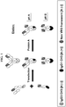
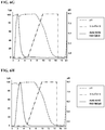
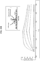
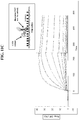
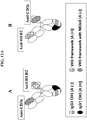
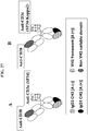
- the preferred known differential Protein A affinity purification technique of the present invention corresponds to a technique wherein all three species i.e. the two homo-dimeric species and the hetero-dimer of interest differ in their total number of Protein A binding sites by at least one site and wherein one of the two homo-dimeric species has no Protein A binding site and therefore does not bind Protein A (as shown in FIG. 1 ).
- VH3 based immunoglobulins or fragments thereof are of major importance to the biological drug industry.
- Therapeutic antibodies based on the VH3 subclass have been extensively developed as these frameworks bind Protein A and facilitate the testing of antibody fragments before their formatting into immunoglobulins; for example, many synthetic antibody phage display libraries used for antibody discovery are based on the VH3 subclass.
- VH3 based antibodies are often selected for their good expression and stability over other known heavy chain variable domain subclasses.
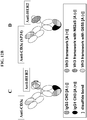
- VH3 domain has only one Protein A binding site with a weaker affinity when compared to a Fc region which has two sites with a stronger affinity ( Roben PW et al., (1995) J Immunol, 154(12): 6437-45 ), there is enough affinity to interfere with the known differential Protein A affinity purification techniques.
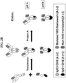
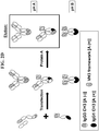
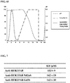
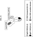
- Protein A binding is restored via the VH3 domain and the preferred technology described in FIG. 1 and above is no longer useful ( FIG. 2A ).
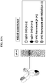
- abrogating Protein A binding in the VH3 based antigen binding site provides a straightforward solution and allows to keep the initial architecture of the desired hetero-dimer ( FIG. 2B ).
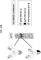
- the heavy chain hetero-dimer can be re-engineered to have the VH3 based antigen binding site located on the heavy chain that binds Protein A in its Fc region ( FIG.
- VH3 domain has a weaker affinity for Protein A compared to a Fc monomer hence the hetero-dimer of interest still elutes at a separate pH value from the other homo-dimeric species, typically at pH 4, while the homo-dimeric species that binds Protein A now encompasses two additional Protein A binding sites and elutes at a pH value ⁇ 3).
- the present invention provides new anti-human CD3 bispecific antibodies comprising a second binding arm which can recognise and bind to a disease associated antigen.
- a disease associated antigen means any antigen or epitope associated with a pathological state such as an oncogenic marker or a marker of some other metabolic or immunological dysfunction.
- a disease marker my also relate to an infectious disease such as a pathogenic virus or bacteria.
- the two binding arms of the anti-human CD3 bispecific antibody each comprise an immunoglobulin constant region and wherein the first arm or polypeptide binds to protein A and the second arm or polypeptide does not bind to protein A.
- the binding of the first polypeptide to protein A and the lack of binding of the second polypeptide to protein A is not intended to mean that the second polypeptide may not have some residual binding to protein A and it is instead intended that the second polypeptide binds less well to protein A in comparison to the first arm.
- the first and second polypeptides of the hetero-dimeric immunoglobulin or fragment thereof comprise an engineered immunoglobulin constant region with a modified CH3 region having a protein-protein interface that favours hetero-dimer formation over homo-dimer formation.
- the present invention provides a hetero-dimeric immunoglobulin or fragment thereof wherein the first and second polypeptides comprise an engineered immunoglobulin constant region with a modified CH3 domain having a protein-protein interface, wherein the protein-protein interface of the first polypeptide comprises an amino acid substitution at a position selected from the group consisting of: 3, 5, 7, 20, 22, 26, 27, 79, 81, 84, 84.2, 85.1, 86, 88 and 90 (IMGT ® numbering), and wherein the protein-protein interface of the second polypeptide comprises an amino acid substitution at a position selected from the group consisting of: 3, 5, 7, 20, 22, 26, 27, 79, 81, 84, 84.2, 84.4, 85.1, 86, 88 and 90 (IMGT ® numbering).
- the protein-protein interface of the second polypeptide comprises an amino acid substitution at position 84.4 and at least one further substitution at a position selected from the group consisting of: 3, 5, 7, 20, 22, 26, 27, 79, 81, 84, 84.2, 85.1, 86, 88 and 90 (IMGT ® numbering).
- the present invention provides a hetero-dimeric immunoglobulin or fragment thereof, wherein the first and second polypeptides comprise an engineered immunoglobulin constant region with a modified CH3 domain having a protein-protein interface, wherein the protein-protein interface of the first polypeptide comprises an amino acid substitution at position 88 and at a position selected from the group consisting of: 3, 5, 7, 20, 22, 26, 27, 79, 81, 84, 84.2, 85.1, 86 and 90 (IMGT ® numbering), and wherein the protein-protein interface of the second polypeptide comprises an amino acid substitution at position 85.1 and/or 86 and at a position selected from the group consisting of 3, 5, 7, 20, 22, 26, 27, 79, 81, 84, 84.2, 84.4, 88 and 90 (IMGT ® numbering).
- the epitope binding region of the first polypeptide binds the CD3 protein complex and the epitope binding region of the second polypeptide binds a disease associated antigen or wherein the epitope binding region of the first polypeptide binds a disease associated antigen and the epitope binding region of the second polypeptide binds the CD3 protein complex; and wherein the epitope binding region that binds the CD3 protein complex comprises a heavy chain CDR1 comprising the amino acid sequence of SEQ ID NO: 194, a heavy chain CDR2 comprising the amino acid sequence of SEQ ID NO: 195 and a heavy chain CDR3 comprising the amino acid sequence of SEQ ID NO: 196, and a light chain CDR1 comprising the amino acid sequence of SEQ ID NO: 197, a light chain CDR2 comprising the amino acid sequence of SEQ ID NO: 198 and a light chain CDR3 comprising the amino acid sequences of: SEQ ID NO: 199; or wherein the epitope binding region that binds
- Use of these new anti-human CD3 bispecific antibodies is not limited to but includes treatments of various human cancers and autoimmune and inflammatory diseases.
- the specific destruction of cancer cells over healthy cells and tissues represents a primary objective in oncology.
- Therapeutics that could safely redirect T cell killing against tumour associated cell surface antigens may offer improved clinical efficacy.
- Known areas of clinical unmet needs in oncology include but are not limited to breast cancer, metastatic breast cancer, ovarian cancer, pancreatic cancer, lung cancer, lymphomas and multiple myeloma. Elimination of disease-causing T cells could be more beneficial than inhibiting T cell differentiation in treating autoimmune and inflammatory diseases such as psoriasis, multiple sclerosis and diabetes.
- a preferred set of disease associated antigens come from the gene products CD33, TROP2, CD105, GD2, GD3, CEA, VEGFR1, VEGFR2, NCAM, CD133, CD123, ADAM17, MCSP, PSCA, FOLR1, CD19, CD20, CD38, EpCAM, HER2, EGFR, PSMA, IgE, Integrin a4b1, CCR5, LewisY, FAP, MUC-1, Wue-1, MSP, EGFRvIII, P glycoprotein, AFP, ALK, BAGE proteins, CD30, CD40, CTLA4, ErbB3, ErbB4, Mesothelin, OX40, CA125, CAIX , CD66e, cMet, EphA2, HGF/SF , MUC1, Phosphatidylserine , TAG-72 , TPBG, ⁇ -catenin, brc-abl, BRCA1, BORIS, CA9, caspase-8, CDK4, Cyclin-B1, CYP1B
- a hetero-dimeric immunoglobulin or fragment thereof according to the invention, wherein the epitope binding region that binds a disease associated antigen comprises heavy chain CDR1, CDR2 and CDR3 amino acid sequences and light chain CDR1, CDR2 and CDR3 amino acid sequences, respectively, selected from the group consisting of:
- the constant region of the second polypeptide of the hetero-dimeric immunoglobulin or fragment thereof comprises an IgG3 CH3 region.
- the constant region of the second polypeptide of the hetero-dimeric immunoglobulin or fragment thereof comprises a CH3 region other than that from IgG, and the non-IgG3 CH3 region comprises at least one substitution so as to decrease/abolish protein A binding.
- the epitope binding region of second polypeptide of the hetero-dimeric immunoglobulin or fragment thereof comprises a VH3 region comprising at least one modification that reduces protein A binding.
- VH3 based antigen binding sites can be readily produced and purified with a high degree of purity in a single Protein A chromatography step. These antibodies may exhibit higher efficacy over current therapies in addition to their ease of production.
- the present invention also provides a method to produce anti-human CD3 bispecific heavy chain hetero-dimers having at least one VH3 based antigen binding site from a recombinant mammalian host cell line wherein the bispecific antibody product is readily isolated after a single Protein A chromatography step with a high degree of purity.
- the modified VH3 region comprises an amino acid substitution selected from the group consisting of: 57, 65, 81, 82a and combination 19/57/59 (Kabat numbering) and even more preferably wherein the modified VH3 region comprises an amino acid substitution selected from the group consisting of: 57A, 57E, 65S, 81E, 82aS and combination 19G/57A/59A (Kabat numbering).
- the hetero-dimeric immunoglobulin or fragment thereof may comprise further substitutions wherein the heavy chain variable framework region comprises an amino acid substitution selected from the group consisting of: I34M, V48I, A49G, R58N/Y, I69L, A71T and T73K (Kabat numbering) and the light chain variable framework region comprises an amino acid substitution selected from the group consisting of: M4L, V33M, A34N, L46R, L47W, T51A, R66G, F71Y and P96F (Kabat numbering); or wherein the heavy chain variable framework region comprises the amino acid substitutions I34M, A49G and A71T (Kabat numbering) and the light chain variable framework region comprises the amino acid substitutions M4L, L46R, L47W and F71Y (Kabat numbering).
- the heavy chain variable framework region comprises an amino acid substitution selected from the group consisting of: I34M, V48I, A49G, R58N/Y, I69L, A71T and T73
- the epitope binding region that binds to the CD3 protein complex comprises a heavy chain variable framework region that is the product of or derived from the human VH3 subclass.
- the heavy chain variable framework region is the product of or derived from human IGHV3-23. More preferably, the heavy chain variable framework region is the product of or derived from human IGHV3-23*04 (SEQ ID NO: 22).
- the heavy chain variable framework region comprises at least one amino acid modification from the corresponding framework region of the heavy chain variable region of the corresponding murine antibody comprising the amino acid sequence of SEQ ID NO: 18 or SEQ ID NO: 60.
- the epitope binding region of the first polypeptide that binds to the CD3 protein complex comprises a light chain variable framework region that is the product of or derived from the human VK1 subclass or the human VK3 subclass.
- the light chain variable framework region is the product of or derived from human VK1-39 or VK3-20. More preferably the light chain variable framework region is the product of or derived from human IGKV1-39*01 (SEQ ID NO: 23) or IGKV3-20*01 (SEQ ID NO: 24).
- the light chain variable framework region comprises at least one amino acid modification from the corresponding framework region of the light chain variable region of the corresponding murine antibody comprising the amino acid sequence of SEQ ID NO: 19 or SEQ ID NO: 61.
- the epitope binding region that binds to the CD3 protein complex comprises a humanized heavy chain variable domain having the back mutations selected from the group consisting of: I34M, V48I, A49G, R58N/Y, I69L, A71T and T73K (Kabat numbering) and a humanized light chain variable domain having the back mutations selected from the group consisting of: M4L, V33M, A34N, L46R, L47W, R66G, F71Y and P96F (Kabat numbering).
- the epitope binding region that binds to the CD3 protein complex comprises a humanized heavy chain variable domain having the back mutations I34M, A49G and A71T (Kabat numbering) and a humanized light chain variable domain having the back mutations M4L, L46R, L47W and F71Y (Kabat numbering).
- the epitope binding region that binds the CD3 protein complex of the hetero-dimeric immunoglobulin or fragment thereof comprises a heavy chain variable region comprising the amino acid sequence of SEQ ID NO: 48, and a light chain variable region comprising the amino acid sequence of SEQ ID NO: 51; or wherein the epitope binding region that binds the CD3 protein complex comprises a heavy chain variable region comprising the amino acid sequence of SEQ ID NO: 49, and a light chain variable region comprising the amino acid sequence of SEQ ID NO: 51; or wherein the epitope binding region that binds the CD3 protein complex comprises a heavy chain variable region comprising the amino acid sequence of SEQ ID NO: 358, and a light chain variable region comprising the amino acid sequence of SEQ ID NO: 51; or wherein the epitope binding region that binds the CD3 protein complex comprises a heavy chain variable region comprising the amino acid sequence of SEQ ID NO: 101, and a light chain variable region comprising the amino acid sequence
- the CD3 protein complex comprises a number of subunits, for example, delta, epsilon and gamma.
- the epitope binding region that binds to the CD3 protein complex binds to the CD3 epsilon subunit.
- an epitope binding region as described herein includes the combination of one or more heavy chain variable domains and one or more complementary light chain variable domains which together form a binding site which permits the specific binding of the hetero-dimeric immunoglobulin or fragment thereof to one or more epitopes.
- the epitope binding region of the first poly peptide comprises a FAB and the epitope binding region of the second polypeptide comprises a scFv.
- the epitope binding region of the first poly peptide comprises a scFv and the epitope binding region of the second polypeptide comprises a FAB.
- the epitope binding region that binds a disease associated antigen binds to HER2.
- the epitope binding region comprises a heavy chain variable framework region that is the product of or derived from the human VH3 subclass, preferably human VH3-23, more preferably human IGHV3-23*04 (SEQ ID NO: 22), and a light chain variable framework region that is the product of or derived from the human VK1 subclass, preferably human VK1-39, more preferably human IGKV1-39*01 (SEQ ID NO: 23).
- the epitope binding region that binds the disease associated antigen HER2 comprises a heavy chain variable domain comprising the amino acid sequence of SEQ ID NO: 20 and a light chain variable domain comprising the amino acid sequence of SEQ ID NO: 21.
- the epitope binding region that binds HER2 may comprise a heavy chain variable domain and a light chain variable domain joined by a G 4 S linker forming a scFv fragment comprising the amino acid sequence of SEQ ID NO: 107.
- variable domain of the scFv fragment comprises a modification to abrogate binding to Protein A, wherein the amino acid substitution is 65S (Kabat numbering) and wherein the scFv fragment comprises the amino acid sequence of SEQ ID NO: 109 or wherein the amino acid substitution is 82aS (Kabat numbering) and wherein the scFv fragment comprises the amino acid sequence of SEQ ID NO: 111.
- Herceptin binding arm comprises a heavy chain variable region encoded by SEQ ID NO: 20 and a light chain variable region encoded by SEQ ID NO: 21.
- the epitope binding region that binds a disease associated antigen binds to CD38.
- the epitope binding region comprises a heavy chain variable framework region that is the product of or derived from the human VH3 subclass, preferably human VH3-23, more preferably human IGHV3-23*04 (SEQ ID NO: 22).
- the heavy chain variable framework region comprises at least one amino acid modification from the corresponding framework region of the heavy chain variable region of the corresponding murine antibody comprising the amino acid sequence of SEQ ID NO: 112 or 114 or 122.
- the epitope binding region further comprises a light chain variable framework region that is the product of or derived from the human VK1 subclass, preferably human VK1-39, more preferably human IGKV1-39*01 (SEQ ID NO: 23).
- the light chain variable framework region comprises at least one amino acid modification from the corresponding framework region of the light chain variable region of the corresponding murine antibody comprising the amino acid sequence of SEQ ID NO: 113 or 115 or 123.
- the CD38 binding polypeptide comprises variable heavy chain domain and variable light chain domain pair encoded by SEQ ID NOs: 116/117, 129/130, 133/134 and 135/136.
- the epitope binding region that binds a disease associated antigen binds to OX40.
- the epitope binding region comprises a heavy chain variable framework region that is the product of or derived from the human VH3 subclass, preferably human VH3-23, more preferably human IGHV3-23*04 (SEQ ID NO: 22).
- the heavy chain variable framework region comprises at least one amino acid modification from the corresponding framework region of the heavy chain variable region of the corresponding murine antibody comprising the amino acid sequence of SEQ ID NO: 139.
- the epitope binding region further comprises a light chain variable framework region that is the product of or derived from the human VK1 subclass, preferably human VK1-39, more preferably human IGKV1-39*01 (SEQ ID NO: 23).
- the light chain variable framework region comprises at least one amino acid modification from the corresponding framework region of the light chain variable region of the corresponding murine antibody comprising the amino acid sequence of SEQ ID NO: 140.
- the humanized heavy chain variable domain comprises a modification to abrogate binding to Protein A comprising the substitution G65S or the substitution N82aS (Kabat numbering).
- OX40 binding polypeptide comprises variable heavy chain domain and variable light chain domain pair encoded by SEQ ID Nos: 141/142, 278/280 and 279/281.
- the epitope binding region that binds a disease associated antigen binds to CD 19.
- the epitope binding region comprises a heavy chain variable framework region that is the product of or derived from the human VH3 subclass, preferably human VH3-23, more preferably human IGHV3-23*04 (SEQ ID NO: 22) and most preferably comprises the amino acid sequence of SEQ ID NO: 296.
- the epitope binding region further comprises a light chain variable framework region that is the product of or derived from the human VK1 subclass, preferably human VK1-39, more preferably human IGKV1-39*01 (SEQ ID NO: 23) and most preferably comprises the amino acid sequence of SEQ ID NO: 297.
- the heavy chain variable domain comprises a modification to abrogate binding to Protein A comprising the substitution G65S or the substitution N82aS (Kabat numbering).
- CD 19 binding polypeptide comprises variable heavy chain domain and variable light chain domain pair encoded by SEQ ID Nos: 296/297.
- the epitope binding region that binds a disease associated antigen binds to CD20.
- the epitope binding region comprises a heavy chain variable framework region that is the product of or derived from the human VH3 subclass, preferably human VH3-23, more preferably human IGHV3-23*04 (SEQ ID NO: 22).
- the heavy chain variable framework region comprises at least one amino acid modification from the corresponding framework region of the heavy chain variable region of the corresponding murine antibody comprising the amino acid sequence of SEQ ID NO: 143.
- the epitope binding region further comprises a light chain variable framework region that is the product of or derived from the human VK1 subclass, preferably human VK1-39, more preferably human IGKV1-39*01 (SEQ ID NO: 23).
- the light chain variable framework region comprises at least one amino acid modification from the corresponding framework region of the light chain variable region of the corresponding murine antibody comprising the amino acid sequence of SEQ ID NO: 144.
- the humanized heavy chain variable domain comprises a modification to abrogate binding to Protein A comprising the substitution G65S or the substitution N82aS (Kabat numbering).
- the EGFR binding polypeptide comprises variable heavy chain domain and variable light chain domain pair encoded by SEQ ID NOs: 143/144, 282/284, 283/285.
- the epitope binding region that binds a disease associated antigen binds to EGFR.
- the epitope binding region comprises a heavy chain variable framework region that is the product of or derived from the human VH3 subclass, preferably human VH3-23, more preferably human IGHV3-23*04 (SEQ ID NO: 22).
- the heavy chain variable framework region comprises at least one amino acid modification from the corresponding framework region of the heavy chain variable region of the corresponding murine antibody comprising the amino acid sequence of SEQ ID NO: 145.
- the epitope binding region further comprises a light chain variable framework region that is the product of or derived from the human VK1 subclass, preferably human VK1-39, more preferably human IGKV1-39*01 (SEQ ID NO: 23).
- the light chain variable framework region comprises at least one amino acid modification from the corresponding framework region of the light chain variable region of the corresponding murine antibody comprising the amino acid sequence of SEQ ID NO: 146.
- the humanized heavy chain variable domain comprises a modification to abrogate binding to Protein A comprising the substitution G65S or the substitution N82aS (Kabat numbering).
- the CD20 binding polypeptide comprises variable heavy chain domain and variable light chain domain pair encoded by SEQ ID NOs: 145/146, 286/288, 287/289, 290/291, 292/294.
- the epitope binding region that binds a disease associated antigen binds to IgE.
- the epitope binding region comprises a heavy chain variable framework region that is the product of or derived from the human VH3 subclass, preferably human VH3-23, more preferably human IGHV3-23*04 (SEQ ID NO: 22).
- the heavy chain variable framework region comprises at least one amino acid modification from the corresponding framework region of the heavy chain variable region of the corresponding humanized antibody comprising the amino acid sequence of SEQ ID NO: 298 or the corresponding murine antibody comprising the amino acid sequence of SEQ ID NO: 304.
- the epitope binding region further comprises a light chain variable framework region that is the product of or derived from the human VK1 subclass, preferably human VK1-39, more preferably human IGKV1-39*01 (SEQ ID NO: 23).
- the light chain variable framework region comprises at least one amino acid modification from the corresponding framework region of the light chain variable region of the corresponding humanized antibody comprising the amino acid sequence of SEQ ID NO: 299 or the corresponding murine antibody comprising the amino acid sequence of SEQ ID NO: 305.
- the heavy chain variable domain comprises a modification to abrogate binding to Protein A comprising the substitution G65S or the substitution N82aS (Kabat numbering).
- the IgE binding polypeptide comprises variable heavy chain domain and variable light chain domain pair encoded by SEQ ID NOs:, 298/299, 300/302, 301/303, 304/305, 306/308, 307/309.
- Anti-CD3 antibodies have been found to trigger toxicity by both direct and indirect mechanisms. Indirect mechanisms are mediated by the Fc region of the CD3 antibody which acts with the Fc receptor expressing immune cells and lead to transient T cell activation and cytokine release. Therefore in order to improve the safety of the hetero-dimeric immunoglobulins or fragment thereof as described herein, the immunoglobulin constant region of the first and/or second polypeptide has reduced or no binding for effector immune cells and/or complement C1q.
- the immunoglobulin constant region is engineered to abrogate Fc receptor binding in the lower hinge region.
- the immunoglobulin constant region of the first and/or second polypeptide comprises the substitution(s) L234A and/or L235A (EU numbering). Most preferably, the immunoglobulin constant region of the first and/or second polypeptide comprises the substitutions L234A and L235A (EU numbering).
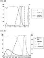
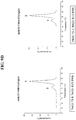
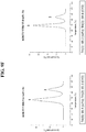
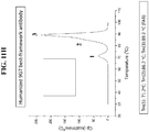
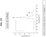
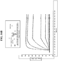
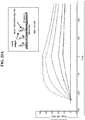
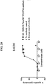
- the disclosure of the present invention also describes a hetero-dimeric immunoglobulin or fragment thereof wherein the epitope binding region binds to the CD3 epsilon subunit of the CD3 protein complex and comprises a FAB having a FAB thermo-stability superior to the FAB thermo-stability of the OKT3 chimera comprising a heavy chain variable domain of amino acid sequence of SEQ ID NO: 25 and a light chain variable domain of amino acid sequence of SEQ ID NO: 26, as measured by Differential Scanning Calorimetry (DSC) as described in FIG. 9 .
- DSC Differential Scanning Calorimetry
- the present invention provides a hetero-dimeric immunoglobulin or fragment thereof as described herein wherein one epitope binding region binds to the CD3 epsilon subunit of the CD3 protein complex and the other epitope binding region that binds a disease associated antigen, binds HER2.
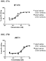
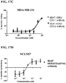
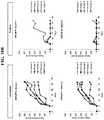
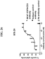
- the potency of such a hetero-dimeric immunoglobulin or fragment thereof to redirect T-cell killing can be measured in an in vitro assay using a flow cytometry method (RDL-FACS) or a colorimetric based method (RDL-MTS) on cell lines expressing HER2 such as JIMT-1, BT-474 and MDA-MB-231, as described in the Examples.
- the hetero-dimeric immunoglobulin or fragment thereof that binds to CD3 epsilon and HER2 kills JIMT-1 cells with a potency of 21 pM or less.
- the hetero-dimeric immunoglobulin or fragment thereof also kills BT-474 cells with a potency of 2 pM or less.
- the hetero-dimeric immunoglobulin or fragment thereof also kills MDA-MB-231 cells with a potency of 0.2 nM or less.
- the cytotoxicity of all cell lines was measured in a RDL assay performed with human PBMCs at an effector:target cell ratio of 10:1 over 48h.
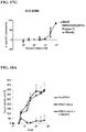
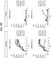
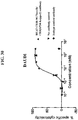
- this hetero-dimeric immunoglobulin or fragment thereof shows a potent anti-tumour effect wherein tested in vivo in a JIMT-1/PBMC xenograft model.
- the hetero-dimeric immunoglobulin or fragment thereof kills JIMT-1 cells at 0.05 mg/kg in a JIMT-1 cell xenograft.
- the present invention provides hetero-dimeric immunoglobulin or fragment thereof binding to:
- the present invention provides hetero-dimeric immunoglobulin or fragment thereof binding to:
- the present invention provides an in vitro method for the production of a hetero-dimeric immunoglobulin or fragment thereof as described herein, comprising the following steps:
- the hetero-dimeric immunoglobulin or fragment thereof found in the purified material from step (v) is at least 95% pure. More preferably the hetero-dimeric immunoglobulin or fragment thereof found in the purified material from step (v) is at least 96% pure. Even more preferably the hetero-dimeric immunoglobulin or fragment thereof found in the purified material from step (v) is at least 97%. Purity of the hetero-dimeric immunoglobulin or fragment thereof found in the purified material can be measured by capillary electrophoresis.
- a polypeptide comprising at least one CDRs from the groups: SEQ ID NOs: 224-229, 230-235 and 352-357; or combinations of heavy chain variable domain and light chain variable domain pairs selected from the group: SEQ ID NOs: 122/123, 124/125, 129/130, 135/136, 133/134 104 /106; and heavy and light chain sequence pair selected from the group: 126/127 or 128, 131/132, 137/138, 359/360.
- the present invention relates generally to novel hetero-dimeric immunoglobulins that bind to the CD3 protein complex and a disease associated antigen. Furthermore, these hetero-dimeric immunoglobulins have reduced or eliminated binding to protein A and therefore can be purified to a very high degree of purity using affinity chromatography.
- polypeptide and protein refer to a polymer of amino acid residues wherein amino acids are combined via peptide bonds to form a chain of amino acids that have been linked together via dehydration synthesis.
- Polypeptides and proteins can be synthesized through chemical synthesis or recombinant expression and are not limited to a minimum amino acid length.
- the group of polypeptides comprises "proteins" as long as the proteins consist of a single polypeptide chain.
- Polypeptides may further form multimers such as dimers, trimers and higher oligomers, i.e. consisting of more than one polypeptide molecule.
- Polypeptide molecules forming such dimers, trimers etc. may be identical or non-identical.
- the corresponding higher order structures of such multimers are, consequently, termed homo- or hetero-dimers, homo- or hetero-trimers etc.
- An example for a hetero-multimer is an antibody molecule, which, in its naturally occurring form, consists of two identical light polypeptide chains and two identical heavy polypeptide chains.
- polypeptide and protein also refer to naturally modified polypeptides/proteins wherein the modification is effected e.g. by post-translational modifications like glycosylation, acetylation, phosphorylation and the like. Such modifications are well known in the art.
- a "polypeptide” refers to a protein which includes modifications, such as deletions, additions and substitutions (which can be conservative in nature) to the native sequence. These modifications may be deliberate, as through site-directed mutagenesis, or may be accidental, such as through mutations of hosts which produce the proteins or errors due to PCR amplification.
- CD3 complex refers to the protein complex known as the CD3 (cluster of differentiation 3) T-cell co-receptor ( Wucherpfennig KW et al., (2010) Cold Spring Harb Perspect Biol, 2(4): a005140 ).
- the CD3 protein complex is composed of four distinct chains. In mammals, the complex contains a CD3 ⁇ chain, a CD3 ⁇ chain, and two CD3 ⁇ chains. These chains associate with a molecule known as the T-cell receptor (TCR) and the ⁇ -chain to generate an activation signal in T lymphocytes ( van der Merwe PA & Dushek O (2011) Nat Rev Immunol, 11(1): 47-55 ).
- TCR T-cell receptor
- the TCR, ⁇ -chain, and CD3 molecules together comprise the TCR complex.
- the CD3 ⁇ , CD3 ⁇ , and CD3 ⁇ chains are highly related cell-surface proteins of the immunoglobulin superfamily containing a single extracellular immunoglobulin domain.
- the intracellular tails of the CD3 molecules contain a single conserved motif known as an immunoreceptor tyrosine-based activation motif or ITAM for short, which is essential for the signalling capacity of the TCR. Since CD3 is required for T-cell activation, drugs (often monoclonal antibodies) that target CD3 have and are being investigated as immunosuppressant therapies.
- disease associated antigen refers to molecules that are involved in a disease process. Examples of disease associated antigens are found in a broad range of therapeutic areas such as inflammation, cancer and autoimmune diseases. In oncology, disease associated antigens are molecules that can broadly be used for the screening and/or monitoring and/or therapeutic targeting of cancers within a patient population, for example EpCAM antigen in prostate cancer. Tumour antigens can be produced directly by the tumour or by non-tumour cells as a response to the presence of a tumour and preferred tumour antigens are cell-surface molecules. Inflammatory disease associated antigens are known, which include but are not limited to, pro-inflammatory cytokines such as TNF- ⁇ and IL-1. Autoimmune disease associated antigens are also known; examples of these include but are not limited to antibodies against double-stranded DNA in systemic lupus erythematosus and amyloid beta peptide in Alzheimers disease.
- Immunoglobulin as referred to herein can be used interchangeably with the term "antibody”. Immunoglobulin includes full-length antibodies and any antigen binding fragment or single chains thereof. Immunoglobulins can be homo-dimeric or hetero-dimeric. Immunoglobulins and specifically naturally occurring antibodies are glycoproteins which exist as one or more copies of a Y-shaped unit, composed of four polypeptide chains. Each "Y" shape contains two identical copies of a heavy (H) chain and two identical copies of a light (L) chain, named as such by their relative molecular weights. Each light chain pairs with a heavy chain and each heavy chain pairs with another heavy chain. Covalent interchain disulfide bonds and non-covalent interactions link the chains together.
- Immunoglobulins and specifically naturally occurring antibodies contain variable regions, which are the two copies of the antigen binding site.
- a Fab fragment consists of the entire light chain and part of the heavy chain.
- the heavy chain contains one variable region (VH) and either three or four constant regions (CH1, CH2, CH3 and CH4, depending on the antibody class or isotype).
- the region between the CH1 and CH2 regions is called the hinge region and permits flexibility between the two Fab arms of the Y-shaped antibody molecule, allowing them to open and close to accommodate binding to two antigenic determinants separated by a fixed distance.
- the "hinge region” as referred to herein is a sequence region of 6-62 amino acids in length, only present in IgA, IgD and IgG, which encompasses the cysteine residues that bridge the two heavy chains.
- the heavy chains of IgA, IgD and IgG each have four regions, i.e. one variable region (VH) and three constant regions (CH1-3).
- IgE and IgM have one variable and four constant regions (CH1-4) on the heavy chain.
- the constant regions of the immunoglobulins may mediate the binding to host tissues or factors, including various cells of the immune system (e.g., effector cells) and the first component (C1q) of the complement system classical pathway.
- Each light chain is usually linked to a heavy chain by one covalent disulfide bond.
- Each light chain contains one variable region (VL) and one light chain constant region.
- the light chain constant region is a kappa light chain constant region designated herein as IGKC or is a lambda light chain constant region designated herein as IGLC.
- IGKC is used herein equivalently to C ⁇ or CK and has the same meaning.
- IGLC is used herein equivalently to C ⁇ or CL and has the same meaning.
- an IGLC region refers to all lambda light chain constant regions e.g. to all lambda light chain constant regions selected from the group consisting of IGLC1, IGLC2, IGLC3, IGLC6 and IGLC7.
- the VH and VL regions can be further subdivided into regions of hypervariability, termed complementarity determining regions (CDR), interspersed with regions that are more conserved, termed framework regions (FR or FW).
- CDR complementarity determining regions
- FR or FW framework regions
- Each VH and VL is composed of three CDRs and four FRs, arranged from amino- terminus to carboxy-terminus in the following order: FR1, CDR1, FR2, CDR2, FR3, CDR3, FR4.
- variable regions of the heavy and light chains contain an epitope- binding region that interacts with an antigen.
- Engineered immunoglobulins can encompass different epitope binding region formats such as scFv, FAB or dAb fragments. These fragments are usually assembled in an antibody-like structure by genetic fusion to a IgG Fc region.
- Engineered immunoglobulins can be constructed as homo or hetero-dimers with or without the use of hetero-dimerization enhancing techniques, and can have mono- or bispecific binding properties.
- full length antibody includes the structure that constitutes the natural biological form of an antibody, including variable and constant regions.
- the full length antibody of the IgG class is a tetramer and consists of two identical pairs of two immunoglobulin chains, each pair having one light and one heavy chain, each light chain comprising immunoglobulin regions VL and a light chain constant region, and each heavy chain comprising immunoglobulin regions VH, CH1 (C ⁇ 1), CH2 (C ⁇ 2), CH3 (C ⁇ 3) and CH4 (C ⁇ 4), depending on the antibody class or isotype).
- IgG antibodies may consist of only two heavy chains, each heavy chain comprising a variable region attached to the Fc region.
- Antibodies are grouped into classes, also referred to as isotypes, as determined genetically by the constant region.
- Human constant light chains are classified as kappa (CK) and lambda (C ⁇ ) light chains.
- Heavy chains are classified as mu ( ⁇ ), delta ( ⁇ ), gamma ( ⁇ ), alpha ( ⁇ ), or epsilon ( ⁇ ) and define the antibody's isotype as IgM, IgD, IgG, IgA and IgE, respectively.
- isotype as used herein is meant any of the classes and/or subclasses of immunoglobulins defined by the chemical and antigenic characteristics of their constant regions.
- the known human immunoglobulin isotypes are IGHG1 (IgG1), IGHG2 (IgG2), IGHG3 (IgG3), IGHG4 (IgG4), IGHA1 (IgA1), IGHA2 (IgA2), IGHM (IgM), IGHD (IgD) and IGHE (IgE).
- the so-called human immunoglobulin pseudo-gamma IGHGP gene represents an additional human immunoglobulin heavy constant region gene which has been sequenced but does not encode a protein due to an altered switch region ( Bensmana M et al., (1988) Nucleic Acids Res, 16(7): 3108 ).
- the human immunoglobulin pseudo-gamma IGHGP gene has open reading frames for all heavy constant regions (CH1-CH3) and hinge. All open reading frames for its heavy constant regions encode protein regions which align well with all human immunoglobulin constant regions with the predicted structural features.
- This additional pseudo-gamma isotype is referred herein as IgGP or IGHGP.
- Other pseudo immunoglobulin genes have been reported such as the human immunoglobulin heavy constant region epsilon P1 and P2 pseudo-genes (IGHEP1 and IGHEP2).
- the IgG class is the most commonly used for therapeutic purposes. In humans this class comprises subclasses IgG1, IgG2, IgG3 and IgG4. In mice this class comprises subclasses IgG1, IgG2a, IgG2b, IgG2c and IgG3.
- Immunoglobulin fragments include, but is not limited to, (i) a region including for example a CH1, a CH2 or a CH3 region, (ii) the Fab fragment consisting of VL, VH, CL or CK and CH1 regions, including Fab' and Fab'-SH, (ii) the Fd fragment consisting of the VH and CH1 regions, (iii) the dAb fragment ( Ward ES et al., (1989) Nature, 341(6242): 544-6 ) which consists of a single variable region (iv) F(ab') 2 fragments, a bivalent fragment comprising two linked Fab fragments (v) single chain Fv fragments (scFv), wherein a VH region and a VL region are linked by a peptide linker which allows the two regions to associate to form an antigen binding site ( Bird RE et al., (1988) Science, 242(4877): 423-6 ; Hu
- variable region refers to the regions or domains that mediates antigen-binding and defines specificity of a particular antibody for a particular antigen.
- the antigen-binding site consists of two variable regions that define specificity: one located in the heavy chain, referred herein as heavy chain variable region (VH) and the other located in the light chain, referred herein as light chain variable region (VL).
- VH heavy chain variable region
- VL light chain variable region
- the heavy chain variable region (VH) can be divided into seven subgroups or subclasses: VH1, VH2, VH3, VH4, VH5, VH6 and VH7.
- specificity may exclusively reside in only one of the two regions as in single-domain antibodies from heavy-chain antibodies found in camelids.
- the V regions are usually about 110 amino acids long and consist of relatively invariant stretches of amino acid sequence called framework regions (FRs or "non-CDR regions") of 15-30 amino acids separated by shorter regions of extreme variability called “hypervariable regions” that are 7-17 amino acids long.
- the variable domains of native heavy and light chains comprise four FRs, largely adopting a beta-sheet configuration, connected by three hypervariable regions, which form loops.
- the hypervariable regions in each chain are held together in close proximity by FRs and, with the hypervariable regions from the other chain, contribute to the formation of the antigen binding site of antibodies (see Kabat EA et al ., supra. ).
- hypervariable region refers to the amino acid residues of an antibody which are responsible for antigen binding.
- the hypervariable region generally comprises amino acid residues from a “complementary determining region” or "CDR", the latter being of highest sequence variability and/or involved in antigen recognition.
- CDR complementary determining region
- For all variable regions numbering is according to Kabat (Kabat EA et al ., supra. ).
- CDR definitions are in use and are encompassed herein.
- the Kabat definition is based on sequence variability and is the most commonly used (Kabat EA et al ., supra. ). Chothia refers instead to the location of the structural loops ( Chothia & Lesk J. (1987) Mol Biol, 196: 901-917 ).
- the AbM definition is a compromise between the Kabat and the Chothia definitions and is used by Oxford Molecular's AbM antibody modelling software ( Martin ACR et al., (1989) Proc Natl Acad Sci USA 86:9268-9272 ; Martin ACR et al., (1991) Methods Enzymol, 203: 121-153 ; Pedersen JT et al., (1992) Immunomethods, 1: 126-136 ; Rees AR et al., (1996) In Sternberg M.J.E. (ed.), Protein Structure Prediction. Oxford University Press, Oxford, 141-172 ).
- the contact definition has been recently introduced ( MacCallum RM et al., (1996) J Mol Biol, 262: 732-745 ) and is based on an analysis of the available complex structures available in the Protein Databank.
- the definition of the CDR by IMGT ® is based on the IMGT numbering for all immunoglobulin and T cell receptor V-REGIONs of all species (IMGT ® , the international ImMunoGeneTics information system ® ; Lefranc MP et al., (1999) Nucleic Acids Res, 27(1): 209-12 ; Ruiz M et al., (2000) Nucleic Acids Res, 28(1): 219-21 ; Lefranc MP (2001) Nucleic Acids Res, 29(1): 207-9 ; Lefranc MP (2003) Nucleic Acids Res, 31(1): 307-10 ; Lefranc MP et al.,
- CDRs Complementarity Determining Regions
- LCDR1 24-34
- LCDR2 50-56
- LCDR3 89-98
- HCDR1 26-35
- HCDR2 50-65
- HCDR3 95-102.
- the "non-CDR regions" of the variable domain are known as framework regions (FR).
- the “non-CDR regions” of the VL region as used herein comprise the amino acid sequences: 1-23 (FR1), 35-49 (FR2), 57-88 (FR3) and 99-107 (FR4).
- the “non-CDR regions” of the VH region as used herein comprise the amino acid sequences: 1-25 (FR1), 36-49 (FR2), 66-94 (FR3) and 103-113 (FR4).
- the CDRs of the present invention may comprise "extended CDRs" which are based on the aforementioned definitions and have variable domain residues as follows: LCDR1: 24-36, LCDR2: 46-56, LCDR3:89-97, HCDR1: 26-35, HCDR2:47-65, HCDR3: 93-102. These extended CDRs are numbered as well according to Kabat et al., supra.
- the "non-extended CDR region" of the VL region as used herein comprise the amino acid sequences: 1-23 (FR1), 37-45 (FR2), 57-88 (FR3) and 98- approximately 107 (FR4).
- the "non-extended CDR region” of the VH region as used herein comprise the amino acid sequences: 1-25 (FR1), 37-46 (FR2), 66-92 (FR3) and 103- approximately 113 (FR4).
- Fab or "FAB” or “Fab region” or “FAB region” as used herein includes the polypeptides that comprise the VH, CH1, VL and light chain constant immunoglobulin regions. Fab may refer to this region in isolation, or this region in the context of a full length antibody or antibody fragment.
- Fc or "Fc region”, as used herein includes the polypeptide comprising the constant region of an antibody heavy chain excluding the first constant region immunoglobulin region.
- Fc refers to the last two constant region immunoglobulin regions of IgA, IgD and IgG or the last three constant region immunoglobulin regions of IgE and IgM, and the flexible hinge N-terminal to these regions.
- Fc may include the J chain.
- Fc comprises immunoglobulin regions Cgamma2 and Cgamma3 (C ⁇ 2 and C ⁇ 3) and the hinge between Cgamma1 (C ⁇ 1) and Cgamma2 (C ⁇ 2).
- the human IgG heavy chain Fc region is usually defined to comprise residues C226 or P230 to its carboxyl-terminus, wherein the numbering is according to the EU index.
- Fc may refer to this region in isolation or this region in the context of an Fc polypeptide, for example an antibody.
- immunoglobulin constant region refers to immunoglobulin or antibody heavy chain constant regions from human or animal species and encompasses all isotypes.
- immunoglobulin constant regions are of human origin and are selected from the group consisting of, but not limited to: IGHG1 CH1, IGHG2 CH1, IGHG3 CH1, IGHG4 CH1, IGHA1 CH1, IGHA2 CH1, IGHE CH1, IGHEP1 CH1, IGHM CH1, IGHD CH1, IGHGP CH1, IGHG1 CH2, IGHG2 CH2, IGHG3 CH2, IGHG4 CH2, IGHA1 CH2, IGHA2 CH2, IGHE CH2, IGHEP1 CH2, IGHM CH2, IGHD CH2, IGHGP CH2, IGHG1 CH3, IGHG2 CH3, IGHG3 CH3, IGHG4 CH3, IGHA1 CH3, IGHA2 CH3, IGHEP1 CH3, IGHM CH3, IGHD CH3,
- Prefered "immunoglobulin constant regions” are selected from the group consisting of human IGHE CH2, IGHM CH2, IGHG1 CH3, IGHG2 CH3, IGHG3 CH3, IGHG4 CH3, IGHA1 CH3, IGHA2 CH3, IGHE CH3, IGHM CH3, IGHD CH3 and IGHGP CH3. More prefered "immunoglobulin constant regions” are selected from the group consisting of human IGHG1 CH3, IGHG2 CH3, IGHG3 CH3, IGHG4 CH3, IGHA1 CH3, IGHA2 CH3, IGHM CH3, IGHD CH3 and IGHGP CH3.
- epitope binding region includes a polypeptide or a fragment thereof having minimal amino acid sequence to permit the specific binding of the immunoglobulin molecule to one or more epitopes.
- Naturally occurring antibodies have two epitope binding regions which are also known as antigen binding or combining sites or paratopes.
- Epitope binding regions in naturally occurring antibodies are confined within the CDR regions of the VH and/or VL domains wherein the amino acid mediating epitope binding are found.
- VH domains or VL domains or fragments thereof and combinations thereof can be engineered to provide epitope binding regions ( Holt LJ et al., (2003) Trends Biotechnol, 21(11): 484-490 ; Polonelli L et al., (2008) PLoS ONE, 3(6): e2371 ).
- non-immunoglobulin based epitope binding regions can be found in artificial protein domains used as "scaffold" for engineering epitope binding regions ( Binz HK et al., (2005) Nat Biotechnol, 23(10): 1257-1268 ) or peptide mimetics ( Murali R & Greene MI (2012) Pharmaceuticals, 5(2): 209-235 ).
- the term 'epitope binding region' includes the combination of one or more heavy chain variable domains and one or more complementary light chain variable domains which together forms a binding site which permits the specific binding of the immunoglobulin molecule to one or more epitopes.
- Examples of an epitope binding region as exemplified in the present invention include scFv and FAB.
- epitope includes a fragment of a polypeptide or protein or a non-protein molecule having antigenic or immunogenic activity in an animal, preferably in a mammal and most preferably in a human.
- An epitope having immunogenic activity is a fragment of a polypeptide or protein that elicits an antibody response in an animal.
- An epitope having antigenic activity is a fragment of a polypeptide or protein to which an antibody or polypeptide specifically binds as determined by any method well-known to one of skill in the art, for example by immunoassays. Antigenic epitopes need not necessarily be immunogenic.
- epitope refers to a polypeptide sequence of at least about 3 to 5, preferably about 5 to 10 or 15 and not more than about 1,000 amino acids (or any integer there between), which define a sequence that by itself or as part of a larger sequence, binds to an antibody generated in response to such sequence.
- the length of the fragment may comprise nearly the full-length of the protein sequence, or even a fusion protein comprising one or more epitopes.
- An epitope for use in the subject invention is not limited to a polypeptide having the exact sequence of the portion of the parent protein from which it is derived.
- epitope encompasses sequences identical to the native sequence, as well as modifications to the native sequence, such as deletions, additions and substitutions (generally conservative in nature).
- the epitopes of protein antigens are divided into two categories, conformational epitopes and linear epitopes, based on their structure and interaction with the epitope binding site ( Goldsby R et al., (2003) “Antigens (Chapter 3)” Immunology (Fifth edition ed.), New York: W. H. Freeman and Company. pp. 57-75, ISBN 0-7167-4947-5 ).
- a conformational epitope is composed of discontinuous sections of the antigen's amino acid sequence.
- epitopes interact with the paratope based on the 3-D surface features and shape or tertiary structure of the antigen. Most epitopes are conformational. By contrast, linear epitopes interact with the paratope based on their primary structure. A linear epitope is formed by a continuous sequence of amino acids from the antigen.
- hetero-dimeric immunoglobulin or “hetero-dimeric fragment” or “hetero-dimer” or “hetero-dimer of heavy chains” as used herein includes an immunoglobulin molecule or part of comprising at least a first and a second polypeptide, like a first and a second region, wherein the second polypeptide differs in amino acid sequence from the first polypeptide.
- a hetero-dimeric immunoglobulin comprises two polypeptide chains, wherein the first chain has at least one non-identical region to the second chain, and wherein both chains assemble, i.e. interact through their non-identical regions.
- hetero-dimeric immunoglobulin has binding specificity for at least two different ligands, antigens or binding sites, i.e. is bispecific.
- Hetero-dimeric immunoglobulin as used herein includes but is not limited to full length bispecific antibodies, bispecifc Fab, bispecifc F(ab') 2 , bispecific scFv fused to an Fc region, diabody fused to an Fc region and domain antibody fused to an Fc region.
- homo-dimeric immunoglobulin or “homo-dimeric fragment” or “homo-dimer” or “homo-dimer of heavy chains” as used herein includes an immunoglobulin molecule or part of comprising at least a first and a second polypeptide, like a first and a second region, wherein the second polypeptide is identical in amino acid sequence to the first polypeptide.
- a homo-dimeric immunoglobulin comprises two polypeptide chains, wherein the first chain has at least one identical region to the second chain, and wherein both chains assemble, i.e. interact through their identical regions.
- a homo-dimeric immunoglobulin fragment comprises at least two regions, wherein the first region is identical to the second region, and wherein both regions assemble, i.e. interact through their protein-protein interfaces.
- IMGT ® immunoglobulin constant regions included in the present invention
- numbering can be according to the IMGT ® (IMGT ® ; supra ).
- CH1, CH2, CH3 immunoglobulin heavy chain constant regions selected from the group consisting of IGHG1, IGHG2, IGHG3 and IGHG4 numbering can be according to the " EU numbering system" (Edelman GM et al., (1969) Proc Natl Acad Sci USA, 63(1): 78-85 ).
- EU numbering system Edelman GM et al., (1969) Proc Natl Acad Sci USA, 63(1): 78-85 .
- a complete correspondence for the human CH1, hinge, CH2 and CH3 constant regions of IGHG1 can be found at the IMGT database (IMGT ® ; supra ).
- IGKC human kappa immunoglobulin light chain constant region
- IGLC1, IGLC2, IGLC3, IGLC6 and IGLC7 For the human lambda immunoglobulin light chain constant regions (IGLC1, IGLC2, IGLC3, IGLC6 and IGLC7), numbering can be according to the "Kabat numbering system" (Kabat EA et al ., supra ). A complete correspondence for human IGLC regions can be found at the IMGT database (IMGT ® ; supra ).
- the human IGHG1 immunoglobulin heavy chain constant regions as referred to herein have the following region boundaries: CH1 region (EU numbering: 118-215), Hinge ⁇ 1 region (EU numbering: 216-230), CH2 region (EU numbering: 231-340) and CH3 region (EU numbering: 341-447).
- the human CK region referred herein spans residues 108 to 214 (EU numbering).
- the human IGLC1, IGLC2, IGLC3, IGLC6 and IGLC7 regions referred herein span residues 108-215 (Kabat numbering).
- amino acid or “amino acid residue” as used herein includes natural amino acids as well as non-natural amino acids. Preferably natural amino acids are included.
- substitution or “amino acid modification” herein includes an amino acid substitution, insertion and/or deletion in a polypeptide sequence.
- substitution or “amino acid substitution” or “amino acid residue substitution” as used herein refers to a substitution of a first amino acid residue in an amino acid sequence with a second amino acid residue, whereas the first amino acid residue is different from the second amino acid residue i.e. the substituted amino acid residue is different from the amino acid which has been substituted.
- substitution R94K refers to a variant polypeptide, in which the arginine at position 94 is replaced with a lysine.
- 94K indicates the substitution of position 94 with a lysine.
- substitutions are typically separated by a slash or a comma.
- R94K/L78V or “R94K, L78V” refers to a double variant comprising the substitutions R94K and L78V.
- amino acid insertion or “insertion” as used herein is meant the addition of an amino acid at a particular position in a parent polypeptide sequence.
- insert -94 designates an insertion at position 94.
- amino acid deletion or “deletion” as used herein is meant the removal of an amino acid at a particular position in a parent polypeptide sequence.
- R94- designates the deletion of arginine at position 94.
- the terms “decrease”, “reduce”, or “reduction” in binding to Protein A refers to an overall decrease of at least 25%, 30%, 40%, 50%, 60%, 70%, 80%, 85%, 90%, 95%, 97%, or 99% up to 100% (elimination) in the binding of a modified immunoglobulin or fragment thereof to Protein A detected by standard art known methods such as those described herein, as compared to a parental i.e. unmodified immunoglobulin or wild-type IgG or an IgG having the wild-type human IgG Fc region.
- these terms alternatively may refer to an overall decrease of 10-fold (i.e. 1 log), 100-fold (2 logs), 1,000-fold (or 3 logs), 10,000-fold (or 4 logs), or 100,000-fold (or 5 logs).
- the terms “eliminate”, “abrogate”, “elimination” or “abrogation” of binding to Protein A refers to an overall decrease of 100% in the binding of a modified immunoglobulin or fragment thereof to Protein A i.e. a complete loss of the binding of a modified immunoglobulin or fragment thereof to Protein A, detected by standard art known methods such as those described herein, as compared to a parental i.e. unmodified immunoglobulin or wild-type IgG or an IgG having the wild-type human IgG Fc region.
- the terms “decrease”, “reduce”, or “reduction” in binding to an affinity reagent refers to an overall decrease of at least 25%, 30%, 40%, 50%, 60%, 70%, 80%, 85%, 90%, 95%, 97%, or 99% up to 100% (elimination) in the binding of a modified immunoglobulin or fragment thereof to the affinity reagent detected by standard art known methods such as those described herein, as compared to a parental, i.e. unmodified immunoglobulin or wild-type IgG or an IgG having the wild-type human IgG Fc region.
- these terms alternatively may refer to an overall decrease of 10-fold (i.e. 1 log), 100-fold (2 logs), 1,000-fold (or 3 logs), 10,000-fold (or 4 logs), or 100,000-fold (or 5 logs).
- the terms “eliminate” , “abrogate”, “elimination” or “abrogation” of binding to an affinity reagent refers to an overall decrease of 100% in the binding of a modified immunoglobulin or fragment thereof to the affinity reagent i.e. a complete loss of the binding of a modified immunoglobulin or fragment thereof to the affinity reagent detected by standard art known methods such as those described herein, as compared to a parental, i.e. unmodified immunoglobulin or wild-type IgG or an IgG having the wild-type human IgG Fc region.
- Bispecific antibodies are monoclonal antibodies that have binding specificities for at least two different antigens.
- the bispecific antibodies are bispecific antibodies with one or more amino acid modifications in the VH region relative to the parental antibody.
- bispecific antibodies may be human or humanized antibodies.
- Bispecific antibodies may also be used to localize cytotoxic agents to cells which express a target antigen. These antibodies possess a target-antigen-binding arm and an arm which binds a cytotoxic agent, such as, e.g., saporin, anti-interferon- ⁇ , vinca alkaloid, ricin A chain, methotrexate or radioactive isotope hapten.
- Bispecific antibodies can be prepared as full length antibodies or antibody fragments.
- bispecific antibodies are known in the art. Traditionally, the recombinant production of bispecific antibodies is based on the co-expression of two immunoglobulin heavy chain-light chain pairs, where the two heavy chains have different specificities ( Milstein and Cuello, (1983) Nature, 305: 537-40 ). Because of the random assortment of immunoglobulin heavy and light chains, these hybridomas (quadromas) produce a potential mixture of different antibody molecules, of which only one has the correct bispecific structure. The purification of the correct molecule, which is usually done by affinity chromatography steps, is rather cumbersome and the product yields are low.
- antibody variable regions with the desired binding specificities are fused to immunoglobulin constant region sequences.
- the fusion is with an immunoglobulin heavy chain constant region, comprising at least part of the hinge, CH2 and CH3 regions.
- the first heavy-chain constant region (CH1) containing the site necessary for light chain binding, is present in at least one of the fusions.
- DNAs encoding the immunoglobulin heavy chain fusions and, if desired, the immunoglobulin light chain are inserted into separate expression vectors and are co-transfected into a suitable host organism.
- This provides for flexibility in adjusting the mutual proportions of the three polypeptide fragments in embodiments when unequal ratios of the three polypeptide chains used in the construction provide the optimum yields. It is, however, possible to insert the coding sequences for two or all three polypeptide chains in one expression vector when the expression of at least two polypeptide chains in equal ratios results in high yields or when the ratios are of no particular significance.
- Bispecific antibodies include cross-linked or "heteroconjugate" antibodies.
- one of the antibodies in the heteroconjugate can be coupled to avidin, the other to biotin.
- Such antibodies have, for example, been proposed to target immune system cells to unwanted cells ( US4,676,980 ) and for treatment of HIV infection ( WO1991/00360 , WO1992/00373 and EP03089 ).
- Heteroconjugate antibodies may be made using any convenient cross-linking method. Suitable cross-linking agents are well known in the art (see US4,676,980 ), along with a number of cross-linking techniques.
- Antibodies with more than two valencies are also contemplated.
- trispecific antibodies can be prepared (see Tutt A et al. (1991) J. Immunol. 147: 60-9 ).
- the present disclosure provides a bispecific hetero-dimeric immunoglobulin or fragment thereof or a bispecific full-length antibody which binds to CD3 and a disease associated antigens selected from within the groups of: tumor antigens, cytokines, vascular growth factors and lympho-angiogenic growth factors.
- the bispecific hetero-dimeric immunoglobulin or fragment thereof or the bispecific antibody binds to CD3 and a disease associated antigen selected from the group consisting of: CD33, TROP2, CD105, GD2, GD3, CEA, VEGFR1, VEGFR2, NCAM, CD133, CD123, ADAM17, MCSP, PSCA, FOLR1, CD19, CD20, CD38, EpCAM, HER2, HER3, EGFR, PSMA, IgE, Integrin a4b1, CCR5, LewisY, FAP, MUC-1, Wue-1, MSP, EGFRvIII, P glycoprotein, AFP, ALK, BAGE proteins, CD30, CD40, CTLA4, ErbB3, ErbB4, Mesothelin, OX40, CA125, CAIX , CD66e, cMet, EphA2, HGF/SF , MUC1 , Phosphatidylserine , TAG-72 , TPBG, ⁇ -caten
- Protein A is a cell wall component produced by several strains of Staphylococcus aureus which consists of a single polypeptide chain.
- the Protein A gene product consists of five homologous repeats attached in a tandem fashion to the pathogen's cell wall.
- the five domains are approximately 58 amino acids in length and denoted EDABC, each exhibiting immunoglobulin binding activity ( Tashiro M & Montelione GT (1995) Curr. Opin. Struct. Biol., 5(4): 471-481 ).
- the five homologous immunoglobulin binding domains fold into a three-helix bundle.
- Each domain is able to bind proteins from many mammalian species, most notably IgGs ( Hober S et al., (2007) J. Chromatogr. B Analyt. Technol. Biomed. Life Sci., 848(1): 40-47 ).
- Protein A binds the heavy chain of most immunoglobulins within the Fc region but also within the Fab region in the case of the human VH3 family ( Jansson B et al, (1998) FEMS Immunol. Med. Microbiol., 20(1): 69-78 ).
- Protein A binds IgG from various species including human, mouse, rabbit and guinea pig but does not bind human IgG3 (Hober S et al., (2007) supra ).
- VH3 is the only subclass to bind Protein A ( Graille M et al., (2000) Proc. Natl. Acad. Sci. USA 97(10): 5399-5404 ), and all five domains of Protein A are known to bind this variable domain subclass ( Jansson B et al, (1998) FEMS Immunol. Med. Microbiol., 20(1): 69-78 .
- VH3 based immunoglobulins or fragments thereof are of major importance to the biotechnology industry.
- VH3 based molecules have been extensively developed since their ability to bind Protein A facilitates their functional pre-screening, and as such many synthetic or donor based phage display libraries or transgenic animal technologies used for antibody discovery are based on the VH3 subclass. In addition VH3 based molecules are often selected for their good expression and stability over other known heavy chain variable domain subclasses.
- Protein A used for production of antibodies in bio-pharmaceuticals is usually produced recombinantly in E. coli and functions essentially the same as native Protein A ( Liu HF et al., (2010) MAbs, 2(5): 480-499 ). Most commonly, recombinant Protein A is bound to a stationary phase chromatography resin for purification of antibodies. Optimal binding occurs at pH8.2, although binding is also good at neutral or physiological conditions (pH 7.0-7.6). Elution is usually achieved through pH shift towards acidic pH (glycine-HCl, pH2.5-3.0).
- Protein A chromatography resins There are various commercially available Protein A chromatography resins. The main differences between these media are the support matrix type, Protein A ligand modification, pore size and particle size. The differences in these factors give rise to differences in compressibility, chemical and physical robustness, diffusion resistance and binding capacity of the adsorbents (Hober S et al., (2007), supra ). Examples of Protein A chromatography resins include but are not limited to the MabSelect SuReTM Protein A resin and MabSelectTM Protein A resin from GE Healthcare as used in examples.
- chromatography refers to protein liquid chromatography and includes fast protein liquid chromatography (FPLC) which is a form of liquid chromatography that is often used to analyze or purify mixtures of proteins.
- FPLC fast protein liquid chromatography
- the mobile phase is an aqueous solution, or "buffer”.
- the buffer flow rate can be operated under gravity flow or controlled by a positive-displacement pump which is normally kept at a constant rate, while the composition of the buffer can be varied by drawing fluids in different proportions from two or more external reservoirs.
- the stationary phase is a resin composed of beads, usually of cross-linked agarose, packed into a cylindrical glass or plastic column. FPLC resins are available in a wide range of bead sizes and surface ligands depending on the application.
- affinity chromatography involves the use of an affinity reagent as ligands which are cross-linked to the stationary phase and that have binding affinity to specific molecules or a class of molecules.
- Ligands can be bio-molecules, like protein ligands or can be synthetic molecules. Both types of ligand tend to have good specificity.
- the most commonly used protein ligand in production is the affinity reagent Protein A.
- affinity chromatography when the solution (for example a crude cell supernatant containing a protein of interest) is loaded onto to the column the target protein is usually adsorbed while allowing contaminants (other proteins, lipids, carbohydrates, DNA, pigments, etc.) to pass through the column.
- the adsorbent itself is normally packed in a chromatography column; though the adsorption stage can be performed by using the adsorbent as a stirred slurry in batch binding mode.
- the next stage after adsorption is the wash stage, in which the adsorbent is washed to remove residual contaminants.
- the bound protein is then eluted in a semi-pure or pure form. Elution is normally achieved by changing the buffer or salt composition so that the protein can no longer interact with the immobilized ligand and is released.
- the protein of interest may not bind the affinity resin and affinity chromatography is directed at binding unwanted contaminants and the unbound fraction is therefore collected to isolate the protein of interest.
- Affinity chromatography can be performed in a fixed bed or a fluidised bed.
- gradient mode chromatography refers to a chromatography method wherein the proportion of the "elution” buffer (buffer B) is increased from 0% to 100% in a gradual or stepwise manner.
- capture-elution mode chromatography or “capture-elution purification mode” or “capture-elution purification” refers to a chromatography method wherein the proportion of the "elution" buffer (buffer B) is not increased from 0% to 100% in a gradual or stepwise manner but rather directly applied at a 100% after capture and optionally a wash step with running buffer (buffer A).
- the present invention provides an epitope binding region that binds the CD3 protein complex comprising the heavy and light chain CDRs as described supra and further comprising a heavy chain variable framework region that is the product of or derived from human gene IGHV3-23*04 (SEQ ID NO: 22).
- the heavy chain variable framework region comprises at least one amino acid modification from the corresponding framework region of the heavy chain variable region of the corresponding murine antibody OKT3 comprising the amino acid sequence of SEQ ID NO: 18.
- the amino acid modification is an amino acid substitution.
- no more than seven, preferably no more than six, preferably no more than five, preferably no more than four, more preferably no more than three, even more preferably no more than two, most preferably no more than one amino acid modifications are performed within a framework region.
- the present disclosure provides an epitope binding region that binds to the CD3 protein complex, wherein the amino acid modification of the framework regions of the heavy chain variable region comprise an amino acid substitution at amino acid position selected from the group consisting of: 34, 48, 49, 58, 69, 71 and 73 and wherein the amino acid position of each group member is indicated according to the Kabat numbering.
- amino acid substitutions of the framework regions of the heavy chain variable region are selected from the group consisting of: I34M, V48I, A49G, R58N, R58Y, I69L, A71T and T73K.
- Preferred amino acid substitution of the framework regions of the heavy chain variable region are at amino acid positions selected from the group consisting of 34, 49 and 71. More preferred amino acid substitutions of the framework regions of the heavy chain variable region are selected from the group consisting of I34M, A49G and A71T.
- the epitope binding region of the first polypeptide that binds the CD3 protein complex comprises a light chain variable framework region that is the product of or derived from a human gene selected from the group consisting of: IGKV1-39*01 (SEQ ID NO: 23) and IGKV3-20*01 (SEQ ID NO: 24).
- the light chain variable framework region comprises at least one amino acid modification from the corresponding framework region of the light chain variable region of the corresponding murine antibody OKT3 comprising the amino acid sequence of SEQ ID NO: 19.
- the amino acid modification is an amino acid substitution.
- no more than eight, preferably no more than seven, preferably no more than six, preferably no more than five, preferably no more than four, more preferably no more than three, even more preferably no more than two, most preferably no more than one amino acid modifications are performed within a framework region.
- the present disclosure provides an epitope binding region that binds to the CD3 protein complex, wherein the amino acid modification of the framework regions of the light chain variable region sequence comprises an amino acid substitution at amino acid position selected from the group consisting of: 4, 33, 34, 46, 47, 66, 71 and 96.
- amino acid substitutions of the framework regions of the light chain variable region are selected from the group consisting of: M4L, V33M, A34N, L46R, L47W, R66G, F71Y and P96F.
- Preferred amino acid substitution of the framework regions of the light chain variable region are at amino acid positions selected from the group consisting of 4, 46 and 47. More preferred amino acid substitutions of the framework regions of the light chain variable region are selected from the group consisting of M4L, L46R, L47W and F71Y.
- the epitope binding region of the first polypeptide that binds to the CD3 protein complex may comprise amino acid modifications of the framework regions of the heavy chain variable region sequence as set out above and amino acid modifications of the framework regions of the light chain variable region sequence as set out above.
- the present disclosure also provides an antibody or fragment thereof that binds to the CD3 protein complex that comprises a heavy chain sequence selected from the group consisting of SEQ ID NOs: 27 to 38, 64-68 and 359, preferably selected consisting of SEQ ID NO: 359.
- the present disclosure also provides an antibody or fragment thereof that binds to the CD3 protein complex that comprises a light chain sequence selected from the group consisting of SEQ ID NOs: 39 to 47, 69 to 90 and 360 preferably consisting of SEQ ID NO: 360.
- the heavy and light chain variable region sequences can bind to the CD3 protein complex
- the heavy and light chain variable region sequences can be "mixed and matched" to create anti-CD3 binding molecules of the invention.
- CD3 binding of such "mixed and matched" antibodies can be tested using the binding assays described e.g. in the Examples.
- the present invention therefore utilises the BEAT ® technology described method ( PCT publication No: WO2012/131555 ), which is based on a unique concept of bio-mimicry that exhibit superior hetero-dimerisation over prior art methods.
- the BEAT technology is based on an interface exchange between naturally occurring homo or hetero-dimeric immunoglobulin domain pairs to create new hetero-dimers which can be used as building blocks for Fc-based bispecific antibodies.
- the present invention provides a hetero-dimeric immunoglobulin or fragment thereof comprising first and second polypeptides comprising an engineered immunoglobulin constant region with a modified CH3 domain having a protein-protein interface, wherein the protein-protein interface of the first polypeptide comprises an amino acid substitution at a position selected from the group consisting of: 3, 5, 7, 20, 22, 26, 27, 79, 81, 84, 84.2, 85.1, 86, 88 and 90 (IMGT ® numbering), and wherein the protein-protein interface of the second polypeptide comprises an amino acid substitution at position 84.4 and at a position selected from the group consisting of 3, 5, 7, 20, 22, 26, 27, 79, 81, 84, 84.2, 85.1, 86, 88 and 90 (IMGT ® numbering).
- the present invention provides a hetero-dimeric immunoglobulin or fragment thereof, wherein the first and second polypeptides comprise an engineered immunoglobulin constant region with a modified CH3 domain having a protein-protein interface, wherein the protein-protein interface of the first polypeptide comprises an amino acid substitution at position 88 and at a position selected from the group consisting of: 3, 5, 7, 20, 22, 26, 27, 79, 81, 84, 84.2, 85.1, 86 and 90 (IMGT ® numbering), and wherein the protein-protein interface of the second polypeptide comprises an amino acid substitution at position 85.1 and/or 86 and at a position selected from the group consisting of 3, 5, 7, 20, 22, 26, 27, 79, 81, 84, 84.2, 84.4, 88 and 90 (IMGT ® numbering), wherein the amino acid residue substituted at position 88 in the first engineered immunoglobulin constant region is interacting with the amino acid residue substituted at position 85.1 and/or 86 in the second engineered immunoglobulin constant
- the amino acid residue which is substituted in the protein-protein interface of the first engineered immunoglobulin constant region at position 88 is 88W and conservative amino acid substitutions thereof, wherein the amino acid position is indicated according to IMGT ® numbering. More preferably, the amino acid residue which is substituted in the protein-protein interface of the first engineered immunoglobulin constant region at position 88 is 88W and wherein the further amino acid residue substituted in the protein-protein interface of the first engineered immunoglobulin constant region is selected from the group consisting of: 3A, 20V, 20T, 20A, 20N, 20Q, 20E, 20S, 20K, 20W, 22A, 22G, 22T, 22L, 22I, 22V, 26R, 26Q, 26T, 26K, 26V, 26S, 26N, 26E, 79Y, 85.1T, 85.1M, 85.1A, 85.1S, 85.1R, 85.1H, 85.1K, 85.1F, 85.1C, 85.1N, 85.1W,
- amino acid residue which is substituted at position 85 and 86 in the protein-protein interface of the second engineered immunoglobulin constant region is selected from the group consisting of: 85.1A, 85.1S, 85.1C and 86S and conservative amino acid substitutions thereof (IMGT ® numbering).
- amino acid residue which is substituted in the protein-protein interface of the second engineered immunoglobulin constant region is selected from the group consisting of: 85.1A, 85.1S, 85.1C and 86S and wherein the further amino acid residue substituted in the protein-protein interface of the second engineered immunoglobulin constant region is selected from the group consisting of: 3E, 5A, 7F, 20T, 22V, 26T, 81D, 84L, 84.2E, 88R and 90R and conservative amino acid substitutions thereof (IMGT ® numbering).
- the amino acid residue which is substituted in the protein-protein interface of the first engineered immunoglobulin constant region at position 88 is 88W and wherein the further amino acid residue substituted in the protein-protein interface of the first engineered immunoglobulin constant region is: 3A, 20K, 22V, 26T, 79Y, 85.1S, 86V and 90N and, wherein the amino acid residues which are substituted in the protein-protein interface of the second engineered immunoglobulin constant region at positions 85.1 and 86 are 85.1A, 85.1S or 85.1A and 86S and wherein the further amino acid residue substituted in the protein-protein interface of the second engineered immunoglobulin constant region is: 3E, 5A, 7F, 20T, 22V, 26T, 81D, 84L, 84.2E, 84.4Q, 88R and 90R (IMGT ® numbering).
- the present invention provides a hetero-dimeric immunoglobulin or fragment thereof, wherein the first and second polypeptides comprise an engineered immunoglobulin constant region with a modified CH3 domain having a protein-protein interface, wherein the protein-protein interface of the first polypeptide comprises an amino acid substitution at position 20, and at a position selected from the group consisting of: 3, 5, 7, 22, 26, 27, 79, 81, 84, 84.2, 85.1, 86, 88 and 90 and, wherein the protein-protein interface of the second polypeptide comprises an amino acid substitution at position 26 and at a position selected from the group consisting of: 3, 22, 27, 79, 81, 84, 85.1, 86, and 88, wherein the amino acid residue substituted at position 20 in the first engineered immunoglobulin constant region is interacting with the amino acid residue substituted at position 26 in the second engineered immunoglobulin constant region, wherein the amino acid position of each group member is indicated according to the IMGT ® numbering.
- the amino acid residues which are substituted in the protein-protein interface of the first engineered immunoglobulin chain comprise the amino acid residues at positions 20 and 22, and optionally a further amino acid residue at a position selected from the group consisting of: 3, 5, 7, 26, 27, 79, 81, 84, 84.2, 84.4, 85.1, 86, 88 and 90 and, wherein the amino acid residues which are substituted in the protein-protein interface of the second engineered immunoglobulin chain comprise the amino acid residues at positions 26 and at a further position selected from the group consisting of: 3, 5, 7, 20, 22, 27, 79, 81, 84, 84.2, 84.4, 85.1, 86, 88 and 90, wherein the amino acid position of each group member is indicated according to the IMGT ® numbering.
- the amino acid residues which are substituted in the protein-protein interface of the first engineered immunoglobulin chain comprise the amino acid residues at positions 20 and 22, and optionally a further amino acid residue at a position selected from the group consisting of: 3, 5, 7, 26, 27, 79, 81, 84, 84.2, 84.4, 85.1, 86, 88 and 90 and, wherein the amino acid residues which are substituted in the protein-protein interface of the second engineered immunoglobulin chain comprise the amino acid residues at positions 26 and 86 and optionally at a further position selected from the group consisting of 3, 5, 7, 20, 22, 27, 79, 81, 84, 84.2, 84.4, 85.1, 88 and 90, wherein the amino acid position of each group member is indicated according to the IMGT ® numbering.
- the amino acid residue which is substituted at position 20 in the protein-protein interface of the first engineered immunoglobulin constant region is selected from the group consisting of 20V, 20T, 20A, 20N, 20Q, 20K, 20S, 20W and 20E and wherein the further amino acid residue substituted in the protein-protein interface of the first engineered immunoglobulin constant region is selected from the group consisting of 3A, 22A, 22G, 22L, 22I, 22V, 22T, 26K, 26R, 26Q, 26T, 26V, 26S, 26N, 26E, 79Y, 85.1W, 85.1F, 85.1T, 85.1M, 85.1A, 85.1S, 85.1R, 85.1H, 85.1K, 85.1C, 85.1N, 86W, 86Y, 86S, 86I, 86H, 86Q, 86V, 86T, 86F, 88Q, 88L, 88V, 88R, 88E, 88T, 88
- the amino acid residue which is substituted in the protein-protein interface of the first engineered immunoglobulin constant region at position 20 is 20K and wherein the further amino acid residue substituted in the protein-protein interface of the first engineered immunoglobulin constant region is 3A, 22V, 26T, 79Y, 85.1S, 86V, 88W and 90N and, wherein the amino acid residues which are substituted in the protein-protein interface of the second engineered immunoglobulin constant region at position 26 is 26T and wherein the further amino acid residue substituted in the protein-protein interface of the second engineered immunoglobulin constant region is 3E, 5A, 7F, 20T, 22V, 81D, 84L, 84.2E, 84.4Q, 85.1C/S/A, 86S, 88R and 90R (IMGT ® numbering).
- Example 1 As a first step (Example 1), the substitutions that reduce or abrogate binding to Protein A were assayed in homo-dimeric immunoglobulins based on FAB or scFv fragments. It was found that the presence of a variable heavy chain domain of the VH3 subclass within the heavy chain which has substitutions for reduced or no binding to Protein A, hampers any differential affinity methods based on Protein A. Solutions to these major impediments were found in the forms of framework substitutions that reduce or abrogate Protein A binding to the VH3 subclass for the differential affinity methods based on Protein A.
- a humanised antibody targeting the human CD3 was generated by grafting the CDRs of a murine anti-CD3 antibody onto IGVH3-23 and IGVK1 or IGVK3 human germline frameworks.
- the best humanised variants had the Protein A binding site present in their VH domain abrogated using a G65S or N82aS substitution (Kabat numbering). These variants were formatted as FAB or scFv fragments.
- antigen binding sites of antibodies targeting disease associated antigens were generated.
- CDRs of murine antibodies could be grafted onto the human germline frameworks IGVH3-23 and IGVK1 (Examples 2.3, 2.4 and 2.6-2.10).
- CDRs of antibodies isolated from phage display libraries could be based on the VH3 variable domain subclass or grafted onto the human germline frameworks IGVH3-23 and IGVK1 (Examples 2.5 and 2.6).
- the Protein A binding site in the VH domain of the epitope binding region was abrogated using the G65S or N82aS substitutions (Kabat numbering).
- hetero-dimeric antibodies were produced based on the BEAT ® technology (as described in WO2012/131555 ) in which the anti-CD3 antibody from Example 2.1 and the epitope binding region of the disease associated antigen as described in Examples 2.2-2.10 were used in an scFv-FAB format or vice versa (Example 3.1). Since a difference in the number of Protein A binding sites between homo- and hetero-dimeric species can be used to isolate the hetero-dimeric species by Protein A chromatography, the bispecific antibodies of the present invention were engineered to result in one of the two homo-dimeric species having no Protein A binding site and therefore no binding to Protein A resin. Furthermore, in order to improve the safety profile of the BEAT antibodies, the Fc receptor binding was reduced or eliminated by engineering the two substitutions L234A and L235A (EU numbering) into the lower hinge region of the Fc region.
- L234A and L235A EU numbering
- cDNAs encoding the different polypeptide chains in part or in full were first gene synthetized by GENEART AG (Regensburg, Germany) and modified using standard molecular biology techniques. PCR products were digested with appropriate DNA restriction enzymes, purified and ligated in a modified pcDNA3.1 plasmid (Invitrogen AG, Switzerland) carrying a CMV promoter and a bovine hormone poly-adenylation (poly(A)) previously digested with the same DNA restriction enzymes. All polypeptide chains were independently ligated in this expression vector where secretion was driven by the murine VJ2C leader peptide.
- Antibodies, ScFv-Fc fusion proteins, BEAT antibodies and antigens were expressed as described below unless otherwise indicated.
- equal quantities of each engineered chains vectors were co-transfected into suspension-adapted HEK293-EBNA cells (ATCC-LGL standards, Teddington, UK; Cat. No: CRL-10852) using Polyethyleneimine (PEI; Sigma, Buchs, Switzerland).
- PEI Polyethyleneimine
- 100ml of cells in suspension at a density of 0.8-1.2 million cells per ml is transfected with a DNA-PEI mixture.
- the immunoglobulin construct is produced by further culturing the cells for a period of 4 to 5 days to allow for secretion into the culture medium (EX-CELL 293, HEK293-serum-free medium (Sigma), supplemented with 0.1% pluronic acid, 4mM glutamine and 0.25 ⁇ g/ml geneticin).
- Cell-free culture supernatants containing the secreted immunoglobulins were prepared by centrifugation followed by sterile filtration and used for further analysis.
- Elution was performed with a pH linear gradient combining various amounts of two buffers (running buffer (A): 0.2M phosphate citrate buffer pH 8.0 and elution buffer (B): 0.04M phosphate citrate buffer pH 3.0.
- the linear gradient went from 0% B to 100% B in five column volumes (CVs).
- Eluted fractions were neutralised with 0.1V of 1M Tris-HCl pH 8.0.
- Supernatant, flow through and elution fractions were analysed under non-reduced conditions by SDS-PAGE (NuPAGE Bis-Tris 4-12% acrylamide, Invitrogen AG, Basel, Switzerland).
- Elution was performed with a pH linear gradient combining various amounts of two buffers (running buffer (A): 0.2 M phosphate citrate buffer pH8.0 and elution buffer (B): 0.04 M phosphate citrate buffer pH3.0).
- the linear gradient went from 0% B to 100% B in 5CVs.
- Eluted fractions were neutralised with 0.1 V of 1M Tris-HCl pH8.0.
- Supernatant, flow through and elution fractions were analysed under non-reduced conditions by SDS-PAGE (NuPAGE Bis-Tris 4-12% acrylamide, Invitrogen AG, Basel, Switzerland).
- Purification scheme included a capture-elution mode chromatography followed by a gradient mode chromatography according to the procedure described above.
- the thermal stabilities of antibodies were compared using calorimetric measurements. Calorimetric measurements were carried out on a VP-DSC differential scanning microcalorimeter (MicroCal-GE Healthcare Europe GmbH, Glattbrugg, Switzerland). The cell volume was 0.128 ml, the heating rate was 1°C/min and the excess pressure was kept at 64 p.s.i. All protein fragments were used at a concentration of 1-0.5 mg/ml in PBS (pH 7.4). The molar heat capacity of each protein was estimated by comparison with duplicate samples containing identical buffer from which the protein had been omitted. The partial molar heat capacities and melting curves were analysed using standard procedures. Thermograms were baseline corrected and concentration normalized before being further analysed using a Non-Two State model in the software Origin v7.0.
- the expected melting profiles for the human IgG subclasses are known ( Garber E & Demarest SJ (2007) Biochem Biophys Res Commun, 355(3): 751-7 ) and all profiles have been shown to contain three unfolding transitions corresponding to the independent unfolding of the CH2, CH3 and FAB domains.
- IGHG1 has the most stable CH3 domain ( ⁇ 85°C); while other subclasses CH3 domains are less stable, although none are known to melt below 70°C.
- all subclasses are known to have a melting temperature of ⁇ 70°C for the CH2 domain.
- SDS sample buffer Beckman Coulter International S.A., Nyon, Switzerland; IgG Purity Kit, Cat. No: A10663
- a 10-kDa internal standard was added to the samples. The sample-mixtures were heated at 70°C for 10 min.
- samples were run on a ProteomeLab PA 800 (Beckman Coulter International S.A., Nyon, Switzerland) fitted with a photodiode array detector (DAD) set at 220 nm. Bare-fused silica capillaries of 50 ⁇ m ID ⁇ 30.2 cm (20.2 cm effective length to detector) were used as separation medium. Sample injection and separation were performed at constant voltages of 5 and 15 kV, respectively, with reverse polarity in SDS-molecular weight gel buffer. The data were recorded at a rate of 2 Hz and current was stable during separation. Capillary and samples were thermo-stated at 25°C.
- DAD photodiode array detector
- cDNA encoding the human HER2 extracellular region fused to an IGHG1 Fc fragment was cloned into an expression vector similar to the heavy and light expression vectors described above and transiently transfected in HEK293E cells using the PEI method (see PCT Publication No: WO2012131555 ).
- Supernatants were conditioned with 0.1V of 1 M Tris-HCl pH8.0 and the antigen purified by Protein A capture-elution chromatography as described in Example 1.
- a monoclonal mouse anti-human IgG (Fc) antibody sensor chip was used, this allowed for the capture the Fc fused recombinant HER2 antigen in the correct orientation (Human Antibody Capture Kit, catalogue number BR-1008-39, GE Healthcare Europe GmbH). Measurements were recorded on a BIAcoreTM 2000 instrument (GE Healthcare Europe GmbH, Glattbrugg, Switzerland). Different dilutions of anti-HER2 FAB (50, 25, 12.5, 6.25, 3.13, 1.57, 0.78, 0.39nM) were injected over the sensor chip for 4min at 30 ⁇ l/min.
- Fc monoclonal mouse anti-human IgG
- HPB-ALL cells (DSMZ, Braunschweig, Germany, Cat. No: ACC483) were used as CD3 positive cell line for FACS staining.
- HPB-ALL were maintained in RPMI 1640 supplemented with 10% FCS and 100 U/ml Penicillin and 100ug/ml streptomycin.
- 100 ⁇ l dilution series of the chimeric OKT3 antibody and humanized variants were incubated with 4x10 5 HPB-all cells in PBS supplemented with 1% BSA and 0.1% Sodium Azide (referred as FACS buffer) for 45 min on ice.
- FACS buffer An irrelevant human IgG1 was used as isotype control and the chimeric OKT3 antibody as positive control.
- HER2 positive human cell lines were as follows:
- CD38 positive human cell lines as used herein were as follows:
- a gene coding for human CD38 was ordered at Source Biosciences (Berlin, Germany, Cat.-No.: IRAU37D11, 4309086).
- Human CD38 was amplified using primers adding a kozak sequence, a start codon followed by a signal peptide (murine V leader) to the 5' end and a NheI restriction site to the 3' end.
- the amplicon was cut using NheI and HindIII and cloned into the expression cassette of pT1, a pcDNA3.1 (Invitrogen AG) derived vector developed in-house.
- the expression cassette of pT1 links the expression of the gene of interest with expression of GFP and PAC (the gene for puromycin resistance) using two IRES (internal ribosome entry sites) on a polycistronic mRNA.
- a midiprep of the plasmid was prepared and the cloned CD38 open reading frame was confirmed by DNA sequencing.
- Suspension CHO-S cells (Invitrogen AG) were transfected using polyethyleneimine (JetPEI ® , Polyplus-transfection, Illkirch, France) in 50 ml bioreactor format (TubeSpin 50 bioreactors, TPP, Trasadingen, Switzerland).
- CD38 positive cell lines included:
- PBMCs Peripheral blood mononuclear cells
- HBP-ALL are examples of human OX40 positive cell lines.
- Stable recombinant CHO[OX40] cells were used herein.
- a recombinant CHO cell line carrying a synthetic cDNA coding for human OX40 was engineered using a similar protocol to that of the stable recombinant CHO[CD38] cell line described above.
- CD20+ cancer cells Human cells expressing CD20 antigen have been described in PCT Publications No: WO2010095031 .
- An example of CD20+ cancer cells is the Daudi cancer cell-line (ATCC-LGL standards; Cat. No: CCL-213), these B lymphoblast cancer cells are cultured in RPMI 1640 medium (Sigma) supplemented with 20% FBS and 1%P/S; 1% L-Glut; 1% Na-Pyr and 1% NEAA. The cells are cultured at 37°C with 5% CO2 supplementation.
- EGFR+ cancer cells Human cells expressing EGFR antigen have been described in PCT Publication No: WO2010108127 .
- An example of EGFR+ cancer cells is the HT-29 cancer cell-line (ATCC-LGL standards; Cat. No: HTB-38), these colorectal cancer cells are cultured are cultured in McCoy's 5A medium (Sigma) supplemented with 10% FBS and 1%P/S; 1% L-Glut; 1% Na-Pyr and 1% NEAA. The cells are cultured at 37°C with 5% CO2 supplementation.
- PCT Publication No: WO2010/033736 on page 71 describes a method to class switch human PBMCs into IgE producing B cells by adding interleukin-4 (IL-4) and anti-CD40 antibody.
- IL-4 interleukin-4
- a cDNA encoding the human CD3 gamma extracellular region (UniProt accession No: P09693 residues 23-103 (SEQ ID NO: 184); UniProt Consortium (2013) Nucleic Acids Res., 41(Database issue): D43-7; http://www.uniprot.org/) fused to the human CD3 epsilon extracellular region (UniProt accession No: P07766, residues 22-118 (SEQ ID NO: 185)) by a 26-residue peptide linker (sequence: GSADDAKKDAAKKDDAKKDDAKKDGS; SEQ ID NO: 186) was first synthetized by GENEART AG (Regensburg, Germany).
- This synthetic gene was fused to a human IgG1 Fc portion using standard overlap PCR techniques and a human IgG1 Fc cDNA template also obtain from Geneart AG.
- the resulting cDNA was cloned in the modified pcDNA3.1 plasmid mentioned above.
- the recombinant vector was transfected into suspension-adapted HEK-EBNA cells (ATCC-CRL-10852) using Polyethyleneimine (PEI) as described above.
- PEI Polyethyleneimine
- the CD3 gamma-epsilon-Fc construct was then purified from cell-free supernatant using recombinant Streamline rProtein A media (GE Healthcare Europe GmbH, Glattbrugg, Switzerland) and used for further analysis.
- a cDNA encoding the human CD3 epsilon peptide 1-26 (UniProt accession No: P07766, amino acids 23-48, SEQ ID NO: 188) and a cDNA encoding the cynomolgus CD3 epsilon peptide 1-26 (UniProt accession No: Q95LI5, amino acids 22-47, SEQ ID NO: 189) were PCR amplified from synthetic cDNAs obtained from GENEART A.G. for the human and cynomolgus monkey CD3 epsilon extracellular regions, respectively. The amplified products were subsquently fused to a human IgG1 Fc portion using standard overlap PCR techniques. The human IgG1 Fc cDNA template was obtained from Geneart AG. The resulting cDNA were cloned in the modified pcDNA3.1 plasmid mentioned above.
- the recombinant vectors were transfected into suspension-adapted HEK-EBNA cells (ATCC-CRL-10852) using Polyethyleneimine (PEI) as described above.
- PEI Polyethyleneimine
- the CD3 epsilon fusion constructs were then purified from cell-free supernatant using recombinant Streamline rProtein A media (GE Healthcare Europe GmbH, Glattbrugg, Switzerland) and used for further analysis.
- Streamline rProtein A media GE Healthcare Europe GmbH, Glattbrugg, Switzerland
- HER2 soluble extracellular region Preparations of HER2 soluble extracellular region have been described in PCT Publication No: WO2012131555 .
- Human HER2 soluble extracellular region fused to a poly-histidine tag (referred herein as HER2-his) or fused to a human IgG1 Fc region (referred herein as HER2-Fc) were prepared.
- a cDNA for human CD38 was obtained from Source Biosciences (Erwin-Negelein-Haus, Germany, Cat. No.: IRAU37D11, 4309086), its extracellular region (UniProt accession No: P28907 residues 43-300) was PCR amplified and cloned into an in-house expression vector derived from pcDNA3.1 (Invitrogen AG). This expression vector encompassed a kozak sequence and a start codon followed by the murine VJ2C leader peptide to the 5' end and a 6-His-tag to the 3' end of its multiple cloning site.
- the soluble extracellular region of human CD38 fused to a 6-His-tag (SEQ ID NO: 192) was expressed and purified as follows: one volume of RPMI 1640 medium (PAA Laboratories, Cat. No: E15-039) containing HEK cells, 0.1% pluronic acid (Invitrogen AG), expression vector and polyethylenimine (JetPEI ® , Polyplus-transfection, Illkirch, France) was incubated in a shaker flask at 37°C, 5% CO 2 and 80% humidity. One volume of ExCell293 medium supplemented with 6 mM glutamine was added to the mixture after 4 hours and incubation continued further for a total of 5 days.
- PBMCs peripheral blood mononuclear cells
- blood filters containing human leukocytes were collected from the Blood Collection Centre in La Chaux-de-Fonds, Switzerland (Centre de Transfusion Sanguine et Laboratoire de Sérologie, rue Sophie-Mairet 29, CH-2300). Cells were removed from the filters by back-flushing with 60 ml of PBS containing 10 U/ml of liquemin (Drossapharm AG, Lucern, Switzerland). PBMCs were then purified with 50 mL Blood-Sep-Filter Tubes (Brunschwig, Basel, Switzerland) following manufacturer's instructions. Tubes were centrifuged for 20 min at 800g at room temperature (without brake) and the cells were collected from the interface.
- PBMCs were washed 3x with Roswell Park Memorial Institute (RPMI, PAA Laboratories, Pasching, Austria) medium without FBS or phosphate buffered Saline (PBS).
- PBMCs were resuspended at 10e6 cells/mL in RDL medium (RPMI supplemented with 10% heat inactivated Fetal bovine serum (FBS) and penicillin/streptomycin) and were cultured overnight at 37°C in a 5% CO 2 incubator prior to the assay.
- RDL medium RPMI supplemented with 10% heat inactivated Fetal bovine serum (FBS) and penicillin/streptomycin
- T cell purification was performed directly after the PBMC isolation using pan-T cell isolation kit II (Myltenyi Biotec GmbH, Bergisch Gladbach, Germany, Cat. No: 130-091-156) following manufacturer's instructions. After purification, T cells were resuspended at 10e6 cells/mL in RDL medium and cultured overnight at 37°C in a 5% CO 2 incubator prior assay.
- pan-T cell isolation kit II Myltenyi Biotec GmbH, Bergisch Gladbach, Germany, Cat. No: 130-091-156
- a flow cytometry method referred herein as RDL-FACS method, based on fluorescencecytometry as described in Schlereth B et al. ((2005) Cancer Res, 65: 2882-2889 ), Moore PA et al. ((2011) Blood, 117(17): 4542-51 ) and Friedrich M et al. ((2012) Mol Cancer Ther, 11: 2664-2673 ).
- Target cells were harvested, counted, washed once and resuspended at 5x10e6 cells/mL in PBS+1 ⁇ M Carboxyfluorescein succinimidyl ester (CFSE, Sigma). Cells were incubated 15 min at 37°C with gentle agitation every 5 min.
- CFSE loaded cells were washed 3x with RDL medium and resuspended at 2x10e5 cells/mL in RDL medium.
- PBMCs were harvested, counted and resuspended at 2x10e6 cells/mL in RDL medium.
- Antibodies serial dilutions (3x solutions) were prepared in RDL medium.
- Target cells (50 ⁇ l/well), T cells (50 ⁇ l/well) and 3x antibody solutions (50 ⁇ l/well) were distributed in flat-bottom 96-well plate (TPP, Trasadingen, Switzerland). The effector: target ratio was 10:1.
- the plates were incubated for 48h in a 5% CO 2 incubator at 37°C.
- the U-bottom plates were centrifuged for 3 min at 300 g, the supernatants were discarded and the cells were resuspended in 200 ⁇ l of cold FACS buffer (PBS + 2% FBS + 10% Versene) supplemented with 7-AAD (Becton Dickinson AG, Allschwil, Switzerland) at a 1/40 dilution.
- the plates were immediately acquired on a Guava easyCyteTM Flow Cytometer (Millipore AG, Switzerland). For each well, the absolute number of living target cells was determined by gating on CFSE positive 7ADD negative population using Flowjo ® software (Miltenyi Biotec GmbH, Bergisch Gladbach, Germany).
- the percentage of specific cytotoxicity for each sample was determined using the condition in which only target cells were incubated as baseline.
- the EC 50 values were determined using nonlinear variable slope regression method with Prism software (GraphPad software, La Jolla, CA, USA).
- the percentage of specific re-directed lysis (RDL) was calculated by subtracting the percentage of specific cytotoxicity of the condition without antibody to the conditions where a test antibody was added.
- a cell viability method referred herein as RDL-MTS method based on a colorimetric method to assess cell viability as described in in Bühler P et al. ((2008) Cancer Immunol Immunother, 57: 43-52 , Labrijn AF et al. ((2013) Proc Natl Acad Sci USA, 110(13): 5145-50 ) and PCT Publication No: WO2012143524 .
- Target cells were harvested, counted, washed once and resuspended at 2x10e5 cells/ml in RDL medium.
- PBMCs were harvested, counted and resuspended at 2x10e6 cells/mL in RDL medium.
- Antibodies serial dilutions (3x solutions) were prepared in RDL medium.
- Target cells 50 ⁇ l/well
- T cells 50 ⁇ l/well
- 3x antibody solutions 50 ⁇ l/well
- the effector: target ratio was 10:1.
- the plates were incubated for 48 h in a 5% CO 2 incubator at 37°C. After incubation the supernatants were discarded and the plates were washed 3 times with 200 ⁇ L of PBS to remove the PBMCs and 100 ⁇ l of RDL medium was then added to each well.
- the readout was done using CellTiter 96 ® kit (Promega AG, D Weg, Switzerland) according to manufacturer's instructions.
- the percentage of specific redirected lysis was calculated by subtracting the percentage specific cytotoxicity of the condition without antibody to the conditions where a test antibody was added.
- the EC 50 values were determined using nonlinear variable slope regression method with Prism software (GraphPad software).
- PMBC Peripheral blood mononuclear cells
- PBMCs were resuspended at 10e6 cells/ml in RPMI medium supplemented with 10% FBS (AMIMED), 1% penicillin-streptomycin (Invitrogen AG) and were cultured overnight at 37°C under 5% CO 2 .
- Target cells were harvested, counted, washed once and resuspended at 5x10e6 cells/ml in PBS.
- mice All mice were depilated before engraftment with VEET cream (Reckitt Benckiser AG, Wallisellen, Switzerland) on the right flank. Photographs and weight measurements of mice were performed on the day of engraftment and later once a week.
- VEET cream Carlitt Benckiser AG, Wallisellen, Switzerland
- Photographs and weight measurements of mice were performed on the day of engraftment and later once a week.
- 5x10e6 human PBMC were mixed with 5x10e6 JIMT-1 breast carcinoma cells in a final volume of 0.2 ml PBS.
- Four different PBMC donors were included.
- the PBMC/JIMT-1 mixture was subcutaneously injected into the right flank of each NOD/SCID mouse.
- a control group with 5x10e6 JIMT-1 breast carcinoma cells in a final volume of 0.2 ml PBS without any human PBMC was included.
- tumor volume [(width2 x length) / 2].
- Example 1 Determination of mutations that reduce or abrogate binding to Protein A in VH3 subclass
- the anti-HER2 homo-dimeric immunoglobulin was formatted similarly to a naturally occurring antibody and consisted of a FAB fragment with anti-HER2 specificity fused to a Fc 133 fragment (a Fc sequence originating from the naturally occurring human IGHG3 isotype wherein the hinge sequence was substituted for the entire hinge sequence from the naturally occurring human IGHG1 isotype, abbreviated Fc 133 with SEQ ID NO:1 - wherein the numerals in the name correspond to the immunoglobulin gamma isotype subclass of each domain in the order of: hinge/CH2/CH3; the corresponding full-length anti-HER2 immunoglobulin being referred herein as anti-HER2 FAB-Fc 133; heavy chain with SEQ ID NO: 2 and light chain with SEQ ID NO: 3).
- the anti-HER2 FAB-Fc 133 homo-dimer and Fc 133 fragment were assayed for Protein A binding by gradient chromatography according to the protocol described in the Materials and Methods Section. As shown in FIG. 3 and FIG. 4A , the Fc 133 fragment did not bind the commercial MabSelect SuReTM Protein A resin (GE Healthcare Europe GmbH) while the anti-HER2 FAB-Fc 133 homo-dimer was able to bind.
- anti-HER2 homo-dimer described above was reformatted as an anti-HER2 scFv-Fc molecule wherein the scFv unit consisted of the parent immunoglobulin variable domains fused by a 15 amino-acid linker (abbreviated herein as anti-HER2 scFv-Fc 133; heavy chain with SEQ ID NO: 4).
- anti-HER2 scFv-Fc 133 15 amino-acid linker
- the scFv-Fc 133 homo-dimer exhibited Protein A binding as observed with the parent anti-HER2 homo-dimeric immunoglobulin.
- This finding led to the conclusion that the variable domains of the anti-HER2 FAB fragment were responsible for hampering the efficacy of the methods abrogating Protein A binding in the Fc portion of homo-dimeric immunoglobulins. More importantly, it was concluded that Protein A binding within variable domains of homo-dimeric immunoglobulins will prevent the preparation of hetero-dimeric immunoglobulins based on Protein A differential purification techniques.
- VH3 based immunoglobulins or fragments thereof are of major importance to the biotechnology industry.
- VH3 based molecules have been extensively developed since their ability to bind Protein A facilitates their functional pre-screening, and as such many synthetic or donor based phage display libraries or transgenic animal technologies used for antibody discovery are based on the VH3 domain subclass.
- VH3 based molecules are often selected for their good expression and stability over other known heavy chain variable domain subclasses.
- a recombinant version of Protein A which does not bind VH3 based FAB fragments has been developed and commercialized by GE Healthcare under the trade name MabSelect SuReTM.
- the MabSelect SuReTM resin was used herein for the Protein A binding assessment of the two homo-dimeric anti-HER2 immunoglobulins discussed above, it was concluded that the MabSelect SuReTM resin was unsuitable for the preparation of hetero-dimeric immunoglobulins having at least one VH3 variable domain when using Protein A differential purification techniques - since homo-dimeric species having no Protein A binding in their Fc regions will still bind Protein A through their VH3 domains.
- VH3 based FAB variants will need to be assayed for Protein A binding.
- MabSelect SuReTM resin type is known to lack VH3 domain subclass binding
- MabSelectTM does bind the VH3 domain subclass (also from GE healthcare) and was selected to analyse VH3 based FAB variants for Protein A binding.
- the use of the MabSelectTM resin was validated by preparing a recombinant anti-HER2 FAB fragment derived from the parent anti-HER2 homo-dimeric immunoglobulin described earlier that is known to be of the VH3-23 variable domain subclass (abbreviated herein as anti-HER2 FAB; heavy chain with SEQ ID NO: 5 and light chain with SEQ ID NO: 3), and assaying the fragment onto the MabSelectTM and MabSelect SuReTM columns (having a light chain based on the VK subclass I, the FAB fragment was first purified in capture-elution mode using protein L chromatography before Protein A gradient chromatography was performed on MabSelectTM or MabSelect SuReTM columns, protocol for both columns followed the protocol described the Materials and Methods section).
- the VH3 based anti-HER2 FAB only bound to the MabSelectTM column, confirming that the MabSelect SuReTM resin lacks binding to the VH3 based FAB fragments; at least as far as monomeric VH3 based FAB fragments are concerned, and further contrasted with the results observed earlier for the VH3 based homo-dimeric immunoglobulins with engineered Fc regions having no binding to Protein A.
- the anti-HER2 FAB showed strong binding to the MabSelectTM column which offered the possibility to assay for VH3 based FAB variants that would have no or reduced Protein A binding.
- VH3 based FAB fragments To abrogate Protein A binding in VH3 based FAB fragments, critical Protein A binding residues in VH3 domains were identified from the crystal structure of a human FAB fragment in complex with the D domain of Protein A (PDB code: 1DEE; www.pdb.org; Graille M et al., (2000) Proc Natl Acad Sci USA, 97(10): 5399-5404 ).
- PDB code: 1DEE www.pdb.org
- Graille M et al. (2000) Proc Natl Acad Sci USA, 97(10): 5399-5404 .
- FIG. 5 shows an alignment of one representative framework for each human heavy chain variable domain subclass.
- Amino acid positions 15, 17, 19, 57, 59, 64, 65, 66, 68, 70, 81, 82a, and 82b were identified as part of the protein-protein interaction interface between the D domain of Protein A and the VH3 based FAB fragment in the 1DEE structure.
- VH3 is the only subclass to bind Protein A, and residues at equivalent amino acid sequence positions in other subclasses were selected to be the source of the substitutions with the view to abrogate or reduce Protein A binding while having potentially reduce immunogenicity - since these substitutions involved the replacement of one residue with another naturally occurring residue at the same equivalent amino acid position found in a non-Protein A binding human VH subclass.
- T57A substitution (heavy chain with SEQ ID NO: 12 and light chain with SEQ ID NO: 3), and T57E substitution (heavy chain with SEQ ID NO: 13 and light chain with SEQ ID NO: 3) were made.
- T57A was previously shown to abrogate Protein A binding in WO2010075548
- T57E was designed to introduce a charged residue that may disrupt the VH3-Protein A interaction.
- FAB mutants were first purified in capture-elution mode using Protein L chromatography, and further assayed for Protein A binding using the MabSelectTM column operated under gradient mode as described in the Materials and Methods section.
- Antibody affinity and specificity is essentially confined to the CDR regions, however, framework substitutions may impact on antibody properties as shown in the case of several humanized antibodies.
- two of the preferred FAB mutants were assayed for HER2 antigen binding by Surface Plasmon Resonance (SPR). SPR measurements with recombinant HER2 antigen were performed as described in the Materials and Methods section. Both preferred mutants showed identical binding to the HER2 antigen when compared to the original FAB molecule ( FIG. 7 ) demonstrating that the substitutions had not impact in terms of specificity or affinity. It is therefore expected that these substitutions could be broadly used to engineer out Protein A binding in VH3 derived antibody molecules without significant loss of antigen binding.
- anti-HER2 homo-dimeric immunoglobulin and anti-HER2 scFv-Fc molecule Two of these preferred substitutions were introduced in the anti-HER2 homo-dimeric immunoglobulin and anti-HER2 scFv-Fc molecule described earlier, and variants were assayed for binding onto the MabSelect SuReTM resin.
- the following variants were prepared: anti-HER2 scFv(G65S)-Fc 133 (heavy chain with SEQ ID NO:14), anti-HER2 scFv(N82aS)-Fc 133 (heavy chain with SEQ ID NO: 15), anti-HER2 FAB(G65S)-Fc 133 (heavy chain with SEQ ID NO: 16 and light chain with SEQ ID NO: 3), and anti-HER2 FAB(N82aS)-Fc 133 (heavy chain with SEQ ID NO: 17 and light chain with SEQ ID NO: 3).
- FIG. 8 shows the profiles from the MabSelect SuReTM chromatography for all four mutants. All variants now displayed reduced to almost no binding to the MabSelect SuReTM column indicating a successful removal of Protein A binding with the previously identified substitutions. More importantly, it was concluded that when combined with Protein A differential purification techniques, substitutions which abrogate or reduce VH3 based FAB affinity for Protein A will allow the preparation of hetero-dimeric immunoglobulins wherein at least one VH3 variable domain is present.
- Example 2 Antigen binding sites that target the human CD3 antigen, tumour associated antigens and inflammatory cell surface antigens
- the human CD3 epsilon subunit was selected to drive T cell redirect killing via bispecific engagement.
- the anti-human CD3 epsilon antigen binding site used herein was derived from the mouse OKT3 antibody (Muromonab-CD3, trade name Orthoclone OKT3, marketed by Janssen-Cilag and subsequently discontinued; murine variable heavy chain and light chain domains with SEQ ID NO: 18 and 19, respectively).
- OKT3 murine variable domains were humanized and formatted as scFv and FAB fragments.
- Herceptin ® antibody is originally derived from the highly stable human families of germline framework VH3 and VK1, germline frameworks from these two families can be equally used as a source of fixed frameworks.
- the human VK3 germline light chain framework family can be used instead of VK1 as it also has good stability properties ( Ewert S et al., (2003) J Mol Biol, 325: 531-553 ).
- human antibodies can be engineered using this fixed framework method to improve stability.
- IMGT ® the international ImMunoGeneTics information system ( Lefranc MP et al. (1999) Nucleic Acids Res, 27(1): 209-12 ; Ruiz M et al.
- a first humanized antibody was constructed wherein the CDRs in the variable domains of the Herceptin ® antibody were respectively replaced with the CDRs from the mouse OKT3 antibody and benchmarked against a chimera of the mouse OKT3 antibody (variable heavy chain and light chain with SEQ ID NO: 25 and 26, and referred herein as the chimeric OKT3 antibody).
- the prototype antibody (variable heavy chain and light chain with SEQ ID NO: 27 and 39, and abbreviated VH/VL) had increased production levels in transient expression tests and increased FAB stability as measured by differential scanning calorimetry but had no binding to HPB-ALL cells (assessed by median fluorescence intensity in FACS experiments, see Materials and Methods section), a human CD3 epsilon positive T cell tumour line ( FIG. 9A ).
- a subset of back mutations (from CDR grafted Herceptin ® prototype to mouse OKT3 sequence) were selected and tested: I34M, V48I, A49G, R58N, R58Y, I69L, A71T and T73K in the variable heavy chain domain and M4L, V33M, A34N, L46R, L47W, R66G, F71Y and P96F in the variable light chain (Kabat numbering).
- the R58N substitution corresponds to a CDR grafted Herceptin ® prototype-to-mouse OKT3 mutation while the R58Y substitution corresponds to a CDR grafted Herceptin ® prototype-to-human IGHV3-23*04 germline substitution.
- the engineering strategy with regard to the combination of substitutions was based on the complementarity of the different substitutions in terms of their putative influence on CDR regions and/or variable domain packing and/or immunogenicity.
- scFv-Fc fusions were also reformatted as scFv-Fc fusions and transiently expressed. Variants were ranked in terms of their relative affinity, stability, expression levels in transient transfection in this format ( FIG. 9E ). Best combinations of variable domains in a scFv-Fc fusion format were similar to the combinations identified in an antibody format: VH8-VL4 (scFv fragment with SEQ ID NO: 58) and VH8-VL8 (scFv fragment with SEQ ID NO: 59). Both scFv fragments had good thermal stability with the scFv-Fc fusion format ( FIG. 9F ).
- the mouse antibody SP34 was first described in 1985 ( Pessano S et al., (1985) EMBO J, 4(2):337-44 ). It was produced by a hybridoma obtained from mice immunised with denatured protein extracts from HPB-ALL cells, the antibody has human specificity and cross-reactivity to cynomolgus monkey. SP34 epitopes on human and cynomolgus monkey CD3 epsilon subunit are known.
- VH and VL domains for the murine SP34 antibody having a VH domain with SEQ ID NO: 60 and a VL domain with SEQ ID NO: 61 were engineered via CDR grafting onto the VH3-23 and VK3 germline frameworks, respectively.
- the resulting VH3 based variable domains can be further abrogated for Protein A binding using the G65S or N82aS substitutions (Kabat numbering) depending on their usage in a BEAT antibody format.
- a first humanized antibody was constructed wherein the CDRs in the variable domains of a human antibody having a germline VH3 heavy chain domain and a germline VK3 light chain domain were respectively replaced with the CDRs from the mouse SP34 antibody.
- the resulting humanized antibody was used a starting point for further affinity improvement and benchmarked against a chimera of the SP34 antibody (heavy chain and light chain with SEQ ID NO: 62 and 63, respectively, and referred herein as the chimeric SP34 antibody).
- the prototype antibody (variable heavy chain and light chain with SEQ ID NO: 64 and 69, and abbreviated VH1/VL1) had a low binding to human CD3 epsilon 1-26_Fc fusion protein (assessed by SPR, see Materials and Methods section and FIG. 10A ).
- VH1 SEQ ID NO: 101
- VH2 SEQ ID NO: 102
- VH3 SEQ ID NO: 103
- VH5 SEQ ID NO: 104
- VL21 SEQ ID NO: 105
- VL32 VL32
- Bispecific antibodies that would redirect T cells to kill HER2 positive cancer cells are useful to treat different forms of human breast cancer.
- Anti-HER2 antibodies have been described ( Blumenthal GM et al., (2013) Clin Cancer Res, 19(18): 4911-6 ) with some being currently used in the clinic or currently under clinical investigations in humans ( Tsang RY & Finn RS (2012) Br J Cancer, 106(1): 6-13 ).
- the anti-HER2 antigen binding site as used herein was derived from the recombinant humanized anti-HER2 antibody Herceptin ® (see section 1.1) formatted as a FAB fragment (FAB heavy chain fragment with SEQ ID NO: 5 and light chain SEQ ID NO: 3) or a scFv fragment (SEQ ID NO: 107).
- the Protein A binding present in the VH domain of the humanized anti-HER2 antibody 4D5 was abrogated using the G65S substitution (FAB fragment with heavy chain having SEQ ID NO: 108 and light chain SEQ ID NO: 3 or scFv fragment with SEQ ID NO: 109) or the N82aS substitution (FAB fragment with heavy chain having SEQ ID NO: 110 and light chain SEQ ID NO: 3 or scFv fragment with SEQ ID NO: 111).
- CD38 is a type II transmembrane glycoprotein which is normally found on hemopoietic cells and in solid tissues. CD38 is also expressed in a variety of malignant hematological diseases. Bispecific antibodies that would redirect T cells to kill CD38 positive cancer cells will be useful to treat a variety of malignant hematological diseases, including multiple myeloma, B-cell chronic lymphocytic leukaemia, B-cell acute lymphocytic leukaemia, Waldenström's macroglobulinemia, primary systemic amyloidosis, mantle-cell lymphoma, pro-lymphocytic/myelocytic leukaemia, acute myeloid leukaemia, chronic myeloid leukaemia, follicular lymphoma, NK-cell leukaemia and plasma-cell leukaemia.
- anti-CD38 antibodies have been described as research reagents or therapeutic candidates ( PCT Publication No: WO2006099875 ).
- PCT Publication No: WO2006099875 the best characterized anti-human CD38 antibodies are OKT-10 and HB-7 mouse hybridomas ( Hoshino S et al., (1997) J Immunol, 158(2): 741-7 ).
- anti-human CD38 antigen binding sites can be derived from mouse hybridomas OKT10 (variable heavy chain and light chain with SEQ ID NO: 112 and 113, respectively) or HB-7 (variable heavy chain and light chain with SEQ ID NO: 114 and 115, respectively) and humanized versions thereof which can be further formatted as a FAB or scFv fragments.
- humanized VH and VL domains for the HB-7 hybridoma are can engineered via CDR grafting onto the VH3-23 and VK1 germline frameworks, respectively.
- Humanized VH and VL variants with different degree of back mutations were investigated in silico and one preferred selection of humanized VH and VL was transiently expressed as a human IgG1 format and referred herein as humanized HB-7 best-fit VH (SEQ ID NO: 116) and VL (SEQ ID NO: 117) domains.
- the following mouse back mutations were introduced: (VH) S35H, I37V, I48L, V67L, V71K, T73N, F78V, Y91F and (VL): M4L, L48I, Y49S, T69K (Kabat numbering).
- the humanized HB-7 best-fit antibody (heavy chain with SEQ ID NO: 118 and light chain with SEQ ID NO: 119) stained CHO[CD38] recombinant cells by FACS (data not shown).
- the humanized HB-7 best-fit antibody had a binding affinity for the CD38 extracellular region similar to that of the chimeric HB-7 antibody (heavy chain with SEQ ID NO: 120 and light chain with SEQ ID NO: 121) when assayed by SPR (KDs of 3.6 and 2.5 nM, respectively; FIG. 11A (chimeric) and FIG. 11B (humanized)).
- the humanized HB-7 best-fit antibody displayed a significant enhancement (+14.6°C) in FAB fragment stability compared to the chimeric HB-7 antibody as judged from calorimetry profiles (76.4°C (chimeric) vs 91.0°C (humanized), FIG. 11F ).
- mice immunized with the human CD38 extracellular domain and human CD38+ cells were used to generate novel hybridoma candidates against human CD38.
- Methods to generate hybridomas are known and the methods used herein were similar to methods disclosed in PCT Publication No: WO2013008171 .
- the 9G7 mouse antibody candidate had a high affinity for both human and cynomolgus monkey CD38 (variable heavy chain and light chain with SEQ ID NO: 122 and 123, respectively). This mouse antibody was first humanized according the methods described in this example supra.
- the germline VH framework IGHV2-5*09 and VK framework IGKV1-33*01 were selected as a starting point for the humanization process.
- the first antibody prototype (formatted as a human IgG1 isoptype, heavy chain SEQ ID NO: 124 and light chain with SEQ ID NO: 125) exhibited a strong binding to human CD38 only three fold lower than the mouse parental antibody as judged by SPR (chimeric 9G7 antibody with heavy chain SEQ ID NO: 126 and light chain with SEQ ID NO: 127; KD of 0.3 nM and 1 nM for the chimeric 9G7 antibody (data not shown) and first humanized prototype (data not shown), respectively).
- the humanized 9G7 best-fit antibody with heavy chain SEQ ID NO: 124 and light chain with SEQ ID NO: 128; KD of 0.5 nM for human CD38, FIG. 11C ).
- the humanized 9G7 best-fit antibody also exhibited a high affinity for the cynomolgus monkey CD38 antigen (KD of 3.2 nM, data not shown), and an enhanced FAB thermo-stability (FAB Tm from DSC scans) over the chimeric 9G7 antibody (94°C vs.
- the humanized 9G7 best-fit antibody has heavy chain variable domain with SEQ ID NO: 129 and light chain variable domain with SEQ ID NO: 130.
- the 9G7 mouse antibody was humanized following the best-framework approach via CDR grafting onto the VH3-23 and VK1 germline frameworks.
- Humanized VH and VL variants with different degree of back mutations were investigated in silico and one preferred selection of humanized VH and VL combination was transiently expressed as a human IgG1 antibody (the resulting antibody is referred herein as the humanized 9G7 best-framework antibody with heavy chain SEQ ID NO: 131 and light chain with SEQ ID NO: 132).
- FIG. 11J summarizes the different humanized 9G7 antibodies described above.
- the humanized 9G7 best-framework antibody has heavy chain variable domain with SEQ ID NO: 133 and light chain variable domain with SEQ ID NO: 134.
- an antibody phage library was screened to generate additional scFv fragments against human CD38.
- the library had a diversity based on the naturally occurring human V genes.
- This donor derived antibody phage display library used cDNAs amplified from blood lymphocytes originating from 48 human donors of which 70% had an autoimmune disease (vasculitis, systemic lupus erythematosus, spondiloarthropathy, rheumatoid arthritis and scleroderma). Library construction followed the protocol described by Schofield et al. (2007, Genome Biol., 8(11): R254 ) with a total diversity of 2.53 x 10e10 clones.
- ScFv fragments recognizing human and/or cynomolgus monkey CD38 were isolated from this donor derived phage display library as follows. ScFv fragments were isolated in a series of repeated selection cycles on recombinantly derived human and/or cynomolgus monkey CD38 antigens (see Materials and Methods section). Methods to screen antibody phage display libraries are known ( Viti F et al., (2000) Methods Enzymol, 326: 480-505 ).
- one preferred scFv fragment (clone No 767) having a variable heavy chain sequence with SEQ ID NO: 135 and a variable light chain with SEQ ID NO: 136 was selected for its ability to bind both human and cynomolgus monkey CD38.
- clone 767 When formatted as a human IgG1 antibody, clone 767 had a KD of about 300 nM for human CD38 ( FIG.
- a bispecific antibody targeting CD3 and OX40 will allow targeting and depletion of activated T lymphocytes.
- the activated T lymphocytes which express both CD3 and OX40 molecules, will engage into a mutual killing process resulting in rapid cell disappearance.
- Co-engagement of human CD3 and OX40 by a bispecific antibody may achieve an effective elimination of pathogenic T cells in a short time frame.
- OX40 is a member of the TNFR-superfamily of receptors and was first identified in 1987 as a 50 kDa glycoprotein expressed on activated CD4+ T cells from the rat ( Paterson DJ et al., (1987) Mol. Immunol. 24: 1281-90 ).
- OX40 is not constitutively expressed on naive T cells but is induced after engagement of the T-Cell Receptor (TCR).
- TCR T-Cell Receptor
- OX40 is a secondary costimulatory molecule, expressed after 24 to 72 hours following activation; its ligand, OX40L, is also not expressed on resting antigen presenting cells, but is expressed following their activation.
- the mouse anti-human OX40 antibody disclosed in PCT Publication No: WO2013008171 (heavy chain and light chain domains with SEQ ID NO: 139 and 140, respectively) can be used as a source of anti-human OX40 antigen binding site.
- a humanized version of this antibody based on the best-fit humanization method is also disclosed in PCT Publication No: WO2013008171 (heavy chain and light chain domains with SEQ ID NO: 141 and 142, respectively and with both antibodies being amendable for reformatting into a BEAT format.
- humanized VH and VL domains for the anti-human OX40 hybridoma are engineered via CDR grafting onto the VH3-23 and VK1 germline frameworks, respectively.
- the resulting VH3 based variable domains are further abrogated for Protein A binding using the G65S or N82aS substitutions (Kabat numbering) depending on their usage in a BEAT antibody format.
- Only two humanized VH and VL domains were investigated differing by their different degree of back mutations. Back mutations were identified from sequence alignments between the parent antibody variable domains and a CDR grafted VH3 and VK1 similar to the first prototype antibody and the approach described in Example 2.1.
- CDR grafted variable domains have no back mutations and are referred to herein as mingrafts. These sequences were then further modified to include all the back mutations identified from the previous alignment and resulted in modified variable domain sequences referred to herein as maxgrafts. The resulting sequences are summarized below:
- Bispecific antibodies that would redirect T cells to kill CD20 expressing B cells can be useful to treat different forms of human lymphomas cancers.
- Several anti-human CD20 antibodies have been described as research reagents or therapeutic candidates.
- the best characterized anti-human CD20 antibodies are the chimeric rituximab antibody and humanized variants thereof (chimeric rituximab antibody, trade name Rituxan ® , PCT Publication No: WO1994011026 ; mouse VH domain of SEQ ID NO: 143 and VL domain of SEQ ID NO: 144).
- humanized VH and VL domains for the rituximab chimeric antibody are engineered via CDR grafting onto the VH3-23 and VK1 germline frameworks, respectively.
- the resulting VH3 based variable domains are further abrogated for Protein A binding using the G65S or N82aS substitutions (Kabat numbering) depending on their usage in a BEAT antibody format.
- Two humanized VH and VL domains are investigated differing by their different degree of back mutations. Back mutations were identified from sequence alignments between the parent antibody variable domains and a CDR grafted VH3 and VK1 similar to the first prototype antibody and the approach described in Example 2.1.
- CDR grafted variable domains have no back mutations and are referred to herein as mingrafts. These sequences were then further modified to include all the back mutations identified from the previous alignment and resulted in modified variable domain sequences referred to herein as maxgrafts. The resulting sequences are summarized below:
- Bispecific antibodies that would redirect T cells to kill EGFR positive cancer cells can be useful to treat different forms of human cancers, preferably human pancreatic cancers and human colon cancers.
- Several anti-human EGFR antibodies have been described as research reagents or therapeutic candidates.
- the best characterized anti-human EGFR antibodies are the chimeric cetuximab antibody and humanized variants thereof. (chimeric cetuximab antibody, trade name Erbitux ® , C225, IMC-C225; PCT Publication No: WO199640210 ; mouse VH domain with SEQ ID NO: 145 and mouse VL domain with SEQ ID NO: 146).
- humanized VH and VL domains for the Erbitux ® chimeric antibody are engineered via CDR grafting onto the VH3-23 and VK1 germline frameworks, respectively.
- the resulting VH3 based variable domains are further abrogated for Protein A binding using the G65S or N82aS substitutions (Kabat numbering) depending on their usage in a BEAT antibody format.
- Two humanized VH and VL domains are investigated differing by their different degree of back mutations. Back mutations were identified from sequence alignments between the parent antibody variable domains and a CDR grafted VH3 and VK1 similar to the first prototype antibody and the approach described in Example 2.1.
- CDR grafted variable domains have no back mutations and are referred to herein as mingrafts. These sequences were then further modified to include all the back mutations identified from the previous alignment and resulted in modified variable domain sequences referred to herein as maxgrafts. The resulting sequences are summarized below:
- Another well characterized anti-human EGFR antibody is the human panitumumab antibody and humanized variants thereof (human panitumumab antibody, trade name Vectibix ® , PCT Publication No: WO2012138997 ; mouse VH domain with SEQ ID NO: 290 and mouse VL domain with SEQ ID NO: 291).
- humanized VH and VL domains for the Vectibix ® chimeric antibody are engineered via CDR grafting onto the VH3-23 and VK1 germline frameworks, respectively.
- the resulting VH3 based variable domains are further abrogated for Protein A binding using the G65S or N82aS substitutions (Kabat numbering) depending on their usage in a BEAT antibody format.
- Two humanized VH and VL domains are investigated differing by their different degree of back mutations. Back mutations were identified from sequence alignments between the parent antibody variable domains and a CDR grafted VH3 and VK1 similar to the first prototype antibody and the approach described in Example 2.1.
- CDR grafted variable domains have no back mutations and are referred to herein as mingrafts. These sequences were then further modified to include all the back mutations identified from the previous alignment and resulted in modified variable domain sequences referred to herein as maxgrafts. The resulting sequences are summarized below:
- the human CD19 molecule is a structurally distinct cell surface receptor expressed on the surface of human B cells, including, but not limited to, pre-B cells, B cells in early development (i.e., immature B cells), mature B cells through terminal differentiation into plasma cells and malignant B cells.
- CD 19 is expressed by most pre-B acute lymphoblastic leukemias (ALL), non-Hodgkin's lymphomas, B cell chronic lymphocytic leukemias (CLL), pro-lymphocytic leukemias, hairy cell leukemias, common acute lymphocytic leukemias and some Null-acute lymphoblastic leukemias ( Nadler LM et al. (1983) J Immunol, 131: 244-250 ; Anderson KC et al., (1984) Blood, 63: 1424-1433 ; Loken MR et al. (1987) Blood, 70: 1316-1324 ; Uckun FM et al.
- ALL acute lymphoblastic leukemias
- CLL B cell chronic lymphocytic leukemias
- pro-lymphocytic leukemias pro-lymphocytic leukemias
- hairy cell leukemias hairy cell leukemias
- CD 19 on plasma cells further suggests it may be expressed on differentiated B cell tumors such as multiple myeloma, plasmacytomas, Waldenstrom's tumors ( Grossbard ML et al. (1998) Br J Haematol, 102: 509-15 ; Treon SP et al. (2003) Semin Oncol, 30: 248-52 ).
- Humanized anti-human CD19 antibodies described in PCT Publication No: WO2010/095031 utilise the VH3-23 and VK1 variable domain frameworks and can be used to produce bispecific antibodies as described in Example 2.1.
- the humanized anti-human CD19 antibody having a VH domain with SEQ ID NO: 296 and a VL domain with SEQ ID NO: 297 is used and further abrogated for Protein A binding using the G65S or N82aS substitutions (Kabat numbering) depending on its use in a BEAT antibody format.
- Bispecific antibodies that would redirect T cells to kill membrane bound IgE positive B cells can be useful to treat different inflammatory disease such as asthma or fibrosis.
- Several anti-human IgE antibodies have been described as research reagents or therapeutic candidates.
- the best characterized anti-human IgE antibodies are the Omalizumab antibody (trade name Xolair ® , USPTO publication No: US6,761,889 , US6,329,509 and US20080003218A1 ; Presta LG et al., (1993) J Immunol, 151: 2623-2632 ; humanized VH domain with SEQ ID NO: 298 and VL domain with SEQ ID NO: 299) and variants thereof.
- VH and VL domains for the Omalizumab antibody are engineered via CDR grafting onto the VH3-23 and VK1 germline frameworks, respectively.
- the resulting VH3 based variable domains are further abrogated for Protein A binding using the G65S or N82aS substitutions (Kabat numbering) depending on their usage in a BEAT antibody format.
- Two stabilized VH and VL domains are investigated differing by their different degree of back mutations. Back mutations were identified from sequence alignments between the parent antibody variable domains and a CDR grafted VH3 and VK1 similar to the first prototype antibody and the approach described in Example 2.1.
- CDR grafted variable domains have no back mutations and are referred to herein as mingrafts. These sequences were then further modified to include all the back mutations identified from the previous alignment and resulted in modified variable domain sequences referred to herein as maxgrafts. The resulting sequences are summarized below:
- humanized VH and VL domains for the humanized Bsw17 antibody are engineered via CDR grafting onto the VH3-23 and VK1 germline frameworks, respectively.
- the resulting VH3 based variable domains are further abrogated for Protein A binding using the G65S or N82aS substitutions (Kabat numbering) depending on their usage in a BEAT antibody format.
- Two stabilized VH and VL domains are investigated differing by their different degree of back mutations. Back mutations were identified from sequence alignments between the parent antibody variable domains and a CDR grafted VH3 and VK1 similar to the first prototype antibody and the approach described in Example 2.1.
- CDR grafted variable domains have no back mutations and are referred to herein as mingrafts. These sequences were then further modified to include all the back mutations identified from the previous alignment and resulted in modified variable domain sequences referred to herein as maxgrafts. The resulting sequences are summarized below:
- BEAT antibodies are heavy chain hetero-dimers based on a unique concept of bio-mimicry that exhibit superior hetero-dimerization over the "knob-into-hole" technology (PCT publication No: WO2012131555 ).
- the BEAT platform is based on an exchange of interface amino acids at 3D equivalent positions between naturally occurring homo or hetero-dimeric immunoglobulin domain pairs to create new hetero-dimers that can be used as building blocks for Fc-based bispecific antibodies.
- the technology allows for the design of Fc-based bispecific antibodies using any type of antigen binding scaffold.
- a scFv-FAB format is used herein to design Fc-based bispecific antibodies without the need to develop a common light chain for both antigen binding sites.
- BTA and BTB chains as used herein encompass an antigen binding site, a human IgG1 hinge region, a CH2 domain originating from human IgG1 or IgG3 isotype and a modified CH3 domain originating from human IgG1 or IgG3 isotype.
- BTA and BTB CH3 domains were selected from the groups consisting of: (BTA) SEQ ID NO: 147, 148, 149, 153, 154, and 155, and (BTB) SEQ ID NO: 150, 151, 152, 156, 157, and 158.
- Preferred BTA-BTB CH3 domain pairings are selected from the group consisting of: SEQ ID NO: 147 with SEQ ID NO: 150, SEQ ID NO: 148 with SEQ ID NO: 150, SEQ ID NO: 149 with SEQ ID NO: 151, SEQ ID NO: 147 with SEQ ID NO: 152, and SEQ ID NO: 148 with SEQ ID NO: 152.
- BTA-BTB CH3 domain pairings are selected from the group consisting of: SEQ ID NO: 147 with SEQ ID NO: 156, SEQ ID NO: 148 with SEQ ID NO: 156, SEQ ID NO: 154 with SEQ ID NO: 150, and SEQ ID NO: 154 with SEQ ID NO: 152.
- BEAT heavy chain hetero-dimers with an asymmetrical binding to Protein A can be created using parental domains from immunoglobulin isotypes having no binding to Protein A (PCT publication No: WO2012131555 ).
- a difference in the number of Protein A binding sites between homo- and hetero-dimeric species is particularly useful to resolve these molecular species by Protein A chromatography.
- abrogation of their Protein A binding site can be achieved through the G65S or N82aS substitutions (Kabat numbering).
- the antigen binding site of VH3 origin needs to be located on the heavy chain that does bind Protein A in its Fc region.
- the antigen binding site of VH3 origin can be substituted with the N82aS substitution or G65S substitution or equivalent substitutions thereof to abrogate Protein A binding.
- the only possibility is to abrogate Protein A binding in at least one of the VH3 based antigen binding sites through the amino acid substitutions described above.
- bispecific antibodies from the present invention are engineered to create one of the two homo-dimer without Protein A binding site. More preferably, bispecific antibodies from the present invention are engineered to create one homo-dimer without Protein A binding site, and the other homo-dimer having a substantial difference in its number of Protein A binding sites (at least one Protein A binding site, preferably two Protein A binding sites) over the hetero-dimer of interest.
- Mechanisms of toxicity triggered by monospecific anti- human CD3 epsilon antibodies have been under extensive investigation; direct mechanisms have been linked to affinity, epitope and valency of the antibodies but indirect mechanisms of toxicity have also been described. These indirect mechanisms of toxicity are mediated by the Fc region of the anti- human CD3 epsilon antibodies which interact with Fc receptor expressing immune cells and lead to transient T cell activation and cytokine release. With a goal to improve safety, BEAT antibodies targeting human CD3 epsilon were abrogated for Fc-receptor binding in their lower hinge region.
- Fc receptor binding was abrogated or reduced using the L234A and L235A substitutions (EU numbering; Strohl WR et al., (2009) Curr Opin Biotechnol, 20(6): 685-91 ); which are often referred as the LALA substitutions.
- BEAT antibodies encompassing at least one VH3 domain abrogated for Protein A binding
- Anti-HER2 and anti-CD3 epsilon arms can be formatted either as a scFv-Fc type of heavy chains consisting of a scFv fragment fused to a BEAT chain or as a heavy chain consisting of a FAB fragment fused to a BEAT chain similar to that of a naturally occurring antibody.
- the FAB based heavy chain requires its association with its cognate light chain to assemble into a functional antigen binding site.
- a second BEAT HER2/CD3 antibody was engineered using a combination of antigen binding sites described in Example 2.1 and 2.2 for the anti-human CD3 epsilon and the anti-human HER2 arms, respectively.
- the anti-human HER2 arm of the hetero-dimeric immunoglobulin consisted of a BEAT heavy chain (SEQ ID NO: 161) encompassing a variable heavy chain region, a CH1 ⁇ 1 region, a ⁇ 1 hinge region, a ⁇ 1 CH2 region with L234A and L235A substitutions (EU numbering), and a ⁇ 1 based BEAT CH3 domain assembled with its cognate light chain (SEQ ID NO: 3).
- the anti-human CD3 epsilon arm of the hetero-dimeric immunoglobulin consisted of a BEAT heavy chain (SEQ ID NO: 162) encompassing a scFv fragment, a CH1 ⁇ 1 region, a ⁇ 1 hinge region, a ⁇ 3 CH2 region with L234A and L235A substitutions (EU numbering), and a ⁇ 3 based BEAT CH3 domain.
- This heavy chain encompassed part of a human IgG3 Fc region and therefore had no binding to Protein A but since the heavy chain used herein had its heavy chain variable domain originating from the VH3 domain subclass, the VH domain was mutated to include the N82aS substitution thereby removing any additional Protein A binding sites within the heavy chain.
- This bispecific antibody is referred herein as BEAT HER2/CD3-2 antibody ( FIG. 12A format B).
- a third BEAT HER2/CD3 antibody was engineered using a combination of antigen binding sites described in Example 2.1 and 2.2 for the anti-human CD3 epsilon and the anti-human HER2 arms, respectively.
- the anti-human CD3 epsilon arm of the hetero-dimeric immunoglobulin consisted of a BEAT heavy chain (SEQ ID NO: 163) encompassing a variable heavy chain domain with the G65S substitution (Kabat numbering), a CH1 ⁇ 1 region, a ⁇ 1 hinge region, a ⁇ 3 CH2 region with L234A and L235A substitutions (EU numbering), and a ⁇ 3 based BEAT CH3 domain assembled with its cognate light chain (SEQ ID NO: 47).
- the anti-human HER2 arm of the hetero-dimeric immunoglobulin consisted of a BEAT heavy chain (SEQ ID NO: 164) encompassing a scFv fragment, a CH1 ⁇ 1 region, a ⁇ 1 hinge region, a ⁇ 1 CH2 region with L234A and L235A substitutions (EU numbering), and a ⁇ 1 based BEAT CH3 domain.
- the scFv portion of the bispecific antibody was further stabilised using an engineered disulfide bond between the heavy and light chain domains at Kabat position heavy chain 44 (G44C) and light chain 100 (Q100C) as described in PCT publication No WO 1994029350 .
- This bispecific antibody is referred herein as BEAT HER2/CD3-3 antibody ( FIG. 12B format C).
- a fourth BEAT HER2/CD3 antibody was engineered using a combination of antigen binding sites described in Example 2.1 and 2.2 for the anti-human CD3 epsilon and the anti-human HER2 arms, respectively.
- the anti-human CD3 epsilon arm of the hetero-dimeric immunoglobulin consisted of a BEAT heavy chain (SEQ ID NO: 165) encompassing a variable heavy chain domain, a CH1 ⁇ 1 region, a ⁇ 1 hinge region, a ⁇ 1 CH2 region with L234A and L235A substitutions (EU numbering), and a ⁇ 1 based BEAT CH3 domain assembled with its cognate light chain (SEQ ID NO: 166).
- This heavy chain and light assembly encompassed a humanized version of the anti-human CD3 epsilon antibody (SP34) as described in PCT Publication No: WO2008119565 .
- the anti-human HER2 arm of the hetero-dimeric immunoglobulin consisted of a BEAT heavy chain (SEQ ID NO: 167) encompassing a scFv fragment, a CH1 ⁇ 1 region, a ⁇ 1 hinge region, a ⁇ 3 CH2 region with L234A and L235A substitutions (EU numbering), and a ⁇ 3 based BEAT CH3 domain.
- This heavy chain encompassed part of a human IgG3 Fc region and therefore had no binding to Protein A but since the heavy chain used herein had its heavy chain variable domain originating from the VH3 domain subclass, the VH domain was mutated to include the N82aS substitution thereby removing any additional Protein A binding sites within the heavy chain.
- This bispecific antibody is referred herein as BEAT HER2/CD3(SP34) antibody ( FIG. 12B format D).
- a fifth BEAT HER2/CD3 antibody was engineered using a combination of antigen binding sites described in Example 2.1 and 2.2 for the anti-human CD3 epsilon and the anti-human HER2 arms, respectively.
- the anti-human CD3 epsilon arm of the hetero-dimeric immunoglobulin consisted of a BEAT heavy chain (SEQ ID NO: 168) encompassing a variable heavy chain domain, a CH1 ⁇ 1 region, a ⁇ 1 hinge region, a ⁇ 1 CH2 region with L234A and L235A substitutions (EU numbering), and a ⁇ 1 based BEAT CH3 domain assembled with its cognate light chain (SEQ ID NO: 89).
- This arm of the bispecific antibody encompassed the variable domains of the humanized SP34 VH1/VL21 antibody described in Example 2.1.
- the anti-human HER2 arm of the hetero-dimeric immunoglobulin consisted of a BEAT heavy chain (SEQ ID NO: 167) encompassing a scFv fragment, a CH1 ⁇ 1 region, a ⁇ 1 hinge region, a ⁇ 3 CH2 region with L234A and L235A substitutions (EU numbering), and a ⁇ 3 based BEAT CH3 domain.
- This arm is equivalent to the BEAT HER2/CD3(SP34) anti-HER2 arm described above (see FIG. 12B format D).
- the heavy chain encompassed part of a human IgG3 Fc region and therefore had no binding to Protein A but since the heavy chain used herein had its heavy chain variable domain originating from the VH3 domain subclass, the VH domain was mutated to include the N82aS substitution thereby removing any additional Protein A binding sites within the heavy chain.
- This bispecific antibody is referred herein as BEAT HER2/CD3(SP34-Kappa1) antibody ( FIG. 12C format E).
- BEAT HER2/CD3-1, BEAT HER2/CD3-2, BEAT HER2/CD3-3, BEAT HER2/CD3(SP34), and BEAT HER2/CD3(SP34-Kappa1) antibodies were expressed transiently, purified and tested in vitro for their affinity towards the HER2 and CD3 epsilon antigens, their stability and their ability to redirect T cell killing. Transient expression yields were in the range of 5-15 mg/l of culture supernatant for all BEAT antibodies. Importantly, all bispecific antibodies exhibited very low level of homo-dimeric contaminants in their preparation after a single Protein A chromatography step.
- BEAT HER2/CD3-2, BEAT HER2/CD3-3, BEAT HER2/CD3(SP34), and BEAT HER2/CD3(SP34-Kappa1) antibodies purified to similar levels of homogeneity and purity after a single Protein A chromatography step.
- the BEAT HER2/CD3-3 antibodies showed a proportion of disulfide bonded hetero-dimer aggregates after Protein A chromatography (27%) that were removed by cation exchange chromatography.
- FIG. 15A and 15B show the SDS-PAGE analysis of eluted Protein A chromatography fractions for the BEAT HER2/CD3-1 and its non N82aS substituted version, respectively.
- the eluted fraction for the non N82aS substituted version exhibits an additional band corresponding to homo-dimers of the heavy chain formatted to carry a FAB arm ( FIG. 15B ) while the N82aS substituted BEAT HER2/CD3 version does not ( FIG.
- Both BEAT HER2/CD3-1 and BEAT HER2/CD3-2 antibodies had similar KD values for the human HER2 and human CD3 epsilon antigens. KD values were in the range of 0.50 - 2 nM for the human HER2 antigen and 1-2 ⁇ M for the human CD3 epsilon antigen (measured by SPR using the human CD3gamma-epsilon-Fc construct (see Materials and Methods section; FIG. 16A and 16B ). DSC profiles for the two bispecific antibodies were similar, in both case the scFv portions either engaging human HER2 or human CD3 epsilon had retained their good thermo-stability profiles with Tm in the range of 68°C. FAB portions in both antibodies had Tm in the range of 82-83°C ( FIG. 16C ).
- BEAT antibodies targeting both human HER2 antigen and human CD3 epsilon using the humanized Herceptin VH and VL sequences is formatted as follows: a BEAT HER2/CD3 is engineered using a combination of antigen binding sites described in Example 2.1 and 2.2 for the anti-human CD3 epsilon and the anti-human HER2 antigen binding sites, respectively.
- the anti-human HER2 arm of the hetero-dimeric immunoglobulin consists of a BEAT heavy chain (SEQ ID NO: 310) encompassing a variable heavy chain region, a CH1 ⁇ 1 region, a ⁇ 1 hinge region, a ⁇ 3 CH2 region with L234A and L235A substitutions (EU numbering), and a ⁇ 3 based BEAT CH3 domain assembled with its cognate light chain (SEQ ID NO: 3).
- the anti-human CD3 epsilon arm of the hetero-dimeric immunoglobulin consists of a BEAT heavy chain (SEQ ID NO: 311) encompassing a scFv fragment, a CH1 ⁇ 1 region, a ⁇ 1 hinge region, a ⁇ 1 CH2 region with L234A and L235A substitutions (EU numbering), and a ⁇ 1 based BEAT CH3 domain.
- This bispecific antibody is referred herein as BEAT HER2/CD3(SP34-Kappa2) antibody.
- BEAT HER2/CD3 antibodies The mechanism of action of BEAT HER2/CD3 antibodies is based on targeting cytotoxic T cell killing towards targeted cells by bridging the CD3 antigen on the cell surface of cytotoxic T cells and the HER2 antigen expressed on targeted cells.
- the potency of BEAT HER2/CD3-1 and BEAT HER2/CD3-2 antibodies to redirect T cell killing was measured using a flow cytometry based method (referred herein as RDL-FACS method) or a colorimetric based method (referred herein as RDL-MTS method).
- the high expressing HER2 cell line JIMT-1, a Herceptin ® (trastuzumab) resistant breast carcinoma cell line, the high expressing HER2 cell line BT-474, a Herceptin ® (trastuzumab) sensitive breast carcinoma cell line and the low HER2 expressing breast adenocarcinoma cell line MDA-MB-231 were individually cultured during 48 h in the presence of human PBMCs and serial dilutions of BEAT HER2/CD3-1 or -2 antibodies or control antibodies.
- human PBMCs from blood donations were used a source of cytotoxic T lymphocytes.
- An effector to target cells ratio of 10:1 was used in all assays.
- Negative controls were provided in the form of samples without antibody treatment (target cells and human PBMCs only).
- the cytotoxicity was determined using the RDL-FACS or RDL-MTS methods after the incubation period (see Materials and Methods section). The results showed that control antibodies did not trigger specific T cell-mediated cytotoxicity.
- BEAT HER2/CD3-1 and -2 antibodies induced a very potent, dose dependent, tumor target cell death. Maximum killing was close to 100%. Both readout methods methods gave close results.
- Donor-to-donor variability accounted for about a tenfold different in EC 50 between the methods. Measured EC 50 s correlated to the level of HER2 antigen expression by the target cell lines.
- BT-474 cells express the most HER2 antigen and EC 50 s for both BEAT HER2/CD3-1 and -2 antibodies were in the sub-picomolar to picomolar range (0.6 and 2 pM, respectively, FIG. 17A ).
- JIMT-1 cells have masked HER2 antigen on their cell surface ( Nagy P et al. (2005), Cancer Res, 65(2): 473-482 ) and consequently exhibit low Herceptin ® binding in spite of having high HER2 expression.
- both BEAT HER2/CD3-1 and -2 antibodies had EC 50 s in the picomolar range against JIMT-1 cells as measured by the RDL-MTS method (21 and 16 pM, respectively; FIG. 17B ).
- the BEAT HER2/CD3-1 antibody When measured with the RDL-FACS method, the BEAT HER2/CD3-1 antibody had an EC 50 of 1.4 pM. Low HER2 expressing breast adenocarcinoma cell line MDA-MB-231 was less sensitive than the previous two cell lines with both antibodies exhibiting sub-nanomolar EC 50 s (both values close to 0.2 nM; FIG. 17C ). When measured with the RDL-FACS method, the BEAT HER2/CD3-1 antibody had an EC 50 of 0.08 nM. Taken together, these results show that BEAT HER2/CD3-1 and -2 antibodies were highly potent at redirecting T cell killing against various HER2 expressing breast cancer cell lines.
- the BEAT HER2/CD3(SP34) antibody encompassed a humanized version of the anti-human CD3 epsilon antibody (SP34) described in PCT Publication No: WO2008119565 .
- the ability of this BEAT antibody format to redirect T cell killing towards HER2+ cells was investigated in vitro.
- Two different HER2+ cell lines were used in killing assays, a high HER2 expressing cell line (NCI-N87) and a low HER2 expressing cell line (HT-1080) (See Materials and Methods section).
- FIG. 17D-E show T cell redirected killing of NCI-N87 and HT-1080 cells by the BEAT HER2/CD3(SP34) antibody, respectively.
- the assays used an effector cells to target cells ratio of 10 to 1, and the RDL-MTS readout method after a 48h incubation period (see Materials and Methods section).
- the results show that the BEAT HER2/CD3(SP34) antibody was highly potent at redirecting T cell killing against HER2+ cell lines with EC 50 s of 0.35 and 29 pM when targeting NCI-N87 and HT-1080 cells, respectively.
- the BEAT HER2/CD3(SP34-Kappa1) antibody encompassed the humanized version of the anti-human CD3 epsilon antibody (SP34-Kappa1) VH1/VL21 described in Example 2.1.
- the ability of this BEAT antibody format to redirect T cell killing towards HER2+ cells was investigated in vitro. Two different HER2+ cell lines were used in killing assays, a high HER2 expressing cell line (NCI-N87) and a low HER2 expressing cell line (HT-1080) (See Materials and Methods section).
- FIG. 17F-G show T cell redirected killing of NCI-N87 and HT-1080 cells by the BEAT HER2/CD3(SP34-Kappa1) antibody, respectively.
- the assays used an effector cells to target cells ratio of 10 to 1, and the RDL-MTS readout method after a 48h incubation period (see Materials and Methods section).
- the results show that the BEAT HER2/CD3(SP34-Kappa1) antibody was highly potent at redirecting T cell killing against HER2+ cell lines with EC 50 s of 0.46 and 338 pM when targeting NCI-N87 and HT-1080 cells, respectively.
- the in vivo efficacy of the BEAT HER2/CD3-1 antibody was investigated using a JIMT-1/PBMC xenograft model.
- Human PBMCs from blood donations were used a source of cytotoxic T lymphocytes.
- Herceptin ® resistant breast carcinoma JIMT-1 cells were mixed at a 1:1 ratio with non-stimulated human PBMCs (four different donors) and subsequently injected subcutaneously in immunodeficiency (NOD/SCID) mice.
- NOD/SCID immunodeficiency mice.
- animals were treated with the BEAT HER2/CD3-1 antibody intravenously three times per week during two weeks.
- Antibody treatment started 3 hours after engraftment and continued on day 2, 4, 7, 9 and 11 thereafter.
- Anti-CD38 and anti-CD3 epsilon arms can be formatted either as a scFv-Fc type of heavy chains consisting of a scFv fragment fused to a BEAT chain or as a heavy chain consisting of a FAB fragment fused to a BEAT chain similar to that of a naturally occurring antibody.
- the FAB based heavy chain requires its association with its cognate light chain to assemble into a functional antigen binding site.
- L234A and L235A substitutions were introduced in CH2 regions and residual Protein A binding was abrogated within using the G65S or N82aS substitutions (Kabat numbering) when appropriate.
- Examples of BEAT antibodies targeting both human CD38 antigen and human CD3 epsilon were formatted as follows:
- the BEAT CD38-HB7bestfit/CD3 antibody was expressed transiently, purified and tested in vitro for its affinity towards the CD38 and CD3 epsilon antigens, its stability and its ability to redirect T cell killing.
- the KD value was 3.2 nM for the human CD38 antigen (measured by SPR; FIG. 20A ).
- DSC profiles for the bispecific antibody showed good thermo-stability profiles with a Tm of approximately 68°C for the scFv portion.
- the FAB portion had a Tm of approximately 91°C ( FIG. 20B ).
- FIG. 21 shows T cell redirected killing of RPMI 8226 myeloma cells using the BEAT CD38-HB7bestfit/CD3 antibody. Note that the assay used purified T cells as effector cells with an effector cells to target cells ratio of 10 to 1. When measured with the RDL-FACS method, the BEAT CD38-HB7bestfit/CD3 antibody had an EC 50 of 2.2 pM (mean of 2 donors, 48h incubation).
- BEAT antibodies targeting both human CD38 antigen and human CD3 epsilon using the human clone 767 VH and VL sequences was formatted as follows: a BEAT CD38/CD3 antibody was engineered using a combination of antigen binding sites described in Example 2.1 and 2.3 for the anti-human CD3 epsilon and the anti-human CD38 arms, respectively.
- the anti-human CD38 arm of the hetero-dimeric immunoglobulin consisted of a BEAT heavy chain (SEQ ID NO: 170) encompassing a variable heavy chain region, a CH1 ⁇ 1 region, a ⁇ 1 hinge region, a ⁇ 1 CH2 region with L234A and L235A substitutions (EU numbering), and a ⁇ 1 based BEAT CH3 domain assembled with its cognate light chain (SEQ ID NO: 138).
- the anti-human CD3 epsilon arm of the hetero-dimeric immunoglobulin consisted of a BEAT heavy chain (SEQ ID NO: 171) encompassing a scFv fragment, a CH1 ⁇ 1 region, a ⁇ 1 hinge region, a ⁇ 3 CH2 region with L234A and L235A substitutions (EU numbering), and a ⁇ 3 based BEAT CH3 domain.
- This heavy chain encompassed part of a human IgG3 Fc region and therefore had no binding to Protein A but since the heavy chain used herein had its heavy chain variable domain originating from a VH3 framework, the VH domain was mutated to include the G65S substitution thereby removing any additional Protein A binding sites within the heavy chain.
- This bispecific antibody is referred herein as BEAT CD38-767/CD3 antibody ( FIG. 19 format B).
- the BEAT CD38-767/CD3 antibody was expressed transiently, purified and tested in vitro for its affinity towards the CD38 and CD3 epsilon antigens, its stability and its ability to redirect T cell killing.
- CD38 expressing cell lines (see Materials and Methods section) were used to assess redirected T cell killing in assays similar to that of described in Example 3.2.1.
- FIG. 22 shows T cell redirected killing of Daudi cells using the BEAT CD38-767/CD3 antibody. Note that the assay used human PBMCs as effector cells with an effector cells to target cells ratio of 10:1.
- the BEAT CD38-767/CD3 antibody had an EC 50 of 244 pM (mean of 3 donors, 24h incubation).
- BEAT antibodies targeting both human CD38 antigen and human CD3 epsilon using the humanized 9G7 best-framework VH and VL sequences is formatted as follows: a BEAT CD38/CD3 is engineered using a combination of antigen binding sites described in Example 2.1 and 2.3 for the anti-human CD3 epsilon and the anti-human CD38 antigen binding sites, respectively.
- the anti-human CD38 arm of the hetero-dimeric immunoglobulin consists of a BEAT heavy chain (SEQ ID NO: 312) encompassing a variable heavy chain region, a CH1 ⁇ 1 region, a ⁇ 1 hinge region, a ⁇ 3 CH2 region with L234A and L235A substitutions (EU numbering), and a ⁇ 3 based BEAT CH3 domain assembled with its cognate light chain (SEQ ID NO: 132).
- the anti-human CD3 epsilon arm of the hetero-dimeric immunoglobulin consists of a BEAT heavy chain (SEQ ID NO: 311) encompassing a scFv fragment, a CH1 ⁇ 1 region, a ⁇ 1 hinge region, a ⁇ 1 CH2 region with L234A and L235A substitutions (EU numbering), and a ⁇ 1 based BEAT CH3 domain.
- This bispecific antibody is referred herein as BEAT CD38-9G7bestframework/CD3(SP34-Kappa2) antibody.
- BEAT antibodies targeting both human CD38 antigen and human CD3 epsilon using the human clone 767 VH and VL sequences is formatted as follows: a BEAT CD38/CD3 is engineered using a combination of antigen binding sites described in Example 2.1 and 2.3 for the anti-human CD3 epsilon and the anti-human CD38 antigen binding sites, respectively.
- the anti-human CD38 arm of the hetero-dimeric immunoglobulin consists of a BEAT heavy chain (SEQ ID NO: 313) encompassing a variable heavy chain region, a CH1 ⁇ 1 region, a ⁇ 1 hinge region, a ⁇ 3 CH2 region with L234A and L235A substitutions (EU numbering), and a ⁇ 3 based BEAT CH3 domain assembled with its cognate light chain (SEQ ID NO: 138).
- the anti-human CD3 epsilon arm of the hetero-dimeric immunoglobulin consists of a BEAT heavy chain (SEQ ID NO: 311) encompassing a scFv fragment, a CH1 ⁇ 1 region, a ⁇ 1 hinge region, a ⁇ 1 CH2 region with L234A and L235A substitutions (EU numbering), and a ⁇ 1 based BEAT CH3 domain.
- This bispecific antibody is referred herein as BEAT CD38-767 /CD3(SP34-Kappa2) antibody.
- Anti-OX40 and anti-CD3 epsilon arms can be formatted either as a scFv-Fc type of heavy chains consisting of a scFv fragment fused to a BEAT chain or as a heavy chain consisting of a FAB fragment fused to a BEAT chain similar to that of a naturally occurring antibody.
- the FAB based heavy chain requires its association with its cognate light chain to assemble into a functional antigen binding site.
- L234A and L235A substitutions were introduced in CH2 regions and residual Protein A binding was abrogated within using the G65S or N82aS substitutions (Kabat numbering) when appropriate.
- Examples of BEAT antibodies targeting both human OX40 antigen and human CD3 epsilon were formatted as follows:
- BEAT OX40/CD3 antibody was engineered using a combination of antigen binding sites described in Example 2.1 and 2.4 for the anti-human CD3 epsilon and the anti-human OX40 arms, respectively.
- the anti-human OX40 arm of the hetero-dimeric immunoglobulin used the variable domains of the humanized anti-human OX40 antibody disclosed in PCT Publication No: WO2013008171 (variable heavy chain and light chain domains with SEQ ID NO: 141 and 142, respectively) and consisted of a BEAT heavy chain (SEQ ID NO: 172) encompassing a variable heavy chain region, a CH1 ⁇ 1 region, a ⁇ 1 hinge region, a ⁇ 1 CH2 region with L234A and L235A substitutions (EU numbering), and a ⁇ 1 based BEAT CH3 domain assembled with its cognate light chain (SEQ ID NO: 173).
- the anti-human CD3 epsilon arm of the hetero-dimeric immunoglobulin consisted of a BEAT heavy chain (SEQ ID NO: 162) encompassing a scFv fragment, a CH1 ⁇ 1 region, a ⁇ 1 hinge region, a ⁇ 3 CH2 region with L234A and L235A substitutions (EU numbering), and a ⁇ 3 based BEAT CH3 domain.
- This heavy chain encompassed part of a human IgG3 Fc region and therefore had no binding to Protein A but since the heavy chain used herein had its heavy chain variable domain originating from a VH3 framework, the VH domain was mutated to include the N82aS substitution thereby removing any additional Protein A binding sites within the heavy chain.
- This arm is equivalent to the BEAT HER2/CD3-2 anti-CD3 epsilon arm described above (see FIG. 12A format B).
- the bispecific antibody is referred herein as BEAT OX40/CD3 antibody ( FIG. 23 ).
- FIG. 24 show T cell redirected killing of stable recombinant CHO[OX40] cells by the BEAT OX40/CD3 antibody.
- the assays used human PBMCs as effector cells with an effector cells to target cells ratio of 20 to 1, and the RDL-MTS readout method after a 48h incubation period (see Materials and Methods section).
- the results show that the BEAT OX40/CD3 antibody was highly potent at redirecting T cell killing against the stable recombinant CHO[OX40] cells with an EC 50 of 0.5 nM (mean of 3 donors).
- BEAT antibodies targeting both human OX40 antigen and human CD3 epsilon using the humanized anti-OX40/maxgraft VH and VL sequences is formatted as follows: a BEAT OX40/CD3 is engineered using a combination of antigen binding sites described in Example 2.1 and 2.4 for the anti-human CD3 epsilon and the anti-human OX40 antigen binding sites, respectively.
- the anti-human OX40 arm of the hetero-dimeric immunoglobulin consists of a BEAT heavy chain (SEQ ID NO: 314) encompassing a variable heavy chain region, a CH1 ⁇ 1 region, a ⁇ 1 hinge region, a ⁇ 3 CH2 region with L234A and L235A substitutions (EU numbering), and a ⁇ 3 based BEAT CH3 domain assembled with its cognate light chain (SEQ ID NO: 315).
- the anti-human CD3 epsilon arm of the hetero-dimeric immunoglobulin consists of a BEAT heavy chain (SEQ ID NO: 311) encompassing a scFv fragment, a CH1 ⁇ 1 region, a ⁇ 1 hinge region, a ⁇ 1 CH2 region with L234A and L235A substitutions (EU numbering), and a ⁇ 1 based BEAT CH3 domain.
- This bispecific antibody is referred herein as BEAT OX40maxgraft/CD3(SP34-Kappa2) antibody.
- BEAT antibodies targeting both human OX40 antigen and human CD3 epsilon using the humanized anti-OX40/mingraft VH and VL sequences is formatted as follows: a BEAT OX40/CD3 is engineered using a combination of antigen binding sites described in Example 2.1 and 2.4 for the anti-human CD3 epsilon and the anti-human OX40 antigen binding sites, respectively.
- the anti-human OX40 arm of the hetero-dimeric immunoglobulin consists of a BEAT heavy chain (SEQ ID NO: 316) encompassing a variable heavy chain region, a CH1 ⁇ 1 region, a ⁇ 1 hinge region, a ⁇ 3 CH2 region with L234A and L235A substitutions (EU numbering), and a ⁇ 3 based BEAT CH3 domain assembled with its cognate light chain (SEQ ID NO: 317).
- the anti-human CD3 epsilon arm of the hetero-dimeric immunoglobulin consists of a BEAT heavy chain (SEQ ID NO: 311) encompassing a scFv fragment, a CH1 ⁇ 1 region, a ⁇ 1 hinge region, a ⁇ 1 CH2 region with L234A and L235A substitutions (EU numbering), and a ⁇ 1 based BEAT CH3 domain.
- This bispecific antibody is referred herein as BEAT OX40mingraft/CD3(SP34-Kappa2) antibody.
- BEAT antibodies targeting both human CD20 antigen and human CD3 epsilon using the humanized rituximab/maxgraft VH and VL sequences is formatted as follows: a BEAT CD20/CD3 is engineered using a combination of antigen binding sites described in Example 2.1 and 2.5 for the anti-human CD3 epsilon and the anti-human CD20 antigen binding sites, respectively.
- the anti-human CD20 arm of the hetero-dimeric immunoglobulin consists of a BEAT heavy chain (SEQ ID NO: 318) encompassing a variable heavy chain region, a CH1 ⁇ 1 region, a ⁇ 1 hinge region, a ⁇ 3 CH2 region with L234A and L235A substitutions (EU numbering), and a ⁇ 3 based BEAT CH3 domain assembled with its cognate light chain (SEQ ID NO: 319).
- the anti-human CD3 epsilon arm of the hetero-dimeric immunoglobulin consists of a BEAT heavy chain (SEQ ID NO: 311) encompassing a scFv fragment, a CH1 ⁇ 1 region, a ⁇ 1 hinge region, a ⁇ 1 CH2 region with L234A and L235A substitutions (EU numbering), and a ⁇ 1 based BEAT CH3 domain.
- This bispecific antibody is referred herein as BEAT CD20maxgraft /CD3(SP34-Kappa2) antibody.
- BEAT antibodies targeting both human CD20 antigen and human CD3 epsilon using the humanized rituximab/mingraft VH and VL sequences is formatted as follows: a BEAT CD20/CD3 is engineered using a combination of antigen binding sites described in Example 2.1 and 2.5 for the anti-human CD3 epsilon and the anti-human CD20 antigen binding sites, respectively.
- the anti-human CD20 arm of the hetero-dimeric immunoglobulin consists of a BEAT heavy chain (SEQ ID NO: 320) encompassing a variable heavy chain region, a CH1 ⁇ 1 region, a ⁇ 1 hinge region, a ⁇ 3 CH2 region with L234A and L235A substitutions (EU numbering), and a ⁇ 3 based BEAT CH3 domain assembled with its cognate light chain (SEQ ID NO: 321).
- the anti-human CD3 epsilon arm of the hetero-dimeric immunoglobulin consists of a BEAT heavy chain (SEQ ID NO: 311) encompassing a scFv fragment, a CH1 ⁇ 1 region, a ⁇ 1 hinge region, a ⁇ 1 CH2 region with L234A and L235A substitutions (EU numbering), and a ⁇ 1 based BEAT CH3 domain.
- This bispecific antibody is referred herein as BEAT CD20mingraft/CD3(SP34-Kappa2) antibody.
- Anti-EGFR and anti-CD3 epsilon arms can be formatted either as a scFv-Fc type of heavy chains consisting of a scFv fragment fused to a BEAT chain or as a heavy chain consisting of a FAB fragment fused to a BEAT chain similar to that of a naturally occurring antibody.
- the FAB based heavy chain requires its association with its cognate light chain to assemble into a functional antigen binding site.
- L234A and L235A substitutions were introduced in CH2 regions and residual Protein A binding was abrogated within using the G65S or N82aS substitutions (Kabat numbering) when appropriate.
- Examples of BEAT antibodies targeting both human EGFR antigen and human CD3 epsilon were formatted as follows:
- BEAT antibodies targeting both human EGFR and human CD3 epsilon antigens is formatted as follows: a BEAT EGFR/CD3 is engineered using a combination of antigen binding sites described in Example 2.1 and 2.6 for the anti-human CD3 epsilon and the anti-human EGFR arms, respectively.
- the anti-human EGFR arm of the hetero-dimeric immunoglobulin consisted of a BEAT heavy chain (SEQ ID NO: 174) based on the mouse Erbitux antibody variable domains (mouse variable heavy and light chain domains with SEQ ID NO: 145 and 146, respectively) that encompassed a variable heavy chain region, a CH1 ⁇ 1 region, a ⁇ 1 hinge region, a ⁇ 1 CH2 region with L234A and L235A substitutions (EU numbering), and a ⁇ 1 based BEAT CH3 domain assembled with its cognate light chain (SEQ ID NO: 175).
- BEAT heavy chain SEQ ID NO: 174
- mouse Erbitux antibody variable domains mouse variable heavy and light chain domains with SEQ ID NO: 145 and 146, respectively
- the anti-human CD3 epsilon arm of the hetero-dimeric immunoglobulin consisted of a BEAT heavy chain (SEQ ID NO: 171) encompassing a scFv fragment, a CH1 ⁇ 1 region, a ⁇ 1 hinge region, a ⁇ 3 CH2 region with L234A and L235A substitutions (EU numbering), and a ⁇ 3 based BEAT CH3 domain.
- This heavy chain encompassed part of a human IgG3 Fc region and therefore had no binding to Protein A but since the heavy chain used herein had its heavy chain variable domain originating from a VH3 framework, the VH domain was mutated to include the G65S substitution thereby removing any additional Protein A binding sites within the heavy chain.
- This arm is equivalent to the BEAT CD38-767/CD3 anti-CD3 epsilon arm described above (see FIG. 19 format B).
- the bispecific antibody is referred herein as BEAT EGFR/CD3 antibody ( FIG. 25 ).
- the BEAT EGFR /CD3 antibody was transiently expressed, purified and tested in vitro for its ability to redirect T cell killing against human EGFR+ cell lines.
- the HT-29 cell line was used in killing assays.
- FIG. 26 show T cell redirected killing of HT-29 cells by the BEAT EGFR /CD3 antibody.
- the assays used human PBMCs as effector cells with an effector cells to target cells ratio of 10 to 1, and the RDL-MTS readout method after a 48h incubation period (see Materials and Methods section).
- the results show that the BEAT EGFR/CD3 antibody was highly potent at redirecting T cell killing against HT-29 cells with an EC 50 of 70.6 pM (mean of 4 donors).
- BEAT antibodies targeting both human EGFR antigen and human CD3 epsilon using the humanized Erbitux/maxgraft VH and VL sequences is formatted as follows: a BEAT EGFR/CD3 is engineered using a combination of antigen binding sites described in Example 2.1 and 2.6 for the anti-human CD3 epsilon and the anti-human EGFR antigen binding sites, respectively.
- the anti-human EGFR arm of the hetero-dimeric immunoglobulin consists of a BEAT heavy chain (SEQ ID NO: 322) encompassing a variable heavy chain region, a CH1 ⁇ 1 region, a ⁇ 1 hinge region, a ⁇ 3 CH2 region with L234A and L235A substitutions (EU numbering), and a ⁇ 3 based BEAT CH3 domain assembled with its cognate light chain (SEQ ID NO: 323).
- the anti-human CD3 epsilon arm of the hetero-dimeric immunoglobulin consists of a BEAT heavy chain (SEQ ID NO: 311) encompassing a scFv fragment, a CH1 ⁇ 1 region, a ⁇ 1 hinge region, a ⁇ 1 CH2 region with L234A and L235A substitutions (EU numbering), and a ⁇ 1 based BEAT CH3 domain.
- This bispecific antibody is referred herein as BEAT EGFRcetux-maxgraft/CD3(SP34-Kappa2) antibody.
- BEAT antibodies targeting both human EGFR antigen and human CD3 epsilon using the humanized Erbitux/mingraft VH and VL sequences is formatted as follows: a BEAT EGFR/CD3 is engineered using a combination of antigen binding sites described in Example 2.1 and 2.6 for the anti-human CD3 epsilon and the anti-human EGFR antigen binding sites, respectively.
- the anti-human EGFR arm of the hetero-dimeric immunoglobulin consists of a BEAT heavy chain (SEQ ID NO: 324) encompassing a variable heavy chain region, a CH1 ⁇ 1 region, a ⁇ 1 hinge region, a ⁇ 3 CH2 region with L234A and L235A substitutions (EU numbering), and a ⁇ 3 based BEAT CH3 domain assembled with its cognate light chain (SEQ ID NO: 325).
- the anti-human CD3 epsilon part of the hetero-dimeric immunoglobulin consists of a BEAT heavy chain (SEQ ID NO: 311) encompassing a scFv fragment, a CH1 ⁇ 1 region, a ⁇ 1 hinge region, a ⁇ 1 CH2 region with L234A and L235A substitutions (EU numbering), and a ⁇ 1 based BEAT CH3 domain.
- This bispecific antibody is referred herein as BEAT EGFRcetux-mingraft/CD3(SP34-Kappa2) antibody.
- BEAT antibodies targeting both human EGFR antigen and human CD3 epsilon using the humanized Vectibix/maxgraft VH and VL sequences is formatted as follows: a BEAT EGFR/CD3 is engineered using a combination of antigen binding sites described in Example 2.1 and 2.6 for the anti-human CD3 epsilon and the anti-human EGFR antigen binding sites, respectively.
- the anti-human EGFR arm of the hetero-dimeric immunoglobulin consists of a BEAT heavy chain (SEQ ID NO: 326) encompassing a variable heavy chain region, a CH1 ⁇ 1 region, a ⁇ 1 hinge region, a ⁇ 3 CH2 region with L234A and L235A substitutions (EU numbering), and a ⁇ 3 based BEAT CH3 domain assembled with its cognate light chain (SEQ ID NO: 327).
- the anti-human CD3 epsilon arm of the hetero-dimeric immunoglobulin consists of a BEAT heavy chain (SEQ ID NO: 311) encompassing a scFv fragment, a CH1 ⁇ 1 region, a ⁇ 1 hinge region, a ⁇ 1 CH2 region with L234A and L235A substitutions (EU numbering), and a ⁇ 1 based BEAT CH3 domain.
- This bispecific antibody is referred herein as BEAT EGFRpani-maxgraft/CD3(SP34-Kappa2) antibody.
- BEAT antibodies targeting both human EGFR antigen and human CD3 epsilon using the humanized Vectibix /mingraft VH and VL sequences is formatted as follows: a BEAT EGFR/CD3 is engineered using a combination of antigen binding sites described in Example 2.1 and 2.6 for the anti-human CD3 epsilon and the anti-human EGFR antigen binding sites, respectively.
- the anti-human EGFR arm of the hetero-dimeric immunoglobulin consists of a BEAT heavy chain (SEQ ID NO: 328) encompassing a variable heavy chain region, a CH1 ⁇ 1 region, a ⁇ 1 hinge region, a ⁇ 3 CH2 region with L234A and L235A substitutions (EU numbering), and a ⁇ 3 based BEAT CH3 domain assembled with its cognate light chain (SEQ ID NO: 329).
- the anti-human CD3 epsilon arm of the hetero-dimeric immunoglobulin consists of a BEAT heavy chain (SEQ ID NO: 311) encompassing a scFv fragment, a CH1 ⁇ 1 region, a ⁇ 1 hinge region, a ⁇ 1 CH2 region with L234A and L235A substitutions (EU numbering), and a ⁇ 1 based BEAT CH3 domain.
- This bispecific antibody is referred herein as BEAT EGFRpani-mingraft/CD3(SP34-Kappa2) antibody.
- Anti-CD 19 and anti-CD3 heavy chains can be formatted either as a scFv-Fc type of heavy chains consisting of a scFv fragment fused to a first BEAT chain or as a heavy chain consisting of a FAB fragment fused to a first BEAT chain similar to that of a naturally occurring antibody.
- the FAB based heavy chain requires its association with its cognate light chain to assemble into a functional antigen binding site.
- L234A and L235A substitutions were introduced in CH2 regions and residual Protein A binding was abrogated within using the G65S or N82aS substitutions (Kabat numbering) when appropriate.
- An example of BEAT antibodies targeting both human CD 19 antigen and human CD3 epsilon using anti-CD 19 VH and VL sequences described in WO2010095031 is formatted as follows:
- BEAT CD19/CD3 is engineered using a combination of antigen binding sites described in Example 2.1 and 2.7 for the anti-human CD3 epsilon and the anti-human CD 19 antigen binding sites, respectively.
- the anti-human CD19 arm of the hetero-dimeric immunoglobulin consists of a BEAT heavy chain (SEQ ID NO: 330) encompassing a variable heavy chain region, a CH1 ⁇ 1 region, a ⁇ 1 hinge region, a ⁇ 3 CH2 region with L234A and L235A substitutions (EU numbering), and a ⁇ 3 based BEAT CH3 domain assembled with its cognate light chain (SEQ ID NO: 331).
- the anti-human CD3 epsilon arm of the hetero-dimeric immunoglobulin consists of a BEAT heavy chain (SEQ ID NO: 311) encompassing a scFv fragment, a CH1 ⁇ 1 region, a ⁇ 1 hinge region, a ⁇ 1 CH2 region with L234A and L235A substitutions (EU numbering), and a ⁇ 1 based BEAT CH3 domain.
- This bispecific antibody is referred herein as BEAT CD19/CD3(SP34-Kappa2) antibody.
- CD19 expressing cell lines described in PCT Publication No: WO2010/095031 are used to assess redirected T cell killing in assays similar to that of described in Example 3.2.1.
- Anti-IgE and anti-CD3 heavy chains can be formatted either as a scFv-Fc type of heavy chains consisting of a scFv fragment fused to a first BEAT chain or as a heavy chain consisting of a FAB fragment fused to a first BEAT chain similar to that of a naturally occurring antibody.
- the FAB based heavy chain requires its association with its cognate light chain to assemble into a functional antigen binding site.
- L234A and L235A substitutions were introduced in CH2 regions and residual Protein A binding was abrogated within using the G65S or N82aS substitutions (Kabat numbering) when appropriate.
- BEAT IgE/CD3 antibodies are engineered using a combination of antigen binding sites described in Example 2.1 and 2.8 for the anti-human CD3 epsilon and the anti-human IgE antigen binding sites, respectively.
- Cell lines expressing IgE on their cell surface are described in PCT Publication No: WO2010/033736 and can used to assess redirected T cell killing in assays similar to that of described in Example 3.2.1.
- BEAT antibodies targeting both human IgE antigen and human CD3 epsilon using the stabilized omalizumab/maxgraft VH and VL sequences is formatted as follows:
- the anti-human IgE arm of the hetero-dimeric immunoglobulin consists of a BEAT heavy chain (SEQ ID NO: 332) encompassing a variable heavy chain region, a CH1 ⁇ 1 region, a ⁇ 1 hinge region, a ⁇ 3 CH2 region with L234A and L235A substitutions (EU numbering), and a ⁇ 3 based BEAT CH3 domain assembled with its cognate light chain (SEQ ID NO: 333).
- the anti-human CD3 epsilon arm of the hetero-dimeric immunoglobulin consists of a BEAT heavy chain (SEQ ID NO: 311) encompassing a scFv fragment, a CH1 ⁇ 1 region, a ⁇ 1 hinge region, a ⁇ 1 CH2 region with L234A and L235A substitutions (EU numbering), and a ⁇ 1 based BEAT CH3 domain.
- This bispecific antibody is referred herein as BEAT IgEomali-maxgraft/CD3(SP34-Kappa2) antibody.
- BEAT antibodies targeting both human IgE antigen and human CD3 epsilon using the stabilized omalizumab/mingraft VH and VL sequences is formatted as follows:
- the anti-human IgE arm of the hetero-dimeric immunoglobulin consists of a BEAT heavy chain (SEQ ID NO: 336) encompassing a variable heavy chain region, a CH1 ⁇ 1 region, a ⁇ 1 hinge region, a ⁇ 3 CH2 region with L234A and L235A substitutions (EU numbering), and a ⁇ 3 based BEAT CH3 domain assembled with its cognate light chain (SEQ ID NO: 337).
- the anti-human CD3 epsilon arm of the hetero-dimeric immunoglobulin consists of a BEAT heavy chain (SEQ ID NO: 311) encompassing a scFv fragment, a CH1 ⁇ 1 region, a ⁇ 1 hinge region, a ⁇ 1 CH2 region with L234A and L235A substitutions (EU numbering), and a ⁇ 1 based BEAT CH3 domain.
- This bispecific antibody is referred herein as BEAT IgEbsw17-maxgraft/CD3(SP34-Kappa2) antibody.
- the anti-human IgE arm of the hetero-dimeric immunoglobulin consists of a BEAT heavy chain (SEQ ID NO: 338) encompassing a variable heavy chain region, a CH1 ⁇ 1 region, a ⁇ 1 hinge region, a ⁇ 3 CH2 region with L234A and L235A substitutions (EU numbering), and a ⁇ 3 based BEAT CH3 domain assembled with its cognate light chain (SEQ ID NO: 339).
- the anti-human CD3 epsilon arm of the hetero-dimeric immunoglobulin consists of a BEAT heavy chain (SEQ ID NO: 311) encompassing a scFv fragment, a CH1 ⁇ 1 region, a ⁇ 1 hinge region, a ⁇ 1 CH2 region with L234A and L235A substitutions (EU numbering), and a ⁇ 1 based BEAT CH3 domain.
- This bispecific antibody is referred herein as BEAT IgE bsw17-mingraft/CD3(SP34-Kappa2) antibody.
- BEAT antibodies targeting both human CD38 antigen and human CD3 epsilon using the humanized HB7/bestfit VH and VL sequences was formatted as follows: a BEAT CD38/CD3 was engineered using a combination of antigen binding sites described in Example 2.1 and 2.3 for the anti-human CD3 epsilon and the anti-human CD38 arms, respectively.
- the anti-human CD38 arm of the hetero-dimeric immunoglobulin consisted of a BEAT heavy chain (SEQ ID NO: 176) encompassing a variable heavy chain domain, a CH1 ⁇ 1 region, a ⁇ 1 hinge region, a ⁇ 3 CH2 region with L234A and L235A substitutions (EU numbering), and a ⁇ 3 based BEAT CH3 domain assembled with its cognate light chain (SEQ ID NO: 119).
- This heavy chain had no binding to Protein A as it encompassed part of a human IgG3 Fc region and had its heavy chain variable domain originating from a non-VH3 domain subclass.
- the anti-human CD3 epsilon arm of the hetero-dimeric immunoglobulin consisted of a BEAT heavy chain (SEQ ID NO: 177) encompassing a scFv fragment, a CH1 ⁇ 1 region, a ⁇ 1 hinge region, a ⁇ 1 CH2 region with L234A and L235A substitutions (EU numbering), and a ⁇ 1 based BEAT CH3 domain.
- This heavy chain and light assembly encompassed a humanized version of the anti-human CD3 epsilon antibody (SP34) as described in PCT Publication No: WO2008119565.
- This BEAT antibody format is referred herein as BEAT CD38-HB7bestfit/CD3(SP34) antibody ( FIG. 27 format A).
- FIG. 28 show T cell redirected killing of Daudi cells by the BEAT CD38-HB7bestfit/CD3(SP34) antibody.
- the assays used human PBMCs as effector cells with an effector cells to target cells ratio of 10 to 1, and the RDL-FACS readout method after a 24h incubation period (see Materials and Methods section).
- the results show that the BEAT CD38-HB7bestfit/CD3(SP34) antibody was highly potent at redirecting T cell killing against the Daudi CD38+ cell line with an EC 50 of 1.8 pM (mean of 3 donors).
- a second example of BEAT antibodies targeting both human CD38 antigen and human CD3 epsilon using the humanized 9G7 best-fit VH and VL sequences was formatted as follows: a BEAT CD38/CD3 was engineered using a combination of antigen binding sites described in Example 2.1 and 2.3 for the anti-human CD3 epsilon and the anti-human CD38 arms, respectively.
- the anti-human CD38 arm of the hetero-dimeric immunoglobulin consisted of a BEAT heavy chain (SEQ ID NO: 178) encompassing a variable heavy chain domain, a CH1 ⁇ 1 region, a ⁇ 1 hinge region, a ⁇ 3 CH2 region with L234A and L235A substitutions (EU numbering), and a ⁇ 3 based BEAT CH3 domain assembled with its cognate light chain (SEQ ID NO: 128).
- This heavy chain had no binding to Protein A as it encompassed part of a human IgG3 Fc region and had its heavy chain variable domain originating from a non-VH3 domain subclass.
- the anti-human CD3 epsilon arm of the hetero-dimeric immunoglobulin consisted of a BEAT heavy chain (SEQ ID NO: 179) encompassing a scFv fragment, a CH1 ⁇ 1 region, a ⁇ 1 hinge region, a ⁇ 1 CH2 region with L234A and L235A substitutions (EU numbering), and a ⁇ 1 based BEAT CH3 domain.
- This arm of the bispecific antibody encompassed the variable domains of the humanized SP34 VH5/VL32 antibody described in Example 2.1.
- This BEAT antibody format is referred herein as BEAT CD38-9G7best-fit/CD3(SP34-Kappa2) antibody ( FIG. 27 format B).
- CD38-9G7best-fit/CD3(SP34-Kappa2) antibody had a KD value of 18 nM for the human CD3 1-26_Fc fusion protein ( FIG. 29 ).
- FIG. 30 show T cell redirected killing of Daudi cells by the BEAT CD38-9G7best-fit/CD3(SP34-Kappa2) antibody.
- the assays used human PBMCs as effector cells with an effector cells to target cells ratio of 10 to 1, and the RDL-FACS readout method after a 24h incubation period (see Materials and Methods section).
- the results show that the BEAT CD38-9G7best-fit/CD3(SP34-Kappa2) antibody was highly potent at redirecting T cell killing against the Daudi CD38+ cell line with an EC 50 of 2 pM (mean of 3 donors).
- Human OX40 expressing cell lines described above are used to assess redirected T cell killing in assays similar to that of described in Example 3.2.4.
- the anti-human CD20 arm of the hetero-dimeric immunoglobulin consisted of a BEAT heavy chain (SEQ ID NO: 180) based on the mouse rituximab antibody variable domains (mouse variable heavy and light chain domains with SEQ ID NO: 143 and 144, respectively) that encompassed a variable heavy chain region, a CH1 ⁇ 1 region, a ⁇ 1 hinge region, a ⁇ 3 CH2 region with L234A and L235A substitutions (EU numbering), and a ⁇ 3 based BEAT CH3 domain assembled with its cognate light chain (SEQ ID NO: 181).
- the anti-human CD3 epsilon arm of the hetero-dimeric immunoglobulin consisted of a BEAT heavy chain (SEQ ID NO: 177) encompassing a scFv fragment, a CH1 ⁇ 1 region, a ⁇ 1 hinge region, a ⁇ 1 CH2 region with L234A and L235A substitutions (EU numbering), and a ⁇ 1 based BEAT CH3 domain.
- This arm is equivalent to the BEAT CD38-HB7bestfit/CD3 anti-CD3 epsilon arm described above (see FIG.
- This scFv fragment encompassed a humanized version of the anti-human CD3 epsilon SP34 antibody as described in PCT Publication No: WO2008119565 (VH and VL domains with SEQ ID NO: 182 and 183, respectively).
- This BEAT antibody format is referred herein as BEAT CD20/CD3(SP34) antibody ( FIG. 31 ).
- the BEAT CD20/CD3(SP34) antibody was transiently expressed, purified and tested in vitro for its ability to redirect T cell killing against human CD20+ cell lines.
- the CD38+ B lymphoblast cell line Daudi was used in killing assays.
- FIG. 32 show T cell redirected killing of Daudi cells by the BEAT CD20/CD3(SP34) antibody.
- the assays used human PBMCs as effector cells with an effector cells to target cells ratio of 10 to 1, and the RDL-FACS readout method after a 24h incubation period (see Materials and Methods section).
- the results show that the BEAT CD20/CD3(SP34) antibody was highly potent at redirecting T cell killing against Daudi cells with an EC 50 of 25 pM (mean of 3 donors).
- BEAT antibodies targeting both human CD20 antigen and human CD3 epsilon using the chimeric rituximab antibody VH and VL sequences is formatted as follows: a BEAT EGFR/CD3 is engineered using a combination of antigen binding sites described in Example 2.1 and 2.5 for the anti-human CD3 epsilon and the anti-human CD20 antigen binding sites, respectively.
- the anti-human CD20 arm of the hetero-dimeric immunoglobulin consists of a BEAT heavy chain (SEQ ID NO: 341) encompassing a variable heavy chain region, a CH1 ⁇ 1 region, a ⁇ 1 hinge region, a ⁇ 3 CH2 region with L234A and L235A substitutions (EU numbering), and a ⁇ 3 based BEAT CH3 domain assembled with its cognate light chain (SEQ ID NO: 181).
- This heavy chain has no binding to Protein A as it encompasses part of a human IgG3 Fc region and has its heavy chain variable domain originating from a non-VH3 domain subclass.
- the anti-human CD3 epsilon arm of the hetero-dimeric immunoglobulin consists of a BEAT heavy chain (SEQ ID NO: 311) encompassing a scFv fragment, a CH1 ⁇ 1 region, a ⁇ 1 hinge region, a ⁇ 1 CH2 region with L234A and L235A substitutions (EU numbering), and a ⁇ 1 based BEAT CH3 domain.
- This bispecific antibody is referred herein as BEAT CD20/CD3(SP34-Kappa2) antibody.
- BEAT antibodies targeting both human EGFR antigen and human CD3 epsilon using the mouse Erbitux antibody VH and VL sequences is formatted as follows: a BEAT EGFR/CD3 is engineered using a combination of antigen binding sites described in Example 2.1 and 2.6 for the anti-human CD3 epsilon and the anti-human EGFR antigen binding sites, respectively.
- the anti-human EGFR arm of the hetero-dimeric immunoglobulin consists of a BEAT heavy chain (SEQ ID NO: 342) encompassing a variable heavy chain region, a CH1 ⁇ 1 region, a ⁇ 1 hinge region, a ⁇ 3 CH2 region with L234A and L235A substitutions (EU numbering), and a ⁇ 3 based BEAT CH3 domain assembled with its cognate light chain (SEQ ID NO: 175).
- This heavy chain has no binding to Protein A as it encompasses part of a human IgG3 Fc region and has its heavy chain variable domain originating from a non-VH3 domain subclass.
- the anti-human CD3 epsilon arm of the hetero-dimeric immunoglobulin consists of a BEAT heavy chain (SEQ ID NO: 311) encompassing a scFv fragment, a CH1 ⁇ 1 region, a ⁇ 1 hinge region, a ⁇ 1 CH2 region with L234A and L235A substitutions (EU numbering), and a ⁇ 1 based BEAT CH3 domain.
- This bispecific antibody is referred herein as BEAT EGFRcetux/CD3(SP34-Kappa2) antibody.
- BEAT antibodies targeting both human EGFR antigen and human CD3 epsilon using the human Vectibix antibody VH and VL sequences is formatted as follows: a BEAT EGFR/CD3 is engineered using a combination of antigen binding sites described in Example 2.1 and 2.6 for the anti-human CD3 epsilon and the anti-human EGFR antigen binding sites, respectively.
- the anti-human EGFR arm of the hetero-dimeric immunoglobulin consists of a BEAT heavy chain (SEQ ID NO: 343) encompassing a variable heavy chain region, a CH1 ⁇ 1 region, a ⁇ 1 hinge region, a ⁇ 3 CH2 region with L234A and L235A substitutions (EU numbering), and a ⁇ 3 based BEAT CH3 domain assembled with its cognate light chain (SEQ ID NO: 344).
- This heavy chain has no binding to Protein A as it encompasses part of a human IgG3 Fc region and has its heavy chain variable domain originating from a non-VH3 domain subclass.
- the anti-human CD3 epsilon arm of the hetero-dimeric immunoglobulin consists of a BEAT heavy chain (SEQ ID NO: 311) encompassing a scFv fragment, a CH1 ⁇ 1 region, a ⁇ 1 hinge region, a ⁇ 1 CH2 region with L234A and L235A substitutions (EU numbering), and a ⁇ 1 based BEAT CH3 domain.
- This bispecific antibody is referred herein as BEAT EGFRpani/CD3(SP34-Kappa2) antibody.
- BEAT antibodies targeting both human IgE antigen and human CD3 epsilon using the humanized omalizumab antibody VH and VL sequences is formatted as follows: a BEAT IgE/CD3 is engineered using a combination of antigen binding sites described in Example 2.1 and 2.8 for the anti-human CD3 epsilon and the anti-human IgE antigen binding sites, respectively.
- the anti-human IgE arm of the hetero-dimeric immunoglobulin consists of a BEAT heavy chain (SEQ ID NO: 345) encompassing a variable heavy chain region, a CH1 ⁇ 1 region, a ⁇ 1 hinge region, a ⁇ 3 CH2 region with L234A and L235A substitutions (EU numbering), and a ⁇ 3 based BEAT CH3 domain assembled with its cognate light chain (SEQ ID NO: 346).
- This heavy chain has no binding to Protein A as it encompasses part of a human IgG3 Fc region and has its heavy chain variable domain originating from a non-VH3 domain subclass.
- the anti-human CD3 epsilon arm of the hetero-dimeric immunoglobulin consists of a BEAT heavy chain (SEQ ID NO: 311) encompassing a scFv fragment, a CH1 ⁇ 1 region, a ⁇ 1 hinge region, a ⁇ 1 CH2 region with L234A and L235A substitutions (EU numbering), and a ⁇ 1 based BEAT CH3 domain.
- This bispecific antibody is referred herein as BEAT IgEomali/CD3(SP34-Kappa2) antibody.
- BEAT antibodies targeting both human IgE antigen and human CD3 epsilon using the mouse Bsw17 antibody VH and VL sequences is formatted as follows: a BEAT IgE/CD3 is engineered using a combination of antigen binding sites described in Example 2.1 and 2.8 for the anti-human CD3 epsilon and the anti-human IgE antigen binding sites, respectively.
- the anti-human IgE arm of the hetero-dimeric immunoglobulin consists of a BEAT heavy chain (SEQ ID NO: 347) encompassing a variable heavy chain region, a CH1 ⁇ 1 region, a ⁇ 1 hinge region, a ⁇ 3 CH2 region with L234A and L235A substitutions (EU numbering), and a ⁇ 3 based BEAT CH3 domain assembled with its cognate light chain (SEQ ID NO: 348).
- This heavy chain has no binding to Protein A as it encompasses part of a human IgG3 Fc region and has its heavy chain variable domain originating from a non-VH3 domain subclass.
- the anti-human CD3 epsilon arm of the hetero-dimeric immunoglobulin consists of a BEAT heavy chain (SEQ ID NO: 311) encompassing a scFv fragment, a CH1 ⁇ 1 region, a ⁇ 1 hinge region, a ⁇ 1 CH2 region with L234A and L235A substitutions (EU numbering), and a ⁇ 1 based BEAT CH3 domain.
- This bispecific antibody is referred herein as BEAT IgEbsw17 /CD3(SP34-Kappa2) antibody.
Applications Claiming Priority (2)
| Application Number | Priority Date | Filing Date | Title |
|---|---|---|---|
| EP13191386 | 2013-11-04 | ||
| EP14793557.1A EP3066133A1 (fr) | 2013-11-04 | 2014-11-04 | Production de cellules t reciblant des immunoglobulines héréro-dimériques |
Related Parent Applications (1)
| Application Number | Title | Priority Date | Filing Date |
|---|---|---|---|
| EP14793557.1A Division EP3066133A1 (fr) | 2013-11-04 | 2014-11-04 | Production de cellules t reciblant des immunoglobulines héréro-dimériques |
Publications (1)
| Publication Number | Publication Date |
|---|---|
| EP3176185A1 true EP3176185A1 (fr) | 2017-06-07 |
Family
ID=49513866
Family Applications (3)
| Application Number | Title | Priority Date | Filing Date |
|---|---|---|---|
| EP14793557.1A Withdrawn EP3066133A1 (fr) | 2013-11-04 | 2014-11-04 | Production de cellules t reciblant des immunoglobulines héréro-dimériques |
| EP16206413.3A Pending EP3176185A1 (fr) | 2013-11-04 | 2014-11-04 | Production lymphocytes t de reciblage d'immunoglobulines hétéro-dimères |
| EP15720336.5A Withdrawn EP3215540A1 (fr) | 2013-11-04 | 2015-05-06 | Immunoglobulines héréro-dimériques reciblant des lymphocytes |
Family Applications Before (1)
| Application Number | Title | Priority Date | Filing Date |
|---|---|---|---|
| EP14793557.1A Withdrawn EP3066133A1 (fr) | 2013-11-04 | 2014-11-04 | Production de cellules t reciblant des immunoglobulines héréro-dimériques |
Family Applications After (1)
| Application Number | Title | Priority Date | Filing Date |
|---|---|---|---|
| EP15720336.5A Withdrawn EP3215540A1 (fr) | 2013-11-04 | 2015-05-06 | Immunoglobulines héréro-dimériques reciblant des lymphocytes |
Country Status (22)
| Country | Link |
|---|---|
| US (7) | US9493563B2 (fr) |
| EP (3) | EP3066133A1 (fr) |
| JP (3) | JP2016538275A (fr) |
| KR (2) | KR20160090308A (fr) |
| CN (3) | CN105873953A (fr) |
| AP (1) | AP2016009222A0 (fr) |
| AU (3) | AU2014343636A1 (fr) |
| BR (2) | BR112016009919A2 (fr) |
| CA (2) | CA2929256C (fr) |
| CL (1) | CL2016001068A1 (fr) |
| EA (1) | EA036577B1 (fr) |
| HK (1) | HK1244013A1 (fr) |
| IL (2) | IL245381B (fr) |
| MA (1) | MA40889A (fr) |
| MX (1) | MX2016005781A (fr) |
| MY (1) | MY176522A (fr) |
| NZ (1) | NZ720161A (fr) |
| PE (1) | PE20160724A1 (fr) |
| PH (1) | PH12016500823A1 (fr) |
| SG (2) | SG11201603244VA (fr) |
| WO (2) | WO2015063339A1 (fr) |
| ZA (1) | ZA201603433B (fr) |
Families Citing this family (146)
| Publication number | Priority date | Publication date | Assignee | Title |
|---|---|---|---|---|
| JP5620626B2 (ja) | 2005-03-31 | 2014-11-05 | 中外製薬株式会社 | 会合制御によるポリペプチド製造方法 |
| CA2647846C (fr) | 2006-03-31 | 2016-06-21 | Chugai Seiyaku Kabushiki Kaisha | Methodes de controle de la pharmacocinetique sanguine des anticorps |
| EP2009101B1 (fr) | 2006-03-31 | 2017-10-25 | Chugai Seiyaku Kabushiki Kaisha | Procede de modification d'anticorps pour purifier un anticorps bispecifique |
| EP4339294A2 (fr) | 2007-09-26 | 2024-03-20 | Chugai Seiyaku Kabushiki Kaisha | Procédé de modification du point isoélectrique d'un anticorps par substitution d'acide aminé dans cdr |
| CA3139492A1 (fr) | 2007-09-26 | 2009-04-02 | Chugai Seiyaku Kabushiki Kaisha | Region constante d'anticorps modifie |
| US9493578B2 (en) | 2009-09-02 | 2016-11-15 | Xencor, Inc. | Compositions and methods for simultaneous bivalent and monovalent co-engagement of antigens |
| EP2543730B1 (fr) | 2010-03-04 | 2018-10-31 | Chugai Seiyaku Kabushiki Kaisha | Variante de région constante d'anticorps |
| EP3029066B1 (fr) | 2010-07-29 | 2019-02-20 | Xencor, Inc. | Anticorps à points isoélectriques modifiés |
| CN105859889B (zh) | 2010-11-17 | 2020-01-07 | 中外制药株式会社 | 具有代替凝血因子viii的功能的功能的多特异性抗原结合分子 |
| EP4279512A3 (fr) | 2010-11-30 | 2024-02-28 | Chugai Seiyaku Kabushiki Kaisha | Agent thérapeutique inducteur de cytotoxicité |
| CN107840894A (zh) * | 2011-03-25 | 2018-03-27 | 格兰马克药品股份有限公司 | 异二聚体免疫球蛋白 |
| US10000567B2 (en) * | 2012-06-14 | 2018-06-19 | Therapix Biosciences Ltd. | Humanized antibodies to cluster of differentiation 3 (CD3) |
| CN104968685A (zh) * | 2012-09-25 | 2015-10-07 | 格兰马克药品股份有限公司 | 异源二聚免疫球蛋白的纯化 |
| US10487155B2 (en) | 2013-01-14 | 2019-11-26 | Xencor, Inc. | Heterodimeric proteins |
| US10131710B2 (en) | 2013-01-14 | 2018-11-20 | Xencor, Inc. | Optimized antibody variable regions |
| AU2014205086B2 (en) | 2013-01-14 | 2019-04-18 | Xencor, Inc. | Novel heterodimeric proteins |
| US9701759B2 (en) | 2013-01-14 | 2017-07-11 | Xencor, Inc. | Heterodimeric proteins |
| US10968276B2 (en) | 2013-03-12 | 2021-04-06 | Xencor, Inc. | Optimized anti-CD3 variable regions |
| US9605084B2 (en) | 2013-03-15 | 2017-03-28 | Xencor, Inc. | Heterodimeric proteins |
| US11053316B2 (en) | 2013-01-14 | 2021-07-06 | Xencor, Inc. | Optimized antibody variable regions |
| WO2014113510A1 (fr) | 2013-01-15 | 2014-07-24 | Xencor, Inc. | Elimination rapide de complexes antigéniques à l'aide de nouveaux anticorps |
| ES2790420T3 (es) | 2013-03-14 | 2020-10-27 | Scripps Research Inst | Conjugados de anticuerpos y de agentes de focalización usos de los mismos |
| US10858417B2 (en) | 2013-03-15 | 2020-12-08 | Xencor, Inc. | Heterodimeric proteins |
| US10106624B2 (en) | 2013-03-15 | 2018-10-23 | Xencor, Inc. | Heterodimeric proteins |
| CA2906927C (fr) | 2013-03-15 | 2021-07-13 | Xencor, Inc. | Modulation de cellules t avec des anticorps bispecifiques et des fusions de fc |
| US10519242B2 (en) | 2013-03-15 | 2019-12-31 | Xencor, Inc. | Targeting regulatory T cells with heterodimeric proteins |
| JP6534615B2 (ja) | 2013-09-27 | 2019-06-26 | 中外製薬株式会社 | ポリペプチド異種多量体の製造方法 |
| SG11201603244VA (en) | 2013-11-04 | 2016-05-30 | Glenmark Pharmaceuticals Sa | Production of t cell retargeting hetero-dimeric immunoglobulins |
| IL302303A (en) | 2013-12-17 | 2023-06-01 | Genentech Inc | Anti-CD3 antibodies and methods of using them |
| US9822186B2 (en) | 2014-03-28 | 2017-11-21 | Xencor, Inc. | Bispecific antibodies that bind to CD38 and CD3 |
| BR112016022912A2 (pt) | 2014-04-07 | 2017-10-17 | Chugai Pharmaceutical Co Ltd | molécula de ligação ao antígeno de imunoativação |
| MX2016014434A (es) | 2014-05-13 | 2017-02-23 | Chugai Pharmaceutical Co Ltd | Molecula de union a antigeno redirigida a celulas t para celulas que tienen funcion de inmunosupresion. |
| WO2016040868A1 (fr) | 2014-09-12 | 2016-03-17 | Genentech, Inc. | Anticorps anti-cll-1 et immunoconjugués |
| MA40894A (fr) * | 2014-11-04 | 2017-09-12 | Glenmark Pharmaceuticals Sa | Immunoglobulines hétéro-dimères reciblant des lymphocytes t cd3/cd38 et leurs procédés de production |
| US11773166B2 (en) | 2014-11-04 | 2023-10-03 | Ichnos Sciences SA | CD3/CD38 T cell retargeting hetero-dimeric immunoglobulins and methods of their production |
| US10259887B2 (en) | 2014-11-26 | 2019-04-16 | Xencor, Inc. | Heterodimeric antibodies that bind CD3 and tumor antigens |
| MA55043A (fr) | 2014-11-26 | 2021-12-29 | Xencor Inc | Anticorps hétérodimériques se liant à l'antigène cd3 et l'antigène cd20 |
| MA41019A (fr) * | 2014-11-26 | 2021-05-05 | Xencor Inc | Anticorps hétérodimériques se liant aux antigènes cd3 et cd38 |
| EP3237449A2 (fr) | 2014-12-22 | 2017-11-01 | Xencor, Inc. | Anticorps trispécifiques |
| SG11201706774WA (en) | 2015-02-27 | 2017-09-28 | Icell Gene Therapeutics Llc | Chimeric antigen receptors (cars) targeting hematologic malignancies, compositions and methods of use thereof |
| WO2016141387A1 (fr) | 2015-03-05 | 2016-09-09 | Xencor, Inc. | Modulation de lymphocytes t avec des anticorps bispécifiques et des hybrides fc |
| JP7082484B2 (ja) | 2015-04-01 | 2022-06-08 | 中外製薬株式会社 | ポリペプチド異種多量体の製造方法 |
| GB201506868D0 (en) * | 2015-04-22 | 2015-06-03 | Ucb Biopharma Sprl | Method for protein purification |
| TWI820377B (zh) | 2015-05-07 | 2023-11-01 | 美商艾吉納斯公司 | 抗ox40抗體及其使用方法 |
| KR20180023952A (ko) | 2015-06-16 | 2018-03-07 | 제넨테크, 인크. | FcRH5에 대한 인간화 및 친화도 성숙 항체 및 사용방법 |
| WO2016204966A1 (fr) | 2015-06-16 | 2016-12-22 | Genentech, Inc. | Anticorps anti-cd3 et leurs méthodes d'utilisation |
| TW201718647A (zh) | 2015-06-16 | 2017-06-01 | 建南德克公司 | 抗-cll-1抗體及使用方法 |
| RS62986B1 (sr) | 2015-06-24 | 2022-03-31 | Hoffmann La Roche | Antitela na transferinski receptor sa prilagođenim afinitetom |
| US11173179B2 (en) | 2015-06-25 | 2021-11-16 | Icell Gene Therapeutics Llc | Chimeric antigen receptor (CAR) targeting multiple antigens, compositions and methods of use thereof |
| KR20180021137A (ko) | 2015-06-25 | 2018-02-28 | 아이셀 진 테라퓨틱스 엘엘씨 | 키메라 항원 수용체 (car), 조성물 및 이의 사용 방법 |
| WO2017222593A1 (fr) | 2016-06-24 | 2017-12-28 | Icell Gene Therapeutics Llc | Récepteurs d'antigènes chimériques (car), compositions et procédés associés |
| EA038329B1 (ru) | 2015-08-28 | 2021-08-10 | Амьюникс Фармасьютикалз, Инк. | Химерный полипептидный комплекс и способы его получения и применения |
| TWI819458B (zh) | 2015-10-02 | 2023-10-21 | 瑞士商赫孚孟拉羅股份公司 | 雙特異性抗‐人類cd20/人類轉鐵蛋白受體抗體及使用方法 |
| AR106189A1 (es) | 2015-10-02 | 2017-12-20 | Hoffmann La Roche | ANTICUERPOS BIESPECÍFICOS CONTRA EL A-b HUMANO Y EL RECEPTOR DE TRANSFERRINA HUMANO Y MÉTODOS DE USO |
| CR20180288A (es) | 2015-10-25 | 2018-09-11 | Us Health | Proteínas de union triespecíficas y/o trivalentes para la prevención o el tratamiento de la infección por vih |
| US11649293B2 (en) | 2015-11-18 | 2023-05-16 | Chugai Seiyaku Kabushiki Kaisha | Method for enhancing humoral immune response |
| EP3378487B1 (fr) | 2015-11-18 | 2022-03-16 | Chugai Seiyaku Kabushiki Kaisha | Polythérapie utilisant une molécule de liaison à l'antigène à rôle de redirection des cellules t, ciblant des cellules immunosupressives |
| MA44312A (fr) | 2015-12-02 | 2018-10-10 | Agenus Inc | Anticorps et leurs méthodes d'utilisation |
| EP3387013B1 (fr) | 2015-12-07 | 2022-06-08 | Xencor, Inc. | Anticorps hétérodimères se liant à cd3 et psma |
| CN110698558B (zh) * | 2015-12-24 | 2021-09-10 | 凯惠科技发展(上海)有限公司 | 一种tpbg抗体及其制备方法、其偶联物和应用 |
| AU2016381992B2 (en) * | 2015-12-28 | 2024-01-04 | Chugai Seiyaku Kabushiki Kaisha | Method for promoting efficiency of purification of Fc region-containing polypeptide |
| EP3433273B1 (fr) | 2016-03-25 | 2021-12-29 | Biomunex Pharmaceuticals | Molécules de liaison à cd38 et à pd-l1 |
| IL262241B1 (en) | 2016-04-13 | 2024-01-01 | Sanofi Sa | Trispecific and/or trivalent binding proteins with an arm that has a cross-linking dual variable domain and a traditional FAB antibody arm, the pharmaceutical composition that includes said binding proteins and their uses in the prevention and/or treatment of cancer or inflammatory diseases |
| FI3443006T3 (fi) | 2016-04-13 | 2023-10-23 | Sanofi Sa | Trispesifisiä ja/tai kolmiarvoisia sitojaproteiineja |
| KR102523402B1 (ko) | 2016-06-14 | 2023-04-19 | 젠코어 인코포레이티드 | 이중특이적 체크포인트 억제제 항체 |
| US11613572B2 (en) | 2016-06-21 | 2023-03-28 | Teneobio, Inc. | CD3 binding antibodies |
| JP7021127B2 (ja) | 2016-06-28 | 2022-02-16 | ゼンコア インコーポレイテッド | ソマトスタチン受容体2に結合するヘテロ二量体抗体 |
| US11312745B2 (en) * | 2016-07-22 | 2022-04-26 | Amgen Inc. | Methods of purifying Fc-containing proteins |
| US10793632B2 (en) | 2016-08-30 | 2020-10-06 | Xencor, Inc. | Bispecific immunomodulatory antibodies that bind costimulatory and checkpoint receptors |
| CN114395048A (zh) | 2016-09-14 | 2022-04-26 | 特尼奥生物股份有限公司 | Cd3结合抗体 |
| CA3038020A1 (fr) | 2016-09-23 | 2018-03-29 | Merus N.V. | Molecules de liaison bispecifique liant cd137 |
| CN106632681B (zh) | 2016-10-11 | 2017-11-14 | 北京东方百泰生物科技有限公司 | 抗egfr和抗cd3双特异抗体及其应用 |
| SG11201903302UA (en) | 2016-10-14 | 2019-05-30 | Xencor Inc | Bispecific heterodimeric fusion proteins containing il-15/il-15ralpha fc-fusion proteins and pd-1 antibody fragments |
| CA3041340A1 (fr) | 2016-11-09 | 2018-05-17 | Agenus Inc. | Anticorps anti-ox40, anticorps anti-gitr, et leurs procedes d'utilisation |
| KR20190074300A (ko) | 2016-11-15 | 2019-06-27 | 제넨테크, 인크. | 항-cd20/항-cd3 이중특이적 항체에 의한 치료를 위한 투약 |
| KR102633423B1 (ko) | 2016-12-21 | 2024-02-06 | 테네오바이오, 인코포레이티드 | 항-bcma 중쇄-단독 항체 |
| CN106749678A (zh) * | 2017-01-09 | 2017-05-31 | 北京普瑞金科技有限公司 | Her2 car重组慢病毒载体及其构建方法与应用 |
| US11560437B2 (en) | 2017-03-27 | 2023-01-24 | Biomunex Pharmaceuticals | Stable multispecific antibodies |
| EP3615571A1 (fr) * | 2017-04-24 | 2020-03-04 | Ichnos Sciences SA | Anticorps bispécifiques de redirection de lymphocytes t pour le traitement de cancers positifs de l'egfr |
| EP3409322A1 (fr) | 2017-06-01 | 2018-12-05 | F. Hoffmann-La Roche AG | Procédés de traitement |
| WO2018224441A1 (fr) * | 2017-06-05 | 2018-12-13 | Numab Innovation Ag | Nouveaux anticorps anti-cd3 |
| US11149094B2 (en) * | 2017-06-05 | 2021-10-19 | Janssen Biotech, Inc. | Engineered multispecific antibodies and other multimeric proteins with asymmetrical CH2-CH3 region mutations |
| EP3634998A1 (fr) * | 2017-06-05 | 2020-04-15 | Numab Therapeutics AG | Nouveaux anticorps anti-cd3 |
| JP7240335B2 (ja) | 2017-06-20 | 2023-03-15 | テネオワン, インコーポレイテッド | 抗bcma重鎖のみ抗体 |
| WO2019006472A1 (fr) | 2017-06-30 | 2019-01-03 | Xencor, Inc. | Protéines de fusion fc hétérodimères ciblées contenant il-15/il-15ra et domaines de liaison à l'antigène |
| US11472880B2 (en) | 2017-08-14 | 2022-10-18 | Morphosys Ag | Humanized antibodies for CD3 |
| CA3075399A1 (fr) * | 2017-09-13 | 2019-03-21 | Teneobio, Inc. | Anticorps a chaine lourde se liant a des exoenzymes |
| EP4249068A3 (fr) | 2017-10-10 | 2023-11-22 | Sanofi | Anticorps anti-cd38 et procédés d'utilisation |
| US10981992B2 (en) | 2017-11-08 | 2021-04-20 | Xencor, Inc. | Bispecific immunomodulatory antibodies that bind costimulatory and checkpoint receptors |
| CA3082383A1 (fr) | 2017-11-08 | 2019-05-16 | Xencor, Inc. | Anticorps bispecifiques et monospecifiques utilisant de nouvelles sequences anti-pd-1 |
| EP3708589A4 (fr) | 2017-11-08 | 2021-08-11 | Kyowa Kirin Co., Ltd. | ANTICORPS BISPÉCIFIQUE QUI SE LIE À CD40 ET À EpCAM |
| JP2021506291A (ja) | 2017-12-19 | 2021-02-22 | ゼンコア インコーポレイテッド | 改変されたil−2 fc融合タンパク質 |
| US20200339685A1 (en) * | 2017-12-27 | 2020-10-29 | Teneobio, Inc. | Cd3-delta/epsilon heterodimer specific antibodies |
| US20200339686A1 (en) * | 2018-01-16 | 2020-10-29 | Lakepharma, Inc. | Bispecific antibody that binds cd3 and another target |
| CN111712261A (zh) | 2018-02-08 | 2020-09-25 | 豪夫迈·罗氏有限公司 | 双特异性抗原结合分子和使用方法 |
| AU2019235523A1 (en) * | 2018-03-14 | 2020-10-29 | Novimmune Sa | Anti-CD3 epsilon antibodies and methods of use thereof |
| CN110305217B (zh) | 2018-03-27 | 2022-03-29 | 广州爱思迈生物医药科技有限公司 | 双特异性抗体及其应用 |
| CA3096052A1 (fr) | 2018-04-04 | 2019-10-10 | Xencor, Inc. | Anticorps heterodimeres qui se lient a la proteine d'activation des fibroblastes |
| CN112437777A (zh) | 2018-04-18 | 2021-03-02 | Xencor股份有限公司 | 包含IL-15/IL-15RA Fc融合蛋白和TIM-3抗原结合结构域的靶向TIM-3的异源二聚体融合蛋白 |
| US11524991B2 (en) | 2018-04-18 | 2022-12-13 | Xencor, Inc. | PD-1 targeted heterodimeric fusion proteins containing IL-15/IL-15Ra Fc-fusion proteins and PD-1 antigen binding domains and uses thereof |
| PE20210127A1 (es) * | 2018-05-23 | 2021-01-19 | Pfizer | Anticuerpos especificos para cd3 y sus usos |
| WO2019224385A2 (fr) * | 2018-05-24 | 2019-11-28 | Glenmark Pharmaceuticals S.A. | Anticorps bispécifiques combinés et thérapies immuno-oncologiques |
| TW202016144A (zh) * | 2018-06-21 | 2020-05-01 | 日商第一三共股份有限公司 | 包括cd3抗原結合片段之組成物及其用途 |
| CA3109362A1 (fr) | 2018-08-17 | 2020-02-20 | Regeneron Pharmaceuticals, Inc. | Procede et systeme de chromatographie permettant la determination de la quantite et de la purete d'une proteine multimere |
| EP3620785A1 (fr) * | 2018-09-07 | 2020-03-11 | Ares Trading S.A. | Caractérisation de protéines de fusion fc basée sur l'électrophorèse |
| EP3856347A1 (fr) * | 2018-09-25 | 2021-08-04 | Ichnos Sciences SA | Quantification d'anticorps dans des échantillons biologiques |
| CN113195523A (zh) | 2018-10-03 | 2021-07-30 | Xencor股份有限公司 | IL-12异源二聚体Fc融合蛋白 |
| AU2019357467A1 (en) | 2018-10-09 | 2021-05-27 | Sanofi | Trispecific anti-CD38, anti-CD28, and anti-CD3 binding proteins and methods of use for treating viral infection |
| EP3870611A1 (fr) * | 2018-10-26 | 2021-09-01 | TeneoBio, Inc. | Anticorps à chaîne lourde se liant à cd38 |
| US20220088071A1 (en) * | 2018-11-30 | 2022-03-24 | The Trustees Of The University Of Pennsylvania | A BW6 Specific CAR Designed To Protect Transplanted Tissue From Rejection |
| CN113603783B (zh) | 2018-12-21 | 2022-07-12 | 浙江时迈药业有限公司 | 蛋白酶可切割的双特异性抗体及其用途 |
| CN114173875A (zh) | 2019-03-01 | 2022-03-11 | Xencor股份有限公司 | 结合enpp3和cd3的异二聚抗体 |
| EP3938403A1 (fr) | 2019-03-14 | 2022-01-19 | F. Hoffmann-La Roche AG | Traitement du cancer avec des anticorps bispécifiques de her2xcd3 en combinaison avec un mab anti-her2 |
| KR102239781B1 (ko) * | 2019-04-08 | 2021-04-13 | 주식회사 녹십자 | Gpnmb 및 cd3에 특이적으로 결합하는 이중특이적 항체 및 이의 용도 |
| US11613576B2 (en) | 2019-04-09 | 2023-03-28 | Sanofi | Trispecific binding proteins, methods, and uses thereof |
| KR20230031981A (ko) | 2019-05-14 | 2023-03-07 | 프로벤션 바이오, 인코포레이티드 | 제1형 당뇨병을 예방하기 위한 방법 및 조성물 |
| KR20220020810A (ko) | 2019-06-14 | 2022-02-21 | 테네오바이오, 인코포레이티드 | Cd22와 cd3에 결합하는 다중특이적 중쇄 항체 |
| JP2022538008A (ja) * | 2019-06-26 | 2022-08-31 | ザ・ジョンズ・ホプキンス・ユニバーシティ | 免疫エフェクター細胞をターゲット化して増殖させるための方法及び材料 |
| CN110551221B (zh) * | 2019-07-02 | 2021-03-05 | 广州爱思迈生物医药科技有限公司 | 一种双特异性抗体及其制备方法与应用 |
| EP3999115A4 (fr) * | 2019-07-12 | 2023-11-22 | LipUm AB | Nouveaux anticorps bssl |
| EP3791931A1 (fr) * | 2019-09-13 | 2021-03-17 | Ichnos Sciences SA | Anticorps bispécifiques pour le traitement de tumeurs solides |
| CN113015749B (zh) * | 2019-09-30 | 2022-01-21 | 和铂医药(苏州)有限公司 | 靶向cd3的抗体、双特异性抗体及其用途 |
| CN112876563B (zh) * | 2019-11-29 | 2022-08-16 | 康诺亚生物医药科技(成都)有限公司 | 药物组合物及其制备方法和应用 |
| JP2023507923A (ja) * | 2019-12-26 | 2023-02-28 | エービーエルバイオインク | プロテインaアフィニティークロマトグラフィーを用いた生理活性ペプチドの精製方法 |
| WO2021148006A1 (fr) * | 2020-01-23 | 2021-07-29 | Adagene Ag | Protéines hétérodimères à mutations fc |
| TW202140561A (zh) | 2020-02-14 | 2021-11-01 | 日商協和麒麟股份有限公司 | 與cd3結合之雙特異性抗體 |
| CN115335123A (zh) * | 2020-02-17 | 2022-11-11 | 得克萨斯大学体系董事会 | 人4-1bb激动剂抗体及其使用方法 |
| WO2021222595A2 (fr) * | 2020-04-30 | 2021-11-04 | Virtuoso Binco, Inc. | Anticorps multispécifiques ciblant cd38 et epcam et leurs utilisations |
| MX2022013876A (es) | 2020-05-04 | 2023-02-09 | Immunorizon Ltd | Constructos de anticuerpos triespecificos precursores y metodos de uso de los mismos. |
| US11919956B2 (en) | 2020-05-14 | 2024-03-05 | Xencor, Inc. | Heterodimeric antibodies that bind prostate specific membrane antigen (PSMA) and CD3 |
| EP4154910A1 (fr) | 2020-06-30 | 2023-03-29 | Harbour Biomed (Shanghai) Co., Ltd | Protéine de liaison ayant des structures h2l2 et hcab |
| IL300666A (en) | 2020-08-19 | 2023-04-01 | Xencor Inc | ANTI–CD28 COMPOSITIONS |
| US11197910B1 (en) | 2020-08-19 | 2021-12-14 | Vitruviae LLC | Fusion proteins for the diagnosis, prophylaxis and treatment of infectious diseases |
| US11124568B1 (en) * | 2020-08-19 | 2021-09-21 | Vitruviae LLC | CD3/CD25 antibodies for neuro-immune diseases |
| KR20230054456A (ko) | 2020-08-25 | 2023-04-24 | 길리애드 사이언시즈, 인코포레이티드 | Hiv를 표적으로 하는 다중특이적 항원 결합 분자 및 이의 사용 방법 |
| US20230406955A1 (en) * | 2020-10-22 | 2023-12-21 | Janux Therapeutics, Inc. | Antibodies targeting her2 and cd3 and uses thereof |
| WO2022148736A1 (fr) * | 2021-01-05 | 2022-07-14 | Transgene | Vectorisation de l'anticorps engageant les cellules t muc1 |
| CA3202891A1 (fr) | 2021-01-28 | 2022-08-04 | Kara Olson | Compositions et methodes de traitement du syndrome de liberation de cytokines |
| EP4305067A1 (fr) | 2021-03-09 | 2024-01-17 | Xencor, Inc. | Anticorps hétérodimériques se liant à cd3 et à cldn6 |
| WO2022192586A1 (fr) | 2021-03-10 | 2022-09-15 | Xencor, Inc. | Anticorps hétérodimères qui se lient au cd3 et au gpc3 |
| JP2024511115A (ja) * | 2021-03-24 | 2024-03-12 | ヤンセン バイオテツク,インコーポレーテツド | CD79b、CD20、及びCD3を標的とする三重特異性抗体 |
| CN114763387A (zh) * | 2021-04-15 | 2022-07-19 | 北京大学深圳研究生院 | 一种基于结构优化蛋白活性的三特异性抗体制备方法 |
| WO2023046322A1 (fr) * | 2021-09-24 | 2023-03-30 | Janssen Pharmaceutica Nv | Protéines comprenant des domaines de liaison à cd20, et leurs utilisations |
| US20230357446A1 (en) | 2022-04-11 | 2023-11-09 | Regeneron Pharmaceuticals, Inc. | Compositions and methods for universal tumor cell killing |
| WO2023240109A1 (fr) | 2022-06-07 | 2023-12-14 | Regeneron Pharmaceuticals, Inc. | Molécules multispécifiques pour moduler l'activité des lymphocytes t, et leurs utilisations |
| WO2024071008A1 (fr) * | 2022-09-26 | 2024-04-04 | 愛知県 | Procédé permettant d'isoler une molécule d'antigène |
Citations (34)
| Publication number | Priority date | Publication date | Assignee | Title |
|---|---|---|---|---|
| EP0003089A1 (fr) | 1978-01-06 | 1979-07-25 | Bernard David | Séchoir pour feuilles imprimées par sérigraphie |
| US4676980A (en) | 1985-09-23 | 1987-06-30 | The United States Of America As Represented By The Secretary Of The Department Of Health And Human Services | Target specific cross-linked heteroantibodies |
| WO1991000360A1 (fr) | 1989-06-29 | 1991-01-10 | Medarex, Inc. | Reactifs bispecifiques pour le traitement du sida |
| WO1992000373A1 (fr) | 1990-06-29 | 1992-01-09 | Biosource Genetics Corporation | Production de melanines a l'aide de microorganismes transformes |
| WO1993008829A1 (fr) | 1991-11-04 | 1993-05-13 | The Regents Of The University Of California | Compositions induisant la destruction de cellules infectees par l'hiv |
| WO1994011026A2 (fr) | 1992-11-13 | 1994-05-26 | Idec Pharmaceuticals Corporation | Application therapeutique d'anticorps chimeriques et radio-marques contre l'antigene a differentiation restreinte des lymphocytes b humains pour le traitement du lymphome des cellules b |
| WO1994029350A2 (fr) | 1993-06-14 | 1994-12-22 | The Government Of The United States Of America, Represented By The Secretary, Department Of Health And Human Services | Fragments de polypeptide recombine stabilises par disulfure possedant une specificite de liaison |
| WO1995033844A1 (fr) | 1994-06-03 | 1995-12-14 | GSF - Forschungszentrum für Umwelt und Gesundheit GmbH | Procede de preparation d'anticorps bispecifiques heterologues |
| WO1996027011A1 (fr) | 1995-03-01 | 1996-09-06 | Genentech, Inc. | Procede d'obtention de polypeptides heteromultimeriques |
| WO1996040210A1 (fr) | 1995-06-07 | 1996-12-19 | Imclone Systems Incorporated | Anticorps et fragments d'anticorps inhibant la croissance des tumeurs |
| US5821337A (en) | 1991-06-14 | 1998-10-13 | Genentech, Inc. | Immunoglobulin variants |
| US6329509B1 (en) | 1991-08-14 | 2001-12-11 | Genentech, Inc. | Anti-IgE antibodies |
| US6551592B2 (en) | 1996-09-03 | 2003-04-22 | Gsf Forschungszentrum Fur Umwelt Und Gesundheit Gmbh | Bi-and trispecific antibodies for the induction of anti-tumor immunity |
| US6761889B2 (en) | 1997-07-02 | 2004-07-13 | Genentech, Inc. | Anti-IgE antibodies |
| WO2005103083A2 (fr) | 2004-02-06 | 2005-11-03 | Morphosys Ag | Anticorps humains anti-cd38 et utilisations de ceux-ci |
| WO2006099875A1 (fr) | 2005-03-23 | 2006-09-28 | Genmab A/S | Anticorps diriges contre cd38 pour le traitement du myelome multiple |
| US20080003218A1 (en) | 1997-07-02 | 2008-01-03 | Genentech, Inc. | Anti-IgE antibodies |
| WO2008047242A2 (fr) | 2006-10-19 | 2008-04-24 | Sanofi-Aventis | Nouveaux anticorps anti-cd38 pour le traitement du cancer |
| WO2008119565A2 (fr) | 2007-04-03 | 2008-10-09 | Micromet Ag | Domaine de liaison spécifique d'espèces croisées |
| WO2008119353A1 (fr) | 2007-03-29 | 2008-10-09 | Genmab A/S | Anticorps bispécifiques et procédés de production de ceux-ci |
| WO2010033736A1 (fr) | 2008-09-17 | 2010-03-25 | Xencor, Inc. | Nouvelles compositions et nouvelles méthodes de traitement d'affections médiées par les ige |
| WO2010075548A2 (fr) | 2008-12-23 | 2010-07-01 | Genentech, Inc. | Variantes d'immunoglobuline de propriétés de liaison vis-à-vis de la protéine a altérées |
| WO2010095031A2 (fr) | 2009-02-23 | 2010-08-26 | Glenmark Pharmaceuticals S.A. | Anticorps humanisés qui se fixent au cd19 et leurs utilisations |
| WO2010108127A1 (fr) | 2009-03-20 | 2010-09-23 | Genentech, Inc. | Anticorps anti-her di-spécifiques |
| WO2010151792A1 (fr) | 2009-06-26 | 2010-12-29 | Regeneron Pharmaceuticals, Inc. | Anticorps bispécifiques facilement isolés avec un format d'immunoglobuline native |
| WO2011154453A1 (fr) | 2010-06-09 | 2011-12-15 | Genmab A/S | Anticorps dirigés contre le cd38 humain |
| WO2012092612A1 (fr) | 2010-12-30 | 2012-07-05 | Takeda Pharmaceutical Company Limited | Anticorps anti-cd38 |
| WO2012131555A2 (fr) | 2011-03-25 | 2012-10-04 | Glenmark Pharmaceuticals S.A. | Immunoglobulines hétéro-dimériques |
| WO2012138997A1 (fr) | 2011-04-07 | 2012-10-11 | Amgen Inc. | Nouvelles protéines de liaison d'egfr |
| WO2012143524A2 (fr) | 2011-04-20 | 2012-10-26 | Genmab A/S | Anticorps bispécifiques contre her2 et cd3 |
| EP2522724A1 (fr) * | 2009-12-25 | 2012-11-14 | Chugai Seiyaku Kabushiki Kaisha | Procédé de modification de polypeptide pour purifier un multimère de polypeptide |
| WO2013008171A1 (fr) | 2011-07-11 | 2013-01-17 | Glenmark Pharmaceuticals S.A. | Anticorps qui se lient à l'ox40 et leurs utilisations |
| WO2013060867A2 (fr) | 2011-10-27 | 2013-05-02 | Genmab A/S | Production de protéines hétérodimères |
| WO2014049003A1 (fr) * | 2012-09-25 | 2014-04-03 | Glenmark Pharmaceuticals S.A. | Purification d'immunoglobulines hétérodimères |
Family Cites Families (12)
| Publication number | Priority date | Publication date | Assignee | Title |
|---|---|---|---|---|
| CA2445611A1 (fr) | 2001-05-31 | 2002-12-05 | Chiron Corporation | Utilisation de p-cadherine comme cible dans la therapie anticancereuse |
| JP5686953B2 (ja) * | 2005-10-11 | 2015-03-18 | アムゲン リサーチ (ミュンヘン) ゲーエムベーハー | 交差種特異的(cross−species−specific)抗体を含む組成物および該組成物の使用 |
| CN103097417B (zh) | 2010-04-20 | 2019-04-09 | 根马布股份公司 | 含异二聚体抗体fc的蛋白及其制备方法 |
| CN107936121B (zh) | 2011-05-16 | 2022-01-14 | 埃泰美德(香港)有限公司 | 多特异性fab融合蛋白及其使用方法 |
| JP2016503818A (ja) * | 2013-01-02 | 2016-02-08 | グレンマーク ファーマシューティカルズ, エセ.アー. | Tl1aと結合する抗体およびその使用 |
| CN105377889B (zh) * | 2013-03-15 | 2020-07-17 | Xencor股份有限公司 | 异二聚体蛋白 |
| EP3028364B1 (fr) | 2013-08-02 | 2019-01-09 | Schneider Electric USA, Inc. | Entrée de poignée de poste de charge de véhicule électrique |
| SG11201603244VA (en) | 2013-11-04 | 2016-05-30 | Glenmark Pharmaceuticals Sa | Production of t cell retargeting hetero-dimeric immunoglobulins |
| EP2982693A1 (fr) | 2014-08-07 | 2016-02-10 | Affimed Therapeutics AG | Domaine de liaison CD3 |
| US11773166B2 (en) * | 2014-11-04 | 2023-10-03 | Ichnos Sciences SA | CD3/CD38 T cell retargeting hetero-dimeric immunoglobulins and methods of their production |
| MA40894A (fr) | 2014-11-04 | 2017-09-12 | Glenmark Pharmaceuticals Sa | Immunoglobulines hétéro-dimères reciblant des lymphocytes t cd3/cd38 et leurs procédés de production |
| KR20190095941A (ko) * | 2016-12-19 | 2019-08-16 | 그렌마크 파머수티칼스 에스. 아. | 신규 tnfr 효능제 및 이의 용도 |
-
2014
- 2014-11-04 SG SG11201603244VA patent/SG11201603244VA/en unknown
- 2014-11-04 MY MYPI2016701558A patent/MY176522A/en unknown
- 2014-11-04 WO PCT/EP2014/073738 patent/WO2015063339A1/fr active Application Filing
- 2014-11-04 EP EP14793557.1A patent/EP3066133A1/fr not_active Withdrawn
- 2014-11-04 AU AU2014343636A patent/AU2014343636A1/en not_active Abandoned
- 2014-11-04 CN CN201480072262.7A patent/CN105873953A/zh active Pending
- 2014-11-04 NZ NZ720161A patent/NZ720161A/en unknown
- 2014-11-04 BR BR112016009919A patent/BR112016009919A2/pt active Search and Examination
- 2014-11-04 MX MX2016005781A patent/MX2016005781A/es active IP Right Grant
- 2014-11-04 JP JP2016527425A patent/JP2016538275A/ja active Pending
- 2014-11-04 US US14/532,923 patent/US9493563B2/en active Active
- 2014-11-04 EA EA201690803A patent/EA036577B1/ru unknown
- 2014-11-04 SG SG10201909806S patent/SG10201909806SA/en unknown
- 2014-11-04 KR KR1020167015063A patent/KR20160090308A/ko not_active Application Discontinuation
- 2014-11-04 CN CN201910984166.4A patent/CN110627907B/zh active Active
- 2014-11-04 PE PE2016000581A patent/PE20160724A1/es unknown
- 2014-11-04 CA CA2929256A patent/CA2929256C/fr active Active
- 2014-11-04 EP EP16206413.3A patent/EP3176185A1/fr active Pending
- 2014-11-04 AP AP2016009222A patent/AP2016009222A0/en unknown
-
2015
- 2015-05-05 MA MA040889A patent/MA40889A/fr unknown
- 2015-05-06 CA CA2966124A patent/CA2966124C/fr active Active
- 2015-05-06 EP EP15720336.5A patent/EP3215540A1/fr not_active Withdrawn
- 2015-05-06 AU AU2015342169A patent/AU2015342169A1/en not_active Abandoned
- 2015-05-06 WO PCT/EP2015/060003 patent/WO2016071004A1/fr active Application Filing
- 2015-05-06 US US15/524,482 patent/US20180112011A1/en not_active Abandoned
- 2015-05-06 KR KR1020177015253A patent/KR20170092562A/ko not_active Application Discontinuation
- 2015-05-06 BR BR112017009264-6A patent/BR112017009264A2/pt not_active Application Discontinuation
- 2015-05-06 JP JP2017542276A patent/JP6994942B2/ja active Active
- 2015-05-06 CN CN201580072185.XA patent/CN107207595A/zh active Pending
-
2016
- 2016-05-01 IL IL245381A patent/IL245381B/en unknown
- 2016-05-03 PH PH12016500823A patent/PH12016500823A1/en unknown
- 2016-05-04 CL CL2016001068A patent/CL2016001068A1/es unknown
- 2016-05-19 ZA ZA2016/03433A patent/ZA201603433B/en unknown
- 2016-06-23 US US15/190,268 patent/US20170145115A1/en not_active Abandoned
-
2017
- 2017-04-23 IL IL251850A patent/IL251850A0/en unknown
-
2018
- 2018-03-12 HK HK18103439.3A patent/HK1244013A1/zh unknown
- 2018-05-21 US US15/984,822 patent/US11851502B2/en active Active
-
2019
- 2019-07-16 US US16/512,672 patent/US20200102403A1/en not_active Abandoned
- 2019-09-30 JP JP2019178516A patent/JP7068251B2/ja active Active
- 2019-11-01 AU AU2019257534A patent/AU2019257534B2/en active Active
-
2020
- 2020-07-17 US US16/931,506 patent/US11891454B2/en active Active
-
2022
- 2022-01-21 US US17/581,624 patent/US20220213227A1/en active Pending
Patent Citations (34)
| Publication number | Priority date | Publication date | Assignee | Title |
|---|---|---|---|---|
| EP0003089A1 (fr) | 1978-01-06 | 1979-07-25 | Bernard David | Séchoir pour feuilles imprimées par sérigraphie |
| US4676980A (en) | 1985-09-23 | 1987-06-30 | The United States Of America As Represented By The Secretary Of The Department Of Health And Human Services | Target specific cross-linked heteroantibodies |
| WO1991000360A1 (fr) | 1989-06-29 | 1991-01-10 | Medarex, Inc. | Reactifs bispecifiques pour le traitement du sida |
| WO1992000373A1 (fr) | 1990-06-29 | 1992-01-09 | Biosource Genetics Corporation | Production de melanines a l'aide de microorganismes transformes |
| US5821337A (en) | 1991-06-14 | 1998-10-13 | Genentech, Inc. | Immunoglobulin variants |
| US6329509B1 (en) | 1991-08-14 | 2001-12-11 | Genentech, Inc. | Anti-IgE antibodies |
| WO1993008829A1 (fr) | 1991-11-04 | 1993-05-13 | The Regents Of The University Of California | Compositions induisant la destruction de cellules infectees par l'hiv |
| WO1994011026A2 (fr) | 1992-11-13 | 1994-05-26 | Idec Pharmaceuticals Corporation | Application therapeutique d'anticorps chimeriques et radio-marques contre l'antigene a differentiation restreinte des lymphocytes b humains pour le traitement du lymphome des cellules b |
| WO1994029350A2 (fr) | 1993-06-14 | 1994-12-22 | The Government Of The United States Of America, Represented By The Secretary, Department Of Health And Human Services | Fragments de polypeptide recombine stabilises par disulfure possedant une specificite de liaison |
| WO1995033844A1 (fr) | 1994-06-03 | 1995-12-14 | GSF - Forschungszentrum für Umwelt und Gesundheit GmbH | Procede de preparation d'anticorps bispecifiques heterologues |
| WO1996027011A1 (fr) | 1995-03-01 | 1996-09-06 | Genentech, Inc. | Procede d'obtention de polypeptides heteromultimeriques |
| WO1996040210A1 (fr) | 1995-06-07 | 1996-12-19 | Imclone Systems Incorporated | Anticorps et fragments d'anticorps inhibant la croissance des tumeurs |
| US6551592B2 (en) | 1996-09-03 | 2003-04-22 | Gsf Forschungszentrum Fur Umwelt Und Gesundheit Gmbh | Bi-and trispecific antibodies for the induction of anti-tumor immunity |
| US20080003218A1 (en) | 1997-07-02 | 2008-01-03 | Genentech, Inc. | Anti-IgE antibodies |
| US6761889B2 (en) | 1997-07-02 | 2004-07-13 | Genentech, Inc. | Anti-IgE antibodies |
| WO2005103083A2 (fr) | 2004-02-06 | 2005-11-03 | Morphosys Ag | Anticorps humains anti-cd38 et utilisations de ceux-ci |
| WO2006099875A1 (fr) | 2005-03-23 | 2006-09-28 | Genmab A/S | Anticorps diriges contre cd38 pour le traitement du myelome multiple |
| WO2008047242A2 (fr) | 2006-10-19 | 2008-04-24 | Sanofi-Aventis | Nouveaux anticorps anti-cd38 pour le traitement du cancer |
| WO2008119353A1 (fr) | 2007-03-29 | 2008-10-09 | Genmab A/S | Anticorps bispécifiques et procédés de production de ceux-ci |
| WO2008119565A2 (fr) | 2007-04-03 | 2008-10-09 | Micromet Ag | Domaine de liaison spécifique d'espèces croisées |
| WO2010033736A1 (fr) | 2008-09-17 | 2010-03-25 | Xencor, Inc. | Nouvelles compositions et nouvelles méthodes de traitement d'affections médiées par les ige |
| WO2010075548A2 (fr) | 2008-12-23 | 2010-07-01 | Genentech, Inc. | Variantes d'immunoglobuline de propriétés de liaison vis-à-vis de la protéine a altérées |
| WO2010095031A2 (fr) | 2009-02-23 | 2010-08-26 | Glenmark Pharmaceuticals S.A. | Anticorps humanisés qui se fixent au cd19 et leurs utilisations |
| WO2010108127A1 (fr) | 2009-03-20 | 2010-09-23 | Genentech, Inc. | Anticorps anti-her di-spécifiques |
| WO2010151792A1 (fr) | 2009-06-26 | 2010-12-29 | Regeneron Pharmaceuticals, Inc. | Anticorps bispécifiques facilement isolés avec un format d'immunoglobuline native |
| EP2522724A1 (fr) * | 2009-12-25 | 2012-11-14 | Chugai Seiyaku Kabushiki Kaisha | Procédé de modification de polypeptide pour purifier un multimère de polypeptide |
| WO2011154453A1 (fr) | 2010-06-09 | 2011-12-15 | Genmab A/S | Anticorps dirigés contre le cd38 humain |
| WO2012092612A1 (fr) | 2010-12-30 | 2012-07-05 | Takeda Pharmaceutical Company Limited | Anticorps anti-cd38 |
| WO2012131555A2 (fr) | 2011-03-25 | 2012-10-04 | Glenmark Pharmaceuticals S.A. | Immunoglobulines hétéro-dimériques |
| WO2012138997A1 (fr) | 2011-04-07 | 2012-10-11 | Amgen Inc. | Nouvelles protéines de liaison d'egfr |
| WO2012143524A2 (fr) | 2011-04-20 | 2012-10-26 | Genmab A/S | Anticorps bispécifiques contre her2 et cd3 |
| WO2013008171A1 (fr) | 2011-07-11 | 2013-01-17 | Glenmark Pharmaceuticals S.A. | Anticorps qui se lient à l'ox40 et leurs utilisations |
| WO2013060867A2 (fr) | 2011-10-27 | 2013-05-02 | Genmab A/S | Production de protéines hétérodimères |
| WO2014049003A1 (fr) * | 2012-09-25 | 2014-04-03 | Glenmark Pharmaceuticals S.A. | Purification d'immunoglobulines hétérodimères |
Non-Patent Citations (82)
| Title |
|---|
| ALMAGRO JC; FRANSSON J, FRONT BIOSCI, vol. 13, 2008, pages 1619 - 33 |
| ALMAGRO JUAN C ET AL: "Humanization of antibodies", FRONTIERS IN BIOSCIENCE, FRONTIERS IN BIOSCIENCE, ALBERTSON, NY, US, vol. 13, 1 January 2008 (2008-01-01), pages 1619 - 1633, XP009126790, ISSN: 1093-9946 * |
| ANDERSON KC ET AL., BLOOD, vol. 63, 1984, pages 1424 - 1433 |
| BENSMANA M ET AL., NUCLEIC ACIDS RES, vol. 16, no. 7, 1988, pages 3108 |
| BINZ HK ET AL., NAT BIOTECHNOL, vol. 23, no. 10, 2005, pages 1257 - 1268 |
| BIRD RE ET AL., SCIENCE, vol. 242, no. 4877, 1988, pages 423 - 6 |
| BLUMENTHAL GM ET AL., CLIN CANCER RES, vol. 19, no. 18, 2013, pages 4911 - 6 |
| BUHLER P ET AL., CANCER IMMUNOL IMMUNOTHER, vol. 57, 2008, pages 43 - 52 |
| CHOTHIA; LESK J, MOL BIOL, vol. 196, 1987, pages 901 - 917 |
| EDELMAN GM ET AL., PROC NATL ACAD SCI USA, vol. 63, no. 1, 1969, pages 78 - 85 |
| EWERT S ET AL., J MOL BIOL, vol. 325, 2003, pages 531 - 553 |
| FRANKEL SR; BAEUERLE PA, CURR OPIN CHEM BIOL, vol. 17, no. 3, 2013, pages 385 - 92 |
| FRIEDRICH M ET AL., MOL CANCER THER, vol. 11, 2012, pages 2664 - 2673 |
| GARBER E; DEMAREST SJ, BIOCHEM BIOPHYS RES COMMUN, vol. 355, no. 3, 2007, pages 751 - 7 |
| GOLDSBY R ET AL.: "Immunology", 2003, W. H. FREEMAN AND COMPANY, article "Antigens (Chapter 3", pages: 57 - 75 |
| GRAILLE M ET AL., PROC NATL ACAD SCI USA, vol. 97, no. 10, 2000, pages 5399 - 5404 |
| GRAILLE M ET AL., PROC. NATL. ACAD. SCI. USA, vol. 97, no. 10, 2000, pages 5399 - 5404 |
| GRAILLE M ET AL: "Crystal structure of a Staphylococcus aureus protein A domain complexed with the Fab fragment of a human IgM antibody: Structural basis for recognition of B-cell receptors and superantigen activity", PROCEEDINGS OF THE NATIONAL ACADEMY OF SCIENCES, NATIONAL ACADEMY OF SCIENCES, US, vol. 97, no. 10, 9 May 2000 (2000-05-09), pages 5399 - 5404, XP002284947, ISSN: 0027-8424, DOI: 10.1073/PNAS.97.10.5399 * |
| GROSSBARD ML ET AL., BR J HAEMATOL, vol. 102, 1998, pages 509 - 15 |
| HOBER S ET AL., J. CHROMATOGR. B ANALYT. TECHNOL. BIOMED. LIFE SCI., vol. 848, no. 1, 2007, pages 40 - 47 |
| HOLLIGER P ET AL., PROC NATL ACAD SCI USA, vol. 90, no. 14, 1993, pages 6444 - 8 |
| HOLT LJ ET AL., TRENDS BIOTECHNOL, vol. 21, no. 11, 2003, pages 484 - 490 |
| HOSHINO S ET AL., J IMMUNOL, vol. 158, no. 2, 1997, pages 741 - 7 |
| HUSTON JS ET AL., PROC NATL ACAD SCI USA, vol. 85, no. 16, 1988, pages 5879 - 83 |
| JACKMAN J ET AL., J BIOL CHEM, vol. 285, no. 27, 2010, pages 20850 - 9 |
| JACKMAN J ET AL., J BIOL CHEM., vol. 285, no. 27, 2010, pages 20850 - 9 |
| JANSSON B ET AL., FEMS IMMUNOL. MED. MICROBIOL., vol. 20, no. 1, 1998, pages 69 - 78 |
| JENDEBERG ET AL., J. IMMUNOL. METHODS, vol. 201, no. 1, 1997, pages 25 - 34 |
| JENDEBERG L ET AL., J IMMUNOL METHODS, vol. 201, no. 1, 1997, pages 25 - 34 |
| JENDEBERG L. ET AL., J IMMUNOL METHODS, vol. 201, no. 1, 1997, pages 25 - 34 |
| JUNG S; PLUCKTHUN A, PROTEIN ENG, vol. 10, no. 8, 1997, pages 959 - 66 |
| KAAS Q ET AL., BRIEFINGS IN FUNCTIONAL GENOMICS & PROTEOMICS, vol. 6, no. 4, 2007, pages 253 - 64 |
| KAAS Q ET AL., BRIEFINGS IN FUNCTIONAL GENOMICS & PROTEOMICS, vol. 6, no. 4, 2007, pages 253 - 64, Retrieved from the Internet <URL:http://www.imgt.org> |
| KLEIN C ET AL., MABS, vol. 4, no. 6, 2012, pages 653 - 63 |
| KUFER P ET AL., TRENDS BIOTECHNOL., vol. 22, no. 5, 2004, pages 238 - 244 |
| LABRIJN AF ET AL., PROC NATL ACAD SCI USA, vol. 110, no. 13, 2013, pages 5145 - 50 |
| LEFRANC MP ET AL., DEV COMP IMMUNOL, vol. 29, no. 3, 2005, pages 185 - 203 |
| LEFRANC MP ET AL., NUCLEIC ACIDS RES, vol. 27, no. 1, 1999, pages 209 - 12 |
| LEFRANC MP, NUCLEIC ACIDS RES, vol. 29, no. 1, 2001, pages 207 - 9 |
| LEFRANC MP, NUCLEIC ACIDS RES, vol. 31, no. 1, 2003, pages 307 - 10 |
| LINDHOFER H ET AL., J IMMUNOL, vol. 155, no. 1, 1995, pages 219 - 225 |
| LIU HF ET AL., MABS, vol. 2, no. 5, 2010, pages 480 - 499 |
| LOKEN MR ET AL., BLOOD, vol. 70, 1987, pages 1316 - 1324 |
| MACCALLUM RM ET AL., J MOL BIOL, vol. 262, 1996, pages 732 - 745 |
| MARKS JD ET AL., J MOL BIOL, vol. 222, no. 3, 1991, pages 581 - 97 |
| MARTIN ACR ET AL., METHODS ENZYMOL, vol. 203, 1991, pages 121 - 153 |
| MARTIN ACR ET AL., PROC NATL ACAD SCI USA, vol. 86, 1989, pages 9268 - 9272 |
| MAY C ET AL., BIOCHEM PHARMACOL, vol. 84, no. 9, 2012, pages 1105 - 12 |
| MERCHANT AM ET AL., NAT BIOTECHNOL, vol. 16, no. 7, 1998, pages 677 - 81 |
| MERCHANT AM ET AL., NAT. BIOTECHNOL., vol. 16, no. 7, 1998, pages 677 - 681 |
| MILSTEIN; CUELLO, NATURE, vol. 305, 1983, pages 537 - 40 |
| MOORE PA ET AL., BLOOD, vol. 117, no. 17, 2011, pages 4542 - 51 |
| MURALI R; GREENE MI, PHARMACEUTICALS, vol. 5, no. 2, 2012, pages 209 - 235 |
| NADLER LM ET AL., J IMMUNOL, vol. 131, 1983, pages 244 - 250 |
| NAGY P ET AL., CANCER RES, vol. 65, no. 2, 2005, pages 473 - 482 |
| NUCLEIC ACIDS RES., vol. 41, 2013, pages D43 - 7, Retrieved from the Internet <URL:http://www.uniprot.org> |
| PATERSON DJ ET AL., MOL. IMMUNOL, vol. 24, 1987, pages 1281 - 90 |
| PEDERSEN JT ET AL., IMMUNOMETHODS, vol. 1, 1992, pages 126 - 136 |
| PESSANO S ET AL., EMBO J, vol. 4, no. 2, 1985, pages 337 - 44 |
| POLONELLI L ET AL., PLOS ONE, vol. 3, no. 6, 2008, pages E2371 |
| POTTER K N ET AL: "Staphylococcal protein A binding to VH3 encoded immunoglobulins", INTERNATIONAL REVIEWS OF IMMUNOLOGY, HARWOOD ACADEMIC PUBLISHERS, LONDON, GB, vol. 14, no. 4, 1 January 1997 (1997-01-01), pages 291 - 308, XP008116757, ISSN: 0883-0185 * |
| PRESTA LG ET AL., J IMMUNOL, vol. 151, 1993, pages 2623 - 2632 |
| REES AR ET AL.: "Protein Structure Prediction", 1996, OXFORD UNIVERSITY PRESS, pages: 141 - 172 |
| ROBEN P W ET AL: "VH3 family antibodies bind domain D of staphylococcal protein A", THE JOURNAL OF IMMUNOLOGY, THE AMERICAN ASSOCIATION OF IMMUNOLOGISTS, US, vol. 154, no. 12, 15 June 1995 (1995-06-15), pages 6437 - 6445, XP002508063, ISSN: 0022-1767 * |
| ROBEN PW ET AL., J IMMUNOL, vol. 154, no. 12, 1995, pages 6437 - 45 |
| RUIZ M ET AL., NUCLEIC ACIDS RES, vol. 28, no. 1, 2000, pages 219 - 21 |
| SCHEUERMANN RH; RACILA E, LEUK LYMPHOMA, vol. 18, 1995, pages 385 - 397 |
| SCHLERETH B ET AL., CANCER RES, vol. 65, 2005, pages 2882 - 2889 |
| SCHOFIELD ET AL., GENOME BIOL., vol. 8, no. 11, 2007, pages R254 |
| STROHL WR ET AL., CURR OPIN BIOTECHNOL, vol. 20, no. 6, 2009, pages 685 - 91 |
| TASHIRO M; MONTELIONE GT, CURR. OPIN. STRUCT. BIOL., vol. 5, no. 4, 1995, pages 471 - 481 |
| TOMLINSON I; HOLLIGER P, METHODS ENZYMOL, vol. 326, 2000, pages 461 - 79 |
| TRAUNECKER ET AL., EMBO J, vol. 10, 1991, pages 3655 - 9 |
| TREON SP ET AL., SEMIN ONCOL, vol. 30, 2003, pages 248 - 52 |
| TSANG RY; FINN RS, BR J CANCER, vol. 106, no. 1, 2012, pages 6 - 13 |
| TUTT A ET AL., J. IMMUNOL., vol. 147, 1991, pages 60 - 9 |
| UCKUN FM ET AL., BLOOD, vol. 71, 1988, pages 13 - 29 |
| VAN DER MERWE PA; DUSHEK O, NAT REV IMMUNOL, vol. 11, no. 1, 2011, pages 47 - 55 |
| VITI F ET AL., METHODS ENZYMOL, vol. 326, 2000, pages 480 - 505 |
| VOGEL M ET AL., J MOL BIOL, vol. 341, no. 2, 2004, pages 477 - 89 |
| WARD ES ET AL., NATURE, vol. 341, no. 6242, 1989, pages 544 - 6 |
| WUCHERPFENNIG KW ET AL., COLD SPRING HARB PERSPECT BIOL, vol. 2, no. 4, 2010, pages A005140 |
Also Published As
Similar Documents
| Publication | Publication Date | Title |
|---|---|---|
| US11891454B2 (en) | Production of T cell retargeting hetero-dimeric immunoglobulins | |
| JP7403505B2 (ja) | Cd3/cd38 t細胞再標的化ヘテロ二量体免疫グロブリン及びその製造方法 | |
| US11773166B2 (en) | CD3/CD38 T cell retargeting hetero-dimeric immunoglobulins and methods of their production | |
| OA17758A (en) | Production of T cell retargeting heterodimeric immunoglobulins. |
Legal Events
| Date | Code | Title | Description |
|---|---|---|---|
| AC | Divisional application: reference to earlier application |
Ref document number: 3066133 Country of ref document: EP Kind code of ref document: P |
|
| AK | Designated contracting states |
Kind code of ref document: A1 Designated state(s): AL AT BE BG CH CY CZ DE DK EE ES FI FR GB GR HR HU IE IS IT LI LT LU LV MC MK MT NL NO PL PT RO RS SE SI SK SM TR |
|
| AX | Request for extension of the european patent |
Extension state: BA ME |
|
| PUAI | Public reference made under article 153(3) epc to a published international application that has entered the european phase |
Free format text: ORIGINAL CODE: 0009012 |
|
| 17P | Request for examination filed |
Effective date: 20171207 |
|
| RBV | Designated contracting states (corrected) |
Designated state(s): AL AT BE BG CH CY CZ DE DK EE ES FI FR GB GR HR HU IE IS IT LI LT LU LV MC MK MT NL NO PL PT RO RS SE SI SK SM TR |
|
| STAA | Information on the status of an ep patent application or granted ep patent |
Free format text: STATUS: EXAMINATION IS IN PROGRESS |
|
| 17Q | First examination report despatched |
Effective date: 20180410 |