US9493563B2 - Production of T cell retargeting hetero-dimeric immunoglobulins - Google Patents
Production of T cell retargeting hetero-dimeric immunoglobulins Download PDFInfo
- Publication number
- US9493563B2 US9493563B2 US14/532,923 US201414532923A US9493563B2 US 9493563 B2 US9493563 B2 US 9493563B2 US 201414532923 A US201414532923 A US 201414532923A US 9493563 B2 US9493563 B2 US 9493563B2
- Authority
- US
- United States
- Prior art keywords
- seq
- amino acid
- acid sequence
- protein
- human
- Prior art date
- Legal status (The legal status is an assumption and is not a legal conclusion. Google has not performed a legal analysis and makes no representation as to the accuracy of the status listed.)
- Active
Links
- 108060003951 Immunoglobulin Proteins 0.000 title claims abstract description 161
- 102000018358 immunoglobulin Human genes 0.000 title claims abstract description 161
- 238000004519 manufacturing process Methods 0.000 title claims description 17
- 229940072221 immunoglobulins Drugs 0.000 title abstract description 39
- 210000001744 T-lymphocyte Anatomy 0.000 title description 70
- 108090000623 proteins and genes Proteins 0.000 claims abstract description 352
- 102000004169 proteins and genes Human genes 0.000 claims abstract description 339
- 239000012634 fragment Substances 0.000 claims abstract description 115
- 125000003275 alpha amino acid group Chemical group 0.000 claims description 258
- 108090000765 processed proteins & peptides Proteins 0.000 claims description 200
- 229920001184 polypeptide Polymers 0.000 claims description 195
- 102000004196 processed proteins & peptides Human genes 0.000 claims description 195
- 101001012157 Homo sapiens Receptor tyrosine-protein kinase erbB-2 Proteins 0.000 claims description 164
- 102100030086 Receptor tyrosine-protein kinase erbB-2 Human genes 0.000 claims description 154
- 238000000034 method Methods 0.000 claims description 130
- 101000777636 Homo sapiens ADP-ribosyl cyclase/cyclic ADP-ribose hydrolase 1 Proteins 0.000 claims description 103
- 102100031585 ADP-ribosyl cyclase/cyclic ADP-ribose hydrolase 1 Human genes 0.000 claims description 57
- 101710165473 Tumor necrosis factor receptor superfamily member 4 Proteins 0.000 claims description 46
- 102100022153 Tumor necrosis factor receptor superfamily member 4 Human genes 0.000 claims description 43
- 102000052116 epidermal growth factor receptor activity proteins Human genes 0.000 claims description 30
- 108700015053 epidermal growth factor receptor activity proteins Proteins 0.000 claims description 30
- YOHYSYJDKVYCJI-UHFFFAOYSA-N n-[3-[[6-[3-(trifluoromethyl)anilino]pyrimidin-4-yl]amino]phenyl]cyclopropanecarboxamide Chemical compound FC(F)(F)C1=CC=CC(NC=2N=CN=C(NC=3C=C(NC(=O)C4CC4)C=CC=3)C=2)=C1 YOHYSYJDKVYCJI-UHFFFAOYSA-N 0.000 claims description 30
- 102100022005 B-lymphocyte antigen CD20 Human genes 0.000 claims description 22
- 101000897405 Homo sapiens B-lymphocyte antigen CD20 Proteins 0.000 claims description 22
- 239000013598 vector Substances 0.000 claims description 20
- 238000000338 in vitro Methods 0.000 claims description 11
- 238000001042 affinity chromatography Methods 0.000 claims description 10
- 239000012228 culture supernatant Substances 0.000 claims description 10
- 238000004113 cell culture Methods 0.000 claims description 8
- 230000010474 transient expression Effects 0.000 claims description 8
- 239000012539 chromatography resin Substances 0.000 claims description 5
- 239000013014 purified material Substances 0.000 claims description 5
- 238000005251 capillar electrophoresis Methods 0.000 claims description 4
- 238000012258 culturing Methods 0.000 claims description 3
- 238000003306 harvesting Methods 0.000 claims description 2
- 230000010473 stable expression Effects 0.000 claims description 2
- 102000017420 CD3 protein, epsilon/gamma/delta subunit Human genes 0.000 claims 24
- 239000000427 antigen Substances 0.000 abstract description 152
- 108091007433 antigens Proteins 0.000 abstract description 150
- 102000036639 antigens Human genes 0.000 abstract description 150
- 201000010099 disease Diseases 0.000 abstract description 30
- 208000037265 diseases, disorders, signs and symptoms Diseases 0.000 abstract description 30
- 239000000833 heterodimer Substances 0.000 abstract description 27
- 238000000746 purification Methods 0.000 abstract description 27
- 230000014509 gene expression Effects 0.000 abstract description 17
- 230000015572 biosynthetic process Effects 0.000 abstract description 8
- 230000027455 binding Effects 0.000 description 343
- 235000018102 proteins Nutrition 0.000 description 335
- 241000282414 Homo sapiens Species 0.000 description 231
- 210000004027 cell Anatomy 0.000 description 214
- 101150057475 BEAT gene Proteins 0.000 description 207
- 238000006467 substitution reaction Methods 0.000 description 152
- 235000001014 amino acid Nutrition 0.000 description 100
- 230000035772 mutation Effects 0.000 description 66
- 102100027285 Fanconi anemia group B protein Human genes 0.000 description 61
- 108010047041 Complementarity Determining Regions Proteins 0.000 description 57
- 150000001413 amino acids Chemical class 0.000 description 49
- NFGXHKASABOEEW-UHFFFAOYSA-N 1-methylethyl 11-methoxy-3,7,11-trimethyl-2,4-dodecadienoate Chemical compound COC(C)(C)CCCC(C)CC=CC(C)=CC(=O)OC(C)C NFGXHKASABOEEW-UHFFFAOYSA-N 0.000 description 47
- 210000003819 peripheral blood mononuclear cell Anatomy 0.000 description 46
- 125000000539 amino acid group Chemical group 0.000 description 41
- 102000009786 Immunoglobulin Constant Regions Human genes 0.000 description 40
- 108010009817 Immunoglobulin Constant Regions Proteins 0.000 description 40
- 206010028980 Neoplasm Diseases 0.000 description 39
- 102000052645 human CD38 Human genes 0.000 description 39
- 239000003795 chemical substances by application Substances 0.000 description 38
- 230000004048 modification Effects 0.000 description 38
- 238000012986 modification Methods 0.000 description 38
- 102220013614 rs397516692 Human genes 0.000 description 37
- 230000000875 corresponding effect Effects 0.000 description 36
- 101001055308 Homo sapiens Immunoglobulin heavy constant epsilon Proteins 0.000 description 35
- 102100026212 Immunoglobulin heavy constant epsilon Human genes 0.000 description 35
- 238000004587 chromatography analysis Methods 0.000 description 33
- 101000961156 Homo sapiens Immunoglobulin heavy constant gamma 1 Proteins 0.000 description 32
- 101000961145 Homo sapiens Immunoglobulin heavy constant gamma 3 Proteins 0.000 description 32
- 102100039345 Immunoglobulin heavy constant gamma 1 Human genes 0.000 description 32
- 102100039348 Immunoglobulin heavy constant gamma 3 Human genes 0.000 description 32
- 239000000539 dimer Substances 0.000 description 31
- 239000012636 effector Substances 0.000 description 31
- 239000000047 product Substances 0.000 description 30
- 241000699666 Mus <mouse, genus> Species 0.000 description 29
- 101000642536 Apis mellifera Venom serine protease 34 Proteins 0.000 description 28
- 108010054477 Immunoglobulin Fab Fragments Proteins 0.000 description 28
- 102000001706 Immunoglobulin Fab Fragments Human genes 0.000 description 28
- 230000002147 killing effect Effects 0.000 description 25
- 238000002198 surface plasmon resonance spectroscopy Methods 0.000 description 25
- 239000000872 buffer Substances 0.000 description 24
- 238000010828 elution Methods 0.000 description 24
- 241001529936 Murinae Species 0.000 description 23
- 230000008685 targeting Effects 0.000 description 23
- 238000001943 fluorescence-activated cell sorting Methods 0.000 description 22
- 210000004602 germ cell Anatomy 0.000 description 22
- 239000000243 solution Substances 0.000 description 22
- 241000894007 species Species 0.000 description 22
- 230000022534 cell killing Effects 0.000 description 21
- 238000011534 incubation Methods 0.000 description 21
- 239000012515 MabSelect SuRe Substances 0.000 description 20
- LOKCTEFSRHRXRJ-UHFFFAOYSA-I dipotassium trisodium dihydrogen phosphate hydrogen phosphate dichloride Chemical compound P(=O)(O)(O)[O-].[K+].P(=O)(O)([O-])[O-].[Na+].[Na+].[Cl-].[K+].[Cl-].[Na+] LOKCTEFSRHRXRJ-UHFFFAOYSA-I 0.000 description 20
- 239000002953 phosphate buffered saline Substances 0.000 description 20
- 238000003556 assay Methods 0.000 description 19
- 239000000463 material Substances 0.000 description 19
- 239000002609 medium Substances 0.000 description 18
- 201000011510 cancer Diseases 0.000 description 16
- 239000003153 chemical reaction reagent Substances 0.000 description 16
- 238000000113 differential scanning calorimetry Methods 0.000 description 16
- 239000006228 supernatant Substances 0.000 description 16
- 108091003079 Bovine Serum Albumin Proteins 0.000 description 15
- 238000013459 approach Methods 0.000 description 15
- 239000012091 fetal bovine serum Substances 0.000 description 15
- 102100024222 B-lymphocyte antigen CD19 Human genes 0.000 description 14
- 241000699670 Mus sp. Species 0.000 description 14
- 108020001507 fusion proteins Proteins 0.000 description 14
- 238000005259 measurement Methods 0.000 description 14
- 108020004414 DNA Proteins 0.000 description 13
- 101000980825 Homo sapiens B-lymphocyte antigen CD19 Proteins 0.000 description 13
- 101001037140 Homo sapiens Immunoglobulin heavy variable 3-23 Proteins 0.000 description 13
- 241001465754 Metazoa Species 0.000 description 13
- 102000037865 fusion proteins Human genes 0.000 description 13
- 229940022353 herceptin Drugs 0.000 description 13
- 102100040220 Immunoglobulin heavy variable 3-23 Human genes 0.000 description 12
- -1 LewisY Proteins 0.000 description 12
- 101100438926 Macaca fascicularis CD38 gene Proteins 0.000 description 12
- 210000004369 blood Anatomy 0.000 description 12
- 239000008280 blood Substances 0.000 description 12
- 239000000710 homodimer Substances 0.000 description 12
- 239000003446 ligand Substances 0.000 description 12
- 229960001972 panitumumab Drugs 0.000 description 12
- 238000002360 preparation method Methods 0.000 description 12
- 230000002829 reductive effect Effects 0.000 description 12
- 229960004641 rituximab Drugs 0.000 description 12
- 206010006187 Breast cancer Diseases 0.000 description 11
- 238000005516 engineering process Methods 0.000 description 11
- 239000000203 mixture Substances 0.000 description 11
- 239000011347 resin Substances 0.000 description 11
- 229920005989 resin Polymers 0.000 description 11
- 101001138089 Homo sapiens Immunoglobulin kappa variable 1-39 Proteins 0.000 description 10
- 102100020910 Immunoglobulin kappa variable 1-39 Human genes 0.000 description 10
- 229920002873 Polyethylenimine Polymers 0.000 description 10
- 101710120037 Toxin CcdB Proteins 0.000 description 10
- 238000004458 analytical method Methods 0.000 description 10
- 210000003719 b-lymphocyte Anatomy 0.000 description 10
- 239000002299 complementary DNA Substances 0.000 description 10
- 229940082789 erbitux Drugs 0.000 description 10
- 239000013604 expression vector Substances 0.000 description 10
- 230000004927 fusion Effects 0.000 description 10
- 102000051957 human ERBB2 Human genes 0.000 description 10
- 102000039446 nucleic acids Human genes 0.000 description 10
- 108020004707 nucleic acids Proteins 0.000 description 10
- 150000007523 nucleic acids Chemical class 0.000 description 10
- 229960000470 omalizumab Drugs 0.000 description 10
- 102000018667 ADP-ribosyl Cyclase 1 Human genes 0.000 description 9
- 208000026310 Breast neoplasm Diseases 0.000 description 9
- 101001057612 Homo sapiens Dual specificity protein phosphatase 5 Proteins 0.000 description 9
- 101000961146 Homo sapiens Immunoglobulin heavy constant gamma 2 Proteins 0.000 description 9
- 101000961149 Homo sapiens Immunoglobulin heavy constant gamma 4 Proteins 0.000 description 9
- 102100039346 Immunoglobulin heavy constant gamma 2 Human genes 0.000 description 9
- 102100039347 Immunoglobulin heavy constant gamma 4 Human genes 0.000 description 9
- 108091008874 T cell receptors Proteins 0.000 description 9
- 102000016266 T-Cell Antigen Receptors Human genes 0.000 description 9
- 238000002835 absorbance Methods 0.000 description 9
- 230000009830 antibody antigen interaction Effects 0.000 description 9
- 102000043381 human DUSP5 Human genes 0.000 description 9
- 210000004408 hybridoma Anatomy 0.000 description 9
- 230000004044 response Effects 0.000 description 9
- 229960005322 streptomycin Drugs 0.000 description 9
- YBJHBAHKTGYVGT-ZKWXMUAHSA-N (+)-Biotin Chemical compound N1C(=O)N[C@@H]2[C@H](CCCCC(=O)O)SC[C@@H]21 YBJHBAHKTGYVGT-ZKWXMUAHSA-N 0.000 description 8
- QKNYBSVHEMOAJP-UHFFFAOYSA-N 2-amino-2-(hydroxymethyl)propane-1,3-diol;hydron;chloride Chemical compound Cl.OCC(N)(CO)CO QKNYBSVHEMOAJP-UHFFFAOYSA-N 0.000 description 8
- DHMQDGOQFOQNFH-UHFFFAOYSA-N Glycine Chemical compound NCC(O)=O DHMQDGOQFOQNFH-UHFFFAOYSA-N 0.000 description 8
- 102220485208 Myelin proteolipid protein_L46R_mutation Human genes 0.000 description 8
- 102220635498 Vacuolar protein sorting-associated protein 33A_I34M_mutation Human genes 0.000 description 8
- 238000010586 diagram Methods 0.000 description 8
- 210000005260 human cell Anatomy 0.000 description 8
- 230000008569 process Effects 0.000 description 8
- 102200057606 rs231775 Human genes 0.000 description 8
- 102220005402 rs33939421 Human genes 0.000 description 8
- DAEPDZWVDSPTHF-UHFFFAOYSA-M sodium pyruvate Chemical compound [Na+].CC(=O)C([O-])=O DAEPDZWVDSPTHF-UHFFFAOYSA-M 0.000 description 8
- 238000012360 testing method Methods 0.000 description 8
- 238000011282 treatment Methods 0.000 description 8
- 101001055315 Homo sapiens Immunoglobulin heavy constant alpha 1 Proteins 0.000 description 7
- 101001055314 Homo sapiens Immunoglobulin heavy constant alpha 2 Proteins 0.000 description 7
- 101000840273 Homo sapiens Immunoglobulin lambda constant 1 Proteins 0.000 description 7
- 101000840271 Homo sapiens Immunoglobulin lambda constant 2 Proteins 0.000 description 7
- 101000840272 Homo sapiens Immunoglobulin lambda constant 3 Proteins 0.000 description 7
- 101000840269 Homo sapiens Immunoglobulin lambda constant 6 Proteins 0.000 description 7
- 101000679851 Homo sapiens Tumor necrosis factor receptor superfamily member 4 Proteins 0.000 description 7
- 102100026217 Immunoglobulin heavy constant alpha 1 Human genes 0.000 description 7
- 102100026216 Immunoglobulin heavy constant alpha 2 Human genes 0.000 description 7
- 102100029610 Immunoglobulin lambda constant 1 Human genes 0.000 description 7
- 241000282567 Macaca fascicularis Species 0.000 description 7
- 239000006146 Roswell Park Memorial Institute medium Substances 0.000 description 7
- 239000011324 bead Substances 0.000 description 7
- 102000050320 human TNFRSF4 Human genes 0.000 description 7
- 238000002823 phage display Methods 0.000 description 7
- 239000007981 phosphate-citrate buffer Substances 0.000 description 7
- 230000003389 potentiating effect Effects 0.000 description 7
- 102200059933 rs61753177 Human genes 0.000 description 7
- 238000002415 sodium dodecyl sulfate polyacrylamide gel electrophoresis Methods 0.000 description 7
- 108010087819 Fc receptors Proteins 0.000 description 6
- 102000009109 Fc receptors Human genes 0.000 description 6
- 201000008275 breast carcinoma Diseases 0.000 description 6
- 238000012217 deletion Methods 0.000 description 6
- 230000037430 deletion Effects 0.000 description 6
- 238000010790 dilution Methods 0.000 description 6
- 239000012895 dilution Substances 0.000 description 6
- 239000003814 drug Substances 0.000 description 6
- 230000008030 elimination Effects 0.000 description 6
- 238000003379 elimination reaction Methods 0.000 description 6
- RAXXELZNTBOGNW-UHFFFAOYSA-N imidazole Natural products C1=CNC=N1 RAXXELZNTBOGNW-UHFFFAOYSA-N 0.000 description 6
- 230000007246 mechanism Effects 0.000 description 6
- 239000000523 sample Substances 0.000 description 6
- 238000000926 separation method Methods 0.000 description 6
- 238000002864 sequence alignment Methods 0.000 description 6
- 208000023275 Autoimmune disease Diseases 0.000 description 5
- 102000004127 Cytokines Human genes 0.000 description 5
- 108090000695 Cytokines Proteins 0.000 description 5
- 102000008394 Immunoglobulin Fragments Human genes 0.000 description 5
- 108010021625 Immunoglobulin Fragments Proteins 0.000 description 5
- 239000012980 RPMI-1640 medium Substances 0.000 description 5
- 230000006044 T cell activation Effects 0.000 description 5
- 238000007792 addition Methods 0.000 description 5
- 230000000890 antigenic effect Effects 0.000 description 5
- 238000011210 chromatographic step Methods 0.000 description 5
- 230000001143 conditioned effect Effects 0.000 description 5
- 239000000356 contaminant Substances 0.000 description 5
- 230000003013 cytotoxicity Effects 0.000 description 5
- 231100000135 cytotoxicity Toxicity 0.000 description 5
- 238000002474 experimental method Methods 0.000 description 5
- 239000012562 protein A resin Substances 0.000 description 5
- 239000012146 running buffer Substances 0.000 description 5
- 238000010186 staining Methods 0.000 description 5
- AYEKOFBPNLCAJY-UHFFFAOYSA-O thiamine pyrophosphate Chemical compound CC1=C(CCOP(O)(=O)OP(O)(O)=O)SC=[N+]1CC1=CN=C(C)N=C1N AYEKOFBPNLCAJY-UHFFFAOYSA-O 0.000 description 5
- 108010027122 ADP-ribosyl Cyclase 1 Proteins 0.000 description 4
- HRPVXLWXLXDGHG-UHFFFAOYSA-N Acrylamide Chemical compound NC(=O)C=C HRPVXLWXLXDGHG-UHFFFAOYSA-N 0.000 description 4
- 208000024893 Acute lymphoblastic leukemia Diseases 0.000 description 4
- 102100024423 Carbonic anhydrase 9 Human genes 0.000 description 4
- 102220591780 Carbonic anhydrase 9_V33M_mutation Human genes 0.000 description 4
- 102100025475 Carcinoembryonic antigen-related cell adhesion molecule 5 Human genes 0.000 description 4
- 102100035139 Folate receptor alpha Human genes 0.000 description 4
- 230000005526 G1 to G0 transition Effects 0.000 description 4
- 239000004471 Glycine Substances 0.000 description 4
- 241000282412 Homo Species 0.000 description 4
- 101000914324 Homo sapiens Carcinoembryonic antigen-related cell adhesion molecule 5 Proteins 0.000 description 4
- 101001023230 Homo sapiens Folate receptor alpha Proteins 0.000 description 4
- 101100126610 Homo sapiens ITIH1 gene Proteins 0.000 description 4
- 102000006496 Immunoglobulin Heavy Chains Human genes 0.000 description 4
- 108010019476 Immunoglobulin Heavy Chains Proteins 0.000 description 4
- 102100023490 Inter-alpha-trypsin inhibitor heavy chain H1 Human genes 0.000 description 4
- 241000124008 Mammalia Species 0.000 description 4
- 102100034256 Mucin-1 Human genes 0.000 description 4
- 206010035226 Plasma cell myeloma Diseases 0.000 description 4
- 102220472894 Receptor-type tyrosine-protein phosphatase beta_R94K_mutation Human genes 0.000 description 4
- 230000004913 activation Effects 0.000 description 4
- 239000003463 adsorbent Substances 0.000 description 4
- 238000001261 affinity purification Methods 0.000 description 4
- 229960002685 biotin Drugs 0.000 description 4
- 235000020958 biotin Nutrition 0.000 description 4
- 239000011616 biotin Substances 0.000 description 4
- OWMVSZAMULFTJU-UHFFFAOYSA-N bis-tris Chemical compound OCCN(CCO)C(CO)(CO)CO OWMVSZAMULFTJU-UHFFFAOYSA-N 0.000 description 4
- 230000000295 complement effect Effects 0.000 description 4
- 210000001151 cytotoxic T lymphocyte Anatomy 0.000 description 4
- 238000010494 dissociation reaction Methods 0.000 description 4
- 230000005593 dissociations Effects 0.000 description 4
- 239000003480 eluent Substances 0.000 description 4
- 239000012149 elution buffer Substances 0.000 description 4
- 239000003797 essential amino acid Substances 0.000 description 4
- 108020005243 folate receptor Proteins 0.000 description 4
- 102000006815 folate receptor Human genes 0.000 description 4
- ZDXPYRJPNDTMRX-UHFFFAOYSA-N glutamine Natural products OC(=O)C(N)CCC(N)=O ZDXPYRJPNDTMRX-UHFFFAOYSA-N 0.000 description 4
- 238000001727 in vivo Methods 0.000 description 4
- 208000027866 inflammatory disease Diseases 0.000 description 4
- 238000003780 insertion Methods 0.000 description 4
- 230000037431 insertion Effects 0.000 description 4
- 230000003993 interaction Effects 0.000 description 4
- 238000002955 isolation Methods 0.000 description 4
- 108010026228 mRNA guanylyltransferase Proteins 0.000 description 4
- 210000004962 mammalian cell Anatomy 0.000 description 4
- 239000007758 minimum essential medium Substances 0.000 description 4
- 239000013612 plasmid Substances 0.000 description 4
- RXWNCPJZOCPEPQ-NVWDDTSBSA-N puromycin Chemical compound C1=CC(OC)=CC=C1C[C@H](N)C(=O)N[C@H]1[C@@H](O)[C@H](N2C3=NC=NC(=C3N=C2)N(C)C)O[C@@H]1CO RXWNCPJZOCPEPQ-NVWDDTSBSA-N 0.000 description 4
- 238000011160 research Methods 0.000 description 4
- 102200090720 rs137852501 Human genes 0.000 description 4
- 102220126961 rs369936288 Human genes 0.000 description 4
- 102220014761 rs397517249 Human genes 0.000 description 4
- 102220272830 rs80357350 Human genes 0.000 description 4
- 229940054269 sodium pyruvate Drugs 0.000 description 4
- UCSJYZPVAKXKNQ-HZYVHMACSA-N streptomycin Chemical compound CN[C@H]1[C@H](O)[C@@H](O)[C@H](CO)O[C@H]1O[C@@H]1[C@](C=O)(O)[C@H](C)O[C@H]1O[C@@H]1[C@@H](NC(N)=N)[C@H](O)[C@@H](NC(N)=N)[C@H](O)[C@H]1O UCSJYZPVAKXKNQ-HZYVHMACSA-N 0.000 description 4
- 101150047061 tag-72 gene Proteins 0.000 description 4
- 229940124598 therapeutic candidate Drugs 0.000 description 4
- 230000001225 therapeutic effect Effects 0.000 description 4
- 210000001519 tissue Anatomy 0.000 description 4
- 230000001988 toxicity Effects 0.000 description 4
- 231100000419 toxicity Toxicity 0.000 description 4
- 238000001890 transfection Methods 0.000 description 4
- 230000001052 transient effect Effects 0.000 description 4
- 230000004614 tumor growth Effects 0.000 description 4
- 208000014697 Acute lymphocytic leukaemia Diseases 0.000 description 3
- 208000031261 Acute myeloid leukaemia Diseases 0.000 description 3
- 108010032595 Antibody Binding Sites Proteins 0.000 description 3
- 108020004635 Complementary DNA Proteins 0.000 description 3
- 102000018651 Epithelial Cell Adhesion Molecule Human genes 0.000 description 3
- 108010066687 Epithelial Cell Adhesion Molecule Proteins 0.000 description 3
- 102100041003 Glutamate carboxypeptidase 2 Human genes 0.000 description 3
- 101000892862 Homo sapiens Glutamate carboxypeptidase 2 Proteins 0.000 description 3
- 101000840257 Homo sapiens Immunoglobulin kappa constant Proteins 0.000 description 3
- 101001047619 Homo sapiens Immunoglobulin kappa variable 3-20 Proteins 0.000 description 3
- 101000840270 Homo sapiens Immunoglobulin lambda constant 7 Proteins 0.000 description 3
- 102100029572 Immunoglobulin kappa constant Human genes 0.000 description 3
- 102100022964 Immunoglobulin kappa variable 3-20 Human genes 0.000 description 3
- 102100029620 Immunoglobulin lambda constant 2 Human genes 0.000 description 3
- 102100029619 Immunoglobulin lambda constant 3 Human genes 0.000 description 3
- 102100029615 Immunoglobulin lambda constant 6 Human genes 0.000 description 3
- 102100029614 Immunoglobulin lambda constant 7 Human genes 0.000 description 3
- 208000034578 Multiple myelomas Diseases 0.000 description 3
- 108700026244 Open Reading Frames Proteins 0.000 description 3
- 102220632793 Ornithine aminotransferase, mitochondrial_R58Y_mutation Human genes 0.000 description 3
- 108010076504 Protein Sorting Signals Proteins 0.000 description 3
- 108010090804 Streptavidin Proteins 0.000 description 3
- 239000007983 Tris buffer Substances 0.000 description 3
- 102220469745 Voltage-dependent L-type calcium channel subunit beta-2_Y59K_mutation Human genes 0.000 description 3
- 239000012491 analyte Substances 0.000 description 3
- 102220364140 c.55A>G Human genes 0.000 description 3
- KRKNYBCHXYNGOX-UHFFFAOYSA-N citric acid Chemical compound OC(=O)CC(O)(C(O)=O)CC(O)=O KRKNYBCHXYNGOX-UHFFFAOYSA-N 0.000 description 3
- 238000010276 construction Methods 0.000 description 3
- 238000011161 development Methods 0.000 description 3
- 230000018109 developmental process Effects 0.000 description 3
- 229940079593 drug Drugs 0.000 description 3
- 230000000694 effects Effects 0.000 description 3
- 238000000684 flow cytometry Methods 0.000 description 3
- 238000005734 heterodimerization reaction Methods 0.000 description 3
- 210000002865 immune cell Anatomy 0.000 description 3
- 230000002163 immunogen Effects 0.000 description 3
- 230000005847 immunogenicity Effects 0.000 description 3
- 238000011835 investigation Methods 0.000 description 3
- 238000004811 liquid chromatography Methods 0.000 description 3
- 230000003211 malignant effect Effects 0.000 description 3
- 239000003550 marker Substances 0.000 description 3
- 238000002844 melting Methods 0.000 description 3
- 230000008018 melting Effects 0.000 description 3
- 238000004091 panning Methods 0.000 description 3
- 230000001717 pathogenic effect Effects 0.000 description 3
- 229920001481 poly(stearyl methacrylate) Polymers 0.000 description 3
- 108010067988 prolactin-binding protein Proteins 0.000 description 3
- 238000003259 recombinant expression Methods 0.000 description 3
- 230000009467 reduction Effects 0.000 description 3
- 238000012216 screening Methods 0.000 description 3
- 238000013207 serial dilution Methods 0.000 description 3
- 230000009870 specific binding Effects 0.000 description 3
- 239000000126 substance Substances 0.000 description 3
- 230000009469 supplementation Effects 0.000 description 3
- 239000000725 suspension Substances 0.000 description 3
- 229960000575 trastuzumab Drugs 0.000 description 3
- 239000013638 trimer Substances 0.000 description 3
- LENZDBCJOHFCAS-UHFFFAOYSA-N tris Chemical compound OCC(N)(CO)CO LENZDBCJOHFCAS-UHFFFAOYSA-N 0.000 description 3
- SSOORFWOBGFTHL-OTEJMHTDSA-N (4S)-5-[[(2S)-1-[[(2S)-1-[[(2S)-1-[[(2S)-1-[[(2S)-1-[[(2S)-1-[[(2S)-1-[[(2S)-6-amino-1-[[(2S)-1-[[(2S)-1-[[(2S)-1-[[(2S)-1-[[2-[(2S)-2-[[(2S)-1-[[(2S)-1-[[(2S)-1-[[(2S)-1-[[(2S)-1-[[(2S)-1-[[(2S)-6-amino-1-[[(2S)-1-[[(2S)-1-[[(2S,3S)-1-[[(2S)-1-[[(2S)-1-[[(2S)-6-amino-1-[[(2S)-1-[[(2S)-1-[[(2S)-1-[[(2S)-1-[[(2S)-1-[[(2S)-5-amino-1-[[(2S)-1-[[(2S)-1-[[(2S)-6-amino-1-[[(2S)-6-amino-1-[[(2S)-1-[[(2S)-1-[[(2S)-5-amino-1-[[(2S)-5-carbamimidamido-1-[[(2S)-5-carbamimidamido-1-[[(1S)-4-carbamimidamido-1-carboxybutyl]amino]-1-oxopentan-2-yl]amino]-1-oxopentan-2-yl]amino]-1,5-dioxopentan-2-yl]amino]-5-carbamimidamido-1-oxopentan-2-yl]amino]-5-carbamimidamido-1-oxopentan-2-yl]amino]-1-oxohexan-2-yl]amino]-1-oxohexan-2-yl]amino]-5-carbamimidamido-1-oxopentan-2-yl]amino]-4-methyl-1-oxopentan-2-yl]amino]-1,5-dioxopentan-2-yl]amino]-4-methyl-1-oxopentan-2-yl]amino]-3-hydroxy-1-oxopropan-2-yl]amino]-3-hydroxy-1-oxopropan-2-yl]amino]-3-hydroxy-1-oxopropan-2-yl]amino]-1-oxopropan-2-yl]amino]-1-oxohexan-2-yl]amino]-3-hydroxy-1-oxopropan-2-yl]amino]-1-oxo-3-phenylpropan-2-yl]amino]-3-methyl-1-oxopentan-2-yl]amino]-3-methyl-1-oxobutan-2-yl]amino]-5-carbamimidamido-1-oxopentan-2-yl]amino]-1-oxohexan-2-yl]amino]-3-methyl-1-oxobutan-2-yl]amino]-5-carbamimidamido-1-oxopentan-2-yl]amino]-3-methyl-1-oxobutan-2-yl]amino]-4-methyl-1-oxopentan-2-yl]amino]-1-oxopropan-2-yl]amino]-5-carbamimidamido-1-oxopentan-2-yl]carbamoyl]pyrrolidin-1-yl]-2-oxoethyl]amino]-3-(1H-indol-3-yl)-1-oxopropan-2-yl]amino]-4-methyl-1-oxopentan-2-yl]amino]-1-oxo-3-phenylpropan-2-yl]amino]-5-carbamimidamido-1-oxopentan-2-yl]amino]-1-oxohexan-2-yl]amino]-3-methyl-1-oxobutan-2-yl]amino]-5-carbamimidamido-1-oxopentan-2-yl]amino]-4-methyl-1-oxopentan-2-yl]amino]-1-oxo-3-phenylpropan-2-yl]amino]-3-(1H-imidazol-4-yl)-1-oxopropan-2-yl]amino]-3-methyl-1-oxobutan-2-yl]amino]-4-methyl-1-oxopentan-2-yl]amino]-4-[[(2S)-2-[[(2S)-2-[[(2S)-2,6-diaminohexanoyl]amino]-3-methylbutanoyl]amino]propanoyl]amino]-5-oxopentanoic acid Chemical compound CC[C@H](C)[C@H](NC(=O)[C@@H](NC(=O)[C@H](CCCNC(N)=N)NC(=O)[C@H](CCCCN)NC(=O)[C@@H](NC(=O)[C@H](CCCNC(N)=N)NC(=O)[C@@H](NC(=O)[C@H](CC(C)C)NC(=O)[C@H](C)NC(=O)[C@H](CCCNC(N)=N)NC(=O)[C@@H]1CCCN1C(=O)CNC(=O)[C@H](Cc1c[nH]c2ccccc12)NC(=O)[C@H](CC(C)C)NC(=O)[C@H](Cc1ccccc1)NC(=O)[C@H](CCCNC(N)=N)NC(=O)[C@H](CCCCN)NC(=O)[C@@H](NC(=O)[C@H](CCCNC(N)=N)NC(=O)[C@H](CC(C)C)NC(=O)[C@H](Cc1ccccc1)NC(=O)[C@H](Cc1c[nH]cn1)NC(=O)[C@@H](NC(=O)[C@H](CC(C)C)NC(=O)[C@H](CCC(O)=O)NC(=O)[C@H](C)NC(=O)[C@@H](NC(=O)[C@@H](N)CCCCN)C(C)C)C(C)C)C(C)C)C(C)C)C(C)C)C(C)C)C(=O)N[C@@H](Cc1ccccc1)C(=O)N[C@@H](CO)C(=O)N[C@@H](CCCCN)C(=O)N[C@@H](C)C(=O)N[C@@H](CO)C(=O)N[C@@H](CO)C(=O)N[C@@H](CO)C(=O)N[C@@H](CC(C)C)C(=O)N[C@@H](CCC(N)=O)C(=O)N[C@@H](CC(C)C)C(=O)N[C@@H](CCCNC(N)=N)C(=O)N[C@@H](CCCCN)C(=O)N[C@@H](CCCCN)C(=O)N[C@@H](CCCNC(N)=N)C(=O)N[C@@H](CCCNC(N)=N)C(=O)N[C@@H](CCC(N)=O)C(=O)N[C@@H](CCCNC(N)=N)C(=O)N[C@@H](CCCNC(N)=N)C(=O)N[C@@H](CCCNC(N)=N)C(O)=O SSOORFWOBGFTHL-OTEJMHTDSA-N 0.000 description 2
- TZCPCKNHXULUIY-RGULYWFUSA-N 1,2-distearoyl-sn-glycero-3-phosphoserine Chemical compound CCCCCCCCCCCCCCCCCC(=O)OC[C@H](COP(O)(=O)OC[C@H](N)C(O)=O)OC(=O)CCCCCCCCCCCCCCCCC TZCPCKNHXULUIY-RGULYWFUSA-N 0.000 description 2
- KKVYYGGCHJGEFJ-UHFFFAOYSA-N 1-n-(4-chlorophenyl)-6-methyl-5-n-[3-(7h-purin-6-yl)pyridin-2-yl]isoquinoline-1,5-diamine Chemical compound N=1C=CC2=C(NC=3C(=CC=CN=3)C=3C=4N=CNC=4N=CN=3)C(C)=CC=C2C=1NC1=CC=C(Cl)C=C1 KKVYYGGCHJGEFJ-UHFFFAOYSA-N 0.000 description 2
- LKKMLIBUAXYLOY-UHFFFAOYSA-N 3-Amino-1-methyl-5H-pyrido[4,3-b]indole Chemical compound N1C2=CC=CC=C2C2=C1C=C(N)N=C2C LKKMLIBUAXYLOY-UHFFFAOYSA-N 0.000 description 2
- 102100030310 5,6-dihydroxyindole-2-carboxylic acid oxidase Human genes 0.000 description 2
- 101710163881 5,6-dihydroxyindole-2-carboxylic acid oxidase Proteins 0.000 description 2
- 108091007505 ADAM17 Proteins 0.000 description 2
- 102100033793 ALK tyrosine kinase receptor Human genes 0.000 description 2
- 102100033350 ATP-dependent translocase ABCB1 Human genes 0.000 description 2
- 102100023635 Alpha-fetoprotein Human genes 0.000 description 2
- 239000004475 Arginine Substances 0.000 description 2
- 102100035526 B melanoma antigen 1 Human genes 0.000 description 2
- 102000036365 BRCA1 Human genes 0.000 description 2
- 108700020463 BRCA1 Proteins 0.000 description 2
- 101150072950 BRCA1 gene Proteins 0.000 description 2
- 102100021663 Baculoviral IAP repeat-containing protein 5 Human genes 0.000 description 2
- 102100027522 Baculoviral IAP repeat-containing protein 7 Human genes 0.000 description 2
- 102000015735 Beta-catenin Human genes 0.000 description 2
- 108060000903 Beta-catenin Proteins 0.000 description 2
- 102100035875 C-C chemokine receptor type 5 Human genes 0.000 description 2
- 101710149870 C-C chemokine receptor type 5 Proteins 0.000 description 2
- 108700012439 CA9 Proteins 0.000 description 2
- 101150013553 CD40 gene Proteins 0.000 description 2
- 101800001318 Capsid protein VP4 Proteins 0.000 description 2
- 102100026548 Caspase-8 Human genes 0.000 description 2
- 108090000538 Caspase-8 Proteins 0.000 description 2
- 108010060385 Cyclin B1 Proteins 0.000 description 2
- 108010025464 Cyclin-Dependent Kinase 4 Proteins 0.000 description 2
- 102000013701 Cyclin-Dependent Kinase 4 Human genes 0.000 description 2
- 102100027417 Cytochrome P450 1B1 Human genes 0.000 description 2
- 102100039498 Cytotoxic T-lymphocyte protein 4 Human genes 0.000 description 2
- 108010076804 DNA Restriction Enzymes Proteins 0.000 description 2
- 238000001712 DNA sequencing Methods 0.000 description 2
- 102100031111 Disintegrin and metalloproteinase domain-containing protein 17 Human genes 0.000 description 2
- 239000006144 Dulbecco’s modified Eagle's medium Substances 0.000 description 2
- 102100037241 Endoglin Human genes 0.000 description 2
- 108010055196 EphA2 Receptor Proteins 0.000 description 2
- 102100030340 Ephrin type-A receptor 2 Human genes 0.000 description 2
- 102000003817 Fos-related antigen 1 Human genes 0.000 description 2
- 108090000123 Fos-related antigen 1 Proteins 0.000 description 2
- 102100039717 G antigen 1 Human genes 0.000 description 2
- 102220597392 G0/G1 switch protein 2_T51A_mutation Human genes 0.000 description 2
- 102100032340 G2/mitotic-specific cyclin-B1 Human genes 0.000 description 2
- 102100039554 Galectin-8 Human genes 0.000 description 2
- ZWZWYGMENQVNFU-UHFFFAOYSA-N Glycerophosphorylserin Natural products OC(=O)C(N)COP(O)(=O)OCC(O)CO ZWZWYGMENQVNFU-UHFFFAOYSA-N 0.000 description 2
- 102000003886 Glycoproteins Human genes 0.000 description 2
- 108090000288 Glycoproteins Proteins 0.000 description 2
- 102000010956 Glypican Human genes 0.000 description 2
- 108050001154 Glypican Proteins 0.000 description 2
- 108050007237 Glypican-3 Proteins 0.000 description 2
- HTTJABKRGRZYRN-UHFFFAOYSA-N Heparin Chemical compound OC1C(NC(=O)C)C(O)OC(COS(O)(=O)=O)C1OC1C(OS(O)(=O)=O)C(O)C(OC2C(C(OS(O)(=O)=O)C(OC3C(C(O)C(O)C(O3)C(O)=O)OS(O)(=O)=O)C(CO)O2)NS(O)(=O)=O)C(C(O)=O)O1 HTTJABKRGRZYRN-UHFFFAOYSA-N 0.000 description 2
- 101710086591 Hepatocyte growth factor-like protein Proteins 0.000 description 2
- 101000779641 Homo sapiens ALK tyrosine kinase receptor Proteins 0.000 description 2
- 101000874316 Homo sapiens B melanoma antigen 1 Proteins 0.000 description 2
- 101000936083 Homo sapiens Baculoviral IAP repeat-containing protein 7 Proteins 0.000 description 2
- 101100383038 Homo sapiens CD19 gene Proteins 0.000 description 2
- 101000910338 Homo sapiens Carbonic anhydrase 9 Proteins 0.000 description 2
- 101000914321 Homo sapiens Carcinoembryonic antigen-related cell adhesion molecule 7 Proteins 0.000 description 2
- 101000777314 Homo sapiens Choline kinase alpha Proteins 0.000 description 2
- 101000777313 Homo sapiens Choline/ethanolamine kinase Proteins 0.000 description 2
- 101000916489 Homo sapiens Chondroitin sulfate proteoglycan 4 Proteins 0.000 description 2
- 101000725164 Homo sapiens Cytochrome P450 1B1 Proteins 0.000 description 2
- 101000889276 Homo sapiens Cytotoxic T-lymphocyte protein 4 Proteins 0.000 description 2
- 101000881679 Homo sapiens Endoglin Proteins 0.000 description 2
- 101000851181 Homo sapiens Epidermal growth factor receptor Proteins 0.000 description 2
- 101000886137 Homo sapiens G antigen 1 Proteins 0.000 description 2
- 101000608769 Homo sapiens Galectin-8 Proteins 0.000 description 2
- 101001011441 Homo sapiens Interferon regulatory factor 4 Proteins 0.000 description 2
- 101000998120 Homo sapiens Interleukin-3 receptor subunit alpha Proteins 0.000 description 2
- 101100495232 Homo sapiens MS4A1 gene Proteins 0.000 description 2
- 101000578784 Homo sapiens Melanoma antigen recognized by T-cells 1 Proteins 0.000 description 2
- 101001005728 Homo sapiens Melanoma-associated antigen 1 Proteins 0.000 description 2
- 101001005717 Homo sapiens Melanoma-associated antigen 12 Proteins 0.000 description 2
- 101001005718 Homo sapiens Melanoma-associated antigen 2 Proteins 0.000 description 2
- 101001005719 Homo sapiens Melanoma-associated antigen 3 Proteins 0.000 description 2
- 101001005720 Homo sapiens Melanoma-associated antigen 4 Proteins 0.000 description 2
- 101001005722 Homo sapiens Melanoma-associated antigen 6 Proteins 0.000 description 2
- 101001057131 Homo sapiens Melanoma-associated antigen D4 Proteins 0.000 description 2
- 101001133056 Homo sapiens Mucin-1 Proteins 0.000 description 2
- 101000623901 Homo sapiens Mucin-16 Proteins 0.000 description 2
- 101000934338 Homo sapiens Myeloid cell surface antigen CD33 Proteins 0.000 description 2
- 101001024605 Homo sapiens Next to BRCA1 gene 1 protein Proteins 0.000 description 2
- 101001131670 Homo sapiens PWWP domain-containing DNA repair factor 3A Proteins 0.000 description 2
- 101000613490 Homo sapiens Paired box protein Pax-3 Proteins 0.000 description 2
- 101000601724 Homo sapiens Paired box protein Pax-5 Proteins 0.000 description 2
- 101000829725 Homo sapiens Phospholipid hydroperoxide glutathione peroxidase Proteins 0.000 description 2
- 101000691463 Homo sapiens Placenta-specific protein 1 Proteins 0.000 description 2
- 101000617725 Homo sapiens Pregnancy-specific beta-1-glycoprotein 2 Proteins 0.000 description 2
- 101001123448 Homo sapiens Prolactin receptor Proteins 0.000 description 2
- 101000610551 Homo sapiens Prominin-1 Proteins 0.000 description 2
- 101001136592 Homo sapiens Prostate stem cell antigen Proteins 0.000 description 2
- 101001136981 Homo sapiens Proteasome subunit beta type-9 Proteins 0.000 description 2
- 101000632529 Homo sapiens Shugoshin 1 Proteins 0.000 description 2
- 101001056234 Homo sapiens Sperm mitochondrial-associated cysteine-rich protein Proteins 0.000 description 2
- 101000946860 Homo sapiens T-cell surface glycoprotein CD3 epsilon chain Proteins 0.000 description 2
- 101000894428 Homo sapiens Transcriptional repressor CTCFL Proteins 0.000 description 2
- 101000638154 Homo sapiens Transmembrane protease serine 2 Proteins 0.000 description 2
- 101000801433 Homo sapiens Trophoblast glycoprotein Proteins 0.000 description 2
- 101000851376 Homo sapiens Tumor necrosis factor receptor superfamily member 8 Proteins 0.000 description 2
- 101001138544 Homo sapiens UMP-CMP kinase Proteins 0.000 description 2
- 101000851018 Homo sapiens Vascular endothelial growth factor receptor 1 Proteins 0.000 description 2
- 101000851007 Homo sapiens Vascular endothelial growth factor receptor 2 Proteins 0.000 description 2
- 101710123134 Ice-binding protein Proteins 0.000 description 2
- 101710082837 Ice-structuring protein Proteins 0.000 description 2
- 102000018071 Immunoglobulin Fc Fragments Human genes 0.000 description 2
- 108010091135 Immunoglobulin Fc Fragments Proteins 0.000 description 2
- 102000012745 Immunoglobulin Subunits Human genes 0.000 description 2
- 108010079585 Immunoglobulin Subunits Proteins 0.000 description 2
- 102100030126 Interferon regulatory factor 4 Human genes 0.000 description 2
- 102100033493 Interleukin-3 receptor subunit alpha Human genes 0.000 description 2
- 102000004388 Interleukin-4 Human genes 0.000 description 2
- 108090000978 Interleukin-4 Proteins 0.000 description 2
- 108020004684 Internal Ribosome Entry Sites Proteins 0.000 description 2
- 102100031413 L-dopachrome tautomerase Human genes 0.000 description 2
- 101710093778 L-dopachrome tautomerase Proteins 0.000 description 2
- 208000031422 Lymphocytic Chronic B-Cell Leukemia Diseases 0.000 description 2
- 206010025323 Lymphomas Diseases 0.000 description 2
- 239000004472 Lysine Substances 0.000 description 2
- KDXKERNSBIXSRK-UHFFFAOYSA-N Lysine Natural products NCCCCC(N)C(O)=O KDXKERNSBIXSRK-UHFFFAOYSA-N 0.000 description 2
- 101150059949 MUC4 gene Proteins 0.000 description 2
- TWRXJAOTZQYOKJ-UHFFFAOYSA-L Magnesium chloride Chemical compound [Mg+2].[Cl-].[Cl-] TWRXJAOTZQYOKJ-UHFFFAOYSA-L 0.000 description 2
- 102100028389 Melanoma antigen recognized by T-cells 1 Human genes 0.000 description 2
- 102100025050 Melanoma-associated antigen 1 Human genes 0.000 description 2
- 102100025084 Melanoma-associated antigen 12 Human genes 0.000 description 2
- 102100025081 Melanoma-associated antigen 2 Human genes 0.000 description 2
- 102100025082 Melanoma-associated antigen 3 Human genes 0.000 description 2
- 102100025077 Melanoma-associated antigen 4 Human genes 0.000 description 2
- 102100025075 Melanoma-associated antigen 6 Human genes 0.000 description 2
- 108010047230 Member 1 Subfamily B ATP Binding Cassette Transporter Proteins 0.000 description 2
- 102000003735 Mesothelin Human genes 0.000 description 2
- 108090000015 Mesothelin Proteins 0.000 description 2
- 101150058357 Muc2 gene Proteins 0.000 description 2
- 108010008707 Mucin-1 Proteins 0.000 description 2
- 102100023123 Mucin-16 Human genes 0.000 description 2
- 101100381978 Mus musculus Braf gene Proteins 0.000 description 2
- 102100025243 Myeloid cell surface antigen CD33 Human genes 0.000 description 2
- 108091007491 NSP3 Papain-like protease domains Proteins 0.000 description 2
- 108010069196 Neural Cell Adhesion Molecules Proteins 0.000 description 2
- 102100027347 Neural cell adhesion molecule 1 Human genes 0.000 description 2
- 108060006580 PRAME Proteins 0.000 description 2
- 102000036673 PRAME Human genes 0.000 description 2
- 102100040891 Paired box protein Pax-3 Human genes 0.000 description 2
- 102100037504 Paired box protein Pax-5 Human genes 0.000 description 2
- 206010061902 Pancreatic neoplasm Diseases 0.000 description 2
- 108090000526 Papain Proteins 0.000 description 2
- 229930182555 Penicillin Natural products 0.000 description 2
- JGSARLDLIJGVTE-MBNYWOFBSA-N Penicillin G Chemical compound N([C@H]1[C@H]2SC([C@@H](N2C1=O)C(O)=O)(C)C)C(=O)CC1=CC=CC=C1 JGSARLDLIJGVTE-MBNYWOFBSA-N 0.000 description 2
- 101710201137 Photosystem II manganese-stabilizing polypeptide Proteins 0.000 description 2
- 102100026181 Placenta-specific protein 1 Human genes 0.000 description 2
- 208000006664 Precursor Cell Lymphoblastic Leukemia-Lymphoma Diseases 0.000 description 2
- 102100029000 Prolactin receptor Human genes 0.000 description 2
- 102100040120 Prominin-1 Human genes 0.000 description 2
- 102100036735 Prostate stem cell antigen Human genes 0.000 description 2
- 239000004365 Protease Substances 0.000 description 2
- 102100035764 Proteasome subunit beta type-9 Human genes 0.000 description 2
- 102220517195 Protein-arginine deiminase type-1_T68V_mutation Human genes 0.000 description 2
- 239000012979 RPMI medium Substances 0.000 description 2
- 102000007056 Recombinant Fusion Proteins Human genes 0.000 description 2
- 108010008281 Recombinant Fusion Proteins Proteins 0.000 description 2
- 102100037421 Regulator of G-protein signaling 5 Human genes 0.000 description 2
- 101710140403 Regulator of G-protein signaling 5 Proteins 0.000 description 2
- 102100027609 Rho-related GTP-binding protein RhoD Human genes 0.000 description 2
- 101710173693 Short transient receptor potential channel 1 Proteins 0.000 description 2
- 101710173694 Short transient receptor potential channel 2 Proteins 0.000 description 2
- 102100028402 Shugoshin 1 Human genes 0.000 description 2
- PXIPVTKHYLBLMZ-UHFFFAOYSA-N Sodium azide Chemical compound [Na+].[N-]=[N+]=[N-] PXIPVTKHYLBLMZ-UHFFFAOYSA-N 0.000 description 2
- 102100026503 Sperm mitochondrial-associated cysteine-rich protein Human genes 0.000 description 2
- 102100035748 Squamous cell carcinoma antigen recognized by T-cells 3 Human genes 0.000 description 2
- 101710185775 Squamous cell carcinoma antigen recognized by T-cells 3 Proteins 0.000 description 2
- 108091081024 Start codon Proteins 0.000 description 2
- 108010002687 Survivin Proteins 0.000 description 2
- 102100035794 T-cell surface glycoprotein CD3 epsilon chain Human genes 0.000 description 2
- 101150117918 Tacstd2 gene Proteins 0.000 description 2
- 102100021393 Transcriptional repressor CTCFL Human genes 0.000 description 2
- 108090001012 Transforming Growth Factor beta Proteins 0.000 description 2
- 102000004887 Transforming Growth Factor beta Human genes 0.000 description 2
- 102100031989 Transmembrane protease serine 2 Human genes 0.000 description 2
- 102100033579 Trophoblast glycoprotein Human genes 0.000 description 2
- LVTKHGUGBGNBPL-UHFFFAOYSA-N Trp-P-1 Chemical compound N1C2=CC=CC=C2C2=C1C(C)=C(N)N=C2C LVTKHGUGBGNBPL-UHFFFAOYSA-N 0.000 description 2
- 102100040245 Tumor necrosis factor receptor superfamily member 5 Human genes 0.000 description 2
- 102100036857 Tumor necrosis factor receptor superfamily member 8 Human genes 0.000 description 2
- 102100027212 Tumor-associated calcium signal transducer 2 Human genes 0.000 description 2
- 101710107540 Type-2 ice-structuring protein Proteins 0.000 description 2
- 102000003425 Tyrosinase Human genes 0.000 description 2
- 108060008724 Tyrosinase Proteins 0.000 description 2
- 102100027244 U4/U6.U5 tri-snRNP-associated protein 1 Human genes 0.000 description 2
- 101710155955 U4/U6.U5 tri-snRNP-associated protein 1 Proteins 0.000 description 2
- 102000015979 Uroplakin-3 Human genes 0.000 description 2
- 108050004262 Uroplakin-3 Proteins 0.000 description 2
- 102100033178 Vascular endothelial growth factor receptor 1 Human genes 0.000 description 2
- 102100033177 Vascular endothelial growth factor receptor 2 Human genes 0.000 description 2
- 108010076089 accutase Proteins 0.000 description 2
- 239000002253 acid Substances 0.000 description 2
- 238000013019 agitation Methods 0.000 description 2
- 230000000259 anti-tumor effect Effects 0.000 description 2
- ODKSFYDXXFIFQN-UHFFFAOYSA-N arginine Natural products OC(=O)C(N)CCCNC(N)=N ODKSFYDXXFIFQN-UHFFFAOYSA-N 0.000 description 2
- 125000000637 arginyl group Chemical group N[C@@H](CCCNC(N)=N)C(=O)* 0.000 description 2
- 230000001363 autoimmune Effects 0.000 description 2
- 201000008274 breast adenocarcinoma Diseases 0.000 description 2
- 238000001818 capillary gel electrophoresis Methods 0.000 description 2
- 230000003833 cell viability Effects 0.000 description 2
- 210000002421 cell wall Anatomy 0.000 description 2
- 238000005119 centrifugation Methods 0.000 description 2
- 229960005395 cetuximab Drugs 0.000 description 2
- 208000032852 chronic lymphocytic leukemia Diseases 0.000 description 2
- 238000004132 cross linking Methods 0.000 description 2
- 230000009089 cytolysis Effects 0.000 description 2
- 229940127089 cytotoxic agent Drugs 0.000 description 2
- 239000002254 cytotoxic agent Substances 0.000 description 2
- 231100000599 cytotoxic agent Toxicity 0.000 description 2
- 230000034994 death Effects 0.000 description 2
- 238000013461 design Methods 0.000 description 2
- 108010087914 epidermal growth factor receptor VIII Proteins 0.000 description 2
- 230000001747 exhibiting effect Effects 0.000 description 2
- 239000012530 fluid Substances 0.000 description 2
- 238000011010 flushing procedure Methods 0.000 description 2
- BRZYSWJRSDMWLG-CAXSIQPQSA-N geneticin Chemical compound O1C[C@@](O)(C)[C@H](NC)[C@@H](O)[C@H]1O[C@@H]1[C@@H](O)[C@H](O[C@@H]2[C@@H]([C@@H](O)[C@H](O)[C@@H](C(C)O)O2)N)[C@@H](N)C[C@H]1N BRZYSWJRSDMWLG-CAXSIQPQSA-N 0.000 description 2
- 239000003102 growth factor Substances 0.000 description 2
- 239000001963 growth medium Substances 0.000 description 2
- 208000014951 hematologic disease Diseases 0.000 description 2
- 229920000669 heparin Polymers 0.000 description 2
- 102000045108 human EGFR Human genes 0.000 description 2
- 210000000987 immune system Anatomy 0.000 description 2
- 230000002998 immunogenetic effect Effects 0.000 description 2
- 238000000126 in silico method Methods 0.000 description 2
- 238000000099 in vitro assay Methods 0.000 description 2
- 102000006495 integrins Human genes 0.000 description 2
- 108010044426 integrins Proteins 0.000 description 2
- 229940028885 interleukin-4 Drugs 0.000 description 2
- 210000000265 leukocyte Anatomy 0.000 description 2
- 230000001404 mediated effect Effects 0.000 description 2
- 239000012528 membrane Substances 0.000 description 2
- 238000012856 packing Methods 0.000 description 2
- 229940055729 papain Drugs 0.000 description 2
- 235000019834 papain Nutrition 0.000 description 2
- 229940049954 penicillin Drugs 0.000 description 2
- 210000004180 plasmocyte Anatomy 0.000 description 2
- 229920001983 poloxamer Polymers 0.000 description 2
- 229950010131 puromycin Drugs 0.000 description 2
- 102000016914 ras Proteins Human genes 0.000 description 2
- 108010014186 ras Proteins Proteins 0.000 description 2
- 238000011084 recovery Methods 0.000 description 2
- 230000000717 retained effect Effects 0.000 description 2
- 102200076388 rs797044569 Human genes 0.000 description 2
- 230000028327 secretion Effects 0.000 description 2
- 239000007787 solid Substances 0.000 description 2
- 238000001179 sorption measurement Methods 0.000 description 2
- 238000003786 synthesis reaction Methods 0.000 description 2
- 201000000596 systemic lupus erythematosus Diseases 0.000 description 2
- ZRKFYGHZFMAOKI-QMGMOQQFSA-N tgfbeta Chemical compound C([C@H](NC(=O)[C@H](C(C)C)NC(=O)CNC(=O)[C@H](CCC(O)=O)NC(=O)[C@H](CCCNC(N)=N)NC(=O)[C@H](CC(N)=O)NC(=O)[C@H](CC(C)C)NC(=O)[C@H]([C@@H](C)O)NC(=O)[C@H](CCC(O)=O)NC(=O)[C@H]([C@@H](C)O)NC(=O)[C@H](CC(C)C)NC(=O)CNC(=O)[C@H](C)NC(=O)[C@H](CO)NC(=O)[C@H](CCC(N)=O)NC(=O)[C@@H](NC(=O)[C@H](C)NC(=O)[C@H](C)NC(=O)[C@@H](NC(=O)[C@H](CC(C)C)NC(=O)[C@@H](N)CCSC)C(C)C)[C@@H](C)CC)C(=O)N[C@@H]([C@@H](C)O)C(=O)N[C@@H](C(C)C)C(=O)N[C@@H](CC=1C=CC=CC=1)C(=O)N[C@@H](C)C(=O)N1[C@@H](CCC1)C(=O)N[C@@H]([C@@H](C)O)C(=O)N[C@@H](CC(N)=O)C(=O)N[C@@H](CCC(O)=O)C(=O)N[C@@H](C)C(=O)N[C@@H](CC=1C=CC=CC=1)C(=O)N[C@@H](CCCNC(N)=N)C(=O)N[C@@H](C)C(=O)N[C@@H](CC(C)C)C(=O)N1[C@@H](CCC1)C(=O)N1[C@@H](CCC1)C(=O)N[C@@H](CCCNC(N)=N)C(=O)N[C@@H](CCC(O)=O)C(=O)N[C@@H](CCCNC(N)=N)C(=O)N[C@@H](CO)C(=O)N[C@@H](CCCNC(N)=N)C(=O)N[C@@H](CC(C)C)C(=O)N[C@@H](CC(C)C)C(O)=O)C1=CC=C(O)C=C1 ZRKFYGHZFMAOKI-QMGMOQQFSA-N 0.000 description 2
- 230000009261 transgenic effect Effects 0.000 description 2
- 238000005406 washing Methods 0.000 description 2
- VQJMAIZOEPPELO-KYGIZGOZSA-N (1S,2S,6R,14R,15R,16R)-5-(cyclopropylmethyl)-16-(2-hydroxy-5-methylhexan-2-yl)-15-methoxy-13-oxa-5-azahexacyclo[13.2.2.12,8.01,6.02,14.012,20]icosa-8(20),9,11-trien-11-ol hydrochloride Chemical compound Cl.CO[C@]12CC[C@@]3(C[C@@H]1C(C)(O)CCC(C)C)[C@H]1Cc4ccc(O)c5O[C@@H]2[C@]3(CCN1CC1CC1)c45 VQJMAIZOEPPELO-KYGIZGOZSA-N 0.000 description 1
- MZOFCQQQCNRIBI-VMXHOPILSA-N (3s)-4-[[(2s)-1-[[(2s)-1-[[(1s)-1-carboxy-2-hydroxyethyl]amino]-4-methyl-1-oxopentan-2-yl]amino]-5-(diaminomethylideneamino)-1-oxopentan-2-yl]amino]-3-[[2-[[(2s)-2,6-diaminohexanoyl]amino]acetyl]amino]-4-oxobutanoic acid Chemical compound OC[C@@H](C(O)=O)NC(=O)[C@H](CC(C)C)NC(=O)[C@H](CCCN=C(N)N)NC(=O)[C@H](CC(O)=O)NC(=O)CNC(=O)[C@@H](N)CCCCN MZOFCQQQCNRIBI-VMXHOPILSA-N 0.000 description 1
- IVLXQGJVBGMLRR-UHFFFAOYSA-N 2-aminoacetic acid;hydron;chloride Chemical compound Cl.NCC(O)=O IVLXQGJVBGMLRR-UHFFFAOYSA-N 0.000 description 1
- APRZHQXAAWPYHS-UHFFFAOYSA-N 4-[5-[3-(carboxymethoxy)phenyl]-3-(4,5-dimethyl-1,3-thiazol-2-yl)tetrazol-3-ium-2-yl]benzenesulfonate Chemical compound S1C(C)=C(C)N=C1[N+]1=NC(C=2C=C(OCC(O)=O)C=CC=2)=NN1C1=CC=C(S([O-])(=O)=O)C=C1 APRZHQXAAWPYHS-UHFFFAOYSA-N 0.000 description 1
- FWMNVWWHGCHHJJ-SKKKGAJSSA-N 4-amino-1-[(2r)-6-amino-2-[[(2r)-2-[[(2r)-2-[[(2r)-2-amino-3-phenylpropanoyl]amino]-3-phenylpropanoyl]amino]-4-methylpentanoyl]amino]hexanoyl]piperidine-4-carboxylic acid Chemical compound C([C@H](C(=O)N[C@H](CC(C)C)C(=O)N[C@H](CCCCN)C(=O)N1CCC(N)(CC1)C(O)=O)NC(=O)[C@H](N)CC=1C=CC=CC=1)C1=CC=CC=C1 FWMNVWWHGCHHJJ-SKKKGAJSSA-N 0.000 description 1
- VDABVNMGKGUPEY-UHFFFAOYSA-N 6-carboxyfluorescein succinimidyl ester Chemical compound C=1C(O)=CC=C2C=1OC1=CC(O)=CC=C1C2(C1=C2)OC(=O)C1=CC=C2C(=O)ON1C(=O)CCC1=O VDABVNMGKGUPEY-UHFFFAOYSA-N 0.000 description 1
- YXHLJMWYDTXDHS-IRFLANFNSA-N 7-aminoactinomycin D Chemical compound C[C@H]1OC(=O)[C@H](C(C)C)N(C)C(=O)CN(C)C(=O)[C@@H]2CCCN2C(=O)[C@@H](C(C)C)NC(=O)[C@H]1NC(=O)C1=C(N)C(=O)C(C)=C2OC(C(C)=C(N)C=C3C(=O)N[C@@H]4C(=O)N[C@@H](C(N5CCC[C@H]5C(=O)N(C)CC(=O)N(C)[C@@H](C(C)C)C(=O)O[C@@H]4C)=O)C(C)C)=C3N=C21 YXHLJMWYDTXDHS-IRFLANFNSA-N 0.000 description 1
- 108700012813 7-aminoactinomycin D Proteins 0.000 description 1
- QTBSBXVTEAMEQO-UHFFFAOYSA-M Acetate Chemical compound CC([O-])=O QTBSBXVTEAMEQO-UHFFFAOYSA-M 0.000 description 1
- 229920000936 Agarose Polymers 0.000 description 1
- 208000024827 Alzheimer disease Diseases 0.000 description 1
- 108091093088 Amplicon Proteins 0.000 description 1
- 102000013455 Amyloid beta-Peptides Human genes 0.000 description 1
- 108010090849 Amyloid beta-Peptides Proteins 0.000 description 1
- 235000002198 Annona diversifolia Nutrition 0.000 description 1
- 108090001008 Avidin Proteins 0.000 description 1
- 201000008162 B cell deficiency Diseases 0.000 description 1
- 208000025324 B-cell acute lymphoblastic leukemia Diseases 0.000 description 1
- 208000010839 B-cell chronic lymphocytic leukemia Diseases 0.000 description 1
- 102220549033 B-cell linker protein_Y91F_mutation Human genes 0.000 description 1
- 241000894006 Bacteria Species 0.000 description 1
- 241000283690 Bos taurus Species 0.000 description 1
- 206010055113 Breast cancer metastatic Diseases 0.000 description 1
- 241000208199 Buxus sempervirens Species 0.000 description 1
- 102220614803 Calmodulin-3_Y92A_mutation Human genes 0.000 description 1
- 241000282832 Camelidae Species 0.000 description 1
- 241000700199 Cavia porcellus Species 0.000 description 1
- 102000000844 Cell Surface Receptors Human genes 0.000 description 1
- 108010001857 Cell Surface Receptors Proteins 0.000 description 1
- 208000010833 Chronic myeloid leukaemia Diseases 0.000 description 1
- 102220505519 Class E basic helix-loop-helix protein 40_P56A_mutation Human genes 0.000 description 1
- 102220604032 Clathrin heavy chain 1_Q89A_mutation Human genes 0.000 description 1
- 206010009944 Colon cancer Diseases 0.000 description 1
- 208000001333 Colorectal Neoplasms Diseases 0.000 description 1
- 208000035473 Communicable disease Diseases 0.000 description 1
- 102000014447 Complement C1q Human genes 0.000 description 1
- 108010078043 Complement C1q Proteins 0.000 description 1
- 102220503790 Cytotoxic T-lymphocyte protein 4_S49A_mutation Human genes 0.000 description 1
- 102000053602 DNA Human genes 0.000 description 1
- 102220597351 DNA-directed RNA polymerases I, II, and III subunit RPABC2_N94A_mutation Human genes 0.000 description 1
- KCXVZYZYPLLWCC-UHFFFAOYSA-N EDTA Chemical compound OC(=O)CN(CC(O)=O)CCN(CC(O)=O)CC(O)=O KCXVZYZYPLLWCC-UHFFFAOYSA-N 0.000 description 1
- 238000002965 ELISA Methods 0.000 description 1
- 239000006145 Eagle's minimal essential medium Substances 0.000 description 1
- 241000588724 Escherichia coli Species 0.000 description 1
- 206010016654 Fibrosis Diseases 0.000 description 1
- 102220597389 G0/G1 switch protein 2_R54A_mutation Human genes 0.000 description 1
- 208000000259 GATA2 Deficiency Diseases 0.000 description 1
- 208000031886 HIV Infections Diseases 0.000 description 1
- 208000037357 HIV infectious disease Diseases 0.000 description 1
- 102220493683 HLA class II histocompatibility antigen, DRB1 beta chain_V67L_mutation Human genes 0.000 description 1
- 101100453139 Homo sapiens ITIH2 gene Proteins 0.000 description 1
- 101000840258 Homo sapiens Immunoglobulin J chain Proteins 0.000 description 1
- 101000998952 Homo sapiens Immunoglobulin heavy variable 1-3 Proteins 0.000 description 1
- 101001037152 Homo sapiens Immunoglobulin heavy variable 2-26 Proteins 0.000 description 1
- 101001037145 Homo sapiens Immunoglobulin heavy variable 2-5 Proteins 0.000 description 1
- 101000839683 Homo sapiens Immunoglobulin heavy variable 4-28 Proteins 0.000 description 1
- 101000839781 Homo sapiens Immunoglobulin heavy variable 4-59 Proteins 0.000 description 1
- 101000989062 Homo sapiens Immunoglobulin heavy variable 5-51 Proteins 0.000 description 1
- 101000989060 Homo sapiens Immunoglobulin heavy variable 6-1 Proteins 0.000 description 1
- 101000989065 Homo sapiens Immunoglobulin heavy variable 7-4-1 Proteins 0.000 description 1
- 101001138121 Homo sapiens Immunoglobulin kappa variable 1-33 Proteins 0.000 description 1
- 101000914514 Homo sapiens T-cell-specific surface glycoprotein CD28 Proteins 0.000 description 1
- 206010061598 Immunodeficiency Diseases 0.000 description 1
- 208000029462 Immunodeficiency disease Diseases 0.000 description 1
- 108700005091 Immunoglobulin Genes Proteins 0.000 description 1
- 102100029571 Immunoglobulin J chain Human genes 0.000 description 1
- 108010065825 Immunoglobulin Light Chains Proteins 0.000 description 1
- 102000013463 Immunoglobulin Light Chains Human genes 0.000 description 1
- 208000005531 Immunoglobulin Light-chain Amyloidosis Diseases 0.000 description 1
- 108010067060 Immunoglobulin Variable Region Proteins 0.000 description 1
- 102000017727 Immunoglobulin Variable Region Human genes 0.000 description 1
- 102100036886 Immunoglobulin heavy variable 1-3 Human genes 0.000 description 1
- 102100040230 Immunoglobulin heavy variable 2-26 Human genes 0.000 description 1
- 102100040235 Immunoglobulin heavy variable 2-5 Human genes 0.000 description 1
- 102100028311 Immunoglobulin heavy variable 4-28 Human genes 0.000 description 1
- 102100028405 Immunoglobulin heavy variable 4-59 Human genes 0.000 description 1
- 102100029414 Immunoglobulin heavy variable 5-51 Human genes 0.000 description 1
- 102100029416 Immunoglobulin heavy variable 6-1 Human genes 0.000 description 1
- 102100029420 Immunoglobulin heavy variable 7-4-1 Human genes 0.000 description 1
- 102100020901 Immunoglobulin kappa variable 1-33 Human genes 0.000 description 1
- 108010090227 Immunoglobulin kappa-Chains Proteins 0.000 description 1
- 102000012960 Immunoglobulin kappa-Chains Human genes 0.000 description 1
- 108010004020 Immunoglobulin lambda-Chains Proteins 0.000 description 1
- 102220465908 Importin subunit beta-1_V48L_mutation Human genes 0.000 description 1
- 206010061218 Inflammation Diseases 0.000 description 1
- 102100039440 Inter-alpha-trypsin inhibitor heavy chain H2 Human genes 0.000 description 1
- 108010002352 Interleukin-1 Proteins 0.000 description 1
- FBOZXECLQNJBKD-ZDUSSCGKSA-N L-methotrexate Chemical compound C=1N=C2N=C(N)N=C(N)C2=NC=1CN(C)C1=CC=C(C(=O)N[C@@H](CCC(O)=O)C(O)=O)C=C1 FBOZXECLQNJBKD-ZDUSSCGKSA-N 0.000 description 1
- OUYCCCASQSFEME-QMMMGPOBSA-N L-tyrosine Chemical compound OC(=O)[C@@H](N)CC1=CC=C(O)C=C1 OUYCCCASQSFEME-QMMMGPOBSA-N 0.000 description 1
- 241000282838 Lama Species 0.000 description 1
- 206010024291 Leukaemias acute myeloid Diseases 0.000 description 1
- 206010058467 Lung neoplasm malignant Diseases 0.000 description 1
- 208000025205 Mantle-Cell Lymphoma Diseases 0.000 description 1
- 102000018697 Membrane Proteins Human genes 0.000 description 1
- 108010052285 Membrane Proteins Proteins 0.000 description 1
- 206010028811 Natural killer-cell leukaemia Diseases 0.000 description 1
- 102220500409 Neutral and basic amino acid transport protein rBAT_S35H_mutation Human genes 0.000 description 1
- 208000015914 Non-Hodgkin lymphomas Diseases 0.000 description 1
- 102000004473 OX40 Ligand Human genes 0.000 description 1
- 108010042215 OX40 Ligand Proteins 0.000 description 1
- 239000012124 Opti-MEM Substances 0.000 description 1
- 241000283973 Oryctolagus cuniculus Species 0.000 description 1
- 101710160107 Outer membrane protein A Proteins 0.000 description 1
- 206010033128 Ovarian cancer Diseases 0.000 description 1
- 206010061535 Ovarian neoplasm Diseases 0.000 description 1
- 238000012408 PCR amplification Methods 0.000 description 1
- 108091005804 Peptidases Proteins 0.000 description 1
- 102000035195 Peptidases Human genes 0.000 description 1
- 208000007452 Plasmacytoma Diseases 0.000 description 1
- 208000033766 Prolymphocytic Leukemia Diseases 0.000 description 1
- 206010060862 Prostate cancer Diseases 0.000 description 1
- 208000000236 Prostatic Neoplasms Diseases 0.000 description 1
- 102220557847 Proteasome subunit beta type-9_K53A_mutation Human genes 0.000 description 1
- 101100384800 Prunus dulcis Cgamma1 gene Proteins 0.000 description 1
- 108091008109 Pseudogenes Proteins 0.000 description 1
- 102000057361 Pseudogenes Human genes 0.000 description 1
- 241000508269 Psidium Species 0.000 description 1
- 201000004681 Psoriasis Diseases 0.000 description 1
- 102220609663 Putative malate dehydrogenase 1B_L48I_mutation Human genes 0.000 description 1
- 102220469433 Putative uncharacterized protein URB1-AS1_W91F_mutation Human genes 0.000 description 1
- 108091034057 RNA (poly(A)) Proteins 0.000 description 1
- 101800000828 RNA-directed RNA polymerase 3D-POL Proteins 0.000 description 1
- 102220607023 Ras-related protein Rap-1b_Y32A_mutation Human genes 0.000 description 1
- 101710100969 Receptor tyrosine-protein kinase erbB-3 Proteins 0.000 description 1
- 102100029986 Receptor tyrosine-protein kinase erbB-3 Human genes 0.000 description 1
- 102220474401 Ribitol-5-phosphate xylosyltransferase 1_F87V_mutation Human genes 0.000 description 1
- 108010039491 Ricin Proteins 0.000 description 1
- 241000283984 Rodentia Species 0.000 description 1
- 238000011579 SCID mouse model Methods 0.000 description 1
- 239000012722 SDS sample buffer Substances 0.000 description 1
- 108010084592 Saporins Proteins 0.000 description 1
- 206010039710 Scleroderma Diseases 0.000 description 1
- 229920002684 Sepharose Polymers 0.000 description 1
- 108010003723 Single-Domain Antibodies Proteins 0.000 description 1
- 241000191967 Staphylococcus aureus Species 0.000 description 1
- 108700005078 Synthetic Genes Proteins 0.000 description 1
- 230000033540 T cell apoptotic process Effects 0.000 description 1
- 201000001322 T cell deficiency Diseases 0.000 description 1
- 230000010782 T cell mediated cytotoxicity Effects 0.000 description 1
- 102100027213 T-cell-specific surface glycoprotein CD28 Human genes 0.000 description 1
- 238000012338 Therapeutic targeting Methods 0.000 description 1
- 102100023935 Transmembrane glycoprotein NMB Human genes 0.000 description 1
- 108060008682 Tumor Necrosis Factor Proteins 0.000 description 1
- 102000000852 Tumor Necrosis Factor-alpha Human genes 0.000 description 1
- 102220475439 Vacuolar protein sorting-associated protein 33A_N31A_mutation Human genes 0.000 description 1
- 206010047115 Vasculitis Diseases 0.000 description 1
- 229940122803 Vinca alkaloid Drugs 0.000 description 1
- 241000700605 Viruses Species 0.000 description 1
- 102220506801 Vitelline membrane outer layer protein 1 homolog_N73T_mutation Human genes 0.000 description 1
- 102220580702 Voltage-dependent T-type calcium channel subunit alpha-1H_N52A_mutation Human genes 0.000 description 1
- 102220580959 Voltage-dependent T-type calcium channel subunit alpha-1H_S25A_mutation Human genes 0.000 description 1
- 102220580956 Voltage-dependent T-type calcium channel subunit alpha-1H_S30A_mutation Human genes 0.000 description 1
- 208000033559 Waldenström macroglobulinemia Diseases 0.000 description 1
- 230000021736 acetylation Effects 0.000 description 1
- 238000006640 acetylation reaction Methods 0.000 description 1
- 230000002378 acidificating effect Effects 0.000 description 1
- 230000009471 action Effects 0.000 description 1
- 230000001464 adherent effect Effects 0.000 description 1
- 230000002776 aggregation Effects 0.000 description 1
- 238000004220 aggregation Methods 0.000 description 1
- 150000001412 amines Chemical class 0.000 description 1
- 238000000540 analysis of variance Methods 0.000 description 1
- 230000005875 antibody response Effects 0.000 description 1
- 210000000612 antigen-presenting cell Anatomy 0.000 description 1
- 239000007864 aqueous solution Substances 0.000 description 1
- 208000006673 asthma Diseases 0.000 description 1
- 230000009286 beneficial effect Effects 0.000 description 1
- 230000008901 benefit Effects 0.000 description 1
- 239000012620 biological material Substances 0.000 description 1
- 229960000074 biopharmaceutical Drugs 0.000 description 1
- 210000004899 c-terminal region Anatomy 0.000 description 1
- 102220430030 c.109A>G Human genes 0.000 description 1
- 102220367300 c.201C>G Human genes 0.000 description 1
- 238000007707 calorimetry Methods 0.000 description 1
- 235000014633 carbohydrates Nutrition 0.000 description 1
- 150000001720 carbohydrates Chemical class 0.000 description 1
- 238000005277 cation exchange chromatography Methods 0.000 description 1
- 230000030833 cell death Effects 0.000 description 1
- 230000011712 cell development Effects 0.000 description 1
- 230000024245 cell differentiation Effects 0.000 description 1
- 239000006285 cell suspension Substances 0.000 description 1
- 210000004978 chinese hamster ovary cell Anatomy 0.000 description 1
- 238000010367 cloning Methods 0.000 description 1
- 230000004186 co-expression Effects 0.000 description 1
- 208000029742 colonic neoplasm Diseases 0.000 description 1
- 238000004737 colorimetric analysis Methods 0.000 description 1
- 230000004154 complement system Effects 0.000 description 1
- 230000002596 correlated effect Effects 0.000 description 1
- 230000000139 costimulatory effect Effects 0.000 description 1
- 230000008878 coupling Effects 0.000 description 1
- 238000010168 coupling process Methods 0.000 description 1
- 238000005859 coupling reaction Methods 0.000 description 1
- 239000006071 cream Substances 0.000 description 1
- 239000003431 cross linking reagent Substances 0.000 description 1
- 230000009260 cross reactivity Effects 0.000 description 1
- 239000013078 crystal Substances 0.000 description 1
- 238000011461 current therapy Methods 0.000 description 1
- 125000000151 cysteine group Chemical group N[C@@H](CS)C(=O)* 0.000 description 1
- 238000004163 cytometry Methods 0.000 description 1
- 230000006378 damage Effects 0.000 description 1
- 230000018044 dehydration Effects 0.000 description 1
- 238000006297 dehydration reaction Methods 0.000 description 1
- 206010012601 diabetes mellitus Diseases 0.000 description 1
- 238000000502 dialysis Methods 0.000 description 1
- 230000004069 differentiation Effects 0.000 description 1
- 238000009792 diffusion process Methods 0.000 description 1
- 230000029087 digestion Effects 0.000 description 1
- 230000008034 disappearance Effects 0.000 description 1
- BNIILDVGGAEEIG-UHFFFAOYSA-L disodium hydrogen phosphate Chemical compound [Na+].[Na+].OP([O-])([O-])=O BNIILDVGGAEEIG-UHFFFAOYSA-L 0.000 description 1
- 229910000397 disodium phosphate Inorganic materials 0.000 description 1
- 238000006073 displacement reaction Methods 0.000 description 1
- 125000002228 disulfide group Chemical group 0.000 description 1
- 231100000673 dose–response relationship Toxicity 0.000 description 1
- 239000003651 drinking water Substances 0.000 description 1
- 235000020188 drinking water Nutrition 0.000 description 1
- 238000009510 drug design Methods 0.000 description 1
- 229940126534 drug product Drugs 0.000 description 1
- 230000004064 dysfunction Effects 0.000 description 1
- 230000012202 endocytosis Effects 0.000 description 1
- 230000002708 enhancing effect Effects 0.000 description 1
- 230000007613 environmental effect Effects 0.000 description 1
- 239000000284 extract Substances 0.000 description 1
- 230000004761 fibrosis Effects 0.000 description 1
- 201000003444 follicular lymphoma Diseases 0.000 description 1
- 235000013305 food Nutrition 0.000 description 1
- 230000006870 function Effects 0.000 description 1
- 239000005350 fused silica glass Substances 0.000 description 1
- 230000002068 genetic effect Effects 0.000 description 1
- 239000011521 glass Substances 0.000 description 1
- 230000013595 glycosylation Effects 0.000 description 1
- 238000006206 glycosylation reaction Methods 0.000 description 1
- 230000005484 gravity Effects 0.000 description 1
- 210000004524 haematopoietic cell Anatomy 0.000 description 1
- 201000009277 hairy cell leukemia Diseases 0.000 description 1
- 238000010438 heat treatment Methods 0.000 description 1
- 201000005787 hematologic cancer Diseases 0.000 description 1
- 208000024200 hematopoietic and lymphoid system neoplasm Diseases 0.000 description 1
- 239000005556 hormone Substances 0.000 description 1
- 229940088597 hormone Drugs 0.000 description 1
- 208000033519 human immunodeficiency virus infectious disease Diseases 0.000 description 1
- 210000003297 immature b lymphocyte Anatomy 0.000 description 1
- 230000001900 immune effect Effects 0.000 description 1
- 238000003018 immunoassay Methods 0.000 description 1
- 230000007813 immunodeficiency Effects 0.000 description 1
- 229960003444 immunosuppressant agent Drugs 0.000 description 1
- 230000001861 immunosuppressant effect Effects 0.000 description 1
- 239000003018 immunosuppressive agent Substances 0.000 description 1
- 230000001771 impaired effect Effects 0.000 description 1
- 230000006872 improvement Effects 0.000 description 1
- 230000002779 inactivation Effects 0.000 description 1
- 208000015181 infectious disease Diseases 0.000 description 1
- 210000004969 inflammatory cell Anatomy 0.000 description 1
- 230000004054 inflammatory process Effects 0.000 description 1
- 230000002401 inhibitory effect Effects 0.000 description 1
- 238000002347 injection Methods 0.000 description 1
- 239000007924 injection Substances 0.000 description 1
- 230000002452 interceptive effect Effects 0.000 description 1
- 230000003834 intracellular effect Effects 0.000 description 1
- PGLTVOMIXTUURA-UHFFFAOYSA-N iodoacetamide Chemical compound NC(=O)CI PGLTVOMIXTUURA-UHFFFAOYSA-N 0.000 description 1
- 208000032839 leukemia Diseases 0.000 description 1
- 230000000670 limiting effect Effects 0.000 description 1
- 150000002632 lipids Chemical class 0.000 description 1
- 201000005202 lung cancer Diseases 0.000 description 1
- 201000001037 lung lymphoma Diseases 0.000 description 1
- 208000020816 lung neoplasm Diseases 0.000 description 1
- 210000004698 lymphocyte Anatomy 0.000 description 1
- 125000003588 lysine group Chemical group [H]N([H])C([H])([H])C([H])([H])C([H])([H])C([H])([H])C([H])(N([H])[H])C(*)=O 0.000 description 1
- 229910001629 magnesium chloride Inorganic materials 0.000 description 1
- 208000015486 malignant pancreatic neoplasm Diseases 0.000 description 1
- 239000011159 matrix material Substances 0.000 description 1
- 210000003519 mature b lymphocyte Anatomy 0.000 description 1
- 230000010534 mechanism of action Effects 0.000 description 1
- 108020004999 messenger RNA Proteins 0.000 description 1
- 230000002503 metabolic effect Effects 0.000 description 1
- 229960000485 methotrexate Drugs 0.000 description 1
- 238000001823 molecular biology technique Methods 0.000 description 1
- 238000012544 monitoring process Methods 0.000 description 1
- 239000000178 monomer Substances 0.000 description 1
- 230000008450 motivation Effects 0.000 description 1
- 201000006417 multiple sclerosis Diseases 0.000 description 1
- 229960003816 muromonab-cd3 Drugs 0.000 description 1
- 238000002703 mutagenesis Methods 0.000 description 1
- 231100000350 mutagenesis Toxicity 0.000 description 1
- 201000000050 myeloid neoplasm Diseases 0.000 description 1
- 239000013642 negative control Substances 0.000 description 1
- 230000007935 neutral effect Effects 0.000 description 1
- 238000006386 neutralization reaction Methods 0.000 description 1
- 231100000590 oncogenic Toxicity 0.000 description 1
- 230000002246 oncogenic effect Effects 0.000 description 1
- 229940029358 orthoclone okt3 Drugs 0.000 description 1
- 201000002528 pancreatic cancer Diseases 0.000 description 1
- 208000008443 pancreatic carcinoma Diseases 0.000 description 1
- 230000036961 partial effect Effects 0.000 description 1
- 239000002245 particle Substances 0.000 description 1
- 244000052769 pathogen Species 0.000 description 1
- 230000001575 pathological effect Effects 0.000 description 1
- 230000037361 pathway Effects 0.000 description 1
- 238000011170 pharmaceutical development Methods 0.000 description 1
- 239000000825 pharmaceutical preparation Substances 0.000 description 1
- 230000026731 phosphorylation Effects 0.000 description 1
- 238000006366 phosphorylation reaction Methods 0.000 description 1
- 230000004962 physiological condition Effects 0.000 description 1
- 239000000049 pigment Substances 0.000 description 1
- 208000031223 plasma cell leukemia Diseases 0.000 description 1
- 230000008488 polyadenylation Effects 0.000 description 1
- 229920000642 polymer Polymers 0.000 description 1
- 239000011148 porous material Substances 0.000 description 1
- 239000013641 positive control Substances 0.000 description 1
- 230000004481 post-translational protein modification Effects 0.000 description 1
- 208000022256 primary systemic amyloidosis Diseases 0.000 description 1
- 210000001948 pro-b lymphocyte Anatomy 0.000 description 1
- 230000000770 proinflammatory effect Effects 0.000 description 1
- 238000000159 protein binding assay Methods 0.000 description 1
- 108020001580 protein domains Proteins 0.000 description 1
- 238000000455 protein structure prediction Methods 0.000 description 1
- 230000004850 protein–protein interaction Effects 0.000 description 1
- 230000002285 radioactive effect Effects 0.000 description 1
- 102000005962 receptors Human genes 0.000 description 1
- 108020003175 receptors Proteins 0.000 description 1
- 230000008929 regeneration Effects 0.000 description 1
- 238000011069 regeneration method Methods 0.000 description 1
- 230000000284 resting effect Effects 0.000 description 1
- 230000002441 reversible effect Effects 0.000 description 1
- 206010039073 rheumatoid arthritis Diseases 0.000 description 1
- 230000033764 rhythmic process Effects 0.000 description 1
- 102220003317 rs137852699 Human genes 0.000 description 1
- 102200081893 rs137854510 Human genes 0.000 description 1
- 102220242557 rs142853738 Human genes 0.000 description 1
- 102200033974 rs1555427497 Human genes 0.000 description 1
- 102220281547 rs1555514410 Human genes 0.000 description 1
- 102200038988 rs17313469 Human genes 0.000 description 1
- 102220335925 rs775094277 Human genes 0.000 description 1
- 102220280441 rs776730435 Human genes 0.000 description 1
- 102220143207 rs779977931 Human genes 0.000 description 1
- 102220064391 rs786205838 Human genes 0.000 description 1
- 102200067034 rs80356913 Human genes 0.000 description 1
- 150000003839 salts Chemical class 0.000 description 1
- 239000006152 selective media Substances 0.000 description 1
- 239000012679 serum free medium Substances 0.000 description 1
- 230000011664 signaling Effects 0.000 description 1
- 238000002741 site-directed mutagenesis Methods 0.000 description 1
- 239000002002 slurry Substances 0.000 description 1
- 239000011734 sodium Substances 0.000 description 1
- 239000001509 sodium citrate Substances 0.000 description 1
- NLJMYIDDQXHKNR-UHFFFAOYSA-K sodium citrate Chemical compound O.O.[Na+].[Na+].[Na+].[O-]C(=O)CC(O)(CC([O-])=O)C([O-])=O NLJMYIDDQXHKNR-UHFFFAOYSA-K 0.000 description 1
- AJPJDKMHJJGVTQ-UHFFFAOYSA-M sodium dihydrogen phosphate Chemical compound [Na+].OP(O)([O-])=O AJPJDKMHJJGVTQ-UHFFFAOYSA-M 0.000 description 1
- 229910000162 sodium phosphate Inorganic materials 0.000 description 1
- 230000002269 spontaneous effect Effects 0.000 description 1
- 238000010561 standard procedure Methods 0.000 description 1
- 230000003068 static effect Effects 0.000 description 1
- 238000007619 statistical method Methods 0.000 description 1
- 238000011146 sterile filtration Methods 0.000 description 1
- 230000001629 suppression Effects 0.000 description 1
- 238000004114 suspension culture Methods 0.000 description 1
- 238000002560 therapeutic procedure Methods 0.000 description 1
- 238000001757 thermogravimetry curve Methods 0.000 description 1
- 238000012546 transfer Methods 0.000 description 1
- 238000003146 transient transfection Methods 0.000 description 1
- 230000007704 transition Effects 0.000 description 1
- 108091007466 transmembrane glycoproteins Proteins 0.000 description 1
- 230000001960 triggered effect Effects 0.000 description 1
- 210000004881 tumor cell Anatomy 0.000 description 1
- OUYCCCASQSFEME-UHFFFAOYSA-N tyrosine Natural products OC(=O)C(N)CC1=CC=C(O)C=C1 OUYCCCASQSFEME-UHFFFAOYSA-N 0.000 description 1
- 230000006444 vascular growth Effects 0.000 description 1
- 229940099073 xolair Drugs 0.000 description 1
Images


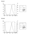


Classifications
-
- C—CHEMISTRY; METALLURGY
- C07—ORGANIC CHEMISTRY
- C07K—PEPTIDES
- C07K16/00—Immunoglobulins [IGs], e.g. monoclonal or polyclonal antibodies
- C07K16/46—Hybrid immunoglobulins
- C07K16/468—Immunoglobulins having two or more different antigen binding sites, e.g. multifunctional antibodies
-
- C—CHEMISTRY; METALLURGY
- C07—ORGANIC CHEMISTRY
- C07K—PEPTIDES
- C07K16/00—Immunoglobulins [IGs], e.g. monoclonal or polyclonal antibodies
- C07K16/18—Immunoglobulins [IGs], e.g. monoclonal or polyclonal antibodies against material from animals or humans
- C07K16/28—Immunoglobulins [IGs], e.g. monoclonal or polyclonal antibodies against material from animals or humans against receptors, cell surface antigens or cell surface determinants
- C07K16/2803—Immunoglobulins [IGs], e.g. monoclonal or polyclonal antibodies against material from animals or humans against receptors, cell surface antigens or cell surface determinants against the immunoglobulin superfamily
- C07K16/2809—Immunoglobulins [IGs], e.g. monoclonal or polyclonal antibodies against material from animals or humans against receptors, cell surface antigens or cell surface determinants against the immunoglobulin superfamily against the T-cell receptor (TcR)-CD3 complex
-
- C—CHEMISTRY; METALLURGY
- C07—ORGANIC CHEMISTRY
- C07K—PEPTIDES
- C07K16/00—Immunoglobulins [IGs], e.g. monoclonal or polyclonal antibodies
- C07K16/18—Immunoglobulins [IGs], e.g. monoclonal or polyclonal antibodies against material from animals or humans
- C07K16/28—Immunoglobulins [IGs], e.g. monoclonal or polyclonal antibodies against material from animals or humans against receptors, cell surface antigens or cell surface determinants
- C07K16/2803—Immunoglobulins [IGs], e.g. monoclonal or polyclonal antibodies against material from animals or humans against receptors, cell surface antigens or cell surface determinants against the immunoglobulin superfamily
-
- C—CHEMISTRY; METALLURGY
- C07—ORGANIC CHEMISTRY
- C07K—PEPTIDES
- C07K16/00—Immunoglobulins [IGs], e.g. monoclonal or polyclonal antibodies
- C07K16/18—Immunoglobulins [IGs], e.g. monoclonal or polyclonal antibodies against material from animals or humans
- C07K16/28—Immunoglobulins [IGs], e.g. monoclonal or polyclonal antibodies against material from animals or humans against receptors, cell surface antigens or cell surface determinants
- C07K16/2863—Immunoglobulins [IGs], e.g. monoclonal or polyclonal antibodies against material from animals or humans against receptors, cell surface antigens or cell surface determinants against receptors for growth factors, growth regulators
-
- C—CHEMISTRY; METALLURGY
- C07—ORGANIC CHEMISTRY
- C07K—PEPTIDES
- C07K16/00—Immunoglobulins [IGs], e.g. monoclonal or polyclonal antibodies
- C07K16/18—Immunoglobulins [IGs], e.g. monoclonal or polyclonal antibodies against material from animals or humans
- C07K16/28—Immunoglobulins [IGs], e.g. monoclonal or polyclonal antibodies against material from animals or humans against receptors, cell surface antigens or cell surface determinants
- C07K16/2878—Immunoglobulins [IGs], e.g. monoclonal or polyclonal antibodies against material from animals or humans against receptors, cell surface antigens or cell surface determinants against the NGF-receptor/TNF-receptor superfamily, e.g. CD27, CD30, CD40, CD95
-
- C—CHEMISTRY; METALLURGY
- C07—ORGANIC CHEMISTRY
- C07K—PEPTIDES
- C07K16/00—Immunoglobulins [IGs], e.g. monoclonal or polyclonal antibodies
- C07K16/18—Immunoglobulins [IGs], e.g. monoclonal or polyclonal antibodies against material from animals or humans
- C07K16/28—Immunoglobulins [IGs], e.g. monoclonal or polyclonal antibodies against material from animals or humans against receptors, cell surface antigens or cell surface determinants
- C07K16/2887—Immunoglobulins [IGs], e.g. monoclonal or polyclonal antibodies against material from animals or humans against receptors, cell surface antigens or cell surface determinants against CD20
-
- C—CHEMISTRY; METALLURGY
- C07—ORGANIC CHEMISTRY
- C07K—PEPTIDES
- C07K16/00—Immunoglobulins [IGs], e.g. monoclonal or polyclonal antibodies
- C07K16/18—Immunoglobulins [IGs], e.g. monoclonal or polyclonal antibodies against material from animals or humans
- C07K16/28—Immunoglobulins [IGs], e.g. monoclonal or polyclonal antibodies against material from animals or humans against receptors, cell surface antigens or cell surface determinants
- C07K16/2896—Immunoglobulins [IGs], e.g. monoclonal or polyclonal antibodies against material from animals or humans against receptors, cell surface antigens or cell surface determinants against molecules with a "CD"-designation, not provided for elsewhere
-
- C—CHEMISTRY; METALLURGY
- C07—ORGANIC CHEMISTRY
- C07K—PEPTIDES
- C07K16/00—Immunoglobulins [IGs], e.g. monoclonal or polyclonal antibodies
- C07K16/18—Immunoglobulins [IGs], e.g. monoclonal or polyclonal antibodies against material from animals or humans
- C07K16/32—Immunoglobulins [IGs], e.g. monoclonal or polyclonal antibodies against material from animals or humans against translation products of oncogenes
-
- C—CHEMISTRY; METALLURGY
- C07—ORGANIC CHEMISTRY
- C07K—PEPTIDES
- C07K16/00—Immunoglobulins [IGs], e.g. monoclonal or polyclonal antibodies
- C07K16/40—Immunoglobulins [IGs], e.g. monoclonal or polyclonal antibodies against enzymes
-
- C—CHEMISTRY; METALLURGY
- C07—ORGANIC CHEMISTRY
- C07K—PEPTIDES
- C07K16/00—Immunoglobulins [IGs], e.g. monoclonal or polyclonal antibodies
- C07K16/42—Immunoglobulins [IGs], e.g. monoclonal or polyclonal antibodies against immunoglobulins
- C07K16/4283—Immunoglobulins [IGs], e.g. monoclonal or polyclonal antibodies against immunoglobulins against an allotypic or isotypic determinant on Ig
- C07K16/4291—Immunoglobulins [IGs], e.g. monoclonal or polyclonal antibodies against immunoglobulins against an allotypic or isotypic determinant on Ig against IgE
-
- C—CHEMISTRY; METALLURGY
- C07—ORGANIC CHEMISTRY
- C07K—PEPTIDES
- C07K2317/00—Immunoglobulins specific features
- C07K2317/10—Immunoglobulins specific features characterized by their source of isolation or production
- C07K2317/14—Specific host cells or culture conditions, e.g. components, pH or temperature
-
- C—CHEMISTRY; METALLURGY
- C07—ORGANIC CHEMISTRY
- C07K—PEPTIDES
- C07K2317/00—Immunoglobulins specific features
- C07K2317/20—Immunoglobulins specific features characterized by taxonomic origin
- C07K2317/24—Immunoglobulins specific features characterized by taxonomic origin containing regions, domains or residues from different species, e.g. chimeric, humanized or veneered
-
- C—CHEMISTRY; METALLURGY
- C07—ORGANIC CHEMISTRY
- C07K—PEPTIDES
- C07K2317/00—Immunoglobulins specific features
- C07K2317/30—Immunoglobulins specific features characterized by aspects of specificity or valency
- C07K2317/31—Immunoglobulins specific features characterized by aspects of specificity or valency multispecific
-
- C—CHEMISTRY; METALLURGY
- C07—ORGANIC CHEMISTRY
- C07K—PEPTIDES
- C07K2317/00—Immunoglobulins specific features
- C07K2317/30—Immunoglobulins specific features characterized by aspects of specificity or valency
- C07K2317/33—Crossreactivity, e.g. for species or epitope, or lack of said crossreactivity
-
- C—CHEMISTRY; METALLURGY
- C07—ORGANIC CHEMISTRY
- C07K—PEPTIDES
- C07K2317/00—Immunoglobulins specific features
- C07K2317/50—Immunoglobulins specific features characterized by immunoglobulin fragments
-
- C—CHEMISTRY; METALLURGY
- C07—ORGANIC CHEMISTRY
- C07K—PEPTIDES
- C07K2317/00—Immunoglobulins specific features
- C07K2317/50—Immunoglobulins specific features characterized by immunoglobulin fragments
- C07K2317/52—Constant or Fc region; Isotype
-
- C—CHEMISTRY; METALLURGY
- C07—ORGANIC CHEMISTRY
- C07K—PEPTIDES
- C07K2317/00—Immunoglobulins specific features
- C07K2317/50—Immunoglobulins specific features characterized by immunoglobulin fragments
- C07K2317/52—Constant or Fc region; Isotype
- C07K2317/524—CH2 domain
-
- C—CHEMISTRY; METALLURGY
- C07—ORGANIC CHEMISTRY
- C07K—PEPTIDES
- C07K2317/00—Immunoglobulins specific features
- C07K2317/50—Immunoglobulins specific features characterized by immunoglobulin fragments
- C07K2317/52—Constant or Fc region; Isotype
- C07K2317/526—CH3 domain
-
- C—CHEMISTRY; METALLURGY
- C07—ORGANIC CHEMISTRY
- C07K—PEPTIDES
- C07K2317/00—Immunoglobulins specific features
- C07K2317/50—Immunoglobulins specific features characterized by immunoglobulin fragments
- C07K2317/55—Fab or Fab'
-
- C—CHEMISTRY; METALLURGY
- C07—ORGANIC CHEMISTRY
- C07K—PEPTIDES
- C07K2317/00—Immunoglobulins specific features
- C07K2317/50—Immunoglobulins specific features characterized by immunoglobulin fragments
- C07K2317/56—Immunoglobulins specific features characterized by immunoglobulin fragments variable (Fv) region, i.e. VH and/or VL
- C07K2317/565—Complementarity determining region [CDR]
-
- C—CHEMISTRY; METALLURGY
- C07—ORGANIC CHEMISTRY
- C07K—PEPTIDES
- C07K2317/00—Immunoglobulins specific features
- C07K2317/50—Immunoglobulins specific features characterized by immunoglobulin fragments
- C07K2317/56—Immunoglobulins specific features characterized by immunoglobulin fragments variable (Fv) region, i.e. VH and/or VL
- C07K2317/567—Framework region [FR]
-
- C—CHEMISTRY; METALLURGY
- C07—ORGANIC CHEMISTRY
- C07K—PEPTIDES
- C07K2317/00—Immunoglobulins specific features
- C07K2317/60—Immunoglobulins specific features characterized by non-natural combinations of immunoglobulin fragments
- C07K2317/62—Immunoglobulins specific features characterized by non-natural combinations of immunoglobulin fragments comprising only variable region components
- C07K2317/622—Single chain antibody (scFv)
-
- C—CHEMISTRY; METALLURGY
- C07—ORGANIC CHEMISTRY
- C07K—PEPTIDES
- C07K2317/00—Immunoglobulins specific features
- C07K2317/70—Immunoglobulins specific features characterized by effect upon binding to a cell or to an antigen
- C07K2317/71—Decreased effector function due to an Fc-modification
-
- C—CHEMISTRY; METALLURGY
- C07—ORGANIC CHEMISTRY
- C07K—PEPTIDES
- C07K2317/00—Immunoglobulins specific features
- C07K2317/70—Immunoglobulins specific features characterized by effect upon binding to a cell or to an antigen
- C07K2317/73—Inducing cell death, e.g. apoptosis, necrosis or inhibition of cell proliferation
-
- C—CHEMISTRY; METALLURGY
- C07—ORGANIC CHEMISTRY
- C07K—PEPTIDES
- C07K2317/00—Immunoglobulins specific features
- C07K2317/90—Immunoglobulins specific features characterized by (pharmaco)kinetic aspects or by stability of the immunoglobulin
- C07K2317/92—Affinity (KD), association rate (Ka), dissociation rate (Kd) or EC50 value
-
- C—CHEMISTRY; METALLURGY
- C07—ORGANIC CHEMISTRY
- C07K—PEPTIDES
- C07K2317/00—Immunoglobulins specific features
- C07K2317/90—Immunoglobulins specific features characterized by (pharmaco)kinetic aspects or by stability of the immunoglobulin
- C07K2317/94—Stability, e.g. half-life, pH, temperature or enzyme-resistance
Definitions
- the present invention relates to hetero-dimeric immunoglobulins that target both a component of the human CD3 antigen and a disease associated antigen and methods of making the same.
- T cell redirected killing is a desirable mode of action in many therapeutic areas.
- Various bispecific antibody formats have been shown to mediate T cell redirection both in pre-clinical and clinical investigations (May C et al., (2012) Biochem Pharmacol, 84(9): 1105-12; Frankel S R & Baeuerle P A, (2013) Curr Opin Chem Biol, 17(3): 385-92).
- All T cell retargeting bispecific antibodies or fragments thereof are engineered to have at least two antigen binding sites wherein one site binds a surface antigen on a target cell and the other site binds a T cell surface antigen.
- the human CD3 epsilon subunit from the TCR protein complex has been the most targeted to redirect T cell killing.
- bispecific antibody formats have been used to redirect T cell killing, these mainly include tandem of scFv fragments and diabody based formats with only few examples of Fc-based bispecific antibody formats reported (Moore P A et al., (2011) Blood, 117(17): 4542-51; May C et al., (2012) supra; Frankel S R & Baeuerle P A, (2013) supra). Bispecific formats that will encompass a human Fc region will have longer circulation half-lives which may result in enhanced efficacy and/or less frequent dosing regimens.
- Fc-based bispecific formats one preferred format to redirect T cell killing is the so-called heavy chain hetero-dimer format. This format is of particular interest as it does not allows aggregation of multiple copies of human CD3 molecules at the T cell surface thereby preventing any T cell inactivation (Klein C et al., (2012) MAbs, 4(6): 653-63).
- the first described method to engineer heavy chain hetero-dimers is a method known as the “knob-into-hole” method (PCT Publication No: WO199627011; Merchant A M et al., (1998) Nat Biotechnol, 16(7): 677-81).
- bispecific antibodies that redirect T cell killing via the engagement of a CD3 subunit it is essential that no homo-dimers specific for the CD3 subunit are present in the final drug product.
- traces of anti-human CD3 epsilon antibody species may trigger transient T cell activation and cytokine release before leading to T cell apoptosis thereby interfering with the goal of a controlled and specific T cell activation.
- Production of stable and safe Fc-based bispecific antibodies that efficiently redirect T cell killing remains a challenge to the pharmaceutical industry with respect to purity and yields.
- the preferred known differential Protein A affinity purification technique of the present invention corresponds to a technique wherein all three species i.e. the two homo-dimeric species and the hetero-dimer of interest differ in their total number of Protein A binding sites by at least one site and wherein one of the two homo-dimeric species has no Protein A binding site and therefore does not bind Protein A (as shown in FIG. 1 ).
- VH3 based immunoglobulins or fragments thereof are of major importance to the biological drug industry.
- Therapeutic antibodies based on the VH3 subclass have been extensively developed as these frameworks bind Protein A and facilitate the testing of antibody fragments before their formatting into immunoglobulins; for example, many synthetic antibody phage display libraries used for antibody discovery are based on the VH3 subclass.
- VH3 based antibodies are often selected for their good expression and stability over other known heavy chain variable domain subclasses.
- VH3 domain has only one Protein A binding site with a weaker affinity when compared to a Fc region which has two sites with a stronger affinity (Roben P W et al., (1995) J Immunol, 154(12): 6437-45), there is enough affinity to interfere with the known differential Protein A affinity purification techniques.
- Protein A binding is restored via the VH3 domain and the preferred technology described in FIG. 1 and above is no longer useful ( FIG. 2A ).
- abrogating Protein A binding in the VH3 based antigen binding site provides a straightforward solution and allows to keep the initial architecture of the desired hetero-dimer ( FIG. 2B ).
- the heavy chain hetero-dimer can be re-engineered to have the VH3 based antigen binding site located on the heavy chain that binds Protein A in its Fc region ( FIG.
- VH3 domain has a weaker affinity for Protein A compared to a Fc monomer hence the hetero-dimer of interest still elutes at a separate pH value from the other homo-dimeric species, typically at pH 4, while the homo-dimeric species that binds Protein A now encompasses two additional Protein A binding sites and elutes at a pH value ⁇ 3).
- the present invention provides new anti-human CD3 bispecific antibodies comprising a second binding arm which can recognise and bind to a disease associated antigen.
- a disease associated antigen means any antigen or epitope associated with a pathological state such as an oncogenic marker or a marker of some other metabolic or immunological dysfunction.
- a disease marker my also relate to an infectious disease such as a pathogenic virus or bacteria.
- the two binding arms of the anti-human CD3 bispecific antibody each comprise an immunoglobulin constant region and wherein the first arm or polypeptide binds to protein A and the second arm or polypeptide does not bind to protein A.
- the binding of the first polypeptide to protein A and the lack of binding of the second polypeptide to protein A is not intended to mean that the second polypeptide may not have some residual binding to protein A and it is instead intended that the second polypeptide binds less well to protein A in comparison to the first arm.
- the first and second polypeptides of the hetero-dimeric immunoglobulin or fragment thereof comprise an engineered immunoglobulin constant region with a modified CH3 region having a protein-protein interface that favours hetero-dimer formation over homo-dimer formation.
- the present invention provides a hetero-dimeric immunoglobulin or fragment thereof wherein the first and second polypeptides comprise an engineered immunoglobulin constant region with a modified CH3 domain having a protein-protein interface, wherein the protein-protein interface of the first polypeptide comprises an amino acid substitution at a position selected from the group consisting of: 3, 5, 7, 20, 22, 26, 27, 79, 81, 84, 84.2, 85.1, 86, 88 and 90 (IMGT® numbering), and wherein the protein-protein interface of the second polypeptide comprises an amino acid substitution at a position selected from the group consisting of: 3, 5, 7, 20, 22, 26, 27, 79, 81, 84, 84.2, 84.4, 85.1, 86, 88 and 90 (IMGT® numbering).
- the protein-protein interface of the second polypeptide comprises an amino acid substitution at position 84.4 and at least one further substitution at a position selected from the group consisting of: 3, 5, 7, 20, 22, 26, 27, 79, 81, 84, 84.2, 85.1, 86, 88 and 90 (IMGT® numbering).
- the present invention provides a hetero-dimeric immunoglobulin or fragment thereof, wherein the first and second polypeptides comprise an engineered immunoglobulin constant region with a modified CH3 domain having a protein-protein interface, wherein the protein-protein interface of the first polypeptide comprises an amino acid substitution at position 88 and at a position selected from the group consisting of: 3, 5, 7, 20, 22, 26, 27, 79, 81, 84, 84.2, 85.1, 86 and 90 (IMGT® numbering), and wherein the protein-protein interface of the second polypeptide comprises an amino acid substitution at position 85.1 and/or 86 and at a position selected from the group consisting of 3, 5, 7, 20, 22, 26, 27, 79, 81, 84, 84.2, 84.4, 88 and 90 (IMGT® numbering).
- the epitope binding region of the first polypeptide binds the CD3 protein complex and the epitope binding region of the second polypeptide binds a disease associated antigen or wherein the epitope binding region of the first polypeptide binds a disease associated antigen and the epitope binding region of the second polypeptide binds the CD3 protein complex; and wherein the epitope binding region that binds the CD3 protein complex comprises a heavy chain CDR1 comprising the amino acid sequence of SEQ ID NO: 194, a heavy chain CDR2 comprising the amino acid sequence of SEQ ID NO: 195 and a heavy chain CDR3 comprising the amino acid sequence of SEQ ID NO: 196, and a light chain CDR1 comprising the amino acid sequence of SEQ ID NO: 197, a light chain CDR2 comprising the amino acid sequence of SEQ ID NO: 198 and a light chain CDR3 comprising the amino acid sequences of: SEQ ID NO: 199; or wherein the epitope binding region that binds
- Use of these new anti-human CD3 bispecific antibodies is not limited to but includes treatments of various human cancers and autoimmune and inflammatory diseases.
- the specific destruction of cancer cells over healthy cells and tissues represents a primary objective in oncology.
- Therapeutics that could safely redirect T cell killing against tumour associated cell surface antigens may offer improved clinical efficacy.
- Known areas of clinical unmet needs in oncology include but are not limited to breast cancer, metastatic breast cancer, ovarian cancer, pancreatic cancer, lung cancer, lymphomas and multiple myeloma. Elimination of disease-causing T cells could be more beneficial than inhibiting T cell differentiation in treating autoimmune and inflammatory diseases such as psoriasis, multiple sclerosis and diabetes.
- a preferred set of disease associated antigens come from the gene products CD33, TROP2, CD105, GD2, GD3, CEA, VEGFR1, VEGFR2, NCAM, CD133, CD123, ADAM17, MCSP, PSCA, FOLR1, CD19, CD20, CD38, EpCAM, HER2, EGFR, PSMA, IgE, Integrin a4b1, CCR5, LewisY, FAP, MUC-1, Wue-1, MSP, EGFRvIII, P glycoprotein, AFP, ALK, BAGE proteins, CD30, CD40, CTLA4, ErbB3, ErbB4, Mesothelin, OX40, CA125, CAIX, CD66e, cMet, EphA2, HGF/SF, MUC1, Phosphatidylserine, TAG-72, TPBG, ⁇ -catenin, brc-abl, BRCA1, BORIS, CA9, caspase-8, CDK4, Cyclin-B1, CYP1B1, ETV6
- a hetero-dimeric immunoglobulin or fragment thereof according to the invention, wherein the epitope binding region that binds a disease associated antigen comprises heavy chain CDR1, CDR2 and CDR3 amino acid sequences and light chain CDR1, CDR2 and CDR3 amino acid sequences, respectively, selected from the group consisting of:
- the constant region of the second polypeptide of the hetero-dimeric immunoglobulin or fragment thereof comprises an IgG3 CH3 region.
- the constant region of the second polypeptide of the hetero-dimeric immunoglobulin or fragment thereof comprises a CH3 region other than that from IgG, and the non-IgG3 CH3 region comprises at least one substitution so as to decrease/abolish protein A binding.
- the epitope binding region of second polypeptide of the hetero-dimeric immunoglobulin or fragment thereof comprises a VH3 region comprising at least one modification that reduces protein A binding.
- VH3 based antigen binding sites can be readily produced and purified with a high degree of purity in a single Protein A chromatography step. These antibodies may exhibit higher efficacy over current therapies in addition to their ease of production.
- the present invention also provides a method to produce anti-human CD3 bispecific heavy chain hetero-dimers having at least one VH3 based antigen binding site from a recombinant mammalian host cell line wherein the bispecific antibody product is readily isolated after a single Protein A chromatography step with a high degree of purity.
- the modified VH3 region comprises an amino acid substitution selected from the group consisting of: 57, 65, 81, 82a and combination 19/57/59 (Kabat numbering) and even more preferably wherein the modified VH3 region comprises an amino acid substitution selected from the group consisting of: 57A, 57E, 65S, 81E, 82aS and combination 19G/57A/59A (Kabat numbering).
- the hetero-dimeric immunoglobulin or fragment thereof may comprise further substitutions wherein the heavy chain variable framework region comprises an amino acid substitution selected from the group consisting of: I34M, V48I, A49G, R58N/Y, I69L, A71T and T73K (Kabat numbering) and the light chain variable framework region comprises an amino acid substitution selected from the group consisting of: M4L, V33M, A34N, L46R, L47W, T51A, R66G, F71Y and P96F (Kabat numbering); or wherein the heavy chain variable framework region comprises the amino acid substitutions I34M, A49G and A71T (Kabat numbering) and the light chain variable framework region comprises the amino acid substitutions M4L, L46R, L47W and F71Y (Kabat numbering).
- the heavy chain variable framework region comprises an amino acid substitution selected from the group consisting of: I34M, V48I, A49G, R58N/Y, I69L, A71T and T73
- the epitope binding region that binds to the CD3 protein complex comprises a heavy chain variable framework region that is the product of or derived from the human VH3 subclass.
- the heavy chain variable framework region is the product of or derived from human IGHV3-23. More preferably, the heavy chain variable framework region is the product of or derived from human IGHV3-23*04 (SEQ ID NO: 22).
- the heavy chain variable framework region comprises at least one amino acid modification from the corresponding framework region of the heavy chain variable region of the corresponding murine antibody comprising the amino acid sequence of SEQ ID NO: 18 or SEQ ID NO: 60.
- the epitope binding region of the first polypeptide that binds to the CD3 protein complex comprises a light chain variable framework region that is the product of or derived from the human VK1 subclass or the human VK3 subclass.
- the light chain variable framework region is the product of or derived from human VK1-39 or VK3-20. More preferably the light chain variable framework region is the product of or derived from human IGKV1-39*01 (SEQ ID NO: 23) or IGKV3-20*01 (SEQ ID NO: 24).
- the light chain variable framework region comprises at least one amino acid modification from the corresponding framework region of the light chain variable region of the corresponding murine antibody comprising the amino acid sequence of SEQ ID NO: 19 or SEQ ID NO: 61.
- the epitope binding region that binds to the CD3 protein complex comprises a humanized heavy chain variable domain having the back mutations selected from the group consisting of: I34M, V48I, A49G, R58N/Y, I69L, A71T and T73K (Kabat numbering) and a humanized light chain variable domain having the back mutations selected from the group consisting of: M4L, V33M, A34N. L46R, L47W, R66G, F71Y and P96F (Kabat numbering).
- the epitope binding region that binds to the CD3 protein complex comprises a humanized heavy chain variable domain having the back mutations I34M, A49G and A71T (Kabat numbering) and a humanized light chain variable domain having the back mutations M4L, L46R, L47W and F71Y (Kabat numbering).
- the epitope binding region that binds the CD3 protein complex of the hetero-dimeric immunoglobulin or fragment thereof comprises a heavy chain variable region comprising the amino acid sequence of SEQ ID NO: 48, and a light chain variable region comprising the amino acid sequence of SEQ ID NO: 51; or wherein the epitope binding region that binds the CD3 protein complex comprises a heavy chain variable region comprising the amino acid sequence of SEQ ID NO: 49, and a light chain variable region comprising the amino acid sequence of SEQ ID NO: 51; or wherein the epitope binding region that binds the CD3 protein complex comprises a heavy chain variable region comprising the amino acid sequence of SEQ ID NO: 358, and a light chain variable region comprising the amino acid sequence of SEQ ID NO: 51; or wherein the epitope binding region that binds the CD3 protein complex comprises a heavy chain variable region comprising the amino acid sequence of SEQ ID NO: 101, and a light chain variable region comprising the amino acid sequence
- the CD3 protein complex comprises a number of subunits, for example, delta, epsilon and gamma.
- the epitope binding region that binds to the CD3 protein complex binds to the CD3 epsilon subunit.
- an epitope binding region as described herein includes the combination of one or more heavy chain variable domains and one or more complementary light chain variable domains which together form a binding site which permits the specific binding of the hetero-dimeric immunoglobulin or fragment thereof to one or more epitopes.
- the epitope binding region of the first poly peptide comprises a FAB and the epitope binding region of the second polypeptide comprises a scFv.
- the epitope binding region of the first poly peptide comprises a scFv and the epitope binding region of the second polypeptide comprises a FAB.
- the epitope binding region that binds a disease associated antigen binds to HER2.
- the epitope binding region comprises a heavy chain variable framework region that is the product of or derived from the human VH3 subclass, preferably human VH3-23, more preferably human IGHV3-23*04 (SEQ ID NO: 22), and a light chain variable framework region that is the product of or derived from the human VK1 subclass, preferably human VK1-39, more preferably human IGKV1-39*01 (SEQ ID NO: 23).
- the epitope binding region that binds the disease associated antigen HER2 comprises a heavy chain variable domain comprising the amino acid sequence of SEQ ID NO: 20 and a light chain variable domain comprising the amino acid sequence of SEQ ID NO: 21.
- the epitope binding region that binds HER2 may comprise a heavy chain variable domain and a light chain variable domain joined by a G 4 S linker forming a scFv fragment comprising the amino acid sequence of SEQ ID NO: 107.
- variable domain of the scFv fragment comprises a modification to abrogate binding to Protein A, wherein the amino acid substitution is 65S (Kabat numbering) and wherein the scFv fragment comprises the amino acid sequence of SEQ ID NO: 109 or wherein the amino acid substitution is 82aS (Kabat numbering) and wherein the scFv fragment comprises the amino acid sequence of SEQ ID NO: 111.
- Herceptin binding arm comprises a heavy chain variable region encoded by SEQ ID NO: 20 and a light chain variable region encoded by SEQ ID NO: 21.
- the epitope binding region that binds a disease associated antigen binds to CD38.
- the epitope binding region comprises a heavy chain variable framework region that is the product of or derived from the human VH3 subclass, preferably human VH3-23, more preferably human IGHV3-23*04 (SEQ ID NO: 22).
- the heavy chain variable framework region comprises at least one amino acid modification from the corresponding framework region of the heavy chain variable region of the corresponding murine antibody comprising the amino acid sequence of SEQ ID NO: 112 or 114 or 122.
- the epitope binding region further comprises a light chain variable framework region that is the product of or derived from the human VK1 subclass, preferably human VK1-39, more preferably human IGKV1-39*01 (SEQ ID NO: 23).
- the light chain variable framework region comprises at least one amino acid modification from the corresponding framework region of the light chain variable region of the corresponding murine antibody comprising the amino acid sequence of SEQ ID NO: 113 or 115 or 123.
- the CD38 binding polypeptide comprises variable heavy chain domain and variable light chain domain pair encoded by SEQ ID NOs: 116/117, 129/130, 133/134 and 135/136.
- the epitope binding region that binds a disease associated antigen binds to OX40.
- the epitope binding region comprises a heavy chain variable framework region that is the product of or derived from the human VH3 subclass, preferably human VH3-23, more preferably human IGHV3-23*04 (SEQ ID NO: 22).
- the heavy chain variable framework region comprises at least one amino acid modification from the corresponding framework region of the heavy chain variable region of the corresponding murine antibody comprising the amino acid sequence of SEQ ID NO: 139.
- the epitope binding region further comprises a light chain variable framework region that is the product of or derived from the human VK1 subclass, preferably human VK1-39, more preferably human IGKV1-39*01 (SEQ ID NO: 23).
- the light chain variable framework region comprises at least one amino acid modification from the corresponding framework region of the light chain variable region of the corresponding murine antibody comprising the amino acid sequence of SEQ ID NO: 140.
- the humanized heavy chain variable domain comprises a modification to abrogate binding to Protein A comprising the substitution G65S or the substitution N82aS (Kabat numbering).
- OX40 binding polypeptide comprises variable heavy chain domain and variable light chain domain pair encoded by SEQ ID Nos: 141/142, 278/280 and 279/281.
- the epitope binding region that binds a disease associated antigen binds to CD19.
- the epitope binding region comprises a heavy chain variable framework region that is the product of or derived from the human VH3 subclass, preferably human VH3-23, more preferably human IGHV3-23*04 (SEQ ID NO: 22) and most preferably comprises the amino acid sequence of SEQ ID NO: 296.
- the epitope binding region further comprises a light chain variable framework region that is the product of or derived from the human VK1 subclass, preferably human VK1-39, more preferably human IGKV1-39*01 (SEQ ID NO: 23) and most preferably comprises the amino acid sequence of SEQ ID NO: 297.
- the heavy chain variable domain comprises a modification to abrogate binding to Protein A comprising the substitution G65S or the substitution N82aS (Kabat numbering).
- CD19 binding polypeptide comprises variable heavy chain domain and variable light chain domain pair encoded by SEQ ID Nos: 296/297.
- the epitope binding region that binds a disease associated antigen binds to CD20.
- the epitope binding region comprises a heavy chain variable framework region that is the product of or derived from the human VH3 subclass, preferably human VH3-23, more preferably human IGHV3-23*04 (SEQ ID NO: 22).
- the heavy chain variable framework region comprises at least one amino acid modification from the corresponding framework region of the heavy chain variable region of the corresponding murine antibody comprising the amino acid sequence of SEQ ID NO: 143.
- the epitope binding region further comprises a light chain variable framework region that is the product of or derived from the human VK1 subclass, preferably human VK1-39, more preferably human IGKV1-39*01 (SEQ ID NO: 23).
- the light chain variable framework region comprises at least one amino acid modification from the corresponding framework region of the light chain variable region of the corresponding murine antibody comprising the amino acid sequence of SEQ ID NO: 144.
- the humanized heavy chain variable domain comprises a modification to abrogate binding to Protein A comprising the substitution G65S or the substitution N82aS (Kabat numbering).
- the EGFR binding polypeptide comprises variable heavy chain domain and variable light chain domain pair encoded by SEQ ID NOs: 143/144, 282/284, 283/285.
- the epitope binding region that binds a disease associated antigen binds to EGFR.
- the epitope binding region comprises a heavy chain variable framework region that is the product of or derived from the human VH3 subclass, preferably human VH3-23, more preferably human IGHV3-23*04 (SEQ ID NO: 22).
- the heavy chain variable framework region comprises at least one amino acid modification from the corresponding framework region of the heavy chain variable region of the corresponding murine antibody comprising the amino acid sequence of SEQ ID NO: 145.
- the epitope binding region further comprises a light chain variable framework region that is the product of or derived from the human VK1 subclass, preferably human VK1-39, more preferably human IGKV1-39*01 (SEQ ID NO: 23).
- the light chain variable framework region comprises at least one amino acid modification from the corresponding framework region of the light chain variable region of the corresponding murine antibody comprising the amino acid sequence of SEQ ID NO: 146.
- the humanized heavy chain variable domain comprises a modification to abrogate binding to Protein A comprising the substitution G65S or the substitution N82aS (Kabat numbering).
- the CD20 binding polypeptide comprises variable heavy chain domain and variable light chain domain pair encoded by SEQ ID NOs: 145/146, 286/288, 287/289, 290/291, 292/294.
- the epitope binding region that binds a disease associated antigen binds to IgE.
- the epitope binding region comprises a heavy chain variable framework region that is the product of or derived from the human VH3 subclass, preferably human VH3-23, more preferably human IGHV3-23*04 (SEQ ID NO: 22).
- the heavy chain variable framework region comprises at least one amino acid modification from the corresponding framework region of the heavy chain variable region of the corresponding humanized antibody comprising the amino acid sequence of SEQ ID NO: 298 or the corresponding murine antibody comprising the amino acid sequence of SEQ ID NO: 304.
- the epitope binding region further comprises a light chain variable framework region that is the product of or derived from the human VK1 subclass, preferably human VK1-39, more preferably human IGKV1-39*01 (SEQ ID NO: 23).
- the light chain variable framework region comprises at least one amino acid modification from the corresponding framework region of the light chain variable region of the corresponding humanized antibody comprising the amino acid sequence of SEQ ID NO: 299 or the corresponding murine antibody comprising the amino acid sequence of SEQ ID NO: 305.
- the heavy chain variable domain comprises a modification to abrogate binding to Protein A comprising the substitution G65S or the substitution N82aS (Kabat numbering).
- the IgE binding polypeptide comprises variable heavy chain domain and variable light chain domain pair encoded by SEQ ID NOs, 298/299, 300/302, 301/303, 304/305, 306/308, 307/309.
- Anti-CD3 antibodies have been found to trigger toxicity by both direct and indirect mechanisms. Indirect mechanisms are mediated by the Fc region of the CD3 antibody which acts with the Fc receptor expressing immune cells and lead to transient T cell activation and cytokine release. Therefore in order to improve the safety of the hetero-dimeric immunoglobulins or fragment thereof as described herein, the immunoglobulin constant region of the first and/or second polypeptide has reduced or no binding for effector immune cells and/or complement C1q.
- the immunoglobulin constant region is engineered to abrogate Fc receptor binding in the lower hinge region.
- the immunoglobulin constant region of the first and/or second polypeptide comprises the substitution(s) L234A and/or L235A (EU numbering). Most preferably, the immunoglobulin constant region of the first and/or second polypeptide comprises the substitutions L234A and L235A (EU numbering).
- the disclosure of the present invention also describes a hetero-dimeric immunoglobulin or fragment thereof wherein the epitope binding region binds to the CD3 epsilon subunit of the CD3 protein complex and comprises a FAB having a FAB thermo-stability superior to the FAB thermo-stability of the OKT3 chimera comprising a heavy chain variable domain of amino acid sequence of SEQ ID NO: 25 and a light chain variable domain of amino acid sequence of SEQ ID NO: 26, as measured by Differential Scanning Calorimetry (DSC) as described in FIG. 9 .
- DSC Differential Scanning Calorimetry
- the present invention provides a hetero-dimeric immunoglobulin or fragment thereof as described herein wherein one epitope binding region binds to the CD3 epsilon subunit of the CD3 protein complex and the other epitope binding region that binds a disease associated antigen, binds HER2.
- the potency of such a hetero-dimeric immunoglobulin or fragment thereof to redirect T-cell killing can be measured in an in vitro assay using a flow cytometry method (RDL-FACS) or a colorimetric based method (RDL-MTS) on cell lines expressing HER2 such as JIMT-1, BT-474 and MDA-MB-231, as described in the Examples.
- the hetero-dimeric immunoglobulin or fragment thereof that binds to CD3 epsilon and HER2 kills JIMT-1 cells with a potency of 21 pM or less.
- the hetero-dimeric immunoglobulin or fragment thereof also kills BT-474 cells with a potency of 2 pM or less.
- the hetero-dimeric immunoglobulin or fragment thereof also kills MDA-MB-231 cells with a potency of 0.2 nM or less.
- the cytotoxicity of all cell lines was measured in a RDL assay performed with human PBMCs at an effector:target cell ratio of 10:1 over 48 h.
- this hetero-dimeric immunoglobulin or fragment thereof shows a potent anti-tumour effect wherein tested in vivo in a JIMT-1/PBMC xenograft model.
- the hetero-dimeric immunoglobulin or fragment thereof kills JIMT-1 cells at 0.05 mg/kg in a JIMT-1 cell xenograft.
- the present invention provides hetero-dimeric immunoglobulin or fragment thereof binding to:
- the CD3 protein complex and HER2 wherein the first polypeptide has an amino acid sequence of SEQ ID NO: 159 and is assembled with a light chain of amino acid sequence of SEQ ID NO: 47 and binds CD3 epsilon, and wherein the second polypeptide has an amino acid sequence of SEQ ID NO: 160 and binds HER2;
- the CD3 protein complex and HER2 wherein the first polypeptide has an amino acid sequence of SEQ ID NO: 161 and is assembled with a cognate light chain of amino acid sequence of SEQ ID NO: 3 and binds HER2, and wherein the second polypeptide has an amino acid sequence of SEQ ID NO: 162 and binds CD3 epsilon;
- the CD3 protein complex and HER2 wherein the first polypeptide has an amino acid sequence of SEQ ID NO: 163 and is assembled with a light chain of amino acid sequence of SEQ ID NO: 47 and binds CD3 epsilon, and wherein the second polypeptide has an amino acid sequence of SEQ ID NO: 164 and binds HER2;
- the CD3 protein complex and HER2 wherein the first polypeptide has an amino acid sequence of SEQ ID NO: 165 and is assembled with a light chain of amino acid sequence of SEQ ID NO: 166 and binds CD3 epsilon, and wherein the second polypeptide has an amino acid sequence of SEQ ID NO: 167 and binds HER2;
- the CD3 protein complex and HER2 wherein the first polypeptide has an amino acid sequence of SEQ ID NO: 168 and is assembled with a light chain of amino acid sequence of SEQ ID NO: 89 and binds CD3 epsilon, and wherein the second polypeptide has an amino acid sequence of SEQ ID NO: 167 and binds HER2;
- the CD3 protein complex and CD38 wherein the first polypeptide has an amino acid sequence of SEQ ID NO: 169 and is assembled with a cognate light chain of amino acid sequence of SEQ ID NO: 119 and binds CD38, and wherein the second polypeptide has an amino acid sequence of SEQ ID NO: 162 and binds CD3 epsilon;
- the CD3 protein complex and CD38 wherein the first polypeptide has an amino acid sequence of SEQ ID NO: 170 and is assembled with a cognate light chain of amino acid sequence of SEQ ID NO: 138 and binds CD38, and wherein the second polypeptide has an amino acid sequence of SEQ ID NO: 171 and binds CD3 epsilon;
- the CD3 protein complex and CD38 wherein the first polypeptide has an amino acid sequence of SEQ ID NO: 176 and is assembled with a cognate light chain of amino acid sequence of SEQ ID NO: 119 and binds CD38, and wherein the second polypeptide has an amino acid sequence of SEQ ID NO: 177 and binds CD3 epsilon;
- the CD3 protein complex and CD38 wherein the first polypeptide has an amino acid sequence of SEQ ID NO: 178 and is assembled with a cognate light chain of amino acid sequence of SEQ ID NO: 128 and binds CD38, and wherein the second polypeptide has an amino acid sequence of SEQ ID NO: 179 and binds CD3 epsilon;
- the CD3 protein complex and OX40 wherein the first polypeptide has an amino acid sequence of SEQ ID NO: 172 and is assembled with a cognate light chain of amino acid sequence of SEQ ID NO: 173 and binds OX40, and wherein the second polypeptide has an amino acid sequence of SEQ ID NO: 162 and binds CD3 epsilon;
- the CD3 protein complex and EGFR wherein the first polypeptide has an amino acid sequence of SEQ ID NO: 174 and is assembled with a cognate light chain of amino acid sequence of SEQ ID NO: 175 and binds EGFR, and wherein the second polypeptide has an amino acid sequence of SEQ ID NO: 171 and binds CD3 epsilon;
- the CD3 protein complex and CD20 wherein the first polypeptide has an amino acid sequence of SEQ ID NO: 180 and is assembled with a cognate light chain of amino acid sequence of SEQ ID NO: 181 and binds CD20, and wherein the second polypeptide has an amino acid sequence of SEQ ID NO: 177 and binds CD3 epsilon.
- the present invention provides hetero-dimeric immunoglobulin or fragment thereof binding to: the CD3 protein complex and HER2, wherein the first polypeptide has an amino acid sequence of SEQ ID NO: 310 and is assembled with a light chain of amino acid sequence of SEQ ID NO: 3 and binds HER2, and wherein the second polypeptide has an amino acid sequence of SEQ ID NO: 311 and binds CD3 epsilon; the CD3 protein complex and CD38, wherein the first polypeptide has an amino acid sequence of SEQ ID NO: 312 and is assembled with a light chain of amino acid sequence of SEQ ID NO: 132 and binds CD38, and wherein the second polypeptide has an amino acid sequence of SEQ ID NO: 311 and binds CD3 epsilon; the CD3 protein complex and CD38, wherein the first polypeptide has an amino acid sequence of SEQ ID NO: 313 and is assembled with a light chain of amino acid sequence of SEQ ID NO: 138 and
- a Protein A based differential purification technique can be used to isolate hetero-dimeric immunoglobulins or fragments thereof encompassing the variable domain subclass of VH3, wherein the Protein A binding site in at least one but preferably both VH3 based epitope binding regions is abrogated.
- the present invention provides an in vitro method for the production of a hetero-dimeric immunoglobulin or fragment thereof as described herein, comprising the following steps: ia) preparing a DNA vector encoding a heavy chain of the first polypeptide and a DNA vector encoding a heavy chain of the second polypeptide wherein one or both DNA vectors or a third DNA vector optionally encode a common light chain or a light chain that assembles with a heavy chain of the first or second polypeptide; or ib) preparing one DNA vector encoding heavy chains of the first and second polypeptides wherein the DNA vector optionally encodes a common light chain or a light chain that assembles with a heavy chain of the first or second polypeptide; and wherein said DNA vectors are suitable for transient or stable expression in a mammalian host cell; ii) transfecting or co-transfecting the DNA vector(s) from (i) in a mammalian host cell line; iii)
- the hetero-dimeric immunoglobulin or fragment thereof found in the purified material from step (v) is at least 95% pure. More preferably the hetero-dimeric immunoglobulin or fragment thereof found in the purified material from step (v) is at least 96% pure. Even more preferably the hetero-dimeric immunoglobulin or fragment thereof found in the purified material from step (v) is at least 97%. Purity of the hetero-dimeric immunoglobulin or fragment thereof found in the purified material can be measured by capillary electrophoresis.
- a polypeptide comprising at least one CDRs from the groups: SEQ ID NOs: 224-229, 230-235 and 352-357; or combinations of heavy chain variable domain and light chain variable domain pairs selected from the group: SEQ ID NOs: 122/123, 124/125, 129/130, 135/136, 133/134 104/106; and heavy and light chain sequence pair selected from the group: 126/127 or 128, 131/132, 137/138, 359/360.
- FIG. 1 Schematic diagram of the preferred differential affinity purification technique using Protein A. None of the heavy chains encompass a VH3 based antigen binding site. Legend: [(A+)] means a functional Protein A binding site and [(A ⁇ )] means a nonfunctional Protein A binding site. pH of elution is indicated.
- FIG. 2A-F Schematic diagrams illustrating the problems faced when purifying hetero-dimers of heavy chains encompassing one or more VH3 domains using differential protein A chromatography. Examples of solutions based on mutating the Protein A binding site within at least one VH3 domain of the hetero-dimer are shown.
- FIG. 2A Problem faced when the hetero-dimer of heavy chains encompasses a VH3 domain within the heavy chain that does not bind Protein A in its Fc region.
- FIG. 2B Solution to the purification problem described in FIG. 2A , the heavy chain of the hetero-dimer that does not bind Protein A in its Fc region encompasses a VH3 domain which has been mutated to abrogate its Protein A binding site.
- FIG. 2A Problem faced when the hetero-dimer of heavy chains encompasses a VH3 domain within the heavy chain that does not bind Protein A in its Fc region.
- FIG. 2B Solution to the purification problem described in FIG. 2A , the heavy
- FIG. 2C Alternative solution to the problem described in FIG. 2A , the hetero-dimer encompasses only one VH3 domain and the hetero-dimer is engineered to have its VH3 domain located on the heavy chain that binds Protein A in its Fc region (VH3 domain relocation strategy as a solution).
- FIG. 2D Problem faced when both heavy chains of the hetero-dimer encompass a VH3 domain.
- FIG. 2E Solution to the purification problem described in FIG. 2D , the heavy chain of the hetero-dimer that does not bind Protein A in its Fc region encompasses a VH3 domain which has been mutated to abrogate its Protein A binding site.
- FIG. 2F Alternative solution to the purification problem described in FIG.
- each VH3 domain has its Protein A binding site abrogated. Boxed species indicated that these species co-elute during the differential Protein A chromatography process. pH values A and B differ by about one pH unit and allow efficient separation of the species that binds Protein A. Typically pH values for pH A and pH B are 4 and 3, respectively. Legend for all figures: [(A+)] means a functional Protein A binding site and [(A ⁇ )] means a nonfunctional Protein A binding site.
- FIG. 3 Protein A gradient mode chromatography traces for Fc 133 (HiTrapTM MabSelect SuReTM Protein A column). Plots of absorbance at 280 nm vs. total volume of mobile phase are shown as solid line. Plots of mobile phase pH and percentage of eluent buffer (B) present in mobile phase are shown as dashed and dotted-dashed lines, respectively.
- FIG. 4A-C Protein A gradient mode chromatography traces. Plots of absorbance at 280 nm vs. total volume of mobile phase are shown as solid line. Plots of mobile phase pH and percentage of eluent buffer (B) present in mobile phase are shown as dashed and dotted-dashed lines, respectively.
- FIG. 4A Anti-HER2 FAB-Fc 133 (HiTrapTM MabSelect SuReTM Protein A column).
- FIG. 4B Anti-HER2 scFv-Fc 133 (HiTrapTM MabSelect SuReTM Protein A column).
- FIG. 4C Anti-HER2 FAB (HiTrapTM MabSelect SuReTM Protein A column and HiTrapTM MabSelectTM Protein A column).
- FIG. 5 Representative amino acid sequences for each of the seven known human VII framework subclasses. Sequences were aligned according to the Kabat numbering, Positions in the human VH3-23 framework subclass that interact with the domain D of Protein A are shown in bold.
- the representative amino acid sequences for each of the seven known human VH framework subclasses are represented by the following SEQ NOs: Human IGHV1-3 (SEQ ID NO: 361), Human IGHV2-26 (SEQ ID NO: 362), Human IGHV3-23 (SEQ ID NO: 363), Human IGHV4-28 (SEQ ID NO: 364), Human IGHV5-51 (SEQ ID NO: 365), Human IGHV6-1 (SEQ ID NO: 366), Human IGHV7-4-1 (SEQ ID NO: 367).
- FIG. 6A-I Protein A gradient mode chromatography traces (HiTrapTM MabSelectTM Protein A column). Plots of absorbance at 280 nm vs. total volume of mobile phase are shown as solid line. Plots of mobile phase pH and percentage of eluent buffer (B) present in mobile phase are shown as dashed and dotted-dashed lines, respectively.
- FIG. 6A Anti-HER2 FAB.
- FIG. 6B Anti-HER2 FAB T57A.
- FIG. 6C Anti-HER2 FAB T57E.
- FIG. 6D Anti-HER2 FAB G65S.
- FIG. 6E Anti-HER2 FAB R66Q.
- FIG. 6F Anti-HER2 FAB T68V.
- FIG. 6G Anti-HER2 FAB Q81E.
- FIG. 6H Anti-HER2 FAB N82aS.
- FIG. 6I Anti-HER2 FAB R19G/T57A/Y59A.
- FIG. 7 Equilibrium dissociation constants (KD) of selected anti-HER2 FAB variants for the HER2 antigen.
- FIG. 8A-D Protein A gradient mode chromatography traces (HiTrapTM MabSelect SuReTM Protein A column). Plots of absorbance at 280 nm vs. total volume of mobile phase are shown as solid line. Plots of mobile phase pH and percentage of eluent buffer (B) present in mobile phase are shown as dashed and dotted-dashed lines, respectively.
- FIG. 8A Anti-HER2 scFv(G65S)-Fc 133.
- FIG. 8B Anti-HER2 scFv(N82aS)-Fc 133.
- FIG. 8C Anti-HER2 FAB(G65S)-Fc 133.
- FIG. 8D Anti-HER2 FAB(N82aS)-Fc 133.
- FIG. 9A-F These figures all relate to OKT3 humanization on stable human frameworks.
- FIG. 9A-C Summary of humanized candidates formatted as human IgG1 antibodies. HPB-ALL staining relative to the chimeric OKT3 antibody: ( ⁇ ) indicates no binding, (+) weaker binding, (++) moderate binding and (+++) similar binding.
- FIG. 9D DSC profiles of selected antibodies of candidates.
- FIG. 9E Summary of humanized candidates formatted as scFv-Fc fusions. HPB-ALL staining relative to the chimeric OKT3 antibody: ( ⁇ ) indicates no binding, (+) weaker binding, (++) moderate binding and (+++) similar binding.
- FIG. 9F DSC profiles of selected scFv-Fc candidates.
- FIG. 10A-B These figures all relate to SP34 humanization on stable human frameworks.
- FIG. 10A Summary of humanized candidates formatted as human IgG1 antibodies.
- FIG. 10B Summary of humanized candidates formatted as scFv-Fc fusion proteins (Fc of human IgG1 isotype).
- FIG. 11A-J These figures all relate to anti-human CD38 antibodies.
- FIG. 11A Antibody-antigen interaction measured by SPR between the chimeric HB-7 antibody and the human CD38 antigen.
- a CM5 sensor chip was covalently coupled with protein G and 200 RUs of chimeric HB-7 antibody were captured.
- Human CD38 protein human CD38 extracellular domain with a poly-histidine tag
- FIG. 11B Antibody-antigen interaction measured by SPR between the humanized HB-7 best-fit antibody and the human CD38 antigen.
- a CM5 sensor chip was covalently coupled with protein G and 200 RUs of humanized HB-7 best-fit antibody were captured.
- Human CD38 protein human CD38 extracellular domain with a poly-histidine tag
- FIG. 11C Antibody-antigen interaction measured by SPR between the humanized 9G7 best-fit antibody and the human CD38 antigen.
- a CM5 sensor chip was covalently coupled with protein G and 200 RUs of humanized 9G7 best-fit antibody were captured.
- Human CD38 protein (human CD38 extracellular domain with a poly-histidine tag) was injected at 25, 12.5, 6.25, 3.12, 1.56, 0.78, 0.39, 0.19, and 0.1 nM at a flow rate of 30 ⁇ l/min in HBS-P.
- FIG. 11D Antibody-antigen interaction measured by SPR between the humanized 9G7 best-framework antibody and the human CD38 antigen.
- a CM5 sensor chip was covalently coupled with protein G and 200 RUs of humanized 9G7 best-framework antibody were captured.
- Human CD38 protein (human CD38 extracellular domain with a poly-histidine tag) was injected at 50, 25, 12.5, 6.25, 3.12, 1.56, 0.78, 0.39, 0.19, and 0.1 nM at a flow rate of 30 ⁇ l/min in HBS-P.
- FIG. 11E Antibody-antigen interaction measured by SPR between the human 767 antibody and the human CD38 antigen.
- a CM5 sensor chip was covalently coupled with protein G and 200 RUs of human 767 antibody were captured.
- Human CD38 protein (human CD38 extracellular domain with a poly-histidine tag) was injected at 500, 250, 125, 62.5, 31.25, and 15.6 nM at a flow rate of 30 ⁇ l/min in HBS-P.
- FIG. 11F DSC profiles of chimeric HB-7 and humanized HB-7 best-fit antibodies.
- FIG. 11G DSC profiles of chimeric 9G7 and humanized 9G7 best-fit antibodies.
- FIG. 11H DSC profiles of humanized 9G7 best-framework antibody.
- FIG. 11I DSC profiles of human clone 767 antibody.
- FIG. 11J summary table for the 9G7 humanized antibodies.
- FIG. 12A-C Schematic diagram of the BEAT HER2/CD3 antibodies in alternative formats.
- FIG. 12A BEAT HER2/CD3-1 (format A) and BEAT HER2/CD3-2 (format B) antibodies.
- FIG. 12B BEAT HER2/CD3-3 (format C) and BEAT HER2/CD3(SP34) (format D) antibodies.
- FIG. 12C BEAT HER2/CD3(SP34-Kappa1) (format E) antibody.
- [(A+)] means functional Protein A binding site.
- [(A ⁇ )] means nonfunctional Protein A binding site.
- FIG. 13 Protein A purification profile of BEAT HER2/CD3-1 antibody (Absorbance trace at 280 nm).
- Neutralization 1/10 vol. of 1M Tris pH 8.
- FIG. 14 Capillary Electrophoresis profile of BEAT HER2/CD3-1 antibody preparations.
- FIG. 15A SDS-PAGE analysis of N82aS substituted BEAT HER2/CD3-1 antibody.
- FIG. 15B SDS-PAGE analysis of N82aS non substituted BEAT HER2/CD3-1 antibody variant. Legend: [(A+)] means a functional Protein A binding site and [(A ⁇ )] means a nonfunctional Protein A binding site. pH of elution is indicated.
- FIG. 16A Antibody-antigen interaction measured by SPR between the BEAT HER2/CD3-1 antibody and the human CD3 epsilon antigen.
- a CM5 sensor chip was covalently coupled with 7400 RUs of the human CD3 gamma-epsilon-Fc fusion protein.
- BEAT HER2/CD3-1 antibody was injected at 5000, 2500, 1250, 625, 312.5 and 156.25 nM at a flow rate of 10 ⁇ l/min in HBS-P. Data are expressed as number of response units (abbreviated RU; Y axis) vs. time (X axis). Affinity was obtained from a plot of the equilibrium response (Req) vs.
- FIG. 16B Antibody-antigen interaction measured by SPR between the BEAT HER2/CD3-1 antibody and the human HER2 antigen.
- a CM5 sensor chip was covalently coupled protein G and 150 RUs of BEAT HER2/CD3-1 antibody were captured.
- HER2-his was injected at 1000, 333, 111, 37, 12, 4.1, 1.4, 0.5 and 0.15 nM at a flow rate of 30 ⁇ l/min in HBS-P. Data are expressed as number of response units (abbreviated RU; Y axis) vs. time (X axis).
- FIG. 16C DSC profiles of BEAT HER2/CD3-1 and -2 antibodies shown in profiles A and B, respectively.
- FIG. 17A-G Examples of T cell redirected killing by the BEAT HER2/CD3 antibodies.
- Readout RDL-MTS method.
- Effector cells human PBMCs. Effector cells-to-targeted cells ratio of 10:1. Means of three donors with 48 h incubation. Antibody concentrations are shown in nM.
- FIG. 17A BEAT HER2/CD3-1 and BEAT HER2/CD3-2 antibodies, target cells: BT-474.
- FIG. 17B BEAT HER2/CD3-1 and BEAT HER2/CD3-2 antibodies, target cells: JIMT-1.
- FIG. 17C BEAT HER2/CD3-1 and BEAT HER2/CD3-2 antibodies, target cells: MDA-MB-231.
- FIG. 17D BEAT HER2/CD3(SP34) antibody, target cells: NCI-N87.
- FIG. 17E BEAT HER2/CD3(SP34) antibody, target cells: HT-1080.
- FIG. 17F BEAT HER2/CD3(SP34-Kappa1) antibody, target cells: NCI-N87.
- FIG. 17G BEAT HER2/CD3(SP34-Kappa1) antibody, target cells: HT-1080.
- FIG. 18A-C JIMT-1 xenografts with human PBMC supplementation.
- FIG. 18A Human PBMC's do not interfere with tumor growth.
- FIG. 18B-C Tumor volumes (mm 3 ) for BEAT HER2/CD3-1 treated and non-treated mice, four human PBMC donors, cohorts of five mice.
- FIG. 19 Schematic diagram of the BEAT CD38-HB7bestfit/CD3 (format A) and BEAT CD38-767/CD3 (format B) antibodies.
- [(A+)] means functional Protein A binding site.
- [(A ⁇ )] means nonfunctional Protein A binding site.
- FIG. 20A Antibody-antigen interaction measured by SPR between the BEAT CD38-HB7bestfit/CD3 antibody and the human CD38 antigen.
- a CM5 sensor chip was covalently coupled with protein G and 200 RUs of BEAT CD38-HB7bestfit/CD3 antibody were captured.
- Human CD38 protein poly-histidine tagged protein
- RU response units
- Y axis time
- X axis time
- FIG. 21 Example of T cell redirected killing by the BEAT CD38-HB7bestfit/CD3 antibody.
- Readout RDL-FACS method. Effector cells: purified human T cells. Effector cells-to-targeted cells ratio of 10:1. Mean of two donors with 48 h incubation. Target cells: RPMI 8226. Antibody concentration is shown in nM.
- FIG. 22 Example of T cell redirected killing by the BEAT CD38-767/CD3(SP34) antibody.
- Readout RDL-FACS method.
- Effector cells human PBMC's. Effector cells-to-targeted cells ratio of 10:1. Mean of three donors with 24 h incubation.
- Target cells Daudi.
- Antibody concentration is shown in nM.
- FIG. 23 Schematic diagram of the BEAT OX40/CD3antibody. Legend: [(A+)] means functional Protein A binding site. [(A ⁇ )] means nonfunctional Protein A binding site.
- FIG. 24 Example of T cell redirected killing by the BEAT OX40/CD3antibody.
- Readout RDL-MTS method.
- Effector cells Human PBMCs. Effector cells-to-targeted cells ratio of 20:1. Mean of three donors with 48 h incubation.
- Target cells recombinant stable CHO[OX40] cells. Antibody concentration is shown in nM.
- FIG. 25 Schematic diagram of the BEAT EGFR/CD3 antibody. Legend: [(A+)] means functional Protein A binding site. [(A ⁇ )] means nonfunctional Protein A binding site.
- FIG. 26 Example of T cell redirected killing by the BEAT EGFR/CD3antibody.
- Readout RDL-MTS method.
- Effector cells Human PBMCs. Effector cells-to-targeted cells ratio of 10:1. Mean of four donors with 48 h incubation.
- Target cells HT-29 cells. Antibody concentration is shown in nM.
- FIG. 27 Schematic diagram of the BEAT CD38-HB7bestfit/CD3(SP34) (format A) and BEAT CD38-9G7bestfit/CD3(SP34-Kappa2) (format B) antibodies. [(A+)] means functional Protein A binding site.
- FIG. 28 Example of T cell redirected killing by the BEAT CD38-HB7bestfit/CD3(SP34) antibody.
- Readout RDL-FACS method.
- Effector cells Human PBMCs. Effector cells-to-targeted cells ratio of 10:1. Mean of three donors with 24 h incubation.
- Target cells Daudi cells. Antibody concentration is shown in nM.
- FIG. 29 Antibody-antigen interaction measured by SPR between the BEAT CD38-9G7bestfit/CD3(SP34-Kappa2) antibody and the human CD3 epsilon 1-26_Fc fusion protein.
- a CM5 sensor chip was covalently coupled with 500 RUs of the human CD3 epsilon 1-26_Fc fusion protein.
- BEAT CD38-9G7bestfit/CD3(SP34-Kappa2) antibody was injected at 50, 25, 12.5, 6.2, 3.1, 0.8 and 0.4 nM at a flow rate of 30 ⁇ l/min in HBS-P. Data are expressed as number of response units (abbreviated RU; Y axis) vs. time (X axis).
- FIG. 30 Example of T cell redirected killing by the BEAT CD38/CD3(SP34-Kappa2) antibody.
- Readout RDL-FACS method.
- Effector cells Human PBMCs. Effector cells-to-targeted cells ratio of 10:1. Mean of three donors with 24 h incubation.
- Target cells Daudi cells. Antibody concentration is shown in nM.
- FIG. 31 Schematic diagram of the BEAT CD20/CD3(SP34) antibody. [(A+)] means functional Protein A binding site.
- FIG. 32 Example of T cell redirected killing by the BEAT CD20/CD3(SP34) antibody.
- Readout RDL-FACS method.
- Effector cells Human PBMCs. Effector cells-to-targeted cells ratio of 10:1. Means of three donors with 24 h incubation.
- Target cells Daudi cells. Antibody concentration is shown in nM.
- the present invention relates generally to novel hetero-dimeric immunoglobulins that bind to the CD3 protein complex and a disease associated antigen. Furthermore, these hetero-dimeric immunoglobulins have reduced or eliminated binding to protein A and therefore can be purified to a very high degree of purity using affinity chromatography.
- polypeptide and protein refer to a polymer of amino acid residues wherein amino acids are combined via peptide bonds to form a chain of amino acids that have been linked together via dehydration synthesis.
- Polypeptides and proteins can be synthesized through chemical synthesis or recombinant expression and are not limited to a minimum amino acid length.
- the group of polypeptides comprises “proteins” as long as the proteins consist of a single polypeptide chain.
- Polypeptides may further form multimers such as dimers, trimers and higher oligomers, i.e. consisting of more than one polypeptide molecule.
- Polypeptide molecules forming such dimers, trimers etc. may be identical or non-identical.
- the corresponding higher order structures of such multimers are, consequently, termed homo- or hetero-dimers, homo- or hetero-trimers etc.
- An example for a hetero-multimer is an antibody molecule, which, in its naturally occurring form, consists of two identical light polypeptide chains and two identical heavy polypeptide chains.
- polypeptide and protein also refer to naturally modified polypeptides/proteins wherein the modification is effected e.g. by post-translational modifications like glycosylation, acetylation, phosphorylation and the like. Such modifications are well known in the art.
- a “polypeptide” refers to a protein which includes modifications, such as deletions, additions and substitutions (which can be conservative in nature) to the native sequence. These modifications may be deliberate, as through site-directed mutagenesis, or may be accidental, such as through mutations of hosts which produce the proteins or errors due to PCR amplification.
- CD3 complex refers to the protein complex known as the CD3 (cluster of differentiation 3) T-cell co-receptor (Wucherpfennig K W et al., (2010) Cold Spring Harb Perspect Biol, 2(4): a005140).
- the CD3 protein complex is composed of four distinct chains. In mammals, the complex contains a CD3 ⁇ chain, a CD3 ⁇ chain, and two CD3 ⁇ chains. These chains associate with a molecule known as the T-cell receptor (TCR) and the ⁇ -chain to generate an activation signal in T lymphocytes (van der Merwe P A & Dushek O (2011) Nat Rev Immunol, 11(1): 47-55).
- TCR T-cell receptor
- the TCR, ⁇ -chain, and CD3 molecules together comprise the TCR complex.
- the CD3 ⁇ , CD3 ⁇ , and CD3 ⁇ chains are highly related cell-surface proteins of the immunoglobulin superfamily containing a single extracellular immunoglobulin domain.
- the intracellular tails of the CD3 molecules contain a single conserved motif known as an immunoreceptor tyrosine-based activation motif or ITAM for short, which is essential for the signalling capacity of the TCR. Since CD3 is required for T-cell activation, drugs (often monoclonal antibodies) that target CD3 have and are being investigated as immunosuppressant therapies.
- disease associated antigen refers to molecules that are involved in a disease process. Examples of disease associated antigens are found in a broad range of therapeutic areas such as inflammation, cancer and autoimmune diseases. In oncology, disease associated antigens are molecules that can broadly be used for the screening and/or monitoring and/or therapeutic targeting of cancers within a patient population, for example EpCAM antigen in prostate cancer. Tumour antigens can be produced directly by the tumour or by non-tumour cells as a response to the presence of a tumour and preferred tumour antigens are cell-surface molecules. Inflammatory disease associated antigens are known, which include but are not limited to, pro-inflammatory cytokines such as TNF- ⁇ and IL-1. Autoimmune disease associated antigens are also known; examples of these include but are not limited to antibodies against double-stranded DNA in systemic lupus erythematosus and amyloid beta peptide in Alzheimers disease.
- Immunoglobulin as referred to herein can be used interchangeably with the term “antibody”. Immunoglobulin includes full-length antibodies and any antigen binding fragment or single chains thereof. Immunoglobulins can be homo-dimeric or hetero-dimeric. Immunoglobulins and specifically naturally occurring antibodies are glycoproteins which exist as one or more copies of a Y-shaped unit, composed of four polypeptide chains. Each “Y” shape contains two identical copies of a heavy (H) chain and two identical copies of a light (L) chain, named as such by their relative molecular weights. Each light chain pairs with a heavy chain and each heavy chain pairs with another heavy chain. Covalent interchain disulfide bonds and non-covalent interactions link the chains together.
- Immunoglobulins and specifically naturally occurring antibodies contain variable regions, which are the two copies of the antigen binding site.
- a Fab fragment consists of the entire light chain and part of the heavy chain.
- the heavy chain contains one variable region (VH) and either three or four constant regions (CH1, CH2, CH3 and CH4, depending on the antibody class or isotype).
- the region between the CH1 and CH2 regions is called the hinge region and permits flexibility between the two Fab arms of the Y-shaped antibody molecule, allowing them to open and close to accommodate binding to two antigenic determinants separated by a fixed distance.
- the “hinge region” as referred to herein is a sequence region of 6-62 amino acids in length, only present in IgA, IgD and IgG, which encompasses the cysteine residues that bridge the two heavy chains.
- the heavy chains of IgA, IgD and IgG each have four regions, i.e. one variable region (VH) and three constant regions (CH1-3).
- IgE and IgM have one variable and four constant regions (CH1-4) on the heavy chain.
- the constant regions of the immunoglobulins may mediate the binding to host tissues or factors, including various cells of the immune system (e.g., effector cells) and the first component (C1q) of the complement system classical pathway.
- Each light chain is usually linked to a heavy chain by one covalent disulfide bond.
- Each light chain contains one variable region (VL) and one light chain constant region.
- the light chain constant region is a kappa light chain constant region designated herein as IGKC or is a lambda light chain constant region designated herein as IGLC.
- IGKC is used herein equivalently to CK or CK and has the same meaning.
- IGLC is used herein equivalently to CA or CL and has the same meaning.
- an IGLC region refers to all lambda light chain constant regions e.g. to all lambda light chain constant regions selected from the group consisting of IGLC1, IGLC2, IGLC3, IGLC6 and IGLC7.
- the VH and VL regions can be further subdivided into regions of hypervariability, termed complementarity determining regions (CDR), interspersed with regions that are more conserved, termed framework regions (FR or FW).
- CDR complementarity determining regions
- FR or FW framework regions
- Each VH and VL is composed of three CDRs and four FRs, arranged from amino-terminus to carboxy-terminus in the following order: FR1, CDR1, FR2, CDR2, FR3, CDR3, FR4.
- variable regions of the heavy and light chains contain an epitope-binding region that interacts with an antigen.
- Engineered immunoglobulins can encompass different epitope binding region formats such as scFv, FAB or dAb fragments. These fragments are usually assembled in an antibody-like structure by genetic fusion to a IgG Fc region.
- Engineered immunoglobulins can be constructed as homo or hetero-dimers with or without the use of hetero-dimerization enhancing techniques, and can have mono- or bispecific binding properties.
- full length antibody as used herein includes the structure that constitutes the natural biological form of an antibody, including variable and constant regions.
- the full length antibody of the IgG class is a tetramer and consists of two identical pairs of two immunoglobulin chains, each pair having one light and one heavy chain, each light chain comprising immunoglobulin regions VL and a light chain constant region, and each heavy chain comprising immunoglobulin regions VH, CH1 (C ⁇ 1), CH2 (C ⁇ 2), CH3 (C ⁇ 3) and CH4 (C ⁇ 4), depending on the antibody class or isotype).
- IgG antibodies may consist of only two heavy chains, each heavy chain comprising a variable region attached to the Fc region.
- Antibodies are grouped into classes, also referred to as isotypes, as determined genetically by the constant region.
- Human constant light chains are classified as kappa (CK) and lambda (C ⁇ ) light chains.
- Heavy chains are classified as mu ( ⁇ ), delta ( ⁇ ), gamma ( ⁇ ), alpha ( ⁇ ), or epsilon ( ⁇ ) and define the antibody's isotype as IgM, IgD, IgG, IgA and IgE, respectively.
- isotype as used herein is meant any of the classes and/or subclasses of immunoglobulins defined by the chemical and antigenic characteristics of their constant regions.
- the known human immunoglobulin isotypes are IGHG1 (IgG1), IGHG2 (IgG2), IGHG3 (IgG3), IGHG4 (IgG4), IGHA1 (IgA1), IGHA2 (IgA2), IGHM (IgM), IGHD (IgD) and IGHE (IgE).
- the so-called human immunoglobulin pseudo-gamma IGHGP gene represents an additional human immunoglobulin heavy constant region gene which has been sequenced but does not encode a protein due to an altered switch region (Bensmana M et al., (1988) Nucleic Acids Res, 16(7): 3108).
- the human immunoglobulin pseudo-gamma IGHGP gene has open reading frames for all heavy constant regions (CH1-CH3) and hinge. All open reading frames for its heavy constant regions encode protein regions which align well with all human immunoglobulin constant regions with the predicted structural features.
- This additional pseudo-gamma isotype is referred herein as IgGP or IGHGP.
- Other pseudo immunoglobulin genes have been reported such as the human immunoglobulin heavy constant region epsilon P1 and P2 pseudo-genes (IGHEP1 and IGHEP2).
- the IgG class is the most commonly used for therapeutic purposes. In humans this class comprises subclasses IgG1, IgG2, IgG3 and IgG4. In mice this class comprises subclasses IgG1, IgG2a, IgG2b. IgG2c and IgG3.
- Immunoglobulin fragments include, but is not limited to, (i) a region including for example a CH1, a CH2 or a CH3 region, (ii) the Fab fragment consisting of VL, VH, CL or CK and CH1 regions, including Fab′ and Fab′-SH, (ii) the Fd fragment consisting of the VH and CH1 regions, (iii) the dAb fragment (Ward E S et al., (1989) Nature, 341(6242): 544-6) which consists of a single variable region (iv) F(ab′) 2 fragments, a bivalent fragment comprising two linked Fab fragments (v) single chain Fv fragments (scFv), wherein a VH region and a VL region are linked by a peptide linker which allows the two regions to associate to form an antigen binding site (Bird R E et al., (1988) Science, 242(4877): 42
- variable region refers to the regions or domains that mediates antigen-binding and defines specificity of a particular antibody for a particular antigen.
- the antigen-binding site consists of two variable regions that define specificity: one located in the heavy chain, referred herein as heavy chain variable region (VH) and the other located in the light chain, referred herein as light chain variable region (VL).
- VH heavy chain variable region
- VL light chain variable region
- the heavy chain variable region (VH) can be divided into seven subgroups or subclasses: VH1, VH2, VH3, VH4, VH5, VH6 and VH7.
- specificity may exclusively reside in only one of the two regions as in single-domain antibodies from heavy-chain antibodies found in camelids.
- the V regions are usually about 110 amino acids long and consist of relatively invariant stretches of amino acid sequence called framework regions (FRs or “non-CDR regions”) of 15-30 amino acids separated by shorter regions of extreme variability called “hypervariable regions” that are 7-17 amino acids long.
- the variable domains of native heavy and light chains comprise four FRs, largely adopting a beta-sheet configuration, connected by three hypervariable regions, which form loops.
- the hypervariable regions in each chain are held together in close proximity by FRs and, with the hypervariable regions from the other chain, contribute to the formation of the antigen binding site of antibodies (see Kabat E A et al., supra.).
- hypervariable region refers to the amino acid residues of an antibody which are responsible for antigen binding.
- the hypervariable region generally comprises amino acid residues from a “complementary determining region” or “CDR”, the latter being of highest sequence variability and/or involved in antigen recognition.
- CDR complementary determining region
- CDR definitions are in use and are encompassed herein.
- the Kabat definition is based on sequence variability and is the most commonly used (Kabat E A el al., supra.). Chothia refers instead to the location of the structural loops (Chothia & Lesk J. (1987) Mol Biol, 196: 901-917).
- the AbM definition is a compromise between the Kabat and the Chothia definitions and is used by Oxford Molecular's AbM antibody modelling software (Martin A C R et al., (1989) Proc Natl Acad Sci USA 86:9268-9272; Martin A C R et al., (1991) Methods Enzymol, 203: 121-153; Pedersen J T et al., (1992) Immunomethods, 1: 126-136; Rees A R et al., (1996) In Sternberg M. J. E. (ed.), Protein Structure Prediction. Oxford University Press, Oxford, 141-172).
- the contact definition has been recently introduced (MacCallum R M et al., (1996) J Mol Biol, 262: 732-745) and is based on an analysis of the available complex structures available in the Protein Databank.
- the definition of the CDR by IMGT®, the international ImMunoGeneTics information system is based on the IMGT numbering for all immunoglobulin and T cell receptor V-REGIONs of all species (IMGT®, the international ImMunoGeneTics information System®; Lefranc M P et al., (1999) Nucleic Acids Res, 27(1): 209-12; Ruiz M et al., (2000) Nucleic Acids Res, 28(1): 219-21; Lefranc M P (2001) Nucleic Acids Res, 29(1): 207-9; Lefranc, M P (2003) Nucleic Acids Res, 31(1): 307-10; Lefrane M P et al., (2005) Dev Comp Immunol, 29(3): 185-203;
- CDRs Complementarity Determining Regions
- the “non-CDR regions” of the variable domain are known as framework regions (FR).
- the “non-CDR regions” of the VL region as used herein comprise the amino acid sequences: 1-23 (FR1), 35-49 (FR2), 57-88 (FR3) and 99-107 (FR4).
- the “non-CDR regions” of the VH region as used herein comprise the amino acid sequences: 1-25 (FR1), 36-49 (FR2), 66-94 (FR3) and 103-113 (FR4).
- the CDRs of the present invention may comprise “extended CDRs” which are based on the aforementioned definitions and have variable domain residues as follows: LCDR1: 24-36, LCDR2: 46-56, LCDR3:89-97, HCDR1: 26-35, HCDR2:47-65, HCDR3: 93-102. These extended CDRs are numbered as well according to Kabat et al., supra.
- the “non-extended CDR region” of the VL region as used herein comprise the amino acid sequences: 1-23 (FR1), 37-45 (FR2), 57-88 (FR3) and 98—approximately 107 (FR4).
- the “non-extended CDR region” of the V H region as used herein comprise the amino acid sequences: 1-25 (FR1), 37-46 (FR2), 66-92 (FR3) and 103—approximately 113 (FR4).
- Fab or “FAB” or “Fab region” or “FAB region” as used herein includes the polypeptides that comprise the VH, CH1, VL and light chain constant immunoglobulin regions. Fab may refer to this region in isolation, or this region in the context of a full length antibody or antibody fragment.
- Fc or “Fc region”, as used herein includes the polypeptide comprising the constant region of an antibody heavy chain excluding the first constant region immunoglobulin region.
- Fc refers to the last two constant region immunoglobulin regions of IgA, IgD and IgG or the last three constant region immunoglobulin regions of IgE and IgM, and the flexible hinge N-terminal to these regions.
- Fc may include the J chain.
- Fc comprises immunoglobulin regions Cgamma2 and Cgamma3 (C ⁇ 2 and C ⁇ 3) and the hinge between Cgamma1 (C ⁇ 1) and Cgamma2 (C ⁇ 2).
- the human IgG heavy chain Fc region is usually defined to comprise residues C226 or P230 to its carboxyl-terminus, wherein the numbering is according to the EU index.
- Fc may refer to this region in isolation or this region in the context of an Fc polypeptide, for example an antibody.
- immunoglobulin constant region refers to immunoglobulin or antibody heavy chain constant regions from human or animal species and encompasses all isotypes.
- immunoglobulin constant regions are of human origin and are selected from the group consisting of, but not limited to: IGHG1 CH1, IGHG2 CH1, IGHG3 CH1, IGHG4 CH1, IGHA1 CH1, IGHA2 CH1, IGHE CH1, IGHEP1 CH1, IGHM CH1, IGHD CH1, IGHGP CH1, IGHG1 CH2, IGHG2 CH2, IGHG3 CH2, IGHG4 CH2, IGHA1 CH2, IGHA2 CH2, IGHE CH2, IGHEP1 CH2, IGHM CH2, IGHD CH2, IGHGP CH2, IGHG1 CH3, IGHG2 CH3, IGHG3 CH3, IGHG4 CH3, IGHA1 CH3, IGHA2 CH3, IGHEP1 CH3, IGHM CH3, IGHD CH3,
- Preferred “immunoglobulin constant regions” are selected from the group consisting of human IGHE CH2, IGHM CH2, IGHG1 CH3, IGHG2 CH3, IGHG3 CH3, IGHG4 CH3, IGHA1 CH3, IGHA2 CH3, IGHE CH3, IGHM CH3, IGHD CH3 and IGHGP CH3. More preferred “immunoglobulin constant regions” are selected from the group consisting of human IGHG1 CH3, IGHG2 CH3, IGHG3 CH3. IGHG4 CH3, IGHA1 CH3, IGHA2 CH3, IGHE CH3. IGHM CH3, IGHD CH3 and IGHGP CH3.
- epitope binding region includes a polypeptide or a fragment thereof having minimal amino acid sequence to permit the specific binding of the immunoglobulin molecule to one or more epitopes.
- Naturally occurring antibodies have two epitope binding regions which are also known as antigen binding or combining sites or paratopes.
- Epitope binding regions in naturally occurring antibodies are confined within the CDR regions of the VH and/or VL domains wherein the amino acid mediating epitope binding are found.
- VH domains or VL domains or fragments thereof and combinations thereof can be engineered to provide epitope binding regions (Holt L J et al., (2003) Trends Biotechnol, 21(11): 484-490; Polonelli L et al., (2008) PLoS ONE, 3(6): e2371).
- non-immunoglobulin based epitope binding regions can be found in artificial protein domains used as “scaffold” for engineering epitope binding regions (Binz H K et al., (2005) Nat Biotechnol, 23(10): 1257-1268) or peptide mimetics (Murali R & Greene M I (2012) Pharmaceuticals, 5(2): 209-235).
- epitope binding region includes the combination of one or more heavy chain variable domains and one or more complementary light chain variable domains which together forms a binding site which permits the specific binding of the immunoglobulin molecule to one or more epitopes.
- Examples of an epitope binding region as exemplified in the present invention include scFv and FAB.
- epitope includes a fragment of a polypeptide or protein or a non-protein molecule having antigenic or immunogenic activity in an animal, preferably in a mammal and most preferably in a human.
- An epitope having immunogenic activity is a fragment of a polypeptide or protein that elicits an antibody response in an animal.
- An epitope having antigenic activity is a fragment of a polypeptide or protein to which an antibody or polypeptide specifically binds as determined by any method well-known to one of skill in the art, for example by immunoassays. Antigenic epitopes need not necessarily be immunogenic.
- epitope refers to a polypeptide sequence of at least about 3 to 5, preferably about 5 to 10 or 15 and not more than about 1,000 amino acids (or any integer there between), which define a sequence that by itself or as part of a larger sequence, binds to an antibody generated in response to such sequence.
- the length of the fragment may comprise nearly the full-length of the protein sequence, or even a fusion protein comprising one or more epitopes.
- An epitope for use in the subject invention is not limited to a polypeptide having the exact sequence of the portion of the parent protein from which it is derived.
- epitope encompasses sequences identical to the native sequence, as well as modifications to the native sequence, such as deletions, additions and substitutions (generally conservative in nature).
- the epitopes of protein antigens are divided into two categories, conformational epitopes and linear epitopes, based on their structure and interaction with the epitope binding site (Goldsby R et al., (2003) “Antigens (Chapter 3)” Immunology (Fifth edition ed.), New York: W. H. Freeman and Company. pp. 57-75, ISBN 0-7167-4947-5).
- a conformational epitope is composed of discontinuous sections of the antigen's amino acid sequence.
- epitopes interact with the paratope based on the 3-D surface features and shape or tertiary structure of the antigen. Most epitopes are conformational. By contrast, linear epitopes interact with the paratope based on their primary structure. A linear epitope is formed by a continuous sequence of amino acids from the antigen.
- hetero-dimeric immunoglobulin or “hetero-dimeric fragment” or “hetero-dimer” or “hetero-dimer of heavy chains” as used herein includes an immunoglobulin molecule or part of comprising at least a first and a second polypeptide, like a first and a second region, wherein the second polypeptide differs in amino acid sequence from the first polypeptide.
- a hetero-dimeric immunoglobulin comprises two polypeptide chains, wherein the first chain has at least one non-identical region to the second chain, and wherein both chains assemble, i.e. interact through their non-identical regions.
- hetero-dimeric immunoglobulin has binding specificity for at least two different ligands, antigens or binding sites, i.e. is bispecific.
- Hetero-dimeric immunoglobulin as used herein includes but is not limited to full length bispecific antibodies, bispecifc Fab, bispecifc F(ab′) 2 , bispecific scFv fused to an Fc region, diabody fused to an Fc region and domain antibody fused to an Fc region.
- homo-dimeric immunoglobulin or “homo-dimeric fragment” or “homo-dimer” or “homo-dimer of heavy chains” as used herein includes an immunoglobulin molecule or part of comprising at least a first and a second polypeptide, like a first and a second region, wherein the second polypeptide is identical in amino acid sequence to the first polypeptide.
- a homo-dimeric immunoglobulin comprises two polypeptide chains, wherein the first chain has at least one identical region to the second chain, and wherein both chains assemble, i.e. interact through their identical regions.
- a homo-dimeric immunoglobulin fragment comprises at least two regions, wherein the first region is identical to the second region, and wherein both regions assemble, i.e. interact through their protein-protein interfaces.
- IMGT® immunoglobulin constant regions
- CH1, CH2, CH3 immunoglobulin heavy chain constant regions selected from the group consisting of IGHG1, IGHG2, IGHG3 and IGHG4 numbering can be according to the “EU numbering system” (Edelman G M et al., (1969) Proc Natl Acad Sci USA, 63(1): 78-85).
- EU numbering system Edelman G M et al., (1969) Proc Natl Acad Sci USA, 63(1): 78-85.
- a complete correspondence for the human CH1, hinge, CH2 and CH3 constant regions of IGHG1 can be found at the IMGT database (IMGT®; supra).
- IGKC human kappa immunoglobulin light chain constant region
- IGLC1, IGLC2, IGLC3, IGLC6 and IGLC7 For the human lambda immunoglobulin light chain constant regions (IGLC1, IGLC2, IGLC3, IGLC6 and IGLC7), numbering can be according to the “Kabat numbering system” (Kabat E A et al., supra). A complete correspondence for human IGLC regions can be found at the IMGT database (IMGT®; supra).
- the human IGHG1 immunoglobulin heavy chain constant regions as referred to herein have the following region boundaries: CH1 region (EU numbering: 118-215), Hinge ⁇ 1 region (EU numbering: 216-230), CH2 region (EU numbering: 231-340) and CH3 region (EU numbering: 341-447).
- the human CK region referred herein spans residues 108 to 214 (EU numbering).
- the human IGLC1, IGLC2, IGLC3, IGLC6 and IGLC7 regions referred herein span residues 108-215 (Kabat numbering).
- amino acid or “amino acid residue” as used herein includes natural amino acids as well as non-natural amino acids. Preferably natural amino acids are included.
- substitution or “amino acid modification” herein includes an amino acid substitution, insertion and/or deletion in a polypeptide sequence.
- substitution or “amino acid substitution” or “amino acid residue substitution” as used herein refers to a substitution of a first amino acid residue in an amino acid sequence with a second amino acid residue, whereas the first amino acid residue is different from the second amino acid residue i.e. the substituted amino acid residue is different from the amino acid which has been substituted.
- substitution R94K refers to a variant polypeptide, in which the arginine at position 94 is replaced with a lysine.
- 94K indicates the substitution of position 94 with a lysine.
- substitutions are typically separated by a slash or a comma.
- R94K/L78V or “R94K, L78V” refers to a double variant comprising the substitutions R94K and L78V.
- amino acid insertion or “insertion” as used herein is meant the addition of an amino acid at a particular position in a parent polypeptide sequence.
- insert ⁇ 94 designates an insertion at position 94.
- amino acid deletion or “deletion” as used herein is meant the removal of an amino acid at a particular position in a parent polypeptide sequence.
- R94 designates the deletion of arginine at position 94.
- the terms “decrease”, “reduce”, or “reduction” in binding to Protein A refers to an overall decrease of at least 25%, 30%, 40%, 50%, 60%, 70%, 80%, 85%, 90%, 95%, 97%, or 99% up to 100% (elimination) in the binding of a modified immunoglobulin or fragment thereof to Protein A detected by standard art known methods such as those described herein, as compared to a parental i.e. unmodified immunoglobulin or wild-type IgG or an IgG having the wild-type human IgG Fc region.
- these terms alternatively may refer to an overall decrease of 10-fold (i.e. 1 log), 100-fold (2 logs), 1,000-fold (or 3 logs), 10,000-fold (or 4 logs), or 100,000-fold (or 5 logs).
- the terms “eliminate”, “abrogate”, “elimination” or “abrogation” of binding to Protein A refers to an overall decrease of 100% in the binding of a modified immunoglobulin or fragment thereof to Protein A i.e. a complete loss of the binding of a modified immunoglobulin or fragment thereof to Protein A, detected by standard art known methods such as those described herein, as compared to a parental i.e. unmodified immunoglobulin or wild-type IgG or an IgG having the wild-type human IgG Fc region.
- the terms “decrease”. “reduce”, or “reduction” in binding to an affinity reagent refers to an overall decrease of at least 25%, 30%, 40%, 50%, 60%, 70%, 80%, 85%, 90%, 95%, 97%, or 99% up to 100% (elimination) in the binding of a modified immunoglobulin or fragment thereof to the affinity reagent detected by standard art known methods such as those described herein, as compared to a parental, i.e. unmodified immunoglobulin or wild-type IgG or an IgG having the wild-type human IgG Fc region.
- these terms alternatively may refer to an overall decrease of 10-fold (i.e. 1 log), 100-fold (2 logs), 1,000-fold (or 3 logs), 10,000-fold (or 4 logs), or 100,000-fold (or 5 logs).
- the terms “eliminate”, “abrogate”, “elimination” or “abrogation” of binding to an affinity reagent refers to an overall decrease of 100% in the binding of a modified immunoglobulin or fragment thereof to the affinity reagent i.e. a complete loss of the binding of a modified immunoglobulin or fragment thereof to the affinity reagent detected by standard art known methods such as those described herein, as compared to a parental, i.e. unmodified immunoglobulin or wild-type IgG or an IgG having the wild-type human IgG Fc region.
- Bispecific antibodies are monoclonal antibodies that have binding specificities for at least two different antigens.
- the bispecific antibodies are bispecific antibodies with one or more amino acid modifications in the VH region relative to the parental antibody.
- bispecific antibodies may be human or humanized antibodies.
- Bispecific antibodies may also be used to localize cytotoxic agents to cells which express a target antigen. These antibodies possess a target-antigen-binding arm and an arm which binds a cytotoxic agent, such as, e.g., saporin, anti-interferon- ⁇ , vinca alkaloid, ricin A chain, methotrexate or radioactive isotope hapten.
- Bispecific antibodies can be prepared as full length antibodies or antibody fragments.
- bispecific antibodies are known in the art. Traditionally, the recombinant production of bispecific antibodies is based on the co-expression of two immunoglobulin heavy chain-light chain pairs, where the two heavy chains have different specificities (Milstein and Cuello, (1983) Nature, 305: 537-40). Because of the random assortment of immunoglobulin heavy and light chains, these hybridomas (quadromas) produce a potential mixture of different antibody molecules, of which only one has the correct bispecific structure. The purification of the correct molecule, which is usually done by affinity chromatography steps, is rather cumbersome and the product yields are low.
- antibody variable regions with the desired binding specificities are fused to immunoglobulin constant region sequences.
- the fusion is with an immunoglobulin heavy chain constant region, comprising at least part of the hinge, CH2 and CH3 regions.
- the first heavy-chain constant region (CH1) containing the site necessary for light chain binding, is present in at least one of the fusions.
- DNAs encoding the immunoglobulin heavy chain fusions and, if desired, the immunoglobulin light chain are inserted into separate expression vectors and are co-transfected into a suitable host organism.
- This provides for flexibility in adjusting the mutual proportions of the three polypeptide fragments in embodiments when unequal ratios of the three polypeptide chains used in the construction provide the optimum yields. It is, however, possible to insert the coding sequences for two or all three polypeptide chains in one expression vector when the expression of at least two polypeptide chains in equal ratios results in high yields or when the ratios are of no particular significance.
- Bispecific antibodies include cross-linked or “heteroconjugate” antibodies.
- one of the antibodies in the heteroconjugate can be coupled to avidin, the other to biotin.
- Such antibodies have, for example, been proposed to target immune system cells to unwanted cells (U.S. Pat. No. 4,676,980) and for treatment of HIV infection (WO1991/00360, WO1992/00373 and EP03089).
- Heteroconjugate antibodies may be made using any convenient cross-linking method. Suitable cross-linking agents are well known in the art (see U.S. Pat. No. 4,676,980), along with a number of cross-linking techniques.
- Antibodies with more than two valencies are also contemplated.
- trispecific antibodies can be prepared (see Tutt A et al. (1991) J. Immunol. 147: 60-9).
- the present disclosure provides a bispecific hetero-dimeric immunoglobulin or fragment thereof or a bispecific full-length antibody which binds to CD3 and a disease associated antigens selected from within the groups of: tumor antigens, cytokines, vascular growth factors and lympho-angiogenic growth factors.
- the bispecific hetero-dimeric immunoglobulin or fragment thereof or the bispecific antibody binds to CD3 and a disease associated antigen selected from the group consisting of: CD33, TROP2, CD105, GD2, GD3, CEA, VEGFR1, VEGFR2, NCAM, CD133, CD123, ADAM17, MCSP, PSCA, FOLR1, CD19, CD20, CD38, EpCAM, HER2, HER3, EGFR, PSMA, IgE, Integrin a4b1, CCR5, LewisY, FAP, MUC-1, Wue-1, MSP, EGFRvIII, P glycoprotein, AFP, ALK, BAGE proteins, CD30.
- a disease associated antigen selected from the group consisting of: CD33, TROP2, CD105, GD2, GD3, CEA, VEGFR1, VEGFR2, NCAM, CD133, CD123, ADAM17, MCSP, PSCA, FOLR1, CD19, CD20, CD38, EpCAM, HER2, HER3, EG
- the bispecific hetero-dimeric immunoglobulin or fragment thereof or the bispecific antibody binds to CD3 and HER2 or CD3 and CD38 or CD3 and OX40.
- Protein A is a cell wall component produced by several strains of Staphylococcus aureus which consists of a single polypeptide chain.
- the Protein A gene product consists of five homologous repeats attached in a tandem fashion to the pathogen's cell wall.
- the five domains are approximately 58 amino acids in length and denoted EDABC, each exhibiting immunoglobulin binding activity (Tashiro M & Montelione G T (1995) Curr. Opin. Struct. Biol., 5(4): 471-481).
- the five homologous immunoglobulin binding domains fold into a three-helix bundle. Each domain is able to bind proteins from many mammalian species, most notably IgGs (Hober S et al., (2007) J.
- Protein A binds the heavy chain of most immunoglobulins within the Fc region but also within the Fab region in the case of the human VH3 family (Jansson B et al, (1998) FEMS Immunol. Med. Microbiol., 20(1): 69-78). Protein A binds IgG from various species including human, mouse, rabbit and guinea pig but does not bind human IgG3 (Hober S et al., (2007) supra).
- VH3 is the only subclass to bind Protein A (Graille M et al., (2000) Proc. Natl. Acad. Sci. USA 97(10): 5399-5404), and all five domains of Protein A are known to bind this variable domain subclass (Jansson B et al, (1998) FEMS Immunol. Med. Microbiol., 20(1): 69-78.
- VH3 based immunoglobulins or fragments thereof are of major importance to the biotechnology industry.
- VH3 based molecules have been extensively developed since their ability to bind Protein A facilitates their functional pre-screening, and as such many synthetic or donor based phage display libraries or transgenic animal technologies used for antibody discovery are based on the VH3 subclass. In addition VH3 based molecules are often selected for their good expression and stability over other known heavy chain variable domain subclasses.
- Protein A used for production of antibodies in bio-pharmaceuticals is usually produced recombinantly in E. coli and functions essentially the same as native Protein A (Liu H F et al., (2010) MAbs, 2(5): 480-499). Most commonly, recombinant Protein A is bound to a stationary phase chromatography resin for purification of antibodies. Optimal binding occurs at pH8.2, although binding is also good at neutral or physiological conditions (pH 7.0-7.6). Elution is usually achieved through pH shift towards acidic pH (glycine-HCl, pH2.5-3.0).
- Protein A chromatography resins There are various commercially available Protein A chromatography resins. The main differences between these media are the support matrix type, Protein A ligand modification, pore size and particle size. The differences in these factors give rise to differences in compressibility, chemical and physical robustness, diffusion resistance and binding capacity of the adsorbents (Hober S et al., (2007), supra). Examples of Protein A chromatography resins include but are not limited to the MabSelect SuReTM Protein A resin and MabSelectTM Protein A resin from GE Healthcare as used in examples.
- chromatography refers to protein liquid chromatography and includes fast protein liquid chromatography (FPLC) which is a form of liquid chromatography that is often used to analyze or purify mixtures of proteins.
- FPLC fast protein liquid chromatography
- the mobile phase is an aqueous solution, or “buffer”.
- the buffer flow rate can be operated under gravity flow or controlled by a positive-displacement pump which is normally kept at a constant rate, while the composition of the buffer can be varied by drawing fluids in different proportions from two or more external reservoirs.
- the stationary phase is a resin composed of beads, usually of cross-linked agarose, packed into a cylindrical glass or plastic column. FPLC resins are available in a wide range of bead sizes and surface ligands depending on the application.
- affinity chromatography involves the use of an affinity reagent as ligands which are cross-linked to the stationary phase and that have binding affinity to specific molecules or a class of molecules.
- Ligands can be bio-molecules, like protein ligands or can be synthetic molecules. Both types of ligand tend to have good specificity.
- the most commonly used protein ligand in production is the affinity reagent Protein A.
- affinity chromatography when the solution (for example a crude cell supernatant containing a protein of interest) is loaded onto to the column the target protein is usually adsorbed while allowing contaminants (other proteins, lipids, carbohydrates, DNA, pigments, etc.) to pass through the column.
- the adsorbent itself is normally packed in a chromatography column; though the adsorption stage can be performed by using the adsorbent as a stirred slurry in batch binding mode.
- the next stage after adsorption is the wash stage, in which the adsorbent is washed to remove residual contaminants.
- the bound protein is then eluted in a semi-pure or pure form. Elution is normally achieved by changing the buffer or salt composition so that the protein can no longer interact with the immobilized ligand and is released.
- the protein of interest may not bind the affinity resin and affinity chromatography is directed at binding unwanted contaminants and the unbound fraction is therefore collected to isolate the protein of interest.
- Affinity chromatography can be performed in a fixed bed or a fluidised bed.
- gradient mode chromatography refers to a chromatography method wherein the proportion of the “elution” buffer (buffer B) is increased from 0% to 100% in a gradual or stepwise manner.
- capture-elution mode chromatography or “capture-elution purification mode” or “capture-elution purification” refers to a chromatography method wherein the proportion of the “elution” buffer (buffer B) is not increased from 0% to 100% in a gradual or stepwise manner but rather directly applied at a 100% after capture and optionally a wash step with running buffer (buffer A).
- the present invention provides an epitope binding region that binds the CD3 protein complex comprising the heavy and light chain CDRs as described supra and further comprising a heavy chain variable framework region that is the product of or derived from human gene IGHV3-23*04 (SEQ ID NO: 22).
- the heavy chain variable framework region comprises at least one amino acid modification from the corresponding framework region of the heavy chain variable region of the corresponding murine antibody OKT3 comprising the amino acid sequence of SEQ ID NO: 18.
- the amino acid modification is an amino acid substitution.
- no more than seven, preferably no more than six, preferably no more than five, preferably no more than four, more preferably no more than three, even more preferably no more than two, most preferably no more than one amino acid modifications are performed within a framework region.
- the present disclosure provides an epitope binding region that binds to the CD3 protein complex, wherein the amino acid modification of the framework regions of the heavy chain variable region comprise an amino acid substitution at amino acid position selected from the group consisting of: 34, 48, 49, 58, 69, 71 and 73 and wherein the amino acid position of each group member is indicated according to the Kabat numbering.
- amino acid substitutions of the framework regions of the heavy chain variable region are selected from the group consisting of: I34M, V48I, A49G, R58N, R58Y, I69L, A71T and T73K.
- Preferred amino acid substitution of the framework regions of the heavy chain variable region are at amino acid positions selected from the group consisting of 34, 49 and 71. More preferred amino acid substitutions of the framework regions of the heavy chain variable region are selected from the group consisting of I34M, A49G and A71T.
- the epitope binding region of the first polypeptide that binds the CD3 protein complex comprises a light chain variable framework region that is the product of or derived from a human gene selected from the group consisting of: IGKV1-39*01 (SEQ ID NO: 23) and IGKV3-20*01 (SEQ ID NO: 24).
- the light chain variable framework region comprises at least one amino acid modification from the corresponding framework region of the light chain variable region of the corresponding murine antibody OKT3 comprising the amino acid sequence of SEQ ID NO: 19.
- the amino acid modification is an amino acid substitution.
- no more than eight, preferably no more than seven, preferably no more than six, preferably no more than five, preferably no more than four, more preferably no more than three, even more preferably no more than two, most preferably no more than one amino acid modifications are performed within a framework region.
- the present disclosure provides an epitope binding region that binds to the CD3 protein complex, wherein the amino acid modification of the framework regions of the light chain variable region sequence comprises an amino acid substitution at amino acid position selected from the group consisting of: 4, 33, 34, 46, 47, 66, 71 and 96.
- amino acid substitutions of the framework regions of the light chain variable region are selected from the group consisting of: M4L, V33M, A34N, L46R, L47W, R66G, F71Y and P96F.
- Preferred amino acid substitution of the framework regions of the light chain variable region are at amino acid positions selected from the group consisting of 4, 46 and 47. More preferred amino acid substitutions of the framework regions of the light chain variable region are selected from the group consisting of M4L, L46R, L47W and F71Y.
- the epitope binding region of the first polypeptide that binds to the CD3 protein complex may comprise amino acid modifications of the framework regions of the heavy chain variable region sequence as set out above and amino acid modifications of the framework regions of the light chain variable region sequence as set out above.
- the present disclosure also provides an antibody or fragment thereof that binds to the CD3 protein complex that comprises a heavy chain sequence selected from the group consisting of SEQ ID NOs: 27 to 38, 64-68 and 359, preferably selected consisting of SEQ ID NO: 359.
- the present disclosure also provides an antibody or fragment thereof that binds to the CD3 protein complex that comprises a light chain sequence selected from the group consisting of SEQ ID NOs: 39 to 47, 69 to 90 and 360 preferably consisting of SEQ ID NO: 360.
- the heavy and light chain variable region sequences can be “mixed and matched” to create anti-CD3 binding molecules of the invention.
- CD3 binding of such “mixed and matched” antibodies can be tested using the binding assays described e.g. in the Examples.
- the present invention therefore utilises the BEAT® technology described method (PCT publication No: WO2012/131555), which is based on a unique concept of bio-mimicry that exhibit superior hetero-dimerisation over prior art methods.
- the BEAT technology is based on an interface exchange between naturally occurring homo or hetero-dimeric immunoglobulin domain pairs to create new hetero-dimers which can be used as building blocks for Fc-based bispecific antibodies.
- the present invention provides a hetero-dimeric immunoglobulin or fragment thereof comprising first and second polypeptides comprising an engineered immunoglobulin constant region with a modified CH3 domain having a protein-protein interface, wherein the protein-protein interface of the first polypeptide comprises an amino acid substitution at a position selected from the group consisting of: 3, 5, 7, 20, 22, 26, 27, 79, 81, 84, 84.2, 85.1, 86, 88 and 90 (IMGT® numbering), and wherein the protein-protein interface of the second polypeptide comprises an amino acid substitution at position 84.4 and at a position selected from the group consisting of 3, 5, 7, 20, 22, 26, 27, 79, 81, 84, 84.2, 85.1, 86, 88 and 90 (IMGT® numbering).
- the present invention provides a hetero-dimeric immunoglobulin or fragment thereof, wherein the first and second polypeptides comprise an engineered immunoglobulin constant region with a modified CH3 domain having a protein-protein interface, wherein the protein-protein interface of the first polypeptide comprises an amino acid substitution at position 88 and at a position selected from the group consisting of: 3, 5, 7, 20, 22, 26, 27, 79, 81, 84, 84.2, 85.1, 86 and 90 (IMGT® numbering), and wherein the protein-protein interface of the second polypeptide comprises an amino acid substitution at position 85.1 and/or 86 and at a position selected from the group consisting of 3, 5, 7, 20, 22, 26, 27, 79, 81, 84, 84.2, 84.4, 88 and 90 (IMGT® numbering), wherein the amino acid residue substituted at position 88 in the first engineered immunoglobulin constant region is interacting with the amino acid residue substituted at position 85.1 and/or 86 in the second engineered immunoglobulin constant region,
- the amino acid residue which is substituted in the protein-protein interface of the first engineered immunoglobulin constant region at position 88 is 88W and conservative amino acid substitutions thereof, wherein the amino acid position is indicated according to IMGT® numbering. More preferably, the amino acid residue which is substituted in the protein-protein interface of the first engineered immunoglobulin constant region at position 88 is 88W and wherein the further amino acid residue substituted in the protein-protein interface of the first engineered immunoglobulin constant region is selected from the group consisting of: 3A, 20V, 20T, 20A, 20N, 20Q, 20E, 20S, 20K, 20W, 22A, 22G, 22T, 22L, 22I, 22V, 26R, 26Q, 26T, 26K, 26V, 26S, 26N, 26E, 79Y, 85.1T, 85.1M, 85.1A, 85.1S, 85.1R, 85.1H, 85.1K, 85.1F, 85.1C, 85.1N, 85.1W,
- amino acid residue which is substituted at position 85 and 86 in the protein-protein interface of the second engineered immunoglobulin constant region is selected from the group consisting of: 85.1A, 85.1S, 85.1C and 86S and conservative amino acid substitutions thereof (IMGT® numbering).
- amino acid residue which is substituted in the protein-protein interface of the second engineered immunoglobulin constant region is selected from the group consisting of: 85.1A, 85.1S, 85.1C and 86S and wherein the further amino acid residue substituted in the protein-protein interface of the second engineered immunoglobulin constant region is selected from the group consisting of: 3E, 5A, 7F, 20T, 22V, 26T, 81D, 84L, 84.2E, 88R and 90R and conservative amino acid substitutions thereof (IMGT® numbering).
- the amino acid residue which is substituted in the protein-protein interface of the first engineered immunoglobulin constant region at position 88 is 88W and wherein the further amino acid residue substituted in the protein-protein interface of the first engineered immunoglobulin constant region is: 3A, 20K, 22V, 26T, 79Y, 85.1S, 86V and 90N and, wherein the amino acid residues which are substituted in the protein-protein interface of the second engineered immunoglobulin constant region at positions 85.1 and 86 are 85.1A, 85.1S or 85.1A and 86S and wherein the further amino acid residue substituted in the protein-protein interface of the second engineered immunoglobulin constant region is: 3E, 5A, 7F, 20T, 22V, 26T, 81D, 84L, 84.2E, 84.4Q, 88R and 90R (IMGT® numbering).
- the present invention provides a hetero-dimeric immunoglobulin or fragment thereof, wherein the first and second polypeptides comprise an engineered immunoglobulin constant region with a modified CH3 domain having a protein-protein interface, wherein the protein-protein interface of the first polypeptide comprises an amino acid substitution at position 20, and at a position selected from the group consisting of: 3, 5, 7, 22, 26, 27, 79, 81, 84, 84.2, 85.1, 86, 88 and 90 and, wherein the protein-protein interface of the second polypeptide comprises an amino acid substitution at position 26 and at a position selected from the group consisting of: 3, 22, 27, 79, 81, 84, 85.1, 86, and 88, wherein the amino acid residue substituted at position 20 in the first engineered immunoglobulin constant region is interacting with the amino acid residue substituted at position 26 in the second engineered immunoglobulin constant region, wherein the amino acid position of each group member is indicated according to the IMGT® numbering.
- the amino acid residues which are substituted in the protein-protein interface of the first engineered immunoglobulin chain comprise the amino acid residues at positions 20 and 22, and optionally a further amino acid residue at a position selected from the group consisting of: 3, 5, 7, 26, 27, 79, 81, 84, 84.2, 84.4, 85.1, 86, 88 and 90 and, wherein the amino acid residues which are substituted in the protein-protein interface of the second engineered immunoglobulin chain comprise the amino acid residues at positions 26 and at a further position selected from the group consisting of: 3, 5, 7, 20, 22, 27, 79, 81, 84, 84.2, 84.4, 85.1, 86, 88 and 90, wherein the amino acid position of each group member is indicated according to the IMGT® numbering.
- the amino acid residues which are substituted in the protein-protein interface of the first engineered immunoglobulin chain comprise the amino acid residues at positions 20 and 22, and optionally a further amino acid residue at a position selected from the group consisting of: 3, 5, 7, 26, 27, 79, 81, 84, 84.2, 84.4, 85.1, 86, 88 and 90 and, wherein the amino acid residues which are substituted in the protein-protein interface of the second engineered immunoglobulin chain comprise the amino acid residues at positions 26 and 86 and optionally at a further position selected from the group consisting of 3, 5, 7, 20, 22, 27, 79, 81, 84, 84.2, 84.4, 85.1, 88 and 90, wherein the amino acid position of each group member is indicated according to the IMGT® numbering.
- the amino acid residue which is substituted at position 20 in the protein-protein interface of the first engineered immunoglobulin constant region is selected from the group consisting of 20V, 20T, 20A, 20N, 20Q, 20K, 20S, 20W and 20E and wherein the further amino acid residue substituted in the protein-protein interface of the first engineered immunoglobulin constant region is selected from the group consisting of 3A, 22A, 22G, 22L, 22I, 22V, 22T, 26K, 26R, 26Q, 26T, 26V, 26S, 26N, 26E, 79Y, 85.1W, 85.1F, 85.1T, 85.1M, 85.1A, 85.1S, 85.1R, 85.1H, 85.1K, 85.1C, 85.1N, 86W, 86Y, 86S, 86I, 86H, 86Q, 86V, 86T, 86F, 88Q, 88L, 88V, 88R, 88E, 88T, 88
- the amino acid residue which is substituted in the protein-protein interface of the first engineered immunoglobulin constant region at position 20 is 20K and wherein the further amino acid residue substituted in the protein-protein interface of the first engineered immunoglobulin constant region is 3A, 22V, 26T, 79Y, 85.1S, 86V, 88W and 90N and, wherein the amino acid residues which are substituted in the protein-protein interface of the second engineered immunoglobulin constant region at position 26 is 26T and wherein the further amino acid residue substituted in the protein-protein interface of the second engineered immunoglobulin constant region is 3E, 5A, 7F, 20T, 22V, 81D, 84L, 84.2E, 84.4Q, 85.1C/S/A, 86S, 88R and 90R (IMGT® numbering).
- Example 1 As a first step (Example 1), the substitutions that reduce or abrogate binding to Protein A were assayed in homo-dimeric immunoglobulins based on FAB or scFv fragments. It was found that the presence of a variable heavy chain domain of the VH3 subclass within the heavy chain which has substitutions for reduced or no binding to Protein A, hampers any differential affinity methods based on Protein A. Solutions to these major impediments were found in the forms of framework substitutions that reduce or abrogate Protein A binding to the VH3 subclass for the differential affinity methods based on Protein A.
- a humanised antibody targeting the human CD3 was generated by grafting the CDRs of a murine anti-CD3 antibody onto IGVH3-23 and IGVK1 or IGVK3 human germline frameworks.
- the best humanised variants had the Protein A binding site present in their VH domain abrogated using a G65S or N82aS substitution (Kabat numbering). These variants were formatted as FAB or scFv fragments.
- antigen binding sites of antibodies targeting disease associated antigens were generated.
- CDRs of murine antibodies could be grafted onto the human germline frameworks IGVH3-23 and IGVK1 (Examples 2.3, 2.4 and 2.6-2.10).
- CDRs of antibodies isolated from phage display libraries could be based on the VH3 variable domain subclass or grafted onto the human germline frameworks IGVH3-23 and IGVK1 (Examples 2.5 and 2.6).
- the Protein A binding site in the VH domain of the epitope binding region was abrogated using the G65S or N82aS substitutions (Kabat numbering).
- hetero-dimeric antibodies were produced based on the BEAT® technology (as described in WO2012/131555) in which the anti-CD3 antibody from Example 2.1 and the epitope binding region of the disease associated antigen as described in Examples 2.2-2.10 were used in an scFv-FAB format or vice versa (Example 3.1). Since a difference in the number of Protein A binding sites between homo- and hetero-dimeric species can be used to isolate the hetero-dimeric species by Protein A chromatography, the bispecific antibodies of the present invention were engineered to result in one of the two homo-dimeric species having no Protein A binding site and therefore no binding to Protein A resin. Furthermore, in order to improve the safety profile of the BEAT antibodies, the Fc receptor binding was reduced or eliminated by engineering the two substitutions L234A and L235A (EU numbering) into the lower hinge region of the Fc region.
- L234A and L235A EU numbering
- cDNAs encoding the different polypeptide chains in part or in full were first gene synthetized by GENEART AG (Regensburg, Germany) and modified using standard molecular biology techniques. PCR products were digested with appropriate DNA restriction enzymes, purified and ligated in a modified pcDNA3.1 plasmid (Invitrogen AG, Switzerland) carrying a CMV promoter and a bovine hormone poly-adenylation (poly(A)) previously digested with the same DNA restriction enzymes. All polypeptide chains were independently ligated in this expression vector where secretion was driven by the murine VJ2C leader peptide.
- Antibodies, ScFv-Fc fusion proteins, BEAT antibodies and antigens were expressed as described below unless otherwise indicated.
- equal quantities of each engineered chains vectors were co-transfected into suspension-adapted HEK293-EBNA cells (ATCC-LGL standards, Teddington, UK; Cat. No: CRL-10852) using Polyethyleneimine (PEI; Sigma, Buchs, Switzerland).
- PEI Polyethyleneimine
- 100 ml of cells in suspension at a density of 0.8-1.2 million cells per ml is transfected with a DNA-PEI mixture.
- the immunoglobulin construct is produced by further culturing the cells for a period of 4 to 5 days to allow for secretion into the culture medium (EX-CELL 293, HEK293-serum-free medium (Sigma), supplemented with 0.1% pluronic acid, 4 mM glutamine and 0.25 ⁇ g/ml geneticin).
- Cell-free culture supernatants containing the secreted immunoglobulins were prepared by centrifugation followed by sterile filtration and used for further analysis.
- Elution was performed with a pH linear gradient combining various amounts of two buffers (running buffer (A): 0.2M phosphate citrate buffer pH 8.0 and elution buffer (B): 0.04M phosphate citrate buffer pH 3.0.
- the linear gradient went from 0% B to 100% B in five column volumes (CVs).
- Eluted fractions were neutralised with 0.1V of 1M Tris-HCl pH 8.0.
- Supernatant, flow through and elution fractions were analysed under non-reduced conditions by SDS-PAGE (NuPAGE Bis-Tris 4-12% acrylamide, Invitrogen AG, Basel, Switzerland).
- Elution was performed with a pH linear gradient combining various amounts of two buffers (running buffer (A): 0.2 M phosphate citrate buffer pH8.0 and elution buffer (B): 0.04 M phosphate citrate buffer pH3.0).
- the linear gradient went from 0% B to 100% B in 5CVs.
- Eluted fractions were neutralised with 0.1V of 1M Tris-HCl pH8.0.
- Supernatant, flow through and elution fractions were analysed under non-reduced conditions by SDS-PAGE (NuPAGE Bis-Tris 4-12% acrylamide, Invitrogen AG, Basel, Switzerland).
- Purification scheme included a capture-elution mode chromatography followed by a gradient mode chromatography according to the procedure described above.
- the thermal stabilities of antibodies were compared using calorimetric measurements. Calorimetric measurements were carried out on a VP-DSC differential scanning microcalorimeter (MicroCal-GE Healthcare Europe GmbH, Glattbrugg, Switzerland). The cell volume was 0.128 ml, the heating rate was 1° C./min and the excess pressure was kept at 64 p.s.i. All protein fragments were used at a concentration of 1-0.5 mg/ml in PBS (pH 7.4). The molar heat capacity of each protein was estimated by comparison with duplicate samples containing identical buffer from which the protein had been omitted. The partial molar heat capacities and melting curves were analysed using standard procedures. Thermograms were baseline corrected and concentration normalized before being further analysed using a Non-Two State model in the software Origin v7.0.
- the expected melting profiles for the human IgG subclasses are known (Garber E & Demarest S J (2007) Biochem Biophys Res Commun, 355(3): 751-7) and all profiles have been shown to contain three unfolding transitions corresponding to the independent unfolding of the CH2, CH3 and FAB domains.
- IGHG1 has the most stable CH3 domain ( ⁇ 85° C.); while other subclasses CH3 domains are less stable, although none are known to melt below 70° C.
- all subclasses are known to have a melting temperature of ⁇ 70° C. for the CH2 domain.
- SDS sample buffer Beckman Coulter International S.A., Nyon, Switzerland; IgG Purity Kit, Cat. No: A10663
- a 10-kDa internal standard was added to the samples. The sample-mixtures were heated at 70° C. for 10 min.
- samples were run on a ProteomeLab PA 800 (Beckman Coulter International S.A., Nyon, Switzerland) fitted with a photodiode array detector (DAD) set at 220 nm. Bare-fused silica capillaries of 50 ⁇ m ID ⁇ 30.2 cm (20.2 cm effective length to detector) were used as separation medium. Sample injection and separation were performed at constant voltages of 5 and 15 kV, respectively, with reverse polarity in SDS-molecular weight gel buffer. The data were recorded at a rate of 2 Hz and current was stable during separation. Capillary and samples were thermo-stated at 25° C.
- DAD photodiode array detector
- cDNA encoding the human HER2 extracellular region fused to an IGHG1 Fc fragment was cloned into an expression vector similar to the heavy and light expression vectors described above and transiently transfected in HEK293E cells using the PEI method (see PCT Publication No: WO2012131555).
- Supernatants were conditioned with 0.1V of 1 M Tris-HCl pH8.0 and the antigen purified by Protein A capture-elution chromatography as described in Example 1.
- a monoclonal mouse anti-human IgG (Fc) antibody sensor chip was used, this allowed for the capture the Fc fused recombinant HER2 antigen in the correct orientation (Human Antibody Capture Kit, catalogue number BR-1008-39, GE Healthcare Europe GmbH). Measurements were recorded on a BIAcoreTM 2000 instrument (GE Healthcare Europe GmbH, Glattbrugg, Switzerland). Different dilutions of anti-HER2 FAB (50, 25, 12.5, 6.25, 3.13, 1.57, 0.78, 0.39 nM) were injected over the sensor chip for 4 min at 30 ⁇ l/min.
- Fc monoclonal mouse anti-human IgG
- HPB-ALL cells (DSMZ, Braunschweig, Germany, Cat. No: ACC483) were used as CD3 positive cell line for FACS staining.
- HPB-ALL were maintained in RPMI 1640 supplemented with 10% FCS and 100 U/ml Penicillin and 100 ug/ml streptomycin.
- 100 ⁇ l dilution series of the chimeric OKT3 antibody and humanized variants were incubated with 4 ⁇ 10 5 HPB-all cells in PBS supplemented with 1% BSA and 0.1% Sodium Azide (referred as FACS buffer) for 45 min on ice.
- FACS buffer An irrelevant human IgG1 was used as isotype control and the chimeric OKT3 antibody as positive control.
- HER2 positive human cell lines were as follows:
- Culture conditions RPMI medium supplemented with 10% heat-inactivated FBS, 1% penicillin-streptomycin (Invitrogen AG, Cat. No: 10378-016), 1% sodium pyruvate solution (PAA Laboratories, Pasching, Austria; Cat. No: S11-003), 1% MEM Non-Essential Amino Acids (PAA Laboratories, Cat. No: M11-00dsmz3) and 1% GlutaMAX-1 (Invitrogen AG, Cat. No: 35050-038) in 150 cm 2 tissue culture flask (TPP, Trasadingen, Switzerland; Cat. No: 90150). Cells were passaged twice per week.
- JIMT-1 (DSMZ, Braunschweig, Germany, Cat. No: ACC589)
- DMEM Dulbeco's modified essential medium
- GlutaMAX-1 Invitrogen AG, Cat. No: 31966-012
- FBS heat-inactivated FBS
- penicillin-streptomycin Invitrogen AG, Cat. No: 10378-016
- sodium pyruvate solution PAA Laboratories, Cat. No: S11-003
- MEM Non-Essential Amino Acids PAA Laboratories, Cat. No: M11-003
- GlutaMAX-1 Invitrogen AG, Cat. No: 35050-038
- MDA-MB-231 ATCC-LGL standards; Cat. No: HTB-26.
- Culture conditions same culture conditions as JIMT-1.
- HT-1080 ATCC-LGL standards; Cat. No: CCL-121.
- HT1080 cells are cultured in EMEM medium supplemented with 10% heat-inactivated FBS, 1% penicillin-streptomycin (Invitrogen AG, Cat. No: 10378-016), and 1% glutamine (Invitrogen AG, Cat. No: 25030-024). The cells are cultured at split three times a week (1 in 6 dilution).
- NCI-N87 ATCC-LGL standards; Cat. No: CRL-5822.
- NCI-N87 cells are cultured in RPMI 1640 medium with 10% heat-inactivated FBS, 1% penicillin-streptomycin (Invitrogen AG, Cat. No: 10378-016), 1% sodium pyruvate solution (PAA Laboratories, Pasching, Austria; Cat. No: S11-003), 1% MEM Non-Essential Amino Acids (PAA Laboratories, Cat. No: M11-00dsmz3), and 1% glutamine (Invitrogen AG, Cat. No: 25030-024). The cells are split twice a week (1 in 3 dilution).
- CD38 positive human cell lines as used herein were as follows: Stable recombinant CHO[CD38] cells
- a gene coding for human CD38 was ordered at Source Biosciences (Berlin, Germany, Cat.-No.: IRAU37D11, 4309086).
- Human CD38 was amplified using primers adding a kozak sequence, a start codon followed by a signal peptide (murine V leader) to the 5′ end and a NheI restriction site to the 3′ end.
- the amplicon was cut using NheI and HindIII and cloned into the expression cassette of pT1, a pcDNA3.1 (Invitrogen AG) derived vector developed in-house.
- the expression cassette of pT1 links the expression of the gene of interest with expression of GFP and PAC (the gene for puromycin resistance) using two IRES (internal ribosome entry sites) on a polycistronic mRNA.
- a midiprep of the plasmid was prepared and the cloned CD38 open reading frame was confirmed by DNA sequencing.
- Suspension CHO-S cells (Invitrogen AG) were transfected using polyethyleneimine (JetPEI®, Polyplus-transfection, Illkirch, France) in 50 ml bioreactor format (TubeSpin 50 bioreactors, TPP, Trasadingen, Switzerland).
- OptiMEM medium Invitrogen AG, Cat. No.: 31985-047.
- a JetPEI®:DNA complex was added to the cells.
- one volume of culture medium PowerCHO2 (Lonza, distributor RUWAG Lifescience, Bettlach. Switzerland, Cat. No:BE12-771Q) supplemented with 4 mM Gin was added to the cell suspension.
- the cells were then incubated on a shaken platform at 37° C., 5% CO2 and 80% humidity.
- CD38 positive cell lines included:
- NCI-H929 ATCC-LGL standards; Cat. No: CRL-9068.
- PBMCs Peripheral blood mononuclear cells
- HBP-ALL are examples of human OX40 positive cell lines.
- Stable recombinant CHO[OX40] cells were used herein.
- a recombinant CHO cell line carrying a synthetic cDNA coding for human OX40 was engineered using a similar protocol to that of the stable recombinant CHO[CD38] cell line described above.
- CD20+ cancer cells Human cells expressing CD20 antigen have been described in PCT Publications No: WO2010095031.
- An example of CD20+ cancer cells is the Daudi cancer cell-line (ATCC-LGL standards; Cat. No: CCL-213), these B lymphoblast cancer cells are cultured in RPMI 1640 medium (Sigma) supplemented with 20% FBS and 1% P/S; 1% L-Glut; 1% Na-Pyr and 1% NEAA. The cells are cultured at 37° C. with 5% CO2 supplementation.
- EGFR+ cancer cells Human cells expressing EGFR antigen have been described in PCT Publication No: WO2010108127.
- An example of EGFR+ cancer cells is the HT-29 cancer cell-line (ATCC-LGL standards; Cat. No: HTB-38), these colorectal cancer cells are cultured are cultured in McCoy's 5A medium (Sigma) supplemented with 10% FBS and 1% P/S; 1% L-Glut; 1% Na-Pyr and 1% NEAA. The cells are cultured at 37° C. with 5% CO2 supplementation.
- PCT Publication No: WO2010/033736 on page 71 describes a method to class switch human PBMCs into IgE producing B cells by adding interleukin-4 (IL-4) and anti-CD40 antibody.
- IL-4 interleukin-4
- a cDNA encoding the human CD3 gamma extracellular region (UniProt accession No: P09693 residues 23-103 (SEQ ID NO: 184); UniProt Consortium (2013) Nucleic Acids Res., 41(Database issue): D43-7) fused to the human CD3 epsilon extracellular region (UniProt accession No: P07766, residues 22-118 (SEQ ID NO: 185)) by a 26-residue peptide linker (sequence: GSADDAKKDAAKKDDAKKDDAKDGS; SEQ ID NO: 186) was first synthetized by GENEART AG (Regensburg, Germany).
- This synthetic gene was fused to a human IgG1 Fc portion using standard overlap PCR techniques and a human IgG1 Fc cDNA template also obtain from Geneart AG.
- the resulting cDNA was cloned in the modified pcDNA3.1 plasmid mentioned above.
- the recombinant vector was transfected into suspension-adapted HEK-EBNA cells (ATCC-CRL-10852) using Polyethyleneimine (PEI) as described above.
- PEI Polyethyleneimine
- the CD3 gamma-epsilon-Fc construct was then purified from cell-free supernatant using recombinant Streamline rProtein A media (GE Healthcare Europe GmbH, Glattbrugg, Switzerland) and used for further analysis.
- a cDNA encoding the human CD3 epsilon peptide 1-26 (UniProt accession No: P07766, amino acids 23-48, SEQ ID NO: 188) and a cDNA encoding the cynomolgus CD3 epsilon peptide 1-26 (UniProt accession No: Q95L15, amino acids 22-47, SEQ ID NO: 189) were PCR amplified from synthetic cDNAs obtained from GENEART A.G. for the human and cynomolgus monkey CD3 epsilon extracellular regions, respectively. The amplified products were subsequently fused to a human IgG1 Fc portion using standard overlap PCR techniques. The human IgG1 Fc cDNA template was obtained from Geneart AG. The resulting cDNA were cloned in the modified pcDNA3.1 plasmid mentioned above.
- the recombinant vectors were transfected into suspension-adapted HEK-EBNA cells (ATCC-CRL-10852) using Polyethyleneimine (PEI) as described above.
- PEI Polyethyleneimine
- the CD3 epsilon fusion constructs were then purified from cell-free supernatant using recombinant Streamline rProtein A media (GE Healthcare Europe GmbH, Glattbrugg, Switzerland) and used for further analysis.
- Streamline rProtein A media GE Healthcare Europe GmbH, Glattbrugg, Switzerland
- HER2 soluble extracellular region Preparations of HER2 soluble extracellular region have been described in PCT Publication No: WO2012131555.
- Human HER2 soluble extracellular region fused to a poly-histidine tag (referred herein as HER2-his) or fused to a human IgG1 Fc region (referred herein as HER2-Fc) were prepared.
- a cDNA for human CD38 was obtained from Source Biosciences (Erwin-Negelein-Haus, Germany, Cat. No.: IRAU37D11, 4309086), its extracellular region (UniProt accession No: P28907 residues 43-300) was PCR amplified and cloned into an in-house expression vector derived from pcDNA3.1 (Invitrogen AG). This expression vector encompassed a kozak sequence and a start codon followed by the murine VJ2C leader peptide to the 5′ end and a 6-His-tag to the 3′ end of its multiple cloning site.
- the soluble extracellular region of human CD38 fused to a 6-His-tag (SEQ ID NO: 192) was expressed and purified as follows: one volume of RPMI 1640 medium (PAA Laboratories, Cat. No: E15-039) containing HEK cells, 0.1% pluronic acid (Invitrogen AG), expression vector and polyethylenimine (JetPEI®, Polyplus-transfection, Illkirch, France) was incubated in a shaker flask at 37° C., 5% CO 2 and 80% humidity. One volume of ExCell293 medium supplemented with 6 mM glutamine was added to the mixture after 4 hours and incubation continued further for a total of 5 days.
- PBMCs peripheral blood mononuclear cells
- blood filters containing human leukocytes were collected from the Blood Collection Centre in La Chaux-de-Fonds, Switzerland (Centre de Transfusion Sanguine et Laboratoire de Sérologie, rue Sophie-Mairet 29, CH-2300). Cells were removed from the filters by back-flushing with 60 ml of PBS containing 10 U/ml of liquemin (Drossapharm AG, Lucern, Switzerland). PBMCs were then purified with 50 mL Blood-Sep-Filter Tubes (Brunschwig, Basel, Switzerland) following manufacturer's instructions. Tubes were centrifuged for 20 min at 800 g at room temperature (without brake) and the cells were collected from the interface.
- PBMCs were washed 3 ⁇ with Roswell Park Memorial Institute (RPMI, PAA Laboratories, Pasching, Austria) medium without FBS or phosphate buffered Saline (PBS).
- PBMCs were resuspended at 10e6 cells/mL in RDL medium (RPMI supplemented with 10% heat inactivated Fetal bovine serum (FBS) and penicillin/streptomycin) and were cultured overnight at 37° C. in a 5% CO 2 incubator prior to the assay.
- RDL medium RPMI supplemented with 10% heat inactivated Fetal bovine serum (FBS) and penicillin/streptomycin
- T cell purification was performed directly after the PBMC isolation using pan-T cell isolation kit II (Myltenyi Biotec GmbH, Bergisch Gladbach, Germany, Cat. No: 130-091-156) following manufacturer's instructions. After purification, T cells were resuspended at 10e6 cells/mL in RDL medium and cultured overnight at 37° C. in a 5% CO 2 incubator prior assay.
- pan-T cell isolation kit II Myltenyi Biotec GmbH, Bergisch Gladbach, Germany, Cat. No: 130-091-156
- a flow cytometry method referred herein as RDL-FACS method, based on fluorescence-cytometry as described in Schlereth B et al. ((2005) Cancer Res, 65: 2882-2889), Moore P A et al. ((2011) Blood, 117(17): 4542-51) and Friedrich M et al. ((2012) Mol Cancer Ther, 11: 2664-2673).
- Target cells were harvested, counted, washed once and resuspended at 5 ⁇ 10e6 cells/mL in PBS+1 ⁇ M Carboxyfluorescein succinimidyl ester (CFSE, Sigma). Cells were incubated 15 min at 37° C.
- CFSE loaded cells were washed 3 ⁇ with RDL medium and resuspended at 2 ⁇ 10e5 cells/mL in RDL medium.
- PBMCs were harvested, counted and resuspended at 2 ⁇ 10e6 cells/mL in RDL medium.
- Antibodies serial dilutions (3 ⁇ solutions) were prepared in RDL medium.
- Target cells 50 ⁇ l/well
- T cells 50 ⁇ l/well
- 3 ⁇ antibody solutions 50 ⁇ l/well
- the effector:target ratio was 10:1.
- the plates were incubated for 48 h in a 5% CO 2 incubator at 37° C.
- the U-bottom plates were centrifuged for 3 min at 300 g, the supernatants were discarded and the cells were resuspended in 200 ⁇ l of cold FACS buffer (PBS+2% FBS+10% Versene) supplemented with 7-AAD (Becton Dickinson AG, Allschwil, Switzerland) at a 1/40 dilution.
- the plates were immediately acquired on a Guava easyCyteTM Flow Cytometer (Millipore AG, Switzerland). For each well, the absolute number of living target cells was determined by gating on CFSE positive 7ADD negative population using Flowjo® software (Miltenyi Biotec GmbH, Bergisch Gladbach, Germany).
- the percentage of specific cytotoxicity for each sample was determined using the condition in which only target cells were incubated as baseline.
- the EC 50 values were determined using nonlinear variable slope regression method with Prism software (GraphPad software, La Jolla, Calif., USA).
- the percentage of specific re-directed lysis (RDL) was calculated by subtracting the percentage of specific cytotoxicity of the condition without antibody to the conditions where a test antibody was added.
- a cell viability method referred herein as RDL-MTS method based on a colorimetric method to assess cell viability as described in in Bühler P et al. ((2008) Cancer Immunol Immunother, 57: 43-52, Labrijn A F et al. ((2013) Proc Natl Acad Sci USA, 110(13): 5145-50) and PCT Publication No: WO2012143524.
- Target cells were harvested, counted, washed once and resuspended at 2 ⁇ 10e5 cells/ml in RDL medium.
- PBMCs were harvested, counted and resuspended at 2 ⁇ 10e6 cells/mL in RDL medium.
- Antibodies serial dilutions (3 ⁇ solutions) were prepared in RDL medium.
- Target cells 50 ⁇ l/well
- T cells 50 ⁇ l/well
- 3 ⁇ antibody solutions 50 ⁇ l/well
- the effector:target ratio was 10:1.
- the plates were incubated for 48 h in a 5% CO 2 incubator at 37° C. After incubation the supernatants were discarded and the plates were washed 3 times with 200 ⁇ L of PBS to remove the PBMCs and 100 ⁇ l of RDL medium was then added to each well.
- the readout was done using CellTiter 96® kit (Promega AG, D Weg, Switzerland) according to manufacturer's instructions.
- the percentage of specific redirected lysis was calculated by subtracting the percentage specific cytotoxicity of the condition without antibody to the conditions where a test antibody was added.
- the EC 50 values were determined using nonlinear variable slope regression method with Prism software (GraphPad software).
- PMBC Peripheral blood mononuclear cells
- PBMCs were resuspended at 10e6 cells/ml in RPMI medium supplemented with 10% FBS (AMIMED), 1% penicillin-streptomycin (Invitrogen AG) and were cultured overnight at 37° C. under 5% CO 2 .
- Target cells were harvested, counted, washed once and resuspended at 5 ⁇ 10e6 cells/ml in PBS.
- mice All mice were depilated before engraftment with VEET cream (Reckitt Benckiser AG, Wallisellen, Switzerland) on the right flank. Photographs and weight measurements of mice were performed on the day of engraftment and later once a week.
- VEET cream Carlitt Benckiser AG, Wallisellen, Switzerland
- Photographs and weight measurements of mice were performed on the day of engraftment and later once a week.
- 5 ⁇ 10e6 human PBMC were mixed with 5 ⁇ 10e6 JIMT-1 breast carcinoma cells in a final volume of 0.2 ml PBS.
- Four different PBMC donors were included.
- the PBMC/JIMT-1 mixture was subcutaneously injected into the right flank of each NOD/SCID mouse.
- a control group with 5 ⁇ 10e6 JIMT-1 breast carcinoma cells in a final volume of 0.2 ml PBS without any human PBMC was included.
- tumor volume [(width2 ⁇ length)/2].
- the anti-HER2 homo-dimeric immunoglobulin was formatted similarly to a naturally occurring antibody and consisted of a FAB fragment with anti-HER2 specificity fused to a Fc 133 fragment (a Fc sequence originating from the naturally occurring human IGHG3 isotype wherein the hinge sequence was substituted for the entire hinge sequence from the naturally occurring human IGHG1 isotype, abbreviated Fc 133 with SEQ ID NO:1—wherein the numerals in the name correspond to the immunoglobulin gamma isotype subclass of each domain in the order of: hinge/CH2/CH3; the corresponding full-length anti-HER2 immunoglobulin being referred herein as anti-HER2 FAB-Fc 133; heavy chain with SEQ ID NO: 2 and light chain with SEQ ID NO: 3).
- the anti-HER2 FAB-Fc 133 homo-dimer and Fc 133 fragment were assayed for Protein A binding by gradient chromatography according to the protocol described in the Materials and Methods Section. As shown in FIG. 3 and FIG. 4A , the Fc 133 fragment did not bind the commercial MabSelect SuReTM Protein A resin (GE Healthcare Europe GmbH) while the anti-HER2 FAB-Fc 133 homo-dimer was able to bind.
- anti-HER2 homo-dimer described above was reformatted as an anti-HER2 scFv-Fc molecule wherein the scFv unit consisted of the parent immunoglobulin variable domains fused by a 15 amino-acid linker (abbreviated herein as anti-HER2 scFv-Fc 133; heavy chain with SEQ ID NO: 4).
- anti-HER2 scFv-Fc 133 15 amino-acid linker
- the scFv-Fc 133 homo-dimer exhibited Protein A binding as observed with the parent anti-HER2 homo-dimeric immunoglobulin.
- This finding led to the conclusion that the variable domains of the anti-HER2 FAB fragment were responsible for hampering the efficacy of the methods abrogating Protein A binding in the Fc portion of homo-dimeric immunoglobulins. More importantly, it was concluded that Protein A binding within variable domains of homo-dimeric immunoglobulins will prevent the preparation of hetero-dimeric immunoglobulins based on Protein A differential purification techniques.
- VH3 based immunoglobulins or fragments thereof are of major importance to the biotechnology industry.
- VH3 based molecules have been extensively developed since their ability to bind Protein A facilitates their functional pre-screening, and as such many synthetic or donor based phage display libraries or transgenic animal technologies used for antibody discovery are based on the VH3 domain subclass.
- VH3 based molecules are often selected for their good expression and stability over other known heavy chain variable domain subclasses.
- a recombinant version of Protein A which does not bind VH3 based FAB fragments has been developed and commercialized by GE Healthcare under the trade name MabSelect SuReTM.
- the MabSelect SuReTM resin was used herein for the Protein A binding assessment of the two homo-dimeric anti-HER2 immunoglobulins discussed above, it was concluded that the MabSelect SuReTM resin was unsuitable for the preparation of hetero-dimeric immunoglobulins having at least one VH3 variable domain when using Protein A differential purification techniques—since homo-dimeric species having no Protein A binding in their Fc regions will still bind Protein A through their VH3 domains.
- VH3 based FAB variants will need to be assayed for Protein A binding.
- MabSelect SuReTM resin type is known to lack VH3 domain subclass binding
- MabSelectTM does bind the VH3 domain subclass (also from GE healthcare) and was selected to analyse VH3 based FAB variants for Protein A binding.
- the use of the MabSelectTM resin was validated by preparing a recombinant anti-HER2 FAB fragment derived from the parent anti-HER2 homo-dimeric immunoglobulin described earlier that is known to be of the VH3-23 variable domain subclass (abbreviated herein as anti-HER2 FAB; heavy chain with SEQ ID NO: 5 and light chain with SEQ ID NO: 3), and assaying the fragment onto the MabSelectTM and MabSelect SuReTM columns (having a light chain based on the VK subclass I, the FAB fragment was first purified in capture-elution mode using protein L chromatography before Protein A gradient chromatography was performed on MabSelectTM or MabSelect SuReTM columns, protocol for both columns followed the protocol described the Materials and Methods section).
- the VH3 based anti-HER2 FAB only bound to the MabSelectTM column, confirming that the MabSelect SuReTM resin lacks binding to the VH3 based FAB fragments; at least as far as monomeric VH3 based FAB fragments are concerned, and further contrasted with the results observed earlier for the VH3 based homo-dimeric immunoglobulins with engineered Fc regions having no binding to Protein A.
- the anti-HER2 FAB showed strong binding to the MabSelectTM column which offered the possibility to assay for VH3 based FAB variants that would have no or reduced Protein A binding.
- VH3 based FAB fragments To abrogate Protein A binding in VH3 based FAB fragments, critical Protein A binding residues in VH3 domains were identified from the crystal structure of a human FAB fragment in complex with the D domain of Protein A (PDB code: 1DEE; Graille M et al., (2000) Proc Natl Acad Sci USA, 97(10): 5399-5404). This analysis was used as a starting point for rational design wherein the nature of the substitutions undertaken was based on sequence comparison between Protein A binding and non-Protein A binding VH subclasses from human origin.
- FIG. 5 shows an alignment of one representative framework for each human heavy chain variable domain subclass.
- Amino acid positions 15, 17, 19, 57, 59, 64, 65, 66, 68, 70, 81, 82a, and 82b were identified as part of the protein-protein interaction interface between the D domain of Protein A and the VH3 based FAB fragment in the 1DEE structure.
- VH3 is the only subclass to bind Protein A, and residues at equivalent amino acid sequence positions in other subclasses were selected to be the source of the substitutions with the view to abrogate or reduce Protein A binding while having potentially reduce immunogenicity—since these substitutions involved the replacement of one residue with another naturally occurring residue at the same equivalent amino acid position found in a non-Protein A binding human VH subclass.
- T57A substitution (heavy chain with SEQ ID NO: 12 and light chain with SEQ ID NO: 3), and T57E substitution (heavy chain with SEQ ID NO: 13 and light chain with SEQ ID NO: 3) were made.
- T57A was previously shown to abrogate Protein A binding in WO2010075548, and T57E was designed to introduce a charged residue that may disrupt the VH3-Protein A interaction.
- FAB mutants were first purified in capture-elution mode using Protein L chromatography, and further assayed for Protein A binding using the MabSelectTM column operated under gradient mode as described in the Materials and Methods section.
- Antibody affinity and specificity is essentially confined to the CDR regions, however, framework substitutions may impact on antibody properties as shown in the case of several humanized antibodies.
- two of the preferred FAB mutants were assayed for HER2 antigen binding by Surface Plasmon Resonance (SPR). SPR measurements with recombinant HER2 antigen were performed as described in the Materials and Methods section. Both preferred mutants showed identical binding to the HER2 antigen when compared to the original FAB molecule ( FIG. 7 ) demonstrating that the substitutions had not impact in terms of specificity or affinity. It is therefore expected that these substitutions could be broadly used to engineer out Protein A binding in VH3 derived antibody molecules without significant loss of antigen binding.
- anti-HER2 homo-dimeric immunoglobulin and anti-HER2 scFv-Fc molecule Two of these preferred substitutions were introduced in the anti-HER2 homo-dimeric immunoglobulin and anti-HER2 scFv-Fc molecule described earlier, and variants were assayed for binding onto the MabSelect SuReTM resin.
- the following variants were prepared: anti-HER2 scFv(G65S)-Fc 133 (heavy chain with SEQ ID NO: 14), anti-HER2 scFv(N82aS)-Fc 133 (heavy chain with SEQ ID NO: 15), anti-HER2 FAB(G65S)-Fc 133 (heavy chain with SEQ ID NO: 16 and light chain with SEQ ID NO: 3), and anti-HER2 FAB(N82aS)-Fc 133 (heavy chain with SEQ ID NO: 17 and light chain with SEQ ID NO: 3).
- FIG. 8 shows the profiles from the MabSelect SuReTM chromatography for all four mutants. All variants now displayed reduced to almost no binding to the MabSelect SuReTM column indicating a successful removal of Protein A binding with the previously identified substitutions. More importantly, it was concluded that when combined with Protein A differential purification techniques, substitutions which abrogate or reduce VH3 based FAB affinity for Protein A will allow the preparation of hetero-dimeric immunoglobulins wherein at least one VH3 variable domain is present.
- the human CD3 epsilon subunit was selected to drive T cell redirect killing via bispecific engagement.
- the anti-human CD3 epsilon antigen binding site used herein was derived from the mouse OKT3 antibody (Muromonab-CD3, trade name Orthoclone OKT3, marketed by Janssen-Cilag and subsequently discontinued; murine variable heavy chain and light chain domains with SEQ ID NO: 18 and 19, respectively).
- OKT3 murine variable domains were humanized and formatted as scFv and FAB fragments.
- Herceptin® antibody is originally derived from the highly stable human families of germline framework VH3 and VK1, germline frameworks from these two families can be equally used as a source of fixed frameworks.
- the human VK3 germline light chain framework family can be used instead of VK1 as it also has good stability properties (Ewert S et al., (2003) J Mol Biol, 325: 531-553).
- human antibodies can be engineered using this fixed framework method to improve stability.
- IMGT® the international ImMunoGeneTies information system (Lefranc M P et al. (1999) Nucleic Acids Res, 27(1): 209-12; Ruiz M et al.
- a first humanized antibody was constructed wherein the CDRs in the variable domains of the Herceptin® antibody were respectively replaced with the CDRs from the mouse OKT3 antibody and benchmarked against a chimera of the mouse OKT3 antibody (variable heavy chain and light chain with SEQ ID NO: 25 and 26, and referred herein as the chimeric OKT3 antibody).
- the prototype antibody (variable heavy chain and light chain with SEQ ID NO: 27 and 39, and abbreviated VH/VL) had increased production levels in transient expression tests and increased FAB stability as measured by differential scanning calorimetry but had no binding to HPB-ALL cells (assessed by median fluorescence intensity in FACS experiments, see Materials and Methods section), a human CD3 epsilon positive T cell tumour line ( FIG. 9A ).
- a subset of back mutations (from CDR grafted Herceptin® prototype to mouse OKT3 sequence) were selected and tested: I34M, V48I, A49G, R58N, R58Y, I69L, A71T and T73K in the variable heavy chain domain and M4L, V33M, A34N, L46R, L47W, R66G, F71Y and P96F in the variable light chain (Kabat numbering).
- the R58N substitution corresponds to a CDR grafted Herceptin® prototype-to-mouse OKT3 mutation while the R58Y substitution corresponds to a CDR grafted Herceptin® prototype-to-human IGHV3-23*04 germline substitution.
- the engineering strategy with regard to the combination of substitutions was based on the complementarity of the different substitutions in terms of their putative influence on CDR regions and/or variable domain packing and/or immunogenicity.
- scFv-Fc fusions were also reformatted as scFv-Fc fusions and transiently expressed. Variants were ranked in terms of their relative affinity, stability, expression levels in transient transfection in this format ( FIG. 9E ). Best combinations of variable domains in a scFv-Fc fusion format were similar to the combinations identified in an antibody format: VH8-VL4 (scFv fragment with SEQ ID NO: 58) and VH8-VL8 (scFv fragment with SEQ ID NO: 59). Both scFv fragments had good thermal stability with the scFv-Fc fusion format ( FIG. 9F ).
- the mouse antibody SP34 was first described in 1985 (Pessano S et al., (1985) EMBO J, 4(2):337-44). It was produced by a hybridoma obtained from mice immunised with denatured protein extracts from HPB-ALL cells, the antibody has human specificity and cross-reactivity to cynomolgus monkey. SP34 epitopes on human and cynomolgus monkey CD3 epsilon subunit are known.
- VH and VL domains for the murine SP34 antibody having a VH domain with SEQ ID NO: 60 and a VL domain with SEQ ID NO: 61 were engineered via CDR grafting onto the VH3-23 and VK3 germline frameworks, respectively.
- the resulting VH3 based variable domains can be further abrogated for Protein A binding using the G65S or N82aS substitutions (Kabat numbering) depending on their usage in a BEAT antibody format.
- a first humanized antibody was constructed wherein the CDRs in the variable domains of a human antibody having a germline VH3 heavy chain domain and a germline VK3 light chain domain were respectively replaced with the CDRs from the mouse SP34 antibody.
- the resulting humanized antibody was used a starting point for further affinity improvement and benchmarked against a chimera of the SP34 antibody (heavy chain and light chain with SEQ ID NO: 62 and 63, respectively, and referred herein as the chimeric SP34 antibody).
- the prototype antibody (variable heavy chain and light chain with SEQ ID NO: 64 and 69, and abbreviated VH1/VL1) had a low binding to human CD3 epsilon 1-26_Fc fusion protein (assessed by SPR, see Materials and Methods section and FIG. 10A ).
- VH1 SEQ ID NO: 101
- VH2 SEQ ID NO: 102
- VH3 SEQ ID NO: 103
- VH5 SEQ ID NO: 104
- VL21 SEQ ID NO: 105
- VL32 VL32
- Bispecific antibodies that would redirect T cells to kill HER2 positive cancer cells are useful to treat different forms of human breast cancer.
- Anti-HER2 antibodies have been described (Blumenthal G M et al., (2013) Clin Cancer Res, 19(18): 4911-6) with some being currently used in the clinic or currently under clinical investigations in humans (Tsang R Y & Finn R S (2012) Br J Cancer, 106(1): 6-13).
- the anti-HER2 antigen binding site as used herein was derived from the recombinant humanized anti-HER2 antibody Herceptin® (see section 1.1) formatted as a FAB fragment (FAB heavy chain fragment with SEQ ID NO: 5 and light chain SEQ ID NO: 3) or a scFv fragment (SEQ ID NO: 107).
- the Protein A binding present in the VH domain of the humanized anti-HER2 antibody 4D5 was abrogated using the G65S substitution (FAB fragment with heavy chain having SEQ ID NO: 108 and light chain SEQ ID NO: 3 or scFv fragment with SEQ ID NO: 109) or the N82aS substitution (FAB fragment with heavy chain having SEQ ID NO: 110 and light chain SEQ ID NO: 3 or scFv fragment with SEQ ID NO: 111).
- CD38 is a type II transmembrane glycoprotein which is normally found on hemopoietic cells and in solid tissues. CD38 is also expressed in a variety of malignant hematological diseases. Bispecific antibodies that would redirect T cells to kill CD38 positive cancer cells will be useful to treat a variety of malignant hematological diseases, including multiple myeloma, B-cell chronic lymphocytic leukaemia, B-cell acute lymphocytic leukaemia, Waldenström's macroglobulinemia, primary systemic amyloidosis, mantle-cell lymphoma, pro-lymphocyticimyelocytic leukaemia, acute myeloid leukaemia, chronic myeloid leukaemia, follicular lymphoma, NK-cell leukaemia and plasma-cell leukaemia.
- anti-CD38 antibodies have been described as research reagents or therapeutic candidates (PCT Publication No: WO2006099875).
- PCT Publication No: WO2006099875 the best characterized anti-human CD38 antibodies are OKT-10 and HB-7 mouse hybridomas (Hoshino S et al., (1997) J Immunol, 158(2): 741-7).
- anti-human CD38 antigen binding sites can be derived from mouse hybridomas OKT10 (variable heavy chain and light chain with SEQ ID NO: 112 and 113, respectively) or HB-7 (variable heavy chain and light chain with SEQ ID NO: 114 and 115, respectively) and humanized versions thereof which can be further formatted as a FAB or scFv fragments.
- humanized VH and VL domains for the HB-7 hybridoma are can engineered via CDR grafting onto the VH3-23 and VK1 germline frameworks, respectively.
- Humanized VH and VL variants with different degree of back mutations were investigated in silico and one preferred selection of humanized VH and VL was transiently expressed as a human IgG1 format and referred herein as humanized HB-7 best-fit VH (SEQ ID NO: 116) and VL (SEQ ID NO: 117) domains.
- the following mouse back mutations were introduced: (VH) S35H, I37V, I48L, V67L, V71K, T73N, F78V, Y91F and (VL): M4L, L48I, Y49S, T69K (Kabat numbering).
- the humanized HB-7 best-fit antibody (heavy chain with SEQ ID NO: 118 and light chain with SEQ ID NO: 119) stained CHO[CD38] recombinant cells by FACS (data not shown).
- the humanized HB-7 best-fit antibody had a binding affinity for the CD38 extracellular region similar to that of the chimeric HB-7 antibody (heavy chain with SEQ ID NO: 120 and light chain with SEQ ID NO: 121) when assayed by SPR (KDs of 3.6 and 2.5 nM, respectively; FIG. 11A (chimeric) and FIG. 11B (humanized)).
- the humanized HB-7 best-fit antibody displayed a significant enhancement (+14.6° C.) in FAB fragment stability compared to the chimeric HB-7 antibody as judged from calorimetry profiles (76.4° C. (chimeric) vs 91.0° C. (humanized), FIG. 11F ).
- mice immunized with the human CD38 extracellular domain and human CD38+ cells were used to generate novel hybridoma candidates against human CD38.
- Methods to generate hybridomas are known and the methods used herein were similar to methods disclosed in PCT Publication No: WO2013008171.
- the 9G7 mouse antibody candidate had a high affinity for both human and cynomolgus monkey CD38 (variable heavy chain and light chain with SEQ ID NO: 122 and 123, respectively). This mouse antibody was first humanized according the methods described in this example supra.
- the germline VH framework IGHV2-5*09 and VK framework IGKV1-33*01 were selected as a starting point for the humanization process.
- the first antibody prototype (formatted as a human IgG1 isoptype, heavy chain SEQ ID NO: 124 and light chain with SEQ ID NO: 125) exhibited a strong binding to human CD38 only three fold lower than the mouse parental antibody as judged by SPR (chimeric 9G7 antibody with heavy chain SEQ ID NO: 126 and light chain with SEQ ID NO: 127; KD of 0.3 nM and 1 nM for the chimeric 9G7 antibody (data not shown) and first humanized prototype (data not shown), respectively).
- the humanized 9G7 best-fit antibody with heavy chain SEQ ID NO: 124 and light chain with SEQ ID NO: 128; KD of 0.5 nM for human CD38, FIG. 11C ).
- the humanized 9G7 best-fit antibody also exhibited a high affinity for the cynomolgus monkey CD38 antigen (KD of 3.2 nM, data not shown), and an enhanced FAB thermo-stability (FAB Tm from DSC scans) over the chimeric 9G7 antibody (94° C. vs. 82.2° C.
- the humanized 9G7 best-fit antibody has heavy chain variable domain with SEQ ID NO: 129 and light chain variable domain with SEQ ID NO: 130.
- the 9G7 mouse antibody was humanized following the best-framework approach via CDR grafting onto the VH3-23 and VK1 germline frameworks.
- Humanized V H and VL variants with different degree of back mutations were investigated in silico and one preferred selection of humanized VH and VL combination was transiently expressed as a human IgG1 antibody (the resulting antibody is referred herein as the humanized 9G7 best-framework antibody with heavy chain SEQ ID NO: 131 and light chain with SEQ ID NO: 132).
- FIG. 11J summarizes the different humanized 9G7 antibodies described above.
- the humanized 9G7 best-framework antibody has heavy chain variable domain with SEQ ID NO: 133 and light chain variable domain with SEQ ID NO: 134.
- an antibody phage library was screened to generate additional scFv fragments against human CD38.
- the library had a diversity based on the naturally occurring human V genes.
- This donor derived antibody phage display library used cDNAs amplified from blood lymphocytes originating from 48 human donors of which 70% had an autoimmune disease (vasculitis, systemic lupus erythematosus, spondiloarthropathy, rheumatoid arthritis and scleroderma). Library construction followed the protocol described by Schofield et al. (2007, Genome Biol., 8(11): R254) with a total diversity of 2.53 ⁇ 10e10 clones.
- ScFv fragments recognizing human and/or cynomolgus monkey CD38 were isolated from this donor derived phage display library as follows. ScFv fragments were isolated in a series of repeated selection cycles on recombinantly derived human and/or cynomolgus monkey CD38 antigens (see Materials and Methods section). Methods to screen antibody phage display libraries are known (Viti F et al., (2000) Methods Enzymol, 326: 480-505).
- one preferred scFv fragment (clone No 767) having a variable heavy chain sequence with SEQ ID NO: 135 and a variable light chain with SEQ ID NO: 136 was selected for its ability to bind both human and cynomolgus monkey CD38.
- clone 767 When formatted as a human IgG1 antibody, clone 767 had a KD of about 300 nM for human CD38 ( FIG.
- a bispecific antibody targeting CD3 and OX40 will allow targeting and depletion of activated T lymphocytes.
- the activated T lymphocytes which express both CD3 and OX40 molecules, will engage into a mutual killing process resulting in rapid cell disappearance.
- Co-engagement of human CD3 and OX40 by a bispecific antibody may achieve an effective elimination of pathogenic T cells in a short time frame.
- OX40 is a member of the TNFR-superfamily of receptors and was first identified in 1987 as a 50 kDa glycoprotein expressed on activated CD4+ T cells from the rat (Paterson D J et al., (1987) Mol. Immunol. 24: 1281-90).
- OX40 is not constitutively expressed on na ⁇ ve T cells but is induced after engagement of the T-Cell Receptor (TCR).
- TCR T-Cell Receptor
- OX40 is a secondary costimulatory molecule, expressed after 24 to 72 hours following activation; its ligand, OX40L, is also not expressed on resting antigen presenting cells, but is expressed following their activation.
- the mouse anti-human OX40 antibody disclosed in PCT Publication No: WO2013008171 (heavy chain and light chain domains with SEQ ID NO: 139 and 140, respectively) can be used as a source of anti-human OX40 antigen binding site.
- a humanized version of this antibody based on the best-fit humanization method is also disclosed in PCT Publication No: WO2013008171 (heavy chain and light chain domains with SEQ ID NO: 141 and 142, respectively and with both antibodies being amendable for reformatting into a BEAT format.
- humanized VH and VL domains for the anti-human OX40 hybridoma are engineered via CDR grafting onto the VH3-23 and VK1 germline frameworks, respectively.
- the resulting VH3 based variable domains are further abrogated for Protein A binding using the G65S or N82aS substitutions (Kabat numbering) depending on their usage in a BEAT antibody format.
- Only two humanized VH and VL domains were investigated differing by their different degree of back mutations. Back mutations were identified from sequence alignments between the parent antibody variable domains and a CDR grafted VH3 and VK1 similar to the first prototype antibody and the approach described in Example 2.1.
- CDR grafted variable domains have no back mutations and are referred to herein as mingrafts. These sequences were then further modified to include all the back mutations identified from the previous alignment and resulted in modified variable domain sequences referred to herein as maxgrafts. The resulting sequences are summarized below:
- Humanized and stabilized anti-OX40 VH having no back mutations abbreviated humanized anti-OX40/mingraft VH (SEQ ID NO: 278).
- Humanized and stabilized anti-OX40 VH having all possible back mutations abbreviated humanized anti-OX40/maxgraft VH (SEQ ID NO: 279).
- Humanized and stabilized anti-OX40 VL having no back mutations abbreviated humanized anti-OX40/mingraft VL (SEQ ID NO: 280).
- Humanized and stabilized anti-OX40 VL having all possible back mutations abbreviated humanized anti-OX40/maxgraft VL (SEQ ID NO: 281).
- Bispecific antibodies that would redirect T cells to kill CD20 expressing B cells can be useful to treat different forms of human lymphomas cancers.
- anti-human CD20 antibodies have been described as research reagents or therapeutic candidates.
- the best characterized anti-human CD20 antibodies are the chimeric rituximab antibody and humanized variants thereof (chimeric rituximab antibody, trade name Rituxan®, PCT Publication No: WO1994011026; mouse VH domain of SEQ ID NO: 143 and VL domain of SEQ ID NO: 144).
- humanized VH and VL domains for the rituximab chimeric antibody are engineered via CDR grafting onto the VH3-23 and VK1 germline frameworks, respectively.
- the resulting VH3 based variable domains are further abrogated for Protein A binding using the G65S or N82aS substitutions (Kabat numbering) depending on their usage in a BEAT antibody format.
- Two humanized VH and VL domains are investigated differing by their different degree of back mutations. Back mutations were identified from sequence alignments between the parent antibody variable domains and a CDR grafted VH3 and VK1 similar to the first prototype antibody and the approach described in Example 2.1.
- CDR grafted variable domains have no back mutations and are referred to herein as mingrafts. These sequences were then further modified to include all the back mutations identified from the previous alignment and resulted in modified variable domain sequences referred to herein as maxgrafts. The resulting sequences are summarized below:
- Humanized and stabilized Rituximab VH having no back mutations abbreviated humanized Rituximab/mingraft VH (SEQ ID NO: 282).
- Bispecific antibodies that would redirect T cells to kill EGFR positive cancer cells can be useful to treat different forms of human cancers, preferably human pancreatic cancers and human colon cancers.
- Several anti-human EGFR antibodies have been described as research reagents or therapeutic candidates.
- the best characterized anti-human EGFR antibodies are the chimeric cetuximab antibody and humanized variants thereof. (chimeric cetuximab antibody, trade name Erbitux®, C225, IMC-C225; PCT Publication No: WO199640210; mouse VH domain with SEQ ID NO: 145 and mouse VL domain with SEQ ID NO: 146).
- humanized VH and VL domains for the Erbitux® chimeric antibody are engineered via CDR grafting onto the VH3-23 and VK1 germline frameworks, respectively.
- the resulting VH3 based variable domains are further abrogated for Protein A binding using the G65S or N82aS substitutions (Kabat numbering) depending on their usage in a BEAT antibody format.
- Two humanized VH and VL domains are investigated differing by their different degree of back mutations. Back mutations were identified from sequence alignments between the parent antibody variable domains and a CDR grafted VH3 and VK1 similar to the first prototype antibody and the approach described in Example 2.1.
- CDR grafted variable domains have no back mutations and are referred to herein as mingrafts. These sequences were then further modified to include all the back mutations identified from the previous alignment and resulted in modified variable domain sequences referred to herein as maxgrafts. The resulting sequences are summarized below:
- Humanized and stabilized Erbitux VH having no back mutations abbreviated humanized Erbitux/mingraft VH (SEQ ID NO: 286).
- Humanized and stabilized Erbitux VH having all possible back mutations abbreviated humanized Erbitux/maxgraft VH (SEQ ID NO: 287).
- Humanized and stabilized Erbitux VL having no back mutations abbreviated humanized Erbitux/mingraft VL (SEQ ID NO: 288).
- Humanized and stabilized Erbitux VL having all possible back mutations abbreviated humanized Erbitux/maxgraft VL (SEQ ID NO: 289).
- Another well characterized anti-human EGFR antibody is the human panitumumab antibody and humanized variants thereof (human panitumumab antibody, trade name Vectibix®, PCT Publication No: WO2012138997; mouse VH domain with SEQ ID NO: 290 and mouse VL domain with SEQ ID NO: 291).
- humanized VH and VL domains for the Vectibix® chimeric antibody are engineered via CDR grafting onto the VH3-23 and VK1 germline frameworks, respectively.
- the resulting VH3 based variable domains are further abrogated for Protein A binding using the G65S or N82aS substitutions (Kabat numbering) depending on their usage in a BEAT antibody format.
- Two humanized VH and VL domains are investigated differing by their different degree of back mutations. Back mutations were identified from sequence alignments between the parent antibody variable domains and a CDR grafted VH3 and VK1 similar to the first prototype antibody and the approach described in Example 2.1.
- CDR grafted variable domains have no back mutations and are referred to herein as mingrafts. These sequences were then further modified to include all the back mutations identified from the previous alignment and resulted in modified variable domain sequences referred to herein as maxgrafts. The resulting sequences are summarized below:
- Humanized and stabilized Vectibix VH having no back mutations abbreviated humanized Vectibix/mingraft VH (SEQ ID NO: 292).
- Humanized and stabilized Vectibix VH having all possible back mutations abbreviated humanized Vectibix/maxgraft VH (SEQ ID NO: 293).
- Humanized and stabilized Vectibix VL having no back mutations abbreviated humanized Vectibix/mingraft VL (SEQ ID NO: 294).
- Humanized and stabilized Vectibix VL having all possible back mutations abbreviated humanized Vectibix/maxgraft VL (SEQ ID NO: 295).
- the human CD19 molecule is a structurally distinct cell surface receptor expressed on the surface of human B cells, including, but not limited to, pre-B cells, B cells in early development (i.e., immature B cells), mature B cells through terminal differentiation into plasma cells and malignant B cells.
- CD19 is expressed by most pre-B acute lymphoblastic leukemias (ALL), non-Hodgkin's lymphomas, B cell chronic lymphocytic leukemias (CLL), pro-lymphocytic leukemias, hairy cell leukemias, common acute lymphocytic leukemias and some Null-acute lymphoblastic leukemias (Nadler L M et al. (1983) J Immunol, 131: 244-250; Anderson K C et al., (1984) Blood, 63: 1424-1433; Loken M R et al. (1987) Blood, 70: 1316-1324; Uckun F M et al.
- ALL acute lymphoblastic leukemias
- CLL B cell chronic lymphocytic leukemias
- pro-lymphocytic leukemias pro-lymphocytic leukemias
- hairy cell leukemias common acute lymphocytic leukemias
- CD19 on plasma cells further suggests it may be expressed on differentiated B cell tumors such as multiple myeloma, plasmacytomas, Waldenstrom's tumors (Grossbard M L et al. (1998) Br J Haematol, 102: 509-15; Treon S P et al. (2003) Semin Oncol, 30: 248-52).
- Humanized anti-human CD19 antibodies described in PCT Publication No: WO2010/095031 utilise the VH3-23 and VK1 variable domain frameworks and can be used to produce bispecific antibodies as described in Example 2.1.
- the humanized anti-human CD19 antibody having a VH domain with SEQ ID NO: 296 and a VL domain with SEQ ID NO: 297 is used and further abrogated for Protein A binding using the G65S or N82aS substitutions (Kabat numbering) depending on its use in a BEAT antibody format.
- Bispecific antibodies that would redirect T cells to kill membrane bound IgE positive B cells can be useful to treat different inflammatory disease such as asthma or fibrosis.
- Several anti-human IgE antibodies have been described as research reagents or therapeutic candidates.
- the best characterized anti-human IgE antibodies are the Omalizumab antibody (trade name Xolair®, USPTO publication No: U.S. Pat. No. 6,761,889, U.S. Pat. No. 6,329,509 and US20080003218A1; Presta L G et al., (1993) J Immunol, 151: 2623-2632; humanized VH domain with SEQ ID NO: 298 and VL domain with SEQ ID NO: 299) and variants thereof.
- VH and VL domains for the Omalizumab antibody are engineered via CDR grafting onto the VH3-23 and VK1 germline frameworks, respectively.
- the resulting VH3 based variable domains are further abrogated for Protein A binding using the G65S or N82aS substitutions (Kabat numbering) depending on their usage in a BEAT antibody format.
- Two stabilized VH and VL domains are investigated differing by their different degree of back mutations. Back mutations were identified from sequence alignments between the parent antibody variable domains and a CDR grafted VH3 and VK1 similar to the first prototype antibody and the approach described in Example 2.1.
- CDR grafted variable domains have no back mutations and are referred to herein as mingrafts. These sequences were then further modified to include all the back mutations identified from the previous alignment and resulted in modified variable domain sequences referred to herein as maxgrafts. The resulting sequences are summarized below:
- Stabilized Omalizumab VH having no back mutations abbreviated stabilized Omalizumab/mingraft VH (SEQ ID NO: 300).
- Stabilized Omalizumab VH having all possible back mutations abbreviated stabilized Omalizumab/maxgraft VH (SEQ ID NO: 301).
- Stabilized Omalizumab VL having no back mutations abbreviated stabilized Omalizumab/mingraft VL (SEQ ID NO: 302).
- Stabilized Omalizumab VL having all possible back mutations abbreviated stabilized Omalizumab/maxgraft VL (SEQ ID NO: 303).
- anti-human IgE antibody is the mouse antibody Bsw17 (Vogel M et al., (2004) J Mol Biol, 341(2): 477-89; mouse VH domain with SEQ ID NO: 304 and mouse VL domain with SEQ ID NO: 305).
- humanized VH and VL domains for the humanized Bsw17 antibody are engineered via CDR grafting onto the VH3-23 and VK1 germline frameworks, respectively.
- the resulting VH3 based variable domains are further abrogated for Protein A binding using the G65S or N82aS substitutions (Kabat numbering) depending on their usage in a BEAT antibody format.
- Two stabilized VH and VL domains are investigated differing by their different degree of back mutations. Back mutations were identified from sequence alignments between the parent antibody variable domains and a CDR grafted VH3 and VK1 similar to the first prototype antibody and the approach described in Example 2.1.
- CDR grafted variable domains have no back mutations and are referred to herein as mingrafts. These sequences were then further modified to include all the back mutations identified from the previous alignment and resulted in modified variable domain sequences referred to herein as maxgrafts. The resulting sequences are summarized below:
- Stabilized Bsw17 VH having no back mutations abbreviated stabilized Bsw17/mingraft VH (SEQ ID NO: 306).
- Stabilized Bsw17 VH having all possible back mutations abbreviated stabilized Bsw17/maxgraft VH (SEQ ID NO: 307).
- Stabilized Bsw17 VL having no back mutations abbreviated stabilized Bsw17/mingraft VL (SEQ ID NO: 308).
- Stabilized Bsw17 VL having all possible back mutations abbreviated stabilized Bsw17/maxgraft VL (SEQ ID NO: 309).
- BEAT antibodies are heavy chain hetero-dimers based on a unique concept of bio-mimicry that exhibit superior hetero-dimerization over the “knob-into-hole” technology (PCT publication No: WO2012131555).
- the BEAT platform is based on an exchange of interface amino acids at 3D equivalent positions between naturally occurring homo or hetero-dimeric immunoglobulin domain pairs to create new hetero-dimers that can be used as building blocks for Fc-based bispecific antibodies.
- the technology allows for the design of Fc-based bispecific antibodies using any type of antigen binding scaffold.
- a scFv-FAB format is used herein to design Fc-based bispecific antibodies without the need to develop a common light chain for both antigen binding sites.
- BTA and BTB chains as used herein encompass an antigen binding site, a human IgG1 hinge region, a CH2 domain originating from human IgG1 or IgG3 isotype and a modified CH3 domain originating from human IgG1 or IgG3 isotype.
- BTA and BTB CH3 domains were selected from the groups consisting of: (BTA) SEQ ID NO: 147, 148, 149, 153, 154, and 155, and (BTB) SEQ ID NO: 150, 151, 152, 156, 157, and 158.
- Preferred BTA-BTB CH3 domain pairings are selected from the group consisting of: SEQ ID NO: 147 with SEQ ID NO: 150, SEQ ID NO: 148 with SEQ ID NO: 150, SEQ ID NO: 149 with SEQ ID NO: 151, SEQ ID NO: 147 with SEQ ID NO: 152, and SEQ ID NO: 148 with SEQ ID NO: 152.
- BTA-BTB CH3 domain pairings are selected from the group consisting of: SEQ ID NO: 147 with SEQ ID NO: 156, SEQ ID NO: 148 with SEQ ID NO: 156, SEQ ID NO: 154 with SEQ ID NO: 150, and SEQ ID NO: 154 with SEQ ID NO: 152.
- BEAT heavy chain hetero-dimers with an asymmetrical binding to Protein A can be created using parental domains from immunoglobulin isotypes having no binding to Protein A (PCT publication No: WO2012131555).
- a difference in the number of Protein A binding sites between homo- and hetero-dimeric species is particularly useful to resolve these molecular species by Protein A chromatography.
- abrogation of their Protein A binding site can be achieved through the G65S or N82aS substitutions (Kabat numbering).
- the antigen binding site of VH3 origin needs to be located on the heavy chain that does bind Protein A in its Fc region.
- the antigen binding site of VH3 origin can be substituted with the N82aS substitution or G65S substitution or equivalent substitutions thereof to abrogate Protein A binding.
- the only possibility is to abrogate Protein A binding in at least one of the VH3 based antigen binding sites through the amino acid substitutions described above.
- bispecific antibodies from the present invention are engineered to create one of the two homo-dimer without Protein A binding site. More preferably, bispecific antibodies from the present invention are engineered to create one homo-dimer without Protein A binding site, and the other homo-dimer having a substantial difference in its number of Protein A binding sites (at least one Protein A binding site, preferably two Protein A binding sites) over the hetero-dimer of interest.
- Mechanisms of toxicity triggered by monospecific anti-human CD3 epsilon antibodies have been under extensive investigation; direct mechanisms have been linked to affinity, epitope and valency of the antibodies but indirect mechanisms of toxicity have also been described. These indirect mechanisms of toxicity are mediated by the Fc region of the anti-human CD3 epsilon antibodies which interact with Fc receptor expressing immune cells and lead to transient T cell activation and cytokine release. With a goal to improve safety, BEAT antibodies targeting human CD3 epsilon were abrogated for Fc-receptor binding in their lower hinge region.
- L234A and L235A substitutions were abrogated or reduced using the L234A and L235A substitutions (EU numbering; Strohl W R et al., (2009) Curr Opin Biotechnol, 20(6): 685-91); which are often referred as the LALA substitutions.
- Anti-HER2 and anti-CD3 epsilon arms can be formatted either as a scFv-Fc type of heavy chains consisting of a scFv fragment fused to a BEAT chain or as a heavy chain consisting of a FAB fragment fused to a BEAT chain similar to that of a naturally occurring antibody.
- the FAB based heavy chain requires its association with its cognate light chain to assemble into a functional antigen binding site.
- L234A and L235A substitutions were introduced in CH2 regions and residual Protein A binding was abrogated within using the G65S or N82aS substitutions (Kabat numbering) when appropriate.
- Examples of BEAT antibodies targeting both human HER2 antigen and human CD3 epsilon were formatted as follows:
- a first BEAT HER2/CD3 antibody was engineered using a combination of antigen binding sites described in Example 2.1 and 2.2 for the anti-human CD3 epsilon and the anti-human HER2 arms, respectively.
- the anti-human CD3 epsilon arm of the hetero-dimeric immunoglobulin consisted of a BEAT heavy chain (SEQ ID NO: 159) encompassing a variable heavy chain region with the N82aS substitution (Kabat numbering), a CH1 ⁇ 1 region, a ⁇ 1 hinge region, a ⁇ 3 CH2 region with L234A and L235A substitutions (EU numbering), and a ⁇ 3 based BEAT CH3 domain assembled with its cognate light chain (SEQ ID NO: 47).
- the anti-human HER2 arm of the hetero-dimeric immunoglobulin consisted of a BEAT heavy chain (SEQ ID NO: 160) encompassing a scFv fragment, a CH1 ⁇ 1 region, a ⁇ 1 hinge region, a ⁇ 1 CH2 region with L234A and L235A substitutions (EU numbering), and a ⁇ 1 based BEAT CH3 domain.
- This bispecific antibody is referred herein as BEAT HER2/CD3-1 antibody ( FIG. 12A format A).
- a second BEAT HER2/CD3 antibody was engineered using a combination of antigen binding sites described in Example 2.1 and 2.2 for the anti-human CD3 epsilon and the anti-human HER2 arms, respectively.
- the anti-human HER2 arm of the hetero-dimeric immunoglobulin consisted of a BEAT heavy chain (SEQ ID NO: 161) encompassing a variable heavy chain region, a CH1 ⁇ 1 region, a ⁇ 1 hinge region, a ⁇ 1 CH2 region with L234A and L235A substitutions (EU numbering), and a ⁇ 1 based BEAT CH3 domain assembled with its cognate light chain (SEQ ID NO: 3).
- the anti-human CD3 epsilon arm of the hetero-dimeric immunoglobulin consisted of a BEAT heavy chain (SEQ ID NO: 162) encompassing a scFv fragment, a CH1 ⁇ 1 region, a ⁇ 1 hinge region, a ⁇ 3 CH2 region with L234A and L235A substitutions (EU numbering), and a ⁇ 3 based BEAT CH3 domain.
- This heavy chain encompassed part of a human IgG3 Fc region and therefore had no binding to Protein A but since the heavy chain used herein had its heavy chain variable domain originating from the VH3 domain subclass, the VH domain was mutated to include the N82aS substitution thereby removing any additional Protein A binding sites within the heavy chain.
- This bispecific antibody is referred herein as BEAT HER2/CD3-2 antibody ( FIG. 12A format B).
- a third BEAT HER2/CD3 antibody was engineered using a combination of antigen binding sites described in Example 2.1 and 2.2 for the anti-human CD3 epsilon and the anti-human HER2 arms, respectively.
- the anti-human CD3 epsilon arm of the hetero-dimeric immunoglobulin consisted of a BEAT heavy chain (SEQ ID NO: 163) encompassing a variable heavy chain domain with the G65S substitution (Kabat numbering), a CH1 ⁇ 1 region, a ⁇ 1 hinge region, a ⁇ 3 CH2 region with L234A and L235A substitutions (EU numbering), and a ⁇ 3 based BEAT CH3 domain assembled with its cognate light chain (SEQ ID NO: 47).
- the anti-human HER2 arm of the hetero-dimeric immunoglobulin consisted of a BEAT heavy chain (SEQ ID NO: 164) encompassing a scFv fragment, a CH1 ⁇ 1 region, a ⁇ 1 hinge region, a ⁇ 1 CH2 region with L234A and L235A substitutions (EU numbering), and a ⁇ 1 based BEAT CH3 domain.
- the scFv portion of the bispecific antibody was further stabilised using an engineered disulfide bond between the heavy and light chain domains at Kabat position heavy chain 44 (G44C) and light chain 100 (Q100C) as described in PCT publication No WO 1994029350.
- This bispecific antibody is referred herein as BEAT HER2/CD3-3 antibody ( FIG. 12B format C).
- a fourth BEAT HER2/CD3 antibody was engineered using a combination of antigen binding sites described in Example 2.1 and 2.2 for the anti-human CD3 epsilon and the anti-human HER2 arms, respectively.
- the anti-human CD3 epsilon arm of the hetero-dimeric immunoglobulin consisted of a BEAT heavy chain (SEQ ID NO: 165) encompassing a variable heavy chain domain, a CH1 ⁇ 1 region, a ⁇ 1 hinge region, a ⁇ 1 CH2 region with L234A and L235A substitutions (EU numbering), and a ⁇ 1 based BEAT CH3 domain assembled with its cognate light chain (SEQ ID NO: 166).
- This heavy chain and light assembly encompassed a humanized version of the anti-human CD3 epsilon antibody (SP34) as described in PCT Publication No: WO2008119565.
- the anti-human HER2 arm of the hetero-dimeric immunoglobulin consisted of a BEAT heavy chain (SEQ ID NO: 167) encompassing a scFv fragment, a CH1 ⁇ 1 region, a ⁇ 1 hinge region, a ⁇ 3 CH2 region with L234A and L235A substitutions (EU numbering), and a ⁇ 3 based BEAT CH3 domain.
- This heavy chain encompassed part of a human IgG3 Fc region and therefore had no binding to Protein A but since the heavy chain used herein had its heavy chain variable domain originating from the VH3 domain subclass, the VH domain was mutated to include the N82aS substitution thereby removing any additional Protein A binding sites within the heavy chain.
- This bispecific antibody is referred herein as BEAT HER2/CD3(SP34) antibody ( FIG. 12B format D).
- a fifth BEAT HER2/CD3 antibody was engineered using a combination of antigen binding sites described in Example 2.1 and 2.2 for the anti-human CD3 epsilon and the anti-human HER2 arms, respectively.
- the anti-human CD3 epsilon arm of the hetero-dimeric immunoglobulin consisted of a BEAT heavy chain (SEQ ID NO: 168) encompassing a variable heavy chain domain, a CH1 ⁇ 1 region, a ⁇ 1 hinge region, a ⁇ 1 CH2 region with L234A and L235A substitutions (EU numbering), and a ⁇ 1 based BEAT CH3 domain assembled with its cognate light chain (SEQ ID NO: 89).
- This arm of the bispecific antibody encompassed the variable domains of the humanized SP34 VH1/VL21 antibody described in Example 2.1.
- the anti-human HER2 arm of the hetero-dimeric immunoglobulin consisted of a BEAT heavy chain (SEQ ID NO: 167) encompassing a scFv fragment, a CH1 ⁇ 1 region, a ⁇ 1 hinge region, a ⁇ 3 CH2 region with L234A and L235A substitutions (EU numbering), and a ⁇ 3 based BEAT CH3 domain.
- This arm is equivalent to the BEAT HER2/CD3(SP34) anti-HER2 arm described above (see FIG. 12B format D).
- the heavy chain encompassed part of a human IgG3 Fc region and therefore had no binding to Protein A but since the heavy chain used herein had its heavy chain variable domain originating from the VH3 domain subclass, the VH domain was mutated to include the N82aS substitution thereby removing any additional Protein A binding sites within the heavy chain.
- This bispecific antibody is referred herein as BEAT HER2/CD3(SP34-Kappa1) antibody ( FIG. 12C format E).
- BEAT HER2/CD3-1, BEAT HER2/CD3-2, BEAT HER2/CD3-3, BEAT HER2/CD3(SP34), and BEAT HER2/CD3(SP34-Kappa1) antibodies were expressed transiently, purified and tested in vitro for their affinity towards the HER2 and CD3 epsilon antigens, their stability and their ability to redirect T cell killing. Transient expression yields were in the range of 5-15 mg/l of culture supernatant for all BEAT antibodies. Importantly, all bispecific antibodies exhibited very low level of homo-dimeric contaminants in their preparation after a single Protein A chromatography step.
- FIG. 13 An example of differential Protein A purification trace for the BEAT HER2/CD3-1 antibody is shown in FIG. 13 , and FIG. 14 shows the capillary electrophoresis profile of the purified hetero-dimer. Only a marginal content of homo-dimeric contaminants can be identified from this profile. Homo-dimers of the heavy chain formatted to carry a FAB portion are not found since these do not bind Protein A.
- BEAT HER2/CD3-2, BEAT HER2/CD3-3, BEAT HER2/CD3(SP34), and BEAT HER2/CD3(SP34-Kappa1) antibodies purified to similar levels of homogeneity and purity after a single Protein A chromatography step.
- the BEAT HER2/CD3-3 antibodies showed a proportion of disulfide bonded hetero-dimer aggregates after Protein A chromatography (27%) that were removed by cation exchange chromatography.
- FIGS. 15A and 15B show the SDS-PAGE analysis of eluted Protein A chromatography fractions for the BEAT HER2/CD3-1 and its non N82aS substituted version, respectively.
- the eluted fraction for the non N82aS substituted version exhibits an additional band corresponding to homo-dimers of the heavy chain formatted to carry a FAB arm ( FIG. 15B ) while the N82aS substituted BEAT HER2/CD3 version does not ( FIG.
- Both BEAT HER2/CD3-1 and BEAT HER2/CD3-2 antibodies had similar KD values for the human HER2 and human CD3 epsilon antigens. KD values were in the range of 0.50-2 nM for the human HER2 antigen and 1-2 ⁇ M for the human CD3 epsilon antigen (measured by SPR using the human CD3gamma-epsilon-Fc construct (see Materials and Methods section; FIGS. 16A and 16B ). DSC profiles for the two bispecific antibodies were similar, in both case the scFv portions either engaging human HER2 or human CD3 epsilon had retained their good thermo-stability profiles with Tm in the range of 68° C. FAB portions in both antibodies had Tm in the range of 82-83° C. ( FIG. 16C ).
- BEAT antibodies targeting both human HER2 antigen and human CD3 epsilon using the humanized Herceptin VH and VL sequences is formatted as follows: a BEAT HER2/CD3 is engineered using a combination of antigen binding sites described in Example 2.1 and 2.2 for the anti-human CD3 epsilon and the anti-human HER2 antigen binding sites, respectively.
- the anti-human HER2 arm of the hetero-dimeric immunoglobulin consists of a BEAT heavy chain (SEQ ID NO: 310) encompassing a variable heavy chain region, a CH1 ⁇ 1 region, a ⁇ 1 hinge region, a ⁇ 3 CH2 region with L234A and L235A substitutions (EU numbering), and a ⁇ 3 based BEAT CH3 domain assembled with its cognate light chain (SEQ ID NO: 3).
- This heavy chain encompasses part of a human IgG3 Fc region and therefore has no binding to Protein A but since the heavy chain used herein has its heavy chain variable domain originating from a VH3 framework, the VH domain is mutated to include the G65S substitution thereby removing any additional Protein A binding sites within the heavy chain.
- the anti-human CD3 epsilon arm of the hetero-dimeric immunoglobulin consists of a BEAT heavy chain (SEQ ID NO: 311) encompassing a scFv fragment, a CH1 ⁇ 1 region, a ⁇ 1 hinge region, a ⁇ 1 CH2 region with L234A and L235A substitutions (EU numbering), and a ⁇ 1 based BEAT CH3 domain.
- This bispecific antibody is referred herein as BEAT HER2/CD3(SP34-Kappa2) antibody.
- BEAT HER2/CD3 antibodies The mechanism of action of BEAT HER2/CD3 antibodies is based on targeting cytotoxic T cell killing towards targeted cells by bridging the CD3 antigen on the cell surface of cytotoxic T cells and the HER2 antigen expressed on targeted cells.
- the potency of BEAT HER2/CD3-1 and BEAT HER2/CD3-2 antibodies to redirect T cell killing was measured using a flow cytometry based method (referred herein as RDL-FACS method) or a colorimetric based method (referred herein as RDL-MTS method).
- the high expressing HER2 cell line JIMT-1, a Herceptin® (trastuzumab) resistant breast carcinoma cell line, the high expressing HER2 cell line BT-474, a Herceptin® (trastuzumab) sensitive breast carcinoma cell line and the low HER2 expressing breast adenocarcinoma cell line MDA-MB-231 were individually cultured during 48 h in the presence of human PBMCs and serial dilutions of BEAT HER2/CD3-1 or -2 antibodies or control antibodies.
- human PBMCs from blood donations were used a source of cytotoxic T lymphocytes.
- An effector to target cells ratio of 10:1 was used in all assays.
- Negative controls were provided in the form of samples without antibody treatment (target cells and human PBMCs only).
- the cytotoxicity was determined using the RDL-FACS or RDL-MTS methods after the incubation period (see Materials and Methods section). The results showed that control antibodies did not trigger specific T cell-mediated cytotoxicity.
- BEAT HER2/CD3-1 and -2 antibodies induced a very potent, dose dependent, tumor target cell death. Maximum killing was close to 100%. Both readout methods gave close results.
- Donor-to-donor variability accounted for about a tenfold different in EC 50 between the methods. Measured EC 50 s correlated to the level of HER2 antigen expression by the target cell lines.
- BT-474 cells express the most HER2 antigen and EC 50 s for both BEAT HER2/CD3-1 and -2 antibodies were in the sub-picomolar to picomolar range (0.6 and 2 pM, respectively, FIG. 17A ).
- JIMT-1 cells have masked HER2 antigen on their cell surface (Nagy P et al. (2005), Cancer Res, 65(2): 473-482) and consequently exhibit low Herceptin® binding in spite of having high HER2 expression.
- both BEAT HER2/CD3-1 and -2 antibodies had EC 50 s in the picomolar range against JIMT-1 cells as measured by the RDL-MTS method (21 and 16 pM, respectively; FIG. 17B ).
- the BEAT HER2/CD3-1 antibody When measured with the RDL-FACS method, the BEAT HER2/CD3-1 antibody had an EC 50 of 1.4 pM. Low HER2 expressing breast adenocarcinoma cell line MDA-MB-231 was less sensitive than the previous two cell lines with both antibodies exhibiting sub-nanomolar EC 50 s (both values close to 0.2 nM; FIG. 17C ). When measured with the RDL-FACS method, the BEAT HER2/CD3-1 antibody had an EC 50 of 0.08 nM. Taken together, these results show that BEAT HER2/CD3-1 and -2 antibodies were highly potent at redirecting T cell killing against various HER2 expressing breast cancer cell lines.
- the BEAT HER2/CD3(SP34) antibody encompassed a humanized version of the anti-human CD3 epsilon antibody (SP34) described in PCT Publication No: WO2008119565.
- SP34 anti-human CD3 epsilon antibody
- the ability of this BEAT antibody format to redirect T cell killing towards HER2+ cells was investigated in vitro.
- Two different HER2+ cell lines were used in killing assays, a high HER2 expressing cell line (NCI-N87) and a low HER2 expressing cell line (HT-1080) (See Materials and Methods section).
- FIG. 17D-E show T cell redirected killing of NCI-N87 and HT-1080 cells by the BEAT HER2/CD3(SP34) antibody, respectively.
- the assays used an effector cells to target cells ratio of 10 to 1, and the RDL-MTS readout method after a 48 h incubation period (see Materials and Methods section).
- the results show that the BEAT HER2/CD3(SP34) antibody was highly potent at redirecting T cell killing against HER2+ cell lines with EC 50 s of 0.35 and 29 pM when targeting NCI-N87 and HT-1080 cells, respectively.
- the BEAT HER2/CD3(SP34-Kappa1) antibody encompassed the humanized version of the anti-human CD3 epsilon antibody (SP34-Kappa1) VH1/VL21 described in Example 2.1.
- the ability of this BEAT antibody format to redirect T cell killing towards HER2+ cells was investigated in vitro. Two different HER2+ cell lines were used in killing assays, a high HER2 expressing cell line (NCI-N87) and a low HER2 expressing cell line (HT-1080) (See Materials and Methods section).
- FIG. 17F-G show T cell redirected killing of NCI-N87 and HT-1080 cells by the BEAT HER2/CD3(SP34-Kappa1) antibody, respectively.
- the assays used an effector cells to target cells ratio of 10 to 1, and the RDL-MTS readout method after a 48 h incubation period (see Materials and Methods section).
- the results show that the BEAT HER2/CD3(SP34-Kappa1) antibody was highly potent at redirecting T cell killing against HER2+ cell lines with EC 50 s of 0.46 and 338 pM when targeting NCI-N87 and HT-1080 cells, respectively.
- the in vivo efficacy of the BEAT HER2/CD3-1 antibody was investigated using a JIMT-1/PBMC xenograft model.
- Human PBMCs from blood donations were used a source of cytotoxic T lymphocytes.
- Hercepting resistant breast carcinoma JIMT-1 cells were mixed at a 1:1 ratio with non-stimulated human PBMCs (four different donors) and subsequently injected subcutaneously in immunodeficiency (NOD/SCID) mice.
- NOD/SCID immunodeficiency mice.
- animals were treated with the BEAT HER2/CD3-1 antibody intravenously three times per week during two weeks.
- Antibody treatment started 3 hours after engraftment and continued on day 2, 4, 7, 9 and 11 thereafter.
- Anti-CD38 and anti-CD3 epsilon arms can be formatted either as a scFv-Fc type of heavy chains consisting of a scFv fragment fused to a BEAT chain or as a heavy chain consisting of a FAB fragment fused to a BEAT chain similar to that of a naturally occurring antibody.
- the FAB based heavy chain requires its association with its cognate light chain to assemble into a functional antigen binding site.
- L234A and L235A substitutions were introduced in CH2 regions and residual Protein A binding was abrogated within using the G65S or N82aS substitutions (Kabat numbering) when appropriate.
- Examples of BEAT antibodies targeting both human CD38 antigen and human CD3 epsilon were formatted as follows:
- a first example of BEAT antibodies targeting both human CD38 antigen and human CD3 epsilon using the humanized HB7 bestfit VH and VL sequences was formatted as follows: A BEAT CD38/CD3 antibody was engineered using a combination of antigen binding sites described in Example 2.1 and 2.3 for the anti-human CD3 epsilon and the anti-human CD38 arms, respectively.
- the anti-human CD38 arm of the hetero-dimeric immunoglobulin consisted of a BEAT heavy chain (SEQ ID NO: 169) encompassing a variable heavy chain region, a CH1 ⁇ 1 region, a ⁇ 1 hinge region, a ⁇ 1 CH2 region with L234A and L235A substitutions (EU numbering), and a ⁇ 1 based BEAT CH3 domain assembled with its cognate light chain (SEQ ID NO: 119).
- the anti-human CD3 epsilon arm of the hetero-dimeric immunoglobulin consisted of a BEAT heavy chain (SEQ ID NO: 162) encompassing a scFv fragment, a CH1 ⁇ 1 region, a ⁇ 1 hinge region, a ⁇ 3 CH2 region with L234A and L235A substitutions (EU numbering), and a ⁇ 3 based BEAT CH3 domain.
- This heavy chain encompassed part of a human IgG3 Fc region and therefore had no binding to Protein A but since the heavy chain used herein had its heavy chain variable domain originating from a VH3 framework, the VH domain was mutated to include the N82aS substitution thereby removing any additional Protein A binding sites within the heavy chain.
- This arm is equivalent to the BEAT HER2/CD3-2 anti-CD3 epsilon arm described above (see FIG. 12A format B).
- the bispecific antibody is referred herein as BEAT CD38-HB7bestfit/CD3 antibody ( FIG. 19 format A).
- the BEAT CD38-HB7bestfit/CD3 antibody was expressed transiently, purified and tested in vitro for its affinity towards the CD38 and CD3 epsilon antigens, its stability and its ability to redirect T cell killing.
- the KD value was 3.2 nM for the human CD38 antigen (measured by SPR; FIG. 20A ).
- DSC profiles for the bispecific antibody showed good thermo-stability profiles with a Tm of approximately 68° C. for the scFv portion.
- the FAB portion had a Tm of approximately 91° C. ( FIG. 20B ).
- FIG. 21 shows T cell redirected killing of RPMI 8226 myeloma cells using the BEAT CD38-HB7bestfit/CD3 antibody. Note that the assay used purified T cells as effector cells with an effector cells to target cells ratio of 10 to 1. When measured with the RDL-FACS method, the BEAT CD38-HB7bestfit/CD3 antibody had an EC 50 of 2.2 pM (mean of 2 donors, 48 h incubation).
- BEAT antibodies targeting both human CD38 antigen and human CD3 epsilon using the human clone 767 VH and VL sequences was formatted as follows: a BEAT CD38/CD3 antibody was engineered using a combination of antigen binding sites described in Example 2.1 and 2.3 for the anti-human CD3 epsilon and the anti-human CD38 arms, respectively.
- the anti-human CD38 arm of the hetero-dimeric immunoglobulin consisted of a BEAT heavy chain (SEQ ID NO: 170) encompassing a variable heavy chain region, a CH1 ⁇ 1 region, a ⁇ 1 hinge region, a ⁇ 1 CH2 region with L234A and L235A substitutions (EU numbering), and a ⁇ 1 based BEAT CH3 domain assembled with its cognate light chain (SEQ ID NO: 138).
- the anti-human CD3 epsilon arm of the hetero-dimeric immunoglobulin consisted of a BEAT heavy chain (SEQ ID NO: 171) encompassing a scFv fragment, a CH1 ⁇ 1 region, a ⁇ 1 hinge region, a ⁇ 3 CH2 region with L234A and L235A substitutions (EU numbering), and a ⁇ 3 based BEAT CH3 domain.
- This heavy chain encompassed part of a human IgG3 Fc region and therefore had no binding to Protein A but since the heavy chain used herein had its heavy chain variable domain originating from a VH3 framework, the VH domain was mutated to include the G65S substitution thereby removing any additional Protein A binding sites within the heavy chain.
- This bispecific antibody is referred herein as BEAT CD38-767/CD3 antibody ( FIG. 19 format B).
- the BEAT CD38-767/CD3 antibody was expressed transiently, purified and tested in vitro for its affinity towards the CD38 and CD3 epsilon antigens, its stability and its ability to redirect T cell killing.
- CD38 expressing cell lines (see Materials and Methods section) were used to assess redirected T cell killing in assays similar to that of described in Example 3.2.1.
- FIG. 22 shows T cell redirected killing of Daudi cells using the BEAT CD38-767/CD3 antibody. Note that the assay used human PBMCs as effector cells with an effector cells to target cells ratio of 10:1.
- the BEAT CD38-767/CD3 antibody had an EC 50 of 244 pM (mean of 3 donors, 24 h incubation).
- BEAT antibodies targeting both human CD38 antigen and human CD3 epsilon using the humanized 9G7 best-framework VH and VL sequences is formatted as follows: a BEAT CD38/CD3 is engineered using a combination of antigen binding sites described in Example 2.1 and 2.3 for the anti-human CD3 epsilon and the anti-human CD38 antigen binding sites, respectively.
- the anti-human CD38 arm of the hetero-dimeric immunoglobulin consists of a BEAT heavy chain (SEQ ID NO: 312) encompassing a variable heavy chain region, a CH1 ⁇ 1 region, a ⁇ 1 hinge region, a ⁇ 3 CH2 region with L234A and L235A substitutions (EU numbering), and a ⁇ 3 based BEAT CH3 domain assembled with its cognate light chain (SEQ ID NO: 132).
- the anti-human CD3 epsilon arm of the hetero-dimeric immunoglobulin consists of a BEAT heavy chain (SEQ ID NO: 311) encompassing a scFv fragment, a CH1 ⁇ 1 region, a ⁇ 1 hinge region, a ⁇ 1 CH2 region with L234A and L235A substitutions (EU numbering), and a ⁇ 1 based BEAT CH3 domain.
- This bispecific antibody is referred herein as BEAT CD38-9G7bestframework/CD3(SP34-Kappa2) antibody.
- BEAT antibodies targeting both human CD38 antigen and human CD3 epsilon using the human clone 767 VH and VL sequences is formatted as follows: a BEAT CD38/CD3 is engineered using a combination of antigen binding sites described in Example 2.1 and 2.3 for the anti-human CD3 epsilon and the anti-human CD38 antigen binding sites, respectively.
- the anti-human CD38 arm of the hetero-dimeric immunoglobulin consists of a BEAT heavy chain (SEQ ID NO: 313) encompassing a variable heavy chain region, a CH1 ⁇ 1 region, a ⁇ 1 hinge region, a ⁇ 3 CH2 region with L234A and L235A substitutions (EU numbering), and a ⁇ 3 based BEAT CH3 domain assembled with its cognate light chain (SEQ ID NO: 138).
- the anti-human CD3 epsilon arm of the hetero-dimeric immunoglobulin consists of a BEAT heavy chain (SEQ ID NO: 311) encompassing a scFv fragment, a CH1 ⁇ 1 region, a ⁇ 1 hinge region, a ⁇ 1 CH2 region with L234A and L235A substitutions (EU numbering), and a ⁇ 1 based BEAT CH3 domain.
- This bispecific antibody is referred herein as BEAT CD38-767/CD3(SP34-Kappa2) antibody.
- Anti-OX40 and anti-CD3 epsilon arms can be formatted either as a scFv-Fc type of heavy chains consisting of a scFv fragment fused to a BEAT chain or as a heavy chain consisting of a FAB fragment fused to a BEAT chain similar to that of a naturally occurring antibody.
- the FAB based heavy chain requires its association with its cognate light chain to assemble into a functional antigen binding site.
- L234A and L235A substitutions were introduced in CH2 regions and residual Protein A binding was abrogated within using the G65S or N82aS substitutions (Kabat numbering) when appropriate.
- Examples of BEAT antibodies targeting both human OX40 antigen and human CD3 epsilon were formatted as follows:
- BEAT OX40/CD3 antibody was engineered using a combination of antigen binding sites described in Example 2.1 and 2.4 for the anti-human CD3 epsilon and the anti-human OX40 arms, respectively.
- the anti-human OX40 arm of the hetero-dimeric immunoglobulin used the variable domains of the humanized anti-human OX40 antibody disclosed in PCT Publication No: WO2013008171 (variable heavy chain and light chain domains with SEQ ID NO: 141 and 142, respectively) and consisted of a BEAT heavy chain (SEQ ID NO: 172) encompassing a variable heavy chain region, a CH1 ⁇ 1 region, a ⁇ 1 hinge region, a ⁇ 1 CH2 region with L234A and L235A substitutions (EU numbering), and a ⁇ 1 based BEAT CH3 domain assembled with its cognate light chain (SEQ ID NO: 173).
- the anti-human CD3 epsilon arm of the hetero-dimeric immunoglobulin consisted of a BEAT heavy chain (SEQ ID NO: 162) encompassing a scFv fragment, a CH1 ⁇ 1 region, a ⁇ 1 hinge region, a ⁇ 3 CH2 region with L234A and L235A substitutions (EU numbering), and a ⁇ 3 based BEAT CH3 domain.
- This heavy chain encompassed part of a human IgG3 Fc region and therefore had no binding to Protein A but since the heavy chain used herein had its heavy chain variable domain originating from a VH3 framework, the VH domain was mutated to include the N82aS substitution thereby removing any additional Protein A binding sites within the heavy chain.
- This arm is equivalent to the BEAT HER2/CD3-2 anti-CD3 epsilon arm described above (see FIG. 12A format B).
- the bispecific antibody is referred herein as BEAT OX40/CD3 antibody ( FIG. 23 ).
- FIG. 24 show T cell redirected killing of stable recombinant CHO[OX40] cells by the BEAT OX40/CD3 antibody.
- the assays used human PBMCs as effector cells with an effector cells to target cells ratio of 20 to 1, and the RDL-MTS readout method after a 48 h incubation period (see Materials and Methods section).
- the results show that the BEAT OX40/CD3 antibody was highly potent at redirecting T cell killing against the stable recombinant CHO[OX40] cells with an EC 50 of 0.5 nM (mean of 3 donors).
- BEAT antibodies targeting both human OX40 antigen and human CD3 epsilon using the humanized anti-OX40/maxgraft VH and VL sequences is formatted as follows: a BEAT OX40/CD3 is engineered using a combination of antigen binding sites described in Example 2.1 and 2.4 for the anti-human CD3 epsilon and the anti-human OX40 antigen binding sites, respectively.
- the anti-human OX40 arm of the hetero-dimeric immunoglobulin consists of a BEAT heavy chain (SEQ ID NO: 314) encompassing a variable heavy chain region, a CH1 ⁇ 1 region, a ⁇ 1 hinge region, a ⁇ 3 CH2 region with L234A and L235A substitutions (EU numbering), and a ⁇ 3 based BEAT CH3 domain assembled with its cognate light chain (SEQ ID NO: 315).
- the anti-human CD3 epsilon arm of the hetero-dimeric immunoglobulin consists of a BEAT heavy chain (SEQ ID NO: 311) encompassing a scFv fragment, a CH1 ⁇ 1 region, a ⁇ 1 hinge region, a ⁇ 1 CH2 region with L234A and L235A substitutions (EU numbering), and a ⁇ 1 based BEAT CH3 domain.
- This bispecific antibody is referred herein as BEAT OX40maxgraft/CD3(SP34-Kappa2) antibody.
- BEAT antibodies targeting both human OX40 antigen and human CD3 epsilon using the humanized anti-OX40/mingraft VH and VL sequences is formatted as follows: a BEAT OX40/CD3 is engineered using a combination of antigen binding sites described in Example 2.1 and 2.4 for the anti-human CD3 epsilon and the anti-human OX40 antigen binding sites, respectively.
- the anti-human OX40 arm of the hetero-dimeric immunoglobulin consists of a BEAT heavy chain (SEQ ID NO: 316) encompassing a variable heavy chain region, a CH1 ⁇ 1 region, a ⁇ 1 hinge region, a ⁇ 3 CH2 region with L234A and L235A substitutions (EU numbering), and a ⁇ 3 based BEAT CH3 domain assembled with its cognate light chain (SEQ ID NO: 317).
- the anti-human CD3 epsilon arm of the hetero-dimeric immunoglobulin consists of a BEAT heavy chain (SEQ ID NO: 311) encompassing a scFv fragment, a CH1 ⁇ 1 region, a ⁇ 1 hinge region, a ⁇ 1 CH2 region with L234A and L235A substitutions (EU numbering), and a ⁇ 1 based BEAT CH3 domain.
- This bispecific antibody is referred herein as BEAT OX40mingraft/CD3(SP34-Kappa2) antibody.
- BEAT antibodies targeting both human CD20 antigen and human CD3 epsilon using the mouse rituximab antibody VH and VL sequences was formatted as follows: A BEAT CD20/CD3 was engineered using a combination of antigen binding sites described in Example 2.1 and 2.5 for the anti-human CD3 epsilon and the anti-human CD20 arms, respectively.
- BEAT antibodies targeting both human CD20 antigen and human CD3 epsilon using the humanized rituximab/maxgraft VH and VL sequences is formatted as follows: a BEAT CD20/CD3 is engineered using a combination of antigen binding sites described in Example 2.1 and 2.5 for the anti-human CD3 epsilon and the anti-human CD20 antigen binding sites, respectively.
- the anti-human CD20 arm of the hetero-dimeric immunoglobulin consists of a BEAT heavy chain (SEQ ID NO: 318) encompassing a variable heavy chain region, a CH1 ⁇ 1 region, a ⁇ 1 hinge region, a ⁇ 3 CH2 region with L234A and L235A substitutions (EU numbering), and a ⁇ 3 based BEAT CH3 domain assembled with its cognate light chain (SEQ ID NO: 319).
- the anti-human CD3 epsilon arm of the hetero-dimeric immunoglobulin consists of a BEAT heavy chain (SEQ ID NO: 311) encompassing a scFv fragment, a CH1 ⁇ 1 region, a ⁇ 1 hinge region, a ⁇ 1 CH2 region with L234A and L235A substitutions (EU numbering), and a ⁇ 1 based BEAT CH3 domain.
- This bispecific antibody is referred herein as BEAT CD20maxgraft/CD3(SP34-Kappa2) antibody.
- BEAT antibodies targeting both human CD20 antigen and human CD3 epsilon using the humanized rituximab/mingraft VH and VL sequences is formatted as follows: a BEAT CD20/CD3 is engineered using a combination of antigen binding sites described in Example 2.1 and 2.5 for the anti-human CD3 epsilon and the anti-human CD20 antigen binding sites, respectively.
- the anti-human CD20 arm of the hetero-dimeric immunoglobulin consists of a BEAT heavy chain (SEQ ID NO: 320) encompassing a variable heavy chain region, a CH1 ⁇ 1 region, a ⁇ 1 hinge region, a ⁇ 3 CH2 region with L234A and L235A substitutions (EU numbering), and a ⁇ 3 based BEAT CH3 domain assembled with its cognate light chain (SEQ ID NO: 321).
- the anti-human CD3 epsilon arm of the hetero-dimeric immunoglobulin consists of a BEAT heavy chain (SEQ ID NO: 311) encompassing a scFv fragment, a CH1 ⁇ 1 region, a ⁇ 1 hinge region, a ⁇ 1 CH2 region with L234A and L235A substitutions (EU numbering), and a ⁇ 1 based BEAT CH3 domain.
- This bispecific antibody is referred herein as BEAT CD20mingraft/CD3(SP34-Kappa2) antibody.
- Anti-EGFR and anti-CD3 epsilon arms can be formatted either as a scFv-Fc type of heavy chains consisting of a scFv fragment fused to a BEAT chain or as a heavy chain consisting of a FAB fragment fused to a BEAT chain similar to that of a naturally occurring antibody.
- the FAB based heavy chain requires its association with its cognate light chain to assemble into a functional antigen binding site.
- L234A and L235A substitutions were introduced in CH2 regions and residual Protein A binding was abrogated within using the G65S or N82aS substitutions (Kabat numbering) when appropriate.
- Examples of BEAT antibodies targeting both human EGFR antigen and human CD3 epsilon were formatted as follows:
- BEAT antibodies targeting both human EGFR and human CD3 epsilon antigens is formatted as follows: a BEAT EGFR/CD3 is engineered using a combination of antigen binding sites described in Example 2.1 and 2.6 for the anti-human CD3 epsilon and the anti-human EGFR arms, respectively.
- the anti-human EGFR arm of the hetero-dimeric immunoglobulin consisted of a BEAT heavy chain (SEQ ID NO: 174) based on the mouse Erbitux antibody variable domains (mouse variable heavy and light chain domains with SEQ ID NO: 145 and 146, respectively) that encompassed a variable heavy chain region, a CH1 ⁇ 1 region, a ⁇ 1 hinge region, a ⁇ 1 CH2 region with L234A and L235A substitutions (EU numbering), and a ⁇ 1 based BEAT CH3 domain assembled with its cognate light chain (SEQ ID NO: 175).
- BEAT heavy chain SEQ ID NO: 174
- mouse Erbitux antibody variable domains mouse variable heavy and light chain domains with SEQ ID NO: 145 and 146, respectively
- the anti-human CD3 epsilon arm of the hetero-dimeric immunoglobulin consisted of a BEAT heavy chain (SEQ ID NO: 171) encompassing a scFv fragment, a CH1 ⁇ 1 region, a ⁇ 1 hinge region, a ⁇ 3 CH2 region with L234A and L235A substitutions (EU numbering), and a ⁇ 3 based BEAT CH3 domain.
- This heavy chain encompassed part of a human IgG3 Fc region and therefore had no binding to Protein A but since the heavy chain used herein had its heavy chain variable domain originating from a VH3 framework, the VH domain was mutated to include the G65S substitution thereby removing any additional Protein A binding sites within the heavy chain.
- This arm is equivalent to the BEAT CD38-767/CD3 anti-CD3 epsilon arm described above (see FIG. 19 format B).
- the bispecific antibody is referred herein as BEAT EGFR/CD3 antibody ( FIG. 25 ).
- the BEAT EGFR/CD3 antibody was transiently expressed, purified and tested in vitro for its ability to redirect T cell killing against human EGFR+ cell lines.
- the HT-29 cell line was used in killing assays.
- FIG. 26 show T cell redirected killing of HT-29 cells by the BEAT EGFR/CD3 antibody.
- the assays used human PBMCs as effector cells with an effector cells to target cells ratio of 10 to 1, and the RDL-MTS readout method after a 48 h incubation period (see Materials and Methods section).
- the results show that the BEAT EGFR/CD3 antibody was highly potent at redirecting T cell killing against HT-29 cells with an EC 50 of 70.6 pM (mean of 4 donors).
- BEAT antibodies targeting both human EGFR antigen and human CD3 epsilon using the humanized Erbitux/maxgraft VH and VL sequences is formatted as follows: a BEAT EGFR/CD3 is engineered using a combination of antigen binding sites described in Example 2.1 and 2.6 for the anti-human CD3 epsilon and the anti-human EGFR antigen binding sites, respectively.
- the anti-human EGFR arm of the hetero-dimeric immunoglobulin consists of a BEAT heavy chain (SEQ ID NO: 322) encompassing a variable heavy chain region, a CH1 ⁇ 1 region, a ⁇ 1 hinge region, a ⁇ 3 CH2 region with L234A and L235A substitutions (EU numbering), and a ⁇ 3 based BEAT CH3 domain assembled with its cognate light chain (SEQ ID NO: 323).
- the anti-human CD3 epsilon arm of the hetero-dimeric immunoglobulin consists of a BEAT heavy chain (SEQ ID NO: 311) encompassing a scFv fragment, a CH1 ⁇ 1 region, a ⁇ 1 hinge region, a ⁇ 1 CH2 region with L234A and L235A substitutions (EU numbering), and a ⁇ 1 based BEAT CH3 domain.
- This bispecific antibody is referred herein as BEAT EGFRcetux-maxgraft-CD3(SP34-Kappa2) antibody.
- BEAT antibodies targeting both human EGFR antigen and human CD3 epsilon using the humanized Erbitux/mingraft VH and VL sequences is formatted as follows: a BEAT EGFR/CD3 is engineered using a combination of antigen binding sites described in Example 2.1 and 2.6 for the anti-human CD3 epsilon and the anti-human EGFR antigen binding sites, respectively.
- the anti-human EGFR arm of the hetero-dimeric immunoglobulin consists of a BEAT heavy chain (SEQ ID NO: 324) encompassing a variable heavy chain region, a CH1 ⁇ 1 region, a ⁇ 1 hinge region, a ⁇ 3 CH2 region with L234A and L235A substitutions (EU numbering), and a ⁇ 3 based BEAT CH3 domain assembled with its cognate light chain (SEQ ID NO: 325).
- the anti-human CD3 epsilon part of the hetero-dimeric immunoglobulin consists of a BEAT heavy chain (SEQ ID NO: 311) encompassing a scFv fragment, a CH1 ⁇ 1 region, a ⁇ 1 hinge region, a ⁇ 1 CH2 region with L234A and L235A substitutions (EU numbering), and a ⁇ 1 based BEAT CH3 domain.
- This bispecific antibody is referred herein as BEAT EGFRcetux-mingraft/CD3(SP34-Kappa2) antibody.
- BEAT antibodies targeting both human EGFR antigen and human CD3 epsilon using the humanized Vectibix/maxgraft VH and VL sequences is formatted as follows: a BEAT EGFR/CD3 is engineered using a combination of antigen binding sites described in Example 2.1 and 2.6 for the anti-human CD3 epsilon and the anti-human EGFR antigen binding sites, respectively.
- the anti-human EGFR arm of the hetero-dimeric immunoglobulin consists of a BEAT heavy chain (SEQ ID NO: 326) encompassing a variable heavy chain region, a CH1 ⁇ 1 region, a ⁇ 1 hinge region, a ⁇ 3 CH2 region with L234A and L235A substitutions (EU numbering), and a ⁇ 3 based BEAT CH3 domain assembled with its cognate light chain (SEQ ID NO: 327).
- This heavy chain encompasses part of a human IgG3 Fe region and therefore has no binding to Protein A but since the heavy chain used herein has its heavy chain variable domain originating from a VH3 framework, the VH domain is mutated to include the G65S substitution thereby removing any additional Protein A binding sites within the heavy chain.
- the anti-human CD3 epsilon arm of the hetero-dimeric immunoglobulin consists of a BEAT heavy chain (SEQ ID NO: 311) encompassing a scFv fragment, a CH1 ⁇ 1 region, a ⁇ 1 hinge region, a ⁇ 1 CH2 region with L234A and L235A substitutions (EU numbering), and a ⁇ 1 based BEAT CH3 domain.
- This bispecific antibody is referred herein as BEAT EGFRpani-maxgraft/CD3(SP34-Kappa2) antibody.
- BEAT antibodies targeting both human EGFR antigen and human CD3 epsilon using the humanized Vectibix/mingraft VH and VL sequences is formatted as follows: a BEAT EGFR/CD3 is engineered using a combination of antigen binding sites described in Example 2.1 and 2.6 for the anti-human CD3 epsilon and the anti-human EGFR antigen binding sites, respectively.
- the anti-human EGFR arm of the hetero-dimeric immunoglobulin consists of a BEAT heavy chain (SEQ ID NO: 328) encompassing a variable heavy chain region, a CH1 ⁇ 1 region, a ⁇ 1 hinge region, a ⁇ 3 CH2 region with L234A and L235A substitutions (EU numbering), and a ⁇ 3 based BEAT CH3 domain assembled with its cognate light chain (SEQ ID NO: 329).
- the anti-human CD3 epsilon arm of the hetero-dimeric immunoglobulin consists of a BEAT heavy chain (SEQ ID NO: 311) encompassing a scFv fragment, a CH1 ⁇ 1 region, a ⁇ 1 hinge region, a ⁇ 1 CH2 region with L234A and L235A substitutions (EU numbering), and a ⁇ 1 based BEAT CH3 domain.
- This bispecific antibody is referred herein as BEAT EGFRpani-mingraft/CD3(SP34-Kappa2) antibody.
- Anti-CD19 and anti-CD3 heavy chains can be formatted either as a scFv-Fc type of heavy chains consisting of a scFv fragment fused to a first BEAT chain or as a heavy chain consisting of a FAB fragment fused to a first BEAT chain similar to that of a naturally occurring antibody.
- the FAB based heavy chain requires its association with its cognate light chain to assemble into a functional antigen binding site.
- L234A and L235A substitutions were introduced in CH2 regions and residual Protein A binding was abrogated within using the G65S or N82aS substitutions (Kabat numbering) when appropriate.
- An example of BEAT antibodies targeting both human CD19 antigen and human CD3 epsilon using anti-CD19 VH and VL sequences described in WO2010095031 is formatted as follows:
- BEAT CD19/CD3 is engineered using a combination of antigen binding sites described in Example 2.1 and 2.7 for the anti-human CD3 epsilon and the anti-human CD19 antigen binding sites, respectively.
- the anti-human CD19 arm of the hetero-dimeric immunoglobulin consists of a BEAT heavy chain (SEQ ID NO: 330) encompassing a variable heavy chain region, a CH1 ⁇ 1 region, a ⁇ 1 hinge region, a ⁇ 3 CH2 region with L234A and L235A substitutions (EU numbering), and a ⁇ 3 based BEAT CH3 domain assembled with its cognate light chain (SEQ ID NO: 331).
- the anti-human CD3 epsilon arm of the hetero-dimeric immunoglobulin consists of a BEAT heavy chain (SEQ ID NO: 311) encompassing a scFv fragment, a CH1 ⁇ 1 region, a ⁇ 1 hinge region, a ⁇ 1 CH2 region with L234A and L235A substitutions (EU numbering), and a ⁇ 1 based BEAT CH3 domain.
- This bispecific antibody is referred herein as BEAT CD19/CD3(SP34-Kappa2) antibody.
- CD19 expressing cell lines described in PCT Publication No: WO2010/095031 are used to assess redirected T cell killing in assays similar to that of described in Example 3.2.1.
- Anti-IgE and anti-CD3 heavy chains can be formatted either as a scFv-Fc type of heavy chains consisting of a scFv fragment fused to a first BEAT chain or as a heavy chain consisting of a FAB fragment fused to a first BEAT chain similar to that of a naturally occurring antibody.
- the FAB based heavy chain requires its association with its cognate light chain to assemble into a functional antigen binding site.
- L234A and L235A substitutions were introduced in CH2 regions and residual Protein A binding was abrogated within using the G65S or N82aS substitutions (Kabat numbering) when appropriate.
- BEAT IgE/CD3 antibodies are engineered using a combination of antigen binding sites described in Example 2.1 and 2.8 for the anti-human CD3 epsilon and the anti-human IgE antigen binding sites, respectively.
- Cell lines expressing IgE on their cell surface are described in PCT Publication No: WO2010/033736 and can used to assess redirected T cell killing in assays similar to that of described in Example 3.2.1.
- BEAT antibodies targeting both human IgE antigen and human CD3 epsilon using the stabilized omalizumab/maxgraft VH and VL sequences is formatted as follows:
- the anti-human IgE arm of the hetero-dimeric immunoglobulin consists of a BEAT heavy chain (SEQ ID NO: 332) encompassing a variable heavy chain region, a CH1 ⁇ 1 region, a ⁇ 1 hinge region, a ⁇ 3 CH2 region with L234A and L235A substitutions (EU numbering), and a ⁇ 3 based BEAT CH3 domain assembled with its cognate light chain (SEQ ID NO: 333).
- the anti-human CD3 epsilon arm of the hetero-dimeric immunoglobulin consists of a BEAT heavy chain (SEQ ID NO: 311) encompassing a scFv fragment, a CH1 ⁇ 1 region, a ⁇ 1 hinge region, a ⁇ 1 CH2 region with L234A and L235A substitutions (EU numbering), and a ⁇ 1 based BEAT CH3 domain.
- This bispecific antibody is referred herein as BEAT IgEomali-maxgraft/CD3(SP34-Kappa2) antibody.
- the anti-human IgE arm of the hetero-dimeric immunoglobulin consists of a BEAT heavy chain (SEQ ID NO: 334) encompassing a variable heavy chain region, a CH1 ⁇ 1 region, a ⁇ 1 hinge region, a ⁇ 3 CH2 region with L234A and L235A substitutions (EU numbering), and a ⁇ 3 based BEAT CH3 domain assembled with its cognate light chain (SEQ ID NO: 335).
- the anti-human CD3 epsilon arm of the hetero-dimeric immunoglobulin consists of a BEAT heavy chain (SEQ ID NO: 311) encompassing a scFv fragment, a CH1 ⁇ 1 region, a ⁇ 1 hinge region, a ⁇ 1 CH2 region with L234A and L235A substitutions (EU numbering), and a ⁇ 1 based BEAT CH3 domain.
- This bispecific antibody is referred herein as BEAT IgEomali-mingraft/CD3(SP34-Kappa2)antibody.
- the anti-human IgE arm of the hetero-dimeric immunoglobulin consists of a BEAT heavy chain (SEQ ID NO: 336) encompassing a variable heavy chain region, a CH1 ⁇ 1 region, a ⁇ 1 hinge region, a ⁇ 3 CH2 region with L234A and L235A substitutions (EU numbering), and a ⁇ 3 based BEAT CH3 domain assembled with its cognate light chain (SEQ ID NO: 337).
- the anti-human CD3 epsilon arm of the hetero-dimeric immunoglobulin consists of a BEAT heavy chain (SEQ ID NO: 311) encompassing a scFv fragment, a CH1 ⁇ 1 region, a ⁇ 1 hinge region, a ⁇ 1 CH2 region with L234A and L235A substitutions (EU numbering), and a ⁇ 1 based BEAT CH3 domain.
- This bispecific antibody is referred herein as BEAT IgEbsw17-maxgraft/CD3(SP34-Kappa2) antibody.
- the anti-human IgE arm of the hetero-dimeric immunoglobulin consists of a BEAT heavy chain (SEQ ID NO: 338) encompassing a variable heavy chain region, a CH1 ⁇ 1 region, a ⁇ 1 hinge region, a ⁇ 3 CH2 region with L234A and L235A substitutions (EU numbering), and a ⁇ 3 based BEAT CH3 domain assembled with its cognate light chain (SEQ ID NO: 339).
- the anti-human CD3 epsilon arm of the hetero-dimeric immunoglobulin consists of a BEAT heavy chain (SEQ ID NO: 311) encompassing a scFv fragment, a CH1 ⁇ 1 region, a ⁇ 1 hinge region, a ⁇ 1 CH2 region with L234A and L235A substitutions (EU numbering), and a ⁇ 1 based BEAT CH3 domain.
- This bispecific antibody is referred herein as BEAT IgE bsw17-mingraft/CD3(SP34-Kappa2) antibody.
- BEAT antibodies targeting both human CD38 antigen and human CD3 epsilon using the humanized HB7/bestfit VH and VL sequences was formatted as follows: a BEAT CD38/CD3 was engineered using a combination of antigen binding sites described in Example 2.1 and 2.3 for the anti-human CD3 epsilon and the anti-human CD38 arms, respectively.
- the anti-human CD38 arm of the hetero-dimeric immunoglobulin consisted of a BEAT heavy chain (SEQ ID NO: 176) encompassing a variable heavy chain domain, a CH1 ⁇ 1 region, a ⁇ 1 hinge region, a ⁇ 3 CH2 region with L234A and L235A substitutions (EU numbering), and a ⁇ 3 based BEAT CH3 domain assembled with its cognate light chain (SEQ ID NO: 119).
- This heavy chain had no binding to Protein A as it encompassed part of a human IgG3 Fc region and had its heavy chain variable domain originating from a non-VH3 domain subclass.
- the anti-human CD3 epsilon arm of the hetero-dimeric immunoglobulin consisted of a BEAT heavy chain (SEQ ID NO: 177) encompassing a scFv fragment, a CH1 ⁇ 1 region, a ⁇ 1 hinge region, a ⁇ 1 CH2 region with L234A and L235A substitutions (EU numbering), and a ⁇ 1 based BEAT CH3 domain.
- This heavy chain and light assembly encompassed a humanized version of the anti-human CD3 epsilon antibody (SP34) as described in PCT Publication No: WO2008119565.
- This BEAT antibody format is referred herein as BEAT CD38-HB7bestfit/CD3(SP34) antibody ( FIG. 27 format A).
- FIG. 28 show T cell redirected killing of Daudi cells by the BEAT CD38-HB7bestfit/CD3(SP34) antibody.
- the assays used human PBMCs as effector cells with an effector cells to target cells ratio of 10 to 1, and the RDL-FACS readout method after a 24 h incubation period (see Materials and Methods section).
- the results show that the BEAT CD38-HB7bestfit/CD3(SP34) antibody was highly potent at redirecting T cell killing against the Daudi CD38+ cell line with an EC 50 of 1.8 pM (mean of 3 donors).
- a second example of BEAT antibodies targeting both human CD38 antigen and human CD3 epsilon using the humanized 9G7 best-fit VH and VL sequences was formatted as follows: a BEAT CD38/CD3 was engineered using a combination of antigen binding sites described in Example 2.1 and 2.3 for the anti-human CD3 epsilon and the anti-human CD38 arms, respectively.
- the anti-human CD38 arm of the hetero-dimeric immunoglobulin consisted of a BEAT heavy chain (SEQ ID NO: 178) encompassing a variable heavy chain domain, a CH1 ⁇ 1 region, a ⁇ 1 hinge region, a ⁇ 3 CH2 region with L234A and L235A substitutions (EU numbering), and a ⁇ 3 based BEAT CH3 domain assembled with its cognate light chain (SEQ ID NO: 128).
- This heavy chain had no binding to Protein A as it encompassed part of a human IgG3 Fc region and had its heavy chain variable domain originating from a non-VH3 domain subclass.
- the anti-human CD3 epsilon arm of the hetero-dimeric immunoglobulin consisted of a BEAT heavy chain (SEQ ID NO: 179) encompassing a scFv fragment, a CH1 ⁇ 1 region, a ⁇ 1 hinge region, a ⁇ 1 CH2 region with L234A and L235A substitutions (EU numbering), and a ⁇ 1 based BEAT CH3 domain.
- This arm of the bispecific antibody encompassed the variable domains of the humanized SP34 VH5/VL32 antibody described in Example 2.1.
- This BEAT antibody format is referred herein as BEAT CD38-9G7best-fit/CD3(SP34-Kappa2) antibody ( FIG. 27 format B).
- CD38-9G7best-fit/CD3(SP34-Kappa2) antibody had a KD value of 18 nM for the human CD3 1-26_Fc fusion protein ( FIG. 29 ).
- FIG. 30 show T cell redirected killing of Daudi cells by the BEAT CD38-9G7best-fit/CD3(SP34-Kappa2) antibody.
- the assays used human PBMCs as effector cells with an effector cells to target cells ratio of 10 to 1, and the RDL-FACS readout method after a 24 h incubation period (see Materials and Methods section).
- the results show that the BEAT CD38-9G7best-fit/CD3(SP34-Kappa2) antibody was highly potent at redirecting T cell killing against the Daudi CD38+ cell line with an EC 50 of 2 pM (mean of 3 donors).
- BEAT antibodies targeting both human OX40 antigen and human CD3 epsilon using the humanized anti-OX40 antibody VH and VL sequences (PCT Publication No: WO2013008171) is formatted as follows: A BEAT OX40/CD3 is engineered using a combination of antigen binding sites described in Example 2.1 and 2.4 for the anti-human CD3 epsilon and the anti-human OX40 antigen binding sites, respectively.
- the anti-human OX40 arm of the hetero-dimeric immunoglobulin consists of a BEAT heavy chain (SEQ ID NO: 340) encompassing a variable heavy chain region, a CH1 ⁇ 1 region, a ⁇ 1 hinge region, a ⁇ 3 CH2 region with L234A and L235A substitutions (EU numbering), and a ⁇ 3 based BEAT CH3 domain assembled with its cognate light chain (SEQ ID NO: 173).
- This heavy chain has no binding to Protein A as it encompasses part of a human IgG3 Fc region and has its heavy chain variable domain originating from a non-VH3 domain subclass.
- the anti-human CD3 epsilon arm of the hetero-dimeric immunoglobulin consists of a BEAT heavy chain (SEQ ID NO: 311) encompassing a scFv fragment, a CH1 ⁇ 1 region, a ⁇ 1 hinge region, a ⁇ 1 CH2 region with L234A and L235A substitutions (EU numbering), and a ⁇ 1 based BEAT CH3 domain.
- This bispecific antibody is referred herein as BEAT OX40/CD3(SP34-Kappa2) antibody.
- Human OX40 expressing cell lines described above are used to assess redirected T cell killing in assays similar to that of described in Example 3.2.4.
- BEAT antibodies targeting both human CD20 antigen and human CD3 epsilon using the mouse rituximab antibody VH and VL sequences was formatted as follows: A BEAT CD20/CD3 was engineered using a combination of antigen binding sites described in Example 2.1 and 2.5 for the anti-human CD3 epsilon and the anti-human CD20 arms, respectively.
- the anti-human CD20 arm of the hetero-dimeric immunoglobulin consisted of a BEAT heavy chain (SEQ ID NO: 180) based on the mouse rituximab antibody variable domains (mouse variable heavy and light chain domains with SEQ ID NO: 143 and 144, respectively) that encompassed a variable heavy chain region, a CH1 ⁇ 1 region, a ⁇ 1 hinge region, a ⁇ 3 CH2 region with L234A and L235A substitutions (EU numbering), and a ⁇ 3 based BEAT CH3 domain assembled with its cognate light chain (SEQ ID NO: 181).
- the anti-human CD3 epsilon arm of the hetero-dimeric immunoglobulin consisted of a BEAT heavy chain (SEQ ID NO: 177) encompassing a scFv fragment, a CH1 ⁇ 1 region, a ⁇ 1 hinge region, a ⁇ 1 CH2 region with L234A and L235A substitutions (EU numbering), and a ⁇ 1 based BEAT CH3 domain.
- This arm is equivalent to the BEAT CD38-HB7bestfit/CD3 anti-CD3 epsilon arm described above (see FIG.
- This scFv fragment encompassed a humanized version of the anti-human CD3 epsilon SP34 antibody as described in PCT Publication No: WO2008119565 (VH and VL domains with SEQ ID NO: 182 and 183, respectively).
- This BEAT antibody format is referred herein as BEAT CD20/CD3(SP34) antibody ( FIG. 31 ).
- the BEAT CD20/CD3(SP34) antibody was transiently expressed, purified and tested in vitro for its ability to redirect T cell killing against human CD20+ cell lines.
- the CD38+ B lymphoblast cell line Daudi was used in killing assays.
- FIG. 32 show T cell redirected killing of Daudi cells by the BEAT CD20/CD3(SP34) antibody.
- the assays used human PBMCs as effector cells with an effector cells to target cells ratio of 10 to 1, and the RDL-FACS readout method after a 24 h incubation period (see Materials and Methods section).
- the results show that the BEAT CD20/CD3(SP34) antibody was highly potent at redirecting T cell killing against Daudi cells with an EC 50 of 25 pM (mean of 3 donors).
- BEAT antibodies targeting both human CD20 antigen and human CD3 epsilon using the chimeric rituximab antibody VH and VL sequences is formatted as follows: a BEAT EGFR/CD3 is engineered using a combination of antigen binding sites described in Example 2.1 and 2.5 for the anti-human CD3 epsilon and the anti-human CD20 antigen binding sites, respectively.
- the anti-human CD20 arm of the hetero-dimeric immunoglobulin consists of a BEAT heavy chain (SEQ ID NO: 341) encompassing a variable heavy chain region, a CH1 ⁇ 1 region, a ⁇ 1 hinge region, a ⁇ 3 CH2 region with L234A and L235A substitutions (EU numbering), and a ⁇ 3 based BEAT CH3 domain assembled with its cognate light chain (SEQ ID NO: 181).
- This heavy chain has no binding to Protein A as it encompasses part of a human IgG3 Fc region and has its heavy chain variable domain originating from a non-VH3 domain subclass.
- the anti-human CD3 epsilon arm of the hetero-dimeric immunoglobulin consists of a BEAT heavy chain (SEQ ID NO: 311) encompassing a scFv fragment, a CH1 ⁇ 1 region, a ⁇ 1 hinge region, a ⁇ 1 CH2 region with L234A and L235A substitutions (EU numbering), and a ⁇ 1 based BEAT CH3 domain.
- This bispecific antibody is referred herein as BEAT CD20/CD3(SP34-Kappa2) antibody.
- BEAT antibodies targeting both human EGFR antigen and human CD3 epsilon using the mouse Erbitux antibody VH and VL sequences is formatted as follows: a BEAT EGFR/CD3 is engineered using a combination of antigen binding sites described in Example 2.1 and 2.6 for the anti-human CD3 epsilon and the anti-human EGFR antigen binding sites, respectively.
- the anti-human EGFR arm of the hetero-dimeric immunoglobulin consists of a BEAT heavy chain (SEQ ID NO: 342) encompassing a variable heavy chain region, a CH1 ⁇ 1 region, a ⁇ 1 hinge region, a ⁇ 3 CH2 region with L234A and L235A substitutions (EU numbering), and a ⁇ 3 based BEAT CH3 domain assembled with its cognate light chain (SEQ ID NO: 175).
- This heavy chain has no binding to Protein A as it encompasses part of a human IgG3 Fc region and has its heavy chain variable domain originating from a non-VH3 domain subclass.
- the anti-human CD3 epsilon arm of the hetero-dimeric immunoglobulin consists of a BEAT heavy chain (SEQ ID NO: 311) encompassing a scFv fragment, a CH1 ⁇ 1 region, a ⁇ 1 hinge region, a ⁇ 1 CH2 region with L234A and L235A substitutions (EU numbering), and a ⁇ 1 based BEAT CH3 domain.
- This bispecific antibody is referred herein as BEAT EGFRcetux/CD3(SP34-Kappa2) antibody.
- BEAT antibodies targeting both human EGFR antigen and human CD3 epsilon using the human Vectibix antibody VH and VL sequences is formatted as follows: a BEAT EGFR/CD3 is engineered using a combination of antigen binding sites described in Example 2.1 and 2.6 for the anti-human CD3 epsilon and the anti-human EGFR antigen binding sites, respectively.
- the anti-human EGFR arm of the hetero-dimeric immunoglobulin consists of a BEAT heavy chain (SEQ ID NO: 343) encompassing a variable heavy chain region, a CH1 ⁇ 1 region, a ⁇ 1 hinge region, a ⁇ 3 CH2 region with L234A and L235A substitutions (EU numbering), and a ⁇ 3 based BEAT CH3 domain assembled with its cognate light chain (SEQ ID NO: 344).
- This heavy chain has no binding to Protein A as it encompasses part of a human IgG3 Fc region and has its heavy chain variable domain originating from a non-VH3 domain subclass.
- the anti-human CD3 epsilon arm of the hetero-dimeric immunoglobulin consists of a BEAT heavy chain (SEQ ID NO: 311) encompassing a scFv fragment, a CH1 ⁇ 1 region, a ⁇ 1 hinge region, a ⁇ 1 CH2 region with L234A and L235A substitutions (EU numbering), and a ⁇ 1 based BEAT CH3 domain.
- This bispecific antibody is referred herein as BEAT EGFRpani/CD3(SP34-Kappa2) antibody.
- BEAT antibodies targeting both human IgE antigen and human CD3 epsilon using the humanized omalizumab antibody VH and VL sequences is formatted as follows: a BEAT IgE/CD3 is engineered using a combination of antigen binding sites described in Example 2.1 and 2.8 for the anti-human CD3 epsilon and the anti-human IgE antigen binding sites, respectively.
- the anti-human IgE arm of the hetero-dimeric immunoglobulin consists of a BEAT heavy chain (SEQ ID NO: 345) encompassing a variable heavy chain region, a CH1 ⁇ 1 region, a ⁇ 1 hinge region, a ⁇ 3 CH2 region with L234A and L235A substitutions (EU numbering), and a ⁇ 3 based BEAT CH3 domain assembled with its cognate light chain (SEQ ID NO: 346).
- This heavy chain has no binding to Protein A as it encompasses part of a human IgG3 Fc region and has its heavy chain variable domain originating from a non-VH3 domain subclass.
- the anti-human CD3 epsilon arm of the hetero-dimeric immunoglobulin consists of a BEAT heavy chain (SEQ ID NO: 311) encompassing a scFv fragment, a CH1 ⁇ 1 region, a ⁇ 1 hinge region, a ⁇ 1 CH2 region with L234A and L235A substitutions (EU numbering), and a ⁇ 1 based BEAT CH3 domain.
- This bispecific antibody is referred herein as BEAT IgEomali/CD3(SP34-Kappa2) antibody.
- BEAT antibodies targeting both human IgE antigen and human CD3 epsilon using the mouse Bsw17 antibody VH and VL sequences is formatted as follows: a BEAT IgE/CD3 is engineered using a combination of antigen binding sites described in Example 2.1 and 2.8 for the anti-human CD3 epsilon and the anti-human IgE antigen binding sites, respectively.
- the anti-human IgE arm of the hetero-dimeric immunoglobulin consists of a BEAT heavy chain (SEQ ID NO: 347) encompassing a variable heavy chain region, a CH1 ⁇ 1 region, a ⁇ 1 hinge region, a ⁇ 3 CH2 region with L234A and L235A substitutions (EU numbering), and a ⁇ 3 based BEAT CH3 domain assembled with its cognate light chain (SEQ ID NO: 348).
- This heavy chain has no binding to Protein A as it encompasses part of a human IgG3 Fc region and has its heavy chain variable domain originating from a non-VH3 domain subclass.
- the anti-human CD3 epsilon arm of the hetero-dimeric immunoglobulin consists of a BEAT heavy chain (SEQ ID NO: 311) encompassing a scFv fragment, a CH1 ⁇ 1 region, a ⁇ 1 hinge region, a ⁇ 1 CH2 region with L234A and L235A substitutions (EU numbering), and a ⁇ 1 based BEAT CH3 domain.
- This bispecific antibody is referred herein as BEAT IgEbsw17/CD3(SP34-Kappa2) antibody.
- Membrane IgE expressing cell lines described are used to assess redirected T cell killing in assays similar to that of described above.
Landscapes
- Health & Medical Sciences (AREA)
- Chemical & Material Sciences (AREA)
- Immunology (AREA)
- Organic Chemistry (AREA)
- Medicinal Chemistry (AREA)
- Biophysics (AREA)
- General Health & Medical Sciences (AREA)
- Genetics & Genomics (AREA)
- Biochemistry (AREA)
- Molecular Biology (AREA)
- Proteomics, Peptides & Aminoacids (AREA)
- Life Sciences & Earth Sciences (AREA)
- Oncology (AREA)
- Peptides Or Proteins (AREA)
- Medicines Containing Antibodies Or Antigens For Use As Internal Diagnostic Agents (AREA)
- Preparation Of Compounds By Using Micro-Organisms (AREA)
Priority Applications (16)
| Application Number | Priority Date | Filing Date | Title |
|---|---|---|---|
| MA040889A MA40889A (fr) | 2013-11-04 | 2015-05-05 | Immunoglobulines héréro-dimériques reciblant des lymphocytes |
| US15/524,482 US20180112011A1 (en) | 2013-11-04 | 2015-05-06 | T cell retargeting hetero-dimeric immunoglobulins |
| CA2966124A CA2966124C (fr) | 2013-11-04 | 2015-05-06 | Immunoglobulines herero-dimeriques reciblant des lymphocytes |
| CN201580072185.XA CN107207595A (zh) | 2013-11-04 | 2015-05-06 | T细胞重靶向性异源二聚体免疫球蛋白 |
| JP2017542276A JP6994942B2 (ja) | 2013-11-04 | 2015-05-06 | T細胞再標的化ヘテロ二量体免疫グロブリン |
| KR1020177015253A KR20170092562A (ko) | 2013-11-04 | 2015-05-06 | T 세포 재표적화 헤테로-이량체성 면역글로불린 |
| AU2015342169A AU2015342169A1 (en) | 2013-11-04 | 2015-05-06 | T cell retargeting hetero-dimeric immunoglobulins |
| BR112017009264-6A BR112017009264A2 (pt) | 2013-11-04 | 2015-05-06 | proteína de ligação a epítopo ou um fragmento da mesma e imunoglobulina heterodimérica ou um fragmento da mesma |
| PCT/EP2015/060003 WO2016071004A1 (fr) | 2013-11-04 | 2015-05-06 | Immunoglobulines héréro-dimériques reciblant des lymphocytes |
| US15/190,268 US20170145115A1 (en) | 2013-11-04 | 2016-06-23 | Production of T cell retargeting hetero-dimeric immunoglobulins |
| IL251850A IL251850A0 (en) | 2013-11-04 | 2017-04-23 | Immunoglobulin heterodimers targeting t cells |
| HK18103439.3A HK1244013A1 (zh) | 2013-11-04 | 2018-03-12 | T細胞重靶向性異源二聚體免疫球蛋白 |
| US15/984,822 US11851502B2 (en) | 2013-11-04 | 2018-05-21 | Production of T cell retargeting hetero-dimeric immunoglobulins |
| US16/512,672 US20200102403A1 (en) | 2013-11-04 | 2019-07-16 | T cell retargeting hetero-dimeric immunoglobulins |
| US16/931,506 US11891454B2 (en) | 2013-11-04 | 2020-07-17 | Production of T cell retargeting hetero-dimeric immunoglobulins |
| US17/581,624 US20220213227A1 (en) | 2013-11-04 | 2022-01-21 | T cell retargeting hetero-dimeric immunoglobulins |
Applications Claiming Priority (3)
| Application Number | Priority Date | Filing Date | Title |
|---|---|---|---|
| EP13191386 | 2013-11-04 | ||
| EP13191386.5 | 2013-11-04 | ||
| EP13191386 | 2013-11-04 |
Related Child Applications (4)
| Application Number | Title | Priority Date | Filing Date |
|---|---|---|---|
| US15/524,482 Continuation US20180112011A1 (en) | 2013-11-04 | 2015-05-06 | T cell retargeting hetero-dimeric immunoglobulins |
| US15/524,482 Continuation-In-Part US20180112011A1 (en) | 2013-11-04 | 2015-05-06 | T cell retargeting hetero-dimeric immunoglobulins |
| PCT/EP2015/060003 Continuation-In-Part WO2016071004A1 (fr) | 2013-11-04 | 2015-05-06 | Immunoglobulines héréro-dimériques reciblant des lymphocytes |
| US15/190,268 Continuation US20170145115A1 (en) | 2013-11-04 | 2016-06-23 | Production of T cell retargeting hetero-dimeric immunoglobulins |
Publications (2)
| Publication Number | Publication Date |
|---|---|
| US20150133640A1 US20150133640A1 (en) | 2015-05-14 |
| US9493563B2 true US9493563B2 (en) | 2016-11-15 |
Family
ID=49513866
Family Applications (7)
| Application Number | Title | Priority Date | Filing Date |
|---|---|---|---|
| US14/532,923 Active US9493563B2 (en) | 2013-11-04 | 2014-11-04 | Production of T cell retargeting hetero-dimeric immunoglobulins |
| US15/524,482 Abandoned US20180112011A1 (en) | 2013-11-04 | 2015-05-06 | T cell retargeting hetero-dimeric immunoglobulins |
| US15/190,268 Abandoned US20170145115A1 (en) | 2013-11-04 | 2016-06-23 | Production of T cell retargeting hetero-dimeric immunoglobulins |
| US15/984,822 Active US11851502B2 (en) | 2013-11-04 | 2018-05-21 | Production of T cell retargeting hetero-dimeric immunoglobulins |
| US16/512,672 Abandoned US20200102403A1 (en) | 2013-11-04 | 2019-07-16 | T cell retargeting hetero-dimeric immunoglobulins |
| US16/931,506 Active 2036-02-06 US11891454B2 (en) | 2013-11-04 | 2020-07-17 | Production of T cell retargeting hetero-dimeric immunoglobulins |
| US17/581,624 Pending US20220213227A1 (en) | 2013-11-04 | 2022-01-21 | T cell retargeting hetero-dimeric immunoglobulins |
Family Applications After (6)
| Application Number | Title | Priority Date | Filing Date |
|---|---|---|---|
| US15/524,482 Abandoned US20180112011A1 (en) | 2013-11-04 | 2015-05-06 | T cell retargeting hetero-dimeric immunoglobulins |
| US15/190,268 Abandoned US20170145115A1 (en) | 2013-11-04 | 2016-06-23 | Production of T cell retargeting hetero-dimeric immunoglobulins |
| US15/984,822 Active US11851502B2 (en) | 2013-11-04 | 2018-05-21 | Production of T cell retargeting hetero-dimeric immunoglobulins |
| US16/512,672 Abandoned US20200102403A1 (en) | 2013-11-04 | 2019-07-16 | T cell retargeting hetero-dimeric immunoglobulins |
| US16/931,506 Active 2036-02-06 US11891454B2 (en) | 2013-11-04 | 2020-07-17 | Production of T cell retargeting hetero-dimeric immunoglobulins |
| US17/581,624 Pending US20220213227A1 (en) | 2013-11-04 | 2022-01-21 | T cell retargeting hetero-dimeric immunoglobulins |
Country Status (22)
| Country | Link |
|---|---|
| US (7) | US9493563B2 (fr) |
| EP (3) | EP3176185A1 (fr) |
| JP (3) | JP2016538275A (fr) |
| KR (2) | KR20160090308A (fr) |
| CN (3) | CN105873953A (fr) |
| AP (1) | AP2016009222A0 (fr) |
| AU (3) | AU2014343636A1 (fr) |
| BR (2) | BR112016009919A2 (fr) |
| CA (2) | CA2929256C (fr) |
| CL (1) | CL2016001068A1 (fr) |
| EA (1) | EA036577B1 (fr) |
| HK (1) | HK1244013A1 (fr) |
| IL (2) | IL245381B (fr) |
| MA (1) | MA40889A (fr) |
| MX (1) | MX2016005781A (fr) |
| MY (1) | MY176522A (fr) |
| NZ (1) | NZ720161A (fr) |
| PE (1) | PE20160724A1 (fr) |
| PH (1) | PH12016500823A1 (fr) |
| SG (2) | SG11201603244VA (fr) |
| WO (2) | WO2015063339A1 (fr) |
| ZA (1) | ZA201603433B (fr) |
Cited By (18)
| Publication number | Priority date | Publication date | Assignee | Title |
|---|---|---|---|---|
| US10000567B2 (en) * | 2012-06-14 | 2018-06-19 | Therapix Biosciences Ltd. | Humanized antibodies to cluster of differentiation 3 (CD3) |
| US10259882B2 (en) | 2015-05-07 | 2019-04-16 | Agenus Inc. | Anti-OX40 antibodies |
| US10351626B2 (en) | 2013-03-14 | 2019-07-16 | The Scripps Research Institute | Targeting agent antibody conjugates and uses thereof |
| WO2019184549A1 (fr) * | 2018-03-27 | 2019-10-03 | 广州爱思迈生物医药科技有限公司 | Anticorps bispécifique et ses applications |
| US20200010568A1 (en) * | 2012-09-25 | 2020-01-09 | Glenmark Pharmaceuticals S.A. | Purification of hetero-dimeric immunoglobulins |
| WO2020037182A1 (fr) | 2018-08-17 | 2020-02-20 | Regeneron Pharmaceuticals, Inc. | Procédé et système de chromatographie permettant la détermination de la quantité et de la pureté d'une protéine multimère |
| US10640572B2 (en) | 2013-12-17 | 2020-05-05 | Genentech, Inc. | Anti-CD3 antibodies and methods of use |
| US10836830B2 (en) | 2015-12-02 | 2020-11-17 | Agenus Inc. | Antibodies and methods of use thereof |
| WO2021224913A1 (fr) | 2020-05-04 | 2021-11-11 | Immunorizon Ltd. | Constructions d'anticorps tri-spécifiques précurseurs et leurs procédés d'utilisation |
| EP3954707A1 (fr) | 2018-12-21 | 2022-02-16 | Zhejiang Shimai Pharmaceutical Co., Ltd. | Anticorps bispécifiques clivables par la protéase et leurs utilisations associées |
| US11359028B2 (en) | 2016-11-09 | 2022-06-14 | Agenus Inc. | Anti-OX40 antibodies and anti-GITR antibodies |
| US11434291B2 (en) | 2019-05-14 | 2022-09-06 | Provention Bio, Inc. | Methods and compositions for preventing type 1 diabetes |
| US11466094B2 (en) | 2016-11-15 | 2022-10-11 | Genentech, Inc. | Dosing for treatment with anti-CD20/anti-CD3 bispecific antibodies |
| US11713358B2 (en) | 2015-08-28 | 2023-08-01 | Amunix Pharmaceuticals, Inc. | Chimeric polypeptide assembly and methods of making and using the same |
| US11773166B2 (en) | 2014-11-04 | 2023-10-03 | Ichnos Sciences SA | CD3/CD38 T cell retargeting hetero-dimeric immunoglobulins and methods of their production |
| WO2023240109A1 (fr) | 2022-06-07 | 2023-12-14 | Regeneron Pharmaceuticals, Inc. | Molécules multispécifiques pour moduler l'activité des lymphocytes t, et leurs utilisations |
| US11851502B2 (en) | 2013-11-04 | 2023-12-26 | Ichnos Sciences SA | Production of T cell retargeting hetero-dimeric immunoglobulins |
| US11866498B2 (en) | 2018-02-08 | 2024-01-09 | Genentech, Inc. | Bispecific antigen-binding molecules and methods of use |
Families Citing this family (130)
| Publication number | Priority date | Publication date | Assignee | Title |
|---|---|---|---|---|
| JP5620626B2 (ja) | 2005-03-31 | 2014-11-05 | 中外製薬株式会社 | 会合制御によるポリペプチド製造方法 |
| US9670269B2 (en) | 2006-03-31 | 2017-06-06 | Chugai Seiyaku Kabushiki Kaisha | Methods of modifying antibodies for purification of bispecific antibodies |
| EP4342995A3 (fr) | 2006-03-31 | 2024-05-15 | Chugai Seiyaku Kabushiki Kaisha | Procédés de régulation de la pharmacocinétique sanguine d'anticorps |
| WO2009041613A1 (fr) | 2007-09-26 | 2009-04-02 | Chugai Seiyaku Kabushiki Kaisha | Région constante d'anticorps modifié |
| DK2202245T3 (en) | 2007-09-26 | 2016-11-21 | Chugai Pharmaceutical Co Ltd | A method of modifying an antibody isoelectric point VIA amino acid substitution in CDR |
| WO2011028952A1 (fr) | 2009-09-02 | 2011-03-10 | Xencor, Inc. | Compositions et procédés pour une co-liaison bivalente et monovalente simultanée d'antigènes |
| WO2011108714A1 (fr) | 2010-03-04 | 2011-09-09 | 中外製薬株式会社 | Variante de région constante d'anticorps |
| CN103052649B (zh) | 2010-07-29 | 2015-12-16 | Xencor公司 | 具有修改的等电点的抗体 |
| BR112013012213A2 (pt) | 2010-11-17 | 2020-09-01 | Chugai Seiyaku Kabushiki Kaisha | moléculas de ligação a antígeno mul tlespecíficas tendo função alternativa à função dos fatores viii, ix e x de coagulação sanguínea, e anticorpo bies- 5 pecífico, seus usos na prevenção ou tratamento de hemorragia, ácido nucleico, vetor, célula, método para produzir as referidas moléculas de ligação, composição farmacêutica e kit |
| TWI807362B (zh) | 2010-11-30 | 2023-07-01 | 日商中外製藥股份有限公司 | 細胞傷害誘導治療劑 |
| SG10201602371VA (en) * | 2011-03-25 | 2016-04-28 | Glenmark Pharmaceuticals Sa | Hetero-dimeric immunoglobulins |
| US9701759B2 (en) | 2013-01-14 | 2017-07-11 | Xencor, Inc. | Heterodimeric proteins |
| US11053316B2 (en) | 2013-01-14 | 2021-07-06 | Xencor, Inc. | Optimized antibody variable regions |
| KR102391731B1 (ko) | 2013-01-14 | 2022-04-27 | 젠코어 인코포레이티드 | 신규한 이형이량체 단백질 |
| US9605084B2 (en) | 2013-03-15 | 2017-03-28 | Xencor, Inc. | Heterodimeric proteins |
| US10487155B2 (en) | 2013-01-14 | 2019-11-26 | Xencor, Inc. | Heterodimeric proteins |
| US10968276B2 (en) | 2013-03-12 | 2021-04-06 | Xencor, Inc. | Optimized anti-CD3 variable regions |
| US10131710B2 (en) | 2013-01-14 | 2018-11-20 | Xencor, Inc. | Optimized antibody variable regions |
| AU2014207549B2 (en) | 2013-01-15 | 2018-12-06 | Xencor, Inc. | Rapid clearance of antigen complexes using novel antibodies |
| US10106624B2 (en) | 2013-03-15 | 2018-10-23 | Xencor, Inc. | Heterodimeric proteins |
| US10858417B2 (en) | 2013-03-15 | 2020-12-08 | Xencor, Inc. | Heterodimeric proteins |
| US10519242B2 (en) | 2013-03-15 | 2019-12-31 | Xencor, Inc. | Targeting regulatory T cells with heterodimeric proteins |
| CA2906927C (fr) | 2013-03-15 | 2021-07-13 | Xencor, Inc. | Modulation de cellules t avec des anticorps bispecifiques et des fusions de fc |
| JP6534615B2 (ja) | 2013-09-27 | 2019-06-26 | 中外製薬株式会社 | ポリペプチド異種多量体の製造方法 |
| KR20230022270A (ko) | 2014-03-28 | 2023-02-14 | 젠코어 인코포레이티드 | Cd38 및 cd3에 결합하는 이중특이적 항체 |
| TWI726842B (zh) | 2014-04-07 | 2021-05-11 | 日商中外製藥股份有限公司 | 免疫活化抗原結合分子 |
| TW201623333A (zh) | 2014-05-13 | 2016-07-01 | Chugai Pharmaceutical Co Ltd | 對具有免疫抑制機能之細胞的t細胞重定向抗原結合分子 |
| MX2017003022A (es) | 2014-09-12 | 2017-05-12 | Genentech Inc | Anticuerpos anti-cll-1 e inmunoconjugados. |
| MA40894A (fr) * | 2014-11-04 | 2017-09-12 | Glenmark Pharmaceuticals Sa | Immunoglobulines hétéro-dimères reciblant des lymphocytes t cd3/cd38 et leurs procédés de production |
| AU2015353409B2 (en) | 2014-11-26 | 2019-05-09 | Xencor, Inc. | Heterodimeric antibodies that bind CD3 and tumor antigens |
| TN2017000222A1 (en) | 2014-11-26 | 2018-10-19 | Xencor Inc | Heterodimeric antibodies that bind cd3 and cd38 |
| US10259887B2 (en) | 2014-11-26 | 2019-04-16 | Xencor, Inc. | Heterodimeric antibodies that bind CD3 and tumor antigens |
| WO2016105450A2 (fr) | 2014-12-22 | 2016-06-30 | Xencor, Inc. | Anticorps trispécifiques |
| PL3261651T3 (pl) * | 2015-02-27 | 2022-08-22 | Icell Gene Therapeutics Llc | Chimeryczne receptory antygenowe (car) ukierunkowane na nowotwory złośliwe układu krwiotwórczego, kompozycje i sposoby ich stosowania |
| WO2016141387A1 (fr) | 2015-03-05 | 2016-09-09 | Xencor, Inc. | Modulation de lymphocytes t avec des anticorps bispécifiques et des hybrides fc |
| JP7082484B2 (ja) | 2015-04-01 | 2022-06-08 | 中外製薬株式会社 | ポリペプチド異種多量体の製造方法 |
| GB201506868D0 (en) * | 2015-04-22 | 2015-06-03 | Ucb Biopharma Sprl | Method for protein purification |
| WO2016205200A1 (fr) | 2015-06-16 | 2016-12-22 | Genentech, Inc. | Anticorps anti-c1 et leurs procédés d'utilisation |
| JP2018526972A (ja) | 2015-06-16 | 2018-09-20 | ジェネンテック, インコーポレイテッド | 抗cd3抗体及び使用方法 |
| HRP20231134T1 (hr) | 2015-06-16 | 2024-01-05 | F. Hoffmann - La Roche Ag | Humanizirana i afinitetno zrela protutijela na fcrh5 i postupci za uporabu |
| BR112017024610A2 (pt) | 2015-06-24 | 2018-07-31 | F. Hoffmann-La Roche Ag | anticorpos para receptor antitransferrina com afinidade especificada |
| CA2990177A1 (fr) | 2015-06-25 | 2016-12-29 | Icell Gene Therapeutics Llc | Recepteurs d'antigenes chimeriques, compositions et methodes associees |
| US11173179B2 (en) | 2015-06-25 | 2021-11-16 | Icell Gene Therapeutics Llc | Chimeric antigen receptor (CAR) targeting multiple antigens, compositions and methods of use thereof |
| AR106189A1 (es) | 2015-10-02 | 2017-12-20 | Hoffmann La Roche | ANTICUERPOS BIESPECÍFICOS CONTRA EL A-b HUMANO Y EL RECEPTOR DE TRANSFERRINA HUMANO Y MÉTODOS DE USO |
| CN114031689A (zh) | 2015-10-02 | 2022-02-11 | 豪夫迈·罗氏有限公司 | 双特异性抗人cd20/人转铁蛋白受体抗体及使用方法 |
| RS62437B1 (sr) | 2015-10-25 | 2021-11-30 | Sanofi Sa | Trispecifični i/ili trivalentni vezujući proteini za prevenciju ili lečenje hiv infekcije |
| WO2017086419A1 (fr) * | 2015-11-18 | 2017-05-26 | 中外製薬株式会社 | Procédé pour renforcer la réponse immunitaire humorale |
| US11660340B2 (en) | 2015-11-18 | 2023-05-30 | Chugai Seiyaku Kabushiki Kaisha | Combination therapy using T cell redirection antigen binding molecule against cell having immunosuppressing function |
| US11623957B2 (en) | 2015-12-07 | 2023-04-11 | Xencor, Inc. | Heterodimeric antibodies that bind CD3 and PSMA |
| CN110698560B (zh) * | 2015-12-24 | 2021-11-26 | 凯惠科技发展(上海)有限公司 | 一种tpbg抗体及其制备方法、其偶联物和应用 |
| CN108368166B (zh) | 2015-12-28 | 2023-03-28 | 中外制药株式会社 | 提高含fc区多肽纯化效率的方法 |
| DK3433273T3 (da) | 2016-03-25 | 2022-02-14 | Biomunex Pharmaceuticals | Binding af molekyler til cd38 og pd-l1 |
| EP4257193A3 (fr) | 2016-04-13 | 2024-01-24 | Sanofi | Protéines de liaison trispécifiques et/ou trivalentes |
| MX2018015592A (es) | 2016-06-14 | 2019-04-24 | Xencor Inc | Anticuerpos inhibidores de puntos de control biespecificos. |
| EP3471773A4 (fr) | 2016-06-21 | 2020-07-08 | Teneobio, Inc. | Anticorps se liant à cd3 |
| US20200283534A1 (en) | 2016-06-24 | 2020-09-10 | iCell Gene Therapeuticics LLC | CHIMERIC ANTIGEN RECEPTORS (CARs), COMPOSITIONS AND METHODS THEREOF |
| JP7021127B2 (ja) | 2016-06-28 | 2022-02-16 | ゼンコア インコーポレイテッド | ソマトスタチン受容体2に結合するヘテロ二量体抗体 |
| US11312745B2 (en) * | 2016-07-22 | 2022-04-26 | Amgen Inc. | Methods of purifying Fc-containing proteins |
| US10793632B2 (en) | 2016-08-30 | 2020-10-06 | Xencor, Inc. | Bispecific immunomodulatory antibodies that bind costimulatory and checkpoint receptors |
| CR20190198A (es) | 2016-09-14 | 2019-10-11 | Teneobio Inc | Anticuerpos de unión a cd3 |
| MA46278A (fr) | 2016-09-23 | 2019-07-31 | Merus Nv | Molécules de liaison qui modulent une activité biologique exprimée par une cellule |
| CN106632681B (zh) * | 2016-10-11 | 2017-11-14 | 北京东方百泰生物科技有限公司 | 抗egfr和抗cd3双特异抗体及其应用 |
| AU2017342560B2 (en) | 2016-10-14 | 2022-03-17 | Xencor, Inc. | IL15/IL15Ralpha heterodimeric Fc-fusion proteins |
| MX2019007379A (es) | 2016-12-21 | 2019-09-18 | Teneobio Inc | Anticuerpos anti-bcma unicamente de cadena pesada. |
| CN106749678A (zh) * | 2017-01-09 | 2017-05-31 | 北京普瑞金科技有限公司 | Her2 car重组慢病毒载体及其构建方法与应用 |
| EA201892312A1 (ru) | 2017-03-17 | 2019-04-30 | Санофи | Триспецифические и/или тривалентные связывающие белки |
| WO2018178101A1 (fr) | 2017-03-27 | 2018-10-04 | Biomunex Pharmaceuticals | Anticorps multispécifiques stables |
| CA3060190A1 (fr) * | 2017-04-24 | 2018-11-01 | Glenmark Pharmaceuticals S.A. | Anticorps bispecifiques de redirection de lymphocytes t pour le traitement de cancers positifs de l'egfr |
| EP3409322A1 (fr) | 2017-06-01 | 2018-12-05 | F. Hoffmann-La Roche AG | Procédés de traitement |
| BR112019025583A2 (pt) * | 2017-06-05 | 2020-06-16 | Janssen Biotech, Inc. | Anticorpos multiespecíficos geneticamente modificados e outras proteínas multiméricas com mutações assimétricas na região ch2-ch3 |
| WO2018224441A1 (fr) * | 2017-06-05 | 2018-12-13 | Numab Innovation Ag | Nouveaux anticorps anti-cd3 |
| MA49250A (fr) * | 2017-06-05 | 2021-04-14 | Numab Therapeutics AG | Nouveaux anticorps anti-cd3 |
| CN110891971B (zh) | 2017-06-20 | 2024-01-12 | 特尼奥生物股份有限公司 | 仅有重链的抗bcma抗体 |
| US11427642B2 (en) | 2017-06-20 | 2022-08-30 | Teneoone, Inc. | Anti-BCMA heavy chain-only antibodies |
| EP3645122A1 (fr) | 2017-06-30 | 2020-05-06 | Xencor, Inc. | Protéines de fusion fc hétérodimères ciblées contenant il-15/il-15ra et domaines de liaison à l'antigène |
| EP3668898B1 (fr) | 2017-08-14 | 2023-07-05 | MorphoSys AG | Anticorps humanisés pour cd3 |
| AU2018331421A1 (en) * | 2017-09-13 | 2020-04-30 | Teneobio, Inc. | Heavy chain antibodies binding to ectoenzymes |
| CN117964758A (zh) | 2017-10-10 | 2024-05-03 | 赛诺菲 | 抗cd38抗体及与抗cd3和抗cd28抗体的组合 |
| EP3706793A1 (fr) | 2017-11-08 | 2020-09-16 | Xencor, Inc. | Anticorps bispécifiques et monospécifiques utilisant de nouvelles séquences anti-pd-1 |
| CA3081854A1 (fr) | 2017-11-08 | 2019-05-16 | Kyowa Kirin Co., Ltd. | Anticorps bispecifique qui se lie a cd40 et a epcam |
| US10981992B2 (en) | 2017-11-08 | 2021-04-20 | Xencor, Inc. | Bispecific immunomodulatory antibodies that bind costimulatory and checkpoint receptors |
| KR20200100098A (ko) | 2017-12-19 | 2020-08-25 | 젠코어 인코포레이티드 | 조작된 il-2 fc 융합 단백질 |
| CN111886250A (zh) * | 2017-12-27 | 2020-11-03 | 特尼奥生物股份有限公司 | CD3-δ/ε异二聚体特异性抗体 |
| US20200339686A1 (en) * | 2018-01-16 | 2020-10-29 | Lakepharma, Inc. | Bispecific antibody that binds cd3 and another target |
| JP7377590B2 (ja) * | 2018-03-14 | 2023-11-10 | ノビミューン エスアー | 抗cd3イプシロン抗体およびそれを使用する方法 |
| AU2019247415A1 (en) | 2018-04-04 | 2020-10-22 | Xencor, Inc. | Heterodimeric antibodies that bind fibroblast activation protein |
| KR20210003814A (ko) | 2018-04-18 | 2021-01-12 | 젠코어 인코포레이티드 | IL-15/IL-15Rα Fc-융합 단백질 및 TIM-3 항원 결합 도메인을 함유하는 TIM-3 표적화 이종이량체 융합 단백질 |
| SG11202010163QA (en) | 2018-04-18 | 2020-11-27 | Xencor Inc | Pd-1 targeted heterodimeric fusion proteins containing il-15/il-15ra fc-fusion proteins and pd-1 antigen binding domains and uses thereof |
| TWI793325B (zh) * | 2018-05-23 | 2023-02-21 | 美商輝瑞大藥廠 | 對cd3具特異性之抗體及其用途 |
| WO2019224385A2 (fr) * | 2018-05-24 | 2019-11-28 | Glenmark Pharmaceuticals S.A. | Anticorps bispécifiques combinés et thérapies immuno-oncologiques |
| US20210277118A1 (en) * | 2018-06-21 | 2021-09-09 | Daiichi Sankyo Company, Limited | Compositions including cd3 antigen binding fragments and uses thereof |
| EP3620785A1 (fr) * | 2018-09-07 | 2020-03-11 | Ares Trading S.A. | Caractérisation de protéines de fusion fc basée sur l'électrophorèse |
| CN112955220A (zh) * | 2018-09-25 | 2021-06-11 | 伊克诺斯科学公司 | 生物样品中的抗体定量 |
| MX2021003765A (es) | 2018-10-03 | 2021-07-15 | Xencor Inc | Proteínas il-12 de fusión a fc heterodimérico. |
| BR112021006558A2 (pt) | 2018-10-09 | 2021-07-13 | Sanofi | proteínas de ligação triespecíficas anti-cd38, anti-cd28 e anti-cd3 e métodos de uso para o tratamento de infecção viral |
| KR20210086651A (ko) * | 2018-10-26 | 2021-07-08 | 테네오바이오, 인코포레이티드 | Cd38에 결합하는 중쇄 항체 |
| US20220088071A1 (en) * | 2018-11-30 | 2022-03-24 | The Trustees Of The University Of Pennsylvania | A BW6 Specific CAR Designed To Protect Transplanted Tissue From Rejection |
| EP3930850A1 (fr) | 2019-03-01 | 2022-01-05 | Xencor, Inc. | Anticorps hétérodimères qui se lient à enpp3 et cd3 |
| EP3938403A1 (fr) | 2019-03-14 | 2022-01-19 | F. Hoffmann-La Roche AG | Traitement du cancer avec des anticorps bispécifiques de her2xcd3 en combinaison avec un mab anti-her2 |
| KR102239781B1 (ko) * | 2019-04-08 | 2021-04-13 | 주식회사 녹십자 | Gpnmb 및 cd3에 특이적으로 결합하는 이중특이적 항체 및 이의 용도 |
| US11613576B2 (en) | 2019-04-09 | 2023-03-28 | Sanofi | Trispecific binding proteins, methods, and uses thereof |
| PE20220575A1 (es) | 2019-06-14 | 2022-04-20 | Teneobio Inc | Anticuerpos multiespecificos de cadena pesada que se unen a cd22 y cd3 |
| EP3990479A4 (fr) * | 2019-06-26 | 2023-08-02 | The Johns Hopkins University | Procédés et substances pour expansion ciblée de cellules effectrices immunitaires |
| CN110551221B (zh) * | 2019-07-02 | 2021-03-05 | 广州爱思迈生物医药科技有限公司 | 一种双特异性抗体及其制备方法与应用 |
| BR112022000391A2 (pt) * | 2019-07-12 | 2022-03-03 | Lipum Ab | Novos anticorpos bssl |
| EP3791931A1 (fr) * | 2019-09-13 | 2021-03-17 | Ichnos Sciences SA | Anticorps bispécifiques pour le traitement de tumeurs solides |
| CA3152438A1 (fr) | 2019-09-30 | 2021-04-08 | Harbour Biomed (suzhou) Co., Ltd. | Anticorps de ciblage de cd3, anticorps bispecifique et son utilisation |
| CN112876563B (zh) * | 2019-11-29 | 2022-08-16 | 康诺亚生物医药科技(成都)有限公司 | 药物组合物及其制备方法和应用 |
| BR112022011993A2 (pt) * | 2019-12-26 | 2022-09-20 | Abl Bio Inc | Método para purificar peptídeo biologicamente ativo usando cromatografia de afinidade de proteína a |
| EP4093778A1 (fr) * | 2020-01-23 | 2022-11-30 | Adagene AG | Protéines hétérodimères à mutations fc |
| TW202140561A (zh) | 2020-02-14 | 2021-11-01 | 日商協和麒麟股份有限公司 | 與cd3結合之雙特異性抗體 |
| WO2021167915A1 (fr) * | 2020-02-17 | 2021-08-26 | Board Of Regents, The University Of Texas System | Anticorps agonistes de 4-1bb humains et leurs procédés d'utilisation |
| WO2021222595A2 (fr) * | 2020-04-30 | 2021-11-04 | Virtuoso Binco, Inc. | Anticorps multispécifiques ciblant cd38 et epcam et leurs utilisations |
| WO2021231976A1 (fr) | 2020-05-14 | 2021-11-18 | Xencor, Inc. | Anticorps hétérodimères qui se lient à l'antigène membranaire spécifique de la prostate (psma) et cd3 |
| KR20230015996A (ko) | 2020-06-30 | 2023-01-31 | 하버 바이오메드 (상하이) 컴퍼니 리미티드 | H2l2 및 hcab 구조를 갖는 결합 단백질 |
| US11197910B1 (en) | 2020-08-19 | 2021-12-14 | Vitruviae LLC | Fusion proteins for the diagnosis, prophylaxis and treatment of infectious diseases |
| US11919958B2 (en) | 2020-08-19 | 2024-03-05 | Xencor, Inc. | Anti-CD28 compositions |
| US11124568B1 (en) * | 2020-08-19 | 2021-09-21 | Vitruviae LLC | CD3/CD25 antibodies for neuro-immune diseases |
| MX2023002194A (es) | 2020-08-25 | 2023-03-03 | Gilead Sciences Inc | Moleculas multiespecificas de union a antigenos dirigidas al vih y metodos de uso. |
| US20230406955A1 (en) * | 2020-10-22 | 2023-12-21 | Janux Therapeutics, Inc. | Antibodies targeting her2 and cd3 and uses thereof |
| WO2022148736A1 (fr) * | 2021-01-05 | 2022-07-14 | Transgene | Vectorisation de l'anticorps engageant les cellules t muc1 |
| US20220233690A1 (en) | 2021-01-28 | 2022-07-28 | Regeneron Pharmaceuticals, Inc. | Compositions and methods for treating cytokine release syndrome |
| AU2022232375A1 (en) | 2021-03-09 | 2023-09-21 | Xencor, Inc. | Heterodimeric antibodies that bind cd3 and cldn6 |
| EP4305065A1 (fr) | 2021-03-10 | 2024-01-17 | Xencor, Inc. | Anticorps hétérodimères qui se lient au cd3 et au gpc3 |
| US20220315663A1 (en) * | 2021-03-24 | 2022-10-06 | Janssen Pharmaceutica Nv | TRISPECIFIC ANTIBODY TARGETING CD79b, CD20, AND CD3 |
| CN114763387A (zh) * | 2021-04-15 | 2022-07-19 | 北京大学深圳研究生院 | 一种基于结构优化蛋白活性的三特异性抗体制备方法 |
| WO2023046322A1 (fr) * | 2021-09-24 | 2023-03-30 | Janssen Pharmaceutica Nv | Protéines comprenant des domaines de liaison à cd20, et leurs utilisations |
| US20230357446A1 (en) | 2022-04-11 | 2023-11-09 | Regeneron Pharmaceuticals, Inc. | Compositions and methods for universal tumor cell killing |
| WO2024071008A1 (fr) * | 2022-09-26 | 2024-04-04 | 愛知県 | Procédé permettant d'isoler une molécule d'antigène |
| WO2024088987A1 (fr) | 2022-10-26 | 2024-05-02 | F. Hoffmann-La Roche Ag | Polythérapie pour le traitement du cancer |
Citations (29)
| Publication number | Priority date | Publication date | Assignee | Title |
|---|---|---|---|---|
| WO1994011026A2 (fr) | 1992-11-13 | 1994-05-26 | Idec Pharmaceuticals Corporation | Application therapeutique d'anticorps chimeriques et radio-marques contre l'antigene a differentiation restreinte des lymphocytes b humains pour le traitement du lymphome des cellules b |
| WO1995033844A1 (fr) | 1994-06-03 | 1995-12-14 | GSF - Forschungszentrum für Umwelt und Gesundheit GmbH | Procede de preparation d'anticorps bispecifiques heterologues |
| WO1996027011A1 (fr) | 1995-03-01 | 1996-09-06 | Genentech, Inc. | Procede d'obtention de polypeptides heteromultimeriques |
| WO1996040210A1 (fr) | 1995-06-07 | 1996-12-19 | Imclone Systems Incorporated | Anticorps et fragments d'anticorps inhibant la croissance des tumeurs |
| US5821337A (en) | 1991-06-14 | 1998-10-13 | Genentech, Inc. | Immunoglobulin variants |
| US6329509B1 (en) | 1991-08-14 | 2001-12-11 | Genentech, Inc. | Anti-IgE antibodies |
| US6761889B2 (en) | 1997-07-02 | 2004-07-13 | Genentech, Inc. | Anti-IgE antibodies |
| WO2005103083A2 (fr) | 2004-02-06 | 2005-11-03 | Morphosys Ag | Anticorps humains anti-cd38 et utilisations de ceux-ci |
| WO2006099875A1 (fr) | 2005-03-23 | 2006-09-28 | Genmab A/S | Anticorps diriges contre cd38 pour le traitement du myelome multiple |
| US20080003218A1 (en) | 1997-07-02 | 2008-01-03 | Genentech, Inc. | Anti-IgE antibodies |
| WO2008047242A2 (fr) | 2006-10-19 | 2008-04-24 | Sanofi-Aventis | Nouveaux anticorps anti-cd38 pour le traitement du cancer |
| WO2008119565A2 (fr) | 2007-04-03 | 2008-10-09 | Micromet Ag | Domaine de liaison spécifique d'espèces croisées |
| WO2008119353A1 (fr) | 2007-03-29 | 2008-10-09 | Genmab A/S | Anticorps bispécifiques et procédés de production de ceux-ci |
| WO2010033736A1 (fr) | 2008-09-17 | 2010-03-25 | Xencor, Inc. | Nouvelles compositions et nouvelles méthodes de traitement d'affections médiées par les ige |
| WO2010075548A2 (fr) | 2008-12-23 | 2010-07-01 | Genentech, Inc. | Variantes d'immunoglobuline de propriétés de liaison vis-à-vis de la protéine a altérées |
| WO2010095031A2 (fr) | 2009-02-23 | 2010-08-26 | Glenmark Pharmaceuticals S.A. | Anticorps humanisés qui se fixent au cd19 et leurs utilisations |
| WO2010108127A1 (fr) | 2009-03-20 | 2010-09-23 | Genentech, Inc. | Anticorps anti-her di-spécifiques |
| WO2010151792A1 (fr) | 2009-06-26 | 2010-12-29 | Regeneron Pharmaceuticals, Inc. | Anticorps bispécifiques facilement isolés avec un format d'immunoglobuline native |
| WO2011131746A2 (fr) | 2010-04-20 | 2011-10-27 | Genmab A/S | Protéines contenant des anticorps fc hétérodimères et leurs procédés de production |
| WO2011154453A1 (fr) | 2010-06-09 | 2011-12-15 | Genmab A/S | Anticorps dirigés contre le cd38 humain |
| WO2012092612A1 (fr) | 2010-12-30 | 2012-07-05 | Takeda Pharmaceutical Company Limited | Anticorps anti-cd38 |
| WO2012131555A2 (fr) | 2011-03-25 | 2012-10-04 | Glenmark Pharmaceuticals S.A. | Immunoglobulines hétéro-dimériques |
| WO2012138997A1 (fr) | 2011-04-07 | 2012-10-11 | Amgen Inc. | Nouvelles protéines de liaison d'egfr |
| WO2012143524A2 (fr) | 2011-04-20 | 2012-10-26 | Genmab A/S | Anticorps bispécifiques contre her2 et cd3 |
| EP2522724A1 (fr) | 2009-12-25 | 2012-11-14 | Chugai Seiyaku Kabushiki Kaisha | Procédé de modification de polypeptide pour purifier un multimère de polypeptide |
| WO2013008171A1 (fr) | 2011-07-11 | 2013-01-17 | Glenmark Pharmaceuticals S.A. | Anticorps qui se lient à l'ox40 et leurs utilisations |
| WO2013060867A2 (fr) | 2011-10-27 | 2013-05-02 | Genmab A/S | Production de protéines hétérodimères |
| WO2014049003A1 (fr) | 2012-09-25 | 2014-04-03 | Glenmark Pharmaceuticals S.A. | Purification d'immunoglobulines hétérodimères |
| WO2015063339A1 (fr) | 2013-11-04 | 2015-05-07 | Glenmark Pharmaceuticals S.A. | Production de cellules t reciblant des immunoglobulines héréro-dimériques |
Family Cites Families (17)
| Publication number | Priority date | Publication date | Assignee | Title |
|---|---|---|---|---|
| FR2413974A1 (fr) | 1978-01-06 | 1979-08-03 | David Bernard | Sechoir pour feuilles imprimees par serigraphie |
| US4676980A (en) | 1985-09-23 | 1987-06-30 | The United States Of America As Represented By The Secretary Of The Department Of Health And Human Services | Target specific cross-linked heteroantibodies |
| ES2096590T3 (es) | 1989-06-29 | 1997-03-16 | Medarex Inc | Reactivos biespecificos para la terapia del sida. |
| CA2086417C (fr) | 1990-06-29 | 1999-07-06 | Biosource Technologies, Inc. | Production de melanine par des microorganismes transformes |
| WO1993008829A1 (fr) | 1991-11-04 | 1993-05-13 | The Regents Of The University Of California | Compositions induisant la destruction de cellules infectees par l'hiv |
| US5747654A (en) | 1993-06-14 | 1998-05-05 | The United States Of America As Represented By The Department Of Health And Human Services | Recombinant disulfide-stabilized polypeptide fragments having binding specificity |
| EP0826696B1 (fr) | 1996-09-03 | 2002-05-29 | GSF-Forschungszentrum für Umwelt und Gesundheit GmbH | Utilisation des anticorps bi- et trispécifiques pour induire une immunité antitumorale |
| AU2002310222A1 (en) | 2001-05-31 | 2002-12-09 | Chiron Corporation | P-cadherin as a target for anti-cancer therapy |
| EP3178850B1 (fr) * | 2005-10-11 | 2021-01-13 | Amgen Research (Munich) GmbH | Compositions comprenant des anticorps spécifiques aux espèces croisées et leurs utilisations |
| WO2012158818A2 (fr) | 2011-05-16 | 2012-11-22 | Fabion Pharmaceuticals, Inc. | Protéines de fusion contenant un fab multi-spécifique et procédé d'utilisation |
| DK2941302T3 (en) * | 2013-01-02 | 2018-10-08 | Glenmark Pharmaceuticals Sa | ANTIBODIES BINDING TO TL1A AND THEIR USES |
| WO2014145806A2 (fr) * | 2013-03-15 | 2014-09-18 | Xencor, Inc. | Protéines hétérodimériques |
| WO2015016946A1 (fr) | 2013-08-02 | 2015-02-05 | Schneider Electric USA, Inc. | Entrée de poignée de poste de charge de véhicule électrique |
| EP2982693A1 (fr) | 2014-08-07 | 2016-02-10 | Affimed Therapeutics AG | Domaine de liaison CD3 |
| MA40894A (fr) | 2014-11-04 | 2017-09-12 | Glenmark Pharmaceuticals Sa | Immunoglobulines hétéro-dimères reciblant des lymphocytes t cd3/cd38 et leurs procédés de production |
| US11773166B2 (en) * | 2014-11-04 | 2023-10-03 | Ichnos Sciences SA | CD3/CD38 T cell retargeting hetero-dimeric immunoglobulins and methods of their production |
| CA3047059A1 (fr) * | 2016-12-19 | 2018-06-28 | Glenmark Pharmaceuticals S.A. | Nouveaux agonistes de tnfr et leurs utilisations |
-
2014
- 2014-11-04 WO PCT/EP2014/073738 patent/WO2015063339A1/fr active Application Filing
- 2014-11-04 EP EP16206413.3A patent/EP3176185A1/fr active Pending
- 2014-11-04 NZ NZ720161A patent/NZ720161A/en unknown
- 2014-11-04 US US14/532,923 patent/US9493563B2/en active Active
- 2014-11-04 SG SG11201603244VA patent/SG11201603244VA/en unknown
- 2014-11-04 MX MX2016005781A patent/MX2016005781A/es active IP Right Grant
- 2014-11-04 PE PE2016000581A patent/PE20160724A1/es unknown
- 2014-11-04 EP EP14793557.1A patent/EP3066133A1/fr not_active Withdrawn
- 2014-11-04 AP AP2016009222A patent/AP2016009222A0/en unknown
- 2014-11-04 SG SG10201909806S patent/SG10201909806SA/en unknown
- 2014-11-04 CN CN201480072262.7A patent/CN105873953A/zh active Pending
- 2014-11-04 MY MYPI2016701558A patent/MY176522A/en unknown
- 2014-11-04 KR KR1020167015063A patent/KR20160090308A/ko not_active Application Discontinuation
- 2014-11-04 AU AU2014343636A patent/AU2014343636A1/en not_active Abandoned
- 2014-11-04 CA CA2929256A patent/CA2929256C/fr active Active
- 2014-11-04 CN CN201910984166.4A patent/CN110627907B/zh active Active
- 2014-11-04 BR BR112016009919A patent/BR112016009919A2/pt active Search and Examination
- 2014-11-04 JP JP2016527425A patent/JP2016538275A/ja active Pending
- 2014-11-04 EA EA201690803A patent/EA036577B1/ru unknown
-
2015
- 2015-05-05 MA MA040889A patent/MA40889A/fr unknown
- 2015-05-06 EP EP15720336.5A patent/EP3215540A1/fr not_active Withdrawn
- 2015-05-06 CN CN201580072185.XA patent/CN107207595A/zh active Pending
- 2015-05-06 CA CA2966124A patent/CA2966124C/fr active Active
- 2015-05-06 AU AU2015342169A patent/AU2015342169A1/en not_active Abandoned
- 2015-05-06 WO PCT/EP2015/060003 patent/WO2016071004A1/fr active Application Filing
- 2015-05-06 JP JP2017542276A patent/JP6994942B2/ja active Active
- 2015-05-06 BR BR112017009264-6A patent/BR112017009264A2/pt not_active Application Discontinuation
- 2015-05-06 KR KR1020177015253A patent/KR20170092562A/ko not_active Application Discontinuation
- 2015-05-06 US US15/524,482 patent/US20180112011A1/en not_active Abandoned
-
2016
- 2016-05-01 IL IL245381A patent/IL245381B/en unknown
- 2016-05-03 PH PH12016500823A patent/PH12016500823A1/en unknown
- 2016-05-04 CL CL2016001068A patent/CL2016001068A1/es unknown
- 2016-05-19 ZA ZA2016/03433A patent/ZA201603433B/en unknown
- 2016-06-23 US US15/190,268 patent/US20170145115A1/en not_active Abandoned
-
2017
- 2017-04-23 IL IL251850A patent/IL251850A0/en unknown
-
2018
- 2018-03-12 HK HK18103439.3A patent/HK1244013A1/zh unknown
- 2018-05-21 US US15/984,822 patent/US11851502B2/en active Active
-
2019
- 2019-07-16 US US16/512,672 patent/US20200102403A1/en not_active Abandoned
- 2019-09-30 JP JP2019178516A patent/JP7068251B2/ja active Active
- 2019-11-01 AU AU2019257534A patent/AU2019257534B2/en active Active
-
2020
- 2020-07-17 US US16/931,506 patent/US11891454B2/en active Active
-
2022
- 2022-01-21 US US17/581,624 patent/US20220213227A1/en active Pending
Patent Citations (30)
| Publication number | Priority date | Publication date | Assignee | Title |
|---|---|---|---|---|
| US5821337A (en) | 1991-06-14 | 1998-10-13 | Genentech, Inc. | Immunoglobulin variants |
| US6329509B1 (en) | 1991-08-14 | 2001-12-11 | Genentech, Inc. | Anti-IgE antibodies |
| WO1994011026A2 (fr) | 1992-11-13 | 1994-05-26 | Idec Pharmaceuticals Corporation | Application therapeutique d'anticorps chimeriques et radio-marques contre l'antigene a differentiation restreinte des lymphocytes b humains pour le traitement du lymphome des cellules b |
| WO1995033844A1 (fr) | 1994-06-03 | 1995-12-14 | GSF - Forschungszentrum für Umwelt und Gesundheit GmbH | Procede de preparation d'anticorps bispecifiques heterologues |
| WO1996027011A1 (fr) | 1995-03-01 | 1996-09-06 | Genentech, Inc. | Procede d'obtention de polypeptides heteromultimeriques |
| WO1996040210A1 (fr) | 1995-06-07 | 1996-12-19 | Imclone Systems Incorporated | Anticorps et fragments d'anticorps inhibant la croissance des tumeurs |
| US6761889B2 (en) | 1997-07-02 | 2004-07-13 | Genentech, Inc. | Anti-IgE antibodies |
| US20080003218A1 (en) | 1997-07-02 | 2008-01-03 | Genentech, Inc. | Anti-IgE antibodies |
| WO2005103083A2 (fr) | 2004-02-06 | 2005-11-03 | Morphosys Ag | Anticorps humains anti-cd38 et utilisations de ceux-ci |
| WO2006099875A1 (fr) | 2005-03-23 | 2006-09-28 | Genmab A/S | Anticorps diriges contre cd38 pour le traitement du myelome multiple |
| WO2008047242A2 (fr) | 2006-10-19 | 2008-04-24 | Sanofi-Aventis | Nouveaux anticorps anti-cd38 pour le traitement du cancer |
| WO2008119353A1 (fr) | 2007-03-29 | 2008-10-09 | Genmab A/S | Anticorps bispécifiques et procédés de production de ceux-ci |
| WO2008119565A2 (fr) | 2007-04-03 | 2008-10-09 | Micromet Ag | Domaine de liaison spécifique d'espèces croisées |
| WO2010033736A1 (fr) | 2008-09-17 | 2010-03-25 | Xencor, Inc. | Nouvelles compositions et nouvelles méthodes de traitement d'affections médiées par les ige |
| WO2010075548A2 (fr) | 2008-12-23 | 2010-07-01 | Genentech, Inc. | Variantes d'immunoglobuline de propriétés de liaison vis-à-vis de la protéine a altérées |
| WO2010095031A2 (fr) | 2009-02-23 | 2010-08-26 | Glenmark Pharmaceuticals S.A. | Anticorps humanisés qui se fixent au cd19 et leurs utilisations |
| WO2010108127A1 (fr) | 2009-03-20 | 2010-09-23 | Genentech, Inc. | Anticorps anti-her di-spécifiques |
| WO2010151792A1 (fr) | 2009-06-26 | 2010-12-29 | Regeneron Pharmaceuticals, Inc. | Anticorps bispécifiques facilement isolés avec un format d'immunoglobuline native |
| EP2522724A1 (fr) | 2009-12-25 | 2012-11-14 | Chugai Seiyaku Kabushiki Kaisha | Procédé de modification de polypeptide pour purifier un multimère de polypeptide |
| WO2011131746A2 (fr) | 2010-04-20 | 2011-10-27 | Genmab A/S | Protéines contenant des anticorps fc hétérodimères et leurs procédés de production |
| WO2011154453A1 (fr) | 2010-06-09 | 2011-12-15 | Genmab A/S | Anticorps dirigés contre le cd38 humain |
| WO2012092612A1 (fr) | 2010-12-30 | 2012-07-05 | Takeda Pharmaceutical Company Limited | Anticorps anti-cd38 |
| WO2012131555A2 (fr) | 2011-03-25 | 2012-10-04 | Glenmark Pharmaceuticals S.A. | Immunoglobulines hétéro-dimériques |
| WO2012138997A1 (fr) | 2011-04-07 | 2012-10-11 | Amgen Inc. | Nouvelles protéines de liaison d'egfr |
| WO2012143524A2 (fr) | 2011-04-20 | 2012-10-26 | Genmab A/S | Anticorps bispécifiques contre her2 et cd3 |
| WO2013008171A1 (fr) | 2011-07-11 | 2013-01-17 | Glenmark Pharmaceuticals S.A. | Anticorps qui se lient à l'ox40 et leurs utilisations |
| WO2013060867A2 (fr) | 2011-10-27 | 2013-05-02 | Genmab A/S | Production de protéines hétérodimères |
| WO2014049003A1 (fr) | 2012-09-25 | 2014-04-03 | Glenmark Pharmaceuticals S.A. | Purification d'immunoglobulines hétérodimères |
| US20150239991A1 (en) * | 2012-09-25 | 2015-08-27 | Glenmark Pharmaceuticals S.A. | Purification of hetero-dimeric immunoglobulins |
| WO2015063339A1 (fr) | 2013-11-04 | 2015-05-07 | Glenmark Pharmaceuticals S.A. | Production de cellules t reciblant des immunoglobulines héréro-dimériques |
Non-Patent Citations (66)
| Title |
|---|
| Almagro, J.C. and Fransson, J., "Humanization of antibodies," Front Biosci 13:1619-1633, Frontiers in Bioscience Publications, United States (2008). |
| Anderson, K.C., et al., "Expression of human B cell-associated antigens on leukemias and lymphomas: a model of human B cell differentiation," Blood 63(6):1424-1433, Grune & Stratton, Inc. United States (1984). |
| Bird, R.E., et al., "Single-Chain Antigen-Binding Proteins," Science 242(4877): 423-426, American Association for the Advancement of Science, United States (1988). |
| Blumenthal, G.M., et al., "First FDA Approval of Dual Anti-HER2 Regimen: Pertuzumab in Combination with Trastuzumab and Docetaxel for HER2-Positive Metastatic Breast Cancer,"Clin Cancer Res. 19(18):4911-4916, The American Association for Cancer Research, United States (Jun. 2013). |
| Bühler, P., et al., "A bispecific diabody directed against prostate-specific membrane antigen and CD3 induces T-cell mediated lysis of prostate cancer cells," Cancer Immunol Immunother 57(1):43-52, Springer Verlag, Germany (2008). |
| Edelman, G.M., et al., "The Covalent Structure of an Entire gammaG Immunoglobulin Molecule," PNAS 63:78-85, National Academy of Sciences, United States (1969). |
| Edelman, G.M., et al., "The Covalent Structure of an Entire γG Immunoglobulin Molecule," PNAS 63:78-85, National Academy of Sciences, United States (1969). |
| Ewert, S., et al., "Biophysical properties of human antibody variable domains," J Mol Biol 325(3):531-553, Elsevier Science Ltd., England (2003). |
| Frankel, S.R. and Baeuerle, P.A., "Targeting T cells to tumor cells using bispecific antibodies," Curr Opin Chem Biol 17(3):385-392, Elsevier Ltd., England (Apr. 2013). |
| Friedrich, M., et al., "Regression of human prostate cancer xenografts in mice by AMG 212/BAY2010112, a novel PSMA/CD3-Bispecific BiTE antibody cross-reactive with non-human primate antigens," Mol Cancer Ther 11(12):2664-2673, American Association for Cancer Research, United States (Oct. 2012). |
| Garber, E. and Demarest, S.J., "A broad range of Fab stabilities within a host of therapeutic IgGs," Biochem. Biophys. Res. Commun. 355(3):751-757, Elsevier Inc., United States (2007). |
| Graille, M., et al., "Crystal structure of a Staphylococcus aureus protein A domain complexed with the Fab fragment of a human IgM antibody: structural basis for recognition of B-cell receptors and superantigen activity," Proc Natl Acad Sci USA 97(10):5399-5404, National Academy of Sciences, United States (2000). |
| Grossbard, M.L., et al., "Anti-B4-blocked ricin: a phase II trial of 7 day continuous infusion in patients with multiple myeloma," Br J Haematol 102(2):509-515, Blackwell Science Ltd., England (1998). |
| Hober. S., et al., "Protien A Chromatography for antibody purification," Journal of Chromatography B 848(1):40-47, Elsevier B.V., Netherlands (2007). |
| Hollinger, P., et al., "Diabodies': Small bivalent and bispecific antibody fragments," Proc Natl Acad Sci 90:6444-6448, National Academy of Sciences, United States (1993). |
| Hoshino, S.I. et al., "Mapping of the catalytic and epitopic sites of human CD38/NAD+ gylcohydrolase to a functional domain in the carboxyl terminus," J Immunol 158(2):741-747, American Association of Immunologists, United States (1997). |
| Huston, J.S., et al., "Protein engineering of antibody binding sites: Recovery of specific activity in an anti-digoxin single-chain Fv analogue produced in Escherichia coli," Proc Natl Acad Sci 85:5879-5883, National Academy of Science, United States (1988). |
| International Search Report and Written Opinion in International Application No. PCT/EP2014/073738, European Patent Office, Munich, mailed Feb. 20, 2015, 16 pages. |
| Jackman, J., et al., "Development of a Two-part Strategy to Identify a Therapeutic Human Bispecific Antibody that Inhibits IgE Receptor Signaling," J. Biol Chem. 285(27):20850-20859, The American society for Biochemistry and Molecular Biology, Inc., United States (2010). |
| Jansson, B., et al., "All individual domains of staphylococcal protien A show Fab binding," FEMS Immunol Med Microbiol 20(1):69-78, Elsevier Science B.V., Netherlands (1998). |
| Jendeberg, L., et al., "Engineering of Fc1 and Fc3 from human immunoglobulin G to analyse subclass specificity for Staphylococal protein A," J Immunol Methods 201(1):25-34, Elsevier Science B.V., Netherlands (1997). |
| Jendenberg, L., et al., "Engineering of Fc1 and Fc3 from human immunoglobulin G to analyse subclass specificity for staphylococcal protein A1," Journal of Immunological Methods 201(1):25-34, Elsevier Science Publishers B.V., Netherlands (1997). |
| Jung, S. and Plückthun, A., "Improving in vivo folding and stability of a single-chain Fv antibody fragment by loop grafting," Protien Engineering 10(8):959-966, Oxford University Press, England (1997). |
| Kaas, Q., et al., "IG, TR and IgSF, MHC and MhcSF: what do we learn from the IMGT Colliers de Perles?," Briefings in Functional Genomics & Proteomics 6(4):253-264, Oxford University Press, England (2008). |
| Kabat, E.A., "The structural basis of antibody complementarity," Adv Protein Chem 32:1-75, Academic Press, United States (1978). |
| Klein, C., et al., "Progress in overcoming the chain association issue in bispecfic heterodimeric IgG antibodies," mAbs 4(6):653-663, Landes Bioscience, United States (Nov./Dec. 2012). |
| Kufer, P., et al., "A Revival of Bispecific Antibodies," Trends in Biotechnology 22(5):238-244, Elsevier Ltd., England (2004). |
| Labrijn, A.F., et al., "Efficient generation of stable bispecific IgG1 by controlled Fab-arm exchange," Proc Natl Acad Sci USA 110(13):5145-5150, National Academy of Sciences, United States (Mar. 2013). |
| Lefranc, M.P., "IMGT, the international ImMunoGeneTics database," Nucleic Acids Res 29(1):207-209, Oxford University Press, England (2001). |
| Lefranc, M.P., "IMGT, the international ImMunoGeneTics database," Nucleic Acids Res 31(1):307-310, Oxford University Press, England (2003). |
| Lefranc, M.P., et al., "IMGT unique numbering for immunoglobulin and T cell receptor constant domains and Ig superfamily C-like domains," Dev Comp Immunol 29(3):185-203, Elsevier Science, United States (2005). |
| Lefranc, M.P., et al., "IMGT, the international ImMunoGeneTics database," Nucleic Acids Res 27(1):209-212, Oxford University Press, England (1999). |
| Lindhofer, H., et al., "Preferential speccies-restricted heavy/light chain pairing in rat/mouse quadromas: Implications for a single-step purification of bispecific antibodies," J Immunol 155(1):219-225, American Association of Immunologists, United States (1995). |
| Liu, H.F., et al., "Recovery and purification process development for monoclonal antibody production," mAbs 2(5):480-499, Landes Bioscience, United States (2010). |
| Loken, M.R., et al., "Flow cytometric analysis of human bone marrow. II. Normal B lymphocyte developmentt," Blood 70(5):1316-1324, Grune & Stratton, Inc., United States (1987). |
| Marks, J.D., et al., "By-passing immunization: Human antibodies fromm V-gene libraries displayed on phage," J Mol Biol 222(3):581-597, Academic Press, England (1991). |
| May, C., et al., "Advances in biospecfic biothrapeutics for the treatment of cancer," Biochem Pharmacol 84(9):1105-1112, Elsevier Inc., England (Nov. 2012). |
| Merchant, A.M., et al., "An Efficent Route to Human Bispecific IgG," Nature Biotechnology 16(7)677-681, Nature America Publishing, United States (1998). |
| Moore, P.A., et al., "Application of dual affinity retargeting molecules to achieve optimal redirected T-cell killing of B-cell lymphoma," Blood 117(17):4542-4551, American Society of Hematology, United States (2011). |
| Nadler, L.M., et al., "B4, a Human B Lymphocyte-Associated Antigen Expressed on Normal Mitogen-Activated, and Malignant B Lymphocytes," J Immunol 131(1):244-250, The American Association of Immunologists, United States (1983). |
| Nagy, P., et al., "Decreased accessibility and lack of activation of ErbB2 in JIMT-1, a herceptin-resistant, MUC4-expressing breast cancer cell line," Cancer Res 65(2):473-482, American Association for Cancer Research, United States (2005). |
| Paterson, D.J., et al., "Antigens of Activated Rat T Lymphocytes Including a Molecule of 50,000 MgammaDetected Only on CD4 Positive T Blasts," Molecular Immunology 24(12):1281-1290, Pergamon Journals Ltd., England (1987). |
| Paterson, D.J., et al., "Antigens of Activated Rat T Lymphocytes Including a Molecule of 50,000 MγDetected Only on CD4 Positive T Blasts," Molecular Immunology 24(12):1281-1290, Pergamon Journals Ltd., England (1987). |
| Pessano. S., et al., The T3/T cell receptor complex: antigenic distinction between the two 20-kd T3 (T3-delta and T3-epsilon) subnits, The EMBO Journal 4(2):337-344, IRL Press Limited, England (1985). |
| Pessano. S., et al., The T3/T cell receptor complex: antigenic distinction between the two 20-kd T3 (T3-δ and T3-ε) subnits, The EMBO Journal 4(2):337-344, IRL Press Limited, England (1985). |
| Potter, K.N., et al., "Staphylococcal Protein A Binding to VH3 Encoded Immunoglobulins," International Reviews of Immunology 14(4):291-308, Harwood Academic Publishers, Great Britain (1997). |
| Presta, L.G., et al., "Humanization of an antibody directed against IgE," J Immunol 151(5):2623-2632, American Association of Immunologists, United States (1993). |
| Roben, P.W., et al., "VH3 family anitibodies bind domain D Staphylococcal protien A," J Immunol 154(12):6437-6445, American Association of Immunologists, United States (1995). |
| Roopenian, D.C., et al., "FcRn: the neonatal Fc receptor comes of age," The Journal of Immunology 7(9):715-725, Nature Publishing Group, England (2007). |
| Ruiz, M., et al., "IMGT, the international ImMunoGeneTics database,"Nucleic Acids Research 28(1):219-221, Oxford University Press, England (2000). |
| Scheuermann, R.H. and Racila, E., "CD19 antigen in leukemia and lymphoma diagnosis and immunotherapy," Leuk Lymphoma 18:385-397, Harwood Academic Publishers GmbH. Singapore (1995). |
| Schlereth, B., et al., "Eradication of tumors from a human colon cancer cell line and from ovarian cancer metastases in immunodeficient mice by a single-chain Ep-CAM-/CD3-bispecific antibody consturct," Cancer Res 65(7):2882-2889, American Association for Cancer Research, United States (2005). |
| Schofield, D.J., et al., "Application of phage display to high throughput antibody generation and characterization," Genome Biol 8:R254.1-R254.18, BioMed Central Ltd., England (2007). |
| Tashiro, M,, and Montelione, G.T., "Structures of bacterial immunoglobulin-binding domains and their complexes with immunoglobulins," Curr Opin Struct Biol 5(4):471-481, Current Biology Ltd., England (1995). |
| Tomlinson, I. and Holliger, P., "Methods for Generating Multivalent and Bispecific Antibody Fragments," Methods in Enzymology 326: 461-479, Academic Press, United States (2000). |
| Treon, S.P., et al., "Expression of serotherapy target antigens in Waldenstrom's macroglobulinemia: therapeutic applications and considerations," Semin Oncol 30(2):248-252, Elsevier Inc., United States (2003). |
| Tsang, R.Y. and Finn, R.S, "Beyond Trastuzumab: Novel Therapeutic Strategies in HER2-positive Metastatic Breast Cancer," Br J Cancer 106:6-13, Nature Publishing Group, England (Jan. 2012). |
| Uckun, F.M., et al., "Detailed studies on expression and function of CD19 surface determinant by using B43 monoclonal antibody and the clinical potential of anti-CD19 immunotoxins," Blood 71(1):13-29, Grune & Stratton, Inc., United States (1988). |
| UniProt "T-cell surface glycoprotein CD3 epsilon chain," identifying No. P07766-CD3E-HUMAN, accessed at http://www.uniprot.org/uniprot/P07766, accessed on Jun. 15, 2015, 13 pages. |
| UniProt "T-cell surface glycoprotein CD3 epsilon chain," identifying No. Q95L15-CD3E-MACFA, accessed at http://www.uniprot.org/uniprot/Q95LI5, accessed on Jun. 15, 2015, 7 pages. |
| UniProt "T-cell surface glycoprotein CD3 gamma chain," identifying No. P09693-CD3G-HUMAN, accessed at http://www.uniprot.org/uniprot/P09693, accessed on Jun. 15, 2015, 13 pages. |
| Van Der Merwe, P.A. and Dushek, O., "Mechanisms for T cell receptor triggering," Nat Rev Immunol 11(1):47-55, Nature Publishing Group, England (2011). |
| Viti, F., et al., "Design and use of phage display libraries for the selection of antibodies and enzymes,"Methods Enzymol 326:480-505, Academic Press, United States (2000). |
| Vogel, M., et al., "A highly conserved interspecies VH in the human genome,"J Mol Biol 341(2):477-489, Elsevier Ltd., England (2004). |
| Ward, E.S., et al., "Binding Activities of a Repertoire of single Immunoglobulin Variable Domains Secreted From Escherichia coli," Nature 341(6246):544-546, Nature Publishing Group, England (1989). |
| Wucherpfennig, K.W., et al., "Structural biology of the T-cell receptor: insights into receptor assembly, ligand recongnition, and initiation of signaling," Cold Spring Harb Perspect Biol 2:1-14, Cold Spring Harbor Laboratory Press, United States (2010). |
Cited By (31)
| Publication number | Priority date | Publication date | Assignee | Title |
|---|---|---|---|---|
| US10000567B2 (en) * | 2012-06-14 | 2018-06-19 | Therapix Biosciences Ltd. | Humanized antibodies to cluster of differentiation 3 (CD3) |
| US20200010568A1 (en) * | 2012-09-25 | 2020-01-09 | Glenmark Pharmaceuticals S.A. | Purification of hetero-dimeric immunoglobulins |
| US11377496B2 (en) | 2013-03-14 | 2022-07-05 | The Scripps Research Institute | Targeting agent antibody conjugates and uses thereof |
| US10351626B2 (en) | 2013-03-14 | 2019-07-16 | The Scripps Research Institute | Targeting agent antibody conjugates and uses thereof |
| US11891454B2 (en) | 2013-11-04 | 2024-02-06 | Ichnos Sciences SA | Production of T cell retargeting hetero-dimeric immunoglobulins |
| US11851502B2 (en) | 2013-11-04 | 2023-12-26 | Ichnos Sciences SA | Production of T cell retargeting hetero-dimeric immunoglobulins |
| US11530275B2 (en) | 2013-12-17 | 2022-12-20 | Genentech, Inc. | Anti-CD3 antibodies and methods of use |
| US11186650B2 (en) | 2013-12-17 | 2021-11-30 | Genentech, Inc. | Anti-CD3 antibodies and methods of use |
| US11732054B2 (en) | 2013-12-17 | 2023-08-22 | Genentech, Inc. | Anti-CD3 antibodies and methods of use |
| US10865251B2 (en) | 2013-12-17 | 2020-12-15 | Genentech, Inc. | Anti-CD3 antibodies and methods of use |
| US10640572B2 (en) | 2013-12-17 | 2020-05-05 | Genentech, Inc. | Anti-CD3 antibodies and methods of use |
| US11773166B2 (en) | 2014-11-04 | 2023-10-03 | Ichnos Sciences SA | CD3/CD38 T cell retargeting hetero-dimeric immunoglobulins and methods of their production |
| US10259882B2 (en) | 2015-05-07 | 2019-04-16 | Agenus Inc. | Anti-OX40 antibodies |
| US11332536B2 (en) | 2015-05-07 | 2022-05-17 | Agenus Inc. | Vectors comprising nucleic acids encoding anti-OX40 antibodies |
| US10626181B2 (en) | 2015-05-07 | 2020-04-21 | Agenus Inc. | Nucleic acids encoding anti-OX40 antibodies |
| US11136404B2 (en) | 2015-05-07 | 2021-10-05 | Agenus Inc. | Anti-OX40 antibodies |
| US11472883B2 (en) | 2015-05-07 | 2022-10-18 | Agenus Inc. | Methods of administering anti-OX40 antibodies |
| US11713358B2 (en) | 2015-08-28 | 2023-08-01 | Amunix Pharmaceuticals, Inc. | Chimeric polypeptide assembly and methods of making and using the same |
| US11981744B2 (en) | 2015-08-28 | 2024-05-14 | Amunix Pharmaceuticals, Inc. | Chimeric polypeptide assembly and methods of making and using the same |
| US11447557B2 (en) | 2015-12-02 | 2022-09-20 | Agenus Inc. | Antibodies and methods of use thereof |
| US10836830B2 (en) | 2015-12-02 | 2020-11-17 | Agenus Inc. | Antibodies and methods of use thereof |
| US11359028B2 (en) | 2016-11-09 | 2022-06-14 | Agenus Inc. | Anti-OX40 antibodies and anti-GITR antibodies |
| US11466094B2 (en) | 2016-11-15 | 2022-10-11 | Genentech, Inc. | Dosing for treatment with anti-CD20/anti-CD3 bispecific antibodies |
| US11866498B2 (en) | 2018-02-08 | 2024-01-09 | Genentech, Inc. | Bispecific antigen-binding molecules and methods of use |
| US11718671B2 (en) | 2018-03-27 | 2023-08-08 | Excelmab, Inc. | Bispecific antibody and uses thereof |
| WO2019184549A1 (fr) * | 2018-03-27 | 2019-10-03 | 广州爱思迈生物医药科技有限公司 | Anticorps bispécifique et ses applications |
| WO2020037182A1 (fr) | 2018-08-17 | 2020-02-20 | Regeneron Pharmaceuticals, Inc. | Procédé et système de chromatographie permettant la détermination de la quantité et de la pureté d'une protéine multimère |
| EP3954707A1 (fr) | 2018-12-21 | 2022-02-16 | Zhejiang Shimai Pharmaceutical Co., Ltd. | Anticorps bispécifiques clivables par la protéase et leurs utilisations associées |
| US11434291B2 (en) | 2019-05-14 | 2022-09-06 | Provention Bio, Inc. | Methods and compositions for preventing type 1 diabetes |
| WO2021224913A1 (fr) | 2020-05-04 | 2021-11-11 | Immunorizon Ltd. | Constructions d'anticorps tri-spécifiques précurseurs et leurs procédés d'utilisation |
| WO2023240109A1 (fr) | 2022-06-07 | 2023-12-14 | Regeneron Pharmaceuticals, Inc. | Molécules multispécifiques pour moduler l'activité des lymphocytes t, et leurs utilisations |
Also Published As
Similar Documents
| Publication | Publication Date | Title |
|---|---|---|
| US11891454B2 (en) | Production of T cell retargeting hetero-dimeric immunoglobulins | |
| JP7403505B2 (ja) | Cd3/cd38 t細胞再標的化ヘテロ二量体免疫グロブリン及びその製造方法 | |
| US20230062624A1 (en) | Cd3/cd38 t cell retargeting hetero-dimeric immunoglobulins and methods of their production | |
| OA17758A (en) | Production of T cell retargeting heterodimeric immunoglobulins. |
Legal Events
| Date | Code | Title | Description |
|---|---|---|---|
| AS | Assignment |
Owner name: GLENMARK PHARMACEUTICALS S.A., SWITZERLAND Free format text: ASSIGNMENT OF ASSIGNORS INTEREST;ASSIGNORS:BLEIN, STANISLAS;OLLIER, ROMAIN;SKEGRO, DARKO;AND OTHERS;SIGNING DATES FROM 20160921 TO 20161003;REEL/FRAME:039943/0119 |
|
| STCF | Information on status: patent grant |
Free format text: PATENTED CASE |
|
| CC | Certificate of correction | ||
| AS | Assignment |
Owner name: ICHNOS SCIENCES SA, SWITZERLAND Free format text: CHANGE OF NAME;ASSIGNOR:GLENMARK PHARMACEUTICALS S.A.;REEL/FRAME:052159/0045 Effective date: 20191018 |
|
| MAFP | Maintenance fee payment |
Free format text: PAYMENT OF MAINTENANCE FEE, 4TH YEAR, LARGE ENTITY (ORIGINAL EVENT CODE: M1551); ENTITY STATUS OF PATENT OWNER: LARGE ENTITY Year of fee payment: 4 |
|
| MAFP | Maintenance fee payment |
Free format text: PAYMENT OF MAINTENANCE FEE, 8TH YEAR, LARGE ENTITY (ORIGINAL EVENT CODE: M1552); ENTITY STATUS OF PATENT OWNER: LARGE ENTITY Year of fee payment: 8 |