EP3167062B1 - Methods for isolating microvesicles and extracting nucleic acids from biological samples - Google Patents
Methods for isolating microvesicles and extracting nucleic acids from biological samples Download PDFInfo
- Publication number
- EP3167062B1 EP3167062B1 EP15818561.1A EP15818561A EP3167062B1 EP 3167062 B1 EP3167062 B1 EP 3167062B1 EP 15818561 A EP15818561 A EP 15818561A EP 3167062 B1 EP3167062 B1 EP 3167062B1
- Authority
- EP
- European Patent Office
- Prior art keywords
- dna
- rna
- isolation
- biological sample
- microvesicles
- Prior art date
- Legal status (The legal status is an assumption and is not a legal conclusion. Google has not performed a legal analysis and makes no representation as to the accuracy of the status listed.)
- Active
Links
Images


Classifications
-
- C—CHEMISTRY; METALLURGY
- C12—BIOCHEMISTRY; BEER; SPIRITS; WINE; VINEGAR; MICROBIOLOGY; ENZYMOLOGY; MUTATION OR GENETIC ENGINEERING
- C12N—MICROORGANISMS OR ENZYMES; COMPOSITIONS THEREOF; PROPAGATING, PRESERVING, OR MAINTAINING MICROORGANISMS; MUTATION OR GENETIC ENGINEERING; CULTURE MEDIA
- C12N15/00—Mutation or genetic engineering; DNA or RNA concerning genetic engineering, vectors, e.g. plasmids, or their isolation, preparation or purification; Use of hosts therefor
- C12N15/09—Recombinant DNA-technology
- C12N15/10—Processes for the isolation, preparation or purification of DNA or RNA
- C12N15/1003—Extracting or separating nucleic acids from biological samples, e.g. pure separation or isolation methods; Conditions, buffers or apparatuses therefor
- C12N15/1006—Extracting or separating nucleic acids from biological samples, e.g. pure separation or isolation methods; Conditions, buffers or apparatuses therefor by means of a solid support carrier, e.g. particles, polymers
-
- C—CHEMISTRY; METALLURGY
- C12—BIOCHEMISTRY; BEER; SPIRITS; WINE; VINEGAR; MICROBIOLOGY; ENZYMOLOGY; MUTATION OR GENETIC ENGINEERING
- C12Q—MEASURING OR TESTING PROCESSES INVOLVING ENZYMES, NUCLEIC ACIDS OR MICROORGANISMS; COMPOSITIONS OR TEST PAPERS THEREFOR; PROCESSES OF PREPARING SUCH COMPOSITIONS; CONDITION-RESPONSIVE CONTROL IN MICROBIOLOGICAL OR ENZYMOLOGICAL PROCESSES
- C12Q1/00—Measuring or testing processes involving enzymes, nucleic acids or microorganisms; Compositions therefor; Processes of preparing such compositions
- C12Q1/68—Measuring or testing processes involving enzymes, nucleic acids or microorganisms; Compositions therefor; Processes of preparing such compositions involving nucleic acids
- C12Q1/6806—Preparing nucleic acids for analysis, e.g. for polymerase chain reaction [PCR] assay
-
- C—CHEMISTRY; METALLURGY
- C12—BIOCHEMISTRY; BEER; SPIRITS; WINE; VINEGAR; MICROBIOLOGY; ENZYMOLOGY; MUTATION OR GENETIC ENGINEERING
- C12Q—MEASURING OR TESTING PROCESSES INVOLVING ENZYMES, NUCLEIC ACIDS OR MICROORGANISMS; COMPOSITIONS OR TEST PAPERS THEREFOR; PROCESSES OF PREPARING SUCH COMPOSITIONS; CONDITION-RESPONSIVE CONTROL IN MICROBIOLOGICAL OR ENZYMOLOGICAL PROCESSES
- C12Q2563/00—Nucleic acid detection characterized by the use of physical, structural and functional properties
- C12Q2563/149—Particles, e.g. beads
Definitions
- Novel methods and kits for isolating nucleic acids from biological samples including cell-free DNA and/or cell-free DNA and nucleic acids including at least RNA from microvesicles, and for extracting nucleic acids from the microvesicles and/or from the biological samples are provided.
- microvesicles Membrane vesicles that are shed by cells are referred collectively as microvesicles. Microvesicles from various cell sources have been extensively studied with respect to protein and lipid content. Recently, microvesicles have been found to also contain both DNA and RNA, including genomic DNA, cDNA, mitochondrial DNA, microRNA (miRNA), and messenger RNA (mRNA).
- DNA and RNA including genomic DNA, cDNA, mitochondrial DNA, microRNA (miRNA), and messenger RNA (mRNA).
- microvesicles shed by cells Due to the genetic and proteomic information contained in microvesicles shed by cells, current research is directed at utilizing microvesicles to gain further insight into the status of these cells, for example, disease state or predisposition for a disease. In addition, current research is also directed at utilizing cell-free DNA to gain further insight into the status of cells.
- a method for extracting DNA and RNA from a biological sample comprising: (a) providing a biological sample; (b) contacting the biological sample with a capture surface under conditions sufficient to retain cell-free DNA and microvesicles from the biological sample on or in the capture surface, wherein the capture surface comprises a membrane or one or more beads that is charged, a membrane or one or more beads that is an anion exchanger functionalized with quaternary ammonium R-CH 2 -N + (CH 3 ) 3 , or a membrane or one or more beads that is positively charged and functionalized with quaternary ammonium R-CH 2 -N + (CH 3 ) 3 ; (c) contacting the capture surface with a phenol-based lysis reagent while cell-free DNA and the microvesicles are on or in the capture surface, thereby releasing the DNA and RNA from the sample and producing a homogenate; and (d) extracting the DNA, the RNA, or both the DNA
- cfDNA cell-free DNA
- cfDNA and nucleic acids including at least RNA from microvesicles by capturing the DNA and the microvesicles to a surface, subsequently lysing the microvesicles to release the nucleic acids, particularly RNA, contained therein, and eluting the DNA and/or DNA and nucleic acids including at least RNA from the capture surface.
- Microvesicles are shed by eukaryotic cells, or budded off of the plasma membrane, to the exterior of the cell. These membrane vesicles are heterogeneous in size with diameters ranging from about 10 nm to about 5000 nm.
- microvesicles All membrane vesicles shed by cells ⁇ 0.8 ⁇ m in diameter are referred to herein collectively as “microvesicles.” These microvesicles include microvesicles, microvesicle-like particles, prostasomes, dexosomes, texosomes, ectosomes, oncosomes, apoptotic bodies, retrovirus-like particles, and human endogenous retrovirus (HERV) particles. Small microvesicles (approximately 10 to 1000nm, and more often 30 to 200 nm in diameter) that are released by exocytosis of intracellular multivesicular bodies are referred to in the art as “microvesicles.”
- the isolation and extraction methods and/or kits provided herein referred to as the EXO52 DNA and/or DNA and RNA isolation methods and/or kits use a spin-column based purification process using an affinity membrane that binds cell free DNA and/or microvesicles.
- the methods and kits of the disclosure allow for the capability to run large numbers of clinical samples in parallel, using volumes from 0.2 up to 4 mL on a single column.
- the cell-free DNA isolated using the EXO52 procedure is highly pure.
- the isolated RNA is highly pure, protected by a vesicle membrane until lysis, and intact vesicles can be eluted from the EXO52 membrane.
- the EXO52 procedure is able to deplete substantially all cell-free DNA from plasma input, and is equal to or better in DNA yield when compared to commercially available circulating DNA isolation kits.
- the EXO52 procedure is able to deplete substantially all mRNA from plasma input, and is equal or better in mRNA/miRNA yield when compared to ultracentrifugation or direct lysis.
- the EXO52 methods and/or kits enrich for the microvesicle bound fraction of miRNAs, and they are easily scalable to large amounts of input material. This ability to scale up enables research on interesting, low abundant transcripts.
- the methods and kits of the disclosure provide unique capabilities that are demonstrated by the examples provided herein.
- the EXO52 methods and kits isolate and extract nucleic acids, e.g., DNA and/or DNA and nucleic acids including at least RNA from a biological sample using the following the general procedure.
- the sample including the cfDNA and the microvesicle fraction, is bound to a membrane filter, and the filter is washed.
- a phenol-based reagent is used to perform on-membrane lysis and release of the nucleic acids, e.g., DNA and/or DNA and RNA.
- Chloroform extraction is then performed using PLG tubes, followed by ethanol conditioning.
- the nucleic acids e.g., DNA and/or DNA and RNA
- the nucleic acids is then bound to a silica column, washed and then eluted.
- the extracted nucleic acids e.g., DNA and/or DNA and RNA, can then be further analyzed, for example, using any of a variety of downstream assays.
- the method may include the following steps.
- the filter is contained in spin column. Prior to addition of the lysis reagent, the sample is bound to a membrane filter in a spin column, and the spin column is then spun for 1 min at approximately 500 x g. The flow-through is then discarded, a buffer is added to the spin column, and the spin column is spun again for 5 min at approximately 5000 x g to remove residual volume from the column. The flow-through is discarded after this second spin.
- the spin column is then contacted with the phenol-based lysis reagent and spun for 5 min at approximately 5000 x g to collect the homogenate containing the lysed microvesicles and captured cfDNA.
- the lysis buffer may be a phenol-based lysis buffer.
- the lysis buffer is QIAzol® lysis reagent (Qiagen).
- Qiagen QIAzol® lysis reagent
- the homogenate is then subject to nucleic acid isolation and extraction.
- a control for RNA isolation efficiency such as, for example, Q-beta or any other control described herein, may be spiked-in to the homogenate prior to nucleic acid isolation and extraction.
- the nucleic acid may be isolated according to the following steps. After addition of the lysis reagent, chloroform is then added to the homogenate, and the solution is mixed vigorously for a brief time period.350 ⁇ l chloroform may be added to the homogenate. The solution is then centrifuged for 5 min at 12,000 x g at 4°C. The upper aqueous phase is then transferred to a new collection tube, and 2 volumes of 100% ethanol is added to the upper aqueous phase, and the solution is mixed. The solution can then be processed using any of a variety of art-recognized methods for isolating and/or extracting nucleic acids.
- the isolated nucleic acids can then be subject to further analysis using any of a variety of downstream assays.
- the combined detection of DNA and RNA may be used to increase the sensitivity for actionable mutations.
- living tumor cells are a potential source for RNA and DNA isolated from the microvesicle fraction of a sample, and dying tumor cells are potential sources for cell-free DNA sources such as, for example, apoptotic vesicle DNA and cell-free DNA from necrotic tumor cells.
- mutated nucleic acids are relatively infrequent in circulation, the maximization of detection sensitivity becomes very important.
- kits for detecting circulating nucleic acids are only able to isolate cfDNA from plasma, i.e., from dying cells.
- EXO52 captured all cfDNA
- EXO52 detected significantly more copies combining exoRNA and cfDNA vs. cfDNA alone.
- Those of ordinarily skill in the art will appreciate that more copies of a mutation or other biomarker leads to enhanced sensitivity and accuracy in identifying mutations and other biomarkers.
- nucleic acids refer to DNA and RNA.
- the nucleic acids can be single stranded or double stranded.
- the nucleic acid is DNA.
- the nucleic acid is RNA.
- RNA includes, but is not limited to, messenger RNA, transfer RNA, ribosomal RNA, non-coding RNAs, microRNAs, and HERV elements.
- biological sample refers to a sample that contains biological materials such as DNA, RNA and protein.
- the biological sample may suitably comprise a bodily fluid from a subject.
- the bodily fluids can be fluids isolated from anywhere in the body of the subject, such as, for example, a peripheral location, including but not limited to, for example, blood, plasma, serum, urine, sputum, spinal fluid, cerebrospinal fluid, pleural fluid, nipple aspirates, lymph fluid, fluid of the respiratory, intestinal, and genitourinary tracts, tear fluid, saliva, breast milk, fluid from the lymphatic system, semen, intra-organ system fluid, ascitic fluid, tumor cyst fluid, amniotic fluid and cell culture supernatant, and combinations thereof.
- Biological samples can also include fecal or cecal samples, or supernatants isolated therefrom.
- the biological sample may suitably comprise cell culture supernatant.
- the biological sample may suitably comprise a tissue sample from a subject.
- the tissue sample can be isolated from anywhere in the body of the subject.
- a suitable sample volume of a bodily fluid is, for example, in the range of about 0.1 ml to about 30 ml fluid.
- the volume of fluid may depend on a few factors, e.g., the type of fluid used.
- the volume of serum samples may be about 0.1ml to about 4ml, preferably about 0.2ml to 4ml.
- the volume of plasma samples may be about 0.1ml to about 4ml, preferably 0.5ml to 4ml.
- the volume of urine samples may be about 10 ml to about 30ml, preferably about 20 ml.
- the methods and kits of the disclosure are suitable for use with samples derived from a human subject.
- the methods and kits of the disclosure are suitable for use with samples derived from a human subject.
- the methods and kits of the disclosure are also suitable for use with samples derived from a human subject.
- the methods and kits of the disclosure are suitable for use with samples derived from a non-human subject such as, for example, a rodent, a non-human primate, a companion animal (e.g., cat, dog, horse), and/or a farm animal (e.g., chicken).
- the term "subject” is intended to include all animals shown to or expected to have nucleic acid-containing particles.
- the subject is a mammal, a human or nonhuman primate, a dog, a cat, a horse, a cow, other farm animals, or a rodent (e.g. mice, rats, guinea pig. etc.).
- a human subject may be a normal human being without observable abnormalities, e.g., a disease.
- a human subject may be a human being with observable abnormalities, e.g., a disease. The observable abnormalities may be observed by the human being himself, or by a medical professional.
- the term "subject,” “patient,” and “individual” are used interchangeably herein.
- the capture surface e.g., beads or a filter (also referred to herein as a membrane), does not affect the ability of the methods provided herein to efficiently capture microvesicles from a biological sample.
- chloroform during the extraction step
- suitable chemicals for use in the extraction step include dichloromethane,toluene, hexane, MTBE, and ethyl acetate (EtOAc).
- a wide range of surfaces are capable of capturing microvesicles according to the methods provided herein, but not all surfaces will capture microvesicles (some surfaces do not capture anything).
- the present disclosure also describes a device for isolating and concentrating microvesicles from biological or clinical samples using disposable plastic parts and centrifuge equipment.
- the device comprises a column comprising a capture surface (i.e., a membrane filter), a holder that secures the capture surface between the outer frit and an inner tube, and a collection tube.
- the outer frit comprises a large net structure to allow passing of liquid, and is preferably at one end of the column.
- the inner tube holds the capture surface in place, and preferably is slightly conus-shaped.
- the collection tube may be commercially available, i.e., 50ml Falcon tube.
- the column is preferably suitable for spinning, i.e ., the size is compatible with standard centrifuge and micro-centrifuge machines.
- the device for isolating the microvesicle fraction from a biological sample contains at least one membrane.
- the device may comprise one, two, three, four, five or six membranes.
- the device may comprise three membranes. Where the device comprises more than one membrane, the membranes are all directly adjacent to one another at one end of the column. Where the device comprises more than one membrane, the membranes are all identical to each other, i.e., are of the same charge and/or have the same functional group.
- filter pore size is nevertheless very important, e.g. because mRNA gets stuck on a 20nm filter and cannot be recovered, whereas microRNAs can easily be eluted off, and e.g. because the filter pore size is an important parameter in available surface capture area.
- the methods provided herein use any of a variety of capture surfaces.
- the capture surface may be a membrane, also referred to herein as a filter or a membrane filter.
- the capture surface may be a commercially available membrane.
- the capture surface may be a charged commercially available membrane.
- the capture surface is neutral.
- the capture surface may be selected from Mustang® Ion Exchange Membrane from PALL Corporation; Vivapure ® Q membrane from Sartorius AG; Sartobind Q, or Vivapure® Q Maxi H; Sartobind ® D from Sartorius AG, Sartobind (S) from Sartorius AG, Sartobind ® Q from Sartorius AG, Sartobind ® IDA from Sartorius AG, Sartobind® Aldehyde from Sartorius AG, Whatman® DE81 from Sigma, Fast Trap Virus Purification column from EMD Millipore; Thermo Scientific* Pierce Strong Cation and Anion Exchange Spin Columns.
- the capture surface can be a charged filter selected from the group consisting of 0.65um positively charged Q PES vacuum filtration (Millipore), 3-5um positively charged Q RC spin column filtration (Sartorius), 0.8um positively charged Q PES homemade spin column filtration (Pall), 0.8um positively charged Q PES syringe filtration (Pall), 0.8um negatively charged S PES homemade spin column filtration (Pall), 0.8um negatively charged S PES syringe filtration (Pall), and 50nm negatively charged nylon syringe filtration (Sterlitech).
- the charged filter is not housed in a syringe filtration apparatus, as Qiazol/RNA is harder to get out of the filter.
- the charged filter is housed at one end of a column.
- the membrane can be made from a variety of suitable materials.
- the membrane is polyethersulfone (PES) (e.g., from Millipore or PALL Corp.).
- PES polyethersulfone
- RC regenerated cellulose
- the capture surface may be a positively charged membrane.
- the capture surface may be a Q membrane, which is a positively charged membrane and is an anion exchanger with quaternary amines.
- the Q membrane is functionalized with quaternary ammonium, R-CH 2 -N + (CH 3 ) 3 .
- the capture surface may be a negatively charged membrane.
- the capture surface may be an S membrane, which is a negatively charged membrane and is a cation exchanger with sulfonic acid groups.
- the S membrane is functionalized with sulfonic acid, R-CH2-SO 3 .
- the capture surface may be a D membrane, which is a weak basic anion exchanger with diethylamine groups, R-CH 2 -NH + (C 2 H 5 ) 2 .
- the capture surface may be a metal chelate membrane.
- the membrane is an IDA membrane, functionalized with minodiacetic acid -N(CH 2 COOH - ) 2 .
- the capture surface may be a microporous membrane, functionalized with aldehyde groups, -CHO.
- the membrane may be a weak basic anion exchanger, with diethylaminoethyl (DEAE) cellulose. Not all charged membranes are suitable for use in the methods provided herein, e.g., RNA isolated using Sartorius Vivapure S membrane spin column showed RT-qPCR inhibition and, thus, unsuitable for PCR related downstream assay.
- microvesicles can be isolated with a positively charged filter.
- the pH during microvesicle capture is a pH ⁇ 7.
- the pH may be greater than 4 and less than or equal to 8.
- the buffer system includes a wash buffer comprising 250mM Bis Tris Propane, pH6.5-7.0.
- the lysis buffer is Qiazol.
- the capture surface is a positively charged Q filter
- the lysis buffer is present at one volume.
- the capture surface is a positively charged Q filter
- the lysis buffer is present at more than one volume.
- the pore sizes of the membrane range from 3 ⁇ m to 20 nm.
- the surface charge of the capture surface can be positive, negative or neutral.
- the capture surface may be a positively charged bead or beads.
- the methods provided herein include a lysis reagent.
- the agent used for on-membrane lysis may be a phenol-based reagent.
- the lysis reagent may be a guanidinium-based reagent.
- the lysis reagent may be a high salt based buffer.
- the lysis reagent may be QIAzol.
- the methods provided herein include a variety of buffers including loading and wash buffers.
- Loading and wash buffers can be of high or low ionic strength.
- the salt concentration, e.g., NaCl concentration can be from Oto 2.4M.
- the buffers can include a variety of components.
- the buffers may include one or more of the following components: Tris, Bis-Tris, Bis-Tris-Propane, Imidazole, Citrate, Methyl Malonic Acid, Acetic Acid, Ethanolamine, Diethanolamine, Triethanolamine (TEA) and Sodium phosphate.
- the pH of loading and wash buffers is important. Filters tend to clog when plasma samples at set to pH S.
- the buffer used is at IX concentration, 2X concentration, 3X concentration, or 4X concentration.
- the loading or binding buffer is at 2X concentration while the wash buffer is at IX concentration.
- the methods may include one or more wash steps, for example, after contacting the biological sample with the capture surface.
- Detergents may be are added to the wash buffer to facilitate removing the non-specific binding (i.e., contaminants, cell debris, and circulating protein complexes or nucleic acids), to obtain a more pure microvesicle fraction.
- Detergents suitable for use include, but are not limited to, sodium dodecyl sulfate (SDS), Tween-20, Tween-80, Triton X-100, Nonidet P-40 (NP-40),, Brij-35, Brij-58, octyl glucoside, octyl thioglucoside, CHAPS orCHAPSO.
- the capture surface e.g., membrane may be housed within a device used for centrifugation; e.g. spin columns, or for vacuum system e.g. vacuum filter holders, or for filtration with pressure e.g. syringe filters.
- a device used for centrifugation e.g. spin columns
- vacuum system e.g. vacuum filter holders
- pressure e.g. syringe filters
- the capture surface is housed in a spin column or vacuum system.
- the isolation of microvesicles from a biological sample prior to extraction of nucleic acids is advantageous for the following reasons: 1) extracting nucleic acids from microvesicles provides the opportunity to selectively analyze disease or tumor-specific nucleic acids obtained by isolating disease or tumor-specific microvesicles apart from other microvesicles within the fluid sample; 2) nucleic acid-containing microvesicles produce significantly higher yields of nucleic acid species with higher integrity as compared to the yield/integrity obtained by extracting nucleic acids directly from the fluid sample without first isolating microvesicles; 3) scalability, e.g., to detect nucleic acids expressed at low levels, the sensitivity can be increased by concentrating microvesicles from a larger volume of sample using the methods described herein; 4) more pure or higher quality/integrity of extracted nucleic acids in that proteins, lipids, cell debris, cells and other potential contaminants and PCR inhibitors that are naturally found within biological samples are excluded before the nucleic acid extraction step; and
- a method of magnetic activated cell sorting is described in a paper by Taylor and Gercel Taylor (Taylor and Gercel-Taylor, 2008).
- a method of nanomembrane ultrafiltration concentration is described in a paper by Cheruvanky et al. (Cheruvanky et al., 2007).
- a method of Percoll gradient isolation is described in a publication by Miranda et al. (Miranda et al., 2010).
- microvesicles may be identified and isolated from bodily fluid of a subject by a microfluidic device (Chen et al., 2010).
- An objective is therefore to provide a method for quick and easy isolation of nucleic acid-containing particles from biological samples such as body fluids and extraction of high quality nucleic acids from the isolated particles.
- the methoddisclosed may be suitable for adaptation and incorporation into a compact device or instrument for use in a laboratory or clinical setting, or in the field.
- the sample may not be pre-processed prior to isolation and extraction of nucleic acids, e.g., DNA and/or DNA and RNA, from the biological sample.
- nucleic acids e.g., DNA and/or DNA and RNA
- the sample may be subjected to a pre-processing step prior to isolation, purification or enrichment of the microvesicles is performed to remove large unwanted particles, cells and/or cell debris and other contaminants present in the biological sample.
- the pre-processing steps may be achieved through one or more centrifugation steps (e.g., differential centrifugation) or one or more filtration steps (e.g., ultrafiltration), or a combination thereof. Where more than one centrifugation pre-processing steps are performed, the biological sample may be centrifuged first at the lower speed and then at the higher speed. If desired, further suitable centrifugation pre-processing steps may be carried out. Alternatively or in addition to the one or more centrifugation pre-processing steps, the biological sample may be filtered. For example, a biological sample may be first centrifuged at 20,000g for 1 hour to remove large unwanted particles; the sample can then be filtered, for example, through a 0.8 ⁇ m filter.
- the sample may be pre-filtered to exclude particles larger than0.8 ⁇ m.
- the sample includes an additive such as EDTA, sodium citrate, and/or citrate-phosphate-dextrose.
- the sample does not contain heparin, as heparin can negatively impact RT-qPCR and other nucleic acid analysis.
- the sample may be mixed with a buffer prior to purification and/or nucleic acid isolation and/or extraction.
- the buffer is XBPbuffer.
- One or more centrifugation steps may be performed before or after contacting the biological sample with the capture surface to separate microvesicles and concentrate the microvesicles isolated from the biological fraction.
- the sample is centrifuged at 20,000 g for 1 hour at 4°C.
- the samples may be centrifuged at a low speed of about 100-500g, preferably about 250- 300g.
- the samples may be centrifuged at a higher speed. Suitable centrifugation speeds are up to about 200,000g; for example from about 2,000g to less than about 200,000g.
- Speeds of above about 15,000g and less than about 200,000g or above about 15,000g and less than about 100,000g or above about 15,000g and less than about 50,000g are preferred.
- Speeds of from about 18,000g to about 40,000g or about 30,000g; and from about 18,000g to about 25,000g are more preferred.
- Particularly preferred is a centrifugation speed of about 20,000g.
- suitable times for centrifugation are from about 5 minutes to about 2 hours, for example, from about 10 minutes to about 1.5 hours, or more preferably from about 15 minutes to about 1 hour.
- a time of about 0.5 hours may be preferred. It is sometimes preferred to subject the biological sample to centrifugation at about 20,000g for about 0.5 hours.
- the above speeds and times can suitably be used in any combination (e.g., from about 18,000g to about 25,000g, or from about 30,000g to about 40,000g for about 10 minutes to about 1.5 hours, or for about 15 minutes to about 1 hour, or for about 0.5 hours, and so on).
- the centrifugation step or steps may be carried out at below-ambient temperatures, for example at about 0-10°C, preferably about 1-5 °C, e.g., about 3 °C or about 4°C.
- One or more filtration steps may be performed before or after contacting the biological sample with the capture surface.
- a filter having a size in the range about 0.1 to about 1.0 ⁇ m may be employed, preferably about 0.8 ⁇ m or 0.22 ⁇ m.
- the filtration may also be performed with successive filtrations using filters with decreasing porosity.
- concentration steps may be performed, in order to reduce the volumes of sample to be treated during the chromatography stages, before or after contacting the biological sample with the capture surface.
- Concentration may be through centrifugation of the sample at high speeds, e.g. between 10,000 and 100,000 g, to cause the sedimentation of the microvesicles. This may consist of a series of differential centrifugations.
- the microvesicles in the pellet obtained may be reconstituted with a smaller volume and in a suitable buffer for the subsequent steps of the process.
- the concentration step may also be performed by ultrafiltration. In fact, this ultrafiltration both concentrates the biological sample and performs an additional purification of the microvesicle fraction.
- the filtration may be an ultrafiltration, preferably a tangential ultrafiltration.
- Tangential ultrafiltration consists of concentrating and fractionating a solution between two compartments (filtrate and retentate), separated by membranes of determined cut-off thresholds. The separation is carried out by applying a flow in the retentate compartment and a transmembrane pressure between this compartment and the filtrate compartment.
- Different systems may be used to perform the ultrafiltration, such as spiral membranes (Millipore, Amicon), flat membranes or hollow fibers (Amicon, Millipore, Sartorius, Pall, GF, Sepracor).
- spiral membranes Micropore, Amicon
- flat membranes or hollow fibers Amicon, Millipore, Sartorius, Pall, GF, Sepracor.
- One or more size-exclusion chromatography step or gel permeation chromatography steps may be performed before or after contacting the biological sample with the capture surface.
- a support selected from silica, acrylamide, agarose, dextran, ethylene glycol-methacrylate co-polymer or mixtures thereof, e.g., agarose-dextran mixtures, are preferably used.
- such supports include, but are not limited to: SUPERDEX® 200HR (Pharmacia), TSK G6000 (TosoHaas) or SEPHACRYL® S (Pharmacia).
- microvesicles can also be characterized by certain surface molecules. Because microvesicles form from budding of the cell plasma membrane, these microvesicles often share many of the same surface molecules found on the cells they originated from.
- surface molecules refers collectively to antigens, proteins, lipids, carbohydrates, and markers found on the surface or in or on the membrane of the microvesicle. These surface molecules can include, for example, receptors, tumor-associated antigens, membrane protein modifications (e.g., glycosylated structures). For example, microvesicles that bud from tumor cells often display tumor-associated antigens on their cell surface.
- affinity chromatography or affinity exclusion chromatography can also be utilized in combination with the methods provided herein to isolate, identify, and or enrich for specific populations of microvesicles from a specific donor cell type (Al-Nedawi et al., 2008; Taylor and Gercel-Taylor, 2008).
- tumor (malignant or non-malignant) microvesicles carry tumor-associated surface antigens and may be detected, isolated and/or enriched via these specific tumor-associated surface antigens.
- the surface antigen is epithelial cell adhesion molecule (EpCAM), which is specific to microvesicles from carcinomas of long, colorectal, breast, prostate, head and neck, and hepatic origin, but not of hematological cell origin (Balzar et al., 1999; Went et al., 2004).
- EpCAM epithelial cell adhesion molecule
- tumor-specific microvesicles can also be characterized by the lack of certain surface markers, such as CD80 and CD86. In these cases, microvesicles with these markers may be excluded for further analysis of tumor specific markers, e.g., by affinity exclusion chromatography.
- Affinity chromatography can be accomplished, for example, by using different supports, resins, beads, antibodies, aptamers, aptamer analogs, molecularly imprinted polymers, or other molecules known in the art that specifically target desired surface molecules on microvesicles.
- control particles may be added to the sample prior to microvesicle isolation or nucleic acid extraction to serve as an internal control to evaluate the efficiency or quality of microvesicle purification and/or nucleic acid extraction.
- the methods described herein provide for the efficient isolation and the control particles along with the microvesicle fraction.
- control particles include Q-beta bacteriophage, virus particles, or any other particle that contains control nucleic acids (e.g., at least one control target gene) that may be naturally occurring or engineered by recombinant DNA techniques.
- the quantity of control particles may be known before the addition to the sample.
- the control target gene can be quantified using real-time PCR analysis. Quantification of a control target gene can be used to determine the efficiency or quality of the microvesicle purification or nucleic acid extraction processes.
- control particle is a Q-beta bacteriophage, referred to herein as "Q-beta particle.”
- Q-beta particle used in the methods described herein may be a naturally-occurring virus particle or may be a recombinant or engineered virus, in which at least one component of the virus particle (e.g., a portion of the genome or coat protein) is synthesized by recombinant DNA or molecular biology techniques known in the art.
- Q-beta is a member of the leviviridae family, characterized by a linear, single-stranded RNA genome that consists of 3 genes encoding four viral proteins: a coat protein, a maturation protein, a lysis protein, and RNA replicase. Due to its similar size to average microvesicles, Q-beta can be easily purified from a biological sample using the same purification methods used to isolate microvesicles, as described herein. In addition, the low complexity of the Q-beta viral single-stranded gene structure is advantageous for its use as a control in amplification-based nucleic acid assays.
- the Q-beta particle contains a control target gene or control target sequence to be detected or measured for the quantification of the amount of Q-beta particle in a sample.
- the control target gene is the Q-beta coat protein gene.
- the nucleic acids from the Q-beta particle are extracted along with the nucleic acids from the biological sample using the extraction methods described herein. Detection of the Q-beta control target gene can be determined by RT-PCR analysis, for example, simultaneously with the biomarker(s) of interest.
- a standard curve of at least 2, 3, or 4 known concentrations in 10-fold dilution of a control target gene can be used to determine copy number. The copy number detected and the quantity of Q-beta particle added can be compared to determine the quality of the isolation and/or extraction process.
- the Q-beta particles may be added to the urine sample prior to nucleic extraction.
- the Q-beta particles are added to the urine sample prior to ultrafiltration and/or after the pre-filtration step.
- Q-beta particles may be added to a bodily fluid sample. 100 copies of Q-beta particles may be added to a bodily fluid sample.
- the copy number of Q-beta particles can be calculated based on the ability of the Q-beta bacteriophage to infect target cells. Thus, the copy number of Q-beta particles is correlated to the colony forming units of the Q-beta bacteriophage.
- the present disclsoure is directed towards the use of a capture surface for the improved isolation, purification, or enrichment of microvesicles.
- the methods disclosed herein provide a highly enriched microvesicle fraction for extraction of high quality nucleic acids from said microvesicles.
- the nucleic acid extractions obtained by the methods described herein may be useful for various applications in which high quality nucleic acid extractions are required or preferred, such as for use in the diagnosis, prognosis, or monitoring of diseases or medical conditions.
- WO 2009/100029 describes, among other things, the use of nucleic acids extracted from microvesicles in GBM patient serum for medical diagnosis, prognosis and therapy evaluation.
- WO 2009/100029 also describes the use of nucleic acids extracted from microvesicles in human urine for the same purposes.
- the use of nucleic acids extracted from microvesicles is considered to potentially circumvent the need for biopsies, highlighting the enormous diagnostic potential of microvesicle biology (Skog et al., 2008).
- the quality or purity of the isolated microvesicles can directly affect the quality of the extracted microvesicle nucleic acids, which then directly affects the efficiency and sensitivity of biomarker assays for disease diagnosis, prognosis, and/or monitoring.
- methods for isolating highly enriched microvesicle fractions from biological samples are needed.
- the present disclsoure provides methods for isolating microvesicles from biological sample for the extraction of high quality nucleic acids from a biological sample.
- highly enriched microvesicle fractions are isolated from biological samples by methods described herein, and wherein high quality nucleic acids subsequently extracted from the highly enriched microvesicle fractions. These high quality extracted nucleic acids are useful for measuring or assessing the presence or absence of biomarkers for aiding in the diagnosis, prognosis, and/or monitoring of diseases or other medical conditions.
- high quality nucleic acid extraction means an extraction in which one is able to detect 18S and 28S rRNA, preferably in a ratio of approximately 1:1 to approximately 1:2; and more preferably, approximately 1:2.
- high quality nucleic acid extractions obtained by the methods described herein will also have an RNA integrity number of greater than or equal to 5 for a low protein biological sample (e.g., urine), or greater than or equal to 3 for a high protein biological sample (e.g., serum), and a nucleic acid yield of greater than or equal to 50 pg/ml from a 20 ml low protein biological sample or a 1 ml high protein biological sample.
- RNA degradation can adversely affect downstream assessment of the extracted RNA, such as in gene expression and mRNA analysis, as well as in analysis of non-coding RNA such as small RNA and microRNA.
- the new methods described herein enable one to extract high quality nucleic acids from microvesicles isolated from a biological sample so that an accurate analysis of nucleic acids within the microvesicles can be performed.
- nucleic acid may be extracted from the isolated or enriched microvesicle fraction.
- the microvesicles may first be lysed.
- the lysis of microvesicles and extraction of nucleic acids may be achieved with various methods known in the art.
- the nucleic acid extraction may be achieved using phenol:chloroform according to standard procedures and techniques known in the art.
- Such methods may also utilize a nucleic acid- binding column to capture the nucleic acids contained within the microvesicles. Once bound, the nucleic acids can then be eluted using a buffer or solution suitable to disrupt the interaction between the nucleic acids and the binding column, thereby successfully eluting the nucleic acids.
- the nucleic acid extraction methods may also include the step of removing or mitigating adverse factors that prevent high quality nucleic acid extraction from a biological sample.
- adverse factors are heterogeneous in that different biological samples may contain various species of adverse factors.
- factors such as excessive DNA may affect the quality of nucleic acid extractions from such samples.
- factors such as excessive endogenous RNase may affect the quality of nucleic acid extractions from such samples.
- Many agents and methods may be used to remove these adverse factors. These methods and agents are referred to collectively herein as an "extraction enhancement operations.”
- the extraction enhancement operation may involve the addition of nucleic acid extraction enhancement agents to the biological sample.
- extraction enhancement agents may include, but are not limited to, an RNase inhibitor such as Superase-In (commercially available from Ambion Inc.) or RNaseINplus (commercially available from Promega Corp.), or other agents that function in a similar fashion; a protease (which may function as an RNase inhibitor); DNase; a reducing agent; a decoy substrate such as a synthetic RNA and/or carrier RNA; a soluble receptor that can bind RNase; a small interfering RNA (siRNA); an RNA binding molecule, such as an anti-RNA antibody, a basic protein or a chaperone protein; an RNase denaturing substance, such as a high osmolarity solution, a detergent, or a combination thereof.
- an RNase inhibitor such as Superase-In (commercially available from Ambion Inc.) or RNaseINplus (commercially available from Promega Corp.), or other agents that function in a similar fashion
- a protease which may function as an RNase
- the extraction enhancement operation may include the addition of an RNase inhibitor to the biological sample, and/or to the isolated microvesicle fraction, prior to extracting nucleic acid; preferably the RNase inhibitor has a concentration of greater than 0.027 AU (I X) for a sample equal to or more than 1 ⁇ l in volume; alternatively, greater than or equal to 0.
- an RNase inhibitor to the biological sample, and/or to the isolated microvesicle fraction, prior to extracting nucleic acid
- the RNase inhibitor has a concentration of greater than 0.027 AU (I X) for a sample equal to or more than 1 ⁇ l in volume; alternatively, greater than or equal to 0.
- enhancement agents may exert their functions in various ways, e.g., through inhibiting RNase activity (e.g., RNase inhibitors), through a ubiquitous degradation of proteins (e.g., proteases), or through a chaperone protein (e.g., a RNA-binding protein) that binds and protects RNAs.
- RNase activity e.g., RNase inhibitors
- a ubiquitous degradation of proteins e.g., proteases
- a chaperone protein e.g., a RNA-binding protein
- the quantification of 18S and 28S rRNAs extracted may be used determine the quality of the nucleic acid extraction.
- the extracted nucleic acid may comprise DNA and/or DNA and RNA.
- the RNA is preferably reverse-transcribed into complementary DNA (cDNA) before further amplification.
- cDNA complementary DNA
- Such reverse transcription may be performed alone or in combination with an amplification step.
- a method combining reverse transcription and amplification steps is reverse transcription polymerase chain reaction (RT-PCR), which may be further modified to be quantitative, e.g., quantitative RT-PCR as described in US Patent No. 5,639,606 .
- RT-PCR reverse transcription polymerase chain reaction
- Another example of the method comprises two separate steps: a first ofreverse transcription to convert RNA into cDNA and a second step of quantifying the amount of cDNA using quantitative PCR.
- RNAs extracted from nucleic acid-containing particles using the methods disclosed herein include many species of transcripts including, but not limited to, ribosomal 18S and 28S rRNA, microRNAs, transfer RNAs, transcripts that are associated with diseases or medical conditions, and biomarkers that are important for diagnosis, prognosis and monitoring of medical conditions.
- RT-PCR analysis determines a Ct (cycle threshold) value for each reaction.
- Ct cycle threshold
- a positive reaction is detected by accumulation of a fluorescence signal.
- the Ct value is defined as the number of cycles required for the fluorescent signal to cross the threshold (i.e., exceeds background level).
- Ct levels are inversely proportional to the amount of target nucleic acid, or control nucleic acid, in the sample (i.e., the lower the Ct level, the greater the amount of control nucleic acid in the sample).
- the copy number of the control nucleic acid may be measured using any of a variety of art-recognized techniques, including, but not limited to, RT- PCR. Copy number of the control nucleic acid can be determined using methods known in the art, such as by generating and utilizing a calibration, or standard curve.
- One or more biomarkers may be one or a collection of genetic aberrations, which is used herein to refer to the nucleic acid amounts as well as nucleic acid variants within the nucleic acid-containing particles.
- genetic aberrations include, without limitation, over-expression of a gene (e.g., an oncogene) or a panel of genes, under-expression of a gene (e.g., a tumor suppressor gene such as p53 or RB) or a panel of genes, alternative production of splice variants of a gene or a panel of genes, gene copy number variants (CNV) (e.g., DNA double minutes) (Hahn, 1993), nucleic acid modifications (e.g., methylation, acetylation and phosphorylations), single nucleotide polymorphisms (SNPs), chromosomal rearrangements (e.g., inversions, deletions and duplications), and mutations (insertions, deletions, duplications, missense
- nucleic acids present in the isolated particles is quantitative and/or qualitative.
- amounts (expression levels), either relative or absolute, of specific nucleic acids of interest within the isolated particles are measured with methods known in the art (described below).
- species of specific nucleic acids of interest within the isolated microvesicles, whether wild type or variants, are identified with methods known in the art.
- the present disclosure also includes various uses of the new methods of isolating microvesicles from a biological sample for high quality nucleic acid extraction from a for (i) aiding in the diagnosis of a subject, (ii) monitoring the progress or reoccurrence of a disease or other medical condition in a subject, or (iii) aiding in the evaluation of treatment efficacy for a subject undergoing or contemplating treatment for a disease or other medical condition; wherein the presence or absence of one or more biomarkers in the nucleic acid extraction obtained from the method is determined, and the one or more biomarkers are associated with the diagnosis, progress or reoccurrence, or treatment efficacy, respectively, of a disease or other medical condition.
- kits for use in the methods disclosed herein comprises a capture surface apparatus sufficient to separate microvesicles from a biological sample from unwanted particles, debris, and small molecules that are also present in the biological sample.
- the present disclosure also optionally includes instructions for using the foregoing reagents in the isolation and optional subsequent nucleic acid extraction process.
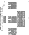
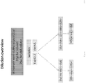
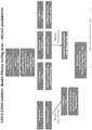
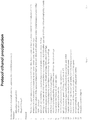
- Example 1 EXO52 Isolation of DNA, as well as Co-isolation of RNA and DNA
- EXO52 This example demonstrates the ability of the EXO52 method to isolate all DNA from a plasma sample.
- various terminology has been used to identify precursor methods to the isolation methods referred to herein as EXO52.
- some Figures include terms such as old EXO52, EXO52.1, and variations thereof. These earlier versions are provided solely as a comparison and to demonstrate the superior isolation achieved using the EXO52 methods of the disclosure.
- EXO52.2 is the EXO52 method where the RNA and DNA extraction is performed in a single tube.
- the EXO52 column can also be used to isolate all DNA from a plasma sample.
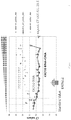
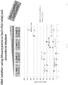
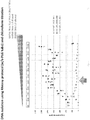
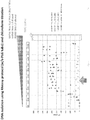
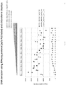
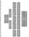
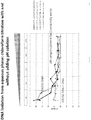
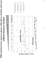
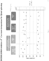
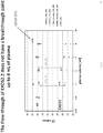
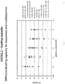
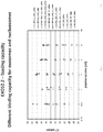
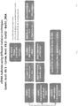
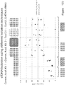
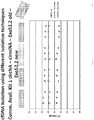
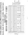
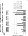
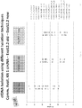
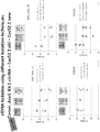
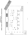
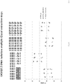
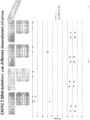
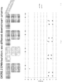
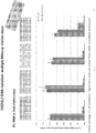
- Two methods for utilizing the EXO52 column for DNA isolation in addition to RNA are depicted in FIG. 1 and FIG. 2 . Specifically, the difference between the two processes is that the RNA and DNA extraction is combined in one tube in EXO52, for easein usability, streamlining of the protocol, and increased reproducibility.
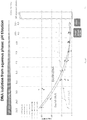
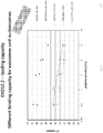
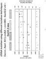
- FIG. 3 shows a gain of 1.5 Cts in EXO50 RNA+ DNA (EXO52).
- EX050 is a method for isolating RNA from microvesicles in a biological sample such as, for example, plasma. This method is described in PCT Publication No. WO2014/107571 .
- the methods of the disclosure can be used to isolate all DNA from plasma samples.
- the DNA is recovered from the lower, hydrophobic phase of the QIAzol lysis after phase separation.
- the methods of the disclosure e.g., two tubes or a single tube as in EXO52
- separate RNA and DNA at similar levels for the same sample volume and the RNA and DNA can be separated from each other.
- These methods of the disclosure capture the same or more mRNA and much more miRNA than a commercially available isolation kit, e.g., Qiagen.
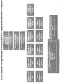
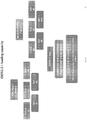
- EXO52 can also be used for co-purification of RNA and DNA. As used herein, EXO52 refers to the following protocol, unless otherwise specified.
- the EXO52 procedure can be used to isolate RNA and DNA from exosomes and other microvesicles using 0.2 - 4 mL of plasma or serum. It is recommended to only use pre-filtered plasma or serum, excluding particles larger than 0.8 ⁇ m.
- the list of compatible plasma tubes includes plasma with the additives EDTA, sodium citrate, and citrate- phosphate-dextrose. Plasma containing heparin can inhibit RT-qPCR.
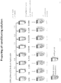
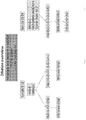
- the sample alone or diluted with a binding buffer, is then loaded onto the EXO52 spin column and spun for 1 min at 500 x g. Discard the flow-through and place the column back into the same collection tube. Wash buffer is then added and the EXO52 column is spun for 5 min at 5000 x g to remove residual volume from the column. Note: After centrifugation, the EXO52 spin column is removed from the collection tube so that the column does not contact the flow-through. The spin column is then transferred to a fresh collection tube, and 700 ⁇ L Qiazol is added to the membrane. Then, the spin column is spun for 5 min at 5000 x g to collect the homogenate containing the lysed exosomes. The homogenate is then transferred to a PLG tube.
- the upper aqueous phase is transferred to a new collection tube, avoiding transfer of any interphase material.
- 2 volumes of 100% ethanol are then added and mixed thoroughly by pipetting up and down several times and without the use of a centrifuge.
- 700 ⁇ l of the sample, including any precipitate that may have formed, is then pipeted up to into an RNeasy MinElute spin column in a 2 ml collection tube (Cat. #1026497), followed by centrifugation at 2:8000 x g (2:10,000 rpm) for 15 sat room temperature (15-25°C).
- the flow-through is discarded. These steps are repeated with the remaining of the sample, and the flow- through is discarded.
- EXO52 is useful for isolating and detecting DNA from biological samples.
- Vesicle RNA is thought to be derived from living cells in e.g. the diseased tissue.
- Cell-free DNA cfDNA is thought to be derived from dying cells e.g. necrotic cells in the disease tissue.
- cfDNA is useful as an indicator of therapeutic response, while the RNA is an indicator of resistance mutations on the rise.
- EXO52 is useful for detection of rare mutations in blood, as EXO52 provides a sufficiently sensitive method that can be applied on nucleic acids of sufficient amount. The amount of actual DNA and RNA molecules in biofluids is very limited, and EXO52 provides an isolation method that extracts all molecules of the blood that are relevant for mutation detection in a volume small enough for effective downstream processing and/or analysis.
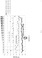
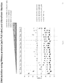
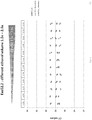
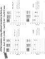
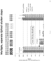
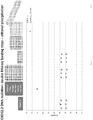
- Figures 13 - 223 demonstrate the specificity and sensitivity of the EXO52 methods.
- adding chloroform to the EXO52 method allowed for the co-isolation of both RNA and DNA, and adding chloroform did not harm the detection or isolation of RNA.
- RNA isolation it was determined that adding more chloroform did not influence RNA isolation if using RNA specific assays. DNA specific assays will result in lower CTs when adding more chloroform because DNA is in aqueous phase.
- CTs for DNA detecting assays increased in aqueous phase isolation with adding more chloroform and decreased in EXO52 phenol phase DNA isolation.
- DNA yield increased in aqueous phase with adding more chloroform, whereas DNA yield decreased in phenol phase (EXO52 DNA Isolation).
- DNA isolation from aqueous phase yield more DNA compared to EXO52 DNA Isolation from phenol phase. Little DNA appeared to stay in phenol phase or in remaining aqueous phase after removing upper phase, as it is difficult to remove whole phase w/o using PLG tube.
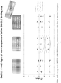
- DNA yield is similar in RT reaction (10 ⁇ l EX050 eluate in final 20 ⁇ l RT Mix) and 1:2 diluted EX050 eluate. DNA did not seem to react with reverse transcription mix.
- RNA only GAPDH assay Studies were repeating using an RNA only GAPDH assay to see if the RNA only GAPDH assay was affected by increasing chloroform addition. RNA was not affected with increasing chloroform addition. Studies were also run using a GAPDH_RNA_DNA assay, which showed no replacement of RNA signal by the DNA (2CTs difference).
- the BRAF assay showed a 2x increase in signal in the EX050 RNA fraction by having DNA present in the aqueous phase.
- the GAPDH assay did not show a clear additive effect of DNA in the EX050 RNA fraction since the added copies were minute in comparison to the RNA copies. With this clear difference between RNA and DNA copies, no replacement of RNA signal can be shown.
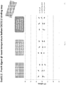
- chloroform addition was the predominant factor in determining the DNA content of the aqueous phase.
- a positive effect of high pH was seen only at low chloroform levels.
- the RNA signal was not affected through addition of DNA into the aqueous phase.
- a Qiazol centrifugation step caused DNA contamination in aqueous phase, but only in samples without PLG tube.
- a Qiazol spin at room temperature did not add up DNA to normal EXO52 DNA isolation. There was no difference in CT values referred to the spin temperature. Temperature for centrifugation step did not influence mRNA and miRNA isolation.
- the binding and elution of DNA from EXO52 to the RNeasy spin column did not depend on ethanol concentration in the range from 1.5x volume to 2.6x volume.
- a proteinase K (ProtK) digestion of a plasma sample led specifically to loss of signal from RNA, but ProtK treatment did not influence DNA yield, as the same CT was obtained for all samples.
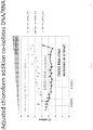
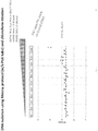
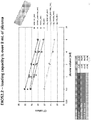
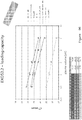
- the DNA loading capacity of EXO52 was not reached at 8ml plasma since the yield of DNA was still linearly increasing and there was no detectable DNA in the flow-through. This is in contrast to the linear loading capacity of vesicles, which is reached at 4 mL. No cfDNA was detected in the flow through (FT) but RNA was seen to accumulate from 2 mL on. The sample output is linear for DNA, but not for RNA. RNA has a different saturation point than DNA. Adding a PLG tub to the procedure was found to increase the yield slightly. EXO52 method added RNA copies, when compared to commercially available CNAkits.
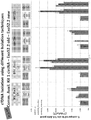
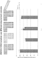
- the methods may use an extraction buffer only based on guanidinium thiocyanate to extract RNA and DNA from the EXO52 column.
- RNA Isolation As shown in the Figures for RNA Isolation, 1 out of 2 replicates ofRLT+ high DTT 56°C resulted in expected CT values. Variation between replicates may have been caused by clogging RNeasy membrane after adding loading mixture. BA profile showed very low RNA concentration for left on column data point for RLT+ high DTT 56°C but only one RNA isolation resulted in expected CTvalues.
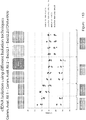
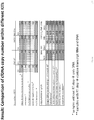
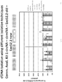
- the Figures also demonstrate isolation of microRNAs using various DNA or DNA/RNA isolation procedures.
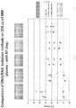
- the EXO52 isolated more mRNA and much more miRNAs than the commercially available CNA kit, and EXO52 and the CNA kit isolated the same amount of DNA.
- the EXO52 method seemed to isolate all DNA from plasma.
- EXO52 consistently outperforms the commercially available circulating nucleic acids (CAN) kit. EXO52 has better yield than CNA Kit on three different plasma pools, different CNA reagent lots, different operators and different sample sources.
- CAN circulating nucleic acids
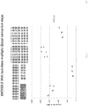
- the EXO52 methods were used to analyze cfDNA in samples from a melanoma cohort.
- the results obtained using the EXO52 methods were compared with the results obtained using a commercially available CNA kit.
- the intra-assay variation (based on different time points of isolation of the same plasma sample) of the CNA kit was higher than that observed using the EXO52 methods.
- the performance of the EXO52 methods is equal or better to those obtained using the commercially available kit.
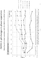
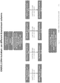
- Example 2 Development of a one-step isolation platform for exosomal RNA and cell-free DNA from cancer plasma
- Circulating nucleic acids in the bloodstream of cancer patients are of great interest to medical research because of their potential to yield information on the patient's disease status and treatment options without requiring a tissue biopsy. Any diagnostic test that seeks to utilize Biofluids for mutation analysis needs a platform that can maximize the capture of tumor derived mutations in circulation.
- Blood plasma contains at least two cell-free sources of nucleic acids: circulating cell-free DNA (cfDNA), generated from apoptotic or necrotic cells, and RNA enclosed in extracellular vesicles including exosomes (exoRNA), which are actively secreted by cells in the body.
- cfDNA circulating cell-free DNA
- exoRNA exosomes
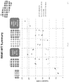
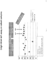
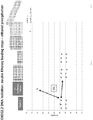
- Figures 224-226 illustrate the studies presented herein which demonstrate the following: (i) Blood plasma contains cell-free RNA in addition to cell-free DNA; (ii) EXO52 is a fast, reproducible and convenient procedure to co-isolate all exoRNA and cfDNA from high volumes ofBiofluids; and (iii) Using both, exoRNA and cfDNA typically doubles the molecules available for rare mutant detection by qPCR and NGS.
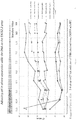
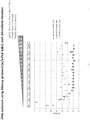
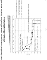
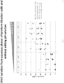
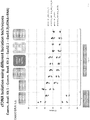
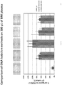
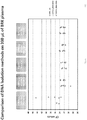
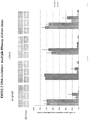
- Figure 227 and 228 are a series of graphs depicting the ability of the EXO52 methods provided herein to capture total circulating nucleic acids.
- the EXO52 methods were compared to a commercially available circulating nucleic acid DNA isolation kit.
- EXO52 captured all cfDNA
- EXO52 detected significantly more copies combining exoRNA and cfDNA vs. cfDNA alone.
- Figure 228 also demonstrates that patients were identified as negative for a biomarker based solely on cfDNA analysis, but with the combined DNA and RNA analysis, these patients were identified as positive for the biomarker.
- Those of ordinarily skill in the art will appreciate that more copies of a mutation or other biomarker leads to enhanced sensitivity and accuracy in identifying mutations and other biomarkers.
Landscapes
- Chemical & Material Sciences (AREA)
- Engineering & Computer Science (AREA)
- Health & Medical Sciences (AREA)
- Life Sciences & Earth Sciences (AREA)
- Genetics & Genomics (AREA)
- Organic Chemistry (AREA)
- Zoology (AREA)
- Wood Science & Technology (AREA)
- Biomedical Technology (AREA)
- Bioinformatics & Cheminformatics (AREA)
- General Engineering & Computer Science (AREA)
- Biotechnology (AREA)
- Analytical Chemistry (AREA)
- Biochemistry (AREA)
- General Health & Medical Sciences (AREA)
- Microbiology (AREA)
- Physics & Mathematics (AREA)
- Biophysics (AREA)
- Molecular Biology (AREA)
- Crystallography & Structural Chemistry (AREA)
- Plant Pathology (AREA)
- Proteomics, Peptides & Aminoacids (AREA)
- Chemical Kinetics & Catalysis (AREA)
- Immunology (AREA)
- Measuring Or Testing Involving Enzymes Or Micro-Organisms (AREA)
- Micro-Organisms Or Cultivation Processes Thereof (AREA)
- Medicines Containing Material From Animals Or Micro-Organisms (AREA)
- Tires In General (AREA)
- Mechanical Treatment Of Semiconductor (AREA)
Priority Applications (1)
| Application Number | Priority Date | Filing Date | Title |
|---|---|---|---|
| EP19201765.5A EP3623792B1 (en) | 2014-07-09 | 2015-07-09 | Methods for isolating microvesicles and extracting nucleic acids from biological samples |
Applications Claiming Priority (4)
| Application Number | Priority Date | Filing Date | Title |
|---|---|---|---|
| US201462022538P | 2014-07-09 | 2014-07-09 | |
| US201462079763P | 2014-11-14 | 2014-11-14 | |
| US201562166890P | 2015-05-27 | 2015-05-27 | |
| PCT/US2015/039760 WO2016007755A1 (en) | 2014-07-09 | 2015-07-09 | Methods for isolating microvesicles and extracting nucleic acids from biological samples |
Related Child Applications (1)
| Application Number | Title | Priority Date | Filing Date |
|---|---|---|---|
| EP19201765.5A Division EP3623792B1 (en) | 2014-07-09 | 2015-07-09 | Methods for isolating microvesicles and extracting nucleic acids from biological samples |
Publications (3)
| Publication Number | Publication Date |
|---|---|
| EP3167062A1 EP3167062A1 (en) | 2017-05-17 |
| EP3167062A4 EP3167062A4 (en) | 2017-12-06 |
| EP3167062B1 true EP3167062B1 (en) | 2019-10-09 |
Family
ID=55064901
Family Applications (2)
| Application Number | Title | Priority Date | Filing Date |
|---|---|---|---|
| EP15818561.1A Active EP3167062B1 (en) | 2014-07-09 | 2015-07-09 | Methods for isolating microvesicles and extracting nucleic acids from biological samples |
| EP19201765.5A Active EP3623792B1 (en) | 2014-07-09 | 2015-07-09 | Methods for isolating microvesicles and extracting nucleic acids from biological samples |
Family Applications After (1)
| Application Number | Title | Priority Date | Filing Date |
|---|---|---|---|
| EP19201765.5A Active EP3623792B1 (en) | 2014-07-09 | 2015-07-09 | Methods for isolating microvesicles and extracting nucleic acids from biological samples |
Country Status (11)
| Country | Link |
|---|---|
| US (1) | US10465183B2 (enExample) |
| EP (2) | EP3167062B1 (enExample) |
| JP (2) | JP6759182B2 (enExample) |
| KR (2) | KR102421185B1 (enExample) |
| CN (2) | CN113699143B (enExample) |
| AU (2) | AU2015287763B2 (enExample) |
| CA (2) | CA3084920C (enExample) |
| ES (2) | ES2762677T3 (enExample) |
| IL (2) | IL250000B (enExample) |
| SG (1) | SG11201700146QA (enExample) |
| WO (1) | WO2016007755A1 (enExample) |
Cited By (1)
| Publication number | Priority date | Publication date | Assignee | Title |
|---|---|---|---|---|
| DE102021208893B3 (de) | 2021-08-13 | 2022-11-17 | Hahn-Schickard-Gesellschaft für angewandte Forschung e.V. | Isolieren von Analyten unterschiedlicher Analyt-Klassen |
Families Citing this family (50)
| Publication number | Priority date | Publication date | Assignee | Title |
|---|---|---|---|---|
| CA2835641C (en) * | 2011-05-11 | 2019-05-28 | Exosome Diagnostics, Inc. | Nucleic acid extraction from heterogeneous biological materials |
| US11268085B2 (en) | 2014-05-27 | 2022-03-08 | Exosome Diagnostics, Inc. | Methods for isolating microvesicles and extracting nucleic acids from biological samples |
| CA3084920C (en) | 2014-07-09 | 2023-02-28 | Exosome Diagnostics, Inc. | Methods for isolating microvesicles and extracting nucleic acids from biological samples |
| CN105821031A (zh) * | 2016-01-18 | 2016-08-03 | 昆明医科大学 | 一种二氯甲烷法提取dna的原理 |
| US20190093172A1 (en) | 2016-04-15 | 2019-03-28 | Exosome Diagnostics, Inc. | Plasma-based detection of anaplastic lymphoma kinase (alk) nucleic acids and alk fusion transcripts and uses thereof in diagnosis and treatment of cancer |
| WO2017185086A1 (en) | 2016-04-22 | 2017-10-26 | Exosome Diagnostics, Inc. | Devices and methods for in vivo capture of biological samples and nucleic acids therein |
| EP3452591B1 (en) | 2016-05-02 | 2023-08-16 | Encodia, Inc. | Macromolecule analysis employing nucleic acid encoding |
| EP3452613B1 (en) | 2016-05-05 | 2021-12-29 | Exosome Diagnostics, Inc. | Profiling microvesicle nucleic acids and uses thereof as signatures in diagnosis of renal transplant rejection |
| CN109642229A (zh) * | 2016-05-13 | 2019-04-16 | 外来体诊断公司 | 从生物液体分离细胞外囊泡和共分离无细胞dna的自动和手动方法 |
| WO2018035340A1 (en) | 2016-08-17 | 2018-02-22 | The Regents Of The University Of California | A novel immunoprobe-based method to assess organ injury status through a biofluid-based cell-free dna (cfdna) assay |
| CA3035881A1 (en) | 2016-09-14 | 2018-03-22 | Toray Industries, Inc. | Method of collecting cell-free dna |
| CN110191962A (zh) * | 2016-10-21 | 2019-08-30 | 外来体诊断公司 | 外来体相关核酸的测序和分析 |
| JP7280823B2 (ja) | 2016-11-30 | 2023-05-24 | エクソサム ダイアグノスティクス,インコーポレイティド | 非小細胞肺癌患者由来のエクソソームrna及び無細胞dnaを使用して血漿中の変異を検出するための方法と組成物 |
| US11666603B2 (en) * | 2016-12-23 | 2023-06-06 | Exopharm Limited | Methods and compositions for purification or isolation of microvesicles and exosomes |
| EP3562959A2 (en) | 2017-01-02 | 2019-11-06 | Exosome Diagnostics, Inc. | Methods to distinguish rna and dna in a combined preparation |
| CA3048212A1 (en) * | 2017-01-06 | 2018-07-12 | Mantra Bio, Inc. | Systems and methods for algorithmic extracellular vesicle population discovery and characterization |
| KR101875594B1 (ko) * | 2017-01-13 | 2018-07-06 | ㈜로제타엑소좀 | 금속을 이용한 세포밖 소포체의 분리 방법 |
| GB201706680D0 (en) | 2017-04-27 | 2017-06-14 | Ge Healthcare Uk Ltd | Device and method for sample isolation |
| US11891664B2 (en) | 2017-05-17 | 2024-02-06 | Exosome Diagnostics, Inc. | Methods of detecting renal transplant rejection using microvesicle nucleic acid biomarkers |
| CN107267440A (zh) * | 2017-06-09 | 2017-10-20 | 李刚 | 适用于外泌体提取的提取试剂及应用及其提取方法 |
| WO2019013991A2 (en) | 2017-07-12 | 2019-01-17 | Illumina, Inc. | METHODS, SYSTEMS AND MATERIALS FOR EXTRACTING NUCLEIC ACIDS |
| CN111133106A (zh) | 2017-07-12 | 2020-05-08 | 外来体诊断公司 | 用于分离和富集生物流体来源的细胞外囊泡的方法及其使用方法 |
| US11345957B2 (en) | 2017-07-18 | 2022-05-31 | Exosome Diagnostics, Inc. | Methods of treating glioblastoma in a subject informed by exosomal RNA signatures |
| JP2020528766A (ja) * | 2017-07-26 | 2020-10-01 | ロゼッタ エクソソーム | 陽イオンを利用した細胞外小胞体の分離方法 |
| EP3684383A4 (en) * | 2017-09-20 | 2021-07-14 | Molecular Stethoscope, Inc. | METHODS AND SYSTEMS FOR DETECTION OF TISSUE DISORDERS |
| JP7390027B2 (ja) | 2017-10-31 | 2023-12-01 | エンコディア, インコーポレイテッド | 核酸エンコーディングおよび/または標識を使用する解析のためのキット |
| US11260347B2 (en) * | 2017-12-26 | 2022-03-01 | Limited Liability Company “Prostagnost” | Method and device for separating extracellular vesicles from biological liquids with the aid of cascade ultrafiltration |
| CN111770931B (zh) * | 2018-02-20 | 2023-08-25 | 高丽大学校产学协力团 | 用于分离外泌体的多柱及外泌体分离方法 |
| WO2019236853A1 (en) | 2018-06-06 | 2019-12-12 | Exosome Diagnostics, Inc. | Methods for developing urine biomarkers and for detecting bladder cancer |
| BR112020026067A2 (pt) | 2018-06-18 | 2021-03-23 | Exopharm Limited | métodos e composições para purificação ou isolamento de microvesículas e exossomas |
| CN108841777A (zh) * | 2018-06-22 | 2018-11-20 | 北京恩泽康泰生物科技有限公司 | 基于静电吸附的胞外囊泡及其内含物的提取方法及装置 |
| JP7426725B2 (ja) * | 2018-10-30 | 2024-02-02 | Craif株式会社 | 細胞外小胞を捕捉するために用いられるデバイス、細胞外小胞の保存方法および移送方法 |
| WO2020106853A1 (en) | 2018-11-20 | 2020-05-28 | Exosome Diagnostics, Inc. | Compositions and methods for internal controls of microvesicle isolations |
| CN114072499B (zh) | 2019-04-30 | 2024-08-06 | Encodia公司 | 用于制备分析物的方法和相关试剂盒 |
| EP4010488A1 (en) * | 2019-08-08 | 2022-06-15 | Biocartis NV | Novel nucleic acid purification chemistry |
| EP4031865A2 (en) | 2019-09-18 | 2022-07-27 | Exosome Diagnostics, Inc. | Compositions, methods, and kits for the isolation of extracellular vesicles |
| US20220411849A1 (en) * | 2019-11-04 | 2022-12-29 | Nasasbiotech, S.L. | Method for isolating nucleic acids |
| US20230009972A1 (en) * | 2019-12-16 | 2023-01-12 | Qiagen Gmbh | Method for enriching vesicular rna |
| EP4077659A1 (en) * | 2019-12-16 | 2022-10-26 | QIAGEN GmbH | Enrichment method |
| EP4140568A4 (en) * | 2020-04-24 | 2024-05-22 | Toyobo Co., Ltd. | Ion exchange membrane |
| US20230168162A1 (en) * | 2020-04-24 | 2023-06-01 | Korea University Research And Business Foundation | Microvesicle isolation method and microvesicle isolation |
| WO2021243206A1 (en) | 2020-05-29 | 2021-12-02 | Exosome Diagnostics, Inc. | Use of microvesicle signature for the diagnosis and treatment of kidney transplant rejection |
| EP4263383A4 (en) | 2020-12-17 | 2024-10-23 | Nephrosant, Inc. | KITS FOR STABILIZATION OF URINE SAMPLES |
| US20240287498A1 (en) * | 2021-06-17 | 2024-08-29 | Qiagen Gmbh | Method for isolating non-vesicular mirna |
| EP4370712A1 (en) | 2021-07-16 | 2024-05-22 | Exosome Diagnostics, Inc. | Methods of detecting sjögren's syndrome using salivary exosomes |
| WO2023076528A2 (en) * | 2021-10-28 | 2023-05-04 | 10X Genomics, Inc. | Methods for sample preparation |
| AU2023221644A1 (en) | 2022-02-18 | 2024-09-12 | Bio-Techne Corporation | Use of microvesicle signatures in the identification and treatment of renal disorders |
| WO2024054572A1 (en) | 2022-09-07 | 2024-03-14 | Exosome Diagnostics, Inc. | Methods of detecting sjögren's syndrome using salivary exosomes |
| CN115960885B (zh) * | 2022-10-09 | 2023-12-12 | 南京诺唯赞生物科技股份有限公司 | 一种提取肝素钠样品中核酸的方法及组合物 |
| KR102784335B1 (ko) * | 2023-02-09 | 2025-03-19 | 순천향대학교 산학협력단 | 세포유리핵산의 고농축화 분리 방법 |
Family Cites Families (23)
| Publication number | Priority date | Publication date | Assignee | Title |
|---|---|---|---|---|
| US4935342A (en) | 1986-12-01 | 1990-06-19 | Syngene, Inc. | Method of isolating and purifying nucleic acids from biological samples |
| US5438128A (en) | 1992-02-07 | 1995-08-01 | Millipore Corporation | Method for rapid purifiction of nucleic acids using layered ion-exchange membranes |
| US5639606A (en) | 1993-04-06 | 1997-06-17 | The University Of Rochester | Method for quantitative measurement of gene expression using multiplex competitive reverse transcriptase-polymerase chain reaction |
| CA2170604C (en) * | 1993-08-30 | 2007-03-13 | Vikas V. Padhye | Nucleic acid purification compositions and methods |
| FR2788780B1 (fr) * | 1999-01-27 | 2001-03-30 | Ap Cells Inc | Procede de preparation de vesicules membranaires |
| GB9927320D0 (en) | 1999-11-18 | 2000-01-12 | Chiron Spa | Exosome separation |
| US6812023B1 (en) | 2000-04-27 | 2004-11-02 | Anosys, Inc. | Methods of producing membrane vesicles |
| US9163229B2 (en) * | 2006-10-10 | 2015-10-20 | Trovagene, Inc. | Compositions, methods and kits for isolating nucleic acids from body fluids using anion exchange media |
| KR101810799B1 (ko) | 2008-02-01 | 2017-12-19 | 더 제너럴 하스피탈 코포레이션 | 의학적 질환 및 병태의 진단, 예후, 및 치료에 있어서 미세소포체의 용도 |
| WO2010065765A2 (en) * | 2008-12-04 | 2010-06-10 | Aethlon Medical, Inc. | Affinity capture of circulating biomarkers |
| US20120077263A1 (en) * | 2009-06-05 | 2012-03-29 | Mayo Foundation For Medical Education And Research | Methods and materials for isolating exosomes |
| WO2011151428A1 (en) | 2010-06-01 | 2011-12-08 | Qiagen Gmbh | Method for isolating and/or purifying nucleic acid(s) |
| ES2624284T3 (es) * | 2010-07-07 | 2017-07-13 | Aethlon Medical Inc | Métodos para cuantificar exosomas |
| US8945509B2 (en) * | 2010-08-20 | 2015-02-03 | Life Technologies Corporation | Magnetic beads having surface glycoconjugates and use thereof |
| CA2817111C (en) * | 2010-11-10 | 2019-03-05 | Exosome Diagnostics, Inc. | Method for isolation of nucleic acid containing particles and extraction of nucleic acids therefrom |
| WO2012087241A1 (en) * | 2010-12-20 | 2012-06-28 | Agency For Science, Technology And Research | Method of purifying exosomes |
| CA2835641C (en) | 2011-05-11 | 2019-05-28 | Exosome Diagnostics, Inc. | Nucleic acid extraction from heterogeneous biological materials |
| JP2014519340A (ja) * | 2011-06-16 | 2014-08-14 | カリス ライフ サイエンシズ ルクセンブルク ホールディングス エス.アー.エール.エル. | バイオマーカー組成物および方法 |
| KR20140005688A (ko) * | 2012-07-06 | 2014-01-15 | 삼성전자주식회사 | 사용자 인터페이스 방법 및 장치 |
| CN104995311A (zh) | 2012-08-30 | 2015-10-21 | 外来体诊断公司 | 用于核酸测定的对照 |
| KR101933621B1 (ko) * | 2012-09-28 | 2018-12-28 | 삼성전자주식회사 | 소포를 분리하기 위한 조성물, 키트 및 이를 이용하여 소포를 분리하는 방법 |
| CN110106229B (zh) | 2013-01-03 | 2023-03-28 | 外来体诊断公司 | 用于分离微囊泡的方法 |
| CA3084920C (en) | 2014-07-09 | 2023-02-28 | Exosome Diagnostics, Inc. | Methods for isolating microvesicles and extracting nucleic acids from biological samples |
-
2015
- 2015-07-09 CA CA3084920A patent/CA3084920C/en active Active
- 2015-07-09 ES ES15818561T patent/ES2762677T3/es active Active
- 2015-07-09 ES ES19201765T patent/ES2898254T3/es active Active
- 2015-07-09 AU AU2015287763A patent/AU2015287763B2/en active Active
- 2015-07-09 EP EP15818561.1A patent/EP3167062B1/en active Active
- 2015-07-09 CN CN202110781733.3A patent/CN113699143B/zh active Active
- 2015-07-09 JP JP2017501235A patent/JP6759182B2/ja active Active
- 2015-07-09 KR KR1020177003611A patent/KR102421185B1/ko active Active
- 2015-07-09 CA CA2954576A patent/CA2954576C/en active Active
- 2015-07-09 US US15/325,021 patent/US10465183B2/en active Active
- 2015-07-09 KR KR1020227023873A patent/KR102596577B1/ko active Active
- 2015-07-09 EP EP19201765.5A patent/EP3623792B1/en active Active
- 2015-07-09 WO PCT/US2015/039760 patent/WO2016007755A1/en not_active Ceased
- 2015-07-09 SG SG11201700146QA patent/SG11201700146QA/en unknown
- 2015-07-09 CN CN201580049375.XA patent/CN107002075B/zh active Active
-
2017
- 2017-01-09 IL IL250000A patent/IL250000B/en active IP Right Grant
-
2019
- 2019-10-17 AU AU2019250221A patent/AU2019250221B2/en active Active
-
2020
- 2020-09-02 JP JP2020147390A patent/JP7128866B2/ja active Active
-
2021
- 2021-01-20 IL IL280307A patent/IL280307B/en unknown
Non-Patent Citations (1)
| Title |
|---|
| None * |
Cited By (2)
| Publication number | Priority date | Publication date | Assignee | Title |
|---|---|---|---|---|
| DE102021208893B3 (de) | 2021-08-13 | 2022-11-17 | Hahn-Schickard-Gesellschaft für angewandte Forschung e.V. | Isolieren von Analyten unterschiedlicher Analyt-Klassen |
| WO2023017049A1 (de) | 2021-08-13 | 2023-02-16 | Hahn-Schickard-Gesellschaft für angewandte Forschung e.V. | Isolieren von analyten unterschiedlicher analyt-klassen |
Also Published As
Similar Documents
| Publication | Publication Date | Title |
|---|---|---|
| AU2019250221B2 (en) | Methods for isolating microvesicles and extracting nucleic acids from biological samples | |
| JP7354327B2 (ja) | 生体液からの細胞外小胞の単離及びセルフリーdnaの同時単離のための自動及び手動方法 | |
| JP6449781B2 (ja) | 微細小胞の単離方法 | |
| US11268085B2 (en) | Methods for isolating microvesicles and extracting nucleic acids from biological samples | |
| CA3022528C (en) | Automated and manual methods for isolation of extracellular vesicles and co-isolation of cell-free dna from biofluids | |
| HK1238676A1 (en) | Methods for isolating microvesicles and extracting nucleic acids from biological samples |
Legal Events
| Date | Code | Title | Description |
|---|---|---|---|
| STAA | Information on the status of an ep patent application or granted ep patent |
Free format text: STATUS: THE INTERNATIONAL PUBLICATION HAS BEEN MADE |
|
| PUAI | Public reference made under article 153(3) epc to a published international application that has entered the european phase |
Free format text: ORIGINAL CODE: 0009012 |
|
| STAA | Information on the status of an ep patent application or granted ep patent |
Free format text: STATUS: REQUEST FOR EXAMINATION WAS MADE |
|
| 17P | Request for examination filed |
Effective date: 20170127 |
|
| AK | Designated contracting states |
Kind code of ref document: A1 Designated state(s): AL AT BE BG CH CY CZ DE DK EE ES FI FR GB GR HR HU IE IS IT LI LT LU LV MC MK MT NL NO PL PT RO RS SE SI SK SM TR |
|
| AX | Request for extension of the european patent |
Extension state: BA ME |
|
| RIN1 | Information on inventor provided before grant (corrected) |
Inventor name: WEI, TAI-FEN Inventor name: RAMACHANDRAN, APARNA Inventor name: BERGHOFF, EMILY Inventor name: YAN, HAOHENG Inventor name: ENDERLE, DANIEL Inventor name: SKOG, JOHAN KARL OLOV Inventor name: NOERHOLM, MIKKEL |
|
| RIN1 | Information on inventor provided before grant (corrected) |
Inventor name: ENDERLE, DANIEL Inventor name: SKOG, JOHAN KARL OLOV Inventor name: WEI, TAI-FEN Inventor name: RAMACHANDRAN, APARNA Inventor name: NOERHOLM, MIKKEL Inventor name: BERGHOFF, EMILY Inventor name: YAN, HAOHENG |
|
| DAV | Request for validation of the european patent (deleted) | ||
| DAX | Request for extension of the european patent (deleted) | ||
| REG | Reference to a national code |
Ref country code: DE Ref legal event code: R079 Ref document number: 602015039599 Country of ref document: DE Free format text: PREVIOUS MAIN CLASS: C12N0015100000 Ipc: G01N0001180000 |
|
| A4 | Supplementary search report drawn up and despatched |
Effective date: 20171107 |
|
| RIC1 | Information provided on ipc code assigned before grant |
Ipc: A61K 9/14 20060101ALI20171030BHEP Ipc: A61K 48/00 20060101ALI20171030BHEP Ipc: G01N 1/18 20060101AFI20171030BHEP |
|
| STAA | Information on the status of an ep patent application or granted ep patent |
Free format text: STATUS: EXAMINATION IS IN PROGRESS |
|
| 17Q | First examination report despatched |
Effective date: 20180710 |
|
| GRAP | Despatch of communication of intention to grant a patent |
Free format text: ORIGINAL CODE: EPIDOSNIGR1 |
|
| STAA | Information on the status of an ep patent application or granted ep patent |
Free format text: STATUS: GRANT OF PATENT IS INTENDED |
|
| INTG | Intention to grant announced |
Effective date: 20190503 |
|
| GRAS | Grant fee paid |
Free format text: ORIGINAL CODE: EPIDOSNIGR3 |
|
| GRAA | (expected) grant |
Free format text: ORIGINAL CODE: 0009210 |
|
| STAA | Information on the status of an ep patent application or granted ep patent |
Free format text: STATUS: THE PATENT HAS BEEN GRANTED |
|
| RIN1 | Information on inventor provided before grant (corrected) |
Inventor name: WEI, TAI-FEN Inventor name: RAMACHANDRAN, APARNA Inventor name: ENDERLE, DANIEL Inventor name: NOERHOLM, MIKKEL Inventor name: SKOG, JOHAN KARL OLOV Inventor name: YAN, HAOHENG Inventor name: BERGHOFF, EMILY |
|
| AK | Designated contracting states |
Kind code of ref document: B1 Designated state(s): AL AT BE BG CH CY CZ DE DK EE ES FI FR GB GR HR HU IE IS IT LI LT LU LV MC MK MT NL NO PL PT RO RS SE SI SK SM TR |
|
| RAP1 | Party data changed (applicant data changed or rights of an application transferred) |
Owner name: EXOSOME DIAGNOSTICS, INC. |
|
| REG | Reference to a national code |
Ref country code: GB Ref legal event code: FG4D |
|
| REG | Reference to a national code |
Ref country code: CH Ref legal event code: EP |
|
| REG | Reference to a national code |
Ref country code: IE Ref legal event code: FG4D |
|
| REG | Reference to a national code |
Ref country code: DE Ref legal event code: R096 Ref document number: 602015039599 Country of ref document: DE |
|
| REG | Reference to a national code |
Ref country code: AT Ref legal event code: REF Ref document number: 1189398 Country of ref document: AT Kind code of ref document: T Effective date: 20191115 |
|
| REG | Reference to a national code |
Ref country code: SE Ref legal event code: TRGR |
|
| REG | Reference to a national code |
Ref country code: NL Ref legal event code: FP |
|
| REG | Reference to a national code |
Ref country code: CH Ref legal event code: NV Representative=s name: MICHELI AND CIE SA, CH |
|
| REG | Reference to a national code |
Ref country code: LT Ref legal event code: MG4D |
|
| REG | Reference to a national code |
Ref country code: AT Ref legal event code: MK05 Ref document number: 1189398 Country of ref document: AT Kind code of ref document: T Effective date: 20191009 |
|
| PG25 | Lapsed in a contracting state [announced via postgrant information from national office to epo] |
Ref country code: FI Free format text: LAPSE BECAUSE OF FAILURE TO SUBMIT A TRANSLATION OF THE DESCRIPTION OR TO PAY THE FEE WITHIN THE PRESCRIBED TIME-LIMIT Effective date: 20191009 Ref country code: BG Free format text: LAPSE BECAUSE OF FAILURE TO SUBMIT A TRANSLATION OF THE DESCRIPTION OR TO PAY THE FEE WITHIN THE PRESCRIBED TIME-LIMIT Effective date: 20200109 Ref country code: LT Free format text: LAPSE BECAUSE OF FAILURE TO SUBMIT A TRANSLATION OF THE DESCRIPTION OR TO PAY THE FEE WITHIN THE PRESCRIBED TIME-LIMIT Effective date: 20191009 Ref country code: PL Free format text: LAPSE BECAUSE OF FAILURE TO SUBMIT A TRANSLATION OF THE DESCRIPTION OR TO PAY THE FEE WITHIN THE PRESCRIBED TIME-LIMIT Effective date: 20191009 Ref country code: NO Free format text: LAPSE BECAUSE OF FAILURE TO SUBMIT A TRANSLATION OF THE DESCRIPTION OR TO PAY THE FEE WITHIN THE PRESCRIBED TIME-LIMIT Effective date: 20200109 Ref country code: LV Free format text: LAPSE BECAUSE OF FAILURE TO SUBMIT A TRANSLATION OF THE DESCRIPTION OR TO PAY THE FEE WITHIN THE PRESCRIBED TIME-LIMIT Effective date: 20191009 Ref country code: PT Free format text: LAPSE BECAUSE OF FAILURE TO SUBMIT A TRANSLATION OF THE DESCRIPTION OR TO PAY THE FEE WITHIN THE PRESCRIBED TIME-LIMIT Effective date: 20200210 Ref country code: GR Free format text: LAPSE BECAUSE OF FAILURE TO SUBMIT A TRANSLATION OF THE DESCRIPTION OR TO PAY THE FEE WITHIN THE PRESCRIBED TIME-LIMIT Effective date: 20200110 Ref country code: AT Free format text: LAPSE BECAUSE OF FAILURE TO SUBMIT A TRANSLATION OF THE DESCRIPTION OR TO PAY THE FEE WITHIN THE PRESCRIBED TIME-LIMIT Effective date: 20191009 |
|
| REG | Reference to a national code |
Ref country code: ES Ref legal event code: FG2A Ref document number: 2762677 Country of ref document: ES Kind code of ref document: T3 Effective date: 20200525 |
|
| PG25 | Lapsed in a contracting state [announced via postgrant information from national office to epo] |
Ref country code: IS Free format text: LAPSE BECAUSE OF FAILURE TO SUBMIT A TRANSLATION OF THE DESCRIPTION OR TO PAY THE FEE WITHIN THE PRESCRIBED TIME-LIMIT Effective date: 20200224 Ref country code: RS Free format text: LAPSE BECAUSE OF FAILURE TO SUBMIT A TRANSLATION OF THE DESCRIPTION OR TO PAY THE FEE WITHIN THE PRESCRIBED TIME-LIMIT Effective date: 20191009 Ref country code: HR Free format text: LAPSE BECAUSE OF FAILURE TO SUBMIT A TRANSLATION OF THE DESCRIPTION OR TO PAY THE FEE WITHIN THE PRESCRIBED TIME-LIMIT Effective date: 20191009 |
|
| PG25 | Lapsed in a contracting state [announced via postgrant information from national office to epo] |
Ref country code: AL Free format text: LAPSE BECAUSE OF FAILURE TO SUBMIT A TRANSLATION OF THE DESCRIPTION OR TO PAY THE FEE WITHIN THE PRESCRIBED TIME-LIMIT Effective date: 20191009 |
|
| REG | Reference to a national code |
Ref country code: DE Ref legal event code: R097 Ref document number: 602015039599 Country of ref document: DE |
|
| PG2D | Information on lapse in contracting state deleted |
Ref country code: IS |
|
| PG25 | Lapsed in a contracting state [announced via postgrant information from national office to epo] |
Ref country code: EE Free format text: LAPSE BECAUSE OF FAILURE TO SUBMIT A TRANSLATION OF THE DESCRIPTION OR TO PAY THE FEE WITHIN THE PRESCRIBED TIME-LIMIT Effective date: 20191009 Ref country code: DK Free format text: LAPSE BECAUSE OF FAILURE TO SUBMIT A TRANSLATION OF THE DESCRIPTION OR TO PAY THE FEE WITHIN THE PRESCRIBED TIME-LIMIT Effective date: 20191009 Ref country code: CZ Free format text: LAPSE BECAUSE OF FAILURE TO SUBMIT A TRANSLATION OF THE DESCRIPTION OR TO PAY THE FEE WITHIN THE PRESCRIBED TIME-LIMIT Effective date: 20191009 Ref country code: RO Free format text: LAPSE BECAUSE OF FAILURE TO SUBMIT A TRANSLATION OF THE DESCRIPTION OR TO PAY THE FEE WITHIN THE PRESCRIBED TIME-LIMIT Effective date: 20191009 Ref country code: IS Free format text: LAPSE BECAUSE OF FAILURE TO SUBMIT A TRANSLATION OF THE DESCRIPTION OR TO PAY THE FEE WITHIN THE PRESCRIBED TIME-LIMIT Effective date: 20200209 |
|
| PLBE | No opposition filed within time limit |
Free format text: ORIGINAL CODE: 0009261 |
|
| STAA | Information on the status of an ep patent application or granted ep patent |
Free format text: STATUS: NO OPPOSITION FILED WITHIN TIME LIMIT |
|
| PG25 | Lapsed in a contracting state [announced via postgrant information from national office to epo] |
Ref country code: SM Free format text: LAPSE BECAUSE OF FAILURE TO SUBMIT A TRANSLATION OF THE DESCRIPTION OR TO PAY THE FEE WITHIN THE PRESCRIBED TIME-LIMIT Effective date: 20191009 Ref country code: SK Free format text: LAPSE BECAUSE OF FAILURE TO SUBMIT A TRANSLATION OF THE DESCRIPTION OR TO PAY THE FEE WITHIN THE PRESCRIBED TIME-LIMIT Effective date: 20191009 |
|
| 26N | No opposition filed |
Effective date: 20200710 |
|
| PG25 | Lapsed in a contracting state [announced via postgrant information from national office to epo] |
Ref country code: SI Free format text: LAPSE BECAUSE OF FAILURE TO SUBMIT A TRANSLATION OF THE DESCRIPTION OR TO PAY THE FEE WITHIN THE PRESCRIBED TIME-LIMIT Effective date: 20191009 |
|
| PG25 | Lapsed in a contracting state [announced via postgrant information from national office to epo] |
Ref country code: MC Free format text: LAPSE BECAUSE OF FAILURE TO SUBMIT A TRANSLATION OF THE DESCRIPTION OR TO PAY THE FEE WITHIN THE PRESCRIBED TIME-LIMIT Effective date: 20191009 |
|
| PG25 | Lapsed in a contracting state [announced via postgrant information from national office to epo] |
Ref country code: LU Free format text: LAPSE BECAUSE OF NON-PAYMENT OF DUE FEES Effective date: 20200709 |
|
| PG25 | Lapsed in a contracting state [announced via postgrant information from national office to epo] |
Ref country code: TR Free format text: LAPSE BECAUSE OF FAILURE TO SUBMIT A TRANSLATION OF THE DESCRIPTION OR TO PAY THE FEE WITHIN THE PRESCRIBED TIME-LIMIT Effective date: 20191009 Ref country code: MT Free format text: LAPSE BECAUSE OF FAILURE TO SUBMIT A TRANSLATION OF THE DESCRIPTION OR TO PAY THE FEE WITHIN THE PRESCRIBED TIME-LIMIT Effective date: 20191009 Ref country code: CY Free format text: LAPSE BECAUSE OF FAILURE TO SUBMIT A TRANSLATION OF THE DESCRIPTION OR TO PAY THE FEE WITHIN THE PRESCRIBED TIME-LIMIT Effective date: 20191009 |
|
| PG25 | Lapsed in a contracting state [announced via postgrant information from national office to epo] |
Ref country code: MK Free format text: LAPSE BECAUSE OF FAILURE TO SUBMIT A TRANSLATION OF THE DESCRIPTION OR TO PAY THE FEE WITHIN THE PRESCRIBED TIME-LIMIT Effective date: 20191009 |
|
| P01 | Opt-out of the competence of the unified patent court (upc) registered |
Effective date: 20230512 |
|
| REG | Reference to a national code |
Ref country code: DE Ref legal event code: R082 Ref document number: 602015039599 Country of ref document: DE Representative=s name: GLAWE DELFS MOLL PARTGMBB, DE Ref country code: DE Ref legal event code: R082 Ref document number: 602015039599 Country of ref document: DE Representative=s name: GLAWE DELFS MOLL PARTNERSCHAFT MBB VON PATENT-, DE Ref country code: DE Ref legal event code: R082 Ref document number: 602015039599 Country of ref document: DE Representative=s name: FORRESTERS IP LLP, DE |
|
| PGFP | Annual fee paid to national office [announced via postgrant information from national office to epo] |
Ref country code: NL Payment date: 20250726 Year of fee payment: 11 |
|
| PGFP | Annual fee paid to national office [announced via postgrant information from national office to epo] |
Ref country code: ES Payment date: 20250801 Year of fee payment: 11 |
|
| PGFP | Annual fee paid to national office [announced via postgrant information from national office to epo] |
Ref country code: DE Payment date: 20250729 Year of fee payment: 11 |
|
| PGFP | Annual fee paid to national office [announced via postgrant information from national office to epo] |
Ref country code: IT Payment date: 20250721 Year of fee payment: 11 |
|
| PGFP | Annual fee paid to national office [announced via postgrant information from national office to epo] |
Ref country code: GB Payment date: 20250728 Year of fee payment: 11 Ref country code: BE Payment date: 20250728 Year of fee payment: 11 |
|
| PGFP | Annual fee paid to national office [announced via postgrant information from national office to epo] |
Ref country code: FR Payment date: 20250725 Year of fee payment: 11 |
|
| PGFP | Annual fee paid to national office [announced via postgrant information from national office to epo] |
Ref country code: CH Payment date: 20250801 Year of fee payment: 11 Ref country code: SE Payment date: 20250727 Year of fee payment: 11 |
|
| PGFP | Annual fee paid to national office [announced via postgrant information from national office to epo] |
Ref country code: IE Payment date: 20250728 Year of fee payment: 11 |
|
| REG | Reference to a national code |
Ref country code: DE Ref legal event code: R082 Ref document number: 602015039599 Country of ref document: DE Representative=s name: FORRESTERS IP LLP, DE |