WO2019018537A1 - Sequencing of nucleic acids associated with exosomal isolation from patients with glioblastoma multiforme - Google Patents
Sequencing of nucleic acids associated with exosomal isolation from patients with glioblastoma multiforme Download PDFInfo
- Publication number
- WO2019018537A1 WO2019018537A1 PCT/US2018/042708 US2018042708W WO2019018537A1 WO 2019018537 A1 WO2019018537 A1 WO 2019018537A1 US 2018042708 W US2018042708 W US 2018042708W WO 2019018537 A1 WO2019018537 A1 WO 2019018537A1
- Authority
- WO
- WIPO (PCT)
- Prior art keywords
- gene
- expression level
- biological sample
- cancer therapy
- cancer
- Prior art date
Links
Classifications
-
- C—CHEMISTRY; METALLURGY
- C12—BIOCHEMISTRY; BEER; SPIRITS; WINE; VINEGAR; MICROBIOLOGY; ENZYMOLOGY; MUTATION OR GENETIC ENGINEERING
- C12Q—MEASURING OR TESTING PROCESSES INVOLVING ENZYMES, NUCLEIC ACIDS OR MICROORGANISMS; COMPOSITIONS OR TEST PAPERS THEREFOR; PROCESSES OF PREPARING SUCH COMPOSITIONS; CONDITION-RESPONSIVE CONTROL IN MICROBIOLOGICAL OR ENZYMOLOGICAL PROCESSES
- C12Q1/00—Measuring or testing processes involving enzymes, nucleic acids or microorganisms; Compositions therefor; Processes of preparing such compositions
- C12Q1/68—Measuring or testing processes involving enzymes, nucleic acids or microorganisms; Compositions therefor; Processes of preparing such compositions involving nucleic acids
- C12Q1/6844—Nucleic acid amplification reactions
- C12Q1/686—Polymerase chain reaction [PCR]
-
- C—CHEMISTRY; METALLURGY
- C12—BIOCHEMISTRY; BEER; SPIRITS; WINE; VINEGAR; MICROBIOLOGY; ENZYMOLOGY; MUTATION OR GENETIC ENGINEERING
- C12Q—MEASURING OR TESTING PROCESSES INVOLVING ENZYMES, NUCLEIC ACIDS OR MICROORGANISMS; COMPOSITIONS OR TEST PAPERS THEREFOR; PROCESSES OF PREPARING SUCH COMPOSITIONS; CONDITION-RESPONSIVE CONTROL IN MICROBIOLOGICAL OR ENZYMOLOGICAL PROCESSES
- C12Q1/00—Measuring or testing processes involving enzymes, nucleic acids or microorganisms; Compositions therefor; Processes of preparing such compositions
- C12Q1/68—Measuring or testing processes involving enzymes, nucleic acids or microorganisms; Compositions therefor; Processes of preparing such compositions involving nucleic acids
- C12Q1/6876—Nucleic acid products used in the analysis of nucleic acids, e.g. primers or probes
- C12Q1/6883—Nucleic acid products used in the analysis of nucleic acids, e.g. primers or probes for diseases caused by alterations of genetic material
- C12Q1/6886—Nucleic acid products used in the analysis of nucleic acids, e.g. primers or probes for diseases caused by alterations of genetic material for cancer
-
- C—CHEMISTRY; METALLURGY
- C12—BIOCHEMISTRY; BEER; SPIRITS; WINE; VINEGAR; MICROBIOLOGY; ENZYMOLOGY; MUTATION OR GENETIC ENGINEERING
- C12Q—MEASURING OR TESTING PROCESSES INVOLVING ENZYMES, NUCLEIC ACIDS OR MICROORGANISMS; COMPOSITIONS OR TEST PAPERS THEREFOR; PROCESSES OF PREPARING SUCH COMPOSITIONS; CONDITION-RESPONSIVE CONTROL IN MICROBIOLOGICAL OR ENZYMOLOGICAL PROCESSES
- C12Q1/00—Measuring or testing processes involving enzymes, nucleic acids or microorganisms; Compositions therefor; Processes of preparing such compositions
- C12Q1/68—Measuring or testing processes involving enzymes, nucleic acids or microorganisms; Compositions therefor; Processes of preparing such compositions involving nucleic acids
- C12Q1/6806—Preparing nucleic acids for analysis, e.g. for polymerase chain reaction [PCR] assay
-
- C—CHEMISTRY; METALLURGY
- C12—BIOCHEMISTRY; BEER; SPIRITS; WINE; VINEGAR; MICROBIOLOGY; ENZYMOLOGY; MUTATION OR GENETIC ENGINEERING
- C12Q—MEASURING OR TESTING PROCESSES INVOLVING ENZYMES, NUCLEIC ACIDS OR MICROORGANISMS; COMPOSITIONS OR TEST PAPERS THEREFOR; PROCESSES OF PREPARING SUCH COMPOSITIONS; CONDITION-RESPONSIVE CONTROL IN MICROBIOLOGICAL OR ENZYMOLOGICAL PROCESSES
- C12Q1/00—Measuring or testing processes involving enzymes, nucleic acids or microorganisms; Compositions therefor; Processes of preparing such compositions
- C12Q1/68—Measuring or testing processes involving enzymes, nucleic acids or microorganisms; Compositions therefor; Processes of preparing such compositions involving nucleic acids
- C12Q1/6844—Nucleic acid amplification reactions
- C12Q1/6851—Quantitative amplification
-
- C—CHEMISTRY; METALLURGY
- C12—BIOCHEMISTRY; BEER; SPIRITS; WINE; VINEGAR; MICROBIOLOGY; ENZYMOLOGY; MUTATION OR GENETIC ENGINEERING
- C12Q—MEASURING OR TESTING PROCESSES INVOLVING ENZYMES, NUCLEIC ACIDS OR MICROORGANISMS; COMPOSITIONS OR TEST PAPERS THEREFOR; PROCESSES OF PREPARING SUCH COMPOSITIONS; CONDITION-RESPONSIVE CONTROL IN MICROBIOLOGICAL OR ENZYMOLOGICAL PROCESSES
- C12Q2600/00—Oligonucleotides characterized by their use
- C12Q2600/158—Expression markers
Definitions
- the present disclosure provides a method comprising: (1) Determining the expression level of at least one gene selected from Table 1 and the expression level of at least one reference gene in a biological sample from a subject; (2) normalizing the expression level of the at least one gene in the biological sample by dividing the expression level of the at least one gene by the expression level of the at least one reference gene; (3) comparing the normalized expression level of the at least one gene in the biological sample to a
- the present disclosure provides a method comprising: (1) Determining the expression level of at least one gene selected from Table 2 and the expression level of at least one reference gene in a biological sample from a subject; (2) normalizing the expression level of the at the least one gene in the biological sample by dividing the expression level of the at least one gene by the expression level of the at least one reference gene; (3) comparing the normalized expression level of the at least one gene in the biological sample to a
- predetermined cutoff value identifying the presence of glioblastoma multiforme in the subject when the normalized expression level of the at least one gene is less than the predetermined cutoff value, or identifying the absence of glioblastoma multiforme in the subject when the normalized expression level of the at least one gene is equal to or greater than the predetermined cutoff value.
- the at least one reference gene can comprise at least one gene selected from Table 5.
- the at least one reference gene can be GAPDH.
- the at least one reference gene can comprise a gene that has an expression level with a coefficient of variation of less than 20%, or less than 10%, or less than 5% in biological samples from subjects having cancer and biological samples from subjects not having cancer.
- the predetermined cutoff value can have a positive predictive value of at least 70%, or at least 80%, or at least 90%, or at least 99%.
- the predetermined cutoff value can have a sensitivity of at least 70%, or at least 80%, or at least 90% or at least 99%.
- the biological sample can comprise at least one nucleic acid.
- the at least one nucleic acid can be RNA.
- the at least one nucleic acid can be extracted from a microvesicle fraction.
- the microvesicle fraction can be isolated from a bodily fluid sample selected from blood, plasma, serum, urine or cerebrospinal fluid (CSF) sample.
- CSF cerebrospinal fluid
- the microvesicle fraction can be isolated by a method comprising: (a) processing a microvesicle fraction to exclude proteins, lipids, debris from dead cells, and other contaminants; (b) purifying microvesicles using size exclusion chromatography, density gradient centrifugation, centrifugation, differential centrifugation, immunoabsorbent capture, affinity purification, microfluidic separation, ultracentrifugation or a nanomembrane ultrafiltration concentrator; and (c) washing the microvesicles.
- Determining the expression level of the at least one gene and the at least one reference gene in step (1) can comprise using quantitative reverse transcription PCR.
- Determining the expression level of the at least one gene and the at least one reference gene in step (1) can comprise sequencing.
- the sequencing can be high-throughput sequencing.
- the sequencing can comprise performing RNA-SEQ.
- the at least one gene can be CREBBP, the at least one reference gene can be GAPDH and the predetermined cutoff value can be at least 0.4.
- the at least one gene can be CXCR2, the at least one reference gene can be GAPDH and the predetermined cutoff value can be at least 0.1.
- the at least one gene can be S 100A9, the at least one reference gene can be GAPDH and the predetermined cutoff value can be at least 1.0.
- the present disclosure provides a method comprising: (1) Determining the expression level of at least one gene selected from Table 3 and the expression level of at least one reference gene in a biological sample from a subject having cancer; (2) normalizing the expression level of the at the least one gene in the biological sample by dividing the expression level of the at least one gene by the expression level of the at least one reference gene; (3) comparing the normalized expression level of the at least one gene in the biological sample to a predetermined cutoff value; and (4) recommending initiating an anti-cancer therapy when the normalized expression level of the at least one gene is greater than the predetermined cutoff value, or recommending not initiating an anti-cancer therapy when the normalized expression level of the at least one gene is equal to or less than the predetermined cutoff value.
- the at least one gene can be ZNF35, the at least one reference gene can be GAPDH and the predetermined cutoff value can be at least 0.002.
- the present disclosure provides a method comprising: (1) Determining the expression level of at least one gene selected from Table 4 and the expression level of at least one reference gene in a biological sample from a subject having cancer; (2) normalizing the expression level of the at the least one gene in the biological sample by dividing the expression level of the at least one gene by the expression level of the at least one reference gene; (3) comparing the normalized expression level of the at least one gene in the biological sample to a predetermined cutoff value; and (4) recommending initiating an anti-cancer therapy when the normalized expression level of the at least one gene is less than the predetermined cutoff value, or recommending not initiating an anti-cancer therapy when the normalized expression level of the at least one gene is equal to or greater than the predetermined cutoff value.
- the at least one gene can be LAMTOR2, the at least one reference gene can be GAPDH and the predetermined cutoff value can be at most 0.0125.
- the at least one reference gene can comprise at least one gene selected from Table 5.
- the at least one reference gene can be GAPDH.
- the at least one reference gene can comprise a gene that has an expression level with a coefficient of variation of less than 20%, or less than 10%, or less than 5% in biological samples from subjects having cancer and biological samples from subjects not having cancer.
- the predetermined cutoff value can have a positive predictive value of at least 70%, or at least 80%, or at least 90%, or at least 99%.
- the predetermined cutoff value can have a sensitivity of at least 70%, or at least 80%, or at least 90% or at least 99%.
- the biological sample can comprise at least one nucleic acid.
- the at least one nucleic acid can be RNA.
- the at least one nucleic acid can be extracted from a microvesicle fraction.
- the microvesicle fraction can be isolated from a bodily fluid sample selected from blood, plasma, serum, urine or cerebrospinal fluid (CSF) sample.
- CSF cerebrospinal fluid
- the microvesicle fraction can be isolated by a method comprising: (a) processing a microvesicle fraction to exclude proteins, lipids, debris from dead cells, and other contaminants; (b) purifying microvesicles using size exclusion chromatography, density gradient centrifugation, centrifugation, differential centrifugation, immunoabsorbent capture, affinity purification, microfluidic separation, ultracentrifugation or a nanomembrane ultrafiltration concentrator; and (c) washing the microvesicles.
- Determining the expression level of the at least one gene and the at least one reference gene in step (1) can comprise using quantitative reverse transcription PCR.
- Determining the expression level of the at least one gene and the at least one reference gene in step (1) can comprise sequencing.
- the sequencing can be high-throughput sequencing.
- the sequencing can comprise performing RNA-SEQ.
- the anti-cancer therapy can comprise administering to the subject a therapeutically effective dose of at least one class of drugs.
- the at least one class of drugs can comprise tyrosine kinase inhibitors.
- Tyrosine kinase inhibitors can be epidermal growth factor receptor (EGFR) inhibitors.
- the EGFR inhibitors can be irreversible EGFR inhibitors.
- the EGFR inhibitors cane be pan-human epidermal growth factor receptor (pan-HER) inhibitors.
- the pan-HER inhibitors can be administered in combination with immunotherapy or a checkpoint inhibitor.
- the pan-HER inhibitor can be Dacomitinib.
- the cancer can be brain cancer.
- the brain cancer can be selected from a group comprising Acoustic Neuroma, Pilocytic Astrocytoma, Low-grade Astrocytoma, Anaplastic Astrocytoma, Glioblastoma multiforme (GBM), Chordoma, CNS Lymphoma,
- Oligodendroglioma Pituitary Tumors, Primitive Neuroectodermal (PNET), Schwannoma, Brain Stem Glioma, Craniopharyngioma, Ependymoma, Juvenile Pilocytic Astrocytoma (JPA), Medulloblastoma, Optic Nerve Glioma, Pineal Tumor, Primitive Neuroectodermal Tumors (PNET), or Rhabdoid Tumor.
- the brain cancer can be Glioblastoma multiforme.
- the present disclosure provides a method comprising:(l) Determining the expression level of at least one gene selected from ZNF302, DNMT3A, BHLHA15, CTD-2132N18.3, ADORA2B, LAMTOR2, or ZNF35 and the expression level of at least one reference gene in a biological sample from a subject having cancer and prior to administration of an anti-cancer therapy; (2) Determining the expression level of at least one gene selected from ZNF302, DNMT3A, BHLHA15, CTD-2132N18.3, ADORA2B, LAMTOR2, or ZNF35 and the expression level of at least one reference gene in a biological sample from a subject having cancer and at least one week after administration of the anti-cancer therapy; (3) normalizing the expression level of the at the least one gene in the biological sample prior to
- the anti-cancer therapy by dividing the expression level of the at least one gene in the biological sample prior to administration of the anti-cancer therapy by the expression level of the at least one reference gene in the biological sample prior to administration of the anti-cancer therapy; (4) normalizing the expression level of the at least one gene in the biological sample at least one week after administration of the anti-cancer therapy by dividing the expression level of the at least one gene in the biological sample at least one week after administration of an anti-cancer therapy by the expression level of the at least one reference gene in the biological sample at least one week after administration of the anti-cancer therapy; and (5) recommending continuing the anti-cancer therapy when the normalized expression level of the at least one gene in the biological sample at least one week after administration of the anti-cancer therapy is greater than the normalized expression level of the at least one gene in the biological sample prior to administration of the anti-cancer therapy, or recommending suspending the anti-cancer therapy when the normalized expression level of the at least one gene in the biological sample at least one week after administration of the anti-can
- the present disclosure provides a method comprising: (1) Determining the expression level of at least one gene selected from ZNF302, DNMT3A, BHLHA15, CTD-2132N18.3, ADORA2B, LAMTOR2, or ZNF35 and the expression level of at least one reference gene in a biological sample from a subject having cancer and prior to administration of an anti-cancer therapy; (2) Determining the expression level of at least one gene selected from ZNF302, DNMT3A, BHLHA15, CTD-2132N18.3, ADORA2B, LAMTOR2, or ZNF35 and the expression level of at least one reference gene in a biological sample from a subject having cancer and at least one week after administration of the anti-cancer therapy; (3) normalizing the expression level of the at the least one gene in the biological sample prior to
- the anti-cancer therapy by dividing the expression level of the at least one gene in the biological sample prior to administration of the anti-cancer therapy by the expression level of the at least one reference gene in the biological sample prior to administration of the anti-cancer therapy; (4) normalizing the expression level of the at least one gene in the biological sample at least one week after administration of the anti-cancer therapy by dividing the expression level of the at least one gene in the biological sample at least one week after administration of an anti-cancer therapy by the expression level of the at least one reference gene in the biological sample at least one week after administration of the anti-cancer therapy; and (5) recommending continuing the anti-cancer therapy when the normalized expression level of the at least one gene in the biological sample at least one week after administration of the anti-cancer therapy is less than the normalized expression level of the at least one gene in the biological sample prior to administration of the anti-cancer therapy, or recommending suspending the anti-cancer therapy when the normalized expression level of the at least one gene in the biological sample at least one week after administration of the anti-can
- the present disclosure provides a method comprising: (1) Determining the expression level of at least one gene selected from ZNF302, DNMT3A, BHLHA15, CTD-2132N18.3, ADORA2B, LAMTOR2, or ZNF35 and the expression level of at least one reference gene in a biological sample from a subject having cancer and at least one week after administration of the anti-cancer therapy; (2) normalizing the expression level of the at least one gene in the biological sample by dividing the expression level of the at least one gene by the expression level of the at least one reference gene; (3) comparing the normalized expression level of the at least one gene in the biological sample to a predetermined cutoff value; and (4) recommending continuing the anti-cancer therapy when the normalized expression level of the at least one gene in the biological sample is greater than the predetermined cutoff value, or recommending suspending the anti-cancer therapy when the normalized expression level of the at least one gene in the biological sample is equal to or less than the predetermined cutoff value.
- the at least one gene can be ZNF35, the at least one reference gene can be GAPDH and the predetermined cutoff value can be at least 0.004.
- the at least one gene can be DNMT3A, the at least one reference gene can be GAPDH and the predetermined cutoff value can be at least 0.5.
- the present disclosure provides a method comprising: (1) Determining the expression level of at least one gene selected from ZNF302, DNMT3A, BHLHA15, CTD-2132N18.3, ADORA2B, LAMTOR2, or ZNF35 and the expression level of at least one reference gene in a biological sample from a subject having cancer and at least one week after administration of the anti-cancer therapy; (2) normalizing the expression level of the at least one gene in the biological sample by dividing the expression level of the at least one gene by the expression level of the at least one reference gene; (3) comparing the normalized expression level of the at least one gene in the biological sample to a predetermined cutoff value; and (4) recommending continuing the anti-cancer therapy when the normalized expression level of the at least one gene in the biological sample is less than the predetermined cutoff value, or recommending suspending the anti-cancer therapy when the normalized expression level of the at least one gene in the biological sample is equal to or greater than the predetermined cutoff value.
- the present disclosure provides a method comprising: (1) Determining the expression level of at least one gene selected from ZNF302, DNMT3A, BHLHA15, CTD-2132N18.3, ADORA2B, LAMTOR2, or ZNF35 and the expression level of at least one reference gene in a biological sample from a subject having cancer and prior to administration of an anti-cancer therapy; (2) Determining the expression level of at least one gene selected from ZNF302, DNMT3A, BHLHA15, CTD-2132N18.3, ADORA2B, LAMTOR2, or ZNF35 and the expression level of at least one reference gene in a biological sample from a subject having cancer and at least one week after administration of the anti-cancer therapy; (3) normalizing the expression level of the at the least one gene in the biological sample prior to administration of the anti-cancer therapy by dividing the expression level of the at least one gene in the biological sample prior to administration of the anti-cancer therapy by the expression level of the at least one reference gene in the biological sample prior to
- the present disclosure provides a method comprising: (1) Determining the expression level of at least one gene selected from ZNF302, DNMT3A, BHLHA15, CTD-2132N18.3, ADORA2B, LAMTOR2, or ZNF35 and the expression level of at least one reference gene in a biological sample from a subject having cancer and prior to administration of an anti-cancer therapy; (2) Determining the expression level of at least one gene selected from ZNF302, DNMT3A, BHLHA15, CTD-2132N18.3, ADORA2B, LAMTOR2, or ZNF35 and the expression level of at least one reference gene in a biological sample from a subject having cancer and at least one week after administration of the anti-cancer therapy; (3) normalizing the expression level of the at the least one gene in the biological sample prior to
- the administration of the anti-cancer therapy by dividing the expression level of the at least one gene in the biological sample prior to administration of the anti-cancer therapy by the expression level of the at least one reference gene in the biological sample prior to administration of the anti-cancer therapy; (4) normalizing the expression level of the at least one gene in the biological sample at least one week after administration of the anti-cancer therapy by dividing the expression level of the at least one gene in the biological sample at least one week after administration of an anti-cancer therapy by the expression level of the at least one reference gene in the biological sample at least one week after administration of the anti-cancer therapy; and (5) generating a score by dividing the normalized expression level of the at least one gene in the biological sample at least one week after administration of the anti-cancer therapy by the normalized expression level of the at least one gene in the biological sample prior to administration of the anti-cancer therapy; (6) comparing the score to a predetermined cutoff value; and (7) recommending continuing the anti-cancer therapy when the score is less than the predetermined cutoff value
- the at least one reference gene can comprise a gene that has an expression level with a coefficient of variation of less than 20%, or less than 10%, or less than 5% in biological samples from subjects having cancer and biological samples from subjects not having cancer.
- the predetermined cutoff value can have a positive predictive value of at least 70%, or at least 80%, or at least 90%, or at least 99%.
- the predetermined cutoff value can have a sensitivity of at least 70%, or at least 80%, or at least 90% or at least 99%.
- the biological sample can comprise at least one nucleic acid.
- the at least one nucleic acid can be RNA.
- the at least one nucleic acid can be extracted from a microvesicle fraction.
- the microvesicle fraction can be isolated from a bodily fluid sample selected from blood, plasma, serum, urine or cerebrospinal fluid (CSF) sample.
- CSF cerebrospinal fluid
- the microvesicle fraction can be isolated by a method comprising: (a) processing a microvesicle fraction to exclude proteins, lipids, debris from dead cells, and other contaminants; (b) purifying microvesicles using size exclusion chromatography, density gradient centrifugation, centrifugation, differential centrifugation, immunoabsorbent capture, affinity purification, microfluidic separation, ultracentrifugation or a nanomembrane ultrafiltration concentrator; and (c) washing the microvesicles.
- Determining the expression level of the at least one gene and the at least one reference gene in step (1) can comprise using quantitative reverse transcription PCR.
- Determining the expression level of the at least one gene and the at least one reference gene in step (1) can comprise sequencing.
- the sequencing can be high-throughput sequencing.
- the sequencing can comprise performing RNA-SEQ.
- the anti-cancer therapy can comprise administering to the subject a therapeutically effective dose of at least one class of drugs.
- the at least one class of drugs can comprise tyrosine kinase inhibitors.
- Tyrosine kinase inhibitors can be epidermal growth factor receptor (EGFR) inhibitors.
- the EGFR inhibitors can be irreversible EGFR inhibitors.
- the EGFR inhibitors cane be pan-human epidermal growth factor receptor (pan-HER) inhibitors.
- the pan-HER inhibitors can be administered in combination with immunotherapy or a checkpoint inhibitor.
- the pan-HER inhibitor can be Dacomitinib.
- the cancer can be brain cancer.
- the brain cancer can be selected from a group comprising Acoustic Neuroma, Pilocytic Astrocytoma, Low-grade Astrocytoma, Anaplastic Astrocytoma, Glioblastoma multiforme (GBM), Chordoma, CNS Lymphoma,
- Oligodendroglioma Pituitary Tumors, Primitive Neuroectodermal (PNET), Schwannoma, Brain Stem Glioma, Craniopharyngioma, Ependymoma, Juvenile Pilocytic Astrocytoma (JPA), Medulloblastoma, Optic Nerve Glioma, Pineal Tumor, Primitive Neuroectodermal Tumors (PNET), or Rhabdoid Tumor.
- the brain cancer can be Glioblastoma multiforme.
- Suspending the anti-cancer therapy can comprise ceasing the anti-cancer therapy.
- Figure 1 is a series of charts showing the number of reads per million and number of transcripts per million for each RNA biotype in serum exosome samples from 14 patients at the pre-treatment (sample ID suffix: ' ) and post-treatment (sample ID suffix: '_2') timepoints.
- Figure 2 is a chart showing the number of genes by biotype detected in samples from 14 patients.
- Figure 3 is a chart showing the results of principle component analysis of all mRNA molecules in samples from 14 patients.
- Figure 4 is a heat map showing the results of differential expression analysis of healthy serum and plasma samples versus pre-treatment serum samples from patients with glioblastoma multiforme (GBM) comprising genes recited in Table 1 and Table 2.
- GBM glioblastoma multiforme
- Figure 5 is a heat map showing the results from differential expression analysis of pre- treatment serum samples from patients with GBM who did not respond to Dacomitinib versus pre-treatment serum samples from patients with GBM who responded to Dacomitinib
- Figure 6 is a chart showing the volume normalized cycle threshold (Ct) value of LAMT0R2 in pre-treatment and post-treatment (at months 1-4) patient samples of both patients who did not respond to Dacomitinib (red) vs those who responded to Dacomitinib (green).
- Ct volume normalized cycle threshold
- Figure 7 is a heat map showing the results from differential expression analysis of pre- treatment serum samples versus post-treatment samples from patients with GBM of both patients who did not respond to Dacomitinib (orange) vs those who responded to Dacomitinib (gray).
- Figure 8 is a plot showing the coefficient of variation across patients of the genes detected in the 14 pre-treatment and 14 post-treatment patient samples (left) as well as the 14 pre-treatment samples and 6 healthy serum samples (right).
- Figure 9 is a box and whisker plot showing the normalized LAMT0R2 expression levels in the 14 patient samples, pre- and post-treatment.
- Figure 10 is a box and whisker plot showing the normalized ZNF35 expression levels in the 14 patient samples, pre- and post-treatment.
- Figure 11 is a box and whisker plot showing the normalized DNMT3A expression levels in the 14 patient samples, pre- and post-treatment.
- Figure 12 is a series of is a box and whisker plot showing the normalized expression levels of CREBBP, CXCR2 and S100A9 in the 14 patient samples in pre-treatment GBM samples and healthy serum samples.
- the present disclosure provides methods for providing a clinical assessment of a subject in need therefore.
- the clinical assessment can include, but is not limited to, diagnosing a subject, monitoring a subject, recommending a treatment for a subject or prognosing a subject.
- the clinical assessment is informed by the analysis of the contents of microvesicles.
- Microvesicles are shed by eukaryotic and prokaryotic cells, or budded off of the plasma membrane, to the exterior of the cell. These membrane vesicles are heterogeneous in size with diameters ranging from about 10 nm to about 5000 nm.
- Extracellular vesicles include microvesicles, microvesicle-like particles, prostasomes, dexosomes, texosomes, ectosomes, oncosomes, apoptotic bodies, retrovirus-like particles, and human endogenous retrovirus (HERV) particles.
- HERV human endogenous retrovirus
- microvesicles Small microvesicles (approximately 10 to lOOOnm, and more often 30 to 200 nm in diameter) that are released by exocytosis of intracellular multivesicular bodies are referred to in the art as "microvesicles”. Microvesicles shed by cells are also herein referred to as "exosomes”.
- Exosomes are known to contain nucleic acids, including various DNA and RNA types such as mRNA (messenger RNA), miRNA (micro RNA), tRNA (transfer RNA), piRNA (piwi-interacting RNA), snRNA (small nuclear RNA), snoRNA (small nucleolar RNA), and rRNA (ribosomal RNA), various classes of long non-coding RNA, including long intergenic non-coding RNA (lincRNA) as well as proteins.
- mRNA messenger RNA
- miRNA miRNA
- micro RNA miRNA
- tRNA transfer RNA
- piRNA piRNA
- snRNA small nuclear RNA
- snoRNA small nucleolar RNA
- rRNA ribosomal RNA
- long non-coding RNA including long intergenic non-coding RNA (lincRNA) as well as proteins.
- lincRNA long intergenic non-coding RNA
- WO 2009/100029 describes, among other things, the use of nucleic acids extracted from microvesicles in Glioblastoma multiforme (GBM, a particularly aggressive form of cancer) patient serum for medical diagnosis, prognosis and therapy evaluation.
- GBM Glioblastoma multiforme
- WO 2009/100029 also describes the use of nucleic acids extracted from microvesicles in human urine for the same purposes.
- the use of nucleic acids extracted from microvesicles is considered to potentially circumvent the need for biopsies, highlighting the enormous diagnostic potential of microvesicle biology (Skog et al. Nature Cell Biology, 2008, 10(12): 1470-1476.
- Microvesicles can be isolated from liquid biopsy samples from a subject, involving biofluids such as whole blood, serum, plasma, urine, and cerebrospinal fluid (CSF).
- the nucleic acids contained within the microvesicles can subsequently be extracted.
- the extracted nucleic acids e.g., exosomal RNA, also referred to herein as "exoRNA,” can be further analyzed based on detection of a biomarker or a combination of biomarkers.
- the analysis can be used to generate a clinical assessment that diagnoses a subject with a disease, predicts the disease outcome of the subject, stratifies the subject within a larger population of subjects, predicts whether the subject will respond to a particular therapy, or determines if a subject is responding to an administered therapy.
- the present disclosure provides a method comprising: (1) Determining the expression level of at least one gene selected from Table 1 and the expression level of at least one reference gene in a biological sample from a subject; (2) normalizing the expression level of the at least one gene in the biological sample by dividing the expression level of the at least one gene by the expression level of the at least one reference gene; (3) comparing the normalized expression level of the at least one gene in the biological sample to a
- predetermined cutoff value identifying the presence of glioblastoma multiforme in the subject when the normalized expression level of the at least one gene is greater than the predetermined cutoff value, or identifying the absence of glioblastoma multiforme in the subject when the normalized expression level of the at least one gene is equal to or less than the predetermined cutoff value.
- the at least one gene can be CREBBP, the at least one reference gene can be GAPDH and the predetermined cutoff value can be at least 0.4.
- the at least one gene can be CXCR2, the at least one reference gene is GAPDH and the predetermined cutoff value can be at least 0.1.
- the at least one gene can be S100A9, the at least one reference gene can be GAPDH and the predetermined cutoff value can be at least 1.0.
- step (4) can comprise producing a report identifying the presence of glioblastoma multiforme in the subject when the normalized expression level of the at least one gene is greater than the predetermined cutoff value, or producing a report identifying the absence of glioblastoma multiforme in the subject when the normalized expression level of the at least one gene is equal to or less than the predetermined cutoff value.
- step (1) of the preceding method can comprise determining the expression level of at least one gene, or at least two genes, or at least three genes, or at least four genes or at least five genes, or at least six genes, or at least seven genes, or at least eight genes, or at least nine genes, or at least ten genes, or at least 11 genes, or at least 12 genes, or at least 13 genes, or at least 14 genes, or at least 15 genes, or at least 16 genes, or at least 17 genes, or at least 18 genes, or at least 19 genes, or at least 20 genes, or at least 21 genes, or at least 22 genes, or at least 23 genes, or at least 24 genes, or at least 25 genes, or at least 26 genes, or at least 27 genes, or at least 28 genes, or at least 29 genes, or at least 30 genes, or at least 31 genes, or at least 32 genes, or at least 33 genes, or at least 34 genes, or at least 35 genes, or at least 36 genes, or at least 37 genes, or at least 38 genes, or at least 39 genes
- step (4) can comprise producing a report identifying the presence of glioblastoma multiforme in the subject when the normalized expression level of the at least one gene is less than the predetermined cutoff value, or producing a report identifying the absence of glioblastoma multiforme in the subject when the normalized expression level of the at least one gene is equal to or greater than the predetermined cutoff value.
- step (1) of the preceding method can comprise determining the expression level of at least one gene, or at least two genes, or at least three genes, or at least four genes or at least five genes, or at least six genes, or at least seven genes, or at least eight genes, or at least nine genes, or at least ten genes, or at least 11 genes, or at least 12 genes, or at least 13 genes, or at least 14 genes, or at least 15 genes, or at least 16 genes, or at least 17 genes, or at least 18 genes, or at least 19 genes, or at least 20 genes, or at least 21 genes, or at least 22 genes, or at least 23 genes, or at least 24 genes, or at least 25 genes, or at least 26 genes, or at least 27 genes, or at least 28 genes, or at least 29 genes, or at least 30 genes, or at least 31 genes, or at least 32 genes, or at least 33 genes, or at least 34 genes, or at least 35 genes, or at least 36 genes, or at least 37 genes, or at least 38 genes, or at least 39 genes
- the present disclosure provides a method comprising: (1)
- step (4) can comprise producing a report recommending initiating an anti-cancer therapy when the normalized expression level of the at least one gene is greater than the predetermined cutoff value, or producing a report recommending not initiating an anti-cancer therapy when the normalized expression level of the at least one gene is equal to or less than the predetermined cutoff value.
- the at least one gene can be ZNF35
- the at least one reference gene can be GAPDH
- the predetermined cutoff value can be at least 0.002.
- step (1) of the preceding method can comprise determining the expression level of at least one gene, or at least two genes, or at least three genes, or at least four genes or at least five genes, or at least six genes, or at least seven genes, or at least eight genes, or at least nine genes, or at least ten genes, or at least 11 genes, or at least 12 genes, or at least 13 genes, or at least 14 genes, or at least 15 genes, or at least 16 genes, or at least 17 genes, or at least 18 genes or at least 19 genes selected from the genes listed in Table 3.
- the present disclosure provides a method comprising: (1)
- step (4) can comprise producing a report recommending initiating an anti-cancer therapy when the normalized expression level of the at least one gene is less than the predetermined cutoff value, or producing a report recommending not initiating an anti-cancer therapy when the normalized expression level of the at least one gene is equal to or greater than the predetermined cutoff value.
- the at least one gene can be
- the at least one reference gene can be GAPDH and the predetermined cutoff value can be at most 0.0125.
- step (1) of the preceding method can comprise determining the expression level of at least one gene, or at least two genes, or at least three genes, or at least four genes or at least five genes, or at least six genes, or at least seven genes, or at least eight genes, or at least nine genes, or at least ten genes, or at least 11 genes, or at least 12 genes, or at least 13 genes, or at least 14 genes, or at least 15 genes, or at least 16 genes, or at least 17 genes, or at least 18 genes or at least 19 genes selected from the genes listed in Table 4.
- the present disclosure provides a method comprising: (1)
- LAMTOR2, or ZNF35 and the expression level of at least one reference gene in a biological sample from a subject having cancer and at least one week after administration of the anticancer therapy; (3) normalizing the expression level of the at the least one gene in the biological sample prior to administration of the anti-cancer therapy by dividing the expression level of the at least one gene in the biological sample prior to administration of the anti-cancer therapy by the expression level of the at least one reference gene in the biological sample prior to administration of the anti-cancer therapy; (4) normalizing the expression level of the at least one gene in the biological sample at least one week after administration of the anti-cancer therapy by dividing the expression level of the at least one gene in the biological sample at least one week after administration of an anti-cancer therapy by the expression level of the at least one reference gene in the biological sample at least one week after administration of the anti-cancer therapy; and (5) recommending continuing the anti-cancer therapy when the normalized expression level of the at least one gene in the biological sample at least one week after administration of the anti-cancer therapy is greater than
- step (5) can comprise producing a report recommending continuing the anti-cancer therapy when the normalized expression level of the at least one gene in the biological sample at least one week after administration of the anti-cancer therapy is greater than the normalized expression level of the at least one gene in the biological sample prior to administration of the anti-cancer therapy, or producing a report recommending suspending the anti-cancer therapy when the normalized expression level of the at least one gene in the biological sample at least one week after administration of the anti-cancer therapy is equal to or less than the normalized expression level of the at least one gene in the biological sample prior to administration of the anti-cancer therapy.
- step (2) of the preceding method can comprise determining the expression level of at least one gene selected from ZNF302, DNMT3A, BHLHA15, CTD- 2132N18.3, ADORA2B, LAMTOR2, or ZNF35 and the expression level of at least one reference gene in a biological sample from a subject having cancer and at least two weeks, or at least three weeks, or at least four weeks (at least one month), or at least two months, or at least three months, or at least five months, or at least six months, or at least seven months, or at least eight months, or at least nine months, or at least ten months, or at least eleven months, or at least twelve months (at least one year), or at least two years, or at least three years, or at least four years, or at least five years, or at least 10 years after administration of the anticancer therapy.
- step (1) of the preceding method can comprise determining the expression level of at least two genes, or at least three genes, or at least four genes, or at least five genes, or at least six genes or at least seven genes selected from ZNF302, DNMT3 A, BHLHA15, CTD-2132N18.3, ADORA2B, LAMTOR2, or ZNF35 and the expression level of at least one reference gene in a biological sample from a subject having cancer and prior to administration of an anti-cancer therapy.
- step (2) of the preceding method can comprise determining the expression level of at least two genes, or at least three genes, or at least four genes, or at least five genes, or at least six genes or at least seven genes selected from ZNF302, DNMT3 A, BHLHA15, CTD-2132N18.3, ADORA2B, LAMTOR2, or ZNF35 and the expression level of at least one reference gene in a biological sample from a subject having cancer and at least one week after administration of the anti-cancer therapy.
- the present disclosure also provides a method comprising: (1) Determining the expression level of at least one gene selected from ZNF302, DNMT3A, BHLHA15, CTD- 2132N18.3, ADORA2B, LAMTOR2, or ZNF35 and the expression level of at least one reference gene in a biological sample from a subject having cancer and prior to
- LAMTOR2, or ZNF35 and the expression level of at least one reference gene in a biological sample from a subject having cancer and at least one week after administration of the anticancer therapy; (3) normalizing the expression level of the at the least one gene in the biological sample prior to administration of the anti-cancer therapy by dividing the expression level of the at least one gene in the biological sample prior to administration of the anti-cancer therapy by the expression level of the at least one reference gene in the biological sample prior to administration of the anti-cancer therapy; (4) normalizing the expression level of the at least one gene in the biological sample at least one week after administration of the anti-cancer therapy by dividing the expression level of the at least one gene in the biological sample at least one week after administration of an anti-cancer therapy by the expression level of the at least one reference gene in the biological sample at least one week after administration of the anti-cancer therapy; and (5) recommending continuing the anti-cancer therapy when the normalized expression level of the at least one gene in the biological sample at least one week after administration of the anti-cancer therapy is less than
- step (5) can comprise producing a report recommending continuing the anti-cancer therapy when the normalized expression level of the at least one gene in the biological sample at least one week after administration of the anti-cancer therapy is less than the normalized expression level of the at least one gene in the biological sample prior to administration of the anti-cancer therapy, or producing a report recommending suspending the anti-cancer therapy when the normalized expression level of the at least one gene in the biological sample at least one week after administration of the anti-cancer therapy is equal to or greater than the normalized expression level of the at least one gene in the biological sample prior to administration of the anti-cancer therapy.
- step (2) of the preceding method can comprise determining the expression level of at least one gene selected from ZNF302, DNMT3A, BHLHA15, CTD- 2132N18.3, ADORA2B, LAMTOR2, or ZNF35 and the expression level of at least one reference gene in a biological sample from a subject having cancer and at least two weeks, or at least three weeks, or at least four weeks (at least one month), or at least two months, or at least three months, or at least five months, or at least six months, or at least seven months, or at least eight months, or at least nine months, or at least ten months, or at least eleven months, or at least twelve months (at least one year), or at least two years, or at least three years, or at least four years, or at least five years, or at least 10 years after administration of the anticancer therapy.
- step (1) of the preceding method can comprise determining the expression level of at least two genes, or at least three genes, or at least four genes, or at least five genes, or at least six genes or at least seven genes selected from ZNF302, DNMT3 A, BHLHA15, CTD-2132N18.3, ADORA2B or LAMTOR2 and the expression level of at least one reference gene in a biological sample from a subject having cancer and prior to administration of an anti-cancer therapy.
- step (2) of the preceding method can comprise determining the expression level of at least two genes, or at least three genes, or at least four genes, or at least five genes, or at least six genes or at least seven genes selected from ZNF302, DNMT3 A, BHLHA15, CTD-2132N18.3, ADORA2B, LAMTOR2, or ZNF35 and the expression level of at least one reference gene in a biological sample from a subject having cancer and at least one week after administration of the anti-cancer therapy.
- the present disclosure also provides a method comprising: (1) Determining the expression level of at least one gene selected from ZNF302, DNMT3A, BHLHA15, CTD- 2132N18.3, ADORA2B, LAMTOR2, or ZNF35 and the expression level of at least one reference gene in a biological sample from a subject having cancer and at least one week after administration of the anti-cancer therapy; (2) normalizing the expression level of the at least one gene in the biological sample by dividing the expression level of the at least one gene by the expression level of the at least one reference gene; (3) comparing the normalized expression level of the at least one gene in the biological sample to a predetermined cutoff value; and (4) recommending continuing the anti-cancer therapy when the normalized expression level of the at least one gene in the biological sample is greater than the predetermined cutoff value, or recommending suspending the anti-cancer therapy when the normalized expression level of the at least one gene in the biological sample is equal to or less than the predetermined cutoff value.
- step (4) can comprise producing a report recommending continuing the anti-cancer therapy when the normalized expression level of the at least one gene in the biological sample is greater than the predetermined cutoff value, or producing a report recommending suspending the anti-cancer therapy when the normalized expression level of the at least one gene in the biological sample is equal to or less than the predetermined cutoff value.
- the at least one gene can be ZNF302
- the at least one reference gene can be GAPDH
- the predetermined cutoff value can be at least 0.004.
- the at least on gene can be DNMT3A
- the at least one reference gene can be GAPDH
- the predetermined cutoff value can be at least 0.5.
- step (1) of the preceding method can comprise determining the expression level of at least two genes, or at least three genes, or at least four genes, or at least five genes, or at least six genes or at least seven genes selected from ZNF302, DNMT3 A, BHLHA15, CTD-2132N18.3, ADORA2B, LAMTOR2, or ZNF35 and the expression level of at least one reference gene in a biological sample from a subject having cancer and at least one week after administration of the anti-cancer therapy.
- step (1) of the preceding method can comprise determining the expression level of at least one gene selected from ZNF302, DNMT3A, BHLHA15, CTD- 2132N18.3, ADORA2B, LAMTOR2, or ZNF35 and the expression level of at least one reference gene in a biological sample from a subject having cancer and at least two weeks, or at least three weeks, or at least four weeks (at least one month), or at least two months, or at least three months, or at least five months, or at least six months, or at least seven months, or at least eight months, or at least nine months, or at least ten months, or at least eleven months, or at least twelve months (at least one year), or at least two years, or at least three years, or at least four years, or at least five years, or at least 10 years after administration of the anticancer therapy.
- the present disclosure also provides a method comprising (1) Determining the expression level of at least one gene selected from ZNF302, DNMT3A, BHLHA15, CTD- 2132N18.3, ADORA2B, LAMTOR2, or ZNF35 and the expression level of at least one reference gene in a biological sample from a subject having cancer and at least one week after administration of the anti-cancer therapy; (2) normalizing the expression level of the at least one gene in the biological sample by dividing the expression level of the at least one gene by the expression level of the at least one reference gene; (3) comparing the normalized expression level of the at least one gene in the biological sample to a predetermined cutoff value; and (4) recommending continuing the anti-cancer therapy when the normalized expression level of the at least one gene in the biological sample is less than the
- predetermined cutoff value or recommending suspending the anti-cancer therapy when the normalized expression level of the at least one gene in the biological sample is equal to or greater than the predetermined cutoff value.
- step (4) can comprise producing a report recommending continuing the anti-cancer therapy when the normalized expression level of the at least one gene in the biological sample is less than the predetermined cutoff value, or producing a report recommending suspending the anti-cancer therapy when the normalized expression level of the at least one gene in the biological sample is equal to or greater than the predetermined cutoff value.
- step (1) of the preceding method can comprise determining the expression level of at least two genes, or at least three genes, or at least four genes, or at least five genes, or at least six genes or at least seven genes selected from ZNF302, DNMT3 A, BHLHA15, CTD-2132N18.3, ADORA2B, LAMTOR2, or ZNF35 and the expression level of at least one reference gene in a biological sample from a subject having cancer and at least one week after administration of the anti-cancer therapy.
- step (1) of the preceding method can comprise determining the expression level of at least one gene selected from ZNF302, DNMT3A, BHLHA15, CTD- 2132N18.3, ADORA2B, LAMTOR2, or ZNF35 and the expression level of at least one reference gene in a biological sample from a subject having cancer and at least two weeks, or at least three weeks, or at least four weeks (at least one month), or at least two months, or at least three months, or at least five months, or at least six months, or at least seven months, or at least eight months, or at least nine months, or at least ten months, or at least eleven months, or at least twelve months (at least one year), or at least two years, or at least three years, or at least four years, or at least five years, or at least 10 years after administration of the anticancer therapy.
- the present disclosure also provides a method comprising: (1) Determining the expression level of at least one gene selected from ZNF302, DNMT3A, BHLHA15, CTD- 2132N18.3, ADORA2B, LAMTOR2, or ZNF35 and the expression level of at least one reference gene in a biological sample from a subject having cancer and prior to
- LAMTOR2, or ZNF35 and the expression level of at least one reference gene in a biological sample from a subject having cancer and at least one week after administration of the anticancer therapy; (3) normalizing the expression level of the at the least one gene in the biological sample prior to administration of the anti-cancer therapy by dividing the expression level of the at least one gene in the biological sample prior to administration of the anti-cancer therapy by the expression level of the at least one reference gene in the biological sample prior to administration of the anti-cancer therapy; (4) normalizing the expression level of the at least one gene in the biological sample at least one week after administration of the anti-cancer therapy by dividing the expression level of the at least one gene in the biological sample at least one week after administration of an anti-cancer therapy by the expression level of the at least one reference gene in the biological sample at least one week after administration of the anti-cancer therapy; and (5) generating a score by dividing the normalized expression level of the at least one gene in the biological sample at least one week after administration of the anti-cancer therapy by the normalized expression
- step (7) can comprise producing a report recommending continuing the anti-cancer therapy when the score is greater than the predetermined cutoff value, or producing a report recommending suspending an anti-cancer therapy when the score is equal to or less than the predetermined cutoff value.
- step (2) of the preceding method can comprise determining the expression level of at least one gene selected from ZNF302, DNMT3A, BHLHA15, CTD- 2132N18.3, ADORA2B, LAMTOR2, or ZNF35 and the expression level of at least one reference gene in a biological sample from a subject having cancer and at least two weeks, or at least three weeks, or at least four weeks (at least one month), or at least two months, or at least three months, or at least five months, or at least six months, or at least seven months, or at least eight months, or at least nine months, or at least ten months, or at least eleven months, or at least twelve months (at least one year), or at least two years, or at least three years, or at least four years, or at least five years, or at least 10 years after administration of the anticancer therapy.
- step (1) of the preceding method can comprise determining the expression level of at least two genes, or at least three genes, or at least four genes, or at least five genes, or at least six genes or at least seven genes selected from ZNF302, DNMT3 A, BHLHA15, CTD-2132N18.3, ADORA2B, LAMTOR2, or ZNF35 and the expression level of at least one reference gene in a biological sample from a subject having cancer and prior to administration of an anti-cancer therapy.
- step (2) of the preceding method can comprise determining the expression level of at least two genes, or at least three genes, or at least four genes, or at least five genes, or at least six genes or at least seven genes selected from ZNF302, DNMT3 A, BHLHA15, CTD-2132N18.3, ADORA2B, LAMTOR2, or ZNF35 and the expression level of at least one reference gene in a biological sample from a subject having cancer and at least one week after administration of the anti-cancer therapy.
- the present disclosure also provides a method comprising: (1) Determining the expression level of at least one gene selected from ZNF302, DNMT3A, BHLHA15, CTD- 2132N18.3, ADORA2B, LAMTOR2, or ZNF35 and the expression level of at least one reference gene in a biological sample from a subject having cancer and prior to
- LAMTOR2, or ZNF35 and the expression level of at least one reference gene in a biological sample from a subject having cancer and at least one week after administration of the anticancer therapy; (3) normalizing the expression level of the at the least one gene in the biological sample prior to administration of the anti-cancer therapy by dividing the expression level of the at least one gene in the biological sample prior to administration of the anti-cancer therapy by the expression level of the at least one reference gene in the biological sample prior to administration of the anti-cancer therapy; (4) normalizing the expression level of the at least one gene in the biological sample at least one week after administration of the anti-cancer therapy by dividing the expression level of the at least one gene in the biological sample at least one week after administration of an anti-cancer therapy by the expression level of the at least one reference gene in the biological sample at least one week after administration of the anti-cancer therapy; and (5) generating a score by dividing the normalized expression level of the at least one gene in the biological sample at least one week after administration of the anti-cancer therapy by the normalized expression
- step (7) can comprise producing a report recommending continuing the anti-cancer therapy when the score is less than the
- step (2) of the preceding method can comprise determining the expression level of at least one gene selected from ZNF302, DNMT3A, BHLHA15, CTD- 2132N18.3, ADORA2B, LAMTOR2, or ZNF35 and the expression level of at least one reference gene in a biological sample from a subject having cancer and at least two weeks, or at least three weeks, or at least four weeks (at least one month), or at least two months, or at least three months, or at least five months, or at least six months, or at least seven months, or at least eight months, or at least nine months, or at least ten months, or at least eleven months, or at least twelve months (at least one year), or at least two years, or at least three years, or at least four years, or at least five years, or at least 10 years after administration of the anticancer therapy.
- step (1) of the preceding method can comprise determining the expression level of at least two genes, or at least three genes, or at least four genes, or at least five genes, or at least six genes or at least seven genes selected from ZNF302, DNMT3 A, BHLHA15, CTD-2132N18.3, ADORA2B, LAMTOR2, or ZNF35 and the expression level of at least one reference gene in a biological sample from a subject having cancer and prior to administration of an anti-cancer therapy.
- step (2) of the preceding method can comprise determining the expression level of at least two genes, or at least three genes, or at least four genes, or at least five genes, or at least six genes or at least seven genes selected from ZNF302, DNMT3 A, BHLHA15, CTD-2132N18.3, ADORA2B, LAMTOR2, or ZNF35 and the expression level of at least one reference gene in a biological sample from a subject having cancer and at least one week after administration of the anti-cancer therapy.
- the at least one reference gene comprises a gene that has an expression level with a coefficient of variation of less than 20%, or less than 10%, or less than 5% in biological samples from subjects having cancer and biological samples from subjects not having cancer.
- the predetermined cutoff value has a positive predictive value of at least 70%, or at least 80%, or at least 90%, or at least 99%.
- the predetermined cutoff value has a sensitivity of at least 70%, or at least 80%, or at least 90%, or at least 99%.
- the biological sample can comprise at least one nucleic acid.
- the at least one nucleic acid can be RNA, DNA or a combination of RNA and DNA.
- the at least one nucleic acid can be extracted from a microvesicle fraction.
- the microvesicle fraction can be isolated from a bodily fluid sample selected from a blood, plasma, serum, urine, or CSF.
- a microvesicle fraction can be isolated by a method comprising: (a) processing a microvesicle fraction to exclude proteins, lipids, debris from dead cells, and other contaminants; (b) purifying microvesicles using size exclusion chromatography, density gradient centrifugation, centrifugation, differential centrifugation, immunoabsorbent capture, affinity purification, microfluidic separation, ultracentrifugation or a nanomembrane ultrafiltration concentrator; and (c) washing the microvesicles.
- determining the expression level of the at least one gene and the at least one reference gene in the preceding methods comprises using quantitative reverse transcription PCR. In other aspects, determining the expression level of the at least one gene and the at least one reference gene in the preceding methods can comprise using direct detection methods. In yet another aspect, determining the expression level of the at least one gene and the at least one reference gene in the preceding methods can comprise sequencing.
- the sequencing can be high-throughput sequencing. In aspects comprising sequencing, the sequencing can comprise performing RNA-SEQ.
- an anti-cancer therapy can comprise administering to the subject a therapeutically effective dose of at least one class of drugs.
- the one class of drugs can comprise tyrosine kinase inhibitors.
- the tyrosine kinase inhibitors can comprise epidermal growth factor receptor (EGFR) inhibitors.
- the EGFR inhibitors can comprise irreversible EGFR inhibitors.
- the EGFR inhibitors can comprise pan- human epidermal growth factor receptor (pan-HER) inhibitors.
- a pan-HER inhibitor can comprise Dacomitinib.
- a pan-HER inhibitor can be administered in combination with immunotherapy or a checkpoint inhibitor.
- the cancer can be brain cancer.
- Brain cancer can include, but is not limited to Acoustic Neuroma, Pilocytic
- Astrocytoma Low-grade Astrocytoma, Anaplastic Astrocytoma, Glioblastoma multiforme (GBM), Chordoma, CNS Lymphoma, Craniopharyngioma, Brain Stem Glioma,
- Ependymoma Mixed Glioma, Optic Nerve Glioma, Subependymoma, Medulloblastoma, Meningioma, Metastatic Brain Tumors, Oligodendroglioma, Pituitary Tumors, Primitive Neuroectodermal (PNET), Schwannoma, Brain Stem Glioma, Craniopharyngioma,
- an at least one reference gene can comprise any gene selected from Table 5.
- the at least one reference gene comprises GAPDH, ACTB, VIM, EEF2, RPS2, RPS3, RPL15, RPL22, UBC or NCL.
- the at least one reference gene comprises GAPDH.
- a predetermined cutoff value can be the ratio of the expression level of a gene to the expression level of a reference gene.
- suspending the anti-cancer therapy can comprise ceasing the anti-cancer therapy.
- samples from a subject can be analyzed using the methods of the present disclosure any time after the administration of an anti-cancer therapy.
- Results from the analysis at one time point after the administration of an anti-cancer therapy can be compared to the results of analyses of samples from any number of other time points after the administration of anti-cancer therapy and/or the results of the analyses of samples from any number of time points before the administration of cancer therapy.
- a "subject" or “patient” can be any mammal, e.g., a human, a primate, mouse, rat, dog, cat, cow, horse, pig, sheep, goat, camel. In a preferred aspect, the subject is a human.
- a subject can be diagnosed with cancer.
- the subject can be diagnosed with brain cancer.
- the sample can be a biological sample.
- the sample may comprise any number of things, including, but not limited to: cells (including both primary cells and cultured cell lines) and tissues (including cultured or explanted).
- a tissue sample (fixed or unfixed) is embedded, serially sectioned, and immobilized onto a microscope slide.
- a pair of serial sections will include at least one cell that is present in both serial sections. Structures and cell types, located on a first serial section will have a similar location on an adjacent serial section.
- the sample can be cultured cells or dissociated cells (fixed or unfixed) that have been immobilized onto a slide.
- the biological sample may suitably comprise a bodily fluid from a subject.
- the bodily fluids can be fluids isolated from anywhere in the body of the subject, such as, for example, a peripheral location, including but not limited to, for example, blood, plasma, serum, urine, sputum, spinal fluid, cerebrospinal fluid, pleural fluid, nipple aspirates, lymph fluid, fluid of the respiratory, intestinal, and genitourinary tracts, tear fluid, saliva, breast milk, fluid from the lymphatic system, semen, intra-organ system fluid, ascitic fluid, tumor cyst fluid, amniotic fluid and cell culture supernatant, and combinations thereof.
- Biological samples can also include fecal or cecal samples, or supernatants isolated therefrom.
- the sample can be obtained from virtually any organism including multicellular organisms, e.g., of the plant, fungus, and animal kingdoms; preferably, the sample is obtained from an animal, e.g., a mammal. Human samples are particularly preferred.
- the preceding methods are used in the clinical assessment of a subject.
- the term "clinical assessment of a subject” can comprise producing a report that predicts or diagnoses a condition in a subject, determine a subject's predisposition to a condition, monitors the treatment of a condition in a subject, diagnoses a therapeutic response of a disease in a subject and prognoses the disease, disease progression, or response to particular treatment of a disease in a subject.
- cancer refers to or describe the physiological condition in mammals that is typically characterized by unregulated cell growth. Included in this definition are benign and malignant cancers. Examples of cancer include but are not limited to, carcinoma, lymphoma, blastoma, sarcoma, and leukemia. More particular examples of such cancers include adrenocortical carcinoma, bladder urothelial carcinoma, breast invasive carcinoma, cervical squamous cell carcinoma, endocervical adenocarcinoma,
- cancers include breast cancer, lung cancer, lymphoma, melanoma, liver cancer, colorectal cancer, ovarian cancer, bladder cancer, renal cancer or gastric cancer.
- cancer include neuroendocrine cancer, non- small cell lung cancer (NSCLC), small cell lung cancer, thyroid cancer, endometrial cancer, biliary cancer, esophageal cancer, anal cancer, salivary, cancer, vulvar cancer or cervical cancer.
- NSCLC non- small cell lung cancer
- esophageal cancer anal cancer
- salivary cancer
- cancer vulvar cancer or cervical cancer.
- a cancer can be a brain cancer. Types of brain tumors and cancer are well known in the art. Glioma is a general name for tumors that arise from the glial (supportive) tissue of the brain. Gliomas are the most common primary brain tumors. Astrocytomas, ependymomas, oligodendrogliomas, and tumors with mixtures of two or more cell types, called mixed gliomas, are the most common gliomas. Brain cancers can include, but are not limited to Acoustic Neuroma, Pilocytic Astrocytoma, Low-grade Astrocytoma, Anaplastic
- Oligodendroglioma Pituitary Tumors, Primitive Neuroectodermal (PNET), Schwannoma, Brain Stem Glioma, Craniopharyngioma, Ependymoma, Juvenile Pilocytic Astrocytoma (JPA), Medulloblastoma, Optic Nerve Glioma, Pineal Tumor, Primitive Neuroectodermal Tumors (PNET), or Rhabdoid Tumor.
- tumor refers to all neoplastic cell growth and proliferation, whether malignant or benign, and all pre-cancerous and cancerous cells and tissues.
- cancer refers to all neoplastic cell growth and proliferation, whether malignant or benign, and all pre-cancerous and cancerous cells and tissues.
- cancer refers to all neoplastic cell growth and proliferation, whether malignant or benign, and all pre-cancerous and cancerous cells and tissues.
- Clinical benefit can be measured by assessing various endpoints, e.g., inhibition, to some extent, of disease progression, including slowing down and complete arrest; reduction in the number of disease episodes and/or symptoms; reduction in lesion size; inhibition (i.e., reduction, slowing down or complete stopping) of disease cell infiltration into adj acent peripheral organs and/or tissues; inhibition (i.e.
- anti-cancer therapy is used in the broadest sense and refers to any method known in the art for the treatment of cancer.
- Anti-cancer therapy can include, but is not limited to, the administration of chemotherapeutic agents, the administration of anti-cancer agents, radiation treatment, immunotherapy, surgery, radiation therapy, targeted therapy, hormone therapy and stem cell transplant.
- Anti-cancer therapy can comprise administering to the subject a therapeutically effective dose of at least one class of drugs.
- effective amount and “therapeutically effective amount” of a drug, agent or compound of the invention is meant a nontoxic but sufficient amount of the drug, agent or compound to provide the desired effect, for example, a response or benefit in the subject.
- Classes of anti-cancer agents can include, but are not limited to, tyrosine kinase inhibitors.
- Tyrosine kinase inhibitors can include, but are not limited to, epidermal growth factor receptor (EGFR) inhibitors.
- EGFR inhibitors can include, but are not limited to, pan- human epidermal growth factor receptor (pan-HER) inhibitors.
- Pan-HER inhibitors can include, but are not limited to Dacomitinib, afatinib, neratinib.
- Classes of anti-cancer agents can include, but are not limited to, antibodies.
- immunotherapy can refer to activating immunotherapy or suppressing immunotherapy.
- activating immunotherapy refers to the use of a therapeutic agent that induces, enhances, or promotes an immune response, including, e.g., a T cell response
- suppressing immunotherapy refers to the use of a therapeutic agent that interferes with, suppresses, or inhibits an immune response, including, e.g., a T cell response.
- Immunotherapy can include the administration of an antibody or antibody fragment.
- antibody herein is used in the broadest sense and encompasses various antibody structures, including but not limited to monoclonal antibodies, polyclonal antibodies, multispecific antibodies (e.g., bispecific antibodies), and antibody fragments so long as they exhibit the desired antigen-binding activity.
- An antibody that binds to a target refers to an antibody that is capable of binding the target with sufficient affinity such that the antibody is useful as a diagnostic and/or therapeutic agent in targeting the target.
- the extent of binding of an anti -target antibody to an unrelated, non-target protein is less than about 10% of the binding of the antibody to target as measured, e.g., by a radioimmunoassay (RIA) or biacore assay.
- RIA radioimmunoassay
- an antibody that binds to a target has a dissociation constant (Kd) of ⁇ 1 ⁇ , ⁇ 100 nM, ⁇ 10 nM, ⁇ 1 nM, ⁇ 0.1 nM, ⁇ 0.01 nM, or ⁇ 0.001 nM (e.g. 10 8 M or less, e.g. from 10 8 M to 10 13 M, e.g., from 10 9 M to 10 13 M).
- Kd dissociation constant
- an anti-target antibody binds to an epitope of a target that is conserved among different species.
- a “blocking antibody” or an “antagonist antibody” is one that partially or fully blocks, inhibits, interferes, or neutralizes a normal biological activity of the antigen it binds.
- an antagonist antibody may block signaling through an immune cell receptor (e.g., a T cell receptor) so as to restore a functional response by T cells (e.g., proliferation, cytokine production, target cell killing) from a dysfunctional state to antigen stimulation.
- an immune cell receptor e.g., a T cell receptor
- an "agonist antibody” or “activating antibody” is one that mimics, promotes, stimulates, or enhances a normal biological activity of the antigen it binds.
- Agonist antibodies can also enhance or initiate signaling by the antigen to which it binds.
- agonist antibodies cause or activate signaling without the presence of the natural ligand.
- an agonist antibody may increase memory T cell proliferation, increase cytokine production by memory T cells, inhibit regulatory T cell function, and/or inhibit regulatory T cell suppression of effector T cell function, such as effector T cell proliferation and/or cytokine production.
- an "antibody fragment” refers to a molecule other than an intact antibody that comprises a portion of an intact antibody that binds the antigen to which the intact antibody binds.
- antibody fragments include but are not limited to Fv, Fab, Fab', Fab'-SH, F(ab')2; diabodies; linear antibodies; single-chain antibody molecules (e.g. scFv); and multispecific antibodies formed from antibody fragments.
- Classes of anti-cancer or chemotherapeutic agents can include alkylating agents, platinum agents, taxanes, vinca agents, anti-estrogen drugs, aromatase inhibitors, ovarian suppression agents, endocrine/hormonal agents, bisphophonate therapy agents and targeted biological therapy agents.
- Specific anti-cancer or chemotherapeutic agents can include cyclophosphamide, fluorouracil (or 5-fluorouracil or 5-FU), methotrexate, thiotepa, carboplatin, cisplatin, taxanes, paclitaxel, protein-bound paclitaxel, docetaxel, vinorelbine, tamoxifen, raloxifene, toremifene, fulvestrant, gemcitabine, irinotecan, ixabepilone, temozolmide, topotecan, vincristine, vinblastine, eribulin, mutamycin, capecitabine, capecitabine, anastrozole, exemestane, letrozole, leuprolide, abarelix, buserlin, goserelin, megestrol acetate, risedronate, pamidronate, ibandronate, alendronate, denosum
- Combinational anti-cancer or chemotherapeutic therapies can include AT:
- Adriamycin ® Doxorubicin
- Taxotere ® Docetaxel
- AC Adriamycin ® , Cytoxan ® (Cyclophosphamide); AC + Taxol ® ; AC + Taxotere ® ; CMF: Cytoxan ® , Methotrexate, 5- fluorouracil; CEF: Cytoxan ® , Ellence ® (Epirubicin), and fluorouracil;
- EC Ellence ® ,
- Additional combination chemotherapeutic therapies for cancer can include: Taxol ® and Xeloda ® (Capecitabine); Taxotere ® and Xeloda ® ; Taxotere ® and Paraplatin ® ; Taxol ® and Paraplatin ® ; Taxol ® and Gemzar ® ; Abraxane ® (Protein-bound Paclitaxel) and Xeloda ® ; Abraxane ® and Paraplatin ® ; Camptosor ® (Irinotecan) and Temodar ®
- the methods of the present disclosure can include a recommendation of treatment, and may further comprising administering a treatment to a subject to whom a
- the treatment can include any anti-cancer therapy or any combination of anti-cancer therapy.
- treating or “treat” describes the management and care of a patient for the purpose of combating a disease, condition, or disorder and includes, but is not limited to, the administration of chemotherapy, immunotherapy, radiotherapy, or a combination thereof, to alleviate the symptoms or complications of a disease, condition or disorder, or to eliminate the disease, condition or disorder.
- the term "alleviating” or “alleviate” is meant to describe a process by which the severity of a sign or symptom of a disorder is decreased. Importantly, a sign or symptom can be alleviated without being eliminated.
- a chemotherapeutic agent can be an alkylating agent; an antibiotic; an anti-metabolite; a detoxifying agent; an interferon; a polyclonal or monoclonal antibody; an EGFR inhibitor; a HER2 inhibitor; a histone deacetylase inhibitor; a hormone; a mitotic inhibitor; an MTOR inhibitor; a multi-kinase inhibitor; a serine/threonine kinase inhibitor; a tyrosine kinase inhibitors; a VEGF/VEGFR inhibitor; a taxane or taxane derivative, an aromatase inhibitor, an anthracycline, a microtubule targeting drug, a topoisomerase poison drug, an inhibitor of a molecular target or enzyme (e.g., a kinase inhibitor), a cytidine analogue drug or any chemo
- Exemplary alkylating agents include, but are not limited to, cyclophosphamide (Cytoxan; Neosar); chlorambucil (Leukeran); melphalan (Alkeran); carmustine (BiCNU); busulfan (Busulfex); lomustine (CeeNU); dacarbazine (DTIC-Dome); oxaliplatin (Eloxatin); carmustine (Gliadel); ifosfamide (Ifex); mechlorethamine (Mustargen); busulfan (Myleran); carboplatin (Paraplatin); cisplatin (CDDP; Platinol); temozolomide (Temodar); thiotepa (Thioplex); bendamustine (Treanda); or streptozocin (Zanosar).
- cyclophosphamide Cytoxan; Neosar
- chlorambucil Leukeran
- melphalan Alker
- antibiotics include, but are not limited to, doxorubicin (Adriamycin); doxorubicin liposomal (Doxil); mitoxantrone (Novantrone); bleomycin (Blenoxane);
- daunorubicin (Cerubidine); daunorubicin liposomal (DaunoXome); dactinomycin
- Exemplary anti-metabolites include, but are not limited to, fluorouracil (Adrucil); capecitabine (Xeloda); hydroxyurea (Hydrea); mercaptopurine (Purinethol); pemetrexed (Alimta); fludarabine (Fludara); nelarabine (Arranon); cladribine (Cladribine Novaplus); clofarabine (Clolar); cytarabine (Cytosar-U); decitabine (Dacogen); cytarabine liposomal (DepoCyt); hydroxyurea (Droxia); pralatrexate (Folotyn); floxuridine (FUDR); gemcitabine (Gemzar); cladribine (Leustatin); fludarabine (Oforta); methotrexate (MTX; Rheumatrex); methotrexate (Trexall); thioguanine
- Exemplary detoxifying agents include, but are not limited to, amifostine (Ethyol) or mesna (Mesnex).
- interferons include, but are not limited to, interferon alfa-2b (Intron A) or interferon alfa-2a (Roferon-A).
- Exemplary polyclonal or monoclonal antibodies include, but are not limited to, trastuzumab (Herceptin); ofatumumab (Arzerra); bevacizumab (Avastin); rituximab
- tositumomab Bexxar
- alemtuzumab Campath
- ibritumomab Zevalin; In-I l l; Y-90 Zevalin
- gemtuzumab Mylotarg
- eculizumab Soliris ordenosumab.
- Exemplary EGFR inhibitors include, but are not limited to, gefitinib (Iressa); lapatinib (Tykerb); cetuximab (Erbitux); erlotinib (Tarceva); panitumumab (Vectibix); PKI-166;
- canertinib (CI-1033); matuzumab (Emd7200) or EKB-569.
- Exemplary HER2 inhibitors include, but are not limited to, trastuzumab (Herceptin); lapatinib (Tykerb) or AC-480.
- Histone Deacetylase Inhibitors include, but are not limited to, vorinostat (Zolinza).
- Exemplary hormones include, but are not limited to, tamoxifen (Soltamox;
- Nolvadex Nolvadex); raloxifene (Evista); megestrol (Megace); leuprolide (Lupron; Lupron Depot; Eligard; Viadur) ; fulvestrant (Faslodex); letrozole (Femara); triptorelin (Trelstar LA; Trelstar Depot) ; exemestane (Aromasin) ; goserelin (Zoladex) ; bicalutamide (Casodex); anastrozole (Arimidex); fluoxymesterone (Androxy; Halotestin); medroxyprogesterone (Provera; Depo- Provera); estramustine (Emcyt); flutamide (Eulexin); toremifene (Fareston); degarelix (Firmagon); nilutamide (Nilandron); abarelix (Plenaxis); or testolactone (Te
- Exemplary mitotic inhibitors include, but are not limited to, paclitaxel (Taxol; Onxol; Abraxane); docetaxel (Taxotere); vincristine (Oncovin; Vincasar PFS); vinblastine (Velban); etoposide (Toposar; Etopophos; VePesid); teniposide (Vumon); ixabepilone (Ixempra);
- nocodazole nocodazole
- epothilone vinorelbine (Navelbine); camptothecin (CPT); irinotecan
- Exemplary MTOR inhibitors include, but are not limited to, everolimus (Afinitor) or temsirolimus (Torisel); rapamune, ridaforolimus; or AP23573.
- Exemplary multi-kinase inhibitors include, but are not limited to, sorafenib (Nexavar); sunitinib (Sutent); BIBW 2992; E7080; Zd6474; PKC-412; motesanib; or AP24534.
- Exemplary serine/threonine kinase inhibitors include, but are not limited to, ruboxistaurin; eril/easudil hydrochloride; flavopiridol; seliciclib (CYC202; Roscovitrine); SNS-032 (BMS-387032); Pkc412; bryostatin; KAI-9803;SF1126; VX-680; Azdl l52; Arry- 142886 (AZD-6244); SCIO-469; GW681323; CC-401; CEP-1347 or PD 332991.
- Exemplary tyrosine kinase inhibitors include, but are not limited to, erlotinib
- Exemplary VEGF/VEGFR inhibitors include, but are not limited to, bevacizumab (Avastin); sorafenib (Nexavar); sunitinib (Sutent); ranibizumab; pegaptanib; or vandetinib.
- microtubule targeting drugs include, but are not limited to, paclitaxel, docetaxel, vincristin, vinblastin, nocodazole, epothilones and navelbine.
- topoisomerase poison drugs include, but are not limited to, teniposide, etoposide, adriamycin, camptothecin, daunorubicin, dactinomycin, mitoxantrone, amsacrine, epirubicin and idarubicin.
- Exemplary taxanes or taxane derivatives include, but are not limited to, paclitaxel and docetaxol.
- Exemplary general chemotherapeutic, anti-neoplastic, anti-proliferative agents include, but are not limited to, altretamine (Hexalen); isotretinoin (Accutane; Amnesteem; Claravis; Sotret); tretinoin (Vesanoid); azacitidine (Vidaza); bortezomib (Velcade) asparaginase (Elspar); levamisole (Ergamisol); mitotane (Lysodren); procarbazine (Matulane); pegaspargase (Oncaspar); denileukin diftitox (Ontak); porfimer (Photofrin); aldesleukin (Proleukin); lenalidomide (Revlimid); bexarotene (Targretin); thalidomide (Thalomid); temsirolimus (Torisel); arsenic trioxide (T
- Exemplary kinase inhibitors include, but are not limited to, Bevacizumab (targets VEGF), BIBW 2992 (targets EGFR and Erb2), Cetuximab/Erbitux (targets Erbl),
- Exemplary serine/threonine kinase inhibitors include, but are not limited to,
- Rapamune targets mTOR/FRAPl
- Deforolimus targets mTOR
- Certican/Everolimus targets mTOR/FRAPl
- AP23573 targets mTOR/FRAPl
- Eril/Fasudil hydrochloride targets RHO
- Flavopiridol targets CDK
- Seliciclib/CYC202/Roscovitrine targets CDK
- SNS-032/BMS-387032 targets CDK
- Ruboxistaurin targets PKC
- Pkc412 targets PKC
- Bryostatin targets PKC
- KAI-9803 targets PKC
- SF1126 targets PI3K
- VX-680 targets Aurora kinase
- Azdl 152 targets Aurora kinase
- Arry-142886/AZD-6244 targets
- MAP/MEK MAP/MEK
- SCIO-469 targets MAP/MEK
- GW681323 targets MAP/MEK
- CC-401 targets JNK
- CEP-1347 targets JNK
- PD 332991 targets CDK
- expression levels of genes can be measured using any methods known in the art. These methods include, but are not limited to sequencing, direct detection, and amplification-based methods.
- the extracted nucleic acids are analyzed directly without an amplification step.
- Direct analysis may be performed with different methods including, but not limited to, the NanoString technology. NanoString technology enables identification and quantification of individual target molecules in a biological sample by attaching a color coded fluorescent reporter to each target molecule. These methods are described in Geiss et al. ⁇ see Geiss et al. Nature
- nucleic acid of the microvesicle it may be beneficial or otherwise desirable to amplify the nucleic acid of the microvesicle prior to analyzing it.
- Methods of nucleic acid amplification are commonly used and generally known in the art. If desired, the amplification can be performed such that it is quantitative. Quantitative amplification will allow quantitative determination of relative amounts of the various nucleic acids, to generate a profile.
- the extracted nucleic acid is RNA.
- RNA molecules are then preferably reverse-transcribed into complementary DNAs before further amplification. Such reverse transcription may be performed alone or in combination with an amplification step.
- RT-PCR reverse transcription polymerase chain reaction
- RT-PCR reverse transcription polymerase chain reaction
- Nucleic acid amplification methods include, without limitation, polymerase chain reaction (PCR) (US Patent No. 5,219,727) and its variants such as in situ polymerase chain reaction (US Patent No. 5,538,871), quantitative polymerase chain reaction (US Patent No. 5,219,727), nested polymerase chain reaction (US Patent No.
- the amounts (expression levels), either relative or absolute, of specific nucleic acids of interest within the microvesicles are measured with methods known in the art (described below).
- the species of specific nucleic acids of interest within the microvesicles, whether wild type or variants, are identified with methods known in the art (described below).
- RNA-seq comprises reverse transcribing at least one RNA molecule to produce at least one double-stranded complementary DNA molecule (dscDNA). Methods known in the art for creating a dscDNA library may be used. RNA-seq can further comprise appending sequencing adaptors to the at least one dscDNA molecule, followed by amplification, and finally sequencing. Methods of sequencing known in the art, including sequencing by synthesis can be used. The various RNA-seq methods known in the art may be used. Base abundances obtained using RNA-seq methods can be measured as read counts and normalized using methods known in the art (e.g.
- NGS next-generation sequencing
- kits for total RNA SEQUENCING which preserves the strand information, meant for mammalian RNA and very low input RNA are useful in this regard, and include, without limitation, Clontech: SMARTer stranded total RNASeq kit; Clontech: SMARTSeq v4 ultra low input RNASeq kit; Illumina: Truseq stranded total RNA library prep kit; Kapa Biosy stems: Kapa stranded RNASeq library preparation kit; New England Biolabs: NEBNext ultra directional library prep kit; Nugen: Ovation Solo RNASeq kit; and Nugen: Nugen Ovation RNASeq system v2.
- the methods of the present disclosure can use reference genes to normalize the measured abundance of other genes and biomarkers. Normalization can be used to control for experimental variation to facilitate more accurate comparisons between measurements from different samples.
- a first sample from a first patient and a second sample from a second patient are analyzed.
- the first sample from the first patient may be more concentrated than the second sample from the second patient, meaning more nucleic acids are extracted from the first sample than are extracted from the second sample.
- the expression level of a particular gene is measured in the two samples, the expression level of the gene will appear higher in the first sample than the second sample, simply because there are more nucleic acid molecule in the first sample.
- the two measured expression levels can be normalized.
- Normalization can also be used to control for unwanted biological variation.
- biological variation can result from some feature of the patient or the sample collection that is not relevant to the methods of the present disclosure, such as blood- exosome concentration due to high or low blood pressure, variations created by collecting samples at different times of the day and variation to due patient age or patient sex.
- the measured expression level of a particular gene can be normalized using methods known in the art.
- normalization can be achieved by dividing the measured expression level of a gene of interest by a reference gene.
- Useful reference genes are genes that show a low variation in their expression level across a variety of different samples and patients.
- a useful reference gene will show the same expression level in samples derived from subjects who have cancer and in samples derived from subjects who do not have cancer.
- a useful reference gene will show the same expression level in samples derived from a subject with cancer before treatment with an anti-cancer therapy and in in samples derived from a subject with cancer after treatment with an anti-cancer therapy.
- the variation in expression level can be quantified by different methods known in the art.
- the variation in expression level of a gene can be quantified by calculating the coefficient of variation in the expression level of a particular gene across a set of different samples.
- a method of magnetic activated cell sorting is described in a paper by Taylor and Gercel Taylor (Taylor and Gercel-Taylor, 2008).
- a method of nanomembrane ultrafiltration concentration is described in a paper by Cheruvanky et al. (Cheruvanky et al, 2007).
- a method of Percoll gradient isolation is described in a publication by Miranda et al. (Miranda et al., 2010).
- microvesicles may be identified and isolated from bodily fluid of a subject by a microfluidic device (Chen et al., 2010).
- sample isolation and analysis techniques encompass the methods referred to as EXO50 and/or EX052 as described in, e.g., WO 2014/107571 and WO 2016/007755, each incorporated by reference herein in the entirety. Also contemplated are the commercially available liquid biopsy platforms sold under the trademarks
- the isolation methods for exosomes for the further purification of extracellular vesicles having associated nucleic acids described herein also include: 1) Ultracentrifugation, often in combination with sucrose density gradients or sucrose cushions to float the relatively low-density exosomes. Isolation of exosomes by sequential differential centrifugations, combined with sucrose gradient ultracentrifugation, can provide high enrichment of exosomes.
- volume-excluding polymer selected from the group consisting of polyethylene glycol, dextran, dextran sulfate, dextran acetate, polyvinyl alcohol, polyvinyl acetate, or polyvinyl sulfate; and wherein the molecular weight of the volume-excluding polymer is from 1000 to 35000 daltons performed in conjunction with the additive sodium chloride from 0 - 1M.
- Size exclusion chromatography for example, SephadexTM G200 column matrix.
- the selection antibodies can be conjugated to paramagnetic microbeads.
- Direct precipitation with chaotropic agents such as guanidinium thiocyanate.
- the device for isolating the extracellular vesicle fraction from a biological sample contains at least one membrane.
- the device comprises one, two, three, four, five or six membranes.
- the device comprises three membranes.
- the membranes are all directly adjacent to one another at one end of the column.
- the membranes are all identical to each other, i.e. , are of the same charge and/or have the same functional group.
- filter pore size is nevertheless very important, e.g. because mRNA gets stuck on a 20nm filter and cannot be recovered, whereas microRNAs can easily be eluted off, and e.g. because the filter pore size is an important parameter in available surface capture area.
- the capture surface is a membrane, also referred to herein as a filter or a membrane filter.
- the capture surface is a commercially available membrane.
- the capture surface is a charged commercially available membrane.
- the capture surface is neutral.
- the capture surface is selected from Mustang® Ion Exchange Membrane from PALL Corporation;
- Sartobind ® D from Sartorius AG
- Sartobind (S) from Sartorius AG
- Sartobind ® Q from Sartorius AG
- Sartobind ® IDA from Sartorius AG
- Sartobind® Aldehyde from Sartorius AG
- Whatman® DE81 from Sigma
- Fast Trap Virus Purification column from EMD
- the capture surface can be a charged filter selected from the group consisting of 0.65um positively charged Q PES vacuum filtration (Millipore), 3-5um positively charged Q RC spin column filtration (Sartorius), 0.8um positively charged Q PES homemade spin column filtration (Pall), 0.8um positively charged Q PES syringe filtration (Pall), 0.8um negatively charged S PES homemade spin column filtration (Pall), 0.8um negatively charged S PES syringe filtration (Pall), and 50nm negatively charged nylon syringe filtration (Sterlitech).
- a charged filter selected from the group consisting of 0.65um positively charged Q PES vacuum filtration (Millipore), 3-5um positively charged Q RC spin column filtration (Sartorius), 0.8um positively charged Q PES homemade spin column filtration (Pall), 0.8um positively charged Q PES syringe filtration (Pall), 0.8um negatively charged S PES homemade spin column filtration (Pall), 0.8um negatively charged S PES s
- the charged filter is not housed in a syringe filtration apparatus, as nucleic acid can be harder to get out of the filter in these aspects.
- the charged filter is housed at one end of a column.
- the membrane can be made from a variety of suitable materials.
- the membrane is polyethersulfone (PES) (e.g., from Millipore or PALL Corp.).
- the membrane is regenerated cellulose (RC) (e.g., from Sartorius or Pierce).
- the capture surface is a positively charged membrane.
- the capture surface is a Q membrane, which is a positively charged membrane and is an anion exchanger with quaternary amines.
- the Q membrane is functionalized with quaternary ammonium, R-CH2-N + (CH3)3.
- the capture surface is a negatively charged membrane.
- the capture surface is an S membrane, which is a negatively charged membrane and is a cation exchanger with sulfonic acid groups.
- the S membrane is functionalized with sulfonic acid, R-CH2-SO3 " .
- the capture surface is a D membrane, which is a weak basic anion exchanger with diethylamine groups, R-CH2-NH + (C2H5)2.
- the capture surface is a metal chelate membrane.
- the membrane is an IDA membrane, functionalized with minodiacetic acid -N(CH2COOH " )2.
- the capture surface is a microporous membrane, functionalized with aldehyde groups, -CHO.
- the membrane is a weak basic anion exchanger, with diethylaminoethyl (DEAE) cellulose. Not all charged membranes are suitable for use in the methods provided herein, e.g., RNA isolated using Sartorius Vivapure S membrane spin column showed RT-qPCR inhibition and, thus, unsuitable for PCR related downstream assay.
- extracellular vesicles can be isolated with a positively charged filter.
- the pH during extracellular vesicle capture is a pH ⁇ 7. In some aspects, the pH is greater than 4 and less than or equal to 8.
- the buffer system includes a wash buffer comprising 250mM Bis Tris Propane, pH 6.5-7.0.
- the lysis buffer is a GTC-based reagent.
- the lysis buffer is present at one volume.
- the capture surface is a positively charged Q filter, the lysis buffer is present at more than one volume.
- the pore sizes of the membrane range from 3 ⁇ to 20 nm.
- the membrane has a pore size of 20nm (Exomir), 0.65 ⁇ (Millipore) or 0.8 ⁇ (Pall).
- the membrane has a pore size in the range of 3-5 ⁇ (Sartorius, Pierce).
- the surface charge of the capture surface can be positive, negative or neutral.
- the capture surface is a positively charged bead or beads.
- the sample is not pre-processed prior to isolation of microvesicles and extraction of nucleic acids, e.g., DNA and/or DNA and RNA, from the biological sample.
- nucleic acids e.g., DNA and/or DNA and RNA
- the sample is subjected to a pre-processing step prior to isolation, purification or enrichment of the extracellular vesicles is performed to remove large unwanted particles, cells and/or cell debris and other contaminants present in the biological sample.
- the pre-processing steps may be achieved through one or more centrifugation steps (e.g., differential centrifugation) or one or more filtration steps (e.g., ultrafiltration), or a combination thereof.
- the biological sample may be centrifuged first at the lower speed and then at the higher speed. If desired, further suitable centrifugation pre-processing steps may be carried out. Alternatively, or in addition to the one or more centrifugation pre-processing steps, the biological sample may be filtered. For example, a biological sample may be first centrifuged at 20,000g for 1 hour to remove large unwanted particles; the sample can then be filtered, for example, through a 0.8 ⁇ filter.
- the sample is pre-filtered to exclude particles larger than 0.8 ⁇ .
- the sample includes an additive such as EDTA, sodium citrate, and/or citrate- phosphate-dextrose.
- the sample does not contain heparin, as heparin can negatively impact RT-qPCR and other nucleic acid analysis.
- the sample is mixed with a buffer prior to purification and/or nucleic acid isolation and/or extraction.
- the buffer is a binding buffer.
- one or more centrifugation steps are performed before or after contacting the biological sample with the capture surface to separate extracellular vesicles and concentrate the extracellular vesicles isolated from the biological fraction.
- the samples may be centrifuged at a low speed of about 100-500g, for example, in some aspects, about 250-300g.
- the samples may be centrifuged at a higher speed. Suitable centrifugation speeds are up to about 200,000g; for example, from about 2,000g to less than about 200,000g.
- Speeds of above about 15,000g and less than about 200,000g or above about 15,000g and less than about 100,000g or above about 15,000g and less than about 50,000g are used in some aspects. Speeds of from about 18,000g to about 40,000g or about 30,000g; and from about 18,000g to about 25,000g are more preferred.
- a centrifugation speed of about 20,000g is used in some aspects. Generally, suitable times for centrifugation are from about 5 minutes to about 2 hours, for example, from about 10 minutes to about 1.5 hours, or from about 15 minutes to about 1 hour. A time of about 0.5 hours may be used. It is sometimes useful, in some aspects, to subject the biological sample to centrifugation at about 20,000g for about 0.5 hours.
- the above speeds and times can suitably be used in any combination (e.g., from about 18,000g to about 25,000g, or from about 30,000g to about 40,000g for about 10 minutes to about 1.5 hours, or for about 15 minutes to about 1 hour, or for about 0.5 hours, and so on).
- the centrifugation step or steps may be carried out at below-ambient
- temperatures for example at about 0-10°C, for example, about 1-5 °C, e.g., about 3 °C or about 4°C.
- one or more filtration steps are performed before or after contacting the biological sample with the capture surface.
- a filter having a size in the range about 0.1 to about 1.0 ⁇ may be employed, for example, about 0.8 ⁇ or 0.22 ⁇ .
- the filtration may also be performed with successive filtrations using filters with decreasing porosity.
- one or more concentration steps are performed, in order to reduce the volumes of sample to be treated during the chromatography stages, before or after contacting the biological sample with the capture surface.
- Concentration may be through centrifugation of the sample at high speeds, e.g. between 10,000 and 100,000 g, to cause the sedimentation of the extracellular vesicles. This may consist of a series of differential centrifugations.
- the extracellular vesicles in the pellet obtained may be reconstituted with a smaller volume and in a suitable buffer for the subsequent steps of the process.
- the concentration step may also be performed by ultrafiltration. In fact, this ultrafiltration both concentrates the biological sample and performs an additional purification of the extracellular vesicle fraction.
- the filtration is an ultrafiltration, for example, a tangential ultrafiltration.
- Tangential ultrafiltration consists of concentrating and fractionating a solution between two compartments (filtrate and retentate), separated by membranes of determined cut-off thresholds. The separation is carried out by applying a flow in the retentate compartment and a transmembrane pressure between this compartment and the filtrate compartment.
- Different systems may be used to perform the ultrafiltration, such as spiral membranes (Millipore, Amicon), flat membranes or hollow fibers (Amicon, Millipore, Sartorius, Pall, GF, Sepracor).
- membranes with a cut-off threshold below 1000 kDa for example, in some aspects, between 100 kDa and 1000 kDa, or for example, in some aspects, between 100 kDa and 600 kDa, is advantageous.
- one or more size-exclusion chromatography step or gel permeation chromatography steps are performed before or after contacting the biological sample with the capture surface.
- a support selected from silica, acrylamide, agarose, dextran, ethylene glycol-methacrylate co-polymer or mixtures thereof, e.g., agarose-dextran mixtures, are used in some aspects.
- such supports include, but are not limited to: SUPERDEX® 200HR (Pharmacia), TSK G6000 (TosoHaas) or SEPHACRYL® S (Pharmacia).
- one or more affinity chromatography steps are performed before or after contacting the biological sample with the capture surface.
- Some extracellular vesicles can also be characterized by certain surface molecules. Because microvesicles form from budding of the cell plasma membrane, these microvesicles often share many of the same surface molecules found on the cells they originated from.
- surface molecules refers collectively to antigens, proteins, lipids, carbohydrates, and markers found on the surface or in or on the membrane of the microvesicle. These surface molecules can include, for example, receptors, tumor-associated antigens, membrane protein modifications (e.g., glycosylated structures).
- microvesicles that bud from tumor cells often display tumor-associated antigens on their cell surface.
- affinity chromatography or affinity exclusion chromatography can also be utilized in combination with the methods provided herein to isolate, identify, and or enrich for specific populations of microvesicles from a specific donor cell type (Al-Nedawi et al, 2008; Taylor and Gercel-Taylor, 2008).
- tumor (malignant or non-malignant) microvesicles carry tumor-associated surface antigens and may be detected, isolated and/or enriched via these specific tumor-associated surface antigens.
- the surface antigen is epithelial cell adhesion molecule (EpCAM), which is specific to microvesicles from carcinomas of lung, colorectal, breast, prostate, head and neck, and hepatic origin, but not of hematological cell origin (Balzar et al, 1999; Went et al, 2004). Additionally, tumor-specific microvesicles can also be
- microvesicles with these markers may be excluded for further analysis of tumor specific markers, e.g., by affinity exclusion chromatography.
- Affinity chromatography can be accomplished, for example, by using different supports, resins, beads, antibodies, aptamers, aptamer analogs, molecularly imprinted polymers, or other molecules known in the art that specifically target desired surface molecules on microvesicles.
- nucleic acid may be extracted from the isolated or enriched extracellular vesicle fraction.
- the extracellular vesicles may first be lysed.
- the lysis of extracellular vesicles and extraction of nucleic acids may be achieved with various methods known in the art, including those described in PCT Publication Nos. WO 2016/007755 and WO 2014/107571, the contents of each of which are hereby incorporated by reference in their entirety.
- Nucleic acid extraction may be achieved using protein precipitation according to standard procedures and techniques known in the art. Such methods may also utilize a nucleic acid-binding column to capture the nucleic acids contained within the extracellular vesicles. Once bound, the nucleic acids can then be eluted using a buffer or solution suitable to disrupt the interaction between the nucleic acids and the binding column, thereby eluting the nucleic acids.
- Exosomal derived nucleic acids can include RNA or DNA, either individually or as a mixture of RNA and DNA. Exosomal derived nucleic acids can include material either contained within or bound to the outer surface of exosomes.
- the DNA component can be exosomal or other cell-free sources (cfDNA).
- nucleic acids e.g., DNA and/or DNA and nucleic acids including at least RNA from a sample using the following general procedure.
- the nucleic acids in the sample e.g., the DNA and/or the DNA and the extracellular vesicle fraction
- a capture surface such as a membrane filter
- an elution reagent is used to perform on-membrane lysis and release of the nucleic acids, e.g., DNA and/or DNA and RNA, thereby forming an eluate.
- the eluate is then contacted with a protein precipitation buffer that includes a transition metal and a buffering agent.
- the cfDNA and/or DNA and nucleic acids include at least the RNA from the extracellular vesicles is then isolated from the protein-precipitated eluate using any of a variety of art-recognized techniques, such as, for example, binding to a silica column followed by washing and elution.
- the elution buffer may comprise a denaturing agent, a detergent, a buffer substance, and/or combinations thereof to maintain a defined solution pH.
- the elution buffer may include a strong denaturing agent, or even a strong denaturing agent and a reduction agent.
- one or more control particles or one or more nucleic acid(s) may be added to the sample prior to extracellular vesicle isolation and/or nucleic acid extraction to serve as an internal control to evaluate the efficiency or quality of extracellular vesicle purification and/or nucleic acid extraction.
- the methods described herein provide for the efficient isolation and the control nucleic acid(s) along with the extracellular vesicle fraction.
- These control nucleic acid(s)in include one or more nucleic acids from Q-beta bacteriophage, one or more nucleic acids from virus particles, or any other control nucleic acids (e.g., at least one control target gene) that may be naturally occurring or engineered by recombinant DNA techniques.
- control target gene can be quantified using real-time PCR analysis. Quantification of a control target gene can be used to determine the efficiency or quality of the extracellular vesicle purification or nucleic acid extraction processes.
- control nucleic acid is a nucleic acid from a Q-beta
- Q-beta control nucleic acid used in the methods described herein may be a naturally-occurring virus control nucleic acid or may be a recombinant or engineered control nucleic acid.
- Q-beta is a member of the leviviridae family, characterized by a linear, single-stranded RNA genome that consists of 3 genes encoding four viral proteins: a coat protein, a maturation protein, a lysis protein, and RNA replicase.
- the Q-beta particle contains a control target gene or control target sequence to be detected or measured for the quantification of the amount of Q-beta particle in a sample.
- the control target gene is the Q-beta coat protein gene.
- the Q-beta particle itself is used as a control
- the nucleic acids from the Q-beta particle are extracted along with the nucleic acids from the biological sample using the extraction methods described herein.
- a nucleic acid from Q-beta for example, RNA from Q-beta
- the Q-beta nucleic acid is extracted along with the nucleic acids from the biological sample using the extraction methods described herein. Detection of the Q-beta control target gene can be determined by RT-PCR analysis, for example, simultaneously with the biomarker(s) of interest.
- a standard curve of at least 2, 3, or 4 known concentrations in 10-fold dilution of a control target gene can be used to determine copy number.
- the copy number detected and the quantity of Q-beta particle added or the copy number detected and the quantity of Q-beta nucleic acid, for example, Q-beta RNA, added can be compared to determine the quality of the isolation and/or extraction process.
- 100 copies of Q-beta particles or Q-beta nucleic acid, for example, Q-beta RNA, are added to a bodily fluid sample.
- the copy number of Q-beta particles can be calculated based on the ability of the Q-beta bacteriophage to infect target cells.
- the copy number of Q-beta particles is correlated to the colony forming units of the Q-beta bacteriophage.
- control particles may be added to the sample prior to extracellular vesicle isolation or nucleic acid extraction to serve as an internal control to evaluate the efficiency or quality of extracellular vesicle purification and/or nucleic acid extraction.
- the methods described herein provide for the efficient isolation and the control particles along with the extracellular vesicle fraction.
- control particles include Q-beta bacteriophage, virus particles, or any other particle that contains control nucleic acids (e.g., at least one control target gene) that may be naturally occurring or engineered by recombinant DNA techniques.
- the quantity of control particles is known before the addition to the sample.
- the control target gene can be quantified using real-time PCR analysis. Quantification of a control target gene can be used to determine the efficiency or quality of the extracellular vesicle purification or nucleic acid extraction processes.
- the Q-beta particles are added to the urine sample prior to nucleic extraction.
- the Q-beta particles are added to the urine sample prior to ultrafiltration and/or after the pre-filtration step.
- the methods and kits described herein include one or more in-process controls.
- the in-process control is detection and analysis of a reference gene that indicates sample quality (i.e., an indicator of the quality of the biological sample, e.g., biofluid sample).
- the in-process control is detection and analysis of a reference gene that indicates plasma quality (i.e., an indicator of the quality of the plasma sample).
- the reference gene(s) is/are analyzed by additional qPCR.
- the in-process control is an in-process control for reverse transcriptase and/or PCR performance.
- These in-process controls include, by way of non- limiting examples, a reference RNA (also referred to herein as ref.RNA), that is spiked in after RNA isolation and prior to reverse transcription.
- the ref.RNA is a control such as Qbeta.
- the ref.RNA is analyzed by additional PCR.
- a spike-in of synthetic RNA or DNA standard also referred to herein as a "synthetic spike-in” is included as a quality control metric, or at any step prior to sequencing library preparation.
- Exogenous materials such as synthetic nucleic acids, can serve as sample quality control reagents, quantification reagents, can enable limit of detection, dynamic range and technical reproducibility studies and/or can enable studies detecting particular sequences.
- RNA spike-ins include, without limitation, Dharmacon: Solaris RNA spike-in control kit; Exiqon: RNA spike-in kit; Horizon Diagnostics: Reference standards, Lexogen: spike-in RNA variant control mixes; Thermo Fisher Scientific: ERCC RNA spike-in control mixes; and Qbeta RNA spike-in, yeast or Arabidopsis RNA.
- the synthetic spike-ins is added to the sample at different dilutions.
- the dilution of the spike-ins to be added to the sample can be in the range of 1 : 1000 to 1 : 10,000,000, including, without limitation, dilutions of 1 : 1000, 1 : 10,000, 1 : 100,000, 1 : 1,000,000 and even 1 : 10,000,000.
- the specific dilution of spike-ins to be added to the sample is determined based on the quantity and/or the quality and/or source of the nucleic acids present in the sample.
- the sample can either be subjected to a reverse transcription reaction or untreated.
- the RNA within a sample is reverse transcribed when it is of interest to convert the RNA to cDNA.
- only first stand synthesis is conducted when only single stranded cDNA is desired.
- both first strand and second strand synthesis is conducted when double stranded DNA is desired.
- the sample is untreated when it is of interest to only investigate DNA fractions within the sample.
- the cDNA processing steps include, for example but not limited to retaining strand information by treating with uracil-N-glycosylase and/or by orientation of NGS adapter sequences, cleavage of RNA, fragmentation of RNA, incorporation of non-canonical nucleotides, annealing or ligation of adapter sequences (adaptor ligation), second strand synthesis, etc.
- the sample is subjected to fragmentation or untreated. Fragmentation can be achieved using enzymatic or non-enzymatic processes or by physical shearing of the material with RNA or dsDNA. In some aspects, fragmentation of the RNA and/or dsDNA is conducted by heat denaturation in the presence of divalent cations.
- the specific duration of fragmentation time of the sample is determined based on the quantity and/or the quality and/or source of the nucleic acids present in the sample. In some aspects, the duration of fragmentation time ranges from 0 minute to 30 minutes.
- sequencing adaptors are added to the material using ligation based approaches following end-repair and adenylation, such as polyadenylation.
- sequencing adaptors are added to the material using PCR-based approaches.
- Nucleic acids within the sample which have gone through any of the aspects described above and now have sequence adaptors will hereto be described as 'library' when referring to the entire collection of nucleic acid fragments within the sample or 'library fragment' when referring to the fragment of nucleic acid that has been incorporated within the context of the sequence adaptors.
- UMI unique molecular index
- unique identifier unique identifier
- molecular tag in the adapter sequence provides an added benefit for read de-duplication and enhanced estimation of the input number of nucleic acid molecules in the sample.
- the library can be subjected to a process whereby composition of the library can be further modified to: 1) remove unwanted products (including but not restricted to; residual adaptors, primers, buffers, enzymes, adaptor dimers); 2) be of a certain size range (by altering the bead or bead buffer reagent to sample ratio, low and/or high molecular weight products can be either included or excluded in the sample); 3) concentrate the sample by elution in minimal volume.
- This process is commonly referred to as a 'clean up' step or the sample is 'cleaned up' and will hereto be referred to as such.
- Bead-based separation techniques can include but are not limited to paramagnetic beads. Bead-based clean up can be conducted once or multiple times if required or desired.
- paramagnetic beads useful according to the methods herein include, without limitation, Beckman Coulter: Agencourt AMPure XP; Beckman Coulter: Agencourt RNAclean XP; Kapa Biosystems: Kapa Pure beads; Omega Biosystems: MagBind TotalPure NGS beads; and ThermoFisher Scientific: Dynabeads.
- the library can be amplified en masse using universal primers that target the adaptor sequence.
- the number of amplification cycles can be modified to produce enough product that is required for downstream processing steps.
- Library quantity and quality is quantified using, but not limited to, fluorometric techniques such as Qubit dsDNA HS assay and/or Agilent Bioanalyzer HS DNA assay.
- fluorometric techniques such as Qubit dsDNA HS assay and/or Agilent Bioanalyzer HS DNA assay.
- the libraries can then be normalized, multiplexed and subjected to sequencing on any next generation sequencing platform.
- the extracted nucleic acid comprises DNA and/or DNA and RNA.
- the RNA is reverse- transcribed into complementary DNA (cDNA) before further amplification.
- cDNA complementary DNA
- Such reverse transcription may be performed alone or in combination with an amplification step.
- a method combining reverse transcription and amplification steps is reverse transcription polymerase chain reaction (RT-PCR), which may be further modified to be quantitative, e.g., quantitative RT-PCR as described in US Patent No. 5,639,606, which is incorporated herein by reference for this teaching.
- RNAs extracted from nucleic acid-containing particles using the methods disclosed herein include many species of transcripts including, but not limited to, ribosomal 18S and 28S rRNA, microRNAs, transfer RNAs, transcripts that are associated with diseases or medical conditions, and biomarkers that are important for diagnosis, prognosis and monitoring of medical conditions.
- RT-PCR analysis determines a Ct (cycle threshold) value for each reaction.
- Ct cycle threshold
- a positive reaction is detected by accumulation of a fluorescence signal.
- the Ct value is defined as the number of cycles required for the fluorescent signal to cross the threshold (i.e., exceeds background level).
- Ct values are inversely proportional to the amount of target nucleic acid, or control nucleic acid, in the sample (i.e., the lower the Ct value, the greater the amount of control nucleic acid in the sample).
- the copy number of the control nucleic acid can be measured using any of a variety of art-recognized techniques, including, but not limited to, RT-PCR.
- Copy number of the control nucleic acid can be determined using methods known in the art, such as by generating and utilizing a calibration, or standard curve.
- Example 1 Analyzing exosomal RNA from 14 glioblastoma multiforme patients pre-treatment and after treatment with Dacomitinib
- Serum samples were collected from 14 patients diagnosed with glioblastoma mutliforme (GBM) before the administration of Dacomitinib (pre-treatment samples) and 1 month after the initiation of Dacomitinib treatment (post-treatment samples). Of the 14 patients, 7 were deemed to be "responders" to the drug and stayed on the drug for more than 6 months.
- GBM glioblastoma mutliforme
- Exosomal RNA was extracted from the patient samples and subjected to total RNA- seq with rRNA depletion. On average, 10-20 million mapped reads were obtained per sample.
- RNA-seq signal attributable to mRNA varies between 10-50% and appears to vary by individual patient.
- RNA read yield was ribosomal RNA (rRNA), which represented a large portion of the sequencing output.
- RNA biotypes such as pseudogenes, long intergenic noncoding RNAs (lincRNAs) and antisense transcripts.
- snRNA small nuclear RNA
- tRNA transfer RNA
- the top panel of Figure 1 shows the number of reads per million obtained for each RNA biotype in the 14 samples and the bottom panel shows the transcripts per million obtained for each RNA biotype in the 14 samples.
- the left bar corresponds to the pre-treatment sample and the right bar corresponds to the post-treatment sample.
- Figure 2 shows the number of genes by biotype detected in the 14 samples.
- the term protein_coding corresponds to mRNA.
- Dacomitinib treatment compared to pre-treatment samples from patient who do not respond. Such differences could be used as a biomarker to determine if a patient will be a responder to Dacomitinib treatment.
- the results of this analysis is shown in Figure 5. In all, 38 genes were significantly differentially expressed between responders and non-responders
- genes included a MAPK and MTOR activator. These 38 genes included 19 genes that were upregulated in patients that responded to Dacomitinib. These genes are shown in Table 3. The 38 genes also included 19 genes that were downregulated in patients that responded to Dacomitinib. These genes are shown in Table 4.
- FIG. 6 is a graph of the volume normalized cycle threshold value for LAMTOR2 in a variety of different samples, including pre-treatment and post-treatment time points in responders and non-responders.
- Figure 6 shows, like the RNA seq data, that LAMTOR2 exhibits a higher abundance in serum exosomes of non-responders. Furthermore, the abundance of LAMTOR2 increases in responders over time.
- the coefficient of variation was calculated across the pre-treatment and post-treatment samples obtained from the 14 different patients. The results of this analysis are shown in the left panel of Figure 8. The abundance of glyceraldehyde 3-phosphate dehydrogenase (GAPDH) was stable across the pre-treatment and post-treatment samples from the different patients and exhibited a low coefficient of variation of 4.4%. Additionally, for each of the genes detected in the pre- treatment samples from the GBM patients and the healthy serum samples, the coefficient of variation was calculated across the different samples. The results of this analysis are shown in the right panel of Figure 8. The abundance of GAPDH was stable across the pre-treatment and the healthy samples and exhibited a low CV of 3.5%. Thus, GAPDH was identified as a useful reference gene that could be used to normalize the measured abundance of other biomarkers, allowing for qPCR-like thresholding.
- GAPDH was identified as a useful reference gene that could be used to normalize the measured abundance of other biomarkers, allowing for qPCR-like thresholding.
- Dacomitinib treatment are on the left, and the patients that responded to Dacomitinib treatment are on the right.
- the pre-treatment samples from patients who responded to Dacomitinib treatment exhibited a lower normalized LAMTOR2 expression level compared to the pre-treatment samples from patients who did not respond to treatment.
- Dacomitinib treatment are on the right.
- the pre-treatment samples from patients who responded to Dacomitinib treatment exhibited a higher normalized ZNF35 expression level compared to the pre-treatment samples from patients who did not respond to treatment.
- the post-treatment samples from patients who responded to Dacomitinib treatment exhibited a higher normalized expression ZNF35 expression level compared to the post-treatment samples from patients who did not respond.
- a patient could be identified as responding to treatment with a positive predictive value of 77.8% and a sensitivity of 100% by determining if the patient's normalized ZNF35 expression level after treatment was greater than 0.004.
- FIG. 12 is a series of box and whisker plots showing the normalized expression levels of CREBBP (left plot), CXCR2 (middle plot) and S100A9 (right plot) in GBM patients (box and whiskers on the left of each plot) and healthy serum (box and whiskers on the right of each plot).
- the normalized expression levels of CREBBP, CXCR2 and S100A9 are greater in GBM patients than in healthy patients.
- DNASE1L2 ENSGOOOOOl 67968.12 RP11-567N4.3 ENSG00000250735.5
- EPHA8 ENSG00000070886 i l RP11-707M1.1 ENSG00000205035.8
- FCGR3B ENSG00000162747.9 RP13-650J16.1 ENSG00000264569.1
- FIGN ENSGOOOOOl 82263.13 RP3-399L15.1 ENSG00000232395.1
- HNRNPKP2 ENSG00000227347.1 SLC01B1 ENSG00000134538.2
- KCNC2 ENSG00000166006.12
- TDGF1P5 ENSG00000254274.1
- MAMDC2 ENSGOOOOOl 65072.9 3-4 tRNA-Lys-TTT-3-4 tRNA-Lys-TTT-
- MAMDC4 ENSG00000177943.13 3-5 tRNA-Lys-TTT-3-5 tRNA-Lys-TTT-
- MAP3K15 ENSG00000180815.14 1-1 tRNA-Met-C AT- 1-1 tRNA-Met-CAT-
- NANOGP1 ENSGOOOOOl 76654.12
Abstract
The present disclosure relates to methods for providing a clinical assessment of a subject in need thereof using the gene expression level measurements from exosomes derived from the subject.
Description
SEQUENCING OF NUCLEIC ACIDS ASSOCIATED WITH EXOSOMAL ISOLATION FROM PATIENTS WITH GLIOBLASTOMA MULTIFORME
CROSS-REFERENCE TO RELATED APPLICATIONS
[0001] This application claims priority to, and the benefit of, U. S. Provisional Application No. 62/534,141, filed July 18, 2017, the contents of which is incorporated herein by reference in its entirety.
BACKGROUND OF THE INVENTION
[0002] Many diseases, including cancer, arise through the accumulation of genetic alterations and subsequent dysregulation of the expression of various genes and pathways. Current methods to identify these genetic alterations in a subject include the analysis of tissue biopsy samples; unfortunately, tissue biopsies are invasive, complicated, and possibly harmful to subjects. However, instances where genetic material can be profiled from biofluids, such as blood, urine, or cerebrospinal fluid, have the advantage of being minimally invasive, being less heterogeneous, providing access to difficult tissues, permitting longitudinal tracking, permitting patient stratification, and enabling earlier disease detection. Liquid biopsies of biofluids contain microvesicles, called exosomes, which are shed by cells and contain nucleic acids and proteins derived from the source cells of the exosomes. Thus, there is a need in the art for methods utilizing biofluid samples, and in particular, the exosomes obtained from liquid biopsies, for the clinical assessment of subjects in need thereof, including diagnosing, monitoring and treating the subject. The present disclosure addresses these needs.
SUMMARY OF THE INVENTION
[0003] The present disclosure provides a method comprising: (1) Determining the expression level of at least one gene selected from Table 1 and the expression level of at least one reference gene in a biological sample from a subject; (2) normalizing the expression level of the at least one gene in the biological sample by dividing the expression level of the at least one gene by the expression level of the at least one reference gene; (3) comparing the normalized expression level of the at least one gene in the biological sample to a
predetermined cutoff value; and (4) identifying the presence of glioblastoma multiforme in the subject when the normalized expression level of the at least one gene is greater than the predetermined cutoff value, or identifying the absence of glioblastoma multiforme in the
l
subject when the normalized expression level of the at least one gene is equal to or less than the predetermined cutoff value.
[0004] The present disclosure provides a method comprising: (1) Determining the expression level of at least one gene selected from Table 2 and the expression level of at least one reference gene in a biological sample from a subject; (2) normalizing the expression level of the at the least one gene in the biological sample by dividing the expression level of the at least one gene by the expression level of the at least one reference gene; (3) comparing the normalized expression level of the at least one gene in the biological sample to a
predetermined cutoff value; and (4) identifying the presence of glioblastoma multiforme in the subject when the normalized expression level of the at least one gene is less than the predetermined cutoff value, or identifying the absence of glioblastoma multiforme in the subject when the normalized expression level of the at least one gene is equal to or greater than the predetermined cutoff value.
[0005] The at least one reference gene can comprise at least one gene selected from Table 5. The at least one reference gene can be GAPDH.
[0006] The at least one reference gene can comprise a gene that has an expression level with a coefficient of variation of less than 20%, or less than 10%, or less than 5% in biological samples from subjects having cancer and biological samples from subjects not having cancer.
[0007] The predetermined cutoff value can have a positive predictive value of at least 70%, or at least 80%, or at least 90%, or at least 99%.
[0008] The predetermined cutoff value can have a sensitivity of at least 70%, or at least 80%, or at least 90% or at least 99%.
[0009] The biological sample can comprise at least one nucleic acid. The at least one nucleic acid can be RNA.
[0010] The at least one nucleic acid can be extracted from a microvesicle fraction. The microvesicle fraction can be isolated from a bodily fluid sample selected from blood, plasma, serum, urine or cerebrospinal fluid (CSF) sample.
[0011] The microvesicle fraction can be isolated by a method comprising: (a) processing a microvesicle fraction to exclude proteins, lipids, debris from dead cells, and other contaminants; (b) purifying microvesicles using size exclusion chromatography, density gradient centrifugation, centrifugation, differential centrifugation, immunoabsorbent capture, affinity purification, microfluidic separation, ultracentrifugation or a nanomembrane ultrafiltration concentrator; and (c) washing the microvesicles.
[0012] Determining the expression level of the at least one gene and the at least one reference gene in step (1) can comprise using quantitative reverse transcription PCR.
[0013] Determining the expression level of the at least one gene and the at least one reference gene in step (1) can comprise sequencing. The sequencing can be high-throughput sequencing. The sequencing can comprise performing RNA-SEQ.
[0014] The at least one gene can be CREBBP, the at least one reference gene can be GAPDH and the predetermined cutoff value can be at least 0.4.
[0015] The at least one gene can be CXCR2, the at least one reference gene can be GAPDH and the predetermined cutoff value can be at least 0.1.
[0016] The at least one gene can be S 100A9, the at least one reference gene can be GAPDH and the predetermined cutoff value can be at least 1.0.
[0017] The present disclosure provides a method comprising: (1) Determining the expression level of at least one gene selected from Table 3 and the expression level of at least one reference gene in a biological sample from a subject having cancer; (2) normalizing the expression level of the at the least one gene in the biological sample by dividing the expression level of the at least one gene by the expression level of the at least one reference gene; (3) comparing the normalized expression level of the at least one gene in the biological sample to a predetermined cutoff value; and (4) recommending initiating an anti-cancer therapy when the normalized expression level of the at least one gene is greater than the predetermined cutoff value, or recommending not initiating an anti-cancer therapy when the normalized expression level of the at least one gene is equal to or less than the predetermined cutoff value.
[0018] The at least one gene can be ZNF35, the at least one reference gene can be GAPDH and the predetermined cutoff value can be at least 0.002.
[0019] The present disclosure provides a method comprising: (1) Determining the expression level of at least one gene selected from Table 4 and the expression level of at least one reference gene in a biological sample from a subject having cancer; (2) normalizing the expression level of the at the least one gene in the biological sample by dividing the expression level of the at least one gene by the expression level of the at least one reference gene; (3) comparing the normalized expression level of the at least one gene in the biological sample to a predetermined cutoff value; and (4) recommending initiating an anti-cancer therapy when the normalized expression level of the at least one gene is less than the predetermined cutoff value, or recommending not initiating an anti-cancer therapy when the
normalized expression level of the at least one gene is equal to or greater than the predetermined cutoff value.
[0020] The at least one gene can be LAMTOR2, the at least one reference gene can be GAPDH and the predetermined cutoff value can be at most 0.0125.
[0021] The at least one reference gene can comprise at least one gene selected from Table 5. The at least one reference gene can be GAPDH.
[0022] The at least one reference gene can comprise a gene that has an expression level with a coefficient of variation of less than 20%, or less than 10%, or less than 5% in biological samples from subjects having cancer and biological samples from subjects not having cancer.
[0023] The predetermined cutoff value can have a positive predictive value of at least 70%, or at least 80%, or at least 90%, or at least 99%.
[0024] The predetermined cutoff value can have a sensitivity of at least 70%, or at least 80%, or at least 90% or at least 99%.
[0025] The biological sample can comprise at least one nucleic acid. The at least one nucleic acid can be RNA.
[0026] The at least one nucleic acid can be extracted from a microvesicle fraction. The microvesicle fraction can be isolated from a bodily fluid sample selected from blood, plasma, serum, urine or cerebrospinal fluid (CSF) sample.
[0027] The microvesicle fraction can be isolated by a method comprising: (a) processing a microvesicle fraction to exclude proteins, lipids, debris from dead cells, and other contaminants; (b) purifying microvesicles using size exclusion chromatography, density gradient centrifugation, centrifugation, differential centrifugation, immunoabsorbent capture, affinity purification, microfluidic separation, ultracentrifugation or a nanomembrane ultrafiltration concentrator; and (c) washing the microvesicles.
[0028] Determining the expression level of the at least one gene and the at least one reference gene in step (1) can comprise using quantitative reverse transcription PCR.
[0029] Determining the expression level of the at least one gene and the at least one reference gene in step (1) can comprise sequencing. The sequencing can be high-throughput sequencing. The sequencing can comprise performing RNA-SEQ.
[0030] The anti-cancer therapy can comprise administering to the subject a therapeutically effective dose of at least one class of drugs. The at least one class of drugs can comprise tyrosine kinase inhibitors. Tyrosine kinase inhibitors can be epidermal growth factor receptor (EGFR) inhibitors. The EGFR inhibitors can be irreversible EGFR inhibitors. The EGFR inhibitors cane be pan-human epidermal growth factor receptor (pan-HER) inhibitors. The
pan-HER inhibitors can be administered in combination with immunotherapy or a checkpoint inhibitor. The pan-HER inhibitor can be Dacomitinib.
[0031] The cancer can be brain cancer. The brain cancer can be selected from a group comprising Acoustic Neuroma, Pilocytic Astrocytoma, Low-grade Astrocytoma, Anaplastic Astrocytoma, Glioblastoma multiforme (GBM), Chordoma, CNS Lymphoma,
Craniopharyngioma, Brain Stem Glioma, Ependymoma, Mixed Glioma, Optic Nerve Glioma, Subependymoma, Medulloblastoma, Meningioma, Metastatic Brain Tumors,
Oligodendroglioma, Pituitary Tumors, Primitive Neuroectodermal (PNET), Schwannoma, Brain Stem Glioma, Craniopharyngioma, Ependymoma, Juvenile Pilocytic Astrocytoma (JPA), Medulloblastoma, Optic Nerve Glioma, Pineal Tumor, Primitive Neuroectodermal Tumors (PNET), or Rhabdoid Tumor. The brain cancer can be Glioblastoma multiforme.
[0032] The present disclosure provides a method comprising:(l) Determining the expression level of at least one gene selected from ZNF302, DNMT3A, BHLHA15, CTD-2132N18.3, ADORA2B, LAMTOR2, or ZNF35 and the expression level of at least one reference gene in a biological sample from a subject having cancer and prior to administration of an anti-cancer therapy; (2) Determining the expression level of at least one gene selected from ZNF302, DNMT3A, BHLHA15, CTD-2132N18.3, ADORA2B, LAMTOR2, or ZNF35 and the expression level of at least one reference gene in a biological sample from a subject having cancer and at least one week after administration of the anti-cancer therapy; (3) normalizing the expression level of the at the least one gene in the biological sample prior to
administration of the anti-cancer therapy by dividing the expression level of the at least one gene in the biological sample prior to administration of the anti-cancer therapy by the expression level of the at least one reference gene in the biological sample prior to administration of the anti-cancer therapy; (4) normalizing the expression level of the at least one gene in the biological sample at least one week after administration of the anti-cancer therapy by dividing the expression level of the at least one gene in the biological sample at least one week after administration of an anti-cancer therapy by the expression level of the at least one reference gene in the biological sample at least one week after administration of the anti-cancer therapy; and (5) recommending continuing the anti-cancer therapy when the normalized expression level of the at least one gene in the biological sample at least one week after administration of the anti-cancer therapy is greater than the normalized expression level of the at least one gene in the biological sample prior to administration of the anti-cancer therapy, or recommending suspending the anti-cancer therapy when the normalized expression level of the at least one gene in the biological sample at least one week after
administration of the anti-cancer therapy is equal to or less than the normalized expression level of the at least one gene in the biological sample prior to administration of the anticancer therapy.
[0033] The present disclosure provides a method comprising: (1) Determining the expression level of at least one gene selected from ZNF302, DNMT3A, BHLHA15, CTD-2132N18.3, ADORA2B, LAMTOR2, or ZNF35 and the expression level of at least one reference gene in a biological sample from a subject having cancer and prior to administration of an anti-cancer therapy; (2) Determining the expression level of at least one gene selected from ZNF302, DNMT3A, BHLHA15, CTD-2132N18.3, ADORA2B, LAMTOR2, or ZNF35 and the expression level of at least one reference gene in a biological sample from a subject having cancer and at least one week after administration of the anti-cancer therapy; (3) normalizing the expression level of the at the least one gene in the biological sample prior to
administration of the anti-cancer therapy by dividing the expression level of the at least one gene in the biological sample prior to administration of the anti-cancer therapy by the expression level of the at least one reference gene in the biological sample prior to administration of the anti-cancer therapy; (4) normalizing the expression level of the at least one gene in the biological sample at least one week after administration of the anti-cancer therapy by dividing the expression level of the at least one gene in the biological sample at least one week after administration of an anti-cancer therapy by the expression level of the at least one reference gene in the biological sample at least one week after administration of the anti-cancer therapy; and (5) recommending continuing the anti-cancer therapy when the normalized expression level of the at least one gene in the biological sample at least one week after administration of the anti-cancer therapy is less than the normalized expression level of the at least one gene in the biological sample prior to administration of the anti-cancer therapy, or recommending suspending the anti-cancer therapy when the normalized expression level of the at least one gene in the biological sample at least one week after administration of the anti-cancer therapy is equal to or greater than the normalized expression level of the at least one gene in the biological sample prior to administration of the anticancer therapy.
[0034] The present disclosure provides a method comprising: (1) Determining the expression level of at least one gene selected from ZNF302, DNMT3A, BHLHA15, CTD-2132N18.3, ADORA2B, LAMTOR2, or ZNF35 and the expression level of at least one reference gene in a biological sample from a subject having cancer and at least one week after administration of the anti-cancer therapy; (2) normalizing the expression level of the at least one gene in the
biological sample by dividing the expression level of the at least one gene by the expression level of the at least one reference gene; (3) comparing the normalized expression level of the at least one gene in the biological sample to a predetermined cutoff value; and (4) recommending continuing the anti-cancer therapy when the normalized expression level of the at least one gene in the biological sample is greater than the predetermined cutoff value, or recommending suspending the anti-cancer therapy when the normalized expression level of the at least one gene in the biological sample is equal to or less than the predetermined cutoff value.
[0035] The at least one gene can be ZNF35, the at least one reference gene can be GAPDH and the predetermined cutoff value can be at least 0.004.
[0036] The at least one gene can be DNMT3A, the at least one reference gene can be GAPDH and the predetermined cutoff value can be at least 0.5.
[0037] The present disclosure provides a method comprising: (1) Determining the expression level of at least one gene selected from ZNF302, DNMT3A, BHLHA15, CTD-2132N18.3, ADORA2B, LAMTOR2, or ZNF35 and the expression level of at least one reference gene in a biological sample from a subject having cancer and at least one week after administration of the anti-cancer therapy; (2) normalizing the expression level of the at least one gene in the biological sample by dividing the expression level of the at least one gene by the expression level of the at least one reference gene; (3) comparing the normalized expression level of the at least one gene in the biological sample to a predetermined cutoff value; and (4) recommending continuing the anti-cancer therapy when the normalized expression level of the at least one gene in the biological sample is less than the predetermined cutoff value, or recommending suspending the anti-cancer therapy when the normalized expression level of the at least one gene in the biological sample is equal to or greater than the predetermined cutoff value.
[0038] The present disclosure provides a method comprising: (1) Determining the expression level of at least one gene selected from ZNF302, DNMT3A, BHLHA15, CTD-2132N18.3, ADORA2B, LAMTOR2, or ZNF35 and the expression level of at least one reference gene in a biological sample from a subject having cancer and prior to administration of an anti-cancer therapy; (2) Determining the expression level of at least one gene selected from ZNF302, DNMT3A, BHLHA15, CTD-2132N18.3, ADORA2B, LAMTOR2, or ZNF35 and the expression level of at least one reference gene in a biological sample from a subject having cancer and at least one week after administration of the anti-cancer therapy; (3) normalizing the expression level of the at the least one gene in the biological sample prior to
administration of the anti-cancer therapy by dividing the expression level of the at least one gene in the biological sample prior to administration of the anti-cancer therapy by the expression level of the at least one reference gene in the biological sample prior to administration of the anti-cancer therapy; (4) normalizing the expression level of the at least one gene in the biological sample at least one week after administration of the anti-cancer therapy by dividing the expression level of the at least one gene in the biological sample at least one week after administration of an anti-cancer therapy by the expression level of the at least one reference gene in the biological sample at least one week after administration of the anti-cancer therapy; and (5) generating a score by dividing the normalized expression level of the at least one gene in the biological sample at least one week after administration of the anti-cancer therapy by the normalized expression level of the at least one gene in the biological sample prior to administration of the anti-cancer therapy; (6) comparing the score to a predetermined cutoff value; and (7) recommending continuing the anti-cancer therapy when the score is greater than the predetermined cutoff value, or recommending suspending an anti-cancer therapy when the score is equal to or less than the predetermined cutoff value.
[0039] The present disclosure provides a method comprising: (1) Determining the expression level of at least one gene selected from ZNF302, DNMT3A, BHLHA15, CTD-2132N18.3, ADORA2B, LAMTOR2, or ZNF35 and the expression level of at least one reference gene in a biological sample from a subject having cancer and prior to administration of an anti-cancer therapy; (2) Determining the expression level of at least one gene selected from ZNF302, DNMT3A, BHLHA15, CTD-2132N18.3, ADORA2B, LAMTOR2, or ZNF35 and the expression level of at least one reference gene in a biological sample from a subject having cancer and at least one week after administration of the anti-cancer therapy; (3) normalizing the expression level of the at the least one gene in the biological sample prior to
administration of the anti-cancer therapy by dividing the expression level of the at least one gene in the biological sample prior to administration of the anti-cancer therapy by the expression level of the at least one reference gene in the biological sample prior to administration of the anti-cancer therapy; (4) normalizing the expression level of the at least one gene in the biological sample at least one week after administration of the anti-cancer therapy by dividing the expression level of the at least one gene in the biological sample at least one week after administration of an anti-cancer therapy by the expression level of the at least one reference gene in the biological sample at least one week after administration of the anti-cancer therapy; and (5) generating a score by dividing the normalized expression level of the at least one gene in the biological sample at least one week after administration of the
anti-cancer therapy by the normalized expression level of the at least one gene in the biological sample prior to administration of the anti-cancer therapy; (6) comparing the score to a predetermined cutoff value; and (7) recommending continuing the anti-cancer therapy when the score is less than the predetermined cutoff value, or recommending suspending anticancer therapy when the score is equal to or greater than the predetermined cutoff value.
[0040] The at least one reference gene can comprise a gene that has an expression level with a coefficient of variation of less than 20%, or less than 10%, or less than 5% in biological samples from subjects having cancer and biological samples from subjects not having cancer.
[0041] The predetermined cutoff value can have a positive predictive value of at least 70%, or at least 80%, or at least 90%, or at least 99%.
[0042] The predetermined cutoff value can have a sensitivity of at least 70%, or at least 80%, or at least 90% or at least 99%.
[0043] The biological sample can comprise at least one nucleic acid. The at least one nucleic acid can be RNA.
[0044] The at least one nucleic acid can be extracted from a microvesicle fraction. The microvesicle fraction can be isolated from a bodily fluid sample selected from blood, plasma, serum, urine or cerebrospinal fluid (CSF) sample.
[0045] The microvesicle fraction can be isolated by a method comprising: (a) processing a microvesicle fraction to exclude proteins, lipids, debris from dead cells, and other contaminants; (b) purifying microvesicles using size exclusion chromatography, density gradient centrifugation, centrifugation, differential centrifugation, immunoabsorbent capture, affinity purification, microfluidic separation, ultracentrifugation or a nanomembrane ultrafiltration concentrator; and (c) washing the microvesicles.
[0046] Determining the expression level of the at least one gene and the at least one reference gene in step (1) can comprise using quantitative reverse transcription PCR.
[0047] Determining the expression level of the at least one gene and the at least one reference gene in step (1) can comprise sequencing. The sequencing can be high-throughput sequencing. The sequencing can comprise performing RNA-SEQ.
[0048] The anti-cancer therapy can comprise administering to the subject a therapeutically effective dose of at least one class of drugs. The at least one class of drugs can comprise tyrosine kinase inhibitors. Tyrosine kinase inhibitors can be epidermal growth factor receptor (EGFR) inhibitors. The EGFR inhibitors can be irreversible EGFR inhibitors. The EGFR inhibitors cane be pan-human epidermal growth factor receptor (pan-HER) inhibitors. The
pan-HER inhibitors can be administered in combination with immunotherapy or a checkpoint inhibitor. The pan-HER inhibitor can be Dacomitinib.
[0049] The cancer can be brain cancer. The brain cancer can be selected from a group comprising Acoustic Neuroma, Pilocytic Astrocytoma, Low-grade Astrocytoma, Anaplastic Astrocytoma, Glioblastoma multiforme (GBM), Chordoma, CNS Lymphoma,
Craniopharyngioma, Brain Stem Glioma, Ependymoma, Mixed Glioma, Optic Nerve Glioma, Subependymoma, Medulloblastoma, Meningioma, Metastatic Brain Tumors,
Oligodendroglioma, Pituitary Tumors, Primitive Neuroectodermal (PNET), Schwannoma, Brain Stem Glioma, Craniopharyngioma, Ependymoma, Juvenile Pilocytic Astrocytoma (JPA), Medulloblastoma, Optic Nerve Glioma, Pineal Tumor, Primitive Neuroectodermal Tumors (PNET), or Rhabdoid Tumor. The brain cancer can be Glioblastoma multiforme.
[0050] Suspending the anti-cancer therapy can comprise ceasing the anti-cancer therapy.
[0051] Any of the above aspects can be combined with any other aspect.
[0052] Unless otherwise defined, all technical and scientific terms used herein have the same meaning as commonly understood by one of ordinary skill in the art to which this disclosure belongs. In the Specification, the singular forms also include the plural unless the context clearly dictates otherwise; as examples, the terms "a," "an," and "the" are understood to be singular or plural and the term "or" is understood to be inclusive. By way of example, "an element" means one or more element. Throughout the specification the word "comprising," or variations such as "comprises" or "comprising," will be understood to imply the inclusion of a stated element, integer or step, or group of elements, integers or steps, but not the exclusion of any other element, integer or step, or group of elements, integers or steps. About can be understood as within 10%, 9%, 8%, 7%, 6%, 5%, 4%, 3%, 2%, 1%, 0.5%, 0.1%, 0.05%, or 0.01% of the stated value. Unless otherwise clear from the context, all numerical values provided herein are modified by the term "about."
[0053] Although methods and materials similar or equivalent to those described herein can be used in the practice or testing of the present disclosure, suitable methods and materials are described below. All publications, patent applications, patents, and other references mentioned herein are incorporated by reference in their entirety. The references cited herein are not admitted to be prior art to the claimed invention. In the case of conflict, the present Specification, including definitions, will control. In addition, the materials, methods, and examples are illustrative only and are not intended to be limiting. Other features and advantages of the disclosure will be apparent from the following detailed description and claim.
BRIEF DESCRIPTION OF THE DRAWINGS
[0054] The above and further features will be more clearly appreciated from the following detailed description when taken in conjunction with the accompanying drawings.
[0055] Figure 1 is a series of charts showing the number of reads per million and number of transcripts per million for each RNA biotype in serum exosome samples from 14 patients at the pre-treatment (sample ID suffix: ' ) and post-treatment (sample ID suffix: '_2') timepoints.
[0056] Figure 2 is a chart showing the number of genes by biotype detected in samples from 14 patients.
[0057] Figure 3 is a chart showing the results of principle component analysis of all mRNA molecules in samples from 14 patients.
[0058] Figure 4 is a heat map showing the results of differential expression analysis of healthy serum and plasma samples versus pre-treatment serum samples from patients with glioblastoma multiforme (GBM) comprising genes recited in Table 1 and Table 2.
[0059] Figure 5 is a heat map showing the results from differential expression analysis of pre- treatment serum samples from patients with GBM who did not respond to Dacomitinib versus pre-treatment serum samples from patients with GBM who responded to Dacomitinib
[0060] Figure 6 is a chart showing the volume normalized cycle threshold (Ct) value of LAMT0R2 in pre-treatment and post-treatment (at months 1-4) patient samples of both patients who did not respond to Dacomitinib (red) vs those who responded to Dacomitinib (green).
[0061] Figure 7 is a heat map showing the results from differential expression analysis of pre- treatment serum samples versus post-treatment samples from patients with GBM of both patients who did not respond to Dacomitinib (orange) vs those who responded to Dacomitinib (gray).
[0062] Figure 8 is a plot showing the coefficient of variation across patients of the genes detected in the 14 pre-treatment and 14 post-treatment patient samples (left) as well as the 14 pre-treatment samples and 6 healthy serum samples (right).
[0063] Figure 9 is a box and whisker plot showing the normalized LAMT0R2 expression levels in the 14 patient samples, pre- and post-treatment.
[0064] Figure 10 is a box and whisker plot showing the normalized ZNF35 expression levels in the 14 patient samples, pre- and post-treatment.
[0065] Figure 11 is a box and whisker plot showing the normalized DNMT3A expression levels in the 14 patient samples, pre- and post-treatment.
[0066] Figure 12 is a series of is a box and whisker plot showing the normalized expression levels of CREBBP, CXCR2 and S100A9 in the 14 patient samples in pre-treatment GBM samples and healthy serum samples.
DETAILED DESCRIPTION OF THE INVENTION
[0067] The present disclosure provides methods for providing a clinical assessment of a subject in need therefore. The clinical assessment can include, but is not limited to, diagnosing a subject, monitoring a subject, recommending a treatment for a subject or prognosing a subject. In some aspects, the clinical assessment is informed by the analysis of the contents of microvesicles.
[0068] Microvesicles are shed by eukaryotic and prokaryotic cells, or budded off of the plasma membrane, to the exterior of the cell. These membrane vesicles are heterogeneous in size with diameters ranging from about 10 nm to about 5000 nm. All membrane vesicles shed by cells < 0.8 μηι in diameter are referred to herein collectively as "exosomes", "extracellular vesicles", or "microvesicles." These extracellular vesicles (EVs) include microvesicles, microvesicle-like particles, prostasomes, dexosomes, texosomes, ectosomes, oncosomes, apoptotic bodies, retrovirus-like particles, and human endogenous retrovirus (HERV) particles. Small microvesicles (approximately 10 to lOOOnm, and more often 30 to 200 nm in diameter) that are released by exocytosis of intracellular multivesicular bodies are referred to in the art as "microvesicles". Microvesicles shed by cells are also herein referred to as "exosomes".
[0069] Exosomes are known to contain nucleic acids, including various DNA and RNA types such as mRNA (messenger RNA), miRNA (micro RNA), tRNA (transfer RNA), piRNA (piwi-interacting RNA), snRNA (small nuclear RNA), snoRNA (small nucleolar RNA), and rRNA (ribosomal RNA), various classes of long non-coding RNA, including long intergenic non-coding RNA (lincRNA) as well as proteins. Recent studies reveal that nucleic acids within microvesicles have a role as biomarkers. For example, WO 2009/100029 describes, among other things, the use of nucleic acids extracted from microvesicles in Glioblastoma multiforme (GBM, a particularly aggressive form of cancer) patient serum for medical diagnosis, prognosis and therapy evaluation. WO 2009/100029 also describes the use of nucleic acids extracted from microvesicles in human urine for the same purposes. The use of
nucleic acids extracted from microvesicles is considered to potentially circumvent the need for biopsies, highlighting the enormous diagnostic potential of microvesicle biology (Skog et al. Nature Cell Biology, 2008, 10(12): 1470-1476.
[0070] Microvesicles can be isolated from liquid biopsy samples from a subject, involving biofluids such as whole blood, serum, plasma, urine, and cerebrospinal fluid (CSF). The nucleic acids contained within the microvesicles can subsequently be extracted. The extracted nucleic acids, e.g., exosomal RNA, also referred to herein as "exoRNA," can be further analyzed based on detection of a biomarker or a combination of biomarkers. The analysis can be used to generate a clinical assessment that diagnoses a subject with a disease, predicts the disease outcome of the subject, stratifies the subject within a larger population of subjects, predicts whether the subject will respond to a particular therapy, or determines if a subject is responding to an administered therapy.
[0071] Various methods of the present disclosure are described in full detail herein.
[0072] In one aspect, the present disclosure provides a method comprising: (1) Determining the expression level of at least one gene selected from Table 1 and the expression level of at least one reference gene in a biological sample from a subject; (2) normalizing the expression level of the at least one gene in the biological sample by dividing the expression level of the at least one gene by the expression level of the at least one reference gene; (3) comparing the normalized expression level of the at least one gene in the biological sample to a
predetermined cutoff value; and (4) identifying the presence of glioblastoma multiforme in the subject when the normalized expression level of the at least one gene is greater than the predetermined cutoff value, or identifying the absence of glioblastoma multiforme in the subject when the normalized expression level of the at least one gene is equal to or less than the predetermined cutoff value.
[0073] In some aspects of the preceding method, the at least one gene can be CREBBP, the at least one reference gene can be GAPDH and the predetermined cutoff value can be at least 0.4. In another aspect, the at least one gene can be CXCR2, the at least one reference gene is GAPDH and the predetermined cutoff value can be at least 0.1. In yet another aspect, the at least one gene can be S100A9, the at least one reference gene can be GAPDH and the predetermined cutoff value can be at least 1.0.
[0074] In some aspects of the preceding method, step (4) can comprise producing a report identifying the presence of glioblastoma multiforme in the subject when the normalized expression level of the at least one gene is greater than the predetermined cutoff value, or producing a report identifying the absence of glioblastoma multiforme in the subject when
the normalized expression level of the at least one gene is equal to or less than the predetermined cutoff value.
[0075] In some aspects, step (1) of the preceding method can comprise determining the expression level of at least one gene, or at least two genes, or at least three genes, or at least four genes or at least five genes, or at least six genes, or at least seven genes, or at least eight genes, or at least nine genes, or at least ten genes, or at least 11 genes, or at least 12 genes, or at least 13 genes, or at least 14 genes, or at least 15 genes, or at least 16 genes, or at least 17 genes, or at least 18 genes, or at least 19 genes, or at least 20 genes, or at least 21 genes, or at least 22 genes, or at least 23 genes, or at least 24 genes, or at least 25 genes, or at least 26 genes, or at least 27 genes, or at least 28 genes, or at least 29 genes, or at least 30 genes, or at least 31 genes, or at least 32 genes, or at least 33 genes, or at least 34 genes, or at least 35 genes, or at least 36 genes, or at least 37 genes, or at least 38 genes, or at least 39 genes, or at least 40 genes, or at least 41 genes, or at least 42 genes, or at least 43 genes, or at least 44 genes, or at least 45 genes, or at least 45 genes, or at least 46 genes, or at least 47 genes, or at least 48 genes, or at least 49 genes, or at least 50 genes, or at least 51 genes, or at least 52 genes, or at least 53 genes, or at least 54 genes, or at least 55 genes, or at least 56 genes, or at least 57 genes, or at least 58 genes, or at least 59 genes, or at least 60 genes, or at least 61 genes, or at least 62 genes, or at least 63 genes, or at least 64 genes, or at least 65 genes, or at least 66 genes, or at least 67 genes, or at least 68 genes, or at least 69 genes, or at least 70 genes, or at least 71 genes, or at least 72 genes, or at least 73 genes, or at least 74 genes, or at least 75 genes, or at least 76 genes, or at least 77 genes, or at least 78 genes, or at least 79 genes, or at least 80 genes, or at least 81 genes, or at least 82 genes, or at least 83 genes, or at least 84 genes, or at least 85 genes, or at least 86 genes, or at least 87 genes, or at least 88 genes, or at least 89 genes, or at least 90 genes, or at least 91 genes, or at least 92 genes, or at least 93 genes, or at least 94 genes, or at least 95 genes, or at least 96 genes, or at least 97 genes, or at least 98 genes, or at least 99 genes, or at least 100 genes, or at least 101 genes, or at least 102 genes, or at least 103 genes, or at least 104 genes, or at least 105 genes, or at least 106 genes, or at least 107 genes, or at least 108 genes, or at least 109 genes, or at least 110 genes, or at least 111 genes, or at least 112 genes, or at least 113 genes, or at least 114 genes, or at least 115 genes, or at least 116 genes, or at least 117 genes, or at least 118 genes, or at least 119 genes, or at least 200 genes, or at least 300 genes, or at least 400 genes, or at least 500 genes, or at least 600 genes, or at least 700 genes, or at least 800 genes, or at least 900 genes, or at least 1000 genes, or at least 1100 genes, or at least 1200 genes, or at least 1300 genes or at least 1326 genes selected from the genes listed in Table 1.
[0076] In another aspect, the present disclosure provides a method comprising: (1)
Determining the expression level of at least one gene selected from Table 2 and the expression level of at least one reference gene in a biological sample from a subject; (2) normalizing the expression level of the at the least one gene in the biological sample by dividing the expression level of the at least one gene by the expression level of the at least one reference gene; (3) comparing the normalized expression level of the at least one gene in the biological sample to a predetermined cutoff value; and (4) identifying the presence of glioblastoma multiforme in the subject when the normalized expression level of the at least one gene is less than the predetermined cutoff value, or identifying the absence of glioblastoma multiforme in the subject when the normalized expression level of the at least one gene is equal to or greater than the predetermined cutoff value.
[0077] In some aspects of the preceding method, step (4) can comprise producing a report identifying the presence of glioblastoma multiforme in the subject when the normalized expression level of the at least one gene is less than the predetermined cutoff value, or producing a report identifying the absence of glioblastoma multiforme in the subject when the normalized expression level of the at least one gene is equal to or greater than the predetermined cutoff value.
[0078] In some aspects, step (1) of the preceding method can comprise determining the expression level of at least one gene, or at least two genes, or at least three genes, or at least four genes or at least five genes, or at least six genes, or at least seven genes, or at least eight genes, or at least nine genes, or at least ten genes, or at least 11 genes, or at least 12 genes, or at least 13 genes, or at least 14 genes, or at least 15 genes, or at least 16 genes, or at least 17 genes, or at least 18 genes, or at least 19 genes, or at least 20 genes, or at least 21 genes, or at least 22 genes, or at least 23 genes, or at least 24 genes, or at least 25 genes, or at least 26 genes, or at least 27 genes, or at least 28 genes, or at least 29 genes, or at least 30 genes, or at least 31 genes, or at least 32 genes, or at least 33 genes, or at least 34 genes, or at least 35 genes, or at least 36 genes, or at least 37 genes, or at least 38 genes, or at least 39 genes, or at least 40 genes, or at least 41 genes, or at least 42 genes, or at least 43 genes, or at least 44 genes, or at least 45 genes, or at least 45 genes, or at least 46 genes, or at least 47 genes, or at least 48 genes, or at least 49 genes, or at least 50 genes, or at least 51 genes, or at least 52 genes, or at least 53 genes, or at least 54 genes, or at least 55 genes, or at least 56 genes, or at least 57 genes, or at least 58 genes, or at least 59 genes, or at least 60 genes, or at least 61 genes, or at least 62 genes, or at least 63 genes, or at least 64 genes, or at least 65 genes, or at least 66 genes, or at least 67 genes, or at least 68 genes, or at least 69 genes, or at least 70
genes, or at least 71 genes, or at least 72 genes, or at least 73 genes, or at least 74 genes, or at least 75 genes, or at least 76 genes, or at least 77 genes, or at least 78 genes, or at least 79 genes, or at least 80 genes, or at least 81 genes, or at least 82 genes, or at least 83 genes, or at least 84 genes, or at least 85 genes, or at least 86 genes, or at least 87 genes, or at least 88 genes, or at least 89 genes, or at least 90 genes, or at least 91 genes, or at least 92 genes, or at least 93 genes, or at least 94 genes, or at least 95 genes, or at least 96 genes, or at least 97 genes, or at least 98 genes, or at least 99 genes, or at least 100 genes, or at least 101 genes, or at least 102 genes, or at least 103 genes, or at least 104 genes, or at least 105 genes, or at least 106 genes, or at least 107 genes, or at least 108 genes, or at least 109 genes, or at least 110 genes, or at least 111 genes, or at least 112 genes, or at least 113 genes, or at least 114 genes, or at least 115 genes, or at least 116 genes, or at least 117 genes, or at least 118 genes, or at least 119 genes, or at least 200 genes, or at least 300 genes or at least 381 genes selected from the genes listed in Table 2.
[0079] In another aspect, the present disclosure provides a method comprising: (1)
Determining the expression level of at least one gene selected from Table 3 and the expression level of at least one reference gene in a biological sample from a subject having cancer; (2) normalizing the expression level of the at the least one gene in the biological sample by dividing the expression level of the at least one gene by the expression level of the at least one reference gene; (3) comparing the normalized expression level of the at least one gene in the biological sample to a predetermined cutoff value; and (4) recommending initiating an anti-cancer therapy when the normalized expression level of the at least one gene is greater than the predetermined cutoff value, or recommending not initiating an anti-cancer therapy when the normalized expression level of the at least one gene is equal to or less than the predetermined cutoff value.
[0080] In some aspects of the preceding method, step (4) can comprise producing a report recommending initiating an anti-cancer therapy when the normalized expression level of the at least one gene is greater than the predetermined cutoff value, or producing a report recommending not initiating an anti-cancer therapy when the normalized expression level of the at least one gene is equal to or less than the predetermined cutoff value.
[0081] In a preferred aspect of the preceding method, the at least one gene can be ZNF35, the at least one reference gene can be GAPDH and the predetermined cutoff value can be at least 0.002.
[0082] In some aspects, step (1) of the preceding method can comprise determining the expression level of at least one gene, or at least two genes, or at least three genes, or at least
four genes or at least five genes, or at least six genes, or at least seven genes, or at least eight genes, or at least nine genes, or at least ten genes, or at least 11 genes, or at least 12 genes, or at least 13 genes, or at least 14 genes, or at least 15 genes, or at least 16 genes, or at least 17 genes, or at least 18 genes or at least 19 genes selected from the genes listed in Table 3.
[0083] In another aspect, the present disclosure provides a method comprising: (1)
Determining the expression level of at least one gene selected from Table 4 and the expression level of at least one reference gene in a biological sample from a subject having cancer; (2) normalizing the expression level of the at the least one gene in the biological sample by dividing the expression level of the at least one gene by the expression level of the at least one reference gene; (3) comparing the normalized expression level of the at least one gene in the biological sample to a predetermined cutoff value; and (4) recommending initiating an anti-cancer therapy when the normalized expression level of the at least one gene is less than the predetermined cutoff value, or recommending not initiating an anti-cancer therapy when the normalized expression level of the at least one gene is equal to or greater than the predetermined cutoff value.
[0084] In some aspects of the preceding method, step (4) can comprise producing a report recommending initiating an anti-cancer therapy when the normalized expression level of the at least one gene is less than the predetermined cutoff value, or producing a report recommending not initiating an anti-cancer therapy when the normalized expression level of the at least one gene is equal to or greater than the predetermined cutoff value.
[0085] In a preferred aspect of the preceding method, the at least one gene can be
LAMTOR2, the at least one reference gene can be GAPDH and the predetermined cutoff value can be at most 0.0125.
[0086] In some aspects, step (1) of the preceding method can comprise determining the expression level of at least one gene, or at least two genes, or at least three genes, or at least four genes or at least five genes, or at least six genes, or at least seven genes, or at least eight genes, or at least nine genes, or at least ten genes, or at least 11 genes, or at least 12 genes, or at least 13 genes, or at least 14 genes, or at least 15 genes, or at least 16 genes, or at least 17 genes, or at least 18 genes or at least 19 genes selected from the genes listed in Table 4.
[0087] In some aspects, the present disclosure provides a method comprising: (1)
Determining the expression level of at least one gene selected from ZNF302, DNMT3A, BHLHA15, CTD-2132N18.3, ADORA2B, LAMTOR2, or ZNF35 and the expression level of at least one reference gene in a biological sample from a subject having cancer and prior to administration of an anti-cancer therapy; (2) Determining the expression level of at least one
gene selected from ZNF302, DNMT3A, BHLHA15, CTD-2132N18.3, ADORA2B,
LAMTOR2, or ZNF35 and the expression level of at least one reference gene in a biological sample from a subject having cancer and at least one week after administration of the anticancer therapy; (3) normalizing the expression level of the at the least one gene in the biological sample prior to administration of the anti-cancer therapy by dividing the expression level of the at least one gene in the biological sample prior to administration of the anti-cancer therapy by the expression level of the at least one reference gene in the biological sample prior to administration of the anti-cancer therapy; (4) normalizing the expression level of the at least one gene in the biological sample at least one week after administration of the anti-cancer therapy by dividing the expression level of the at least one gene in the biological sample at least one week after administration of an anti-cancer therapy by the expression level of the at least one reference gene in the biological sample at least one week after administration of the anti-cancer therapy; and (5) recommending continuing the anti-cancer therapy when the normalized expression level of the at least one gene in the biological sample at least one week after administration of the anti-cancer therapy is greater than the normalized expression level of the at least one gene in the biological sample prior to administration of the anti-cancer therapy, or recommending suspending the anti-cancer therapy when the normalized expression level of the at least one gene in the biological sample at least one week after administration of the anti-cancer therapy is equal to or less than the normalized expression level of the at least one gene in the biological sample prior to administration of the anti-cancer therapy.
[0088] In some aspects of the preceding method, step (5) can comprise producing a report recommending continuing the anti-cancer therapy when the normalized expression level of the at least one gene in the biological sample at least one week after administration of the anti-cancer therapy is greater than the normalized expression level of the at least one gene in the biological sample prior to administration of the anti-cancer therapy, or producing a report recommending suspending the anti-cancer therapy when the normalized expression level of the at least one gene in the biological sample at least one week after administration of the anti-cancer therapy is equal to or less than the normalized expression level of the at least one gene in the biological sample prior to administration of the anti-cancer therapy.
[0089] In some aspects, step (2) of the preceding method can comprise determining the expression level of at least one gene selected from ZNF302, DNMT3A, BHLHA15, CTD- 2132N18.3, ADORA2B, LAMTOR2, or ZNF35 and the expression level of at least one reference gene in a biological sample from a subject having cancer and at least two weeks, or
at least three weeks, or at least four weeks (at least one month), or at least two months, or at least three months, or at least five months, or at least six months, or at least seven months, or at least eight months, or at least nine months, or at least ten months, or at least eleven months, or at least twelve months (at least one year), or at least two years, or at least three years, or at least four years, or at least five years, or at least 10 years after administration of the anticancer therapy.
[0090] In some aspects, step (1) of the preceding method can comprise determining the expression level of at least two genes, or at least three genes, or at least four genes, or at least five genes, or at least six genes or at least seven genes selected from ZNF302, DNMT3 A, BHLHA15, CTD-2132N18.3, ADORA2B, LAMTOR2, or ZNF35 and the expression level of at least one reference gene in a biological sample from a subject having cancer and prior to administration of an anti-cancer therapy.
[0091] In some aspects, step (2) of the preceding method can comprise determining the expression level of at least two genes, or at least three genes, or at least four genes, or at least five genes, or at least six genes or at least seven genes selected from ZNF302, DNMT3 A, BHLHA15, CTD-2132N18.3, ADORA2B, LAMTOR2, or ZNF35 and the expression level of at least one reference gene in a biological sample from a subject having cancer and at least one week after administration of the anti-cancer therapy.
[0092] The present disclosure also provides a method comprising: (1) Determining the expression level of at least one gene selected from ZNF302, DNMT3A, BHLHA15, CTD- 2132N18.3, ADORA2B, LAMTOR2, or ZNF35 and the expression level of at least one reference gene in a biological sample from a subject having cancer and prior to
administration of an anti-cancer therapy; (2) Determining the expression level of at least one gene selected from ZNF302, DNMT3A, BHLHA15, CTD-2132N18.3, ADORA2B,
LAMTOR2, or ZNF35 and the expression level of at least one reference gene in a biological sample from a subject having cancer and at least one week after administration of the anticancer therapy; (3) normalizing the expression level of the at the least one gene in the biological sample prior to administration of the anti-cancer therapy by dividing the expression level of the at least one gene in the biological sample prior to administration of the anti-cancer therapy by the expression level of the at least one reference gene in the biological sample prior to administration of the anti-cancer therapy; (4) normalizing the expression level of the at least one gene in the biological sample at least one week after administration of the anti-cancer therapy by dividing the expression level of the at least one gene in the biological sample at least one week after administration of an anti-cancer therapy by the expression
level of the at least one reference gene in the biological sample at least one week after administration of the anti-cancer therapy; and (5) recommending continuing the anti-cancer therapy when the normalized expression level of the at least one gene in the biological sample at least one week after administration of the anti-cancer therapy is less than the normalized expression level of the at least one gene in the biological sample prior to administration of the anti-cancer therapy, or recommending suspending the anti-cancer therapy when the normalized expression level of the at least one gene in the biological sample at least one week after administration of the anti-cancer therapy is equal to or greater than the normalized expression level of the at least one gene in the biological sample prior to administration of the anti-cancer therapy.
[0093] In aspects of the preceding method, step (5) can comprise producing a report recommending continuing the anti-cancer therapy when the normalized expression level of the at least one gene in the biological sample at least one week after administration of the anti-cancer therapy is less than the normalized expression level of the at least one gene in the biological sample prior to administration of the anti-cancer therapy, or producing a report recommending suspending the anti-cancer therapy when the normalized expression level of the at least one gene in the biological sample at least one week after administration of the anti-cancer therapy is equal to or greater than the normalized expression level of the at least one gene in the biological sample prior to administration of the anti-cancer therapy.
[0094] In some aspects, step (2) of the preceding method can comprise determining the expression level of at least one gene selected from ZNF302, DNMT3A, BHLHA15, CTD- 2132N18.3, ADORA2B, LAMTOR2, or ZNF35 and the expression level of at least one reference gene in a biological sample from a subject having cancer and at least two weeks, or at least three weeks, or at least four weeks (at least one month), or at least two months, or at least three months, or at least five months, or at least six months, or at least seven months, or at least eight months, or at least nine months, or at least ten months, or at least eleven months, or at least twelve months (at least one year), or at least two years, or at least three years, or at least four years, or at least five years, or at least 10 years after administration of the anticancer therapy.
[0095] In some aspects, step (1) of the preceding method can comprise determining the expression level of at least two genes, or at least three genes, or at least four genes, or at least five genes, or at least six genes or at least seven genes selected from ZNF302, DNMT3 A, BHLHA15, CTD-2132N18.3, ADORA2B or LAMTOR2 and the expression level of at least
one reference gene in a biological sample from a subject having cancer and prior to administration of an anti-cancer therapy.
[0096] In some aspects, step (2) of the preceding method can comprise determining the expression level of at least two genes, or at least three genes, or at least four genes, or at least five genes, or at least six genes or at least seven genes selected from ZNF302, DNMT3 A, BHLHA15, CTD-2132N18.3, ADORA2B, LAMTOR2, or ZNF35 and the expression level of at least one reference gene in a biological sample from a subject having cancer and at least one week after administration of the anti-cancer therapy.
[0097] The present disclosure also provides a method comprising: (1) Determining the expression level of at least one gene selected from ZNF302, DNMT3A, BHLHA15, CTD- 2132N18.3, ADORA2B, LAMTOR2, or ZNF35 and the expression level of at least one reference gene in a biological sample from a subject having cancer and at least one week after administration of the anti-cancer therapy; (2) normalizing the expression level of the at least one gene in the biological sample by dividing the expression level of the at least one gene by the expression level of the at least one reference gene; (3) comparing the normalized expression level of the at least one gene in the biological sample to a predetermined cutoff value; and (4) recommending continuing the anti-cancer therapy when the normalized expression level of the at least one gene in the biological sample is greater than the predetermined cutoff value, or recommending suspending the anti-cancer therapy when the normalized expression level of the at least one gene in the biological sample is equal to or less than the predetermined cutoff value.
[0098] In aspects of the preceding method, step (4) can comprise producing a report recommending continuing the anti-cancer therapy when the normalized expression level of the at least one gene in the biological sample is greater than the predetermined cutoff value, or producing a report recommending suspending the anti-cancer therapy when the normalized expression level of the at least one gene in the biological sample is equal to or less than the predetermined cutoff value.
[0099] In preferred aspects of the preceding method, the at least one gene can be ZNF302, the at least one reference gene can be GAPDH and the predetermined cutoff value can be at least 0.004.
[00100] In another preferred aspect of the preceding method, the at least on gene can be DNMT3A, the at least one reference gene can be GAPDH and the predetermined cutoff value can be at least 0.5.
[00101] In some aspects, step (1) of the preceding method can comprise determining the expression level of at least two genes, or at least three genes, or at least four genes, or at least five genes, or at least six genes or at least seven genes selected from ZNF302, DNMT3 A, BHLHA15, CTD-2132N18.3, ADORA2B, LAMTOR2, or ZNF35 and the expression level of at least one reference gene in a biological sample from a subject having cancer and at least one week after administration of the anti-cancer therapy.
[00102] In some aspects, step (1) of the preceding method can comprise determining the expression level of at least one gene selected from ZNF302, DNMT3A, BHLHA15, CTD- 2132N18.3, ADORA2B, LAMTOR2, or ZNF35 and the expression level of at least one reference gene in a biological sample from a subject having cancer and at least two weeks, or at least three weeks, or at least four weeks (at least one month), or at least two months, or at least three months, or at least five months, or at least six months, or at least seven months, or at least eight months, or at least nine months, or at least ten months, or at least eleven months, or at least twelve months (at least one year), or at least two years, or at least three years, or at least four years, or at least five years, or at least 10 years after administration of the anticancer therapy.
[00103] The present disclosure also provides a method comprising (1) Determining the expression level of at least one gene selected from ZNF302, DNMT3A, BHLHA15, CTD- 2132N18.3, ADORA2B, LAMTOR2, or ZNF35 and the expression level of at least one reference gene in a biological sample from a subject having cancer and at least one week after administration of the anti-cancer therapy; (2) normalizing the expression level of the at least one gene in the biological sample by dividing the expression level of the at least one gene by the expression level of the at least one reference gene; (3) comparing the normalized expression level of the at least one gene in the biological sample to a predetermined cutoff value; and (4) recommending continuing the anti-cancer therapy when the normalized expression level of the at least one gene in the biological sample is less than the
predetermined cutoff value, or recommending suspending the anti-cancer therapy when the normalized expression level of the at least one gene in the biological sample is equal to or greater than the predetermined cutoff value.
[00104] In some aspects of the preceding method, step (4) can comprise producing a report recommending continuing the anti-cancer therapy when the normalized expression level of the at least one gene in the biological sample is less than the predetermined cutoff value, or producing a report recommending suspending the anti-cancer therapy when the normalized
expression level of the at least one gene in the biological sample is equal to or greater than the predetermined cutoff value.
[00105] In some aspects, step (1) of the preceding method can comprise determining the expression level of at least two genes, or at least three genes, or at least four genes, or at least five genes, or at least six genes or at least seven genes selected from ZNF302, DNMT3 A, BHLHA15, CTD-2132N18.3, ADORA2B, LAMTOR2, or ZNF35 and the expression level of at least one reference gene in a biological sample from a subject having cancer and at least one week after administration of the anti-cancer therapy.
[00106] In some aspects, step (1) of the preceding method can comprise determining the expression level of at least one gene selected from ZNF302, DNMT3A, BHLHA15, CTD- 2132N18.3, ADORA2B, LAMTOR2, or ZNF35 and the expression level of at least one reference gene in a biological sample from a subject having cancer and at least two weeks, or at least three weeks, or at least four weeks (at least one month), or at least two months, or at least three months, or at least five months, or at least six months, or at least seven months, or at least eight months, or at least nine months, or at least ten months, or at least eleven months, or at least twelve months (at least one year), or at least two years, or at least three years, or at least four years, or at least five years, or at least 10 years after administration of the anticancer therapy.
[00107] The present disclosure also provides a method comprising: (1) Determining the expression level of at least one gene selected from ZNF302, DNMT3A, BHLHA15, CTD- 2132N18.3, ADORA2B, LAMTOR2, or ZNF35 and the expression level of at least one reference gene in a biological sample from a subject having cancer and prior to
administration of an anti-cancer therapy; (2) Determining the expression level of at least one gene selected from ZNF302, DNMT3A, BHLHA15, CTD-2132N18.3, ADORA2B,
LAMTOR2, or ZNF35 and the expression level of at least one reference gene in a biological sample from a subject having cancer and at least one week after administration of the anticancer therapy; (3) normalizing the expression level of the at the least one gene in the biological sample prior to administration of the anti-cancer therapy by dividing the expression level of the at least one gene in the biological sample prior to administration of the anti-cancer therapy by the expression level of the at least one reference gene in the biological sample prior to administration of the anti-cancer therapy; (4) normalizing the expression level of the at least one gene in the biological sample at least one week after administration of the anti-cancer therapy by dividing the expression level of the at least one gene in the biological sample at least one week after administration of an anti-cancer therapy by the expression
level of the at least one reference gene in the biological sample at least one week after administration of the anti-cancer therapy; and (5) generating a score by dividing the normalized expression level of the at least one gene in the biological sample at least one week after administration of the anti-cancer therapy by the normalized expression level of the at least one gene in the biological sample prior to administration of the anti-cancer therapy; (6) comparing the score to a predetermined cutoff value; and (7) recommending continuing the anti-cancer therapy when the score is greater than the predetermined cutoff value, or recommending suspending an anti-cancer therapy when the score is equal to or less than the predetermined cutoff value.
[00108] In some aspects of the preceding method, step (7) can comprise producing a report recommending continuing the anti-cancer therapy when the score is greater than the predetermined cutoff value, or producing a report recommending suspending an anti-cancer therapy when the score is equal to or less than the predetermined cutoff value.
[00109] In some aspects, step (2) of the preceding method can comprise determining the expression level of at least one gene selected from ZNF302, DNMT3A, BHLHA15, CTD- 2132N18.3, ADORA2B, LAMTOR2, or ZNF35 and the expression level of at least one reference gene in a biological sample from a subject having cancer and at least two weeks, or at least three weeks, or at least four weeks (at least one month), or at least two months, or at least three months, or at least five months, or at least six months, or at least seven months, or at least eight months, or at least nine months, or at least ten months, or at least eleven months, or at least twelve months (at least one year), or at least two years, or at least three years, or at least four years, or at least five years, or at least 10 years after administration of the anticancer therapy.
[00110] In some aspects, step (1) of the preceding method can comprise determining the expression level of at least two genes, or at least three genes, or at least four genes, or at least five genes, or at least six genes or at least seven genes selected from ZNF302, DNMT3 A, BHLHA15, CTD-2132N18.3, ADORA2B, LAMTOR2, or ZNF35 and the expression level of at least one reference gene in a biological sample from a subject having cancer and prior to administration of an anti-cancer therapy.
[OOlll] In some aspects, step (2) of the preceding method can comprise determining the expression level of at least two genes, or at least three genes, or at least four genes, or at least five genes, or at least six genes or at least seven genes selected from ZNF302, DNMT3 A, BHLHA15, CTD-2132N18.3, ADORA2B, LAMTOR2, or ZNF35 and the expression level
of at least one reference gene in a biological sample from a subject having cancer and at least one week after administration of the anti-cancer therapy.
[00112] The present disclosure also provides a method comprising: (1) Determining the expression level of at least one gene selected from ZNF302, DNMT3A, BHLHA15, CTD- 2132N18.3, ADORA2B, LAMTOR2, or ZNF35 and the expression level of at least one reference gene in a biological sample from a subject having cancer and prior to
administration of an anti-cancer therapy; (2) Determining the expression level of at least one gene selected from ZNF302, DNMT3A, BHLHA15, CTD-2132N18.3, ADORA2B,
LAMTOR2, or ZNF35 and the expression level of at least one reference gene in a biological sample from a subject having cancer and at least one week after administration of the anticancer therapy; (3) normalizing the expression level of the at the least one gene in the biological sample prior to administration of the anti-cancer therapy by dividing the expression level of the at least one gene in the biological sample prior to administration of the anti-cancer therapy by the expression level of the at least one reference gene in the biological sample prior to administration of the anti-cancer therapy; (4) normalizing the expression level of the at least one gene in the biological sample at least one week after administration of the anti-cancer therapy by dividing the expression level of the at least one gene in the biological sample at least one week after administration of an anti-cancer therapy by the expression level of the at least one reference gene in the biological sample at least one week after administration of the anti-cancer therapy; and (5) generating a score by dividing the normalized expression level of the at least one gene in the biological sample at least one week after administration of the anti-cancer therapy by the normalized expression level of the at least one gene in the biological sample prior to administration of the anti-cancer therapy; (6) comparing the score to a predetermined cutoff value; and (7) recommending continuing the anti-cancer therapy when the score is less than the predetermined cutoff value, or
recommending suspending anti-cancer therapy when the score is equal to or greater than the predetermined cutoff value.
[00113] In some aspects of the preceding method, step (7) can comprise producing a report recommending continuing the anti-cancer therapy when the score is less than the
predetermined cutoff value, or producing a report recommending suspending anti-cancer therapy when the score is equal to or greater than the predetermined cutoff value.
[00114] In some aspects, step (2) of the preceding method can comprise determining the expression level of at least one gene selected from ZNF302, DNMT3A, BHLHA15, CTD- 2132N18.3, ADORA2B, LAMTOR2, or ZNF35 and the expression level of at least one
reference gene in a biological sample from a subject having cancer and at least two weeks, or at least three weeks, or at least four weeks (at least one month), or at least two months, or at least three months, or at least five months, or at least six months, or at least seven months, or at least eight months, or at least nine months, or at least ten months, or at least eleven months, or at least twelve months (at least one year), or at least two years, or at least three years, or at least four years, or at least five years, or at least 10 years after administration of the anticancer therapy.
[00115] In some aspects, step (1) of the preceding method can comprise determining the expression level of at least two genes, or at least three genes, or at least four genes, or at least five genes, or at least six genes or at least seven genes selected from ZNF302, DNMT3 A, BHLHA15, CTD-2132N18.3, ADORA2B, LAMTOR2, or ZNF35 and the expression level of at least one reference gene in a biological sample from a subject having cancer and prior to administration of an anti-cancer therapy.
[00116] In some aspects, step (2) of the preceding method can comprise determining the expression level of at least two genes, or at least three genes, or at least four genes, or at least five genes, or at least six genes or at least seven genes selected from ZNF302, DNMT3 A, BHLHA15, CTD-2132N18.3, ADORA2B, LAMTOR2, or ZNF35 and the expression level of at least one reference gene in a biological sample from a subject having cancer and at least one week after administration of the anti-cancer therapy.
[00117] In some aspects of the methods of the present disclosure, the at least one reference gene comprises a gene that has an expression level with a coefficient of variation of less than 20%, or less than 10%, or less than 5% in biological samples from subjects having cancer and biological samples from subjects not having cancer.
[00118] In some aspects of the methods of the present disclosure, the predetermined cutoff value has a positive predictive value of at least 70%, or at least 80%, or at least 90%, or at least 99%.
[00119] In some aspects of the methods of the present disclosure, the predetermined cutoff value has a sensitivity of at least 70%, or at least 80%, or at least 90%, or at least 99%.
[00120] In some aspects of the methods of the present disclosure, the biological sample can comprise at least one nucleic acid. The at least one nucleic acid can be RNA, DNA or a combination of RNA and DNA.
[00121] In some aspects of the methods of the present disclosure wherein the biological sample comprises at least one nucleic acid, the at least one nucleic acid can be extracted from a microvesicle fraction. The microvesicle fraction can be isolated from a bodily fluid sample
selected from a blood, plasma, serum, urine, or CSF. In preferred aspects, a microvesicle fraction can be isolated by a method comprising: (a) processing a microvesicle fraction to exclude proteins, lipids, debris from dead cells, and other contaminants; (b) purifying microvesicles using size exclusion chromatography, density gradient centrifugation, centrifugation, differential centrifugation, immunoabsorbent capture, affinity purification, microfluidic separation, ultracentrifugation or a nanomembrane ultrafiltration concentrator; and (c) washing the microvesicles.
[00122] In some aspects of the methods of the present disclosure, determining the expression level of the at least one gene and the at least one reference gene in the preceding methods comprises using quantitative reverse transcription PCR. In other aspects, determining the expression level of the at least one gene and the at least one reference gene in the preceding methods can comprise using direct detection methods. In yet another aspect, determining the expression level of the at least one gene and the at least one reference gene in the preceding methods can comprise sequencing. The sequencing can be high-throughput sequencing. In aspects comprising sequencing, the sequencing can comprise performing RNA-SEQ.
[00123] In some aspects of the methods of the present disclosure, an anti-cancer therapy can comprise administering to the subject a therapeutically effective dose of at least one class of drugs. The one class of drugs can comprise tyrosine kinase inhibitors. The tyrosine kinase inhibitors can comprise epidermal growth factor receptor (EGFR) inhibitors. The EGFR inhibitors can comprise irreversible EGFR inhibitors. The EGFR inhibitors can comprise pan- human epidermal growth factor receptor (pan-HER) inhibitors. A pan-HER inhibitor can comprise Dacomitinib. A pan-HER inhibitor can be administered in combination with immunotherapy or a checkpoint inhibitor.
[00124] In some aspects of the methods of the present disclosure, the cancer can be brain cancer. Brain cancer can include, but is not limited to Acoustic Neuroma, Pilocytic
Astrocytoma, Low-grade Astrocytoma, Anaplastic Astrocytoma, Glioblastoma multiforme (GBM), Chordoma, CNS Lymphoma, Craniopharyngioma, Brain Stem Glioma,
Ependymoma, Mixed Glioma, Optic Nerve Glioma, Subependymoma, Medulloblastoma, Meningioma, Metastatic Brain Tumors, Oligodendroglioma, Pituitary Tumors, Primitive Neuroectodermal (PNET), Schwannoma, Brain Stem Glioma, Craniopharyngioma,
Ependymoma, Juvenile Pilocytic Astrocytoma (JPA), Medulloblastoma, Optic Nerve Glioma, Pineal Tumor, Primitive Neuroectodermal Tumors (PNET), or Rhabdoid Tumor. In preferred aspects, the brain cancer is glioblastoma multiforme.
[00125] In some aspects of the methods of the present disclosure, an at least one reference gene can comprise any gene selected from Table 5. Preferably, the at least one reference gene comprises GAPDH, ACTB, VIM, EEF2, RPS2, RPS3, RPL15, RPL22, UBC or NCL. Most preferably, the at least one reference gene comprises GAPDH.
[00126] In some aspects of the methods of the present disclosure, a predetermined cutoff value can be the ratio of the expression level of a gene to the expression level of a reference gene.
[00127] In some aspects of the methods of the present disclosure, suspending the anti-cancer therapy can comprise ceasing the anti-cancer therapy.
[00128] In aspects of the methods of the present disclosure, samples from a subject can be analyzed using the methods of the present disclosure any time after the administration of an anti-cancer therapy. Results from the analysis at one time point after the administration of an anti-cancer therapy can be compared to the results of analyses of samples from any number of other time points after the administration of anti-cancer therapy and/or the results of the analyses of samples from any number of time points before the administration of cancer therapy.
[00129] Definitions
[00130] As used herein, a "subject" or "patient" can be any mammal, e.g., a human, a primate, mouse, rat, dog, cat, cow, horse, pig, sheep, goat, camel. In a preferred aspect, the subject is a human. A subject can be diagnosed with cancer. The subject can be diagnosed with brain cancer.
[00131] The sample can be a biological sample. As will be appreciated by those in the art, the sample may comprise any number of things, including, but not limited to: cells (including both primary cells and cultured cell lines) and tissues (including cultured or explanted). In aspects, a tissue sample (fixed or unfixed) is embedded, serially sectioned, and immobilized onto a microscope slide. As is well known, a pair of serial sections will include at least one cell that is present in both serial sections. Structures and cell types, located on a first serial section will have a similar location on an adjacent serial section. The sample can be cultured cells or dissociated cells (fixed or unfixed) that have been immobilized onto a slide. The biological sample may suitably comprise a bodily fluid from a subject. The bodily fluids can be fluids isolated from anywhere in the body of the subject, such as, for example, a peripheral location, including but not limited to, for example, blood, plasma, serum, urine, sputum, spinal fluid, cerebrospinal fluid, pleural fluid, nipple aspirates, lymph fluid, fluid of the respiratory, intestinal, and genitourinary tracts, tear fluid, saliva, breast milk, fluid from the lymphatic system, semen, intra-organ system fluid, ascitic fluid, tumor cyst fluid, amniotic
fluid and cell culture supernatant, and combinations thereof. Biological samples can also include fecal or cecal samples, or supernatants isolated therefrom.
[00132] The sample can be obtained from virtually any organism including multicellular organisms, e.g., of the plant, fungus, and animal kingdoms; preferably, the sample is obtained from an animal, e.g., a mammal. Human samples are particularly preferred.
[00133] In some aspects, the preceding methods are used in the clinical assessment of a subject. As used herein the term "clinical assessment of a subject" can comprise producing a report that predicts or diagnoses a condition in a subject, determine a subject's predisposition to a condition, monitors the treatment of a condition in a subject, diagnoses a therapeutic response of a disease in a subject and prognoses the disease, disease progression, or response to particular treatment of a disease in a subject.
[00134] The terms "cancer" and "cancerous" refer to or describe the physiological condition in mammals that is typically characterized by unregulated cell growth. Included in this definition are benign and malignant cancers. Examples of cancer include but are not limited to, carcinoma, lymphoma, blastoma, sarcoma, and leukemia. More particular examples of such cancers include adrenocortical carcinoma, bladder urothelial carcinoma, breast invasive carcinoma, cervical squamous cell carcinoma, endocervical adenocarcinoma,
cholangiocarcinoma, colon adenocarcinoma, lymphoid neoplasm diffuse large B-cell lymphoma, esophageal carcinoma, glioblastoma multiforme, head and neck squamous cell carcinoma, kidney chromophobe, kidney renal clear cell carcinoma, kidney renal papillary cell carcinoma, acute myeloid leukemia, brain lower grade glioma, liver hepatocellular carcinoma, lung adenocarcinoma, lung squamous cell carcinoma, mesothelioma, ovarian serous cystadenocarcinoma, pancreatic adenocarcinoma, pheochromocytoma, paraganglioma, prostate adenocarcinoma, rectum adenocarcinoma, sarcoma, skin cutaneous melanoma, stomach adenocarcinoma, testicular germ cell tumors, thyroid carcinoma, thymoma, uterine carcinosarcoma, uveal melanoma. Other examples include breast cancer, lung cancer, lymphoma, melanoma, liver cancer, colorectal cancer, ovarian cancer, bladder cancer, renal cancer or gastric cancer. Further examples of cancer include neuroendocrine cancer, non- small cell lung cancer (NSCLC), small cell lung cancer, thyroid cancer, endometrial cancer, biliary cancer, esophageal cancer, anal cancer, salivary, cancer, vulvar cancer or cervical cancer.
[00135] A cancer can be a brain cancer. Types of brain tumors and cancer are well known in the art. Glioma is a general name for tumors that arise from the glial (supportive) tissue of the brain. Gliomas are the most common primary brain tumors. Astrocytomas, ependymomas,
oligodendrogliomas, and tumors with mixtures of two or more cell types, called mixed gliomas, are the most common gliomas. Brain cancers can include, but are not limited to Acoustic Neuroma, Pilocytic Astrocytoma, Low-grade Astrocytoma, Anaplastic
Astrocytoma, Glioblastoma multiforme (GBM), Chordoma, CNS Lymphoma,
Craniopharyngioma, Brain Stem Glioma, Ependymoma, Mixed Glioma, Optic Nerve Glioma, Subependymoma, Medulloblastoma, Meningioma, Metastatic Brain Tumors,
Oligodendroglioma, Pituitary Tumors, Primitive Neuroectodermal (PNET), Schwannoma, Brain Stem Glioma, Craniopharyngioma, Ependymoma, Juvenile Pilocytic Astrocytoma (JPA), Medulloblastoma, Optic Nerve Glioma, Pineal Tumor, Primitive Neuroectodermal Tumors (PNET), or Rhabdoid Tumor.
[00136] The term "tumor" refers to all neoplastic cell growth and proliferation, whether malignant or benign, and all pre-cancerous and cancerous cells and tissues. The terms "cancer," "cancerous," "cell proliferative disorder," "proliferative disorder" and "tumor" are not mutually exclusive as referred to herein.
[00137] The terms "response" or "benefit" is used in the broadest sense and refers to any desirable effect and specifically includes clinical benefit as defined herein. Clinical benefit can be measured by assessing various endpoints, e.g., inhibition, to some extent, of disease progression, including slowing down and complete arrest; reduction in the number of disease episodes and/or symptoms; reduction in lesion size; inhibition (i.e., reduction, slowing down or complete stopping) of disease cell infiltration into adj acent peripheral organs and/or tissues; inhibition (i.e. reduction, slowing down or complete stopping) of disease spread; decrease of auto-immune response, which may, but does not have to, result in the regression or ablation of the disease lesion; relief, to some extent, of one or more symptoms associated with the disorder; increase in the length of disease-free presentation following treatment, e.g., progression-free survival; increased overall survival; higher response rate; and/or decreased mortality at a given point of time following treatment.
[00138] The term "anti-cancer therapy" is used in the broadest sense and refers to any method known in the art for the treatment of cancer. Anti-cancer therapy can include, but is not limited to, the administration of chemotherapeutic agents, the administration of anti-cancer agents, radiation treatment, immunotherapy, surgery, radiation therapy, targeted therapy, hormone therapy and stem cell transplant. Anti-cancer therapy can comprise administering to the subject a therapeutically effective dose of at least one class of drugs. The terms "effective amount" and "therapeutically effective amount" of a drug, agent or compound of the
invention is meant a nontoxic but sufficient amount of the drug, agent or compound to provide the desired effect, for example, a response or benefit in the subject.
[00139] "Initiating an anti-cancer therapy" is used in its broadest sense and refers to starting any method known in the art for the treatment of cancer and continuing the method for any length of time.
[00140] Classes of anti-cancer agents can include, but are not limited to, tyrosine kinase inhibitors. Tyrosine kinase inhibitors can include, but are not limited to, epidermal growth factor receptor (EGFR) inhibitors. EGFR inhibitors can include, but are not limited to, pan- human epidermal growth factor receptor (pan-HER) inhibitors. Pan-HER inhibitors can include, but are not limited to Dacomitinib, afatinib, neratinib.
[00141] Classes of anti-cancer agents can include, but are not limited to, antibodies.
[00142] The term "immunotherapy" can refer to activating immunotherapy or suppressing immunotherapy. As will be appreciated by those in the art, activating immunotherapy refers to the use of a therapeutic agent that induces, enhances, or promotes an immune response, including, e.g., a T cell response while suppressing immunotherapy refers to the use of a therapeutic agent that interferes with, suppresses, or inhibits an immune response, including, e.g., a T cell response. Immunotherapy can include the administration of an antibody or antibody fragment.
[00143] The term "antibody" herein is used in the broadest sense and encompasses various antibody structures, including but not limited to monoclonal antibodies, polyclonal antibodies, multispecific antibodies (e.g., bispecific antibodies), and antibody fragments so long as they exhibit the desired antigen-binding activity. An antibody that binds to a target refers to an antibody that is capable of binding the target with sufficient affinity such that the antibody is useful as a diagnostic and/or therapeutic agent in targeting the target. In one aspect, the extent of binding of an anti -target antibody to an unrelated, non-target protein is less than about 10% of the binding of the antibody to target as measured, e.g., by a radioimmunoassay (RIA) or biacore assay. In certain aspects, an antibody that binds to a target has a dissociation constant (Kd) of < 1 μΜ, < 100 nM, < 10 nM, < 1 nM, < 0.1 nM, < 0.01 nM, or < 0.001 nM (e.g. 108 M or less, e.g. from 108 M to 1013 M, e.g., from 109 M to 1013 M). In certain aspects, an anti-target antibody binds to an epitope of a target that is conserved among different species.
[00144] A "blocking antibody" or an "antagonist antibody" is one that partially or fully blocks, inhibits, interferes, or neutralizes a normal biological activity of the antigen it binds. For example, an antagonist antibody may block signaling through an immune cell receptor (e.g., a
T cell receptor) so as to restore a functional response by T cells (e.g., proliferation, cytokine production, target cell killing) from a dysfunctional state to antigen stimulation.
[00145] An "agonist antibody" or "activating antibody" is one that mimics, promotes, stimulates, or enhances a normal biological activity of the antigen it binds. Agonist antibodies can also enhance or initiate signaling by the antigen to which it binds. In some aspects, agonist antibodies cause or activate signaling without the presence of the natural ligand. For example, an agonist antibody may increase memory T cell proliferation, increase cytokine production by memory T cells, inhibit regulatory T cell function, and/or inhibit regulatory T cell suppression of effector T cell function, such as effector T cell proliferation and/or cytokine production.
[00146] An "antibody fragment" refers to a molecule other than an intact antibody that comprises a portion of an intact antibody that binds the antigen to which the intact antibody binds. Examples of antibody fragments include but are not limited to Fv, Fab, Fab', Fab'-SH, F(ab')2; diabodies; linear antibodies; single-chain antibody molecules (e.g. scFv); and multispecific antibodies formed from antibody fragments.
[00147] Classes of anti-cancer or chemotherapeutic agents can include alkylating agents, platinum agents, taxanes, vinca agents, anti-estrogen drugs, aromatase inhibitors, ovarian suppression agents, endocrine/hormonal agents, bisphophonate therapy agents and targeted biological therapy agents.
[00148] Specific anti-cancer or chemotherapeutic agents can include cyclophosphamide, fluorouracil (or 5-fluorouracil or 5-FU), methotrexate, thiotepa, carboplatin, cisplatin, taxanes, paclitaxel, protein-bound paclitaxel, docetaxel, vinorelbine, tamoxifen, raloxifene, toremifene, fulvestrant, gemcitabine, irinotecan, ixabepilone, temozolmide, topotecan, vincristine, vinblastine, eribulin, mutamycin, capecitabine, capecitabine, anastrozole, exemestane, letrozole, leuprolide, abarelix, buserlin, goserelin, megestrol acetate, risedronate, pamidronate, ibandronate, alendronate, denosumab, zoledronate, trastuzumab, tykerb or bevacizumab, or combinations thereof.
[00149] Combinational anti-cancer or chemotherapeutic therapies can include AT:
Adriamycin® (Doxorubicin) and Taxotere® (Docetaxel); AC: Adriamycin®, Cytoxan® (Cyclophosphamide); AC + Taxol®; AC + Taxotere®; CMF: Cytoxan®, Methotrexate, 5- fluorouracil; CEF: Cytoxan®, Ellence® (Epirubicin), and fluorouracil; EC: Ellence®,
Cytoxan®; FAC: 5-fluorouracil, Adriamycin®, and Cytoxan®; GET: Gemzar® (Gemcitabine), Ellence®, and Taxol®; TC: Taxotere®, Cytoxan®; TC: Taxotere®, Paraplatin® (Carboplatin); TAC: Taxotere®, Adriamycin®, Cytoxan® or TCH: Taxotere®, Herceptin® (Trastuzumab),
and Paraplatin®. Additional combination chemotherapeutic therapies for cancer can include: Taxol® and Xeloda® (Capecitabine); Taxotere® and Xeloda®; Taxotere® and Paraplatin®; Taxol® and Paraplatin®; Taxol® and Gemzar®; Abraxane® (Protein-bound Paclitaxel) and Xeloda®; Abraxane® and Paraplatin®; Camptosor® (Irinotecan) and Temodar®
(Temozolomide); Gemzar® and Paraplatin® or Ixempra® (Ixabepilone) and Xeloda®
[00150] The methods of the present disclosure can include a recommendation of treatment, and may further comprising administering a treatment to a subject to whom a
recommendation of treatment was provided. The treatment can include any anti-cancer therapy or any combination of anti-cancer therapy.
[00151] As used herein, "treating" or "treat" describes the management and care of a patient for the purpose of combating a disease, condition, or disorder and includes, but is not limited to, the administration of chemotherapy, immunotherapy, radiotherapy, or a combination thereof, to alleviate the symptoms or complications of a disease, condition or disorder, or to eliminate the disease, condition or disorder.
[00152] As used herein, the term "alleviating" or "alleviate" is meant to describe a process by which the severity of a sign or symptom of a disorder is decreased. Importantly, a sign or symptom can be alleviated without being eliminated.
[00153] A chemotherapeutic agent (also referred to as an anti-neoplastic agent or antiproliferative agent) can be an alkylating agent; an antibiotic; an anti-metabolite; a detoxifying agent; an interferon; a polyclonal or monoclonal antibody; an EGFR inhibitor; a HER2 inhibitor; a histone deacetylase inhibitor; a hormone; a mitotic inhibitor; an MTOR inhibitor; a multi-kinase inhibitor; a serine/threonine kinase inhibitor; a tyrosine kinase inhibitors; a VEGF/VEGFR inhibitor; a taxane or taxane derivative, an aromatase inhibitor, an anthracycline, a microtubule targeting drug, a topoisomerase poison drug, an inhibitor of a molecular target or enzyme (e.g., a kinase inhibitor), a cytidine analogue drug or any chemotherapeutic, anti-neoplastic or anti-proliferative agent listed in
www. cancer, org/ docroot/ cdg/ cdg_0.asp.
[00154] Exemplary alkylating agents include, but are not limited to, cyclophosphamide (Cytoxan; Neosar); chlorambucil (Leukeran); melphalan (Alkeran); carmustine (BiCNU); busulfan (Busulfex); lomustine (CeeNU); dacarbazine (DTIC-Dome); oxaliplatin (Eloxatin); carmustine (Gliadel); ifosfamide (Ifex); mechlorethamine (Mustargen); busulfan (Myleran); carboplatin (Paraplatin); cisplatin (CDDP; Platinol); temozolomide (Temodar); thiotepa (Thioplex); bendamustine (Treanda); or streptozocin (Zanosar).
[00155] Exemplary antibiotics include, but are not limited to, doxorubicin (Adriamycin); doxorubicin liposomal (Doxil); mitoxantrone (Novantrone); bleomycin (Blenoxane);
daunorubicin (Cerubidine); daunorubicin liposomal (DaunoXome); dactinomycin
(Cosmegen); epirubicin (Ellence); idarubicin (Idamycin); plicamycin (Mithracin); mitomycin (Mutamycin); pentostatin (Nipent); or valrubicin (Valstar).
[00156] Exemplary anti-metabolites include, but are not limited to, fluorouracil (Adrucil); capecitabine (Xeloda); hydroxyurea (Hydrea); mercaptopurine (Purinethol); pemetrexed (Alimta); fludarabine (Fludara); nelarabine (Arranon); cladribine (Cladribine Novaplus); clofarabine (Clolar); cytarabine (Cytosar-U); decitabine (Dacogen); cytarabine liposomal (DepoCyt); hydroxyurea (Droxia); pralatrexate (Folotyn); floxuridine (FUDR); gemcitabine (Gemzar); cladribine (Leustatin); fludarabine (Oforta); methotrexate (MTX; Rheumatrex); methotrexate (Trexall); thioguanine (Tabloid); TS-1 or cytarabine (Tarabine PFS).
[00157] Exemplary detoxifying agents include, but are not limited to, amifostine (Ethyol) or mesna (Mesnex).
[00158] Exemplary interferons include, but are not limited to, interferon alfa-2b (Intron A) or interferon alfa-2a (Roferon-A).
[00159] Exemplary polyclonal or monoclonal antibodies include, but are not limited to, trastuzumab (Herceptin); ofatumumab (Arzerra); bevacizumab (Avastin); rituximab
(Rituxan); cetuximab (Erbitux); panitumumab (Vectibix); tositumomab/iodine131
tositumomab (Bexxar); alemtuzumab (Campath); ibritumomab (Zevalin; In-I l l; Y-90 Zevalin); gemtuzumab (Mylotarg); eculizumab (Soliris) ordenosumab.
[00160] Exemplary EGFR inhibitors include, but are not limited to, gefitinib (Iressa); lapatinib (Tykerb); cetuximab (Erbitux); erlotinib (Tarceva); panitumumab (Vectibix); PKI-166;
canertinib (CI-1033); matuzumab (Emd7200) or EKB-569.
[00161] Exemplary HER2 inhibitors include, but are not limited to, trastuzumab (Herceptin); lapatinib (Tykerb) or AC-480.
[00162] Histone Deacetylase Inhibitors include, but are not limited to, vorinostat (Zolinza).
[00163] Exemplary hormones include, but are not limited to, tamoxifen (Soltamox;
Nolvadex); raloxifene (Evista); megestrol (Megace); leuprolide (Lupron; Lupron Depot; Eligard; Viadur) ; fulvestrant (Faslodex); letrozole (Femara); triptorelin (Trelstar LA; Trelstar Depot) ; exemestane (Aromasin) ; goserelin (Zoladex) ; bicalutamide (Casodex); anastrozole (Arimidex); fluoxymesterone (Androxy; Halotestin); medroxyprogesterone (Provera; Depo- Provera); estramustine (Emcyt); flutamide (Eulexin); toremifene (Fareston); degarelix (Firmagon); nilutamide (Nilandron); abarelix (Plenaxis); or testolactone (Teslac).
[00164] Exemplary mitotic inhibitors include, but are not limited to, paclitaxel (Taxol; Onxol; Abraxane); docetaxel (Taxotere); vincristine (Oncovin; Vincasar PFS); vinblastine (Velban); etoposide (Toposar; Etopophos; VePesid); teniposide (Vumon); ixabepilone (Ixempra);
nocodazole; epothilone; vinorelbine (Navelbine); camptothecin (CPT); irinotecan
(Camptosar); topotecan (Hycamtin); amsacrine or lamellarin D (LAM-D).
[00165] Exemplary MTOR inhibitors include, but are not limited to, everolimus (Afinitor) or temsirolimus (Torisel); rapamune, ridaforolimus; or AP23573.
[00166] Exemplary multi-kinase inhibitors include, but are not limited to, sorafenib (Nexavar); sunitinib (Sutent); BIBW 2992; E7080; Zd6474; PKC-412; motesanib; or AP24534.
[00167] Exemplary serine/threonine kinase inhibitors include, but are not limited to, ruboxistaurin; eril/easudil hydrochloride; flavopiridol; seliciclib (CYC202; Roscovitrine); SNS-032 (BMS-387032); Pkc412; bryostatin; KAI-9803;SF1126; VX-680; Azdl l52; Arry- 142886 (AZD-6244); SCIO-469; GW681323; CC-401; CEP-1347 or PD 332991.
[00168] Exemplary tyrosine kinase inhibitors include, but are not limited to, erlotinib
(Tarceva); gefitinib (Iressa); imatinib (Gleevec); sorafenib (Nexavar); sunitinib (Sutent); trastuzumab (Herceptin); bevacizumab (Avastin); rituximab (Rituxan); lapatinib (Tykerb); cetuximab (Erbitux); panitumumab (Vectibix); everolimus (Afinitor); alemtuzumab
(Campath); gemtuzumab (Mylotarg); temsirolimus (Torisel); pazopanib (Votrient); dasatinib (Spry eel); nilotinib (Tasigna); vatalanib (Ptk787; ZK222584); CEP-701; SU5614; MLN518; XL999; VX-322; Azd0530; BMS-354825; SKI-606 CP-690; AG-490; WHI-P154; WHI- P131; AC-220; or AMG888.
[00169] Exemplary VEGF/VEGFR inhibitors include, but are not limited to, bevacizumab (Avastin); sorafenib (Nexavar); sunitinib (Sutent); ranibizumab; pegaptanib; or vandetinib.
[00170] Exemplary microtubule targeting drugs include, but are not limited to, paclitaxel, docetaxel, vincristin, vinblastin, nocodazole, epothilones and navelbine.
[00171] Exemplary topoisomerase poison drugs include, but are not limited to, teniposide, etoposide, adriamycin, camptothecin, daunorubicin, dactinomycin, mitoxantrone, amsacrine, epirubicin and idarubicin.
[00172] Exemplary taxanes or taxane derivatives include, but are not limited to, paclitaxel and docetaxol.
[00173] Exemplary general chemotherapeutic, anti-neoplastic, anti-proliferative agents include, but are not limited to, altretamine (Hexalen); isotretinoin (Accutane; Amnesteem; Claravis; Sotret); tretinoin (Vesanoid); azacitidine (Vidaza); bortezomib (Velcade) asparaginase (Elspar); levamisole (Ergamisol); mitotane (Lysodren); procarbazine
(Matulane); pegaspargase (Oncaspar); denileukin diftitox (Ontak); porfimer (Photofrin); aldesleukin (Proleukin); lenalidomide (Revlimid); bexarotene (Targretin); thalidomide (Thalomid); temsirolimus (Torisel); arsenic trioxide (Trisenox); verteporfin (Visudyne); mimosine (Leucenol); (1M tegafur - 0.4 M 5-chloro-2,4-dihydroxypyrimidine - 1 M potassium oxonate) or lovastatin.
[00174] Exemplary kinase inhibitors include, but are not limited to, Bevacizumab (targets VEGF), BIBW 2992 (targets EGFR and Erb2), Cetuximab/Erbitux (targets Erbl),
Imatinib/Gleevic (targets Bcr-Abl), Trastuzumab (targets Erb2), Gefitinib/Iressa (targets EGFR), Ranibizumab (targets VEGF), Pegaptanib (targets VEGF), Erlotinib/Tarceva (targets Erbl), Nilotinib (targets Bcr-Abl), Lapatinib (targets Erbl and Erb2/Her2), GW- 572016/lapatinib ditosylate (targets HER2/Erb2), Panitumumab/Vectibix (targets EGFR), Vandetinib (targets RET/VEGFR), E7080 (multiple targets including RET and VEGFR), Herceptin (targets HER2/Erb2), PKI-166 (targets EGFR), Canertinib/CI-1033 (targets EGFR), Sunitinib/SU-11464/Sutent (targets EGFR and FLT3), Matuzumab/Emd7200 (targets EGFR), EKB-569 (targets EGFR), Zd6474 (targets EGFR and VEGFR), PKC-412 (targets VEGR and FLT3), Vatalanib/Ptk787/ZK222584 (targets VEGR), CEP-701 (targets FLT3), SU5614 (targets FLT3), MLN518 (targets FLT3), XL999 (targets FLT3), VX-322 (targets FLT3), Azd0530 (targets SRC), BMS-354825 (targets SRC), SKI-606 (targets SRC), CP-690 (targets JAK), AG-490 (targets JAK), WHI-P154 (targets JAK), WHI-P131 (targets JAK), sorafenib/Nexavar (targets RAF kinase, VEGFR- 1, VEGFR-2, VEGFR-3, PDGFR- β, KIT, FLT-3, and RET), Dasatinib/Sprycel (BCR/ABL and Src), AC-220 (targets Flt3), AC-480 (targets all HER proteins, "panHER"), Motesanib diphosphate (targets VEGF 1-3, PDGFR, and c-kit), Denosumab (targets RANKL, inhibits SRC), AMG888 (targets HER3), and AP24534 (multiple targets including Flt3).
[00175] Exemplary serine/threonine kinase inhibitors include, but are not limited to,
Rapamune (targets mTOR/FRAPl), Deforolimus (targets mTOR), Certican/Everolimus (targets mTOR/FRAPl), AP23573 (targets mTOR/FRAPl), Eril/Fasudil hydrochloride (targets RHO), Flavopiridol (targets CDK), Seliciclib/CYC202/Roscovitrine (targets CDK), SNS-032/BMS-387032 (targets CDK), Ruboxistaurin (targets PKC), Pkc412 (targets PKC), Bryostatin (targets PKC), KAI-9803 (targets PKC), SF1126 (targets PI3K), VX-680 (targets Aurora kinase), Azdl 152 (targets Aurora kinase), Arry-142886/AZD-6244 (targets
MAP/MEK), SCIO-469 (targets MAP/MEK), GW681323 (targets MAP/MEK), CC-401 (targets JNK), CEP-1347 (targets JNK), and PD 332991 (targets CDK).
[00176] General methods
[00177] Determining gene expression levels
[00178] In methods of the present disclosure, expression levels of genes can be measured using any methods known in the art. These methods include, but are not limited to sequencing, direct detection, and amplification-based methods.
[00179] In aspects with direct detections methods, the extracted nucleic acids, including DNA and/or RNA, are analyzed directly without an amplification step. Direct analysis may be performed with different methods including, but not limited to, the NanoString technology. NanoString technology enables identification and quantification of individual target molecules in a biological sample by attaching a color coded fluorescent reporter to each target molecule. These methods are described in Geiss et al. {see Geiss et al. Nature
Biotechnology, 2008, 26(3): 317-325), which is incorporated herein by reference.
[00180] In other aspects, it may be beneficial or otherwise desirable to amplify the nucleic acid of the microvesicle prior to analyzing it. Methods of nucleic acid amplification are commonly used and generally known in the art. If desired, the amplification can be performed such that it is quantitative. Quantitative amplification will allow quantitative determination of relative amounts of the various nucleic acids, to generate a profile.
[00181] In one embodiment, the extracted nucleic acid is RNA. RNA molecules are then preferably reverse-transcribed into complementary DNAs before further amplification. Such reverse transcription may be performed alone or in combination with an amplification step. One example of a method combining reverse transcription and amplification steps is reverse transcription polymerase chain reaction (RT-PCR), which may be further modified to be quantitative, e.g., quantitative RT-PCR as described in US Patent No. 5,639,606, which is incorporated herein by reference for this teaching.
[00182] Nucleic acid amplification methods include, without limitation, polymerase chain reaction (PCR) (US Patent No. 5,219,727) and its variants such as in situ polymerase chain reaction (US Patent No. 5,538,871), quantitative polymerase chain reaction (US Patent No. 5,219,727), nested polymerase chain reaction (US Patent No. 5,556,773), self-sustained sequence replication and its variants (Guatelli et al., 1990), transcriptional amplification system and its variants (Kwoh et al, 1989), Qb Replicase and its variants (Miele et al, 1983), cold-PCR (Li et al., 2008) or any other nucleic acid amplification methods, followed by the detection of the amplified molecules using techniques well known to those of skill in the art. Especially useful are those detection schemes designed for the detection of nucleic acid molecules if such molecules are present in very low numbers. The foregoing references are incorporated herein for their teachings of these methods.
[00183] The analysis of nucleic acids present in the microvesicles is quantitative and/or qualitative. For quantitative analysis, the amounts (expression levels), either relative or absolute, of specific nucleic acids of interest within the microvesicles are measured with methods known in the art (described below). For qualitative analysis, the species of specific nucleic acids of interest within the microvesicles, whether wild type or variants, are identified with methods known in the art (described below).
[00184] Sequencing methods can include, but are not limited to RNA-seq. In some aspects, RNA-seq comprises reverse transcribing at least one RNA molecule to produce at least one double-stranded complementary DNA molecule (dscDNA). Methods known in the art for creating a dscDNA library may be used. RNA-seq can further comprise appending sequencing adaptors to the at least one dscDNA molecule, followed by amplification, and finally sequencing. Methods of sequencing known in the art, including sequencing by synthesis can be used. The various RNA-seq methods known in the art may be used. Base abundances obtained using RNA-seq methods can be measured as read counts and normalized using methods known in the art (e.g. Love, MI, Huber W, and Anders, S, "Moderated estimation of fold change and dispersion for RNA-seq data with DESeq2", Genome Biology, 2015 15:500, herein incorporated by reference). Gene abundances can also be reported in Reads Per Million (RPM) or Transcripts Per Million (TPM).
[00185] In some aspects, "next-generation" sequencing (NGS) or high-throughput sequencing experiments are performed. These sequencing techniques allow for the identification of nucleic acids present in low or high abundance in a sample, or which are otherwise not detected by more conventional hybridization methods or a quantitative PCR method. NGS typically incorporates the addition of nucleotides followed by washing steps.
[00186] Commercially available kits for total RNA SEQUENCING which preserves the strand information, meant for mammalian RNA and very low input RNA are useful in this regard, and include, without limitation, Clontech: SMARTer stranded total RNASeq kit; Clontech: SMARTSeq v4 ultra low input RNASeq kit; Illumina: Truseq stranded total RNA library prep kit; Kapa Biosy stems: Kapa stranded RNASeq library preparation kit; New England Biolabs: NEBNext ultra directional library prep kit; Nugen: Ovation Solo RNASeq kit; and Nugen: Nugen Ovation RNASeq system v2.
[00187] The methods of the present disclosure can use reference genes to normalize the measured abundance of other genes and biomarkers. Normalization can be used to control for experimental variation to facilitate more accurate comparisons between measurements from different samples. In a non-limiting example, a first sample from a first patient and a second
sample from a second patient are analyzed. The first sample from the first patient may be more concentrated than the second sample from the second patient, meaning more nucleic acids are extracted from the first sample than are extracted from the second sample. Thus, if the expression level of a particular gene is measured in the two samples, the expression level of the gene will appear higher in the first sample than the second sample, simply because there are more nucleic acid molecule in the first sample. To more accurately compare between the two samples, the two measured expression levels can be normalized.
[00188] Normalization can also be used to control for unwanted biological variation. In a non- limiting example, biological variation can result from some feature of the patient or the sample collection that is not relevant to the methods of the present disclosure, such as blood- exosome concentration due to high or low blood pressure, variations created by collecting samples at different times of the day and variation to due patient age or patient sex.
[00189] In methods of the present disclosure, the measured expression level of a particular gene can be normalized using methods known in the art. In a non-limiting example, normalization can be achieved by dividing the measured expression level of a gene of interest by a reference gene. Useful reference genes are genes that show a low variation in their expression level across a variety of different samples and patients. For example, a useful reference gene will show the same expression level in samples derived from subjects who have cancer and in samples derived from subjects who do not have cancer. In another example, a useful reference gene will show the same expression level in samples derived from a subject with cancer before treatment with an anti-cancer therapy and in in samples derived from a subject with cancer after treatment with an anti-cancer therapy. The variation in expression level can be quantified by different methods known in the art. For example, the variation in expression level of a gene can be quantified by calculating the coefficient of variation in the expression level of a particular gene across a set of different samples.
[00190] Isolation of microvesicles
[00191] Several methods of isolating microvesicles from a biological sample have been described in the art. For example, a method of differential centrifugation is described in a paper by Raposo et al. (Raposo et al., 1996), a paper by Skog et. al.(Skog et al, 2008) and a paper by Nilsson et. al. (Nilsson et al, 2009). Methods of ion exchange and/or gel permeation chromatography are described in US Patent Nos. 6,899,863 and 6,812,023. Methods of sucrose density gradients or organelle electrophoresis are described in U.S. Patent No.
7,198,923. A method of magnetic activated cell sorting (MACS) is described in a paper by Taylor and Gercel Taylor (Taylor and Gercel-Taylor, 2008). A method of nanomembrane
ultrafiltration concentration is described in a paper by Cheruvanky et al. (Cheruvanky et al, 2007). A method of Percoll gradient isolation is described in a publication by Miranda et al. (Miranda et al., 2010). Further, microvesicles may be identified and isolated from bodily fluid of a subject by a microfluidic device (Chen et al., 2010). In research and development, as well as commercial applications of nucleic acid biomarkers, it is desirable to extract high quality nucleic acids from biological samples in a consistent, reliable, and practical manner.
[00192] In some aspects, the sample isolation and analysis techniques encompass the methods referred to as EXO50 and/or EX052 as described in, e.g., WO 2014/107571 and WO 2016/007755, each incorporated by reference herein in the entirety. Also contemplated are the commercially available liquid biopsy platforms sold under the trademarks
EXOLUTION™, EXOLUTION PLUS™, EXOLUTION™ UPREP, EXOLUTION HT™, UPREP™, EXOEASY™, EXORNEASY™, each available from Exosome Diagnostics, Inc., as well as the QIAamp Circulating Nucleic Acids Kit, DNeasy Blood & Tissue Kits, AllPrep DNA/RNA Mini Kit, and the AllPrep DNA/RN A/Protein Mini Kit, each available from Qiagen.
[00193] The isolation methods for exosomes for the further purification of extracellular vesicles having associated nucleic acids described herein also include: 1) Ultracentrifugation, often in combination with sucrose density gradients or sucrose cushions to float the relatively low-density exosomes. Isolation of exosomes by sequential differential centrifugations, combined with sucrose gradient ultracentrifugation, can provide high enrichment of exosomes. 2) The use of volume-excluding polymer selected from the group consisting of polyethylene glycol, dextran, dextran sulfate, dextran acetate, polyvinyl alcohol, polyvinyl acetate, or polyvinyl sulfate; and wherein the molecular weight of the volume-excluding polymer is from 1000 to 35000 daltons performed in conjunction with the additive sodium chloride from 0 - 1M. 3) Size exclusion chromatography, for example, SephadexTM G200 column matrix. 4) Selective immunoaffinity or charge-based capture using paramagnetic beads (including immuno-precipitation), for example, by using antibodies directed against the surface antigens including but not limited to EpCAM, CD326, KSA, TROP1. The selection antibodies can be conjugated to paramagnetic microbeads. 5) Direct precipitation with chaotropic agents such as guanidinium thiocyanate.
[00194] Isolation of microvesicles can be achieved via a membrane as the capture surface, although it should be understood that the format of the capturing surface, e.g., beads or a filter (also referred to herein as a membrane), does not affect the ability of the methods provided herein to efficiently capture extracellular vesicles from a biological sample.
[00195] In aspects where the capture surface is a membrane, the device for isolating the extracellular vesicle fraction from a biological sample contains at least one membrane. In some aspects, the device comprises one, two, three, four, five or six membranes. In some aspects, the device comprises three membranes. In aspects where the device comprises more than one membrane, the membranes are all directly adjacent to one another at one end of the column. In aspects where the device comprises more than one membrane, the membranes are all identical to each other, i.e. , are of the same charge and/or have the same functional group.
[00196] It should be noted that capture by filtering through a pore size smaller than the extracellular vesicles is not the primary mechanism of capture by the methods provided herein. However, filter pore size is nevertheless very important, e.g. because mRNA gets stuck on a 20nm filter and cannot be recovered, whereas microRNAs can easily be eluted off, and e.g. because the filter pore size is an important parameter in available surface capture area.
[00197] The methods provided herein use samples isolated by any of a variety of capture surfaces. In some aspects, the capture surface is a membrane, also referred to herein as a filter or a membrane filter. In some aspects, the capture surface is a commercially available membrane. In some aspects, the capture surface is a charged commercially available membrane. In some aspects, the capture surface is neutral. In some aspects, the capture surface is selected from Mustang® Ion Exchange Membrane from PALL Corporation;
Vivapure ® Q membrane from Sartorius AG; Sartobind Q, or Vivapure® Q Maxi H;
Sartobind ® D from Sartorius AG, Sartobind (S) from Sartorius AG, Sartobind ® Q from Sartorius AG, Sartobind ® IDA from Sartorius AG, Sartobind® Aldehyde from Sartorius AG, Whatman® DE81 from Sigma, Fast Trap Virus Purification column from EMD
Millipore; Thermo Scientific* Pierce Strong Cation and Anion Exchange Spin Columns.
[00198] In aspects where the capture surface is charged, the capture surface can be a charged filter selected from the group consisting of 0.65um positively charged Q PES vacuum filtration (Millipore), 3-5um positively charged Q RC spin column filtration (Sartorius), 0.8um positively charged Q PES homemade spin column filtration (Pall), 0.8um positively charged Q PES syringe filtration (Pall), 0.8um negatively charged S PES homemade spin column filtration (Pall), 0.8um negatively charged S PES syringe filtration (Pall), and 50nm negatively charged nylon syringe filtration (Sterlitech). In some aspects, the charged filter is not housed in a syringe filtration apparatus, as nucleic acid can be harder to get out of the filter in these aspects. In some aspects, the charged filter is housed at one end of a column.
[00199] In aspects where the capture surface is a membrane, the membrane can be made from a variety of suitable materials. In some aspects, the membrane is polyethersulfone (PES) (e.g., from Millipore or PALL Corp.). In some aspects, the membrane is regenerated cellulose (RC) (e.g., from Sartorius or Pierce).
[00200] In some aspects, the capture surface is a positively charged membrane. In some aspects, the capture surface is a Q membrane, which is a positively charged membrane and is an anion exchanger with quaternary amines. For example, the Q membrane is functionalized with quaternary ammonium, R-CH2-N+(CH3)3. In some aspects, the capture surface is a negatively charged membrane. In some aspects, the capture surface is an S membrane, which is a negatively charged membrane and is a cation exchanger with sulfonic acid groups. For example, the S membrane is functionalized with sulfonic acid, R-CH2-SO3". In some aspects, the capture surface is a D membrane, which is a weak basic anion exchanger with diethylamine groups, R-CH2-NH+(C2H5)2. In some aspects, the capture surface is a metal chelate membrane. For example, the membrane is an IDA membrane, functionalized with minodiacetic acid -N(CH2COOH")2. In some aspects, the capture surface is a microporous membrane, functionalized with aldehyde groups, -CHO. In other aspects, the membrane is a weak basic anion exchanger, with diethylaminoethyl (DEAE) cellulose. Not all charged membranes are suitable for use in the methods provided herein, e.g., RNA isolated using Sartorius Vivapure S membrane spin column showed RT-qPCR inhibition and, thus, unsuitable for PCR related downstream assay.
[00201] In aspects where the capture surface is charged, extracellular vesicles can be isolated with a positively charged filter.
[00202] In aspects where the capture surface is charged, the pH during extracellular vesicle capture is a pH <7. In some aspects, the pH is greater than 4 and less than or equal to 8.
[00203] In aspects where the capture surface is a positively charged Q filter, the buffer system includes a wash buffer comprising 250mM Bis Tris Propane, pH 6.5-7.0. In aspects where the capture surface is a positively charged Q filter, the lysis buffer is a GTC-based reagent. In aspects where the capture surface is a positively charged Q filter, the lysis buffer is present at one volume. In aspects where the capture surface is a positively charged Q filter, the lysis buffer is present at more than one volume.
[00204] Depending on the membrane material, the pore sizes of the membrane range from 3 μιτι to 20 nm. For example, in aspects where the capture surface is a commercially available PES membrane, the membrane has a pore size of 20nm (Exomir), 0.65 μπι (Millipore) or 0.8
μηι (Pall). In aspects where the capture surface is a commercially available RC membrane, the membrane has a pore size in the range of 3-5 μηι (Sartorius, Pierce).
[00205] The surface charge of the capture surface can be positive, negative or neutral. In some aspects, the capture surface is a positively charged bead or beads.
[00206] In some aspects, the sample is not pre-processed prior to isolation of microvesicles and extraction of nucleic acids, e.g., DNA and/or DNA and RNA, from the biological sample.
[00207] In some aspects, the sample is subjected to a pre-processing step prior to isolation, purification or enrichment of the extracellular vesicles is performed to remove large unwanted particles, cells and/or cell debris and other contaminants present in the biological sample. The pre-processing steps may be achieved through one or more centrifugation steps (e.g., differential centrifugation) or one or more filtration steps (e.g., ultrafiltration), or a combination thereof.
[00208] Where more than one centrifugation pre-processing steps are performed, the biological sample may be centrifuged first at the lower speed and then at the higher speed. If desired, further suitable centrifugation pre-processing steps may be carried out. Alternatively, or in addition to the one or more centrifugation pre-processing steps, the biological sample may be filtered. For example, a biological sample may be first centrifuged at 20,000g for 1 hour to remove large unwanted particles; the sample can then be filtered, for example, through a 0.8 μιτι filter.
[00209] In some aspects, the sample is pre-filtered to exclude particles larger than 0.8 μιτι. In some aspects, the sample includes an additive such as EDTA, sodium citrate, and/or citrate- phosphate-dextrose. In some aspects, the sample does not contain heparin, as heparin can negatively impact RT-qPCR and other nucleic acid analysis. In some aspects, the sample is mixed with a buffer prior to purification and/or nucleic acid isolation and/or extraction. In some aspects, the buffer is a binding buffer.
[00210] In some aspects, one or more centrifugation steps are performed before or after contacting the biological sample with the capture surface to separate extracellular vesicles and concentrate the extracellular vesicles isolated from the biological fraction. To remove large unwanted particles, cells, and/or cell debris, the samples may be centrifuged at a low speed of about 100-500g, for example, in some aspects, about 250-300g. Alternatively or in addition, the samples may be centrifuged at a higher speed. Suitable centrifugation speeds are up to about 200,000g; for example, from about 2,000g to less than about 200,000g. Speeds of above about 15,000g and less than about 200,000g or above about 15,000g and less than about 100,000g or above about 15,000g and less than about 50,000g are used in some
aspects. Speeds of from about 18,000g to about 40,000g or about 30,000g; and from about 18,000g to about 25,000g are more preferred. In some aspects, a centrifugation speed of about 20,000g. Generally, suitable times for centrifugation are from about 5 minutes to about 2 hours, for example, from about 10 minutes to about 1.5 hours, or from about 15 minutes to about 1 hour. A time of about 0.5 hours may be used. It is sometimes useful, in some aspects, to subject the biological sample to centrifugation at about 20,000g for about 0.5 hours.
However, the above speeds and times can suitably be used in any combination (e.g., from about 18,000g to about 25,000g, or from about 30,000g to about 40,000g for about 10 minutes to about 1.5 hours, or for about 15 minutes to about 1 hour, or for about 0.5 hours, and so on). The centrifugation step or steps may be carried out at below-ambient
temperatures, for example at about 0-10°C, for example, about 1-5 °C, e.g., about 3 °C or about 4°C.
[00211] In some aspects, one or more filtration steps are performed before or after contacting the biological sample with the capture surface. A filter having a size in the range about 0.1 to about 1.0 μιτι may be employed, for example, about 0.8 μιτι or 0.22 μιη. The filtration may also be performed with successive filtrations using filters with decreasing porosity.
[00212] In some aspects, one or more concentration steps are performed, in order to reduce the volumes of sample to be treated during the chromatography stages, before or after contacting the biological sample with the capture surface. Concentration may be through centrifugation of the sample at high speeds, e.g. between 10,000 and 100,000 g, to cause the sedimentation of the extracellular vesicles. This may consist of a series of differential centrifugations. The extracellular vesicles in the pellet obtained may be reconstituted with a smaller volume and in a suitable buffer for the subsequent steps of the process. The concentration step may also be performed by ultrafiltration. In fact, this ultrafiltration both concentrates the biological sample and performs an additional purification of the extracellular vesicle fraction. In another embodiment, the filtration is an ultrafiltration, for example, a tangential ultrafiltration.
Tangential ultrafiltration consists of concentrating and fractionating a solution between two compartments (filtrate and retentate), separated by membranes of determined cut-off thresholds. The separation is carried out by applying a flow in the retentate compartment and a transmembrane pressure between this compartment and the filtrate compartment. Different systems may be used to perform the ultrafiltration, such as spiral membranes (Millipore, Amicon), flat membranes or hollow fibers (Amicon, Millipore, Sartorius, Pall, GF, Sepracor). Within the scope of the invention, the use of membranes with a cut-off threshold below 1000
kDa, for example, in some aspects, between 100 kDa and 1000 kDa, or for example, in some aspects, between 100 kDa and 600 kDa, is advantageous.
[00213] In some aspects, one or more size-exclusion chromatography step or gel permeation chromatography steps are performed before or after contacting the biological sample with the capture surface. To perform the gel permeation chromatography step, a support selected from silica, acrylamide, agarose, dextran, ethylene glycol-methacrylate co-polymer or mixtures thereof, e.g., agarose-dextran mixtures, are used in some aspects. For example, such supports include, but are not limited to: SUPERDEX® 200HR (Pharmacia), TSK G6000 (TosoHaas) or SEPHACRYL® S (Pharmacia).
[00214] In some aspects, one or more affinity chromatography steps are performed before or after contacting the biological sample with the capture surface. Some extracellular vesicles can also be characterized by certain surface molecules. Because microvesicles form from budding of the cell plasma membrane, these microvesicles often share many of the same surface molecules found on the cells they originated from. As used herein, "surface molecules" refers collectively to antigens, proteins, lipids, carbohydrates, and markers found on the surface or in or on the membrane of the microvesicle. These surface molecules can include, for example, receptors, tumor-associated antigens, membrane protein modifications (e.g., glycosylated structures). For example, microvesicles that bud from tumor cells often display tumor-associated antigens on their cell surface. As such, affinity chromatography or affinity exclusion chromatography can also be utilized in combination with the methods provided herein to isolate, identify, and or enrich for specific populations of microvesicles from a specific donor cell type (Al-Nedawi et al, 2008; Taylor and Gercel-Taylor, 2008). For example, tumor (malignant or non-malignant) microvesicles carry tumor-associated surface antigens and may be detected, isolated and/or enriched via these specific tumor-associated surface antigens. In one example, the surface antigen is epithelial cell adhesion molecule (EpCAM), which is specific to microvesicles from carcinomas of lung, colorectal, breast, prostate, head and neck, and hepatic origin, but not of hematological cell origin (Balzar et al, 1999; Went et al, 2004). Additionally, tumor-specific microvesicles can also be
characterized by the lack of certain surface markers, such as CD80 and CD86. In these cases, microvesicles with these markers may be excluded for further analysis of tumor specific markers, e.g., by affinity exclusion chromatography. Affinity chromatography can be accomplished, for example, by using different supports, resins, beads, antibodies, aptamers, aptamer analogs, molecularly imprinted polymers, or other molecules known in the art that specifically target desired surface molecules on microvesicles.
[00215] Extraction of nucleic acids
[00216] Following the isolation of extracellular vesicles from a biological sample, nucleic acid may be extracted from the isolated or enriched extracellular vesicle fraction. To achieve this, the extracellular vesicles may first be lysed. The lysis of extracellular vesicles and extraction of nucleic acids may be achieved with various methods known in the art, including those described in PCT Publication Nos. WO 2016/007755 and WO 2014/107571, the contents of each of which are hereby incorporated by reference in their entirety. Nucleic acid extraction may be achieved using protein precipitation according to standard procedures and techniques known in the art. Such methods may also utilize a nucleic acid-binding column to capture the nucleic acids contained within the extracellular vesicles. Once bound, the nucleic acids can then be eluted using a buffer or solution suitable to disrupt the interaction between the nucleic acids and the binding column, thereby eluting the nucleic acids.
[00217] Exosomal derived nucleic acids can include RNA or DNA, either individually or as a mixture of RNA and DNA. Exosomal derived nucleic acids can include material either contained within or bound to the outer surface of exosomes. The DNA component can be exosomal or other cell-free sources (cfDNA).
[00218] Where an extracellular vesicle fraction is utilized, isolation and extraction of nucleic acids, e.g., DNA and/or DNA and nucleic acids including at least RNA from a sample using the following general procedure. First, the nucleic acids in the sample, e.g., the DNA and/or the DNA and the extracellular vesicle fraction, are bound to a capture surface such as a membrane filter, and the capture surface is washed. Then, an elution reagent is used to perform on-membrane lysis and release of the nucleic acids, e.g., DNA and/or DNA and RNA, thereby forming an eluate. The eluate is then contacted with a protein precipitation buffer that includes a transition metal and a buffering agent. The cfDNA and/or DNA and nucleic acids include at least the RNA from the extracellular vesicles is then isolated from the protein-precipitated eluate using any of a variety of art-recognized techniques, such as, for example, binding to a silica column followed by washing and elution.
[00219] The elution buffer may comprise a denaturing agent, a detergent, a buffer substance, and/or combinations thereof to maintain a defined solution pH. The elution buffer may include a strong denaturing agent, or even a strong denaturing agent and a reduction agent.
[00220] In some aspects, one or more control particles or one or more nucleic acid(s) may be added to the sample prior to extracellular vesicle isolation and/or nucleic acid extraction to serve as an internal control to evaluate the efficiency or quality of extracellular vesicle purification and/or nucleic acid extraction. The methods described herein provide for the
efficient isolation and the control nucleic acid(s) along with the extracellular vesicle fraction. These control nucleic acid(s)include one or more nucleic acids from Q-beta bacteriophage, one or more nucleic acids from virus particles, or any other control nucleic acids (e.g., at least one control target gene) that may be naturally occurring or engineered by recombinant DNA techniques. In some aspects, the quantity of control nucleic acid(s) is known before the addition to the sample. The control target gene can be quantified using real-time PCR analysis. Quantification of a control target gene can be used to determine the efficiency or quality of the extracellular vesicle purification or nucleic acid extraction processes.
[00221] In some aspects, the control nucleic acid is a nucleic acid from a Q-beta
bacteriophage, referred to herein as "Q-beta control nucleic acid." The Q-beta control nucleic acid used in the methods described herein may be a naturally-occurring virus control nucleic acid or may be a recombinant or engineered control nucleic acid. Q-beta is a member of the leviviridae family, characterized by a linear, single-stranded RNA genome that consists of 3 genes encoding four viral proteins: a coat protein, a maturation protein, a lysis protein, and RNA replicase. When the Q-beta particle itself is used as a control, due to its similar size to average microvesicles, Q-beta can be easily purified from a biological sample using the same purification methods used to isolate microvesicles, as described herein. In addition, the low complexity of the Q-beta viral single-stranded gene structure is advantageous for its use as a control in amplification-based nucleic acid assays. The Q-beta particle contains a control target gene or control target sequence to be detected or measured for the quantification of the amount of Q-beta particle in a sample. For example, the control target gene is the Q-beta coat protein gene. When the Q-beta particle itself is used as a control, after addition of the Q-beta particles to the biological sample, the nucleic acids from the Q-beta particle are extracted along with the nucleic acids from the biological sample using the extraction methods described herein. When a nucleic acid from Q-beta, for example, RNA from Q-beta, is used as a control, the Q-beta nucleic acid is extracted along with the nucleic acids from the biological sample using the extraction methods described herein. Detection of the Q-beta control target gene can be determined by RT-PCR analysis, for example, simultaneously with the biomarker(s) of interest. A standard curve of at least 2, 3, or 4 known concentrations in 10-fold dilution of a control target gene can be used to determine copy number. The copy number detected and the quantity of Q-beta particle added or the copy number detected and the quantity of Q-beta nucleic acid, for example, Q-beta RNA, added can be compared to determine the quality of the isolation and/or extraction process.
[00222] In some aspects, 50, 100, 150, 200, 250, 300, 350, 400, 450, 500, 1,000 or 5,000 copies of Q-beta particles or Q-beta nucleic acid, for example, Q-beta RNA, added to a bodily fluid sample. In some aspects, 100 copies of Q-beta particles or Q-beta nucleic acid, for example, Q-beta RNA, are added to a bodily fluid sample. When the Q-beta particle itself is used as control, the copy number of Q-beta particles can be calculated based on the ability of the Q-beta bacteriophage to infect target cells. Thus, the copy number of Q-beta particles is correlated to the colony forming units of the Q-beta bacteriophage.
[00223] Optionally, control particles may be added to the sample prior to extracellular vesicle isolation or nucleic acid extraction to serve as an internal control to evaluate the efficiency or quality of extracellular vesicle purification and/or nucleic acid extraction. The methods described herein provide for the efficient isolation and the control particles along with the extracellular vesicle fraction. These control particles include Q-beta bacteriophage, virus particles, or any other particle that contains control nucleic acids (e.g., at least one control target gene) that may be naturally occurring or engineered by recombinant DNA techniques. In some aspects, the quantity of control particles is known before the addition to the sample. The control target gene can be quantified using real-time PCR analysis. Quantification of a control target gene can be used to determine the efficiency or quality of the extracellular vesicle purification or nucleic acid extraction processes.
[00224] In some aspects, the Q-beta particles are added to the urine sample prior to nucleic extraction. For example, the Q-beta particles are added to the urine sample prior to ultrafiltration and/or after the pre-filtration step.
[00225] In some aspects, the methods and kits described herein include one or more in-process controls. In some aspects, the in-process control is detection and analysis of a reference gene that indicates sample quality (i.e., an indicator of the quality of the biological sample, e.g., biofluid sample). In some aspects, the in-process control is detection and analysis of a reference gene that indicates plasma quality (i.e., an indicator of the quality of the plasma sample). In some aspects, the reference gene(s) is/are analyzed by additional qPCR.
[00226] In some aspects, the in-process control is an in-process control for reverse transcriptase and/or PCR performance. These in-process controls include, by way of non- limiting examples, a reference RNA (also referred to herein as ref.RNA), that is spiked in after RNA isolation and prior to reverse transcription. In some aspects, the ref.RNA is a control such as Qbeta. In some aspects, the ref.RNA is analyzed by additional PCR.
[00227] In some aspects, a spike-in of synthetic RNA or DNA standard, also referred to herein as a "synthetic spike-in" is included as a quality control metric, or at any step prior to
sequencing library preparation. Exogenous materials such as synthetic nucleic acids, can serve as sample quality control reagents, quantification reagents, can enable limit of detection, dynamic range and technical reproducibility studies and/or can enable studies detecting particular sequences.
[00228] Commercially available synthetic spike-ins include, without limitation, Dharmacon: Solaris RNA spike-in control kit; Exiqon: RNA spike-in kit; Horizon Diagnostics: Reference standards, Lexogen: spike-in RNA variant control mixes; Thermo Fisher Scientific: ERCC RNA spike-in control mixes; and Qbeta RNA spike-in, yeast or Arabidopsis RNA.
[00229] In some aspects, the synthetic spike-ins is added to the sample at different dilutions. In some aspects, the dilution of the spike-ins to be added to the sample can be in the range of 1 : 1000 to 1 : 10,000,000, including, without limitation, dilutions of 1 : 1000, 1 : 10,000, 1 : 100,000, 1 : 1,000,000 and even 1 : 10,000,000. The specific dilution of spike-ins to be added to the sample is determined based on the quantity and/or the quality and/or source of the nucleic acids present in the sample.
[00230] In some aspects, the sample can either be subjected to a reverse transcription reaction or untreated. The RNA within a sample is reverse transcribed when it is of interest to convert the RNA to cDNA. In some aspects, only first stand synthesis is conducted when only single stranded cDNA is desired. In some aspects, both first strand and second strand synthesis is conducted when double stranded DNA is desired. In some aspects, the sample is untreated when it is of interest to only investigate DNA fractions within the sample. In some aspects, the cDNA processing steps include, for example but not limited to retaining strand information by treating with uracil-N-glycosylase and/or by orientation of NGS adapter sequences, cleavage of RNA, fragmentation of RNA, incorporation of non-canonical nucleotides, annealing or ligation of adapter sequences (adaptor ligation), second strand synthesis, etc.
[00231] In some aspects, the sample is subjected to fragmentation or untreated. Fragmentation can be achieved using enzymatic or non-enzymatic processes or by physical shearing of the material with RNA or dsDNA. In some aspects, fragmentation of the RNA and/or dsDNA is conducted by heat denaturation in the presence of divalent cations. The specific duration of fragmentation time of the sample is determined based on the quantity and/or the quality and/or source of the nucleic acids present in the sample. In some aspects, the duration of fragmentation time ranges from 0 minute to 30 minutes.
[00232] In some aspects, sequencing adaptors are added to the material using ligation based approaches following end-repair and adenylation, such as polyadenylation. In some aspects,
sequencing adaptors are added to the material using PCR-based approaches. Nucleic acids within the sample, which have gone through any of the aspects described above and now have sequence adaptors will hereto be described as 'library' when referring to the entire collection of nucleic acid fragments within the sample or 'library fragment' when referring to the fragment of nucleic acid that has been incorporated within the context of the sequence adaptors. Inclusion of unique molecular index (UMI), unique identifier, or molecular tag in the adapter sequence provides an added benefit for read de-duplication and enhanced estimation of the input number of nucleic acid molecules in the sample.
[00233] In some aspects, using bead-based separation techniques, the library can be subjected to a process whereby composition of the library can be further modified to: 1) remove unwanted products (including but not restricted to; residual adaptors, primers, buffers, enzymes, adaptor dimers); 2) be of a certain size range (by altering the bead or bead buffer reagent to sample ratio, low and/or high molecular weight products can be either included or excluded in the sample); 3) concentrate the sample by elution in minimal volume. This process is commonly referred to as a 'clean up' step or the sample is 'cleaned up' and will hereto be referred to as such. Bead-based separation techniques can include but are not limited to paramagnetic beads. Bead-based clean up can be conducted once or multiple times if required or desired.
[00234] Commercially available paramagnetic beads useful according to the methods herein include, without limitation, Beckman Coulter: Agencourt AMPure XP; Beckman Coulter: Agencourt RNAclean XP; Kapa Biosystems: Kapa Pure beads; Omega Biosystems: MagBind TotalPure NGS beads; and ThermoFisher Scientific: Dynabeads.
[00235] Following bead-based clean up, the library can be amplified en masse using universal primers that target the adaptor sequence. The number of amplification cycles can be modified to produce enough product that is required for downstream processing steps.
[00236] Library quantity and quality is quantified using, but not limited to, fluorometric techniques such as Qubit dsDNA HS assay and/or Agilent Bioanalyzer HS DNA assay. The libraries can then be normalized, multiplexed and subjected to sequencing on any next generation sequencing platform.
[00237] In some aspects, the extracted nucleic acid comprises DNA and/or DNA and RNA. In aspects where the extracted nucleic acid comprises DNA and RNA, the RNA is reverse- transcribed into complementary DNA (cDNA) before further amplification. Such reverse transcription may be performed alone or in combination with an amplification step. One example of a method combining reverse transcription and amplification steps is reverse
transcription polymerase chain reaction (RT-PCR), which may be further modified to be quantitative, e.g., quantitative RT-PCR as described in US Patent No. 5,639,606, which is incorporated herein by reference for this teaching. Another example of the method comprises two separate steps: a first of reverse transcription to convert RNA into cDNA and a second step of quantifying the amount of cDNA using quantitative PCR. As demonstrated in the examples that follow, the RNAs extracted from nucleic acid-containing particles using the methods disclosed herein include many species of transcripts including, but not limited to, ribosomal 18S and 28S rRNA, microRNAs, transfer RNAs, transcripts that are associated with diseases or medical conditions, and biomarkers that are important for diagnosis, prognosis and monitoring of medical conditions.
[00238] For example, RT-PCR analysis determines a Ct (cycle threshold) value for each reaction. In RT-PCR, a positive reaction is detected by accumulation of a fluorescence signal. The Ct value is defined as the number of cycles required for the fluorescent signal to cross the threshold (i.e., exceeds background level). Ct values are inversely proportional to the amount of target nucleic acid, or control nucleic acid, in the sample (i.e., the lower the Ct value, the greater the amount of control nucleic acid in the sample).
[00239] In another aspect, the copy number of the control nucleic acid can be measured using any of a variety of art-recognized techniques, including, but not limited to, RT-PCR. Copy number of the control nucleic acid can be determined using methods known in the art, such as by generating and utilizing a calibration, or standard curve.
[00240] Any of the above aspects and embodiments can be combined with any other aspect or embodiment as disclosed here in the Summary and/or Detailed Description sections.
[00241] As used in this Specification and the appended claims, the singular forms "a," "an" and "the" include plural referents unless the context clearly dictates otherwise.
[00242] Unless specifically stated or obvious from context, as used herein, the term "or" is understood to be inclusive and covers both "or" and "and".
[00243] Unless specifically stated or obvious from context, as used herein, the term "about" is understood as within a range of normal tolerance in the art, for example within 2 standard deviations of the mean. About can be understood as within 10%, 9%, 8%, 7%, 6%, 5%, 4%, 3%, 2%, 1%, 0.5%, 0.1%, 0.05%, or 0.01% of the stated value. Unless otherwise clear from the context, all numerical values provided herein are modified by the term "about."
[00244] Unless defined otherwise, all technical and scientific terms used herein have the same meaning as commonly understood by one of ordinary skill in the art to which the invention pertains. Although other probes, compositions, methods, and kits similar, or equivalent, to
those described herein can be used in the practice of the present disclosure, the preferred materials and methods are described herein. It is to be understood that the terminology used herein is for the purpose of describing particular aspects only, and is not intended to be limiting.
Examples:
[00245] Example 1 - Analyzing exosomal RNA from 14 glioblastoma multiforme patients pre-treatment and after treatment with Dacomitinib
[00246] Serum samples were collected from 14 patients diagnosed with glioblastoma mutliforme (GBM) before the administration of Dacomitinib (pre-treatment samples) and 1 month after the initiation of Dacomitinib treatment (post-treatment samples). Of the 14 patients, 7 were deemed to be "responders" to the drug and stayed on the drug for more than 6 months.
[00247] Exosomal RNA was extracted from the patient samples and subjected to total RNA- seq with rRNA depletion. On average, 10-20 million mapped reads were obtained per sample.
[00248] During data analysis, quality control was monitored by analyzing the number of detected genes in each sample as well as by sample clustering. In each sample, the percentage of the RNA-seq signal attributable to mRNA varies between 10-50% and appears to vary by individual patient.
[00249] The main determinant of RNA read yield was ribosomal RNA (rRNA), which represented a large portion of the sequencing output.
[00250] The analysis showed that one could reliably detect in excess of 10,000 different mRNA genes in all of the samples. Additionally, one could detect approximately 2,000 genes of various non-coding biotypes, such as pseudogenes, long intergenic noncoding RNAs (lincRNAs) and antisense transcripts. There were also a small set of other RNA biotypes detected, including small nuclear RNA (snRNA) and transfer RNA (tRNA).
[00251] The top panel of Figure 1 shows the number of reads per million obtained for each RNA biotype in the 14 samples and the bottom panel shows the transcripts per million obtained for each RNA biotype in the 14 samples. In each group of bars, the left bar corresponds to the pre-treatment sample and the right bar corresponds to the post-treatment sample. Figure 2 shows the number of genes by biotype detected in the 14 samples. In Figures 1 and 2, the term protein_coding corresponds to mRNA.
[00252] Principle component analysis was performed on read-counts of all protein coding (mRNA) genes. The analysis, shown in Figure 3, displays a good separation between pre- treatment and post-treatment samples by PC7, with a 2.6% total variability. These results
were encouraging as they suggested that there are a large number of differentially expressed genes that are driving the pre-vs-post treatment difference.
[00253] Differential expression analysis was then performed. First gene expression data from healthy plasma and serum samples was compared to the gene expression data from the pre- treatment samples to determine if particular genes are differentially expressed in GBM versus healthy patients. The heat map in Figure 4 shows the results of this analysis and comprises genes recited in Table 1 and Table 2. In all, over 1000 genes were significantly differentially expressed in the pre-treatment GBM samples versus the healthy samples (p(adjusted)<0.05). Genes that were upregulated in pre-treatment GBM samples are shown in Table 1 and genes that were downregulated in pre-treatment GBM samples are shown in Table 2.
[00254] Differential expression analysis was also performed to determine if particular genes are differentially expressed in pre-treatment samples from patients who respond to
Dacomitinib treatment compared to pre-treatment samples from patient who do not respond. Such differences could be used as a biomarker to determine if a patient will be a responder to Dacomitinib treatment. The results of this analysis is shown in Figure 5. In all, 38 genes were significantly differentially expressed between responders and non-responders
(p(adjusted)<0.05). These genes included a MAPK and MTOR activator. These 38 genes included 19 genes that were upregulated in patients that responded to Dacomitinib. These genes are shown in Table 3. The 38 genes also included 19 genes that were downregulated in patients that responded to Dacomitinib. These genes are shown in Table 4.
[00255] The abundance of LAMTOR2, a gene involved in amino acid sensing and activation of mTORCl, was validated using quantitative PCR. Figure 6 is a graph of the volume normalized cycle threshold value for LAMTOR2 in a variety of different samples, including pre-treatment and post-treatment time points in responders and non-responders. Figure 6 shows, like the RNA seq data, that LAMTOR2 exhibits a higher abundance in serum exosomes of non-responders. Furthermore, the abundance of LAMTOR2 increases in responders over time.
[00256] Finally differential expression analysis was performed to determine if particular genes are differentially expressed in pre-treatment samples versus post-treatment samples, and whether there are differences in this differentially expression in responders and non- responders. The results of the analysis is shown in Figure 7, with the post-treatment samples located on the top half of the heat map and the pre-treatment samples located on the bottom half of the heat map, and in Table 7. These genes included a known tumor suppressor (TP53), several transcriptional regulators and protein regulators. Furthermore, there were 5
genes that tracked with post-treatment response. These 5 genes included ZNF302, DNMT3A, BHLHA15, CTD-2132N18.3 and ADORA2B. These 5 genes showed higher levels of expression in post-treatment samples in patients who responded to the Dacomitinib treatment.
[00257] For each of the genes detected in the patient samples, the coefficient of variation (CV) was calculated across the pre-treatment and post-treatment samples obtained from the 14 different patients. The results of this analysis are shown in the left panel of Figure 8. The abundance of glyceraldehyde 3-phosphate dehydrogenase (GAPDH) was stable across the pre-treatment and post-treatment samples from the different patients and exhibited a low coefficient of variation of 4.4%. Additionally, for each of the genes detected in the pre- treatment samples from the GBM patients and the healthy serum samples, the coefficient of variation was calculated across the different samples. The results of this analysis are shown in the right panel of Figure 8. The abundance of GAPDH was stable across the pre-treatment and the healthy samples and exhibited a low CV of 3.5%. Thus, GAPDH was identified as a useful reference gene that could be used to normalize the measured abundance of other biomarkers, allowing for qPCR-like thresholding.
[00258] In addition to GAPDH, several other genes were identified as exhibiting low CV values across the pre-treatment and post-treatment patient samples and across the pre- treatment and healthy samples. These genes could also be useful as reference genes to normalize the measured abundance of other biomarkers. A subset of these potential reference genes, and their corresponding CV values, are shown in Table 6. In total, 261 genes exhibited CV values equal to or less than 5%, making them useful as reference genes. These 261 genes are shown in Table 5
[00259] To test the use of GAPDH as a reference gene, the expression level of LAMTOR2 was normalized using GAPDH. Normalization was achieved by dividing the expression level of LAMTOR2 in a particular sample by the expression level of GAPDH in the same sample to generate a normalized LAMTOR2 expression level. The results of this analysis for the pre- treatment samples and the post-treatment samples are shown in the box and whisker plot depicted in Figure 9. For both sets of samples, the patients who did not respond to
Dacomitinib treatment are on the left, and the patients that responded to Dacomitinib treatment are on the right. As shown in Figure 9, the pre-treatment samples from patients who responded to Dacomitinib treatment exhibited a lower normalized LAMTOR2 expression level compared to the pre-treatment samples from patients who did not respond to treatment. In fact, one could predict that a patient would respond to Dacomitinib treatment with a
positive predictive value of 100% and a sensitivity of 100% by determining if the patient's normalized LAMTOR2 expression level prior to treatment was less than 0.0125.
[00260] A similar normalization analysis was performed for ZNF35. The normalized expression levels of ZNF35 in pre-treatment and post-treatment samples are shown in the box and whisker plots depicted in Figure 10. For both sets of samples, the patients who did not respond to Dacomitinib treatment are on the left, and the patients that responded to
Dacomitinib treatment are on the right. As shown in Figure 10, the pre-treatment samples from patients who responded to Dacomitinib treatment exhibited a higher normalized ZNF35 expression level compared to the pre-treatment samples from patients who did not respond to treatment. In fact, one could predict that a patient would respond to Dacomitinib treatment with a positive predictive value of 100% and a sensitivity of 100% by determining if the patient's normalized ZNF35 expression prior to treatment was greater than 0.002.
Furthermore, the post-treatment samples from patients who responded to Dacomitinib treatment exhibited a higher normalized expression ZNF35 expression level compared to the post-treatment samples from patients who did not respond. In fact, a patient could be identified as responding to treatment with a positive predictive value of 77.8% and a sensitivity of 100% by determining if the patient's normalized ZNF35 expression level after treatment was greater than 0.004.
[00261] A similar normalization analysis was performed for DNMT3A. The normalized expression levels of DNMT3A in pre-treatment and post-treatment samples are shown in the box and whisker plots depicted in Figure 11. For both sets of samples, the patients who did not respond to Dacomitinib treatment are on the left, and the patients that responded to Dacomitinib treatment are on the right. As shown in Figure 11, the post-treatment samples from patients who responded to Dacomitinib treatment exhibited a higher normalized DNMT3A expression level compared to the post-treatment samples from patients who did not respond. In fact, a patient could be identified as responding to treatment with a positive predictive value of 71.4% and a sensitivity of 71.4% by determining if the patient's normalized DNMT3A expression level after treatment was greater than 1.0.
[00262] Normalized expression levels (using GAPDH as the reference gene) of particular genes in pre-treatment samples from GBM patients and samples from healthy patients were also compared. Figure 12 is a series of box and whisker plots showing the normalized expression levels of CREBBP (left plot), CXCR2 (middle plot) and S100A9 (right plot) in GBM patients (box and whiskers on the left of each plot) and healthy serum (box and whiskers on the right of each plot). As Figure 12 shows, the normalized expression levels of
CREBBP, CXCR2 and S100A9 are greater in GBM patients than in healthy patients. In fact, one could predict that a patient had GBM with a positive predictive value of 100% and a sensitivity of 100% if the patient had a normalized CREBBP expression level greater than 0.42. One could also predict that a patient had GBM with a positive predictive value of 100% and a sensitivity of 92.9% if the patient had a normalized CXCR2 expression level greater than 0.1. Finally, one could predict that a patient had GBM with a positive predictive value of 100% and a sensitivity of 100% if the patient had a normalized S100A9 expression level greater than 1.16.
[00263] Tables
[00264] Table 1. Genes upregulated in glioblastoma multiforme

AC093495.4 ENSG00000228242.6 OXER1 ENSG00000162881.6
P2RX5-
AC096558.1 ENSG00000228655.6 TAX1BP3 ENSG00000257950.3
AC098872.3 ENSG00000225884.2 P2RY13 ENSGOOOOOl 81631.6
PABPC1L2B-
AC 104024.1 ENSG00000230709.1 AS1 ENSG00000226725.2
AC 104389.1 ENSG00000279346.1 PADI2 ENSGOOOOOl 17115.12
AC 104667.3 ENSG00000234949.2 PAGE2B ENSG00000238269.8
AC108004.3 ENS G00000241525.4 PALM2 ENSG00000243444.7
AC114271.2 ENSG00000274425.1 PALM2-AKAP2 ENSG00000157654.17
AC114730.3 ENSG00000224272.2 PALM3 ENSG00000187867.8
AC114730.8 ENSG00000215692.2 PANDAR ENSG00000281450.1
AC116366.5 ENSG00000238160.1 PARTI ENSGOOOOOl 52931.7
AC135048.13 ENSG00000261487.1 PAWRP1 ENSG00000225533.1
ACSL1 ENSG00000151726.13 PCDH19 ENSGOOOOOl 65194.15
ACSS3 ENSGOOOOOl 11058.7 PCDH7 ENSGOOOOOl 69851.15
ACVR1B ENSG00000135503.12 PCDH8 ENSG00000136099.13
ADAMTS16 ENSG00000145536.15 PCDHB15 ENSGOOOOOl 13248.5
ADAMTSL2 ENSG00000197859.9 PCDHB3 ENSGOOOOOl 13205.5
ADCY5 ENSG00000173175.14 PCDHGA6 ENSG00000253731.2
ADGRA1-AS1 ENSG00000256925.2 PCDHGC4 ENSG00000242419.5
ADRA2A ENSG00000150594.6 PCYT1B ENSGOOOOOl 02230.13
AF003625.3 ENSG00000269993.1 PDCD10 ENSGOOOOOl 14209.14
AF 196970.3 ENSG00000232828.1 PDE1B ENSG00000123360. i l
AGAP14P ENSG00000279058.1 PDZD3 ENSG00000172367.15
AGAP7P ENSG00000264204.2 PGAM1P7 ENSG00000213997.3
AGK ENSG00000006530.15 PGD ENSG00000142657.20
AIF1 ENSG00000204472.12 PHEX ENSG00000102174.8
AKAIN1 ENSG00000231824.3 PI3 ENSG00000124102.4
AKAP10 ENSGOOOOOl 08599.14 PIK3CD-AS2 ENSG00000231789.2
AKR1C2 ENSG00000151632.17 PKP1 ENSG00000081277.12
AKR1C6P ENSGOOOOOl 51631.8 PLA2G4F ENSG00000168907.13
AL022476.2 ENSG00000230319.1 PLAC8 ENSG00000145287.10
AL365273.1 ENSG00000282889.1 PLCXD3 ENSG00000182836.9
AL513122.1 ENSG00000283299.1 PLPPR2 ENSG00000105520.10
AL513122.2 ENSG00000283430.1 PLXNC1 ENSG00000136040.8
AL589743.1 ENSG00000279508.1 PMCHL2 ENSG00000169040.14
ALDH5A1 ENSGOOOOOl 12294.12 PNMA5 ENSG00000198883. i l
ALOX5AP ENSG00000132965.9 POLR2J ENSG00000005075.15
ALPL ENSG00000162551.13 POM121B ENSG00000205578.5
AMBP ENSG00000106927. i l POMP ENSGOOOOOl 32963.7
ANKHD 1 -EIF4EBP3 ENSG00000254996.5 POU3F3 ENSG00000198914.3
ANKRD22 ENSG00000152766.5 PPIAL4G ENSG00000236334.2
ANKRD30A ENSG00000148513.17 PPP1R3B ENSG00000173281.4
ANKRD34A ENSG00000272031.2 PRELP ENSG00000188783.5
ANTXRLP1 ENSG00000263482.3 PRICKLE4 ENSG00000124593.14
AP000697.6 ENSG00000224269.1 PRKCB ENSG00000166501.12
AP000936.1 ENSG00000234268.1 PRLHR ENSG00000119973.5
AP001437.1 ENSG00000273210.1 PROK2 ENSG00000163421.8
AP001468.58 ENSG00000228404.1 PRR13 ENSG00000205352.10
AP001627.1 ENSG00000225731.1 PRR16 ENSG00000184838.14
APMAP ENSG00000101474. i l PRR23C ENSG00000233701.3
APOA4 ENSG00000110244.6 PRR26 ENSG00000180525. i l
APOA5 ENSG00000110243. i l PRR31 ENSG00000198454.2
APOBEC3H ENSG00000100298.15 PRR5-ARHGAP8 ENSG00000248405.10
APOC3 ENSG00000110245. i l PRRG3 ENSG00000130032.15
AQP9 ENSG00000103569.9 PRRG4 ENSG00000135378.3
ARHGAP26 ENSG00000145819.15 PRRX1 ENSG00000116132. i l
ARHGEF17 ENSG00000110237.3 PRSS16 ENSG00000112812.15
ARHGEF38 ENSG00000236699.8 PRSS21 ENSG00000007038.10
ARL2-SNX15 ENSG00000273003.1 PRSS36 ENSG00000178226.10
ARPC1A ENSG00000241685.8 PTAFR ENSG00000169403. i l
ARPP21 ENS G00000172995.16 PTCH2 ENSG00000117425.13
ARSDP1 ENSG00000225117.1 PTCHD1 ENSG00000165186.10
ARSE ENSG00000157399.14 PTGES2-AS1 ENSG00000232850.3
ARX ENSG00000004848.7 QPRT ENSG00000103485.17
ASAH2 ENSG00000188611.14 RAB10 ENSG00000084733.10
ASB8 ENSG00000177981.10 RAB2B ENSG00000129472.12
ASCL1 ENSG00000139352.3 RAB3D ENSG00000105514.7
ASGR1 ENSG00000141505. i l RAB4B-EGLN2 ENSG00000171570.10
ASIC3 ENSG00000213199.7 RAB6C ENSG00000222014.5
ASIP ENSG00000101440.9 RAB7A ENSG00000075785.12
ASMT ENSG00000196433.12 RAB8A ENSG00000167461. i l
ASNSP1 ENSG00000248498.3 RAD21-AS1 ENSG00000253327.2
ASS1P2 ENSG00000223922.1 RAD54L ENSG00000085999. i l
ASTN1 ENSG00000152092.15 RANP5 ENSG00000228054.2
ASTN2 ENSG00000148219.16 RASAL2-AS1 ENSG00000224687.1
ATP5E ENSG00000124172.9 RASGEF1C ENSG00000146090.15
ATP6V0D2 ENSG00000147614.3 RASL11B ENSG00000128045.6
ATP6V1F ENSG00000128524.4 RASSF2 ENSG00000101265.15
ATP8A2 ENSG00000132932.16 RBM22P13 ENSG00000261284.2
BCL2L2-PABPN1 ENSG00000258643.5 RBM47 ENSG00000163694.14
BEGAIN ENSG00000183092.16 REC114 ENSG00000183324.10
BEST1 ENSG00000167995.15 REM2 ENSG00000139890.9
BEST3 ENSG00000127325.18 REX02 ENSG00000076043.9
BGN ENSG00000182492.15 RGMB ENSG00000174136. i l
BHLHE23 ENSG00000125533.5 RGS19 ENSG00000171700.13
BICDL2 ENSG00000162069.14 RGS9BP ENSG00000186326.3
BLACE ENSG00000204960.6 RHBDF1 ENSG00000007384.15
BLOC1S6 ENSG00000104164.10 RIMS l ENSG00000079841.18
BNIP3L ENSG00000104765.15 RNASE7 ENSG00000165799.4
BORCS7-ASMT ENSG00000270316.1 RNF130 ENSG00000113269.13
BORCS8-MEF2B ENSG00000064489.22 RNU11 ENSG00000270103.3
BPGM ENSG00000172331. i l ROCK1P1 ENSG00000263006.6
BRKl ENSG00000254999.3 RORB-AS1 ENSG00000224825.2
BSN ENS G00000164061.4 RP1-102E24.6 ENSG00000256913.1
C15orfi2 ENSG00000183643.4 RP1-107N3.1 ENSG00000235274.1
C15orfi8-AP3S2 ENSG00000250021.7 RP1-111C20.4 ENSG00000271913.5
C15orf59 ENSG00000205363.5 RP1-121G13.3 ENSG00000220695.1
C16orf95 ENSG00000260456.6 RP1-140C12.2 ENSG00000261003.1
C17orfl05 ENSG00000231256.7 RP1-170O19.17 ENSG00000253308.2
C17orf74 ENSG00000184560.7 RP1-181J22.1 ENSG00000244535.1
Clorfl62 ENSG00000143110. i l RP1-267D11.6 ENSG00000277283.1
Clorfl95 ENSG00000204464.7 RP1-269M15.3 ENSG00000233508.2
CIS ENSG00000182326.14 RP1-296G17.3 ENSG00000233433.1
C20orfl44 ENSG00000149609.5 RP1-308E4.1 ENSG00000229820.2
C20orfl66-ASl ENS G00000174403.15 RP1-55C23.7 ENSG00000234484.1
C3orf67 ENSG00000163689.19 RP1-69B13.2 ENSG00000216811.2
C7orf62 ENSG00000164645.2 RP1-69D17.3 ENSG00000233351.1
C7orf73 ENSG00000243317.7 RP1-69D17.4 ENSG00000226149.5
C8orf44-SGK3 ENSG00000270024.5 RP1-80N2.3 ENSG00000261211.1
CABP1 ENSG00000157782.9 RP11-102N12.3 ENSG00000273472.1
CACNA1A ENSG00000141837.19 RP11-106M3.2 ENSG00000260729.1
CACNG2 ENSG00000166862.6 RP11-108K14.8 ENSG00000254536.1
CACNG4 ENSG00000075461.5 RP11-108M9.5 ENSG00000235241.1
CACNG7 ENSG00000105605.7 RP11-108N13.1 ENSG00000218476.2
CALM2 ENSG00000143933.16 RP11-108P20.4 ENSG00000267593.1
CALM2P2 ENSG00000229097.1 RP11-109 J4.1 ENSG00000253417.5
CALN1 ENSG00000183166.10 RP11-10G12.1 ENSG00000244538.1
CALY ENSG00000130643.8 RP11-111D3.2 ENSG00000236920.2
CASC10 ENSG00000204682.5 RP11-111F5.5 ENSG00000227449.8
CAV1 ENSG00000105974. i l RP11-111K18.1 ENSG00000256646.7
CBLN2 ENSG00000141668.9 RP11-1217F2.20 ENSG00000283481.1
CCDC105 ENS G00000160994.3 RP11-124N3.2 ENSG00000251487.1
CCDC140 ENSG00000163081.2 RP11-128A17.1 ENSG00000259460.1
CCDC150 ENSG00000144395.17 RP11-130C19.3 ENSG00000227914.3
CCDC189 ENSG00000196118. i l RP11-132N15.2 ENSG00000236412.1
CCL16 ENSG00000275152.4 RP11-15A1.7 ENSG00000266921.1
CCNDBP1 ENSG00000166946.13 RP11-165D7.5 ENSG00000274929.1
CD300LG ENSG00000161649.12 RP11-169K17.2 ENSG00000270531.1
CD68 ENS G00000129226.13 RP11-171I2.1 ENSG00000267784.1
CDA ENSG00000158825.5 RP11-177B4.2 ENSG00000260120.1
CDC42 ENSG00000070831.15 RP11-177G23.2 ENSG00000272137.1
CDH18 ENSG00000145526. i l RP11-182E14.1 ENSG00000248911.2
CDH19 ENSG00000071991.8 RP11-186B7.4 ENSG00000264772.6
CDH2 ENSG00000170558.8 RP11-190C22.9 ENSG00000272967.1
CDHR2 ENSG00000074276.10 RP11-194N12.2 ENSG00000267222.1
CDK2 ENSG00000123374.10 RP11-1E1.2 ENSG00000271676.1
CDON ENSG00000064309.14 RP11-102.1 ENSG00000263547.1
CEBPD ENS G00000221869.4 RP11-20D14.6 ENSG00000249790.2
CECR6 ENSGOOOOOl 83307.3 RP11-20O24.1 ENSG00000228665.2
CFAP20 ENSG00000070761.7 RP11-211G3.2 ENSG00000223401.2
CFHR3 ENSGOOOOOl 16785.13 RP11-212121.2 ENSG00000260135.6
CH17-264B6.3 ENS G00000277125.1 RP11-212121.4 ENSG00000261997.1
CH17-264B6.4 ENSG00000273897.1 RP11-215E13.2 ENSG00000264990.1
CH17-38B12.4 ENSG00000269475.2 RP11-216N14.5 ENSG00000231827.3
CHMP4C ENSGOOOOOl 64695.4 RP11-21J18.1 ENSG00000265257.5
CHST1 ENSG00000175264.7 RP11-21L23.2 ENSG00000261578.1
CHST6 ENSG00000183196.8 RP11-221 J22.1 ENSG00000214407.3
CILP2 ENSG00000160161.9 RP11-221 J22.2 ENSG00000241280.1
CISD2 ENSG00000145354.9 RP11-227G15.12 ENSG00000279762.3
CKLF ENSG00000217555.12 RP11-22C11.2 ENSG00000261437.1
CKS1B ENSGOOOOOl 73207.12 RP11-23J9.4 ENSG00000255036.6
CLCA2 ENSG00000137975.7 RP11-241F15.10 ENSG00000250753.2
CLCNKB ENSGOOOOOl 84908.17 RP11-244F12.1 ENSG00000259394.2
CLEC18B ENSG00000140839. i l RP11-261B23.1 ENSG00000259993.1
CLEC18C ENSG00000157335.20 RP11-264B14.2 ENSG00000267449.1
CLEC4E ENSG00000166523.7 RP 11-26613.1 ENSG00000234466.1
CLIC6 ENSG00000159212.12 RP11-277A4.4 ENSG00000203325.3
CLUL1 ENSG00000079101.16 RP 11-279022.1 ENSG00000274637.1
CMB9-55A18.1 ENSG00000269570.2 RP11-280K24.4 ENSG00000258535.1
CMTM2 ENSG00000140932.9 RP11-281P23.1 ENSG00000251152.1
CNIH3 ENSG00000143786.7 RP11-284H19.1 ENSG00000256879.1
CNOT4P1 ENSG00000236704.1 RP11-285E9.5 ENSG00000265121.1
COL18A1 ENSGOOOOOl 82871.14 RP11-290F24.3 ENSG00000254444.1
COL27A1 ENSGOOOOOl 96739.14 RP11-295K3.1 ENSG00000250644.3
COL4A2-AS2 ENSG00000224821.5 RP11-307C19.1 ENSG00000259362.2
COX5BP7 ENSG00000226024.1 RP11-307P5.1 ENSG00000227681.5
CP ENSG00000047457.13 RP11-318G8.2 ENSG00000256079.1
CPSF4L ENSGOOOOOl 87959.9 RP11-323C15.1 ENSG00000235922.1
CR1 ENSG00000203710.10 RP11-33504.3 ENSG00000235872.2
CRB3 ENSG00000130545.15 RP11-336N8.2 ENSG00000233947.1
CREBBP ENSG00000005339.14 RP11-337C18.8 ENSG00000237188.3
CRISP3 ENSG00000096006. i l RP11-338K13.1 ENSG00000273267.1
CROCCP1 ENS G00000225769.1 RP11-342F21.1 ENSG00000235901.2
CSDAP1 ENSG00000261614.1 RP11-343C2.9 ENSG00000260371.1
CSF2RB ENSG00000100368.13 RP11-346C20.4 ENSG00000272250.1
CSNK2A3 ENSG00000254598.2 RP11-350E12.5 ENSG00000238110.1
CSPG4P10 ENSG00000276710.4 RP11-351M16.4 ENSG00000282996.1
CSRP1 ENSG00000159176.13 RP11-354K4.1 ENSG00000231762.1
CT62 ENSG00000225362.8 RP11-356C4.5 ENSG00000261172.1
CTA-989H11.1 ENSG00000273366.1 RP11-357D18.1 ENSG00000250978.5
CTA-992D9. i l ENSG00000279440.1 RP11-358D14.2 ENSG00000226390.1
CTB-107G13.1 ENS G00000234715.1 RP11-359B12.2 ENSG00000250132.6
CTB-31O20.2 ENSG00000261526.2 RP11-359M6.1 ENSG00000257474.5
CTB-31O20.6 ENSG00000267125.2 RP11-35J23.1 ENSG00000229418.2
CTB-60B18.22 ENSG00000283251.1 RP11-362F19.1 ENSG00000248810.1
CTB-89H12.4 ENSG00000230551.4 RP11-37C7.3 ENSG00000261136.1
CTBP1-AS ENSG00000280927.1 RP11-385E5.5 ENSG00000229791.1
CTC-265F19.2 ENSG00000267412.1 RP11-394A14.2 ENSG00000219926.10
CTC-327F10.1 ENSG00000249518.1 RP11-395G23.3 ENSG00000254615.2
CTC-338M12.4 ENSG00000233937.6 RP11-403B2.7 ENSG00000260409.1
CTC-338M12.7 ENSG00000247049.2 RP11-407N17.3 ENSG00000258941.3
CTC-360G5.9 ENSG00000269486.2 RP11-407N8.4 ENSG00000271596.1
CTC-400I9.2 ENSG00000267081.1 RP11-409C19.2 ENSG00000253223.1
CTC-435M10.10 ENSG00000268987.1 RP11-413N10.3 ENSG00000283415.1
CTC-484M2.1 ENSG00000242858.1 RP11-422P24.9 ENSG00000231416.1
CTC-518B2.8 ENSG00000269741.5 RP11-439E19.7 ENSG00000235021.1
CTC-573M9.1 ENSG00000248634.1 RP11-44201.3 ENSG00000275088.1
CTD-2006K23.1 ENSG00000261222.2 RP11-44503.3 ENSG00000260763.1
CTD-2017F17.2 ENSG00000274383.1 RP11-448A19.1 ENSG00000273329.1
CTD-2023N9.1 ENSG00000250961.1 RP11-449D8.1 ENSG00000265485.5
CTD-2036P10.5 ENSG00000278769.1 RP11-44F14.1 ENSG00000260078.3
CTD-2147F2.1 ENSG00000259485.1 RPH-44F14.2 ENSG00000261804.1
CTD-2201E18.5 ENSG00000271788.1 RP11-44F21.3 ENSG00000249717.1
CTD-2281E23.1 ENSG00000275427.1 RP11-44K6.3 ENSG00000253939.1
CTD-2329K10.1 ENSG00000259563.1 RP11-456N14.4 ENSG00000283148.1
CTD-2501B8.1 ENSG00000264813.6 RP11-459E5.1 ENSG00000253125.1
CTD-2541J13.2 ENSG00000263424.1 RP11-45901.2 ENSG00000234426.1
CTD-2544H17.2 ENSG00000272139.1 RP11-461014.3 ENSG00000271196.1
CTD-2583A14.8 ENSG00000269867.1 RP11-468E2.4 ENSG00000259529.1
CTD-2619J13.19 ENSG00000269473.1 RP11-473M20.16 ENSG00000261889.1
CTD-2619J13.8 ENSG00000268230.5 RP11-47506.1 ENSG00000233008.5
CTD-2651B20.2 ENSG00000259338.1 RP 11-488120.8 ENSG00000260958.2
CTD-2653D5.1 ENSG00000255438.2 RP11-489E7.1 ENSG00000253604.1
CTD-2653M23.3 ENS G00000272103.1 RP 11-49619.1 ENSG00000254815.5
CTD-3064M3.4 ENSG00000244998.1 RP11-497K21.1 ENSG00000249419.1
CTSS ENSG00000163131.10 RP11-504G3.4 ENSG00000256282.1
CXCR1 ENSG00000163464.7 RP11-507K12.1 ENSG00000283321.1
CXCR2 ENSG00000180871.7 RP11-50E11.2 ENSG00000231588.1
CYSTM1 ENSG00000120306.9 RP11-513I15.6 ENSG00000225339.3
DAAM2 ENSG00000146122.16 RP11-51L5.5 ENSG00000263887.6
DCAF12 ENSGOOOOOl 98876.12 RP11-525J21.1 ENSG00000249892.1
DCAF6 ENSG00000143164.15 RP11-529E15.1 ENSG00000272027.2
DCN ENSG00000011465.16 RP11-531H8.2 ENSG00000254789.2
DCU 1D1 ENSG00000043093.13 RP11-541N10.3 ENSG00000260461.1
DEFA4 ENSG00000164821.4 RP11-543P15.1 ENSG00000227081.5
DEN D4A ENSG00000174485.15 RP 11-544024.2 ENSG00000274423.1
DISP2 ENSG00000140323.5 RP11-54J7.2 ENSG00000255368.1
DKK2 ENSGOOOOOl 55011.8 RP11-5407.18 ENSG00000273443.1
DLX6 ENSG00000006377.10 RP11-554D15.1 ENSG00000223786.1
DMRT1 ENSG00000137090. i l RP11-55K22.2 ENSG00000213109.4
DMRTA2 ENSG00000142700. i l RP11-560 A7.1 ENSG00000250079.1
DNAH2 ENSGOOOOOl 83914.14 RP11-561B11.2 ENSG00000258790.1
DNAH3 ENSG00000158486.13 RP11-562L8.1 ENSG00000257522.5
DNASE1L2 ENSGOOOOOl 67968.12 RP11-567N4.3 ENSG00000250735.5
DOC2A ENSG00000149927.17 RP11-572M11.4 ENSG00000240057.5
DOK3 ENSG00000146094.13 RP11-576D8.4 ENSG00000224717.1
DOK7 ENSGOOOOOl 75920.15 RP11-57G10.8 ENSG00000272892.1
DPEP1 ENSG00000015413.9 RP11-57H14.4 ENSG00000260917.1
DPM3 ENSG00000179085.7 RP11-580P21.1 ENSG00000249976.2
DPYS ENSG00000147647.12 RP11-586D19.1 ENSG00000249896.2
DPYSL5 ENSG00000157851.16 RP11-598F7.4 ENSG00000249695.6
DUSP15 ENSG00000149599.15 RP11-598P20.3 ENSG00000254198.1
DUSP8P3 ENSG00000215097.3 RP11-598P20.5 ENSG00000254673.1
DUSP8P5 ENSG00000235316.1 RP11-603J24.9 ENSG00000257411.1
EC ELI ENSG00000171551. i l RP11-613F22.7 ENSG00000256734.1
ECHDC3 ENSGOOOOOl 34463.14 RP11-617J18.1 ENSG00000258375.2
EDIL3 ENSG00000164176.12 RP11-61I13.3 ENSG00000235033.7
EEF1A1P4 ENSG00000245205.3 RP11-61J19.5 ENSG00000260805.2
EEF1A1P5 ENSGOOOOOl 96205.8 RP11-621K7.1 ENSG00000226439.3
EFNA2 ENSG00000099617.3 RP11-627K11.1 ENSG00000243517.1
EGFLAM-AS2 ENSG00000248572.5 RP11-631N16.2 ENSG00000257354.2
RP11-
EGR3 ENSGOOOOOl 79388.8 632C17 A. l ENSG00000230202.1
EGR4 ENSG00000135625.7 RP11-634H22.1 ENSG00000273391.1
EHMT2 ENSG00000204371. i l RP11-645C24.5 ENSG00000260306.1
EIF3C ENSG00000184110.14 RP11-65B23.5 ENSG00000276478.1
EIF4E2 ENSG00000135930.13 RP11-65L3.4 ENSG00000270956.1
EIF4EBP2 ENSG00000148730.6 RP11-667K14.5 ENSG00000262810.1
EIF4H ENSGOOOOOl 06682.14 RP11-66N11.7 ENSG00000228616.1
ELANE ENSG00000197561.6 RP11-67704.2 ENSG00000260759.1
EMC6 ENSG00000127774.6 RP11-680B3.2 ENSG00000240521.1
EMX1 ENSG00000135638.13 RP11-686D16.1 ENSG00000231662.1
EMX2 ENSG00000170370. i l RP11-694115.7 ENSG00000270441.1
ENPEP ENSG00000138792.9 RP11-602.4 ENSG00000261054.1
EPG5 ENSGOOOOOl 52223.12 RP11-706O15.7 ENSG00000205662.2
EPHA8 ENSG00000070886. i l RP11-707M1.1 ENSG00000205035.8
ERN2 ENSG00000134398.13 RP11-70J12.1 ENSG00000230001.1
ESPL1 ENSG00000135476. i l RP11-726G1.1 ENSG00000214776.9
ESPNP ENSG00000268869.5 RP11-728G15.1 ENSG00000256008.2
EVI2B ENSGOOOOOl 85862.6 RP11-74M13.4 ENSG00000258679.1
EXTL3 ENSG00000012232.8 RP11-75A5.1 ENSG00000251162.1
EYA4 ENSGOOOOOl 12319.17 RP11-761N21.2 ENSG00000234287.1
FABP1 ENSG00000163586.9 RP11-76E16.2 ENSG00000257674.1
FABP7P1 ENSG00000226766.1 RP11-775B15.3 ENSG00000254037.2
FADS1 ENSG00000149485.17 RP11-794G24.1 ENSG00000256443.1
FAM104A ENSG00000133193.12 RP11-7F17.3 ENSG00000258819.1
FAM131C ENSGOOOOOl 85519.8 RP11-7K24.3 ENSG00000261068.2
FAM149A ENSG00000109794.13 RP11-80H5.5 ENSG00000249962.1
FAM153C ENSG00000204677.10 RP11-814E24.1 ENSG00000203462.2
FAM159B ENSG00000145642. i l RP11-834C11.12 ENSG00000273049.1
FAM166B ENSG00000215187.9 RP11-838N2.3 ENSG00000259256.1
F AM 18 OB ENSGOOOOOl 96666.4 RP11-848G14.5 ENSG00000250138.4
FAM189A1 ENSGOOOOOl 04059.4 RP11-84A19.2 ENSG00000203620.2
FAM19A4 ENSG00000163377.15 RP11-862G15.1 ENSG00000258742.5
FAM200B ENSG00000237765.6 RP11-86414.1 ENSG00000255508.7
FAM229A ENSG00000225828.1 RP11-87M18.2 ENSG00000226484.2
FAM45BP ENSG00000221930.6 RP11-88I21.2 ENSG00000244215.1
FAM46C ENSGOOOOOl 83508.4 RP11-92A5.2 ENSG00000250538.5
FAM71C ENSGOOOOOl 80219.1 RP11-96D1.10 ENSG00000263276.1
FAM71F1 ENSG00000135248.15 RP11-96K19.5 ENSG00000273010.1
FAM86B2 ENSG00000145002.12 RP11-989E6.10 ENSG00000261200.1
FAM9C ENSG00000187268. i l RP13-1032I1.10 ENSG00000262660.1
FAR2 ENSG00000064763.10 RP13-270P17.1 ENSG00000264235.5
FAT2 ENSG00000086570.12 RP13-30A9.2 ENSG00000229882.1
FBX07 ENSG00000100225.17 RP13-395E19.2 ENSG00000260211.2
FCER1G ENSG00000158869.10 RP13-467H17.1 ENSG00000261693.1
FCGR2A ENSG00000143226.13 RP13-585F24.1 ENSG00000241612.1
FCGR3B ENSG00000162747.9 RP13-650J16.1 ENSG00000264569.1
FECH ENSG00000066926.10 RP13-685P2.7 ENSG00000270773.1
FEV ENSGOOOOOl 63497.2 RP13-766D20.4 ENSG00000278200.1
FEZF2 ENSGOOOOOl 53266.12 RP13-890H12.2 ENSG00000267288.2
FGB ENSG00000171564. i l RP3-337H4.8 ENSG00000203362.2
FGF8 ENSGOOOOOl 07831.12 RP3-340B19.2 ENSG00000219023.1
FGL2 ENSG00000127951.6 RP3-368B9.2 ENSG00000250681.1
FIGN ENSGOOOOOl 82263.13 RP3-399L15.1 ENSG00000232395.1
FKTN ENSGOOOOOl 06692.13 RP3-428L16.2 ENSG00000272841.1
FLJ40288 ENSGOOOOOl 83470.9 RP3-42906.1 ENSG00000223342.2
FLRT2 ENSGOOOOOl 85070.10 RP3-454G6.2 ENSG00000283683.1
FOXA1 ENSG00000129514.5 RP3-455J7.4 ENSG00000241666.2
FOXI3 ENSG00000214336.4 RP3-512B11.3 ENSG00000261189.1
FOXL2NB ENSG00000206262.8 RP4-535B20.1 ENSG00000231485.1
FPR1 ENSGOOOOOl 71051.8 RP4-613B23.5 ENSG00000280571.1
FPR3 ENSGOOOOOl 87474.4 RP4-684024.5 ENSG00000233896.1
FREM1 ENSG00000164946.19 RP4-701O16.5 ENSG00000253183.1
FRG2 ENSG00000205097.6 RP4-788P17.1 ENSG00000223479.3
FRG2B ENSG00000225899.7 RP5-1014O16.1 ENSG00000223377.1
FRG2C ENSG00000172969.7 RP5-1031J8.1 ENSG00000229976.1
FRMD8P1 ENSG00000227942.1 RP5-1063M23.3 ENSG00000250770.3
FRRS 1 ENSGOOOOOl 56869.12 RP5-1142J19.2 ENSG00000224629.1
FSTL4 ENSG00000053108.16 RP5-857K21.4 ENSG00000230021.8
FTCD ENSG00000160282.13 RP5-864K19.7 ENSG00000273637.1
FTL ENSG00000087086.14 RP5-921G16.1 ENSG00000242593.5
FUT9 ENSG00000172461.10 RP5-928E24.2 ENSG00000235461.1
FXYD5 ENSG00000089327.14 RP5-991023.1 ENSG00000253510.1
FZD5 ENSG00000163251.3 RPIA ENSG00000153574.8
GABARAPL2 ENSG00000034713.7 RPL12P8 ENSG00000219932.6
GABBR2 ENSG00000136928.6 RPL13AP6 ENSG00000234118.1
GABRA1 ENSG00000022355.16 RPL14P1 ENSG00000139239.7
GABRA2 ENSG00000151834.15 RPL17P43 ENSG00000228331.2
GABRA3 ENSG00000011677.12 RPL18AP3 ENSG00000213442.5
GAL ENSG00000069482.6 RPL21P75 ENSG00000213860.4
GAL3ST2 ENSG00000154252. i l RPL23AP2 ENSG00000225067.4
GALNTL6 ENSG00000174473.15 RPL23AP63 ENSG00000243721.1
GATS ENSG00000239521.7 RPL23AP74 ENSG00000227694.1
GBX1 ENSGOOOOOl 64900.4 RPL26P19 ENSG00000226221.1
GBX2 ENSG00000168505.6 RPL34 ENSGOOOOO 109475.16
GCA ENSGOOOOOl 15271.10 RPL34P31 ENSG00000239223.3
GDF6 ENSG00000156466.9 RPL36 ENSG00000130255.12
GDF9 ENSGOOOOOl 64404.8 RPL37P6 ENSG00000241431.1
GDI2P1 ENS G00000229165.1 RPL41 ENSG00000229117.8
GFRA4 ENSG00000125861.14 RPL41P2 ENSG00000256338.2
GFRAL ENSGOOOOOl 87871.2 RPS 15AP1 ENSG00000214535.3
GGACT ENSG00000134864.10 RPS 15AP38 ENSG00000237668.1
GJB4 ENSGOOOOOl 89433.5 RPS27 ENSG00000177954. i l
GJC1 ENSGOOOOOl 82963.9 RPS27P29 ENSG00000240231.1
GJD3 ENSGOOOOOl 83153.6 RPS4XP22 ENSG00000239830.1
GLB1L ENSGOOOOO 163521.15 RPS7P1 ENSG00000263266.2
GLIS3 ENSGOOOOOl 07249.21 RPSAP52 ENSG00000241749.4
GLT1D1 ENSG00000151948. i l RSP02 ENSG00000147655.10
GLT6D1 ENSG00000204007.6 S100A11 ENSG00000163191.5
GMNC ENSG00000205835.8 S100A12 ENSG00000163221.8
GNB2 ENSG00000172354.9 S100A8 ENSG00000143546.9
GOLGA2P9 ENSG00000269332.5 S100A9 ENSG00000163220.10
GOLGA6L2 ENSG00000174450. i l SAMD15 ENSG00000100583.4
GPC5 ENSG00000179399.14 SAMHD1 ENSG00000101347.8
GPM6A ENSG00000150625.16 SBF1P1 ENSG00000248522.1
GPR142 ENSG00000257008.6 SBK1 ENSG00000188322.4
GPR146 ENSG00000164849.9 SCARF2 ENSG00000244486.8
GPR149 ENSG00000174948.5 SCARNA9 ENSG00000254911.3
GPR17 ENSG00000144230.16 SCG3 ENSG00000104112.8
GPR37 ENSG00000170775.2 SCML2 ENSG00000102098.17
GPR37L1 ENSG00000170075.8 SCN2B ENSG00000149575.5
GPR63 ENSG00000112218.8 SCN5A ENSG00000183873.15
GPR75-ASB3 ENSG00000270898.5 SCRG1 ENSG00000164106.7
GPR85 ENSG00000164604.12 SDCBP ENSG00000137575. i l
GPRC5A ENSG00000013588.6 SEMA3B ENSG00000012171.18
GRAMD1B ENSG00000023171.15 SENCR ENSG00000254703.2
GRAMD4P2 ENSG00000235992.1 SERPINA1 ENSG00000197249.13
GRAMD4P3 ENSG00000278301.1 SERPINB10 ENSG00000242550.5
GREM1 ENS G00000166923.10 SFTPB ENSG00000168878.16
GRIA4 ENSG00000152578.12 SGPL1 ENSG00000166224.16
CRN ENSG00000030582.16 SH3GL2 ENSG00000107295.9
GRTP1 ENSG00000139835.13 SHC4 ENSG00000185634. i l
GSDMA ENSG00000167914. i l SHISA7 ENSG00000187902. i l
GSG1L ENSG00000169181.12 SIAH2 ENSG00000181788.3
GSPT1 ENSG00000103342.12 SIGLECl ENSG00000088827.12
GSX2 ENSG00000180613.10 SIGLEC12 ENSG00000254521.6
GUCY2D ENSG00000132518.6 SIRPA ENSG00000198053. i l
GYPC ENSG00000136732.14 SIX5 ENSG00000177045.7
HAPLN4 ENSG00000187664.8 SKA3 ENSG00000165480.15
HBBP1 ENSG00000229988.1 SKIDA1 ENSG00000180592.16
HCAR1 ENSG00000196917.5 SKOR2 ENSG00000215474.7
HCN1 ENSG00000164588.6 SLC13A3 ENSG00000158296.13
HCN4 ENSG00000138622.3 SLC13A5 ENSG00000141485.16
HDGFL1 ENSG00000112273.6 SLC16A3 ENSG00000141526.15
HELZ2 ENSG00000130589.16 SLC16A6 ENSG00000108932. i l
HEMGN ENSG00000136929.12 SLC18A1 ENSG00000036565.14
HERC2P5 ENSG00000260644.6 SLC18A3 ENSG00000187714.6
HERC2P8 ENSG00000261599.5 SLC22A9 ENSG00000149742.9
HIPK1 ENSG00000163349.21 SLC25A10 ENSG00000183048. i l
HIPK1-AS 1 ENSG00000235527.6 SLC25A3 ENSG00000075415.12
HIST1H2AI ENSG00000196747.4 SLC25A37 ENSG00000147454.13
HIST1H2AL ENSG00000276903.1 SLC2A6 ENSG00000160326.13
HIST1H2AM ENSG00000278677.1 SLC3A1 ENSG00000138079.13
HIST1H4J ENSG00000197238.4 SLC5A10 ENSG00000154025.15
HIST2H2AA4 ENSG00000272196.2 SLC6A18 ENSG00000164363.9
HIST3H3 ENSG00000168148.3 SLC7A10 ENSG00000130876. i l
HLA-C ENSG00000204525.15 SLC7A11 -AS 1 ENSG00000250033.5
HLA-V ENSGOOOOOl 81 126.13 SLC9A3P1 ENSG00000233011.1
HMGA2 ENSG00000149948.13 SLC9A7P1 ENSG00000227825.4
HMGN2P28 ENSG00000236086.4 SLC9B 1 ENSG00000164037.16
HNRNPA1P54 ENSG00000236539.3 SLC01A2 ENSG00000084453.16
HNRNPKP2 ENSG00000227347.1 SLC01B1 ENSG00000134538.2
HOXB13 ENSG00000159184.7 SLIRP ENSGOOOOOl 19705.9
HOXC10 ENSGOOOOOl 80818.4 SMAP1 ENSGOOOOOl 12305.14
HP09025 ENSG00000267719.1 SNCA ENSG00000145335.15
HRC ENSG00000130528. i l SNX3 ENSGOOOOOl 12335.14
HRH2 ENSGOOOOOl 13749.7 SOCS2-AS 1 ENSG00000246985.7
HRK ENSG00000135116.9 SOD3 ENSGOOOOOl 09610.5
HRNR ENSG00000197915.5 SOSTDC1 ENSG00000171243.7
HS 1BP3-IT1 ENSG00000231948.2 SOX1 ENSG00000182968.4
HS3ST4 ENSG00000182601.6 SOX2-OT ENSG00000242808.7 hsa-let-7a-3 MI0000062 SPAG5-AS 1 ENSG00000227543.4 hsa-mir-16-1 MI0000070 SPANXA2-OT1 ENSG00000277215.1 hsa-mir-4454 MI0016800 SPATA18 ENSGOOOOOl 63071.10 hsa-mir-6126 MI0021260 SPEG ENSG00000072195.14 hsa-mir-6516 MI0025513 SPINK13 ENSG00000214510.9 hsa-mir-6894 MI0022741 SPINT1 ENSG00000166145.14 hsa-mir-71 10 MI0022961 SPTBN2 ENSG00000173898. i l hsa-mir-7641-2 MI0024976 SRCIN1 ENSG00000277363.4
HSD11B1L ENSG00000167733.13 SRGN ENSG00000122862.4
HSDL2 ENSGOOOOOl 19471.14 SRMS ENSG00000125508.3
HSPB2 ENSG00000170276.5 SRRM4 ENSG00000139767.8
HSPB7 ENSGOOOOOl 73641.17 SSTR1 ENSG00000139874.5
HSPB9 ENSG00000260325.1 SSTR5-AS 1 ENSG00000261713.6
HTRA3 ENSG00000170801.9 ST13P18 ENSG00000234322.1
ICAM5 ENSGOOOOOl 05376.4 ST13P3 ENSG00000257773.1
IFI30 ENSG00000216490.3 ST6GALNAC1 ENSG00000070526.14
IFITM2 ENSG00000185201.16 ST6GALNAC2 ENSG00000070731.10
IGF2 ENSGOOOOOl 67244.18 STAB2 ENSG00000136011.14
IGSF 10 ENSGOOOOOl 52580.8 STARD5 ENSGOOOOOl 72345.13
IGSF6 ENSG00000140749.8 STEAP3-AS 1 ENSG00000229867.1
IGSF9 ENSG00000085552.16 STK38L ENSG0000021 1455.7
IKBIP ENSG00000166130.14 STMN1 ENSGOOOOOl 17632.21
IL17D ENSG00000172458.4 STRADB ENSG00000082146.12
IL17RD ENSG00000144730.16 STX3 ENSGOOOOOl 66900.15
IL1R2 ENSGOOOOOl 15590.13 STXBP5L ENSG00000145087.12
ILDR2 ENSG00000143195.12 STXBP6 ENSG00000168952.15
IQCJ-SCHIP1 ENSG00000283154.1 SULT1B1 ENSG00000173597.8
IRX2 ENSG00000170561.12 SYNGR3 ENSG00000127561.14
IRX5 ENSG00000176842.14 TACR1 ENSG00000115353.10
ISCAIPI ENSG00000217416.4 TANC2 ENSG00000170921.14
ITGAM ENSG00000169896.16 TANK ENSG00000136560.13
JAML ENSG00000160593.17 TARM1 ENSG00000248385.7
JAZF1 ENSG00000153814. i l TAS2R12 ENSG00000256682.2
KANSL1-AS1 ENSG00000214401.4 TBL1XR1 ENSG00000177565.16
KANTR ENSG00000232593.6 TBX5 ENSG00000089225.19
KAT2B ENSG00000114166.7 TCF23 ENSGOOOOO 163792.6
KB-1208A12.3 ENSG00000245970.2 TCP11L2 ENSGOOOOO 166046.10
KCNC2 ENSG00000166006.12 TDGF1P5 ENSG00000254274.1
KCNE1 ENSG00000180509. i l TEDDM1 ENSG00000203730.2
KCNE1B ENSG00000276289.4 TEN1-CDK3 ENSG00000261408.5
KCNE3 ENSG00000175538.10 TENM4 ENSG00000149256.15
KCNJ15 ENSG00000157551.17 TERT ENSG00000164362.18
KCNJ6 ENSG00000157542.9 THAP7-AS1 ENSG00000230513.1
KCNJ9 ENSG00000162728.4 TIMM9 ENSG00000100575.13
KCNK6 ENSG00000099337.4 TIMP2 ENSG00000035862.12
KCNMA1-AS3 ENSG00000225652.1 TIPIN ENSG00000075131.9
KCNQ1DN ENSG00000237941.2 TLR2 ENSG00000137462.6
KCNQ10T1 ENSG00000269821.1 TLR4 ENSG00000136869.13
KCTD1 ENSG00000134504.12 TLX1 ENSG00000107807.12
KCTD8 ENSG00000183783.6 TMEM117 ENSG00000139173.9
KDELR3 ENSG00000100196.10 TMEM125 ENSG00000179178.10
KHDC1 ENSG00000135314.12 TMEM150B ENSG00000180061.9
KIAA0430 ENSG00000166783.20 TMEM151B ENSG00000178233.17
KIAA1456 ENSG00000250305.8 TMEM179 ENSG00000258986.6
KIAA1551 ENSG00000174718. i l TMEM184B ENSG00000198792.12
TMEM189-
KIAA1644 ENSG00000138944.7 UBE2V1 ENSG00000124208.16
KIF4B ENSG00000226650.5 TMEM241 ENSGOOOOO 134490.13
KIF6 ENSG00000164627.17 TMEM38B ENSG00000095209. i l
KLF2 ENSG00000127528.5 TMEM39B ENSG00000121775.17
KLF5 ENSG00000102554.13 TMEM51-AS1 ENSG00000175147. i l
KLHDC7A ENSG00000179023.8 TMEM59 ENSG00000116209. i l
KLHL33 ENSG00000185271.7 TMEM78 ENSG00000177800.2
KLK10 ENSG00000129451. i l TMEM97P2 ENSG00000253866.1
KREMEN1 ENSG00000183762.12 TMLHE ENSGOOOOO 185973.10
KRT15 ENSG00000171346.14 TMPRSS4 ENSG00000137648.17
KRT18P57 ENSG00000215867.4 TMSB4XP1 ENSG00000236876.3
KRT223P ENSG00000229028.2 TMSB4XP4 ENSG00000223551.1
KRT23 ENSG00000108244.16 TNFAIP6 ENSG00000123610.4
KRT8P46 ENSG00000248971.2 TNFRSF10C ENSG00000173535.13
KRTAP10-8 ENSG00000187766.1 TNFRSF19 ENSG00000127863.15
KRTAP16-1 ENSG00000212657.1 TOLLIP-AS1 ENSG00000255153.1
KRTAP23-1 ENSG00000186980.6 TP53 ENSG00000141510.16
LAT ENSG00000213658.10 TP53I11 ENSG00000175274.18
LDHAL6EP ENSG00000270098.1 TPCN2 ENSG00000162341.16
LHX3 ENSG00000107187.15 TRIM3 ENSG00000110171.18
LHX8 ENS G00000162624.14 TRIM7 ENSG00000146054.17 tRNA-Arg-ACG-
LILRB3 ENSG00000204577. i l 2-3 tRNA-Arg-ACG-2-3 tRNA-Arg-CCG-
LINC00273 ENSG00000256642.1 1-2 tRNA-Arg-CCG-1-2 tRNA-Arg-CCG-
LINC00383 ENSG00000237534.1 1-3 tRNA-Arg-CCG-1-3 tRNA-Arg-CCT-
LINC00461 ENSG00000245526.9 4-1 tRNA-Arg-CCT-4-1 tRNA-Arg-TCG-
LINC00479 ENSG00000236384.7 1-1 tRNA-Arg-TCG-1-1 tRNA-Arg-TCT-
LINC00482 ENSG00000185168.5 1-1 tRNA- Arg-TCT- 1-1 tRNA-Arg-TCT-
LINC00562 ENSG00000260388.2 3-1 tRNA-Arg-TCT-3-1 tRNA-Asn-GTT-
LINC00658 ENSG00000226995.7 1-1 tRN A- Asn-GTT- 1-1 tRNA-Asn-GTT-
LINC00683 ENSG00000266256.1 17-1 tPvNA-Asn-GTT-17-1 tRNA-Asn-GTT-
LINC00887 ENSG00000214145.6 2-2 tRNA-Asn-GTT-2-2 tRNA-Asn-GTT-
LINC00907 ENSG00000267586.6 2-4 tRNA-Asn-GTT-2-4 tRNA-Asn-GTT-
LINC00940 ENSG00000235049.1 9-2 tRNA-Asn-GTT-9-2 tRNA-Asn-GTT- tRNA-Asn-GTT-chr 1 -
LINC00976 ENSG00000281657.2 chrl-140 140
tRNA-Asp-GTC-
LINC01063 ENSG00000232065.1 1-1 tRNA-Asp-GTC-1-1 tRNA-Asp-GTC-
LINC01091 ENSG00000249464.5 2-1 tRNA-Asp-GTC-2-1 tRNA-Asp-GTC-
LINC01097 ENSG00000281202.2 2-10 tRNA-Asp-GTC-2-10 tRNA-Asp-GTC-
LINC01104 ENSG00000232084.5 2-2 tRNA-Asp-GTC-2-2 tRNA-Asp-GTC-
LINC01133 ENSG00000224259.6 2-3 tRNA-Asp-GTC-2-3 tRNA-Asp-GTC-
LINC01185 ENSG00000228414.6 2-4 tRNA-Asp-GTC-2-4 tRNA-Asp-GTC-
LINC01197 ENSG00000248441.6 2-5 tRNA-Asp-GTC-2-5 tRNA-Asp-GTC-
LINC01287 ENSG00000234722.3 2-6 tRNA-Asp-GTC-2-6 tRNA-Asp-GTC-
LINC01289 ENSG00000253734.1 2-7 tRNA-Asp-GTC-2-7
tRNA-Asp-GTC-
LINC01342 ENSG00000223823.1 2-8 tRNA-Asp-GTC-2-8 tRNA-Asp-GTC-
LINC01359 ENSG00000226891.7 2-9 tRNA-Asp-GTC-2-9 tRNA-Asp-GTC-
LINC01410 ENSG00000238113.6 4-1 tRNA-Asp-GTC-4-1 tRNA-Cys-GCA-
LINC01415 ENSG00000267325.1 6-1 tRNA-Cys-GCA-6-1 tRNA-Gln-CTG-
LINC01422 ENS G00000223704.1 1-2 tRNA-Gln-CTG-1-2 tRNA-Gln-TTG-
LINC01444 ENSG00000264301.1 1-1 tRNA-Gln-TTG- 1-1 tRNA-Glu-CTC-
LINC01465 ENSG00000221949.5 1-1 tRNA-Glu-CTC-1-1 tRNA-Glu-CTC-
LINC01484 ENSG00000253686.1 1-6 tRNA-Glu-CTC-1-6 tRNA-Glu-CTC-
LINC01487 ENSG00000241336.1 1-7 tRNA-Glu-CTC-1-7 tRNA-Glu-TTC-
LINC01501 ENSG00000229613.1 1-1 tRNA-Glu-TTC-1-1 tRNA-Glu-TTC-
LINC01556 ENSG00000204709.4 11-1 tRNA-Glu-TTC-11-1 tRNA-Glu-TTC-
LINC01586 ENSG00000249487.6 3-1 tRNA-Glu-TTC-3-1 tRNA-Glu-TTC-
LINC01624 ENSG00000227508.6 8-1 tRNA-Glu-TTC-8-1 tRNA-Gly-CCC-
LINGO 1 ENSGOOOOOl 69783.12 1-2 tRNA-Gly-CCC-1-2 tRNA-Gly-CCC-
LITAF ENSGOOOOOl 89067.12 2-1 tRNA-Gly-CCC-2-1 tRNA-Gly-CCC-
LL22NC03-123E1.5 ENSG00000234726.1 7-1 tRNA-Gly-CCC-7-1 tRNA-Gly-GCC-
LLNLF-173C4.1 ENSG00000282051.1 2-2 tRNA-Gly-GCC-2-2 tRNA-Gly-TCC-
LM03 ENSG00000048540.14 1-1 tRNA-Gly-TCC-1-1 tRNA-Gly-TCC-
LRFN4 ENSGOOOOOl 73621.8 2-1 tRNA-Gly-TCC-2-1 tRNA-Gly-TCC-
LRRC29 ENSG00000125122.15 3-1 tRNA-Gly-TCC-3-1 tRNA-His-GTG-
LRRC37A ENSGOOOOOl 76681.14 1-1 tRNA-His-GTG-1-1 tRNA-His-GTG-
LRRC37A7P ENSG00000265158.1 1-3 tRNA-His-GTG-1-3 tRNA-His-GTG-
LRRK2 ENSGOOOOOl 88906.14 1-5 tRNA-His-GTG- 1-5 tRNA-His-GTG-
LTBP2 ENSG00000119681. i l 1-9 tRNA-His-GTG-1-9
LTC4S ENSG00000213316.9 tRNA-His-GTG- tRNA-His-GTG-2-1
2-1
tRN A-Ile- AAT-5 -
LTF ENSG00000012223.12 1 tRNA-Ile-AAT-5-1 tRNA-iMet-CAT-
LY6H ENSGOOOOOl 76956.12 1-1 tRNA-iMet-CAT-1-1 tRNA-Lys-CTT-
LYST ENSGOOOOOl 43669.13 1-2 tRNA-Lys-CTT-1-2 tRNA-Lys-CTT-
LYVE1 ENSG00000133800.8 2-1 tRNA-Lys-CTT-2-1 tRNA-Lys-CTT-
LYZ ENSG00000090382.6 2-4 tRNA-Lys-CTT-2-4 tRNA-Lys-TTT-
MAB21L1 ENSGOOOOOl 80660.7 3-3 tRNA-Lys-TTT-3-3 tRNA-Lys-TTT-
MAMDC2 ENSGOOOOOl 65072.9 3-4 tRNA-Lys-TTT-3-4 tRNA-Lys-TTT-
MAMDC4 ENSG00000177943.13 3-5 tRNA-Lys-TTT-3-5 tRNA-Lys-TTT-
MAMSTR ENSG00000176909. i l 9-1 tRNA-Lys-TTT-9-1 tRNA-Met-CAT-
MAP3K15 ENSG00000180815.14 1-1 tRNA-Met-C AT- 1-1 tRNA-Met-CAT-
MAP7D2 ENSGOOOOOl 84368.15 3-1 tRNA-Met-CAT-3-1 tRNA-Met-CAT-
MARCH4 ENSG00000144583.4 4-1 tRNA-Met-C AT-4- 1 tRNA-Met-CAT-
MARCH8 ENSG00000165406.15 5-1 tRNA-Met-CAT-5-1 tRNA-Met-CAT-
MASP1 ENSGOOOOOl 27241.16 6-1 tRNA-Met-C AT-6- 1 tRNA-Pro-AGG-
MATN3 ENSGOOOOOl 32031.12 1-1 tRNA-Pro-AGG-1-1 tRNA-Pro-CGG-
MBNL3 ENSG00000076770.14 1-2 tRNA-Pro-CGG-1-2 tRNA-SeC-TCA-
MBOAT1 ENSG00000172197.10 1-1 tRNA-SeC-TCA-1-1 tRNA-Ser-AGA-
MBOAT2 ENSG00000143797. i l 3-1 tRNA-Ser-AGA-3-1 tRNA-Ser-CGA-
MC5R ENSG00000176136.5 1-1 tRNA-Ser-CGA-1-1 tRNA-Ser-TGA-
MDFI ENSGOOOOOl 12559.13 4-1 tRNA-Ser-TGA-4- 1 tRNA-Thr-AGT-
MDGA1 ENSGOOOOOl 12139.14 6-1 tRNA-Thr-AGT-6- 1 tRNA-Thr-CGT-
MEF2A ENSG00000068305.17 2-1 tRNA-Thr-CGT-2-1 tRNA-Thr-TGT-
MEFV ENSGOOOOOl 03313.12 2-1 tRNA-Thr-TGT-2- 1 tRNA-Trp-CCA-
MEGF9 ENSGOOOOOl 06780.8 2-1 tRNA-Trp-CCA-2-1
tRNA-Trp-CCA-
MEIS1-AS3 ENSG00000226819.1 4-1 tRNA-Trp-CCA-4-1 tRNA-Und-NNN-
MEIS2 ENSG00000134138.19 4-1 tRNA-Und-NNN-4- 1 tRNA-Val-AAC-
MEIS3 ENSG00000105419.17 1-2 tRNA-Val-AAC-1-2 tRNA-Val-AAC-
MEX3D ENSG00000181588.16 1-3 tRNA-Val-AAC-1-3 tRNA-Val-AAC-
MFSD10 ENSGOOOOOl 09736.14 1-4 tRNA-Val-AAC-1-4 tRNA-Val-AAC-
MGAM ENSG00000257335.8 3-1 tRNA-Val-AAC-3-1 tRNA-Val-CAC-
MIR1-1HG ENSGOOOOOl 74407.12 1-4 tRNA-Val-CAC-1-4 tRNA-Val-CAC-
MIR124-2HG ENSG00000254377.5 2-1 tRNA-Val-CAC-2-1 tRNA-Val-CAC-
MIR219A2 ENSG00000207955.4 3-1 tRNA-Val-CAC-3-1 tRNA-Val-TAC-
MIR6820 ENSG00000279010.2 1-2 tRNA-Val-TAC-1-2
MKLN1-AS ENSG00000236753.5 TRNP1 ENSG00000253368.3
MKRN1 ENSG00000133606.10 TRPM1 ENSG00000134160.13
MKRN9P ENSG00000258128.2 TSLP ENSG00000145777.14
MME ENSG00000196549.10 TSPAN10 ENSG00000182612.10
MMP25 ENSG00000008516.16 TSPAN5 ENSG00000168785.7
MMP8 ENSG00000118113. i l TSPO ENSG00000100300.17
MNX1-AS1 ENSG00000243479.3 TTC28-AS1 ENSG00000235954.6
MPPE1P1 ENSG00000258990.2 TTC34 ENSG00000215912.12
MS4A6A ENSGOOOOOl 10077.14 TUBGCP2 ENSG00000130640.13
MSLN ENSG00000102854.15 TUFT1 ENSG00000143367.15
MSRB1 ENSG00000198736. i l UBALD2 ENSGOOOOOl 85262.8
MT-ATP6 ENSGOOOOOl 98899.2 UBE2B ENSGOOOOOl 19048.7
MT-COl ENSGOOOOOl 98804.2 UBE2D1 ENSG00000072401.14
MT-C02 ENSG00000198712.1 UBE2D3 ENSG00000109332.19
MT-C03 ENSG00000198938.2 UBE2V1 ENSG00000244687. i l
MT-CYB ENSGOOOOOl 98727.2 UBLCP1 ENSG00000164332.7
MT-ND1 ENSG00000198888.2 UGT2A3 ENSG00000135220.10
MT-ND2 ENSG00000198763.3 UNC80 ENSG00000144406.18
MT-ND4 ENSG00000198886.2 UPK1A ENSG00000105668.7
MT-ND5 ENSGOOOOOl 98786.2 USP12 ENSGOOOOOl 52484.13
MT-TC ENSG00000210140.1 USP12PX ENSG00000226081.2
MT-TD ENSG00000210154.1 USP12PY ENSG00000232927.1
MT-TE ENSG00000210194.1 USP15 ENSG00000135655.14
MT-TF ENSG00000210049.1 USP17L1 ENSG00000230549.3
MT-TG ENSG00000210164.1 USP2 ENSG00000036672.15
MT-TH ENSG00000210176.1 USP43 ENSG00000154914.16
MT-TI ENSG00000210100.1 VLDLR ENSG00000147852.15
MT-TK ENSG00000210156.1 VNN2 ENSGOOOOOl 12303.13
MT-TL1 ENSG00000209082.1 VSTM1 ENSG00000189068.9
MT-TL2 ENSG00000210191.1 VSTM2L ENSG00000132821. i l
MT-TM ENS G00000210112.1 VWA1 ENSG00000179403. i l
MT-TN ENSG00000210135.1 WDFY3 ENSGOOOOOl 63625.15
MT-TP ENSG00000210196.2 WDR31 ENSG00000148225.15
MT-TQ ENSG00000210107.1 WISP3 ENSGOOOOOl 12761.19
XX-
MT-TR ENSG00000210174.1 FW83563B9.5 ENSG00000280195.1
MT-TS1 ENSG00000210151.2 XXYLT1 ENSG00000173950.15
MT-TS2 ENSG00000210184.1 XYLB ENSG00000093217.9
MT-TT ENSG00000210195.2 YIPF6 ENSG00000181704. i l
MT-TV ENSG00000210077.1 YOD1 ENSG00000180667.10
MT-TW ENSG00000210117.1 YWHAB ENSG00000166913.12
MT-TY ENSG00000210144.1 ZBTB7B ENSGOOOOOl 60685.13
MTATP6P1 ENSG00000248527.1 ZEB2P1 ENSG00000249506.3
MTC01P18 ENSG00000237910.1 ZFAS1 ENSG00000177410.12
MTC01P7 ENSG00000236211.1 ZFHX3 ENSG00000140836.14
MTC03P12 ENSG00000198744.5 ZFRP1 ENSG00000234570.1
MTCP1 ENSG00000214827.9 ZNF19 ENSG00000157429.15
MTCYBP29 ENSG00000224880.1 ZNF205 ENSG00000122386.10
MTND2P28 ENSG00000225630.1 ZNF226 ENSGOOOOOl 67380.16
MTRNR2L5 ENSG00000249860.3 ZNF30-AS1 ENSG00000270876.1
MTTP ENSGOOOOOl 38823.13 ZNF302 ENSG00000089335.20
MUC12 ENSG00000205277.9 ZNF536 ENSG00000198597.8
MUM1 ENSG00000160953.15 ZNF596 ENSG00000172748.12
MXD1 ENSG00000059728.10 ZNF773 ENSG00000152439.12
MXD3 ENSG00000213347.10 ZNF814 ENSG00000204514.9
MXI1 ENSGOOOOOl 19950.20 ZSWIM6 ENSG00000130449.5
MYD88 ENSGOOOOOl 72936.12
MY015A ENSG00000091536.16
MYOD1 ENSG00000129152.3
NAIP ENSG00000249437.7
NANOGP1 ENSGOOOOOl 76654.12
NAP1L1P3 ENSG00000213371.4
NCF2 ENSGOOOOOl 16701.14
NDEL1 ENSG00000166579.15
NDRG1 ENSGOOOOOl 04419.14
NDUFA6 ENSGOOOOOl 84983.9
NDUFB4P11 ENSG00000259374.2
NECAB1 ENSG00000123119. i l
NEDD9 ENSGOOOOOl 11859.16
NEGRI ENSGOOOOOl 72260.14
NEURL1-AS1 ENSG00000235470.5
NEUROD4 ENSG00000123307.3
NHLH2 ENSG00000177551.5
NIT1 ENSG00000158793.13
NKX2-5 ENSG00000183072.9
NLGN3 ENSG00000196338.12
[00265] Table 2. Genes downregulated in patients with GBM

ATP5HP4 ENSG00000234925.2 RP11-488L18.10 ENSG00000259865.1
ATP6V1G2- DDX39B ENSG00000254870.5 RP11-490G8.1 ENSG00000241556.1
ATPIF1 ENSGOOOOOl 30770.17 RP11-492M23.2 ENSG00000229605.5
AXIN1 ENSG00000103126.14 RP11-505P4.7 ENSG00000229862.5
B3GAT1 ENSG00000109956.12 RP11-512F24.1 ENSG00000232499.2
BCAS1 ENSG00000064787.13 RP11-546B8.5 ENSG00000254012.1
BCLAF1P1 ENSG00000248966.1 RP11-580J4.1 ENSG00000251123.1
BTF3P8 ENSG00000236813.1 RP11-589F5.4 ENSG00000253366.3
Cl lorf95 ENSGOOOOOl 88070.9 RP11-681N23.1 ENSG00000243779.1
C12orf57 ENSGOOOOOl 11678.10 RP11-69L16.5 ENSG00000220472.1
C17orf97 ENSGOOOOOl 87624.8 RP11-76H14.2 ENSG00000217769.4
C19orf43 ENSG00000123144.10 RP11-771F20.1 ENSG00000239344.1
C19orf53 ENSG00000104979.8 RP11-778D9.4 ENSG00000228205.1
Clorfl68 ENSGOOOOOl 87889.12 RP11-77P16.4 ENSG00000249846.6
Clor©4 ENSG00000142698.14 RP11-829H16.2 ENSG00000213867.4
C22orfi4 ENSGOOOOOl 88511.12 RP11-846F4.10 ENSG00000264930.1
C3orf52 ENSGOOOOOl 14529.12 RP11-849F2.10 ENSG00000271029.1
C8orf82 ENSG00000213563.6 RP11-864N7.2 ENSG00000227615.1
CAPG ENSG00000042493.15 RP11-879F14.1 ENSG00000267175.5
CARD 11 ENSGOOOOOl 98286.9 RP11-889L3.1 ENSG00000218227.3
CBX1 ENSG00000108468.14 RP11-92K2.2 ENSG00000231767.3
CCDC9 ENSG00000105321.12 RP11-941H19.1 ENSG00000241746.1
CCL3L3 ENSG00000276085.1 RP13-258015.1 ENSG00000225912.1
CDK11A ENSG00000008128.22 RP3-393E18.1 ENSG00000216480.2
CDK11B ENSG00000248333.8 RP3-477M7.5 ENSG00000232208.2
CDK20 ENSG00000156345.17 RP4-682C21.2 ENSG00000181227.3
CH507-338C24.1 ENSG00000277991.4 RP4-814D15.2 ENSG00000225224.1
CHCHD6 ENSG00000159685.10 RPL10AP6 ENSG00000226360.5
CHD4 ENSGOOOOOl 11642.14 RPL12P38 ENSG00000213228.5
CHGA ENSG00000100604.12 RPL12P42 ENSG00000213253.5
COLQ ENSG00000206561.12 RPL13 ENSGOOOOOl 67526.13
COX4I1 ENSG00000131143.8 RPL13AP25 ENSG00000136149.6
COX7CP1 ENSG00000235957.1 RPL13P12 ENSG00000215030.5
CROCCP2 ENSG00000215908.9 RPL14P3 ENSG00000241923.2
CSAD ENSGOOOOOl 39631.18 RPL15P18 ENSG00000228501.2
CTA-29F11.1 ENSG00000260708.1 RPL15P2 ENSG00000240914.1
CTD-2017C7.2 ENSG00000259088.1 RPL21P10 ENSG00000239272.1
CTD-2192J16.15 ENSGOOOOOl 78464.6 RPL21P134 ENSG00000233254.1
CTD-2270N23.1 ENSG00000213862.4 RPL21P28 ENSG00000220749.4
CTD-2545G14.7 ENSG00000262526.2 RPL23AP18 ENSG00000225338.1
CTD-2554C21.2 ENSG00000267640.6 RPL26P35 ENSG00000244229.1
CTD-3035D6.1 ENSG00000213315.5 RPL27A ENSG00000166441.12
CXCL8 ENSGOOOOOl 69429.10 RPL27AP5 ENSGOOOOOl 82383.8
DDX3X ENSG00000215301.9 RPL3 ENSG00000100316.15
DEF6 ENSG00000023892.10 RPL34P18 ENSG00000240509.1
DHX37 ENSGOOOOOl 50990.7 RPL35P5 ENSG00000225573.4
DHX38 ENSG00000140829. i l RPL3P7 ENSG00000225093.1
DNAI1 ENSG00000122735.15 RPL41P5 ENSG00000256393.1
EDF1 ENSG00000107223.12 RPL4P3 ENSG00000230364.1
EEF1A1P12 ENSG00000214199.3 RPL4P4 ENSG00000229638.1
EEF1A1P13 ENSG00000250182.3 RPL5P34 ENSG00000234009.1
EEF1A1P14 ENSG00000233057.1 RPL7AP30 ENSG00000241741.1
EEF1A1P16 ENSG00000213235.3 RPL7AP6 ENSG00000242071.3
EEF1A1P19 ENSG00000249855.1 RPL7AP66 ENSG00000175886.10
EEF1A1P22 ENSG00000259612.1 RPL7P23 ENSG00000244363.3
EEF1A1P6 ENSG00000233476.3 RPL7P26 ENSG00000184612.8
EEF1A1P8 ENSG00000223529.1 RPL8 ENSG00000161016.16
EEF1B2P3 ENSG00000232472.1 RPL9P25 ENSG00000240821.1
EEF1D ENSGOOOOOl 04529.17 RPLP0P2 ENSG00000243742.5
EEF1GP2 ENSG00000250346.1 RPLP2 ENSGOOOOOl 77600.8
EIF1P3 ENSG00000231684.3 RPS 10 ENSG00000124614.13
EIF1P7 ENSG00000213772.3 RPS 11P5 ENSG00000232888.4
EIF3A ENSG00000107581.12 RPS 16 ENSGOOOOOl 05193.8
EIF4A2 ENSGOOOOOl 56976.15 RPS 19 ENSG00000105372.6
EIF4BP7 ENSG00000225031.1 RPS20P10 ENSG00000233971.1
ERRFI1 ENSGOOOOOl 16285.12 RPS20P14 ENSG00000223803.1
ETFB ENSGOOOOOl 05379.9 RPS23 ENSG00000186468.12
F2RL2 ENSGOOOOOl 64220.6 RPS26P13 ENSG00000227887.1
FAM219A ENSG00000164970.14 RPS26P15 ENSG00000223416.3
FAM92B ENSG00000153789.12 RPS26P28 ENSG00000243538.1
FAU ENSG00000149806.10 RPS26P47 ENSG00000234354.3
FBL ENSGOOOOOl 05202.7 RPS27AP2 ENSG00000232333.1
FTH1P12 ENSG00000213362.3 RPS2P4 ENSG00000196183.5
FTH1P8 ENSG00000219507.4 RPS2P7 ENSG00000235508.3
GAA ENSG00000171298.12 RPS3AP25 ENSG00000232385.2
GAPDHP40 ENSG00000248626.1 RPS3AP26 ENSG00000214389.2
GAS 8 ENSG00000141013.15 RPS3AP47 ENSG00000205871.5
GLDC ENSG00000178445.8 RPS4XP11 ENSG00000234335.1
GOLGA3 ENSG00000090615.13 RPS4XP17 ENSG00000244097.1
GPX4 ENSGOOOOOl 67468.16 RPS5 ENSG00000083845.8
GS1-345D13.1 ENSG00000278388.1 RPS7P10 ENSG00000226525.5
GTF2F1 ENSG00000125651.13 RPS7P11 ENSG00000213326.4
GTPBP3 ENSGOOOOOl 30299.16 RPS7P14 ENSG00000213695.3
GXYLT1P4 ENSG00000275026.1 RPS7P3 ENSG00000231940.1
H3F3B ENSG00000132475.9 RPS9 ENSG00000170889.13
HIRIP3 ENSG00000149929.15 RPSAP12 ENSG00000240087.3
HIST1H1D ENSG00000124575.6 RPSAP14 ENSG00000233984.1
HMGN2P3 ENSG00000230330.1 RPSAP15 ENSG00000237506.3
HNRNPA1 ENSGOOOOOl 35486.17 RPSAP18 ENSG00000224261.2
HNRNPA1P10 ENSG00000214223.4 RPSAP3 ENSG00000242952.1
HNRNPA1P35 ENSG00000225695.1 RPSAP61 ENSG00000214016.3
HNRNPA1P4 ENSG00000206228.4 RPSAP8 ENSG00000230592.2
HNRNPA1P6 ENSG00000229887.4 SAFB ENSG00000160633.12
HNRNPA1P7 ENSG00000215492.6 SART1 ENSG00000175467.14
HNRNPA3 ENSG00000170144.19 SAX02 ENSG00000188659.9
HNRNPA3P5 ENSG00000236565.3 SDCBPP2 ENSG00000247570.2
HNRNPAB ENSG00000197451.i l SEPT7-AS1 ENSG00000228878.7
HNRNPD ENSG00000138668.18 9-Sep ENSGOOOOOl 84640.17
HNRNPU ENSGOOOOOl 53187.17 SERBP1 ENSG00000142864.14
HNRNPUL2 ENSG00000214753.2 SERF IB ENSG00000205572.9
HSP90AB2P ENSG00000205940.8 SERPINB2 ENSGOOOOOl 97632.8
HSPA8P20 ENSG00000234564.1 SLC17A4 ENSG00000146039.10
IL32 ENSG00000008517.16 SLC25A6P3 ENSG00000232846.1
JUN ENSGOOOOOl 77606.6 SLC30A4 ENSG00000104154.6
KCNQ5 ENSGOOOOOl 85760.15 SLC35F2 ENSGOOOOOl 10660.14
KIF7 ENSG00000166813.14 SLC7A11 ENSG00000151012.13
KLF16 ENSG00000129911.8 SMARCE1P6 ENSG00000214465.3
KLHL34 ENSGOOOOOl 85915.5 SNHG3 ENSG00000242125.3
KPNA5 ENSG00000196911.10 SNHG6 ENSG00000245910.8
KRI 1 ENSG00000129347.19 SNRPFP1 ENSG00000231878.1
KRT16P3 ENSG00000214822.8 SPA17 ENSG00000064199.6
LAPTM4B ENSGOOOOOl 04341.16 SPEN ENSG00000065526.10
LBH ENSG00000213626.i l SPTBN1 ENSGOOOOOl 15306.15
LCT ENSGOOOOOl 15850.9 SRSF5 ENSGOOOOOl 00650.15
LDHAP5 ENSG00000213574.2 SUB1P1 ENSG00000227203.3
LEFTY 1 ENSG00000243709.1 SUB1P3 ENSG00000261612.1
LINC01016 ENSG00000249346.6 TAF15 ENSG00000270647.5
LINC01106 ENSGOOOOOl 75772.10 TBC1D10B ENSG00000169221.13
LRPPRC ENSG00000138095.18 TCOF1 ENSG00000070814.17
LUZP4P1 ENSG00000232853.1 TIGD1 ENSG00000221944.5
MANEA-AS1 ENSG00000261366.1 TMEM155 ENSG00000164112.12
MAP7D1 ENSGOOOOOl 16871.15 TMSB4XP8 ENSG00000187653. i l
MAPK11 ENSGOOOOOl 85386.14 TMUB2 ENSG00000168591.15
MLLT6 ENSG00000275023.4 TOP1MT ENSG00000184428.12
MRFAP1 ENSG00000179010.14 TRA2B ENSG00000136527.17 tRNA- Ala-AGC- 11-
MTATP8P1 ENSG00000240409.1 1 tRNA-Ala-AGC-11-1
MTND2P9 ENSG00000225901.1 tRNA-Ala-AGC-8-2 tRNA-Ala-AGC-8-2
MTRNR2L10 ENSG00000256045.2 tRNA-Arg-CCT-1-1 tRNA-Arg-CCT-1-1
MTRNR2L3 ENSG00000256222.2 tRNA-Arg-CCT-2-1 tRNA-Arg-CCT-2-1
MY018A ENSG00000196535.15 tRNA-Arg-CCT-3-1 tRNA-Arg-CCT-3-1
MYOM2 ENSG00000036448.9 tRNA- Asn-GTT-7- 1 tRNA- Asn-GTT-7- 1
tRNA-Cys-GCA- tRNA-Cys-GCA-
NAALADL1 ENSGOOOOOl 68060.15 chrl2-13 chrl2-13
NACA ENSGOOOOOl 96531.10 tRNA-Glu-TTC-4-2 tRNA-Glu-TTC-4-2
NACA3P ENSG00000121089.4 tRNA-Gly-TCC-2-5 tRNA-Gly-TCC-2-5
NPM1P24 ENSG00000215086.2 tRNA-Ile-AAT-2-1 tRNA-Ile-AAT-2-1
NPM1P27 ENSG00000249353.2 tRNA-Leu-CAG-1-6 tRNA-Leu-CAG-1-6
NPM1P39 ENSG00000225159.1 tRNA-Leu-CAG-2-2 tRNA-Leu-CAG-2-2
NPM1P46 ENSG00000213104.3 tRNA-Lys-CTT-1-1 tRNA-Lys-CTT-1-1
NR4A2 ENSGOOOOOl 53234.13 tRNA-Lys-CTT-2-3 tRNA-Lys-CTT-2-3
NUDC ENSG00000090273.13 tRNA-Lys-CTT-6-1 tRNA-Lys-CTT-6-1 tRNA-Lys-CTT- tRNA-Lys-CTT-chr7-
OR2H4P ENSG00000230598.1 chr7-30 30
PCDHA1 ENSG00000204970.9 TYSND1 ENSG00000156521.13
PDCD4 ENSG00000150593.16 U2AF1 ENSG00000160201. i l
PLEKHJ1 ENSGOOOOOl 04886.10 U2AF2 ENSG00000063244.12
PLXDC1 ENSG00000161381.13 UBE2M ENSG00000130725.7
PMEPA1 ENSG00000124225.15 UBL5 ENSG00000198258.10
PMS2P2 ENSG00000278416.1 ULBP3 ENSG00000131019.10
PN ENSG00000100941.8 UQCR11 ENSG00000127540. i l
PNRC2P1 ENSG00000228217.1 USH1C ENSG00000006611.15
PPIAP29 ENSG00000214975.4 VSIG2 ENSG00000019102. i l
PPIAP31 ENSG00000217094.2 YBX1P1 ENSG00000224861.1
PRCAT47 ENSG00000260896.5 YBX1P10 ENSG00000213866.3
PRMT5-AS1 ENSG00000237054.9 ZNF519P1 ENSG00000232950.1
PRPF6 ENSG00000101161.7 ZNF571 ENSGOOOOOl 80479.13
PRR34-AS1 ENSG00000241990.5 ZNF610 ENSG00000167554.14
ZNF727 ENSG00000214652.5
[00266] Table 3. Genes upregulated in pre-treatment samples from patients who respond to Dacomitinib

Dacomitinib

[00268] Table 5. Reference Genes
 CALM1 ENSGOOOOOl 98668.10 RACK1 ENSG00000204628. i l
CAP1 ENSG00000131236.16 RAD23B ENSG00000119318.12
CAPZA1 ENSGOOOOOl 16489.12 RANBP2 ENSG00000153201.15
CAST ENSGOOOOOl 53113.23 RASSF5 ENSG00000266094.7
CCNI ENSGOOOOOl 18816.9 RBM25 ENSG00000119707.13
CD37 ENSG00000104894. i l RBM33 ENSGOOOOO 184863.10
CDC42SE2 ENSG00000158985.13 RBM39 ENSGOOOOO 131051.21
CDV3 ENSG00000091527.15 RBMX ENSG00000147274.14
CFL1 ENSG00000172757.12 ROCK1 ENSG00000067900.7
CHD2 ENSG00000173575.19 RPL10A ENSG00000198755.10
CHMP3 ENSGOOOOOl 15561.15 RPL11 ENSG00000142676.12
CLNS1A ENSG00000074201.8 RPL12 ENSG00000197958.12
CLTC ENSG00000141367. i l RPL13A ENSG00000142541.16
CMPK1 ENSGOOOOOl 62368.13 RPL14 ENSG00000188846.13
CNBP ENSG00000169714.16 RPL15 ENSG00000174748.18
CN 2 ENSG00000064666.14 RPL17 ENSG00000265681.7
COR01A ENSGOOOOOl 02879.15 RPL19 ENSG00000108298.9
COTL1 ENSG00000103187.7 RPL21 ENSG00000122026.10
CSDE1 ENSG00000009307.15 RPL22 ENSG00000116251.9
CSK ENSGOOOOOl 03653.16 RPL23 ENSG00000125691.12
DAZAP2 ENSGOOOOOl 83283.15 RPL23A ENSGOOOOO 198242.13
DDX5 ENSG00000108654.12 RPL26 ENSG00000161970.12
DDX6 ENSG00000110367. i l RPL27 ENSG00000131469.12
DEK ENSG00000124795.14 RPL28 ENSG00000108107.13
DIAPH1 ENSGOOOOOl 31504.15 RPL30 ENSG00000156482.10
DNAJA1 ENSG00000086061.15 RPL31 ENSG00000071082.10
EEF1A1 ENSGOOOOOl 56508.17 RPL32 ENSG00000144713.12
EEF1B2 ENSGOOOOOl 14942.13 RPL35 ENSG00000136942.14
EEF1G ENSG00000254772.9 RPL37A ENSG00000197756.9
EEF2 ENSG00000167658.15 RPL38 ENSGOOOOO 172809.12
EIF1 ENSG00000173812.10 RPL4 ENSGOOOOO 174444.14
EIF2S2 ENSG00000125977.6 RPL5 ENSG00000122406.12
EIF3E ENSG00000104408.9 RPL6 ENSG00000089009.15
EIF3G ENSG00000130811. i l RPL7 ENSG00000147604.13
EIF3H ENSG00000147677.10 RPLPO ENSG00000089157.15
EIF4B ENSG00000063046.17 RPLP1 ENSG00000137818. i l
ELF1 ENSG00000120690.14 RPS11 ENSG00000142534.6
ERBIN ENSG00000112851.14 RPS12 ENSGOOOOOl 12306.7
ETS1 ENSG00000134954.14 RPS13 ENSGOOOOOl 10700.6
FAM107B ENSG00000065809.13 RPS14 ENSG00000164587. i l
FBXW7 ENSGOOOOOl 09670.13 RPS15 ENSGOOOOOl 15268.9
FLU ENSGOOOOOl 51702.16 RPS15A ENSGOOOOO 134419.15
FTH1 ENSGOOOOOl 67996.15 RPS17 ENSGOOOOO 182774.10
FYB ENSG00000082074.15 RPS18 ENSG00000231500.6
FYTTD1 ENSG00000122068.12 RPS2 ENSG00000140988.15
GAPDH ENSGOOOOOl 11640.14 RPS20 ENSG00000008988.9
GIMAP4 ENSG00000133574.9 RPS24 ENSG00000138326.18
GIMAP7 ENSG00000179144.4 RPS25 ENSG00000118181.10
GNA13 ENSG00000120063.9 RPS27A ENSG00000143947.12
GNAI2 ENSGOOOOOl 14353.16 RPS29 ENSG00000213741.8
GNAS ENSG00000087460.23 RPS3 ENSG00000149273.14
GNB1 ENSG00000078369.17 RPS3A ENSG00000145425.9
GRK6 ENSGOOOOOl 98055.10 RPS4X ENSG00000198034.10
H3F3A ENSG00000163041.9 RPS6 ENSG00000137154.12
HCLS1 ENSGOOOOOl 80353.10 RPS7 ENSG00000171863.12
HLA-A ENSG00000206503. i l RPS8 ENSG00000142937.i l
HLA-B ENSG00000234745.9 RPSA ENSG00000168028.13
HMGB1 ENSGOOOOOl 89403.14 RSL1D1 ENSG00000171490.12
HNRNPA2B1 ENSG00000122566.20 RSRC1 ENSG00000174891.12
HNRNPC ENSG00000092199.17 SEC62 ENSG00000008952.16
HNRNPK ENSGOOOOOl 65119.19 SEPT7 ENSG00000122545.18
HNRNPM ENSG00000099783. i l SERF2 ENSG00000140264.19
HNRNPR ENSG00000125944.18 SET ENSG00000119335.16
HOOK3 ENSG00000168172.8 SETD2 ENSG00000181555.19
HSP90AA1 ENSG00000080824.18 SF3B2 ENSG00000087365.15
HSP90AB1 ENSG00000096384.19 SFPQ ENSG00000116560.10
HSPA8 ENSGOOOOOl 09971.13 SKPl ENSG00000113558.18
IK ENSG00000113141.16 SLC25A6 ENSG00000169100.13
IQGAP1 ENSG00000140575.12 SMARCA5 ENSG00000153147.5
JAK1 ENSG00000162434. i l SMARCC1 ENSGOOOOOl 73473.10
KIF5B ENSGOOOOOl 70759.10 SMARCC2 ENSG00000139613.i l
KMT2C ENSG00000055609.17 SMC1A ENSG00000072501.17
KMT2E ENSG00000005483.20 SMCHD1 ENSG00000101596.14
KPNBl ENSG00000108424.9 SNRNP200 ENSG00000144028.14
LAPTM5 ENSG00000162511.7 SRRM1 ENSG00000133226.16
LASP1 ENSG00000002834.17 SRSF4 ENSG00000116350.16
LCP1 ENSG00000136167.13 ST13 ENSGOOOOOl 00380.13
LCP2 ENSG00000043462. i l STK10 ENSG00000072786.12
LRRFIP1 ENSG00000124831.18 STK17B ENSG00000081320.10
LSP1 ENSG00000130592.14 STK4 ENSG00000101109.i l
MAN1A2 ENSG00000198162.12 SYF2 ENSGOOOOOl 17614.9
MAP4K4 ENSG00000071054.16 THRAP3 ENSG00000054118.13
MAP RE 1 ENSGOOOOOl 01367.8 TMA7 ENSG00000232112.3
MBD2 ENSG00000134046. i l TMSB10 ENSG00000034510.5
MCL1 ENSG00000143384.12 TMSB4X ENSG00000205542.10
MGEA5 ENSGOOOOOl 98408.13 TPM3 ENSG00000143549.19
MIER1 ENSG00000198160.14 TPR ENSG00000047410.13
MOB1A ENSGOOOOOl 14978.17 TPT1 ENSG00000133112.16
MORC3 ENSG00000159256.12 TRIM44 ENSG00000166326.6
MSN ENSG00000147065.16 TUBA1B ENSG00000123416.15
MYL12A ENSG00000101608.12 TUBB ENSG00000196230.12
MYL12B ENSG00000118680.12 TXNIP ENSG00000265972.5
MYL6 ENSG00000092841.18 UBAP2 ENSG00000137073.20
N4BP2L2 ENSG00000244754.8 UBB ENSG00000170315.13
NAP1L1 ENSG00000187109.13 UBC ENSG00000150991.14
NAP1L4 ENSG00000205531.12 UBXN1 ENSG00000162191.13
NBR1 ENSG00000188554.13 UPF2 ENSG00000151461.19
NCL ENSG00000115053.15 UTRN ENSG00000152818.18
NFATC3 ENSG00000072736.18 VASP ENSG00000125753.13
NIN ENSG00000100503.23 VIM ENSG00000026025.14
NIPBL ENSG00000164190.16 VTI1B ENSG00000100568.10
NONO ENSG00000147140.15 WDR1 ENSG00000071127.16
WIPF1 ENSG00000115935.17
XRCC5 ENSG00000079246.15
XRN2 ENSG00000088930.7
YBX1 ENSG00000065978.18
YTHDC1 ENSG00000083896.12
YWHAZ ENSG00000164924.17
ZC3H13 ENSG00000123200.16
[00269] Table 6. Genes with low CV values that can be used as reference genes.
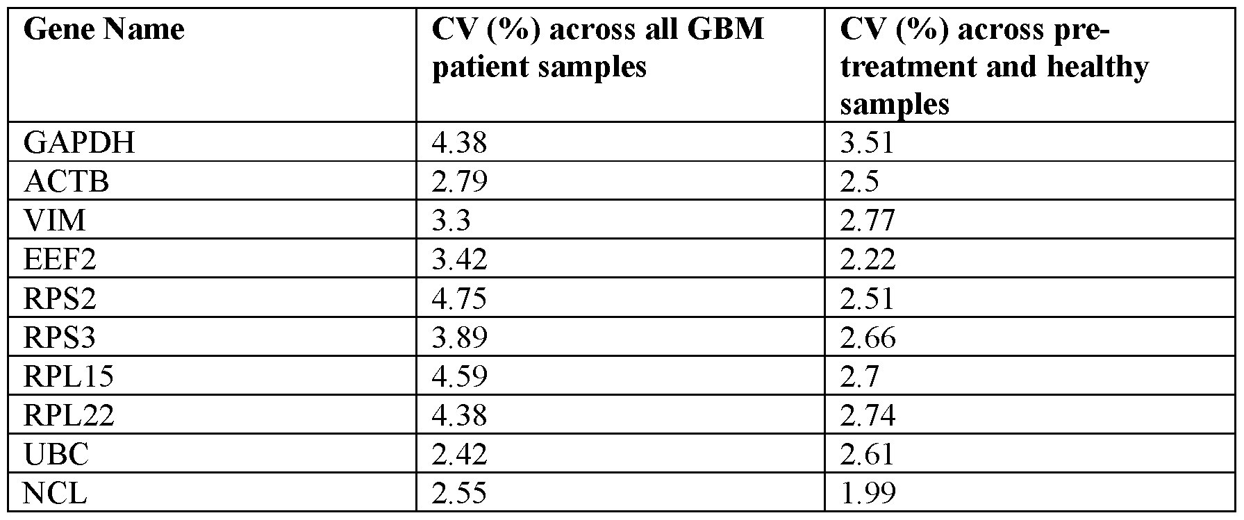
[00270] Table 7. Genes differentially expressed in pre-treatment vs post-treatment patient samples

H3F3AP4 ENSG00000235655.3 RP11-120K18.3 ENSG00000261245.2
SPINK13 ENSG00000214510.9 PTMAP2 ENSGOOOOOl 97744.5
C16orf90 ENSG00000215131.10 IL27 ENSG00000197272.2
OVOL1 ENSG00000172818.9 RP11-592N21.1 ENSG00000212664.5
TP53 ENSG00000141510.16 RP11-445N18.3 ENSG00000228462.1
RP1-102E24.6 ENSG00000256913.1 RAB19 ENSG00000146955.10
RP11-516A11.1 ENSG00000228328.2 IL11 ENSG00000095752.6
ZNF610 ENSGOOOOOl 67554.14 HS6ST1P1 ENSG00000187952.9
RPL5P34 ENSG00000234009.1 HNRNPA1P48 ENSG00000224578.4
RP11-1217F2.20 ENSG00000283481.1 HSPB2-Cl lorf52 ENSG00000254445.1
CTD-2561J22.2 ENSG00000213976.4 LA16c-306E5.3 ENSG00000263212.2
RP11-872D17.8 ENSG00000254979.5 SYS1-DBNDD2 ENSG00000254806.5
HRG ENSGOOOOOl 13905.4 RP11-701124.1 ENSG00000255291.2
RP11-416L21.1 ENSG00000213495.3 RP11-367J7.3 ENSG00000227217.1
RP11-34P13.15 ENSG00000268903.1 AC007743.1 ENSG00000233251.7
RPL7P9 ENSG00000137970.7 CTA-246H3.8 ENSG00000230637.2
PTGER4P2-
CTD-2021A8.3 ENSG00000227080.2 CDK2AP2P2 ENSG00000275450.1
TCAP ENSGOOOOOl 73991.5 RP3-469D22.1 ENSG00000238084.4
KRTAP23-1 ENSGOOOOOl 86980.6 RIBC2 ENSG00000128408.8
RP11-982M15.2 ENSG00000258430.1 RP3-508I15.18 ENSG00000244491.1
AC010524.2 ENSG00000268686.1 RP1-111D6.3 ENSG00000228408.6
HS1BP3-IT1 ENSG00000231948.2 FOXS1 ENSGOOOOOl 79772.7
RP11-543P15.1 ENSG00000227081.5 ST13P15 ENSG00000243759.1
GRTP1-AS1 ENSG00000225083.1 RP11-154H23.3 ENSG00000270562.1
RP1-181J22.1 ENSG00000244535.1 SMG7-AS1 ENSG00000232860.7
CTC-498J12.3 ENSG00000248664.1 MUC20P1 ENSG00000224769.1
RP11-420K14.1 ENSG00000268278.1 MYLK-AS1 ENSG00000239523.5
CDON ENSG00000064309.14 STAG3 ENSG00000066923.17
RP4-761J14.8 ENSG00000219410.5 RP11-256P1.1 ENSG00000249971.1
RP11-560J1.2 ENSG00000271888.1 RPL23AP88 ENSG00000271153.1
HIST2H3PS2 ENSG00000203818.7 WASIR2 ENSG00000231439.4
ORM2 ENSG00000228278.3 CCDC74A ENSG00000163040.14
FAHD2CP ENSG00000231584.8 CA7 ENSG00000168748.13
CTB-152G17.6 ENSG00000272918.1 RP11-585P4.6 ENSG00000273987.1
TLX1 ENSGOOOOOl 07807.12 FAUP1 ENSG00000235297.3
CCT6B ENSG00000132141.13 RP13-644M16.4 ENSG00000196472.4
RPS15AP1 ENSG00000214535.3 OR2F2 ENSG00000221910.2
DRICHl ENSGOOOOOl 89269.12 FAM87B ENSG00000177757.2
TBCAP1 ENSG00000226781.1 ADORA2B ENSGOOOOOl 70425.3
RP11-378J18.8 ENSG00000272750.1 RP11-668G10.2 ENSG00000229894.4
ERVK-28 ENSG00000267696.6 RP11-701P16.2 ENSG00000251139.2 hsa-mir-7641-2 MI0024976 C1QTNF3 ENSG00000082196.20
LA16C-380H5.5 ENSG00000272079.2 ARRDC5 ENSG00000205784.2
RP11-420H19.3 ENSG00000213291.3 RP11-263K19.6 ENSG00000236263.1
ZNF888 ENSG00000213793.4 CTC-246B18.8 ENSG00000268262.1
DCANP1 ENSG00000251380.3 CBLN2 ENSG00000141668.9
GALNT12 ENSGOOOOOl 19514.6 RP11-517A5.6 ENSG00000263029.1
RP11-67A1.2 ENSG00000261864.1 RPS4XP11 ENSG00000234335.1
AC083884.8 ENSG00000232729.7 AP003774.6 ENSG00000231680.1
SLC19A3 ENSG00000135917.13 ZSCAN10 ENSG00000130182.7
AC096558.1 ENSG00000228655.6 RP11-445H22.3 ENSG00000283440.1
PRDX3P1 ENSG00000229598.1 ZNF157 ENSG00000147117.7
ASMT ENSGOOOOOl 96433.12 CTB-47B8.5 ENSG00000213414.3
RP11-288H12.3 ENSG00000213073.4 KAZALD1 ENSG00000107821.14
RP11-111A22.1 ENSG00000259536.5 RP11-798G7.7 ENSG00000267246.1
RP13-104F24.3 ENSG00000265298.1 MAN1B1-AS1 ENSG00000268996.3
ZDHHC11 ENSGOOOOOl 88818.12 AC004453.8 ENSG00000146677.7
RP4-61404.11 ENSG00000261582.1 PRPF38AP1 ENSG00000225053.1
RP11-578F21.9 ENSG00000260844.2 RP11-51L5.7 ENSG00000270033.1
SNHG6 ENSG00000245910.8 ST13P11 ENSG00000213368.3
ASS1P2 ENS G00000223922.1 MIMT1 ENSG00000268654.1
CTC-273B12.5 ENSG00000268530.5 GLOD5 ENSG00000171433. i l
RP11-923111.3 ENSG00000260122.1 RP11-290D2.6 ENSG00000273149.1
SEPT 14 ENSGOOOOOl 54997.8 RP11-930O11.2 ENSG00000259483.1
RP11-350E12.5 ENSG00000238110.1 LINC00264 ENSG00000233261.3
RP13-270P17.1 ENSG00000264235.5 TRIML2 ENSGOOOOOl 79046.8
RP11-867G23.4 ENSG00000254452.1 DI030S ENSG00000258498.7
FTH1P8 ENSG00000219507.4 PVRIG2P ENSG00000235333.3
RP11-974F13.5 ENSG00000248769.1 AC064874.1 ENSG00000222007.6
TBC1D3G ENSG00000260287.4 NEDD8-MDP1 ENSG00000255526.6
GBP7 ENSG00000213512.1 RP11-314A20.5 ENSG00000261898.2
RP11-421L21.3 ENSG00000233184.6 RP11-106D4.3 ENSG00000276393.1
LYSMD1 ENSG00000163155. i l AL591893.1 ENSG00000229021.2
SLC7A5P2 ENSG00000258186.2 RP5-878I13.1 ENSG00000274374.1
RP11-553K23.2 ENSG00000214280.3 RP11-65B7.2 ENSG00000281883.1
RP11-1000B6.5 ENSG00000244952.2 TNFSF15 ENSGOOOOOl 81634.7
AC002116.8 ENSG00000248101.2 SDHDP6 ENSG00000224183.1
GJA1P1 ENSG00000176857.5 CTB-111H14.1 ENSG00000243797.6
RP11-92K2.2 ENSG00000231767.3 GAS1RR ENSG00000226237.1
RP11-710M11.1 ENSG00000266373.1
NR1I3 ENSG00000143257. i l
RP11-182J1.12 ENSG00000259244.1
ZDHHC9 ENSGOOOOOl 88706.12
RPS7P1 ENSG00000263266.2
RP11-166N17.1 ENSG00000227253.3
MRPL2P1 ENSG00000257480.1

Claims
What is claimed is:
1. A method comprising:
(1) determining the expression level of at least one gene selected from Table 1 and the expression level of at least one reference gene in a biological sample from a subject;
(2) normalizing the expression level of the at least one gene in the biological sample by dividing the expression level of the at least one gene by the expression level of the at least one reference gene;
(3) comparing the normalized expression level of the at least one gene in the biological sample to a predetermined cutoff value; and
(4) identifying the presence of glioblastoma multiforme in the subject when the normalized expression level of the at least one gene is greater than the predetermined cutoff value, or identifying the absence of glioblastoma multiforme in the subject when the normalized expression level of the at least one gene is equal to or less than the predetermined cutoff value.
2. A method comprising:
(1) Determining the expression level of at least one gene selected from Table 2 and the expression level of at least one reference gene in a biological sample from a subject;
(2) normalizing the expression level of the at the least one gene in the biological sample by dividing the expression level of the at least one gene by the expression level of the at least one reference gene;
(3) comparing the normalized expression level of the at least one gene in the biological sample to a predetermined cutoff value; and
(4) identifying the presence of glioblastoma multiforme in the subject when the normalized expression level of the at least one gene is less than the predetermined cutoff value, or identifying the absence of glioblastoma multiforme in the subject when the normalized expression level of the at least one gene is equal to or greater than the predetermined cutoff value.
3. The method of claim 1 or claim 2, wherein the at least one reference gene comprises at least one gene selected from Table 5.
4. The method of claim 3, wherein the at least one reference gene is GAPDH.
5. The method of claim 1 or claim 2, wherein the at least one reference gene comprises a gene that has an expression level with a coefficient of variation of less than 20% in biological samples from subjects having cancer and biological samples from subjects not having cancer.
6. The method of claim 5, wherein the at least one reference gene comprises a gene that has an expression level with a coefficient of variation of less than 10% in biological samples from subjects having cancer and biological samples from subjects not having cancer.
7. The method of claim 6, wherein the at least one reference gene comprises a gene that has an expression level with a coefficient of variation of less than 5% in biological samples from subjects having cancer and biological samples from subjects not having cancer.
8. The method of claim 1 or claim 2, wherein the predetermined cutoff value has a positive predictive value of at least 70%.
9. The method of claim 8, wherein the predetermined cutoff value has a positive predictive value of at least 80%.
10. The method of claim 9, wherein the predetermined cutoff value has a positive predictive value of at least 90%.
11. The method of claim 10, wherein the predetermined cutoff value has a positive predictive value of at least 99%.
12. The method of claim 1 or claim 2, wherein the predetermined cutoff value has a sensitivity of at least 70%.
13. The method of claim 12, wherein the predetermined cutoff value has a sensitivity of at least 80%.
14. The method of claim 13, wherein the predetermined cutoff value has a sensitivity of at least 90%.
15. The method of claim 14, wherein the predetermined cutoff value has a sensitivity of at least 99%.
16. The method of claim 1 or claim 2, wherein a biological sample comprises at least one nucleic acid.
17. The method of claim 16, wherein the nucleic acid is RNA.
18. The method of claim 16, wherein the at least one nucleic acid is extracted from a microvesicle fraction.
19. The method of claim 18, wherein the microvesicle fraction is isolated from a bodily fluid sample selected from blood, plasma, serum, urine or cerebrospinal fluid (CSF) sample.
20. The method of claim 19, wherein the microvesicle fraction is isolated by a method comprising:
(a) processing a microvesicle fraction to exclude proteins, lipids, debris from dead cells, and other contaminants;
(b) purifying microvesicles using size exclusion chromatography, density gradient centrifugation, centrifugation, differential centrifugation, immunoabsorbent capture, affinity purification, microfluidic separation, ultracentrifugation or a nanomembrane ultrafiltration concentrator; and
(c) washing the microvesicles.
21. The method of claim 1 or claim 2, wherein determining the expression level of the at least one gene and the at least one reference gene in step (1) comprises using quantitative reverse transcription PCR.
22. The method of claim 1 or claim 2, wherein determining the expression level of the at least one gene and the at least one reference gene in step (1) comprises sequencing.
23. The method of claim 22, wherein the sequencing is high-throughput sequencing.
24. The method of claim 22, wherein sequencing comprises performing RNA-SEQ.
25. The method of claim 1 , wherein the at least one gene is CREBBP.
26. The method of claim 25, wherein the at least one reference gene is GAPDH.
27. The method of claim 26, wherein the predetermined cutoff value is at least 0.4.
28. The method of claim 1 , wherein the at least one gene is CXCR2.
29. The method of claim 28, wherein the at least one reference gene is GAPDH.
30. The method of claim 29, wherein the predetermined cutoff value is at least 0.1
31. The method of claim 1 , wherein the at least gene is S 100A9.
32. The method of claim 31, wherein the at least one reference gene is GAPDH.
33. The method of claim 32, wherein the predetermined cutoff value is at least 1.0.
34. A method comprising:
(1) Determining the expression level of at least one gene selected from Table 3 and the expression level of at least one reference gene in a biological sample from a subject having cancer;
(2) normalizing the expression level of the at the least one gene in the biological sample by dividing the expression level of the at least one gene by the expression level of the at least one reference gene;
(3) comparing the normalized expression level of the at least one gene in the biological sample to a predetermined cutoff value; and
(4) recommending initiating an anti-cancer therapy when the normalized expression level of the at least one gene is greater than the predetermined cutoff value, or recommending not initiating an anti-cancer therapy when the normalized expression level of the at least one gene is equal to or less than the predetermined cutoff value.
A method comprising:
(1) Determining the expression level of at least one gene selected from Table 4 and the expression level of at least one reference gene in a biological sample from a subject having cancer;
(2) normalizing the expression level of the at the least one gene in the biological sample by dividing the expression level of the at least one gene by the expression level of the at least one reference gene;
(3) comparing the normalized expression level of the at least one gene in the biological sample to a predetermined cutoff value; and
(4) recommending initiating an anti-cancer therapy when the normalized expression level of the at least one gene is less than the predetermined cutoff value, or recommending not initiating an anti-cancer therapy when the normalized expression level of the at least one gene is equal to or greater than the predetermined cutoff value.
36. The method of claim 34 or claim 35, wherein the at least one reference gene comprises at least one gene selected from Table 5.
37. The method of claim 36, wherein the at least one reference gene is GAPDH.
38. The method of claim 34 or claim 35, Wherein the at least one reference gene comprises a gene that has an expression level with a coefficient of variation of less than 20% in biological samples from subj ects having cancer and biological samples from subjects not having cancer.
39. The method of claim 38, wherein the at least one reference gene comprises a gene that has an expression level with a coefficient of variation of less than 10% in biological samples from subjects having cancer and biological samples from subj ects not having cancer.
40. The method of claim 39, wherein the at least one reference gene comprises a gene that has an expression level with a coefficient of variation of less than 5% in biological samples from subjects having cancer and biological samples from subj ects not having cancer.
41. The method of claim 34 or claim 35, wherein the predetermined cutoff value has a positive predictive value of at least 70%.
42. The method of claim 41, wherein the predetermined cutoff value has a positive predictive value of at least 80%.
43. The method of claim 42, wherein the predetermined cutoff value has a positive predictive value of at least 90%.
44. The method of claim 43, wherein the predetermined cutoff value has a positive predictive value of at least 99%.
45. The method of claim 34 or claim 35, wherein the predetermined cutoff value has a sensitivity of at least 70%.
46. The method of claim 45, wherein the predetermined cutoff value has a sensitivity of at least 80%.
47. The method of claim 46, wherein the predetermined cutoff value has a sensitivity of at least 90%.
48. The method of claim 47, wherein the predetermined cutoff value has a sensitivity of at least 99%.
49. The method of claim 34 or 35, wherein a biological sample comprises at least one nucleic acid.
50. The method of claim 49, wherein the nucleic acid is RNA.
51. The method of claim 49, wherein the at least one nucleic acid is extracted from a microvesicle fraction.
52. The method of claim 51, wherein the microvesicle fraction is isolated from a bodily fluid sample selected from blood, plasma, serum, urine or cerebrospinal fluid (CSF) sample.
53. The method of claim 52, wherein the microvesicle fraction is isolated by a method comprising:
(a) processing a microvesicle fraction to exclude proteins, lipids, debris from dead cells, and other contaminants;
(b) purifying microvesicles using size exclusion chromatography, density gradient centrifugation, centrifugation, differential centrifugation, immunoabsorbent capture, affinity purification, microfluidic separation, ultracentrifugation or a nanomembrane ultrafiltration concentrator; and
(c) washing the microvesicles.
54. The method of claim 34 or 35, wherein determining the expression level of the at least one gene and the at least one reference gene in step (1) comprises using quantitative reverse transcription PCR.
55. The method of claim 34 or 35, wherein determining the expression level of the at least one gene and the at least one reference gene in step (1) comprises sequencing.
56. The method of claim 55, wherein the sequencing is high-throughput sequencing.
57. The method of claim 55, wherein sequencing comprises performing RNA-SEQ.
58. The method of claim 34 or 35, wherein the anti-cancer therapy comprises
administering to the subject a therapeutically effective dose of at least one class of drugs.
59. The method of claim 58, wherein the at least one class of drugs comprises tyrosine kinase inhibitors.
60. The method of claim 59, wherein the tyrosine kinase inhibitors are epidermal growth factor receptor (EGFR) inhibitors.
61. The method of claim 60, wherein the EGFR inhibitors are irreversible EGFR inhibitors.
62. The method of claim 60, wherein the EGFR inhibitors are pan-human epidermal growth factor receptor (pan-HER) inhibitors.
63. The method of claim 62, wherein the pan-HER inhibitors are administered in combination with immunotherapy or a checkpoint inhibitor.
64. The method of claim 62, wherein the pan-HER inhibitor is Dacomitinib.
65. The method of claim 34 or 35, wherein the cancer is brain cancer.
66. The method of claim 65, wherein the brain cancer is selected from a group comprising Acoustic Neuroma, Pilocytic Astrocytoma, Low-grade Astrocytoma, Anaplastic
Astrocytoma, Glioblastoma multiforme (GBM), Chordoma, CNS Lymphoma,
Craniopharyngioma, Brain Stem Glioma, Ependymoma, Mixed Glioma, Optic Nerve Glioma, Subependymoma, Medulloblastoma, Meningioma, Metastatic Brain Tumors,
Oligodendroglioma, Pituitary Tumors, Primitive Neuroectodermal (PNET), Schwannoma, Brain Stem Glioma, Craniopharyngioma, Ependymoma, Juvenile Pilocytic Astrocytoma (JPA), Medulloblastoma, Optic Nerve Glioma, Pineal Tumor, Primitive Neuroectodermal Tumors (PNET), or Rhabdoid Tumor.
67. The method of claim 66, wherein the brain cancer is Glioblastoma multiforme.
68. The method 34, wherein the at least one gene is ZNF35.
69. The method of claim 68, wherein the at least one reference gene is GAPDH.
70. The method of claim 69, wherein the predetermined cutoff value is at least 0.002.
71. The method of claim 35, wherein the at least one gene is LAMTOR2.
72. The method of claim 71, wherein the at least one reference gene is GAPDH.
73. The method of claim 71, wherein the predetermined cutoff value is at most 0.0125.
74. A method comprising:
(1) Determining the expression level of at least one gene selected from ZNF302, DNMT3A, BHLHA15, CTD-2132N18.3, ADORA2B, LAMTOR2, or ZNF35 and the
expression level of at least one reference gene in a biological sample from a subject having cancer and prior to administration of an anti-cancer therapy;
(2) Determining the expression level of at least one gene selected from ZNF302, DNMT3A, BHLHA15, CTD-2132N18.3, ADORA2B, LAMTOR2, or ZNF35 and the expression level of at least one reference gene in a biological sample from a subject having cancer and at least one week after administration of the anti-cancer therapy;
(3) normalizing the expression level of the at the least one gene in the biological sample prior to administration of the anti-cancer therapy by dividing the expression level of the at least one gene in the biological sample prior to administration of the anti-cancer therapy by the expression level of the at least one reference gene in the biological sample prior to administration of the anti-cancer therapy;
(4) normalizing the expression level of the at least one gene in the biological sample at least one week after administration of the anti-cancer therapy by dividing the expression level of the at least one gene in the biological sample at least one week after administration of an anti-cancer therapy by the expression level of the at least one reference gene in the biological sample at least one week after administration of the anti-cancer therapy; and
(5) recommending continuing the anti-cancer therapy when the normalized expression level of the at least one gene in the biological sample at least one week after administration of the anti-cancer therapy is greater than the normalized expression level of the at least one gene in the biological sample prior to administration of the anti-cancer therapy, or recommending suspending the anti-cancer therapy when the normalized expression level of the at least one gene in the biological sample at least one week after administration of the anti-cancer therapy is equal to or less than the normalized expression level of the at least one gene in the biological sample prior to administration of the anti-cancer therapy.
75. A method comprising:
(1) Determining the expression level of at least one gene selected from ZNF302, DNMT3A, BHLHA15, CTD-2132N18.3, ADORA2B, LAMTOR2, or ZNF35 and the expression level of at least one reference gene in a biological sample from a subject having cancer and prior to administration of an anti-cancer therapy;
(2) Determining the expression level of at least one gene selected from ZNF302, DNMT3A, BHLHA15, CTD-2132N18.3, ADORA2B, LAMTOR2, or ZNF35 and the expression level of at least one reference gene in a biological sample from a subject having cancer and at least one week after administration of the anti-cancer therapy;
(3) normalizing the expression level of the at the least one gene in the biological sample prior to administration of the anti-cancer therapy by dividing the expression level of the at least one gene in the biological sample prior to administration of the anti-cancer therapy by the expression level of the at least one reference gene in the biological sample prior to administration of the anti-cancer therapy;
(4) normalizing the expression level of the at least one gene in the biological sample at least one week after administration of the anti-cancer therapy by dividing the expression level of the at least one gene in the biological sample at least one week after administration of an anti-cancer therapy by the expression level of the at least one reference gene in the biological sample at least one week after administration of the anti-cancer therapy; and
(5) recommending continuing the anti-cancer therapy when the normalized expression level of the at least one gene in the biological sample at least one week after administration of the anti-cancer therapy is less than the normalized expression level of the at least one gene in the biological sample prior to administration of the anti-cancer therapy, or recommending suspending the anti-cancer therapy when the normalized expression level of the at least one gene in the biological sample at least one week after administration of the anti-cancer therapy is equal to or greater than the normalized expression level of the at least one gene in the biological sample prior to administration of the anti-cancer therapy.
76. A method comprising:
(1) Determining the expression level of at least one gene selected from ZNF302, DNMT3A, BHLHA15, CTD-2132N18.3, ADORA2B, LAMTOR2, or ZNF35 and the expression level of at least one reference gene in a biological sample from a subject having cancer and at least one week after administration of the anti-cancer therapy;
(2) normalizing the expression level of the at least one gene in the biological sample by dividing the expression level of the at least one gene by the expression level of the at least one reference gene;
(3) comparing the normalized expression level of the at least one gene in the biological sample to a predetermined cutoff value; and
(4) recommending continuing the anti-cancer therapy when the normalized expression level of the at least one gene in the biological sample is greater than the predetermined cutoff value, or recommending suspending the anti-cancer therapy when the normalized expression level of the at least one gene in the biological sample is equal to or less than the
predetermined cutoff value.
77. A method comprising:
(1) Determining the expression level of at least one gene selected from ZNF302, DNMT3A, BHLHA15, CTD-2132N18.3, ADORA2B, LAMTOR2, or ZNF35 and the expression level of at least one reference gene in a biological sample from a subject having cancer and at least one week after administration of the anti-cancer therapy;
(2) normalizing the expression level of the at least one gene in the biological sample by dividing the expression level of the at least one gene by the expression level of the at least one reference gene;
(3) comparing the normalized expression level of the at least one gene in the biological sample to a predetermined cutoff value; and
(4) recommending continuing the anti-cancer therapy when the normalized expression level of the at least one gene in the biological sample is less than the predetermined cutoff value, or recommending suspending the anti-cancer therapy when the normalized expression level of the at least one gene in the biological sample is equal to or greater than the predetermined cutoff value.
78. A method comprising:
(1) Determining the expression level of at least one gene selected from ZNF302, DNMT3A, BHLHA15, CTD-2132N18.3, ADORA2B, LAMTOR2, or ZNF35 and the expression level of at least one reference gene in a biological sample from a subject having cancer and prior to administration of an anti-cancer therapy;
(2) Determining the expression level of at least one gene selected from ZNF302, DNMT3A, BHLHA15, CTD-2132N18.3, ADORA2B, LAMTOR2, or ZNF35 and the expression level of at least one reference gene in a biological sample from a subject having cancer and at least one week after administration of the anti-cancer therapy;
(3) normalizing the expression level of the at the least one gene in the biological sample prior to administration of the anti-cancer therapy by dividing the expression level of the at least one gene in the biological sample prior to administration of the anti-cancer therapy by the expression level of the at least one reference gene in the biological sample prior to administration of the anti-cancer therapy;
(4) normalizing the expression level of the at least one gene in the biological sample at least one week after administration of the anti-cancer therapy by dividing the expression level of the at least one gene in the biological sample at least one week after administration of
an anti-cancer therapy by the expression level of the at least one reference gene in the biological sample at least one week after administration of the anti-cancer therapy; and
(5) generating a score by dividing the normalized expression level of the at least one gene in the biological sample at least one week after administration of the anti-cancer therapy by the normalized expression level of the at least one gene in the biological sample prior to administration of the anti-cancer therapy;
(6) comparing the score to a predetermined cutoff value; and
(7) recommending continuing the anti-cancer therapy when the score is greater than the predetermined cutoff value, or recommending suspending an anti-cancer therapy when the score is equal to or less than the predetermined cutoff value.
79. A method comprising:
(1) Determining the expression level of at least one gene selected from ZNF302, DNMT3A, BHLHA15, CTD-2132N18.3, ADORA2B, LAMTOR2, or ZNF35 and the expression level of at least one reference gene in a biological sample from a subject having cancer and prior to administration of an anti-cancer therapy;
(2) Determining the expression level of at least one gene selected from ZNF302, DNMT3A, BHLHA15, CTD-2132N18.3, ADORA2B, LAMTOR2, or ZNF35 and the expression level of at least one reference gene in a biological sample from a subject having cancer and at least one week after administration of the anti-cancer therapy;
(3) normalizing the expression level of the at the least one gene in the biological sample prior to administration of the anti-cancer therapy by dividing the expression level of the at least one gene in the biological sample prior to administration of the anti-cancer therapy by the expression level of the at least one reference gene in the biological sample prior to administration of the anti-cancer therapy;
(4) normalizing the expression level of the at least one gene in the biological sample at least one week after administration of the anti-cancer therapy by dividing the expression level of the at least one gene in the biological sample at least one week after administration of an anti-cancer therapy by the expression level of the at least one reference gene in the biological sample at least one week after administration of the anti-cancer therapy; and
(5) generating a score by dividing the normalized expression level of the at least one gene in the biological sample at least one week after administration of the anti-cancer therapy by the normalized expression level of the at least one gene in the biological sample prior to administration of the anti-cancer therapy;
(6) comparing the score to a predetermined cutoff value; and
(7) recommending continuing the anti-cancer therapy when the score is less than the predetermined cutoff value, or recommending suspending anti-cancer therapy when the score is equal to or greater than the predetermined cutoff value.
80. The method of any of claims 74 to 79, wherein the at least one reference gene comprises at least one gene selected from Table 5.
81. The method of claim 80, wherein the at least one reference gene is GAPDH.
82. The method of any of claims 74 to 79, Wherein the at least one reference gene comprises a gene that has an expression level with a coefficient of variation of less than 20% in biological samples from subj ects having cancer and biological samples from subjects not having cancer.
83. The method of claim 82, wherein the at least one reference gene comprises a gene that has an expression level with a coefficient of variation of less than 10% in biological samples from subjects having cancer and biological samples from subj ects not having cancer.
84. The method of claim 83, wherein the at least one reference gene comprises a gene that has an expression level with a coefficient of variation of less than 5% in biological samples from subjects having cancer and biological samples from subj ects not having cancer.
85. The method of any of claims 76 to 79, wherein the predetermined cutoff value has a positive predictive value of at least 70%.
86. The method of claim 85, wherein the predetermined cutoff value has a positive predictive value of at least 80%.
87. The method of claim 86, wherein the predetermined cutoff value has a positive predictive value of at least 90%.
88. The method of claim 87, wherein the predetermined cutoff value has a positive predictive value of at least 99%.
89. The method of any of claims 76 to 79, wherein the predetermined cutoff value has a sensitivity of at least 70%.
90. The method of claim 89, wherein the predetermined cutoff value has a sensitivity of at least 80%.
91. The method of claim 90, wherein the predetermined cutoff value has a sensitivity of at least 90%.
92. The method of claim 91, wherein the predetermined cutoff value has a sensitivity of at least 99%.
93. The method of any of claims 74 to 79, wherein a biological sample comprises at least one nucleic acid.
94. The method of claim 93, wherein the nucleic acid is RNA.
95. The method of claim 93, wherein the at least one nucleic acid is extracted from a microvesicle fraction.
96. The method of claim 95, wherein the microvesicle fraction is isolated from a bodily fluid sample selected from blood, plasma, serum, urine or cerebrospinal fluid (CSF) sample.
97. The method of claim 96, wherein the microvesicle fraction is isolated by a method comprising:
(a) processing a microvesicle fraction to exclude proteins, lipids, debris from dead cells, and other contaminants;
(b) purifying microvesicles using size exclusion chromatography, density gradient centrifugation, centrifugation, differential centrifugation, immunoabsorbent capture, affinity purification, microfluidic separation, ultracentrifugation or a nanomembrane ultrafiltration concentrator; and
(c) washing the microvesicles.
98. The method of any of claims 74 to 79, wherein determining the expression level of a gene comprises using quantitative reverse transcription PCR.
99. The method of any of claims 74 to 79, wherein determining the expression level of a gene comprises sequencing.
100. The method of claim 99, wherein the sequencing is high-throughput sequencing.
101. The method of claim 99, wherein sequencing comprises performing RNA-SEQ.
102. The method of any of claims 74 to 79, wherein the anti-cancer therapy comprises administering to the subject a therapeutically effective dose of at least one class of drugs.
103. The method of claim 102, wherein the at least one class of drugs comprises tyrosine kinase inhibitors.
104. The method of claim 103, wherein the tyrosine kinase inhibitors are epidermal growth factor receptor (EGFR) inhibitors.
105. The method of claim 104, wherein the EGFR inhibitors are irreversible EGFR inhibitors.
106. The method of claim 104, wherein the EGFR inhibitors are pan-human epidermal growth factor receptor (pan-HER) inhibitors.
107. The method of claim 106, wherein the pan-HER inhibitors are administered in combination with immunotherapy or a checkpoint inhibitor.
108. The method of claim 106, wherein the pan-HER inhibitor is Dacomitinib.
109. The method of claim any of claims 74 to 79, wherein the cancer is brain cancer.
110. The method of claim 109, wherein the brain cancer is selected from a group comprising Acoustic Neuroma, Pilocytic Astrocytoma, Low-grade Astrocytoma, Anaplastic
Astrocytoma, Glioblastoma multiforme (GBM), Chordoma, CNS Lymphoma,
Craniopharyngioma, Brain Stem Glioma, Ependymoma, Mixed Glioma, Optic Nerve Glioma, Subependymoma, Medulloblastoma, Meningioma, Metastatic Brain Tumors,
Oligodendroglioma, Pituitary Tumors, Primitive Neuroectodermal (PNET), Schwannoma, Brain Stem Glioma, Craniopharyngioma, Ependymoma, Juvenile Pilocytic Astrocytoma (JPA), Medulloblastoma, Optic Nerve Glioma, Pineal Tumor, Primitive Neuroectodermal Tumors (PNET), or Rhabdoid Tumor.
111. The method of claim 110, wherein the brain cancer is Glioblastoma multiforme.
112. The method of any of claims 74 to 79, wherein suspending the anti-cancer therapy comprises ceasing the anti-cancer therapy.
113. The method of claim 76, wherein the at least one gene is ZNF35.
114. The method of claim 113, wherein the at least one reference gene is GAPDH.
115. The method of claim 115, wherein the predetermined cutoff value is at least 0.004.
116. The method of claim 76, wherein the at least one gene is DNMT3A.
117. The method of claim 116, wherein the at least one reference gene is GAPDH.
118. The method of claim 117, wherein the predetermined cutoff value is at least 0.5.
Priority Applications (2)
| Application Number | Priority Date | Filing Date | Title |
|---|---|---|---|
| US16/631,603 US11345957B2 (en) | 2017-07-18 | 2018-07-18 | Methods of treating glioblastoma in a subject informed by exosomal RNA signatures |
| EP18834871.8A EP3655531A4 (en) | 2017-07-18 | 2018-07-18 | Sequencing of nucleic acids associated with exosomal isolation from patients with glioblastoma multiforme |
Applications Claiming Priority (2)
| Application Number | Priority Date | Filing Date | Title |
|---|---|---|---|
| US201762534141P | 2017-07-18 | 2017-07-18 | |
| US62/534,141 | 2017-07-18 |
Publications (1)
| Publication Number | Publication Date |
|---|---|
| WO2019018537A1 true WO2019018537A1 (en) | 2019-01-24 |
Family
ID=65016582
Family Applications (1)
| Application Number | Title | Priority Date | Filing Date |
|---|---|---|---|
| PCT/US2018/042708 WO2019018537A1 (en) | 2017-07-18 | 2018-07-18 | Sequencing of nucleic acids associated with exosomal isolation from patients with glioblastoma multiforme |
Country Status (3)
| Country | Link |
|---|---|
| US (1) | US11345957B2 (en) |
| EP (1) | EP3655531A4 (en) |
| WO (1) | WO2019018537A1 (en) |
Cited By (3)
| Publication number | Priority date | Publication date | Assignee | Title |
|---|---|---|---|---|
| CN111394464A (en) * | 2020-04-24 | 2020-07-10 | 青岛市中心医院 | Detection reagent for radioactive damage diseases and application thereof |
| US11085089B2 (en) | 2019-03-01 | 2021-08-10 | Mercy Bioanalytics, Inc. | Systems, compositions, and methods for target entity detection |
| US11345957B2 (en) | 2017-07-18 | 2022-05-31 | Exosome Diagnostics, Inc. | Methods of treating glioblastoma in a subject informed by exosomal RNA signatures |
Families Citing this family (6)
| Publication number | Priority date | Publication date | Assignee | Title |
|---|---|---|---|---|
| CN111088357B (en) * | 2019-12-31 | 2022-09-20 | 深圳大学 | Tumor marker for ESCC and application thereof |
| CN113621580B (en) * | 2021-08-18 | 2023-01-31 | 山东科金生物发展有限公司 | Reagent for enhancing in-vitro amplification activity of adipose-derived mesenchymal stem cells |
| CN113862269B (en) * | 2021-10-25 | 2023-12-22 | 中南大学湘雅三医院 | tsRNA molecules and uses thereof |
| WO2023102051A1 (en) * | 2021-12-01 | 2023-06-08 | Cedars-Sinai Medical Center | Intestinal mononuclear phagocytes as prognostic biomarker for crohn's disease |
| CN114182016A (en) * | 2021-12-02 | 2022-03-15 | 复旦大学 | Application of AP006284.1 as molecular target of prostate cancer |
| WO2023192227A2 (en) * | 2022-03-29 | 2023-10-05 | The Regents Of The University Of California | Methods for determining the presence, type, grade, classification of a tumor, cyst, lesion, mass, and/or cancer |
Citations (9)
| Publication number | Priority date | Publication date | Assignee | Title |
|---|---|---|---|---|
| WO2001063292A2 (en) * | 2000-02-22 | 2001-08-30 | Board Of Regents, The University Of Texas System | Compositions and methods of use of het, a novel modulator of estrogen action |
| US20100093556A1 (en) * | 2005-06-13 | 2010-04-15 | The Regents Of The University Of Michigan | Compositions and methods for treating and diagnosing cancer |
| US20120164172A1 (en) * | 2009-09-28 | 2012-06-28 | FARRIES Timothy | Cancer Therapy |
| US20130224208A1 (en) * | 2010-09-30 | 2013-08-29 | Riken | Glioma treatment method, glioma testing method, method for delivering desired substance to glioma, and drugs used in these methods |
| US20140011702A1 (en) * | 2011-03-23 | 2014-01-09 | Centre Hospitalier Universitaire Pontchaillou | Biomarkers and methods for the prognosis of glioblastoma |
| US20140275073A1 (en) * | 2013-03-12 | 2014-09-18 | The Board Of Trustees Of The Leland Stanford Junior University | Methods of treating cancer sensitive to anti-egfr therapy and modifying treatment using blood biomarkers |
| US20150038335A1 (en) * | 2011-11-10 | 2015-02-05 | Exosome Diagnostics, Inc. | Cerebrospinal fluid assay |
| WO2016040892A1 (en) * | 2014-09-13 | 2016-03-17 | Novartis Ag | Combination therapies |
| US20160235788A1 (en) * | 2013-10-09 | 2016-08-18 | Reneuron Limited | STEM CELL MICROPARTICLES AND miRNA |
Family Cites Families (21)
| Publication number | Priority date | Publication date | Assignee | Title |
|---|---|---|---|---|
| US5219727A (en) | 1989-08-21 | 1993-06-15 | Hoffmann-Laroche Inc. | Quantitation of nucleic acids using the polymerase chain reaction |
| CA2218875C (en) | 1991-07-23 | 2000-11-07 | The Research Foundation Of State University Of New York | Improvements in the in situ pcr |
| US5639606A (en) | 1993-04-06 | 1997-06-17 | The University Of Rochester | Method for quantitative measurement of gene expression using multiplex competitive reverse transcriptase-polymerase chain reaction |
| US5556773A (en) | 1993-08-06 | 1996-09-17 | Yourno; Joseph | Method and apparatus for nested polymerase chain reaction (PCR) with single closed reaction tubes |
| FR2788780B1 (en) | 1999-01-27 | 2001-03-30 | Ap Cells Inc | PROCESS FOR THE PREPARATION OF MEMBRANE VESICLES |
| GB9927320D0 (en) | 1999-11-18 | 2000-01-12 | Chiron Spa | Exosome separation |
| US6812023B1 (en) | 2000-04-27 | 2004-11-02 | Anosys, Inc. | Methods of producing membrane vesicles |
| WO2009100029A1 (en) * | 2008-02-01 | 2009-08-13 | The General Hospital Corporation | Use of microvesicles in diagnosis, prognosis and treatment of medical diseases and conditions |
| WO2012031008A2 (en) | 2010-08-31 | 2012-03-08 | The General Hospital Corporation | Cancer-related biological materials in microvesicles |
| WO2014015149A2 (en) | 2012-07-18 | 2014-01-23 | Exosome Diagnostics, Inc. | Use of microvesicles in diagnosis, prognosis, and treatment of medical diseases and conditions |
| EP2890783A4 (en) * | 2012-08-30 | 2016-03-02 | Exosome Diagnostics Inc | Controls for nucleic acid assays |
| CN110106229B (en) * | 2013-01-03 | 2023-03-28 | 外来体诊断公司 | Method for isolating microvesicles |
| EP3033418B1 (en) | 2013-08-14 | 2019-12-11 | Reneuron Limited | Stem cell microparticles and mirna |
| EP3049536B1 (en) | 2013-09-23 | 2019-05-22 | The University of Chicago | Methods and compositions relating to cancer therapy with dna damaging agents |
| WO2015152724A2 (en) | 2014-04-02 | 2015-10-08 | Stichting Vu-Vumc | Biomarkers for the detection of frontotemporal dementia |
| CA2954576C (en) * | 2014-07-09 | 2020-08-11 | Daniel ENDERLE | Methods for isolating microvesicles and extracting nucleic acids from biological samples |
| US10266895B2 (en) * | 2014-11-05 | 2019-04-23 | Hitachi Chemical Company Ltd. | Exosomes and microvesicles in intestinal luminal fluids and stool and use of same for the assessment of inflammatory bowel disease |
| WO2016172598A1 (en) | 2015-04-22 | 2016-10-27 | The Broad Institute Inc. | Exosomes and uses thereof |
| US20190093172A1 (en) | 2016-04-15 | 2019-03-28 | Exosome Diagnostics, Inc. | Plasma-based detection of anaplastic lymphoma kinase (alk) nucleic acids and alk fusion transcripts and uses thereof in diagnosis and treatment of cancer |
| US11193943B2 (en) * | 2017-02-06 | 2021-12-07 | Edward Goetzl | Endothelial cell derived exosomes and uses thereof |
| US11345957B2 (en) | 2017-07-18 | 2022-05-31 | Exosome Diagnostics, Inc. | Methods of treating glioblastoma in a subject informed by exosomal RNA signatures |
-
2018
- 2018-07-18 US US16/631,603 patent/US11345957B2/en active Active
- 2018-07-18 EP EP18834871.8A patent/EP3655531A4/en active Pending
- 2018-07-18 WO PCT/US2018/042708 patent/WO2019018537A1/en unknown
Patent Citations (9)
| Publication number | Priority date | Publication date | Assignee | Title |
|---|---|---|---|---|
| WO2001063292A2 (en) * | 2000-02-22 | 2001-08-30 | Board Of Regents, The University Of Texas System | Compositions and methods of use of het, a novel modulator of estrogen action |
| US20100093556A1 (en) * | 2005-06-13 | 2010-04-15 | The Regents Of The University Of Michigan | Compositions and methods for treating and diagnosing cancer |
| US20120164172A1 (en) * | 2009-09-28 | 2012-06-28 | FARRIES Timothy | Cancer Therapy |
| US20130224208A1 (en) * | 2010-09-30 | 2013-08-29 | Riken | Glioma treatment method, glioma testing method, method for delivering desired substance to glioma, and drugs used in these methods |
| US20140011702A1 (en) * | 2011-03-23 | 2014-01-09 | Centre Hospitalier Universitaire Pontchaillou | Biomarkers and methods for the prognosis of glioblastoma |
| US20150038335A1 (en) * | 2011-11-10 | 2015-02-05 | Exosome Diagnostics, Inc. | Cerebrospinal fluid assay |
| US20140275073A1 (en) * | 2013-03-12 | 2014-09-18 | The Board Of Trustees Of The Leland Stanford Junior University | Methods of treating cancer sensitive to anti-egfr therapy and modifying treatment using blood biomarkers |
| US20160235788A1 (en) * | 2013-10-09 | 2016-08-18 | Reneuron Limited | STEM CELL MICROPARTICLES AND miRNA |
| WO2016040892A1 (en) * | 2014-09-13 | 2016-03-17 | Novartis Ag | Combination therapies |
Non-Patent Citations (3)
| Title |
|---|
| DONG ET AL.: "Integrated Analysis of Mutations, Mirna and Mrna Expression in Glioblastoma", BMC SYSTEMS BIOLOGY, vol. 4, no. 163, 2010, pages 1 - 20, XP021090509 * |
| DWYER ET AL.: "Glioblastoma Cell -Secreted Interleukin-8 Induces Brain Endothelial Cell Permeability via CXCR2", PLOS ONE, vol. 7, no. 9, 20 September 2012 (2012-09-20), pages 1 - 13, XP055410388 * |
| See also references of EP3655531A4 * |
Cited By (4)
| Publication number | Priority date | Publication date | Assignee | Title |
|---|---|---|---|---|
| US11345957B2 (en) | 2017-07-18 | 2022-05-31 | Exosome Diagnostics, Inc. | Methods of treating glioblastoma in a subject informed by exosomal RNA signatures |
| US11085089B2 (en) | 2019-03-01 | 2021-08-10 | Mercy Bioanalytics, Inc. | Systems, compositions, and methods for target entity detection |
| CN111394464A (en) * | 2020-04-24 | 2020-07-10 | 青岛市中心医院 | Detection reagent for radioactive damage diseases and application thereof |
| CN111394464B (en) * | 2020-04-24 | 2021-03-16 | 青岛市中心医院 | Detection reagent for radioactive damage diseases and application thereof |
Also Published As
| Publication number | Publication date |
|---|---|
| US20200224245A1 (en) | 2020-07-16 |
| US11345957B2 (en) | 2022-05-31 |
| EP3655531A4 (en) | 2021-09-08 |
| EP3655531A1 (en) | 2020-05-27 |
Similar Documents
| Publication | Publication Date | Title |
|---|---|---|
| US11345957B2 (en) | Methods of treating glioblastoma in a subject informed by exosomal RNA signatures | |
| US11802316B2 (en) | Methods and compositions related to T-cell activity | |
| JP6938584B2 (en) | Diagnosis of diseases caused by extracellular vesicles | |
| AU2014305994B2 (en) | Urine biomarker cohorts, gene expression signatures, and methods of use thereof | |
| US20200332361A1 (en) | Method for assisting detection of alzheimer's disease or mild cognitive impairment | |
| JP2019535307A (en) | Sequencing and analysis of exosome-bound nucleic acids | |
| EP2406402A2 (en) | Method to assess human allograft status from microrna expression levels | |
| US20130040833A1 (en) | Use of microvesicles in analyzing nucleic acid profiles | |
| Kennel et al. | Longitudinal profiling of circulating miRNA during cardiac allograft rejection: a proof‐of‐concept study | |
| US20180148789A1 (en) | Methods for treating and assessing tumor invasion and metastasis | |
| US11525161B2 (en) | Methods of distinguishing ischemic stroke from intracerebral hemorrhage | |
| US20210230701A1 (en) | Methods for developing urine biomarkers and for detecting bladder cancer | |
| JP2019513352A (en) | Purification of RNA fraction using hydrophilic polymer material | |
| CN110806480A (en) | Tumor specific cell subset and characteristic gene and application thereof | |
| CN115029438A (en) | Application of RARES 2 gene in diagnosis and treatment of breast cancer brain metastasis | |
| KR101797281B1 (en) | Multiple biomarker for diagnosing cancer | |
| CN111363808B (en) | Biomarker related to Alzheimer disease and application thereof | |
| WO2014182726A2 (en) | Genetic markers for macular degeneration disorder treatment | |
| WO2000073440A1 (en) | Pollinosis-associated gene 787 | |
| JP2003125777A (en) | Pollinosis-relating gene 581 |
Legal Events
| Date | Code | Title | Description |
|---|---|---|---|
| 121 | Ep: the epo has been informed by wipo that ep was designated in this application |
Ref document number: 18834871 Country of ref document: EP Kind code of ref document: A1 |
|
| NENP | Non-entry into the national phase |
Ref country code: DE |
|
| ENP | Entry into the national phase |
Ref document number: 2018834871 Country of ref document: EP Effective date: 20200218 |