EP3029064A1 - Molécules de liaison bispécifiques egfr/igfir - Google Patents
Molécules de liaison bispécifiques egfr/igfir Download PDFInfo
- Publication number
- EP3029064A1 EP3029064A1 EP15192336.4A EP15192336A EP3029064A1 EP 3029064 A1 EP3029064 A1 EP 3029064A1 EP 15192336 A EP15192336 A EP 15192336A EP 3029064 A1 EP3029064 A1 EP 3029064A1
- Authority
- EP
- European Patent Office
- Prior art keywords
- seq
- amino acid
- loop
- acid sequence
- egfr
- Prior art date
- Legal status (The legal status is an assumption and is not a legal conclusion. Google has not performed a legal analysis and makes no representation as to the accuracy of the status listed.)
- Withdrawn
Links
- 238000009739 binding Methods 0.000 title abstract description 329
- 230000027455 binding Effects 0.000 title abstract description 328
- 108060006698 EGF receptor Proteins 0.000 title description 418
- 239000013636 protein dimer Substances 0.000 claims abstract description 63
- 102000002090 Fibronectin type III Human genes 0.000 claims abstract description 19
- 108050009401 Fibronectin type III Proteins 0.000 claims abstract description 19
- 125000003275 alpha amino acid group Chemical group 0.000 claims description 460
- 108090000765 processed proteins & peptides Proteins 0.000 claims description 96
- 102000004196 processed proteins & peptides Human genes 0.000 claims description 90
- 229920001184 polypeptide Polymers 0.000 claims description 85
- 229920001223 polyethylene glycol Chemical group 0.000 claims description 83
- 239000002202 Polyethylene glycol Chemical group 0.000 claims description 49
- 210000004899 c-terminal region Anatomy 0.000 claims description 47
- 241000282414 Homo sapiens Species 0.000 claims description 40
- 108010071390 Serum Albumin Proteins 0.000 claims description 5
- 102000007562 Serum Albumin Human genes 0.000 claims description 5
- 102000004338 Transferrin Human genes 0.000 claims description 3
- 108090000901 Transferrin Proteins 0.000 claims description 3
- 125000003827 glycol group Chemical group 0.000 claims description 3
- 239000012581 transferrin Substances 0.000 claims description 3
- 102000018071 Immunoglobulin Fc Fragments Human genes 0.000 claims description 2
- 108010091135 Immunoglobulin Fc Fragments Proteins 0.000 claims description 2
- SQVRNKJHWKZAKO-UHFFFAOYSA-N beta-N-Acetyl-D-neuraminic acid Natural products CC(=O)NC1C(O)CC(O)(C(O)=O)OC1C(O)C(O)CO SQVRNKJHWKZAKO-UHFFFAOYSA-N 0.000 claims description 2
- 108091008324 binding proteins Proteins 0.000 claims description 2
- 102000028557 immunoglobulin binding proteins Human genes 0.000 claims description 2
- 108091009323 immunoglobulin binding proteins Proteins 0.000 claims description 2
- SQVRNKJHWKZAKO-OQPLDHBCSA-N sialic acid Chemical compound CC(=O)N[C@@H]1[C@@H](O)C[C@@](O)(C(O)=O)OC1[C@H](O)[C@H](O)CO SQVRNKJHWKZAKO-OQPLDHBCSA-N 0.000 claims description 2
- 102000023732 binding proteins Human genes 0.000 claims 1
- 108090000623 proteins and genes Proteins 0.000 abstract description 175
- 102000004169 proteins and genes Human genes 0.000 abstract description 170
- 101001034652 Homo sapiens Insulin-like growth factor 1 receptor Proteins 0.000 abstract description 138
- 102100039688 Insulin-like growth factor 1 receptor Human genes 0.000 abstract description 138
- 230000001225 therapeutic effect Effects 0.000 abstract description 19
- 239000013598 vector Substances 0.000 abstract description 13
- 239000012634 fragment Substances 0.000 abstract description 12
- 102000040430 polynucleotide Human genes 0.000 abstract description 5
- 108091033319 polynucleotide Proteins 0.000 abstract description 5
- 239000002157 polynucleotide Substances 0.000 abstract description 5
- 238000011160 research Methods 0.000 abstract description 4
- 102000052116 epidermal growth factor receptor activity proteins Human genes 0.000 abstract description 2
- 108700015053 epidermal growth factor receptor activity proteins Proteins 0.000 abstract description 2
- YOHYSYJDKVYCJI-UHFFFAOYSA-N n-[3-[[6-[3-(trifluoromethyl)anilino]pyrimidin-4-yl]amino]phenyl]cyclopropanecarboxamide Chemical compound FC(F)(F)C1=CC=CC(NC=2N=CN=C(NC=3C=C(NC(=O)C4CC4)C=CC=3)C=2)=C1 YOHYSYJDKVYCJI-UHFFFAOYSA-N 0.000 abstract description 2
- 102000001301 EGF receptor Human genes 0.000 description 417
- 239000011230 binding agent Substances 0.000 description 403
- 229940024606 amino acid Drugs 0.000 description 353
- 235000001014 amino acid Nutrition 0.000 description 351
- 150000001413 amino acids Chemical class 0.000 description 345
- 210000004027 cell Anatomy 0.000 description 196
- 235000018102 proteins Nutrition 0.000 description 165
- 206010028980 Neoplasm Diseases 0.000 description 105
- 239000000178 monomer Substances 0.000 description 77
- 238000000034 method Methods 0.000 description 76
- 125000005647 linker group Chemical group 0.000 description 75
- XUJNEKJLAYXESH-REOHCLBHSA-N L-Cysteine Chemical compound SC[C@H](N)C(O)=O XUJNEKJLAYXESH-REOHCLBHSA-N 0.000 description 69
- MTCFGRXMJLQNBG-REOHCLBHSA-N (2S)-2-Amino-3-hydroxypropansäure Chemical compound OC[C@H](N)C(O)=O MTCFGRXMJLQNBG-REOHCLBHSA-N 0.000 description 61
- FAPWRFPIFSIZLT-UHFFFAOYSA-M Sodium chloride Chemical compound [Na+].[Cl-] FAPWRFPIFSIZLT-UHFFFAOYSA-M 0.000 description 54
- 238000006467 substitution reaction Methods 0.000 description 52
- 230000005764 inhibitory process Effects 0.000 description 46
- 230000026731 phosphorylation Effects 0.000 description 42
- 238000006366 phosphorylation reaction Methods 0.000 description 42
- 230000000694 effects Effects 0.000 description 39
- 102100033237 Pro-epidermal growth factor Human genes 0.000 description 35
- 238000003556 assay Methods 0.000 description 34
- 125000000539 amino acid group Chemical group 0.000 description 33
- 241000699670 Mus sp. Species 0.000 description 31
- 239000003446 ligand Substances 0.000 description 30
- 102000005962 receptors Human genes 0.000 description 29
- 108020003175 receptors Proteins 0.000 description 29
- LOKCTEFSRHRXRJ-UHFFFAOYSA-I dipotassium trisodium dihydrogen phosphate hydrogen phosphate dichloride Chemical compound P(=O)(O)(O)[O-].[K+].P(=O)(O)([O-])[O-].[Na+].[Na+].[Cl-].[K+].[Cl-].[Na+] LOKCTEFSRHRXRJ-UHFFFAOYSA-I 0.000 description 28
- 239000002953 phosphate buffered saline Substances 0.000 description 28
- 241000699666 Mus <mouse, genus> Species 0.000 description 27
- 239000011780 sodium chloride Substances 0.000 description 27
- 125000000151 cysteine group Chemical group N[C@@H](CS)C(=O)* 0.000 description 26
- 229960005395 cetuximab Drugs 0.000 description 24
- 239000000203 mixture Substances 0.000 description 24
- 238000004458 analytical method Methods 0.000 description 23
- 230000008859 change Effects 0.000 description 23
- 238000012217 deletion Methods 0.000 description 23
- 230000037430 deletion Effects 0.000 description 23
- 235000018417 cysteine Nutrition 0.000 description 22
- 239000003814 drug Substances 0.000 description 22
- DHMQDGOQFOQNFH-UHFFFAOYSA-N Glycine Chemical compound NCC(O)=O DHMQDGOQFOQNFH-UHFFFAOYSA-N 0.000 description 21
- MTCFGRXMJLQNBG-UHFFFAOYSA-N Serine Natural products OCC(N)C(O)=O MTCFGRXMJLQNBG-UHFFFAOYSA-N 0.000 description 21
- XUJNEKJLAYXESH-UHFFFAOYSA-N cysteine Natural products SCC(N)C(O)=O XUJNEKJLAYXESH-UHFFFAOYSA-N 0.000 description 21
- 229960001972 panitumumab Drugs 0.000 description 21
- 238000011282 treatment Methods 0.000 description 21
- RAXXELZNTBOGNW-UHFFFAOYSA-N imidazole Natural products C1=CNC=N1 RAXXELZNTBOGNW-UHFFFAOYSA-N 0.000 description 20
- 238000001542 size-exclusion chromatography Methods 0.000 description 20
- 238000002965 ELISA Methods 0.000 description 19
- 239000004230 Fast Yellow AB Substances 0.000 description 19
- 150000001875 compounds Chemical class 0.000 description 19
- 150000007523 nucleic acids Chemical class 0.000 description 19
- 238000007792 addition Methods 0.000 description 18
- 239000000872 buffer Substances 0.000 description 18
- 210000002966 serum Anatomy 0.000 description 18
- 201000011510 cancer Diseases 0.000 description 17
- -1 VEGF3) Proteins 0.000 description 16
- 229940079593 drug Drugs 0.000 description 16
- 230000015556 catabolic process Effects 0.000 description 15
- 238000006731 degradation reaction Methods 0.000 description 15
- 230000006320 pegylation Effects 0.000 description 15
- 229920000642 polymer Polymers 0.000 description 15
- 101710167800 Capsid assembly scaffolding protein Proteins 0.000 description 14
- 101000599951 Homo sapiens Insulin-like growth factor I Proteins 0.000 description 14
- 102100037852 Insulin-like growth factor I Human genes 0.000 description 14
- 101710130420 Probable capsid assembly scaffolding protein Proteins 0.000 description 14
- 101710204410 Scaffold protein Proteins 0.000 description 14
- 108020004707 nucleic acids Proteins 0.000 description 14
- 102000039446 nucleic acids Human genes 0.000 description 14
- 102100033810 RAC-alpha serine/threonine-protein kinase Human genes 0.000 description 13
- 230000004913 activation Effects 0.000 description 13
- 238000005259 measurement Methods 0.000 description 13
- 239000002609 medium Substances 0.000 description 13
- 102220347773 c.185C>G Human genes 0.000 description 12
- 230000036515 potency Effects 0.000 description 12
- 102200143234 rs121912767 Human genes 0.000 description 12
- 230000011664 signaling Effects 0.000 description 12
- XLYOFNOQVPJJNP-UHFFFAOYSA-N water Chemical compound O XLYOFNOQVPJJNP-UHFFFAOYSA-N 0.000 description 12
- 108090000723 Insulin-Like Growth Factor I Proteins 0.000 description 11
- 241001465754 Metazoa Species 0.000 description 11
- 230000004075 alteration Effects 0.000 description 11
- 239000000539 dimer Substances 0.000 description 11
- 230000012010 growth Effects 0.000 description 11
- 239000007943 implant Substances 0.000 description 11
- 238000003780 insertion Methods 0.000 description 11
- 230000037431 insertion Effects 0.000 description 11
- 230000003389 potentiating effect Effects 0.000 description 11
- 230000035755 proliferation Effects 0.000 description 11
- 229910000162 sodium phosphate Inorganic materials 0.000 description 11
- QXOHLNCNYLGICT-YFKPBYRVSA-N Met-Gly Chemical group CSCC[C@H](N)C(=O)NCC(O)=O QXOHLNCNYLGICT-YFKPBYRVSA-N 0.000 description 10
- 230000021615 conjugation Effects 0.000 description 10
- 238000003121 in-cell western assay Methods 0.000 description 10
- 239000000463 material Substances 0.000 description 10
- 108010005942 methionylglycine Proteins 0.000 description 10
- 239000000523 sample Substances 0.000 description 10
- AJPJDKMHJJGVTQ-UHFFFAOYSA-M sodium dihydrogen phosphate Chemical compound [Na+].OP(O)([O-])=O AJPJDKMHJJGVTQ-UHFFFAOYSA-M 0.000 description 10
- 101100301828 Arabidopsis thaliana RH41 gene Proteins 0.000 description 9
- 239000003153 chemical reaction reagent Substances 0.000 description 9
- 239000003795 chemical substances by application Substances 0.000 description 9
- 238000009472 formulation Methods 0.000 description 9
- 230000005847 immunogenicity Effects 0.000 description 9
- 238000001727 in vivo Methods 0.000 description 9
- 239000008188 pellet Substances 0.000 description 9
- 239000012071 phase Substances 0.000 description 9
- 230000014616 translation Effects 0.000 description 9
- LQVXSNNAFNGRAH-QHCPKHFHSA-N BMS-754807 Chemical compound C([C@@]1(C)C(=O)NC=2C=NC(F)=CC=2)CCN1C(=NN1C=CC=C11)N=C1NC(=NN1)C=C1C1CC1 LQVXSNNAFNGRAH-QHCPKHFHSA-N 0.000 description 8
- 108091035707 Consensus sequence Proteins 0.000 description 8
- 241000588724 Escherichia coli Species 0.000 description 8
- 102100037362 Fibronectin Human genes 0.000 description 8
- 108010067306 Fibronectins Proteins 0.000 description 8
- 102000004218 Insulin-Like Growth Factor I Human genes 0.000 description 8
- TWRXJAOTZQYOKJ-UHFFFAOYSA-L Magnesium chloride Chemical compound [Mg+2].[Cl-].[Cl-] TWRXJAOTZQYOKJ-UHFFFAOYSA-L 0.000 description 8
- 240000004808 Saccharomyces cerevisiae Species 0.000 description 8
- 238000012300 Sequence Analysis Methods 0.000 description 8
- 210000001744 T-lymphocyte Anatomy 0.000 description 8
- 230000000259 anti-tumor effect Effects 0.000 description 8
- 230000004071 biological effect Effects 0.000 description 8
- 230000000903 blocking effect Effects 0.000 description 8
- 238000005119 centrifugation Methods 0.000 description 8
- 208000037265 diseases, disorders, signs and symptoms Diseases 0.000 description 8
- 239000013604 expression vector Substances 0.000 description 8
- 238000007912 intraperitoneal administration Methods 0.000 description 8
- 230000003472 neutralizing effect Effects 0.000 description 8
- 230000037361 pathway Effects 0.000 description 8
- 238000012360 testing method Methods 0.000 description 8
- 230000004614 tumor growth Effects 0.000 description 8
- JKMHFZQWWAIEOD-UHFFFAOYSA-N 2-[4-(2-hydroxyethyl)piperazin-1-yl]ethanesulfonic acid Chemical compound OCC[NH+]1CCN(CCS([O-])(=O)=O)CC1 JKMHFZQWWAIEOD-UHFFFAOYSA-N 0.000 description 7
- 101150086096 Eif2ak3 gene Proteins 0.000 description 7
- OUYCCCASQSFEME-QMMMGPOBSA-N L-tyrosine Chemical compound OC(=O)[C@@H](N)CC1=CC=C(O)C=C1 OUYCCCASQSFEME-QMMMGPOBSA-N 0.000 description 7
- 108010076504 Protein Sorting Signals Proteins 0.000 description 7
- 239000012980 RPMI-1640 medium Substances 0.000 description 7
- 241000700159 Rattus Species 0.000 description 7
- VMHLLURERBWHNL-UHFFFAOYSA-M Sodium acetate Chemical compound [Na+].CC([O-])=O VMHLLURERBWHNL-UHFFFAOYSA-M 0.000 description 7
- 230000001580 bacterial effect Effects 0.000 description 7
- 230000005754 cellular signaling Effects 0.000 description 7
- 238000006243 chemical reaction Methods 0.000 description 7
- 238000011156 evaluation Methods 0.000 description 7
- 238000002474 experimental method Methods 0.000 description 7
- 239000012091 fetal bovine serum Substances 0.000 description 7
- 238000000338 in vitro Methods 0.000 description 7
- 239000007924 injection Substances 0.000 description 7
- 238000002347 injection Methods 0.000 description 7
- 239000006166 lysate Substances 0.000 description 7
- 239000000243 solution Substances 0.000 description 7
- 230000000638 stimulation Effects 0.000 description 7
- 238000013519 translation Methods 0.000 description 7
- OUYCCCASQSFEME-UHFFFAOYSA-N tyrosine Natural products OC(=O)C(N)CC1=CC=C(O)C=C1 OUYCCCASQSFEME-UHFFFAOYSA-N 0.000 description 7
- 206010006187 Breast cancer Diseases 0.000 description 6
- 241000282693 Cercopithecidae Species 0.000 description 6
- 108020004414 DNA Proteins 0.000 description 6
- WSFSSNUMVMOOMR-UHFFFAOYSA-N Formaldehyde Chemical compound O=C WSFSSNUMVMOOMR-UHFFFAOYSA-N 0.000 description 6
- WHUUTDBJXJRKMK-UHFFFAOYSA-N Glutamic acid Natural products OC(=O)C(N)CCC(O)=O WHUUTDBJXJRKMK-UHFFFAOYSA-N 0.000 description 6
- PEDCQBHIVMGVHV-UHFFFAOYSA-N Glycerine Chemical compound OCC(O)CO PEDCQBHIVMGVHV-UHFFFAOYSA-N 0.000 description 6
- 108060003951 Immunoglobulin Proteins 0.000 description 6
- CKLJMWTZIZZHCS-REOHCLBHSA-N L-aspartic acid Chemical compound OC(=O)[C@@H](N)CC(O)=O CKLJMWTZIZZHCS-REOHCLBHSA-N 0.000 description 6
- WHUUTDBJXJRKMK-VKHMYHEASA-N L-glutamic acid Chemical compound OC(=O)[C@@H](N)CCC(O)=O WHUUTDBJXJRKMK-VKHMYHEASA-N 0.000 description 6
- 241000699660 Mus musculus Species 0.000 description 6
- 229920001213 Polysorbate 20 Polymers 0.000 description 6
- 239000000427 antigen Substances 0.000 description 6
- 108091007433 antigens Proteins 0.000 description 6
- 102000036639 antigens Human genes 0.000 description 6
- 239000002254 cytotoxic agent Substances 0.000 description 6
- 238000000502 dialysis Methods 0.000 description 6
- 238000000113 differential scanning calorimetry Methods 0.000 description 6
- 230000007783 downstream signaling Effects 0.000 description 6
- 238000010828 elution Methods 0.000 description 6
- 108020001507 fusion proteins Proteins 0.000 description 6
- 102000037865 fusion proteins Human genes 0.000 description 6
- 238000002523 gelfiltration Methods 0.000 description 6
- 238000004128 high performance liquid chromatography Methods 0.000 description 6
- 102000018358 immunoglobulin Human genes 0.000 description 6
- 230000000977 initiatory effect Effects 0.000 description 6
- 238000011068 loading method Methods 0.000 description 6
- 239000012139 lysis buffer Substances 0.000 description 6
- 108020004999 messenger RNA Proteins 0.000 description 6
- 230000004048 modification Effects 0.000 description 6
- 238000012986 modification Methods 0.000 description 6
- 239000002773 nucleotide Substances 0.000 description 6
- 125000003729 nucleotide group Chemical group 0.000 description 6
- 238000011580 nude mouse model Methods 0.000 description 6
- 239000000256 polyoxyethylene sorbitan monolaurate Substances 0.000 description 6
- 235000010486 polyoxyethylene sorbitan monolaurate Nutrition 0.000 description 6
- 239000000047 product Substances 0.000 description 6
- 230000004044 response Effects 0.000 description 6
- 230000019491 signal transduction Effects 0.000 description 6
- 238000002198 surface plasmon resonance spectroscopy Methods 0.000 description 6
- 229940124597 therapeutic agent Drugs 0.000 description 6
- 229910052720 vanadium Inorganic materials 0.000 description 6
- 239000003981 vehicle Substances 0.000 description 6
- IYMAXBFPHPZYIK-BQBZGAKWSA-N Arg-Gly-Asp Chemical compound NC(N)=NCCC[C@H](N)C(=O)NCC(=O)N[C@@H](CC(O)=O)C(O)=O IYMAXBFPHPZYIK-BQBZGAKWSA-N 0.000 description 5
- 208000026310 Breast neoplasm Diseases 0.000 description 5
- 239000006144 Dulbecco’s modified Eagle's medium Substances 0.000 description 5
- 239000004471 Glycine Substances 0.000 description 5
- 241000124008 Mammalia Species 0.000 description 5
- 108091028043 Nucleic acid sequence Proteins 0.000 description 5
- 239000012505 Superdex™ Substances 0.000 description 5
- 102000006747 Transforming Growth Factor alpha Human genes 0.000 description 5
- 101800004564 Transforming growth factor alpha Proteins 0.000 description 5
- 239000013504 Triton X-100 Substances 0.000 description 5
- 229920004890 Triton X-100 Polymers 0.000 description 5
- 230000002776 aggregation Effects 0.000 description 5
- 238000004220 aggregation Methods 0.000 description 5
- 238000004587 chromatography analysis Methods 0.000 description 5
- 229940127089 cytotoxic agent Drugs 0.000 description 5
- 231100000599 cytotoxic agent Toxicity 0.000 description 5
- 230000003247 decreasing effect Effects 0.000 description 5
- 208000035475 disorder Diseases 0.000 description 5
- 238000010494 dissociation reaction Methods 0.000 description 5
- 230000005593 dissociations Effects 0.000 description 5
- 230000006870 function Effects 0.000 description 5
- 235000013922 glutamic acid Nutrition 0.000 description 5
- 239000004220 glutamic acid Substances 0.000 description 5
- 239000001963 growth medium Substances 0.000 description 5
- 235000003642 hunger Nutrition 0.000 description 5
- 238000003119 immunoblot Methods 0.000 description 5
- 230000001976 improved effect Effects 0.000 description 5
- 230000001965 increasing effect Effects 0.000 description 5
- 230000002401 inhibitory effect Effects 0.000 description 5
- 238000004519 manufacturing process Methods 0.000 description 5
- 238000013507 mapping Methods 0.000 description 5
- 208000002154 non-small cell lung carcinoma Diseases 0.000 description 5
- 238000002360 preparation method Methods 0.000 description 5
- 230000008569 process Effects 0.000 description 5
- 238000000746 purification Methods 0.000 description 5
- 102000027426 receptor tyrosine kinases Human genes 0.000 description 5
- 108091008598 receptor tyrosine kinases Proteins 0.000 description 5
- 230000001105 regulatory effect Effects 0.000 description 5
- 238000002415 sodium dodecyl sulfate polyacrylamide gel electrophoresis Methods 0.000 description 5
- 241000894007 species Species 0.000 description 5
- 230000037351 starvation Effects 0.000 description 5
- 210000001519 tissue Anatomy 0.000 description 5
- GOJUJUVQIVIZAV-UHFFFAOYSA-N 2-amino-4,6-dichloropyrimidine-5-carbaldehyde Chemical group NC1=NC(Cl)=C(C=O)C(Cl)=N1 GOJUJUVQIVIZAV-UHFFFAOYSA-N 0.000 description 4
- 102000007469 Actins Human genes 0.000 description 4
- 108010085238 Actins Proteins 0.000 description 4
- IJGRMHOSHXDMSA-UHFFFAOYSA-N Atomic nitrogen Chemical compound N#N IJGRMHOSHXDMSA-UHFFFAOYSA-N 0.000 description 4
- 206010009944 Colon cancer Diseases 0.000 description 4
- AOJJSUZBOXZQNB-TZSSRYMLSA-N Doxorubicin Chemical compound O([C@H]1C[C@@](O)(CC=2C(O)=C3C(=O)C=4C=CC=C(C=4C(=O)C3=C(O)C=21)OC)C(=O)CO)[C@H]1C[C@H](N)[C@H](O)[C@H](C)O1 AOJJSUZBOXZQNB-TZSSRYMLSA-N 0.000 description 4
- KCXVZYZYPLLWCC-UHFFFAOYSA-N EDTA Chemical compound OC(=O)CN(CC(O)=O)CCN(CC(O)=O)CC(O)=O KCXVZYZYPLLWCC-UHFFFAOYSA-N 0.000 description 4
- 102000004190 Enzymes Human genes 0.000 description 4
- 108090000790 Enzymes Proteins 0.000 description 4
- 241000238631 Hexapoda Species 0.000 description 4
- 102000003746 Insulin Receptor Human genes 0.000 description 4
- 108010001127 Insulin Receptor Proteins 0.000 description 4
- XEEYBQQBJWHFJM-UHFFFAOYSA-N Iron Chemical compound [Fe] XEEYBQQBJWHFJM-UHFFFAOYSA-N 0.000 description 4
- 108091054455 MAP kinase family Proteins 0.000 description 4
- 102000043136 MAP kinase family Human genes 0.000 description 4
- 108091005804 Peptidases Proteins 0.000 description 4
- 102000004022 Protein-Tyrosine Kinases Human genes 0.000 description 4
- 108090000412 Protein-Tyrosine Kinases Proteins 0.000 description 4
- IQFYYKKMVGJFEH-XLPZGREQSA-N Thymidine Chemical compound O=C1NC(=O)C(C)=CN1[C@@H]1O[C@H](CO)[C@@H](O)C1 IQFYYKKMVGJFEH-XLPZGREQSA-N 0.000 description 4
- 239000002253 acid Substances 0.000 description 4
- 125000003277 amino group Chemical group 0.000 description 4
- 235000003704 aspartic acid Nutrition 0.000 description 4
- OQFSQFPPLPISGP-UHFFFAOYSA-N beta-carboxyaspartic acid Natural products OC(=O)C(N)C(C(O)=O)C(O)=O OQFSQFPPLPISGP-UHFFFAOYSA-N 0.000 description 4
- 229920000249 biocompatible polymer Polymers 0.000 description 4
- 230000004663 cell proliferation Effects 0.000 description 4
- 230000005757 colony formation Effects 0.000 description 4
- 238000006471 dimerization reaction Methods 0.000 description 4
- 239000000975 dye Substances 0.000 description 4
- 239000002158 endotoxin Substances 0.000 description 4
- 229940088598 enzyme Drugs 0.000 description 4
- 230000004927 fusion Effects 0.000 description 4
- 230000013595 glycosylation Effects 0.000 description 4
- 238000006206 glycosylation reaction Methods 0.000 description 4
- 239000012216 imaging agent Substances 0.000 description 4
- 230000002163 immunogen Effects 0.000 description 4
- 238000000099 in vitro assay Methods 0.000 description 4
- NOESYZHRGYRDHS-UHFFFAOYSA-N insulin Chemical compound N1C(=O)C(NC(=O)C(CCC(N)=O)NC(=O)C(CCC(O)=O)NC(=O)C(C(C)C)NC(=O)C(NC(=O)CN)C(C)CC)CSSCC(C(NC(CO)C(=O)NC(CC(C)C)C(=O)NC(CC=2C=CC(O)=CC=2)C(=O)NC(CCC(N)=O)C(=O)NC(CC(C)C)C(=O)NC(CCC(O)=O)C(=O)NC(CC(N)=O)C(=O)NC(CC=2C=CC(O)=CC=2)C(=O)NC(CSSCC(NC(=O)C(C(C)C)NC(=O)C(CC(C)C)NC(=O)C(CC=2C=CC(O)=CC=2)NC(=O)C(CC(C)C)NC(=O)C(C)NC(=O)C(CCC(O)=O)NC(=O)C(C(C)C)NC(=O)C(CC(C)C)NC(=O)C(CC=2NC=NC=2)NC(=O)C(CO)NC(=O)CNC2=O)C(=O)NCC(=O)NC(CCC(O)=O)C(=O)NC(CCCNC(N)=N)C(=O)NCC(=O)NC(CC=3C=CC=CC=3)C(=O)NC(CC=3C=CC=CC=3)C(=O)NC(CC=3C=CC(O)=CC=3)C(=O)NC(C(C)O)C(=O)N3C(CCC3)C(=O)NC(CCCCN)C(=O)NC(C)C(O)=O)C(=O)NC(CC(N)=O)C(O)=O)=O)NC(=O)C(C(C)CC)NC(=O)C(CO)NC(=O)C(C(C)O)NC(=O)C1CSSCC2NC(=O)C(CC(C)C)NC(=O)C(NC(=O)C(CCC(N)=O)NC(=O)C(CC(N)=O)NC(=O)C(NC(=O)C(N)CC=1C=CC=CC=1)C(C)C)CC1=CN=CN1 NOESYZHRGYRDHS-UHFFFAOYSA-N 0.000 description 4
- 102000006495 integrins Human genes 0.000 description 4
- 108010044426 integrins Proteins 0.000 description 4
- BPHPUYQFMNQIOC-NXRLNHOXSA-N isopropyl beta-D-thiogalactopyranoside Chemical compound CC(C)S[C@@H]1O[C@H](CO)[C@H](O)[C@H](O)[C@H]1O BPHPUYQFMNQIOC-NXRLNHOXSA-N 0.000 description 4
- 229930027917 kanamycin Natural products 0.000 description 4
- SBUJHOSQTJFQJX-NOAMYHISSA-N kanamycin Chemical compound O[C@@H]1[C@@H](O)[C@H](O)[C@@H](CN)O[C@@H]1O[C@H]1[C@H](O)[C@@H](O[C@@H]2[C@@H]([C@@H](N)[C@H](O)[C@@H](CO)O2)O)[C@H](N)C[C@@H]1N SBUJHOSQTJFQJX-NOAMYHISSA-N 0.000 description 4
- 229960000318 kanamycin Drugs 0.000 description 4
- 229930182823 kanamycin A Natural products 0.000 description 4
- 229910001629 magnesium chloride Inorganic materials 0.000 description 4
- 210000004962 mammalian cell Anatomy 0.000 description 4
- BDAGIHXWWSANSR-UHFFFAOYSA-N methanoic acid Natural products OC=O BDAGIHXWWSANSR-UHFFFAOYSA-N 0.000 description 4
- 230000035772 mutation Effects 0.000 description 4
- 239000000137 peptide hydrolase inhibitor Substances 0.000 description 4
- 239000000546 pharmaceutical excipient Substances 0.000 description 4
- 230000003285 pharmacodynamic effect Effects 0.000 description 4
- 230000004481 post-translational protein modification Effects 0.000 description 4
- 239000002510 pyrogen Substances 0.000 description 4
- 230000002285 radioactive effect Effects 0.000 description 4
- 150000003839 salts Chemical class 0.000 description 4
- 238000013207 serial dilution Methods 0.000 description 4
- 238000007920 subcutaneous administration Methods 0.000 description 4
- 235000000346 sugar Nutrition 0.000 description 4
- 238000003786 synthesis reaction Methods 0.000 description 4
- 238000013518 transcription Methods 0.000 description 4
- 230000035897 transcription Effects 0.000 description 4
- 238000011269 treatment regimen Methods 0.000 description 4
- OOIBFPKQHULHSQ-UHFFFAOYSA-N (3-hydroxy-1-adamantyl) 2-methylprop-2-enoate Chemical compound C1C(C2)CC3CC2(O)CC1(OC(=O)C(=C)C)C3 OOIBFPKQHULHSQ-UHFFFAOYSA-N 0.000 description 3
- UMCMPZBLKLEWAF-BCTGSCMUSA-N 3-[(3-cholamidopropyl)dimethylammonio]propane-1-sulfonate Chemical compound C([C@H]1C[C@H]2O)[C@H](O)CC[C@]1(C)[C@@H]1[C@@H]2[C@@H]2CC[C@H]([C@@H](CCC(=O)NCCC[N+](C)(C)CCCS([O-])(=O)=O)C)[C@@]2(C)[C@@H](O)C1 UMCMPZBLKLEWAF-BCTGSCMUSA-N 0.000 description 3
- QTBSBXVTEAMEQO-UHFFFAOYSA-M Acetate Chemical compound CC([O-])=O QTBSBXVTEAMEQO-UHFFFAOYSA-M 0.000 description 3
- WEVYAHXRMPXWCK-UHFFFAOYSA-N Acetonitrile Chemical compound CC#N WEVYAHXRMPXWCK-UHFFFAOYSA-N 0.000 description 3
- 238000000035 BCA protein assay Methods 0.000 description 3
- 241000894006 Bacteria Species 0.000 description 3
- 108091003079 Bovine Serum Albumin Proteins 0.000 description 3
- 108700010070 Codon Usage Proteins 0.000 description 3
- IAZDPXIOMUYVGZ-UHFFFAOYSA-N Dimethylsulphoxide Chemical compound CS(C)=O IAZDPXIOMUYVGZ-UHFFFAOYSA-N 0.000 description 3
- LYCAIKOWRPUZTN-UHFFFAOYSA-N Ethylene glycol Chemical compound OCCO LYCAIKOWRPUZTN-UHFFFAOYSA-N 0.000 description 3
- 102100031181 Glyceraldehyde-3-phosphate dehydrogenase Human genes 0.000 description 3
- 101000851007 Homo sapiens Vascular endothelial growth factor receptor 2 Proteins 0.000 description 3
- AYFVYJQAPQTCCC-GBXIJSLDSA-N L-threonine Chemical compound C[C@@H](O)[C@H](N)C(O)=O AYFVYJQAPQTCCC-GBXIJSLDSA-N 0.000 description 3
- 102000043131 MHC class II family Human genes 0.000 description 3
- 108091054438 MHC class II family Proteins 0.000 description 3
- 101100118551 Mus musculus Egfr gene Proteins 0.000 description 3
- 239000004365 Protease Substances 0.000 description 3
- 229940124158 Protease/peptidase inhibitor Drugs 0.000 description 3
- 102100037486 Reverse transcriptase/ribonuclease H Human genes 0.000 description 3
- HEMHJVSKTPXQMS-UHFFFAOYSA-M Sodium hydroxide Chemical compound [OH-].[Na+] HEMHJVSKTPXQMS-UHFFFAOYSA-M 0.000 description 3
- 102000013275 Somatomedins Human genes 0.000 description 3
- AYFVYJQAPQTCCC-UHFFFAOYSA-N Threonine Natural products CC(O)C(N)C(O)=O AYFVYJQAPQTCCC-UHFFFAOYSA-N 0.000 description 3
- 239000004473 Threonine Substances 0.000 description 3
- 102100033177 Vascular endothelial growth factor receptor 2 Human genes 0.000 description 3
- 239000004480 active ingredient Substances 0.000 description 3
- 230000015572 biosynthetic process Effects 0.000 description 3
- 210000004369 blood Anatomy 0.000 description 3
- 239000008280 blood Substances 0.000 description 3
- 201000008275 breast carcinoma Diseases 0.000 description 3
- 239000012830 cancer therapeutic Substances 0.000 description 3
- 238000005341 cation exchange Methods 0.000 description 3
- 238000012512 characterization method Methods 0.000 description 3
- 238000003776 cleavage reaction Methods 0.000 description 3
- 238000010367 cloning Methods 0.000 description 3
- 238000010293 colony formation assay Methods 0.000 description 3
- 230000004540 complement-dependent cytotoxicity Effects 0.000 description 3
- 230000034994 death Effects 0.000 description 3
- 231100000517 death Toxicity 0.000 description 3
- 201000010099 disease Diseases 0.000 description 3
- 238000005516 engineering process Methods 0.000 description 3
- 230000007717 exclusion Effects 0.000 description 3
- 238000001914 filtration Methods 0.000 description 3
- 108020004445 glyceraldehyde-3-phosphate dehydrogenase Proteins 0.000 description 3
- 238000003384 imaging method Methods 0.000 description 3
- 238000011534 incubation Methods 0.000 description 3
- 238000012417 linear regression Methods 0.000 description 3
- 239000007788 liquid Substances 0.000 description 3
- 125000003588 lysine group Chemical group [H]N([H])C([H])([H])C([H])([H])C([H])([H])C([H])([H])C([H])(N([H])[H])C(*)=O 0.000 description 3
- 238000002844 melting Methods 0.000 description 3
- 230000008018 melting Effects 0.000 description 3
- 125000001360 methionine group Chemical group N[C@@H](CCSC)C(=O)* 0.000 description 3
- 230000035407 negative regulation of cell proliferation Effects 0.000 description 3
- 229950010203 nimotuzumab Drugs 0.000 description 3
- 239000013612 plasmid Substances 0.000 description 3
- 235000019419 proteases Nutrition 0.000 description 3
- 239000000376 reactant Substances 0.000 description 3
- 230000002829 reductive effect Effects 0.000 description 3
- 239000011347 resin Substances 0.000 description 3
- 229920005989 resin Polymers 0.000 description 3
- 201000009410 rhabdomyosarcoma Diseases 0.000 description 3
- 230000007017 scission Effects 0.000 description 3
- 239000007787 solid Substances 0.000 description 3
- 125000006850 spacer group Chemical group 0.000 description 3
- 239000000126 substance Substances 0.000 description 3
- 239000006228 supernatant Substances 0.000 description 3
- 230000002459 sustained effect Effects 0.000 description 3
- 239000003826 tablet Substances 0.000 description 3
- 239000003643 water by type Substances 0.000 description 3
- 238000001262 western blot Methods 0.000 description 3
- KZNQNBZMBZJQJO-UHFFFAOYSA-N 1-(2-azaniumylacetyl)pyrrolidine-2-carboxylate Chemical compound NCC(=O)N1CCCC1C(O)=O KZNQNBZMBZJQJO-UHFFFAOYSA-N 0.000 description 2
- VOXZDWNPVJITMN-ZBRFXRBCSA-N 17β-estradiol Chemical compound OC1=CC=C2[C@H]3CC[C@](C)([C@H](CC4)O)[C@@H]4[C@@H]3CCC2=C1 VOXZDWNPVJITMN-ZBRFXRBCSA-N 0.000 description 2
- OSWFIVFLDKOXQC-UHFFFAOYSA-N 4-(3-methoxyphenyl)aniline Chemical compound COC1=CC=CC(C=2C=CC(N)=CC=2)=C1 OSWFIVFLDKOXQC-UHFFFAOYSA-N 0.000 description 2
- 102000008102 Ankyrins Human genes 0.000 description 2
- 108010049777 Ankyrins Proteins 0.000 description 2
- COVZYZSDYWQREU-UHFFFAOYSA-N Busulfan Chemical compound CS(=O)(=O)OCCCCOS(C)(=O)=O COVZYZSDYWQREU-UHFFFAOYSA-N 0.000 description 2
- 108091026890 Coding region Proteins 0.000 description 2
- KDXKERNSBIXSRK-RXMQYKEDSA-N D-lysine Chemical compound NCCCC[C@@H](N)C(O)=O KDXKERNSBIXSRK-RXMQYKEDSA-N 0.000 description 2
- 208000030453 Drug-Related Side Effects and Adverse reaction Diseases 0.000 description 2
- 241000196324 Embryophyta Species 0.000 description 2
- 229910052693 Europium Inorganic materials 0.000 description 2
- BCCRXDTUTZHDEU-VKHMYHEASA-N Gly-Ser Chemical compound NCC(=O)N[C@@H](CO)C(O)=O BCCRXDTUTZHDEU-VKHMYHEASA-N 0.000 description 2
- 101001027128 Homo sapiens Fibronectin Proteins 0.000 description 2
- 101000852815 Homo sapiens Insulin receptor Proteins 0.000 description 2
- 101000851018 Homo sapiens Vascular endothelial growth factor receptor 1 Proteins 0.000 description 2
- VEXZGXHMUGYJMC-UHFFFAOYSA-N Hydrochloric acid Chemical compound Cl VEXZGXHMUGYJMC-UHFFFAOYSA-N 0.000 description 2
- 108090001061 Insulin Proteins 0.000 description 2
- 102000004877 Insulin Human genes 0.000 description 2
- 102000048143 Insulin-Like Growth Factor II Human genes 0.000 description 2
- 108090001117 Insulin-Like Growth Factor II Proteins 0.000 description 2
- QNAYBMKLOCPYGJ-REOHCLBHSA-N L-alanine Chemical compound C[C@H](N)C(O)=O QNAYBMKLOCPYGJ-REOHCLBHSA-N 0.000 description 2
- DCXYFEDJOCDNAF-REOHCLBHSA-N L-asparagine Chemical compound OC(=O)[C@@H](N)CC(N)=O DCXYFEDJOCDNAF-REOHCLBHSA-N 0.000 description 2
- KDXKERNSBIXSRK-YFKPBYRVSA-N L-lysine Chemical compound NCCCC[C@H](N)C(O)=O KDXKERNSBIXSRK-YFKPBYRVSA-N 0.000 description 2
- KZSNJWFQEVHDMF-BYPYZUCNSA-N L-valine Chemical compound CC(C)[C@H](N)C(O)=O KZSNJWFQEVHDMF-BYPYZUCNSA-N 0.000 description 2
- KDXKERNSBIXSRK-UHFFFAOYSA-N Lysine Natural products NCCCCC(N)C(O)=O KDXKERNSBIXSRK-UHFFFAOYSA-N 0.000 description 2
- 102100028123 Macrophage colony-stimulating factor 1 Human genes 0.000 description 2
- 206010027476 Metastases Diseases 0.000 description 2
- 101100335081 Mus musculus Flt3 gene Proteins 0.000 description 2
- NWIBSHFKIJFRCO-WUDYKRTCSA-N Mytomycin Chemical compound C1N2C(C(C(C)=C(N)C3=O)=O)=C3[C@@H](COC(N)=O)[C@@]2(OC)[C@@H]2[C@H]1N2 NWIBSHFKIJFRCO-WUDYKRTCSA-N 0.000 description 2
- 229910020700 Na3VO4 Inorganic materials 0.000 description 2
- 108091006006 PEGylated Proteins Proteins 0.000 description 2
- 108091007960 PI3Ks Proteins 0.000 description 2
- 229910019142 PO4 Inorganic materials 0.000 description 2
- 102000003993 Phosphatidylinositol 3-kinases Human genes 0.000 description 2
- 108090000430 Phosphatidylinositol 3-kinases Proteins 0.000 description 2
- 108010038512 Platelet-Derived Growth Factor Proteins 0.000 description 2
- 102000010780 Platelet-Derived Growth Factor Human genes 0.000 description 2
- 108010029485 Protein Isoforms Proteins 0.000 description 2
- 102000001708 Protein Isoforms Human genes 0.000 description 2
- 108010014608 Proto-Oncogene Proteins c-kit Proteins 0.000 description 2
- 102000016971 Proto-Oncogene Proteins c-kit Human genes 0.000 description 2
- 108700003853 RON Proteins 0.000 description 2
- 108020004511 Recombinant DNA Proteins 0.000 description 2
- 108010083644 Ribonucleases Proteins 0.000 description 2
- 102000006382 Ribonucleases Human genes 0.000 description 2
- 241000235070 Saccharomyces Species 0.000 description 2
- PMZURENOXWZQFD-UHFFFAOYSA-L Sodium Sulfate Chemical compound [Na+].[Na+].[O-]S([O-])(=O)=O PMZURENOXWZQFD-UHFFFAOYSA-L 0.000 description 2
- NKANXQFJJICGDU-QPLCGJKRSA-N Tamoxifen Chemical compound C=1C=CC=CC=1C(/CC)=C(C=1C=CC(OCCN(C)C)=CC=1)/C1=CC=CC=C1 NKANXQFJJICGDU-QPLCGJKRSA-N 0.000 description 2
- 229940123237 Taxane Drugs 0.000 description 2
- 102100024554 Tetranectin Human genes 0.000 description 2
- 206010070863 Toxicity to various agents Diseases 0.000 description 2
- 239000007983 Tris buffer Substances 0.000 description 2
- KZSNJWFQEVHDMF-UHFFFAOYSA-N Valine Natural products CC(C)C(N)C(O)=O KZSNJWFQEVHDMF-UHFFFAOYSA-N 0.000 description 2
- 108010073929 Vascular Endothelial Growth Factor A Proteins 0.000 description 2
- 108010019530 Vascular Endothelial Growth Factors Proteins 0.000 description 2
- 102100039037 Vascular endothelial growth factor A Human genes 0.000 description 2
- 102100033178 Vascular endothelial growth factor receptor 1 Human genes 0.000 description 2
- 241000251539 Vertebrata <Metazoa> Species 0.000 description 2
- 238000001793 Wilcoxon signed-rank test Methods 0.000 description 2
- SIIZPVYVXNXXQG-UQTMIEBXSA-N [(2r,3r,4r,5r)-5-(6-aminopurin-9-yl)-4-[[(2r,3s,4r,5r)-5-(6-aminopurin-9-yl)-3,4-dihydroxyoxolan-2-yl]methoxy-hydroxyphosphoryl]oxy-3-hydroxyoxolan-2-yl]methyl [(2r,3r,4r,5r)-2-(6-aminopurin-9-yl)-4-hydroxy-5-(phosphonooxymethyl)oxolan-3-yl] hydrogen phos Chemical group C1=NC2=C(N)N=CN=C2N1[C@@H]1O[C@H](COP(O)(=O)O[C@H]2[C@@H](O[C@H](COP(O)(O)=O)[C@H]2O)N2C3=NC=NC(N)=C3N=C2)[C@@H](O)[C@H]1OP(O)(=O)OC[C@H]([C@@H](O)[C@H]1O)O[C@H]1N1C(N=CN=C2N)=C2N=C1 SIIZPVYVXNXXQG-UQTMIEBXSA-N 0.000 description 2
- 238000009825 accumulation Methods 0.000 description 2
- OIRDTQYFTABQOQ-KQYNXXCUSA-N adenosine Chemical compound C1=NC=2C(N)=NC=NC=2N1[C@@H]1O[C@H](CO)[C@@H](O)[C@H]1O OIRDTQYFTABQOQ-KQYNXXCUSA-N 0.000 description 2
- 235000004279 alanine Nutrition 0.000 description 2
- 125000003295 alanine group Chemical group N[C@@H](C)C(=O)* 0.000 description 2
- 230000006229 amino acid addition Effects 0.000 description 2
- 239000012491 analyte Substances 0.000 description 2
- 230000001028 anti-proliverative effect Effects 0.000 description 2
- 238000013459 approach Methods 0.000 description 2
- VSRXQHXAPYXROS-UHFFFAOYSA-N azanide;cyclobutane-1,1-dicarboxylic acid;platinum(2+) Chemical compound [NH2-].[NH2-].[Pt+2].OC(=O)C1(C(O)=O)CCC1 VSRXQHXAPYXROS-UHFFFAOYSA-N 0.000 description 2
- 230000008901 benefit Effects 0.000 description 2
- 239000012148 binding buffer Substances 0.000 description 2
- 238000013378 biophysical characterization Methods 0.000 description 2
- 238000001574 biopsy Methods 0.000 description 2
- 229960004562 carboplatin Drugs 0.000 description 2
- 238000001516 cell proliferation assay Methods 0.000 description 2
- 108091092356 cellular DNA Proteins 0.000 description 2
- 230000001413 cellular effect Effects 0.000 description 2
- JCKYGMPEJWAADB-UHFFFAOYSA-N chlorambucil Chemical compound OC(=O)CCCC1=CC=C(N(CCCl)CCCl)C=C1 JCKYGMPEJWAADB-UHFFFAOYSA-N 0.000 description 2
- 229960004630 chlorambucil Drugs 0.000 description 2
- DQLATGHUWYMOKM-UHFFFAOYSA-L cisplatin Chemical compound N[Pt](N)(Cl)Cl DQLATGHUWYMOKM-UHFFFAOYSA-L 0.000 description 2
- 229960004316 cisplatin Drugs 0.000 description 2
- 230000002860 competitive effect Effects 0.000 description 2
- 230000000295 complement effect Effects 0.000 description 2
- 238000004590 computer program Methods 0.000 description 2
- 230000001268 conjugating effect Effects 0.000 description 2
- 238000010276 construction Methods 0.000 description 2
- 230000008878 coupling Effects 0.000 description 2
- 238000010168 coupling process Methods 0.000 description 2
- 238000005859 coupling reaction Methods 0.000 description 2
- 238000012258 culturing Methods 0.000 description 2
- 230000009089 cytolysis Effects 0.000 description 2
- 210000000172 cytosol Anatomy 0.000 description 2
- 239000000824 cytostatic agent Substances 0.000 description 2
- 231100000433 cytotoxic Toxicity 0.000 description 2
- 230000001472 cytotoxic effect Effects 0.000 description 2
- 230000001419 dependent effect Effects 0.000 description 2
- 238000004807 desolvation Methods 0.000 description 2
- 238000001514 detection method Methods 0.000 description 2
- 238000011161 development Methods 0.000 description 2
- 230000018109 developmental process Effects 0.000 description 2
- 239000003085 diluting agent Substances 0.000 description 2
- 229960004679 doxorubicin Drugs 0.000 description 2
- 239000012149 elution buffer Substances 0.000 description 2
- OGPBJKLSAFTDLK-UHFFFAOYSA-N europium atom Chemical compound [Eu] OGPBJKLSAFTDLK-UHFFFAOYSA-N 0.000 description 2
- 230000005284 excitation Effects 0.000 description 2
- 238000001917 fluorescence detection Methods 0.000 description 2
- 239000007850 fluorescent dye Substances 0.000 description 2
- 235000019253 formic acid Nutrition 0.000 description 2
- 239000000499 gel Substances 0.000 description 2
- 239000007903 gelatin capsule Substances 0.000 description 2
- 239000003102 growth factor Substances 0.000 description 2
- 229960000789 guanidine hydrochloride Drugs 0.000 description 2
- PJJJBBJSCAKJQF-UHFFFAOYSA-N guanidinium chloride Chemical compound [Cl-].NC(N)=[NH2+] PJJJBBJSCAKJQF-UHFFFAOYSA-N 0.000 description 2
- 208000014829 head and neck neoplasm Diseases 0.000 description 2
- 102000047882 human INSR Human genes 0.000 description 2
- 230000002209 hydrophobic effect Effects 0.000 description 2
- 230000003463 hyperproliferative effect Effects 0.000 description 2
- 229940072221 immunoglobulins Drugs 0.000 description 2
- 230000006698 induction Effects 0.000 description 2
- 230000001939 inductive effect Effects 0.000 description 2
- 239000003112 inhibitor Substances 0.000 description 2
- 239000002054 inoculum Substances 0.000 description 2
- 229940125396 insulin Drugs 0.000 description 2
- 230000003993 interaction Effects 0.000 description 2
- 230000003834 intracellular effect Effects 0.000 description 2
- 238000001990 intravenous administration Methods 0.000 description 2
- 238000004255 ion exchange chromatography Methods 0.000 description 2
- 229910052742 iron Inorganic materials 0.000 description 2
- 210000003292 kidney cell Anatomy 0.000 description 2
- JJTUDXZGHPGLLC-UHFFFAOYSA-N lactide Chemical compound CC1OC(=O)C(C)OC1=O JJTUDXZGHPGLLC-UHFFFAOYSA-N 0.000 description 2
- 108020001756 ligand binding domains Proteins 0.000 description 2
- 239000002502 liposome Substances 0.000 description 2
- 238000004895 liquid chromatography mass spectrometry Methods 0.000 description 2
- 230000004807 localization Effects 0.000 description 2
- HQKMJHAJHXVSDF-UHFFFAOYSA-L magnesium stearate Chemical compound [Mg+2].CCCCCCCCCCCCCCCCCC([O-])=O.CCCCCCCCCCCCCCCCCC([O-])=O HQKMJHAJHXVSDF-UHFFFAOYSA-L 0.000 description 2
- 231100000682 maximum tolerated dose Toxicity 0.000 description 2
- 229910052751 metal Chemical class 0.000 description 2
- 239000002184 metal Chemical class 0.000 description 2
- MYWUZJCMWCOHBA-VIFPVBQESA-N methamphetamine Chemical compound CN[C@@H](C)CC1=CC=CC=C1 MYWUZJCMWCOHBA-VIFPVBQESA-N 0.000 description 2
- 229930182817 methionine Natural products 0.000 description 2
- 230000007935 neutral effect Effects 0.000 description 2
- 229910052757 nitrogen Inorganic materials 0.000 description 2
- 230000009871 nonspecific binding Effects 0.000 description 2
- 231100000252 nontoxic Toxicity 0.000 description 2
- 230000003000 nontoxic effect Effects 0.000 description 2
- 239000003921 oil Substances 0.000 description 2
- 235000019198 oils Nutrition 0.000 description 2
- DWAFYCQODLXJNR-BNTLRKBRSA-L oxaliplatin Chemical compound O1C(=O)C(=O)O[Pt]11N[C@@H]2CCCC[C@H]2N1 DWAFYCQODLXJNR-BNTLRKBRSA-L 0.000 description 2
- 229960001756 oxaliplatin Drugs 0.000 description 2
- 238000007911 parenteral administration Methods 0.000 description 2
- YBYRMVIVWMBXKQ-UHFFFAOYSA-N phenylmethanesulfonyl fluoride Chemical compound FS(=O)(=O)CC1=CC=CC=C1 YBYRMVIVWMBXKQ-UHFFFAOYSA-N 0.000 description 2
- 235000021317 phosphate Nutrition 0.000 description 2
- 230000036470 plasma concentration Effects 0.000 description 2
- BASFCYQUMIYNBI-UHFFFAOYSA-N platinum Chemical compound [Pt] BASFCYQUMIYNBI-UHFFFAOYSA-N 0.000 description 2
- 229920001515 polyalkylene glycol Polymers 0.000 description 2
- 230000000644 propagated effect Effects 0.000 description 2
- 238000000159 protein binding assay Methods 0.000 description 2
- 108700016418 rat Egfr Proteins 0.000 description 2
- 230000009467 reduction Effects 0.000 description 2
- 238000006722 reduction reaction Methods 0.000 description 2
- 238000006268 reductive amination reaction Methods 0.000 description 2
- 230000008929 regeneration Effects 0.000 description 2
- 238000011069 regeneration method Methods 0.000 description 2
- 238000012552 review Methods 0.000 description 2
- FGDZQCVHDSGLHJ-UHFFFAOYSA-M rubidium chloride Chemical compound [Cl-].[Rb+] FGDZQCVHDSGLHJ-UHFFFAOYSA-M 0.000 description 2
- 238000012216 screening Methods 0.000 description 2
- 238000007423 screening assay Methods 0.000 description 2
- 125000003607 serino group Chemical group [H]N([H])[C@]([H])(C(=O)[*])C(O[H])([H])[H] 0.000 description 2
- 125000005629 sialic acid group Chemical group 0.000 description 2
- 229910052938 sodium sulfate Inorganic materials 0.000 description 2
- 235000011152 sodium sulphate Nutrition 0.000 description 2
- 239000007790 solid phase Substances 0.000 description 2
- 239000002904 solvent Substances 0.000 description 2
- 230000009870 specific binding Effects 0.000 description 2
- 206010041823 squamous cell carcinoma Diseases 0.000 description 2
- 230000000087 stabilizing effect Effects 0.000 description 2
- 230000001629 suppression Effects 0.000 description 2
- 229950001790 tendamistat Drugs 0.000 description 2
- 108010037401 tendamistate Proteins 0.000 description 2
- 108010013645 tetranectin Proteins 0.000 description 2
- 238000002560 therapeutic procedure Methods 0.000 description 2
- 230000001988 toxicity Effects 0.000 description 2
- 231100000419 toxicity Toxicity 0.000 description 2
- 230000002103 transcriptional effect Effects 0.000 description 2
- 238000012546 transfer Methods 0.000 description 2
- 238000003146 transient transfection Methods 0.000 description 2
- LENZDBCJOHFCAS-UHFFFAOYSA-N tris Chemical compound OCC(N)(CO)CO LENZDBCJOHFCAS-UHFFFAOYSA-N 0.000 description 2
- IHIXIJGXTJIKRB-UHFFFAOYSA-N trisodium vanadate Chemical compound [Na+].[Na+].[Na+].[O-][V]([O-])([O-])=O IHIXIJGXTJIKRB-UHFFFAOYSA-N 0.000 description 2
- 238000013414 tumor xenograft model Methods 0.000 description 2
- 239000004474 valine Substances 0.000 description 2
- 230000003612 virological effect Effects 0.000 description 2
- 238000005406 washing Methods 0.000 description 2
- 229920003169 water-soluble polymer Polymers 0.000 description 2
- FQWZLZJRZNURGX-BYPYZUCNSA-N (2s)-2-(2-diazohydrazinyl)-4-methylsulfanylbutanoic acid Chemical group CSCC[C@@H](C(O)=O)NN=[N+]=[N-] FQWZLZJRZNURGX-BYPYZUCNSA-N 0.000 description 1
- 125000004178 (C1-C4) alkyl group Chemical group 0.000 description 1
- TUSDEZXZIZRFGC-UHFFFAOYSA-N 1-O-galloyl-3,6-(R)-HHDP-beta-D-glucose Natural products OC1C(O2)COC(=O)C3=CC(O)=C(O)C(O)=C3C3=C(O)C(O)=C(O)C=C3C(=O)OC1C(O)C2OC(=O)C1=CC(O)=C(O)C(O)=C1 TUSDEZXZIZRFGC-UHFFFAOYSA-N 0.000 description 1
- NFGXHKASABOEEW-UHFFFAOYSA-N 1-methylethyl 11-methoxy-3,7,11-trimethyl-2,4-dodecadienoate Chemical compound COC(C)(C)CCCC(C)CC=CC(C)=CC(=O)OC(C)C NFGXHKASABOEEW-UHFFFAOYSA-N 0.000 description 1
- GZCWLCBFPRFLKL-UHFFFAOYSA-N 1-prop-2-ynoxypropan-2-ol Chemical compound CC(O)COCC#C GZCWLCBFPRFLKL-UHFFFAOYSA-N 0.000 description 1
- BMYNFMYTOJXKLE-UHFFFAOYSA-N 3-azaniumyl-2-hydroxypropanoate Chemical compound NCC(O)C(O)=O BMYNFMYTOJXKLE-UHFFFAOYSA-N 0.000 description 1
- AOJJSUZBOXZQNB-VTZDEGQISA-N 4'-epidoxorubicin Chemical compound O([C@H]1C[C@@](O)(CC=2C(O)=C3C(=O)C=4C=CC=C(C=4C(=O)C3=C(O)C=21)OC)C(=O)CO)[C@H]1C[C@H](N)[C@@H](O)[C@H](C)O1 AOJJSUZBOXZQNB-VTZDEGQISA-N 0.000 description 1
- FWMNVWWHGCHHJJ-SKKKGAJSSA-N 4-amino-1-[(2r)-6-amino-2-[[(2r)-2-[[(2r)-2-[[(2r)-2-amino-3-phenylpropanoyl]amino]-3-phenylpropanoyl]amino]-4-methylpentanoyl]amino]hexanoyl]piperidine-4-carboxylic acid Chemical compound C([C@H](C(=O)N[C@H](CC(C)C)C(=O)N[C@H](CCCCN)C(=O)N1CCC(N)(CC1)C(O)=O)NC(=O)[C@H](N)CC=1C=CC=CC=1)C1=CC=CC=C1 FWMNVWWHGCHHJJ-SKKKGAJSSA-N 0.000 description 1
- VVIAGPKUTFNRDU-UHFFFAOYSA-N 6S-folinic acid Natural products C1NC=2NC(N)=NC(=O)C=2N(C=O)C1CNC1=CC=C(C(=O)NC(CCC(O)=O)C(O)=O)C=C1 VVIAGPKUTFNRDU-UHFFFAOYSA-N 0.000 description 1
- STQGQHZAVUOBTE-UHFFFAOYSA-N 7-Cyan-hept-2t-en-4,6-diinsaeure Natural products C1=2C(O)=C3C(=O)C=4C(OC)=CC=CC=4C(=O)C3=C(O)C=2CC(O)(C(C)=O)CC1OC1CC(N)C(O)C(C)O1 STQGQHZAVUOBTE-UHFFFAOYSA-N 0.000 description 1
- 230000005730 ADP ribosylation Effects 0.000 description 1
- 108010066676 Abrin Proteins 0.000 description 1
- 108010051457 Acid Phosphatase Proteins 0.000 description 1
- 102000013563 Acid Phosphatase Human genes 0.000 description 1
- 206010000830 Acute leukaemia Diseases 0.000 description 1
- 208000010507 Adenocarcinoma of Lung Diseases 0.000 description 1
- 230000007730 Akt signaling Effects 0.000 description 1
- 108020004774 Alkaline Phosphatase Proteins 0.000 description 1
- 102000002260 Alkaline Phosphatase Human genes 0.000 description 1
- 108010039627 Aprotinin Proteins 0.000 description 1
- 239000004475 Arginine Substances 0.000 description 1
- BFYIZQONLCFLEV-DAELLWKTSA-N Aromasine Chemical compound O=C1C=C[C@]2(C)[C@H]3CC[C@](C)(C(CC4)=O)[C@@H]4[C@@H]3CC(=C)C2=C1 BFYIZQONLCFLEV-DAELLWKTSA-N 0.000 description 1
- 108010024976 Asparaginase Proteins 0.000 description 1
- 102000015790 Asparaginase Human genes 0.000 description 1
- DCXYFEDJOCDNAF-UHFFFAOYSA-N Asparagine Natural products OC(=O)C(N)CC(N)=O DCXYFEDJOCDNAF-UHFFFAOYSA-N 0.000 description 1
- 238000012935 Averaging Methods 0.000 description 1
- 241000193830 Bacillus <bacterium> Species 0.000 description 1
- 231100000699 Bacterial toxin Toxicity 0.000 description 1
- VGGGPCQERPFHOB-MCIONIFRSA-N Bestatin Chemical compound CC(C)C[C@H](C(O)=O)NC(=O)[C@@H](O)[C@H](N)CC1=CC=CC=C1 VGGGPCQERPFHOB-MCIONIFRSA-N 0.000 description 1
- VGGGPCQERPFHOB-UHFFFAOYSA-N Bestatin Natural products CC(C)CC(C(O)=O)NC(=O)C(O)C(N)CC1=CC=CC=C1 VGGGPCQERPFHOB-UHFFFAOYSA-N 0.000 description 1
- DWRXFEITVBNRMK-UHFFFAOYSA-N Beta-D-1-Arabinofuranosylthymine Natural products O=C1NC(=O)C(C)=CN1C1C(O)C(O)C(CO)O1 DWRXFEITVBNRMK-UHFFFAOYSA-N 0.000 description 1
- 206010005003 Bladder cancer Diseases 0.000 description 1
- 208000003174 Brain Neoplasms Diseases 0.000 description 1
- 239000002126 C01EB10 - Adenosine Substances 0.000 description 1
- 108091011896 CSF1 Proteins 0.000 description 1
- 101100206390 Caenorhabditis briggsae tag-125 gene Proteins 0.000 description 1
- 101100480514 Caenorhabditis elegans tag-53 gene Proteins 0.000 description 1
- 101100102886 Caenorhabditis elegans wdr-5.1 gene Proteins 0.000 description 1
- OYPRJOBELJOOCE-UHFFFAOYSA-N Calcium Chemical compound [Ca] OYPRJOBELJOOCE-UHFFFAOYSA-N 0.000 description 1
- UXVMQQNJUSDDNG-UHFFFAOYSA-L Calcium chloride Chemical compound [Cl-].[Cl-].[Ca+2] UXVMQQNJUSDDNG-UHFFFAOYSA-L 0.000 description 1
- KLWPJMFMVPTNCC-UHFFFAOYSA-N Camptothecin Natural products CCC1(O)C(=O)OCC2=C1C=C3C4Nc5ccccc5C=C4CN3C2=O KLWPJMFMVPTNCC-UHFFFAOYSA-N 0.000 description 1
- 241000222122 Candida albicans Species 0.000 description 1
- GAGWJHPBXLXJQN-UORFTKCHSA-N Capecitabine Chemical compound C1=C(F)C(NC(=O)OCCCCC)=NC(=O)N1[C@H]1[C@H](O)[C@H](O)[C@@H](C)O1 GAGWJHPBXLXJQN-UORFTKCHSA-N 0.000 description 1
- GAGWJHPBXLXJQN-UHFFFAOYSA-N Capecitabine Natural products C1=C(F)C(NC(=O)OCCCCC)=NC(=O)N1C1C(O)C(O)C(C)O1 GAGWJHPBXLXJQN-UHFFFAOYSA-N 0.000 description 1
- 241000283707 Capra Species 0.000 description 1
- OKTJSMMVPCPJKN-UHFFFAOYSA-N Carbon Chemical compound [C] OKTJSMMVPCPJKN-UHFFFAOYSA-N 0.000 description 1
- 229920002134 Carboxymethyl cellulose Polymers 0.000 description 1
- 208000005623 Carcinogenesis Diseases 0.000 description 1
- 102000014914 Carrier Proteins Human genes 0.000 description 1
- 102000004225 Cathepsin B Human genes 0.000 description 1
- 108090000712 Cathepsin B Proteins 0.000 description 1
- ZEOWTGPWHLSLOG-UHFFFAOYSA-N Cc1ccc(cc1-c1ccc2c(n[nH]c2c1)-c1cnn(c1)C1CC1)C(=O)Nc1cccc(c1)C(F)(F)F Chemical compound Cc1ccc(cc1-c1ccc2c(n[nH]c2c1)-c1cnn(c1)C1CC1)C(=O)Nc1cccc(c1)C(F)(F)F ZEOWTGPWHLSLOG-UHFFFAOYSA-N 0.000 description 1
- 231100000023 Cell-mediated cytotoxicity Toxicity 0.000 description 1
- 206010057250 Cell-mediated cytotoxicity Diseases 0.000 description 1
- 241000288673 Chiroptera Species 0.000 description 1
- 108020004705 Codon Proteins 0.000 description 1
- 208000001333 Colorectal Neoplasms Diseases 0.000 description 1
- 108020004635 Complementary DNA Proteins 0.000 description 1
- 241000699802 Cricetulus griseus Species 0.000 description 1
- CMSMOCZEIVJLDB-UHFFFAOYSA-N Cyclophosphamide Chemical compound ClCCN(CCCl)P1(=O)NCCCO1 CMSMOCZEIVJLDB-UHFFFAOYSA-N 0.000 description 1
- UHDGCWIWMRVCDJ-CCXZUQQUSA-N Cytarabine Chemical compound O=C1N=C(N)C=CN1[C@H]1[C@@H](O)[C@H](O)[C@@H](CO)O1 UHDGCWIWMRVCDJ-CCXZUQQUSA-N 0.000 description 1
- FBPFZTCFMRRESA-FSIIMWSLSA-N D-Glucitol Natural products OC[C@H](O)[C@H](O)[C@@H](O)[C@H](O)CO FBPFZTCFMRRESA-FSIIMWSLSA-N 0.000 description 1
- CKLJMWTZIZZHCS-UWTATZPHSA-N D-aspartic acid Chemical compound OC(=O)[C@H](N)CC(O)=O CKLJMWTZIZZHCS-UWTATZPHSA-N 0.000 description 1
- FBPFZTCFMRRESA-JGWLITMVSA-N D-glucitol Chemical compound OC[C@H](O)[C@@H](O)[C@H](O)[C@H](O)CO FBPFZTCFMRRESA-JGWLITMVSA-N 0.000 description 1
- 230000006820 DNA synthesis Effects 0.000 description 1
- 102000007260 Deoxyribonuclease I Human genes 0.000 description 1
- 108010008532 Deoxyribonuclease I Proteins 0.000 description 1
- 229920002307 Dextran Polymers 0.000 description 1
- 108010049959 Discoidins Proteins 0.000 description 1
- 206010061818 Disease progression Diseases 0.000 description 1
- 206010013710 Drug interaction Diseases 0.000 description 1
- LTLYEAJONXGNFG-DCAQKATOSA-N E64 Chemical compound NC(=N)NCCCCNC(=O)[C@H](CC(C)C)NC(=O)[C@H]1O[C@@H]1C(O)=O LTLYEAJONXGNFG-DCAQKATOSA-N 0.000 description 1
- 102000012545 EGF-like domains Human genes 0.000 description 1
- 108050002150 EGF-like domains Proteins 0.000 description 1
- 101800003838 Epidermal growth factor Proteins 0.000 description 1
- HTIJFSOGRVMCQR-UHFFFAOYSA-N Epirubicin Natural products COc1cccc2C(=O)c3c(O)c4CC(O)(CC(OC5CC(N)C(=O)C(C)O5)c4c(O)c3C(=O)c12)C(=O)CO HTIJFSOGRVMCQR-UHFFFAOYSA-N 0.000 description 1
- 102000003951 Erythropoietin Human genes 0.000 description 1
- 108090000394 Erythropoietin Proteins 0.000 description 1
- 108090000371 Esterases Proteins 0.000 description 1
- 208000006168 Ewing Sarcoma Diseases 0.000 description 1
- 102000010834 Extracellular Matrix Proteins Human genes 0.000 description 1
- 108010037362 Extracellular Matrix Proteins Proteins 0.000 description 1
- 239000001263 FEMA 3042 Substances 0.000 description 1
- 108091008794 FGF receptors Proteins 0.000 description 1
- 101150017750 FGFRL1 gene Proteins 0.000 description 1
- 102000018233 Fibroblast Growth Factor Human genes 0.000 description 1
- 108050007372 Fibroblast Growth Factor Proteins 0.000 description 1
- 102000044168 Fibroblast Growth Factor Receptor Human genes 0.000 description 1
- 102100023593 Fibroblast growth factor receptor 1 Human genes 0.000 description 1
- 101710182386 Fibroblast growth factor receptor 1 Proteins 0.000 description 1
- 102100023600 Fibroblast growth factor receptor 2 Human genes 0.000 description 1
- 101710182389 Fibroblast growth factor receptor 2 Proteins 0.000 description 1
- 102100027842 Fibroblast growth factor receptor 3 Human genes 0.000 description 1
- 101710182396 Fibroblast growth factor receptor 3 Proteins 0.000 description 1
- 102100027844 Fibroblast growth factor receptor 4 Human genes 0.000 description 1
- 102100026149 Fibroblast growth factor receptor-like 1 Human genes 0.000 description 1
- GHASVSINZRGABV-UHFFFAOYSA-N Fluorouracil Chemical compound FC1=CNC(=O)NC1=O GHASVSINZRGABV-UHFFFAOYSA-N 0.000 description 1
- VWUXBMIQPBEWFH-WCCTWKNTSA-N Fulvestrant Chemical compound OC1=CC=C2[C@H]3CC[C@](C)([C@H](CC4)O)[C@@H]4[C@@H]3[C@H](CCCCCCCCCS(=O)CCCC(F)(F)C(F)(F)F)CC2=C1 VWUXBMIQPBEWFH-WCCTWKNTSA-N 0.000 description 1
- 241000233866 Fungi Species 0.000 description 1
- 229910052688 Gadolinium Inorganic materials 0.000 description 1
- GYHNNYVSQQEPJS-UHFFFAOYSA-N Gallium Chemical compound [Ga] GYHNNYVSQQEPJS-UHFFFAOYSA-N 0.000 description 1
- 206010061968 Gastric neoplasm Diseases 0.000 description 1
- 108700004714 Gelonium multiflorum GEL Proteins 0.000 description 1
- 108010073178 Glucan 1,4-alpha-Glucosidase Proteins 0.000 description 1
- 102100022624 Glucoamylase Human genes 0.000 description 1
- WQZGKKKJIJFFOK-GASJEMHNSA-N Glucose Natural products OC[C@H]1OC(O)[C@H](O)[C@@H](O)[C@@H]1O WQZGKKKJIJFFOK-GASJEMHNSA-N 0.000 description 1
- BLCLNMBMMGCOAS-URPVMXJPSA-N Goserelin Chemical compound C([C@@H](C(=O)N[C@H](COC(C)(C)C)C(=O)N[C@@H](CC(C)C)C(=O)N[C@@H](CCCN=C(N)N)C(=O)N1[C@@H](CCC1)C(=O)NNC(N)=O)NC(=O)[C@H](CO)NC(=O)[C@H](CC=1C2=CC=CC=C2NC=1)NC(=O)[C@H](CC=1NC=NC=1)NC(=O)[C@H]1NC(=O)CC1)C1=CC=C(O)C=C1 BLCLNMBMMGCOAS-URPVMXJPSA-N 0.000 description 1
- 108010069236 Goserelin Proteins 0.000 description 1
- HVLSXIKZNLPZJJ-TXZCQADKSA-N HA peptide Chemical compound C([C@@H](C(=O)N[C@@H](CC(O)=O)C(=O)N[C@@H](C(C)C)C(=O)N1[C@@H](CCC1)C(=O)N[C@@H](CC(O)=O)C(=O)N[C@@H](CC=1C=CC(O)=CC=1)C(=O)N[C@@H](C)C(O)=O)NC(=O)[C@H]1N(CCC1)C(=O)[C@@H](N)CC=1C=CC(O)=CC=1)C1=CC=C(O)C=C1 HVLSXIKZNLPZJJ-TXZCQADKSA-N 0.000 description 1
- 239000007995 HEPES buffer Substances 0.000 description 1
- 102100029966 HLA class II histocompatibility antigen, DP alpha 1 chain Human genes 0.000 description 1
- 101100082540 Haemophilus influenzae (strain ATCC 51907 / DSM 11121 / KW20 / Rd) pcp gene Proteins 0.000 description 1
- 208000009889 Herpes Simplex Diseases 0.000 description 1
- 101000851181 Homo sapiens Epidermal growth factor receptor Proteins 0.000 description 1
- 101000917134 Homo sapiens Fibroblast growth factor receptor 4 Proteins 0.000 description 1
- 101000864089 Homo sapiens HLA class II histocompatibility antigen, DP alpha 1 chain Proteins 0.000 description 1
- 101000930802 Homo sapiens HLA class II histocompatibility antigen, DQ alpha 1 chain Proteins 0.000 description 1
- 101000968032 Homo sapiens HLA class II histocompatibility antigen, DR beta 3 chain Proteins 0.000 description 1
- 101000878605 Homo sapiens Low affinity immunoglobulin epsilon Fc receptor Proteins 0.000 description 1
- 101001106413 Homo sapiens Macrophage-stimulating protein receptor Proteins 0.000 description 1
- 101001012157 Homo sapiens Receptor tyrosine-protein kinase erbB-2 Proteins 0.000 description 1
- 101001052849 Homo sapiens Tyrosine-protein kinase Fer Proteins 0.000 description 1
- 108010031794 IGF Type 1 Receptor Proteins 0.000 description 1
- 101150088952 IGF1 gene Proteins 0.000 description 1
- XDXDZDZNSLXDNA-TZNDIEGXSA-N Idarubicin Chemical compound C1[C@H](N)[C@H](O)[C@H](C)O[C@H]1O[C@@H]1C2=C(O)C(C(=O)C3=CC=CC=C3C3=O)=C3C(O)=C2C[C@@](O)(C(C)=O)C1 XDXDZDZNSLXDNA-TZNDIEGXSA-N 0.000 description 1
- XDXDZDZNSLXDNA-UHFFFAOYSA-N Idarubicin Natural products C1C(N)C(O)C(C)OC1OC1C2=C(O)C(C(=O)C3=CC=CC=C3C3=O)=C3C(O)=C2CC(O)(C(C)=O)C1 XDXDZDZNSLXDNA-UHFFFAOYSA-N 0.000 description 1
- 102000008394 Immunoglobulin Fragments Human genes 0.000 description 1
- 108010021625 Immunoglobulin Fragments Proteins 0.000 description 1
- 102000016844 Immunoglobulin-like domains Human genes 0.000 description 1
- 108050006430 Immunoglobulin-like domains Proteins 0.000 description 1
- 108700001097 Insect Genes Proteins 0.000 description 1
- 102100040018 Interferon alpha-2 Human genes 0.000 description 1
- 108010079944 Interferon-alpha2b Proteins 0.000 description 1
- 208000008839 Kidney Neoplasms Diseases 0.000 description 1
- 241000235649 Kluyveromyces Species 0.000 description 1
- ONIBWKKTOPOVIA-BYPYZUCNSA-N L-Proline Chemical compound OC(=O)[C@@H]1CCCN1 ONIBWKKTOPOVIA-BYPYZUCNSA-N 0.000 description 1
- ODKSFYDXXFIFQN-BYPYZUCNSA-P L-argininium(2+) Chemical compound NC(=[NH2+])NCCC[C@H]([NH3+])C(O)=O ODKSFYDXXFIFQN-BYPYZUCNSA-P 0.000 description 1
- ZDXPYRJPNDTMRX-VKHMYHEASA-N L-glutamine Chemical compound OC(=O)[C@@H](N)CCC(N)=O ZDXPYRJPNDTMRX-VKHMYHEASA-N 0.000 description 1
- AGPKZVBTJJNPAG-WHFBIAKZSA-N L-isoleucine Chemical compound CC[C@H](C)[C@H](N)C(O)=O AGPKZVBTJJNPAG-WHFBIAKZSA-N 0.000 description 1
- ROHFNLRQFUQHCH-YFKPBYRVSA-N L-leucine Chemical compound CC(C)C[C@H](N)C(O)=O ROHFNLRQFUQHCH-YFKPBYRVSA-N 0.000 description 1
- FFEARJCKVFRZRR-BYPYZUCNSA-N L-methionine Chemical compound CSCC[C@H](N)C(O)=O FFEARJCKVFRZRR-BYPYZUCNSA-N 0.000 description 1
- FBOZXECLQNJBKD-ZDUSSCGKSA-N L-methotrexate Chemical compound C=1N=C2N=C(N)N=C(N)C2=NC=1CN(C)C1=CC=C(C(=O)N[C@@H](CCC(O)=O)C(O)=O)C=C1 FBOZXECLQNJBKD-ZDUSSCGKSA-N 0.000 description 1
- COLNVLDHVKWLRT-QMMMGPOBSA-N L-phenylalanine Chemical compound OC(=O)[C@@H](N)CC1=CC=CC=C1 COLNVLDHVKWLRT-QMMMGPOBSA-N 0.000 description 1
- 239000005517 L01XE01 - Imatinib Substances 0.000 description 1
- 239000005411 L01XE02 - Gefitinib Substances 0.000 description 1
- ROHFNLRQFUQHCH-UHFFFAOYSA-N Leucine Natural products CC(C)CC(N)C(O)=O ROHFNLRQFUQHCH-UHFFFAOYSA-N 0.000 description 1
- 206010024305 Leukaemia monocytic Diseases 0.000 description 1
- GDBQQVLCIARPGH-UHFFFAOYSA-N Leupeptin Natural products CC(C)CC(NC(C)=O)C(=O)NC(CC(C)C)C(=O)NC(C=O)CCCN=C(N)N GDBQQVLCIARPGH-UHFFFAOYSA-N 0.000 description 1
- HLFSDGLLUJUHTE-SNVBAGLBSA-N Levamisole Chemical compound C1([C@H]2CN3CCSC3=N2)=CC=CC=C1 HLFSDGLLUJUHTE-SNVBAGLBSA-N 0.000 description 1
- 102100038007 Low affinity immunoglobulin epsilon Fc receptor Human genes 0.000 description 1
- 206010058467 Lung neoplasm malignant Diseases 0.000 description 1
- 206010025323 Lymphomas Diseases 0.000 description 1
- 239000004472 Lysine Substances 0.000 description 1
- 102000019149 MAP kinase activity proteins Human genes 0.000 description 1
- 108040008097 MAP kinase activity proteins Proteins 0.000 description 1
- 108010046938 Macrophage Colony-Stimulating Factor Proteins 0.000 description 1
- 102100021435 Macrophage-stimulating protein receptor Human genes 0.000 description 1
- FYYHWMGAXLPEAU-UHFFFAOYSA-N Magnesium Chemical compound [Mg] FYYHWMGAXLPEAU-UHFFFAOYSA-N 0.000 description 1
- 208000034578 Multiple myelomas Diseases 0.000 description 1
- 108010014251 Muramidase Proteins 0.000 description 1
- 102000016943 Muramidase Human genes 0.000 description 1
- 101001026869 Mus musculus F-box/LRR-repeat protein 3 Proteins 0.000 description 1
- 101100297639 Mus musculus Pigf gene Proteins 0.000 description 1
- 101100300839 Mus musculus Rai14 gene Proteins 0.000 description 1
- 108010062010 N-Acetylmuramoyl-L-alanine Amidase Proteins 0.000 description 1
- ZDZOTLJHXYCWBA-VCVYQWHSSA-N N-debenzoyl-N-(tert-butoxycarbonyl)-10-deacetyltaxol Chemical compound O([C@H]1[C@H]2[C@@](C([C@H](O)C3=C(C)[C@@H](OC(=O)[C@H](O)[C@@H](NC(=O)OC(C)(C)C)C=4C=CC=CC=4)C[C@]1(O)C3(C)C)=O)(C)[C@@H](O)C[C@H]1OC[C@]12OC(=O)C)C(=O)C1=CC=CC=C1 ZDZOTLJHXYCWBA-VCVYQWHSSA-N 0.000 description 1
- 125000001429 N-terminal alpha-amino-acid group Chemical group 0.000 description 1
- 108091061960 Naked DNA Proteins 0.000 description 1
- 239000000020 Nitrocellulose Substances 0.000 description 1
- 206010030155 Oesophageal carcinoma Diseases 0.000 description 1
- 101710160107 Outer membrane protein A Proteins 0.000 description 1
- 229930012538 Paclitaxel Natural products 0.000 description 1
- 206010061902 Pancreatic neoplasm Diseases 0.000 description 1
- 108010087702 Penicillinase Proteins 0.000 description 1
- LRBQNJMCXXYXIU-PPKXGCFTSA-N Penta-digallate-beta-D-glucose Natural products OC1=C(O)C(O)=CC(C(=O)OC=2C(=C(O)C=C(C=2)C(=O)OC[C@@H]2[C@H]([C@H](OC(=O)C=3C=C(OC(=O)C=4C=C(O)C(O)=C(O)C=4)C(O)=C(O)C=3)[C@@H](OC(=O)C=3C=C(OC(=O)C=4C=C(O)C(O)=C(O)C=4)C(O)=C(O)C=3)[C@H](OC(=O)C=3C=C(OC(=O)C=4C=C(O)C(O)=C(O)C=4)C(O)=C(O)C=3)O2)OC(=O)C=2C=C(OC(=O)C=3C=C(O)C(O)=C(O)C=3)C(O)=C(O)C=2)O)=C1 LRBQNJMCXXYXIU-PPKXGCFTSA-N 0.000 description 1
- 102000035195 Peptidases Human genes 0.000 description 1
- 229940122907 Phosphatase inhibitor Drugs 0.000 description 1
- 108091000080 Phosphotransferase Proteins 0.000 description 1
- 241001315609 Pittosporum crassifolium Species 0.000 description 1
- 231100000742 Plant toxin Toxicity 0.000 description 1
- 206010035226 Plasma cell myeloma Diseases 0.000 description 1
- 239000004743 Polypropylene Substances 0.000 description 1
- ZLMJMSJWJFRBEC-RNFDNDRNSA-N Potassium-43 Chemical compound [43K] ZLMJMSJWJFRBEC-RNFDNDRNSA-N 0.000 description 1
- ONIBWKKTOPOVIA-UHFFFAOYSA-N Proline Natural products OC(=O)C1CCCN1 ONIBWKKTOPOVIA-UHFFFAOYSA-N 0.000 description 1
- 241000589516 Pseudomonas Species 0.000 description 1
- 101000762949 Pseudomonas aeruginosa (strain ATCC 15692 / DSM 22644 / CIP 104116 / JCM 14847 / LMG 12228 / 1C / PRS 101 / PAO1) Exotoxin A Proteins 0.000 description 1
- 239000012979 RPMI medium Substances 0.000 description 1
- 102100030086 Receptor tyrosine-protein kinase erbB-2 Human genes 0.000 description 1
- 101710100969 Receptor tyrosine-protein kinase erbB-3 Proteins 0.000 description 1
- 102100029986 Receptor tyrosine-protein kinase erbB-3 Human genes 0.000 description 1
- 102100029981 Receptor tyrosine-protein kinase erbB-4 Human genes 0.000 description 1
- 101710100963 Receptor tyrosine-protein kinase erbB-4 Proteins 0.000 description 1
- 102000007056 Recombinant Fusion Proteins Human genes 0.000 description 1
- 108010008281 Recombinant Fusion Proteins Proteins 0.000 description 1
- 206010038389 Renal cancer Diseases 0.000 description 1
- 108010039491 Ricin Proteins 0.000 description 1
- 229920005654 Sephadex Polymers 0.000 description 1
- 239000012507 Sephadex™ Substances 0.000 description 1
- 229920002684 Sepharose Polymers 0.000 description 1
- 108010017622 Somatomedin Receptors Proteins 0.000 description 1
- 102000004584 Somatomedin Receptors Human genes 0.000 description 1
- 238000003800 Staudinger reaction Methods 0.000 description 1
- 235000021355 Stearic acid Nutrition 0.000 description 1
- 208000005718 Stomach Neoplasms Diseases 0.000 description 1
- 229930006000 Sucrose Natural products 0.000 description 1
- CZMRCDWAGMRECN-UGDNZRGBSA-N Sucrose Chemical compound O[C@H]1[C@H](O)[C@@H](CO)O[C@@]1(CO)O[C@@H]1[C@H](O)[C@@H](O)[C@H](O)[C@@H](CO)O1 CZMRCDWAGMRECN-UGDNZRGBSA-N 0.000 description 1
- 230000006044 T cell activation Effects 0.000 description 1
- 102100026966 Thrombomodulin Human genes 0.000 description 1
- 108010079274 Thrombomodulin Proteins 0.000 description 1
- 208000024770 Thyroid neoplasm Diseases 0.000 description 1
- 108091023040 Transcription factor Proteins 0.000 description 1
- 102000040945 Transcription factor Human genes 0.000 description 1
- DTQVDTLACAAQTR-UHFFFAOYSA-N Trifluoroacetic acid Chemical class OC(=O)C(F)(F)F DTQVDTLACAAQTR-UHFFFAOYSA-N 0.000 description 1
- GLNADSQYFUSGOU-GPTZEZBUSA-J Trypan blue Chemical compound [Na+].[Na+].[Na+].[Na+].C1=C(S([O-])(=O)=O)C=C2C=C(S([O-])(=O)=O)C(/N=N/C3=CC=C(C=C3C)C=3C=C(C(=CC=3)\N=N\C=3C(=CC4=CC(=CC(N)=C4C=3O)S([O-])(=O)=O)S([O-])(=O)=O)C)=C(O)C2=C1N GLNADSQYFUSGOU-GPTZEZBUSA-J 0.000 description 1
- 108060008682 Tumor Necrosis Factor Proteins 0.000 description 1
- 102000000852 Tumor Necrosis Factor-alpha Human genes 0.000 description 1
- 102100024537 Tyrosine-protein kinase Fer Human genes 0.000 description 1
- 108091023045 Untranslated Region Proteins 0.000 description 1
- 208000007097 Urinary Bladder Neoplasms Diseases 0.000 description 1
- JXLYSJRDGCGARV-WWYNWVTFSA-N Vinblastine Natural products O=C(O[C@H]1[C@](O)(C(=O)OC)[C@@H]2N(C)c3c(cc(c(OC)c3)[C@]3(C(=O)OC)c4[nH]c5c(c4CCN4C[C@](O)(CC)C[C@H](C3)C4)cccc5)[C@@]32[C@H]2[C@@]1(CC)C=CCN2CC3)C JXLYSJRDGCGARV-WWYNWVTFSA-N 0.000 description 1
- 108020005202 Viral DNA Proteins 0.000 description 1
- 108700005077 Viral Genes Proteins 0.000 description 1
- IXKSXJFAGXLQOQ-XISFHERQSA-N WHWLQLKPGQPMY Chemical compound C([C@@H](C(=O)N[C@@H](CC=1C2=CC=CC=C2NC=1)C(=O)N[C@@H](CC(C)C)C(=O)N[C@@H](CCC(N)=O)C(=O)N[C@@H](CC(C)C)C(=O)N1CCC[C@H]1C(=O)NCC(=O)N[C@@H](CCC(N)=O)C(=O)N[C@@H](CC(O)=O)C(=O)N1CCC[C@H]1C(=O)N[C@@H](CCSC)C(=O)N[C@@H](CC=1C=CC(O)=CC=1)C(O)=O)NC(=O)[C@@H](N)CC=1C2=CC=CC=C2NC=1)C1=CNC=N1 IXKSXJFAGXLQOQ-XISFHERQSA-N 0.000 description 1
- HCHKCACWOHOZIP-UHFFFAOYSA-N Zinc Chemical compound [Zn] HCHKCACWOHOZIP-UHFFFAOYSA-N 0.000 description 1
- 229960002184 abarelix Drugs 0.000 description 1
- AIWRTTMUVOZGPW-HSPKUQOVSA-N abarelix Chemical compound C([C@@H](C(=O)N[C@H](CC(N)=O)C(=O)N[C@@H](CC(C)C)C(=O)N[C@@H](CCCCNC(C)C)C(=O)N1[C@@H](CCC1)C(=O)N[C@H](C)C(N)=O)N(C)C(=O)[C@H](CO)NC(=O)[C@@H](CC=1C=NC=CC=1)NC(=O)[C@@H](CC=1C=CC(Cl)=CC=1)NC(=O)[C@@H](CC=1C=C2C=CC=CC2=CC=1)NC(C)=O)C1=CC=C(O)C=C1 AIWRTTMUVOZGPW-HSPKUQOVSA-N 0.000 description 1
- 108010023617 abarelix Proteins 0.000 description 1
- 238000002835 absorbance Methods 0.000 description 1
- 230000021736 acetylation Effects 0.000 description 1
- 238000006640 acetylation reaction Methods 0.000 description 1
- 150000007513 acids Chemical class 0.000 description 1
- 230000003213 activating effect Effects 0.000 description 1
- 239000008186 active pharmaceutical agent Substances 0.000 description 1
- 238000011374 additional therapy Methods 0.000 description 1
- 229960005305 adenosine Drugs 0.000 description 1
- 102000019997 adhesion receptor Human genes 0.000 description 1
- 108010013985 adhesion receptor Proteins 0.000 description 1
- 238000009098 adjuvant therapy Methods 0.000 description 1
- 238000005377 adsorption chromatography Methods 0.000 description 1
- 230000002411 adverse Effects 0.000 description 1
- MGSKVZWGBWPBTF-UHFFFAOYSA-N aebsf Chemical compound NCCC1=CC=C(S(F)(=O)=O)C=C1 MGSKVZWGBWPBTF-UHFFFAOYSA-N 0.000 description 1
- 238000001042 affinity chromatography Methods 0.000 description 1
- 238000001261 affinity purification Methods 0.000 description 1
- 238000013019 agitation Methods 0.000 description 1
- 125000003545 alkoxy group Chemical group 0.000 description 1
- 229960000473 altretamine Drugs 0.000 description 1
- 150000001412 amines Chemical class 0.000 description 1
- BFNBIHQBYMNNAN-UHFFFAOYSA-N ammonium sulfate Chemical compound N.N.OS(O)(=O)=O BFNBIHQBYMNNAN-UHFFFAOYSA-N 0.000 description 1
- 229910052921 ammonium sulfate Inorganic materials 0.000 description 1
- 235000011130 ammonium sulphate Nutrition 0.000 description 1
- 230000003698 anagen phase Effects 0.000 description 1
- 229960002932 anastrozole Drugs 0.000 description 1
- YBBLVLTVTVSKRW-UHFFFAOYSA-N anastrozole Chemical compound N#CC(C)(C)C1=CC(C(C)(C#N)C)=CC(CN2N=CN=C2)=C1 YBBLVLTVTVSKRW-UHFFFAOYSA-N 0.000 description 1
- 230000003042 antagnostic effect Effects 0.000 description 1
- 239000003242 anti bacterial agent Substances 0.000 description 1
- 230000000181 anti-adherent effect Effects 0.000 description 1
- 230000001093 anti-cancer Effects 0.000 description 1
- 229940088710 antibiotic agent Drugs 0.000 description 1
- 230000030741 antigen processing and presentation Effects 0.000 description 1
- 229960004405 aprotinin Drugs 0.000 description 1
- ODKSFYDXXFIFQN-UHFFFAOYSA-N arginine Natural products OC(=O)C(N)CCCNC(N)=N ODKSFYDXXFIFQN-UHFFFAOYSA-N 0.000 description 1
- 229960003272 asparaginase Drugs 0.000 description 1
- DCXYFEDJOCDNAF-UHFFFAOYSA-M asparaginate Chemical compound [O-]C(=O)C(N)CC(N)=O DCXYFEDJOCDNAF-UHFFFAOYSA-M 0.000 description 1
- 235000009582 asparagine Nutrition 0.000 description 1
- 229960001230 asparagine Drugs 0.000 description 1
- QVGXLLKOCUKJST-UHFFFAOYSA-N atomic oxygen Chemical compound [O] QVGXLLKOCUKJST-UHFFFAOYSA-N 0.000 description 1
- 230000035578 autophosphorylation Effects 0.000 description 1
- 239000000688 bacterial toxin Substances 0.000 description 1
- 230000009286 beneficial effect Effects 0.000 description 1
- WPYMKLBDIGXBTP-UHFFFAOYSA-N benzoic acid group Chemical group C(C1=CC=CC=C1)(=O)O WPYMKLBDIGXBTP-UHFFFAOYSA-N 0.000 description 1
- 108010051210 beta-Fructofuranosidase Proteins 0.000 description 1
- IQFYYKKMVGJFEH-UHFFFAOYSA-N beta-L-thymidine Natural products O=C1NC(=O)C(C)=CN1C1OC(CO)C(O)C1 IQFYYKKMVGJFEH-UHFFFAOYSA-N 0.000 description 1
- 229960000397 bevacizumab Drugs 0.000 description 1
- 230000001588 bifunctional effect Effects 0.000 description 1
- 239000012472 biological sample Substances 0.000 description 1
- 238000007413 biotinylation Methods 0.000 description 1
- 230000006287 biotinylation Effects 0.000 description 1
- 210000001124 body fluid Anatomy 0.000 description 1
- 230000037396 body weight Effects 0.000 description 1
- 229960001467 bortezomib Drugs 0.000 description 1
- GXJABQQUPOEUTA-RDJZCZTQSA-N bortezomib Chemical compound C([C@@H](C(=O)N[C@@H](CC(C)C)B(O)O)NC(=O)C=1N=CC=NC=1)C1=CC=CC=C1 GXJABQQUPOEUTA-RDJZCZTQSA-N 0.000 description 1
- 108010006025 bovine growth hormone Proteins 0.000 description 1
- 201000008274 breast adenocarcinoma Diseases 0.000 description 1
- 239000007975 buffered saline Substances 0.000 description 1
- 229960002092 busulfan Drugs 0.000 description 1
- 239000011575 calcium Substances 0.000 description 1
- 229910052791 calcium Inorganic materials 0.000 description 1
- 239000001110 calcium chloride Substances 0.000 description 1
- 229910001628 calcium chloride Inorganic materials 0.000 description 1
- 239000001506 calcium phosphate Substances 0.000 description 1
- 229910000389 calcium phosphate Inorganic materials 0.000 description 1
- 235000011010 calcium phosphates Nutrition 0.000 description 1
- HXCHCVDVKSCDHU-LULTVBGHSA-N calicheamicin Chemical compound C1[C@H](OC)[C@@H](NCC)CO[C@H]1O[C@H]1[C@H](O[C@@H]2C\3=C(NC(=O)OC)C(=O)C[C@](C/3=C/CSSSC)(O)C#C\C=C/C#C2)O[C@H](C)[C@@H](NO[C@@H]2O[C@H](C)[C@@H](SC(=O)C=3C(=C(OC)C(O[C@H]4[C@@H]([C@H](OC)[C@@H](O)[C@H](C)O4)O)=C(I)C=3C)OC)[C@@H](O)C2)[C@@H]1O HXCHCVDVKSCDHU-LULTVBGHSA-N 0.000 description 1
- 229930195731 calicheamicin Natural products 0.000 description 1
- 238000007707 calorimetry Methods 0.000 description 1
- 229940127093 camptothecin Drugs 0.000 description 1
- VSJKWCGYPAHWDS-FQEVSTJZSA-N camptothecin Chemical compound C1=CC=C2C=C(CN3C4=CC5=C(C3=O)COC(=O)[C@]5(O)CC)C4=NC2=C1 VSJKWCGYPAHWDS-FQEVSTJZSA-N 0.000 description 1
- 230000036952 cancer formation Effects 0.000 description 1
- 230000005907 cancer growth Effects 0.000 description 1
- 229960004117 capecitabine Drugs 0.000 description 1
- 239000002775 capsule Substances 0.000 description 1
- 150000001720 carbohydrates Chemical class 0.000 description 1
- 235000014633 carbohydrates Nutrition 0.000 description 1
- 229910052799 carbon Inorganic materials 0.000 description 1
- 230000006315 carbonylation Effects 0.000 description 1
- 238000005810 carbonylation reaction Methods 0.000 description 1
- 239000001768 carboxy methyl cellulose Substances 0.000 description 1
- 235000010948 carboxy methyl cellulose Nutrition 0.000 description 1
- 239000008112 carboxymethyl-cellulose Substances 0.000 description 1
- 231100000504 carcinogenesis Toxicity 0.000 description 1
- 230000003197 catalytic effect Effects 0.000 description 1
- 238000000423 cell based assay Methods 0.000 description 1
- 238000004113 cell culture Methods 0.000 description 1
- 230000010261 cell growth Effects 0.000 description 1
- 230000006037 cell lysis Effects 0.000 description 1
- 239000008004 cell lysis buffer Substances 0.000 description 1
- 210000000170 cell membrane Anatomy 0.000 description 1
- 210000004671 cell-free system Anatomy 0.000 description 1
- 230000005890 cell-mediated cytotoxicity Effects 0.000 description 1
- 125000003636 chemical group Chemical group 0.000 description 1
- 239000007910 chewable tablet Substances 0.000 description 1
- 210000004978 chinese hamster ovary cell Anatomy 0.000 description 1
- 208000024207 chronic leukemia Diseases 0.000 description 1
- 238000012875 competitive assay Methods 0.000 description 1
- 238000000205 computational method Methods 0.000 description 1
- 238000002591 computed tomography Methods 0.000 description 1
- 238000012790 confirmation Methods 0.000 description 1
- 238000011109 contamination Methods 0.000 description 1
- 238000007796 conventional method Methods 0.000 description 1
- 229920001577 copolymer Polymers 0.000 description 1
- 239000006071 cream Substances 0.000 description 1
- 230000009260 cross reactivity Effects 0.000 description 1
- 239000013078 crystal Substances 0.000 description 1
- 229960004397 cyclophosphamide Drugs 0.000 description 1
- 229960000684 cytarabine Drugs 0.000 description 1
- 230000001086 cytosolic effect Effects 0.000 description 1
- 230000001085 cytostatic effect Effects 0.000 description 1
- 238000007405 data analysis Methods 0.000 description 1
- 229960000975 daunorubicin Drugs 0.000 description 1
- STQGQHZAVUOBTE-VGBVRHCVSA-N daunorubicin Chemical compound O([C@H]1C[C@@](O)(CC=2C(O)=C3C(=O)C=4C=CC=C(C=4C(=O)C3=C(O)C=21)OC)C(C)=O)[C@H]1C[C@H](N)[C@H](O)[C@H](C)O1 STQGQHZAVUOBTE-VGBVRHCVSA-N 0.000 description 1
- 238000011033 desalting Methods 0.000 description 1
- UREBDLICKHMUKA-CXSFZGCWSA-N dexamethasone Chemical compound C1CC2=CC(=O)C=C[C@]2(C)[C@]2(F)[C@@H]1[C@@H]1C[C@@H](C)[C@@](C(=O)CO)(O)[C@@]1(C)C[C@@H]2O UREBDLICKHMUKA-CXSFZGCWSA-N 0.000 description 1
- 229960003957 dexamethasone Drugs 0.000 description 1
- 235000005911 diet Nutrition 0.000 description 1
- 230000037213 diet Effects 0.000 description 1
- 230000004069 differentiation Effects 0.000 description 1
- 239000012470 diluted sample Substances 0.000 description 1
- 229940042399 direct acting antivirals protease inhibitors Drugs 0.000 description 1
- 230000005750 disease progression Effects 0.000 description 1
- 238000006073 displacement reaction Methods 0.000 description 1
- 238000009826 distribution Methods 0.000 description 1
- 125000002228 disulfide group Chemical group 0.000 description 1
- VSJKWCGYPAHWDS-UHFFFAOYSA-N dl-camptothecin Natural products C1=CC=C2C=C(CN3C4=CC5=C(C3=O)COC(=O)C5(O)CC)C4=NC2=C1 VSJKWCGYPAHWDS-UHFFFAOYSA-N 0.000 description 1
- 229960003668 docetaxel Drugs 0.000 description 1
- 239000002552 dosage form Substances 0.000 description 1
- 239000003937 drug carrier Substances 0.000 description 1
- 238000007876 drug discovery Methods 0.000 description 1
- 238000001962 electrophoresis Methods 0.000 description 1
- 238000004520 electroporation Methods 0.000 description 1
- 210000002889 endothelial cell Anatomy 0.000 description 1
- 230000002255 enzymatic effect Effects 0.000 description 1
- 229940116977 epidermal growth factor Drugs 0.000 description 1
- 229960001904 epirubicin Drugs 0.000 description 1
- 229930013356 epothilone Natural products 0.000 description 1
- HESCAJZNRMSMJG-KKQRBIROSA-N epothilone A Chemical class C/C([C@@H]1C[C@@H]2O[C@@H]2CCC[C@@H]([C@@H]([C@@H](C)C(=O)C(C)(C)[C@@H](O)CC(=O)O1)O)C)=C\C1=CSC(C)=N1 HESCAJZNRMSMJG-KKQRBIROSA-N 0.000 description 1
- 239000006167 equilibration buffer Substances 0.000 description 1
- 229940105423 erythropoietin Drugs 0.000 description 1
- 229960005309 estradiol Drugs 0.000 description 1
- FRPJXPJMRWBBIH-RBRWEJTLSA-N estramustine Chemical compound ClCCN(CCCl)C(=O)OC1=CC=C2[C@H]3CC[C@](C)([C@H](CC4)O)[C@@H]4[C@@H]3CCC2=C1 FRPJXPJMRWBBIH-RBRWEJTLSA-N 0.000 description 1
- 229960001842 estramustine Drugs 0.000 description 1
- 239000005038 ethylene vinyl acetate Substances 0.000 description 1
- 229960005420 etoposide Drugs 0.000 description 1
- VJJPUSNTGOMMGY-MRVIYFEKSA-N etoposide Chemical compound COC1=C(O)C(OC)=CC([C@@H]2C3=CC=4OCOC=4C=C3[C@@H](O[C@H]3[C@@H]([C@@H](O)[C@@H]4O[C@H](C)OC[C@H]4O3)O)[C@@H]3[C@@H]2C(OC3)=O)=C1 VJJPUSNTGOMMGY-MRVIYFEKSA-N 0.000 description 1
- 210000003527 eukaryotic cell Anatomy 0.000 description 1
- 229960000255 exemestane Drugs 0.000 description 1
- 210000002744 extracellular matrix Anatomy 0.000 description 1
- 238000000605 extraction Methods 0.000 description 1
- 229940126864 fibroblast growth factor Drugs 0.000 description 1
- 239000000945 filler Substances 0.000 description 1
- 229960000390 fludarabine Drugs 0.000 description 1
- GIUYCYHIANZCFB-FJFJXFQQSA-N fludarabine phosphate Chemical compound C1=NC=2C(N)=NC(F)=NC=2N1[C@@H]1O[C@H](COP(O)(O)=O)[C@@H](O)[C@@H]1O GIUYCYHIANZCFB-FJFJXFQQSA-N 0.000 description 1
- 229960002949 fluorouracil Drugs 0.000 description 1
- VVIAGPKUTFNRDU-ABLWVSNPSA-N folinic acid Chemical compound C1NC=2NC(N)=NC(=O)C=2N(C=O)C1CNC1=CC=C(C(=O)N[C@@H](CCC(O)=O)C(O)=O)C=C1 VVIAGPKUTFNRDU-ABLWVSNPSA-N 0.000 description 1
- 235000008191 folinic acid Nutrition 0.000 description 1
- 239000011672 folinic acid Substances 0.000 description 1
- 235000013305 food Nutrition 0.000 description 1
- 229960002258 fulvestrant Drugs 0.000 description 1
- 238000002825 functional assay Methods 0.000 description 1
- 125000000524 functional group Chemical group 0.000 description 1
- 230000002538 fungal effect Effects 0.000 description 1
- UIWYJDYFSGRHKR-UHFFFAOYSA-N gadolinium atom Chemical compound [Gd] UIWYJDYFSGRHKR-UHFFFAOYSA-N 0.000 description 1
- 229910052733 gallium Inorganic materials 0.000 description 1
- 239000007789 gas Substances 0.000 description 1
- 206010017758 gastric cancer Diseases 0.000 description 1
- 229960002584 gefitinib Drugs 0.000 description 1
- XGALLCVXEZPNRQ-UHFFFAOYSA-N gefitinib Chemical compound C=12C=C(OCCCN3CCOCC3)C(OC)=CC2=NC=NC=1NC1=CC=C(F)C(Cl)=C1 XGALLCVXEZPNRQ-UHFFFAOYSA-N 0.000 description 1
- 238000005227 gel permeation chromatography Methods 0.000 description 1
- SDUQYLNIPVEERB-QPPQHZFASA-N gemcitabine Chemical compound O=C1N=C(N)C=CN1[C@H]1C(F)(F)[C@H](O)[C@@H](CO)O1 SDUQYLNIPVEERB-QPPQHZFASA-N 0.000 description 1
- 229960005277 gemcitabine Drugs 0.000 description 1
- 238000001415 gene therapy Methods 0.000 description 1
- 230000002068 genetic effect Effects 0.000 description 1
- 208000005017 glioblastoma Diseases 0.000 description 1
- 239000008103 glucose Substances 0.000 description 1
- ZDXPYRJPNDTMRX-UHFFFAOYSA-N glutamine Natural products OC(=O)C(N)CCC(N)=O ZDXPYRJPNDTMRX-UHFFFAOYSA-N 0.000 description 1
- 150000004676 glycans Polymers 0.000 description 1
- 229960002913 goserelin Drugs 0.000 description 1
- 201000003911 head and neck carcinoma Diseases 0.000 description 1
- 230000036541 health Effects 0.000 description 1
- 108010067006 heat stable toxin (E coli) Proteins 0.000 description 1
- 229910001385 heavy metal Inorganic materials 0.000 description 1
- UUVWYPNAQBNQJQ-UHFFFAOYSA-N hexamethylmelamine Chemical compound CN(C)C1=NC(N(C)C)=NC(N(C)C)=N1 UUVWYPNAQBNQJQ-UHFFFAOYSA-N 0.000 description 1
- HNDVDQJCIGZPNO-UHFFFAOYSA-N histidine Natural products OC(=O)C(N)CC1=CN=CN1 HNDVDQJCIGZPNO-UHFFFAOYSA-N 0.000 description 1
- 238000000265 homogenisation Methods 0.000 description 1
- 229940088597 hormone Drugs 0.000 description 1
- 239000005556 hormone Substances 0.000 description 1
- 102000045108 human EGFR Human genes 0.000 description 1
- 102000045648 human IGF1R Human genes 0.000 description 1
- 238000013415 human tumor xenograft model Methods 0.000 description 1
- 239000001257 hydrogen Substances 0.000 description 1
- 229910052739 hydrogen Inorganic materials 0.000 description 1
- 239000008172 hydrogenated vegetable oil Substances 0.000 description 1
- 125000001165 hydrophobic group Chemical group 0.000 description 1
- 125000002887 hydroxy group Chemical group [H]O* 0.000 description 1
- 229960000908 idarubicin Drugs 0.000 description 1
- 229960002411 imatinib Drugs 0.000 description 1
- KTUFNOKKBVMGRW-UHFFFAOYSA-N imatinib Chemical compound C1CN(C)CCN1CC1=CC=C(C(=O)NC=2C=C(NC=3N=C(C=CN=3)C=3C=NC=CC=3)C(C)=CC=2)C=C1 KTUFNOKKBVMGRW-UHFFFAOYSA-N 0.000 description 1
- 230000001900 immune effect Effects 0.000 description 1
- 230000028993 immune response Effects 0.000 description 1
- 238000010324 immunological assay Methods 0.000 description 1
- 238000001114 immunoprecipitation Methods 0.000 description 1
- 238000009169 immunotherapy Methods 0.000 description 1
- 238000002513 implantation Methods 0.000 description 1
- 230000005917 in vivo anti-tumor Effects 0.000 description 1
- 238000010348 incorporation Methods 0.000 description 1
- 239000003701 inert diluent Substances 0.000 description 1
- 239000012678 infectious agent Substances 0.000 description 1
- 208000015181 infectious disease Diseases 0.000 description 1
- 230000008595 infiltration Effects 0.000 description 1
- 238000001764 infiltration Methods 0.000 description 1
- 238000003331 infrared imaging Methods 0.000 description 1
- 238000001802 infusion Methods 0.000 description 1
- ZPNFWUPYTFPOJU-LPYSRVMUSA-N iniprol Chemical compound C([C@H]1C(=O)NCC(=O)NCC(=O)N[C@H]2CSSC[C@H]3C(=O)N[C@@H](CCCCN)C(=O)N[C@@H](C)C(=O)N[C@@H](CCCNC(N)=N)C(=O)N[C@H](C(N[C@H](C(=O)N[C@@H](CCCNC(N)=N)C(=O)N[C@@H](CC=4C=CC(O)=CC=4)C(=O)N[C@@H](CC=4C=CC=CC=4)C(=O)N[C@@H](CC=4C=CC(O)=CC=4)C(=O)N[C@@H](CC(N)=O)C(=O)N[C@@H](C)C(=O)N[C@@H](CCCCN)C(=O)N[C@@H](C)C(=O)NCC(=O)N[C@@H](CC(C)C)C(=O)N[C@@H](CSSC[C@H](NC(=O)[C@H](CC(O)=O)NC(=O)[C@H](CCC(O)=O)NC(=O)[C@H](C)NC(=O)[C@H](CO)NC(=O)[C@H](CCCCN)NC(=O)[C@H](CC=4C=CC=CC=4)NC(=O)[C@H](CC(N)=O)NC(=O)[C@H](CC(N)=O)NC(=O)[C@H](CCCNC(N)=N)NC(=O)[C@H](CCCCN)NC(=O)[C@H](C)NC(=O)[C@H](CCCNC(N)=N)NC2=O)C(=O)N[C@@H](CCSC)C(=O)N[C@@H](CCCNC(N)=N)C(=O)N[C@@H]([C@@H](C)O)C(=O)N[C@@H](CSSC[C@H](NC(=O)[C@H](CC=2C=CC=CC=2)NC(=O)[C@H](CC(O)=O)NC(=O)[C@H]2N(CCC2)C(=O)[C@@H](N)CCCNC(N)=N)C(=O)N[C@@H](CC(C)C)C(=O)N[C@@H](CCC(O)=O)C(=O)N2[C@@H](CCC2)C(=O)N2[C@@H](CCC2)C(=O)N[C@@H](CC=2C=CC(O)=CC=2)C(=O)N[C@@H]([C@@H](C)O)C(=O)NCC(=O)N2[C@@H](CCC2)C(=O)N3)C(=O)NCC(=O)NCC(=O)N[C@@H](C)C(O)=O)C(=O)N[C@@H](CCC(N)=O)C(=O)N[C@H](C(=O)N[C@@H](CC=2C=CC=CC=2)C(=O)N[C@H](C(=O)N1)C(C)C)[C@@H](C)O)[C@@H](C)CC)=O)[C@@H](C)CC)C1=CC=C(O)C=C1 ZPNFWUPYTFPOJU-LPYSRVMUSA-N 0.000 description 1
- 150000002484 inorganic compounds Chemical class 0.000 description 1
- 229910010272 inorganic material Inorganic materials 0.000 description 1
- 102000028416 insulin-like growth factor binding Human genes 0.000 description 1
- 108091022911 insulin-like growth factor binding Proteins 0.000 description 1
- 230000008611 intercellular interaction Effects 0.000 description 1
- 230000002452 interceptive effect Effects 0.000 description 1
- 239000001573 invertase Substances 0.000 description 1
- 235000011073 invertase Nutrition 0.000 description 1
- 229960004768 irinotecan Drugs 0.000 description 1
- UWKQSNNFCGGAFS-XIFFEERXSA-N irinotecan Chemical compound C1=C2C(CC)=C3CN(C(C4=C([C@@](C(=O)OC4)(O)CC)C=4)=O)C=4C3=NC2=CC=C1OC(=O)N(CC1)CCC1N1CCCCC1 UWKQSNNFCGGAFS-XIFFEERXSA-N 0.000 description 1
- 239000000797 iron chelating agent Substances 0.000 description 1
- XEEYBQQBJWHFJM-AHCXROLUSA-N iron-52 Chemical compound [52Fe] XEEYBQQBJWHFJM-AHCXROLUSA-N 0.000 description 1
- 238000002955 isolation Methods 0.000 description 1
- AGPKZVBTJJNPAG-UHFFFAOYSA-N isoleucine Natural products CCC(C)C(N)C(O)=O AGPKZVBTJJNPAG-UHFFFAOYSA-N 0.000 description 1
- 229960000310 isoleucine Drugs 0.000 description 1
- QRYFGTULTGLGHU-NBERXCRTSA-N iturelix Chemical compound C([C@H](C(=O)N[C@@H](CC(C)C)C(=O)N[C@@H](CCCCNC(C)C)C(=O)N1[C@@H](CCC1)C(=O)N[C@H](C)C(N)=O)NC(=O)[C@H](CCCCNC(=O)C=1C=NC=CC=1)NC(=O)[C@H](CO)NC(=O)[C@@H](CC=1C=NC=CC=1)NC(=O)[C@@H](CC=1C=CC(Cl)=CC=1)NC(=O)[C@@H](CC=1C=C2C=CC=CC2=CC=1)NC(C)=O)CCCNC(=O)C1=CC=CN=C1 QRYFGTULTGLGHU-NBERXCRTSA-N 0.000 description 1
- 108010083551 iturelix Proteins 0.000 description 1
- FABUFPQFXZVHFB-CFWQTKTJSA-N ixabepilone Chemical compound C/C([C@@H]1C[C@@H]2O[C@]2(C)CCC[C@@H]([C@@H]([C@H](C)C(=O)C(C)(C)[C@H](O)CC(=O)N1)O)C)=C\C1=CSC(C)=N1 FABUFPQFXZVHFB-CFWQTKTJSA-N 0.000 description 1
- 229960002014 ixabepilone Drugs 0.000 description 1
- 238000005304 joining Methods 0.000 description 1
- 201000010982 kidney cancer Diseases 0.000 description 1
- 229940043355 kinase inhibitor Drugs 0.000 description 1
- 238000012933 kinetic analysis Methods 0.000 description 1
- 238000002372 labelling Methods 0.000 description 1
- 201000004959 laryngeal benign neoplasm Diseases 0.000 description 1
- 206010023841 laryngeal neoplasm Diseases 0.000 description 1
- 229960003881 letrozole Drugs 0.000 description 1
- HPJKCIUCZWXJDR-UHFFFAOYSA-N letrozole Chemical compound C1=CC(C#N)=CC=C1C(N1N=CN=C1)C1=CC=C(C#N)C=C1 HPJKCIUCZWXJDR-UHFFFAOYSA-N 0.000 description 1
- 229960001691 leucovorin Drugs 0.000 description 1
- GDBQQVLCIARPGH-ULQDDVLXSA-N leupeptin Chemical compound CC(C)C[C@H](NC(C)=O)C(=O)N[C@@H](CC(C)C)C(=O)N[C@H](C=O)CCCN=C(N)N GDBQQVLCIARPGH-ULQDDVLXSA-N 0.000 description 1
- 108010052968 leupeptin Proteins 0.000 description 1
- 229960001614 levamisole Drugs 0.000 description 1
- 238000000670 ligand binding assay Methods 0.000 description 1
- 230000000670 limiting effect Effects 0.000 description 1
- 150000002632 lipids Chemical class 0.000 description 1
- 238000001638 lipofection Methods 0.000 description 1
- 238000004811 liquid chromatography Methods 0.000 description 1
- 201000007270 liver cancer Diseases 0.000 description 1
- 208000014018 liver neoplasm Diseases 0.000 description 1
- 238000012153 long-term therapy Methods 0.000 description 1
- 239000006210 lotion Substances 0.000 description 1
- 101150074251 lpp gene Proteins 0.000 description 1
- 239000000314 lubricant Substances 0.000 description 1
- 201000005249 lung adenocarcinoma Diseases 0.000 description 1
- 201000005202 lung cancer Diseases 0.000 description 1
- 208000020816 lung neoplasm Diseases 0.000 description 1
- 210000004324 lymphatic system Anatomy 0.000 description 1
- 239000004325 lysozyme Substances 0.000 description 1
- 229960000274 lysozyme Drugs 0.000 description 1
- 235000010335 lysozyme Nutrition 0.000 description 1
- 238000002824 mRNA display Methods 0.000 description 1
- 239000011777 magnesium Substances 0.000 description 1
- 229910052749 magnesium Inorganic materials 0.000 description 1
- 235000019359 magnesium stearate Nutrition 0.000 description 1
- 230000014759 maintenance of location Effects 0.000 description 1
- 230000036210 malignancy Effects 0.000 description 1
- 208000015486 malignant pancreatic neoplasm Diseases 0.000 description 1
- 208000026037 malignant tumor of neck Diseases 0.000 description 1
- WPBNNNQJVZRUHP-UHFFFAOYSA-L manganese(2+);methyl n-[[2-(methoxycarbonylcarbamothioylamino)phenyl]carbamothioyl]carbamate;n-[2-(sulfidocarbothioylamino)ethyl]carbamodithioate Chemical compound [Mn+2].[S-]C(=S)NCCNC([S-])=S.COC(=O)NC(=S)NC1=CC=CC=C1NC(=S)NC(=O)OC WPBNNNQJVZRUHP-UHFFFAOYSA-L 0.000 description 1
- 238000004949 mass spectrometry Methods 0.000 description 1
- 239000011159 matrix material Substances 0.000 description 1
- 229950008001 matuzumab Drugs 0.000 description 1
- 230000007246 mechanism Effects 0.000 description 1
- 229960001924 melphalan Drugs 0.000 description 1
- SGDBTWWWUNNDEQ-LBPRGKRZSA-N melphalan Chemical compound OC(=O)[C@@H](N)CC1=CC=C(N(CCCl)CCCl)C=C1 SGDBTWWWUNNDEQ-LBPRGKRZSA-N 0.000 description 1
- 239000012528 membrane Substances 0.000 description 1
- 230000004060 metabolic process Effects 0.000 description 1
- 230000009401 metastasis Effects 0.000 description 1
- 229960000485 methotrexate Drugs 0.000 description 1
- 230000011987 methylation Effects 0.000 description 1
- 238000007069 methylation reaction Methods 0.000 description 1
- 238000013508 migration Methods 0.000 description 1
- 230000005012 migration Effects 0.000 description 1
- 150000007522 mineralic acids Chemical class 0.000 description 1
- 229960004857 mitomycin Drugs 0.000 description 1
- KKZJGLLVHKMTCM-UHFFFAOYSA-N mitoxantrone Chemical compound O=C1C2=C(O)C=CC(O)=C2C(=O)C2=C1C(NCCNCCO)=CC=C2NCCNCCO KKZJGLLVHKMTCM-UHFFFAOYSA-N 0.000 description 1
- 229960001156 mitoxantrone Drugs 0.000 description 1
- 238000010369 molecular cloning Methods 0.000 description 1
- 201000006894 monocytic leukemia Diseases 0.000 description 1
- 238000004264 monolayer culture Methods 0.000 description 1
- 150000002772 monosaccharides Chemical class 0.000 description 1
- 239000002105 nanoparticle Substances 0.000 description 1
- 239000004309 nisin Substances 0.000 description 1
- 229920001220 nitrocellulos Polymers 0.000 description 1
- 238000010606 normalization Methods 0.000 description 1
- 238000013421 nuclear magnetic resonance imaging Methods 0.000 description 1
- 230000000269 nucleophilic effect Effects 0.000 description 1
- 235000015097 nutrients Nutrition 0.000 description 1
- QIQXTHQIDYTFRH-UHFFFAOYSA-N octadecanoic acid Chemical compound CCCCCCCCCCCCCCCCCC(O)=O QIQXTHQIDYTFRH-UHFFFAOYSA-N 0.000 description 1
- OQCDKBAXFALNLD-UHFFFAOYSA-N octadecanoic acid Natural products CCCCCCCC(C)CCCCCCCCC(O)=O OQCDKBAXFALNLD-UHFFFAOYSA-N 0.000 description 1
- 239000002674 ointment Substances 0.000 description 1
- 238000011275 oncology therapy Methods 0.000 description 1
- 210000000056 organ Anatomy 0.000 description 1
- 150000007524 organic acids Chemical class 0.000 description 1
- 235000005985 organic acids Nutrition 0.000 description 1
- 230000003204 osmotic effect Effects 0.000 description 1
- 230000001151 other effect Effects 0.000 description 1
- 210000001672 ovary Anatomy 0.000 description 1
- 229910052760 oxygen Inorganic materials 0.000 description 1
- 239000001301 oxygen Substances 0.000 description 1
- 229960001592 paclitaxel Drugs 0.000 description 1
- IPCSVZSSVZVIGE-UHFFFAOYSA-N palmitic acid group Chemical group C(CCCCCCCCCCCCCCC)(=O)O IPCSVZSSVZVIGE-UHFFFAOYSA-N 0.000 description 1
- 201000002528 pancreatic cancer Diseases 0.000 description 1
- 208000008443 pancreatic carcinoma Diseases 0.000 description 1
- 239000002245 particle Substances 0.000 description 1
- 229950009506 penicillinase Drugs 0.000 description 1
- 108010091212 pepstatin Proteins 0.000 description 1
- FAXGPCHRFPCXOO-LXTPJMTPSA-N pepstatin A Chemical compound OC(=O)C[C@H](O)[C@H](CC(C)C)NC(=O)[C@H](C)NC(=O)C[C@H](O)[C@H](CC(C)C)NC(=O)[C@H](C(C)C)NC(=O)[C@H](C(C)C)NC(=O)CC(C)C FAXGPCHRFPCXOO-LXTPJMTPSA-N 0.000 description 1
- 238000010647 peptide synthesis reaction Methods 0.000 description 1
- 230000000737 periodic effect Effects 0.000 description 1
- 230000002093 peripheral effect Effects 0.000 description 1
- 239000008194 pharmaceutical composition Substances 0.000 description 1
- 239000000825 pharmaceutical preparation Substances 0.000 description 1
- 229940127557 pharmaceutical product Drugs 0.000 description 1
- COLNVLDHVKWLRT-UHFFFAOYSA-N phenylalanine Natural products OC(=O)C(N)CC1=CC=CC=C1 COLNVLDHVKWLRT-UHFFFAOYSA-N 0.000 description 1
- 239000010452 phosphate Substances 0.000 description 1
- NBIIXXVUZAFLBC-UHFFFAOYSA-K phosphate Chemical compound [O-]P([O-])([O-])=O NBIIXXVUZAFLBC-UHFFFAOYSA-K 0.000 description 1
- 150000003013 phosphoric acid derivatives Chemical class 0.000 description 1
- YXJYBPXSEKMEEJ-UHFFFAOYSA-N phosphoric acid;sulfuric acid Chemical compound OP(O)(O)=O.OS(O)(=O)=O YXJYBPXSEKMEEJ-UHFFFAOYSA-N 0.000 description 1
- 102000020233 phosphotransferase Human genes 0.000 description 1
- 239000003757 phosphotransferase inhibitor Substances 0.000 description 1
- 239000006187 pill Substances 0.000 description 1
- 239000003123 plant toxin Substances 0.000 description 1
- 238000007747 plating Methods 0.000 description 1
- 229910052697 platinum Inorganic materials 0.000 description 1
- 108700028325 pokeweed antiviral Proteins 0.000 description 1
- 229920001200 poly(ethylene-vinyl acetate) Polymers 0.000 description 1
- 230000008488 polyadenylation Effects 0.000 description 1
- 229940068918 polyethylene glycol 400 Drugs 0.000 description 1
- 229920005862 polyol Polymers 0.000 description 1
- 150000003077 polyols Chemical group 0.000 description 1
- 229920002503 polyoxyethylene-polyoxypropylene Polymers 0.000 description 1
- 229920001155 polypropylene Polymers 0.000 description 1
- 150000004804 polysaccharides Polymers 0.000 description 1
- 238000011176 pooling Methods 0.000 description 1
- 238000002600 positron emission tomography Methods 0.000 description 1
- OXCMYAYHXIHQOA-UHFFFAOYSA-N potassium;[2-butyl-5-chloro-3-[[4-[2-(1,2,4-triaza-3-azanidacyclopenta-1,4-dien-5-yl)phenyl]phenyl]methyl]imidazol-4-yl]methanol Chemical compound [K+].CCCCC1=NC(Cl)=C(CO)N1CC1=CC=C(C=2C(=CC=CC=2)C2=N[N-]N=N2)C=C1 OXCMYAYHXIHQOA-UHFFFAOYSA-N 0.000 description 1
- 238000011533 pre-incubation Methods 0.000 description 1
- 239000002244 precipitate Substances 0.000 description 1
- 239000002243 precursor Substances 0.000 description 1
- 238000012545 processing Methods 0.000 description 1
- 210000001236 prokaryotic cell Anatomy 0.000 description 1
- 125000001500 prolyl group Chemical group [H]N1C([H])(C(=O)[*])C([H])([H])C([H])([H])C1([H])[H] 0.000 description 1
- 230000001737 promoting effect Effects 0.000 description 1
- 238000011321 prophylaxis Methods 0.000 description 1
- 235000019833 protease Nutrition 0.000 description 1
- 230000004845 protein aggregation Effects 0.000 description 1
- 108020001580 protein domains Proteins 0.000 description 1
- 239000003531 protein hydrolysate Substances 0.000 description 1
- 239000012460 protein solution Substances 0.000 description 1
- 230000006337 proteolytic cleavage Effects 0.000 description 1
- 238000011127 radiochemotherapy Methods 0.000 description 1
- 238000002601 radiography Methods 0.000 description 1
- 238000001953 recrystallisation Methods 0.000 description 1
- 230000010076 replication Effects 0.000 description 1
- 238000004366 reverse phase liquid chromatography Methods 0.000 description 1
- 210000003705 ribosome Anatomy 0.000 description 1
- 238000007151 ring opening polymerisation reaction Methods 0.000 description 1
- 229960004641 rituximab Drugs 0.000 description 1
- 229940102127 rubidium chloride Drugs 0.000 description 1
- 238000005185 salting out Methods 0.000 description 1
- 239000013017 sartobind Substances 0.000 description 1
- 230000003248 secreting effect Effects 0.000 description 1
- 230000028327 secretion Effects 0.000 description 1
- 238000010187 selection method Methods 0.000 description 1
- 238000000926 separation method Methods 0.000 description 1
- 230000009919 sequestration Effects 0.000 description 1
- 239000004017 serum-free culture medium Substances 0.000 description 1
- 230000009450 sialylation Effects 0.000 description 1
- 230000007781 signaling event Effects 0.000 description 1
- 230000009131 signaling function Effects 0.000 description 1
- 229920002545 silicone oil Polymers 0.000 description 1
- 238000009097 single-agent therapy Methods 0.000 description 1
- 238000003998 size exclusion chromatography high performance liquid chromatography Methods 0.000 description 1
- 208000000587 small cell lung carcinoma Diseases 0.000 description 1
- 150000003384 small molecules Chemical class 0.000 description 1
- 239000001632 sodium acetate Substances 0.000 description 1
- 235000017281 sodium acetate Nutrition 0.000 description 1
- 239000001488 sodium phosphate Substances 0.000 description 1
- 239000002047 solid lipid nanoparticle Substances 0.000 description 1
- 239000000600 sorbitol Substances 0.000 description 1
- 238000001228 spectrum Methods 0.000 description 1
- 230000002269 spontaneous effect Effects 0.000 description 1
- 238000010186 staining Methods 0.000 description 1
- 238000010561 standard procedure Methods 0.000 description 1
- 238000010972 statistical evaluation Methods 0.000 description 1
- 239000008117 stearic acid Substances 0.000 description 1
- 239000008223 sterile water Substances 0.000 description 1
- 201000011549 stomach cancer Diseases 0.000 description 1
- ZSJLQEPLLKMAKR-GKHCUFPYSA-N streptozocin Chemical compound O=NN(C)C(=O)N[C@H]1[C@@H](O)O[C@H](CO)[C@@H](O)[C@@H]1O ZSJLQEPLLKMAKR-GKHCUFPYSA-N 0.000 description 1
- 229960001052 streptozocin Drugs 0.000 description 1
- 239000005720 sucrose Substances 0.000 description 1
- 150000008163 sugars Chemical class 0.000 description 1
- 229910052717 sulfur Inorganic materials 0.000 description 1
- 230000010741 sumoylation Effects 0.000 description 1
- 239000013589 supplement Substances 0.000 description 1
- 239000004094 surface-active agent Substances 0.000 description 1
- 238000011477 surgical intervention Methods 0.000 description 1
- 238000013268 sustained release Methods 0.000 description 1
- 239000007939 sustained release tablet Substances 0.000 description 1
- 239000012730 sustained-release form Substances 0.000 description 1
- 208000024891 symptom Diseases 0.000 description 1
- 230000002195 synergetic effect Effects 0.000 description 1
- 239000000454 talc Substances 0.000 description 1
- 229910052623 talc Inorganic materials 0.000 description 1
- 229960001603 tamoxifen Drugs 0.000 description 1
- LRBQNJMCXXYXIU-NRMVVENXSA-N tannic acid Chemical compound OC1=C(O)C(O)=CC(C(=O)OC=2C(=C(O)C=C(C=2)C(=O)OC[C@@H]2[C@H]([C@H](OC(=O)C=3C=C(OC(=O)C=4C=C(O)C(O)=C(O)C=4)C(O)=C(O)C=3)[C@@H](OC(=O)C=3C=C(OC(=O)C=4C=C(O)C(O)=C(O)C=4)C(O)=C(O)C=3)[C@@H](OC(=O)C=3C=C(OC(=O)C=4C=C(O)C(O)=C(O)C=4)C(O)=C(O)C=3)O2)OC(=O)C=2C=C(OC(=O)C=3C=C(O)C(O)=C(O)C=3)C(O)=C(O)C=2)O)=C1 LRBQNJMCXXYXIU-NRMVVENXSA-N 0.000 description 1
- 235000015523 tannic acid Nutrition 0.000 description 1
- 229920002258 tannic acid Polymers 0.000 description 1
- 229940033123 tannic acid Drugs 0.000 description 1
- RCINICONZNJXQF-MZXODVADSA-N taxol Chemical compound O([C@@H]1[C@@]2(C[C@@H](C(C)=C(C2(C)C)[C@H](C([C@]2(C)[C@@H](O)C[C@H]3OC[C@]3([C@H]21)OC(C)=O)=O)OC(=O)C)OC(=O)[C@H](O)[C@@H](NC(=O)C=1C=CC=CC=1)C=1C=CC=CC=1)O)C(=O)C1=CC=CC=C1 RCINICONZNJXQF-MZXODVADSA-N 0.000 description 1
- 238000011191 terminal modification Methods 0.000 description 1
- 125000000101 thioether group Chemical group 0.000 description 1
- 229940104230 thymidine Drugs 0.000 description 1
- 201000002510 thyroid cancer Diseases 0.000 description 1
- 230000000699 topical effect Effects 0.000 description 1
- 229960000303 topotecan Drugs 0.000 description 1
- UCFGDBYHRUNTLO-QHCPKHFHSA-N topotecan Chemical compound C1=C(O)C(CN(C)C)=C2C=C(CN3C4=CC5=C(C3=O)COC(=O)[C@]5(O)CC)C4=NC2=C1 UCFGDBYHRUNTLO-QHCPKHFHSA-N 0.000 description 1
- 229960005026 toremifene Drugs 0.000 description 1
- XFCLJVABOIYOMF-QPLCGJKRSA-N toremifene Chemical compound C1=CC(OCCN(C)C)=CC=C1C(\C=1C=CC=CC=1)=C(\CCCl)C1=CC=CC=C1 XFCLJVABOIYOMF-QPLCGJKRSA-N 0.000 description 1
- 229960005267 tositumomab Drugs 0.000 description 1
- 231100000331 toxic Toxicity 0.000 description 1
- 230000002588 toxic effect Effects 0.000 description 1
- 239000003053 toxin Substances 0.000 description 1
- 231100000765 toxin Toxicity 0.000 description 1
- 239000011573 trace mineral Substances 0.000 description 1
- 235000013619 trace mineral Nutrition 0.000 description 1
- 230000005030 transcription termination Effects 0.000 description 1
- 238000001890 transfection Methods 0.000 description 1
- 102000035160 transmembrane proteins Human genes 0.000 description 1
- 108091005703 transmembrane proteins Proteins 0.000 description 1
- 229960000575 trastuzumab Drugs 0.000 description 1
- QORWJWZARLRLPR-UHFFFAOYSA-H tricalcium bis(phosphate) Chemical compound [Ca+2].[Ca+2].[Ca+2].[O-]P([O-])([O-])=O.[O-]P([O-])([O-])=O QORWJWZARLRLPR-UHFFFAOYSA-H 0.000 description 1
- RYFMWSXOAZQYPI-UHFFFAOYSA-K trisodium phosphate Chemical compound [Na+].[Na+].[Na+].[O-]P([O-])([O-])=O RYFMWSXOAZQYPI-UHFFFAOYSA-K 0.000 description 1
- 108010087967 type I signal peptidase Proteins 0.000 description 1
- 150000003668 tyrosines Chemical class 0.000 description 1
- 229950009811 ubenimex Drugs 0.000 description 1
- 238000010798 ubiquitination Methods 0.000 description 1
- 230000034512 ubiquitination Effects 0.000 description 1
- 238000000108 ultra-filtration Methods 0.000 description 1
- 238000000825 ultraviolet detection Methods 0.000 description 1
- 241000701447 unidentified baculovirus Species 0.000 description 1
- 230000003827 upregulation Effects 0.000 description 1
- 201000005112 urinary bladder cancer Diseases 0.000 description 1
- VBEQCZHXXJYVRD-GACYYNSASA-N uroanthelone Chemical compound C([C@@H](C(=O)N[C@H](C(=O)N[C@@H](CS)C(=O)N[C@@H](CC(N)=O)C(=O)N[C@@H](CS)C(=O)N[C@H](C(=O)N[C@@H]([C@@H](C)CC)C(=O)NCC(=O)N[C@@H](CC=1C=CC(O)=CC=1)C(=O)N[C@@H](CO)C(=O)NCC(=O)N[C@@H](CC(O)=O)C(=O)N[C@@H](CCCNC(N)=N)C(=O)N[C@@H](CS)C(=O)N[C@@H](CCC(N)=O)C(=O)N[C@@H]([C@@H](C)O)C(=O)N[C@@H](CCCNC(N)=N)C(=O)N[C@@H](CC(O)=O)C(=O)N[C@@H](CC(C)C)C(=O)N[C@@H](CCCNC(N)=N)C(=O)N[C@@H](CC=1C2=CC=CC=C2NC=1)C(=O)N[C@@H](CC=1C2=CC=CC=C2NC=1)C(=O)N[C@@H](CCC(O)=O)C(=O)N[C@@H](CC(C)C)C(=O)N[C@@H](CCCNC(N)=N)C(O)=O)C(C)C)[C@@H](C)O)NC(=O)[C@H](CO)NC(=O)[C@H](CC(O)=O)NC(=O)[C@H](CC(C)C)NC(=O)[C@H](CO)NC(=O)[C@H](CCC(O)=O)NC(=O)[C@@H](NC(=O)[C@H](CC=1NC=NC=1)NC(=O)[C@H](CCSC)NC(=O)[C@H](CS)NC(=O)[C@@H](NC(=O)CNC(=O)CNC(=O)[C@H](CC(N)=O)NC(=O)[C@H](CC(C)C)NC(=O)[C@H](CS)NC(=O)[C@H](CC=1C=CC(O)=CC=1)NC(=O)CNC(=O)[C@H](CC(O)=O)NC(=O)[C@H](CC=1C=CC(O)=CC=1)NC(=O)[C@H](CO)NC(=O)[C@H](CO)NC(=O)[C@H]1N(CCC1)C(=O)[C@H](CS)NC(=O)CNC(=O)[C@H]1N(CCC1)C(=O)[C@H](CC=1C=CC(O)=CC=1)NC(=O)[C@H](CO)NC(=O)[C@@H](N)CC(N)=O)C(C)C)[C@@H](C)CC)C1=CC=C(O)C=C1 VBEQCZHXXJYVRD-GACYYNSASA-N 0.000 description 1
- 235000013311 vegetables Nutrition 0.000 description 1
- 229960003048 vinblastine Drugs 0.000 description 1
- JXLYSJRDGCGARV-XQKSVPLYSA-N vincaleukoblastine Chemical compound C([C@@H](C[C@]1(C(=O)OC)C=2C(=CC3=C([C@]45[C@H]([C@@]([C@H](OC(C)=O)[C@]6(CC)C=CCN([C@H]56)CC4)(O)C(=O)OC)N3C)C=2)OC)C[C@@](C2)(O)CC)N2CCC2=C1NC1=CC=CC=C21 JXLYSJRDGCGARV-XQKSVPLYSA-N 0.000 description 1
- 229960004528 vincristine Drugs 0.000 description 1
- OGWKCGZFUXNPDA-XQKSVPLYSA-N vincristine Chemical compound C([N@]1C[C@@H](C[C@]2(C(=O)OC)C=3C(=CC4=C([C@]56[C@H]([C@@]([C@H](OC(C)=O)[C@]7(CC)C=CCN([C@H]67)CC5)(O)C(=O)OC)N4C=O)C=3)OC)C[C@@](C1)(O)CC)CC1=C2NC2=CC=CC=C12 OGWKCGZFUXNPDA-XQKSVPLYSA-N 0.000 description 1
- OGWKCGZFUXNPDA-UHFFFAOYSA-N vincristine Natural products C1C(CC)(O)CC(CC2(C(=O)OC)C=3C(=CC4=C(C56C(C(C(OC(C)=O)C7(CC)C=CCN(C67)CC5)(O)C(=O)OC)N4C=O)C=3)OC)CN1CCC1=C2NC2=CC=CC=C12 OGWKCGZFUXNPDA-UHFFFAOYSA-N 0.000 description 1
- GBABOYUKABKIAF-GHYRFKGUSA-N vinorelbine Chemical compound C1N(CC=2C3=CC=CC=C3NC=22)CC(CC)=C[C@H]1C[C@]2(C(=O)OC)C1=CC([C@]23[C@H]([C@]([C@H](OC(C)=O)[C@]4(CC)C=CCN([C@H]34)CC2)(O)C(=O)OC)N2C)=C2C=C1OC GBABOYUKABKIAF-GHYRFKGUSA-N 0.000 description 1
- 229960002066 vinorelbine Drugs 0.000 description 1
- 239000011534 wash buffer Substances 0.000 description 1
- 230000003442 weekly effect Effects 0.000 description 1
- 230000004580 weight loss Effects 0.000 description 1
- 210000005253 yeast cell Anatomy 0.000 description 1
- 229950008250 zalutumumab Drugs 0.000 description 1
- 229910052725 zinc Inorganic materials 0.000 description 1
- 239000011701 zinc Substances 0.000 description 1
- XOOUIPVCVHRTMJ-UHFFFAOYSA-L zinc stearate Chemical compound [Zn+2].CCCCCCCCCCCCCCCCCC([O-])=O.CCCCCCCCCCCCCCCCCC([O-])=O XOOUIPVCVHRTMJ-UHFFFAOYSA-L 0.000 description 1
- XRASPMIURGNCCH-UHFFFAOYSA-N zoledronic acid Chemical compound OP(=O)(O)C(P(O)(O)=O)(O)CN1C=CN=C1 XRASPMIURGNCCH-UHFFFAOYSA-N 0.000 description 1
- 229960004276 zoledronic acid Drugs 0.000 description 1
Images

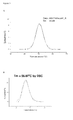
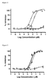

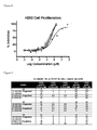


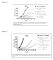
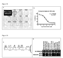

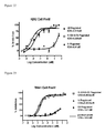


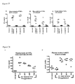
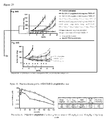
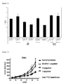
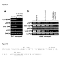
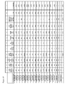
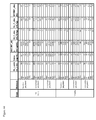


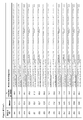


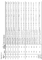
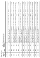

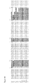


Classifications
-
- C—CHEMISTRY; METALLURGY
- C07—ORGANIC CHEMISTRY
- C07K—PEPTIDES
- C07K14/00—Peptides having more than 20 amino acids; Gastrins; Somatostatins; Melanotropins; Derivatives thereof
- C07K14/435—Peptides having more than 20 amino acids; Gastrins; Somatostatins; Melanotropins; Derivatives thereof from animals; from humans
- C07K14/78—Connective tissue peptides, e.g. collagen, elastin, laminin, fibronectin, vitronectin, cold insoluble globulin [CIG]
-
- A—HUMAN NECESSITIES
- A61—MEDICAL OR VETERINARY SCIENCE; HYGIENE
- A61K—PREPARATIONS FOR MEDICAL, DENTAL OR TOILETRY PURPOSES
- A61K38/00—Medicinal preparations containing peptides
- A61K38/16—Peptides having more than 20 amino acids; Gastrins; Somatostatins; Melanotropins; Derivatives thereof
- A61K38/17—Peptides having more than 20 amino acids; Gastrins; Somatostatins; Melanotropins; Derivatives thereof from animals; from humans
- A61K38/39—Connective tissue peptides, e.g. collagen, elastin, laminin, fibronectin, vitronectin, cold insoluble globulin [CIG]
-
- A—HUMAN NECESSITIES
- A61—MEDICAL OR VETERINARY SCIENCE; HYGIENE
- A61K—PREPARATIONS FOR MEDICAL, DENTAL OR TOILETRY PURPOSES
- A61K39/00—Medicinal preparations containing antigens or antibodies
- A61K39/395—Antibodies; Immunoglobulins; Immune serum, e.g. antilymphocytic serum
- A61K39/39533—Antibodies; Immunoglobulins; Immune serum, e.g. antilymphocytic serum against materials from animals
- A61K39/3955—Antibodies; Immunoglobulins; Immune serum, e.g. antilymphocytic serum against materials from animals against proteinaceous materials, e.g. enzymes, hormones, lymphokines
-
- A—HUMAN NECESSITIES
- A61—MEDICAL OR VETERINARY SCIENCE; HYGIENE
- A61K—PREPARATIONS FOR MEDICAL, DENTAL OR TOILETRY PURPOSES
- A61K45/00—Medicinal preparations containing active ingredients not provided for in groups A61K31/00 - A61K41/00
- A61K45/06—Mixtures of active ingredients without chemical characterisation, e.g. antiphlogistics and cardiaca
-
- A—HUMAN NECESSITIES
- A61—MEDICAL OR VETERINARY SCIENCE; HYGIENE
- A61P—SPECIFIC THERAPEUTIC ACTIVITY OF CHEMICAL COMPOUNDS OR MEDICINAL PREPARATIONS
- A61P35/00—Antineoplastic agents
-
- A—HUMAN NECESSITIES
- A61—MEDICAL OR VETERINARY SCIENCE; HYGIENE
- A61P—SPECIFIC THERAPEUTIC ACTIVITY OF CHEMICAL COMPOUNDS OR MEDICINAL PREPARATIONS
- A61P43/00—Drugs for specific purposes, not provided for in groups A61P1/00-A61P41/00
-
- C—CHEMISTRY; METALLURGY
- C07—ORGANIC CHEMISTRY
- C07K—PEPTIDES
- C07K14/00—Peptides having more than 20 amino acids; Gastrins; Somatostatins; Melanotropins; Derivatives thereof
- C07K14/435—Peptides having more than 20 amino acids; Gastrins; Somatostatins; Melanotropins; Derivatives thereof from animals; from humans
- C07K14/475—Growth factors; Growth regulators
-
- C—CHEMISTRY; METALLURGY
- C07—ORGANIC CHEMISTRY
- C07K—PEPTIDES
- C07K16/00—Immunoglobulins [IGs], e.g. monoclonal or polyclonal antibodies
- C07K16/18—Immunoglobulins [IGs], e.g. monoclonal or polyclonal antibodies against material from animals or humans
- C07K16/28—Immunoglobulins [IGs], e.g. monoclonal or polyclonal antibodies against material from animals or humans against receptors, cell surface antigens or cell surface determinants
- C07K16/2863—Immunoglobulins [IGs], e.g. monoclonal or polyclonal antibodies against material from animals or humans against receptors, cell surface antigens or cell surface determinants against receptors for growth factors, growth regulators
-
- A—HUMAN NECESSITIES
- A61—MEDICAL OR VETERINARY SCIENCE; HYGIENE
- A61K—PREPARATIONS FOR MEDICAL, DENTAL OR TOILETRY PURPOSES
- A61K39/00—Medicinal preparations containing antigens or antibodies
- A61K2039/505—Medicinal preparations containing antigens or antibodies comprising antibodies
-
- C—CHEMISTRY; METALLURGY
- C07—ORGANIC CHEMISTRY
- C07K—PEPTIDES
- C07K2317/00—Immunoglobulins specific features
- C07K2317/30—Immunoglobulins specific features characterized by aspects of specificity or valency
- C07K2317/31—Immunoglobulins specific features characterized by aspects of specificity or valency multispecific
-
- C—CHEMISTRY; METALLURGY
- C07—ORGANIC CHEMISTRY
- C07K—PEPTIDES
- C07K2317/00—Immunoglobulins specific features
- C07K2317/30—Immunoglobulins specific features characterized by aspects of specificity or valency
- C07K2317/34—Identification of a linear epitope shorter than 20 amino acid residues or of a conformational epitope defined by amino acid residues
-
- C—CHEMISTRY; METALLURGY
- C07—ORGANIC CHEMISTRY
- C07K—PEPTIDES
- C07K2317/00—Immunoglobulins specific features
- C07K2317/90—Immunoglobulins specific features characterized by (pharmaco)kinetic aspects or by stability of the immunoglobulin
- C07K2317/92—Affinity (KD), association rate (Ka), dissociation rate (Kd) or EC50 value
-
- C—CHEMISTRY; METALLURGY
- C07—ORGANIC CHEMISTRY
- C07K—PEPTIDES
- C07K2318/00—Antibody mimetics or scaffolds
- C07K2318/20—Antigen-binding scaffold molecules wherein the scaffold is not an immunoglobulin variable region or antibody mimetics
-
- C—CHEMISTRY; METALLURGY
- C07—ORGANIC CHEMISTRY
- C07K—PEPTIDES
- C07K2319/00—Fusion polypeptide
- C07K2319/30—Non-immunoglobulin-derived peptide or protein having an immunoglobulin constant or Fc region, or a fragment thereof, attached thereto
Definitions
- the present invention relates to EGFR binding domains and bispecific molecules comprising an EGFR binding domain and a distinct IGFIR binding domain for use in diagnostic, research and therapeutic applications.
- the invention further relates to cells comprising such proteins, polynucleotide encoding such proteins or fragments thereof, and vectors comprising the polynucleotides encoding the innovative proteins.
- Exemplary EGFR binding domains and bispecific molecules include antibody-like protein dimers based on the tenth fibronectin type III domain.
- Receptor tyrosine kinases have a conserved domain structure including an extracellular domain, a transmembrane domain and an intracellular tyrosine kinase domain.
- the extracellular domain can bind to a ligand, such as to a polypeptide growth factor or to a cell membrane-associated molecule.
- ligand binding or ligand binding induced dimerization of receptor tyrosine kinases activates the intracellular catalytic tyrosine kinase domain of the receptor and subsequent signal transduction.
- receptor tyrosine kinases include, but are not limited to ERBB receptors (e.g., EGFR, ERBB2, ERBB3, ERBB4), erythropoietin-producing hepatocellular (EPH) receptors, fibroblast growth factor (FGF) receptors (e.g., FGFR1, FGFR2, FGFR3, FGFR4, FGFR5), platelet-derived growth factor (PDGF) receptors (e.g., PDGFR-A, PDGFR-B), vascular endothelial growth factor (VEGF) receptors (e.g., VEGFR1/FLT1, VEGFR2/FLK1, VEGF3), tyrosine kinase with immunoglobulin-like and EGF-like domains (TIE) receptors , insulin-like growth factor (IGF) receptors (e.g., INS-R, IGFIR, IR-R), Discoidin Domain (DD) receptors , receptor for
- the application provides EGFR binding tenth fibronectin type III domains ( 10 Fn3) having novel sequences.
- EGFR binding 10 Fn3 having a consensus sequence are also provided.
- Such EGFR binding 10 Fn3 may be monomeric or may be included as part of a fusion protein.
- E/I binders encompassed by the invention include bispecific antibodies and dimers of ligand binding scaffold proteins (e.g., tendamistat, affibody, fibronectin type III domain, anticalin, tetranectin, and ankyrin).
- ligand binding scaffold proteins e.g., tendamistat, affibody, fibronectin type III domain, anticalin, tetranectin, and ankyrin.
- the E/I binders may be constructed in any orientation, e.g., from N-terminus to C-terminus either in the E-I arrangement or the I-E arrangement.
- antibody-like protein dimers comprising an EGFR binding 10 Fn3 covalently or non-covalently linked to an IGFIR binding 10 Fn3.
- the 10 Fn3 bind their target (EGFR or IGFIR) with a K D of less than 500 nM.
- Each of the individual 10 Fn3 independently has an amino acid sequence at least 70, 80, 85, 90, 95, 98, or 100% identical to SEQ ID NO: 32, wherein n is an integer from 1-20, o is an integer from 1-20, and p is an integer from 1-40.
- n is an integer from 8-12
- o is an integer from 4-8
- p is an integer from 4-28.
- n is 10, o is 6, and p is 12.
- the antibody-like protein dimers comprise IGFIR binding 10 Fn3 covalently linked to EGFR binding 10 Fn3 via a polypeptide linker or a polyethylene glycol moiety.
- the antibody-like protein dimer comprises an amino acid sequence at least 80, 90, 95, or 100% identical to any one of SEQ ID NOs: 20-31, 53-58, 87-92, 98-105, 118-133, 149-154, 164-169, 179-184, 192-197, 205-210 and 211-216.
- the E/I binder comprises an amino acid sequence having any one of SEQ ID NOs: 20-31, 53-58, 87-92, 98-105, 118-133, 149-154, 164-169, 179-184, 192-197, 205-210 and 211-216, wherein (i) the EGFR binding 10 Fn3 and/or the IGF-IR binding 10 Fn3 comprises a 10 Fn3 scaffold having from has anywhere from 0 to 20, from 0 to 15, from 0 to 10, from 0 to 8, from 0 to 6, from 0 to 5, from 0 to 4, from 0 to 3, from 0 to 2, or from 0 to 1 substitutions, conservative substitutions, deletions or additions relative to the corresponding scaffold amino acids of SEQ ID NO: 1, and/or (ii) the EGFR binding 10 Fn3 has anywhere from 0 to 15, from 0 to 10, from 0 to 8, from 0 to 6, from 0 to 5, from 0 to 4, from 0 to 3, from 0 to 2, or from 0
- compositions comprising an antibody-like protein dimer as described herein and a pharmaceutically acceptable carrier, wherein the composition is essentially pyrogen free.
- methods for treating hyperproliferative disorders, such as cancer, in a subject comprising administering to a subject in need thereof a therapeutically effective amount of a pharmaceutically acceptable composition comprising an antibody-like protein dimer as described herein.
- the application provides a nucleic acid encoding an antibody-like protein dimer as described herein.
- a vector comprising a nucleic acid encoding an antibody-like dimer as described herein.
- Suitable vectors include, for example, expression vectors.
- host cells comprising a nucleic, vector, or expression vector, comprising a nucleic acid encoding an antibody-like protein dimer as described herein.
- Suitable host cells include prokaryotic and eukaryotic host cells. Exemplary prokaryotic cells are bacterial cells, such as E. coli. Exemplary eukaryotic cells are mammalian cells, such as CHO cells.
- antibody-like protein refers to a non-immunoglobulin protein having an "immunoglobulin-like fold", i.e., comprises about 80-150 amino acid residues that are structurally organized into a set of beta or beta-like strands, forming beta sheets, where the beta or beta-like strands are connected by intervening loop portions.
- the beta sheets form the stable core of the antibody-like protein, while creating two "faces" composed of the loops that connect the beta or beta-like strands. As described herein, these loops can be varied to create customized ligand binding sites, and such variations can be generated without disrupting the overall stability of the protein.
- an antibody-like protein is a "fibronectin-based scaffold protein", by which is meant a polypeptide based on a fibronectin type III domain (Fn3).
- an antibody-like protein is based on a tenth fibronectin type III domain ( 10 Fn3).
- polypeptide By a “polypeptide” is meant any sequence of two or more amino acids, regardless of length, post-translation modification, or function. "Polypeptide,” “peptide,” and “protein” are used interchangeably herein.
- Percent (%) amino acid sequence identity herein is defined as the percentage of amino acid residues in a candidate sequence that are identical with the amino acid residues in a selected sequence, after aligning the sequences and introducing gaps, if necessary, to achieve the maximum percent sequence identity, and not considering any conservative substitutions as part of the sequence identity. Alignment for purposes of determining percent amino acid sequence identity can be achieved in various ways that are within the skill in the art, for instance, using publicly available computer software such as BLAST, BLAST-2, ALIGN, ALIGN-2 or Megalign (DNASTAR) software. Those skilled in the art can determine appropriate parameters for measuring alignment, including any algorithms needed to achieve maximal alignment over the full-length of the sequences being compared.
- % amino acid sequence identity values are obtained as described below by using the sequence comparison computer program ALIGN-2.
- the ALIGN-2 sequence comparison computer program was authored by Genentech, Inc. has been filed with user documentation in the U.S. Copyright Office, Washington D.C., 20559, where it is registered under U.S. Copyright Registration No. TXU510087, and is publicly available through Genentech, Inc., South San Francisco, Calif.
- the ALIGN-2 program should be compiled for use on a UNIX operating system, preferably digital UNIX V4.0D. All sequence comparison parameters are set by the ALIGN-2 program and do not vary.
- the % amino acid sequence identity of a given amino acid sequence A to, with, or against a given amino acid sequence B is calculated as follows: 100 times the fraction X/Y where X is the number of amino acid residues scored as identical matches by the sequence alignment program ALIGN-2 in that program's alignment of A and B, and where Y is the total number of amino acid residues in B. It will be appreciated that where the length of amino acid sequence A is not equal to the length of amino acid sequence B, the % amino acid sequence identity of A to B will not equal the % amino acid sequence identity of B to A.
- the term "therapeutically effective amount” refers to an amount of a drug effective to treat a disease or disorder in a mammal.
- the therapeutically effective amount of the drug may reduce the number of cancer cells; reduce the tumor size; inhibit (i.e., slow to some extent and preferably stop) cancer cell infiltration into peripheral organs; inhibit (i.e., slow to some extent and preferably stop) tumor metastasis; inhibit, to some extent, tumor growth; and/or relieve to some extent one or more of the symptoms associated with the disorder.
- the drug may prevent growth and/or kill existing cancer cells, it may be cytostatic and/or cytotoxic.
- efficacy in vivo can, for example, be measured by assessing the time to disease progression (TTP) and/or determining the response rates (RR).
- the half-life of an amino acid sequence or compound can generally be defined as the time taken for the serum concentration of the polypeptide to be reduced by 50% in vivo due to, e.g., degradation of the sequence or compound and/or clearance or sequestration of the sequence or compound by natural mechanisms.
- the half-life can be determined in any manner known in the art, such as by pharmacokinetic analysis. See e.g., M Gibaldi & D Perron "Pharmacokinetics", published by Marcel Dekker, 2nd Rev. edition (1982 ).
- E/I binder refers to a bispecific molecule that comprises an EGFR binding domain and a distinct IGFIR binding domain. The two domains may be covalently or non-covalently linked.
- An exemplary E/I binder is an antibody-like dimer comprising an EGFR binding 10 Fn3 and an IGFIR binding 10 Fn3, i.e., an E/I 10 Fn3 based binder.
- the epidermal growth factor receptor (EGFR) and insulin-like growth factor receptor (IFGR) play key roles in the tumorigenesis of several types of human cancer. Inhibition of either receptor effectively reduces tumor growth in preclinical models as well as clinically. Blocking the EGFR pathway induces switching to the IGFR pathway to drive growth with in vitro tumor models. Therefore, blocking both receptors simultaneously may achieve superior efficacy to blocking either pathway alone by overcoming pathway switching.
- the activity of an E/I binder is synergistic in comparison to the monomeric components of the E/I binder.
- E/I binders bispecific molecules that bind EGFR and IGFIR, referred to herein as "E/I binders”. Applicants have discovered that such bispecific molecules inhibit proliferation of a cancer model cell line with greater potency than the corresponding. monospecific binders (see e.g., Example 9 and Figure 8 ).
- E/I binders will be useful in numerous therapeutic applications, especially in the treatment of cancer. In addition to therapeutic applications, E/I binders may be used in any circumstance where it is desirable to detect EGFR and/or IGFIR.
- E/I binders have an EGFR binding domain and a distinct IGFIR binding domain.
- Typical binding domains include antibodies; therefore, bispecific antibodies may be generated to function as E/I binders.
- Bispecific antibodies comprising complementary pairs of V H and V L regions are known in the art. These bispecific antibodies comprise two pairs of V H and V L , each V H/L pair binding to a single antigen. (see e.g., Hu et al., Cancer Res. 1996 56:3055-306 ; Neri et al., J. Mol. Biol. 1995 246:367-373 ; Atwell et al., Mol. Immunol. 1996 33:1301-1312 ; and Carter et al., Protein Sci.
- An exemplary bispecific antibody is a diabody, i.e., a small antibody fragment with two antigen-binding sites, which fragments comprise a heavy-chain variable domain connected to a light-chain variable domain in the same polypeptide chain ( Hollinger et al., Proc. Natl. Acad. Sci. USA 1993 90: 6444-6448 ).
- E/I binders also encompass dimers of ligand binding scaffold proteins.
- Scaffold proteins are well described in the literature and include, e.g., tendamistat, affibody, fibroncectin type III domain, anticalin, tetranectin, and ankyrin. Additional scaffold proteins that may be used to generate E/I binders are reviewed in Binz et al., Nature Biotech 23:1257-1268 (2005 ). Scaffold proteins are based on a rigid core structure or 'framework' that is important in determining and stabilizing the three-dimensional structure. In between the fixed or conserved residues of the scaffold lie variable regions such as loops, surfaces or cavities that can be randomized to alter ligand binding. A large diversity of amino acids is provided in the variable regions between the fixed scaffold residues to provide specific binding to a target molecule.
- Fibronectin is a large protein which plays essential roles in the formation of extracellular matrix and cell-cell interactions; it consists of many repeats of three types (types I, II, and III) of small domains.
- Fn3 is small, monomeric, soluble, and stable. It lacks disulfide bonds and, therefore, is stable under reducing conditions.
- the overall structure of Fn3 resembles the immunoglobulin fold.
- Fn3 domains comprise, in order from N-terminus to C-terminus, a beta or beta-like strand, A; a loop, AB; a beta or beta-like strand, B; a loop, BC; a beta or beta-like strand, C; a loop, CD; a beta or beta-like strand, D; a loop, DE; a beta or beta-like strand, E; a loop, EF; a beta or beta-like strand, F; a loop, FG; and a beta or beta-like strand, G.
- the seven antiparallel ⁇ -strands are arranged as two beta sheets that form a stable core, while creating two "faces" composed of the loops that connect the beta or beta-like strands.
- Loops AB, CD, and EF are located at one face and loops BC, DE, and FG are located on the opposing face. Any or all of loops AB, BC, CD, DE, EF and FG may participate in ligand binding.
- There are at least 15 different modules of Fn3 and while the sequence homology between the molecules is low, they all share a high similarity in tertiary structure.
- AdnectinsTM (Adnexus, a Bristol-Myers Squibb R&D Company) are ligand binding scaffold proteins based on the tenth fibronectin type III domain, i.e., the tenth module of Fn3, ( 10 Fn3).
- the amino acid sequence of a naturally occurring human 10 Fn3 is set forth in SEQ ID NO: 1.
- SEQ ID NO:1 (BC, FG, and DE loops are emphasized)
- the AB loop corresponds to residues 15-16
- the BC loop corresponds to residues 21-30
- the CD loop corresponds to residues 39-45
- the DE loop corresponds to residues 51-56
- the EF loop corresponds to residues 60-66
- the FG loop corresponds to residues 76-87.
- beta strand A corresponds to residues 9-14
- beta strand B corresponds to residues 17-20
- beta strand C corresponds to residues 31-38
- beta strand D corresponds to residues 46-50
- beta strand E corresponds to residues 57-59
- beta strand F corresponds to residues 67-75
- beta strand G corresponds to residues 88-94.
- the strands are connected to each other through the corresponding loop, e.g., strands A and B are connected via loop AB in the formation strand A, loop AB, strand B, etc.
- Residues involved in forming the hydrophobic core include the amino acids corresponding to the following amino acids of SEQ ID NO: 1: L8, V10, A13, L18, I20, W22, Y32,134, Y36, F48, V50, A57, I59, L62, Y68, I70, V72, A74, I88, I90 and Y92, wherein the core amino acid residues are represented by the single letter amino acid code followed by the position at which they are located within SEQ ID NO: 1. See e.g., Dickinson et al., J. Mol. Biol. 236: 1079-1092 (1994 ).
- amino acid residues corresponding to residues 21-30, 51-56, and 76-87 of SEQ ID NO: 1 define the BC, DE and FG loops, respectively.
- a desired target such as IGF-IR or EGFR.
- residues corresponding to amino acids 23-30, 52-55 and 77-86 of SEQ ID NO: 1 were modified to produce high affinity 10 Fn3 binders (see Figure 46 .
- the BC loop may be defined by amino acids corresponding to residues 23-30 of SEQ ID NO: 1
- the DE loop may be defined by amino acids corresponding to residues 52-55 of SEQ ID NO: 1
- the FG loop may be defined by amino acids corresponding to residues 77-86 of SEQ ID NO: 1.
- 10 Fn3 are structurally and functionally analogous to antibodies, specifically the variable region of an antibody. While 10 Fn3 domains may be described as "antibody mimics" or “antibody-like proteins", they do offer a number of advantages over conventional antibodies. In particular, they exhibit better folding and thermostability properties as compared to antibodies, and they lack disulphide bonds, which are known to impede or prevent proper folding under certain conditions. Exemplary E/I 10 Fn3 based binders are predominantly monomeric with Tm's averaging ⁇ 50 °C.
- the BC, DE, and FG loops of 10 Fn3 are analogous to the complementary determining regions (CDRs) from immunoglobulins. Alteration of the amino acid sequence in these loop regions changes the binding specificity of 10 Fn3.
- the protein sequences outside of the CDR-like loops are analogous to the framework regions from immunoglobulins and play a role in the structural conformation of the 10 Fn3. Alterations in the framework-like regions of 10 Fn3 are permissible to the extent that the structural conformation is not so altered as to disrupt ligand binding.
- Antibody-like proteins based on the 10 Fn3 scaffold can be defined generally by the sequence:
- 10 Fn3 generally begin with the amino acid residue corresponding to number 1 of SEQ ID NO: 1.
- domains with amino acid deletions are also encompassed by the invention.
- amino acid residues corresponding to the first eight amino acids of SEQ ID NO: 1 are deleted.
- Additional sequences may also be added to the N- or C-terminus. For example, an additional MG sequence may be placed at the N-terminus of 10 Fn3. The M will usually be cleaved off, leaving a G at the N-terminus.
- sequences may be placed at the C-terminus of the 10 Fn3 domain, e.g., EIDKPSQ (SEQ ID NO: 9), EIDKPCQ (SEQ ID NO: 10), EGSGS (SEQ ID NO: 96) or EGSGC (SEQ ID NO: 97).
- the non-ligand binding sequences of 10 Fn3, i.e., the " 10 Fn3 scaffold” may be altered provided that the 10 Fn3 retains ligand binding function and/or structural stability.
- one or more of Asp 7, Glu 9, and Asp 23 are replaced by another amino acid, such as, for example, a non-negatively charged amino acid residue (e.g., Asn, Lys, etc.).
- a non-negatively charged amino acid residue e.g., Asn, Lys, etc.
- These mutations have been reported to have the effect of promoting greater stability of the mutant 10 Fn3 at neutral pH as compared to the wild-type form (See, PCT Publication No. WO 02/04523 ).
- a variety of additional alterations in the 10 Fn3 scaffold that are either beneficial or neutral have been disclosed. See, for example, Batori et al., Protein Eng. 2002 15(12):1015-20 ; Koide et al., Biochemistry 2001 40(34):10326-
- the 10 Fn3 scaffold may be modified by one or more conservative substitutions. As many as 5%, 10%, 20% or even 30% or more of the amino acids in the 10 Fn3 scaffold may be altered by a conservative substitution without substantially altering the affinity of the 10 Fn3 for a ligand.
- the scaffold modification preferably reduces the binding affinity of the 10 Fn3 binder for a ligand by less than 100-fold, 50-fold, 25-fold, 10-fold, 5-fold, or 2-fold. It may be that such changes will alter the immunogenicity of the 10 Fn3 in vivo, and where the immunogenicity is decreased, such changes will be desirable.
- conservative substitutions are residues that are physically or functionally similar to the corresponding reference residues.
- a conservative substitution and its reference residue have similar size, shape, electric charge, chemical properties including the ability to form covalent or hydrogen bonds, or the like.
- Preferred conservative substitutions are those fulfilling the criteria defined for an accepted point mutation in Dayhoff et al., Atlas of Protein Sequence and Structure 5:345-352 (1978 & Supp .).
- conservative substitutions are substitutions within the following groups: (a) valine, glycine; (b) glycine, alanine; (c) valine, isoleucine, leucine; (d) aspartic acid, glutamic acid; (e) asparagine, glutamine; (f) serine, threonine; (g) lysine, arginine, methionine; and (h) phenylalanine, tyrosine.
- an EGFR binding 10 Fn3 may be provided as part of a fusion protein or multimer.
- an EGFR binding 10 Fn3 may be covalently or non-covalently linked to at least a second 10 Fn3 binding domain.
- the second 10 Fn3 binding domain may bind to EGFR or to a different target.
- an EGFR binding 10 Fn3 may be covalently or non-covalently linked to an IGF-IR binding 10 Fn3.
- the EGFR binding 10 Fn3 proteins described herein bind to EGFR with a K D of less than 500 nM, 100 nM, 50 nM, 10 nM, 1 nM, 500 pM, 100 pM. 100 pM, 50 pM or 10 pM.
- the BC loop of the EGFR binding 10 Fn3 proteins correspond to amino acids 23-30 of SEQ ID NO: 1
- the DE loop of the EGFR binding 10 Fn3 proteins correspond to amino acids 52-55 of SEQ ID NO: 1
- the FG loop of the EGFR binding 10 Fn3 proteins correspond to amino acids 77-86 of SEQ ID NO: 1.
- the invention provides an EGFR binding 10 Fn3 comprising a BC loop having a YQ at the positions corresponding to amino acids 29 and 30 of SEQ ID NO: 1.
- the invention provides an EGFR binding 10 Fn3 comprising a BC loop having a YQ at the positions corresponding to amino acids 29 and 30 of SEQ ID NO: 1 and an FG loop that is fifteen amino acids in length, e.g., an FG loop that is extended in length by five amino acids due to an insertion of five amino acids between residues corresponding to amino acids 77-86 of SEQ ID NO: 1.
- the invention provides an EGFR binding 10 Fn3 comprising a BC loop having a YQ at the positions corresponding to amino acids 29 and 30 of SEQ ID NO: 1 and a DE loop having a V, I, L, M or A residue at the position corresponding to amino acid 54 of SEQ ID NO: 1.
- the invention provides an EGFR binding 10 Fn3 comprising a BC loop having a YQ at the positions corresponding to amino acids 29 and 30 of SEQ ID NO: 1, a DE loop having a V, I, L, M or A residue at the position corresponding to amino acid 54 of SEQ ID NO: 1, and an FG loop that is fifteen amino acids in length, e.g., an FG loop that is extended in length by five amino acids due to an insertion of five amino acids between residues corresponding to amino acids 77-86 of SEQ ID NO: 1.
- the invention provides an EGFR binding 10 Fn3 comprising a BC loop having a YQ at the positions corresponding to amino acids 29 and 30 of SEQ ID NO: 1 and an FG loop comprising a D or N at the position corresponding to amino acid 77 of SEQ ID NO: 1.
- the invention provides an EGFR binding 10 Fn3 comprising a BC loop having a YQ at the positions corresponding to amino acids 29 and 30 of SEQ ID NO: 1 and an FG loop (i) that is fifteen amino acids in length, e.g., an FG loop that is extended in length by five amino acids due to an insertion of five amino acids between residues corresponding to amino acids 77-86 of SEQ ID NO: 1 and (ii) comprises a D or N at the position corresponding to amino acid 77 of SEQ ID NO: 1.
- the invention provides an EGFR binding 10 Fn3 comprising a DE loop comprising a V, I, L, M or A residue at the position corresponding to amino acid 54 of SEQ ID NO: 1 and an FG loop comprising a D or N at the position corresponding to amino acid 77 of SEQ ID NO: 1.
- the invention provides an EGFR binding 10 Fn3 comprising a DE loop comprising a V, I, L, M or A residue at the position corresponding to amino acid 54 of SEQ ID NO: 1 and an FG loop (i) that is fifteen amino acids in length, e.g., an FG loop that is extended in length by five amino acids due to an insertion of five amino acids between residues corresponding to amino acids 77-86 of SEQ ID NO: 1 and (ii) comprises a D or N at the position corresponding to amino acid 77 of SEQ ID NO: 1.
- the invention provides an EGFR binding 10 Fn3 comprising a BC loop having a YQ at the positions corresponding to amino acids 29 and 30 of SEQ ID NO: 1, a DE loop comprising a V, I, L, M or A residue at the position corresponding to amino acid 54 of SEQ ID NO: 1, and an FG loop comprising a D or N at the position corresponding to amino acid 77 of SEQ ID NO: 1.
- the invention provides an EGFR binding 10 Fn3 comprising a BC loop having a YQ at the positions corresponding to amino acids 29 and 30 of SEQ ID NO: 1, a DE loop comprising a V, I, L, M or A residue at the position corresponding to amino acid 54 of SEQ ID NO: 1, and an FG loop (i) that is fifteen amino acids in length, e.g., an FG loop that is extended in length by five amino acids due to an insertion of five amino acids between residues corresponding to amino acids 77-86 of SEQ ID NO: 1 and (ii) comprises a D or N at the position corresponding to amino acid 77 of SEQ ID NO: 1.
- the invention provides an EGFR binding 10 Fn3 comprising a BC loop comprising the amino acid sequence XXXXXXYQ, a DE loop comprising the amino acid sequence XX(V/I/L/M/A)X, and an FG loop comprising the amino acid sequence (D/N)X n , wherein X is any amino acid and n is 9-14 amino acids. In an exemplary embodiment, n is 14 amino acids.
- the invention provides an EGFR binding 10 Fn3 comprising a BC loop corresponding to amino acids 23-30 of SEQ ID NO: 1 comprising the amino acid sequence XXXXXYQ, a DE loop corresponding to amino acids 52-55 of SEQ ID NO: 1 comprising the amino acid sequence XX(V/I/L/M/A)X, and an FG loop corresponding to amino acids 77-86 of SEQ ID NO: 1 comprising the amino acid sequence (D/N)X n , wherein X is any amino acid and n is 9-14 amino acids. In an exemplary embodiment, n is 14 amino acids.
- the invention provides an EGFR binding 10 Fn3 comprising a BC loop comprising the amino acid sequence XXXXXXYQ, a DE loop comprising the amino acid sequence XX(V/I/L/M/A)X, and an FG loop comprising an amino acid sequence selected from:
- the invention provides an EGFR binding 10 Fn3 comprising a BC loop comprising the amino acid sequence XXXXXXYQ, a DE loop comprising the amino acid sequence (G/Y/H)(D/M/G)(V/L/I)X, and an FG loop comprising an amino acid sequence (D/N)(Y/M)(Y/A/M)(Y/H/F)(K/Q/V)(E/P/R)(Y/T/K)X(E/Y/Q)(Y/G/H), wherein X is any amino acid.
- the invention provides an EGFR binding 10 Fn3 comprising a BC loop comprising the amino acid sequence XXXXXXYQ, a DE loop comprising the amino acid sequence (G/Y/H)(D/M/G)(V/L/I)X, and an FG loop comprising an amino acid sequence D(Y/F/W)(Y/F/K)(N/D/P)(P/H/L)(A/T/V)(T/D/S)(H/Y/G)(E/P/V)(Y/H)(T/K/I)(Y/F )(H/N/Q)(T/Q/E)(T/S/I), wherein X is any amino acid.
- the invention provides an EGFR binding 10 Fn3 comprising a BC loop comprising the amino acid sequence XXXXXXYQ, a DE loop comprising the amino acid sequence XX(V/I/L/M/A)X, and an FG loop comprising an amino acid sequence selected from:
- the invention provides an EGFR binding 10 Fn3 comprising a BC loop comprising the amino acid sequence XXXXXXYQ, a DE loop comprising the amino acid sequence XX(V/I/L/M/A)X, and an FG loop comprising an amino acid sequence selected from:
- the invention provides an EGFR binding 10 Fn3 comprising a BC loop comprising the amino acid sequence XXXXXXYQ, a DE loop comprising the amino acid sequence XX(V/I/L/M/A)X, and an FG loop comprising the amino acid sequence (D/N)(M/Y)(M/A/W)(H/F/Y)(V/K)EY(A/Q/R/S/T)E(Y/H/D), wherein X is any amino acid.
- the invention provides an EGFR binding 10 Fn3 comprising a BC loop comprising the amino acid sequence XXXXXXYQ, a DE loop comprising the amino acid sequence XX(V/I/L/M/A)X, and an FG loop comprising the amino acid sequence D(Y/F/W)(Y/F/K)(N/P/D)(P/H/L)X(T/D/S)(H/G/Y)(E/P/Y)(Y/H)XYXXX, wherein X is any amino acid.
- the DE loop of the EGFR binding 10 Fn3 may comprise the sequence (G/Y/H)(D/M/G)(V/L/I)X.
- the invention provides an EGFR binding 10 Fn3 comprising an FG loop comprising an amino acid sequence selected from:
- the EGFR binding 10 Fn3 comprises any of the consensus sequences provided above, with the proviso that the EGFR binding 10 Fn3 does not comprise one or more of the following sequences:
- an EGFR binding 10 Fn3 comprising one of the consensus sequences provided above has at least 40%, 50%, 60%, 70%, 75%, or 80% identity to SEQ ID NO: 1.
- the overall structure of an EGFR binding 10 Fn3 comprising one of the consensus sequences provided above resembles the immunoglobulin fold.
- an EGFR binding 10 Fn3 comprising one of the consensus sequences provided above further comprises the core amino acid residues of the scaffold.
- an EGFR binding 10 Fn3 comprising one of the consensus sequences provided above has at least 70%, 75%, 80%, 85%, 90%, 95%, 97%, 98%, or 99% identity to any one of SEQ ID NOs: 5-8, 52, 66-68, 106-108, 112-114, 140-142, 155-157, 170-172, 182, 185-187, 198-200, or 219-327.
- an EGFR binding 10 Fn3 comprising one of the consensus sequences provided above has at least 70%, 75%, 80%, 85%, 90%, 95%, 97%, 98%, or 99% identity to the amino acid sequence of amino acid residues corresponding to E9 of SEQ ID NO: 1 through T94 of SEQ ID NO: 1 of any one of SEQ ID NOs: 5-8, 52, 66-68, 106-108, 112-114, 140-142, 155-157, 170-172, 182, 185-187, 198-200, or 219-327.
- the EGFR binding 10 Fn3 comprising one of the consensus sequences provided above comprises a 10 Fn3 scaffold having from has anywhere from 0 to 20, from 0 to 15, from 0 to 10, from 0 to 8, from 0 to 6, from 0 to 5, from 0 to 4, from 0 to 3, from 0 to 2, or from 0 to 1 substitutions, conservative substitutions, deletions or additions relative to the scaffold amino acids residues of SEQ ID NO: 1.
- the invention provides an EGFR binding 10 Fn3 comprising a BC loop having the amino acid sequence set forth in amino acids 23-30, a DE loop having the amino acid sequence set forth in amino acids 52-55, and an FG loop having the amino acid sequence set forth in amino acids 77-86 of any one of SEQ ID NOs: 219-327.
- the invention provides an EGFR binding 10 Fn3 comprising a BC loop having the amino acid sequence set forth in amino acids 21-30, a DE loop having the amino acid sequence set forth in amino acids 51-56, and an FG loop having the amino acid sequence set forth in amino acids 76-87 of any one of SEQ ID NOs: 219-327.
- the invention provides an EGFR binding 10 Fn3 comprising an amino acid sequence at least 60%, 75%, 80%, 85%, 90%, 95%, or 98% identical to any one of SEQ ID NOs: 219-327.
- an antibody-like protein comprising a tenth fibronectin type III domain ( 10 Fn3) that binds EGFR with a K D of less than 500 nM and comprises a BC loop having the amino acid sequence set forth in amino acids 21-30 of SEQ ID NO: 5, a DE loop having the amino acid sequence set forth in amino acids 51-56 of SEQ ID NO: 5, and an FG loop having the amino acid sequence set forth in amino acids 76-92 of SEQ ID NO: 5.
- 10 Fn3 tenth fibronectin type III domain that binds EGFR with a K D of less than 500 nM and comprises a BC loop having the amino acid sequence set forth in amino acids 21-30 of SEQ ID NO: 5, a DE loop having the amino acid sequence set forth in amino acids 51-56 of SEQ ID NO: 5, and an FG loop having the amino acid sequence set forth in amino acids 76-92 of SEQ ID NO: 5.
- the EGFR binding 10 Fn3 comprises a BC loop having the amino acid sequence XgDSGRGSYQX h (SEQ ID NO: 40), a DE loop having the amino acid sequence X i GPVHX j (SEQ ID NO: 42), and an FG loop having the amino acid sequence X k DHKPHADGPHTYHEX l (SEQ ID NO: 44); wherein X is any amino acid and g, h, i, j, k, and l are integers independently selected from 0 to 5.
- the EGFR binding 10 Fn3 comprises a BC loop having the amino acid sequence SWDSGRGSYQ (SEQ ID NO: 39), a DE loop having the amino acid sequence PGPVHT (SEQ ID NO: 41), and an FG loop having the amino acid sequence TDHKPHADGPHTYHESP (SEQ ID NO: 43).
- the EGFR binding 10 Fn3 has an amino acid sequence at least 80, 90, 95, or 100% identical to SEQ ID NOs: 5 or 6.
- an antibody-like protein comprising a tenth fibronectin type III domain ( 10 Fn3) that binds EGFR with a K D of less than 500 nM and comprises a BC loop having the amino acid sequence set forth in amino acids 21-30 of SEQ ID NO: 7, a DE loop having the amino acid sequence set forth in amino acids 51-56 of SEQ ID NO: 7, and an FG loop having the amino acid sequence set forth in amino acids 76-87 of SEQ ID NO: 7.
- 10 Fn3 tenth fibronectin type III domain that binds EGFR with a K D of less than 500 nM and comprises a BC loop having the amino acid sequence set forth in amino acids 21-30 of SEQ ID NO: 7, a DE loop having the amino acid sequence set forth in amino acids 51-56 of SEQ ID NO: 7, and an FG loop having the amino acid sequence set forth in amino acids 76-87 of SEQ ID NO: 7.
- the EGFR binding 10 Fn3 comprises a BC loop having the amino acid sequence X m VAGAEDYQX n (SEQ ID NO: 34), a DE loop having the amino acid sequence X o HDLVX p (SEQ ID NO: 36), and an FG loop having the amino acid sequence X q DMMHVEYTEHX r (SEQ ID NO: 38); wherein X is any amino acid and m, n, o, p, q, and r are integers independently selected from 0 to 5.
- the EGFR binding 10 Fn3 comprises a BC loop having the amino acid sequence SWVAGAEDYQ (SEQ ID NO: 33), a DE loop having the amino acid sequence PHDLVT (SEQ ID NO: 35), and an FG loop having the amino acid sequence TDMMHVEYTEHP (SEQ ID NO: 37).
- the EGFR binding 10 Fn3 has an amino acid sequence at least 80, 90, 95, or 100% identical to SEQ ID NO: 7 or 8.
- an antibody-like protein comprising a tenth fibronectin type III domain ( 10 Fn3) that binds EGFR with a K D of less than 500 nM and comprises a BC loop having the amino acid sequence set forth in amino acids 23-30 of SEQ ID NO: 82, a DE loop having the amino acid sequence set forth in amino acids 51-55 of SEQ ID NO: 82, and an FG loop having the amino acid sequence set forth in amino acids 76-86 of SEQ ID NO: 82.
- 10 Fn3 tenth fibronectin type III domain
- the EGFR binding 10 Fn3 comprises a BC loop having the amino acid sequence X s LPGKLRYQX t (SEQ ID NO: 60), a DE loop having the amino acid sequence X u HDLRX w (SEQ ID NO: 62), and an FG loop having the amino acid sequence X y NMMHVEYSEYX z (SEQ ID NO: 64); wherein X is any amino acid and s, t, u, w, y and z are integers independently selected from 0 to 5.
- the EGFR binding 10 Fn3 comprises a BC loop having the amino acid sequence LPGKLRYQ (resdiues 3-13 of SEQ ID NO: 59), a DE loop having the amino acid sequence PHDLR (residues 1-5 of SEQ ID NO: 61), and an FG loop having the amino acid sequence TNMMHVEYSEY (residues 1-11 of SEQ ID NO: 63).
- the EGFR binding 10 Fn3 has an amino acid sequence at least 80, 90, 95, or 100% identical to SEQ ID NO: 52 or 82.
- an antibody-like protein comprising a tenth fibronectin type III domain ( 10 Fn3) that binds EGFR with a K D of less than 500 nM and comprises a BC loop having the amino acid sequence set forth in amino acids 23-30 of SEQ ID NO: 106, a DE loop having the amino acid sequence set forth in amino acids 51-55 of SEQ ID NO: 106, and an FG loop having the amino acid sequence set forth in amino acids 76-86 of SEQ ID NO: 106.
- 10 Fn3 tenth fibronectin type III domain
- the EGFR binding 10 Fn3 comprises a BC loop having the amino acid sequence X g HERDGSRQX h (SEQ ID NO: 134), a DE loop having the amino acid sequence X i GGVRX j (SEQ ID NO: 135), and an FG loop having the amino acid sequence X k DYFNPTTHEYIYQTTX l (SEQ ID NO: 136); wherein X is any amino acid and g, h, i, j, k and l are integers independently selected from 0 to 5.
- the EGFR binding 10 Fn3 comprises a BC loop having the amino acid sequence SWHERDGSRQ (SEQ ID NO: 109), a DE loop having the amino acid sequence PGGVRT (SEQ ID NO: 110), and an FG loop having the amino acid sequence TDYFNPTTHEYIYQTTP (SEQ ID NO: 111).
- the EGFR binding 10 Fn3 has an amino acid sequence at least 80, 90, 95, or 100% identical to any one of SEQ ID NOs: 106-108.
- an antibody-like protein comprising a tenth fibronectin type III domain ( 10 Fn3) that binds EGFR with a K D of less than 500 nM and comprises a BC loop having the amino acid sequence set forth in amino acids 23-30 of SEQ ID NO: 112, a DE loop having the amino acid sequence set forth in amino acids 51-55 of SEQ ID NO: 112, and an FG loop having the amino acid sequence set forth in amino acids 76-86 of SEQ ID NO: 112.
- a tenth fibronectin type III domain 10 Fn3 that binds EGFR with a K D of less than 500 nM and comprises a BC loop having the amino acid sequence set forth in amino acids 23-30 of SEQ ID NO: 112, a DE loop having the amino acid sequence set forth in amino acids 51-55 of SEQ ID NO: 112, and an FG loop having the amino acid sequence set forth in amino acids 76-86 of SEQ ID NO: 112.
- the EGFR binding 10 Fn3 comprises a BC loop having the amino acid sequence X g WAPVDRYQX h (SEQ ID NO: 137), a DE loop having the amino acid sequence X i RDVYX j (SEQ ID NO: 138), and an FG loop having the amino acid sequence X k DYKPHADGPHTYHESX l (SEQ ID NO: 139); wherein X is any amino acid and g, h, i, j, k and l are integers independently selected from 0 to 5.
- the EGFR binding 10 Fn3 comprises a BC loop having the amino acid sequence SWWAPVDRYQ (SEQ ID NO: 115), a DE loop having the amino acid sequence PRDVYT (SEQ ID NO: 116), and an FG loop having the amino acid sequence TDYKPHADGPHTYHESP (SEQ ID NO: 117).
- the EGFR binding 10 Fn3 has an amino acid sequence at least 80, 90, 95, or 100% identical to any one of SEQ ID NOs: 112-114.
- an antibody-like protein comprising a tenth fibronectin type III domain ( 10 Fn3) that binds EGFR with a K D of less than 500 nM and comprises a BC loop having the amino acid sequence set forth in amino acids 13-22 of SEQ ID NO: 141, a DE loop having the amino acid sequence set forth in amino acids 43-48 of SEQ ID NO: 141, and an FG loop having the amino acid sequence set forth in amino acids 68-84 of SEQ ID NO: 141.
- 10 Fn3 tenth fibronectin type III domain
- the EGFR binding 10 Fn3 comprises a BC loop having the amino acid sequence X g TQGSTHYQX h (SEQ ID NO: 146), a DE loop having the amino acid sequence X i GMVYX j (SEQ ID NO: 147), and an FG loop having the amino acid sequence X k DYFDRSTHEYKYRTTX l (SEQ ID NO: 148); wherein X is any amino acid and g, h, i, j, k and l are integers independently selected from 0 to 5.
- the EGFR binding 10 Fn3 comprises a BC loop having the amino acid sequence SWTQGSTHYQ (SEQ ID NO: 143), a DE loop having the amino acid sequence PGMVYT (SEQ ID NO: 144), and an FG loop having the amino acid sequence TDYFDRSTHEYKYRTTP (SEQ ID NO: 145).
- the EGFR binding 10 Fn3 has an amino acid sequence at least 80, 90, 95, or 100% identical to any one of SEQ ID NOs: 140-142.
- an antibody-like protein comprising a tenth fibronectin type III domain ( 10 Fn3) that binds EGFR with a K D of less than 500 nM and comprises a BC loop having the amino acid sequence set forth in amino acids 13-22 of SEQ ID NO: 156, a DE loop having the amino acid sequence set forth in amino acids 43-48 of SEQ ID NO: 156, and an FG loop having the amino acid sequence set forth in amino acids 68-84 of SEQ ID NO: 156.
- 10 Fn3 tenth fibronectin type III domain
- the EGFR binding 10 Fn3 comprises a BC loop having the amino acid sequence X g YWEGLPYQX h (SEQ ID NO: 161), a DE loop having the amino acid sequence X i RDVNX j (SEQ ID NO: 162), and an FG loop having the amino acid sequence X k DWYNPDTHEYIYHTIX l (SEQ ID NO: 163); wherein X is any amino acid and g, h, i, j, k and l are integers independently selected from 0 to 5.
- the EGFR binding 10 Fn3 comprises a BC loop having the amino acid sequence SWYWEGLPYQ (SEQ ID NO: 158), a DE loop having the amino acid sequence PRDVNT (SEQ ID NO: 159), and an FG loop having the amino acid sequence TDWYNPDTHEYIYHTIP (SEQ ID NO: 160).
- the EGFR binding 10 Fn3 has an amino acid sequence at least 80, 90, 95, or 100% identical to any one of SEQ ID NOs: 155-157.
- an antibody-like protein comprising a tenth fibronectin type III domain ( 10 Fn3) that binds EGFR with a K D of less than 500 nM and comprises a BC loop having the amino acid sequence set forth in amino acids 13-22 of SEQ ID NO: 171, a DE loop having the amino acid sequence set forth in amino acids 43-48 of SEQ ID NO: 171, and an FG loop having the amino acid sequence set forth in amino acids 68-84 of SEQ ID NO: 171.
- 10 Fn3 tenth fibronectin type III domain that binds EGFR with a K D of less than 500 nM and comprises a BC loop having the amino acid sequence set forth in amino acids 13-22 of SEQ ID NO: 171, a DE loop having the amino acid sequence set forth in amino acids 43-48 of SEQ ID NO: 171, and an FG loop having the amino acid sequence set forth in amino acids 68-84 of SEQ ID NO: 171.
- the EGFR binding 10 Fn3 comprises a BC loop having the amino acid sequence X g ASNRGTYQX h (SEQ ID NO: 176), a DE loop having the amino acid sequence X i GGVSX j (SEQ ID NO: 177), and an FG loop having the amino acid sequence X k DAFNPTTHEYNYFTTX l (SEQ ID NO: 178); wherein X is any amino acid and g, h, i, j, k and l are integers independently selected from 0 to 5.
- the EGFR binding 10 Fn3 comprises a BC loop having the amino acid sequence SWASNRGTYQ (SEQ ID NO: 173), a DE loop having the amino acid sequence PGGVST (SEQ ID NO: 174), and an FG loop having the amino acid sequence TDAFNPTTHEYNYFTTP (SEQ ID NO: 175).
- the EGFR binding 10 Fn3 has an amino acid sequence at least 80, 90, 95, or 100% identical to any one of SEQ ID NOs: 170-172.
- an antibody-like protein comprising a tenth fibronectin type III domain ( 10 Fn3) that binds EGFR with a K D of less than 500 nM and comprises a BC loop having the amino acid sequence set forth in amino acids 13-22 of SEQ ID NO: 186, a DE loop having the amino acid sequence set forth in amino acids 43-48 of SEQ ID NO: 186, and an FG loop having the amino acid sequence set forth in amino acids 68-84 of SEQ ID NO: 186.
- 10 Fn3 tenth fibronectin type III domain
- the EGFR binding 10 Fn3 comprises a BC loop having the amino acid sequence X g DAPTSRYQX h (SEQ ID NO: 190), a DE loop having the amino acid sequence X i GGLSX j (SEQ ID NO: 191), and an FG loop having the amino acid sequence X k DYKPHADGPHTYHESX l (SEQ ID NO: 139); wherein X is any amino acid and g, h, i, j, k and l are integers independently selected from 0 to 5.
- the EGFR binding 10 Fn3 comprises a BC loop having the amino acid sequence SWDAPTSRYQ (SEQ ID NO: 188), a DE loop having the amino acid sequence PGGLST (SEQ ID NO: 189), and an FG loop having the amino acid sequence TDYKPHADGPHTYHESP (SEQ ID NO: 117).
- the EGFR binding 10 Fn3 has an amino acid sequence at least 80, 90, 95, or 100% identical to any one of SEQ ID NOs: 185-187.
- an antibody-like protein comprising a tenth fibronectin type III domain ( 10 Fn3) that binds EGFR with a K D of less than 500 nM and comprises a BC loop having the amino acid sequence set forth in amino acids 13-22 of SEQ ID NO: 199, a DE loop having the amino acid sequence set forth in amino acids 43-48 of SEQ ID NO: 199, and an FG loop having the amino acid sequence set forth in amino acids 68-84 of SEQ ID NO: 199.
- 10 Fn3 tenth fibronectin type III domain
- the EGFR binding 10 Fn3 comprises a BC loop having the amino acid sequence X g DAGAVTYQX h (SEQ ID NO: 203), a DE loop having the amino acid sequence X i GGVRX j (SEQ ID NO: 135), and an FG loop having the amino acid sequence X k DYKPHADGPHTYHEYX l (SEQ ID NO: 204); wherein X is any amino acid and g, h, i, j, k and l are integers independently selected from 0 to 5.
- the EGFR binding 10 Fn3 comprises a BC loop having the amino acid sequence SWDAGAVTYQ (SEQ ID NO: 201), a DE loop having the amino acid sequence PGGVRT (SEQ ID NO: 110), and an FG loop having the amino acid sequence TDYKPHADGPHTYHEYP (SEQ ID NO: 202).
- the EGFR binding 10 Fn3 has an amino acid sequence at least 80, 90, 95, or 100% identical to any one of SEQ ID NOs: 198-200.
- an EGFR binding 10 Fn3 domain is covalently or non-covalently linked to an EGF-IR binding 10 Fn3 domain.
- the IGF-IR binding 10 Fn3 may comrpise a BC loop having the amino acid sequence set forth in amino acids 21-30 of SEQ ID NO: 3, a DE loop having the amino acid sequence set forth in amino acids 51-56 of SEQ ID NO: 3, and an FG loop having the amino acid sequence set forth in amino acids 76-83 of SEQ ID NO: 3.
- the IGF-IR binding 10 Fn3 comprises a BC loop having the amino acid sequence X a SARLKVAX b (SEQ ID NO: 46), a DE loop having the amino acid sequence X c KNVYX d (SEQ ID NO: 48), and an FG loop having the amino acid sequence X e RFRDYQX f (SEQ ID NO: 50), wherein X is any amino acid and a, b, c, d, e, f, g, h, i, j, k, and l are integers independently selected from 0 to 5, or wherein a is 2 and b-f are 1, or wherein a-f are zero.
- the IGF-IR binding 10 Fn3 has an amino acid sequence at least 80, 90, 95, 98, 99, or 100% identical to SEQ ID NO: 3.
- the IGF-IR binding 10 Fn3 comprises a 10 Fn3 scaffold having from has anywhere from 0 to 20, from 0 to 15, from 0 to 10, from 0 to 8, from 0 to 6, from 0 to 5, from 0 to 4, from 0 to 3, from 0 to 2, or from 0 to 1 substitutions, conservative substitutions, deletions or additions relative to the scaffold amino acid residues of SEQ ID NO: 1.
- the IGF-IR binding 10 Fn3 has anywhere from 0 to 15, from 0 to 10, from 0 to 8, from 0 to 6, from 0 to 5, from 0 to 4, from 0 to 3, from 0 to 2, or from 0 to 1 substitutions, conservative substitutions, deletions or additions relative to the corresponding loop sequences of SEQ ID NO: 3.
- an antibody-like protein multimer comprises at least one EGFR binding 10 Fn3 covalently or non-covalently linked to at least one IGFIR binding 10 Fn3.
- the E/I binders described herein may be constructed as a single polypeptide chain wherein the E and I subunits may be in either orientation, e.g., from N-terminus to C-terminus, in the E-I orientation or in the I-E orientation.
- the disclosure relates, in part, to the surprising discovery that multiple 10 Fn3 joined via a polypeptide linker correctly fold independently of each other, retain high affinity binding, and that each of the domains retains its functional properties (see e.g., Examples 5-10). Additionally, these E/I 10 Fn3 based binders demonstrate desirable biophysical properties such as low aggregation and high melting temperature (T m ) (see e.g., Example 4).
- T m low aggregation and high melting temperature
- the Examples characterize a variety of E/I 10 Fn3 based binders.
- An exemplary IGFIR binding 10 Fn3 is set forth in SEQ ID NO: 4.
- Exemplary EGFR binding 10 Fn3 are set forth in SEQ ID NOs: 6, 8, 52, 107, 113, 140, 155, 170, 185 and 198.
- an E/I binder comprises an EGFR binding 10 Fn3 and an IGFIR binding 10 Fn3, independently having an amino acid sequence at least 40, 50, 60, 70, or 80% identical to the human 10 Fn3 domain, shown in SEQ ID NO: 1. Much of the variability will generally occur in one or more of the loops.
- an E/I binder comprises an EGFR binding 10 Fn3 and an IGFIR binding 10 Fn3, independently having an amino acid sequence at least 70, 80, 85, 90, 95, 98, or 100% identical to SEQ ID NO: 32, wherein n is an integer from 1-20, o is an integer from 1-20, and p is an integer from 1-40. In some embodiments, n is an integer from 8-12, o is an integer from 4-8, and p is an integer from 4-28. In some embodiments, n is 10, o is 6, and p is 12.
- the disclosure provides multimers of 10 Fn3 having at least one loop selected from loop BC, DE, and FG with an altered amino acid sequence relative to the sequence of the corresponding loop of the human 10 Fn3.
- altered is meant one or more amino acid sequence alterations relative to a template sequence (corresponding human fibronectin domain) and includes amino acid additions, deletions, and substitutions. Altering an amino acid sequence may be accomplished through intentional, blind, or spontaneous sequence variation, generally of a nucleic acid coding sequence, and may occur by any technique, for example, PCR, error-prone PCR, or chemical DNA synthesis. In some embodiments, an amino acid sequence is altered by substituting with or adding naturally occurring amino acids.
- one or more loops selected from BC, DE, and FG may be extended or shortened in length relative to the corresponding human fibronectin loop.
- the FG loop of the human 10 Fn3 is 12 residues long, whereas the corresponding loop in antibody heavy chains ranges from 4-28 residues.
- the length of the FG loop of 10 Fn3 may be altered in length as well as in sequence to obtain the greatest possible flexibility and affinity in antigen binding.
- the altered BC loop has up to 10 amino acid substitutions, up to 9 amino acid deletions, up to 10 amino acid insertions, or a combination of substitutions and deletions or insertions.
- the altered DE loop has up to 6 amino acid substitutions, up to 5 amino acid deletions, up to 14 amino acid insertions or a combination of substitutions and deletions or insertions.
- the FG loop has up to 12 amino acid substitutions, up to 11 amino acid deletions, up to 28 amino acid insertions or a combination of substitutions and deletions or insertions.
- Naturally occurring 10 Fn3 comprises an "arginine-glycine-aspartic acid" (RGD) integrin-binding motif in the FG loop.
- RGD arginine-glycine-aspartic acid
- Preferred multimers of 10 Fn3 lack an RGD integrin-binding motif.
- an E/I binder is an antibody-like protein dimer comprising a) an EGFR binding 10 Fn3 comprising a BC loop having the amino acid sequence set forth in amino acids 21-30 of SEQ ID NO: 5, a DE loop having the amino acid sequence set forth in amino acids 51-56 of SEQ ID NO: 5, and an FG loop having the amino acid sequence set forth in amino acids 76-92 of SEQ ID NO: 5; covalently or non-covalently linked to b) an IGFIR binding 10 Fn3 comprising a BC loop having the amino acid sequence set forth in amino acids 21-30 of SEQ ID NO: 3, a DE loop having the amino acid sequence set forth in amino acids 51-56 of SEQ ID NO: 3, and an FG loop having the amino acid sequence set forth in amino acids 76-83 of SEQ ID NO: 3.
- the EGFR binding 10 Fn3 has an amino acid sequence at least 80, 90, 95, 98, 99, or 100% identical to SEQ ID NO: 5.
- the IGFIR binding 10 Fn3 has an amino acid sequence at least 80, 90, 95, 98, 99, or 100% identical to SEQ ID NO: 3.
- the E/I binder comprises an amino acid sequence at least 80, 85, 90, 95, 98, 99, or 100% identical to SEQ ID NOs: 20, 21, 23, 24, 90, 92, 101 or 103.
- an E/I binder is an antibody-like protein dimer comprising a) an EGFR binding 10 Fn3 comprising a BC loop having the amino acid sequence set forth in amino acids 21-30 of SEQ ID NO: 7, a DE loop having the amino acid sequence set forth in amino acids 51-56 of SEQ ID NO: 7, and an FG loop having the amino acid sequence set forth in amino acids 76-87 of SEQ ID NO: 7; covalently or non-covalently linked to b) an IGFIR binding 10 Fn3 comprising a BC loop having the amino acid sequence set forth in amino acids 21-30 of SEQ ID NO: 3, a DE loop having the amino acid sequence set forth in amino acids 51-56 of SEQ ID NO: 3, and an FG loop having the amino acid sequence set forth in amino acids 76-83 of SEQ ID NO: 3.
- the EGFR binding 10 Fn3 has an amino acid sequence at least 80, 90, 95, 98, or 100% identical to SEQ ID NO: 7.
- the IGFIR binding 10 Fn3 has an amino acid sequence at least 80, 90, 95, 98, or 100% identical to SEQ ID NO: 3.
- the E/I binder comprises an amino acid sequence at least 80, 85, 90, 95, 98, or 100% identical to SEQ ID NOs: 26, 27, 29, 30, 89, 91, 100 or 102.
- an E/I binder is an antibody-like protein dimer comprising a) an EGFR binding 10 Fn3 comprising a BC loop having the amino acid sequence set forth in amino acids 21-30 of SEQ ID NO: 82, a DE loop having the amino acid sequence set forth in amino acids 51-56 of SEQ ID NO: 82, and an FG loop having the amino acid sequence set forth in amino acids 76-87 of SEQ ID NO: 82; covalently or non-covalently linked to b) an IGFIR binding 10 Fn3 comprising a BC loop having the amino acid sequence set forth in amino acids 21-30 of SEQ ID NO: 3, a DE loop having the amino acid sequence set forth in amino acids 51-56 of SEQ ID NO: 3, and an FG loop having the amino acid sequence set forth in amino acids 76-83 of SEQ ID NO: 3.
- the EGFR binding 10 Fn3 has an amino acid sequence at least 80, 90, 95, 98, or 100% identical to SEQ ID NO: 82.
- the IGFIR binding 10 Fn3 has an amino acid sequence at least 80, 90, 95, 98, or 100% identical to SEQ ID NO: 3.
- the E/I binder comprises an amino acid sequence at least 80, 85, 90, 95, 98, or 100% identical to SEQ ID NOs: 53, 54, 87, 88, 98, 99, 104 or 105.
- an E/I binder is an antibody-like protein dimer comprising a) an EGFR binding 10 Fn3 comprising a BC loop having the amino acid sequence set forth in amino acids 21-30 of SEQ ID NO: 106, a DE loop having the amino acid sequence set forth in amino acids 51-56 of SEQ ID NO: 106, and an FG loop having the amino acid sequence set forth in amino acids 76-92 of SEQ ID NO: 106; covalently or non-covalently linked to b) an IGFIR binding 10 Fn3 comprising a BC loop having the amino acid sequence set forth in amino acids 21-30 of SEQ ID NO: 3, a DE loop having the amino acid sequence set forth in amino acids 51-56 of SEQ ID NO: 3, and an FG loop having the amino acid sequence set forth in amino acids 76-83 of SEQ ID NO: 3.
- the EGFR binding 10 Fn3 has an amino acid sequence at least 80, 90, 95, 98, or 100% identical to SEQ ID NO: 106.
- the IGFIR binding 10 Fn3 has an amino acid sequence at least 80, 90, 95, 98, or 100% identical to SEQ ID NO: 3.
- the E/I binder comprises an amino acid sequence at least 80, 85, 90, 95, 98, or 100% identical to SEQ ID NOs: 118-125.
- an E/I binder is an antibody-like protein dimer comprising a) an EGFR binding 10 Fn3 comprising a BC loop having the amino acid sequence set forth in amino acids 21-30 of SEQ ID NO: 112, a DE loop having the amino acid sequence set forth in amino acids 51-56 of SEQ ID NO: 112, and an FG loop having the amino acid sequence set forth in amino acids 76-92 of SEQ ID NO: 112; covalently or non-covalently linked to b) an IGFIR binding 10 Fn3 comprising a BC loop having the amino acid sequence set forth in amino acids 21-30 of SEQ ID NO: 3, a DE loop having the amino acid sequence set forth in amino acids 51-56 of SEQ ID NO: 3, and an FG loop having the amino acid sequence set forth in amino acids 76-83 of SEQ ID NO: 3.
- the EGFR binding 10 Fn3 has an amino acid sequence at least 80, 90, 95, 98, or 100% identical to SEQ ID NO: 112.
- the IGFIR binding 10 Fn3 has an amino acid sequence at least 80, 90, 95, 98, or 100% identical to SEQ ID NO: 3.
- the E/I binder comprises an amino acid sequence at least 80, 85, 90, 95, 98, or 100% identical to SEQ ID NOs: 126-133.
- an E/I binder is an antibody-like protein dimer comprising a) an EGFR binding 10 Fn3 comprising a BC loop having the amino acid sequence set forth in amino acids 13-22 of SEQ ID NO: 141, a DE loop having the amino acid sequence set forth in amino acids 43-48 of SEQ ID NO: 141, and an FG loop having the amino acid sequence set forth in amino acids 68-84 of SEQ ID NO: 141; covalently or non-covalently linked to b) an IGFIR binding 10 Fn3 comprising a BC loop having the amino acid sequence set forth in amino acids 21-30 of SEQ ID NO: 3, a DE loop having the amino acid sequence set forth in amino acids 51-56 of SEQ ID NO: 3, and an FG loop having the amino acid sequence set forth in amino acids 76-83 of SEQ ID NO: 3.
- the EGFR binding 10 Fn3 has an amino acid sequence at least 80, 90, 95, 98, or 100% identical to SEQ ID NO: 140, 141, 142 or 300.
- the IGFIR binding 10 Fn3 has an amino acid sequence at least 80, 90, 95, 98, or 100% identical to SEQ ID NO: 3.
- the E/I binder comprises an amino acid sequence at least 80, 85, 90, 95, 98, or 100% identical to SEQ ID NOs: 149-154.
- an E/I binder is an antibody-like protein dimer comprising a) an EGFR binding 10 Fn3 comprising a BC loop having the amino acid sequence set forth in amino acids 13-22 of SEQ ID NO: 156, a DE loop having the amino acid sequence set forth in amino acids 43-48 of SEQ ID NO: 156, and an FG loop having the amino acid sequence set forth in amino acids 68-84 of SEQ ID NO: 156; covalently or non-covalently linked to b) an IGFIR binding 10 Fn3 comprising a BC loop having the amino acid sequence set forth in amino acids 21-30 of SEQ ID NO: 3, a DE loop having the amino acid sequence set forth in amino acids 51-56 of SEQ ID NO: 3, and an FG loop having the amino acid sequence set forth in amino acids 76-83 of SEQ ID NO: 3.
- the EGFR binding 10 Fn3 has an amino acid sequence at least 80, 90, 95, 98, or 100% identical to SEQ ID NO: 155, 156, 157 or 305.
- the IGFIR binding 10 Fn3 has an amino acid sequence at least 80, 90, 95, 98, or 100% identical to SEQ ID NO: 3.
- the E/I binder comprises an amino acid sequence at least 80, 85, 90, 95, 98, or 100% identical to SEQ ID NOs: 158-166.
- an E/I binder is an antibody-like protein dimer comprising a) an EGFR binding 10 Fn3 comprising a BC loop having the amino acid sequence set forth in amino acids 13-22 of SEQ ID NO: 171, a DE loop having the amino acid sequence set forth in amino acids 43-48 of SEQ ID NO: 171, and an FG loop having the amino acid sequence set forth in amino acids 68-84 of SEQ ID NO: 171; covalently or non-covalently linked to b) an IGFIR binding 10 Fn3 comprising a BC loop having the amino acid sequence set forth in amino acids 21-30 of SEQ ID NO: 3, a DE loop having the amino acid sequence set forth in amino acids 51-56 of SEQ ID NO: 3, and an FG loop having the amino acid sequence set forth in amino acids 76-83 of SEQ ID NO: 3.
- the EGFR binding 10 Fn3 has an amino acid sequence at least 80, 90, 95, 98, or 100% identical to SEQ ID NO: 170, 171, 172 or 311.
- the IGFIR binding 10 Fn3 has an amino acid sequence at least 80, 90, 95, 98, or 100% identical to SEQ ID NO: 3.
- the E/I binder comprises an amino acid sequence at least 80, 85, 90, 95, 98, or 100% identical to SEQ ID NOs: 179-184.
- an E/I binder is an antibody-like protein dimer comprising a) an EGFR binding 10 Fn3 comprising a BC loop having the amino acid sequence set forth in amino acids 13-22 of SEQ ID NO: 186, a DE loop having the amino acid sequence set forth in amino acids 43-48 of SEQ ID NO: 186, and an FG loop having the amino acid sequence set forth in amino acids 68-84 of SEQ ID NO: 186; covalently or non-covalently linked to b) an IGFIR binding 10 Fn3 comprising a BC loop having the amino acid sequence set forth in amino acids 21-30 of SEQ ID NO: 3, a DE loop having the amino acid sequence set forth in amino acids 51-56 of SEQ ID NO: 3, and an FG loop having the amino acid sequence set forth in amino acids 76-83 of SEQ ID NO: 3.
- the EGFR binding 10 Fn3 has an amino acid sequence at least 80, 90, 95, 98, or 100% identical to SEQ ID NO: 185, 186, 187 or 320.
- the IGFIR binding 10 Fn3 has an amino acid sequence at least 80, 90, 95, 98, or 100% identical to SEQ ID NO: 3.
- the E/I binder comprises an amino acid sequence at least 80, 85, 90, 95, 98, or 100% identical to SEQ ID NOs: 192-197.
- an E/I binder is an antibody-like protein dimer comprising a) an EGFR binding 10 Fn3 comprising a BC loop having the amino acid sequence set forth in amino acids 13-22 of SEQ ID NO: 199, a DE loop having the amino acid sequence set forth in amino acids 43-48 of SEQ ID NO: 199, and an FG loop having the amino acid sequence set forth in amino acids 68-84 of SEQ ID NO: 199; covalently or non-covalently linked to b) an IGFIR binding 10 Fn3 comprising a BC loop having the amino acid sequence set forth in amino acids 21-30 of SEQ ID NO: 3, a DE loop having the amino acid sequence set forth in amino acids 51-56 of SEQ ID NO: 3, and an FG loop having the amino acid sequence set forth in amino acids 76-83 of SEQ ID NO: 3.
- the EGFR binding 10 Fn3 has an amino acid sequence at least 80, 90, 95, 98, or 100% identical to SEQ ID NO: 198, 199, 200 or 327.
- the IGFIR binding 10 Fn3 has an amino acid sequence at least 80, 90, 95, 98, or 100% identical to SEQ ID NO: 3.
- the E/I binder comprises an amino acid sequence at least 80, 85, 90, 95, 98, or 100% identical to SEQ ID NOs: 205-210.
- an E/I binder is an antibody-like protein dimer comprising an IGFIR binding 10 Fn3 comprising a BC loop having the amino acid sequence X a SARLKVAX b (SEQ ID NO: 46), a DE loop having the amino acid sequence X c KNVYX d (SEQ ID NO: 48), and an FG loop having the amino acid sequence X e RFRDYQX f (SEQ ID NO: 50); covalently or non-covalently linked to an EGFR binding 10 Fn3 comprising a BC loop having the amino acid sequence X g DSGRGSYQX h (SEQ ID NO: 40), a DE loop having the amino acid sequence X i GPVHX j (SEQ ID NO: 42), and an FG loop having the amino acid sequence X k DHKPHADGPHTYHEX l (SEQ ID NO: 44); wherein X is any amino acid and a, b, c, d, e, f
- an E/I binder is an antibody-like protein dimer comprising an IGFIR binding 10 Fn3 comprising a BC loop having the amino acid sequence SWSARLKVAR (SEQ ID NO: 45), a DE loop having the amino acid sequence PKNVYT (SEQ ID NO: 47), and an FG loop having the amino acid sequence TRFRDYQP (SEQ ID NO: 49); covalently or non-covalently linked to an EGFR binding 10 Fn3 comprising a BC loop having the amino acid sequence SWDSGRGSYQ (SEQ ID NO: 39), a DE loop having the amino acid sequence PGPVHT (SEQ ID NO: 41), and an FG loop having the amino acid sequence TDHKPHADGPHTYHESP (SEQ ID NO: 43).
- an E/I binder is an antibody-like protein dimer comprising an IGFIR binding 10 Fn3 comprising a BC loop having the amino acid sequence X a SARLKVAX b (SEQ ID NO: 46), a DE loop having the amino acid sequence X c KNVYX d (SEQ ID NO: 48), and an FG loop having the amino acid sequence X e RFRDYQX f (SEQ ID NO: 50); covalently or non-covalently linked to an EGFR binding 10 Fn3 comprising a BC loop having the amino acid sequence X m VAGAEDYQX n (SEQ ID NO: 34), a DE loop having the amino acid sequence X o HDLVX p (SEQ ID NO: 36), and an FG loop having the amino acid sequence X q DMMHVEYTEHX r (SEQ ID NO: 38); wherein X is any amino acid and a, b, c, d, e, f, m,
- an E/I binder is an antibody-like protein dimer comprising an IGFIR binding 10 Fn3 comprising a BC loop having the amino acid sequence SWSARLKVAR (SEQ ID NO: 45), a DE loop having the amino acid sequence PKNVYT (SEQ ID NO: 47), and an FG loop having the amino acid sequence TRFRDYQP (SEQ ID NO: 49); covalently or non-covalently linked to an EGFR binding 10 Fn3 comprising a BC loop having the amino acid sequence SWVAGAEDYQ (SEQ ID NO: 33), a DE loop having the amino acid sequence PHDLVT (SEQ ID NO: 35), and an FG loop having the amino acid sequence TDMMHVEYTEHP (SEQ ID NO: 37).
- an E/I binder is an antibody-like protein dimer comprising an IGFIR binding 10 Fn3 comprising a BC loop having the amino acid sequence X a SARLKVAX b (SEQ ID NO: 46), a DE loop having the amino acid sequence X c KNVYX d (SEQ ID NO: 48), and an FG loop having the amino acid sequence X e RFRDYQX f (SEQ ID NO: 50); covalently or non-covalently linked to an EGFR binding 10 Fn3 comprising a BC loop having the amino acid sequence X s LPGKLRYQX t (SEQ ID NO: 60), a DE loop having the amino acid sequence X u HDLRX w (SEQ ID NO: 62), and an FG loop having the amino acid sequence X y NMMHVEYSEYX z (SEQ ID NO: 64); wherein X is any amino acid and a, b, c, d, e, f,
- a and s are 2; b-f, u, w, y and z are 1; and t is zero. In some embodiments, a-f, s-u, w, y and z are zero.
- an E/I binder is an antibody-like protein dimer comprising an IGFIR binding 10 Fn3 comprising a BC loop having the amino acid sequence SWSARLKVAR (SEQ ID NO: 45), a DE loop having the amino acid sequence PKNVYT (SEQ ID NO: 47), and an FG loop having the amino acid sequence TRFRDYQP (SEQ ID NO: 49); covalently or non-covalently linked to an EGFR binding 10 Fn3 comprising a BC loop having the amino acid sequence SWLPGKLRYQ (SEQ ID NO: 59), a DE loop having the amino acid sequence PHDLRT (SEQ ID NO: 61), and an FG loop having the amino acid sequence TNMMHVEYSEYP (SEQ ID NO: 63).
- an E/I binder is an antibody-like protein dimer comprising an IGFIR binding 10 Fn3 comprising a BC loop having the amino acid sequence X a SARLKVAX b (SEQ ID NO: 46), a DE loop having the amino acid sequence X c KNVYX d (SEQ ID NO: 48), and an FG loop having the amino acid sequence X e RFRDYQX f (SEQ ID NO: 50); covalently or non-covalently linked to an EGFR binding 10 Fn3 comprising a BC loop having the amino acid sequence X g HERDGSRQX h (SEQ ID NO: 134), a DE loop having the amino acid sequence X i GGVRX j (SEQ ID NO: 135), and an FG loop having the amino acid sequence X k DYFNPTTHEYIYQTTX l (SEQ ID NO: 136); wherein X is any amino acid and a, b, c, d,
- an E/I binder is an antibody-like protein dimer comprising an IGFIR binding 10 Fn3 comprising a BC loop having the amino acid sequence SWSARLKVAR (SEQ ID NO: 45), a DE loop having the amino acid sequence PKNVYT (SEQ ID NO: 47), and an FG loop having the amino acid sequence TRFRDYQP (SEQ ID NO: 49); covalently or non-covalently linked to an EGFR binding 10 Fn3 comprising a BC loop having the amino acid sequence SWHERDGSRQ (SEQ ID NO: 109), a DE loop having the amino acid sequence PGGVRT (SEQ ID NO: 110), and an FG loop having the amino acid sequence TDYFNPTTHEYIYQTTP (SEQ ID NO: 111).
- an E/I binder is an antibody-like protein dimer comprising an IGFIR binding 10 Fn3 comprising a BC loop having the amino acid sequence X a SARLKVAX b (SEQ ID NO: 46), a DE loop having the amino acid sequence X c KNVYX d (SEQ ID NO: 48), and an FG loop having the amino acid sequence X e RFRDYQX f (SEQ ID NO: 50); covalently or non-covalently linked to an EGFR binding 10 Fn3 comprising a BC loop having the amino acid sequence X g WAPVDRYQX h (SEQ ID NO: 137), a DE loop having the amino acid sequence X i RDVYX j (SEQ ID NO: 138), and an FG loop having the amino acid sequence X k DYKPHADGPHTYHESX l (SEQ ID NO: 139); wherein X is any amino acid and a, b, c, d,
- an E/I binder is an antibody-like protein dimer comprising an IGFIR binding 10 Fn3 comprising a BC loop having the amino acid sequence SWSARLKVAR (SEQ ID NO: 45), a DE loop having the amino acid sequence PKNVYT (SEQ ID NO: 47), and an FG loop having the amino acid sequence TRFRDYQP (SEQ ID NO: 49); covalently or non-covalently linked to an EGFR binding 10 Fn3 comprising a BC loop having the amino acid sequence SWWAPVDRYQ (SEQ ID NO: 115), a DE loop having the amino acid sequence PRDVYT (SEQ ID NO: 116), and an FG loop having the amino acid sequence TDYKPHADGPHTYHESP (SEQ ID NO: 117).
- an E/I binder is an antibody-like protein dimer comprising an IGFIR binding 10 Fn3 comprising a BC loop having the amino acid sequence X a SARLKVAX b (SEQ ID NO: 46), a DE loop having the amino acid sequence X c KNVYX d (SEQ ID NO: 48), and an FG loop having the amino acid sequence X e RFRDYQX f (SEQ ID NO: 50); covalently or non-covalently linked to an EGFR binding 10 Fn3 comprising a BC loop having the amino acid sequence X g TQGSTHYQX h (SEQ ID NO: 146), a DE loop having the amino acid sequence X i GMVYX j (SEQ ID NO: 147), and an FG loop having the amino acid sequence X k DYFDRSTHEYKYRTTX l (SEQ ID NO: 148); wherein X is any amino acid and a, b, c, d,
- an E/I binder is an antibody-like protein dimer comprising an IGFIR binding 10 Fn3 comprising a BC loop having the amino acid sequence SWSARLKVAR (SEQ ID NO: 45), a DE loop having the amino acid sequence PKNVYT (SEQ ID NO: 47), and an FG loop having the amino acid sequence TRFRDYQP (SEQ ID NO: 49); covalently or non-covalently linked to an EGFR binding 10 Fn3 comprising a BC loop having the amino acid sequence SWTQGSTHYQ (SEQ ID NO: 143), a DE loop having the amino acid sequence PGMVYT (SEQ ID NO: 144), and an FG loop having the amino acid sequence TDYFDRSTHEYKYRTTP (SEQ ID NO: 145).
- an E/I binder is an antibody-like protein dimer comprising an IGFIR binding 10 Fn3 comprising a BC loop having the amino acid sequence X a SARLKVAX b (SEQ ID NO: 46), a DE loop having the amino acid sequence X c KNVYX d (SEQ ID NO: 48), and an FG loop having the amino acid sequence X e RFRDYQX f (SEQ ID NO: 50); covalently or non-covalently linked to an EGFR binding 10 Fn3 comprising a BC loop having the amino acid sequence X g YWEGLPYQX h (SEQ ID NO: 161), a DE loop having the amino acid sequence X i RDVNX j (SEQ ID NO: 162), and an FG loop having the amino acid sequence X k DWYNPDTHEYIYHTIX l (SEQ ID NO: 163); wherein X is any amino acid and a, b, c, d
- an E/I binder is an antibody-like protein dimer comprising an IGFIR binding 10 Fn3 comprising a BC loop having the amino acid sequence SWSARLKVAR (SEQ ID NO: 45), a DE loop having the amino acid sequence PKNVYT (SEQ ID NO: 47), and an FG loop having the amino acid sequence TRFRDYQP (SEQ ID NO: 49); covalently or non-covalently linked to an EGFR binding 10 Fn3 comprising a BC loop having the amino acid sequence SWYWEGLPYQ (SEQ ID NO: 158), a DE loop having the amino acid sequence PRDVNT (SEQ ID NO: 159), and an FG loop having the amino acid sequence TDWYNPDTHEYIYHTIP (SEQ ID NO: 160).
- an E/I binder is an antibody-like protein dimer comprising an IGFIR binding 10 Fn3 comprising a BC loop having the amino acid sequence X a SARLKVAX b (SEQ ID NO: 46), a DE loop having the amino acid sequence X c KNVYX d (SEQ ID NO: 48), and an FG loop having the amino acid sequence X e RFRDYQX f (SEQ ID NO: 50); covalently or non-covalently linked to an EGFR binding 10 Fn3 comprising a BC loop having the amino acid sequence X g ASNRGTYQX h (SEQ ID NO: 176), a DE loop having the amino acid sequence X i GGVSX j (SEQ ID NO: 177), and an FG loop having the amino acid sequence X k DAFNPTTHEYNYFTTX l (SEQ ID NO: 178); wherein X is any amino acid and a, b, c, d, e,
- an E/I binder is an antibody-like protein dimer comprising an IGFIR binding 10 Fn3 comprising a BC loop having the amino acid sequence SWSARLKVAR (SEQ ID NO: 45), a DE loop having the amino acid sequence PKNVYT (SEQ ID NO: 47), and an FG loop having the amino acid sequence TRFRDYQP (SEQ ID NO: 49); covalently or non-covalently linked to an EGFR binding 10 Fn3 comprising a BC loop having the amino acid sequence SWASNRGTYQ (SEQ ID NO: 173), a DE loop having the amino acid sequence PGGVST (SEQ ID NO: 174), and an FG loop having the amino acid sequence TDAFNPTTHEYNYFTTP (SEQ ID NO: 175).
- an E/I binder is an antibody-like protein dimer comprising an IGFIR binding 10 Fn3 comprising a BC loop having the amino acid sequence X a SARLKVAX b (SEQ ID NO: 46), a DE loop having the amino acid sequence X c KNVYX d (SEQ ID NO: 48), and an FG loop having the amino acid sequence X e RFRDYQX f (SEQ ID NO: 50); covalently or non-covalently linked to an EGFR binding 10 Fn3 comprising a BC loop having the amino acid sequence X g DAPTSRYQX h (SEQ ID NO: 190), a DE loop having the amino acid sequence X i GGLSX j (SEQ ID NO: 191), and an FG loop having the amino acid sequence X k DYKPHADGPHTYHESX l (SEQ ID NO: 139); wherein X is any amino acid and a, b, c, d, e
- an E/I binder is an antibody-like protein dimer comprising an IGFIR binding 10 Fn3 comprising a BC loop having the amino acid sequence SWSARLKVAR (SEQ ID NO: 45), a DE loop having the amino acid sequence PKNVYT (SEQ ID NO: 47), and an FG loop having the amino acid sequence TRFRDYQP (SEQ ID NO: 49); covalently or non-covalently linked to an EGFR binding 10 Fn3 comprising a BC loop having the amino acid sequence SWDAPTSRYQ (SEQ ID NO: 188), a DE loop having the amino acid sequence PGGLST (SEQ ID NO: 189), and an FG loop having the amino acid sequence TDYKPHADGPHTYHESP (SEQ ID NO: 117).
- an E/I binder is an antibody-like protein dimer comprising an IGFIR binding 10 Fn3 comprising a BC loop having the amino acid sequence X a SARLKVAX b (SEQ ID NO: 46), a DE loop having the amino acid sequence X c KNVYX d (SEQ ID NO: 48), and an FG loop having the amino acid sequence X e RFRDYQX f (SEQ ID NO: 50); covalently or non-covalently linked to an EGFR binding 10 Fn3 comprising a BC loop having the amino acid sequence X g DAGAVTYQX h (SEQ ID NO: 203), a DE loop having the amino acid sequence X i GGVRX j (SEQ ID NO: 135), and an FG loop having the amino acid sequence X k DYKPHADGPHTYHEYX l (SEQ ID NO: 204); wherein X is any amino acid and a, b, c, d, e
- an E/I binder is an antibody-like protein dimer comprising an IGFIR binding 10 Fn3 comprising a BC loop having the amino acid sequence SWSARLKVAR (SEQ ID NO: 45), a DE loop having the amino acid sequence PKNVYT (SEQ ID NO: 47), and an FG loop having the amino acid sequence TRFRDYQP (SEQ ID NO: 49); covalently or non-covalently linked to an EGFR binding 10 Fn3 comprising a BC loop having the amino acid sequence SWDAGAVTYQ (SEQ ID NO: 201), a DE loop having the amino acid sequence PGGVRT (SEQ ID NO: 110), and an FG loop having the amino acid sequence TDYKPHADGPHTYHEYP (SEQ ID NO: 202).
- an E/I binder is an antibody-like protein dimer comprising a) an IGFIR binding 10 Fn3 comprising a BC loop having the amino acid sequence set forth in amino acids 23-29 of SEQ ID NO: 3, a DE loop having the amino acid sequence set forth in amino acids 52-55 of SEQ ID NO: 3, and an FG loop having the amino acid sequence set forth in amino acids 77-82 of SEQ ID NO: 3; covalently or non-covalently linked to b) an EGFR binding 10 Fn3 comprising a BC, DE and FG loop as set forth in any one of SEQ ID NOs: 219-327 (see e.g., Figure 45 wherein the BC, DE and FG loop sequences for each EGFR binding 10 Fn3 are underlined).
- an E/I binder is an antibody-like protein dimer comprising a) an IGFIR binding 10 Fn3 comprising a BC loop having the amino acid sequence set forth in amino acids 23-29 of SEQ ID NO: 3, a DE loop having the amino acid sequence set forth in amino acids 52-55 of SEQ ID NO: 3, and an FG loop having the amino acid sequence set forth in amino acids 77-82 of SEQ ID NO: 3; covalently or non-covalently linked to b) an EGFR binding 10 Fn3 comprising a BC loop having the amino acid sequence set forth in amino acids corresponding to amino acid residues 23-30 of SEQ ID NO: 1 of any one of SEQ ID NOs: 5-8, 52, 66-68, 106-108, 112-114, 140-142, 155-157, 170-172, 182, 185-187, 198-200, or 219-327, a DE loop having the amino acid sequence set forth in amino acids corresponding to amino acid residues 52-55 of SEQ ID NO:
- the EGFR binding 10 Fn3 of the antibody-like protein dimer comprises an amino acid sequence at least 80, 90, 95, or 100% identical to the amino acid sequence of amino acid residues corresponding to E9 of SEQ ID NO: 1 through T94 of SEQ ID NO: 1 of any one of SEQ ID NOs: 5-8, 52, 66-68, 106-108, 112-114, 140-142, 155-157, 170-172, 182, 185-187, 198-200, or 219-327.
- the IGFIR binding 10 Fn3 of the antibody-like protein dimer has an amino acid sequence at least 80, 90, 95, 98, 99, or 100% identical to the amino acid sequence of amino acid residues corresponding to E9 of SEQ ID NO: 1 through T94 of SEQ ID NO: 1 of SEQ ID NO: 3.
- the E/I binder comprises an amino acid sequence at least 80, 85, 90, 95, 98, 99, or 100% identical to any one of SEQ ID NOs: 20-31, 53-58, 87-92, 98-105, 118-133, 149-154, 164-169, 179-184, 192-197, 205-210 and 211-216.
- X as defined herein is a naturally occurring amino acid.
- the E binders, or the E and/or I monomers of the E/I binders described herein may contain a Ser to Cys amino acid substitution at a position corresponding to serine 62 or serine 91 of SEQ ID NO: 1.
- the disclosure provides short peptide sequences that mediate EGFR binding.
- sequences include the amino acid residues that correspond to the BC, DE, and FG loops from SEQ ID NOs: 5, 7, 82, 106, 112, 141, 156, 171, 186 and 199.
- Other examples of such sequences include the amino acid residues that correspond to the BC, DE, and FG loops from SEQ ID NOs: 219-327.
- the peptides bind to their respective ligand with a dissociation constant (K D ) of less than 500nM, 100nM, 50nM, 5nM or less.
- K D dissociation constant
- Such sequences may mediate ligand binding in an isolated form or when inserted into a particular protein structure, such as an immunoglobulin or immunoglobulin-like domain.
- an antibody-like protein dimer comprises a polypeptide having the structure A-B-C, wherein A is a polypeptide comprising, consisting essentially of, or consisting of a 10 Fn3 domain that binds to EGFR, B is a polypeptide linker, and C is a polypeptide comprising, consisting essentially of, or consisting of a 10 Fn3 domain that binds to IGF-IR.
- a antibody-like protein dimer comprises a polypeptide having the structure A-B-C, wherein A is a polypeptide comprising, consisting essentially of, or consisting of a 10 Fn3 domain that binds to IGF-IR, B is a polypeptide linker, and C is a polypeptide comprising, consisting essentially of, or consisting of a 10 Fn3 domain that binds to EGFR.
- antibody-like protein dimers having the structure A-B-C are polypeptides comprising (i) a polypeptide having an amino acid sequence set forth in any one of SEQ ID NOs: 20-31, 53-58, 87-92, 98-105, 118-133, 149-154, 164-169, 179-184, 192-197, 205-210 and 211-216, or (ii) a polypeptide comprising an amino acid sequence at least 85%, 90%, 95%, 97%, 98%, or 99% identical to any one of the amino acid sequences set forth in SEQ ID NOs: 20-31, 53-58, 87-92, 98-105, 118-133, 149-154, 164-169, 179-184, 192-197, 205-210 and 211-216.
- the A or C region is a polypeptide comprising a 10 Fn3 domain that binds to EGFR; wherein the 10 Fn3 domain has the structure from N-terminus to C-terminus: beta strand A, loop AB, beta strand B, loop BC, beta strand C, loop CD, beta strand D, loop DE, beta strand E, loop EF, beta strand F, loop FG, beta strand G; wherein: (i) the BC loop has the amino acid sequence of SEQ ID NO: 33 or 34, the DE loop has the amino acid sequence of SEQ ID NO: 35 or 36, and the FG loop has the amino acid sequence of SEQ ID NO: 37 or 38, (ii) the BC loop has the amino acid sequence of SEQ ID NO: 39 or 40, the DE loop has the amino acid sequence of SEQ ID NO: 41 or 42, and the FG loop has the amino acid sequence of SEQ ID NO: 43 or 44, (iii) the BC loop has the amino acid sequence of SEQ ID NO: 59
- the 10 Fn3 domain that binds to EGFR preferably folds into a structure wherein the 7 beta strands are distributed between two beta sheets that pack against each other forming a stable core and wherein the beta strands are connected by the six loops which are solvent exposed.
- the 10 Fn3 domain is from 80-150 amino acids in length.
- the A or C region is a 10 Fn3 domain that binds to EGFR with a K D of less than 100 nM having a sequence selected from the group consisting of SEQ ID NO: 83-85 and 466-472 as set forth below:
- the BC, DE and FG loops have a fixed sequence as shown in bold, or a sequence at least 75%, 80%, 85%, 90%, 95%, 97%, 98%, or 99% identical to the sequences shown in bold, the AB loop is represented by X n1 , the CD is represented by X n2 , and EF loop is represented by X n3 , and the beta strands A-G are underlined.
- X represents any amino acid and the subscript following the X represents an integer of the number of amino acids.
- n1 may be anywhere from 1-15, 2-15, 1-10, 2-10, 1-8, 2-8, 1-5, 2-5, 1-4, 2-4, 1-3, 2-3, or 1-2 amino acids; n2 and n3 may each independently be anywhere from 2-20, 2-15, 2-10, 2-8, 5-20, 5-15, 5-10, 5-8, 6-20, 6-15, 6-10, 6-8, 2-7, 5-7, or 6-7 amino acids; and a1-a6 may each independently comprise from 0-10, 0-5, 1-10, 1-5, or 2-5 amino acids.
- n1 is 2 amino acids
- n2 is 7 amino acids
- n3 is 7 amino acids
- a1-a6 is 0 amino acids.
- sequences of the beta strands may have anywhere from 0 to 10, from 0 to 8, from 0 to 6, from 0 to 5, from 0 to 4, from 0 to 3, from 0 to 2, or from 0 to 1 substitutions, deletions or additions across all 7 scaffold regions relative to the corresponding amino acids shown in SEQ ID NO: 1.
- the sequences of the beta strands may have anywhere from 0 to 10, from 0 to 8, from 0 to 6, from 0 to 5, from 0 to 4, from 0 to 3, from 0 to 2, or from 0 to 1 conservative substitutions across all 7 scaffold regions relative to the corresponding amino acids shown in SEQ ID NO: 1.
- the core amino acid residues are fixed and any substitutions, conservative substitutions, deletions or additions occur at residues other than the core amino acid residues.
- the EGFR binder is represented by one of the following amino acid sequences: or E112
- the sequence of the BC, DE and FG loops have a fixed sequence as shown in bold, or a sequence at least 75%, 80%, 85%, 90%, 95%, 97%, 98%, or 99% identical to the sequences shown in bold, and the remaining sequence which is underlined (e.g., the sequence of the 7 beta strands and the AB, CD and EF loops) has anywhere from 0 to 20, from 0 to 15, from 0 to 10, from 0 to 8, from 0 to 6, from 0 to 5, from 0 to 4, from 0 to 3, from 0 to 2, or from 0 to 1 substitutions, conservative substitutions, deletions or additions relative to the corresponding amino acids shown in SEQ ID NO: 66-68, 108, 114, 141, 156, 171, 186 and 199.
- the core amino acid residues are fixed and any substitutions, conservative substitutions, deletions or additions occur at residues other than the core amino acid residues.
- the 10 Fn3 domain that binds to EGFR may optionally comprise an N-terminal extension of from 1-20, 1-15, 1-10, 1-8, 1-5, 1-4, 1-3, 1-2, or 1 amino acids in length.
- N-terminal extensions include (represented by the single letter amino acid code) M, MG, G, MGVSDVPRDL (SEQ ID NO: 69), GVSDVPRDL (SEQ ID NO: 70), and VSDVPRDL (SEQ ID NO: 71), or N-terminal truncations of any one of SEQ ID NOs: 69, 70, or 71.
- the 10 Fn3 domain that binds to EGFR may optionally comprise a C-terminal tail.
- C-terminal tails include polypeptides that are from 1-20, 1-15, 1-10, 1-8, 1-5, 1-4, 1-3, 1-2, or 1 amino acids in length.
- Specific examples of C-terminal tails include EIDKPSQ (SEQ ID NO: 9), EIDKPCQ (SEQ ID NO: 10), and EIDK (SEQ ID NO: 78).
- suitable C-terminal tails may be a C-terminally truncated fragment of SEQ ID NOs: 9, 10 or 78, including, for example, one of the following amino acid sequences (represented by the single letter amino acid code): E, EI, EID, EIDKP (SEQ ID NO: 79), EIDKPS (SEQ ID NO: 80), or EIDKPC (SEQ ID NO: 81).
- Other suitable C-terminal tails include, for example, ES, EC, EGS, EGC, EGSGS (SEQ ID NO: 96), EGSGC (SEQ ID NO: 97), or EIEK (SEQ ID NO: 217).
- the 10 Fn3 domain that binds to EGFR comprises both an N-terminal extension and a C-terminal tail.
- the A region comprises an N-terminal extension beginning with Gly or Met-Gly and a C-terminal extension that does not contain a cysteine residue and the B region comprises an N-terminal extension that does not start with a Met and a C-terminal extension that comprises a cysteine residue.
- 10 Fn3 domains that bind to EGFR are polypeptides comprising (i) a polypeptide having an amino acid sequence set forth in any one of SEQ ID NOs: 5-8, 52, 66-68, 82-85, 106-108, 112-114, 140-142, 155-157, 170-172, 185-187, 198-200, and 219-327, or (ii) a polypeptide comprising an amino acid sequence at least 85%, 90%, 95%, 97%, 98%, or 99% identical to the amino acid sequence set forth in any one of SEQ ID NOs: 5-8, 52, 66-68, 82-85, 106-108, 112-114, 140-142, 155-157, 170-172, 185-187, 198-200, and 219-327.
- the A or C region is a polypeptide comprising a 10 Fn3 domain that binds to IGF-IR, wherein the 10 Fn3 domain has the structure from N-terminus to C-terminus: beta strand A, loop AB, beta strand B, loop BC, beta strand C, loop CD, beta strand D, loop DE, beta strand E, loop EF, beta strand F, loop FG, beta strand G, wherein the BC loop has the amino acid sequence of SEQ ID NO: 45 or 46, the DE loop has the amino acid sequence of SEQ ID NO: 47 or 48, and the FG loop has the amino acid sequence of SEQ ID NO: 49 or 50, wherein the 10 Fn3 domain folds into an antibody heavy chain variable region-like structure, and wherein the polypeptide binds to IGF-IR with a K D of less than 100 nM.
- the 10 Fn3 domain that binds to IGF-IR preferably folds into a structure wherein the 7 beta strands are distributed between two beta sheets that pack against each other forming a stable core and wherein the beta strands are connected by the six loops which are solvent exposed.
- the 10 Fn3 domain is from 80-150 amino acids in length.
- the A or C region is a 10 Fn3 domain that binds to IGF-IR with a K D of less than 100 nM having the sequence set forth below:
- the BC, DE and FG loops have a fixed sequence as shown in bold, or a sequence at least 75%, 80%, 85%, 90%, 95%, 97%, 98%, or 99% identical to the sequences shown in bold, the AB loop is represented by X n1 , the CD loop is represented by X n2 , and the EF loop is represented by X n3 , and the beta strands A-G are underlined.
- X represents any amino acid and the subscript following the X represents an integer of the number of amino acids.
- n1 may be anywhere from 1-15, 2-15, 1-10, 2-10, 1-8, 2-8, 1-5, 2-5, 1-4, 2-4, 1-3, 2-3, or 1-2 amino acids; n2 and n3 may each independently be anywhere from 2-20, 2-15, 2-10, 2-8, 5-20, 5-15, 5-10, 5-8, 6-20, 6-15, 6-10, 6-8, 2-7, 5-7, or 6-7 amino acids; and a1-a6 may each independently comprise from 0-10, 0-5, 1-10, 1-5, or 2-5 amino acids.
- n1 is 2 amino acids
- n2 is 7 amino acids
- n3 is 7 amino acids
- a1-a6 is 0 amino acids.
- the sequences of the beta strands may have anywhere from 0 to 10, from 0 to 8, from 0 to 6, from 0 to 5, from 0 to 4, from 0 to 3, from 0 to 2, or from 0 to 1 substitutions, deletions or additions across all 7 scaffold regions relative to the corresponding amino acids shown in SEQ ID NO: 1.
- the sequences of the beta strands may have anywhere from 0 to 10, from 0 to 8, from 0 to 6, from 0 to 5, from 0 to 4, from 0 to 3, from 0 to 2, or from 0 to 1 conservative substitutions across all 7 scaffold regions relative to the corresponding amino acids shown in SEQ ID NO: 1.
- the core amino acid residues are fixed and any substitutions, conservative substitutions, deletions or additions occur at residues other than the core amino acid residues.
- the IGF-IR binder is represented by the following amino acid sequence:
- the sequence of the BC, DE and FG loops have a fixed sequence as shown in bold, or a sequence at least 75%, 80%, 85%, 90%, 95%, 97%, 98%, or 99% identical to the sequences shown in bold, and the remaining sequence which is underlined (e.g., the sequence of the 7 beta strands and the AB, CD and EF loops) has anywhere from 0 to 20, from 0 to 15, from 0 to 10, from 0 to 8, from 0 to 6, from 0 to 5, from 0 to 4, from 0 to 3, from 0 to 2, or from 0 to 1 substitutions, conservative substitutions, deletions or additions relative to the corresponding amino acids shown in SEQ ID NO: 65.
- the core amino acid residues are fixed and any substitutions, conservative substitutions, deletions or additions occur at residues other than the core amino acid residues.
- the 10 Fn3 domain that binds to IGF-IR may optionally comprise an N-terminal extension of from 1-20, 1-15, 1-10, 1-8, 1-5, 1-4, 1-3, 1-2, or 1 amino acids in length.
- N-terminal extensions include (represented by the single letter amino acid code) M, MG, G, MGVSDVPRDL (SEQ ID NO: 69), GVSDVPRDL (SEQ ID NO: 70), and VSDVPRDL (SEQ ID NO: 71), or N-terminal truncations of any one of SEQ ID NOs: 69, 70, or 71.
- the 10 Fn3 domain that binds to IGF-IR may optionally comprise a C-terminal tail.
- C-terminal tails include polypeptides that are from 1-20, 1-15, 1-10, 1-8, 1-5, 1-4, 1-3, 1-2, or 1 amino acids in length.
- Specific examples of C-terminal tails include EIDKPSQ (SEQ ID NO: 9), EIDKPCQ (SEQ ID NO: 10), and EIDK (SEQ ID NO: 78).
- suitable C-terminal tails may be a C-terminally truncated fragment of SEQ ID NOs: 9, 10 or 78, including, for example, one of the following amino acid sequences (represented by the single letter amino acid code): E, EI, EID, EIDKP (SEQ ID NO: 79), EIDKPS (SEQ ID NO: 80), or EIDKPC (SEQ ID NO: 81).
- Other suitable C-terminal tails include, for example, ES, EC, EGS, EGC, EGSGS (SEQ ID NO: 96), EGSGC (SEQ ID NO: 97), or EIEK (SEQ ID NO: 217).
- the 10 Fn3 domain that binds to IGF-IR comprises both an N-terminal extension and a C-terminal tail.
- the A region comprises an N-terminal extension beginning with Gly or Met-Gly and a C-terminal extension that does not contain a cysteine residue and the B region comprises an N-terminal extension that does not start with a Met and a C-terminal extension that comprises a cysteine residue.
- 10 Fn3 domains that bind to IGF-IR are polypeptides comprising (i) a polypeptide having an amino acid sequence set forth in any one of SEQ ID NOs: 3, 4, 65 or 86, or (ii) a polypeptide comprising an amino acid sequence at least 85%, 90%, 95%, 97%, 98%, or 99% identical to the amino acid sequence set forth in any one of SEQ ID NOs: 3, 4, 65 or 86.
- the B region is a linker as described further herein.
- the B region is a polypeptide linker.
- Exemplary polypeptide linkers include polypeptides having from 1-20, 1-15, 1-10, 1-8, 1-5, 1-4, 1-3, or 1-2 amino acids. Specific examples of suitable polypeptide linkers are described further herein and include, for example, linkers having a sequence selected from the group consisting of SEQ ID NOs: 11-19, 51, 93-95 and 218.
- the linker may be a C-terminal tail polypeptide as described herein, an N-terminal extension polypeptide as described herein, or a combination thereof.
- an antibody-like protein dimer comprises a polypeptide having the structure X 1 -A-X 2 -B-X 3 -C-X 4 , wherein X 1 is an optional N-terminal extension, A is a 10 Fn3 domain that binds to EGFR, X 2 is an optional C-terminal tail, B is a polypeptide linker, X 3 is an optional N-terminal extension, C is a 10 Fn3 domain that binds to IGF-IR, and X 4 is an optional C-terminal tail.
- an antibody-like protein dimer comprises a polypeptide having the structure X 1 -A-X 2 -B-X 3 -C-X 4 , wherein X 1 is an optional N-terminal extension, A is a 10 Fn3 domain that binds to IGF-IR, X 2 is an optional C-terminal tail, B is a polypeptide linker, X 3 is an optional N-terminal extension, C is a 10 Fn3 domain that binds to EGFR, and X 4 is an optional C-terminal tail. Specific examples of suitable N-terminal extensions and C-terminal tails are described above.
- one or more of X 1 , X 2 , B, X 3 or X 4 may comprise an amino acid residue suitable for pegylation, such as a cysteine or lysine residue.
- X 4 comprises at least one amino acid suitable for pegylation, such as a cysteine or lysine residue. Specific examples of suitable polypeptide linkers are described further below.
- antibody-like protein dimers having the structure X 1 -A-X 2 -B-X 3 -C-X 4 are polypeptides comprising (i) a polypeptide having the amino acid sequence set forth in any one of SEQ ID NOs: 20-31, 53-58, 87-92, 98-105, 118-133, 149-154, 164-169, 179-184, 192-197, 205-210 and 211-216, or (ii) a polypeptide comprising an amino acid sequence at least 85%, 90%, 95%, 97%, 98%, or 99% identical to the amino acid sequence set forth in any one of SEQ ID NOs: 20-31, 53-58, 87-92, 98-105, 118-133, 149-154, 164-169, 179-184, 192-197, 205-210 and 211-216.
- binding affinities of the first and second 10 Fn3 domains of an antibody-like protein dimer may be similar to each other, e.g., binding affinities within 100-fold, 30-fold, 10-fold, 3-fold, 1-fold, 0.3-fold or 0.1-fold, of each other, or binding affinities within 0.1-fold to 10-fold, within 0.3-fold to 10-fold, within 0.1-fold to 3-fold, within 0.3-fold to 3-fold, within 0.1-fold to 1-fold, within 0.3-fold to 1-fold, within 1-fold to 10-fold, within 3-fold to 10-fold, within 3-fold to 30-fold, or within 1-fold to 3-fold of each other.
- Multimers of antibody-like proteins may be covalently or non-covalently linked.
- an EGFR binding 10 Fn3 may be directly or indirectly linked to an IGFIR binding 10 Fn3 via a polypeptide linker.
- Suitable linkers for joining Fn3 are those which allow the separate domains to fold independently of each other forming a three dimensional structure that permits high affinity binding to a target molecule.
- linker is a glycine-serine based linker. These linkers comprise glycine and serine residues and may be between 8 and 50, 10 and 30, and 10 and 20 amino acids in length.
- linkers having an amino acid sequence GSGSGSGSGSGSGSGSGSGSGSGSGSGSGSGSGSGSGSGSGSGSGSGSGSGSGSGSGS (SEQ ID NO: 11), GSGSGSGSGSGS (SEQ ID NO: 13), GGGGS GGGGS GGGGS (SEQ ID NO: 14), GGGGS GGGGS GGGGS GGGGS (SEQ ID NO: 15), GGGGS GGGGS GGGGS GGGGS (SEQ ID NO: 16), or GGGGSGGGGSGGGSG (SEQ ID NO: 17).
- the linker is a glycine-proline based linker. These linkers comprise glycine and proline residues and may be between 3 and 30, 10 and 30, and 3 and 20 amino acids in length.
- the linker is a proline-alanine based linker. These linkers comprise proline and alanine residues and may be between 3 and 30, 10 and 30, 3 and 20 and 6 and 18 amino acids in length. Examples of such linkers include SEQ ID NOs: 93, 94 and 95. It is contemplated, that the optimal linker length and amino acid composition may be determined by routine experimentation by methods well known in the art.
- multimers of antibody-like proteins are linked via a polypeptide linker having a protease site that is cleavable by a protease in the blood or target tissue.
- a polypeptide linker having a protease site that is cleavable by a protease in the blood or target tissue.
- Such embodiments can be used to release two or more therapeutic proteins for better delivery or therapeutic properties or more efficient production compared to separately producing such proteins.
- Additional linkers or spacers e.g., SEQ ID NOs: 9 and 10, may be introduced at the C-terminus of a Fn3 domain between the Fn3 domain and the polypeptide linker. Additional linkers or spacers may be introduced at the N-terminus of a Fn3 domain between the Fn3 domain and the polypeptide linker.
- multimers of antibody-like proteins may be directly or indirectly linked via a polymeric linker.
- Polymeric linkers can be used to optimally vary the distance between each protein moiety to create a protein with one or more of the following characteristics: 1) reduced or increased steric hindrance of binding of one or more protein domain when binding to a protein of interest, 2) increased protein stability or solubility, 3) decreased protein aggregation, and 4) increased overall avidity or affinity of the protein.
- multimers of antibody-like proteins are linked via a biocompatible polymer such as a polymeric sugar.
- the polymeric sugar can include an enzymatic cleavage site that is cleavable by an enzyme in the blood or target tissue.
- Such embodiments can be used to release two or more therapeutic proteins for better delivery or therapeutic properties or more efficient production compared to separately producing such proteins
- multimers of antibody-like proteins are linked via a polyoxyalkylene, in particular a polyethylene glycol (PEG) moiety.
- Antibody-like proteins may comprise a cysteine containing linker, such as the linker set forth in SEQ ID NO: 10, 81, 97 or 218.
- PEG may be conjugated to the cysteine moiety in the linker sequence and may operably link the two domains.
- the disclosure provides E binders and E/I binders further comprising a pharmacokinetic (PK) moiety.
- the E/I binder is a multimer of antibody-like proteins, in particular, a dimer of an EGFR binding 10 Fn3 and an IGFIR binding 10 Fn3.
- Improved pharmacokinetics may be assessed according to the perceived therapeutic need. Often it is desirable to increase bioavailability and/or increase the time between doses, possibly by increasing the time that a protein remains available in the serum after dosing. In some instances, it is desirable to improve the continuity of the serum concentration of the protein over time (e.g., decrease the difference in serum concentration of the protein shortly after administration and shortly before the next administration).
- E binders and E/I binders may be attached to a moiety that reduces the clearance rate of the polypeptide in a mammal (e.g., mouse, rat, or human) by greater than three-fold relative to the unmodified polypeptide.
- Other measures of improved pharmacokinetics may include serum half-life, which is often divided into an alpha phase and a beta phase. Either or both phases may be improved significantly by addition of an appropriate moiety.
- Moieties that tend to slow clearance of a protein from the blood include polyoxyalkylene moieties (e.g., polyethylene glycol); sugars (e.g., sialic acid); and well-tolerated protein moieties (e.g., Fc, Fc fragments, transferrin, or serum albumin).
- polyoxyalkylene moieties e.g., polyethylene glycol
- sugars e.g., sialic acid
- well-tolerated protein moieties e.g., Fc, Fc fragments, transferrin, or serum albumin.
- the PK moiety is a serum albumin binding protein such as those described in U.S. Publication Nos. 2007/0178082 and 2007/0269422 .
- the PK moiety is a serum immunoglobulin binding protein such as those described in U.S. Publication No. 2007/0178082 .
- the PK moiety is polyethylene glycol (PEG).
- the serum clearance rate of a PK-modified antibody-like protein multimer may be decreased by about 10%, 20%, 30%, 40%, 50%, 60%, 70%, 80%, or even 90%, relative to the clearance rate of the unmodified E/I binders.
- the PK-modified multimer may have a half-life (t 1/2 ) which is enhanced relative to the half-life of the unmodified multimer.
- the half-life of PK- binding polypeptide may be enhanced by at least 10%, 20%, 30%, 40%, 50%, 60%, 70%, 80%, 90%, 100%, 125%, 150%, 175%, 200%, 250%, 300%, 400% or 500%, or even by 1000% relative to the half-life of the unmodified multimer.
- the multimer half-life is determined in vitro, such as in a buffered saline solution or in serum. In other embodiments, the multimer half-life is an in vivo half life, such as the half-life of the multimer in the serum or other bodily fluid of an animal.
- a PK moiety is linked to an antibody-like protein multimer via at least one disulfide bond, a peptide bond, a polypeptide, a polymeric sugar, or a polyethylene glycol moiety.
- exemplary polypeptide linkers include PSTSTST (SEQ ID NO: 12), EIDKPSQ (SEQ ID NO: 9), and GS linkers, such as GSGSGSGSGS (SEQ ID NO: 13) and multimers thereof.
- E binders and E/I binders in particular, antibody-like protein multimers such as a dimer of an EGFR binding 10 Fn3 and an IGFIR binding 10 Fn3. Binding to EGFR or IGFIR may be assessed in terms of equilibrium constants (e.g., dissociation, K D ) and in terms of kinetic constants (e.g., on rate constant, k on and off rate constant, k off ). In some embodiments, an antibody-like protein monomer or multimer will bind to EGFR with a K D of less than 500nM, 100nM, 50nM, 5nM or less.
- K D dissociation
- an antibody-like protein multimer will bind to IGFIR with a K D of less than 500nM, 100nM, 50nM, 5nM or less. Higher K D values may be tolerated where the k off is sufficiently low or the k on is sufficiently high.
- E binders and E/I binders may bind to any part of EGFR, including the extracellular domain of a EGFR, in particular the ligand binding domain of EGFR. Binding of E binders and E/I binders to EGFR may disrupt the interaction of EGFR with one or more ligands, including TGF-alpha and EGF, and/or disrupt receptor dimerization. In some embodiments, E binders and E/I binders compete with an anti-EGFR antibody for binding to EGFR.
- the anti-EGFR antibody may be selected from any known anti-EGFR antibody including panitumumab (Amgen), nimotuzumab (YM Biosciences), zalutumumab (Genmab), EMD72000 (Merck KGaA), and cetuximab (ImClone Systems).
- panitumumab Amgen
- nimotuzumab YM Biosciences
- zalutumumab Genmab
- EMD72000 Merck KGaA
- cetuximab ImClone Systems
- E binders and E/I binders inhibit downstream signaling of EGFR.
- EGFR ligand binding leads to homo- or heterodimeric receptor dimerization with EGFR or another HER family member. Dimerization promotes receptor autophosphorylation, which in turn leads to the activation of several signaling pathways.
- E/I binders may bind to any part of IGFIR, including the extracellular domain of a IGFIR, in particular the ligand binding domain of IGFIR. Binding of E/I binders to IGFIR may disrupt the interaction of IGFIR with one or more ligands, e.g., IGF-I and IGF-II; and/or disrupt assembly of receptor heterotetramers. In some embodiments, E/I binders compete with an anti-IGFIR antibody for binding to IGFIR.
- the anti-IGFIR antibody may be selected from any known anti-IGFIR antibody.
- E/I binders inhibit downstream signaling of IGFIR.
- the IGF-I receptor is composed of two types of subunits: an alpha subunit (a 130-135 kDa protein that is entirely extracellular and functions in ligand binding) and a beta subunit (a 95-kDa transmembrane protein, with transmembrane and cytoplasmic domains).
- IGFIR is initially synthesized as a single chain proreceptor polypeptide that is processed by glycosylation, proteolytic cleavage, and covalent bonding to assemble into a mature 460-kDa heterotetramer comprising two alpha-subunits and two beta-subunits.
- the beta subunit(s) possesses ligand-activated tyrosine kinase activity.
- EGFR and IGFIR receptor signaling independently activates the MAPK pathway, including the phosphorylation of MEK.
- Another activated pathway is the phosphatidylinositol 3-kinase (PI3K) pathway, including phosphorylation of AKT.
- PI3K phosphatidylinositol 3-kinase
- Receptor signaling is transduced to the nucleus, resulting in the activation of various transcription factors.
- Screening assays may be designed to identify and characterize E binders and E/I binders. Binding assays, such as surface plasmon resonance and ELISA, and assays that detect activated signaling pathways are well-known in the art, see e.g., Example 5. Various antibodies, including many that are commercially available, have been produced which specifically bind to phosphorylated, activated isoforms of EGFR and IGFIR, see e.g., Examples 6 and 7. Downstream signaling events may also be used as an indicator of receptor inhibition, such as by measuring levels of AKT phosphorylation, see e.g., Example 8. Cell proliferation assays are also a useful method for characterizing the ability of candidate E/I binders to bind and inhibit EGFR and IGFIR signaling, see e.g., Example 9.
- Binding assays such as surface plasmon resonance and ELISA, and assays that detect activated signaling pathways are well-known in the art, see e.g.
- Conjugation to a biocompatible polymer may be used to link antibody-like protein multimers and/or to improve the pharmacokinetics of the proteins.
- the identity, size and structure of the polymer is selected so as to improve the circulation half-life of the multimer or decrease the antigenicity of the multimer without an unacceptable decrease in activity.
- polymers useful in the invention include, but are not limited to, poly(alkylene glycols) such as polyethylene glycol (PEG).
- PEG polyethylene glycol
- the polymer is not limited to a particular structure and can be linear (e.g., alkoxy PEG or bifunctional PEG), or non-linear such as branched, forked, multi-armed (e.g., PEGs attached to a polyol core), and dendritic.
- PEG and other water-soluble polymers are activated with a suitable activating group appropriate for coupling to a desired site on the polypeptide.
- a polymeric reagent will possess a reactive group for reaction with the polypeptide.
- Representative polymeric reagents and methods for conjugating these polymers to an active moiety are well-known in the art and further described in Zalipsky, S., et al., "Use of Functionalized Poly(Ethylene Glycols) for Modification of Polypeptides" in Polyethylene Glycol Chemistry: Biotechnical and Biomedical Applications, J. M. Harris, Plenus Press, New York (1992 ), and in Zalipsky (1995) Advanced Drug Reviews 16: 157-182 .
- the weight-average molecular weight of the polymer is from about 100 Daltons to about 150,000 Daltons.
- Exemplary weight-average molecular weights for the biocompatible polymer include about 20,000 Daltons, about 40,000 Daltons, about 60,000 Daltons and about 80,000 Daltons.
- Branched versions of the biocompatible polymer having a total molecular weight of any of the foregoing can also be used.
- the polymer is PEG.
- PEG is a well-known, water soluble polymer that is commercially available or can be prepared by ring-opening polymerization of ethylene glycol according to methods well known in the art ( Sandler and Karo, Polymer Synthesis, Academic Press, New York, Vol. 3, pages 138-161 ).
- the term "PEG” is used broadly to encompass any polyethylene glycol molecule, without regard to size or to modification at an end of the PEG, and can be represented by the formula: X-O(CH 2 CH 2 O) n-1 CH 2 CH 2 OH, where n is 20 to 2300 and X is H or a terminal modification, e.g., a C 1-4 alkyl.
- PEG can contain further chemical groups which are necessary for binding reactions, which result from the chemical synthesis of the molecule; or which act as a spacer for optimal distance of parts of the molecule.
- a PEG can consist of one or more PEG side-chains which are linked together.
- PEGs with more than one PEG chain are called multiarmed or branched PEGs. Branched PEG are described in, for example, European Published Application No. 473084A and U.S. Patent No. 5,932,462 .
- the hydroxyl end groups of the polymer molecule must be provided in activated form, i.e. with reactive functional groups.
- activated polymer molecules are commercially available, e.g. from Nektar Therapeutics, Inc., Huntsville, Ala., USA; PolyMASC Pharmaceuticals plc, UK; or SunBio Corporation, Anyang City, South Korea.
- the polymer molecules can be activated by conventional methods known in the art, e.g. as disclosed in WO 90/13540 .
- activated PEG polymers include the following linear PEGs: NHS-PEG, SPA-PEG, SSPA-PEG, SBA-PEG, SS-PEG, SSA-PEG, SC-PEG, SG-PEG, SCM-PEG, NOR-PEG, BTC-PEG, EPOX-PEG, NCO-PEG, NPC-PEG, CDI-PEG, ALD-PEG, TRES-PEG, VS-PEG, OPSS-PEG, IODO-PEG, and MAL-PEG, and branched PEGs, such as PEG2-NHS, PEG2-MAL, and those disclosed in U.S. Pat. No. 5,932,462 and U.S. Pat. No. 5,643,575 , both of which are incorporated herein by reference.
- cysteine residues are native to the protein, whereas in other embodiments, one or more cysteine residues are engineered into the protein. Mutations may be introduced into a protein coding sequence to generate cysteine residues. This might be achieved, for example, by mutating one or more amino acid residues to cysteine.
- Preferred amino acids for mutating to a cysteine residue include serine, threonine, alanine and other hydrophilic residues.
- the residue to be mutated to cysteine is a surface-exposed residue.
- Algorithms are well-known in the art for predicting surface accessibility of residues based on primary sequence or a protein.
- surface residues may be predicted by comparing the amino acid sequences of binding polypeptides, given that the crystal structure of the framework based on which binding polypeptides are designed and evolved has been solved (see Himanen et al., Nature. (2001) 20-27;414(6866):933-8 ) and thus the surface-exposed residues identified.
- cysteine residues are introduced into antibody-like protein multimers at or near the N- and/or C-terminus, or within loop regions.
- Pegylation of cysteine residues may be carried out using, for example, PEG-maleiminde, PEG-vinylsulfone, PEG-iodoacetamide, or PEG-orthopyridyl disulfide.
- the pegylated antibody-like protein multimer comprises a PEG molecule covalently attached to the alpha amino group of the N-terminal amino acid.
- Site specific N-terminal reductive amination is described in Pepinsky et al., (2001) JPET, 297,1059 , and U.S. Patent No. 5,824,784 .
- the use of a PEG-aldehyde for the reductive amination of a protein utilizing other available nucleophilic amino groups is described in U.S. Patent No. 4,002,531 , in Wieder et al., (1979) J. Biol. Chem. 254,12579 , and in Chamow et al., (1994) Bioconjugate Chem. 5, 133 .
- pegylated antibody-like protein multimer comprises one or more PEG molecules covalently attached to a linker, which in turn is attached to the alpha amino group of the amino acid residue at the N-terminus of the binding polypeptide.
- an antibody-like protein multimer is pegylated at the C-terminus.
- a protein may be pegylated at the C-terminus by the introduction of C-terminal azido-methionine and the subsequent conjugation of a methyl-PEG-triarylphosphine compound via the Staudinger reaction. This C-terminal conjugation method is described in Cazalis et al., C-Terminal Site-Specific PEGylation of a Truncated Thrombomodulin Mutant with Retention of Full Bioactivity, Bioconjug Chem. 2004;15(5):1005-1009 .
- the pegylated antibody-like protein multimers will preferably retain at least about 25%, 50%, 60%, 70%, 80%, 85%, 90%, 95% or 100% of the biological activity associated with the unmodified protein.
- biological activity refers to its ability to bind to EGFR and IGFIR, as assessed by K D , k on or k off .
- the pegylated antibody-like protein multimer shows an increase in binding to EGFR and/or IGFIR relative to unpegylated protein.
- E binders and E/I binders in particular, antibody-like protein multimers, such as a dimer of an EGFR binding 10 Fn3 and an IGFIR binding 10 Fn3, may be altered to eliminate one or more B- or T-cell epitopes.
- a protein, or a multimer of proteins may be deimmunized to render it non-immunogenic, or less immunogenic, to a given species. Deimmunization can be achieved through structural alterations to the protein. Any deimmunization technique known to those skilled in the art can be employed, see e.g., WO 00/34317 , the disclosure of which is incorporated herein in its entirety.
- the sequences of the E binders and E/I binders can be analyzed for the presence of MHC class II binding motifs. For example, a comparison may be made with databases of MHC-binding motifs such as, for example by searching the "motifs" database on the worldwide web at sitewehil.wehi.edu.au.
- MHC class II binding peptides may be identified using computational threading methods such as those devised by Altuvia et al. (J. Mol. Biol. 249 244-250 (1995 )) whereby consecutive overlapping peptides from the polypeptide are testing for their binding energies to MHC class II proteins.
- Computational binding prediction algorithms include iTopeTM, Tepitope, SYFPEITHI, EpiMatrix (EpiVax), and MHCpred.
- iTopeTM Tepitope
- SYFPEITHI EpiMatrix
- EpiVax EpiMatrix
- MHCpred MHCpred
- these epitopes are then eliminated by alteration of one or more amino acids, as required to eliminate the T-cell epitope.
- alteration of one or more amino acids within the T-cell epitope itself This could involve altering an amino acid adjacent the epitope in terms of the primary structure of the protein or one which is not adjacent in the primary structure but is adjacent in the secondary structure of the molecule.
- the usual alteration contemplated will be amino acid substitution, but it is possible that in certain circumstances amino acid addition or deletion will be appropriate.
- the deimmunized sequence may be analyzed again to ensure that new T-cell epitopes have not been created and, if they have, the epitope(s) can be deleted.
- T-cell epitopes identified computationally need to be removed.
- a person skilled in the art will appreciate the significance of the "strength" or rather potential immunogenicity of particular epitopes.
- the various computational methods generate scores for potential epitopes.
- a person skilled in the art will recognize that only the high scoring epitopes may need to be removed.
- a skilled person will also recognize that there is a balance between removing potential epitopes and maintaining binding affinity of the protein. Therefore, one strategy is to sequentially introduce substitutions into the protein and then test for antigen binding and immunogenicity.
- the deimmunized protein is less immunogenic (or rather, elicits a reduced HAMA response) than the original protein in a human subject.
- Assays to determine immunogenicity are well within the knowledge of the skilled person. Art-recognized methods of determining immune response can be performed to monitor a HAMA response in a particular subject or during clinical trials. Subjects administered deimmunized protein can be given an immunogenicity assessment at the beginning and throughout the administration of said therapy. The HAMA response is measured, for example, by detecting antibodies to the deimmunized protein in serum samples from the subject using a method known to one in the art, including surface plasmon resonance technology (BIAcore) and/or solid-phase ELISA analysis. Alternatively, in vitro assays designed to measure a T-cell activation event are also indicative of immunogenicity.
- E binders and E/I binders in particular, antibody-like protein multimers such as a dimer of an EGFR binding 10 Fn3 and an IGFIR binding 10 Fn3, may further comprise post-translational modifications.
- exemplary post-translational protein modification include phosphorylation, acetylation, methylation, ADP-ribosylation, ubiquitination, glycosylation, carbonylation, sumoylation, biotinylation or addition of a polypeptide side chain or of a hydrophobic group.
- the modified E binders and E/I binders may contain non-amino acid elements, such as lipids, poly- or mono-saccharide, and phosphates.
- a preferred form of glycosylation is sialylation, which conjugates one or more sialic acid moieties to the polypeptide.
- Sialic acid moieties improve solubility and serum half-life while also reducing the possible immunogenicity of the protein. See, e.g., Raju et al. Biochemistry. 2001 Jul 31;40(30):8868-76 . Effects of such non-amino acid elements on the functionality of an E binder or E/I binder may be tested for its antagonizing role in EGFR and IGFIR signaling function.
- E binders and E/I binders are modified to enhance antigen-dependent cell-mediated cytotoxicity (ADCC) and/or complement dependent cytotoxicity (CDC).
- ADCC antigen-dependent cell-mediated cytotoxicity
- CDC complement dependent cytotoxicity
- the E/I binder is a dimer of an EGFR binding 10 Fn3 and an IGFIR binding 10 Fn3, further comprising an Fc region.
- the Fc region is a variant that enhances ADCC or CDC.
- the Fc region variant may comprise a human Fc region sequence (e.g., a human IgG1, IgG2, IgG3 or IgG4 Fc region) comprising an amino acid modification (e.g., a substitution) at one or more amino acid positions, including positions 256, 290, 298, 312, 326, 330, 333, 334, 360, 378 or 430, wherein the numbering of the residues in the Fc region is that of the EU index as in Kabat.
- a human Fc region sequence e.g., a human IgG1, IgG2, IgG3 or IgG4 Fc region
- an amino acid modification e.g., a substitution
- nucleic acid sequences encoding any of the proteins described herein.
- nucleic acid sequences encoding any of the proteins described herein.
- a nucleic acid sequence encoding a protein described herein may be modified slightly in sequence and yet still encode its respective gene product.
- nucleic acids encoding the E/I binders described herein include nucleic acids having SEQ ID NOs: 442-465 or nucleic acids having a sequence at least 80%, 85%, 90%, 95%, 97%, 98%, 99% or 100% identical to any one of SEQ ID NOs: 442-465.
- Isolated nucleic acids which differ from the nucleic acids as set forth in SEQ ID NOs: 442-465 due to degeneracy in the genetic code are also within the scope of the invention.
- E/I binders comprising an I monomer encoded by a nucleotide sequence at least 80%, 85%, 90%, 95%, 97%, 98%, 99% or 100% identical to SEQ ID NO: 328 and/or E/I binders comprising an E monomer encoded by a nucleotide sequence at 80%, 85%, 90%, 95%, 97%, 98%, 99% or 100% identical to any one of SEQ ID NOs: 329-441 or 495. Also provided are E binders encoded by a nucleotide sequence at 80%, 85%, 90%, 95%, 97%, 98%, 99% or 100% identical to any one of SEQ ID NOs: 329-441 or 495. In certain embodiments, the nucleotide sequences encoding the E/I binders, an E monomer, or an I monomer do not contain a sequence encoding a 6XHis tag (SEQ ID NO: 487).
- Nucleic acids encoding any of the various proteins or polypeptides disclosed herein may be synthesized chemically. Codon usage may be selected so as to improve expression in a cell. Such codon usage will depend on the cell type selected. Specialized codon usage patterns have been developed for E. coli and other bacteria, as well as mammalian cells, plant cells, yeast cells and insect cells. See for example: Mayfield et al., Proc Natl Acad Sci USA. 2003 100(2):438-42 ; Sinclair et al. Protein Expr Purif. 2002 (1):96-105 ; Connell ND. Curr Opin Biotechnol. 2001 (5):446-9 ; Makrides et al. Microbiol Rev. 1996 60(3):512-38 ; and Sharp et al. Yeast. 1991 7(7):657-78 .
- Such regulatory elements include a transcriptional promoter, an optional operator sequence to control transcription, a sequence encoding suitable mRNA ribosomal binding sites, and sequences that control the termination of transcription and translation.
- a transcriptional promoter an optional operator sequence to control transcription
- a sequence encoding suitable mRNA ribosomal binding sites and sequences that control the termination of transcription and translation.
- the ability to replicate in a host, usually conferred by an origin of replication, and a selection gene to facilitate recognition of transformants are additionally incorporated.
- Suitable regulatory elements are well-known in the art.
- the proteins described herein may be produced as a fusion protein with a heterologous polypeptide, which is preferably a signal sequence or other polypeptide having a specific cleavage site at the N-terminus of the mature protein or polypeptide.
- the heterologous signal sequence selected preferably is one that is recognized and processed (i.e., cleaved by a signal peptidase) by the host cell.
- the signal sequence is substituted by a prokaryotic signal sequence selected, for example, from the group of the alkaline phosphatase, penicillinase, lpp, or heat-stable enterotoxin II leaders.
- the native signal sequence may be substituted by, e.g., the yeast invertase leader, a factor leader (including Saccharomyces and Kluyveromyces alpha-factor leaders), or acid phosphatase leader, the C. albicans glucoamylase leader, or the signal described in PCT Publication No. WO 90/13646 .
- yeast invertase leader a factor leader (including Saccharomyces and Kluyveromyces alpha-factor leaders), or acid phosphatase leader, the C. albicans glucoamylase leader, or the signal described in PCT Publication No. WO 90/13646 .
- mammalian signal sequences as well as viral secretory leaders for example, the herpes simplex gD signal, are available.
- the DNA for such precursor regions may be ligated in reading frame to DNA encoding the protein.
- Expression vectors used in eukaryotic host cells will also contain sequences necessary for the termination of transcription and for stabilizing the mRNA. Such sequences are commonly available from the 5' and, occasionally 3', untranslated regions of eukaryotic or viral DNAs or cDNAs. These regions contain nucleotide segments transcribed as polyadenylated fragments in the untranslated portion of the mRNA encoding the multivalent antibody.
- One useful transcription termination component is the bovine growth hormone polyadenylation region. See PCT Publication No. WO 94/11026 and the expression vector disclosed therein.
- the recombinant DNA can also include any type of protein tag sequence that may be useful for purifying the protein.
- protein tags include but are not limited to a histidine tag, a FLAG tag, a myc tag, an HA tag, or a GST tag.
- Appropriate cloning and expression vectors for use with bacterial, fungal, yeast, and mammalian cellular hosts can be found in Cloning Vectors: A Laboratory Manual, (Elsevier, New York, 1985 ), the relevant disclosure of which is hereby incorporated by reference.
- the expression construct is introduced into the host cell using a method appropriate to the host cell, as will be apparent to one of skill in the art.
- a variety of methods for introducing nucleic acids into host cells are known in the art, including, but not limited to, electroporation; transfection employing calcium chloride, rubidium chloride, calcium phosphate, DEAE-dextran, or other substances; microprojectile bombardment; lipofection; and infection (where the vector is an infectious agent).
- Suitable host cells include prokaryotes, yeast, mammalian cells, or bacterial cells.
- Suitable bacteria include gram negative or gram positive organisms, for example, E. coli or Bacillus spp. Yeast, preferably from the Saccharomyces species, such as S. cerevisiae, may also be used for production of polypeptides.
- Various mammalian or insect cell culture systems can also be employed to express recombinant proteins. Baculovirus systems for production of heterologous proteins in insect cells are reviewed by Luckow and Summers, (Bio/Technology, 6:47, 1988 ).
- vertebrate cells such as for glycosylation
- suitable mammalian host cell lines include endothelial cells, COS-7 monkey kidney cells, CV-1, L cells, C127, 3T3, Chinese hamster ovary (CHO), human embryonic kidney cells, HeLa, 293, 293T, and BHK cell lines.
- endothelial cells COS-7 monkey kidney cells
- CV-1 COS-7 monkey kidney cells
- L cells L cells
- Chinese hamster ovary (CHO) human embryonic kidney cells
- HeLa, 293, 293T and BHK cell lines.
- the small size of the protein multimers described herein would make E. coli the preferred method for expression.
- Host cells are transformed with the herein-described expression or cloning vectors for protein production and cultured in conventional nutrient media modified as appropriate for inducing promoters, selecting transformants, or amplifying the genes encoding the desired sequences.
- the host cells used to produce the proteins of this invention may be cultured in a variety of media.
- Commercially available media such as Ham's F10 (Sigma), Minimal Essential Medium ((MEM), Sigma), RPMI-1640 (Sigma), and Dulbecco's Modified Eagle's Medium ((DMEM), Sigma) are suitable for culturing the host cells.
- any of these media may be supplemented as necessary with hormones and/or other growth factors (such as insulin, transferrin, or epidermal growth factor), salts (such as sodium chloride, calcium, magnesium, and phosphate), buffers (such as HEPES), nucleotides (such as adenosine and thymidine), antibiotics (such as GENTAMYCINTM drug), trace elements (defined as inorganic compounds usually present at final concentrations in the micromolar range), and glucose or an equivalent energy source. Any other necessary supplements may also be included at appropriate concentrations that would be known to those skilled in the art.
- the culture conditions such as temperature, pH, and the like, are those previously used with the host cell selected for expression, and will be apparent to the ordinarily skilled artisan.
- Proteins disclosed herein can also be produced using cell-translation systems.
- the nucleic acids encoding the proteins must be modified to allow in vitro transcription to produce mRNA and to allow cell-free translation of the mRNA in the particular cell-free system being utilized.
- Exemplary eukaryotic cell-free translation systems include, for example, mammalian or yeast cell-free translation systems
- exemplary prokaryotic cell-free translation systems include, for example, bacterial cell-free translation systems.
- Proteins disclosed herein can also be produced by chemical synthesis (e.g., by the methods described in Solid Phase Peptide Synthesis, 2nd ed., 1984, The Pierce Chemical Co., Rockford, IL ). Modifications to the protein can also be produced by chemical synthesis.
- the proteins disclosed herein can be purified by isolation/purification methods for proteins generally known in the field of protein chemistry.
- Non-limiting examples include extraction, recrystallization, salting out (e.g., with ammonium sulfate or sodium sulfate), centrifugation, dialysis, ultrafiltration, adsorption chromatography, ion exchange chromatography, hydrophobic chromatography, normal phase chromatography, reversed-phase chromatography, gel filtration, gel permeation chromatography, affinity chromatography, electrophoresis, countercurrent distribution or any combinations of these.
- proteins may be exchanged into different buffers and/or concentrated by any of a variety of methods known to the art, including, but not limited to, filtration and dialysis.
- the purified proteins are preferably at least 85% pure, more preferably at least 95% pure, and most preferably at least 98% pure. Regardless of the exact numerical value of the purity, the proteins are sufficiently pure for use as a pharmaceutical product.
- the E binders described herein can be detectably labeled and used to contact cells expressing EGFR for imaging or diagnostic applications.
- the E/I binders described herein can be detectably labeled and used to contact cells expressing EGFR and/or IGFIR for imaging or diagnostic applications. Any method known in the art for conjugating a protein to the detectable moiety may be employed, including those methods described by Hunter, et al., Nature 144:945 (1962 ); David, et al., Biochemistry 13:1014 (1974 ); Pain, et al., J. Immunol. Meth. 40:219 (1981 ); and Nygren, J. Histochem. and Cytochem. 30:407 (1982 ).
- the E binders and E/I binders described herein are further attached to a label that is able to be detected (e.g., the label can be a radioisotope, fluorescent compound, enzyme or enzyme co-factor).
- the label may be a radioactive agent, such as: radioactive heavy metals such as iron chelates, radioactive chelates of gadolinium or manganese, positron emitters of oxygen, nitrogen, iron, carbon, or gallium, 43 K, 52 Fe, 57 Co, 67 Cu, 67 Ga, 68 Ga, 123 I, 125 I, 131 I, 132 I, or 99 Tc.
- An E binder or E/I binder affixed to such a moiety may be used as an imaging agent and is administered in an amount effective for diagnostic use in a mammal such as a human and the localization and accumulation of the imaging agent is then detected.
- the localization and accumulation of the imaging agent may be detected by radioscintigraphy, nuclear magnetic resonance imaging, computed tomography or positron emission tomography.
- the amount of radioisotope to be administered is dependent upon the radioisotope.
- Those having ordinary skill in the art can readily formulate the amount of the imaging agent to be administered based upon the specific activity and energy of a given radionuclide used as the active moiety.
- E binders and E/I binders also are useful as affinity purification agents.
- the proteins are immobilized on a suitable support, such a Sephadex resin or filter paper, using methods well known in the art.
- the proteins can be employed in any known assay method, such as competitive binding assays, direct and indirect sandwich assays, and immunoprecipitation assays ( Zola, Monoclonal Antibodies: A Manual of Techniques, pp. 147-158 (CRC Press, Inc., 1987 )).
- E binders are useful in methods for detecting EGFR in a sample.
- E/I binders also are useful in methods for detecting EGFR and/or IGFIR in a sample.
- the sample will often by a biological sample, such as a biopsy, and particularly a biopsy of a tumor, a suspected tumor.
- the sample may be from a human or other mammal.
- the E binder or E/I binder may be labeled with a labeling moiety, such as a radioactive moiety, a fluorescent moiety, a chromogenic moiety, a chemiluminescent moiety, or a hapten moiety; and may be immobilized on a solid support.
- Detection may be carried out using any technique known in the art, such as, for example, radiography, immunological assay, fluorescence detection, mass spectroscopy, or surface plasmon resonance.
- the E binders described herein are also useful in methods for treating conditions which respond to an inhibition of EGFR biological activity.
- the E/I binders described herein are also useful in methods for treating conditions which respond to an inhibition of EGFR and/or IGFIR biological activity.
- EGFR and IGFIR are involved either directly or indirectly in the signal transduction pathways of various cell activities, including proliferation, adhesion and migration, as well as differentiation.
- the application provides methods for treating a subject afflicted with a hyperproliferative disorder with a therapeutically effective amount of an E binder or an E/I binder.
- E binders and E/I binders are useful for the treatment and/or prophylaxis of tumors and/or tumor metastases.
- the E/I binder is an antibody-like protein multimer such as a dimer of an EGFR binding 10 Fn3 and an IGFIR binding 10 Fn3.
- compositions comprising E binders or E/I binders are administered to a subject afflicted with a tumor, including but not limited to, a brain tumor, tumor of the urogenital tract, tumor of the lymphatic system, stomach tumor, laryngeal tumor, monocytic leukemia, lung adenocarcinoma, small-cell lung carcinoma, pancreatic cancer, glioblastoma and breast carcinoma; or a cancerous disease, including but not limited to, squamous cell carcinoma, bladder cancer, stomach cancer, liver cancer, kidney cancer, colorectal cancer, breast cancer, head cancer, neck cancer, oesophageal cancer, gynecological cancer, thyroid cancer, lymphoma, chronic leukemia and acute leukemia.
- a tumor including but not limited to, a brain tumor, tumor of the urogenital tract, tumor of the lymphatic system, stomach tumor, laryngeal tumor, monocytic leukemia, lung adenocarcinoma, small-cell
- An E binder or an E/I binder can be administered alone or in combination with one or more additional therapies such as chemotherapy radiotherapy, immunotherapy, surgical intervention, or any combination of these. Long-term therapy is equally possible as is adjuvant therapy in the context of other treatment strategies, as described herein. Techniques and dosages for administration vary depending on the type of specific polypeptide and the specific condition being treated but can be readily determined by the skilled artisan.
- E binder or E/I binders and an additional therapeutic agent, such as a cytotoxic agent.
- an E binder or E/I binder is linked to a cytotoxic agent.
- Such embodiments can be prepared by in vitro or in vivo methods as appropriate. In vitro methods include conjugation chemistry well know in the art, such as conjugation to cysteine and lysine residues.
- a linking group or reactive group is used. Suitable linking groups are well known in the art and include disulfide groups, thioether groups, acid labile groups, photolabile groups, peptidase labile groups and esterase labile groups.
- Cytotoxic agents can also be linked to E binders or E/I binders through an intermediary carrier molecule such as serum albumin
- Exemplary cytotoxic agents that may be linked to E binders or E/I binders include maytansinoids, taxanes, analogs of CC-1065, bacterial toxin, plant toxin, ricin, abrin, a ribonuclease (RNase), DNase I, a protease, Staphylococcal enterotoxin-A, pokeweed antiviral protein, gelonin, diphtherin toxin, Pseudomonas exotoxin, Pseudomonas endotoxin, Ranpimase (Rap), Rap (N69Q), methotrexate, daunorubicin, doxorubicin, vincristine, vinblastine, melphalan, mitomycin C, chlorambucil, and calicheamicin.
- maytansinoids include maytansinoids, taxanes, analogs of CC-1065, bacterial toxin, plant toxin, ricin
- E binders or E/I binders are co-administered, or administered sequentially, with one or more additional therapeutic agents.
- Suitable therapeutic agents include, but are not limited to, cytotoxic or cytostatic agents, such as cancer therapeutic agents.
- Cancer therapeutic agents are those agents that seek to kill or limit the growth of cancer cells while having minimal effects on the patient. Thus, such agents may exploit any difference in cancer cell properties (e.g., metabolism, vascularization or cell-surface antigen presentation) from healthy host cells.
- Therapeutic agents that can be combined with E/I binders for improved anti-cancer efficacy include diverse agents used in oncology practice (Reference: Cancer, Principles & Practice of Oncology, DeVita, V. T., Hellman, S., Rosenberg, S.
- doxorubicin such as doxorubicin, epirubicin, cyclophosphamide, trastuzumab, capecitabine, tamoxifen, toremifene, letrozole, anastrozole, fulvestrant, exemestane, goserelin, oxaliplatin, carboplatin, cisplatin, dexamethasone, antide, bevacizumab, 5-fluorouracil, leucovorin, levamisole, irinotecan, etoposide, topotecan, gemcitabine, vinorelbine, estramustine, mitoxantrone, abarelix, zoledronate, streptozocin, rituximab, idarubicin, busulfan, chlorambucil, fludarabine, imatinib, cytarabine, ibritumo
- the present application provides methods for treating conditions which respond to an inhibition of EGFR and/or IGFIR biological activity.
- Techniques and dosages for administration vary depending on the type of specific polypeptide and the specific condition being treated but can be readily determined by the skilled artisan.
- regulatory agencies require that a protein reagent to be used as a therapeutic is formulated so as to have acceptably low levels of pyrogens.
- therapeutic formulations will generally be distinguished from other formulations in that they are substantially pyrogen free, or at least contain no more than acceptable levels of pyrogen as determined by the appropriate regulatory agency (e.g., FDA).
- the E binders and E/I binders are are pharmaceutically acceptable to a mammal, in particular a human.
- a "pharmaceutically acceptable" polypeptide refers to a polypeptide that is administered to an animal without significant adverse medical consequences.
- Examples of pharmaceutically acceptable E binders and E/I binders include 10 Fn3 domains that lack the integrin-binding domain (RGD) and 10 Fn3 domains that are essentially endotoxin free or have very low endotoxin levels.
- compositions may be administered with a pharmaceutically acceptable diluent, carrier, or excipient, in unit dosage form. Administration may be parenteral (e.g., intravenous, subcutaneous), oral, or topical, as non-limiting examples.
- parenteral e.g., intravenous, subcutaneous
- oral e.g., oral, or topical
- any gene therapy technique using nucleic acids encoding E binders or E/I binders may be employed, such as naked DNA delivery, recombinant genes and vectors, cell-based delivery, including ex vivo manipulation of patients' cells, and the like.
- the composition can be in the form of a pill, tablet, capsule, liquid, or sustained release tablet for oral administration; a liquid for intravenous, subcutaneous or parenteral administration; or a gel, lotion, ointment, cream, or a polymer or other sustained release vehicle for local administration.
- Formulations for parenteral administration may, for example, contain excipients, sterile water, saline, polyalkylene glycols such as polyethylene glycol, oils of vegetable origin, or hydrogenated napthalenes.
- excipients sterile water, saline, polyalkylene glycols such as polyethylene glycol, oils of vegetable origin, or hydrogenated napthalenes.
- Biocompatible, biodegradable lactide polymer, lactide/glycolide copolymer, or polyoxyethylene-polyoxypropylene copolymers may be used to control the release of the compounds.
- Nanoparticulate formulations may be used to control the biodistribution of the compounds.
- Other potentially useful parenteral delivery systems include ethylene-vinyl acetate copolymer particles, osmotic pumps, implantable infusion systems, and liposomes.
- concentration of the compound in the formulation varies depending upon a number of factors, including the dosage of the drug to be administered, and the route of administration.
- the polypeptide may be optionally administered as a pharmaceutically acceptable salt, such as non-toxic acid addition salts or metal complexes that are commonly used in the pharmaceutical industry.
- acid addition salts include organic acids such as acetic, lactic, pamoic, maleic, citric, malic, ascorbic, succinic, benzoic, palmitic, suberic, salicylic, tartaric, methanesulfonic, toluenesulfonic, or trifluoroacetic acids or the like; polymeric acids such as tannic acid, carboxymethyl cellulose, or the like; and inorganic acid such as hydrochloric acid, hydrobromic acid, sulfuric acid phosphoric acid, or the like.
- Metal complexes include zinc, iron, and the like.
- the polypeptide is formulated in the presence of sodium acetate to increase thermal stability.
- Formulations for oral use include tablets containing the active ingredient(s) in a mixture with non-toxic pharmaceutically acceptable excipients.
- excipients may be, for example, inert diluents or fillers (e.g., sucrose and sorbitol), lubricating agents, glidants, and anti-adhesives (e.g., magnesium stearate, zinc stearate, stearic acid, silicas, hydrogenated vegetable oils, or talc).
- Formulations for oral use may also be provided as chewable tablets, or as hard gelatin capsules wherein the active ingredient is mixed with an inert solid diluent, or as soft gelatin capsules wherein the active ingredient is mixed with water or an oil medium.
- a therapeutically effective dose refers to a dose that produces the therapeutic effects for which it is administered.
- the exact dose will depend on the disorder to be treated, and may be ascertained by one skilled in the art using known techniques.
- the E binder or E/I binder is administered at about 0.01 ⁇ g/kg to about 50 mg/kg per day, preferably 0.01 mg/kg to about 30 mg/kg per day, most preferably 0.1 mg/kg to about 20 mg/kg per day.
- the polypeptide may be given daily (e.g., once, twice, three times, or four times daily) or less frequently (e.g., once every other day, once or twice weekly, or monthly).
- adjustments for age as well as the body weight, general health, sex, diet, time of administration, drug interaction, and the severity of the disease may be necessary, and will be ascertainable with routine experimentation by those skilled in the art.
- Example 1 In Cell Western Assay to Screen for EGFR Activity
- Cells were seeded into poly-D-lysine coated 96-well microtiter plates (Becton Dickinson, Franklin Lakes, NJ) at 24,000 cells/well for A431 epidermoid carcinoma or FaDu head & neck carcinoma cells and allowed to adhere overnight. Cells were washed once and then incubated for 24 hours in serum free media. Serial dilutions of the 10 Fn3-based binders were next applied to the cells and incubated for 2-3 hours prior to stimulation with 100 ng/ml EGF for 10 minutes. Following stimulation, cells were fixed for 20 minutes in PBS containing 3.7% formaldehyde and then permeabilized in PBS containing 0.1% triton-X-100 for 15 minutes.
- Results yielded various 10 Fn3 clones that had ability to inhibit activity of EGFR, and showed that certain specific E/I 10 Fn3 binders possessed similar activity to the example shown in Figure 9 .
- E/I binders were produced by covalently linking an EGFR-binding 10 Fn3 to an IGFIR-binding 10 Fn3 using a glycine-serine linker, thereby generating 10 Fn3 dimers, wherein each 10 Fn3 domain binds to a different target.
- the IGFIR-binding 10 Fn3 (I1) was previously described as SEQ ID NO: 226 in PCT Publication No. WO 2008/066752 .
- Two novel EGFR-binding 10 Fn3 (E2 and E1) were identified by screening an RNA-protein fusion library, as described in PCT Publication No. WO 2008/066752 , for binders to EGFR-Fc (R&D Systems, Minneapolis, MN).
- E2-GS10-I1 SEQ ID NO: 25
- E1-GS10-I1 SEQ ID NO: 31
- I1-GS10-E1 SEQ ID NO: 28
- I1-GS10-E2 SEQ ID NO: 22
- E1-GS10-I1 (SEQ ID NO: 55), E2-GS10-I1 (SEQ ID NO: 56), E3-GS10-I1 (SEQ ID NO: 53), I1-GS10-E1 (SEQ ID NO: 57), I1-GS10-E2 (SEQ ID NO: 58), I1-GS10-E3 (SEQ ID NO: 54), E4-GS10-I1 (SEQ ID NO: 120), I1-GS10-E4 (SEQ ID NO: 124), E5-GS10-I1 (SEQ ID NO: 128), I1-GS10-E5 (SEQ ID NO: 132), E85-GS10-I1 (SEQ ID NO: 149), I1-GS10-E85 (SEQ ID NO: 152), E90-GS10-I1 (SEQ ID NO: 164), E96-GS10-I1 (SEQ ID NO: 55), E2-GS10-I1 (SEQ ID NO: 56), E3-GS10-I1
- Examples 32 also describes a variety of E monomers having the sequences set forth in Figure 45 and including a His tag at the C-terminus.
- the various 10 Fn3-based binders were purified using a high throughput protein production process (HTPP). Selected binders were cloned into the pET9d vector in order to generate His 6 tag (SEQ ID NO: 487) fusions.
- DNA was transformed into E. coli HMS174(DE3), and cells were inoculated in 5 ml LB medium containing 50 ⁇ g/mL kanamycin in a 24-well format and grown at 37 °C overnight. Fresh 5 ml LB medium (50 ⁇ g/mL kanamycin) cultures were prepared for inducible expression by aspirating 200 ⁇ l from the overnight culture and dispensing it into the appropriate well. The cultures were grown at 37 °C until A 600 0.6-0.9.
- IPTG isopropyl- ⁇ -thiogalactoside
- Cell pellets (in 24-well format) were lysed by resuspension in 450 ⁇ l of Lysis buffer (50 mM NaH 2 PO 4 , 0.5 M NaCl, 1x CompleteTM Protease Inhibitor Cocktail-EDTA free (Roche), 1 mM PMSF, 10 mM CHAPS, 40 mM imidazole, 1 mg/ml lysozyme, 30 ug/ml DNAse, 2 ⁇ g/ml aprotonin, pH 8.0) and shaken at room temperature for 1 hour.
- Lysis buffer 50 mM NaH 2 PO 4 , 0.5 M NaCl, 1x CompleteTM Protease Inhibitor Cocktail-EDTA free (Roche), 1 mM PMSF, 10 mM CHAPS, 40 mM imidazole, 1 mg/ml lysozyme, 30 ug/ml DNAse, 2 ⁇ g/ml aprotonin, pH 8.0
- Lysates were clarified and re-racked into a 96-well format by transfer into a 96-well Whatman GF/D Unifilter fitted with a 96-well, 650 ⁇ l catch plate and centrifuged for 5 minutes at 200 x g.
- the clarified lysates were transferred to a 96-well Ni-Chelating Plate that had been equilibrated with equilibration buffer (50 mM NaH 2 PO 4 , 0.5 M NaCl, 10 mM CHAPS, 40 mM imidazole, pH 8.0) and incubated for 5 minutes. Unbound material was removed by vacuum.
- equilibration buffer 50 mM NaH 2 PO 4 , 0.5 M NaCl, 10 mM CHAPS, 40 mM imidazole, pH 8.0
- the resin was washed 2 x 0.3 ml/well with Wash buffer #1 (50 mM NaH 2 PO 4 , 0.5 M NaCl, 5 mM CHAPS, 40 mM imidazole, pH 8.0) with each wash removed by vacuum.
- Wash buffer #1 50 mM NaH 2 PO 4 , 0.5 M NaCl, 5 mM CHAPS, 40 mM imidazole, pH 8.0
- the resin was washed with 3 x 0.3 ml/well with PBS with each wash step removed by vacuum.
- PBS + 20 mM EDTA 50 ⁇ l Elution buffer
- Protein was eluted by applying an additional 100 ⁇ l of Elution buffer to each well.
- the plate(s) were centrifuged for 5 minutes at 200 x g and eluted protein collected in 96-well catch plates containing 5 ⁇ l of 0.5 M MgCl 2 affixed to the bottom of the Niplates. Eluted protein was quantified using a BCA Protein assay with SEQ ID NO: 2 as the protein standard.
- FIG. 1 depicts an exemplary SDS-PAGE analysis from one of the E/I 10 Fn3-based binders.
- IPTG isopropyl- ⁇ -thiogalactoside
- ONE autoinduction media
- Cell pellets were harvested by centrifugation for 30 minutes at ⁇ 10,000 x g at 4 °C and frozen at 80 °C.
- the cell pellet was resuspended in 25 mL of lysis buffer (20 mM NaH 2 PO 4 , 0.5 M NaCl, 1x CompleteTM Protease Inhibitor Cocktail-EDTA free (Roche), pH 7.4) using an Ultra-turrax homgenizer on ice.
- Cell lysis was achieved by high pressure homogenization ( ⁇ 18,000psi) using a Model M-110S Microfluidizer (Microfluidics). The soluble fraction was separated by centrifugation for 30 minutes at 23,300 x g at 4 °C. The supernatant was clarified via 0.45 ⁇ m filter. The clarified lysate was loaded onto a HisTrap column (GE) pre-equilibrated with 20 mM NaH 2 PO 4 , 0.5 M NaCl, pH 7.4.
- GE HisTrap column
- the column was then washed with 25 column volumes of 20 mM NaH 2 PO 4 , 0.5 M NaCl, pH 7.4, followed by 20 column volumes of 20 mM NaH 2 PO 4 , 0.5 M NaCl, 25 mM imidazole, pH 7.4, and then 35 column volumes of 20 mM NaH 2 PO 4 , 0.5 M NaCl, 40 mM imidazole, pH 7.4.
- Protein was eluted with 15 column volumes of 20 mM NaH 2 PO 4 , 0.5 M NaCl, 500 mM imidazole, pH 7.4, fractions pooled based on absorbance at A 280 and dialyzed against 1x PBS, 50 mM Tris, 150 mM NaCl, pH 8.5 or 50 mM NaOAc, 150 mM NaCl, pH4.5. Any precipitate was removed by filtering at 0.22 ⁇ m.
- Multi-valent fibronectin based scaffold proteins can be pegylated with various sizes and types of PEG.
- the protein is typically modified near the C-terminus by a single point mutation of an amino acid, typically a serine, to a cysteine.
- PEGylation of the protein at the single cysteine residue is accomplished through conjugation with various maleimide-derivatized PEG forms by combining the derivitized-PEG reagent with the protein solution and incubating.
- Progress and confirmation of the PEGylation conjugation reaction can be confirmed by SDS-PAGE and/or SE-HPLC methods that separate the non-PEGylated protein from the PEGylated protein.
- the construct E2-GS10-I1 (SEQ ID NO: 25) was pegylated by replacing a serine that was at position 221 with a cysteine.
- the resulting construct, SEQ ID NO: 56 was then conjugated with a maleimide-derivatized 40 kDa branched PEG (NOF America Corporation, White Plains, NY).
- the derivatized PEG reagent was mixed with the protein construct in solution and incubated at pH 7.40 at Room temperature until the reaction was complete, typically 30 minutes or overnight at 4 °C.
- the pH was lowered to pH 4.5 or pH 5.0 by dialysis or rapid desalting using size exclusion column chromotography into in 50 NaOAc, 150 mM NaCl buffer.
- the mixture of products and excess reactants from the PEGylation reaction were then loaded onto a cation exchange chromotography column at the lowered pH and eluted with a 150 mM to 1 M NaCl gradient. Studies to confirm the pegylation were also conducted as described in the paragraph above.
- the conjugations can be performed with the His tagged or the His-Tag free versions of the protein.
- E/I 10 Fn3-based binders Two of the E/I 10 Fn3-based binders, E2-GS10-Il-cys (with his) (SEQ ID NO: 56) and E3-GS10-I1-Cys (with his) (SEQ ID NO: 53), were pegylated using an alternative procedure. Five ml of an inoculum culture of BL21(DE3) E.
- E2-GS10-I1-cys with his
- E3-GS10-I1-Cys with his
- ONE auto-induction media
- Expression was carried out at 18 °C after initial growth at 37 °C and harvested by centrifugation for 10 minutes at ⁇ 10,000 x g at 4 °C.
- Cell pellets were frozen at 80 °C.
- the cell pellet was resuspended in 10 mL of lysis buffer (20 mM NaH 2 PO 4 , 0.5 M NaCl, 5 mM Immidazole, pH 7.4) and mechanically lysed using an Avestin homgenizer. The soluble fraction was separated by centrifugation for 15 minutes at 23,300 x g at 4 °C. The supernatant was decanted and the pellet was solubilized in Lysis buffer (above) supplemented with 4 M to 6 M guanidine hydrochloride (GdnHCl).
- Solubilized protein was then purified on a suitably sized NiNTA column (Qiagen, Inc.) pre-equilibrated with the GdnHCL supplemented Lysis Buffer. The column was then washed with 5 to 10 column volumes of the same buffer, followed by elution with the same buffer supplemented with 300 mM Immidazole. The fractions eluted off the column containing the protein of interest were diluted to 2-3 mgs/mL protein and then combined with a 1.2-1.5 molar excess of solid NEM-PEG (40 kDa branched or other). The mixture was allowed to react at room temperature for 30 minutes or until the reaction was complete.
- NiNTA column Qiagen, Inc.
- the entire reaction volume was then placed into a dialysis bag (5,000 Da Molecular Weight cutoff) and the mixture was subjected to a dialysis refolding process.
- this process may consist of two 10-16 hour 500:1 (buffer: dialysate) dialysis exchanges against 50mM NaOAc, 150 mm NaCl, pH 4.5.
- the dialysate from this procedure contains properly folded, PEGylated materials plus excess reactants.
- the mixture of products and excess reactants from the PEGylation reaction were clarified via centrifugation or filtration prior to loading them onto a cation exchange chromotography column (SP Sepharose or Resource S, GE Healthcare).
- SP Sepharose or Resource S GE Healthcare
- Standard size exclusion chromatography was performed on the proteins purified from the HTPP and the midscale processes (0.1 to 1 ⁇ g of protein for HTPP and 10-50 ug for midscale).
- Gel filtration standards Bio-Rad Laboratories, Hercules, CA were used for molecular weight calibration.
- FIG. 2 depicts exemplary SEC profiles for E/I 10 Fn3-based binders (I1-GS10-E2 in Figure 2A and E2-GS10-I1 in Figure 2B ).
- HPLC elution was split at approximately to 1:1 ratio and half sent to UV detector and the other half to mass spectrometer.
- Mass spectrometer had the following instrument settings: capillary voltage 3.5 KV, cone voltage 40, source temperature 80 °C, desolvation temperature 250 °C, desolvation gas flow 450 and multi channel photo detector voltage 2200.
- Raw spectra were deconvoluted with MaxEn1 (Waters Corporation).
- the molecular weight of I1-GS10-E2 (SEQ ID NO: 22) as measured by LC-MS is 24,445 Dalton, which is within 1 Dalton from the molecular weight calculated from the amino acid composition. This indicates that the protein has the correct amino acid composition and the N terminal methionine is processed. There is no other post translational modification on the protein.
- DSC Differential Scanning Calorimetry
- the T m of E2-GS10-I1 (with Peg) was determined to be 50.72 °C and the T m of E2-GS10-I1 (without Peg) was determined to be 56.82 °C (see Figure 3B ).
- anti-human IgG was immobilized on flow cells 1 and 2 of a CM5 chip following the manufacturer's recommendations (GE Healthcare, Piscataway, NJ).
- EGFR-Fc 50 nM was injected at 5 uL for 2 minutes on flow cell 2 (Fc2).
- Two 30 second injections of 3 M MgCl 2 were used for regeneration of the bound EGFR-Fc from the anti-human IgG surface.
- Protein A was diluted to 80 ug/mL in acetate pH 4.5 and immobilized to ⁇ 3000 RU on flow cells 3 and 4 of a CM5 chip surface.
- Approximately 1300 RU of IGF1R-Fc was captured on Fc 4.
- Two 30 second injections of 50 mM glycine pH 1.5 were used to regenerate the surface between samples.
- the EGFR 10 Fn3-based binders were evaluated for specificity in a similar format using anti-human IgG to capture HER2-Fc.
- the 10 Fn3-based binders did not show any discernible binding to captured HER2-Fc under conditions where robust binding was seen for EGFR-Fc.
- E/I 10 Fn3-based binders to inhibit phosphorylation of IGF1R on tyrosine 1131 was determined using an H292 cell in vitro assay. Briefly, 65 X 10 3 H292 cells were plated in 96-well microplates (Biocoat Poly-D-Lysine coated 96-well plate, cat#356640, Becton Dickinson, Franklin Lakes, NJ) in RPMI-1640 culture medium containing 10 mM Hepes pH 7.4 and 10% fetal bovine serum. Cells were allowed to adhere for 24 hours at 37 °C, 5% CO 2 .
- E/I 10 Fn3-based binders to inhibit phosphorylation of the EGFR on tyrosine 1068 was determined using an H292 cell in vitro assay.
- the assay was carried out as described in Example 6, except that cells were stimulated with 100 ng/ml of EGF (cat#236-EG-200, R & D Systems, Minneapolis, MN) and a phospho-EGFR ELISA was performed (cat#7240, Cell Signaling, Beverly, MA). The manufacturer's procedure was followed to carry out the ELISA.
- pegylated E2-GS10-I1 and I1-GS10-E2 inhibition of pEGFR was shown at 32 nM and 47 nM, respectively. Similar data is shown in Figure 11 for the pegylated E/I binders E2-GS10-I1, and I1-GS10-E2.
- E/I 10 Fn3-based binders to inhibit phosphorylation of AKT on serine 473 was determined using an H292 cell in vitro assay.
- the assay was carried out as described in Example 6, except that cells were simultaneously stimulated with both EGF and IGF1 as described above and lysates were analyzed with a phospho-AKT ELISA (cat#7160, Cell Signaling, Beverly, MA). The manufacturer's procedure was followed to carry out the ELISA.
- E1-GS10-I1 inhibited EGF and IGF 1-stimulated phosphorylation of AKT in H292 cells.
- E/I binders demonstrated ability to inhibit EGF and IGF1-stimulated phosphorylation of AKT with an IC50 in the range of 0.1 nM to 26 nM, including several pegylated E/I binders that were tested.
- E/I 10 Fn3-based binders were evaluated for antiproliferative activity in the H292 non-small cell lung carcinoma cell line, which depends on EGFR signaling for growth, or the RH41 Ewing sarcoma cell line, which depends on IGF1R signaling for growth. Antiproliferative activity of binders was assessed in monolayer cultures by staining cellular DNA with the CyQuantNF fluorescent stain (cat#C35006, Invitrogen, Carlsbad, CA).
- 2 X 10 3 H292 or 5 X 10 3 RH41 cells were plated into 96-well microplates (View Plates 96F cat#6005225, Perkin-Elmer, Waltham, MA) in RPMI-1640 culture medium containing 10 mM Hepes pH 7.4 and 10% fetal bovine serum and allowed to adhere for 24 hours at 37 °C, 5% CO 2 .
- Cells were maintained as exponentially growing monolayers and remained in logarithmic growth phase during the period of the assay without reaching confluence during the course of the assay. Twenty-four hours after plating, serial dilutions of midscale material was added and cells were incubated for an additional 72 hours.
- E/I binders E1-GS10-I1, I1-GS10-E1, E2-GS10-I1 and I1-GS10-E2 were tested in an EGF ligand binding cell-based competition assay in A431 cells and compared to EGFR 10 Fn3-based binders E1 and E2 (midscale material).
- A431 cells were plated at 15000 cells/well in 96-well plates in DMEM + 10% FBS and incubated 48 hours. Cells were washed with starvation media (DMEM + 0.1% BSA) and incubated in starvation media for 1 hour.
- Starvation media was removed and replaced with 10 Fn3-based binders that were diluted in starvation media and cells were pre-incubated for 30 minutes at 37 °C to allow proteins to bind to EGF receptors on cell surfaces.
- 10 nM final concentration of Europium (Eu)-labeled EGF (Perkin Elmer, Boston, MA) diluted in starvation media was added to pre-incubated cells and plates were incubated for 3 hours at 4 °C in the dark. Plates were washed twice with cold PBS and 50 ul/well of Enhancement solution (Perkin Elmer, Boston, MA) was added to plates and incubated 1 hour at 37 °C. Plates were read on the Flexstation II (Molecular Devices). The data was plotted with Softmax plus software and IC50 values, i.e., the concentration of 10 Fn3-based binders required to inhibit 50% of the Eu-EGF ligand from binding to the EGF receptor on the cell surfaces, were calculated.
- E2 and E1 compared with E2-GS10-I1, I1-GS10-E2, E1-GS10-I1 and I1-GS10-E1 are summarized in Table 3.
- This data indicates that the E/I 10 Fn3-based binders compete with, and inhibit the binding of, EGF to the EGFR receptor on A431 cells with similar potency to the EGFR 10 Fn3-based binders. See Table 4 below for the IC50 values for the E and I monomers.
- Table 3 Summary of IC50 values for inhibition of EGF Binding to EGFR on A431 cell surfaces Protein IC50 (nM) E2 7 E1 14 E2-GS10-I1 1.8 I1-GS10-E2 1.4 E1-GS10-I1 14.6 I1-GS10-E1 7
- Target effects of the various E/I 10 Fn3-based binders were evaluated in DiFi colon carcinoma cells by immunoblotting. Cells were seeded at 4 x 10 5 cells in each 25 cm 2 flask and incubated overnight at 37 °C in 5% CO 2 . The next day, treatments were initiated and cells were further incubated for various times from 1.5 to 120 hours.
- HNTG HNTG
- HNTG Hepes, 150 mM NaCl, 0.5% triton-X-100, 8% glycerol, 2 mM Na 3 VO 4 , 1.5 mM MgCl 2 , 1 mM EDTA containing the protease inhibitors AEBSF, aprotinin, leupeptin, bestatin, pepstatin-A and E64) and total protein was quantified with the BCA protein assay (Pierce, Waltham, MA).
- FIG. 10 shows that for the pegylated binder I1-GS10-E2, there was no EGFR degradation.
- Table 4 summarizes various properties of the E monomers. Table 4. Summary of properties of E monomers.
- EGFR Monomer BIAcore KD IC50 Neutralizes EGF Binding IC50 Inhibition of pEGFR IC50 Inhibition of pERK IC50 Inhibition of H292 Proliferation IC50 E1 14.6 nM 0.53 nM 18 nM 17 nM 18 nM E2 1.4 nM 1.46 nM 20 nM 40 nM 30 nM E3 0.72 nM 0.87 nM 11 nM 97 nM 26 nM
- Example 12 Evaluation of Certain E / I 10 Fn3-Based Binders on H292 Tumor Xenografts Grown In Nude Mice
- E/I binders E2-GS10-I1 and E3-GS10-I1 as well as the monoclonal antibody panitumumab were evaluated in an H292 tumor xenograft model.
- panitumumab was obtained as the marketed drug and E/I binders were purified as described above.
- In vitro activity of all E/I binders was validated prior to administration in animals by testing functionality of each end in the EGF-stimulated pEGFR and the IGF1-stimulated pIGFR assay in H292 cells.
- E/I binders were diluted in phosphate buffered saline (PBS) at the beginning of the experiment and stored at 2-4 °C for the duration of each study. Both compounds were administered i.p. in a total volume of 500 ⁇ l/inj/mouse and were equilibrated to room temperature prior to administration.
- PBS phosphate buffered saline
- mice and tumor propagation Female athymic (nude) mice 5-6 weeks of age were obtained from Harlan Sprague-Dawley Co. (Indianapolis, IN). and were quarantined for approximately 3 weeks prior to their use for tumor propagation or drug efficacy testing. The animals were provided food and water ad libitum . Animal care was performed in keeping with AAALAC and Bristol-Myers Squibb guidelines. Tumors were propagated by subcutaneous (s.c.) implantation in nude mice. Tumor passages occurred approximately every two to four weeks.
- Antitumor activity was evaluated in terms of % tumor growth inhibition (TGI) where a %TGI of >50% was considered active.
- %TGI value was calculated at various time points beginning after 1.5 tumor volume doubling times and sustained over a time period of 3 tumor volume doubling times (TVDT) where possible.
- TVDT median time (days) for control tumors to reach target size - median time (days) for control tumors to reach half the target size.
- the definition of a cured mouse was one whose tumor was undetectable, or ⁇ 35 mg, when assessed more than 10 TVDTs post-treatment.
- Treatment groups typically 8 mice) with more than one death attributable to drug toxicity were considered to have had excessively toxic treatments and their data were not used in the evaluation of antitumor activity.
- the maximum tolerated dose is defined as the dose level immediately below which excessive toxicity (i.e. more than one death) occurred. Treated mice dying prior to having their tumors reach target size were considered to have died from drug toxicity.
- Statistical evaluations of data were performed using Gehan's generalized Wilcoxon test ( Gehan, EA, A Generalized Wilcoxon Test for Comparing Arbitrarily Slightly-Censored Samples, Biometrika 52:203-223, 1965 ).
- Tumors were harvested from untreated or drug treated mice and snap frozen in liquid nitrogen. Samples were weighed and homogenized in 10 ⁇ l of lysis buffer (50 mM Hepes, 150 mM NaCl, 0.5% triton-X-100, 8% glycerol, 2 mM Na 3 VO 4 , 1.5 mM MgCl 2 , 1mM EDTA containing one complete mini protease inhibitor tablet Sigma #S8820 per 15 ml buffer and phosphatase inhibitor cocktail Sigma #P5726) for each mg of tissue.
- lysis buffer 50 mM Hepes, 150 mM NaCl, 0.5% triton-X-100, 8% glycerol, 2 mM Na 3 VO 4 , 1.5 mM MgCl 2 , 1mM EDTA containing one complete mini protease inhibitor tablet Sigma #S8820 per 15 ml buffer and phosphatase inhibitor cocktail Sigma #P5726
- Tissues were minced in a 100 mm petri dish with two scalpels, transferred to Falcon#2059 polypropylene round bottom tubes and macerated with a hand held homogenizer for 30 seconds. Homogenate was transferred to 1.5 ml eppendorf tubes and centrifuged at 15000 x g for 2 minutes in a microfuge. Clarified supernatant was transferred to a new tube and total protein concentration was determined with the Pierce BCA protein assay (Pierce Biotechnology). Samples were analyzed by immunoblotting or on a Meso scale MSD Sector Imager 6000 multi spot assay system as recommended by the manufacturer (Meso Scale Discovery, Gaithersburg, MD).
- the pegylated E/I binders E2-GS10-I1 and E3-GS10-I1 were tested in an H292 NSCLC in athymic mice. Tumors were implanted subcutaneously with 1 mm 3 H292 tumor fragments in the hind flank and allowed to establish to a size of 50-150 mg prior to initiation of treatment on Day 6 post-tumor implant.
- the pegylated E/I binders were administered i.p. at a dose of 100 mg/kg on a TIWX3 schedule to assess antitumor activity. Panitumumab was obtained as marketed drug and administered i.p.
- MCF7 cells (American Type Culture Collection, Cat No. HTB-22, Manassas, VA) were cultured in RPMI medium containing 10 mM hepes and 10% FBS at 37 °C in the presence of 5% CO 2 .
- the small molecule IGF1R inhibitor BMS-754807 was added to the culture medium and the concentration increased at stepwise increments over a period of 10 months until the cells exhibited continued proliferation in the presence of 200 mM BMS-754807.
- the resistant cells were designated MCF7r and the IC50 for BMS-754807 was 1239 nM compared to 120 nM for the parental MCF7 cells as measured in a proliferation assay carried out as previously described ( Carboni et al., Cancer Res.
- MCF7r cells were passaged in complete medium for an additional 20 or 60 days to remove all traces of residual BMS-754807.
- Analysis of the MCF7r cells by immunoblotting revealed that EGFR was significantly overexpressed in the resistant cells compared to the parental MCF7 cells ( Figure 14 ).
- MCF7 and MCF7r cells were serum starved and then stimulated with EGF for 7 minutes, phosphorylated EGFR could not be detected in the parental MCF7 cells (probably due to low levels of EGFR) but was strongly visible in MCF7r cells.
- phosphorylated IGFR was seen in the parental MCF7 cells but despite the slightly higher levels of total IGFR present in the MCF7r cells almost no pIGFR was observed. This shows that the IGFR in the resistant MCF7r cells lost the ability to activate IGFR in response to IGF1 stimulation ( Figure 14 ). Activation of the MAP kinase pathway in response to EGF stimulation was stronger in the MCF7r cells as measured by pERK activation.
- MCF7r cells were scaled up in T75 flasks and isolated by centrifugation. Viable cell numbers were measured by trypan blue exclusion with a Vi-CELL XR (Beckman Coulter, Fullerton, CA), resuspended in PBS to 5 x 10 6 viable cells/ml and implanted subcutaneously in the hind flank of athymic mice in a volume of 0.2 ml. For MCF7 and MCF7r tumor growth, all mice were supplemented with 0.25 mg 90 day release pellets of 17- ⁇ -estradiol (Innovative Research of America, Sarasota, FL, Cat. No. NE-121).
- Tumors were propagated until they reached a median size of 500-1000 mg when they were excised and 1 mm 3 fragments were reimplanted in the hind flank of new athymic mice.
- Tumors were adapted for solid tumor growth by serial trocar passage in mice through at least four rounds of growth during which tumor volume doubling time and take rate were monitored for each passage. Growth characteristics were observed to determine if the xenografts exhibited acceptable properties to serve as a reliable, reproducible model.
- the MCF7r tumor type demonstrated an acceptable take rate and doubling time and therefore satisfied the criteria for use as a xenograft model.
- the MCF7 parental tumor model had been previously established using the same techniques.
- cetuximab had a TGI value over 100% which indicates that it caused tumor regression below the starting size at the initiation of treatment. Both doses resulted in a statistically significant difference in tumor size compared to the control group (Table 7). Table 6. Results of the MCF7 human breast carcinoma tumor xenograft study.
- %TGI value was calculated at three points as the average inhibition of Day 20, 24 and Day 27. Outcome, a treatment regimen was considered active if it produced a statistically significant %TGI value of >50%; q3dx5;13, compound was administered every three days for six doses starting on the thirteenth day after tumor implant.
- GEO tumors were established by implanting 1 mm 3 tumor fragments subcutaneously in the hind flank of athymic mice and allowing them to reach a size of 50-150 mg prior to initiation of treatment on Day 18 post-tumor implant.
- Cetuximab was administered ip at 0.25 mg/mouse on a Q3DX5 schedule (doses administered on Day 18, 21, 24, 27, 30).
- the IGFR kinase inhibitor BMS-754807 was administered at 25 mg/kg on a QDX21 schedule.
- Mean tumor sizes calculated from groups of 8 mice are shown in Figure 16 . Cetuximab was active at 0.25 mg/mouse with a %TGI value of 67%.
- BMS-754807 was active with a %TGI of 80% and the combination of the two was considerably more active then either agent alone with a %TGI of 94% (Table 8). All treatment groups were statistically different from the control group on Day 26 (Table 8). Table 8. Results of the GEO human colon carcinoma tumor xenograft study.
- H292 cells were implanted subcutaneously in the hind flank of athymic mice as 1 mm 3 fragments and allowed to establish to a size of 50-150 mg prior to initiation of treatment on Day 12 post-tumor implant.
- Cetuximab was administered ip at 0.1 mg/mouse on a Q3DX5 schedule.
- MAB391 is an antibody to IGF1R (R&D Systems, Minneapolis, MN, Cat. No. MAB391) and was administered at a dose of 40 mg/kg on a BIWX3 schedule.
- Mean tumor sizes calculated from groups of 8 mice are shown in Figure 17 .
- Cetuximab was active at 0.1 mg/mouse with a %TGI value of 95.1% and MAB391 was inactive at 40 mg/kg with a %TGI value of 10.5% (Table 9). Mice dosed with the combination of cetuximab and MAB391 exhibited a %TGI value of 109.2% indicating tumor regression in the combination group (Table 9). After dosing ceased, tumors regrew in the cetuximab treated group more rapidly than in the group treated with the combination of cetuximab and MAB391 ( Figure 17 ). Table 9. Results of the H292 human NSCLC tumor xenograft study.
- a YES a Vehicle for cetuximab and MAB391 was phosphate buffered saline. Abbreviations used are as described in Table 6 and 8.
- test compounds were seeded into 24-well plates (Becton-Dickinson, Franklin Lakes, NJ, Cat. No. 351143) in complete medium and allowed to adhere overnight. The next day medium was removed and replaced with medium containing 2% FBS. Test compound was diluted into medium containing 2% FBS and added to cells in serial dilutions. Cells were incubated at 37 °C for 14 days. After 14 days, media was discarded and wells rinsed once with 2 ml PBS. Cells were stained with 0.5 ml Coomassie Stain Solution (Bio-Rad, Hercules, CA, Cat. No. 161-0436) for 20 min.
- IC50 values of various E/I 10 Fn3-based binders, monospecific IGF1R 10 Fn3-based binder, and EGFR antibody in the colony formation assay SAMPLE IC50 (nM) E4-GS10-I1 (with Peg) 5 I1-GS10-E5 (with Peg) 1 I1-GS10-E4 6 E2-GS10-I1 (with Peg) 560 I1 monomer (with Peg) 15,510 panitumumab 140
- An epitope mapping assay was developed utilizing commercially available antibodies where the binding site on the EGFR extracellular domain is roughly known according to various literature reports.
- the antibodies used in this assay are listed in Table 11 and Figure 19A depicts how antibodies were localized to approximate binding domains on EGFR .
- the assay is a variation of the In Cell Western assay previously described and assesses the ability of EGFR 10 Fn3-based binders preincubated with A431 or other cells expressing EGFR to block binding of the detection antibodies from the panel listed in Table 11.
- the assay was carried out as follows: A431 cells in log phase growth were harvested by trypsinization and seeded in a 96 well plate at 24,000 cells/well in a total volume of 100 ⁇ l/well.
- Secondary antibodies are the same ones used in the In Cell Western assay and were appropriate for the species of antibody being detected. These secondary antibodies were diluted (1:800) in Odyssey Blocker+0.2% Tween-20 and added in a volume of 50 ⁇ l per well along with TOPRO3 (Invitrogen, Carlsbad, CA, cat#T3605) diluted at (1:3000) to counterstain cells for normalization. Cells were incubated on bench for 1 hour at room temp. Secondary antibody was dumped out and each well washed 4X with 200 ⁇ l of PBS+0.1% Tween-20 for 5 minutes at room temp. Plates were imaged on a Licor instrument at 160 ⁇ m resolution, medium quality, focus offset of 3mm, intensity of 5.
- This assay was also carried out with the marketed drug antibodies cetuximab, panitumumab and nimotuzumab to determine if the EGFR 10 Fn3-based binders were interfering with their binding to EGFR on A431 cells. Representative results are shown in Figure 19B . Table 11. Commercially available antibodies to the extracellular domain of EGFR.
- the kinetics of I monomers binding to the target was measured using BIAcore 2000 or 3000 biosensors (Pharmacia Biosensor).
- a capture assay was developed utilizing an IGF-IR-Fc fusion.
- a similar reagent had been described by Forbes et al. ( Forbes et al. 2002, European J. Biochemistry, 269, 961-968 ).
- the extracellular domain of human IGF-IR (aa 1-932) was cloned into a mammalian expression vector containing the hinge and constant regions of human IgG1.
- Transient transfection of the plasmid produced a fusion protein, IGF-IR-Fc which was subsequently purified by Protein A chromatography and captured on Protein A immobilized on Biasensor CM5 chips by amine coupling.
- the kinetic analysis involved the capture of IGF-IR-Fc on Protein A followed by injection of the fibronectin-based scaffold protein in solution and regeneration of the Protein A surface by glycine pH 2.0. Sensorgrams were obtained at each concentration and were evaluated using a program Biaevaluation, BIA Evaluation 2.0 (BIAcore), to determine the rate constants k a (k on ) and k d (k off )
- the dissociation constant, K D was calculated from the ratio of rate constants k off /k on .
- a concentration series (2 uM to 0 uM) of purified fibronectin-based scaffold protein was evaluated for binding to protein A captured human IGF-IR-Fc fusion protein.
- IR human insulin receptor
- VEGF-R2-Fc recombinant human insulin receptor-R2-Fc
- CM5 Biasensor chip by amine group linkage following standard procedures recommended by Biacore (Uppsala, Sweden).
- 60 ug/mL of IR diluted in acetate 4.5 was coupled/immobilized to a level of 8300 RU and 11.9 ug/mL of VEGF-R2-Fc diluted in acetate 5.0 was immobilized to a level of 9700 RU on flow cells 2 and 3.
- a blank reference surface was prepared on FC 1. Specific binding to either IR or VEGF-R2-Fc was calculated by subtracting the binding observed to the blank reference flow cell 1.
- Fibronectin-based scaffold proteins were diluted to 10 uM in HBS-EP (10 mM Hepes, 150 mM NaCl, 3 mM EDTA, 0.05% Surfactant P20) and injected at 20 uL/min for 3 minutes over the flow cells at 25 °C and dissociation was observed over 10 mins.
- the human breast adenocarcinoma MCF-7 (ATCC, Manassas, VA) was plated in 24 well plates at a concentration of 50,000 cells per well in RPMI 1640 (Invitrogen, Carlsbad, CA) containing 10% fetal bovine serum (Hyclone, Logan, UT). The following day, cells were washed in binding buffer consisting of RPMI 1640 containing 0.1% BSA (Sigma, St. Louis, MO), and then pre-incubated for 30 minutes on ice in 200 ⁇ L binding buffer containing IGF-IR competitor.
- RPMI 1640 Invitrogen, Carlsbad, CA
- BSA Sigma, St. Louis, MO
- Fibronectin-based scaffold proteins fused to Fc were evaluated for their ability to inhibit IGF-1R phosphorylation in Rh41 human rhabdomyosarcoma cells.
- a Western Blot was employed to assess the ability of the I monomer to inhibit IGF-1R phosphorylation in Rh41 human rhabdomyosarcoma cells.
- Cells were stimulated with IGF-I, IGF-II, insulin ligands (50ng/ml), or no stimulation (NS) and then treated with various concentrations of the I monomer.
- Membranes were probed with phospho-specific antibodies.
- Rh41 human Rhabdomyosarcoma and H929 human Multiple Myeloma
- Rh41 cells were plated at a density of 3500 cells/well in 96-well microtiter plates and 24 hours later they were exposed to a range of I monomer concentrations. After 72 hours incubation at 37°C, cells were pulsed with 4 ⁇ Ci/ml [ 3 H] thymidine (Amersham Pharmacia Biotech, UK) for 3 hours, trypsinized, harvested onto UniFilter-96, GF/B plates (PerkinElmer, Boston, MA) and scintillation was measured on a TopCount NXT (Packard, CT). Results are expressed as an IC50, which is the drug concentration required to inhibit cell proliferation by 50% to that of untreated control cells Data represents the average of triplicate wells with standard deviations shown.
- 10 Fn3-based binders that bound either EGFR or IGF-IR were identified using the biochemical selection technique of mRNA display in which a protein is covalently attached to its coding nucleic acid sequences.
- 10 Fn3-based proteins-mRNA fusion populations that bound either IGF-IR or EGFR when the receptors were presented at concentrations from 1 to 10 nM were cloned into E. coli and expressed as 10 Fn3-based proteins.
- a subset of target binders that blocked EGFR or IGF-IR signaling and had suitable biophysical properties were identified (Table 13). These initial clones were optimized for target binding affinity and cellular potency with additional mRNA selection at increasingly lower target concentrations and selection for lower dissociation rate constants.
- IC50 values obtained during the selection procedures ranged from 9 to 304 nM, illustrating the opportunity for choosing molecules from a wide range of potency values for the construction of bi-specific 10 Fn3-based binders.
- EGFR 10 Fn3-based binders were tested by In-Cell Western screening assays for the blockade of phosphorylation of EGFR and ERK, a downstream signaling molecule of EGFR activation (methods similar to Example 1). Analogous studies were performed on optimized IGF-IR binders.
- Optimized EGFR-binding clones (E3, E1, and E2) inhibited EGFR phosphorylation on Y1068 and downstream phosphorylation of ERK on Y204 of p42/p44 in vitro with IC50 values ranging from 9 to 40 nM, potencies that were more than 100-fold higher than the parental EGFR clone (Table 13, methods similar to Example 1).
- I1 bound to IGF-IR with a K D value of 0.11 nM and inhibited IGF-I-stimulated IGF-IR phosphorylation with an IC50 of 0.2 nM (Table 13, methods similar to Example 6).
- the optimized IGF-IR and EGFR single-domain 10 Fn3-based binders were >95% monomeric based on size exclusion chromatography, had melting temperatures >56°C (Table 13, methods similar to Example 4), and exhibited minimal immunogenic potential as predicted from EpiMatrix ( ⁇ 7 for five out of six loops), a matrix-based algorithm for T-cell epitope mapping ( De Groot AS, Moise L (2007) Prediction of immunogenicity for therapeutic proteins: state of the art.
- the 10 Fn3-based binders E1, E2, and E3 were selected for further development, and had EGFR binding constants in the range of 0.7 to 10 nM as determined from Biacore assay (Table 13, methods similar to Example 5). EGFR-binding of these 10 Fn3-based binders was competitive for EGF binding to EGFR (Table 13) as measured by a displacement assay using Europium labeled EGF (methods similar to Example 10). Similarly, IGF-I binding to IGF-IR was inhibited by I1 (Table 13, methods similar to Example 19).
- T m values of selected E/I 10 Fn3-based binders ranged from 49-58°C and their SEC profiles indicated the protein was >90% monomer (Table 14, methods similar to Example 4).
- Monospecific 10 Fn3-based binders and E/I 10 Fn3-based binders showed comparable binding affinities, although T m values decreased slightly when the single domain 10 Fn3-based binders were linked together (Tables 13 and 14).
- E/I 10 Fn3-based binders were PEGylated with a 40 kDa branched PEG (methods similar to Example 3).
- PEGylation of E/I 10 Fn3-based binders resulted in a 10- to 20-fold reduction of binding affinity relative to the unPEGylated constructs due to decreased association rate constants but did not decrease T m . Furthermore, PEGylation did not markedly reduce inhibition of EGFR/IGF-IR phosphorylation in cells.
- the PEGylated E-I orientation (wherein the EGFR binder is at the N terminus, and IGF1R is at the C terminus) exhibited slightly lower IC50 values for the inhibition of EGFR and IGF-IR phosphorylation by ELISA compared to the I-E orientation.
- K D values are from Biacore binding assays using recombinant EGFR or IGF-IR domains adsorbed on the chip.
- In-Cell Western assays (ICW) were conducted to determine the ability of EI-Tandems to inhibit the phosphorylation of EGFR or ERK in A431 cells.
- Enzyme-linked immunosorbent assays (ELISA) were used to determine the phosphorylation of EGFR or IGF-IR in H292 cells.
- Example 21 Species Cross-Reactivity of E/I 10 Fn3-based binders
- Pegylated E/I 10 Fn3-based binders were analyzed for their binding affinities to EGFR from mouse, rat and monkey using surface plasmon resonance (BIAcore) analysis (methods identical to Example 5).
- Mouse EGFR was purchased from R&D systems (Minneapolis, MN)
- rat EGFR was produced in house
- monkey EGFR was purchased from KEMP (Frederick, MD)
- Figure 43 summarizes various characteristics of additional E/I 10 Fn3-based binders.
- the pegylated E/I 10 Fn3-based binders were tested to determine inhibition of EGF induced EGFR and ERK phosphorylation in A431, using methods as previously described in Example 1. Results demonstrated that the pegylated E/I 10 Fn3-based binders inhibited EGF induced EGFR phosphorylation with IC50's ranging from 12 nM -297 nM and phosphorylation of ERK with IC50's ranging from 12 nM -295 nM ( Figure 43 , columns a and b).
- the pegylated E/I 10 Fn3-based binders were tested to determine if they could induce degradation of EGFR and IGFR in Difi cells as shown in columns e and f of Table 16.
- Cells were treated with 1uM of pegylated E/I 10 Fn3-based binders and harvested at time points starting at 7 hrs and ending at 120 hrs and levels of EGFR and IGF1R were determine by Western blot analysis.
- the strength of degradation was scored as either (+) indicating the tandem degraded that receptor but the degrdation was not sustained and receptor expression reappeared during the time course or (++) which indicates the tandem degraded the receptor and sustained that degradation throughout the time course.
- the binding affinity of the pegylated E/I 10 Fn3-based binders for EGFR and IGF1R was assessed by surface Plasmon resonance (BIAcore) analysis as previously described in Example 5. Results demonstrated that the pegylated E/I 10 Fn3-based binders bound to EGFR with affinities ranging between 3.35 nM- 57.9 nM and bound to IGF1R with affinities ranging between 0.37 nM- 2.43 nM ( Figure 43 , columns g and h).
- the pegylated E/I 10 Fn3-based binders were tested to determine their potency for blocking EGF binding to EGFR on the surface of A431 cells using methods previously described in Example 10.
- the the pegylated E/I 10 Fn3-based binders blocked EGF binding to A431 cells with IC50's ranging from 19.5 nM to 238nM ( Figure 43 , column i).
- the pegylated E/I 10 Fn3-based binders were assessed for their ability to inhibit colony formation of H292 cells using methods described in Example 17. As shown in Figure 43 , column j, the pegylated E/I 10 Fn3-based binders inhibited colony formation with IC50 values ranging from 1 nM - 560 nM and three of the four pegylated E/I 10 Fn3-based binders tested were 23-140 fold more potent than the anti-EGFR monoclonal antibody panitumumab. The fourth pegylated E/I 10 Fn3-based binders was 4 fold less potent than panitumuab. The pegylated I1 monomer was only marginally active in inhibiting colony formation in H292 with an IC50 >15uM) and this is expected since H292 cell growth is predominantly driven by EGFR signaling and not IGF1R signaling.
- the melting temperature was assessed for pegylated E/I 10 Fn3-based binders by DSC (as previously described in Example 4) or thermal dye melt methodology.
- the pegylated E/I 10 Fn3-based binders were diluted to 0.2 mg/mL in 50mM NaAc buffer pH 4.5. Each sample was spiked with 1uL of the 200x Sypro Orange in DMSO buffer for a final concentration of 0.5% dye. Each sample was loaded into the 96 well tray and coated with 5uL of silicone oil.
- SEC Size exclusion chromatography
- I1-GS10-E5 pegylated was constructed without the 6HIS tag (SEQ ID NO: 487) and also with various alterations to the linker region.
- a global change was made to all the constructs wherein the C-terminal tail of the first monomer had a single point change of the aspartic acid to glutamic acid (D to an E).
- D to an E aspartic acid to glutamic acid
- S to C serine residues mutated to cysteines
- Example 7 Methods for measuring inhibition of pEGFR are described in Example 7, pIGFR in Example 6, pERK in Example 1, Tm in Example 4, EGFR and IGFR KD in Example 5. Detailed analysis of the binding kinetics were also carried out on these clones and are presented in Tables 18 and 19 (using methods similar to those described in Example 5). Table 17.
- Table 19 Biacore binding of altered I1-GS10-E5 Pegylated clones to IGF1R-Fc.
- Example 24 Inhibition of shared downstream signaling pathways of EGFR and IGFR
- Example 25 Inhibition of cell proliferation by 10 Fn3-based binders and comparator antibody
- H292 and RH41 cell proliferation experiments were conducted as described in Example 9.
- the EGFR monospecific 10 Fn3-based binder E5-pegylated inhibited proliferation of H292 cells with an IC50 value of 0.016 ⁇ M.
- the IGFR monospecific 10 Fn3-based binder I1-pegylated had an IC50 value of >8.4 ⁇ M while the E/I 10 Fn3-based binder I1-GS10-E5 pegylated was slightly more potent with an IC50 value of 0.006 ⁇ M ( Figure 22 ).
- the H292 cell line is of lung cancer origin and sensitive to inhibition of IGFR and EGFR (( Akashi Y, et al.
- RH41 is a pediatric rhabdomyosarcoma cell line that is known to be driven predominantly by IGFR signaling (( Huang F, et al. (2009). The mechanisms of differential sensitivity to an insulin-like growth factor-1 receptor inhibitor (BMS-536924) and rationale for combining with EGFR/HER2 inhibitors. Cancer Res. 69:161-170 )) and thus not sensitive to EGFR blockade.
- Example 26 Inhibition of receptor activation and downstream signaling in vitro by pegylated and non-pegylated 10 Fn3-based binders
- DiFi, H292 or BxPC3 cells were serum-starved, exposed to 1 ⁇ M or 0.1 ⁇ M E5 pegylated, I1 pegylated, or I1-GS10-E5 pegylated, or vehicle control for 2 hours, then stimulated with either EGF, IGF-I, or EGF + IGF-I for 10 min.
- Cells were cultured in vitro, serum starved overnight and then exposed to 10 Fn3-based binders for 2 hours prior to stimulation with 100ng/ml of EGF or IGF.
- Cell lysates were prepared in lysis buffer (1% Triton X-100, 5% glycerol, 0.15 M NaCl, 20 mM Tris-HCl pH 7.6, Complete Protease Inhibitor Cocktail Tablets [Roche, Indianapolis, IN] and Phosphatase Inhibitor Cocktail 2 [Sigma-Aldrich Corp.]).
- lysis buffer 1% Triton X-100, 5% glycerol, 0.15 M NaCl, 20 mM Tris-HCl pH 7.6, Complete Protease Inhibitor Cocktail Tablets [Roche, Indianapolis, IN] and Phosphatase Inhibitor Cocktail 2 [Sigma-Aldrich Corp.]).
- Lysates (30 ⁇ g) were resolved by SDS-PAGE, transferred to membranes, and immunoblotted with antibodies to phospho-EGFR and total EGFR (Santa Cruz Biotechnology, Carlsbad, CA), phospho-AKT (Ser 473), phospho-p44/42 MAPK (Thr202/Tyr204) (Cell Signaling Technology, Beverly, MA), or total actin (Chemicon International, Temecula, CA) in Odyssey Blocking Buffer with 0.1% Tween 20 (LI-COR Biosciences, Lincoln, NE). Membranes were incubated with the appropriate secondary antibodies. Protein visualization was performed using a LI-COR Biosciences Odyssey infrared imaging system.
- I1-GS10-E5 pegylated blocks IGF-stimulated IGFR phosphorylation more than I1 pegylated (monospeciific IGFR binder) by itself.
- IGF-stimulation cross activates the EGFR only when EGFR is blocked.
- I1-GS10-E5 pegylated inhibited EGF-stimulated pAKT in DiFi; increased pAKT in EGF-stimulated H292 and in BxPC3 EGF did not activate pAKT.
- I1-GS10-E5 pegylated inhibited IGF-stimulated pIGFR more than the individual E5 pegylated and I1 pegylated by themselves.
- I1-GS10-E5 pegylated had very little if any effect on EGF-stimulated pERK in DiFi, H292 or BxPC3. IGF-stimulation did not induce pERK in any cell line examined.
- H292 cells were serum-starved, exposed to 1 ⁇ M unPEGylated monospecific EGFR binder E2, IGFR binder I1, or E2-GS10-I1, or vehicle control for 1 hour, then stimulated with either EGF, IGF-I, or EGF + IGF-I for 10 min.
- the basal levels of phosphorylated EGFR, IGFR, and AKT were nearly undetectable after serum deprivation ( Figure 25 ). Stimulation with EGF induced EGFR phosphorylation, but did not transactivate IGFR. EGFR phosphorylation was blocked by the E2, and E2-GS10-I1, but not I1.
- IGF-I induced strong phosphorylation of IGFR that was blocked by I1 and E2-GS 10-I1, but not by E2.
- EGF stimulation only slightly increased AKT phosphorylation, but IGF-I or EGF + IGF-I strongly induced phosphorylation of AKT that was suppressed to basal levels by both I1 and E2-GS10-I1.
- the combination of IGF-I and EGF induced AKT phosphorylation more than either growth factor alone.
- E2 partially reduced pAKT induced by the combination of EGF and IGF-I.
- I1 showed the most dramatic reduction in pAKT, suggesting that stimulation with EGF + IGF-I led to strong AKT phosphorylation through the IGFR pathway.
- EGFR-Fc (3 ⁇ g/mL in Na-acetate pH 5.0) was immobilized on the Biacore CM5 chip surface using standard EDC/NHS amide coupling chemistry to a surface density of 300 RU.
- EGFR antibodies were obtained as a marketed drug and competition between monospecific EGFR binder E2 and antibodies for binding to EGFR-Fc was assessed by binding 450 nM E2 (30 ⁇ L/min, 200s contact time), immediately followed by 450 nM E2 alone, or a mixture of 450 nM E2 plus 450 nM cetuximab, panitumumab, or nimotuzumab (30 ⁇ L/min, 200 sec contact time).
- the surface was successfully regenerated between cycles using two 10 sec pulses of 50 mM NaOH at a flow rate of 30 ⁇ L/min.
- Initial injection of E2 shows binding to EGFR on the surface of the chip.
- a second injection of E2 mixed with an equal amount of cetuximab, panitumumab, or nimotuzumab shows no competition for binding of antibodies to EGFR by E2 ( Figure 26A ).
- BiAcore Surface plasmon resonance analysis was utilized to demonstrate simultaneous engagement of captured EGFR-Fc and solution phase IGF1R by E/I 10 Fn3-based binders.
- Recombinant human EGFR-Fc (aa 1-645 of the extracellular domain of human EGFR fused to human Fc) was purchased from R&D systems (Minneapolis, MN).
- Recombinant IGF1R (aa 1-932 of human IGF1R propeptide, proteolytically cleaved and disulfide linked) was purchased from R&D systems (Minneapolis, MN).
- EGFR-Fc 50 nM was captured on flow cell 2 at 10uL/min for 2 minutes. Binding of E/I 10 Fn3-based binders to EGFR-Fc was achieved by injecting 10 Fn3-based protein samples (100nM) over both flow cells at 10uL/min for 2 minutes. Simultaneous engagement of EGFR-Fc and IGF1R was probed by subsequently injecting IGF1R (0,100nM) over both flow cells at 30uL/min for 2 minutes. Dissociation of the complex was monitored for 300 seconds.
- Biacore T100 Evaluation Software, Version 2.0.1 (GE healthcare/Biacore) was utilized to overlay sensograms and remove airspikes. As shown in Figure 26B , both domains of the E/I 10 Fn3-based binder are functional and able to bind to EGFR-Fc and IGF1R simultaneously.
- E2-GS10-I1 pegylated to HER family receptors was assessed by Biacore as described in Example 5.
- HER-2-Fc, HER-3-Fc and HER-4-Fc R&D Systems
- E2-GS10-I1 pegylated did not show any discernible binding to other HER family members under conditions where robust binding was seen for EGFR-Fc (HER-1) (Table 20).
- Table 20 Binding affinity of E2-GS10-I1 pegylated to extracellular domains of HER family of receptors.
- TGF ⁇ and mIGF1 were measured in mouse plasma at the end of xenograft studies or in non tumor bearing mice at various times following treatment.
- Blood was obtained by terminal cardiac puncture into tubes containing EDTA as an anticoagulant.
- Plasma was prepared by centrifuging blood at 1300 xg for 10 minutes at 4 degrees C and removing the clarified supernatant to a separate tube.
- TGF ⁇ levels were measured in 0.1ml of plasma
- mIGF1 levels were measured in 0.02 ml plasma with an ELISA assay as recommended by manufacturer (R&D Systems, Minneapolis, MN).
- TGF ⁇ Plasma levels of TGF ⁇ were increased in mice treated with I1-GS10-ES pegylated or the monospecific EGFR binder E5 pegylated but not cetuximab ( Figure 27A-C ).
- the TGF ⁇ could be secreted from the human tumor or may represent endogenous mouse TGF ⁇ . Due to the high homology between human and mouse TGF ⁇ (93% amino acid identity) the ELISA may cross react with mouse TGF ⁇ . Furthermore, human TGF ⁇ secreted by the implanted tumor can bind to the mouse EGFR.
- I1-GS10-ES pegylated and E5 pegylated can bind both human and mouse EGFR, all host and tumor EGFR binding sites are blocked by these 10 Fn3-based binders while cetuximab does not bind mouse EGFR.
- non-tumor bearing nude mice were dosed with I1-GS10-ES pegylated at 100mg/kg and plasma samples were taken at 4, 24, 48, 72 hours post dose. Increases in mouse TGF ⁇ were in fact observed that persisted out past 72 hours ( Figure 28A ). Plasma samples from non-tumored mice were also tested for mIGF1 with a mouse specific ELISA and increases in this ligand were also observed ( Figure 28B ).
- Example 29 Results of in vivo human tumor xenograft studies for various E/I 10 Fn3-based binders
- E/I 10 Fn3-based binders were evaluated in a head-to-head H292 NSCLC study (methods described in Example 12) at a lower dose than previously used so that differences in relative activity could be ascertained. Efficacy of the E/I 10 Fn3-based binders E2-GS10-I1 pegylated, E4-GS10-I1 pegylated, I1-GS10-E5 pegylated, I1-GS10-E85 pegylated, I1-GS10-E4 pegylated, I1-GS10-E105 pegylated at a single dose of 0.625mg/mouse and panitumumab at two doses (1 mg/mouse and 0.1 mg/mouse) were compared.
- panitumumab and all E/I 10 Fn3-based binders evaluated in this study were active by a tumor growth inhibition (TGI) endpoint.
- TGI tumor growth inhibition
- E4-GS10-I1 pegylated, I1-GS10-E5 pegylated, I1-GS10-E4 pegylated and panitumumab all caused tumor regression (Table 21, TGI values greater than 100%) while E2-GS10-I1 pegylated, I1-GS10-E85 pegylated and I1-GS10-E105 pegylated caused tumor growth inhibition (Table 21, TGI values up to 100%). Differences in activity were statistically significant when compared to the control group.
- a treatment regimen was considered active if it produced a statistically significant %TGI value of >50%; q3dx5;6, compound was administered every three days for six doses starting on the sixth day after tumor implant; 6 on/1 off; 6, compound was administered once a day for 6 days then no treatment for 1 day and this regimen started on the sixth day after tumor implant. p values were calculated on Day 20 relative to the control group in a two tailed paired analysis with 8 measurements per group.
- H292 is a non-small cell lung carcinoma (NSCLC) and is described in more detail Example 12; MCF7r breast carcinoma is described in Example 14; and GEO colon carcinoma is described in Example 15.
- NSCLC non-small cell lung carcinoma
- MCF7r breast carcinoma is described in Example 14;
- GEO colon carcinoma is described in Example 15.
- the DiFi human colon carcinoma expresses high levels of activated EGFR and also expresses IGFR;
- RH41 is a pediatric rhabdomyosarcoma cell line that is known to be driven predominantly by IGFR signaling ( Huang F, et al.
- panitumumab was not active at either dose, the E2-pegylated construct was not active while the E/I 10 Fn3-based binders and the I1-pegylated construct were all active.
- Figure 32 shows antitumor efficacy in the RH41 model for a representative construct E2-GS10-I1 pegylated (data also shown in Table 23).
- panitumumab regressed tumors at the 1mg/mouse dose and was active at the 0.1mg/mouse dose but among the E/I 10 Fn3-based binders only the I1-GS10-E5 pegylated E/I 10 Fn3-construct was active.
- Cetuximab was active at all doses studied but adding in IGFR inhibition by combining it with the I1-pegylated did not result in synergy.
- a treatment regimen was considered active if it produced a statistically significant %TGI value of >50%; q3dx5;6, compound was administered every three days for six doses starting on the sixth day after tumor implant; TIWX3;6, compound was administered three times a week for 3 weeks and this regimen started on the sixth day after tumor implant.
- p values were calculated relative to the control group in a two tailed paired analysis with 8 measurements per group on Day 23 for H292, Day 39 for DiFi, Day 37 for RH41 for groups 1-6 and Day 39 for groups 7-10 and Day 20 for Ca127. Table 23.
- a treatment regimen was considered active if it produced a statistically significant %TGI value of >50%; q3dx5;6, compound was administered every three days for six doses starting on the sixth day after tumor implant; TIWX3;6, compound was administered three times a week for 3 weeks and this regimen started on the sixth day after tumor implant.
- p values were calculated relative to the control group in a two tailed paired analysis with 8 measurements per group on Day 26 for MCF7r, Day 27 for BxPC3, Day 29 for GEO and Day17 for H441.
- Example 30 Pharmacokinetic profile of various E/I 10 Fn3-based binders in mice
- E2-GS10-I1 The pharmacokinetic profiles of the pegylated E/I 10 Fn3-based binder, E2-GS10-I1, were assessed in mice via intraperitoneal injection. Three nude mice per dose group were dosed with E2-GS10-I1, formulated in PBS, at 10 and 100 mg/kg, ip and plasma samples were collected in citrate phosphate dextrose solution at pre dosing, 0.5, 2, 4, 8, 12, 24, 48, 72, 96, 144, and 168 hours post dosing.
- Plasma samples were assessed for pegylated E2-GS10-I1 Fn3-based binder levels using a quantitative electrochemiluminescence (ECL) assay developed to detect and quantitate the pegylated E/I 10 Fn3-based binder in plasma samples.
- ECL electrochemiluminescence
- a mouse monoclonal antibody with specificity toward the EGFR binding region was adsorbed to Meso Scale Discovery plates overnight at 4°C to allow capture of the pegylated E/I 10 Fn3-based binder in the plasma samples.
- the plasma samples were added to the plates and incubated at 22°C for 1 h.
- the captured pegylated E/I 10 Fn3-based binder was detected by a rabbit polyclonal antibody specific to the scaffold region of the E/I 10 Fn3-based binder, mixed with a goat anti-rabbit antibody linked with a SULFO-TAG. Following a wash to remove unbound SULFO-TAG reagent, a read buffer was added and ECL detection was used. The level of pegylated E2-GS10-I1 in plasma samples was calculated based on comparison to a 4-parameter fit of a standard curve of the pegylated E2-GS10-I1 Fn3-based binder.
- mice administered 10 or 100 mg/kg interperitoneally (ip) of pegylated E2-GS10-I1 resulted in peak levels of approximately 200 and 1700 ⁇ g/mL, respectively, indicating dose-proportional pharmacokinetics ( Figure 30 ).
- Pharmacokinetic parameters for Figure 30 were calculated in a similar fashion to those described in the paragraph below (note that "T 1/2" is interchangeable with "HL_lambda_z”and AUC is interchangeable with "AUC!NF_obs”.
- the half-life of pegylated E2-GS10-I1 in mice was 15.75 ⁇ 1.52 h ( Figure 30 ). Based on these pharmacokinetic parameters, administration of 100 mg/kg three times weekly (TIW) in human tumor xenograft studies was able to maintain drug levels 10- to 100-fold higher than the in vitro IC50 value.
- mice were administered 10 or 100 mg/kg interperitoneally (ip) and, for the pegylated I1-GS10-E5, 10 or 64 mg/kg subcutaneously (sc), plasma was collected and analyzed as described above to measure the levels of pegylated E/I 10 Fn3-based binders.
- the pharmacokinetic parameters of these various E/I 10 Fn3-based binders were obtained by non-compartmental analysis of plasma (serum) concentration vs. time data. WinNonlin software (version 5.1, Pharsight Corp.
- the 10 Fn3-based binder 679F09 (as described in PCT WO 2009/102421 ) ( Figure 34 ) was identified as a binder to EGFR ectodomain-Fc fusion protein (R&D Systems). Binding activity was selected using a bead coated with EGFR-Fc and 10 Fn3-based binders coupled to their nucleic acid coding sequence (see e.g., Xu et al., Directed Evolution of High-Affinity Antibody Mimics Using MRNA Display, Chem. Biol. 9: 933-942 (2002 )). More potent variants of the parental EGFR binder 679F09 having alterations to the amino acid sequences in the BC, DE and FG loops were also identified.
- Sequence Motifs #1 and #2 evaluate each residue position within a loop separately.
- the 10 Fn3-based binders were subjected to further analysis.
- the loop sequences were aligned using ClustalW ( Thompson JD et al. CLUSTAL W: improving the sensitivity of progressive multiple sequence alignment through sequence weighting, position-specific gap penalties and weight matrix choice. Nucleic Acids Research 22: 4673-4680, 1994 ). From this alignment, families of sequences were grouped using manual inspection. For the BC and DE loops, sequence patterns similar to Sequence Motifs #1 and #2 were observed. However, additional sequence motifs could be defined for the 10 and 15 amino acid long FG loops.
- an IC50 is calculated by fitting the data for % inhibition at various concentrations. However, given that only a single data point is available for each binder, it is inappropriate to use this single data point to calculate an IC50. Therefore, the percent inhibition of EGFR signaling at a single concentration point was used as an approximation of the potency of the binder. Although a binder may show 75% inhibition at a concentration of 100 nM, increasing the concentration will allow the clone to show 100% inhibition at a higher concentration. The % inhibition is inversely related to the IC50; i.e., the higher the % inhibition, the lower the IC50 and the more potent the binder.
- a binder showed 75% inhibition at a concentration of 100 nM, we considered this to be a "potent" binder for the purposes of Sequence Analysis II.
- the binders which showed less than 75% inhibition at 100 nM concentration for the most part still bind to EGFR and still have an effect on EGFR signaling.
- the anti-EGFR monoclonal antibody Nimotuzumab Friedlander E et al. ErbB-directed immunotherapy: antibodies in current practice and promising new agents. Immunol Lett 116: 126-140, 2008 ) is currently under development as a therapeutic, but it shows ⁇ 5% inhibition at a 100 nM concentration in the EGFR phosphorylation assay (data not shown).
- the sequences of all "potent" binders assayed and their % inhibition of EGFR phosphorylation at 100 nM concentration is shown in Figure 45 .
- the total number of unique 10 Fn3-based binders that showed >75% inhibition at 100 nM concentration was 111.
- the sequences first were analyzed by the frequency of amino acids at each position in the loops ( Figures 39-42 ). Since these binders are a subset of all the binders selected for high affinity binding to EGFR during Profusion, they also follow Sequence Motif #1 (see above). All “potent” sequences analyzed follow the FG loop sequence pattern ("D/N" in position 1). Of all unique “potent” sequences analyzed, 93% follow the pattern for the BC loop ("YQ" in positions 7-8), and 98% follow the pattern for the DE loop (aliphatic residue ("V/I/L/M/A”) in position 3). All “potent” sequences analyzed follow at least two of the three patterns of Sequence Motif #1.
- 15-amino acid FG loop length also appears to be highly represented in the most "potent" binders. While 15-amino acid long FG loops represent only 55% of all binders selected for high affinity binding to EGFR (Sequence Analysis I), 15-amino acid FG loops represent 86% of the binders with >50% inhibition of EGFR phosphorylation at 100 nM concentration, and 91% of the binders with >75% inhibition (“potent" binders in Sequence Analysis II). Therefore, the longer 15-amino acid FG loop appears to be a sequence pattern associated with greater potency.
- sequence analysis of the "potent" binders with 15-amino acid FG loops also further illuminated which residue positions were most conserved, allowing Sequence Motif #5 to be defined.
- An "X" in this sequence motif denotes positions where there are no three dominant amino acids.
- EGFR binders that were analyzed are progeny of the parent 679F09 and constitute a sequence "family," i.e. they are all related in sequence according to the aforementioned sequence motifs.
- Various members of the 679F09 family of binders can tolerate a T51I scaffold mutation and retain binding activity. Therefore, a T51I scaffold mutation could be combined with any of the aforementioned sequence motifs to also yield a binder with high affinity binding to EGFR.
- the present invention further relates to the following embodiments E1 to E41:
Applications Claiming Priority (6)
| Application Number | Priority Date | Filing Date | Title |
|---|---|---|---|
| US20016408P | 2008-11-24 | 2008-11-24 | |
| US20028208P | 2008-11-26 | 2008-11-26 | |
| US21296609P | 2009-04-17 | 2009-04-17 | |
| US17827909P | 2009-05-14 | 2009-05-14 | |
| US22733009P | 2009-07-21 | 2009-07-21 | |
| EP09760433.4A EP2350126B1 (fr) | 2008-11-24 | 2009-11-24 | Molécules liantes bispécifiques egfr/igfir |
Related Parent Applications (1)
| Application Number | Title | Priority Date | Filing Date |
|---|---|---|---|
| EP09760433.4A Division EP2350126B1 (fr) | 2008-11-24 | 2009-11-24 | Molécules liantes bispécifiques egfr/igfir |
Publications (1)
| Publication Number | Publication Date |
|---|---|
| EP3029064A1 true EP3029064A1 (fr) | 2016-06-08 |
Family
ID=41667546
Family Applications (2)
| Application Number | Title | Priority Date | Filing Date |
|---|---|---|---|
| EP15192336.4A Withdrawn EP3029064A1 (fr) | 2008-11-24 | 2009-11-24 | Molécules de liaison bispécifiques egfr/igfir |
| EP09760433.4A Not-in-force EP2350126B1 (fr) | 2008-11-24 | 2009-11-24 | Molécules liantes bispécifiques egfr/igfir |
Family Applications After (1)
| Application Number | Title | Priority Date | Filing Date |
|---|---|---|---|
| EP09760433.4A Not-in-force EP2350126B1 (fr) | 2008-11-24 | 2009-11-24 | Molécules liantes bispécifiques egfr/igfir |
Country Status (21)
| Country | Link |
|---|---|
| US (5) | US8343501B2 (fr) |
| EP (2) | EP3029064A1 (fr) |
| JP (2) | JP5823870B2 (fr) |
| KR (1) | KR20110098922A (fr) |
| CN (1) | CN102439036B (fr) |
| AR (1) | AR074399A1 (fr) |
| AU (1) | AU2009316271A1 (fr) |
| BR (1) | BRPI0922613A2 (fr) |
| CA (1) | CA2744405A1 (fr) |
| CL (1) | CL2011001214A1 (fr) |
| CO (1) | CO6382161A2 (fr) |
| EA (1) | EA022415B1 (fr) |
| ES (1) | ES2555381T3 (fr) |
| IL (1) | IL212715A0 (fr) |
| MX (1) | MX2011005257A (fr) |
| NZ (2) | NZ601693A (fr) |
| PE (1) | PE20110587A1 (fr) |
| TN (1) | TN2011000225A1 (fr) |
| TW (1) | TWI496582B (fr) |
| WO (1) | WO2010060095A1 (fr) |
| ZA (1) | ZA201103766B (fr) |
Families Citing this family (63)
| Publication number | Priority date | Publication date | Assignee | Title |
|---|---|---|---|---|
| CA2552435A1 (fr) | 2003-12-05 | 2005-06-23 | Compound Therapeutics, Inc. | Inhibiteurs des recepteurs du facteur de croissance endothelial vasculaire de type 2 |
| WO2008066752A2 (fr) | 2006-11-22 | 2008-06-05 | Adnexus, A Bristol-Myers Squibb R & D Company | Substances thérapeutiques à base de protéines modifiées ciblées sur des récepteurs de tyrosine kinases, dont l'igf-ir |
| KR101686247B1 (ko) | 2007-10-31 | 2016-12-14 | 메디뮨 엘엘씨 | 단백질 스캐폴드 |
| US8524244B2 (en) | 2008-02-14 | 2013-09-03 | Bristol-Myers Squibb Company | Targeted therapeutics based on engineered proteins that bind EGFR |
| JP6192891B2 (ja) | 2008-05-02 | 2017-09-06 | ノバルティス アーゲー | 改善されたフィブロネクチンベースの結合分子およびそれらの使用 |
| WO2009142773A2 (fr) * | 2008-05-22 | 2009-11-26 | Bristol-Myers Squibb Company | Protéines à domaines d’échafaudage à base de fibronectine multivalente |
| EA028304B1 (ru) | 2008-10-31 | 2017-11-30 | Сентокор Орто Байотек Инк. | Способы конструирования библиотеки белкового каркаса на основе домена фибронектина типа iii (fn3) |
| TWI496582B (zh) | 2008-11-24 | 2015-08-21 | 必治妥美雅史谷比公司 | 雙重專一性之egfr/igfir結合分子 |
| US8067201B2 (en) | 2009-04-17 | 2011-11-29 | Bristol-Myers Squibb Company | Methods for protein refolding |
| US9139825B2 (en) | 2009-10-30 | 2015-09-22 | Novartis Ag | Universal fibronectin type III bottom-side binding domain libraries |
| EA201270713A1 (ru) | 2010-02-18 | 2013-01-30 | Бристол-Майерс Сквибб Компани | Белки на основе фибронектина с каркасными доменами, которые связывают ил-23 |
| MX341119B (es) | 2010-04-13 | 2016-08-09 | Medimmune Llc | Estructuras multiméricas específicas para ligando inductor de apoptosis relacionada con el factor de necrosis tumoral r2 (trail). |
| CA2797274C (fr) | 2010-04-30 | 2024-01-23 | Centocor Ortho Biotech Inc. | Compositions de domaines de fibronectine stabilises, procedes et utilisations |
| TW201138808A (en) | 2010-05-03 | 2011-11-16 | Bristol Myers Squibb Co | Serum albumin binding molecules |
| EP2576615B1 (fr) | 2010-05-26 | 2016-03-30 | Bristol-Myers Squibb Company | Protéines d'échafaudage à base de fibronectine ayant une stabilité améliorée |
| CN103154242B (zh) | 2010-07-06 | 2015-09-30 | 诺华股份有限公司 | 诺如病毒衍生的免疫原性组合物和方法 |
| MX345300B (es) * | 2010-07-30 | 2017-01-24 | Novartis Ag * | Moléculas andamio de fibronectina y bibliotecas de las mismas. |
| CN103764162B (zh) * | 2011-03-04 | 2017-03-08 | 夏尔人类遗传治疗公司 | 用于多肽组成物的肽连接物及其使用方法 |
| US8580922B2 (en) | 2011-03-04 | 2013-11-12 | Shire Human Genetic Therapies, Inc. | Peptide linkers for polypeptide compositions and methods for using same |
| ES2942455T3 (es) | 2011-04-13 | 2023-06-01 | Bristol Myers Squibb Co | Proteínas de fusión Fc que comprenden enlazadores o disposiciones nuevos |
| EP2709669A1 (fr) * | 2011-05-17 | 2014-03-26 | Bristol-Myers Squibb Company | Procédés de maintien de la pegylation de polypeptides |
| US9347058B2 (en) | 2011-05-17 | 2016-05-24 | Bristol-Myers Squibb Company | Methods for the selection of binding proteins |
| MX358827B (es) | 2011-09-27 | 2018-09-05 | Janssen Biotech Inc | Supercóntigos de proteína basados en repeticiones de fibronectina tipo iii con superficies de unión alternativas. |
| RS60828B1 (sr) | 2011-10-11 | 2020-10-30 | Viela Bio Inc | Cd40l-specifične konstrukcije izvedene iz tn3 i metode za njihovu primenu |
| US9522951B2 (en) | 2011-10-31 | 2016-12-20 | Bristol-Myers Squibb Company | Fibronectin binding domains with reduced immunogenicity |
| WO2013177187A2 (fr) | 2012-05-22 | 2013-11-28 | Massachusetts Institute Of Technology | Traitement de tumeur synergique avec du pk il-2 étendu et des agents thérapeutiques |
| TWI641619B (zh) * | 2012-06-25 | 2018-11-21 | 美商再生元醫藥公司 | 抗-egfr抗體及其用途 |
| PL3564258T3 (pl) | 2012-09-13 | 2021-10-25 | Bristol-Myers Squibb Company | Oparte na fibronektynie białka z domeną rusztowania, które wiążą się z miostatyną |
| US9695228B2 (en) * | 2012-11-21 | 2017-07-04 | Janssen Biotech, Inc. | EGFR and c-Met fibronectin type III domain binding molecules |
| WO2014120891A2 (fr) * | 2013-02-01 | 2014-08-07 | Bristol-Myers Squibb Company | Protéines d'échafaudage à base de fibronectine |
| EP2953968B1 (fr) | 2013-02-06 | 2018-07-25 | Bristol-Myers Squibb Company | Les proteines de domaine de type iii de fibronectine avec meilleure solubilite |
| EP3617220B1 (fr) | 2013-02-12 | 2021-03-24 | Bristol-Myers Squibb Company | Procédés de repliement de protéine à ph élevé |
| EP3057608A4 (fr) | 2013-10-14 | 2017-10-11 | Janssen Biotech, Inc. | Molécules de liaison au domaine de fibronectine de type iii modifiées par la cystéine |
| EP3101035B1 (fr) * | 2014-01-28 | 2021-10-06 | Beijing Hanmi Pharmaceutical Co., Ltd. | Protéine de fusion bifonctionnelle, procédé de préparation s'y rapportant et son utilisation |
| AU2015231170B2 (en) | 2014-03-20 | 2019-10-03 | Bristol-Myers Squibb Company | Serum albumin-binding fibronectin type III domains |
| US20170216403A1 (en) | 2014-08-12 | 2017-08-03 | Massachusetts Institute Of Technology | Synergistic tumor treatment with il-2, a therapeutic antibody, and an immune checkpoint blocker |
| US10166273B2 (en) | 2014-08-12 | 2019-01-01 | The Board Of Trustees Of The Leland Stanford Junior University | Synergistic tumor treatment with antibodies targeting PD-1, PD-L1 or CTLA4 and integrin-binding-Fc-fusion protein |
| JP6701217B2 (ja) | 2014-11-25 | 2020-05-27 | ブリストル−マイヤーズ スクイブ カンパニーBristol−Myers Squibb Company | 生物学的製剤の18f−放射性標識方法および組成物 |
| CN114591420A (zh) | 2014-11-25 | 2022-06-07 | 百时美施贵宝公司 | 用于成像的新型pd-l1结合多肽 |
| CN114316061A (zh) * | 2015-01-02 | 2022-04-12 | 武田药品工业株式会社 | 针对血浆激肽释放酶和因子xii的双特异性抗体 |
| EP4011916A1 (fr) | 2015-07-21 | 2022-06-15 | Takeda Pharmaceutical Company Limited | Anticorps monoclonal inhibiteur, dirigé contre le facteur xiia |
| EP3708580B1 (fr) | 2015-09-23 | 2023-11-01 | Bristol-Myers Squibb Company | Domaines de la fibronectine de type iii se liant à l'albumine sérique à vitesse de dissociation élevée |
| US11344639B2 (en) | 2016-06-01 | 2022-05-31 | Bristol-Myers Squibb Company | PET imaging with PD-L1 binding polypeptides |
| WO2017210335A1 (fr) | 2016-06-01 | 2017-12-07 | Bristol-Myers Squibb Company | Méthodes d'imagerie faisant intervenir des substances biologiques radiomarquées au 18f |
| EA201990067A1 (ru) | 2016-06-21 | 2019-07-31 | Янссен Байотек, Инк. | Цистеин-модифицированные связывающие молекулы на основе домена фибронектина iii типа |
| AU2017378226A1 (en) | 2016-12-14 | 2019-06-20 | Janssen Biotech, Inc. | CD8A-binding fibronectin type III domains |
| EP3554561B1 (fr) | 2016-12-14 | 2023-06-28 | Janssen Biotech, Inc. | Domaines de fibronectine de type iii à liaison au cd137 |
| US10597438B2 (en) | 2016-12-14 | 2020-03-24 | Janssen Biotech, Inc. | PD-L1 binding fibronectin type III domains |
| CA3049656A1 (fr) | 2017-01-10 | 2018-07-19 | Nodus Therapeutics | Polytherapie pour le traitment de tumeurs avec une proteine de fusion fc de liaison a l'integrine et un modulateur immunitaire |
| US10350266B2 (en) | 2017-01-10 | 2019-07-16 | Nodus Therapeutics, Inc. | Method of treating cancer with a multiple integrin binding Fc fusion protein |
| EP3638300A4 (fr) * | 2017-06-16 | 2021-06-23 | Protelica, Inc. | Récepteurs d'antigène chimériques à domaine de liaison de la fibronectine et leurs méthodes d'utilisation |
| WO2019036605A2 (fr) | 2017-08-17 | 2019-02-21 | Massachusetts Institute Of Technology | Agents de liaison à spécificité multiple de chimiokines cxc et leurs utilisations |
| KR101943081B1 (ko) * | 2017-08-31 | 2019-01-29 | (주)케어젠 | 주름 개선 활성을 나타내는 펩타이드 및 이의 용도 |
| US20220275043A1 (en) | 2018-07-17 | 2022-09-01 | Massachusetts Institute Of Technology | Soluble multimeric immunoglobulin-scaffold based fusion proteins and uses thereof |
| CA3113618A1 (fr) | 2018-09-28 | 2020-04-02 | Massachusetts Institute Of Technology | Molecules immunomodulatrices localisees dans le collagene et leurs procedes |
| US20220064326A1 (en) * | 2019-01-11 | 2022-03-03 | The University Of North Carolina At Chapel Hill | Highly modular biepitopic and bispecific car-t cells for cancer immunotherapy |
| KR101959020B1 (ko) * | 2019-01-14 | 2019-03-18 | (주)케어젠 | 주름 개선 활성을 나타내는 펩타이드 및 이의 용도 |
| WO2020154032A1 (fr) | 2019-01-23 | 2020-07-30 | Massachusetts Institute Of Technology | Schéma posologique de dosage d'immunothérapie combinée pour un blocage de points de contrôle immunitaires |
| US11781138B2 (en) | 2019-10-14 | 2023-10-10 | Aro Biotherapeutics Company | FN3 domain-siRNA conjugates and uses thereof |
| EP4045061A4 (fr) | 2019-10-14 | 2024-04-17 | Aro Biotherapeutics Company | Domaines de type iii de fibronectine de liaison à cd137 |
| CA3176448A1 (fr) | 2020-03-31 | 2021-10-07 | Repertoire Immune Medicines, Inc. | Bibliotheques de multimeres de cmh de peptide echangeables et a codes-barres |
| WO2022087154A1 (fr) | 2020-10-20 | 2022-04-28 | Repertoire Immune Medicines, Inc. | Multimères peptidiques de classe ii du cmh et leurs utilisations |
| WO2023196995A1 (fr) | 2022-04-07 | 2023-10-12 | Repertoire Immune Medicines, Inc. | Multimères de récepteurs de cellules t et leurs utilisations |
Citations (32)
| Publication number | Priority date | Publication date | Assignee | Title |
|---|---|---|---|---|
| US4002531A (en) | 1976-01-22 | 1977-01-11 | Pierce Chemical Company | Modifying enzymes with polyethylene glycol and product produced thereby |
| USRE30985E (en) | 1978-01-01 | 1982-06-29 | Serum-free cell culture media | |
| US4560655A (en) | 1982-12-16 | 1985-12-24 | Immunex Corporation | Serum-free cell culture medium and process for making same |
| WO1987000195A1 (fr) | 1985-06-28 | 1987-01-15 | Celltech Limited | Culture de cellules animales |
| US4657866A (en) | 1982-12-21 | 1987-04-14 | Sudhir Kumar | Serum-free, synthetic, completely chemically defined tissue culture media |
| US4767704A (en) | 1983-10-07 | 1988-08-30 | Columbia University In The City Of New York | Protein-free culture medium |
| WO1990003430A1 (fr) | 1988-09-23 | 1990-04-05 | Cetus Corporation | Milieu de culture de cellules pour l'amelioration de la croissance des cellules, de la longivite de la culture et de l'expression du produit |
| US4927762A (en) | 1986-04-01 | 1990-05-22 | Cell Enterprises, Inc. | Cell culture medium with antioxidant |
| WO1990013540A1 (fr) | 1989-04-19 | 1990-11-15 | Enzon, Inc. | Carbonates actifs d'oxydes de polyalkylene pour la modification de polypeptides |
| WO1990013646A1 (fr) | 1989-04-28 | 1990-11-15 | Transgene S.A. | Application de nouveaux fragments d'adn en tant que sequence codant pour un peptide signal pour la secretion de proteines matures par des levures recombinantes, cassettes d'expression, levures transformees et procede de preparation de proteines correspondant |
| EP0473084A2 (fr) | 1990-08-28 | 1992-03-04 | Sumitomo Pharmaceuticals Company, Limited | Dérivés de polyéthylèneglycol, leurs peptides modifiés, préparation et utilisation des peptides modifiés |
| US5122469A (en) | 1990-10-03 | 1992-06-16 | Genentech, Inc. | Method for culturing Chinese hamster ovary cells to improve production of recombinant proteins |
| WO1994001451A2 (fr) | 1992-07-13 | 1994-01-20 | Bionebraska, Inc. | Procede pour la modification de polypeptides recombines |
| WO1994011026A2 (fr) | 1992-11-13 | 1994-05-26 | Idec Pharmaceuticals Corporation | Application therapeutique d'anticorps chimeriques et radio-marques contre l'antigene a differentiation restreinte des lymphocytes b humains pour le traitement du lymphome des cellules b |
| US5643575A (en) | 1993-10-27 | 1997-07-01 | Enzon, Inc. | Non-antigenic branched polymer conjugates |
| US5824784A (en) | 1994-10-12 | 1998-10-20 | Amgen Inc. | N-terminally chemically modified protein compositions and methods |
| WO1998056915A2 (fr) | 1997-06-12 | 1998-12-17 | Research Corporation Technologies, Inc. | Polypeptides d'anticorps artificiels |
| US5932462A (en) | 1995-01-10 | 1999-08-03 | Shearwater Polymers, Inc. | Multiarmed, monofunctional, polymer for coupling to molecules and surfaces |
| WO2000034787A1 (fr) | 1998-12-10 | 2000-06-15 | The University Of Nottingham | Methode et reactifs de detection du cancer |
| WO2000034317A2 (fr) | 1998-12-08 | 2000-06-15 | Biovation Limited | Modification de l'immunogenicite de proteines |
| WO2001064942A1 (fr) | 2000-02-29 | 2001-09-07 | Phylos, Inc. | Echafaudages proteiniques internes pour l'imitation d'anticorps et autres proteines de liaison |
| WO2002004523A2 (fr) | 2000-07-11 | 2002-01-17 | Research Corporation Technologies, Inc. | Polypeptides d'anticorps artificiels |
| US20020044921A1 (en) | 2000-09-29 | 2002-04-18 | Seoju Lee | Pegylated interleukin-10 |
| WO2002032925A2 (fr) | 2000-10-16 | 2002-04-25 | Phylos, Inc. | Echafaudages proteiniques internes pour analogues d'anticorps et autres proteines de liaison |
| WO2002081497A2 (fr) | 2001-04-04 | 2002-10-17 | University Of Rochester | Monocorps de polypeptide de liaison avec l'integrine $g(a)$g(n)$g(b)3 et utilisation de ceux-ci |
| US20030186385A1 (en) | 2000-11-17 | 2003-10-02 | Shohei Koide | Method of identifying polypeptide monobodies which bind to target proteins and use thereof |
| US20070178082A1 (en) | 2002-11-08 | 2007-08-02 | Ablynx N.V. | Stabilized single domain antibodies |
| US20070269422A1 (en) | 2006-05-17 | 2007-11-22 | Ablynx N.V. | Serum albumin binding proteins with long half-lives |
| WO2008031098A1 (fr) | 2006-09-09 | 2008-03-13 | The University Of Chicago | Bibliothèques d'acides aminés binaires pour monocorps polypeptidiques de fibronectines de type iii |
| WO2008066752A2 (fr) | 2006-11-22 | 2008-06-05 | Adnexus, A Bristol-Myers Squibb R & D Company | Substances thérapeutiques à base de protéines modifiées ciblées sur des récepteurs de tyrosine kinases, dont l'igf-ir |
| WO2008097497A2 (fr) | 2007-02-02 | 2008-08-14 | Adnexus, A Bristol-Myers Squibb R & D Company | Blocage de voie de vegf |
| WO2009102421A2 (fr) | 2008-02-14 | 2009-08-20 | Bristol-Myers Squibb Company | Agents thérapeutiques cibles basés sur des protéines fabriquées qui se lient au récepteur de l’egf |
Family Cites Families (117)
| Publication number | Priority date | Publication date | Assignee | Title |
|---|---|---|---|---|
| US3773919A (en) | 1969-10-23 | 1973-11-20 | Du Pont | Polylactide-drug mixtures |
| US5770697A (en) | 1986-11-04 | 1998-06-23 | Protein Polymer Technologies, Inc. | Peptides comprising repetitive units of amino acids and DNA sequences encoding the same |
| US5641648A (en) | 1986-11-04 | 1997-06-24 | Protein Polymer Technologies, Inc. | Methods for preparing synthetic repetitive DNA |
| US6018030A (en) | 1986-11-04 | 2000-01-25 | Protein Polymer Technologies, Inc. | Peptides comprising repetitive units of amino acids and DNA sequences encoding the same |
| US5514581A (en) | 1986-11-04 | 1996-05-07 | Protein Polymer Technologies, Inc. | Functional recombinantly prepared synthetic protein polymer |
| US4863457A (en) | 1986-11-24 | 1989-09-05 | Lee David A | Drug delivery device |
| US4997652A (en) | 1987-12-22 | 1991-03-05 | Visionex | Biodegradable ocular implants |
| US5164188A (en) | 1989-11-22 | 1992-11-17 | Visionex, Inc. | Biodegradable ocular implants |
| WO1992002536A1 (fr) | 1990-08-02 | 1992-02-20 | The Regents Of The University Of Colorado | Evolution polypeptidique systematique par traduction inverse |
| KR0185215B1 (ko) | 1990-11-30 | 1999-05-01 | 요시다 쇼오지 | 서방성 안구삽입용 약제 |
| US5773574A (en) | 1990-12-03 | 1998-06-30 | The Scripps Research Institute | Polypeptides for promoting cell attachment |
| US5235041A (en) | 1990-12-28 | 1993-08-10 | Protein Polymer Technologies, Inc. | Purification of structurally ordered recombinant protein polymers |
| US5792742A (en) | 1991-06-14 | 1998-08-11 | New York University | Fibrin-binding peptide fragments of fibronectin |
| WO1993003172A1 (fr) | 1991-08-01 | 1993-02-18 | University Research Corporation | Evolution polypeptidique systematique par traduction inverse |
| WO1994017097A1 (fr) | 1993-01-19 | 1994-08-04 | Regents Of The University Of Minnesota | Fragments de fibronectine synthetique utilises comme inhibiteurs d'infections retrovirales |
| DK0654256T3 (da) | 1993-02-26 | 2000-12-18 | Santen Pharmaceutical Co Ltd | Bionedbrydelig senehindetampon |
| AU8124694A (en) | 1993-10-29 | 1995-05-22 | Affymax Technologies N.V. | In vitro peptide and antibody display libraries |
| US5443505A (en) | 1993-11-15 | 1995-08-22 | Oculex Pharmaceuticals, Inc. | Biocompatible ocular implants |
| US5516522A (en) | 1994-03-14 | 1996-05-14 | Board Of Supervisors Of Louisiana State University | Biodegradable porous device for long-term drug delivery with constant rate release and method of making the same |
| US5559000A (en) | 1995-01-18 | 1996-09-24 | The Scripps Research Institute | Encoded reaction cassette |
| US6369116B1 (en) | 1995-06-02 | 2002-04-09 | Oculex Pharmaceuticals, Inc. | Composition and method for treating glaucoma |
| US5869079A (en) | 1995-06-02 | 1999-02-09 | Oculex Pharmaceuticals, Inc. | Formulation for controlled release of drugs by combining hydrophilic and hydrophobic agents |
| DE19646372C1 (de) | 1995-11-11 | 1997-06-19 | Evotec Biosystems Gmbh | Genotyp und Phänotyp koppelnde Verbindung |
| GB9618960D0 (en) | 1996-09-11 | 1996-10-23 | Medical Science Sys Inc | Proteases |
| US5922676A (en) | 1996-09-20 | 1999-07-13 | The Burnham Institute | Methods of inhibiting cancer by using superfibronectin |
| WO1998016636A1 (fr) | 1996-10-17 | 1998-04-23 | Mitsubishi Chemical Corporation | Molecule permettant d'homologuer un genotype et un phenotype, et utilisation de celle-ci |
| JP3837748B2 (ja) | 1997-01-17 | 2006-10-25 | 東亞合成株式会社 | Vegf結合性ポリペプチド |
| US6261804B1 (en) | 1997-01-21 | 2001-07-17 | The General Hospital Corporation | Selection of proteins using RNA-protein fusions |
| ATE529509T1 (de) | 1997-01-21 | 2011-11-15 | Gen Hospital Corp | Selektion von proteinen unter verwendung von rna- protein-fusionen |
| WO1998049198A1 (fr) * | 1997-04-30 | 1998-11-05 | Enzon, Inc. | Proteines a chaine unique fixant les antigenes capables de glycosylation, production et utilisations de ces dernieres |
| US6159722A (en) | 1997-12-03 | 2000-12-12 | Boehringer Mannheim Gmbh | Chimeric serine proteases |
| WO1999051773A1 (fr) | 1998-04-03 | 1999-10-14 | Phylos, Inc. | Systemes de proteines adressables |
| US20030045681A1 (en) | 1998-05-11 | 2003-03-06 | Anthony J. Zelano | Specific binding molecules for scintigraphy, conjugates containing them and therapeutic method for treatment of angiogenesis |
| JP4108827B2 (ja) | 1998-05-28 | 2008-06-25 | 大日本印刷株式会社 | 無機質系化粧板 |
| JP4562286B2 (ja) | 1998-12-10 | 2010-10-13 | ブリストル−マイヤーズ スクウィブ カンパニー | 抗体模倣物および他の結合タンパク質のタンパク質骨格 |
| US7115396B2 (en) | 1998-12-10 | 2006-10-03 | Compound Therapeutics, Inc. | Protein scaffolds for antibody mimics and other binding proteins |
| ES2223705T3 (es) | 1999-04-28 | 2005-03-01 | Board Of Regents, The University Of Texas System | Composiciones y metodos para el tratamiento de cancer mediante inhibi cion selectiva del vegf. |
| DE60034566T2 (de) | 1999-07-27 | 2008-01-03 | Adnexus Therapeutics, Inc., Waltham | Peptidakzeptor ligationsverfahren |
| US6399857B1 (en) | 1999-09-22 | 2002-06-04 | Paradigm Genetics, Inc. | Modified nucleotide sequence encoding a LexA DNA binding domain optimized for arabidopsis species |
| CA2404528A1 (fr) | 2000-03-31 | 2001-10-04 | Institut Pasteur | Peptides inhibant l'angiogenese induite par le vegf (facteur de croissance endotheliale), polynucleotides codant pour ces peptides et leurs procedes d'utilisation |
| AU2001268481A1 (en) | 2000-06-15 | 2001-12-24 | Board Of Regents, The University Of Texas System | Regulatable, catalytically active nucleic acids |
| WO2002043785A2 (fr) | 2000-11-29 | 2002-06-06 | Oculex Pharmaceuticals, Inc. | Procedes permettant de reduire ou prevenir le rejet de greffe dans l'oeil et implants intraoculaires utilises a cet effet |
| US6509027B2 (en) | 2001-02-12 | 2003-01-21 | Supergen, Inc. | Injectable pharmaceutical composition comprising coated particles of camptothecin |
| US20030082630A1 (en) | 2001-04-26 | 2003-05-01 | Maxygen, Inc. | Combinatorial libraries of monomer domains |
| US20030004561A1 (en) | 2001-06-28 | 2003-01-02 | Steve Bigus | Peeling sheath for self-expanding stent |
| AU2002342672A1 (en) | 2001-09-11 | 2003-03-24 | Nascacell Gmbh | Method for screening for inhibitors of protein/protein interaction and corresponding ribozymes |
| US20080193445A1 (en) * | 2002-01-18 | 2008-08-14 | Liliane Goetsch | Novel anti-IGF-IR antibodies and uses thereof |
| DE60321450D1 (de) | 2002-02-22 | 2008-07-17 | Schering Corp | Pharmazeutische zubereitungen von antineoplastischen wirkstoffen, insbesondere temozolomid, verfahren zu ihrer herstellung und deren verwendung |
| JP2005526506A (ja) | 2002-03-04 | 2005-09-08 | イムクローン システムズ インコーポレイティド | Kdrに特異的なヒト抗体及びその利用 |
| AU2003243436A1 (en) | 2002-06-06 | 2003-12-22 | Shohei Koide | Reconstituted polypeptides |
| US7053701B2 (en) | 2003-11-04 | 2006-05-30 | Vice Michael W | Power amplifier output stage with multiple power states and improved efficiency |
| US7610783B2 (en) | 2003-11-17 | 2009-11-03 | Telezygology Inc. | Fasteners and other assemblies |
| CA2552435A1 (fr) | 2003-12-05 | 2005-06-23 | Compound Therapeutics, Inc. | Inhibiteurs des recepteurs du facteur de croissance endothelial vasculaire de type 2 |
| US20080220049A1 (en) | 2003-12-05 | 2008-09-11 | Adnexus, A Bristol-Myers Squibb R&D Company | Compositions and methods for intraocular delivery of fibronectin scaffold domain proteins |
| US20070059336A1 (en) | 2004-04-30 | 2007-03-15 | Allergan, Inc. | Anti-angiogenic sustained release intraocular implants and related methods |
| US20050244469A1 (en) | 2004-04-30 | 2005-11-03 | Allergan, Inc. | Extended therapeutic effect ocular implant treatments |
| US20060182783A1 (en) | 2004-04-30 | 2006-08-17 | Allergan, Inc. | Sustained release intraocular drug delivery systems |
| US20050244472A1 (en) | 2004-04-30 | 2005-11-03 | Allergan, Inc. | Intraocular drug delivery systems containing excipients with reduced toxicity and related methods |
| JP2008512352A (ja) | 2004-07-17 | 2008-04-24 | イムクローン システムズ インコーポレイティド | 新規な四価の二重特異性抗体 |
| US7597884B2 (en) * | 2004-08-09 | 2009-10-06 | Alios Biopharma, Inc. | Hyperglycosylated polypeptide variants and methods of use |
| MX2007003789A (es) | 2004-10-04 | 2007-07-20 | Qlt Usa Inc | Suministro ocular de formulaciones polimericas para suministro. |
| MX2007006557A (es) | 2004-12-02 | 2007-06-15 | Schering Corp | Metodos para utilizar intratecalmente una formulacion de temozolomida en el tratamiento de canceres. |
| EP1853309A4 (fr) | 2005-02-23 | 2008-10-22 | Merrimack Pharmaceuticals Inc | Agents de liaison bispecifiques utilises pour moduler une activite biologique |
| FR2888850B1 (fr) | 2005-07-22 | 2013-01-11 | Pf Medicament | Nouveaux anticorps anti-igf-ir et leurs applications |
| US7612181B2 (en) * | 2005-08-19 | 2009-11-03 | Abbott Laboratories | Dual variable domain immunoglobulin and uses thereof |
| US7887273B2 (en) | 2005-09-15 | 2011-02-15 | Newfrey Llc | Blind rivet and method |
| US20070072933A1 (en) | 2005-09-26 | 2007-03-29 | Peyman Gholam A | Delivery of an ocular agent |
| WO2007044688A1 (fr) * | 2005-10-07 | 2007-04-19 | The Regents Of The University Of California | Acides nucleiques codant des enzymes cytochrome du p450 modifiees et procedes pour les utiliser |
| JP5745208B2 (ja) | 2005-10-18 | 2015-07-08 | アラーガン インコーポレイテッドAllergan,Incorporated | 後区組織に選択的に浸透するグルココルチコイド誘導体を用いた眼治療 |
| US20070099879A1 (en) | 2005-10-31 | 2007-05-03 | Wisconsin Alumni Research Foundation (Warf) | Method of using calcitriol for treating intraocular diseases associated with angiogenesis |
| JP2009514540A (ja) | 2005-11-09 | 2009-04-09 | バイエル・シエーリング・ファーマ アクチエンゲゼルシャフト | 機能−阻止抗−ed−b−フィブロネクチン抗体(private)の同定及び特徴化 |
| IN2015DN02553A (fr) * | 2005-11-23 | 2015-09-11 | Acceleron Pharma Inc | |
| WO2008029276A2 (fr) | 2006-02-02 | 2008-03-13 | Allergan, Inc. | Compositions et procédés de traitement d'une maladie ophtalmique |
| AU2007218318A1 (en) * | 2006-02-24 | 2007-08-30 | Chiesi Farmaceutici S.P.A. | Anti-amyloid immunogenic compositions, methods and uses |
| KR20090005010A (ko) * | 2006-04-21 | 2009-01-12 | 트랜스진 에스.에이. | Hpv-18-기재의 유두종바이러스 백신 |
| EP2054444B1 (fr) | 2006-08-04 | 2016-11-02 | MedImmune Limited | Anticorps anti-erbb2 |
| JP2010507099A (ja) * | 2006-10-16 | 2010-03-04 | ザ アリゾナ ボード オブ リージェンツ ア ボディ コーポレイト オブ ザ ステイト オブ アリゾナ アクティング フォー アンド オン ビハーフ オブ アリゾナ ステイト ユニバーシティー | 合成抗体 |
| US8476243B2 (en) * | 2006-12-29 | 2013-07-02 | Transderm, Inc. | Methods and compositions for treating keratin hyperproliferative disorders |
| US20100166747A1 (en) | 2007-03-02 | 2010-07-01 | Beltran Pedro J | Methods and compositions for treating tumor diseases |
| US11078262B2 (en) | 2007-04-30 | 2021-08-03 | Allergan, Inc. | High viscosity macromolecular compositions for treating ocular conditions |
| MX2009012609A (es) | 2007-05-22 | 2009-12-07 | Amgen Inc | Composiciones y metodos para producir proteinas de fusion bioactivas. |
| AU2008287426B2 (en) | 2007-08-10 | 2014-06-26 | Protelica, Inc. | Universal fibronectin type III binding-domain libraries |
| US8470966B2 (en) | 2007-08-10 | 2013-06-25 | Protelica, Inc. | Universal fibronectin type III binding-domain libraries |
| US8680019B2 (en) | 2007-08-10 | 2014-03-25 | Protelica, Inc. | Universal fibronectin Type III binding-domain libraries |
| US20100285000A1 (en) * | 2007-08-20 | 2010-11-11 | Bristol-Myers Squibb Company | Use of vegfr-2 inhibitors for treating metastatic cancer |
| JP5496897B2 (ja) | 2007-10-04 | 2014-05-21 | ザイモジェネティクス, インコーポレイテッド | B7ファミリーメンバーのzB7H6ならびに関連する組成物および方法 |
| KR101686247B1 (ko) | 2007-10-31 | 2016-12-14 | 메디뮨 엘엘씨 | 단백질 스캐폴드 |
| WO2009073115A1 (fr) | 2007-11-28 | 2009-06-11 | Bristol-Myers Squibb Company | Combinaison d'une thérapie par vegfr-2 avec des inhibiteurs de mtor |
| WO2009086116A2 (fr) | 2007-12-19 | 2009-07-09 | Centocor, Inc. | Affichage alternatif de phage de fusion de protéine de support par la fusion en plx du phage m13 |
| EP2234646A2 (fr) | 2007-12-27 | 2010-10-06 | Novartis AG | Molécules de fibronectine améliorées de liaison et leur utilisation |
| JP6192891B2 (ja) | 2008-05-02 | 2017-09-06 | ノバルティス アーゲー | 改善されたフィブロネクチンベースの結合分子およびそれらの使用 |
| WO2009142773A2 (fr) | 2008-05-22 | 2009-11-26 | Bristol-Myers Squibb Company | Protéines à domaines d’échafaudage à base de fibronectine multivalente |
| US8415291B2 (en) | 2008-10-31 | 2013-04-09 | Centocor Ortho Biotech Inc. | Anti-TNF alpha fibronectin type III domain based scaffold compositions, methods and uses |
| EA028304B1 (ru) | 2008-10-31 | 2017-11-30 | Сентокор Орто Байотек Инк. | Способы конструирования библиотеки белкового каркаса на основе домена фибронектина типа iii (fn3) |
| TWI496582B (zh) | 2008-11-24 | 2015-08-21 | 必治妥美雅史谷比公司 | 雙重專一性之egfr/igfir結合分子 |
| JP5985826B2 (ja) | 2008-12-16 | 2016-09-06 | ノバルティス アーゲー | 酵母ディスプレイ系 |
| US20100210511A1 (en) * | 2009-02-11 | 2010-08-19 | Bristol-Myers Squibb Company | Combination VEGFR2 Therapy with Temozolomide |
| EP2396011B1 (fr) | 2009-02-12 | 2016-04-13 | Janssen Biotech, Inc. | Compositions supports à base du domaine de la fibronectine de type iii, procédés et utilisations |
| US8067201B2 (en) * | 2009-04-17 | 2011-11-29 | Bristol-Myers Squibb Company | Methods for protein refolding |
| WO2011020033A2 (fr) | 2009-08-13 | 2011-02-17 | Massachusetts Institute Of Technology | Protéines recombinées comprenant des domaines mutants de fibronectine |
| US9139825B2 (en) | 2009-10-30 | 2015-09-22 | Novartis Ag | Universal fibronectin type III bottom-side binding domain libraries |
| WO2011051466A1 (fr) | 2009-11-02 | 2011-05-05 | Novartis Ag | Molécules de liaison anti-idiotypiques à base de fibronectine et leurs utilisations |
| US20110123545A1 (en) | 2009-11-24 | 2011-05-26 | Bristol-Myers Squibb Company | Combination of vegfr2 and igf1r inhibitors for the treatment of proliferative diseases |
| WO2011092233A1 (fr) | 2010-01-29 | 2011-08-04 | Novartis Ag | Conjugaison de levures pour produire des combinaisons de liants à base de fibronectine à haute affinité |
| US8961977B2 (en) | 2010-02-12 | 2015-02-24 | University Of Rochester | Antigenic mimics of discontinuous epitopes of pathogen recognized by broadly neutralizing antibodies |
| EA201270713A1 (ru) | 2010-02-18 | 2013-01-30 | Бристол-Майерс Сквибб Компани | Белки на основе фибронектина с каркасными доменами, которые связывают ил-23 |
| MX341119B (es) | 2010-04-13 | 2016-08-09 | Medimmune Llc | Estructuras multiméricas específicas para ligando inductor de apoptosis relacionada con el factor de necrosis tumoral r2 (trail). |
| ES2689875T3 (es) | 2010-04-13 | 2018-11-16 | Bristol-Myers Squibb Company | Proteínas con un dominio de armazón a base de fibronectina que se unen a PCSK9 |
| CA2797274C (fr) | 2010-04-30 | 2024-01-23 | Centocor Ortho Biotech Inc. | Compositions de domaines de fibronectine stabilises, procedes et utilisations |
| TW201138808A (en) | 2010-05-03 | 2011-11-16 | Bristol Myers Squibb Co | Serum albumin binding molecules |
| EP2576615B1 (fr) | 2010-05-26 | 2016-03-30 | Bristol-Myers Squibb Company | Protéines d'échafaudage à base de fibronectine ayant une stabilité améliorée |
| MX345300B (es) | 2010-07-30 | 2017-01-24 | Novartis Ag * | Moléculas andamio de fibronectina y bibliotecas de las mismas. |
| WO2012088006A1 (fr) | 2010-12-22 | 2012-06-28 | Bristol-Myers Squibb Company | Protéines à domaine échafaudage à base de fibronectine qui se lient à l'il-23 |
| ES2942455T3 (es) | 2011-04-13 | 2023-06-01 | Bristol Myers Squibb Co | Proteínas de fusión Fc que comprenden enlazadores o disposiciones nuevos |
| EP2709669A1 (fr) | 2011-05-17 | 2014-03-26 | Bristol-Myers Squibb Company | Procédés de maintien de la pegylation de polypeptides |
| US9347058B2 (en) | 2011-05-17 | 2016-05-24 | Bristol-Myers Squibb Company | Methods for the selection of binding proteins |
| MX358827B (es) | 2011-09-27 | 2018-09-05 | Janssen Biotech Inc | Supercóntigos de proteína basados en repeticiones de fibronectina tipo iii con superficies de unión alternativas. |
-
2009
- 2009-11-20 TW TW098139570A patent/TWI496582B/zh not_active IP Right Cessation
- 2009-11-24 NZ NZ601693A patent/NZ601693A/en unknown
- 2009-11-24 JP JP2011537719A patent/JP5823870B2/ja not_active Expired - Fee Related
- 2009-11-24 EP EP15192336.4A patent/EP3029064A1/fr not_active Withdrawn
- 2009-11-24 ES ES09760433.4T patent/ES2555381T3/es active Active
- 2009-11-24 MX MX2011005257A patent/MX2011005257A/es active IP Right Grant
- 2009-11-24 EP EP09760433.4A patent/EP2350126B1/fr not_active Not-in-force
- 2009-11-24 BR BRPI0922613A patent/BRPI0922613A2/pt not_active IP Right Cessation
- 2009-11-24 AR ARP090104530A patent/AR074399A1/es unknown
- 2009-11-24 CN CN200980155318.4A patent/CN102439036B/zh not_active Expired - Fee Related
- 2009-11-24 PE PE2011001073A patent/PE20110587A1/es not_active Application Discontinuation
- 2009-11-24 WO PCT/US2009/065765 patent/WO2010060095A1/fr active Application Filing
- 2009-11-24 AU AU2009316271A patent/AU2009316271A1/en not_active Abandoned
- 2009-11-24 US US12/625,217 patent/US8343501B2/en active Active
- 2009-11-24 EA EA201100669A patent/EA022415B1/ru not_active IP Right Cessation
- 2009-11-24 KR KR1020117014484A patent/KR20110098922A/ko not_active Application Discontinuation
- 2009-11-24 NZ NZ592782A patent/NZ592782A/xx unknown
- 2009-11-24 CA CA2744405A patent/CA2744405A1/fr not_active Abandoned
-
2011
- 2011-05-05 IL IL212715A patent/IL212715A0/en unknown
- 2011-05-06 TN TN2011000225A patent/TN2011000225A1/fr unknown
- 2011-05-23 ZA ZA2011/03766A patent/ZA201103766B/en unknown
- 2011-05-24 CL CL2011001214A patent/CL2011001214A1/es unknown
- 2011-05-24 CO CO11064214A patent/CO6382161A2/es active IP Right Grant
-
2012
- 2012-12-03 US US13/692,555 patent/US9017655B2/en active Active
-
2015
- 2015-03-20 US US14/664,290 patent/US9771411B2/en active Active
- 2015-07-02 JP JP2015133521A patent/JP2016000731A/ja active Pending
-
2017
- 2017-08-23 US US15/684,595 patent/US10183987B2/en active Active
-
2018
- 2018-11-30 US US16/206,551 patent/US10954286B2/en active Active
Patent Citations (32)
| Publication number | Priority date | Publication date | Assignee | Title |
|---|---|---|---|---|
| US4002531A (en) | 1976-01-22 | 1977-01-11 | Pierce Chemical Company | Modifying enzymes with polyethylene glycol and product produced thereby |
| USRE30985E (en) | 1978-01-01 | 1982-06-29 | Serum-free cell culture media | |
| US4560655A (en) | 1982-12-16 | 1985-12-24 | Immunex Corporation | Serum-free cell culture medium and process for making same |
| US4657866A (en) | 1982-12-21 | 1987-04-14 | Sudhir Kumar | Serum-free, synthetic, completely chemically defined tissue culture media |
| US4767704A (en) | 1983-10-07 | 1988-08-30 | Columbia University In The City Of New York | Protein-free culture medium |
| WO1987000195A1 (fr) | 1985-06-28 | 1987-01-15 | Celltech Limited | Culture de cellules animales |
| US4927762A (en) | 1986-04-01 | 1990-05-22 | Cell Enterprises, Inc. | Cell culture medium with antioxidant |
| WO1990003430A1 (fr) | 1988-09-23 | 1990-04-05 | Cetus Corporation | Milieu de culture de cellules pour l'amelioration de la croissance des cellules, de la longivite de la culture et de l'expression du produit |
| WO1990013540A1 (fr) | 1989-04-19 | 1990-11-15 | Enzon, Inc. | Carbonates actifs d'oxydes de polyalkylene pour la modification de polypeptides |
| WO1990013646A1 (fr) | 1989-04-28 | 1990-11-15 | Transgene S.A. | Application de nouveaux fragments d'adn en tant que sequence codant pour un peptide signal pour la secretion de proteines matures par des levures recombinantes, cassettes d'expression, levures transformees et procede de preparation de proteines correspondant |
| EP0473084A2 (fr) | 1990-08-28 | 1992-03-04 | Sumitomo Pharmaceuticals Company, Limited | Dérivés de polyéthylèneglycol, leurs peptides modifiés, préparation et utilisation des peptides modifiés |
| US5122469A (en) | 1990-10-03 | 1992-06-16 | Genentech, Inc. | Method for culturing Chinese hamster ovary cells to improve production of recombinant proteins |
| WO1994001451A2 (fr) | 1992-07-13 | 1994-01-20 | Bionebraska, Inc. | Procede pour la modification de polypeptides recombines |
| WO1994011026A2 (fr) | 1992-11-13 | 1994-05-26 | Idec Pharmaceuticals Corporation | Application therapeutique d'anticorps chimeriques et radio-marques contre l'antigene a differentiation restreinte des lymphocytes b humains pour le traitement du lymphome des cellules b |
| US5643575A (en) | 1993-10-27 | 1997-07-01 | Enzon, Inc. | Non-antigenic branched polymer conjugates |
| US5824784A (en) | 1994-10-12 | 1998-10-20 | Amgen Inc. | N-terminally chemically modified protein compositions and methods |
| US5932462A (en) | 1995-01-10 | 1999-08-03 | Shearwater Polymers, Inc. | Multiarmed, monofunctional, polymer for coupling to molecules and surfaces |
| WO1998056915A2 (fr) | 1997-06-12 | 1998-12-17 | Research Corporation Technologies, Inc. | Polypeptides d'anticorps artificiels |
| WO2000034317A2 (fr) | 1998-12-08 | 2000-06-15 | Biovation Limited | Modification de l'immunogenicite de proteines |
| WO2000034787A1 (fr) | 1998-12-10 | 2000-06-15 | The University Of Nottingham | Methode et reactifs de detection du cancer |
| WO2001064942A1 (fr) | 2000-02-29 | 2001-09-07 | Phylos, Inc. | Echafaudages proteiniques internes pour l'imitation d'anticorps et autres proteines de liaison |
| WO2002004523A2 (fr) | 2000-07-11 | 2002-01-17 | Research Corporation Technologies, Inc. | Polypeptides d'anticorps artificiels |
| US20020044921A1 (en) | 2000-09-29 | 2002-04-18 | Seoju Lee | Pegylated interleukin-10 |
| WO2002032925A2 (fr) | 2000-10-16 | 2002-04-25 | Phylos, Inc. | Echafaudages proteiniques internes pour analogues d'anticorps et autres proteines de liaison |
| US20030186385A1 (en) | 2000-11-17 | 2003-10-02 | Shohei Koide | Method of identifying polypeptide monobodies which bind to target proteins and use thereof |
| WO2002081497A2 (fr) | 2001-04-04 | 2002-10-17 | University Of Rochester | Monocorps de polypeptide de liaison avec l'integrine $g(a)$g(n)$g(b)3 et utilisation de ceux-ci |
| US20070178082A1 (en) | 2002-11-08 | 2007-08-02 | Ablynx N.V. | Stabilized single domain antibodies |
| US20070269422A1 (en) | 2006-05-17 | 2007-11-22 | Ablynx N.V. | Serum albumin binding proteins with long half-lives |
| WO2008031098A1 (fr) | 2006-09-09 | 2008-03-13 | The University Of Chicago | Bibliothèques d'acides aminés binaires pour monocorps polypeptidiques de fibronectines de type iii |
| WO2008066752A2 (fr) | 2006-11-22 | 2008-06-05 | Adnexus, A Bristol-Myers Squibb R & D Company | Substances thérapeutiques à base de protéines modifiées ciblées sur des récepteurs de tyrosine kinases, dont l'igf-ir |
| WO2008097497A2 (fr) | 2007-02-02 | 2008-08-14 | Adnexus, A Bristol-Myers Squibb R & D Company | Blocage de voie de vegf |
| WO2009102421A2 (fr) | 2008-02-14 | 2009-08-20 | Bristol-Myers Squibb Company | Agents thérapeutiques cibles basés sur des protéines fabriquées qui se lient au récepteur de l’egf |
Non-Patent Citations (60)
| Title |
|---|
| "Cancer, Principles & Practice of Oncology", 2001, LIPPINCOTT-RAVEN |
| "Cloning Vectors: A Laboratory Manual", 1985, ELSEVIER |
| "Remington: The Science and Practice of Pharmacy", 2000, LIPPINCOTT WILLIAMS & WILKINS |
| "Solid Phase Peptide Synthesis", 1984, THE PIERCE CHEMICAL CO. |
| AKASHI Y ET AL.: "Enhancement of the antitumor activity of ionising radiation by nimotuzumab, a humanised monoclonal antibody to the epidermal growth factor receptor, in non-small cell lung cancer cell lines of differing epidermal growth factor receptor status", BR. J. CANCER, vol. 98, 2008, pages 749 - 755 |
| ALTUVIA ET AL., J. MOL. BIOL., vol. 249, 1995, pages 244 - 250 |
| ATWELL ET AL., MOL. IMMUNOL., vol. 33, 1996, pages 1301 - 1312 |
| BARNES ET AL., ANAL. BIOCHEM., vol. 102, 1980, pages 255 |
| BATORI ET AL., PROTEIN ENG., vol. 15, no. 12, 2002, pages 1015 - 20 |
| BINZ ET AL., NATURE BIOTECH, vol. 23, 2005, pages 1257 - 1268 |
| BUCK E ET AL.: "Feedback mechanisms promote cooperativity for small molecule inhibitors of epidermal and insulin-like growth factor receptors", CANCER RES., vol. 68, 2008, pages 8322 - 8332 |
| CARBONI ET AL., CANCER RES., vol. 69, 2009, pages 161 - 170 |
| CARTER ET AL., PROTEIN SCI, vol. 6, 1997, pages 781 - 788 |
| CAZALIS ET AL.: "C-Terminal Site-Specific PEGylation of a Truncated Thrombomodulin Mutant with Retention of Full Bioactivity", BIOCONJUG CHEM., vol. 15, no. 5, 2004, pages 1005 - 1009, XP002515520, DOI: doi:10.1021/BC049903Y |
| CHAMOW ET AL., BIOCONJUGATE CHEM, vol. 5, 1994, pages 133 |
| CONNELL ND, CURR OPIN BIOTECHNOL., no. 5, 2001, pages 446 - 9 |
| CROOKS GE; HON G; CHANDONIA JM; BRENNER SE: "WebLogo: A sequence logo generator", GENOME RESEARCH, vol. 14, 2004, pages 1188 - 1190, XP002408756, DOI: doi:10.1101/gr.849004 |
| DAVID ET AL., BIOCHEMISTRY, vol. 13, 1974, pages 1014 |
| DAYHOFF ET AL.: "Atlas of Protein Sequence and Structure", vol. 5, 1978, pages: 345 - 352 |
| DICKINSON ET AL., J. MOL. BIOL., vol. 236, 1994, pages 1079 - 1092 |
| DOBASHI Y ET AL.: "EGFR-dependent and independent activation of Akt/mTOR cascade in bone and soft tissue tumors", MOD PATHOL, 2009 |
| EMANUEL STUART L ET AL: "Functional activity of a bispecific Adnectin inhibitor to EGFR and IGFR", PROCEEDINGS OF THE ANNUAL MEETING OF THE AMERICAN ASSOCIATION FOR CANCER RESEARCH; 100TH ANNUAL MEETING OF THE AMERICAN-ASSOCIATION-FOR-CANCER-RESEARCH; DENVER, CA, USA; APRIL 18 20090401 NEW YORK, NY, US, vol. 50, 1 April 2009 (2009-04-01), pages 680, XP008119390, ISSN: 0197-016X * |
| F. AUSUBEL ET AL.: "Current Protocols in Molecular Biology", 1987, GREEN PUBLISHING AND WILEY-INTERSCIENCE |
| FORBES ET AL., EUROPEAN J. BIOCHEMISTRY, vol. 269, 2002, pages 961 - 968 |
| FRIEDLANDER E ET AL.: "ErbB-directed immunotherapy: antibodies in current practice and promising new agents", IMMUNOL LETT, vol. 116, 2008, pages 126 - 140, XP022513146, DOI: doi:10.1016/j.imlet.2007.12.001 |
| GEHAN, EA: "A Generalized Wilcoxon Test for Comparing Arbitrarily Slightly-Censored Samples", BIOMETRIKA, vol. 52, 1965, pages 203 - 223 |
| GRIMBERG A., CANCER BIOL THER., vol. 2, no. 6, 2003, pages 630 - 5 |
| HAM ET AL., METH. ENZ., vol. 58, 1979, pages 44 |
| HIMANEN ET AL., NATURE, vol. 414, no. 6866, 20 December 2000 (2000-12-20), pages 933 - 8 |
| HOLLINGER ET AL., PROC. NATL. ACAD. SCI. USA, vol. 90, 1993, pages 6444 - 6448 |
| HORN L ET AL., CLIN LUNG CANCER, vol. 8, 2007, pages 68 - 73 |
| HU ET AL., CANCER RES., vol. 56, 1996, pages 3055 - 306 |
| HUANG F ET AL.: "The mechanisms of differential sensitivity to an insulin-like growth factor-1 receptor inhibitor (BMS-536924) and rationale for combining with EGFR/HER2 inhibitors", CANCER RES., vol. 69, 2009, pages 161 - 170, XP002600927, DOI: doi:10.1158/0008-5472.CAN-08-0835 |
| HUANG FEI ET AL: "The mechanisms of differential sensitivity to an insulin-like growth factor-1 receptor inhibitor (BMS-536924) and rationale for combining with EGFR/HER2 inhibitors.", CANCER RESEARCH, vol. 69, no. 1, 1 January 2009 (2009-01-01), pages 161 - 170, XP002570082, ISSN: 1538-7445 * |
| HUNTER ET AL., NATURE, vol. 144, 1962, pages 945 |
| KOIDE ET AL., BIOCHEMISTRY, vol. 40, no. 34, 2001, pages 10326 - 33 |
| LU D ET AL: "Simultaneous blockade of both the epidermal growth factor receptor and the insulin-like growth factor receptor signaling pathways in cancer cells with a fully human recombinant bispecific antibody", 23 January 2004, JOURNAL OF BIOLOGICAL CHEMISTRY, AMERICAN SOCIETY FOR BIOCHEMISTRY AND MOLECULAR BIOLOGY, INC, US, PAGE(S) 2856 - 2865, ISSN: 0021-9258, XP002316541 * |
| LUCKOW; SUMMERS, BIO/TECHNOLOGY, vol. 6, 1988, pages 47 |
| M GIBALDI; D PERRON: "Pharmacokinetics", 1982, MARCEL DEKKER |
| MAKRIDES ET AL., MICROBIOL REV., vol. 60, no. 3, 1996, pages 512 - 38 |
| MAYFIELD ET AL., PROC NATL ACAD SCI USA., vol. 100, no. 2, 2003, pages 438 - 42 |
| MENDELSOHN J, J CLIN ONCOL., vol. 21, no. 14, 2003, pages 2787 - 99 |
| NAHTA R ET AL., BREAST CANCER RES., vol. 8, no. 6, 2006, pages 215 |
| NERI ET AL., J. MOL. BIOL., vol. 246, 1995, pages 367 - 373 |
| NYGREN, J. HISTOCHEM. AND CYTOCHEM., vol. 30, 1982, pages 407 |
| PAIN ET AL., J. IMMUNOL. METH, vol. 40, 1981, pages 219 |
| PEPINSKY ET AL., JPET, vol. 297, 2001, pages 1059 |
| RAJU ET AL., BIOCHEMISTRY, vol. 40, no. 30, 31 July 2001 (2001-07-31), pages 8868 - 76 |
| ROUSSIDIS AE, IN VIVO, vol. 16, no. 6, 2002, pages 459 - 69 |
| SAMBROOK ET AL.: "Molecular Cloning: A Laboratory Manual", vol. 1-3, 1989, COLD SPRING HARBOR LABORATORY PRESS |
| SANDLER; KARO: "Polymer Synthesis", vol. 3, ACADEMIC PRESS, pages: 138 - 161 |
| SHARP ET AL., YEAST, vol. 7, no. 7, 1991, pages 657 - 78 |
| SINCLAIR ET AL., PROTEIN EXPR PURIF., no. 1, 2002, pages 96 - 105 |
| THOMPSON JD ET AL.: "CLUSTAL W: improving the sensitivity of progressive multiple sequence alignment through sequence weighting, position-specific gap penalties and weight matrix choice", NUCLEIC ACIDS RESEARCH, vol. 22, 1994, pages 4673 - 4680, XP002956304 |
| WIEDER ET AL., J. BIOL. CHEM., vol. 254, 1979, pages 12579 |
| XU ET AL., CHEMISTRY & BIOLOGY, vol. 9, 2002, pages 933 - 942 |
| XU ET AL.: "Directed Evolution of High-Affinity Antibody Mimics Using MRNA Display", CHEM. BIOL., vol. 9, 2002, pages 933 - 942, XP027353363 |
| ZALIPSKY, ADVANCED DRUG REVIEWS, vol. 16, 1995, pages 157 - 182 |
| ZALIPSKY, S.; J. M. HARRIS ET AL.: "Polyethylene Glycol Chemistry: Biotechnical and Biomedical Applications", 1992, PLENUS PRESS, article "Use of Functionalized Poly(Ethylene Glycols) for Modification of Polypeptides" |
| ZOLA: "Monoclonal Antibodies: A Manual of Techniques", 1987, CRC PRESS, INC., pages: 147 - 158 |
Also Published As
Similar Documents
| Publication | Publication Date | Title |
|---|---|---|
| US10954286B2 (en) | Bispecific EGFR/IGFIR binding molecules | |
| US20210017252A1 (en) | Multivalent fibronectin based scaffold domain proteins | |
| US8524244B2 (en) | Targeted therapeutics based on engineered proteins that bind EGFR | |
| EP2738180A1 (fr) | Protéines de liaison comportant au moins deux domaines de liaison ciblant HER2 | |
| AU2013202605A1 (en) | Bispecific egfr/igfir binding molecules |
Legal Events
| Date | Code | Title | Description |
|---|---|---|---|
| PUAI | Public reference made under article 153(3) epc to a published international application that has entered the european phase |
Free format text: ORIGINAL CODE: 0009012 |
|
| AC | Divisional application: reference to earlier application |
Ref document number: 2350126 Country of ref document: EP Kind code of ref document: P |
|
| AK | Designated contracting states |
Kind code of ref document: A1 Designated state(s): AT BE BG CH CY CZ DE DK EE ES FI FR GB GR HR HU IE IS IT LI LT LU LV MC MK MT NL NO PL PT RO SE SI SK SM TR |
|
| AX | Request for extension of the european patent |
Extension state: AL BA RS |
|
| STAA | Information on the status of an ep patent application or granted ep patent |
Free format text: STATUS: REQUEST FOR EXAMINATION WAS MADE |
|
| STAA | Information on the status of an ep patent application or granted ep patent |
Free format text: STATUS: EXAMINATION IS IN PROGRESS |
|
| 17P | Request for examination filed |
Effective date: 20161208 |
|
| RBV | Designated contracting states (corrected) |
Designated state(s): AT BE BG CH CY CZ DE DK EE ES FI FR GB GR HR HU IE IS IT LI LT LU LV MC MK MT NL NO PL PT RO SE SI SK SM TR |
|
| 17Q | First examination report despatched |
Effective date: 20170110 |
|
| STAA | Information on the status of an ep patent application or granted ep patent |
Free format text: STATUS: THE APPLICATION IS DEEMED TO BE WITHDRAWN |
|
| 18D | Application deemed to be withdrawn |
Effective date: 20170523 |