EP2951583B1 - Detektionsassays unter verwendung von magnetischen nanopartikeln - Google Patents
Detektionsassays unter verwendung von magnetischen nanopartikeln Download PDFInfo
- Publication number
- EP2951583B1 EP2951583B1 EP14746707.0A EP14746707A EP2951583B1 EP 2951583 B1 EP2951583 B1 EP 2951583B1 EP 14746707 A EP14746707 A EP 14746707A EP 2951583 B1 EP2951583 B1 EP 2951583B1
- Authority
- EP
- European Patent Office
- Prior art keywords
- mnp
- assay
- binding agent
- membrane
- substrate
- Prior art date
- Legal status (The legal status is an assumption and is not a legal conclusion. Google has not performed a legal analysis and makes no representation as to the accuracy of the status listed.)
- Active
Links
Images






Classifications
-
- G—PHYSICS
- G01—MEASURING; TESTING
- G01N—INVESTIGATING OR ANALYSING MATERIALS BY DETERMINING THEIR CHEMICAL OR PHYSICAL PROPERTIES
- G01N33/00—Investigating or analysing materials by specific methods not covered by groups G01N1/00 - G01N31/00
- G01N33/48—Biological material, e.g. blood, urine; Haemocytometers
- G01N33/50—Chemical analysis of biological material, e.g. blood, urine; Testing involving biospecific ligand binding methods; Immunological testing
- G01N33/53—Immunoassay; Biospecific binding assay; Materials therefor
- G01N33/543—Immunoassay; Biospecific binding assay; Materials therefor with an insoluble carrier for immobilising immunochemicals
- G01N33/54393—Improving reaction conditions or stability, e.g. by coating or irradiation of surface, by reduction of non-specific binding, by promotion of specific binding
-
- G—PHYSICS
- G01—MEASURING; TESTING
- G01N—INVESTIGATING OR ANALYSING MATERIALS BY DETERMINING THEIR CHEMICAL OR PHYSICAL PROPERTIES
- G01N33/00—Investigating or analysing materials by specific methods not covered by groups G01N1/00 - G01N31/00
- G01N33/48—Biological material, e.g. blood, urine; Haemocytometers
- G01N33/50—Chemical analysis of biological material, e.g. blood, urine; Testing involving biospecific ligand binding methods; Immunological testing
- G01N33/53—Immunoassay; Biospecific binding assay; Materials therefor
- G01N33/543—Immunoassay; Biospecific binding assay; Materials therefor with an insoluble carrier for immobilising immunochemicals
- G01N33/54313—Immunoassay; Biospecific binding assay; Materials therefor with an insoluble carrier for immobilising immunochemicals the carrier being characterised by its particulate form
- G01N33/54326—Magnetic particles
-
- G—PHYSICS
- G01—MEASURING; TESTING
- G01N—INVESTIGATING OR ANALYSING MATERIALS BY DETERMINING THEIR CHEMICAL OR PHYSICAL PROPERTIES
- G01N33/00—Investigating or analysing materials by specific methods not covered by groups G01N1/00 - G01N31/00
- G01N33/48—Biological material, e.g. blood, urine; Haemocytometers
- G01N33/50—Chemical analysis of biological material, e.g. blood, urine; Testing involving biospecific ligand binding methods; Immunological testing
- G01N33/53—Immunoassay; Biospecific binding assay; Materials therefor
- G01N33/543—Immunoassay; Biospecific binding assay; Materials therefor with an insoluble carrier for immobilising immunochemicals
- G01N33/54313—Immunoassay; Biospecific binding assay; Materials therefor with an insoluble carrier for immobilising immunochemicals the carrier being characterised by its particulate form
- G01N33/54346—Nanoparticles
Definitions
- the present invention relates to accelerated assays for detection of target molecules.
- Assays are commonly used for detecting target molecules in a sample for research or diagnostic purposes.
- US 7,927,561 discloses methods of detecting an analyte in a fluid sample.
- the fluid sample is incubated with magnetic particles coated with binding molecules directed to the analyte.
- the method is based on concentrating the magnetically labeled analyte to a detection region using a focusing magnet.
- WO 2011/045436 discloses a lateral flow assay device and a method of detecting an analyte in a liquid sample.
- the lateral flow assay device comprises a sample zone and a reaction zone forming a flow path for the sample.
- the method employs a first capture molecule which carries a non-magnetic label and a second capture molecule which carries a magnetic particle.
- WO 2011/030286 discloses a method for detection of an analyte in a sample comprising a plurality of capture moieties capable of binding to said analyte. At least one of the capture moieties is bound to a solid substrate and at least one other capture moiety is bound to a detectable marker, wherein the detectable marker is a large particle marker having a particle size of ⁇ 50nm - ⁇ 5,000nm.
- This assay is performed as a "sandwich assay" in which the analyte is caught between two different capture molecules.
- Many detection assays involve immobilization of target molecules onto a porous substrate (e.g. a membrane) followed by incubation with target binding agents (e.g. Dot blot or Western blot).
- target binding agents e.g. Dot blot or Western blot.
- the target binding agents are labeled (either directly or via a secondary binding agent) such that upon performing a suitable reaction, a signal is formed indicating the presence of the target molecule and its amount.
- the Western blotting workflow is long, arduous and often takes more than a day from start to finish. After protein transfer to the blotting membrane there is a development process that takes from 3-4 hours to overnight to complete. The development process includes membrane blocking, antibody probing, repetitive membrane washing and signal detection; this is the most tedious portion of the entire workflow.
- the present invention is based on the finding that the association of target binding agents (capture molecules) with magnetic nanoparticles (MNPs), preferably with magnetic nanoparticles having a small diameter, and applying magnetic field during the assay, largely enhanced the efficiency of the detection step in dot blot, Western blot and ELISA analysis assays.
- target binding agents capture molecules
- MNPs magnetic nanoparticles
- the present invention provides an assay for detecting a target molecule comprising:
- the present invention provides an assay for detecting a target molecule comprising:
- said substrate is a porous substrate.
- the porous substrate may be a membrane, a filter or a gel.
- the average pore size of the porous substrate is larger than the average diameter of the MNPs.
- said substrate is a non-porous substrate.
- said capture molecule is a primary binding agent.
- said capture molecule is a secondary binding agent.
- the substrate is incubated with a primary binding agent prior to the incubation with the MNP complex comprising said secondary binding agent.
- the MNP complex comprising said secondary binding agent is incubated with a primary binding agent prior to the incubation with the substrate.
- said primary binding agent is a primary antibody.
- the secondary binding agent is a secondary antibody.
- the MNP in the MNP complex is detectably labeled.
- the capture molecule in the MNP complex is detectably labeled.
- the labeling agent may be selected from the group consisting of a fluorescent compound, such as fluorescein (or a fluorescein derivative such as FITC) or phycoerythrin (PE), a fluorescent particle such as quantum dot, an enzyme such as horseradish peroxidase (HRP) or alkaline phosphatase (AP), a chromophore, or an electrochemically active or a radioactive molecule.
- a fluorescent compound such as fluorescein (or a fluorescein derivative such as FITC) or phycoerythrin (PE)
- a fluorescent particle such as quantum dot
- an enzyme such as horseradish peroxidase (HRP) or alkaline phosphatase (AP), a chromophore, or an electrochemically active or a radioactive molecule.
- the detection step comprises a signal development reaction.
- the signal development reaction is a chemiluminescent reaction or a colorimetric reaction.
- the MNP have a diameter of 10 nm, 20 nm, 25 nm or 30 nm.
- said assay is used for dot blot analysis, Western blot analysis, slot blot, ELISA or RIA.
- said magnetic field is generated by a magnet array composed of small magnets in a chessboard configuration.
- the substrate prior to incubating the substrate with the MNP complex, the substrate undergoes blocking with a blocking solution.
- the substrate prior to incubating the substrate with a blocking solution, the substrate undergoes a drying step.
- the drying step may be performed by heating, air flow, immersion in polar organic solvents, vacuum or any combination thereof.
- the present invention provides an immunoassay with enhanced efficiency and speed.
- the immunoassays of the invention employ small size magnetic nanoparticles that are associated with a capture molecule (a binding agent) and which are detectably labeled.
- the assay is accelerated by using a magnet.
- MNPs with three different diameters (20 nm, 100 nm and 200 nm) were dissolved in PBS buffer at the same concentration (100 ⁇ g/ml) and placed in a vial ( Figure 1A ) over a strong magnet (surface field of 7300 Gauss) for 5 min ( Figure 1B ).
- Figure 1A a strong magnet
- Figure 1B and 1C the 100 nm and 200 nm MNPs were attracted to the magnet, while the solution color of the 20 nm MNPs remained visually unchanged, namely these small MNPs were not attracted significantly to the magnet. Therefore it was surprising that use of these small MNPs provided such effective assay acceleration under the same magnetic field within 5 minutes.
- the magnetic nanoparticles are used for detection of an analyte or a target molecule immobilized on a porous substrate.
- the magnetic nanoparticles are used for detection of an analyte or a target molecule immobilized on a non-porous substrate.
- Non-limiting examples of substrate based detection assays are dot blot, slot blot, Western blot and ELISA analyses. These are commonly used assays for detecting the presence and amount of target molecules (usually proteins) in a sample.
- target molecules usually proteins
- a dot blot assay the whole sample which putatively includes the target molecule is placed as such onto a porous substance (e.g. a nitrocellulose membrane) and the proteins within the sample are immobilized in the membrane in the form of a dot.
- the sample which putatively includes the target molecule is first run on a separating gel thereby the proteins are separated one from the other and form a gradient according to their size. Next, the separated proteins are blotted onto a nitrocellulose membrane and immobilized onto the membrane according to their respective position in the gel.
- ELISA Enzyme-linked immunosorbent assay
- the whole sample which putatively includes the target molecule is placed as such onto a non-porous substance (e.g. a well of a standard 96 well plate) and the proteins within the sample coat the bottom of the well.
- sandwich ELISA assay may be performed.
- the bottom of the well is pre-coated with specific primary antibodies directed against a target molecule.
- the target molecule is immobilized onto the surface via specific binding to these antibodies.
- the presence and amount of the target molecule is assessed by an immunoassay conventionally employing a first and a second antibody, whereby the second antibody is detectably labeled so as to detect the presence of the target molecule and its amount.
- the present invention provides an assay for detecting a target molecule comprising:
- said substrate is a porous substrate, such as a membrane, a filter, a gel, a sponge or any other porous matrix.
- the average pore size of the porous substrate is larger than the average diameter of the small MNPs.
- said substrate is a non-porous substrate, such as plastic or glass.
- the capture molecule associated with the MNP may be a primary (target) binding agent (e.g. a primary antibody) or a secondary binding agent (e.g. a secondary antibody).
- the binding agents associated with the MNPs may be detectably labeled.
- the MNP itself may be detectably labeled.
- the assay is performed in a single step whereby the MNPs are directly incubated with the target molecule.
- the target molecule is immobilized on a porous substance.
- the MNPs are associated with a secondary binding agent they may be added to the assay after incubation of the target molecule with a primary target binding agent.
- the target (primary) binding agent and the magnetic nanoparticles associated with the secondary binding agent may be added simultaneously to the assay vessel containing the target molecule, optionally after a period of incubation.
- the target molecule is immobilized on a porous substance.
- a magnet placed under the porous substrate or under the assay vessel causes an accelerated binding reaction as evidenced in the Examples provided below.
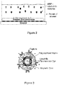
- Figure 2 illustrates schematically, in a non-limiting fashion, the general concept of the invention.
- the figure illustrates different components of the assay of the invention: detectably labeled magnetic nanoparticles associated with an antibody capable of recognizing a target protein, a substrate containing the protein target and a magnet.
- the binding of the MNPs to the target protein immobilized on the substrate is accelerated via application of magnetic field.
- the general concept may be applied to any assay wherein the analyte is immobilized within a porous substrate.
- Non-limiting examples include a dot blot assay and Western blot analysis.
- the invention can be applied to any assay wherein the analyte is immobilized onto a non-porous substrate either directly or via binding to specific antibodies immobilized onto said non-porous substrate, for example in an ELISA performed in a well-plate.
- the detection assay may be performed without separating the steps of (i) binding a primary agent to the target molecule, and (ii) binding a secondary agent to the primary agent.
- This "combined steps" assay is based on pre-incubation of the MNPs carrying the secondary agent with the primary agent, which is usually in molar excess to assure efficient formation of MNP complexes carrying primary agent.
- the assay is further based on the enhanced rate of the reaction of the MNP - primary agent complex with the target molecule, which is accelerated by applying magnetic field.
- the invention provides an assay for detecting a target molecule comprising:
- said MNP has a small diameter.
- said substrate is a porous substrate.
- the average pore size of the porous substrate is larger than the average diameter of the small MNPs.
- said substrate is a non-porous substrate.
- MNP complex magnetic nanoparticle complex
- the term "magnetic nanoparticle complex (MNP complex)" as referred herein relates to a complex comprising one or more magnetic nanoparticles associated with one or more capture molecules.
- the MNP complex of the invention is detectably labeled.
- detectably labeled MNP complex refers to an MNP complex which is associated with a compound that may be detected by an appropriate reaction (enzymatic or color reaction) or by fluorescent excitation and assists in visualizing, quantifying or detecting the target molecule.
- Said labeling compound may be associated with the MNP (e.g. formed around the magnetic shell of the MNP) or associated with the capture molecule, or with both.
- a non-limiting example of a labeled MNP is an MNP having a shell of lipophilic fluorescence dye which is formed around a magnetic core and coated with a polysaccharide matrix. Capture molecules may be associated (or embedded within) the polysaccharide matrix.
- the labeling agent may be a fluorescent compound, e.g. fluorescein (or a fluorescein derivative such as FITC) or phycoerythrin (PE), a fluorescent particle such as quantum dot, an enzyme such as horseradish peroxidase (HRP) or alkaline phosphatase (AP), a chromophore, or an electrochemically active or a radioactive molecule.
- a fluorescent compound e.g. fluorescein (or a fluorescein derivative such as FITC) or phycoerythrin (PE), a fluorescent particle such as quantum dot, an enzyme such as horseradish peroxidase (HRP) or alkaline phosphatase (AP), a chromophore, or an electrochemically active or a radioactive molecule.
- association refers to any physical or chemical forces such as Van-der-Walls, coordinative, covalent, ionic, electrostatic, dipole-dipole, or hydrogen association (bond or interaction).
- the association may occur directly or indirectly (i.e. comprising one or more intermediate agents).
- the intermediate agent is an antibody, another protein, hormone, ligand or spacer.
- the intermediate agent may be streptavidin, biotin, immunoglobulin binding protein (e.g. protein A, protein G, Protein A/G, protein L, etc.), DNA or RNA molecule, peptide tag or its chelating complex (e.g. polyhistidine tag or metal complex of nitrilotriacetic acid such as Ni-NTA).
- the magnetic nanoparticle complexes may be in a dry form (e.g. powder) or dispersed in a medium.
- the "medium”, by its broadest definition, is any material or volume carrying said complexes.
- the medium may be liquid, gel, or porous material.
- the medium may be aqueous or non-aqueous liquid.
- capture molecule refers to an agent which directly or indirectly binds to the target molecule.
- the capture molecule may be a target binding agent (also referred to as a primary agent) and as such it binds directly to the target molecule.
- the capture molecule may also be a secondary binding agent which binds to a primary binding agent and as such it binds indirectly to the target molecule via the primary binding agent.
- Non-limiting examples of capture agents include antibodies, immunoglobulin binding proteins (e.g. protein A, protein G, Protein A/G, protein L), streptavidin (that binds to biotin and biotin labeled compounds), DNA or RNA strands that bind to complementary DNA or RNA strands, and chelating complexes (such as Ni-NTA) that bind to specific peptide tags (e.g. polyhistidine tag).
- immunoglobulin binding proteins e.g. protein A, protein G, Protein A/G, protein L
- streptavidin that binds to biotin and biotin labeled compounds
- DNA or RNA strands that bind to complementary DNA or RNA strands
- chelating complexes such as Ni-NTA
- specific peptide tags e.g. polyhistidine tag
- target binding agent or primary binding agent refers to an agent which specifically associates (binds) to the target molecule (analyte).
- target binding agent is a primary antibody directed to the target antigen.
- second binding agent refers to an agent which binds to the target binding agent (the primary agent).
- the target binding agent is an antibody which specifically binds the target molecule and the secondary binding agent is a secondary antibody which binds the primary antibody.
- the primary antibody is specific for the target and may be but is not limited to a mouse antibody, a rabbit antibody, a goat antibody etc.
- the selection of the type of secondary antibody is dependent on the class of the primary antibody (e.g. IgG or IgD), and on the source of the primary antibody, e.g. if the primary antibody is a mouse antibody, the secondary antibody would be an anti-mouse antibody.
- the secondary antibody may be detectably labeled.
- Non-limiting examples of labels include an enzyme (e.g. HRP or AP), a fluorescent compounds (e.g. fluorescein or phycoerythrin), a chromophore, or an electrochemically active or a radioactive molecule.
- second MNP complex refers to a complex comprising a MNP, a secondary binding agent, and a primary binding agent. Such a complex is obtained by incubating a MNP complex associated with a secondary binding agent with a primary binding agent. The "second MNP complex” may then be applied directly onto the target molecule.
- Antibody refers to IgG, IgM, IgD, and IgA antibodies. This term refers to whole antibodies or fragments of the antibodies comprising the antigen-binding domain, e.g. scFv, Fab, F (ab') 2, bi-specific antibodies, diabodies, and other fragments capable of binding to target molecule.
- the definition includes polyclonal antibodies and monoclonal antibodies.
- Magnetic nanoparticles encompass any nanoparticle (or nanoparticle material) having magnetism (respond to applied magnetic field) observed therewith.
- the magnetic nanoparticles may be paramagnetic, ferromagnetic, ferrimagnetic or superparamagnetic nanoparticles.
- the magnetic nanoparticle is composed of (or doped with) metal, such as iron (Fe), nickel (Ni), cobalt (Co), manganese (Mn), or any combination thereof.
- the magnetic nanoparticle is composed of rare earth metal, such as gadolinium (Gd), neodymium (Nd), samarium (Sm), dysprosium (Dy), Holmium (Ho), or any combination thereof.
- the material is an alloy of metals, wherein at least one metal is having magnetism.
- the alloy is steel, Neodymium-iron-boron alloy (Nd 2 Fe 14 B), nickel-iron alloy (also known as Permalloy), aluminum-nickel-cobalt alloy, samarium-cobalt alloy, yttrium-iron alloy (YIG), yttrium-cobalt alloy (YCo 5 ).
- the magnetic nanoparticles are composed of metal oxide.
- metal oxide Non-limiting examples are iron oxides, such as Fe 2 O 3 ,Fe 3 O 4 , FeO, manganese oxide, such as, Mn 3 O 4 , Mn 2 O 3 , and oters, such as, CrO 2 , EuO, or any combination thereof.
- the magnetic nanoparticles are composed of minerals.
- Non-limiting examples of magnetic minerals are FeS 2 , Fe 3 Si.
- the magnetic nanoparticles are materials (magnetic or non-magnetic, e.g., metals, semiconductors, insulators) doped with a magnetic component.
- the magnetic nanoparticles are composed of a semiconductor material doped (or alloyed) with magnetic material. e.g., GaAs, In As, InSb, AlN, TiO 2 , ZnO, SnO 2 doped with one or more of the following dopants (atoms or ions) Fe, Co, Ni, Cr, Mn, V.
- the magnetic nanoparticles are composed of a semiconductor material, such as MnAs, MnSb.
- the magnetic nanoparticles are composed of iron oxide.
- the magnetic nanoparticle in accordance with the present invention has dimensions (length or diameter) at the nanometric range (between 1 and 1000 nm).
- the nanoparticles of the invention are small sized nanoparticles being in the range of between 1 nm to 50 nm in length or diameter.
- the nanoparticles are on average in the range of 1 nm to 30 nm in length or diameter.
- the nanoparticles are on average in the range of 10 nm to 30 nm in length or diameter.
- the nanoparticles have a diameter of 10nm, or 20nm, or 25nm, or 30nm.
- the nanoparticles are selected amongst isotropic and anisotropic shaped nanoparticles.
- the nanoparticles may be symmetrical or unsymmetrical, may be elongated or having a round (spherical)-shape, rod-like shape, elliptical, branch, network or any irregular shape.
- the nanoparticles are selected from nanospheres, nanorods, branched nanoparticles, multipods and others.
- the labeled MNP complex in accordance with the invention is a small sized iron oxide nanoparticle ( ⁇ 30 nm) coated with linear amphiphilic polymer carrying end carboxylic groups associated with horseradish peroxidase (HRP)-labeled anti-rabbit IgG secondary antibody.
- HRP horseradish peroxidase
- the assay of the invention may be a Western blot assay.
- Protocols for carrying out Western blot assays are well known in the art.
- a first step in a Western blot assay is electrophoresis of a sample containing the target molecules.
- electrophoresis is carried out on polyacrylamide gels.
- a non-limiting example of a protocol for carrying out a Western blot assay is provided in Example 7 below. Following electrophoresis the target molecules are transferred onto a porous substrate for further analysis.
- the system operates on the basis of acceleration of magnetic nanoparticle complexes, carrying the capture agent to a substrate containing the target molecule.
- the substrate may be a porous substrate.
- the term "porous substrate” refers to a substrate having a plurality of pores (depressions). These pores have inner voids of the same or varying volume and shape, defined by inner surface.
- the substrate pores are nanometric in size, namely having a mean size smaller than 1,000 nm.
- the mean pore size is below 500 nm.
- the mean pore size is below 300 nm.
- the mean pore size is below 200 nm.
- the mean pore size is below 100 nm.
- the mean pore size is below 50 nm.
- the mean pore size is between 300 nm and 50 nm.
- the porous substrate is a membrane having 200nm or 450nm pores.
- the substrate may also be a non-porous substrate.
- the substrate may be a flexible or a rigid or a soft substrate, and may be composed of any material.
- the substrate is a layered substrate, e.g., a porous layer on a non-porous layer or soft layer (such as gel or tissue) on metal layer (holder).
- the substrate (or one of its layers) may be composed of insulating, conducting or semiconducting material.
- the substrate may be composed of glassy, polymeric, ceramic, fibrous material, or any combination thereof.
- the substrate's material may be composed of glass, paper, wool, fleece, gel, cellulose, or any combination thereof.
- the substrate is composed of a nitrocellulose or PVDF membrane.
- the nitrocellulose or PVDF membrane may have varying pore sizes, e.g. 0.2 ⁇ m (200 nm) or 0.45 ⁇ m (450 nm).
- target molecules is associated to (immobilized on) the surface of the substrate.
- the "target molecule” or “analyte” refers to any agent whose presence and/or amount is to be determined.
- the target molecule may be a mixture of several target molecules.
- the presence or concentration profile of the different target molecules is to be determined.
- the target molecule may be any biological and/or chemical agent (i.e., molecule/macromolecule/complex/conjugate).
- the target molecule is an organic or inorganic molecule.
- the target molecule is a biological agent.
- the target molecule is a protein, a polypeptide or a peptide.
- the target molecules are nucleic acids (e.g. DNA, RNA), carbohydrates, lipids, glycolipides, triglyceride or polysaccharide.
- the target molecules may be hormones, vitamins, antibodies, metabolites, amino acid (e.g., glutamic acid, aspartic acid), alcohol (e.g., ethanol), organic acid (e.g., acetic acid, lactic acid), antioxidant (e.g., ascorbic acid), polyol (e.g., glycerol) or any derivatives and/or combination thereof.
- the target molecule is a toxin or a drug.
- the assay of the invention may detect even small amounts of the target molecule.
- amount of the target molecule in the substrate is in micrograms. In some embodiments the amount of the target molecule is below 1 microgram. In some embodiments amount of the target molecule in the substrate is in nanograms. In some embodiments the amount of target agent in the substrate is between 100 ng to 1 ng. In some embodiments the amount of target agent in the substrate is between 100pg to 1pg.
- the system comprises an element generating magnetic field, i.e., a magnet.
- the system may have one or more magnets.
- the system has at least one magnet situated in vicinity to the substrate.
- the magnet is positioned below the substrate.
- the magnet may be positioned on top of the substrate or on the side.
- the magnet may be a permanent magnet or an electromagnet.
- the element generating a magnetic field is a magnet array composed of small magnets (e.g. 1 ⁇ 4 inch x 1 ⁇ 4 inch x 1 ⁇ 2 inch, as shown in Figure 16 ).
- the magnetic polarity in the magnet array may be perpendicular, namely intersecting at a right angle to the main plane formed by the magnets, and opposite for each neighboring magnet (i.e. a chessboard configuration), also termed herein a "chessboard magnet array".
- the assay of the invention further comprises a detection step.
- the detection step may involve illuminating said substrate followed by fluorescent detection (e.g. by using a fluorescent imager), densitometry measurements, spectrophotometer measurements, light scattering measurements, current measurements, magnetic induction measurements, light detection (by e.g. a CCD camera, photographic film) or radioactivity detection.
- the assay of the invention comprises label free detection based on mass or refractive index change, such as surface plasmon resonance (SPR) or quartz crystal microbalance (QCM).
- the detection step comprises a signal development reaction.
- the signal development reaction is a chemiluminescent reaction, which is catalyzed by the labeling agent on the substrate.
- the chemiluminescent reaction of lumiunol is catalyzed by the enzymes HRP or AP under suitable conditions, which are provided in standard chemiluminescence development solutions.
- the signal development reaction forms a detectable color.
- the colorimetric reaction of several reagents, such as TMB or OPD is catalyzed by HRP under suitable conditions.
- the assay is performed with a substrate comprising at least one target molecule.
- the substrate comprising (or associated with) at least one target molecule is formed by contacting a sample potentially comprising the target molecule with the substrate.
- the target molecules are deposited on the surface of the substrate, which may be a porous substrate, and through surface opening(s) of the substrate one or more target molecules are immobilized onto the inner surface of the substrate's pores.
- the substrate may be a non-porous substrate. In such case the substrate's surface is being coated by the target molecules, either directly or indirectly via binding to a specific antibody which is immobilized onto the substrate's surface.
- the target molecule may be deposited on the substrate by any method.
- deposition may be by simple dropping a sample containing the target molecule on the substrate or immersing the substrate in the sample.
- the substrate may be activated prior to the exposure to the target molecules, for example surface carboxylic acid groups are commonly activated by a solution containing EDC and NHS to enable standard amine coupling.
- the substrate e.g. a nitrocellulose membrane
- the sample comprising the target molecule(s) is deposited on a substrate by employing electrochemical deposition, electrophoretic deposition, electroplating, spraying, spin coating, or any other method of deposition.
- the substrate may undergo one or more treatments steps, such as washing, drying, blocking, heating or deactivation.
- said treatment step is blocking.
- the blocking treatment is performed by placing (or immersing) the substrate in blocking solution, which may contain proteins (e.g. non-fat milk or BSA), buffer (e.g. Tris Buffered Saline (TBS) or Phosphate Buffered Saline (PBS)), and detergent (such as Tween 20 or Triton X-100).
- proteins e.g. non-fat milk or BSA
- buffer e.g. Tris Buffered Saline (TBS) or Phosphate Buffered Saline (PBS)
- detergent such as Tween 20 or Triton X-100.
- the assay may include a drying step (i.e. a step of water exclusion or partial water exclusion) in which the substrate (e.g. the membrane) is dried prior to incubating the substrate with the MNP complex, and in particular, prior to the blocking step that precedes the incubation with the MNP complex. Drying of the membrane improves the Signal to Noise Ratio (SNR), namely_higher signals and lower background are obtained in assays that include membrane drying.
- SNR Signal to Noise Ratio
- the higher signals are probably due to fixation of the target proteins to the membrane, and preventing loss of proteins during the following assay and wash steps.
- the lower background is probably due to more efficient blocking - the blocking proteins are brought into the membrane by wetting process, which is much faster and efficient than diffusion process as in the standard blocking.
- Membrane drying may be performed using any method known in the art, for example by heating, subjecting to air flow, immersion in polar organic solvents (e.g. alcohol or DMSO), applying vacuum or any combination thereof.
- polar organic solvents e.g. alcohol or DMSO
- drying is performed by placing the membrane in a laboratory oven at 37 °C under air flow for 30 min.
- the assay steps may have prior and/or post steps of washing.
- the washing step is by placing (or immersing) the substrate in a washing solution.
- the washing solution comprises buffer (e.g. TBS or PBS) and a detergent (such as Tween 20 or Triton X-100).
- the washing of MNPs from the substrate surface may be accelerated or made more efficient by magnetic attraction.
- the magnet should be positioned at an opposite direction to the direction that was used to accelerate the MNP-analyte binding reaction. For example, if the magnet was placed below the membrane for the binding step, it should be placed above the membrane in the washing step. Alternatively, the membrane may be rotated with its upper face down while the magnet remains below the membrane.
- the assay comprises separation of the magnetic nanoparticle complexes from the other agents in the medium.
- the MNP complex is mixed with excess of primary binding agent, it may be useful to purify the MNP complexes that bound the primary binding agent and wash away the excess of primary binding agent.
- the magnetic properties of the MNPs may be used for the separation.
- separation means is by magnetic precipitation, or passing through a magnetic column.
- common methods for separation by size may be used, such as centrifugation, chromatography or filtering.
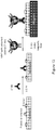
- FIG. 13 A schematic representation of this embodiment is outlined in Figure 13 .
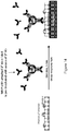
- FIG. 14 A schematic representation of this embodiment is outlined in Figure 14 .
- the first and the second primary binding agents are directed against different binding sites on the target molecule.
- the invention provides a detectably labeled MNP complex comprising magnetic nanoparticles having a small diameter associated with a capture molecule capable of binding directly or indirectly to a target molecule, for use in an assay for the detection of said target molecule wherein said target molecule is immobilized onto a porous substance.
- said target molecule is immobilized onto a non-porous substance.
- the detectably labeled MNP complex is for use in research laboratories, hospitals or medical care centers (for the professional user, such as a technician, a nurse, a doctor). In some embodiments, the detectably labeled MNP complex is for home use (for by patients).
- Example 1 Dot blot analysis using 100 nm fluorescent MNP-capture agent complexes
- MNPs Fluorescent magnetic nanoparticles
- 100 nm size (hydrodynamic diameter) were purchased from Chemicell. These MNPs contain a shell of lipophilic fluorescence dye which was formed around a magnetic core and coated with a polysaccharide matrix. Capturing agents were embedded within the polysaccharide matrix ( Figure 3 ). MNPs with three capture agents - Protein G, anti-mouse IgG and streptavidin - were tested. All three MNP types had fluorescence with excitation pick at 547 nm and emission pick at 580 nm.
- the MNPs would provide stronger (or at least comparable) signals than the standard captures in dot blot experiments, namely in immune-reaction with an antigen which was spotted onto the membrane.
- the results of dot blot experiments indicated that the MNPs failed to function as good as the standard capturing agents.
- 2 ⁇ l drops of biotinylated antibody at a series of concentrations were spotted on 0.45 ⁇ m NC membranes.
- One membrane was then exposed to a solution of streptavidin coated MNPs, and a second reference membrane was exposed to the standard capture streptavidin-PE under similar conditions (e.g. the same molar concentration).
- Detection using the ChemiDoc MP imager revealed strong signals with streptavidin-PE ( Figure 5A ) and only weak signals with streptavidin-MNP ( Figure 5B ).
- magnetic nanoparticle-antibody complexes comprising small sized iron oxide nanoparticles ( ⁇ 30 nm) were purchased from Ocean Nanotech. These iron oxide (IO) nanoparticles are coated with linear amphiphilic polymer carrying end carboxylic groups, which can be used to immobilize antibodies or other proteins ( Figure 6 ).
- HRP horseradish peroxidase
- Ocean Nanotech horseradish peroxidase-labeled anti-rabbit IgG secondary antibody
- HRP horseradish peroxidase
- Three nanoparticle sizes were used: 10 nm, 20 nm, and 30 nm.
- Enzyme (HRP) activity of the nanoparticles was tested using a colorimetric assay in solution, with comparison to the free labeled antibody (anti-rabbit IgG-HRP from Jackson). This analysis was done by placing a concentration series of MNP or antibody solution in wells of non-binding 96 well plate (50 ⁇ l solution per well). Color development was done by adding 100 ⁇ l of standard TMB solution to each well, and was stopped by adding 50 ⁇ l sulfuric acid.
- the signal was detected by reading optical density at 450 nm (OD 450 ) using a plate reader.
- the results are presented in Figure 7 , wherein the OD 450 is shown as a function of the labeled MNP / antibody concentration. High activity was observed with all the tested MNPs, while all MNPs were found to be more active than the free anti-rabbit HRP (in molar terms).
- the magnetic particles-associated with anti-rabbit-HRP showed higher molar activity with increasing magnetic particles diameter.
- the average number of antibody molecules per MNP is estimated to be about 1.5, 4.5 and 9.6 for the 10, 20 and 30 nm MNPs, respectively.
- the larger particles can carry a larger number of antibody molecules per particle, and hence their increased enzymatic activity.
- Example 3 Dot blot analysis with small MNPs carrying a secondary antibody
- Rabbit IgG solution (2 ⁇ l) was spotted on two 0.45 ⁇ m NC membrane at a series of 16 concentrations (x1.5 dilution series), yielding spotted amounts from 5 ⁇ g down to 11 ng.
- One membrane was reacted with the 10 nm IO magnetic nanoparticles described in Example 2 (0.43 nM).
- the second, reference membrane was reacted with free anti-rabbit HRP under similar conditions (e.g. the same molar concentration). Color signal was developed and detected using 4-CN substrate and ChemiDoc MP imager.
- the protocol was composed of the following steps:
- Figure 11B is a top view picture of said representative device
- Figure 11C is a picture of the different elements of the representative device: a neodymium disc magnet, a membrane, and a holder.
- the experimental setting schematically illustrated in Figure 11A includes a membrane showing 6 sample zones, five of which are rabbit IgG each at a different concentration and one control sample including a non-relevant antibody (0.5 ⁇ g CAII).
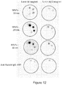
- Dot blot assays were carried out with and without application of the magnetic field for 5 minutes using four different measuring agents: a complex of 10 nm magnetic nanoparticles-anti-rabbit-HRP antibody, a complex of 20 nm magnetic nanoparticles-anti-rabbit-HRP antibody, a complex of 30 nm magnetic nanoparticles-anti-rabbit-HRP antibody, and free anti-rabbit IgG-HRP ( Figure 12 ).
- the results show very weak detection signal of the high rabbit IgG concentrations (5 ⁇ g and 0.5 ⁇ g) in the dot blot assay with free anti-rabbit IgG and no signal at all in the lower concentrations, with or without applying the magnetic field.
- the assay using 10 nm magnetic particles-antibody complex without magnetic field showed similar results as the free anti-rabbit IgG-HRP measuring agent, however upon applying magnetic field there is clear improvement of the detection signal and the assay shows sensitivity even to 0.05 ⁇ g.
- the detection signals become even more pronounced when the assays were carried out with 20 nm and 30 nm magnetic particles-antibody complexes and the applied magnetic field.
- the sensitivity was also improved, showing capabilities to detect lower levels of rabbit IgG even up to 5 ng, even in naked-eye examination. Therefore, the magnetic field clearly improves detection mainly with the 20nm and 30 nm MNPs.
- Example 5 Comparison of a Dot Blot assay with separate antibody binding steps and combined antibody binding steps
- nanoparticle complex was associated with a secondary binding agent and applied to the substrate after the substrate was incubated with the first binding agent (a schematic representation of this procedure is outlined in Figure 13 ).
- the protocol was composed of the following steps (steps 1-3 and 5 are similar to those detailed in Example 3 above):
- nanoparticle complex was associated with a secondary binding agent and pre-incubated with the first binding agent prior to applying the combined solution onto the substrate (a schematic representation of this procedure is outlined in Figure 14 ) .
- Figure 15 summarizes the results of the experiments outlined in Example 5.
- the combined antibody assay with magnetic field for 5 minutes showed a higher signal then that obtained in the combined antibody assay without magnetic field, even when a long (1 hour) binding step is implemented.
- Comparison of the separate antibody steps assay with the above conditions showed an opposite behavior, i.e., the assay having long (1 hour) binding step without magnetic field shows a higher detection signal than the corresponding assay with applying magnetic field for 5 minutes. In both cases the control experiments, where the assays were without magnetic field for a short period, no detection signal was observed.
- the results indicate that the magnetic field accelerates the detection, and exhibits a detection signal after a short period of time, which does not appear so quickly without the employment of magnetic field.
- the binding step is performed in a mixture of free primary antibodies and magnetic nanoparticle-secondary antibody complexes.
- the procedure may be performed without separating the free primary antibodies from the primary antibodies that are bound to the MNPs, since although both the free primary antibodies and the primary antibodies that are now bound to the magnetic nanoparticles may bind to the target molecule, when applying the magnetic field the magnetic nanoparticles-primary antibodies complexes are drawn to the membrane at an accelerated pace, while the free primary antibodies remain in the medium and bind to the target molecules at a much slower rate.
- This is a simple competition in which the MNP-bound primary antibodies have a significant advantage.
- the two primary antibodies compete with one another for binding to the target protein, and therefore the detection signal is lower in this assay after one hour without applying the magnet.
- the results demonstrate that the assays of the invention provide a significantly shortened assay time with appreciably detectable results.
- the assay duration is substantially shortened by using the techniques of the invention.
- Steps 1-3 and 6 were done similarly to the assays with magnetic acceleration. Instead of Steps 4 and 5 the following steps were performed:
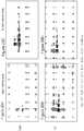
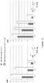
- FIGs 18 and 19 summarize the results of these experiments.
- the Chemidoc MP images ( Figure 18 ) and analysis graphs ( Figure 19 ) reveal higher signals for the short MNP-based assay for both hT and hSA models. More detectable bands were gained for the MNP-based assays, indicating higher assay sensitivity. Without wishing to be bound by theory, the increased signals are probably due to the fact that there are multiple labeled antibodies attached to each MNP (-12 in this case), and thus signal amplification is obtained.
- Figure 18 also shows higher background noise in the MNP-based assays. It was thus required to reduce the background in these assays and gain higher signal:noise ratio (SNR). Many experimental parameters have been tested in order to improve the SNR. The method that yielded the most significant improvement involved drying of the membrane as described in Example 8 below.
- FIGs 20 and 21 summarize the results of these experiments.
- the Chemidoc MP images ( Figure 20 ) and analysis graphs ( Figure 21 ) reveal higher signals and lower background (and thus improved SNR) for the assays that include membrane drying for both hT and hSA models.
- the higher signals are probably due to fixation of the target proteins to the membrane, and preventing loss of proteins during the following assay and wash steps;
- the lower background is probably due to more efficient blocking - the blocking proteins are brought into the membrane by wetting process, which is much faster and efficient than diffusion process as in the standard blocking.
- ELISA assay was performed with the same two proteins as demonstrated above, hT and hSA.
- the protocol was composed of the following steps:
- Steps 1, 2 and 5 were done similarly to the assays with magnetic acceleration. Instead of Steps 3 and 4 the following steps were performed:
- Figure 22 summarizes the results of these experiments. Higher SNR for the short MNP-based assay for both hT and hSA models was recorded, indicating higher assay sensitivity. This improved SNR originated from both higher signals and lower background (results not shown). Without wishing to be bound by theory, the increased signals are probably due to the fact that there are multiple labeled antibodies attached to each MNP (-17 in this case), and thus signal amplification is obtained.
Landscapes
- Health & Medical Sciences (AREA)
- Life Sciences & Earth Sciences (AREA)
- Immunology (AREA)
- Engineering & Computer Science (AREA)
- Chemical & Material Sciences (AREA)
- Molecular Biology (AREA)
- Biomedical Technology (AREA)
- Hematology (AREA)
- Urology & Nephrology (AREA)
- Food Science & Technology (AREA)
- Biochemistry (AREA)
- Cell Biology (AREA)
- Biotechnology (AREA)
- Medicinal Chemistry (AREA)
- Physics & Mathematics (AREA)
- Analytical Chemistry (AREA)
- Microbiology (AREA)
- General Health & Medical Sciences (AREA)
- General Physics & Mathematics (AREA)
- Pathology (AREA)
- Nanotechnology (AREA)
- Chemical Kinetics & Catalysis (AREA)
- Measuring Or Testing Involving Enzymes Or Micro-Organisms (AREA)
- Apparatus Associated With Microorganisms And Enzymes (AREA)
- Tropical Medicine & Parasitology (AREA)
Claims (15)
- Ein Assay zum Detektieren eines Zielmoleküls, umfassend:a. Inkubieren eines Substrats, das potentiell mindestens ein Zielmolekül umfasst, mit detektierbar markierten magnetischen Nanopartikel (MNP)-Komplexen, wobei die MNP-Komplexe MNP mit einem Durchmesser kleiner als 50 nm umfassen, das mit einem Fängermolekül in Verbindung steht, das in der Lage ist sich direkt oder indirekt an das Zielmolekül zu binden, wobei die Inkubation unter Anlegen eines Magnetfeldes durchgeführt wird; undb. Unterziehen des inkubierten Substrats einem Detektionsschritt;
wobei das Vorhandensein eines Detektionssignals das Vorhandensein des Zielmoleküls anzeigt und die Signalintensität die Menge des Zielmoleküls anzeigt. - Ein Assay zum Detektieren eines Zielmoleküls, umfassend:a. Bereitstellen von detektierbar markierten magnetischen Nanopartikel (MNP)-Komplexen, wobei die MNP-Komplexe MNP umfassen, das in Verbindung steht mit einem sekundären Bindemittel, das in der Lage ist ein primäres Bindemittel zu binden;b. Inkubieren der MNP-Komplexe mit einem primären Bindemittel, dabei Bilden eines zweiten MNP-Komplexes, umfassend MNP, ein sekundäres Bindemittel und ein primäres Bindemittel;c. Inkubieren eines Substrats, das potentiell mindestens ein Zielmolekül umfasst, mit dem zweiten MNP-Komplex, wobei die Inkubation des Substrats, das potentiell mindestens ein Zielmolekül umfasst, mit dem zweiten MNP-Komplex, unter Anlegen eines Magnetfeldes durchgeführt wird; undd. Unterziehen der inkubierten Probe einer Detektionsumsetzung;
wobei das Vorhandensein eines Detektionssignals das Vorhandensein des Ziclmolcküls anzeigt und die Signalintensität die Menge des Zielmoleküls anzeigt. - Der Assay gemäß Anspruch 1, wobei das Fängermolekül ein primäres Bindemittel ist.
- Der Assay gemäß Anspruch 1, wobei das Fängermolekül ein sekundäres Bindemittel ist.
- Der Assay gemäß Anspruch 4, wobei das Substrat vor der Inkubation mit dem MNP-Komplex, umfassend das sekundäre Bindemittel, mit einem primären Bindemittel inkubiert wird.
- Der Assay gemäß Anspruch 4, wobei der MNP-Komplex, umfassend das sekundäre Bindemittel, vor der Inkubation mit dem Substrat mit einem primären Bindemittel inkubiert wird.
- Der Assay gemäß einem der Ansprüche 2 bis 6, wobei das primäre Bindemittel ein primärer Antikörper ist.
- Der Assay gemäß Anspruch 7, wobei das sekundäre Bindemittel ein sekundärer Antikörper ist.
- Der Assay gemäß einem der vorangehenden Ansprüche, wobei das MNP in dem MNP-Komplex detektierbar markiert ist.
- Der Assay gemäß einem der vorangehenden Ansprüche, wobei das Fängermolekül in dem MNP-Komplex detektierbar markiert ist.
- Der Assay gemäß Anspruch 2, wobei das sekundäre Bindemittel in dem MNP-Komplex detektierbar markiert ist.
- Der Assay gemäß einem der vorangehenden Ansprüche, wobei das Magnetfeld durch eine Magnetanordnung, die aus kleinen Magneten in einer Schachbrett-Konfiguration zusammengesetzt ist, erzeugt wird.
- Der Assay gemäß einem der Ansprüche 1, 3, 4 und 6 bis 8, wobei der Assay ein Dot-Blot-Assay ist, umfassend die Schritte:a. Blotting des Zielmoleküls auf eine Membran;b. Eintauchen der Membran in eine Blockierungslösung;c. Eintauchen der Membran in eine vor-vorbereitete Lösung, die ein primäres Bindemittel und MNPs mit einem kleinen Durchmesser, die in Verbindung mit einem markierten sekundären Mittel stehen, enthält, unter Anlegen eines Magnetfeldes; undd. Detektieren der Markierung.
- Der Assay gemäß einem der Ansprüche 1, 3, 4 und 6 bis 8, wobei der Assay ein Western-Blot-Assay ist, umfassend die Schritte:a. Unterziehen einer Probe, umfassend Proteine, einer elektrophoretischen Trennung;b. Übertragen der getrennten Proteine auf eine Membran;c. Eintauchen der Membran in eine Blockierungslösung;d. Eintauchen der Membran in eine vor-vorbereitete Lösung, die ein primäres Bindemittel und MNPs mit einem kleinen Durchmesser, die in Verbindung mit einem markierten sekundären Mittel stehen, enthält, unter Anlegen eines Magnetfeldes; unde. Detektieren der Markierung.
- Der Assay gemäß einem der Ansprüche 1, 3, 4 und 6 bis 8, wobei der Assay ein ELISA ist, umfassend die Schritte:a. Beschichten des Substrats mit dem Zielprotein;b. Hinzufügen einer Blockierungslösung;c. Hinzufügen einer vor-vorbereiteten Lösung, die ein primäres Bindemittel und MNPs mit einem kleinen Durchmesser, die in Verbindung mit einem markierten sekundären Mittel stehen, enthält, unter Anlegen eines Magnetfeldes; undd. Detektieren der Markierung.
Applications Claiming Priority (2)
| Application Number | Priority Date | Filing Date | Title |
|---|---|---|---|
| US201361758065P | 2013-01-29 | 2013-01-29 | |
| PCT/IL2014/050101 WO2014118775A1 (en) | 2013-01-29 | 2014-01-29 | Detection assays employing magnetic nanoparticles |
Publications (3)
| Publication Number | Publication Date |
|---|---|
| EP2951583A1 EP2951583A1 (de) | 2015-12-09 |
| EP2951583A4 EP2951583A4 (de) | 2016-09-07 |
| EP2951583B1 true EP2951583B1 (de) | 2018-01-24 |
Family
ID=51223353
Family Applications (1)
| Application Number | Title | Priority Date | Filing Date |
|---|---|---|---|
| EP14746707.0A Active EP2951583B1 (de) | 2013-01-29 | 2014-01-29 | Detektionsassays unter verwendung von magnetischen nanopartikeln |
Country Status (4)
| Country | Link |
|---|---|
| US (1) | US10732178B2 (de) |
| EP (1) | EP2951583B1 (de) |
| CN (1) | CN105102979B (de) |
| WO (1) | WO2014118775A1 (de) |
Families Citing this family (7)
| Publication number | Priority date | Publication date | Assignee | Title |
|---|---|---|---|---|
| KR101398764B1 (ko) * | 2013-08-29 | 2014-05-27 | 강릉원주대학교산학협력단 | 입자의 이동에 의해 분석물질을 검출하는 장치 및 방법 |
| JP6420433B1 (ja) * | 2017-09-12 | 2018-11-07 | 株式会社 マトリックス細胞研究所 | 反応促進装置及び反応促進方法 |
| JP6370018B1 (ja) * | 2018-03-15 | 2018-08-08 | 株式会社 マトリックス細胞研究所 | 反応促進装置及び反応促進方法 |
| JP6370017B1 (ja) * | 2018-03-15 | 2018-08-08 | 株式会社 マトリックス細胞研究所 | 反応促進装置及び反応促進方法 |
| JP2020016522A (ja) * | 2018-07-25 | 2020-01-30 | 国立大学法人 岡山大学 | 磁性粒子を用いた免疫染色法 |
| JP7832652B2 (ja) * | 2022-02-08 | 2026-03-18 | 国立大学法人京都工芸繊維大学 | 抗体固定ニトロセルロース膜の製造方法及び抗原結合性増強方法 |
| CN117110607B (zh) * | 2023-09-16 | 2024-01-26 | 鲁东大学 | 一种检测副溶血性弧菌的组合物及其应用 |
Family Cites Families (40)
| Publication number | Priority date | Publication date | Assignee | Title |
|---|---|---|---|---|
| US5714380A (en) * | 1986-10-23 | 1998-02-03 | Amoco Corporation | Closed vessel for isolating target molecules and for performing amplification |
| DE68911359T2 (de) | 1988-04-26 | 1994-05-11 | Nippon Telegraph & Telephone | Lasermagnetisches Immuntestverfahren und Gerät dazu. |
| EP0339980B1 (de) | 1988-04-26 | 1994-07-20 | Nippon Telegraph And Telephone Corporation | Mikropartikel, Verfahren und Gerät zur Sammlung von Proben zur Verwendung bei der Markierung von Immunreaktionen und Verfahren und Gerät zur Bereitung von Proben |
| EP0351857B1 (de) | 1988-07-20 | 1994-11-30 | Olympus Optical Co., Ltd. | Immuntestverfahren unter Verwendung magnetischer Markerteilchen |
| US5736349A (en) | 1989-09-29 | 1998-04-07 | Nippon Paint Co., Ltd. | Magnetic particle and immunoassay using the same |
| JP2979414B2 (ja) | 1989-09-29 | 1999-11-15 | 富士レビオ株式会社 | 磁性粒子およびそれを用いた免疫測定法 |
| US5445971A (en) * | 1992-03-20 | 1995-08-29 | Abbott Laboratories | Magnetically assisted binding assays using magnetically labeled binding members |
| DE19508772C2 (de) * | 1995-03-01 | 1998-01-29 | Schering Ag | Verfahren und Verbindungen zur Detektion von Analyten mittels Remanenzmessung und deren Verwendung |
| US20040209376A1 (en) * | 1999-10-01 | 2004-10-21 | Surromed, Inc. | Assemblies of differentiable segmented particles |
| US6136549A (en) * | 1999-10-15 | 2000-10-24 | Feistel; Christopher C. | systems and methods for performing magnetic chromatography assays |
| AU2001272012A1 (en) | 2000-06-19 | 2002-01-02 | Arizona Board Of Regents | Rapid flow-based immunoassay microchip |
| DK1311820T3 (en) * | 2000-07-14 | 2015-04-07 | Janssen Diagnostics Llc | Increased separation efficiency via a controlled aggregation of magnetic nanoparticles |
| WO2002031179A2 (en) * | 2000-10-11 | 2002-04-18 | Evotec Oai Ag | Multiplex assays using nanoparticles |
| US20030077598A1 (en) * | 2001-01-04 | 2003-04-24 | Phan Brigitte Chau | Dual bead assays including covalent linkages for improved specificity and related optical analysis discs |
| CN100442052C (zh) * | 2002-08-15 | 2008-12-10 | 陕西西大北美基因股份有限公司 | 磁性荧光微球及其制备方法和采用该磁性荧光微球进行生物分子检测的方法 |
| US7314763B2 (en) * | 2002-08-27 | 2008-01-01 | Kimberly-Clark Worldwide, Inc. | Fluidics-based assay devices |
| AU2003267255A1 (en) * | 2002-09-20 | 2004-04-08 | Novus Molecular, Inc. | Methods and devices for active bioassay |
| CN1218752C (zh) * | 2003-06-27 | 2005-09-14 | 天津大学 | 复合功能超微磁性载体粒子及其制备方法 |
| US20050147963A1 (en) * | 2003-12-29 | 2005-07-07 | Intel Corporation | Composite organic-inorganic nanoparticles and methods for use thereof |
| CN1261765C (zh) * | 2004-12-27 | 2006-06-28 | 中国人民解放军军事医学科学院卫生学环境医学研究所 | 一种纳米免疫磁颗粒的制备方法及应用 |
| WO2006091525A2 (en) | 2005-02-24 | 2006-08-31 | Invitrogen Corporation | Electro-blotting devices, systems, and kits, and methods for their use |
| CN101375166B (zh) * | 2006-01-25 | 2013-07-10 | 皇家飞利浦电子股份有限公司 | 用于分析流体的装置 |
| CN100464189C (zh) * | 2006-04-21 | 2009-02-25 | 深圳市新产业生物医学工程有限公司 | 磁分离直接化学发光试剂及用该试剂的测试方法 |
| CN101029897A (zh) * | 2007-02-09 | 2007-09-05 | 深圳市新产业生物医学工程有限公司 | 一种降钙素原测试试剂盒及其测试方法 |
| MX2009009994A (es) * | 2007-03-20 | 2009-10-19 | Becton Dickinson Co | Ensayos que usan particulas activas para espectroscopia raman mejorada por superficie (sers). |
| WO2008114025A1 (en) * | 2007-03-21 | 2008-09-25 | University Of The West Of England, Bristol | Particle facilitated testing |
| FR2917173B1 (fr) * | 2007-06-08 | 2009-07-31 | Bio Rad Pasteur Sa | Procede multiplexe de detection d'une infection |
| US8287810B2 (en) * | 2007-06-20 | 2012-10-16 | Board Of Trustees Of Michigan State University | Electrically-active ferromagnetic particle conductimetric biosensor test kit |
| US7927561B2 (en) * | 2008-01-10 | 2011-04-19 | Becton, Dickinson And Company | Rapid particle detection assay |
| JP4980944B2 (ja) * | 2008-02-12 | 2012-07-18 | 富士フイルム株式会社 | 免疫学的測定方法 |
| GB0808089D0 (en) * | 2008-05-02 | 2008-06-11 | Iti Scotland Ltd | magnetic recognition system |
| US20100044229A1 (en) | 2008-07-11 | 2010-02-25 | Life Technologies Corporation | Electrophoretically Enhanced Detection of Analytes on a Solid Support |
| US11493507B2 (en) | 2009-09-14 | 2022-11-08 | Siemens Healthineers Nederland B.V. | Highly sensitive immunoassay with large particle labels |
| RU2595843C2 (ru) | 2009-10-16 | 2016-08-27 | Омик Аб | Способ анализа и устройства с применением магнитных частиц |
| US20110262989A1 (en) * | 2010-04-21 | 2011-10-27 | Nanomr, Inc. | Isolating a target analyte from a body fluid |
| SE535918C2 (sv) | 2010-06-10 | 2013-02-19 | Hemocue Ab | Detektion av magnetiskt inmärkta biologiska komponenter |
| CN102331492A (zh) | 2011-06-14 | 2012-01-25 | 浙江大学 | 一种检测血清中螨虫过敏原特异性抗体的方法 |
| CH705758B1 (fr) * | 2011-11-15 | 2016-03-31 | Metalor Technologies Int | Nanoparticules cœur-coquille métal-silice, procédé de fabrication et dispositif de test par immunochromatographie comprenant de telles nanoparticules. |
| KR102037469B1 (ko) | 2012-01-02 | 2019-10-28 | 삼성전자주식회사 | 그래핀 전자 소자 및 그 제조 방법 |
| FR3001038B1 (fr) * | 2013-01-17 | 2018-02-09 | Centre National De La Recherche Scientifique (Cnrs) | Procede de capture, procede de detection et kit de capture d'une molecule dans un echantillon |
-
2014
- 2014-01-29 US US14/167,428 patent/US10732178B2/en active Active
- 2014-01-29 WO PCT/IL2014/050101 patent/WO2014118775A1/en not_active Ceased
- 2014-01-29 CN CN201480018580.5A patent/CN105102979B/zh active Active
- 2014-01-29 EP EP14746707.0A patent/EP2951583B1/de active Active
Non-Patent Citations (1)
| Title |
|---|
| None * |
Also Published As
| Publication number | Publication date |
|---|---|
| WO2014118775A1 (en) | 2014-08-07 |
| US10732178B2 (en) | 2020-08-04 |
| US20140212990A1 (en) | 2014-07-31 |
| CN105102979B (zh) | 2017-05-03 |
| EP2951583A1 (de) | 2015-12-09 |
| CN105102979A (zh) | 2015-11-25 |
| EP2951583A4 (de) | 2016-09-07 |
Similar Documents
| Publication | Publication Date | Title |
|---|---|---|
| US10732178B2 (en) | Detection assays employing magnetic nanoparticles | |
| JP4171139B2 (ja) | 磁性体標識による免疫検査方法とその装置 | |
| US7018849B2 (en) | Process for (A) separating biological/ligands from dilute solutions and (B) conducting an immunochromatographic assay thereof employing superparamagnetic particles throughtout | |
| TWI429903B (zh) | 超靈敏磁減量量測系統以及使用其之超靈敏免洗檢定 | |
| US20070020699A1 (en) | Lateral flow assay and device using magnetic particles | |
| JP4271034B2 (ja) | 磁場を用いた結合アッセイ方法 | |
| CN101441210B (zh) | 纳米磁性粒子层析试纸检测方法 | |
| RU2008148311A (ru) | Детектирование молекул-мишеней в пробе | |
| CN115825452B (zh) | 胶质纤维酸性蛋白检测试剂盒 | |
| US20110050215A1 (en) | System for applying magnetic forces to a biosensor surface by mechanically moving at least one magnet | |
| JP7041491B2 (ja) | バイオアッセイのための検出剤及びそれを用いたシグナルの増幅方法 | |
| TW202215051A (zh) | 使用磁性奈米顆粒偵測及定量一或多種分析物 | |
| WO2009039239A2 (en) | Lateral flow assay using centrifugal force | |
| KR20220093311A (ko) | 자기 수단으로 분자를 포획하기 위한 키트 및 방법 | |
| JP2001509891A (ja) | 分析物の免疫学的測定の方法 | |
| JP6107129B2 (ja) | 生体物質固定化微粒子の製造方法 | |
| JP2009128169A (ja) | 標的物質の検出方法、検出カートリッジ、検出キット | |
| JP3749959B2 (ja) | 磁性粒子を用いる被検物質の測定方法及び該方法に使用する測定器具 | |
| WO2017033922A1 (ja) | 電界効果トランジスタを用いた目的物質の検出方法 | |
| JPH02107968A (ja) | 磁性粒子を用いた免疫学的測定方法 | |
| JPH02210262A (ja) | 間接凝集反応測定法 | |
| JP5137880B2 (ja) | 結合性物質を固定化した乾燥粒子の製造方法 | |
| CN117538541A (zh) | α-突触核蛋白检测试剂盒 | |
| Diez-Martin et al. | Overcoming the detection limit of GMR sensors using disk-shaped nanostructures | |
| JP5143046B2 (ja) | 被験物質の測定方法及び該測定方法を実施するためのキット |
Legal Events
| Date | Code | Title | Description |
|---|---|---|---|
| PUAI | Public reference made under article 153(3) epc to a published international application that has entered the european phase |
Free format text: ORIGINAL CODE: 0009012 |
|
| 17P | Request for examination filed |
Effective date: 20150831 |
|
| AK | Designated contracting states |
Kind code of ref document: A1 Designated state(s): AL AT BE BG CH CY CZ DE DK EE ES FI FR GB GR HR HU IE IS IT LI LT LU LV MC MK MT NL NO PL PT RO RS SE SI SK SM TR |
|
| AX | Request for extension of the european patent |
Extension state: BA ME |
|
| DAX | Request for extension of the european patent (deleted) | ||
| A4 | Supplementary search report drawn up and despatched |
Effective date: 20160810 |
|
| RIC1 | Information provided on ipc code assigned before grant |
Ipc: G01N 33/543 20060101AFI20160804BHEP |
|
| REG | Reference to a national code |
Ref country code: DE Ref legal event code: R079 Ref document number: 602014020213 Country of ref document: DE Free format text: PREVIOUS MAIN CLASS: G01N0033530000 Ipc: G01N0033543000 |
|
| GRAP | Despatch of communication of intention to grant a patent |
Free format text: ORIGINAL CODE: EPIDOSNIGR1 |
|
| RIC1 | Information provided on ipc code assigned before grant |
Ipc: G01N 33/543 20060101AFI20170707BHEP |
|
| INTG | Intention to grant announced |
Effective date: 20170801 |
|
| GRAS | Grant fee paid |
Free format text: ORIGINAL CODE: EPIDOSNIGR3 |
|
| GRAA | (expected) grant |
Free format text: ORIGINAL CODE: 0009210 |
|
| AK | Designated contracting states |
Kind code of ref document: B1 Designated state(s): AL AT BE BG CH CY CZ DE DK EE ES FI FR GB GR HR HU IE IS IT LI LT LU LV MC MK MT NL NO PL PT RO RS SE SI SK SM TR |
|
| REG | Reference to a national code |
Ref country code: GB Ref legal event code: FG4D |
|
| REG | Reference to a national code |
Ref country code: CH Ref legal event code: EP |
|
| REG | Reference to a national code |
Ref country code: AT Ref legal event code: REF Ref document number: 966067 Country of ref document: AT Kind code of ref document: T Effective date: 20180215 |
|
| REG | Reference to a national code |
Ref country code: IE Ref legal event code: FG4D |
|
| REG | Reference to a national code |
Ref country code: FR Ref legal event code: PLFP Year of fee payment: 5 |
|
| REG | Reference to a national code |
Ref country code: DE Ref legal event code: R096 Ref document number: 602014020213 Country of ref document: DE |
|
| REG | Reference to a national code |
Ref country code: NL Ref legal event code: MP Effective date: 20180124 |
|
| REG | Reference to a national code |
Ref country code: LT Ref legal event code: MG4D |
|
| REG | Reference to a national code |
Ref country code: AT Ref legal event code: MK05 Ref document number: 966067 Country of ref document: AT Kind code of ref document: T Effective date: 20180124 |
|
| PG25 | Lapsed in a contracting state [announced via postgrant information from national office to epo] |
Ref country code: NL Free format text: LAPSE BECAUSE OF FAILURE TO SUBMIT A TRANSLATION OF THE DESCRIPTION OR TO PAY THE FEE WITHIN THE PRESCRIBED TIME-LIMIT Effective date: 20180124 |
|
| PG25 | Lapsed in a contracting state [announced via postgrant information from national office to epo] |
Ref country code: FI Free format text: LAPSE BECAUSE OF FAILURE TO SUBMIT A TRANSLATION OF THE DESCRIPTION OR TO PAY THE FEE WITHIN THE PRESCRIBED TIME-LIMIT Effective date: 20180124 Ref country code: NO Free format text: LAPSE BECAUSE OF FAILURE TO SUBMIT A TRANSLATION OF THE DESCRIPTION OR TO PAY THE FEE WITHIN THE PRESCRIBED TIME-LIMIT Effective date: 20180424 Ref country code: CY Free format text: LAPSE BECAUSE OF FAILURE TO SUBMIT A TRANSLATION OF THE DESCRIPTION OR TO PAY THE FEE WITHIN THE PRESCRIBED TIME-LIMIT Effective date: 20180124 Ref country code: HR Free format text: LAPSE BECAUSE OF FAILURE TO SUBMIT A TRANSLATION OF THE DESCRIPTION OR TO PAY THE FEE WITHIN THE PRESCRIBED TIME-LIMIT Effective date: 20180124 Ref country code: ES Free format text: LAPSE BECAUSE OF FAILURE TO SUBMIT A TRANSLATION OF THE DESCRIPTION OR TO PAY THE FEE WITHIN THE PRESCRIBED TIME-LIMIT Effective date: 20180124 Ref country code: LT Free format text: LAPSE BECAUSE OF FAILURE TO SUBMIT A TRANSLATION OF THE DESCRIPTION OR TO PAY THE FEE WITHIN THE PRESCRIBED TIME-LIMIT Effective date: 20180124 |
|
| PG25 | Lapsed in a contracting state [announced via postgrant information from national office to epo] |
Ref country code: RS Free format text: LAPSE BECAUSE OF FAILURE TO SUBMIT A TRANSLATION OF THE DESCRIPTION OR TO PAY THE FEE WITHIN THE PRESCRIBED TIME-LIMIT Effective date: 20180124 Ref country code: GR Free format text: LAPSE BECAUSE OF FAILURE TO SUBMIT A TRANSLATION OF THE DESCRIPTION OR TO PAY THE FEE WITHIN THE PRESCRIBED TIME-LIMIT Effective date: 20180425 Ref country code: AT Free format text: LAPSE BECAUSE OF FAILURE TO SUBMIT A TRANSLATION OF THE DESCRIPTION OR TO PAY THE FEE WITHIN THE PRESCRIBED TIME-LIMIT Effective date: 20180124 Ref country code: PL Free format text: LAPSE BECAUSE OF FAILURE TO SUBMIT A TRANSLATION OF THE DESCRIPTION OR TO PAY THE FEE WITHIN THE PRESCRIBED TIME-LIMIT Effective date: 20180124 Ref country code: LV Free format text: LAPSE BECAUSE OF FAILURE TO SUBMIT A TRANSLATION OF THE DESCRIPTION OR TO PAY THE FEE WITHIN THE PRESCRIBED TIME-LIMIT Effective date: 20180124 Ref country code: SE Free format text: LAPSE BECAUSE OF FAILURE TO SUBMIT A TRANSLATION OF THE DESCRIPTION OR TO PAY THE FEE WITHIN THE PRESCRIBED TIME-LIMIT Effective date: 20180124 Ref country code: BG Free format text: LAPSE BECAUSE OF FAILURE TO SUBMIT A TRANSLATION OF THE DESCRIPTION OR TO PAY THE FEE WITHIN THE PRESCRIBED TIME-LIMIT Effective date: 20180424 Ref country code: IS Free format text: LAPSE BECAUSE OF FAILURE TO SUBMIT A TRANSLATION OF THE DESCRIPTION OR TO PAY THE FEE WITHIN THE PRESCRIBED TIME-LIMIT Effective date: 20180524 |
|
| REG | Reference to a national code |
Ref country code: CH Ref legal event code: PL |
|
| REG | Reference to a national code |
Ref country code: DE Ref legal event code: R097 Ref document number: 602014020213 Country of ref document: DE |
|
| PG25 | Lapsed in a contracting state [announced via postgrant information from national office to epo] |
Ref country code: MC Free format text: LAPSE BECAUSE OF FAILURE TO SUBMIT A TRANSLATION OF THE DESCRIPTION OR TO PAY THE FEE WITHIN THE PRESCRIBED TIME-LIMIT Effective date: 20180124 Ref country code: AL Free format text: LAPSE BECAUSE OF FAILURE TO SUBMIT A TRANSLATION OF THE DESCRIPTION OR TO PAY THE FEE WITHIN THE PRESCRIBED TIME-LIMIT Effective date: 20180124 Ref country code: EE Free format text: LAPSE BECAUSE OF FAILURE TO SUBMIT A TRANSLATION OF THE DESCRIPTION OR TO PAY THE FEE WITHIN THE PRESCRIBED TIME-LIMIT Effective date: 20180124 Ref country code: IT Free format text: LAPSE BECAUSE OF FAILURE TO SUBMIT A TRANSLATION OF THE DESCRIPTION OR TO PAY THE FEE WITHIN THE PRESCRIBED TIME-LIMIT Effective date: 20180124 Ref country code: RO Free format text: LAPSE BECAUSE OF FAILURE TO SUBMIT A TRANSLATION OF THE DESCRIPTION OR TO PAY THE FEE WITHIN THE PRESCRIBED TIME-LIMIT Effective date: 20180124 Ref country code: LU Free format text: LAPSE BECAUSE OF NON-PAYMENT OF DUE FEES Effective date: 20180129 |
|
| REG | Reference to a national code |
Ref country code: IE Ref legal event code: MM4A |
|
| REG | Reference to a national code |
Ref country code: BE Ref legal event code: MM Effective date: 20180131 |
|
| PG25 | Lapsed in a contracting state [announced via postgrant information from national office to epo] |
Ref country code: CZ Free format text: LAPSE BECAUSE OF FAILURE TO SUBMIT A TRANSLATION OF THE DESCRIPTION OR TO PAY THE FEE WITHIN THE PRESCRIBED TIME-LIMIT Effective date: 20180124 Ref country code: SK Free format text: LAPSE BECAUSE OF FAILURE TO SUBMIT A TRANSLATION OF THE DESCRIPTION OR TO PAY THE FEE WITHIN THE PRESCRIBED TIME-LIMIT Effective date: 20180124 Ref country code: DK Free format text: LAPSE BECAUSE OF FAILURE TO SUBMIT A TRANSLATION OF THE DESCRIPTION OR TO PAY THE FEE WITHIN THE PRESCRIBED TIME-LIMIT Effective date: 20180124 Ref country code: CH Free format text: LAPSE BECAUSE OF NON-PAYMENT OF DUE FEES Effective date: 20180131 Ref country code: BE Free format text: LAPSE BECAUSE OF NON-PAYMENT OF DUE FEES Effective date: 20180131 Ref country code: SM Free format text: LAPSE BECAUSE OF FAILURE TO SUBMIT A TRANSLATION OF THE DESCRIPTION OR TO PAY THE FEE WITHIN THE PRESCRIBED TIME-LIMIT Effective date: 20180124 Ref country code: LI Free format text: LAPSE BECAUSE OF NON-PAYMENT OF DUE FEES Effective date: 20180131 |
|
| PLBE | No opposition filed within time limit |
Free format text: ORIGINAL CODE: 0009261 |
|
| STAA | Information on the status of an ep patent application or granted ep patent |
Free format text: STATUS: NO OPPOSITION FILED WITHIN TIME LIMIT |
|
| 26N | No opposition filed |
Effective date: 20181025 |
|
| PG25 | Lapsed in a contracting state [announced via postgrant information from national office to epo] |
Ref country code: IE Free format text: LAPSE BECAUSE OF NON-PAYMENT OF DUE FEES Effective date: 20180129 |
|
| PG25 | Lapsed in a contracting state [announced via postgrant information from national office to epo] |
Ref country code: SI Free format text: LAPSE BECAUSE OF FAILURE TO SUBMIT A TRANSLATION OF THE DESCRIPTION OR TO PAY THE FEE WITHIN THE PRESCRIBED TIME-LIMIT Effective date: 20180124 |
|
| PG25 | Lapsed in a contracting state [announced via postgrant information from national office to epo] |
Ref country code: MT Free format text: LAPSE BECAUSE OF NON-PAYMENT OF DUE FEES Effective date: 20180129 |
|
| PG25 | Lapsed in a contracting state [announced via postgrant information from national office to epo] |
Ref country code: TR Free format text: LAPSE BECAUSE OF FAILURE TO SUBMIT A TRANSLATION OF THE DESCRIPTION OR TO PAY THE FEE WITHIN THE PRESCRIBED TIME-LIMIT Effective date: 20180124 |
|
| PG25 | Lapsed in a contracting state [announced via postgrant information from national office to epo] |
Ref country code: PT Free format text: LAPSE BECAUSE OF FAILURE TO SUBMIT A TRANSLATION OF THE DESCRIPTION OR TO PAY THE FEE WITHIN THE PRESCRIBED TIME-LIMIT Effective date: 20180124 |
|
| PG25 | Lapsed in a contracting state [announced via postgrant information from national office to epo] |
Ref country code: HU Free format text: LAPSE BECAUSE OF FAILURE TO SUBMIT A TRANSLATION OF THE DESCRIPTION OR TO PAY THE FEE WITHIN THE PRESCRIBED TIME-LIMIT; INVALID AB INITIO Effective date: 20140129 Ref country code: MK Free format text: LAPSE BECAUSE OF NON-PAYMENT OF DUE FEES Effective date: 20180124 |
|
| P01 | Opt-out of the competence of the unified patent court (upc) registered |
Effective date: 20230530 |
|
| P02 | Opt-out of the competence of the unified patent court (upc) changed |
Effective date: 20230530 |
|
| PGFP | Annual fee paid to national office [announced via postgrant information from national office to epo] |
Ref country code: DE Payment date: 20250129 Year of fee payment: 12 |
|
| PGFP | Annual fee paid to national office [announced via postgrant information from national office to epo] |
Ref country code: FR Payment date: 20250127 Year of fee payment: 12 |
|
| PGFP | Annual fee paid to national office [announced via postgrant information from national office to epo] |
Ref country code: GB Payment date: 20250121 Year of fee payment: 12 |