EP2867690B1 - Quantification of the relative amount of water in the tissue microcapillary network - Google Patents
Quantification of the relative amount of water in the tissue microcapillary network Download PDFInfo
- Publication number
- EP2867690B1 EP2867690B1 EP13810505.1A EP13810505A EP2867690B1 EP 2867690 B1 EP2867690 B1 EP 2867690B1 EP 13810505 A EP13810505 A EP 13810505A EP 2867690 B1 EP2867690 B1 EP 2867690B1
- Authority
- EP
- European Patent Office
- Prior art keywords
- flow
- compensated
- diffusion
- data
- pgse
- Prior art date
- Legal status (The legal status is an assumption and is not a legal conclusion. Google has not performed a legal analysis and makes no representation as to the accuracy of the status listed.)
- Active
Links
Images

Classifications
-
- G—PHYSICS
- G01—MEASURING; TESTING
- G01R—MEASURING ELECTRIC VARIABLES; MEASURING MAGNETIC VARIABLES
- G01R33/00—Arrangements or instruments for measuring magnetic variables
- G01R33/20—Arrangements or instruments for measuring magnetic variables involving magnetic resonance
- G01R33/44—Arrangements or instruments for measuring magnetic variables involving magnetic resonance using nuclear magnetic resonance [NMR]
- G01R33/48—NMR imaging systems
- G01R33/54—Signal processing systems, e.g. using pulse sequences ; Generation or control of pulse sequences; Operator console
- G01R33/56—Image enhancement or correction, e.g. subtraction or averaging techniques, e.g. improvement of signal-to-noise ratio and resolution
- G01R33/563—Image enhancement or correction, e.g. subtraction or averaging techniques, e.g. improvement of signal-to-noise ratio and resolution of moving material, e.g. flow contrast angiography
- G01R33/56341—Diffusion imaging
-
- A—HUMAN NECESSITIES
- A61—MEDICAL OR VETERINARY SCIENCE; HYGIENE
- A61B—DIAGNOSIS; SURGERY; IDENTIFICATION
- A61B5/00—Measuring for diagnostic purposes; Identification of persons
- A61B5/02—Detecting, measuring or recording for evaluating the cardiovascular system, e.g. pulse, heart rate, blood pressure or blood flow
- A61B5/026—Measuring blood flow
- A61B5/0263—Measuring blood flow using NMR
-
- A—HUMAN NECESSITIES
- A61—MEDICAL OR VETERINARY SCIENCE; HYGIENE
- A61B—DIAGNOSIS; SURGERY; IDENTIFICATION
- A61B5/00—Measuring for diagnostic purposes; Identification of persons
- A61B5/05—Detecting, measuring or recording for diagnosis by means of electric currents or magnetic fields; Measuring using microwaves or radio waves
- A61B5/055—Detecting, measuring or recording for diagnosis by means of electric currents or magnetic fields; Measuring using microwaves or radio waves involving electronic [EMR] or nuclear [NMR] magnetic resonance, e.g. magnetic resonance imaging
-
- A—HUMAN NECESSITIES
- A61—MEDICAL OR VETERINARY SCIENCE; HYGIENE
- A61B—DIAGNOSIS; SURGERY; IDENTIFICATION
- A61B5/00—Measuring for diagnostic purposes; Identification of persons
- A61B5/72—Signal processing specially adapted for physiological signals or for diagnostic purposes
- A61B5/7203—Signal processing specially adapted for physiological signals or for diagnostic purposes for noise prevention, reduction or removal
- A61B5/7207—Signal processing specially adapted for physiological signals or for diagnostic purposes for noise prevention, reduction or removal of noise induced by motion artifacts
-
- G—PHYSICS
- G01—MEASURING; TESTING
- G01R—MEASURING ELECTRIC VARIABLES; MEASURING MAGNETIC VARIABLES
- G01R33/00—Arrangements or instruments for measuring magnetic variables
- G01R33/20—Arrangements or instruments for measuring magnetic variables involving magnetic resonance
- G01R33/44—Arrangements or instruments for measuring magnetic variables involving magnetic resonance using nuclear magnetic resonance [NMR]
- G01R33/48—NMR imaging systems
- G01R33/54—Signal processing systems, e.g. using pulse sequences ; Generation or control of pulse sequences; Operator console
- G01R33/56—Image enhancement or correction, e.g. subtraction or averaging techniques, e.g. improvement of signal-to-noise ratio and resolution
- G01R33/563—Image enhancement or correction, e.g. subtraction or averaging techniques, e.g. improvement of signal-to-noise ratio and resolution of moving material, e.g. flow contrast angiography
- G01R33/5635—Angiography, e.g. contrast-enhanced angiography [CE-MRA] or time-of-flight angiography [TOF-MRA]
-
- G—PHYSICS
- G01—MEASURING; TESTING
- G01R—MEASURING ELECTRIC VARIABLES; MEASURING MAGNETIC VARIABLES
- G01R33/00—Arrangements or instruments for measuring magnetic variables
- G01R33/20—Arrangements or instruments for measuring magnetic variables involving magnetic resonance
- G01R33/44—Arrangements or instruments for measuring magnetic variables involving magnetic resonance using nuclear magnetic resonance [NMR]
- G01R33/48—NMR imaging systems
- G01R33/54—Signal processing systems, e.g. using pulse sequences ; Generation or control of pulse sequences; Operator console
- G01R33/56—Image enhancement or correction, e.g. subtraction or averaging techniques, e.g. improvement of signal-to-noise ratio and resolution
- G01R33/563—Image enhancement or correction, e.g. subtraction or averaging techniques, e.g. improvement of signal-to-noise ratio and resolution of moving material, e.g. flow contrast angiography
- G01R33/56366—Perfusion imaging
-
- G—PHYSICS
- G01—MEASURING; TESTING
- G01R—MEASURING ELECTRIC VARIABLES; MEASURING MAGNETIC VARIABLES
- G01R33/00—Arrangements or instruments for measuring magnetic variables
- G01R33/20—Arrangements or instruments for measuring magnetic variables involving magnetic resonance
- G01R33/44—Arrangements or instruments for measuring magnetic variables involving magnetic resonance using nuclear magnetic resonance [NMR]
- G01R33/48—NMR imaging systems
- G01R33/54—Signal processing systems, e.g. using pulse sequences ; Generation or control of pulse sequences; Operator console
- G01R33/56—Image enhancement or correction, e.g. subtraction or averaging techniques, e.g. improvement of signal-to-noise ratio and resolution
- G01R33/565—Correction of image distortions, e.g. due to magnetic field inhomogeneities
- G01R33/56509—Correction of image distortions, e.g. due to magnetic field inhomogeneities due to motion, displacement or flow, e.g. gradient moment nulling
-
- F—MECHANICAL ENGINEERING; LIGHTING; HEATING; WEAPONS; BLASTING
- F04—POSITIVE - DISPLACEMENT MACHINES FOR LIQUIDS; PUMPS FOR LIQUIDS OR ELASTIC FLUIDS
- F04C—ROTARY-PISTON, OR OSCILLATING-PISTON, POSITIVE-DISPLACEMENT MACHINES FOR LIQUIDS; ROTARY-PISTON, OR OSCILLATING-PISTON, POSITIVE-DISPLACEMENT PUMPS
- F04C2270/00—Control; Monitoring or safety arrangements
- F04C2270/04—Force
- F04C2270/041—Controlled or regulated
-
- G—PHYSICS
- G01—MEASURING; TESTING
- G01R—MEASURING ELECTRIC VARIABLES; MEASURING MAGNETIC VARIABLES
- G01R33/00—Arrangements or instruments for measuring magnetic variables
- G01R33/20—Arrangements or instruments for measuring magnetic variables involving magnetic resonance
- G01R33/44—Arrangements or instruments for measuring magnetic variables involving magnetic resonance using nuclear magnetic resonance [NMR]
- G01R33/48—NMR imaging systems
- G01R33/54—Signal processing systems, e.g. using pulse sequences ; Generation or control of pulse sequences; Operator console
- G01R33/543—Control of the operation of the MR system, e.g. setting of acquisition parameters prior to or during MR data acquisition, dynamic shimming, use of one or more scout images for scan plane prescription
-
- G—PHYSICS
- G01—MEASURING; TESTING
- G01R—MEASURING ELECTRIC VARIABLES; MEASURING MAGNETIC VARIABLES
- G01R33/00—Arrangements or instruments for measuring magnetic variables
- G01R33/20—Arrangements or instruments for measuring magnetic variables involving magnetic resonance
- G01R33/44—Arrangements or instruments for measuring magnetic variables involving magnetic resonance using nuclear magnetic resonance [NMR]
- G01R33/48—NMR imaging systems
- G01R33/54—Signal processing systems, e.g. using pulse sequences ; Generation or control of pulse sequences; Operator console
- G01R33/56—Image enhancement or correction, e.g. subtraction or averaging techniques, e.g. improvement of signal-to-noise ratio and resolution
- G01R33/5608—Data processing and visualization specially adapted for MR, e.g. for feature analysis and pattern recognition on the basis of measured MR data, segmentation of measured MR data, edge contour detection on the basis of measured MR data, for enhancing measured MR data in terms of signal-to-noise ratio by means of noise filtering or apodization, for enhancing measured MR data in terms of resolution by means for deblurring, windowing, zero filling, or generation of gray-scaled images, colour-coded images or images displaying vectors instead of pixels
Definitions
- the present invention relates to a method for analyzing diffusion-weighted magnetic resonance (MR) images recorded with a variable amount of velocity compensation to quantify the amount and velocity of blood flowing in the tissue microvasculature.
- MR diffusion-weighted magnetic resonance
- the signal attenuation originating from perfusion can partially be removed by employing diffusion-weighting gradient modulation schemes in which the phase shifts of spins flowing at a constant velocity are refocused (6-8). Images obtained by taking the difference of flow-compensated and non-compensated images yield information on capillary density (6, 7). Unfortunately, the image signal-to-noise ratio (SNR) is usually too low to accurately quantify pathologically induced changes of intravascular fractions using analysis methods based on difference images.
- SNR image signal-to-noise ratio
- US7336072 a method for visualizing macroscopic flow in MRI is presented.
- the method provides analysis of data obtained by the flow compensated and non-compensated sequence.
- the information about macroscopic flow (velocity) is contained in the phase of the signal and it is extracted by the method disclosed in US7336072 .
- the velocity filed is constructed to visualize macroscopic flow.
- Different visualization methods are presented in US7336072 , e.g. using color coded maps or vector fields. For comprehensive flow image data reading, the velocity field is superimposed on an anatomical image. To identify regions with flow and stationary tissue, the magnitudes of the signals acquired by flow compensated and non-compensated sequences are subtracted.
- the invention relates to a data analysis method and corresponding image acquisition protocol overcoming the previously mentioned problems.
- the present invention enables the effects of diffusion and perfusion on the pseudo-diffusion coefficient to be analyzed separately based on the data from experiments with variable degree of flow compensation. Varying the degree of flow compensation allows for a more robust quantification of dispersed flow.
- MR magnetic resonance
- the extraction of the at least one parameter may be related to extracting several parameters of the intravoxel incoherent motion (IVIM) effect.
- the extraction is related to extracting the information about the fraction of the microcapillary water and the velocity dispersion or the pseudo-diffusion value, which are attributed to the intravoxel incoherent motion (IVIM), from the model fitted data set.
- the method according to the present invention when being compared to that disclosed in US7336072 , is also based on the analysis of the data acquired by flow compensated and non-compensated sequences.
- the analysis method aims to quantify the intravoxel incoherent motion effect (IVIM effect).
- IVIM effect intravoxel incoherent motion effect
- the aim of the method according to the present invention is to quantify the relative amount water in tissue micro-capillary network with higher precision and accuracy compared to the established approaches, i.e. bi-exponential and segmented fit of the attenuation data.
- the present method also allows for a quantification of the velocity dispersion within the micro-capillary network.
- the method disclosed in US7336072 aims at quantification of velocity in macroscopic flow.
- the analysis according to the present invention is based on the signal magnitude data, while the macroscopic velocity filed extracted by the method in US7336072 is based on the signal phase data.
- the method according to the present invention is based on a constrained fit of the flow compensated and non-compensated data. No such data fitting method is presented in US7336072 .
- the method according to the present invention as disclosed in the present claim 1, is very different from the method disclosed in US7336072 .
- the effects of diffusion and perfusion on the pseudo-diffusion coefficient can be analyzed separately based on the data from experiments with variable degree of flow compensation.
- data from flow-compensated and non-compensated experiments is considered.
- the inventors suggest a novel diffusion-perfusion experiment with variable degree of flow compensation and a novel joint analysis of the flow compensated and non-compensated data to quantify flow with improved accuracy and precision compared to the conventional methods.
- Information about the probability distribution P D ⁇ d 2 can be obtained by regressing the equation (18) below onto the signal attenuation data E ( b , ⁇ ) at variable b and ⁇ .
- Pulse sequences that allow for independent adjustment of diffusion weighting, b, and velocity dispersion weighting, ⁇ can be used to quantify the velocity dispersion by disentangling the diffusion and the velocity dispersion contributions to the total signal attenuation, characterized by the pseudo-diffusion coefficient, D ⁇ .
- D-P diffusion-perfusion correlation experiment
- the measurement of the correlation between the diffusion coefficient and the velocity dispersion allows associating the velocity dispersion components and the corresponding diffusion components when one or more diffusion or velocity dispersion components are present in the system.
- the obtained quantitative information on microscopic flow may be used for diagnosing disease.
- Possible uses are as a method for diagnosing tumor vascular properties, such as blood volume fraction and/or microvascular flow velocity, e.g. by use of the parameters CBV (cerebral blood volume) and/or CBF (cerebral blood flow).
- Examples of indications to diagnose are breast cancer or liver cirrhosis.
- Intravoxel incoherent motion is quantified in terms of the perfusion fraction, ⁇ , and the so called pseudo-diffusion coefficient, D ⁇ .
- the method according to the present invention may be directed to evaluating tumor systems. Therefore, according to one specific embodiment of the present invention, the quantitative information is related to tumor vascular properties, such as blood volume fraction and/or microvascular flow velocity, e.g. consisting of at least parameters CBV (cerebral blood volume) and/or CBF (cerebral blood flow).
- tumor vascular properties such as blood volume fraction and/or microvascular flow velocity, e.g. consisting of at least parameters CBV (cerebral blood volume) and/or CBF (cerebral blood flow).
- the non-correlated diffusive motion and the coherent flow Due to spatial variation of resonance frequency in inhomogeneous magnetic fields, the observable NMR signal carries information about mean properties of motion on the time-scale of the NMR experiment.
- the echo attenuation is proportional to diffusion coefficient while the signal phase is proportional to the mean flow velocity.
- Flow compensated NMR diffusion experiments are designed to eliminate the phase shift due to flow.
- the dynamic properties of several sub-ensembles of nuclei affect the NMR signal. Such case may result, for example, if nuclei with different diffusive properties are probed, e.g.
- the ensemble average of the exponent in Eq.1 can be expressed as an exponent of averages in terms of cumulant series (14).
- phase coherence loss due to dispersed flow leading to additional signal attenuation, depends on the observation time relative to flow velocity and to the characteristic length-scale, l , on which flow velocity changes take place. If flow velocity varies during the experimental time, e.g. due to variation in flow direction relative to the position/motion encoding gradients, the phase coherence is lost, leading to diffusion-like signal attenuation.
- the ratio ⁇ 2 / b depends on the gradient pulse sequence design. To maximize the effect of flow, manifested in the pseudo diffusion coefficient, the pulse sequence can be designed to maximize the ratio ⁇ 2 / b .
- Eq. (14) describes the case with a single D and ⁇ v 2 ⁇ contribution.
- Eq. (14) can be generalized as the Laplace transform of the probability distribution P D ⁇ d 2 , where the experimental parameters b and ⁇ 2 are reciprocal to the system parameters D and v d 2 .
- the P D ⁇ d 2 is given by the inverse Laplace transform of the measured signal intensity E ( b , ⁇ ) .
- the correlation between different diffusion components ( D ) and the velocity dispersion components ( v d 2 ) can be revealed by the P D ⁇ d 2 .
- Information about the probability distribution P D ⁇ d 2 can be obtained by regressing the equation (18) onto the signal attenuation data E ( b , ⁇ ) at variable b and ⁇ .
- Pulse sequences that allow for independent adjustment of diffusion weighting, b, and velocity dispersion weighting, ⁇ can be used to quantify the velocity dispersion by disentangling the diffusion and the velocity dispersion contributions to the total signal attenuation, characterized by the pseudo-diffusion coefficient, D ⁇ .
- D-P diffusion-perfusion correlation experiment
- the measurement of the correlation between the diffusion coefficient and the velocity dispersion allows associating the velocity dispersion components and the corresponding diffusion components when one or more diffusion or velocity dispersion components are present in the system.
- ⁇ is the fraction of the contribution with perfusion.
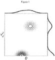
- the probability distribution P D ⁇ d 2 for the example summarized by the Eq. 19 is schematically illustrated in Figure 1 . Note that the two contributions, represented as peaks at the ( D f , v d 2 ) and (D,0) coordinates on the P D ⁇ d 2 contour plot, can only be resolved along the v d 2 axis, which is reciprocal to ⁇ 2 Thus varying ⁇ at different diffusion weighting b provides a means of resolving the velocity dispersion components and thus correlating the velocity dispersion components and the diffusion components.
- the diffusion coefficients D and D f , the velocity dispersion v d 2 and the ratio ⁇ can be quantified by regressing the equation (19) onto the signal attenuation data.
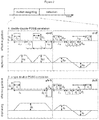
- the D-P sequence consists of two different double-PGSE blocks, each with its own gradient amplitudes, G d1 and G d2 and timing parameters, ⁇ d1 , ⁇ d2 , ⁇ d1 , ⁇ d2 .
- ⁇ and b can be independently adjusted by varying two of the gradient amplitudes G d1 , G d2 and/or durations ⁇ d1 , ⁇ d2 and/or pulse separations ⁇ d1 , ⁇ d2 .
- the D-P sequence in Figure 2.2 consists of a single PGSE block and a double-PGSE block, each with its own gradient amplitudes, G s and G d and timing parameters, ⁇ s , ⁇ d , ⁇ s , ⁇ d .
- ⁇ and b can be independently adjusted by varying two of the gradient amplitudes G s , G d and/or durations ⁇ s , ⁇ d and/or pulse separations ⁇ s , ⁇ d .
- the velocity dispersion weighting ⁇ q s ⁇ s
- the diffusion weighting b q s 2 ⁇ s ⁇ ⁇ 3 + 2 q d 2 ⁇ d ⁇ ⁇ 3 .
- ⁇ and b can be adjusted independently by adjusting q s and q d .
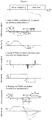
- the examples shown in Figure 3 can be viewed as a single block design where ⁇ and b can be adjusted independently.
- the D-P sequence in Figure 3 .1 consists of two single PGSE blocks, each with its own gradient amplitudes, G 1 and G 2 and timing parameters, ⁇ 1 , ⁇ 2 , ⁇ 1 , ⁇ 2 .
- the entire block allows for variable flow compensation ( ⁇ ).
- ⁇ and b can be independently adjusted by varying two of the gradient amplitudes G 1 , G 2 and/or durations ⁇ 1 , ⁇ 2 and/or pulse separations ⁇ 1 , ⁇ 2 .
- Yet another special case of the example shown in Figure 3 .1 is the example shown in Figure 3 .2.
- the gradient modulation consists of three gradient pulses with amplitudes G 1 , G 2 and G 3 and timing parameters ⁇ 1 , ⁇ 2 , ⁇ 3 , ⁇ 1 , ⁇ 2 .
- the entire block allows for variable flow compensation ( ⁇ ).
- ⁇ and b can be independently adjusted by varying two of the gradient amplitudes G 1 , G 2 and G 3 and/or durations ⁇ 1 , ⁇ 2 , ⁇ 3 and/or pulse separations ⁇ 1 , ⁇ 2 .
- ⁇ and b can be adjusted independently by adjusting q 1 and q 3 .
- sequences 3.1 and 3.3 might be best suited since they can be conveniently implemented in combination with the different read out protocols.
- These sequences, particularly the one shown in Figure 3.3 may, with an appropriate choice of sequence timing parameters, require minimum gradient switching and are thus favored by the gradient slew rate and amplitude limitations often encountered in clinical scanners.
- the experiment may be performed also by using only two values of the parameter ⁇ , where one value set to zero and the other value is set to a value different from zero.
- Several pulse sequences may be used as a flow compensated sequence or non-compensated sequence. Some examples of such sequences are given in the Figure 4 .
- Such sequences may employ harmonically oscillating gradients or any kind of gradient waveforms, which achieve diffusion weighting and flow compensation simultaneously.
- the analysis does not relate to any particular diffusion weighting or flow-compensation gradient sequence. Analysis of the non-compensated and flow-compensated data sets may require adjustment to how the timing parameters, diffusion weighting and dephasing factors are calculated in Eqs. 27 and 28.
- the Monte Carlo error estimation was used in prooving the efficiency of the present invention. Random noise is added to simulated data, which is then regressed with different protocols to obtain the fit parameters. This procedure is repeated 1000 times for each signal-to-noise level to obtain mean values and error estimates for the fit parameters (15). To ensure a fair comparison of the different analysis protocols, the same amount of data points is used in the conventional as in the new protocol. In the new protocol, only the data points corresponding to every second (even) b -value from the original series of b -values (used in the conventional protocols) are used in the flow-compensated dataset and only data points at the odd b -values from the original series are used in the non-compensated dataset (compare Fig. 5a and Fig.
- Fig. 1 displays a schematic representation of the probability distribution P D ⁇ d 2 for the example with two signal contributions described by Eq. (19).
- the P D ⁇ d 2 is given by the inverse Laplace transform of the measured signal E ( b , ⁇ ) .
- the relation between E ( b , ⁇ ) and P D ⁇ d 2 is given by Eq. (18).
- the contour lines connect points with equal probability density.
- the solid lines on top and on the right hand side of the contour plot represent the projections of the probability density function, i.e. the probability distribution of diffusion coefficients, D, and the probability distribution of velocity dispersions, v d 2 , respectively.
- Fig. 2 displays schematics of pulse sequences for motion weighted nuclear magnetic resonance (NMR) or magnetic resonance imaging (MRI).
- Fig. 3 displays schematics of pulse sequences for motion weighted nuclear magnetic resonance (NMR) or magnetic resonance imaging (MRI).
- the magnification of the motion encoding block shows the effective gradient wave form and the dephasing for three examples of the diffusion-perfusion correlation experiment, which can be viewed as composed of one motion encoding block, where the diffusion weighting, b, and the velocity dispersion weighting, ⁇ , can be adjusted independently: 1. double-PGSE with long gradient pulses, where the independent timing parameters ⁇ 1 , ⁇ 2 , ⁇ 1 , ⁇ 2 and the gradient magnitudes G 1 and G 2 allow for independent adjustment of the diffusion weighting, b, and the velocity dispersion weighting, ⁇ ; 2.
- double-PGSE with short gradient pulses where the independent timing parameters ⁇ 1 , ⁇ 2 and the dephasing magnitude q allow for independent adjustment of the diffusion weighting, b, and the velocity dispersion weighting, ⁇ ; 3. three-pulse PGSE, where the independent timing parameters ⁇ 1 , ⁇ 2 , ⁇ 3 , ⁇ 1 , ⁇ 2 and the gradient magnitudes G 1 and G 2 are constraint by the echo condition (at the end of the motion encoding the dephasing is equal to zero) and allow for independent adjustment of the diffusion weighting, b, and the velocity dispersion weighting, ⁇ .
- Fig. 4 displays schematics of pulse sequences for motion weighted nuclear magnetic resonance (NMR) or magnetic resonance imaging (MRI).
- the magnification of the motion encoding block shows the effective gradient wave form for four commonly known motion encoding schemes: 1. single-PGSE, non-compensated (signal attenuation from diffusion and perfusion); 2. double-PGSE, non-compensated (signal attenuation from diffusion and perfusion); 3. double-PGSE, flow-compensated (signal attenuation from diffusion but NOT from perfusion). 4. n -PGSE, flow-compensated (signal attenuation from diffusion but NOT from perfusion).
- Each gradient pulse pair is characterized by its amplitude G , pulse duration ⁇ , and separation between leading edges ⁇ .
- Fig. 5 shows simulated MR signal vs. diffusion-weighting b for some of the pulse sequences in Fig. 4 .
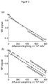
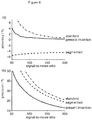
- Fig. 6 shows the accuracy (a) and the precision (b) of the perfusion fraction ⁇ as quantified with conventional protocols ("standard” and “segmented") as well as according to the present invention.
- the accuracy and precision are plotted as a function of the signal-to-noise ratio of the raw MR signal data. Note the better accuracy and precision of the present invention when compared with the conventional protocols over the whole range of signal-to-noise ratios.
Landscapes
- Health & Medical Sciences (AREA)
- Physics & Mathematics (AREA)
- Nuclear Medicine, Radiotherapy & Molecular Imaging (AREA)
- Life Sciences & Earth Sciences (AREA)
- Engineering & Computer Science (AREA)
- General Health & Medical Sciences (AREA)
- Signal Processing (AREA)
- Radiology & Medical Imaging (AREA)
- High Energy & Nuclear Physics (AREA)
- General Physics & Mathematics (AREA)
- Condensed Matter Physics & Semiconductors (AREA)
- Animal Behavior & Ethology (AREA)
- Biophysics (AREA)
- Surgery (AREA)
- Medical Informatics (AREA)
- Heart & Thoracic Surgery (AREA)
- Public Health (AREA)
- Veterinary Medicine (AREA)
- Molecular Biology (AREA)
- Pathology (AREA)
- Biomedical Technology (AREA)
- Vascular Medicine (AREA)
- Physiology (AREA)
- Artificial Intelligence (AREA)
- Computer Vision & Pattern Recognition (AREA)
- Cardiology (AREA)
- Hematology (AREA)
- Psychiatry (AREA)
- Magnetic Resonance Imaging Apparatus (AREA)
Applications Claiming Priority (3)
| Application Number | Priority Date | Filing Date | Title |
|---|---|---|---|
| US201261665998P | 2012-06-29 | 2012-06-29 | |
| SE1250736 | 2012-06-29 | ||
| PCT/SE2013/050755 WO2014003643A1 (en) | 2012-06-29 | 2013-06-24 | Quantification of the relative amount of water in the tissue microcapillary network |
Publications (3)
| Publication Number | Publication Date |
|---|---|
| EP2867690A1 EP2867690A1 (en) | 2015-05-06 |
| EP2867690A4 EP2867690A4 (en) | 2017-01-25 |
| EP2867690B1 true EP2867690B1 (en) | 2021-11-24 |
Family
ID=49783621
Family Applications (1)
| Application Number | Title | Priority Date | Filing Date |
|---|---|---|---|
| EP13810505.1A Active EP2867690B1 (en) | 2012-06-29 | 2013-06-24 | Quantification of the relative amount of water in the tissue microcapillary network |
Country Status (10)
| Country | Link |
|---|---|
| US (2) | US10031204B2 (enExample) |
| EP (1) | EP2867690B1 (enExample) |
| JP (1) | JP6328624B2 (enExample) |
| KR (1) | KR102059408B1 (enExample) |
| CN (1) | CN104471423B (enExample) |
| AU (1) | AU2013281266B2 (enExample) |
| BR (1) | BR112014032534B8 (enExample) |
| CA (1) | CA2876852C (enExample) |
| IN (1) | IN2014MN02502A (enExample) |
| WO (1) | WO2014003643A1 (enExample) |
Families Citing this family (9)
| Publication number | Priority date | Publication date | Assignee | Title |
|---|---|---|---|---|
| CN106344015A (zh) * | 2015-07-15 | 2017-01-25 | 四川大学华西医院 | 一种异常扩散程度加权的弥散磁共振成像方法 |
| US11241163B2 (en) * | 2017-06-08 | 2022-02-08 | The Board Of Trustees Of The University Of Illinois | Measuring blood vessel characteristics with MRI |
| US10684340B1 (en) * | 2019-01-08 | 2020-06-16 | Siemens Healthcare Gmbh | Systems and methods for predicting errors and optimizing protocols in quantitative magnetic resonance imaging |
| CN109820506B (zh) * | 2019-02-20 | 2023-07-07 | 王毅翔 | 基于磁共振弥散成像的组织血管密度指标检测方法及装置 |
| US11789106B2 (en) * | 2020-03-28 | 2023-10-17 | Kalmia Ab | Magnetic resonance method, software product, and system for determining a diffusion propagator or related diffusion parameters for spin-labelled particles |
| CN111407278B (zh) * | 2020-03-31 | 2020-12-29 | 浙江大学 | 利用流速补偿和非补偿的弥散磁共振测量胎盘血流的方法及装置 |
| CN115421086B (zh) * | 2022-09-02 | 2023-04-14 | 哈尔滨医科大学 | 活体心脏复杂组织学特征精准解析的超融合体素内不相干运动张量磁共振成像方法 |
| CN115877297A (zh) * | 2022-12-19 | 2023-03-31 | 武汉中科医疗科技工业技术研究院有限公司 | 磁共振信号衰减原因判别方法、装置及磁共振系统 |
| CN117310581B (zh) * | 2023-10-11 | 2024-05-10 | 安徽峻德医疗科技有限公司 | 一种核磁共振信号衰减拟合方法、系统、设备及存储介质 |
Family Cites Families (25)
| Publication number | Priority date | Publication date | Assignee | Title |
|---|---|---|---|---|
| JPS5123312B1 (enExample) | 1971-05-06 | 1976-07-15 | ||
| JPH04357934A (ja) * | 1991-06-05 | 1992-12-10 | Toshiba Corp | Mriによるivimイメージング |
| JP3144840B2 (ja) | 1991-07-31 | 2001-03-12 | 株式会社東芝 | 磁気共鳴イメージング装置 |
| JP3146033B2 (ja) * | 1991-11-05 | 2001-03-12 | 株式会社東芝 | 磁気共鳴イメージング装置 |
| US8005530B2 (en) * | 1995-04-12 | 2011-08-23 | Prince Martin R | Method and apparatus for imaging abdominal aorta and aortic aneurysms |
| JP3516373B2 (ja) * | 1996-09-04 | 2004-04-05 | 株式会社日立メディコ | 磁気共鳴測定装置 |
| JP4225620B2 (ja) * | 1999-01-11 | 2009-02-18 | 株式会社東芝 | Mri装置 |
| AU1803401A (en) | 1999-11-24 | 2001-06-04 | Board Of Regents, The University Of Texas System | Methods and systems for generating tractograms |
| US20040189297A1 (en) * | 2002-12-13 | 2004-09-30 | Michael Bock | Imaging arrangement and process for locally-resolved imaging |
| JP4039980B2 (ja) * | 2003-06-04 | 2008-01-30 | マルホン工業株式会社 | 遊技機 |
| JP3774713B2 (ja) * | 2003-10-15 | 2006-05-17 | 株式会社東芝 | コンタクトホールの形成方法 |
| JP4357934B2 (ja) | 2003-11-14 | 2009-11-04 | アルパイン株式会社 | ナビゲーション装置及び代替経路提示方法 |
| WO2006011810A2 (en) * | 2004-07-30 | 2006-02-02 | Ge Healthcare As | Mr imaging method for the discrimination between healthy and tumour tissue |
| JP2005031099A (ja) * | 2004-10-29 | 2005-02-03 | Yokogawa Electric Corp | 分光装置 |
| DE102005008753B4 (de) | 2005-02-25 | 2007-09-27 | Siemens Ag | Verfahren zur Darstellung von Fluss in einem Magnetresonanzbild |
| DE102005021067B4 (de) * | 2005-05-06 | 2008-08-28 | Siemens Ag | Bildgebende Vorrichtung |
| US7411394B2 (en) * | 2005-05-17 | 2008-08-12 | Board Of Trustees Of Michigan State University | Method for imaging diffusion anisotropy and diffusion gradient simultaneously |
| JP4961566B2 (ja) * | 2005-10-20 | 2012-06-27 | 国立大学法人 新潟大学 | 磁気共鳴画像処理方法および磁気共鳴画像処理装置 |
| US8155729B1 (en) | 2006-02-17 | 2012-04-10 | General Electric Company | Method and apparatus to compensate imaging data with simultaneously acquired motion data |
| US8053260B2 (en) | 2006-11-17 | 2011-11-08 | General Electric Company | Large-area lighting systems and methods of making the same |
| JP4777372B2 (ja) * | 2008-02-08 | 2011-09-21 | 株式会社東芝 | 磁気共鳴イメージング装置 |
| CN102077108B (zh) | 2008-04-28 | 2015-02-25 | 康奈尔大学 | 分子mri中的磁敏度精确量化 |
| US8497680B2 (en) * | 2011-03-24 | 2013-07-30 | University Hospital Of Basel | Magnetic resonance method for quantification of molecular diffusion using double echo steady state sequences |
| US9075121B2 (en) * | 2011-07-15 | 2015-07-07 | Wisconsin Alumni Research Foundation | System and method for rotating angle velocity encoding, phase contrast magnetic resonance imaging |
| WO2013025487A1 (en) * | 2011-08-12 | 2013-02-21 | The United States Of America, As Represented By The Secretary, Dpt Of Health And Human Services | Spin echo sequences for diffusion weighted imaging of moving media |
-
2013
- 2013-06-24 WO PCT/SE2013/050755 patent/WO2014003643A1/en not_active Ceased
- 2013-06-24 US US14/410,549 patent/US10031204B2/en active Active
- 2013-06-24 AU AU2013281266A patent/AU2013281266B2/en active Active
- 2013-06-24 BR BR112014032534A patent/BR112014032534B8/pt not_active IP Right Cessation
- 2013-06-24 JP JP2015520119A patent/JP6328624B2/ja active Active
- 2013-06-24 KR KR1020157002610A patent/KR102059408B1/ko active Active
- 2013-06-24 CN CN201380033778.6A patent/CN104471423B/zh active Active
- 2013-06-24 CA CA2876852A patent/CA2876852C/en not_active Expired - Fee Related
- 2013-06-24 IN IN2502MUN2014 patent/IN2014MN02502A/en unknown
- 2013-06-24 EP EP13810505.1A patent/EP2867690B1/en active Active
-
2018
- 2018-03-29 US US15/939,991 patent/US10788558B2/en active Active
Non-Patent Citations (1)
| Title |
|---|
| None * |
Also Published As
| Publication number | Publication date |
|---|---|
| CA2876852C (en) | 2020-12-22 |
| US10788558B2 (en) | 2020-09-29 |
| JP6328624B2 (ja) | 2018-05-23 |
| US10031204B2 (en) | 2018-07-24 |
| US20180224514A1 (en) | 2018-08-09 |
| CA2876852A1 (en) | 2014-01-03 |
| AU2013281266A1 (en) | 2015-01-22 |
| CN104471423B (zh) | 2017-03-29 |
| BR112014032534B1 (pt) | 2021-10-13 |
| BR112014032534B8 (pt) | 2023-02-14 |
| WO2014003643A1 (en) | 2014-01-03 |
| BR112014032534A2 (pt) | 2017-06-27 |
| IN2014MN02502A (enExample) | 2015-07-17 |
| AU2013281266B2 (en) | 2017-04-13 |
| KR102059408B1 (ko) | 2020-02-11 |
| US20150168527A1 (en) | 2015-06-18 |
| EP2867690A4 (en) | 2017-01-25 |
| EP2867690A1 (en) | 2015-05-06 |
| JP2015521891A (ja) | 2015-08-03 |
| KR20150036296A (ko) | 2015-04-07 |
| CN104471423A (zh) | 2015-03-25 |
Similar Documents
| Publication | Publication Date | Title |
|---|---|---|
| EP2867690B1 (en) | Quantification of the relative amount of water in the tissue microcapillary network | |
| Kundu et al. | Multi-echo fMRI: A review of applications in fMRI denoising and analysis of BOLD signals | |
| Kecskemeti et al. | MPnRAGE: A technique to simultaneously acquire hundreds of differently contrasted MPRAGE images with applications to quantitative T1 mapping | |
| Szczepankiewicz et al. | Quantification of microscopic diffusion anisotropy disentangles effects of orientation dispersion from microstructure: applications in healthy volunteers and in brain tumors | |
| Posse et al. | MR spectroscopic imaging: principles and recent advances | |
| EP2702423B1 (en) | Method for r2* quantification with mri with correction for macroscopic magnetic field inhomogeneities | |
| Steel et al. | Metabolite-cycled density-weighted concentric rings k-space trajectory (DW-CRT) enables high-resolution 1 H magnetic resonance spectroscopic imaging at 3-Tesla | |
| Bammer et al. | New methods in diffusion-weighted and diffusion tensor imaging | |
| Bosma et al. | Assessment of data acquisition parameters, and analysis techniques for noise reduction in spinal cord fMRI data | |
| EP1535083A1 (en) | Microvascular blood volume magnetic resonance imaging | |
| Granlund et al. | High-resolution, three-dimensional diffusion-weighted breast imaging using DESS | |
| Latt et al. | Accuracy of $ q $-Space Related Parameters in MRI: Simulations and Phantom Measurements | |
| JP7150056B2 (ja) | 磁化の反転状態の評価を伴う動脈スピンラベリング法 | |
| Shah et al. | Measuring the absolute water content of the brain using quantitative MRI | |
| US12153110B2 (en) | Simultaneous multi-slice MRSI using density weighted concentric ring acquisition | |
| Çavuşoğlu | Arterial spin labeling MRI using spiral acquisitions and concurrent field monitoring | |
| Liu et al. | A practical approach to in vivo high-resolution diffusion tensor imaging of rhesus monkeys on a 3-T human scanner | |
| Daniel et al. | Assessing the Impact of Imaging Parameters on MRI Measurement of Kidney T2 | |
| Goryawala et al. | A Path to Establishing MRSI as a Clinical Standard Imaging | |
| Heikal et al. | A phantom to assess the accuracy of tumor delineation using MRSI | |
| Alexander | Aurobrata Ghosh2, Andrada Ianus2 and | |
| Heule | Rapid magnetic resonance tissue relaxometry in the steady state | |
| Li | Multi-dimensional MR spectroscopic imaging of gliomas at different field strengths | |
| Aboussouan | Magnetic resonance imaging of the brain: enabling advances in efficient non-cartesian sampling | |
| Lupo | Application of perfusion-weighted, susceptibility-weighted, and spectroscopic magnetic resonance imaging for characterizing glioma microvasculature at different field strengths |
Legal Events
| Date | Code | Title | Description |
|---|---|---|---|
| PUAI | Public reference made under article 153(3) epc to a published international application that has entered the european phase |
Free format text: ORIGINAL CODE: 0009012 |
|
| 17P | Request for examination filed |
Effective date: 20150128 |
|
| AK | Designated contracting states |
Kind code of ref document: A1 Designated state(s): AL AT BE BG CH CY CZ DE DK EE ES FI FR GB GR HR HU IE IS IT LI LT LU LV MC MK MT NL NO PL PT RO RS SE SI SK SM TR |
|
| AX | Request for extension of the european patent |
Extension state: BA ME |
|
| DAX | Request for extension of the european patent (deleted) | ||
| RA4 | Supplementary search report drawn up and despatched (corrected) |
Effective date: 20170102 |
|
| RIC1 | Information provided on ipc code assigned before grant |
Ipc: G01R 33/565 20060101ALI20161221BHEP Ipc: A61B 5/055 20060101ALI20161221BHEP Ipc: A61B 5/026 20060101ALI20161221BHEP Ipc: G01R 33/563 20060101AFI20161221BHEP Ipc: G01R 33/54 20060101ALN20161221BHEP Ipc: G01R 33/56 20060101ALN20161221BHEP Ipc: A61B 5/00 20060101ALI20161221BHEP |
|
| GRAP | Despatch of communication of intention to grant a patent |
Free format text: ORIGINAL CODE: EPIDOSNIGR1 |
|
| STAA | Information on the status of an ep patent application or granted ep patent |
Free format text: STATUS: GRANT OF PATENT IS INTENDED |
|
| RIC1 | Information provided on ipc code assigned before grant |
Ipc: G01R 33/54 20060101ALN20201203BHEP Ipc: G01R 33/56 20060101ALN20201203BHEP Ipc: G01R 33/565 20060101ALI20201203BHEP Ipc: A61B 5/00 20060101ALI20201203BHEP Ipc: A61B 5/055 20060101ALI20201203BHEP Ipc: A61B 5/026 20060101ALI20201203BHEP Ipc: G01R 33/563 20060101AFI20201203BHEP |
|
| INTG | Intention to grant announced |
Effective date: 20201218 |
|
| GRAJ | Information related to disapproval of communication of intention to grant by the applicant or resumption of examination proceedings by the epo deleted |
Free format text: ORIGINAL CODE: EPIDOSDIGR1 |
|
| STAA | Information on the status of an ep patent application or granted ep patent |
Free format text: STATUS: REQUEST FOR EXAMINATION WAS MADE |
|
| REG | Reference to a national code |
Ref country code: DE Ref legal event code: R079 Ref document number: 602013080198 Country of ref document: DE Free format text: PREVIOUS MAIN CLASS: G01R0033560000 Ipc: G01R0033563000 |
|
| INTC | Intention to grant announced (deleted) | ||
| GRAP | Despatch of communication of intention to grant a patent |
Free format text: ORIGINAL CODE: EPIDOSNIGR1 |
|
| STAA | Information on the status of an ep patent application or granted ep patent |
Free format text: STATUS: GRANT OF PATENT IS INTENDED |
|
| RIC1 | Information provided on ipc code assigned before grant |
Ipc: G01R 33/563 20060101AFI20210505BHEP Ipc: G01R 33/565 20060101ALI20210505BHEP Ipc: A61B 5/026 20060101ALI20210505BHEP Ipc: A61B 5/00 20060101ALI20210505BHEP Ipc: A61B 5/055 20060101ALI20210505BHEP Ipc: G01R 33/56 20060101ALN20210505BHEP Ipc: G01R 33/54 20060101ALN20210505BHEP |
|
| INTG | Intention to grant announced |
Effective date: 20210608 |
|
| GRAS | Grant fee paid |
Free format text: ORIGINAL CODE: EPIDOSNIGR3 |
|
| GRAA | (expected) grant |
Free format text: ORIGINAL CODE: 0009210 |
|
| STAA | Information on the status of an ep patent application or granted ep patent |
Free format text: STATUS: THE PATENT HAS BEEN GRANTED |
|
| AK | Designated contracting states |
Kind code of ref document: B1 Designated state(s): AL AT BE BG CH CY CZ DE DK EE ES FI FR GB GR HR HU IE IS IT LI LT LU LV MC MK MT NL NO PL PT RO RS SE SI SK SM TR |
|
| REG | Reference to a national code |
Ref country code: GB Ref legal event code: FG4D |
|
| REG | Reference to a national code |
Ref country code: DE Ref legal event code: R096 Ref document number: 602013080198 Country of ref document: DE |
|
| REG | Reference to a national code |
Ref country code: AT Ref legal event code: REF Ref document number: 1450255 Country of ref document: AT Kind code of ref document: T Effective date: 20211215 |
|
| REG | Reference to a national code |
Ref country code: IE Ref legal event code: FG4D |
|
| REG | Reference to a national code |
Ref country code: LT Ref legal event code: MG9D |
|
| REG | Reference to a national code |
Ref country code: NL Ref legal event code: MP Effective date: 20211124 |
|
| REG | Reference to a national code |
Ref country code: AT Ref legal event code: MK05 Ref document number: 1450255 Country of ref document: AT Kind code of ref document: T Effective date: 20211124 |
|
| PG25 | Lapsed in a contracting state [announced via postgrant information from national office to epo] |
Ref country code: RS Free format text: LAPSE BECAUSE OF FAILURE TO SUBMIT A TRANSLATION OF THE DESCRIPTION OR TO PAY THE FEE WITHIN THE PRESCRIBED TIME-LIMIT Effective date: 20211124 Ref country code: LT Free format text: LAPSE BECAUSE OF FAILURE TO SUBMIT A TRANSLATION OF THE DESCRIPTION OR TO PAY THE FEE WITHIN THE PRESCRIBED TIME-LIMIT Effective date: 20211124 Ref country code: FI Free format text: LAPSE BECAUSE OF FAILURE TO SUBMIT A TRANSLATION OF THE DESCRIPTION OR TO PAY THE FEE WITHIN THE PRESCRIBED TIME-LIMIT Effective date: 20211124 Ref country code: BG Free format text: LAPSE BECAUSE OF FAILURE TO SUBMIT A TRANSLATION OF THE DESCRIPTION OR TO PAY THE FEE WITHIN THE PRESCRIBED TIME-LIMIT Effective date: 20220224 Ref country code: AT Free format text: LAPSE BECAUSE OF FAILURE TO SUBMIT A TRANSLATION OF THE DESCRIPTION OR TO PAY THE FEE WITHIN THE PRESCRIBED TIME-LIMIT Effective date: 20211124 |
|
| PG25 | Lapsed in a contracting state [announced via postgrant information from national office to epo] |
Ref country code: IS Free format text: LAPSE BECAUSE OF FAILURE TO SUBMIT A TRANSLATION OF THE DESCRIPTION OR TO PAY THE FEE WITHIN THE PRESCRIBED TIME-LIMIT Effective date: 20220324 Ref country code: SE Free format text: LAPSE BECAUSE OF FAILURE TO SUBMIT A TRANSLATION OF THE DESCRIPTION OR TO PAY THE FEE WITHIN THE PRESCRIBED TIME-LIMIT Effective date: 20211124 Ref country code: PT Free format text: LAPSE BECAUSE OF FAILURE TO SUBMIT A TRANSLATION OF THE DESCRIPTION OR TO PAY THE FEE WITHIN THE PRESCRIBED TIME-LIMIT Effective date: 20220324 Ref country code: PL Free format text: LAPSE BECAUSE OF FAILURE TO SUBMIT A TRANSLATION OF THE DESCRIPTION OR TO PAY THE FEE WITHIN THE PRESCRIBED TIME-LIMIT Effective date: 20211124 Ref country code: NO Free format text: LAPSE BECAUSE OF FAILURE TO SUBMIT A TRANSLATION OF THE DESCRIPTION OR TO PAY THE FEE WITHIN THE PRESCRIBED TIME-LIMIT Effective date: 20220224 Ref country code: NL Free format text: LAPSE BECAUSE OF FAILURE TO SUBMIT A TRANSLATION OF THE DESCRIPTION OR TO PAY THE FEE WITHIN THE PRESCRIBED TIME-LIMIT Effective date: 20211124 Ref country code: LV Free format text: LAPSE BECAUSE OF FAILURE TO SUBMIT A TRANSLATION OF THE DESCRIPTION OR TO PAY THE FEE WITHIN THE PRESCRIBED TIME-LIMIT Effective date: 20211124 Ref country code: HR Free format text: LAPSE BECAUSE OF FAILURE TO SUBMIT A TRANSLATION OF THE DESCRIPTION OR TO PAY THE FEE WITHIN THE PRESCRIBED TIME-LIMIT Effective date: 20211124 Ref country code: GR Free format text: LAPSE BECAUSE OF FAILURE TO SUBMIT A TRANSLATION OF THE DESCRIPTION OR TO PAY THE FEE WITHIN THE PRESCRIBED TIME-LIMIT Effective date: 20220225 Ref country code: ES Free format text: LAPSE BECAUSE OF FAILURE TO SUBMIT A TRANSLATION OF THE DESCRIPTION OR TO PAY THE FEE WITHIN THE PRESCRIBED TIME-LIMIT Effective date: 20211124 |
|
| PG25 | Lapsed in a contracting state [announced via postgrant information from national office to epo] |
Ref country code: SM Free format text: LAPSE BECAUSE OF FAILURE TO SUBMIT A TRANSLATION OF THE DESCRIPTION OR TO PAY THE FEE WITHIN THE PRESCRIBED TIME-LIMIT Effective date: 20211124 Ref country code: SK Free format text: LAPSE BECAUSE OF FAILURE TO SUBMIT A TRANSLATION OF THE DESCRIPTION OR TO PAY THE FEE WITHIN THE PRESCRIBED TIME-LIMIT Effective date: 20211124 Ref country code: RO Free format text: LAPSE BECAUSE OF FAILURE TO SUBMIT A TRANSLATION OF THE DESCRIPTION OR TO PAY THE FEE WITHIN THE PRESCRIBED TIME-LIMIT Effective date: 20211124 Ref country code: EE Free format text: LAPSE BECAUSE OF FAILURE TO SUBMIT A TRANSLATION OF THE DESCRIPTION OR TO PAY THE FEE WITHIN THE PRESCRIBED TIME-LIMIT Effective date: 20211124 Ref country code: DK Free format text: LAPSE BECAUSE OF FAILURE TO SUBMIT A TRANSLATION OF THE DESCRIPTION OR TO PAY THE FEE WITHIN THE PRESCRIBED TIME-LIMIT Effective date: 20211124 Ref country code: CZ Free format text: LAPSE BECAUSE OF FAILURE TO SUBMIT A TRANSLATION OF THE DESCRIPTION OR TO PAY THE FEE WITHIN THE PRESCRIBED TIME-LIMIT Effective date: 20211124 |
|
| REG | Reference to a national code |
Ref country code: DE Ref legal event code: R097 Ref document number: 602013080198 Country of ref document: DE |
|
| PLBE | No opposition filed within time limit |
Free format text: ORIGINAL CODE: 0009261 |
|
| STAA | Information on the status of an ep patent application or granted ep patent |
Free format text: STATUS: NO OPPOSITION FILED WITHIN TIME LIMIT |
|
| PG25 | Lapsed in a contracting state [announced via postgrant information from national office to epo] |
Ref country code: AL Free format text: LAPSE BECAUSE OF FAILURE TO SUBMIT A TRANSLATION OF THE DESCRIPTION OR TO PAY THE FEE WITHIN THE PRESCRIBED TIME-LIMIT Effective date: 20211124 |
|
| 26N | No opposition filed |
Effective date: 20220825 |
|
| REG | Reference to a national code |
Ref country code: DE Ref legal event code: R081 Ref document number: 602013080198 Country of ref document: DE Owner name: RANDOM WALK IMAGING AB, SE Free format text: FORMER OWNER: CR DEVELOPMENT AB, LUND, SE |
|
| PG25 | Lapsed in a contracting state [announced via postgrant information from national office to epo] |
Ref country code: SI Free format text: LAPSE BECAUSE OF FAILURE TO SUBMIT A TRANSLATION OF THE DESCRIPTION OR TO PAY THE FEE WITHIN THE PRESCRIBED TIME-LIMIT Effective date: 20211124 |
|
| PG25 | Lapsed in a contracting state [announced via postgrant information from national office to epo] |
Ref country code: MC Free format text: LAPSE BECAUSE OF FAILURE TO SUBMIT A TRANSLATION OF THE DESCRIPTION OR TO PAY THE FEE WITHIN THE PRESCRIBED TIME-LIMIT Effective date: 20211124 |
|
| REG | Reference to a national code |
Ref country code: CH Ref legal event code: PL |
|
| REG | Reference to a national code |
Ref country code: BE Ref legal event code: MM Effective date: 20220630 |
|
| GBPC | Gb: european patent ceased through non-payment of renewal fee |
Effective date: 20220624 |
|
| PG25 | Lapsed in a contracting state [announced via postgrant information from national office to epo] |
Ref country code: LU Free format text: LAPSE BECAUSE OF NON-PAYMENT OF DUE FEES Effective date: 20220624 Ref country code: LI Free format text: LAPSE BECAUSE OF NON-PAYMENT OF DUE FEES Effective date: 20220630 Ref country code: IE Free format text: LAPSE BECAUSE OF NON-PAYMENT OF DUE FEES Effective date: 20220624 Ref country code: FR Free format text: LAPSE BECAUSE OF NON-PAYMENT OF DUE FEES Effective date: 20220630 Ref country code: CH Free format text: LAPSE BECAUSE OF NON-PAYMENT OF DUE FEES Effective date: 20220630 |
|
| PG25 | Lapsed in a contracting state [announced via postgrant information from national office to epo] |
Ref country code: IT Free format text: LAPSE BECAUSE OF FAILURE TO SUBMIT A TRANSLATION OF THE DESCRIPTION OR TO PAY THE FEE WITHIN THE PRESCRIBED TIME-LIMIT Effective date: 20211124 Ref country code: GB Free format text: LAPSE BECAUSE OF NON-PAYMENT OF DUE FEES Effective date: 20220624 Ref country code: BE Free format text: LAPSE BECAUSE OF NON-PAYMENT OF DUE FEES Effective date: 20220630 |
|
| P01 | Opt-out of the competence of the unified patent court (upc) registered |
Effective date: 20230523 |
|
| PG25 | Lapsed in a contracting state [announced via postgrant information from national office to epo] |
Ref country code: HU Free format text: LAPSE BECAUSE OF FAILURE TO SUBMIT A TRANSLATION OF THE DESCRIPTION OR TO PAY THE FEE WITHIN THE PRESCRIBED TIME-LIMIT; INVALID AB INITIO Effective date: 20130624 |
|
| PG25 | Lapsed in a contracting state [announced via postgrant information from national office to epo] |
Ref country code: MK Free format text: LAPSE BECAUSE OF FAILURE TO SUBMIT A TRANSLATION OF THE DESCRIPTION OR TO PAY THE FEE WITHIN THE PRESCRIBED TIME-LIMIT Effective date: 20211124 Ref country code: CY Free format text: LAPSE BECAUSE OF FAILURE TO SUBMIT A TRANSLATION OF THE DESCRIPTION OR TO PAY THE FEE WITHIN THE PRESCRIBED TIME-LIMIT Effective date: 20211124 |
|
| PG25 | Lapsed in a contracting state [announced via postgrant information from national office to epo] |
Ref country code: TR Free format text: LAPSE BECAUSE OF FAILURE TO SUBMIT A TRANSLATION OF THE DESCRIPTION OR TO PAY THE FEE WITHIN THE PRESCRIBED TIME-LIMIT Effective date: 20211124 |
|
| PG25 | Lapsed in a contracting state [announced via postgrant information from national office to epo] |
Ref country code: MT Free format text: LAPSE BECAUSE OF FAILURE TO SUBMIT A TRANSLATION OF THE DESCRIPTION OR TO PAY THE FEE WITHIN THE PRESCRIBED TIME-LIMIT Effective date: 20211124 |
|
| PGFP | Annual fee paid to national office [announced via postgrant information from national office to epo] |
Ref country code: DE Payment date: 20250627 Year of fee payment: 13 |