EP2766393B1 - ANTI-HtrA1 ANTIBODIES AND METHODS OF USE - Google Patents
ANTI-HtrA1 ANTIBODIES AND METHODS OF USE Download PDFInfo
- Publication number
- EP2766393B1 EP2766393B1 EP12840084.3A EP12840084A EP2766393B1 EP 2766393 B1 EP2766393 B1 EP 2766393B1 EP 12840084 A EP12840084 A EP 12840084A EP 2766393 B1 EP2766393 B1 EP 2766393B1
- Authority
- EP
- European Patent Office
- Prior art keywords
- antibody
- seq
- hvr
- amino acid
- acid sequence
- Prior art date
- Legal status (The legal status is an assumption and is not a legal conclusion. Google has not performed a legal analysis and makes no representation as to the accuracy of the status listed.)
- Active
Links
- 238000000034 method Methods 0.000 title claims description 83
- 125000003275 alpha amino acid group Chemical group 0.000 claims description 295
- 241000282414 Homo sapiens Species 0.000 claims description 103
- 210000004027 cell Anatomy 0.000 claims description 84
- 230000000694 effects Effects 0.000 claims description 41
- 239000000758 substrate Substances 0.000 claims description 36
- 150000007523 nucleic acids Chemical class 0.000 claims description 33
- 108020004707 nucleic acids Proteins 0.000 claims description 32
- 102000039446 nucleic acids Human genes 0.000 claims description 32
- 230000014509 gene expression Effects 0.000 claims description 31
- 239000013638 trimer Substances 0.000 claims description 28
- 150000001413 amino acids Chemical class 0.000 claims description 26
- 108010021625 Immunoglobulin Fragments Proteins 0.000 claims description 21
- 102000008394 Immunoglobulin Fragments Human genes 0.000 claims description 21
- 206010064930 age-related macular degeneration Diseases 0.000 claims description 21
- 239000003814 drug Substances 0.000 claims description 21
- 230000002401 inhibitory effect Effects 0.000 claims description 19
- 108091005804 Peptidases Proteins 0.000 claims description 18
- 239000004365 Protease Substances 0.000 claims description 17
- 238000010494 dissociation reaction Methods 0.000 claims description 15
- 230000005593 dissociations Effects 0.000 claims description 15
- 208000008069 Geographic Atrophy Diseases 0.000 claims description 12
- 230000002207 retinal effect Effects 0.000 claims description 12
- 102000012479 Serine Proteases Human genes 0.000 claims description 11
- 108010022999 Serine Proteases Proteins 0.000 claims description 11
- 229940127089 cytotoxic agent Drugs 0.000 claims description 11
- 229940127121 immunoconjugate Drugs 0.000 claims description 11
- 239000008194 pharmaceutical composition Substances 0.000 claims description 11
- 210000000608 photoreceptor cell Anatomy 0.000 claims description 11
- 230000007850 degeneration Effects 0.000 claims description 10
- 102100021117 Serine protease HTRA2, mitochondrial Human genes 0.000 claims description 9
- 101710146118 Serine protease HTRA2, mitochondrial Proteins 0.000 claims description 9
- 239000002254 cytotoxic agent Substances 0.000 claims description 9
- 208000002780 macular degeneration Diseases 0.000 claims description 9
- 206010012689 Diabetic retinopathy Diseases 0.000 claims description 8
- 206010063381 Polypoidal choroidal vasculopathy Diseases 0.000 claims description 8
- 231100000599 cytotoxic agent Toxicity 0.000 claims description 8
- 239000003937 drug carrier Substances 0.000 claims description 8
- 206010038933 Retinopathy of prematurity Diseases 0.000 claims description 7
- 230000015572 biosynthetic process Effects 0.000 claims description 5
- 238000004113 cell culture Methods 0.000 claims description 4
- 238000012258 culturing Methods 0.000 claims description 3
- 208000011325 dry age related macular degeneration Diseases 0.000 claims description 3
- 102100037486 Reverse transcriptase/ribonuclease H Human genes 0.000 claims 1
- 108090000765 processed proteins & peptides Proteins 0.000 description 103
- 108091007491 NSP3 Papain-like protease domains Proteins 0.000 description 89
- 230000027455 binding Effects 0.000 description 82
- 239000000427 antigen Substances 0.000 description 43
- 108090000623 proteins and genes Proteins 0.000 description 43
- 108091007433 antigens Proteins 0.000 description 41
- 102000036639 antigens Human genes 0.000 description 41
- 102000004169 proteins and genes Human genes 0.000 description 37
- 238000006467 substitution reaction Methods 0.000 description 36
- MTCFGRXMJLQNBG-REOHCLBHSA-N (2S)-2-Amino-3-hydroxypropansäure Chemical compound OC[C@H](N)C(O)=O MTCFGRXMJLQNBG-REOHCLBHSA-N 0.000 description 35
- 235000018102 proteins Nutrition 0.000 description 35
- FAPWRFPIFSIZLT-UHFFFAOYSA-M Sodium chloride Chemical compound [Na+].[Cl-] FAPWRFPIFSIZLT-UHFFFAOYSA-M 0.000 description 33
- 235000001014 amino acid Nutrition 0.000 description 31
- 239000000203 mixture Substances 0.000 description 31
- PEDCQBHIVMGVHV-UHFFFAOYSA-N Glycerine Chemical compound OCC(O)CO PEDCQBHIVMGVHV-UHFFFAOYSA-N 0.000 description 27
- 239000000872 buffer Substances 0.000 description 27
- CKLJMWTZIZZHCS-REOHCLBHSA-N L-aspartic acid Chemical compound OC(=O)[C@@H](N)CC(O)=O CKLJMWTZIZZHCS-REOHCLBHSA-N 0.000 description 26
- 238000003556 assay Methods 0.000 description 26
- 229940024606 amino acid Drugs 0.000 description 25
- DCXYFEDJOCDNAF-REOHCLBHSA-N L-asparagine Chemical compound OC(=O)[C@@H](N)CC(N)=O DCXYFEDJOCDNAF-REOHCLBHSA-N 0.000 description 23
- 208000037265 diseases, disorders, signs and symptoms Diseases 0.000 description 23
- 125000003295 alanine group Chemical group N[C@@H](C)C(=O)* 0.000 description 22
- RAXXELZNTBOGNW-UHFFFAOYSA-N imidazole Natural products C1=CNC=N1 RAXXELZNTBOGNW-UHFFFAOYSA-N 0.000 description 21
- 239000012634 fragment Substances 0.000 description 19
- 238000004519 manufacturing process Methods 0.000 description 18
- 102000035195 Peptidases Human genes 0.000 description 17
- 239000011780 sodium chloride Substances 0.000 description 17
- 238000011282 treatment Methods 0.000 description 17
- 239000013598 vector Substances 0.000 description 17
- 108010076119 Caseins Proteins 0.000 description 16
- 102000011632 Caseins Human genes 0.000 description 16
- 241001529936 Murinae Species 0.000 description 16
- 241000699666 Mus <mouse, genus> Species 0.000 description 16
- 201000010099 disease Diseases 0.000 description 16
- 241000700159 Rattus Species 0.000 description 15
- 229920001184 polypeptide Polymers 0.000 description 15
- 102000004196 processed proteins & peptides Human genes 0.000 description 15
- 210000001525 retina Anatomy 0.000 description 15
- 239000000243 solution Substances 0.000 description 15
- 108060003951 Immunoglobulin Proteins 0.000 description 14
- 241000699670 Mus sp. Species 0.000 description 14
- 229920001213 Polysorbate 20 Polymers 0.000 description 14
- 102000018358 immunoglobulin Human genes 0.000 description 14
- 239000000256 polyoxyethylene sorbitan monolaurate Substances 0.000 description 14
- 235000010486 polyoxyethylene sorbitan monolaurate Nutrition 0.000 description 14
- 235000019419 proteases Nutrition 0.000 description 14
- ROHFNLRQFUQHCH-YFKPBYRVSA-N L-leucine Chemical compound CC(C)C[C@H](N)C(O)=O ROHFNLRQFUQHCH-YFKPBYRVSA-N 0.000 description 13
- 239000007983 Tris buffer Substances 0.000 description 13
- 125000000539 amino acid group Chemical group 0.000 description 13
- 230000004071 biological effect Effects 0.000 description 13
- 238000011534 incubation Methods 0.000 description 13
- 229940124597 therapeutic agent Drugs 0.000 description 13
- LENZDBCJOHFCAS-UHFFFAOYSA-N tris Chemical compound OCC(N)(CO)CO LENZDBCJOHFCAS-UHFFFAOYSA-N 0.000 description 13
- 102000004190 Enzymes Human genes 0.000 description 12
- 108090000790 Enzymes Proteins 0.000 description 12
- 241001465754 Metazoa Species 0.000 description 12
- 208000022873 Ocular disease Diseases 0.000 description 12
- 230000010056 antibody-dependent cellular cytotoxicity Effects 0.000 description 12
- 229940088598 enzyme Drugs 0.000 description 12
- 230000006870 function Effects 0.000 description 12
- 210000001519 tissue Anatomy 0.000 description 12
- 241000588724 Escherichia coli Species 0.000 description 11
- 238000005516 engineering process Methods 0.000 description 11
- 238000003780 insertion Methods 0.000 description 11
- 230000037431 insertion Effects 0.000 description 11
- 238000002560 therapeutic procedure Methods 0.000 description 11
- 238000012217 deletion Methods 0.000 description 10
- 230000037430 deletion Effects 0.000 description 10
- 238000009472 formulation Methods 0.000 description 10
- 210000001672 ovary Anatomy 0.000 description 10
- 230000002285 radioactive effect Effects 0.000 description 10
- 108091003079 Bovine Serum Albumin Proteins 0.000 description 9
- DHMQDGOQFOQNFH-UHFFFAOYSA-N Glycine Chemical compound NCC(O)=O DHMQDGOQFOQNFH-UHFFFAOYSA-N 0.000 description 9
- 230000004075 alteration Effects 0.000 description 9
- 229940098773 bovine serum albumin Drugs 0.000 description 9
- 230000004540 complement-dependent cytotoxicity Effects 0.000 description 9
- 235000021247 β-casein Nutrition 0.000 description 9
- UMCMPZBLKLEWAF-BCTGSCMUSA-N 3-[(3-cholamidopropyl)dimethylammonio]propane-1-sulfonate Chemical compound C([C@H]1C[C@H]2O)[C@H](O)CC[C@]1(C)[C@@H]1[C@@H]2[C@@H]2CC[C@H]([C@@H](CCC(=O)NCCC[N+](C)(C)CCCS([O-])(=O)=O)C)[C@@]2(C)[C@@H](O)C1 UMCMPZBLKLEWAF-BCTGSCMUSA-N 0.000 description 8
- 102000004954 Biglycan Human genes 0.000 description 8
- 108090001138 Biglycan Proteins 0.000 description 8
- 102000004237 Decorin Human genes 0.000 description 8
- 108090000738 Decorin Proteins 0.000 description 8
- 238000002965 ELISA Methods 0.000 description 8
- KDXKERNSBIXSRK-UHFFFAOYSA-N Lysine Natural products NCCCCC(N)C(O)=O KDXKERNSBIXSRK-UHFFFAOYSA-N 0.000 description 8
- 230000003197 catalytic effect Effects 0.000 description 8
- 239000003795 chemical substances by application Substances 0.000 description 8
- 238000002474 experimental method Methods 0.000 description 8
- 210000004408 hybridoma Anatomy 0.000 description 8
- 238000001727 in vivo Methods 0.000 description 8
- 230000005764 inhibitory process Effects 0.000 description 8
- 238000002347 injection Methods 0.000 description 8
- 239000007924 injection Substances 0.000 description 8
- 238000010172 mouse model Methods 0.000 description 8
- 230000035772 mutation Effects 0.000 description 8
- 238000012014 optical coherence tomography Methods 0.000 description 8
- 238000003752 polymerase chain reaction Methods 0.000 description 8
- 230000009467 reduction Effects 0.000 description 8
- 230000004044 response Effects 0.000 description 8
- 238000002198 surface plasmon resonance spectroscopy Methods 0.000 description 8
- 208000024891 symptom Diseases 0.000 description 8
- 101001041393 Homo sapiens Serine protease HTRA1 Proteins 0.000 description 7
- 108010001336 Horseradish Peroxidase Proteins 0.000 description 7
- 230000009824 affinity maturation Effects 0.000 description 7
- 235000004279 alanine Nutrition 0.000 description 7
- 239000005018 casein Substances 0.000 description 7
- BECPQYXYKAMYBN-UHFFFAOYSA-N casein, tech. Chemical compound NCCCCC(C(O)=O)N=C(O)C(CC(O)=O)N=C(O)C(CCC(O)=N)N=C(O)C(CC(C)C)N=C(O)C(CCC(O)=O)N=C(O)C(CC(O)=O)N=C(O)C(CCC(O)=O)N=C(O)C(C(C)O)N=C(O)C(CCC(O)=N)N=C(O)C(CCC(O)=N)N=C(O)C(CCC(O)=N)N=C(O)C(CCC(O)=O)N=C(O)C(CCC(O)=O)N=C(O)C(COP(O)(O)=O)N=C(O)C(CCC(O)=N)N=C(O)C(N)CC1=CC=CC=C1 BECPQYXYKAMYBN-UHFFFAOYSA-N 0.000 description 7
- 235000021240 caseins Nutrition 0.000 description 7
- 208000035475 disorder Diseases 0.000 description 7
- 239000012636 effector Substances 0.000 description 7
- 150000002482 oligosaccharides Chemical class 0.000 description 7
- 239000008188 pellet Substances 0.000 description 7
- 108700012359 toxins Proteins 0.000 description 7
- XLYOFNOQVPJJNP-UHFFFAOYSA-N water Substances O XLYOFNOQVPJJNP-UHFFFAOYSA-N 0.000 description 7
- HZAXFHJVJLSVMW-UHFFFAOYSA-N 2-Aminoethan-1-ol Chemical compound NCCO HZAXFHJVJLSVMW-UHFFFAOYSA-N 0.000 description 6
- WEVYAHXRMPXWCK-UHFFFAOYSA-N Acetonitrile Chemical compound CC#N WEVYAHXRMPXWCK-UHFFFAOYSA-N 0.000 description 6
- SHZGCJCMOBCMKK-UHFFFAOYSA-N D-mannomethylose Natural products CC1OC(O)C(O)C(O)C1O SHZGCJCMOBCMKK-UHFFFAOYSA-N 0.000 description 6
- PNNNRSAQSRJVSB-SLPGGIOYSA-N Fucose Natural products C[C@H](O)[C@@H](O)[C@H](O)[C@H](O)C=O PNNNRSAQSRJVSB-SLPGGIOYSA-N 0.000 description 6
- SHZGCJCMOBCMKK-DHVFOXMCSA-N L-fucopyranose Chemical compound C[C@@H]1OC(O)[C@@H](O)[C@H](O)[C@@H]1O SHZGCJCMOBCMKK-DHVFOXMCSA-N 0.000 description 6
- ZDXPYRJPNDTMRX-VKHMYHEASA-N L-glutamine Chemical compound OC(=O)[C@@H](N)CCC(N)=O ZDXPYRJPNDTMRX-VKHMYHEASA-N 0.000 description 6
- KDXKERNSBIXSRK-YFKPBYRVSA-N L-lysine Chemical compound NCCCC[C@H](N)C(O)=O KDXKERNSBIXSRK-YFKPBYRVSA-N 0.000 description 6
- AYFVYJQAPQTCCC-GBXIJSLDSA-N L-threonine Chemical compound C[C@@H](O)[C@H](N)C(O)=O AYFVYJQAPQTCCC-GBXIJSLDSA-N 0.000 description 6
- 206010028980 Neoplasm Diseases 0.000 description 6
- 239000007977 PBT buffer Substances 0.000 description 6
- 239000002202 Polyethylene glycol Substances 0.000 description 6
- 102100021119 Serine protease HTRA1 Human genes 0.000 description 6
- 239000002246 antineoplastic agent Substances 0.000 description 6
- 239000012131 assay buffer Substances 0.000 description 6
- 230000001580 bacterial effect Effects 0.000 description 6
- 201000011510 cancer Diseases 0.000 description 6
- 238000003776 cleavage reaction Methods 0.000 description 6
- 230000013595 glycosylation Effects 0.000 description 6
- 238000006206 glycosylation reaction Methods 0.000 description 6
- 238000005259 measurement Methods 0.000 description 6
- 230000004048 modification Effects 0.000 description 6
- 238000012986 modification Methods 0.000 description 6
- 239000000178 monomer Substances 0.000 description 6
- 229920001542 oligosaccharide Polymers 0.000 description 6
- 238000002823 phage display Methods 0.000 description 6
- 229920001223 polyethylene glycol Polymers 0.000 description 6
- 230000004481 post-translational protein modification Effects 0.000 description 6
- 230000002829 reductive effect Effects 0.000 description 6
- 230000007017 scission Effects 0.000 description 6
- 238000012216 screening Methods 0.000 description 6
- 238000002415 sodium dodecyl sulfate polyacrylamide gel electrophoresis Methods 0.000 description 6
- 239000006228 supernatant Substances 0.000 description 6
- 230000001225 therapeutic effect Effects 0.000 description 6
- 239000003053 toxin Substances 0.000 description 6
- 231100000765 toxin Toxicity 0.000 description 6
- 229910052720 vanadium Inorganic materials 0.000 description 6
- 230000004304 visual acuity Effects 0.000 description 6
- NFGXHKASABOEEW-UHFFFAOYSA-N 1-methylethyl 11-methoxy-3,7,11-trimethyl-2,4-dodecadienoate Chemical compound COC(C)(C)CCCC(C)CC=CC(C)=CC(=O)OC(C)C NFGXHKASABOEEW-UHFFFAOYSA-N 0.000 description 5
- 108091035707 Consensus sequence Proteins 0.000 description 5
- 241000196324 Embryophyta Species 0.000 description 5
- LYCAIKOWRPUZTN-UHFFFAOYSA-N Ethylene glycol Chemical compound OCCO LYCAIKOWRPUZTN-UHFFFAOYSA-N 0.000 description 5
- 102100026120 IgG receptor FcRn large subunit p51 Human genes 0.000 description 5
- QNAYBMKLOCPYGJ-REOHCLBHSA-N L-alanine Chemical compound C[C@H](N)C(O)=O QNAYBMKLOCPYGJ-REOHCLBHSA-N 0.000 description 5
- 239000004480 active ingredient Substances 0.000 description 5
- 230000000903 blocking effect Effects 0.000 description 5
- 239000003153 chemical reaction reagent Substances 0.000 description 5
- 235000018417 cysteine Nutrition 0.000 description 5
- XUJNEKJLAYXESH-UHFFFAOYSA-N cysteine Natural products SCC(N)C(O)=O XUJNEKJLAYXESH-UHFFFAOYSA-N 0.000 description 5
- 229940079593 drug Drugs 0.000 description 5
- 239000013604 expression vector Substances 0.000 description 5
- 239000012530 fluid Substances 0.000 description 5
- 230000007062 hydrolysis Effects 0.000 description 5
- 238000006460 hydrolysis reaction Methods 0.000 description 5
- 239000012139 lysis buffer Substances 0.000 description 5
- 238000002360 preparation method Methods 0.000 description 5
- 239000000047 product Substances 0.000 description 5
- 238000012552 review Methods 0.000 description 5
- 238000011144 upstream manufacturing Methods 0.000 description 5
- FPQQSJJWHUJYPU-UHFFFAOYSA-N 3-(dimethylamino)propyliminomethylidene-ethylazanium;chloride Chemical compound Cl.CCN=C=NCCCN(C)C FPQQSJJWHUJYPU-UHFFFAOYSA-N 0.000 description 4
- FWMNVWWHGCHHJJ-SKKKGAJSSA-N 4-amino-1-[(2r)-6-amino-2-[[(2r)-2-[[(2r)-2-[[(2r)-2-amino-3-phenylpropanoyl]amino]-3-phenylpropanoyl]amino]-4-methylpentanoyl]amino]hexanoyl]piperidine-4-carboxylic acid Chemical compound C([C@H](C(=O)N[C@H](CC(C)C)C(=O)N[C@H](CCCCN)C(=O)N1CCC(N)(CC1)C(O)=O)NC(=O)[C@H](N)CC=1C=CC=CC=1)C1=CC=CC=C1 FWMNVWWHGCHHJJ-SKKKGAJSSA-N 0.000 description 4
- 102100022712 Alpha-1-antitrypsin Human genes 0.000 description 4
- 241000282693 Cercopithecidae Species 0.000 description 4
- 108020004414 DNA Proteins 0.000 description 4
- AOJJSUZBOXZQNB-TZSSRYMLSA-N Doxorubicin Chemical compound O([C@H]1C[C@@](O)(CC=2C(O)=C3C(=O)C=4C=CC=C(C=4C(=O)C3=C(O)C=21)OC)C(=O)CO)[C@H]1C[C@H](N)[C@H](O)[C@H](C)O1 AOJJSUZBOXZQNB-TZSSRYMLSA-N 0.000 description 4
- 108010087819 Fc receptors Proteins 0.000 description 4
- 102000009109 Fc receptors Human genes 0.000 description 4
- 241000238631 Hexapoda Species 0.000 description 4
- 241000282412 Homo Species 0.000 description 4
- 101710177940 IgG receptor FcRn large subunit p51 Proteins 0.000 description 4
- LRQKBLKVPFOOQJ-YFKPBYRVSA-N L-norleucine Chemical compound CCCC[C@H]([NH3+])C([O-])=O LRQKBLKVPFOOQJ-YFKPBYRVSA-N 0.000 description 4
- 125000000174 L-prolyl group Chemical group [H]N1C([H])([H])C([H])([H])C([H])([H])[C@@]1([H])C(*)=O 0.000 description 4
- 241000124008 Mammalia Species 0.000 description 4
- 241000699660 Mus musculus Species 0.000 description 4
- OVRNDRQMDRJTHS-UHFFFAOYSA-N N-acelyl-D-glucosamine Natural products CC(=O)NC1C(O)OC(CO)C(O)C1O OVRNDRQMDRJTHS-UHFFFAOYSA-N 0.000 description 4
- MBLBDJOUHNCFQT-LXGUWJNJSA-N N-acetylglucosamine Natural products CC(=O)N[C@@H](C=O)[C@@H](O)[C@H](O)[C@H](O)CO MBLBDJOUHNCFQT-LXGUWJNJSA-N 0.000 description 4
- 241000283973 Oryctolagus cuniculus Species 0.000 description 4
- 208000000208 Wet Macular Degeneration Diseases 0.000 description 4
- 238000004458 analytical method Methods 0.000 description 4
- 239000012472 biological sample Substances 0.000 description 4
- FPPNZSSZRUTDAP-UWFZAAFLSA-N carbenicillin Chemical compound N([C@H]1[C@H]2SC([C@@H](N2C1=O)C(O)=O)(C)C)C(=O)C(C(O)=O)C1=CC=CC=C1 FPPNZSSZRUTDAP-UWFZAAFLSA-N 0.000 description 4
- 229960003669 carbenicillin Drugs 0.000 description 4
- 150000001720 carbohydrates Chemical group 0.000 description 4
- 238000010367 cloning Methods 0.000 description 4
- 239000000562 conjugate Substances 0.000 description 4
- 230000007423 decrease Effects 0.000 description 4
- 238000001514 detection method Methods 0.000 description 4
- 238000002866 fluorescence resonance energy transfer Methods 0.000 description 4
- 230000001900 immune effect Effects 0.000 description 4
- 238000000338 in vitro Methods 0.000 description 4
- 210000003292 kidney cell Anatomy 0.000 description 4
- 238000013507 mapping Methods 0.000 description 4
- 239000002609 medium Substances 0.000 description 4
- 125000001360 methionine group Chemical group N[C@@H](CCSC)C(=O)* 0.000 description 4
- 239000003094 microcapsule Substances 0.000 description 4
- 239000002773 nucleotide Substances 0.000 description 4
- 125000003729 nucleotide group Chemical group 0.000 description 4
- -1 poly(n-vinyl pyrrolidone) Polymers 0.000 description 4
- 229920000642 polymer Polymers 0.000 description 4
- 238000000159 protein binding assay Methods 0.000 description 4
- 238000013207 serial dilution Methods 0.000 description 4
- 238000001542 size-exclusion chromatography Methods 0.000 description 4
- 230000009261 transgenic effect Effects 0.000 description 4
- QKNYBSVHEMOAJP-UHFFFAOYSA-N 2-amino-2-(hydroxymethyl)propane-1,3-diol;hydron;chloride Chemical compound Cl.OCC(N)(CO)CO QKNYBSVHEMOAJP-UHFFFAOYSA-N 0.000 description 3
- YRNWIFYIFSBPAU-UHFFFAOYSA-N 4-[4-(dimethylamino)phenyl]-n,n-dimethylaniline Chemical compound C1=CC(N(C)C)=CC=C1C1=CC=C(N(C)C)C=C1 YRNWIFYIFSBPAU-UHFFFAOYSA-N 0.000 description 3
- QTBSBXVTEAMEQO-UHFFFAOYSA-N Acetic acid Chemical compound CC(O)=O QTBSBXVTEAMEQO-UHFFFAOYSA-N 0.000 description 3
- 241000894006 Bacteria Species 0.000 description 3
- WVDDGKGOMKODPV-UHFFFAOYSA-N Benzyl alcohol Chemical compound OCC1=CC=CC=C1 WVDDGKGOMKODPV-UHFFFAOYSA-N 0.000 description 3
- 108020004705 Codon Proteins 0.000 description 3
- 241000699802 Cricetulus griseus Species 0.000 description 3
- WQZGKKKJIJFFOK-QTVWNMPRSA-N D-mannopyranose Chemical compound OC[C@H]1OC(O)[C@@H](O)[C@@H](O)[C@@H]1O WQZGKKKJIJFFOK-QTVWNMPRSA-N 0.000 description 3
- IAZDPXIOMUYVGZ-UHFFFAOYSA-N Dimethylsulphoxide Chemical compound CS(C)=O IAZDPXIOMUYVGZ-UHFFFAOYSA-N 0.000 description 3
- 238000012286 ELISA Assay Methods 0.000 description 3
- MHAJPDPJQMAIIY-UHFFFAOYSA-N Hydrogen peroxide Chemical compound OO MHAJPDPJQMAIIY-UHFFFAOYSA-N 0.000 description 3
- ODKSFYDXXFIFQN-BYPYZUCNSA-N L-arginine Chemical compound OC(=O)[C@@H](N)CCCN=C(N)N ODKSFYDXXFIFQN-BYPYZUCNSA-N 0.000 description 3
- OUYCCCASQSFEME-QMMMGPOBSA-N L-tyrosine Chemical compound OC(=O)[C@@H](N)CC1=CC=C(O)C=C1 OUYCCCASQSFEME-QMMMGPOBSA-N 0.000 description 3
- 239000004472 Lysine Substances 0.000 description 3
- NQTADLQHYWFPDB-UHFFFAOYSA-N N-Hydroxysuccinimide Chemical compound ON1C(=O)CCC1=O NQTADLQHYWFPDB-UHFFFAOYSA-N 0.000 description 3
- OVRNDRQMDRJTHS-RTRLPJTCSA-N N-acetyl-D-glucosamine Chemical compound CC(=O)N[C@H]1C(O)O[C@H](CO)[C@@H](O)[C@@H]1O OVRNDRQMDRJTHS-RTRLPJTCSA-N 0.000 description 3
- 206010029113 Neovascularisation Diseases 0.000 description 3
- 108010067372 Pancreatic elastase Proteins 0.000 description 3
- 102000016387 Pancreatic elastase Human genes 0.000 description 3
- NBIIXXVUZAFLBC-UHFFFAOYSA-N Phosphoric acid Chemical compound OP(O)(O)=O NBIIXXVUZAFLBC-UHFFFAOYSA-N 0.000 description 3
- DNIAPMSPPWPWGF-UHFFFAOYSA-N Propylene glycol Chemical compound CC(O)CO DNIAPMSPPWPWGF-UHFFFAOYSA-N 0.000 description 3
- MTCFGRXMJLQNBG-UHFFFAOYSA-N Serine Natural products OCC(N)C(O)=O MTCFGRXMJLQNBG-UHFFFAOYSA-N 0.000 description 3
- 108010003723 Single-Domain Antibodies Proteins 0.000 description 3
- 239000012505 Superdex™ Substances 0.000 description 3
- 102000004887 Transforming Growth Factor beta Human genes 0.000 description 3
- 108090001012 Transforming Growth Factor beta Proteins 0.000 description 3
- 108090000631 Trypsin Proteins 0.000 description 3
- 102000004142 Trypsin Human genes 0.000 description 3
- 241000251539 Vertebrata <Metazoa> Species 0.000 description 3
- 229940049595 antibody-drug conjugate Drugs 0.000 description 3
- 238000013459 approach Methods 0.000 description 3
- 238000013357 binding ELISA Methods 0.000 description 3
- 210000004899 c-terminal region Anatomy 0.000 description 3
- 235000014633 carbohydrates Nutrition 0.000 description 3
- 230000015556 catabolic process Effects 0.000 description 3
- 210000004978 chinese hamster ovary cell Anatomy 0.000 description 3
- 230000000295 complement effect Effects 0.000 description 3
- 239000002299 complementary DNA Substances 0.000 description 3
- 230000009918 complex formation Effects 0.000 description 3
- 238000004590 computer program Methods 0.000 description 3
- 229920001577 copolymer Polymers 0.000 description 3
- 238000002784 cytotoxicity assay Methods 0.000 description 3
- 231100000263 cytotoxicity test Toxicity 0.000 description 3
- 230000003247 decreasing effect Effects 0.000 description 3
- 230000002950 deficient Effects 0.000 description 3
- 238000006731 degradation reaction Methods 0.000 description 3
- 230000001419 dependent effect Effects 0.000 description 3
- 238000003745 diagnosis Methods 0.000 description 3
- 238000002571 electroretinography Methods 0.000 description 3
- 238000010828 elution Methods 0.000 description 3
- 230000002255 enzymatic effect Effects 0.000 description 3
- 230000005284 excitation Effects 0.000 description 3
- 230000033581 fucosylation Effects 0.000 description 3
- 230000004927 fusion Effects 0.000 description 3
- 230000012010 growth Effects 0.000 description 3
- 230000036541 health Effects 0.000 description 3
- 230000005847 immunogenicity Effects 0.000 description 3
- 230000016784 immunoglobulin production Effects 0.000 description 3
- 230000006872 improvement Effects 0.000 description 3
- 238000001802 infusion Methods 0.000 description 3
- 230000003993 interaction Effects 0.000 description 3
- 238000001990 intravenous administration Methods 0.000 description 3
- 238000011068 loading method Methods 0.000 description 3
- 239000000463 material Substances 0.000 description 3
- 108020004999 messenger RNA Proteins 0.000 description 3
- 238000000569 multi-angle light scattering Methods 0.000 description 3
- 238000002703 mutagenesis Methods 0.000 description 3
- 231100000350 mutagenesis Toxicity 0.000 description 3
- 108091008695 photoreceptors Proteins 0.000 description 3
- 238000000746 purification Methods 0.000 description 3
- 238000003127 radioimmunoassay Methods 0.000 description 3
- 239000000376 reactant Substances 0.000 description 3
- 210000003583 retinal pigment epithelium Anatomy 0.000 description 3
- 239000000523 sample Substances 0.000 description 3
- 241000894007 species Species 0.000 description 3
- 239000000126 substance Substances 0.000 description 3
- JJAHTWIKCUJRDK-UHFFFAOYSA-N succinimidyl 4-(N-maleimidomethyl)cyclohexane-1-carboxylate Chemical compound C1CC(CN2C(C=CC2=O)=O)CCC1C(=O)ON1C(=O)CCC1=O JJAHTWIKCUJRDK-UHFFFAOYSA-N 0.000 description 3
- 239000004094 surface-active agent Substances 0.000 description 3
- ZRKFYGHZFMAOKI-QMGMOQQFSA-N tgfbeta Chemical compound C([C@H](NC(=O)[C@H](C(C)C)NC(=O)CNC(=O)[C@H](CCC(O)=O)NC(=O)[C@H](CCCNC(N)=N)NC(=O)[C@H](CC(N)=O)NC(=O)[C@H](CC(C)C)NC(=O)[C@H]([C@@H](C)O)NC(=O)[C@H](CCC(O)=O)NC(=O)[C@H]([C@@H](C)O)NC(=O)[C@H](CC(C)C)NC(=O)CNC(=O)[C@H](C)NC(=O)[C@H](CO)NC(=O)[C@H](CCC(N)=O)NC(=O)[C@@H](NC(=O)[C@H](C)NC(=O)[C@H](C)NC(=O)[C@@H](NC(=O)[C@H](CC(C)C)NC(=O)[C@@H](N)CCSC)C(C)C)[C@@H](C)CC)C(=O)N[C@@H]([C@@H](C)O)C(=O)N[C@@H](C(C)C)C(=O)N[C@@H](CC=1C=CC=CC=1)C(=O)N[C@@H](C)C(=O)N1[C@@H](CCC1)C(=O)N[C@@H]([C@@H](C)O)C(=O)N[C@@H](CC(N)=O)C(=O)N[C@@H](CCC(O)=O)C(=O)N[C@@H](C)C(=O)N[C@@H](CC=1C=CC=CC=1)C(=O)N[C@@H](CCCNC(N)=N)C(=O)N[C@@H](C)C(=O)N[C@@H](CC(C)C)C(=O)N1[C@@H](CCC1)C(=O)N1[C@@H](CCC1)C(=O)N[C@@H](CCCNC(N)=N)C(=O)N[C@@H](CCC(O)=O)C(=O)N[C@@H](CCCNC(N)=N)C(=O)N[C@@H](CO)C(=O)N[C@@H](CCCNC(N)=N)C(=O)N[C@@H](CC(C)C)C(=O)N[C@@H](CC(C)C)C(O)=O)C1=CC=C(O)C=C1 ZRKFYGHZFMAOKI-QMGMOQQFSA-N 0.000 description 3
- 239000012588 trypsin Substances 0.000 description 3
- YBJHBAHKTGYVGT-ZKWXMUAHSA-N (+)-Biotin Chemical compound N1C(=O)N[C@@H]2[C@H](CCCCC(=O)O)SC[C@@H]21 YBJHBAHKTGYVGT-ZKWXMUAHSA-N 0.000 description 2
- JWDFQMWEFLOOED-UHFFFAOYSA-N (2,5-dioxopyrrolidin-1-yl) 3-(pyridin-2-yldisulfanyl)propanoate Chemical compound O=C1CCC(=O)N1OC(=O)CCSSC1=CC=CC=N1 JWDFQMWEFLOOED-UHFFFAOYSA-N 0.000 description 2
- IDOQDZANRZQBTP-UHFFFAOYSA-N 2-[2-(2,4,4-trimethylpentan-2-yl)phenoxy]ethanol Chemical compound CC(C)(C)CC(C)(C)C1=CC=CC=C1OCCO IDOQDZANRZQBTP-UHFFFAOYSA-N 0.000 description 2
- JKMHFZQWWAIEOD-UHFFFAOYSA-N 2-[4-(2-hydroxyethyl)piperazin-1-yl]ethanesulfonic acid Chemical compound OCC[NH+]1CCN(CCS([O-])(=O)=O)CC1 JKMHFZQWWAIEOD-UHFFFAOYSA-N 0.000 description 2
- VPFUWHKTPYPNGT-UHFFFAOYSA-N 3-(3,4-dihydroxyphenyl)-1-(5-hydroxy-2,2-dimethylchromen-6-yl)propan-1-one Chemical compound OC1=C2C=CC(C)(C)OC2=CC=C1C(=O)CCC1=CC=C(O)C(O)=C1 VPFUWHKTPYPNGT-UHFFFAOYSA-N 0.000 description 2
- STQGQHZAVUOBTE-UHFFFAOYSA-N 7-Cyan-hept-2t-en-4,6-diinsaeure Natural products C1=2C(O)=C3C(=O)C=4C(OC)=CC=CC=4C(=O)C3=C(O)C=2CC(O)(C(C)=O)CC1OC1CC(N)C(O)C(C)O1 STQGQHZAVUOBTE-UHFFFAOYSA-N 0.000 description 2
- 229920000936 Agarose Polymers 0.000 description 2
- CIWBSHSKHKDKBQ-JLAZNSOCSA-N Ascorbic acid Chemical compound OC[C@H](O)[C@H]1OC(=O)C(O)=C1O CIWBSHSKHKDKBQ-JLAZNSOCSA-N 0.000 description 2
- 101150078806 BCAT2 gene Proteins 0.000 description 2
- 102100026413 Branched-chain-amino-acid aminotransferase, mitochondrial Human genes 0.000 description 2
- 208000005590 Choroidal Neovascularization Diseases 0.000 description 2
- 206010060823 Choroidal neovascularisation Diseases 0.000 description 2
- 108010047041 Complementarity Determining Regions Proteins 0.000 description 2
- 241000699800 Cricetinae Species 0.000 description 2
- 229920002307 Dextran Polymers 0.000 description 2
- RTZKZFJDLAIYFH-UHFFFAOYSA-N Diethyl ether Chemical compound CCOCC RTZKZFJDLAIYFH-UHFFFAOYSA-N 0.000 description 2
- KCXVZYZYPLLWCC-UHFFFAOYSA-N EDTA Chemical compound OC(=O)CN(CC(O)=O)CCN(CC(O)=O)CC(O)=O KCXVZYZYPLLWCC-UHFFFAOYSA-N 0.000 description 2
- 241000233866 Fungi Species 0.000 description 2
- 108700039691 Genetic Promoter Regions Proteins 0.000 description 2
- WQZGKKKJIJFFOK-GASJEMHNSA-N Glucose Natural products OC[C@H]1OC(O)[C@H](O)[C@@H](O)[C@@H]1O WQZGKKKJIJFFOK-GASJEMHNSA-N 0.000 description 2
- 102000003886 Glycoproteins Human genes 0.000 description 2
- 108090000288 Glycoproteins Proteins 0.000 description 2
- 239000007995 HEPES buffer Substances 0.000 description 2
- 108010054477 Immunoglobulin Fab Fragments Proteins 0.000 description 2
- 102000001706 Immunoglobulin Fab Fragments Human genes 0.000 description 2
- XEEYBQQBJWHFJM-UHFFFAOYSA-N Iron Chemical compound [Fe] XEEYBQQBJWHFJM-UHFFFAOYSA-N 0.000 description 2
- YQEZLKZALYSWHR-UHFFFAOYSA-N Ketamine Chemical compound C=1C=CC=C(Cl)C=1C1(NC)CCCCC1=O YQEZLKZALYSWHR-UHFFFAOYSA-N 0.000 description 2
- FBOZXECLQNJBKD-ZDUSSCGKSA-N L-methotrexate Chemical compound C=1N=C2N=C(N)N=C(N)C2=NC=1CN(C)C1=CC=C(C(=O)N[C@@H](CCC(O)=O)C(O)=O)C=C1 FBOZXECLQNJBKD-ZDUSSCGKSA-N 0.000 description 2
- NWIBSHFKIJFRCO-WUDYKRTCSA-N Mytomycin Chemical compound C1N2C(C(C(C)=C(N)C3=O)=O)=C3[C@@H](COC(N)=O)[C@@]2(OC)[C@@H]2[C@H]1N2 NWIBSHFKIJFRCO-WUDYKRTCSA-N 0.000 description 2
- 238000005481 NMR spectroscopy Methods 0.000 description 2
- 229920002274 Nalgene Polymers 0.000 description 2
- 229930193140 Neomycin Natural products 0.000 description 2
- 108090000854 Oxidoreductases Proteins 0.000 description 2
- 102000004316 Oxidoreductases Human genes 0.000 description 2
- 238000012408 PCR amplification Methods 0.000 description 2
- 102000000470 PDZ domains Human genes 0.000 description 2
- 108050008994 PDZ domains Proteins 0.000 description 2
- ISWSIDIOOBJBQZ-UHFFFAOYSA-N Phenol Natural products OC1=CC=CC=C1 ISWSIDIOOBJBQZ-UHFFFAOYSA-N 0.000 description 2
- 206010035226 Plasma cell myeloma Diseases 0.000 description 2
- 239000004372 Polyvinyl alcohol Substances 0.000 description 2
- 241000288906 Primates Species 0.000 description 2
- NBBJYMSMWIIQGU-UHFFFAOYSA-N Propionic aldehyde Chemical compound CCC=O NBBJYMSMWIIQGU-UHFFFAOYSA-N 0.000 description 2
- 241000496314 Pyrrhobryum medium Species 0.000 description 2
- 108010039491 Ricin Proteins 0.000 description 2
- 241000283984 Rodentia Species 0.000 description 2
- 239000012722 SDS sample buffer Substances 0.000 description 2
- 240000004808 Saccharomyces cerevisiae Species 0.000 description 2
- CDBYLPFSWZWCQE-UHFFFAOYSA-L Sodium Carbonate Chemical compound [Na+].[Na+].[O-]C([O-])=O CDBYLPFSWZWCQE-UHFFFAOYSA-L 0.000 description 2
- 108010090804 Streptavidin Proteins 0.000 description 2
- DTQVDTLACAAQTR-UHFFFAOYSA-N Trifluoroacetic acid Chemical compound OC(=O)C(F)(F)F DTQVDTLACAAQTR-UHFFFAOYSA-N 0.000 description 2
- 229920004929 Triton X-114 Polymers 0.000 description 2
- BGDKAVGWHJFAGW-UHFFFAOYSA-N Tropicamide Chemical compound C=1C=CC=CC=1C(CO)C(=O)N(CC)CC1=CC=NC=C1 BGDKAVGWHJFAGW-UHFFFAOYSA-N 0.000 description 2
- 238000002835 absorbance Methods 0.000 description 2
- 238000012867 alanine scanning Methods 0.000 description 2
- 108010050122 alpha 1-Antitrypsin Proteins 0.000 description 2
- 229940024142 alpha 1-antitrypsin Drugs 0.000 description 2
- WQZGKKKJIJFFOK-PHYPRBDBSA-N alpha-D-galactose Chemical compound OC[C@H]1O[C@H](O)[C@H](O)[C@@H](O)[C@H]1O WQZGKKKJIJFFOK-PHYPRBDBSA-N 0.000 description 2
- 238000011122 anti-angiogenic therapy Methods 0.000 description 2
- 239000000611 antibody drug conjugate Substances 0.000 description 2
- 230000002917 arthritic effect Effects 0.000 description 2
- 239000008228 bacteriostatic water for injection Substances 0.000 description 2
- 230000008901 benefit Effects 0.000 description 2
- 230000001588 bifunctional effect Effects 0.000 description 2
- 210000004369 blood Anatomy 0.000 description 2
- 239000008280 blood Substances 0.000 description 2
- YCIMNLLNPGFGHC-UHFFFAOYSA-N catechol Chemical compound OC1=CC=CC=C1O YCIMNLLNPGFGHC-UHFFFAOYSA-N 0.000 description 2
- 239000013592 cell lysate Substances 0.000 description 2
- 239000002738 chelating agent Substances 0.000 description 2
- 238000006243 chemical reaction Methods 0.000 description 2
- 229940044683 chemotherapy drug Drugs 0.000 description 2
- 230000002759 chromosomal effect Effects 0.000 description 2
- 238000002648 combination therapy Methods 0.000 description 2
- 210000004087 cornea Anatomy 0.000 description 2
- 125000000151 cysteine group Chemical group N[C@@H](CS)C(=O)* 0.000 description 2
- 231100000433 cytotoxic Toxicity 0.000 description 2
- 230000001472 cytotoxic effect Effects 0.000 description 2
- 230000006378 damage Effects 0.000 description 2
- STQGQHZAVUOBTE-VGBVRHCVSA-N daunorubicin Chemical compound O([C@H]1C[C@@](O)(CC=2C(O)=C3C(=O)C=4C=CC=C(C=4C(=O)C3=C(O)C=21)OC)C(C)=O)[C@H]1C[C@H](N)[C@H](O)[C@H](C)O1 STQGQHZAVUOBTE-VGBVRHCVSA-N 0.000 description 2
- 239000003405 delayed action preparation Substances 0.000 description 2
- 238000001212 derivatisation Methods 0.000 description 2
- 239000003599 detergent Substances 0.000 description 2
- 238000011161 development Methods 0.000 description 2
- 230000018109 developmental process Effects 0.000 description 2
- 230000029087 digestion Effects 0.000 description 2
- 230000003292 diminished effect Effects 0.000 description 2
- 229960004679 doxorubicin Drugs 0.000 description 2
- 238000007824 enzymatic assay Methods 0.000 description 2
- 210000000981 epithelium Anatomy 0.000 description 2
- 239000007850 fluorescent dye Substances 0.000 description 2
- 230000005714 functional activity Effects 0.000 description 2
- 230000002538 fungal effect Effects 0.000 description 2
- 210000004602 germ cell Anatomy 0.000 description 2
- ZDXPYRJPNDTMRX-UHFFFAOYSA-N glutamine Natural products OC(=O)C(N)CCC(N)=O ZDXPYRJPNDTMRX-UHFFFAOYSA-N 0.000 description 2
- 229920001519 homopolymer Polymers 0.000 description 2
- 102000052683 human HtrA1 Human genes 0.000 description 2
- 229910052739 hydrogen Inorganic materials 0.000 description 2
- 230000002163 immunogen Effects 0.000 description 2
- 229940072221 immunoglobulins Drugs 0.000 description 2
- 239000003112 inhibitor Substances 0.000 description 2
- 238000001155 isoelectric focusing Methods 0.000 description 2
- 229960003299 ketamine Drugs 0.000 description 2
- 210000003734 kidney Anatomy 0.000 description 2
- 210000005229 liver cell Anatomy 0.000 description 2
- 239000006166 lysate Substances 0.000 description 2
- 125000003588 lysine group Chemical group [H]N([H])C([H])([H])C([H])([H])C([H])([H])C([H])([H])C([H])(N([H])[H])C(*)=O 0.000 description 2
- RLSSMJSEOOYNOY-UHFFFAOYSA-N m-cresol Chemical compound CC1=CC=CC(O)=C1 RLSSMJSEOOYNOY-UHFFFAOYSA-N 0.000 description 2
- 210000004962 mammalian cell Anatomy 0.000 description 2
- 230000035800 maturation Effects 0.000 description 2
- 230000001404 mediated effect Effects 0.000 description 2
- 229960000485 methotrexate Drugs 0.000 description 2
- 125000002496 methyl group Chemical group [H]C([H])([H])* 0.000 description 2
- 229940126619 mouse monoclonal antibody Drugs 0.000 description 2
- 201000000050 myeloid neoplasm Diseases 0.000 description 2
- 229960004927 neomycin Drugs 0.000 description 2
- 230000007935 neutral effect Effects 0.000 description 2
- 108010087904 neutravidin Proteins 0.000 description 2
- 231100000252 nontoxic Toxicity 0.000 description 2
- 230000003000 nontoxic effect Effects 0.000 description 2
- 230000003287 optical effect Effects 0.000 description 2
- AQIXEPGDORPWBJ-UHFFFAOYSA-N pentan-3-ol Chemical compound CCC(O)CC AQIXEPGDORPWBJ-UHFFFAOYSA-N 0.000 description 2
- 210000003819 peripheral blood mononuclear cell Anatomy 0.000 description 2
- 102000013415 peroxidase activity proteins Human genes 0.000 description 2
- 108040007629 peroxidase activity proteins Proteins 0.000 description 2
- 235000020030 perry Nutrition 0.000 description 2
- 239000000546 pharmaceutical excipient Substances 0.000 description 2
- 239000013612 plasmid Substances 0.000 description 2
- 229940068977 polysorbate 20 Drugs 0.000 description 2
- 229920002451 polyvinyl alcohol Polymers 0.000 description 2
- 239000001267 polyvinylpyrrolidone Substances 0.000 description 2
- 229920000036 polyvinylpyrrolidone Polymers 0.000 description 2
- 235000013855 polyvinylpyrrolidone Nutrition 0.000 description 2
- 239000003755 preservative agent Substances 0.000 description 2
- 230000002265 prevention Effects 0.000 description 2
- 230000000644 propagated effect Effects 0.000 description 2
- QELSKZZBTMNZEB-UHFFFAOYSA-N propylparaben Chemical compound CCCOC(=O)C1=CC=C(O)C=C1 QELSKZZBTMNZEB-UHFFFAOYSA-N 0.000 description 2
- 238000002731 protein assay Methods 0.000 description 2
- 230000005180 public health Effects 0.000 description 2
- 229960003876 ranibizumab Drugs 0.000 description 2
- 238000010188 recombinant method Methods 0.000 description 2
- 230000008929 regeneration Effects 0.000 description 2
- 238000011069 regeneration method Methods 0.000 description 2
- 239000011347 resin Substances 0.000 description 2
- 229920005989 resin Polymers 0.000 description 2
- GHMLBKRAJCXXBS-UHFFFAOYSA-N resorcinol Chemical compound OC1=CC=CC(O)=C1 GHMLBKRAJCXXBS-UHFFFAOYSA-N 0.000 description 2
- 230000000717 retained effect Effects 0.000 description 2
- 230000011664 signaling Effects 0.000 description 2
- 238000002741 site-directed mutagenesis Methods 0.000 description 2
- 239000007787 solid Substances 0.000 description 2
- 235000000346 sugar Nutrition 0.000 description 2
- 230000008685 targeting Effects 0.000 description 2
- 238000012360 testing method Methods 0.000 description 2
- 238000012546 transfer Methods 0.000 description 2
- 229960004791 tropicamide Drugs 0.000 description 2
- 108010036927 trypsin-like serine protease Proteins 0.000 description 2
- 241001515965 unidentified phage Species 0.000 description 2
- 230000000007 visual effect Effects 0.000 description 2
- 238000005406 washing Methods 0.000 description 2
- 229920003169 water-soluble polymer Polymers 0.000 description 2
- BPICBUSOMSTKRF-UHFFFAOYSA-N xylazine Chemical compound CC1=CC=CC(C)=C1NC1=NCCCS1 BPICBUSOMSTKRF-UHFFFAOYSA-N 0.000 description 2
- 229960001600 xylazine Drugs 0.000 description 2
- HDTRYLNUVZCQOY-UHFFFAOYSA-N α-D-glucopyranosyl-α-D-glucopyranoside Natural products OC1C(O)C(O)C(CO)OC1OC1C(O)C(O)C(O)C(CO)O1 HDTRYLNUVZCQOY-UHFFFAOYSA-N 0.000 description 1
- JKHVDAUOODACDU-UHFFFAOYSA-N (2,5-dioxopyrrolidin-1-yl) 3-(2,5-dioxopyrrol-1-yl)propanoate Chemical compound O=C1CCC(=O)N1OC(=O)CCN1C(=O)C=CC1=O JKHVDAUOODACDU-UHFFFAOYSA-N 0.000 description 1
- PVGATNRYUYNBHO-UHFFFAOYSA-N (2,5-dioxopyrrolidin-1-yl) 4-(2,5-dioxopyrrol-1-yl)butanoate Chemical compound O=C1CCC(=O)N1OC(=O)CCCN1C(=O)C=CC1=O PVGATNRYUYNBHO-UHFFFAOYSA-N 0.000 description 1
- BQWBEDSJTMWJAE-UHFFFAOYSA-N (2,5-dioxopyrrolidin-1-yl) 4-[(2-iodoacetyl)amino]benzoate Chemical compound C1=CC(NC(=O)CI)=CC=C1C(=O)ON1C(=O)CCC1=O BQWBEDSJTMWJAE-UHFFFAOYSA-N 0.000 description 1
- PMJWDPGOWBRILU-UHFFFAOYSA-N (2,5-dioxopyrrolidin-1-yl) 4-[4-(2,5-dioxopyrrol-1-yl)phenyl]butanoate Chemical compound O=C1CCC(=O)N1OC(=O)CCCC(C=C1)=CC=C1N1C(=O)C=CC1=O PMJWDPGOWBRILU-UHFFFAOYSA-N 0.000 description 1
- VLARLSIGSPVYHX-UHFFFAOYSA-N (2,5-dioxopyrrolidin-1-yl) 6-(2,5-dioxopyrrol-1-yl)hexanoate Chemical compound O=C1CCC(=O)N1OC(=O)CCCCCN1C(=O)C=CC1=O VLARLSIGSPVYHX-UHFFFAOYSA-N 0.000 description 1
- WCMOHMXWOOBVMZ-UHFFFAOYSA-N (2,5-dioxopyrrolidin-1-yl) 6-[3-(2,5-dioxopyrrol-1-yl)propanoylamino]hexanoate Chemical compound O=C1CCC(=O)N1OC(=O)CCCCCNC(=O)CCN1C(=O)C=CC1=O WCMOHMXWOOBVMZ-UHFFFAOYSA-N 0.000 description 1
- IHVODYOQUSEYJJ-UHFFFAOYSA-N (2,5-dioxopyrrolidin-1-yl) 6-[[4-[(2,5-dioxopyrrol-1-yl)methyl]cyclohexanecarbonyl]amino]hexanoate Chemical compound O=C1CCC(=O)N1OC(=O)CCCCCNC(=O)C(CC1)CCC1CN1C(=O)C=CC1=O IHVODYOQUSEYJJ-UHFFFAOYSA-N 0.000 description 1
- MFRNYXJJRJQHNW-DEMKXPNLSA-N (2s)-2-[[(2r,3r)-3-methoxy-3-[(2s)-1-[(3r,4s,5s)-3-methoxy-5-methyl-4-[methyl-[(2s)-3-methyl-2-[[(2s)-3-methyl-2-(methylamino)butanoyl]amino]butanoyl]amino]heptanoyl]pyrrolidin-2-yl]-2-methylpropanoyl]amino]-3-phenylpropanoic acid Chemical compound CN[C@@H](C(C)C)C(=O)N[C@@H](C(C)C)C(=O)N(C)[C@@H]([C@@H](C)CC)[C@H](OC)CC(=O)N1CCC[C@H]1[C@H](OC)[C@@H](C)C(=O)N[C@H](C(O)=O)CC1=CC=CC=C1 MFRNYXJJRJQHNW-DEMKXPNLSA-N 0.000 description 1
- OENCMORJQAUAAJ-DEOSSOPVSA-N (2s)-6-(2,4-dinitroanilino)-2-(9h-fluoren-9-ylmethoxycarbonylamino)hexanoic acid Chemical compound C([C@@H](C(=O)O)NC(=O)OCC1C2=CC=CC=C2C2=CC=CC=C21)CCCNC1=CC=C([N+]([O-])=O)C=C1[N+]([O-])=O OENCMORJQAUAAJ-DEOSSOPVSA-N 0.000 description 1
- IEUUDEWWMRQUDS-UHFFFAOYSA-N (6-azaniumylidene-1,6-dimethoxyhexylidene)azanium;dichloride Chemical compound Cl.Cl.COC(=N)CCCCC(=N)OC IEUUDEWWMRQUDS-UHFFFAOYSA-N 0.000 description 1
- 108091032973 (ribonucleotides)n+m Proteins 0.000 description 1
- WNXJIVFYUVYPPR-UHFFFAOYSA-N 1,3-dioxolane Chemical compound C1COCO1 WNXJIVFYUVYPPR-UHFFFAOYSA-N 0.000 description 1
- VILFTWLXLYIEMV-UHFFFAOYSA-N 1,5-difluoro-2,4-dinitrobenzene Chemical compound [O-][N+](=O)C1=CC([N+]([O-])=O)=C(F)C=C1F VILFTWLXLYIEMV-UHFFFAOYSA-N 0.000 description 1
- DIYPCWKHSODVAP-UHFFFAOYSA-N 1-[3-(2,5-dioxopyrrol-1-yl)benzoyl]oxy-2,5-dioxopyrrolidine-3-sulfonic acid Chemical compound O=C1C(S(=O)(=O)O)CC(=O)N1OC(=O)C1=CC=CC(N2C(C=CC2=O)=O)=C1 DIYPCWKHSODVAP-UHFFFAOYSA-N 0.000 description 1
- CULQNACJHGHAER-UHFFFAOYSA-N 1-[4-[(2-iodoacetyl)amino]benzoyl]oxy-2,5-dioxopyrrolidine-3-sulfonic acid Chemical compound O=C1C(S(=O)(=O)O)CC(=O)N1OC(=O)C1=CC=C(NC(=O)CI)C=C1 CULQNACJHGHAER-UHFFFAOYSA-N 0.000 description 1
- PNDPGZBMCMUPRI-HVTJNCQCSA-N 10043-66-0 Chemical compound [131I][131I] PNDPGZBMCMUPRI-HVTJNCQCSA-N 0.000 description 1
- YBBNVCVOACOHIG-UHFFFAOYSA-N 2,2-diamino-1,4-bis(4-azidophenyl)-3-butylbutane-1,4-dione Chemical compound C=1C=C(N=[N+]=[N-])C=CC=1C(=O)C(N)(N)C(CCCC)C(=O)C1=CC=C(N=[N+]=[N-])C=C1 YBBNVCVOACOHIG-UHFFFAOYSA-N 0.000 description 1
- KGLPWQKSKUVKMJ-UHFFFAOYSA-N 2,3-dihydrophthalazine-1,4-dione Chemical class C1=CC=C2C(=O)NNC(=O)C2=C1 KGLPWQKSKUVKMJ-UHFFFAOYSA-N 0.000 description 1
- FZDFGHZZPBUTGP-UHFFFAOYSA-N 2-[[2-[bis(carboxymethyl)amino]-3-(4-isothiocyanatophenyl)propyl]-[2-[bis(carboxymethyl)amino]propyl]amino]acetic acid Chemical compound OC(=O)CN(CC(O)=O)C(C)CN(CC(O)=O)CC(N(CC(O)=O)CC(O)=O)CC1=CC=C(N=C=S)C=C1 FZDFGHZZPBUTGP-UHFFFAOYSA-N 0.000 description 1
- FMYBFLOWKQRBST-UHFFFAOYSA-N 2-[bis(carboxymethyl)amino]acetic acid;nickel Chemical compound [Ni].OC(=O)CN(CC(O)=O)CC(O)=O FMYBFLOWKQRBST-UHFFFAOYSA-N 0.000 description 1
- FBUTXZSKZCQABC-UHFFFAOYSA-N 2-amino-1-methyl-7h-purine-6-thione Chemical compound S=C1N(C)C(N)=NC2=C1NC=N2 FBUTXZSKZCQABC-UHFFFAOYSA-N 0.000 description 1
- UAIUNKRWKOVEES-UHFFFAOYSA-N 3,3',5,5'-tetramethylbenzidine Chemical compound CC1=C(N)C(C)=CC(C=2C=C(C)C(N)=C(C)C=2)=C1 UAIUNKRWKOVEES-UHFFFAOYSA-N 0.000 description 1
- NDMPLJNOPCLANR-UHFFFAOYSA-N 3,4-dihydroxy-15-(4-hydroxy-18-methoxycarbonyl-5,18-seco-ibogamin-18-yl)-16-methoxy-1-methyl-6,7-didehydro-aspidospermidine-3-carboxylic acid methyl ester Natural products C1C(CC)(O)CC(CC2(C(=O)OC)C=3C(=CC4=C(C56C(C(C(O)C7(CC)C=CCN(C67)CC5)(O)C(=O)OC)N4C)C=3)OC)CN1CCC1=C2NC2=CC=CC=C12 NDMPLJNOPCLANR-UHFFFAOYSA-N 0.000 description 1
- ZMRMMAOBSFSXLN-UHFFFAOYSA-N 4-[4-(2,5-dioxopyrrol-1-yl)phenyl]butanehydrazide Chemical compound C1=CC(CCCC(=O)NN)=CC=C1N1C(=O)C=CC1=O ZMRMMAOBSFSXLN-UHFFFAOYSA-N 0.000 description 1
- 102100031126 6-phosphogluconolactonase Human genes 0.000 description 1
- 108010029731 6-phosphogluconolactonase Proteins 0.000 description 1
- CJIJXIFQYOPWTF-UHFFFAOYSA-N 7-hydroxycoumarin Natural products O1C(=O)C=CC2=CC(O)=CC=C21 CJIJXIFQYOPWTF-UHFFFAOYSA-N 0.000 description 1
- ODRDTKMYQDXVGG-UHFFFAOYSA-N 8-methoxycoumarin Natural products C1=CC(=O)OC2=C1C=CC=C2OC ODRDTKMYQDXVGG-UHFFFAOYSA-N 0.000 description 1
- 108010066676 Abrin Proteins 0.000 description 1
- WQVFQXXBNHHPLX-ZKWXMUAHSA-N Ala-Ala-His Chemical compound C[C@H](N)C(=O)N[C@@H](C)C(=O)N[C@@H](Cc1cnc[nH]1)C(O)=O WQVFQXXBNHHPLX-ZKWXMUAHSA-N 0.000 description 1
- YYSWCHMLFJLLBJ-ZLUOBGJFSA-N Ala-Ala-Ser Chemical compound C[C@H](N)C(=O)N[C@@H](C)C(=O)N[C@@H](CO)C(O)=O YYSWCHMLFJLLBJ-ZLUOBGJFSA-N 0.000 description 1
- YYAVDNKUWLAFCV-ACZMJKKPSA-N Ala-Ser-Gln Chemical compound [H]N[C@@H](C)C(=O)N[C@@H](CO)C(=O)N[C@@H](CCC(N)=O)C(O)=O YYAVDNKUWLAFCV-ACZMJKKPSA-N 0.000 description 1
- 108010088751 Albumins Proteins 0.000 description 1
- 102000009027 Albumins Human genes 0.000 description 1
- 102000002260 Alkaline Phosphatase Human genes 0.000 description 1
- 108020004774 Alkaline Phosphatase Proteins 0.000 description 1
- 102100021266 Alpha-(1,6)-fucosyltransferase Human genes 0.000 description 1
- QGZKDVFQNNGYKY-OUBTZVSYSA-N Ammonia-15N Chemical compound [15NH3] QGZKDVFQNNGYKY-OUBTZVSYSA-N 0.000 description 1
- 206010002091 Anaesthesia Diseases 0.000 description 1
- PTVGLOCPAVYPFG-CIUDSAMLSA-N Arg-Gln-Asp Chemical compound [H]N[C@@H](CCCNC(N)=N)C(=O)N[C@@H](CCC(N)=O)C(=O)N[C@@H](CC(O)=O)C(O)=O PTVGLOCPAVYPFG-CIUDSAMLSA-N 0.000 description 1
- 239000004475 Arginine Substances 0.000 description 1
- PTNFNTOBUDWHNZ-GUBZILKMSA-N Asn-Arg-Met Chemical compound [H]N[C@@H](CC(N)=O)C(=O)N[C@@H](CCCNC(N)=N)C(=O)N[C@@H](CCSC)C(O)=O PTNFNTOBUDWHNZ-GUBZILKMSA-N 0.000 description 1
- MECFLTFREHAZLH-ACZMJKKPSA-N Asn-Glu-Cys Chemical compound C(CC(=O)O)[C@@H](C(=O)N[C@@H](CS)C(=O)O)NC(=O)[C@H](CC(=O)N)N MECFLTFREHAZLH-ACZMJKKPSA-N 0.000 description 1
- KHCNTVRVAYCPQE-CIUDSAMLSA-N Asn-Lys-Asn Chemical compound [H]N[C@@H](CC(N)=O)C(=O)N[C@@H](CCCCN)C(=O)N[C@@H](CC(N)=O)C(O)=O KHCNTVRVAYCPQE-CIUDSAMLSA-N 0.000 description 1
- DCXYFEDJOCDNAF-UHFFFAOYSA-N Asparagine Natural products OC(=O)C(N)CC(N)=O DCXYFEDJOCDNAF-UHFFFAOYSA-N 0.000 description 1
- 101000669426 Aspergillus restrictus Ribonuclease mitogillin Proteins 0.000 description 1
- 108090001008 Avidin Proteins 0.000 description 1
- 108091008875 B cell receptors Proteins 0.000 description 1
- 230000003844 B-cell-activation Effects 0.000 description 1
- 108090000363 Bacterial Luciferases Proteins 0.000 description 1
- 102100026189 Beta-galactosidase Human genes 0.000 description 1
- 201000004569 Blindness Diseases 0.000 description 1
- 241000283690 Bos taurus Species 0.000 description 1
- 101000741065 Bos taurus Beta-casein Proteins 0.000 description 1
- 206010006187 Breast cancer Diseases 0.000 description 1
- 238000011740 C57BL/6 mouse Methods 0.000 description 1
- UXVMQQNJUSDDNG-UHFFFAOYSA-L Calcium chloride Chemical compound [Cl-].[Cl-].[Ca+2] UXVMQQNJUSDDNG-UHFFFAOYSA-L 0.000 description 1
- 241000282465 Canis Species 0.000 description 1
- 241000282472 Canis lupus familiaris Species 0.000 description 1
- 101710158575 Cap-specific mRNA (nucleoside-2'-O-)-methyltransferase Proteins 0.000 description 1
- 241000283707 Capra Species 0.000 description 1
- OKTJSMMVPCPJKN-UHFFFAOYSA-N Carbon Chemical compound [C] OKTJSMMVPCPJKN-UHFFFAOYSA-N 0.000 description 1
- OKTJSMMVPCPJKN-OUBTZVSYSA-N Carbon-13 Chemical compound [13C] OKTJSMMVPCPJKN-OUBTZVSYSA-N 0.000 description 1
- OKTJSMMVPCPJKN-NJFSPNSNSA-N Carbon-14 Chemical compound [14C] OKTJSMMVPCPJKN-NJFSPNSNSA-N 0.000 description 1
- 229920002134 Carboxymethyl cellulose Polymers 0.000 description 1
- 208000002177 Cataract Diseases 0.000 description 1
- 102000000844 Cell Surface Receptors Human genes 0.000 description 1
- 108010001857 Cell Surface Receptors Proteins 0.000 description 1
- 208000029812 Cerebral Small Vessel disease Diseases 0.000 description 1
- 241000282552 Chlorocebus aethiops Species 0.000 description 1
- 102000011413 Chondroitinases and Chondroitin Lyases Human genes 0.000 description 1
- 108010023736 Chondroitinases and Chondroitin Lyases Proteins 0.000 description 1
- KRKNYBCHXYNGOX-UHFFFAOYSA-K Citrate Chemical compound [O-]C(=O)CC(O)(CC([O-])=O)C([O-])=O KRKNYBCHXYNGOX-UHFFFAOYSA-K 0.000 description 1
- 108010051219 Cre recombinase Proteins 0.000 description 1
- 239000004971 Cross linker Substances 0.000 description 1
- 108700032819 Croton tiglium crotin II Proteins 0.000 description 1
- FBPFZTCFMRRESA-FSIIMWSLSA-N D-Glucitol Natural products OC[C@H](O)[C@H](O)[C@@H](O)[C@H](O)CO FBPFZTCFMRRESA-FSIIMWSLSA-N 0.000 description 1
- IGXWBGJHJZYPQS-SSDOTTSWSA-N D-Luciferin Chemical compound OC(=O)[C@H]1CSC(C=2SC3=CC=C(O)C=C3N=2)=N1 IGXWBGJHJZYPQS-SSDOTTSWSA-N 0.000 description 1
- FBPFZTCFMRRESA-KVTDHHQDSA-N D-Mannitol Chemical compound OC[C@@H](O)[C@@H](O)[C@H](O)[C@H](O)CO FBPFZTCFMRRESA-KVTDHHQDSA-N 0.000 description 1
- FBPFZTCFMRRESA-JGWLITMVSA-N D-glucitol Chemical compound OC[C@H](O)[C@@H](O)[C@H](O)[C@H](O)CO FBPFZTCFMRRESA-JGWLITMVSA-N 0.000 description 1
- 238000001712 DNA sequencing Methods 0.000 description 1
- WEAHRLBPCANXCN-UHFFFAOYSA-N Daunomycin Natural products CCC1(O)CC(OC2CC(N)C(O)C(C)O2)c3cc4C(=O)c5c(OC)cccc5C(=O)c4c(O)c3C1 WEAHRLBPCANXCN-UHFFFAOYSA-N 0.000 description 1
- CYCGRDQQIOGCKX-UHFFFAOYSA-N Dehydro-luciferin Natural products OC(=O)C1=CSC(C=2SC3=CC(O)=CC=C3N=2)=N1 CYCGRDQQIOGCKX-UHFFFAOYSA-N 0.000 description 1
- 239000004375 Dextrin Substances 0.000 description 1
- 229920001353 Dextrin Polymers 0.000 description 1
- BWGNESOTFCXPMA-UHFFFAOYSA-N Dihydrogen disulfide Chemical compound SS BWGNESOTFCXPMA-UHFFFAOYSA-N 0.000 description 1
- 102000016607 Diphtheria Toxin Human genes 0.000 description 1
- 108010053187 Diphtheria Toxin Proteins 0.000 description 1
- 206010061818 Disease progression Diseases 0.000 description 1
- 241000283086 Equidae Species 0.000 description 1
- 241000672609 Escherichia coli BL21 Species 0.000 description 1
- 241001524679 Escherichia virus M13 Species 0.000 description 1
- 101710082714 Exotoxin A Proteins 0.000 description 1
- 206010073306 Exposure to radiation Diseases 0.000 description 1
- 102000010834 Extracellular Matrix Proteins Human genes 0.000 description 1
- 108010037362 Extracellular Matrix Proteins Proteins 0.000 description 1
- 241000282326 Felis catus Species 0.000 description 1
- 108090000331 Firefly luciferases Proteins 0.000 description 1
- BJGNCJDXODQBOB-UHFFFAOYSA-N Fivefly Luciferin Natural products OC(=O)C1CSC(C=2SC3=CC(O)=CC=C3N=2)=N1 BJGNCJDXODQBOB-UHFFFAOYSA-N 0.000 description 1
- PXGOKWXKJXAPGV-UHFFFAOYSA-N Fluorine Chemical compound FF PXGOKWXKJXAPGV-UHFFFAOYSA-N 0.000 description 1
- 229910052688 Gadolinium Inorganic materials 0.000 description 1
- 108010015133 Galactose oxidase Proteins 0.000 description 1
- 108010010803 Gelatin Proteins 0.000 description 1
- 108700004714 Gelonium multiflorum GEL Proteins 0.000 description 1
- WQWMZOIPXWSZNE-WDSKDSINSA-N Gln-Asp-Gly Chemical compound [H]N[C@@H](CCC(N)=O)C(=O)N[C@@H](CC(O)=O)C(=O)NCC(O)=O WQWMZOIPXWSZNE-WDSKDSINSA-N 0.000 description 1
- YYOBUPFZLKQUAX-FXQIFTODSA-N Glu-Asn-Glu Chemical compound [H]N[C@@H](CCC(O)=O)C(=O)N[C@@H](CC(N)=O)C(=O)N[C@@H](CCC(O)=O)C(O)=O YYOBUPFZLKQUAX-FXQIFTODSA-N 0.000 description 1
- 108010073178 Glucan 1,4-alpha-Glucosidase Proteins 0.000 description 1
- 102100022624 Glucoamylase Human genes 0.000 description 1
- 239000004366 Glucose oxidase Substances 0.000 description 1
- 108010015776 Glucose oxidase Proteins 0.000 description 1
- 108010018962 Glucosephosphate Dehydrogenase Proteins 0.000 description 1
- SXRSQZLOMIGNAQ-UHFFFAOYSA-N Glutaraldehyde Chemical compound O=CCCCC=O SXRSQZLOMIGNAQ-UHFFFAOYSA-N 0.000 description 1
- 239000004471 Glycine Substances 0.000 description 1
- 101150007028 HTRA1 gene Proteins 0.000 description 1
- 108010026751 High-Temperature Requirement A Serine Peptidase 1 Proteins 0.000 description 1
- 101000819490 Homo sapiens Alpha-(1,6)-fucosyltransferase Proteins 0.000 description 1
- 101000935587 Homo sapiens Flavin reductase (NADPH) Proteins 0.000 description 1
- 101001041117 Homo sapiens Hyaluronidase PH-20 Proteins 0.000 description 1
- 101000878605 Homo sapiens Low affinity immunoglobulin epsilon Fc receptor Proteins 0.000 description 1
- 108010003272 Hyaluronate lyase Proteins 0.000 description 1
- 102000001974 Hyaluronidases Human genes 0.000 description 1
- IOVUXUSIGXCREV-DKIMLUQUSA-N Ile-Leu-Phe Chemical compound CC[C@H](C)[C@H](N)C(=O)N[C@@H](CC(C)C)C(=O)N[C@H](C(O)=O)CC1=CC=CC=C1 IOVUXUSIGXCREV-DKIMLUQUSA-N 0.000 description 1
- DGAQECJNVWCQMB-PUAWFVPOSA-M Ilexoside XXIX Chemical class C[C@@H]1CC[C@@]2(CC[C@@]3(C(=CC[C@H]4[C@]3(CC[C@@H]5[C@@]4(CC[C@@H](C5(C)C)OS(=O)(=O)[O-])C)C)[C@@H]2[C@]1(C)O)C)C(=O)O[C@H]6[C@@H]([C@H]([C@@H]([C@H](O6)CO)O)O)O.[Na+] DGAQECJNVWCQMB-PUAWFVPOSA-M 0.000 description 1
- 102000006496 Immunoglobulin Heavy Chains Human genes 0.000 description 1
- 108010019476 Immunoglobulin Heavy Chains Proteins 0.000 description 1
- 206010061218 Inflammation Diseases 0.000 description 1
- 238000012695 Interfacial polymerization Methods 0.000 description 1
- ZCYVEMRRCGMTRW-AHCXROLUSA-N Iodine-123 Chemical compound [123I] ZCYVEMRRCGMTRW-AHCXROLUSA-N 0.000 description 1
- ODKSFYDXXFIFQN-BYPYZUCNSA-P L-argininium(2+) Chemical compound NC(=[NH2+])NCCC[C@H]([NH3+])C(O)=O ODKSFYDXXFIFQN-BYPYZUCNSA-P 0.000 description 1
- HNDVDQJCIGZPNO-YFKPBYRVSA-N L-histidine Chemical compound OC(=O)[C@@H](N)CC1=CN=CN1 HNDVDQJCIGZPNO-YFKPBYRVSA-N 0.000 description 1
- AGPKZVBTJJNPAG-WHFBIAKZSA-N L-isoleucine Chemical compound CC[C@H](C)[C@H](N)C(O)=O AGPKZVBTJJNPAG-WHFBIAKZSA-N 0.000 description 1
- FFEARJCKVFRZRR-BYPYZUCNSA-N L-methionine Chemical compound CSCC[C@H](N)C(O)=O FFEARJCKVFRZRR-BYPYZUCNSA-N 0.000 description 1
- 108010023244 Lactoperoxidase Proteins 0.000 description 1
- 102000045576 Lactoperoxidases Human genes 0.000 description 1
- ROHFNLRQFUQHCH-UHFFFAOYSA-N Leucine Natural products CC(C)CC(N)C(O)=O ROHFNLRQFUQHCH-UHFFFAOYSA-N 0.000 description 1
- 102100038007 Low affinity immunoglobulin epsilon Fc receptor Human genes 0.000 description 1
- DDWFXDSYGUXRAY-UHFFFAOYSA-N Luciferin Natural products CCc1c(C)c(CC2NC(=O)C(=C2C=C)C)[nH]c1Cc3[nH]c4C(=C5/NC(CC(=O)O)C(C)C5CC(=O)O)CC(=O)c4c3C DDWFXDSYGUXRAY-UHFFFAOYSA-N 0.000 description 1
- 102100033468 Lysozyme C Human genes 0.000 description 1
- 239000004907 Macro-emulsion Substances 0.000 description 1
- PWHULOQIROXLJO-UHFFFAOYSA-N Manganese Chemical compound [Mn] PWHULOQIROXLJO-UHFFFAOYSA-N 0.000 description 1
- 229930195725 Mannitol Natural products 0.000 description 1
- 102000005741 Metalloproteases Human genes 0.000 description 1
- 108010006035 Metalloproteases Proteins 0.000 description 1
- UEZVMMHDMIWARA-UHFFFAOYSA-N Metaphosphoric acid Chemical compound OP(=O)=O UEZVMMHDMIWARA-UHFFFAOYSA-N 0.000 description 1
- 206010027476 Metastases Diseases 0.000 description 1
- 244000302512 Momordica charantia Species 0.000 description 1
- 235000009811 Momordica charantia Nutrition 0.000 description 1
- 108010014251 Muramidase Proteins 0.000 description 1
- OFKKPUNNTZKBSR-VIFPVBQESA-N N(6)-(2,4-dinitrophenyl)-L-lysine Chemical compound OC(=O)[C@@H](N)CCCCNC1=CC=C([N+]([O-])=O)C=C1[N+]([O-])=O OFKKPUNNTZKBSR-VIFPVBQESA-N 0.000 description 1
- 108010062010 N-Acetylmuramoyl-L-alanine Amidase Proteins 0.000 description 1
- OVRNDRQMDRJTHS-FMDGEEDCSA-N N-acetyl-beta-D-glucosamine Chemical compound CC(=O)N[C@H]1[C@H](O)O[C@H](CO)[C@@H](O)[C@@H]1O OVRNDRQMDRJTHS-FMDGEEDCSA-N 0.000 description 1
- ZDZOTLJHXYCWBA-VCVYQWHSSA-N N-debenzoyl-N-(tert-butoxycarbonyl)-10-deacetyltaxol Chemical compound O([C@H]1[C@H]2[C@@](C([C@H](O)C3=C(C)[C@@H](OC(=O)[C@H](O)[C@@H](NC(=O)OC(C)(C)C)C=4C=CC=CC=4)C[C@]1(O)C3(C)C)=O)(C)[C@@H](O)C[C@H]1OC[C@]12OC(=O)C)C(=O)C1=CC=CC=C1 ZDZOTLJHXYCWBA-VCVYQWHSSA-N 0.000 description 1
- 230000004988 N-glycosylation Effects 0.000 description 1
- WTBIAPVQQBCLFP-UHFFFAOYSA-N N.N.N.CC(O)=O.CC(O)=O.CC(O)=O.CC(O)=O.CC(O)=O Chemical compound N.N.N.CC(O)=O.CC(O)=O.CC(O)=O.CC(O)=O.CC(O)=O WTBIAPVQQBCLFP-UHFFFAOYSA-N 0.000 description 1
- 229910021586 Nickel(II) chloride Inorganic materials 0.000 description 1
- 241000238413 Octopus Species 0.000 description 1
- 108020005187 Oligonucleotide Probes Proteins 0.000 description 1
- 229910019142 PO4 Inorganic materials 0.000 description 1
- 229930012538 Paclitaxel Natural products 0.000 description 1
- 241000609499 Palicourea Species 0.000 description 1
- 241001494479 Pecora Species 0.000 description 1
- 206010057249 Phagocytosis Diseases 0.000 description 1
- WEMYTDDMDBLPMI-DKIMLUQUSA-N Phe-Ile-Lys Chemical compound CC[C@H](C)[C@@H](C(=O)N[C@@H](CCCCN)C(=O)O)NC(=O)[C@H](CC1=CC=CC=C1)N WEMYTDDMDBLPMI-DKIMLUQUSA-N 0.000 description 1
- KIQUCMUULDXTAZ-HJOGWXRNSA-N Phe-Tyr-Tyr Chemical compound N[C@@H](Cc1ccccc1)C(=O)N[C@@H](Cc1ccc(O)cc1)C(=O)N[C@@H](Cc1ccc(O)cc1)C(O)=O KIQUCMUULDXTAZ-HJOGWXRNSA-N 0.000 description 1
- 101100413173 Phytolacca americana PAP2 gene Proteins 0.000 description 1
- 239000004698 Polyethylene Substances 0.000 description 1
- 208000037062 Polyps Diseases 0.000 description 1
- ONIBWKKTOPOVIA-UHFFFAOYSA-N Proline Natural products OC(=O)C1CCCN1 ONIBWKKTOPOVIA-UHFFFAOYSA-N 0.000 description 1
- 241000589517 Pseudomonas aeruginosa Species 0.000 description 1
- 101100084022 Pseudomonas aeruginosa (strain ATCC 15692 / DSM 22644 / CIP 104116 / JCM 14847 / LMG 12228 / 1C / PRS 101 / PAO1) lapA gene Proteins 0.000 description 1
- 238000011529 RT qPCR Methods 0.000 description 1
- 241000700157 Rattus norvegicus Species 0.000 description 1
- 108020004511 Recombinant DNA Proteins 0.000 description 1
- 238000012300 Sequence Analysis Methods 0.000 description 1
- QMCDMHWAKMUGJE-IHRRRGAJSA-N Ser-Phe-Val Chemical compound [H]N[C@@H](CO)C(=O)N[C@@H](CC1=CC=CC=C1)C(=O)N[C@@H](C(C)C)C(O)=O QMCDMHWAKMUGJE-IHRRRGAJSA-N 0.000 description 1
- DKGRNFUXVTYRAS-UBHSHLNASA-N Ser-Ser-Trp Chemical compound [H]N[C@@H](CO)C(=O)N[C@@H](CO)C(=O)N[C@@H](CC1=CNC2=C1C=CC=C2)C(O)=O DKGRNFUXVTYRAS-UBHSHLNASA-N 0.000 description 1
- 108010071390 Serum Albumin Proteins 0.000 description 1
- 102000007562 Serum Albumin Human genes 0.000 description 1
- BQCADISMDOOEFD-UHFFFAOYSA-N Silver Chemical compound [Ag] BQCADISMDOOEFD-UHFFFAOYSA-N 0.000 description 1
- 102220509333 Small integral membrane protein 10_H22A_mutation Human genes 0.000 description 1
- VMHLLURERBWHNL-UHFFFAOYSA-M Sodium acetate Chemical compound [Na+].CC([O-])=O VMHLLURERBWHNL-UHFFFAOYSA-M 0.000 description 1
- 241000256251 Spodoptera frugiperda Species 0.000 description 1
- 229930006000 Sucrose Natural products 0.000 description 1
- CZMRCDWAGMRECN-UGDNZRGBSA-N Sucrose Chemical compound O[C@H]1[C@H](O)[C@@H](CO)O[C@@]1(CO)O[C@@H]1[C@H](O)[C@@H](O)[C@H](O)[C@@H](CO)O1 CZMRCDWAGMRECN-UGDNZRGBSA-N 0.000 description 1
- PZBFGYYEXUXCOF-UHFFFAOYSA-N TCEP Chemical compound OC(=O)CCP(CCC(O)=O)CCC(O)=O PZBFGYYEXUXCOF-UHFFFAOYSA-N 0.000 description 1
- 108010006785 Taq Polymerase Proteins 0.000 description 1
- 229940123237 Taxane Drugs 0.000 description 1
- COYHRQWNJDJCNA-NUJDXYNKSA-N Thr-Thr-Thr Chemical compound C[C@@H](O)[C@H](N)C(=O)N[C@@H]([C@@H](C)O)C(=O)N[C@@H]([C@@H](C)O)C(O)=O COYHRQWNJDJCNA-NUJDXYNKSA-N 0.000 description 1
- 108090000190 Thrombin Proteins 0.000 description 1
- HDTRYLNUVZCQOY-WSWWMNSNSA-N Trehalose Natural products O[C@@H]1[C@@H](O)[C@@H](O)[C@@H](CO)O[C@@H]1O[C@@H]1[C@H](O)[C@@H](O)[C@@H](O)[C@@H](CO)O1 HDTRYLNUVZCQOY-WSWWMNSNSA-N 0.000 description 1
- 241000255993 Trichoplusia ni Species 0.000 description 1
- KHPLUFDSWGDRHD-SLFFLAALSA-N Tyr-Tyr-Pro Chemical compound C1C[C@@H](N(C1)C(=O)[C@H](CC2=CC=C(C=C2)O)NC(=O)[C@H](CC3=CC=C(C=C3)O)N)C(=O)O KHPLUFDSWGDRHD-SLFFLAALSA-N 0.000 description 1
- 108010092464 Urate Oxidase Proteins 0.000 description 1
- 101150117115 V gene Proteins 0.000 description 1
- 244000000188 Vaccinium ovalifolium Species 0.000 description 1
- 240000001866 Vernicia fordii Species 0.000 description 1
- JXLYSJRDGCGARV-WWYNWVTFSA-N Vinblastine Natural products O=C(O[C@H]1[C@](O)(C(=O)OC)[C@@H]2N(C)c3c(cc(c(OC)c3)[C@]3(C(=O)OC)c4[nH]c5c(c4CCN4C[C@](O)(CC)C[C@H](C3)C4)cccc5)[C@@]32[C@H]2[C@@]1(CC)C=CCN2CC3)C JXLYSJRDGCGARV-WWYNWVTFSA-N 0.000 description 1
- 229940122803 Vinca alkaloid Drugs 0.000 description 1
- 239000003875 Wang resin Substances 0.000 description 1
- 108010093894 Xanthine oxidase Proteins 0.000 description 1
- 102100033220 Xanthine oxidase Human genes 0.000 description 1
- IEDXPSOJFSVCKU-HOKPPMCLSA-N [4-[[(2S)-5-(carbamoylamino)-2-[[(2S)-2-[6-(2,5-dioxopyrrolidin-1-yl)hexanoylamino]-3-methylbutanoyl]amino]pentanoyl]amino]phenyl]methyl N-[(2S)-1-[[(2S)-1-[[(3R,4S,5S)-1-[(2S)-2-[(1R,2R)-3-[[(1S,2R)-1-hydroxy-1-phenylpropan-2-yl]amino]-1-methoxy-2-methyl-3-oxopropyl]pyrrolidin-1-yl]-3-methoxy-5-methyl-1-oxoheptan-4-yl]-methylamino]-3-methyl-1-oxobutan-2-yl]amino]-3-methyl-1-oxobutan-2-yl]-N-methylcarbamate Chemical compound CC[C@H](C)[C@@H]([C@@H](CC(=O)N1CCC[C@H]1[C@H](OC)[C@@H](C)C(=O)N[C@H](C)[C@@H](O)c1ccccc1)OC)N(C)C(=O)[C@@H](NC(=O)[C@H](C(C)C)N(C)C(=O)OCc1ccc(NC(=O)[C@H](CCCNC(N)=O)NC(=O)[C@@H](NC(=O)CCCCCN2C(=O)CCC2=O)C(C)C)cc1)C(C)C IEDXPSOJFSVCKU-HOKPPMCLSA-N 0.000 description 1
- 230000001594 aberrant effect Effects 0.000 description 1
- 239000008351 acetate buffer Substances 0.000 description 1
- 230000002378 acidificating effect Effects 0.000 description 1
- 239000013543 active substance Substances 0.000 description 1
- 239000002671 adjuvant Substances 0.000 description 1
- 239000003463 adsorbent Substances 0.000 description 1
- 230000002411 adverse Effects 0.000 description 1
- 238000001042 affinity chromatography Methods 0.000 description 1
- 238000013019 agitation Methods 0.000 description 1
- 150000001299 aldehydes Chemical class 0.000 description 1
- 125000000217 alkyl group Chemical group 0.000 description 1
- 230000003281 allosteric effect Effects 0.000 description 1
- HDTRYLNUVZCQOY-LIZSDCNHSA-N alpha,alpha-trehalose Chemical compound O[C@@H]1[C@@H](O)[C@H](O)[C@@H](CO)O[C@@H]1O[C@@H]1[C@H](O)[C@@H](O)[C@H](O)[C@@H](CO)O1 HDTRYLNUVZCQOY-LIZSDCNHSA-N 0.000 description 1
- 108010001818 alpha-sarcin Proteins 0.000 description 1
- 229910000147 aluminium phosphate Inorganic materials 0.000 description 1
- 230000037005 anaesthesia Effects 0.000 description 1
- 238000010171 animal model Methods 0.000 description 1
- 230000003042 antagnostic effect Effects 0.000 description 1
- 229940045799 anthracyclines and related substance Drugs 0.000 description 1
- 239000003242 anti bacterial agent Substances 0.000 description 1
- 230000000259 anti-tumor effect Effects 0.000 description 1
- 229940088710 antibiotic agent Drugs 0.000 description 1
- 238000009175 antibody therapy Methods 0.000 description 1
- 238000010913 antigen-directed enzyme pro-drug therapy Methods 0.000 description 1
- 230000000890 antigenic effect Effects 0.000 description 1
- 239000003963 antioxidant agent Substances 0.000 description 1
- 235000006708 antioxidants Nutrition 0.000 description 1
- 239000007864 aqueous solution Substances 0.000 description 1
- ODKSFYDXXFIFQN-UHFFFAOYSA-N arginine Natural products OC(=O)C(N)CCCNC(N)=N ODKSFYDXXFIFQN-UHFFFAOYSA-N 0.000 description 1
- 206010003246 arthritis Diseases 0.000 description 1
- 239000000607 artificial tear Substances 0.000 description 1
- 125000003118 aryl group Chemical group 0.000 description 1
- 235000010323 ascorbic acid Nutrition 0.000 description 1
- 229960005070 ascorbic acid Drugs 0.000 description 1
- 239000011668 ascorbic acid Substances 0.000 description 1
- 229960001230 asparagine Drugs 0.000 description 1
- 235000009582 asparagine Nutrition 0.000 description 1
- 125000000613 asparagine group Chemical group N[C@@H](CC(N)=O)C(=O)* 0.000 description 1
- 229940009098 aspartate Drugs 0.000 description 1
- 108010044540 auristatin Proteins 0.000 description 1
- 210000003719 b-lymphocyte Anatomy 0.000 description 1
- 229960000686 benzalkonium chloride Drugs 0.000 description 1
- 229960001950 benzethonium chloride Drugs 0.000 description 1
- UREZNYTWGJKWBI-UHFFFAOYSA-M benzethonium chloride Chemical compound [Cl-].C1=CC(C(C)(C)CC(C)(C)C)=CC=C1OCCOCC[N+](C)(C)CC1=CC=CC=C1 UREZNYTWGJKWBI-UHFFFAOYSA-M 0.000 description 1
- WPYMKLBDIGXBTP-UHFFFAOYSA-N benzoic acid Chemical compound OC(=O)C1=CC=CC=C1 WPYMKLBDIGXBTP-UHFFFAOYSA-N 0.000 description 1
- 235000019445 benzyl alcohol Nutrition 0.000 description 1
- CADWTSSKOVRVJC-UHFFFAOYSA-N benzyl(dimethyl)azanium;chloride Chemical compound [Cl-].C[NH+](C)CC1=CC=CC=C1 CADWTSSKOVRVJC-UHFFFAOYSA-N 0.000 description 1
- WQZGKKKJIJFFOK-VFUOTHLCSA-N beta-D-glucose Chemical compound OC[C@H]1O[C@@H](O)[C@H](O)[C@@H](O)[C@@H]1O WQZGKKKJIJFFOK-VFUOTHLCSA-N 0.000 description 1
- 108010005774 beta-Galactosidase Proteins 0.000 description 1
- SQVRNKJHWKZAKO-UHFFFAOYSA-N beta-N-Acetyl-D-neuraminic acid Natural products CC(=O)NC1C(O)CC(O)(C(O)=O)OC1C(O)C(O)CO SQVRNKJHWKZAKO-UHFFFAOYSA-N 0.000 description 1
- 239000011230 binding agent Substances 0.000 description 1
- 230000005540 biological transmission Effects 0.000 description 1
- 239000000090 biomarker Substances 0.000 description 1
- 229960002685 biotin Drugs 0.000 description 1
- 235000020958 biotin Nutrition 0.000 description 1
- 239000011616 biotin Substances 0.000 description 1
- HUTDDBSSHVOYJR-UHFFFAOYSA-H bis[(2-oxo-1,3,2$l^{5},4$l^{2}-dioxaphosphaplumbetan-2-yl)oxy]lead Chemical compound [Pb+2].[Pb+2].[Pb+2].[O-]P([O-])([O-])=O.[O-]P([O-])([O-])=O HUTDDBSSHVOYJR-UHFFFAOYSA-H 0.000 description 1
- 210000002459 blastocyst Anatomy 0.000 description 1
- 230000036760 body temperature Effects 0.000 description 1
- 210000000988 bone and bone Anatomy 0.000 description 1
- 210000004556 brain Anatomy 0.000 description 1
- LRHPLDYGYMQRHN-UHFFFAOYSA-N butyl alcohol Substances CCCCO LRHPLDYGYMQRHN-UHFFFAOYSA-N 0.000 description 1
- 125000000484 butyl group Chemical group [H]C([*])([H])C([H])([H])C([H])([H])C([H])([H])[H] 0.000 description 1
- ZTQSAGDEMFDKMZ-UHFFFAOYSA-N butyric aldehyde Natural products CCCC=O ZTQSAGDEMFDKMZ-UHFFFAOYSA-N 0.000 description 1
- 239000001110 calcium chloride Substances 0.000 description 1
- 229910001628 calcium chloride Inorganic materials 0.000 description 1
- HXCHCVDVKSCDHU-LULTVBGHSA-N calicheamicin Chemical compound C1[C@H](OC)[C@@H](NCC)CO[C@H]1O[C@H]1[C@H](O[C@@H]2C\3=C(NC(=O)OC)C(=O)C[C@](C/3=C/CSSSC)(O)C#C\C=C/C#C2)O[C@H](C)[C@@H](NO[C@@H]2O[C@H](C)[C@@H](SC(=O)C=3C(=C(OC)C(O[C@H]4[C@@H]([C@H](OC)[C@@H](O)[C@H](C)O4)O)=C(I)C=3C)OC)[C@@H](O)C2)[C@@H]1O HXCHCVDVKSCDHU-LULTVBGHSA-N 0.000 description 1
- 229930195731 calicheamicin Natural products 0.000 description 1
- 238000005251 capillar electrophoresis Methods 0.000 description 1
- 239000002041 carbon nanotube Substances 0.000 description 1
- 229910021393 carbon nanotube Inorganic materials 0.000 description 1
- 239000001768 carboxy methyl cellulose Substances 0.000 description 1
- 235000010948 carboxy methyl cellulose Nutrition 0.000 description 1
- 239000008112 carboxymethyl-cellulose Substances 0.000 description 1
- 238000006555 catalytic reaction Methods 0.000 description 1
- 239000006143 cell culture medium Substances 0.000 description 1
- 230000030833 cell death Effects 0.000 description 1
- 230000003915 cell function Effects 0.000 description 1
- 210000003986 cell retinal photoreceptor Anatomy 0.000 description 1
- 230000036755 cellular response Effects 0.000 description 1
- 208000019065 cervical carcinoma Diseases 0.000 description 1
- 230000008859 change Effects 0.000 description 1
- JCKYGMPEJWAADB-UHFFFAOYSA-N chlorambucil Chemical compound OC(=O)CCCC1=CC=C(N(CCCl)CCCl)C=C1 JCKYGMPEJWAADB-UHFFFAOYSA-N 0.000 description 1
- 229960004630 chlorambucil Drugs 0.000 description 1
- 238000004587 chromatography analysis Methods 0.000 description 1
- 210000000349 chromosome Anatomy 0.000 description 1
- 230000001684 chronic effect Effects 0.000 description 1
- 230000001886 ciliary effect Effects 0.000 description 1
- 230000004186 co-expression Effects 0.000 description 1
- 238000005354 coacervation Methods 0.000 description 1
- 238000012875 competitive assay Methods 0.000 description 1
- 230000024203 complement activation Effects 0.000 description 1
- 230000021615 conjugation Effects 0.000 description 1
- 238000010276 construction Methods 0.000 description 1
- 239000013068 control sample Substances 0.000 description 1
- 238000012937 correction Methods 0.000 description 1
- 230000008878 coupling Effects 0.000 description 1
- 239000007822 coupling agent Substances 0.000 description 1
- 238000010168 coupling process Methods 0.000 description 1
- 238000005859 coupling reaction Methods 0.000 description 1
- 238000004132 cross linking Methods 0.000 description 1
- 239000013078 crystal Substances 0.000 description 1
- 239000012228 culture supernatant Substances 0.000 description 1
- HPXRVTGHNJAIIH-UHFFFAOYSA-N cyclohexanol Chemical compound OC1CCCCC1 HPXRVTGHNJAIIH-UHFFFAOYSA-N 0.000 description 1
- 125000001295 dansyl group Chemical group [H]C1=C([H])C(N(C([H])([H])[H])C([H])([H])[H])=C2C([H])=C([H])C([H])=C(C2=C1[H])S(*)(=O)=O 0.000 description 1
- 229960000975 daunorubicin Drugs 0.000 description 1
- 230000002939 deleterious effect Effects 0.000 description 1
- 235000019425 dextrin Nutrition 0.000 description 1
- 229940039227 diagnostic agent Drugs 0.000 description 1
- 239000000032 diagnostic agent Substances 0.000 description 1
- 229930191339 dianthin Natural products 0.000 description 1
- 125000005442 diisocyanate group Chemical group 0.000 description 1
- 230000010339 dilation Effects 0.000 description 1
- 239000003085 diluting agent Substances 0.000 description 1
- 239000013024 dilution buffer Substances 0.000 description 1
- 239000000539 dimer Substances 0.000 description 1
- 206010013023 diphtheria Diseases 0.000 description 1
- LOKCTEFSRHRXRJ-UHFFFAOYSA-I dipotassium trisodium dihydrogen phosphate hydrogen phosphate dichloride Chemical compound P(=O)(O)(O)[O-].[K+].P(=O)(O)([O-])[O-].[Na+].[Na+].[Cl-].[K+].[Cl-].[Na+] LOKCTEFSRHRXRJ-UHFFFAOYSA-I 0.000 description 1
- 150000002016 disaccharides Chemical class 0.000 description 1
- 230000005750 disease progression Effects 0.000 description 1
- 239000006185 dispersion Substances 0.000 description 1
- ZWIBGKZDAWNIFC-UHFFFAOYSA-N disuccinimidyl suberate Chemical compound O=C1CCC(=O)N1OC(=O)CCCCCCC(=O)ON1C(=O)CCC1=O ZWIBGKZDAWNIFC-UHFFFAOYSA-N 0.000 description 1
- 229960003668 docetaxel Drugs 0.000 description 1
- AMRJKAQTDDKMCE-UHFFFAOYSA-N dolastatin Chemical compound CC(C)C(N(C)C)C(=O)NC(C(C)C)C(=O)N(C)C(C(C)C)C(OC)CC(=O)N1CCCC1C(OC)C(C)C(=O)NC(C=1SC=CN=1)CC1=CC=CC=C1 AMRJKAQTDDKMCE-UHFFFAOYSA-N 0.000 description 1
- 229930188854 dolastatin Natural products 0.000 description 1
- 230000003828 downregulation Effects 0.000 description 1
- 238000012377 drug delivery Methods 0.000 description 1
- 238000001035 drying Methods 0.000 description 1
- 230000009977 dual effect Effects 0.000 description 1
- 239000000975 dye Substances 0.000 description 1
- 238000002296 dynamic light scattering Methods 0.000 description 1
- 238000002001 electrophysiology Methods 0.000 description 1
- 230000007831 electrophysiology Effects 0.000 description 1
- 210000001671 embryonic stem cell Anatomy 0.000 description 1
- 108010028531 enomycin Proteins 0.000 description 1
- 230000009088 enzymatic function Effects 0.000 description 1
- 238000006911 enzymatic reaction Methods 0.000 description 1
- 150000002148 esters Chemical class 0.000 description 1
- VJJPUSNTGOMMGY-MRVIYFEKSA-N etoposide Chemical compound COC1=C(O)C(OC)=CC([C@@H]2C3=CC=4OCOC=4C=C3[C@@H](O[C@H]3[C@@H]([C@@H](O)[C@@H]4O[C@H](C)OC[C@H]4O3)O)[C@@H]3[C@@H]2C(OC3)=O)=C1 VJJPUSNTGOMMGY-MRVIYFEKSA-N 0.000 description 1
- 229960005420 etoposide Drugs 0.000 description 1
- 210000003527 eukaryotic cell Anatomy 0.000 description 1
- 238000011156 evaluation Methods 0.000 description 1
- 239000013613 expression plasmid Substances 0.000 description 1
- 210000002744 extracellular matrix Anatomy 0.000 description 1
- 210000003754 fetus Anatomy 0.000 description 1
- 238000001914 filtration Methods 0.000 description 1
- 238000000684 flow cytometry Methods 0.000 description 1
- GNBHRKFJIUUOQI-UHFFFAOYSA-N fluorescein Chemical compound O1C(=O)C2=CC=CC=C2C21C1=CC=C(O)C=C1OC1=CC(O)=CC=C21 GNBHRKFJIUUOQI-UHFFFAOYSA-N 0.000 description 1
- 238000013534 fluorescein angiography Methods 0.000 description 1
- 150000002222 fluorine compounds Chemical class 0.000 description 1
- 235000013305 food Nutrition 0.000 description 1
- 210000001061 forehead Anatomy 0.000 description 1
- UIWYJDYFSGRHKR-UHFFFAOYSA-N gadolinium atom Chemical compound [Gd] UIWYJDYFSGRHKR-UHFFFAOYSA-N 0.000 description 1
- 229930182830 galactose Natural products 0.000 description 1
- 239000008273 gelatin Substances 0.000 description 1
- 229920000159 gelatin Polymers 0.000 description 1
- 235000019322 gelatine Nutrition 0.000 description 1
- 235000011852 gelatine desserts Nutrition 0.000 description 1
- 238000003633 gene expression assay Methods 0.000 description 1
- 230000002068 genetic effect Effects 0.000 description 1
- 239000011521 glass Substances 0.000 description 1
- 239000008103 glucose Substances 0.000 description 1
- 229940116332 glucose oxidase Drugs 0.000 description 1
- 235000019420 glucose oxidase Nutrition 0.000 description 1
- 125000000404 glutamine group Chemical group N[C@@H](CCC(N)=O)C(=O)* 0.000 description 1
- 229940084936 gonak Drugs 0.000 description 1
- 229940093915 gynecological organic acid Drugs 0.000 description 1
- 210000003958 hematopoietic stem cell Anatomy 0.000 description 1
- LIIALPBMIOVAHH-UHFFFAOYSA-N herniarin Chemical compound C1=CC(=O)OC2=CC(OC)=CC=C21 LIIALPBMIOVAHH-UHFFFAOYSA-N 0.000 description 1
- JHGVLAHJJNKSAW-UHFFFAOYSA-N herniarin Natural products C1CC(=O)OC2=CC(OC)=CC=C21 JHGVLAHJJNKSAW-UHFFFAOYSA-N 0.000 description 1
- 125000000623 heterocyclic group Chemical group 0.000 description 1
- HNDVDQJCIGZPNO-UHFFFAOYSA-N histidine Natural products OC(=O)C(N)CC1=CN=CN1 HNDVDQJCIGZPNO-UHFFFAOYSA-N 0.000 description 1
- 238000002744 homologous recombination Methods 0.000 description 1
- 230000006801 homologous recombination Effects 0.000 description 1
- 210000005260 human cell Anatomy 0.000 description 1
- 229960002773 hyaluronidase Drugs 0.000 description 1
- 239000000017 hydrogel Substances 0.000 description 1
- 229920001477 hydrophilic polymer Polymers 0.000 description 1
- 230000002209 hydrophobic effect Effects 0.000 description 1
- 229920001600 hydrophobic polymer Polymers 0.000 description 1
- WGCNASOHLSPBMP-UHFFFAOYSA-N hydroxyacetaldehyde Natural products OCC=O WGCNASOHLSPBMP-UHFFFAOYSA-N 0.000 description 1
- 229940031574 hydroxymethyl cellulose Drugs 0.000 description 1
- 229920003063 hydroxymethyl cellulose Polymers 0.000 description 1
- 229920003088 hydroxypropyl methyl cellulose Polymers 0.000 description 1
- 229940044700 hylenex Drugs 0.000 description 1
- 238000005286 illumination Methods 0.000 description 1
- 238000003384 imaging method Methods 0.000 description 1
- 150000002463 imidates Chemical class 0.000 description 1
- 230000003053 immunization Effects 0.000 description 1
- 238000002649 immunization Methods 0.000 description 1
- 230000002637 immunotoxin Effects 0.000 description 1
- 229940051026 immunotoxin Drugs 0.000 description 1
- 239000002596 immunotoxin Substances 0.000 description 1
- 231100000608 immunotoxin Toxicity 0.000 description 1
- 238000000099 in vitro assay Methods 0.000 description 1
- APFVFJFRJDLVQX-AHCXROLUSA-N indium-111 Chemical compound [111In] APFVFJFRJDLVQX-AHCXROLUSA-N 0.000 description 1
- 229940055742 indium-111 Drugs 0.000 description 1
- 230000036512 infertility Effects 0.000 description 1
- 230000004054 inflammatory process Effects 0.000 description 1
- 239000004615 ingredient Substances 0.000 description 1
- 238000013383 initial experiment Methods 0.000 description 1
- 238000013101 initial test Methods 0.000 description 1
- 238000011081 inoculation Methods 0.000 description 1
- 239000000138 intercalating agent Substances 0.000 description 1
- 230000002452 interceptive effect Effects 0.000 description 1
- 238000001361 intraarterial administration Methods 0.000 description 1
- 238000007918 intramuscular administration Methods 0.000 description 1
- 230000004410 intraocular pressure Effects 0.000 description 1
- 238000007912 intraperitoneal administration Methods 0.000 description 1
- 238000005342 ion exchange Methods 0.000 description 1
- 238000004255 ion exchange chromatography Methods 0.000 description 1
- 229910052742 iron Inorganic materials 0.000 description 1
- 230000000302 ischemic effect Effects 0.000 description 1
- BPHPUYQFMNQIOC-NXRLNHOXSA-N isopropyl beta-D-thiogalactopyranoside Chemical compound CC(C)S[C@@H]1O[C@H](CO)[C@H](O)[C@H](O)[C@H]1O BPHPUYQFMNQIOC-NXRLNHOXSA-N 0.000 description 1
- 238000012933 kinetic analysis Methods 0.000 description 1
- 238000011813 knockout mouse model Methods 0.000 description 1
- 229940057428 lactoperoxidase Drugs 0.000 description 1
- 229950005692 larotaxel Drugs 0.000 description 1
- SEFGUGYLLVNFIJ-QDRLFVHASA-N larotaxel dihydrate Chemical compound O.O.O([C@@H]1[C@@]2(C[C@@H](C(C)=C(C2(C)C)[C@H](C([C@@]23[C@H]1[C@@]1(CO[C@@H]1C[C@@H]2C3)OC(C)=O)=O)OC(=O)C)OC(=O)[C@H](O)[C@@H](NC(=O)OC(C)(C)C)C=1C=CC=CC=1)O)C(=O)C1=CC=CC=C1 SEFGUGYLLVNFIJ-QDRLFVHASA-N 0.000 description 1
- 239000003446 ligand Substances 0.000 description 1
- 239000002502 liposome Substances 0.000 description 1
- 238000004811 liquid chromatography Methods 0.000 description 1
- 230000004777 loss-of-function mutation Effects 0.000 description 1
- 210000005265 lung cell Anatomy 0.000 description 1
- 210000004698 lymphocyte Anatomy 0.000 description 1
- 239000004325 lysozyme Substances 0.000 description 1
- 229960000274 lysozyme Drugs 0.000 description 1
- 235000010335 lysozyme Nutrition 0.000 description 1
- 238000002595 magnetic resonance imaging Methods 0.000 description 1
- 229910052748 manganese Inorganic materials 0.000 description 1
- 239000011572 manganese Substances 0.000 description 1
- 239000000594 mannitol Substances 0.000 description 1
- 235000010355 mannitol Nutrition 0.000 description 1
- 238000004949 mass spectrometry Methods 0.000 description 1
- 230000008774 maternal effect Effects 0.000 description 1
- 239000011159 matrix material Substances 0.000 description 1
- 238000001840 matrix-assisted laser desorption--ionisation time-of-flight mass spectrometry Methods 0.000 description 1
- 230000007246 mechanism Effects 0.000 description 1
- SGDBTWWWUNNDEQ-LBPRGKRZSA-N melphalan Chemical compound OC(=O)[C@@H](N)CC1=CC=C(N(CCCl)CCCl)C=C1 SGDBTWWWUNNDEQ-LBPRGKRZSA-N 0.000 description 1
- 229960001924 melphalan Drugs 0.000 description 1
- 239000012528 membrane Substances 0.000 description 1
- 229910052751 metal Inorganic materials 0.000 description 1
- 239000002184 metal Chemical class 0.000 description 1
- 230000009401 metastasis Effects 0.000 description 1
- 229930182817 methionine Natural products 0.000 description 1
- 239000004292 methyl p-hydroxybenzoate Substances 0.000 description 1
- 235000010270 methyl p-hydroxybenzoate Nutrition 0.000 description 1
- 229960002216 methylparaben Drugs 0.000 description 1
- 239000004530 micro-emulsion Substances 0.000 description 1
- 108010029942 microperoxidase Proteins 0.000 description 1
- 239000004005 microsphere Substances 0.000 description 1
- 229960004857 mitomycin Drugs 0.000 description 1
- 238000002156 mixing Methods 0.000 description 1
- 108010010621 modeccin Proteins 0.000 description 1
- 239000003607 modifier Substances 0.000 description 1
- 230000004001 molecular interaction Effects 0.000 description 1
- 230000009456 molecular mechanism Effects 0.000 description 1
- 210000001616 monocyte Anatomy 0.000 description 1
- 150000002772 monosaccharides Chemical class 0.000 description 1
- 231100000219 mutagenic Toxicity 0.000 description 1
- 230000003505 mutagenic effect Effects 0.000 description 1
- 229950006780 n-acetylglucosamine Drugs 0.000 description 1
- 239000002088 nanocapsule Substances 0.000 description 1
- 239000002105 nanoparticle Substances 0.000 description 1
- 210000000822 natural killer cell Anatomy 0.000 description 1
- 108010068617 neonatal Fc receptor Proteins 0.000 description 1
- 230000003472 neutralizing effect Effects 0.000 description 1
- QMMRZOWCJAIUJA-UHFFFAOYSA-L nickel dichloride Chemical compound Cl[Ni]Cl QMMRZOWCJAIUJA-UHFFFAOYSA-L 0.000 description 1
- 239000002736 nonionic surfactant Substances 0.000 description 1
- 238000010606 normalization Methods 0.000 description 1
- 230000001293 nucleolytic effect Effects 0.000 description 1
- 239000002751 oligonucleotide probe Substances 0.000 description 1
- 150000007524 organic acids Chemical class 0.000 description 1
- 235000005985 organic acids Nutrition 0.000 description 1
- BWKDAMBGCPRVPI-ZQRPHVBESA-N ortataxel Chemical compound O([C@@H]1[C@]23OC(=O)O[C@H]2[C@@H](C(=C([C@@H](OC(C)=O)C(=O)[C@]2(C)[C@@H](O)C[C@H]4OC[C@]4([C@H]21)OC(C)=O)C3(C)C)C)OC(=O)[C@H](O)[C@@H](NC(=O)OC(C)(C)C)CC(C)C)C(=O)C1=CC=CC=C1 BWKDAMBGCPRVPI-ZQRPHVBESA-N 0.000 description 1
- 229950001094 ortataxel Drugs 0.000 description 1
- LXCFILQKKLGQFO-UHFFFAOYSA-N p-hydroxybenzoic acid methyl ester Natural products COC(=O)C1=CC=C(O)C=C1 LXCFILQKKLGQFO-UHFFFAOYSA-N 0.000 description 1
- 229960001592 paclitaxel Drugs 0.000 description 1
- 230000001575 pathological effect Effects 0.000 description 1
- 230000007170 pathology Effects 0.000 description 1
- 230000037361 pathway Effects 0.000 description 1
- 238000010647 peptide synthesis reaction Methods 0.000 description 1
- 230000008782 phagocytosis Effects 0.000 description 1
- 108010076042 phenomycin Proteins 0.000 description 1
- 101150009573 phoA gene Proteins 0.000 description 1
- NBIIXXVUZAFLBC-UHFFFAOYSA-K phosphate Chemical compound [O-]P([O-])([O-])=O NBIIXXVUZAFLBC-UHFFFAOYSA-K 0.000 description 1
- 239000010452 phosphate Substances 0.000 description 1
- 239000002953 phosphate buffered saline Substances 0.000 description 1
- 238000002428 photodynamic therapy Methods 0.000 description 1
- 229920003023 plastic Polymers 0.000 description 1
- 239000004033 plastic Substances 0.000 description 1
- BASFCYQUMIYNBI-UHFFFAOYSA-N platinum Chemical compound [Pt] BASFCYQUMIYNBI-UHFFFAOYSA-N 0.000 description 1
- 229920000191 poly(N-vinyl pyrrolidone) Polymers 0.000 description 1
- 229920001308 poly(aminoacid) Polymers 0.000 description 1
- 229920001583 poly(oxyethylated polyols) Polymers 0.000 description 1
- 108010054442 polyalanine Proteins 0.000 description 1
- 102000054765 polymorphisms of proteins Human genes 0.000 description 1
- 229920000136 polysorbate Polymers 0.000 description 1
- 238000011533 pre-incubation Methods 0.000 description 1
- 239000002243 precursor Substances 0.000 description 1
- 230000002335 preservative effect Effects 0.000 description 1
- 230000003449 preventive effect Effects 0.000 description 1
- 230000008569 process Effects 0.000 description 1
- 238000012545 processing Methods 0.000 description 1
- 238000004393 prognosis Methods 0.000 description 1
- 210000001236 prokaryotic cell Anatomy 0.000 description 1
- 125000001500 prolyl group Chemical group [H]N1C([H])(C(=O)[*])C([H])([H])C([H])([H])C1([H])[H] 0.000 description 1
- 230000001902 propagating effect Effects 0.000 description 1
- 230000000069 prophylactic effect Effects 0.000 description 1
- 238000011321 prophylaxis Methods 0.000 description 1
- 235000010232 propyl p-hydroxybenzoate Nutrition 0.000 description 1
- 239000004405 propyl p-hydroxybenzoate Substances 0.000 description 1
- 229960003415 propylparaben Drugs 0.000 description 1
- 235000019833 protease Nutrition 0.000 description 1
- 230000001681 protective effect Effects 0.000 description 1
- 231100000654 protein toxin Toxicity 0.000 description 1
- 230000002797 proteolythic effect Effects 0.000 description 1
- 210000001747 pupil Anatomy 0.000 description 1
- 239000013014 purified material Substances 0.000 description 1
- 238000010791 quenching Methods 0.000 description 1
- 230000000171 quenching effect Effects 0.000 description 1
- 230000005855 radiation Effects 0.000 description 1
- 150000003254 radicals Chemical class 0.000 description 1
- 238000007420 radioactive assay Methods 0.000 description 1
- 229920005604 random copolymer Polymers 0.000 description 1
- 229910052761 rare earth metal Inorganic materials 0.000 description 1
- 150000002910 rare earth metals Chemical class 0.000 description 1
- 238000011552 rat model Methods 0.000 description 1
- 239000011541 reaction mixture Substances 0.000 description 1
- 238000003753 real-time PCR Methods 0.000 description 1
- 230000008707 rearrangement Effects 0.000 description 1
- 102000005962 receptors Human genes 0.000 description 1
- 108020003175 receptors Proteins 0.000 description 1
- 239000000790 retinal pigment Substances 0.000 description 1
- 238000003757 reverse transcription PCR Methods 0.000 description 1
- 238000004007 reversed phase HPLC Methods 0.000 description 1
- 230000002441 reversible effect Effects 0.000 description 1
- PYWVYCXTNDRMGF-UHFFFAOYSA-N rhodamine B Chemical compound [Cl-].C=12C=CC(=[N+](CC)CC)C=C2OC2=CC(N(CC)CC)=CC=C2C=1C1=CC=CC=C1C(O)=O PYWVYCXTNDRMGF-UHFFFAOYSA-N 0.000 description 1
- 102210034792 rs11200638 Human genes 0.000 description 1
- 230000035945 sensitivity Effects 0.000 description 1
- 238000012163 sequencing technique Methods 0.000 description 1
- 210000000717 sertoli cell Anatomy 0.000 description 1
- 210000002966 serum Anatomy 0.000 description 1
- SQVRNKJHWKZAKO-OQPLDHBCSA-N sialic acid Chemical compound CC(=O)N[C@@H]1[C@@H](O)C[C@@](O)(C(O)=O)OC1[C@H](O)[C@H](O)CO SQVRNKJHWKZAKO-OQPLDHBCSA-N 0.000 description 1
- 229910052709 silver Inorganic materials 0.000 description 1
- 239000004332 silver Substances 0.000 description 1
- 150000003384 small molecules Chemical class 0.000 description 1
- 229910052708 sodium Inorganic materials 0.000 description 1
- 239000011734 sodium Substances 0.000 description 1
- 239000001632 sodium acetate Substances 0.000 description 1
- 235000017281 sodium acetate Nutrition 0.000 description 1
- 229910000029 sodium carbonate Inorganic materials 0.000 description 1
- MKNJJMHQBYVHRS-UHFFFAOYSA-M sodium;1-[11-(2,5-dioxopyrrol-1-yl)undecanoyloxy]-2,5-dioxopyrrolidine-3-sulfonate Chemical compound [Na+].O=C1C(S(=O)(=O)[O-])CC(=O)N1OC(=O)CCCCCCCCCCN1C(=O)C=CC1=O MKNJJMHQBYVHRS-UHFFFAOYSA-M 0.000 description 1
- ULARYIUTHAWJMU-UHFFFAOYSA-M sodium;1-[4-(2,5-dioxopyrrol-1-yl)butanoyloxy]-2,5-dioxopyrrolidine-3-sulfonate Chemical compound [Na+].O=C1C(S(=O)(=O)[O-])CC(=O)N1OC(=O)CCCN1C(=O)C=CC1=O ULARYIUTHAWJMU-UHFFFAOYSA-M 0.000 description 1
- VUFNRPJNRFOTGK-UHFFFAOYSA-M sodium;1-[4-[(2,5-dioxopyrrol-1-yl)methyl]cyclohexanecarbonyl]oxy-2,5-dioxopyrrolidine-3-sulfonate Chemical compound [Na+].O=C1C(S(=O)(=O)[O-])CC(=O)N1OC(=O)C1CCC(CN2C(C=CC2=O)=O)CC1 VUFNRPJNRFOTGK-UHFFFAOYSA-M 0.000 description 1
- MIDXXTLMKGZDPV-UHFFFAOYSA-M sodium;1-[6-(2,5-dioxopyrrol-1-yl)hexanoyloxy]-2,5-dioxopyrrolidine-3-sulfonate Chemical compound [Na+].O=C1C(S(=O)(=O)[O-])CC(=O)N1OC(=O)CCCCCN1C(=O)C=CC1=O MIDXXTLMKGZDPV-UHFFFAOYSA-M 0.000 description 1
- 230000000392 somatic effect Effects 0.000 description 1
- 239000000600 sorbitol Substances 0.000 description 1
- 210000000952 spleen Anatomy 0.000 description 1
- 239000003381 stabilizer Substances 0.000 description 1
- SFVFIFLLYFPGHH-UHFFFAOYSA-M stearalkonium chloride Chemical compound [Cl-].CCCCCCCCCCCCCCCCCC[N+](C)(C)CC1=CC=CC=C1 SFVFIFLLYFPGHH-UHFFFAOYSA-M 0.000 description 1
- 210000000130 stem cell Anatomy 0.000 description 1
- 238000011146 sterile filtration Methods 0.000 description 1
- 239000011550 stock solution Substances 0.000 description 1
- 238000007920 subcutaneous administration Methods 0.000 description 1
- 239000007929 subcutaneous injection Substances 0.000 description 1
- 238000010254 subcutaneous injection Methods 0.000 description 1
- 239000005720 sucrose Substances 0.000 description 1
- 150000008163 sugars Chemical class 0.000 description 1
- 230000001629 suppression Effects 0.000 description 1
- 239000000725 suspension Substances 0.000 description 1
- 230000002459 sustained effect Effects 0.000 description 1
- 238000003786 synthesis reaction Methods 0.000 description 1
- DKPFODGZWDEEBT-QFIAKTPHSA-N taxane Chemical class C([C@]1(C)CCC[C@@H](C)[C@H]1C1)C[C@H]2[C@H](C)CC[C@@H]1C2(C)C DKPFODGZWDEEBT-QFIAKTPHSA-N 0.000 description 1
- RCINICONZNJXQF-MZXODVADSA-N taxol Chemical compound O([C@@H]1[C@@]2(C[C@@H](C(C)=C(C2(C)C)[C@H](C([C@]2(C)[C@@H](O)C[C@H]3OC[C@]3([C@H]21)OC(C)=O)=O)OC(=O)C)OC(=O)[C@H](O)[C@@H](NC(=O)C=1C=CC=CC=1)C=1C=CC=CC=1)O)C(=O)C1=CC=CC=C1 RCINICONZNJXQF-MZXODVADSA-N 0.000 description 1
- MODVSQKJJIBWPZ-VLLPJHQWSA-N tesetaxel Chemical compound O([C@H]1[C@@H]2[C@]3(OC(C)=O)CO[C@@H]3CC[C@@]2(C)[C@H]2[C@@H](C3=C(C)[C@@H](OC(=O)[C@H](O)[C@@H](NC(=O)OC(C)(C)C)C=4C(=CC=CN=4)F)C[C@]1(O)C3(C)C)O[C@H](O2)CN(C)C)C(=O)C1=CC=CC=C1 MODVSQKJJIBWPZ-VLLPJHQWSA-N 0.000 description 1
- 229950009016 tesetaxel Drugs 0.000 description 1
- 125000003396 thiol group Chemical group [H]S* 0.000 description 1
- CNHYKKNIIGEXAY-UHFFFAOYSA-N thiolan-2-imine Chemical compound N=C1CCCS1 CNHYKKNIIGEXAY-UHFFFAOYSA-N 0.000 description 1
- 229960004072 thrombin Drugs 0.000 description 1
- 238000004448 titration Methods 0.000 description 1
- RUELTTOHQODFPA-UHFFFAOYSA-N toluene 2,6-diisocyanate Chemical compound CC1=C(N=C=O)C=CC=C1N=C=O RUELTTOHQODFPA-UHFFFAOYSA-N 0.000 description 1
- 231100000331 toxic Toxicity 0.000 description 1
- 230000002588 toxic effect Effects 0.000 description 1
- 238000001890 transfection Methods 0.000 description 1
- 238000011830 transgenic mouse model Methods 0.000 description 1
- 230000010474 transient expression Effects 0.000 description 1
- LZAJKCZTKKKZNT-PMNGPLLRSA-N trichothecene Chemical compound C12([C@@]3(CC[C@H]2OC2C=C(CCC23C)C)C)CO1 LZAJKCZTKKKZNT-PMNGPLLRSA-N 0.000 description 1
- 229930013292 trichothecene Natural products 0.000 description 1
- 230000001810 trypsinlike Effects 0.000 description 1
- ORHBXUUXSCNDEV-UHFFFAOYSA-N umbelliferone Chemical compound C1=CC(=O)OC2=CC(O)=CC=C21 ORHBXUUXSCNDEV-UHFFFAOYSA-N 0.000 description 1
- HFTAFOQKODTIJY-UHFFFAOYSA-N umbelliferone Natural products Cc1cc2C=CC(=O)Oc2cc1OCC=CC(C)(C)O HFTAFOQKODTIJY-UHFFFAOYSA-N 0.000 description 1
- 241000701447 unidentified baculovirus Species 0.000 description 1
- 230000003827 upregulation Effects 0.000 description 1
- 230000002792 vascular Effects 0.000 description 1
- 229960003048 vinblastine Drugs 0.000 description 1
- JXLYSJRDGCGARV-XQKSVPLYSA-N vincaleukoblastine Chemical compound C([C@@H](C[C@]1(C(=O)OC)C=2C(=CC3=C([C@]45[C@H]([C@@]([C@H](OC(C)=O)[C@]6(CC)C=CCN([C@H]56)CC4)(O)C(=O)OC)N3C)C=2)OC)C[C@@](C2)(O)CC)N2CCC2=C1NC1=CC=CC=C21 JXLYSJRDGCGARV-XQKSVPLYSA-N 0.000 description 1
- 229960004528 vincristine Drugs 0.000 description 1
- OGWKCGZFUXNPDA-XQKSVPLYSA-N vincristine Chemical compound C([N@]1C[C@@H](C[C@]2(C(=O)OC)C=3C(=CC4=C([C@]56[C@H]([C@@]([C@H](OC(C)=O)[C@]7(CC)C=CCN([C@H]67)CC5)(O)C(=O)OC)N4C=O)C=3)OC)C[C@@](C1)(O)CC)CC1=C2NC2=CC=CC=C12 OGWKCGZFUXNPDA-XQKSVPLYSA-N 0.000 description 1
- OGWKCGZFUXNPDA-UHFFFAOYSA-N vincristine Natural products C1C(CC)(O)CC(CC2(C(=O)OC)C=3C(=CC4=C(C56C(C(C(OC(C)=O)C7(CC)C=CCN(C67)CC5)(O)C(=O)OC)N4C=O)C=3)OC)CN1CCC1=C2NC2=CC=CC=C12 OGWKCGZFUXNPDA-UHFFFAOYSA-N 0.000 description 1
- 229960004355 vindesine Drugs 0.000 description 1
- UGGWPQSBPIFKDZ-KOTLKJBCSA-N vindesine Chemical compound C([C@@H](C[C@]1(C(=O)OC)C=2C(=CC3=C([C@]45[C@H]([C@@]([C@H](O)[C@]6(CC)C=CCN([C@H]56)CC4)(O)C(N)=O)N3C)C=2)OC)C[C@@](C2)(O)CC)N2CCC2=C1N=C1[C]2C=CC=C1 UGGWPQSBPIFKDZ-KOTLKJBCSA-N 0.000 description 1
- 230000004393 visual impairment Effects 0.000 description 1
- XLYOFNOQVPJJNP-OUBTZVSYSA-N water-17o Chemical compound [17OH2] XLYOFNOQVPJJNP-OUBTZVSYSA-N 0.000 description 1
- 238000001262 western blot Methods 0.000 description 1
Images


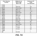
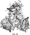


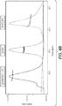


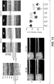


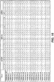
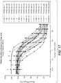

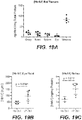
Classifications
-
- C—CHEMISTRY; METALLURGY
- C07—ORGANIC CHEMISTRY
- C07K—PEPTIDES
- C07K16/00—Immunoglobulins [IGs], e.g. monoclonal or polyclonal antibodies
- C07K16/40—Immunoglobulins [IGs], e.g. monoclonal or polyclonal antibodies against enzymes
-
- A—HUMAN NECESSITIES
- A61—MEDICAL OR VETERINARY SCIENCE; HYGIENE
- A61P—SPECIFIC THERAPEUTIC ACTIVITY OF CHEMICAL COMPOUNDS OR MEDICINAL PREPARATIONS
- A61P27/00—Drugs for disorders of the senses
-
- A—HUMAN NECESSITIES
- A61—MEDICAL OR VETERINARY SCIENCE; HYGIENE
- A61P—SPECIFIC THERAPEUTIC ACTIVITY OF CHEMICAL COMPOUNDS OR MEDICINAL PREPARATIONS
- A61P27/00—Drugs for disorders of the senses
- A61P27/02—Ophthalmic agents
-
- A—HUMAN NECESSITIES
- A61—MEDICAL OR VETERINARY SCIENCE; HYGIENE
- A61P—SPECIFIC THERAPEUTIC ACTIVITY OF CHEMICAL COMPOUNDS OR MEDICINAL PREPARATIONS
- A61P43/00—Drugs for specific purposes, not provided for in groups A61P1/00-A61P41/00
-
- A—HUMAN NECESSITIES
- A61—MEDICAL OR VETERINARY SCIENCE; HYGIENE
- A61P—SPECIFIC THERAPEUTIC ACTIVITY OF CHEMICAL COMPOUNDS OR MEDICINAL PREPARATIONS
- A61P9/00—Drugs for disorders of the cardiovascular system
- A61P9/10—Drugs for disorders of the cardiovascular system for treating ischaemic or atherosclerotic diseases, e.g. antianginal drugs, coronary vasodilators, drugs for myocardial infarction, retinopathy, cerebrovascula insufficiency, renal arteriosclerosis
-
- A—HUMAN NECESSITIES
- A61—MEDICAL OR VETERINARY SCIENCE; HYGIENE
- A61K—PREPARATIONS FOR MEDICAL, DENTAL OR TOILETRY PURPOSES
- A61K39/00—Medicinal preparations containing antigens or antibodies
- A61K2039/505—Medicinal preparations containing antigens or antibodies comprising antibodies
-
- C—CHEMISTRY; METALLURGY
- C07—ORGANIC CHEMISTRY
- C07K—PEPTIDES
- C07K2317/00—Immunoglobulins specific features
- C07K2317/30—Immunoglobulins specific features characterized by aspects of specificity or valency
- C07K2317/34—Identification of a linear epitope shorter than 20 amino acid residues or of a conformational epitope defined by amino acid residues
-
- C—CHEMISTRY; METALLURGY
- C07—ORGANIC CHEMISTRY
- C07K—PEPTIDES
- C07K2317/00—Immunoglobulins specific features
- C07K2317/70—Immunoglobulins specific features characterized by effect upon binding to a cell or to an antigen
- C07K2317/76—Antagonist effect on antigen, e.g. neutralization or inhibition of binding
-
- C—CHEMISTRY; METALLURGY
- C07—ORGANIC CHEMISTRY
- C07K—PEPTIDES
- C07K2317/00—Immunoglobulins specific features
- C07K2317/90—Immunoglobulins specific features characterized by (pharmaco)kinetic aspects or by stability of the immunoglobulin
- C07K2317/92—Affinity (KD), association rate (Ka), dissociation rate (Kd) or EC50 value
Definitions
- the present invention relates to anti-HtrA1 antibodies and methods of using the same.
- HtrA1 The serine protease HtrA1 (PRSS11; Clan PA, family S1) belongs to an evolutionarily conserved family of HtrA proteins ( Clausen, T., et al., Nat Rev Mol Cell Biol 12:152-62 (2011 ); Clausen, T., et al., Mol Cell 10:443-55 (2002 )).
- HtrA1, 3, and 4 share the same domain architecture: an N-terminal IGFBP-like module and a Kazal-like module, a protease domain with trypsin-like fold, and a C-terminal PDZ domain.
- HtrA1 The physiological relevance of HtrA1 has been firmly established by the identification of human loss-of-function mutations causing familial ischemic cerebral small-vessel disease ( Hara, K., et al., N Engl J Med 360:1729-39 (2009 )). The molecular mechanism involves deficient TGF- ⁇ inhibition by HtrA1 resulting in increased TGF- ⁇ signaling (Hara et al., 2009).
- Dysregulated TGF- ⁇ signaling by aberrant HtrA1 expression may also contribute to arthritic disease ( Oka, C., et al., Development 131:1041-53 (2004 ); Tsuchiya, A., et al., Bone 37:323-36 (2005 )), perhaps in conjunction with HtrA1-mediated degradation of various extracellular matrix components ( Chamberland et al., J Biol Chem 284:27352-9 (2009 ); Grau, S., et al., J Biol Chem 281:6124-9 (2006 ); Hadfield, K.D., et al., J Biol Chem 283:5928-38 (2008 ); Tocharus, J., et al., Dev Growth Differ 46:257-74 (2004 ); Tsuchiya et al., 2005)), or indirectly via up-regulation of matrix metalloproteases (Grau et al., 2006).
- the invention provides anti-HtrA1 antibodies and methods of using the same for therapeutic purposes as defined in the claims.
- the invention is directed to an isolated monoclonal antibody that binds to HtrA1 and inhibits its serine protease activity for one or more HtrA1 substrates, wherein the antibody comprises a VH sequence comprising SEQ ID NO: 32 and a VL sequence comprising SEQ ID NO:31.
- the disclosure provides an isolated antibody that bind to HtrA1 having one or more of the following properties: (i) an IC 50 of less than 50 nM, 30 nM, 25 nM, 20 nM, 15 nM, 10 nM, 5 nM, 3 nM, 2.5 nM, 2 nM, 1 nM, or less for one or more HtrA1 substrates; (ii) binds to HtrAl with a ratio of 1 variable domain to one subunit of an HtrA1 trimer (e.g., a Fab binds to an HtrA1 trimer with a ratio of 3 Fab to 1 HtrA1 trimer, and an IgG binds to an HtrA1 trimer with a ratio of 3 IgG to 2 HtrA1 trimers), (iii) for antibodies comprising two variable domains, binds to HtrA1 in a manner that results in the forming a "cage
- the disclosure provides an isolated antibody that binds to HtrA1, wherein the antibody (i) binds to an epitope that includes N224, K248, or both of HtrAl, and (ii) inhibits HtrA1 with an IC 50 of ⁇ 30 nM.
- the epitope further includes one or more of the following residues of HtrA1: V201, T223, K243, K225, E247 and H220.
- the antibody may further comprise one or more of the following properties: (i) binds to HtrA1 with a ratio of 1 variable domain to one subunit of an HtrA1 trimer, or (ii) does not prevent trimer formation of HtrA1.
- the IC 50 is determined using a serine protease assay with a substrate having SEQ ID NO:12, e.g., such as the FRET assay described herein.
- an antibody described herein does not cross-react with one or more of HtrA2, HtrA3 and HtrA4.
- an antibody described herein has a dissociation constant of ⁇ 500 nM.
- the dissociation constant may be determined, for example, by BIAcore using a Fab.
- an antibody described herein is a human, humanized, or chimeric antibody.
- an antibody described herein is an antibody fragment that binds HtrA1.
- an antibody described herein comprises (a) HVR-H1 comprising the amino acid sequence of SEQ ID NO:4; (b) HVR-H2 comprising the amino acid sequence of SEQ ID NO:5; and (c) HVR-H3 comprising the amino acid sequence of SEQ ID NO:6. and (d) HVR-L1 comprising the amino acid sequence of SEQ ID NO:1; (e) HVR-L2 comprising the amino acid sequence of SEQ ID NO:2; and (f) HVR-L3 comprising the amino acid sequence of SEQ ID NO:3.
- an antibody described herein comprises (a) HVR-H1 comprising the amino acid sequence of SEQ ID NO:20; (b) HVR-H2 comprising the amino acid sequence of SEQ ID NO:5; and (c) HVR-H3 comprising the amino acid sequence of SEQ ID NO:6.
- an antibody described herein further comprises (a) HVR-L1 comprising the amino acid sequence of SEQ ID NO:18; (b) HVR-L2 comprising the amino acid sequence of SEQ ID NO:2; and (c) HVR-L3 comprising the amino acid sequence of SEQ ID NO:19, or the antibody described herein further comprises (d) HVR-L1 comprising the amino acid sequence of SEQ ID NO:21; (e) HVR-L2 comprising the amino acid sequence of SEQ ID NO:2; and (f) HVR-L3 comprising the amino acid sequence of SEQ ID NO:22,
- an antibody described herein further comprises a heavy chain variable domain framework (FR2) sequence of SEQ ID NO:17.
- FR2 heavy chain variable domain framework
- an antibody described herein comprises (a) a VH sequence having at least 95% sequence identity to the amino acid sequence of SEQ ID NO:8; (b) a VL sequence having at least 95% sequence identity to the amino acid sequence of SEQ ID NO:7.
- an antibody described herein comprises a VH sequence comprising SEQ ID NO: 32 and a VL sequence comprising SEQ ID NO: 31.
- an antibody described herein comprises a VH sequence of SEQ ID NO:8 and a VL sequence of SEQ ID NO:7.
- an antibody described herein comprises a VH sequence of SEQ ID NO:30 and a VL sequence of SEQ ID NO:28.
- an antibody described herein comprises a VH sequence of SEQ ID NO:30 and a VL sequence of SEQ ID NO:29.
- an antibody described herein is a full length IgG1 or IgG4 antibody.
- an isolated nucleic acid encoding an anti-HtrA1 antibody described herein is provided.
- a host cell comprising an isolated nucleic acid encoding an anti-HtrAl antibody described herein is provided.
- a method of producing an antibody may comprise culturing a host cell comprising an isolated nucleic acid encoding an anti-HtrA1 antibody under conditions suitable for expression of the nucleic acid encoding the anti-HtrA1 antibody.
- the method may further comprise recovering the anti-HtrA1 antibody from the host cell culture, purifying the anti-HtrA1 antibody, or formulating the anti-HtrA1 antibody with a pharmaceutically acceptable excipient.
- an immunoconjugate comprising an anti-HtrA1 antibody and a cytotoxic agent is provided.
- a pharmaceutical formulation comprising an anti-HtrA1 antibody and a pharmaceutically acceptable carrier is provided.
- the application provides an anti-HtrA1 antibody for use as a medicament, e.g., for use in treating age-related macular degeneration (AMD, wet or dry), geographic atrophy (GA), diabetic retinopathy (DR), retinopathy of prematurity (ROP), or polypoidal choroidal vasculopathy (PCV), for inhibiting degeneration of retinal or photoreceptor cells, or for inhibiting HtrA1 protease activity in an eye.
- AMD age-related macular degeneration
- GA geographic atrophy
- DR diabetic retinopathy
- ROP retinopathy of prematurity
- PCV polypoidal choroidal vasculopathy
- the application provides an anti-HtrA1 antibody for use treating an individual having age-related macular degeneration (wet or dry), geographic atrophy, diabetic retinopathy, retinopathy of prematurity, or polypoidal choroidal vasculopathy, comprising administering to the individual an effective amount of an anti-HtrA1 antibody as described herein.
- age-related macular degeneration wet or dry
- geographic atrophy a grouping of retinopathy
- diabetic retinopathy retinopathy of prematurity
- polypoidal choroidal vasculopathy comprising administering to the individual an effective amount of an anti-HtrA1 antibody as described herein.
- the application provides an anti-HtrA1 antibody for use in inhibiting retinal or photoreceptor cell degeneration in an individual comprising administering to the individual an effective amount of an anti-HtrA1 antibody as described herein to inhibit retinal or photoreceptor cell degeneration.
- the application provides an anti-HtrA1 antibody for use in inhibiting HtrA1 serine protease activityin an eye of an individual comprising administering to the individual an effective amount of an anti-HtrA1 antibody as described herein to inhibit HtrA1 serine protease activity in the eye.
- acceptor human framework for the purposes herein is a framework comprising the amino acid sequence of a light chain variable domain (VL) framework or a heavy chain variable domain (VH) framework derived from a human immunoglobulin framework or a human consensus framework, as defined below.
- An acceptor human framework "derived from” a human immunoglobulin framework or a human consensus framework may comprise the same amino acid sequence thereof, or it may contain amino acid sequence changes. In some embodiments, the number of amino acid changes are 10 or less, 9 or less, 8 or less, 7 or less, 6 or less, 5 or less, 4 or less, 3 or less, or 2 or less.
- the VL acceptor human framework is identical in sequence to the VL human immunoglobulin framework sequence or human consensus framework sequence.
- Bind refers to the strength of the sum total of noncovalent interactions between a single binding site of a molecule (e.g., an antibody) and its binding partner (e.g., an antigen).
- binding affinity refers to intrinsic binding affinity which reflects a 1:1 interaction between members of a binding pair (e.g., antibody and antigen).
- the affinity of a molecule X for its partner Y can generally be represented by the dissociation constant (Kd). Affinity can be measured by common methods known in the art, including those described herein. Specific illustrative and exemplary embodiments for measuring binding affinity are described in the following.
- an “affinity matured” antibody refers to an antibody with one or more alterations in one or more hypervariable regions (HVRs), compared to a parent antibody which does not possess such alterations, such alterations resulting in an improvement in the affinity of the antibody for antigen.
- HVRs hypervariable regions
- anti-HtrA1 antibody and "an antibody that binds to HtrA1” refer to an antibody that is capable of binding HtrA1 with sufficient affinity such that the antibody is useful as a diagnostic and/or therapeutic agent in targeting HtrA1.
- the extent of binding of an anti- HtrA1 antibody to an unrelated, non- HtrA1 protein is less than about 10% of the binding of the antibody to HtrA1 as measured, e.g., by a radioimmunoassay (RIA).
- RIA radioimmunoassay
- an antibody that binds to HtrA1 has a dissociation constant (Kd) of ⁇ 1 ⁇ M, ⁇ 100 nM, ⁇ 10 nM, ⁇ 1 nM, ⁇ 0.1 nM, ⁇ 0.01 nM, or ⁇ 0.001 nM (e.g. 10 -8 M or less, e.g. from 10 -8 M to 10 -13 M, e.g., from 10 -9 M to 10 -13 M).
- Kd dissociation constant
- an anti-HtrA1 antibody binds to an epitope of HtrA1 that is conserved among HtrA1 from different species.
- antibody herein is used in the broadest sense and encompasses various antibody structures, including but not limited to monoclonal antibodies, polyclonal antibodies, multispecific antibodies (e.g., bispecific antibodies), and antibody fragments so long as they exhibit the desired antigen-binding activity.
- antibody fragment refers to a molecule other than an intact antibody that comprises a portion of an intact antibody that binds the antigen to which the intact antibody binds.
- antibody fragments include but are not limited to Fv, Fab, Fab', Fab'-SH, F(ab') 2 ; diabodies; linear antibodies; single-chain antibody molecules (e.g. scFv); and multispecific antibodies formed from antibody fragments.
- an "antibody that binds to the same epitope” as a reference antibody refers to an antibody that blocks binding of the reference antibody to its antigen in a competition assay by 50% or more, and conversely, the reference antibody blocks binding of the antibody to its antigen in a competition assay by 50% or more.
- An exemplary competition assay is provided herein.
- chimeric antibody refers to an antibody in which a portion of the heavy and/or light chain is derived from a particular source or species, while the remainder of the heavy and/or light chain is derived from a different source or species.
- the "class" of an antibody refers to the type of constant domain or constant region possessed by its heavy chain.
- the heavy chain constant domains that correspond to the different classes of immunoglobulins are called ⁇ , ⁇ , ⁇ , ⁇ , and ⁇ , respectively.
- cytotoxic agent refers to a substance that inhibits or prevents a cellular function and/or causes cell death or destruction.
- Cytotoxic agents include, but are not limited to, radioactive isotopes (e.g., At 211 , I 131 , I 125 , Y 90 , Re 186 , Re 188 , Sm 153 , Bi 212 , P 32 , Pb 212 and radioactive isotopes of Lu); chemotherapeutic agents or drugs (e.g., methotrexate, adriamicin, vinca alkaloids (vincristine, vinblastine, etoposide), doxorubicin, melphalan, mitomycin C, chlorambucil, daunorubicin or other intercalating agents); growth inhibitory agents; enzymes and fragments thereof such as nucleolytic enzymes; antibiotics; toxins such as small molecule toxins or enzymatically active toxins of bacterial, fungal
- Antibody effector functions refer to those biological activities attributable to the Fc region of an antibody, which vary with the antibody isotype. Examples of antibody effector functions include: C1q binding and complement dependent cytotoxicity (CDC); Fc receptor binding; antibody-dependent cell-mediated cytotoxicity (ADCC); phagocytosis; down regulation of cell surface receptors (e.g. B cell receptor); and B cell activation.
- an "effective amount" of an agent refers to an amount effective, at dosages and for periods of time necessary, to achieve the desired therapeutic or prophylactic result.
- Fc region herein is used to define a C-terminal region of an immunoglobulin heavy chain that contains at least a portion of the constant region.
- the term includes native sequence Fc regions and variant Fc regions.
- a human IgG heavy chain Fc region extends from Cys226, or from Pro230, to the carboxyl-terminus of the heavy chain.
- the C-terminal lysine (Lys447) of the Fc region may or may not be present.
- numbering of amino acid residues in the Fc region or constant region is according to the EU numbering system, also called the EU index, as described in Kabat et al., Sequences of Proteins of Immunological Interest, 5th Ed. Public Health Service, National Institutes of Health, Bethesda, MD, 1991 .
- Framework region refers to variable domain residues other than hypervariable region (HVR) residues.
- the FR of a variable domain generally consists of four FR domains: FR1, FR2, FR3, and FR4. Accordingly, the HVR and FR sequences generally appear in the following sequence in VH (or VL): FR1-H1(L1)-FR2-H2(L2)-FR3-H3(L3)-FR4.
- full length antibody “intact antibody,” and “whole antibody” are used herein interchangeably to refer to an antibody having a structure substantially similar to a native antibody structure or having heavy chains that contain an Fc region as defined herein.
- host cell refers to cells into which exogenous nucleic acid has been introduced, including the progeny of such cells.
- Host cells include “transformants” and “transformed cells,” which include the primary transformed cell and progeny derived therefrom without regard to the number of passages. Progeny may not be completely identical in nucleic acid content to a parent cell, but may contain mutations. Mutant progeny that have the same function or biological activity as screened or selected for in the originally transformed cell are included herein.
- a "human antibody” is one which possesses an amino acid sequence which corresponds to that of an antibody produced by a human or a human cell or derived from a non-human source that utilizes human antibody repertoires or other human antibody-encoding sequences. This definition of a human antibody specifically excludes a humanized antibody comprising non-human antigen-binding residues.
- a "human consensus framework” is a framework which represents the most commonly occurring amino acid residues in a selection of human immunoglobulin VL or VH framework sequences.
- the selection of human immunoglobulin VL or VH sequences is from a subgroup of variable domain sequences.
- the subgroup of sequences is a subgroup as in Kabat et al., Sequences of Proteins of Immunological Interest, Fifth Edition, NIH Publication 91-3242, Bethesda MD (1991), vols. 1-3 .
- the subgroup is subgroup kappa I as in Kabat et al., supra.
- the subgroup is subgroup III as in Kabat et al., supra.
- a “humanized” antibody refers to a chimeric antibody comprising amino acid residues from non-human HVRs and amino acid residues from human FRs.
- a humanized antibody will comprise substantially all of at least one, and typically two, variable domains, in which all or substantially all of the HVRs (e.g., CDRs) correspond to those of a non-human antibody, and all or substantially all of the FRs correspond to those of a human antibody.
- a humanized antibody optionally may comprise at least a portion of an antibody constant region derived from a human antibody.
- a "humanized form" of an antibody, e.g., a non-human antibody refers to an antibody that has undergone humanization.
- hypervariable region refers to each of the regions of an antibody variable domain which are hypervariable in sequence and/or form structurally defined loops ("hypervariable loops").
- native four-chain antibodies comprise six HVRs; three in the VH (H1, H2, H3), and three in the VL (L1, L2, L3).
- HVRs generally comprise amino acid residues from the hypervariable loops and/or from the "complementarity determining regions" (CDRs), the latter being of highest sequence variability and/or involved in antigen recognition.
- CDRs complementarity determining regions
- An HVR region as used herein comprise any number of residues located within positions 24-36 (for L1), 46-56 (for L2), 89-97 (for L3), 26-35B (for H1), 47-65 (for H2), and 93-102 (for H3). Therefore, an HVR includes residues in positions described previously:
- CDRs generally comprise the amino acid residues that form the hypervariable loops.
- CDRs also comprise "specificity determining residues," or "SDRs,” which are residues that contact antigen. SDRs are contained within regions of the CDRs called abbreviated-CDRs, or a-CDRs.
- Exemplary a-CDRs (a-CDR-L1, a-CDR-L2, a-CDR-L3, a-CDR-H1, a-CDR-H2, and a-CDR-H3) occur at amino acid residues 31-34 of L1, 50-55 of L2, 89-96 of L3, 31-35B of HI, 50-58 of H2, and 95-102 of H3.
- HVR residues and other residues in the variable domain are numbered herein according to Kabat et al., supra.
- an “immunoconjugate” is an antibody conjugated to one or more heterologous molecule(s), including but not limited to a cytotoxic agent.
- mammals include, but are not limited to, domesticated animals (e.g., cows, sheep, cats, dogs, and horses), primates (e.g., humans and non-human primates such as monkeys), rabbits, and rodents (e.g., mice and rats).
- domesticated animals e.g., cows, sheep, cats, dogs, and horses
- primates e.g., humans and non-human primates such as monkeys
- rabbits e.g., mice and rats
- rodents e.g., mice and rats.
- the individual or subject is a human.
- an “isolated” antibody is one which has been separated from a component of its natural environment.
- an antibody is purified to greater than 95% or 99% purity as determined by, for example, electrophoretic (e.g., SDS-PAGE, isoelectric focusing (IEF), capillary electrophoresis) or chromatographic (e.g., ion exchange or reverse phase HPLC).
- electrophoretic e.g., SDS-PAGE, isoelectric focusing (IEF), capillary electrophoresis
- chromatographic e.g., ion exchange or reverse phase HPLC
- nucleic acid refers to a nucleic acid molecule that has been separated from a component of its natural environment.
- An isolated nucleic acid includes a nucleic acid molecule contained in cells that ordinarily contain the nucleic acid molecule, but the nucleic acid molecule is present extrachromosomally, at a chromosomal location that is different from its natural chromosomal location, or contains only coding sequences.
- isolated nucleic acid encoding an anti-HtrA1 antibody refers to one or more nucleic acid molecules encoding antibody heavy and light chains (or fragments thereof), including such nucleic acid molecule(s) in a single vector or separate vectors, and such nucleic acid molecule(s) present at one or more locations in a host cell.
- monoclonal antibody refers to an antibody obtained from a population of substantially homogeneous antibodies, i.e., the individual antibodies comprising the population are identical and/or bind the same epitope, except for possible variant antibodies, e.g., containing naturally occurring mutations or arising during production of a monoclonal antibody preparation, such variants generally being present in minor amounts.
- polyclonal antibody preparations typically include different antibodies directed against different determinants (epitopes)
- each monoclonal antibody of a monoclonal antibody preparation is directed against a single determinant on an antigen.
- the modifier "monoclonal” indicates the character of the antibody as being obtained from a substantially homogeneous population of antibodies, and is not to be construed as requiring production of the antibody by any particular method.
- the monoclonal antibodies to be used in accordance with the present invention may be made by a variety of techniques, including but not limited to the hybridoma method, recombinant DNA methods, phage-display methods, and methods utilizing transgenic animals containing all or part of the human immunoglobulin loci, such methods and other exemplary methods for making monoclonal antibodies being described herein.
- naked antibody refers to an antibody that is not conjugated to a heterologous moiety (e.g., a cytotoxic moiety) or radiolabel.
- the naked antibody may be present in a pharmaceutical formulation.
- Native antibodies refer to naturally occurring immunoglobulin molecules with varying structures.
- native IgG antibodies are heterotetrameric glycoproteins of about 150,000 daltons, composed of two identical light chains and two identical heavy chains that are disulfide-bonded. From N- to C-terminus, each heavy chain has a variable region (VH), also called a variable heavy domain or a heavy chain variable domain, followed by three constant domains (CH1, CH2, and CH3).
- VH variable region
- VL variable region
- the light chain of an antibody may be assigned to one of two types, called kappa ( ⁇ ) and lambda ( ⁇ ), based on the amino acid sequence of its constant domain.
- package insert is used to refer to instructions customarily included in commercial packages of therapeutic products, that contain information about the indications, usage, dosage, administration, combination therapy, contraindications and/or warnings concerning the use of such therapeutic products.
- Percent (%) amino acid sequence identity with respect to a reference polypeptide sequence is defined as the percentage of amino acid residues in a candidate sequence that are identical with the amino acid residues in the reference polypeptide sequence, after aligning the sequences and introducing gaps, if necessary, to achieve the maximum percent sequence identity, and not considering any conservative substitutions as part of the sequence identity. Alignment for purposes of determining percent amino acid sequence identity can be achieved in various ways that are within the skill in the art, for instance, using publicly available computer software such as BLAST, BLAST-2, ALIGN or Megalign (DNASTAR) software. Those skilled in the art can determine appropriate parameters for aligning sequences, including any algorithms needed to achieve maximal alignment over the full length of the sequences being compared.
- % amino acid sequence identity values are generated using the sequence comparison computer program ALIGN-2.
- the ALIGN-2 sequence comparison computer program was authored by Genentech, Inc., and the source code has been filed with user documentation in the U.S. Copyright Office, Washington D.C., 20559, where it is registered under U.S. Copyright Registration No. TXU510087.
- the ALIGN-2 program is publicly available from Genentech, Inc., South San Francisco, California, or may be compiled from the source code.
- the ALIGN-2 program should be compiled for use on a UNIX operating system, including digital UNIX V4.0D. All sequence comparison parameters are set by the ALIGN-2 program and do not vary.
- the % amino acid sequence identity of a given amino acid sequence A to, with, or against a given amino acid sequence B is calculated as follows: 100 times the fraction X / Y where X is the number of amino acid residues scored as identical matches by the sequence alignment program ALIGN-2 in that program's alignment of A and B, and where Y is the total number of amino acid residues in B.
- pharmaceutical formulation refers to a preparation which is in such form as to permit the biological activity of an active ingredient contained therein to be effective, and which contains no additional components which are unacceptably toxic to a subject to which the formulation would be administered.
- a “pharmaceutically acceptable carrier” refers to an ingredient in a pharmaceutical formulation, other than an active ingredient, which is nontoxic to a subject.
- a pharmaceutically acceptable carrier includes, but is not limited to, a buffer, excipient, stabilizer, or preservative.
- HtrA1 refers to any native HtrA1 from any vertebrate source, including mammals such as primates (e.g. humans) and rodents (e.g., mice and rats), unless otherwise indicated.
- the term encompasses "full-length,” unprocessed HtrA1 as well as any form of HtrA1 that results from processing in the cell.
- the term also encompasses naturally occurring variants of HtrA1, e.g., splice variants or allelic variants.
- the amino acid sequence of an exemplary HtrA1 is shown in SEQ ID NO:13.
- Exemplary fragments of human HtrA1 include fragments comprising, consisting essentially of, or consistingof amino acids Q23-P480 or P161-K379.
- treatment refers to clinical intervention in an attempt to alter the natural course of the individual being treated, and can be performed either for prophylaxis or during the course of clinical pathology. Desirable effects of treatment include, but are not limited to, preventing occurrence or recurrence of disease, alleviation of symptoms, diminishment of any direct or indirect pathological consequences of the disease, preventing metastasis, decreasing the rate of disease progression, amelioration or palliation of the disease state, and remission or improved prognosis.
- antibodies of the invention are used to delay development of a disease or to slow the progression of a disease.
- variable region refers to the domain of an antibody heavy or light chain that is involved in binding the antibody to antigen.
- the variable domains of the heavy chain and light chain (VH and VL, respectively) of a native antibody generally have similar structures, with each domain comprising four conserved framework regions (FRs) and three hypervariable regions (HVRs).
- FRs conserved framework regions
- HVRs hypervariable regions
- antibodies that bind a particular antigen may be isolated using a VH or VL domain from an antibody that binds the antigen to screen a library of complementary VL or VH domains, respectively. See, e.g., Portolano et al., J. Immunol. 150:880-887 (1993 ); Clarkson et al., Nature 352:624-628 (1991 ).
- vector refers to a nucleic acid molecule capable of propagating another nucleic acid to which it is linked.
- the term includes the vector as a self-replicating nucleic acid structure as well as the vector incorporated into the genome of a host cell into which it has been introduced.
- Certain vectors are capable of directing the expression of nucleic acids to which they are operatively linked. Such vectors are referred to herein as "expression vectors.”
- the invention is based, in part, on the discovery that a reduction of HtrA1 activity has protective effects on photoreceptor cells in the eye, the outer nuclear layer, and on elecroretinogram functionality.
- antibodies that bind to HtrA1 are provided.
- Antibodies of the invention are useful, e.g., for the diagnosis or treatment of various diseases associated with HtrA1 activity, including ocular disorders such as age-related macular degeneration or geographic atrophy.
- the invention is directed to an isolated monoclonal antibody that binds to HtrA1 and inhibits its serine protease activity for one or more HtrA1 substrates, wherein the antibody comprises a VH sequence comprising SEQ ID NO: 32 and a VL sequence comprising SEQ ID NO:31.
- An anti-HtrA1 antibody can have one or more of the following properties: (i) has an IC 50 of less than 50 nM, 30 nM, 25 nM, 20 nM, 15 nM, 10 nM, 5 nM, 3 nM, 2.5 nM, 2 nM, 1 nM, or less for one or more HtrA1 substrates; (ii) binds to HtrA1 with a ratio of 1 variable domain to one subunit of an HtrA1 trimer (e.g., a Fab binds to an HtrA1 trimer with a ratio of 3 Fab to 1 HtrA1 trimer, and an IgG binds to an HtrA1 trimer with a ratio of 3 IgG to 2 HtrA1 trimers), (iii) for antibodies comprising two variable domains, binds to HtrA1 in a manner that results in the forming a "cage" similar to that shown in
- an anti-HtrA1 antibody comprising at least one, two, three, four, five, or six HVRs selected from (a) HVR-H1 comprising the amino acid sequence of SEQ ID NO:87; (b) HVR-H2 comprising the amino acid sequence of SEQ ID NO:88; (c) HVR-H3 comprising the amino acid sequence of SEQ ID NO:89; (d) HVR-L1 comprising the amino acid sequence of SEQ ID NO:85; (e) HVR-L2 comprising the amino acid sequence of SEQ ID NO:58; and (f) HVR-L3 comprising the amino acid sequence of SEQ ID NO:86.
- the invention provides an anti-HtrA1 antibody comprising at least one, two, three, four, five, or six HVRs selected from (a) HVR-H1 comprising the amino acid sequence of SEQ ID NO:25; (b) HVR-H2 comprising the amino acid sequence of SEQ ID NO:26; (c) HVR-H3 comprising the amino acid sequence of SEQ ID NO:27; (d) HVR-L1 comprising the amino acid sequence of SEQ ID NO:23; (e) HVR-L2 comprising the amino acid sequence of SEQ ID NO:2; and (f) HVR-L3 comprising the amino acid sequence of SEQ ID NO:24.
- HVR-H1 comprising the amino acid sequence of SEQ ID NO:25
- HVR-H2 comprising the amino acid sequence of SEQ ID NO:26
- HVR-H3 comprising the amino acid sequence of SEQ ID NO:27
- HVR-L1 comprising the amino acid sequence of SEQ ID NO:23
- HVR-L2 compris
- the invention provides an anti-HtrAl antibody comprising at least one, two, three, four, five, or six HVRs selected from (a) HVR-H1 comprising an amino acid sequence selected from: SEQ ID NO:4, 20, 47-51 and 75-80; (b) HVR-H2 comprising an amino acid sequence selected from: SEQ ID NO:5, 52 and 81-82; (c) HVR-H3 comprising an amino acid sequence selected from: SEQ ID NO:6, 53 and 83-84; (d) HVR-L1 comprising an amino acid sequence selected from: SEQ ID NO:1, 18, 21, 33 and 54-57; (e) HVR-L2 comprising an amino acid sequence selected from: SEQ ID NO:2 and 58; and (f) HVR-L3 comprising an amino acid sequence selected from: SEQ ID NO:3, 19, 22, 34-46 and 59-74.
- HVR-H1 comprising an amino acid sequence selected from: SEQ ID NO:4, 20, 47-51 and 75-80
- the invention provides an anti-HtrAl antibody comprising at least one, two, three, four, five, or six HVRs selected from (a) HVR-H1 comprising the amino acid sequence of SEQ ID NO:4; (b) HVR-H2 comprising the amino acid sequence of SEQ ID NO:5; (c) HVR-H3 comprising the amino acid sequence of SEQ ID NO:6; (d) HVR-L1 comprising the amino acid sequence of SEQ ID NO:1; (e) HVR-L2 comprising the amino acid sequence of SEQ ID NO:2; and (f) HVR-L3 comprising the amino acid sequence of SEQ ID NO:3.
- the invention provides an anti-HtrAl antibody comprising at least one, two, three, four, five, or six HVRs selected from (a) HVR-H1 comprising the amino acid sequence of SEQ ID NO:20; (b) HVR-H2 comprising the amino acid sequence of SEQ ID NO:5; (c) HVR-H3 comprising the amino acid sequence of SEQ ID NO:6; (d) HVR-L1 comprising the amino acid sequence of SEQ ID NO:18; (e) HVR-L2 comprising the amino acid sequence of SEQ ID NO:2; and (f) HVR-L3 comprising the amino acid sequence of SEQ ID NO:19.
- HVR-H1 comprising the amino acid sequence of SEQ ID NO:20
- HVR-H2 comprising the amino acid sequence of SEQ ID NO:5
- HVR-H3 comprising the amino acid sequence of SEQ ID NO:6
- HVR-L1 comprising the amino acid sequence of SEQ ID NO:18
- HVR-L2 compris
- the invention provides an anti-HtrAl antibody comprising at least one, two, three, four, five, or six HVRs selected from (a) HVR-H1 comprising the amino acid sequence of SEQ ID NO:20; (b) HVR-H2 comprising the amino acid sequence of SEQ ID NO:5; (c) HVR-H3 comprising the amino acid sequence of SEQ ID NO:6; (d) HVR-L1 comprising the amino acid sequence of SEQ ID NO:21; (e) HVR-L2 comprising the amino acid sequence of SEQ ID NO:2; and (f) HVR-L3 comprising the amino acid sequence of SEQ ID NO:22.
- HVR-H1 comprising the amino acid sequence of SEQ ID NO:20
- HVR-H2 comprising the amino acid sequence of SEQ ID NO:5
- HVR-H3 comprising the amino acid sequence of SEQ ID NO:6
- HVR-L1 comprising the amino acid sequence of SEQ ID NO:21
- HVR-L2 compris
- an antibody comprising at least one, at least two, or all three VH HVR sequences selected from (a) HVR-H1 comprising the amino acid sequence of SEQ ID NO:87; (b) HVR-H2 comprising the amino acid sequence of SEQ ID NO:88; and (c) HVR-H3 comprising the amino acid sequence of SEQ ID NO:89.
- the antibody comprises HVR-H3 comprising the amino acid sequence of SEQ ID NO:89.
- the antibody comprises HVR-H3 comprising the amino acid sequence of SEQ ID NO:89 and HVR-L3 comprising the amino acid sequence of SEQ ID NO:86.
- the antibody comprises HVR-H3 comprising the amino acid sequence of SEQ ID NO:89, HVR-L3 comprising the amino acid sequence of SEQ ID NO:86, and HVR-H2 comprising the amino acid sequence of SEQ ID NO:88.
- the antibody comprises (a) HVR-H1 comprising the amino acid sequence of SEQ ID NO:87; (b) HVR-H2 comprising the amino acid sequence of SEQ ID NO:88; and (c) HVR-H3 comprising the amino acid sequence of SEQ ID NO:89.
- an antibody comprising at least one, at least two, or all three VH HVR sequences selected from (a) HVR-H1 comprising the amino acid sequence of SEQ ID NO:25; (b) HVR-H2 comprising the amino acid sequence of SEQ ID NO:26; and (c) HVR-H3 comprising the amino acid sequence of SEQ ID NO:27.
- the antibody comprises HVR-H3 comprising the amino acid sequence of SEQ ID NO:27.
- the antibody comprises HVR-H3 comprising the amino acid sequence of SEQ ID NO:27 and HVR-L3 comprising the amino acid sequence of SEQ ID NO:24.
- the antibody comprises HVR-H3 comprising the amino acid sequence of SEQ ID NO:27, HVR-L3 comprising the amino acid sequence of SEQ ID NO:24, and HVR-H2 comprising the amino acid sequence of SEQ ID NO:26.
- the antibody comprises (a) HVR-H1 comprising the amino acid sequence of SEQ ID NO:25; (b) HVR-H2 comprising the amino acid sequence of SEQ ID NO:26; and (c) HVR-H3 comprising the amino acid sequence of SEQ ID NO:27.
- an antibody comprising at least one, at least two, or all three VH HVR sequences selected from (a) HVR-H1 comprising an amino acid sequence selected from: SEQ ID NO:4, 20, 47-51 and 75-80; (b) HVR-H2 comprising an amino acid sequence selected from SEQ ID NO:5, 52 and 81-82; and (c) HVR-H3 comprising an amino acid sequence selected from: SEQ ID NO:6, 53 and 83-84.
- the antibody comprises HVR-H3 comprising an amino acid sequence selected from SEQ ID NO:6 and 53.
- the antibody comprises HVR-H3 comprising an amino acid sequence selected from SEQ ID NO:6, 53 and 83-84 and HVR-L3 comprising an amino acid sequence selected from SEQ ID NO:3, 19, 22, 34-46 and 59-74.
- the antibody comprises HVR-H3 comprising an amino acid sequence selected from SEQ ID NO:6, 53 and 83-84, HVR-L3 comprising an amino acid sequence selected from SEQ ID NO: 3, 19, 22, 34-46 and 59-74, and HVR-H2 comprising an amino acid sequence selected from SEQ ID NO:5, 52 and 81-82.
- the antibody comprises (a) HVR-H1 comprising an amino acid sequence selected from SEQ ID NO:4, 20, 47-51 and 75-80; (b) HVR-H2 comprising an amino acid sequence selected from SEQ ID NO:5, 52 and 81-82; and (c) HVR-H3 comprising an amino acid sequence selected from SEQ ID NO:6, 53 and 83-84.
- an antibody comprising at least one, at least two, or all three VH HVR sequences selected from (a) HVR-H1 comprising the amino acid sequence of SEQ ID NO:4; (b) HVR-H2 comprising the amino acid sequence of SEQ ID NO:5; and (c) HVR-H3 comprising the amino acid sequence of SEQ ID NO:6.
- the antibody comprises HVR-H3 comprising the amino acid sequence of SEQ ID NO:6.
- the antibody comprises HVR-H3 comprising the amino acid sequence of SEQ ID NO:6 and HVR-L3 comprising the amino acid sequence of SEQ ID NO:3.
- the antibody comprises HVR-H3 comprising the amino acid sequence of SEQ ID NO:6, HVR-L3 comprising the amino acid sequence of SEQ ID NO:3, and HVR-H2 comprising the amino acid sequence of SEQ ID NO:5.
- the antibody comprises (a) HVR-H1 comprising the amino acid sequence of SEQ ID NO:4; (b) HVR-H2 comprising the amino acid sequence of SEQ ID NO:5; and (c) HVR-H3 comprising the amino acid sequence of SEQ ID NO:6.
- an antibody comprising at least one, at least two, or all three VH HVR sequences selected from (a) HVR-H1 comprising the amino acid sequence of SEQ ID NO:20; (b) HVR-H2 comprising the amino acid sequence of SEQ ID NO:5; and (c) HVR-H3 comprising the amino acid sequence of SEQ ID NO:6.
- the antibody comprises HVR-H3 comprising the amino acid sequence of SEQ ID NO:20.
- the antibody comprises HVR-H3 comprising the amino acid sequence of SEQ ID NO:20 and HVR-L3 comprising the amino acid sequence of SEQ ID NO:19.
- the antibody comprises HVR-H3 comprising the amino acid sequence of SEQ ID NO:20, HVR-L3 comprising the amino acid sequence of SEQ ID NO:19, and HVR-H2 comprising the amino acid sequence of SEQ ID NO:5.
- the antibody comprises HVR-H3 comprising the amino acid sequence of SEQ ID NO:20 and HVR-L3 comprising the amino acid sequence of SEQ ID NO:22.
- the antibody comprises HVR-H3 comprising the amino acid sequence of SEQ ID NO:20, HVR-L3 comprising the amino acid sequence of SEQ ID NO:22, and HVR-H2 comprising the amino acid sequence of SEQ ID NO:5.
- the antibody comprises (a) HVR-H1 comprising the amino acid sequence of SEQ ID NO:20; (b) HVR-H2 comprising the amino acid sequence of SEQ ID NO:5; and (c) HVR-H3 comprising the amino acid sequence of SEQ ID NO:6.
- an antibody comprising at least one, at least two, or all three VL HVR sequences selected from (a) HVR-L1 comprising the amino acid sequence of SEQ ID NO:85; (b) HVR-L2 comprising the amino acid sequence of SEQ ID NO:58; and (c) HVR-L3 comprising the amino acid sequence of SEQ ID NO:86.
- the invention provides an antibody comprising at least one, at least two, or all three VL HVR sequences selected from (a) HVR-L1 comprising the amino acid sequence of SEQ ID NO:23; (b) HVR-L2 comprising the amino acid sequence of SEQ ID NO:2; and (c) HVR-L3 comprising the amino acid sequence of SEQ ID NO:24.
- the antibody comprises (a) HVR-L1 comprising the amino acid sequence of SEQ ID NO:85; (b) HVR-L2 comprising the amino acid sequence of SEQ ID NO:58; and (c) HVR-L3 comprising the amino acid sequence of SEQ ID NO:86.
- the antibody comprises (a) HVR-L1 comprising the amino acid sequence of SEQ ID NO:23; (b) HVR-L2 comprising the amino acid sequence of SEQ ID NO:2; and (c) HVR-L3 comprising the amino acid sequence of SEQ ID NO:24.
- an antibody comprising at least one, at least two, or all three VL HVR sequences selected from (a) HVR-L1 comprising an amino acid sequence selected from SEQ ID NO:1, 18, 21, 33 and 54-57; (b) HVR-L2 comprising an amino acid sequence selected from SEQ ID NO:2 and 58; and (c) HVR-L3 comprising an amino acid sequence selected from SEQ ID NO:3, 19, 22, 34-46 and 59-74.
- the antibody comprises (a) HVR-L1 comprising an amino acid sequence selected from SEQ ID NO:1, 18, 21, 33 and 54-57; (b) HVR-L2 comprising an amino acid sequence selected from SEQ ID NO:2 and 58; and (c) HVR-L3 comprising an amino acid sequence selected from SEQ ID NO:3, 19, 22, 34-46 and 59-74.
- an antibody comprising at least one, at least two, or all three VL HVR sequences selected from (a) HVR-L1 comprising the amino acid sequence of SEQ ID NO:1; (b) HVR-L2 comprising the amino acid sequence of SEQ ID NO:2; and (c) HVR-L3 comprising the amino acid sequence of SEQ ID NO:3.
- the antibody comprises (a) HVR-L1 comprising the amino acid sequence of SEQ ID NO:1; (b) HVR-L2 comprising the amino acid sequence of SEQ ID NO:2; and (c) HVR-L3 comprising the amino acid sequence of SEQ ID NO:3.
- an antibody comprising at least one, at least two, or all three VL HVR sequences selected from (a) HVR-L1 comprising the amino acid sequence of SEQ ID NO:18; (b) HVR-L2 comprising the amino acid sequence of SEQ ID NO:2; and (c) HVR-L3 comprising the amino acid sequence of SEQ ID NO:19.
- the antibody comprises (a) HVR-L1 comprising the amino acid sequence of SEQ ID NO:18; (b) HVR-L2 comprising the amino acid sequence of SEQ ID NO:2; and (c) HVR-L3 comprising the amino acid sequence of SEQ ID NO:19.
- an antibody comprising at least one, at least two, or all three VL HVR sequences selected from (a) HVR-L1 comprising the amino acid sequence of SEQ ID NO:21; (b) HVR-L2 comprising the amino acid sequence of SEQ ID NO:2; and (c) HVR-L3 comprising the amino acid sequence of SEQ ID NO:22.
- the antibody comprises (a) HVR-L1 comprising the amino acid sequence of SEQ ID NO:21; (b) HVR-L2 comprising the amino acid sequence of SEQ ID NO:2; and (c) HVR-L3 comprising the amino acid sequence of SEQ ID NO:22.
- an antibody comprises (a) a VH domain comprising at least one, at least two, or all three VH HVR sequences selected from (i) HVR-H1 comprising the amino acid sequence of SEQ ID NO:87, (ii) HVR-H2 comprising the amino acid sequence of SEQ ID NO:88, and (iii) HVR-H3 comprising the amino acid sequence of SEQ ID NO:89; and (b) a VL domain comprising at least one, at least two, or all three VL HVR sequences selected from (i) HVR-L1 comprising the amino acid sequence of SEQ ID NO:85, (ii) HVR-L2 comprising the amino acid sequence of SEQ ID NO:58, and (c) HVR-L3 comprising the amino acid sequence of SEQ ID NO:86.
- an antibody comprises (a) a VH domain comprising at least one, at least two, or all three VH HVR sequences selected from (i) HVR-H1 comprising the amino acid sequence of SEQ ID NO:25, (ii) HVR-H2 comprising the amino acid sequence of SEQ ID NO:26, and (iii) HVR-H3 comprising the amino acid sequence of SEQ ID NO:27; and (b) a VL domain comprising at least one, at least two, or all three VL HVR sequences selected from (i) HVR-L1 comprising the amino acid sequence of SEQ ID NO:23, (ii) HVR-L2 comprising the amino acid sequence of SEQ ID NO:2, and (c) HVR-L3 comprising the amino acid sequence of SEQ ID NO:24.
- an antibody comprises (a) a VH domain comprising at least one, at least two, or all three VH HVR sequences selected from (i) HVR-H1 comprising an amino acid sequence selected from SEQ ID NO:4, 20, 47-51 and 75-80, (ii) HVR-H2 comprising an amino acid sequence selected from SEQ ID NO:5, 52 and 81-82, and (iii) HVR-H3 comprising an amino acid sequence selected from SEQ ID NO:6, 53 and 83-84; and (b) a VL domain comprising at least one, at least two, or all three VL HVR sequences selected from (i) HVR-L1 comprising an amino acid sequence selected from SEQ ID NO:1, 18, 21, 33 and 54-57, (ii) HVR-L2 comprising an amino acid sequence selected from SEQ ID NO:2 and 58, and (c) HVR-L3 comprising an amino acid sequence selected from SEQ ID NO:3, 19, 22, 34-46 and 59-74.
- an antibody comprises (a) a VH domain comprising at least one, at least two, or all three VH HVR sequences selected from (i) HVR-H1 comprising the amino acid sequence of SEQ ID NO:4, (ii) HVR-H2 comprising the amino acid sequence of SEQ ID NO:5, and (iii) HVR-H3 comprising the amino acid sequence of SEQ ID NO:6; and (b) a VL domain comprising at least one, at least two, or all three VL HVR sequences selected from (i) HVR-L1 comprising the amino acid sequence of SEQ ID NO:1, (ii) HVR-L2 comprising the amino acid sequence of SEQ ID NO:2, and (c) HVR-L3 comprising the amino acid sequence of SEQ ID NO:3.
- an antibody comprises (a) a VH domain comprising at least one, at least two, or all three VH HVR sequences selected from (i) HVR-H1 comprising the amino acid sequence of SEQ ID NO:20, (ii) HVR-H2 comprising the amino acid sequence of SEQ ID NO:5, and (iii) HVR-H3 comprising the amino acid sequence of SEQ ID NO:6; and (b) a VL domain comprising at least one, at least two, or all three VL HVR sequences selected from (i) HVR-L1 comprising the amino acid sequence of SEQ ID NO:18, (ii) HVR-L2 comprising the amino acid sequence of SEQ ID NO:2, and (c) HVR-L3 comprising the amino acid sequence of SEQ ID NO:19.
- an antibody comprises (a) a VH domain comprising at least one, at least two, or all three VH HVR sequences selected from (i) HVR-H1 comprising the amino acid sequence of SEQ ID NO:20, (ii) HVR-H2 comprising the amino acid sequence of SEQ ID NO:5, and (iii) HVR-H3 comprising the amino acid sequence of SEQ ID NO:6; and (b) a VL domain comprising at least one, at least two, or all three VL HVR sequences selected from (i) HVR-L1 comprising the amino acid sequence of SEQ ID NO:21, (ii) HVR-L2 comprising the amino acid sequence of SEQ ID NO:2, and (c) HVR-L3 comprising the amino acid sequence of SEQ ID NO:22.
- an antibody comprising (a) HVR-H1 comprising the amino acid sequence of SEQ ID NO:87; (b) HVR-H2 comprising the amino acid sequence of SEQ ID NO:88; (c) HVR-H3 comprising the amino acid sequence of SEQ ID NO:89; (d) HVR-L1 comprising the amino acid sequence of SEQ ID NO:85; (e) HVR-L2 comprising the amino acid sequence of SEQ ID NO:58; and (f) HVR-L3 comprising the amino acid sequence of SEQ ID NO:86.
- the invention provides an antibody comprising (a) HVR-H1 comprising the amino acid sequence of SEQ ID NO:25; (b) HVR-H2 comprising the amino acid sequence of SEQ ID NO:26; (c) HVR-H3 comprising the amino acid sequence of SEQ ID NO:27; (d) HVR-L1 comprising the amino acid sequence of SEQ ID NO:23; (e) HVR-L2 comprising the amino acid sequence of SEQ ID NO:2; and (f) HVR-L3 comprising the amino acid sequence of SEQ ID NO:24.
- an antibody comprising (a) HVR-H1 comprising an amino acid sequence selected from SEQ ID NO:4, 20, 47-51 and 75-80; (b) HVR-H2 comprising an amino acid sequence selected from SEQ ID NO:5, 52 and 81-82; (c) HVR-H3 comprising an amino acid sequence selected from SEQ ID NO:6, 53 and 83-84; (d) HVR-L1 comprising an amino acid sequence selected from SEQ ID NO:1, 18, 21, 33 and 54-57; (e) HVR-L2 comprising an amino acid sequence selected from SEQ ID NO:2 and 58; and (f) HVR-L3 comprising an amino acid sequence selected from SEQ ID NO:3, 19, 22, 34-46 and 59-74.
- an antibody comprising (a) HVR-H1 comprising the amino acid sequence of SEQ ID NO:4; (b) HVR-H2 comprising the amino acid sequence of SEQ ID NO:5; (c) HVR-H3 comprising the amino acid sequence of SEQ ID NO:6; (d) HVR-L1 comprising the amino acid sequence of SEQ ID NO:1; (e) HVR-L2 comprising the amino acid sequence of SEQ ID NO:2; and (f) HVR-L3 comprising the amino acid sequence of SEQ ID NO:3.
- an antibody comprising (a) HVR-H1 comprising the amino acid sequence of SEQ ID NO:20; (b) HVR-H2 comprising the amino acid sequence of SEQ ID NO:5; (c) HVR-H3 comprising the amino acid sequence of SEQ ID NO:6; (d) HVR-L1 comprising the amino acid sequence of SEQ ID NO:18; (e) HVR-L2 comprising the amino acid sequence of SEQ ID NO:2; and (f) HVR-L3 comprising the amino acid sequence of SEQ ID NO:19.
- an antibody comprising (a) HVR-H1 comprising the amino acid sequence of SEQ ID NO:20; (b) HVR-H2 comprising the amino acid sequence of SEQ ID NO:5; (c) HVR-H3 comprising the amino acid sequence of SEQ ID NO:6; (d) HVR-L1 comprising the amino acid sequence of SEQ ID NO:21; (e) HVR-L2 comprising the amino acid sequence of SEQ ID NO:2; and (f) HVR-L3 comprising the amino acid sequence of SEQ ID NO:22.
- an anti-HtrAl antibody is humanized.
- an anti-HtrA1 antibody comprises HVRs as in any of the above embodiments, and further comprises an acceptor human framework, e.g. a human immunoglobulin framework or a human consensus framework.
- an anti-HtrA1 antibody comprises HVRs as in any of the above embodiments, and further comprises a VH comprising an FR2 sequence of SEQ ID NO:17.
- an anti-HtrA1 antibody comprises a heavy chain variable domain (VH) sequence having at least 90%, 91%, 92%, 93%, 94%, 95%, 96%, 97%, 98%, 99%, or 100% sequence identity to the amino acid sequence of any one of SEQ ID NO:8 or 29.
- VH heavy chain variable domain
- a VH sequence having at least 90%, 91%, 92%, 93%, 94%, 95%, 96%, 97%, 98%, or 99% identity contains substitutions (e.g., conservative substitutions), insertions, or deletions relative to the reference sequence, but an anti-HtrA1 antibody comprising that sequence retains the ability to bind to HtrA1.
- the anti-HtrA1 antibody comprises the VH sequence in SEQ ID NO:8, 29 or 32, including post-translational modifications of that sequence.
- the VH comprises one, two or three HVRs selected from: (a) HVR-H1 comprising an amino acid sequence selected from: SEQ ID NO:4, 20, 25 and 47-51, (b) HVR-H2 comprising an amino acid sequence selected from SEQ ID NO:5, 26 and 52, and (c) HVR-H3 comprising an amino acid sequence selected from SEQ ID NO:6, 27 and 53.
- an anti-HtrA1 antibody comprising a light chain variable domain (VL) having at least 90%, 91%, 92%, 93%, 94%, 95%, 96%, 97%, 98%, 99%, or 100% sequence identity to the amino acid sequence of any one of SEQ ID NO:7, 28 or 30.
- VL light chain variable domain
- a VL sequence having at least 90%, 91%, 92%, 93%, 94%, 95%, 96%, 97%, 98%, or 99% identity contains substitutions (e.g., conservative substitutions), insertions, or deletions relative to the reference sequence, but an anti-HtrA1 antibody comprising that sequence retains the ability to bind to HtrA1.
- the anti-HtrAl antibody comprises the VL sequence in SEQ ID NO:7, 28, 30 or 31, including post-translational modifications of that sequence.
- the VL comprises one, two or three HVRs selected from (a) HVR-L1 comprising an amino acid sequence selected from SEQ ID NO:1, 18, 21, 23 and 33; (b) HVR-L2 comprising the amino acid sequence of SEQ ID NO:2; and (c) HVR-L3 comprising an amino acid sequence selected from SEQ ID NO:3, 19, 22, 24 and 34-36.
- an anti-HtrA1 antibody comprising a VH as in any of the embodiments provided above, and a VL as in any of the embodiments provided above.
- the antibody comprises the VH and VL sequences in SEQ ID NO:32 and SEQ ID NO:31, respectively, including post-translational modifications of those sequences.
- the antibody comprises the VH and VL sequences in SEQ ID NO:8 and SEQ ID NO:7, respectively, including post-translational modifications of those sequences.
- the antibody comprises the VH and VL sequences in SEQ ID NO:29 and SEQ ID NO:28, respectively, including post-translational modifications of those sequences.
- the antibody comprises the VH and VL sequences in SEQ ID NO:29 and SEQ ID NO:30, respectively, including post-translational modifications of those sequences.
- an antibody that binds to the same epitope as an anti-HtrA1 antibody provided herein.
- an antibody is provided that binds to the same epitope as an anti-HtrA1 antibody comprising a VH sequence of SEQ ID NO:8 and a VL sequence of SEQ ID NO:7.
- an antibody is provided that binds to an epitope of HtrA1 containing residue N224, K248 or both of SEQ ID NO: 13, or residues equivalent thereto in a different HtrA1 sequence.
- the epitope of HtrAl further comprising one or more of the following residues V201, T223, K243, K225, E247 and H220 of SEQ ID NO: 13, or amino acids equivalent thereto in a different HtrA1 sequence.
- an antibody is provided that binds to an epitope of HtrA1 containing one or more of residues N224, K248 and V201 or all of the foregoing of SEQ ID NO: 13, or residues equivalent thereto in a different HtrA1 sequence.
- an antibody that binds to an epitope of HtrA1 containing one or more of residues N224, K248, V201, T223 and K243 or all of the foregoing of SEQ ID NO:13, or residues equivalent thereto in a different HtrA1 sequence.
- an antibody is provided that binds to an epitope of HtrA1 containing one or more of residues N224, K248, V201, T223, K243, K225, E247 and H22A or all of the foregoing of SEQ ID NO: 13, or residues equivalent thereto in a different HtrA1 sequence.
- the epitope is a linear epitope. In other embodiments, the epitope is a conformational epitope.
- an anti-HtrA1 antibody is a monoclonal antibody, including a chimeric, humanized or human antibody.
- an anti-HtrAl antibody is an antibody fragment, e.g., a Fv, Fab, Fab', scFv, diabody, or F(ab') 2 fragment.
- the antibody is a full length antibody, e.g., an intact IgG1 antibody or other antibody class or isotype as defined herein.
- an anti-HtrA1 antibody according to any of the above embodiments is not an antibody having a VH sequence of SEQ ID NO:8 and a VL sequence of SEQ ID NO:7.
- an anti-HtrA1 antibody may incorporate any of the features, singly or in combination, as described in Sections 1-7 below:
- an antibody provided herein has a dissociation constant (Kd) of ⁇ 1 ⁇ M, ⁇ 100 nM, ⁇ 10 nM, ⁇ 1 nM, ⁇ 0.1 nM, ⁇ 0.01 nM, or ⁇ 0.001 nM (e.g. 10 -8 M or less, e.g. from 10 -8 M to 10 -13 M, e.g., from 10 -9 M to 10 -13 M).
- Kd dissociation constant
- Kd is measured by a radiolabeled antigen binding assay (RIA) performed with the Fab version of an antibody of interest and its antigen as described by the following assay.
- Solution binding affinity of Fabs for antigen is measured by equilibrating Fab with a minimal concentration of ( 125 I)-labeled antigen in the presence of a titration series of unlabeled antigen, then capturing bound antigen with an anti-Fab antibody-coated plate (see, e.g., Chen et al., J. Mol. Biol. 293:865-881(1999 )).
- MICROTITER® multi-well plates (Thermo Scientific) are coated overnight with 5 ⁇ g/ml of a capturing anti-Fab antibody (Cappel Labs) in 50 mM sodium carbonate (pH 9.6), and subsequently blocked with 2% (w/v) bovine serum albumin in PBS for two to five hours at room temperature (approximately 23°C).
- a non-adsorbent plate (Nunc #269620)
- 100 pM or 26 pM [ 125 I] -antigen are mixed with serial dilutions of a Fab of interest (e.g., consistent with assessment of the anti-VEGF antibody, Fab-12, in Presta et al., Cancer Res.
- the Fab of interest is then incubated overnight; however, the incubation may continue for a longer period (e.g., about 65 hours) to ensure that equilibrium is reached. Thereafter, the mixtures are transferred to the capture plate for incubation at room temperature (e.g., for one hour). The solution is then removed and the plate washed eight times with 0.1% polysorbate 20 (TWEEN-20®) in PBS. When the plates have dried, 150 ⁇ l/well of scintillant (MICROSCINT-20TM; Packard) is added, and the plates are counted on a TOPCOUNTTM gamma counter (Packard) for ten minutes. Concentrations of each Fab that give less than or equal to 20% of maximal binding are chosen for use in competitive binding assays.
- Kd is measured using surface plasmon resonance assays using a BIACORE®-2000 or a BIACORE®-3000 (BIAcore, Inc., Piscataway, NJ) at 25°C with immobilized antigen CM5 chips at ⁇ 10 response units (RU).
- CM5 carboxymethylated dextran biosensor chips
- EDC N -ethyl- N '- (3-dimethylaminopropyl)-carbodiimide hydrochloride
- NHS N -hydroxysuccinimide
- Antigen is diluted with 10 mM sodium acetate, pH 4.8, to 5 ⁇ g/ml ( ⁇ 0.2 ⁇ M) before injection at a flow rate of 5 ⁇ l/minute to achieve approximately 10 response units (RU) of coupled protein. Following the injection of antigen, 1 M ethanolamine is injected to block unreacted groups. For kinetics measurements, two-fold serial dilutions of Fab (0.78 nM to 500 nM) are injected in PBS with 0.05% polysorbate 20 (TWEEN-20TM) surfactant (PBST) at 25°C at a flow rate of approximately 25 ⁇ l/min.
- TWEEN-20TM polysorbate 20
- association rates (k on ) and dissociation rates (k off ) are calculated using a simple one-to-one Langmuir binding model (BIACORE® Evaluation Software version 3.2) by simultaneously fitting the association and dissociation sensorgrams.
- the equilibrium dissociation constant (Kd) is calculated as the ratio k off /k on. See, e.g., Chen et al., J. Mol. Biol. 293:865-881 (1999 ).
- an antibody provided herein is an antibody fragment.
- Antibody fragments include, but are not limited to, Fab, Fab', Fab'-SH, F(ab') 2 , Fv, and scFv fragments, and other fragments described below.
- Fab fragment antigen
- Fab' fragment antigen binding domain
- Fab'-SH fragment antigen binding domain antigen binding domain antigen binding domain antigen binding domain antigen binding domain antigen binding domains
- Fv fragment antigen binding domain antigen binding
- scFv fragments see, e.g., Pluckthün, in The Pharmacology of Monoclonal Antibodies, vol. 113, Rosenburg and Moore eds., (Springer-Verlag, New York), pp. 269-315 (1994 ); see also WO 93/16185 ; and U.S.
- Patent Nos. 5,571,894 and 5,587,458 For discussion of Fab and F(ab') 2 fragments comprising salvage receptor binding epitope residues and having increased in vivo half-life, see U.S. Patent No. 5,869,046 .
- Diabodies are antibody fragments with two antigen-binding sites that may be bivalent or bispecific. See, for example, EP 404,097 ; WO 1993/01161 ; Hudson et al., Nat. Med. 9:129-134 (2003 ); and Hollinger et al., Proc. Natl. Acad. Sci. USA 90: 6444-6448 (1993 ). Triabodies and tetrabodies are also described in Hudson et al., Nat. Med. 9:129-134 (2003 ).
- Single-domain antibodies are antibody fragments comprising all or a portion of the heavy chain variable domain or all or a portion of the light chain variable domain of an antibody.
- a single-domain antibody is a human single-domain antibody (Domantis, Inc., Waltham, MA; see, e.g., U.S. Patent No. 6,248,516 B1 ).
- Antibody fragments can be made by various techniques, including but not limited to proteolytic digestion of an intact antibody as well as production by recombinant host cells (e.g. E. coli or phage), as described herein.
- recombinant host cells e.g. E. coli or phage
- an antibody provided herein is a chimeric antibody.
- Certain chimeric antibodies are described, e.g., in U.S. Patent No. 4,816,567 ; and Morrison et al., Proc. Natl. Acad. Sci. USA, 81:6851-6855 (1984 )).
- a chimeric antibody comprises a non-human variable region (e.g., a variable region derived from a mouse, rat, hamster, rabbit, or non-human primate, such as a monkey) and a human constant region.
- a chimeric antibody is a "class switched" antibody in which the class or subclass has been changed from that of the parent antibody. Chimeric antibodies include antigen-binding fragments thereof.
- a chimeric antibody is a humanized antibody.
- a non-human antibody is humanized to reduce immunogenicity to humans, while retaining the specificity and affinity of the parental non-human antibody.
- a humanized antibody comprises one or more variable domains in which HVRs, e.g., CDRs, (or portions thereof) are derived from a non-human antibody, and FRs (or portions thereof) are derived from human antibody sequences.
- HVRs e.g., CDRs, (or portions thereof) are derived from a non-human antibody
- FRs or portions thereof
- a humanized antibody optionally will also comprise at least a portion of a human constant region.
- some FR residues in a humanized antibody are substituted with corresponding residues from a non-human antibody (e.g., the antibody from which the HVR residues are derived), e.g., to restore or improve antibody specificity or affinity.
- a non-human antibody e.g., the antibody from which the HVR residues are derived
- Human framework regions that may be used for humanization include but are not limited to: framework regions selected using the "best-fit" method (see, e.g., Sims et al. J. Immunol. 151:2296 (1993 )); framework regions derived from the consensus sequence of human antibodies of a particular subgroup of light or heavy chain variable regions (see, e.g., Carter et al. Proc. Natl. Acad. Sci. USA, 89:4285 (1992 ); and Presta et al. J. Immunol., 151:2623 (1993 )); human mature (somatically mutated) framework regions or human germline framework regions (see, e.g., Almagro and Fransson, Front. Biosci.
- an antibody provided herein is a human antibody.
- Human antibodies can be produced using various techniques known in the art. Human antibodies are described generally in van Dijk and van de Winkel, Curr. Opin. Pharmacol. 5: 368-74 (2001 ) and Lonberg, Curr. Opin. Immunol. 20:450-459 (2008 ).
- Human antibodies may be prepared by administering an immunogen to a transgenic animal that has been modified to produce intact human antibodies or intact antibodies with human variable regions in response to antigenic challenge.
- Such animals typically contain all or a portion of the human immunoglobulin loci, which replace the endogenous immunoglobulin loci, or which are present extrachromosomally or integrated randomly into the animal's chromosomes. In such transgenic mice, the endogenous immunoglobulin loci have generally been inactivated.
- Human antibodies can also be made by hybridoma-based methods. Human myeloma and mouse-human heteromyeloma cell lines for the production of human monoclonal antibodies have been described. (See, e.g., Kozbor J. Immunol., 133: 3001 (1984 ); Brodeur et al., Monoclonal Antibody Production Techniques and Applications, pp. 51-63 (Marcel Dekker, Inc., New York, 1987 ); and Boerner et al., J. Immunol., 147: 86 (1991 ).) Human antibodies generated via human B-cell hybridoma technology are also described in Li et al., Proc. Natl. Acad. Sci.
- Human antibodies may also be generated by isolating Fv clone variable domain sequences selected from human-derived phage display libraries. Such variable domain sequences may then be combined with a desired human constant domain. Techniques for selecting human antibodies from antibody libraries are described below.
- Antibodies of the invention may be isolated by screening combinatorial libraries for antibodies with the desired activity or activities. For example, a variety of methods are known in the art for generating phage display libraries and screening such libraries for antibodies possessing the desired binding characteristics. Such methods are reviewed, e.g., in Hoogenboom et al. in Methods in Molecular Biology 178:1-37 (O'Brien et al., ed., Human Press, Totowa, NJ, 2001 ) and further described, e.g., in the McCafferty et al., Nature 348:552-554 ; Clackson et al., Nature 352: 624-628 (1991 ); Marks et al., J. Mol. Biol.
- repertoires of VH and VL genes are separately cloned by polymerase chain reaction (PCR) and recombined randomly in phage libraries, which can then be screened for antigen-binding phage as described in Winter et al., Ann. Rev. Immunol., 12: 433-455 (1994 ).
- Phage typically display antibody fragments, either as single-chain Fv (scFv) fragments or as Fab fragments.
- scFv single-chain Fv
- Libraries from immunized sources provide high-affinity antibodies to the immunogen without the requirement of constructing hybridomas.
- naive repertoire can be cloned (e.g., from human) to provide a single source of antibodies to a wide range of non-self and also self antigens without any immunization as described by Griffiths et al., EMBO J, 12: 725-734 (1993 ).
- naive libraries can also be made synthetically by cloning unrearranged V-gene segments from stem cells, and using PCR primers containing random sequence to encode the highly variable CDR3 regions and to accomplish rearrangement in vitro, as described by Hoogenboom and Winter, J. Mol. Biol., 227: 381-388 (1992 ).
- Patent publications describing human antibody phage libraries include, for example: US Patent No.
- Antibodies or antibody fragments isolated from human antibody libraries are considered human antibodies or human antibody fragments herein.
- an antibody provided herein is a multispecific antibody, e.g. a bispecific antibody.
- Multispecific antibodies are monoclonal antibodies that have binding specificities for at least two different sites. In certain embodiments, one of the binding specificities is for HtrA1 and the other is for any other antigen.
- bispecific antibodies may bind to two different epitopes of HtrA1. Bispecific antibodies may also be used to localize cytotoxic agents to cells which express HtrA1. Bispecific antibodies can be prepared as full length antibodies or antibody fragments.
- Multispecific antibodies include, but are not limited to, recombinant co-expression of two immunoglobulin heavy chain-light chain pairs having different specificities (see Milstein and Cuello, Nature 305: 537 (1983 )), WO 93/08829 , and Traunecker et al., EMBO J. 10: 3655 (1991 )), and "knob-in-hole” engineering (see, e.g., U.S. Patent No. 5,731,168 ). Multi-specific antibodies may also be made by engineering electrostatic steering effects for making antibody Fc-heterodimeric molecules ( WO 2009/089004A1 ); crosslinking two or more antibodies or fragments (see, e.g., US Patent No.
- the antibody or fragment herein also includes a "Dual Acting FAb” or “DAF” comprising an antigen binding site that binds to HtrA1 as well as another, different antigen (see, US 2008/0069820 , for example).
- a “Dual Acting FAb” or “DAF” comprising an antigen binding site that binds to HtrA1 as well as another, different antigen (see, US 2008/0069820 , for example).
- amino acid sequence variants of the antibodies provided herein are contemplated. For example, it may be desirable to improve the binding affinity and/or other biological properties of the antibody.
- Amino acid sequence variants of an antibody may be prepared by introducing appropriate modifications into the nucleotide sequence encoding the antibody, or by peptide synthesis. Such modifications include, for example, deletions from, and/or insertions into and/or substitutions of residues within the amino acid sequences of the antibody. Any combination of deletion, insertion, and substitution can be made to arrive at the final construct, provided that the final construct possesses the desired characteristics, e.g., antigen-binding.
- antibody variants having one or more amino acid substitutions are provided.
- Sites of interest for substitutional mutagenesis include the HVRs and FRs.
- Conservative substitutions are shown in Table 1 under the heading of "conservative substitutions.” More substantial changes are provided in Table 1 under the heading of "exemplary substitutions,” and as further described below in reference to amino acid side chain classes.
- Amino acid substitutions may be introduced into an antibody of interest and the products screened for a desired activity, e.g., retained/improved antigen binding, decreased immunogenicity, or improved ADCC or CDC.
- Amino acids may be grouped according to common side-chain properties:
- Non-conservative substitutions will entail exchanging a member of one of these classes for another class.
- substitutional variant involves substituting one or more hypervariable region residues of a parent antibody (e.g. a humanized or human antibody).
- a parent antibody e.g. a humanized or human antibody
- the resulting variant(s) selected for further study will have modifications (e.g., improvements) in certain biological properties (e.g., increased affinity, reduced immunogenicity) relative to the parent antibody and/or will have substantially retained certain biological properties of the parent antibody.
- An exemplary substitutional variant is an affinity matured antibody, which may be conveniently generated, e.g., using phage display-based affinity maturation techniques such as those described herein. Briefly, one or more HVR residues are mutated and the variant antibodies displayed on phage and screened for a particular biological activity (e.g. binding affinity).
- Alterations may be made in HVRs, e.g., to improve antibody affinity. Such alterations may be made in HVR "hotspots," i.e., residues encoded by codons that undergo mutation at high frequency during the somatic maturation process (see, e.g., Chowdhury, Methods Mol. Biol. 207:179-196 (2008 )), and/or SDRs (a-CDRs), with the resulting variant VH or VL being tested for binding affinity.
- HVR "hotspots” i.e., residues encoded by codons that undergo mutation at high frequency during the somatic maturation process (see, e.g., Chowdhury, Methods Mol. Biol. 207:179-196 (2008 )
- SDRs a-CDRs
- affinity maturation diversity is introduced into the variable genes chosen for maturation by any of a variety of methods (e.g., error-prone PCR, chain shuffling, or oligonucleotide-directed mutagenesis).
- a secondary library is then created. The library is then screened to identify any antibody variants with the desired affinity.
- Another method to introduce diversity involves HVR-directed approaches, in which several HVR residues (e.g., 4-6 residues at a time) are randomized. HVR residues involved in antigen binding may be specifically identified, e.g., using alanine scanning mutagenesis or modeling. CDR-H3 and CDR-L3 in particular are often targeted.
- substitutions, insertions, or deletions may occur within one or more HVRs so long as such alterations do not substantially reduce the ability of the antibody to bind antigen.
- conservative alterations e.g., conservative substitutions as provided herein
- Such alterations may be outside of HVR "hotspots" or SDRs.
- each HVR either is unaltered, or contains no more than one, two or three amino acid substitutions.
- a useful method for identification of residues or regions of an antibody that may be targeted for mutagenesis is called "alanine scanning mutagenesis" as described by Cunningham and Wells (1989) Science, 244:1081-1085 .
- a residue or group of target residues e.g., charged residues such as arg, asp, his, lys, and glu
- a neutral or negatively charged amino acid e.g., alanine or polyalanine
- Further substitutions may be introduced at the amino acid locations demonstrating functional sensitivity to the initial substitutions.
- a crystal structure of an antigen-antibody complex to identify contact points between the antibody and antigen. Such contact residues and neighboring residues may be targeted or eliminated as candidates for substitution.
- Variants may be screened to determine whether they contain the desired properties.
- Amino acid sequence insertions include amino- and/or carboxyl-terminal fusions ranging in length from one residue to polypeptides containing a hundred or more residues, as well as intrasequence insertions of single or multiple amino acid residues.
- terminal insertions include an antibody with an N-terminal methionyl residue.
- Other insertional variants of the antibody molecule include the fusion to the N- or C-terminus of the antibody to an enzyme (e.g. for ADEPT) or a polypeptide which increases the serum half-life of the antibody.
- an antibody provided herein is altered to increase or decrease the extent to which the antibody is glycosylated.
- Addition or deletion of glycosylation sites to an antibody may be conveniently accomplished by altering the amino acid sequence such that one or more glycosylation sites is created or removed.
- the carbohydrate attached thereto may be altered.
- Native antibodies produced by mammalian cells typically comprise a branched, biantennary oligosaccharide that is generally attached by an N-linkage to Asn297 of the CH2 domain of the Fc region. See, e.g., Wright et al. TIBTECH 15:26-32 (1997 ).
- the oligosaccharide may include various carbohydrates, e.g., mannose, N-acetyl glucosamine (GlcNAc), galactose, and sialic acid, as well as a fucose attached to a GlcNAc in the "stem" of the biantennary oligosaccharide structure.
- modifications of the oligosaccharide in an antibody of the invention may be made in order to create antibody variants with certain improved properties.
- antibody variants having a carbohydrate structure that lacks fucose attached (directly or indirectly) to an Fc region.
- the amount of fucose in such antibody may be from 1% to 80%, from 1% to 65%, from 5% to 65% or from 20% to 40%.
- the amount of fucose is determined by calculating the average amount of fucose within the sugar chain at Asn297, relative to the sum of all glycostructures attached to Asn 297 (e. g. complex, hybrid and high mannose structures) as measured by MALDI-TOF mass spectrometry, as described in WO 2008/077546 , for example.
- Asn297 refers to the asparagine residue located at about position 297 in the Fc region (Eu numbering of Fc region residues); however, Asn297 may also be located about ⁇ 3 amino acids upstream or downstream of position 297, i.e., between positions 294 and 300, due to minor sequence variations in antibodies. Such fucosylation variants may have improved ADCC function. See, e.g., US Patent Publication Nos. US 2003/0157108 (Presta, L. ); US 2004/0093621 (Kyowa Hakko Kogyo Co., Ltd).
- Examples of publications related to "defucosylated” or “fucose-deficient” antibody variants include: US 2003/0157108 ; WO 2000/61739 ; WO 2001/29246 ; US 2003/0115614 ; US 2002/0164328 ; US 2004/0093621 ; US 2004/0132140 ; US 2004/0110704 ; US 2004/0110282 ; US 2004/0109865 ; WO 2003/085119 ; WO 2003/084570 ; WO 2005/035586 ; WO 2005/035778 ; WO2005/053742 ; WO2002/031140 ; Okazaki et al. J. Mol. Biol.
- Examples of cell lines capable of producing defucosylated antibodies include Lec13 CHO cells deficient in protein fucosylation ( Ripka et al. Arch. Biochem. Biophys. 249:533-545 (1986 ); US Pat Appl No US 2003/0157108 A1, Presta, L ; and WO 2004/056312 A1, Adams et al. , especially at Example 11), and knockout cell lines, such as alpha-1,6-fucosyltransferase gene, FUT8, knockout CHO cells (see, e.g., Yamane-Ohnuki et al. Biotech. Bioeng. 87: 614 (2004 ); Kanda, Y. et al., Biotechnol. Bioeng., 94(4):680-688 (2006 ); and WO2003/085107 ).
- Antibody variants are further provided with bisected oligosaccharides, e.g., in which a biantennary oligosaccharide attached to the Fc region of the antibody is bisected by GlcNAc. Such antibody variants may have reduced fucosylation and/or improved ADCC function. Examples of such antibody variants are described, e.g., in WO 2003/011878 (Jean-Mairet et al. ); US Patent No. 6,602,684 (Umana et al. ); and US 2005/0123546 (Umana et al. ) . Antibody variants with at least one galactose residue in the oligosaccharide attached to the Fc region are also provided.
- Such antibody variants may have improved CDC function.
- Such antibody variants are described, e.g., in WO 1997/30087 (Patel et al. ); WO 1998/58964 (Raju, S. ); and WO 1999/22764 (Raju, S. ).
- one or more amino acid modifications may be introduced into the Fc region of an antibody provided herein, thereby generating an Fc region variant.
- the Fc region variant may comprise a human Fc region sequence (e.g., a human IgG1, IgG2, IgG3 or IgG4 Fc region) comprising an amino acid modification (e.g. a substitution) at one or more amino acid positions.
- the invention contemplates an antibody variant that possesses some but not all effector functions, which make it a desirable candidate for applications in which the half life of the antibody in vivo is important yet certain effector functions (such as complement and ADCC) are unnecessary or deleterious.
- In vitro and/or in vivo cytotoxicity assays can be conducted to confirm the reduction/depletion of CDC and/or ADCC activities.
- Fc receptor (FcR) binding assays can be conducted to ensure that the antibody lacks FcyR binding (hence likely lacking ADCC activity), but retains FcRn binding ability.
- NK cells express Fc(RIII only, whereas monocytes express Fc(RI, Fc(RII and Fc(RIII.
- FcR expression on hematopoietic cells is summarized in Table 3 on page 464 of Ravetch and Kinet, Annu. Rev. Immunol. 9:457-492 (1991 ).
- Non-limiting examples of in vitro assays to assess ADCC activity of a molecule of interest is described in U.S. Patent No. 5,500,362 (see, e.g. Hellstrom, I. et al. Proc. Nat'l Acad. Sci. USA 83:7059-7063 (1986 )) and Hellstrom, I et al., Proc.
- non-radioactive assay methods may be employed (see, for example, ACTITM non-radioactive cytotoxicity assay for flow cytometry (CellTechnology, Inc. Mountain View, CA; and CytoTox 96® non-radioactive cytotoxicity assay (Promega, Madison, WI).
- Useful effector cells for such assays include peripheral blood mononuclear cells (PBMC) and Natural Killer (NK) cells.
- ADCC activity of the molecule of interest may be assessed in vivo, e.g., in a animal model such as that disclosed in Clynes et al. Proc. Nat'l Acad. Sci. USA 95:652-656 (1998 ).
- C1q binding assays may also be carried out to confirm that the antibody is unable to bind C1q and hence lacks CDC activity. See, e.g., C1q and C3c binding ELISA in WO 2006/029879 and WO 2005/100402 .
- a CDC assay may be performed (see, for example, Gazzano-Santoro et al., J. Immunol.
- FcRn binding and in vivo clearance/half life determinations can also be performed using methods known in the art (see, e.g., Petkova, S.B. et al., Int'l. Immunol. 18(12):1759-1769 (2006 )).
- Antibodies with reduced effector function include those with substitution of one or more of Fc region residues 238, 265, 269, 270, 297, 327 and 329 ( U.S. Patent No. 6,737,056 ).
- Fc mutants include Fc mutants with substitutions at two or more of amino acid positions 265, 269, 270, 297 and 327, including the so-called "DANA" Fc mutant with substitution of residues 265 and 297 to alanine ( US Patent No. 7,332,581 ).
- an antibody variant comprises an Fc region with one or more amino acid substitutions which improve ADCC, e.g., substitutions at positions 298, 333, and/or 334 of the Fc region (EU numbering of residues).
- alterations are made in the Fc region that result in altered ( i.e., either improved or diminished) C1q binding and/or Complement Dependent Cytotoxicity (CDC), e.g., as described in US Patent No. 6,194,551 , WO 99/51642 , and Idusogie et al. J. Immunol. 164: 4178-4184 (2000 ).
- CDC Complement Dependent Cytotoxicity
- Antibodies with increased half lives and improved binding to the neonatal Fc receptor (FcRn), which is responsible for the transfer of maternal IgGs to the fetus are described in US2005/0014934A1 (Hinton et al. ). Those antibodies comprise an Fc region with one or more substitutions therein which improve binding of the Fc region to FcRn.
- Such Fc variants include those with substitutions at one or more of Fc region residues: 238, 256, 265, 272, 286, 303, 305, 307, 311, 312, 317, 340, 356, 360, 362, 376, 378, 380, 382, 413, 424 or 434, e.g., substitution of Fc region residue 434 ( US Patent No. 7,371,826 ).
- cysteine engineered antibodies e.g., "thioMAbs”
- one or more residues of an antibody are substituted with cysteine residues.
- the substituted residues occur at accessible sites of the antibody.
- reactive thiol groups are thereby positioned at accessible sites of the antibody and may be used to conjugate the antibody to other moieties, such as drug moieties or linker-drug moieties, to create an immunoconjugate, as described further herein.
- any one or more of the following residues may be substituted with cysteine: V205 (Kabat numbering) of the light chain; A118 (EU numbering) of the heavy chain; and S400 (EU numbering) of the heavy chain Fc region.
- Cysteine engineered antibodies may be generated as described, e.g., in U.S. Patent No. 7,521,541 .
- an antibody provided herein may be further modified to contain additional nonproteinaceous moieties that are known in the art and readily available.
- the moieties suitable for derivatization of the antibody include but are not limited to water soluble polymers.
- water soluble polymers include, but are not limited to, polyethylene glycol (PEG), copolymers of ethylene glycol/propylene glycol, carboxymethylcellulose, dextran, polyvinyl alcohol, polyvinyl pyrrolidone, poly-1, 3-dioxolane, poly-1,3,6-trioxane, ethylene/maleic anhydride copolymer, polyaminoacids (either homopolymers or random copolymers), and dextran or poly(n-vinyl pyrrolidone)polyethylene glycol, propropylene glycol homopolymers, prolypropylene oxide/ethylene oxide co-polymers, polyoxyethylated polyols (e.g., gly
- Polyethylene glycol propionaldehyde may have advantages in manufacturing due to its stability in water.
- the polymer may be of any molecular weight, and may be branched or unbranched.
- the number of polymers attached to the antibody may vary, and if more than one polymer are attached, they can be the same or different molecules. In general, the number and/or type of polymers used for derivatization can be determined based on considerations including, but not limited to, the particular properties or functions of the antibody to be improved, whether the antibody derivative will be used in a therapy under defined conditions, etc.
- conjugates of an antibody and nonproteinaceous moiety that may be selectively heated by exposure to radiation are provided.
- the nonproteinaceous moiety is a carbon nanotube ( Kam et al., Proc. Natl. Acad. Sci. USA 102: 11600-11605 (2005 )).
- the radiation may be of any wavelength, and includes, but is not limited to, wavelengths that do not harm ordinary cells, but which heat the nonproteinaceous moiety to a temperature at which cells proximal to the antibody-nonproteinaceous moiety are killed.
- Antibodies may be produced using recombinant methods and compositions, e.g., as described in U.S. Patent No. 4,816,567 .
- isolated nucleic acid encoding an anti-HtrAl antibody described herein is provided.
- Such nucleic acid may encode an amino acid sequence comprising the VL and/or an amino acid sequence comprising the VH of the antibody (e.g., the light and/or heavy chains of the antibody).
- one or more vectors e.g., expression vectors
- a host cell comprising such nucleic acid is provided.
- a host cell comprises (e.g., has been transformed with): (1) a vector comprising a nucleic acid that encodes an amino acid sequence comprising the VL of the antibody and an amino acid sequence comprising the VH of the antibody, or (2) a first vector comprising a nucleic acid that encodes an amino acid sequence comprising the VL of the antibody and a second vector comprising a nucleic acid that encodes an amino acid sequence comprising the VH of the antibody.
- the host cell is eukaryotic, e.g. a Chinese Hamster Ovary (CHO) cell or lymphoid cell (e.g., Y0, NS0, Sp20 cell).
- a method of making an anti-HtrAl antibody comprises culturing a host cell comprising a nucleic acid encoding the antibody, as provided above, under conditions suitable for expression of the antibody, and optionally recovering the antibody from the host cell (or host cell culture medium).
- nucleic acid encoding an antibody is isolated and inserted into one or more vectors for further cloning and/or expression in a host cell.
- nucleic acid may be readily isolated and sequenced using conventional procedures (e.g., by using oligonucleotide probes that are capable of binding specifically to genes encoding the heavy and light chains of the antibody).
- Suitable host cells for cloning or expression of antibody-encoding vectors include prokaryotic or eukaryotic cells described herein.
- antibodies may be produced in bacteria, in particular when glycosylation and Fc effector function are not needed.
- U.S. Patent Nos. 5,648,237 , 5,789,199 , and 5,840,523 See also Charlton, Methods in Molecular Biology, Vol. 248 (B.K.C. Lo, ed., Humana Press, Totowa, NJ, 2003), pp. 245-254 , describing expression of antibody fragments in E. coli.
- the antibody may be isolated from the bacterial cell paste in a soluble fraction and can be further purified.
- eukaryotic microbes such as filamentous fungi or yeast are suitable cloning or expression hosts for antibody-encoding vectors, including fungi and yeast strains whose glycosylation pathways have been "humanized,” resulting in the production of an antibody with a partially or fully human glycosylation pattern. See Gerngross, Nat. Biotech. 22:1409-1414 (2004 ), and Li et al., Nat. Biotech. 24:210-215 (2006 ).
- Suitable host cells for the expression of glycosylated antibody are also derived from multicellular organisms (invertebrates and vertebrates). Examples of invertebrate cells include plant and insect cells. Numerous baculoviral strains have been identified which may be used in conjunction with insect cells, particularly for transfection of Spodoptera frugiperda cells.
- Plant cell cultures can also be utilized as hosts. See, e.g., US Patent Nos. 5,959,177 , 6,040,498 , 6,420,548 , 7,125,978 , and 6,417,429 (describing PLANTIBODIESTM technology for producing antibodies in transgenic plants).
- Vertebrate cells may also be used as hosts.
- mammalian cell lines that are adapted to grow in suspension may be useful.
- Other examples of useful mammalian host cell lines are monkey kidney CV1 line transformed by SV40 (COS-7); human embryonic kidney line (293 or 293 cells as described, e.g., in Graham et al., J. Gen Virol. 36:59 (1977 )); baby hamster kidney cells (BHK); mouse sertoli cells (TM4 cells as described, e.g., in Mather, Biol. Reprod.
- monkey kidney cells (CV1); African green monkey kidney cells (VERO-76); human cervical carcinoma cells (HELA); canine kidney cells (MDCK; buffalo rat liver cells (BRL 3A); human lung cells (W138); human liver cells (Hep G2); mouse mammary tumor (MMT 060562); TRI cells, as described, e.g., in Mather et al., Annals N.Y. Acad. Sci. 383:44-68 (1982 ); MRC 5 cells; and FS4 cells.
- Other useful mammalian host cell lines include Chinese hamster ovary (CHO) cells, including DHFR - CHO cells ( Urlaub et al., Proc. Natl. Acad. Sci.
- Anti-HtrAl antibodies provided herein may be identified, screened for, or characterized for their physical/chemical properties and/or biological activities by various assays known in the art.
- an antibody of the invention is tested for its antigen binding activity, e.g., by known methods such as ELISA, Western blot, etc.
- competition assays may be used to identify an antibody that competes with Fab94 or IgG94 for binding to HtrAl.
- a competing antibody binds to the same epitope (e.g., a linear or a conformational epitope) that is bound by Fab94 or IgG94.
- epitope e.g., a linear or a conformational epitope
- Detailed exemplary methods for mapping an epitope to which an antibody binds are provided in Morris (1996) "Epitope Mapping Protocols," in Methods in Molecular Biology vol. 66 (Humana Press, Totowa, NJ ).
- immobilized HtrAl is incubated in a solution comprising a first labeled antibody that binds to HtrAl (e.g., Fab94 or IgG94) and a second unlabeled antibody that is being tested for its ability to compete with the first antibody for binding to HtrAl.
- the second antibody may be present in a hybridoma supernatant.
- immobilized HtrAl is incubated in a solution comprising the first labeled antibody but not the second unlabeled antibody.
- assays are provided for identifying anti-HtrAl antibodies thereof having biological activity.
- Biological activity may include, e.g., blocking, antagonizing, suppressing, interfering, modulating and/or reducing one or more biological activities of HtrAl.
- Antibodies having such biological activity in vivo and/or in vitro are also provided.
- an antibody of the invention is tested for such biological activity.
- an anti-HtrAl antibody binds to HtrAl and reduces or inhibits its serine protease activity for one or more HtrAl substrates, including, for example, the H2-Opt peptide, ⁇ -casein or BODIPY FL casein substrates as described in the Examples below, or any other suitable HtrAl substrate.
- an anti-HtrAl antibody inhibits HtrAl serine protease activity with an IC 50 of less than 50 nM, 30 nM, 25 nM, 20 nM, 15 nM, 10 nM, 5 nM, 3 nM, 2.5 nM, 2 nM, 1 nM, or less for one or more HtrAl substrates.
- an anti-HtrAl antibody protects photoreceptor cells from degredation, protects the thickness of the outer nuclear layer, or protects electroretinogram functional activity in an ocular disease model, such as the constant light exposure mouse model descrbied in the Examples below.
- the invention also provides immunoconjugates comprising an anti-HtrAl antibody herein conjugated to one or more cytotoxic agents, such as chemotherapeutic agents or drugs, growth inhibitory agents, toxins (e.g., protein toxins, enzymatically active toxins of bacterial, fungal, plant, or animal origin, or fragments thereof), or radioactive isotopes.
- cytotoxic agents such as chemotherapeutic agents or drugs, growth inhibitory agents, toxins (e.g., protein toxins, enzymatically active toxins of bacterial, fungal, plant, or animal origin, or fragments thereof), or radioactive isotopes.
- an immunoconjugate is an antibody-drug conjugate (ADC) in which an antibody is conjugated to one or more drugs, including but not limited to a maytansinoid (see U.S. Patent Nos. 5,208,020 , 5,416,064 and European Patent EP 0 425 235 B1 ); an auristatin such as monomethylauristatin drug moieties DE and DF (MMAE and MMAF) (see U.S. Patent Nos. 5,635,483 and 5,780,588 , and 7,498,298 ); a dolastatin; a calicheamicin or derivative thereof (see U.S. Patent Nos.
- ADC antibody-drug conjugate
- drugs including but not limited to a maytansinoid (see U.S. Patent Nos. 5,208,020 , 5,416,064 and European Patent EP 0 425 235 B1 ); an auristatin such as monomethylauristatin drug moieties DE and DF (MMA
- an immunoconjugate comprises an antibody as described herein conjugated to an enzymatically active toxin or fragment thereof, including but not limited to diphtheria A chain, nonbinding active fragments of diphtheria toxin, exotoxin A chain (from Pseudomonas aeruginosa), ricin A chain, abrin A chain, modeccin A chain, alpha-sarcin, Aleurites fordii proteins, dianthin proteins, Phytolaca americana proteins (PAPI, PAPII, and PAP-S), momordica charantia inhibitor, curcin, crotin, sapaonaria officinalis inhibitor, gelonin, mitogellin, restrictocin, phenomycin, enomycin, and the tricothecenes.
- an enzymatically active toxin or fragment thereof including but not limited to diphtheria A chain, nonbinding active fragments of diphtheria toxin, exotoxin A chain (
- an immunoconjugate comprises an antibody as described herein conjugated to a radioactive atom to form a radioconjugate.
- a variety of radioactive isotopes are available for the production of radioconjugates. Examples include At 211 , I 131 , I 125 , Y 90 , Re 186 , Re 188 , Sm 153 , Bi 212 , P 32 , Pb 212 and radioactive isotopes of Lu.
- the radioconjugate When used for detection, it may comprise a radioactive atom for scintigraphic studies, for example Tc 99m or I 123 , or a spin label for nuclear magnetic resonance (NMR) imaging (also known as magnetic resonance imaging, mri), such as iodine-123 again, iodine-131, indium-111, fluorine-19, carbon-13, nitrogen-15, oxygen-17, gadolinium, manganese or iron.
- NMR nuclear magnetic resonance
- Conjugates of an antibody and cytotoxic agent may be made using a variety of bifunctional protein coupling agents such as N-succinimidyl-3-(2-pyridyldithio) propionate (SPDP), succinimidyl-4-(N-maleimidomethyl) cyclohexane-1-carboxylate (SMCC), iminothiolane (IT), bifunctional derivatives of imidoesters (such as dimethyl adipimidate HCl), active esters (such as disuccinimidyl suberate), aldehydes (such as glutaraldehyde), bis-azido compounds (such as bis (p-azidobenzoyl) hexanediamine), bis-diazonium derivatives (such as bis-(p-diazoniumbenzoyl)-ethylenediamine), diisocyanates (such as toluene 2,6-diisocyanate), and bis-active fluorine compounds (such as
- a ricin immunotoxin can be prepared as described in Vitetta et al., Science 238:1098 (1987 ).
- Carbon-14-labeled 1-isothiocyanatobenzyl-3-methyldiethylene triaminepentaacetic acid (MX-DTPA) is an exemplary chelating agent for conjugation of radionucleotide to the antibody. See WO94/11026 .
- the linker may be a "cleavable linker" facilitating release of a cytotoxic drug in the cell.
- an acid-labile linker, peptidase-sensitive linker, photolabile linker, dimethyl linker or disulfide-containing linker Chari et al., Cancer Res. 52:127-131 (1992 ); U.S. Patent No. 5,208,020 ) may be used.
- the immunuoconjugates or ADCs herein expressly contemplate, but are not limited to such conjugates prepared with cross-linker reagents including, but not limited to, BMPS, EMCS, GMBS, HBVS, LC-SMCC, MBS, MPBH, SBAP, SIA, SIAB, SMCC, SMPB, SMPH, sulfo-EMCS, sulfo-GMBS, sulfo-KMUS, sulfo-MBS, sulfo-SIAB, sulfo-SMCC, and sulfo-SMPB, and SVSB (succinimidyl-(4-vinylsulfone)benzoate) which are commercially available (e.g., from Pierce Biotechnology, Inc., Rockford, IL., U.S.A).
- cross-linker reagents including, but not limited to, BMPS, EMCS, GMBS, HBVS, LC
- any of the anti-HtrA1 antibodies provided herein is useful for detecting the presence of HtrA1 in a biological sample.
- the term "detecting" as used herein encompasses quantitative or qualitative detection.
- a biological sample comprises a cell or tissue, such as a sample comprising photoreceptor cells, retinal pigment epithelium cells, cells of the outer nuclear layer, the inner nuclear layer, Muller cells, ciliary epithelium, or retinal tissue.
- an anti-HtrA1 antibody for use in a method of diagnosis or detection is provided.
- a method of detecting the presence of HtrA1 in a biological sample is provided.
- the method comprises contacting the biological sample with an anti-HtrA1 antibody as described herein under conditions permissive for binding of the anti-1 antibody to HtrA1, and detecting whether a complex is formed between the anti-HtrA1 antibody and HtrA1.
- Such method may be an in vitro or in vivo method.
- an anti-HtrAl antibody is used to select subjects eligible for therapy with an anti-HtrA1 antibody, e.g. where HtrA1 is a biomarker for selection of patients.
- a patient suitable for treatment with an anti-HtrA1 antibody may be identified by detecting one or more polymorphisms in the HtrA1 gene or HtrA1 control sequence, such as the HtrA1 promoter polymorphism rs11200638(G/A) (see e.g., A. DeWan, et al., Science 314: 989-992 (2006 )).
- Exemplary disorders that may be diagnosed using an antibody of the invention include ocular disorders, such as, for example, wet age-related macular degeneration (AMD), dry age-related macular degeneration, geographic atrophy (GA), diabetic retinopathy (DA), retinopathy of prematurity (ROP), or polypoidal choroidal vasculopathy (PCV).
- AMD wet age-related macular degeneration
- GA geographic atrophy
- DA diabetic retinopathy
- ROP retinopathy of prematurity
- PCV polypoidal choroidal vasculopathy
- labeled anti-HtrAl antibodies include, but are not limited to, labels or moieties that are detected directly (such as fluorescent, chromophoric, electron-dense, chemiluminescent, and radioactive labels), as well as moieties, such as enzymes or ligands, that are detected indirectly, e.g., through an enzymatic reaction or molecular interaction.
- Exemplary labels include, but are not limited to, the radioisotopes 32 P, 14 C, 125 I, 3 H, and 131 I, fluorophores such as rare earth chelates or fluorescein and its derivatives, rhodamine and its derivatives, dansyl, umbelliferone, luceriferases, e.g., firefly luciferase and bacterial luciferase ( U.S. Patent No.
- luciferin 2,3-dihydrophthalazinediones, horseradish peroxidase (HRP), alkaline phosphatase, ⁇ -galactosidase, glucoamylase, lysozyme, saccharide oxidases, e.g., glucose oxidase, galactose oxidase, and glucose-6-phosphate dehydrogenase, heterocyclic oxidases such as uricase and xanthine oxidase, coupled with an enzyme that employs hydrogen peroxide to oxidize a dye precursor such as HRP, lactoperoxidase, or microperoxidase, biotin/avidin, spin labels, bacteriophage labels, stable free radicals, and the like.
- HRP horseradish peroxidase
- lactoperoxidase lactoperoxidase
- microperoxidase biotin/avidin
- spin labels bacteriophage labels,
- compositions of an anti-HtrA1 antibody as described herein are prepared by mixing such antibody having the desired degree of purity with one or more optional pharmaceutically acceptable carriers ( Remington's Pharmaceutical Sciences 16th edition, Osol, A. Ed. (1980 )), in the form of lyophilized formulations or aqueous solutions.
- Pharmaceutically acceptable carriers are generally nontoxic to recipients at the dosages and concentrations employed, and include, but are not limited to: buffers such as phosphate, citrate, and other organic acids; antioxidants including ascorbic acid and methionine; preservatives (such as octadecyldimethylbenzyl ammonium chloride; hexamethonium chloride; benzalkonium chloride; benzethonium chloride; phenol, butyl or benzyl alcohol; alkyl parabens such as methyl or propyl paraben; catechol; resorcinol; cyclohexanol; 3-pentanol; and m-cresol); low molecular weight (less than about 10 residues) polypeptides; proteins, such as serum albumin, gelatin, or immunoglobulins; hydrophilic polymers such as polyvinylpyrrolidone; amino acids such as glycine, glutamine, asparagine, histidine, arg
- sHASEGP soluble neutral-active hyaluronidase glycoproteins
- rHuPH20 HYLENEX®, Baxter International, Inc.
- Certain exemplary sHASEGPs and methods of use, including rHuPH20, are described in US Patent Publication Nos. 2005/0260186 and 2006/0104968 .
- a sHASEGP is combined with one or more additional glycosaminoglycanases such as chondroitinases.
- Exemplary lyophilized antibody formulations are described in US Patent No. 6,267,958 .
- Aqueous antibody formulations include those described in US Patent No. 6,171,586 and WO2006/044908 , the latter formulations including a histidine-acetate buffer.
- the formulation herein may also contain more than one active ingredient as necessary for the particular indication being treated, preferably those with complementary activities that do not adversely affect each other.
- an ocular disorder associated undesired neovascularization such as wet AMD
- an anti-angiogenic therapy such as an anti-VEGF therapy like LUCENTISTM (ranibizumab).
- active ingredients are suitably present in combination in amounts that are effective for the purpose intended.
- treatment of a disease or disorder associated undesirable ocular neovascularization may involve a combination of an anti-HtrAl antibody and photodynamic therapy (e.g., with MACUGENTM or VISUDYNETM).
- Active ingredients may be entrapped in microcapsules prepared, for example, by coacervation techniques or by interfacial polymerization, for example, hydroxymethylcellulose or gelatin-microcapsules and poly-(methylmethacylate) microcapsules, respectively, in colloidal drug delivery systems (for example, liposomes, albumin microspheres, microemulsions, nano-particles and nanocapsules) or in macroemulsions.
- colloidal drug delivery systems for example, liposomes, albumin microspheres, microemulsions, nano-particles and nanocapsules
- Sustained-release preparations may be prepared. Suitable examples of sustained-release preparations include semipermeable matrices of solid hydrophobic polymers containing the antibody, which matrices are in the form of shaped articles, e.g. films, or microcapsules.
- the formulations to be used for in vivo administration are generally sterile. Sterility may be readily accomplished, e.g., by filtration through sterile filtration membranes.
- anti-HtrAl antibodies Any of the anti-HtrAl antibodies provided herein may be used in therapeutic methods.
- an anti-HtrA1 antibody is useful for inhibiting the degeneration of retinal cells (such as, for example, retinal pigment epithelium (RPE) cells) or photoreceptor cells in an eye of a patient suffering from or at risk for developing an ocular disorder (e.g., a patient having an HtrA1 polymorphism, increased HtrA1 expression in the eye, or drusen in at least one eye).
- an anti-HtrA1 antibody may be used for slowing the progression to late stage geographic atrophy or dry age-related macular degeneration in an eye of a patient having drusen in the eye to be treated.
- a patient suitable for treatment with an anti-HtrA1 antibody may be identified by detecting the onset of disease in at least one eye of the patient.
- certain patients may be slected by detecting drusen, geographic atrophy, or choroidal neovascularization in one eye, while the other eye is symptom free.
- Such patients may be good candidates for preventative treatment in the symptom free eye, e.g., to delay the onset or reduce the severity of such symptoms (e.g., drusen, geographic atrophy, and/or choroidal neovascularization) in the symptom free eye.
- the symptomatic eye may be treated, or both the symptomatic and symptom free eye may be treated in accordance with the methods described herein.
- an anti-HtrA1 antibody may be used for preventing or inhibiting the progression of an ocular disorder in an eye of patient, wherein the patient has developed drusen, wet AMD, or geographic atrophy in the other eye, but the eye being treated is not yet symptomatic.
- the symptomatic eye is treated, or both the symptomatic and the symptom free eye are treated.
- an anti-1 antibody is useful for treating arthritis.
- an anti-HtrA1 antibody for use as a medicament is provided.
- an anti-HtrA1 antibody for use in treating an ocular disease or disorder such as, for example, AMD (wet or dry), GA, DR, PCV or ROP, is provided.
- an anti-HtrA1 antibody for use in a method of treatment is provided.
- the invention provides an anti-HtrA1 antibody for use in a method of treating an individual having an ocular disease or disorder comprising administering to the individual an effective amount of the anti-HtrA1 antibody.
- the method further comprises administering to the individual an effective amount of at least one additional therapeutic agent, e.g., as described below.
- the invention provides an anti-HtrA1 antibody for use in inhibiting degeneration of retinal cells (such as, for example, RPE cells) or photoreceptor cells in an eye of a patient.
- the invention provides an anti-HtrA1 antibody for use in a method of inhibiting degeneration of retinal or photoreceptor cells in an individual comprising administering to the individual an effective amount of the anti-HtrA1 antibody to inhibit degeneration of retinal or photoreceptor cells.
- the invention provides an anti-HtrA1 antibody for use in inhibiting HtrA1 serine protease activity in an eye of a patient.
- the invention provides an anti-HtrA1 antibody for use in a method of inhibiting HtrA1 serine protease activity in an eye of an individual comprising administering to the individual an effective amount of the anti-HtrA1 antibody to inhibit HtrA1 serine protease activity in the eye.
- An "individual" according to any of the above embodiments is preferably a human.
- the invention provides an anti-HtrA1 antibody for use in inhibiting PCV in a patient in need thereof, e.g., a patient having choroidal vascular networks with polyp-like aneurysmal dilations.
- An "individual” may be a human.
- the efficacy of the treatment of an ocular disorder using an anti-HtrA1 antibody can be measured by various endpoints commonly used in evaluating intraocular diseases, such as, for example, performing an eye exam, measuring intraocular pressure, assessing visual acuity, measuring slitlamp pressure, assessing intraocular inflammation, measuring the size of CNV, measuring the leakage of CNV (e.g., by Fluorescein angiography), measuring the amount of drusen, measuring the location of drusen, etc.
- vision loss can be assessed, for example, by measuring the mean change in best correction visual acuity (BCVA) from baseline to a desired time point (e.g., where the BCVA is based on Early Treatment Diabetic Retinopathy Study (ETDRS) visual acuity chart and assessment at a test distance of 4 meters), measuring the proportion of subjects who lose fewer than 15 letters in visual acuity at a desired time point compared to baseline, measuring the proportion of subjects who gain greater than or equal to 15 letters in visual acuity at a desired tine point compared to baseline, measuring the proportion of subjects with a visual-acuity Snellen equivalent of 20/2000 or worse at a desired time point, or measuring the NEI Visual Functioning Questionnaire.
- BCVA mean change in best correction visual acuity
- EDRS Early Treatment Diabetic Retinopathy Study
- the invention provides pharmaceutical formulations comprising any of the anti-HtrA1 antibodies provided herein, e.g., for use in any of the above therapeutic methods.
- a pharmaceutical formulation comprises any of the anti-HtrA1 antibodies provided herein and a pharmaceutically acceptable carrier.
- a pharmaceutical formulation comprises any of the anti-HtrA1 antibodies provided herein and at least one additional therapeutic agent, e.g., as described below.
- Antibodies of the invention can be used either alone or in combination with other agents in a therapy.
- an antibody of the invention may be co-administered with at least one additional therapeutic agent.
- an additional therapeutic agent is a therapeutic agent suitable for treatment of an ocular disorder associated with undesirable neovascularization in the eye, such as, for example, wet AMD.
- Suitable therapeutic agents include, for example, anti-angiogenic therapies such as an anti-VEGF therapy like LUCENTISTM (ranibizumab).
- combination therapies noted above encompass combined administration (where two or more therapeutic agents are included in the same or separate formulations), and separate administration, in which case, administration of the antibody of the invention can occur prior to, simultaneously, and/or following, administration of the additional therapeutic agent and/or adjuvant.
- Antibodies of the invention can also be used in combination with photdynamic therapy (e.g., with MACUGENTM or VISUDYNETM).
- An antibody of the invention can be administered by any suitable means, including parenteral, intrapulmonary, and intranasal, and, if desired for local treatment, intralesional administration.
- Parenteral infusions include intramuscular, intravenous, intraarterial, intraperitoneal, or subcutaneous administration.
- an antibody of the invention (and any additional therapeutic agent) can be administered by intravitreal injection. Dosing can be by any suitable route, e.g. by injections, such as intravenous, intravitreal or subcutaneous injections, depending in part on whether the administration is brief or chronic.
- Various dosing schedules including but not limited to single or multiple administrations over various time-points, bolus administration, and pulse infusion are contemplated herein.
- Antibodies of the invention would be formulated, dosed, and administered in a fashion consistent with good medical practice. Factors for consideration in this context include the particular disorder being treated, the particular mammal being treated, the clinical condition of the individual patient, the cause of the disorder, the site of delivery of the agent, the method of administration, the scheduling of administration, and other factors known to medical practitioners.
- the antibody need not be, but is optionally formulated with one or more agents currently used to prevent or treat the disorder in question. The effective amount of such other agents depends on the amount of antibody present in the formulation, the type of disorder or treatment, and other factors discussed above. These are generally used in the same dosages and with administration routes as described herein, or about from 1 to 99% of the dosages described herein, or in any dosage and by any route that is empirically/clinically determined to be appropriate.
- an antibody of the invention when used alone or in combination with one or more other additional therapeutic agents, will depend on the type of disease to be treated, the type of antibody, the severity and course of the disease, whether the antibody is administered for preventive or therapeutic purposes, previous therapy, the patient's clinical history and response to the antibody, and the discretion of the attending physician.
- the antibody is suitably administered to the patient at one time or over a series of treatments.
- about 1 ⁇ g/kg to 15 mg/kg (e.g. 0.1mg/kg-10mg/kg) of antibody can be an initial candidate dosage for administration to the patient, whether, for example, by one or more separate administrations, or by continuous infusion.
- One typical daily dosage might range from about 1 ⁇ g/kg to 100 mg/kg or more, depending on the factors mentioned above. For repeated administrations over several days or longer, depending on the condition, the treatment would generally be sustained until a desired suppression of disease symptoms occurs.
- One exemplary dosage of the antibody would be in the range from about 0.05 mg/kg to about 10 mg/kg.
- one or more doses of about 0.5 mg/kg, 2.0 mg/kg, 4.0 mg/kg or 10 mg/kg (or any combination thereof) may be administered to the patient.
- Such doses may be administered intermittently, e.g. every week or every three weeks ( e.g. such that the patient receives from about two to about twenty, or e.g. about six doses of the antibody).
- An initial higher loading dose, followed by one or more lower doses may be administered.
- an article of manufacture containing materials useful for the treatment, prevention and/or diagnosis of the disorders described above comprises a container and a label or package insert on or associated with the container.
- Suitable containers include, for example, bottles, vials, syringes, IV solution bags, etc.
- the containers may be formed from a variety of materials such as glass or plastic.
- the container holds a composition which is by itself or combined with another composition effective for treating, preventing and/or diagnosing the condition and may have a sterile access port (for example the container may be an intravenous solution bag or a vial having a stopper pierceable by a hypodermic injection needle).
- At least one active agent in the composition is an antibody of the invention.
- the label or package insert indicates that the composition is used for treating the condition of choice.
- the article of manufacture may comprise (a) a first container with a composition contained therein, wherein the composition comprises an antibody of the invention; and (b) a second container with a composition contained therein, wherein the composition comprises a further cytotoxic or otherwise therapeutic agent.
- the article of manufacture in this embodiment of the invention may further comprise a package insert indicating that the compositions can be used to treat a particular condition.
- the article of manufacture may further comprise a second (or third) container comprising a pharmaceutically-acceptable buffer, such as bacteriostatic water for injection (BWFI), phosphate-buffered saline, Ringer's solution and dextrose solution. It may further include other materials desirable from a commercial and user standpoint, including other buffers, diluents, filters, needles, and syringes.
- BWFI bacteriostatic water for injection
- phosphate-buffered saline such as bac
- any of the above articles of manufacture may include an immunoconjugate of the invention in place of or in addition to an anti-HtrA1 antibody.
- Phage Display Synthetic antibody libraries displayed bivalent Fab fragments on the M13 phage and the diversity was generated by use of oligo-directed mutagenesis in three complementarity determining regions (CDRs) of the heavy chain. The details of the Fab libraries were described previously ( Lee, C.V., et al., J Mol Biol. 340:1073-93 (2004 ); Lee, C.V., et al., J Immunol Methods. 284:119-32 (2004 )).
- Recovered phage was amplified in E. coli XL-1 blue cells. During subsequent selection rounds, incubation of antibody phage with the antigen-coated plates was reduced to 2-3 hours and the stringency of plate washing was gradually increased. 13 identified clones were reformatted to IgG (as described below for Fab94), expressed in 293 cells and purified by Protein A affinity chromatography. The 13 purified IgG bound to MuHtrA1_PD in a binding ELISA.
- Anti-HtrA1 Fab94 and IgG94 expression and purification The variable regions of both the heavy and light chains of Fab94 (YW505.94) were subcloned into a E. coli Fab expression vector (AEP1). The resulting plasmid was transformed into E. coli. strain 34B8. The single colony was grown overnight at 37 °C in 30 ml LB medium supplemented with 50 ⁇ g/ml of Carbenicilin. Five ml of the culture was inoculated into 500 ml of complete C.R.A.P. medium ( Simmons, L.C., et al., J Immunol Methods. 263:133-47 (2002 )) supplemented with Carbenicilin (50 ⁇ g/ml) and grown at 30 °C for 24 h. The Fab94 protein was purified using protein A agarose resin.
- variable domains of both the light chain and heavy chain of Fab94 were cloned into a pRK5-based plasmid with human light chain or heavy chain (human IgG1) constant domain for transient expression in Chinese hamster ovary (CHO) cells.
- the IgG94 protein was purified by use of protein A agarose chromatography.
- Figs. 1A and 1B The amino acid sequences for the light chain variable region and heavy chain variable region for Fab94 (YW505.94) are shown in Figs. 1A and 1B , respectively.
- Full length human HtrAl (HuHtrA1) (Q23-P480) and full length murine HtrAl (MuHtrA1) (P24-P480) were cloned by PCR amplification from full length clones using Taq polymerase.
- the primers contained the nucleotides required for adding portions of the restriction sites BamHI and EcoRI to the resulting PCR products.
- the PCR fragments were ligated into a modified pAcGP67 baculovirus transfer vector (BD Pharmingen) containing a His 6 sequence (SEQ ID NO: 90) upstream of the BamHI restriction site resulting in an N-terminal His 6 tag (SEQ ID NO: 90) on the HtrAl proteins.
- BD Pharmingen modified pAcGP67 baculovirus transfer vector
- HtrA1_PD human HtrAl
- PDZ domain e.g., containing residues D161-K379
- the amplified DNA was inserted into a modified pET vector resulting in an N-terminal His 6 tag (SEQ ID NO: 90) and a thrombin cleavage site fused upstream of the D161 codon of HtrAl.
- HtrA1_PD Ala mutants were generated using the QuikChange XL site-directed mutagenesis kit according to the manufacturer's protocol (Agilent Technologies, Santa Clara CA). Constructs were verified by DNA sequencing.
- the protease domains of murine HtrAl (MuHtrA1_PD) (G156-S367), murine HtrA3 (MuHtrA3_PD) (P133-F350) and murine HtrA4 (MuHtrA4_PD) (E159-Y371) were cloned from full length clones using Pfu (Agilent) polymerase.
- the primers contained the nucleotides required for adding the restriction sites Not I and Stu I to the resulting PCR products.
- the PCR fragments were ligated into pST23 of an expression vector with a phoA promoter.
- This expression vector contains the amino acid sequences MK(HQ) 6 MHQSTAA (SEQ ID NO: 11) upstream of the Not I restriction site resulting in N-terminal fusion of unizyme tag to the protease domains.
- amino acid residues of huHtrA1, muHtrA1, muHtrA3, and muHtrA4 are made with reference to SEQ ID NOs: 13-16, respectively.
- Amino acids positions are specified by the one letter amino acid code followed by its position within one of SEQ ID NOs: 13-16.
- full length huHtrA1 comprises a sequence starting at glutamine at position 23 of SEQ ID NO:13 and ending at proline at position 480 of SEQ ID NO:13, e.g., Q23-P480.
- mutations at a particular position within an HtrA protein are designated by the starting amino acid, followed by the position within one of SEQ ID NOs: 13-16, followed by the substituted amino acid.
- K248A a mutation of Lysine at position 248 of HuHtrA1 (SEQ ID NO:13) to alanine.
- HuHtrA1 and MuHtrA1 were expressed in Trichoplusia ni insect cells (Expression Systems LLC, Woodland, CA). Harvested insect cell media (after pH was adjusted to 6.8) had NiCl 2 , CaCl 2 , and NaCl added to final concentrations of 1.0, 2.5 and 150 mM respectively.
- HuHtrA1_PD constructs (wildtype and S328A mutant) were expressed in E. coli BL21 (Stratagene). E. coli cultures were grown at 37 °C in LB medium containing 50 ⁇ g/ml carbenicillin until A 600 reached 0.8 to 1.0 and then induced with 0.4 mM IPTG and grown at 16 °C for 24 h. The bacterial cell pellets were resuspended in 1/10 th culture volume of 50 mM Tris pH 8.0, 500 mM NaCl and disrupted using a Microfluidizer. Lysates were centrifuged at 30,000 x g for 30 min.
- HuHtrA1, MuHtrA1, HuHtrA1_PD and HuHtrA1_PD(S328A) were purified using nickel-nitrilo-triacetic acid resin (Qiagen). After loading insect cell media or E. coli lysates, columns were washed with 10 column volumes (CV) of 50 mM Tris pH 8.0, 500 mM NaCl followed by 10 CV of 50 mM Tris pH 8.0, 1 M NaCl, 20 mM imidazole. Proteins were eluted with 50 mM Tris pH 8.0, 200 mM NaCl, 10% glycerol, 0.25% CHAPS, 300 mM imidazole.
- CV column volumes
- HuHtrA1_PD Ala mutants (panel of 15 mutants except S328A) were expressed as described for the wildtype form of HuHtrA1_PD (see above).
- Lysis buffer PopCulture, EMD Chemical, San Diego CA
- the pellet was suspended completely in lysis buffer with a polytron and stirred at room temperature for 30 min.
- the cell lysate was centrifuged (Beckman, LA-16.250 rotor) at 33,700 x g for 30 min at 4 °C and the resulting supernatant was filtered through a 0.22 ⁇ m filter unit (Nalgene).
- the filtered supernatant was loaded onto a Ni-NTA column (3 ml, Qiagen) pre-equilibrated with 50 mM Tris, 300 mM NaCl, 10 mM imidazole, 10% glycerol pH 8.0 (buffer A). Once loaded the column was washed with 12 CV of buffer A followed by 20 CV of buffer A+ 0.1% Triton X-114. The column was washed again with 15 CV of buffer A to remove detergent. Proteins were eluted with buffer A + 200 mM imidazole, 0.25% CHAPS.
- MuHtrA1_PD, MuHtrA3_PD and MuHtrA4_PD were expressed in E. coli 58F3 ( Szeto, W., et al., Cancer Res. 61:4197-205 (2001 )).
- MuHtrA1_PD was purified as described for HuHtrA1_PD (see above).
- lysis buffer 50 mM Tris pH 8.5, 500 mM NaCl, 10 mM imidazole, 10% glycerol
- the pellet was homogenized in lysis buffer with a polytron and cells disrupted with a mircofludizer.
- the cell lysate was centrifuged and the resulting supernatant filtered through a 0.22 ⁇ m filter unit (Nalgene).
- the filtered supernatant was loaded onto a 3 ml Ni-NTA column (Qiagen) pre-equilibrated with 25 mM Tris pH 8.5, 500 mM NaCl, 20 mM imidazole, 10% glycerol (buffer B). After loading, the column was washed with 12 CV of buffer B followed by 20 CV of buffer B + 0.1% Triton X-114. The column was washed again with 15 CV of buffer B to remove detergent. Proteins were eluted with 25 mM Tris pH 8.5, 500 mM NaCl, 250 mM imidazole, 10% glycerol, 0.25% CHAPS.
- EXAMPLE 3 Determination of Inhibitory Activity of Anti-HtrAl Antibodies Using a FRET Assay
- peptide substrate The peptide H2-Opt (Mca-IRRVSYSF(Dnp)KK) (SEQ ID NO:12), originally described as a substrate for HtrA2 ( Martins, L.M., et al., J Biol Chem. 278:49417-27 (2003 )), was synthesized on Fmoc-Lys(Boc)-wang resin using standard coupling procedures with HBTU. Fmoc-Lys(DNP)-OH (Anaspec) was incorporated in the P5' position.
- the peptide was synthesized up to P5 (Mca, 7-Methoxy-coumarin, Aldrich) and then cleaved from the solid support using trifluoroacetic acid, triisoproplysilane and water for 2 hours at room temperature. Peptide was precipitated from ethyl ether, extracted with acetic acid, acetonitrile, water and lyophilized. Crude labeled peptide was dissolved and purified on preparative reverse phase C18 column using acetonitrile/water. Purified fractions were pooled, lyophilized and analyzed by liquid chromatography/mass spectrometry (PE/Sciex) and found to be consistent with their calculated masses.
- P5 Me, 7-Methoxy-coumarin, Aldrich
- Peptide was precipitated from ethyl ether, extracted with acetic acid, acetonitrile, water and lyophilized. Crude labeled peptide was dissolved and purified on preparative reverse phase
- Enzymatic assays with peptide substrate HtrAl was incubated in 96-well black optical bottom plates (Nalge Nunc Int., Rochester, NY) with IgG94 or Fab94 serially diluted in 50 mM Tris-HCl, pH 8.0, 200 mM NaCl, 0.25% CHAPS (assay buffer) for 20 min at 37 °C.
- IgGs final concentrations 0.16 mg/ml - 0.28 mg/ml
- a 10 mM stock solution of the peptide substrate Mca-IRRVSYSF(Dnp)KK (SEQ ID NO:12) (H2-Opt) in DMSO was diluted in water to 12.5 ⁇ M, pre-warmed at 37 °C and then added to the reaction mixture.
- the final concentrations of the reactants were: 5 nM HuHtrA1 or MuHtrA1, 0.005 - 300 nM IgG94, 0.02 - 900 nM Fab94, 2.5 ⁇ M H2-Opt.
- a panel of 13 phage derived antibodies was incubated with HuHtrA1 or HuHtrA1_PD and enzyme activity measured.
- HuHtrA1 or HuHtrA1_PD enzyme activity measured.
- antibodies YW503.57, YW504.57, YW504.61 and YW505.94 also referred to as Fab94, antibody 94, Ab94 or IgG94 strongly inhibited both HuHtrA1 and HuHtrA1_PD activities.
- IgG94 inhibited the enzymatic activity of HuHtrA1 towards the H2-Opt substrate in a concentration-dependent fashion with an IC 50 value of 1.8 nM. Complete inhibition was achieved above 30 nM IgG94. In contrast, two other members of the trypsin-like serine protease family, trypsin and elastase, were not inhibited by IgG94 at 300 nM, indicating that IgG94 has specificity towards HuHtrA1. Fab94 also inhibited HuHtrA1 in a concentration-dependent manner, but less potently than IgG94 as indicated by the IC 50 value of 29.1 nM.
- the inhibitory potenciesof affinity-improved variant antibody YW505.94a.28 was also determined using the assay described above with the fluorescence quenched peptide substrate H2-Opt and 1.0 nM of HuHtrA1 or 1.5 nM of HuHtrA1_PD. The results are shown below.
- Bovine ⁇ -casein (Sigma-Aldrich) was repurified on a MonoQ ion exchange column to yield highly purified material.
- HuHtrA1 was incubated in Eppendorf tubes together with increasing concentrations of IgG94 in assay buffer for 15 min at 37 °C after which the macromolecular substrates were added.
- the final concentration of reactants were: 10 nM HuHtrA1, 50 ⁇ g/ml ⁇ -casein, 2.3-150 nM IgG94.
- the final concentrations were: 75 nM HuHtrA1, 50 ⁇ g/ml biglycan (R & D Systems), 2.3 - 150 nM IgG94.
- SDS sample buffer was added and samples boiled and analyzed by SDS-PAGE (non-reducing).
- the increase of fluorescence signal (excitation 484 nm, emission 535 nm) was measured on a SPECTRAmax M5 microplate reader (Molecular Devices, Sunnyvale, CA) and the linear rates of BODIPY FL cleavage (mRFU/min) determined and expressed as percentage of uninhibited rates (control).
- HuHtrA1 degraded the macromolecular substrates ⁇ -casein, decorin and biglycan (lane 2).
- the substrates remained intact during the experimental period ( Fig. 6 , lane 1).
- Preincubation of HuHtrA1 with increasing concentrations of IgG94 resulted in a concentration-dependent inhibition of substrate degradation.
- IgG94 concentrations tested degradation of ⁇ -casein, decorin and biglycan was completely prevented ( Fig. 6 , lane 3).
- the results show that IgG94 potently and effectively inhibited HuHtrA1 activity towards three macromolecular substrates.
- the specificity of IgG94 was assessed by measuring its activity for inhibiting muHtrA1_PD as compared to its ability to inhibit the structurally related proteases MuHtrA3_PD and MuHtrA4_PD (see Fig. 5 ). The specificity was determined using the FRET assay as described above in Example 3, except that the concentrations of MuHtrA1_PD, MuHtrA3_PD and MuHtrA4_PD 6nM, 20nM and 20nM, respectively, and the concentration range of IgG94 was 0.2 - 400 nM.
- the specificity was also analyzed using the BODIPY FL casein substrate as described above in Example 4, except that the concentration of MuHtrA1_PD, MuHtrA3_PD and MuHtrA4_PD was 50 nM and the concentration range of IgG94 was 1.4 - 1000 nM.
- IgG94 inhibited MuHtrA1_PD cleavage of H2-Opt ( Fig. 5 , top panel) and BODIPY FL ( Fig. 5 , bottom panel), but did not inhibit cleavage of the same substrates by MuHtrA3_PD and MuHtrA4_PD up to concentrations of 400 nM and 1000 nM for H2-Opt and BODIPY FL substrates, respectively.
- the results suggest that IgG94 has excellent specificity and does not impair activities of related proteases.
- SRP Surface Plasmon Resonance
- biotinylated HuHtrA1_PD or MuHtrA1_PD were first injected at 10 ⁇ l/min flow rate to capture approximately 100 RU at 3 different flow cells (FC), except for FC1 (reference cell), and then 5-fold serial dilutions of Fab94 (0.48 nM - 300 nM) in 0.01M HEPES pH 7.4, 0.15M NaCl, 0.005% surfactant P20 were injected (flow rate: 30 ⁇ l/min) with no regeneration between injections.
- the sensorgrams were recorded and evaluated by BIAcoreTM T100 Evaluation Software (version 2.0) after subtraction of reference cell signal.
- Binding was determined using an ELISA assay, which was conducted using a MAXISORPTM microtiter plate coated with HuHtrA1_PD mutants at 2 ⁇ g/ml in PBS for 1 h, followed by blocking with PBST buffer (0.5% BSA and 0.05% Tween 20 in PBS) for 1 h at room temperature. Five-fold serially diluted Ig94 (50 nM to 0.0006 nM) in PBST buffer was added and incubated for 30 min.
- the plates were washed with PBT buffer (0.05% Tween 20 in PBS), and HRP-conjugated goat anti-human IgG (H+L) (Invitrogen) was added (1:5000 in PBST buffer) and incubated for 1h.
- the plates were washed with PBT buffer and developed by adding tetramethylbenzidine substrate (Kirkegaard and Perry Laboratories, Gaithersburg, MD). The absorbance at 450 nm was plotted as a function of antibody concentration in solution to determine EC 50 values. The location of mutants effecting binding were then mapped onto the structure of HtrAl.
- IgG94 showed the greatest reduction in binding to the mutants HuHtrA1_PD(N224A) and HuHtrA1_PD(K248A) (see Fig. 7A ).
- IgG94 also showed at least a 5-fold reduction in binding to HuHtrA1 mutants V201A, H220A, T223A, K225A, K243A and E247A (see Fig. 7A ).
- IgG94 binds to an epitope that includes residues N224 and K248, which are located on Loop B and Loop C (see Figs. 7B and 7C ). In trypsin-like serine proteases these loops are known to be part of the specificity determining region that interacts with substrates. K248 (Loop C) is very close to the catalytic D250 and N224 to the catalytic H220.
- binding of IgG94 to these residues may impair substrate access to the catalytic cleft either by direct steric hindrance or by an indirect allosteric mechanism as found for neutralizing anti-HGFA antibodies that also recognize residues in Loop C ( Ganesan, R., et al., Structure 17:1614-1624 (2009 ); Wu, Y., et al., Proc Natl Acad Sci USA 104:19784-9 (2007 )).
- antibody binding may inhibit catalysis by directly influencing the catalytic triad residues D250 and/or H220.
- EXAMPLE 8 Stoichiometry of Complexes of HtrA1_PD with Fab94 and IgG94 by Size Exclusion Chromatography - Multi-angle Light Scattering (SEC-MALLS)
- a catalytically inactive form HuHtrA1_PD(S328A) was used for complex formation.
- the Fab94:HuHtrA1-PD(S328A) and IgG94:HuHtrA1_PD(S328A) complexes were formed in Tris buffer (50 mM Tris-HCl, pH 8.0, 200 mM NaCl, 10% glycerol, 0.25% CHAPS). The complexes and the individual components alone were incubated at 4 °C overnight and then injected and resolved on a S200 Superdex 10/300 GL column (GE Healthcare) in Tris buffer with a flow rate of 0.5 mL/min.
- HtrA1_PD 76,755 -- -- -- Fab94 47,400 -- -- -- -- IgG94 151,200 -- -- -- -- Fab94:HtrA1_PD 210,100 3:1 218,955 4.2 IgG94:HtrA1_PD 618,600 3:2 607,110 1.9 * HuHtrA1_PD was HuHtrA1_PD with the catalytic serine mutated to alanine (HuHtrA1-PD(S328A)); the mass represents the trimer ** Average of two independent experiments & Ratio of antibody:protease trimer
- the determined mass of the Fab94:HtrA1_PD(S328A) complex of 210,100 Da is consistent with a complex of 3 Fabs binding to one HuHtrA1_PD(S328A) trimer (3:1 complex).
- the difference between the experimental and the theoretical masses of such a 3:1 complex is only 4.2%. Therefore, one Fab is able to bind to each HuHtrA1_PD(S328A) monomer within one HtrAl homo-trimer.
- the model also accounts for the about 16-fold increased potency of IgG94 over Fab94 in enzymatic assays (see e.g., Fig. 3 ) in that IgG94, strongly benefits from avidity effects in its binding to HtrA1_PD trimers.
- Clone YW505.94a was derived from YW505.94 [Ab94] by changing Kabat residue N28 to serine within CDR H1 in order to remove a potential N-linked glycosylation site.
- Phagemid pW0703 derived from phagemid pV0350-2b ( Lee et al., J. Mol.
- Phage sorting strategy to isolate affinity-improved variants For affinity improvement selection, phage libraries were subjected to plate sorting for the first round, followed by four rounds of solution sorting. For the first round of plate sorting, four libraries (L1/L2/L3hard, L3/H1soft, L3/H2soft and L3/H3soft) were sorted against a human HtrAl (HuHtrA1) coated plate (NUNC Maxisorp plate) separately with phage input at about 3 O.D./ml in 1% BSA and 0.05% Tween 20 for 1 hour at room temperature (RT). After the first round of plate sorting, four rounds of solution sorting were performed to increase the stringency of selection.
- human HtrAl Human HtrA1 coated plate
- phage propagated from first round of plate sorting were incubated with 500nM of biotinylated HuHtrA1 in 100ul buffer containing 1% Superblock (Pierce biotechnology) and 0.05% Tween20 for at least 1 hour at room termperature (RT).
- the mixture was further diluted 10X with 1% Superblock and applied 100ul/well to neutravidin-coated wells (10ug/ml) for 15 min at RT with gentle shaking so that biotinylated HuHtrA1 could bind phage.
- the wells were washed with PBS-0.05% Tween20 ten times.
- control wells containing phage without biotinylated HuHtrA1 selection were captured on neutravidin-coated plates. Bound phage was eluted with 0.1N HCl for 20 min, neutralized by 1/10 volume of 1M Tris pH11, titered, and propagated for the next round.
- three more rounds of solution sorting were carried out using two methods of increasing selection stringency simultaneously. The first method, which is for on-rate selection, decreases biotinylated target protein concentration from 10nM to 0.1nM. The second method, which is for off-rate selection, adds excess amounts of non-biotinylated HuHtrA1 protein (100-1000 fold more) to compete off weaker binders. Also, the phage input was decreased (0.1-0.5 O.D/ml) to lower the background phage binding.
- High throughput affinity screening ELISA Single spot competition. Colonies were picked from the fifth round screening and grown overnight at 37 °C in 350ul/well of 2YT media with 50ug/ml carbenicillin and 1e 10/ml KO7 in 96-well block (QIAgene). From the same plate, a colony of XL-1 infected parental phage was picked as a control.
- 96-well Nunc Maxisorp plates were coated with 100ul/well of HuHtrA1 protein (2ug/ml) in PBS at RT for 2 hours. The plates were blocked with 100ul of 0.5% BSA and 0.05% Tween in PBS (PBST buffer) for one hour.
- the phage supernatant was diluted 1: 5 in PBST buffer with or without 5nM HuHtrA1 in 100ul total volume and incubated at least 1 hour at RT. Then, 75ul of mixture were transferred to the HuHtrA1 coated plates. The plate was gently shacken for 15 min to allow the capture of unbound phage to the HuHtrA1 coated plate. The plate was washed five times with 0.05% Tween20 in PBS (PBT buffer). The binding was quantified by adding horse radish peroxidase (HRP)-conjugated anti-M13 antibody in ELISA buffer (1:5000) and incubated for 30 min at RT. The plates were washed with PBT buffer five times.
- HRP horse radish peroxidase
- Phage competition ELISA to determine IC 50 MAXISORPTM microtiter plates were coated with HuHtrA1 at 2 ⁇ g/ml in PBS for 2 hr at RT and then blocked with PBST buffer for another hour at RT. Purified phage from culture supernatants were incubated with serially diluted HuHtrA1 or MuHtrA1 in PBST buffer in a tissue-culture microtiter plate for an hour, after which 80 ⁇ l of the mixture was transferred to the HuHtrA1-coated wells for 15 minutes to capture unbound phage.
- the plate was washed with PBT buffer, and HRP-conjugated anti-M13 (Amersham Pharmacia Biotech) was added (1:5000 in PBST buffer) for one hour.
- the plate was washed with PBT buffer and developed by adding tetramethylbenzidine substrate (Kirkegaard and Perry Laboratories, Gaithersburg, MD).
- the absorbance at 450 nm was plotted as a function of HuHtrA1 or MuHtrA1 concentration in solution to determine phage IC 50 . This was used as an affinity estimate for the Fab clone displayed on the surface of the phage.
- FIG. 17 shows results from a phage competition assay demonstrating the binding of YW505.94a affinity-improved variants against HuHtrA1.
- Fig. 18 shows results from a phage competition assay demonstrating the binding of YW505.94a affinity-improved variants against MuHtrA1.
- SRP Surface Plasmon Resonance
- biotinylated human or murine HtrAl was first injected at 10ul/min flow rate to capture approximately 100 RU at 3 different flow cells (FC), except for FC1 (reference), and then 5-fold serial dilutions of anti-HtrAl Fab in HBS-P buffer (0.01M HEPES pH 7.4, 0.15M NaCl, 0.005% surfactant P20) from low (0.48nM) to high (300nM) [5 points] were injected (flow rate: 30 ⁇ l/min) one after the other in the same cycle with no regeneration between injections. The sensorgram was recorded and subject to reference and buffer subtraction before evaluation by BIAcoreTM T100 Evaluation Software (version 2.0).
- HtrA1 Knockout Mouse To generate HtrA1(+/-) embryonic stem cells, linearized targeting vector DNA was electroporated into ES cells of Balb/c background to introduce Flox sites in the region 5' of exon 1 and 3' of exon 1 and an introduced neomycin-resistance gene (Neo). ES clones resistant to neomycin were selected, and exon 1 plus the neomycin cassette was excised by electroporating the ES cells with a cre-recombinase-expressing expression plasmid. Homologous recombination was confirmed by sequencing of the entire HtrAl locus and upstream promoter region in the targeted ES cell.
- Targeted clones were injected into C57BL/6 blastocysts to generate male chimeric mice of germline transmission. Interbreeding of +/- female and +/- male mice was performed to generate -/female or -/- male HtrAl mice on a Balb/c background. Successful deletion of HtrAl was confirmed by RT-PCR of ovaries from HtrAl wt and ko mice.
- mice Male Balb/c Htra1 wt/wt or ko/ko mice, 8-13 weeks old, are in normal housing ( ⁇ 100 lux in cage) until start of constant light exposure (CLE).
- CLE constant light exposure
- mice are housed singly in normal cages covered with a wire rack only, without a filter top.
- Food pellets and Hydrogels are placed on the bottom of the cage, and not on the wire top, for nourishment so as to not impede light entering the cage.
- Ten cages are placed on a Metro rack outfitted with fluorescent lights and enclosed in white panels to deliver -1200 lux to mice for up to 14 days. Cages are rotated counterclockwise around the rack daily during CLE to ensure equal light exposure. If multiple shelves are used, cages are also rotated between shelves to ensure equal light exposure.
- OCT Optical Coherence Tomography
- OCT optical coherence tomography
- OCT is performed 4-7 days before light exposure to provide a retinal thickness baseline measurement.
- OCT is performed using a Heidelberg Spectralis HRA+OCT camera.
- Animals are anesthetized with 70-80 mg/kg ketamine/15 mg/kg xylazine in 150-300 ul sterile saline, eyes are dilated with 1% tropicamide drops and retina thickness is measured by OCT. Artificial tears are used to keep eyes moist to prevent cataracts. After OCT, animals are allowed to recover from anesthesia and returned to their cages and light rack.
- Electroretinogram Electroretinograms (ERG) ERGs are performed 7 days before light exposure and at 15 days post CLE. ERGs are performed and recorded using a Diagnosys LLC Espion 2 visual electrophysiology system and a Colordome desktop Ganzfeld as a light source. Mice are dark adapted overnight in a dark room to equilibrate photoreceptors. Once dark adapted, all subsequent procedures are performed in the dark with only a red light for illumination. Animals are anesthetized with Ketamine, 75-80 mg/kg, & Xylazine, 7.5-15 mg/kg, IP in 200-300 ul PBS. Mouse body temperature is maintained at 37 °C using a homeothermic plate connected to its control unit.
- Pupils are dilated with 1% tropicamide. ERGs from both eyes will be recorded simultaneously with Burian-Allen silver or platinum wire loop electrodes. Mice are placed on a platform, a reference electrode is inserted subcutaneously through the forehead, and a ground electrode is inserted subcutaneously at the base of the tail. Gonak hypermellose solution is placed on the cornea to establish an electrical contact between the cornea and the electrode, and protect eyes from drying during the experiment. Electrodes are placed on the left and right eyes and mouse inserted into a ColorDome light stimulator. Eyes will be stimulated with white light (7 flash intensities 4e-5, 2e-5, 0.5, 2, 5, 10, 20 cd.s/m 2 ) and signals bandpass-filtered at 0.15-1000 Hz and sampled at 2 kHz.
- HtrA mRNA levels in the mouse retina and ovary were determined. Whole eyes were enucleated, the retina removed and placed in RLT Buffer (Qiagen) and frozen at -80 °C until homoginization. Retina's were homogenized and shredded by gentleMACS M tubes (Miltinyi Biotec). Similar to the retina, ovarys were isolated from female HtrAl wt, het and ko mice and homogenized as described for the retina.
- HtrAl mRNA expression increases in a mouse model of constant light exposure.
- HtrA2 levels do not increase in the retina.
- HtrAl levels are are significantly higher than HtrA2, HtrA3 and HtrA4 levels in the retina of a non-exposed mouse.
- mice lacking HtrAl expression show a sparing of photoreceptors ( Fig. 13 ), the outer nuclear layer (ONL) ( Fig. 14 ), and functional responses (ERG) ( Fig. 12 ) in a mouse model of light-induced degeneration.
- Rats will be subjected to constant light exposure as described above for mice.
- the efficacy of an anti-HtrAl antibody to protect the rat eye from degeneration caused by the light exposure will be evaluated.
- Rat eyes will be subject to intravitreal injections of an anti-HtrAl antibody at a dose and frequency suitable to maintain an effective concentration of the anti-HtrAl antibody in the eye during the course of the experiment.
- the rat eyes will be monitored following light exposure to determine retinal thickness by OCT and functionality by ERG, as described above for the mouse model.
- EXAMPLE 12 HtrA1 Expression is Abundant in Rat Vitreous and Accumulates in Mouse Eye Fluid and Eye Tissue Upon Light Stress
- ELISA Assay For analysis of rat tissues, an anti-muHtrAl rabbit polyclonal antibody (Genentech) was diluted to 125 ng/mL, while for analysis of mouse eye fluid and retina tissue, an anti-huHtrAl mouse monoclonal antibody (Genentech) was diluted to 250 ng/mL, both in PBS, and coated onto 384-well ELISA plates (Nunc; Neptune, NJ) during an overnight incubation at 4°C. Plates were washed with PBS plus 0.05% Tween-20 and blocked during a two hour incubation with PBS plus 0.5% bovine serum albumin (BSA). This and all subsequent incubations were performed at room temperature with gentle agitation.
- Recombinant murine HtrAl standard (Genentech) and samples from rat or murine tissues were diluted in sample/standard dilution buffer (PBS, 0.5% BSA, 15 ppm Proclin, 0.05% Tween 20, 0.25% CHAPS, 0.2% BgG, 5 mM EDTA, 0.35M NaCl, (pH 7.4)), added to washed plates, and incubated for 1.5 - 2 hours.
- sample/standard dilution buffer PBS, 0.5% BSA, 15 ppm Proclin, 0.05% Tween 20, 0.25% CHAPS, 0.2% BgG, 5 mM EDTA, 0.35M NaCl, (pH 7.4)
- Plate-bound HtrAl was detected during a 1-hour incubation with a biotinylated anti-muHtrAl rabbit polyclonal antibody (Genentech) diluted to 100 ng/mL for rat tissues, and for mouse eye fluid and retina tissue, biotinylated anti-huHtrAl mouse monoclonal antibody (Genentech) was diluted to 200 ng/mL, both in assay buffer (PBS, 0.5% BSA, 15 ppm Proclin, 0.05% Tween 20), followed by a wash step and a 30-minute incubation with streptavidin-HRP (GE Healthcare; Piscataway, NJ), also diluted in assay buffer (1:20,000).
- assay buffer PBS, 0.5% BSA, 15 ppm Proclin, 0.05% Tween 20
- tetramethyl benzidine KPL, Gaithersburg, MD
- concentrations of rat or murine HtrAl were calculated from a four-parameter fit of the muHtrA1 standard curve.
- Fig. 19A expression of HtrAl was low in rat ovary, brain, spleen, and total eye tissue as compared to the levels of expression of HtrAl in rat vitreous.
- Fig. 19B the level of HtrAl in mouse eye fluid increased in a model of constant light exposure as compared to a control.
- Fig. 19C the level of HtrAl in mouse retinal tissue increased in a model of contant light exposure as compared to a control.
Priority Applications (5)
| Application Number | Priority Date | Filing Date | Title |
|---|---|---|---|
| SI201231378T SI2766393T1 (sl) | 2011-10-14 | 2012-10-12 | Protitelesa Anti-HtrA1 in postopki uporabe |
| EP18182184.4A EP3461839A1 (en) | 2011-10-14 | 2012-10-12 | Anti-htra1 antibodies and methods of use |
| RS20181070A RS57645B1 (sr) | 2011-10-14 | 2012-10-12 | Anti-htra1 antitela i načini primene |
| PL12840084T PL2766393T3 (pl) | 2011-10-14 | 2012-10-12 | PRZECIWCIAŁA PRZECIW HtrA1 I SPOSOBY ZASTOSOWANIA |
| HRP20181457TT HRP20181457T1 (hr) | 2011-10-14 | 2018-09-11 | Anti-htra1 protutijela i postupci uporabe |
Applications Claiming Priority (2)
| Application Number | Priority Date | Filing Date | Title |
|---|---|---|---|
| US201161547649P | 2011-10-14 | 2011-10-14 | |
| PCT/US2012/059878 WO2013055998A1 (en) | 2011-10-14 | 2012-10-12 | ANTI-HtrA1 ANTIBODIES AND METHODS OF USE |
Related Child Applications (1)
| Application Number | Title | Priority Date | Filing Date |
|---|---|---|---|
| EP18182184.4A Division EP3461839A1 (en) | 2011-10-14 | 2012-10-12 | Anti-htra1 antibodies and methods of use |
Publications (3)
| Publication Number | Publication Date |
|---|---|
| EP2766393A1 EP2766393A1 (en) | 2014-08-20 |
| EP2766393A4 EP2766393A4 (en) | 2015-07-01 |
| EP2766393B1 true EP2766393B1 (en) | 2018-07-18 |
Family
ID=48082468
Family Applications (2)
| Application Number | Title | Priority Date | Filing Date |
|---|---|---|---|
| EP18182184.4A Withdrawn EP3461839A1 (en) | 2011-10-14 | 2012-10-12 | Anti-htra1 antibodies and methods of use |
| EP12840084.3A Active EP2766393B1 (en) | 2011-10-14 | 2012-10-12 | ANTI-HtrA1 ANTIBODIES AND METHODS OF USE |
Family Applications Before (1)
| Application Number | Title | Priority Date | Filing Date |
|---|---|---|---|
| EP18182184.4A Withdrawn EP3461839A1 (en) | 2011-10-14 | 2012-10-12 | Anti-htra1 antibodies and methods of use |
Country Status (32)
| Country | Link |
|---|---|
| US (3) | US9738727B2 (sl) |
| EP (2) | EP3461839A1 (sl) |
| JP (2) | JP6532678B2 (sl) |
| KR (2) | KR20140082796A (sl) |
| CN (2) | CN108373506A (sl) |
| AR (1) | AR088322A1 (sl) |
| AU (2) | AU2012322618A1 (sl) |
| BR (1) | BR112014008862A2 (sl) |
| CA (1) | CA2850032C (sl) |
| CL (1) | CL2014000888A1 (sl) |
| CO (1) | CO7020870A2 (sl) |
| CR (1) | CR20140212A (sl) |
| DK (1) | DK2766393T3 (sl) |
| EA (1) | EA201490778A1 (sl) |
| ES (1) | ES2687951T3 (sl) |
| HK (1) | HK1255608A1 (sl) |
| HR (1) | HRP20181457T1 (sl) |
| HU (1) | HUE039133T2 (sl) |
| IL (1) | IL232055B (sl) |
| LT (1) | LT2766393T (sl) |
| MA (1) | MA35712B1 (sl) |
| MX (1) | MX2014004022A (sl) |
| PE (1) | PE20141562A1 (sl) |
| PL (1) | PL2766393T3 (sl) |
| PT (1) | PT2766393T (sl) |
| RS (1) | RS57645B1 (sl) |
| SG (1) | SG11201401477XA (sl) |
| SI (1) | SI2766393T1 (sl) |
| TR (1) | TR201811270T4 (sl) |
| TW (1) | TW201321414A (sl) |
| WO (1) | WO2013055998A1 (sl) |
| ZA (1) | ZA201402172B (sl) |
Families Citing this family (21)
| Publication number | Priority date | Publication date | Assignee | Title |
|---|---|---|---|---|
| CN107318267B (zh) | 2013-08-12 | 2021-08-17 | 豪夫迈·罗氏有限公司 | 用于治疗补体相关的病症的组合物和方法 |
| BR112016025312A2 (pt) | 2014-05-01 | 2017-10-17 | Genentech Inc | variantes de anticorpo, anticorpo anti-fator d, formulação farmacêutica, dispositivo de distribuição, utilização da formulação e de uma composição, composição e método de tratamento de uma desordem |
| EP3218403B1 (en) | 2014-11-10 | 2020-05-13 | F.Hoffmann-La Roche Ag | Anti-interleukin-33 antibodies and uses thereof |
| CN108472382A (zh) | 2015-10-30 | 2018-08-31 | 豪夫迈·罗氏有限公司 | 抗-因子d抗体变体缀合物及其用途 |
| BR112018008769A2 (pt) | 2015-10-30 | 2018-12-04 | Genentech Inc | formulações farmacêuticas, formulações líquidas aquosa reconstituída e liofilizada, seringa, método de fabricação de uma formulação farmacêutica, método para tratamento de uma doença e uso de uma formulação reconstruída |
| JP2018534930A (ja) | 2015-10-30 | 2018-11-29 | ジェネンテック, インコーポレイテッド | 抗d因子抗体及びコンジュゲート |
| EP3368578B1 (en) * | 2015-10-30 | 2021-03-17 | H. Hoffnabb-La Roche Ag | Anti-htra1 antibodies and methods of use thereof |
| EP3472151A4 (en) | 2016-06-21 | 2020-03-04 | Orion Ophthalmology LLC | CARBOCYCLIC PROLINAMIDE DERIVATIVES |
| AU2017282651B2 (en) | 2016-06-21 | 2021-08-12 | Orion Ophthalmology LLC | Heterocyclic prolinamide derivatives |
| JP2018096930A (ja) * | 2016-12-16 | 2018-06-21 | 公益財団法人ヒューマンサイエンス振興財団 | 加齢黄斑変性の診断マーカー、診断キット、診断補助方法、及び、発症リスク判定方法 |
| KR102591737B1 (ko) * | 2016-12-22 | 2023-10-20 | 다이이찌 산쿄 가부시키가이샤 | 가령 황반 변성증 치료용 펩티드 |
| US11453718B2 (en) | 2017-03-27 | 2022-09-27 | The Schepens Eye Research Institute, Inc. | NOTCH3 agonist compositions and methods for treating small vessel diseases |
| AU2018390167A1 (en) * | 2017-12-21 | 2020-06-11 | F. Hoffmann-La Roche Ag | Companion diagnostic for HTRA1 RNA antagonists |
| TW202015718A (zh) * | 2018-06-21 | 2020-05-01 | 日商第一三共股份有限公司 | 視網膜色素變性治療用胜肽 |
| TW202039585A (zh) * | 2018-11-16 | 2020-11-01 | 美商雙子治療公司 | 靶向htra1之基於抗體之療法及使用方法 |
| WO2020109343A1 (en) | 2018-11-29 | 2020-06-04 | F. Hoffmann-La Roche Ag | Combination therapy for treatment of macular degeneration |
| US20220059184A1 (en) * | 2018-12-24 | 2022-02-24 | Visterra, Inc. | Methods for identifying epitopes and paratopes |
| CA3136119A1 (en) * | 2019-04-10 | 2020-10-15 | University Of Utah Research Foundation | Htra1 modulation for treatment of amd |
| AU2021236306A1 (en) | 2020-03-13 | 2022-09-15 | Genentech, Inc. | Anti-interleukin-33 antibodies and uses thereof |
| JP2023536629A (ja) * | 2020-08-04 | 2023-08-28 | エグゼリクシス, インコーポレイテッド | Cd47結合性作用剤およびその使用 |
| TW202246351A (zh) * | 2021-02-08 | 2022-12-01 | 美商恩格姆生物製藥公司 | Htra1結合劑及其使用方法 |
Family Cites Families (221)
| Publication number | Priority date | Publication date | Assignee | Title |
|---|---|---|---|---|
| US3687808A (en) | 1969-08-14 | 1972-08-29 | Univ Leland Stanford Junior | Synthetic polynucleotides |
| US4376110A (en) | 1980-08-04 | 1983-03-08 | Hybritech, Incorporated | Immunometric assays using monoclonal antibodies |
| US4486530A (en) | 1980-08-04 | 1984-12-04 | Hybritech Incorporated | Immunometric assays using monoclonal antibodies |
| US4469863A (en) | 1980-11-12 | 1984-09-04 | Ts O Paul O P | Nonionic nucleic acid alkyl and aryl phosphonates and processes for manufacture and use thereof |
| US4485045A (en) | 1981-07-06 | 1984-11-27 | Research Corporation | Synthetic phosphatidyl cholines useful in forming liposomes |
| CA1188246A (en) | 1981-08-31 | 1985-06-04 | Jerry L. Gregory | Immobilized microbe recycle apparatus |
| US5023243A (en) | 1981-10-23 | 1991-06-11 | Molecular Biosystems, Inc. | Oligonucleotide therapeutic agent and method of making same |
| US4476301A (en) | 1982-04-29 | 1984-10-09 | Centre National De La Recherche Scientifique | Oligonucleotides, a process for preparing the same and their application as mediators of the action of interferon |
| US4522811A (en) | 1982-07-08 | 1985-06-11 | Syntex (U.S.A.) Inc. | Serial injection of muramyldipeptides and liposomes enhances the anti-infective activity of muramyldipeptides |
| NZ207394A (en) | 1983-03-08 | 1987-03-06 | Commw Serum Lab Commission | Detecting or determining sequence of amino acids |
| US4816567A (en) | 1983-04-08 | 1989-03-28 | Genentech, Inc. | Recombinant immunoglobin preparations |
| US4544545A (en) | 1983-06-20 | 1985-10-01 | Trustees University Of Massachusetts | Liposomes containing modified cholesterol for organ targeting |
| US5118800A (en) | 1983-12-20 | 1992-06-02 | California Institute Of Technology | Oligonucleotides possessing a primary amino group in the terminal nucleotide |
| US5550111A (en) | 1984-07-11 | 1996-08-27 | Temple University-Of The Commonwealth System Of Higher Education | Dual action 2',5'-oligoadenylate antiviral derivatives and uses thereof |
| FR2567892B1 (fr) | 1984-07-19 | 1989-02-17 | Centre Nat Rech Scient | Nouveaux oligonucleotides, leur procede de preparation et leurs applications comme mediateurs dans le developpement des effets des interferons |
| US5367066A (en) | 1984-10-16 | 1994-11-22 | Chiron Corporation | Oligonucleotides with selectably cleavable and/or abasic sites |
| FR2575751B1 (fr) | 1985-01-08 | 1987-04-03 | Pasteur Institut | Nouveaux nucleosides de derives de l'adenosine, leur preparation et leurs applications biologiques |
| US5185444A (en) | 1985-03-15 | 1993-02-09 | Anti-Gene Deveopment Group | Uncharged morpolino-based polymers having phosphorous containing chiral intersubunit linkages |
| US5034506A (en) | 1985-03-15 | 1991-07-23 | Anti-Gene Development Group | Uncharged morpholino-based polymers having achiral intersubunit linkages |
| US5405938A (en) | 1989-12-20 | 1995-04-11 | Anti-Gene Development Group | Sequence-specific binding polymers for duplex nucleic acids |
| US5235033A (en) | 1985-03-15 | 1993-08-10 | Anti-Gene Development Group | Alpha-morpholino ribonucleoside derivatives and polymers thereof |
| US4737456A (en) | 1985-05-09 | 1988-04-12 | Syntex (U.S.A.) Inc. | Reducing interference in ligand-receptor binding assays |
| US4676980A (en) | 1985-09-23 | 1987-06-30 | The United States Of America As Represented By The Secretary Of The Department Of Health And Human Services | Target specific cross-linked heteroantibodies |
| US4868105A (en) | 1985-12-11 | 1989-09-19 | Chiron Corporation | Solution phase nucleic acid sandwich assay |
| US6548640B1 (en) | 1986-03-27 | 2003-04-15 | Btg International Limited | Altered antibodies |
| US4987071A (en) | 1986-12-03 | 1991-01-22 | University Patents, Inc. | RNA ribozyme polymerases, dephosphorylases, restriction endoribonucleases and methods |
| IL85035A0 (en) | 1987-01-08 | 1988-06-30 | Int Genetic Eng | Polynucleotide molecule,a chimeric antibody with specificity for human b cell surface antigen,a process for the preparation and methods utilizing the same |
| WO1988007089A1 (en) | 1987-03-18 | 1988-09-22 | Medical Research Council | Altered antibodies |
| US5264423A (en) | 1987-03-25 | 1993-11-23 | The United States Of America As Represented By The Department Of Health And Human Services | Inhibitors for replication of retroviruses and for the expression of oncogene products |
| US5276019A (en) | 1987-03-25 | 1994-01-04 | The United States Of America As Represented By The Department Of Health And Human Services | Inhibitors for replication of retroviruses and for the expression of oncogene products |
| US4924624A (en) | 1987-10-22 | 1990-05-15 | Temple University-Of The Commonwealth System Of Higher Education | 2,',5'-phosphorothioate oligoadenylates and plant antiviral uses thereof |
| US5188897A (en) | 1987-10-22 | 1993-02-23 | Temple University Of The Commonwealth System Of Higher Education | Encapsulated 2',5'-phosphorothioate oligoadenylates |
| US5770701A (en) | 1987-10-30 | 1998-06-23 | American Cyanamid Company | Process for preparing targeted forms of methyltrithio antitumor agents |
| US5606040A (en) | 1987-10-30 | 1997-02-25 | American Cyanamid Company | Antitumor and antibacterial substituted disulfide derivatives prepared from compounds possessing a methyl-trithio group |
| IE61148B1 (en) | 1988-03-10 | 1994-10-05 | Ici Plc | Method of detecting nucleotide sequences |
| JPH03503894A (ja) | 1988-03-25 | 1991-08-29 | ユニバーシィティ オブ バージニア アランミ パテンツ ファウンデイション | オリゴヌクレオチド n‐アルキルホスホラミデート |
| US5278302A (en) | 1988-05-26 | 1994-01-11 | University Patents, Inc. | Polynucleotide phosphorodithioates |
| US5216141A (en) | 1988-06-06 | 1993-06-01 | Benner Steven A | Oligonucleotide analogs containing sulfur linkages |
| US5175273A (en) | 1988-07-01 | 1992-12-29 | Genentech, Inc. | Nucleic acid intercalating agents |
| GB8823869D0 (en) | 1988-10-12 | 1988-11-16 | Medical Res Council | Production of antibodies |
| AU634186B2 (en) | 1988-11-11 | 1993-02-18 | Medical Research Council | Single domain ligands, receptors comprising said ligands, methods for their production, and use of said ligands and receptors |
| US5047335A (en) | 1988-12-21 | 1991-09-10 | The Regents Of The University Of Calif. | Process for controlling intracellular glycosylation of proteins |
| DE3920358A1 (de) | 1989-06-22 | 1991-01-17 | Behringwerke Ag | Bispezifische und oligospezifische, mono- und oligovalente antikoerperkonstrukte, ihre herstellung und verwendung |
| WO1991003162A1 (en) | 1989-08-31 | 1991-03-21 | City Of Hope | Chimeric dna-rna catalytic sequences |
| US5591722A (en) | 1989-09-15 | 1997-01-07 | Southern Research Institute | 2'-deoxy-4'-thioribonucleosides and their antiviral activity |
| US5013556A (en) | 1989-10-20 | 1991-05-07 | Liposome Technology, Inc. | Liposomes with enhanced circulation time |
| US5399676A (en) | 1989-10-23 | 1995-03-21 | Gilead Sciences | Oligonucleotides with inverted polarity |
| US5264564A (en) | 1989-10-24 | 1993-11-23 | Gilead Sciences | Oligonucleotide analogs with novel linkages |
| US5264562A (en) | 1989-10-24 | 1993-11-23 | Gilead Sciences, Inc. | Oligonucleotide analogs with novel linkages |
| EP0942000B1 (en) | 1989-10-24 | 2004-06-23 | Isis Pharmaceuticals, Inc. | 2'-Modified oligonucleotides |
| US5208020A (en) | 1989-10-25 | 1993-05-04 | Immunogen Inc. | Cytotoxic agents comprising maytansinoids and their therapeutic use |
| CA2026147C (en) | 1989-10-25 | 2006-02-07 | Ravi J. Chari | Cytotoxic agents comprising maytansinoids and their therapeutic use |
| US5959177A (en) | 1989-10-27 | 1999-09-28 | The Scripps Research Institute | Transgenic plants expressing assembled secretory antibodies |
| US5177198A (en) | 1989-11-30 | 1993-01-05 | University Of N.C. At Chapel Hill | Process for preparing oligoribonucleoside and oligodeoxyribonucleoside boranophosphates |
| US5130302A (en) | 1989-12-20 | 1992-07-14 | Boron Bilogicals, Inc. | Boronated nucleoside, nucleotide and oligonucleotide compounds, compositions and methods for using same |
| US5681941A (en) | 1990-01-11 | 1997-10-28 | Isis Pharmaceuticals, Inc. | Substituted purines and oligonucleotide cross-linking |
| US5459255A (en) | 1990-01-11 | 1995-10-17 | Isis Pharmaceuticals, Inc. | N-2 substituted purines |
| US5646265A (en) | 1990-01-11 | 1997-07-08 | Isis Pharmceuticals, Inc. | Process for the preparation of 2'-O-alkyl purine phosphoramidites |
| US5587470A (en) | 1990-01-11 | 1996-12-24 | Isis Pharmaceuticals, Inc. | 3-deazapurines |
| US5670633A (en) | 1990-01-11 | 1997-09-23 | Isis Pharmaceuticals, Inc. | Sugar modified oligonucleotides that detect and modulate gene expression |
| US5587361A (en) | 1991-10-15 | 1996-12-24 | Isis Pharmaceuticals, Inc. | Oligonucleotides having phosphorothioate linkages of high chiral purity |
| US6150584A (en) | 1990-01-12 | 2000-11-21 | Abgenix, Inc. | Human antibodies derived from immunized xenomice |
| US6075181A (en) | 1990-01-12 | 2000-06-13 | Abgenix, Inc. | Human antibodies derived from immunized xenomice |
| US5321131A (en) | 1990-03-08 | 1994-06-14 | Hybridon, Inc. | Site-specific functionalization of oligodeoxynucleotides for non-radioactive labelling |
| US5470967A (en) | 1990-04-10 | 1995-11-28 | The Dupont Merck Pharmaceutical Company | Oligonucleotide analogs with sulfamate linkages |
| GB9009980D0 (en) | 1990-05-03 | 1990-06-27 | Amersham Int Plc | Phosphoramidite derivatives,their preparation and the use thereof in the incorporation of reporter groups on synthetic oligonucleotides |
| EP0455905B1 (en) | 1990-05-11 | 1998-06-17 | Microprobe Corporation | Dipsticks for nucleic acid hybridization assays and methods for covalently immobilizing oligonucleotides |
| US5610289A (en) | 1990-07-27 | 1997-03-11 | Isis Pharmaceuticals, Inc. | Backbone modified oligonucleotide analogues |
| US5602240A (en) | 1990-07-27 | 1997-02-11 | Ciba Geigy Ag. | Backbone modified oligonucleotide analogs |
| US5489677A (en) | 1990-07-27 | 1996-02-06 | Isis Pharmaceuticals, Inc. | Oligonucleoside linkages containing adjacent oxygen and nitrogen atoms |
| US5677437A (en) | 1990-07-27 | 1997-10-14 | Isis Pharmaceuticals, Inc. | Heteroatomic oligonucleoside linkages |
| US5541307A (en) | 1990-07-27 | 1996-07-30 | Isis Pharmaceuticals, Inc. | Backbone modified oligonucleotide analogs and solid phase synthesis thereof |
| US5608046A (en) | 1990-07-27 | 1997-03-04 | Isis Pharmaceuticals, Inc. | Conjugated 4'-desmethyl nucleoside analog compounds |
| US5618704A (en) | 1990-07-27 | 1997-04-08 | Isis Pharmacueticals, Inc. | Backbone-modified oligonucleotide analogs and preparation thereof through radical coupling |
| US5623070A (en) | 1990-07-27 | 1997-04-22 | Isis Pharmaceuticals, Inc. | Heteroatomic oligonucleoside linkages |
| IL99066A (en) | 1990-08-03 | 1996-01-31 | Sterling Winthrop Inc | Nuclease-resistant compounds containing oligonucleotide sequences having either or both of the ends, and preparations containing them |
| US5177196A (en) | 1990-08-16 | 1993-01-05 | Microprobe Corporation | Oligo (α-arabinofuranosyl nucleotides) and α-arabinofuranosyl precursors thereof |
| US5625126A (en) | 1990-08-29 | 1997-04-29 | Genpharm International, Inc. | Transgenic non-human animals for producing heterologous antibodies |
| US5770429A (en) | 1990-08-29 | 1998-06-23 | Genpharm International, Inc. | Transgenic non-human animals capable of producing heterologous antibodies |
| ES2108048T3 (es) | 1990-08-29 | 1997-12-16 | Genpharm Int | Produccion y utilizacion de animales inferiores transgenicos capaces de producir anticuerpos heterologos. |
| US5661016A (en) | 1990-08-29 | 1997-08-26 | Genpharm International Inc. | Transgenic non-human animals capable of producing heterologous antibodies of various isotypes |
| US5545806A (en) | 1990-08-29 | 1996-08-13 | Genpharm International, Inc. | Ransgenic non-human animals for producing heterologous antibodies |
| US5633425A (en) | 1990-08-29 | 1997-05-27 | Genpharm International, Inc. | Transgenic non-human animals capable of producing heterologous antibodies |
| US5214134A (en) | 1990-09-12 | 1993-05-25 | Sterling Winthrop Inc. | Process of linking nucleosides with a siloxane bridge |
| US5561225A (en) | 1990-09-19 | 1996-10-01 | Southern Research Institute | Polynucleotide analogs containing sulfonate and sulfonamide internucleoside linkages |
| JPH06505704A (ja) | 1990-09-20 | 1994-06-30 | ギリアド サイエンシズ,インコーポレイテッド | 改変ヌクレオシド間結合 |
| US5432272A (en) | 1990-10-09 | 1995-07-11 | Benner; Steven A. | Method for incorporating into a DNA or RNA oligonucleotide using nucleotides bearing heterocyclic bases |
| JP3257675B2 (ja) | 1990-10-12 | 2002-02-18 | マックス−プランク−ゲゼルシャフト ツール フェルデルング デル ビッセンシャフテン エー.ファウ. | 修飾リボザイム |
| EP0564531B1 (en) | 1990-12-03 | 1998-03-25 | Genentech, Inc. | Enrichment method for variant proteins with altered binding properties |
| US5571894A (en) | 1991-02-05 | 1996-11-05 | Ciba-Geigy Corporation | Recombinant antibodies specific for a growth factor receptor |
| US5278299A (en) | 1991-03-18 | 1994-01-11 | Scripps Clinic And Research Foundation | Method and composition for synthesizing sialylated glycosyl compounds |
| US5714331A (en) | 1991-05-24 | 1998-02-03 | Buchardt, Deceased; Ole | Peptide nucleic acids having enhanced binding affinity, sequence specificity and solubility |
| US5539082A (en) | 1993-04-26 | 1996-07-23 | Nielsen; Peter E. | Peptide nucleic acids |
| JP4124480B2 (ja) | 1991-06-14 | 2008-07-23 | ジェネンテック・インコーポレーテッド | 免疫グロブリン変異体 |
| DE4216134A1 (de) | 1991-06-20 | 1992-12-24 | Europ Lab Molekularbiolog | Synthetische katalytische oligonukleotidstrukturen |
| GB9114948D0 (en) | 1991-07-11 | 1991-08-28 | Pfizer Ltd | Process for preparing sertraline intermediates |
| US5571799A (en) | 1991-08-12 | 1996-11-05 | Basco, Ltd. | (2'-5') oligoadenylate analogues useful as inhibitors of host-v5.-graft response |
| WO1993006217A1 (en) | 1991-09-19 | 1993-04-01 | Genentech, Inc. | EXPRESSION IN E. COLI OF ANTIBODY FRAGMENTS HAVING AT LEAST A CYSTEINE PRESENT AS A FREE THIOL, USE FOR THE PRODUCTION OF BIFUNCTIONAL F(ab')2 ANTIBODIES |
| DK0605522T3 (da) | 1991-09-23 | 2000-01-17 | Medical Res Council | Fremgangsmåde til fremstilling af humaniserede antistoffer |
| US5587458A (en) | 1991-10-07 | 1996-12-24 | Aronex Pharmaceuticals, Inc. | Anti-erbB-2 antibodies, combinations thereof, and therapeutic and diagnostic uses thereof |
| DE59208572D1 (de) | 1991-10-17 | 1997-07-10 | Ciba Geigy Ag | Bicyclische Nukleoside, Oligonukleotide, Verfahren zu deren Herstellung und Zwischenprodukte |
| WO1993008829A1 (en) | 1991-11-04 | 1993-05-13 | The Regents Of The University Of California | Compositions that mediate killing of hiv-infected cells |
| US5594121A (en) | 1991-11-07 | 1997-01-14 | Gilead Sciences, Inc. | Enhanced triple-helix and double-helix formation with oligomers containing modified purines |
| WO1993010260A1 (en) | 1991-11-21 | 1993-05-27 | The Board Of Trustees Of The Leland Stanford Junior University | Controlling degradation of glycoprotein oligosaccharides by extracellular glycosisases |
| US5484908A (en) | 1991-11-26 | 1996-01-16 | Gilead Sciences, Inc. | Oligonucleotides containing 5-propynyl pyrimidines |
| US5652094A (en) | 1992-01-31 | 1997-07-29 | University Of Montreal | Nucleozymes |
| FR2687679B1 (fr) | 1992-02-05 | 1994-10-28 | Centre Nat Rech Scient | Oligothionucleotides. |
| DE69334255D1 (de) | 1992-02-06 | 2009-02-12 | Novartis Vaccines & Diagnostic | Marker für Krebs und biosynthetisches Bindeprotein dafür |
| US5434257A (en) | 1992-06-01 | 1995-07-18 | Gilead Sciences, Inc. | Binding compentent oligomers containing unsaturated 3',5' and 2',5' linkages |
| EP0577558A2 (de) | 1992-07-01 | 1994-01-05 | Ciba-Geigy Ag | Carbocyclische Nukleoside mit bicyclischen Ringen, Oligonukleotide daraus, Verfahren zu deren Herstellung, deren Verwendung und Zwischenproduckte |
| EP0752248B1 (en) | 1992-11-13 | 2000-09-27 | Idec Pharmaceuticals Corporation | Therapeutic application of chimeric and radiolabeled antibodies to human B lymphocyte restricted differentiation antigen for treatment of B cell lymphoma |
| US5635483A (en) | 1992-12-03 | 1997-06-03 | Arizona Board Of Regents Acting On Behalf Of Arizona State University | Tumor inhibiting tetrapeptide bearing modified phenethyl amides |
| US5780588A (en) | 1993-01-26 | 1998-07-14 | Arizona Board Of Regents | Elucidation and synthesis of selected pentapeptides |
| US5476925A (en) | 1993-02-01 | 1995-12-19 | Northwestern University | Oligodeoxyribonucleotides including 3'-aminonucleoside-phosphoramidate linkages and terminal 3'-amino groups |
| GB9304618D0 (en) | 1993-03-06 | 1993-04-21 | Ciba Geigy Ag | Chemical compounds |
| ATE155467T1 (de) | 1993-03-30 | 1997-08-15 | Sanofi Sa | Acyclische nucleosid analoge und sie enthaltende oligonucleotidsequenzen |
| AU6412794A (en) | 1993-03-31 | 1994-10-24 | Sterling Winthrop Inc. | Oligonucleotides with amide linkages replacing phosphodiester linkages |
| DE4311944A1 (de) | 1993-04-10 | 1994-10-13 | Degussa | Umhüllte Natriumpercarbonatpartikel, Verfahren zu deren Herstellung und sie enthaltende Wasch-, Reinigungs- und Bleichmittelzusammensetzungen |
| EP0714409A1 (en) | 1993-06-16 | 1996-06-05 | Celltech Therapeutics Limited | Antibodies |
| DE69409391T2 (de) | 1993-09-09 | 1998-10-29 | Procter & Gamble | Automatisches geschirrspülen mit alkoxy- oder aryloxyamidtensid |
| US5502177A (en) | 1993-09-17 | 1996-03-26 | Gilead Sciences, Inc. | Pyrimidine derivatives for labeled binding partners |
| US5446137B1 (en) | 1993-12-09 | 1998-10-06 | Behringwerke Ag | Oligonucleotides containing 4'-substituted nucleotides |
| US5596091A (en) | 1994-03-18 | 1997-01-21 | The Regents Of The University Of California | Antisense oligonucleotides comprising 5-aminoalkyl pyrimidine nucleotides |
| US5627053A (en) | 1994-03-29 | 1997-05-06 | Ribozyme Pharmaceuticals, Inc. | 2'deoxy-2'-alkylnucleotide containing nucleic acid |
| US5625050A (en) | 1994-03-31 | 1997-04-29 | Amgen Inc. | Modified oligonucleotides and intermediates useful in nucleic acid therapeutics |
| US5525711A (en) | 1994-05-18 | 1996-06-11 | The United States Of America As Represented By The Secretary Of The Department Of Health And Human Services | Pteridine nucleotide analogs as fluorescent DNA probes |
| AU2766195A (en) | 1994-06-03 | 1996-01-05 | Brigham And Women's Hospital | Identification of polycystic kidney disease gene, diagnostics and treatment |
| US5773001A (en) | 1994-06-03 | 1998-06-30 | American Cyanamid Company | Conjugates of methyltrithio antitumor agents and intermediates for their synthesis |
| US5789199A (en) | 1994-11-03 | 1998-08-04 | Genentech, Inc. | Process for bacterial production of polypeptides |
| US5731168A (en) | 1995-03-01 | 1998-03-24 | Genentech, Inc. | Method for making heteromultimeric polypeptides |
| US5840523A (en) | 1995-03-01 | 1998-11-24 | Genetech, Inc. | Methods and compositions for secretion of heterologous polypeptides |
| US5869046A (en) | 1995-04-14 | 1999-02-09 | Genentech, Inc. | Altered polypeptides with increased half-life |
| US5739277A (en) | 1995-04-14 | 1998-04-14 | Genentech Inc. | Altered polypeptides with increased half-life |
| US5714586A (en) | 1995-06-07 | 1998-02-03 | American Cyanamid Company | Methods for the preparation of monomeric calicheamicin derivative/carrier conjugates |
| US5712374A (en) | 1995-06-07 | 1998-01-27 | American Cyanamid Company | Method for the preparation of substantiallly monomeric calicheamicin derivative/carrier conjugates |
| CA2225313A1 (en) | 1995-06-21 | 1997-01-09 | Martek Biosciences Corporation | Combinatorial libraries of labeled biochemical compounds and methods for producing same |
| US6267958B1 (en) | 1995-07-27 | 2001-07-31 | Genentech, Inc. | Protein formulation |
| US5837492A (en) | 1995-12-18 | 1998-11-17 | Myriad Genetics, Inc. | Chromosome 13-linked breast cancer susceptibility gene |
| GB9603256D0 (en) | 1996-02-16 | 1996-04-17 | Wellcome Found | Antibodies |
| US6274376B1 (en) | 1996-02-20 | 2001-08-14 | Smithkline Beecham Corporation | ClpL |
| EP0828003A3 (en) | 1996-09-06 | 2003-01-15 | SmithKline Beecham plc | Human serine protease |
| US5800998A (en) | 1996-11-12 | 1998-09-01 | Millennium Pharmaceuticals, Inc. | Assays for diagnosing type II diabetes in a subject |
| DE69834027D1 (de) | 1997-01-14 | 2006-05-18 | Human Genome Sciences Inc | Tumor-nekrose-faktor rezeptor 5 |
| US6171586B1 (en) | 1997-06-13 | 2001-01-09 | Genentech, Inc. | Antibody formulation |
| CA2293829C (en) | 1997-06-24 | 2011-06-14 | Genentech, Inc. | Methods and compositions for galactosylated glycoproteins |
| TW589189B (en) | 1997-08-04 | 2004-06-01 | Scras | Kit containing at least one double-stranded RNA combined with at least one anti-viral agent for therapeutic use in the treatment of a viral disease, notably of viral hepatitis |
| US6040498A (en) | 1998-08-11 | 2000-03-21 | North Caroline State University | Genetically engineered duckweed |
| AU759779B2 (en) | 1997-10-31 | 2003-05-01 | Genentech Inc. | Methods and compositions comprising glycoprotein glycoforms |
| US6610833B1 (en) | 1997-11-24 | 2003-08-26 | The Institute For Human Genetics And Biochemistry | Monoclonal human natural antibodies |
| BR9813365A (pt) | 1997-12-05 | 2004-06-15 | Scripps Research Inst | Método para produção e humanização de um anticorpo monoclonal de rato |
| US6506559B1 (en) | 1997-12-23 | 2003-01-14 | Carnegie Institute Of Washington | Genetic inhibition by double-stranded RNA |
| US6274720B1 (en) | 1997-12-31 | 2001-08-14 | Incyte Genomics, Inc. | Human preproneurotensin/neuromedin N |
| US6194551B1 (en) | 1998-04-02 | 2001-02-27 | Genentech, Inc. | Polypeptide variants |
| DK1068241T3 (da) | 1998-04-02 | 2008-02-04 | Genentech Inc | Antistofvarianter og fragmenter deraf |
| PT1071700E (pt) | 1998-04-20 | 2010-04-23 | Glycart Biotechnology Ag | Modificação por glicosilação de anticorpos para melhorar a citotoxicidade celular dependente de anticorpos |
| WO1999055885A2 (en) | 1998-04-28 | 1999-11-04 | Axys Pharmaceuticals, Inc. | Novel serine protease capable of selective cleavage of insulin-like growth factor binding protein |
| GB9827152D0 (en) | 1998-07-03 | 1999-02-03 | Devgen Nv | Characterisation of gene function using double stranded rna inhibition |
| AU5290499A (en) | 1998-08-03 | 2000-02-28 | Novartis Ag | Human htra serine protease |
| ES2694002T3 (es) | 1999-01-15 | 2018-12-17 | Genentech, Inc. | Polipéptido que comprende una región Fc de IgG1 humana variante |
| US6737056B1 (en) | 1999-01-15 | 2004-05-18 | Genentech, Inc. | Polypeptide variants with altered effector function |
| EP2314700A1 (en) | 1999-01-28 | 2011-04-27 | Medical College of Georgia Research Institute, Inc | Composition and method for in vivo and in vitro attenuation of gene expression using double stranded RNA |
| DE19956568A1 (de) | 1999-01-30 | 2000-08-17 | Roland Kreutzer | Verfahren und Medikament zur Hemmung der Expression eines vorgegebenen Gens |
| PT1914244E (pt) | 1999-04-09 | 2013-07-26 | Kyowa Hakko Kirin Co Ltd | Processo para regular a actividade de moléculas funcionais sob o ponto de vista imunológico |
| US7125978B1 (en) | 1999-10-04 | 2006-10-24 | Medicago Inc. | Promoter for regulating expression of foreign genes |
| WO2001025454A2 (en) | 1999-10-04 | 2001-04-12 | Medicago Inc. | Method for regulating transcription of foreign genes in the presence of nitrogen |
| EP1235842A4 (en) | 1999-10-15 | 2003-04-23 | Univ Massachusetts | GENESIS OF THE RNA INTERFERENCE PATH AS AID OF TARGETED GENTIAN INTERFERENCE |
| JP4668498B2 (ja) | 1999-10-19 | 2011-04-13 | 協和発酵キリン株式会社 | ポリペプチドの製造方法 |
| GB9927444D0 (en) | 1999-11-19 | 2000-01-19 | Cancer Res Campaign Tech | Inhibiting gene expression |
| CA2393869A1 (en) | 1999-12-15 | 2001-06-21 | Genetech,Inc. | Shotgun scanning, a combinatorial method for mapping functional protein epitopes |
| WO2001049698A1 (en) | 1999-12-29 | 2001-07-12 | Immunogen, Inc. | Cytotoxic agents comprising modified doxorubicins and daunorubicins and their therapeutic use |
| NZ521540A (en) | 2000-04-11 | 2004-09-24 | Genentech Inc | Multivalent antibodies and uses therefor |
| CA2785941C (en) | 2000-10-06 | 2017-01-10 | Kyowa Hakko Kirin Co., Ltd. | Antibody composition-producing cell |
| US7064191B2 (en) | 2000-10-06 | 2006-06-20 | Kyowa Hakko Kogyo Co., Ltd. | Process for purifying antibody |
| US6946292B2 (en) | 2000-10-06 | 2005-09-20 | Kyowa Hakko Kogyo Co., Ltd. | Cells producing antibody compositions with increased antibody dependent cytotoxic activity |
| US6596541B2 (en) | 2000-10-31 | 2003-07-22 | Regeneron Pharmaceuticals, Inc. | Methods of modifying eukaryotic cells |
| ATE378403T1 (de) | 2000-11-30 | 2007-11-15 | Medarex Inc | Transchromosomale transgen-nagetiere zur herstellung von humänen antikörpern |
| AUPR670701A0 (en) | 2001-07-30 | 2001-08-23 | Prince Henry's Institute Of Medical Research | Novel serine protease |
| CN1555411A (zh) | 2001-08-03 | 2004-12-15 | ���迨�����\���ɷݹ�˾ | 抗体-依赖性细胞毒性增大的抗体糖基化变体 |
| EP1443961B1 (en) | 2001-10-25 | 2009-05-06 | Genentech, Inc. | Glycoprotein compositions |
| JP2003135075A (ja) | 2001-11-05 | 2003-05-13 | Research Association For Biotechnology | 新規な全長cDNA |
| US20040093621A1 (en) | 2001-12-25 | 2004-05-13 | Kyowa Hakko Kogyo Co., Ltd | Antibody composition which specifically binds to CD20 |
| JP4628679B2 (ja) | 2002-04-09 | 2011-02-09 | 協和発酵キリン株式会社 | Gdp−フコースの輸送に関与する蛋白質の活性が低下または欠失した細胞 |
| US7691568B2 (en) | 2002-04-09 | 2010-04-06 | Kyowa Hakko Kirin Co., Ltd | Antibody composition-containing medicament |
| AU2003236018A1 (en) | 2002-04-09 | 2003-10-20 | Kyowa Hakko Kirin Co., Ltd. | METHOD OF ENHANCING ACTIVITY OF ANTIBODY COMPOSITION OF BINDING TO FcGamma RECEPTOR IIIa |
| JPWO2003085107A1 (ja) | 2002-04-09 | 2005-08-11 | 協和醗酵工業株式会社 | ゲノムが改変された細胞 |
| EP1498490A4 (en) | 2002-04-09 | 2006-11-29 | Kyowa Hakko Kogyo Kk | PROCESS FOR PREPARING ANTIBODY COMPOSITION |
| CA2481925A1 (en) | 2002-04-09 | 2003-10-16 | Kyowa Hakko Kogyo Co., Ltd. | Therapeutic agent for patients having human fc.gamma.riiia |
| JP4753578B2 (ja) | 2002-06-03 | 2011-08-24 | ジェネンテック, インコーポレイテッド | 合成抗体ファージライブラリー |
| US7361740B2 (en) | 2002-10-15 | 2008-04-22 | Pdl Biopharma, Inc. | Alteration of FcRn binding affinities or serum half-lives of antibodies by mutagenesis |
| EP1572744B1 (en) | 2002-12-16 | 2010-06-09 | Genentech, Inc. | Immunoglobulin variants and uses thereof |
| WO2004065416A2 (en) | 2003-01-16 | 2004-08-05 | Genentech, Inc. | Synthetic antibody phage libraries |
| US7871607B2 (en) | 2003-03-05 | 2011-01-18 | Halozyme, Inc. | Soluble glycosaminoglycanases and methods of preparing and using soluble glycosaminoglycanases |
| US20060104968A1 (en) | 2003-03-05 | 2006-05-18 | Halozyme, Inc. | Soluble glycosaminoglycanases and methods of preparing and using soluble glycosaminogly ycanases |
| WO2005035586A1 (ja) | 2003-10-08 | 2005-04-21 | Kyowa Hakko Kogyo Co., Ltd. | 融合蛋白質組成物 |
| EP1705251A4 (en) | 2003-10-09 | 2009-10-28 | Kyowa Hakko Kirin Co Ltd | PROCESS FOR PRODUCING ANTIBODY COMPOSITION BY RNA INHIBITION OF FUNCTION OF $ G (A) 1,6-FUCOSYLTRANSFERASE |
| RS55723B1 (sr) | 2003-11-05 | 2017-07-31 | Roche Glycart Ag | Molekuli koji se vezuju za antigen sa povećanim afinitetom vezivanja za fc receptor i efektornom funkcijom |
| BR122018071968B8 (pt) | 2003-11-06 | 2021-07-27 | Seattle Genetics Inc | conjugado de anticorpo-droga, composição farmacêutica, artigo de manufatura e uso de um conjugado de anticorpo-droga |
| JP2005154399A (ja) * | 2003-11-24 | 2005-06-16 | Nara Institute Of Science & Technology | セリンプロテアーゼHtrAの使用 |
| WO2005053742A1 (ja) | 2003-12-04 | 2005-06-16 | Kyowa Hakko Kogyo Co., Ltd. | 抗体組成物を含有する医薬 |
| RU2386638C2 (ru) | 2004-03-31 | 2010-04-20 | Дженентек, Инк. | Гуманизированные анти-тфр-бета-антитела |
| US7785903B2 (en) | 2004-04-09 | 2010-08-31 | Genentech, Inc. | Variable domain library and uses |
| CA2885854C (en) | 2004-04-13 | 2017-02-21 | F. Hoffmann-La Roche Ag | Anti-p-selectin antibodies |
| TWI380996B (zh) | 2004-09-17 | 2013-01-01 | Hoffmann La Roche | 抗ox40l抗體 |
| NZ580115A (en) | 2004-09-23 | 2010-10-29 | Genentech Inc | Cysteine engineered antibody light chains and conjugates |
| JO3000B1 (ar) | 2004-10-20 | 2016-09-05 | Genentech Inc | مركبات أجسام مضادة . |
| KR101354876B1 (ko) | 2005-06-08 | 2014-02-18 | 유니버시티 오브 피츠버그 - 오브 더 커먼웰쓰 시스템 오브 하이어 에듀케이션 | 염색체 10q26상의 연령-관련 황반병증(ARM)에 대한감수성 유전자 |
| WO2007056441A2 (en) | 2005-11-07 | 2007-05-18 | Genentech, Inc. | Binding polypeptides with diversified and consensus vh/vl hypervariable sequences |
| WO2007064919A2 (en) | 2005-12-02 | 2007-06-07 | Genentech, Inc. | Binding polypeptides with restricted diversity sequences |
| AU2007249408A1 (en) | 2006-05-09 | 2007-11-22 | Genentech, Inc. | Binding polypeptides with optimized scaffolds |
| US10118970B2 (en) | 2006-08-30 | 2018-11-06 | Genentech, Inc. | Multispecific antibodies |
| US20100166743A1 (en) | 2006-10-06 | 2010-07-01 | University Of Utah Research Foundation | Method of detecting ocular diseases and pathologic conditions and treatment of same |
| US20080226635A1 (en) | 2006-12-22 | 2008-09-18 | Hans Koll | Antibodies against insulin-like growth factor I receptor and uses thereof |
| EP2913341A1 (en) | 2006-12-22 | 2015-09-02 | University of Utah Research Foundation | Method of detecting ocular diseases and pathologic conditions and treatment of same |
| TW200846358A (en) | 2007-02-16 | 2008-12-01 | Genentech Inc | HtrA1-PDZ and HtrA3-PDZ modulators |
| US7972787B2 (en) | 2007-02-16 | 2011-07-05 | Massachusetts Eye And Ear Infirmary | Methods for detecting age-related macular degeneration |
| CN100592373C (zh) | 2007-05-25 | 2010-02-24 | 群康科技(深圳)有限公司 | 液晶显示面板驱动装置及其驱动方法 |
| WO2009046405A2 (en) * | 2007-10-05 | 2009-04-09 | University Of Utah Research Foundation | Antibodies to htra1 and methods of using the same |
| WO2009089004A1 (en) | 2008-01-07 | 2009-07-16 | Amgen Inc. | Method for making antibody fc-heterodimeric molecules using electrostatic steering effects |
| CN101550451B (zh) * | 2008-03-04 | 2011-08-31 | 四川省医学科学院(四川省人民医院) | 检测老年黄斑变性疾病的试剂盒 |
| CN102066404B (zh) * | 2008-04-23 | 2013-11-13 | 诺娃细胞科技公司 | 血管生成肽 |
| WO2010054110A2 (en) * | 2008-11-05 | 2010-05-14 | Genentech, Inc. | Genetic polymorphisms in age-related macular degeneration |
-
2012
- 2012-10-12 BR BR112014008862A patent/BR112014008862A2/pt not_active Application Discontinuation
- 2012-10-12 CN CN201810028448.2A patent/CN108373506A/zh active Pending
- 2012-10-12 EP EP18182184.4A patent/EP3461839A1/en not_active Withdrawn
- 2012-10-12 CN CN201280050549.0A patent/CN103917556B/zh active Active
- 2012-10-12 PE PE2014000524A patent/PE20141562A1/es not_active Application Discontinuation
- 2012-10-12 MX MX2014004022A patent/MX2014004022A/es active IP Right Grant
- 2012-10-12 AU AU2012322618A patent/AU2012322618A1/en not_active Abandoned
- 2012-10-12 SG SG11201401477XA patent/SG11201401477XA/en unknown
- 2012-10-12 JP JP2014535903A patent/JP6532678B2/ja not_active Expired - Fee Related
- 2012-10-12 CA CA2850032A patent/CA2850032C/en active Active
- 2012-10-12 ES ES12840084.3T patent/ES2687951T3/es active Active
- 2012-10-12 HU HUE12840084A patent/HUE039133T2/hu unknown
- 2012-10-12 TR TR2018/11270T patent/TR201811270T4/tr unknown
- 2012-10-12 WO PCT/US2012/059878 patent/WO2013055998A1/en active Application Filing
- 2012-10-12 EP EP12840084.3A patent/EP2766393B1/en active Active
- 2012-10-12 RS RS20181070A patent/RS57645B1/sr unknown
- 2012-10-12 TW TW101137846A patent/TW201321414A/zh unknown
- 2012-10-12 KR KR1020147012525A patent/KR20140082796A/ko not_active Application Discontinuation
- 2012-10-12 PL PL12840084T patent/PL2766393T3/pl unknown
- 2012-10-12 PT PT12840084T patent/PT2766393T/pt unknown
- 2012-10-12 AR ARP120103805A patent/AR088322A1/es unknown
- 2012-10-12 LT LTEP12840084.3T patent/LT2766393T/lt unknown
- 2012-10-12 DK DK12840084.3T patent/DK2766393T3/en active
- 2012-10-12 EA EA201490778A patent/EA201490778A1/ru unknown
- 2012-10-12 KR KR1020207001304A patent/KR102102862B1/ko active IP Right Grant
- 2012-10-12 SI SI201231378T patent/SI2766393T1/sl unknown
- 2012-10-12 US US13/651,289 patent/US9738727B2/en active Active
-
2014
- 2014-03-24 ZA ZA2014/02172A patent/ZA201402172B/en unknown
- 2014-04-09 CL CL2014000888A patent/CL2014000888A1/es unknown
- 2014-04-10 IL IL232055A patent/IL232055B/en active IP Right Grant
- 2014-05-06 MA MA36984A patent/MA35712B1/fr unknown
- 2014-05-09 CR CR20140212A patent/CR20140212A/es unknown
- 2014-05-13 CO CO14102608A patent/CO7020870A2/es unknown
-
2017
- 2017-07-18 US US15/653,201 patent/US20170342163A1/en not_active Abandoned
- 2017-10-02 AU AU2017236046A patent/AU2017236046C1/en not_active Ceased
- 2017-10-13 JP JP2017199087A patent/JP2018046832A/ja active Pending
-
2018
- 2018-09-11 HR HRP20181457TT patent/HRP20181457T1/hr unknown
- 2018-09-26 US US16/143,098 patent/US20190016826A1/en not_active Abandoned
- 2018-11-19 HK HK18114739.7A patent/HK1255608A1/zh unknown
Non-Patent Citations (2)
| Title |
|---|
| C. CIFERRI ET AL: "The trimeric serine protease HtrA1 forms a cage-like inhibition complex with an anti-HtrA1 antibody", BIOCHEMICAL JOURNAL, vol. 472, no. 2, 18 September 2015 (2015-09-18), GB, pages 169 - 181, XP055333880, ISSN: 0264-6021, DOI: 10.1042/BJ20150601 * |
| RUDIKOFF S ET AL: "Single amino acid substitution altering antigen-binding specificity", PROCEEDINGS OF THE NATIONAL ACADEMY OF SCIENCES, NATIONAL ACADEMY OF SCIENCES, US, vol. 79, 1 March 1982 (1982-03-01), pages 1979 - 1983, XP007901436, ISSN: 0027-8424, DOI: 10.1073/PNAS.79.6.1979 * |
Also Published As
Similar Documents
| Publication | Publication Date | Title |
|---|---|---|
| AU2017236046C1 (en) | Anti-HtrA1 antibodies and methods of use | |
| US10035854B2 (en) | Method of treating retinopathy with an anti-LRP5 antibody | |
| US9309306B2 (en) | Anti-MCSP antibodies | |
| US20120195910A1 (en) | Anti-pcsk9 antibodies and methods of use | |
| US11702479B2 (en) | Anti-jagged antibodies and methods of use | |
| NZ623346B2 (en) | ANTI-HtrA1 ANTIBODIES AND METHODS OF USE |
Legal Events
| Date | Code | Title | Description |
|---|---|---|---|
| PUAI | Public reference made under article 153(3) epc to a published international application that has entered the european phase |
Free format text: ORIGINAL CODE: 0009012 |
|
| 17P | Request for examination filed |
Effective date: 20140514 |
|
| AK | Designated contracting states |
Kind code of ref document: A1 Designated state(s): AL AT BE BG CH CY CZ DE DK EE ES FI FR GB GR HR HU IE IS IT LI LT LU LV MC MK MT NL NO PL PT RO RS SE SI SK SM TR |
|
| DAX | Request for extension of the european patent (deleted) | ||
| RA4 | Supplementary search report drawn up and despatched (corrected) |
Effective date: 20150603 |
|
| RIC1 | Information provided on ipc code assigned before grant |
Ipc: C07K 16/00 20060101AFI20150528BHEP Ipc: A61P 27/02 20060101ALI20150528BHEP Ipc: A61K 39/395 20060101ALI20150528BHEP Ipc: C07K 16/40 20060101ALI20150528BHEP |
|
| 17Q | First examination report despatched |
Effective date: 20160616 |
|
| GRAP | Despatch of communication of intention to grant a patent |
Free format text: ORIGINAL CODE: EPIDOSNIGR1 |
|
| INTG | Intention to grant announced |
Effective date: 20180216 |
|
| RIN1 | Information on inventor provided before grant (corrected) |
Inventor name: VAN LOOKEREN-CAMPAGNE, MENNO Inventor name: KATSCHKE, JR., KENNETH J. Inventor name: MORAN, PAUL M. Inventor name: LIANG, WEI-CHING Inventor name: STAWICKI, SCOTT Inventor name: LIPARI, MICHAEL TERRY Inventor name: KIRCHHOFER, DANIEL Inventor name: WU, YAN |
|
| GRAS | Grant fee paid |
Free format text: ORIGINAL CODE: EPIDOSNIGR3 |
|
| GRAA | (expected) grant |
Free format text: ORIGINAL CODE: 0009210 |
|
| AK | Designated contracting states |
Kind code of ref document: B1 Designated state(s): AL AT BE BG CH CY CZ DE DK EE ES FI FR GB GR HR HU IE IS IT LI LT LU LV MC MK MT NL NO PL PT RO RS SE SI SK SM TR |
|
| REG | Reference to a national code |
Ref country code: GB Ref legal event code: FG4D |
|
| REG | Reference to a national code |
Ref country code: CH Ref legal event code: EP |
|
| REG | Reference to a national code |
Ref country code: IE Ref legal event code: FG4D |
|
| REG | Reference to a national code |
Ref country code: AT Ref legal event code: REF Ref document number: 1019240 Country of ref document: AT Kind code of ref document: T Effective date: 20180815 |
|
| REG | Reference to a national code |
Ref country code: DE Ref legal event code: R096 Ref document number: 602012048706 Country of ref document: DE |
|
| REG | Reference to a national code |
Ref country code: HR Ref legal event code: TUEP Ref document number: P20181457 Country of ref document: HR |
|
| REG | Reference to a national code |
Ref country code: RO Ref legal event code: EPE |
|
| REG | Reference to a national code |
Ref country code: FR Ref legal event code: PLFP Year of fee payment: 7 |
|
| REG | Reference to a national code |
Ref country code: DK Ref legal event code: T3 Effective date: 20180924 |
|
| REG | Reference to a national code |
Ref country code: NL Ref legal event code: FP |
|
| REG | Reference to a national code |
Ref country code: SE Ref legal event code: TRGR |
|
| REG | Reference to a national code |
Ref country code: PT Ref legal event code: SC4A Ref document number: 2766393 Country of ref document: PT Date of ref document: 20181015 Kind code of ref document: T Free format text: AVAILABILITY OF NATIONAL TRANSLATION Effective date: 20180903 |
|
| REG | Reference to a national code |
Ref country code: ES Ref legal event code: FG2A Ref document number: 2687951 Country of ref document: ES Kind code of ref document: T3 Effective date: 20181030 |
|
| REG | Reference to a national code |
Ref country code: NO Ref legal event code: T2 Effective date: 20180718 |
|
| REG | Reference to a national code |
Ref country code: HR Ref legal event code: T1PR Ref document number: P20181457 Country of ref document: HR |
|
| REG | Reference to a national code |
Ref country code: HU Ref legal event code: AG4A Ref document number: E039133 Country of ref document: HU |
|
| PG25 | Lapsed in a contracting state [announced via postgrant information from national office to epo] |
Ref country code: IS Free format text: LAPSE BECAUSE OF FAILURE TO SUBMIT A TRANSLATION OF THE DESCRIPTION OR TO PAY THE FEE WITHIN THE PRESCRIBED TIME-LIMIT Effective date: 20181118 |
|
| REG | Reference to a national code |
Ref country code: GR Ref legal event code: EP Ref document number: 20180402623 Country of ref document: GR Effective date: 20190125 |
|
| PG25 | Lapsed in a contracting state [announced via postgrant information from national office to epo] |
Ref country code: LV Free format text: LAPSE BECAUSE OF FAILURE TO SUBMIT A TRANSLATION OF THE DESCRIPTION OR TO PAY THE FEE WITHIN THE PRESCRIBED TIME-LIMIT Effective date: 20180718 Ref country code: AL Free format text: LAPSE BECAUSE OF FAILURE TO SUBMIT A TRANSLATION OF THE DESCRIPTION OR TO PAY THE FEE WITHIN THE PRESCRIBED TIME-LIMIT Effective date: 20180718 |
|
| REG | Reference to a national code |
Ref country code: SK Ref legal event code: T3 Ref document number: E 28971 Country of ref document: SK |
|
| REG | Reference to a national code |
Ref country code: DE Ref legal event code: R097 Ref document number: 602012048706 Country of ref document: DE |
|
| PG25 | Lapsed in a contracting state [announced via postgrant information from national office to epo] |
Ref country code: EE Free format text: LAPSE BECAUSE OF FAILURE TO SUBMIT A TRANSLATION OF THE DESCRIPTION OR TO PAY THE FEE WITHIN THE PRESCRIBED TIME-LIMIT Effective date: 20180718 |
|
| PLBE | No opposition filed within time limit |
Free format text: ORIGINAL CODE: 0009261 |
|
| STAA | Information on the status of an ep patent application or granted ep patent |
Free format text: STATUS: NO OPPOSITION FILED WITHIN TIME LIMIT |
|
| PG25 | Lapsed in a contracting state [announced via postgrant information from national office to epo] |
Ref country code: SM Free format text: LAPSE BECAUSE OF FAILURE TO SUBMIT A TRANSLATION OF THE DESCRIPTION OR TO PAY THE FEE WITHIN THE PRESCRIBED TIME-LIMIT Effective date: 20180718 |
|
| 26N | No opposition filed |
Effective date: 20190423 |
|
| PG25 | Lapsed in a contracting state [announced via postgrant information from national office to epo] |
Ref country code: MC Free format text: LAPSE BECAUSE OF FAILURE TO SUBMIT A TRANSLATION OF THE DESCRIPTION OR TO PAY THE FEE WITHIN THE PRESCRIBED TIME-LIMIT Effective date: 20180718 Ref country code: LU Free format text: LAPSE BECAUSE OF NON-PAYMENT OF DUE FEES Effective date: 20181012 |
|
| REG | Reference to a national code |
Ref country code: HR Ref legal event code: ODRP Ref document number: P20181457 Country of ref document: HR Payment date: 20191009 Year of fee payment: 8 |
|
| PG25 | Lapsed in a contracting state [announced via postgrant information from national office to epo] |
Ref country code: MT Free format text: LAPSE BECAUSE OF NON-PAYMENT OF DUE FEES Effective date: 20181012 |
|
| PG25 | Lapsed in a contracting state [announced via postgrant information from national office to epo] |
Ref country code: MK Free format text: LAPSE BECAUSE OF NON-PAYMENT OF DUE FEES Effective date: 20180718 Ref country code: CY Free format text: LAPSE BECAUSE OF FAILURE TO SUBMIT A TRANSLATION OF THE DESCRIPTION OR TO PAY THE FEE WITHIN THE PRESCRIBED TIME-LIMIT Effective date: 20180718 |
|
| REG | Reference to a national code |
Ref country code: HR Ref legal event code: ODRP Ref document number: P20181457 Country of ref document: HR Payment date: 20200930 Year of fee payment: 9 |
|
| REG | Reference to a national code |
Ref country code: AT Ref legal event code: UEP Ref document number: 1019240 Country of ref document: AT Kind code of ref document: T Effective date: 20180718 |
|
| REG | Reference to a national code |
Ref country code: HR Ref legal event code: ODRP Ref document number: P20181457 Country of ref document: HR Payment date: 20211005 Year of fee payment: 10 |
|
| REG | Reference to a national code |
Ref country code: HR Ref legal event code: ODRP Ref document number: P20181457 Country of ref document: HR Payment date: 20221005 Year of fee payment: 11 |
|
| PGFP | Annual fee paid to national office [announced via postgrant information from national office to epo] |
Ref country code: SK Payment date: 20220919 Year of fee payment: 11 Ref country code: RO Payment date: 20220930 Year of fee payment: 11 Ref country code: PT Payment date: 20220927 Year of fee payment: 11 Ref country code: NO Payment date: 20220926 Year of fee payment: 11 Ref country code: NL Payment date: 20220916 Year of fee payment: 11 Ref country code: IE Payment date: 20220926 Year of fee payment: 11 Ref country code: GB Payment date: 20220914 Year of fee payment: 11 Ref country code: FI Payment date: 20220926 Year of fee payment: 11 Ref country code: DK Payment date: 20220927 Year of fee payment: 11 Ref country code: CZ Payment date: 20220919 Year of fee payment: 11 |
|
| PGFP | Annual fee paid to national office [announced via postgrant information from national office to epo] |
Ref country code: RS Payment date: 20220922 Year of fee payment: 11 Ref country code: PL Payment date: 20220926 Year of fee payment: 11 Ref country code: GR Payment date: 20220926 Year of fee payment: 11 Ref country code: FR Payment date: 20220916 Year of fee payment: 11 Ref country code: BE Payment date: 20220915 Year of fee payment: 11 |
|
| PGFP | Annual fee paid to national office [announced via postgrant information from national office to epo] |
Ref country code: TR Payment date: 20221010 Year of fee payment: 11 Ref country code: SE Payment date: 20221011 Year of fee payment: 11 Ref country code: LT Payment date: 20221004 Year of fee payment: 11 Ref country code: IT Payment date: 20221017 Year of fee payment: 11 Ref country code: ES Payment date: 20221101 Year of fee payment: 11 Ref country code: DE Payment date: 20220914 Year of fee payment: 11 Ref country code: BG Payment date: 20220930 Year of fee payment: 11 Ref country code: AT Payment date: 20220926 Year of fee payment: 11 |
|
| PGFP | Annual fee paid to national office [announced via postgrant information from national office to epo] |
Ref country code: SI Payment date: 20220916 Year of fee payment: 11 Ref country code: HU Payment date: 20220924 Year of fee payment: 11 Ref country code: HR Payment date: 20221005 Year of fee payment: 11 Ref country code: CH Payment date: 20221101 Year of fee payment: 11 |
|
| REG | Reference to a national code |
Ref country code: HR Ref legal event code: PBON Ref document number: P20181457 Country of ref document: HR Effective date: 20231012 |