EP2705848A1 - Tumor suppression using placental stem cells - Google Patents
Tumor suppression using placental stem cells Download PDFInfo
- Publication number
- EP2705848A1 EP2705848A1 EP13171916.3A EP13171916A EP2705848A1 EP 2705848 A1 EP2705848 A1 EP 2705848A1 EP 13171916 A EP13171916 A EP 13171916A EP 2705848 A1 EP2705848 A1 EP 2705848A1
- Authority
- EP
- European Patent Office
- Prior art keywords
- cells
- stem cells
- placental stem
- cell
- placental
- Prior art date
- Legal status (The legal status is an assumption and is not a legal conclusion. Google has not performed a legal analysis and makes no representation as to the accuracy of the status listed.)
- Withdrawn
Links
Images


Classifications
-
- C—CHEMISTRY; METALLURGY
- C12—BIOCHEMISTRY; BEER; SPIRITS; WINE; VINEGAR; MICROBIOLOGY; ENZYMOLOGY; MUTATION OR GENETIC ENGINEERING
- C12N—MICROORGANISMS OR ENZYMES; COMPOSITIONS THEREOF; PROPAGATING, PRESERVING, OR MAINTAINING MICROORGANISMS; MUTATION OR GENETIC ENGINEERING; CULTURE MEDIA
- C12N5/00—Undifferentiated human, animal or plant cells, e.g. cell lines; Tissues; Cultivation or maintenance thereof; Culture media therefor
- C12N5/06—Animal cells or tissues; Human cells or tissues
- C12N5/0602—Vertebrate cells
- C12N5/0693—Tumour cells; Cancer cells
-
- C—CHEMISTRY; METALLURGY
- C12—BIOCHEMISTRY; BEER; SPIRITS; WINE; VINEGAR; MICROBIOLOGY; ENZYMOLOGY; MUTATION OR GENETIC ENGINEERING
- C12N—MICROORGANISMS OR ENZYMES; COMPOSITIONS THEREOF; PROPAGATING, PRESERVING, OR MAINTAINING MICROORGANISMS; MUTATION OR GENETIC ENGINEERING; CULTURE MEDIA
- C12N5/00—Undifferentiated human, animal or plant cells, e.g. cell lines; Tissues; Cultivation or maintenance thereof; Culture media therefor
- C12N5/06—Animal cells or tissues; Human cells or tissues
-
- A—HUMAN NECESSITIES
- A61—MEDICAL OR VETERINARY SCIENCE; HYGIENE
- A61K—PREPARATIONS FOR MEDICAL, DENTAL OR TOILETRY PURPOSES
- A61K35/00—Medicinal preparations containing materials or reaction products thereof with undetermined constitution
- A61K35/12—Materials from mammals; Compositions comprising non-specified tissues or cells; Compositions comprising non-embryonic stem cells; Genetically modified cells
- A61K35/48—Reproductive organs
-
- A—HUMAN NECESSITIES
- A61—MEDICAL OR VETERINARY SCIENCE; HYGIENE
- A61P—SPECIFIC THERAPEUTIC ACTIVITY OF CHEMICAL COMPOUNDS OR MEDICINAL PREPARATIONS
- A61P35/00—Antineoplastic agents
-
- A—HUMAN NECESSITIES
- A61—MEDICAL OR VETERINARY SCIENCE; HYGIENE
- A61P—SPECIFIC THERAPEUTIC ACTIVITY OF CHEMICAL COMPOUNDS OR MEDICINAL PREPARATIONS
- A61P35/00—Antineoplastic agents
- A61P35/02—Antineoplastic agents specific for leukemia
-
- C—CHEMISTRY; METALLURGY
- C12—BIOCHEMISTRY; BEER; SPIRITS; WINE; VINEGAR; MICROBIOLOGY; ENZYMOLOGY; MUTATION OR GENETIC ENGINEERING
- C12N—MICROORGANISMS OR ENZYMES; COMPOSITIONS THEREOF; PROPAGATING, PRESERVING, OR MAINTAINING MICROORGANISMS; MUTATION OR GENETIC ENGINEERING; CULTURE MEDIA
- C12N5/00—Undifferentiated human, animal or plant cells, e.g. cell lines; Tissues; Cultivation or maintenance thereof; Culture media therefor
- C12N5/06—Animal cells or tissues; Human cells or tissues
- C12N5/0602—Vertebrate cells
- C12N5/0603—Embryonic cells ; Embryoid bodies
- C12N5/0605—Cells from extra-embryonic tissues, e.g. placenta, amnion, yolk sac, Wharton's jelly
-
- C—CHEMISTRY; METALLURGY
- C12—BIOCHEMISTRY; BEER; SPIRITS; WINE; VINEGAR; MICROBIOLOGY; ENZYMOLOGY; MUTATION OR GENETIC ENGINEERING
- C12N—MICROORGANISMS OR ENZYMES; COMPOSITIONS THEREOF; PROPAGATING, PRESERVING, OR MAINTAINING MICROORGANISMS; MUTATION OR GENETIC ENGINEERING; CULTURE MEDIA
- C12N5/00—Undifferentiated human, animal or plant cells, e.g. cell lines; Tissues; Cultivation or maintenance thereof; Culture media therefor
- C12N5/06—Animal cells or tissues; Human cells or tissues
- C12N5/0602—Vertebrate cells
- C12N5/0652—Cells of skeletal and connective tissues; Mesenchyme
- C12N5/0662—Stem cells
- C12N5/0668—Mesenchymal stem cells from other natural sources
-
- C—CHEMISTRY; METALLURGY
- C12—BIOCHEMISTRY; BEER; SPIRITS; WINE; VINEGAR; MICROBIOLOGY; ENZYMOLOGY; MUTATION OR GENETIC ENGINEERING
- C12N—MICROORGANISMS OR ENZYMES; COMPOSITIONS THEREOF; PROPAGATING, PRESERVING, OR MAINTAINING MICROORGANISMS; MUTATION OR GENETIC ENGINEERING; CULTURE MEDIA
- C12N5/00—Undifferentiated human, animal or plant cells, e.g. cell lines; Tissues; Cultivation or maintenance thereof; Culture media therefor
- C12N5/06—Animal cells or tissues; Human cells or tissues
- C12N5/0602—Vertebrate cells
- C12N5/0693—Tumour cells; Cancer cells
- C12N5/0694—Cells of blood, e.g. leukemia cells, myeloma cells
-
- A—HUMAN NECESSITIES
- A61—MEDICAL OR VETERINARY SCIENCE; HYGIENE
- A61K—PREPARATIONS FOR MEDICAL, DENTAL OR TOILETRY PURPOSES
- A61K35/00—Medicinal preparations containing materials or reaction products thereof with undetermined constitution
- A61K35/12—Materials from mammals; Compositions comprising non-specified tissues or cells; Compositions comprising non-embryonic stem cells; Genetically modified cells
-
- C—CHEMISTRY; METALLURGY
- C12—BIOCHEMISTRY; BEER; SPIRITS; WINE; VINEGAR; MICROBIOLOGY; ENZYMOLOGY; MUTATION OR GENETIC ENGINEERING
- C12N—MICROORGANISMS OR ENZYMES; COMPOSITIONS THEREOF; PROPAGATING, PRESERVING, OR MAINTAINING MICROORGANISMS; MUTATION OR GENETIC ENGINEERING; CULTURE MEDIA
- C12N2501/00—Active agents used in cell culture processes, e.g. differentation
- C12N2501/20—Cytokines; Chemokines
- C12N2501/23—Interleukins [IL]
-
- C—CHEMISTRY; METALLURGY
- C12—BIOCHEMISTRY; BEER; SPIRITS; WINE; VINEGAR; MICROBIOLOGY; ENZYMOLOGY; MUTATION OR GENETIC ENGINEERING
- C12N—MICROORGANISMS OR ENZYMES; COMPOSITIONS THEREOF; PROPAGATING, PRESERVING, OR MAINTAINING MICROORGANISMS; MUTATION OR GENETIC ENGINEERING; CULTURE MEDIA
- C12N2501/00—Active agents used in cell culture processes, e.g. differentation
- C12N2501/20—Cytokines; Chemokines
- C12N2501/24—Interferons [IFN]
-
- C—CHEMISTRY; METALLURGY
- C12—BIOCHEMISTRY; BEER; SPIRITS; WINE; VINEGAR; MICROBIOLOGY; ENZYMOLOGY; MUTATION OR GENETIC ENGINEERING
- C12N—MICROORGANISMS OR ENZYMES; COMPOSITIONS THEREOF; PROPAGATING, PRESERVING, OR MAINTAINING MICROORGANISMS; MUTATION OR GENETIC ENGINEERING; CULTURE MEDIA
- C12N2502/00—Coculture with; Conditioned medium produced by
- C12N2502/02—Coculture with; Conditioned medium produced by embryonic cells
- C12N2502/025—Coculture with; Conditioned medium produced by embryonic cells extra-embryonic cells, e.g. amniotic epithelium, placental cells, Wharton's jelly
-
- C—CHEMISTRY; METALLURGY
- C12—BIOCHEMISTRY; BEER; SPIRITS; WINE; VINEGAR; MICROBIOLOGY; ENZYMOLOGY; MUTATION OR GENETIC ENGINEERING
- C12N—MICROORGANISMS OR ENZYMES; COMPOSITIONS THEREOF; PROPAGATING, PRESERVING, OR MAINTAINING MICROORGANISMS; MUTATION OR GENETIC ENGINEERING; CULTURE MEDIA
- C12N2502/00—Coculture with; Conditioned medium produced by
- C12N2502/13—Coculture with; Conditioned medium produced by connective tissue cells; generic mesenchyme cells, e.g. so-called "embryonic fibroblasts"
- C12N2502/1352—Mesenchymal stem cells
- C12N2502/1388—Mesenchymal stem cells from other natural sources
-
- C—CHEMISTRY; METALLURGY
- C12—BIOCHEMISTRY; BEER; SPIRITS; WINE; VINEGAR; MICROBIOLOGY; ENZYMOLOGY; MUTATION OR GENETIC ENGINEERING
- C12N—MICROORGANISMS OR ENZYMES; COMPOSITIONS THEREOF; PROPAGATING, PRESERVING, OR MAINTAINING MICROORGANISMS; MUTATION OR GENETIC ENGINEERING; CULTURE MEDIA
- C12N2510/00—Genetically modified cells
Definitions
- the present invention provides methods of using placental stem cells to suppress the proliferation of tumor cells, and the growth of tumors.
- the invention also provides compounds comprising placental stem cells for use in suppression of tumor cell proliferation and tumor growth, isolated populations of tumor-suppressive populations of placental stem cells, and methods of making such populations.
- Human stem cells are totipotential or pluripotential precursor cells capable of generating a variety of mature human cell lineages. Evidence exists that demonstrates that stem cells can be employed to repopulate many, if not all, tissues and restore physiologic and anatomic functionality.
- placenta is a particularly attractive source of stem cells. Because mammalian placentas are plentiful and are normally discarded as medical waste, they represent a unique source of medically-useful stem cells.
- Bone marrow-derived mesenchymal stem cells have recently been shown, when genetically modified, to have the ability to migrate into, and infiltrate, certain tumor cells. See, e.g., Hung et al., "Mesenchymal Stem Cell Targeting of Microscopic Tumors and Tumor Stroma Development Monitored by Noninvasive In vivo Positron Emission tomography Imaging," Clin. Cancer Res. 11(21):7749-7756 (2005 ). Certain genetically engineered bone marrow-derived mesenchymal stem cell lines have been shown to suppress tumor growth.
- placenta-derived stem cells to suppress the growth of tumors, or to suppress the proliferation of tumor cells.
- the present invention provides such a use for placental stem cells and populations of the same.
- the present invention provides methods of suppression of tumor cell proliferation, and of tumor growth, using placental stem cells, populations of placental stem cells, and compositions comprising placental stem cells.
- the present invention also provides compositions, including compositions comprising placental stem cells, having tumor cell proliferation suppressive properties.
- the invention further provides populations of placental cells selected on the basis of their ability to suppress tumor cell proliferation, and compositions having such properties.
- the invention provides a method of suppressing proliferation of a plurality of tumor cells comprising contacting said plurality of tumor cells with a plurality of placental stem cells for a time sufficient for said placental stem cells to detectably suppress proliferation of said plurality of tumor cells, as compared to a plurality of tumor cells not contacted with placental stem cells.
- said tumor cells are part of a solid tumor.
- said tumor cells are a non-solid tumor cell type.
- said tumor cells are histiocytic lymphoma cells, chronic myelogenous leukemia cells, acute T-cell leukemia, acute myelogenous leukemia, colon adenocarcinoma cells, retinoblastoma cells or lung carcinoma cells.
- said contacting is performed in vitro.
- said contacting is performed in an individual in vivo.
- the individual can be a mammal, e.g., a human.
- said placental stem cells are HLA matched to said individual.
- said placental stem cells are not HLA matched to said individual.
- said contacting comprises administering said placental cells to said individual intravenously. In another more specific embodiment, said contacting comprises administering said placental cells to said individual at or adjacent to the site of a tumor.
- said placental stem cells express CD200 and HLA-G; express CD73, CD105, and CD200; express CD200 and OCT-4; express CD73, CD105, and HLA-G; express CD73 and CD105, and facilitate the formation of one or more embryoid-like bodies in a population of placental cells that comprise the stem cell, when said population is cultured under conditions that allow formation of embryoid-like bodies; and/or express OCT -4 and facilitate the formation of one or more embryoid-like bodies in a population of placental cells that comprise the stem cell, when said population is cultured under conditions that allow formation of embryoid-like bodies.
- At least a portion of said plurality of placental stem cells have been engineered to express a cytokine.
- said cytokine is IFN- ⁇ or IL-2.
- the method additionally comprises contacting said tumor cells with one or more anticancer compounds. In another specific embodiment, the method additionally comprises contacting said tumor cells with a plurality of mesenchymal stem cells, e.g., bone marrow-derived mesenchymal stem cells. In another specific embodiment, the method additionally comprises contacting said tumor cells with a plurality of fibroblast cells.
- the method additionally comprises contacting said tumor cells with one or more stem cell chemoattractants.
- said chemoattractant is stromal derived factor-1 (SDF-1).
- the method can employ as many placental stem cells as are required to effect a detectable suppression of tumor cell proliferation or growth of a tumor, e.g. in an individual.
- the plurality of placental stem cells used to contact the plurality of tumor cells can comprise about 1 x 10 5 placental stem cells, about 1 x 10 6 placental stem cells, about 1 x 10 7 placental stem cells, or about 1 x 10 8 placental stem cells, or more.
- the method comprises administering at least about 1 x 10 5 , at least about 1 x 10 6 , at least about 1 x 10 7 , or at least about 1 x 10 8 placental stem cells to said individual.
- the method comprises administering a number of placental stem cells about one time, two times, three times, four times, five times, or more than five times the number of tumor cells in an individual.
- Any art known method may be used to determine the number of tumor cells in an individual. Exemplary methods of tumor cell quantification are described in U.S. Patent Nos. 6,365,362 and 6,645,731 ; by Méhes et al., Haematologia 31(2):97-109 (2001 ); and Hardingham et al., Cancer Research 53:3455-3458 (1993 ), the contents of which are hereby incorporated by reference in their entireties.
- the method comprises administering a number of placental stem cells based on the weight of the individual.
- the method comprises administering about 1 x 10 3 placental stem cells / kg, 5 x 10 3 placental Stem cells / kg, 1 x 10 4 placental stem cells / kg, 5 x 10 4 placental stem cells / kg, 1 x 10 5 placental stem cells / kg, 5 x 10 5 placental stem cells / kg, 1 x 10 6 placental stem cells / kg, 5 x 10 6 placental stem cells / kg, 1 x 10 7 placental stem cells / kg, 5 x 10 7 placental stem cells / kg, or 1 x 10 8 placental stem cells / kg to said individual.
- the method comprises administering at least about 1 x 10 3 placental stem cells / kg, at least about 5 x 10 3 placental stem cells / kg, at least about 1 x 10 4 placental stem cells / kg, at least about 5 x 10 4 placental stem cells / kg, at least about 1 x 10 5 placental stem cells / kg, at least about 5 x 10 5 placental stem cells / kg, at least about 1 x 10 6 placental stem cells / kg, at least about 5 x 10 6 placental stem cells / kg, at least about 1 x 10 7 placental stem cells / kg, at least about 5 x 10 7 placental stem cells / kg, or at least about 1 x 10 8 placental stem cells / kg to said individual.
- said placental stem cells have been proliferated in vitro for no more than 30 population doublings, no more than 20 population doublings, no more than 10 population doublings, or no more than 5 population doublings.
- said placental stem cells have been cryopreserved and thawed prior to said contacting.
- said placental stem cells arc confirmed to suppress tumor cell proliferation in vitro by, e.g., at least about 20%, 30%, 40%, 50%, 60%, 70%, 75%, 80%, 90%, or 95%, compared to proliferation of an equivalent number of tumor cells in the absence of said placental stem cells.
- the method additionally comprises determining that said placental stem cells have tumor cell growth suppressive activity prior to administration of said placental stem cells to said individual, e.g., screening said placental stem cells for detectable suppression of proliferation of representative sample tumor cells.
- said placental stem cells are confirmed to suppress tumor cell proliferation in vitro by, e.g., at least about 20%, 30%, 40%, 50%, 60%, 70%, 75%, 80%, 90%, or 95%, compared to proliferation of an equivalent number of tumor cells in the absence of said placental stem cells, prior to administration to said individual.
- the placental stem cells prior to administration to an individual comprising tumor cells, is determined to suppress the proliferation of tumor cells by direct contact, by non-direct contact (e.g., through soluble factors), or both
- said placental stem cells are determined to suppress tumor cell proliferation in vitro by, e.g., at least about 20%, 30%, 40%, 50%, 60%, 70%, 75%, 80%, 90%, or 95% in a direct culture assay, in, e.g., a transwell assay, or more preferably, in both a direct culture assay and a transwell assay, prior to administration to said individual.
- said placental stem cells are screened in vitro for suppression of proliferation of tumor cells, or tumor growth, against a tumor cell sharing the same cell type, e.g. epithelial, squamous, etc., the same tissue of origin, e.g. breast, prostate, etc., or more preferably, both the same cell type and tissue of origin as a tumor cell that is endogenous to the individual to be administered said placental Stem cells according to the method.
- said placental stem cells are screened in vitro for tumor growth suppressive activity against tumor cells obtained from a biopsy of said individual, or purified or isolated from a blood sample of said individual.
- said placental stem cells can be derived from amnion, chorion, amnion-chorion, umbilical cord or perfusate, and are confirmed to suppress tumor cell proliferation in vitro by, e.g., at least about 20%, 30%, 44%, 50%, 60%, 70%, 75%, 80%, 90%, or 95%, compared to proliferation of an equivalent number of tumor cells in the absence of said placental stem cells, prior to administration to said individual.
- the invention further provides a method of suppressing growth or proliferation of a plurality of tumor cells, e.g., blood cancer cells, comprising contacting said plurality of tumor cells with a composition comprising conditioned culture medium or a supernatant from a culture of a plurality of placental stem cells for a time sufficient for said conditioned culture medium or supernatant to detectably suppress proliferation of said plurality of tumor cells, as compared to a plurality of tumor cells not contacted with said conditioned culture medium or supernatant.
- said contacting is performed in vitro.
- said contacting is performed in vivo.
- said conditioned culture medium or supernatant is obtained from a plurality of placental stem cells that are co-cultured with a plurality of tumor cells.
- said conditioned culture medium or supernatant is obtained from a plurality of placental stem cells co-cultured with a plurality of tumor cells at a ratio of about 1:1, about 2:1, about 3:1, about 4:1, or about 5:1 placental stem cells to tumor cells.
- the method can employ conditioned culture medium or supernatant from as many placental stem cells, alone or co-cultured with a plurality of tumor cells, as are required to effect a detectable suppression of tumor cell proliferation or growth of a tumor.
- the conditioned culture medium or supernatant can be obtained from a culture comprising about 1 x 10 5 placental stem cells, about 1 x 10 6 placental stem cells, about 1 x 10 7 placental stem cells, or about 1 x 10 8 placental stem cells, or more.
- the conditioned culture medium or supernatant can be obtained from a co-culture comprising about 1 x 10 5 to about 5 x 10 5 placental stem cells and about 1 x 10 5 tumor cells; about 1 x 10 6 to about 5 x 10 6 placental stem cells and about 1 x 10 6 tumor cells; about 1 x 10 7 to about 5 x 10 7 placental stem cells and about 1 x 10 7 tumor cells; or about 1 x 10 8 to about 5 x 10 8 placental stem cells and about 1 x 10 8 tumor cells.
- the conditioned culture medium or supernatant is culture medium or supernatant from a culture comprising a number of placental stem cells, alone or co-cultured with tumor cells, wherein the number of cells producing the conditioned medium is based on the weight of an individual to which the conditioned medium is administered.
- the conditioned culture medium or supernatant can be conditioned medium or supernatant produced by a culture comprising about 1 x 10 3 placental stem cells per kg of a recipient's body weight, 5 x 10 3 placental stem cells / kg, 1 x 10 4 placental stem cells / kg, 5 x 10 4 placental stem cells kg, 1 x 10 5 placental stem cells / kg, 5 x 10 3 placental stem cells / kg, 1 x 10 6 placental stem cells / kg, 5 x 10 6 placental stem cells / kg, 1 x 10 7 placental stem cells / kg, 5 x 10 7 placental stem cells / kg, or 1 x 10 8 placental stem cells / kg.
- a culture comprising about 1 x 10 3 placental stem cells per kg of a recipient's body weight, 5 x 10 3 placental stem cells / kg, 1 x 10 4 placental stem cells / kg, 5 x 10 4 placental stem
- the conditioned culture medium or supernatant is culture medium or supernatant from a co-culture comprising about 1 x 10 3 to about 5 x 10 3 placental stem cells / kg and about 1 x 10 3 tumor cells / kg; about 1 x 10 4 to about 5 x 10 4 placental stem cells / kg and about 1 x 10 4 tumor cells / kg; about 1 x 10 5 to about 5 x 10 5 placental stem cells / kg and about 1 x 10 5 tumor cells / kg; about 1 x 10 6 to about 5 x 10 6 placental stem cells / kg and about 1 x 10 6 tumor cells / kg; about I x 10 7 to about 5 x 10 7 placental stem cells / kg and about 1 x 10 7 tumor cells I kg; or about 1 x 10 7 to about 5 x 10 8 placenta! stem cells / kg and about 1 x 10 8 tumor cells / kg.
- the invention further provides methods of producing cell populations comprising placental stem cells selected on the basis of their ability to suppress the proliferation of a tumor cell or population of tumor cells, or the growth of a tumor.
- the invention provides a method of producing a cell population comprising selecting placental stem cells, wherein said placental stem cells (a) adhere to a substrate; (b) express CD200 and HLA-G; or express CD73, CD105, and CD200; or express CD200 and OCT-4; or express CD73, CD105, and HLA-G; or express CD73 and CD105, and facilitate the formation of one or more embryoid-like bodies in a population of placental cells that comprise the placental stem cells, when said population is cultured under conditions that allow formation of embryoid-like bodies; or express OCT-4, and facilitate the formation of one or more embryoid-like bodies in a population of placental cells that comprise the placental stem cells, when said population is cultured under conditions that allow formation of embryoid-like bodies; and
- the invention provides a method of producing an isolated cell population comprising selecting placental stem cells that (a) adhere to a substrate, (b) express CD200 and HLA-G, and (c) detectably suppress tumor cell proliferation, wherein said tumor cells are histiocytic lymphoma cells, chronic myelogenous leukemia cells, acute T-cell leukemia cellls, acute myelogenous leukemia cells, colon adenocarcinoma cells, retinoblastoma cells or lung carcinoma cells; and isolating said placental stem cells from other cells to form a cell population.
- the method comprises selecting placental stem cells that (a) adhere to a substrate, (b) express CD73, CD105, and CD200, and (c) detectably suppress tumor cell proliferation, wherein said tumor cells are histiocytic lymphoma cells, chronic myelogenous leukemia cells, acute T-cell leukemia cells, acute myelogenous leukemia cells, colon adenocarcinoma cells, retinoblastoma cells or lung carcinoma cells; and isolating said placental stem cells from other cells to form a cell population.
- tumor cells are histiocytic lymphoma cells, chronic myelogenous leukemia cells, acute T-cell leukemia cells, acute myelogenous leukemia cells, colon adenocarcinoma cells, retinoblastoma cells or lung carcinoma cells; and isolating said placental stem cells from other cells to form a cell population.
- the method comprises selecting placental stem cells that (a) adhere to a substrate, (b) express CD200 and OCT-4, and (c) detectably suppress tumor cell proliferation, wherein said tumor cells are histiocytic lymphoma cells, chronic myelogenous leukemia cells, acute T-cell leukemia cells, acute myelogenous leukemia cells, colon adenocarcinoma cells, retinoblastoma cells or lung carcinoma cells; and isolating said placental stem cells from other cells to form a cell population.
- the method comprises selecting placental stem cells that (a) adhere to a substrate, (b) express CD73 and CD105, (c) form embryoid-like bodies when cultured under conditions allowing the formation of embryoid-like bodies, and (d) detectably suppress tumor cell proliferation, wherein said tumor cells are histiocytic lymphoma cells, chronic myelogenous leukemia cells, acute T-cell leukemia cells, acute myelogenous leukemia cells, colon adenocarcinoma cells, retinoblastoma cells or lung carcinoma cells; and isolating said placental stem cells from other cells to form a cell population.
- the method comprises selecting placental stem cells that (a) adhere to a substrate, (b) express CD73, CD105, and HLA-G, and (c) detectably suppress tumor cell proliferation, wherein said tumor cells are histiocytic lymphoma cells, chronic myelogenous leukemia cells, acute T-cell leukemia cells, acute myelogenous leukemia cells, colon adenocarcinoma cells, retinoblastoma cells or lung carcinoma cells; and isolating said placental stem cells from other cells to form a cell population.
- tumor cells are histiocytic lymphoma cells, chronic myelogenous leukemia cells, acute T-cell leukemia cells, acute myelogenous leukemia cells, colon adenocarcinoma cells, retinoblastoma cells or lung carcinoma cells; and isolating said placental stem cells from other cells to form a cell population.
- the method comprises selecting placental stem cells that (a) adhere to a substrate, (b) express OCT-4, (c) form embryoid-like bodies when cultured under conditions allowing the formation of embryoid-like bodies, and (d) detectably suppress tumor cell proliferation, wherein said tumor cells are histiocytic lymphoma cells, chronic myelogenous leukemia cells, acute T-cell leukemia cells, acute myelogenous leukemia cells, colon adenocarcinoma cells, retinoblastoma cells or lung carcinoma cells; and isolating said placental stem cells from other cells to form a cell population.
- said placental stem cells are derived primarily from amnion, chorion, amnion and chorion, or umbilical cord.
- the stem cells used in the methods are umbilical cord stem cells.
- the methods can comprise selecting cells exhibiting at least one characteristic specific to a mesenchymal stem cell.
- said characteristic specific to a mesenchymal stem cell is expression of CD29, expression of CD44, expression of CD90, or expression of a combination of the foregoing.
- said selecting is accomplished using an antibody.
- said selecting is accomplished using flow cytometry.
- said selecting is accomplished using magnetic beads.
- said selecting is accomplished by fluorescence-activated cell sorting.
- said cell population is expanded.
- the invention also provides isolated populations of placental stem cells produced or selected, e.g. , according to any of the above methods.
- the invention provides an isolated cell population comprising placental stem cells, wherein said placental stem cells: (a) adhere to a substrate; (b) express CD200 and HLA-G, or express CD73, CD105, and CD200, or express CD200 and OCT-4, or express CD73, CD105, and HLA-G, or express CD73 and CD105, and facilitate the formation of one or more embryoid-like bodies in a population of placental cells that comprise the placental stem cells, when said population is cultured under conditions that allow formation of embryoid-like bodies, or express OCT-4 and facilitate the formation of one or more embryoid-like bodies in a population of placental cells that comprise the placental stem cells, when said population is cultured under conditions that allow formation of embryoid-like bodies; and (c) have been determined to detectably suppress proliferation of a plurality of tumor cells, as
- the isolated placental cell population comprises placental stem cells that (a) adhere to a substrate, (b) express CD200 and HLA-G, and (c) have been determined to detectably suppress proliferation of said plurality of tumor cells, as compared to a plurality of tumor cells not contacted with placental stem cells, wherein said tumor cells are histiocytic lymphoma cells, chronic myelogenous leukemia cells, acute T-cell leukemia cells, acute myelogenous leukemia cells, colon adenocarcinoma cells, retinoblastoma cells or lung carcinoma cells.
- the isolated placental cell population comprises placental stem cells that (a) adhere to a substrate, (b) express CD73, CD105, and CD200, and (c) have been determined to detectably suppress proliferation of said plurality of tumor cells, as compared to a plurality of tumor cells not contacted with placental stem cells, wherein said tumor cells are histiocytic lymphoma cells, chronic myelogenous leukemia cells, acute T-cell leukemia cells, acute myclogenous leukemia cells, colon adenocarcinoma cells, retinoblastoma cells or lung carcinoma cells.
- the isolated placental cell population comprises placental stem cells that (a) adhere to a substrate, (b) express CD200 and OCT-4, and (c) have been determined to detectably suppress proliferation of said plurality of tumor cells, as compared to a plurality of tumor cells not contacted with placental stem cells, wherein said tumor cells are histiocytic lymphoma cells, chronic myelogenous leukemia cells, acute T-cell leukemia cells, acute myelogenous leukemia cells, colon adenocarcinoma cells, retinoblastoma cells or lung carcinoma cells.
- the isolated placental cell population comprises placental stem cells that (a) adhere to a substrate, (b) express CD73 and CD105, (c) form embryoid-like bodies when cultured under conditions allowing the formation of embryoid-like bodies, and (d) have been determined to detectably suppress proliferation of said plurality of tumor cells, as compared to a plurality of tumor cells not contacted with placental stem cells, wherein said tumor cells are histiocytic lymphoma cells, chronic myelogenous leukemia cells, acute T-cell leukemia cells, acute myelogenous leukemia cells, colon adenocarcinoma cells, retinoblastoma cells or lung carcinoma cells.
- the isolated placental cell population comprises placental stem cells that (a) adhere to a substrate, (b) express CD73, CD105, and HLA-G, and (c) have been determined to detectably suppress proliferation of said plurality of tumor cells, as compared to a plurality of tumor cells not contacted with placental stem cells, wherein said tumor cells are histiocytic lymphoma cells, chronic myelogenous leukemia cells, acute T-cell leukemia cells, acute myelogenous leukemia cells, colon adenocarcinoma cells, retinoblastoma cells or lung carcinoma cells.
- the isolated placental stem cell population comprises placental stem cells that (a) adhere to a substrate, (b) express OCT-4, (c) form embryoid-like bodies when cultured under conditions allowing the formation of embryoid-like bodies, and (d) have been determined to detectably suppress proliferation of said plurality of tumor cells, as compared to a plurality of tumor cells not contacted with placental stem cells, wherein said tumor cells are histiocytic lymphoma cells, chronic myelogenous leukemia cells, acute T-cell leukemia cells, acute myelogenous leukemia cells, colon adenocarcinoma cells, retinoblastoma cells or lung carcinoma cells.
- the invention also provides compositions comprising any of the isolated placental stem cell populations described above.
- the composition also comprises a plurality of non-placental cells, e.g. , non-placental stem cells, e.g. mesenchymal stem cells, e.g. , bone marrow-derived mesenchymal stem cells.
- the composition also comprises a plurality of fibroblasts.
- the fibroblasts are autologous to the subject being administered a placental stem cell composition described herein.
- the placental stem cells may be defined by or selected on the basis of additional markers.
- said placental stem cells that express CD200 and HLA-G also express CD73 and CD105, that is, are CD73 + and CD105 + .
- said placental cells are CD34 - , CD38 - or CD45 - .
- said placental stem cells are CD34 - , CD38 - , CD45 - , CD73 + and CD105 + .
- said plurality of placental stem cells facilitates the development of one or more embryoid-like bodies from a population of isolated placental cells comprising the placental stem cells when said population is cultured under conditions that allow formation of embryoid-like bodies.
- said placental stem cells that express CD73, CD105, and CD200 are also HLA-G + .
- said placental stem cells are CD34 - , CD38 - or CD45 - .
- said placental stem cells are CD34 - , CD38 - and CD45 - .
- said placental stem cells are CD34 - , CD38 - , CD45 - , and HLA-G + .
- said placental stem cells facilitate the development of one or more embryoid-like bodies from a population of isolated placental cells comprising the placental stem cells when said population is cultured under conditions that allow formation of embryoid-like bodies.
- said placental stem cells that express CD200 and OCT-4 also express CD73 + and CD105 + .
- said placental stem cells are HLA-G + .
- said placental stem cells are CD34 - , CD38 - or CD45 - .
- said placental stem cells are CD34 - , CD38 - and CD45 - .
- said placental stem cells are CD34 - , CD38 - , CD45 - , CD73 + , CD105 + and HLA-G + .
- said placental stem cells facilitate the formation of one or more embryoid-like bodies from a population of isolated placental cells comprising placental stem cells when said population is cultured under conditions that allow formation of embryoid-like bodies.
- said placental stem cells that express CD73, CD105, and HLA-G are also CD34 - , CD38 + or CD45 - .
- said placental stem cells are CD34 - , CD38 - and CD45 - .
- said placental stem cells are OCT-4 + .
- said placental stem cells are CD200 + .
- said placental stem cells are CD34 - , CD38 - , CD45 - , OCT-4 + and CD200 + .
- said stem cells facilitate the formation of one or more embryoid-like bodies from a population of isolated placental cells comprising the placental stem cells when said population is cultured under conditions that allow formation of embryoid-like bodies.
- said placental stem cells that express CD73 and CD105, and facilitate the formation of one or more embryoid-like bodies in a population of placental cells that comprise the placental stem cells when said population is cultured under conditions that allow formation of embryoid-like bodies are also CD34 - , CD38 - or CD45 - .
- said placental stem cells are OCT-4 + .
- said placental stem cells are CD200 + .
- said placental stem cells are OCT-4+, CD200 + , CD34 - , CD38 - and CD45 - .
- said placental stem cells that express OCT-4, and facilitate the formation of one or more embryoid-like bodies in a population of placental cells that comprise the placental stem cells when said population is cultured under conditions that allow formation of embryoid-like bodies are also CD73 + and CD105 + .
- said placantal stem cells are CD34 - , CD38 - and CD45 - .
- said placental stem cells are CD200 + .
- said placental stem cells are CD73 + , CD105 + , CD200 + , CD34 - , CD38 - and CD45 - .
- Placental stem cells used in the methods can be derived from the whole placenta, or from any part of the placenta.
- said placental stem cells are derived primarily, or only, from amnion, or amnion and chorion.
- stem cells used in the methods of the invention are obtained from umbilical cord.
- the invention further provides isolated cell populations comprising placental stem cells produced by any of the methods described herein for selecting tumor cell suppressive placental cell populations.
- the invention provides a cell population comprising isolated placental stem cell, wherein said placental stem cells: (a) adhere to a substrate, (b) express CD200 and HLA-G, or express CD73, CD105, and CD200, or express CD200 and OCT-4, or express CD73, CD105, and HLA-G, or express CD73 and CD105, and facilitate the formation of one or more embryoid-like bodies in a population of placental cells that comprise the placental stem cells, when said population is cultured under conditions that allow formation of embryoid-like bodies, or express OCT-4 and facilitate the formation of one or more embryoid-like bodies in a population of placental cells that comprise the placental stem cells, when said population is cultured under conditions that allow formation of embryoid-like bodies; and (c) detectably suppress the proliferation of a tumor cell or plurality
- the invention further provides cryopreserved stem cell populations, e.g., a cell population comprising placental stem cells, wherein the cell population is tumor cell suppressive, that are described herein.
- the invention provides a population of CD200 + , HLA-G + placental stem cells that detectably suppress proliferation of said plurality of tumor cells, as compared to a plurality of tumor cells not contacted with placental stem cells, wherein said cells have been cryopreserved, and wherein said population is contained within a container.
- the invention also provides a population of CD73 + , CD105 + , CD200 + placental stem cells that detectably suppress proliferation of said plurality of tumor cells, as compared to a plurality of tumor cells not contacted with placental stem cells, wherein said stem cells have been cryopreserved, and wherein said population is contained within a container.
- the invention also provides a population of CD200 + , OCT-4 + placental stem cells that detectably suppress proliferation of said plurality of tumor cells, as compared to a plurality of tumor cells not contacted with placental stem cells, wherein said stem cells have been cryopreserved, and wherein said population is contained within a container.
- the invention also provides a population of CD73 + , CD105 + placental stem cells that detectably suppress proliferation of said plurality of tumor cells, as compared to a plurality of tumor cells not contacted with placental stem cells, wherein said cells have been cryopreserved, and wherein said population is contained within a container, and wherein said stem cells facilitate the formation of one or more embryoid-like bodies when cultured with a population of placental cells under conditions that allow for the formation of embryoid-like bodies.
- the invention further provides a population of CD73 + , CD105 + , HLA-G + placental stem cells that detectably suppress proliferation of said plurality of tumor cells, as compared to a plurality of tumor cells not contacted with placental stem cells, wherein said cells have been cryopreserved, and wherein said population is contained within a container.
- the invention also provides a population of OCT-4 + placental stem cells that detectably suppress proliferation of said plurality of tumor cells, as compared to a plurality of tumor cells not contacted with placental stem cells, wherein said cells have been cryopreserved, wherein said population is contained within a container, and wherein said stem cells facilitate the formation of one or more embryoid-like bodies when cultured with a population of placental cells under conditions that allow for the formation of embryoid-like bodies.
- said container is a bag.
- said population comprises about, at least, or at most 1 x 10 6 said stem cells, 5 x 10 6 said stem cells, 1 x 10 7 said stem cells, 5 x 10 7 said stem cells, 1 x 10 8 said stem cells, 5 x 10 8 said stem cells, 1 x 10 9 said stem cells, 5 x 10 9 said stem cells, or 1 x 10 10 said stem cells.
- said stem cells have been passaged about, at least, or no more than 5 times, no more than 10 times, no more than 15 times, or no more that 20 times.
- said stem cells have been expanded within said container.
- the invention further provides tumor cell suppressive compositions, that is, compositions that detectably suppress the proliferation of a tumor cell or population of tumor cells, or suppress the growth of a tumor.
- the invention provides a composition comprising supernatant from a culture of any of the isolated placental cell populations described herein.
- the invention provides a composition comprising culture medium from a culture of isolated placental stem cells, wherein said placental cells (a) adhere to a substrate; (b) express CD200 and HLA-G, or express CD73, CD105, and CD200, or express CD200 and OCT-4, or express CD73, CD105, and HLA-G, or express CD73 and CD 105, and facilitate the formation of one or more embryoid-like bodies in a population of placental cells that comprise the placental stem cells, when said population is cultured under conditions that allow formation of embryoid-like bodies, or express OCT-4 and facilitate the formation of one or more embryoid-like bodies in a population of placental cells that comprise the placental stem cells, when said population is cultured under conditions that allow formation of embryoid-like bodies; and (c) detectably suppress the proliferation of a tumor cell or population of tumor cells, or the growth of a tumor, wherein said culture of placental stem cells have been cultured in said medium for 24 hours or more
- any of the foregoing compositions comprises a matrix.
- said matrix is a three-dimensional scaffold.
- said matrix comprises collagen, gelatin, laminin, fibronectin, pectin, ornithine, or vitronectin.
- the matrix is an amniotic membrane or an amniotic membrane-derived biomaterial.
- said matrix comprises an extracellular membrane protein.
- said matrix comprises a synthetic compound.
- said matrix comprises a bioactive compound.
- said bioactive compound is a growth factor, cytokine, antibody, or organic molecule of less than 5,000 daltons.
- the present invention also provides pharmaceutical compositions comprising placental stem cells that have been genetically engineered to produce recombinant or exogenous cytokines associated with tumor suppression.
- the invention provides a pharmaceutical compound comprising a plurality of placental stem cells, wherein said placental stem cells have been engineered to express exogenous IFN- ⁇ or IL-2.
- said placental stem cells express exogenous IFN- ⁇ or IL-2 in an amount that results in detectably greater suppression of tumor cell proliferation, when said tumor cells are contacted with said placental stem cells, compared to placental stem cells not expressing exogenous IFN- ⁇ or IL-2.
- said placental stem cells (a) adhere to a substrate, (b) express CD200 and HLA-G, or express CD73, CD105, and CD200, or express CD200 and OCT-4, or express CD73, CD105, and HLA-G, or express CD73 and CD105, and facilitate the formation of one or more embryoid-like bodies in a population of placental cells that comprise the stem cell, when said population is cultured under conditions that allow formation of embryoid-like bodies, or express OCT-4, and facilitate the formation of one or more embryoid-like bodies in a population of placental cells that comprise the stem cell, when said population is cultured under conditions that allow formation of embryoid-like bodies; and (c) detectably suppress proliferation of a tumor cell or plurality of tumor cells, or growth of a tumor.
- SH2 refers to an antibody that binds an epitope on the marker CD105.
- cells that are referred to as SH2 + are CD105 + . See, e.g., U.S. Patent No. 5,486,359 .
- SH3 and SH4 refer to antibodies that bind epitopes present on the marker CD73.
- cells that are referred to as SH3 + and/or SH4 + are CD73 + . See, e.g., U.S. Patent No. 5,486,359 .
- isolated stem cell means a stem cell that is substantially separated from other, non-stem cells of the tissue, e.g., placenta, from which the stem cell is derived.
- a stem cell is “isolated” if at least 50%, 60%, 70%, 75%, 80%, 90%, 95%, or at least 99% of the non-stem cells with which the stem cell is naturally associated are removed from the stem cell, e.g. , during collection and/or culture of the stem cell.
- isolated population of cells means a population of cells that is substantially separated from other cells of the tissue, e.g. , placenta, from which the population of cells is derived.
- a stem cell is “isolated” if at least 50%, 60%, 70%, 75%, 80%, 90%, 95%, or at least 99% of the cells with which the population of cells, or cells from which the population of cells is derived, is naturally associated are removed from the stem cell, e.g. , during collection and/or culture of the stem cell.
- placental stem cell refers to a stem cell or progenitor cell, that is derived from a mammalian placenta, regardless of morphology cell surface markers, or the number of passages after a primary culture, which adheres to a tissue culture substrate (e.g., tissue culture plastic or a fibronectin-coated tissue culture plate).
- tissue culture substrate e.g., tissue culture plastic or a fibronectin-coated tissue culture plate.
- placental stem cell encompasses stem cells or progenitor cells derived from any portion of a mammalian placenta, including the amnion, chorion, amnion-chorion plate, and/or the umbilical cord, as well as cells derived from perfusion of the placenta.
- placement stem cell does not, however, refer to a trophoblast or a cell obtained from cord blood.
- a cell is considered a "stem cell” if the cell retains at least one attribute of a stem cell, e.g., the ability to differentiate into at least one other cell type.
- a stem cell is "positive" for a particular marker when that marker is detectable.
- a placental stem cell is positive for, e.g. , CD73 (that is, is CD73 + ) because CD73 is detectable on placental stem cells in an amount detectably greater than background (in comparison to, e.g. , an isotype control).
- a cell is also positive for a marker when that marker can be used to distinguish the cell from at least one other cell type, or can be used to select or isolate the cell when present or expressed by the cell.
- a “tumor cell” in the context of this method means any cell exhibiting a non-normal growth pattern, and includes benign but hyperplasic cells, cancer cells, metastatic cells, and the like.
- a “tumor cell” can be, e.g., a cell in a solid tumor, or a cell having the potential or ability to form a solid tumor, or a cell of a non-solid tumor, e.g. the cell of a blood cancer.
- the tumor cell is derived from a cell of epithelial, glandular, or hematopoietic origin.
- the tumor cells are histiocytic lymphoma cells, chronic myelogenous leukemia cells, acute T-cell leukemia cells, acute myelogenous leukemia cells, colon adenocarcinoma cells, retinoblastoma cells or lung carcinoma cells.
- "suppress the proliferation of a tumor cell, or a plurality of tumor cells” means to reduce the amount of proliferation of a tumor cell or plurality of tumor cells in comparison to a control or standard.
- the proliferation of a tumor cell or plurality of tumor cells in the presence of, e.g. , a plurality of placental stem cells is compared to the proliferation of the same type of tumor cell or plurality of tumor cells in the absence of the placental stem cells.
- the term encompasses a detectable reduction in the proliferation of the tumor cell or plurality of tumor cells, a cessation of proliferation, or a reduction in the number of tumor cells.
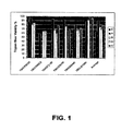
- FIG. 1 Viability of placental stem cells from perfusion (A), amnion (B), chorion (C), or amnion-chorion plate (D); or umbilical cord stem cells (E). Numbers on X-axis designate placenta from which stem cells were obtained.
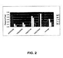
- FIG. 2 Percent HLA ABC - /CD45 - /CD34 - /CD133 + cells from perfusion (A), amnion (B), chorion (C), amnion-chorion plate (D) or umbilical cord (E) as determined by FACSCalibur. Numbers on X-axis designate placenta from which stem cells were obtained.
- FIG. 3 Percent HLA ABC - /CD45 - /CD34 - /CD133 + cells from perfusion (A), amnion (B), chorion (C), amnion-chorion plate (D) or umbilical cord (E), as determined by FACS Aria.. Numbers on X-axis designate placenta from which stem cells were obtained.
- FIG. 4 HLA-G, CD10, CD13, CD33, CD38, CD44, CD90, CD105, CD117, CD200 expression in stem cells derived from placental perfusate.
- FIG. 5 HLA-G, CD10, CD13, CD33, CD38, CD44, CD90, CD105, CD117, CD200 expression in stem cells derived from amnion.
- FIG. 6 HLA-G, CD10, CD13, CD33, CD38, CD44, CD90, CD105, CD117, CD200 expression in stem cells derived from chorion.
- FIG. 7 HLA-G, CD10, CD13, CD33, CD38, CD44, CD90, CD105, CD117, CD200 expression in stem cells derived from amnion-chorion plate.
- FIG. 8 HLA-G, CD10, CD13, CD33, CD38, CD44, CD90, CD105, CD117, CD200 expression in stem cells derived from umbilical cord.
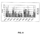
- FIG. 9 Average expression of HLA-G, CD10, CD13, CD33, CD38, CD44, CD90, CD105, CD117, CD200 expression in stem cells derived from perfusion (A), amnion (B), chorion (C), amnion-chorion plate (D) or umbilical cord (E).
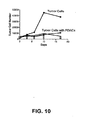
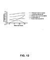
- FIG. 10 Placental stem cells and umbilical cord stem cells inhibit lymphoblastoid cell line (LCL) tumor cell growth.
- LCL lymphoblastoid cell line
- AC amnion-chorion
- AM amniotic membrane
- UC umbilical cord
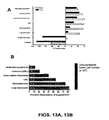
- FIGS. 13A and 13B Suppression and contact dependency of placental stem cell tumor suppression.
- FIG. 14 Highly expressed cytokines in supernatants from the experiments the results of which are shown in FIGS. 13A and 13B .
- IL-6, IL-8 and MCP-1 are shown for LCL and histiocytic lymphoma. Compare FIGS. 15A and 15B .
- FIGS. 15A and 15B Cytokine secretion profile of LCL/placental stem cell co-culture.
- A LCL were cultured either alone or with placental stem cells in open wells (LCL PDAC) or transwells (LCL PDAC TW). Live CD23 + cells were counted on a flow cytometer.
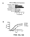
- FIG. 16A and 16B A: Suppression of tumor cell lines by placental stem cells and bone marrow mesenchymal stem cells (BM-MSC). Megakaryocyte leukemia cell line MEG-01, histiocytic lymphoma, retinoblastoma, and chronic myelogenous leukemia cells were incubated either alone or with umbilical cord stem cells (UC), amnion-chorion placental stem cells (AC), or BM-MSCs. After co-culture for six days, the number of live AAD - cells was determined for each co-culture. B: Time course of tumor cell suppression by placental stem cells.
- UC umbilical cord stem cells
- AC amnion-chorion placental stem cells
- B Time course of tumor cell suppression by placental stem cells.
- MEG-01 cells were incubated either alone, or co-cultured with human umbilical vein endothelial cells (HUVEC), BM-MSCs, or placental stem cells (PDAC).
- the number of live (Annexin V - , 7-AAD - ) cells was determined for each culture at 1, 2, 3, 4 and 6 days following initiation of co-culture.
- FIG. 17 Contact dependency of placental stem cell tumor suppression of MEG-01 cells.
- Conditioned media from NEG-01/umbilical cord stem coll co-cultures inhibit megakaryocyte leukemia cell line (MEG-01) tumor cell growth.
- MEG-01 cells were cultured either alone, directly co-cultured with umbilical cord stem cells (MEG/UC), or cultured in conditioned media (split 1:2 or 1:10) harvested from suppressed MEG-01/umbilical cord stem cell co-cultures (UC), MEG-01/bone marrow mesenchymal stem cell co-cultures (BM), or MEG-01/HUVEC co-cultures (H). After six days of co-culture, the number of live cells (Annexin V - , 7-AAD - ) was determined.
- FIG. 18 Cytokine secretion profile in MEG-01/placental stem cell co-culture.
- MEG-01, placental stem cells (PDAC), BM-MSCs, and HUVEC cells were cultured alone, or co-cultured in the following combinations: NMG-01/HUVEC; MEG-01/BM-MSC; or MEG-01/PDAC.
- Supernatants from 7-day cultures were harvested and analyzed on the Luminex for platelet-derived growth factor-AA (PDGF-AA), granulocyte-monocyte colony stimulating factor (GM-CSF), growth-related oncogen-alpha (GRO ⁇ ), and leukemia inhibitory factor (LIF) secretion. Amounts shown are in pg/ml.
- PDGF-AA platelet-derived growth factor-AA
- GM-CSF granulocyte-monocyte colony stimulating factor
- GRO ⁇ growth-related oncogen-alpha
- LIF leukemia inhibitory factor
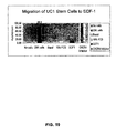
- FIG. 19 Migration of umbilical cord stem cells (UC1) in response to stromal cell-derived factor 1 (SDF-1).
- UC1 placental stem cells were incubated for 24 hours in serum free media only (basal), or in media containing 10% FBS, SDF-1, or SDF-1 plus the CXCR4 inhibitor AMD3100.
- fluorescence was measured with a fluorescence plate reader at 480 nm/520 nm.
- the present invention provides for the suppression of the proliferation of tumor cells, and the suppression of the growth of tumors, using placental stem cells.
- the invention provides a method of suppressing the proliferation of a tumor cell or plurality of tumor cells, or the growth of a tumor, or the proliferation of a non-solid tumor cell or a plurality of non-solid tumor cells, comprising contacting the tumor cell or cells, or tumor, with a plurality of placental stem cells for a time sufficient for said placental stem cells to detectably suppress proliferation of the tumor cell or cells, or growth of the tumor.
- Placental stem cells are, e.g., the placental stem cells described elsewhere herein ( see Section 5.2). Placental stem cells used for tumor cell suppression can be derived or obtained from a single placenta or multiple placentas. Placental stem cells used for tumor cell suppression can also be derived from a single species, e.g., the species of the intended recipient or the species of the tumor cells the function of which is to be reduced or suppressed, or can be derived from multiple species. Placental stem cells can be derived from the whole placenta, or from any portion thereof, for example, the amnion, the chorion, the amnion-chorion plate, or the umbilical cord.
- Placental stem cells derived from any portion of the placenta can be used in the methods of the invention. Placental stem cells can be collected from the placenta, or portion thereof, by any means known to those of skill in the art, e.g. , perfusion or enzymatic digestion.
- a tumor cell can be any cell exhibiting neoplastic cell growth and proliferation, whether malignant or benign, and includes pre-cancerous as well as cancerous cells.
- tumor cells include but are not limited to, carcinoma cells, lymphoma cells, blastorna cells, sarcoma cells, and leukemia cells.
- tumor cells include breast cancer cells, prostate cancer cells, colon cancer cells, squamous cell cancer cells, small-cell lung cancer cells, non-small cell lung cancer cells, gastrointestinal cancer cells, pancreatic cancer cells, glioblastoma, cervical cancer cells, ovarian cancer cells, liver cancer cells, bladder cancer cells, hepatoma cells, colorectal cancer cells, endometrial carcinoma cells, salivary gland carcinoma cells, kidney cancer cells, liver cancer cells, vulval cancer cells, thyroid cancer cells, hepatic carcinoma cells and various types of head and neck cancer cells.
- the tumor cells are megakaryoblastic lymphoma cells, acute lymphoblast leukemia cells, acute T-cell leukemia cells, histiocytic lymphoma cells, bone marrow acute myelogenous leukemia cells, chronic myelogenous leukemia cells, colon adenocarcinoma cells, retinoblastoma cells or lung carcinoma cells.
- the presence of tumor cells in an individual may be determined by performing a biopsy on tissue suspected to be cancerous, or determined from body fluid samples, e.g. , from cells purified or isolated from a blood sample. Cancerous cells or tissues can then be characterized using a variety of biological, molecular, morphological, and cytological means. Specifically, biological and molecular markers can be used to assess characteristics such as the type of cell origin (such as an epithelial cell), specific type of cell (such as organ type like breast or prostate), cell growth or cell growth potential, cell growth arrest, and hyperploidy status. These cellular markers are selected from, but not limited to, molecular, biochemical, and biological markers and probes that are used alone or in combination.
- Contacting in the context of the present invention encompasses bringing the placental stem cells and tumor cells together in vitro, e.g., in a single container ( e.g ., culture dish, flask, vial, etc. ). "Contacting” also encompasses bringing placental stem cells and tumor cells together or in vivo, for example, the same individual ( e.g ., mammal, for example, mouse, rat, dog, cat, sheep, goat, horse, human, etc. ), for example, by providing the placental stem cells to the individual intravenously, by direct injection into the site of a tumor, or the like. In certain embodiments of in vivo contacting, said placental stem cells and said tumor cells are cells in cell culture.
- said cells are co-cultured in the same physical space, e.g., in the same culture dish or well in a culture dish.
- said contacting does not require direct physical contact between said placental stem cells and said tumor cells.
- said contacting can comprise culturing said placental stem cells and said tumor cells in separate physical spaces, e.g. , separate wells in a cell culture dish, wherein the medium in which said placental stem cells and said tumor cells is shared between the placental stem cells and tumor cells.
- both the placental stem cells and the tumor cells are exogenous to the individual, that is, neither type of cell originated within the individual.
- the tumor cells are tumor cells that arose within the individual through tumorigenesis, i.e ., the tumor cells are endogenous to the individual.
- the contacting (either in vitro or in vivo ) is for a time sufficient, and with a number of placental stem cells sufficient, to cause a detectable suppression of the proliferation of the tumor cell or tumor cells, for a period of time after said contacting. More preferably, in various embodiments, said contacting is sufficient to suppress proliferation of a tumor cell or tumor cells by at least 50%, 60%, 70%, 75%, 80%, 90% or 95%, compared to the immune function in the absence of the placental stem cells, for a period of time after said contacting.
- the period of time is 1, 2, 3, 4, 5, 6 or 7 days, or 1, 2, 3, 4, 5, 6, 7, 8, 9, 10 weeks or more.
- a plurality of placental stem cells is contacted with a plurality of tumor cells, e.g., tumor cells in an individual, wherein the plurality of placental stem cells comprises about 1 x 10 5 placental stem cells, about 1 x 10 6 placental stem cells, about 1 x 10 7 placental stem cells, about 1 x 10 8 placental stem cells, about 1 x 10 9 placental stem cells, about 1 x 10 10 placental stem cells, about 1 x 10 11 placental stem cells, about 1 x 10 12 placental stem cells or more.
- the method comprises administering at least about 1 x 10 5 , at least about 1 x 10 6 , at least about 1 x 10 7 , or at least about 1 x 10 8 placental stem cells to said individual per kilogram of the individual's body weight.
- about 1 million placental stem cells is administered to an individual comprising a plurality of tumor cells, per kilogram of the individual's body weight.
- the method comprises administering a number of placental stem cells about one time, two times, three times, four times, five times, or more than five times the number of tumor cells in an individual.
- Any art known method may be used to determine the number of tumor cells in an individual. Exemplary methods of tumor cell quantification are described in U.S. Patent Nos. 6,365,362 and 6,645,731 ; by Méhes et al., Haematologia 31(2):97-109 (2001 ); and Hardingham et al., Cancer Research 53:3455-3458 (1993 ), the contents of which are hereby incorporated by reference in their entireties.
- the method comprises administering a number of placental stem cells based on the weight of the individual.
- the method comprises administering about 1 x 10 3 placental stem cells / kg, 5 x 10 3 placental stem cells / kg, 1 x 10 4 placental stem cells / kg, 5 x 10 4 placental stem cells / kg, 1 x 10 5 placental stem cells / kg, 5 x 10 5 placental stem cells / kg, / x 10 6 placental stem cells / kg, 5 x 10 6 placental stem cells / kg, 1 x 10 7 placental stem cells / kg, 5 x 10 7 placental stem cells / kg, or 1 x 10 8 placental stem cells / kg to said individual.
- the method comprises administering at least about 1 x 10 3 placental stem cells / kg, at least about 5 x 10 3 placental stem cells / kg, at least about 1 x 10 4 placental stem cells / kg, at least about 5 x 10 4 placental stem cells / kg, at least about 1 x 10 5 placental stem cells / kg, at least about 5 x 10 5 placental stem cells / kg, at least about 1 x 10 6 placental stem cells / kg, at least about 5 x 10 6 placental stem cells / kg, at least about 1 x 10 7 placental stem cells / kg, at least about 5 x 10 7 placental stem cells / kg, or at least about 1 x 10 8 placental stem cells / kg to said individual.
- said placental stem cells have been proliferated in vitro for no more than 30 population doublings, no more than 20 population doublings, no more than 10 population doublings, or no more than 5 population doublings.
- said placental stem cells have been cryopreserved and thawed prior to said contacting.
- said placental stem cells suppress said tumor cell proliferation by about 20%, 30%, 40%, 50%, 60%, 70%, 75%, 80%, 90%, or 95%, compared to proliferation of an equivalent number of tumor cells in the absence of said placental stem cells.
- the placental stem cells e.g., placental stem cells from a particular individual or pool of individuals, from particular tissues, or the like, are screened for tumor-suppressive activity prior to use, e.g., to suppress tumor cell growth or proliferation in an individual.
- the method of suppressing tumor cell proliferation or growth using placental stem cells comprises screening said placental stem cells in vitro for tumor cell growth suppressive activity prior to administration of said placental stem cells to said individual.
- said placental stem cells are confirmed to suppress tumor cell proliferation in vitro by, e.g., at least about 20%, 30%, 40%, 50%, 60%, 70%, 75%, 80%, 90%, or 95%, compared to proliferation of an equivalent number of tumor cells in the absence of said placental stem cells, prior to administration to said individual, where the proliferation is measured by the number of cells produced under equivalent conditions over a period of time.
- the placental stem cells can suppress tumor cells by direct contact, through soluble factors, or both.
- said placental stem cells are confirmed to suppress tumor cell proliferation in vitro in a direct culture assay, in a transwell assay, or more preferably, in both a direct culture assay and a transwell assay, prior to administration to said individual.
- the placental stem cells can be screened for suppression of tumor cell proliferation or growth using any tumor cells, but more useful screens are those that replicate, or attempt to replicate, tumor suppression within an affected individual.
- said placental stem cells are screened in vitro for tumor growth suppressive activity against a tumor cell of the same cell type, e.g . epithelial, squamous, etc., the same tissue of origin, e.g. breast, prostate, etc., or more preferably, both the same cell type and tissue of origin as a tumor cell in the individual to be administered said placental stem cells.
- said placental stem cells are screened in vitro for tumor growth suppressive activity against tumor cells obtained from a tumor cell biopsy from said individual, or tumor cells purified or isolated from a blood sample of said individual.
- said placental stem cells are from amnion, chorion, amnion-chorion, or umbilical cord, or from placental perfusate, and are confirmed to suppress tumor cell proliferation in vitro by, e.g., at least about 20%, 30%, 40%, 50%, 60%, 70%, 75%, 80%, 90%, or 95%, compared to proliferation of an equivalent number of tumor cells in the absence of said stem cells, prior to administration to said individual.
- the placental stem cells can be introduced into the individual in any manner known to those of skill in the art to be effective at introducing live cells to an individual.
- the placental stem cells can be introduced into the individual by intravenous transfusion, or can be introduced intramuscularly, intraperitoneally, intradermally, and the like.
- the placental stem cells are injected into the individual into, at the site of, or at the periphery of the tumor or tumor cells.
- Cells can also be introduced by the transplantation of, e.g., a natural or man-made matrix, e.g ., gelatin, in which the placental stem cells are enmeshed and out of which the cells can grow once transplanted.
- a natural or man-made matrix e.g ., gelatin
- Non-limiting examples of such matrices are provided in Section 5.6.1.4, below.
- Introduction of the placental stem cells into an individual, particularly an individual having endogenous tumor cells can comprise a single introduction, or multiple introductions over the course of several hours, several days, several weeks, several months, or several years.
- Each introduction of placental stem cells can comprise a number of stem cells sufficient, in and of itself, to detectably suppress proliferation of a plurality of tumor cells, or can be sufficient in the aggregate.
- the placental stem cells can be formulated as a pharmaceutical composition, as described in Section 5.6.1, below.
- the degree of suppression in an in vivo context can be determined in an in vitro assay, for example, by comparing the number of tumor cells produced by a tumor cell or plurality of tumor cells under optimal growth conditions for a period of time compared to a number of tumor cells produced by an equivalent number of tumor cells in contact with placental stem cells for the same amount of time.
- Proliferation of cells, including tumor cells can be assessed by any art-known method. For example, cells in culture or in an individual can be sampled at various time points and counted with a hemacytometer or similar device.
- the tumor cells can be stained with a non-degrading dye designed to be segregated into daughter cells, e.g., staining with bromodeoxyuridine (BrDU), carboxyfluorescein diacetate (CFSE) or Oregon Green 488 carboxylic acid diacetate (Invitrogen), and the degree of staining determined with a cytometer.
- Placental stem cells suppress tumor cell growth where the tumor cells, in contact with placental stem cells, show a detectably lower amount of the stain per cell (e.g., a detectably lower average amount of stein per cell) than tumor cells not contacted with placental stem cells.
- the degree of suppression in an in vitro assay can be extrapolated, for a particular number of placental stem cells and a number of tumor cells, to a degree of tumor or tumor cell suppression in an individual.
- Suppression of the growth of a tumor can be assessed by any means known in the art for imaging or detecting tumors in vivo.
- the cells of the tumor can be labeled with a tumor-specific antibody and imaged using, e.g., a PET scan or CAT scan, or can be imaged using X-rays.
- a determination of suppression of tumor growth can be ascertained, e.g., by visual inspection of an image of the tumor, by determining the intensity of labeling of the tumor, by determining the area of the tumor in an image of the tumor, etc.
- a determination of suppression of growth of a tumor in vivo can also be made by detecting or noting any elimination of, improvement in, or lessening of worsening of a symptom related to the tumor.
- the individual can be a mammal, e.g., a human.
- said contacting comprises administering said placental cells to said individual intravenously.
- said contacting comprises administering said placental cells to said individual at or adjacent to the site of a tumor.
- the placental stem cells can also be administered with one or more second types of stem cells, e.g., mesenchymal stem cells from bone marrow.
- Such second stem cells can be administered to an individual with placental stem cells in a ratio of, e.g., about 1:10 to about 10:1.
- the placental stem cells can also be administered with one or more types of cells that are not stem cells.
- the placental stem cells are administered to an individual along with a second plurality of cells that are autologous to the individual.
- the placental stem cells are co-administered with fibroblasts.
- the fibroblasts are autologous fibroblasts.
- the fibroblasts can be administered to an individual with placental stem cells in a ratio of, e.g. about 1:10 to about 10:1.
- the placental stem cells can also be administered with one or more stem cell chemoattractants.
- the stem cell chemoattractant is SDF-1.
- placental stem cells that is, stem cells obtainable from a placenta or part thereof, that (1) adhere to a tissue culture substrate; (2) have the capacity to differentiate into non-placental cell types; and (3) have, in sufficient numbers, the capacity to detectably suppress the proliferation of a tumor cell or plurality of tumor cells, or detectably suppress the growth of a tumor.
- Placental stem cells are not derived from blood, e.g., placental blood or umbilical cord blood.
- the placental stem cells used in the methods and compositions of the present invention have the capacity, and are selected for their capacity, to suppress proliferation of a cancer cell or plurality of cancer cells in vitro or in vivo, or to suppress growth of a tumor in vivo.
- Placental stem cells can be either fetal or maternal in origin (that is, can have the genotype of either the mother or fetus).
- Populations of placental stem cells, or populations of cells comprising placental stem cells can comprise placental stem cells that are solely fetal or maternal in origin, or can comprise a mixed population of placental stem cells of both fetal and maternal origin.
- the placental stem cells, and populations of cells comprising the placental stem cells can be identified and selected by the morphological, marker, and culture characteristics discussed below.
- placental stem cells used in the present invention when cultured in primary cultures or in cell culture, adhere to the tissue culture substrate, e.g., tissue culture container surface ( e.g ., tissue culture plastic).
- tissue culture substrate e.g., tissue culture container surface (e.g ., tissue culture plastic).
- Placental stem cells in culture assume a generally fibroblastoid, stellate appearance, with a number of cytoplasmic processes extending from the central cell body.
- the placental stem cells are, however, morphologically differentiable from fibroblasts cultured under the same conditions, as the placental stem cells exhibit a greater number of such processes than do fibroblasts. Morphologically, placental stem cells are also differentiable from hematopoietic stem cells, which generally assume a more rounded, or cobblestone, morphology in culture.
- the placental stem cells, and stem cell populations of the invention include stem cells and stem cell-containing cell populations obtained directly from the placenta, or any part thereof ( e.g ., amnion, chorion, amnion-chorion plate, placental cotyledons, umbilical cord, and the like).
- Placental stem cell populations also includes populations of (that is, two or more) placental stem cells in culture, and a population in a container, e.g ., a bag. Placental stem cells are not, however, trophoblasts.
- Placental stem cells generally express the markers CD73, CD105, CD200, HLA-G, and/or OCT-4, and do not express CD34, CD38, or CD45.
- Placental stem cells can also express HLA-ABC (MHC-1) and HLA-DR. These markers can be used to identify placental stem cells, and to distinguish placental stem cells from other stem cell types. Because the placental stem cells can express CD73 and CD105, they can have mesenchymal stem cell-like characteristics.
- the placental stem cells can express CD200 and HLA-G, a fetal-specific marker, they can be distinguished from mesenchymal stem cells, e.g ., bone marrow-derived mesenchymal stem cells, which express neither CD200 nor HLA-G.
- mesenchymal stem cells e.g ., bone marrow-derived mesenchymal stem cells, which express neither CD200 nor HLA-G.
- the lack of expression of CD34, CD38 and/or CD45 identifies the placental stem cells as non-hematopoietic stem cells.
- the invention provides an isolated cell population comprising a plurality of placental stem cells that are CD200 + , HLA-G + , wherein said stem cells detectably suppress cancer cell proliferation or tumor growth.
- said stem cells are also CD73 + and CD105 + .
- said stem cells are also CD34 - , CD38 - or CD45 - .
- said stem cells are also CD34 - , CD38 - , CD45 - , CD73 + and CD105 + .
- said isolated population produces one or more embryoid-like bodies when cultured under conditions that allow the formation of embryoid-like bodies.
- the invention provides an isolated cell population comprising a plurality of placental stem cells that are CD73 + , CD105 + , CD200 + , wherein said stem cells detectably suppress cancer cell proliferation or tumor growth.
- said stem cells are HLA-G + .
- said stem cells are CD34 - , CD38 - or CD45 - .
- said stem cells are CD34 - , CD38 - and CD45 - .
- said stem cells are CD34 - , CD38 - , CD45 - , and HLA-G + .
- said population of cells produces one or more embryoid-like bodies when cultured under conditions that allow the formation of embryoid-like bodies.
- the invention also provides an isolated cell population comprising a plurality of placental stem cells that are CD200 + , OCT-4 + , wherein said stem cells detectably suppress cancer cell proliferation or tumor growth.
- said stem cells are CD73 + and CD105 + .
- said stem cells are HLA-G + .
- said stem cells are CD34 - , GD38 - and CD45 - .
- said stem cells are CD34 - , CD38 - , CD45 - , CD73 + , CD105 + and HLA-G + .
- the population produces one or more embryoid-like bodies when cultured under conditions that allow the formation of embryoid-like bodies.
- the invention also provides an isolated cell population comprising a plurality of placental stem cells that are CD73 + , CD105 + and HLA-G + , wherein said stem cells detectably suppress T cell proliferation in a mixed lymphocyte reaction (MLR) assay.
- said stem cells are also CD34 - , CD38 - or CD45 - .
- said stem cells are also CD34 - , CD38 - and CD45 - .
- said stem cells are also OCT-4 + .
- said stem cells are also CD200 + .
- said stem cells are also CD34 - , CD38 - , CD45 - , OCT-4 + and CD200 + .
- the invention also provides an isolated cell population comprising a plurality of tumor cell suppressive placental stem cells that are CD73 + , CD105 + stem cells, wherein said plurality forms one or more embryoid-like bodies under conditions that allow formation of embryoid-like bodies, and wherein said stem cells detectably suppress cancer cell proliferation or tumor growth.
- said stem cells are also CD34 - , CD38 - or CD45 - .
- said stem cells are also CD34 - , CD38 - and CD45 - .
- said stem cells are also OCT-4 + .
- said stem cells are also OCT-4 + , CD34 - , CD38 - and CD45 - .
- the invention also provides an isolated cell population comprising a plurality of placental stem cells that are OCT-4 + stem cells, wherein said population forms one or more embryoid-like bodies when cultured under conditions that allow the formation of embryoid-like bodies, and wherein said stem cells have been identified as detectably suppressing cancer cell proliferation or tumor growth.
- At least 10%, at least 20%, at least 30%, at least 40%, at least 50% at least 60%, at least 70%, at least 80%, at least 90%, or at least 95% of said isolated placental cells are OCT4 + stem cells.
- said stem cells are CD73 + and CD105 + .
- said stem cells are CD34 - , CD38 - , or CD45 - .
- said stem cells are CD200 + .
- said stem cells are CD73 + , CD105 + , CD200 + , CD34 - , CD38 - , and CD45 - .
- said population has been expanded, for example, passaged at least once, at least three times, at least five times, at least 10 times, at least 15 times, or at least 20 times.
- the method can additionally comprise selecting placental cells that express ABC-p (a placenta-specific ABC transporter protein; see, e.g., Allikmets et al., Cancer Res. 58(23):5337-9 (1998 )).
- the method can also comprise selecting cells exhibiting at least one characteristic specific to, e.g ., a mesenchymal stem cell, for example, expression of CD29, expression of CD44, expression of CD90, or expression of a combination of the foregoing.
- the invention provides an isolated cell population comprising a plurality of tumor cell-suppressive placental stem cells that are CD29 + , CD44 + , CD73 + , CD90 + , CD105 + , CD200 + , CD34- and CD133 - .
- the placental stem cells constitutively secrete IL-6, IL-8 and monocyte chemoattractant protein (MCP-1).
- Each of the above-referenced pluralities of placental stem cells can comprise placental stem cells obtained and isolated directly from a mammalian placenta, or placental stem cells that have been cultured and passaged at least 1, 2, 3, 4, 5, 6, 7, 8, 9, 10, 12, 14, 16, 18, 20, 25, 30 or more times, or a combination thereof.
- the tumor cell suppressive pluralities of placental stem cells described above can comprise about, at least, or no more than, 1 x 10 5 , 5 x 10 5 , 1 x 10 6 , 5 x 10 6 , 1 x 10 7 , 5 x 10 7 , 1 x 10 8 , 5 x 10 8 , 1 x 10 9 , 5 x 10 9 , 1 x 10 10 , 5 x 10 10 , 1 x 10 11 or more placental stem cells.
- the invention also provides a method of selecting a plurality of placental stem cells from a plurality of placental cells, comprising selecting a population of placental cells wherein at least 10%, at least 20%, at least 30%, at least 40%, at least 50% at least 60%, at least 70%, at least 80%, at least 90%, or at least 95% of said cells are CD200 + , HLA-G + placental stem cells, and wherein said placental stem cells detectably suppress cancer cell proliferation or tumor growth.
- said selecting comprises selecting stem cells that are also CD73 + and CD105 + .
- said selecting comprises selecting stem cells that are also CD34 - , CD38 - or CD45 - .
- said selecting comprises selecting placental stem cells that are also CD34 - , CD38 - , CD45 - , CD73 + and CD105 + .
- said selecting also comprises selecting a plurality of placental stem cells that forms one or more embryoid-like bodies when cultured under conditions that allow the formation of embryoid-like bodies.
- the invention also provides a method of selecting a plurality of placental stem cells from a plurality of placental cells, comprising selecting a plurality of placental cells wherein at least 10%, at least 20%, at least 30%, at least 40%, at least 50% at least 60%, at least 70%, at least 80%, at least 90%, or at least 95% of said cells are CD73 + , CD105 + , CD200 + placental stem cells, and wherein said placental stem cells detectably suppress cancer cell proliferation or tumor growth.
- said selecting comprises selecting stem cells that are also HLA-G + .
- said selecting comprises selecting placental stem cells that are also CD34 - , CD38 - or CD45 - .
- said selecting comprises selecting placental stem cells that are also CD34', CD38 - and CD45 - . In another specific embodiment, said selecting comprises selecting placental stem cells that are also CD34 - , CD38 - , CD45 - , and HLA-G + . In another specific embodiment, said selecting additionally comprises selecting a population of placental cells that produces one or more embryoid-like bodies when the population is cultured under conditions that allow the formation of embryoid-like bodies.
- the invention also provides a method of selecting a plurality of placental stem cells from a plurality of placental cells, comprising selecting a plurality of placental cells wherein at least 10%, at least 20%, at least 30%, at least 40%, at least 50% at least 60%, at least 70%, at least 80%, at least 90%, or at least 95% of said cells are CD200 + , OCT-4 + placental stem cells, and wherein said placental stem cells detectably suppress cancer cell proliferation or tumor growth.
- said selecting comprises selecting placental stem cells that are also CD73 + and CD105 + .
- said selecting comprises selecting placental stem cells that are also HLA-G + .
- said selecting comprises selecting placental stem cells that are also CD34 - , CD38 - and CD45 - . In another specific embodiment, said selecting comprises selecting placental stem cells that are also CD34 - , CD38 - , CD45 - , CD73 + , CD105 + and HLA-G + .
- the invention also provides a method of selecting a plurality of placental stem cells from a plurality of placental cells, comprising selecting a plurality of placental cells wherein at least 10%, at least 20%, at least 30%, at least 40%, at least 50% at least 60%, at least 70%, at least 80%, at least 90%, or at least 95% of said cells are CD73 + , CD 105 + and HLA-G + placental stem cells, and wherein said placental stem cells detectably suppresses T cell proliferation in a mixed lymphocyte reaction (MLR) assay.
- said selecting comprises selecting placental stem cells that are also CD34 - , CD38 - or CD45 - .
- said selecting comprises selecting placental stem cells that are also CD34 - , CD38 - and CD45 - . In another specific embodiment, said selecting comprises selecting placental stem cells that are also CD200 + . In another specific embodiment, said selecting comprises selecting placental stem cells that are also CD34 - , CD38 - , CD45 - , OCT-4 + and CD200 + .
- the invention also provides a method of selecting a plurality of placental stem cells from a plurality of placental cells, comprising selecting a plurality of placental cells wherein at least 10%, at least 20%, at least 30%, at least 40%, at least 50% at least 60%, at least 70%, at least 80%, at least 90%, or at least 95% of said cells are CD73 + , CD105 + placental stem cells, and wherein said plurality forms one or more embryoid-like bodies under conditions that allow formation of embryoid-like bodies, and wherein said stem cells detectably suppress cancer cell proliferation or tumor growth.
- said selecting comprises selecting placental stem cells that are also CD34 - , CD38 - or CD45 - .
- said selecting comprises selecting placental stem cells that are also CD34 - , CD38 - and CD45 - . In another specific embodiment, said selecting comprises selecting placental stem cells that are also OCT-4 + . In a more specific embodiment, said selecting comprises selecting placental stem cells that are also OCT-4 + , CD34 - , CD38 - and CD45 - .
- the invention also provides a method of selecting a plurality of placental stem cells from a plurality of placental cells, comprising selecting a plurality of placental cells wherein at least 10%, at least 20%, at least 30%, at least 40%, at least 50% at least 60%, at least 70%, at least 80%, at least 90%, or at least 95% of said isolated placental cells are OCT4 + stem cells, and wherein said plurality forms one or more embryoid-like bodies under conditions that allow formation of embryoid-like bodies, and wherein said stem cells detectably suppress cancer cell proliferation or tumor growth.
- said selecting comprises selecting placental stem cells that are also CD73 + and CD105 + .
- said selecting comprises selecting placental stem cells that are also CD34 - , CD38 - , or CD45 - . In another specific embodiment, said selecting comprises selecting placental stem cells that are also CD200 + . In a more specific embodiment, said selecting comprises selecting placental stem cells that are also CD73 + , CD105 + , CD200 + , CD34 - , CD38 - , and CD45 - .
- the invention also provides methods of producing populations, of placental stem cells that can suppress the proliferation of tumor cells.
- the invention provides a method of producing a cell population, comprising selecting any of the pluralities of placental stem cells described above, and isolating the plurality of placental stem cells from other cells, e.g., other placental cells.
- the invention provides a method of producing a cell population comprising selecting placental cells, wherein said placental cells (a) adhere to a substrate, (b) express CD200 and HLA-G, or express CD73, CD105, and CD200, or express CD200 and OCT-4, or express CD73, CD105, and HLA-G, or express CD73 and CD105, and facilitate the formation of one or more embryoid-like bodies in a population of placental cells that comprise the stem cell, when said population is cultured under conditions that allow formation of embryoid-like bodies, or express OCT-4, and facilitate the formation of one or more embryoid-like bodies in a population of placental cells that comprise the stem cell, when said population is cultured under conditions that allow formation of embryoid-like bodies; and (c) detectably suppress cancer cell proliferation or tumor growth; and isolating said placental cells from other cells to form a cell population.
- the invention provides a method of producing a cell population comprising selecting placental stem cells that (a) adhere to a substrate, (b) express CD200 and HLA-G, and (c) detectably suppress cancer cell proliferation or tumor growth; and isolating said placental stem cells from other cells to form a cell population.
- the invention provides a method of producing a cell population comprising selecting placental stem cells that (a) adhere to a substrate, (b) express CD73, CD105, and CD200, and (c) detectably suppress cancer cell proliferation or tumor growth; and isolating said placental stem cells from other cells to form a cell population.
- the invention provides a method of producing a cell population comprising selecting placental stem cells that (a) adhere to a substrate, (b) express CD200 and OCT-4, and (c) detectably suppress cancer cell proliferation or tumor growth; and isolating said placental stem cells from other cells to form a cell population.
- the invention provides a method of producing a cell population comprising selecting placental stem cells that (a) adhere to a substrate, (b) express CD73 and CD105, (c) form embryoid-like bodies when cultured under conditions allowing the formation of embryoid-like bodies, and (d) detectably suppress cancer cell proliferation or tumor growth; and isolating said placental stem cells from other cells to form a cell population.
- the invention provides a method of producing a cell population comprising selecting placental stem cells that (a) adhere to a substrate, (b) express CD73, CD105, and HLA-G, and (c) detectably suppress CD4+ or CD8+ T cell proliferation in an MLR; and isolating said placental stem cells from other cells to form a cell population.
- a method of producing a cell population comprising selecting placental stem cells that (a) adhere to a substrate, (b) express OCT-4, (c) form embryoid-like bodies when cultured under conditions allowing the formation of embryoid-like bodies, and (d) detectably suppress cancer cell proliferation or tumor growth; and isolating said placental stem cells from other cells to form a cell population.
- the selection can comprise determining whether a sample of said placental stem cells suppresses cancer cell proliferation, or suppresses the growth of a tumor, and selecting the population of placental stem cells if the sample of placental stem cells detectably suppresses cancer cell proliferation or tumor growth.
- placental stem cells The growth of the placental stem cells described herein, as for any mammalian cell, depends in part upon the particular medium selected for growth. Under optimum conditions, placental stem cells typically double in number in 3-5 days.
- the placental stem cells of the invention adhere to a substrate in culture, e.g. the surface of a tissue culture container (e.g ., tissue culture dish plastic, fibronectin-coated plastic, and the like) and form a monolayer.
- tissue culture container e.g ., tissue culture dish plastic, fibronectin-coated plastic, and the like
- embryoid-like bodies that is, three-dimensional clusters of cells that grow atop the adherent stem cell layer.
- Cells within the embryoid-like bodies express markers associated with very early stem cells, e.g ., OCT-4, Nanog, SSEA3 and SSEA4.
- Cells within the embryoid-like bodies are typically not adherent to the culture substrate, as are the placental stem cells described herein, but remain attached to the adherent cells during culture.
- Embryoid-like body cells are dependent upon the adherent placental stem cells for viability, as embryoid-like bodies do not form in the absence of the adherent stem cells.
- the adherent placental stem cells thus facilitate the growth of one or more embryoid-like bodies in a population of placental cells that comprise the adherent placental stem cells.
- the cells of the embryoid-like bodies are thought to grow on the adherent placental stem cells much as embryonic stem cells grow on a feeder layer of cells.
- Mesenchymal stem cells e.g ., bone marrow-derived mesenchymal stem cells, do not develop embryoid-like bodies in culture.
- the present invention further provides methods of collecting and isolating placental stem cells.
- stem cells are obtained from a mammalian placenta using a physiologically-acceptable solution, e.g ., a stem cell collection composition.
- a stem cell collection composition is described in detail in related U.S. Provisional Application No. 60/754,969 , entitled "Improved Composition for Collecting and Preserving Placental Stem Cells and Methods of Using the Composition" filed on December 29, 2005.
- the stem cell collection composition can comprise any physiologically-acceptable solution suitable for the collection and/or culture of stem cells, for example, a saline solution (e.g ., phosphate-buffered saline, Kreb's solution, modified Kreb's solution, Eagle's solution, 0.9% NaCl. etc. ), a culture medium (e.g ., DMEM, H.DMEM, etc. ), and the like.
- a saline solution e.g ., phosphate-buffered saline, Kreb's solution, modified Kreb's solution, Eagle's solution, 0.9% NaCl. etc.
- a culture medium e.g ., DMEM, H.DMEM, etc.
- the stem cell collection composition can comprise one or more components that tend to preserve placental stem cells, that is, prevent the placental stem cells from dying, or delay the death of the placental stem cells, reduce the number of placental stem cells in a population of cells that die, or the like, from the time of collection to the time of culturing.
- Such components can be, e.g ., an apoptosis inhibitor (e.g ., a caspase inhibitor or JNK inhibitor); a vasodilator (e.g ., magnesium sulfate, an antihypertensive drug, atrial natriuretic peptide (ANP), adrenocorticotropin, corticotropin-releasing hormone, sodium nitroprusside, hydralazine, adenosine triphosphate, adenosine, indomethacin or magnesium sulfate, a phosphodiesterase inhibitor, etc .); a necrosis inhibitor (e.g ., 2-(1H-Indol-3-yl)-3-pentylamino-maleimide, pyrrolidine dithiocarbamate, or clonazepam); a TNF- ⁇ inhibitor; and/or an oxygen-carrying perfluorocarbon (e.g ., perfluorocarbon
- the stem cell collection composition can comprise one or more tissue-degrading enzymes, e.g., a metalloprotease, a serine protease, a neutral protease, an RNase, or a DNase, or the like.
- tissue-degrading enzymes include, but are not limited to, collagenases (e.g ., collagenase I, II, III or IV, a collagenase from Clostridium histolyticum, etc.); dispase, thermolysin, elastase, trypsin, LIBERASE, hyaluronidase, and the like.
- the stem cell collection composition can comprise a bacteriocidally or bacteriostatically effective amount of an antibiotic.
- the antibiotic is a macrolide (e.g ., tobramycin), a cephalosporin (e.g ., cephalexin, cephradine, cefuroxime, cefprozil, cefaclor, cefixime or cefadroxil), a clarithromycin, an erythromycin, a penicillin (e.g ., penicillin V) or a quinolone ( e.g ., ofloxacin, ciprofloxacin or norfloxacin), a tetracycline, a streptomycin, etc.
- the antibiotic is active against Gram(+) and/or Gram(-) bacteria, e.g., Pseudomonas aeruginosa, Staphylacoccus aureus, and the like.
- the stem cell collection composition can also comprise one or more of the following compounds: adenosine (about 1 mM to about 50 mM); D-glucose (about 20 mM to about 100 mM); magnesium ions (about 1 mM to about 50 mM); a macromolecule of molecular weight greater than 20,000 daltons, in one embodiment, present in an amount sufficient to maintain endothelial integrity and cellular viability (e.g ., a synthetic or naturally occurring colloid, a polysaccharide such as dextran or a polyethylene glycol present at about 25 g/l to about 100 g/l, or about 40 g/l to about 60 g/l); an antioxidant ( e.g ., butylated hydroxyanisole, butylated hydroxytoluene, glutathione, vitamin C or vitamin E present at about 25 ⁇ M to about 100 ⁇ M); a reducing agent (e.g ., N-acetylcysteine present
- a human placenta is recovered shortly after its expulsion after birth.
- the placenta is recovered from a patient after informed consent and after a complete medical history of the patient is taken and is associated with the placenta.
- the medical history continues after delivery.
- Such a medical history can be used to coordinate subsequent use of the placenta or the stem cells harvested therefrom.
- human placental stem cells can be used, in light of the medical history, for personalized medicine for the infant associated with the placenta, or for parents, siblings or other relatives of the infant.
- the umbilical cord blood and placental blood Prior to recovery of placental stem cells, the umbilical cord blood and placental blood are removed. In certain embodiments, after delivery, the cord blood in the placenta is recovered.
- the placenta can be subjected to a conventional cord blood recovery process.
- a needle or cannula is used, with the aid of gravity, to exsanguinate the placenta (see, e.g., Anderson, U.S. Patent No. 5,372,381 ; Hessel et al., U.S. Patent No. 5,415,665 ).
- the needle or cannula is usually placed in the umbilical vein and the placenta can be gently massaged to aid in draining cord blood from the placenta.
- cord blood recovery may be performed commercially, e.g ., LifeBank Inc., Cedar Knolls, N.J., ViaCord, Cord Blood Registry and Cryocell.
- the placenta is gravity drained without further manipulation so as to minimize tissue disruption during cord blood recovery.
- a placenta is transported from the delivery or birthing room to another location, e.g., a laboratory, for recovery of cord blood and collection of stem cells by, e.g., perfusion or tissue dissociation.
- the placenta is preferably transported in a sterile, thermally insulated transport device (maintaining the temperature of the placenta between 20-28°C), for example, by placing the placenta, with clamped proximal umbilical cord, in a sterile zip-lock plastic bag, which is then placed in an insulated container.
- the placenta is transported in a cord blood collection kit substantially as described in pending United States patent application no. 11/230,760, filed September 19,2005 .
- the placenta is delivered to the laboratory four to twenty-four hours following delivery.
- the proximal umbilical cord is clamped, preferably within 4-5 cm (centimeter) of the insertion into the placental disc prior to cord blood recovery.
- the proximal umbilical cord is clamped after cord blood recovery but prior to further processing of the placenta.
- the placenta prior to stem cell collection, can be stored under sterile conditions and at either room temperature or at a temperature of 5 to 25°C (centigrade).
- the placenta may be stored for a period of longer than forty eight hours, and preferably for a period of four to twenty-four hours prior to perfusing the placenta to remove any residual cord blood.
- the placenta is preferably stored in an anticoagulant solution at a temperature of 5 to 25°C (centigrade).
- Suitable anticoagulant solutions are well known in the art.
- a solution of heparin or warfarin sodium can be used.
- the anticoagulant solution comprises a solution of heparin ( e.g. , 1% w/w in 1:1000 solution).
- the exsanguinated placenta is preferably stored for no more than 36 hours before placental stem cells are collected.
- the mammalian placenta or a part thereof, once collected and prepared generally as above, can be treated in any art-known manner, e.g., can be perfused or disrupted, e.g., digested with one or more tissue-disrupting enzymes, to obtain stem cells.
- stem cells are collected from a mammalian placenta by physical disruption, e.g., enzymatic digestion, of the organ.
- the placenta, or a portion thereof may be, e.g., crushed, sheared, minced, diced, chopped, macerated or the like, while in contact with the stem cell collection composition of the invention, and the tissue subsequently digested with one or more enzymes.
- the placenta, or a portion thereof may also be physically disrupted and digested with one or more enzymes, and the resulting material then immersed in, or mixed into, the stem cell collection composition of the invention.
- any method of physical disruption can be used, provided that the method of disruption leaves a plurality, more preferably a majority, and more preferably at least 60%, 70%, 80%, 90%, 95%, 98%, or 99% of the cells in said organ viable, as determined by, e.g., trypan blue exclusion.
- placenta can be dissected into components prior to physical disruption and/or enzymatic digestion and stem cell recovery.
- placental stem cells can be obtained from the amniotic membrane, chorion, placental cotyledons, or any combination thereof.
- Umbilical cord stem cells can also be used in the methods of the invention.
- placental stem cells are obtained from placental tissue comprising amnion and chorion.
- placental stem cells are obtained from umbilical cord.
- placental stem cells can be obtained by disruption of a small block of placental tissue, e.g., a block of placental tissue that is about 1, 2, 3, 4, 5, 6, 7, 8, 9, 10, 20, 30, 40, 50, 60, 70, 80, 90, 100, 200, 300, 400, 500, 600, 700, 800, 900 or about 1000 cubic millimeters in volume.
- a block of placental tissue e.g., a block of placental tissue that is about 1, 2, 3, 4, 5, 6, 7, 8, 9, 10, 20, 30, 40, 50, 60, 70, 80, 90, 100, 200, 300, 400, 500, 600, 700, 800, 900 or about 1000 cubic millimeters in volume.
- a preferred stem cell collection composition comprises one or more tissue-disruptive enzyme(s).
- Enzymatic digestion preferably uses a combination of enzymes, e.g., a combination of a matrix metalloprotease and a neutral protease, for example, a combination of collagenase and dispase.
- enzymatic digestion of placental tissue uses a combination of a matrix metalloprotease, a neutral protease, and a mucolytic enzyme for digestion of hyaluronic acid, such as a combination of collagenase, dispase, and hyaluronidase or a combination of LIBERASE (Boehringer Mannheim Corp., Indianapolis, Ind.) and hyaluronidase.
- enzymes that can be used to disrupt placenta tissue include papain, deoxyribonucleases, serine proteases, such as trypsin, chymotrypsin, or elastase.
- Serine proteases may be inhibited by alpha 2 microglobulin in serum and therefore the medium used for digestion is usually serum-free.
- EDTA and DNase are commonly used in enzyme digestion procedures to increase the efficiency of cell recovery.
- the digestate is preferably diluted so as to avoid trapping stem cells within the viscous digest.
- tissue digestion enzymes can be used. Typical concentrations for tissue digestion enzymes include, e.g., 50-200 U/mL for collagenase I and collagenase IV, 1-10 U/mL for dispase, and 14-100 U/mL for elastase.
- Proteases can be used in combination, that is, two or more proteases in the same digestion reaction, or can be used sequentially in order to liberate placental stem cells. For example, in one embodiment, a placenta, or part thereof, is digested first with an appropriate amount of collagenase I at 2 mg/ml for 30 minutes, followed by digestion with trypsin, 0.25%, for 10 minutes, at 37°C .
- Serine proteases are preferably used consecutively following use of other enzymes.
- the tissue can further be disrupted by the addition of a chelator, e.g., ethylene glycol bis(2-aminoethyl ether)-N,N,N',N'-tetraacetic acid (EGTA) or ethylenediaminetetraacetic acid (EDTA) to the stem cell collection composition comprising the stem cells, or to a solution in which the tissue is disrupted and/or digested prior to isolation of the stem cells with the stem cell collection composition.
- a chelator e.g., ethylene glycol bis(2-aminoethyl ether)-N,N,N',N'-tetraacetic acid (EGTA) or ethylenediaminetetraacetic acid (EDTA)
- the placental stem cells collected will comprise a mix of placental stem cells derived from both fetal and maternal sources.
- the placental stem cells collected will comprise almost exclusively fetal placenta stem cells.
- Placental stem cells can also be obtained by perfusion of the mammalian placenta.
- Methods of perfusing mammalian placenta to obtain stem cells are disclosed, e.g., in Hariri, U.S. Patent No. 7,045,148 , and in related U.S. Provisional Application No. 60/754,969 , entitled "Improved Composition for Collecting and Preserving Placental Stem Cells and Methods of Using the Composition” filed on December 29, 2005.
- Placental stem cells can be collected by perfusion, e.g., through the placental vasculature, using, e.g., a stem cell collection composition as a perfusion solution.
- a mammalian placenta is perfused by passage of perfusion solution through either or both of the umbilical artery and umbilical vein.
- the flow of perfusion solution through the placenta may be accomplished using, e.g., gravity flow into the placenta.
- the perfusion solution is forced through the placenta using a pump, e.g., a peristaltic pump.
- the umbilical vein can be, e.g., cannulated with a cannula, e.g ., a TEFLON® or plastic cannula, that is connected to a sterile connection apparatus, such as sterile tubing.
- a sterile connection apparatus such as sterile tubing.
- the sterile connection apparatus is connected to a perfusion manifold.
- the placenta is preferably oriented ( e.g ., suspended) in such a manner that the umbilical artery and umbilical vein are located at the highest point of the placenta.
- the placenta can be perfused by passage of a perfusion fluid, e.g., the stem cell collection composition of the invention, through the placental vasculature, or through the placental vasculature and surrounding tissue.
- a perfusion fluid e.g., the stem cell collection composition of the invention
- the umbilical artery and the umbilical vein are connected simultaneously to a pipette that is connected via a flexible connector to a reservoir of the perfusion solution.
- the perfusion solution is passed into the umbilical vein and artery.
- the perfusion solution exudes from and/or passes through the walls of the blood vessels into the surrounding tissues of the placenta, and is collected in a suitable open vessel from the surface of the placenta that was attached to the uterus of the mother during gestation.
- the perfusion solution may also be introduced through the umbilical cord opening and allowed to flow or percolate out of openings in the wall of the placenta which interfaced with the maternal uterine wall.
- the perfusion solution is passed through the umbilical veins and collected from the umbilical artery, or is passed through the umbilical artery and collected from the umbilical veins.
- the proximal umbilical cord is clamped during perfusion, and more preferably, is clamped within 4-5 cm (centimeter) of the cord's insertion into the placental disc.
- the first collection of perfusion fluid from a mammalian placenta during the exsanguination process is generally colored with residual red blood cells of the cord blood and/or placental blood.
- the perfusion fluid becomes more colorless as perfusion proceeds and the residual cord blood cells are washed out of the placenta.
- 30 to 100 ml (milliliter) of perfusion fluid is adequate to initially exsanguinate the placenta, but more or less perfusion fluid may be used depending on the observed results.
- the volume of perfusion liquid used to collect placental stem cells may vary depending upon the number of stem cells to be collected, the size of the placenta, the number of collections to be made from a single placenta, etc.
- the volume of perfusion liquid may be from 50 mL to 5000 mL, 50 mL to 4000 mL, 50 mL to 3000 mL, 100 mL to 2000 mL, 250 mL to 2000 mL, 500 mL to 2000 mL, or 750 mL to 2000 mL.
- the placenta is perfused with 700-800 mL of perfusion liquid following exsanguination.
- the placenta can be perfused a plurality of times over the course of several hours or several days. Where the placenta is to be perfused a plurality of times, it may be maintained or cultured under aseptic conditions in a container or other suitable vessel, and perfused with the stem cell collection composition, or a standard perfusion solution (e.g ., a normal saline solution such as phosphate buffered saline (“PBS”)) with or without an anticoagulant (e.g.
- a standard perfusion solution e.g a normal saline solution such as phosphate buffered saline (“PBS”)
- PBS phosphate buffered saline
- antibiotics such as streptomycin (e.g ., at 40-100 ⁇ g/ml), penicillin (e.g , at 40U/ml), amphotericin B (e.g. , at 0.5 ⁇ g/ml).
- an isolated placenta is maintained or cultured for a period of time without collecting the perfusate, such that the placenta is maintained or cultured for 1, 2, 3, 4, 5, 6, 7, 8, 9,10, 11, 12, 13, 14,15, 16, 17, 18, 19, 20, 21, 22, 23, or 24 hours, or 2 or 3 or more days before perfusion and collection of perfusate.
- the perfused placenta can be maintained for one or more additional time(s), e.g. , 1, 2, 3, 4, 5, 6, 7, 8, 9, 10, 11, 12, 13, 14, 15, 16, 17, 18, 19, 20, 21, 22, 23, 24 or more hours, and perfused a second time with, e.g., 70-800 mL perfusion fluid.
- the placenta can be perfused 1, 2, 3, 4, 5 or more times, for example, once every 1, 2, 3, 4, 5 or 6 hours.
- perfusion of the placenta and collection of perfusion solution e.g., stem cell collection composition
- perfusion solution e.g., stem cell collection composition
- perfusion of the placenta and collection of perfusion solution is repeated until the number of recovered nucleated cells falls below 100 cells/ml.
- the perfusates at different time points can be further processed individually to recover time-dependent populations of cells, e.g., stem cells. Perfusates from different time points can also be pooled.
- placental stem cells are believed to migrate into the exsanguinated and perfused microcirculation of the placenta where, according to the methods of the invention, they are collected, preferably by washing into a collecting vessel by perfusion.
- Perfusing the isolated placenta not only serves to remove residual cord blood but also provide the placenta with the appropriate nutrients, including oxygen.
- the placenta may be cultivated and perfused with a similar solution which was used to remove the residual cord blood cells, preferably, without the addition of anticoagulant agents.
- Perfusion according to the methods of the invention results in the collection of significantly more placental stem cells than the number obtainable from a mammalian placenta not perfused with said solution, and not otherwise treated to obtain stem cells (e.g ., by tissue disruption, e.g., enzymatic digestion).
- stem cells e.g ., by tissue disruption, e.g., enzymatic digestion.
- "significantly more” means at least 10% more.
- Perfusion according to the methods of the invention yields significantly more placental stem cells than, e.g., the number of placental stem cells obtainable from culture medium in which a placenta, or portion thereof, has been cultured.
- Stem cells can be isolated from placenta by perfusion with a solution comprising one or more proteases or other tissue-disruptive enzymes.
- a placenta or portion thereof e.g ., amniotic membrane, amnion and chorion, placental lobule or cotyledon, umbilical cord, or combination of any of the foregoing
- a placenta or portion thereof e.g ., amniotic membrane, amnion and chorion, placental lobule or cotyledon, umbilical cord, or combination of any of the foregoing
- tissue-disruptive enzymes in 200 mL of a culture medium for 30 minutes.
- Cells from the perfusate are collected, brought to 4°C, and washed with a cold inhibitor mix comprising 5 mM EDTA, 2 mM dithiothreitol and 2 mM beta-mercaptoethanol.
- the stem cells are washed after several minutes with a cold (e.g ., 4°C) stem cell collection composition of the invention.
- perfusion using the pan method that is, whereby perfusate is collected after it has exuded from the maternal side of the placenta, results in a mix of fetal and maternal cells.
- the cells collected by this method comprise a mixed population of placental stem cells of both fetal and maternal origin.
- perfusion solely through the placental vasculature whereby perfusion fluid is passed through one or two placental vessels and is collected solely through the remaining vessel(s), results in the collection of a population of placental stem cells almost exclusively of fetal origin.
- Stem cells from mammalian placenta can initially be purified from ( i.e ., be isolated from) other cells by Ficoll gradient centrifugation. Such centrifugation can follow any standard protocol for centrifugation speed, etc .
- cells collected from the placenta are recovered from perfusate by centrifugation at 5000 x g for 15 minutes at room temperature, which separates cells from, e.g ., contaminating debris and platelets.
- placental perfusate is concentrated to about 200 ml, gently layered over Ficoll, and centrifuged at about 1100 x g for 20 minutes at 22°C. and the low-density interface layer of cells is collected for further processing.
- Cell pellets can be resuspended in fresh stem cell collection composition, or a medium suitable for stem cell maintenance, e.g., IMDM serum-free medium containing 2U/ml heparin and 2mM EDTA (GibcoBRL, NY).
- IMDM serum-free medium containing 2U/ml heparin and 2mM EDTA
- the total mononuclear cell fraction can be isolated, e.g., using LYMPHOPREP® (Nycomed Pharma, Oslo, Norway) according to the manufacturer's recommended procedure.
- isolated placental stem cells means to remove at least 20%, 30%, 40%, 50%, 60%, 70%, 80%, 90%, 95% or 99% of the cells with which the stem cells are normally associated in the intact mammalian placenta.
- a stem cell from an organ is “isolated” when it is present in a population of cells that comprises fewer than 50% of the cells with which the stem cell is normally associated in the intact organ.
- Placental cells obtained by perfusion or digestion can, for example, be further, or initially, isolated by differential trypsinization using, e.g., a solution of 0.05% trypsin with 0.2% EDTA (Sigma, St. Louis MO). Differential trypsinization is possible because placental stem cells typically detach from plastic surfaces within about five minutes whereas other adherent populations typically require more than 20-30 minutes incubation.
- the detached placental stem cells can be harvested following trypsinization and trypsin neutralization, using, e.g., Trypsin Neutralizing Solution (TNS, Cambrex).
- aliquots of, for example, about 5-10 x 10 6 cells are placed in each of several T-75 flasks, preferably fibronectin-coated T75 flasks.
- the cells can be cultured with commercially available Mesenchymal Stem Cell Growth Medium (MSCGM) (Cambrex), and placed in a tissue culture incubator (37°C, 5% CO 2 ). After 10 to 15 days, non-adherent cells are removed from the flasks by washing with PBS. The PBS is then replaced by MSCGM. Flasks are preferably examined daily for the presence of various adherent cell types and in particular, for identification and expansion of clusters of fibroblastoid cells.
- MSCGM Mesenchymal Stem Cell Growth Medium
- the number and type of cells collected from a mammalian placenta can be monitored, for example, by measuring changes in morphology and cell surface markers using standard cell detection techniques such as flow cytometry, cell sorting, immunocytochemistry (e.g ., staining with tissue specific or cell-marker specific antibodies) fluorescence activated cell sorting (FACS), magnetic activated cell sorting (MACS), by examination of the morphology of cells using light or confocal microscopy, and/or by measuring changes in gene expression using techniques well known in the art, such as PCR and gene expression profiling. These techniques can be used, too, to identify cells that are positive for one or more particular markers.
- standard cell detection techniques such as flow cytometry, cell sorting, immunocytochemistry (e.g ., staining with tissue specific or cell-marker specific antibodies) fluorescence activated cell sorting (FACS), magnetic activated cell sorting (MACS), by examination of the morphology of cells using light or confocal microscopy, and/or by measuring
- a cell comprises a detectable amount of CD34; if so, the cell is CD34 + .
- the cell is OCT-4 +
- Antibodies to cell surface markers e.g ., CD markers such as CD34
- sequence of stem cell-specific genes such as OCT-4, arc well-known in the art.
- Placental cells may be sorted using a fluorescence activated cell sorter (FACS).
- Fluorescence activated cell sorting is a well-known method for separating particles, including cells, based on the fluorescent properties of the particles ( Kamarch, 1987, Methods Enzymol, 151:150-165 ). Laser excitation of fluorescent moieties in the individual particles results in a small electrical charge allowing electromagnetic separation of positive and negative particles from a mixture.
- cell surface marker-specific antibodies or ligands are labeled with distinct fluorescent labels, Cells are processed through the cell sorter, allowing separation of cells based on their ability to bind to the antibodies used.
- FACS sorted particles may be directly deposited into individual wells of 96-well or 384-well plates to facilitate separation and cloning.
- stem cells from placenta are sorted on the basis of expression of the markers CD34, CD38, CD44, CD45, CD73, CD105, OCT-4 and/or HLA-G. This can be accomplished in connection with procedures to select stem cells on the basis of their adherence properties in culture. For example, an adherence selection stem can be accomplished before or after sorting on the basis of marker expression. In one embodiment, for example, cells are sorted first on the basis of their expression of CD34; CD34 - cells are retained, and cells that are CD200 + HLA-G + , are separated from all other CD34 - cells.
- cells from placenta are based on their expression of markers CD200 and/or HLA-G; for example, cells displaying either of these markers are isolated for further use.
- Cells that express, e.g., CD200 and/or HLA-G can, in a specific embodiment, be further sorted based on their expression of CD73 and/or CD 105, or epitopes recognized by antibodies SH2, SH3 or SH4, or lack of expression of CD34, CD38 or CD45.
- placental cells are sorted by expression, or lack thereof, of CD200, HLA-G, CD73, CD105, CD34, CD38 and CD45, and placental cells that are CD200 + , HLA-G + , CD73 + , CD105 + , CD34 - , CD38 - and CD45 - are isolated from other placental cells for further use.
- magnetic beads can be used to separate cells,
- the cells may be sorted using a magnetic activated cell sorting (MACS) technique, a method for separating particles based on their ability to bind magnetic beads (0.5-100 ⁇ m diameter).
- a variety of useful modifications can be performed on the magnetic microspheres, including covalent addition of antibody that specifically recognizes a particular cell surface molecule or hapten.
- the beads are then mixed with the cells to allow binding. Cells are then passed through a magnetic field to separate out cells having the specific cell surface marker. In one embodiment, these cells can then isolated and re-mixed with magnetic beads coupled to an antibody against additional cell surface markers. The cells are again passed through a magnetic field, isolating cells that bound both the antibodies. Such cells can then be diluted into separate dishes, such as microtiter dishes for clonal isolation.
- Placental stem cells can also be characterized and/or sorted based on cell morphology and growth characteristics. For example, placental stem cells can be characterized as having, and/or selected on the basis of, e.g., a fibroblastoid appearance in culture. Placental stem cells can also be characterized as having, and/or be selected, on the basis of their ability to form embryoid-like bodies. In one embodiment, for example, placental cells that are fibroblastoid in shape, express CD73 and CD 105, and produce one or more embryoid-like bodies in culture are isolated from other placental cells. In another embodiment, OCT-4 + placental cells that produce one or more embryoid-like bodies in culture are isolated from other placental cells.
- placental stem cells can be identified and characterized by a colony forming unit assay.
- Colony forming unit assays are commonly known in the art, such as MESEN CULTTM medium (Stem Cell Technologies, Inc., Vancouver British Columbia)
- Placental stem cells can be assessed for viability, proliferation potential, and longevity using standard techniques known in the art, such as trypan blue exclusion assay, fluorescein diacetate uptake assay, propidium iodide uptake assay (to assess viability); and thymidine uptake assay, MTT cell proliferation assay (to assess proliferation). Longevity may be determined by methods well known in the art, such as by determining the maximum number of population doubling in an extended culture.
- Placental stem cells can also be separated from other placental cells using other techniques known in the art, e.g., selective growth of desired cells (positive selection), selective destruction of unwanted cells (negative selection); separation based upon differential cell agglutinability in the mixed population as, for example, with soybean agglutinin; freeze-thaw procedures; filtration; conventional and zonal centrifugation; centrifugal elutriation (counter-streaming centrifugation); unit gravity separation; countercurrent distribution; electrophoresis; and the like.
- other techniques known in the art e.g., selective growth of desired cells (positive selection), selective destruction of unwanted cells (negative selection); separation based upon differential cell agglutinability in the mixed population as, for example, with soybean agglutinin; freeze-thaw procedures; filtration; conventional and zonal centrifugation; centrifugal elutriation (counter-streaming centrifugation); unit gravity separation; countercurrent distribution; electrophoresis; and the like.
- Isolated placental stem cells or placental stem cell population, or cells or placental tissue from which placental stem cells grow out, can be used to initiate, or seed, cell cultures.
- Cells are generally transferred to sterile tissue culture vessels either uncoated or coated with extracellular matrix or ligands such as laminin, collogen ( e.g ., native or denatured), gelatin, fibronectin, ornithine, vitronectin, and extracellular membrane protein (e.g. , MATRIGEL (BD Discovery Lebware, Bedford, Mass.)),
- extracellular matrix or ligands such as laminin, collogen (e.g ., native or denatured), gelatin, fibronectin, ornithine, vitronectin, and extracellular membrane protein (e.g. , MATRIGEL (BD Discovery Lebware, Bedford, Mass.)),
- Placental stem cells can be cultured in any medium, and under any conditions, recognized in the art as acceptable for the culture of stem cells.
- the culture medium comprises serum.
- Placental stem cells can be cultured in, for example, DMEM-LG (Dulbecco's Modified Essential Medium, low glucose)/MCDB 201 (chick fibroblast basal medium) containing ITS (insulin-transferrin-selenium), LA+BSA (linoleic acid-bovine serum albumin), dextrose, L-ascorbic acid, PDGF, EGF, IGF-1, and penicillin/streptomycin; DMEM-HG (high glucose) comprising 10% fetal bovine serum (FBS); DMEM-HG comprising 15% FBS; IMDM (Iscove's modified Dulbecco's medium) comprising 10% FBS, 10% horse serum, and hydrocortisone; M199 comprising 10% FBS, EGF, and heparin; ⁇ -MEM (
- DMEM high or low glucose
- Eagle's basal medium Eagle's basal medium
- Ham's F10 medium F10
- Ham's F-12 medium F12
- Iscove's modified Dulbecco's medium Mesenchymal Stem Cell Growth Medium (MSCGM)
- MSCGM Mesenchymal Stem Cell Growth Medium
- Liebovitz's L-15 medium MCDB
- DMIEM/F12 RPMI 1640
- advanced DMEM Gibco
- DMEM/MCDB201 Sigma
- CELL-GRO FREE CELL-GRO FREE
- the culture medium can be supplemented with one or more components including, for example, serum (e.g ., fetal bovine serum (FBS), preferably about 2-15% (vlv); equine (horse) serum (ES); human serum (HS)); beta-mercaptoethanol (BME), preferably about 0.001% (v/v); one or more growth factors, for example, platelet-derived growth factor (PDGF), epidermal growth factor (EGF), basic fibroblast growth factor (bFGF), insulin-like growth factor-1 (IGF-1), leukemia inhibitory factor (LIF), vascular endothelial growth factor (VEGF), and erythropoietin (EPO); amino acids, including L-valine; and one or more antibiotic and/or antimycotic agents to control microbial contamination, such as, for example, penicillin G, streptomycin sulfate, amphotericin B, gentamicin, and nystatin, either alone or in combination.
- serum
- an isolated placental stem cell, or isolated population of stem cells e.g ., a stem cell or population of stem cells separated from at least 50% of the placental cells with which the stem cell or population of stem cells is normally associated in vivo
- the stem cell or population of stem cells can be proliferated and expanded in vitro.
- a population of placental stem cells can be cultured in tissue culture containers, e.g., dishes, flasks, multiwell plates, or the like, for a sufficient time for the stem cells to proliferate to 70-90% confluence, that is, until the stem cells and their progeny occupy 70-90% of the culturing surface area of the tissue culture container.
- Placental stem cells can be seeded in culture vessels at a density that allows cell growth.
- the cells may be seeded at low density (e.g., about 1,000 to about 5,000 cells/cm 2 ) to high density ( e.g., about 50,000 or more cells/cm 2 ).
- the cells are cultured at about 0 to about 5 percent by volume CO 2 in air.
- the cells are cultured at about 2 to about 25 percent O 2 in air, preferably about 5 to about 20 percent Q 2 in air.
- the cells preferably are cultured at about 25°C to about 40°C, preferably 37°C.
- the cells are preferably cultured in an incubator.
- the culture medium can be static or agitated, for example, using a bioreactor.
- Placental stem cells preferably are grown under low oxidative stress (e . g ., with addition of glutathione, ascorbic acid, catalase, tocopherol, N-acetylcysteine, or the like).
- the cells may be passaged.
- the cells can be enzymatically treated, e.g., trypsinized, using techniques well-known in the art, to separate them from the tissue culture surface.
- about 20,000-100,000 stem cells preferably about 50,000 stem cells, are passaged to a new culture container containing fresh culture medium.
- the new medium is the same type of medium from which the stem cells were removed.
- the invention encompasses populations of placental stem cells that have been passaged at least 1, 2, 3, 4, 5, 6, 7, 8, 9, 10, 12, 14, 16, 18, or 20 times, or more.
- Placental stem cell population can be isolated directly from one or more placentas; that is, the placental stem cell population can be a population of placental cells, comprising placental stem cells, obtained from, or contained within, perfusate, or obtained from, or contained within, digestate (that is, the collection of cells obtained by enzymatic digestion of a placenta or part thereof).
- Isolated placental stem cells of the invention can also be cultured and expanded to produce placental stem cell populations.
- Populations of placental cells comprising placental stem cells can also be cultured and expanded to produce placental stem cell populations.
- Placental stem cell populations of the invention comprise placental stem cells, for example, placental stem cells as described herein. In various embodiments, at least 10%, 20%, 30%, 40%, 50%, 60%, 70%, 80%, 90%, 95%, or 99% of the cells in an isolated placental stem cell population are placental stem cells. That is, a placental stem cell population can comprise, e.g ., as much as 1%, 5%, 10%, 20%, 30%, 40%, 50%, 60%, 70%, 80%, 90% non-stem cells.
- the substrate can be any surface on which culture and/or selection of cells, e.g ., placental stem cells, can be accomplished.
- the substrate is plastic, e.g ., tissue culture dish or multiwell plate plastic.
- Tissue culture plastic can be coated with a biomolecule, e.g ., laminin or fibronectin.
- Cells e.g ., placental stem cells
- cells can be selected using an antibody or antibodies to one or more cell surface markers, for example, in flow cytometry or FACS. Selection can be accomplished using antibodies in conjunction with magnetic beads.
- Antibodies that are specific for certain stem cell-related markers are known in the art. For example, antibodies to OCT-4 (Abcam, Cambridge, MA), CD200 (Abcam), HLA-G (Abcam), CD73 (BD Biosciences Pharmingen, San Diego, CA), CD105 (Abcam; BioDesign International, Saco, ME), etc.
- Antibodies to other markers are also available commercially, e.g., CD34, CD38 and CD45 are available from, e.g., StemCell Technologies or BioDesign International.
- the isolated placental stem cell population can comprise placental cells that are not stem cells, or cells that are not placental cells.
- Isolated placental stem cell populations can be combined with one or more populations of non-stem cells or non-placental cells.
- an isolated population of placental stem cells can be combined with blood (e.g., placental blood or umbilical cord blood), blood-derived stem cells (e.g., stem cells derived from placental blood or umbilical cord blood), populations of blood-derived nucleated cells, bone marrow-derived mesenchymal cells, bone-derived stem cell populations, crude bone marrow, adult (somatic) stem cells, populations of stem cells contained within tissue, cultured stem cells, populations of fully-differentiated cells (e.g ., chondrocytes, fibroblasts, amniotic cells, osteoblasts, muscle cells, cardiac cells, etc .) and the like.
- blood e.g., placental blood or umbilical cord blood
- blood-derived stem cells e.g., stem cells derived from placental blood or umbilical cord blood
- Cells in an isolated placental stem cell population can be combined with a plurality of cells of another type in ratios of about 100,000,000:1, 50,000,000:1, 20,000,000:1, 10,000,000:1, 5,000,000:1, 2,000,000:1, 1,000,000:1, 500,000:1, 200,000:1, 100,000:1, 50,000:1, 20,000:1, 10,000:1, 5,000:1, 2,000:1, 1,000:1, 500:1, 200:1, 100:1, 50:1, 20:1, 10:1, 5:1, 2:1, 1:1; 1:2; 1:5; 1: 10; 1:100; 1:200; 1:500; 1:1,000; 1:2,000; 1:5,000; 1:10,000; 1:20,000; 1:50,000; 1:100,000; 1:500,000; 1:1,000,000; 1:2,000,000; 1:5,000,000; 1:10,000,000; 1:20,000,000; 1:50,000,000; or about 1:100,000,000, comparing numbers of total nucleated cells in each population.
- Cells in an isolated placental stem cell population can be combined with a plurality of cells of a plurality of cell types, as well.
- an isolated population of placental stem cells is combined with a plurality of hematopoietic stem cells.
- hematopoietic stem cells can be, for example, contained within unprocessed placental, umbilical cord blood or peripheral blood; in total nucleated cells from placental blood, umbilical cord blood or peripheral blood; in an isolated population of CD34 + cells from placental blood, umbilical cord blood or peripheral blood; in unprocessed bone marrow; in total nucleated cells from bone marrow; in an isolated population of CD34 + cells from bone marrow, or the like.
- Placental stem cells can be preserved, that is, placed under conditions that allow for long-term storage, or conditions that inhibit cell death by, e.g ., apoptosis or necrosis.
- Placental stem cells can be preserved using, e.g ., a composition comprising an apoptosis inhibitor, necrosis inhibitor and/or an oxygen-carrying perfluorocarbon, as described in related U.S. Provisional Application No. 60/754,969 , entitled "Improved Composition for Collecting and Preserving Placental Stem Cells and Methods of Using the Composition” filed on December 25, 2005.
- a composition comprising an apoptosis inhibitor, necrosis inhibitor and/or an oxygen-carrying perfluorocarbon
- the invention provides a method of preserving a population of stem cells comprising contacting said population of stem cells with a stem cell collection composition comprising an inhibitor of apoptosis and an oxygen-carrying perfluorocarbon, wherein said inhibitor of apoptosis is present in an amount and for a time sufficient to reduce or prevent apoptosis in the population of stem cells, as compared to a population of stem cells not contacted with the inhibitor of apoptosis.
- said inhibitor of apoptosis is a caspase inhibitor.
- said inhibitor ofapoptosis is a JNK inhibitor.
- said JNK inhibitor does not modulate differentiation or proliferation of said stem cells.
- said stem cell collection composition comprises said inhibitor of apoptosis and said oxygen-carrying perfluorocarbon in separate phases.
- said stem cell collection composition comprises said inhibitor of apoptosis and said oxygen-carrying perfluorocarbon in an emulsion.
- the stem cell collection composition additionally comprises an emulsifier, e.g ., lecithin.
- said apoptosis inhibitor and said perfluorocarbon are between about 0°C and about 25°C at the time of contacting the stem cells.
- said apoptosis inhibitor and said perfluorocarbon are between about 2°C and 10°C, or between about 2°C and about 5°C, at the time of contacting the stem cells.
- said contacting is performed during transport of said population of stem cells.
- said contacting is performed during freezing and thawing of said population of stem cells.
- the invention provides a method of preserving a population of placental stem cells comprising contacting said population of stem cells with an inhibitor of apoptosis and an organ-preserving compound, wherein said inhibitor of opoptosis is present in an amount and for a time sufficient to reduce or prevent apoptosis in the population of stem cells, as compared to a population of stem cells not contacted with the inhibitor of apoptosis.
- the organ-preserving compound is UW solution (described in U.S. Patent No. 4,798,824 ; also known as ViaSpan; see also Southard et al., Transplantation 49(2):251-257 (1990 )) or a solution described in Stern et al., U.S.
- the stem cell collection composition additionally comprises an oxygen-carrying perfluorocarbon, either in two phases or as an emulsion.
- placental stem cells are contacted with a stem cell collection composition comprising an apoptosis inhibitor and oxygen-carrying perfluorocarbon, organ-preserving compound, or combination thereof, during perfusion.
- said stem cells are contacted during a process of tissue disruption, e.g ., enzymatic digestion.
- placental stem cells are contacted with said stem cell collection compound after collection by perfusion, or after collection by tissue disruption, e.g., enzymatic digestion.
- a stem cell, or population of stem cells is exposed to a hypoxic condition during collection, enrichment or isolation for less than six hours during said preservation, wherein a hypoxic condition is a concentration of oxygen that is less than normal blood oxygen concentration.
- a hypoxic condition is a concentration of oxygen that is less than normal blood oxygen concentration.
- said population of stem cells is exposed to said hypoxic condition for less than two hours during said preservation.
- said population of stem cells is exposed to said hypoxic condition for less than one hour, or less than thirty minutes, or is not exposed to a hypoxic condition, during collection, enrichment or isolation.
- said population of stem cells is not exposed to shear stress during collection, enrichment or isolation.
- the placental stem cells of the invention can be cryopreserved, e.g ., in cryopreservation medium in small containers, e.g ., ampoules.
- Suitable cryopreservation medium includes, but is not limited to, culture medium including, e.g ., growth medium, or cell freezing medium, for example commercially available cell freezing medium, e.g ., C2695, C2639 or C6039 (Sigma).
- Cryopreservation medium preferably comprises DMSO (dimethylsulfoxide), at a concentration of, e.g ., about 10% (v/v).
- Cryopreservation medium may comprise additional agents, for example, methylcellulose and/or glycerol.
- Placental stem cells are preferably cooled at about 1°C/min during cryopreservation.
- a preferred cryopreservation temperature is about -80°C to about -180°C, preferably about -125°C to about -140°C.
- Cryopreserved cells can be transferred to liquid nitrogen prior to thawing for use. In some embodiments, for example, once the ampoules have reached about -90°C, they are transferred to a liquid nitrogen storage area.
- Cryopreserved cells preferably are thawed at a temperature of about 25°C to about 40°C, preferably to a temperature of about 37°C.
- the methods of tumor cell suppression of the present invention can use compositions comprising placental stem cells, or biomolecules therefrom.
- the pluralities and populations of placental stem cells of the present invention can be combined with any physiologically-acceptable or medically-acceptable compound, composition or device for use in, e.g. , research or therapeutics.
- the tumor cell suppressive placental stem cell populations of the invention can be preserved, for example, cryopreserved for later use.
- Methods for cryopreservation of cells, such as stem cells are well known in the art.
- Placental stem cell populations can be prepared in a form that is easily administrable to an individual.
- the invention provides a placental stem cell population that is contained within a container that is suitable for medical use.
- a container can be, for example, a sterile plastic bag, flask, jar, or other container from which the placental stem cell population can be easily dispensed.
- the container can be a blood bag or other plastic, medically-acceptable bag suitable for the intravenous administration of a liquid to a recipient.
- the container is preferably one that allows for cryopreservation of the combined stem cell population.
- Cryopreserved tumor cell suppressive placental stem cell populations can comprise placental stem cells derived from a single donor, or from multiple donors.
- the placental stem cell population can be completely HLA-matched to an intended recipient, or partially or completely HLA-mismatched.
- the invention provides a composition comprising an tumor cell suppressive placental stem cell population in a container.
- the stem cell population is cryopreserved.
- the container is a bag, flask, or jar.
- said bag is a sterile plastic bag.
- said bag is suitable for, allows or facilitates intravenous administration of said placental stem cell population.
- the bag can comprise multiple lumens or compartments that are interconnected to allow mixing of the placental stem cells and one or more other solutions, e.g., a drug, prior to, or during, administration.
- the composition comprises one or more compounds that facilitate cryopreservation of the combined stem cell population.
- said placental stem cell population is contained within a physiologically-acceptabte aqueous solution.
- said physiologically-acceptable aqueous solution is a 0.9% NaCl solution.
- said placental stem cell population comprises placental cells that are HLA-matched to a recipient of said stem cell population.
- said combined stem cell population comprises placental cells that are at least partially HLA-mismatched to a recipient of said stem cell population.
- said placental stem cells are derived from a plurality of donors.
- Tumor cell suppressive populations of placental stem cells, or populations of cells comprising placental stem cells can be formulated into pharmaceutical compositions for use in vivo.
- Such pharmaceutical compositions comprise a population of placental stem cells, or a population of cells comprising placental stem cells, in a pharmaceutically-acceptable carrier, e.g ., a saline solution or other accepted physiologically-acceptable solution for in vivo administration.
- Pharmaceutical compositions of the invention can comprise any of the placental stem cell populations, or placental stem cell types, described elsewhere herein.
- the pharmaceutical compositions can comprise fetal, maternal, or both fetal and maternal placental stem cells.
- the pharmaceutical compositions of the invention can further comprise placental stem cells obtained from a single individual or placenta, or from a plurality of individuals or placentae.
- compositions of the invention can comprise any tumor cell suppressive number of placental stem cells.
- a single unit dose of placental stem cells can comprise, in various embodiments, about, at least, or no more than 1 x 10 5 , 5 x 10 5 , 1 x 10 6 , 5 x 10 6 , 1 x 10 7 , 5 x 10 7 , 1 x 10 8 , 5 x 10 8 , 1 x 10 9 , 5 x 10 9 , 1 x 10 10 , 5 x 10 10 , 1 x 10 11 or more placental stem cells.
- compositions of the invention comprise populations of cells that comprise 50% viable cells or more (that is, at least 50% of the cells in the population are functional or living). Preferably, at least 60% of the cells in the population are viable. More preferably, at least 70%, 80%, 90%, 95%, or 99% of the cells in the population in the pharmaceutical composition are viable.
- the placental stem cells of the invention can be used to produce conditioned medium that is tumor cell suppressive, that is, medium comprising one or more biomolecules secreted or excreted by the stem cells that have a detectable tumor cell suppressive effect on a plurality of one or more types of tumor cells.
- the conditioned medium comprises medium in which placental stem cells have grown for at least 1, 2, 3, 4, 5, 6, 7, 8, 9, 10, 11, 12, 13, 14 or more days.
- the conditioned medium comprises medium in which placental stem cells have grown to at least 30%, 40%, 50%, 60%, 70%, 80%, 90% confluence, or up to 100% confluence.
- Such conditioned medium can be used to support the culture of a separate population of placental stem cells, or stem cells of another kind.
- the conditioned medium comprises medium in which placental stem cells have been differentiated into an adult cell type.
- the conditioned medium of the invention comprises medium in which placental stem cells and non-placental stem cells have been cultured.
- the invention provides a composition comprising culture medium from a culture of placental stem cells, wherein said placental stem cells (a) adhere to a substrate; (b) express CD200 and HLA-G, or express CD73, CD105, and CD200, or express CD200 and OCT-4, or express CD73, CD105, and HLA-G, or express CD73 and CD105 and facilitate the formation of one or more embryoid-like bodies in a population of placental cells that comprise the placental stem cells, when said population is cultured under conditions that allow formation of embryoid-like bodies, or express OCT-4 and facilitate the formation of one or more embryoid-like bodies in a population of placental cells that comprise the placental stem cells when said population is cultured under conditions that allow formation of embryoid-like bodies; and (c) detectably suppress the growth or proliferation of a tumor cell or population of tumor cells.
- the composition further comprises a plurality of said placental stem cells.
- the composition comprises a plurality of non-placental cells.
- said non-placental cells comprise CD34+ cells, e.g., hematopoietic progenitor cells, such as peripheral blood hematopoietic progenitor cells, cord blood hematopoietic progenitor cells, or placental blood hematopoietic progenitor cells.
- the non-placental cells can also comprise other stem cells, such as mesenchymal stem cells, e.g., bone marrow-derived mesenchymal stem cells.
- the non-placental cells can also be one ore more types of adult cells or cell lines.
- the composition comprises an anti-proliferative agent, e.g., an anti-MIP-1 ⁇ or anti-MIP-1 ⁇ antibody.
- placental stem cell-conditioned culture medium or supernatant is obtained from a plurality of placental stem cells co-cultured with a plurality of tumor cells at a ratio of about 1:1, about 2:1, about 3:1, about 4:1, or about 5:1 placental stem cells to tumor cells.
- the conditioned culture medium or supernatant can be obtained from a culture comprising about 1 x 10 5 placental stem cells, about 1 x 10 6 placental stem cells, about 1 x 10 7 placental stem cells, or about I x 10 8 placental stem cells, or more.
- the conditioned culture medium or supernatant is obtained from a co-culture comprising about 1 x 10 5 to about 5 x 10 5 placental stem cells and about 1 x 10 5 tumor cells; about 1 x 10 6 to about 5 x 10 6 placental stem cells and about 1 x 10 6 tumor cells; about 1 x 10 7 to about 5 x 10 7 placental stem cells and about 1 x 10 7 tumor cells; or about 1 x 10 8 to about 5 x 10 8 placental stem cells and about 1 x 10 8 tumor cells.
- the conditioned culture medium or supernatant is culture medium or supernatant from a culture comprising a number of placental stem cells, alone or co-cultured with tumor cells, wherein the number of placental cells producing the conditioned medium is based on the weight of an individual to which the conditioned medium is to be administered.
- the conditioned culture medium or supernatant can be conditioned medium or supernatant produced by a culture comprising about 1 x 10 3 placental stem cells per kg of a recipient's body weight, 5 x 10 3 placental stem cells / kg, 1 x 10 4 placental stem cells / kg, 5 x 10 4 placental stem cells / kg, 1 x 10 5 placental stem cells / kg, 5 x 10 5 placental stem cells / kg, 1 x 10 6 placental stem cells / kg, 5 x 10 6 placental stem cells / kg, 1 x 10 7 placental stem cells / kg, 5 x 10 7 placental stem cells / kg, or 1 x 10 5 placental stem cells / kg.
- the conditioned culture medium or supernatant can be obtained from a co-culture comprising about 1 x 10 3 to about 5 x 10 3 placental stem cells / kg and about 1 x 10 3 tumor cells / kg; about 1 x 10 4 to about 5 x 10 4 placental stem cells / kg and about 1 x 10 4 tumor cells / kg; about 1 x 10 5 to about 5 x 10 5 placental stem cells / kg and about 1 x 10 5 tumor cells /kg; about 1 x 10 6 to about 5 x 10 6 placental stem cells / kg and about 1 x 10 6 tumor cells / kg; about 1 x 10 7 to about 5 x 10 7 placental stem cells / kg and about 1 x 10 7 tumor cells / kg; or about 1 x 10 8 to about 5 x 10 8 placental stem cells / kg and about 1 x 10 8 tumor cells / kg.
- the conditioned medium suitable for administration to a 70 kg individual comprises supernatant conditioned by about 70 million placental stem cells in about 200 mL culture medium.
- Conditioned medium can be condensed to prepare an administrable pharmaceutical-grade product.
- conditioned medium can be condensed to about 90%, 80%, 70%, 60%, 50%, 40%, 30%, 20%, 10% or more by removal of water, e.g ., by evaporation, lyophilization, or the like.
- 200 mL conditioned medium from about 70 million placental stem cells can be condensed to a volume of about 180 mL, 160 mL, 140 mL, 120 mL, 100 mL, 80 mL, 60 mL, 40 mL, 20 mL or less.
- the conditioned medium can also be substantially dried, e.g ., to a powder.
- the invention further comprises matrices, e.g ., hydrogels, scaffolds, and the like that comprise a tumor cell suppressive population of placental stem cells, or a tumor suppressive amount of placental stem cell-conditioned medium.
- matrices e.g ., hydrogels, scaffolds, and the like that comprise a tumor cell suppressive population of placental stem cells, or a tumor suppressive amount of placental stem cell-conditioned medium.
- Placental stem cells of the invention can be seeded onto a natural matrix, e.g., a placental biomaterial such as an amniotic membrane material.
- a placental biomaterial such as an amniotic membrane material.
- an amniotic membrane material can be, e.g., amniotic membrane dissected directly from a mammalian placenta; fixed or heat-treated amniotic membrane, substantially dry ( i.e ., ⁇ 20% H 2 O) amniotic membrane, chorionic membrane, substantially dry chorionic membrane, substantially dry amniotic and chorionic membrane, and the like.
- Preferred placental biomaterials on which placental stem cells can be seeded are described in Hariri, U.S. Application Publication No. 2004/0048796 .
- the matrix e.g ., hydrogel, can be soaked with, or prepared using, placental stem cell-conditioned medium.
- Placental stem cells of the invention can be suspended in a hydrogel solution suitable for, e.g. , injection.
- Suitable hydrogels for such compositions include self-assembling peptides, such as RAD16.
- a hydrogel solution comprising the cells can be allowed to harden, for instance in a mold, to form a matrix having cells dispersed therein for implantation. Placental stem cells in such a matrix can also be cultured so that the cells are mitotically expanded prior to implantation.
- the hydrogel is, e.g ., an organic polymer (natural or synthetic) that is cross-linked via covalent, ionic, or hydrogen bonds to create a three-dimensional open-lattice structure that entraps water molecules to form a gel.
- Hydrogel-forming materials include polysaccharides such as alginate and salts thereof, peptides, polyphosphazines, and polyacrylates, which are crosslinked ionically, or block polymers such as polyethylene oxide-polypropylene glycol block copolymers which are crosslinked by temperature or pH, respectively.
- the hydrogel or matrix of the invention is biodegradable.
- the formulation comprises an in situ polymerizable gel (see., e.g., U.S. Patent Application Publication 2002/0022676 ; Anseth et al., J. Control Release, 78(1-3):199-209 (2002 ); Wang et al., Biomaterials, 24(22):3969-80 (2003 ).
- the polymers are at least partially soluble in aqueous solutions, such as water, buffered salt solutions, or aqueous alcohol solutions, that have charged side groups, or a monovalent ionic salt thereof.
- aqueous solutions such as water, buffered salt solutions, or aqueous alcohol solutions
- polymers having acidic side groups that can be reacted with cations are poly(phosphazenes), poly(acrylic acids), poly(methacrylic acids), copolymers of acrylic acid and methacrylic acid, poly(vinyl acetate), and sulfonated polymers, such as sulfonated polystyrene.
- Copolymers having acidic side groups formed by reaction of acrylic or methacrylic acid and vinyl ether monomers or polymers can also be used.
- acidic groups are carboxylic acid groups, sulfonic acid groups, halogenated (preferably fluorinated) alcohol groups, phenolic OH groups, and acidic OH groups.
- the placental stem cells of the invention or co-cultures thereof can be seeded onto a three-dimensional framework or scaffold and implanted in vivo .
- a three-dimensional framework or scaffold can be implanted in combination with any one or more growth factors, cells, drugs or other components that stimulate tissue formation or otherwise enhance or improve the practice of the invention.
- Nonwoven mats examples include nonwoven mats, porous foams, or self assembling peptides.
- Nonwoven mats can be formed using fibers comprised of a synthetic absorbable copolymer of glycolic and lactic acids (e.g., PGA/PLA) (VICRYL, Ethicon, Inc., Somerville, N.J.).
- Foams composed of, e.g ., poly( ⁇ -caprolactone)/poly(glycolic acid) (PCL/PGA) copolymer, formed by processes such as freeze-drying, or lyophilization ( see, e.g ., U.S. Pat. No. 6,355,699 ), can also be used as scaffolds.
- Placental stem cells of the invention can also be seeded onto, or contacted with, a physiologically-acceptable ceramic material including, but not limited to, mono-, di-, tri-, alpha-tri-, beta-tri-, and tetra-calcium phosphate, hydroxyapatite, fluoroapatites, calcium sulfates, calcium fluorides, calcium oxides, calcium carbonates, magnesium calcium phosphates, biologically active glasses such as BIOGLASS ® , and mixtures thereof.
- a physiologically-acceptable ceramic material including, but not limited to, mono-, di-, tri-, alpha-tri-, beta-tri-, and tetra-calcium phosphate, hydroxyapatite, fluoroapatites, calcium sulfates, calcium fluorides, calcium oxides, calcium carbonates, magnesium calcium phosphates, biologically active glasses such as BIOGLASS ® , and mixtures thereof.
- Porous biocompatible ceramic materials currently commercially available include SURGIBONE ® (CanMedica Corp., Canada), ENDOBON ® (Merck Biomaterial France, France), CEROS ® (Mathys, AG, Bettlach, Switzerland), and mineralized collagen bone grafting products such as HEALOSTM(DePuy, Inc., Raynham, MA) and VITOSS ® , RHAKOSSTM and CORTOSS ® (Orthovita, Malvern, Pa.).
- the framework can be a mixture, blend or composite of natural and/or synthetic materials.
- placental stem cells can be seeded onto, or contacted with, a felt, which can be, e.g ., composed of a multifilament yarn made from a bioabsorbable material such as PGA, PLA, PCL copolymers or blends, or hyaluronic acid.
- a felt which can be, e.g ., composed of a multifilament yarn made from a bioabsorbable material such as PGA, PLA, PCL copolymers or blends, or hyaluronic acid.
- the placental stem cells of the invention can, in another embodiment, be seeded onto foam scaffolds that may be composite structures.
- foam scaffolds can be molded into a useful shape, such as that of a portion of a specific structure in the body to be repaired, replaced or augmented.
- the framework is treated, e.g ., with 0.1M acetic acid followed by incubation in polylysine, PBS, and/or collagen, prior to inoculation of the cells of the invention in order to enhance cell attachment.
- External surfaces of a matrix may be modified to improve the attachment or growth of cells and differentiation of tissue, such as by plasma-coating the matrix, or addition of one or more proteins (e.g., collagens, elastic fibers, reticular fibers), glycoproteins, glycosaminoglycans (e.g., heparin sulfate, chondroitin-4-sulfate, chondroitin-6-sulfate, dermatan sulfate, keratin sulfate, etc. ) , a cellular matrix, and/or other materials such as, but not limited to, gelatin, alginates, agar, agarose, and plant gums, and the like.
- proteins e.g., collagens, elastic fibers, reticular fibers
- glycoproteins e.g., glycoproteins, glycosaminoglycans (e.g., heparin sulfate, chondroitin-4-sulfate, chondroitin-6
- the scaffold comprises, or is treated with, materials that render it non-thrombogenic. These treatments and materials may also promote and sustain endothelial growth, migration, and extracellular matrix deposition. Examples of these materials and treatments include but are not limited to natural materials such as basement membrane proteins such as laminin and Type IV collagen, synthetic materials such as EPTFE, and segmented polyurethaneurea silicones, such as PURSPANTM (The Polymer Technology Group, Inc., Berkeley, Calif.).
- the scaffold can also comprise anti-thrombotic agents such as heparin; the scaffolds can also be treated to alter the surface charge ( e.g. , coating with plasma) prior to seeding with placental stem cells.
- Placental stem cells, and the placental stem cell compositions described herein can be part of an anticancer therapy regimen that includes one or more other anticancer agents.
- anticancer agents are well-known in the art.
- Specific anticancer agents that may be administered to an individual having cancer include, but are not limited to: acivicin; aclarubicin; acodazole hydrochloride; acronine; adozelesin; aldesleukin; altretamine; ambomycin; ametantrone acetate; amsacrine; anastrozole; anthramycin; asparaginase; asperlin; azacitidine; azetepa; azotomycin; batimastat; benzodepa; bicalutamide; bisantrene hydrochloride; bisnafide dimesylata; bizelesin; bleomycin sulfate; brequinar sodium; bropirimine; busul
- anti-cancer drugs include, but arc not limited to: 20-epi-1,25 dihydroxyvitamin D3; 5-ethynyluracil; abiraterone; aclarubicin; acylfulvene; adecypenol; adozelesin; aldesleukin; ALL-TK antagonists; altretamine; ambamustine; amidox; amifostine; aminolevulinic acid; amrubicin; amsacrine; anagrelide; anastrozole; andrographolide; angiogenesis inhibitors; antagonist D; antagonist G; antarelix; anti-dorsalizing morphogenetic protein-1; antiandrogen, prostatic carcinoma; antiestrogen; antineoplaston; antisense oligonucleotides; aphidicolin glycinato; apoptosis gene modulators; apoptosis regulators; apurinic acid; ara-CDP-DL-PTBA
- the placental stem cells for the present invention can be used in assays to identify compounds or compositions that enhance the ability of placental stem cells to suppress the proliferation of tumor cells.
- the assay is used for a type of cancer the proliferation of which can be suppressed by tumor cells in the absence of such compounds or compositions.
- the placental stem cells of the present invention are combined with a test compound and tumor cells, e.g ., a tumor cell line, and the effect of the placental stem cells on the tumor cells is determined, e.g ., in the presence and in the absence of the test compound.
- a test compound enhances the ability of placental stem cells to suppress tumor cell proliferation if tumor cell proliferation is detectably reduced when the test compound is present compared to when it is absent.
- the invention provides a method of identifying a compound that enhances tumor suppression by placental stem cells, comprising contacting a first plurality of stem cells with a second plurality of tumor cells in the presence of said compound under conditions that allow tumor cell proliferation, wherein if said compound causes a detectable change in tumor cell proliferation compared to a plurality of tumor cells not contacted with said compound, said compound is identified as a compound that enhances tumor suppression by placental stem cells.
- the first plurality and second plurality can be the same number, or different numbers, of cells.
- said tumor cells are tumor cells from a biopsy.
- said tumor cells are cells of a tumor cell line.
- the invention also provides a method of identifying, out of a panel or set of compounds, a compound or plurality of compounds that best enhances the suppression of tumor cell proliferation by placental stem cells.
- a panel or set of compounds can be a panel or set of anticancer or antineoplastic compounds, such as, without limitation, anticancer or antineoplastic compounds listed in Section 5.6.2, above.
- tumor cells obtained from an individual having cancer can be tested with a panel of compounds, in the presence of placental stem cells, to identify one or a plurality of said anticancer or antineoplastic compounds best suited to treating the individual.
- the invention provides a method of selecting an compound from a plurality of compounds, comprising, for each compound in said plurality of compounds, contacting a first plurality of stem cells with a second plurality of tumor cells in the presence of an compound in said plurality of compounds under conditions that allow tumor cell proliferation, and identifying one or more of said compounds in said plurality of compounds that enhance tumor cell proliferation, compared to a plurality of tumor cells not contacted with said compound, to an extent greater than a predetermined standard.
- Such a predetermined standard can be, for example, a compound in said plurality of compounds that shows the greatest degree of enhancement of said compounds in said plurality of compounds; the 2, 3, 4, 5, 6, 7, 8, 9 or 10 compounds in said plurality of compounds showing the greatest degree of enhancement; the top 5%, 10%, 15%, 20% of said compounds in said plurality of compounds showing the greatest degree of enhancement; any of said plurality of compounds that enhances the tumor cell suppressive effect of placental stem cells, etc.
- the method is used to select 1, 2, 3, 4 or 5 compounds to administer to said individual having cancer.
- Placental stem cells are obtained from a post-partum mammalian placenta either by perfusion or by physical disruption, e.g ., enzymatic digestion.
- the cells are cultured in a culture medium comprising 60% DMEM-LG (Gibeo), 40% MCDB-201(Sigma), 2% fetal calf serum (FCS) (Hyclone Laboratories), 1x insulin-transferrin-selenium (ITS), 1x lenolenic-acid-bovine-serum-albumin (LA-BSA), 10 -9 M dexamethasone (Sigma), 10 -4 M ascorbic acid 2-phosphate (Sigma), epidermal growth factor (EGF) 10 ng/ml (R&D Systems), platelet derived-growth factor (PDGF-BB) 10 ng/ml (R&D Systems), and 100U penicillin/1000U streptomycin.
- EGF epidermal growth factor
- PDGF-BB platelet derived
- the culture flask in which the cells are cultured is prepared as follows. T75 flasks are coated with fibronectin (FN), by adding 5 ml PBS containing 5 ng/ml human FN (Sigma F0895) to the flask. The flasks with FN solution are left at 37°C for 30 min. The FN solution is then removed prior to cell culture. There is no need to dry the flasks following treatment. Alternatively, the flasks are left in contact with the FN solution at 4°C overnight or longer; prior to culture, the flasks are warmed and the FN solution is removed.
- FN fibronectin
- Cultures of placental stem cells from placental perfusate are established as follows. Cells from a Ficoll gradient are seeded in FN-coated T75 flasks, prepared as above, at 50-100x10 6 cells/flask in 15 ml culture medium. Typically, 5 to 10 flasks are seeded. The flasks are incubated at 37°C for 12-18 hrs to allow the attachment of adherent cells. 10 ml of warm PBS is added to each flask to remove cells in suspension, and mixed gently. 15 mL of the medium is then removed and replaced with 15 ml fresh culture medium. All medium is changed 3-4 days after the start of culture. Subsequent culture medium changes are performed, during which 50% or 7.5 ml of the medium is removed.
- the culture is checked under a microscope to examine the growth of the adherent cell colonies.
- adherent cells are harvested by trypsin digestion. Cells harvested from these primary cultures are designated passage 0 (zero).
- Placental stem cell cultures are established from digested placental tissue as follows.
- the perfused placenta is placed on a sterile paper sheet with the maternal side up.
- Approximately 0.5 cm of the surface layer on maternal side of placenta is scraped off with a blade, and the blade is used to remove a placental tissue block measuring approximately 1 x 2 x 1 cm.
- This placenta tissue is then minced into approximately 1 mm 3 pieces. These pieces are collected into a 50 ml Falcon tube and digested with collagenase IA (2 mg/ml, Sigma) for 30 minutes, followed by trypsin-EDTA (0.25%, GIBCO BRL) for 10 minutes, at 37°C in water bath.
- the resulting solution is centrifuged at 400g for 10 minutes at room temperature, and the digestion solution is removed.
- the pellet is resuspended to approximately 10 volumes with PBS (for example, a 5 ml pellet is resuspended with 45 ml PBS), and the tubes are centrifuged at 400g for 10 minutes at room temperature.
- the tissue/cell pellet is resuspended in 130 mL culture medium, and the cells are seeded at 13 ml per fibronectin-coated T-75 flask. Cells are incubated at 37°C with a humidified atmosphere with 5% CO 2 . Placental Stem Cells are optionally cryopreserved at this stage.
- Cryopreserved cells are quickly thawed in a 37°C water bath. Placental stem cells are immediately removed from the cryovial with 10 ml warm medium and transferred to a 15 ml sterile tube. The cells are centrifuged at 400g for 10 minutes at room temperature. The cells are gently resuspended in 10 ml of warm culture medium by pipetting, and viable cell counts are determined by Trypan blue exclusion. Cells are then seeded at about 6000-7000 cells per cm 2 onto FN-coated flasks, prepared as above (approximately 5x10 5 cells per T-75 flask). The cells are incubated at 37°C, 5% CO 2 and 90% humidity.
- the cells After counting the cells and determining viability as above, the cells are centrifuged at 1000 RPM for 5 minutes at room temperature. Cells are passaged by gently resuspending the cell pellet from one T-75 flask with culture medium, and evenly plating the cells onto two FN-coated T-75 flasks.
- populations of adherent placental stem cells are identified that express markers CD105, CD117, CD33, CD73, CD29, CD44, CD10, CD90 and CD133, and do not express CD34 or CD45.
- the cells may or may not express HLA-ABC and/or HLA-DR.
- placental cells Five distinct populations of placental cells were obtained from the placentas of normal, full-term pregnancies. All donors provided full written consent for the use of their placentas for research purposes. Five populations of placental cells were examined: (1) placental perfusate (from perfusion of the placental vasculature); and enzymatic digestions of (2) amnion, (3) chorion, (4) amnion-chorion plate; and (5) umbilical cord. The various placental tissues were cleaned in sterile PBS (Gibco-Invitrogen Corporation, Carlsbad, CA) and placed on separate sterile Petri dishes. The various tissues were minced using a sterile surgical scalpel and placed into 50 mL Falcon conical tubes.
- PBS Gibco-Invitrogen Corporation, Carlsbad, CA
- the minced tissues were digested with IX Collagenase (Sigma-Aldrich, St. Louis, MO) for 20 minutes in a 37°C water bath, centrifuged, and then digested with 0.25% Trypsin-EDTA (Gibco-Invitrogen Corp) for 10 minutes in a 37°C water bath.
- the various tissues were centrifuged after digestion and rinsed once with sterile PBS (Gibco-Invitrogen Corp).
- the reconstituted cells were then filtered twice, once with 100 ⁇ m cell strainers and once with 30 ⁇ m separation filters, to remove any residual extracellular matrix or cellular debris.
- the manual trypan blue exclusion method was employed post digestion to calculate cell counts and assess cellular viability.
- Cells were mixed with Trypan Blue Dye (Sigma-Aldrich) at a ratio of 1:1, and the viability of the cells was determined using a hemacytometer.
- Cells that were HLA ABC - /CD45 - /CD34 - /CD133 + were selected for characterization. Cells having this phenotype were identified, quantified, and characterized by two of Becton-Dickinson flow cytometers, the FACSCalibur and the FACS Aria (Becton-Dickinson, San Jose, CA, USA). The various placental cells were stained, at a ratio of about 10 ⁇ L of antibody per 1 million cells, for 30 minutes at room temperature on a shaker.
- FITC Fluorescein Isothiocyanate
- CD10 BD Immunocytometry Systems, San Jose, CA
- CD44 BD Biosciences Pharmingen, San Jose, CA
- CD105 R&D Systems Inc., Minneapolis, MN
- PE phycoerythrin conjugated monoclonal antibodies against CD44, CD200, CD117, and CD13
- Phycoerythrin-Cy5 PE Cy5 conjugated Streptavidin and monoclonal antibodies against CD117
- phycoerythrin-Cy7 PE Cy7 conjugated monoclonal antibodies against CD33 and CD10
- APC allophycocyanin
- the cells were rinsed once to remove unbound antibodies and were fixed overnight with 4% paraformaldehyde (USB, Cleveland, OH) at 4°C. The following day, the cells were rinsed twice, filtered through a 30 ⁇ m separation filter, and were evaluated on a flow cytometer.
- paraformaldehyde USB, Cleveland, OH
- Samples that were stained with anti-mouse IgG antibodies were used as negative controls and were used to adjust the Photo Multiplier Tubes (PMTs). Samples that were single stained with anti-human antibodies were used as positive controls and were used to adjust spectral overlaps/compensations.
- placental cells from perfusate, amnion, or chorion was stained with 7-Amino-Actinomycin D (7AAD; BD Biosciences Pharmingen) and monoclonal antibodies specific for the phenotype of interest.
- the cells were stained at a ratio of 10 ⁇ L of antibody per 1 million cells, and were incubated for 30 minutes at room temperature on a shaker. These cells were then positively sorted for live cells expressing the phenotype of interest on the BD FACS Aria and plated into culture. Sorted (population of interest) and "All" (non-sorted) placental cell populations were plated for comparisons.
- the cells were plated onto a fibronectin (Sigma-Aldrich) coated 96 well plate at the cell densities listed in Table 1 (cells/cm 2 ). The cell density, and whether the cell type was plated in duplicate or triplicate, was determined and governed by the number of cells expressing the phenotype of interest. Table I: Cell plating densities 96 Well Plate Culture Density of Plated Cells Conditions Sorted All All Max.
- Complete medium (60% DMEM-LG (Gibco) and 40% MCDB-201 (Sigma); 2% fetal bovine serum (Hyclone Labs.); 1x insulin-transferrin-selenium (ITS); 1x linoleic acid-bovine serum albumin (LA-BSA); 10 -9 M dexamethasone (Sigma); 10 -4 M ascorbic acid 2-phosphate (Sigma); epidermal growth factor 10 ng/mL (R&D Systems); and platelet-derived growth factor (PDGF-BB) 10 ng/mL (R&D Systems)) was added to each well of the 96 well plate and the plate was placed in a 5% CO 2 /37°C incubator. On day 7, 100 ⁇ L of complete medium was added to each of the wells. The 96 well plate was monitored for about two weeks and a final assessment of the culture was completed on day 12.
- ITS insulin-transferrin-selenium
- LA-BSA 1x lino
- FACSCalibur data was analyzed in FlowJo (Tree star, Inc) using standard gating techniques.
- the BD FACS Aria data was analyzed using the FACSDiva software (Becton-Dickinson).
- the FACS Aria data was analyzed using doublet discrimination gating to minimize doublets, as well as, standard gating techniques. All results were compiled in Microsoft Excel and all values, herein, are represented as average ⁇ standard deviation (number, standard error of mean).
- Post-digestion viability was assessed using the manual trypan blue exclusion method ( FIG. 1 ).
- the BD FACS Aria data also identified amnion, perfusate, and chorion as providing higher numbers of HLA ABC - /CD45 - /CD34 - /CD133 + cells than the remaining sources.
- HLA ABC - /CD45 - /CD34 - /CD133 + cells were identified and quantified from each cell source, its cells were further analyzed and characterized for their expression of cell surface markers HLA-G, CD10, CD13, CD33, CD38, CD44, CD90, CD105, CD117, CD200, and CD105.
- Perfusate-derived cells were consistently positive for HLA-G, CD33, CD117, CD10, CD44, CD200, CD90, CD38, CD105, and CD13 ( FIG. 4 ).
- Amnion-derived cells were consistently positive for HLA-G, CD33, CD117, CD10, CD44, CD200, CD90, CD38, CD105, and CD13 ( FIG. 5 ).
- Umbilical cord-derived cells were consistently positive for HLA-G, CD33, CD90, CD38, CD105, and CD 13, while the expression of CD117, CD10, CD44, and CD200 varied ( FIG. 8 ).
- FIG. 9 A summary of all marker expression averages is shown in FIG. 9 .
- Sorted perfusate-derived cells plated at a cell density of 40,600/cm 2 , resulted in small, round, non-adherent cells.
- Two out of the three sets of non-sorted perfusate-derived cells, each plated at a cell density of 40,600/cm 2 resulted in mostly small, round, non-adherent cells with several adherent cells located around the periphery of well.
- Non-sorted perfusate-derived cells, plated at a cell density of 93,800/cm 2 resulted in mostly small, round, non-adherent cells with several adherent cells located around the well peripheries.
- Sorted amnion-derived cells plated at a cell density of 6,300/cm 2 , resulted in small, round, non-adherent cells.
- Non-sorted amnion-derived cells plated at a cell density of 6,300/cm 2 , resulted in small, round, non-adherent cells.
- Non-sorted amnion-derived cells plated at a cell density of 62,500/cm 2 resulted in small, round, non-adherent cells.
- Sorted chorion-derived cells plated at a cell density of 6,300/cm 2 , resulted in small, round, non-adherent cells.
- Non-sorted chorion-derived cells plated at a cell density of 6,300/cm 2 , resulted in small, round, non-adherent cells.
- Non-sorted chorion-derived cells plated at a cell density of 62,500/cm 2 resulted in small, round, non-adherent cells.
- the initial populations of round, nonadherent cells described above upon further culturing, adhered to the tissue culture surface, and assumed a characteristic fibroblastoid shape.
- the adherent cells lost expression of CD117, and were consistently CD10+, CD34-, CD105+ and CD200+.
- Adherent placental stem cells were differentiated into several different cell lineages. Adherent placental stem cells were isolated from the placenta by physical disruption of tissue from anatomical sites within the placenta, including the amniotic membrane, chorion, placental cotyledons, or any combination thereof, and umbilical cord stem cells were obtained by physical disruption of umbilical cord tissue.
- Placental stem cells and umbilical cord stem cells were established in a medium containing low concentrations of fetal calf serum and limited growth factors. Flow cytometry analysis showed that placental stem cells typically exhibited a CD200 + CD105 + CD73 + CD34 - CD45 - phenotype at percentages of y%. Placental stem cells were found to differentiate down the adipocyte, chondrocyte and osteocyte lineages.
- placental stem cells In an induction medium containing IBMX, insulin, dexamethasone and indomethacin, placental stem cells turned into fat laden adipocytes in 3 to 5 weeks. Under osteogenic induction culture conditions, placental stem cells were found to form bone nodules and have calcium depositions in their extracellular matrix. Chondrogenic differentiation of PDACs was performed in micropellets and was confirmed by formation of glycosaminoglycan in the tissue aggregates.
- the placental stem cells described herein have the ability to suppress tumor cell growth and proliferation.
- Tumor cell lines used included lymphoblastoid cell lines (LCL) from laboratory donors, and human cell lines purchased from ATCC (CML - CRL-2099; breast duct carcinoma - CRL-2343; acute lymphoblastic leukemia - CCL-119; colon carcinoma - CRL-5942).
- Cell lines used included human retinoblastoma, histiocytic lymphoma, lung carcinoma, acute leukemia, chronic myelogenous leukemia, colon adenocarcinoma, and breast carcinoma cell lines.
- LCL was obtained by culturing peripheral blood mononuclear cells in the presence of EBV from the B95.8 lytic EBV line and cyclosporin A. After two weeks in R20 medium (RPMI 1640 and bovine fetal serum (Celgro)) the cancer cells were maintained in R10 medium. Tumor cell lines were maintained in R10 medium.
- Umbilical cord stem cells obtained by enzymatic digestion of umbilical cord tissue were plated in 96 well plates or in 24 well plates with or without transwell inserts. Co-cultures of placental stem cells and tumor cells were performed using cell numbers as indicated for specific experiments (see below). After culture, non-adherent tumor cells were collected and stained with 7-Amino-Actinomycin D (7-AAD) to assess viability.
- 7-AAD 7-Amino-Actinomycin D
- cytokine analysis 50 ⁇ L culture supernatant was collected and analyzed on a LUMINEX ® analyzer using an array of 25 cytokines: IL-1 ⁇ , IL-1ra, IL-2R, IL-4, IL-5, IL-6, IL-7, IL-8, IL-10, IL-12p40, IL-13, IL-15, IL-17, TNF- ⁇ , IFN- ⁇ , IFN- ⁇ , GM-CSF, MIP-1 ⁇ , MIP-1 ⁇ , IP-10, MIG, Eotaxin, RANTES and MCP-1.
- cytokines IL-1 ⁇ , IL-1ra, IL-2R, IL-4, IL-5, IL-6, IL-7, IL-8, IL-10, IL-12p40, IL-13, IL-15, IL-17, TNF- ⁇ , IFN- ⁇ , IFN- ⁇ , GM-CSF, MIP-1 ⁇ , MIP-1 ⁇ , IP-10, MIG, Eotaxin,
- EBV transformed tumor cells were cultured either alone or with placental stem cells from different placental sites.
- LCL cells alone grew in culture over 17 days to approximately 40,000 cells.
- placental stem cells from amnion-chorion (AC) or amniotic membrane (AM), or umbilical cord stem cells (UC), at a ratio of 1:2, however, the growth was suppressed to about 10,000 cells, a suppression of about 75% ( FIG. 10 ).
- LCL proliferation by placental stem cells was compared to suppression by bone marrow-derived mesenchymal stem cells (BM-MSCs).
- LCL were cultured either alone, with placental or UC stem cells, or with BM-MSCs for six days, at which point cells in each condition were counted. Over the course of six days of culture, LCL alone proliferated to approximately 23,000 cells. In contrast, LCL + BM-MSCs at a 1:2 ratio resulted in approximately 38,000 LCL cells.
- LCL + placental stem cell (1:2) condition resulted in only approximately 5,000 LCL cells in six days of culture, indicating that placental stem cells or umbilical cord stem cells were significantly more suppressive of LCL growth than BM-MSCs. See FIG. 11 .
- a panel of tumor cell lines was designed according to their relevance for human cancer epidemiology. All of the tumor cell lines were grown in suspension to facilitate separation from adherent placental stem cells, A titration experiment was performed to determine the tumor cell suppressive effect of placental stem cells on histiocytic lymphoma cells, leukemia (CML) cells, breast duct carcinoma cells, acute lymphoblastic leukemia cells or colon carcinoma cells combined in ratios of 0:1, 1:2, 1:1, 1.5:1 or 2:1 ( FIG. 12 ).
- Placental cell lines appeared to suppress histiocytic lymphoma and CML to the greatest degree, resulting in approximately 60% and 48% suppression, respectively, at a 2:1 ratio of placental stem cells to tumor cells. However, these cell lines appeared only weakly dose-responsive at the tested ratios.
- the breast duct carcinoma cell line displayed a stronger tendency towards a dose response, as did the acute lymphoblastic leukemia (ALL) line.
- ALL acute lymphoblastic leukemia
- colon carcinoma cells showed weaker suppression at higher numbers of placental stem cells.
- a subsequent experiment ( FIG. 13A ), performed in the same manner using a larger collection of tumor cell lines and a ratio of placental stem cells to tumor cells of 1:1, confirmed and expanded upon the results above. All tumor cell lines except breast carcinoma and ALL were suppressed by 50% to 75%, compared to tumor cells grown in the absence of placental stem cells. Breast carcinoma and ALL, however, which had in the prior experiment been moderately suppressed (10% to 20%) appeared to be activated by placental stem cells during co-culture, That is, co-culture appeared to increase the number of breast carcinoma and ALL cells.
- the cytokine secretion profile changed only slightly between the open well and transwell conditions ( FIG. 14B ).
- the values of MIP-1 ⁇ and MIP-1 ⁇ did not change significantly.
- the supernatants from the co-culture experiment described in FIGS. 15A and 15B were analyzed using the same array of 25 cytokines.
- the co-culture secretion profile for the LCL/PDAC co-culture was largely similar in that expected amounts of IL-6, IL-8 and MCP-1 were detected. No significant amount of MIP-1 ⁇ / ⁇ was found, however.
- histiocytic lymphoma displayed a similar profile to the LCL, whereas the other six lines had an overall subdued secretion profile.
- placental stem cells display tumor cell growth suppressive effects on a broad range of tumor cell lines, including histiocytic lymphoma, chronic myelogenous leukemia, colon adenocarcinoma, retinoblastoma, and lung carcinoma. These tumor cell lines derive from cell types of varying origin, e.g . epithelial, glandular, and hematopoietic, indicating that placental stem cells can be efficacious against a broad range of tumor types in a clinical setting. These effects are partly contact dependent, and the contact dependency may correlate with tumor growth speed. Of the eight tumor lines tested, breast carcinoma and ALL cells appear to be activated by co-culture with placental stem cells.
- Placental stem cells of the invention demonstrate the ability to suppress growth of megakaryoblastic leukemia cells in a contact independent manner.
- Tumor cell lines used in these studies included chronic myelogenous leukemia cell line MEG-01 (megakaryoblastic leukemia cells; ATCC # CRL-2021), histiocytic lymphoma, and retinoblastoma cell lines.
- Umbilical cord stem cells (UC), amnion-chorion stem cells (AC), bone marrow mesenchymal stem cells (BM-MSC), or human umbilical vein endothelial cells (HUVEC) were cultured alone or co-cultured with tumor cells for 6 days in 24-well tissue culture plates at a starting cell number of 5x10 ⁇ 4 cells per well.
- 5x10 ⁇ 4 cells were co-cultured with tumor cells at a 1:1 ratio
- 5x10 ⁇ 4 cells of each cell type were seeded per well.
- 25x10 ⁇ 4 placental stem cells were seeded with 5x10 ⁇ 4 tumor cells. Suppression of tumor cells was calculated by comparing the number of live (Annexin-V - 7-AAD - ) cells in co-culture to the number of live cells in control cultures (tumor cells alone).
- co-cultured MEG-01 cells were collected and stained with Annexin V and propidium iodide at 0, 3, and 6 days following initiation of co-culture, and analyzed by flow cytometry.
- co-cultured cells were fixed, permeabilized and stained with propidium iodide at 0, 1, 2, 3, 4 and 6 days following initiation of co-culture and analyzed for DNA content distribution by flow cytometry.
- cytokine analysis 50 ⁇ l culture supernatant was collected and analyzed on a LUMINEX ® analyzer for the following cytokines: platelet-derived growth factor-AA (PDGF-AA), granulocyte-monocyte colony stimulating factor (GM-CSF), growth-related oncogen-alpha (GRO ⁇ ), and leukemia inhibitory factor (LIF) secretion, tumor necrosis factor-alpha (TNF- ⁇ ), fibroblast growth factor -2 (FGF-2), epidermal growth factor (EGF), soluble interleukin-2 (sIL2), and vascular endothelial growth factor (VEGF).
- PDGF-AA platelet-derived growth factor-AA
- GM-CSF granulocyte-monocyte colony stimulating factor
- GRO ⁇ growth-related oncogen-alpha
- LIF leukemia inhibitory factor secretion
- TNF- ⁇ tumor necrosis factor-alpha
- FGF-2 fibroblast growth factor -2
- EGF epidermal growth factor
- CML chronic myelogenous leukemia
- MEG-01 megakaryoblastic leukemia
- UC umbilical cord stem cells
- AC amnion-chorion stem cells
- Co-culture of MEG-01 cells with BM-MSCs resulted in roughly 25% growth suppression after six days of co-culture.
- co-culture of MEG-01 cells with either umbilical cord or AC placental stem cells resulted in greater than 75% suppression after six days; co-culture with amnion-chorion cells resulted in greater than 90% suppression ( FIG. 16A ).
- Histiocytic lymphoma cells and retinoblastoma cells were also suppressed by co-culture with umbilical cord stem cells; however, suppression of these cell lines (HL: ⁇ 20%; Rb: ⁇ 50%) was moderate compared to the suppression seen in MEG-01 cells.
- FIG. 16B The time course of MEG-01 suppression by placental stem cells relative to suppression by BM-MSCs is shown in FIG. 16B .
- Dramatic suppression of MEG-01 cells is observed at day 2 for both BM-MSC and umbilical cord stem cell co-cultures; however, growth suppression is strongly maintained in the UC co-culture at day 6, while growth is only moderately suppressed by co-culture with BM-MSCs.
- CML chronic myelogenous leukemia
- CML cells are particularly susceptible to suppression by placental stem cells relative to other blood tumor cell types, e.g. histiocytic lymphoma, or solid tumor cell types, e.g. retinoblastoma.
- MEG-01 cells co-cultured with umbilical cord cells for 6 days were examined for the presence of apoptotic markers, analyzed for induction of cell cycle arrest, and assessed for maturation along the megakaryocyte lineage.
- MEG-01 growth suppression by umbilical cord stem cells did not appear to result from maturation along the megakaryocyte lineage, as MEG-01 cells co-cultured with umbilical cord stem cells, BM-MSCs, and HUVEC cells all showed similar levels of induction of the megakaryoblast maturation marker CD36 (data not shown).
- MEG-01 growth suppression by umbilical cord stem cells appears to occur in an apoptotic-, cell-cycle-, and maturation-independent manner.
- MEG-01 growth suppression by placental stem cells a growth suppression assay was performed which utilized conditioned media from suppressed MEG-01/placental stem cell co-cultures ( FIG. 17 ).
- MEG-01 cells were grown in RPMI-based media, and media was replaced conditioned media from MEG-01/umbilical cord stem cell co-cultures, MEG-01/BM-MSCs co-cultures, or MEG-01/HUVEC co-cultures at 1:2 or 1:10 (conditioned media to unconditioned media).
- Negative control cells (MEG alone) were grown without replacement of the starting media with conditioned media.
- MEG-01 cells were also directly co-cultured with umbilical cord stem cells as a positive control (MEG/UC).
- MEG-01 cells treated with MEG-01/umbilical cord stem cell co-culture conditioned media (1:2) was suppressed to the same degree as MEG-01 cells directly co-cultured with umbilical cord stem cells (MEG/UC), suggesting that soluble growth factor(s) produced by umbilical cord stem cells are responsible for the growth suppression of MEG-01 cells.
- soluble factors present in co-culture media may be involved in the suppression of MEG-01 cells
- supernatants from MEG-01, UC stem cells, BM-MSCs, and HUVEC cells cultured alone, or MEG-01 cells co-cultured with UC stem cells, BM-MSC and HUVEC, respectively were collected following a 6-day culture and analyzed for the presence of the following cytokines: FGF-2, TNF-alpha, GM-CSF, PooF.
- EGF, GRO-alpha, sIL-2, VEGF, and LIF, PDGF-AA and GM-CSF were found to be highly secreted when PDAC cells were co-cultured with MEG-01 cells, at levels beyond those seen when either cell line is cultured alone ( FIG. 18 ).
- GRO- ⁇ was highly secreted in PDAC/MEG-01 co-cultures, and similar levels of GRO- ⁇ were found in PDAC cells cultured alone.
- results from the growth suppression studies of megakaryoblastic leukemia cell line MEG-01 suggest that co-culture with placental stem cells results in an enhanced tumor cell growth suppression compared to co-culture with bone marrow mesenchymal stem cells. While not intending to be bound to any particular theory of operation, suppression appears to be a result of growth inhibition, and not induction of apoptosis, cell cycle arrest or maturation of MEG-01 cells along the megakaryocyte lineage. Suppression by placental stem cells may involve the action of soluble growth factors.
- Candidate factors include PDGF-AA, GM-CSF, GRO- ⁇ , and LIF. While not intending to be bound to any theory, it is believed that secretion of these factors may serve beneficial effects in vivo by enhancing the growth suppressive effect of placental stem cells through the attraction of innate and adaptive immune function.
- Megakaryoblastic leukemia cells showed a higher sensitivity to placental stem cell co-culture compared to other tumor cell lines tested (histiocytic lymphoma, retinoblastoma), which suggests that chronic myelogenous leukemia may be particularly responsive to therapeutic applications of placental stem cell compositions.
- MEG-01 chronic myelogenous lymphoma line additional leukemia and lymphoma cell lines were tested for contact independent suppression by umbilical cord stem cells and amnion chorion stem cells.
- Cell lines used in this study included a number of leukemia cell lines available from ATCC, including megakaryoblastic lymphoma line MEG-01 (ATCC# CRL-2021); acute lymphoblast leukemia line CCRF-CEM (ATCC# CCL-119); acute T-cell leukemia line J.RT3-T3.5 (ATCC# TIB-153); histiocytic lymphoma U937 (ATCC# CRL-1593.2); bone marrow acute myelogenous leukemia line KG-1 (ATCC# CRL-8031); and chronic myelogenous leukemia line KU812 (ATCC# CRL-2099).
- tumor cell lines were cultured alone, or co-cultured with umbilical cord (UC) stem cells or amnion-chorion (AC) placental stem cells, in both a direct co-culture (DC) and transwell (TW) format in 24-well tissue culture plated (unless otherwise specified), at 1:1 and 5:1 ratios (stem cells to tumor cells).
- DC direct co-culture
- TW transwell
- tumor cells were collected from suspension, resuspended in 200 ⁇ l phosphate buffered saline and stained with Annexin V and propidium iodide. The number of live cells (Annexin V - , PI - cells) was determined using a Becton Dickinson FACS Calibur Flow Cytometer.
- the CCRF-CEM cell line was suppressed by placental cell lines UC1 and AC1 in both a direct culture and transwell format.
- Direct culture with placental stem cells was slightly more effective at suppressing CCRF-CEM growth relative to the transwell format.
- a culture ratio of 5:1 was more effective at suppressing CCRF-CEM growth in both the direct and transwell format.
- Placental stem cells of the invention demonstrate the ability to migrate in response to the presence of the chemoattractant SDF-1..
- placental stem cell migration was measured in the presence of Stromal cell-derived factor-1 (SDF-1) using the Cell Biolabs CYTOSELECTTM Cell Migration Assay Kit.
- the assay provides polycarbonate membrane inserts (8 ⁇ m pore size) in a 24-well plate. The membrane serves as a barrier to discriminate migratory cells from non-migratory cells. Migratory cells are able to extend protrusions towards chemoattractants (via actin cytoskeleton reorganization) and ultimately pass through the pores of the polycarbonate membrane.
- migratory cells are then dissociated from the membrane and subsequently detected using a dye which fluoresces upon binding to cellular nucleic acids (CYQUANT® GR dye; Invitrogen Corporation, Carlsbad, CA).
- CYQUANT® GR dye a dye which fluoresces upon binding to cellular nucleic acids
- umbilical cord placental stem cells were prepared in a cell suspension in serum free media.
- SDF-1 was directly added to the cell suspension alone, or in combination with a blocker of the SDF-1 receptor CXCR4 (AMD3100). Cells were assayed following 24 hours of incubation in a cell culture incubator.
- FIG. 19 shows that umbilical cord placental stem cells migrated to fetal calf serum and SDF-1.
- Placental stem cells incubated without the addition of serum or SDF-1 showed a migration of cells equivalent to 6.0 fluorescence units after 24 hours in culture.
- Addition of 10% FBS to the cell suspension resulted in the migration of cells equivalent to 12.7 fluorescence units after 24 hours.
- Addition of 1 ⁇ g/ml SDF-1 resulted in the migration of cells equivalent to 15.0 fluorescence units after 24 hours.
- AMD3100 an inhibitor of the SDF-1 receptor CXCR4
- Migration of placental stem cells in the presence of both SDF-1 and AMD3100 amounted to 7.0 fluorescence units after 24 hours, close to the level of migration seen without the addition of SDF-1 or serum.
- placental stem cells have the ability to migrate in response to a specific chemoattractant.
- the SDF-1 receptor CXCR4 is expressed on many tumor cells.
- co-administration of SDF-1 and placental stem cells into a tumor site may prolong the localization of placental stem cells to the targeted tumor site and facilitate placental stem cell-tumor cell interaction, thereby enhancing the tumor cell growth suppressive effects of placental stem cells.
Landscapes
- Health & Medical Sciences (AREA)
- Life Sciences & Earth Sciences (AREA)
- Engineering & Computer Science (AREA)
- Biomedical Technology (AREA)
- Chemical & Material Sciences (AREA)
- Biotechnology (AREA)
- Zoology (AREA)
- Organic Chemistry (AREA)
- Bioinformatics & Cheminformatics (AREA)
- Genetics & Genomics (AREA)
- Wood Science & Technology (AREA)
- General Health & Medical Sciences (AREA)
- Cell Biology (AREA)
- Developmental Biology & Embryology (AREA)
- Microbiology (AREA)
- Biochemistry (AREA)
- General Engineering & Computer Science (AREA)
- Oncology (AREA)
- Reproductive Health (AREA)
- Veterinary Medicine (AREA)
- Gynecology & Obstetrics (AREA)
- Medicinal Chemistry (AREA)
- Pharmacology & Pharmacy (AREA)
- Animal Behavior & Ethology (AREA)
- Public Health (AREA)
- Hematology (AREA)
- Virology (AREA)
- Chemical Kinetics & Catalysis (AREA)
- General Chemical & Material Sciences (AREA)
- Nuclear Medicine, Radiotherapy & Molecular Imaging (AREA)
- Rheumatology (AREA)
- Epidemiology (AREA)
- Immunology (AREA)
- Pregnancy & Childbirth (AREA)
- Medicines Containing Material From Animals Or Micro-Organisms (AREA)
- Micro-Organisms Or Cultivation Processes Thereof (AREA)
Applications Claiming Priority (3)
| Application Number | Priority Date | Filing Date | Title |
|---|---|---|---|
| US83562706P | 2006-08-04 | 2006-08-04 | |
| US11/888,926 US7993918B2 (en) | 2006-08-04 | 2007-08-03 | Tumor suppression using placental stem cells |
| EP07836619.2A EP2054508B1 (en) | 2006-08-04 | 2007-08-06 | Tumor suppression using placental stem cells |
Related Parent Applications (1)
| Application Number | Title | Priority Date | Filing Date |
|---|---|---|---|
| EP07836619.2A Division EP2054508B1 (en) | 2006-08-04 | 2007-08-06 | Tumor suppression using placental stem cells |
Related Child Applications (1)
| Application Number | Title | Priority Date | Filing Date |
|---|---|---|---|
| EP13184122 Division-Into | 2013-09-12 |
Publications (1)
| Publication Number | Publication Date |
|---|---|
| EP2705848A1 true EP2705848A1 (en) | 2014-03-12 |
Family
ID=39033524
Family Applications (2)
| Application Number | Title | Priority Date | Filing Date |
|---|---|---|---|
| EP13171916.3A Withdrawn EP2705848A1 (en) | 2006-08-04 | 2007-08-06 | Tumor suppression using placental stem cells |
| EP07836619.2A Active EP2054508B1 (en) | 2006-08-04 | 2007-08-06 | Tumor suppression using placental stem cells |
Family Applications After (1)
| Application Number | Title | Priority Date | Filing Date |
|---|---|---|---|
| EP07836619.2A Active EP2054508B1 (en) | 2006-08-04 | 2007-08-06 | Tumor suppression using placental stem cells |
Country Status (13)
| Country | Link |
|---|---|
| US (2) | US7993918B2 (es) |
| EP (2) | EP2705848A1 (es) |
| JP (1) | JP5469457B2 (es) |
| KR (1) | KR101438554B1 (es) |
| CN (1) | CN101720354A (es) |
| AU (1) | AU2007281920B2 (es) |
| CA (1) | CA2660014A1 (es) |
| ES (1) | ES2432148T3 (es) |
| IL (2) | IL196884A0 (es) |
| MX (1) | MX2009001311A (es) |
| NZ (2) | NZ574951A (es) |
| WO (1) | WO2008019148A2 (es) |
| ZA (2) | ZA200900956B (es) |
Families Citing this family (82)
| Publication number | Priority date | Publication date | Assignee | Title |
|---|---|---|---|---|
| US8147824B2 (en) | 1999-08-05 | 2012-04-03 | Athersys, Inc. | Immunomodulatory properties of multipotent adult progenitor cells and uses thereof |
| US7311905B2 (en) | 2002-02-13 | 2007-12-25 | Anthrogenesis Corporation | Embryonic-like stem cells derived from post-partum mammalian placenta, and uses and methods of treatment using said cells |
| CA2430989A1 (en) | 2000-12-06 | 2002-06-13 | Robert J. Hariri | Method of collecting placental stem cells |
| EP3246396B1 (en) | 2001-02-14 | 2020-01-29 | Celularity, Inc. | Renovation and repopulation of decellularized tissues and cadaveric organs by stem cells |
| AU2002251935B2 (en) * | 2001-02-14 | 2007-11-29 | Celularity Inc. | Post-partum mammalian placenta, its use and placental stem cells therefrom |
| US7682803B2 (en) | 2005-10-13 | 2010-03-23 | Anthrogenesis Corporation | Immunomodulation using placental stem cells |
| US7498171B2 (en) | 2002-04-12 | 2009-03-03 | Anthrogenesis Corporation | Modulation of stem and progenitor cell differentiation, assays, and uses thereof |
| KR20050086780A (ko) * | 2002-11-26 | 2005-08-30 | 안트로제네시스 코포레이션 | 세포요법제, 세포요법제 단위 및 이를 이용한 치료방법 |
| JP2007531116A (ja) * | 2004-03-26 | 2007-11-01 | セルジーン・コーポレーション | 幹細胞バンクを提供するためのシステム及び方法 |
| KR20080056302A (ko) * | 2005-10-13 | 2008-06-20 | 안트로제네시스 코포레이션 | 태반 유래 줄기세포로부터 생산된 희소돌기 아교세포 |
| US11000546B2 (en) | 2005-11-09 | 2021-05-11 | Athersys, Inc. | Immunomodulatory properties of MAPCs and uses thereof |
| US10117900B2 (en) | 2005-11-09 | 2018-11-06 | Athersys, Inc. | MAPC treatment of brain injuries and diseases |
| SI2347775T1 (sl) | 2005-12-13 | 2020-10-30 | President And Fellows Of Harvard College | Ogrodja za celično transplantacijo |
| AU2006332679A1 (en) * | 2005-12-29 | 2007-07-12 | Anthrogenesis Corporation | Co-culture of placental stem cells and stem cells from a second source |
| US9598669B2 (en) | 2005-12-29 | 2017-03-21 | Anthrogenesis Corporation | Composition for collecting placental stem cells and methods of using the composition |
| KR20180105266A (ko) | 2005-12-29 | 2018-09-27 | 안트로제네시스 코포레이션 | 태반 줄기 세포 집단 |
| CN104027359A (zh) | 2006-01-23 | 2014-09-10 | 阿特西斯公司 | 无附加的免疫抑制治疗的mapc治疗法 |
| US7993918B2 (en) | 2006-08-04 | 2011-08-09 | Anthrogenesis Corporation | Tumor suppression using placental stem cells |
| US8372437B2 (en) | 2006-08-17 | 2013-02-12 | Mimedx Group, Inc. | Placental tissue grafts |
| US8562972B2 (en) | 2006-10-23 | 2013-10-22 | Anthrogenesis Corporation | Methods and compositions for treatment of bone defects with placental cell populations |
| KR20090109127A (ko) | 2007-02-12 | 2009-10-19 | 안트로제네시스 코포레이션 | 부착성 태반 줄기세포에서 유래한 간세포와 연골세포 및 cd34+, cd45- 태반 줄기세포가 농축된 세포군 |
| AU2008216749B2 (en) | 2007-02-12 | 2014-03-13 | Celularity Inc. | Treatment of inflammatory diseases using placental stem cells |
| WO2008156659A1 (en) | 2007-06-18 | 2008-12-24 | Children's Hospital & Research Center At Oakland | Method of isolating stem and progenitor cells from placenta |
| US9770535B2 (en) * | 2007-06-21 | 2017-09-26 | President And Fellows Of Harvard College | Scaffolds for cell collection or elimination |
| US9200253B1 (en) | 2007-08-06 | 2015-12-01 | Anthrogenesis Corporation | Method of producing erythrocytes |
| SI2203176T1 (sl) | 2007-09-28 | 2015-04-30 | Anthrogenesis Corporation | Tumorska supresija z uporabo humanega perfuzata placente in intermediarnih naravnih celic ubijalk, pridobljenih iz humane placente |
| CA2715460C (en) | 2008-02-13 | 2020-02-18 | President And Fellows Of Harvard College | Continuous cell programming devices |
| US9370558B2 (en) | 2008-02-13 | 2016-06-21 | President And Fellows Of Harvard College | Controlled delivery of TLR agonists in structural polymeric devices |
| RU2384618C2 (ru) * | 2008-03-27 | 2010-03-20 | Общество С Ограниченной Ответственностью "Лаборатория Клеточных Технологий" | Способ получения фибробластоподобных клеток из пупочного канатика новорожденного |
| PE20110399A1 (es) | 2008-08-20 | 2011-06-22 | Anthrogenesis Corp | Tratamiento de la apoplejia utilizando celulas placentarias aisladas |
| CN105766891A (zh) | 2008-08-20 | 2016-07-20 | 人类起源公司 | 改进的细胞组合物及制备所述组合物的方法 |
| AU2009283161A1 (en) | 2008-08-22 | 2010-02-25 | Anthrogenesis Corporation | Methods and compositions for treatment of bone defects with placental cell populations |
| RU2562154C2 (ru) | 2008-11-19 | 2015-09-10 | Антродженезис Корпорейшн | Амниотические адгезивные клетки |
| ES2665883T3 (es) * | 2008-12-19 | 2018-04-30 | DePuy Synthes Products, Inc. | Tratamiento de enfermedades y trastornos pulmonares y pulmonares |
| US8771677B2 (en) | 2008-12-29 | 2014-07-08 | Vladimir B Serikov | Colony-forming unit cell of human chorion and method to obtain and use thereof |
| PL2411506T3 (pl) * | 2009-03-25 | 2019-07-31 | Celularity, Inc. | Hamowanie nowotworu z użyciem ludzkich pochodzących z łożyska komórek nk pośrednich i związki immunomodulujące |
| WO2011002959A1 (en) | 2009-07-02 | 2011-01-06 | Anthrogenesis Corporation | Method of producing erythrocytes without feeder cells |
| EP2461828B1 (en) | 2009-07-31 | 2017-06-21 | President and Fellows of Harvard College | Programming of cells for tolerogenic therapies |
| EP2333047A1 (en) * | 2009-12-09 | 2011-06-15 | Fresenius Medical Care Deutschland GmbH | Adult stem cell derived conditioned medium and/or adult stem cells for use in the therapeutic treatment of a tumor disease |
| JP2013518108A (ja) * | 2010-01-26 | 2013-05-20 | アントフロゲネシス コーポレーション | 胎盤幹細胞を用いる骨関連癌の治療 |
| TW201703777A (zh) | 2010-04-07 | 2017-02-01 | 安瑟吉納西斯公司 | 利用胎盤幹細胞之血管新生 |
| KR20130092394A (ko) | 2010-04-08 | 2013-08-20 | 안트로제네시스 코포레이션 | 태반 줄기 세포를 사용한 사르코이드증의 치료 |
| US9352003B1 (en) | 2010-05-14 | 2016-05-31 | Musculoskeletal Transplant Foundation | Tissue-derived tissuegenic implants, and methods of fabricating and using same |
| US8883210B1 (en) | 2010-05-14 | 2014-11-11 | Musculoskeletal Transplant Foundation | Tissue-derived tissuegenic implants, and methods of fabricating and using same |
| US10130736B1 (en) | 2010-05-14 | 2018-11-20 | Musculoskeletal Transplant Foundation | Tissue-derived tissuegenic implants, and methods of fabricating and using same |
| CN101856496B (zh) * | 2010-06-07 | 2013-09-18 | 四川大学 | 胎盘干细胞抗肿瘤疫苗及其制备方法与应用 |
| KR20210063457A (ko) | 2010-07-13 | 2021-06-01 | 안트로제네시스 코포레이션 | 천연 킬러 세포의 생성 방법 |
| CN101919378B (zh) * | 2010-08-06 | 2013-09-11 | 青岛奥克生物开发有限公司 | 一种直接静脉应用的间充质干细胞冻存液 |
| ES2773858T3 (es) | 2010-10-06 | 2020-07-15 | Harvard College | Hidrogeles formadores de poros inyectables para terapias celulares basadas en materiales |
| US8574899B2 (en) | 2010-12-22 | 2013-11-05 | Vladimir B Serikov | Methods for augmentation collection of placental hematopoietic stem cells and uses thereof |
| WO2012092485A1 (en) | 2010-12-31 | 2012-07-05 | Anthrogenesis Corporation | Enhancement of placental stem cell potency using modulatory rna molecules |
| CN102210707B (zh) * | 2011-04-01 | 2014-04-30 | 四川大学 | 协同刺激分子修饰胎盘成体干细胞活制剂及其制备与应用 |
| WO2012149358A1 (en) | 2011-04-28 | 2012-11-01 | President And Fellows Of Harvard College | Injectable preformed macroscopic 3-dimensional scaffolds for minimally invasive administration |
| US9675561B2 (en) | 2011-04-28 | 2017-06-13 | President And Fellows Of Harvard College | Injectable cryogel vaccine devices and methods of use thereof |
| PT2714059T (pt) | 2011-06-01 | 2019-02-04 | Celularity Inc | Tratamento da dor utilizando células estaminais da placenta |
| US9486512B2 (en) | 2011-06-03 | 2016-11-08 | President And Fellows Of Harvard College | In situ antigen-generating cancer vaccine |
| WO2013055476A1 (en) | 2011-09-09 | 2013-04-18 | Anthrogenesis Corporation | Treatment of amyotrophic lateral sclerosis using placental stem cells |
| ES2773895T3 (es) | 2012-04-16 | 2020-07-15 | Harvard College | Composiciones de sílice mesoporosa para modular las respuestas inmunitarias |
| ITTO20120859A1 (it) * | 2012-10-02 | 2014-04-03 | Univ Degli Studi Torino | Nuova applicazione terapeutica di un mezzo condizionato da cellule staminali mesenchimali placentari |
| EP2953635A4 (en) | 2013-02-05 | 2016-10-26 | Anthrogenesis Corp | NATURAL KILLER CELLS FROM PLAZENTA |
| JP6235795B2 (ja) * | 2013-05-29 | 2017-11-22 | 国立大学法人 東京医科歯科大学 | 細胞のリプログラミングのための組成物 |
| CN103331374A (zh) * | 2013-06-13 | 2013-10-02 | 南京惠德机械有限公司 | 一种线材成型快换模 |
| US10449220B2 (en) | 2013-08-30 | 2019-10-22 | Mimedx Group, Inc. | Micronized placental compositions comprising a chelator |
| US10682400B2 (en) | 2014-04-30 | 2020-06-16 | President And Fellows Of Harvard College | Combination vaccine devices and methods of killing cancer cells |
| CN105316283B (zh) * | 2014-08-01 | 2019-02-12 | 深圳华大基因科技有限公司 | 一种临床级胎盘间充质干细胞制备方法 |
| CN104212761A (zh) * | 2014-08-20 | 2014-12-17 | 北京瑞思德生物科技有限公司 | 用于制备胎盘干细胞的试剂盒 |
| US11786457B2 (en) | 2015-01-30 | 2023-10-17 | President And Fellows Of Harvard College | Peritumoral and intratumoral materials for cancer therapy |
| CA2978262C (en) * | 2015-03-06 | 2020-10-13 | Ajou University Industry-Academic Cooperation Foundation | Cell therapeutic agent for cancer treatment and combination therapy with same |
| WO2016151476A1 (en) * | 2015-03-23 | 2016-09-29 | Pluristem Ltd. | Use of adherent stromal cells for enhancing hematopoiesis in a subject in need thereof |
| JP7094533B2 (ja) | 2015-04-10 | 2022-07-04 | プレジデント アンド フェローズ オブ ハーバード カレッジ | 免疫細胞捕捉デバイスおよびその製造および使用方法 |
| CA2986702C (en) | 2015-05-21 | 2023-04-04 | David Wang | Modified demineralized cortical bone fibers |
| CN115487351A (zh) | 2016-02-06 | 2022-12-20 | 哈佛学院校长同事会 | 重塑造血巢以重建免疫 |
| WO2017141181A1 (en) * | 2016-02-18 | 2017-08-24 | Pluristem Ltd. | Methods and compositions for treating cancers and neoplasms |
| US11753626B2 (en) | 2016-03-09 | 2023-09-12 | Beijing Percans Oncology Co., Ltd. | Tumor cell suspension cultures and related methods |
| JP2019522486A (ja) | 2016-07-13 | 2019-08-15 | プレジデント アンド フェローズ オブ ハーバード カレッジ | 抗原提示細胞模倣足場およびそれを作製および使用するための方法 |
| JP7076126B2 (ja) * | 2016-10-11 | 2022-05-27 | 国立大学法人富山大学 | 羊膜間葉系幹細胞の抗腫瘍薬としての使用 |
| JP7225095B2 (ja) * | 2016-11-21 | 2023-02-20 | ベイジン パーカンズ オンコロジー カンパニー リミテッド | 上皮腫瘍細胞培養 |
| CA3059634A1 (en) | 2017-04-13 | 2018-10-18 | Senti Biosciences, Inc. | Combinatorial cancer immunotherapy |
| CN108904799A (zh) * | 2018-07-09 | 2018-11-30 | 夏荣木 | 一种抗肿瘤制剂及制备的方法 |
| US11419898B2 (en) | 2018-10-17 | 2022-08-23 | Senti Biosciences, Inc. | Combinatorial cancer immunotherapy |
| EP3866813A4 (en) | 2018-10-17 | 2022-08-03 | Senti Biosciences, Inc. | COMBINATORY CANCER IMMUNOTHERAPY |
| WO2021071127A1 (ko) * | 2019-10-11 | 2021-04-15 | 경북대학교 산학협력단 | 엑소좀 pd-l1의 발현에 대한 억제제를 유효성분으로 포함하는 항암 효과 증진용 조성물 |
Citations (1)
| Publication number | Priority date | Publication date | Assignee | Title |
|---|---|---|---|---|
| WO2007079183A2 (en) * | 2005-12-29 | 2007-07-12 | Anthrogenesis Corporation | Placental stem cell populations |
Family Cites Families (139)
| Publication number | Priority date | Publication date | Assignee | Title |
|---|---|---|---|---|
| US396397A (en) * | 1889-01-22 | Revolving mold-board for plows | ||
| US206343A (en) * | 1878-07-23 | Improvement in embalming compositions | ||
| US240956A (en) * | 1881-05-03 | Magnet for separating iron chips | ||
| US53805A (en) * | 1866-04-10 | Improved amalgamator | ||
| US213228A (en) * | 1879-03-11 | Improvement in grates | ||
| US175824A (en) * | 1876-04-11 | Improvement in sleighs | ||
| US136471A (en) * | 1873-03-04 | Improvement in ironing apparatus | ||
| US24280A (en) * | 1859-06-07 | Machine for bubring wool and ginning cotton | ||
| US226595A (en) * | 1880-04-20 | Eichabd f | ||
| US104164A (en) * | 1870-06-14 | Hubert l | ||
| US5192553A (en) | 1987-11-12 | 1993-03-09 | Biocyte Corporation | Isolation and preservation of fetal and neonatal hematopoietic stem and progenitor cells of the blood and methods of therapeutic use |
| US5004681B1 (en) | 1987-11-12 | 2000-04-11 | Biocyte Corp | Preservation of fetal and neonatal hematopoietic stem and progenitor cells of the blood |
| GB8803697D0 (en) | 1988-02-17 | 1988-03-16 | Deltanine Research Ltd | Clinical developments using amniotic membrane cells |
| US5837539A (en) | 1990-11-16 | 1998-11-17 | Osiris Therapeutics, Inc. | Monoclonal antibodies for human mesenchymal stem cells |
| US5486359A (en) | 1990-11-16 | 1996-01-23 | Osiris Therapeutics, Inc. | Human mesenchymal stem cells |
| US5733542A (en) | 1990-11-16 | 1998-03-31 | Haynesworth; Stephen E. | Enhancing bone marrow engraftment using MSCS |
| US6010696A (en) | 1990-11-16 | 2000-01-04 | Osiris Therapeutics, Inc. | Enhancing hematopoietic progenitor cell engraftment using mesenchymal stem cells |
| US5750376A (en) * | 1991-07-08 | 1998-05-12 | Neurospheres Holdings Ltd. | In vitro growth and proliferation of genetically modified multipotent neural stem cells and their progeny |
| CA2125985C (en) | 1991-12-23 | 2001-04-17 | Stewart Craig | Stem cell inhibiting proteins |
| US5426098A (en) * | 1993-09-02 | 1995-06-20 | Celtrix Pharmaceuticals, Inc. | Increase in hematopoietic progenitor cells in peripheral blood by transforming growth factor beta |
| US5591625A (en) | 1993-11-24 | 1997-01-07 | Case Western Reserve University | Transduced mesenchymal stem cells |
| US6174333B1 (en) | 1994-06-06 | 2001-01-16 | Osiris Therapeutics, Inc. | Biomatrix for soft tissue regeneration using mesenchymal stem cells |
| DK0952792T3 (da) | 1994-06-06 | 2003-12-08 | Osiris Therapeutics Inc | Biomatrix til vævsregeneration |
| US5736396A (en) | 1995-01-24 | 1998-04-07 | Case Western Reserve University | Lineage-directed induction of human mesenchymal stem cell differentiation |
| US5922597A (en) | 1995-11-14 | 1999-07-13 | Regents Of The University Of Minnesota | Ex vivo culture of stem cells |
| EP2311471A3 (en) | 1996-04-19 | 2013-05-15 | Osiris Therapeutics, Inc. | Regeneration and augmentation of bone using mesenchymal stem cells |
| US5827740A (en) | 1996-07-30 | 1998-10-27 | Osiris Therapeutics, Inc. | Adipogenic differentiation of human mesenchymal stem cells |
| US5916202A (en) | 1996-08-30 | 1999-06-29 | Haswell; John N. | Umbilical cord blood collection |
| US5945337A (en) | 1996-10-18 | 1999-08-31 | Quality Biological, Inc. | Method for culturing CD34+ cells in a serum-free medium |
| CA2296704C (en) | 1997-07-14 | 2010-10-19 | Osiris Therapeutics, Inc. | Cardiac muscle regeneration using mesenchymal stem cells |
| US7514074B2 (en) | 1997-07-14 | 2009-04-07 | Osiris Therapeutics, Inc. | Cardiac muscle regeneration using mesenchymal stem cells |
| WO1999011287A1 (en) | 1997-09-04 | 1999-03-11 | Osiris Therapeutics, Inc. | Ligands that modulate differentiation of mesenchymal stem cells |
| WO1999041613A1 (en) | 1998-02-12 | 1999-08-19 | Immunivest | Methods and reagents for the rapid and efficient isolation of circulating cancer cells |
| ATE286118T1 (de) | 1998-03-13 | 2005-01-15 | Osiris Therapeutics Inc | Anwendungen für humane nicht autologe, mesenchymale stammzellen |
| US6368636B1 (en) | 1998-03-18 | 2002-04-09 | Osiris Therapeutics, Inc. | Mesenchymal stem cells for prevention and treatment of immune responses in transplantation |
| EP1066052B1 (en) | 1998-03-18 | 2006-02-01 | Osiris Therapeutics, Inc. | Mesenchymal stem cells for prevention and treatment of immune responses in transplantation |
| ES2205792T3 (es) | 1998-04-03 | 2004-05-01 | Osiris Therapeutics, Inc. | Celulas madre mesenquimatosas como inmunosupresores. |
| US6835377B2 (en) | 1998-05-13 | 2004-12-28 | Osiris Therapeutics, Inc. | Osteoarthritis cartilage regeneration |
| EP1078042A1 (en) | 1998-05-22 | 2001-02-28 | Osiris Therapeutics, Inc. | Production of megakaryocytes by co-culturing human mesenchymal stem cells with cd34+ cells |
| WO1999061587A1 (en) | 1998-05-29 | 1999-12-02 | Osiris Therapeutics, Inc. | Human cd45+ and/or fibroblast + mesenchymal stem cells |
| PT1084230E (pt) | 1998-06-08 | 2008-01-25 | Osiris Therapeutics Inc | Regulação da diferenciação de células estaminais hematopoiéticas através da utilização de células estaminais mesenquimatosas humanas |
| ATE383417T1 (de) | 1998-11-12 | 2008-01-15 | Cytomatrix Llc | Produktion lymphoider gewebsspezifischer zellen von hämatopoietischen vorläuferzellen in einem dreidimensionalen system |
| US6328765B1 (en) | 1998-12-03 | 2001-12-11 | Gore Enterprise Holdings, Inc. | Methods and articles for regenerating living tissue |
| US7015037B1 (en) | 1999-08-05 | 2006-03-21 | Regents Of The University Of Minnesota | Multiponent adult stem cells and methods for isolation |
| US8147824B2 (en) | 1999-08-05 | 2012-04-03 | Athersys, Inc. | Immunomodulatory properties of multipotent adult progenitor cells and uses thereof |
| US6685936B2 (en) | 1999-10-12 | 2004-02-03 | Osiris Therapeutics, Inc. | Suppressor cells induced by culture with mesenchymal stem cells for treatment of immune responses in transplantation |
| DE19954421A1 (de) | 1999-11-12 | 2001-05-31 | Lohmann Therapie Syst Lts | Filmförmige Zubereitung zur biphasigen Freisetzung pharmakologisch wirksamer oder anderer Substanzen |
| CA2397417A1 (en) * | 2000-01-12 | 2001-07-19 | Sarah S. Bacus | Method for quantitating a protein by image analysis |
| WO2001075094A1 (en) | 2000-04-04 | 2001-10-11 | Thomas Jefferson University | Application of myeloid-origin cells to the nervous system |
| DE10021977A1 (de) * | 2000-05-05 | 2001-11-08 | Meto International Gmbh | Radiofrequenz (RF) - Sicherungselement, Spule und Kondensator für ein RF-Sicherungselement und Verfahren zu deren Herstellung |
| US20080152629A1 (en) | 2000-12-06 | 2008-06-26 | James Edinger | Placental stem cell populations |
| US7311905B2 (en) | 2002-02-13 | 2007-12-25 | Anthrogenesis Corporation | Embryonic-like stem cells derived from post-partum mammalian placenta, and uses and methods of treatment using said cells |
| CA2430989A1 (en) | 2000-12-06 | 2002-06-13 | Robert J. Hariri | Method of collecting placental stem cells |
| US20030045552A1 (en) | 2000-12-27 | 2003-03-06 | Robarge Michael J. | Isoindole-imide compounds, compositions, and uses thereof |
| US7091353B2 (en) | 2000-12-27 | 2006-08-15 | Celgene Corporation | Isoindole-imide compounds, compositions, and uses thereof |
| EP3246396B1 (en) | 2001-02-14 | 2020-01-29 | Celularity, Inc. | Renovation and repopulation of decellularized tissues and cadaveric organs by stem cells |
| CA2438501C (en) | 2001-02-14 | 2014-09-16 | Leo T. Furcht | Multipotent adult stem cells, sources thereof, methods of obtaining and maintaining same, methods of differentiation thereof, methods of use thereof and cells derived thereof |
| AU2002251935B2 (en) | 2001-02-14 | 2007-11-29 | Celularity Inc. | Post-partum mammalian placenta, its use and placental stem cells therefrom |
| US20030044977A1 (en) | 2001-08-10 | 2003-03-06 | Norio Sakuragawa | Human stem cells originated from human amniotic mesenchymal cell layer |
| DE10139783C1 (de) | 2001-08-14 | 2003-04-17 | Transtissue Technologies Gmbh | Zellzusammensetzungen zur Behandlung von Osteoarthrose, sowie Verfahren zu deren Herstellung |
| US20030044976A1 (en) * | 2001-08-27 | 2003-03-06 | Advanced Cell Technology | De-differentiation and re-differentiation of somatic cells and production of cells for cell therapies |
| US9969980B2 (en) | 2001-09-21 | 2018-05-15 | Garnet Biotherapeutics | Cell populations which co-express CD49c and CD90 |
| ATE438708T1 (de) | 2001-11-15 | 2009-08-15 | Childrens Medical Center | Verfahren zur isolierung, expansion und differenzierung fötaler stammzellen aus chorionzotte, fruchtwasser und plazenta und therapeutische verwendungen davon |
| US7799324B2 (en) | 2001-12-07 | 2010-09-21 | Geron Corporation | Using undifferentiated embryonic stem cells to control the immune system |
| JP3934539B2 (ja) | 2001-12-12 | 2007-06-20 | 独立行政法人科学技術振興機構 | 胎盤等由来の成体又は生後組織の前駆細胞 |
| WO2003061591A2 (en) | 2002-01-22 | 2003-07-31 | Advanced Cell Technology, Inc. | Stem cell-derived endothelial cells modified to disrupt tumor angiogenesis |
| US7682803B2 (en) | 2005-10-13 | 2010-03-23 | Anthrogenesis Corporation | Immunomodulation using placental stem cells |
| US7736892B2 (en) | 2002-02-25 | 2010-06-15 | Kansas State University Research Foundation | Cultures, products and methods using umbilical cord matrix cells |
| US20030161818A1 (en) | 2002-02-25 | 2003-08-28 | Kansas State University Research Foundation | Cultures, products and methods using stem cells |
| US20030187515A1 (en) | 2002-03-26 | 2003-10-02 | Hariri Robert J. | Collagen biofabric and methods of preparing and using the collagen biofabric |
| CA2481385A1 (en) | 2002-04-12 | 2003-10-23 | Celgene Corporation | Modulation of stem and progenitor cell differentiation, assays, and uses thereof |
| US7498171B2 (en) | 2002-04-12 | 2009-03-03 | Anthrogenesis Corporation | Modulation of stem and progenitor cell differentiation, assays, and uses thereof |
| JP2005536189A (ja) | 2002-04-12 | 2005-12-02 | セルジーン・コーポレーション | 血管新生のモジュレーターの同定方法、それにより見出された化合物および該化合物を使用する治療方法 |
| US20040161419A1 (en) | 2002-04-19 | 2004-08-19 | Strom Stephen C. | Placental stem cells and uses thereof |
| AU2003239159A1 (en) | 2002-04-19 | 2003-11-03 | University Of Pittsburgh Of The Commonwealth System Of Higher Education | Placental derived stem cells and uses thereof |
| JP2005528105A (ja) | 2002-05-30 | 2005-09-22 | セルジーン・コーポレーション | 細胞分化をモジュレートするため、並びに骨髄増殖性疾患及び骨髄異形成症候群を治療するためのjnk又はmkk阻害剤の使用方法 |
| US7422736B2 (en) | 2002-07-26 | 2008-09-09 | Food Industry Research And Development Institute | Somatic pluripotent cells |
| ES2560847T3 (es) | 2002-07-31 | 2016-02-23 | Yves Saint-Laurent Parfums | Células madre procedentes de tejido adiposo y células diferenciadas procedentes de estas células |
| KR20050086780A (ko) | 2002-11-26 | 2005-08-30 | 안트로제네시스 코포레이션 | 세포요법제, 세포요법제 단위 및 이를 이용한 치료방법 |
| ES2351386T3 (es) | 2003-02-11 | 2011-02-03 | John E. Davies | Células progenitoras procedentes de la gelatina de wharton de cordón umbilical humano . |
| JP2006517975A (ja) | 2003-02-13 | 2006-08-03 | アンスロジェネシス コーポレーション | 疾患、障害または症状を患っている個体を治療するための臍帯血の使用 |
| CN1548529A (zh) | 2003-05-09 | 2004-11-24 | 中国人民解放军军事医学科学院基础医 | 一种人胎盘间充质干细胞的分离方法 |
| PL1641914T3 (pl) | 2003-06-27 | 2017-01-31 | DePuy Synthes Products, Inc. | Komórki pochodzące z poporodowej tkanki łożyska oraz sposoby uzyskiwania i zastosowania tych komórek |
| US7875272B2 (en) | 2003-06-27 | 2011-01-25 | Ethicon, Incorporated | Treatment of stroke and other acute neuraldegenerative disorders using postpartum derived cells |
| US7569385B2 (en) | 2003-08-14 | 2009-08-04 | The Regents Of The University Of California | Multipotent amniotic fetal stem cells |
| US20050089513A1 (en) | 2003-10-28 | 2005-04-28 | Norio Sakuragawa | Side population cells originated from human amnion and their uses |
| EP1682654A2 (en) | 2003-11-10 | 2006-07-26 | Amgen Inc. | Methods of using g-csf mobilized c-kit+cells in the production of embryoid body-like cell clusters for tissue repair and in the treatment of cardiac myopathy |
| JP2005151907A (ja) | 2003-11-27 | 2005-06-16 | Shigeo Saito | 胎盤又は羊膜由来ヒト幹細胞及びその樹立方法並びに臓器への分化誘導方法 |
| CA2547570A1 (en) | 2003-12-02 | 2005-06-23 | Celgene Corporation | 4-(amino)-2-(2,6-dioxo(3-piperidyl))-isoindoline-1,3-dione for induction of fetal hemoglobin in individuals having anemia |
| US20050176139A1 (en) | 2004-01-12 | 2005-08-11 | Yao-Chang Chen | Placental stem cell and methods thereof |
| US20050266391A1 (en) | 2004-01-15 | 2005-12-01 | Bennett Brydon L | Methods for preserving tissue |
| EP2298864B1 (en) | 2004-03-22 | 2017-10-11 | Mesoblast International Sàrl | Mesenchymal stem cells and uses therefor |
| JP2007531116A (ja) | 2004-03-26 | 2007-11-01 | セルジーン・コーポレーション | 幹細胞バンクを提供するためのシステム及び方法 |
| JP2006006249A (ja) | 2004-06-28 | 2006-01-12 | Hiroshima Univ | 羊膜由来細胞の培養方法及びその利用 |
| US7244759B2 (en) | 2004-07-28 | 2007-07-17 | Celgene Corporation | Isoindoline compounds and methods of making and using the same |
| DK1789534T3 (en) | 2004-08-16 | 2014-11-17 | Cellres Corp Pte Ltd | ISOLATION OF stem / progenitor cells FOSTER HINDE FROM an umbilical cord |
| US7909806B2 (en) | 2004-09-23 | 2011-03-22 | Anthrogenesis Corporation | Cord blood and placenta collection kit |
| US7147626B2 (en) | 2004-09-23 | 2006-12-12 | Celgene Corporation | Cord blood and placenta collection kit |
| WO2006101548A2 (en) | 2004-12-21 | 2006-09-28 | Ethicon, Inc. | Postpartum cells derived from umbilical cord tissue, and methods of making, culturing, and using the same |
| WO2006083394A2 (en) | 2004-12-21 | 2006-08-10 | Ethicon, Inc. | Postpartum cells derived from placental tissue, and methods of making, culturing, and using the same |
| CA2592435C (en) | 2004-12-23 | 2017-03-28 | Ethicon, Incorporated | Treatment of stroke and other acute neural degenerative disorders using postpartum derived cells |
| ES2621847T3 (es) | 2004-12-23 | 2017-07-05 | DePuy Synthes Products, Inc. | Células posparto derivadas de tejido de cordón umbilical, y métodos de elaboración y uso de las mismas |
| US9056093B2 (en) | 2005-01-07 | 2015-06-16 | Wake Forest University Health Sciences | Regeneration of pancreatic islets by amniotic fluid stem cell therapy |
| US20060222634A1 (en) | 2005-03-31 | 2006-10-05 | Clarke Diana L | Amnion-derived cell compositions, methods of making and uses thereof |
| AU2006202209B2 (en) | 2005-05-27 | 2011-04-14 | Lifescan, Inc. | Amniotic fluid derived cells |
| NZ564563A (en) | 2005-06-10 | 2011-03-31 | Celgene Corp | Human placental collagen compositions, processes for their preparation, methods of their use and kits comprising the compositions |
| KR20080026198A (ko) | 2005-06-30 | 2008-03-24 | 안트로제네시스 코포레이션 | 태반 유도된 콜라겐 바이오패브릭을 사용한 고막의 복원 |
| US7928280B2 (en) | 2005-07-13 | 2011-04-19 | Anthrogenesis Corporation | Treatment of leg ulcers using placenta derived collagen biofabric |
| US20070021762A1 (en) | 2005-07-13 | 2007-01-25 | Qing Liu | Ocular plug formed from placenta derived collagen biofabric |
| WO2007011693A2 (en) | 2005-07-14 | 2007-01-25 | Medistem Laboratories, Inc. | Compositions of placentally-derived stem cells for the treatment of cancer |
| KR20080056302A (ko) | 2005-10-13 | 2008-06-20 | 안트로제네시스 코포레이션 | 태반 유래 줄기세포로부터 생산된 희소돌기 아교세포 |
| CN101374946B (zh) | 2005-12-16 | 2017-07-18 | 伊西康公司 | 用于在组织相容性不匹配的移植中抑制有害的免疫反应的组合物和方法 |
| EP1979050B1 (en) | 2005-12-28 | 2017-04-19 | DePuy Synthes Products, Inc. | Treatment of peripheral vascular disease using postpartum-derived cells |
| AU2006332679A1 (en) | 2005-12-29 | 2007-07-12 | Anthrogenesis Corporation | Co-culture of placental stem cells and stem cells from a second source |
| US9598669B2 (en) * | 2005-12-29 | 2017-03-21 | Anthrogenesis Corporation | Composition for collecting placental stem cells and methods of using the composition |
| US20070253931A1 (en) | 2006-01-12 | 2007-11-01 | Osiris Therapeutics, Inc. | Use of mesenchymal stem cells for treating genetic diseases and disorders |
| US9944900B2 (en) | 2006-01-18 | 2018-04-17 | Hemacell Perfusion | Pulsatile perfusion extraction method for non-embryonic pluripotent stem cells |
| EP1845154A1 (en) | 2006-04-12 | 2007-10-17 | RNL Bio Co., Ltd. | Multipotent stem cells derived from placenta tissue and cellular therapeutic agents comprising the same |
| KR20090031895A (ko) | 2006-06-09 | 2009-03-30 | 안트로제네시스 코포레이션 | 태반 니치 및 줄기 세포 배양을 위한 이의 용도 |
| US20070287176A1 (en) | 2006-06-13 | 2007-12-13 | Alireza Rezania | Chorionic villus derived cells |
| US7993918B2 (en) | 2006-08-04 | 2011-08-09 | Anthrogenesis Corporation | Tumor suppression using placental stem cells |
| US8105634B2 (en) | 2006-08-15 | 2012-01-31 | Anthrogenesis Corporation | Umbilical cord biomaterial for medical use |
| US8122763B2 (en) * | 2006-09-01 | 2012-02-28 | Avair, Llc | Breathing gas supply visual broadcast apparatus |
| WO2008042441A1 (en) | 2006-10-03 | 2008-04-10 | Anthrogenesis Corporation | Use of umbilical cord biomaterial for ocular surgery |
| US8071135B2 (en) | 2006-10-04 | 2011-12-06 | Anthrogenesis Corporation | Placental tissue compositions |
| CN104623643A (zh) | 2006-10-06 | 2015-05-20 | 人类起源公司 | 天然(端肽)胎盘胶原组合物 |
| US8562972B2 (en) | 2006-10-23 | 2013-10-22 | Anthrogenesis Corporation | Methods and compositions for treatment of bone defects with placental cell populations |
| AU2008216749B2 (en) | 2007-02-12 | 2014-03-13 | Celularity Inc. | Treatment of inflammatory diseases using placental stem cells |
| KR20090109127A (ko) | 2007-02-12 | 2009-10-19 | 안트로제네시스 코포레이션 | 부착성 태반 줄기세포에서 유래한 간세포와 연골세포 및 cd34+, cd45- 태반 줄기세포가 농축된 세포군 |
| KR101645311B1 (ko) | 2007-09-26 | 2016-08-03 | 안트로제네시스 코포레이션 | 인간 태반 관류액으로부터의 혈관형성 세포 |
| SI2203176T1 (sl) | 2007-09-28 | 2015-04-30 | Anthrogenesis Corporation | Tumorska supresija z uporabo humanega perfuzata placente in intermediarnih naravnih celic ubijalk, pridobljenih iz humane placente |
| KR20170116221A (ko) | 2007-11-07 | 2017-10-18 | 안트로제네시스 코포레이션 | 조산 합병증의 치료에 있어서의 제대혈의 용도 |
| BRPI0914789A2 (pt) * | 2008-06-16 | 2015-10-20 | Immunogen Inc | efeitos sinérgicos |
| CN105766891A (zh) | 2008-08-20 | 2016-07-20 | 人类起源公司 | 改进的细胞组合物及制备所述组合物的方法 |
| PE20110399A1 (es) | 2008-08-20 | 2011-06-22 | Anthrogenesis Corp | Tratamiento de la apoplejia utilizando celulas placentarias aisladas |
| AU2009283161A1 (en) | 2008-08-22 | 2010-02-25 | Anthrogenesis Corporation | Methods and compositions for treatment of bone defects with placental cell populations |
| RU2562154C2 (ru) | 2008-11-19 | 2015-09-10 | Антродженезис Корпорейшн | Амниотические адгезивные клетки |
| RU2570550C2 (ru) | 2008-11-21 | 2015-12-10 | Антродженезис Корпорейшн | Лечение заболеваний, расстройств или патологических состояний легких с использованием плацентарных клеток |
| WO2011002959A1 (en) * | 2009-07-02 | 2011-01-06 | Anthrogenesis Corporation | Method of producing erythrocytes without feeder cells |
-
2007
- 2007-08-03 US US11/888,926 patent/US7993918B2/en active Active
- 2007-08-06 EP EP13171916.3A patent/EP2705848A1/en not_active Withdrawn
- 2007-08-06 MX MX2009001311A patent/MX2009001311A/es active IP Right Grant
- 2007-08-06 NZ NZ574951A patent/NZ574951A/en unknown
- 2007-08-06 CA CA002660014A patent/CA2660014A1/en not_active Abandoned
- 2007-08-06 EP EP07836619.2A patent/EP2054508B1/en active Active
- 2007-08-06 NZ NZ595938A patent/NZ595938A/xx unknown
- 2007-08-06 ES ES07836619T patent/ES2432148T3/es active Active
- 2007-08-06 WO PCT/US2007/017622 patent/WO2008019148A2/en active Application Filing
- 2007-08-06 KR KR1020097004531A patent/KR101438554B1/ko active IP Right Grant
- 2007-08-06 JP JP2009522903A patent/JP5469457B2/ja active Active
- 2007-08-06 ZA ZA200900956A patent/ZA200900956B/xx unknown
- 2007-08-06 CN CN200780037355A patent/CN101720354A/zh active Pending
- 2007-08-06 AU AU2007281920A patent/AU2007281920B2/en active Active
-
2009
- 2009-02-03 IL IL196884A patent/IL196884A0/en active IP Right Grant
-
2010
- 2010-06-14 ZA ZA2010/04218A patent/ZA201004218B/en unknown
-
2011
- 2011-06-20 US US13/164,378 patent/US20110250185A1/en not_active Abandoned
-
2012
- 2012-11-20 IL IL223158A patent/IL223158A0/en unknown
Patent Citations (1)
| Publication number | Priority date | Publication date | Assignee | Title |
|---|---|---|---|---|
| WO2007079183A2 (en) * | 2005-12-29 | 2007-07-12 | Anthrogenesis Corporation | Placental stem cell populations |
Non-Patent Citations (7)
| Title |
|---|
| ANKER IN'T P ET AL: "Isolation of mesenchymal stem cells of fetal or maternal origin from human placenta", STEM CELLS, ALPHAMED PRESS, DAYTON, OH, US, vol. 22, no. 7, 1 January 2004 (2004-01-01), pages 1338 - 1345, XP002371702, ISSN: 1066-5099, DOI: 10.1634/STEMCELLS.2004-0058 * |
| LARS BRYNGELSON OHLSSON ET AL: "Mesenchymal progenitor cell-mediated inhibition of tumor growth in vivo and in vitro in gelatin matrix", EXPERIMENTAL AND MOLECULAR PATHOLOGY, vol. 75, no. 3, 1 December 2003 (2003-12-01), pages 248 - 255, XP055097277, ISSN: 0014-4800, DOI: 10.1016/j.yexmp.2003.06.001 * |
| LI CHANG DONG ET AL: "MESENCHYMAL STEM CELLS DERIVED FROM HUMAN PLACENTA SUPPRESS ALLOGENEIC UMBILICAL CORD BLOOD LYMPHOCYTE PROLIFERATION", CELL RESEARCH - XIBAO YANJIU, BEIJING, CN, vol. 15, no. 7, 1 July 2005 (2005-07-01), pages 539 - 547, XP009080356, ISSN: 1001-0602 * |
| NAKAMURA K ET AL: "Antitumor effect of genetically engineered mesenchymal stem cells in a rat glioma model", GENE THERAPY, vol. 11, no. 14, July 2004 (2004-07-01), pages 1155 - 1164, XP002479724, ISSN: 0969-7128 * |
| STUDENY M ET AL: "MESENCHYMAL STEM CELLS: POTENTIAL PRECURSORS FOR TUMOR STROMA AND TARGETED-DELIVERY VEHICLES FOR ANTICANCER AGENTS", JOURNAL OF THE NATIONAL CANCER INSTITUTE, OXFORD UNIVERSITY PRESS, GB, vol. 96, no. 21, 3 November 2004 (2004-11-03), pages 1593 - 1603, XP008039292, ISSN: 0027-8874 * |
| STUDENY MATUS ET AL: "Bone marrow-derived mesenchymal stem cells as vehicles for interferon-beta delivery into tumors", CANCER RESEARCH, vol. 62, no. 13, 1 July 2002 (2002-07-01), pages 3603 - 3608, XP002479725, ISSN: 0008-5472 * |
| ZHANG YI ET AL: "Human placenta-derived mesenchymal progenitor cells support culture expansion of long-term culture-initiating cells from cord blood CD34+ cells", EXPERIMENTAL HEMATOLOGY, NEW YORK, NY, US, vol. 32, no. 7, 1 July 2004 (2004-07-01), pages 657 - 664, XP002389863, ISSN: 0301-472X * |
Also Published As
| Publication number | Publication date |
|---|---|
| US7993918B2 (en) | 2011-08-09 |
| ES2432148T3 (es) | 2013-12-02 |
| NZ595938A (en) | 2013-11-29 |
| MX2009001311A (es) | 2009-08-12 |
| KR20090111800A (ko) | 2009-10-27 |
| AU2007281920A1 (en) | 2008-02-14 |
| CN101720354A (zh) | 2010-06-02 |
| ZA201004218B (en) | 2012-12-27 |
| JP5469457B2 (ja) | 2014-04-16 |
| AU2007281920B2 (en) | 2014-01-23 |
| US20080152624A1 (en) | 2008-06-26 |
| KR101438554B1 (ko) | 2014-09-05 |
| JP2010501159A (ja) | 2010-01-21 |
| WO2008019148A2 (en) | 2008-02-14 |
| CA2660014A1 (en) | 2008-02-14 |
| US20110250185A1 (en) | 2011-10-13 |
| IL223158A0 (en) | 2012-12-31 |
| EP2054508B1 (en) | 2013-07-24 |
| WO2008019148A3 (en) | 2008-08-07 |
| ZA200900956B (en) | 2010-09-29 |
| IL196884A0 (en) | 2011-08-01 |
| NZ574951A (en) | 2012-03-30 |
| EP2054508A2 (en) | 2009-05-06 |
Similar Documents
| Publication | Publication Date | Title |
|---|---|---|
| EP2054508B1 (en) | Tumor suppression using placental stem cells | |
| US20210121452A1 (en) | Tumor suppression using human placenta-derived intermediate natural killer cells and immunomodulatory compounds | |
| EP2529007B1 (en) | Treatment of bone-related cancers using placental stem cells | |
| US8263065B2 (en) | Tumor suppression using human placental perfusate and human placenta-derived intermediate natural killer cells | |
| WO2011127113A1 (en) | Treatment of sarcoidosis using placental stem cells | |
| EP2411506B1 (en) | Tumor suppression using human placenta-derived intermediate natural killer cells and immunomodulatory compounds | |
| AU2013203190B2 (en) | Tumor suppression using human placenta-derived intermediate natural killer cells and immunomodulatory compounds | |
| AU2013201775A1 (en) | Tumor suppression using placental stem cells | |
| AU2021201886A1 (en) | Tumor suppression using human placenta-derived intermediate natural killer cells and immunomodulatory compounds |
Legal Events
| Date | Code | Title | Description |
|---|---|---|---|
| PUAI | Public reference made under article 153(3) epc to a published international application that has entered the european phase |
Free format text: ORIGINAL CODE: 0009012 |
|
| AC | Divisional application: reference to earlier application |
Ref document number: 2054508 Country of ref document: EP Kind code of ref document: P |
|
| AK | Designated contracting states |
Kind code of ref document: A1 Designated state(s): AT BE BG CH CY CZ DE DK EE ES FI FR GB GR HU IE IS IT LI LT LU LV MC MT NL PL PT RO SE SI SK TR |
|
| AX | Request for extension of the european patent |
Extension state: AL BA HR MK RS |
|
| STAA | Information on the status of an ep patent application or granted ep patent |
Free format text: STATUS: THE APPLICATION IS DEEMED TO BE WITHDRAWN |
|
| 18D | Application deemed to be withdrawn |
Effective date: 20140913 |