EP2511296A1 - Antikörper, Kit und Verfahren zur Bestimmung von Amyloidpeptiden - Google Patents
Antikörper, Kit und Verfahren zur Bestimmung von Amyloidpeptiden Download PDFInfo
- Publication number
- EP2511296A1 EP2511296A1 EP11382107A EP11382107A EP2511296A1 EP 2511296 A1 EP2511296 A1 EP 2511296A1 EP 11382107 A EP11382107 A EP 11382107A EP 11382107 A EP11382107 A EP 11382107A EP 2511296 A1 EP2511296 A1 EP 2511296A1
- Authority
- EP
- European Patent Office
- Prior art keywords
- peptide
- subject
- sample
- cells
- level
- Prior art date
- Legal status (The legal status is an assumption and is not a legal conclusion. Google has not performed a legal analysis and makes no representation as to the accuracy of the status listed.)
- Withdrawn
Links
Images


Classifications
-
- G—PHYSICS
- G01—MEASURING; TESTING
- G01N—INVESTIGATING OR ANALYSING MATERIALS BY DETERMINING THEIR CHEMICAL OR PHYSICAL PROPERTIES
- G01N33/00—Investigating or analysing materials by specific methods not covered by groups G01N1/00 - G01N31/00
- G01N33/48—Biological material, e.g. blood, urine; Haemocytometers
- G01N33/50—Chemical analysis of biological material, e.g. blood, urine; Testing involving biospecific ligand binding methods; Immunological testing
- G01N33/68—Chemical analysis of biological material, e.g. blood, urine; Testing involving biospecific ligand binding methods; Immunological testing involving proteins, peptides or amino acids
- G01N33/6893—Chemical analysis of biological material, e.g. blood, urine; Testing involving biospecific ligand binding methods; Immunological testing involving proteins, peptides or amino acids related to diseases not provided for elsewhere
- G01N33/6896—Neurological disorders, e.g. Alzheimer's disease
-
- G—PHYSICS
- G01—MEASURING; TESTING
- G01N—INVESTIGATING OR ANALYSING MATERIALS BY DETERMINING THEIR CHEMICAL OR PHYSICAL PROPERTIES
- G01N33/00—Investigating or analysing materials by specific methods not covered by groups G01N1/00 - G01N31/00
- G01N33/48—Biological material, e.g. blood, urine; Haemocytometers
- G01N33/50—Chemical analysis of biological material, e.g. blood, urine; Testing involving biospecific ligand binding methods; Immunological testing
- G01N33/53—Immunoassay; Biospecific binding assay; Materials therefor
-
- C—CHEMISTRY; METALLURGY
- C07—ORGANIC CHEMISTRY
- C07K—PEPTIDES
- C07K16/00—Immunoglobulins [IGs], e.g. monoclonal or polyclonal antibodies
- C07K16/18—Immunoglobulins [IGs], e.g. monoclonal or polyclonal antibodies against material from animals or humans
-
- C—CHEMISTRY; METALLURGY
- C07—ORGANIC CHEMISTRY
- C07K—PEPTIDES
- C07K2317/00—Immunoglobulins specific features
- C07K2317/30—Immunoglobulins specific features characterized by aspects of specificity or valency
- C07K2317/34—Identification of a linear epitope shorter than 20 amino acid residues or of a conformational epitope defined by amino acid residues
-
- G—PHYSICS
- G01—MEASURING; TESTING
- G01N—INVESTIGATING OR ANALYSING MATERIALS BY DETERMINING THEIR CHEMICAL OR PHYSICAL PROPERTIES
- G01N2333/00—Assays involving biological materials from specific organisms or of a specific nature
- G01N2333/435—Assays involving biological materials from specific organisms or of a specific nature from animals; from humans
- G01N2333/46—Assays involving biological materials from specific organisms or of a specific nature from animals; from humans from vertebrates
- G01N2333/47—Assays involving proteins of known structure or function as defined in the subgroups
- G01N2333/4701—Details
- G01N2333/4709—Amyloid plaque core protein
-
- G—PHYSICS
- G01—MEASURING; TESTING
- G01N—INVESTIGATING OR ANALYSING MATERIALS BY DETERMINING THEIR CHEMICAL OR PHYSICAL PROPERTIES
- G01N2800/00—Detection or diagnosis of diseases
- G01N2800/28—Neurological disorders
- G01N2800/2814—Dementia; Cognitive disorders
- G01N2800/2821—Alzheimer
-
- G—PHYSICS
- G01—MEASURING; TESTING
- G01N—INVESTIGATING OR ANALYSING MATERIALS BY DETERMINING THEIR CHEMICAL OR PHYSICAL PROPERTIES
- G01N2800/00—Detection or diagnosis of diseases
- G01N2800/52—Predicting or monitoring the response to treatment, e.g. for selection of therapy based on assay results in personalised medicine; Prognosis
-
- G—PHYSICS
- G01—MEASURING; TESTING
- G01N—INVESTIGATING OR ANALYSING MATERIALS BY DETERMINING THEIR CHEMICAL OR PHYSICAL PROPERTIES
- G01N2800/00—Detection or diagnosis of diseases
- G01N2800/56—Staging of a disease; Further complications associated with the disease
Definitions
- the invention relates to the field of immunoassays which allow the detection of A ⁇ 17 peptides by virtue of the use of an anti-A ⁇ 17 antibody and a kit and method using said antibody.
- the invention also relates to a method for diagnosing or distinguishing between different stages of a neurodegenerative disease.
- AD Alzheimer's disease
- AD Alzheimer's disease
- ⁇ amyloid precursor protein ⁇ amyloid precursor protein
- NINCDS National Institute of Neurological and Communicative Disorders and Stroke
- ADRDA Alzheimer's Disease and Related Disorders Association
- Mild cognitive impairment also known as incipient dementia, or isolated memory impairment
- MCI Mild cognitive impairment
- incipient dementia or isolated memory impairment
- It is considered to be the boundary or transitional stage between normal aging and dementia.
- MCI can present with a variety of symptoms, when memory loss is the predominant symptom it is termed "amnestic MCI" and is frequently seen as a risk factor for Alzheimer's disease ( Grundman M et al. (2004). Arch. Neurol. 61 (1): 59-66 ).
- MCI MCI-derived neurodegenerative disease .
- a comprehensive clinical assessment including clinical observation, neuroimaging, blood tests and neuropsychological testing are best in order to rule out an alternate diagnosis.
- amyloid plaques are a defining feature of Alzheimer disease neuropathology, and A ⁇ can be detected in plasma, its measure is a compelling candidate biomarker for Alzheimer disease.
- diagnosis of AD is carried out using clinical criteria based on the presence of typical clinical hallmarks and the exclusion of other types of dementia using neuroimaging techniques and blood analysis. Using these criteria, diagnostic reliability is acceptable although, according to studies done using brain autopsy, between 10-20% of the patients diagnosed with AD suffered from a different disease. Moreover, the current diagnostic methods can only be carried out when the neurodegenerative process is so advanced that the patient suffers from severe dementia and the brain damages are so extensive that the number of therapeutic measures is limited. Definitive diagnosis requires pathologic examination of post-mortem brain tissue.
- AD biomarker In view of the fact that A ⁇ accumulates in the brain of AD patients and is a central element in the pathogenesis of AD, this protein has been considered as the most suitable candidate as AD biomarker.
- a ⁇ as plasma biomarker for AD faces the problem that the concentrations of the A ⁇ peptides (A ⁇ (1-40) and A ⁇ (1-42)) in serum are extremely low, so that there are no assays which are sensitive enough so as to allow reliable detection of said peptide species.
- WO200646644 describes an electrochemiluminiscent (ECL) sandwich assay wherein the mAb 21F12 (which recognises amino acids 33-42 of A ⁇ 42) is coupled to magnetic beads, which are then used to capture the A ⁇ 42 peptide in the sample containing A ⁇ 42 and further contacted with 3D6 mAb coupled to a ruthenium complex. The amount of 3D6 antibody bound is then detected by the luminescence emitted by the ruthenium complex when electrical energy is applied. Using this assay, the inventors are capable of detecting as low as 0.5 pg/mL of a A ⁇ 42 standard.
- WO2009015696 describes a high-sensitivity ELISA sandwich assay wherein the detection antibody is contacted with a biotin-labeled reagent showing specificity for said antibody. The reagent is contacted with streptavidin which is coupled to peroxidase. Peroxidase activity is then detected by colorimetry using TMB or fluorescently using QuantaBlue.
- WO2006053251 describes a method for the determination of amyloid beta peptide species in a sample comprising contacting a sample with a denaturing agent, extracting the peptide pool from the sample-denaturing agent mixture, separating the amyloid beta peptide species from the pool and determining the amount of amyloid beta peptide species. This method requires a step of separation of the peptides prior to the determination, which results in increased processing time and increased costs.
- Suitable AD biomarkers described in the prior art and which can be detected in plasma include (i) markers derived from the amyloid plaque, (ii) autoantibodies against A ⁇ o ⁇ APP, (iii) inflammatory markers such IL-6, its receptor or gp130, C-reactive protein or oxidative stress (isoprostanes), (iv) markers of lipidic metabolism (apoE, oxysterols) and (v) vascular disease markers (homocysteine, lipoprotein b C1q) ( Scheuner D et al. (1996) Nature Med 2, 864-870 ).
- AD biomarker in view of the fact that A ⁇ accumulates in the brain of AD patients and is a central element in the pathogenesis of AD, this protein has been considered as the most suitable candidate as AD biomarker.
- a ⁇ as plasma biomarker for AD faces the problem that the concentrations of the A ⁇ peptides (A ⁇ (1-40) and A ⁇ (1-42)) in serum are extremely low, so that there are no assays which are sensitive enough so as to allow reliable detection of said peptide species.
- the monoclonal anti-A ⁇ (1-17) (6E10) is an antibody directed to the N-terminal region of the A ⁇ peptide, generated against the peptide A ⁇ (1-17) ( Kim KS, et al. Neurosci. Res. Comm. 7; 1988 ) and recognizing the A ⁇ peptides including said region or the monoclonal antibody generated against the peptide A ⁇ (1-28) (Pierce).
- the invention relates to an antibody which specifically binds to the A ⁇ (1-17) peptide.
- the invention relates to a kit for the detection of the A ⁇ (1-17) peptide, comprising:
- the invention relates to a method for determining or detecting the A ⁇ (1-17) peptide in a sample comprising the steps of:
- the invention relates to a method for the monitoring of a neurodegenerative disease in a subject, comprising:
- the invention relates to a method for determining whether a subject is suffering from mild Alzheimer's disease comprising determining the value of one or more parameters selected from the group of consisting of:
- the invention relates to a method for determining whether a subject suffers from MCI with prodromal Alzheimer's disease comprising determining the value of one or more parameters selected from the group of:
- the invention relates to a method for determining whether a subject is suffering from MCI with prodromal Alzheimer's disease comprising determining the value of one or more parameters selected from the group of:
- the invention relates to a method for determining whether a subject is suffering from mild Alzheimer's disease comprising determining the value of one or more parameters selected from the group of:
- the invention relates to a method for determining whether a subject is suffering from mild Alzheimer's disease comprising determining the value of one or more parameters selected from the group of:
- Figure 1 shows that the antibody recognises A ⁇ 17 in a specific manner without showing any substantial cross-reactivity towards other A ⁇ species such as A ⁇ 15, A ⁇ 16, A ⁇ 38, A ⁇ 40 or A ⁇ 42. They have also designed a kit for the detection of the A ⁇ 17 peptide allowing a reliable quantification of said molecular species in any sample in any subject and, in particular, in the plasma from subjects suspected of suffering AD. Likewise, the authors have shown that it is possible to distinguish between different groups of subjects: healthy, prodromal AD, non-prodromal mild cognitive impairment and mild AD subjects by measuring the level of different parameters.
- the invention relates to an antibody, hereinafter the antibody of the invention, which specifically binds to the A ⁇ (1-17) peptide (SEQ ID NO:1).
- the term "specifically binds”, as used herein, refers to an antibody which binds to the A ⁇ 17 peptide without giving any substantial cross-reaction with other A ⁇ peptides.
- the specificity for the A ⁇ 17 peptide is higher than 50%, higher than 60%, higher than 70%, higher than 80% or higher than 90%. More preferably, the specificity of the antibody for the A ⁇ 17 peptide is higher than 95%.
- the authors of the invention have assayed the specificity of the anti- A ⁇ 17 antibody and they have showed that it is highly specific for the A ⁇ 17 peptide and it does not show substantial cross-reactivity to other A ⁇ peptides (A ⁇ 15, A ⁇ 16, A ⁇ 38, A ⁇ 40 and A ⁇ 42).
- the antibody of the invention does not show substantial cross-reactivity towards a A ⁇ peptide selected from the group consisting of A ⁇ 15, A ⁇ 16, A ⁇ 38, A ⁇ 40, A ⁇ 42 and a combination of one or more of them
- antibodies molecules suitable include:
- All these antibody fragments can be further modified using conventional techniques known in the art, for example, by using amino acid deletion(s), insertion(s), substitution(s), addition(s), and/or recombination (and/or any other modification(s) (e.g. posttranslational and chemical modifications, such as glycosylation and phosphorylation) known in the art either alone or in combination.
- modification(s) e.g. posttranslational and chemical modifications, such as glycosylation and phosphorylation
- Antibodies suitable for the present invention include both polyclonal and monoclonal antibodies.
- various hosts including goats, rabbits, rats, mice, sheep, dogs, camels, dromedaries, llamas, humans, birds and others may be immunized by injection with the peptide corresponding to the fragment of A ⁇ 17 which has immunogenic properties.
- various adjuvants may be used to increase immunological response.
- adjuvants include, but are not limited to, Freund's, mineral gels such as aluminium hydroxide, and surface active substances such as lysolecithin, polyanions, peptides, oil emulsions, KLH, and dinitrophenol.
- adjuvants used in humans BCG (bacilli Calmette-Guerin) and Corynebacterium parvum are especially preferable.
- the adjuvants used for the generation of the antibody of the invention are Freund's Adjuvant Complete, Freund's Adjuvant Incomplete and Imject Alum.
- Proteins useful for conjugation with the peptide are, without limitation, keyhole limpet hemocyanin (KLH), Blue Carrier (hemocyanin isolated from Concholepas concholepas), bovine thyroglobulin, or soybean trypsin inhibitor, using a bifunctional or derivatizing agent, e.g., maleimidobenzoyl sulfosuccinimide ester (conjugation through cysteine residues), N-hydroxysuccinimide (through lysine residues), glutaraldehyde, succinic anhydride or SOCl 2 .
- KLH keyhole limpet hemocyanin
- Blue Carrier hemocyanin isolated from Concholepas concholepas
- bovine thyroglobulin or soybean trypsin inhibitor
- a bifunctional or derivatizing agent e.g., maleimidobenzoyl sulfosuccinimide ester (conjugation through cysteine residues
- the protein used for conjugation is KLH.
- the conjugation between the peptide and KLH is carried out through the crosslinker NHS-PEG 4 -Maleimide and the peptides have a cysteine in N-terminal to perform the conjugation of the peptide to the KLH.
- monoclonal antibodies For the production of monoclonal antibodies, conventional techniques can be used. For instance, monoclonal antibodies may be made using the hybridoma method first described by Kohler et al., Nature, 256:495 (1975 ) using the procedure described in detail in units 11.4 to 11.11 of Ausubel, F.M. et al. (Current Protocols in Molecular Biology, John Wiley & Sons Inc; ring-bound edition, 2003 ). Alternatively, monoclonal antibodies can be isolated by recombinant DNA procedures from antibody phage libraries generated using the techniques described in McCafferty et al., Nature, 348:552-554 (1990 ).
- Polyclonal antibodies can be used directly as an antiserum obtained from immunised hosts after bleeding and removal of the fibrin clot.
- Monoclonal antibodies can be used directly as the supernatant of the hybridoma culture or as ascites fluid after implantation of the hybridoma in the peritoneal cavity of a suitable host.
- the immunoglobulin molecules either polyclonal or monoclonal, can be purified prior to their use by conventional means such as affinity purification using peptides derived from A ⁇ 17, non-denaturing gel purification, HPLC or RP-HPLC, size exclusion, purification on protein A column, or any combination of these techniques.
- the antibody of the invention is a policlonal antibody.
- the method for the generation of the antibody of the invention includes the following steps:
- the region of the A ⁇ peptide or immunogen used for generating the antibody of the invention is selected from the group consisting of peptide A ⁇ (12-17), corresponding to the sequence VHHQKL (SEQ ID NO:2) and from the peptide A ⁇ (11-17), corresponding to the sequence EVHHQKL (SEQ ID NO:3).
- kit of the invention comprising:
- amyloid beta peptide is used herein interchangeably with A ⁇ , amyloid beta protein, "A beta,” “beta AP,” “A beta peptide,” or “A beta peptide” refers to a family of peptides that are the principal chemical constituent of the senile plaques and vascular amyloid deposits (amyloid angiopathy) found in the brain in patients of Alzheimer's disease (AD), Down's Syndrome, and Hereditary Cerebral Hemorrhage with Amyloidosis of the Dutch-Type (HCHWA-D).
- AD Alzheimer's disease
- Down's Syndrome Down's Syndrome
- HHCHWA-D Hereditary Cerebral Hemorrhage with Amyloidosis of the Dutch-Type
- amyloid beta peptide is a fragment of beta-amyloid precursor protein (APP) which comprises a variable number of amino acids which is produced by the sequential proteolytic cleavage of the amyloid precursor protein by the ⁇ -and ⁇ -secretases or by ⁇ -and ⁇ -secretases.
- APP beta-amyloid precursor protein
- Amyloid beta peptides are commonly expressed as "A ⁇ (x-y)" wherein x represents the amino acid number of the amino terminus of the amyloid beta peptides and y represents the amino acid number of the carboxy terminus.
- “A ⁇ (1-17)” or “A ⁇ 17”, as used herein, relates to a 17 amino acids peptide corresponding to amino acids 672 to 688 (SEQ ID NO:1) of the amyloid precursor protein and which is produced by the sequential proteolytic cleavage of the amyloid precursor protein (SEQ ID NO:4) by the ⁇ - and ⁇ -secretases.
- the "capture antibody” is the antibody which is used to retrieve from a sample all molecular species to which the antibody specifically binds.
- the type of antibody that can be used as capture antibody as long as it contains at least one antigen binding site specific for A ⁇ 17.
- any antibody specific for the A ⁇ 17 peptide can be used as capture antibody.
- the capture antibody binds to a different region than the second antibody.
- the capture antibody is directed against an epitope of the N-terminal region of the A ⁇ 17 peptide.
- epitopes which can be targeted by the capture antibody include epitopes located within amino acids 1 to 10 of A ⁇ 17.
- the capture antibody is a monoclonal antibody.
- the capture antibody recognises a region corresponding to amino acids 1-16 of the A ⁇ peptide.
- the monoclonal antibody used as capture antibody is the 6E10 mAb as has been described in Kim, K.S. (Neuroscience Res. Comm. 1988, 2:121-130 ).
- the second component of the kit of the invention corresponds to the antibody of the invention, already described previously in the present invention.
- the "detection antibody” is the antibody which will be used to detect the amount of antigen which has been retained by the capture antibody.
- the first antibody recognizes a different region than the second antibody, because it must bind to a region of the antigen which is not covered by the capture antibody.
- the first and the second antibody can be used in the kit of the invention indistinctly as capture and detection antibodies.
- One of the two antibodies may be coupled to a first member of a binding pair, so as to allow the detection of the antibody which is bound to the antigen captured by the capture antibody.
- the antibody which will be coupled will act as detection antibody and may be either the first or the second antibody.
- the kit further comprises an antibody or combination of antibodies which specifically bind to A ⁇ 40 and/or A ⁇ 42.
- the kit would be useful for the detection of the A ⁇ 17 peptide and the A ⁇ 40 and/or A ⁇ 42 peptide/s.
- a ⁇ 42 relates to a 42 amino acids peptide corresponding to amino acids 672 to 713 (SEQ ID NO:5) and which is produced by the sequential proteolytic cleavage of the amyloid precursor protein (SEQ ID NO:4) by the ⁇ - and ⁇ -secretases.
- a ⁇ 40 relates to a 40 amino acids peptide corresponding to amino acids 672 to 711 (SEQ ID NO:6) and which is produced by the sequential proteolytic cleavage of the amyloid precursor protein (SEQ ID NO:4) by the ⁇ - and ⁇ -secretases.
- the first or second antibody is coupled to a first member of a binding pair. If this is the case, a second member of a binding pair coupled to a detectable tag is also included in the kit.
- the detectable tag which is coupled to the second member of the binding pair is an enzyme tag, then the kit further comprises a substrate which can be converted by said enzyme into a detectable product.
- Suitable first and second members of binding pairs include, without limitation:
- first and second member of a binding pair is relative and that each of the above members can be seen as first or second members of the binding pair.
- first member of a binding pair is biotin or a functionally equivalent variant thereof and the second member of the binding pair is avidin, streptavidin or a functionally equivalent variant thereof.
- the second member of the binding pair is streptavidin.
- Suitable detectable tags include, without limitation, fluorescent moieties (e.g., fluorescein, rhodamine, phycoerythrin, coumarin, oxazine, resorufin, cyanine and derivatives thereof), luminescent moieties (e.g., QdotTM nanoparticles supplied by the Quantum Dot Corporation, Palo Alto, CA). If the detectable tag is an enzyme, then this enzyme must be capable of generating a detectable signal, for example, upon addition of an activator, substrate, amplifying agent and the like. Enzymes which are suitable as detectable tags for the present invention and its corresponding substrates include:
- the detectable tag is a fluorescent molecule, a luminescent molecule or an enzyme.
- the detectable tag is horseradish peroxidase and the detection reagent is TMB.
- the kit further comprises a solid support.
- support or “surface” refers to a solid phase which is a porous or non-porous water insoluble material that can have any one of a number of shapes, such as strip, rod, particles, including latex particles, magnetic particles, microparticles, beads, membranes, microtiter wells and plastic tubes.
- any material is suitable as solid support provided that is able to bind sufficient amounts of the capturing antibody.
- the choice of solid phase material is determined based upon desired assay format performance characteristics.
- Materials suitable for the solid support include polymeric materials, particularly cellulosic materials and materials derived from cellulose, such as fibre containing papers, e.g., filter paper, chromatographic paper, glass fiber paper, etc.; synthetic or modified naturally occurring polymers, such as nitrocellulose, cellulose acetate, poly (vinyl chloride), polyacrylamide, cross linked dextrane, agarose, polyacrylate, polyethylene, polypropylene, poly(4-methylbutene), polystyrene, polymethacrylate, poly(ethylene terephthalate), nylon, polyvinyl butyrate), etc.; either used by themselves or in conjunction with other materials; glass such as, e.g., glass available as bioglass, ceramics, metals, and the like.
- Non-crosslinked polymers of styrene and carboxylated styrene or styrene functionalized with other active groups such as amino, hydroxyl, halo and the like are preferred.
- copolymers of substituted styrenes with dienes such as butadiene will be used.
- the antibody which is not coupled to a first member of a binding pair is prebound to the solid support, which can be the first or the second antibody.
- the solid support and the first or second antibody may be separately provided in the kit or, alternatively, the support may be delivered already precoated with the antibody. In this case, the support may have been treated with a blocking solution after the binding of the antibody. If the support is precoated, it is preferred that the support is treated with a concentrated trehalose solution and allowed to dry, in which case the dry trehalose forms a halo on the support.
- These supports containing the dry trehalose are exceptionably stable and can be stored for up to two years when kept at 4°C in the dark.
- Additional components of the kit may include:
- the immobilisation of the antibody into the solid support can be carried out prior to the binding of the target peptide to be detected or once the peptide/protein is bound to the antibody. In any, case, if a solid support is used, it is convenient to block the excess of protein binding sites on the carrier prior to the addition of the sample containing the target peptide to be determined. Preferably, blocking or quenching of the peptide-binding sites on the support is carried out using the same buffer which is used for washing the complexes after each binding reaction (e.g. 50 mM Tris-HCl, pH 8, PBS or TBS optionally, comprising Tween 20) supplemented with a macromolecular compound (e.g.
- the support comprising the immobilised capture antibody must be stored for some time, it is preferred that the support is treated with a concentrated trehalose solution and allowed to dry, in which case the dry trehalose forms a halo on the support.
- These supports containing the dry trehalose are exceptionably stable and can be stored up to two years when kept at 4°C in the dark.
- the kit of the invention allows the detection or determination with high sensitivity and specificity of the peptide or peptides which are specifically recognised by the first and second antibody components of the kit.
- the invention relates to the use of a kit of the invention to detect the A ⁇ 17 peptide in a sample.
- the kit is used to detect optionally the A ⁇ 40 and/or A ⁇ 42 peptides and the combination thereof in a sample.
- kits of the invention In view of the ability of the kit of the invention to provide high sensitivity and specificity determination of the concentration of the A ⁇ 17 peptide in any sample, it can be used for the diagnosis of any disease wherein there is an altered concentration of any of this peptide in any cell fluid or tissue, in particular, degenerative diseases and, more particularly, neurodegenerative diseases.
- degenerative diseases which may be diagnosed based on the appearance of altered levels of A ⁇ 17 or of altered levels of A ⁇ 40 or A ⁇ 42 include:
- the kit of the invention allows to carry out a method for determining or detecting the A ⁇ 17 peptide in a sample.
- the invention relates to a method for determining or detecting the A ⁇ (1-17) peptide in a sample, hereinafter the method for detection of the invention, comprising the steps of:
- sample includes any one of tissue culture, plasma, serum, saliva, semen, sputum, cerebral spinal fluid (CSF), tears, mucus, sweat, milk, brain or peripheral tissue extracts and the like.
- the sample is selected from the group of blood, serum, plasma and CSF.
- the sample is a plasma sample.
- the peptides which are detected correspond to non-oligomeric forms of said peptide, more preferably, monomeric forms of A ⁇ 17.
- the sample which contains A ⁇ 17 peptides is contacted with a first antibody so as to form a first immune complex.
- the first antibody is a monoclonal antibody.
- the monoclonal antibody is the 6E10 antibody.
- washing buffers that can be used in the context of the present invention include any buffer at a pH close to physiological (e.g. 50 mM Tris-HCl) optionally comprising salts (e.g. 150 mM NaCl) and optionally comprising low concentrations of a detergent (e.g. 0.05% Tween-20).
- the complexes formed between the capture antibody and the A ⁇ peptide in the sample are then contacted with the second antibody so as to form a "sandwich-type" immune complex.
- the first antibody and the second antibody will bind to a different region of the A ⁇ 17 peptide, so there is no interference between them.
- the first and second step of the present method can be interchanged and the capture of the A ⁇ 17 peptide can be performed by the second antibody (the antibody of the invention).
- the complex is then contacted with the first antibody, which will bind in a different region as the second antibody.
- One of the two antibodies will be coupled to a first member of a binding pair, which will correspond to the antibody which is not used in the capture step.
- the immune complex can be washed to eliminate unspecifically-bound antibodies using essentially the same buffers and procedures as described before.
- the method of the invention involves contacting the complexes formed between the capture antibody-antigen and the detection antibody coupled with a first member of a binding pair with a second member of a binding pair which is coupled to a detectable tag.
- the first member of a binding pair is biotin and the second member of a binding pair is avidin, streptavidin or a functionally equivalent variant thereof.
- the method involves detecting the detectable tag.
- detection and/or the quantification of the detectable tag depend on the nature of the tag and are techniques known in the art.
- detection can be by visual observation on a UV transiluminator, or by using a UV-based charged coupled device (CCD) camera detection system, a laser-based gel scanner, a xenon-arc-based CCD camera detection system, a Polaroid camera combined with a UV-transiluminator, as well as a variety of other devices used for detecting luminescence.
- CCD charged coupled device
- the fourth step of the method according to the invention involves exposing the immunocomplexes labelled with the tag (e.g., the captured peptide, the detection antibody and the reagent labelled with the detectable tag) to activators, substrates, or amplifying agents of the enzyme used as detectable tag.
- detectable tags capable of generating a detectable signal include enzyme-labeled antibodies.
- Exemplary enzymes well known for this purpose include horseradish peroxidase, alkaline phosphatase and glycosidases, including ⁇ -galactosidase, ⁇ -glucosidase and ⁇ -glucuronidase.
- a reagent which specifically binds to the detection antibody can be tagged with horseradish peroxidase.
- detection can then be performed using any of a wide range of well known substrates for the enzyme used as detectable tags.
- the detectable tag is a fluorescent molecule, a luminescent molecule or an enzyme.
- the method for detection of the invention further comprises the detection of A ⁇ 40 and/or A ⁇ 42 peptides.
- the invention in another aspect, relates to a method for the monitoring of a neurodegenerative disease in a subject, hereinafter method for monitoring of the invention, comprising determining in a sample at a first time point the level of the A ⁇ 17 peptide free in plasma and the level of A ⁇ 17 peptide bound to cells in a sample of said subj ect and comparing the levels with said levels at a second time point; wherein if the ratio of A ⁇ 17 peptide free in plasma and A ⁇ 17 peptide bound to cells is increased at the second time point with respect to the first time point, it is indicative of a worsening of Alzheimer's disease in the subject.
- neurodegenerative disease refers to a condition or disorder in which neuronal cells are lost due to cell death bringing about a deterioration of cognitive functions or result in damage, dysfunction, or complications that may be characterized by neurological, neurodegenerative, physiological, psychological, or behavioral aberrations.
- Suitable neurodegenerative diseases that can be diagnosed with the methods of the invention include, without limitation, age-related macular degeneration, Creutzfeldt-Jakob disease, Alzheimer's Disease, cerebral angiopathy with amyloidosis, vascular dementia, radiotherapy induced dementia, axon injury, acute cortical spreading depression, alpha-synucleinopathies, brain ischemia, Huntington's disease, permanent focal cerebral ischemia, peripheral nerve regeneration, post-status epilepticus model, spinal cord injury, sporadic amyotrophic lateral sclerosis and transmissible spongiform encephalopathy.
- the neurodegenerative disease which is monitored according to the method of the invention is Alzheimer's disease or prodromal forms thereof including mild cognitive impairment, mild cognitive impairment with probable Alzheimer's disease or mild cognitive impairment with possible Alzheimer's disease.
- the method for monitoring of the invention is useful for the evaluation of the progression of Alzheimer's disease in a subject.
- the authors of the invention have shown that an increase in the ratio of the level of A ⁇ 17 peptide free in plasma/level of A ⁇ 17 peptide bound to cells is indicative of a worsening of AD.
- the method for monitoring of the invention comprises the following steps:
- the term "worsening of Alzheimer's disease”, as used herein, means that the disease is progressing to a later stage with respect to stage at the first time point measured.
- a skilled person will recognize and confirm if the progression of the disease is worse by analysing also other indicative features.
- the features or indications which appear in a later stage, and which are indicative of a progression of the disease are, without limiting, the appearance of amyloid plaques and neurofibrillary tangles and a faster decline in cognitive functions.
- the ratio of the level of A ⁇ 17 peptide free in plasma /level of A ⁇ 17 peptide bound to cells is increased at the second time point with respect to the first time point, it is indicative of higher rates of cognitive impairment of the subject.
- the higher level of A ⁇ 17 peptide found free in plasma is thus indicative of a higher cognitive impairment.
- the concentration of amyloid beta peptide can be determined using one or more techniques chosen from Western blot, immunoprecipitation, enzyme-linked immunosorbent assay (ELISA), surface plasmon resonance, precipitin reaction, a gel diffusion immunodiffusion assay, radioimmunoassay (RIA), fluorescent activated cell sorting (FACS), two-dimensional gel electrophoresis, capillary electrophoresis, mass spectroscopy (MS), matrix- assisted laser desorption/ionization-time of flight-MS (MALDI-TOF), surface- enhanced laser desorption ionization-time of flight (SELDI-TOF), high performance liquid chromatography (HPLC), fast protein liquid chromatography (FPLC), multidimensional liquid chromatography (LC) followed by tandem mass spectrometry (MS/MS), thin-layer chromatography, protein chip expression analysis and laser densiometry.
- a ⁇ 17 peptide free in plasma refers to the level of amyloid beta peptides which are not associated to any component of the biological sample and which is readily available for binding to a specific antibody. This peptide may be determined by conventional immunological techniques by contacting the biological sample with an antibody specific for said peptide. In a preferred embodiment, the level of free amyloid peptide is determined in plasma.
- a ⁇ 17 peptide level bound to cells refers to amyloid beta peptides which are non-covalently associated to the surface of the cells present in the biological sample and which is unavailable for binding to antibodies added to the sample and hence, immunologically detectable.
- the biological sample is blood
- the amyloid beta peptide is associated to red blood cells, white blood cells, including neutrophils, eosinophils, basophils, lymphocytes and monocytes, and platelets.
- the amount of amyloid beta peptide associated to cells in a given sample can be determined and this value can be used alone or in combination with other parameters related to amyloid beta peptides in the methods of the invention.
- the cellular fraction is first required to isolate the cellular fraction from the biological sample. This can be carried out using any technique known to the skilled person such as centrifugation, sedimentation, filtration and the like. Once the cell fraction of a biological sample has been isolated, the cells are contacted with a protein solubilising agent.
- the amount of amyloid beta peptide associated to cells in a given sample can be determined and this value can be used alone or in combination with other parameters related to amyloid beta peptides in the methods of the invention.
- it is first required to isolate the cellular fraction from the biological sample. This can be carried out using any technique known to the skilled person such as centrifugation, sedimentation, filtration and the like. Once the cell fraction of a biological sample has been isolated, the cells are contacted with a protein solubilising agent.
- the sample of the subject is selected from the group of blood, serum, plasma and CSF. In a more preferred embodiment, the sample is a plasma sample.
- the levels of certain populations of amyloid beta peptides can be used for determining whether a patient suffers mild Alzheimer's disease, for determining whether a subject suffers prodromal Alzheimer's disease, for distinguishing a subject suffering from non-prodromal mild cognitive impairment from a subject suffering from prodromal Alzheimer's disease, for distinguishing a subject suffering from non-prodromal mild cognitive impairment from a subject suffering from mild Alzheimer's disease or for distinguishing a subject suffering from prodromal Alzheimer's disease from a subject suffering from mild Alzheimer's disease.
- the invention relates to a method for determining whether a subject is suffering from mild Alzheimer's disease comprising determining the value of one or more parameters selected from the group of consisting of:
- the parameters to be measured for the distinction of a healthy subject from a subject suffering from mild Alzheimer's disease are the following:
- a method for determining whether a subject suffers from MCI with prodromal Alzheimer's disease comprising determining the value of one or more parameters selected from the group of:
- the parameters to be measured for the distinction of a healthy subject from a subject suffering from MCI with prodromal Alzheimer's disease are the following:
- the invention relates to a method for distinguishing a subject suffering from MCI with non-prodromal Alzheimer's disease from a subject suffering from MCI with prodromal Alzheimer's disease comprising determining the value of one or more parameters selected from the group of:
- the parameters to be measured that allow the distinction between a subject suffering from MCI with non-prodromal Alzheimer's disease and a subject suffering from MCI with prodromal Alzheimer's disease are the following:
- the invention relates to a method for distinguishing a subject suffering from MCI with non-prodromal Alzheimer's disease from a subject suffering from mild Alzheimer's disease comprising determining the value of one or more parameters selected from the group of:
- the parameters to be measured for the distinction of a subject suffering from MCI with non-prodromal Alzheimer's disease from a subject suffering from mild Alzheimer's disease are the following:
- the invention relates to a method for distinguishing a subject suffering from MCI with prodromal Alzheimer's disease from a subject suffering from mild Alzheimer's disease comprising determining the value of one or more parameters selected from the group of:
- the parameters to be measured for the distinction between a subject suffering from MCI with prodromal Alzheimer's disease and a subject suffering from mild Alzheimer's disease are the following:
- diagnosis includes the assessment of a subject's susceptibility to a disease, determination as to whether a subject presently has the disease, and also the prognosis of a subject affected by the disease. As will be understood by persons skilled in the art, such assessment normally may not be correct for 100% of the subjects to be diagnosed, although it preferably is correct. The term, however, requires that a statistically significant part of the subjects can be identified as suffering from the disease or having a predisposition thereto. If a part is statistically significant it can be determined simply by the person skilled in the art using several well known statistical evaluation tools, for example, determination of confidence intervals, determination of p values, Student's t-test, Mann-Whitney test, etc.
- the preferred confidence intervals are at least 50%, at least 60%, at least 70%, at least 80%, at least 90%, at least 95%.
- the p values are preferably 0.2, 0.1 or 0.05.
- the term "subject” relates to all the animals classified as mammals and includes but is not limited to domestic and farm animals, primates and humans, for example, human beings, non-human primates, cows, horses, pigs, sheep, goats, dogs, cats, or rodents.
- the subject is a male or female human being of any age or race.
- Alzheimer's Disease refers to a mental deterioration associated with specific degenerative brain disease that is characterized by senile plaques, neuritic tangles, and progressive neuronal loss which manifests clinically in progressive memory deficits, confusion, behavioral problems, inability to care for oneself, gradual physical deterioration and, ultimately, death.
- Alzheimer's disease in the context of the present invention, refers to a early stage of the Alzheimer's disease, wherein the subject experiences:
- CDR 1, MMSE between 16 and 24 points and Medial temporal atrophy (determined by MRI) >3 points in Scheltens scale.
- MCI mimild cognitive impairement
- prodromal Alzheimer's disease also known as “MCI with probable Alzheimer's disease” refers to patients showing MCI and who are considered as showing high risk for conversion to Alzheimer's disease. Criteria for identifying a patient as probable AD are those as defined by the NINCDS-ADRDA criteria ( McKhann G. et al., 1984, Neurology, 34:939-44 ), namely, dementia established by clinical and neuropsychological examination, progressive cognitive impairment present in two or more areas of cognition, onset of the deficits between the ages of 40 and 90 years and absence of other diseases capable of producing a dementia syndrome.
- non-prodromal Alzheimer's disease also known as “MCI with possible Alzheimer's disease” refers to patients showing MCI and who are considered as showing low risk of developing Alzheimer's disease. Criteria for identifying a patient as possible AD are those as defined as defined by the NINCDS-ADRDA criteria ( McKhann G. et al., 1984, Neurology, 34:939-44 ), namely, dementia syndrome with an atypical onset, presentation or progression and without a known etiology but no co-morbid diseases capable of producing dementia are believed to be in the origin of it.
- the term "healthy subject”, as used in the present invention, refers to a subject in good health.
- said subject is older than 65 years old. It corresponds to a subject not suffering from a neurodegenerative disease or without any history of neurodegenerative disease.
- the healthy subjects are patients who show an absence of memory complains, normal performance in neuropsychological tests and absence of structural alterations in MRI.
- a healthy subject may be healthy and have no other disease, or they may have a disease other than MCI and AD.
- the term "distinguishing a healthy subject from a subject suffering from prodromal Alzheimer's disease” refers to the capability of discriminating between a subject not having symptoms of Alzheimer's disease (AD) and a subject suffering from prodromal Alzheimer's disease.
- the term "distinguishing a healthy subject from a subject suffering from mild Alzheimer's disease” refers to the capability of discriminating between a subject not having symptoms of Alzheimer's disease (AD) and a subject in the early stages of AD (mild Alzheimer's disease).
- the term "distinguishing a subject suffering from non-prodromal MCI from a subject suffering from a subject suffering from prodromal Alzheimer's disease” refers to the capability of discriminating between a subject having symptoms of non-prodromal MCI and a subject suffering from prodromal Alzheimer's disease.
- the term "distinguishing a subject suffering from non-prodromal MCI from a subject suffering from mild Alzheimer's disease” refers to the capability of discriminating between a subject having symptoms of non-prodromal MCI and a subject in the early stages of AD (mild Alzheimer's disease).
- the term "distinguishing a subject suffering from prodromal AD from a subject suffering from mild Alzheimer's disease” refers to the capability of discriminating between a subject having symptoms of prodromal AD and a subject in the early stages of AD (mild Alzheimer's disease).
- plasma refers to the fluid component of the whole blood. Depending on the separation method used, plasma may be completely free of cellular components, or may contain various amounts of platelets and/or small amount of other cellular components.
- a ⁇ 17, A ⁇ 40 or A ⁇ 42 peptides levels bound to cells refers to amyloid beta peptides which are non-covalently associated to the surface of the cells present in the biological sample and which is unavailable for binding to antibodies added to the sample and hence, immunologically detectable.
- the amyloid beta peptide is associated to red blood cells, white blood cells, including neutrophils, eosinophils, basophils, lymphocytes and monocytes, and platelets.
- the amount of amyloid beta peptide associated to cells in a given sample can be determined and this value can be used alone or in combination with other parameters related to amyloid beta peptides in the methods of the invention.
- it is first required to isolate the cellular fraction from the biological sample. This can be carried out using any technique known to the skilled person such as centrifugation, sedimentation, filtration and the like. Once the cell fraction of a biological sample has been isolated, the cells are contacted with a protein solubilising agent.
- Suitable protein solubilising agent include detergents, chaotropic agents and reducing agents as defined below and are usually provided in a buffer solution at an adequate concentration. Suitable agents, buffer solutions and concentrations of agents in the buffer solution have been described below.
- the contacting step is carried out essentially as explained below in the method for releasing the amyloid peptide which is attached to components (proteins and lipids) of the biological sample.
- the protein solubilising agent is a detergent.
- the detergent is Tween 20. Suitable concentrations of Tween 20 for use as protein solubilising agent are as defined above, i.e. between 0.004-0.02%, more preferably of 0.005-0.01 % (w/v).
- the contacting step is carried out preferably at a low temperature in order to inhibit proteolytic activities present in the sample. Suitable temperatures are of about 0-10 °C, preferably of about 3-5 °C, e.g., about 4 °C.
- the contacting step is carried out by resuspending the cellular fraction in the biological sample with the solution comprising the protein solubilising agent.
- Said resuspension can be carried out by gentle pippeting up- and down, by stirring, preferably by shaking, more preferably by high speed shaking, most preferably by vortexing) for at least 5 seconds, preferably for at least 10 seconds, more preferably for at least 15 seconds (e.g., for 15-50 seconds).
- Advantageous speeds for said mixing, stirring, shaking, high speed shaking or vortexing comprise a speed of at least 250 rpm, preferably of at least 500 rpm, more preferably of at least 1,000 rpm, most preferably of about 2,000-2,500 rpm.
- the contacting step is carried out under conditions adequate for achieving partial or, preferably, full dissociation of the amyloid beta peptide from the cells present in the biological sample.
- the conditions can be adequately determined by one of ordinary skills in the art by monitoring the amount of amyloid beta peptide which is detectable before the contacting step and progressively at different time points after the contacting step has taken place.
- total A ⁇ peptide in plasma refers to "A ⁇ peptide free in plasma” plus the "amyloid beta peptide associated to macromolecular components".
- amyloid beta peptide associated to macromolecular components refers to the amyloid beta peptide which is non-covalently bound or attached to molecules found in the biological sample under study. This peptide is usually not readily accessible for immunological detection and thus, requires a pretreatment of the biological sample in order to achieve the separation of the peptide from the components.
- amyloid beta peptide attached to macromolecular components will be released from said components and will become available for immunological detection using specific antibodies. Since the biological sample contains already a certain amount of free amyloid beta peptide, the total amount of free amyloid peptide after contacting the sample with the protein solubilising agent will be the aggregate level of free amyloid beta peptide originally present and the level of amyloid beta peptide which has been released upon treatment with the protein solubilising agent.
- the level of amyloid beta peptide associated to macromolecular components present in the biological sample needs to be determined, this can be typically done by determining the level of free amyloid beta peptide prior to the treatment with the protein solubilising agent and the level of free amyloid beta peptide after the treatment with the protein solubilising agent and substracting the first value from the second value.
- it is usually adequate to determine the aggregated level of free amyloid beta peptides which includes the originally free amyloid beta peptides as well as the level of amyloid beta peptides which have been released from the macromolecular components after the treatment with the protein solubilising agent. Therefore, the parameter which is usually determined when the sample is treated so as to dissociate the amyloid peptide from macromolecular components corresponds to the addition of the free peptide present in the sample and the peptide associated to macromolecular components.
- the macromolecular components of the sample which may bind amyloid beta peptides and which contribute to the pool of amyloid beta peptide associated to macromolecular components includes both proteins as well as lipids.
- the macromolecular components include, without limitation, blood proteins and lipids.
- Exemplary blood proteins include albumin, immunoglobulin G, immunoglobulin E, immunoglobulin M, immunoglobulin A, fibrinogen (fibrin and degradation products thereof), alpha-1 antitrypsin, prealbumin, alpha 1 antitrypsin, alpha 1 acid glycoprotein, alpha 1 fetoprotein, Haptoglobin alpha 2, macroglobulin, ceruloplasmin, transferring, C3/C4 Beta 2 microglobulin, beta lipoprotein, alpha, beta and gamma globulins, C-reactive protein (CRP), prothrombin, thyroxine-binding protein, transthyretin and the like.
- Exemplary blood lipids include free fatty acids, cholesterol, triglycerides, phospholipids, sphingolipids and the like.
- the amount of amyloid beta peptide associated to macromolecular components can be determined by contacting a cell-free sample of the biological sample with a protein solubilising agent under conditions adequate for inducing the release of said amyloid beta peptides from the macromolecular components.
- contacting it is meant herein adding to the sample a sufficient amount of a solution comprising the protein solubilising agent so that the concentration of the protein solubilising agent in the mixture is sufficient to effectively solubilise the amyloid beta peptide which is bound to the proteins and cells in the sample.
- the protein solubilising agent is found in solution in a buffer solution so that the addition of the protein solubilising agent does not result in a substantial modification in the pH of the sample.
- protein solubilising agent refers to any compound of composition capable of altering the secondary, tertiary and/or quaternary structure of polypeptides while leaving the primary structure intact.
- protein solubilising agents are capable increasing the solubility of proteins in a sample as well as of preventing inter- and intramolecular aggregation of proteins.
- Proteins solubilising agents suitable for use in the present invention include, without limitation, detergents, chaotropic agents, reducing agents or mixtures thereof.
- detergent is a synonym used for surfactants in general, and refers to amphipathic surface-active agents that, when added to a liquid, reduce surface tension of the liquid in comparison to the same liquid in the absence of the detergent. Detergents are also capable of preventing aggregation of proteins and of preventing non-specific interaction or binding of contaminants to a protein of interest. Detergents suitable for use in the present invention include, without limitation, non-ionic (neutral), anionic, cationic, or zwitterionic detergents.
- non-ionic or neutral detergents include, without limitation, detergents of the Tween series, such as Tween® 20, Tween® 21, Tween® 40, Tween® 60, Tween® 61, Tween® 65, Tween® 80, Tween® 81, Tween® 85, detergents of the Span® series, such as Span® 20; detergents of the Tergitol series, such as Tergitol Type 15-S-12; detergents of the Brij® series, such as Brij® 35, Brij® 56, Brij® 72, Brij® 76, Brij® 92V, Brij® 97, Brij® 58P; detergents of the Tween series, such as Tween® 20, Tween® 21, Tween® 40, Tween® 60, Tween® 61, Tween® 65, Tween® 80, Tween® 81, Tween® 85; detergents of the riton® series, such as Triton® X-
- anionic detergents include, without limitation, cholic acid and derivatives thereof, taurocholic acid, Triton X-200, Triton W-30, Triton- 30, Triton-770, dioctyl sulfo succinate, N 5 N- dimethyldodecylamine N-oxide, sodium 1-alkylsulfonates, N-lauroylsarcosine or fatty acid salts.
- cationic detergents includes, without limitation, mono and di-methyl fatty amines, alkyl trimethyl ammonium salts, dialkyl dimethyl ammonium salts, alkyl amine acetates, trialkylammonium acetates, alkyldimethylbenzyl ammonium salts, dialkymethylbenzyl ammonium salts, alkylpyridinium halide and alkyl (alkyl substituted) pyridinium salts, alkylthiomethylpyridinium salts, alkylamidomethylpyridinium salts, alkylquinolinium salts, alkylisoquinolinium salts, N,N-alkylmethylpyrollidonium salts, 1,1-dialkylpiperidinium salts, 4,4-dialkylthiamorpholinium salts, 4,4-dialkylthiamorpholinium-1-oxide salts, methyl his (alkyl ethyl)-2-alkyl imidazolinium methyl s
- zwitterionic detergents include, without limitation, 3-[(3-cholamidopropyl)dimethylammonio]-1-propanesulfonate (CHAPS); 3-[(3-cholamidopropyl)dimethylammonio]-2-hydroxy-1-rhoropanesulfonate (CHAPSO); N-(alkyl C10-C16)-N,N-dimethylglycine betaine (EMPIGEN BB); Caprylyl sulfobetaine (SB3-10); 3-[N,N-dimethyl(3-myristoylaminopropyl)ammonio]propanesulfonate (Amidosulfobetaine-14; ASB-14); N-tetradecyl-N,N-dimethyl-3-ammonio-1-propoanesulfonate(3-14 Detergent; ZWITTERGENT); N-dodecyl-N,N'-dimethyl-3-ammonio- 1 -propa
- the protein solubilising reagent is a detergent.
- the detergent is Tween 20.
- Tween 20 is used at a concentration of 0.5%.
- a “chaotropic agent”, as used herein, relate to a compound or mixture of compounds which disrupt hydrogen bonds and hydrophobic interactions both between and within proteins. When used at high concentrations, chaotropic agents disrupt secondary protein structure and bring into solution proteins that are not otherwise soluble.
- Suitable chaotropic agents include, without limitation, urea, guanidinium isothiocyanate, sodium thiocyanate (NaSCN), Guanidine HCl, guanidinium chloride, guanidinium thiocyanate, lithium tetrachloroacetate, sodium perchlorate, rubidium tetrachloroacetate, potassium iodide or cesium trifluoroacetate.
- reducing agent refers to any compound or material which maintains sulfhydryl groups in the reduced state and reduces intra- or intermolecular disulfide bonds.
- reducing agents suitable for the method of the present invention include both sulfhydryl or phosphine reducing agents.
- sulfhydryl reductants include dithiothreitol (DTT), dithioerythritol (DTE), and ⁇ -mercaptoethanol.
- phosphine reductants include tributylphosphine (TBP) and tris-carboxyethylphosphine (TCEP).
- the biological sample is first processed to remove the cellular fraction.
- the cell-free sample is then contacted with the protein solubilising agent.
- the sample is diluted using a buffer comprising the protein solubilising agent.
- the sample is diluted 5-fold in a buffer solution comprising Tween 20.
- a "buffer solution” is any substance or mixture of compounds in solution that is capable of neutralizing both acids and bases without appreciably changing the original acidity or alkalinity of the solution.
- Suitable buffer solutions to be used in the method of the invention include, without limitation, Tris buffer solution, phosphate buffer solution, borate buffer solution, carbonate buffer solution, glycine-sodium hydroxide buffer solution, or the like.
- the buffer solution is a phosphate buffer solution such as phosphate-buffered saline or PBS.
- the amount of solution comprising the protein solubilising agent which is added to the biological sample is not essential as long as sufficient dissociation of the amyloid beta peptide is achieved.
- the biological fluid may be diluted in the solution comprising the protein solubilising agent at a dilution of at least 1/2 (v/v), 1/3 (v/v), 1/4 (v/v), 1/5 (v/v), 1/6 (v/v), 1/7 (v/v), 1/8 (v/v), 1/9 (v/v), 1/10 (v/v), 1/20 (v/v), 1/50 (v/v), 1/60 (v/v), 1/80 (v/v), 1/90 (v/v), 1/100 (v/v) or more.
- the solution containing the protein solubilising agent may comprise said selected protein solubilising agent (s) at a concentration ranging from 0.001% to 0.5% (w/v).
- said biological fluid typically contains said surfactant(s) at less than 0.1 % (w/v), preferably less than 0.6% (w/v), more preferably no more than 0.5% (w/v), most preferably no more than 0.45% (w/v) and even most preferably 0.5%.
- Suitable buffer systems for use in the present invention include Tris-HCl buffers including a salt such as NaCl or KCl and, optionally, BSA.
- Tris-HCl buffers including a salt such as NaCl or KCl and, optionally, BSA.
- Particular buffer systems include, without limitation,
- the preferred concentration is of 0.004-0.02%, more preferably of 0.005-0.01 % (w/v).
- the contacting step is carried out preferably at a low temperature in order to inhibit proteolytic activities present in the sample. Suitable temperatures are of about 0-10 °C, preferably of about 3-5 °C, e.g., about 4 °C.
- both fluids may be mixed.
- Mixing may be carried out by stirring, preferably by shaking, more preferably by high speed shaking, most preferably by vortexing) for at least 5 seconds, preferably for at least 10 seconds, more preferably for at least 15 seconds (e.g., for 15-50 seconds).
- Advantageous speeds for said mixing, stirring, shaking, high speed shaking or vortexing comprise a speed of at least 250 rpm, preferably of at least 500 rpm, more preferably of at least 1,000 rpm, most preferably of about 2,000-2,500 rpm.
- the contacting step is carried out under conditions adequate for achieving partial or, preferably, full dissociation of the amyloid beta peptide from the protein and lipids present in the biological sample.
- the conditions can be adequately determined by one of ordinary skills in the art by monitoring the amount of amyloid beta peptide which is detectable before the contacting step and progressively at different time points after the contacting step has taken place.
- the time course experiment may be determined as described in example of the experimental part.
- the level of amyloid beta peptide is determined by diluting a biological sample with a buffer containing the protein solubilising reagent, the level of free amyloid beta peptide obtained by immunological determination will have to be corrected in order to take into consideration the dilution factor previously applied to the biological sample.
- a ⁇ 17, A ⁇ 40 or A ⁇ 42 peptides free in plasma refer to the level of amyloid beta peptides which are not associated to any component of the biological sample and which is readily available for binding to a specific antibody.
- This peptide may be determined by conventional immunological techniques by contacting the biological sample with an antibody specific for said peptide.
- the level of free amyloid peptide is determined in plasma.
- reference value refers to a value of the parameter which is being used for comparison and which has been determined in a subject not suffering from a neurodegenerative disease or without any history of neurodegenerative disease.
- the subjects from whom the reference values for the different parameters and calculated parameters are obtained are patients showing an absence of memory complains, normal performance in neuropsychological tests and absence of structural alterations in MRI.
- reference values are selected which allow a sensitivity higher than 85% and a specificity higher than 75%.
- the reference values are selected so as to obtain a sensitivity higher than 70% and an specificity higher than 70%.
- the reference values allow obtaining a prediction with an accuracy or precision of at least 80%.
- the determination of whether a subject is suffering from mild Alzheimer's disease is carried out when there is an alteration in the value of the parameter or in the value of the calculated parameter with respect to the reference value.
- the concentration of amyloid beta peptide can be determined using one or more techniques chosen from Western blot, immunoprecipitation, enzyme-linked immunosorbent assay (ELISA), surface plasmon resonance, precipitin reaction, a gel diffusion immunodiffusion assay, radioimmunoassay (RIA), fluorescent activated cell sorting (FACS), two-dimensional gel electrophoresis, capillary electrophoresis, mass spectroscopy (MS), matrix- assisted laser desorption/ionization-time of flight-MS (MALDI-TOF), surface- enhanced laser desorption ionization-time of flight (SELDI-TOF), high performance liquid chromatography (HPLC), fast protein liquid chromatography (FPLC), multidimensional liquid chromatography (LC) followed by tandem mass spectrometry (MS/MS), thin-layer chromatography, protein chip expression analysis and laser densiometry.
- ELISA enzyme-linked immunosorbent assay
- RIA radioimmunoassay
- FACS fluorescent activated cell sorting
- the determination of the method of the invention is carried out by an immunological method.
- immunological method when applied to a determination, relates to any method which involves the use of one or more antibodies specific for a target substance in order to determine the amount/concentration of said target substance excluding other substances found in the sample.
- Suitable immunological methods include, without limitation, Western blot, immunoprecipitation, enzyme-linked immunosorbent assay (ELISA), surface plasmon resonance, radioimmunoassay (RIA).
- ELISA enzyme-linked immunosorbent assay
- RIA radioimmunoassay
- the determination or detection of the amyloid beta peptide is carried out by ELISA.
- any type of antibody is adequate for performing the immunological detection methods according to the invention provided that the antibody is specific enough to effectively discriminate the amyloid beta peptide species in the sample from other substances.
- ELISA enzyme-linked immunosorbent assay and relates to an assay by which an unknown amount of target substance (the amyloid beta peptide) is affixed to a surface, and then a specific antibody is washed over the surface so that it can bind to the antigen. This antibody is linked to an enzyme, and in the final step a substance is added that the enzyme can convert to some detectable signal.
- target substance the amyloid beta peptide
- This antibody is linked to an enzyme, and in the final step a substance is added that the enzyme can convert to some detectable signal.
- Different types of ELISA assays are known and can be applied to the method of the invention, including direct ELISA, sandwich ELISA, competitive ELISA and ELISA reverse method & device (ELISA-R m&d).
- Direct ELISA is carried out by contacting the test sample comprising the amyloid beta peptide with a solid support which has been previously coated with a concentrated solution of a non-interacting protein or reagent (bovine serum albumin, casein).
- a non-interacting protein or reagent bovine serum albumin, casein.
- an antibody specific for amyloid beta peptide is added under conditions adequate for binding onto the amyloid beta peptide.
- the antibody which is bound is then detected with a secondary antibody which is coupled to a detectable tag or to a substrate modifying enzyme.
- the signal resulting from the detectable tag or from the substrate is then proportional to the amount of antibody bound to the support which, in turn, correlates directly with the amount of amyloid beta peptide in the sample.
- Competitive ELISA assay includes a first step wherein the test sample comprising an unknown amount of amyloid beta peptide is contacted with a first antibody as defined above.
- the antibody-antigen complexes are added to an antigen coated well. Once the support is washed to remove any non-specifically bound complexes, the amount of first antibody is detected with a second antibody which is coupled to a detectable moiety.
- An alternative competitive ELISA assay is that which includes an enzyme-linked antigen rather than enzyme-linked antibody.
- the labeled antigen competes for primary antibody binding sites with the sample antigen (unlabeled). Using this type of assays, the concentration of antigen in the sample results inversely correlates with the amount of labeled antigen retained in the well and, accordingly, in a weaker signal.
- ELISA reverse method & device uses an innovative solid phase constituted of an immunosorbent polystyrene rod with 4-12 protruding ogives; the entire device is suitable to be introduced in a test tube containing the collected sample and the following steps (washing, incubation in conjugate and incubation in chromogenous) are easily carried out by immerging the ogives in microwells of standard microplates prefilled with reagents, sealed and stored until their use.
- the ELISA assay is an ELISA sandwich assay.
- the ELISA sandwich assay involves coating a support with a first antibody specific for amyloid beta peptide, applying the sample containing the amyloid beta peptide which will result in the binding of the amyloid beta peptide to the first antibody and applying a second antibody also specific for amyloid beta peptide, wherein said second antibody is usually coupled to a detectable tag or to a substrate-modifying enzyme.
- the signal generated by the tag or by the converted substrate is the proportional to the amount of antigen in the sample.
- a standard curve for the A ⁇ 17 peptide must be prepared using increasing concentrations for determining the concentrations of the parameters mentioned above.
- the standard curve serves the dual purpose of (i) establishing the concentration range wherein the signal increases linearly with the concentration of the target peptide and (ii) determining the concentration of the peptides in the test sample by interpolation of the signal obtained with the test or standard samples in the curve to obtain concentration values.
- preferred concentrations of the test samples are e.g.
- alteration refers to a statistically significant increase or decrease in the value of the parameter under consideration with respect to the reference value.
- statically significant relate to a statistical analysis of the probability that there is a non-random association between two or more results, endpoints or outcome, i.e. that there is a certain degree of mathematical assurance that the value of the parameter is associated with a particular patient population with respect to the reference value.
- the statistical significance of the alteration in the values can be determined using p-value. For instance, when using p-value, a parameter is identified as showing a significant alteration when the p-value is less than 0.1, preferably less than 0.05, more preferably less than 0.01, even more preferably less than 0.005, the most preferably less than 0.001.
- the value of the parameter under consideration can be assigned as being “increased” when the value above the reference value is of at least 1.1-fold, 1.5-fold, 5-fold, 10-fold, 20-fold, 30-fold, 40-fold, 50-fold, 60-fold, 70-fold, 80-fold, 90-fold, 100-fold or even more compared with the reference value.
- a parameter value can be considered as being “decreased” when it is at least 0.9-fold, 0.75-fold, 0.2-fold, 0.1-fold, 0.05-fold, 0.025-fold, 0.02-fold, 0.01-fold, 0.005-fold or even less compared with reference value.
- the alteration in the value of the parameter or in the value of the calculated parameter with respect to the reference value is an increase.
- the carrier used for conjugation was Keyhole limpet hemocyanin (KLH) (Pierce, ref. 77600).
- KLH Keyhole limpet hemocyanin
- the conjugation was carried out with the crosslinker NHS-PEG 4 -Maleimide (Pierce, ref. 22104).
- As adjuvants three types were used: Freund's Adjuvant Complete (Sigma, ref. F5881), Freund's Adjuvant Incomplete (Sigma, ref. F5506) and Imject Alum (Pierce, ref. 77161).
- the immunization protocol was the following:
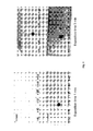
- a dot-blot was carried out to determine the specificity of the anti-A ⁇ 17 antibody, wherein other A ⁇ peptides were included ( Figure 1 ).
- the anti-A ⁇ 17 antibody was added in two dilutions (1:1000 and 1:2000) and the secondary antibody used was Goat anti-rabbit-HRP 1:2000. 500 ng of each peptide were loaded in 2.5 ⁇ l.
- the dot-blot was developed with ECL with the SNAP technology at 1 minute and 3 minutes exposition. As it is shown in said Figure, the antibody is highly specific for the A ⁇ 17 peptide and it does not recognize specifically the A ⁇ 15, A ⁇ 16, A ⁇ 38, A ⁇ 40 and A ⁇ 42.
- Formic acid sodium chloride, ⁇ -Cyano-4-hydroxycinnamic acid ( ⁇ -CHCA), trifluoroacetic acid (TFA), DL-Dithiothreitol (DTT), triethanolamine, Tris and bovine serum albumin (BSA) were from Sigma (Steimheim, Germany).
- Acetonitrile was purchased from Carlo-Erba (Rodano, MI, Italy).Dimethyl pimelimidate ⁇ 2 HCl (DMP) was acquired from Pierce (Rockford, IL, USA).
- Tricine sample buffer was purchased from Bio-Rad (Hercules, CA, USA).
- IP-MALDI procedure was carried out as described by Portelius et al. 2007 (J. Proteome. Res. 6 (11), 4433-4439 ), with some modifications in order to adapt the procedure to the analysis of dog samples.
- different amounts of A ⁇ specific antibodies were used (6E10 and 4G8 -specific for A ⁇ epitopes in amino acids 4-9 and 18-22, respectively, Signet Laboratories, Inc., Dedham, MA, USA) and SAR2 (specific for anti-A ⁇ 40 carboxy terminal epitope, Araclon Biotech S.L., Zaragoza, Spain) in order to determine the optimum quantity of each antibody by Western-blotting.
- the optimization procedure involved different steps.
- Each antibody was separately added to 50 ⁇ L Dynabeads Protein G (Invitrogen Dynal AS, Oslo, Norway) according to the manufacturer's description. Beads were incubated with different amounts of antibody, washed, and then antibodies were eluted by boiling the beads in tricine sample buffer with DTT. The saturating amount of each antibody was determined by Western-blotting.
- the first one is basically based on the instructions of the manufacturer with the optimized parameters: an aliquot of the A ⁇ specific antibody (4 ⁇ g of 6E10 or 8 ⁇ g of 4G8 or 6 ⁇ g of SAR 2) was incubated with 50 ⁇ L of Dynabeads Protein G for 1 hour at room temperature in a rotary shaker. After that, three washes with Phosphate Buffer Saline (PBS: 1mM NaH 2 PO 4 H 2 O, 5 mM Na 2 HPO 4 ⁇ 2 H 2 O, 138mM NaCl) were carried out. 1 mL of CSF was added and incubated overnight at room temperature in a rotary shaker.
- PBS Phosphate Buffer Saline
- the ZipTip C 18 protocol consists of several steps: tips solvation with acetonitrile, conditioning with 0.1 %TFA (repeating this step several times), sample load (by pipetting up and down several times), a wash step with 0.1 % TFA and final elution with 3 ⁇ L of ⁇ -CHCA (5 mg/mL).
- This second protocol resulted in mass spectra with less background noise and more intense peaks and it was used for the rest of the experiments.
- Source 1 voltage was held at 8 kV and laser value was set 1000 units higher than in MS experiments.
- Precursor ions were selected for fragmentation by means of a Timed Ion Selector at a resolution value of 250 (full width at half maximum). Selected ions were decelerated before arrival to the collision chamber at the deceleration stack and were dissociated upon collisions with air at a kinetic energy of 1 keV (High-Energy Collision Induced Dissociation). Fragment ions were reaccelerated in the second source at 15 kV after a short delay time. The Metastable Suppressor was activated in all the MS/MS experiments to avoid the detection of the remaining precursor ions and unwanted metastable decay fragments.
- the plate was coated using the 6E10 monoclonal capture antibody which recognises amino acids 1-17 in the A ⁇ amyloid peptide.
- the concentration used was determined according to the saturation concentration of the antibody, so that it will not be the limiting factor in the antigen-antibody reaction.
- the absorbance at 450nm was related with the antibody concentration in each well by monitoring the antibody adsorbed to the well with an Anti-Mouse IgG HRP conjugated antibody incubated for 1 hour shaking at RT and an incubation step with the chromogen substrate followed by a stop of the reaction. The concentration at which the signal did not increase with the antibody concentration was chosen.
- the microplates were coated with 100 ⁇ l of capture antibody in Coating Buffer and incubated overnight (ON) at 4°C during approximately 20h.
- the plates were then washed 5 times with the Washing Buffer and 100 ⁇ l of the Preservative Solution was added. The plates were left to evaporate until a white halo characteristic of trehalose appears (2-3 days at RT). The plates so treated could be kept at 4°C covered with aluminium foil and are stable for two years.
- Samples may be used undiluted or diluted 1:2 to 1:10 in Sample/Standard Diluent. Dilution 1:3 is recommended for plasma samples and 1:5 for blood cells samples. To ensure accurate quantification, the standard curves and blanks must be generated in the same diluents or buffers as the samples.
- the samples of the standard curve of human A ⁇ 17 were prepared from a 200 pg/ml stock solution of the peptides Aß17 on plates coated with the 6E10 mAb and treated with trehalose. From these solutions, serial dilutions 1:2 in Sample/Standard Diluent were made so as to give concentrations of 200, 100, 50, 25, 12.5, 6.25 and 3.125 pg/ml. 100 ⁇ l of each sample is added and incubated overnight at 4°C (or for 2h at 37°C).
- the detection antibody is a polyclonal biotin-conjugated antibody against human A ⁇ peptide ending at amino acid 17.
- the conjugation of the antibody to biotin takes place after an activation step and incubation ON at RT in the dark. The biotin excess is inactivated in a further step.
- the detection antibody was added diluted in Antibody Diluent. 100 ⁇ l are added to each well and were then incubated for 1 h shaking at room temperature. Then, 100 ⁇ l of a 1/50 dilution in Antibody Diluent of HRP-coupled Streptavidin (from SIGMA) were added to each well and incubated for 1 h shaking at room temperature.
- the results may be calculated using any immunoassay software package.
- the method used to the curve fit is lineal regression and the concentration of A ⁇ 17 was calculated as follows:
- the intra-plate variability, the intra-assay variability, the inter-assay variability in plasma samples and the inter-assay variability in cell samples were measured.
- the intra-plate variability was calculated by analysing the differences between the wells in triplicate, in samples, standard samples and reference samples in the same plate. In the intra-assay reproducibility, the concentration obtained for 4 reference plasma samples analysed in 11 different plates in the same assay was compared. In the inter-assay variability in plasma samples, the differences obtained in concentration when analysing two plasma samples in two different and independent assays were measured.
- the inter-assay variability in cell samples refers to the comparison of the A ⁇ 17 levels between two different and independent determinations of 8 cell control samples.
- the detection limit (DL) and quantification limit (QL) were calculated by two different methods:
- a ⁇ 17, A ⁇ 40 and A ⁇ 42 levels were determined using the ELISA sandwich assay described in Example 1 in plasma samples from a cohort of control healthy subjects (CS>65 or HC>65) older than 65 (19 subjects), from a cohort of subjects suffering from mild Alzheimer's disease (mild AD) (17 subjects), from a cohort of subjects having prodromal or probable mild cognitive impairment (MCI) (16 subjects) and from a cohort of subjects having non-prodromal or possible mild cognitive impairment (MCI) (16 subjects).
- the concentrations in pg/mL obtained for the peptides are shown in the Tables of Figure 3A , Figure 4A , Figure 5A and Figure 6A .
- the A ⁇ 17, A ⁇ 40 and A ⁇ 42 peptide levels in plasma (total peptide in plasma + peptide bound to cells) (PIB (17+40+42)) in mild AD differ significantly with regard to both control healthy subjects and MCI possible or non-prodromal subjects with respect to mild Alzheimer's disease subjects.
- the marker PIB (17+40+42) does not differ between HC>65 subjects and MCI probable, whereas PIB (40+42) do it with high significance ( Figure 3B ).
Priority Applications (22)
| Application Number | Priority Date | Filing Date | Title |
|---|---|---|---|
| EP11382107A EP2511296A1 (de) | 2011-04-12 | 2011-04-12 | Antikörper, Kit und Verfahren zur Bestimmung von Amyloidpeptiden |
| PCT/ES2012/070231 WO2012140296A1 (es) | 2011-04-12 | 2012-04-03 | Anticuerpo, kit y metodo para la determinacion de peptidos amiloides |
| CN201280018250.7A CN103547593B (zh) | 2011-04-12 | 2012-04-03 | 用于确定淀粉样肽的抗体、试剂盒和方法 |
| JP2014504363A JP5926368B2 (ja) | 2011-04-12 | 2012-04-03 | アミロイドペプチドを測定するための抗体、キットおよび方法 |
| NZ615011A NZ615011B2 (en) | 2011-04-12 | 2012-04-03 | Antibody, kit and method for determining amyloid peptides |
| MYPI2013701919A MY161291A (en) | 2011-04-12 | 2012-04-03 | Antibody, kit and method for determination of amyloid peptides |
| CA2828307A CA2828307C (en) | 2011-04-12 | 2012-04-03 | Antibody, kit and method for determination of amyloid peptides |
| EP12771472.3A EP2698381B1 (de) | 2011-04-12 | 2012-04-03 | Antikörper, kit und verfahren zur bestimmung von amyloidpeptiden |
| RU2013139450/10A RU2571213C2 (ru) | 2011-04-12 | 2012-04-03 | Антитело, набор и способ определения амилоидных пептидов |
| KR1020137025856A KR101602546B1 (ko) | 2011-04-12 | 2012-04-03 | 아밀로이드 펩티드를 측정하기 위한 항체, 키트 및 방법 |
| BR112013023540A BR112013023540A2 (pt) | 2011-04-12 | 2012-04-03 | anticorpo, kit e método para a determinação de peptídeos amilóides |
| MX2013011515A MX337535B (es) | 2011-04-12 | 2012-04-03 | Anticuerpo, kit y metodo para la determinacion de peptidos amiloides. |
| ES12771472.3T ES2667016T3 (es) | 2011-04-12 | 2012-04-03 | Anticuerpo, kit y procedimiento para la determinación de péptidos amiloides |
| SG2013073796A SG194053A1 (en) | 2011-04-12 | 2012-04-03 | Antibody, kit and method for determining amyloid peptides |
| US14/004,634 US9255932B2 (en) | 2011-04-12 | 2012-04-03 | Antibody, kit and method for determining amyloid peptides |
| ARP120101218A AR085959A1 (es) | 2011-04-12 | 2012-04-10 | Anticuerpo, kit y metodo para la determinacion de peptidos amiloides |
| TW101112786A TWI549966B (zh) | 2011-04-12 | 2012-04-11 | 測定澱粉樣肽之抗體、套組及方法 |
| IL228170A IL228170B (en) | 2011-04-12 | 2013-08-28 | Antibody, kit and method for determining amyloid peptides |
| ZA2013/06572A ZA201306572B (en) | 2011-04-12 | 2013-09-02 | Antibody,kit and method for determining amyloid peptides |
| CL2013002586A CL2013002586A1 (es) | 2011-04-12 | 2013-09-09 | Anticuerpo policlonal que se une al peptido amiloide b (ab)(1-17); kit; metodo para detectar el peptido ab(1-17); metodo para seguimiento de una enfermedad neurodegenerativa; metodo para establecer si un sujeto padece una enfermedad de enfermedad de alzheimer (ad) leve o deterioro cognitivo leve con ad prodromica u otras condiciones. |
| US14/977,769 US9863961B2 (en) | 2011-04-12 | 2015-12-22 | Method for determination of amyloid peptides with anti-amyloid antibody |
| IL253794A IL253794A0 (en) | 2011-04-12 | 2017-08-02 | Antibody, kit and method for determining amyloid peptides |
Applications Claiming Priority (1)
| Application Number | Priority Date | Filing Date | Title |
|---|---|---|---|
| EP11382107A EP2511296A1 (de) | 2011-04-12 | 2011-04-12 | Antikörper, Kit und Verfahren zur Bestimmung von Amyloidpeptiden |
Publications (1)
| Publication Number | Publication Date |
|---|---|
| EP2511296A1 true EP2511296A1 (de) | 2012-10-17 |
Family
ID=44510162
Family Applications (2)
| Application Number | Title | Priority Date | Filing Date |
|---|---|---|---|
| EP11382107A Withdrawn EP2511296A1 (de) | 2011-04-12 | 2011-04-12 | Antikörper, Kit und Verfahren zur Bestimmung von Amyloidpeptiden |
| EP12771472.3A Active EP2698381B1 (de) | 2011-04-12 | 2012-04-03 | Antikörper, kit und verfahren zur bestimmung von amyloidpeptiden |
Family Applications After (1)
| Application Number | Title | Priority Date | Filing Date |
|---|---|---|---|
| EP12771472.3A Active EP2698381B1 (de) | 2011-04-12 | 2012-04-03 | Antikörper, kit und verfahren zur bestimmung von amyloidpeptiden |
Country Status (18)
| Country | Link |
|---|---|
| US (2) | US9255932B2 (de) |
| EP (2) | EP2511296A1 (de) |
| JP (1) | JP5926368B2 (de) |
| KR (1) | KR101602546B1 (de) |
| CN (1) | CN103547593B (de) |
| AR (1) | AR085959A1 (de) |
| BR (1) | BR112013023540A2 (de) |
| CA (1) | CA2828307C (de) |
| CL (1) | CL2013002586A1 (de) |
| ES (1) | ES2667016T3 (de) |
| IL (2) | IL228170B (de) |
| MX (1) | MX337535B (de) |
| MY (1) | MY161291A (de) |
| RU (1) | RU2571213C2 (de) |
| SG (1) | SG194053A1 (de) |
| TW (1) | TWI549966B (de) |
| WO (1) | WO2012140296A1 (de) |
| ZA (1) | ZA201306572B (de) |
Cited By (5)
| Publication number | Priority date | Publication date | Assignee | Title |
|---|---|---|---|---|
| US11397188B2 (en) * | 2017-03-30 | 2022-07-26 | Board Of Regents, The University Of Texas System | Method of detecting an APP Alzheimer's disease marker peptide in patients with Alzheimer's disease |
| EP3966572A4 (de) * | 2019-05-10 | 2023-01-25 | Quest Diagnostics Investments LLC | Multiplex-assay zur bestimmung des beta-amyloid 42/40-verhältnisses in menschlichen plasmaproben |
| WO2023111168A1 (en) * | 2021-12-17 | 2023-06-22 | F. Hoffmann-La Roche Ag | A novel antibody for detection of amyloid beta 42 (aβ42) |
| US11779629B2 (en) | 2016-11-09 | 2023-10-10 | The University Of British Columbia | Compositions comprising cyclic peptides derived from an A-beta peptide |
| US11905318B2 (en) | 2015-11-09 | 2024-02-20 | The University Of British Columbia | Cyclic compound/peptide comprising an A-beta13-16 peptide and a linker that is covalently coupled to the n-terminus residue and the c-terminus residue of the A-beta13-16 peptide |
Families Citing this family (13)
| Publication number | Priority date | Publication date | Assignee | Title |
|---|---|---|---|---|
| CN103852579B (zh) * | 2012-12-05 | 2018-02-23 | 姚钧 | 一种人体血清Aβ的定量检测方法 |
| WO2014160647A1 (en) * | 2013-03-24 | 2014-10-02 | The Washington University | Method for amyloid beta sample processing |
| KR20160079891A (ko) * | 2013-11-20 | 2016-07-06 | 유니버시티 오브 아이오와 리써치 파운데이션 | 아밀로이드 침착의 치료를 위한 방법 및 조성물 |
| AU2015325043B2 (en) | 2014-09-30 | 2021-02-18 | Washington University | Tau kinetic measurements |
| JP7452829B2 (ja) * | 2015-11-09 | 2024-03-19 | ザ・ユニバーシティ・オブ・ブリティッシュ・コロンビア | アミロイドベータのエピトープおよびそれに対する抗体 |
| WO2017141881A1 (ja) * | 2016-02-15 | 2017-08-24 | ノーベルファーマ株式会社 | 遺伝性疾患に関わるタンパク質の測定方法及び測定キット |
| TW201827467A (zh) | 2016-11-03 | 2018-08-01 | 比利時商健生藥品公司 | 焦穀胺酸類澱粉蛋白-β之抗體及其用途 |
| KR20200038298A (ko) * | 2017-08-18 | 2020-04-10 | 더 존스 홉킨스 유니버시티 | 단백질 정제를 위한 초분자 필라멘트 조립체 |
| RU2708465C1 (ru) * | 2019-02-18 | 2019-12-09 | Федеральное Государственное Бюджетное Учреждение Науки Институт Молекулярной Биологии Им. В.А. Энгельгардта Российской Академии Наук (Имб Ран) | Способ детекции модификаций амилоидного пептида с помощью высокоспецифичных антител |
| EA202192606A1 (ru) | 2019-03-26 | 2022-01-24 | Янссен Фармацевтика Нв | Антитела к пироглутамат--амилоиду и их применение |
| WO2021009074A1 (en) | 2019-07-12 | 2021-01-21 | Adx Neurosciences Nv | Novel markers as early predictors of alzheimer's pathology |
| CN111334480B (zh) * | 2020-05-18 | 2020-09-01 | 苏州仁端生物医药科技有限公司 | 一种分泌抗Aβ1-42单克隆抗体的杂交瘤细胞株及其应用 |
| CN116574271B (zh) * | 2023-06-06 | 2023-12-19 | 苏州谱迪生物科技有限公司 | 一种用于免疫检测低丰度生物标志物信号级联放大的荧光树状大分子的制备方法及其应用 |
Citations (9)
| Publication number | Priority date | Publication date | Assignee | Title |
|---|---|---|---|---|
| WO2001062801A2 (en) | 2000-02-24 | 2001-08-30 | Washington University | Humanized antibodies that sequester amyloid beta peptide |
| WO2002046237A2 (en) | 2000-12-06 | 2002-06-13 | Neuralab Limited | Humanized antibodies that recognize beta amyloid peptide |
| WO2003015617A2 (en) | 2001-08-17 | 2003-02-27 | Washington University | Assay method for alzheimer's disease |
| WO2004013172A2 (en) | 2002-07-24 | 2004-02-12 | Innogenetics N.V. | Fragments of beta-amyloid as targets for vaccination against alzheimer disease |
| WO2006046644A1 (ja) | 2004-10-28 | 2006-05-04 | Sanko Junyaku Co., Ltd. | アルツハイマー病の検定方法及び診断試薬 |
| WO2006053251A2 (en) | 2004-11-12 | 2006-05-18 | Pfizer, Inc. | Method of measuring amyloid-beta peptides |
| WO2007022015A1 (en) | 2005-08-12 | 2007-02-22 | Georgetown University | Methods to evaluate amyloid beta-lowering agents using wild-type mice |
| WO2007050359A2 (en) | 2005-10-21 | 2007-05-03 | Merck & Co., Inc. | Anti-addl monoclonal antibody and use thereof |
| WO2009015696A1 (en) | 2007-08-02 | 2009-02-05 | Araclon Biotech | High sensitivty immunoassays and kits for the determination of peptides and proteins of biological interest |
Family Cites Families (12)
| Publication number | Priority date | Publication date | Assignee | Title |
|---|---|---|---|---|
| NL7908515A (nl) * | 1979-11-22 | 1981-06-16 | Anza Bv | Werkwijze voor het maken van een gevlochten koord, getwijnd garen of geslagen touw, respectievelijk voor het maken van een netwerk uit genoemde materialen, in het bijzonder een netwerk bestemd voor de visserij. |
| AU5439790A (en) | 1989-04-14 | 1990-11-16 | Research Foundation For Mental Hygiene, Inc. | Cerebrovascular amyloid protein-specific monoclonal antibody sv17-6e10 |
| EP0683234B2 (de) | 1993-01-25 | 2007-06-06 | Takeda Chemical Industries, Ltd. | Antikörper gegen beta-amyloid oder derivative davon und seine verwendung |
| US7771722B2 (en) * | 2001-08-17 | 2010-08-10 | Eli Lilly And Company | Assay method for alzheimer's disease |
| CN100488982C (zh) | 2001-11-02 | 2009-05-20 | 迪尔基因国际有限公司 | 与β-淀粉样蛋白结合并引起蛋白质构象转变的单克隆抗体的制备方法 |
| WO2004029629A1 (en) | 2002-09-27 | 2004-04-08 | Janssen Pharmaceutica N.V. | N-11 truncated amyloid-beta nomoclonal antibodies, compositions, methods and uses |
| EP1480041A1 (de) | 2003-05-22 | 2004-11-24 | Innogenetics N.V. | Verfahren zur Voraussage, zur Diagnose und zur vergleichenden Diagnose von Alzheimer |
| WO2005080435A1 (ja) * | 2004-02-20 | 2005-09-01 | Immuno-Biological Laboratories Co., Ltd. | モノクローナル抗体およびその利用 |
| ES2259270B1 (es) | 2005-03-09 | 2007-11-01 | Consejo Superior De Investigaciones Cientificas | Metodo de diagnostico in vitro de la enfermedad de alzheimer mediante un anticuerpo monoclonal. |
| ATE523788T1 (de) | 2006-06-08 | 2011-09-15 | Fu Berlin | ASSAY ZUR DIAGNOSE VON ALZHEIMER BASIEREND AUF DER BESTIMMUNG DES VERHÄLTNISSES VON SEKRETASE-Aß-SPALTPRODUKTEN |
| MX2010004660A (es) * | 2007-10-29 | 2010-07-30 | Inserm Inst Nat De La Sante De | NUEVOS ANTICUERPOS ESPECIFICOS DE LOS PEPTIDOS ß-AMILOIDES Y SUS USOS COMO AGENTES DE DIAGNOSTICO Y DROGAS. |
| WO2009136499A1 (ja) | 2008-05-08 | 2009-11-12 | 武田薬品工業株式会社 | Aβオリゴマー測定法 |
-
2011
- 2011-04-12 EP EP11382107A patent/EP2511296A1/de not_active Withdrawn
-
2012
- 2012-04-03 MY MYPI2013701919A patent/MY161291A/en unknown
- 2012-04-03 RU RU2013139450/10A patent/RU2571213C2/ru not_active IP Right Cessation
- 2012-04-03 BR BR112013023540A patent/BR112013023540A2/pt not_active Application Discontinuation
- 2012-04-03 MX MX2013011515A patent/MX337535B/es active IP Right Grant
- 2012-04-03 EP EP12771472.3A patent/EP2698381B1/de active Active
- 2012-04-03 CN CN201280018250.7A patent/CN103547593B/zh not_active Expired - Fee Related
- 2012-04-03 US US14/004,634 patent/US9255932B2/en not_active Expired - Fee Related
- 2012-04-03 JP JP2014504363A patent/JP5926368B2/ja not_active Expired - Fee Related
- 2012-04-03 SG SG2013073796A patent/SG194053A1/en unknown
- 2012-04-03 KR KR1020137025856A patent/KR101602546B1/ko active IP Right Grant
- 2012-04-03 CA CA2828307A patent/CA2828307C/en not_active Expired - Fee Related
- 2012-04-03 WO PCT/ES2012/070231 patent/WO2012140296A1/es active Application Filing
- 2012-04-03 ES ES12771472.3T patent/ES2667016T3/es active Active
- 2012-04-10 AR ARP120101218A patent/AR085959A1/es unknown
- 2012-04-11 TW TW101112786A patent/TWI549966B/zh not_active IP Right Cessation
-
2013
- 2013-08-28 IL IL228170A patent/IL228170B/en active IP Right Grant
- 2013-09-02 ZA ZA2013/06572A patent/ZA201306572B/en unknown
- 2013-09-09 CL CL2013002586A patent/CL2013002586A1/es unknown
-
2015
- 2015-12-22 US US14/977,769 patent/US9863961B2/en active Active
-
2017
- 2017-08-02 IL IL253794A patent/IL253794A0/en unknown
Patent Citations (9)
| Publication number | Priority date | Publication date | Assignee | Title |
|---|---|---|---|---|
| WO2001062801A2 (en) | 2000-02-24 | 2001-08-30 | Washington University | Humanized antibodies that sequester amyloid beta peptide |
| WO2002046237A2 (en) | 2000-12-06 | 2002-06-13 | Neuralab Limited | Humanized antibodies that recognize beta amyloid peptide |
| WO2003015617A2 (en) | 2001-08-17 | 2003-02-27 | Washington University | Assay method for alzheimer's disease |
| WO2004013172A2 (en) | 2002-07-24 | 2004-02-12 | Innogenetics N.V. | Fragments of beta-amyloid as targets for vaccination against alzheimer disease |
| WO2006046644A1 (ja) | 2004-10-28 | 2006-05-04 | Sanko Junyaku Co., Ltd. | アルツハイマー病の検定方法及び診断試薬 |
| WO2006053251A2 (en) | 2004-11-12 | 2006-05-18 | Pfizer, Inc. | Method of measuring amyloid-beta peptides |
| WO2007022015A1 (en) | 2005-08-12 | 2007-02-22 | Georgetown University | Methods to evaluate amyloid beta-lowering agents using wild-type mice |
| WO2007050359A2 (en) | 2005-10-21 | 2007-05-03 | Merck & Co., Inc. | Anti-addl monoclonal antibody and use thereof |
| WO2009015696A1 (en) | 2007-08-02 | 2009-02-05 | Araclon Biotech | High sensitivty immunoassays and kits for the determination of peptides and proteins of biological interest |
Non-Patent Citations (29)
| Title |
|---|
| ARCH. NEUROL., vol. 60, 2003, pages 958 - 964 |
| AUSUBEL, F.M. ET AL.: "Current Protocols in Molecular Biology", 2003, JOHN WILEY & SONS INC |
| BACHER M ET AL: "Peripheral and central biodistribution of <111>In-labeled anti-beta-amyloid autoantibodies in a transgenic mouse model of Alzheimer's disease", NEUROSCIENCE LETTERS, LIMERICK, IE, vol. 449, no. 3, 16 January 2009 (2009-01-16), pages 240 - 245, XP025885249, ISSN: 0304-3940, [retrieved on 20080903], DOI: 10.1016/J.NEULET.2008.08.083 * |
| CLACKSOII ET AL., NATURE, vol. 352, 1991, pages 624 - 628 |
| DOWDY, WEARDEN: "Statistics for Research", 1983, JOHN WILEY & SONS |
| GROWDON ET AL., NEUROBIOL. AGING, vol. 19, 1998, pages 109 - 116 |
| GRUNDMAN M ET AL., ARCH. NEUROL., vol. 61, no. 1, 2004, pages 59 - 66 |
| IRINA PERDIVARA ET AL: "Determination of primary structure and microheterogeneity of a Î-amyloid plaque-specific antibody using high-performance LCâ tandem mass spectrometry", ANALYTICAL AND BIOANALYTICAL CHEMISTRY, SPRINGER, BERLIN, DE, vol. 391, no. 1, 28 March 2008 (2008-03-28), pages 325 - 336, XP019621319, ISSN: 1618-2650 * |
| KIM KS ET AL., NEUROSCI. RES. COMM., vol. 7, 1988 |
| KIM, K.S., NEUROSCIENCE RES. COMM., vol. 2, 1988, pages 121 - 130 |
| KOHLER ET AL., NATURE, vol. 256, 1975, pages 495 |
| LANZ, T.A, SCHACTHTER, J.B., J. NEUROSCIENCE METHODS, vol. 157, 2006, pages 71 - 81 |
| MARKS ET AL., BIO/TECHNOLOGY, vol. 10, 1992, pages 779 - 783 |
| MARKS ET AL., J. MOL. BIOL., vol. 222, 1991, pages 581 - 597 |
| MAYEUX, R. ET AL., ANN NEUROL., vol. 46, 1999, pages 412 - 416 |
| MCCAFFERTY ET AL., NATURE, vol. 348, 1990, pages 552 - 554 |
| MCKHANN G. ET AL., NEUROLOGY, vol. 34, 1984, pages 939 - 944 |
| MEHTA ET AL., ARCH. NEUROL., vol. 57, 2000, pages 100 - 105 |
| MORRIS JC ET AL., ARCH. NEUROL., vol. 58, no. 3, 2001, pages 397 - 405 |
| PETERSEN RC ET AL., ARCH. NEUROL., vol. 56, no. 3, 1999, pages 303 - 308 |
| PORTELIUS ET AL., J. PROTEOME. RES., vol. 6, no. 11, 2007, pages 4433 - 4439 |
| SAMBROOK ET AL.: "Molecular Cloning: A Laboratory Manual", 1989, COLD SPRING HARBOR LABORATORY PRESS |
| SCHEUNER D ET AL., NATURE MED, vol. 2, 1996, pages 864 - 870 |
| SCHEUNER ET AL., NATURE MED., vol. 2, 1996, pages 864 - 870 |
| SUZUKI,N. ET AL., SCIENCE, vol. 264, 1994, pages 1336 - 1340 |
| TABERT MH ET AL., ARCH. GEN. PSYCHIATRY, vol. 63, no. 8, 2006, pages 916 - 924 |
| TAMAOKA A ET AL., J NEUROL SCI., vol. 141, 1996, pages 65 - 68 |
| VANDERSTICHELE H ET AL., AMYLOID, vol. 7, 2000, pages 245 - 258 |
| WATERHOUSE ET AL., NUCL. ACIDS. RES., vol. 21, 1993, pages 2265 - 2266 |
Cited By (5)
| Publication number | Priority date | Publication date | Assignee | Title |
|---|---|---|---|---|
| US11905318B2 (en) | 2015-11-09 | 2024-02-20 | The University Of British Columbia | Cyclic compound/peptide comprising an A-beta13-16 peptide and a linker that is covalently coupled to the n-terminus residue and the c-terminus residue of the A-beta13-16 peptide |
| US11779629B2 (en) | 2016-11-09 | 2023-10-10 | The University Of British Columbia | Compositions comprising cyclic peptides derived from an A-beta peptide |
| US11397188B2 (en) * | 2017-03-30 | 2022-07-26 | Board Of Regents, The University Of Texas System | Method of detecting an APP Alzheimer's disease marker peptide in patients with Alzheimer's disease |
| EP3966572A4 (de) * | 2019-05-10 | 2023-01-25 | Quest Diagnostics Investments LLC | Multiplex-assay zur bestimmung des beta-amyloid 42/40-verhältnisses in menschlichen plasmaproben |
| WO2023111168A1 (en) * | 2021-12-17 | 2023-06-22 | F. Hoffmann-La Roche Ag | A novel antibody for detection of amyloid beta 42 (aβ42) |
Also Published As
| Publication number | Publication date |
|---|---|
| ES2667016T3 (es) | 2018-05-09 |
| CN103547593A (zh) | 2014-01-29 |
| EP2698381B1 (de) | 2018-01-24 |
| NZ615011A (en) | 2015-11-27 |
| JP2014516357A (ja) | 2014-07-10 |
| CA2828307A1 (en) | 2012-10-18 |
| CN103547593B (zh) | 2016-04-20 |
| IL228170B (en) | 2018-01-31 |
| TWI549966B (zh) | 2016-09-21 |
| RU2013139450A (ru) | 2015-03-10 |
| AR085959A1 (es) | 2013-11-06 |
| US9255932B2 (en) | 2016-02-09 |
| EP2698381A1 (de) | 2014-02-19 |
| EP2698381A4 (de) | 2015-04-01 |
| KR20130135931A (ko) | 2013-12-11 |
| TW201305201A (zh) | 2013-02-01 |
| CL2013002586A1 (es) | 2014-05-16 |
| SG194053A1 (en) | 2013-11-29 |
| MX337535B (es) | 2016-03-09 |
| US9863961B2 (en) | 2018-01-09 |
| WO2012140296A9 (es) | 2013-08-29 |
| BR112013023540A2 (pt) | 2016-11-22 |
| ZA201306572B (en) | 2017-11-29 |
| CA2828307C (en) | 2019-06-11 |
| US20160195548A1 (en) | 2016-07-07 |
| WO2012140296A1 (es) | 2012-10-18 |
| MY161291A (en) | 2017-04-14 |
| RU2571213C2 (ru) | 2015-12-20 |
| IL253794A0 (en) | 2017-09-28 |
| KR101602546B1 (ko) | 2016-03-10 |
| US20140024054A1 (en) | 2014-01-23 |
| JP5926368B2 (ja) | 2016-05-25 |
| MX2013011515A (es) | 2014-01-08 |
Similar Documents
| Publication | Publication Date | Title |
|---|---|---|
| US9863961B2 (en) | Method for determination of amyloid peptides with anti-amyloid antibody | |
| US20210011032A1 (en) | Methods and reagents for improved detection of amyloid beta peptides | |
| US20190178899A1 (en) | High sensitivity immunoassays and kits for the determination of peptides and proteins of biological interest | |
| AU2013204931B2 (en) | Antibody, kit and method for determining amyloid peptides | |
| NZ615011B2 (en) | Antibody, kit and method for determining amyloid peptides |
Legal Events
| Date | Code | Title | Description |
|---|---|---|---|
| PUAI | Public reference made under article 153(3) epc to a published international application that has entered the european phase |
Free format text: ORIGINAL CODE: 0009012 |
|
| AK | Designated contracting states |
Kind code of ref document: A1 Designated state(s): AL AT BE BG CH CY CZ DE DK EE ES FI FR GB GR HR HU IE IS IT LI LT LU LV MC MK MT NL NO PL PT RO RS SE SI SK SM TR |
|
| AX | Request for extension of the european patent |
Extension state: BA ME |
|
| STAA | Information on the status of an ep patent application or granted ep patent |
Free format text: STATUS: THE APPLICATION IS DEEMED TO BE WITHDRAWN |
|
| 18D | Application deemed to be withdrawn |
Effective date: 20130418 |