EP1806394B1 - Cellular tissue microchip and method of forming cellular tissue - Google Patents
Cellular tissue microchip and method of forming cellular tissue Download PDFInfo
- Publication number
- EP1806394B1 EP1806394B1 EP05795896A EP05795896A EP1806394B1 EP 1806394 B1 EP1806394 B1 EP 1806394B1 EP 05795896 A EP05795896 A EP 05795896A EP 05795896 A EP05795896 A EP 05795896A EP 1806394 B1 EP1806394 B1 EP 1806394B1
- Authority
- EP
- European Patent Office
- Prior art keywords
- cellular
- cell
- cells
- microchip
- adhesive
- Prior art date
- Legal status (The legal status is an assumption and is not a legal conclusion. Google has not performed a legal analysis and makes no representation as to the accuracy of the status listed.)
- Expired - Lifetime
Links
Images


Classifications
-
- C—CHEMISTRY; METALLURGY
- C12—BIOCHEMISTRY; BEER; SPIRITS; WINE; VINEGAR; MICROBIOLOGY; ENZYMOLOGY; MUTATION OR GENETIC ENGINEERING
- C12M—APPARATUS FOR ENZYMOLOGY OR MICROBIOLOGY; APPARATUS FOR CULTURING MICROORGANISMS FOR PRODUCING BIOMASS, FOR GROWING CELLS OR FOR OBTAINING FERMENTATION OR METABOLIC PRODUCTS, i.e. BIOREACTORS OR FERMENTERS
- C12M23/00—Constructional details, e.g. recesses, hinges
- C12M23/02—Form or structure of the vessel
- C12M23/16—Microfluidic devices; Capillary tubes
-
- C—CHEMISTRY; METALLURGY
- C12—BIOCHEMISTRY; BEER; SPIRITS; WINE; VINEGAR; MICROBIOLOGY; ENZYMOLOGY; MUTATION OR GENETIC ENGINEERING
- C12M—APPARATUS FOR ENZYMOLOGY OR MICROBIOLOGY; APPARATUS FOR CULTURING MICROORGANISMS FOR PRODUCING BIOMASS, FOR GROWING CELLS OR FOR OBTAINING FERMENTATION OR METABOLIC PRODUCTS, i.e. BIOREACTORS OR FERMENTERS
- C12M25/00—Means for supporting, enclosing or fixing the microorganisms, e.g. immunocoatings
- C12M25/06—Plates; Walls; Drawers; Multilayer plates
-
- C—CHEMISTRY; METALLURGY
- C12—BIOCHEMISTRY; BEER; SPIRITS; WINE; VINEGAR; MICROBIOLOGY; ENZYMOLOGY; MUTATION OR GENETIC ENGINEERING
- C12N—MICROORGANISMS OR ENZYMES; COMPOSITIONS THEREOF; PROPAGATING, PRESERVING, OR MAINTAINING MICROORGANISMS; MUTATION OR GENETIC ENGINEERING; CULTURE MEDIA
- C12N5/00—Undifferentiated human, animal or plant cells, e.g. cell lines; Tissues; Cultivation or maintenance thereof; Culture media therefor
- C12N5/06—Animal cells or tissues; Human cells or tissues
- C12N5/0602—Vertebrate cells
- C12N5/067—Hepatocytes
- C12N5/0671—Three-dimensional culture, tissue culture or organ culture; Encapsulated cells
-
- B—PERFORMING OPERATIONS; TRANSPORTING
- B33—ADDITIVE MANUFACTURING TECHNOLOGY
- B33Y—ADDITIVE MANUFACTURING, i.e. MANUFACTURING OF THREE-DIMENSIONAL [3-D] OBJECTS BY ADDITIVE DEPOSITION, ADDITIVE AGGLOMERATION OR ADDITIVE LAYERING, e.g. BY 3-D PRINTING, STEREOLITHOGRAPHY OR SELECTIVE LASER SINTERING
- B33Y80/00—Products made by additive manufacturing
Definitions
- the present invention relates to a microchip for retaining cells and particularly relates to the formation of cellular tissue.
- Such cell-culture techniques generally involve attaching and then culturing cells in a monolayer (i.e., in two dimensions) on a planar substrate on which collagen or another cellular-adhering material has been coated.
- a monolayer i.e., in two dimensions
- collagen or another cellular-adhering material has been coated.
- liver functions of primary hepatocytes can be maintained for longer periods of time by culturing the cells in cellular tissues, which are aggregates wherein the cells are connected one another in three dimensions, instead of culturing the cells in a monolayer.
- Patent Document 1 An example of a conventional method for forming such hepatic cellular tissue is disclosed in Patent Document 1, wherein a specific growth factor is added to the culture medium, whereby spherical hepatic cellular tissues are formed within the pores of a polyurethane foam.
- Patent Document 2 discloses the formation of hepatic cellular tissue on a surface that is coated with a polymeric material that is composed of a monomer containing phenylborate, a monomer containing an amino group, and a 2-hydroxy-ethyl methacrylate copolymer.
- the document US-A-2002 0173033 describes a cell culture plate comprising cavities to accommodate cells to be cultured.
- the bottom wall of these cavities are coated with cell-adhesive or cell-repulsive material.
- the formed cellular tissues are of nonuniform shape and size. Therefore, when, for example, an evaluation is made of the metabolic function of a specific drug in the cells, obtaining reliable results has been difficult because, e.g., the number of cells in contact with the drug (in particular, the number of cells near the surface of the cellular tissue) differs for each cellular tissue.
- the conventional methods above also involve the use of culture-substrate surfaces to which cells adhere relatively weakly (i.e., which have relatively poor cellular adhesiveness) and the formed cellular tissues are desorbed from the culture-substrate surfaces to be suspended in the culture medium. Therefore, when removing the culture medium from which the nutrients have been consumed and pouring a fresh culture medium or other operations are performed in the culturing process, for example, cells and cellular tissues will therefore be removed along with the culture medium, and culturing may be unable to continue thereafter.
- the present invention was made in light of the these problems, and it is one of the objects thereof to provide a microchip that forms cellular tissues having uniform shapes and sizes and that can culture the formed cellular tissues for long periods of time, and to provide a method for forming cellular tissues that uses this microchip.
- a cellular tissue microchip comprises a plurality of cell-retaining cavities for retaining cells, wherein a bottom surface of the cell-retaining cavities contains one adhesive region that exhibits cellular adhesiveness; and a non-adhesive region that surrounds the adhesive region and that exhibits cellular non-adhesiveness.
- a method for forming cellular tissues comprises culturing cells within the cell-retaining cavities of the cellular tissue microchip; and forming a cellular tissue on the adhesive region.
- a microchip can be provided that forms cellular tissues having uniform shapes and sizes and that enables the culture of the formed cellular tissues for long periods of time, and a method for forming cellular tissues can be provided that uses this microchip.
- a cellular tissue microchip according to an embodiment of the present invention will be described below with reference to the drawings.
- the cellular tissue microchip according to the present invention is not limited to the embodiment below.
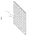
- FIG. 1 is a descriptive diagram of the cellular tissue microchip (referred to below as "the present microchip 1") according to the present embodiment.
- the present microchip 1 has a plurality of cell-retaining cavities 12 that are formed as bottomed holes on a substrate 10 and that have prescribed depths in order to retain cells.
- a culture medium into which cells have been dispersed is introduced into the cell-retaining cavities 12 of the present microchip 1.
- a bottom surface 20 (see FIGS. 2 and 3 ) of the cell-retaining cavities 12 is used as a culture-substrate surface for the cells, whereby cellular tissues are formed from the cells.
- the source of the cells may be any type of animal, organ, tissue, or the like.
- the cells may be primary cells isolated from the liver, pancreas, kidney, nerves, skin, or other regions of humans, pigs, dogs, rats, mice, or other animals; non-human embryonic stem (ES) cells; cells from established cell lines; or cells resulting from genetic modifications thereof.
- ES non-human embryonic stem
- a single type of cell may be used, or two or more types of cells mixed in any ratio may also be used.
- the culture medium used may have any composition as long as the medium is an aqueous solution that includes the necessary salts, nutrients, and other components at appropriate concentrations so that the survival, functionality, and other aspects of the cells can be maintained.
- the culture medium used may be DMEM (Dulbecco's Modified Eagle's Medium), another basal medium to which antibiotics have been added, or so-called physiological saline solution.
- the substrate 10 of the present microchip 1 is composed of, e.g., polystyrene, polyethylene, polypropylene, polycarbonate, polyamide, polyacetal, polyester (polyethylene terephthalate or the like), polyurethane, polysulfone, polyacrylate, polymethacrylate, polyvinyl, a silicone or other synthetic resin, EPDM (ethylene propylene diene monomer) or another synthetic or natural rubber, glass, ceramic, or stainless steel or another metallic material.
- the substrate 10 is formed into, e.g., a plate shape.
- the cell-retaining cavities 12 can be formed in the substrate 10 using any machining method selected in accordance with the material and other properties of the substrate 10. As specific examples, the cell-retaining cavities 12 may be formed on the substrate 10 by punching using a machining center or the like, optical micromachining using a laser or the like, etching, or embossing. Alternatively, the cell-retaining cavities 12 may be formed when the substrate 10 is molded using injection molding, press molding, stereolithography, or the like.
- the cell-retaining cavities 12 can be formed as, e.g., bottomed holes on the surface of the substrate 10, which has a prescribed thickness, wherein the depth of the bottomed holes is less than the thickness of the substrate 10.
- the cell-retaining cavities 12 may also be formed by, e.g., forming holes that pass through the substrate 10 and then affixing another member to one surface of the substrate 10 in order to make the bottom surface.
- a substrate, film, or other article made from a material that is the same as or different from the material of the substrate 10 in which the through-holes are formed may be used as the member for forming the bottom surface of the cell-retaining cavities 12.
- the cell-retaining cavities 12 are positioned in order at prescribed intervals on the substrate 10, as shown in FIG. 1 .
- the ordered plurality of cell-retaining cavities 12 may be formed using a machining center or the like that precisely controlls the machining location according to a computer-aided design (CAD) program.
- CAD computer-aided design
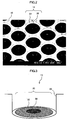
- FIG. 2 is a scanning electron microphotograph that shows a portion of the present microchip 1.
- each of the cell-retaining cavities 12 formed on the substrate 10 have the bottom surface 20 and a lateral surface 22 that form the hole structure.
- the bottom surface 20 and the lateral surface 22 of each of the cell-retaining cavities 12 have a substantially smooth surface, as shown in FIG. 2 .
- the shape of the bottom surface 20 is not particularly limited and may be, e.g., a circular shape as shown in FIG. 2 , or an ellipsoid, polygon, or other shape.
- the diameter of the bottom surface 20 is preferably within the range of approximately 2 to 50 times the diameter of the cells to be used, and a range of approximately 4 to 30 times the cell diameter is particularly preferable.
- the size of the cells changes according to the type, state, and other properties of the cells, and therefore the area of the bottom surface 20 cannot be categorically defined, but a range of, e.g., 100 to 1 ⁇ 10 6 ⁇ m 2 is preferable, and a range of 300 to 3 ⁇ 10 5 ⁇ m 2 is particularly preferable.
- the size of the bottom surface 20 will dictate the number of cells contained in the cellular tissue formed on the bottom surface 20. Specifically, when the size of the bottom surface 20 is less than the aforementioned lower limit, the number of cells necessary to form a cellular tissue on the bottom surface 20 cannot be retained. When the bottom surface 20 is larger than the aforementioned upper limit, the number of cells to be retained on the bottom surface 20 is too large, and therefore an enormous cellular tissue will be formed. In this case, the cells located inside the cellular tissue are not able to receive adequate quantities of nutrients and oxygen from the culture medium outside the cellular tissue and may die off.
- the depth of the cell-retaining cavities 12 is preferably within the range of approximately 1 to 50 times the diameter of the cells to be used, and a range of approximately 2 to 30 times the cell diameter is particularly preferable. This range is stipulated because when the depth of the cell-retaining cavities 12 is less than the aforementioned lower limit, the cells will tend not to be reliably retained within the cell-retaining cavities 12. When the depth of the cell-retaining cavities 12 is greater than the aforementioned upper limit, the amount of oxygen and nutrients provided to the cells on the bottom surface 20 of the cell-retaining cavities 12 may be insufficient.
- FIG. 3 is a descriptive diagram of one of the plurality of cell-retaining cavities 12 included in the present microchip 1.
- the bottom surface 20 of the cell-retaining cavities 12 has one adhesive region 30 that exhibits cellular adhesiveness and a non-adhesive region 32 that surrounds the adhesive region 30 and that exhibits cellular non-adhesiveness.
- the adhesive region 30 has, e.g., a cellular adhesive surface that has an electrically charged state, and a hydrophilicity or a hydrophobicity that are appropriate for cell adhesion within a culture medium or other solution.
- a cellular adhesive surface refers to, e.g., a surface to which cells can adhere in a relatively flat shape after deforming from a spherical shape when precipitating out of the culture medium onto the surface.
- the surface of the adhesive region 30 can be formed of, e.g., the material surface itself of the substrate 10 that is exposed as the bottom surface 20 of the cell-retaining cavities 12 when the cell-retaining cavities 12 are formed.
- the surface of the adhesive region 30 can also be formed of the surface of the exposed substrate 10, e.g., on which a cellular adhesive material that is acquired from living organisms or synthesized, or a material derived therefrom is fixed.
- a material that can bind to a specific protein or other cell surface molecule (e.g., integrin or a sugar receptor) on the cell membrane of the cells to be used may be employed as the cellular adhesive material fixed in place to the adhesive region 30.
- collagen, fibronectin, laminin, and the like may be used as cellular adhesive materials that are acquired from living organisms.
- Compounds wherein, e.g., a desired functional group, polymer, or the like has been bonded (e.g., covalently bonded using a condensation reaction or the like) to such cellular adhesive materials may be used as derivatives of these materials.
- Compounds that include specific amino acid sequences e.g., arginine-glycine-aspartate (RGD) sequences
- specific sugar sequences e.g., galactose side chains
- Compounds wherein, e.g., a desired functional group, polymer, or the like has been bonded to such cellular adhesive materials may be used as derivatives of these materials.
- the aqueous solution of the cellular adhesive material and the like is dried on the bottom surface 20, for example, whereby these cellular adhesive materials that are acquired from living organisms or synthesized, or the derivatives thereof can be fixed in place to the bottom surface 20.
- Chemical reactions may also be made to occur in the aqueous solution of the cellular adhesive material and the like between the functional groups of the cellular adhesive material and the functional groups of the bottom surface 20 (e.g., a condensation reaction between carboxyl groups and amino groups or the like), forming covalent bonds and the like whereby the cellular adhesive material can be fixed in place to the bottom surface 20.
- the non-adhesive region 32 has, e.g., a surface that has poor cellular adhesiveness and that has an electric charge state, and a hydrophilicity or a hydrophobicity that are not suitable for cell adhesion such that the cells within the culture medium are substantially unable to adhere the surface.
- a surface that exhibits cellular non-adhesiveness is a surface wherein, e.g., when cells precipitate out of the culture medium onto the surface, the shape of the cells does not deform substantially from a spherical shape, so that extremely weak adhesion results, but the cells are easily desorbed by currents and the like in the culture medium. Alternatively, the cells are completely unable to adhere to the surface and are maintained in a suspneded state with a spherical shape in the culture medium.
- the surface of the non-adhesive region 32 can be formed of, e.g., the material surface itself of the substrate 10 that is exposed as the bottom surface 20 of the cell-retaining cavities 12 when the cell-retaining cavities 12 are formed.
- the surface of the non-adhesive region 32 can also be formed of the surface of the exposed substrate 10, e.g., on which a cellular adhesive material that is acquired from living organisms or synthesized, or a material derived therefrom is fixed.
- a cellular non-adhesive material that is synthetic or biologically derived and that does not bind to proteins, sugar chains, or other cell surface molecules on the cell membrane of the cells to be used may be employed as the material having poor cellular adhesiveness that is fixed in place to the non-adhesive region 32.
- albumin other proteins exhibiting high hydrophilicity, or the like can be used as biologically derived cellular non-adhesive materials.
- Compounds wherein, e.g., a desired functional group, polymer, or the like has been bonded to such cellular non-adhesive materials may be used as derivatives of these materials.
- Polyethylene glycol or other polymers that exhibit extremely high hydrophilicity MPC (2-methacryloyloxyethyl phosphorylcholine), poly-HEMA (poly-hyroxyethyl methacrylate), SPC (segmented polyurethane), or the like can be used as synthetic materials having poor cellular adhesiveness.
- Compounds wherein, e.g., a desired functional group, polymer, or the like has been bonded to such cellular non-adhesive materials may be used as derivatives of these materials.
- the aqueous solution of the cellular non-adhesive material and the like is dried on the bottom surface 20, for example, whereby these cellular non-adhesive materials that are acquired from living organisms or synthesized, or the derivatives thereof can be fixed in place to the bottom surface 20.
- Chemical reactions may also be made to occur in the aqueous solution of the cellular non-adhesive material and the like between the functional groups of the cellular non-adhesive material and the functional groups of the bottom surface 20, forming covalent bonds and the like whereby the cellular non-adhesive material can be fixed in place to the bottom surface 20.
- the adhesive region 30 may also be formed in the vicinity of the center of the bottom surface 20 of the cell-retaining cavities 12. If the cell-retaining cavities 12 are positioned in an ordered fashion on the present microchip 1, as shown in FIG. 1 , the cellular tissues formed on the adhesive regions 30 of the bottom surfaces 20 of the cell-retaining cavities 12 in such instances can also be positioned in an ordered fashion on the present microchip 1, and the shapes of the formed cellular tissues can be made uniform.
- the cellular tissues are, e.g., stained using a fluorescent dye or the like and the function of the cellular tissues is evaluated according to the extent of staining or the like, the positions of the uniformly shaped cellular tissues can be accurately specified according to the coordinates or the like established on the present microchip 1.
- the extent of staining can therefore be quickly, easily, and accurately analyzed using an automatic analyzer or the like.
- the bottom surface 20 that comprises the adhesive region 30 and the non-adhesive region 32 may be formed as a entirely flat surface, as shown in FIG. 3 , or may be formed to have a step or the like on a portion of at least one of the regions or between the adhesive region 30 and the non-adhesive region 32.
- the cells to be used are dispersed in the culture medium at a prescribed density.
- a prescribed volume of the solution into which the cells have been dispersed is then introduced into each of the cell-retaining cavities 12, whereby the cells are seeded.
- the cells are thereby made to precipitate out onto the bottom surface 20 of the cell-retaining cavities 12.
- the number of cells seeded in one cell-retaining cavity 12 is preferably set so that the cells that have precipitated out onto the bottom surface 20 can contact each other to the extent necessary to form mutual connections, and so as to limit the size of the cellular tissue, which is formed by the aggregation of the cells to be within the prescribed range.
- the number of seeded cells per cell-retaining cavity 12 is preferably within a range of 2 to 1.5 ⁇ 10 5 , and a range of 50 to 3.0 ⁇ 10 4 is particularly preferable.
- the number of seeded cells per unit area on the bottom surface 20 of the cell-retaining cavity is preferably within the range of 30 to 1.5 ⁇ 10 4 cells/mm 2 .
- the present microchip 1 onto which the cells have been thus seeded is kept horizontal and maintained in a stationary state for a prescribed period of time, whereby the cells are cultured.
- the stationary culture period among the cells that have precipitated out onto the bottom surfaces 20 of the cell-retaining cavities 12, the cells that have precipitated out on the adhesive region 30 adhere to the surface of the adhesive region 30.
- the cells that precipitate out on the non-adhesive region 32 are maintained in a suspended state without adhering to the surface of the non-adhesive region 32, or adhere extremely weakly.
- the stationary culture is further maintained, or culturing is continued while the present microchip 1 is shaken so that an arc will be drawn on the horizontal surface.
- the cells that have precipitated out on the bottom surfaces 20 of the cell-retaining cavities 12 will thereby connect one another as the culture period progresses.
- the cells gradually move so as to gather on the adhesive regions 30 of the bottom surfaces 20, and cellular tissues in which the cells have connected in three-dimensions are formed on the adhesive regions 30.
- These cellular tissues are not suspended in the culture medium and can be cultured for long periods of time in a state that the cells adhere to the adhesive regions 30 being stably retained within the cell-retaining cavities 12.
- a flow inlet for the influx of the culture medium to the cell-retaining cavity 12 and a flow outlet for the efflux of the culture medium from the cell-retaining cavity 12 can be also formed on a portion of the lateral surface 22 of each of the cell-retaining cavities 12 of the present microchip 1, whereby the culture can be performed while flowing the culture medium within the cell-retaining cavities 12 after the cellular tissues have been formed on the adhesive region 30.
- a flat plate (24 mm ⁇ 24 mm, 200 ⁇ m thick) of polymethyl methacrylate was used as the substrate 10 of the present microchip 1 of the present example.
- a machining center (a table-top NC micromachining apparatus; PMT Corporation) was used to punch holes in a 10 mm-square rectangular area on a portion of the surface of the polymethyl methacrylate plate, whereby approximately 1000 circular through-holes were formed having a diameter of 300 ⁇ m.
- the circular through-holes were positioned in an ordered fashion so that the distance between the centers of the through-holes was 400 ⁇ m.
- a flat plate (22 mm ⁇ 22 mm, 400 ⁇ m thick) of glass was used as the bottom surface 20 of the cell-retaining cavities 12 of the present microchip 1.
- a sputtering apparatus (E-1030; Hitachi, Ltd.) was used to perform sputtering in a 12 mm-square rectangular area on a portion of the surface of the glass plate, whereby a thin film (9 nm thick) of platinum (Pt) was formed.
- the platinum-surface portion of the glass plate was aligned with the portion of the polymethyl methacrylate plate on which the through holes had been formed, and a silicone adhesive (TSE389; GE Toshiba Silicones Co., Ltd.) was used to bond together the glass plate and the polymethyl methacrylate plate.
- the cell-retaining cavities 12 were thus formed as shown in FIG. 2 having depths of 200 ⁇ m and having circular bottom surfaces 20 that had diameters of 300 ⁇ m.
- a stamp of PDMS poly (dimethyl siloxane) having a plurality of cylindrical protrusions, which had diameters of 100 ⁇ m and lengths of 200 ⁇ m, was manufactured by mold pressing.
- the positions of the cylindrical protrusions of the stamp were established so that the distance between the centers of the circular cross-sections on the ends of the protrusions was 400 ⁇ m so as to correspond with the locations of the cell-retaining cavities 12 on the present microchip 1.
- the adhesive regions 30 were formed on the bottom surfaces 20 of the cell-retaining cavities 12 by microcontact printing using this stamp as described hereinafter.
- a peptide containing a cellular adhesive RGD sequence (amino acid sequence: RGDSAAAAAC; Thermo Electron Corporation) was prepared as the cellular adhesive material to be fixed in place to the surface of the adhesive regions 30.
- the ends of the cylindrical protrusions of the manufactured stamp were immersed in a DMSO (dimethyl sulfoxide) solution containing the cellular adhesive peptide at a concentration of 1.78 mg/mL, whereby the solution of the peptide was applied to the end surfaces of the cylindrical protrusions.
- DMSO dimethyl sulfoxide
- the positions of the cylindrical protrusions of the stamp to which the peptide solution was applied were then aligned with the areas near the centers of the bottom surfaces 20 of the cell-retaining cavities 12 of the present microchip 1 on which the platinum was deposited.
- the ends of the cylindrical protrusions were then pressed onto the bottom surfaces 20, whereby the peptide solution applied to the ends of the cylindrical protrusions was coated on the areas near the centers of the bottom surface 20 of the cell-retaining cavities 12.
- the peptide solution was dried in a nitrogen atmosphere. Chemical bonds were thereby formed between the platinum surface of the bottom surfaces 20 and the thiol groups included on the cysteines on the ends of the cellular adhesive peptide molecules, and the cellular adhesive peptides were fixed in place to the platinum surface.
- One adhesive region 30 having a diameter of approximately 100 ⁇ m and upon which the cellular adhesive peptide was fixed was thereby formed in vicinity of the center of the bottom surface 20 of each of the cell-retaining cavities 12, which had diameters of 300 ⁇ m.
- the non-adhesive region 32 was formed in the following manner on the bottom surface 20 that includes the adhesive region 30. Specifically, a synthetic, cellular non-adhesive polymer (chemical formula: CH 3 (CH 2 CH 2 ) n SH; Nektar Therapeutics) having a polyethylene glycol (PEG) chain of molecular weight 5000 was prepared as the cellular non-adhesive material to be fixed in place to the surface of the non-adhesive region 32.
- a synthetic, cellular non-adhesive polymer chemical formula: CH 3 (CH 2 CH 2 ) n SH; Nektar Therapeutics
- PEG polyethylene glycol
- the present microchip 1 was thus manufactured having the adhesive region 30 formed in the vicinity of the center of the bottom surface 20 of each of the cell-retaining cavities 12 and having the non-adhesive region 32 formed in the regions other than the adhesive region 30.
- the present microchip 1 was used in the culture experiment below.
- hepatocytes that were prepared from rat liver using a well-known collagenase perfusion method were used as the cells in the present example. The method for preparing these hepatocytes is briefly described. Specifically, a cannula was first inserted into the portal vein (one of the blood vessels that connects to the liver) of a 7-week-old Wistar-strain rat (body weight 250g).
- a blood-removal solution having a prescribed composition was injected into the liver from the cannula, after which a digestive solution was perfused into which collagenase (Wako Pure Chemical Industries, Ltd.) had been dissolved at a concentration of 0.5 mg/mL, and digestion was carried out to break the mutual connections and the like between the hepatocytes in the liver.
- the digested liver was then excised and hepatocytes in which the individual cells were dispersed and isolated were obtained by diffusion treatments using a scalpel, pipette, and the like and by washing using a culture medium and the like.
- a serum-free culture medium that was used as the culture medium was prepared by adding 60 mg/L of proline (Sigma-Aldrich), 50 mg/mL of epidermal growth factor (EGF; Funakoshi Co., Ltd.), 7.5 mg/L of hydrocortisone (Wako Pure Chemical Industries, Ltd.), 0.1 ⁇ M of copper sulfate 5-hydrate (Wako Pure Chemical Industries, Ltd.), 3 ⁇ g/L of selenate (Wako Pure Chemical Industries, Ltd.), 50 pM of zinc sulfate 7-hydrate (Wako Pure Chemical Industries, Ltd.), 50 ⁇ g/L of linoleic acid (Sigma), 58.8 mg/L of penicillin (Meiji Seika Kaisha, Ltd.), 100 mg/L of streptomycin (Meiji Seika Kaisha, Ltd.), 1.05 g/L of sodium bicarbonate (Wako Pure Chemical Industries, Ltd.), and 1.19 g/L of 2-[4-(2-hydroxyeth
- the resulting hepatocytes were dispersed in the culture medium to a density of 5.0 ⁇ 10 5 cells/mL.
- the sterilized present microchip 1 was mounted on the bottom part of a polystyrene culture vessel (diameter 35 mm; Falcon). A 2.0 mL portion of the solution into which the cells were dispersed was added to the present microchip 1 and cultured at 37°C in a stationary state in an atmosphere of 5% carbon dioxide gas and 95% air.
- a flat plate (22 mm ⁇ 22 mm, 400 ⁇ m thick) of glass which was identical to the glass plate used to form the bottom surface 20 of the cell-retaining cavities of the present microchip 1, was mounted on the bottom part of a polystyrene culture vessel having a diameter of 35 mm. Another 2.0 mL portion of the solution into which the cells were dispersed was then added onto the glass plate and cultured in a stationary state in the same manner.
- a flat plate (22 mm ⁇ 22 mm, 400 ⁇ m thick) of glass which was identical to the glass plate used to form the bottom surface 20 of the cell-retaining cavities of the present microchip 1 and on which collagen (Cellmatrix Type I-C, Nitta Gelatin, Inc.) was coated, was mounted on the bottom part of a polystyrene culture vessel having a diameter of 35 mm. Another 2.0 mL portion of the solution into which the cells were dispersed was then added onto the collagen-coated glass plate and cultured in a stationary state in the same manner. The culture medium within the culture vessels used for the present microchip 1 and the control experiments was replaced with fresh culture medium every other day after the start of the culture.
- FIG. 4 shows a phase-contrast microphotograph of the hepatic cellular tissues (T in FIG. 4 ) formed in the cell-retaining cavities 12 on the third day of the culture.
- One hepatic cellular tissue is formed in each of the cell-retaining cavities 12, as shown in FIG. 3 .
- the shapes of the cellular tissues are all spherical and have smooth surfaces. Such spherical hepatic cellular tissues were formed in the cell-retaining cavities 12 of the present microchip 1 within one to two days after the start of the culture.
- Each of the hepatic cellular tissues were formed in the vicinity of the center of the bottom surface 20 within each of the cell-retaining cavities 12, as shown in FIG. 3 . Specifically, each of the hepatic cellular tissues adhered to the adhesive region 30 on the bottom surface 20 of each of the cell-retaining cavities 12 and were stably retained without being suspended in the culture medium. Hepatocytes were not substantially present on the non-adhesive region 32.
- hepatic cellular tissues of various sizes and irregular shapes were formed from a portion of the seeded cells in the first control experiment, and the tissues were in a suspended state within the culture medium over the entirety of the glass plate (not shown). Additionally, the hepatocytes adhered to the collagen-coated glass plate in the second control experiment were flattened and extended in a monolayer state (not shown).
- FIG. 5 is a histogram that shows the particle diameter distributions of the hepatic cellular tissues formed in the cell-retaining cavities 12 of the present microchip 1 and of the hepatic cellular tissues formed in the first control experiment.
- the horizontal axis in FIG. 5 designates the diameter ( ⁇ m) of the hepatic cellular tissues.
- the vertical axis designates the ratio (%) of the number of hepatic cellular tissues having the diameter shown at each point on the horizontal axis relative to the total number of hepatic cellular tissues of which particle diameter was measured.
- the circles in FIG. 5 designate the results for the hepatic cellular tissues formed using the present microchip 1, and the squares designate the results for the hepatic cellular tissues formed in the first control experiment.
- the diameters of 86% of the hepatic cellular tissues among those formed in the present microchip 1 were within the range of 160 to 180 ⁇ m, as shown in FIG. 5 .
- hepatic cellular tissues having similar particle diameters and extremely uniform sizes were formed in the cell-retaining cavities 12 of the present microchip 1.
- the hepatic cellular tissues formed in the first control experiment had a variety of particle diameters and had sizes that were non-uniform overall.
- the measurement of the particle diameters of the hepatic cellular tissues was carried out by image analysis of the microscopy results.
- the ability to remove ammonia (one of the waste substances within living organisms), which is one of the characteristic detoxification functions of the liver, was also evaluated for the hepatic cellular tissues formed in the present microchip 1 and for the monolayer of hepatocytes in the second control experiment.
- the culture medium within the culture vessel containing the hepatic cellular tissues or the monolayer of hepatocytes was first replaced with a culture medium to which ammonia at a prescribed concentration had been added, and culturing was then continued.
- the ammonia-added culture medium was then recovered after a prescribed period of time had elapsed, and the amount of decrease in the concentration of ammonia in the recovered culture medium during the prescribed period of time after the addition of ammonia was measured using a commercially available measurement kit (Wako Pure Chemical Industries, Ltd.).
- FIG. 6 shows the results of evaluating the ability to remove ammonia.
- the horizontal axis in FIG. 6 designates the culture time (the number of days cultured).
- the vertical axis designates the amount of ammonia removed ( ⁇ mol) per unit number of cells (10 6 cells) and per unit time (hours).
- the circles in FIG. 6 designate the results for the hepatic cellular tissues formed using the present microchip 1, and the squares designate the results for the monolayer of hepatocytes of the second control experiment.
- the hepatic cellular tissues formed in the present microchip 1 continued to express the ability to remove ammonia for the two weeks after the start of the culture.
- the ability of the monolayer of hepatocytes in the second control experiment to remove ammonia monotonically decreased after the first week of the culture and had completely disappeared at the two-week point.
- the hepatic cellular tissues formed in the first control experiment were in a suspended state, and most of the hepatic cellular tissues would be removed when replacing the culture medium or performing similar actions. Evaluating the ability of these hepatic cellular tissues in the first control experiment to remove ammonia was therefore difficult.
- hepatic cellular tissues that continue to express specific liver functions at a high level were able to be cultured for long periods of time in a state that the hepatic cellular tissues were stably fixed in the vicinity of the center of the bottom surface 20 of the cell-retaining cavities 12.
Landscapes
- Health & Medical Sciences (AREA)
- Engineering & Computer Science (AREA)
- Life Sciences & Earth Sciences (AREA)
- Chemical & Material Sciences (AREA)
- Organic Chemistry (AREA)
- Bioinformatics & Cheminformatics (AREA)
- Wood Science & Technology (AREA)
- Zoology (AREA)
- Biomedical Technology (AREA)
- Biotechnology (AREA)
- Genetics & Genomics (AREA)
- General Health & Medical Sciences (AREA)
- Microbiology (AREA)
- Biochemistry (AREA)
- General Engineering & Computer Science (AREA)
- Sustainable Development (AREA)
- Cell Biology (AREA)
- Immunology (AREA)
- Gastroenterology & Hepatology (AREA)
- Dispersion Chemistry (AREA)
- Clinical Laboratory Science (AREA)
- Apparatus Associated With Microorganisms And Enzymes (AREA)
- Micro-Organisms Or Cultivation Processes Thereof (AREA)
- Immobilizing And Processing Of Enzymes And Microorganisms (AREA)
Applications Claiming Priority (2)
| Application Number | Priority Date | Filing Date | Title |
|---|---|---|---|
| JP2004315600A JP3981929B2 (ja) | 2004-10-29 | 2004-10-29 | 細胞組織体マイクロチップ |
| PCT/JP2005/019427 WO2006046490A1 (ja) | 2004-10-29 | 2005-10-21 | 細胞組織体マイクロチップ及び細胞組織体の形成方法 |
Publications (3)
| Publication Number | Publication Date |
|---|---|
| EP1806394A1 EP1806394A1 (en) | 2007-07-11 |
| EP1806394A4 EP1806394A4 (en) | 2008-10-22 |
| EP1806394B1 true EP1806394B1 (en) | 2009-11-25 |
Family
ID=36227729
Family Applications (1)
| Application Number | Title | Priority Date | Filing Date |
|---|---|---|---|
| EP05795896A Expired - Lifetime EP1806394B1 (en) | 2004-10-29 | 2005-10-21 | Cellular tissue microchip and method of forming cellular tissue |
Country Status (7)
| Country | Link |
|---|---|
| US (1) | US8652847B2 (enExample) |
| EP (1) | EP1806394B1 (enExample) |
| JP (1) | JP3981929B2 (enExample) |
| KR (1) | KR100877306B1 (enExample) |
| CN (1) | CN101048493B (enExample) |
| DE (1) | DE602005017924D1 (enExample) |
| WO (1) | WO2006046490A1 (enExample) |
Families Citing this family (26)
| Publication number | Priority date | Publication date | Assignee | Title |
|---|---|---|---|---|
| JP4033265B2 (ja) * | 2004-10-29 | 2008-01-16 | 財団法人北九州産業学術推進機構 | 細胞組織体マイクロデバイス |
| US9121003B2 (en) | 2007-09-12 | 2015-09-01 | Kitakyushu Foundation For The Advancement Of Industry, Science And Technology | Cell culture instrument and cell culture method using the same |
| KR20140069360A (ko) * | 2009-06-23 | 2014-06-09 | 가부시키가이샤 히타치세이사쿠쇼 | 배양 기재 |
| JP5730472B2 (ja) * | 2009-06-23 | 2015-06-10 | 株式会社日立製作所 | 培養基材、及び細胞培養方法 |
| JP5730499B2 (ja) * | 2010-05-07 | 2015-06-10 | 株式会社日立製作所 | 培養器材 |
| JP5688695B2 (ja) * | 2010-01-29 | 2015-03-25 | 独立行政法人理化学研究所 | 基板、細胞培養装置、細胞チップおよび培養方法 |
| CN103261393A (zh) * | 2010-12-22 | 2013-08-21 | 株式会社日立制作所 | 培养器材以及培养片材 |
| WO2014061675A1 (ja) * | 2012-10-17 | 2014-04-24 | コニカミノルタ株式会社 | 希少細胞の回収方法および検出方法 |
| US9790465B2 (en) | 2013-04-30 | 2017-10-17 | Corning Incorporated | Spheroid cell culture well article and methods thereof |
| US20160369217A1 (en) * | 2014-02-05 | 2016-12-22 | Toyo Gosei Co., Ltd. | Plates for culture of biological samples |
| JP2017532971A (ja) | 2014-10-29 | 2017-11-09 | コーニング インコーポレイテッド | 細胞培養集合体を生成するためのマイクロウェル設計および製造 |
| JP6930914B2 (ja) | 2014-10-29 | 2021-09-01 | コーニング インコーポレイテッド | 灌流バイオリアクタ・プラットフォーム |
| JP6731916B2 (ja) | 2014-10-29 | 2020-07-29 | コーニング インコーポレイテッド | 細胞培養インサート |
| JP5909543B2 (ja) * | 2014-12-26 | 2016-04-26 | 株式会社日立製作所 | 培養器材 |
| JP5984998B2 (ja) * | 2015-04-08 | 2016-09-06 | 株式会社日立製作所 | 培養基材 |
| CN104774763A (zh) * | 2015-04-24 | 2015-07-15 | 何向锋 | 一种微片式单细胞克隆分离培养板及单细胞克隆分离方法 |
| CN109126912B (zh) * | 2017-06-15 | 2021-02-02 | 上海微创医疗器械(集团)有限公司 | 生物芯片及其制备方法 |
| PL3652292T3 (pl) | 2017-07-14 | 2025-09-01 | Corning Incorporated | Naczynie do hodowli komórkowej 3d i sposoby hodowli komórkowej 3d |
| CN111051494B (zh) | 2017-07-14 | 2024-03-29 | 康宁股份有限公司 | 用于手动或自动培养基交换的3d细胞培养容器 |
| US11857970B2 (en) | 2017-07-14 | 2024-01-02 | Corning Incorporated | Cell culture vessel |
| AU2019302182A1 (en) | 2018-07-10 | 2021-03-04 | Nippon Shokubai Co., Ltd. | Cell culture sheet |
| CN111065725B (zh) | 2018-07-13 | 2024-03-29 | 康宁股份有限公司 | 包括具有互联的壁的微板的流体装置 |
| EP3649229B1 (en) | 2018-07-13 | 2021-08-25 | Corning Incorporated | Cell culture vessels with stabilizer devices |
| US11912968B2 (en) | 2018-07-13 | 2024-02-27 | Corning Incorporated | Microcavity dishes with sidewall including liquid medium delivery surface |
| CN113278523B (zh) * | 2021-05-08 | 2024-11-15 | 上海碧博生物医药工程有限公司 | 具有细胞定位功能的细胞培养板及其制备方法和细胞定位的方法 |
| EP4289934B1 (en) * | 2022-06-09 | 2025-12-31 | Finnadvance Oy | CELL CULTURE MEMBRANE STRUCTURE, ITS PRODUCTION PROCESSES, CELL CULTURE PLATE, CELL CULTURE APPARATUS INCLUDING THE CELL CULTURE MEMBRANE STRUCTURE AND CELL CULTURE PROCESSES USING THE CELL CULTURE APPARATUS |
Family Cites Families (9)
| Publication number | Priority date | Publication date | Assignee | Title |
|---|---|---|---|---|
| US5202227A (en) | 1989-06-03 | 1993-04-13 | Kanegafuchi Kagaku Kogyo Kabushiki Kaisha | Control of cell arrangement |
| JP3887435B2 (ja) | 1996-07-12 | 2007-02-28 | 第一製薬株式会社 | スフェロイド形成促進剤 |
| WO2002004113A2 (en) * | 2000-07-11 | 2002-01-17 | The Johns Hopkins University School Of Medicine | Methods of patterning protein and cell adhesivity |
| JP4002720B2 (ja) * | 2000-11-22 | 2007-11-07 | 独立行政法人科学技術振興機構 | 一細胞長期培養顕微観察装置 |
| JP2002357604A (ja) * | 2001-04-17 | 2002-12-13 | Nisshinbo Ind Inc | 反応容器及びこれを用いる生物学的に活性な物質の分析方法 |
| AU2002257289A1 (en) | 2001-05-17 | 2002-11-25 | The Board Of Trustees Of The Leland Stanford Junior University | Device and method for three-dimensional spatial localization and functional interconnection of different types of cells |
| AU2002352696A1 (en) | 2001-11-16 | 2003-06-10 | University Of North Carolina At Chapel Hill | Cell substrates and methods of use thereof |
| CN1490400A (zh) * | 2002-10-17 | 2004-04-21 | 中国科学院力学研究所 | 毛细管微包被法控制细胞空间分布、形状和大小及其应用 |
| CN1490401A (zh) * | 2002-10-17 | 2004-04-21 | 中国科学院力学研究所 | 低自由能表面微处理控制细胞空间定位及应用 |
-
2004
- 2004-10-29 JP JP2004315600A patent/JP3981929B2/ja not_active Expired - Fee Related
-
2005
- 2005-10-21 DE DE602005017924T patent/DE602005017924D1/de not_active Expired - Lifetime
- 2005-10-21 KR KR1020077010597A patent/KR100877306B1/ko not_active Expired - Fee Related
- 2005-10-21 EP EP05795896A patent/EP1806394B1/en not_active Expired - Lifetime
- 2005-10-21 WO PCT/JP2005/019427 patent/WO2006046490A1/ja not_active Ceased
- 2005-10-21 CN CN2005800367166A patent/CN101048493B/zh not_active Expired - Fee Related
- 2005-10-21 US US11/665,817 patent/US8652847B2/en not_active Expired - Fee Related
Also Published As
| Publication number | Publication date |
|---|---|
| CN101048493A (zh) | 2007-10-03 |
| KR100877306B1 (ko) | 2009-01-07 |
| WO2006046490A1 (ja) | 2006-05-04 |
| EP1806394A4 (en) | 2008-10-22 |
| EP1806394A1 (en) | 2007-07-11 |
| US8652847B2 (en) | 2014-02-18 |
| JP3981929B2 (ja) | 2007-09-26 |
| US20080138900A1 (en) | 2008-06-12 |
| CN101048493B (zh) | 2012-07-18 |
| JP2006121991A (ja) | 2006-05-18 |
| KR20070065905A (ko) | 2007-06-25 |
| DE602005017924D1 (de) | 2010-01-07 |
Similar Documents
| Publication | Publication Date | Title |
|---|---|---|
| EP1806394B1 (en) | Cellular tissue microchip and method of forming cellular tissue | |
| US9121003B2 (en) | Cell culture instrument and cell culture method using the same | |
| Li et al. | Biology on a chip: microfabrication for studying the behavior of cultured cells | |
| JP5234428B2 (ja) | 細胞集合体形成器具、細胞集合体培養器具、細胞集合体転写キット及び細胞集合体の培養方法 | |
| US6734000B2 (en) | Nanoporous silicon support containing macropores for use as a bioreactor | |
| US8741645B2 (en) | Test kit comprising a culture instrument with a cell pattern and a gel suitable to embed cell pattern | |
| Schober et al. | Mimicking the biological world: Methods for the 3 D structuring of artificial cellular environments | |
| EP2180042A1 (en) | Methods and device to constrain multicellular arrangements in stable, stationary and reproducible spatial configuration | |
| JP4033265B2 (ja) | 細胞組織体マイクロデバイス | |
| Mai et al. | MatriGrid® based biological morphologies: tools for 3D cell culturing | |
| JP2008295382A (ja) | 培養方法及び培養装置 | |
| Tan et al. | Adhesion contact dynamics of primary hepatocytes on poly (ethylene terephthalate) surface | |
| Desai et al. | Microtextured cell culture platforms: Biomimetic substrates for the growth of cardiac myocytes and fibroblasts | |
| US8592139B2 (en) | Test method using cells and test kit therefor | |
| Otsuka | Micropatterning of cell aggregate in three dimension for in vivo mimicking cell culture | |
| Ishikawa et al. | Nanofabrication technologies to control cell and tissue function for biomedical applications | |
| Anene-Nzelu et al. | Liver tissue model for drug toxicity screening | |
| US20100015704A1 (en) | Process for the enrichment of melanocytes by means of modified surfaces | |
| KR20230056438A (ko) | 패턴이 형성된 근육세포 배양 장치 및 이의 배양 방법 | |
| Hsieh et al. | Applications of fabricated micro-and nanostructures in biomedicine | |
| JPH0731464A (ja) | 細胞培養基材及びその製造方法 | |
| Zhang | Enclosed Microfluidic Platform Realizing Enhanced Stem-cell Survival | |
| Zhang | Enclosed Microfluidic Platform Realizing Enhanced Stem-cell Survival (EMPRESS) | |
| CN114599776A (zh) | 细胞膜片制造装置及细胞膜片 | |
| JP2019180354A (ja) | 細胞シートの製造方法及び細胞シート |
Legal Events
| Date | Code | Title | Description |
|---|---|---|---|
| PUAI | Public reference made under article 153(3) epc to a published international application that has entered the european phase |
Free format text: ORIGINAL CODE: 0009012 |
|
| 17P | Request for examination filed |
Effective date: 20070426 |
|
| AK | Designated contracting states |
Kind code of ref document: A1 Designated state(s): DE FR GB |
|
| RIN1 | Information on inventor provided before grant (corrected) |
Inventor name: FUKUDA, JUNJI C/O UNIVERSITY OF TSUKUBA Inventor name: NAKAZAWA, KOHJI |
|
| DAX | Request for extension of the european patent (deleted) | ||
| RBV | Designated contracting states (corrected) |
Designated state(s): DE FR GB |
|
| A4 | Supplementary search report drawn up and despatched |
Effective date: 20080919 |
|
| GRAP | Despatch of communication of intention to grant a patent |
Free format text: ORIGINAL CODE: EPIDOSNIGR1 |
|
| GRAS | Grant fee paid |
Free format text: ORIGINAL CODE: EPIDOSNIGR3 |
|
| GRAA | (expected) grant |
Free format text: ORIGINAL CODE: 0009210 |
|
| AK | Designated contracting states |
Kind code of ref document: B1 Designated state(s): DE FR GB |
|
| REG | Reference to a national code |
Ref country code: GB Ref legal event code: FG4D |
|
| REF | Corresponds to: |
Ref document number: 602005017924 Country of ref document: DE Date of ref document: 20100107 Kind code of ref document: P |
|
| PLBE | No opposition filed within time limit |
Free format text: ORIGINAL CODE: 0009261 |
|
| STAA | Information on the status of an ep patent application or granted ep patent |
Free format text: STATUS: NO OPPOSITION FILED WITHIN TIME LIMIT |
|
| 26N | No opposition filed |
Effective date: 20100826 |
|
| PGFP | Annual fee paid to national office [announced via postgrant information from national office to epo] |
Ref country code: DE Payment date: 20131016 Year of fee payment: 9 Ref country code: FR Payment date: 20131009 Year of fee payment: 9 Ref country code: GB Payment date: 20131016 Year of fee payment: 9 |
|
| REG | Reference to a national code |
Ref country code: DE Ref legal event code: R119 Ref document number: 602005017924 Country of ref document: DE |
|
| GBPC | Gb: european patent ceased through non-payment of renewal fee |
Effective date: 20141021 |
|
| PG25 | Lapsed in a contracting state [announced via postgrant information from national office to epo] |
Ref country code: DE Free format text: LAPSE BECAUSE OF NON-PAYMENT OF DUE FEES Effective date: 20150501 Ref country code: GB Free format text: LAPSE BECAUSE OF NON-PAYMENT OF DUE FEES Effective date: 20141021 |
|
| REG | Reference to a national code |
Ref country code: FR Ref legal event code: ST Effective date: 20150630 |
|
| PG25 | Lapsed in a contracting state [announced via postgrant information from national office to epo] |
Ref country code: FR Free format text: LAPSE BECAUSE OF NON-PAYMENT OF DUE FEES Effective date: 20141031 |