EP0925034B1 - Vorrichtung zur diagnostizierung und durchführung von interventionsverfahren auf gewebe in vivo - Google Patents
Vorrichtung zur diagnostizierung und durchführung von interventionsverfahren auf gewebe in vivo Download PDFInfo
- Publication number
- EP0925034B1 EP0925034B1 EP97932540A EP97932540A EP0925034B1 EP 0925034 B1 EP0925034 B1 EP 0925034B1 EP 97932540 A EP97932540 A EP 97932540A EP 97932540 A EP97932540 A EP 97932540A EP 0925034 B1 EP0925034 B1 EP 0925034B1
- Authority
- EP
- European Patent Office
- Prior art keywords
- tissue
- catheter
- distal end
- catheter shaft
- interventional
- Prior art date
- Legal status (The legal status is an assumption and is not a legal conclusion. Google has not performed a legal analysis and makes no representation as to the accuracy of the status listed.)
- Expired - Lifetime
Links
Images

Classifications
-
- A—HUMAN NECESSITIES
- A61—MEDICAL OR VETERINARY SCIENCE; HYGIENE
- A61B—DIAGNOSIS; SURGERY; IDENTIFICATION
- A61B1/00—Instruments for performing medical examinations of the interior of cavities or tubes of the body by visual or photographical inspection, e.g. endoscopes; Illuminating arrangements therefor
- A61B1/00163—Optical arrangements
- A61B1/00165—Optical arrangements with light-conductive means, e.g. fibre optics
-
- A—HUMAN NECESSITIES
- A61—MEDICAL OR VETERINARY SCIENCE; HYGIENE
- A61B—DIAGNOSIS; SURGERY; IDENTIFICATION
- A61B17/00—Surgical instruments, devices or methods, e.g. tourniquets
- A61B17/22—Implements for squeezing-off ulcers or the like on the inside of inner organs of the body; Implements for scraping-out cavities of body organs, e.g. bones; Calculus removers; Calculus smashing apparatus; Apparatus for removing obstructions in blood vessels, not otherwise provided for
-
- A—HUMAN NECESSITIES
- A61—MEDICAL OR VETERINARY SCIENCE; HYGIENE
- A61B—DIAGNOSIS; SURGERY; IDENTIFICATION
- A61B5/00—Measuring for diagnostic purposes; Identification of persons
- A61B5/0059—Measuring for diagnostic purposes; Identification of persons using light, e.g. diagnosis by transillumination, diascopy, fluorescence
- A61B5/0075—Measuring for diagnostic purposes; Identification of persons using light, e.g. diagnosis by transillumination, diascopy, fluorescence by spectroscopy, i.e. measuring spectra, e.g. Raman spectroscopy, infrared absorption spectroscopy
-
- A—HUMAN NECESSITIES
- A61—MEDICAL OR VETERINARY SCIENCE; HYGIENE
- A61B—DIAGNOSIS; SURGERY; IDENTIFICATION
- A61B5/00—Measuring for diagnostic purposes; Identification of persons
- A61B5/0059—Measuring for diagnostic purposes; Identification of persons using light, e.g. diagnosis by transillumination, diascopy, fluorescence
- A61B5/0082—Measuring for diagnostic purposes; Identification of persons using light, e.g. diagnosis by transillumination, diascopy, fluorescence adapted for particular medical purposes
- A61B5/0084—Measuring for diagnostic purposes; Identification of persons using light, e.g. diagnosis by transillumination, diascopy, fluorescence adapted for particular medical purposes for introduction into the body, e.g. by catheters
-
- A—HUMAN NECESSITIES
- A61—MEDICAL OR VETERINARY SCIENCE; HYGIENE
- A61B—DIAGNOSIS; SURGERY; IDENTIFICATION
- A61B8/00—Diagnosis using ultrasonic, sonic or infrasonic waves
- A61B8/12—Diagnosis using ultrasonic, sonic or infrasonic waves in body cavities or body tracts, e.g. by using catheters
-
- A—HUMAN NECESSITIES
- A61—MEDICAL OR VETERINARY SCIENCE; HYGIENE
- A61B—DIAGNOSIS; SURGERY; IDENTIFICATION
- A61B8/00—Diagnosis using ultrasonic, sonic or infrasonic waves
- A61B8/44—Constructional features of the ultrasonic, sonic or infrasonic diagnostic device
- A61B8/4444—Constructional features of the ultrasonic, sonic or infrasonic diagnostic device related to the probe
- A61B8/4461—Features of the scanning mechanism, e.g. for moving the transducer within the housing of the probe
-
- A—HUMAN NECESSITIES
- A61—MEDICAL OR VETERINARY SCIENCE; HYGIENE
- A61B—DIAGNOSIS; SURGERY; IDENTIFICATION
- A61B10/00—Other methods or instruments for diagnosis, e.g. instruments for taking a cell sample, for biopsy, for vaccination diagnosis; Sex determination; Ovulation-period determination; Throat striking implements
- A61B10/02—Instruments for taking cell samples or for biopsy
- A61B10/06—Biopsy forceps, e.g. with cup-shaped jaws
-
- A—HUMAN NECESSITIES
- A61—MEDICAL OR VETERINARY SCIENCE; HYGIENE
- A61B—DIAGNOSIS; SURGERY; IDENTIFICATION
- A61B17/00—Surgical instruments, devices or methods, e.g. tourniquets
- A61B17/00491—Surgical glue applicators
-
- A—HUMAN NECESSITIES
- A61—MEDICAL OR VETERINARY SCIENCE; HYGIENE
- A61B—DIAGNOSIS; SURGERY; IDENTIFICATION
- A61B17/00—Surgical instruments, devices or methods, e.g. tourniquets
- A61B17/28—Surgical forceps
- A61B17/29—Forceps for use in minimally invasive surgery
-
- A—HUMAN NECESSITIES
- A61—MEDICAL OR VETERINARY SCIENCE; HYGIENE
- A61B—DIAGNOSIS; SURGERY; IDENTIFICATION
- A61B17/00—Surgical instruments, devices or methods, e.g. tourniquets
- A61B17/32—Surgical cutting instruments
- A61B17/320016—Endoscopic cutting instruments, e.g. arthroscopes, resectoscopes
-
- A—HUMAN NECESSITIES
- A61—MEDICAL OR VETERINARY SCIENCE; HYGIENE
- A61B—DIAGNOSIS; SURGERY; IDENTIFICATION
- A61B17/00—Surgical instruments, devices or methods, e.g. tourniquets
- A61B17/32—Surgical cutting instruments
- A61B17/3201—Scissors
-
- A—HUMAN NECESSITIES
- A61—MEDICAL OR VETERINARY SCIENCE; HYGIENE
- A61B—DIAGNOSIS; SURGERY; IDENTIFICATION
- A61B17/00—Surgical instruments, devices or methods, e.g. tourniquets
- A61B17/32—Surgical cutting instruments
- A61B17/3205—Excision instruments
- A61B17/32056—Surgical snare instruments
-
- A—HUMAN NECESSITIES
- A61—MEDICAL OR VETERINARY SCIENCE; HYGIENE
- A61B—DIAGNOSIS; SURGERY; IDENTIFICATION
- A61B17/00—Surgical instruments, devices or methods, e.g. tourniquets
- A61B17/32—Surgical cutting instruments
- A61B17/3209—Incision instruments
- A61B17/3211—Surgical scalpels, knives; Accessories therefor
-
- A—HUMAN NECESSITIES
- A61—MEDICAL OR VETERINARY SCIENCE; HYGIENE
- A61B—DIAGNOSIS; SURGERY; IDENTIFICATION
- A61B17/00—Surgical instruments, devices or methods, e.g. tourniquets
- A61B2017/00017—Electrical control of surgical instruments
- A61B2017/00022—Sensing or detecting at the treatment site
- A61B2017/00057—Light
- A61B2017/00061—Light spectrum
-
- A—HUMAN NECESSITIES
- A61—MEDICAL OR VETERINARY SCIENCE; HYGIENE
- A61B—DIAGNOSIS; SURGERY; IDENTIFICATION
- A61B17/00—Surgical instruments, devices or methods, e.g. tourniquets
- A61B2017/00017—Electrical control of surgical instruments
- A61B2017/00022—Sensing or detecting at the treatment site
- A61B2017/00106—Sensing or detecting at the treatment site ultrasonic
-
- A—HUMAN NECESSITIES
- A61—MEDICAL OR VETERINARY SCIENCE; HYGIENE
- A61B—DIAGNOSIS; SURGERY; IDENTIFICATION
- A61B17/00—Surgical instruments, devices or methods, e.g. tourniquets
- A61B17/22—Implements for squeezing-off ulcers or the like on the inside of inner organs of the body; Implements for scraping-out cavities of body organs, e.g. bones; Calculus removers; Calculus smashing apparatus; Apparatus for removing obstructions in blood vessels, not otherwise provided for
- A61B2017/22072—Implements for squeezing-off ulcers or the like on the inside of inner organs of the body; Implements for scraping-out cavities of body organs, e.g. bones; Calculus removers; Calculus smashing apparatus; Apparatus for removing obstructions in blood vessels, not otherwise provided for with an instrument channel, e.g. for replacing one instrument by the other
- A61B2017/22074—Implements for squeezing-off ulcers or the like on the inside of inner organs of the body; Implements for scraping-out cavities of body organs, e.g. bones; Calculus removers; Calculus smashing apparatus; Apparatus for removing obstructions in blood vessels, not otherwise provided for with an instrument channel, e.g. for replacing one instrument by the other the instrument being only slidable in a channel, e.g. advancing optical fibre through a channel
- A61B2017/22077—Implements for squeezing-off ulcers or the like on the inside of inner organs of the body; Implements for scraping-out cavities of body organs, e.g. bones; Calculus removers; Calculus smashing apparatus; Apparatus for removing obstructions in blood vessels, not otherwise provided for with an instrument channel, e.g. for replacing one instrument by the other the instrument being only slidable in a channel, e.g. advancing optical fibre through a channel with a part piercing the tissue
-
- A—HUMAN NECESSITIES
- A61—MEDICAL OR VETERINARY SCIENCE; HYGIENE
- A61B—DIAGNOSIS; SURGERY; IDENTIFICATION
- A61B17/00—Surgical instruments, devices or methods, e.g. tourniquets
- A61B17/22—Implements for squeezing-off ulcers or the like on the inside of inner organs of the body; Implements for scraping-out cavities of body organs, e.g. bones; Calculus removers; Calculus smashing apparatus; Apparatus for removing obstructions in blood vessels, not otherwise provided for
- A61B2017/22082—Implements for squeezing-off ulcers or the like on the inside of inner organs of the body; Implements for scraping-out cavities of body organs, e.g. bones; Calculus removers; Calculus smashing apparatus; Apparatus for removing obstructions in blood vessels, not otherwise provided for after introduction of a substance
-
- A—HUMAN NECESSITIES
- A61—MEDICAL OR VETERINARY SCIENCE; HYGIENE
- A61B—DIAGNOSIS; SURGERY; IDENTIFICATION
- A61B90/00—Instruments, implements or accessories specially adapted for surgery or diagnosis and not covered by any of the groups A61B1/00 - A61B50/00, e.g. for luxation treatment or for protecting wound edges
- A61B90/30—Devices for illuminating a surgical field, the devices having an interrelation with other surgical devices or with a surgical procedure
- A61B2090/306—Devices for illuminating a surgical field, the devices having an interrelation with other surgical devices or with a surgical procedure using optical fibres
-
- A—HUMAN NECESSITIES
- A61—MEDICAL OR VETERINARY SCIENCE; HYGIENE
- A61B—DIAGNOSIS; SURGERY; IDENTIFICATION
- A61B90/00—Instruments, implements or accessories specially adapted for surgery or diagnosis and not covered by any of the groups A61B1/00 - A61B50/00, e.g. for luxation treatment or for protecting wound edges
- A61B90/36—Image-producing devices or illumination devices not otherwise provided for
- A61B90/361—Image-producing devices, e.g. surgical cameras
- A61B2090/3614—Image-producing devices, e.g. surgical cameras using optical fibre
-
- A—HUMAN NECESSITIES
- A61—MEDICAL OR VETERINARY SCIENCE; HYGIENE
- A61B—DIAGNOSIS; SURGERY; IDENTIFICATION
- A61B90/00—Instruments, implements or accessories specially adapted for surgery or diagnosis and not covered by any of the groups A61B1/00 - A61B50/00, e.g. for luxation treatment or for protecting wound edges
- A61B90/36—Image-producing devices or illumination devices not otherwise provided for
- A61B90/37—Surgical systems with images on a monitor during operation
- A61B2090/378—Surgical systems with images on a monitor during operation using ultrasound
-
- A—HUMAN NECESSITIES
- A61—MEDICAL OR VETERINARY SCIENCE; HYGIENE
- A61B—DIAGNOSIS; SURGERY; IDENTIFICATION
- A61B90/00—Instruments, implements or accessories specially adapted for surgery or diagnosis and not covered by any of the groups A61B1/00 - A61B50/00, e.g. for luxation treatment or for protecting wound edges
- A61B90/39—Markers, e.g. radio-opaque or breast lesions markers
- A61B2090/3937—Visible markers
- A61B2090/395—Visible markers with marking agent for marking skin or other tissue
-
- A—HUMAN NECESSITIES
- A61—MEDICAL OR VETERINARY SCIENCE; HYGIENE
- A61B—DIAGNOSIS; SURGERY; IDENTIFICATION
- A61B2217/00—General characteristics of surgical instruments
- A61B2217/002—Auxiliary appliance
- A61B2217/005—Auxiliary appliance with suction drainage system
Definitions
- This application relates to diagnosing and performing interventional procedures on tissue using endoscopically insertable catheters.
- Lesions in body lumens such as the alimentary track may be diagnosed by inserting an endoscope into the alimentary track and inserting through a working channel of the endoscope a catheter having optical fibers for transmitting light to tissue located at a distal end of the catheter and for conveying light back from the tissue for analysis by a spectroscopic diagnosis system. If the spectroscopic diagnosis system determines an interventional procedure should be performed on the tissue, a biopsy of the tissue may be taken or the tissue may be otherwise removed or treated.
- Document US-A-5 350 375 discloses a catheter comprising an elongated catheter shaft; an optical fiber, extending through the catheter shaft, for transmitting light to tissue located at a distal end of the catheter shaft; an optical fiber, extending through the catheter shaft, for conveying light back from the tissue for analysis by a spectroscopic diagnosis system to determine whether an interventional procedure should be performed on the tissue; and a guidewire.
- One aspect of the invention features a catheter for diagnosing and performing an interventional procedure on tissue.
- the catheter has an elongated catheter shaft, and optical fibers, extending through the catheter shaft, transmit light to tissue located at a distal end of the catheter shaft and convey light back from the tissue for analysis by a spectroscopic diagnosis system to determine whether an interventional procedure should be performed on the tissue.
- An interventional device is located at the distal end of the catheter shaft for engaging tissue diagnosed by the spectroscopic diagnosis system in order to perform the interventional procedure on the tissue.
- the catheter is constructed to be inserted through the working channel of an endoscope
- the interventional device is, for example, a scalpel, forceps jaws, a snare, a scissors, or a needle.
- An interventional needle can be used, for example, to cut the tissue, to apply an adhesive material to the tissue, to inject a chemical ablation fluid into the tissue, or to inject a marking fluid into the tissue so as to enable the tissue to be treated by another interventional device, which may be located on another catheter. Because the interventional device is located on the same catheter as the optical fibers, the physician can perform the interventional procedure on the tissue without having to remove the catheter from the patient's body.
- the diagnosis and interventional procedure can be accomplished at multiple sites without having to remove the catheter from the patient's body.
- the interventional device is, for example, a scalpel, forceps jaws, a snare, a scissors, or a needle.
- Another aspect of the invention features an assembly comprising an endoscope, a catheter shaft insertable through a working channel of the endoscope having optical fibers for transmitting light to tissue and from tissue for analysis by a spectroscopic diagnosis system to determine whether an interventional procedure should be performed on the tissue, and an interventional device, insertable through a working channel of the endoscope, for performing the interventional procedure on the tissue.
- the invention aids physicians in the accurate early diagnosis of patients with cancer or other abnormalities inside the body. Many cancers or other abnormalities can be treated efficiently if diagnosed and treated early enough in the least invasive manner.
- the invention helps physicians to locate suspect areas, diagnose accurately, and sample and treat tissue efficiently.
- the invention also provides high diagnostic accuracy and short procedural time by providing accurate data and avoiding unnecessary biopsies. Consequently, overall health care costs are low because of low lab analysis and minimal outpatient hospital visits.
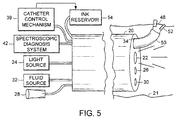
- Figs. 1-6 refer to an endoscopically insertable catheter having optical fibers for transmitting light to tissue and for conveying light back from tissue for analysis by a spectroscopic diagnosis system and having a needle for performing an interventional procedure on the tissue, namely, injecting india ink into tissue diagnosed by the spectroscopic diagnosis system in order to mark the tissue for biopsy, treatment, or removal by another interventional device. If the spectroscopic diagnosis system determines that the tissue should be treated, the catheter having the optical fibers and needle is withdrawn from the endoscope and a catheter having forceps jaws is inserted through the endoscope in order to obtain a biopsy of the tissue or to remove or treat the tissue.
- a distal end of an endoscope 20 is inserted through a lumen 21 of a patient's body, such as the esophagus, rectum or pulmonary tract.
- Endoscope 20 has an optical fiber 22 connected to a light source 24 located at a proximal end of endoscope 20 for transmitting light to tissue located at the distal end of endoscope 20, an optical fiber 26 connected to an eyepiece 28 located at the proximal end of endoscope 20 for viewing the tissue, a fluid channel 30 connected to a fluid source 32 located at the proximal end of endoscope 20 for flushing the tissue with fluid such as a wash for the visual fiber optics, and a working channel 34 for receiving a catheter.
- light source 24 is turned on.
- a catheter 36 having an elongated catheter shaft is inserted through working channel 34 of endoscope 20.
- Catheter 36 has optical fibers 38 and 40, which extend through the catheter shaft and are connected to a spectroscopic diagnosis system 42 located at the proximal end of endoscope 20.
- Optical fibers 38 and 40 can be made of a quartz glass component or other suitable glass or polymer material capable of transmitting and receiving wavelengths necessary to distinguish between healthy and abnormal tissue that has been treated by a diagnostic reagent.
- the optical fibers may be bundled together as a single light transmission and reception probe rather than the two discrete fibers shown in Figs.
- the probe including an outer sheath made of SST or a suitable semi-rigid polymer that is non-reactive to diagnostic reagents.
- Catheter 36 is connected to a catheter control mechanism 39 located at the proximal end of endoscope 20 that controls the longitudinal movement of catheter 36 within working channel 34.
- Catheter control mechanism 39 also controls the longitudinal movement of optical fibers 38, 40 and may extend or retract optical fibers 38, 40 with respect to catheter 36.
- light source 24 is turned on and optical fibers 38, 40 are extended from catheter 36 to the tissue.
- light source 24 is turned off, optical fiber 38 transmits light to the tissue, and optical fiber 40 conveys light back from the tissue for analysis by spectroscopic diagnosis system 42 to determine whether the tissue should be treated.
- optical fiber 38 transmits a monochromatic light beam having a wavelength or set of wavelengths selected to cause the tissue to fluoresce in a manner such that at one wavelength of the fluorescent light the intensity is approximately the same regardless of whether the tissue is normal or cancerous and at another wavelength of the fluorescent light the intensity differs substantially depending on whether the tissue is normal or cancerous.
- spectroscopic diagnosis system 42 can determine whether the tissue is normal or cancerous, and in some instances whether the tissue is a benign tumor.
- the tissue may first be treated by a diagnostic reagent that bonds more selectively with diseased (cancerous) tissue than with normal tissue, or vice versa, and that absorbs light transmitted through the catheter and thereby causes the tissue to fluoresce at a wavelength or set of wavelengths different from the transmission wavelength.
- a diagnostic reagent that bonds more selectively with diseased (cancerous) tissue than with normal tissue, or vice versa, and that absorbs light transmitted through the catheter and thereby causes the tissue to fluoresce at a wavelength or set of wavelengths different from the transmission wavelength.
- the intensity of the light conveyed back to the spectroscopic diagnosis system may be displayed graphically through the aid of a computer as a function of wavelength, and the endoscopist can interpret the data.
- catheter control mechanism 39 retracts optical fibers 38 and 40 into catheter 36, and light source 24 is turned on.
- Catheter control mechanism 39 also controls the longitudinal movement of a needle 44 which is located at the distal end of catheter 36 and extends needle 44 to the tissue. Needle 44, which can be made of SST or a suitable semirigid polymer that is non-reactive to diagnostic reagents, extends through a lumen 46 in the catheter shaft. Needle 44 and optical fibers 38 and 40 may be combined together as a single probe.
- Catheter control mechanism 39 causes india ink 48 from ink reservoir 54 to be injected through needle 44 into the tissue to be treated.
- Optical fiber 38 transmits light to the tissue at a number of points and optical fiber 40 conveys light back from the tissue for analysis by spectroscopic diagnosis system 42 in order to determine whether the tissue is cancerous at each of these points.
- Each such point that is determined to be cancerous is marked by india ink 48 using needle 44.
- Figs. 6-10 are drawings of different embodiments of an endoscopically insertable catheter 100 combining optical fibers and various other types of interventional devices.
- Catheter 100 can be inserted in an endoscope as shown in Figs. 1-5 in order to perform spectroscopic diagnosis on tissue and remove cancerous tissue without having to mark the cancerous tissue with india ink.
- the optical fibers are used to perform spectroscopic diagnosis on the remaining tissue to determine whether all of the cancerous tissue has been removed.
- catheter 100 has optical fibers 102, 104 glued to a surface of a scalpel 106.
- catheter 100 has optical fibers 102, 104 placed between forceps jaws 108, which may be multiple sampling biopsy forceps jaws capable of removing multiple samples of tissue into the body of catheter 100 as described in WO-A-9 508 292.
- the forceps jaws are caused to open up as shown in Fig. 7 by the withdrawal of an outer sleeve of the catheter (not shown) in the proximal direction and are caused to close together by pushing the outer sleeve in the distal direction.
- the forceps jaws can be used to remove tissue known to be unhealthy or to obtain biopsy samples for additional, cellular analysis.
- the optical fibers are of course retracted when the forceps jaws are being used. If the forceps jaws are of the multiple-biopsy type, the optical fibers can be bundled together as a single probe designed to spear through the biopsy samples within the catheter for continued diagnosis at additional sites.
- catheter 100 has optical fibers 102, 104 placed next to a polypectomy snare 110, which can be looped around a polyp and quickly retracted into catheter 100 to excise the polyp.
- the optical fibers may alternatively pass through the same lumen as the snare.
- catheter 100 has optical fibers 102, 104 placed next to scissors 112.
- the optical fibers may be glued to one of the scissors blades.
- catheter 100 has optical fibers 102, 104 placed next to a needle 114, similar in construction to needle 44 described above in connection with Figs. 1-5.
- Needle 114 may be constructed to inject chemical ablation fluid or other drugs into tissue, possibly including time-release capsules containing cancer-fighting substances, to cut tissue, or to apply glue for temporarily adhering tissue between the esophagus and the stomach for treating gastroesophageal reflux disease. Localized chemical ablation or drug treatment can be performed at high concentration because of the specificity with which spectroscopic diagnosis can identify the location of unhealthy tissue. Needle 114 of Fig. 10 or needle 44 of Figs.
- optical fibers 102, 104 are extendable and retractable with respect to catheter 100, and with the exception of the embodiment of Fig. 6, in which the optical fibers are glued to scalpel 106, any movement capabilities of optical fibers 102, 104 on the one hand and the interventional device on the other hand can be completely independent of each other.
- Each of the embodiments described above in connection with Figs. 1-10 can be modified by eliminating the endoscope and adding additional optical fibers to the catheter for use in connection with the imaging function, or by using one or more optical fibers of the catheter not only in connection with spectroscopic diagnosis function but also in connection with the imaging function. Since the catheter would be used without an endoscope, the catheter would have a steerable tip to allow movement and positioning of the catheter.
- the lumen of the catheter needle in embodiments having such a needle, can double as a wash for the visual fiber optics as well as a vacuum and air source.
- One advantage of such a construction is that the catheter would not have to be as large as an endoscope combined with a catheter, because no endoscope working channel is required.
- an endoscope 200 is combined with an endoscopically insertable catheter 216 having forceps jaws 218 and an endoscopically insertable catheter 212 having a rotatable ultrasound imaging transducer 214.
- Ultrasound imaging transducer 214 provides a visual image of the tumor, which can be useful in determining the depth to which a tumor has grown into or through the wall of a lumen in order to determine whether the tumor can be removed safely from the lumen. If the tissue structure imaged by ultrasound imaging transducer 214 can be removed safely from the lumen, forceps jaws 218 are used to remove the tissue structure. The visual image can also be used to determine how much tissue should be removed. The imaging can be performed simultaneously with the tissue removal, so as to enable the physician to see whether the tissue structure has been completely removed.
- Endoscope 200 has an optical fiber 202 for transmitting light from light source 230 to tissue located at a distal end of endoscope 200, an optical fiber 204 for conveying light back from the tissue to eyepiece 234 for viewing tissue, a fluid channel 206 for flushing tissue with fluid provided by fluid source 232, and working channels 208, 210 for receiving catheters 212 and 216 respectively.
- Interventional catheter control mechanism 224 controls the longitudinal movement of catheter 216 within working channel 210 as well as the operation of forceps jaws 218.
- ultrasound catheter control mechanism 226 controls the longitudinal movement of ultrasound catheter 212 within working channel 208 as well as the operation of ultrasound transducer 214, including its rotation, transmission of ultrasound pulses, and detection of reflected pulses by the transducer.
- the ultrasound image of the tissue imaged by the ultrasound transducer is displayed by ultrasound image display 228.
- the endoscope of Fig. 11 also may be used in combination with any of the different endoscopically insertable catheters combining optical fibers and interventional devices described above and shown in Figs. 6-10, which may be substituted for interventional catheter 216.
- catheter 216 has forceps jaws 218 and optical fibers 220, 222, which are connected to a spectroscopic diagnosis system 236 that spectroscopically diagnoses the tissue to determine whether the tissue should be treated, as is described above in connection with Fig. 7.
- ultrasound imaging catheter 214 is used to measure the depth to which a tumor has grown into or through the wall of a lumen to determine whether the tumor can be removed safely from the lumen. If the cancerous tissue imaged by ultrasound imaging device 214 can be removed safely from the lumen, forceps jaws 218 are used to remove the cancerous tissue. After forceps jaws 218 remove the cancerous tissue, optical fibers 220, 222 are used to perform spectroscopic diagnosis on the tissue to determine whether all of the cancerous tissue has been removed.
Landscapes
- Health & Medical Sciences (AREA)
- Life Sciences & Earth Sciences (AREA)
- Surgery (AREA)
- Physics & Mathematics (AREA)
- General Health & Medical Sciences (AREA)
- Engineering & Computer Science (AREA)
- Biomedical Technology (AREA)
- Heart & Thoracic Surgery (AREA)
- Medical Informatics (AREA)
- Molecular Biology (AREA)
- Animal Behavior & Ethology (AREA)
- Public Health (AREA)
- Veterinary Medicine (AREA)
- Pathology (AREA)
- Biophysics (AREA)
- Nuclear Medicine, Radiotherapy & Molecular Imaging (AREA)
- Radiology & Medical Imaging (AREA)
- Optics & Photonics (AREA)
- Spectroscopy & Molecular Physics (AREA)
- Orthopedic Medicine & Surgery (AREA)
- Vascular Medicine (AREA)
- Endoscopes (AREA)
- Investigating Or Analysing Materials By Optical Means (AREA)
- Measurement Of The Respiration, Hearing Ability, Form, And Blood Characteristics Of Living Organisms (AREA)
- Ultra Sonic Daignosis Equipment (AREA)
Claims (16)
- Ein Katheter (36), bestehend aus:einem verlängerten Katheterschaft mit mindestens zwei Kanälen (46) und einem distalen Ende;einem ersten, in einen der mindestens zwei Kanäle des Katheterschaftes einsetz- und herausnehmbaren und bis über das distale Ende des Katheterschaftes hinaus ausschiebbaren Lichtleiter (38) zur Übertragung von Licht zu am distalen Ende des Katheterschaftes befindlichem Gewebe;einem zweiten, in einen der mindestens zwei Kanäle des Katheterschaftes einsetz- und herausnehmbaren und bis über das distale Ende des Katheterschaftes hinaus ausschiebbaren Lichtleiter (40) zur Rückübertragung des Lichtes von dem Gewebe zur Analyse durch ein spektroskopisches Diagnosesystem (42) zwecks Entscheidung, ob ein Interventionsverfahren an dem Gewebe vorgenommen werden soll, wobei besagter zweiter und erster Lichtleiter diskrete Leiter sind, in deren mindestens einen ein Beobachtungsgerät einbezogen ist zur Umwandlung weiteren, vom Gewebe zurückgeführten Lichtes in ein visuelles Beobachtungsbild des Gewebes.; sowieeine am distalen Ende des Katheterschaftes angeordnete Eingriffsvorrichtung (52) zum Eingriff in von dem spektroskopischen Diagnosesystem diagnostiziertes Gewebe, um das Interventionsverfahren an dem Gewebe auszuführen, wobei besagte Eingriffsvorrichtung von den genannten ersten und zweiten Lichtleitern physikalisch trennbar ist.
- Katheter nach Anspruch 1, bei dem die Eingriffsvorrichtung ein Skalpell umfasst.
- Katheter nach Anspruch 1, bei dem die Eingriffsvorrichtung zangenklauen umfasst.
- Katheter nach Anspruch 1, bei dem die Eingriffsvorrichtung eine Schlinge umfasst.
- Katheter nach Anspruch 1, bei dem die Eingriffsvorrichtung eine Schere umfasst.
- Katheter nach Anspruch 1, bei dem die Eingriffsvorrichtung eine Nadel umfasst.
- Katheter nach Anspruch 6, bei dem die Nadel zum Injizieren einer Markierungsflüssigkeit in das Gewebe ausgebildet ist.
- Katheter nach Anspruch 6, bei dem die Nadel zum Injizieren einer chemischen Ablationsflüssigkeit in das Gewebe vorgesehen ist.
- Katheter nach Anspruch 6, bei dem die Nadel zum Schneiden des Gewebes vorgesehen ist.
- Katheter nach Anspruch 6, bei dem die Nadel zum Applizieren eines Haftmittels zu dem Gewebe vorgesehen ist.
- Katheter nach Anspruch 6, bei dem die Nadel zum zuführen einer für die visuelle Beobachtung des Gewebes nützlichen Flüssigkeit zu dem Gewebe vorgesehen ist.
- Katheter nach Anspruch 6, bei dem die Nadel zum Applizieren eines vakuums zu dem Gewebe vorgesehen ist.
- Katheter nach Anspruch 1, bei dem der Katheterschaft passend zum Einführen durch den Arbeitskanal eines Endoskops ausgebildet ist.
- Katheter nach Anspruch 1, bei dem mindestens einer der Lichtleiter zur zufuhr von Visualisierungslicht zu dem Gewebe vorgesehen ist.
- Eine Anordnung bestehend aus:einem Endoskop (20);einem verlängerten Katheterschaft mit mindestens zwei Kanälen (46) und einem distalen Ende, der zum Einführen durch einen Arbeitskanal (34) des Endoskops ausgebildet ist;einem ersten, in einen der mindestens zwei Kanäle des Katheterschaftes einsetz- und herausnehmbaren und bis über das distale Ende des Katheterschaftes hinaus ausschiebbaren Lichtleiter (38) zur Übertragung von Licht zu am distalen Ende des Katheterschaftes befindlichen Gewebe;einem zweiten, in einen der mindestens zwei Kanäle des Katheterschaftes einsetz- und herausnembaren und bis über das distale Ende des Katheterschaftes hinaus ausschiebbaren Lichtleiter (40) zur Rückübertragung des Lichtes von dem Gewebe zur Analyse durch ein spektroskopisches Diagnosesystem (42) zwecks Entscheidung, ob ein Interventionsverfahren an dem Gewebe vorgenommen werden soll, wobei besagter zweiter und erster Lichtleiter diskrete Leiter sind, in deren mindestens einen ein Beobachtungsgerät einbezogen ist zur Umwandlung weiteren, vom Gewebe zurückgeführten Lichtes in ein visuelles Beobachtungsbild des Gewebes; sowieeine Eingriffsvorrichtung (52), die zum Einführen durch einen Arbeitskanal eines Endoskops ausgebildet ist, zum Eingriff in von dem spektroskopischen Diagnosesystem diagnostiziertes Gewebe, um das Interventionsverfahren an dem Gewebe auszuführen, wobei besagte Eingriffsvorrichtung von den genannten ersten und zweiten Lichtleitern physikalisch trennbar ist.
- Eine Anordnung nach Anspruch 15, bei der die Eingriffsvorrichtung am distalen Ende des die Lichtleiter aufweisenden Katheterschaftes angeordnet ist.
Applications Claiming Priority (3)
| Application Number | Priority Date | Filing Date | Title |
|---|---|---|---|
| US679425 | 1991-04-02 | ||
| US08/679,425 US6296608B1 (en) | 1996-07-08 | 1996-07-08 | Diagnosing and performing interventional procedures on tissue in vivo |
| PCT/US1997/011864 WO1998001074A1 (en) | 1996-07-08 | 1997-07-07 | Diagnosing and performing interventional procedures on tissue in vivo |
Publications (2)
| Publication Number | Publication Date |
|---|---|
| EP0925034A1 EP0925034A1 (de) | 1999-06-30 |
| EP0925034B1 true EP0925034B1 (de) | 2004-03-24 |
Family
ID=24726865
Family Applications (1)
| Application Number | Title | Priority Date | Filing Date |
|---|---|---|---|
| EP97932540A Expired - Lifetime EP0925034B1 (de) | 1996-07-08 | 1997-07-07 | Vorrichtung zur diagnostizierung und durchführung von interventionsverfahren auf gewebe in vivo |
Country Status (6)
| Country | Link |
|---|---|
| US (3) | US6296608B1 (de) |
| EP (1) | EP0925034B1 (de) |
| JP (1) | JP3943600B2 (de) |
| CA (1) | CA2260147A1 (de) |
| DE (1) | DE69728277T2 (de) |
| WO (1) | WO1998001074A1 (de) |
Families Citing this family (252)
| Publication number | Priority date | Publication date | Assignee | Title |
|---|---|---|---|---|
| US6564087B1 (en) * | 1991-04-29 | 2003-05-13 | Massachusetts Institute Of Technology | Fiber optic needle probes for optical coherence tomography imaging |
| US6296608B1 (en) * | 1996-07-08 | 2001-10-02 | Boston Scientific Corporation | Diagnosing and performing interventional procedures on tissue in vivo |
| US5924976A (en) * | 1997-08-21 | 1999-07-20 | Stelzer; Paul | Minimally invasive surgery device |
| AU752829B2 (en) * | 1998-01-26 | 2002-10-03 | Brigham And Women's Hospital | Fluorescence imaging endoscope |
| US6976957B1 (en) * | 1998-06-22 | 2005-12-20 | Origin Medsystems, Inc. | Cannula-based surgical instrument and method |
| US6228076B1 (en) * | 1999-01-09 | 2001-05-08 | Intraluminal Therapeutics, Inc. | System and method for controlling tissue ablation |
| US6697666B1 (en) * | 1999-06-22 | 2004-02-24 | Board Of Regents, The University Of Texas System | Apparatus for the characterization of tissue of epithelial lined viscus |
| US6537205B1 (en) * | 1999-10-14 | 2003-03-25 | Scimed Life Systems, Inc. | Endoscopic instrument system having reduced backlash control wire action |
| US6454702B1 (en) * | 1999-10-14 | 2002-09-24 | Scimed Life Systems, Inc. | Endoscope and endoscopic instrument system having reduced backlash when moving the endoscopic instrument within a working channel of the endoscope |
| WO2001030429A1 (en) * | 1999-10-22 | 2001-05-03 | Scimed Life Systems, Inc. | Guided injection device |
| US6685666B1 (en) * | 1999-11-12 | 2004-02-03 | Mark G. Fontenot | Catheters for breast surgery |
| US6458076B1 (en) | 2000-02-01 | 2002-10-01 | 5 Star Medical | Multi-lumen medical device |
| US6869430B2 (en) | 2000-03-31 | 2005-03-22 | Rita Medical Systems, Inc. | Tissue biopsy and treatment apparatus and method |
| DE60141090D1 (de) | 2000-10-30 | 2010-03-04 | Gen Hospital Corp | Optische systeme zur gewebeanalyse |
| US9295391B1 (en) | 2000-11-10 | 2016-03-29 | The General Hospital Corporation | Spectrally encoded miniature endoscopic imaging probe |
| US6432064B1 (en) * | 2001-04-09 | 2002-08-13 | Ethicon Endo-Surgery, Inc. | Biopsy instrument with tissue marking element |
| EP2333522A1 (de) | 2001-04-30 | 2011-06-15 | The General Hospital Corporation | Verfahren und Vorrichtung zur verbesserung der Bildklarheit und Empfindlichkeit bei der optischen Kohärenz-Tomographie unter Verwendung von dynamischer Rückkopplung zur kontrolle der Fokussierungseigenschaften und der Kohärenzsteuerung |
| US20060201351A1 (en) * | 2001-07-02 | 2006-09-14 | Gi View Ltd. | Self-propelled imaging system |
| US8116845B2 (en) | 2005-08-04 | 2012-02-14 | Dune Medical Devices Ltd. | Tissue-characterization probe with effective sensor-to-tissue contact |
| US8721565B2 (en) | 2005-08-04 | 2014-05-13 | Dune Medical Devices Ltd. | Device for forming an effective sensor-to-tissue contact |
| US20080154090A1 (en) * | 2005-01-04 | 2008-06-26 | Dune Medical Devices Ltd. | Endoscopic System for In-Vivo Procedures |
| US20080287750A1 (en) * | 2002-01-04 | 2008-11-20 | Dune Medical Devices Ltd. | Ergonomic probes |
| US20040010204A1 (en) * | 2002-03-28 | 2004-01-15 | Pearl Technology Holdings, Llc | Electronic/fiberoptic tissue differentiation instrumentation |
| US20030225432A1 (en) * | 2002-05-31 | 2003-12-04 | Baptiste Reginald C. | Soft tissue retraction device for an endoscopic instrument |
| JP2004024331A (ja) * | 2002-06-21 | 2004-01-29 | Vayu:Kk | カテーテル |
| AU2003258250A1 (en) * | 2002-08-16 | 2004-03-03 | Beth Israel Deaconess Medical Center | Apparatus for multifocal deposition and analysis and methods for its use |
| US8123698B2 (en) * | 2002-10-07 | 2012-02-28 | Suros Surgical Systems, Inc. | System and method for minimally invasive disease therapy |
| JP4477382B2 (ja) * | 2003-03-04 | 2010-06-09 | オリンパス株式会社 | 内視鏡的腹腔内処置システム |
| US8187288B2 (en) | 2003-03-10 | 2012-05-29 | Boston Scientific Scimed, Inc. | Re-shapeable medical device |
| EP1611470B1 (de) | 2003-03-31 | 2015-10-14 | The General Hospital Corporation | Speckle-reduktion bei der optischen kohärenztomographie durch weglängencodierte winkelzusammensetzung |
| US20040199052A1 (en) | 2003-04-01 | 2004-10-07 | Scimed Life Systems, Inc. | Endoscopic imaging system |
| WO2005000096A2 (en) | 2003-06-05 | 2005-01-06 | Hydrocision, Inc. | Disposable endoscope and method of making a disposable endoscope |
| EP2290339A3 (de) | 2003-06-06 | 2012-11-28 | The General Hospital Corporation | Verfahren und Vorrichtung für eine Lichtquelle mit Abstimmung der Wellenlänge |
| US8403828B2 (en) * | 2003-07-21 | 2013-03-26 | Vanderbilt University | Ophthalmic orbital surgery apparatus and method and image-guide navigation system |
| US10610406B2 (en) * | 2004-07-21 | 2020-04-07 | Vanderbilt University | Drug delivery device and applications of same |
| US7833176B2 (en) * | 2003-08-13 | 2010-11-16 | G. I. View Ltd. | Pressure-propelled system for body lumen |
| US20050038319A1 (en) * | 2003-08-13 | 2005-02-17 | Benad Goldwasser | Gastrointestinal tool over guidewire |
| US20050036059A1 (en) * | 2003-08-13 | 2005-02-17 | Benad Goldwasser | Ingestible imaging system |
| US20050038318A1 (en) * | 2003-08-13 | 2005-02-17 | Benad Goldwasser | Gastrointestinal tool over guidewire |
| US8172770B2 (en) * | 2005-09-28 | 2012-05-08 | Suros Surgical Systems, Inc. | System and method for minimally invasive disease therapy |
| US20120289859A9 (en) * | 2003-08-27 | 2012-11-15 | Nicoson Zachary R | System and method for minimally invasive disease therapy |
| EP2270448B1 (de) | 2003-10-27 | 2020-03-18 | The General Hospital Corporation | Verfahren und Vorrichtung zur Durchführung optischer Abbildung mit Frequenzdomäneninterferometrie |
| US7635346B2 (en) * | 2004-01-09 | 2009-12-22 | G. I. View Ltd. | Pressure-propelled system for body lumen |
| US7635345B2 (en) * | 2004-01-09 | 2009-12-22 | G. I. View Ltd. | Pressure-propelled system for body lumen |
| US8419678B2 (en) * | 2004-01-09 | 2013-04-16 | G.I. View Ltd. | Pressure-propelled system for body lumen |
| US7922654B2 (en) * | 2004-08-09 | 2011-04-12 | Boston Scientific Scimed, Inc. | Fiber optic imaging catheter |
| EP4026486A1 (de) * | 2004-03-23 | 2022-07-13 | Boston Scientific Medical Device Limited | In-vivo-visualisierungssystem |
| US11819192B2 (en) | 2004-03-23 | 2023-11-21 | Boston Scientific Scimed, Inc. | In-vivo visualization system |
| WO2006014392A1 (en) | 2004-07-02 | 2006-02-09 | The General Hospital Corporation | Endoscopic imaging probe comprising dual clad fibre |
| US8081316B2 (en) | 2004-08-06 | 2011-12-20 | The General Hospital Corporation | Process, system and software arrangement for determining at least one location in a sample using an optical coherence tomography |
| US8965487B2 (en) | 2004-08-24 | 2015-02-24 | The General Hospital Corporation | Process, system and software arrangement for measuring a mechanical strain and elastic properties of a sample |
| JP5324095B2 (ja) | 2004-08-24 | 2013-10-23 | ザ ジェネラル ホスピタル コーポレイション | 血管セグメントを画像化する方法および装置 |
| EP2329759B1 (de) | 2004-09-29 | 2014-03-12 | The General Hospital Corporation | System und Verfahren zur Abbildung optischer Kohärenz |
| WO2006055845A1 (en) * | 2004-11-15 | 2006-05-26 | Cytyc Corporation | System for drug delivery |
| JP2008521516A (ja) | 2004-11-29 | 2008-06-26 | ザ ジェネラル ホスピタル コーポレイション | サンプル上の複数の地点を同時に照射し検出することによって光学画像生成を実行する構成、装置、内視鏡、カテーテル、及び方法 |
| US8182422B2 (en) | 2005-12-13 | 2012-05-22 | Avantis Medical Systems, Inc. | Endoscope having detachable imaging device and method of using |
| US8310530B2 (en) | 2006-05-19 | 2012-11-13 | Avantis Medical Systems, Inc. | Device and method for reducing effects of video artifacts |
| US8235887B2 (en) | 2006-01-23 | 2012-08-07 | Avantis Medical Systems, Inc. | Endoscope assembly with retroscope |
| US8797392B2 (en) | 2005-01-05 | 2014-08-05 | Avantis Medical Sytems, Inc. | Endoscope assembly with a polarizing filter |
| US8289381B2 (en) | 2005-01-05 | 2012-10-16 | Avantis Medical Systems, Inc. | Endoscope with an imaging catheter assembly and method of configuring an endoscope |
| US8872906B2 (en) | 2005-01-05 | 2014-10-28 | Avantis Medical Systems, Inc. | Endoscope assembly with a polarizing filter |
| WO2006085316A2 (en) | 2005-02-10 | 2006-08-17 | G.I. View Ltd. | Advancement techniques for gastrointestinal tool with guiding element |
| EP2325803A1 (de) | 2005-04-28 | 2011-05-25 | The General Hospital Corporation | Beurteilung von optischen Kohärenztomographieinformationen für eine anatomische Struktur |
| DE102005033474A1 (de) * | 2005-07-18 | 2007-01-25 | Heywang-Köbrunner, Sylvia, Prof. Dr. | Verfahren zur Untersuchung von Gewebeproben, Vorrichtung dafür und neue Verwendung von Fluoreszenzmarkern |
| KR101387454B1 (ko) | 2005-08-09 | 2014-04-22 | 더 제너럴 하스피탈 코포레이션 | 광간섭 단층촬영법에서 편광 기반 직교 복조를 수행하기위한 장치, 방법 및 저장 매체 |
| US20070049833A1 (en) * | 2005-08-16 | 2007-03-01 | The General Hospital Corporation | Arrangements and methods for imaging in vessels |
| US20080200834A1 (en) * | 2005-09-28 | 2008-08-21 | Mark Joseph L | Introducer device for improved imaging |
| CN101304683B (zh) | 2005-09-29 | 2012-12-12 | 通用医疗公司 | 以渐进增加的分辨率观察和分析一个或多个生物样品的方法和用于该方法的设备 |
| US7901441B2 (en) | 2005-10-18 | 2011-03-08 | Boston Scientific Scimed, Inc. | Method of using an imaging catheter to conduct photodynamic procedures |
| JP2009512539A (ja) * | 2005-10-24 | 2009-03-26 | スペクトラサイエンス,インコーポレーテッド | 病変組織の非内視鏡的光バイオプシー検出のためのシステムおよび方法 |
| US20070129630A1 (en) * | 2005-12-07 | 2007-06-07 | Shimko Daniel A | Imaging method, device and system |
| EP2289397A3 (de) | 2006-01-19 | 2011-04-06 | The General Hospital Corporation | Verfahren und Systeme zur optischen Bildgebung von epithelialen Luminalorganen durch Strahlenabtastung dieser |
| US8145018B2 (en) | 2006-01-19 | 2012-03-27 | The General Hospital Corporation | Apparatus for obtaining information for a structure using spectrally-encoded endoscopy techniques and methods for producing one or more optical arrangements |
| WO2007149603A2 (en) | 2006-02-01 | 2007-12-27 | The General Hospital Corporation | Apparatus for applying a plurality of electro-magnetic radiations to a sample |
| US10426548B2 (en) | 2006-02-01 | 2019-10-01 | The General Hosppital Corporation | Methods and systems for providing electromagnetic radiation to at least one portion of a sample using conformal laser therapy procedures |
| WO2007092911A2 (en) | 2006-02-08 | 2007-08-16 | The General Hospital Corporation | Methods, arrangements and systems for obtaining information associated with an anatomical sample using optical microscopy |
| JP2009527770A (ja) | 2006-02-24 | 2009-07-30 | ザ ジェネラル ホスピタル コーポレイション | 角度分解型のフーリエドメイン光干渉断層撮影法を遂行する方法及びシステム |
| US7473232B2 (en) | 2006-02-24 | 2009-01-06 | Boston Scientific Scimed, Inc. | Obtaining a tissue sample |
| JP2007296324A (ja) * | 2006-04-06 | 2007-11-15 | Olympus Corp | 生体組織の観察方法 |
| US20090203991A1 (en) * | 2006-04-21 | 2009-08-13 | Cedars-Sinai Medical Center | Multiple imaging and/or spectroscopic modality probe |
| EP2517616A3 (de) | 2006-05-10 | 2013-03-06 | The General Hospital Corporation | Prozesse, Anordnungen und Systeme zur Bereitstellung der Frequenzbereichsabbildung einer Probe |
| EP2029213A2 (de) * | 2006-06-14 | 2009-03-04 | Cornova, Inc. | Verfahren und vorrichtung zur identifikation und behandlung von myokardininfarkten |
| EP2032016A2 (de) * | 2006-06-14 | 2009-03-11 | Optivia Medical LLC | Systeme und verfahren zur einführung einer medizinischen vorrichtung |
| AU2007275720A1 (en) * | 2006-07-18 | 2008-01-24 | Boston Medical Center Corporation | Device with integrated multi-fiber optical probe and methods of use |
| JP2009544430A (ja) * | 2006-07-26 | 2009-12-17 | ハンセン メディカル,インク. | 最小侵襲の外科手術を行うためのシステム |
| WO2008017080A2 (en) * | 2006-08-03 | 2008-02-07 | Hansen Medical, Inc. | Systems for performing minimally invasive procedures |
| FR2904927B1 (fr) * | 2006-08-17 | 2018-05-18 | Mauna Kea Technologies | Utilisation d'un systeme d'imagerie par fluorescence confocale fibre in vivo in situ, systeme et procede d'imagerie par fluorescence confocale fibres in vivo in situ |
| IL177550A0 (en) * | 2006-08-17 | 2006-12-31 | Sialo Technology Israel Ltd | All-in-one optical microscopic handle |
| US20080051626A1 (en) * | 2006-08-28 | 2008-02-28 | Olympus Medical Systems Corp. | Fistulectomy method between first duct and second duct, ultrasonic endoscope, catheter with balloon, magnet retaining device, and magnet set |
| JP5073415B2 (ja) * | 2006-08-28 | 2012-11-14 | オリンパスメディカルシステムズ株式会社 | 超音波内視鏡 |
| US8238678B2 (en) * | 2006-08-30 | 2012-08-07 | Siemens Medical Solutions Usa, Inc. | Providing representative image information |
| US8838213B2 (en) | 2006-10-19 | 2014-09-16 | The General Hospital Corporation | Apparatus and method for obtaining and providing imaging information associated with at least one portion of a sample, and effecting such portion(s) |
| US20080097224A1 (en) * | 2006-10-20 | 2008-04-24 | Infraredx, Inc. | Manual and Motor Driven Optical Pullback and Rotation System and Method |
| US20080097223A1 (en) * | 2006-10-20 | 2008-04-24 | Infraredx, Inc. | Optical Catheter Carriage Interlock System and Method |
| US20080097158A1 (en) * | 2006-10-20 | 2008-04-24 | Infraredx, Inc. | Noise Suppression System and Method in Catheter Pullback and Rotation System |
| US20080097408A1 (en) * | 2006-10-20 | 2008-04-24 | Infraredx, Inc. | Pullback Carriage Interlock System and Method for Catheter System |
| WO2008066911A2 (en) * | 2006-11-30 | 2008-06-05 | Newton Laboratories, Inc. | Spectroscopically enhanced imaging |
| JP2008161570A (ja) * | 2006-12-28 | 2008-07-17 | Olympus Medical Systems Corp | 超音波内視鏡システム |
| US8372000B2 (en) | 2007-01-03 | 2013-02-12 | Boston Scientific Scimed, Inc. | Method and apparatus for biliary access and stone retrieval |
| US8801606B2 (en) * | 2007-01-09 | 2014-08-12 | Ethicon Endo-Surgery, Inc. | Method of in vivo monitoring using an imaging system including scanned beam imaging unit |
| US8273015B2 (en) * | 2007-01-09 | 2012-09-25 | Ethicon Endo-Surgery, Inc. | Methods for imaging the anatomy with an anatomically secured scanner assembly |
| JP2010516325A (ja) * | 2007-01-17 | 2010-05-20 | ジー・アイ・ヴュー・リミテッド | 結腸内視術のための診断用または治療用ツール |
| US20080172065A1 (en) * | 2007-01-17 | 2008-07-17 | Isaac Ostrovsky | Medical device with beacon |
| WO2008100855A1 (en) | 2007-02-12 | 2008-08-21 | Boston Scientific Limited | Endoscope cap |
| WO2008101003A1 (en) * | 2007-02-13 | 2008-08-21 | Cornova, Inc. | Biocompatible polymers, polymer tie-coats, methods of making and using the same, and products incorporating the polymers |
| US20080214890A1 (en) * | 2007-03-01 | 2008-09-04 | Olympus Medical Systems Corporation | Therapeutic method and therapeutic system used with steps for approaching to lesion using overtube |
| EP2602651A3 (de) | 2007-03-23 | 2014-08-27 | The General Hospital Corporation | Verfahren, Anordnungen und Vorrichtung zur Verwendung eines wellenlängengewobbelten Lasers anhand von Winkelabtastungs- und Dispersionsverfahren |
| WO2008121844A1 (en) | 2007-03-30 | 2008-10-09 | The General Hospital Corporation | System and method providing intracoronary laser speckle imaging for the detection of vulnerable plaque |
| US8064666B2 (en) | 2007-04-10 | 2011-11-22 | Avantis Medical Systems, Inc. | Method and device for examining or imaging an interior surface of a cavity |
| WO2008131093A2 (en) * | 2007-04-17 | 2008-10-30 | Fox Chase Cancer Center | Method and apparatus for endoscopic examination of lesions |
| US9125552B2 (en) * | 2007-07-31 | 2015-09-08 | Ethicon Endo-Surgery, Inc. | Optical scanning module and means for attaching the module to medical instruments for introducing the module into the anatomy |
| EP2173254A2 (de) | 2007-07-31 | 2010-04-14 | The General Hospital Corporation | System und verfahren zur bereitstellung von strahlerfassungsmustern für hochgeschwindigkeits-abbildungen optischer dopplerfrequenzdomänen |
| EP2033571B1 (de) | 2007-09-05 | 2017-02-08 | Vision-Sciences Inc. | Kompakte Endoskopspitze und Verfahren zu deren Herstellung |
| US8280496B2 (en) | 2007-12-13 | 2012-10-02 | Boston Scientific Scimed, Inc. | Extended spectral sensitivity endoscope system and method of using the same |
| US11123047B2 (en) | 2008-01-28 | 2021-09-21 | The General Hospital Corporation | Hybrid systems and methods for multi-modal acquisition of intravascular imaging data and counteracting the effects of signal absorption in blood |
| US9332942B2 (en) | 2008-01-28 | 2016-05-10 | The General Hospital Corporation | Systems, processes and computer-accessible medium for providing hybrid flourescence and optical coherence tomography imaging |
| FR2928077B1 (fr) * | 2008-02-29 | 2011-03-25 | Echosens | Dispositif et procede de micro-elastographie. |
| US20100331782A1 (en) * | 2008-03-03 | 2010-12-30 | Koninklijke Philips Electronics N.V. | Biopsy guidance by image-based x-ray guidance system and photonic needle |
| US8016814B2 (en) * | 2008-03-10 | 2011-09-13 | Medtronic Vascular, Inc. | Guidewires and delivery catheters having fiber optic sensing components and related systems and methods |
| US20090237498A1 (en) * | 2008-03-20 | 2009-09-24 | Modell Mark D | System and methods for the improvement of images generated by fiberoptic imaging bundles |
| WO2009137659A1 (en) | 2008-05-07 | 2009-11-12 | Infraredx, Inc. | Multimodal catheter system and method for intravascular analysis |
| US9289262B2 (en) * | 2008-05-19 | 2016-03-22 | Boston Scientific Scimed, Inc. | Dielectric coatings for laser fiber and related methods |
| US20090287200A1 (en) * | 2008-05-19 | 2009-11-19 | Brian Hanley | Side-firing laser fiber with glass fused reflector and capillary and related methods |
| US20090287199A1 (en) * | 2008-05-19 | 2009-11-19 | Brian Hanley | Side-firing laser fiber with protective tip and related methods |
| US8657812B2 (en) * | 2008-05-19 | 2014-02-25 | Boston Scientific Scimed, Inc. | Side-firing laser fiber with internal bent fiber and related methods |
| US8425500B2 (en) | 2008-05-19 | 2013-04-23 | Boston Scientific Scimed, Inc. | Method and apparatus for protecting capillary of laser fiber during insertion and reducing metal cap degradation |
| US9820719B2 (en) * | 2008-06-19 | 2017-11-21 | Cogentix Medical, Inc. | Method and system for intrabody imaging |
| JP5795531B2 (ja) | 2008-06-20 | 2015-10-14 | ザ ジェネラル ホスピタル コーポレイション | フューズドファイバオプティックカプラ構造、及びその使用方法 |
| US20090326525A1 (en) * | 2008-06-26 | 2009-12-31 | Jessica Hixon | Laser fiber capillary apparatus and method |
| US9254089B2 (en) | 2008-07-14 | 2016-02-09 | The General Hospital Corporation | Apparatus and methods for facilitating at least partial overlap of dispersed ration on at least one sample |
| CN102131452B (zh) * | 2008-07-30 | 2014-03-12 | G.I.视频有限公司 | 可操作性增强的系统和方法 |
| US9358369B1 (en) * | 2008-08-13 | 2016-06-07 | Abbott Cardiovascular Systems Inc. | Reduced profile and enhanced flexibility delivery catheters for light activated agents |
| US8170657B1 (en) * | 2008-08-13 | 2012-05-01 | Abbott Cadiovascular Systems Inc. | Delivery catheters for light activated agents |
| WO2010028034A2 (en) * | 2008-09-02 | 2010-03-11 | Epitek, Inc. | Method and apparatus for locating heart structures inside the pericardial sac through a sub-xiphoid approach |
| US20100069760A1 (en) * | 2008-09-17 | 2010-03-18 | Cornova, Inc. | Methods and apparatus for analyzing and locally treating a body lumen |
| US20100081923A1 (en) * | 2008-09-29 | 2010-04-01 | Searete Llc, A Limited Liability Corporation Of The State Of Delaware | Histological facilitation systems and methods |
| US20100081926A1 (en) * | 2008-09-29 | 2010-04-01 | Searete Llc, A Limited Liability Corporation Of The State Of Delaware | Histological facilitation systems and methods |
| US20100081916A1 (en) * | 2008-09-29 | 2010-04-01 | Searete Llc, A Limited Liability Corporation Of The State Of Delaware. | Histological facilitation systems and methods |
| US20100081924A1 (en) * | 2008-09-29 | 2010-04-01 | Searete Llc, A Limited Liability Corporation Of The State Of Delaware | Histological facilitation systems and methods |
| US20100081927A1 (en) * | 2008-09-29 | 2010-04-01 | Searete Llc, A Limited Liability Corporation Of The State Of Delaware | Histological facilitation systems and methods |
| US20100081915A1 (en) * | 2008-09-29 | 2010-04-01 | Searete Llc, Alimited Liability Corporation Of The State Of Delaware | Histological facilitation systems and methods |
| US20100081190A1 (en) * | 2008-09-29 | 2010-04-01 | Searete Llc, A Limited Liability Corporation Of The State Of Delaware | Histological facilitation systems and methods |
| US20100081919A1 (en) * | 2008-09-29 | 2010-04-01 | Searete Llc, A Limited Liability Corporation Of The State Of Delaware | Histological facilitation systems and methods |
| US20100081925A1 (en) * | 2008-09-29 | 2010-04-01 | Searete Llc, A Limited Liability Corporation Of The State Of Delaware | Histological facilitation systems and methods |
| US20100081928A1 (en) * | 2008-09-29 | 2010-04-01 | Searete Llc, A Limited Liability Corporation Of The State Of Delaware | Histological Facilitation systems and methods |
| WO2010042217A1 (en) * | 2008-10-09 | 2010-04-15 | Sti Medical Systems, Llc | Process for preserving three dimensional orientation to allow registering histopathological diagnoses of tissue |
| AU2009321185B2 (en) | 2008-11-03 | 2014-05-01 | G.I. View Ltd | Remote pressure sensing system and method thereof |
| ES2957932T3 (es) | 2008-12-10 | 2024-01-30 | Massachusetts Gen Hospital | Sistemas, aparatos y procedimientos para ampliar el rango de profundidad de imagen de tomografía de coherencia óptica mediante submuestreo óptico |
| JP6053284B2 (ja) | 2009-02-04 | 2016-12-27 | ザ ジェネラル ホスピタル コーポレイション | ハイスピード光学波長チューニング源の利用のための装置及び方法 |
| WO2010105197A2 (en) | 2009-03-12 | 2010-09-16 | The General Hospital Corporation | Non-contact optical system, computer-accessible medium and method for measuring at least one mechanical property of tissue using coherent speckle techniques(s) |
| US8495931B2 (en) | 2009-04-23 | 2013-07-30 | John S. Sroka | Ratchet wrench |
| EP2429376B1 (de) | 2009-05-08 | 2016-10-19 | Boston Scientific Scimed, Inc. | Endoskop mit distaler spitze mit verkapselten optischen komponenten und anzeigeausrichtungsfunktionen |
| US10165929B2 (en) | 2009-06-18 | 2019-01-01 | Endochoice, Inc. | Compact multi-viewing element endoscope system |
| US8926502B2 (en) | 2011-03-07 | 2015-01-06 | Endochoice, Inc. | Multi camera endoscope having a side service channel |
| CA2765559C (en) | 2009-06-18 | 2017-09-05 | Peer Medical Ltd. | Multi-camera endoscope |
| US9713417B2 (en) | 2009-06-18 | 2017-07-25 | Endochoice, Inc. | Image capture assembly for use in a multi-viewing elements endoscope |
| US11547275B2 (en) | 2009-06-18 | 2023-01-10 | Endochoice, Inc. | Compact multi-viewing element endoscope system |
| US9901244B2 (en) | 2009-06-18 | 2018-02-27 | Endochoice, Inc. | Circuit board assembly of a multiple viewing elements endoscope |
| US9101287B2 (en) | 2011-03-07 | 2015-08-11 | Endochoice Innovation Center Ltd. | Multi camera endoscope assembly having multiple working channels |
| US9402533B2 (en) | 2011-03-07 | 2016-08-02 | Endochoice Innovation Center Ltd. | Endoscope circuit board assembly |
| US9872609B2 (en) | 2009-06-18 | 2018-01-23 | Endochoice Innovation Center Ltd. | Multi-camera endoscope |
| WO2012077117A1 (en) | 2010-12-09 | 2012-06-14 | Peermedical Ltd. | Flexible electronic circuit board multi-camera endoscope |
| US9492063B2 (en) | 2009-06-18 | 2016-11-15 | Endochoice Innovation Center Ltd. | Multi-viewing element endoscope |
| US9101268B2 (en) | 2009-06-18 | 2015-08-11 | Endochoice Innovation Center Ltd. | Multi-camera endoscope |
| US11864734B2 (en) | 2009-06-18 | 2024-01-09 | Endochoice, Inc. | Multi-camera endoscope |
| US9642513B2 (en) | 2009-06-18 | 2017-05-09 | Endochoice Inc. | Compact multi-viewing element endoscope system |
| US9706903B2 (en) | 2009-06-18 | 2017-07-18 | Endochoice, Inc. | Multiple viewing elements endoscope system with modular imaging units |
| US11278190B2 (en) | 2009-06-18 | 2022-03-22 | Endochoice, Inc. | Multi-viewing element endoscope |
| US11490826B2 (en) | 2009-07-14 | 2022-11-08 | The General Hospital Corporation | Apparatus, systems and methods for measuring flow and pressure within a vessel |
| US8803962B2 (en) * | 2009-09-30 | 2014-08-12 | Boston Scientific Scimed, Inc. | System and method for imaging during a medical procedure |
| CN102711585B (zh) * | 2009-11-02 | 2016-04-20 | 波士顿科学国际医疗贸易公司 | 具有可改变刚度的柔性内窥镜 |
| WO2011062894A2 (en) | 2009-11-18 | 2011-05-26 | Boston Scientific Scimed, Inc. | Methods and apparatus related to a distal end of a side-fire optical fiber having multiple capillary components |
| US8870858B2 (en) | 2009-11-18 | 2014-10-28 | Boston Scientific Scimed, Inc. | Methods and apparatus related to a side-fire assembly that has an optical grating |
| CA2781215A1 (en) * | 2009-11-18 | 2011-05-26 | Boston Scientific Scimed, Inc. | Methods and apparatus related to a side-fire member having a doped silica component |
| US20110178509A1 (en) | 2009-11-18 | 2011-07-21 | Zerfas Jeffrey W | Methods and apparatus related to a distal end portion of an optical fiber having a substantially spherical shape |
| US8532456B2 (en) * | 2009-12-17 | 2013-09-10 | Boston Scientific Scimed, Inc. | Methods and apparatus related to an optical fiber member having a removable cover |
| WO2011152894A2 (en) | 2010-02-22 | 2011-12-08 | Boston Scientific Scimed, Inc. | Methods and apparatus related to a side-fire optical fiber having a robust distal end portion |
| DK2542154T3 (da) | 2010-03-05 | 2020-11-23 | Massachusetts Gen Hospital | Apparat til tilvejebringelse af elektromagnetisk stråling til en prøve |
| US9737320B2 (en) * | 2010-03-18 | 2017-08-22 | Covidien Lp | Surgical grasper with integrated probe |
| US9069130B2 (en) | 2010-05-03 | 2015-06-30 | The General Hospital Corporation | Apparatus, method and system for generating optical radiation from biological gain media |
| JP5778762B2 (ja) | 2010-05-25 | 2015-09-16 | ザ ジェネラル ホスピタル コーポレイション | 光コヒーレンストモグラフィー画像のスペクトル解析のための装置及び方法 |
| US9557154B2 (en) | 2010-05-25 | 2017-01-31 | The General Hospital Corporation | Systems, devices, methods, apparatus and computer-accessible media for providing optical imaging of structures and compositions |
| WO2011153434A2 (en) | 2010-06-03 | 2011-12-08 | The General Hospital Corporation | Apparatus and method for devices for imaging structures in or at one or more luminal organs |
| US10080486B2 (en) | 2010-09-20 | 2018-09-25 | Endochoice Innovation Center Ltd. | Multi-camera endoscope having fluid channels |
| US9560953B2 (en) | 2010-09-20 | 2017-02-07 | Endochoice, Inc. | Operational interface in a multi-viewing element endoscope |
| JP5883018B2 (ja) | 2010-10-27 | 2016-03-09 | ザ ジェネラル ホスピタル コーポレイション | 少なくとも1つの血管内部の血圧を測定するための装置、システム、および方法 |
| CN103403605A (zh) | 2010-10-28 | 2013-11-20 | 恩多巧爱思创新中心有限公司 | 用于多传感器内窥镜的光学系统 |
| US11889986B2 (en) | 2010-12-09 | 2024-02-06 | Endochoice, Inc. | Flexible electronic circuit board for a multi-camera endoscope |
| JP6054874B2 (ja) | 2010-12-09 | 2016-12-27 | エンドチョイス イノベーション センター リミテッド | マルチカメラ内視鏡用フレキシブル電子回路基板 |
| US9125677B2 (en) * | 2011-01-22 | 2015-09-08 | Arcuo Medical, Inc. | Diagnostic and feedback control system for efficacy and safety of laser application for tissue reshaping and regeneration |
| EP3228236A1 (de) | 2011-02-07 | 2017-10-11 | Endochoice Innovation Center Ltd. | Mehrteilige abdeckung für ein mehrkamera-endoskop |
| KR101157170B1 (ko) * | 2011-07-01 | 2012-06-20 | 박종은 | 내시경용 약물 투여장치 |
| US9492113B2 (en) | 2011-07-15 | 2016-11-15 | Boston Scientific Scimed, Inc. | Systems and methods for monitoring organ activity |
| WO2013013049A1 (en) | 2011-07-19 | 2013-01-24 | The General Hospital Corporation | Systems, methods, apparatus and computer-accessible-medium for providing polarization-mode dispersion compensation in optical coherence tomography |
| US9265459B2 (en) * | 2011-10-07 | 2016-02-23 | Boston Scientific Scimed, Inc. | Methods and systems for detection and thermal treatment of lower urinary tract conditions |
| WO2013052848A1 (en) * | 2011-10-07 | 2013-04-11 | Boston Scientific Scimed, Inc. | Methods for detection and thermal treatment of lower urinary tract conditions |
| WO2013066631A1 (en) | 2011-10-18 | 2013-05-10 | The General Hospital Corporation | Apparatus and methods for producing and/or providing recirculating optical delay(s) |
| JP6162710B2 (ja) | 2011-11-16 | 2017-07-12 | コロプラスト アクティーゼルスカブ | 生物の体内の操作に進むように特に意図された操作装置 |
| EP2604175B1 (de) | 2011-12-13 | 2019-11-20 | EndoChoice Innovation Center Ltd. | Endoskop mit entfernbarer Spitze |
| CA2798729A1 (en) | 2011-12-13 | 2013-06-13 | Peermedical Ltd. | Rotatable connector for an endoscope |
| EP3150106B1 (de) | 2011-12-29 | 2024-03-27 | Cook Medical Technologies LLC | Raumoptimierter visualisierungskatheter mit einem kamerazughalter in einem katheter mit dezentriertem lumen |
| EP2797491B1 (de) | 2011-12-29 | 2018-05-30 | Cook Medical Technologies LLC | Raumoptimierter visualisierungskatheter mit einem kamerazughalter |
| US9668643B2 (en) | 2011-12-29 | 2017-06-06 | Cook Medical Technologies Llc | Space-optimized visualization catheter with oblong shape |
| WO2013106664A1 (en) * | 2012-01-13 | 2013-07-18 | Vanderbilt University | Systems and methods for robot-assisted transurethral exploration and intervention |
| US9956042B2 (en) | 2012-01-13 | 2018-05-01 | Vanderbilt University | Systems and methods for robot-assisted transurethral exploration and intervention |
| CN104114075B (zh) * | 2012-02-13 | 2016-08-31 | 皇家飞利浦有限公司 | 具有集成组织标记设备的光子探针装置 |
| US20130218027A1 (en) * | 2012-02-22 | 2013-08-22 | Boston Scientific Scimed, Inc. | Imaging device and methods of using the same |
| US8753340B2 (en) * | 2012-03-01 | 2014-06-17 | Mark D Noar | Catheter structure and method for locating tissue in a body organ and simultaneously delivering therapy and evaluating the therapy delivered |
| WO2013148306A1 (en) | 2012-03-30 | 2013-10-03 | The General Hospital Corporation | Imaging system, method and distal attachment for multidirectional field of view endoscopy |
| US9687303B2 (en) | 2012-04-20 | 2017-06-27 | Vanderbilt University | Dexterous wrists for surgical intervention |
| US9539726B2 (en) | 2012-04-20 | 2017-01-10 | Vanderbilt University | Systems and methods for safe compliant insertion and hybrid force/motion telemanipulation of continuum robots |
| WO2013158983A1 (en) | 2012-04-20 | 2013-10-24 | Vanderbilt University | Robotic device for establishing access channel |
| US9333650B2 (en) | 2012-05-11 | 2016-05-10 | Vanderbilt University | Method and system for contact detection and contact localization along continuum robots |
| US11490797B2 (en) | 2012-05-21 | 2022-11-08 | The General Hospital Corporation | Apparatus, device and method for capsule microscopy |
| US9492140B2 (en) * | 2012-06-12 | 2016-11-15 | Volcano Corporation | Devices, systems, and methods for forward looking imaging |
| US9560954B2 (en) | 2012-07-24 | 2017-02-07 | Endochoice, Inc. | Connector for use with endoscope |
| EP2888616A4 (de) | 2012-08-22 | 2016-04-27 | Gen Hospital Corp | System, verfahren, und über computer zugängliches medium zur herstellung eines miniaturendoskops mit weicher lithografie |
| US10893806B2 (en) | 2013-01-29 | 2021-01-19 | The General Hospital Corporation | Apparatus, systems and methods for providing information regarding the aortic valve |
| WO2014121082A1 (en) | 2013-02-01 | 2014-08-07 | The General Hospital Corporation | Objective lens arrangement for confocal endomicroscopy |
| JP6378311B2 (ja) | 2013-03-15 | 2018-08-22 | ザ ジェネラル ホスピタル コーポレイション | 物体を特徴付ける方法とシステム |
| US9986899B2 (en) | 2013-03-28 | 2018-06-05 | Endochoice, Inc. | Manifold for a multiple viewing elements endoscope |
| US9993142B2 (en) | 2013-03-28 | 2018-06-12 | Endochoice, Inc. | Fluid distribution device for a multiple viewing elements endoscope |
| US10499794B2 (en) | 2013-05-09 | 2019-12-10 | Endochoice, Inc. | Operational interface in a multi-viewing element endoscope |
| EP2997354A4 (de) | 2013-05-13 | 2017-01-18 | The General Hospital Corporation | Erkennung einer selbstinterferierenden fluoreszenzphase und amplitude |
| EP3021734B1 (de) | 2013-07-19 | 2020-04-08 | The General Hospital Corporation | Abbildungsvorrichtung unter verwendung einer endoskopie mit multidirektionalem sichtfeld |
| EP3021735A4 (de) | 2013-07-19 | 2017-04-19 | The General Hospital Corporation | Bestimmung der augenbewegung mittels netzhautabbildung mit rückkopplung |
| WO2015013651A2 (en) | 2013-07-26 | 2015-01-29 | The General Hospital Corporation | System, apparatus and method utilizing optical dispersion for fourier-domain optical coherence tomography |
| JP5861002B2 (ja) * | 2013-10-15 | 2016-02-16 | オリンパス株式会社 | 医療器具 |
| US9282985B2 (en) * | 2013-11-11 | 2016-03-15 | Gyrus Acmi, Inc. | Aiming beam detection for safe laser lithotripsy |
| WO2015105870A1 (en) | 2014-01-08 | 2015-07-16 | The General Hospital Corporation | Method and apparatus for microscopic imaging |
| WO2015116986A2 (en) | 2014-01-31 | 2015-08-06 | The General Hospital Corporation | System and method for facilitating manual and/or automatic volumetric imaging with real-time tension or force feedback using a tethered imaging device |
| WO2015153982A1 (en) | 2014-04-04 | 2015-10-08 | The General Hospital Corporation | Apparatus and method for controlling propagation and/or transmission of electromagnetic radiation in flexible waveguide(s) |
| KR101599253B1 (ko) * | 2014-04-18 | 2016-03-04 | 부경대학교 산학협력단 | 관형 신체조직의 진단―치료 겸용 융합형 광 의료기기 |
| US20170050043A1 (en) * | 2014-04-18 | 2017-02-23 | Yukyong National University Industry-University Cooperation Foundation | Probe comprising optically diffusing fiber, method for manufacturing same and applications thereof |
| US9254075B2 (en) | 2014-05-04 | 2016-02-09 | Gyrus Acmi, Inc. | Location of fragments during lithotripsy |
| JP2017525435A (ja) | 2014-07-25 | 2017-09-07 | ザ ジェネラル ホスピタル コーポレイション | インビボ・イメージングおよび診断のための機器、デバイスならびに方法 |
| CN104188621B (zh) * | 2014-09-26 | 2016-01-20 | 吴江市江南不锈钢器材有限责任公司 | 一种金属软管探测夹取装置 |
| US20160096005A1 (en) * | 2014-10-03 | 2016-04-07 | Gyrus Acmi, Inc., D.B.A. Olympus Surgical Technologies America | Hybrid introducer |
| ES2913531T3 (es) | 2015-04-16 | 2022-06-02 | Gentuity Llc | Sondas microópticas para neurología |
| WO2017040484A1 (en) | 2015-08-31 | 2017-03-09 | Gentuity, Llc | Imaging system includes imaging probe and delivery devices |
| CN108135654B (zh) | 2015-11-04 | 2021-03-30 | 波士顿科学医学有限公司 | 医疗装置和相关的方法 |
| USD798443S1 (en) | 2016-05-03 | 2017-09-26 | Coloplast A/S | Videoscope handle |
| CN110248583B (zh) | 2016-12-02 | 2021-12-31 | 范德比尔特大学 | 带有连续体操作器的可操纵内窥镜 |
| EP3551085A1 (de) * | 2016-12-07 | 2019-10-16 | Boston Scientific Scimed, Inc. | System zur biopsienadel- und zielgewebeechtzeitvisualisierung |
| WO2019055701A1 (en) | 2017-09-13 | 2019-03-21 | Vanderbilt University | MULTI-SCALE CONTINUUM MOVEMENT ROBOTS BY BALANCING MODULATION |
| EP3700406A4 (de) | 2017-11-28 | 2021-12-29 | Gentuity LLC | Bildgebungssystem |
| US11872364B2 (en) | 2018-10-23 | 2024-01-16 | Dravid Koura | Fluid-delivery system, device, and adapter for delivering fluid to tissue |
| EP3993743A4 (de) | 2019-07-01 | 2023-07-26 | Michael S. Berlin | Verfahren und vorrichtung zur bildführung für die glaukomchirurgie |
| US20210162189A1 (en) * | 2019-12-03 | 2021-06-03 | Boston Scientific Scimed, Inc. | Medical delivery device and method of use |
| WO2021202313A1 (en) * | 2020-03-31 | 2021-10-07 | Berlin Michael S | Endoscopic instrument for ophthalmic surgery |
| WO2021210111A1 (ja) * | 2020-04-15 | 2021-10-21 | セルスペクト株式会社 | 測定方法、測定装置、測定プログラム、判定装置、判定装置の作動方法、及び判定プログラム |
| CN116322549A (zh) * | 2020-06-03 | 2023-06-23 | 波士顿科学国际有限公司 | 用于移除身体质量块的装置 |
Family Cites Families (46)
| Publication number | Priority date | Publication date | Assignee | Title |
|---|---|---|---|---|
| JPS6043134B2 (ja) * | 1977-08-25 | 1985-09-26 | 信紘 佐藤 | 生体の臓器,組識の反射特性測定装置 |
| US4211229A (en) | 1977-12-01 | 1980-07-08 | Richard Wolf Medical Instruments Corp. | Laser endoscope |
| US4327738A (en) * | 1979-10-19 | 1982-05-04 | Green Philip S | Endoscopic method & apparatus including ultrasonic B-scan imaging |
| US4697600A (en) * | 1983-11-01 | 1987-10-06 | Frank Cardenas | Method for obtaining tissue and cells by fine needle aspiration for cytology/biopsy and kit relating to the same |
| JPS60209146A (ja) * | 1984-03-31 | 1985-10-21 | Olympus Optical Co Ltd | 螢光スペクトル分析装置 |
| US5106387A (en) * | 1985-03-22 | 1992-04-21 | Massachusetts Institute Of Technology | Method for spectroscopic diagnosis of tissue |
| JPS62247232A (ja) * | 1986-04-21 | 1987-10-28 | Agency Of Ind Science & Technol | 蛍光測定装置 |
| US4739760A (en) | 1986-06-06 | 1988-04-26 | Thomas J. Fogarty | Vein valve cutter apparatus |
| US4807593A (en) * | 1987-05-08 | 1989-02-28 | Olympus Optical Co. Ltd. | Endoscope guide tube |
| US5588432A (en) * | 1988-03-21 | 1996-12-31 | Boston Scientific Corporation | Catheters for imaging, sensing electrical potentials, and ablating tissue |
| US5078150A (en) * | 1988-05-02 | 1992-01-07 | Olympus Optical Co., Ltd. | Spectral diagnosing apparatus with endoscope |
| US4981138A (en) * | 1988-06-30 | 1991-01-01 | Yale University | Endoscopic fiberoptic fluorescence spectrometer |
| US5419323A (en) | 1988-12-21 | 1995-05-30 | Massachusetts Institute Of Technology | Method for laser induced fluorescence of tissue |
| US5022399A (en) | 1989-05-10 | 1991-06-11 | Biegeleisen Ken P | Venoscope |
| DE3920706A1 (de) * | 1989-06-24 | 1991-01-10 | Foerster Ernst | Kathetervorrichtung zur biopsie |
| JP2852774B2 (ja) | 1989-11-22 | 1999-02-03 | 株式会社エス・エル・ティ・ジャパン | 生体組織の診断装置およびこの診断装置を備えた治療装置 |
| US5150715A (en) * | 1990-04-18 | 1992-09-29 | Fuji Photo Optical Co., Ltd. | Ultrasound-imaging diagnostic system |
| US5197470A (en) | 1990-07-16 | 1993-03-30 | Eastman Kodak Company | Near infrared diagnostic method and instrument |
| JPH04131746A (ja) | 1990-09-21 | 1992-05-06 | Olympus Optical Co Ltd | レーザ診断装置 |
| EP0501034A1 (de) | 1991-01-30 | 1992-09-02 | CeramOptec GmbH | Beleuchtete Führungseinrichtung |
| US5280788A (en) * | 1991-02-26 | 1994-01-25 | Massachusetts Institute Of Technology | Devices and methods for optical diagnosis of tissue |
| US5122147A (en) | 1991-04-05 | 1992-06-16 | Sewell Jr Frank K | Polyp marking device and method |
| US5325860A (en) * | 1991-11-08 | 1994-07-05 | Mayo Foundation For Medical Education And Research | Ultrasonic and interventional catheter and method |
| US5704361A (en) * | 1991-11-08 | 1998-01-06 | Mayo Foundation For Medical Education And Research | Volumetric image ultrasound transducer underfluid catheter system |
| US5290276A (en) * | 1992-02-06 | 1994-03-01 | Sewell Jr Frank | Rotatable laparoscopic puncturing instrument |
| CA2097280A1 (en) | 1992-06-01 | 1993-12-02 | Sandor G. Vari | Apparatus and method of use for a photosensitizer enhanced fluorescence based bipsy needle |
| US5382163A (en) | 1992-07-20 | 1995-01-17 | Putnam; David L. | Method and apparatus for detecting the presence of dental plaque or calculus |
| US5772597A (en) * | 1992-09-14 | 1998-06-30 | Sextant Medical Corporation | Surgical tool end effector |
| JPH0795898B2 (ja) | 1992-10-30 | 1995-10-18 | 合名会社中村産業 | 土壌改良法 |
| WO1994010920A1 (en) | 1992-11-06 | 1994-05-26 | Clarus Medical Systems, Inc. | Surgical instrument incorporating fiber optic viewing systems |
| WO1994011771A1 (en) | 1992-11-06 | 1994-05-26 | Clarus Medical Systems, Inc. | Surgical instrument with stick-on fiber-optic viewing system |
| DE69309953T2 (de) * | 1992-11-18 | 1997-09-25 | Spectrascience Inc | Diagnosebildgerät |
| US5261889A (en) * | 1992-11-24 | 1993-11-16 | Boston Scientific Corporation | Injection therapy catheter |
| US5350375A (en) | 1993-03-15 | 1994-09-27 | Yale University | Methods for laser induced fluorescence intensity feedback control during laser angioplasty |
| JPH06343559A (ja) | 1993-06-02 | 1994-12-20 | Sanyo Electric Co Ltd | コーヒーサーバー |
| US5405376A (en) * | 1993-08-27 | 1995-04-11 | Medtronic, Inc. | Method and apparatus for ablation |
| JP2833456B2 (ja) * | 1993-11-22 | 1998-12-09 | 株式会社東芝 | 体内挿入型超音波検査装置 |
| US5749830A (en) * | 1993-12-03 | 1998-05-12 | Olympus Optical Co., Ltd. | Fluorescent endoscope apparatus |
| JP3248344B2 (ja) * | 1994-04-07 | 2002-01-21 | 富士写真光機株式会社 | 体内挿入型超音波診断装置 |
| US5542948A (en) * | 1994-05-24 | 1996-08-06 | Arrow Precision Products, Inc. | Surgical combination inject and snare apparatus |
| US5701902A (en) | 1994-09-14 | 1997-12-30 | Cedars-Sinai Medical Center | Spectroscopic burn injury evaluation apparatus and method |
| WO1997001988A1 (en) * | 1995-06-30 | 1997-01-23 | Boston Scientific Corporation | Ultrasound imaging catheter with a cutting element |
| US5749889A (en) * | 1996-02-13 | 1998-05-12 | Imagyn Medical, Inc. | Method and apparatus for performing biopsy |
| US5843000A (en) * | 1996-05-07 | 1998-12-01 | The General Hospital Corporation | Optical biopsy forceps and method of diagnosing tissue |
| US5762613A (en) * | 1996-05-07 | 1998-06-09 | Spectrascience, Inc. | Optical biopsy forceps |
| US6296608B1 (en) * | 1996-07-08 | 2001-10-02 | Boston Scientific Corporation | Diagnosing and performing interventional procedures on tissue in vivo |
-
1996
- 1996-07-08 US US08/679,425 patent/US6296608B1/en not_active Expired - Lifetime
-
1997
- 1997-07-07 CA CA002260147A patent/CA2260147A1/en not_active Abandoned
- 1997-07-07 EP EP97932540A patent/EP0925034B1/de not_active Expired - Lifetime
- 1997-07-07 DE DE69728277T patent/DE69728277T2/de not_active Expired - Lifetime
- 1997-07-07 WO PCT/US1997/011864 patent/WO1998001074A1/en active IP Right Grant
- 1997-07-07 JP JP50533198A patent/JP3943600B2/ja not_active Expired - Fee Related
-
2001
- 2001-08-10 US US09/928,146 patent/US20010047135A1/en not_active Abandoned
-
2005
- 2005-10-03 US US11/242,236 patent/US8135454B2/en not_active Expired - Fee Related
Also Published As
| Publication number | Publication date |
|---|---|
| DE69728277T2 (de) | 2004-08-12 |
| DE69728277D1 (de) | 2004-04-29 |
| US6296608B1 (en) | 2001-10-02 |
| US20010047135A1 (en) | 2001-11-29 |
| US8135454B2 (en) | 2012-03-13 |
| EP0925034A1 (de) | 1999-06-30 |
| JP2000516112A (ja) | 2000-12-05 |
| US20060036182A1 (en) | 2006-02-16 |
| CA2260147A1 (en) | 1998-01-15 |
| JP3943600B2 (ja) | 2007-07-11 |
| WO1998001074A1 (en) | 1998-01-15 |
Similar Documents
| Publication | Publication Date | Title |
|---|---|---|
| EP0925034B1 (de) | Vorrichtung zur diagnostizierung und durchführung von interventionsverfahren auf gewebe in vivo | |
| US6394964B1 (en) | Optical forceps system and method of diagnosing and treating tissue | |
| US20200214739A1 (en) | Medical device having visual puncture apparatus | |
| JP3220165B2 (ja) | 光学的生検鉗子及び組織の診断方法 | |
| US20080154090A1 (en) | Endoscopic System for In-Vivo Procedures | |
| JP3220164B2 (ja) | 光学的生検鉗子 | |
| JP2008526347A (ja) | インビボ手順のための内視鏡システム | |
| US20070093703A1 (en) | System and method for non-endoscopic optical biopsy detection of diseased tissue | |
| JPH04500619A (ja) | 遠隔光伝送用繊維束による医療検出法とその装置 | |
| US8668654B1 (en) | Cytological brushing system | |
| JP2004533273A (ja) | 歩哨リンパ節の位置決めおよび生検 | |
| JP2002505900A (ja) | 光学生検装置および組織診断方法 | |
| US20050165288A1 (en) | Systems and methods for treating breast tissue | |
| JPH04307050A (ja) | 吸引生検具 | |
| WO2023003021A1 (en) | Systems and methods for real-time sampling | |
| US10646109B1 (en) | Device and method of balloon endoscopy | |
| US11109847B2 (en) | Methods and apparatus for concurrently detecting and treating medical conditions of a body | |
| CN102028502B (zh) | 具有彩色多普勒超声扫描功能的电子喉镜系统 | |
| JP2023547601A (ja) | レーザー蛍光能力を備えた光学的に強化された器具 | |
| Katzir | Fiberoptic techniques in medicine and biology | |
| Vilmann et al. | EUS-FNA Technique |
Legal Events
| Date | Code | Title | Description |
|---|---|---|---|
| PUAI | Public reference made under article 153(3) epc to a published international application that has entered the european phase |
Free format text: ORIGINAL CODE: 0009012 |
|
| 17P | Request for examination filed |
Effective date: 19990204 |
|
| AK | Designated contracting states |
Kind code of ref document: A1 Designated state(s): DE FR GB IE |
|
| RAP1 | Party data changed (applicant data changed or rights of an application transferred) |
Owner name: BOSTON SCIENTIFIC LIMITED |
|
| 17Q | First examination report despatched |
Effective date: 20030206 |
|
| GRAP | Despatch of communication of intention to grant a patent |
Free format text: ORIGINAL CODE: EPIDOSNIGR1 |
|
| RTI1 | Title (correction) |
Free format text: DEVICE FOR DIAGNOSING AND PERFORMING INTERVENTIONAL PROCEDURES ON TISSUE IN VIVO |
|
| GRAS | Grant fee paid |
Free format text: ORIGINAL CODE: EPIDOSNIGR3 |
|
| GRAA | (expected) grant |
Free format text: ORIGINAL CODE: 0009210 |
|
| AK | Designated contracting states |
Kind code of ref document: B1 Designated state(s): DE FR GB IE |
|
| REG | Reference to a national code |
Ref country code: GB Ref legal event code: FG4D |
|
| REG | Reference to a national code |
Ref country code: IE Ref legal event code: FG4D |
|
| REF | Corresponds to: |
Ref document number: 69728277 Country of ref document: DE Date of ref document: 20040429 Kind code of ref document: P |
|
| ET | Fr: translation filed | ||
| PLBE | No opposition filed within time limit |
Free format text: ORIGINAL CODE: 0009261 |
|
| STAA | Information on the status of an ep patent application or granted ep patent |
Free format text: STATUS: NO OPPOSITION FILED WITHIN TIME LIMIT |
|
| 26N | No opposition filed |
Effective date: 20041228 |
|
| PGFP | Annual fee paid to national office [announced via postgrant information from national office to epo] |
Ref country code: FR Payment date: 20070706 Year of fee payment: 11 |
|
| PGFP | Annual fee paid to national office [announced via postgrant information from national office to epo] |
Ref country code: GB Payment date: 20080616 Year of fee payment: 12 |
|
| REG | Reference to a national code |
Ref country code: FR Ref legal event code: ST Effective date: 20090331 |
|
| PG25 | Lapsed in a contracting state [announced via postgrant information from national office to epo] |
Ref country code: FR Free format text: LAPSE BECAUSE OF NON-PAYMENT OF DUE FEES Effective date: 20080731 |
|
| GBPC | Gb: european patent ceased through non-payment of renewal fee |
Effective date: 20090707 |
|
| PG25 | Lapsed in a contracting state [announced via postgrant information from national office to epo] |
Ref country code: GB Free format text: LAPSE BECAUSE OF NON-PAYMENT OF DUE FEES Effective date: 20090707 |
|
| PGFP | Annual fee paid to national office [announced via postgrant information from national office to epo] |
Ref country code: IE Payment date: 20110628 Year of fee payment: 15 |
|
| PGFP | Annual fee paid to national office [announced via postgrant information from national office to epo] |
Ref country code: DE Payment date: 20110729 Year of fee payment: 15 |
|
| PG25 | Lapsed in a contracting state [announced via postgrant information from national office to epo] |
Ref country code: DE Free format text: LAPSE BECAUSE OF NON-PAYMENT OF DUE FEES Effective date: 20130201 |
|
| REG | Reference to a national code |
Ref country code: IE Ref legal event code: MM4A |
|
| PG25 | Lapsed in a contracting state [announced via postgrant information from national office to epo] |
Ref country code: IE Free format text: LAPSE BECAUSE OF NON-PAYMENT OF DUE FEES Effective date: 20120707 |
|
| REG | Reference to a national code |
Ref country code: DE Ref legal event code: R119 Ref document number: 69728277 Country of ref document: DE Effective date: 20130201 |