EP0798384A1 - Procédé biotechnologique de préparation de biotine - Google Patents
Procédé biotechnologique de préparation de biotine Download PDFInfo
- Publication number
- EP0798384A1 EP0798384A1 EP97107803A EP97107803A EP0798384A1 EP 0798384 A1 EP0798384 A1 EP 0798384A1 EP 97107803 A EP97107803 A EP 97107803A EP 97107803 A EP97107803 A EP 97107803A EP 0798384 A1 EP0798384 A1 EP 0798384A1
- Authority
- EP
- European Patent Office
- Prior art keywords
- biotin
- gene
- coli
- plasmid
- dna
- Prior art date
- Legal status (The legal status is an assumption and is not a legal conclusion. Google has not performed a legal analysis and makes no representation as to the accuracy of the status listed.)
- Withdrawn
Links
- YBJHBAHKTGYVGT-ZKWXMUAHSA-N (+)-Biotin Chemical compound N1C(=O)N[C@@H]2[C@H](CCCCC(=O)O)SC[C@@H]21 YBJHBAHKTGYVGT-ZKWXMUAHSA-N 0.000 title claims abstract description 181
- 239000011616 biotin Substances 0.000 title claims abstract description 90
- 229960002685 biotin Drugs 0.000 title claims abstract description 90
- 235000020958 biotin Nutrition 0.000 title claims abstract description 87
- 238000010352 biotechnological method Methods 0.000 title 1
- 241000588724 Escherichia coli Species 0.000 claims abstract description 89
- 101710117026 Biotin synthase Proteins 0.000 claims abstract description 62
- 239000000284 extract Substances 0.000 claims abstract description 37
- 108090000623 proteins and genes Proteins 0.000 claims description 138
- 102000004169 proteins and genes Human genes 0.000 claims description 52
- 235000018102 proteins Nutrition 0.000 claims description 50
- 210000004027 cell Anatomy 0.000 claims description 38
- 238000006243 chemical reaction Methods 0.000 claims description 29
- 108010057366 Flavodoxin Proteins 0.000 claims description 28
- 150000001413 amino acids Chemical class 0.000 claims description 26
- AUTOLBMXDDTRRT-JGVFFNPUSA-N (4R,5S)-dethiobiotin Chemical compound C[C@@H]1NC(=O)N[C@@H]1CCCCCC(O)=O AUTOLBMXDDTRRT-JGVFFNPUSA-N 0.000 claims description 24
- 229940024606 amino acid Drugs 0.000 claims description 23
- 235000001014 amino acid Nutrition 0.000 claims description 23
- 238000000034 method Methods 0.000 claims description 20
- 238000004519 manufacturing process Methods 0.000 claims description 18
- 108010074122 Ferredoxins Proteins 0.000 claims description 13
- 229960002363 thiamine pyrophosphate Drugs 0.000 claims description 11
- 235000008170 thiamine pyrophosphate Nutrition 0.000 claims description 11
- 239000011678 thiamine pyrophosphate Substances 0.000 claims description 11
- YXVCLPJQTZXJLH-UHFFFAOYSA-N thiamine(1+) diphosphate chloride Chemical compound [Cl-].CC1=C(CCOP(O)(=O)OP(O)(O)=O)SC=[N+]1CC1=CN=C(C)N=C1N YXVCLPJQTZXJLH-UHFFFAOYSA-N 0.000 claims description 11
- 235000018417 cysteine Nutrition 0.000 claims description 9
- XUJNEKJLAYXESH-UHFFFAOYSA-N cysteine Natural products SCC(N)C(O)=O XUJNEKJLAYXESH-UHFFFAOYSA-N 0.000 claims description 9
- CKLJMWTZIZZHCS-REOHCLBHSA-N L-aspartic acid Chemical compound OC(=O)[C@@H](N)CC(O)=O CKLJMWTZIZZHCS-REOHCLBHSA-N 0.000 claims description 7
- MEFKEPWMEQBLKI-AIRLBKTGSA-N S-adenosyl-L-methioninate Chemical compound O[C@@H]1[C@H](O)[C@@H](C[S+](CC[C@H](N)C([O-])=O)C)O[C@H]1N1C2=NC=NC(N)=C2N=C1 MEFKEPWMEQBLKI-AIRLBKTGSA-N 0.000 claims description 7
- XJLXINKUBYWONI-DQQFMEOOSA-N [[(2r,3r,4r,5r)-5-(6-aminopurin-9-yl)-3-hydroxy-4-phosphonooxyoxolan-2-yl]methoxy-hydroxyphosphoryl] [(2s,3r,4s,5s)-5-(3-carbamoylpyridin-1-ium-1-yl)-3,4-dihydroxyoxolan-2-yl]methyl phosphate Chemical compound NC(=O)C1=CC=C[N+]([C@@H]2[C@H]([C@@H](O)[C@H](COP([O-])(=O)OP(O)(=O)OC[C@@H]3[C@H]([C@@H](OP(O)(O)=O)[C@@H](O3)N3C4=NC=NC(N)=C4N=C3)O)O2)O)=C1 XJLXINKUBYWONI-DQQFMEOOSA-N 0.000 claims description 7
- 229960001570 ademetionine Drugs 0.000 claims description 7
- DCXYFEDJOCDNAF-REOHCLBHSA-N L-asparagine Chemical compound OC(=O)[C@@H](N)CC(N)=O DCXYFEDJOCDNAF-REOHCLBHSA-N 0.000 claims description 6
- DCXYFEDJOCDNAF-UHFFFAOYSA-N Asparagine Natural products OC(=O)C(N)CC(N)=O DCXYFEDJOCDNAF-UHFFFAOYSA-N 0.000 claims description 5
- XUJNEKJLAYXESH-REOHCLBHSA-N L-Cysteine Chemical compound SC[C@H](N)C(O)=O XUJNEKJLAYXESH-REOHCLBHSA-N 0.000 claims description 5
- 238000012870 ammonium sulfate precipitation Methods 0.000 claims description 5
- 235000009582 asparagine Nutrition 0.000 claims description 5
- 229960001230 asparagine Drugs 0.000 claims description 5
- 210000004671 cell-free system Anatomy 0.000 claims description 5
- 230000008569 process Effects 0.000 claims description 5
- MTCFGRXMJLQNBG-REOHCLBHSA-N (2S)-2-Amino-3-hydroxypropansäure Chemical compound OC[C@H](N)C(O)=O MTCFGRXMJLQNBG-REOHCLBHSA-N 0.000 claims description 4
- ZDXPYRJPNDTMRX-VKHMYHEASA-N L-glutamine Chemical compound OC(=O)[C@@H](N)CCC(N)=O ZDXPYRJPNDTMRX-VKHMYHEASA-N 0.000 claims description 4
- MTCFGRXMJLQNBG-UHFFFAOYSA-N Serine Natural products OCC(N)C(O)=O MTCFGRXMJLQNBG-UHFFFAOYSA-N 0.000 claims description 4
- 235000003704 aspartic acid Nutrition 0.000 claims description 4
- OQFSQFPPLPISGP-UHFFFAOYSA-N beta-carboxyaspartic acid Natural products OC(=O)C(N)C(C(O)=O)C(O)=O OQFSQFPPLPISGP-UHFFFAOYSA-N 0.000 claims description 4
- ZDXPYRJPNDTMRX-UHFFFAOYSA-N glutamine Natural products OC(=O)C(N)CCC(N)=O ZDXPYRJPNDTMRX-UHFFFAOYSA-N 0.000 claims description 4
- 235000004554 glutamine Nutrition 0.000 claims description 4
- 235000004400 serine Nutrition 0.000 claims description 4
- 150000002500 ions Chemical class 0.000 claims description 3
- 238000002360 preparation method Methods 0.000 claims description 3
- 229930027945 nicotinamide-adenine dinucleotide Natural products 0.000 claims 1
- 230000004936 stimulating effect Effects 0.000 claims 1
- 239000013612 plasmid Substances 0.000 abstract description 117
- 101150029327 bioB gene Proteins 0.000 abstract description 73
- 239000012634 fragment Substances 0.000 abstract description 71
- 101100218845 Escherichia coli (strain K12) bioH gene Proteins 0.000 abstract description 46
- 101150076754 bioA gene Proteins 0.000 abstract description 41
- 244000005700 microbiome Species 0.000 abstract description 29
- 238000013518 transcription Methods 0.000 abstract description 23
- 230000035897 transcription Effects 0.000 abstract description 23
- 101100381793 Bacillus subtilis (strain 168) bioK gene Proteins 0.000 abstract description 17
- BFNBIHQBYMNNAN-UHFFFAOYSA-N ammonium sulfate Chemical compound N.N.OS(O)(=O)=O BFNBIHQBYMNNAN-UHFFFAOYSA-N 0.000 abstract description 14
- 229910052921 ammonium sulfate Inorganic materials 0.000 abstract description 14
- 235000011130 ammonium sulphate Nutrition 0.000 abstract description 14
- 101150032820 bioF gene Proteins 0.000 abstract description 14
- 230000002068 genetic effect Effects 0.000 abstract description 8
- 241000589158 Agrobacterium Species 0.000 abstract description 7
- 241000305071 Enterobacterales Species 0.000 abstract description 7
- 238000011138 biotechnological process Methods 0.000 abstract description 6
- 241000589187 Rhizobium sp. Species 0.000 abstract description 4
- 241000588923 Citrobacter Species 0.000 abstract description 3
- 241000588722 Escherichia Species 0.000 abstract description 3
- 241000607142 Salmonella Species 0.000 abstract description 3
- -1 biC Proteins 0.000 abstract description 3
- 239000001166 ammonium sulphate Substances 0.000 abstract 1
- 230000000638 stimulation Effects 0.000 abstract 1
- 108020004414 DNA Proteins 0.000 description 80
- 101150023452 bioD gene Proteins 0.000 description 44
- 239000013598 vector Substances 0.000 description 35
- PEDCQBHIVMGVHV-UHFFFAOYSA-N Glycerine Chemical compound OCC(O)CO PEDCQBHIVMGVHV-UHFFFAOYSA-N 0.000 description 33
- 101150066555 lacZ gene Proteins 0.000 description 30
- 230000000694 effects Effects 0.000 description 23
- 230000014509 gene expression Effects 0.000 description 23
- FAPWRFPIFSIZLT-UHFFFAOYSA-M Sodium chloride Chemical compound [Na+].[Cl-] FAPWRFPIFSIZLT-UHFFFAOYSA-M 0.000 description 22
- 238000010276 construction Methods 0.000 description 21
- 238000012360 testing method Methods 0.000 description 17
- 238000006491 synthase reaction Methods 0.000 description 16
- 108091034117 Oligonucleotide Proteins 0.000 description 14
- 238000000855 fermentation Methods 0.000 description 14
- 230000004151 fermentation Effects 0.000 description 14
- 230000015572 biosynthetic process Effects 0.000 description 13
- OKKJLVBELUTLKV-UHFFFAOYSA-N Methanol Chemical compound OC OKKJLVBELUTLKV-UHFFFAOYSA-N 0.000 description 12
- 238000004458 analytical method Methods 0.000 description 12
- 238000013459 approach Methods 0.000 description 12
- 101150085692 bioC gene Proteins 0.000 description 12
- 101150000658 RR28 gene Proteins 0.000 description 11
- 239000007983 Tris buffer Substances 0.000 description 11
- KWIUHFFTVRNATP-UHFFFAOYSA-N glycine betaine Chemical compound C[N+](C)(C)CC([O-])=O KWIUHFFTVRNATP-UHFFFAOYSA-N 0.000 description 11
- 239000002609 medium Substances 0.000 description 11
- 239000011780 sodium chloride Substances 0.000 description 11
- LENZDBCJOHFCAS-UHFFFAOYSA-N tris Chemical compound OCC(N)(CO)CO LENZDBCJOHFCAS-UHFFFAOYSA-N 0.000 description 11
- WQZGKKKJIJFFOK-GASJEMHNSA-N Glucose Natural products OC[C@H]1OC(O)[C@H](O)[C@@H](O)[C@@H]1O WQZGKKKJIJFFOK-GASJEMHNSA-N 0.000 description 10
- 239000000872 buffer Substances 0.000 description 10
- 239000008103 glucose Substances 0.000 description 10
- 239000000203 mixture Substances 0.000 description 10
- 239000000243 solution Substances 0.000 description 10
- XLYOFNOQVPJJNP-UHFFFAOYSA-N water Chemical compound O XLYOFNOQVPJJNP-UHFFFAOYSA-N 0.000 description 10
- QTBSBXVTEAMEQO-UHFFFAOYSA-N Acetic acid Chemical compound CC(O)=O QTBSBXVTEAMEQO-UHFFFAOYSA-N 0.000 description 9
- 102000004190 Enzymes Human genes 0.000 description 9
- 108090000790 Enzymes Proteins 0.000 description 9
- 108091028043 Nucleic acid sequence Proteins 0.000 description 9
- 238000012217 deletion Methods 0.000 description 9
- 230000037430 deletion Effects 0.000 description 9
- VHJLVAABSRFDPM-QWWZWVQMSA-N dithiothreitol Chemical compound SC[C@@H](O)[C@H](O)CS VHJLVAABSRFDPM-QWWZWVQMSA-N 0.000 description 9
- 239000006916 nutrient agar Substances 0.000 description 9
- 238000000746 purification Methods 0.000 description 9
- 238000003786 synthesis reaction Methods 0.000 description 9
- 229910001868 water Inorganic materials 0.000 description 9
- 108010086093 Mung Bean Nuclease Proteins 0.000 description 8
- 238000009396 hybridization Methods 0.000 description 8
- 101100004473 Emericella nidulans (strain FGSC A4 / ATCC 38163 / CBS 112.46 / NRRL 194 / M139) bioDA gene Proteins 0.000 description 7
- 238000005571 anion exchange chromatography Methods 0.000 description 7
- 230000001965 increasing effect Effects 0.000 description 7
- 238000011534 incubation Methods 0.000 description 7
- 239000002773 nucleotide Substances 0.000 description 7
- 125000003729 nucleotide group Chemical group 0.000 description 7
- 239000002244 precipitate Substances 0.000 description 7
- 239000006228 supernatant Substances 0.000 description 7
- OKTJSMMVPCPJKN-UHFFFAOYSA-N Carbon Chemical compound [C] OKTJSMMVPCPJKN-UHFFFAOYSA-N 0.000 description 6
- 102000012410 DNA Ligases Human genes 0.000 description 6
- 108010061982 DNA Ligases Proteins 0.000 description 6
- LFQSCWFLJHTTHZ-UHFFFAOYSA-N Ethanol Chemical compound CCO LFQSCWFLJHTTHZ-UHFFFAOYSA-N 0.000 description 6
- ISWSIDIOOBJBQZ-UHFFFAOYSA-N Phenol Chemical compound OC1=CC=CC=C1 ISWSIDIOOBJBQZ-UHFFFAOYSA-N 0.000 description 6
- XSQUKJJJFZCRTK-UHFFFAOYSA-N Urea Chemical compound NC(N)=O XSQUKJJJFZCRTK-UHFFFAOYSA-N 0.000 description 6
- 229910052799 carbon Inorganic materials 0.000 description 6
- 230000012010 growth Effects 0.000 description 6
- 235000015097 nutrients Nutrition 0.000 description 6
- 101150080777 pheS gene Proteins 0.000 description 6
- 230000009466 transformation Effects 0.000 description 6
- 238000011144 upstream manufacturing Methods 0.000 description 6
- 101150060445 uvrB gene Proteins 0.000 description 6
- JKMHFZQWWAIEOD-UHFFFAOYSA-N 2-[4-(2-hydroxyethyl)piperazin-1-yl]ethanesulfonic acid Chemical compound OCC[NH+]1CCN(CCS([O-])(=O)=O)CC1 JKMHFZQWWAIEOD-UHFFFAOYSA-N 0.000 description 5
- 239000002028 Biomass Substances 0.000 description 5
- 108010054576 Deoxyribonuclease EcoRI Proteins 0.000 description 5
- 239000007995 HEPES buffer Substances 0.000 description 5
- 241000589180 Rhizobium Species 0.000 description 5
- 229920005654 Sephadex Polymers 0.000 description 5
- 239000012507 Sephadex™ Substances 0.000 description 5
- JLCPHMBAVCMARE-UHFFFAOYSA-N [3-[[3-[[3-[[3-[[3-[[3-[[3-[[3-[[3-[[3-[[3-[[5-(2-amino-6-oxo-1H-purin-9-yl)-3-[[3-[[3-[[3-[[3-[[3-[[5-(2-amino-6-oxo-1H-purin-9-yl)-3-[[5-(2-amino-6-oxo-1H-purin-9-yl)-3-hydroxyoxolan-2-yl]methoxy-hydroxyphosphoryl]oxyoxolan-2-yl]methoxy-hydroxyphosphoryl]oxy-5-(5-methyl-2,4-dioxopyrimidin-1-yl)oxolan-2-yl]methoxy-hydroxyphosphoryl]oxy-5-(6-aminopurin-9-yl)oxolan-2-yl]methoxy-hydroxyphosphoryl]oxy-5-(6-aminopurin-9-yl)oxolan-2-yl]methoxy-hydroxyphosphoryl]oxy-5-(6-aminopurin-9-yl)oxolan-2-yl]methoxy-hydroxyphosphoryl]oxy-5-(6-aminopurin-9-yl)oxolan-2-yl]methoxy-hydroxyphosphoryl]oxyoxolan-2-yl]methoxy-hydroxyphosphoryl]oxy-5-(5-methyl-2,4-dioxopyrimidin-1-yl)oxolan-2-yl]methoxy-hydroxyphosphoryl]oxy-5-(4-amino-2-oxopyrimidin-1-yl)oxolan-2-yl]methoxy-hydroxyphosphoryl]oxy-5-(5-methyl-2,4-dioxopyrimidin-1-yl)oxolan-2-yl]methoxy-hydroxyphosphoryl]oxy-5-(5-methyl-2,4-dioxopyrimidin-1-yl)oxolan-2-yl]methoxy-hydroxyphosphoryl]oxy-5-(6-aminopurin-9-yl)oxolan-2-yl]methoxy-hydroxyphosphoryl]oxy-5-(6-aminopurin-9-yl)oxolan-2-yl]methoxy-hydroxyphosphoryl]oxy-5-(4-amino-2-oxopyrimidin-1-yl)oxolan-2-yl]methoxy-hydroxyphosphoryl]oxy-5-(4-amino-2-oxopyrimidin-1-yl)oxolan-2-yl]methoxy-hydroxyphosphoryl]oxy-5-(4-amino-2-oxopyrimidin-1-yl)oxolan-2-yl]methoxy-hydroxyphosphoryl]oxy-5-(6-aminopurin-9-yl)oxolan-2-yl]methoxy-hydroxyphosphoryl]oxy-5-(4-amino-2-oxopyrimidin-1-yl)oxolan-2-yl]methyl [5-(6-aminopurin-9-yl)-2-(hydroxymethyl)oxolan-3-yl] hydrogen phosphate Polymers Cc1cn(C2CC(OP(O)(=O)OCC3OC(CC3OP(O)(=O)OCC3OC(CC3O)n3cnc4c3nc(N)[nH]c4=O)n3cnc4c3nc(N)[nH]c4=O)C(COP(O)(=O)OC3CC(OC3COP(O)(=O)OC3CC(OC3COP(O)(=O)OC3CC(OC3COP(O)(=O)OC3CC(OC3COP(O)(=O)OC3CC(OC3COP(O)(=O)OC3CC(OC3COP(O)(=O)OC3CC(OC3COP(O)(=O)OC3CC(OC3COP(O)(=O)OC3CC(OC3COP(O)(=O)OC3CC(OC3COP(O)(=O)OC3CC(OC3COP(O)(=O)OC3CC(OC3COP(O)(=O)OC3CC(OC3COP(O)(=O)OC3CC(OC3COP(O)(=O)OC3CC(OC3COP(O)(=O)OC3CC(OC3COP(O)(=O)OC3CC(OC3CO)n3cnc4c(N)ncnc34)n3ccc(N)nc3=O)n3cnc4c(N)ncnc34)n3ccc(N)nc3=O)n3ccc(N)nc3=O)n3ccc(N)nc3=O)n3cnc4c(N)ncnc34)n3cnc4c(N)ncnc34)n3cc(C)c(=O)[nH]c3=O)n3cc(C)c(=O)[nH]c3=O)n3ccc(N)nc3=O)n3cc(C)c(=O)[nH]c3=O)n3cnc4c3nc(N)[nH]c4=O)n3cnc4c(N)ncnc34)n3cnc4c(N)ncnc34)n3cnc4c(N)ncnc34)n3cnc4c(N)ncnc34)O2)c(=O)[nH]c1=O JLCPHMBAVCMARE-UHFFFAOYSA-N 0.000 description 5
- 229960003237 betaine Drugs 0.000 description 5
- 210000000349 chromosome Anatomy 0.000 description 5
- 238000000605 extraction Methods 0.000 description 5
- 239000000499 gel Substances 0.000 description 5
- 239000012071 phase Substances 0.000 description 5
- 108090000765 processed proteins & peptides Proteins 0.000 description 5
- 102000004196 processed proteins & peptides Human genes 0.000 description 5
- 239000000523 sample Substances 0.000 description 5
- OPIFSICVWOWJMJ-AEOCFKNESA-N 5-bromo-4-chloro-3-indolyl beta-D-galactoside Chemical compound O[C@@H]1[C@@H](O)[C@@H](O)[C@@H](CO)O[C@H]1OC1=CNC2=CC=C(Br)C(Cl)=C12 OPIFSICVWOWJMJ-AEOCFKNESA-N 0.000 description 4
- HEDRZPFGACZZDS-UHFFFAOYSA-N Chloroform Chemical compound ClC(Cl)Cl HEDRZPFGACZZDS-UHFFFAOYSA-N 0.000 description 4
- 108020004705 Codon Proteins 0.000 description 4
- RGHNJXZEOKUKBD-SQOUGZDYSA-M D-gluconate Chemical compound OC[C@@H](O)[C@@H](O)[C@H](O)[C@@H](O)C([O-])=O RGHNJXZEOKUKBD-SQOUGZDYSA-M 0.000 description 4
- 101100311938 Dictyostelium discoideum phesA gene Proteins 0.000 description 4
- 101100423325 Dictyostelium discoideum phesB gene Proteins 0.000 description 4
- WHUUTDBJXJRKMK-VKHMYHEASA-N L-glutamic acid Chemical compound OC(=O)[C@@H](N)CCC(O)=O WHUUTDBJXJRKMK-VKHMYHEASA-N 0.000 description 4
- TWRXJAOTZQYOKJ-UHFFFAOYSA-L Magnesium chloride Chemical compound [Mg+2].[Cl-].[Cl-] TWRXJAOTZQYOKJ-UHFFFAOYSA-L 0.000 description 4
- 239000011543 agarose gel Substances 0.000 description 4
- QVGXLLKOCUKJST-UHFFFAOYSA-N atomic oxygen Chemical compound [O] QVGXLLKOCUKJST-UHFFFAOYSA-N 0.000 description 4
- WQZGKKKJIJFFOK-VFUOTHLCSA-N beta-D-glucose Chemical compound OC[C@H]1O[C@@H](O)[C@H](O)[C@@H](O)[C@@H]1O WQZGKKKJIJFFOK-VFUOTHLCSA-N 0.000 description 4
- 238000004113 cell culture Methods 0.000 description 4
- 238000010367 cloning Methods 0.000 description 4
- 238000005520 cutting process Methods 0.000 description 4
- 238000011161 development Methods 0.000 description 4
- 230000018109 developmental process Effects 0.000 description 4
- 238000002474 experimental method Methods 0.000 description 4
- 238000005194 fractionation Methods 0.000 description 4
- 238000001641 gel filtration chromatography Methods 0.000 description 4
- 229940050410 gluconate Drugs 0.000 description 4
- 238000004128 high performance liquid chromatography Methods 0.000 description 4
- 238000000338 in vitro Methods 0.000 description 4
- 238000002955 isolation Methods 0.000 description 4
- 238000012423 maintenance Methods 0.000 description 4
- 239000012528 membrane Substances 0.000 description 4
- 125000000896 monocarboxylic acid group Chemical group 0.000 description 4
- 229910052760 oxygen Inorganic materials 0.000 description 4
- 239000001301 oxygen Substances 0.000 description 4
- 230000037361 pathway Effects 0.000 description 4
- 108091008146 restriction endonucleases Proteins 0.000 description 4
- 238000001228 spectrum Methods 0.000 description 4
- QKNYBSVHEMOAJP-UHFFFAOYSA-N 2-amino-2-(hydroxymethyl)propane-1,3-diol;hydron;chloride Chemical compound Cl.OCC(N)(CO)CO QKNYBSVHEMOAJP-UHFFFAOYSA-N 0.000 description 3
- 108010042833 7,8-diaminopelargonic acid aminotransferase Proteins 0.000 description 3
- WEVYAHXRMPXWCK-UHFFFAOYSA-N Acetonitrile Chemical compound CC#N WEVYAHXRMPXWCK-UHFFFAOYSA-N 0.000 description 3
- 241000972773 Aulopiformes Species 0.000 description 3
- 101100296545 Caenorhabditis elegans pbo-6 gene Proteins 0.000 description 3
- 235000000638 D-biotin Nutrition 0.000 description 3
- 239000011665 D-biotin Substances 0.000 description 3
- 102000053602 DNA Human genes 0.000 description 3
- 238000012270 DNA recombination Methods 0.000 description 3
- 241001524679 Escherichia virus M13 Species 0.000 description 3
- 239000012564 Q sepharose fast flow resin Substances 0.000 description 3
- HEMHJVSKTPXQMS-UHFFFAOYSA-M Sodium hydroxide Chemical compound [OH-].[Na+] HEMHJVSKTPXQMS-UHFFFAOYSA-M 0.000 description 3
- 239000002253 acid Substances 0.000 description 3
- 230000001580 bacterial effect Effects 0.000 description 3
- 238000010923 batch production Methods 0.000 description 3
- 102000005936 beta-Galactosidase Human genes 0.000 description 3
- 108010005774 beta-Galactosidase Proteins 0.000 description 3
- AIYUHDOJVYHVIT-UHFFFAOYSA-M caesium chloride Chemical compound [Cl-].[Cs+] AIYUHDOJVYHVIT-UHFFFAOYSA-M 0.000 description 3
- 229940041514 candida albicans extract Drugs 0.000 description 3
- 239000004202 carbamide Substances 0.000 description 3
- 229960005091 chloramphenicol Drugs 0.000 description 3
- WIIZWVCIJKGZOK-RKDXNWHRSA-N chloramphenicol Chemical compound ClC(Cl)C(=O)N[C@H](CO)[C@H](O)C1=CC=C([N+]([O-])=O)C=C1 WIIZWVCIJKGZOK-RKDXNWHRSA-N 0.000 description 3
- HAAZLUGHYHWQIW-KVQBGUIXSA-N dGTP Chemical compound C1=NC=2C(=O)NC(N)=NC=2N1[C@H]1C[C@H](O)[C@@H](COP(O)(=O)OP(O)(=O)OP(O)(O)=O)O1 HAAZLUGHYHWQIW-KVQBGUIXSA-N 0.000 description 3
- 239000008367 deionised water Substances 0.000 description 3
- 229910021641 deionized water Inorganic materials 0.000 description 3
- DEFVIWRASFVYLL-UHFFFAOYSA-N ethylene glycol bis(2-aminoethyl)tetraacetic acid Chemical compound OC(=O)CN(CC(O)=O)CCOCCOCCN(CC(O)=O)CC(O)=O DEFVIWRASFVYLL-UHFFFAOYSA-N 0.000 description 3
- 108010092809 exonuclease Bal 31 Proteins 0.000 description 3
- 238000011049 filling Methods 0.000 description 3
- 230000004927 fusion Effects 0.000 description 3
- 238000002523 gelfiltration Methods 0.000 description 3
- 238000001727 in vivo Methods 0.000 description 3
- 238000003780 insertion Methods 0.000 description 3
- 230000037431 insertion Effects 0.000 description 3
- 238000004255 ion exchange chromatography Methods 0.000 description 3
- BPHPUYQFMNQIOC-NXRLNHOXSA-N isopropyl beta-D-thiogalactopyranoside Chemical compound CC(C)S[C@@H]1O[C@H](CO)[C@H](O)[C@H](O)[C@H]1O BPHPUYQFMNQIOC-NXRLNHOXSA-N 0.000 description 3
- 239000008188 pellet Substances 0.000 description 3
- 229920001184 polypeptide Polymers 0.000 description 3
- 235000019515 salmon Nutrition 0.000 description 3
- 239000007858 starting material Substances 0.000 description 3
- 238000004809 thin layer chromatography Methods 0.000 description 3
- 229940088594 vitamin Drugs 0.000 description 3
- 229930003231 vitamin Natural products 0.000 description 3
- 235000013343 vitamin Nutrition 0.000 description 3
- 239000011782 vitamin Substances 0.000 description 3
- 150000003722 vitamin derivatives Chemical class 0.000 description 3
- 238000005406 washing Methods 0.000 description 3
- 239000012138 yeast extract Substances 0.000 description 3
- LMSDCGXQALIMLM-UHFFFAOYSA-N 2-[2-[bis(carboxymethyl)amino]ethyl-(carboxymethyl)amino]acetic acid;iron Chemical compound [Fe].OC(=O)CN(CC(O)=O)CCN(CC(O)=O)CC(O)=O LMSDCGXQALIMLM-UHFFFAOYSA-N 0.000 description 2
- OSBLTNPMIGYQGY-UHFFFAOYSA-N 2-amino-2-(hydroxymethyl)propane-1,3-diol;2-[2-[bis(carboxymethyl)amino]ethyl-(carboxymethyl)amino]acetic acid;boric acid Chemical compound OB(O)O.OCC(N)(CO)CO.OC(=O)CN(CC(O)=O)CCN(CC(O)=O)CC(O)=O OSBLTNPMIGYQGY-UHFFFAOYSA-N 0.000 description 2
- ALYNCZNDIQEVRV-UHFFFAOYSA-N 4-aminobenzoic acid Chemical compound NC1=CC=C(C(O)=O)C=C1 ALYNCZNDIQEVRV-UHFFFAOYSA-N 0.000 description 2
- RZVAJINKPMORJF-UHFFFAOYSA-N Acetaminophen Chemical compound CC(=O)NC1=CC=C(O)C=C1 RZVAJINKPMORJF-UHFFFAOYSA-N 0.000 description 2
- 241000588624 Acinetobacter calcoaceticus Species 0.000 description 2
- 241000589159 Agrobacterium sp. Species 0.000 description 2
- 108090001008 Avidin Proteins 0.000 description 2
- 241000894006 Bacteria Species 0.000 description 2
- UXVMQQNJUSDDNG-UHFFFAOYSA-L Calcium chloride Chemical compound [Cl-].[Cl-].[Ca+2] UXVMQQNJUSDDNG-UHFFFAOYSA-L 0.000 description 2
- 108010017826 DNA Polymerase I Proteins 0.000 description 2
- 102000004594 DNA Polymerase I Human genes 0.000 description 2
- KCXVZYZYPLLWCC-UHFFFAOYSA-N EDTA Chemical compound OC(=O)CN(CC(O)=O)CCN(CC(O)=O)CC(O)=O KCXVZYZYPLLWCC-UHFFFAOYSA-N 0.000 description 2
- 101100008681 Glycine max DHPS1 gene Proteins 0.000 description 2
- 239000012630 HPLC buffer Substances 0.000 description 2
- 241000193386 Lysinibacillus sphaericus Species 0.000 description 2
- 125000001429 N-terminal alpha-amino-acid group Chemical group 0.000 description 2
- PVNIIMVLHYAWGP-UHFFFAOYSA-N Niacin Chemical compound OC(=O)C1=CC=CN=C1 PVNIIMVLHYAWGP-UHFFFAOYSA-N 0.000 description 2
- DFPAKSUCGFBDDF-UHFFFAOYSA-N Nicotinamide Chemical compound NC(=O)C1=CC=CN=C1 DFPAKSUCGFBDDF-UHFFFAOYSA-N 0.000 description 2
- 239000000020 Nitrocellulose Substances 0.000 description 2
- NBIIXXVUZAFLBC-UHFFFAOYSA-N Phosphoric acid Chemical compound OP(O)(O)=O NBIIXXVUZAFLBC-UHFFFAOYSA-N 0.000 description 2
- 108010021757 Polynucleotide 5'-Hydroxyl-Kinase Proteins 0.000 description 2
- 102000008422 Polynucleotide 5'-hydroxyl-kinase Human genes 0.000 description 2
- 241000589516 Pseudomonas Species 0.000 description 2
- 239000012614 Q-Sepharose Substances 0.000 description 2
- 101000702488 Rattus norvegicus High affinity cationic amino acid transporter 1 Proteins 0.000 description 2
- 239000012506 Sephacryl® Substances 0.000 description 2
- 238000012300 Sequence Analysis Methods 0.000 description 2
- 238000002105 Southern blotting Methods 0.000 description 2
- 108091081024 Start codon Proteins 0.000 description 2
- 239000008051 TBE buffer Substances 0.000 description 2
- 238000005273 aeration Methods 0.000 description 2
- 230000003698 anagen phase Effects 0.000 description 2
- 229940009098 aspartate Drugs 0.000 description 2
- 238000000376 autoradiography Methods 0.000 description 2
- 108010058966 bacteriophage T7 induced DNA polymerase Proteins 0.000 description 2
- 238000007664 blowing Methods 0.000 description 2
- 239000001110 calcium chloride Substances 0.000 description 2
- 229910001628 calcium chloride Inorganic materials 0.000 description 2
- 238000004587 chromatography analysis Methods 0.000 description 2
- 230000021615 conjugation Effects 0.000 description 2
- 238000000909 electrodialysis Methods 0.000 description 2
- 239000003480 eluent Substances 0.000 description 2
- 108020001507 fusion proteins Proteins 0.000 description 2
- 102000037865 fusion proteins Human genes 0.000 description 2
- 229960002989 glutamic acid Drugs 0.000 description 2
- 239000001963 growth medium Substances 0.000 description 2
- 230000001939 inductive effect Effects 0.000 description 2
- 229910001629 magnesium chloride Inorganic materials 0.000 description 2
- 239000000463 material Substances 0.000 description 2
- 238000005259 measurement Methods 0.000 description 2
- BDAGIHXWWSANSR-UHFFFAOYSA-N methanoic acid Natural products OC=O BDAGIHXWWSANSR-UHFFFAOYSA-N 0.000 description 2
- 229920001220 nitrocellulos Polymers 0.000 description 2
- 230000003287 optical effect Effects 0.000 description 2
- WLJVNTCWHIRURA-UHFFFAOYSA-N pimelic acid Chemical compound OC(=O)CCCCCC(O)=O WLJVNTCWHIRURA-UHFFFAOYSA-N 0.000 description 2
- OTYBMLCTZGSZBG-UHFFFAOYSA-L potassium sulfate Chemical compound [K+].[K+].[O-]S([O-])(=O)=O OTYBMLCTZGSZBG-UHFFFAOYSA-L 0.000 description 2
- 229910052939 potassium sulfate Inorganic materials 0.000 description 2
- 239000000047 product Substances 0.000 description 2
- 238000005086 pumping Methods 0.000 description 2
- 239000012266 salt solution Substances 0.000 description 2
- 238000012163 sequencing technique Methods 0.000 description 2
- 238000004904 shortening Methods 0.000 description 2
- 238000002415 sodium dodecyl sulfate polyacrylamide gel electrophoresis Methods 0.000 description 2
- 238000006467 substitution reaction Methods 0.000 description 2
- 239000000758 substrate Substances 0.000 description 2
- NHGXDBSUJJNIRV-UHFFFAOYSA-M tetrabutylammonium chloride Chemical compound [Cl-].CCCC[N+](CCCC)(CCCC)CCCC NHGXDBSUJJNIRV-UHFFFAOYSA-M 0.000 description 2
- 229930101283 tetracycline Natural products 0.000 description 2
- 238000013519 translation Methods 0.000 description 2
- YNJBWRMUSHSURL-UHFFFAOYSA-N trichloroacetic acid Chemical compound OC(=O)C(Cl)(Cl)Cl YNJBWRMUSHSURL-UHFFFAOYSA-N 0.000 description 2
- 238000000108 ultra-filtration Methods 0.000 description 2
- NWXMGUDVXFXRIG-WESIUVDSSA-N (4s,4as,5as,6s,12ar)-4-(dimethylamino)-1,6,10,11,12a-pentahydroxy-6-methyl-3,12-dioxo-4,4a,5,5a-tetrahydrotetracene-2-carboxamide Chemical compound C1=CC=C2[C@](O)(C)[C@H]3C[C@H]4[C@H](N(C)C)C(=O)C(C(N)=O)=C(O)[C@@]4(O)C(=O)C3=C(O)C2=C1O NWXMGUDVXFXRIG-WESIUVDSSA-N 0.000 description 1
- 101150029062 15 gene Proteins 0.000 description 1
- PJODNTCLCCRNCO-UHFFFAOYSA-N 2,2-diaminononanoic acid Chemical compound CCCCCCCC(N)(N)C(O)=O PJODNTCLCCRNCO-UHFFFAOYSA-N 0.000 description 1
- WIIZWVCIJKGZOK-IUCAKERBSA-N 2,2-dichloro-n-[(1s,2s)-1,3-dihydroxy-1-(4-nitrophenyl)propan-2-yl]acetamide Chemical compound ClC(Cl)C(=O)N[C@@H](CO)[C@@H](O)C1=CC=C([N+]([O-])=O)C=C1 WIIZWVCIJKGZOK-IUCAKERBSA-N 0.000 description 1
- PWKSKIMOESPYIA-UHFFFAOYSA-N 2-acetamido-3-sulfanylpropanoic acid Chemical compound CC(=O)NC(CS)C(O)=O PWKSKIMOESPYIA-UHFFFAOYSA-N 0.000 description 1
- NKDFYOWSKOHCCO-YPVLXUMRSA-N 20-hydroxyecdysone Chemical compound C1[C@@H](O)[C@@H](O)C[C@]2(C)[C@@H](CC[C@@]3([C@@H]([C@@](C)(O)[C@H](O)CCC(C)(O)C)CC[C@]33O)C)C3=CC(=O)[C@@H]21 NKDFYOWSKOHCCO-YPVLXUMRSA-N 0.000 description 1
- GHOKWGTUZJEAQD-SSDOTTSWSA-N 3-[[(2s)-2,4-dihydroxy-3,3-dimethylbutanoyl]amino]propanoic acid Chemical compound OCC(C)(C)[C@H](O)C(=O)NCCC(O)=O GHOKWGTUZJEAQD-SSDOTTSWSA-N 0.000 description 1
- OSWFIVFLDKOXQC-UHFFFAOYSA-N 4-(3-methoxyphenyl)aniline Chemical compound COC1=CC=CC(C=2C=CC(N)=CC=2)=C1 OSWFIVFLDKOXQC-UHFFFAOYSA-N 0.000 description 1
- XWHHYOYVRVGJJY-UHFFFAOYSA-N 4-fluorophenylalanine Chemical compound OC(=O)C(N)CC1=CC=C(F)C=C1 XWHHYOYVRVGJJY-UHFFFAOYSA-N 0.000 description 1
- QTBSBXVTEAMEQO-UHFFFAOYSA-M Acetate Chemical compound CC([O-])=O QTBSBXVTEAMEQO-UHFFFAOYSA-M 0.000 description 1
- 241000589291 Acinetobacter Species 0.000 description 1
- HRPVXLWXLXDGHG-UHFFFAOYSA-N Acrylamide Chemical compound NC(=O)C=C HRPVXLWXLXDGHG-UHFFFAOYSA-N 0.000 description 1
- 229920001817 Agar Polymers 0.000 description 1
- 241000590031 Alteromonas Species 0.000 description 1
- 102000052866 Amino Acyl-tRNA Synthetases Human genes 0.000 description 1
- 108700028939 Amino Acyl-tRNA Synthetases Proteins 0.000 description 1
- QGZKDVFQNNGYKY-UHFFFAOYSA-O Ammonium Chemical compound [NH4+] QGZKDVFQNNGYKY-UHFFFAOYSA-O 0.000 description 1
- ATRRKUHOCOJYRX-UHFFFAOYSA-N Ammonium bicarbonate Chemical compound [NH4+].OC([O-])=O ATRRKUHOCOJYRX-UHFFFAOYSA-N 0.000 description 1
- 229910000013 Ammonium bicarbonate Inorganic materials 0.000 description 1
- 241000589151 Azotobacter Species 0.000 description 1
- 244000063299 Bacillus subtilis Species 0.000 description 1
- 235000014469 Bacillus subtilis Nutrition 0.000 description 1
- 101000755953 Bacillus subtilis (strain 168) Ribosome maturation factor RimP Proteins 0.000 description 1
- 101100058405 Bordetella avium (strain 197N) bioCD gene Proteins 0.000 description 1
- 108091003079 Bovine Serum Albumin Proteins 0.000 description 1
- 0 C[C@@](CC=CC)(*CC*12CCC1)C2C=C Chemical compound C[C@@](CC=CC)(*CC*12CCC1)C2C=C 0.000 description 1
- 241000589519 Comamonas Species 0.000 description 1
- 238000001712 DNA sequencing Methods 0.000 description 1
- 108010014303 DNA-directed DNA polymerase Proteins 0.000 description 1
- 102000016928 DNA-directed DNA polymerase Human genes 0.000 description 1
- 108010008532 Deoxyribonuclease I Proteins 0.000 description 1
- 102000007260 Deoxyribonuclease I Human genes 0.000 description 1
- 201000004624 Dermatitis Diseases 0.000 description 1
- 241000701959 Escherichia virus Lambda Species 0.000 description 1
- 108091092566 Extrachromosomal DNA Proteins 0.000 description 1
- 229920001917 Ficoll Polymers 0.000 description 1
- 230000005526 G1 to G0 transition Effects 0.000 description 1
- WHUUTDBJXJRKMK-UHFFFAOYSA-N Glutamic acid Natural products OC(=O)C(N)CCC(O)=O WHUUTDBJXJRKMK-UHFFFAOYSA-N 0.000 description 1
- 108010025076 Holoenzymes Proteins 0.000 description 1
- ROHFNLRQFUQHCH-YFKPBYRVSA-N L-leucine Chemical compound CC(C)C[C@H](N)C(O)=O ROHFNLRQFUQHCH-YFKPBYRVSA-N 0.000 description 1
- COLNVLDHVKWLRT-QMMMGPOBSA-N L-phenylalanine Chemical compound OC(=O)[C@@H](N)CC1=CC=CC=C1 COLNVLDHVKWLRT-QMMMGPOBSA-N 0.000 description 1
- 240000006024 Lactobacillus plantarum Species 0.000 description 1
- 235000013965 Lactobacillus plantarum Nutrition 0.000 description 1
- ROHFNLRQFUQHCH-UHFFFAOYSA-N Leucine Natural products CC(C)CC(N)C(O)=O ROHFNLRQFUQHCH-UHFFFAOYSA-N 0.000 description 1
- 102000003960 Ligases Human genes 0.000 description 1
- 108090000364 Ligases Proteins 0.000 description 1
- 241001465754 Metazoa Species 0.000 description 1
- WIWGXURDMPANAJ-UHFFFAOYSA-N Mono-Natrium-Salz Natural products S1C=C(COC(N)=O)C(C(O)=O)N2C(=O)C(OC)(NC(=O)CCCC(N)C(O)=O)C21 WIWGXURDMPANAJ-UHFFFAOYSA-N 0.000 description 1
- 239000004677 Nylon Substances 0.000 description 1
- 108700026244 Open Reading Frames Proteins 0.000 description 1
- 229910019142 PO4 Inorganic materials 0.000 description 1
- KWYUFKZDYYNOTN-UHFFFAOYSA-M Potassium hydroxide Chemical compound [OH-].[K+] KWYUFKZDYYNOTN-UHFFFAOYSA-M 0.000 description 1
- ONIBWKKTOPOVIA-UHFFFAOYSA-N Proline Natural products OC(=O)C1CCCN1 ONIBWKKTOPOVIA-UHFFFAOYSA-N 0.000 description 1
- 241000589517 Pseudomonas aeruginosa Species 0.000 description 1
- 241001240958 Pseudomonas aeruginosa PAO1 Species 0.000 description 1
- 241000589755 Pseudomonas mendocina Species 0.000 description 1
- 241000589776 Pseudomonas putida Species 0.000 description 1
- 101150090155 R gene Proteins 0.000 description 1
- 108700005075 Regulator Genes Proteins 0.000 description 1
- 101150104372 SD17 gene Proteins 0.000 description 1
- 240000004808 Saccharomyces cerevisiae Species 0.000 description 1
- 206010039792 Seborrhoea Diseases 0.000 description 1
- VYPSYNLAJGMNEJ-UHFFFAOYSA-N Silicium dioxide Chemical compound O=[Si]=O VYPSYNLAJGMNEJ-UHFFFAOYSA-N 0.000 description 1
- 108020004682 Single-Stranded DNA Proteins 0.000 description 1
- VMHLLURERBWHNL-UHFFFAOYSA-M Sodium acetate Chemical compound [Na+].CC([O-])=O VMHLLURERBWHNL-UHFFFAOYSA-M 0.000 description 1
- NINIDFKCEFEMDL-UHFFFAOYSA-N Sulfur Chemical compound [S] NINIDFKCEFEMDL-UHFFFAOYSA-N 0.000 description 1
- 101000759036 Sus scrofa Trypsin Proteins 0.000 description 1
- 239000004098 Tetracycline Substances 0.000 description 1
- JZRWCGZRTZMZEH-UHFFFAOYSA-N Thiamine Natural products CC1=C(CCO)SC=[N+]1CC1=CN=C(C)N=C1N JZRWCGZRTZMZEH-UHFFFAOYSA-N 0.000 description 1
- 229930003779 Vitamin B12 Natural products 0.000 description 1
- 229930003756 Vitamin B7 Natural products 0.000 description 1
- ZKHQWZAMYRWXGA-KNYAHOBESA-N [[(2r,3s,4r,5r)-5-(6-aminopurin-9-yl)-3,4-dihydroxyoxolan-2-yl]methoxy-hydroxyphosphoryl] dihydroxyphosphoryl hydrogen phosphate Chemical compound C1=NC=2C(N)=NC=NC=2N1[C@@H]1O[C@H](COP(O)(=O)OP(O)(=O)O[32P](O)(O)=O)[C@@H](O)[C@H]1O ZKHQWZAMYRWXGA-KNYAHOBESA-N 0.000 description 1
- 239000000654 additive Substances 0.000 description 1
- 230000000996 additive effect Effects 0.000 description 1
- 239000008272 agar Substances 0.000 description 1
- 229910000147 aluminium phosphate Inorganic materials 0.000 description 1
- 229960004050 aminobenzoic acid Drugs 0.000 description 1
- 235000012538 ammonium bicarbonate Nutrition 0.000 description 1
- 239000001099 ammonium carbonate Substances 0.000 description 1
- 108010038083 amyloid fibril protein AS-SAM Proteins 0.000 description 1
- 239000003242 anti bacterial agent Substances 0.000 description 1
- 230000004596 appetite loss Effects 0.000 description 1
- 238000004166 bioassay Methods 0.000 description 1
- 230000003115 biocidal effect Effects 0.000 description 1
- 238000013452 biotechnological production Methods 0.000 description 1
- 229940085300 biotin 5 mg Drugs 0.000 description 1
- 239000007478 blood agar base Substances 0.000 description 1
- KGBXLFKZBHKPEV-UHFFFAOYSA-N boric acid Chemical compound OB(O)O KGBXLFKZBHKPEV-UHFFFAOYSA-N 0.000 description 1
- 239000004327 boric acid Substances 0.000 description 1
- 229940098773 bovine serum albumin Drugs 0.000 description 1
- 235000008429 bread Nutrition 0.000 description 1
- 239000007853 buffer solution Substances 0.000 description 1
- 230000008859 change Effects 0.000 description 1
- 239000003593 chromogenic compound Substances 0.000 description 1
- 239000013611 chromosomal DNA Substances 0.000 description 1
- AGVAZMGAQJOSFJ-WZHZPDAFSA-M cobalt(2+);[(2r,3s,4r,5s)-5-(5,6-dimethylbenzimidazol-1-yl)-4-hydroxy-2-(hydroxymethyl)oxolan-3-yl] [(2r)-1-[3-[(1r,2r,3r,4z,7s,9z,12s,13s,14z,17s,18s,19r)-2,13,18-tris(2-amino-2-oxoethyl)-7,12,17-tris(3-amino-3-oxopropyl)-3,5,8,8,13,15,18,19-octamethyl-2 Chemical compound [Co+2].N#[C-].[N-]([C@@H]1[C@H](CC(N)=O)[C@@]2(C)CCC(=O)NC[C@@H](C)OP(O)(=O)O[C@H]3[C@H]([C@H](O[C@@H]3CO)N3C4=CC(C)=C(C)C=C4N=C3)O)\C2=C(C)/C([C@H](C\2(C)C)CCC(N)=O)=N/C/2=C\C([C@H]([C@@]/2(CC(N)=O)C)CCC(N)=O)=N\C\2=C(C)/C2=N[C@]1(C)[C@@](C)(CC(N)=O)[C@@H]2CCC(N)=O AGVAZMGAQJOSFJ-WZHZPDAFSA-M 0.000 description 1
- 230000006835 compression Effects 0.000 description 1
- 238000007906 compression Methods 0.000 description 1
- SUYVUBYJARFZHO-RRKCRQDMSA-N dATP Chemical compound C1=NC=2C(N)=NC=NC=2N1[C@H]1C[C@H](O)[C@@H](COP(O)(=O)OP(O)(=O)OP(O)(O)=O)O1 SUYVUBYJARFZHO-RRKCRQDMSA-N 0.000 description 1
- SUYVUBYJARFZHO-UHFFFAOYSA-N dATP Natural products C1=NC=2C(N)=NC=NC=2N1C1CC(O)C(COP(O)(=O)OP(O)(=O)OP(O)(O)=O)O1 SUYVUBYJARFZHO-UHFFFAOYSA-N 0.000 description 1
- RGWHQCVHVJXOKC-SHYZEUOFSA-J dCTP(4-) Chemical compound O=C1N=C(N)C=CN1[C@@H]1O[C@H](COP([O-])(=O)OP([O-])(=O)OP([O-])([O-])=O)[C@@H](O)C1 RGWHQCVHVJXOKC-SHYZEUOFSA-J 0.000 description 1
- UFJPAQSLHAGEBL-RRKCRQDMSA-N dITP Chemical compound O1[C@H](COP(O)(=O)OP(O)(=O)OP(O)(O)=O)[C@@H](O)C[C@@H]1N1C(N=CNC2=O)=C2N=C1 UFJPAQSLHAGEBL-RRKCRQDMSA-N 0.000 description 1
- NHVNXKFIZYSCEB-XLPZGREQSA-N dTTP Chemical compound O=C1NC(=O)C(C)=CN1[C@@H]1O[C@H](COP(O)(=O)OP(O)(=O)OP(O)(O)=O)[C@@H](O)C1 NHVNXKFIZYSCEB-XLPZGREQSA-N 0.000 description 1
- 101150085919 degQ gene Proteins 0.000 description 1
- 230000007850 degeneration Effects 0.000 description 1
- 238000000432 density-gradient centrifugation Methods 0.000 description 1
- 238000000502 dialysis Methods 0.000 description 1
- 238000001962 electrophoresis Methods 0.000 description 1
- 238000010828 elution Methods 0.000 description 1
- 238000001952 enzyme assay Methods 0.000 description 1
- 239000013613 expression plasmid Substances 0.000 description 1
- 206010016256 fatigue Diseases 0.000 description 1
- 230000002349 favourable effect Effects 0.000 description 1
- 238000001914 filtration Methods 0.000 description 1
- 229940019142 folic acid 5 mg Drugs 0.000 description 1
- 235000013305 food Nutrition 0.000 description 1
- 235000019253 formic acid Nutrition 0.000 description 1
- 235000013922 glutamic acid Nutrition 0.000 description 1
- 239000004220 glutamic acid Substances 0.000 description 1
- 229960004198 guanidine Drugs 0.000 description 1
- PJJJBBJSCAKJQF-UHFFFAOYSA-N guanidinium chloride Chemical compound [Cl-].NC(N)=[NH2+] PJJJBBJSCAKJQF-UHFFFAOYSA-N 0.000 description 1
- UYTPUPDQBNUYGX-UHFFFAOYSA-N guanine Chemical group O=C1NC(N)=NC2=C1N=CN2 UYTPUPDQBNUYGX-UHFFFAOYSA-N 0.000 description 1
- 230000006872 improvement Effects 0.000 description 1
- 238000010348 incorporation Methods 0.000 description 1
- 230000002452 interceptive effect Effects 0.000 description 1
- JDNTWHVOXJZDSN-UHFFFAOYSA-N iodoacetic acid Chemical compound OC(=O)CI JDNTWHVOXJZDSN-UHFFFAOYSA-N 0.000 description 1
- 238000002372 labelling Methods 0.000 description 1
- 229940072205 lactobacillus plantarum Drugs 0.000 description 1
- 238000009630 liquid culture Methods 0.000 description 1
- 230000007774 longterm Effects 0.000 description 1
- 235000021266 loss of appetite Nutrition 0.000 description 1
- 208000019017 loss of appetite Diseases 0.000 description 1
- 239000003550 marker Substances 0.000 description 1
- 230000002503 metabolic effect Effects 0.000 description 1
- 230000037353 metabolic pathway Effects 0.000 description 1
- 230000004060 metabolic process Effects 0.000 description 1
- 238000002156 mixing Methods 0.000 description 1
- 238000012986 modification Methods 0.000 description 1
- 230000004048 modification Effects 0.000 description 1
- 238000010369 molecular cloning Methods 0.000 description 1
- LPUQAYUQRXPFSQ-UHFFFAOYSA-M monosodium glutamate Chemical compound [Na+].[O-]C(=O)C(N)CCC(O)=O LPUQAYUQRXPFSQ-UHFFFAOYSA-M 0.000 description 1
- 230000035772 mutation Effects 0.000 description 1
- FTQWRYSLUYAIRQ-UHFFFAOYSA-N n-[(octadecanoylamino)methyl]octadecanamide Chemical compound CCCCCCCCCCCCCCCCCC(=O)NCNC(=O)CCCCCCCCCCCCCCCCC FTQWRYSLUYAIRQ-UHFFFAOYSA-N 0.000 description 1
- 229960003966 nicotinamide Drugs 0.000 description 1
- 235000005152 nicotinamide Nutrition 0.000 description 1
- 239000011570 nicotinamide Substances 0.000 description 1
- 229960003512 nicotinic acid Drugs 0.000 description 1
- 235000001968 nicotinic acid Nutrition 0.000 description 1
- 239000011664 nicotinic acid Substances 0.000 description 1
- 229920001778 nylon Polymers 0.000 description 1
- 238000005457 optimization Methods 0.000 description 1
- 229940089807 pantothenic acid 5 mg Drugs 0.000 description 1
- 239000002245 particle Substances 0.000 description 1
- COLNVLDHVKWLRT-UHFFFAOYSA-N phenylalanine Natural products OC(=O)C(N)CC1=CC=CC=C1 COLNVLDHVKWLRT-UHFFFAOYSA-N 0.000 description 1
- NBIIXXVUZAFLBC-UHFFFAOYSA-K phosphate Chemical compound [O-]P([O-])([O-])=O NBIIXXVUZAFLBC-UHFFFAOYSA-K 0.000 description 1
- 239000010452 phosphate Substances 0.000 description 1
- 230000026731 phosphorylation Effects 0.000 description 1
- 238000006366 phosphorylation reaction Methods 0.000 description 1
- 239000013600 plasmid vector Substances 0.000 description 1
- 238000007747 plating Methods 0.000 description 1
- 239000001267 polyvinylpyrrolidone Substances 0.000 description 1
- 235000013855 polyvinylpyrrolidone Nutrition 0.000 description 1
- 229920000036 polyvinylpyrrolidone Polymers 0.000 description 1
- 238000001556 precipitation Methods 0.000 description 1
- 239000002243 precursor Substances 0.000 description 1
- 230000000750 progressive effect Effects 0.000 description 1
- 230000000644 propagated effect Effects 0.000 description 1
- 239000012460 protein solution Substances 0.000 description 1
- FCHXJFJNDJXENQ-UHFFFAOYSA-N pyridoxal hydrochloride Chemical compound Cl.CC1=NC=C(CO)C(C=O)=C1O FCHXJFJNDJXENQ-UHFFFAOYSA-N 0.000 description 1
- RADKZDMFGJYCBB-UHFFFAOYSA-N pyridoxal hydrochloride Natural products CC1=NC=C(CO)C(C=O)=C1O RADKZDMFGJYCBB-UHFFFAOYSA-N 0.000 description 1
- 238000004445 quantitative analysis Methods 0.000 description 1
- 238000009790 rate-determining step (RDS) Methods 0.000 description 1
- 230000022532 regulation of transcription, DNA-dependent Effects 0.000 description 1
- 230000001105 regulatory effect Effects 0.000 description 1
- 230000002441 reversible effect Effects 0.000 description 1
- 229940047034 riboflavin 5 mg Drugs 0.000 description 1
- 102220277134 rs776745497 Human genes 0.000 description 1
- 150000003839 salts Chemical class 0.000 description 1
- 229920006395 saturated elastomer Polymers 0.000 description 1
- 208000008742 seborrheic dermatitis Diseases 0.000 description 1
- 238000000926 separation method Methods 0.000 description 1
- 239000000741 silica gel Substances 0.000 description 1
- 229910002027 silica gel Inorganic materials 0.000 description 1
- 235000021309 simple sugar Nutrition 0.000 description 1
- 239000011734 sodium Substances 0.000 description 1
- 239000001632 sodium acetate Substances 0.000 description 1
- 235000017281 sodium acetate Nutrition 0.000 description 1
- 239000001509 sodium citrate Substances 0.000 description 1
- NLJMYIDDQXHKNR-UHFFFAOYSA-K sodium citrate Chemical compound O.O.[Na+].[Na+].[Na+].[O-]C(=O)CC(O)(CC([O-])=O)C([O-])=O NLJMYIDDQXHKNR-UHFFFAOYSA-K 0.000 description 1
- 239000007787 solid Substances 0.000 description 1
- 239000007790 solid phase Substances 0.000 description 1
- 238000003756 stirring Methods 0.000 description 1
- KDYFGRWQOYBRFD-UHFFFAOYSA-L succinate(2-) Chemical compound [O-]C(=O)CCC([O-])=O KDYFGRWQOYBRFD-UHFFFAOYSA-L 0.000 description 1
- 229910052717 sulfur Inorganic materials 0.000 description 1
- 239000011593 sulfur Substances 0.000 description 1
- 229960002180 tetracycline Drugs 0.000 description 1
- 235000019364 tetracycline Nutrition 0.000 description 1
- 150000003522 tetracyclines Chemical class 0.000 description 1
- 229960003495 thiamine Drugs 0.000 description 1
- 235000019157 thiamine Nutrition 0.000 description 1
- 239000011721 thiamine Substances 0.000 description 1
- KYMBYSLLVAOCFI-UHFFFAOYSA-N thiamine Chemical compound CC1=C(CCO)SCN1CC1=CN=C(C)N=C1N KYMBYSLLVAOCFI-UHFFFAOYSA-N 0.000 description 1
- 229960000344 thiamine hydrochloride Drugs 0.000 description 1
- DPJRMOMPQZCRJU-UHFFFAOYSA-M thiamine hydrochloride Chemical compound Cl.[Cl-].CC1=C(CCO)SC=[N+]1CC1=CN=C(C)N=C1N DPJRMOMPQZCRJU-UHFFFAOYSA-M 0.000 description 1
- 235000019190 thiamine hydrochloride Nutrition 0.000 description 1
- 239000011747 thiamine hydrochloride Substances 0.000 description 1
- 239000011573 trace mineral Substances 0.000 description 1
- 235000013619 trace mineral Nutrition 0.000 description 1
- 230000001131 transforming effect Effects 0.000 description 1
- 235000019163 vitamin B12 Nutrition 0.000 description 1
- 239000011715 vitamin B12 Substances 0.000 description 1
- 235000011912 vitamin B7 Nutrition 0.000 description 1
- 239000011735 vitamin B7 Substances 0.000 description 1
- 108700026215 vpr Genes Proteins 0.000 description 1
- DGVVWUTYPXICAM-UHFFFAOYSA-N β‐Mercaptoethanol Chemical compound OCCS DGVVWUTYPXICAM-UHFFFAOYSA-N 0.000 description 1
Images


Classifications
-
- C—CHEMISTRY; METALLURGY
- C12—BIOCHEMISTRY; BEER; SPIRITS; WINE; VINEGAR; MICROBIOLOGY; ENZYMOLOGY; MUTATION OR GENETIC ENGINEERING
- C12P—FERMENTATION OR ENZYME-USING PROCESSES TO SYNTHESISE A DESIRED CHEMICAL COMPOUND OR COMPOSITION OR TO SEPARATE OPTICAL ISOMERS FROM A RACEMIC MIXTURE
- C12P17/00—Preparation of heterocyclic carbon compounds with only O, N, S, Se or Te as ring hetero atoms
- C12P17/18—Preparation of heterocyclic carbon compounds with only O, N, S, Se or Te as ring hetero atoms containing at least two hetero rings condensed among themselves or condensed with a common carbocyclic ring system, e.g. rifamycin
- C12P17/185—Heterocyclic compounds containing sulfur atoms as ring hetero atoms in the condensed system
- C12P17/186—Heterocyclic compounds containing sulfur atoms as ring hetero atoms in the condensed system containing a 2-oxo-thieno[3,4-d]imidazol nucleus, e.g. Biotin
-
- C—CHEMISTRY; METALLURGY
- C12—BIOCHEMISTRY; BEER; SPIRITS; WINE; VINEGAR; MICROBIOLOGY; ENZYMOLOGY; MUTATION OR GENETIC ENGINEERING
- C12N—MICROORGANISMS OR ENZYMES; COMPOSITIONS THEREOF; PROPAGATING, PRESERVING, OR MAINTAINING MICROORGANISMS; MUTATION OR GENETIC ENGINEERING; CULTURE MEDIA
- C12N15/00—Mutation or genetic engineering; DNA or RNA concerning genetic engineering, vectors, e.g. plasmids, or their isolation, preparation or purification; Use of hosts therefor
- C12N15/09—Recombinant DNA-technology
- C12N15/11—DNA or RNA fragments; Modified forms thereof; Non-coding nucleic acids having a biological activity
- C12N15/52—Genes encoding for enzymes or proenzymes
-
- C—CHEMISTRY; METALLURGY
- C12—BIOCHEMISTRY; BEER; SPIRITS; WINE; VINEGAR; MICROBIOLOGY; ENZYMOLOGY; MUTATION OR GENETIC ENGINEERING
- C12N—MICROORGANISMS OR ENZYMES; COMPOSITIONS THEREOF; PROPAGATING, PRESERVING, OR MAINTAINING MICROORGANISMS; MUTATION OR GENETIC ENGINEERING; CULTURE MEDIA
- C12N15/00—Mutation or genetic engineering; DNA or RNA concerning genetic engineering, vectors, e.g. plasmids, or their isolation, preparation or purification; Use of hosts therefor
- C12N15/09—Recombinant DNA-technology
- C12N15/63—Introduction of foreign genetic material using vectors; Vectors; Use of hosts therefor; Regulation of expression
- C12N15/67—General methods for enhancing the expression
Definitions
- the invention relates to recombinant genetic material for expressing the genes of the biotin pathway of enterobacteria, microorganisms which contain this recombinant genetic material, and the use of such microorganisms in a biotechnological process for producing biotin.
- the invention further relates to a method for producing biotin, which comprises the conversion of dethiobiotin by means of biotin synthase in a cell-free system.
- Biotin (vitamin H) is an important vitamin for humans and animals, the lack of which can cause, for example, seborrhea, dermatitis, loss of appetite and fatigue. Accordingly, biotin is a useful additive for food and feed.
- biotin using methods of synthetic organic chemistry is complex and costly. For this reason, biotechnological processes in which biotin can be synthesized from cheap starting materials such as glucose with the help of microorganisms are receiving increasing attention.
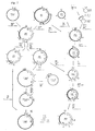
- Escherichia coli is a microorganism that is able to synthesize from simple carbon sources such as glycerol or glucose biotin (Fig. 1).
- the genes responsible for the biosynthesis of biotin in E. coli are present in an operon that has already been cloned and comprises the five genes bioA , bioB bioC , bioD and bioF ( hereinafter also referred to as bio genes) ( Gupta et al., Gene 1 : 331-345; 1977). These genes are transcribed in two different directions by a promoter-operator region located between the bioA and bioB genes.
- the genes bioB , bioF , bioC and bioD are on the right and the bioA gene is on the left of the promoter-operator region.
- ORFI open reading frame
- Other strains from the family of enterobacteria for example the genus Salmonella or Citrobacter , have a structure of the biotin operon analogous to E. coli (Shiuan and Campbell, Gene 67 : 203-211; 1988).
- Biotechnological processes for the production of biotin are already known which are carried out by means of microorganisms which have been transformed with the cloned biotin operon from E. coli . These processes are carried out on the basis of glucose.
- EP-B-236 429 describes, for example, microorganisms which have been transformed with the biotin operon from E. coli , the host organisms being mutated in their bir A / bio R gene.
- EP-A-316 229 describes E. coli mutants which produce less acetate and which have also been transformed with the cloned biotin operon.
- EP-A-449 724 discloses microorganisms transformed with the biotin operon, which additionally have mutations which result in lower glucose consumption.
- EP-A-266 240 also discloses the cloning of the genes responsible for the synthesis of biotin in Bacillus sphaericus and a method for producing biotin based thereon. Due to the metabolism of Bacillus sphaericus , this process must be carried out using expensive pimelic acid.
- the object of the present invention is therefore to provide a biotechnological process for the production of biotin which enables higher yields of biotin and is therefore more economical.
- transcription unit is understood to mean a DNA sequence in which the genes are arranged in a transcription direction and are transcribed into a continuous transcript under common transcription control, the DNA sequence in addition to the respective genes also the genetic control elements required for gene expression such as promoters and Ribosome binding sites includes.
- “Functionally equivalent genetic variants and mutants” are understood to mean genes which are derived from the wild-type genes of the original organisms, ie the enterobacteria, and have base exchanges in the context of the known degeneration of the genetic code. Such base exchanges can be of natural origin or artificially generated, for example in order to adapt the gene sequence to the preferred codon use of a particular microorganism in which expression is to take place.
- the genetic variants and mutants also include deletions, insertions and substitutions of bases or codons which basically leave the function of the gene product of such a modified sequence intact.
- sequences are included which hybridize with the wild-type sequences under the usual hybridization conditions, ie at temperatures between 55 and 66 ° C. and at 0.03 to 0.3M salt fraction, that is to say sequences which have high homology to the wild-type sequences , for example higher than 70%.
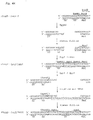
- Figure 2 shows the construction scheme for the plasmid pBO30.
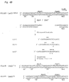
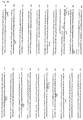
- FIG. 3 shows the DNA sequence of the plasmids pBO30, pBO30A-9 and pBO30A-15 for the region of the 3 'end of the bioD gene and the 5' end of the bioA gene (broken arrows; the bioA start) codon is underlined, the bioD stop codon shown in dotted lines) together with the restriction sites relevant for plasmid construction and the Shine-Dalgarno (SD) sequence of the bioA gene.
- SD Shine-Dalgarno
- Strikethrough nucleotides should theoretically be present, but are absent in plasmid p bioB :: lacZ / 985E and the plasmids p bioB :: lacZ / 9 and p bioB :: lacZ / 16 derived therefrom, which indicate the loss of a Bam HI site ( BamHI) results.
- "fill-in” filling with Klenow polymerase.
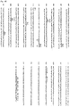
- Figure 5 shows the construction scheme for the plasmids pBO30A-15/9 and pBO30A-15 / 9 ⁇ orfI.
- Figure 7 shows the construction scheme for plasmid pBO74 ⁇ B starting from plasmids pBO74-13 and pBO3; Arrows indicate the position and orientation of the tac promoter and the bio genes. The vector portion of the plasmids is shown in bold. Open lines represent the extent of the deletion of the bioB gene.
- A Aat II; B: Bam HI; Bg: Bgl II; C: Cla I; E: Eco RI; H: Hin dIII; K: Kpn I; N: Nco I; No: NruI; P: Pst I; S: Sno I; Sa: Sal I; Se: Sse I; Sp: Sph I; Ss: Ssp I; and X: Xba I.
- "fill-in” filling in recessive 3 'ends with Klenow polymerase; mbn: Removing overhanging 5 'or 3' ends with "Mung Bean Nuclease; Bal31: progressive deletion of DNA with exonuclease Bal 31.
- the vector portion of the plasmids is shown in bold. The differently hatched parts in the plasmids were used for the subsequent cloning step Arrows indicate the position and orientation of the bio genes.
- the genes of the biotin operon are first appropriately isolated from the chromosome of a suitable microorganism and then linked under the control of gene regulatory elements such as promoters and ribosome binding sites in such a way that they are organized in a single transcription unit.
- Bacterial strains from the family of enterobacteria for example of the genus Escherichia , Salmonella or Citrobacter, can serve as the starting material for the isolation of the bio genes.
- the starting material is expediently a microorganism of the species Escherichia coli which is best characterized.
- the DNA fragments and vectors according to the invention can be constructed, for example, from a gene bank of a suitable microorganism such as E. coli , from which the bio genes or fragments thereof by hybridization with labeled oligonucleotides which contain partial sequences of the bio genes, can be isolated and cloned in a known manner.
- the isolated and cloned bio genes are then linked to one another using the known methods of DNA recombination under the control of a common promoter so that they are present as a single transcription unit.
- the bio genes are expediently arranged in such a way that the bioA gene lies downstream of the bioB , bioF , bioC and bioD genes which are already present in a transcription unit in the wild-type operon of E.
- the bioB gene encodes the key enzyme of the entire biotin synthesis pathway with the biotin synthase, since the conversion of dethiobiotin to biotin by biotin synthase has so far been the rate-determining step of the five-step biotin synthesis pathway.
- the bioB gene is therefore expediently the first gene within the transcription unit, since an optimal expression of this gene can then take place because of the proximity to the promoter (FIGS. 2, 4, 5 and 6).
- the second transcription unit in the wild-type E. coli biotin operon which contains the bioA gene, also comprises another gene, ORFI, which codes for a polypeptide with 158 amino acids.
- ORFI also codes for a polypeptide with 158 amino acids.
- expression plasmids in which no ORFI gene is present show that this gene is not essential for biotin biosynthesis under the usual fermentation conditions. However, it cannot be ruled out that this polypeptide, whose function has hitherto been unknown, may also play a role in biotin synthesis under certain conditions.
- the transcription unit with the bio genes additionally also includes the ORFI gene. (Figs. 2, 5 and 6).
- the bio genes are advantageously not under the control of the natural biotin promoter of E. coli . Rather, the bio genes are useful for improving transcription under the Check of a strong third-party promoter.
- the choice of promoter depends on the desired expression conditions, for example on whether constitutive or induced expression is desired, or on the microorganism in which the expression is to take place.
- Suitable promoters are, for example, the promoters P L and P R of phage lambda (see Schauder et al., Gene 52 : 279-283; 1987), the promoter pxyl S of the TOL plasmid from Pseudomonas putida with the neighboring regulator gene xyl R ( Franklin et al., J. Bacteriol.
- the trc promoter (Amann et al., Gene 69 : 301-315; 1988), the trp promoter (Amann et al., Gene 25 : 167-178; 1983), the Bacillus subtilis p degQ promoter, which is active in the stationary phase (Dahl et al., J. Bacteriol. 173 : 1539-1547; 1991) and the lac UV5 promoter (Amann et al ., Gene 25 : 167-178; 1983).
- the preferred promoter is the tac promoter, a hybrid of the trp and lac UV5 promoters from E. coli , which can be used as a constitutive or inducible promoter (Russell and Bennett, Gene 20 : 231-243; 1982) .
- the expression of the bioA gene in the preferred arrangement described above can be further improved if the distance between the genes bioD and bioA which follow one another in the transcription unit is as short as possible, ie is preferably less than 50 bp (base pairs).
- the expression is particularly high if the sequence of the 3 'end of the bioD gene which codes for the COOH terminus of dethiobiotin (DTB) synthetase simultaneously contains the ribosome binding site of the subsequent bioA gene.
- DTB dethiobiotin
- Such a constellation can be achieved if the 5 'end of the bioA gene together with its ribosome binding site is fused with the bioD gene in such a way that its 3' end is upstream of the bioA gene and, if appropriate, through the sequence with the ribosome binding site , the 5'-terminus of the bioA gene is substituted (Fig. 3 and 6; Seq ID No: 1, 6 and 8-16).
- This effect is all the more surprising since the COOH terminus of the DTB synthetase can be exchanged in such a fusion without the enzyme losing its activity. Similar overlaps can also be found in the wild-type biotin operon of E. coli between the reading frames of the bioB , bioF , bioC and bioD genes .
- the expression of the bioB gene can be further optimized by optimizing the ribosome binding site in front of the bioB gene.
- a strong promoter for example the tac promoter.
- the optimization of the ribosome binding site of the bioB gene ie the variation of the Shine-Dalgarno sequence and its distance from the 5 'end of the structural gene, can be carried out using the usual methods of DNA recombination.
- the influence of a specific ribosome binding site on translation can be determined in a manner known per se, for example by gene fusion of the gene to be tested with the lacZ gene and subsequent test with the chromogenic substrate 5-bromo-4-chloro-3-indolyl- ⁇ -D- galactopyranoside (X-Gal).
- DNA fragments which comprise the bio genes in a transcription unit can be incorporated into a large number of vectors using the known techniques of DNA recombination.
- the plasmids pBO30A-15/9 (FIGS. 5 and 6, Seq ID No: 1 and 6; Example 1.5.2) and pBO47 (Example 1.7) were obtained.
- Plasmid pBO30A-15/9 was released on September 28, 1992 at the German Collection for Microorganisms and Cell Cultures GmbH, D-3300 Braunschweig, Mascheroderweg 1b, in E. coli XL1-Blue and E. coli BM4062 under the accession numbers DSM 7246 and 7247, and deposited on September 17, 1993 in E.
- Plasmid pBO47 was released on January 17, 1993 at the German Collection for Microorganisms and Cell Cultures GmbH in Agrobacterium / Rhizobium sp HK4 deposited under the deposit number DSM 8555.
- vectors with a specific host spectrum and vectors with a broad host spectrum (“broad host range") are suitable as vectors.
- Examples of vectors with a specific host spectrum for example for E. coli , are pBR322 (Bolivar et al., Gene 2 : 95-113; 1977), pUC18 / 19 (Yanisch-Perron et al., Gene 33 : 103-119; 1985 ), pK18 / 19 (Pridmore, Gene 56 : 309-312; 1987) and pRA95 (available from Nycomed Pharma AS, Hvidovre, Denmark).
- All vectors which are suitable for Gram-negative bacteria can be used as "broad host range” vectors.
- Examples of such "broad host range” vectors are pRK290 (Ditta et al., Proc. Natl. Acad. Sci. USA 77 : 7347-7351; 1980), pKT240 (Bagdasarian et al., Gene 26 : 273-282; 1983 ), Derivatives of pRK290 such as pLAFR1 (Long et al., Nature 298 : 485-488; 1982) and pRK290X (Alvarez-Morales et al., Nucl. Acid. Res.
- pKT240 derivatives of pKT240 such as pMMB66EH (Fürste et al., Gene 48 : 119-131; 1986) or pGSS33 (Sharpe, Gene 29 : 93-102; 1984).
- the DNA fragment according to the invention must be introduced into the desired host strains which are suitable for expression.
- Microorganisms that are suitable for the expression of the bio genes are, for example, enterobacteria, preferably of the genus Escherichia , or microorganisms of the genus Rhizobium , Agrobacterium , Rhizobium / Agrobacterium , Acinetobacter , Azotobacter , Pseudomonas and Comamonas .
- enterobacteria preferably of the genus Escherichia
- microorganisms of the genus Rhizobium preferably of the genus Escherichia
- microorganisms of the genus Rhizobium , Agrobacterium , Rhizobium / Agrobacterium , Acinetobacter , Azotobacter , Pseudomonas and Comamonas are, for example, enterobacteria, preferably of the genus
- the microorganisms can contain the DNA fragment according to the invention either on a vector molecule or integrated in their chromosome.
- the DNA fragment can be introduced into the microorganisms, for example, by transformation or conjugation.
- the selected microorganisms are expediently transformed in a manner known per se using vectors which contain the DNA fragments according to the invention. Suitable production strains are, for example, E. coli XL1-Blue, E. coli BM4062 and E.
- coli ED8767 each containing plasmid pBO30A-15/9 (DSM 7246, DSM 7247 and DSM 8554) and Agrobacterium / Rhizobium sp HK4 with plasmid pBO47 (DSM 8555).
- the transformed host strains are advantageously isolated from a selective nutrient medium to which an antibiotic is added, against which the host strains are resistant by means of a marker gene located on the vector or DNA fragment.
- the biotechnological production of biotin takes place using the microorganisms which contain the DNA fragments or vectors according to the invention.
- the process for the production of biotin takes place in a conventional manner in cultures, starting from a carbon source suitable as a growth substrate for the respective microorganism, which is finally converted into biotin.
- Simple sugar molecules for example glucose or glycerol, are particularly suitable as the carbon source.
- commercially available media such as, for example, Nutrient Yeast Broth (NYB: Nutrient Broth No. 2, Oxoid, 25 g / l; yeast extract, Oxoid, 5 g / l) or minimal glycerol and glucose media can be used as growth media.
- the fermentation ie the production of biotin, is preferably carried out as a so-called "fed-batch process", ie in a batch fermentation to which a volume flow with fresh nutrients is supplied continuously or at intervals, however no culture solution is withdrawn.
- a glycerol solution with a variable inflow rate adapted to the respective biomass development is preferably supplied as the "feed”.
- the fermentation takes place within the pH and temperature ranges that are physiologically compatible with the respective microorganisms.
- the pH is expediently within a range from 6 to 8 and the temperature within a range from 20 to 45 ° C.
- the yield of biotin can be further improved by varying the nutrients in the medium and by adapting the fermentation conditions to the respective microorganism in the usual way.
- the present invention furthermore relates to a process for the preparation of biotin which comprises the conversion of dethiobiotin to biotin in the cell-free system by means of the enzyme biotin synthase, wherein the reaction in the presence of thiamine pyrophosphate, NADPH, S-adenosylmethionine, Fe 2+ ions, cysteine and at least one further amino acid from the group of asparagine, aspartic acid, glutamine and serine is carried out.
- the biotin synthase can be used either in a purified form or in the form of a cell extract.
- the cell extract or the purified biotin synthase is expediently obtained from a strain with increased expression of the biotin synthase, for example from E. coli XL1-Blue with the plasmid pBO30A-15/9 (DSM 7246).
- the cell extract and, if appropriate, the purification of the biotin synthase can be prepared by the methods customary in biochemistry, for example by homogenizing the cells, gel filtration, ammonium sulfate fractionation and ion exchange chromatography.
- the cofactors required for the reaction include S-adenosylmethionine (SAM), thiamine pyrophosphate (TPP), reduced nicotinic acid amidadenine dinucleotide phosphate (NADPH) and Fe 2+ ions.
- SAM S-adenosylmethionine
- TPP thiamine pyrophosphate
- NADPH reduced nicotinic acid amidadenine dinucleotide phosphate
- Fe 2+ ions Fe 2+ ions.
- the cofactors are expediently added in concentrations of 1 to 500 ⁇ m.
- Dithiothreitol (DTT) is also expediently added to the mixture in a concentration of 0.1 to 10 mM
- Amino acids that are required for the implementation are cysteine as sulfur donor and at least one other amino acid from the group of asparagine, aspartic acid, glutamine and serine. Aspartic acid is expediently added as aspartate. Cysteine is expediently added in concentrations of 10 to 500 ⁇ m, the other amino acids in concentrations of 1 to 50 mM.
- 1.18.1.2 are known proteins that are known in a known manner, for example by ammonium sulfate fractionation and subsequent ion exchange chromatography and gel filtration chromatography, regardless of the expression of the biotin synthase Allow cell extracts from E. coli to be obtained.
- flavodoxin and ferredoxin (flavodoxin) -NADP + reductase could be obtained from E. coli XL1-Blue with plasmid pBO30A-15/9 (DSM 7246), which has an increased biotin synthase expression, as well as from E.
- Plasmid pBO74 ⁇ B was deposited in E. coli XL1-Blue on September 28, 1992 at the German Collection for Microorganisms and Cell Cultures GmbH, D-3300 Braunschweig, Mascheroderweg 1b, under the accession number DSM 7245.
- the precipitate obtained after the ammonium precipitation can be further purified, for example, by chromatographic methods such as ion exchange chromatography and gel filtration chromatography.
- a protein fraction obtainable as described above is therefore expediently added to the mixture for converting dethiobiotin to biotin, especially if the biotin synthase is not used in the form of a cell extract.
- the reaction takes place in a suitable buffer system, expediently within the pH and temperature ranges in which the enzymes are physiologically active, preferably in a pH range from 6 to 9 and at a temperature between 4 and 50 ° C.
- Restriction endonucleases were used with 3 to 5 units / ⁇ g DNA according to the manufacturer's instructions. Labeling and phosphorylation of DNA linkers (obtained from Boehringer Mannheim, FRG) for the incorporation of restriction sites and of synthetic oligonucleotides (obtained from Microsynth, Windisch, CH), for example for use as probes for DNA / DNA hybridizations and as "primers" for sequencing reactions, were carried out using T4 polynucleotide kinase (Boehringer Mannheim, FRG) according to Sambrook et al. (Molecular Cloning: A laboratory manual. 2nd edition, Cold Spring Harbor Laboratory, Cold Spring Harbor, NY; 11.31 and 5.68; 1989). Ligation reactions were carried out with T4 DNA ligase according to the manufacturer's instructions.
- DNA sequencing was carried out using the chain termination method according to Sanger et al. (Proc. Natl. Acad. Sci. USA 94 : 5463-5467; 1977). All sequence reactions were carried out using the Sequenase kit from United States Biochemicals (Cleveland, OH, USA) according to the manufacturer's protocol. Sequenase (version 2.0, a genetically modified T7 DNA polymerase) gave uniform, easily readable DNA sequences over more than 600 bp; Compressions in GC-rich DNA areas could easily be resolved if the nucleotide dITP was used instead of dGTP.
- the plasmid DNA was purified using a CsCl gradient or "Gene Clean" (BIO 101, La Jolla, CA).
- the gels were 550 mm long and 0.2 mm thick; the electrophoresis was carried out in an LKB macrophor apparatus with a thermostat at a voltage of 2100V and a temperature of 60 ° C. The gels were then dried on Whatman 3 MM paper and autoradiographed with X-ray film Fuji RX or Amersham Hyperfilm ⁇ max.
- Extrachromosomal DNA was isolated either in smaller amounts by the "rapid alkaline SDS"("Miniprep") method according to Birnboim and Doly (Nucl. Acid. Res. 7 : 1513-1523; 1979), or, to isolate larger amounts, by cesium chloride density gradient centrifugation according to a modified method according to Clewell and Helsinki (Proc. Natl. Acad. Sci. USA 42 : 1159-1166; 1969).
- QIAGEN-packs from DIAGEN, Düsseldorf (FRG) were used.
- E. coli DSM 498 K12 "wild type”; German Collection for Microorganisms and Cell Cultures GmbH
- the isolation was carried out essentially according to Hahn and Hennecke (Mol. Gen. Genet. 193 : 46-52; 1984).
- Pst I restriction enzyme
- the DNA fragments were separated electrophoretically in a horizontal 0.7% agarose gel in a conventional manner (Sambrook et al., 1989, ibid .; 6.19 to 6.9) and transferred to “gene screen” membranes (nylon membranes from NEN-Du Pont) ( Southern, J. Mol.
- the DNA was first 2 h in 5x Denhardt's solution (1x Denhardt's solution: 0.02% bovine serum albumin, 0.02% Ficoll, 0.01% polyvinylpyrrolidone), 6x SSC buffer (1x SSC: 150 mM NaCl, 15 mM sodium citrate, pH 7.2) and 150 ⁇ g / ml salmon sperm DNA pre-hybridized, then hybridized for 18 h in 2x Denhardt's solution, 6 x SSC, 0.5% SDS, 150 ⁇ g / ml salmon sperm DNA and washed for 2 h and finally washed four times for 30 minutes each in 2x SSC, 0.1% SDS. The temperature was 65 ° C in all steps. The labeled oligonucleotide hybridized on this "Southern blot" with a 5.4 kb Pst I fragment.
- This vector contains the gene for chloramphenicol resistance (Cm R ), the ColE1 replicon from pACYC184 (Chang and Cohen, J. Bacteriol., 134 : 1141-1156; 1978) and the E. coli gene pheS for phenylalanine tRNA- Synthetase that has a Pst I site.
- E. coli RR28 has a mutated pheS gene ( pheS 12) in the chromosome and is therefore resistant to p-fluorophenylalanine (pFphe) in the growth medium.
- pFphe p-fluorophenylalanine
- a probe with parts of the bioC and bioD genes was used, consisting of a 520 bp Sph I / Pst I fragment from pBO1. This fragment was isolated from an agarose gel and 0.2 ⁇ g of the isolated fragment were nick-translated with DNA polymerase I (Boehringer Mannheim, FRG; holoenzyme from E. coli; this so-called “Kornberg polymerase” was used together with DNase I used) and 25 ⁇ Ci ⁇ - [ 32 P] -dATP (NEN-Du Pont, NEG-012H) radioactively labeled (Sambrook et al., 1989, ibid. 10.8).
- DNA polymerase I Boehringer Mannheim, FRG; holoenzyme from E. coli; this so-called “Kornberg polymerase” was used together with DNase I used
- 25 ⁇ Ci ⁇ - [ 32 P] -dATP NN-Du Pont, NEG-012H
- Hybridization of this probe with restriction fragments of the E. coli DSM 498 chromosome generated by Ssp I on a "Southern blot" as described above showed on the one hand the 1.6 kb Ssp I fragment with bioF and bioC known from pBO1 and on the other hand a 1 , 1 kb- Ssp I fragment with bioD and sequences of the neighboring gene uvrB (Sancar et al., Cell, 28; 523-520; 1982).
- a partial gene bank was again created to clone the 1.1 kb Ssp I fragment.
- 30 ⁇ g of DNA from E. coli DSM 498 were cut with Ssp I and separated on a 0.7% agarose gel. Fragments from 0.9 kb to 1.3 kb in size were cut out and isolated by electrodialysis. 0.5 .mu.g of these fragments were mixed with 0.5 micrograms of cleaved with Sma I phage vector M13mp19 (Yanisch-Perron et al., 1985, ibid.) was ligated. This ligation approach was used to transfect E.
- coli JM109 (Yanisch-Perron et al., 1985, ibid) to Messing (Methods Enzymol., 101 : 20-79; 1983).
- 150 phage clones with insert (phenotype LacZ - ) were isolated and propagated in NYB medium. After centrifuging the E. coli cells The phages were each applied in 50 ⁇ l of the supernatants with a Schleicher & Schüll "minifold I" apparatus as "dot blot" to a nitrocellulose filter (Schleicher & Schüll BA 85).
- the filters were treated for 5 min with 0.1 M NaOH / 1.5 M NaCl buffer and then neutralized with 0.5 M Tris-HCl, pH 7.5 / 2.5 M NaCl (5 min) .
- the DNA was fixed on the filter by incubation (2 h) at 80 ° C.
- the filter was hybridized as described (Sambrook et al., 1989, ibid., 9.52-9.55) with the radioactively labeled 520 bp long SphI / PstI fragment at 60 ° C. In this way, the phage clone M13 bioD was identified with the 1.1 kb Ssp I fragment described above, which contains the bioD gene (FIG. 2).
- plasmid pBO1 and 0.5 ⁇ g of the phage M13 bioD were cut with the restriction enzymes Sno I and Hin dIII and religated in one approach.
- recombined plasmids were examined by restriction analysis.
- a plasmid, pBO2 (FIG. 2), was selected in which an approximately 1.5 kb long Sno I / Hin dIII fragment of pBO1, which contains part of the bioD gene and non-essential sequences of the vector pHE3, is replaced by a 0.95 kb Sno I / Hin dIII fragment from M13 bioD .
- plasmid pBO2 contained the complete bio- operon, as is present in E. coli , together with sequences of the uvrB promoter (Sancar et al., Cell 28 : 523-530; 1982) downstream of bioD .
- E. coli RR28 grows worse with pBO2 on NA plates than with pBO1 and forms significantly smaller colonies. The reason for this could be the uvrB sequences in pBO2.
- 20 ⁇ g pBO2-DNA was cut with Hin dIII and in 150 ⁇ l Bal 31 buffer (600 mM NaCl, 12.5 mM MgCl 2 , 12.5 mM CaCl 2 , 1 mM EDTA, 20 mM Tris HCl, pH 7.2) added. Then became gradual Shortening of the linear plasmids Bal 31 (from Alteromonas espejiani , Boehringer Mannheim, FRG) added.
- Bal 31 buffer 600 mM NaCl, 12.5 mM MgCl 2 , 12.5 mM CaCl 2 , 1 mM EDTA, 20 mM Tris HCl, pH 7.2
- Treatment with Bal 31 removes not only the uvrB sequences, but also essential sequences of the pHE3 vector. Therefore, the truncated pBO2 plasmids were cut with Eco RI after treatment with Mung Bean Nuclease to remove the portion of the vector DNA of pHE3 that was truncated by Bal 31.
- the original vector sequence was then regenerated by ligating the treated pBO2 plasmid with a 1.5 kb DNA fragment which was isolated from pBO2 after restriction with Bam HI, treatment with mung bean nuclease and a further restriction with Eco RI and which has the previously deleted essential vector sequences of pHE3. Since the cm resistance of the vector is completely regenerated by this ligation, intact plasmids can be recognized by their property of imparting resistance to Cm.
- E. coli RR28 was transformed with the ligation batches and plated on NA plates with 20 ⁇ g / ml Cm. Small, slow growing colonies, typical of pBO2, and large, normal growing colonies were observed. The number of large colonies per PBO2 aliquot took l31-incubation with the duration of Ba.
- Plasmid DNA was isolated from 22 normal growing colonies and analyzed by restriction and sequence analysis. In this way, plasmids pBO3 and pBO6 were obtained in of which approximately 330 bp and 410 bp of the uvrB region were deleted, but which still had the bioD gene completely.
- the undesired wild-type promoter in front of the bioBFCD genes must be removed. This can be done by cutting with Nco I, which also exposes the start codon of the bioB gene.
- the tac promoter (Russell and Bennett, 1982, ibid.) was chosen as the promoter, since it can be used as a constitutive or inducible promoter and very good activity not only in E. coli but also in many other Gram-negative bacteria Has.
- a DNA fragment with the tac promoter with Hin dIII and Bam HI ends was obtained from Pharmacia-LKB (Uppsala, Sweden) and into the plasmid pUC18 cut with Hin dIII and Bam HI (Yanisch-Perron et al., 1985, ibid).
- the plasmid pUC18 / tac resulted (FIG. 2). 8 ⁇ g of this plasmid were then cut with BamHI and, in order to fill in the recessive 3 ′ ends with Klenow polymerase (DNA polymerase I from E.
- a 3.2 kb fragment with the genes bioB , bioF , bioC and the 5 'end of the bioD gene was isolated from pBO1.
- 8 ⁇ g pBO1 were cut with Nco I and then treated as above with Klenow polymerase to fill in the recessive 3 ′ ends. This was followed by a second restriction with Pst I, followed by isolation of the desired 3.2 kb fragment.
- the plasmid DNA of transformants with Cm R was examined by restriction analysis.
- the plasmid pBO21 (FIG. 2) was obtained, in which the tac promoter is located in front of the bioB gene.
- pBO22 was finally obtained (FIG. 2).
- 25 ⁇ g of double-stranded DNA of phage M13 bio 18 were linearized by restriction with Nco I, taken up in 160 ⁇ l Bal31 buffer, and then Bal 31 was added to remove the bioA promoter. After 20, 40, 60, 80, 100 and 120 seconds of incubation at room temperature, 25 ⁇ l aliquots were removed and the Bal 31 reaction was stopped by adding 2 ⁇ l 0.5 M EGTA, pH 7.5, and phenol extraction. 3 aliquots were combined and cut with Xba I to remove the bioB and bioF genes. The DNA was then treated with Klenow polymerase as above to protrude 5 'ends fill in to smooth ends. The DNA treated in this way was religated and E.
- coli JM109 was transformed with the ligated DNA.
- Single-stranded DNA was isolated from 24 phage clones (Messing, 1983, ibid.) And the DNA sequence at the 5 'end of the bioA gene was determined according to Sanger et al. (1977; ibid.) Analyzed. In this way, the phage clone M13 bio 18/13 was obtained in which the wild-type promoter was deleted in front of the bioA gene and which at the same time had a SalI site 26bp upstream of the bioA gene (FIG. 2).
- the isolated DNA fragments were ligated with T4 DNA ligase and the biotin auxotrophic strain E. coli SA291 (Cleary and Campbell, J. Bacteriol. 112 : 830-839; 1972) was transformed using this ligation approach.
- Clones with a complete biotin operon in the plasmid were selected by plating out on NA plates with 20 ⁇ g / ml Cm and 8 ⁇ g / ml avidin. Plasmids from such clones were checked by restriction analysis. In this way, plasmid pBO30 was obtained which contains the genes bioB , bioF , bioC , bioD and bioA and the gene ORFI together with the tac promoter in a transcription unit (FIG. 2).
- the Sal I site was regenerated by ligation with a synthetic oligonucleotide of the sequence 5'-CGTCGACG-3 ', a Sal I linker. Then the DNA was cut with Sal I and Sno I and the bioD fragments truncated at the 3'-end with a length of about 640 bp were isolated. These fragments were ligated with an 8.25 kb fragment from pBO30 which could be isolated after cutting this plasmid with Sal I and Sno I and which contains the unchanged bioA gene.
- the biotin-auxotrophic strain E. coli SA291 was transformed with the above ligation approach and then cloned with an intact bioD gene on NA plates with 60 ⁇ g / ml Cm and 5 ⁇ g / ml avidin to select. 26 such clones were obtained and were examined by restriction analysis. Eight of these clones with apparent shortening of the region upstream from the Sal I site were characterized more precisely by DNA sequence analysis. In five of these clones, about 20 to 45 bp of DNA between the bioD and bioA genes were deleted as desired. In E. coli mini cells, these clones actually caused expression of the bioA gene to be increased by a factor of 2 compared to pBO30. An example of a plasmid with such improved expression is the plasmid pBO30A-9 obtained in this way (FIG. 3).
- the sequence upstream of the bioB gene in pBO30 which comprises the tac promoter and a ribosome binding site which is located in the cloned tac Promoter fragment is included, modified.
- p bioB :: lacZ -2 is identical to the plasmid pBO22 in the vector part, in the tac promoter with the ribosome binding site and in the 5 'end of the bioB gene (FIG. 2).
- the 3 'end of the bioB gene and the remaining bio genes were deleted and the lacZ gene from E. coli (Casadaban et al., Methods Enzymol. 100 : 293-308; 1983) so that bioB and lacZ were fused in the correct reading frame to express a bioB :: lacZ fusion protein and that the Nru I site was regenerated.
- the oligonucleotide 985E with the sequence 5'-CATGGAATCCTCCACTGATCAGTAC-3 ' was inserted into the plasmid p bioB :: lacZ -2 in front of the bioB gene (FIG. 4).
- p bioB :: lacZ -2 was first cleaved with Bam HI and then the overhanging Bam HI ends were filled in with Klenow polymerase as described.
- a guanine residue (G) was obviously deleted non-specifically, which resulted in the loss of a Bam HI site in the later plasmids.
- a Kpn I linker E.
- coli XL1-Blue Bacillus XL1-Blue (Bullock et al., Biotechniques 5 : 376-379; 1987) was transformed with the ligation approach and the plasmid p bioB :: lacZ / KpnI was isolated. This plasmid was partially cut with Nco I and then recut with Kpn I. After ligation with the oligonucleotide 985E, the second DNA strand was filled in with Klenow polymerase. After transformation of E.
- Plasmid p bioB :: lacZ / 985E was further varied by cutting out the ribosome binding site by restriction with Kpn I and Spe I and replacing it with three different mixed oligonucleotides, SD17, SD19 and SD21 (Fig. 4). After ligation with these oligonucleotides it was incubated with Klenow polymerase closed the gap in the second strand of DNA.
- Bacterial cells from E. coli XL1-Blue were transformed with this DNA and plated as above on NA plates with 20 ⁇ g / ml Cm, 30 ⁇ g / ml X-Gal and 0.5 mM IPTG.
- 20 clones with good expression of the bioB :: lacZ fusion protein, which formed dark blue colonies on this medium, were selected and the ⁇ -galactosidase activity of these clones was determined using a Miller enzyme assay (Experiments in Molecular Genetics, Cold Spring Harbor Laboratory , Cold Spring Harbor, NY, pp. 352-355; 1972).
- the E. coli strains were previously grown with bio :: lacZ plasmids in liquid culture up to an optical density at 600 nm (OD 600 ) of approximately 0.5.
- the plasmids p bioB :: lacZ / 985E, p bioB :: lacZ / 16 and p bioB :: lacZ / 9 (FIG. 4) showed the highest ⁇ -galactosidase activity, in which the ⁇ -galactosidase activity compared to p bioB :: lacZ -2 was increased by a factor of 2.1, 3.4 or 5.9.
- the DNA sequence of the optimized ribosome binding sites for the bioB gene in these plasmids was according to Sanger et al. (1977, ibid.).
- pBO30 ⁇ A which is derived from pBO30, a Sal I / Bam HI fragment with the majority of the bioA gene and an interfering Nru I site is deleted (FIG. 5). The two fragments were ligated and clones with recombined plasmids were isolated. In this way, the plasmids pBO30 ⁇ A / 9, pBO30 ⁇ A / 16 and pBO30 ⁇ A / 985 were obtained.
- Fig. 5 shows one such construction using the example of pBO30 ⁇ A / 9 with the ribosome binding site from p bioB :: lacZ / 9.
- FIG. 5 shows the construction of plasmids which contain the genes bioB , bioF , bioC , bioD and bioA together with an optimized ribosome binding site , using the construction of pBO30A-15/9 as an example.
- coli S17-1 (Simon et al., Biotechnology 1 : 784-791; 1983) was transformed with the ligation approach. Recombined plasmids were characterized by restriction analysis; in this way plasmid pBO47 was obtained, which contains the biotin operon integrated in pRK290X.
- the plasmid pBO47 was converted into the bacterial strains Rhizobium / Agrobacterium sp. By conjugation with the strain E. coli S17-1 / pBO47 . HK4, Pseudomonas mondocina , Pseudomonas aeruginosa PA01 (Holloway, J. Gen. Microbiol. 13 : 572-581; 1955) and Acinetobacter calcoaceticus DSM 588.
- Plasmid pBO74 ⁇ B with a deletion of the bioB gene was constructed starting from plasmid pBO74-13 (FIG. 7). Plasmid pBO74-13 consists of the same DNA building blocks as pBO30 (Fig. 2). However, the order of the bio genes within the plasmid pBO74-13 is different.
- plasmid pBO74-13 5 ⁇ g of the plasmid pBO74-13 were cut with Sma I. After extraction with phenol / chloroform, the plasmid DNA was cut with Sph I and a 6 kb fragment containing the vector DNA, the tac promoter, the genes bioA- ORFI and bioCD was isolated. 18 ⁇ g of the plasmid pBO3 (FIG. 2) were cut with Ssp I and Sph I and a 1.66 kb fragment with the bioF gene and part of the bioC gene was isolated. The isolated fragments were ligated together and E. coli RR28 was transformed with the ligation mixture. Recombined plasmids were analyzed using restriction analysis. In this way, plasmid pBO74 ⁇ B was obtained, which differs from plasmid pBO74-13 in the deletion of the bioB gene (FIG. 7).
- the metabolic activity of the cells was monitored using their specific oxygen uptake rate.
- the production of biotin during the fermentation was monitored by titrating the biotin values of the fermenter medium with a bioassay with Lactobacillus plantarum (E. DeMoll and W. Shive, Anal. Chem. 158 : 55-58, 1986).
- the carbon source in this case a 50% glycerol solution in deionized water, was supplied with a variable flow rate that was adapted to the respective biomass development.
- glycerol an empirical value of 2 g glycerol per liter of culture was used for an OD increase from OD 1 to OD 2 for the "feed" rate.
- the pH of the fermenter was automatically adjusted to pH 7 by pumping in 40% H 3 PO 4 or 25% NH 3 .
- the aeration was controlled by blowing 10-25 NL / min air and rotating the stirrer at 300-700 rpm according to the respective biomass development.
- An oxygen saturation between 15 and 40% was aimed for.
- the oxygen and CO 2 content of the exhaust air was measured paramagnetically or using infrared.
- the temperature of the fermenter was adjusted to 37 ° C. At 37 ° C the culture grew with a doubling time of 2.5 hours to an OD 650 of 20 and then became stationary.
- E. coli ED8767 (NE Murray et al., Mol. Gen. Genet. 150 : 53-61; 1975) with pBO30A-15/9 (DSM 8554) or E. coli BM4062 (DF Barker and AM Campbell, J. Bacteriol. 143 : 789-800; 1980) with pBO30A-15/9 (DSM 7247).
- the plasmids pBO3, pBO30 and pBO30A-15 / 9 ⁇ ORFI were tested in a similar manner and the biotin productivity was determined.
- Table I shows the strong improvement in the biotin productivity of strains with the plasmids pBO30, pBO30A-15/9 and pBO30A-15 / 9 ⁇ ORFI, which have the bio genes in a transcription unit, in comparison with E. coli S17-1 (Wild type, biotin genes on the chromosome) and E. coli S17-1 / pBO3 (biotin genes on the plasmid, but divergent transcription as in the wild type operon).
- the experiments also show that the absence of the ORFI gene has no effect on biotin productivity.
- a glucose betaine feed (360 g / l glucose plus 103 g / l betaine dissolved in deionized water) was slowly metered in (1.5 ml / hour) to enable long-term slow growth or "maintenance growth".
- a final concentration of 100 mg / l in the fermenter was supplemented with Fe 2+ gluconate. 10 ml of salt solution and 1.36 ml of standard vitamin solution were replenished at the times 200, 360 and 550 hours.
- the pH of the fermenter was automatically adjusted to pH 7 by pumping in 85% phosphoric acid or 3M potassium hydroxide solution.
- the aeration was controlled by blowing in 1-3 NL / min air and rotating the stirrer at 300-1000 rpm according to the respective biomass development, so that an oxygen tension of 1-4 mg / l was ensured.
- the temperature of the fermenter was adjusted to 30 ° C.
- the culture grew in the exponential growth phase with a doubling time of 5.6 hours, in the phase with the very limited "feed” with a doubling time of 300 hours and then switched to "maintenance growth".
- DAPA diaminopelargonic acid
- HK4 itself is biotin auxotrophic.
- the strain was able to generate dethiobiotin from the biotin precursor DAPA and finally convert it to D (+) - biotin with high yield. 110 mg / l D (+) - biotin was accumulated. It is remarkable that this synthesis was mainly carried out by non-growing cells.
- a cell extract from E. coli XL1-Blue (DSM 7246) with the plasmid pBO30A-15/9 (extract Z) and a cell extract from E. coli XL1-Blue with the plasmid pBO74 ⁇ B (DSM 7245; extract W) were produced .
- the microorganism cells were grown at 37 ° C. in a volume of 800 l with an OD 600 of 2 in a medium with 20 g / l nutrient bread, 5 g / l yeast extract and 20 mg / l cm. The cells were harvested by filtration and then centrifuged at 5000 xg for 15 min.
- Extract Z could then either be used directly to measure (test) the biotin synthase reaction or only after purification by gel filtration on a column loaded with Sephadex G25M PD-10 (Pharmacia, column volume: 9.1 ml). Extract W was either used directly to test the biotin synthase reaction or fractionated according to Example 3.3.
- a typical standard test consisted of the cell-free extract Z or W, of labeled or unlabeled dethiobiotin, depending on the reaction, or of the protein fractions purified therefrom (Examples 3.7-3.9) individually or in combination with one another and / or from conventional cofactors such as SAM (92 ⁇ M), Fe 2+ gluconate (200 ⁇ M), NADPH (100 ⁇ M), TPP (100 ⁇ M), DTT (1mM) and / or a combination of amino acids.
- SAM 92 ⁇ M
- Fe 2+ gluconate 200 ⁇ M
- NADPH 100 ⁇ M
- TPP 100 ⁇ M
- DTT DTT
- the protein fractions, cofactors or amino acids to be tested were added in a final volume of 250 ⁇ l. The incubation takes place between 4 and 50 ° C.
- HPLC buffer A 25mM KH 2 PO 4 , 5mM tetrabutylammonium chloride, pH 3.4
- HPLC conditions were as follows: Shandon-HypersilBDS-C 18 column (particle size: 5 ⁇ m, column size 10 mm x 2.0 mm), flow rate 0.35 ml / min, temperature 40 ° C, eluent: HPLC buffer A with 10 vol.% Acetonitrile.
- the samples were analyzed semi-quantitatively using thin layer chromatography and autoradiography.
- the samples were resuspended in 20 ul of a mixture consisting of 10% acetic acid, 65% methanol and 25% water and 2.5 ul was applied to a silica gel "high performance" TLC plate (E. Merck, Darmstadt).
- the plate was developed with an eluent consisting of chloroform (17 ml), methanol (3 ml) and formic acid (0.2 ml). After chromatography, the plate was dried and an X-ray film was then placed overnight.
- Example 3.3 When the same desalted cell extract as described in Example 3.3 was incubated with L-cysteine, asparagine, dethiobiotin, SAM, TPP, NADPH and Fe 2+ gluconate, dethiobiotin was converted to biotin. In order to test the influence of these cofactors on the biotin synthase reaction, they were used individually and in combination with one another. Only a combination of all of these cofactors showed biotin synthase activity. If a cofactor was missing, no biotin synthase activity could be measured, ie all cofactors are necessary for the biotin synthase activity (Example 3.3, Table II).
- the cell-free extract Z was first used Subject to ammonium sulfate fractionation. This was carried out with a saturation of 25% ammonium sulfate with stirring for 30 min at 4 ° C. Then centrifuged at 10,000 xg for 30 min and the resulting pellet was discarded. The resulting supernatant was saturated with 70% ammonium sulfate, whereby the biotin synthase was precipitated.
- the precipitate was resuspended in a small volume of 100mM HEPES buffer (pH 7.5), desalted (Sephadex G25M PD-10) and then by anion exchange chromatography (Q-Sepharose Fast-Flow, Pharmacia) with a continuous gradient of 100 mM - 1 M HEPES buffer (pH 7.5), cleaned.
- the fractions with biotin synthase activity were concentrated (Amicon ultrafiltration cell, YM-10 membrane), desalted as already described and then rechromatographed on a Q-Sepharose "Hi-Load” anion exchange chromatography column (Pharmacia; 20 mM Tris buffer (pH 7, 5) containing 1 mM DTT and a 0-1 M NaCl gradient).
- the fractions with high biotin synthase activity were pooled, concentrated and desalted. In these fractions, the biotin synthase was no longer contaminated with other proteins which are necessary for the biotin synthase activity.
- the extract was precipitated in succession at 45% and 55% saturation of ammonium sulfate. After ammonium sulfate had been added, the whole was stirred at 4 ° C. for 30 minutes and then centrifuged at 10,000 ⁇ g for 30 minutes. The precipitate obtained with 45% saturation of ammonium sulfate was resuspended in 100 mM HEPES buffer (pH 7.5). Aliquots of the 45% precipitate, the 55% precipitate and the 55% supernatant were then removed and desalted (Sephadex G25M PD-10 Pillar). The individual fractions were tested both individually and in combination with one another, in accordance with Example 3.2.
- the eluted protein fractions were combined, concentrated (Amicon ultrafiltration cell, YM-10 membrane), desalted (Sephadex G25M PD-10) and then on a Mono Q anion exchange chromatography column, which was run with 20 mM Tris buffer (pH 7.0, containing 1 mM DTT) was cleaned. Then the purified fractions were examined by SDS-PAGE.
- biotin synthase test system (example 3.2) with purified biotin synthase, protein or proteins from the precipitate of the 45% ammonium sulfate precipitation, with amino acids (example 3.3 ) and with low molecular weight cofactors (Example 3.4). Biotin synthase activity could only be measured in the fractions containing the protein sought.
- the amino acid sequence of this protein was then determined as follows.
- the protein was digested with DTT in 6 M guanidine-HCl buffer for 4 h reduced.
- the resulting samples were carboxymethylated with iodoacetic acid and then dialyzed against 0.1% ammonium bicarbonate for 48 hours. After the samples had dried, they were digested with pig trypsin in 7 M urea buffer and the peptides were separated by "reverse phase" HPLC. Two peptides with the corresponding DNA sequences for flavodoxin from E. coli were identified. Flavodoxin could be obtained homogeneously through these purification steps.
- Extract W was applied to an anion exchange chromatography column (Q-Sepharose FastFlow (Pharmacia)). This column was equilibrated with 20 mM Tris buffer (pH 7.5) containing 1 mM TPP. The proteins bound to the column were eluted with a continuous NaCl gradient (0-1 M). The eluted protein fractions were combined and concentrated according to example 3.7, desalted and applied to a Mono-Q anion exchange chromatography column. The proteins bound to this column were eluted with a continuous NaCl gradient (0-0.4 M in 20 mM Tris buffer).
- the combined eluted protein fractions (as already described, concentrated and desalted) were then placed on a Superose 12 Prep.Gel filtration chromatography column (Pharmacia; equilibrated with 20 mM Tris buffer) and then on a Sephacryl HR100 gel Filtration column (Pharmacia; equilibrated with 20 mM Tris buffer) applied. After elution with 20 mM Tris buffer, another protein could be obtained homogeneously (examined by SDS-PAGE). The fractions containing this protein were identified analogously to the test system according to Example 3.7. Biotin synthase activity was only measured after the addition of these fractions.
- the purified protein was sequenced directly.
- the protein had an N-terminal amino acid sequence corresponding to the ferredoxin (flavodoxin) NADP + reductase. Through these purification steps, ferredoxin (flavodoxin) NADP + reductase could be obtained homogeneously.
- the purified biotin synthase (Example 3.2) had no biotin synthase activity with purified flavodoxin plus ferredoxin (flavodoxin) -NADP + reductase and with the necessary cofactors and with the amino acids. In order to achieve activity, another protein or proteins in the 45% ammonium sulfate fraction was searched for.
- This protein or proteins were obtained from the cell-free extract W by ammonium sulfate precipitation with a saturation of 45%.
- the protein pellet obtained was resuspended in 20 mM Tris buffer, pH 7.5, containing 1 mM DTT and TPP (1 g / l) and then desalted with a PD-10 column (Pharmacia).
- the desalted material was then applied to an anion exchange chromatography column (Q-Sepharose HP Hi-Load), which had previously been treated with 20 mM Tris buffer, pH 7.5, containing 1 mM DTT and TPP (1 g / l) , had been equilibrated.
- the protein fractions with the desired activity were eluted with a continuous NaCl gradient (0 mM - 600 mM NaCl). These protein fractions were then further purified by means of gel filtration chromatography (Sephacryl HR-100 column, Pharmacia). The protein pellet obtained therefrom was resuspended in 100 mM HEPES buffer (pH 7.5) and then desalted as already described. A protein solution was obtained from this, which was used for the in vitro test according to Example 3.2.
- Flavodoxin, ferredoxin (flavodoxin) NADP + reductase and the protein or proteins responsible for the biotin synthase reaction were added to the cell-free extract Z.
- the addition of proteins, cofactors and amino acids caused an increased biotin synthase activity (Table III).
- Table III Composition of the tests pmol produced biotin Extract Z with cofactors and amino acids 390 Extract Z + cofactors + amino acids + flavodoxin + ferredoxin (flavodoxin) -NADP + reductase + one or more of the proteins responsible for the biotin synthase reaction 1560
- Table IV Composition of the test pmol produced biotin purified biotin synthase 0 purified biotin synthase + flavodoxin + ferredoxin (flavodoxin) NADP + reductase + one or more proteins + cofactors + amino acids responsible for the biotin synthase reaction 800
Landscapes
- Genetics & Genomics (AREA)
- Health & Medical Sciences (AREA)
- Life Sciences & Earth Sciences (AREA)
- Engineering & Computer Science (AREA)
- Organic Chemistry (AREA)
- Chemical & Material Sciences (AREA)
- Wood Science & Technology (AREA)
- Zoology (AREA)
- Biotechnology (AREA)
- General Engineering & Computer Science (AREA)
- Biomedical Technology (AREA)
- Bioinformatics & Cheminformatics (AREA)
- General Health & Medical Sciences (AREA)
- Microbiology (AREA)
- Biochemistry (AREA)
- Molecular Biology (AREA)
- Biophysics (AREA)
- Plant Pathology (AREA)
- Physics & Mathematics (AREA)
- Chemical Kinetics & Catalysis (AREA)
- General Chemical & Material Sciences (AREA)
- Preparation Of Compounds By Using Micro-Organisms (AREA)
- Micro-Organisms Or Cultivation Processes Thereof (AREA)
- Heterocyclic Carbon Compounds Containing A Hetero Ring Having Oxygen Or Sulfur (AREA)
- Enzymes And Modification Thereof (AREA)
- Pharmaceuticals Containing Other Organic And Inorganic Compounds (AREA)
- Peptides Or Proteins (AREA)
- Saccharide Compounds (AREA)
Applications Claiming Priority (5)
| Application Number | Priority Date | Filing Date | Title |
|---|---|---|---|
| CH3124/92 | 1992-10-02 | ||
| CH312492 | 1992-10-02 | ||
| CH213493 | 1993-07-15 | ||
| CH2134/93 | 1993-07-15 | ||
| EP93920855A EP0667909B1 (fr) | 1992-10-02 | 1993-10-01 | Procede biotechnologique de preparation de biotine |
Related Parent Applications (2)
| Application Number | Title | Priority Date | Filing Date |
|---|---|---|---|
| EP93920855A Division EP0667909B1 (fr) | 1992-10-02 | 1993-10-01 | Procede biotechnologique de preparation de biotine |
| EP93920855.9 Division | 1994-04-14 |
Publications (1)
| Publication Number | Publication Date |
|---|---|
| EP0798384A1 true EP0798384A1 (fr) | 1997-10-01 |
Family
ID=25689605
Family Applications (2)
| Application Number | Title | Priority Date | Filing Date |
|---|---|---|---|
| EP93920855A Expired - Lifetime EP0667909B1 (fr) | 1992-10-02 | 1993-10-01 | Procede biotechnologique de preparation de biotine |
| EP97107803A Withdrawn EP0798384A1 (fr) | 1992-10-02 | 1993-10-01 | Procédé biotechnologique de préparation de biotine |
Family Applications Before (1)
| Application Number | Title | Priority Date | Filing Date |
|---|---|---|---|
| EP93920855A Expired - Lifetime EP0667909B1 (fr) | 1992-10-02 | 1993-10-01 | Procede biotechnologique de preparation de biotine |
Country Status (16)
| Country | Link |
|---|---|
| US (1) | US6083712A (fr) |
| EP (2) | EP0667909B1 (fr) |
| JP (2) | JP3566287B2 (fr) |
| KR (1) | KR100200542B1 (fr) |
| AT (1) | ATE163198T1 (fr) |
| AU (1) | AU4820293A (fr) |
| CA (1) | CA2145400C (fr) |
| CZ (1) | CZ285533B6 (fr) |
| DE (1) | DE59308149D1 (fr) |
| DK (1) | DK0667909T3 (fr) |
| ES (1) | ES2112995T3 (fr) |
| FI (1) | FI117515B (fr) |
| HU (1) | HU219827B (fr) |
| PL (2) | PL179166B1 (fr) |
| SK (1) | SK279669B6 (fr) |
| WO (1) | WO1994008023A2 (fr) |
Families Citing this family (17)
| Publication number | Priority date | Publication date | Assignee | Title |
|---|---|---|---|---|
| US5859335A (en) * | 1994-12-08 | 1999-01-12 | Novartis Finance Corporation | Enhanced biotin biosynthesis in plant tissue |
| US5869719A (en) * | 1995-03-08 | 1999-02-09 | Novartis Finance Corporation | Transgenic plants having increased biotin content |
| AU7338096A (en) * | 1995-11-02 | 1997-05-22 | Shiseido Company Ltd. | Microorganism resistant to threonine analogue and production of biotin |
| EP0806479B1 (fr) * | 1996-05-06 | 2003-06-18 | F. Hoffmann-La Roche Ag | Production fermentative de la biotine |
| DE69731451T2 (de) | 1996-09-27 | 2005-11-24 | Dsm Ip Assets B.V. | Biotin-Biosynthese-Gene II |
| US6737256B2 (en) | 1997-07-14 | 2004-05-18 | Roche Vitamins Inc. | Overcoming DAPA aminotransferase bottlenecks in biotin vitamers biosynthesis |
| DE19731274A1 (de) | 1997-07-22 | 1999-01-28 | Basf Ag | Verfahren zur Herstellung von Biotin |
| ATE341621T1 (de) * | 1997-10-24 | 2006-10-15 | Invitrogen Corp | Rekombinatorisches klonieren unter verwendung von nukleinsaüren mit rekombinationsstellen |
| DE60043321D1 (de) * | 1999-09-08 | 2009-12-24 | Xenogen Corp | Luziferase Expressionskassetten und Methoden zu ihrer Verwendung |
| US6660907B2 (en) | 2000-07-10 | 2003-12-09 | Pioneer Hi-Bred International, Inc. | Genes encoding SCIP-1 orthologs and methods of use |
| US7198924B2 (en) | 2000-12-11 | 2007-04-03 | Invitrogen Corporation | Methods and compositions for synthesis of nucleic acid molecules using multiple recognition sites |
| EP1386008A4 (fr) * | 2001-05-07 | 2005-01-19 | Paradigm Genetics Inc | Methodes permettant d'identifier les inhibiteurs de l'expression ou de l'activite de la biotine synthase chez les plantes |
| EP1697534B1 (fr) | 2003-12-01 | 2010-06-02 | Life Technologies Corporation | Molecule d'acide nucleique contenant des sites de recombinaison et leurs procedes d'utilisation |
| AU2009224600A1 (en) * | 2008-03-14 | 2009-09-17 | Merck Serono S.A. | Variation of recombinant expression titres by optimising bacterial ribosome binding sites |
| GB0920775D0 (en) * | 2009-11-26 | 2010-01-13 | King S College | Cells |
| WO2018022595A1 (fr) * | 2016-07-25 | 2018-02-01 | Invista Textiles (U.K.) Limited | Matériaux et procédés utilisant des hôtes mutants produisant de la biotine pour la fabrication de produits chimiques à 7 atomes de carbone |
| KR102712136B1 (ko) * | 2021-11-15 | 2024-09-27 | 씨제이제일제당 (주) | 바이오틴 신타제 활성을 갖는 폴리펩티드 변이체 및 이를 이용한 바이오틴 생산 방법 |
Citations (4)
| Publication number | Priority date | Publication date | Assignee | Title |
|---|---|---|---|---|
| WO1987001391A1 (fr) * | 1985-08-26 | 1987-03-12 | Amgen | Systeme de synthese de la biotine |
| EP0266240A1 (fr) * | 1986-09-30 | 1988-05-04 | Transgene S.A. | Clonage des gènes bioA, bioD, bioF, bioC, bioH de Bacillus sphaericus, vecteurs et cellules transformées et procédé de préparation de la biotine |
| EP0316229A1 (fr) * | 1987-11-07 | 1989-05-17 | Shiseido Company Limited | Micro-organisme ayant une capacité de formation d'acétate réduite et procédé pour la production de substance par ce micro-organisme |
| EP0449724A2 (fr) * | 1990-03-27 | 1991-10-02 | Shiseido Company Limited | Microorganismes et procédé pour la production de biotine utilisant ceux-ci |
Family Cites Families (9)
| Publication number | Priority date | Publication date | Assignee | Title |
|---|---|---|---|---|
| JPH0740922B2 (ja) * | 1985-03-05 | 1995-05-10 | 株式会社資生堂 | ビオチン生産性微生物 |
| US5110731A (en) * | 1985-08-26 | 1992-05-05 | Amgen, Inc. | System for biotin synthesis |
| JPS62155081A (ja) * | 1985-12-27 | 1987-07-10 | Shiseido Co Ltd | 新規微生物およびそれを用いる醗酵法によるビオチンの製造法 |
| GB2216530B (en) * | 1988-03-22 | 1992-07-08 | Mini Agriculture & Fisheries | Genetic material for expression of biotin synthetase enzymes |
| JPH04169180A (ja) * | 1990-11-02 | 1992-06-17 | Shiseido Co Ltd | 新規微生物及びそれを用いるビオチン活性物質の製造方法 |
| JP3290473B2 (ja) * | 1991-09-13 | 2002-06-10 | 株式会社資生堂 | ビオチンオペロン |
| JP3192704B2 (ja) * | 1991-10-17 | 2001-07-30 | 株式会社資生堂 | デスチオビオチンからビオチンへの変換方法 |
| US5374543A (en) * | 1992-10-02 | 1994-12-20 | Amgen Inc. | Enhanced indole biosynthesis |
| US5445952A (en) * | 1993-01-22 | 1995-08-29 | Basf Aktiengesellschaft | Method to produce biotin |
-
1993
- 1993-10-01 CZ CZ95809A patent/CZ285533B6/cs not_active IP Right Cessation
- 1993-10-01 JP JP50871694A patent/JP3566287B2/ja not_active Expired - Fee Related
- 1993-10-01 WO PCT/EP1993/002688 patent/WO1994008023A2/fr active IP Right Grant
- 1993-10-01 US US08/411,768 patent/US6083712A/en not_active Expired - Lifetime
- 1993-10-01 CA CA002145400A patent/CA2145400C/fr not_active Expired - Lifetime
- 1993-10-01 KR KR1019950701255A patent/KR100200542B1/ko not_active IP Right Cessation
- 1993-10-01 DK DK93920855T patent/DK0667909T3/da active
- 1993-10-01 EP EP93920855A patent/EP0667909B1/fr not_active Expired - Lifetime
- 1993-10-01 AU AU48202/93A patent/AU4820293A/en not_active Abandoned
- 1993-10-01 ES ES93920855T patent/ES2112995T3/es not_active Expired - Lifetime
- 1993-10-01 PL PL93333484A patent/PL179166B1/pl unknown
- 1993-10-01 EP EP97107803A patent/EP0798384A1/fr not_active Withdrawn
- 1993-10-01 SK SK420-95A patent/SK279669B6/sk not_active IP Right Cessation
- 1993-10-01 AT AT93920855T patent/ATE163198T1/de active
- 1993-10-01 HU HU9500951A patent/HU219827B/hu unknown
- 1993-10-01 DE DE59308149T patent/DE59308149D1/de not_active Expired - Lifetime
- 1993-10-01 PL PL93308301A patent/PL177635B1/pl unknown
-
1995
- 1995-03-31 FI FI951547A patent/FI117515B/fi not_active IP Right Cessation
-
2003
- 2003-09-04 JP JP2003312645A patent/JP3577075B2/ja not_active Expired - Fee Related
Patent Citations (4)
| Publication number | Priority date | Publication date | Assignee | Title |
|---|---|---|---|---|
| WO1987001391A1 (fr) * | 1985-08-26 | 1987-03-12 | Amgen | Systeme de synthese de la biotine |
| EP0266240A1 (fr) * | 1986-09-30 | 1988-05-04 | Transgene S.A. | Clonage des gènes bioA, bioD, bioF, bioC, bioH de Bacillus sphaericus, vecteurs et cellules transformées et procédé de préparation de la biotine |
| EP0316229A1 (fr) * | 1987-11-07 | 1989-05-17 | Shiseido Company Limited | Micro-organisme ayant une capacité de formation d'acétate réduite et procédé pour la production de substance par ce micro-organisme |
| EP0449724A2 (fr) * | 1990-03-27 | 1991-10-02 | Shiseido Company Limited | Microorganismes et procédé pour la production de biotine utilisant ceux-ci |
Non-Patent Citations (4)
| Title |
|---|
| A. FUJISAWA ET AL.: "Bioconversion of dethiobiotin to biotin by a cell-free system of a bioYB transformant of Bacillus sphaericus", FEMS MICROBIOL. LETTERS, vol. 110, no. 1, 1 June 1993 (1993-06-01), pages 1 - 4, XP002036303 * |
| D. SHIUAN AND A. CAMPBELL: "Transcriptional regulation and gene arrangement of Escherichia coli, Citrobacter freundii and Salmonella thyphimurium biotin operons", GENE, vol. 67, no. 2, 1988, ELSEVIER SCIENCE PUBLISHERS,B.V.,AMSTERDAM,NL;, pages 203 - 211, XP002036300 * |
| IZUMI, YOSHIKAZU ET AL: "Characterization of biotin biosynthetic enzymes of Bacillus sphaericus: a dethiobiotin producing bacterium", AGRIC. BIOL. CHEM. (1981), 45(9), 1983-9 CODEN: ABCHA6;ISSN: 0002-1369, 1981, XP002036301 * |
| O. IFUKU ET AL.: "Conversion of dethiobiotin to biotin in cell-free extracts of Escherichia coli", BIOSCIENCE BIOTECHNOLOGY BIOCHEMISTRY, vol. 56, no. 11, November 1992 (1992-11-01), TOKYO JP, pages 1780 - 1785, XP002036302 * |
Also Published As
| Publication number | Publication date |
|---|---|
| JP3566287B2 (ja) | 2004-09-15 |
| CA2145400A1 (fr) | 1994-04-14 |
| CZ80995A3 (en) | 1995-09-13 |
| CZ285533B6 (cs) | 1999-08-11 |
| PL179166B1 (pl) | 2000-07-31 |
| EP0667909A1 (fr) | 1995-08-23 |
| JPH08501694A (ja) | 1996-02-27 |
| WO1994008023A3 (fr) | 1994-06-23 |
| DE59308149D1 (de) | 1998-03-19 |
| KR950703648A (ko) | 1995-09-20 |
| FI951547A0 (fi) | 1995-03-31 |
| KR100200542B1 (ko) | 1999-06-15 |
| US6083712A (en) | 2000-07-04 |
| FI951547A (fi) | 1995-03-31 |
| JP3577075B2 (ja) | 2004-10-13 |
| FI117515B (fi) | 2006-11-15 |
| SK42095A3 (en) | 1995-11-08 |
| SK279669B6 (sk) | 1999-02-11 |
| DK0667909T3 (da) | 1998-09-23 |
| WO1994008023A2 (fr) | 1994-04-14 |
| JP2004000283A (ja) | 2004-01-08 |
| CA2145400C (fr) | 2005-08-09 |
| HUT71781A (en) | 1996-02-28 |
| HU219827B (hu) | 2001-08-28 |
| ATE163198T1 (de) | 1998-02-15 |
| HU9500951D0 (en) | 1995-05-29 |
| EP0667909B1 (fr) | 1998-02-11 |
| ES2112995T3 (es) | 1998-04-16 |
| PL308301A1 (en) | 1995-07-24 |
| PL177635B1 (pl) | 1999-12-31 |
| AU4820293A (en) | 1994-04-26 |
Similar Documents
| Publication | Publication Date | Title |
|---|---|---|
| EP0334841B1 (fr) | Micro-organismes et plasmides pour la formation de mono-oxygenase d'acide acetique de 2,4-dichlorophenoxy (2,4-d), et procede pour la fabrication de ces plasmides et souches | |
| EP0798384A1 (fr) | Procédé biotechnologique de préparation de biotine | |
| DE4440118C1 (de) | Die Genexpression in coryneformen Bakterien regulierende DNA | |
| EP0722500B1 (fr) | Genes permettant le metabolisme de la butyrobetaine/crotonobetaine-l-carnitine et leur utilisation pour la production microbiologique de l-carnitine | |
| DE69333954T2 (de) | An der streptogramin biosynthese beteiligte polypeptide, ihre kodierenden nukleotidsequenzen und ihre verwendung | |
| DE69132628T2 (de) | Biokonversionen katalysiert durch die toluol-monooxygenase aus -1(pseudomonas mendocina) kr-1 | |
| DE69130704T2 (de) | Polypeptide beteiligt an der kobalamin- und/oder kobamid-biosynthese, für diese kodierende dns-sequencen, und ihre herstellung und vewendung. | |
| DE60014640T2 (de) | Genetische kaskaderegulierte expression von klonierten genen | |
| DE112019000467T5 (de) | Rekombinanter Mikroorganismus, Verfahren zu dessen Herstellung und seine Anwendung bei der Herstellung von Coenzym Q10 | |
| EP0469523B1 (fr) | Clonage et surexpression de l'hydrogenase de glucose-6 phosphate de Leuconostoc dextranicus | |
| US5374554A (en) | Microorganism and process for preparing D-biotin using the same | |
| DE69016693T2 (de) | Züchtung von transformierten Mikroorganismen. | |
| EP0504798B1 (fr) | Méthode de production de la glutaryl acyclase en grande quantité | |
| DE69736387T2 (de) | Biosynthese-verfahren zur herstellung von o-phospho-l-threonine | |
| EP0733704B1 (fr) | Système d'expression contrÔlable | |
| DE69931176T2 (de) | Neue bacillus subtilis stämme für lebensmittelfermentierung | |
| DE3586410T2 (de) | Verfahren zur herstellung von neutraler protease. | |
| EP0279273B1 (fr) | Séquences d'ADN, plasmides, microorganismes ainsi que procédé de préparation de l'acide chinolinique | |
| EP0502524B1 (fr) | Procédé microbiologique pour l'hydroxylation terminale de groupes éthyliques sur des hétérocycles aromatiques à 5 ou 6 atomes | |
| DE69233447T2 (de) | Biotin-Operon | |
| DE69003693T2 (de) | Verfahren zur Expression von Biotin in Bacillus sphaericus. | |
| EP0285949B1 (fr) | Unité d'expression génétique, et système de clonage et d'expression | |
| DE3786578T2 (de) | Biotin-Synthetase kodierendes Gen und dessen Verwendung. | |
| EP0393684A1 (fr) | ADN recombinant et vecteur d'expression | |
| DE3826041C2 (fr) |
Legal Events
| Date | Code | Title | Description |
|---|---|---|---|
| PUAI | Public reference made under article 153(3) epc to a published international application that has entered the european phase |
Free format text: ORIGINAL CODE: 0009012 |
|
| 17P | Request for examination filed |
Effective date: 19970513 |
|
| AC | Divisional application: reference to earlier application |
Ref document number: 667909 Country of ref document: EP |
|
| AK | Designated contracting states |
Kind code of ref document: A1 Designated state(s): AT BE CH DE DK ES FR GB IE IT LI NL PT SE |
|
| RIN1 | Information on inventor provided before grant (corrected) |
Inventor name: SHAW, NICHOLAS Inventor name: FUHRMANN, MARTIN Inventor name: BRASS, JOHANN Inventor name: BIRCH, OLWEN |
|
| 17Q | First examination report despatched |
Effective date: 19991020 |
|
| STAA | Information on the status of an ep patent application or granted ep patent |
Free format text: STATUS: THE APPLICATION IS DEEMED TO BE WITHDRAWN |
|
| 18D | Application deemed to be withdrawn |
Effective date: 20000503 |