EP0714988A2 - Verfahren zur Quantifizierung von Nukleinsaüren - Google Patents
Verfahren zur Quantifizierung von Nukleinsaüren Download PDFInfo
- Publication number
- EP0714988A2 EP0714988A2 EP95890172A EP95890172A EP0714988A2 EP 0714988 A2 EP0714988 A2 EP 0714988A2 EP 95890172 A EP95890172 A EP 95890172A EP 95890172 A EP95890172 A EP 95890172A EP 0714988 A2 EP0714988 A2 EP 0714988A2
- Authority
- EP
- European Patent Office
- Prior art keywords
- nucleic acid
- sample
- nucleic acids
- standard
- quantification
- Prior art date
- Legal status (The legal status is an assumption and is not a legal conclusion. Google has not performed a legal analysis and makes no representation as to the accuracy of the status listed.)
- Granted
Links
Images


Classifications
-
- C—CHEMISTRY; METALLURGY
- C12—BIOCHEMISTRY; BEER; SPIRITS; WINE; VINEGAR; MICROBIOLOGY; ENZYMOLOGY; MUTATION OR GENETIC ENGINEERING
- C12Q—MEASURING OR TESTING PROCESSES INVOLVING ENZYMES, NUCLEIC ACIDS OR MICROORGANISMS; COMPOSITIONS OR TEST PAPERS THEREFOR; PROCESSES OF PREPARING SUCH COMPOSITIONS; CONDITION-RESPONSIVE CONTROL IN MICROBIOLOGICAL OR ENZYMOLOGICAL PROCESSES
- C12Q1/00—Measuring or testing processes involving enzymes, nucleic acids or microorganisms; Compositions therefor; Processes of preparing such compositions
- C12Q1/70—Measuring or testing processes involving enzymes, nucleic acids or microorganisms; Compositions therefor; Processes of preparing such compositions involving virus or bacteriophage
- C12Q1/701—Specific hybridization probes
-
- C—CHEMISTRY; METALLURGY
- C12—BIOCHEMISTRY; BEER; SPIRITS; WINE; VINEGAR; MICROBIOLOGY; ENZYMOLOGY; MUTATION OR GENETIC ENGINEERING
- C12Q—MEASURING OR TESTING PROCESSES INVOLVING ENZYMES, NUCLEIC ACIDS OR MICROORGANISMS; COMPOSITIONS OR TEST PAPERS THEREFOR; PROCESSES OF PREPARING SUCH COMPOSITIONS; CONDITION-RESPONSIVE CONTROL IN MICROBIOLOGICAL OR ENZYMOLOGICAL PROCESSES
- C12Q1/00—Measuring or testing processes involving enzymes, nucleic acids or microorganisms; Compositions therefor; Processes of preparing such compositions
- C12Q1/68—Measuring or testing processes involving enzymes, nucleic acids or microorganisms; Compositions therefor; Processes of preparing such compositions involving nucleic acids
- C12Q1/6844—Nucleic acid amplification reactions
- C12Q1/6851—Quantitative amplification
Definitions
- the invention relates to a method for the quantification of nucleic acids in a sample using nucleic acid amplification, wherein a given amount of a known nucleic acid molecule is added as an internal standard to the sample before the amplification step, which standard nucleic acid molecule differs from the nucleic acid to be quantified at least in one distinguishes distinguishable feature
- nucleic acids Another aspect in the detection of nucleic acids is the quality control of biotechnologically manufactured products.
- preparations of immunogenic virus proteins which are obtained from infectious viruses and are to be used as vaccines, must be checked for their content of contaminating viral nucleic acid.
- recombinant products that were produced with a viral expression system are checked for contaminating nucleic acids that can originate from the expression system.
- the contaminant nucleic acid limits allowed here are from the World Health Organization at 100 pg per dose, and from the U.S. Food and drug administration set at 10 pg per dose.
- the concentration of viral RNA in the range from 10 2 to 10 8 copies can be determined by competitive PCR.
- the reproducibility of this method is given for multiple determinations of HIV-1 with a coefficient of variation of 0.26 ⁇ 0.15.
- the standard RNA is only added after sample preparation. When extracting the sample, up to 50% of the RNA can be lost.
- the amount of RNA originally contained in the sample is based on the standard that was only added after the extraction. This only determines the lowest limit of the possible number of copies per sample. The proportions lost through the extraction cannot be taken into account.
- WO 94/20640 also describes a competitive RT-PCR method which is said to be able to detect 100 copies of the HIV genome in the plasma of HIV-infected patients. In areas with fewer than 100 copies, the concentration of the HIV genome can only be determined by estimation. The standard deviation of determinations of the number of copies in the range from 10 4 to 10 6 copies is 22% when the same plasma is tripled; in the case of duplicate determinations of the same RNA preparation, the standard deviation is 15% (WO 94/20640, page 22, lines 8-13). Here, too, the extraction efficiency of the RNA extraction is not taken into account. The HIV RNA copy numbers found are in the range from 4.4 ⁇ 10 3 to 9.3 ⁇ 10 6 . Only one patient is given 100 copies as an "extrapolated" value. The question therefore arises whether this method is accurate Determination of low copy numbers (below 10 3 ) is suitable at all.
- Porcher et al. Succeeded in improving the detection of small amounts of PCR products. (BioTechniques 13 (1992), 106) through the use of fluorescence-labeled primers and the quantification of the PCR products with an automatic laser fluorescence DNA sequencer.
- the object of the present invention is to provide a method for the quantification of nucleic acids which enables very precise and, above all, reproducible information relating to the amount of nucleic acid in a sample and at the same time allows statements to be made about the detection limit of the nucleic acid to be determined .
- the method according to the invention of the type mentioned at the outset is characterized in that prior to nucleic acid amplification, known amounts of at least two known nucleic acid molecules which differ from one another and differ from the nucleic acid to be quantified are added as an internal standard to the amounts obtained amplified sample and standard nucleic acid are determined and the amount of nucleic acid to be quantified originally present in the sample is determined from the amounts obtained.
- the method according to the invention surprisingly enables a very exact and, moreover, reproducible quantification of nucleic acids of all kinds.
- nucleic acid amplification is to be understood as methods which are based on the method described by Mullis et al. (U.S. Patents 4,683,195 and 4,683,202) and other developed technology, such as polymerase chain reaction (PCR), reverse transcriptase PCR (RT-PCR) or ligase CR (LCR).
- PCR polymerase chain reaction
- RT-PCR reverse transcriptase PCR
- LCR ligase CR
- the standard nucleic acid must differ from the nucleic acid to be quantified in at least one detectable feature distinguish, but it should be possible to amplify using the same primers.
- Standard nucleic acids which have a different size than the nucleic acid to be quantified or a unique restriction interface have proven to be practical.
- the standard nucleic acid is preferably a DNA when determining amounts of DNA and preferably an RNA when determining amounts of RNA.
- Preferred standards differ from the nucleic acid to be quantified in that their PCR products differ in 1% to 20% of their length or by at least 3 and a maximum of 50 nucleotides.
- a standard nucleic acid whose PCR product is longer than that of the nucleic acid to be determined is called the "plus"("+”) standard, and a standard nucleic acid whose PCR product is smaller than that of the nucleic acid to be determined is called “ minus "(" - ”) - Standard called.
- the exact sequence of the standard nucleic acid should of course be known.
- the standards are preferably used in different concentrations in the method according to the invention, one of the standards being added at a concentration just above the detection limit.
- the primers used in the amplification preferably contain groups which increase the detection limit of the amplified nucleic acids, for example fluorescent or radioactive groups or chemical groups which can be detected using affine proteins and subsequent detection reactions (for example biotin-avidin, DIG labeling, etc.), with primers with fluorescent groups being particularly preferred.
- groups which increase the detection limit of the amplified nucleic acids for example fluorescent or radioactive groups or chemical groups which can be detected using affine proteins and subsequent detection reactions (for example biotin-avidin, DIG labeling, etc.), with primers with fluorescent groups being particularly preferred.
- nucleic acid amount is basically the quantity of DNA or RNA; a nucleic acid amount can be, for example, in the form of mass (mg, ⁇ g, ng, pg) or the number of copies of a specific nucleic acid molecule ) can be carried out in a wide variety of ways after the amplification, but usually a step must be provided in which the amplified standard nucleic acid is separated from the amplified nucleic acid to be quantified and the separated nucleic acid amounts are determined separately will. This separation step preferably consists of a gel electrophoresis or a chromatographic method.
- a preferred embodiment of the method according to the invention therefore consists in that the amounts of amplified nucleic acid are determined using a nucleic acid detection device, preferably a fluorescence-sensitive nucleic acid detection device.
- a nucleic acid detection device preferably a fluorescence-sensitive nucleic acid detection device.
- nucleic acid detection devices are automatic DNA sequencers with laser-induced fluorescence measuring devices (e.g. Gene Scanner®373A from Applied Biosystems), HPLC or capillary electrophoresis system systems. With these devices it is possible to separate nucleic acid molecules that differ by only one bp in length.
- a particular advantage of the Gene-Scanner® is the ability to distinguish different fluorescent dyes in a single lane. This enables a large number of samples to be processed simultaneously on a gel, since all traces available on the gel can be used for samples. It is also possible to analyze a large number of PCR products, labeled with different fluorescent dyes, in a single lane (multiplex PCR). With the simultaneous detection of, for example, two different nucleic acids in a sample, the effort and costs are also almost halved. This is of particular advantage when the method according to the invention is used in routine operation, for example if a blood sample is to be tested for HIV and HCV. In contrast, the Porcher et al. Automatic laser fluorescence DNA sequencers used to analyze the PCR products analyze only one fluorescent dye (and therefore only one DNA) per lane.
- a preferred embodiment of the method according to the invention therefore relates to a method in which several amounts of amplified sample and standard nucleic acids obtained in the same sample are determined by means of the multiplex analysis.
- the amplification step is stopped in the exponential phase.
- the ratio of the number of copies of the amplified standards is directly proportional to the number of copies of the sequence to be quantified. Furthermore, the number of copies of the nucleic acid to be determined can be determined by co-amplification of a single standard.
- the method according to the invention is the often used method by Gilliland et al. far superior, since only one measurement with at least two different standard molecules has to be carried out per sample, whereas the Gilliland method becomes more precise the more standards are used in different dilutions in different samples.
- a particularly preferred area of application of the method according to the invention is the quantification of nucleic acids from microorganisms, preferably nucleic acids from HIV, parvovirus, herpes virus, HAV, HBV, HCV, baculovirus, adenovirus, influenza virus, vaccinia virus, borrelia species, salmonella species or yeast.
- microorganisms are interesting both as pathogens and because of their use in the production of vaccines and recombinant proteins.
- the method according to the invention is used in the quantification of viral nucleic acids, which can remain as contaminants in biotechnologically produced products.
- biotechnological products can preferably be immunogenic virus proteins or virus parts which are obtained from infectious viruses by biotechnological processes; or it is a matter of recombinantly produced products which have been produced using a viral expression system and which may have contaminations of the viral nucleic acid of the expression system.
- Viral nucleic acids are preferably quantified in a biological sample, in particular in human plasmas and their derivatives, according to the method according to the invention.
- a biological sample in particular in human plasmas and their derivatives, according to the method according to the invention.
- the course of an infection or the monitoring of vaccination or therapy treatments can be better and more precisely monitored.
- pathogenic viruses can be determined with the method according to the invention in a concentration which is at least a power of ten below the infectious dose of these viruses.
- the standards are added directly to the sample, preferably before the extraction step, and are subsequently subjected to it Sample nucleic acids and standard nucleic acids follow the same sample preparation steps (co-extraction). This enables a quantification result that not only has increased and unadulterated reproducibility, but is also independent of sample preparation manipulations.
- Another aspect of the present invention therefore relates to a method for determining the detection limit of certain nucleic acids, in which at least one standard nucleic acid with a concentration of just above the detection limit is used.
- the calculation described above gives at least two values for the concentration of the sample, from which the mean value is finally calculated.
- two batches with at least two standards are usually analyzed for each sample, so that an average of four values for the concentration of the sample can finally be formed.
- the method according to the invention is characterized by a particularly high reproducibility.
- the standard deviation which is obtained with multiple determinations, is a maximum of 15%, but generally generally less than 10%, even if the number of copies is in the low concentration range of 10 2 .
- These extremely low standard deviations are particularly surprising, especially since the respective samples are subject to multiple determinations are also prepared separately each time together with the standard, ie errors in handling and efficiency differences in the extraction of the sample and the PCR reaction per se are also included in these standard deviations.
- the efficiency of the PCR reaction is influenced, for example, by the type of nucleic acid molecule to be amplified. If the method according to the invention is used for the determination of RNA viruses, one is struggling with the generally known problem that the reverse transcription is incomplete and only a few percent of the RNA present is actually being transcribed. Furthermore, it has been shown that the efficiency of the amplification for the "+" or "-" standard can differ in the individual determinations, but is on average the same. This can lead to fluctuations in the concentrations to be determined.
- different amounts of the at least two standard nucleic acids are therefore added to the sample before amplification in order to enlarge the information obtained.
- two standard nucleic acids are used which have a different length than the nucleic acid to be quantified, preferably a standard nucleic acid sequence which is shorter and a standard nucleic acid sequence which is longer than the nucleic acid to be quantified.
- a particularly preferred difference in length is between 1% and 20%.
- nucleic acid amounts in the range from 1 to 500 copies can be determined much more precisely and reproducibly than with the methods described in the prior art.
- reproducibility limit of the method is by no means reached.
- the range of uses of the method according to the invention includes, for example, the qualitative and quantitative analysis of biological samples for nucleic acids, in particular blood and blood derivatives and biotechnological products. Another possible application is in the diagnosis and / or monitoring of the course of infections and in the monitoring of vaccination and therapy treatments.
- An important aspect of the invention therefore relates to biological, in particular biotechnological products which have a content below the permitted limits of 10 or 100 pg per dose, preferably below the range from 1 to 100 copies per sample, and measured with the present method
- the preferred products include viral and bacterial proteins, such as gp160, recombinant blood factors, plasma proteins, and vaccines, in particular against herpes, influenza, TBE, parvo or hepatitis viruses and monoclonal antibodies.
- viral and bacterial proteins such as gp160, recombinant blood factors, plasma proteins, and vaccines, in particular against herpes, influenza, TBE, parvo or hepatitis viruses and monoclonal antibodies.
- the present invention also relates to the use of the method according to the invention for the detection of nucleic acids in biological samples.
- the quantification method according to the invention is particularly preferably used for nucleic acids from CHO, Vero (monkey cell line), BHK, SK-Hep1 (human liver cell line), hybridoma or CEC cells, since these cell cultures are most commonly used in the production of Vaccines or biotechnologically produced proteins.
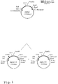
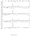
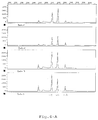
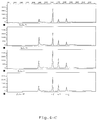
- FIG. 1 shows the cloning of pgag-15 and pgag + 12; 2 shows the results of the quantification of HIV with RT-PCR; 3 shows the results of the quantification of HAV with RT-PCR; 4 shows the results of the quantification of HBV; and Figure 5 is the sequence listing.
- Nucleic acids of different origins are amplified by means of PCR using primers which have fluorescent groups (Saiki et al., Science 239 (1985) 487-491).
- the analysis and quantification of the amplified PCR products obtained was carried out using an automatic DNA sequencer with a laser-induced fluorescence measuring device (DNA sequencer 373A with Gene Scan® software from Applied Biosystems).
- This instrument is able to separate the fluorescence-labeled PCR products by means of gel electrophoresis in a polyacrylamide gel under denaturing conditions and to quantify their amount.
- the copy number of certain sequences in the sample is based on the intensities obtained of the PCR products of the nucleic acid to be quantified and at least two internal standards.
- 500 ⁇ l of the sample are centrifuged for 20 min at 70,000 rpm in an ultracentrifuge.
- the pellet is dissolved in 500 ul 10 mM TRIS / HCl pH 8.0 and 10 ul proteinase K (Boehringer Mannheim, 20 mg / ml), as well as 10 ul 20% SDS.
- a certain amount of standard nucleic acid and 1 ⁇ g herring sperm DNA are added and the sample is incubated at 56 ° C for 1 hour.
- the sample is extracted successively with phenol and chloroform, and 10 ul glycogen (Boehringer Mannheim, 20 mg / ml) are added. It is then precipitated with ethanol, centrifuged, the pellet washed and finally redissolved in water.
- 5 ⁇ 10 5 cells are lysed in 100 ⁇ l lysis buffer (1 ⁇ PCR buffer from Boehringer, 0.5 mg / ml Proteinase K, 0.45% Tween) at 56 ° C. for 5 h. Aliquots of this are used for the PCR.
- 500 ⁇ l of the sample are dissolved in 5 ⁇ l 10 mM TRIS / HCl pH 8.0 and 10 ⁇ l Proteinase K (Boehringer Mannheim, 20 mg / ml). After incubation overnight at 37 ° C. or for 4 h at 56 ° C., a certain amount of standard nucleic acid is added, the sample is extracted successively with phenol and chloroform and 10 ⁇ l glycogen (Boehringer Mannheim, 20 mg / ml) is added. It is then precipitated with ethanol, centrifuged, the pellet washed and finally redissolved in water.
- RNAzol® guanidium isothiocyanate solution
- a predetermined number e.g. 400 and 1200 copies of the minus and plus RNA standards added and vortexed.
- the solution is heated at 70 ° C. for 10 minutes, then 1/10 volume of chloroform is added and incubated on ice for 10 minutes. Then it is centrifuged for 5 min in a table centrifuge, the supernatant is transferred to new tubes. 500 ⁇ l isopropanol is added and the temperature is set to -80 ° C for 15 min. It is then centrifuged for 10 min, washed twice with 70% ethanol and the pellet is taken up in 50 ⁇ l of water. 5 ⁇ l are used for the RT-PCR reaction.
- the PCR mixture contains an aliquot of the extracted nucleic acid, PCR buffer (Boehringer Mannheim), MgCl 2 , dNTPs, primers, Taq DNA polymerase (Boehringer Mannheim, 5.0 U / ⁇ l) and water.
- the PCR is carried out in accordance with the manufacturer's instructions for buffer and enzyme or in accordance with customary working instructions (Mullis et al., Methods in Enzymology 155 (1987), 335) in a PCR apparatus (GeneAmp PCR System 9600 from Perkin-Elmer).
- the RT-PCR approach contains, in a known manner, an aliquot of the extracted nucleic acid RT buffer from Perkin-Elmer, MgCl 2 , dNTPs, the RT primer and rT.th. polymerase (Perkin-Elmer, 2.5 U / ⁇ l ) and water.
- the RT is carried out in a PCR apparatus (GeneAmp PCR System 9600 from Perkin-Elmer) in accordance with the manufacturer's instructions for buffer and enzyme or in accordance with customary working instructions (Mullis et al., Methods in Enzymology 155 (1987), 335) .
- MgCl 2 MgCl 2
- a chelate buffer and the second primer are added for the PCR reaction. Then the PCR is carried out according to the information described above.
- PCR products 0.5 to 1.0 ⁇ l are taken from the PCR solution and analyzed in a 373A instrument from Applied Biosystems according to the manufacturer's instructions.
- primers are used which bind in the cDNA sequences of HIV-1 and result in a 115 bp product by RT-PCR of wild-type RNA, namely (Ratner et al. Numbering).
- the primers were produced using phosphoamidite chemistry on a DNA synthesizer (Applied Biosystems 394 DNA Synthesizer).
- the standard plasmids pgag-15 and pgag + 12 are derived from the plasmid pgag1, which consists of the known pBS / SK plasmid (from Stratagene) and an insert in the multiple cloning site of this plasmid, which insert the bp 1417 to 2008 des HIV-1 from Ratner et al. contains.
- telomeres were deleted, in pgag + 12 a 12 bp long insert was inserted at position 1593 (see Fig. 1).
- the plasmids were purified (QUIAGEN method), the concentration was determined by spectroscopic measurement at 260 nm and diluted in a 10 mM TRIS / HCl pH 8 / 0.1 mM EDTA buffer (Sambrook et al. Molecular Cloning, Second Edition, Cold Spring Harbor Lab Press, Cold Spring Harbor (1989)).
- RNA preparations serve as the standard for RT-PCR.
- the length of the RT-PCR products of standard and wild-type DNA is therefore 127 (pgag + 12), 100 (pgag-15) and 115 bp (wt).
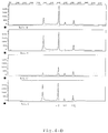
- Figures 2A and 2B illustrate the results of a control experiment.
- the PCR products of the "-", "wt” and “+” standards can be seen in both lanes.
- the results of the quantitative evaluation of these chromatograms are given in Table 1, lines 1 and 2.
- the "N-added” and “N + added” columns indicate the number of copies of the "-” and “+” standard added.
- “Dilution” indicates the dilution factor by which the sample was diluted, "Volume” the processed volume.
- Sample indicates the sample code, "Comment” contains further information about the sample.
- “GS #, Lane” specifies the analysis run or the number of the track in this run.
- “A-”, “Awt” and “A +” indicate the peak areas, ie the intensity of the PCR products of "-", "wt” and “+”.
- “N-Base” or “N + Base” contains the result, given in copies per ml of sample, calculated according to the formula given above. The “Final Result” column shows the mean value of these values.
- a negative plasma was mixed with a preparation of HIVIIIB, the infectivity of which had previously been determined in vitro.
- the virus concentration in the plasma was 200 TCID 50 per ml.
- 0.5 ml of a 1:10 and a 1:40 dilution were analyzed as described above.
- the results are shown in Figures 2C and 2D and in rows 3 and 4 of Table 1.
- a copy number in the undiluted plasma of 33187 or 25895 per ml results for both samples. The deviation from the mean is 12%. This experiment shows that the measured copy number HIV in a sample is independent of the amount used in the test.
- Figures 2E and 2F and rows 5 and 6 of Table 1 show the results of the determination of an unknown sample. 5612 and 5828 copies were measured in two independent determinations. The deviation is 2%. From this it can be concluded that the method described above is suitable for the sensitive and precise determination of HIV.
- primers which bind in the cDNA sequences of the HAV and give a 139 bp product, namely by RT-PCR of wild-type RNA, namely (Numbering according to Cohen et al. (J.Virol. 61 (1987) 50-59).
- the primers were produced using phosphoamidite chemistry on a DNA synthesizer (Applied Biosystems 394 DNA Synthesizer).
- the standard plasmids pHAV-10 and pHAV + 9 are derived from the plasmid pHAV-wt, which consists of the known pCRII plasmid (company InVitrogen) and an insert in the multiple cloning site of this plasmid, which insert the bp 2020 to 2226 des HAV from Cohen et al. contains.
- pHAV-10 the bp 2100 to 2109 were deleted, in pHAV + 9 a 9 bp insert was inserted at position 2100.
- the plasmids were purified (QUIAGEN method), the concentration was determined by spectroscopic measurement at 260 nm and diluted in a 10 mM TRIS / HCl pH 8 / 0.1 mM EDTA buffer (Sambrook et al. Molecular Cloning, Second Edition, Cold Spring Harbor Lab Press, Cold Spring Harbor (1989)).
- RNA preparations serve as the standard for RT-PCR.
- the length of the RT-PCR products of standard and wild-type DNA are 148 (pHAV + 9), 129 (pHAV-10) and 139 bp (wt).
- Table 2 shows the evaluation of the measurements with the nucleic acid detection device using a computer program (MS Excel®). Columns 1 and 2 indicate the amount of minus standard and plus standard used. Columns 3 and 4 indicate dilution and volume used. The sample is given in column 5, column 6 denotes the virus for which the test is being carried out. The copy numbers of the samples were calculated both using the minus standard (N-base; column 7) and using the plus standard (N + base; column 8); the mean of both determinations gives the measurement result. Column 9 remained empty. Column 10 gives the number of the sample run. Columns 11, 12 and 13 indicate the area of the peaks detected.
- FIG. 3 shows the graphical evaluation of the HAV examination, the intensities of the fluorescence signals of the PCR products (and by-products) being shown in the different lanes.
- the products can be identified based on their defined size (in bp).
- the standards are 148 and 129 bp long, the wild type 139.
- This quantification uses primers which bind in the cDNA sequences of the HCV and give a 114 bp product by RT-PCR of wild-type RNA, namely (Numbering according to Han et al. (PNAS 88 (1991) 1711-1715).
- the primers were produced using phosphoamidite chemistry on a DNA synthesizer (Applied Biosystems 394 DNA Synthesizer).
- the standard plasmids pHCV-7 and pHCV + 8 are derived from the plasmid pHCV-wt, which consists of the known pBS / SK - plasmid (from Statagene) and an insert in the multiple cloning site of this plasmid, which insert is the bp 27 to 313 of the HCV from Han et al. contains.
- pHCV-7 the bp 126 to 135 were deleted, in pHCV + 8 an 8 bp insert was inserted at position 126.
- the plasmids were purified (QUIAGEN method), the concentration was determined by spectroscopic measurement at 260 nm, cut with XmnI and diluted in a 10 mm TRIS / HCl pH 8 / 0.1 mM EDTA buffer (Sambrook et al., Molecular Cloning, Second Edition, Cold Spring Harbor Lab Press, Cold Spring Harbor (1989)).
- RNA preparations serve as the standard for RT-PCR.
- the length of the RT-PCR products of standard and wild-type DNA is therefore 122 (pHCV + 8), 107 (pHCV-7) and 114 bp (wt).
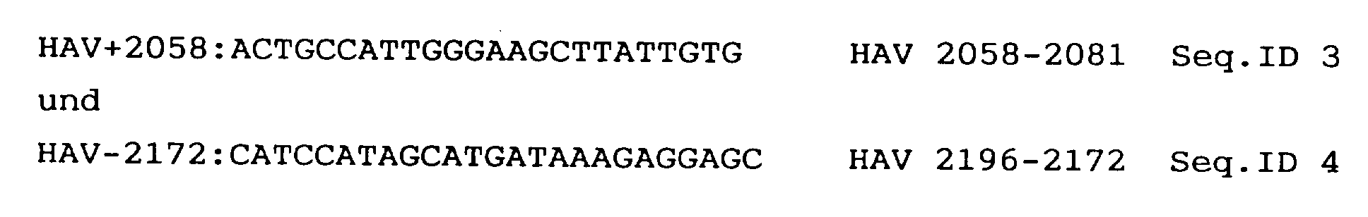
- Lines 5 and 6 of Table 3 show the results of the determination of an unknown sample. 3277 and 3676 copies were measured in two independent determinations. The deviation is 5%. It can be concluded that the one described above Method for the sensitive and precise determination of HCV is suitable.
- primers which bind in the cDNA sequences of HIV-1 and which give a 115 bp product, namely by PCR of proviral HIV DNA (Ratner et al. Numbering).
- the primers were produced using phosphoamidite chemistry on a DNA synthesizer (Applied Biosystems 394 DNA Synthesizer).
- the standard plasmids pgag-15 and pgag + 12 are derived from the plasmid pgagl, which consists of the known pBS / SK - plasmid (from Stratagene) and an insert in the multiple cloning site of this plasmid, which insert the bp 1417 to 2008 the HIV-1 from Ratner et al. contains.
- telomeres were deleted, in pgag + 12 a 12 bp insert was inserted at position 1593 (see Fig. 1).
- the plasmids were purified (QUIAGEN method), the concentration was determined by spectroscopic measurement at 260 nm, cut with EcoRI and diluted in a 10 mm TRIS / HCl pH 8 / 0.1 mM EDTA buffer (Sambrook et al. Molecular Cloning, Second Edition, Cold Spring Harbor Lab Press, Cold Spring Harbor (1989)).
- the length of the PCR products of standard and wild-type DNA is therefore 127 (pgag + 12), 100 (pgag-15) and 115 bp (wt).
- Table 4 The results of a series of quantifications are shown in Table 4.
- a coded control series of positive and negative samples containing HIV proviral DNA in different copy numbers were determined using the method according to the invention.
- Table 4 shows that all negative controls are negative and all positive samples are in the correct size range were quantified. The precise measurement in the lower copy area is particularly remarkable, see CD-13 and CD-26. These samples contained nominally 2 copies / 10 5 cells and were quantified with 3 copies each. This shows the accuracy and extreme sensitivity of the method according to the invention.
- primers are used which bind in the genome of the HBV and result in a 182 bp product by PCR of wild-type DNA, namely (Numbering according to Fujiyama et al.).
- the primers were produced using phosphoamidite chemistry on a DNA synthesizer (Applied Biosystems 394 DNA Synthesizer).
- the standard plasmids pHBV-9 and pHBV + 12 are derived from the plasmid pHBV-wt, which consists of the known pCRII plasmid (company In Vitrogene) and an insert in the multiple cloning site of this plasmid, which insert is the bp 1763 to 1868 the HBV from Fujiyama et al. contains.
- pHBV-9 the bp were deleted from 1868 to 1876, in pHBV + 12 a 12 bp insert was inserted at 1868.
- the plasmids were purified (QUIAGEN method), the concentration was determined by spectroscopic measurement at 260 nm, cut once with a restriction enzyme and diluted in a 10 mM TRIS / HCl pH 8 / 0.1 mM EDTA buffer (Sambrook et al. Molecular Cloning , Second Edition, Cold Spring Harbor Lab Press, Cold Spring Harbor (1989)).
- the length of the PCR products of standard and wild-type DNA is therefore 194 (pHBV + 12), 173 (pHBV-9) and 182 bp (wt).
- the results of a series of quantifications are shown in Table 5 and illustrated graphically in FIG.
- the amplification reaction was carried out on the basis of 150 copies of pHBV-9 and 50 copies of HBV + 12 and different amounts of pHBV-wt (400, 200, 100, 50 and 0 copies). Each approach was measured four times.
- primers were used which bind in the genome of the HSV and give a 114 bp product by PCR of wild-type DNA, namely
- the standard plasmid pHerp-9 is derived from the plasmid pHerp, which consists of the known pCRII plasmis (company In Vitrogen) and an insert from bp128364 to 138784 of the HSV genome (McGeoch DJ et al., EMBL GenBank ID HElCG1) .
- pHerp-9 9bp were deleted.
- the plasmids were purified, the concentrations were determined by spectroscopic measurement at 260 nm, linearized with a restriction enzyme and diluted in a TE buffer (10 mM Tris / HCl, IMM EDTA, pH 8).
- This DNA preparation serves as the standard for PCR.
- the length of the PCR products of standard and wild type are therefore 105 (pHerp-9) and 114 bp (pHerp).
- primers were used which bind in the genome of the vaccinia virus and which give a 136 bp product by PCR of wild-type DNA, namely (Numbering according to Goebel et al., Virology 179: 247-266, 1990).
- the primers were produced on a DNA synthesizer (Applied Biosystems 394 DNA Synthesizer).
- the standard plasmid pVV-21 is derived from the plasmid pVV-wt, which consists of the known pTZ19R plasmid (Pharmacia) and an insert between the two PvuII restriction sites of this plasmid.
- This insert contains bp 83855 to 84761 of the thymidine kinase gene region of the vaccinia virus (see Goebel et al., 1990).
- the plasmids were purified, the concentrations were determined by spectroscopic measurement at 260 nm, linearized with a restriction enzyme and diluted in a TE buffer (10 mM Tris / HCl, IMM EDTA, pH 8).
- This DNA preparation serves as the standard for PCR.
- the length of the PCR products of standard and wild type are therefore 115 (pVV-21) and 136 (pVVwt).
- Columns 2 and 3 give the dilution factor and the one used Volume of the sample. Column 4 can (if necessary) provide more information about the sample. Column 5 gives the name of the sample. Column 6 shows the calculated amount of vaccinia DNA in pg / ml. Column 7 gives the number of the gene scanner run in which the PCR products were analyzed. Columns 8 and 9 indicate the areas of the detected peaks from standard plasmid (column 8) and from wild type (column 9).
Landscapes
- Chemical & Material Sciences (AREA)
- Life Sciences & Earth Sciences (AREA)
- Organic Chemistry (AREA)
- Health & Medical Sciences (AREA)
- Zoology (AREA)
- Wood Science & Technology (AREA)
- Proteomics, Peptides & Aminoacids (AREA)
- Engineering & Computer Science (AREA)
- Immunology (AREA)
- Molecular Biology (AREA)
- General Engineering & Computer Science (AREA)
- Microbiology (AREA)
- Biophysics (AREA)
- Analytical Chemistry (AREA)
- Physics & Mathematics (AREA)
- Genetics & Genomics (AREA)
- Biochemistry (AREA)
- Bioinformatics & Cheminformatics (AREA)
- Biotechnology (AREA)
- General Health & Medical Sciences (AREA)
- Chemical Kinetics & Catalysis (AREA)
- Virology (AREA)
- Measuring Or Testing Involving Enzymes Or Micro-Organisms (AREA)
- Saccharide Compounds (AREA)
- Preparation Of Compounds By Using Micro-Organisms (AREA)
- Medicines Containing Antibodies Or Antigens For Use As Internal Diagnostic Agents (AREA)
- Investigating Or Analysing Biological Materials (AREA)
Abstract
Description
- Die Erfindung betrifft ein Verfahren zur Quantifizierung von Nukleinsäuren in einer Probe unter Anwendung von Nukleinsäure-Amplifizierung, wobei der Probe vor dem Amplifizierungsschritt eine gegebene Menge eines bekannten Nukleinsäuremoleküls als interner Standard zugegeben wird, welches Standard-Nukleinsäuremolekül sich von der zu quantifizierenden Nukleinsäure zumindest in einem detektierbaren Merkmal unterscheidet
- Für die Diagnose von verschiedenen viralen Erkrankungen gibt es zur Zeit nur sehr unzuverlässige Detektionsmethoden. Eine Virusinfektion kann in einem frühen Stadium der Erkrankung oft nicht festgestellt werden, da die zur Verfügung stehenden ELISA-Methoden die wenigen im Blut zirkulierenden Virusproteine nicht detektieren können. Allerdings wäre es für Erfolge in der Therapie von unschätzbarem Wert, Infektionen möglichst früh zu erkennen. Weiters gilt für virale Infektionen auch, daß es im Verlaufe der Krankheit oder durch den Einfluß einer Therapie zu Schwankungen der Viruskonzentration im Blut kommt. Dabei kann die Anzahl der Viruspartikel so stark abnehmen, daß die herkömmlichen Methoden sich als zu wenig sensitiv erweisen. Falsch negative Ergebnisse durch ELISA-Test lassen in solchen Fällen den Krankheitsverlauf in einem falschen Bild erscheinen.
- Ein anderer Aspekt bei der Detektion von Nukleinsäuren ist die Qualitätskontrolle von biotechnologisch hergestellten Produkten. Einerseits müssen Präparationen von immunogenen Virusproteinen, die aus infektiösen Viren gewonnen werden und als Impfstoffe eingesetzt werden sollen, auf ihren Gehalt an kontaminierender viraler Nukleinsäure hin überprüft werden. Andererseits werden rekombinante Produkte, die mit einem viralen Expressionssystem hergestellt wurden, auf kontaminierende Nukleinsäuren überprüft, die vom Expressionssystem stammen können. Die hier erlaubten Grenzwerte an kontaminierender Nukleinsäure sind von der Welt-Gesundheits-Organisation mit 100 pg pro Dosis, und von der U.S. Food and Drug Administration mit 10 pg pro Dosis festgelegt.
- Die Entwicklung in der PCR-Technologie hat es ermöglicht, die Nachweisgrenze zur Detektion von Nukleinsäuren drastisch zu senken. Allerdings kämpft man vor allem bei niedrigen Nukleinsäurekonzentrationen nach wie vor mit großen Problemen in der Reproduzierbarkeit der Resultate. Das liegt einerseits an der noch nicht ausgereiften Technik, andererseits sind es oft nur wenige Kopien von Nukleinsäuren, die nachgewiesen werden sollen. Bei quantitativen Bestimmungen in diesem niedrigen Konzentrationsbereich wirken sich Fehler in der Analyse natürlich besonders drastisch aus.
- Es ist dem Fachmann bestens bekannt, daß die Effizienz der PCR-Reaktion oftmals von Reaktionsgefäß zu Reaktionsgefäß unterschiedlich sein kann. Dieser Effizienzunterschied kann Unterschiede in den Ergebnissen von bis zu 105 ergeben. Die Reproduzierbarkeit der mit den bekannten Methoden erhaltenen Quantifizierungsdaten ist daher immer noch nicht ausreichend.
- Um die Reproduzierbarkeit der PCR-Reaktionen zu verbessern, wurde von Gilliland (PNAS 87:2725 (1990)) eine kompetitive PCR-Methode entwickelt. Zur Bestimmung von DNA-Mengen wird eine Verdünnungsreihe eines internen Standards verwendet, die gleichzeitig mit der Probe amplifiziert wird. Die PCR-Reaktion wird bis zur Sättigung durchgeführt. Dies erlaubt auch die Detektion der PCR-Produkte mittels Ethidiumbromidfärbung. Die PCR-Produkte werden anschliessend auf einem Gel aufgetrennt, die Kopienzahl der Probe mit der Kopienzahl der Standard-Verdünnungsreihe verglichen und so die Konzentration der Probe abgeschätzt. Diese Konzentrationsabschätzung ist dann exakt, wenn die Konzentration des Standards und der Probe etwa im Verhältnis 1:1 in einem Reaktionsgefäß amplifiziert wurden. Dies wiederum impliziert, daß die Bestimmung der DNA-Menge umso genauer erfolgt, je mehr Standardverdünnungen verwendet werden.
- Eine Methode zur Quantifizierung von RNA wurde von Wang et al. (PNAS 86 (1989), 9717) vorgeschlagen. Diese PCR-Reaktion wird in der exponentiellen Phase gestoppt. Die Autoren schaffen sich durch Amplifikation unterschiedlicher Standardkonzentrationen eine Eichkurve. Da in der exponentiellen Reaktionsphase die Anzahl der PCR-Produkte direkt proportional zur Anzahl der PCR-Zyklen und zur Konzentration der RNA steigt, ist diese Eichkurve eine Gerade, in der man schließlich die Konzentration einer amplifizierten Probeablesen kann. Ein Nachteil dieser Methode ist, daß die Endkonzentration der PCR-Produkte relativ gering ist, sodaß man für die Detektion empfindliche Nachweismethoden anwenden muß. Wang et al. benutzen radioaktiv markierte Nukleotide.
- Gemäß der WO 93/23573 kann durch kompetitive PCR die Konzentration viraler RNA im Bereich von 102 bis 108 Kopien bestimmt werden. Die Reproduzierbarkeit dieser Methode wird bei Mehrfachbestimmungen von HIV-1 mit einem Variationskoeffizienten von 0,26 ± 0,15 angegeben. Dabei werden die Bestimmungen umso genauer, je näher das Konzentrationsverhältnis von Probe zu Standard 1:1 liegt. Dabei ist zu berücksichtigen, daß bei dieser Methode die Standard-RNA erst nach der Probenaufbereitung zugegeben wird. Bei der Extraktion der Probe können aber bis zu 50 % der RNA verloren gehen. So wird schließlich die ursprünglich in der Probe enthaltene RNA-Menge auf den erst nach der Extraktion zugegebenen Standard bezogen. Dadurch wird lediglich die unterste Grenze der möglichen Kopienanzahl pro Probe bestimmt. Die durch die Extraktion verloren gegangenen Anteile können nicht berücksichtigt werden.
- In der WO 94/20640 wird ebenfalls eine kompetitive RT-PCR-Methode beschrieben, von der behauptet wird, daß damit 100 Kopien des HIV-Genoms im Plasma von HIV-infizierten Patienten festgestellt werden kann. In Bereichen unter 100 Kopien erfolgt die Ermittlung der Konzentration der HIV-Genome nur mehr durch Abschätzung. Die Standardabweichung von Bestimmungen der Kopienanzahl im Bereich von 104 bis 106 Kopien liegt bei 22 %, wenn von dem gleichen Plasma eine Dreifachbestimmung gemacht wird; bei Doppelbestimmungen derselben RNA-Präparation liegt die Standardabweichung bei 15 % (WO 94/20640, Seite 22, Zeilen 8-13). Auch hier wird die Extraktionseffizienz der RNA-Extraktion nicht berücksichtigt. Die gefundenen Kopienzahlen der HIV RNA sind im Bereich von 4,4 x 10³ bis 9,3 x 10⁶. Nur bei einem einzigen Patienten werden 100 Kopien als "extrapolierter" Wert angegeben. Es stellt sich daher die Frage, ob diese Methode zur exakten Bestimmung von niedriger Kopienzahlen (unter 103) überhaupt geeignet ist.
- Eine Verbesserung in der Detektion von geringen Mengen an PCR-Produkten gelang Porcher et al. (BioTechniques 13 (1992), 106) durch den Einsatz von Fluoreszenz-markierten Primern und der Quantifizierung der PCR-Produkte mit einem automatischen Laser-Fluoreszenz-DNA-Sequencer.
- Die vorliegende Erfindung stellt sich die Aufgabe, ein Verfahren zur Quantifizierung von Nukleinsäuren zur Verfügung zu stellen, welches eine sehr genaue und vor allem eine gut reproduzierbare Information bezüglich der Menge an Nukleinsäure in einer Probe ermöglicht und gleichzeitig Aussagen über die Nachweisgrenze der zu bestimmenden Nukleinsäure erlaubt.
- Das erfindungsgemäße Verfahren der eingangs erwähnten Art ist dadurch gekennzeichnet, daß der Probe vor der Nukleinsäure-Amplifizierung bekannte Mengen von mindestens zwei sich zumindest in einem detektierbaren Merkmal voneinander und von der zu quantifizierenden Nukleinsäure unterscheidenden bekannten Nukleinsäuremolekülen als interner Standard zugegeben werden, die erhaltenen Mengen an amplifizierter Proben- und Standard-Nukleinsäure bestimmt werden und aus den erhaltenen Mengen die ursprünglich in der Probe vorhandene Menge an zu quantifizierender Nukleinsäure bestimmt wird.
- Das erfindungsgemäße Verfahren ermöglicht überraschenderweise eine sehr exakte und darüberhinaus gut reproduzierbare Quantifizierung von Nukleinsäuren aller Art.
- Unter Nukleinsäure-Amplifizierung sind prinzipiell Verfahren zu verstehen, welche auf der von Mullis et al. (U.S. PS 4,683,195 und 4,683,202) und anderen entwickelten Technologie beruhen, beispielsweise die Polymerase-Kettenreaktion (PCR), die reverse Transkriptase-PCR (RT-PCR) oder die Ligase-CR (LCR).
- Die Standard-Nukleinsäure muß sich in wenigstens einem detektierbaren Merkmal von der zu quantifizierenden Nukleinsäure unterscheiden, sie sollte aber mit Hilfe der gleichen Primer amplifiziert werden können. Als praktisch haben sich Standard-Nukleinsäuren erwiesen, die eine andere Größe als die zu quantifizierende Nukleinsäure oder eine unike Restriktionsschnittstelle aufweisen. Die Standard-Nukleinsäure ist bei Bestimmung von DNA-Mengen vorzugsweise eine DNA und bei Bestimmung von RNA-Mengen vorzugsweise eine RNA. Bevorzugte Standards unterscheiden sich von der zu quantifizierenden Nukleinsäure dadurch, daß deren PCR-Produkte sich in 1 % bis 20 % ihrer Länge bzw. durch mindestens 3, maximal 50 Nukleotide, unterscheiden. Eine Standardnukleinsäure, deren PCR-Produkt länger ist als das der zu bestimmenden Nukleinsäure, wird als "plus"("+")-Standard bezeichnet, und eine Standardnukleinsäure, deren PCR-Produkt kleiner ist als das der zu bestimmenden Nukleinsäure, wird als "minus"("-")-Standard bezeichnet. Die genaue Sequenz der Standard-Nukleinsäure sollte natürlich bekannt sein.
- Die Standards werden im erfindungsgemäßen Verfahren bevorzugt in unterschiedlicher Konzentration eingesetzt, wobei einer der Standards in einer Konzentration knapp oberhalb der Nachweisgrenze zugegeben wird.
- Die beim Amplifizieren verwendeten Primer enthalten vorzugsweise Gruppen, welche die Nachweisgrenze der amplifizierten Nukleinsäuren erhöhen, beispielsweise fluoreszierende oder radioaktive Gruppen oder chemische Gruppen, die mit affinen Proteinen und nachgestalteten Detektionsreaktionen detektiert werden können (z.B. Biotin-Avidin, DIG-Markierung, etc.), wobei Primer mit fluoreszierenden Gruppen besonders bevorzugt sind.
- Die Bestimmung der Nukleinsäure-Mengen (unter Nukleinsäure-Menge versteht man prinzipiell die Quantität an DNA oder RNA; eine Nukleinsäure-Menge kann z.B. in Form von Masse (mg, µg, ng, pg) oder als Anzahl der Kopien eines bestimmten Nukleinsäure-Moleküls angegeben werden) nach der Amplifizierung kann auf unterschiedlichste Art erfolgen, meist jedoch ist ein Schritt vorzusehen, bei welchem die amplifizierte Standard-Nukleinsäure von der amplifizierten, zu quantifizierenden Nukleinsäure getrennt wird und die getrennten Nukleinsäure-Mengen separat bestimmt werden. Vorzugsweise besteht dieser Trennungsschritt in einer Gelelektrophorese oder in einem chromatographisches Verfahren.
- Als besonders geeignet haben sich Detektionsverfahren erwiesen, welche automatisch erfolgen und den Trennungs- und Quantifizierungsschritt kombinieren. Eine bevorzugte Ausführungsform des erfindungsgemäßen Verfahrens besteht daher darin, daß die Bestimmung der Mengen an amplifizierter Nukleinsäure unter Verwendung eines Nukleinsäure-Detektionsgerätes, vorzugsweise eines fluoreszenzempfindlichen Nukleinsäure-Detektionsgerätes, erfolgt. Beispiele für solche Nukleinsäure-Detektionsgeräte sind automatische DNA-Sequenzer mit laserinduzierten Fluoreszenz-Meßeinrichtungen (z.B. Gene Scanner®373A der Firma Applied Biosystems), HPLC- oder Kapillar-Elektrophorese-System-Anlagen. Bei diesen Geräten ist es möglich, Nukleinsäure-Moleküle voneinander zu trennen, die sich lediglich um ein bp in der Länge unterscheiden.
- Ein besonderer Vorteil des Gene-Scanner® ist es, unterschiedliche Fluoreszenzfarbstoffe in einer einzigen Spur unterscheiden zu können. Dies ermöglicht die gleichzeitige Aufarbeitung einer Vielzahl von Proben auf einem Gel, da alle am Gel zur Verfügung stehenden Spuren für Proben verwendet werden können. Weiters ist es möglich, eine Vielzahl von PCR-Produkten, markiert mit unterschiedlichen Fluoreszenz-Farbstoffen, in einer einzigen Bahn zu analysieren (Multiplex-PCR). Beim gleichzeitigen Nachweis von beispielsweise zwei verschiedenen Nukleinsäuren in einer Probe werden außerdem Aufwand und Kosten nahezu halbiert. Dies ist beim Einsatz des erfindungsgemäßen Verfahrens im Routinebetrieb von besonderem Vorteil, wenn beispielsweise eine Blutprobe auf HIV und HCV getestet werden soll. Im Gegensatz dazu kann der von Porcher et al. zur Analyse der PCR-Produkte verwendete automatische Laser-Fluoreszenz-DNA-Sequenzer nur einen Fluoreszenzfarbstoff (und damit nur eine DNA) pro Spur analysieren.
- Eine bevorzugte Ausführungsform des erfindungsgemäßen Verfahrens betrifft daher ein Verfahren, bei dem mehrere erhaltene Mengen an amplifizierter Proben- und Standard-Nukleinsäuren in der gleichen Probe mittels der Multiplex-Analyse bestimmt werden.
- Bei einer bevorzugten Ausführungsform des erfindungsgemäßen Verfahrens wird der Amplifizierungsschritt bereits in der exponentiellen Phase gestoppt.
- Dadurch wird erreicht, daß das Verhältnis der Kopienanzahl der amplifizierten Standards direkt proportional zur Kopienanzahl der zu quantifizierenden Sequenz ist. Weiters kann durch Koamplifikation eines einzigen Standards die Kopienanzahl der zu bestimmenden Nukleinsäure festgestellt werden. In diesem Punkt ist die erfindungsgemäße Methode der oft verwendeten Methode von Gilliland et al. weit überlegen, da pro Probe lediglich eine Messung mit wenigstens zwei verschiedenen Standard-Molekülen durchgeführt werden muß, wogegen die Methode nach Gilliland um so genauer wird, je mehr Standards in verschiedenen Verdünnungen in verschiedenen Proben verwendet werden.
- Ein besonders bevorzugtes Anwendungsgebiet des erfindungsgemäßen Verfahrens besteht in der Quantifizierung von Nukleinsäuren aus Mikroorganismen, vorzugsweise Nukleinsäuren aus HIV, Parvovirus, Herpesvirus, HAV, HBV, HCV, Baculovirus, Adenovirus, Influenzavirus, Vacciniavirus, Borreliaspezies, Salmonellaspezies oder Hefe. Diese Mikroorganismen sind sowohl als Pathogene als auch wegen ihrer Verwendung in der Herstellung von Impfstoffen und rekombinanten Proteinen interessant.
- Im speziellen findet das erfindungsgemäße Verfahren Anwendung in der Quantifizierung viraler Nukleinsäuren, die als Kontaminationen in biotechnologisch hergestellten Produkten zurückbleiben können. Vorzugsweise kann es sich bei diesen biotechnologischen Produkten um immunogene Virusproteine oder Virusteile handeln, die durch biotechnologische Verfahren aus infektiösen Viren gewonnen werden; oder es handelt sich um rekombinant hergestellte Produkte, die mit einem viralen Expressionssystem hergestellt worden sind, und Kontaminationen der viralen Nukleinsäure des Expressionssystems aufweisen können.
- Es werden vorzugsweise virale Nukleinsäuren in einer biologischen Probe, insbesondere in humanen Plasmen und deren Derivaten, gemäß dem erfindungsgemäßen Verfahren quantifiziert. Beispielsweise kann mit der vorliegenden Methodik der Verlauf einer Infektion oder die Überwachung von Impfungs- bzw. Therapiebehandlungen besser und genauer überwacht werden.
- Dabei ist es von besonderer Bedeutung, daß mit dem erfindungsgemäßen Verfahren pathogene Viren in einer Konzentration bestimmt werden können, die mindestens eine Zehnerpotenz unterhalb der infektiösen Dosis dieser Viren liegt.
- Um für die Nukleinsäurequantifizierung in der Probe einen möglichst genauen Wert zu erhalten, der nicht durch Manipulationen der zu untersuchenden Probe beeinflußt wird, setzt man gemäß einer bevorzugten Ausführungsform des erfindungsgemäßen Verfahrens die Standards direkt der Probe zu, vorzugsweise vor dem Extraktionsschritt, und unterzieht anschließend somit Proben-Nukleinsäuren und Standard-Nukleinsäuren den gleichen Probenaufbereitungsschritten (Koextraktion). Dies ermöglicht ein Quantifizierungsergebnis, das nicht nur erhöhte und unverfälschte Reproduzierbarkeit aufweist, sondern darüberhinaus auch unabhängig von Probenaufbereitungs-Manipulationen ist.
- Ein weiterer Aspekt der vorliegenden Erfindung betrifft daher ein Verfahren zur Bestimmung der Nachweisgrenze von bestimmten Nukleinsäuren, bei welchem mindestens eine Standard-Nukleinsäure mit einer Konzentration von knapp oberhalb der Nachweisgrenze eingesetzt wird.
- Vorzugsweise wird die Menge an Nukleinsäure in Kopienanzahl nach der folgenden Formel berechnet:

- AProbe die Peak-Fläche der amplifizierten Nukleinsäuren der Probe,
- AStandard die Peak-Fläche des amplifizierten internen Standards darstellt,
- NStandard die Zahl der eingesetzten Kopien des internen Standards,
- F das Verhältnis des Einheitsvolumens zum extrahierten Volumen und
- D der Verdünnungsfaktor (falls die Probe vor der Extraktion verdünnt worden ist), bedeutet
- Für den besonderen Anwendungsbereich des erfindungsgemäßen Verfahrens, bei dem kontaminierende virale Nukleinsäuren quantifiziert werden sollen, wird, um den Forderungen der WHO und der FDA Genüge zu tun, die Menge an Nukleinsäure in pg/ml angegeben. Dazu kommt erfindungsgemäß folgende Formel zum Einsatz:

- AS =
- die Peakfläche des PCR-Produkts der Probe,
- Ast =
- die Peakfläche des PCR-Produkts des Standards,
- Nst =
- die Kopienanzahl des Standard-Plasmids,
- F1 =
- das Verhältnis des Einheitsvolumens zum extrahierten Volumen,
- D =
- der Verdünnungsfaktor der Probe (falls notwendig) und
- F2 =
- die Masse eines viralen Moleküls in pg (Molekulargewicht der viralen DNA/Avogadro-Zahl), ist.
- Da nach dem erfindungsgemäßen Verfahren mindestens zwei Standards in unterschiedlichen Konzentrationen eingesetzt werden, erhält man nach der oben beschriebenen Berechnung mindestens zwei Werte für die Konzentration der Probe, aus denen man schließlich den Mittelwert berechnet. Im Routinebetrieb werden von jeder Probe in der Regel zwei Ansätze mit mindestens zwei Standards analysiert, wodurch schließlich ein Mittelwert aus vier Werten für die Konzen-tration der Probe gebildet werden kann.
- Das erfindungsgemäße Verfahren zeichnet sich durch eine besonders hohe Reproduzierbarkeit aus. Die Standardabweichung, die bei Mehrfachbestimmungen erhalten wird, beträgt maximal 15 %, im allgemeinen aber meist unter 10 %, selbst wenn die Kopienzahl im niedrigen Konzentrationsbereich von 102 liegt. Diese überaus geringen Standardabweichungen sind vor allem deswegen äußerst überraschend, da die jeweiligen Proben bei Mehrfachbestimmungen gemeinsam mit dem Standard auch jedesmal separat aufbereitet werden, d.h., daß auch Fehler in der Handhabung und Effizienzunterschiede in der Extraktion der Probe und der PCR-Reaktion an sich in diese Standardabweichungen eingehen.
- Die in den WO 93/23573 und WO 94/20640 beschriebenen Standardabweichungen von 0,26 ± 0,15 bzw. von 22 % bzw. 15 % wurden erhalten, indem die Probe nach der Aufbereitung in verschiedene Teile geteilt wird und diese Teile für die Bestimmung der Reproduzierbarkeit herangezogen werden. Dadurch gehen Effizienzunterschiede, die durch die Probenextraktion bzw. die RT-PCR auftreten können, nicht in die Bestimmung der Standardabweichung ein und es ergibt sich zwangsläufig eine bessere, wenn auch verfälschte Reproduzierbarkeit.
- Beeinflußt wird die Effizienz der PCR-Reaktion zum Beispiel von der Art des zu amplifizierenden Nukleinsäure-Moleküls. Wird die erfindungsgemäße Methode zur Bestimmung von RNA-Viren herangezogen, so kämpft man mit dem allgemein bekannten Problem, daß die reverse Transkription unvollständig ist und lediglich wenige Prozent der vorhandenen RNA auch wirklich transkribiert werden. Weiters hat sich gezeigt, daß die Effizienz der Amplifikation für den "+"- bzw."-"-Standard in den einzelnen Bestimmungen differieren kann, im Mittel aber gleich ist. Dadurch kann es zu Schwankungen in den zu bestimmenden Konzentrationen kommen.
- Gemäß einer bevorzugten Ausführungsform der Erfindung werden daher zur Vergrößerung der erhaltenen Information unterschiedliche Mengen der mindestens zwei Standard-Nukleinsäuren der Probe vor der Amplifizierung zugegeben werden.
- Um weitere Ungenauigkeit in der Konzentrationsbestimmung der Probe auszuschließen, werden daher im Routineverfahren zwei Aliquote jeder Probe bestimmt, bei denen jeweils mindestens zwei Standards mitamplifiziert werden. So erhält man vier Meßwerte pro Probe, aus denen man sich dann einen Mittelwert errechnen kann. In den Beispielen ist die Genauigkeit und Reproduzierbarkeit der Methode gut demonstriert. Es wird auch gezeigt, daß die Methode über einen großen Konzentrationsbereich reproduzierbare Ergebnisse liefert.
- Gemäß einer bevorzugten Ausführungsform des erfindungsgemäßen Verfahrens kommen zwei Standard-Nukleinsäuren zum Einsatz, welche eine gegenüber der zu quantifizierenden Nukleinsäure unterschiedliche Länge aufweisen, vorzugsweise eine Standard-Nukleinsäuresequenz, welche kürzer, und eine Standard-Nukleinsäuresequenz, welche länger ist als die zu quantifizierende Nukleinsäure. Ein besonders bevorzugter Längenunterschied liegt zwischen 1 % und 20 %.
- Es hat sich für die erfindungsgemäße Methode von Vorteil erwiesen, die Nukleinsäure der internen Standards in linearisierter Form der PCR-Reaktion zuzusetzen. Dadurch werden weitere Unterschiede in der Effizienz der Reaktion ausgeglichen, die auf die unterschiedliche Form der zu amplifizierenden Nukleinsäure zurückzuführen sind.
- Wichtige Kriterien bei der Quantifizierung von Nukleinsäuren sind - wie erwähnt - die Sensitivität und die Reproduzierbarkeit. Mit dem erfindungsgemäßen Verfahren können Nukleinsäure-Mengen im Bereich von 1 bis 500 Kopien wesentlich präziser und reproduzierbarer als mit den im Stand der Technik beschriebenen Methoden bestimmt werden. Damit ist aber keineswegs die Reproduzierbarkeitsgrenze der Methode erreicht.
- Das Verwendungsspektrum des erfindungsgemäßen Verfahrens umfaßt beispielsweise die qualitative und quantitative Analyse von biologischen Proben auf Nukleinsäuren, insbesondere Blut und Blutderivate und biotechnologische Produkte. Eine weitere beispielhafte Einsatzmöglichkeit liegt in der Diagnose und/oder der Überwachung des Verlaufes von Infektionen sowie in der Überwachung von Impf- und Therapiebehandlungen.
- Ein wichtiger Aspekt der Erfindung betrifft daher biologische, insbesondere biotechnologische Produkte, die einen unterhalb der erlaubten Grenzen von 10 bzw. 100 pg pro Dosis, vorzugsweise unterhalb des Bereiches von 1 bis 100 Kopien pro Probe, liegenden und mit dem vorliegenden Verfahren gemessenen Gehalt an Nukleinsäuren aufweisen und damit als im wesentlichen frei von Nukleinsäuren gelten können.
- Zu den bevorzugten Produkten gehören virale und bakterielle Proteine, wie gp160, rekombinante Blutfaktoren, Plasmaproteine, sowie Impfstoffe, insbesondere gegen Herpes-, Influenza-, TBE-, Parvo- oder Hepatitis-Viren und monoklonale Antikörper.
- Gemäß einem weiteren Aspekt betrifft die vorliegende Erfindung auch die Verwendung des erfindungsgemäßen Verfahrens zur Detektion von Nukleinsäuren in biologischen Proben.
- Im weiteren kämpft die Qualitätskontrolle insbesondere bei Impfstoffen oder biotechnologisch erzeugten Proteinen mit Background-Problemen. So kann man beim Bestimmen von kontaminierender Nukleinsäure (chromosomale DNA, RNA, virale DNA und RNA) bei Primaten-Zellkulturen auch die Verunreinigungen durch die Handhabung während der Produktion oder während der Aufarbeitung der Produkte durch das erfindungsgemäße Verfahren erfassen, wenn die eingesetzten Primer spezifisch sind. Erfolgt die Produktion von rekombinanten Proteinen allerdings in Nicht-Primaten-Zellkulturen, wie zum Beispiel CHO (Chinese Hamster Ovarien), BHK (Baby Hamster Nierenzellen) oder CEC (Hühner-Embryozellen), so ist die Nachweisgrenze mit der erfindungsgemäßen Methode weit niedriger, da das Problem der Verunreinigungen durch die Handhabung der Probe wegfällt. Besonders bevorzugt wird die erfindungsgemäße Quantifizierungsmetho-dik bei Nukleinsäuren aus CHO-, Vero-(Affenzellinie), BHK-, SK-Hep1-(menschliche Leberzellinie), Hybridom- oder CEC-Zellen angewendet, da diese Zellkulturen am gebräuchlichsten sind bei der Produktion von Impfstoffen oder biotechnologisch hergestellten Proteinen.
- Die Auswahl der Primerpaare ist selbstverständlich ebenfalls ein wichtiger Faktor, um eine gute Quantifizierung zu erhalten. Daher betrifft die vorliegende Erfindung gemäß einem weiteren Aspekt Primer, welche im vorliegenden Verfahren zur Anwendung kommen, nämlich
 und Plasmide für die Herstellung der Standards, nämlich
- pgag1 (bestehend aus dem bekannten pBS/SK -Plasmid und einem Insert zwischen der Pst I und der Apa I Stelle der multiplen Klonierungsstelle, welches Insert die Basenpaare (bp) 1417 bis 2008 der HIV-1 Sequenz aus Ratner et al. (Nature 313 (1985), 277- 284) enthält),
- pgag-15 (abgeleitet aus pgagl mit einer Deletion von 15 bp ab bp 1593 der HIV-1 Sequenz aus Ratner et al.),
- pgag+12 (abgeleitet aus pgagl, indem eine 12 Nukleotide lange Insertion an der bp1593-Stelle eingefügt wurde),
- pHAV-wt (bestehend aus dem bekannten pCRII-Plasmid) und einem Insert an der multiplen Klonierungsstelle des pCRII-Plasmids, welches Insert die bp 2020 bis 2226 der cDNA-Sequenz aus Cohen et al. enthält),
- pHAV-10bp (abgeleitet aus pHAV-wt mit einer Deletion von 10 bp ab bp 2100 der HAV-Sequenz aus Cohen et al. eingefügt wurde),
- pHAV+9bp (abgeleitet aus pHAV-wt, indem eine 9 Nukleotide lange Insertion an der bp2100-Stelle eingefügt wurde),
- pHCV-wt (bestehend aus dem bekannten pBS/SK Plasmid und einem Insert an der EcoRV-Stelle diese Plasmids, welches Insert die bp27 bis 313 der cDNA-Sequenz aus Han et al. enthält),
- pHCV-7bp (abgeleitet aus pHCV-wt mit einer Deletion von 7bp ab bp 126 der HCV-Sequenz aus Han et al. eingefügt wurde),
- pHCV+8bp (abgeleitet aus pHCV-wt, indem eine 8 Nukleotide lange Insertion an der bp126-Stelle eingefügt wurde),
- pHBV-wt (bestehend aus dem bekannten pCRII-Plasmid und einem Insert, welches Insert die bp 1763 bis 2032 des HBV-Genoms gemäß Fujiyama et al. enthält),
- pHBV-9bp (abgeleitet aus pHBV-wt mit einer Deletion von 9bp ab bp 1868 der HBV-Sequenz aus Fujiyama et al. eingefügt wurde),
- pHBV+12bp (abgeleitet aus pHBV-wt, indem eine 12 Nukleotide lange Insertion an der bp1868-Stelle eingefügt wurde),
- pVV-wt (abgeleitet aus dem bekannten pTZ19R-Plasmid von Pharmacia mit einem Insert zwischen den beiden PVUII-Stellen dieses Plasmids, welches Insert die bp 83855 bis 84761 des Thymidinkinase-Gens des Vaccinia-Virus (nach Goebel et al., 1990) enthält.),
- pVV-21 (abgeleitet aus pVV-wt mit einer Deletion von 21 Nukleotiden (bp 84718 bis 84738),
- pVV+24 (abgeleitet aus pVV-wt mit einer Insertion von 24 bp an der Stelle 84713),
- pHerp (abgeleitet von dem bekannten pCRII-Plasmid (Firma In Vitrogen) und einem Insert, welches Insert die bp 138364 bis 138784 des HSV-Genoms gemäß McGeoch et al., EMBL GenBank DE 1 CG1 enthält),
- pHerp-9 (abgeleitet von p-Herp mit einer Deletion von 9 Nukleotiden (bp 138388 bis 138396) und
- pHerp+10 (abgeleitet von p-Herp mit einer Insertion von 10 bp bei bp 138407.
- Gemäß einem weiteren Aspekt bezieht sich die vorliegende Erfindung auch auf ein Set zur Quantifizierung von Nukleinsäuren in einer Probe, welches umfaßt:
- mindestens zwei bekannte Nukleinsäuren als interne Standards, welches sich voneinander und von den zu quantifizierenden Nukleinsäuren in mindestens einem nachweisbaren Charakteristikum unterscheiden,
- Fluoreszenz-markierte Primer, die an die Standard-Nukleinsäure und an die zu quantifizierende Nukleinsäure binden,
- positive Kontrollen, welche bekannte Mengen einer Nukleinsäure, an der Interesse besteht, aufweisen,
- eine negative Kontrolle, welche Humanplasma, das frei von der viralen Nukleinsäure ist, an der Interesse besteht, aufweist, und
- eine Arbeitsanleitung.
- Bevorzugte Ausführungsformen des Sets gemäß der vorliegenden Erfindung sind die folgenden:
- 1.) Set zur Quantifizierung von HIV-RNA in einer Probe, welches umfaßt:
- mindestens zwei interne Standards, die eine in vitrotranskribierte RNA aufweisen, welche von den Plasmiden pgag-15 und pgag+12 stammt,
- die Fluoreszenz-markierten Primer SK38 und SK39
- eine positive Kontrolle, welche bekannte Mengen an HIV-1-Teilchen aufweist,
- eine negative Kontrolle, welche Humanplasma aufweist, das frei von Virus-Nukleinsäuren ist und
- eine Arbeitsanleitung.
- 2.) Set zur Quantifizierung von HCV-RNA in einer Probe, welches aufweist:
- mindestens zwei interne Standards, die eine in vitrotranskribierte RNA aufweisen, welche von den Plasmiden pHCV-7 und pHCV+8 stammt,
- die Fluoreszenz-markierten Primer HCV32Ext und HCVPT4
- positive Kontrollen, welche bekannte Mengen an HCV-Teilchen aufweisen,
- eine negative Kontrolle, welche Humanplasma aufweist, das frei von Virus-Nukleinsäuren ist und
- eine Arbeitsanleitung.
- 3.) Set zur Quantifizierung von HAV-RNA in einer Probe, welches aufweist:
- mindestens zwei interne Standards, die eine in vitrotranskribierte RNA aufweisen, welche von den Plasmiden pHAV-10 und pHAV+9 stammt,
- die Fluoreszenz-markierten Primer HAV+2058 und HAV-2172
- positive Kontrollen, welche bekannte Mengen an HAV-Teilchen aufweisen,
- eine negative Kontrolle, welche Humanplasma aufweist, das frei von Virus-Nukleinsäuren ist und
- eine Arbeitsanleitung.
- 4.) Set zur Quantifizierung von HBV-DNA in einer Probe, welches aufweist:
- mindestens zwei interne Standards, die die Plasmide pHBV-9 und pHBV+12 aufweisen,
- die Fluoreszenz-markierten Primer HBV+1780B und HBV-1960B
- positive Kontrollen, welche bekannte Mengen an HBV-Teilchen aufweisen,
- eine negative Kontrolle, welche Humanplasma aufweist, das frei von Virus-Nukleinsäuren ist und
- eine Arbeitsanleitung.
- 5.) Set zur Quantifizierung von HSV-DNA in einer Probe, welches aufweist:
- als interne Standards, die Plasmide pHerp-9 und pHerp+10,
- die Fluoreszenz-markierten Primer gDR/B und gDR2R/B
- positive Kontrollen, welche bekannte Mengen an HSV-Teilchen aufweisen,
- eine negative Kontrolle, welche Humanplasma aufweist, das frei von Virus-Nukleinsäuren ist und
- eine Arbeitsanleitung.
- Die Erfindung wird in den nachstehenden Beispielen und den dazugehörigen Zeichnungsfiguren, auf die sie jedoch nicht beschränkt sein soll, noch weiter erläutert. Insbesondere wird in den Beispielen gezeigt, daß das erfindungsgemäße Verfahren sich hervorragend für eine routinemäßige, schnelle und trotzdem genaue und reproduzierbare Quantifizierung von Nukleinsäuren in unterschiedlichsten Proben eignet.
- Es zeigen: Fig.1 die Klonierung von pgag-15 und pgag+12; Fig.2 die Ergebnisse der Quantifizierung von HIV mit RT-PCR; Fig.3 die Ergebnisse der Quantifizierung von HAV mit RT-PCR; Fig. 4 die Ergebnisse der Quantifizierung von HBV; und Fig.5 ist das Sequenzprotokoll.
- Nukleinsäuren unterschiedlicher Herkunft werden mittels PCR unter Verwendung von Primern, welche fluoreszierende Gruppen haben, amplifiziert (Saiki et al., Science 239 (1985) 487-491). Die Analyse und die Quantifizierung der erhaltenen amplifizierten PCR-Produkte wurde mit Hilfe eines automatischen DNA-Sequenzierers mit laserinduzierter Fluoreszenz-Meßeinrichtung (DNA-Sequenzierer 373A mit Gene Scan®-Software von Applied Biosystems) ausgeführt. Dieses Instrument ist in der Lage, die Fluoreszenz-markierten PCR-Produkte mittels einer Gelelektrophorese in einem Polyacrylamidgel unter denaturierenden Bedingungen der Größe nach aufzutrennen und deren Menge quantitativ zu bestimmen. Die Kopienzahl bestimmter Sequenzen in der Probe wird auf Grundlage der erhaltenen Intensitäten der PCR-Produkte von zu quantifizierender Nukleinsäure und mindestens zwei internen Standards bestimmt.
- 500 µl der Probe werden für 20 min bei 70000 rpm in einer Ultrazentrifuge zentrifugiert. Das Pellet wird in 500 µl 10 mM TRIS/HCl pH 8,0 und 10 µl Proteinase K (Boehringer Mannheim, 20 mg/ml), sowie 10 µl 20% SDS aufgelöst. Eine bestimmte Menge an Standard-Nukleinsäure und 1 µg Hering-Sperma-DNA werden zugesetzt, und die Probe wird 1 h lang bei 56°C inkubiert. Die Probe wird nacheinander mit Phenol und Chloroform extrahiert, und 10 µl Glykogen (Boehringer Mannheim, 20 mg/ml) werden zugesetzt. Anschließend wird mit Ethanol präzipitiert, zentrifugiert, das Pellet gewaschen und schließlich in Wasser wieder gelöst.
- 5 x 10⁵ Zellen werden in 100 µl Lysis-Puffer (1 X PCR-Puffer von Boehringer, 0,5 mg/ml Proteinase K, 0,45 % Tween) 5 h bei 56°C lysiert. Aliquote davon werden für die PCR eingesetzt.
- 500 µl der Probe werden in 5 µl 10 mM TRIS/HCl pH 8,0 und 10 µl Proteinase K (Boehringer Mannheim, 20 mg/ml) aufgelöst. Nach Inkubation über Nacht bei 37°C oder für 4 h bei 56°C wird eine bestimmte Menge an Standard-Nukleinsäure zugesetzt, die Probe nacheinander mit Phenol und Chloroform extrahiert und 10 µl Glykogen (Boehringer Mannheim, 20 mg/ml) zugesetzt. Anschließend wird mit Ethanol präzipitiert, zentrifugiert, das Pellet gewaschen und schließlich in Wasser wieder gelöst.
- 1 ml Plasma bzw. mit PBS verdünntes Plasma wird bei 70000 rpm 20 min. zentrifugiert. Der Überstand wird durch Absaugen entfernt. Das Pellet wird in 1 ml Guanidiumisothiocyanat-Lösung (RNAzol® der Firma Biotexc) aufgenommen und 5 µl 1 mg/ml t-RNA aus Hefe und eine vorbestimmte Menge, z.B. 20 µl, Standard-RNA zugegeben.
- Es werden eine vorbestimmte Anzahl, z.B. 400 und 1200 Kopien, des Minus- und Plus-RNA-Standards zugegeben und gevortext. Die Lösung wird 10 min bei 70°C erhitzt, dann 1/10 Volumen Chloroform zugegeben und für 10 min auf Eis inkubiert. Dann wird für 5 min in einer Tischzentrifuge zentrifugiert, der Überstand in neue Röhrchen transferiert. 500 µl Isopropanol wird zugegeben und 15 min auf -80°C gestellt. Anschließend wird 10 min zentrifugiert, 2 x mit 70 % Ethanol gewaschen und das Pellet in 50 µl Wasser aufgenommen. Für die RT-PCR-Reaktion werden 5 µl eingesetzt.
- Der PCR-Ansatz enthält in bekannter Weise ein Aliquot der extrahierten Nukleinsäure, PCR-Puffer (Boehringer Mannheim), MgCl2, dNTPs, Primer, Taq-DNA-Polymerase (Boehringer Mannheim, 5,0 E/µl) und Wasser. Die PCR wird gemäß den Angaben des Herstellers von Puffer und Enzym bzw. gemäß üblicher Arbeitsvorschriften (Mulliset al., Methods in Enzymology 155 (1987), 335) in einem PCR-Apparatur (GeneAmp PCR System 9600 der Firma Perkin-Elmer) durchgeführt.
- Der RT-PCR-Ansatz enthält in bekannter Weise ein Aliquot der extrahierten Nukleinsäure RT-Puffer von Perkin-Elmer, MgCl2, dNTPs, den RT-Primer und rT.th.-Polymerase (Perkin-Elmer, 2,5 E/µl) und Wasser. Die RT wird gemäß den Angaben des Herstellers von Puffer und Enzym bzw. gemäß üblicher Arbeitsvorschriften (Mullis et al., Methods in Enzymology 155 (1987), 335) in einem PCR-Apparatur (GeneAmp PCR System 9600 der Firma Perkin-Elmer) durchgeführt.
- Für die PCR-Reaktion werden noch MgCl2, ein Chelatpuffer und der zweite Primer zugegeben. Dann wird die PCR nach den oben beschriebenen Angaben durchgeführt.
- Für die Bestimmung und Quantifizierung der PCR-Produkte werden der PCR-Lösung 0,5 bis 1,0 µl entnommen und in einem 373A Instrument der Firma Applied Biosystems gemäß den Angaben des Herstellers analysiert.
- Zusätzlich zu strengen internen Kontrollen sichert eine Anordnung von positiven und negativen Kontrollen die PCR-Ergebnisse. In den verschiedenen Kontrollen werden die Standards in verschiedenen Schritten des Analyse-Verfahrens zugegeben (Liste 1).
-


- Bei dieser Quantifizierung werden Primer verwendet, welche in den cDNA-Sequenzen des HIV-1 binden und durch RT-PCR von Wildtyp-RNA ein 115 bp großes Produkt ergeben, nämlich(Numerierung nach Ratner et al.). Die Primer wurden unter Verwendung der Phosphoamidit-Chemie auf einem DNA-Synthesizer hergestellt (Applied Biosystems 394 DNA Synthesizer).

- Die Standard-Plasmide pgag-15 und pgag+12 sind abgeleitet aus dem Plasmid pgag1, welches aus dem bekannten pBS/SK -Plasmid (Firma Stratagene) und einem Insert in der multiplen Klonierungsstelle dieses Plasmids besteht, welches Insert die bp 1417 bis 2008 des HIV-1 aus Ratner et al. enthält.
- In pgag-15 wurden die bp 1593 bis 1607 deletiert, in pgag+12 ein 12 bp langes Insert an der Stelle 1593 eingefügt (siehe Fig.l). Die Plasmide wurden gereinigt (QUIAGEN-Verfahren), die Konzentration durch spektroskopische Messung bei 260 nm bestimmt und in einem 10mM TRIS/HCl pH 8/0,1 mM EDTA-Puffer verdünnt (Sambrook et al. Molecular Cloning, Second Edition, Cold Spring Harbor Lab Press, Cold Spring Harbor (1989)).
- Nach Linearisierung mit Asp718 ergibt die in vitro Transkription mit T3-Polymerase gemäß Sambrook et al. ein 644 "+"-Transkript und ein 617 b "-"-Transkript, welche mit einer Guanidinisothiocyanatlösung extrahiert und durch spektrophotometrische Messungen bei 260 nm quantifiziert werden.
- Diese RNA-Präparationen dienen als Standard für die RT-PCR.
- Die Länge der RT-PCR-Produkte von Standard und Wildtyp-DNA betragen daher 127 (pgag+12), 100 (pgag-15) und 115 bp (wt).
- Die Figuren 2A und 2B illustrieren die Ergebnisse eines Kontrollexperiments. Zwei Proben mit je 100 Kopien wt-RNA, hergestellt von dem Plasmid pgagl, nach oben beschriebener Methode extrahiert, durch Koamplifikation mit "-"- und "+"-Standard amplifiziert und durch "Genescanning" analysiert. In beiden Spuren sind die PCR-Produkte von "-"-, "wt"- und "+"-Standard zu erkennen. Die Resul-tate der quantitativen Auswertung dieser Chromatogramme sind in Tabelle 1, Zeilen 1 und 2, angegeben.
- Die Spalten "N-added" und "N+added" geben die Anzahl von zugegebenen Kopien von "-"- und "+"-Standard an. "Dilution" gibt den Verdünnungsfaktor an, um den die Probe verdünnt wurde, "Volume" das aufgearbeitete Volumen. "Sample" gibt den Probencode an, "Comment" enthält weitere Informationen zur Probe. "GS#, Lane" gibt den Analysenlauf bzw. die Nummer der Spur in diesem Lauf an. "A-", "Awt" und "A+" geben die Peakflächen, also die Intensität der PCR-Pro-dukte von "-", "wt" und "+". "N-Base" bzw. "N+Base" enthält das Resultat, angegeben in Kopien pro ml Probe, berechnet nach oben angegebener Formel. Die Spalte "Final Result" gibt den Mittelwert dieser Werte an.
- Für die Proben wurden 114 bzw. 106 Kopien gemessen, wobei beiden Proben 100 Kopien zugegeben wurde. Es ist festzustellen, daß zwischen theoretischem Wert (i.e. Kopien wt zugegeben) und gemessenem Wert (i.e. Kopien wt bestimmt) eine gute Übereinstimmung herrscht. Die Abweichungen betragen +14 bzw. +6 %. Daraus ist zu schließen, daß mit dieser Methode eine unbekannte Menge wt-RNA bestimmt werden kann.
- Für ein weiteres Kontrollexperiment wurde ein negatives Plasma mit einer Präparation von HIVIIIB versetzt, deren Infektiosität zuvor in vitro bestimmt wurde. Die Viruskonzentration im Plasma betrug 200 TCID50 pro ml. Je 0,5 ml einer 1:10 bzw. einer 1:40 Verdünnung wurden wie oben beschrieben analysiert. Die Ergebnisse sind in den Figuren 2C und 2D sowie in den Zeilen 3 und 4 der Tabelle 1 wiedergegeben. Für beide Proben ergibt sich eine Kopienzahl im unverdünnten Plasma von 33187 bzw. 25895 pro ml. Die Abweichung vom Mittelwert beträgt 12 %. Dieses Experiment zeigt, daß die gemessene Kopienzahl HIV in einer Probe unabhängig von der im Test eingesetzten Menge ist.
- Die Figuren 2E und 2F sowie die Zeilen 5 und 6 der Tabelle 1 zeigen die Ergebnisse der Bestimmung einer unbekannten Probe. In zwei unabhängigen Bestimmungen wurden 5612 bzw. 5828 Kopien gemessen. Die Abweichung beträgt 2 %. Daraus ist zu schließen, daß die oben beschriebene Methode zur sensitiven und präzisen Bestimmung von HIV geeignet ist.
- Die höhere Abweichung bei Messung von verschiedenen Verdünnungen der gleichen Probe kann dem zusätzlichen Fehler durch das Verdünnen zugeschrieben werden.

- Bei dieser Quantifizierung werden Primer verwendet, welche in den cDNA-Sequenzen des HAV binden und durch RT-PCR von Wildtyp-RNA ein 139 bp großes Produkt ergeben, nämlich(Numerierung nach Cohen et al. (J.Virol.61 (1987) 50-59). Die Primer wurden unter Verwendung der Phosphoamidit-Chemie auf einem DNA-Synthesizer hergestellt (Applied Biosystems 394 DNA Synthesizer).
- Die Standard-Plasmide pHAV-10 und pHAV+9 sind abgeleitet aus dem Plasmid pHAV-wt, welches aus dem bekannten pCRII-Plasmid (Firma InVitrogen) und einem Insert in der multiplen Klonierungsstelle dieses Plasmids besteht, welches Insert die bp 2020 bis 2226 des HAV aus Cohen et al. enthält.
- In pHAV-10 wurden die bp 2100 bis 2109 deletiert, in pHAV+9 ein 9 bp langes Insert an der Stelle 2100 eingefügt. Die Plasmide wurden gereinigt (QUIAGEN-Verfahren), die Konzentration durch spektroskopische Messung bei 260 nm bestimmt und in einem 10mM TRIS/HCl pH 8/0,1 mM EDTA-Puffer verdünnt (Sambrook et al. Molecular Cloning, Second Edition, Cold Spring Harbor Lab Press, Cold Spring Harbor (1989)).
- Nach Linearisierung mit AlwNI ergibt die in vitro Transkription mit T7-Polymerase gemäß Sambrook et al. ergibt ein 1140 b "+"-Transkript, ein 1121 b "-"-Transkript und ein 1131 b "wt"-Transkript, welche mit einer Guanidinisothiocyanatlösung extrahiert und durch spektrophotometrische Messungen bei 260 nm quantifiziert werden.
- Diese RNA-Präparationen dienen als Standard für die RT-PCR.
- Die Länge der RT-PCR-Produkte von Standard und Wildtyp-DNA betragen 148 (pHAV+9), 129 (pHAV-10) und 139 bp (wt).
- Zwei Plasmen (PL1 und PL2) sowie zwei Albuminlösungen (Albumin und Humanalbumin) wurden mittels der beschriebenen Methode auf HAV untersucht. Tabelle 2 zeigt die Auswertung der Messungen mit dem Nukleinsäure-Detektionsgerät mit Hilfe eines Computerprogrammes (MS Excel®). Spalten 1 und 2 geben die Menge an eingesetzten Minus-Standard und Plus-Standard an. Spalten 3 und 4 bezeichnen Verdünnung und eingesetztes Volumen. In Spalte 5 ist die Probe angegeben, Spalte 6 bezeichnet das Virus, auf das untersucht wird. Die Kopienzahlen der Proben wurden sowohl anhand des Minus-Standards (N-Base; Spalte 7) als auch anhand des Plus-Standards (N+Base; Spalte 8) berechnet; der Mittelwert beider Bestimmungen ergibt das Meßergebnis. Spalte 9 blieb leer. Spalte 10 gibt die Nummer des Probenlaufes an. Die Spalten 11, 12 und 13 geben die Fläche der detektierten Peaks an.
- Fig.3 zeigt die graphische Auswertung der HAV-Untersuchung, wobei in den verschiedenen Bahnen die Intensitäten der Fluoreszenzsignale der PCR-Produkte (und Nebenprodukte) dargestellt. Die Produkte sind anhand ihrer definierten Größe (in bp) identifizierbar. Die Standards sind 148 und 129 bp lang, der Wildtyp 139.

- Die Ergebnisse dieser Untersuchungen zeigten, daß Plasma 1 positiv war (798 Kopien/ml), während die anderen Proben unterhalb der Nachweisgrenze lagen. Der "Nachweisgrenzenpeak" (der Plus-Standard in einer Kopienzahl von 300 Kopien/ml extrahiertes Material) ist in allen Messungen klar erkennbar.
- Bei dieser Quantifizierung werden Primer verwendet, welche in den cDNA-Sequenzen des HCV binden und durch RT-PCR von Wildtyp-RNA ein 114 bp großes Produkt ergeben, nämlich(Numerierung nach Han et al. (PNAS 88 (1991) 1711-1715). Die Primer wurden unter Verwendung der Phosphoamidit-Chemie auf einem DNA-Synthesizer hergestellt (Applied Biosystems 394 DNA Synthesizer).

- Die Standard-Plasmide pHCV-7 und pHCV+8 sind abgeleitet aus dem Plasmid pHCV-wt, welches aus dem bekannten pBS/SK--Plasmid (Firma Statagene) und einem Insert in der multiplen Klonierungsstelle dieses Plasmids besteht, welches Insert die bp 27 bis 313 des HCV aus Han et al. enthält.
- In pHCV-7 wurden die bp 126 bis 135 deletiert, in pHCV+8 ein 8 bp langes Insert an der Stelle 126 eingefügt. Die Plasmide wurde gereinigt (QUIAGEN-Verfahren), die Konzentration durch spektroskopische Messung bei 260 nm bestimmt, mit XmnI geschnitten und in einem lOmM TRIS/HCl pH 8/0,1 mM EDTA-Puffer verdünnt (Sambrook et al., Molecular Cloning, Second Edition, Cold Spring Harbor Lab Press, Cold Spring Harbor (1989)).
- In vitro-Transkription mit T3-Polymerase gemäß Sambrook et al. ergibt ein 1385 b "+"Transkript, ein 1370 b "-"Transkript und ein 1377 b "wt"Transkript, welche mit einer Guanidinisothiocyanatlösung extrahiert und durch spektrophotometrische Messungen bei 260 nm quantifiziert werden.
- Diese RNA-Präparationen dienen als Standard für die RT-PCR.
- Die Länge der RT-PCR-Produkte von Standard und Wildtyp-DNA betragen daher 122 (pHCV+8), 107 (pHCV-7) und 114 bp (wt).
- Zwei Proben mit 200bzw. 2000 Kopien wt-RNA, hergestellt von dem Plasmid pHCV-wt, wurden nach oben beschriebener Methode extrahiert, durch Koamplifikation mit "-"- und "+"-Standard amplifiziert und durch "Genescanning" analysiert. In beiden Spuren sind die PCR-Produkte von "-"-, "wt"- und "+"-Standard zu erkennen. Die Resultate der quantitativen Auswertung dieser Chromatogramme sind in den Zeilen 1 und 2 der Tabelle 3 angegeben. Die Tabelle ist analog zu Beispiel 1 aufgebaut.
- Für die Proben wurden 1972 bzw. 193 Kopien gemessen, wobei den Proben 2000 bzw. 200 Kopien zugegeben wurden. Es ist festzustellen, daß zwischen theoretischem Wert (i.e. Kopien wt zugegeben) und gemessenem Wert (i.e. Kopien wt bestimmt) eine gute Übereinstimmung herrscht. Die Abweichungen betragen -1 bzw. -3 %. Daraus ist zu schließen, daß mit dieser Methode eine unbekannte Menge wt-RNA bestimmt werden kann.
- Für ein weiteres Kontrollexperiment wurde ein Pool von HCV-positivem Plasma von verschiedenen Patienten verwendet. Je 0,5 ml einer 1:125 bzw. einer 1:625-Verdünnung wurden wie oben beschrieben analysiert. Die Ergebnisse sind in den Zeilen 3 und 4 der Tabelle 3 wiedergegeben. Für beide Proben ergibt sich eine Kopienzahl im unverdünnten Pool von 1215157 bzw. 898327 pro ml. Die Abweichung vom Mittelwert beträgt 15 %. Dieses Experiment zeigt, daß die gemessene Kopienzahl HCV in einer Probe unabhängig von der im Test eingesetzten Menge ist. Dieses Beispiel zeigt ferner, daß die Reproduzierbarkeit des erfindungsgemäßen Verfahrens nicht nur im Bereich geringer Kopienzahl, sondern auch im Bereich hoher Kopienzahl gegeben ist.
- Die Zeilen 5 und 6 der Tabelle 3 zeigen die Ergebnisse der Bestimmung einer unbekannten Probe. In zwei unabhängigen Bestimmungen wurden 3277 bzw. 3676 Kopien gemessen. Die Abweichung beträgt 5 %. Daraus ist zu schließen, daß die oben beschriebene Methode zur sensitiven und präzisen Bestimmung von HCV geeignet ist.
- Die höhere Abweichung bei Messung von verschiedenen Verdünnungen der gleichen Probe kann dem zusätzlichen Fehler durch das Verdünnen zugeschrieben werden.
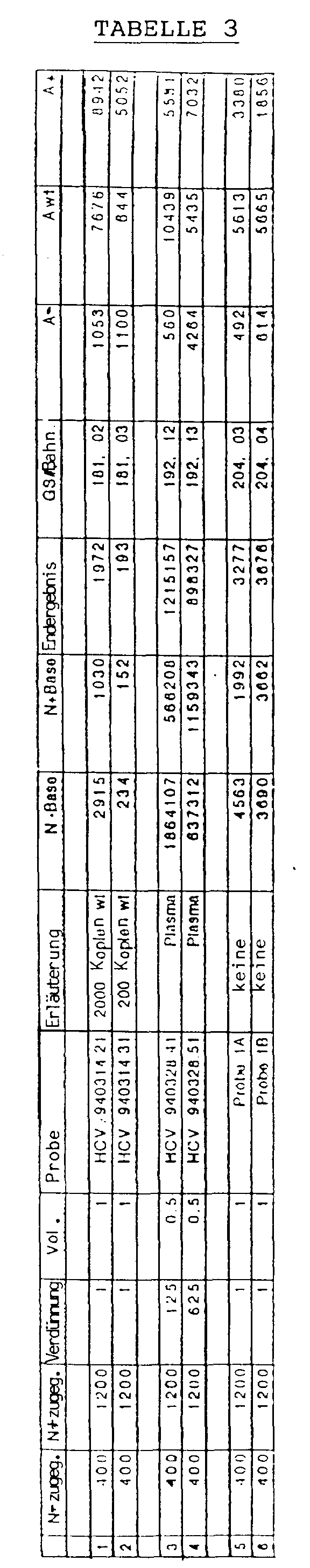
- Bei dieser Quantifizierung werden Primer verwendet, welche in den cDNA-Sequenzen des HIV-1 binden und durch PCR von proviraler HIV- DNA ein 115 bp großes Produkt ergeben, nämlich(Numerierung nach Ratner et al.). Die Primer wurden unter Verwendung der Phosphoamidit-Chemie auf einem DNA-Synthesizer hergestellt (Applied Biosystems 394 DNA Synthesizer).

- Die Standard-Plasmide pgag-15 und pgag+12 sind abgeleitet aus dem Plasmid pgagl, welches aus dem bekannten pBS/SK--Plasmid (Firma Stratagene) und einem Insert in der multiplen Klonierungsstelle dieses Plasmids besteht, welches Insert die bp 1417 bis 2008 der HIV-1 aus Ratner et al. enthält.
- In pgag-15 wurden die bp 1593 bis 1607 deletiert, in pgag+12 ein 12 bp langes Insert an der Stelle 1593 eingefügt (siehe Fig.1). Die Plasmide wurde gereinigt (QUIAGEN-Verfahren), die Konzentration durch spektroskopische Messung bei 260 nm bestimmt, mit EcoRI geschnitten und in einem lOmM TRIS/HCl pH 8/0,1 mM EDTA-Puffer verdünnt (Sambrook et al. Molecular Cloning, Second Edition, Cold Spring Harbor Lab Press, Cold Spring Harbor (1989)).
- Diese DNA-Präparationen dienen als Standard für die PCR.
- Die Länge der PCR-Produkte von Standard und Wildtyp-DNA betragen daher 127 (pgag+12), 100 (pgag-15) und 115 bp (wt).
- Die Ergebnisse einer Quantifizierungsreihe sind in Tabelle 4 wiedergegeben. Eine kodierte Kontrollreihe von positiven und negativen Proben, die HIV provirale DNA in unterschiedlicher Kopienzahl enthielten, wurden mit dem erfindungsgemäßen Verfahren bestimmt. Die Tabelle 4 zeigt, daß alle Negativkontrollen negativ und alle Positiv-Proben im richtigen Größenbereich quantifiziert wurden. Besonders bemerkenswert ist die genaue Messung im unteren Kopienbereich, siehe CD-13 und CD-26. Diese Proben enthielten nominal 2 Kopien/105-Zellen und wurden mit jeweils 3 Kopien quantifiziert. Dies zeigt die Genauigkeit und extreme Sensitivität des erfindungsgemäßen Verfahren.
TABELLE 4 PROBE KOPIENZAHL PROBE KOPIENZAHL nom. gef. nom. gef. CD-1 0 0 CD-16 0 0 CD-2 50 68 CD-17 0 0 CD-3 0 0 CD-18 0 0 CD-4 0 0 CD-19 2 5 CD-5 10 24 CD-20 20 16 CD-6 0 0 CD-21 0 0 CD-7 0 0 CD-22 0 0 CD-8 10 16 CD-23 10 18 CD-9 20 49 CD-24 0 0 CD-10 0 0 CD-25 0 0 CD-11 5 6 CD-26 2 3 CD-12 5 5 CD-25 50 71 CD-13 2 3 CD-28 0 0 CD-14 0 0 CD-29 5 5 CD-15 0 0 CD-30 20 25 - Bei dieser Quantifizierung werden Primer verwendet, welche im Genom des HBV binden und durch PCR von Wildtyp-DNA ein 182 bp großes Produkt ergeben, nämlich
 (Numerierung nach Fujiyama et al.). Die Primer wurden unter Verwendung der Phosphoamidit-Chemie auf einem DNA-Synthesizer hergestellt (Applied Biosystems 394 DNA Synthesizer).
- Die Standard-Plasmide pHBV-9 und pHBV+12 sind abgeleitet aus dem Plasmid pHBV-wt, welches aus dem bekannten pCRII-Plasmid (Firma In Vitrogene) und einem Insert in der multiplen Klonierungsstelle dieses Plasmids besteht, welches Insert die bp 1763 bis 1868 der HBV aus Fujiyama et al. enthält.
- In pHBV-9 wurden die bp 1868 bis 1876 deletiert, in pHBV+12 ein 12 bp langes Insert an der Stelle 1868 eingefügt. Die Plasmide wurden gereinigt (QUIAGEN-Verfahren), die Konzentration durch spektroskopische Messung bei 260 nm bestimmt, mit einem Restriktionsenzym einmal geschnitten und in einem 10mM TRIS/HCl pH 8/0,1 mM EDTA-Puffer verdünnt (Sambrook et al. Molecular Cloning, Second Edition, Cold Spring Harbor Lab Press, Cold Spring Harbor (1989)).
- Diese DNA-Präparationen dienen als Standard für die PCR.
- Die Länge der PCR-Produkte von Standard und Wildtyp-DNA betragen daher 194 (pHBV+12), 173 (pHBV-9) und 182 bp (wt).
- Die Ergebnisse einer Quantifizierungsreihe sind in Tabelle 5 wiedergegeben und in der Figur 4 graphisch veranschaulicht. Die Amplifizierungsreaktion wurde ausgehend von jeweils 150 Kopien pHBV-9 und 50 Kopien von HBV+12 und unterschiedlichen Mengen an pHBV-wt (400, 200, 100, 50 und 0 Kopien) durchgeführt. Jeder Ansatz wurde vierfach gemessen.
- Die Ergebnisse zeigen, daß der festgestellte DNA-Gehalt sehr gut reproduzierbar ist.
- Bei dieser Quantifizierung wurden Primer verwendet, welche im Genom des HSV binden und durch PCR von Wildtyp-DNA ein 114 bp langes Produkt ergeben, nämlich

- Das Standard-Plasmid pHerp-9 ist abgeleitet aus dem Plasmid pHerp, welches aus dem bekannten pCRII-Plasmis (Firma In Vitrogen) und einem Insert von bp128364 bis 138784 des HSV-Genoms (McGeoch D.J. et al., EMBL GenBank ID HElCG1) besteht.
- In pHerp-9 wurden 9bp deletiert. Die Plasmide wurden gereinigt, die Konzentrationen durch spektroskopische Messung bei 260nm bestimmt, mit einem Restriktionsenzym linearisiert und in einem TE-Puffer (10 mM Tris/HCl, lmM EDTA, pH 8) verdünnt.
- Dieses DNA-Präparat dient als Standard für die PCR. Die Länge der PCR-Produkte von Standard und Wildtyp betragen daher 105 (pHerp-9) und 114 bp (pHerp).
- Die Ergebnisse einer Quantifizierung sind in der Tabelle 6 wiedergegeben. Fünf Proben sowie 1,66 pg pHerp Wildtyp DNA wurden in Anwesenheit von 40000 (Spalte 1) Kopien Standard pHerp-9 mittels der beschriebenen Methode analysiert. Der Faktor F2 der beschriebenen Formel zur Berechnung der Herpes DNA-Menge ist in diesem Fall 1/6000. Die Spalten 2 und 3 geben den Verdünnungsfaktor sowie das eingesetzte Volumen der Probe an. Spalte 4 gibt den Namen der Probe an. Spalte 5 enthält zusätzliche Informationen über die Proben (wenn nötig). In der Spalte 6 ist die errechnete Menge an HSV-DNA in pg/ml angegeben. Der Mittelwert der Doppelbestimmung ist in Spalte 7 angegeben. Spalte 8 und 9 geben die Flächen der detektierten Peaks von Standard-Plasmid (Spalte 8) und von Wildtyp (Spalte 9) an.
- Bei den Proben 14, 17 und JA10 werden Doppelbestimmungen von Herpes-DNA gemacht, die eine Abweichung vom Mittelwert von 0 % bis 4,3 % aufweisen. Diese geringe Standardabweichung kann noch dra-stisch verbessert werden, wenn man bei den Bestimmungen auch den pHerp+10-Standard mitbestimmt.

- Bei dieser Quantifizierung wurden Primer verwendet, welche im Genom des Vaccinia-Virus binden und durch PCR von Wildtyp-DNA ein 136 bp langes Produkt ergeben, nämlich(Numerierung nach Goebel et al., Virology 179: 247-266, 1990). Die Primer wurden auf einem DNA-Synthesizer hergestellt (Applied Biosystems 394 DNA Synthesizer).

- Das Standard-Plasmid pVV-21 ist abgeleitet aus dem Plasmid pVV-wt, welches aus dem bekannten pTZ19R-Plasmid (Pharmacia) und einem Insert zwischen den beiden PvuII-Restriktionssschnittstellen dieses Plasmids besteht. Dieses Insert enthält die bp 83855 bis 84761 der Thymidinkinasegen-Region des Vaccinia-Virus (siehe Goebel et al., 1990).
- In pVV-21 wurden 21 bp (von bp 84718 bis 84738) deletiert.
- Die Plasmide wurden gereinigt, die Konzentrationen durch spektroskopische Messung bei 260 nm bestimmt, mit einem Restriktionsenzym linearisiert und in einem TE-Puffer (10 mM Tris/HCl, lmM EDTA, pH 8) verdünnt.
- Dieses DNA-Präparat dient als Standard für die PCR. Die Länge der PCR-Produkte von Standard und Wildtyp betragen daher 115 (pVV-21) und 136 (pVVwt).
- Die Ergebnisse einer Quantifizierung sind in der Tabelle 7 wiedergegeben. Proben, Wasser und 10 pg Vaccinia-DNA wurden in Anwesenheit von 10000 Kopien Standard-Plasma (Spalte 1) mittels der beschriebenen Methode analysiert. Der Wert des Faktors F2 der beschriebenen Formel für die Berechnung der Vaccinia-DNA in pg/ml ist in diesem Fall 1/5000.
- Die Spalten 2 und 3 geben den Verdünnungsfaktor sowie das eingesetzte Volumen der Probe an. In Spalte 4 können (wenn nötig) mehr Informationen über die Probe angegeben werden. Spalte 5 gibt den Namen der Probe an. In Spalte 6 ist die berechnete Menge an Vaccinia-DNA in pg/ml angegeben. Spalte 7 gibt die Nummer des Genescanner-Laufes an, in dem die PCR-Produkte analysiert worden sind. Spalte 8 und 9 geben die Flächen der detektierten Peaks von Standard-Plasmid (Spalte 8) und von Wildtyp (Spalte 9) an.
- Bei der Probe 311494 werden 1,64 pg, bzw. 1,77 pg Vaccinia-DNA gemessen, wobei die Abweichung vom Mittelwert bei 3,8 % liegt. Bei der Probe 310494 werden 7,0 pg, bzw. 7,2 pg Vaccinia-DNA gemessen, wobei die Abweichung vom Mittelwert bei 1,4 % liegt. Diese geringe Standardabweichung kann noch drastisch verbessert werden, wenn man bei den Bestimmungen auch den pVV+24-Standard mitbestimmt.

Claims (23)
- Verfahren zur Quantifizierung von Nukleinsäuren in einer Probe unter Anwendung von Nukleinsäure-Amplifizierung, wobei der Probe vor dem Amplifizierungsschritt eine gegebene Menge eines bekannten Nukleinsäuremoleküls als interner Standard zugegeben wird, welches Standard-Nukleinsäuremolekül sich von der zu quantifizierenden Nukleinsäure zumindest in einem detektierbaren Merkmal unterscheidet, dadurch gekennzeichnet,
daß der Probe vor der Nukleinsäure-Amplifizierung bekannte Mengen von mindestens zwei sich zumindest in einem.detektierbaren Merkmal voneinander und von der zu quantifizierenden Nukleinsäure unterscheidenden bekannten Nukleinsäuremolekülen als interner Standard zugegeben werden, die erhaltenen Mengen an amplifizierter Proben- und Standard-Nukleinsäure bestimmt werden und aus den erhaltenen Mengen die ursprünglich in der Probe vorhandene Menge an zu quantifizierender Nukleinsäure bestimmt wird. - Verfahren nach Anspruch 1, dadurch gekennzeichnet, daß beim Amplifizieren Primer mit fluoreszierenden oder radioaktiven Gruppen oder chemischen Gruppen, die mit affinen Proteinen und nachgeschalteten Detektionsreaktionen detektiert werden können, vorzugsweise mit fluoreszierenden Gruppen, verwendet werden.
- Verfahren nach einem der Ansprüche 1 bis 2, dadurch gekennzeichnet, daß die Amplifizierung der Nukleinsäuren bereits in der exponentiellen Phase gestoppt wird.
- Verfahren nach einem der Ansprüche 1 bis 3, dadurch gekennzeichnet, daß die Bestimmung der Mengen an amplifizierter Nukleinsäure unter Verwendung eines Nukleinsäure-Detektionsgerätes, vorzugsweise eines fluoreszenz-empfindlichen Nukleinsäure-Detektionsgerätes, erfolgt.
- Verfahren nach einem der Ansprüche 1 bis 4, dadurch gekennzeichnet, daß Nukleinsäuren von Mikroorganismen, vorzugsweise HIV, Parvovirus, Herpesvirus, HAV, HBV, HCV, Baculovirus, Adenovirus, Vacciniavirus, Borreliaspezies, Salmonellaspezies und Hefe, quantifiziert werden.
- Verfahren nach Anspruch 5, dadurch gekennzeichnet, daß virale Nukleinsäuren in einer biologischen Probe, insbesondere Blut- und Blutderivaten und biotechnologischen Produkten, quantifiziert werden.
- Verfahren nach einem der Ansprüche 1 bis 6, dadurch gekennzeichnet, daß unterschiedliche Mengen der Standard-Nukleinsäuren der Probe vor der Amplifizierung zugegeben werden.
- Verfahren nach einem der Ansprüche 1 bis 7, dadurch gekennzeichnet, daß Standard-Nukleinsäuren zum Einsatz kommen, welche eine gegenüber der zu quantifizierenden Nukleinsäure unterschiedliche Länge aufweisen, vorzugsweise eine Standard-Nukleinsäuresequenz, welche kürzer, und eine Standard-Nukleinsäuresequenz, welche länger ist als die zu quantifizierende Nukleinsäure.
- Verfahren nach einem der Ansprüche 1 bis 8, dadurch gekennzeichnet, daß mehrere erhaltene Mengen an amplifizierter Proben- und Standard-Nukleinsäuren in der gleichen Probe mittels der Multiplex-Analyse bestimmt werden.
- Verfahren nach einem der Ansprüche 1 bis 9, dadurch gekennzeichnet, daß die Standard-Nukleinsäuren der Probe zugegeben werden, noch bevor die Probe einem oder mehreren Probenaufbereitungsschritten unterzogen wird.
- Verfahren zur Bestimmung der Nachweisgrenze von bestimmten Nukleinsäuren unter Verwendung eines Verfahrens gemäß der Ansprüche 1 bis 10, dadurch gekennzeichnet, daß mindestens einer Standard-Nukleinsäure mit einer Konzentration von knapp oberhalb der Nachweisgrenze eingesetzt wird.
- Biologische, insbesondere biotechnologische Produkte, die einen Gehalt an Nukleinsäure unterhalb der Grenze von 10 bzw. 100 pg pro Dosis, ermittelt nach einem Verfahren gemäß einem der Ansprüche 1 bis 10, aufweisen und daher im wesentlichen frei von Nukleinsäuren sind.
- Biologische, insbesondere biotechnologische Produkte, die einen Gehalt an Nukleinsäure unterhalb des Bereiches von 1 bis 100 Kopien der Nukleinsäure aufweisen.
- Biologische, insbesondere biotechnologische Produkte nach Anspruch 12 oder 13, dadurch gekennzeichnet, daß sie ausgewählt sind aus viralen und bakteriellen Proteinen, insbesondere gp160, rekombinanten Blutfaktoren, Plasmaproteinen sowie Impfstoffen, insbesondere gegen Herpes-, Influenza-, TBE-, Parvo- oder Hepatitis-Viren, und monoklonalen Antikörpern.
- Verwendung eines Verfahrens gemäß einem der Ansprüche 1 bis 10 zur Detektion von Nukleinsäuren in biologischen Proben.
- Primer mit der Sequenz gemäß der Seq.ID 3, 4, 5, 6, 7, 8, 9, 10, 11 und 12.
- Standard-Plasmide pgagl, pgag-15, pgag+12, pHAV-wt, pHAV-10bp, pHAV+9bp, pHCV-wt, pHCV-7bp, pHCV+8bp, pHBV-wt, pHBV-9bp, pHBV+12bp, pVV-21, pVV+24, pHerp-9 und pHerp+10.
- Set zur Quantifizierung von Nukleinsäuren in einer Probe, welches umfaßt:- mindestens zwei bekannte Nukleinsäuren als interne Standards, welches sich voneinander und von den zu quantifizierenden Nukleinsäuren in mindestens einem nachweisbaren Charakteristikum unterscheiden,- Fluoreszenz-markierte Primer, die an die Standard-Nukleinsäure und an die zu quantifizierende Nukleinsäure binden,- positive Kontrollen, welche bekannte Mengen einer Nukleinsäure, an der Interesse besteht, aufweisen,- eine negative Kontrolle, welche Humanplasma, das frei von der viralen Nukleinsäure ist, an der Interesse besteht, aufweist, und- eine Arbeitsanleitung.
- Set nach Anspruch 18 zur Quantifizierung von HIV-RNA in einer Probe, welches umfaßt:- mindestens zwei interne Standards, die eine in vitrotranskribierte RNA aufweisen, welche von den Plasmiden pgag-15 und pgag+12 stammt,- die Fluoreszenz-markierten Primer SK38 und SK39- eine positive Kontrolle, welche bekannte Mengen an HIV-1-Teilchen aufweist,- eine negative Kontrolle, welche Humanplasma aufweist, das frei von Virus-Nukleinsäuren ist und- eine Arbeitsanleitung.
- Set nach Anspruch 18 zur Quantifizierung von HCV-RNA in einer Probe, welches aufweist:- mindestens zwei interne Standards, die eine in vitrotranskribierte RNA aufweisen, welche von den Plasmiden pHCV-7 und pHCV+8 stammt,- die Fluoreszenz-markierten Primer HCV32Ext und HCVPT4- positive Kontrollen, welche bekannte Mengen an HCV-Teilchen aufweisen,- eine negative Kontrolle, welche Humanplasma aufweist, das frei von Virus-Nukleinsäuren ist und- eine Arbeitsanleitung.
- Set nach Anspruch 18 zur Quantifizierung von HAV-RNA in einer Probe, welches aufweist:- mindestens zwei interne Standards, die eine in vitrotranskribierte RNA aufweisen, welche von den Plasmiden pHAV-10 und pHAV+9 stammt,- die Fluoreszenz-markierten Primer HAV+2058 und HAV-2172- positive Kontrollen, welche bekannte Mengen an HAV-Teilchen aufweisen,- eine negative Kontrolle, welche Humanplasma aufweist, das frei von Virus-Nukleinsäuren ist und- eine Arbeitsanleitung.
- Set nach Anspruch 18 zur Quantifizierung von HBV-DNA in einer Probe, welches aufweist:- mindestens zwei interne Standards, die die Plasmide pHBV-9 und pHBV+12 aufweisen,- die Fluoreszenz-markierten Primer HBV+1780B und HBV-1960B- positive Kontrollen, welche bekannte Mengen an HBV-Teilchen aufweisen,- eine negative Kontrolle, welche Humanplasma aufweist, das frei von Virus-Nukleinsäuren ist und- eine Arbeitsanleitung.
- Set nach Anspruch 18 zur Quantifizierung von HSV-DNA in einer Probe, welches aufweist:- als interne Standards, die Plasmide pHerp-9 und pHerp+10,- die Fluoreszenz-markierten Primer gDR/B und gDR2R/B- positive Kontrollen, welche bekannte Mengen an HSV-Teilchen aufweisen,- eine negative Kontrolle, welche Humanplasma aufweist, das frei von Virus-Nukleinsäuren ist und- eine Arbeitsanleitung.
Applications Claiming Priority (6)
| Application Number | Priority Date | Filing Date | Title |
|---|---|---|---|
| AT1831/94 | 1994-09-26 | ||
| AT183194A AT401062B (de) | 1994-09-26 | 1994-09-26 | Verfahren zur quantifizierung von nukleinsäuren |
| AT183194 | 1994-09-26 | ||
| AT224594 | 1994-12-02 | ||
| AT2245/94 | 1994-12-02 | ||
| AT224594A ATA224594A (de) | 1994-12-02 | 1994-12-02 | Verfahren zur quantifizierung von nukleinsäuren |
Publications (3)
| Publication Number | Publication Date |
|---|---|
| EP0714988A2 true EP0714988A2 (de) | 1996-06-05 |
| EP0714988A3 EP0714988A3 (de) | 1997-04-16 |
| EP0714988B1 EP0714988B1 (de) | 2001-12-12 |
Family
ID=25596921
Family Applications (1)
| Application Number | Title | Priority Date | Filing Date |
|---|---|---|---|
| EP95890172A Expired - Lifetime EP0714988B1 (de) | 1994-09-26 | 1995-09-26 | Verfahren zur Quantifizierung von Nukleinsaüren |
Country Status (8)
| Country | Link |
|---|---|
| US (1) | US5789153A (de) |
| EP (1) | EP0714988B1 (de) |
| JP (1) | JPH08107798A (de) |
| AT (1) | ATE210735T1 (de) |
| CA (1) | CA2159044A1 (de) |
| DE (1) | DE59509938D1 (de) |
| DK (1) | DK0714988T3 (de) |
| ES (1) | ES2169753T3 (de) |
Cited By (7)
| Publication number | Priority date | Publication date | Assignee | Title |
|---|---|---|---|---|
| WO1996035437A1 (de) * | 1995-05-08 | 1996-11-14 | Immuno Aktiengesellschaft | Qualitätsgesichertes arzneimittel, enthaltend ein oder mehrere plasmaderivate |
| WO1997012058A1 (en) * | 1995-09-26 | 1997-04-03 | Dynal A/S | Method for quantifying nucleic acid using multiple competitor nucleic acids |
| WO1999063109A1 (de) * | 1998-06-04 | 1999-12-09 | Baxter Aktiengesellschaft | Verfahren zur bestimmung von antigenen |
| WO2002024949A1 (en) * | 2000-09-22 | 2002-03-28 | Hitachi, Ltd. | Method of analyzing nucleic acid |
| US6649339B1 (en) | 1999-08-20 | 2003-11-18 | Baxter Aktiengesellschaft | Method for producing a quality assured biological sample and composition containing the same |
| US6953837B2 (en) | 1997-02-27 | 2005-10-11 | Baxter Aktiengesellschaft | Factor VIII/vWF complexes and compositions |
| US8063197B2 (en) | 2004-07-13 | 2011-11-22 | Gen-Probe Incorporated | Compositions and methods for detection of hepatitis A virus nucleic acid |
Families Citing this family (22)
| Publication number | Priority date | Publication date | Assignee | Title |
|---|---|---|---|---|
| US5814442A (en) * | 1994-06-10 | 1998-09-29 | Georgetown University | Internally controlled virion nucleic acid amplification reaction for quantitation of virion and virion nucleic acid |
| US6265152B1 (en) * | 1995-12-22 | 2001-07-24 | Visible Genetics Inc. | Method and kit for evaluation of HIV mutations |
| AT406778B (de) * | 1996-04-09 | 2000-09-25 | Immuno Ag | Verfahren zur desintegration von nucleinsäuren und herstellung von qualitätsgesicherten biologischen produkten |
| AT405403B (de) | 1997-02-27 | 1999-08-25 | Immuno Ag | Reinigung von von willebrand-faktor durch kationenaustauscherchromatographie |
| DE19752898A1 (de) * | 1997-11-28 | 1999-08-05 | Centeon Pharma Gmbh | Verfahren zum Nachweis hoher Vierenkonzentrationen im Blutplasma und/oder Blutserum mittels der Polymerasekettenreaktion |
| US6455692B1 (en) | 1998-08-04 | 2002-09-24 | Transgenomic, Inc. | Method of concentrating polynucleotides using MIPC |
| GB9902422D0 (en) * | 1999-02-03 | 1999-03-24 | Lgc Teddington Limited | Reference material for nucleic acid amplification |
| US6379889B1 (en) * | 1999-11-04 | 2002-04-30 | Agilent Technologies, Inc. | Multiplexing methods for identifying nucleic acids using denaturing liquid chromatography |
| AT409382B (de) * | 1999-12-22 | 2002-07-25 | Baxter Ag | Verfahren zur detektion, unterscheidung und quantifizierung von freien und eingekapselten nukleinsäuren in einer probe |
| US6312929B1 (en) * | 2000-12-22 | 2001-11-06 | Cepheid | Compositions and methods enabling a totally internally controlled amplification reaction |
| US6982029B2 (en) * | 2001-05-07 | 2006-01-03 | Spectramedix Llc | Electrophoretic method and system having internal lane standards for color calibration |
| US20030207310A1 (en) * | 2002-05-01 | 2003-11-06 | Zhaowei Liu | System and method for determining the sizes and quantity of polynucleotides with capillary array electrophoresis |
| US7565250B2 (en) * | 2003-12-06 | 2009-07-21 | Abbott Laboratories Inc. | Method and system for analyzing reactions using an information system |
| US7932081B2 (en) | 2005-03-10 | 2011-04-26 | Gen-Probe Incorporated | Signal measuring system for conducting real-time amplification assays |
| JP5564795B2 (ja) * | 2009-01-06 | 2014-08-06 | 株式会社島津製作所 | Dna定量方法、及び遺伝子解析方法 |
| US8877464B2 (en) | 2010-07-29 | 2014-11-04 | Roche Molecular Systems, Inc. | Qualitative and quantitative detection of microbial nucleic acids |
| CA2802548C (en) * | 2010-07-29 | 2017-01-03 | F. Hoffmann-La Roche Ag | Qualitative and quantitative detection of microbial nucleic acids |
| JP6121591B2 (ja) * | 2010-07-29 | 2017-04-26 | エフ.ホフマン−ラ ロシュ アーゲーF. Hoffmann−La Roche Aktiengesellschaft | 微生物核酸の定性的および定量的検出 |
| EP2627787B1 (de) | 2010-10-14 | 2017-04-19 | Rheonix, Inc. | Multiplexierte quantitative erkennung von nukleinsäuretargets |
| WO2012095748A2 (en) * | 2011-01-14 | 2012-07-19 | Smiths Detection - Watford Limited | Single tube quantitative polymerase chain reaction (pcr) |
| EP2877952B1 (de) | 2012-07-27 | 2023-04-12 | Gen-Probe Incorporated | Dual-referenzkalibrierverfahren und system zum quantifizieren von polynukleotiden |
| DE102018213027A1 (de) * | 2018-08-03 | 2020-02-06 | Robert Bosch Gmbh | Reaktionsgemisch, Verfahren und Kit zur Durchführung einer quantitativen Echtzeit-PCR |
Family Cites Families (10)
| Publication number | Priority date | Publication date | Assignee | Title |
|---|---|---|---|---|
| US4683202A (en) * | 1985-03-28 | 1987-07-28 | Cetus Corporation | Process for amplifying nucleic acid sequences |
| US4683195A (en) * | 1986-01-30 | 1987-07-28 | Cetus Corporation | Process for amplifying, detecting, and/or-cloning nucleic acid sequences |
| AU659649B2 (en) * | 1991-06-10 | 1995-05-25 | Lucky Limited | Hepatitis C diagnostics and vaccines |
| JP2739004B2 (ja) * | 1992-01-16 | 1998-04-08 | 三菱電機株式会社 | 半導体装置 |
| WO1993023573A1 (en) * | 1992-05-15 | 1993-11-25 | New England Deaconess Hospital | Quantitation of viral rna by competitive polymerase chain reaction |
| WO1994010342A1 (en) * | 1992-10-30 | 1994-05-11 | The Rogosin Institute | Method for quantifying nucleic acids |
| WO1994020640A1 (en) * | 1993-03-05 | 1994-09-15 | Genelabs Technologies, Inc. | Method for hiv quantitation |
| DK0662156T3 (da) * | 1993-07-09 | 1999-10-25 | Akzo Nobel Nv | Forbedret fremgangsmåde til kvantificering af nukleinsyre |
| US5523204A (en) * | 1993-12-10 | 1996-06-04 | Becton Dickinson And Company | Detection of nucleic acids in cells by strand displacement amplification |
| US5491063A (en) * | 1994-09-01 | 1996-02-13 | Hoffmann-La Roche Inc. | Methods for in-solution quenching of fluorescently labeled oligonucleotide probes |
-
1995
- 1995-09-25 CA CA002159044A patent/CA2159044A1/en not_active Abandoned
- 1995-09-26 DK DK95890172T patent/DK0714988T3/da active
- 1995-09-26 ES ES95890172T patent/ES2169753T3/es not_active Expired - Lifetime
- 1995-09-26 EP EP95890172A patent/EP0714988B1/de not_active Expired - Lifetime
- 1995-09-26 US US08/533,820 patent/US5789153A/en not_active Expired - Fee Related
- 1995-09-26 DE DE59509938T patent/DE59509938D1/de not_active Expired - Fee Related
- 1995-09-26 JP JP7247504A patent/JPH08107798A/ja active Pending
- 1995-09-26 AT AT95890172T patent/ATE210735T1/de not_active IP Right Cessation
Cited By (14)
| Publication number | Priority date | Publication date | Assignee | Title |
|---|---|---|---|---|
| WO1996035437A1 (de) * | 1995-05-08 | 1996-11-14 | Immuno Aktiengesellschaft | Qualitätsgesichertes arzneimittel, enthaltend ein oder mehrere plasmaderivate |
| WO1997012058A1 (en) * | 1995-09-26 | 1997-04-03 | Dynal A/S | Method for quantifying nucleic acid using multiple competitor nucleic acids |
| US6953837B2 (en) | 1997-02-27 | 2005-10-11 | Baxter Aktiengesellschaft | Factor VIII/vWF complexes and compositions |
| US7648958B2 (en) | 1997-02-27 | 2010-01-19 | Baxter Innovations Gmbh | Factor VIII/vWF-complex and methods of purifying same |
| AT407160B (de) * | 1998-06-04 | 2001-01-25 | Immuno Ag | Verfahren zur bestimmung von antigenen |
| WO1999063109A1 (de) * | 1998-06-04 | 1999-12-09 | Baxter Aktiengesellschaft | Verfahren zur bestimmung von antigenen |
| US6649339B1 (en) | 1999-08-20 | 2003-11-18 | Baxter Aktiengesellschaft | Method for producing a quality assured biological sample and composition containing the same |
| WO2002024949A1 (en) * | 2000-09-22 | 2002-03-28 | Hitachi, Ltd. | Method of analyzing nucleic acid |
| US7790368B1 (en) | 2000-09-22 | 2010-09-07 | Hitachi, Ltd. | Method for analyzing nucleic acid |
| US8063197B2 (en) | 2004-07-13 | 2011-11-22 | Gen-Probe Incorporated | Compositions and methods for detection of hepatitis A virus nucleic acid |
| US8461324B2 (en) | 2004-07-13 | 2013-06-11 | Gen-Probe Incorporated | Compositions and methods for detection of hepatitis A virus nucleic acid |
| US8563707B2 (en) | 2004-07-13 | 2013-10-22 | Gen-Probe Incorporated | Compositions and methods for detection of hepatitis A virus nucleic acid |
| US9469881B2 (en) | 2004-07-13 | 2016-10-18 | Gen-Probe Incorporated | Compositions and methods for detection of hepatitis A virus nucleic acid |
| US10392656B2 (en) | 2004-07-13 | 2019-08-27 | Gen-Probe Incorporated | Compositions and methods for detection of hepatitis A virus nucleic acid |
Also Published As
| Publication number | Publication date |
|---|---|
| US5789153A (en) | 1998-08-04 |
| DE59509938D1 (de) | 2002-01-24 |
| EP0714988B1 (de) | 2001-12-12 |
| DK0714988T3 (da) | 2002-04-08 |
| JPH08107798A (ja) | 1996-04-30 |
| ATE210735T1 (de) | 2001-12-15 |
| CA2159044A1 (en) | 1996-03-27 |
| ES2169753T3 (es) | 2002-07-16 |
| EP0714988A3 (de) | 1997-04-16 |
Similar Documents
| Publication | Publication Date | Title |
|---|---|---|
| EP0714988B1 (de) | Verfahren zur Quantifizierung von Nukleinsaüren | |
| EP0714987B1 (de) | Verfahren zur Quantifizierung von genomischer DNA | |
| DE69233285T2 (de) | Quantifikation von Nukleinsäuren | |
| DE69229929T2 (de) | Verfahren zum Nachweis von DNS in einer Probe | |
| DE2915082C2 (de) | ||
| DE69029105T2 (de) | Schnellverfahren zum Nachweis und/oder zur Identifizierung einer Einzelbase in einer Nukleinsäuresequenz und seine Verwendungen | |
| DE60129869T2 (de) | Methode zur Quantifizierung von Nukleinsäuren in Echtzeit mittels Effizienzkorrektur | |
| DE69434314T2 (de) | Polymorphismus von mononukleotiden und ihre verwendung in der genanalyse | |
| DE69527125T2 (de) | Verfahren und Vorrichtung zur DNS Analyse | |
| DE60030811T2 (de) | Verfahren zur Ampifizierung von RNA | |
| DE69003477T2 (de) | Verfahren zum Nachweis von Nucleinsäuren. | |
| DE69224548T2 (de) | Verfahren zur Amplifizierung und zum Nachweis von Nukleinsäuren,die einen schnellen PCR-Zyklus verwenden | |
| DE69028951T2 (de) | Oligonukleotiden-Sequenzen zur Amplifizierung von HIV-2 und SIV Retrovirusgenomen, ihre Verwendung zur In-vitro-Diagnostik von durch diese Viren verursachten Infektionen | |
| DD298430A5 (de) | Verfahren zum nachweis und/oder zur identifizierung einer nucleinsaeuresequenz | |
| EP0769954B1 (de) | Qualitätsgesichertes arzneimittel, enthaltend ein oder mehrere plasmaderivate | |
| AT409383B (de) | Verfahren zur detektion und quantifizierung von nukleinsäuren in einer probe | |
| DE60125171T2 (de) | Verfahren zur detektion und quantifizierung von menschlichen p450-molekülen und sonde und kit für dieses verfahren | |
| EP1124993A2 (de) | Neue primer und sonden zum nachweis von hiv | |
| WO2004044238A1 (de) | Mittel und verfahren zum nachweis humaner adenoviren | |
| DE60011789T2 (de) | Verfahren zur Herstellung einer qualitätsgesicherten biologischen Probe und Zusammensetzung, welche diese enthält | |
| EP0405376B1 (de) | Typisierung humaner Rhinoviren | |
| AT401062B (de) | Verfahren zur quantifizierung von nukleinsäuren | |
| DE69525435T2 (de) | Enzymatische vervielfaeltigung eines dna-fragments in vitro mittels einem treppenformigen anfangsstueck | |
| DE19835856A1 (de) | Verfahren zum Nachweis von HBV | |
| DE20315159U1 (de) | Kit zum Nachweis eines neuen Coronavirus, das mit dem Schweren Akuten Atemwegssyndrom (Severe Acute Respiratory Syndrome, SARS) assoziiert ist |
Legal Events
| Date | Code | Title | Description |
|---|---|---|---|
| PUAI | Public reference made under article 153(3) epc to a published international application that has entered the european phase |
Free format text: ORIGINAL CODE: 0009012 |
|
| AK | Designated contracting states |
Kind code of ref document: A2 Designated state(s): AT BE CH DE DK ES FR GB IE IT LI NL SE |
|
| PUAL | Search report despatched |
Free format text: ORIGINAL CODE: 0009013 |
|
| AK | Designated contracting states |
Kind code of ref document: A3 Designated state(s): AT BE CH DE DK ES FR GB IE IT LI NL SE |
|
| 17P | Request for examination filed |
Effective date: 19970916 |
|
| 17Q | First examination report despatched |
Effective date: 19990714 |
|
| RAP1 | Party data changed (applicant data changed or rights of an application transferred) |
Owner name: BAXTER AKTIENGESELLSCHAFT |
|
| RAP1 | Party data changed (applicant data changed or rights of an application transferred) |
Owner name: BAXTER AKTIENGESELLSCHAFT |
|
| GRAG | Despatch of communication of intention to grant |
Free format text: ORIGINAL CODE: EPIDOS AGRA |
|
| GRAG | Despatch of communication of intention to grant |
Free format text: ORIGINAL CODE: EPIDOS AGRA |
|
| GRAH | Despatch of communication of intention to grant a patent |
Free format text: ORIGINAL CODE: EPIDOS IGRA |
|
| GRAH | Despatch of communication of intention to grant a patent |
Free format text: ORIGINAL CODE: EPIDOS IGRA |
|
| GRAA | (expected) grant |
Free format text: ORIGINAL CODE: 0009210 |
|
| AK | Designated contracting states |
Kind code of ref document: B1 Designated state(s): AT BE CH DE DK ES FR GB IE IT LI NL SE |
|
| REF | Corresponds to: |
Ref document number: 210735 Country of ref document: AT Date of ref document: 20011215 Kind code of ref document: T |
|
| REG | Reference to a national code |
Ref country code: CH Ref legal event code: EP |
|
| REG | Reference to a national code |
Ref country code: GB Ref legal event code: IF02 |
|
| REG | Reference to a national code |
Ref country code: IE Ref legal event code: FG4D Free format text: GERMAN |
|
| REF | Corresponds to: |
Ref document number: 59509938 Country of ref document: DE Date of ref document: 20020124 |
|
| REG | Reference to a national code |
Ref country code: CH Ref legal event code: NV Representative=s name: KIRKER & CIE SA |
|
| ET | Fr: translation filed | ||
| GBT | Gb: translation of ep patent filed (gb section 77(6)(a)/1977) |
Effective date: 20020222 |
|
| REG | Reference to a national code |
Ref country code: DK Ref legal event code: T3 |
|
| REG | Reference to a national code |
Ref country code: ES Ref legal event code: FG2A Ref document number: 2169753 Country of ref document: ES Kind code of ref document: T3 |
|
| PLBE | No opposition filed within time limit |
Free format text: ORIGINAL CODE: 0009261 |
|
| STAA | Information on the status of an ep patent application or granted ep patent |
Free format text: STATUS: NO OPPOSITION FILED WITHIN TIME LIMIT |
|
| 26N | No opposition filed | ||
| PGFP | Annual fee paid to national office [announced via postgrant information from national office to epo] |
Ref country code: AT Payment date: 20030902 Year of fee payment: 9 |
|
| PGFP | Annual fee paid to national office [announced via postgrant information from national office to epo] |
Ref country code: NL Payment date: 20030909 Year of fee payment: 9 |
|
| PGFP | Annual fee paid to national office [announced via postgrant information from national office to epo] |
Ref country code: GB Payment date: 20030917 Year of fee payment: 9 |
|
| PGFP | Annual fee paid to national office [announced via postgrant information from national office to epo] |
Ref country code: FR Payment date: 20030918 Year of fee payment: 9 |
|
| PGFP | Annual fee paid to national office [announced via postgrant information from national office to epo] |
Ref country code: SE Payment date: 20030919 Year of fee payment: 9 |
|
| PGFP | Annual fee paid to national office [announced via postgrant information from national office to epo] |
Ref country code: IE Payment date: 20030922 Year of fee payment: 9 Ref country code: DK Payment date: 20030922 Year of fee payment: 9 |
|
| PGFP | Annual fee paid to national office [announced via postgrant information from national office to epo] |
Ref country code: CH Payment date: 20030923 Year of fee payment: 9 |
|
| PGFP | Annual fee paid to national office [announced via postgrant information from national office to epo] |
Ref country code: ES Payment date: 20031008 Year of fee payment: 9 |
|
| PGFP | Annual fee paid to national office [announced via postgrant information from national office to epo] |
Ref country code: BE Payment date: 20031016 Year of fee payment: 9 |
|
| PGFP | Annual fee paid to national office [announced via postgrant information from national office to epo] |
Ref country code: DE Payment date: 20031031 Year of fee payment: 9 |
|
| PG25 | Lapsed in a contracting state [announced via postgrant information from national office to epo] |
Ref country code: GB Free format text: LAPSE BECAUSE OF NON-PAYMENT OF DUE FEES Effective date: 20040926 Ref country code: AT Free format text: LAPSE BECAUSE OF NON-PAYMENT OF DUE FEES Effective date: 20040926 |
|
| PG25 | Lapsed in a contracting state [announced via postgrant information from national office to epo] |
Ref country code: SE Free format text: LAPSE BECAUSE OF NON-PAYMENT OF DUE FEES Effective date: 20040927 Ref country code: IE Free format text: LAPSE BECAUSE OF NON-PAYMENT OF DUE FEES Effective date: 20040927 Ref country code: ES Free format text: LAPSE BECAUSE OF NON-PAYMENT OF DUE FEES Effective date: 20040927 |
|
| PG25 | Lapsed in a contracting state [announced via postgrant information from national office to epo] |
Ref country code: LI Free format text: LAPSE BECAUSE OF NON-PAYMENT OF DUE FEES Effective date: 20040930 Ref country code: DK Free format text: LAPSE BECAUSE OF NON-PAYMENT OF DUE FEES Effective date: 20040930 Ref country code: CH Free format text: LAPSE BECAUSE OF NON-PAYMENT OF DUE FEES Effective date: 20040930 Ref country code: BE Free format text: LAPSE BECAUSE OF NON-PAYMENT OF DUE FEES Effective date: 20040930 |
|
| BERE | Be: lapsed |
Owner name: *BAXTER A.G. Effective date: 20040930 |
|
| PG25 | Lapsed in a contracting state [announced via postgrant information from national office to epo] |
Ref country code: NL Free format text: LAPSE BECAUSE OF NON-PAYMENT OF DUE FEES Effective date: 20050401 Ref country code: DE Free format text: LAPSE BECAUSE OF NON-PAYMENT OF DUE FEES Effective date: 20050401 |
|
| EUG | Se: european patent has lapsed | ||
| REG | Reference to a national code |
Ref country code: CH Ref legal event code: PL |
|
| GBPC | Gb: european patent ceased through non-payment of renewal fee |
Effective date: 20040926 |
|
| PG25 | Lapsed in a contracting state [announced via postgrant information from national office to epo] |
Ref country code: FR Free format text: LAPSE BECAUSE OF NON-PAYMENT OF DUE FEES Effective date: 20050531 |
|
| NLV4 | Nl: lapsed or anulled due to non-payment of the annual fee |
Effective date: 20050401 |
|
| REG | Reference to a national code |
Ref country code: DK Ref legal event code: EBP |
|
| REG | Reference to a national code |
Ref country code: IE Ref legal event code: MM4A |
|
| REG | Reference to a national code |
Ref country code: FR Ref legal event code: ST |
|
| PG25 | Lapsed in a contracting state [announced via postgrant information from national office to epo] |
Ref country code: IT Free format text: LAPSE BECAUSE OF NON-PAYMENT OF DUE FEES Effective date: 20050926 |
|
| REG | Reference to a national code |
Ref country code: ES Ref legal event code: FD2A Effective date: 20040927 |
|
| BERE | Be: lapsed |
Owner name: *BAXTER A.G. Effective date: 20040930 |