EP0405285B1 - Plasminogenaktivator - Google Patents
Plasminogenaktivator Download PDFInfo
- Publication number
- EP0405285B1 EP0405285B1 EP90111471A EP90111471A EP0405285B1 EP 0405285 B1 EP0405285 B1 EP 0405285B1 EP 90111471 A EP90111471 A EP 90111471A EP 90111471 A EP90111471 A EP 90111471A EP 0405285 B1 EP0405285 B1 EP 0405285B1
- Authority
- EP
- European Patent Office
- Prior art keywords
- dna
- units
- added
- plasmid
- hours
- Prior art date
- Legal status (The legal status is an assumption and is not a legal conclusion. Google has not performed a legal analysis and makes no representation as to the accuracy of the status listed.)
- Expired - Lifetime
Links
Images


Classifications
-
- C—CHEMISTRY; METALLURGY
- C12—BIOCHEMISTRY; BEER; SPIRITS; WINE; VINEGAR; MICROBIOLOGY; ENZYMOLOGY; MUTATION OR GENETIC ENGINEERING
- C12N—MICROORGANISMS OR ENZYMES; COMPOSITIONS THEREOF; PROPAGATING, PRESERVING, OR MAINTAINING MICROORGANISMS; MUTATION OR GENETIC ENGINEERING; CULTURE MEDIA
- C12N9/00—Enzymes; Proenzymes; Compositions thereof; Processes for preparing, activating, inhibiting, separating or purifying enzymes
- C12N9/14—Hydrolases (3)
- C12N9/48—Hydrolases (3) acting on peptide bonds (3.4)
- C12N9/50—Proteinases, e.g. Endopeptidases (3.4.21-3.4.25)
- C12N9/64—Proteinases, e.g. Endopeptidases (3.4.21-3.4.25) derived from animal tissue
- C12N9/6421—Proteinases, e.g. Endopeptidases (3.4.21-3.4.25) derived from animal tissue from mammals
- C12N9/6424—Serine endopeptidases (3.4.21)
- C12N9/6456—Plasminogen activators
- C12N9/6462—Plasminogen activators u-Plasminogen activator (3.4.21.73), i.e. urokinase
-
- C—CHEMISTRY; METALLURGY
- C12—BIOCHEMISTRY; BEER; SPIRITS; WINE; VINEGAR; MICROBIOLOGY; ENZYMOLOGY; MUTATION OR GENETIC ENGINEERING
- C12Y—ENZYMES
- C12Y304/00—Hydrolases acting on peptide bonds, i.e. peptidases (3.4)
- C12Y304/21—Serine endopeptidases (3.4.21)
- C12Y304/21073—Serine endopeptidases (3.4.21) u-Plasminogen activator (3.4.21.73), i.e. urokinase
Definitions
- This invention relates to a novel plasminogen activator constructed by using recombinant DNA technology, a deoxyribonucleic acid (DNA) coding for the activator, a recombinant plasmid containing the DNA, a microbial or animal cell transformed with the recombinant plasmid, and a method of producing the plasminogen activator using said microbial or animal cell.
- DNA deoxyribonucleic acid
- the novel plasminogen activator according to the invention is stable against the protease (thrombin) which cleaves the urokinase type plasminogen activator into an inactive double-chain structure. Therefore, it can be readily recovered and purified to a pure form.
- This novel plasminogen activator has an increased specific activity, is stable and has good thrombolytic activity. Accordingly, the novel plasminogen activator according to the invention can be used, as expected, as a therapeutic agent for the treatment and/or prevention of cerebral thrombosis and myocardial infarction, among others.
- Urokinase (UK), streptokinase (SK) and tissue plasminogen activator (t-PA) are currently used as thrombolytic agents.
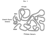
- the primary structure of human urokinase has been described by Heynecker et al. (JP-A-59-51300, the term “JP-A” used herein means "an unexamined published Japanese patent application”) and the folded structure thereof has been described and shown in Fig. 1.
- This folded structure which has been proposed based on homology to other proteins, can be divided into the following three domains.
- the first domain on the N-terminal side is a domain homologous to epidermal growth factor. Hereinafter, this domain is referred to as "growth factor domain”.
- the second domain is the so-called “kringle domain”.
- the third domain at the C-terminal side is the so-called serine protease domain.
- the third domain is referred to as "protease domain”.
- prourokinase occurs in two forms, a single-chain form and a double-chain form.
- the single chain form of UK is called prourokinase (hereinafter referred to as "pro-UK”) and, in such a structure, it has no thrombolytic activity. Only upon cleavage by plasmin into a double-chain structure, it can exhibit thrombolytic activity. However, when pro-UK is cleaved by thrombin, which cleaves pro-UK at a site two amino acids upstream from the plasmin cleavage site, the resulting double-chain structure has no thrombolytic activity any longer [Ichinose et al., J. Biol. Chem., 261 , 3486 (1986)].
- pro-UK after administration into the blood, pro-UK is cleaved by thrombin which is present in the blood into an inactive double-chain structure. Furthermore, if in the purification process for pro-UK there is a contamination by a thrombin-like protease, the protease will possibly cleave pro-UK into an inactive double-chain structure.
- the substitute amino acid introduced in the vicinity of the plasmin cleavage site namely at P3 to P1', simultaneously makes it difficult for plasmin cleavage to occur, hence the conversion of the pro-UK derivative obtained into an active form becomes difficult and a decreased specific activity results.
- the pro-UK derivative mentioned above in which the position of amino acid substitution is P3 or P2 relative to the plasmin cleavage site, a decrease in susceptibility to plasmin, hence a decrease in specific activity, has been observed.
- the peptide bond to be cleaved is represented by -P1-P1'-and the amino acids on the amino side to the peptide bond to be cleaved are given position codes P1, P2, P3, P4 etc. from the amino acid constituting the amino side of said peptide bond toward the amino terminus.
- the amino acids on the carboxyl side of this bond are given position codes P1', P1', P3', P4' etc. from the amino acid constituting the carboxyl side of said peptide bond toward the carboxyl terminus.
- pro-UK derivatives which are more or less resistant to cleavage by thrombin, by introducing a substitute amino acid in position P1 with respect to the thrombin cleavage site (position P3 with respect to the plasmin cleavage site; 156th amino acid of mature pro-UK) or position P1' with respect to the thrombin cleavage site (P2 with respect to the plasmin cleavage site; 157th amino acid of mature pro-UK).
- the pro-UK derivatives may possibly have a decreased specific activity as a result of a decrease in susceptibility to plasmin.
- the present inventors have confirmed that amino acid substitution in mature pro-UK at position 157 (P2 with respect to the plasmin cleavage site) results in a decrease in specific activity.
- amino acid substitution at position 157 it has been found that there is another problem in addition to specific activity decrease. Namely, the resistance of the pro-UK derivatives to thrombin is still unsatisfactory.
- the present inventors made a novel amino acid substitution in a unique site, that is in position 155 [proline (Pro)] of mature pro-UK.
- pro-UK derivatives obtained by amino acid substitution in position 155 are completely resistant to thrombin and, in addition, have an increased specific activity as compared with naturally occurring pro-UK.
- the present invention provides a novel plasminogen activator of the human prourokinase type which is characterized in that the 155th [from the N terminal serine of mature human prourokinase] amino acid residue proline has been replaced by another amino acid residue.
- the invention further provides a DNA coding for the peptide constituting this activator, a recombinant plasmid with this DNA inserted in it, a microbial or animal cell transformed with this plasmid, and a method of producing this plasminogen activator which uses this microbial or animal cell.
- Fig. 1 illustrates the folded structure of human pro-UK.
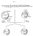
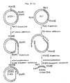
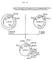
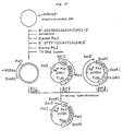
- Fig. 2 illustrates the construction scheme for the plasmid pUKT6.
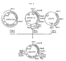
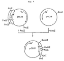
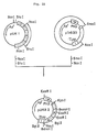
- Fig. 3 illustrates the construction scheme for the plasmid pUKT4 and pUKS3.
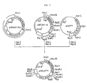
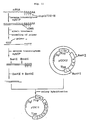
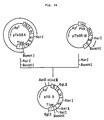
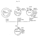
- Fig. 4 illustrates the construction scheme for the plasmid pSEUKT6.
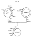
- Fig. 5 illustrates the construction scheme for the plasmid pSEUKT4.
- Fig. 6 illustrates the construction scheme for the plasmid pSEUKS3.
- Fig. 7 (1) and (2) illustrate the levels of resistance of various pro-UK derivatives to thrombin in comparison with that of naturally occurring pro-UK to thrombin, the mark ⁇ being for naturally occurring pro-UK, ⁇ for the pro-UK derivative UK-T6, ⁇ for the pro-UK derivative UK-T4, ⁇ for the pro-UK derivative UK-S3 and ⁇ for the pro-UK derivative UK-T.
- the thrombin concentrations are 5.0 IU/ml and 100 IU/ml, respectively.
- Fig. 7 (3) illustrates the results of SDS-polyacrylamide gel electrophoresis of naturally occurring pro-UK and various pro-UK derivatives after treatment with 5.0 IU/ml of thrombin, comparatively showing the digestion of each single-chain polypeptide during the course of time.
- Fig. 8 illustrates the scheme for the synthesis of cDNA by the Okayama-Berg method and the construction of a recombinant plasmid DNA containing said cDNA.
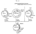
- Fig. 9 illustrates the construction scheme for the plasmid DNA pCCK1.
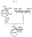
- Fig. 10 illustrates the construction scheme for the plasmid pCCK2.
- Fig. 11 illustrates the construction scheme for the plasmid pUK11 carrying human pro-UK cDNA.
- Fig. 12 illustrates the construction scheme for the plasmid pTrS20.
- Fig. 13 illustrates the construction scheme for the plasmid pTrS33.
- Fig. 14 illustrates the construction scheme for the plasmid pTerm2.
- Fig. 15 illustrates the construction scheme for the plasmid pTSF10.
- Fig. 16 illustrates the construction scheme for the plasmid pTA4.
- Fig. 17 illustrates the construction scheme for the plasmid pTkSD217.
- Fig. 18 illustrates the construction scheme for the plasmid pTkSL11.
- Fig. 19 illustrates the construction scheme for the plasmid pTkSS4.
- Fig. 20 illustrates the construction scheme for the plasmid pTkSJ1.
- Fig. 21 illustrates the construction scheme for the plasmid pTkSR18.
- Fig. 22 illustrates the construction scheme for the plasmid pUKA2.
- Fig. 23 illustrates the construction scheme for the plasmid pUKB101.
- Fig. 24 illustrates the construction scheme for the plasmid pTG3.
- Fig. 25 illustrates the construction scheme for the plasmid phPA2.
- Fig. 26 illustrates the construction scheme for the single-stranded pUKmpS1.
- Fig. 27 illustrates the construction scheme for the plasmid pUKS1.
- Fig. 28 illustrates the construction scheme for the plasmid pAGE105M.
- Fig. 29 illustrates the construction scheme for the plasmid pAGE106.
- Fig. 30 illustrates the construction scheme for the plasmid pSE1PA1-5.
- Fig. 31 illustrates the construction scheme for the plasmid pSE1PA1-9.
- Fig. 32 illustrates the construction scheme for the plasmid pUC19H.
- Fig. 33 illustrates the construction scheme for the plasmid pSE1PA1-9A.
- Fig. 34 illustrates the construction scheme for the plasmid pUKF2.
- Fig. 35 illustrates the construction scheme for the plasmid pUKFpro.
- Fig. 36 illustrates the construction scheme for the plasmid pSEUK1-1A.
- Fig. 37 illustrates the construction scheme for the plasmid pSPAS1-9A
- Fig. 38 illustrates the construction scheme for the plasmid pUKT1.
- Fig. 39 illustrates the construction scheme for the plasmid pSEUKT.
- the following abbreviations are used in the human t-PA cDNA region: F for finger domain, G for growth factor domain, K1 for kringle 1, K2 for kringle 2, K2' for kringle resulting from partial replacement of kringle 2 by kringle 1, and P for protease domain.
- F for finger domain G for growth factor domain
- K1 for kringle 1 K2 for kringle 2
- K2' for kringle resulting from partial replacement of kringle 2 by kringle 1 K2' for kringle resulting from partial replacement of kringle 2 by kringle 1
- P protease domain.
- the novel plasminogen activator according to the invention is a human prourokinase-type plasminogen activator resulting from substitution of an amino acid residue other than a proline residue for the 155th [from the N terminus, serine, of mature human prourokinase] amino acid residue, proline, of mature human prourokinase. This amino acid substitution results in an increase in plasminogen activator activity and in acquisition of resistance to thrombin-like proteases.
- any amino acid residue selected from asparagine, aspartic acid, alanine, arginine, isoleucine, glycine, glutamine, glutamic acid, threonine, serine, cysteine, tyrosine, tryptophan, valine, histidine, phenylalanine, methionine, lysine and leucine can be used as a substitute provided that said residue can afford resistance to thrombin-like proteases.
- a novel plasminogen activator resulting from substitution of an asparagine residue for the 153rd amino acid, leucine, residue and of a threonine residue for the 155th amino acid residue, proline, has a newly created site for N-glycosylation which can stabilize the protein. It is expected that when this plasminogen activator is expressed in animal cells, N-glycosylation might occur on the 153rd amino acid residue, asparagine.
- pro-UK derivative the novel plasminogen activator according to the invention.
- the recombinant plasmid according to the invention is produced by insertion of a DNA fragment coding for the above-mentioned pro-UK derivative into an appropriate plasmid capable of DNA expression in host cells.
- the DNA fragment coding for the pro-UK derivative of this invention can be prepared by introducing a base substitution which causes amino acid substitution into a human UK DNA.
- a human UK DNA a cDNA obtained by reverse transcription of a messenger RNA coding for human UK and a human UK DNA obtained from chromosomal DNA are used.
- the human UK cDNA may be any cDNA coding for human UK.
- the human UK cDNA contained in the plasmid pUK1 or pUK11 can be used.
- pUK1 and pUK11 are plasmids produced by the present inventors and the procedures for their preparation are described in Reference Examples 1, 2 and 3.
- the human UK cDNA in pUK1 codes for a pro-UK modification lacking part of the N-terminal region of pro-UK while that in pUK11 codes for a pro-UK modification lacking part of the C-terminal region of pro-UK.
- the base sequences of the respective cDNAs agree in part with the base sequence shown in Table 4.
- the plasmid into which the DNA coding for a proUK derivative is to be inserted may be any plasmid allowing expression of said DNA in microbial or animal cells.
- Escherichia coli is preferably used as the microbial cells.
- a plasmid which allows insertion of a foreign DNA thereinto at a site downstream from an appropriate promoter, for example trp or lac promoter and in which the distance between the Shine-Dalgarno sequence (hereinafter referred to as "SD sequence") and the initiation codon (ATG) is appropriate, for example 6 to 18 bases long can be used.
- SD sequence Shine-Dalgarno sequence
- ATG initiation codon
- pKYP10 U.S. Patent No. 4,686,191
- pTrS33 Reference Example 4
- the plasmid to be used in causing expression of the DNA coding for a pro-UK derivative in animal cells may be any plasmid provided that it allows expression of said DNA in animal cells.
- a plasmid which allows insertion of a foreign DNA at a site downstream from an appropriate promotor, for example the SV40 early promoter or SV40 late promoter, and which contains a poly(A) signal, splicing signal and so forth.
- Preferred plasmids are pAGE103 [Mizukami et al., J. Biochem., 101 , 1307-1310 (1987)] and pSE1PA1SE1dhfr1-9A (hereinafter referred to as "pSPAS1-9A" for short) (Reference Example 14), both constructed by the present inventors.
- Escherichia coli strain harboring pAGE103 has been deposited, since March 23, 1987, with the Fermentation Research Institute under the designation (and deposit number) Escherichia coli EAGE103 (FERM BP-1312).
- Escherichia coli EAGE103 FERM BP-1312
- dhfr dihydrofolate reductase
- Recombination between the DNA coding for a pro-UK derivative and a vector DNA can be carried out by using available techniques that are generally used in the recombinant DNA technology, namely by digesting both DNAs with one or more restriction enzymes, followed by ligation using T4 DNA ligase. Prior to ligation, the termini of the DNA fragments obtained by digestion with the restriction enzyme or enzymes may be filled in using a DNA polymerase Klenow fragment or may be filled in or processed for paring off using T4 DNA polymerase. DNA linkers may also be used.
- phPA2 (Reference Example 10) constructed from pUK1 is cleaved with Eco RI and Hin dIII and a DNA fragment of about 3.4 kb is purified.
- pUKS1 (Reference Example 11) is cleaved with Hin dIII and Cfr I and a DNA fragment of about 0.75 kb is purified. The two DNA fragments thus obtained, together with the two 5'-phosphorylated synthetic DNAs shown in Fig.
- the recombinant plasmid pUKT4 coding for a pro-UK derivative (UK-T4) and the recombinant plasmid pUKS3 coding for another pro-UK derivative (UK-S3) are constructed.
- phPA2 (Reference Example 10) is cleaved with Eco RI and Hin dIII and a DNA fragment of about 3.4 kb is purified.
- pUKS1 (Reference Example 11) is cleaved with Hin dIII and Cfr I and a DNA fragment of about 0.75 kb is purified. The two DNA fragments thus obtained, together with the two 5'-phosphorylated synthetic DNAs shown in Fig.
- pUKT4 coding for a pro-UK derivative (UK-T4) differing from pro-UK in that the 153rd (from the N terminus) amino acid residue Leu and 155th amino acid residue Pro of pro-UK have been replaced by Ser and Thr, respectively.
- pUKS3 coding for a pro-UK derivative (UK-S3) differing from pro-UK in that the 153rd (from the N terminus) amino acid residue Leu and 155th amino acid residue Pro of pro-UK have been replaced by Asn and Thr, respectively, is also obtained.
- Plasmid DNAs capable of expressing the respective pro-UK derivatives (UK-T6, UK-T4, UK-S3 and UK-T) and natural-type pro-UK in animal cells can be prepared, for example, in the following manner.
- pSPAS1-9A (Reference Example 14) is cleaved with Xho I and Kpn I and a DNA fragment of about 8.6 kb is purified.
- psE1UKpro1-1A (hereinafter referred to as pSEUK1-1A for short) (Reference Example 13) is cleaved with Xho I and Bgl II and a DNA fragment of about 0.75 kb is purified.
- pUKT6 is cleaved with Bgl II and Kpn I and a DNA fragment of about 1.15 kb is purified.
- pSEUKT6 plasmid pSE1UKT6SEd1-3
- pSEUKT6 plasmid pSE1UKT6SEd1-3
- pSPAS1-9A (Reference Example 14) is cleaved with Xho I and Kpn I and a DNA fragment of about 8.6 kb is purified.
- pSEUK1-1A (Reference Example 13) is cleaved with Xho I and Bgl II and a DNA fragment of about 0.75 kb is purified.
- pUKT4 is cleaved with Bgl II and Kpn I and a DNA fragment of about 1.15 kb is purified.
- pSEUKT4 plasmid pSE1UKT4SEd1-3
- pSEUKT4 plasmid pSE1UKT4SEd1-3
- pSPAS1-9A (Reference Example 14) is cleaved with Xho I and Kpn I and a DNA fragment of about 8.6 kb is purified.
- pSEUK1-1A (Reference Example 13) is cleaved with Xho I and Bgl II and a DNA fragment of about 0.75 kb is purified.
- pUKS3 is cleaved with Bgl II and Kpn I and a DNA fragment of about 1.15 kb is purified.
- pSEUKS3 plasmid pSE1UK3SEd1-3
- pSEUKS3 plasmid pSE1UK3SEd1-3
- the digestion of DNA with a restriction enzyme or enzymes is generally carried out in a reaction mixture containing 0.1 to 20 »g of DNA, 2 to 200 mM (preferably 10 to 40 mM) Tris-HCl (pH 6.0 to 9.5, preferably pH 7.0 to 8.0), 0 to 200 mM NaCl and 2 to 20 mM (preferably 5 to 10 mM) MgCl2, using 0.1 to 100 units (preferably 1 to 3 units per microgram of DNA) of each restriction enzyme, at 20 to 70°C (the optimal temperature may vary depending on the restriction enzyme or enzymes) for 15 minutes to 24 hours.
- the reaction is generally terminated by heating at 55 to 75°C for 5 to 30 minutes.
- the reaction may also be terminated by inactivating the restriction enzyme or enzymes using an appropriate reagent such as phenol or diethyl pyrocarbonate.
- the DNA fragment resulting from restriction enzyme digestion can be purified by low temperature gelation agarose gel electrophoresis (hereinafter referred to as "LGT method”) [L. Wieslander, Analytical Biochemistry, 98 , 305 (1979)] or agarose gel freezing and thawing (hereinafter referred to as "AFT method”).
- LGT method low temperature gelation agarose gel electrophoresis
- AFT method agarose gel freezing and thawing
- This AFT method comprises admixing a DNA fragment-containing agarose gel (0.7 to 1.5%) slice with an equal volume of TE buffer [10 mM Tris-HCl (pH 7.5), 1 mM EDTA] and 2 volumes of phenol (saturated with TE buffer), repeating two cycles of freezing (-70°C) and thawing (65°C), centrifuging, separating the resulting upper aqueous layer and recovering the DNA fragment by precipitation with ethanol.
- the DNA fragment may also be electrophoretically eluted and purified from an agarose or polyacrylamide gel using a Maxfield model AE-3241 DNA fragment collector (Atto), for instance. (Hereinafter, the latter method is referred to as "electrophoretic elution method").
- the ligation of DNA fragments is generally carried out in a reaction mixture containing 2 to 200 mM (preferably 10 to 40 mM) Tris-HCl (pH 6.1 to 9.5, preferably pH 7.0 to 8.0), 2 to 20 mM (preferably 5 to 10 mM) MgCl2, 0.1 to 10 mM (preferably 0.5 to 2.0 mM) ATP and 1 to 50 mM (preferably 5 to 10 mM) dithiothreitol (hereinafter referred to as "DTT") at 1 to 37°C (preferably 3 to 20°C) for 15 minutes to 72 hours (preferably 2 to 20 hours), using 1 to 1,000 units of T4 DNA ligase.
- Tris-HCl pH 6.1 to 9.5, preferably pH 7.0 to 8.0
- 2 to 20 mM preferably 5 to 10 mM
- MgCl2 preferably 5 to 10 mM MgCl2
- 0.1 to 10 mM preferably 0.5 to 2.0 mM
- ATP
- the recombinant plasmid DNA resulting from the ligation reaction is introduced into Escherichia coli , if necessary using the transformation method of Cohen et al. [S.N. Cohen et al., Proc. Natl. Acad. Sci. USA, 69 , 2110 (1972)] or the transformation method of Hanahan [J. Mol. Biol., 166 , 557 (1983)].
- the recombinant M13 phage RF DNA produced by the ligation reaction is introduced into Escherichia coli , strain JM105 [J. Messing et al., Gene, 33 , 103 (1985)], for instance, if necessary using the known method of transfection [Y. Kuchino et al., Tampakushitsu, Kakusan, Koso (Protein, Nucleic Acid and Enzyme), 29 , 294 (1984)].
- the recombinant plasmid DNA or recombinant M13 phage RF DNA can be isolated from the Escherichia coli , strain harboring the same by the method of Birnboim et al. [H.C. Birnboim et al., Nucleic Acids Res., 7 , 1513 (1979)], for instance.
- the isolation of the single-stranded DNA from the recombinant M13 phage can be performed by the known method [Y. Kuchino et al., Tampakushitsu, Kakusan, Koso, 29 , 294 (1984)].
- deoxyoligonucleotides to be used in the practice of the invention can be synthesized by solid-phase synthesis by the phosphoamidite method [S.L. Beaucage et al., Tetrahedron Lett., 22 , 1859 (1981) and L. J. BcBrie et al., ibid. 24 , 245 (1983)] using an Applied Biosystems model 380A DNA synthesizer (Applied Biosystems Inc., Foster City, CA 94404).
- T4 kinase buffer 50 mM Tris-HCl (pH 7.6), 10 mM MgCl2, 5 mM DTT, 0.1 mM EDTA, 0.5 mM ATP]
- T4 kinase buffer 50 mM Tris-HCl (pH 7.6), 10 mM MgCl2, 5 mM DTT, 0.1 mM EDTA, 0.5 mM ATP
- the deoxyoligonucleotide is radioactively labeled at the 5' end thereof using 20 to 50 »Ci of 5'-[ ⁇ -32P]ATP (3,000 Ci/mmol, Amersham, Arlington Heights, IL) in lieu of 0.5 mM ATP in the T4 kinase buffer mentioned above.
- each plasmid DNA is subjected to digestion with one to ten restriction enzymes, followed by agarose gel or polyacrylamide gel electrophoresis for checking the cleavage sites. Furthermore, if necessary, the base sequence of a DNA can be determined by the dideoxy sequence method using M13 phage.
- the pro-UK derivative polypeptides according to the invention can be produced by using Escherichia coli or animal cells as the host, for example in the following manner.
- the pro-UK derivative polypeptides are produced using animal cells as the host as follows.
- the host for use in causing expression of the pro-UK derivative polypeptides may be any animal cell strain or line provided that it allows expression of said polypeptides.
- Preferred specific animal cells include dhfr-deficient CHO cells [G. Urlaub & L.A. Chasin, Proc. Natl. Acad. Sci. USA, 77 , 4216 (1980)], among others.
- pro-UK derivative polypeptide production is described in which pSEUKT6 is used as a plasmid for expression of the pro-UK derivative UK-T6 and dhfr-deficient CHO cells are used as the host.
- the plasmid pSEUKT6 is introduced into dhfr-deficient CHO cells by, for example, the calcium phosphate method [Graham & Van der Eb, Virology, 52 , 546 (1978)].
- Transformant strains harboring pSEUKT6 can be selected using, for example, MEM ALPHA medium (ribonucleic acid-free and deoxyribonucleic acid-free; Gibco-Oriental) supplemented with G418 and dialyzed fetal calf serum.
- a transformant strain with the gene for the physiologically active polypeptide being amplified can be selected from the transformant resistant to methotrexate.
- the transformant strain thus obtained is cultured in a medium, whereby the pro-UK derivative polypeptide can be produced in the culture.
- Suitable media are HAM F10 medium, HAM F12 medium (both available from Flow Laboratories), Dulbecco's MEM medium, RPMI-1640 medium (latter two available from Nissui Pharmaceutical), MEM ALPHA medium, and mixed media derived from these, each supplemented with any of various sera (e.g. fetal calf serum). If necessary, 0.5 to 5 mM glutamine, antibiotics (penicillin (25 U/ml), streptomycin (25 »g/ml), G418 (0.3 mg/ml), etc.], sodium bicarbonate (0.01%) and so forth may be added to the medium each in an appropriate amount.
- antibiotics penicillin (25 U/ml), streptomycin (25 »g/ml), G418 (0.3 mg/ml), etc.
- sodium bicarbonate 0.01%
- the cultivation is generally carried out at a seed cell density of 5 ⁇ 104 to 1 ⁇ 106 cells/ml at 30 to 40°C for 2 to 10 days, whereupon the substance according to the invention is secreted mainly extracellularly in an amount depending on the cell density.
- pro-UK derivative polypeptide is isolated and purified from the supernatant after centrifugation.
- the plasminogen activating activity of the pro-UK derivative polypeptide obtained can be assayed by the fibrin plate assay method [Granelli-Piperno & Reich, J. Exp. Med., 148 , 223 (1978)].
- T4 kinase buffer a solution containing 50 mM Tris-HCl (pH 7.6), 10 mM MgCl2, 5 mM DTT, 0.1 mM EDTA and 0.5 mM ATP, with 5 units of T4 DNA kinase (Takara Shuzo) added, at 37°C for 30 minutes.
- T4 ligase buffer 20 mM Tris-HCl (pH 7.6), 10 mM MgCl2, 10 mM DTT and 1 mM ATP, 300 units of T4 ligase (Takara Shuzo) were added, and the ligation reaction was carried out at 4°C for 18 hours.
- the resulting reaction mixture was used to transform Escherichia coli MM294 [F ⁇ hsdR ⁇ hsdM+ endoI ⁇ thi] [ATCC31446; the same strain as Escherichia coli 294 so referred to in the report of K. Backman et al., Proc. Natl. Acad. Sci. USA, 73 , 4174 (1976)] to give ampicillin (hereinafter, "Ap")-resistant transformants. Plasmid DNAs were isolated from these transformants and subjected to structural analysis by digestion with restriction enzymes and to DNA sequence determination by the M13 dideoxy sequencing method. A plasmid DNA thus found to have the desired structure was named pUKT6 (cf. Fig. 2).
- the thus-obtained pUKS1-derived DNA fragment (about 0.75 kb, about 0.1 »g), phPA2-derived DNA fragment (about 3.4 kb, about 0.1 »g) and two 5'-phosphorylated synthetic DNAs (1 picomole each) were dissolved in 20 »l of T4 ligase buffer, 300 units of T4 ligase were added, and the ligation reaction was carried out at 4°C for 18 hours.
- the reaction mixture obtained was used to transform Escherichia coli MM294 to give Ap-resistant transformants. Plasmid DNAs were isolated from these transformants and subjected to structural analysis by digestion with restriction enzymes and to DNA sequence determination by the M13 dideoxy sequence method.
- pSPAS1-9A-derived DNA fragment (about 8.6 kb, about 0.1 »g), psEUK1-1A-derived DNA fragment (about 0.75 kb, about 0.02 »g) and pUKT6-derived DNA fragment (about 1.15 kb, about 0.02 »g) were dissolved in 20 »l of T4 ligase buffer, 100 units of T4 DNA ligase were added, and the ligation reaction was carried out at 4°C for 18 hours.
- the reaction mixture thus obtained was used to transform Escherichia coli MM294 to give Ap-resistant transformants.
- the plasmid DNA pSEUKT6 isolated from one of said transformants was subjected to structural analysis by digestion with restriction enzymes and found to have the desired structure (cf. Fig. 4).
- pSPAS1-9A-derived DNA fragment (about 8.6 kb, about 0.1 »g), pSEUK1-1A-derived DNA fragment (about 0.75 kb, about 0.02 »g) and pUKT4-derived DNA fragment (about 1.15 kb, about 0.02 »g) were dissolved in 20 »l of T4 ligase buffer, 100 units of T4 DNA ligase were added, and the ligation reaction was carried out at 4°C for 18 hours.
- the reaction mixture thus obtained was used to transform Escherichia coli MM294 to give Ap-resistant transformants.
- the plasmid DNA pSEUKT4 isolated from one of these transformants was found to have the desired structure upon structural analysis by digestion with restriction enzymes (cf. Fig. 5).
- pSPAS1-9A-derived DNA fragment (about 8.6 kb, about 0.1 »g), pSEUK1-1A-derived DNA fragment (about 0.75 kb, about 0.02 »g) and pUKS3-derived DNA fragment (about 1.15 kb, about 0.02 »g) were dissolved in 20 »l of T4 ligase buffer, 100 units of T4 DNA ligase were added, and the ligation reaction was carried out at 4°C for 18 hours.
- the reaction mixture thus obtained was used to transform Escherichia coli MM294 to give Ap-resistant transformants.
- the plasmid DNA pSEUKS3 isolated from one of these transformants was found to have the desired structure upon structural analysis by digestion with restriction enzymes (cf. Fig. 6).
- pSEUKT6 (obtained in Example 3) was introduced into dhfr-deficient CHO cells essentially by the calcium phosphate method.
- MEM ⁇ non-selective medium
- PCS fetal calf serum
- NaHCO3 7.5% NaHCO3
- Incubation was performed in a CO2 incubator at 37°C for 5 days using a dish having a diameter of 10 cm.
- Cells were washed with PBS and, after addition of MEM ⁇ (selective medium), cultured for 5 days. The same procedure as above was followed and incubation was further carried out for 5 days.
- Cells were washed with PBS, subjected to trypsin treatment, suspended in 10 ml of MEM ⁇ (selective medium) and cultured in a dish having a diameter of 6 cm in a CO2 incubator at 37°C for 3 to 7 days.
- Colonies that had appeared were subjected to trypsin treatment and then inoculated into a dish having a diameter of 10 cm to a cell concentration of 5 ⁇ 104 cells/ml using 10 ml of MEM ⁇ (selective medium) containing 50 mM MTX. Medium exchange was performed three times at 5 day intervals using the same medium mentioned above. MTX-resistant colonies that had appeared were respectively isolated and each colony was cultured in a dish having a diameter of 6 cm until confluence.
- Example 7 After confluence, culturing was conducted for 3 days in a medium identical in composition to the above-mentioned one except that it was FCS-free and contained 10 KIU/ml of aprotinin (Boehringer-Mannheim). The culture supernatant (100 ml per run) thus obtained was used in Example 7.
- Example 7 After confluence, culturing was conducted for 3 days in a medium identical in composition to the above-mentioned one except that it was FCS-free and contained 10 KIU/ml of aprotinin (Boehringer-Mannheim). The culture supernatant (100 ml per run) thus obtained was used in Example 7.
- Example 7 After confluence, culturing was conducted for 3 days in a medium identical in composition to the above-mentioned one except that it was FCS-free and contained 10 KIU/ml of aprotinin (Boehringer-Mannheim). The culture supernatant (100 ml per run) thus obtained was used in Example 7.
- Example 7 After confluence, culturing was conducted for 3 days in a medium identical in composition to the above-mentioned one except that it was FCS-free and contained 10 KIU/ml of aprotinin (Boehringer-Mannheim). The culture supernatant (100 ml per run) thus obtained was used in Example 7.
- pro-UK-producing cell strains Using the recombinant plasmid pSEUK1-1A obtained in Reference Example 13 and pSV2-dhfr and dhfr-deficient CHO cells and following the same procedure as described above, there were obtained pro-UK-producing cell strains. Among them, the clone No. 5 was found to be the highest in activity, the pro-UK production amounting to 3 »g/106 cells/day. This clone was cultured in 100 ml of MEM ⁇ (selective medium) containing 50 nM MTX in a Falcon model 3027 roller bottle.
- MEM ⁇ selective medium
- Example 7 After confluence, culturing was conducted for 3 days in a medium identical in composition to the above-mentioned one except that it was FCS-free and contained 10 KIU/ml of aprotinin (Boehringer-Mannheim). The culture supernatant (100 ml per run) thus obtained was used in Example 7.
- the serum-free culture supernatants (200 ml each) obtained in Example 6 and containing natural type pro-UK or a pro-UK derivative were respectively applied to an anti-UK monoclonal antibody column (4 ml) [prepared by binding an anti-UK monoclonal antibody (prepared by the method of Milstein et al. [C.
- SDS-polyacrylamide gel electrophoresis revealed that the final purified sample contained a single-chain product with a purity of not less than 95%.
- the SDS-polyacrylamide gel electrophoresis suggested carbohydrate chain addition to the pro-UK derivative UK-S3, which has an N-glycosylation site, namely -Asn-X-Thr-. An increase in molecular weight was confirmed as well.
- the final purified samples respectively containing the natural type pro-UK and pro-UK derivatives as obtained in Example 7 were each applied to a reversed-phase HPLC column (TSK gel ODS-120T, Tosoh).
- the peak area was compared with the peak area for natural type pro-UK for which the concentration had been determined by the Lowry method [Lowry et al., J. Biol. Chem., 193 , 265 (1951)] to determine the concentration of each protein.
- the activity of each sample was determined by the fibrin plate assay method.
- the fibrin plate assay method was carried out in the following manner.
- Bovine thrombin (Sigma) (1,000 U) was dissolved in 5 ml of 50 mM phosphate buffer (pH 7.0). The solution was filter sterilized through a 0.45 »m membrane filter to give a thrombin solution. Separately, a fibrinogen solution was prepared by dissolving bovine fibrinogen (Nakalai-Tesque) in 100 ml of sterilized 50 mM phosphate buffer (pH 7.0) with 30 minutes of stirring and then removing the insoluble matter using sterilized glass wool to give a fibrinogen solution.
- bovine fibrinogen Nakalai-Tesque
- a 2% agar solution was prepared by adding 2 g of agar (Sigma) to 100 ml of 50 mM phosphate buffer (pH 7.0), sterilizing at 1.5 atmospheres and 120°C for 20 minutes and then maintaining the solution at 60°C.
- Human urokinase double-chain form, urine-derived, Nippon Chemical Research
- concentration was adjusted to 10, 100, 1,000 and 10,000 IU/ml. These solutions were used as standards.
- Each sample was diluted to 0.5 »g/ml with UK buffer.
- the specific activity data for the natural type pro-UK and respective pro-UK derivatives are shown herein below in Table 5. While the specific activities of UK-T6, UK-T4 and UK-S3 were higher (1,2- to 1,8-fold) than that of natural type pro-UK, the specific activity of the pro-UK derivative UK-T (with an amino acid substitution in position P1' relative to the thrombin cleavage site) was only 60% of that of natural type pro-UK. This decrease in specific activity was presumably due to that introduction of amino acid substitution in the neighborhood of the plasmin cleavage site (namely in position P2 relative to the plasmin cleavage site) which made it difficult for the cleavage with plasmin to take place. Therefore, the pro-UK derivatives (UK-T6, UK-T4 and UK-S3) with amino acid substitution in position P2 are advantageous from the specific activity viewpoint as well.
- the final purified samples respectively containing the natural type pro-UK and pro-UK derivatives as obtained in Example 7 were each applied to a reversed-phase HPLC column (TSK gel ODS-120, Tosoh). Each sample was diluted with the diffusate solution (obtained in the dialysis in Example 7) to 30 »g/ml on the basis of the peak area obtained. To 100 »l of this dilution were added 20 »l of 30 IU/ml or 600 IU/ml human thrombin, and the mixture was incubated at 37°C. The human thrombin was obtained from Sigma. The human thrombin was used after 1.5 hours of treatment with 10 IU/1,000 IU thrombin of aprotinin at 37°C.
- a comparison with respect to the susceptibility to thrombin was performed by determining the residual activities of the natural type pro-UK and pro-UK derivatives in the reaction mixtures obtained as described above by the fibrin plate assay method (cf. Example 8). As a result, it was revealed that the pro-UK derivatives had apparently decreased susceptibility to thrombin as compared with natural type pro-UK (cf. Fig. 7-(1)). In particular, it was also shown that UK-T6, UK-T4 and UK-S3 were still lower in susceptibility to thrombin than UK-T (cf. Fig. 7-(2)).
- Escherichia coli harboring the plasmids pSEUKS3, pSEUKT4 and pSEUKT6 have been deposited, since June 15, 1989, with the Fermentation Research Institute under the Budapest Treaty under the designations (and accession numbers) Escherichia coli ESEUKS3 (FERM BP-2478), Escherichia coli ESEUKT4 (FERM BP-2479) and Escherichia coli ESEUKT6 (FERM BP-2480), respectively.
- RNA was prepared from Detroit 562 human pharyngeal carcinoma cells by the guanidine thiocyanate-lithium chloride method [Cathala et al., DNA, 2 , 329 (1983)] as follows.
- Detroit 562 human pharyngeal carcinoma cells [W. D. Peterson, Jr, et al., Proc. Soc. Exp. Biol. Med., 136 , 1187 (1971)] were grown in 50 ml MEM medium (Nissui Pharmaceutical) containing 10% FCS, 1/100 volume of 100 ⁇ nonessential amino acid solution (Flow Laboratories), 1 mM sodium pyruvate and 0.1% lactoalbumin hydrolyzate (Gibco-Oriental) in a tissue culture flask (Corning; 150 cm2).
- Cells were collected from the pooled cell suspension by centrifugation (1,100 ⁇ g, 4°C, 10 minutes), washed with 80 ml of phosphate buffer, and solubilized in 10 ml of a solution containing 5 M guanidine thiocyanate, 10 mM EDTA, 50 mM Tris-HCl (pH 7) and 8% (v/v) 2-mercaptoethanol using a vortex mixer. The solubilization product was transferred to a centrifuge tube, 80 ml of 4 M LiCl were added and, after stirring, the resulting mixture was allowed to stand at 4°C for 20 hours.
- RNA precipitate was suspended in 50 ml of a solution comprising 4 M urea and 2 M lithium chloride, and RNA was recovered again as a precipitate by centrifugation using a Hitachi RPR10 rotor (9,000 rpm, 60 minutes).
- the RNA precipitate was dissolved in 10 ml of a solution containing 0.1% sodium lauryl sulfate, 1 mM EDTA and 10 mM Tris-HCl (pH 7.5) and, after extraction with phenol-chloroform, recovered by precipitation with ethanol.
- RNA obtained were dissolved in 1 ml of a solution containing 10 mM Tris-HCl (pH 8.0) and 1 mM EDTA. After 5 minutes of incubation at 65°C, 0.1 ml of 5 M NaCl were added. The resulting mixture was subjected to oligo(dT)-cellulose column [P-L Biochemicals) chromatography (column volume: 0.5 ml). The poly(A)-containing mRNA adsorbed was eluted with a solution containing 10 mM Tris-HCl (pH 7.5) and 1 mM EDTA. About 90 »g of poly(A)-containing mRNA were thus obtained.
- TdT buffer a buffer containing 40 mM sodium cacodylate, 30 mM Tris-HCl (pH 6.8), 1 mM CaCl2 and 0.1 mM dithiothreitol (DTT) (hereinafter referred to as "TdT buffer") with dTTP added to a concentration of 0.25 mM.
- TdT terminal deoxynucleotidyl transferase
- the DNA thus obtained was added to 150 »l of a buffer containing 10 mM Tris-HCl (pH 7.5), 6 mM MgCl2 and 100 mM NaCl, 360 units of Eco RI were further added, and the reaction was carried out at 37°C for 2 hours.
- the reaction mixture was treated by the LGT method and a DNA fragment of about 3.1 kb was recovered.
- About 60 »g of poly(dT) chain-added pCDV1 were obtained.
- the resulting DNA was dissolved in 500 »l of a solution containing 10 mM Tris-HCl (pH 8.0) and 1 mM EDTA, the solution was incubated at 65°C for 5 minutes and then cooled on ice, and 50 »l of 5 M NaCl were added to the solution.
- the resultant mixture was subjected to oligo(dA)-cellulose column (Collaborative Research) chromatography. Molecules having a sufficiently long poly(dT) chain were adsorbed on the column.
- vector primer poly(dT) chain-added pCDV1
- a linker DNA was prepared in the following manner.
- the resulting DNA was added to 100 »l of a buffer containing 10 mM Tris-HCl (pH 7.5), 6 mM MgCl2 and 60 mM NaCl, 80 units of Hin dIII were added, and the mixture was incubated at 37°C for 3 hours, whereby the pL1 DNA was cleaved at the Hin dIII site.
- the reaction mixture was fractionated by agarose gel electrophoresis, and a DNA fragment of about 0.5 kb was recovered by the DEAE paper method [Dretzen et al., Anal. Biochem., 112 , 295 (1981)].
- An oligo(dG) chain-containing linker DNA (hereinafter referred to as "linker DNA” for short) was thus obtained.
- the reaction mixture was subjected to phenol-chloroform extraction, followed by precipitation with ethanol, whereby the vector primer DNA with an RNA-DNA double strand added thereto was recovered.
- the resulting DNA was dissolved in 20 »l of TdT buffer containing 66 »M dCTP and 0.2 »g of poly(A), 14 units of TdT (P-L Biochemicals) were added, and the mixture was incubated at 37°C for 2 minutes for the addition of a (dC) chain comprising 20 dC residues to the 3' end of cDNA.
- the reaction mixture was subjected to phenol-chloroform extraction and then a (dC) chain-added cDNA-vector primer DNA was recovered therefrom by precipitation with ethanol.
- the resulting DNA was dissolved in 400 »l of a solution containing 10 mM Tris-HCl (pH 7.5), 6 mM MgCl2 and 60 mM NaCl, 20 units of Hin dIII were added, and the cleavage at the Hin dIII site was effected by performing incubation at 37°C for 2 hours.
- the reaction mixture was subjected to phenol-chloroform extraction and then to precipitation with ethanol, which gave 0.5 picomole of a (dC) chain-added cDNA-vector primer DNA.
- a 0.2-picomole portion of the DNA and 0.4 picomole of the linker DNA mentioned above were dissolved in 100 »l of a solution containing 10 mM Tris-HCl (pH 7.5), 0.1 M NaCl and 1 mM EDTA. Incubation was performed at 65°C for 10 minutes, then at 42°C for 25 minutes further at 0°C for 30 minutes. The reaction mixture was modified to give a total volume of 1,000 »l of a reaction mixture containing 20 mM Tris-HCl (pH 7.5), 4 mM MgCl2, 10 mM (NH4)2SO4, 0.1 M KCl and 0.1 mM ⁇ -NAD.
- the cDNA-containing recombinant DNA was circularized and the RNA portion off the RNA-DNA double strand was replaced by DNA, whereby a recombinant plasmid in the form of a complete, double-stranded DNA was formed.
- the t-PA cDNA was selected out, by colony hydridization, as a clone capable of associating with a 32p-labeled synthetic DNA probe having the DNA sequence 5'-ATGGATGCAATGAAGAGAGGGCTCTGCTGT-3' in agreement with a part of the base sequence of the t-PA signal peptide region of the human t-PA cDNA [Pennica et al., Nature, 301 , 214 (1983)], as follows.
- Escherichia coli C600SF8 [Cameron, Proc. Natl. Acad. Sci. USA, 72 , 3416 (1975)] was transformed with the recombinant plasmid obtained above in section (2) by the method of Hanahan [J. Mol. Biol., 166 , 557 (1983)]. About 10,000 colonies obtained were immobilized on a nitrocellulose filter by the method of Hanahan and Meselson [Methods in Enzymology, 100 , 333 (1983)].
- the above-mentioned 32P-labeled probe was added to this prehydridization solution and allowed to associate with DNA on the filter (65°C, at least 16 hours).
- the Detroit 562 cell-derived cDNA library prepared in Reference Example 1 was screened by the technique of colony hydridization and a human pro-UK cDNA clone was isolated.
- the recombinant plasmid obtained in Reference Example 1 was used to transform Escherichia coli C600SF8 [Cameron, Proc. Natl. Acad. Sci. USA, 72 , 3416 (1975)] by the method of Hanahan [J. Mol. Biol., 166 , 557 (1983)].
- About 30,000 colonies obtained were immobilized on a nitrocellulose filter by the method of Hanahan and Meselson [Methods in Enzymology, 100 , 333 (1983)].
- filter prehybridization was effected in a solution containing 6 ⁇ NET, 10 ⁇ Denhardt solution and 100 »g/ml of fragmented Escherichia coli chromosome DNA at 65°C for at least 4 hours.
- the filter was then washed twice with 6 ⁇ SSC (room temperature, 5 minutes per wash) and then with a solution containing 1 ⁇ SSC and 0.1% SDS at 57°C for 30 minutes.
- the filter was further washed with a solution containing 1 ⁇ SSC and 0.1% SDS at 57°C for 15 minutes and then twice with 6 ⁇ SSC (room temperature, 5 minutes per wash).
- the filter was air-dried and one positive clone was identified by autoradiography.
- the DNA sequence of the cDNA of the plasmid pUK1 carried by this positive clone was determined by the dideoxy method using M13 phage.
- the cDNA of pUK1 codes for the translational region for that portion of pro-UK downstream from the 41st amino acid residue (Cys) of pro-UK (according to the numbering of amino acid residues of pro-UK as used in Table 4) and for the 3'-nontranslational region.
- the amino acid sequence of pro-UK encoded by the cDNA of pUK1 was in agreement with that reported by Holmes et al. [Bio/Technology, 3 , 923 (1985)].
- pro-UK cDNA encoded by the plasmid pUK1 obtained in Reference Example 2 does not contain the signal region and growth factor domain region of pro-UK, a cDNA containing these regions was cloned as described below.
- a vector, pCCK2 for cDNA cloning was constructed as follows.
- Escherichia coli HB101 was transformed with the plasmid pRC19 constructed by Kuwano et al. [J. Biochem., 96 , 923-926 (1984)] and carrying a cDNA for rat brain cholecystokinin (CCK) precursor, was cultured and the pRC19 DNA was prepared from cultured cells in the conventional manner.
- pRC19 constructed by Kuwano et al. [J. Biochem., 96 , 923-926 (1984)] and carrying a cDNA for rat brain cholecystokinin (CCK) precursor
- the thus-obtained pRC19-derived DNA fragment (about 530 bp, about 0.01 »g) and the pUC19-derived DNA fragment (about 2.7 kb, about 0.05 »g) were dissolved in 20 »l of T4 ligase buffer, 200 units of T4 DNA ligase were added, and the ligation reaction was carried out at 4°C for 18 hours.
- the recombinant plasmid mixture obtained was used to transform Escherichia coli MM294 to give Ap-resistant transformants.
- the plasmid DNA pCCK1 isolated from one of these transformants was subjected to structural analysis with restriction enzymes and found to have the desired structure (cf. Fig. 9).
- the thus-obtained pCCK1-derived DNA fragment (about 0.55 kb, about 0.02 »g) and the pTrS33-derived DNA fragment (about 2.85 kb, about 0.1 »g) were dissolved in 20 »l of T4 ligase buffer, 50 units of T4 DNA ligase were added, and the ligation reaction was conducted at 4°C for 18 hours.
- the recombinant plasmid mixture obtained was used to transform Escherichia coli MM294 to give Ap-resistant transformants.
- the plasmid DNA pCCK2 isolated from one of these transformants was subjected to structural analysis using restriction enzymes and found to have the desired structure (cf. Fig. 10).
- composition of this solution was adjusted so that the resultant solution contained, in a total volume of 80 »l, 50 mM Tris-HCl (pH 8.3), 8 mM MgCl2, 30 mM KCl, 5 mM DTT, 1 mM dNTP (dATP, dTTP, dGTP and dCTP), 10 units of ribonuclease inhibitor (P-L Biochemicals) and 5 »g/ml of oligo(dT)12 ⁇ 18 (Collaborative Research). The solution was maintained at 41°C for 15 minutes.
- composition of this solution was adjusted so that the resultant solution contained, in a total volume of 40 »l, 50 mM Tris-HCl (pH 8.3), 8 mM MgCl2, 30 mM KCl, 5 mM DTT, 1 mM dNTP (dATP, dTTP, dGTP and dCTP) and 2.5 »g/ml of the synthetic DNA primer CATGAGAGCCCTGCTGG (idential to a part of the base sequence for the signal peptide region of human pro-UK).
- the solution was maintained at 65°C for 10 minutes and then at 41°C for 30 minutes.
- the thus-obtained cDNA fragment (about 1.1 to 1.4 kb, about 0.02 »g) and pCCK2-derived DNA fragment (about 2.9 kb, about 0.05 »g) were dissolved in 20 »l of T4 ligase buffer, 200 units of T4 DNA ligase were added, and the ligation reaction was conducted at 4°C for 18 hours.
- the reaction mixture obtained was used to transform Escherichia coli C600SF8 to give about 25,000 Ap-resistant transformants.
- about 1,000 positive clones were obtained by the colony hybridization method mentioned in Reference Example 2, which were capable of associating with the same probe as used for isolating the pro-UK cDNA in Reference Example 2.
- the hybridization conditions and filter washing conditions were the same as employed in Reference Example 2.
- the plasmid pUKll (cf. Fig. 11) isolated from one of the positive clones thus obtained was subjected to base sequence determination for the pro-UK signal peptide, growth factor domain and kringle domain regions by the dideoxy method using Ml3 phage. The DNA sequence thus found was in agreement with those reported by Holmes [Bio/Technology, 3 , 923 (1985)].
- the ATG vector pTrS20 in which the SD sequence is 14 bases apart from the initiation codon ATG and which has a Sac I site just behind the ATG codon was constructed.
- the following DNA linker was synthesized by the phosphotriester method for providing the initiation codon ATG downstream from Ptrp.
- the synthetic 19-mer and 17-mer DNAs (10 picomoles each) were dissolved in a total volume of 20 »l of a solution containing 50 mM Tris-HCl (pH 7.5), 10 mM MgCl2, 5 mM dithiothreitol, 0.1 mM EDTA and 1 mM ATP, 3 units of T4 polynucleotlde kinase (Takara Shuzo) were added, and the phosphorylatlon reaction was conducted at 37°C for 60 minutes.
- the recombinant plasmid mixture obtained was used to transform Escherichia coli HB101 [Bolivar et al., Gene, 2 , 75 (1977)] to give Ap-resistant colonies. Each colony was cultured and a plasmid DNA was recovered from cultured cells and examined for its structure by cleavage with the restriction enzymes Eco RI, Ban III, Hin dIII, Sac I and Nru I, followed by agarose gel electrophoresis. A plasmid found to have the desired structure was named pTrS20 (cf. Fig. 12). It was confirmed by the dideoxy sequencing method using M13 phage that pTrS20 had the following DNA sequence in the neighborhood of the Ban III and Hin dIIII sites
- the thus-obtained pTrS20-derived DNA fragment (about 1.15 kb, about 0.1 »g), pKYP26-derived DNA fragment (about 1.7 kb, about 0.1 »g), M13mp18-derived DNA fragment (about 0.65 kb, about 0.05 »g) and two 5'-phosphorylated synthetic DNAs (1 picomole each) were dissolved in 20 »l of T4 ligase buffer, 50 units of T4 DNA ligase were added, and the ligation reaction was conducted at 4°C for 18 hours.
- the recombinant plasmid mixture thus obtained was used to transform Escherichia coli MM294 to give Ap-resistant transformants.
- the plasmid pTrS33 isolated from one of these transformants was found to have the desired structure upon structural analysis comprising digestion with restriction enzymes and DNA sequencing by the dideoxy method using M13 phage (cf. Fig. 13).
- the thus-obtained pKYP26-derived DNA fragment (about 1.7 kb, about 0.1 »g), pTrS20-derived DNA fragment (about 1.15 kb) and two 5'-phosphorylated synthetic DNAs (1 picomole each) were dissolved in 20 »l of T4 ligase buffer, 50 units of T4 DNA ligase were added, and the ligation reaction was conducted at 4°C for 18 hours.
- the reaction mixture obtained was used to transform Escherichia coli MM294 to give Ap-resistant transformants.
- the plasmid DNA pTerm2 isolated from one of these transformants was found to have the desired structure upon structural analysis comprising digestion with restriction enzymes and DNA sequencing by the dideoxy method using M13 phage (cf. Fig. 14).
- Escherichia coli C600SF8 transformed with the human t-PA cDNA-containing plasmid ptPA7 obtained in Reference Example 1 was cultured and the ptPA7 DNA was prepared from cultured cells by a conventional method.
- About 2 »g of the ptPA7 DNA obtained were dissolved in 30 »l of Y-100 buffer, 8 units of Bgl II were added, and the digestion reaction was conducted at 37°C for 2 hours. After 10 minutes of heat treatment at 65°C, a DNA fragment of about 2.0 kb was purified using the AFT method.
- ptPA7-derived DNA fragment about 2.0 kb, about 0.1 »g
- pTrS33-derived DNA fragment about 2.8 kb, about 0.1 »g
- the reaction mixture obtained was used to transform Escherichia coli MM294 to give Ap-resistant transformants.
- the plasmid DNA pTSF10 isolated from one of these transformants was found to have the desired structure upon structural analysis comprising digestion with restriction enzymes (cf. Fig. 15).
- Escherichia coli IGHA2 (deposited with the Fermentation Research Institute under the deposit number FERM BP-400) was cultured and the pGHA2 plasmid DNA (U.S. Patent No. 4,868,125) was prepared from cultured cells by a conventional method. About 2 »g of the pGHA2 DNA obtained were dissolved in 30 »l of Y-100 buffer, 8 units of Pst I and 8 units of Bgl II were added, and the digestion reaction was conducted at 37°C for 2 hours. After 10 minutes of heat treatment at 65°C, a DNA fragment of about 0.75 kb was purified using the AFT method.
- the thus-obtained four DNA fragments (0.03 »g of the pTSF10-derived fragment, 0.05 »g of the pGHA2-derived fragment, 0.1 »g of the ptPA7-derived fragment and 0.1 »g of the pTerm-2-derived fragment) were dissolved in 20 »l of T4 ligase buffer, 50 units of T4 DNA ligase were added, and the ligation reaction was conducted at 4°C for 18 hours.
- the reaction mixture obtained was used to transform Escherichia coli MM294 to give Ap-resistant transformants.
- the plasmid DNA pTA4 isolated from one of these transformants was found to have the desired structure upon structural analysis comprising digestion with restriction enzymes (cf. Fig. 16).
- This DNA fragment was dissolved in 30 »l of Y-100 buffer, 10 units of Bam HI were added, and the digestion reaction was conducted at 37°C for 2 hours. After 10 minutes of heat treatment at 65°C, a DNA fragment of about 1.5 kb was purified using the AFT method.
- the reaction was terminated by extraction with phenol. After chloroform extraction, a DNA fragment was recovered by precipitation with ethanol. This DNA fragment was dissolved in 30 »l of Y-100 buffer, 10 units of Bam HI were added, and the digestion reaction was conducted at 37°C for 2 hours. After 10 minutes of heat treatment at 65°C, a DNA fragment of about 2.8 kb was purified using the AFT method.
- pTA4-derived DNA fragment about 1.5 kb, about 0.2 »g
- pTrS33-derived DNA fragment about 2.8 kb, about 0.1 »g
- the reaction mixture obtained was used to transform Escherichia coli MM294 to give Ap-resistant transformants.
- the plasmid DNA pTkSD217 isolated from one of these transformants was subjected to structural analysis comprising digestion with restriction enzymes and its DNA sequence downstream from the Escherichia coli tryptophan promoter (Ptrp) was determined by the dideoxy sequence method using M13 phage [J. Messing et al., Gene , 19 , 269 (1985)]. As a result, it was confirmed that pTkSD217 had the desired structure and that the DNA sequence was as follows (cf. Fig. 17).
- the reaction mixture obtained was used to transform Escherichia coli MM294 to give Ap-resistant transformants.
- the plasmid DNA pTkSL11 isolated from one of these transformants was found to have the desired structure upon structural analysis comprising digestion with restriction enzymes and DNA sequence determination by the M13 dideoxy sequence method (cf. Fig. 18).
- ptPA7-derived DNA fragment about 2.0 kb, about 0.1 »g
- pTkSL11-derived DNA fragment about 2.0 kb, about 0.1 »g
- the reaction mixture obtained was used to transform Escherichia coli MM294 to give Ap-resistant transformants.
- the plasmid DNA pTkSS4 isolated from one of these transformants was found to have the desired structure upon structural analysis comprising digestion with restriction enzymes (cf. Fig. 19).
- the reaction mixture obtained was used to transform Escherichia coli MM294 to give Ap-resistant transformants.
- the plasmid DNA pTkSJ1 isolated from one of these transformants was found to have the desired structure upon structural analysis comprising digestion with restriction enzymes and DNA sequencing by the dideoxy method using M13 phage (cf. Fig. 20).
- a DNA fragment of about 1.0 kb was purified using the AFT method. Further, separately, about 2 »g of the pTerm2 plasmid DNA obtained in Reference Example 5 were dissolved in 30 »l of Y-150 buffer, 8 units of Pvu I and 8 units of Nsi I (New England Biolabs) were added, and the digestion reaction was conducted at 37°C for 2 hours. After 10 minutes of heat treatment at 65°C, a DNA fragment of about 1.85 kb was purified using the AFT method. Furthermore, the following two synthetic DNAs (35 bases and 31 bases) were synthesized using an Applied Biosystems model 380A DNA synthesizer and respectively 5'-phosphorylated by the same method as mentioned above.
- the reaction mixture thus obtained was used to transform Escherichia coli MM294 to give Ap-resistant transformants.
- the plasmid DNA ⁇ pTkSR18 isolated from one of these transformants was found to have the desired structure upon structural analysis comprising digestion with restriction enzymes and DNA sequencing by the dideoxy method using M13 phage (cf. Fig. 21).
- the pUK1 DNA was prepared from an Escherichia coli C600SF8 transformant strain harboring the human pro-UK cDNA-containing plasmid pUK1 obtained in Reference Example 2. About 2 »g of the pUK1 DNA obtained were dissolved in 30 »l of Y-100 buffer, 8 units of the restriction enzyme Nco I and 8 units of the restriction enzyme Stu I were added, and the digestion reaction was conducted at 37°C for 2 hours. After 10 minutes of heat treatment at 65°C, a DNA fragment of about 1.2 kb was purified using the AFT method.
- the thus-obtained pUK1-derived DNA fragment (about 1.2 kb, about 0.05 »g) and pTrS33-derived DNA fragment (about 2.85 kb, about 0.1 »g) were dissolved in 20 »l of a buffer containing 20 mM Tris-HCl (pH 7.6), 10 mM MgCl2, 10 mM dithiothreitol (DTT) and 1 mM ATP, 100 units of T4 DNA ligase were added, and the ligation reaction was conducted at 4°C for 18 hours.
- the reaction mixture thus obtained was used to transform Escherichia coli MM294 to give ampicillin Ap-resistant transformants.
- the plasmid DNA pUKA2 isolated from one of these transformants was found to have the desired structure upon structural analysis comprising digestion with restriction enzymes (cf. Fig. 22).
- the thus-obtained pUKA2-derived DNA fragment (about 1.2 kb, about 0.05 »g), pTrS33-derived DNA fragment (about 1.15 kb, about 0.05 »g), pTerm2-derived DNA fragment (about 1.7 kb, about 0.05 »g) and two 5'-phosphorylated synthetic DNAs (1 picomole each) were dissolved in 20 »l of T4 ligase buffer, 300 units of T4 DNA ligase were added, and the ligation reaction was conducted at 4°C for 18 hours.
- the reaction mixture obtained was used to transform Escherichia coli MM294 to give Ap-resistant transformants.
- the plasmid DNA pUKB101 isolated from one of these transformants was found to have the desired structure upon structural analysis comprising digestion with restriction enzymes and DNA sequence determination by the M13 dideoxy sequence method (cf. Fig. 22).
- pTkSS4-derived DNA fragment about 3.3 kb, about 0.1 »g
- pTkSR18-derived DNA fragment about 0.2 kb, about 0.01 »g
- the reaction mixture thus obtained was used to transform Escherichia coli MM294 to give Ap-resistant transformants.
- the plasmid DNA pTG3 isolated from one of these transformants was found to have the desired structure upon structural analysis comprising digestion with restriction enzymes (cf. Fig. 24).
- the thus-obtained pTG3-derived DNA fragment (about 1.7 kb, about 0.05 »g), pUK101-derived DNA fragment (about 3.0 kb, about 0.05 »g), pTkSR18-derived DNA fragment (about 0.55 kb, about 0.05 »g) and four 5'-phosphorylated synthetic DNAs (1 picomole each) were dissolved in 20 »l of T4 ligase buffer, 300 units of T4 DNA ligase were added, and the ligation reaction was conducted at 4°C for 18 hours.
- the reaction mixture obtained was used to transform Escherichia coli MM294 to give Ap-resistant transformants.
- the plasmid DNA phPA2 isolated from one of these transformants was found to have the desired structure upon structural analysis comprising digestion with restriction enzymes and DNA sequence determination by the Ml3 dideoxy sequencing method (cf. Fig. 25).
- the thus-obtained pUK1-derived DNA fragment (890 bp) and M13mp18RF-derived DNA fragment (about 7.2 kb) were dissolved in 20 »l of T4 ligase buffer, 300 units of T4 DNA ligase were added, and the ligation reaction was conducted at 4°C for 18 hours.
- the above reaction mixture was used to transfect Escherichia coli JM105 by a known method [Messing et al., Methods in Enzymology, 101 , 20 (1983)] to give a recombinant phage. Then, Escherichia coli JM105 was infected with this recombinant phage by the Messing et al. method. A single-stranded phage DNA was recovered from the culture supernatant, while a double-stranded phage DNA was recovered from cultured cells in the same manner as in recovering plasmid DNAs. The structure of this double-stranded phage DNA (pUKmpS1) was confirmed by digestion with restriction enzymes (cf. Fig. 26).
- UK-S1 For producing a UK derivative in which the 164th amino acid residue is Asn in lieu of Phe in UK and which has a carbohydrate chain added thereto (hereinafter this derivative is referred to as "UK-S1"), a 17-base synthetic DNA, 5'-GGGGAGAAAACACCACC-3', was synthesized using an Applied Biosystems model 380A DNA synthesizer.
- Klenow fragment 3 units of Escherichia coli DNA polymerase I Klenow fragment (Takara Shuzo) (hereinafter referred to as "Klenow fragment" for short) were added, and the reaction was conducted at 25°C for 30 minutes. Then to the reaction mixture were added 1 »l of 10-fold concentrated polymerase buffer, 6 »l of 0.5 picomole/»l M13 primer M4 (Takara Shuzo) and 3 units of Klenow fragment. After conducting the reaction at 37°C for 10 minutes and then at 25°C for 40 minutes, 2 »l of 10 mM ATP and 300 units of T4 DNA ligase were added, and the ligation reaction was conducted at 11°C for 18 hours.
- the reaction mixture was subjected to phenol extraction and chloroform extraction, and a DNA fragment was recovered by precipitation with ethanol.
- This DNA fragment was dissolved in a total of 30 »l of Y-100 buffer, 12 units of Eco RI and 12 units of Pst I were added, and the digestion reaction was conducted at 37°C for 2 hours. After 10 minutes of heat treatment at 65°C, a Pst I- Eco RI fragment (about 600 bp) was purified using the AFT method.
- the reaction mixture thus obtained was used to transform Escherichia coli C600SF8 [Proc. Natl. Acad. Sci. USA, 72 , 3416 (1975)] to give Ap resistant transformants.
- a recombinant plasmid, pUKS1 capable of hybridizing with a probe prepared by radiolabeling the above-mentioned synthetic DNA for mutagenesis with 32P at the 5' end was isolated from one of the transformants using the technique of colony hybridization.
- Structural analysis comprising digestion with restriction enzymes and DNA sequence determination by the dideoxy method using M13 phage confirmed that pUKS1 had the desired structure (cf. Fig. 27).
- An Escherichia coli HB101 transformant harboring the plasmid pAGE28 constructed by the present inventors [Mizukami et al., J. Biochem., 101 , 1307-1310 (1987)] was cultured and the pAGE28 DNA was prepared from cultured cells by a conventional method. About 2 »g of the pAGE28 DNA obtained were dissolved in 30 »l of Y-100 buffer, 8 units of Xho I and 12 units of Sca I were added, and the digestion reaction was conducted at 37°C for 2 hours. After 10 minutes of heat treatment at 65°C, a DNA fragment of about 2.8 kb was purified using the AFT method.
- an Escherichia coli HB101 transformant harboring the plasmid pAGE103 constructed by the present inventors (Mizukami et al., J. Biochem., 101 , 1307-1310 (1987)] was cultured and the pAGE103 DNA was prepared from cultured cells in a conventional manner. About 3 »g of the pAGE103 DNA obtained were dissolved in 30 »l of Y-100 buffer, 10 units of Eco RI were added, and the digestion reaction was conducted at 37°C for 2 hours. After phenol extraction and chloroform extraction, a DNA fragment was recovered by precipitation with ethanol.
- This DNA fragment was dissolved in a total of 40 »l of polymerase buffer, 6 units of Klenow fragment were added, and the reaction was conducted at 15°C for 1 hour to thereby convert the Eco RI protruding ends to blunt ends.
- the reaction was terminated by phenol extraction and, after chloroform extraction, a DNA fragment was recovered by precipitation with ethanol.
- This DNA fragment was dissolved in 30 »l of Y-100 buffer, 12 units of Xho I were added, and the digestion reaction was conducted at 37°C for 2 hours. After 10 minutes of heat treatment at 65°C, a DNA fragment of about 0.4 kb was purified using the AFT method.
- an Escherichia coli transformant harboring the plasmid pKCR constructed by O'Hara et al. [Proc. Natl. Acad. Sci. USA, 78 , 1527 (1981)] was cultured and the pKCR DNA was prepared from cultured cells by the conventional method. About 2 »g of the pKCR DNA obtained were dissolved in 30 »l of Y-150 buffer, 12 units of Bam HI and 16 units of Sal I were added, and the digestion reaction was conducted at 37°C for 2 hours. After phenol extraction and chloroform extraction, a DNA fragment was recovered by precipitation with ethanol.
- This DNA fragment was dissolved in a total of 40 »l of polymerase buffer, 6 units of Klenow fragment were added, and the reaction was conducted at 15°C for 1 hour for the conversion of the Bam HI and Sal I protruding ends to blunt ends. After 10 minutes of heat treatment at 65°C, a DNA fragment of about 1.85 kb was purified using the AFT method.
- the thus-obtained pAGE28-derived DNA fragment (about 2.8 kb, about 0.05 »g), pAGE103-derived DNA fragment (about 0.4 kb, about 0.03 »g) and pKCR-derived DNA fragment (about 1.85 kb, about 0.2 »g) were dissolved in 20 »l of T4 ligase buffer, 300 units of T4 DNA ligase were added, and the ligation reaction was conducted at 4°C for 18 hours.
- the thus-obtained reaction mixture was used to transform Escherichia coli MM294 to give kanamycin (hereinafter referred to as "Km")-resistant transformants.
- Km kanamycin
- the plasmid pAGE105M isolated from one of these transformants was found to have the desired structure upon structural analysis comprising digestion with restriction enzymes (cf. Fig. 28).
- the thus-obtained pAGE105M-derived DNA fragment (about 5.0 kb, about 0.1 »g), 5'-phosphorylated Eco RI linker (1 picomole) were dissolved in 20 »l T4 ligase buffer, 100 units of T4 DNA ligase were added, and the ligation reaction was conducted at 4°C for 18 hours.
- the thus-obtained reaction mixture was used to transform Escherichia coli MM294 to give Km-resistant transformants.
- the plasmid DNA pAGE106 isolated from one of these transformants was found to have the desired structure upon structural analysis comprising digestion with restriction enzymes (cf. Fig. 29).
- the thus-obtained pAGE106-derived DNA fragment (about 5.0 kb, about 0.1 »g), ptPA7-derived DNA fragment (about0.7 kb, about 0.1 »g), pTA4-derived Eco RI- Kpn I fragment (about 1.4 kb, about 0.05 »g) and two 5'-phosphorylated synthetic DNAs (1 picomole each) were dissolved in 20 »l of T4 ligase buffer, 50 units of T4 DNA ligase were added, and the ligation reaction was conducted at 4°C for 18 hours.
- the thus-obtained reaction mixture was used to transform Escherichia coli MM294 to give Km-resistant transformants.
- the plasmid DNA pSE1PA1-5 isolated from one of these transformants was found to have the desired structure upon structural analysis comprising digestion with restriction enzymes and DNA sequence determination by the dideoxy method using M13 phage (cf. Fig. 30).
- the thus-obtained reaction mixture was used to transform Escherichia coli MM294 to give Km-resistant transformants.
- the plasmid DNA pSE1PA1-9 isolated from one of these transformants was found to have the desired structure upon structural analysis comprising digestion with restriction enzymes and DNA sequence determination by the dideoxy method using M13 phage (cf. Fig. 31).
- the reaction mixture thus obtained was used to transform Escherichia coli MM294 to give Ap-resistant transformants.
- the plasmid DNA pUC19H isolated from one of these transformants was found to have the desired structure upon structural analysis comprising digestion with restriction enzymes (cf. Fig. 32).
- This DNA fragment was dissolved in 30 »l of Y-50 buffer, 8 units of Pvu II were added, and the digestion reaction was conducted at 37°C for 2 hours. After 10 minutes of heat treatment at 65°C, a DNA fragment of about 1.4 kb was purified using the AFT method.
- about 2 »g of the t-PA expression plasmid pSE1PA1-9 obtained as described above were dissolved in 30 »l of Y-150 buffer, 8 units of Xho I and 8 units of Eco RV were added, and the digestion reaction was conducted at 37°C for 2 hours. After 10 minutes of heat treatment at 65°C, a DNA fragment of about 5.9 kb was purified using the AFT method.
- the thus-obtained pUC19H-derived DNA fragment (about 1.4 kb, about 0.1 »g), pSE1PA1-9-derived DNA fragment (about 5.9 kb, about 0.1 »g) and pAGE128-derived DNA fragment (about 0.85 kb, about 0.05 »g) were dissolved in 20 »l of T4 ligase buffer, 100 units of T4 DNA ligase were added, and the ligation reaction was conducted at 4°C for 18 hours.
- the reaction mixture thus obtained was used to transform Escherichia coli MM294 to give transformants resistant to both Ap and Km.
- the plasmid DNA pSE1PA1-9A isolated from one of these transformants was found to have the desired structure upon structural analysis comprising digestion with restriction enzymes (cf. Fig. 33).
- pUK11-derived DNA fragment (about 0.45 kb, about 0.02 »g), pUKA2-derived DNA fragment (about 1.2 kb, about 0.05 »g) and pTerm2-derived DNA fragment (about 2.85 kb, about 0.05 »g) were dissolved in 20 »l of T4 ligase buffer, 50 units of T4 DNA ligase were added, and the ligation reaction was conducted at 4°C for 18 hours.
- the reaction mixture thus obtained was used to transform Escherichia coli MM294 to give Ap-resistant transformants.
- the plasmid DNA pUKF2 isolated from one of these transformants was found to have the desired structure upon structural analysis comprising digestion with restriction enzymes (cf. Fig. 34).
- the following six synthetic DNAs (39 bases, 41 bases, 41 bases, 39 bases, 17 bases and 17 bases) were synthesized and respectively 5'-phosphorylated following the procedure mentioned above.
- the thus-obtained pUKF2-derived DNA fragment (about 4.3 kb, about 0.1 »g) and six 5'-phosphorylated synthetic DNAs (1 picomole each) were dissolved in 20 »l of T4 ligase buffer, 300 units of T4 DNA ligase were added, and the ligation reaction was conducted at 4°C for 18 hours.
- the reaction mixture thus obtained was used to transform Escherichia coli MM294 to give Ap-resistant transformants.
- the plasmid DNA pUKFpro isolated from one of these transformants was found to have the desired structure upon structural analysis comprising digestion with restriction enzymes and DNA sequence determination by the M13 dideoxy method (cf. Fig. 35).
- the thus-obtained pSE1PA1-9A-derived DNA fragment (about 6.3 kb, about 0.1 »g) and pUKFpro-derived DNA fragment (about 1.55 kb, about 0.05 »g) were dissolved in 20 »l of T4 ligase buffer, 100 units of T4 DNA ligase were added, and the ligation reaction was conducted at 4°C for 18 hours.
- the reaction mixture thus obtained was used to transform Escherichia coli MM294 to give Ap-resistant transformants.
- the plasmid DNA pSEUK1-1A isolated from one of these transformants was found to have the desired structure upon structural analysis comprising digestion with restriction enzymes (cf. Fig. 36).
- a DNA fragment was recovered by precipitation with ethanol and dissolved in a total of 30 »l of Y-150 buffer, 10 units of Mlu I was added, and the digestion reaction was conducted at 37°C for 2 hours. After 10 minutes of heat treatment at 65°C, a DNA fragment of about 3.3 kb was purified using the AFT method.
- the thus-obtained pAGE106-derived DNA fragment (about 0.1 »g), pSV2dhfr-derived DNA fragment (about 0.03 »g) and pSE1PA1-9A-derived DNA fragment (about 0.1 »g) were dissolved in 20 »l of T4 ligase buffer, 100 units of T4 DNA ligase were added, and the ligation reaction was conducted at 4°C for 18 hours.
- the reaction mixture thus obtained was used to transform Escherichia coli MM294 to give Ap-resistant transformants.
- the plasmid DNA pSE1dhfr1A isolated from one of these transformants was found to have the desired structure upon structural analysis comprising digestion with restriction enzymes (cf. Fig. 37).
- the reaction mixture thus obtained was used to transform Escherichia coli MM294 to give Ap-resistant transformants.
- the plasmid DNA pSPAS1-9A isolated from one of these transformants was found to have the desired structure upon structural analysis comprising digestion with restriction enzymes (cf. Fig. 37).
- a DNA fragment of about 3.4 kb was purified using the AFT method. Furthermore, the following two synthetic DNAs (each having 43 bases) were synthesized and 5'-phosphorylated following the procedure already mentioned hereinbefore.
- the thus-obtained pUK11-derived DNA fragment (about 0.75 kb, about 0.1 »g), phPA2-derived DNA fragment (about 3.4 kb, about 0.1 »g) and two 5'-phosphorylated synthetic DNAs (1 picomole each) were dissolved in 20 »l of T4 ligase buffer, 300 units of T4 DNA ligase were added, and the ligation reaction was conducted at 4°C for 18 hours.
- the reaction mixture thus obtained was used to transform Escherichia coli MM294 to give Ap-resistant transformants. Plasmid DNAs were isolated from these transformants and subjected to structural analysis comprising digestion with restriction enzymes and to DNA sequence determination by the M13 dideoxy sequence method. As a result, it was confirmed that pUKTl had the desired structure (cf. Fig. 38).
- pSPAS1-9A-derived DNA fragment (about 8.6 kb, about 0.1 »g), pSEUK1-1A-derived DNA fragment (about 0.75 kb, about 0.02 »g) and pUKS1-derived DNA fragment (about 1.15 kb, about 0.02 »g) were dissolved in 20 »l of T4 ligase buffer, 100 units of T4 DNA ligase were added, and the ligation reaction was conducted at 4°C for 18 hours.
- the reaction mixture thus obtained was used to transform Escherichia coli MM294 to give Ap-resistant transformants.
- the plasmid DNA pSEUKT isolated from one of these transformants was found to have the desired structure upon structural analysis comprising digestion with restriction enzymes (cf. Fig. 39).
- the present invention makes it possible to supply polypeptides usable as thrombolytic agents on an industrial scale utilizing the recombinant DNA technology and the use of these polypeptides for therapeutic benefit.
Landscapes
- Chemical & Material Sciences (AREA)
- Health & Medical Sciences (AREA)
- Organic Chemistry (AREA)
- Life Sciences & Earth Sciences (AREA)
- Engineering & Computer Science (AREA)
- Wood Science & Technology (AREA)
- Bioinformatics & Cheminformatics (AREA)
- Zoology (AREA)
- Genetics & Genomics (AREA)
- Biomedical Technology (AREA)
- Biochemistry (AREA)
- General Health & Medical Sciences (AREA)
- General Engineering & Computer Science (AREA)
- Medicinal Chemistry (AREA)
- Molecular Biology (AREA)
- Biotechnology (AREA)
- Microbiology (AREA)
- Enzymes And Modification Thereof (AREA)
- Micro-Organisms Or Cultivation Processes Thereof (AREA)
- Medicines That Contain Protein Lipid Enzymes And Other Medicines (AREA)
- Preparation Of Compounds By Using Micro-Organisms (AREA)
Claims (13)
- Plasminogen-Aktivator, der eine identische Peptidsequenz mit der natürlich vorkommenden humanen Prourokinase besitzt, mit der Ausnahme, daß die 155. Aminosäure ab dem Serin, der N-terminalen Aminosäure, eine andere als Prolin, der 155. Aminosäure der natürlich vorkommenden humanen Prourokinase, ist, und der gegebenenfalls zusätzlich Methionin am N-Terminus aufweist.
- Plasminogen-Aktivator gemäß Anspruch 1, worin die 153. Aminosäure ab Serin, der N-terminalen Aminosäure, eine andere als Leucin, der 153. Aminosäure von natürlich vorkommender humaner Prourokinase, ist.
- Plasminogen-Aktivator gemäß den Ansprüchen 1 oder 2, wobei die 155. Aminosäure ausgewählt ist unter Asparagin, Asparaginsäure, Alanin, Arginin, Isoleucin, Glycin, Glutamin, Glutaminsäure, Threonin, Serin, Cystein, Tyrosin, Tryptophan, Valin, Histidin, Phenylalanin, Methionin, Lysin und Leucin.
- Plasminogen-Aktivator gemäß den Ansprüchen 1 oder 2, wobei die 155. Aminosäure Threonin ist.
- Plasminogen-Aktivator gemäß Anspruch 2, wobei die 153. Aminosäure ausgewählt ist unter Asparagin und Serin.
- Plasminogen-Aktivator gemäß Anspruch 2, wobei die 153. Aminosäure Asparagin und die 155. Aminosäure Threonin ist.
- Plasminogen-Aktivator gemäß Anspruch 2, wobei die 153. Aminosäure Serin und die 155. Aminosäure Threonin ist.
- Plasminogen-Aktivator gemäß den Ansprüchen 1 oder 2, der die in der Tabelle 1, Tabelle 2 oder Tabelle 3 dargestellte Peptidsequenz besitzt.
- Desoxyribonucleinsäure (DNA), die für den Plasminogen-Aktivator gemäß Anspruch 1 oder Anspruch 2 codiert.
- Rekombinante DNA, abgeleitet von der DNA des Anspruchs 9 durch deren Insertion in eine Vektor-DNA.
- Mikrobielle oder Animalzelle, enthaltend die rekombinante DNA des Anspruchs 10.
- Verwendung des Plasminogen-Aktivators der Ansprüche 1 oder 2 zur Herstellung eines Arzneimittels zur Behandlung oder Prävention von Zerebralthrombose oder Myocardinfarkt.
- Pharmazeutisches Mittel zur Prävention oder Behandlung von Zerebralthrombose oder Myocardinfarkt, umfassend eine wirksame Menge des Plasminogen-Aktivators des Anspruchs 1 oder des Anspruchs 2, zusammen mit einem pharmazeutisch verträglichen Träger oder Verdünnungsmittel.
Applications Claiming Priority (2)
| Application Number | Priority Date | Filing Date | Title |
|---|---|---|---|
| JP1156302A JPH0322979A (ja) | 1989-06-19 | 1989-06-19 | 新規プラスミノーゲン活性化因子 |
| JP156302/89 | 1989-06-19 |
Publications (2)
| Publication Number | Publication Date |
|---|---|
| EP0405285A1 EP0405285A1 (de) | 1991-01-02 |
| EP0405285B1 true EP0405285B1 (de) | 1994-09-28 |
Family
ID=15624841
Family Applications (1)
| Application Number | Title | Priority Date | Filing Date |
|---|---|---|---|
| EP90111471A Expired - Lifetime EP0405285B1 (de) | 1989-06-19 | 1990-06-18 | Plasminogenaktivator |
Country Status (5)
| Country | Link |
|---|---|
| US (2) | US5160735A (de) |
| EP (1) | EP0405285B1 (de) |
| JP (1) | JPH0322979A (de) |
| CA (1) | CA2019219A1 (de) |
| DE (1) | DE69012888T2 (de) |
Families Citing this family (79)
| Publication number | Priority date | Publication date | Assignee | Title |
|---|---|---|---|---|
| EP0947581A4 (de) * | 1996-08-08 | 2004-07-28 | Mitsubishi Pharma Corp | Kulturmedium und seine verwendung. |
| EP2175025A1 (de) | 1998-04-14 | 2010-04-14 | Kyowa Hakko Bio Co., Ltd. | Verfahren zur Herstellung von Isoprenoidverbindungen durch Mikroorganismen |
| EP1085090B1 (de) | 1998-06-02 | 2011-12-14 | Nihon University | MIT IgA-NEPHROPATHIE ASSOZIIERTE DNS |
| EP1295939A4 (de) | 2000-06-15 | 2005-02-02 | Kyowa Hakko Kogyo Kk | Insulin-ähnliches wachstumsfaktor bindendes protein |
| CA2785941C (en) | 2000-10-06 | 2017-01-10 | Kyowa Hakko Kirin Co., Ltd. | Antibody composition-producing cell |
| JP2002236384A (ja) | 2000-12-05 | 2002-08-23 | Fuji Xerox Co Ltd | 電子写真用現像剤及び画像形成方法 |
| US20040010130A1 (en) * | 2001-02-22 | 2004-01-15 | Motoya Katsuki | Recombinant gene containing inverted repeat sequence and utilization thereof |
| US20040117868A1 (en) * | 2002-01-29 | 2004-06-17 | Jun Imamura | Protein participating in restoration from cytoplasmic male sterility to fertility and gene encoding the same |
| EP1382612B1 (de) | 2001-04-25 | 2011-07-27 | Institut National de la Recherche Agronomique | Protein, das an der wiederherstellung der fruchtbarkeit nach cytoplasmischer männlicher sterilität beteiligt ist, und ein dieses codierendes gen |
| US6995236B2 (en) | 2001-05-08 | 2006-02-07 | Riken | Sphingomyelin detecting probe |
| EP1437412A4 (de) * | 2001-09-20 | 2005-02-02 | Plantech Res Inst | An der synthese von fettsäure mit einer trans-11-, cis-13-konjugierten doppelbindung beteiligte gene und verwendung davon |
| ATE448310T1 (de) | 2001-09-26 | 2009-11-15 | Kyowa Hakko Bio Co Ltd | Verfahren zur herstellung sialsäure enthaltendem komplexem zucker |
| JP2003144141A (ja) * | 2001-11-14 | 2003-05-20 | Gencom Co | RNAi効果が増強したES細胞 |
| ATE437956T1 (de) | 2002-05-08 | 2009-08-15 | Kyowa Hakko Bio Co Ltd | Verfahren zur herstellung von cytidin 5'- diphosphatcholin |
| JP4171256B2 (ja) * | 2002-07-18 | 2008-10-22 | 三菱化学株式会社 | パピローマウイルスベクターを用いたRNAi表現型を有する非ヒト哺乳動物の作製方法 |
| WO2004022736A1 (ja) | 2002-08-30 | 2004-03-18 | Japan Science And Technology Corporation | 遺伝子のターゲティング破壊法および超耐熱菌ゲノム、ならびにこれらを利用したゲノムチップ |
| JP2004180641A (ja) * | 2002-12-06 | 2004-07-02 | Gencom Co | RNAi表現型を有する非ヒト哺乳動物の作製方法 |
| CA2511553C (en) | 2002-12-26 | 2013-07-30 | Shin-Ichi Hashimoto | Process for producing dipeptides |
| US20090270314A1 (en) | 2005-09-29 | 2009-10-29 | Tohoku University | Polypeptide having anti-angiogenic activity |
| US8058037B2 (en) | 2005-11-29 | 2011-11-15 | Kyowa Hakko Bio Co., Ltd. | Protein and DNA encoding the protein |
| US8093362B2 (en) | 2005-12-06 | 2012-01-10 | Kyowa Hakko Kirin Co., Ltd. | Anti-PERP recombinant antibody |
| JP5592059B2 (ja) | 2005-12-27 | 2014-09-17 | 協和発酵バイオ株式会社 | L−グルタミンの製造法 |
| US20100080794A1 (en) | 2006-04-14 | 2010-04-01 | Takashi Tsuji | Mutant polypeptide having effector function |
| JP5586146B2 (ja) | 2006-06-06 | 2014-09-10 | 協和発酵キリン株式会社 | ヘパリン結合上皮細胞増殖因子様増殖因子に結合するモノクローナル抗体 |
| US7939294B2 (en) | 2006-09-25 | 2011-05-10 | Kyowa Hakko Bio Co., Ltd. | DNA encoding proteins having dipeptide-synthesizing activity and methods of using the same |
| KR101578940B1 (ko) | 2007-01-24 | 2015-12-18 | 교와 핫꼬 기린 가부시키가이샤 | 이펙터 활성이 증강된 유전자 재조합 항체 조성물 |
| JPWO2008090960A1 (ja) | 2007-01-24 | 2010-05-20 | 協和発酵キリン株式会社 | ガングリオシドgm2に特異的に結合する遺伝子組換え抗体組成物 |
| EP2316939A2 (de) | 2007-01-25 | 2011-05-04 | Kyowa Hakko Kirin Co., Ltd. | Neues Peptid |
| EP2138576A4 (de) | 2007-03-16 | 2011-02-23 | Kyowa Hakko Kirin Co Ltd | Anti-claudin-4-antikörper |
| EP2140876A4 (de) | 2007-03-29 | 2011-08-03 | Univ Okayama Nat Univ Corp | Therapeutisches mittel mit vasohibin |
| US8383381B2 (en) | 2007-09-27 | 2013-02-26 | Shjonogi & Co., Ltd. | Method for producing hydroxylated adaivjantane using cytochrome P450 |
| EP2230300A4 (de) | 2007-10-24 | 2012-08-08 | Otsuka Chemical Holdings Co Ltd | Polypeptid mit verbesserter effektorfunktion |
| WO2009072628A1 (ja) | 2007-12-05 | 2009-06-11 | Kyowa Hakko Kirin Co., Ltd. | ヘパリン結合上皮細胞増殖因子様増殖因子に結合するモノクローナル抗体 |
| EP2314628A4 (de) | 2008-06-30 | 2013-07-10 | Kyowa Hakko Kirin Co Ltd | Anti-cd27-antikörper |
| CN103145835B (zh) | 2008-07-17 | 2014-10-29 | 协和发酵麒麟株式会社 | 抗-系统asc氨基酸转运蛋白2 (asct2)抗体 |
| WO2010018847A1 (ja) | 2008-08-13 | 2010-02-18 | 協和発酵キリン株式会社 | 遺伝子組換えプロテインs組成物 |
| EP2357228B1 (de) | 2008-10-09 | 2017-05-10 | Kyowa Medex CO., LTD. | Neuartige fructosyl-peptidyl-oxidase |
| ES2602459T3 (es) | 2008-12-26 | 2017-02-21 | Kyowa Hakko Kirin Co., Ltd. | Anticuerpo anti-CD4 |
| JP5694921B2 (ja) | 2009-04-20 | 2015-04-01 | 協和発酵キリン株式会社 | アミノ酸変異が導入されたIgG2を有する抗体 |
| EP2476438B1 (de) | 2009-09-10 | 2014-07-23 | Kyowa Hakko Kirin Co., Ltd. | Medikament mit einer spezifisch an den menschlichen cc-chemokin-rezeptor 4 (ccr4) gebundenen antikörperzusammensetzung |
| EP2543727B1 (de) | 2010-03-02 | 2016-08-31 | Kyowa Hakko Kirin Co., Ltd. | Modifizierte antikörperzusammensetzung |
| EP2554669B1 (de) | 2010-03-26 | 2018-09-19 | Kyowa Hakko Kirin Co., Ltd. | Neuer antikörper mit darin eingeführter modifikationsstelle und antikörperfragment |
| ES2682078T3 (es) | 2010-06-11 | 2018-09-18 | Kyowa Hakko Kirin Co., Ltd. | Anticuerpo anti-TIM-3 |
| JP5058332B2 (ja) | 2010-07-14 | 2012-10-24 | 住友ゴム工業株式会社 | イソプレンオリゴマー、ポリイソプレン、及びこれらの製造方法、ゴム組成物、並びに空気入りタイヤ |
| US20130130317A1 (en) | 2010-08-02 | 2013-05-23 | Kyowa Hakko Kirin Co., Ltd | Method for producing substance |
| EP2660315B1 (de) | 2010-12-27 | 2018-11-14 | Kyowa Hakko Kirin Co., Ltd. | Verfahren zur herstellung einer wässrigen lösung mit einem kulturmedium und einem chelatbildner |
| AU2012274461A1 (en) | 2011-06-20 | 2014-01-16 | Kyowa Hakko Kirin Co., Ltd. | Anti-erbB3 antibody |
| KR102143476B1 (ko) | 2012-07-02 | 2020-08-12 | 쿄와 기린 가부시키가이샤 | 항bmp9 항체를 유효 성분으로 하는, 신장성 빈혈, 암성 빈혈 등의 빈혈에 대한 치료제 |
| EP2905290B1 (de) | 2012-10-05 | 2019-12-04 | Kyowa Kirin Co., Ltd. | Heterodimere proteinzusammensetzung |
| ES2691794T3 (es) | 2012-12-07 | 2018-11-28 | Kyowa Hakko Kirin Co., Ltd. | Anticuerpo anti-FOLR1 |
| CN105431533B (zh) | 2013-07-09 | 2019-06-28 | 协和梅迪克斯株式会社 | 糖化六肽氧化酶及其用途 |
| US20180052173A1 (en) | 2015-03-18 | 2018-02-22 | Janssen Vaccines & Prevention B.V. | Assays for recombinant expression systems |
| CN115011572A (zh) | 2016-03-04 | 2022-09-06 | 国立大学法人新潟大学 | 变异型四异戊烯基-β-姜黄烯环化酶及龙涎香醇的制备方法 |
| EP3492591A4 (de) | 2016-07-26 | 2020-04-01 | Shizuoka Prefecture | Anti-b7-h4-antikörper |
| JP6842720B2 (ja) | 2016-08-10 | 2021-03-17 | Spiber株式会社 | 不溶性組換えタンパク質凝集体の製造方法 |
| WO2018066558A1 (ja) | 2016-10-03 | 2018-04-12 | Spiber株式会社 | 組換えタンパク質の精製方法 |
| KR102564097B1 (ko) | 2017-01-10 | 2023-08-04 | 고쿠리츠다이가쿠호우진 야마구치 다이가쿠 | 항 gpc3 항체 |
| EP3653713A4 (de) | 2017-07-13 | 2021-03-31 | Kyowa Kirin Co., Ltd. | Anti-bril-antikörper und verfahren zur stabilisierung des bril-fusionsproteins mit diesem antikörper |
| WO2019017401A1 (ja) | 2017-07-18 | 2019-01-24 | 協和発酵キリン株式会社 | 抗ヒトccr1モノクローナル抗体 |
| BR112020001627A2 (pt) | 2017-07-26 | 2020-07-21 | Spiber Inc. | fibroína modificada, ácido nucleico, vetor de expressão, hospedeiro, produto, fibra de fibroína artificialmente modificada, e, método para a produção de uma fibra de fibroína artificialmente modificada. |
| MX2020002345A (es) | 2017-09-01 | 2020-07-13 | Univ Niigata | Metodo eficiente para producir ambreina. |
| JP7311425B2 (ja) | 2017-11-08 | 2023-07-19 | 協和キリン株式会社 | CD40とEpCAMに結合するバイスペシフィック抗体 |
| CA3085563A1 (en) | 2017-12-12 | 2019-06-20 | Kyowa Kirin Co., Ltd. | Anti-bmp10 antibody, and therapeutic agent for hypertension and hypertensive diseases comprising said antibody as an active ingredient |
| EP3816291A4 (de) | 2018-06-26 | 2022-03-16 | Kyowa Kirin Co., Ltd. | Antikörperbindung an chondroitinsulfat-proteoglycan-5 |
| CA3105000A1 (en) | 2018-06-26 | 2020-01-02 | Kyowa Kirin Co., Ltd. | Antibody binding to cell adhesion molecule 3 |
| JP7475687B2 (ja) | 2018-09-21 | 2024-04-30 | 国立大学法人 東京医科歯科大学 | ヒトhmgb1に特異的に結合するヒトモノクローナル抗体、及びそれを含有するアルツハイマー病を治療又は予防するための医薬組成物 |
| US20210380684A1 (en) | 2018-09-28 | 2021-12-09 | Kyowa Kirin Co., Ltd. | Antibody composition |
| EP3875595A4 (de) | 2018-11-02 | 2022-07-13 | Kyowa Kirin Co., Ltd. | Verfahren zur herstellung eines flüssigen mediums |
| CN113226371A (zh) | 2018-12-28 | 2021-08-06 | 协和麒麟株式会社 | 与TfR结合的双特异性抗体 |
| EP3910096A4 (de) | 2019-01-09 | 2023-05-24 | Spiber Inc. | Modifiziertes fibroin |
| CN113439087A (zh) | 2019-01-31 | 2021-09-24 | 丝芭博株式会社 | 重组蛋白的制备方法 |
| WO2020230901A1 (ja) | 2019-05-15 | 2020-11-19 | 協和キリン株式会社 | Cd40とgpc3に結合するバイスペシフィック抗体 |
| WO2020230899A1 (ja) | 2019-05-15 | 2020-11-19 | 協和キリン株式会社 | Cd40とfapに結合するバイスペシフィック抗体 |
| BR112022004590A2 (pt) | 2019-09-13 | 2022-06-14 | Kyowa Kirin Co Ltd | Variante dcr3 |
| CN114502739A (zh) | 2019-10-02 | 2022-05-13 | 国立大学法人九州大学 | 肝素样物质的制造方法、重组细胞及其制造方法 |
| TW202140561A (zh) | 2020-02-14 | 2021-11-01 | 日商協和麒麟股份有限公司 | 與cd3結合之雙特異性抗體 |
| WO2021187502A1 (ja) | 2020-03-16 | 2021-09-23 | Spiber株式会社 | 合成高分子及びその製造方法、成形材料、並びに、成形体 |
| KR20220161337A (ko) | 2020-04-01 | 2022-12-06 | 쿄와 기린 가부시키가이샤 | 항체 조성물 |
| KR20240049285A (ko) | 2021-08-26 | 2024-04-16 | 쿄와 기린 가부시키가이샤 | Cd116 및 cd131에 결합하는 이중 특이적 항체 |
Family Cites Families (4)
| Publication number | Priority date | Publication date | Assignee | Title |
|---|---|---|---|---|
| EP0210279B1 (de) * | 1985-01-25 | 1991-08-14 | Sagami Chemical Research Center | Stabilisierte menschliche prourokinase |
| US5219569A (en) * | 1985-04-22 | 1993-06-15 | Genentech, Inc. | Protease resistant urokinase |
| DE3735916A1 (de) * | 1987-10-23 | 1989-05-03 | Basf Ag | Neue polypeptide, ihre herstellung und verwendung |
| US5218092A (en) * | 1988-09-29 | 1993-06-08 | Kyowa Hakko Kogyo Co., Ltd. | Modified granulocyte-colony stimulating factor polypeptide with added carbohydrate chains |
-
1989
- 1989-06-19 JP JP1156302A patent/JPH0322979A/ja active Pending
-
1990
- 1990-06-14 US US07/538,270 patent/US5160735A/en not_active Expired - Fee Related
- 1990-06-18 EP EP90111471A patent/EP0405285B1/de not_active Expired - Lifetime
- 1990-06-18 CA CA002019219A patent/CA2019219A1/en not_active Abandoned
- 1990-06-18 DE DE69012888T patent/DE69012888T2/de not_active Expired - Fee Related
-
1992
- 1992-06-16 US US07/899,634 patent/US5342775A/en not_active Expired - Fee Related
Also Published As
| Publication number | Publication date |
|---|---|
| EP0405285A1 (de) | 1991-01-02 |
| US5342775A (en) | 1994-08-30 |
| CA2019219A1 (en) | 1990-12-19 |
| DE69012888D1 (de) | 1994-11-03 |
| US5160735A (en) | 1992-11-03 |
| JPH0322979A (ja) | 1991-01-31 |
| DE69012888T2 (de) | 1995-06-01 |
Similar Documents
| Publication | Publication Date | Title |
|---|---|---|
| EP0405285B1 (de) | Plasminogenaktivator | |
| US5147643A (en) | DNA sequences encoding human t-PA substituted at position 275 or at positions 275 and 277 and pharmaceutical compositions | |
| EP0093619B2 (de) | Plasminogenaktivator für menschliches Gewebe, diesen Aktivator enthaltende pharmazeutische Zusammensetzungen, Verfahren zur Herstellung des Aktivators sowie DNA und transformierte Zellzwischenprodukte hierfür | |
| EP0227462B1 (de) | Peptidplasminogenaktivatoren | |
| US5763253A (en) | Methods of preparing tissue plasiminogen activator derivative composition | |
| US4853330A (en) | Human tissue plasminogen activator | |
| CA1341447C (en) | Mutant t-pa with kringle replacement | |
| FI100106B (fi) | Menetelmä plasminogeenin yksisäikeisen yhdistelmäaktivaattorin valmist amiseksi | |
| KR0135427B1 (ko) | 변형된 성장인자 도메인을 갖는 조직 플라스미노겐 활성화제 동족체를 암호화하는 dna, 이를 함유하는 발현 벡터 및 이를 포함하는 숙주세포 | |
| US5073494A (en) | Human tissue plasminogen activator substituted at position 275 or at positions 275 and 277 | |
| EP0297066B1 (de) | Neue fibrinolytische Enzyme | |
| US5185259A (en) | Truncated human tissue plasminogen activator | |
| US5149533A (en) | Modified t-PA with kringle-/replaced by another kringle | |
| EP0440709B1 (de) | Trombolytischer wirkstoff mit modifikation der domäne der kringle | |
| EP0387380A1 (de) | Mutanten von Plasminogenaktivator aus Urin, ihre Herstellung und Verwendung | |
| EP0311589B1 (de) | Modifizierter Gewebeplasminogenaktivator | |
| US5037752A (en) | Modified tissue plasminogen activator substituted at cysteine-73 and lysine-277 | |
| EP0381331A2 (de) | Gensequenzen, die modifizierte Reste in der Proteasedomäne des t-PAs codieren | |
| US5486471A (en) | Tissue plasminogen activator analogs having modified growth factor domains | |
| AU637781B2 (en) | New tissue plasminogen activator | |
| US5407819A (en) | Human plasminogen activator variants having amino acids 37-42 substituted and a method for their manufacture | |
| EP0420502B1 (de) | Plasminogenaktivatorderivat und Methode zu seiner Herstellung | |
| EP0445464A2 (de) | Plasminogenaktivator-Derivat und Verfahren zu seiner Herstellung | |
| EP0277751A2 (de) | Modifiziertes fibrinocytisches Enzym |
Legal Events
| Date | Code | Title | Description |
|---|---|---|---|
| PUAI | Public reference made under article 153(3) epc to a published international application that has entered the european phase |
Free format text: ORIGINAL CODE: 0009012 |
|
| AK | Designated contracting states |
Kind code of ref document: A1 Designated state(s): AT BE CH DE DK ES FR GB GR IT LI LU NL SE |
|
| 17P | Request for examination filed |
Effective date: 19910625 |
|
| 17Q | First examination report despatched |
Effective date: 19931108 |
|
| RBV | Designated contracting states (corrected) |
Designated state(s): DE FR GB IT |
|
| GRAA | (expected) grant |
Free format text: ORIGINAL CODE: 0009210 |
|
| AK | Designated contracting states |
Kind code of ref document: B1 Designated state(s): DE FR GB IT |
|
| REF | Corresponds to: |
Ref document number: 69012888 Country of ref document: DE Date of ref document: 19941103 |
|
| ITF | It: translation for a ep patent filed |
Owner name: SOCIETA' ITALIANA BREVETTI S.P.A. |
|
| ET | Fr: translation filed | ||
| PGFP | Annual fee paid to national office [announced via postgrant information from national office to epo] |
Ref country code: GB Payment date: 19950607 Year of fee payment: 6 |
|
| PGFP | Annual fee paid to national office [announced via postgrant information from national office to epo] |
Ref country code: FR Payment date: 19950629 Year of fee payment: 6 |
|
| PLBE | No opposition filed within time limit |
Free format text: ORIGINAL CODE: 0009261 |
|
| STAA | Information on the status of an ep patent application or granted ep patent |
Free format text: STATUS: NO OPPOSITION FILED WITHIN TIME LIMIT |
|
| PGFP | Annual fee paid to national office [announced via postgrant information from national office to epo] |
Ref country code: DE Payment date: 19950824 Year of fee payment: 6 |
|
| 26N | No opposition filed | ||
| PG25 | Lapsed in a contracting state [announced via postgrant information from national office to epo] |
Ref country code: GB Effective date: 19960618 |
|
| GBPC | Gb: european patent ceased through non-payment of renewal fee |
Effective date: 19960618 |
|
| PG25 | Lapsed in a contracting state [announced via postgrant information from national office to epo] |
Ref country code: FR Effective date: 19970228 |
|
| PG25 | Lapsed in a contracting state [announced via postgrant information from national office to epo] |
Ref country code: DE Effective date: 19970301 |
|
| REG | Reference to a national code |
Ref country code: FR Ref legal event code: ST |
|
| PG25 | Lapsed in a contracting state [announced via postgrant information from national office to epo] |
Ref country code: IT Free format text: LAPSE BECAUSE OF NON-PAYMENT OF DUE FEES;WARNING: LAPSES OF ITALIAN PATENTS WITH EFFECTIVE DATE BEFORE 2007 MAY HAVE OCCURRED AT ANY TIME BEFORE 2007. THE CORRECT EFFECTIVE DATE MAY BE DIFFERENT FROM THE ONE RECORDED. Effective date: 20050618 |