EP0100561A1 - Vecteurs hybrides de levure et leur utilisation pour la production des polypeptides - Google Patents
Vecteurs hybrides de levure et leur utilisation pour la production des polypeptides Download PDFInfo
- Publication number
- EP0100561A1 EP0100561A1 EP83107804A EP83107804A EP0100561A1 EP 0100561 A1 EP0100561 A1 EP 0100561A1 EP 83107804 A EP83107804 A EP 83107804A EP 83107804 A EP83107804 A EP 83107804A EP 0100561 A1 EP0100561 A1 EP 0100561A1
- Authority
- EP
- European Patent Office
- Prior art keywords
- yeast
- dna
- saccharomyces cerevisiae
- promoter
- pjdb207
- Prior art date
- Legal status (The legal status is an assumption and is not a legal conclusion. Google has not performed a legal analysis and makes no representation as to the accuracy of the status listed.)
- Granted
Links
- 0 C(*1)C2=CC1=C2 Chemical compound C(*1)C2=CC1=C2 0.000 description 1
Images


Classifications
-
- C—CHEMISTRY; METALLURGY
- C07—ORGANIC CHEMISTRY
- C07K—PEPTIDES
- C07K14/00—Peptides having more than 20 amino acids; Gastrins; Somatostatins; Melanotropins; Derivatives thereof
- C07K14/005—Peptides having more than 20 amino acids; Gastrins; Somatostatins; Melanotropins; Derivatives thereof from viruses
-
- A—HUMAN NECESSITIES
- A61—MEDICAL OR VETERINARY SCIENCE; HYGIENE
- A61P—SPECIFIC THERAPEUTIC ACTIVITY OF CHEMICAL COMPOUNDS OR MEDICINAL PREPARATIONS
- A61P1/00—Drugs for disorders of the alimentary tract or the digestive system
- A61P1/16—Drugs for disorders of the alimentary tract or the digestive system for liver or gallbladder disorders, e.g. hepatoprotective agents, cholagogues, litholytics
-
- A—HUMAN NECESSITIES
- A61—MEDICAL OR VETERINARY SCIENCE; HYGIENE
- A61P—SPECIFIC THERAPEUTIC ACTIVITY OF CHEMICAL COMPOUNDS OR MEDICINAL PREPARATIONS
- A61P31/00—Antiinfectives, i.e. antibiotics, antiseptics, chemotherapeutics
- A61P31/12—Antivirals
-
- C—CHEMISTRY; METALLURGY
- C07—ORGANIC CHEMISTRY
- C07K—PEPTIDES
- C07K14/00—Peptides having more than 20 amino acids; Gastrins; Somatostatins; Melanotropins; Derivatives thereof
- C07K14/435—Peptides having more than 20 amino acids; Gastrins; Somatostatins; Melanotropins; Derivatives thereof from animals; from humans
- C07K14/52—Cytokines; Lymphokines; Interferons
- C07K14/555—Interferons [IFN]
- C07K14/56—IFN-alpha
-
- C—CHEMISTRY; METALLURGY
- C12—BIOCHEMISTRY; BEER; SPIRITS; WINE; VINEGAR; MICROBIOLOGY; ENZYMOLOGY; MUTATION OR GENETIC ENGINEERING
- C12N—MICROORGANISMS OR ENZYMES; COMPOSITIONS THEREOF; PROPAGATING, PRESERVING, OR MAINTAINING MICROORGANISMS; MUTATION OR GENETIC ENGINEERING; CULTURE MEDIA
- C12N15/00—Mutation or genetic engineering; DNA or RNA concerning genetic engineering, vectors, e.g. plasmids, or their isolation, preparation or purification; Use of hosts therefor
- C12N15/09—Recombinant DNA-technology
- C12N15/63—Introduction of foreign genetic material using vectors; Vectors; Use of hosts therefor; Regulation of expression
- C12N15/79—Vectors or expression systems specially adapted for eukaryotic hosts
- C12N15/80—Vectors or expression systems specially adapted for eukaryotic hosts for fungi
- C12N15/81—Vectors or expression systems specially adapted for eukaryotic hosts for fungi for yeasts
-
- C—CHEMISTRY; METALLURGY
- C07—ORGANIC CHEMISTRY
- C07K—PEPTIDES
- C07K2319/00—Fusion polypeptide
-
- C—CHEMISTRY; METALLURGY
- C07—ORGANIC CHEMISTRY
- C07K—PEPTIDES
- C07K2319/00—Fusion polypeptide
- C07K2319/01—Fusion polypeptide containing a localisation/targetting motif
- C07K2319/02—Fusion polypeptide containing a localisation/targetting motif containing a signal sequence
-
- C—CHEMISTRY; METALLURGY
- C07—ORGANIC CHEMISTRY
- C07K—PEPTIDES
- C07K2319/00—Fusion polypeptide
- C07K2319/01—Fusion polypeptide containing a localisation/targetting motif
- C07K2319/036—Fusion polypeptide containing a localisation/targetting motif targeting to the medium outside of the cell, e.g. type III secretion
-
- C—CHEMISTRY; METALLURGY
- C07—ORGANIC CHEMISTRY
- C07K—PEPTIDES
- C07K2319/00—Fusion polypeptide
- C07K2319/40—Fusion polypeptide containing a tag for immunodetection, or an epitope for immunisation
-
- C—CHEMISTRY; METALLURGY
- C07—ORGANIC CHEMISTRY
- C07K—PEPTIDES
- C07K2319/00—Fusion polypeptide
- C07K2319/70—Fusion polypeptide containing domain for protein-protein interaction
- C07K2319/74—Fusion polypeptide containing domain for protein-protein interaction containing a fusion for binding to a cell surface receptor
- C07K2319/75—Fusion polypeptide containing domain for protein-protein interaction containing a fusion for binding to a cell surface receptor containing a fusion for activation of a cell surface receptor, e.g. thrombopoeitin, NPY and other peptide hormones
-
- C—CHEMISTRY; METALLURGY
- C12—BIOCHEMISTRY; BEER; SPIRITS; WINE; VINEGAR; MICROBIOLOGY; ENZYMOLOGY; MUTATION OR GENETIC ENGINEERING
- C12N—MICROORGANISMS OR ENZYMES; COMPOSITIONS THEREOF; PROPAGATING, PRESERVING, OR MAINTAINING MICROORGANISMS; MUTATION OR GENETIC ENGINEERING; CULTURE MEDIA
- C12N2730/00—Reverse transcribing DNA viruses
- C12N2730/00011—Details
- C12N2730/10011—Hepadnaviridae
- C12N2730/10111—Orthohepadnavirus, e.g. hepatitis B virus
- C12N2730/10122—New viral proteins or individual genes, new structural or functional aspects of known viral proteins or genes
Definitions
- the invention relates to DNA fragments containing the promoters of the yeast acid phosphatase genes,hybrid vectors containing said promoters capable of transforming yeast cells and yeast cells transformed with said hybrid vectors.
- the invention also provides processes for the preparation of said DNA fragments, said hybrid vectors and said yeast cells, wherein recombinant DNA technology is applied.
- the invention concerns a process for the manufacture of polypeptides which are encoded by gene inserts in said hybrid vectors, and which are useful in the treatment of human and animal diseases, and of derivatives thereof.
- yeasts are eukaryotes they share many biological pathways with other eukaryotes, most importantly with mammalian cells.
- the related- ness of the two systems can be advantageous.
- the secretory pathway of yeast resembles that of higher animal cells and it is known that yeast cells have the machinery for the cleavage of signal sequences (uncharged N-terminal part of a protein, usually split off during the secretory transpor) (47).
- signal sequences uncharged N-terminal part of a protein, usually split off during the secretory transpor
- glycosylation system Associated with the secretory pathway.
- the basic steps leading to glycosylated proteins are similar in all eukaryotes and it is expected that yeast cells, contrary to prokaryotic cells, can produce proteins which are faithfully glycosylated (although some final steps in the pathway will have to be modified).
- yeast cells are free of endotoxins. Contaminating endotoxins are often found in protein preparations from E. coli and have to be removed through expensive purification steps.
- yeast Since yeast is a microorganism, yeast cells are easy to cultivate. The cell mass obtainable per volume of culture fluid is considerably higher for yeast than for E. coli. In addition, the fermentational behaviour of yeast is well understood and conditions for large scale fermentations are already established.
- baker's yeast Saccharomyces cerevisiae
- Saccharomyces cerevisiae has received increasing attention among molecular biologists from basic and applied research areas.
- this development is due to the establishment of a transformation system (Hinnen et al. (1); Beggs (2)) which allows this microorganism to be used for genetic manipulations, such as introduction and cloning of heterologous DNA.
- plasmids are the pre- ' ferred vectors used to transform yeast cells, i.e. to introduce recombinant DNA into yeast cells.
- Saccharomyces cerevisiae with a plasmid containing the chromosomal rabbit ⁇ -globin gene is reported by Beggs et al. (6). As set forth in the publication, the gene is incorrectly transcribed and no splicing of the primary P-globin transcripts could be detected.
- Eukaryotic cells such as yeast and especially mammalian cells, co-transformed with foreign DNA coding for a polypeptide and linked with an inducible promoter, and with unlinked DNA which permits the identification of the transformed cells, and a process for the production thereof is described in PCT patent application 81/02425 (7).
- a DNA sequence coding for a eukaryotic replication site, eukaryotic vectors conferring mitotic stability at low copy number and containing a eukaryotic replication site, and yeast cells transformed with said vectors are disclosed in European patent application 48081 (8).
- Hybrid DNAs comprising, inter alia, a eukaryotic host autonomously replicating segment, a method for the production thereof and a method for high-frequency transforming eukaryotic cells, e.g. yeast, with said hybrid DNAs is disclosed in European patent application 45573 (9).
- Plasmids comprising the ovalbumin gene controlled by the promoter of the Escherichia coli p-lac Z gene and capable of being transformed into yeast cells are described in German Offenlegungsschrift 2923297 (10) and French patent application 2458585 (11).
- Hybrid plasmids comprising DNA of a bacterial plasmid, whole or part of the DNA of the yeast 2 ⁇ plasmid and the yeast URA3 gene and yeasts transformed with said hybrid plasmids are disclosed in - European patent application 11562 (12)..
- yeast vectors are available for gene cloning.
- yeast promoters meeting these requirements.
- the present invention provides newly isolated yeast promoters having improved expression properties and a process for the production thereof.
- the yeast promoters according to the present invention are derived from the genomic DNA of yeast, especially of Saccharomyces cerevisiae. At least two structural genes (PH03 and PH05) and several regulatory genes (PH02, PH04, PH080, PH081. PH085) are involved in the expression of acid phosphatase in yeast (for reference, see, for example, (13)). PH05 and PH03 code for a repressible (regulated) and a constitutive yeast acid phosphatase, respectively.
- the PH05 gene is repressed at high concentrations of inorganic phosphate and turned on (derepressed) under inorganic phosphate starvation (usually to a high extent under appropriate physiological conditions), whereas the PH03 gene is expressed constitutively at low levels.
- the repressible enzyme is gly c o-sylated and has a molecular weight of about 490 Kilodaltons (14).
- the promoters controlling the acid phosphatase genes have not been isolated or used in prior art recombinant DNA technology and hence their nucleotide sequences have not been elucidated.
- the DNA sequences directly following the yeast acid phosphatase promoters code for signal peptides which are thought to be involved in the secretion process. It would be advantageous to link a foreign protein coding region to a yeast signal sequence ensuring in vivo transport of the protein across the yeast cell membrane. This would result in a reduction of product degradation and contamination of the product by host cell material and would facilitate product recovery.
- the expressed polypeptide may be either toxic to the yeast cell (fungicidal activity) or may at least inhibit cell proliferation (fungistatic activity), or the polypeptide may be enzymatically digested within the cell, especially if it is exposed Lo yeast proteases for a long time. In all cases mentioned, the yield of the desired polypeptide would be low.
- These disadvantages can be avoided by using the PH05 promoter and vectors containing said promoter. The PH05 promoter can be repressed or turned on (derepressed) at the will of the experimentator, solely by increasing or decreasing the concentration of inorganic phosphate in the medium.
- the promoter can be repressed during the exponential growth phase of the yeast and may be turned on only during early stationary phase at maximal cell density allowing expression of the gene controlled by the PH05 promoter. This property combined with a high level of transcription makes the PH05 promoter the preferred one in the present invention.
- the present invention relates especially to a DNA fragment comprising a yeast acid phosphatase promoter, such as the PH03 promoter or, preferably, the PH05 promoter, and flanking sequences.
- a yeast acid phosphatase promoter such as the PH03 promoter or, preferably, the PH05 promoter, and flanking sequences.
- the yeast acid phosphatase promoter is followed by all or part of the signal sequence of the yeast acid phosphatase coding region naturally linked to said promoter.
- said DNA fragment may contain sequences which are required for efficient translation of mRNA. Also enclosed are those mutants of said DNA fragment which retain the promoter function.
- a DNA fragment according to the invention may be prepared, for example, by
- the DNA fragments containing the acid phosphatase promoter may also include at the 3' and 5' termini original flanking DNA sequences which do not affect the promoter function and may be used as connecting sequences in the subsequent cloning procedures. If desired, these additional sequences can be shortened by digestion with a restriction endonuclease (if possible) or with a suitable exonuclease, for example Ba131.
- the fragments can be ligated to chemically synthesized DNA linkers which preferably include the recognition sequence of an appropriate restriction endonuclease. This allows a convenient connection of the acid phosphatase promoter with foreign polypeptide coding regions.
- the yeast acid phosphatase promoters according to the present invention may be used to control the expression of a yeast or a non-yeast polypeptide coding region in a yeast hybrid vector.
- the present invention also relates to hybrid vectors comprising a yeast acid phosphatase promoter and a yeast or a non-yeast polypeptide coding region which is controlled by said promoter.
- vector vector
- hybrid vector DNA sequences
- Vectors and hybrid vectors may be present in linear or, preferably, circular form.
- a yeast acid phosphatase promoter is especially one of those described in chapter 1 and refers preferably to the regulated acid phosphatase promoter PH05.
- the yeast or non-yeast polypeptide coding region (gene) controlled by one of the above promoters may be derived from genomic DNA or from cDNA prepared via the mRNA route or may be synthesized chemically.
- the non-yeast polypeptide coding regions (genes) originate from viruses, prokaryotic cells or eukaryotic cells, including from higher eukaryotic cells, especially from human cells.
- these genes When expressed in the host yeast'cell, these genes can provide for the production of a wide variety of polypeptides including glycosylated polypeptides, such as enzymes which can be used, for example, for the production of nutrients and for performing enzymatic reactions in chemistry, or non-enzymatic polypeptides, for example hormones, polypeptides with immunomodulatory, anti-viral and anti-cancer properties, antibodies, viral antigens, vaccines, clotting factors, foodstuffs and the like.
- glycosylated polypeptides such as enzymes which can be used, for example, for the production of nutrients and for performing enzymatic reactions in chemistry, or non-enzymatic polypeptides, for example hormones, polypeptides with immunomodulatory, anti-viral and anti-cancer properties, antibodies, viral antigens, vaccines, clotting factors, foodstuffs and the like.
- genes code for amylases, proteases, lysozyme, viral thymidine kinase, rennin, ⁇ -lactamase, glucose isomerase; secretin, thymosin, relaxin, calcitonin, somatostatin, human or bovine growth hormone, insulin, luteinizing hormone, parathyroid hormone, adrenocorticotropin, ⁇ -endorphin, melanocyte-stimulating hormone, ⁇ -lipotropin, urogastrone; interferon, such as human interferon, e.g.
- a chosen polypeptide coding region may optionally include a signal sequence or a part thereof. As indicated above, this can give rise to a fused protein containing the PH05 signal sequence or a hybrid signal sequence containing part of the PH05 signal sequence and part of the signal sequence of the foreign polypeptide together with the foreign mature polypeptide. In both instances, those combinations are favoured which lead to the cleavage of the signal sequence upon maturation of the foreign polypeptide.
- the hybrid vectors according to the invention may contain additional DNA sequence(s) which are inessential or less important for the function of the promoter, i.e. for the expression of the polypeptide coding region, but which may perform important functions, for example, in the propagation of the yeast cells transformed with said hybrid vectors.
- the additional DNA sequence(s) may be derived from prokaryotic and/or eukaryotic cells and may include chromosomal and/or extra-chromosomal DNA sequences.
- the additional DNA sequences may stem from (or consist of) plasmid DNA, such as bacterial or eukaryotic plasmid DNA, viral DNA and/or chromosomal DNA, such as bacterial, yeast or higher eukaryotic chromosomal DNA.
- Preferred hybrid vectors contain additional DNA sequences derived from bacterial plasmids, especially Escherichia coli plasmid pBR322 or related plasmids, bacteriophage ⁇ , yeast 2 ⁇ plasmid, and/or yeast chromosomal DNA.
- the additional DNA sequences carry a yeast replication origin and a selective genetic marker for yeast.
- Hybrid vectors containing a yeast replication origin e.g. the chromosomal autonomously replicating segment(ars)
- Hybrid vectors containing sequences homologous to yeast 2p plasmid DNA can be used as well. These hybrid vectors will get integrated by recombination into 2p plasmids already present within the cell or will replicate autonomously. 2p sequences are especially suitable for high-frequency transformation plasmids and can give rise to high copy numbers.
- the hybrid vectors according to the invention may include a DNA sequence of a gene present in the host yeast chromosome (e.g. PH05), the promoter of which may be linked to the yeast or non-yeast polypeptide coding region.
- the homologous sequence By virtue of the homologous sequence the whole vector can be stably introduced into the host chromosome by recombination. Thus, during propagation the progeny cells will retain the introduced genetic material even without selective pressure.
- any marker gene can be used which facilitates the selection for transformants due to the phenotypic expression of the marker.
- Suitable markers for yeast are particularly those expressing antibiotic resistance or, in the case of auxotrophic yeast mutants, genes which complement host lesions.
- Corresponding genes confer, for example, resistance to the antibiotic cycloheximide or provide for prototrophy in an auxotrophic yeast mutant, for example the URA3, LEU2, HIS3 or TRPl gene. It is also possible to employ as markers structural genes which are associated with an autonomously replicating segment providing that the host to be transformed is auxotrophic for the product expressed by the marker.
- the additional DNA sequences which are present in the hybrid vectors according to the invention may also include a replication origin and a selective genetic marker for a bacterial host, especially Escherichia coli.
- a replication origin for a bacterial host
- E. coli replication origin for a bacterial host
- E. coli marker for a yeast hybrid vector
- large amounts of hybrid vector DNA can be obtained by growth and amplification in E. coli and, secondly, the construction of hybrid vectors is conveniently done in E. coli making use of the whole repertoire of cloning technology based on E. coli.
- E. coli plasmids, such as pBR322 and the like contain both E. coli replication origin and E. coli genetic markers conferring resistance to antibiotics, for example tetracycline and ampicillin, and are advantageously employed as part of the yeast hybrid vectors.
- vector DNA which together with the acid phosphatase promoter and the yeast or non-yeast polypeptide coding region is forming a hybrid vector according to the invention.
- the hybrid vectors can be prepared by methods known in the art, for example by introducing into a vector DNA a yeast acid phosphatase promoter and a yeast or a non-yeast polypeptide coding region which is controlled by said promoter.
- Conveniently mapped linear or, preferably, circular vector DNA for example bacterial plasmid DNA or the like (see above), having at least one restriction site, preferably two or more restriction sites, can be employed.
- the vector DNA already contains replication origins and gene markers for yeast and/or a bacterial host.
- the vector DNA is cleaved using an appropriate restriction endonuclease.
- the restricted DNA is ligated to the DNA fragment containing the acid phosphatase promoter and to the DNA segment coding for a yeast or non-yeast polypeptide.
- restriction and annealing conditions are to be chosen in such a manner that there is no interference with the essential functions of the vector DNA and of the promoter.
- the hybrid vector may be built up sequentially or by ligating two DNA segments comprising all sequences of interest.
- Blunt ends (fully base-paired DNA duplexes) produced by certain restriction endonucleases may be directly ligated with T 4 DNA ligase. More usually, DNA segments are linked through their single-stranded cohesive ends and covalently closed by a DNA ligase, e.g. T 4 DNA ligase.
- T 4 DNA ligase e.g. T 4 DNA ligase.
- Such single-stranded "cohesive termini” may be formed by cleaving DNA with another class of endonucleases which produce staggered ends (the two strands of the DNA duplex are cleaved at different points at a distance of a few nucleotides).
- Single strands can also be formed by the addition of nucleotides to blunt ends or staggered ends using terminal transferase ("homopolymeric tailing") or by simply chewing back one strand of a blunt-ended DNA segment with a suitable exonuclease, such as ⁇ -exonuclease.
- a further approach to the production of staggered ends consists in ligating to the blunt-ended DNA segment a chemically synthesized linker DNA which contains a recognition site for a staggered-end forming endonuclease and digesting the resulting DNA with the respective endonuclease.
- the gene coding for a yeast or a non-yeast protein must be properly located with respect to sequences containing transcriptional (acid phosphatase promoter) and transla-.. tional functions (ribosome binding sites).
- transcriptional acid phosphatase promoter
- transla-.. tional functions ribosome binding sites.
- the ligation of the DNA segment comprising the promoter with the polypeptide coding region has to be achieved in the proper orientation. If two orientations are possible the correct one can be determined by conventional restriction analysis.
- Hybrid vectors containing an incorrectly oriented gene insert can be re-oriented by excising the gene insert with a suitable restriction endonuclease and re-ligating the gene with the hybrid vector fragment. In any case improper orientation can be avoided by ligating two DNA segments each with different restriction sites at their ends.
- the construction of the hybrid vector should be done in such a way that it allows correct transcription initiation and termination.
- the transcript should preferably end in a DNA sequence derived from yeast chromosomal DNA or yeast 2p plasmid.
- the transcript ends in a DNA sequence containing transcription termination signals of a yeast gene, e.g. of PH05 or PH03.
- a proper reading frame must be established.
- the nucleotide sequence of both promoter region and polypeptide coding region is known prior to ligation or can easily be determined (e.g. (15)) so that there are no problems in establishing the correct reading frame.
- specific secondary DNA structures might be needed for even more efficient expression. of the gene.
- a preferred region for joining the acid phosphatase promoter to a foreign coding sequence is between the major acid phosphatase mRNA start and the ATG of the acid phosphatase coding region, for example, when using the PH05 promoter, within a stretch of about 40 bp between the major PH05 mRNA start and the ATG of the PH05 acid phosphatase coding region.
- the foreign coding sequence should have its own ATG for translation initiation, or else it has to be provided by an additional synthetic oligonucleotide.
- signal sequences are those naturally linked to the polypeptide gene to be expressed or to the acid phosphatase promoter.
- fused signal sequences may be constructed by ligating part of the acid phosphatase signal sequence with part of the polypeptide signal sequence. If the direct expression of a mature polypeptide is desired, signal sequences or parts thereof optionally following the promoter region or optionally preceding the mature polypeptide coding region have to be eliminated, for example by digestion with an exonuclease, e.g. with Ba131.
- Intermediate products such as vectors still lacking one or more essential functions, as well as the final hybrid vectors according to the invention may be transformed into a bacterial host, especially E. coli, for the above reasons (e.g. production of large amounts of intermediate products and hybrid plasmids, respectively).
- Bacterial vectors such as the E. coli plasmid pBR322 and those fragments thereof which contain a bacterial replication origin and gene marker(s) are the most preferred vectors for that reason.
- the final steps for the preparation of the yeast hybrid vectors preferably also include the introduction of a genetic marker and a replication origin for yeast.
- DNA segments which may be inserted into the bacterial vector in order to produce the hybrid vectors according to the invention, such as an autonomously replicating segment (ars, cf. (4)), sequences of yeast 2p plasmid (2) or yeast marker DNA (cf. 16), can be isolated from yeast chromosomal DNA and yeast 2p plasmid DNA, respectively, in a conventional manner.
- the gene coding for a yeast or a non-yeast polypeptide may be isolated from chromosomal or extrachromosomal DNA, derived from cDNA prepared via the mRNA route (see above) using conventional techniques (e.g. 17, 18)ormaybesynthesized chemically.
- the method for the preparation of the hybrid vectors comprises the steps of
- steps 3 to 5 it is likewise possible to alter the order of steps, such as steps 3 to 5, for example, by first introducing the polypeptide coding segment and subsequently inserting the genetic marker and the replication origin for yeast into the recombinant plasmid obtained as a product of step 2.
- a yeast replication origin and a polypeptide coding segment Prior to inserting a gene marker for yeast, a yeast replication origin and a polypeptide coding segment, inessential functions, such as the acid phosphatase structural gene, may optionally be excised from the recombinant plasmid obtained in step 2.
- the DNA segment coding for a yeast or non-yeast polypeptide is joined to the acid phosphatase promoter (step 4) in the region between the major acid phosphatase mRNA start and the ATG of the acid phosphatase coding region.
- a synthetic linker containing an appropriate restriction site is introduced to allow a junction between said DNA segment and the acid phosphatase promoter.
- Intermediate hybrid vectors comprising the yeast acid phosphatase promoter and still lacking the yeast or non-yeast polypeptide coding sequence are also an object of the present invention and can be prepared by the above successive steps (1), (2), (3) and optionally (5), wherein the acid phosphatase promoter is preferably terminated in the region between the major acid phosphatase mRNA start and the ATG of the acid phosphatase gene and/or, optionally, a synthetic linker containing an appropriate restriction site is introduced to allow the insertion of a DNA segment coding for a yeast or non-yeast polypeptide.
- Another aspect of the present invention involves a process for the production of transformed yeast cells capable of producing yeast or non-yeast polypeptides, which process comprises transforming yeast with any of the hybrid vectors described in chapter 2.
- Useful yeasts include species of the genera Saccharomyces, Schizosaccharomyces, Torulopsis and related genera (cf. (19)), especially strains of Saccharomyces cerevisiae.
- the transformation of yeast with the hybrid vectors may be accomplished by procedures known from the literature, e.g. according to the method described by Hinnen et al (1). This method can be divided into three steps:
- the regeneration agar is prepared in a way to allow regeneration and selection of transformed cells at the same time.
- yeast genes coding for enzymes of amino acid biosynthetic pathways are generally used as selective markers (cf. chapter 2)
- the regeneration is preferably performed in yeast minimal medium agar.
- a two step procedure might be advantageous: (1) regeneration of the cell wall in a rich complex medium, and (2) selection of the transformed cells by replica plating the cell layer onto selective agar plates.
- the transformed cells can also be identified by means of alternative methods.
- Such methods include, for example, in situ hybridization with a labeled DNA fragment homuloguus Lu sequences of the hybrid vector (e.g. according to Hinnen et al. (1)), in situ immunoassays provided that the antibody of the product of the introduced gene is available, or other screening methods which measure gene products encoded by the transforming plasmid(s).
- the yeast can be co-transformed with a hybrid vector according to the invention and a second vector containing a genetic marker for yeast. If the two different vectors have DNA sequences in common (these can be bacterial sequences present on the vectors), recombination can take place leading to a fused selectable hybrid molecule.
- the invention also relates to yeast hosts transformed with hybrid vectors containing a yeast acid phosphatase promoter and a yeast or a non-yeast polypeptide coding region.
- yeast cells transformed with autonomously replicating plasmids tend to lose the introduced hybrid plasmid (cf. (16)). For this reason, such yeast cells have to be grown under selective conditions, i.e. conditions which require the expression of a plasmid- encoded gene for growth.
- Most selective markers currently in use are genes coding for enzymes of amino acid or purine biosynthesis. This makes it necessary to use synthetic minimal media deficient in the corresponding amino acid or purine base.
- some genes conferring antibiotic resistance may be used as well (e.g. genes conferring resistance to cycloheximide or to the amino-glycoside G 418 (21)).
- Yeast cells transformed with vectors containing antibiotic resistance genes may be grown in complex media containing the corresponding antibiotic whereby faster growth rates and higher cell densities can be reached.
- Yeast cells transformed with DNA integrating into the chromosomes do not require selective growth conditions. These transformed cells are sufficiently stable to allow growth without selective pressure. For the above reason, these cells are advantageously grown in complex media.
- Yeast cells containing hybrid plasmids with a constitutive acid phosphatase promoter express the yeast or non-yeast protein gene attached to said promoter without induction.
- a constitutive acid phosphatase promoter e.g. PH03
- the composition of the growth medium has to be adapted in order to obtain maximum levels of mRNA transcripts, i.e. the growth medium must contain low concentration of inorganic phosphate for derepression of the PH05 promoter.
- the invention also concerns a method for producing a yeast or a non-yeast polypeptide, such as human interferon or HBV surface antigen, comprising the steps of
- the transformed yeast strains according to the present invention are cultured in a liquid medium containing assimilable sources of carbon and nitrogen and inorganic salts.
- Suitable carbon sources include, for example, amino acids, such as casamino acids, peptides and proteins and their degradation products, such as tryptone, peptone or meat extracts, furthermore yeast extract, malt extract, corn steep liquor, as well as ammonium salts, such as ammonium chloride, sulphate or nitrate, which can be used either alone or in suitable mixtures.
- amino acids such as casamino acids, peptides and proteins and their degradation products, such as tryptone, peptone or meat extracts, furthermore yeast extract, malt extract, corn steep liquor, as well as ammonium salts, such as ammonium chloride, sulphate or nitrate, which can be used either alone or in suitable mixtures.
- Inorganic salts which may be used include, for example sulphates, chlorides, phosphates and carbonates of sodium, potassium, magnesium and calcium.
- the nutrient medium may also contain growth promoting substances and/or substances exerting a selection pressure in order to prevent the loss of the hybrid plasmid.
- Growth promoting substances and/or substances exerting a selection pressure include, for example, trace elements, such as iron, zinc, manganese and the like, or individual amino acids.
- hybrid plasmid contains a gene conferring resistance to an antibiotic substance, cells containing such a hybrid plasmid will survive in a medium supplemented with the antibiotic substance whereas cells which have lost said hybrid plasmid as well as contaminating antibiotic-sensitive microorganisms will not. If the hybrid plasmid contains a gene providing for prototrophy in an auxotrophic yeast mutant, e.g. the LEU2 or HIS3 gene, a selection pressure can be exerted by omitting the gene product, such as leucine or histidine, in the nutrient medium.
- an auxotrophic yeast mutant e.g. the LEU2 or HIS3 gene
- the cultured yeast strain has been transformed with a hybrid plasmid containing the regulated acid phosphatase promoter PH05,.the content of inorganic phosphate must be reduced in the nutrient medium after the pre-culture phase in order to ensure maximum levels of mRNA transcripts and, consequently, maximum yields of polypeptides.
- the cultivation is carried out employing conventional techniques.
- the culturing conditions such as temperature, pH of the medium and fermentation time are selected in such a way that maximal levels of polypeptides are produced.
- a chosen yeast strain is preferably grown under aerobic conditions in submerged culture with shaking or stirring at a temperature of about 25° to 35°C, preferably at about 30°C, at a pH value of from 4 to 8, for example at approximately pH 7, and for about 4 to 20 hours, preferably until maximum yields of polypeptides are reached.
- the first step for the recovery of the expressed polypeptide consists in liberating the polypeptide from the cell interior.
- the cell wall is first removed by enzymatic digestion with glucosidases (cf. section. 3). Subsequently, the resulting spheroplasts are treated with detergents, such as Triton.
- detergents such as Triton.
- mechanical forces such as shearing forces (for example X-press, French press) or shaking with glass beads, may be used to break cells.
- the resulting polypeptide mixture can be enriched for the desired polypeptide by conventional means, such as precipitation with ammonium sulphate or trichloroacetic acid, gel electrophoresis, dialysis, chromatography, for example, ion exchange chromatography, size-exclusion chromatography, HPLC or reverse phase HPLC, and the like.
- the final purification of the pre-purified product can be achieved, for example, by means of antibody affinity chromatography.
- the purification steps (except the lysis of the cells) can be accomplished according to the method of Staehelin et al. (22) developed for the purification of human leukocyte interferon.
- isolation and purification of the desired polypeptide can be performed using the following steps:
- polypeptide may be recovered without cell lysis by enzymatic removal of the cell wall or by treatment with chemical agents, e.g. thiol reagents or EDTA, which give rise to cell wall damages permitting the polypeptide to be released.
- chemical agents e.g. thiol reagents or EDTA, which give rise to cell wall damages permitting the polypeptide to be released.
- the polypeptide is secreted into the culture broth, it can be recovered directly therefrom.
- polypeptides obtainable according to the present invention are useful and valuable in the treatment of human and animal diseases or in preventing them (e.g. interferon, HBV surface antigen, etc.) or can be used as foodstuffs, feed, feed additives or in enzymatic reactions (see 2 above). It is to be understood that the production of naturally occurring derivatives of said polypeptides, such as proteolytically cleaved polypeptides and/or glycosylated polypeptides, is also comprised by the present invention.
- the invention concerns furthermore polypeptides and naturally occurring derivatives thereof, whenever prepared according to the methods of the present invention.
- the invention concerns also the new polypeptides per se obtainable according to the inventive process.
- the invention concerns especially the DNA fragments, the hybrid vectors, the transformed yeast, the polypeptides and the processes for their preparation as described in the Examples.
- Example I Construction of a yeast gene library
- the yeast DNA digested under EcoRI * conditions is size-fractionated on a sucrose gradient (5-20% sucrose in 10 mM Tris ⁇ HCl pH 7.5, I mM EDTA) for 6 hrs at 38'000 rpm in a SW 40 rotor. Thirty fractions of 0.4 ml each are collected from the top of the gradient. Fraction 16 contains DNA fragments of 30-40 kb in size. The DNA of this fraction (3 ⁇ g) is precipitated with ethanol and ligated for 16 hours at 15°C in a total volume of 15 ⁇ l to 1 pg of cosmid vector pYcl (25), linearized by EcoRI.
- Ligation is carried out with 300 U T4 DNA ligase (New England Biolabs) using the buffer system described by the supplier.
- the DNA is packaged in vitro into bacteriophage ⁇ (26) and the assembled phages are used to transduce E. coli strain HB101 (r - k , m k , leu , pro , recA ).
- the efficiency of transduction is about 5000 ampicillin-resistant colonies per ⁇ g of pYcl vector. 3000 amp R colonies are picked and grown individually in the wells of microtiter dishes in LB medium [10 g Bacto-Tryptone (Difco), 5 g Bacto Yeast Extract (Difco), 10 g NaCl] containing 100 ⁇ g/ml ampicillin.
- Example 2 Isolation of the regulated acid phosphatase gene PH05
- Replicas of the gene library are grown on LB agar plates (LB medium plus 15 g/1 agar) containing 100 pg/ml ampicillin. The cell material from 500 colonies is washed off the plates and pooled. DNA is isolated from individual pools using the following protocol:
- Plasmid DNA from these pools is used to transform S. cerevisiae strain A H216 (a, his3, leu3, pho3, pho5) according to the procedure described by Hinnen et al. ( 1 ).
- Yeast transformants are replica-plated on low P i -minimal medium [as "Difco yeast minimal medium without amino acids" supplemented with 20 g/1 glucose, but prepared from the components according to the recipe of Difco (Difco Manual, Difco Laboratories, Detroit, USA) except that 0.03 g/1 KH 2 P0 4 plus 1 g/1 KC1 is used instead of 1 g/1.RH 2 PO 4 ] and stained for acid phosphatase activity by overlayering them with staining agar [1% Difco agar in 100 mM acetate buffer pH 4.0, 2 mg/ml Fast Blue B Salt (Serva) and 0.2 mg/ml ⁇ -naphthyl phosphate (Serva)].
- the hybrid plasmid has a size of 42 kb.
- EcoRI and BamHI fragments of pG7 are subcloned in pBR322/HIS3 (16) and pJDB207 (28 ) respectively.
- Restriction digests are as recommended by the supplier (New England Biolabs) and ligations are performed in 20 ⁇ l with 150 U T4 DNA ligase (New England Biolabs) and 20 ⁇ g/ml of the individual digested plasmids (conditions as suggested by New England Biolabs).
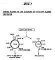
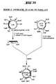
- a 5.1 kb BamHI fragment which is part of a 8 kb EcoRI fragment is subcloned in yeast vector pJDB207 and, upon transformation of yeast strain AH216, this hybrid plasmid (pJDB207/PH05,PH03, see fig. 1) elicites high phosphatase activity under derepressed (low P.-) conditions (PH05 gene) and low levels of activity in normal yeast minimal medium (expression of the PH03 gene).
- Transformants are checked for acid phosphatase activity after growth on either low P i - or normal minimal medium plates.
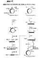
- Clones containing at least Sau3A fragments A and B (fig. 2, No. 1-4) express acid phosphatase at the same level (qualitative estimates after overlayering with acid phosphatase staining agar, as described in Example 2) as the entire 5.1kb BamHI fragment. Expression is regulated normally by the concentration of inorganic phosphate in the medium.
- Clones with Sau3A-fragment A only express low levels of acid phosphatase, which is not influenced by the inorganic phosphate concentration in the medium.
- Sau3A fragment A is sufficient for constitutive acid phosphatase (PH03) expression.
- Sau3A fragment B (fig. 2, No. 7) alone does not lead to any expression of acid phosphatase under either repressed or derepressed conditions.
- a subclone with the complete sequence between the BamHI and PstI sites shows regulated, but not constitutive synthesis of acid phosphatase. This subclone must therefore contain the yeast PH05 gene (16).
- the exact localisation of the PH05 gene is determined by DNA sequencing using the method of Maxam and Gilbert (15).
- a 623bp BamHI-Sall restriction fragment is cloned into plasmid pBR322 (see fig. 1), replacing the BamHI-SalI fragment which extends from position 375 to 650 (pBR322 nomenclature), using digestion and ligation conditions as described above (all enzymes are from New England Biolabs).
- DNA fragments of the BamHI-SalI DNA insert are asymmetrically labelled at their 5' ends at the following sites: BamHI (-54), Sau3A (-200) and SalI (+82), (for numbering see fig. 3a).
- the nucleotide sequence of the 623bp BamHI-SalI DNA insert is depicted in fig. 3a. It reveals that the insert contains the PH05 promoter region and part of the PH05 phosphatase protein coding region.
- the exact localisation of the PH03 gene is determined by DNA sequence analysis according to the manual "M13 cloning and DNA sequencing system" published by New England Biolabs.
- a 416 bp (5')PstI-RsaI(3') fragment is subcloned in vectors M13mp8 and M13mp9 (49), using unique PstI and SmaI restriction sites.
- the nucleotide sequence of the 416 bp PstI-RsaI DNA insert is shown in fig. 3b. It reveals that the insert contains the PH03 promoter region and part of the PH03 acid phosphatase protein coding sequence.
- the scheme outlined in fig. 4 requires elimination of the unique Ball restriction site in plasmid pBR322.
- 3 ⁇ g of pBR322 are digested to completion with restriction endonucleases Ball (BRL) and PvuII (Biolabs) according to the recommendations of the suppliers.
- the BalI/ PvuII double digest of pBR322 results in two restriction fragments of 3738 bp and 622 bp in size. The two fragments are separated on a 1% low melting agarose gel (Sigma) in TBE (90 mM Tris ⁇ HCl pH 8.3, 2.5 mM EDTA, 90 mM boric acid) buffer.
- the DNA bands are stained with ethidiumbromide and visualized under long wave UV light at 366 nm.
- the piece of agarose containing the 3738 bp fragment is cut out from the gel, liquified at 65°C, adjusted to 500 mM NaCl and incubated at 65°C for 20 min.
- One volume of phenol (equilibrated with 10 mM Tris ⁇ HCl pH 7.5, 1 mM EDTA, 500 mM NaCl) is added.
- the aqueous phase is reextracted twice with phenol and once with chloroform.
- the DNA is precipitated with 2.5 volumes of cold absolute ethanol and collected by centrifugation.
- the DNA pellet is washed with cold 80% ethanol and then dried in vacuum.
- the DNA is resuspended in TE at a concentration of 0.15 mg/ml.
- the isolated 3738 bp DNA fragment has two blunt ends resulting from the Ball and PvuII double digests.
- the DNA is circularized by blunt end ligation. 0.6 ⁇ g of DNA are incubated over night at room temperature in 30 ⁇ l of 60 mM Tris ⁇ HCl pH 7.5, 10 mM MgCl 2 , 10 mM DTT, 4 mM ATP, and 900 U of T4 DNA ligase (Biolabs). 5 ⁇ l aliquots of the ligation mixture are added to 50 pl of calcium treated, transformation competent E. coli HB101 cells, prepared by the method of Mandel et al. (29).
- Plasmid DNA is prepared from the cells using the procedure described in Example 2. Restriction digests with HaeIII (purchased from Biolabs, digestion conditions as suggested by supplier), PvuII and Ball of the plasmids are analyzed on a 1.52 agarose gel in TBE buffer.
- the restriction pattern and the predicted size of the newly formed junction fragment indicates that the plasmids are identical and contain all of the pBR322 sequences except for the Ball - PvuII fragment. These plasmids lack the Ball restriction site and are referred to as pBR322ABalI.
- pJDB207/PH05,PH03 contains a yeast 5.1 BamHI insert with the genes for regulated and constitutive yeast acid phosphatase (PH05 and PH03).
- pJDB207/PH05,PH03 as well as plasmid pBR322 ⁇ BalI are digested with restriction endonuclease BamHI. After complete digestion the enzyme is inactivated for 2 min at 65°C. Both DNAs are precipi- tatedby ethanol and resuspended in 10 mM Tris ⁇ HCl pH 8.0 at a concentration of 0.2 mg/ml each.
- each of the two BamHI-digested DNAs are combined and ligated in 20 ⁇ l of ligation buffer (as suggested by New England Biolabs), containing 300 U of T4 DNA ligase, for 20 hrs at 15°C.
- 5 ⁇ l aliquots of the ligation mixture are added to 50 ⁇ l of calcium-treated E. coli HB101 cells and transformation is carried out as described in Example 4a.
- the transformed E. coli cells are tested for their resistance towards ampicillin and tetracyclin.
- Eight amp R , tet S colonies are isolated and grown in 100 ml of LB medium containing 100 ⁇ g/ml of ampicillin. Plasmid DNA is isolated from the cells (see Example 2).
- Restriction digests with BamHI show that 4 plasmids contain a 5.1 kb insert besides the 3.7 kb vector fragment (pBR322ABalI).
- Restriction digests with Sail determine the orientation of the inserted 5.1 kb fragment: two plasmids have the insert oriented as shown in figure 4. One of them is referred to as p30.
- the direction of transcription of the PH05, PH03 genes in the 5.1 kb insert is anticlockwise as indicated in figure 4.
- the DNA is purified by adsorbing the DNA on a DE52 (Whatman) ion exchange column in a low salt buffer (150 mM NaCl, 10 mM Tris-HCl pH 8.0, 1 mM EDTA) and then eluting it with a high salt buffer solution (1.5 M NaCl, 10 mM Tris ⁇ HCl pH 8.0 and 1 mM EDTA).
- the DNA is precipitated with ethanol and then further digested with EcoRI (Boehringer). The 3.9 kb EcoRI-Ball restriction fragment is again separated on a preparative 0.8% low melting agarose gel,recovered as described in Example 4a and ethanol precipitated. This DNA fragment is called fragment A.
- E.coli strain HB-101 CG-pBR322/HLycIFN-8' (see Example 10E) is grown in 100 ml LB medium supplemented with 10 ⁇ g/ml tetracyclin and plasmid DNA is isolated as described in Example 2.
- HLycIFN-8 DNA are completely digested with restriction endonuclease HaeIII.
- the restriction fragments are separated on a preparative 0.8% low melting agarose gel.
- a 940 bp HaeIII fragment is cut out and eluted from the agarose gel as described in Example 4a.
- the DNA is purified on DE52 as described in Example 5a and then further digested with EcoRI.
- the 602 bp EcoRI-HaeIII fragment is again separated on a preparative 0.8% low melting agarose gel, recovered as described in Example 4a and ethanol precipitated. This DNA fragment is called fragment B.
- the two restriction fragments can be ligated enzymatically via the EcoRI sticky ends and the blunt ends of Ball and HaeIII respectively, thus creating a circular molecule with a unique EcoRI site and a Ball-HaeIII junction which is cleavable with HaeIII (but not with Ball).
- the ligation is carried out in a buffer system containing 60 mM Tris ⁇ HCl pH 7.5, 10 mM MgCl 2 , 10 mM DTT, 4 mM ATP, 300 units of T4 DNA ligase for 16 hrs at 23°C at a DNA concentration of 20 ⁇ g/ml of fragment A and 3 ⁇ g/ml of fragment B in a total volume of 10 ⁇ l
- Example 5c 2 ⁇ l aliquots of the ligation mixture (see Example 5c) are added to 50 ⁇ l of calcium-treated E. coli HB101 cells (see Example 4a). The mixtures are then plated on LB agar plates supplemented with 100 pg/ml ampicillin. The plates are incubated at 37°C for 16 hrs. About 300 ampicillin resistant colonies of E. coli HB101 are prepared. Plasmid DNA from eight ampicillin resistant colonies is isolated, analysed and their structure is determined by comparing the mobility of the restriction fragments obtained after cleavage with EcoRI and HaeIII with standard DNA [bacteriophage ⁇ DNA digested with HindIII (New England Biolabs), p30 plasmid DNA digested with HaeIII and EcoRI].
- plasmids After verification of the structure of the junctions,5 plasmids are obtained which have the correct structure.
- One of these plasmids containing the PH05 promoter linked to the 8i-interferon polypeptide coding region (see fig. 5) is called p30IFN1(8 ' 1 ).
- Example 6 Addition of replication origin and selective marker for yeast (see fig. 4)
- the 1.5 kb EcoRI restriction fragment is purified. Plasmid Yrp7 ( 4 ) is cut with EcoRI, the two fragments obtained are separated on a 0.8% agarose gel and the 1.5 kb fragment containing a yeast autonomously replicating segment and the yeast TRP1 gene is purified and isolated as described in Example 4a. Ligation is carried out (as suggested by New England Biolabs) with 20 ⁇ g/ml of EcoRI cut p30IFNl(8 ' 1 ) and 10 pg/ml of the 1.5 kb EcoRI restriction fragment from Yrp7; 100 units of T4 ligase are used.
- Plasmids containing the TRP1 yeast gene are directly selectable by transformation of the E. coli trpC mutant strain JA 194 (trpC, leuB, B l ).
- the E. coli trpC gene codes for the E. coli N-(5'-phosphoribosyl)-anthranilate isomerase.
- E. coli trpC mutants can be complemented by the yeast TRP1 gene (4). Transformation of E. coli strain JA 194 is carried out as described for E.
- coli HB101 see Example 4a except for the following modification: before plating the mixtures onto agar plates the cells are allowed to recover in 1 ml of LB medium at 37°C for 60 min; the cells are washed once with E. coli M9 minimal medium (30) and plated onto M9 minimal medium plates supplemented with vitamine Bl (1 ⁇ g/ml) and L-leucine (20 mg/ml). The plates are incubated for 2 days at 37°C. Approximately 1000 tryptophan prototrophic E. coli colonies are recovered.
- Trp colonies are purified on LB plates supplemented with 100 pg/ml ampicillin. Individual colonies are picked and plasmids are isolated as described in Example 2. Purified plasmids are analyzed by measuring the size of the restriction fragments generated after cleavage with EcoRI, HindIII, PstI and BglII (Biolabs). Two different types of plasmids are obtained which contain the 1.5 kb EcoRI restriction fragment in the two possible orientations (see fig. 4). They are named p30IFN2(8 ' 1 ) and p30IFN2'(8 ' 1 ) as indicated in figure 4.
- Plasmids p30IFN2(8i) and p30IFN2'(8 ' 1 ) are each introduced into Saccharomyces cerevisiae strain RH971 (a, trpl, leu2, his4) in analogy as described by Hinnen et al. (1).
- One ⁇ g of plasmid DNA is added to 100 ⁇ l of a spheroplast suspension and the mixture is treated with polyethylene glycole as described (1).
- the spheroplasts are mixed with 10 ml regeneration agar and plated onto yeast minimal medium plates without leucine. After incubation for 3 days at 30°C, about 1000 transformed cells are obtained.
- yeast colony from the yeast transformation plates [named Saccharomyces cerevisiae RH971/Lp30IFN2(8 ' 1 ) and /p30IFN2'(8 ' 1 ) respectively] is picked into 10 ml of yeast minimal medium in a 100 ml Erlenmeyer flask, and grown at 30°C at 200 rpm for 24 hrs to a density of about 2-3x10 7 cells/ml. The cells are washed once with 20 ml of low-P i minimal medium. Three ml of the resuspended cells are used to inoculate 300 ml low-P. minimal medium and 300 ml normal minimal medium, respectively, in 1000 ml Erlenmeyer flasks.
- Incubation is at 30°C at 160 rpm. Induction of the PH05 promoter is followed by ' measuring the appearance of acid phosphatase activity in whole cells as described by Toh-e et al. (31). The cells are grown to about 1-2x10 7 cells/ml (26-30 hrs of incubation).
- Example 8 Preparation of yeast cell extracts and determination of the interferon titer
- Cells from the 300 ml culture medium (see Example 7) at a density of 1-2x10 7 /ml are collected by centrifugation in a Sorvall GSA rotor for 5 min at 8000 rpm at 4°C. The cells are washed once with 100 ml H 2 0, resuspended in 6 ml ice cold lysis mix [0.1 M potassium phosphate buffer pH 7.4, 1% (v/v) Triton X-100,0.0001M PMSF (Merck)] and transferred to a 30 ml corex tube.
- the suspension is centrifuged again for 5 min in a Sorvall SS-34 rotor at 8000 rpm at 4°C and resuspended in 3 ml lysis mix at 0°C.
- Four g of glass beads (0.4 mm in diameter) are added to the cells and the suspension is shaken on a Vortex Mixer (Scientific Instruments Inc., USA) at full speed for 30 sec and then cooled for 1 min in an ice bath. This shaking procedure is repeated 5 to 10 times until more than 90% of the cells are broken (check under light microscope). Cell debris and glass beads are removed from the solution by centrifugation for 10 min at 8 000 rpm at 4°C in a Sorvall HB-4 rotor.
- Example 9 Insertion of the interferon gene into the high copy number yeast 2p vector pJDB 207 (see Fig. 6)
- Plasmid p30IFN2'(8') is digested with restriction endonucleases HindIII and BamHI according to the specifications of the supplier (Biolabs). Two fragments are generated of the size of 4.0 kb and 2.0 kb. The 2.0 kb restriction fragment is separated and purified by low melting agarose gel electrophoresis as described under step 4a.
- Plasmid pJDB207 (28) is digested with restriction endonucleases Hind III and BamHI. Three fragments are generated. The 6.5 kb restriction fragment is separated as above.
- 0.3 ⁇ g of the 2.0 kb fragment (containing the PH05 promoter linked to the interferon protein coding region) is ligated for 15 hrs to the 6.5 kb vector fragment in a total volume of 20 ⁇ l using 300 U T4 DNA ligase under conditions described by the supplier (Biolabs). E.coli HB101 cells are transformed and ampicillin resistant colonies are selected. The plasmid DNA is isolated and the correct structure of the isolated plasmid DNA is verified by restriction digestions using HindIII and BamHI, with p30IFN2'(8 ' 1 ) and pJDB207 digested with the same enzymes as molecular weight standards. The new plasmid obtained is called pJDB207/IFN2'(8 ' 1 ).
- Plasmid pJDB207/IFN2'(8i) is transformed into S.cerevisiae strainRH971 in analogy as described (1) selecting for leucine prototrophic colonies.
- One single leucine prototrophic yeast colony [named Saccharomyces cerevisiae RH97/LpJDB207/IFN2'(8 ' 1 )] is picked and grown as described in Example 7.
- the interferon titer is determined as described in Example 8. The results are depicted in Table 1.
- Example 10 Production of E.coli strains transformed with recombinant plasmids containing the coding regions for human lymphoblastoid interferons
- Namalwa cells are grown in culture medium RPMI 1640 containing 10% fetal calf serum at 37°C. When a cell density of 3 ⁇ 10 6 cells/ml is reached, the suspension is centrifuged at 800 x g for 10 minutes at room temperature. The collected cells are resuspended in 200 ml of culture medium containing glutamine (0.027% by volume), penicillin (200 units/ml) and streptomycin (50 ⁇ g/ml). The cells are incubated for 90 minutes at 37°C with Newcastle disease virus (NDV 110) at a ratio of 190 HAU/10 6 cells (HAU: haemagglutination units).
- NDV 110 Newcastle disease virus
- the cell density is adjusted to 1.3 ⁇ 10 6 cells/ml and the cell suspension is shaken at 34°C at 100 rpm. After 12 h, 6 ⁇ 10 9 cells are harvested and resuspended in 50 ml phosphate-buffered saline ("PBS"; 1 1 PBS contains 80 g NaCl, 2 g KC1, 14.4 g Na 2 HP0 4 and 2 g KH2P04).
- PBS phosphate-buffered saline
- 1 1 PBS contains 80 g NaCl, 2 g KC1, 14.4 g Na 2 HP0 4 and 2 g KH2P04.
- a sample is removed and the interferon activity is determined according to the procedure of Armstrong ( 32 ) using human CCL-23 cells and vesicular stomatitis virus (VSV) as the challenge virus. 4300 IFN units/ml are found.
- the cell suspension (6 ⁇ 10 9 cells in 50 ml PBS) is added at room temperature to 800 ml lysis buffer consisting of 0.05 M Tris ⁇ HCl (pH 7.5), 0.1 M NaCl, 5 mM EDTA and 2% SDS (cryst. research grade, Serva).
- the lysate is digested with 0.2 mg/ml of preincubated (2 h at 37°C) protease (Protease P, type VI, Sigma) at room temperature for 1 h while stirring the solution.
- the solution is deproteinized by extracting 3 times with 500 ml phenol satured with TNE and 5 times with 500 ml chloroform. 500 mg of nucleic acids are obtained as measured by absorbance at 260 nm.
- step Ab The slightly viscous aqueous solution obtained as described above (step Ab) is adjusted to 0.3 M NaCl and 1 g of oligo(dT) cellulose (type 7, P-L Biochemicals) is added. After stirring for 30 min at room temperature the suspension is centrifuged in 1 1 Sorvall bottles in a Sorvall RC-3 centrifuge at 4000 rpm for 10 min at room temperature and the oligo(dT) cellulose slurry is washed twice with 40 ml 2 x TNE containing 0.5% SDS. The bound poly(A) RNA is then eluted by five successive washes with 2.5 ml H 2 O. The yield is 720 ⁇ g poly(A) RNA as determined by measuring the optical density.
- RNA solution from the first adsorption is adsorbed a second time to 1 g of oligo(dT) cellulose and eluted as described above, yielding 320 ⁇ g poly(A) RNA.
- the eluates are pooled, adjusted to TNE and the poly(A) RNA is precipitated with 67% ethanol at -20°C for 10 hours.
- the RNA is collected by centrifugation at 10 000 rpm in ⁇ a Sorwall RC-5B centrifuge for 10 min at 0°C.
- the precipitate (1 mg) is redissolved in 1 ml of 1 mM EDTA.
- RNA is assayed for HulFN mRNA activity by injection into oocytes of Xenopus laevis as follows:
- the poly(A) RNA is passed through a Chelex-100 column (200-400 mesh, Bio-Rad) of 0.5 ml bed volume.
- the column is rinsed with 1 ml of 1 mM EDTA.
- the eluate (1 mg poly(A) RNA in 2 ml EDTA) is heated for 2 min at 100°C and subjected to centrifugation through a sucrose density gradient ( 6 14 ml sucrose solutions increasing in sucrose concentration from 5% to 23% (m/v) and containing 50 mM Tris ⁇ HCl [pH 7.5], 0.2 M NaCl and 1 mM EDTA).
- the centrifugation is carried out in a TST 41 rotor (Kontron AG) at 35 000 rpm for 16 h at 5°C.
- 0.3 ml fractions are collected with an ISCO gradient collector. 2 volumes of ethanol are added to each fraction and the solution is allowed to stand for 10 h at -20°C.
- the precipitated mRNA is collected by centrifugation (Sorvall, HB-4 rotor at 0°C, 10 000 rpm for 10 min).
- the precipitate of each fraction is redissolved in 25 ⁇ l of 1 mM EDTA and each fraction is assayed for human IFN mRNA activity as described above (step Ac), except that only. 10 oocytes are injected per RNA sample instead of 20.
- step Ac The results are given in table 2.
- fractions 23-29 are pooled and the poly(A) RNA is purified further as follows:
- the poly(A) RNA is dissolved in 100 ⁇ l1 of 0.5 mM EDTA.
- the yield is 40 ⁇ g as determined by measuring the optical density.
- poly(A) RNA is assayed for human IFN activity as described above by using 20 oocytes per assay.
- the poly(A) RNA preparation has a specific activity of 8100 IU interferon per ⁇ g RNA.
- Poly(A) RNA enriched for HuIFN mRNA (see step Ad) is used as a template to prepare double-stranded cDNA essentially as described by Efstratiadis et al. (36), Maniatis et al. (37) and Hoeijmakers et al. (38).
- reaction mixture containing 40 mM Tris ⁇ HCl (pH 7.5), 30 mM NaCl, 5 mM MgCl 2 , 0.5 mM DTT (Calbiochem.), 1 mM dGTP, dCTP, dTTP (P-L Biochemicals) and 1 mM 32 P-dATP (Amersham, specific activity 50 000 cpm /nmole ), 20 ⁇ g/ml oligo(dT) 12-18 (P-L Biochemicals), 40 ⁇ g/ml poly(A) RNA and 100 units of avian myeloblastosis virus (AMV) reverse transcriptase (Life Sciences, Inc., St.
- AMV avian myeloblastosis virus
- the precipitate is dissolved in 95 ⁇ l of H 2 0. 5 ⁇ l of ION NaOH is added and the mixture is incubated at 25°C for 40 min. After neutralization with 5M acetic acid, 50 ⁇ l H 2 0 and 2 volumes of ethanol are added and the sample is stored at -20°C for 10 hrs. The precipitate is collected by centrifugation as described before and redissolved in 200 ⁇ l of 0.1 mM EDTA. The yield of single-stranded cDNA is 3.7 ⁇ g.
- the size of the cDNA is 700-1500 nucleotides in length, as determined from its electrophoretic mobility in a 6% polyacrylamide gel in Tris-borate-EDTA (108 g of Tris, 9.3 g of disodium EDTA, and 55 g of boric acid per. one 1 solution of pH 8.3) containing 7 M urea relative to marker DNAs of known length (39).
- the obtained cDNA solution is heated at 100°C for 90 sec, chilled and incubated in a 400 ⁇ l reaction mixture comprising 0.1 M potassium phosphate buffer (pH 6.9), 10 mM MgCl 2 , 10 mM DTT (Calbiochem), 1 mM dATP, 1 mM dCTP, 1 mM dTTP (P-L, Biochemicals), 1 mM 3 H-dGTP (Amersham, specific activity 94 000 cpm/nmole) and 165 units/ml of E.coli DNA polymerase I (Biolabs, New England) for 8 h at 15°C.
- the reaction is terminated by adding EDTA and SDS to final concentrations of 10 mM and 0.1%, respectively.
- the mixture is extracted with phenol and chloroform, chromatographed over Sephadex G-50 (Pharmacia, fine, 2 ml bed volume) and ethanol precipitated as described above (step Ba).
- the resulting DNA is treated in a 50 ⁇ l incubation mixture containing 0.25 M NaCl, 50 mM sodium acetate (pH 4.5) and 1 mM ZnSO 4 with 6 units of S 1 endonuclease (P-L Biochemicals) at 37°C for 30 min.
- the reaction is stopped with 0.1 % SDS and 10 mM EDTA.
- the reaction mixture is deproteinized with 1 volume of phenol (saturated in 50 mM sodium acetate, pH 4.5) and chloroform.
- the aqueous phase is chromatographed on a 2 ml Sephadex G-50 (Pharmacia, fine) column in TNE. 100 ⁇ l fractions are collected and the Cerenkov radiation of each fraction is determined.
- the excluded fractions are pooled and the DNA is precipitated with 2 volumes of ethanol at -20°C for 10 h as described above.
- the precipitate is centrifuged in a HB-4 rotor (see above) and the collected precipitate is dissolved in a 100 ⁇ l solution containing 10 mH Tris ⁇ HCl (pH 7.5) and 0.5 mM EDTA. 4 ⁇ g of DNA are obtained.
- the DNA is fractionated through a sucrose density gradient (5-23 %) in 50 mM Tris-HCl (pH 7.5) and 1 mM EDTA in a TST-60 rotor (Kontron AG). Centrifugation is carried out at 55 000 rpm for 5 h at 15°C.
- the DNA which sediments faster than a 800 base pair marker DNA, run in a parallel gradient, is pooled, adjusted to TNE and precipitated with 67 % ethanol at -20°C for 10 hrs. 0.4 ⁇ g double-stranded cDNA are obtained.
- the 3'-termini of 0.1 ⁇ g of the obtained ds cDNA are provided with poly(dC) tails in a 10 ⁇ l reaction volume containing 100 mM sodium cacodylate (pH 7.2), 2.5 mM CoCl2, 50 ⁇ g BSA (Calbiochem.) per ml, 1 mM dCTP and 10 units of terminal deoxynucleotidyl transferase (P-L Biochemicals) per ⁇ g of ds cDNA. After incubation (20 min at 27°C), EDTA is added to 10 mM and the sample is stored at -20°C until use.
- 10 ⁇ g of pBR 322 plasmid DNA is digested with 10 units of Pst I endonuclease (Biolabs) in a 100 ⁇ l solution containing 50 mM NaCl, 6 mM Tris ⁇ HCl (pH 7.5), 6 mM MgCl 2 , 6 mM 2-mercaptoethanol and 100 ⁇ g/ml gelatine for 1 h at 37°C.
- the solution is extracted with 1 volume of phenol and chloroform.
- the solution is adjusted to TNE and the linearized DNA is precipitated with 2 volumes of ethanol at -20°C for 5 h.
- the linearized plasmid DNA is elongated with dGMP in a 200 ⁇ l reaction volume containing 100 mM sodium cacodylate (pH 7.2), 5 mM MgCl 2 , 20 mM haH 2 PO 4 , 50 ⁇ g BSA per ml, 1 mM dGTP and 100 units of terminal deoxynucleotidyl transferase (P-L Biochemicals). After incubation for 20 min at 37°C, EDTA is added to 10 mM and the reaction mixture is frozen at -20°C until use.
- a mixture of dCMP-elondated double-stranded cDNA (0.1 ⁇ g) and dGMP- tailed linearized pBR 322 (0.5 ⁇ g) in 500 ⁇ l TNE buffer is incubated at 65°C for one hour, at 46°C for one hour, at 37°C for one hour and at 20°C for one hour.
- the solution containing the pBR 322-linked cDNA is put on ice and used immediately for transformation.
- step Cc 10 ⁇ l of the reaction mixture containing the annealed pBR 322 hybrid plasmid DNAs prepared as described above (step Cc) are added to a mixture containing 150 ⁇ l calcium-treated E. coli HB 101 in 10 mM MgCl 2 , 10 mM CaCl 2 and 10 mM Tris ⁇ HCl (pH 7.5) in a total volume of 200 ⁇ l.
- the mixture is cooled in ice for 20 min, heated to 42°C for 1 min and incubated at 20°C for 10 min.
- 1 ml of tryptone medium tryptone medium contains 10 g Bacto-Trypton (Difco); 1 g yeast extract (Difco); 1 g glucose; 8 g NaCl and 294 mg CaCl 2 ⁇ 2 H 2 0 in 1 1 of distilled water
- tryptone medium contains 10 g Bacto-Trypton (Difco); 1 g yeast extract (Difco); 1 g glucose; 8 g NaCl and 294 mg CaCl 2 ⁇ 2 H 2 0 in 1 1 of distilled water
- the mixture is plated onto 2 agar plates (Mc Conkey agar, Difco; 0,6 ml/plate) supplemented with 10 ⁇ g/ml of tetracycline (Sigma).
- the plates are incubated at 37°C for 12-17 hrs. About 5600 tetracycline resistant colonies
- Anoligodeoxynucleotide complementary to a stretch of 13 nucleotides which both HuIFN-a l and HuIFH- ⁇ mRNA share in common is chemically synthesized by the phosphotriester method (cf. Itakura et al. (40), de Rooij et al (41)). The individual steps of the synthesis are outlined in figure 8.
- the starting materials indicated in line 1 of figure 8 are known from the literature.
- the protective groups are split off by the methods described by Itakura et al.: the deblocking of 5'-monomethoxytrityl (M) or dimethoxytrityl (D) substituted hydroxyl groups is performed with acetic acid (80%) at room temperature, and the P-cyanoethyl phosphate groups are cleaved with 0.1 N sodium hydroxide in dioxane-water (4:1) at room temperature. The condensation of the building blocks is accomplished by using triisopropylbenzenesulfonyl chloride as an activating agent to afford oligodeoxynucleotides up to the fully protected 13-mer primer represented in line 7 of figure 8. The last step (complete removal of all protective groups) is achieved in the following manner:
- the solution is extracted with 1 volume of phenol (saturated in TNE) and the nucleic acids are precipitated with 2 volumes of ethanol at -20°C for 10 h.
- the precipitate is collected by centrifugation (HB-4 rotor, 20 min, 10 000 rpm, 0°C) and dissolved in 20 ⁇ l dye mix containing 90% (v/v) formamide (Merck, pro analysis), 1 mM EDTA, 0.05% bromo.phenol blue and 0.05% xylene cyanol blue.
- the sample is heated at 90°C for 2 min and applied on a 5% polyacrylamide gel in Tris-borate-EDTA(cf.)Peacock et al. (39).
- a single band is visible on the autoradiogram which migrates between the 267 bp and 435 bp 32 P-labeled marker DNA fragments obtained from the Hae III digest of the plasmid pBR 322.
- the 32 P-labeled cDNA fragment is extracted from the gel and purified as described by Mueller et al. (42). 20 000 Cerenkov cpm of the 32 P-labeled human IFN-a and IFN- ⁇ specific cDNA probe are obtained.
- step D 1650 of the transformant colonies prepared as described above (step D) are transferred to nitrocellulose filters BA 85 (Schleicher & Schuell, 8 cm diameter).
- the cells are lysed and their DNA is denatured and fixed to the filters in situ, according to Grunstein and Hogness (20).
- the filters bearing the colonies are prehybridized in 4 x SET (a solution containing 0.15 M NaCl, 30 mM Tris ⁇ HCl (pH 8.0), 1 mM EDTA) 0.1% (w/v) Ficoll 400 (Pharmacia), 0.1% (w/v) polyvinylpyrrolidone (PVP-360,Sigma), 0.1% (v/v) BSA, 0.5% SDS, 50 ⁇ g/ml denatured calf-thymus DNA (prepared as follows: 5 mg calf-thymus DNA (type I, Sigma) is boiled for 10 min in 0.5 M NaOH to shear the DNA, neutralized with 5 M acetic acid and precipitated with 2 volumes of ethanol at -20°C.
- the precipitate is collected by centrifugation in a HB-4 rotor for 10 min at 0°C and redissolved in 500 ⁇ 1 0.5 mM EDTA) at 65°C for 4h using 20 ml mixtures per filter and hybridized with 10 3 Cerenkov cpm of the 32 P-labeled probe per nitrocellulose filter in 5 x SET, 0.027 (w/v) Ficoll, 0.01% polyvinylpyrrolidone, 0.02% (v/v) BSA, 0.2% SDS and 50 ⁇ g/ml denatured calf-thymus DNA.
- the hybridization is performed at 65°C for 36 h.
- the filters are rinsed once in chloroform, twice in SET,0.5% SDS at room temperature and twice in SET, 0.57. SDS for 1 h at 60°C and once with 3 mM Trizma base at room temperature for 1 h.
- the filters are dried by blotting on 3 MM-paper (Whatman), and an X-ray film (Fuji) is exposed to the filters using a screen (Ilford intensifying screen) at -80°C for 72 h.
- the hybrid plasmid DNAs are isolated from the 9 positively hybridizing clones and used to retransform E. coli HB 101 as described before.
- the hybrid plasmid DNA is isolated as follows: 1 colony is used to inoculate 10 ml of tryptone medium, supplemented with 10 ⁇ g/ml of tetracycline as above in a 25 ml Erlenmeyer flask. The culture is shaken for 15-18 hrs at 37°C at 300 rpm. The cells are harvested by centrifugation (Sorvall, HS-4 rotor, 10 min at 4000 rpm, 4°C). About 0.1 g of cells are obtained and are resuspended in l.ml 50 mM Tris ⁇ HCl (pH 8.0).
- lysozyme solution (10 mg/ml in 50 mM Tris ⁇ HCl (pH 8.0),lysozyme is purchased from Sigma) ,are added and after incubation at 0°C for 10 min, 0.15 ml of 0.5 M EDTA (pH 7.5) is added. After another 10 min at 0°C, 60 ⁇ l of 2% Triton X-100 (Merck) is added. After 30 min at 0°C, the sample is centrifuged for 30 min at 15 000 rpm and 4°C in a Sorvall SA-600 rotor. The supernatant is deproteinized with 1 volume of phenol (saturated in TNE).
- the phases are separated by centrifugation (Sorvall HB-4 rotor) for 10 min at 5000 rpm at 4°C.
- the upper phase is extracted twice with 1 volume of chloroform.
- Pancreatic R NAse A (Sigma; 10 mg/ml in TNE, preheated 10 min at 85°C) is added to a final concentration of 25 ⁇ g/ml and the mixture is incubated for 40 min at 37°C.
- the solution is then adjusted to 1 M NaCl and 10% polyethylene glycol 6000 (Fluka, autoclaved for 20 min at 120°C) and incubated at -10°C for 2 hrs.
- the precipitate is collected in a Sorvall HB-4 rotor (20 min at 10 000 rpm, 0°C) and redissolved in 100 ⁇ 1 of TNE.
- the DNA solution is extracted with 1 volume of phenol and the DNA is precipitated with 2 volumes of ethanol at -80°C for 10 min.
- the precipitate is collected by ccntrifugation in an Eppcndorf centrifuge and the DNA is redissolved in 20 ⁇ 1 of 10 mM Tris ⁇ HCl (pll 7.5) and 0.5 mM EDTA. 8-10 ⁇ g of hybrid plasmid DNA are recovered from a 10 ml culture.
- E. coli HB 101 is transformed with each of the nine isolated hybrid DNAs and the transformed cells are plated on agar plates containing tetracycline, as described before (step D). From each transformation, 3 tetracycline resistant clones are picked, 10 ml cultures are prepared and the hybrid DNAs are isolated from the cultures as described before.
- One of the recloned recombinant DNA molecules gives 2 bands, one with the mobility of Pst I-cleaved pBR 322, the other with a mobility corresponding to about 1000 bp. It is denoted CG-pBR 322/HLycIFN-l'b.
- Another recombinant DNA gives 3 bands, one with the mobility of Pst I-cleaved pBR 322, one with a mobility of about 600 bp and one with a mobility of about 150 bp.
- the recombinant DNA molecule in this clone is designated CG-pBR 322/HLycIFN- ⁇ 1 .
- the recombinant plasmid DNAs of the clones CG-pBR 322/HLycIFN-l'b and CG-pBR 322/HLycIFN- ⁇ 1 are isolated from the cultures as described above (step Ec) and characterized by establishing the nucleotide sequence of the cDNA insert using the method described by Maxam and Gilbert (15). Basically, the following approach is used:
- the labeled DNA fragments are cleaved with a second restriction endonuclease and the products are separated by electrophoresis through a 6%, 8% or 10% polyacrylamide gel in Tris-borate-EDTA buffer.
- the DNA fragments are extracted from the gel and purified as described by Mueller et al. (42).
- the DNA fragments are chemically degraded and the products are separated by polyacrylamide gel electrophoresis as described by Maxam and Gilbert (15).
- the isolated plasmid DNAs of the clone CG-pBR 322/ HLycIFN-1'b are treated as follows.
- 5 ⁇ g of the plasmid DNA is digested with Bgl II, 5' terminally labeled, and cleaved with Pvu II.
- the Pvu II-Bgl II * (*indicates the labeled site) and Bgl II- Pvu II* DNA fragments are isolated on a 6% polyacrylamide )gel.
- 5 pg of the plasmid is digested with Alu I, 5'-terminally labeled, and cleaved with Pst I.
- the Pst I - Alu I * DNA fragment is isolated on a 8% polyacrylamide gel. The individual fragments are subsequently degraded and sequenced according to Maxam and Gilbert. The nucleotide sequence obtained is depicted in figure 10. A stretch of about 25-35 deoxyguanosine residues is preceding at the 5'-end of the cDNA insert. The nucleotide sequence shown is somewhat similar to that of IFN-a (type F) cDNA described by Goeddel et al. [(43), cf. also Weissmann(44)], nevertheless displaying a lot of distinct deviations (point mutations) some of which are affecting the resulting amino acids (cf. fig.10).
- the isolated plasmid DNA of the clone CG-pBR 322/HLycIFN- ⁇ 1 is treated in a similar manner. 5 ⁇ g of the plasmid is digested with Pvu II and 5'-terminally labeled. One half of the mixture is cleaved with Pst I, and the rest with Bgl II. The Pst I-Pvu II * and Bgl II-Pvu II* fragments are isolated by electrophoresis on a 6Z polyacrylamide gel and degraded as mentioned above.
- the nucleotide sequence (N-terminal sequence) is depicted in figure lland reveals that the cDNA insert starts at nucleotide number 102 of the IFN- ⁇ 1 cDNA as described by Taniguchi et al. (45). Therefore, the cDNA insert has the capacity to code for human IFN- ⁇ 1 lacking 11 amino acids at the N-terminus.
- the cDNA insert is flanked at its 5'end by a stretch of about 20-25 deoxyguanosine residues and shows a point mutation at position 153, converting a C to a T residue without affecting the resulting amino acid.
- the recombinant plasmid DNAs of the clones CG-pBR 322/HLycIFN-l'b and CG-pBR 322/HLycIFN- ⁇ 1 are isolated from the cultures as described above (step Ec).
- the CG-pBR 322/HLycIFN-l'b plasmid DNA (5 ⁇ g) is digested with Bgl II, 5' terminally labeled, and cleaved with Pvu II.
- the isolated CG-pBR 322/HLycIFN- ⁇ 1 plasmid DNA (5 ⁇ g) is digested with Pvu II, 5'-terminally labeled, and cleaved with Bgl II.
- the Pvu II-Bgl II * (351 bp) DNA fragment (probe A) and the Pvu II *- Bgi II (368 bp) DNA fragment (probe B) are isolated from a 8% polyacrylamide gel as described above (step Ed) and used for in situ colony hybridization (see below).
- the restriction of the plasmid DNAs, the labeling, and the purification of the DNA fragments are accomplished in the same manner as described above (step Ed).
- step D 4000 of the transformant colonies prepared as described above (step D) are transferred to nitrocellulose filters BA 85 (Schleicher & Schuell, 8 cm diameter). The cells are lysed and their DNA is denatured and fixed to the filters in situ, according to Grunstein and Hogness (20). Hybridizations to the probes A and B (both probes are mixed) are performed as described before (step Ec). 6 positive colonies are identified by autoradiography, 3 of which, designated
- nucleotide sequences of the cDNA inserts are established by using the general approach as described above (step Ed).
- the isolated plasmid DNAs CG-pBR 322/HLycIFN-4 1 . and CG-pBR 322/HLycIFN-8 1 are each digested with Pvu II, 5'-terminally labeled and cleaved with Pst I.
- the DNA fragments are fractionated on a 8% polyacrylamide gel and the Pst I-Pvu II * ( ⁇ 120 bp) from 8 1 DNA and Pst I-Pvu II* (82 bp) from 4 DNA are isolated as usual.
- the isolated plasmid DNA CG-pBR 322/HLycIFN-5 1 is treated as follows.
- 5 ⁇ g of the plasmid DNA is digested with Hae III, 5'-terminally labeled and cleaved with Pst I.
- the Pst I-Hae III* (57 bp) DNA fragment is isolated on a 10% polyacrylamide gel.
- 5 ⁇ g of the plasmid is digested with EcoR I, 5'-terminally labeled and cleaved with Pst I.
- the Pst I-EcoR I * (235 bp) and EcoR I*-Pst I ( ⁇ 700 bp) DNA fragments are isolated on a 8% polyacrylamide gel.
- the various DNA fragments are subjected to sequence analysis according to Maxam and Gilbert (15).
- nucleotide sequences of the cDNA inserts are depicted in figures 12-14.
- figure 12 a partial nucleotide sequence of the cDNA insert of CG-pBR 322/HLycIFN-4 1 is shown.
- the insert is flanked at the 5' end by a stretch of 23 deoxyguanosine residues and comprises part of the IFN-a 2 (Le) cDNA described by Streuli et al. (46).In the 3'-extracistronic region, there are some minor deviations (point mutations) and a stretch of additional 318 nucleotides.
- the nucleotide sequence of the cDNA insert of CG-pBR 322/HLycIFN-8 1 is depicted in figure 13.
- the insert is flanked at the 5' end by a stretch of 20-23 deoxyguanosine residues and is similar but not identical, to the IFN- ⁇ (type D) cDNA described by Goeddel et al. [(43); cf. also Mantei et al. (27)] .
- the IFN gene contains at positions 28-30 a GCC triplet and at positions 409-411 a GCG triplet coding for alanine instead of GTC and GTG, respectively, coding for valine.
- nucleotide sequence of the cDNA insert of CG-pBR 322/HLyc IFN-5 1 (see figure 14)reveals a stretch of 17 deoxyguanosine residues at the 5' end.
- the nucleotide sequence is related to that of IFN-a (type B) cDNA described by Goeddel et al. (43).
- nucleotides at the 5' end of the cDNA insert of HLycIFN-5 point mutations, excisions and insertions in the extracistronic region and in the IFN coding sequence, especially at positions 22 and 361-372, are evident as well.
- clones exhibiting no measurable IFN activities contain recombinant DNAs in which the HuLyIFN-cDNA insert is in an improper orientation in regard to the direction of transcription. Therefore, the recombinant DNA of one such clone (CG-pBR 322/HLycIFN-1'b) containing a full length cDNA insert is reoriented as follows:
- the recombinant plasmid DNA (150 ⁇ g) of the clone E. coli HB 101 CG-pBR 322/HLycIFN-1'b is cleaved with Pst I (Biolabs) using standard procedures (cf. step Ed). Following phenol extraction and ethanol precipitation, the excised insert is isolated by means of sucrose density gradient centrifugation (5-23%) in 50 mM Tris-HCl (pH 8.0) and 1 mM EDTA. The centrifugation is performed at 35 000 rpm in a TST 41 rotor (Kontron AG) at 15°C for 16 h. 0.3 ml fractions are collected with an ISCO gradient collector at 1 ml/min.
- the fractions containing the small fragment are pooled.
- the DNA is precipitated with ethanol as usual, and the precipitate is collected by centrifugation in a HB-4 rotor (Sorvall) at 10 000 rpm at 0°C for 10 min.
- the precipitate is redissolved in 60 ⁇ l 10 mM Tris HCl (pH 7.5) and 0.05 mM EDTA. 30 ⁇ g DNA are recovered as determined by measuring the optical density.
- the insert DNA (10 ⁇ g) is digested with Hae III (Biolabs) and the fragments are fractionated on a 2% agarose gel in a solution containing 50 mM Tris, 50 mM boric acid, 1 mM EDTA and 0.5 ⁇ g/ml ethidium bromide.
- FIG 15, fragments 3 and 4, respectively) are each excised from the gel, squirted through a thin needle with a syringe into 5 ml of 0.15 M NaCl, 50 mM Tris ⁇ HCl (pH 8.0), 1 mM EDTA, and eluted overnight by shaking.
- the eluate is passed through a 100 ⁇ l DE-52 (Whatman) Pasteur-pipette column to adsorb the DNA.
- the column is washed with 2 ml of the same buffer and the DNA is eluted with 400 ⁇ l of a solution containing 1.5 M NaCl, 50 mM Tris (pH 8.0) and 1 mM EDTA.
- the DNA is precipitated with 2 volumes of ethanol at -20°C overnight. The precipitate is collected by centrifugation in an Eppendorf centrifuge.
- the Hae III-Hae III DNA fragment (82 bp) is redissolved and digested with Sau 3A (Biolabs). The enzyme is heat-inactivated at 65°C for 30 min. 1 ⁇ g of the Hae III-Pst I DNA fragment (869 bp) is added, the solution is adjusted to 10 mM MgCl 2 , 10 mM DTT and 0.5 mM ATP, and T4 DNA ligase (Biolabs) is added to 30 units/ ⁇ l reaction volume. The solution is incubated for 10 h at 15°C. Following extraction with phenol and chloroform, the mixture is fractionated on a 2% agarose gel in Tris-borate-EDTA in the presence of ethidium bromide.
- the Sau 3A-Pst I DNA fragment (cf. figure 15,fragment 5) is extracted as described before, precipitated with ethanol and redissolved in 10 ⁇ l of a solution containing 10 mM Tris ⁇ HCl (pH 7.5) and 0.05 mM EDTA.
- the plasmid pBR 322 is cleaved with Pst I (cf. step Cb) and treated with 4 units/ml of the exonuclease Bal 31 (Bethesda Research Lab.) at 30°C for 4-10 min to remove the ⁇ -lactamase coding segment.
- step Ea The linker is added to the Bal 31 treated pBR 322 DNA using conventional means.
- the resulting hybrid molecule is cleaved with the restriction endonucleases Bcl I (Biolabs) and EcoR I:
- Bcl I Biolabs
- EcoR I EcoR I
- the digestion products are fractionated on a 8% polyacrylamide gel in Tris-borate-EDTA as described before (step Ba).
- DNA fragments (ApPr DNA fragments), migrating between 184 bp and 234 bp marker DNAs, are isolated as described above (step I a), and precipitated with ethanol as usual.
- the precipitate is redissolved in a solution containing 10 mM Tris ⁇ HCI (pH 7.5) and 0.05 mM EDTA.
- the solutions containing the ApPr DNA fragments and the cDNA insert are pooled.
- the mixture is adjusted to 10 mM MgCl 2 , 10 mM DTT and 0.5 mM ATP, and incubated with 30 units/ ⁇ l T4 DNA ligase (Biolabs) at 15°C for 12 h.
- T4 DNA ligase Biolabs
- the mixture is fractionated on a 1% low melting agarose gel (Biorad).
- the obtained ApPr-cDNA fragment is joined to the large fragment of pBR 322 cleaved with both Pst I (Biolabs) and EcoR I (Biolabs) in the following manner.
- the gel piece, containing the ApPr cDNA fragment (about 20 ⁇ l) is mixed with the Pst I-EcoR I fragment of pBR 322, melted at 65°C for 2 min,cooled to 37°C, adjusted to 0.5 mM ATP, 10 mM DTT and 10 mM MgCl 2 , and incubated with 30 units/ ⁇ l of T4 DNA ligase (Biolabs) for 12 h at 15°C.
- the recombinant plasmid DNA of the clone CG-pBR (AP)/LyIFN- ⁇ -1 is isolated from the culture as described above (step 3c) and characterized by establishing the nucleotide sequence of the cDNA insert (IFN gene) and the ⁇ -lactamase regulatory region.
- IFN gene the nucleotide sequence of the cDNA insert
- the IFN specific protein yields of the clone E. coli HB 101 CG-pBR 322/ HLycIFN-8 1 is improved as follows (cf. fig. 17):
- CG ⁇ pBR (An)/LyIFN- ⁇ -1 DNA (100 ⁇ g) is cleaved with Hind III (Biolabs) and Bgl II (Biolabs).
- the excised DNA fragment is isolated by means of sucrose density gradient centrifugation (5-23%) in 50 mM Tris ⁇ HCl (pH 8.0) and 1 mM EDTA. The centrifugation is performed at 58 000 rpm in a TST 60 rotor (Kontron AG) at 15°C for 4 hours. 0.2 ml fractions are collected as described before. The fractions containing the small fragment (Hind III-Bgl II) are pooled and the DNA is precipitated with ethanol as usual. The precipitate is redissolved in 80 p1 10 mM Tris ⁇ HCl (pH 7.5) and 0.05 mM EDTA. 16 ⁇ g DNA are recovered as determined by measuring the optical density.
- the DNA fragment (Hind III-Bgl II) (4 ⁇ g) is cleaved with Sau 3A (Biolabs) and the digestion products are fractionated on a 6% polyacrylamide gel in Tris-borate-EDTA as described before.
- the DNA fragments are stained in EtBr (0.5 pg/ml), the Hind III-Sau 3A DNA fragment (239 bp) is extracted and isolated as before.
- the DNA is precipitated with ethanol as usual. The precipitate is redissolved in 20 ⁇ l 10 mM Tris ⁇ HC1 (pH 7.5) and 0.05 mM EDTA.
- the cDNA insert is excised from the recombinant plasmid CG-pBR 322/ HLycIFN-8 1 as described above (section Ia).
- the cDNA insert (2 ⁇ g) is digested with 2.5 units of Sau 3A (Biolabs) in 10 ⁇ g/ml EtBr and incubated at 37°C for 60 min.
- the digests are phenol extracted and the DNA is precipitated in ethanol as above.
- the DNA fragments are fractionated on a 1.2% agarose gel in a solution containing 50 mM Tris, 50 mM boric acid, 1mM EDTA and 0.5 ⁇ g/ml ethidium bromide.
- the second largest DNA fragment (Sau 3A-PstI: 693 bp) is extracted from the gel and purified as described in section Ia).
- the DNA is redissolved in 20 ⁇ l 10 mM Tris-HCl (pH 7.5) and 0.05 mM EDTA .
- Equal amounts of both DNA fragments are incubated in a solution containing 10 mM MgCl 2 , 10 mM DTT, 0.5 mM ATP and 30 units/ ⁇ l T4 DNA ligase (Biolabs) at 15°C for 3 hours. The mixture is incubated for 15 min. at 80°C and adjusted to 50 mM NaCl. The DNA mixture is digested with 0.5 units PstI (Biolabs) and 1 unit Hind III (Biolabs) for 20 min. at 37°C. The DNA is phenol extracted, ethanol precipitated and redissolved in 20 ⁇ l 10 mM Tris-HCI (pH 7.5) and 0.05 mM EDTA.
- One half of the resulting mixture is ligated to the large Hind III-PstI DNA fragment of the plasmid pBR 322 ( ⁇ 100 ng) in 10 mM MgCl 2 , 10 mM DTT, 0.5 mM ATP containing 30 units/ ⁇ l of T4 DNA ligase (Biolabs) for 2h at 15°C.
- One tenth volume of the solution is used to transform E. coli HB 101 as described in step D).
- the transformed colonies are used to test for IFN activity as described earlier (cf. step F ).
- the clone synthesizing the highest level of IFN activity is selected and designated E. coli HB 101 CG-pBR (AP)/LyIFN-a-3.
- the IFN activity is determined as described above (step F).
- An activity of 70000 (IU/ml) is found which represents a 700 fold stimulation compared to the original clone E. coli HB 101 CG-pBR 322/HlycIFN-8 1 .
- the recombinant plasmid DNA of the clone CG-pBR (AP)/LyIFN-a-3 is isolated from the culture as described above (step Cc) and characterized by establishing the nucleotide sequence of the cDNA insert (IFN gene) and the ⁇ -lactamase regulatory region.
- IFN gene the nucleotide sequence of the cDNA insert
- the construction protocol for the plasmid CG-pBR (AP)/LyIFN- ⁇ -3 can be used for all a-IFN cDNA genes or appropriately cut chromosomal ⁇ -IFN genes in general.
- the plasmid CG-pBR (AP)/LyIFN- ⁇ -2 is obtained in an identical manner as described for the plasmid CG-pBR(AP)/LyIFN-a-3.
- This new plasmid contains the DNA insert of CG-pBR 322/HLycIFN-5 1 and the ⁇ -lactamase regulatory region from CG-pBR (AP)/LyIFN- ⁇ -1.
- a clone designated E. coli HB 101 CG-pBR (AP)/LyIFN-a-2 is selected as described above.
- IFN activity of 50000 (IU/ml) is found which represents a 5 fold stimulation compared to the original E. coli HB 101CG-pBR 322/HLycIFN-5 1 .
- the nucleotide sequence of the cDNA insert and the ⁇ -lactamase regulatory region of the plasmid CG-pBR (AP)/LyIFN-a-2 is established as described above and depicted in fig. 19.
- Micro-organisms and recombinant DNA molecules prepared as described in Example 10 are exemplified by cultures deposited in the culture collection of the Agricultural Research Culture Collection (NRRL) on September 14, 1981 and are assigned the following accession numbers:
- Example 11 The lymphoblastoid IFN-1'b and IFN-5 1 coding sequences of the E. coli plasmids CG-pBR322/HLycIFN-l'b and -5 1 (cf. Example 10) can be subcloned in plasmid p30 (cf. Example 4) in an analogous manner as described for IFN-8 1 in Examples 5 and 6. A partial digestion with restriction endonuclease HaeIII is required.
- the yeast hybrid plasmids obtained in this way are p30IFN2(l'b), p30IFN2'(1'b), p30IFN2(5 1 ) and p30IFN2'(5 1 ).
- hybrid plasmids can be used to transform Saccharomyces cerevisiae RH971 as described in Example 7.
- the following colonies containing a hybrid plasmid with a lymphoblastoid IFN cDNA insert can be selected:
- Example 12 In an analogous manner as described in Example 9, the following yeast hybrid plasmids can be obtained starting from the plasmids p30IFN2(1'b), -(5 1 ), (8 ' 1 ) and p30IFN2'(1'b) -(5 1 ), respectively:
- hybrid plasmids can be transformed into S. cerevisiae strain RH971 selecting for leucine prototrophic colonies.
- the following colonies containing a hybrid plasmid with a lymphoblastoid IFN cDNA insert can be obtained:
- Example 13 Construction of an expression plasmid containing the PH05 promoter and PHO5 transcription termination signals (see fig. 20)
- the PH05 transcript has been mapped by Sl nuclease mapping (48).
- the signals for transcription termination have been shown to be located in a 0.37 kb Sau3A-PstI fragment of the PH05 gene.
- the nucleotide sequence of the Sau3A-PstI fragment is given in fig. 21.
- pJDB207/PH05,PH03 DNA Five ⁇ g of pJDB207/PH05,PH03 DNA (cf. Example 2) are digested to completion with restriction endonucleases Sau3A and Pstl. The restriction fragments are separated on a vertical 1.5% low melting agarose gel in TBE buffer. The 0.37 kb Sau3A-PstI fragment is localized by ethidiumbromide staining and a gel block as small as possible is cut out containing this DNA fragment.
- M13mp9 phage DNA is a useful cloning vector with a cluster of unique restriction sites (49). Five ⁇ g of M13mp9 DNA are digested to completion with restriction endonucleases BamHI and PstI. The larger 7.2 kb DNA fragment is separated from a very small fragment (8 bp) on a 0.8% low melting agarose gel. The gel block containing the large DNA fragment is cut out of the gel. Gel blocks with the 0.3% kb Sau3A-PstI fragment of pJDB207/PH05,PH03 (cf.
- Example 13b) and the 7.2 kb BamHI-PstI fragment of M13mp9 are liquefied at 65°C, mixed in about equimolar amounts and diluted with H 2 0 to lower the agarose concentration to 0.3%.
- Ligation is carried out in a 200 ⁇ l solution containing 60 mM Tris.HCI pH 7.5, 10 mM MgCl 2 10 mM DTT, 1 mM ATP and 600 units of T4 DNA ligase (Biolabs).
- Transduction of competent cells of the strain E. coli JM101 (Ca++) is done according to the manual "M13 cloning and DNA sequencing system" published by New England Biolabs. Phages from a number of white plaques are grown and analyzed for the size of their DNA insert by cleavage with restriction endonucleases EcoRI and Pstl.
- M13mp9 derived clone containing the Sau3A-PstI PH05 transcription termination fragment is isolated and referred to as M13mp9/PHO5 (Sau3A-PstI).
- M13mp9/PH05(Sau3A-PstI) DNA is cleaved to completion with restriction endonucleases HaeIII and HindIII.
- the resulting two DNA fragments are separated on a 1.5% vertical low melting agarose gel in TBE buffer.
- the 0.39 kb fragment is isolated in a gel block cut out of the gel.
- p30/EcoRI R DNA is digested with Ball and HindIII.
- the large 3.98 kb fragment is separated on a 0.8X low melting agarose gel in TBE buffer and isolated by cutting a gel block containing the DNA fragment.
- Expression plasmid p31 contains the PH05 promoter region with part of the signal sequence of PH05 and adjacent to it a DNA fragment with the PH05 transcription termination signals. Foreign coding sequences to be expressed in this vector may conveniently be inserted between promoter and transcription termination sequences.
- E. coli strain HB101 CG-pBR322/HLycIFN-5 1 (see Example loE) is grown in 100 ml LB medium supplemented with 10 ⁇ g/ml tetracyclin and the plasmid DNA is isolated as described in Example 2.
- Ten ⁇ g of CG-pBR322/HLycIFN-5 1 DNA are completely digested with restriction endonucleases PstI and HpaI. The restriction fragments are separated on a preparative 0.8% low melting agarose gel.
- the PstI-Hpal fragment of about 860 bp containing the IFN-5 coding sequence is cut out of the gel and eluted from the agarose gel as described in Example 4a and purified by DE52 ion exchange chromatography as detailed in Example 5a.
- the Pstl-Hpal fragment contains 3 HaeIII sites: at position 41, 65 and 146 (from the ATG) in the IFN-5 coding sequence. Partial HaeIII digestion leads to three HaeIII-HpaI fragments of 699 bp, 780 bp and 804 bp, respectively. HaeIII digestion is carefully adjusted to obtain about equal amounts of all three fragments. The mixture of fragments is phenol extracted, ethanol precipitated and resuspended in 10 mM Tris pH8 at a concentration of 0.1 mg/ml.
- the linearized dephosphorylated p31 DNA is purified by DE52 ion exchange chromatography (see Example 5a). After ethanol precipitation the DNA is resuspended in 10 mM Tris pH 8 at a concentration of 0.3 mg/ml.
- 0.6 ⁇ g of dephosphorylated p31 vector DNA cleaved with Ball is ligated to 0.5 ⁇ g of partial HaeIII-HpaI fragments of IFN-5 1 DNA (see Example 14a). Ligation is carried out in 10 ⁇ l of 60 mM Tris pH 7.5, 10 mM MgCl 2 , 10 mM DTT, 4 mM ATP and 300 units of T4 DNA ligase (Biolabs) overnight at room temperature. A 1 ⁇ l aliquot of the ligation mixture is added to 50 ⁇ l of calcium treated transformation competent E. coli HB101 cells. The transformation protocoll is as described in Example 4a.
- Plasmid DNA is prepared according to the method of Holmes et al., (50) and analysed by digestion with restriction endonuclease BstEII (one unique site in the PH05 promoter).
- the clones are referred to as p31/IFI(5 ), p31/IF2(5 1 ) and p31/IF3(5 1 ) with IFN-5 1 inserts of 804 bp (HaeIII 1 -HpaI insert), 780 bp (HaeIII 2 -HpaI) and 699 bp (HaeIII 3 -HpaI), respectively.
- Example 15 Insertion of lymphoblastoid interferon-1'b DNA into plasmid p31 (see Fig. 22)
- CG-pBR322/HLycIFN-l'b DNA (see Example 10E) are digested with restriction endonucleases PstI and RsaI. The restriction fragments are separated on a 0.8% low melting agarose gel. A PstI-RsaI fragment of about 870 bp is isolated from the gel and purified as described above (Example 14a).
- the PstI-RsaI fragment contains three HaeIII sites: at positions 13, 37 and 118 from the ATG of the IFN-l'b coding sequence. Partial HaeIII digestion leads to three HaeIII-RsaI fragments of 735 bp, 816 bp and 840 bp, respectively.
- the mixture of fragments is phenol extracted, ethanol precipitated and resuspended in 10 mM Tris pH 8.0 at a concentration of 0.1 mg/ml.
- Example 14b 0.6 ⁇ g of dephosphorylated p31 vector DNA cleaved with Ball (see Example 14b) is ligated to 0.5 ⁇ g of partial HaeIII-RsaI fragments of IFN-l'b DNA.
- the ligation procedure, the transformation of competent E. coli HB 101 cells with the ligation mixture and the selection of the transformed amp R colonies is carried out as described in Example 14c.
- Plasmid DNA is prepared according to the method of Holmes et al. (50) and analysed by digestion with restriction endonulcease BstEII.
- p30IFNl(8 1 ) DNA is digested with restriction endonucleases SalI and EcoRI.
- a 1.46 kb SalI-EcoRI fragment, containing the PH05 promoter linked to the protein coding region of IFN-8 1 is separated on a 0.8% low melting agarose gel.
- the DNA band is localized by ethidium bromide staining and cut out of the gel.
- p31 DNA Five ⁇ g of p31 DNA (see Example 13d) are completely digested with restriction endonucleases SalI and EcoRI. The 3.5 kb vector fragment containing the PH05 transcription termination sequence is separated on a 0.8% low melting agarose gel and the DNA band is cut out.
- Gel blocks with 0.67 ⁇ g of the 3.5 kb SalI-EcoRI fragment of p31 and 0.5 ⁇ g of the 1.46 kb SalI-EcoRI fragment of p30IFNl(8 1 ) are ligated in 240 ⁇ l as described in Example 4a at 15°C overnight. 10 ⁇ l of the ligation mixture are used to transform competent E. coli HB101 cells.
- Plasmid DNA is prepared according to the method of Holmes et al. (50) and analysed by digestion with restriction endonuclease BstEII (one unique site in the PH05 promoter).
- a number of clones containing the IFN-8 1 insert are analysed by BstEII-PvuII double digests. They all contain the 1.46 kb SalI-EcoRI fragment. The identical clones are referred to as p31/IF(8 1 ).
- Example 17 Subdoning of gene constructions in the high copy number yeast vector pJDB207
- Examples 14-16 contain the PH05 promoter, different interferon coding regions and the PH05 transcription termination signals in a tandem array, all inserted in a pBR322 derived vector.
- yeast vector pJDB207 For expression in yeast the whole insert is subcloned as such in yeast vector pJDB207 (28) allowing selection for leucine prototrophic colonies (cf. Example 9 and fig. 6).
- each of p31/IF(8 1 ) DNA, p31/IFl(5 1 ) DNA, p31/IF2(5 1 ) DNA, p31/IF3(5 1 ) DNA and p31/IF2(1'b) DNA are digested with restriction endonucleases SalI and HindIII.
- the restriction fragments are separated on a preparative 0.8% low melting agarose gel. The small fragment ( ⁇ 2 kb in size) of each digest is cut out of the gel.
- Each of the 2 kb SalI-HindIII fragments of the plasmids p31/IF(8 1 ), p31/IF1(5 1 ), p31/IF2(5 ), p31/IF3(5 1 ) and p31/IF2(l'b) is mixed with an equimolar amount of the 6.2 kb HindIII-SalI fragment of pJDB207.
- Ligations are carried out in 100 ⁇ l for 4 hrs at 15°C. 10 ⁇ l of each ligation mixture are used to transform competent E. coli HB101 cells as described in Example 4a.
- Several amp R colonies from each experiment are grown individually in LB medium containing 100 ⁇ g/ml of ampicillin.
- the plasmid DNA is analysed for the size of the insert by cleavage with restriction endonucleases HindIII and SalI.
- the resulting clones with the correct inserts are named pJDB207/IF(8 1 ), pJDB207/IF1(5 1 ), pJDB207/IF2(5 ) (cf. fig. 27), pJDB207/IF3(5 1 ) and pJDB207/IF2(1'b) (cf. fig. 27).
- Plasmids pJDB207/IF(8 1 ), pJDB207/IF1(51), pJDB207/IF2(5 1 ), pJDB207/IF3(5 1 ) and pJDB207/IF2(1'b) are each introduced into Saccharomyces cerevisiae strain AH220 (a, trp1, leu2-3, leu2-112, his3, pho5, pho3) using the transformation protocoll described by Hinnen et al. (1).
- Transformed yeast cells are selected on yeast minimal medium plates deficient in leucine. Single transformed yeast colonies are picked and grown as described in Example 7.
- Example 19 Preparation of yeast cell extracts and determination of the interferon titer:
- Cell extracts are prepared and interferon activity is determined as described in Example 8.
- Example 20 Expression of hepatitis B virus surface (HBVs) antigen under the control of the yeast PH05 promoter
- DNA of plasmid pHBV130 is digested with restriction endonuclease AvaI as recommended by the supplier (New England Biolabs).
- a fragment of 1336 base pairs is obtained which contains the entire protein coding region of HBVs, including 27 base pairs of a potential pre-HBVs sequence (see Fig. 2 in ref. 51: the AvaI fragment spans the DNA segment from the Xho site until an AvaI site 62 base pairs beyond the BamHI site located between the surface coding region and the core coding region).
- the 1336 base pair fragment is purified by soft agarose electrophoresis (0.8% agarose gel) as described in Example 4a.
- plasmid pBR322/PH05 Bam-Sal (see fig. 1) is cut with restriction endonucleases SaIl and AvaI (position 1424 of pBR322) and the resulting 3.9 kb vector fragment containing pBR322 sequences together wiLh the PH05 Bam-Sal segment is purified by soft agarose electrophoresis as described above.
- 1 ⁇ g of the 1336 base pair fragment is ligated to 3 ⁇ g of the 3.9 kb vector fragment in 50 ⁇ l of 60 mM Tris-HC1 pH 7.5, 10 mM MgCl 2 , 10 mM DTT, 1 mM ATP and 600 units of T4 DNA ligase (Biolabs) at 15°C for 4 hours. Transformation of E. coli HB101 to ampicillin resistance and plasmid isolation is carried out as described in Example 4a.
- the correct structure of the plasmid is verified by restriction analysis.
- the new plasmid thus constructed is called pBR322/PH05/HBVs (see fig. 24).
- the fusion described creates a DNA sequence arrangement as depicted in fig. 25.
- the sequence data are taken from fig. 3 (PH05) and from Pasek et al. (52; HBVs).
- the mRNA initiation site is determined by conventional Sl mapping (48) using the BamHI-SalI fragment of pBR322/PH05 Bam-Sal (fig. 1).
- Sl mapping 48
- BamHI-SalI fragment of pBR322/PH05 Bam-Sal fig. 1
- restriction endonuclease KpnI conditions specified by supplier, New England Biolabs
- 4 ⁇ g of linearized plasmid is digested with 1 unit of exonuclease Ba131 (Bethesda Reasearch Laboratory) at 30°C for 45 sec in 12 mM CaCl 2 , 12 mM MgCl 2 , 300 mM NaCl, 20 mM Tris, 1 mM EDTA pH 8.1 in a total volume of 100 ⁇ l.
- the reaction is stopped by phenol extraction as described above. After ethanol precipitation the DNA is resuspended in 50 ⁇ l TE.
- plasmid DNA is isolated as described and individual plasmid preparations are analysed by restriction analysis with the following enzymes: HaeIII, PstI, BstEII and HhaI. This analysis allows the determination of the presence of the HhaI site (6 base pairs before the start of the HBVs protein coding region) and gives a measure for the size of the deletion.
- the DNA sequence in the junction area is determined using the method of Maxam and Gilbert (15) (radioactive labelling at the BstEII site at position-374, see fig. 3).
- the endpoints of the deletion generated in one of the plasmids are indicated in fig. 24.
- This plasmid is called pBR322/PH05/HBVsAl4.
- DNA of plasmid pBR322/PH05/HBVs ⁇ 14 is digested with restriction endonuclease BamHI (New England Biolabs, conditions as described by supplier).
- a 1.8 kb BamHI fragment is prepared by soft agarose gel electrophoresis (0.8% agarose) as described in Example 4a.
- 2 ⁇ g of the yeast vector pJDB207 is digested with the same enzyme.
- 1 ⁇ g of digested pJDB207 and 1 ⁇ g of the 1.8 kb BamHI restriction fragment is ligated in a total volume of 20 / ul using the conditions described in Example 20a. Transformation of E.
- coli HB101 to ampicillin resistance and isolation of plasmid DNA is carried out as described above (Example 4a).
- Individual plasmids are analyzed by BamHI restriction analysis and the orientation of the inserted BamHI fragment is determined by HindIII/BstEII double digestion.
- Fig. 26 outlines the construction.
- the plasmid obtained as indicated in fig. 26 is called pJDB207/PH05/HBVs ⁇ 14.
- the plasmid is transformed into yeast strain AH220 as described in Example 7.
- Transformed yeast cells are selected, incubated in liquid medium and grown under derepressing conditions as described in Example 7.
- a single transformed yeast colony is referred to as Saccharomyces cerevisiae AH220/pJDB207/HBVsAl4.
- HBVs protein produced is determined using the Abbott radioimmun assay (51), Under the assumption that the HBVs antigen produced by yeast reacts similarly to the antigen in human serum, about 2 ⁇ g HBVs antigen per ml yeast extract are found under derepressing conditions. Under repressing conditions the titer is below 0.001 ⁇ g/ml.
- 5 ⁇ g of DNA of plasmid pJDB207/IF(8i) (Example 17) is digested with BamHI as described above and the 6.9 kb vector part is isolated by soft agarose electrophoresis (0.8% agarose) as described in Example 4a.
- 5 ⁇ g of pBR322./PH05/HBVsAl4 is digested with BamHI as described above and the 1.8 kb BamHI fragment is isolated by soft agarose gel electrophoresis.
- 1 ⁇ g of the 6.9 kb vector fragment is ligated with 1 ⁇ g of the 1.8 kb BamHI fragment in a total volume of 20 ⁇ l using the conditions described in Example 20a. Transformation of E.
- coli HB101 to ampicillin resistance and plasmid isolation is done as described in Example 4a. Plasmid analysis is performed by restriction endonuclease digestions. The plasmid obtained as indicated in fig. 26 is called pJDB207/PH05/HBVs ⁇ 14t. Transformation of yeast strain AH220 and selection of the transformed cells is done as described in Example 7. A single transformed yeast colony is referred to as Saccharomyces cerevisiae AH220/pJDB207/HBVs ⁇ 14t.
- HBV Hepatitis B virus
- Example 22 Deletion of 3' nontranslated DNA sequences in plasmids pJDB207/IF2(5 1 ) and pJDB207/IF2(l'b) (see figures 27 and 28)
- the construction of the plasmids pJDB207/IF2(5 1 ) and pJDB207/IF2(l'b) resulted in a relative long 3' nontranslated region of about 440 bp and 480 bp, respectively.
- the DNA is digested with exonuclease Ba131 from an unique SmaI site in the middle of this region. Xho linkers are introduced and the DNA is circularized by ligation.
- each of the plasmid DNAs are digested with restriction endonuclease Smal. After extraction with phenol/chloroform, the DNA is precipitated with ethanol. The DNA is resuspended in 10 mM Tris pH 8.0 at a concentration of 0.5 mg/ml. 10 ⁇ g of the SmaI cleaved DNAs are each digested with 2 U of endonuclease Ba131 (BRL) in 100 ⁇ l of 20 mM Tris pH 8.0, 100 mM NaCl, 12 mM MgCl 2 , 12 mM CaCl 2 and 1 mM EDTA.
- BTL endonuclease Ba131
- XhoI linkers Three pg of XhoI linkers (5'-CCTCGAGG-3', Collaborative Research) are kinased in 50 ⁇ l of 6 mM Tris pH 7.5, 10 mM MgCl 2 , 4 mM DTT, 0.5 mM ATP and 35 U of T4 polynucleotide kinase (Boehringer) for 30 min at 37°C.
- kinased XhoI linkers and 0.4 pg of Ba131 treated blunt end DNA of plasmid pJDB207/IF2(5 1 ) or pJDB207/IF2(1'b) are ligated over night at room temperature in 25 ⁇ l of 60 mM Tris pH 7.5, 10 mM MgCl 2 5 mM DTT, 3.5 mM ATP and 450 U of T4 DNA ligase.
- the ligated DNA is separated from excess linkers by isopropanol precipitation in the presence of 10 mM EDTA, 0.3 M sodiumacetate pH 6.0 and 0.54 volumes of isopropanol. After 35 min.
- the DNA is sedimented by centrifugation.
- the pellets are dried at the air and resuspended in 17 ⁇ l of 6 mM Tris pH 7.9, 150 mM NaCl, 6 mM MgCl 2 and 6 mM mercaptoethanol.
- the XhoI linkers ligated to the DNA are cleaved with XhoI, the DNA is precipitated with isopropanol as described before and circularized by ligation.
- nucleotide sequence on either side of the new junction (Xhol linker) between the 3' nontranslated region of the IFN-5 1 gene and the PH05 transcription termination region is given in fig. 28.
- Example 23 Construction of recombinant plasmids, containing portable IFN-5 , -8' and -1'b cDNA inserts which can be used for direct expression of mature lymphoblastoid IFN (cf. figures 29 and 30)
- the cDNA inserts are excised from the recombinant plasmids CG-pBR322/HLycIFN-8 1 , CG-pBR322/HLycIFN-l'b, CG-pBR322/HLycIFN-5 1 by digestion of each of 150 ⁇ g of plasmid DNA with PstI (Biolabs) using the procedure as suggested by the supplier. Following phenol extraction and ethanol precipitation, the excised inserts are isolated by means of sucrose density gradient centrifugation (5-23%) in 50 mM Tris-HCl (pH 8.0) and 1 mM EDTA.
- the centrifugation is performed at 35 000 rpm in a TST 41 rotor (Kontron AG) at 15°C Eor 16 h. 0.3 ml fractions are collected with an ISCO gradient collector at 1 ml/min. The fractions containing the small fragment (i.e. the insert) are pooled.
- the DNA is precipitated with ethanol as usual, and the precipitate is collected by centrifugation in a HB-4 rotor (Sorvall) at 10 000 rpm at 0°C for 10 min. The precipitate is redissolved in 60 ul 10 mM Tris-HCl (pH 7.5) and 0.05 mM EDTA. About 30 ⁇ g DNA are recovered as determined by measuring the optical density.
- each of the cDNA inserts are digested with 2.5 units of Sau3A (Biolabs) in 10 ⁇ g/ml EtBr and incubated at 37°C for 60 min.
- the digests are phenol extracted and the DNA is precipitated in ethanol as above.
- the DNA fragments are fractionated on a 1.2 Z agarose gel in a solution containing 50 mM Tris, 50 mM boric acid, 1 mM EDTA and 0.5 ⁇ g/ml ethidium bromide.
- the second largest DNA fragment (Sau 3A-PstI) from each of the 3 digests is excised from the gel, squirted through a thin needle with a syringe into 5 ml of 0.15 M NaCl, 50 mM Tris ⁇ HCl (pH 8.0), 1 mM EDTA, and eluted overnight by shaking.
- the eluate is passed through a 100 ⁇ l DE-52 (Whatman) Pasteur-pipette column to adsorb the DNA.
- the column is washed with 2 ml of the same buffer and the DNA is eluted with 400 ⁇ l of a solution containing 1.5 M NaCl, 50 mM Tris (pH 8.0) and 1 mM EDTA.
- the DNA is precipitated with 2 volumes of ethanol at -20°C overnight.
- the precipitates are collected by centrifugation in an Eppendorf centrifuge and redissolved in 10 ⁇ l 10 mM Tris ⁇ HCl (pH 7.8), 0,05 mM EDTA.
- 10 ⁇ g of plasmid DNA pBR322 are digested with 15 units of EcoRI (Biolabs) for 60 min. at 37°C under conditions described by the supplier. Following phenol extraction and ethanol precipitation, the DNA is redissolved in 25 pl H 2 0 and the staggered ends are removed by treatment of the DNA with 20 units of endonuclease S 1 (P-L Biochemicals) in 350 ⁇ l of a solution containing 250 mM NaCl, 30 mM sodium acetate (pH 4.5), 1 mM ZnS0 4 at 30°C for 30 min. The reaction is stopped by adding EDTA (pH 7.5) at 10 mM. The DNA is extracted with phenol, concentrated by ethanol precipitation and redissolved in 50 ⁇ l of 10 mM Tris ⁇ HCl (pH 7.8), 0.05 mM EDTA.
- the synthetic oligodeoxynucleotides are phosphorylated at their 5'-ends by incubating 80 pmoles of both oligodeoxynucleotides with 20 ⁇ Ci [y- P]-ATP (6700 Ci ⁇ mmol -1 , Amersham) in a 80 ⁇ l reaction volume, containing 0.1 mM rATP (Sigma), 50 mM Tris ⁇ HCl (pH 9.5), 10 mM MgCl 2 , 5 mM DTT and 20 units of T 4 polynucleotide Kinase (P-L Biochemicals) for 30 min. at 37°C. The reaction is stopped by freezing the mixture at -80°C.
- the resulting radioactively phosphorylated linker of the formula [ 32 P]-AATTCTATGTGT TTAAGATACACACTAG-[ P] is subsequently incubated with 6 pg of pBR322 cleaved with EcoRI and S 1 (see above) in a 200 ⁇ l reaction volume containing 0.5 mM rATP (Sigma) 0.1 mM DTT (Calbiochem), 20 mM Tris ⁇ HCl (pH 7.8), 10 mM MgCl 2 and 4 ⁇ 10 3 units of T 4 DNA ligase (Biolabs) by incubating the mixture for 2 h at 15°C.
- the solution is deproteinized by phenol extraction and the DNA is concentrated by ethanol precipitation.
- the DNA is redissolved in 100 ⁇ l 10 mM Tris ⁇ HCl (pH 7.5), 0.05 mM EDTA and centrifuged through a sucrose gradient (5 - 23 X) in 50 mM Tris ⁇ HCl (pH 8.0), 1 mM EDTA. The centrifugation is performed at 60 000 rpm in a TST 60 rotor (Kontron AG) at 15°C for 2.5 h. 0.2 ml fractions are collected with an ISCO gradient collector at 1 ml/min.
- fractions containing the [ 32 P]-labeled plasmid DNA are pooled, the DNA is precipitated with ethanol and digested with 12 units of BclI (Biolabs) as recommended by the supplier. After phenol extraction and ethanol precipitation, the digested DNA is treated with 10 units of PstI (Biolabs) as recommended by the supplier. The phenol extracted digest is then centrifuged through a (5 - 23 %) sucrose density gradient for 15 h at 35 000 rpm at 15°C in a TST 41 rotor (Kontron AG).
- 2 ⁇ l of the acceptor plasmid DNA fragment ( ⁇ 100 ng) (see above) are incubated with each of 5 ⁇ l of cDNA inserts ( ⁇ 20 ng) (see above) in a reaction volume containing 20 mM Tris ⁇ HCl (pH 7.8), 10 mM MgCl 2 , 0.1 mM rATP, 0.1 mM DTT and 400 units of T 4 DNA ligase in 10 ⁇ l for 3 h at 15°C.
- reaction mixtures 5 ⁇ l are then added to a mixture containing 15 0 ⁇ l calcium-treated E. coli HB 101 in 10 mM MgCl 2 , 10 mM CaCl 2 and 10 mM Tris ⁇ HCl (pH 7.5) in a total volume of 200 ⁇ l.
- tryptone medium contains 10 g Bacto-Trypton (Difco); 1 g yeast extract (Difco); 1 g glucose; 8 g NaCl and 294 mg CaCl 2 ⁇ 2 H 2 0 in 1 1 of distilled water
- tryptone medium contains 10 g Bacto-Trypton (Difco); 1 g yeast extract (Difco); 1 g glucose; 8 g NaCl and 294 mg CaCl 2 ⁇ 2 H 2 0 in 1 1 of distilled water
- the mixture is plated onto 2 agar plates (Mc Conkey agar, Difco; 0,6 ml/plate) supplemented with 10 ⁇ g/ml of tetracycline (Sigma). The plates are incubated at 37°C for 12-17 h. Approximately 1000 colonies are obtained per transformation mixture. 4 colonies are picked from each transformation mixture for further analysis.
- the plasmid DNA is isolated from 12 colonies (4 from each of the 3 ligation mixtures, see above).
- the hybrid plasmid DNA is isolated as follows: 1 colony is used to inoculate 10 ml of tryptone medium, supplemented with 10 pg/ml of tetracycline as above in a 25 ml Erlenmeyer flask. The culture is shaken for 15 - 18 h at 37°C at 300 rpm. The cells are harvested by centrifugation (Sorvall, HS-4 rotor, 10 min. at 4000 rpm, 4°C). About 0.1 g of cells are obtained and are resuspended in 1 ml 50 mM Tris ⁇ HCl (pH 8.0).
- lysozyme solution (10 mg/ml in 50 mM Tris-HC1 (pH 8.0), lysozyme is puchased from Sigma) are added and after incubation at 0°C for 10 min., 0.15 ml of 0.5 M EDTA (pH 7.5) is added. After another 10 min. at 0°C, 60 pl of 2 % Triton X-100 (Merck) is added. After 30 min. at 0°C, the sample is centrifuged for 30 min. at 15000 rpm and 4°C in a Sorvall SA-600 rotor. The supernatant is deproteinized with 1 volume of phenol (saturated in TNE).
- the phases are separated by centrifugation (Sorvall HB-4 rotor) for 10 min. at 5000 rpm at 4°C.
- the upper phase is extracted twice with 1 volume of chloroform.
- Pancreatic RNAse A (Sigma; 10 mg/ml in TNE, preheated 10 min. at 85°C) is added to a final concentration of 25 pg/ml and the mixture is incubated for 40 min. at 37°C.
- the solution is then adjusted to 1 M NaCl and 10 % polyethylene glycol 6000 (Fluka, autoclaved for 20 min. at 120°C) and incubated at -10°C for 2 h.
- the precipitate is collected in a Sorvall HB-4 rotor (20 min. at 10 000 rpm, 0°C) and redissolved in 100 ul of TNE.
- the DNA solution is extracted with 1 volume of phenol and the DNA is precipitated with 2 volumes of ethanol at -80°C for 10 min
- the precipitate is collected by centrifugation in an Eppendorf centrifuge and the DNA is redissolved in 20 ⁇ l of 10 mM Tris ⁇ HCl (pH 7.5) and 0.5 mM EDTA. 8 - 10 ⁇ g of hybrid plasmid DNA are recovered from 10 ml culture.
- All of the plasmid DNAs are analysed by the following double digests: 0.5 ⁇ g of each DNA is digested with HindIII (Biolabs) and PvuII (Biolabs), HindIII and PstI (Biolabs), HindIII and BamH 1 (Biolabs), EcoRI (Biolabs) and PstI using standard protocols, and fractionated on a 1.5 % agarose gel according to size in 40 mM Tris ⁇ acetate (pH 7.8), 1 mM EDTA containing 0.5 ⁇ g/ml EtBr.
- Plasmid DNA, containing the insert derived from pBR322/HLycIFN-8i.or pBR322/HLycIFN-5 1 or pBR322/FILycIFN-1'b are denoted CG-pBR322/HLycIFN(a-3)-252 and CG-pBR322/HLycIEN( ⁇ -2)-261 and CG-pBR322/HLycIFN( ⁇ -1)-258, respectively.
- the nucleotide sequence is determined at this area.
- 2 ⁇ g of the isolated plasmid DNA CG-pBR322/HLycIFN( ⁇ -1)-258 is digested with EcoRI, 5'-terminally labeled and cleaved with PstI.
- the DNA fragments are fractionated on a 6 % polyacrylamid gel and the EcoRI *- PstI (9.4 bp) DNA fragment is extracted as described above.
- the DNA fragment is subjected to sequence analysis according to Maxam and Gilbert (15).
- the IFN coding sequences are preceded by the following nulceotide segment containing an EcoRI restriction site.
- Example 24 Deletion of the PH05 signal sequence in the expression plasmid p31 (see figure 31)
- Expression plasmid p31 contains the PH05 promoter sequence including the mRNA start sites, the translation start codon ATG of acid phosphatase and additional 40 nucleotides coding for part of the acid phosphatase signal sequence. In this construction, the nucleotides for the signal sequence and the ATG are eliminated by Ba131 digestion. EcoRI linkers are introduced to allow joining of the PH05 promoter to appropriate mature coding sequences (e.g. interferon genes).
- 20 pg of p30 DNA are digested with restriction endonuclease Ball, resulting in 2 fragments of 3.7 and 5.1 kb. After extraction with phenol/chloroform, the DNA is precipitated with ethanol. The DNA is resuspended in 10 mM Tris pH 8.0 at a concentration of 0.5 ⁇ g/ml. 9 ⁇ g of Ball cleaved p30 DNA are digested with 2 U of exonuclease Ba131 (BRL ) in 100 ⁇ l of 20 mM Tris pH 8.0, 100 mM NaCl, 12 mM MgCl 2 , 12 mM CaCl 2 and 1 mM EDTA.
- BBL exonuclease Ba131
- Two A 260 units of EcoRI linkers (5'-GGAATTCC-3', BRL) are resuspended in 250 ⁇ l of 10 mM Tris pH 8, 1 mM EDTA.
- Two ⁇ g of EcoRI linkers are kinased in 75 ⁇ l of 60 mM Tris pH 7.5, 10 mM MgCl 2 , 15 mM DDT, 10 ⁇ M ATP and 33 U of T4 polynucleotide kinase (Boehringer). After 1 h at 37°C the mixture is allowed to cool to room temperature and is then stored at -20°C.
- the annealed, double stranded EcoRI linkers are ligated with their blunt ends to the Ba131 treated DNA fragments.
- Half a microgram of Ba131 treated DNA (see Example 24a) is incubated for 16 hours at room temperature with a 50fold excess of kinased EcoRI linkers in 20 pl of 60 mM Tris pH 7.5, 10 mM MgCl 2 , 10 mM DTT, 4 mM ATP and 600 U of T4 DNA ligase (Biolabs). After inactivation of the T4 DNA ligase (10 min at 65°C) the excess of EcoRI linkers is cleaved by 50 U of EcoRI (Boehringer) in a volume of 50 ⁇ l. The DNA is extracted with phenol/ chloroform, precipitated by ethanol and resuspended in 10 mM Tris, 1 mM EDTA.
- Restriction endonuclease EcoRI not only cleaves the terminally added EcoRI linkers of both Ball fragments (3.7 kb and 5.1 kb) but also at an internal EcoRI site in the 5.1 kb fragment giving rise to a 3.9 kb and a 1.2 kb fragment.
- the 3.7 kb and 3.9 kb fragments are separated from the 1.2 kb fragment on a 0.8% low melting agarose gel (Sigma) in 90 mM Tris-HCI pH 8.3, 90 mM boric acid and 2.5 mM EDTA.
- the DNA bands are stained with ethidium bromide and visualized under long wave UV light at 366 nm.
- the two large DNA fragments of 3.7 kb and 3.9 kb are not separated. They are cut out of the gel in a single gel block and are extracted as described in Example 4a.
- the linear fragments terminating in EcoRI sticky ends are circularized by ligation. About 0.25 pg of fragments are ligated in 100 ⁇ l of 60 mM Tris pH 7.5, 10 mM MgCl 2 , 10 mM DTT, 1 mM ATP and 600 U T4 DNA ligase for 4 hours at 15°C.
- plasmid DNA is digested with EcoRI. After extraction with phenol/chloroform the restricted DNA is precipitated with ethanol. The DNA is dephosphorylated and 5'-terminally labeled as described in Example lOEd. The labeled DNA fragments are cleaved with a second restriction endonuclease, BamHI. The products are separated on a 0.8 % low melting agarose gel.
- the 0.5-0.6 kb 5'-labeled EcoRI-BamHI fragment is isolated from low melting agarose as described in Example 4a.
- the different DNA fragments are chemically degraded and the products are separated by polyacrylamide gel electrophoresis as described by Maxam and Gilbert (15).
- Plasmid pR contains the PH05/R promoter on a 534 bp BamHI-EcoRI fragment. According to the numbering in fig. 3a, the fragment covers PH05 promoter sequences from nucleotide - 541 (BamHI site) to nucleotide - 10.
- An EcoRI linker, ligated to nucleotide - 10 contributes two G-residues upon EcoRI cleavage.
- Plasmid pR is digested with restriction endonucleases BamHI and EcoRI. The 0.53 kb BamHI-EcoRI fragment is separated on a 0.8% low melting agarose gel and isolated as described in Example 4a. The nucleotide sequence is given in fig. 33.
- plasmid pY is digested and a 0.53 kb BamHI-EcoRI fragment is isolated containing the PH05/Y promoter.
- the nucleotide sequence is given in fig. 34.
- plasmid p31 Five ⁇ g of plasmid p31 (cf. Example 13d) are digested with restriction endonuclease Sall. The restricted DNA is precipitated with ethanol and resuspended in 50 ⁇ l of 100 mM Tris pH 7.5, 50 mM NaCl, 5 mM MgCl 2 . The DNA is digested with EcoRI to completion. The restriction fragments are separated on a 0.8% low melting agarose gel in Tris-borate-EDTA buffer pH 8.3. A 3.5 kb DNA fragment is isolated in a small gel block containing the DNA band.
- Example 25 Insertion of lymphoblastoid interferon- ⁇ -3 DNA into plasmid p31/R or p31/Y (cf. figure 35)
- This construction joins the PH05 promoter region to the gene coding for mature interferon-a-3. Neither of the signal sequences of PH05 or interferon is present but there is an EcoRI linker introduced at the site of the junction.
- Plasmid p31/R contains the PH05 promoter sequence which is terminated 9 nucleotides before the ATG of the acid phosphatase gene by an EcoRI linker 5'-GGAATTCC-3'.
- the lymphoblastoid interferon- ⁇ -3 gene in plasmid CG-pBR322/HLycIFN(a-3)-252 is specifically adapted for the junction to the EcoRI linker in the PH05 promoter.
- the additional nucleotides at the 5' end of the coding sequence of mature interferon-a-3 provide an EcoRI restriction site and an ATG, necessary for the translation of the interferon gene (cf. figure29).
- plasmid p31/Y see Example 24e.
- plasmid p31/R Five pg of plasmid p31/R are digested with restriction endonuclease EcoRI (Boehringer) to completion. After extraction with phenol/chloroform, the DNA is precipitated with ethanol and resuspended in 100 ⁇ l of 50 mM Tris pH 8.0. Passage of the DNA through Chelex 100 (BioRAD), dephosphorylation by calf intestine alkaline phosphatase (Boehringer) and purification of the dephosphorylated DNA by DE52 ion exchange chromatography is as described in Example 14b. The DNA is resuspended in 10 mM Tris-HCl pH 7.5, 1 mM EDTA at a concentration of 0.2 mg/ml.
- EcoRI restriction endonuclease EcoRI
- Ten ⁇ g of plasmid CG-pBR322/HLycIFN(a-3)-252 are digested with restriction endonuclease EcoRI. The digest results in 2 fragments of 3.8 kb and 0.6 kb. The 0.6 kb fragment contains the interferon- ⁇ -3 coding region. The fragment is isolated on a 0.6 % low melting agarose gel in Tris-borate-EDTA buffer. The gel piece containing the 0.6 kb DNA fragment is cut out of the gel and used for ligation.
- ampR colonies are grown individually in LB medium containing 100 ⁇ g/ml ampicillin.
- Plasmid DNA is prepared according to the method of Holmes et al. (50) and is analysed by BgIILBstEII. double digests to determine the orientation and the size of the insert.
- One of these clones is referred to as p31R/IF( ⁇ -3).
- Example 26 Insertion of lymphoblastoid interferon- ⁇ -2 DNA into plasmid p31/R (see figure 36)
- This construction joins the PH05 promoter to the mature interferon-a-2 coding region.
- plasmid pJDB207/IF2(5 1 ) Five ⁇ g of plasmid pJDB207/IF2(5 1 ) (cf. Example 17) are digested with HindIII to completion. The buffer is adjusted with 0.1 volume of 1 M Tris pH 7.9. The HindIII-cleaved plasmid is then digested with XbaI. The 0.9 kb XbaI-HindIII fragment contains part of the interferon-a-2 coding sequence and the downstream PH05 transcription termination signals. The 0.9 kb fragment is separated on a 0.8% low melting agarose gel and cut out of the gel.
- plasmid CG-pBR322/HLycIFN(a-2)-261 are digested with XbaI in 100 ⁇ l of 6 mM Tris pH 7.9, 150 mM NaCl, 6 mM MgCl 2 and 6 mM mercaptoethanol. After complete digestion with XbaI the linearized plasmid DNA is partially digested with 3 U of EcoRI (Boehringer). After 20 min at 37°C the digestion is stopped by freezing at -70°C. The DNA fragments are analysed on a 1.5 % agarose gel in Tris-borate-EDTA buffer pH 8.3.
- the .252 bp EcoRI-XbaI fragment contains the 5' part of the mature interferon- ⁇ -2 coding sequence (up to the XbaI site) with the specific linker for the junction with the PH05 promoter.
- the 252 bp fragment is isolated in a small gel block from a 0.8 % low melting agarose gel.
- amp R colonies are grown individually in LB medium containing 100 ⁇ g/ml of ampicillin.
- Plasmid DNA is prepared according to the method of Holmes et al. (50) and is analysed by BamHI/HindIII double digestion. All clones show an identical digestion pattern. One of them is referred to as p31R/IF(a-2).
- amp R colonies of each ligation are grown individually in LB medium containing 100 ⁇ g/ml ampicillin. Plasmid DNA is analysed by BamHI/HindIII double digestion. The resulting clones are referred to as p31R/IF( ⁇ -2) ⁇ 72, p31R/IF( ⁇ -2) ⁇ 82, p31Y/IF( ⁇ -2), p31Y/TF( ⁇ -2) ⁇ 72 and p31Y/IF( ⁇ -2) ⁇ 82.
- Example 27 Insertion of lymphoblastoid interferon- ⁇ -1 into plasmid p31/R (see figure 37)
- Example 26a Ten pg of vector p31/R are digested with HindIII and EcoRI as described in Example 26a. The resulting 0.4 kb and 3.9 kb fragments are separated on a preparative 0.8% low melting agarose gel. The 3.9 kb HindIII-EcoRI fragment is eluted from the gel as described in Example 4a.
- the DNA is purified by DE52 (Whatman) ion exchange chromatography (see Example 5a), precipitated with ethanol and resuspended in 10 mM Tris pH 8.0, 1 mM EDTA at a concentration of 0.1 mg/ml.
- plasmid pJDB207/IF2(l'b) Five ⁇ g of plasmid pJDB207/IF2(l'b) (cf. Example 17) are digested with PvuII and HindIII. The resulting fragments of 0.9 kb and 7.3 kb are separated on a preparative 0.8% low melting agarose gel. The 0.9 kb fragment is eluted from the gel and purified as described in Example 27a. The DNA is resuspended in 10 mM Tris pH 8.0, 1 mM EDTA at a concentration of 0.05 mg/ml.
- Ten ⁇ g of plasmid CG-pBR322/HLycIFN( ⁇ -1)-258 are digested with PvuII and EcoRI.
- a 286 bp restriction fragment is separated from a 4.2 kb fragment on a preparative 0.8% low melting agarose gel.
- the 286 bp EcoRI-PvuII fragment is eluted from the gel and purified as described in Example 27a.
- the DNA is resuspended in 10 mM Tris pH 8.0, 1 mM EDTA at a concentration of 0.03 mg/ml.
- CG-pBR322/HLycIFN( ⁇ -1)-258 are ligated for 16 hrs at 15°C in 20 ⁇ l of 60 mM Tris pH 7.5, 10 mM MgCl 2 ' 10 mM DTT, 4 mM ATP and 600 U of DNA ligase. A 3 ⁇ l aliquot of the ligation mixture is added to 100 ⁇ l of calcium treated, transformation competent E. coli HB101 cells (see Example 4a).
- amp R colonies are grown individually in LB medium with 100 ⁇ g/ml of ampicillin. Plasmid DNA is analysed by BamHI/HindIII double digestion. One clone giving rise to a 1.4 kb BamHI fragment and a 390 bp BamHI-HindIII fragment, is further analysed and referred to as p31R/IF( ⁇ -1).
- the 3.9 kb HindIII-EcoRI fragment of vector p31/R can be replaced by the HindIII-EcoRI fragment of vector p31/Y.
- a 0.45 kb PvuII- HindIII fragment of pJDB207/IF2(1'b) ⁇ can be used for the ligation.
- the resulting clones are analysed as described above. The clones are referred to as p31R/IF( ⁇ -1) ⁇ , p3lY/IF(a-1) and p31Y/IF( ⁇ -1) ⁇ .
- Example 28 Subcloning of gene constructions in the high copy number yeast vector pJDB207 (see fig. 38)
- Example 25-27 contain the PH05 promoter, different interferon coding regions and the PH05 transcription termination signals in a tandem array, all inserted in a pBR322 derived vector. SalI-HindIII fragments containing the whole array are ligated into the 6.2 kb SalI-HindIII fragment of pJDB207 as described in Example 17.
- Plasmid pJDB207 is digested with restriction endonucleases SalI and HindIII and the large 6.2 kb fragment is isolated as described in Example 17.
- Ligation of the DNA fragments and transformation of competent E. coli HB101 cells is carried out as described in Example 17. 8 amp R colonies are grown individually in LB medium containing 100 p g /ml of ampicillin. The plasmid DNA is analysed for the size of the insert by cleavages with restriction endonucleases HindIII and SalI. One clone having the correct insert is selected and referred to as pJDB207R/IF( ⁇ -3).
- Example 29 Transformation of Saccharomyces cerevisiae AH220 and induction of interferon production
- Saccharom y ces cerevisiae strain GRF18 (a, his3-11, his3-15, leu2-3, leu2-112, can R ) is transformed with the plasmids listed in Example 29.
- the different yeast colonies are referred to as
- Example 31 Preparation of yeast cell extracts and determination of the interferon titer
- Cells from 50 ml of culture medium at a cell density of 1-2x10 7 /ml are collected by centrifugation in a Sorval GSA rotor for 10 min. at 3000 rpm.
- the cells are resuspended in 2 ml of 0.1 M KH 2 PO 4 pH 7.4, and centrifuged at room temperature for 5 min. at 3000 rpm.
- the sedimented cells are resuspended in 1.2 ml ice cold lysis mix [0.1 M potassium phosphate buffer pH 7.4, 1% (v/v) Triton X-100,0.1 mM PMSF (Merck)] and transferred to a 30 ml Corex tube.
- the interferon activity in S. cerevisiae strains AH220 and GRF18 after transformation with a recombinant plasmid is generally identical.
- Table 6 shows a comparison of the interferon activities of both strains after transformation with the plasmids listed as examples.
- Example 32 Production of interferon-a-2 by a recombinant strain of the yeast Saccharomyces cerevisiae on a 300 1 scale
- Saccharomyces cerevisiae strain GRF18/pJDB207R/IF( ⁇ -2) ⁇ 82 carries a plasmid which includes a leucine marker allowing selective maintenance of the plasmid in the host organism, a structural gene for human interferon-a-2 and the acid phosphatase PH05 promoter which allows expression of the IFN-a-2 gene in media with limiting amounts of inorganic phosphate.
- the strain is maintained on agar slant cultures prepared with a defined medium lacking the amino acid leucine to ensure retention of the plasmid. Freshly inoculated slants are incubated for 24 hours at 30°C. The surface culture of one slant is resuspended in 3 ml pre-culture medium which is then transferred to the first shake flask pre-culture.
- the 500 ml flask has a single baffle and contains 100 ml pre-culture medium having the following composition (values in g/1):
- the medium which has been prepared using deionised water has a pH value of approximately 6.0.
- the glucose is sterilised separately. This first pre-culture is incubated for 24 hours at 30°C on an orbital shaker with 5 cm throw at a speed of 250 rev /m i n .
- the first pre-culture flask provides the inoculum for the second pre-culture flasks. These flasks receive an inoculum level of 1% v/v.
- the medium and incubation conditions are identical with those for the first pre-culture.
- the culture broths from 36 such flasks are combined to provide a 1% v/v. inoculum for the main production fermenter.
- the production fermenter has a total volume of approximately 500 1, contains 4 baffles and a single six-bladed disc turbine agitator with a diameter of 230 mm.
- the agitation rate. is 450 rev/min, the overpressure 0.3 bar and the aeration rate is 1 vol/vol/min.
- the fermenter contains 300 1 of a medium with the following composition (values in g/1): L-asparagine, 2.0; L-histidine, 0.02; KH 2 PO 4 , 0.03; MgSO 4 ⁇ 7H 2 O, 0.5; NaC1, 0.1; CaCl 2 ⁇ 2H 2 O, 0.1; KC1, 1.0; D-glucose (monohydrate), 20.0; vitamin solution, 5 ml/I and trace element solution, 5 ml/1.
- the medium is adjusted to pH7.2 using NaOH before sterilisation.
- the glucose, vitamins and trace elements are sterilised separately and added to the medium.
- the stock solutions for vitamins and trace elements have the following compositions (in g/1): Vitamins - biotin, 0.0002; calcium-D-pantothenat, 0.04; folic acid, 0.0002; nicotinic acid, 0.04; p-aminobenzoic acid, 0.02; pyridoxine hydrochloride, 0.04; riboflavin, 0.02; thiamine hydrochloride, 0.04; and inositol, 0.2 in 1 1 of deionised water; trace elements - boric acid, 0.05; CuSO 4 ⁇ 5H 2 O, 0.004; KI, 0.01; FeCl 3 ⁇ 6H 2 O, 0.02; MnSO 4 ⁇ 4H 2 O, 0.04; Na 2 MoO 4 ⁇ 2H 2 O, 0.02 and ZnSO 4 ⁇ 7H 2 O, 0.04 in 1 1 of deionised water.
- the fermentation temperature is 30°C.
- the pH value falls to a value of about 4.0-4.2 but can be controlled if desired at an intermediate value using sodium hydroxide.
- the optical density, which reaches about 2.0 units, and the acid phosphatase activity are useful indications of the progress of the fermentation.
- the fermentation broth may be cooled to 10°C if required prior to harvesting of the yeast cells.
- a total volume of 600 1 of culture broth having a pH of 4.1 is cooled to 10°C.
- the cells are separated using an Alfa-Laval BRPX-207 de-sludger centrifuge.
- the clear supernatant contains no IFN-activity.
- Residual supernatant liquor entrained with the cells is displaced by washing with 20 1 Lysis Buffer A [100 mM KH 2 PO 4 , 500 mM NaCl, 0.1% v/v Triton X-100® and 0.1 mM PMSF adjusted with KOH to pH 7.5].
- the contents of the centrifuge bowl (7 1) are ejected with complete desludging and the de-sludger washed once with 5 1 Lysis Buffer A.
- the cell mass obtained is diluted with Buffer A to 60 1 and has a pH value of 7.3.
- the suspension is cooled to 5-10°C and passed through a DYNO®-Mill (type KD5) at a feed rate of
- the mill is equipped with polyurethane agitator discs and 4200 ml glass beads of 0.5-0.75 mm diameter and is operated at 1625 rev/min.
- the ruptured cell suspension (pH ⁇ 7.3) is centrifuged as described previously.
- the supernatant (75 1) is concentrated to 3 1 by ultrafiltration.
- An aliquot (300 ml) of this polypeptide solution is passed through a H1P100 Hollow filter cartridge using an Amicon DC-2 Hollow Fibre System.
- a further 2 1 of buffer system B [30 mM Tris-HC1, 500 mM NaCI, adjusted to pH 8.5] is applied to the filter.
- the combined filtrate and washings (2 1) are concentrated to 100 ml by means of a H1P10 Hollow filter cartridge.
- the concentrate is adsorbed onto a column of DEAE-Trisacryl ®M DEAE (LKB Ltd.).
- the column is washed and then eluted with Buffer C (200 mM NaCl, 25 mM Tris-HC1 at pH 8.5).
- Buffer C 200 mM NaCl, 25 mM Tris-HC1 at pH 8.5.
- the eluate has an interferon activity of 1.4 x 10 6 IU / mg polypeptide when assayed according to the method of Armstrong (32).
- the eluate is stored at -20°C prior to further purification on the monoclonal antibody column.
- the monoclonal antibody column NK2 (purchased from CELLTECH U.K.) (bed volume 20 ml), is equilibrated with 20 mM Na-phosphate, 154 mH NaCl, pH 7.4 and portions of the above polypeptide solution are applied onto the column at room temperature with a flow rate of 50 ml/h. The first fractions containing the nonadsorbed polypeptides and 100 ml of PBS washings are discarded. Further non specific bound polypeptides are eluted with 110 ml of PBS containing additional 0.5 M NaCl and 0.2% Triton X 100®.
- the column is washed with 200 ml of 20 mM Na-phosphate, 0.3 M NaCl, pH 7.4, whereupon the specifically adsorbed polypeptides are eluted with 50 ml of Buffer D (0.1 M citric acid, 0.3 M NaCl, pH 2).
- Buffer D 0.1 M citric acid, 0.3 M NaCl, pH 2.
- This solution is adjusted to pH 6.3 with 2N NaOH and concentrated at 4°C with the aid of an immersible-CXTM molecular separator (Millipore®.
- the concentrate is applied onto a Sephadex G-25® fine column (2.6x34 cm, 200 ml bed volume) equilibrated with 0.025 M histidine ⁇ HCl at pH 6.3.
- the column is eluted with the same histidine ⁇ HCl buffer at 4°C and with a flow rate of 42 ml/h. 20 fractions are collected of each 10.5 ml. Polypeptide containing fractions are detected by their optical absorption at 280 nm. Fractions 7 and 8 contain the polypeptide with IFN activity as localised by the assay according to Armstrong (32). The active fractions containing LyIFN-a-2 are stored at -20°C until further use. The IFN activity of the fractions is 1.8 ⁇ 10 8 IU/mg polypeptide (32).
- SDS polyacrylamide gel electrophoresis (cf. (53)) reveals a molecular weight for the obtained LyIFN-a-2 of about 18 kDaltons.
- Example 34 Secretion of interferon by transformed yeast cells into the culture medium
- yeast strain S. cerevisiae GRF18/pJDB207/IF(8 ' 1 ) containing a hybrid signal sequence, see example 17
- yeast strain S. cerevisiae GRF18/pJDB207R/IF( ⁇ -3) without signal sequence
- the amount of the produced interferon present in the culture medium as well as the amount of interferon present in cell extracts,(prepared as described in Example 31) is determined and the results are given in table 7.
Landscapes
- Health & Medical Sciences (AREA)
- Life Sciences & Earth Sciences (AREA)
- Chemical & Material Sciences (AREA)
- Organic Chemistry (AREA)
- Genetics & Genomics (AREA)
- General Health & Medical Sciences (AREA)
- Medicinal Chemistry (AREA)
- Engineering & Computer Science (AREA)
- Zoology (AREA)
- Gastroenterology & Hepatology (AREA)
- Molecular Biology (AREA)
- Biophysics (AREA)
- Biochemistry (AREA)
- Bioinformatics & Cheminformatics (AREA)
- Wood Science & Technology (AREA)
- Nuclear Medicine, Radiotherapy & Molecular Imaging (AREA)
- Public Health (AREA)
- Veterinary Medicine (AREA)
- Chemical Kinetics & Catalysis (AREA)
- General Engineering & Computer Science (AREA)
- Biotechnology (AREA)
- Animal Behavior & Ethology (AREA)
- Pharmacology & Pharmacy (AREA)
- Virology (AREA)
- General Chemical & Material Sciences (AREA)
- Proteomics, Peptides & Aminoacids (AREA)
- Mycology (AREA)
- Biomedical Technology (AREA)
- Physics & Mathematics (AREA)
- Communicable Diseases (AREA)
- Toxicology (AREA)
- Plant Pathology (AREA)
- Microbiology (AREA)
- Oncology (AREA)
- Micro-Organisms Or Cultivation Processes Thereof (AREA)
- Preparation Of Compounds By Using Micro-Organisms (AREA)
- Peptides Or Proteins (AREA)
- Medicines That Contain Protein Lipid Enzymes And Other Medicines (AREA)
- Saccharide Compounds (AREA)
- Medicines Containing Antibodies Or Antigens For Use As Internal Diagnostic Agents (AREA)
Applications Claiming Priority (6)
| Application Number | Priority Date | Filing Date | Title |
|---|---|---|---|
| GB8222883 | 1982-08-09 | ||
| GB8222883 | 1982-08-09 | ||
| GB8237026 | 1982-12-31 | ||
| GB8237026 | 1982-12-31 | ||
| GB838315145A GB8315145D0 (en) | 1982-08-09 | 1983-06-02 | Yeast hybrid vectors |
| GB8315145 | 1983-06-02 |
Publications (2)
| Publication Number | Publication Date |
|---|---|
| EP0100561A1 true EP0100561A1 (fr) | 1984-02-15 |
| EP0100561B1 EP0100561B1 (fr) | 1989-05-31 |
Family
ID=27261693
Family Applications (1)
| Application Number | Title | Priority Date | Filing Date |
|---|---|---|---|
| EP83107804A Expired EP0100561B1 (fr) | 1982-08-09 | 1983-08-08 | Vecteurs hybrides de levure et leur utilisation pour la production des polypeptides |
Country Status (20)
| Country | Link |
|---|---|
| EP (1) | EP0100561B1 (fr) |
| JP (2) | JPH0653073B2 (fr) |
| AT (1) | ATE43636T1 (fr) |
| AU (1) | AU566348B2 (fr) |
| CA (1) | CA1341381C (fr) |
| CY (1) | CY1451A (fr) |
| DD (1) | DD218118A5 (fr) |
| DE (1) | DE3379960D1 (fr) |
| DK (1) | DK174905B1 (fr) |
| ES (3) | ES524816A0 (fr) |
| FI (1) | FI80720C (fr) |
| GB (1) | GB2125047B (fr) |
| GR (1) | GR78924B (fr) |
| HK (1) | HK90688A (fr) |
| HU (1) | HU204095B (fr) |
| IE (1) | IE55598B1 (fr) |
| NO (1) | NO172548C (fr) |
| NZ (1) | NZ205178A (fr) |
| PH (1) | PH25617A (fr) |
| PT (1) | PT77167B (fr) |
Cited By (29)
| Publication number | Priority date | Publication date | Assignee | Title |
|---|---|---|---|---|
| EP0103201A2 (fr) * | 1982-08-16 | 1984-03-21 | Juridical Foundation The Chemo-Sero-Therapeutic Research Institute | Vecteur "navette" |
| EP0109560A2 (fr) * | 1982-10-20 | 1984-05-30 | SUNTORY KABUSHIKI KAISHA, also known as SUNTORY LTD. | Vecteur plasmidique pour levure capable d'expression induite et méthode d'utilisation de ce plasmide |
| EP0124874A2 (fr) * | 1983-05-05 | 1984-11-14 | F. HOFFMANN-LA ROCHE & CO. Aktiengesellschaft | Vecteurs d'expression pour l'interféron |
| EP0143081A2 (fr) * | 1983-11-21 | 1985-05-29 | Ciba-Geigy Ag | Synthèse d'activateur du plasminogene tissulaire(TPA) par levure |
| WO1986000089A1 (fr) * | 1984-06-14 | 1986-01-03 | Ciba-Geigy Ag | Vecteurs hybrides eucaryotes et preparation de polypeptides |
| WO1986000637A1 (fr) * | 1984-07-09 | 1986-01-30 | Integrated Genetics, Inc. | Vehicule de clonage de levure |
| WO1986000638A1 (fr) * | 1984-07-09 | 1986-01-30 | Lemontt Jeffrey F | Vecteur de clonage de levure |
| EP0171142A1 (fr) * | 1984-05-25 | 1986-02-12 | ZymoGenetics, Inc. | Méthodes pour la production de protéines et cellules transformées et constructions d'ADN pour la correction de déficits en cellules d'hôte et leur préparation et utilisation |
| WO1986000923A1 (fr) * | 1984-07-31 | 1986-02-13 | Integrated Genetics, Inc. | Vecteurs de clonage de levure |
| EP0194006A1 (fr) * | 1985-02-01 | 1986-09-10 | Imperial Chemical Industries Plc | Polypeptides analogues d'interféron, procédé pour les préparer et compositions pharmaceutiques les contenant |
| EP0213593A1 (fr) * | 1985-08-29 | 1987-03-11 | Ciba-Geigy Ag | Promoteurs répressibles de levure |
| EP0216573A1 (fr) * | 1985-09-18 | 1987-04-01 | Green Cross Corporation | Promoteur de levure et méthode de la production de protéines hétérologues |
| US4694074A (en) * | 1984-10-26 | 1987-09-15 | Green Cross Corporation | Process for the purification of HBsAg |
| EP0240224A2 (fr) * | 1986-03-31 | 1987-10-07 | Interferon Sciences, Inc. | Analogue d'alpha interféron |
| EP0263072A2 (fr) | 1986-10-03 | 1988-04-06 | Ciba-Geigy Ag | Peptides analogues au lymphokine |
| EP0238441A3 (en) * | 1986-03-15 | 1988-08-10 | Ciba-Geigy Ag | Insecticidal proteinaceous substance |
| US5013652A (en) * | 1986-10-14 | 1991-05-07 | Genex Corporation | Composite yeast vectors |
| US5079229A (en) * | 1988-03-07 | 1992-01-07 | Ciba-Geigy Corporation | Modified eglin proteins |
| US5180667A (en) * | 1988-03-07 | 1993-01-19 | Ciba-Geigy Corporation | Genes encoding eglin C mutants |
| EP0553494A1 (fr) * | 1991-12-31 | 1993-08-04 | Lucky Ltd. | Gène recombinant codant pour l'interferon alpha humain et son vecteur d'expression |
| US5336609A (en) * | 1984-12-06 | 1994-08-09 | Labofina, S.A. | Transformed yeasts for producing lysozyme, plasmids used for this transformation, method of producing lysozyme |
| EP0763543A2 (fr) | 1988-09-02 | 1997-03-19 | The Rockefeller University | Une méthode pour la préparation d'une cytokine inflammatoire (MIP-2) et des applications diagnostiques et thérapeutiques pour la cytokine ou ses anticorps |
| US5723292A (en) * | 1984-05-25 | 1998-03-03 | Zymogenetics, Inc. | Stable DNA constructs |
| US6410272B1 (en) | 1985-12-12 | 2002-06-25 | Ciba-Geigy Corporation A Corp. Of New York | Production of thrombin inhibitors |
| US6500645B1 (en) | 1994-06-17 | 2002-12-31 | Novo Nordisk A/S | N-terminally extended proteins expressed in yeast |
| EP1281770A2 (fr) | 1993-03-10 | 2003-02-05 | Smithkline Beecham Corporation | Phosphodiesterase du cerveau humain et procédé de criblage |
| WO2006136383A1 (fr) * | 2005-06-22 | 2006-12-28 | Eucodis Gmbh | Selection de molecules d'acides nucleiques codant des phosphatases |
| WO2013059507A1 (fr) | 2011-10-20 | 2013-04-25 | University Of Iowa Research Foundation | Gènesn-déméthylase et leurs utilisations |
| WO2013187957A1 (fr) | 2012-06-15 | 2013-12-19 | University Of Iowa Research Foundation | Promoteurs sensibles à la caféine et gènes régulateurs |
Families Citing this family (13)
| Publication number | Priority date | Publication date | Assignee | Title |
|---|---|---|---|---|
| GB2125047B (en) * | 1982-08-09 | 1986-02-19 | Ciba Geigy Ag | Yeast hybrid vectors and their use for the production of polypeptides |
| JPS5936699A (ja) * | 1982-08-20 | 1984-02-28 | Chemo Sero Therapeut Res Inst | シヤトルベクタ− |
| JPS5931799A (ja) * | 1982-08-16 | 1984-02-20 | Science & Tech Agency | B型肝炎ウイルス遺伝子を組込んだ組換えプラスミド |
| IL69202A (en) * | 1982-09-08 | 1991-08-16 | Smith Kline Rit | Surface antigen polypeptides of hbv virus produced in yeast and the preparation thereof |
| JPS60248181A (ja) * | 1984-05-23 | 1985-12-07 | Shiseido Co Ltd | 酵母発現ベクタ− |
| JPS62151183A (ja) * | 1985-09-18 | 1987-07-06 | Green Cross Corp:The | 酵母プロモ−タ−及び異種蛋白質の製造方法 |
| US4857467A (en) * | 1986-07-23 | 1989-08-15 | Phillips Petroleum Company | Carbon and energy source markers for transformation of strains of the genes Pichia |
| JPH02145193A (ja) * | 1988-11-19 | 1990-06-04 | Lotte Confectionery Co Ltd | 遺伝子の組換えによる酵母でのヒト・アルファ・インターフェロンの製造方法 |
| WO2004024885A2 (fr) | 2002-09-13 | 2004-03-25 | Cornell Research Foundation, Inc. | Utilisation de mutations pour ameliorer les aspergillus phytases |
| CN102059495A (zh) * | 2010-11-06 | 2011-05-18 | 中国长江航运集团宜昌船厂 | 一种船体框架胎模及加工方法 |
| JP5940554B2 (ja) | 2010-12-09 | 2016-06-29 | アンスティテュ・パストゥールInstitut Pasteur | 高収量の組換えタンパク質発現を得るためのmgmtに基づく方法 |
| MX349562B (es) | 2011-12-09 | 2017-08-02 | Pasteur Institut | Ensayo de inmuno deteccion multiplex. |
| WO2022049409A1 (fr) | 2020-09-02 | 2022-03-10 | Sia Terragen | Diagnostic express pour sars-cov-2 |
Citations (7)
| Publication number | Priority date | Publication date | Assignee | Title |
|---|---|---|---|---|
| FR2458585A1 (fr) * | 1979-06-07 | 1981-01-02 | Pasteur Institut | Procede de production de proteines par expression des genes correspondants dans des micro-organismes et vecteurs susceptibles d'etre mis en oeuvre dans de tels procedes |
| WO1981002425A1 (fr) * | 1980-02-25 | 1981-09-03 | R Axel | Utilisation de sequences eucaryotiques d'activation dans la production de materiaux proteiques |
| EP0045573A2 (fr) * | 1980-08-05 | 1982-02-10 | The Board Of Trustees Of The Leland Stanford Junior University | Segment eucaryotique à replication autonome |
| EP0048081A2 (fr) * | 1980-09-09 | 1982-03-24 | The Regents Of The University Of California | ADN capable de se répliquer et de se maintenir de façon stable en mitose dans un hôte eucaryotique, méthode de préparation et cellule eucaryotique le contenant |
| EP0060057A1 (fr) * | 1981-02-25 | 1982-09-15 | Genentech, Inc. | Expression de polypeptides dans des levures |
| EP0073635A2 (fr) * | 1981-08-25 | 1983-03-09 | Celltech Limited | Vecteurs d'expression |
| EP0073657A1 (fr) * | 1981-08-31 | 1983-03-09 | Genentech, Inc. | Préparation d'antigène de surface de l'hépatite B dans une levure |
Family Cites Families (2)
| Publication number | Priority date | Publication date | Assignee | Title |
|---|---|---|---|---|
| GB2125047B (en) * | 1982-08-09 | 1986-02-19 | Ciba Geigy Ag | Yeast hybrid vectors and their use for the production of polypeptides |
| JPS5931799A (ja) * | 1982-08-16 | 1984-02-20 | Science & Tech Agency | B型肝炎ウイルス遺伝子を組込んだ組換えプラスミド |
-
1983
- 1983-07-14 GB GB08319099A patent/GB2125047B/en not_active Expired
- 1983-08-05 FI FI832825A patent/FI80720C/fi not_active IP Right Cessation
- 1983-08-08 AT AT83107804T patent/ATE43636T1/de not_active IP Right Cessation
- 1983-08-08 ES ES524816A patent/ES524816A0/es active Granted
- 1983-08-08 CA CA000617149A patent/CA1341381C/fr not_active Expired - Fee Related
- 1983-08-08 IE IE1860/83A patent/IE55598B1/en not_active IP Right Cessation
- 1983-08-08 DE DE8383107804T patent/DE3379960D1/de not_active Expired
- 1983-08-08 PT PT77167A patent/PT77167B/pt unknown
- 1983-08-08 NZ NZ205178A patent/NZ205178A/en unknown
- 1983-08-08 EP EP83107804A patent/EP0100561B1/fr not_active Expired
- 1983-08-08 HU HU832799A patent/HU204095B/hu unknown
- 1983-08-08 AU AU17682/83A patent/AU566348B2/en not_active Expired
- 1983-08-08 GR GR72168A patent/GR78924B/el unknown
- 1983-08-08 DK DK198303614A patent/DK174905B1/da not_active IP Right Cessation
- 1983-08-08 NO NO832848A patent/NO172548C/no not_active IP Right Cessation
- 1983-08-09 PH PH29374A patent/PH25617A/en unknown
- 1983-08-09 DD DD83253824A patent/DD218118A5/de not_active IP Right Cessation
- 1983-08-09 JP JP58144542A patent/JPH0653073B2/ja not_active Expired - Lifetime
-
1985
- 1985-03-01 ES ES540880A patent/ES8607391A1/es not_active Expired
- 1985-03-01 ES ES540881A patent/ES8607026A1/es not_active Expired
-
1988
- 1988-11-10 HK HK906/88A patent/HK90688A/xx not_active IP Right Cessation
-
1989
- 1989-03-10 CY CY1451A patent/CY1451A/xx unknown
-
1990
- 1990-12-28 JP JP2419332A patent/JPH0813277B2/ja not_active Expired - Lifetime
Patent Citations (7)
| Publication number | Priority date | Publication date | Assignee | Title |
|---|---|---|---|---|
| FR2458585A1 (fr) * | 1979-06-07 | 1981-01-02 | Pasteur Institut | Procede de production de proteines par expression des genes correspondants dans des micro-organismes et vecteurs susceptibles d'etre mis en oeuvre dans de tels procedes |
| WO1981002425A1 (fr) * | 1980-02-25 | 1981-09-03 | R Axel | Utilisation de sequences eucaryotiques d'activation dans la production de materiaux proteiques |
| EP0045573A2 (fr) * | 1980-08-05 | 1982-02-10 | The Board Of Trustees Of The Leland Stanford Junior University | Segment eucaryotique à replication autonome |
| EP0048081A2 (fr) * | 1980-09-09 | 1982-03-24 | The Regents Of The University Of California | ADN capable de se répliquer et de se maintenir de façon stable en mitose dans un hôte eucaryotique, méthode de préparation et cellule eucaryotique le contenant |
| EP0060057A1 (fr) * | 1981-02-25 | 1982-09-15 | Genentech, Inc. | Expression de polypeptides dans des levures |
| EP0073635A2 (fr) * | 1981-08-25 | 1983-03-09 | Celltech Limited | Vecteurs d'expression |
| EP0073657A1 (fr) * | 1981-08-31 | 1983-03-09 | Genentech, Inc. | Préparation d'antigène de surface de l'hépatite B dans une levure |
Cited By (39)
| Publication number | Priority date | Publication date | Assignee | Title |
|---|---|---|---|---|
| EP0105149A2 (fr) * | 1982-08-16 | 1984-04-11 | Science and Technology Agency, Minister's Secretariat, Director of Finance Division | Plasmide recombinant contenant le gène du virus de l'hépatite B, levure transformée avec ce plasmide recombinant et production de l'antigène de surface du virus de l'hépatite B |
| EP0105149A3 (en) * | 1982-08-16 | 1985-11-13 | Science And Technology Agency, Minister's Secretariat, Director Of Finance Division | Recombinant plasmid containing hepatitis b virus gene, yeast transformed with said recombinant plasmid, and production of hepatitis b virus surface antigen |
| EP0103201A3 (en) * | 1982-08-16 | 1985-11-21 | Chemo Sero Therapeut Res Inst | Shuttle vector |
| EP0103201A2 (fr) * | 1982-08-16 | 1984-03-21 | Juridical Foundation The Chemo-Sero-Therapeutic Research Institute | Vecteur "navette" |
| EP0109560A2 (fr) * | 1982-10-20 | 1984-05-30 | SUNTORY KABUSHIKI KAISHA, also known as SUNTORY LTD. | Vecteur plasmidique pour levure capable d'expression induite et méthode d'utilisation de ce plasmide |
| EP0109560A3 (fr) * | 1982-10-20 | 1985-11-27 | SUNTORY KABUSHIKI KAISHA, also known as SUNTORY LTD. | Vecteur plasmidique pour levure capable d'expression induite et méthode d'utilisation de ce plasmide |
| EP0124874A2 (fr) * | 1983-05-05 | 1984-11-14 | F. HOFFMANN-LA ROCHE & CO. Aktiengesellschaft | Vecteurs d'expression pour l'interféron |
| EP0124874A3 (fr) * | 1983-05-05 | 1987-03-25 | F. HOFFMANN-LA ROCHE & CO. Aktiengesellschaft | Vecteurs d'expression pour l'interféron |
| EP0143081A2 (fr) * | 1983-11-21 | 1985-05-29 | Ciba-Geigy Ag | Synthèse d'activateur du plasminogene tissulaire(TPA) par levure |
| EP0143081A3 (en) * | 1983-11-21 | 1987-01-07 | Ciba-Geigy Ag | Synthesis of fibrinolytic agents by yeast |
| US5723292A (en) * | 1984-05-25 | 1998-03-03 | Zymogenetics, Inc. | Stable DNA constructs |
| EP0171142A1 (fr) * | 1984-05-25 | 1986-02-12 | ZymoGenetics, Inc. | Méthodes pour la production de protéines et cellules transformées et constructions d'ADN pour la correction de déficits en cellules d'hôte et leur préparation et utilisation |
| WO1986000089A1 (fr) * | 1984-06-14 | 1986-01-03 | Ciba-Geigy Ag | Vecteurs hybrides eucaryotes et preparation de polypeptides |
| WO1986000638A1 (fr) * | 1984-07-09 | 1986-01-30 | Lemontt Jeffrey F | Vecteur de clonage de levure |
| WO1986000637A1 (fr) * | 1984-07-09 | 1986-01-30 | Integrated Genetics, Inc. | Vehicule de clonage de levure |
| WO1986000923A1 (fr) * | 1984-07-31 | 1986-02-13 | Integrated Genetics, Inc. | Vecteurs de clonage de levure |
| US4694074A (en) * | 1984-10-26 | 1987-09-15 | Green Cross Corporation | Process for the purification of HBsAg |
| US5336609A (en) * | 1984-12-06 | 1994-08-09 | Labofina, S.A. | Transformed yeasts for producing lysozyme, plasmids used for this transformation, method of producing lysozyme |
| EP0194006A1 (fr) * | 1985-02-01 | 1986-09-10 | Imperial Chemical Industries Plc | Polypeptides analogues d'interféron, procédé pour les préparer et compositions pharmaceutiques les contenant |
| EP0213593A1 (fr) * | 1985-08-29 | 1987-03-11 | Ciba-Geigy Ag | Promoteurs répressibles de levure |
| AU599329B2 (en) * | 1985-08-29 | 1990-07-19 | Novartis Ag | Repressible yeast promoters |
| US5436136A (en) * | 1985-08-29 | 1995-07-25 | Ciba-Geigy Corporation | Repressible yeast promoters |
| EP0216573A1 (fr) * | 1985-09-18 | 1987-04-01 | Green Cross Corporation | Promoteur de levure et méthode de la production de protéines hétérologues |
| US6410272B1 (en) | 1985-12-12 | 2002-06-25 | Ciba-Geigy Corporation A Corp. Of New York | Production of thrombin inhibitors |
| EP0238441A3 (en) * | 1986-03-15 | 1988-08-10 | Ciba-Geigy Ag | Insecticidal proteinaceous substance |
| EP0240224A2 (fr) * | 1986-03-31 | 1987-10-07 | Interferon Sciences, Inc. | Analogue d'alpha interféron |
| EP0240224A3 (fr) * | 1986-03-31 | 1989-02-01 | Interferon Sciences, Inc. | Analogue d'alpha interféron |
| EP0263072A2 (fr) | 1986-10-03 | 1988-04-06 | Ciba-Geigy Ag | Peptides analogues au lymphokine |
| US5013652A (en) * | 1986-10-14 | 1991-05-07 | Genex Corporation | Composite yeast vectors |
| US5180667A (en) * | 1988-03-07 | 1993-01-19 | Ciba-Geigy Corporation | Genes encoding eglin C mutants |
| US5079229A (en) * | 1988-03-07 | 1992-01-07 | Ciba-Geigy Corporation | Modified eglin proteins |
| EP0763543A2 (fr) | 1988-09-02 | 1997-03-19 | The Rockefeller University | Une méthode pour la préparation d'une cytokine inflammatoire (MIP-2) et des applications diagnostiques et thérapeutiques pour la cytokine ou ses anticorps |
| EP0553494A1 (fr) * | 1991-12-31 | 1993-08-04 | Lucky Ltd. | Gène recombinant codant pour l'interferon alpha humain et son vecteur d'expression |
| CN1062014C (zh) * | 1991-12-31 | 2001-02-14 | 株式会社乐喜 | 编码人α-干扰素的重组基因、表达载体的制备方法及该干扰素的纯化 |
| EP1281770A2 (fr) | 1993-03-10 | 2003-02-05 | Smithkline Beecham Corporation | Phosphodiesterase du cerveau humain et procédé de criblage |
| US6500645B1 (en) | 1994-06-17 | 2002-12-31 | Novo Nordisk A/S | N-terminally extended proteins expressed in yeast |
| WO2006136383A1 (fr) * | 2005-06-22 | 2006-12-28 | Eucodis Gmbh | Selection de molecules d'acides nucleiques codant des phosphatases |
| WO2013059507A1 (fr) | 2011-10-20 | 2013-04-25 | University Of Iowa Research Foundation | Gènesn-déméthylase et leurs utilisations |
| WO2013187957A1 (fr) | 2012-06-15 | 2013-12-19 | University Of Iowa Research Foundation | Promoteurs sensibles à la caféine et gènes régulateurs |
Also Published As
Similar Documents
| Publication | Publication Date | Title |
|---|---|---|
| EP0100561A1 (fr) | Vecteurs hybrides de levure et leur utilisation pour la production des polypeptides | |
| EP0123811B1 (fr) | Utilisation du promoteur GAL 1 de levure | |
| US5037743A (en) | BAR1 secretion signal | |
| EP0123544A2 (fr) | Procédé d'expression de protéines hétérologues dans les levures, vecteurs d'expression et micro-organismes du genre levure à cet effet | |
| FI107939B (fi) | Kluyveromyces isäntäkantana | |
| EP0213593B1 (fr) | Promoteurs répressibles de levure | |
| EP0088632A2 (fr) | Expression, traitement et excrétion de protéine hétérologue par des levures | |
| JPH07503368A (ja) | 新規な生物活性ポリペプチド類,ならびにそれらの製造および含有薬学組成物 | |
| NO175266B (no) | Fremgangsmåte for fremstilling av et humant t-PA protein som mangler en eller flere av de N-bundne karbohydratrestene, DNA, hybridvektor samt transformert eukaryot vertscelle som er i stand til å uttrykke et humant t-PA protein | |
| FI94773B (fi) | Menetelmä trombiini-inhibiittorien tuottamiseksi | |
| HU201115B (en) | Process for producing fibrinolitic materials with yeast | |
| US7198919B1 (en) | Use of alpha factor sequences in yeast expression systems | |
| EP0362183A2 (fr) | Procédé de production de lysozyme humain | |
| JP2595205B2 (ja) | 発現カセット | |
| Goodey et al. | Expression and secretion of foreign polypeptides in yeast | |
| PH26266A (en) | Yeast hybrid vectors and their use for the production of polypeptides | |
| CA2002480C (fr) | Procede de production de lysozyme humain | |
| CA1273883A (fr) | Dna recombinant comportant une sequence nucleotidique de la somatotrophine bovine | |
| KR940011534B1 (ko) | 억제성 효모 프로모터의 제조방법 | |
| HU204087B (en) | Process for producing humqn alpha or beta interferon |
Legal Events
| Date | Code | Title | Description |
|---|---|---|---|
| PUAI | Public reference made under article 153(3) epc to a published international application that has entered the european phase |
Free format text: ORIGINAL CODE: 0009012 |
|
| 17P | Request for examination filed |
Effective date: 19830808 |
|
| AK | Designated contracting states |
Designated state(s): AT BE CH DE FR IT LI LU NL SE |
|
| 17Q | First examination report despatched |
Effective date: 19860313 |
|
| D17Q | First examination report despatched (deleted) | ||
| GRAA | (expected) grant |
Free format text: ORIGINAL CODE: 0009210 |
|
| ITF | It: translation for a ep patent filed |
Owner name: SOCIETA' ITALIANA BREVETTI S.P.A. |
|
| AK | Designated contracting states |
Kind code of ref document: B1 Designated state(s): AT BE CH DE FR IT LI LU NL SE |
|
| REF | Corresponds to: |
Ref document number: 43636 Country of ref document: AT Date of ref document: 19890615 Kind code of ref document: T |
|
| REF | Corresponds to: |
Ref document number: 3379960 Country of ref document: DE Date of ref document: 19890706 |
|
| ET | Fr: translation filed | ||
| PLBE | No opposition filed within time limit |
Free format text: ORIGINAL CODE: 0009261 |
|
| STAA | Information on the status of an ep patent application or granted ep patent |
Free format text: STATUS: NO OPPOSITION FILED WITHIN TIME LIMIT |
|
| 26N | No opposition filed | ||
| EPTA | Lu: last paid annual fee | ||
| ITTA | It: last paid annual fee | ||
| EAL | Se: european patent in force in sweden |
Ref document number: 83107804.3 |
|
| REG | Reference to a national code |
Ref country code: CH Ref legal event code: PFA Free format text: CIBA-GEIGY AG TRANSFER- NOVARTIS AG |
|
| NLS | Nl: assignments of ep-patents |
Owner name: NOVARTIS AG |
|
| REG | Reference to a national code |
Ref country code: FR Ref legal event code: TP |
|
| PGFP | Annual fee paid to national office [announced via postgrant information from national office to epo] |
Ref country code: FR Payment date: 20020708 Year of fee payment: 20 |
|
| PGFP | Annual fee paid to national office [announced via postgrant information from national office to epo] |
Ref country code: BE Payment date: 20020709 Year of fee payment: 20 |
|
| PGFP | Annual fee paid to national office [announced via postgrant information from national office to epo] |
Ref country code: NL Payment date: 20020710 Year of fee payment: 20 |
|
| PGFP | Annual fee paid to national office [announced via postgrant information from national office to epo] |
Ref country code: AT Payment date: 20020711 Year of fee payment: 20 |
|
| PGFP | Annual fee paid to national office [announced via postgrant information from national office to epo] |
Ref country code: DE Payment date: 20020715 Year of fee payment: 20 Ref country code: CH Payment date: 20020715 Year of fee payment: 20 |
|
| PGFP | Annual fee paid to national office [announced via postgrant information from national office to epo] |
Ref country code: SE Payment date: 20020717 Year of fee payment: 20 |
|
| PGFP | Annual fee paid to national office [announced via postgrant information from national office to epo] |
Ref country code: LU Payment date: 20020718 Year of fee payment: 20 |
|
| PG25 | Lapsed in a contracting state [announced via postgrant information from national office to epo] |
Ref country code: LI Free format text: LAPSE BECAUSE OF EXPIRATION OF PROTECTION Effective date: 20030807 Ref country code: CH Free format text: LAPSE BECAUSE OF EXPIRATION OF PROTECTION Effective date: 20030807 |
|
| PG25 | Lapsed in a contracting state [announced via postgrant information from national office to epo] |
Ref country code: NL Free format text: LAPSE BECAUSE OF EXPIRATION OF PROTECTION Effective date: 20030808 Ref country code: LU Free format text: LAPSE BECAUSE OF EXPIRATION OF PROTECTION Effective date: 20030808 Ref country code: AT Free format text: LAPSE BECAUSE OF EXPIRATION OF PROTECTION Effective date: 20030808 |
|
| REG | Reference to a national code |
Ref country code: CH Ref legal event code: PL |
|
| EUG | Se: european patent has lapsed | ||
| NLV7 | Nl: ceased due to reaching the maximum lifetime of a patent |
Effective date: 20030808 |