WO2022003850A1 - X線分析装置 - Google Patents
X線分析装置 Download PDFInfo
- Publication number
- WO2022003850A1 WO2022003850A1 PCT/JP2020/025763 JP2020025763W WO2022003850A1 WO 2022003850 A1 WO2022003850 A1 WO 2022003850A1 JP 2020025763 W JP2020025763 W JP 2020025763W WO 2022003850 A1 WO2022003850 A1 WO 2022003850A1
- Authority
- WO
- WIPO (PCT)
- Prior art keywords
- sample
- container
- holder
- film
- opening
- Prior art date
- Legal status (The legal status is an assumption and is not a legal conclusion. Google has not performed a legal analysis and makes no representation as to the accuracy of the status listed.)
- Ceased
Links
Images

Classifications
-
- G—PHYSICS
- G01—MEASURING; TESTING
- G01N—INVESTIGATING OR ANALYSING MATERIALS BY DETERMINING THEIR CHEMICAL OR PHYSICAL PROPERTIES
- G01N23/00—Investigating or analysing materials by the use of wave or particle radiation, e.g. X-rays or neutrons, not covered by groups G01N3/00 – G01N17/00, G01N21/00 or G01N22/00
- G01N23/22—Investigating or analysing materials by the use of wave or particle radiation, e.g. X-rays or neutrons, not covered by groups G01N3/00 – G01N17/00, G01N21/00 or G01N22/00 by measuring secondary emission from the material
- G01N23/223—Investigating or analysing materials by the use of wave or particle radiation, e.g. X-rays or neutrons, not covered by groups G01N3/00 – G01N17/00, G01N21/00 or G01N22/00 by measuring secondary emission from the material by irradiating the sample with X-rays or gamma-rays and by measuring X-ray fluorescence
-
- G—PHYSICS
- G01—MEASURING; TESTING
- G01N—INVESTIGATING OR ANALYSING MATERIALS BY DETERMINING THEIR CHEMICAL OR PHYSICAL PROPERTIES
- G01N23/00—Investigating or analysing materials by the use of wave or particle radiation, e.g. X-rays or neutrons, not covered by groups G01N3/00 – G01N17/00, G01N21/00 or G01N22/00
- G01N23/22—Investigating or analysing materials by the use of wave or particle radiation, e.g. X-rays or neutrons, not covered by groups G01N3/00 – G01N17/00, G01N21/00 or G01N22/00 by measuring secondary emission from the material
- G01N23/2204—Specimen supports therefor; Sample conveying means therefore
-
- G—PHYSICS
- G01—MEASURING; TESTING
- G01N—INVESTIGATING OR ANALYSING MATERIALS BY DETERMINING THEIR CHEMICAL OR PHYSICAL PROPERTIES
- G01N2223/00—Investigating materials by wave or particle radiation
- G01N2223/07—Investigating materials by wave or particle radiation secondary emission
- G01N2223/076—X-ray fluorescence
-
- G—PHYSICS
- G01—MEASURING; TESTING
- G01N—INVESTIGATING OR ANALYSING MATERIALS BY DETERMINING THEIR CHEMICAL OR PHYSICAL PROPERTIES
- G01N2223/00—Investigating materials by wave or particle radiation
- G01N2223/10—Different kinds of radiation or particles
- G01N2223/101—Different kinds of radiation or particles electromagnetic radiation
- G01N2223/1016—X-ray
-
- G—PHYSICS
- G01—MEASURING; TESTING
- G01N—INVESTIGATING OR ANALYSING MATERIALS BY DETERMINING THEIR CHEMICAL OR PHYSICAL PROPERTIES
- G01N2223/00—Investigating materials by wave or particle radiation
- G01N2223/30—Accessories, mechanical or electrical features
- G01N2223/307—Accessories, mechanical or electrical features cuvettes-sample holders
-
- G—PHYSICS
- G01—MEASURING; TESTING
- G01N—INVESTIGATING OR ANALYSING MATERIALS BY DETERMINING THEIR CHEMICAL OR PHYSICAL PROPERTIES
- G01N2223/00—Investigating materials by wave or particle radiation
- G01N2223/30—Accessories, mechanical or electrical features
- G01N2223/309—Accessories, mechanical or electrical features support of sample holder
Definitions
- the present invention relates to an X-ray analyzer.
- an X-ray analyzer that analyzes fluorescent X-rays generated from a sample irradiated with X-rays.
- Japanese Patent Application Laid-Open No. 2018-63196 describes a housing portion having a mounting portion on which a sample can be placed, and an X-ray tube that irradiates a sample with X-rays through an opening provided in the mounting portion.
- an X-ray analyzer comprising a detector for detecting fluorescent X-rays generated from a sample. The X-ray tube and the detector are located below the mounting section. The sample is placed on the placement section via the film.
- An object of the present invention is to provide an X-ray analyzer capable of suppressing a drop of a sample from a mounting portion.
- a sample container for accommodating a sample, a mounting portion on which the sample container can be placed, and the sample in the sample container are irradiated with X-rays from below the above-mentioned mounting portion.
- the above-mentioned mounting portion is provided with an opening for passing X-rays emitted from the X-ray irradiation source, and the sample container has a shape that surrounds the sample and opens downward.
- the present invention relates to an X-ray analyzer having a siege tube having a shape that opens downward and a holder film that closes the opening of the siege tube.
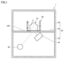
- FIG. 1 is a diagram schematically showing a configuration of an X-ray analyzer according to an embodiment of the present invention.
- the X-ray analyzer 1 includes a sample container 10, a case 20, an X-ray irradiation source 30, a detector 40, and a holder 50.
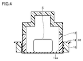
- the sample container 10 is a container for accommodating the sample S. As shown in FIG. 2, the sample container 10 has a container body 12 and a container film 14.
- the container body 12 has a shape that surrounds the sample S and opens downward.
- the container body 12 is made of, for example, polypropylene (PP).
- the container film 14 closes the opening 12a (see FIG. 2) of the container body 12 and supports the sample S.
- the container film 14 is made of polypropylene.
- the container film 14 is welded to the lower end of the container body 12.
- the container film 14 may be made of polyethylene terephthalate (PET) or the like.
- the case 20 houses the sample container 10 and the like.
- the case 20 is made of metal.
- the case 20 has an accommodating portion 22, a mounting portion 24, and a lid portion 26.
- the accommodating portion 22 is composed of the lower part of the case 20.
- the accommodating portion 22 accommodates the X-ray irradiation source 30 and the detector 40.
- the accommodating portion 22 has a shape that opens upward.
- the mounting portion 24 is connected to the upper end portion of the accommodating portion 22.
- the mounting portion 24 is a portion on which the sample container 10 is placed.
- the mounting portion 24 is provided with an opening 24h for passing X-rays emitted from the X-ray irradiation source 30.
- the opening 24h is set smaller than the outer shape of the container body 12. In other words, the outer shape of the container body 12 is larger than the opening 24h.
- the lid portion 26 surrounds the sample container 10. The lower end portion of the lid portion 26 is connected to the outer edge portion of the mounting portion 24.
- the X-ray irradiation source 30 is housed in the housing unit 22.
- the X-ray irradiation source 30 irradiates the sample S arranged in the sample container 10 with X-rays from below the mounting portion 24.
- Examples of the X-ray irradiation source 30 include an X-ray tube.
- the detector 40 is housed in the housing unit 22.
- the detector 40 detects fluorescent X-rays generated from the sample S that has received the X-rays emitted from the X-ray irradiation source 30 below the mounting unit 24. From the viewpoint of improving the analysis accuracy of the sample S, the detector 40 is preferably arranged in the vicinity of the opening 24h.
- the holder 50 is mounted on the mounting portion 24 and houses the sample container 10.
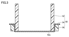
- the holder 50 is configured to be removable from the mounting portion 24. As shown in FIG. 2, the holder 50 has a surrounding cylinder 52 and a holder film 54.
- the surrounding cylinder 52 has an outer shape larger than that of the opening 24h.
- the surrounding cylinder 52 surrounds the sample container 10 and has a shape that opens downward.
- the surrounding cylinder 52 is formed in a cylindrical shape.
- the siege tube 52 is made of, for example, polypropylene.
- the height of the enclosing cylinder 52 is larger than the height of the sample container 10.
- the position of the surrounding cylinder 52 with respect to the opening 24h is determined by a positioning mechanism (not shown).
- the inner peripheral surface 52S of the surrounding cylinder 52 may be gradually reduced in diameter as it approaches the mounting portion 24. By doing so, the sample container 10 is guided to a predetermined position when the sample container 10 is arranged in the siege cylinder 52 from above the siege cylinder 52.
- the holder film 54 closes the opening 52a (see FIG. 2) of the surrounding cylinder 52. That is, two films (container film 14 and holder film 54) are laminated above the opening 24h.
- the holder film 54 is made of, for example, polypropylene.
- the holder film 54 is welded to the lower end of the surrounding cylinder 52.
- the thickness of the holder film 54 is about the same as the thickness of the container film 14.
- the thickness of the holder film 54 is preferably set to 0.5 times or more and 2 times or less the thickness of the container film 14.
- the holder film 54 may be formed of polyethylene terephthalate (PET) or the like.
- the holder film 54 is further arranged below the container film 14 that supports the sample S, even if the container film 14 is damaged or the like. , The drop of the sample S from the mounting portion 24 is suppressed. Therefore, dirt and the like of the detector 40 are suppressed.
- the holder 50 may further have a holding ring 56.
- the outer shape of the holder film 54 is set to be larger than the outer shape of the surrounding cylinder 52.
- the sandwiching ring 56 sandwiches the edge portion of the holder film 54 between the inner peripheral surface of the sandwiching ring 56 and the outer peripheral surface of the surrounding cylinder 52.
- the sample container 10 may further have a holding ring 16.
- the outer shape of the container film 14 is set to be larger than the outer shape of the container body 12.
- the sandwiching ring 16 sandwiches the edge portion of the container film 14 between the inner peripheral surface of the sandwiching ring 16 and the outer peripheral surface of the container body 12.
- the X-ray analyzer according to one embodiment is described above with respect to a sample container for accommodating a sample, a mounting portion on which the sample container can be placed, and the sample in the sample container.
- An X-ray irradiation source that irradiates X-rays from below the part, a detector that detects fluorescent X-rays generated from the sample below the above-mentioned place, and the sample that is placed on the above-mentioned place.
- a holder for accommodating a container is provided, and the above-mentioned placement portion is provided with an opening for passing X-rays emitted from the X-ray irradiation source, and the sample container surrounds the sample.
- the holder has a container body having a shape that opens downward and a container film that closes the opening of the container body and supports the sample, and the holder has a larger outer shape than the opening. It has a siege tube having a shape that surrounds the sample container and opens downward, and a holder film that closes the opening of the siege tube.
- the holder film may be welded to the lower end of the surrounding cylinder.
- the work of attaching the holder film to the surrounding cylinder is omitted.
- the holder further has a holding ring attached around the surrounding cylinder, and the holding ring is the inner peripheral surface of the holding ring.
- the edge portion of the holder film may be sandwiched between the outer peripheral surface of the surrounding cylinder.
- the outer shape of the container body is preferably larger than the opening.
- the sample can be analyzed by placing the sample container on the mounting portion.
- X-ray analyzer 10 sample container, 12 container body, 14 container film, 16 holding ring, 20 case, 22 accommodating part, 24 mounting part, 24h opening, 26 lid part, 30 X-ray irradiation source, 40 detection Container, 50 holder, 52 siege tube, 54 holder film, 56 holding ring, S sample.
Landscapes
- Physics & Mathematics (AREA)
- Health & Medical Sciences (AREA)
- Life Sciences & Earth Sciences (AREA)
- Chemical & Material Sciences (AREA)
- Analytical Chemistry (AREA)
- Biochemistry (AREA)
- General Health & Medical Sciences (AREA)
- General Physics & Mathematics (AREA)
- Immunology (AREA)
- Pathology (AREA)
- Analysing Materials By The Use Of Radiation (AREA)
Priority Applications (7)
| Application Number | Priority Date | Filing Date | Title |
|---|---|---|---|
| JP2022532907A JPWO2022003850A1 (https=) | 2020-07-01 | 2020-07-01 | |
| EP20943512.2A EP4177600B1 (en) | 2020-07-01 | 2020-07-01 | X-ray analyzer |
| US18/013,198 US20230236142A1 (en) | 2020-07-01 | 2020-07-01 | X-ray analyzer |
| PCT/JP2020/025763 WO2022003850A1 (ja) | 2020-07-01 | 2020-07-01 | X線分析装置 |
| CN202080102470.2A CN115715365A (zh) | 2020-07-01 | 2020-07-01 | X射线分析装置 |
| TW110117615A TWI787821B (zh) | 2020-07-01 | 2021-05-17 | X射線分析裝置 |
| JP2024095686A JP7679908B2 (ja) | 2020-07-01 | 2024-06-13 | X線分析装置 |
Applications Claiming Priority (1)
| Application Number | Priority Date | Filing Date | Title |
|---|---|---|---|
| PCT/JP2020/025763 WO2022003850A1 (ja) | 2020-07-01 | 2020-07-01 | X線分析装置 |
Publications (1)
| Publication Number | Publication Date |
|---|---|
| WO2022003850A1 true WO2022003850A1 (ja) | 2022-01-06 |
Family
ID=79315789
Family Applications (1)
| Application Number | Title | Priority Date | Filing Date |
|---|---|---|---|
| PCT/JP2020/025763 Ceased WO2022003850A1 (ja) | 2020-07-01 | 2020-07-01 | X線分析装置 |
Country Status (6)
| Country | Link |
|---|---|
| US (1) | US20230236142A1 (https=) |
| EP (1) | EP4177600B1 (https=) |
| JP (2) | JPWO2022003850A1 (https=) |
| CN (1) | CN115715365A (https=) |
| TW (1) | TWI787821B (https=) |
| WO (1) | WO2022003850A1 (https=) |
Cited By (1)
| Publication number | Priority date | Publication date | Assignee | Title |
|---|---|---|---|---|
| EP4700379A1 (en) * | 2024-08-23 | 2026-02-25 | Jeol Ltd. | Sample container, fluorescent x-ray analyzer, and measurement method |
Citations (8)
| Publication number | Priority date | Publication date | Assignee | Title |
|---|---|---|---|---|
| JPH04331349A (ja) * | 1991-05-03 | 1992-11-19 | Horiba Ltd | 蛍光x線分析装置 |
| JPH04355355A (ja) * | 1991-05-31 | 1992-12-09 | Rigaku Denki Kogyo Kk | 蛍光x線分析装置における窓材の汚染検出方法 |
| US5703927A (en) * | 1993-04-15 | 1997-12-30 | Angelo M. Torrisi | Safety ring for double open-ended sample holder cell for spectroscopic analysis |
| WO2004088296A1 (ja) * | 2003-03-28 | 2004-10-14 | Rigaku Industrial Corporation | 蛍光x線分析装置 |
| JP2006317153A (ja) * | 2005-05-10 | 2006-11-24 | Rigaku Industrial Co | 蛍光x線分析用試料保持具ならびにそれを用いる蛍光x線分析方法および装置 |
| JP2011013027A (ja) * | 2009-06-30 | 2011-01-20 | Horiba Ltd | 蛍光x線分析装置 |
| JP2011089794A (ja) * | 2009-10-20 | 2011-05-06 | Rigaku Corp | 蛍光x線分析用試料保持具ならびにそれを用いる蛍光x線分析方法および装置 |
| JP2018063196A (ja) | 2016-10-14 | 2018-04-19 | 株式会社島津製作所 | X線分析装置 |
Family Cites Families (7)
| Publication number | Priority date | Publication date | Assignee | Title |
|---|---|---|---|---|
| US4974244A (en) * | 1988-08-04 | 1990-11-27 | Angelo M. Torrisi | Sample positioning method and system for X-ray spectroscopic analysis |
| JP3525188B2 (ja) * | 2001-06-06 | 2004-05-10 | 理学電機工業株式会社 | 蛍光x線分析装置 |
| JP3629539B2 (ja) * | 2002-03-04 | 2005-03-16 | 理学電機工業株式会社 | 蛍光x線分析装置 |
| JP5517506B2 (ja) * | 2009-06-30 | 2014-06-11 | 株式会社堀場製作所 | 蛍光x線分析装置 |
| JP5907375B2 (ja) * | 2011-12-28 | 2016-04-26 | 株式会社テクノエックス | 蛍光x線分析装置及び蛍光x線分析方法 |
| DE102014115383A1 (de) * | 2014-08-01 | 2016-02-04 | Helmut Fischer GmbH Institut für Elektronik und Messtechnik | Handgerät sowie mobile Einrichtung zur Röntgenfluoreszenzanalyse |
| CN108956672B (zh) * | 2018-05-15 | 2021-12-17 | 江苏天瑞仪器股份有限公司 | 使用食品快检仪专用样品杯进行重金属检测的检测方法 |
-
2020
- 2020-07-01 US US18/013,198 patent/US20230236142A1/en not_active Abandoned
- 2020-07-01 CN CN202080102470.2A patent/CN115715365A/zh active Pending
- 2020-07-01 EP EP20943512.2A patent/EP4177600B1/en active Active
- 2020-07-01 WO PCT/JP2020/025763 patent/WO2022003850A1/ja not_active Ceased
- 2020-07-01 JP JP2022532907A patent/JPWO2022003850A1/ja active Pending
-
2021
- 2021-05-17 TW TW110117615A patent/TWI787821B/zh active
-
2024
- 2024-06-13 JP JP2024095686A patent/JP7679908B2/ja active Active
Patent Citations (8)
| Publication number | Priority date | Publication date | Assignee | Title |
|---|---|---|---|---|
| JPH04331349A (ja) * | 1991-05-03 | 1992-11-19 | Horiba Ltd | 蛍光x線分析装置 |
| JPH04355355A (ja) * | 1991-05-31 | 1992-12-09 | Rigaku Denki Kogyo Kk | 蛍光x線分析装置における窓材の汚染検出方法 |
| US5703927A (en) * | 1993-04-15 | 1997-12-30 | Angelo M. Torrisi | Safety ring for double open-ended sample holder cell for spectroscopic analysis |
| WO2004088296A1 (ja) * | 2003-03-28 | 2004-10-14 | Rigaku Industrial Corporation | 蛍光x線分析装置 |
| JP2006317153A (ja) * | 2005-05-10 | 2006-11-24 | Rigaku Industrial Co | 蛍光x線分析用試料保持具ならびにそれを用いる蛍光x線分析方法および装置 |
| JP2011013027A (ja) * | 2009-06-30 | 2011-01-20 | Horiba Ltd | 蛍光x線分析装置 |
| JP2011089794A (ja) * | 2009-10-20 | 2011-05-06 | Rigaku Corp | 蛍光x線分析用試料保持具ならびにそれを用いる蛍光x線分析方法および装置 |
| JP2018063196A (ja) | 2016-10-14 | 2018-04-19 | 株式会社島津製作所 | X線分析装置 |
Non-Patent Citations (1)
| Title |
|---|
| See also references of EP4177600A4 |
Cited By (1)
| Publication number | Priority date | Publication date | Assignee | Title |
|---|---|---|---|---|
| EP4700379A1 (en) * | 2024-08-23 | 2026-02-25 | Jeol Ltd. | Sample container, fluorescent x-ray analyzer, and measurement method |
Also Published As
| Publication number | Publication date |
|---|---|
| JP7679908B2 (ja) | 2025-05-20 |
| JPWO2022003850A1 (https=) | 2022-01-06 |
| JP2024107407A (ja) | 2024-08-08 |
| US20230236142A1 (en) | 2023-07-27 |
| TWI787821B (zh) | 2022-12-21 |
| EP4177600B1 (en) | 2025-10-08 |
| TW202202834A (zh) | 2022-01-16 |
| CN115715365A (zh) | 2023-02-24 |
| EP4177600A1 (en) | 2023-05-10 |
| EP4177600A4 (en) | 2024-03-06 |
Similar Documents
| Publication | Publication Date | Title |
|---|---|---|
| JPH04331349A (ja) | 蛍光x線分析装置 | |
| EP3029455A1 (en) | X-ray fluorescence analyzer | |
| US9182362B2 (en) | Apparatus for protecting a radiation window | |
| JP4854005B2 (ja) | 蛍光x線分析装置 | |
| JP7679908B2 (ja) | X線分析装置 | |
| JP2013221745A (ja) | X線検出装置 | |
| JP2943063B2 (ja) | 蛍光x線分析装置 | |
| JP2000230912A (ja) | X線分析用試料ホルダおよびx線分析装置 | |
| JP5517506B2 (ja) | 蛍光x線分析装置 | |
| JP7605021B2 (ja) | X線分析装置 | |
| CN115963126B (zh) | 实验室谱仪 | |
| JP5782154B2 (ja) | 蛍光x線分析装置 | |
| JP6227068B1 (ja) | 試料容器保持部材、光計測装置及び試料容器配置方法 | |
| JPWO2022003850A5 (https=) | ||
| JP4029081B2 (ja) | 試料ホルダ用蓋、試料ホルダ及び蛍光x線測定装置 | |
| JP5524521B2 (ja) | 蛍光x線分析装置 | |
| JP5072704B2 (ja) | 放射線検査装置 | |
| JPH11160492A (ja) | 放射性物質貯蔵容器 | |
| JP4771985B2 (ja) | 放射線検査装置 | |
| WO2021213559A1 (en) | A spacer for a cuvette, use thereof and a method of analysing a sample | |
| JP3089817B2 (ja) | 陽電子消滅法による材料劣化検出装置 | |
| JP7649591B1 (ja) | 蛍光x線分析装置 | |
| KR102559077B1 (ko) | 실시간 선량 감시가 가능한 방사성 폐기물 운반용기 | |
| JP2026508925A (ja) | 包装された試料を分析するためのx線回折装置及び方法 | |
| JP3210530U (ja) | 蛍光x線分析用一体化試料及び試料保持具 |
Legal Events
| Date | Code | Title | Description |
|---|---|---|---|
| 121 | Ep: the epo has been informed by wipo that ep was designated in this application |
Ref document number: 20943512 Country of ref document: EP Kind code of ref document: A1 |
|
| ENP | Entry into the national phase |
Ref document number: 2022532907 Country of ref document: JP Kind code of ref document: A |
|
| NENP | Non-entry into the national phase |
Ref country code: DE |
|
| ENP | Entry into the national phase |
Ref document number: 2020943512 Country of ref document: EP Effective date: 20230201 |
|
| WWG | Wipo information: grant in national office |
Ref document number: 2020943512 Country of ref document: EP |