US7972788B2 - Skin aging marker and technique for use thereof - Google Patents
Skin aging marker and technique for use thereof Download PDFInfo
- Publication number
- US7972788B2 US7972788B2 US12/064,602 US6460206A US7972788B2 US 7972788 B2 US7972788 B2 US 7972788B2 US 6460206 A US6460206 A US 6460206A US 7972788 B2 US7972788 B2 US 7972788B2
- Authority
- US
- United States
- Prior art keywords
- skin
- keratin
- proteins
- aging
- cells
- Prior art date
- Legal status (The legal status is an assumption and is not a legal conclusion. Google has not performed a legal analysis and makes no representation as to the accuracy of the status listed.)
- Expired - Fee Related, expires
Links
- 230000009759 skin aging Effects 0.000 title claims abstract description 57
- 238000000034 method Methods 0.000 title claims abstract description 53
- 239000003550 marker Substances 0.000 title description 19
- 108090000623 proteins and genes Proteins 0.000 claims abstract description 223
- 102000004169 proteins and genes Human genes 0.000 claims abstract description 184
- 210000003491 skin Anatomy 0.000 claims abstract description 110
- 230000032683 aging Effects 0.000 claims abstract description 89
- 102000040739 Secretory proteins Human genes 0.000 claims abstract description 65
- 108091058545 Secretory proteins Proteins 0.000 claims abstract description 65
- 230000003834 intracellular effect Effects 0.000 claims abstract description 64
- 210000004927 skin cell Anatomy 0.000 claims abstract description 48
- 230000008859 change Effects 0.000 claims abstract description 37
- 239000000126 substance Substances 0.000 claims abstract description 34
- 102100034867 Kallikrein-7 Human genes 0.000 claims description 32
- 101710176222 Kallikrein-7 Proteins 0.000 claims description 32
- 239000013076 target substance Substances 0.000 claims description 28
- 102000015736 beta 2-Microglobulin Human genes 0.000 claims description 26
- 108010081355 beta 2-Microglobulin Proteins 0.000 claims description 26
- 102100038910 Alpha-enolase Human genes 0.000 claims description 22
- 102100032700 Keratin, type I cytoskeletal 20 Human genes 0.000 claims description 21
- 102100040445 Keratin, type I cytoskeletal 14 Human genes 0.000 claims description 20
- 108010066370 Keratin-20 Proteins 0.000 claims description 20
- 101710165425 Alpha-enolase Proteins 0.000 claims description 19
- 101710184673 Enolase 1 Proteins 0.000 claims description 19
- 108010066321 Keratin-14 Proteins 0.000 claims description 19
- 102100022127 Radixin Human genes 0.000 claims description 19
- 108010048484 radixin Proteins 0.000 claims description 19
- 230000000694 effects Effects 0.000 claims description 14
- 238000012258 culturing Methods 0.000 claims description 3
- WQZGKKKJIJFFOK-GASJEMHNSA-N Glucose Natural products OC[C@H]1OC(O)[C@H](O)[C@@H](O)[C@@H]1O WQZGKKKJIJFFOK-GASJEMHNSA-N 0.000 claims 1
- 239000008103 glucose Substances 0.000 claims 1
- 230000002401 inhibitory effect Effects 0.000 claims 1
- 230000001105 regulatory effect Effects 0.000 claims 1
- 230000014509 gene expression Effects 0.000 abstract description 128
- 238000005259 measurement Methods 0.000 abstract description 13
- 230000002265 prevention Effects 0.000 abstract description 7
- 235000018102 proteins Nutrition 0.000 description 159
- 210000004027 cell Anatomy 0.000 description 113
- 210000005175 epidermal keratinocyte Anatomy 0.000 description 55
- 210000001519 tissue Anatomy 0.000 description 50
- 239000010410 layer Substances 0.000 description 36
- 102100040443 Keratin, type I cytoskeletal 15 Human genes 0.000 description 34
- 108010066330 Keratin-15 Proteins 0.000 description 33
- 102100023974 Keratin, type II cytoskeletal 7 Human genes 0.000 description 31
- 108010070507 Keratin-7 Proteins 0.000 description 30
- 150000001413 amino acids Chemical group 0.000 description 27
- 102100023970 Keratin, type I cytoskeletal 10 Human genes 0.000 description 26
- 108010065038 Keratin-10 Proteins 0.000 description 25
- 241000699666 Mus <mouse, genus> Species 0.000 description 25
- 238000004458 analytical method Methods 0.000 description 25
- WEVYAHXRMPXWCK-UHFFFAOYSA-N Acetonitrile Chemical compound CC#N WEVYAHXRMPXWCK-UHFFFAOYSA-N 0.000 description 24
- 108010022233 Plasminogen Activator Inhibitor 1 Proteins 0.000 description 22
- 102100039418 Plasminogen activator inhibitor 1 Human genes 0.000 description 22
- 238000001262 western blot Methods 0.000 description 22
- 102000004374 Insulin-like growth factor binding protein 3 Human genes 0.000 description 21
- 108090000965 Insulin-like growth factor binding protein 3 Proteins 0.000 description 21
- 102100027869 Moesin Human genes 0.000 description 21
- 108010071525 moesin Proteins 0.000 description 21
- 102000044465 Galectin-7 Human genes 0.000 description 20
- 102000004878 Gelsolin Human genes 0.000 description 20
- 241000282414 Homo sapiens Species 0.000 description 20
- 101000608772 Homo sapiens Galectin-7 Proteins 0.000 description 20
- 102100033421 Keratin, type I cytoskeletal 18 Human genes 0.000 description 20
- 102100033420 Keratin, type I cytoskeletal 19 Human genes 0.000 description 20
- 102100031358 Urokinase-type plasminogen activator Human genes 0.000 description 20
- 108090000435 Urokinase-type plasminogen activator Proteins 0.000 description 20
- 230000003247 decreasing effect Effects 0.000 description 20
- 108010001498 Galectin 1 Proteins 0.000 description 19
- 102000000802 Galectin 3 Human genes 0.000 description 19
- 108010001517 Galectin 3 Proteins 0.000 description 19
- 108090001064 Gelsolin Proteins 0.000 description 19
- 102100040487 Keratin, type I cytoskeletal 13 Human genes 0.000 description 19
- 102100040441 Keratin, type I cytoskeletal 16 Human genes 0.000 description 19
- 108010066327 Keratin-18 Proteins 0.000 description 19
- 108010066302 Keratin-19 Proteins 0.000 description 19
- 108010055671 ezrin Proteins 0.000 description 19
- 108090000668 Annexin A2 Proteins 0.000 description 18
- 102000004149 Annexin A2 Human genes 0.000 description 18
- 102100020903 Ezrin Human genes 0.000 description 18
- 102100021736 Galectin-1 Human genes 0.000 description 18
- 108010065070 Keratin-13 Proteins 0.000 description 18
- 108010066364 Keratin-16 Proteins 0.000 description 18
- 108010091175 Matriptase Proteins 0.000 description 18
- 102100037942 Suppressor of tumorigenicity 14 protein Human genes 0.000 description 18
- 239000000523 sample Substances 0.000 description 18
- 239000000243 solution Substances 0.000 description 17
- 101000839464 Leishmania braziliensis Heat shock 70 kDa protein Proteins 0.000 description 16
- 108090001053 Gastrin releasing peptide Proteins 0.000 description 15
- 102100032510 Heat shock protein HSP 90-beta Human genes 0.000 description 15
- 101001016856 Homo sapiens Heat shock protein HSP 90-beta Proteins 0.000 description 15
- 101000988090 Leishmania donovani Heat shock protein 83 Proteins 0.000 description 15
- 102100028262 U6 snRNA-associated Sm-like protein LSm4 Human genes 0.000 description 15
- 210000000981 epithelium Anatomy 0.000 description 15
- 239000012528 membrane Substances 0.000 description 14
- 108020004707 nucleic acids Proteins 0.000 description 13
- 150000007523 nucleic acids Chemical class 0.000 description 13
- 102000039446 nucleic acids Human genes 0.000 description 13
- 238000000539 two dimensional gel electrophoresis Methods 0.000 description 13
- 241000283973 Oryctolagus cuniculus Species 0.000 description 12
- 108020004999 messenger RNA Proteins 0.000 description 12
- 102100021238 Dynamin-2 Human genes 0.000 description 11
- 101000817607 Homo sapiens Dynamin-2 Proteins 0.000 description 11
- QKNYBSVHEMOAJP-UHFFFAOYSA-N 2-amino-2-(hydroxymethyl)propane-1,3-diol;hydron;chloride Chemical compound Cl.OCC(N)(CO)CO QKNYBSVHEMOAJP-UHFFFAOYSA-N 0.000 description 10
- 102000035195 Peptidases Human genes 0.000 description 10
- 108091005804 Peptidases Proteins 0.000 description 10
- 239000004365 Protease Substances 0.000 description 10
- 239000000872 buffer Substances 0.000 description 10
- 239000006228 supernatant Substances 0.000 description 10
- OKKJLVBELUTLKV-UHFFFAOYSA-N Methanol Chemical compound OC OKKJLVBELUTLKV-UHFFFAOYSA-N 0.000 description 9
- XSQUKJJJFZCRTK-UHFFFAOYSA-N Urea Chemical compound NC(N)=O XSQUKJJJFZCRTK-UHFFFAOYSA-N 0.000 description 9
- 210000000170 cell membrane Anatomy 0.000 description 9
- 238000001962 electrophoresis Methods 0.000 description 9
- 210000003963 intermediate filament Anatomy 0.000 description 9
- 102000004190 Enzymes Human genes 0.000 description 8
- 108090000790 Enzymes Proteins 0.000 description 8
- 239000002033 PVDF binder Substances 0.000 description 8
- 229920002981 polyvinylidene fluoride Polymers 0.000 description 8
- 108091032973 (ribonucleotides)n+m Proteins 0.000 description 7
- 102000010834 Extracellular Matrix Proteins Human genes 0.000 description 7
- 108010037362 Extracellular Matrix Proteins Proteins 0.000 description 7
- 102000011782 Keratins Human genes 0.000 description 7
- 108010076876 Keratins Proteins 0.000 description 7
- 238000004113 cell culture Methods 0.000 description 7
- 239000002537 cosmetic Substances 0.000 description 7
- 230000007423 decrease Effects 0.000 description 7
- 229940088598 enzyme Drugs 0.000 description 7
- 238000007665 sagging Methods 0.000 description 7
- 108020004711 Nucleic Acid Probes Proteins 0.000 description 6
- 108090000708 Proteasome Endopeptidase Complex Proteins 0.000 description 6
- 102000004245 Proteasome Endopeptidase Complex Human genes 0.000 description 6
- FAPWRFPIFSIZLT-UHFFFAOYSA-M Sodium chloride Chemical compound [Na+].[Cl-] FAPWRFPIFSIZLT-UHFFFAOYSA-M 0.000 description 6
- DTQVDTLACAAQTR-UHFFFAOYSA-N Trifluoroacetic acid Chemical compound OC(=O)C(F)(F)F DTQVDTLACAAQTR-UHFFFAOYSA-N 0.000 description 6
- 238000002474 experimental method Methods 0.000 description 6
- 239000007788 liquid Substances 0.000 description 6
- 239000002609 medium Substances 0.000 description 6
- 239000002853 nucleic acid probe Substances 0.000 description 6
- 230000035755 proliferation Effects 0.000 description 6
- UMGDCJDMYOKAJW-UHFFFAOYSA-N thiourea Chemical compound NC(N)=S UMGDCJDMYOKAJW-UHFFFAOYSA-N 0.000 description 6
- 108010085238 Actins Proteins 0.000 description 5
- 102000007469 Actins Human genes 0.000 description 5
- 108010022181 Phosphopyruvate Hydratase Proteins 0.000 description 5
- 230000032677 cell aging Effects 0.000 description 5
- 239000003153 chemical reaction reagent Substances 0.000 description 5
- 230000003436 cytoskeletal effect Effects 0.000 description 5
- 238000005516 engineering process Methods 0.000 description 5
- 238000011156 evaluation Methods 0.000 description 5
- 210000002744 extracellular matrix Anatomy 0.000 description 5
- 239000002243 precursor Substances 0.000 description 5
- 238000002415 sodium dodecyl sulfate polyacrylamide gel electrophoresis Methods 0.000 description 5
- 238000012360 testing method Methods 0.000 description 5
- XLYOFNOQVPJJNP-UHFFFAOYSA-N water Chemical compound O XLYOFNOQVPJJNP-UHFFFAOYSA-N 0.000 description 5
- 108010067306 Fibronectins Proteins 0.000 description 4
- 102000016359 Fibronectins Human genes 0.000 description 4
- 108010001336 Horseradish Peroxidase Proteins 0.000 description 4
- 102100025756 Keratin, type II cytoskeletal 5 Human genes 0.000 description 4
- 102100023972 Keratin, type II cytoskeletal 8 Human genes 0.000 description 4
- 108010070553 Keratin-5 Proteins 0.000 description 4
- 108010070511 Keratin-8 Proteins 0.000 description 4
- 108010026552 Proteome Proteins 0.000 description 4
- 108010005173 SERPIN-B5 Proteins 0.000 description 4
- 102100030333 Serpin B5 Human genes 0.000 description 4
- 230000004913 activation Effects 0.000 description 4
- 230000004663 cell proliferation Effects 0.000 description 4
- 239000002299 complementary DNA Substances 0.000 description 4
- 239000000284 extract Substances 0.000 description 4
- 239000000203 mixture Substances 0.000 description 4
- 238000002360 preparation method Methods 0.000 description 4
- 230000002062 proliferating effect Effects 0.000 description 4
- 238000007390 skin biopsy Methods 0.000 description 4
- 230000037394 skin elasticity Effects 0.000 description 4
- HLXHCNWEVQNNKA-UHFFFAOYSA-N 5-methoxy-2,3-dihydro-1h-inden-2-amine Chemical compound COC1=CC=C2CC(N)CC2=C1 HLXHCNWEVQNNKA-UHFFFAOYSA-N 0.000 description 3
- 108010061641 Cystatin A Proteins 0.000 description 3
- 102100031237 Cystatin-A Human genes 0.000 description 3
- 102100039328 Endoplasmin Human genes 0.000 description 3
- WSFSSNUMVMOOMR-UHFFFAOYSA-N Formaldehyde Chemical compound O=C WSFSSNUMVMOOMR-UHFFFAOYSA-N 0.000 description 3
- PEDCQBHIVMGVHV-UHFFFAOYSA-N Glycerine Chemical compound OCC(O)CO PEDCQBHIVMGVHV-UHFFFAOYSA-N 0.000 description 3
- 101710113864 Heat shock protein 90 Proteins 0.000 description 3
- 108010004889 Heat-Shock Proteins Proteins 0.000 description 3
- 102000002812 Heat-Shock Proteins Human genes 0.000 description 3
- 102100022905 Keratin, type II cytoskeletal 1 Human genes 0.000 description 3
- 108010070514 Keratin-1 Proteins 0.000 description 3
- 102000018331 Keratin-17 Human genes 0.000 description 3
- 108010066325 Keratin-17 Proteins 0.000 description 3
- 108010070918 Keratin-3 Proteins 0.000 description 3
- 108010085895 Laminin Proteins 0.000 description 3
- 102000007547 Laminin Human genes 0.000 description 3
- 238000000636 Northern blotting Methods 0.000 description 3
- 108010076504 Protein Sorting Signals Proteins 0.000 description 3
- 102000003978 Tissue Plasminogen Activator Human genes 0.000 description 3
- 108090000373 Tissue Plasminogen Activator Proteins 0.000 description 3
- 239000013504 Triton X-100 Substances 0.000 description 3
- 229920004890 Triton X-100 Polymers 0.000 description 3
- GEHJBWKLJVFKPS-UHFFFAOYSA-N bromochloroacetic acid Chemical compound OC(=O)C(Cl)Br GEHJBWKLJVFKPS-UHFFFAOYSA-N 0.000 description 3
- 239000004202 carbamide Substances 0.000 description 3
- 230000001413 cellular effect Effects 0.000 description 3
- 238000001514 detection method Methods 0.000 description 3
- 238000007876 drug discovery Methods 0.000 description 3
- 239000000835 fiber Substances 0.000 description 3
- 230000006870 function Effects 0.000 description 3
- 239000003112 inhibitor Substances 0.000 description 3
- 108010028309 kalinin Proteins 0.000 description 3
- 238000011160 research Methods 0.000 description 3
- 238000012552 review Methods 0.000 description 3
- 229920006395 saturated elastomer Polymers 0.000 description 3
- 239000003001 serine protease inhibitor Substances 0.000 description 3
- 210000001626 skin fibroblast Anatomy 0.000 description 3
- 239000011780 sodium chloride Substances 0.000 description 3
- 229960000187 tissue plasminogen activator Drugs 0.000 description 3
- 238000012546 transfer Methods 0.000 description 3
- 108090000663 Annexin A1 Proteins 0.000 description 2
- 102100026189 Beta-galactosidase Human genes 0.000 description 2
- 241000283690 Bos taurus Species 0.000 description 2
- 206010006187 Breast cancer Diseases 0.000 description 2
- 208000026310 Breast neoplasm Diseases 0.000 description 2
- OYPRJOBELJOOCE-UHFFFAOYSA-N Calcium Chemical compound [Ca] OYPRJOBELJOOCE-UHFFFAOYSA-N 0.000 description 2
- 101000749287 Clitocybe nebularis Clitocypin Proteins 0.000 description 2
- 101000767029 Clitocybe nebularis Clitocypin-1 Proteins 0.000 description 2
- 102000008186 Collagen Human genes 0.000 description 2
- 108010035532 Collagen Proteins 0.000 description 2
- 229940094664 Cysteine protease inhibitor Drugs 0.000 description 2
- 102000010831 Cytoskeletal Proteins Human genes 0.000 description 2
- 108010037414 Cytoskeletal Proteins Proteins 0.000 description 2
- 102000007563 Galectins Human genes 0.000 description 2
- 108010046569 Galectins Proteins 0.000 description 2
- 102100040352 Heat shock 70 kDa protein 1A Human genes 0.000 description 2
- 102100039165 Heat shock protein beta-1 Human genes 0.000 description 2
- 101710100504 Heat shock protein beta-1 Proteins 0.000 description 2
- 101000661807 Homo sapiens Suppressor of tumorigenicity 14 protein Proteins 0.000 description 2
- 102100037852 Insulin-like growth factor I Human genes 0.000 description 2
- 102100020944 Integrin-linked protein kinase Human genes 0.000 description 2
- 102100025759 Keratin, type II cytoskeletal 3 Human genes 0.000 description 2
- 101001018085 Lysobacter enzymogenes Lysyl endopeptidase Proteins 0.000 description 2
- TWRXJAOTZQYOKJ-UHFFFAOYSA-L Magnesium chloride Chemical compound [Mg+2].[Cl-].[Cl-] TWRXJAOTZQYOKJ-UHFFFAOYSA-L 0.000 description 2
- 241000124008 Mammalia Species 0.000 description 2
- SEQKRHFRPICQDD-UHFFFAOYSA-N N-tris(hydroxymethyl)methylglycine Chemical compound OCC(CO)(CO)[NH2+]CC([O-])=O SEQKRHFRPICQDD-UHFFFAOYSA-N 0.000 description 2
- 241001494479 Pecora Species 0.000 description 2
- 102000012288 Phosphopyruvate Hydratase Human genes 0.000 description 2
- 108091000080 Phosphotransferase Proteins 0.000 description 2
- 102000004179 Plasminogen Activator Inhibitor 2 Human genes 0.000 description 2
- 108090000614 Plasminogen Activator Inhibitor 2 Proteins 0.000 description 2
- 229920001213 Polysorbate 20 Polymers 0.000 description 2
- 241000700159 Rattus Species 0.000 description 2
- 101710202172 Rho GDP-dissociation inhibitor Proteins 0.000 description 2
- 102000012479 Serine Proteases Human genes 0.000 description 2
- 108010022999 Serine Proteases Proteins 0.000 description 2
- 229940122055 Serine protease inhibitor Drugs 0.000 description 2
- 101710102218 Serine protease inhibitor Proteins 0.000 description 2
- 102100036383 Serpin B3 Human genes 0.000 description 2
- 102000019197 Superoxide Dismutase Human genes 0.000 description 2
- 108010012715 Superoxide dismutase Proteins 0.000 description 2
- 241000282898 Sus scrofa Species 0.000 description 2
- 239000007997 Tricine buffer Substances 0.000 description 2
- 239000007983 Tris buffer Substances 0.000 description 2
- 102000004243 Tubulin Human genes 0.000 description 2
- 108090000704 Tubulin Proteins 0.000 description 2
- 239000000654 additive Substances 0.000 description 2
- 230000000996 additive effect Effects 0.000 description 2
- 108010005774 beta-Galactosidase Proteins 0.000 description 2
- 230000015572 biosynthetic process Effects 0.000 description 2
- 229910052791 calcium Inorganic materials 0.000 description 2
- 239000011575 calcium Substances 0.000 description 2
- 239000003795 chemical substances by application Substances 0.000 description 2
- 229920001436 collagen Polymers 0.000 description 2
- 150000001875 compounds Chemical class 0.000 description 2
- 230000002596 correlated effect Effects 0.000 description 2
- 238000009223 counseling Methods 0.000 description 2
- 210000000805 cytoplasm Anatomy 0.000 description 2
- 230000004069 differentiation Effects 0.000 description 2
- 230000029087 digestion Effects 0.000 description 2
- 229940042399 direct acting antivirals protease inhibitors Drugs 0.000 description 2
- BFMYDTVEBKDAKJ-UHFFFAOYSA-L disodium;(2',7'-dibromo-3',6'-dioxido-3-oxospiro[2-benzofuran-1,9'-xanthene]-4'-yl)mercury;hydrate Chemical compound O.[Na+].[Na+].O1C(=O)C2=CC=CC=C2C21C1=CC(Br)=C([O-])C([Hg])=C1OC1=C2C=C(Br)C([O-])=C1 BFMYDTVEBKDAKJ-UHFFFAOYSA-L 0.000 description 2
- 238000001378 electrochemiluminescence detection Methods 0.000 description 2
- 210000001339 epidermal cell Anatomy 0.000 description 2
- 210000002615 epidermis Anatomy 0.000 description 2
- 239000000945 filler Substances 0.000 description 2
- 210000001035 gastrointestinal tract Anatomy 0.000 description 2
- 210000004907 gland Anatomy 0.000 description 2
- RWSXRVCMGQZWBV-WDSKDSINSA-N glutathione Chemical compound OC(=O)[C@@H](N)CCC(=O)N[C@@H](CS)C(=O)NCC(O)=O RWSXRVCMGQZWBV-WDSKDSINSA-N 0.000 description 2
- 230000034659 glycolysis Effects 0.000 description 2
- ZBKIUFWVEIBQRT-UHFFFAOYSA-N gold(1+) Chemical compound [Au+] ZBKIUFWVEIBQRT-UHFFFAOYSA-N 0.000 description 2
- 210000004209 hair Anatomy 0.000 description 2
- JYGXADMDTFJGBT-VWUMJDOOSA-N hydrocortisone Chemical compound O=C1CC[C@]2(C)[C@H]3[C@@H](O)C[C@](C)([C@@](CC4)(O)C(=O)CO)[C@@H]4[C@@H]3CCC2=C1 JYGXADMDTFJGBT-VWUMJDOOSA-N 0.000 description 2
- 238000010191 image analysis Methods 0.000 description 2
- 238000002991 immunohistochemical analysis Methods 0.000 description 2
- NOESYZHRGYRDHS-UHFFFAOYSA-N insulin Chemical compound N1C(=O)C(NC(=O)C(CCC(N)=O)NC(=O)C(CCC(O)=O)NC(=O)C(C(C)C)NC(=O)C(NC(=O)CN)C(C)CC)CSSCC(C(NC(CO)C(=O)NC(CC(C)C)C(=O)NC(CC=2C=CC(O)=CC=2)C(=O)NC(CCC(N)=O)C(=O)NC(CC(C)C)C(=O)NC(CCC(O)=O)C(=O)NC(CC(N)=O)C(=O)NC(CC=2C=CC(O)=CC=2)C(=O)NC(CSSCC(NC(=O)C(C(C)C)NC(=O)C(CC(C)C)NC(=O)C(CC=2C=CC(O)=CC=2)NC(=O)C(CC(C)C)NC(=O)C(C)NC(=O)C(CCC(O)=O)NC(=O)C(C(C)C)NC(=O)C(CC(C)C)NC(=O)C(CC=2NC=NC=2)NC(=O)C(CO)NC(=O)CNC2=O)C(=O)NCC(=O)NC(CCC(O)=O)C(=O)NC(CCCNC(N)=N)C(=O)NCC(=O)NC(CC=3C=CC=CC=3)C(=O)NC(CC=3C=CC=CC=3)C(=O)NC(CC=3C=CC(O)=CC=3)C(=O)NC(C(C)O)C(=O)N3C(CCC3)C(=O)NC(CCCCN)C(=O)NC(C)C(O)=O)C(=O)NC(CC(N)=O)C(O)=O)=O)NC(=O)C(C(C)CC)NC(=O)C(CO)NC(=O)C(C(C)O)NC(=O)C1CSSCC2NC(=O)C(CC(C)C)NC(=O)C(NC(=O)C(CCC(N)=O)NC(=O)C(CC(N)=O)NC(=O)C(NC(=O)C(N)CC=1C=CC=CC=1)C(C)C)CC1=CN=CN1 NOESYZHRGYRDHS-UHFFFAOYSA-N 0.000 description 2
- 108010059517 integrin-linked kinase Proteins 0.000 description 2
- 238000001155 isoelectric focusing Methods 0.000 description 2
- 210000003734 kidney Anatomy 0.000 description 2
- 210000005075 mammary gland Anatomy 0.000 description 2
- 239000011159 matrix material Substances 0.000 description 2
- 210000000110 microvilli Anatomy 0.000 description 2
- 210000005036 nerve Anatomy 0.000 description 2
- 239000000137 peptide hydrolase inhibitor Substances 0.000 description 2
- 150000003904 phospholipids Chemical class 0.000 description 2
- 102000020233 phosphotransferase Human genes 0.000 description 2
- 239000004033 plastic Substances 0.000 description 2
- 229920003023 plastic Polymers 0.000 description 2
- 238000006116 polymerization reaction Methods 0.000 description 2
- 239000000256 polyoxyethylene sorbitan monolaurate Substances 0.000 description 2
- 235000010486 polyoxyethylene sorbitan monolaurate Nutrition 0.000 description 2
- 108090000765 processed proteins & peptides Proteins 0.000 description 2
- 108020003175 receptors Proteins 0.000 description 2
- 102000005962 receptors Human genes 0.000 description 2
- 230000009758 senescence Effects 0.000 description 2
- 230000008054 signal transmission Effects 0.000 description 2
- 239000002356 single layer Substances 0.000 description 2
- 238000010186 staining Methods 0.000 description 2
- 239000000758 substrate Substances 0.000 description 2
- 230000032258 transport Effects 0.000 description 2
- TUQOTMZNTHZOKS-UHFFFAOYSA-N tributylphosphine Chemical compound CCCCP(CCCC)CCCC TUQOTMZNTHZOKS-UHFFFAOYSA-N 0.000 description 2
- LENZDBCJOHFCAS-UHFFFAOYSA-N tris Chemical compound OCC(N)(CO)CO LENZDBCJOHFCAS-UHFFFAOYSA-N 0.000 description 2
- QRXMUCSWCMTJGU-UHFFFAOYSA-L (5-bromo-4-chloro-1h-indol-3-yl) phosphate Chemical compound C1=C(Br)C(Cl)=C2C(OP([O-])(=O)[O-])=CNC2=C1 QRXMUCSWCMTJGU-UHFFFAOYSA-L 0.000 description 1
- CFBILACNYSPRPM-UHFFFAOYSA-N 2-amino-2-(hydroxymethyl)propane-1,3-diol;2-[[1,3-dihydroxy-2-(hydroxymethyl)propan-2-yl]amino]acetic acid Chemical compound OCC(N)(CO)CO.OCC(CO)(CO)NCC(O)=O CFBILACNYSPRPM-UHFFFAOYSA-N 0.000 description 1
- GXIURPTVHJPJLF-UWTATZPHSA-N 2-phospho-D-glyceric acid Chemical compound OC[C@H](C(O)=O)OP(O)(O)=O GXIURPTVHJPJLF-UWTATZPHSA-N 0.000 description 1
- UMCMPZBLKLEWAF-BCTGSCMUSA-N 3-[(3-cholamidopropyl)dimethylammonio]propane-1-sulfonate Chemical compound C([C@H]1C[C@H]2O)[C@H](O)CC[C@]1(C)[C@@H]1[C@@H]2[C@@H]2CC[C@H]([C@@H](CCC(=O)NCCC[N+](C)(C)CCCS([O-])(=O)=O)C)[C@@]2(C)[C@@H](O)C1 UMCMPZBLKLEWAF-BCTGSCMUSA-N 0.000 description 1
- JYCQQPHGFMYQCF-UHFFFAOYSA-N 4-tert-Octylphenol monoethoxylate Chemical compound CC(C)(C)CC(C)(C)C1=CC=C(OCCO)C=C1 JYCQQPHGFMYQCF-UHFFFAOYSA-N 0.000 description 1
- SLXKOJJOQWFEFD-UHFFFAOYSA-N 6-aminohexanoic acid Chemical compound NCCCCCC(O)=O SLXKOJJOQWFEFD-UHFFFAOYSA-N 0.000 description 1
- 102100040881 60S acidic ribosomal protein P0 Human genes 0.000 description 1
- 108050002744 60S acidic ribosomal protein P0 Proteins 0.000 description 1
- MPVDXIMFBOLMNW-ISLYRVAYSA-N 7-hydroxy-8-[(E)-phenyldiazenyl]naphthalene-1,3-disulfonic acid Chemical compound OC1=CC=C2C=C(S(O)(=O)=O)C=C(S(O)(=O)=O)C2=C1\N=N\C1=CC=CC=C1 MPVDXIMFBOLMNW-ISLYRVAYSA-N 0.000 description 1
- 108091005508 Acid proteases Proteins 0.000 description 1
- HRPVXLWXLXDGHG-UHFFFAOYSA-N Acrylamide Chemical compound NC(=O)C=C HRPVXLWXLXDGHG-UHFFFAOYSA-N 0.000 description 1
- 102000011200 Actin-related protein 2/3 complex subunit 5 Human genes 0.000 description 1
- 108050001405 Actin-related protein 2/3 complex subunit 5 Proteins 0.000 description 1
- 102000003741 Actin-related protein 3 Human genes 0.000 description 1
- 108090000104 Actin-related protein 3 Proteins 0.000 description 1
- 108010063503 Actinin Proteins 0.000 description 1
- 102000010825 Actinin Human genes 0.000 description 1
- 102100032488 Acylamino-acid-releasing enzyme Human genes 0.000 description 1
- 108010061216 Acylaminoacyl-peptidase Proteins 0.000 description 1
- 102000005234 Adenosylhomocysteinase Human genes 0.000 description 1
- 108020002202 Adenosylhomocysteinase Proteins 0.000 description 1
- 102000002260 Alkaline Phosphatase Human genes 0.000 description 1
- 108020004774 Alkaline Phosphatase Proteins 0.000 description 1
- 102000000412 Annexin Human genes 0.000 description 1
- 108050008874 Annexin Proteins 0.000 description 1
- 102000004145 Annexin A1 Human genes 0.000 description 1
- 102100040006 Annexin A1 Human genes 0.000 description 1
- 101000640990 Arabidopsis thaliana Tryptophan-tRNA ligase, chloroplastic/mitochondrial Proteins 0.000 description 1
- IJGRMHOSHXDMSA-UHFFFAOYSA-N Atomic nitrogen Chemical compound N#N IJGRMHOSHXDMSA-UHFFFAOYSA-N 0.000 description 1
- 108090001008 Avidin Proteins 0.000 description 1
- 229920001342 Bakelite® Polymers 0.000 description 1
- 238000009010 Bradford assay Methods 0.000 description 1
- BHPQYMZQTOCNFJ-UHFFFAOYSA-N Calcium cation Chemical compound [Ca+2] BHPQYMZQTOCNFJ-UHFFFAOYSA-N 0.000 description 1
- 241000283707 Capra Species 0.000 description 1
- 102000004958 Caspase-14 Human genes 0.000 description 1
- 108090001132 Caspase-14 Proteins 0.000 description 1
- 241000700199 Cavia porcellus Species 0.000 description 1
- 241000282693 Cercopithecidae Species 0.000 description 1
- 241000905957 Channa melasoma Species 0.000 description 1
- 101710163595 Chaperone protein DnaK Proteins 0.000 description 1
- 101710181333 Chaperone protein dnaK1 Proteins 0.000 description 1
- 241001340526 Chrysoclista linneella Species 0.000 description 1
- 206010009944 Colon cancer Diseases 0.000 description 1
- 101710084382 Cyclase-associated protein 1 Proteins 0.000 description 1
- 108010072220 Cyclophilin A Proteins 0.000 description 1
- 102000012177 Cystatin M Human genes 0.000 description 1
- 108010061617 Cystatin M Proteins 0.000 description 1
- 102100027598 Cystatin-8 Human genes 0.000 description 1
- 101710187959 Cystatin-8 Proteins 0.000 description 1
- 102000018832 Cytochromes Human genes 0.000 description 1
- 108010052832 Cytochromes Proteins 0.000 description 1
- 102100031007 Cytosolic non-specific dipeptidase Human genes 0.000 description 1
- 238000009014 DC Assay Kit Methods 0.000 description 1
- 108020004414 DNA Proteins 0.000 description 1
- 238000000018 DNA microarray Methods 0.000 description 1
- 101710088194 Dehydrogenase Proteins 0.000 description 1
- 206010013786 Dry skin Diseases 0.000 description 1
- ZGTMUACCHSMWAC-UHFFFAOYSA-L EDTA disodium salt (anhydrous) Chemical compound [Na+].[Na+].OC(=O)CN(CC([O-])=O)CCN(CC(O)=O)CC([O-])=O ZGTMUACCHSMWAC-UHFFFAOYSA-L 0.000 description 1
- 238000002965 ELISA Methods 0.000 description 1
- 102100031334 Elongation factor 2 Human genes 0.000 description 1
- 101000738180 Euglena gracilis Chaperonin CPN60, mitochondrial Proteins 0.000 description 1
- 102100026761 Eukaryotic translation initiation factor 5A-1 Human genes 0.000 description 1
- 102100030421 Fatty acid-binding protein 5 Human genes 0.000 description 1
- 101710083187 Fatty acid-binding protein 5 Proteins 0.000 description 1
- 102000008857 Ferritin Human genes 0.000 description 1
- 108050000784 Ferritin Proteins 0.000 description 1
- 238000008416 Ferritin Methods 0.000 description 1
- 102000009123 Fibrin Human genes 0.000 description 1
- 108010073385 Fibrin Proteins 0.000 description 1
- 201000008808 Fibrosarcoma Diseases 0.000 description 1
- 102100022277 Fructose-bisphosphate aldolase A Human genes 0.000 description 1
- 101710123627 Fructose-bisphosphate aldolase A Proteins 0.000 description 1
- 101710093554 Galactose-specific lectin Proteins 0.000 description 1
- 101100226596 Gallus gallus FABP gene Proteins 0.000 description 1
- SXRSQZLOMIGNAQ-UHFFFAOYSA-N Glutaraldehyde Chemical compound O=CCCCC=O SXRSQZLOMIGNAQ-UHFFFAOYSA-N 0.000 description 1
- 108010024636 Glutathione Proteins 0.000 description 1
- 108010036164 Glutathione synthase Proteins 0.000 description 1
- 102100034294 Glutathione synthetase Human genes 0.000 description 1
- 102000003886 Glycoproteins Human genes 0.000 description 1
- 108090000288 Glycoproteins Proteins 0.000 description 1
- 102000005623 HSP27 Heat-Shock Proteins Human genes 0.000 description 1
- 108010045100 HSP27 Heat-Shock Proteins Proteins 0.000 description 1
- 108010027992 HSP70 Heat-Shock Proteins Proteins 0.000 description 1
- 102000018932 HSP70 Heat-Shock Proteins Human genes 0.000 description 1
- 101710178376 Heat shock 70 kDa protein Proteins 0.000 description 1
- 101710089247 Heat shock 70 kDa protein 1 Proteins 0.000 description 1
- 101710093639 Heat shock 70 kDa protein 1A Proteins 0.000 description 1
- 101710093640 Heat shock 70 kDa protein 1B Proteins 0.000 description 1
- 101710152018 Heat shock cognate 70 kDa protein Proteins 0.000 description 1
- 102100034051 Heat shock protein HSP 90-alpha Human genes 0.000 description 1
- 206010019663 Hepatic failure Diseases 0.000 description 1
- 108090000100 Hepatocyte Growth Factor Proteins 0.000 description 1
- 102100021866 Hepatocyte growth factor Human genes 0.000 description 1
- 102000008949 Histocompatibility Antigens Class I Human genes 0.000 description 1
- 108010088652 Histocompatibility Antigens Class I Proteins 0.000 description 1
- 101000919690 Homo sapiens Cytosolic non-specific dipeptidase Proteins 0.000 description 1
- 101000812663 Homo sapiens Endoplasmin Proteins 0.000 description 1
- 101001037759 Homo sapiens Heat shock 70 kDa protein 1A Proteins 0.000 description 1
- 101000979629 Homo sapiens Nucleoside diphosphate kinase A Proteins 0.000 description 1
- 101100043112 Homo sapiens SERPINB3 gene Proteins 0.000 description 1
- 101000911513 Homo sapiens Uncharacterized protein FAM215A Proteins 0.000 description 1
- 206010020751 Hypersensitivity Diseases 0.000 description 1
- 102000004877 Insulin Human genes 0.000 description 1
- 108090001061 Insulin Proteins 0.000 description 1
- 108090000723 Insulin-Like Growth Factor I Proteins 0.000 description 1
- 102000004218 Insulin-Like Growth Factor I Human genes 0.000 description 1
- 102000048143 Insulin-Like Growth Factor II Human genes 0.000 description 1
- 108090001117 Insulin-Like Growth Factor II Proteins 0.000 description 1
- 108010044467 Isoenzymes Proteins 0.000 description 1
- 102000004195 Isomerases Human genes 0.000 description 1
- 108090000769 Isomerases Proteins 0.000 description 1
- 101710183404 Keratin, type I cytoskeletal 10 Proteins 0.000 description 1
- 101710183403 Keratin, type I cytoskeletal 13 Proteins 0.000 description 1
- 101710183391 Keratin, type I cytoskeletal 14 Proteins 0.000 description 1
- 101710183392 Keratin, type I cytoskeletal 15 Proteins 0.000 description 1
- 101710183400 Keratin, type I cytoskeletal 16 Proteins 0.000 description 1
- 108050006944 Keratin, type I cytoskeletal 18 Proteins 0.000 description 1
- 101710183399 Keratin, type I cytoskeletal 19 Proteins 0.000 description 1
- 101710183663 Keratin, type I cytoskeletal 20 Proteins 0.000 description 1
- 102100023129 Keratin, type I cytoskeletal 9 Human genes 0.000 description 1
- 102100022854 Keratin, type II cytoskeletal 2 epidermal Human genes 0.000 description 1
- 102100025758 Keratin, type II cytoskeletal 4 Human genes 0.000 description 1
- 101710194919 Keratin, type II cytoskeletal 7 Proteins 0.000 description 1
- 108010070520 Keratin-2 Proteins 0.000 description 1
- 108010070921 Keratin-4 Proteins 0.000 description 1
- 108010070557 Keratin-6 Proteins 0.000 description 1
- 102000005706 Keratin-6 Human genes 0.000 description 1
- 108010070585 Keratin-9 Proteins 0.000 description 1
- 101710203139 L-lactate dehydrogenase B Proteins 0.000 description 1
- 102000004856 Lectins Human genes 0.000 description 1
- 108090001090 Lectins Proteins 0.000 description 1
- 102100022118 Leukotriene A-4 hydrolase Human genes 0.000 description 1
- FYYHWMGAXLPEAU-UHFFFAOYSA-N Magnesium Chemical compound [Mg] FYYHWMGAXLPEAU-UHFFFAOYSA-N 0.000 description 1
- 102100038884 Major vault protein Human genes 0.000 description 1
- 101710094960 Major vault protein Proteins 0.000 description 1
- 101710185553 Malate dehydrogenase, cytoplasmic Proteins 0.000 description 1
- 102100026475 Malate dehydrogenase, cytoplasmic Human genes 0.000 description 1
- XUMBMVFBXHLACL-UHFFFAOYSA-N Melanin Natural products O=C1C(=O)C(C2=CNC3=C(C(C(=O)C4=C32)=O)C)=C2C4=CNC2=C1C XUMBMVFBXHLACL-UHFFFAOYSA-N 0.000 description 1
- 102000018697 Membrane Proteins Human genes 0.000 description 1
- 108010052285 Membrane Proteins Proteins 0.000 description 1
- 206010027476 Metastases Diseases 0.000 description 1
- 241000699670 Mus sp. Species 0.000 description 1
- 101100341115 Neurospora crassa (strain ATCC 24698 / 74-OR23-1A / CBS 708.71 / DSM 1257 / FGSC 987) ipp-1 gene Proteins 0.000 description 1
- 102100023252 Nucleoside diphosphate kinase A Human genes 0.000 description 1
- 102100036518 Nucleoside diphosphate phosphatase ENTPD5 Human genes 0.000 description 1
- 108091034117 Oligonucleotide Proteins 0.000 description 1
- -1 PA28-α Proteins 0.000 description 1
- 102000038030 PI3Ks Human genes 0.000 description 1
- 108091007960 PI3Ks Proteins 0.000 description 1
- 241000282577 Pan troglodytes Species 0.000 description 1
- 208000005775 Parakeratosis Diseases 0.000 description 1
- 108010077519 Peptide Elongation Factor 2 Proteins 0.000 description 1
- 102100034539 Peptidyl-prolyl cis-trans isomerase A Human genes 0.000 description 1
- 102000003992 Peroxidases Human genes 0.000 description 1
- 102100029139 Peroxiredoxin-1 Human genes 0.000 description 1
- 241000009328 Perro Species 0.000 description 1
- 102100037389 Phosphoglycerate mutase 1 Human genes 0.000 description 1
- 108050006040 Phosphoglycerate mutase 1 Proteins 0.000 description 1
- 102000013566 Plasminogen Human genes 0.000 description 1
- 108010051456 Plasminogen Proteins 0.000 description 1
- 102100022019 Pregnancy-specific beta-1-glycoprotein 2 Human genes 0.000 description 1
- 101710135618 Pregnancy-specific beta-1-glycoprotein 2 Proteins 0.000 description 1
- 101710101148 Probable 6-oxopurine nucleoside phosphorylase Proteins 0.000 description 1
- 102000011195 Profilin Human genes 0.000 description 1
- 108050001408 Profilin Proteins 0.000 description 1
- 101800004937 Protein C Proteins 0.000 description 1
- 102000017975 Protein C Human genes 0.000 description 1
- 108010029485 Protein Isoforms Proteins 0.000 description 1
- 102000001708 Protein Isoforms Human genes 0.000 description 1
- 108090000412 Protein-Tyrosine Kinases Proteins 0.000 description 1
- 102000004022 Protein-Tyrosine Kinases Human genes 0.000 description 1
- 102000030764 Purine-nucleoside phosphorylase Human genes 0.000 description 1
- 101800001295 Putative ATP-dependent helicase Proteins 0.000 description 1
- 101800001006 Putative helicase Proteins 0.000 description 1
- 102000013009 Pyruvate Kinase Human genes 0.000 description 1
- 108020005115 Pyruvate Kinase Proteins 0.000 description 1
- 208000001647 Renal Insufficiency Diseases 0.000 description 1
- 101800001700 Saposin-D Proteins 0.000 description 1
- 102000008847 Serpin Human genes 0.000 description 1
- 108050000761 Serpin Proteins 0.000 description 1
- 206010040844 Skin exfoliation Diseases 0.000 description 1
- UZMAPBJVXOGOFT-UHFFFAOYSA-N Syringetin Natural products COC1=C(O)C(OC)=CC(C2=C(C(=O)C3=C(O)C=C(O)C=C3O2)O)=C1 UZMAPBJVXOGOFT-UHFFFAOYSA-N 0.000 description 1
- 102000016266 T-Cell Antigen Receptors Human genes 0.000 description 1
- 108010092262 T-Cell Antigen Receptors Proteins 0.000 description 1
- 239000006180 TBST buffer Substances 0.000 description 1
- 108010046722 Thrombospondin 1 Proteins 0.000 description 1
- 102100036034 Thrombospondin-1 Human genes 0.000 description 1
- ATJFFYVFTNAWJD-UHFFFAOYSA-N Tin Chemical compound [Sn] ATJFFYVFTNAWJD-UHFFFAOYSA-N 0.000 description 1
- 102000003929 Transaminases Human genes 0.000 description 1
- 108090000340 Transaminases Proteins 0.000 description 1
- 102100026145 Transitional endoplasmic reticulum ATPase Human genes 0.000 description 1
- 101710132062 Transitional endoplasmic reticulum ATPase Proteins 0.000 description 1
- 102000014701 Transketolase Human genes 0.000 description 1
- 108010043652 Transketolase Proteins 0.000 description 1
- 102000005924 Triose-Phosphate Isomerase Human genes 0.000 description 1
- 108700015934 Triose-phosphate isomerases Proteins 0.000 description 1
- 108090000631 Trypsin Proteins 0.000 description 1
- 102000004142 Trypsin Human genes 0.000 description 1
- 102000002501 Tryptophan-tRNA Ligase Human genes 0.000 description 1
- 108010082433 UDP-glucose-hexose-1-phosphate uridylyltransferase Proteins 0.000 description 1
- 102100029163 Ubiquitin carboxyl-terminal hydrolase 14 Human genes 0.000 description 1
- 101710091086 Ubiquitin carboxyl-terminal hydrolase 14 Proteins 0.000 description 1
- 102100026728 Uncharacterized protein FAM215A Human genes 0.000 description 1
- 102100023486 Vinculin Human genes 0.000 description 1
- 230000002159 abnormal effect Effects 0.000 description 1
- 239000002253 acid Substances 0.000 description 1
- 150000007513 acids Chemical class 0.000 description 1
- 208000009956 adenocarcinoma Diseases 0.000 description 1
- 230000002776 aggregation Effects 0.000 description 1
- 238000004220 aggregation Methods 0.000 description 1
- 238000013019 agitation Methods 0.000 description 1
- 238000005804 alkylation reaction Methods 0.000 description 1
- 208000026935 allergic disease Diseases 0.000 description 1
- 230000007815 allergy Effects 0.000 description 1
- 229960002684 aminocaproic acid Drugs 0.000 description 1
- BFNBIHQBYMNNAN-UHFFFAOYSA-N ammonium sulfate Chemical compound N.N.OS(O)(=O)=O BFNBIHQBYMNNAN-UHFFFAOYSA-N 0.000 description 1
- 229910052921 ammonium sulfate Inorganic materials 0.000 description 1
- 235000011130 ammonium sulphate Nutrition 0.000 description 1
- 230000000288 anti-kallikrein effect Effects 0.000 description 1
- 239000000427 antigen Substances 0.000 description 1
- 102000036639 antigens Human genes 0.000 description 1
- 108091007433 antigens Proteins 0.000 description 1
- 230000006907 apoptotic process Effects 0.000 description 1
- 239000007864 aqueous solution Substances 0.000 description 1
- 239000007640 basal medium Substances 0.000 description 1
- 210000000013 bile duct Anatomy 0.000 description 1
- 239000000090 biomarker Substances 0.000 description 1
- 238000001574 biopsy Methods 0.000 description 1
- 230000000903 blocking effect Effects 0.000 description 1
- 210000001772 blood platelet Anatomy 0.000 description 1
- 210000004556 brain Anatomy 0.000 description 1
- 238000009395 breeding Methods 0.000 description 1
- 230000001488 breeding effect Effects 0.000 description 1
- 229910001424 calcium ion Inorganic materials 0.000 description 1
- 201000011510 cancer Diseases 0.000 description 1
- 230000015556 catabolic process Effects 0.000 description 1
- 230000011712 cell development Effects 0.000 description 1
- 230000024245 cell differentiation Effects 0.000 description 1
- 239000013592 cell lysate Substances 0.000 description 1
- 230000009087 cell motility Effects 0.000 description 1
- 208000029742 colonic neoplasm Diseases 0.000 description 1
- 238000010835 comparative analysis Methods 0.000 description 1
- 239000003636 conditioned culture medium Substances 0.000 description 1
- 238000007796 conventional method Methods 0.000 description 1
- 230000006003 cornification Effects 0.000 description 1
- 230000000875 corresponding effect Effects 0.000 description 1
- 210000004207 dermis Anatomy 0.000 description 1
- 230000035618 desquamation Effects 0.000 description 1
- KCFYHBSOLOXZIF-UHFFFAOYSA-N dihydrochrysin Natural products COC1=C(O)C(OC)=CC(C2OC3=CC(O)=CC(O)=C3C(=O)C2)=C1 KCFYHBSOLOXZIF-UHFFFAOYSA-N 0.000 description 1
- 238000010790 dilution Methods 0.000 description 1
- 239000012895 dilution Substances 0.000 description 1
- 239000000539 dimer Substances 0.000 description 1
- 229910001873 dinitrogen Inorganic materials 0.000 description 1
- 239000012153 distilled water Substances 0.000 description 1
- 229940079593 drug Drugs 0.000 description 1
- 239000003814 drug Substances 0.000 description 1
- 210000003890 endocrine cell Anatomy 0.000 description 1
- 108010022937 endoplasmin Proteins 0.000 description 1
- 210000002889 endothelial cell Anatomy 0.000 description 1
- 210000002919 epithelial cell Anatomy 0.000 description 1
- 210000003238 esophagus Anatomy 0.000 description 1
- 108010085279 eukaryotic translation initiation factor 5A Proteins 0.000 description 1
- 238000001704 evaporation Methods 0.000 description 1
- 230000008020 evaporation Effects 0.000 description 1
- 238000000605 extraction Methods 0.000 description 1
- 210000002950 fibroblast Anatomy 0.000 description 1
- 229950010772 glucose-1-phosphate Drugs 0.000 description 1
- 108010017007 glucose-regulated proteins Proteins 0.000 description 1
- 229960003180 glutathione Drugs 0.000 description 1
- 150000004676 glycans Chemical class 0.000 description 1
- PCHJSUWPFVWCPO-UHFFFAOYSA-N gold Chemical compound [Au] PCHJSUWPFVWCPO-UHFFFAOYSA-N 0.000 description 1
- 229960004198 guanidine Drugs 0.000 description 1
- PJJJBBJSCAKJQF-UHFFFAOYSA-N guanidinium chloride Chemical compound [Cl-].NC(N)=[NH2+] PJJJBBJSCAKJQF-UHFFFAOYSA-N 0.000 description 1
- 210000003780 hair follicle Anatomy 0.000 description 1
- 230000036541 health Effects 0.000 description 1
- 235000013402 health food Nutrition 0.000 description 1
- 210000002216 heart Anatomy 0.000 description 1
- 210000003630 histaminocyte Anatomy 0.000 description 1
- 239000000710 homodimer Substances 0.000 description 1
- 239000005556 hormone Substances 0.000 description 1
- 229940088597 hormone Drugs 0.000 description 1
- 210000005260 human cell Anatomy 0.000 description 1
- 229960000890 hydrocortisone Drugs 0.000 description 1
- 230000028993 immune response Effects 0.000 description 1
- 210000000987 immune system Anatomy 0.000 description 1
- 230000002998 immunogenetic effect Effects 0.000 description 1
- 238000012744 immunostaining Methods 0.000 description 1
- 230000008595 infiltration Effects 0.000 description 1
- 238000001764 infiltration Methods 0.000 description 1
- 229940125396 insulin Drugs 0.000 description 1
- 102000006495 integrins Human genes 0.000 description 1
- 108010044426 integrins Proteins 0.000 description 1
- 210000002490 intestinal epithelial cell Anatomy 0.000 description 1
- JDNTWHVOXJZDSN-UHFFFAOYSA-N iodoacetic acid Chemical compound OC(=O)CI JDNTWHVOXJZDSN-UHFFFAOYSA-N 0.000 description 1
- 230000002427 irreversible effect Effects 0.000 description 1
- 201000006370 kidney failure Diseases 0.000 description 1
- 108010008094 laminin alpha 3 Proteins 0.000 description 1
- 210000001821 langerhans cell Anatomy 0.000 description 1
- 210000002429 large intestine Anatomy 0.000 description 1
- 239000002523 lectin Substances 0.000 description 1
- 210000000265 leukocyte Anatomy 0.000 description 1
- 108010072713 leukotriene A4 hydrolase Proteins 0.000 description 1
- 150000002632 lipids Chemical class 0.000 description 1
- 210000005229 liver cell Anatomy 0.000 description 1
- 208000007903 liver failure Diseases 0.000 description 1
- 231100000835 liver failure Toxicity 0.000 description 1
- 230000033001 locomotion Effects 0.000 description 1
- 210000004072 lung Anatomy 0.000 description 1
- 210000002540 macrophage Anatomy 0.000 description 1
- 229910052749 magnesium Inorganic materials 0.000 description 1
- 239000011777 magnesium Substances 0.000 description 1
- 229910001629 magnesium chloride Inorganic materials 0.000 description 1
- 238000004949 mass spectrometry Methods 0.000 description 1
- 238000001840 matrix-assisted laser desorption--ionisation time-of-flight mass spectrometry Methods 0.000 description 1
- 210000000716 merkel cell Anatomy 0.000 description 1
- 230000009401 metastasis Effects 0.000 description 1
- 238000002493 microarray Methods 0.000 description 1
- 210000003470 mitochondria Anatomy 0.000 description 1
- 230000002438 mitochondrial effect Effects 0.000 description 1
- 150000002772 monosaccharides Chemical class 0.000 description 1
- 210000000214 mouth Anatomy 0.000 description 1
- 210000003205 muscle Anatomy 0.000 description 1
- 229930014626 natural product Natural products 0.000 description 1
- 210000000944 nerve tissue Anatomy 0.000 description 1
- JPXMTWWFLBLUCD-UHFFFAOYSA-N nitro blue tetrazolium(2+) Chemical compound COC1=CC(C=2C=C(OC)C(=CC=2)[N+]=2N(N=C(N=2)C=2C=CC=CC=2)C=2C=CC(=CC=2)[N+]([O-])=O)=CC=C1[N+]1=NC(C=2C=CC=CC=2)=NN1C1=CC=C([N+]([O-])=O)C=C1 JPXMTWWFLBLUCD-UHFFFAOYSA-N 0.000 description 1
- 108010028546 nucleoside-diphosphatase Proteins 0.000 description 1
- 229920001542 oligosaccharide Polymers 0.000 description 1
- 150000002482 oligosaccharides Chemical class 0.000 description 1
- 238000000955 peptide mass fingerprinting Methods 0.000 description 1
- 108040007629 peroxidase activity proteins Proteins 0.000 description 1
- 108030002458 peroxiredoxin Proteins 0.000 description 1
- 229930029653 phosphoenolpyruvate Natural products 0.000 description 1
- DTBNBXWJWCWCIK-UHFFFAOYSA-N phosphoenolpyruvic acid Chemical compound OC(=O)C(=C)OP(O)(O)=O DTBNBXWJWCWCIK-UHFFFAOYSA-N 0.000 description 1
- 230000019612 pigmentation Effects 0.000 description 1
- 230000001817 pituitary effect Effects 0.000 description 1
- 210000002826 placenta Anatomy 0.000 description 1
- 210000002381 plasma Anatomy 0.000 description 1
- 239000005017 polysaccharide Substances 0.000 description 1
- 229920001282 polysaccharide Polymers 0.000 description 1
- 239000000843 powder Substances 0.000 description 1
- 239000002244 precipitate Substances 0.000 description 1
- 238000012545 processing Methods 0.000 description 1
- 230000001737 promoting effect Effects 0.000 description 1
- 238000002731 protein assay Methods 0.000 description 1
- 229960000856 protein c Drugs 0.000 description 1
- 230000002285 radioactive effect Effects 0.000 description 1
- 230000000384 rearing effect Effects 0.000 description 1
- 238000003757 reverse transcription PCR Methods 0.000 description 1
- 150000003839 salts Chemical class 0.000 description 1
- 210000002374 sebum Anatomy 0.000 description 1
- 230000028327 secretion Effects 0.000 description 1
- 210000002027 skeletal muscle Anatomy 0.000 description 1
- 235000020183 skimmed milk Nutrition 0.000 description 1
- 238000002791 soaking Methods 0.000 description 1
- 239000007787 solid Substances 0.000 description 1
- 210000000278 spinal cord Anatomy 0.000 description 1
- 108010088201 squamous cell carcinoma-related antigen Proteins 0.000 description 1
- 239000012192 staining solution Substances 0.000 description 1
- 108020003113 steroid hormone receptors Proteins 0.000 description 1
- 102000005969 steroid hormone receptors Human genes 0.000 description 1
- 210000002784 stomach Anatomy 0.000 description 1
- 210000005127 stratified epithelium Anatomy 0.000 description 1
- 210000000434 stratum corneum Anatomy 0.000 description 1
- 210000000106 sweat gland Anatomy 0.000 description 1
- 238000009210 therapy by ultrasound Methods 0.000 description 1
- 210000001541 thymus gland Anatomy 0.000 description 1
- 210000002105 tongue Anatomy 0.000 description 1
- 239000013638 trimer Substances 0.000 description 1
- 239000012588 trypsin Substances 0.000 description 1
- 239000000439 tumor marker Substances 0.000 description 1
- 230000007306 turnover Effects 0.000 description 1
- 210000003741 urothelium Anatomy 0.000 description 1
- 238000012795 verification Methods 0.000 description 1
- 125000000391 vinyl group Chemical group [H]C([*])=C([H])[H] 0.000 description 1
- 229920002554 vinyl polymer Polymers 0.000 description 1
- 229940088594 vitamin Drugs 0.000 description 1
- 239000011782 vitamin Substances 0.000 description 1
- 229930003231 vitamin Natural products 0.000 description 1
- 235000013343 vitamin Nutrition 0.000 description 1
- 150000003722 vitamin derivatives Chemical class 0.000 description 1
Images


Classifications
-
- C—CHEMISTRY; METALLURGY
- C12—BIOCHEMISTRY; BEER; SPIRITS; WINE; VINEGAR; MICROBIOLOGY; ENZYMOLOGY; MUTATION OR GENETIC ENGINEERING
- C12Q—MEASURING OR TESTING PROCESSES INVOLVING ENZYMES, NUCLEIC ACIDS OR MICROORGANISMS; COMPOSITIONS OR TEST PAPERS THEREFOR; PROCESSES OF PREPARING SUCH COMPOSITIONS; CONDITION-RESPONSIVE CONTROL IN MICROBIOLOGICAL OR ENZYMOLOGICAL PROCESSES
- C12Q1/00—Measuring or testing processes involving enzymes, nucleic acids or microorganisms; Compositions therefor; Processes of preparing such compositions
- C12Q1/68—Measuring or testing processes involving enzymes, nucleic acids or microorganisms; Compositions therefor; Processes of preparing such compositions involving nucleic acids
- C12Q1/6876—Nucleic acid products used in the analysis of nucleic acids, e.g. primers or probes
- C12Q1/6883—Nucleic acid products used in the analysis of nucleic acids, e.g. primers or probes for diseases caused by alterations of genetic material
-
- G—PHYSICS
- G01—MEASURING; TESTING
- G01N—INVESTIGATING OR ANALYSING MATERIALS BY DETERMINING THEIR CHEMICAL OR PHYSICAL PROPERTIES
- G01N33/00—Investigating or analysing materials by specific methods not covered by groups G01N1/00 - G01N31/00
- G01N33/48—Biological material, e.g. blood, urine; Haemocytometers
- G01N33/50—Chemical analysis of biological material, e.g. blood, urine; Testing involving biospecific ligand binding methods; Immunological testing
- G01N33/5005—Chemical analysis of biological material, e.g. blood, urine; Testing involving biospecific ligand binding methods; Immunological testing involving human or animal cells
- G01N33/5008—Chemical analysis of biological material, e.g. blood, urine; Testing involving biospecific ligand binding methods; Immunological testing involving human or animal cells for testing or evaluating the effect of chemical or biological compounds, e.g. drugs, cosmetics
- G01N33/502—Chemical analysis of biological material, e.g. blood, urine; Testing involving biospecific ligand binding methods; Immunological testing involving human or animal cells for testing or evaluating the effect of chemical or biological compounds, e.g. drugs, cosmetics for testing non-proliferative effects
- G01N33/5023—Chemical analysis of biological material, e.g. blood, urine; Testing involving biospecific ligand binding methods; Immunological testing involving human or animal cells for testing or evaluating the effect of chemical or biological compounds, e.g. drugs, cosmetics for testing non-proliferative effects on expression patterns
-
- G—PHYSICS
- G01—MEASURING; TESTING
- G01N—INVESTIGATING OR ANALYSING MATERIALS BY DETERMINING THEIR CHEMICAL OR PHYSICAL PROPERTIES
- G01N33/00—Investigating or analysing materials by specific methods not covered by groups G01N1/00 - G01N31/00
- G01N33/48—Biological material, e.g. blood, urine; Haemocytometers
- G01N33/50—Chemical analysis of biological material, e.g. blood, urine; Testing involving biospecific ligand binding methods; Immunological testing
- G01N33/53—Immunoassay; Biospecific binding assay; Materials therefor
-
- G—PHYSICS
- G01—MEASURING; TESTING
- G01N—INVESTIGATING OR ANALYSING MATERIALS BY DETERMINING THEIR CHEMICAL OR PHYSICAL PROPERTIES
- G01N33/00—Investigating or analysing materials by specific methods not covered by groups G01N1/00 - G01N31/00
- G01N33/48—Biological material, e.g. blood, urine; Haemocytometers
- G01N33/50—Chemical analysis of biological material, e.g. blood, urine; Testing involving biospecific ligand binding methods; Immunological testing
- G01N33/68—Chemical analysis of biological material, e.g. blood, urine; Testing involving biospecific ligand binding methods; Immunological testing involving proteins, peptides or amino acids
- G01N33/6881—Chemical analysis of biological material, e.g. blood, urine; Testing involving biospecific ligand binding methods; Immunological testing involving proteins, peptides or amino acids from skin
-
- C—CHEMISTRY; METALLURGY
- C12—BIOCHEMISTRY; BEER; SPIRITS; WINE; VINEGAR; MICROBIOLOGY; ENZYMOLOGY; MUTATION OR GENETIC ENGINEERING
- C12Q—MEASURING OR TESTING PROCESSES INVOLVING ENZYMES, NUCLEIC ACIDS OR MICROORGANISMS; COMPOSITIONS OR TEST PAPERS THEREFOR; PROCESSES OF PREPARING SUCH COMPOSITIONS; CONDITION-RESPONSIVE CONTROL IN MICROBIOLOGICAL OR ENZYMOLOGICAL PROCESSES
- C12Q2600/00—Oligonucleotides characterized by their use
- C12Q2600/136—Screening for pharmacological compounds
-
- C—CHEMISTRY; METALLURGY
- C12—BIOCHEMISTRY; BEER; SPIRITS; WINE; VINEGAR; MICROBIOLOGY; ENZYMOLOGY; MUTATION OR GENETIC ENGINEERING
- C12Q—MEASURING OR TESTING PROCESSES INVOLVING ENZYMES, NUCLEIC ACIDS OR MICROORGANISMS; COMPOSITIONS OR TEST PAPERS THEREFOR; PROCESSES OF PREPARING SUCH COMPOSITIONS; CONDITION-RESPONSIVE CONTROL IN MICROBIOLOGICAL OR ENZYMOLOGICAL PROCESSES
- C12Q2600/00—Oligonucleotides characterized by their use
- C12Q2600/158—Expression markers
Definitions
- the present invention relates to a skin aging marker and its utilization technology.
- Non-patent Literature 1 Aging of cells has been cited as a cause of aging, but whether aging of cells actually causes aging of an individual has not been substantiated yet.
- a recent report presents a result substantiating that aging of cells causes aging of an individual (Non-patent Literature 1).
- ECMs extracellular matrix proteins
- proteases having the effect of breaking down ECMs
- Non-patent Literature 5 evaluating only the apparent changes is not enough to accurately measure the degree of skin aging, and it is important to examine the biochemical changes that occur at the cellular level as the skin ages.
- Senescence-associated ⁇ -galactosidase has been identified as an aging marker for skin cells, and SA- ⁇ -Gal has been shown to express specifically in aged cells, without expressing itself in terminally differentiated cells, and to present a very good correlation with aging of human skin tissues (Non-patent Literature 6).
- Other skin aging markers that have been reported include Maspin, which is a type of serine protease inhibitor, and Cystatin A, which is a type of cysteine protease inhibitor (Non-patent Literatures 7, 8).
- proteomics One effective method to find biomarkers for skin aging is to separate all proteins (proteome) present in specific cells under a specific condition using two-dimensional electrophoresis and then identify these proteins using a mass spectrometer (proteomics).
- proteins have been extracted from human skin tissues of young and old persons, where the proteins found by proteomics that have changed significantly due to aging of skin include eIF-5A, Cyclophilin A, Proteasomal protein, PA28- ⁇ , Tryptophanyl-tRNA synthetase, HSP 60, Annexin I, NM23 H2 (nucleoside diphosphatase kinase), PI3K p85 ⁇ isoform, Mn-superoxide dismutase, PAI-2, and Mx-A protein (Non-patent Literature 9).
- proteomics studies that have been carried out for skin are analyses using extracts from skin tissues or cell extracts, and there are no examples where proteomics was done by focusing on the supernatant of cell culture containing a lot of proteins secreted in trace amount.
- verification of identified skin aging markers using human skin involves less practical methods such as immunostaining using skin tissues taken by biopsy, and there are no examples of highly practical methods where expression of skin aging markers was examined by collecting a sample of horny cell layer using a horny cell layer checker, which is capable of collecting a sample of horny cell layer in a non-invasive, safe and simple manner, and then extracting proteins from the collected sample.
- the inventors focused on the supernatant of cell culture containing a lot of proteins secreted in trace amount, and conducted proteomics of proteins whose expression changes with aging of epidermal keratinocytes.
- proteome analysis using two-dimensional electrophoresis and a mass spectrometer by preparing supernatants of cultures of second passage (young cells) through fifth passage (aged cells) of normal human epidermal keratinocytes.
- we attempted to identify ultratrace proteins (1 femto-mol or less) using a technology that makes such identification possible (Experimental Medicine, 20 (16), 2367-2370, 2002).
- Keratin 10 As a result, eight types of Keratin (Keratin 10, 13, 14, 15, 16, 18, 19 and 20) increased their expression with aging of epidermal keratinocytes, while the expression of Keratin 7 decreased.
- ⁇ 2-microglobulin As a result, we also identified ⁇ 2-microglobulin as a protein that would exhibit a higher ratio of the non-severed type, to the severed type resulting from severing of protein by protease, with aging of epidermal keratinocytes.
- the present invention can establish an evaluation system that uses, as an indicator, change with aging of skin at the cellular level. By using this evaluation system, the degree of skin aging can be determined in a more detailed and accurate manner compared to conventional methods.
- the present invention also provides a kit for determining the degree of skin aging and a method for identifying substances effective in the prevention of skin aging.
- FIG. 1 Change in proliferation (a) and change in form (b) occurring in passages of epidermal keratinocytes
- FIG. 2 Stained image of cells stained by SA- ⁇ -Gal (a) and percentage of cells stained by SA- ⁇ -Gal (b) for each passage of epidermal keratinocytes
- FIGS. 3( a ) to 3 ( d ) Expression patterns, obtained by two-dimensional electrophoresis, of proteins extracted from the second ( FIG. 3( a )) and fifth passages ( FIG. 3( b )) of epidermal keratinocytes, and tables indicating identified proteins corresponding to spot numbers ( FIGS. 3( c ) and 3 ( d ))
- FIG. 4 Results of analysis, by one-dimensional Western blotting, of change in expression of Laminin 5 and Fibronectin with aging of epidermal keratinocytes
- FIGS. 5( a ), 5 ( b ) Results of analysis, by one-dimensional Western blotting, of change in expression of proteases and protease inhibitors with aging of epidermal keratinocytes
- FIG. 6 Results of analysis, by one-dimensional Western blotting, of change in expression of heat shock proteins with aging of epidermal keratinocytes
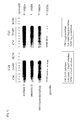
- FIG. 7 Results of analysis, by one-dimensional Western blotting, of change in expression of intracellular and cytoskeletal proteins with aging of epidermal keratinocytes
- FIG. 8 Results of analysis, by one-dimensional Western blotting, of change in expression of Galectin and IGFBP-3 with aging of epidermal keratinocytes
- FIG. 9 Results of analysis, by one-dimensional Western blotting, of change in expression of Keratins with aging of epidermal keratinocytes
- FIG. 10 Correlation between the aging and the expression of Keratin 15 extracted from the horny cell layer at the corner of the eye
- FIG. 11 Correlation between the aging and the expression of Keratin 10 extracted from the horny cell layer at the corner of the eye
- FIG. 12 Correlation between the aging and the elasticity at the corner of the eye
- FIG. 13 Correlation between the aging and the volumetric ratio of lines at the corner of the eye
- FIG. 14 Correlation between the elasticity at the corner of the eye and the expression of Keratin 7 extracted from the horny cell layer at the corner of the eye
- FIG. 15 Correlation between the elasticity at the corner of the eye and the expression of Keratin 15 extracted from the horny cell layer at the corner of the eye
- FIG. 16 Correlation between the volumetric ratio of lines at the corner of the eye and the expression of Keratin 7 extracted from the horny cell layer at the corner of the eye
- FIG. 17 Correlation between the volumetric ratio of lines at the corner of the eye and the expression of Keratin 15 extracted from the horny cell layer at the corner of the eye
- FIG. 18 Correlation between the elasticity at the corner of the eye and the expression of Kallikrein 7 extracted from the horny cell layer at the corner of the eye
- FIG. 19 Correlation between the volumetric ratio of lines at the corner of the eye and the expression of Kallikrein 7 extracted from the horny cell layer at the corner of the eye
- the present invention provides a method for determining the degree of skin aging, including measurement of secretory proteins and/or intracellular proteins in skin cells and/or skin tissues.
- Secretory proteins and/or intracellular proteins change their expression with aging of skin.
- Forms of “change in expression” include change in presence/absence of expression of a protein and/or its gene, change in the amount of expression, and change in the ratio of severed and non-severed types of an expressed protein due to protease.
- Secretory proteins and/or intracellular proteins used to determine the degree of skin aging are preferably selected from the group consisting of Kallikrein 7, PAI-1, uPA, Matriptase, GRP 94, HSP 70, HSP 90, Galectin-1, Galectin-3, Galectin-7, IGFBP-3, Enolase-1, Annexin II, Ezrin, Radixin, Moesin, Gelsolin, Keratin 7, Keratin 10, Keratin 13, Keratin 14, Keratin 15, Keratin 16, Keratin 18, Keratin 19, Keratin 20 and ⁇ 2-microglobulin.
- One or more proteins may be selected.
- Kallikrein 7 is a secretory protein with a molecular weight of 27,525 Da. It has the effect of promoting the desquamation of cells from the skin surface by severing the intercellular bond in the horny cell layer. Kallikrein 7 is expressed in a large amount in skin tissues, while it is also expressed in the brain, mammary gland, spinal cord and kidney. Gene sequence information (Stratum corneum chymotrytic enzyme, J. Biol. Chem. 269: 19420-19426, 1994, L33404). Amino acid sequence information (Kallikrein 7 precursor, J. Biol. Chem. 269: 19420-19426, 1994, P49862).
- PAI-1 is a secretory protein with a molecular weight of 45,060 Da. It is a type of serine protease and inhibits the activation of tPA (tissue plasminogen activator), uPA, protein C, etc. PAI-1 is a glycoprotein having stability against acids and belongs to the serpin family. PAI-1 is found in plasma, platelets, endothelial cells, liver cells, and fibrosarcoma cells. Gene sequence information (Plasminogen activator inhibitor-1, J. Clin. Invest. 78: 1673-1680, 1986, M16006). Amino acid sequence information (Plasminogen activator inhibitor-1, FEBS Lett. 210: 11-16, 1987, P05121).
- uPA is a secretory protein with a molecular weight of 48,525 Da.
- uPA is a type of serine protease that converts plasminogen into plasmine.
- uPA is secreted extracellularly as a precursor protein (55 kDa) and takes a severed form (35 kDa) to exhibit protease activity.
- a large amount of uPA is expressed in breast cancer patients.
- Gene sequence information (Urokinase-type plasminogen activator, Nucleic Acids Res. 13: 2759-2771, 1985, BC013575). Amino acid sequence information (Urokinase-type plasminogen activator, Hoppe-Seyler's Z. Physiol. Chem. 363: 1043-1058, 1982, P00749).
- Matriptase is a secretory protein with a molecular weight of 75,626 Da. It is an enzyme that breaks down extracellular matrixes. Matriptase has been reported to play a part in infiltration and metastasis of breast cancer. It exhibits enzyme activity similar to trypsin. It has also been shown that matriptase forms a complex with HAI-1 (Hepatocyte growth factor activation inhibitor 1), which is an inhibitor of matriptase. In addition, studies have shown that mice in which matriptase gene has been knocked will die due to the inability to control moisture evaporation from the skin. Gene sequence information (Membrane-type serine protease 1, J. Biol. Chem. 274: 18231-18236, 1999, AF188224). Amino acid sequence information (Membrane-type serine protease 1, J. Biol. Chem. 274: 18237-18242, 1999, Q9BS01).
- GRP 94 is a secretory protein with a molecular weight of 92,469 Da. It is a type of heat shock protein and a chaperon molecule that acts in the severing and transport of secretory proteins. Having a signal peptide, GRP 94 is secreted extracellularly in some situations or localized in ER in others.
- Gene sequence information tumor rejection antigen (gp96) 1, Proc. Natl. Acad. Sci. USA. 87: 5658-5662, 1990, BC066656). Amino acid sequence information (Endoplasmin, Nat. Biotechnol. 21: 566-569, 2003, P14625).
- HSP 70 is an intracellular protein with a molecular weight of 70,052 Da. Normally it functions as a molecular chaperon in cells and involves in the folding, transport, aggregation and breakdown of proteins, among others. In addition, HSP 70 helps proteins transfer normally in mitochondria and ER. HSP 70 also coordinates with HSP 90 to take part in signal transmission in cells. Gene basal sequence information (Heat shock 70 kDa protein 1A, Immunogenetics 32: 242-251, 1990, BC002453). Amino acid sequence information (Heat shock 70 kDa protein 1, P08107).
- HSP 90 is an intracellular protein with a molecular weight of 85,453 Da. Usually it plays an important role in the signal transmission system by bonding with/dissociating from complexes such as steroid hormone receptors. When cells are exposed to high temperature, HSP 90 changes its structure and acts as a molecular chaperon that prevents irreversible damage to proteins. Gene basal sequence information (Heat shock protein 90, Nucleic Acids Res. 17: 7108-710, 1989, X15183). Amino acid sequence information (Heat shock protein 90, J. Biol. Chem. 264: 2431-2437, 1989, P07900).
- Galectin-1 is an intracellular protein with a molecular weight of 14,585 Da. It is secreted extracellularly in some situations. Galectin-1 is present in the heart, stomach, skeletal muscles, nerves, thymus gland, kidney, placenta, etc. It bonds with ⁇ -galactoside, CD 45, CD 3, CD 4, etc. Galectin-1 is known to promote cell proliferation, induce apoptosis and influence immune response. It also specifically bonds with Laminin, Integrin and other extracellular matrix components and cell receptors to exert significant influence over adhesion and movement of cells. Gene sequence information (Galectin-1, J. Biol. Chem. 264: 1310-1316, 1989, BT006775). Amino acid sequence information (Galectin-1, J. Biochem. 104: 1-4, 1988, P09382).
- Galectin-3 is an intracellular protein with a molecular weight of 35,678 Da. It is a galactose-specific lectin that bonds with IgE. Galectin-3 is mainly expressed in the epithelium of large intestine as well as in active macrophages. Galectin-3 is produced by epidermal keratinocytes and present at the surface of islet of Langerhans in the skin, and reportedly bonds with IgE to control the immune system. Gene sequence information (Galectin-3, Proc. Natl. Acad. Sci. USA. 87: 7324-7328, 1990, AB006780). Amino acid sequence information (Galectin-3, P17931).
- Galectin-7 is an intracellular protein with a molecular weight of 14,944 Da. Generally it controls cell proliferation between cells or between a cell and an extracellular matrix. Being an apoptosis-related protein, Galectin-7 controls the activation of JNK and release of cytochrome C. It is also secreted in cytoplasm, nucleus and outside cells. Galectin-7 is a member of the galectin sub-family which is the first human epidermis to have been cloned. According to studies of cultured epidermal keratinocytes, Galectin-7 is expressed in all epidermal cells without being affected by the degree of cornification. Gene sequence information (Galectin-7, Dev. Biol. 168: 259-271, 1995, L07769). Amino acid sequence information (Galectin-7, J. Biol. Chem. 270: 5823-5829, 1995, P47929).
- IGFBP-3 is a secretory protein with a molecular weight of 31,660 Da. Generally it bonds with IGF to take on a longer half life. In cell cultures, IGFBP-3 suppresses or promotes cell proliferation. At the cell surface, it bonds with IGF receptors. IGFBP-3 has a greater bonding strength with IGF-2 than with IGF-1. IGFBP-3 is expressed in most tissues. Gene sequence information (Insulin-like growth factor binding protein-3, J. Biol. Chem. 265, 12642-12649, 1990, M35878). Amino acid sequence information (Insulin-like growth factor binding protein-3, J. Biol. Chem. 265, 14892-14898, 1990, P17936).
- ⁇ -enolase is expressed in most tissues, while ⁇ -enolase and ⁇ -enolase are expressed only in muscle tissues and nerve tissues, respectively.
- Gene sequence information (Alpha enolase, Proc. Natl. Acad. Sci. USA. 83: 6741-6745, 1986, M14328). Amino acid sequence information (Alpha enolase, Enzyme Protein 48: 37-44, 1995, P06733).
- Annexin II is an intracellular protein with a molecular weight of 38,473 Da. According to reports, some are also secreted extracellularly. It is a membrane-binding protein controlled by calcium, and this protein bonds with two calcium ions. Annexin II is localized near cell membrane. Of the two pairs of annexin repeats, one bonds with calcium, while the other bonds with phospholipid. This protein cross-links with phospholipid-bonded actins in cell membrane or proteins in the cytoskeletal system, or activates plasminogens via tPA. Gene basal sequence information (Annexin A2, Gene 95: 243-251, 1990, BC015834). Amino acid sequence information (Annexin A2, J. Biol. Chem. 266: 5169-5176, 1991, P07355).
- Ezrin is an intracellular protein with a molecular weight of 69,268 Da. Radixin and Moesin mentioned below are molecules belonging to the same family as Ezrin. Ezrin mainly connects proteins in the cytoskeletal system with cell membrane. It is localized inside the filiform projection called “microvilli” in cell membrane. Ezrin comprises the microvilli of intestinal epithelial cells. It is phosphorylated by tyrosine kinase. Gene sequence information (Ezrin, J. Biol. Chem. 264: 16727-16732, 1989, X51521). Amino acid sequence information (Ezrin, Biochem. Biophys. Res. Commun. 224: 666-674, 1996, P15311).
- Radixin is an intracellular protein with a molecular weight of 68,564 Da. It mainly connects proteins in the cytoskeletal system with cell membrane.
- Gene sequence information (Radixin, Genomics 16: 199-206, 1993, L02320).
- Amino acid sequence information (Radixin, P35241).
- Moesin is an intracellular protein with a molecular weight of 67,689 Da. It mainly connects proteins in the cytoskeletal system with cell membrane. Moesin expression reportedly decreases in abnormally differentiated epidermal keratinocytes. Gene sequence information (Moesin, Proc. Natl. Acad. Sci. USA. 88: 8297-8301, 1991, M69066). Amino acid sequence information (Moesin, Proc. Natl. Acad. Sci. USA. 88: 8297-8301, 1991, P26038).
- Gelsolin is a secretory protein with a molecular weight of 85,698 Da. There are two types: one secreted extracellularly and the other functioning intracellularly. Extracellularly secreted Gelsolin bonds with fibronectin. Gelsolin has three actin polymerization adjustment functions including promotion of actin fiber growth via formation of polymerized nucleus, severing of actin fiber, and protection of severed end of actin fiber. Genlsolin controls both the polymerization and depolymerization phases using these two-sided functions and plays an important role in cell movement. Gelsolin is expressed in large amounts in platelets and fibroblasts. Gene sequence information (Gelsolin, Nat. 323, 455-458, 1986, X04412). Amino acid sequence information (Gelsolin, Nat. Biotechnol. 21, 566-569, 2003, P06396).
- Keratin 7 is a cytoskeletal protein with a molecular weight of 51,287 Da and also a component of intermediate filament. It is expressed in various epithelial cells including gland cells. Keratin 7 is also expressed in the transitional epithelium of urinal tract, bile duct, lung, and epithelium of mammary gland. It is used as a tumor marker in some forms. Gene sequence information (Keratin 7, J. Cell Biol. 107: 1337-1350, 1988, AF509887). Amino acid sequence information (Keratin, type II cytoskeletal 7, P08729).
- Keratin 10 is a cytoskeletal protein with a molecular weight of 59,519 Da. It is a component of intermediate filament forming a hetero-tetramer with Keratin 1. Keratin 10 is expressed across the skin, but prominently in the prickle cell layer. Gene sequence information (Keratin 10, J. Mol. Biol. 204: 841-856, 1988, M19156). Amino acid sequence information (Keratin, type I cytoskeletal 10, Electrophoresis 13: 960-969, 1992, P13645).
- Keratin 13 is a cytoskeletal protein with a molecular weight of 49,586 Da. It is a component of intermediate filament forming a hetero-tetramer with Keratin 4. Keratin 13 is expressed in the tongue, esophagus, epithelium, and epithelium of urinal tract, among others. Gene sequence information (Keratin 13, Gene 215: 269-279, 1998, X52426). Amino acid sequence information (Keratin, type I cytoskeletal 13, P13646).
- Keratin 14 is a cytoskeletal protein with a molecular weight of 51,490 Da. It is a component of intermediate filament forming a hetero-tetramer with Keratin 5. Keratin 14 is present in the epithelium and particularly expressed in large amounts near the basal layer. It is also used as an undifferentiated marker for the epithelium. Gene sequence information (Keratin 14, Proc. Natl. Acad. Sci. USA. 82: 1609-1613, 1985, BC002690). Amino acid sequence information (Keratin, type I cytoskeletal 14, P02533).
- Keratin 15 is a cytoskeletal protein with a molecular weight of 49,167 Da. It is a component of intermediate filament. Keratin 15 is mainly localized in the basal layer of epithelium. Gene sequence information (Keratin 15, J. Cell Biol. 106: 1249-1261, 1988, X07696). Amino acid sequence information (Keratin, type I cytoskeletal 15, P19012).
- Keratin 16 is a cytoskeletal protein with a molecular weight of 51,136 Da. It is a component of intermediate filament forming a hetero-tetramer with Keratin 6. It is expressed in hair follicles, nails, flat epithelium layer in the oral cavity, basal layer of epithelium, epithelium of palma manus, and sweat glands. Keratin 16 is involved in the proliferation and differentiation of cells. Gene sequence information (Keratin 16, Biochem. Biophys. Res. Commun. 215: 517-523, 1995, AF061812). Amino acid sequence information (Keratin, type I cytoskeletal 16, Electrophoresis 13: 960-969, 1992, P08779).
- Keratin 18 is a cytoskeletal protein with a molecular weight of 47,926 Da. It is a component of intermediate filament forming a hetero-tetramer with Keratin 8. Keratin 18 is present in the epithelium of most single layer glands. Patients lacking Keratin 18 have risks of developing a sudden hepatic failure. Gene sequence information (Keratin 18, Differentiation 33: 61-68, 1986, M26325). Amino acid sequence information (Keratin, type I cytoskeletal 18, Electrophoresis 18: 605-613, 1997, P05783).
- Keratin 19 is a cytoskeletal protein with a molecular weight of 44,106 Da. It is the smallest form of Keratin in terms of molecular weight. Keratin 19 is a component of intermediate filament. It is expressed in large amounts in most forms of single layer epithelium and non-cornified stratified epithelium. Keratin 19 is also expressed in all types of adenocarcinoma. Gene sequence information (Keratin 19, J. Invest. Dermatol. 92: 707-716, 1989, Y00503). Amino acid sequence information (Keratin, type I cytoskeletal 19, Electrophoresis 13: 960-969, 1992, P08727).
- Keratin 20 is a cytoskeletal protein with a molecular weight of 48,486 Da. It is a component of intermediate filament. Among normal cells, Keratin 20 is present in endocrine cells in the epithelium of intestinal tract, epithelium of digestive tract and upper part of gastropyloric, as well as in Merkel cells in the epithelium of urinal tract and skin. Keratin 20 is used as a specific marker protein for colon cancer, etc. Gene sequence information (Keratin 20, Differentiation 53: 75-93, 1993, BC031559). Amino acid sequence information (Keratin, type I cytoskeletal 20, P35900).
- ⁇ 2-microglobulin is a secretory protein with a molecular weight of 13,175 Da. It is the L chain in HLA class I antigens (histocompatible antigens). ⁇ 2-microglobulin is present at the surface of many cells. It is used as a diagnostic indicator for renal failure and various types of malignant tumors, etc. Although ⁇ 2-microglobulin in severed form is found in some cases, what it means is not very clear. Gene sequence information (Beta-2-microglobulin, J. Immunol. 139: 3132-3138, 1987, AB021288). Amino acid sequence information (Beta-2-microglobulin, Biochemistry 12: 4811-4822 (1973), P61769).
- the above proteins may be a precursor protein or mature protein, or of severed type or non-severed type.
- precursor proteins include pro-proteins and prepro-proteins, among others.
- Some pro-proteins and prepro-proteins have a signal peptide, which is a specific secretory protein sequence.
- expression of secretory proteins and/or intracellular proteins in skin cells and/or skin tissues may be measured or expression of their genes may be measured.
- such measurement can be performed using the Northern blotting method, PCR method, Western blotting method, immunohistochemical analysis method, etc.
- cDNA microarray, ELISA method, antibody array, etc. can also be used to perform measurement.
- an antibody that specifically recognizes the target protein may be a monoclonal antibody or polyclonal antibody. These antibodies can be manufactured by known methods and some are commercially available. If the Western blotting method is used to perform measurement, the antibody is secondarily detected using 125 I labeled protein A, peroxidase-binding IgG, etc. If measurement is performed using the immunohistochemical analysis method, the antibody are preferably labeled with a fluorochrome, ferritin, enzyme, etc.
- nucleic acid probes capable of specifically hybridizing with mRNA of the target protein if the Northern blotting method is used to perform measurement.
- a pair of nucleic acid primers may be used, including a nucleic acid primer specifically hybridizing with mRNA of the target protein, and a nucleic acid primer capable of specifically hybridizing with cDNA synthesized using the aforementioned mRNA as a mold (if the PCR method is used to perform measurement).
- Nucleic acid probes and primers can be designed based on the gene information of the target protein. Normally, nucleic acid probes having a basicity of approx. 15 to 1500 are appropriate. Nucleic acid probes are preferably labeled with a radioactive element, fluorochrome, enzyme, etc. Normally, nucleic acid primers having a basicity of approx. 15 to 30 are appropriate.
- Presence/absence of expression of secretory proteins and/or intracellular proteins or their genes in skin cells and/or skin tissues may be detected or the amount of expression may be measured.
- Presence/absence of expression of proteins and/or their mRNA can be checked from presence/absence of appearance of spots and bands at specified locations.
- the amount of expression of proteins and/or their mRNA can be measured using the staining intensity of spots and bands. Alternatively, proteins and/or their mRNA may be quantified.
- Skin cells and skin tissues may be derived from any life form, where examples include those derived from human, pig, monkey, chimpanzee, dog, cow, rabbit, rat, mouse and other mammals. To determine the degree of aging of human skin, however, human-derived tissues need to be used.
- skin biopsy samples or cultured skin cells, cultured skin tissues and the like, obtained from skin biopsy samples, can be used to determine the degree of skin aging.
- skin cells examples include epidermal keratinocytes, skin fibroblasts, Langerhans cells, melanin cells, mast cells, endothelial cells, sebum cells, hair papilla cells and hair matrix cells, among others.
- Skin cells are supplied from the Health Science Research Resources Bank and RIKEN Cell Bank in Japan, or from the American Type Culture Collection outside Japan. They can also be purchased from Clontech, Kurabo and PromoCell. It is also possible to collect skin cells from skin using known methods ( Bunshi Seibutsugaku Kenkyu no Tameno Shin Saibobaiyo Jikkenho (New Experimental Cell Culture Methods for Molecular Biology Research), p. 57-71, Yodosha, 1999).
- Examples of skin tissues include horny cell layer, epidermis and dermis of skin, among others. Skin tissues can be purchased from Biochain and Super Bio Chips. It is also possible to collect skin tissues from skin using known methods (Acta. Derm. Venereol., 85, 389-393, 2005).
- Skin models are sold by Kurabo, Toyobo, etc., and these commercially available models can be used. Models can also be fabricated from known methods (J. Invest. Dermatol., 104 (1), 107-112, 1995).
- Skin biopsy samples may be cells or tissues. Skin biopsy samples are preferably those of the skin's horny cell layer collected by a horny cell layer checker or other tapes, as described later in examples.
- Horny cell layer checkers measure the degree of parakeratosis and cell area of the horny cell layer, and have been used for many years in the collection of samples of horny cell layer for the purpose of evaluating the degree of skin roughness and turnover rate of horny cell layer ( Keshohin no Yuyosei: Hyoka Gijutsu no Shimpo to Shorai Tembo (Utility of Cosmetics: Progress and Future Perspective of Evaluation Technologies), Society of Cosmetic Chemists of Japan, Yakuji Nippo, p 95-96). These checkers are very useful in collecting samples from the horny cell layer in a non-invasive, easy and safe manner at a counseling outlet or home.
- the degree of skin aging refers to the degree of aging signs exhibited by the skin such as lines and sagging due to age. It is obvious that lines and sagging progress with age. In addition to age, exposure to UV rays also worsens lines, sagging, dull complexion and pigmentation. Lines and sagging can be measured using known methods ( Keshohin no Yuyosei: Hyoka Gijutsu no Shimpo to Shorai Tembo (Utility of Cosmetics: Progress and Future Perspective of Evaluation Technologies), Society of Cosmetic Chemists of Japan, Yakuji Nippo, 154-158, 169-175, 179-182, 187-190, 2001).
- the degree of skin aging can be determined using, as an indicator, drop in the proliferation ability of skin cells.
- SA- ⁇ -Gal senescence-associated ⁇ -galactosidase
- the degree of skin aging can be determined using the standards explained below.
- skin samples are obtained from volunteers in different age groups and the amount of expression is measured for specific marker proteins to create standard curves showing the relationships of measured amounts of expression with skin elasticity and degree of lines that are known as indicators of skin aging.
- the degree of skin aging is determined for analysis samples by comparing the amounts of expressions of standard samples and analysis samples.
- ⁇ 2-microglobulin skin samples are obtained from volunteers in different age groups and the average ratio of severed and non-severed types is calculated before creating standard curves showing the relationships of measured amounts of expression with skin elasticity and degree of lines that are known as indicators of skin aging.
- the degree of skin aging is determined for analysis samples by comparing the amounts of expressions of standard samples and analysis samples.
- the present invention also provides a kit for determining the degree of skin aging.
- a kit conforming to the present invention includes antibodies capable of specifically recognizing secretory proteins and/or intracellular proteins whose expression changes with aging of skin.
- Secretory proteins and/or intracellular proteins are preferably selected from the group consisting of Kallikrein 7, PAI-1, uPA, Matriptase, GRP 94, HSP 70, HSP 90, Galectin-1, Galectin-3, Galectin-7, IGFBP-3, Enolase-1, Annexin II, Ezrin, Radixin, Moesin, Gelsolin, Keratin 7, Keratin 10, Keratin 13, Keratin 14, Keratin 15, Keratin 16, Keratin 18, Keratin 19, Keratin 20 and ⁇ 2-microglobulin.
- the kit may also include a tape for collecting skin tissues, a set of reagents for immunochemically detecting the proteins collected on the tape, and an operating manual, among others.
- the operating manual includes description of, among others, how to use the kit, as well as the judgment criteria for determining the degree of skin aging.
- a kit conforming to the present invention includes nucleic acid probes capable of specifically hybridizing with mRNA of secretory proteins and/or intracellular proteins whose expression changes with aging of skin.
- Secretory proteins and/or intracellular proteins are preferably selected from the group consisting of Kallikrein 7, PAI-1, uPA, Matriptase, GRP 94, HSP 70, HSP 90, Galectin-1, Galectin-3, Galectin-7, IGFBP-3, Enolase-1, Annexin II, Ezrin, Radixin, Moesin, Gelsolin, Keratin 7, Keratin 10, Keratin 13, Keratin 14, Keratin 15, Keratin 16, Keratin 18, Keratin 19, Keratin 20 and ⁇ 2-microglobulin.
- the kit may also include a tape for collecting skin tissues, reagents for extracting RNA from the skin tissues collected on the tape, reagents for analyzing RNA using the Northern blotting method, and an operating manual, among others.
- the operating manual includes description of, among others, how to use the kit, as well as the judgment criteria for determining the degree of skin aging.
- a kit conforming to the present invention includes a pair of nucleic acid primers comprising a nucleic acid primer capable of specifically hybridizing with mRNA of secretory proteins and/or intracellular proteins whose expression changes with aging of skin, and a nucleic acid primer capable of specifically hybridizing with cDNA synthesized using the aforementioned mRNA as a mold.
- Secretory proteins and/or intracellular proteins are preferably selected from the group consisting of Kallikrein 7, PAI-1, uPA, Matriptase, GRP 94, HSP 70, HSP 90, Galectin-1, Galectin-3, Galectin-7, IGFBP-3, Enolase-1, Annexin II, Ezrin, Radixin, Moesin, Gelsolin, Keratin 7, Keratin 10, Keratin 13, Keratin 14, Keratin 15, Keratin 16, Keratin 18, Keratin 19, Keratin 20 and ⁇ 2-microglobulin.
- the kit may also include a tape for collecting skin tissues, reagents for extracting RNA from the skin tissues collected on the tape, reagents for analyzing RNA using the RT-PCR method, and an operating manual, among others.
- the operating manual includes description of, among others, how to use the kit, as well as the judgment criteria for determining the degree of skin aging.
- the present invention also provides a method for identifying substances effective in the prevention of skin aging. This method includes the following steps:
- the target substance may be any substance, such as a protein, peptide, vitamin, hormone, polysaccharide, oligosaccharide, monosaccharide, low-molecular compound, nucleic acid (DNA, RNA, oligonucleotide, mononucleotide, etc.), lipid, other natural compound, synthetic compound, or any mixture thereof.
- the target substance may be caused to contact skin cells and/or skin tissues using any method, such as a method to add the target substance in a culture solution of skin cells and/or skin tissues, or a method to culture skin cells and/or skin tissues in a culture container or on a culture sheet on which the target substance has been applied or fixed. It is also possible to use a method to apply the target substance directly over the skin, or orally administer the target substance, using a life form such as human or other mammal (such as mouse, rat, guinea pig, rabbit, pig, etc.).
- a life form such as human or other mammal (such as mouse, rat, guinea pig, rabbit, pig, etc.).
- the culture time of skin cells and/or skin tissues is not specifically limited, and a desired time can be set as long as it is enough to check whether the target substance has any effect on the expression of secretory proteins and/or intracellular proteins or their genes in skin cells and/or skin tissues. If normal human epidermal keratinocytes are used as skin cells, for example, a culture time of 12 to 48 hours is appropriate, and that of 12 to 24 hours is preferable.
- culture skin cells and/or skin tissues means growing/proliferating skin cells and/or skin tissues, and the definition also encompasses sustaining the life of, breeding and rearing life forms having skin cells or skin tissues in addition to growing/proliferating isolated single skin cells and/or skin tissues.
- the control skin cells and/or skin tissues to be compared against may be skin cells and/or skin tissues not yet contacted by the target substance, or skin cells and/or skin tissues that have been given the same treatment except that they do not contact the target substance.
- Secretory proteins and/or intracellular proteins are preferably selected from the group consisting of Kallikrein 7, PAI-1, uPA, Matriptase, GRP 94, HSP 70, HSP 90, Galectin-1, Galectin-3, Galectin-7, IGFBP-3, Enolase-1, Annexin II, Ezrin, Radixin, Moesin, Gelsolin, Keratin 7, Keratin 10, Keratin 13, Keratin 14, Keratin 15, Keratin 16, Keratin 18, Keratin 19, Keratin 20 and ⁇ 2-microglobulin.
- the target substance can be identified as being a substance effective in the prevention of skin aging.
- the ratio of non-severed type to severed type of ⁇ 2-microglobulin increases with aging of epidermal keratinocytes. Accordingly, if the ratio of non-severed type to severed type of ⁇ 2-microglobulin has decreased compared to the control in the skin cells and/or skin tissues contacted by the target substance, and the target substance can be evaluated to have the effect of decreasing the ratio of non-severed type to severed type of ⁇ 2-microglobulin, then the target substance can be identified as being a substance effective in the prevention of skin aging.
- a medium KGM (+) prepared by adding additive factors (0.1 ng/ml EGF, 0.03 mg/ml bovine pituitary medium, 5 ⁇ g/ml insulin, 0.5 ⁇ g/ml hydrocortisone, GA-1000) to a serum-free basal medium (KBM, Cambrex Corporation), normal human epidermal keratinocytes (NHEK, Cambrex Corporation) were cultured in a 37° C., 5% CO 2 incubator. To collect the supernatant of culture (CM; Conditioned Medium) and cell extracts (CL: Cell Lysates), cells were inoculated in 5 ⁇ 10 5 -cells/150-mm dishes and the medium was changed every two days to continue culturing until saturation.
- additive factors 0.1 ng/ml EGF, 0.03 mg/ml bovine pituitary medium, 5 ⁇ g/ml insulin, 0.5 ⁇ g/ml hydrocortisone, GA-1000
- KBM serum-free basal medium
- NHEK
- SA- ⁇ -Gal in each passage of NHEK was measured using a senescent cells staining kit (Sigma Corporation). Separately from those used for passage, cells were inoculated in three 1 ⁇ 10 5 -cells/60-mm dishes and when 80% saturation was reached, the cells were fixed for 7 minutes at room temperature in an aqueous solution containing 2% formaldehyde and 0.2% glutaraldehyde. Furthermore, the cells were washed three times using 1 ⁇ PBS ( ⁇ ), after which a staining solution was added to stain the cells overnight at 37° C. The number of SA- ⁇ -Gal stained cells per 100 cells was measured for each of the three dishes.
- a dish containing saturated NHEK was washed twice using KBM and cultured for one day after changing the medium to KBM to completely remove the additive factors.
- Two days after changing the medium to KBM CM was collected. The collected CM was centrifuged for 10 minutes at 130 ⁇ g to remove suspended cells, and then centrifuged again for 30 minutes at 10,000 ⁇ g, after which supernatant was collected.
- Ammonium sulfate (Wako Pure Chemical Industries, Ltd.) was added to this CM to achieve 80% saturation and the mixture was agitated to fully dissolve solids, and the obtained liquid was let stand overnight at 4° C.
- the liquid was centrifuged for 30 minutes at 25,000 ⁇ g to precipitate protein, and the precipitate was dissolved in 20 mM Tris-HCl (pH 7.4) until a concentration of 500 times to condense the CM. Furthermore, the condensed CM was dialyzed using 20 mM Tris-HCl (pH 7.4).
- a fixed quantity of protein was measured by the Bradford method using a DC protein assay kit (Bio-Rad Laboratories Inc.) and used as a Western blotting sample.
- the prepared CM was freeze-dried and the obtained dry powder was dissolved in a two-dimensional electrophoresis sample preparation (6.5M urea (ICN Biomedicals, Inc.), 1.5M thiourea (Sigma Corporation), 0.15% SDS (Wako Pure Chemical Industries, Ltd.), 0.85% Triton X-100 (Wako Pure Chemical Industries, Ltd.), 0.4% CHAPS (Dojindo Laboratories), 2.5 mM tributyl phosphine (Wako Pure Chemical Industries, Ltd.)) to achieve the final concentration of 500 times, and a fixed quantity of protein was measured by the dot blotting method.
- the dish from which the CM had been collected was washed three times using PBS ( ⁇ ), and then frozen at ⁇ 30° C. to destroy the cell membrane. Thereafter, cells were collected using a cell scraper (Sumitomo Bakelite Co., Ltd.) and given ultrasonic treatment for 30 seconds. The obtained cells were mixed with 1% Triton X-100 and let stand for 30 minutes at 4° C. Next, the cells were centrifuged for 30 minutes at 15,000 ⁇ g to remove insolubles, and supernatant was taken as a sample. A fixed quantity of protein was measured using the dot blotting method to use as a Western blotting sample.
- a gel-swelling solution 5M urea (Wako Pure Chemical Industries, Ltd.), 2M thiourea (Wako Pure Chemical Industries, Ltd.), 0.5% ampholytes (pH 3.5 to 10) (Amersham Biosciences), 0.0025% orange G (Wako Pure Chemical Industries, Ltd.), 2.5 mM tributyl phosphine (Wako Pure Chemical Industries, Ltd.), 1% Triton X-100) so that the total quantity becomes 340 ⁇ l, after which the mixture was added to an Immobiline DryStrip gel (18 cm, pH 3 to 10, NL) (Amersham Biosciences) to swell overnight at 20° C.
- Immobiline DryStrip gel (18 cm, pH 3 to 10, NL
- the gel was soaked in a SDS equalizing buffer (5.8 M urea, 0.06M thiourea, 0.5% dithiothereitol (DTT, Wako Pure Chemical Industries, Ltd.), 25% glycerol (Wako Pure Chemical Industries, Ltd.), 0.0025% BPB (Wako Pure Chemical Industries, Ltd.)) and equalized for 1 hour at room temperature.
- a SDS equalizing buffer 5.8 M urea, 0.06M thiourea, 0.5% dithiothereitol (DTT, Wako Pure Chemical Industries, Ltd.), 25% glycerol (Wako Pure Chemical Industries, Ltd.), 0.0025% BPB (Wako Pure Chemical Industries, Ltd.)
- SDS-PAGE was performed using a Tris-Tricine buffer (cathode buffer: 0.05M Tris, 0.05M Tricine (Bio-Rad Laboratories Inc.), 0.05% SDS; anode buffer: 1M Tris-HCl (pH 8.8)) and 7.5% acrylamide gel of 18 cm ⁇ 18 cm.
- Tris-Tricine buffer cathode buffer: 0.05M Tris, 0.05M Tricine (Bio-Rad Laboratories Inc.), 0.05% SDS; anode buffer: 1M Tris-HCl (pH 8.8)) and 7.5% acrylamide gel of 18 cm ⁇ 18 cm.
- the gel completing SDS-PAGE was transferred for 2 hours to PVDF membranes of 20 cm ⁇ 20 cm (ProBlot Membranes (Applied Biosystems)) using a semi-dry type transfer system (Nihon Eido Co., Ltd.) at a constant current of 150 mA.
- anode liquid 1 comprising 0.3M Tris-HCl (pH 10.4) and 20% methanol (Wako Pure Chemical Industries, Ltd.), anode liquid 2 comprising 25 mM Tris-HCL (pH 10.4) and 20% methanol, and cathode liquid comprising 25 mM Tris-HCl (pH 10.4), 20% methanol and 40 mM 6-amino hexanoic acid (Wako Pure Chemical Industries, Ltd.) were used.
- the transferred gel was washed three times for 20 minutes each using a TTBS buffer (20 mM Tris-HCl (pH 7.5) (Bio-Rad Laboratories Inc.), 500 mM NaCl (Wako Pure Chemical Industries, Ltd.), 0.3% Tween 20 (Bio-Rad Laboratories Inc.)), and then washed three times for 2 minutes each using pure water.
- TTBS buffer 20 mM Tris-HCl (pH 7.5) (Bio-Rad Laboratories Inc.), 500 mM NaCl (Wako Pure Chemical Industries, Ltd.), 0.3% Tween 20 (Bio-Rad Laboratories Inc.)
- the PVDF membranes were sealed by vinyl membranes and soaked in a gold colloidal solution (Colloidal Gold Total Protein Staine (Bio-Rad Laboratories Inc.)), and the solution was shaken for 1 to 2 hours to stain the protein. Thereafter, the gold colloidal solution was removed and the residue was washed five times for 1 minute each using
- the PVDF membranes were then removed and transferred into a tube containing a Lys-C digesting buffer (70% acetonitrile/20 mM Tris-HCl (pH 9.0)) and rinsed two to three times, followed by soaking in the Lys-C digesting buffer to implement protease digestion for 1 hour.
- a Lys-C digesting buffer (70% acetonitrile/20 mM Tris-HCl (pH 9.0)
- the protease-digested solution was diluted seven times using distilled water to an acetonitrile concentration of 10%. As a pretreatment, suction and discharge was repeated several times using 50% acetonitrile/0.1% trifluoro-acetic acid (TFA, Wako Pure Chemical Industries, Ltd.), and several times using 2% acetonitrile/0.1% TFA, in order to activate the filler part of the ZipTipc18 pipette tip (Millipore Corporation). Next, a protease-digested solution was suctioned and discharged several times using the activated ZipTipc18 pipette tip to cause the fragmented peptide to be adsorbed to the filler part.
- TFA trifluoro-acetic acid
- SDS-PAGE was performed with a Laemmli Tris-Glycin system using a Western blotting sample. After the SDS-PAGE, protein in the gel was transferred to PVDF membranes (Millipore Corporation) at a constant current of 0.8 mA per 1 cm 2 , and then soaked in 5% skim milk/PBS ( ⁇ ) and blocked overnight at 4° C. The PVDF membranes were washed three times using 0.1% Tween 20/PBS ( ⁇ ), and then soaked in each primary antibody solution and shaken for 1 hour at room temperature.
- the types of primary antibodies used, and their dilution factors, are as follows: rabbit anti-maspin polyclonal antibody (Santa Cruz Biotechnology) (diluted by 1000 times), mouse anti-cystatin A monoclonal antibody (Sigma) (diluted by 1000 times), rabbit anti-kallikrein 7 polyclonal antibody (Santa Cruz Biotechnology) (diluted by 1000 times), sheep anti-PAI-1 polyclonal antibody (Biopool) (diluted by 1000 times), mouse anti-PAI-2 monoclonal antibody (American Diagnostica) (diluted by 1000 times), rabbit anti-uPA polyclonal antibody (Santa Cruz Biotechnology) (diluted by 1000 times), rabbit anti-matriptase polyclonal antibody (Calbiochem) (diluted by 1000 times), rat anti-GRP 94 monoclonal antibody (Stressgeen) (diluted by 1000 times), mouse anti-HSP 70 monoclonal antibody (Santa Cruz Biotechnology) (diluted by 1000 times), mouse anti-HSP 90 monoclonal antibody (Santa Cruz Biotechnology)
- sheep anti-PAI-1 polyclonal antibody mouse anti-PAI-2 monoclonal antibody, rabbit anti-uPA polyclonal antibody or rabbit anti-matriptase polyclonal antibody was used as the primary antibody
- biotinylated anti-goat IgG Vector Laboratories
- biotinylated anti-sheep IgG Vector Laboratories
- horseradish peroxidase labeled anti-mouse IgG (Amersham Bioscience) (diluted by 5000 times), horseradish peroxidase labeled anti-rabbit IgG (Amersham Bioscience) (diluted by 5000 times), horseradish peroxidase labeled anti-rat IgG (Amersham Bioscience) (diluted by 5000 times) or horseradish peroxidase labeled anti-goat IgG (Santa Cruz) (diluted by 5000 times) was used as the secondary antibody.
- an enhanced chemiluminescence (ECL) kit (Amersham Bioscience) was used to implement detection.
- the amount of expression of protein detected by the Western blotting method was quantified using NIH image and the amount of expression of fifth-passage protein was divided by the amount of expression of second-passage protein. If the calculated value was 2.0 or more, it was determined that the amount of expression increased significantly with aging of epidermal keratinocytes. If the calculated value was less than 0.5, it was determined that the amount of expression decreased significantly with aging of epidermal keratinocytes. If the calculated value was 0.5 or more but less than 2.0, on the other hand, it was determined that expression did not change with aging of epidermal keratinocytes.
- FIG. 1 ( a ) plots the number of days before the 150-mm dish saturated for each passage. While it took six days for the dish to saturate with the first passage, the number of days increased to 28 with the fifth passage, suggesting an exponential increase in the proliferation rate from the first to fifth passages. Proliferation stopped completely with the sixth passage. In terms of cell form, fifth-passage cells were around twice as big as second-passage cells, and the former became flattened and enlarged reflecting the common trend observed in cell aging ( FIG. 1 ( b )).
- Aging or terminal differentiation of cells is one cause of reduced proliferation of epidermal keratinocytes through repeated passages of culture. Accordingly, we examined the activity of SA- ⁇ -Gal identified as an aging marker for epidermal keratinocytes and skin tissues to determine if epidermal keratinocytes had actually aged, because this marker is known to express only when cells have aged, not when cells have terminally differentiated (Proc. Natl. Acad. Sci. USA, 92, 9363-9367, 1995). While second-passage cells were not stained blue to indicate SA- ⁇ -Gal, roughly a half of fifth-passage cells were stained blue ( FIG. 2 ).
- CMs of second to fifth passages were prepared to analyze, by two-dimensional electrophoresis, those proteins whose expression changes ( FIGS. 3( a ) to 3 ( d )).
- FIGS. 3( a ) to 3 ( d ) When the spots taken from the supernatants of cell cultures of young cells (second passage, FIG. 3( a )) and aged cells (fifth passage, FIG. 3( b )) were compared, 19 proteins whose expression decreased with cell aging (Table 1) and 49 proteins whose expression increased with cell aging (Table 2) were identified.
- the identified proteins included known skin aging markers such as Maspin, Cystatin A and HSP 27, and therefore the analysis by two-dimensional electrophoresis performed in this test was deemed appropriate in identifying proteins that change with aging of skin.
- Table 1 lists the proteins whose expression decreased with aging of epidermal keratinocytes in the two-dimensional electrophoresis analysis.
- Table 2 lists the proteins whose expression increased with aging of epidermal keratinocytes in the two-dimensional electrophoresis analysis.
- serine protease inhibitors Maspin and PAI-2 and cysteine protease inhibitor Cystatin A all exhibited an increased amount of expression with aging ( FIG. 5 ( a )( b )).
- serine proteases Kallikrein 7 and uPA, membrane-binding serine protease Matriptase and serine protease inhibitor PAI-1 all exhibited a decreased amount of expression with aging. While ⁇ 2-microglobulin is known to be severed by acid proteases, it was found that the ratio of severed type (13 kDa) would decrease and that of non-severed type (14 kDa) would increase with aging ( FIG. 5 ( a )).
- HSP 70, HSP 90 and GRP 94 (only GRP 94 has a signal peptide), which are all an intracellular protein and a type of heat shock protein, were secreted more in CM with aging. However, the amount of expression in CL did not change with any of them ( FIG. 6 ).
- Enolase-1 which is an intracellular glycolysis enzyme, Annexin II that bonds with phospholipids in cell membrane, and cytoskeletal proteins Ezrin, Moesin and Radixin were found to secrete more in CM with aging ( FIG. 7 ).
- Gelsolin which is a cytoskeletal protein, did not change the amount of secretion in CM, but its amount of expression in CL increased ( FIG. 7 ).
- Galectin-1, Galectin-3 and Galectin-7 of the lectin family, and IGFBP-3 showed a significant increase in the amount of expression of the protein secreted extracellularly with aging of epidermal keratinocytes ( FIG. 8 ).
- Galectin-1, Galectin-3, Galectin-7 and IGFBP-3 were not detected in CL ( FIG. 8 ).
- Keratins that are cytoskeletal proteins
- 13 types Keratin 3, 5, 7, 8, 10, 13, 14, 15, 16, 17, 18, 19 and 20
- Keratin 10, 13, 14, 15, 16, 18, 19 and 20 showed a significant increase in their expression in cells with aging of epidermal keratinocytes ( FIG. 9 ).
- the expression of Keratin 7 in cells decreased significantly with aging of epidermal keratinocytes ( FIG. 9 ).
- No change in expression was observed for Keratin 3, 5, 8 and 17. Keratins were not detected in CM.
- Table 3 lists the proteins whose expression changed with aging of epidermal keratinocytes in the one-dimensional Western blotting analysis.
- Table 3 lists the proteins whose expression changed with aging of epidermal keratinocytes in the one-dimensional Western blotting analysis.
- Elasticity and volumetric ratio of lines were measured at the corner of the eye on 59 women in their 20s to 60s. Elasticity at the corner of the eye was measured as suction elasticity using a Cutometer (Courage+Khazaka electronic GmbH). Suction elasticity is a measure that provides an indicator of skin elasticity, and this elasticity has been shown to decrease with aging. Volumetric ratio of lines at the corner of the eye was measured via replica image analysis by collecting replicas from the corner of the eye using Skin Cast (Yamada Cosmetic Laboratories) which is a replica agent consisting of two parts. Replica image analysis was then performed using a reflective replica analysis system ASA-03RXD.
- the ASA-03RXD was used to irradiate parallel light onto the collected replica at an angle of 30 degrees and the obtained shadow image reflecting the shapes of lines was captured with a CCD camera and imported to a computer for image processing to measure the volumetric ratio of lines at the replica surface ( ⁇ m 3 /mm 2 /100).
- volumetric ratio of lines has been shown to increase with aging.
- the horny cell layer was peeled from the corner of the eye using a horny cell layer checker (Asahibiomed Co., Ltd.) on 59 women in their 20 to 60s, and protein was extracted from the obtained samples using a protein-extracting solution.
- the horny cell layer checker was attached to the corner of the eye of the subject and the checker was gently rubbed from above with the fingertip.
- the sample collection was repeated three times, and then 50 ⁇ l of a protein-extracting solution (50 mM Tris-HCl (pH 7.5), 120 mM NaCl, 0.4% Igepal CA-630 (Sigma-Aldrich Co.)) was added to the horny cell layer attached to each of the three horny cell layer checkers and a cell scraper was used to spread the protein-extracting solution evenly over the horny cell layer, after which the solution was collected into a 1.5-ml plastic tube (Eppendorf AG).
- a protein-extracting solution 50 mM Tris-HCl (pH 7.5), 120 mM NaCl, 0.4% Igepal CA-630 (Sigma-Aldrich Co.
- the collected solution was centrifuged for 15 minutes at 10,000 ⁇ g to precipitate insolubles, and supernatant was collected into a new 1.5-ml plastic tube.
- the mass of protein in the collected sample was measured using a DC assay kit.
- Keratin 7 Keratin 10
- Keratin 15 Keratin 15
- Kallikrein 7 Among the proteins whose expression changes with aging of epidermal keratinocytes, Keratin 7, Keratin 10, Keratin 15 and Kallikrein 7 were examined to see if there was any correlation with the degree of aging of human skin.
- Keratin 10 Since Keratin 10 has been shown to decrease its expression with skin aging, which of Keratin 10, Keratin 7 and Keratin 15 would exhibit correlation with the degree of aging of human skin was examined. When the correlation of Keratin 10/Keratin 15 and age was examined, Keratin 10 showed no correlation with age, while Keratin 15 was correlated with age. Accordingly, it was found that Keratin 15 would provide a more appropriate skin aging marker than existing skin aging markers ( FIGS. 10 , 11 ).
- FIG. 14 shows the correlation between the amount of expression of Keratin 7 and the elasticity at the corner of the eye
- FIG. 15 shows the correlation between the amount of expression of Keratin 15 and the elasticity at the corner of the eye.
- the amount of expression of Keratin 7 decreased significantly as the elasticity at the corner of the eye dropped, while the amount of expression of Keratin 15 increased significantly as the elasticity at the corner of the eye dropped.
- FIG. 16 shows the correlation between the amount of expression of Keratin 7 and the volumetric ratio of lines
- FIG. 17 shows the correlation between the amount of expression of Keratin 15 and the volumetric ratio of lines.
- the amount of expression of Keratin 7 decreased significantly as the volumetric ratio of lines at the corner of the eye increased, while the amount of expression of Keratin 15 increased significantly as the volumetric ratio of lines at the corner of the eye increased.
- FIG. 18 shows the correlation between the amount of expression of Kallikrein 7 and the elasticity at the corner of the eye
- FIG. 19 shows the correlation between the amount of expression of Kallikrein 7 and the volumetric ratio of lines at the corner of the eye.
- the amount of expression of Kallikrein 7 decreased significantly as the elasticity at the corner of the eye dropped, and also decreased significantly as the volumetric ratio of lines at the corner of the eye increased.
Applications Claiming Priority (3)
| Application Number | Priority Date | Filing Date | Title |
|---|---|---|---|
| JP2005240820 | 2005-08-23 | ||
| JP2005-240820 | 2005-08-23 | ||
| PCT/JP2006/316407 WO2007023808A1 (ja) | 2005-08-23 | 2006-08-22 | 皮膚老化マーカーとその利用技術 |
Publications (2)
| Publication Number | Publication Date |
|---|---|
| US20090305242A1 US20090305242A1 (en) | 2009-12-10 |
| US7972788B2 true US7972788B2 (en) | 2011-07-05 |
Family
ID=37771551
Family Applications (1)
| Application Number | Title | Priority Date | Filing Date |
|---|---|---|---|
| US12/064,602 Expired - Fee Related US7972788B2 (en) | 2005-08-23 | 2006-08-22 | Skin aging marker and technique for use thereof |
Country Status (6)
| Country | Link |
|---|---|
| US (1) | US7972788B2 (ko) |
| JP (4) | JP5362219B2 (ko) |
| KR (1) | KR101318521B1 (ko) |
| CN (1) | CN101248192B (ko) |
| HK (1) | HK1114883A1 (ko) |
| WO (1) | WO2007023808A1 (ko) |
Cited By (5)
| Publication number | Priority date | Publication date | Assignee | Title |
|---|---|---|---|---|
| US9994849B2 (en) | 2015-12-24 | 2018-06-12 | Samsung Electronics Co., Ltd. | Method of separating senescent cells using overexpression of protocadherin |
| US10407729B2 (en) | 2008-05-14 | 2019-09-10 | Dermtech, Inc. | Diagnosis of melanoma by nucleic acid analysis |
| WO2019236632A1 (en) | 2018-06-04 | 2019-12-12 | Avon Products. Inc. | Protein biomarkers for identifying and treating aging skin and skin conditions |
| US11578373B2 (en) | 2019-03-26 | 2023-02-14 | Dermtech, Inc. | Gene classifiers and uses thereof in skin cancers |
| US11976332B2 (en) | 2018-02-14 | 2024-05-07 | Dermtech, Inc. | Gene classifiers and uses thereof in non-melanoma skin cancers |
Families Citing this family (39)
| Publication number | Priority date | Publication date | Assignee | Title |
|---|---|---|---|---|
| JP5362219B2 (ja) * | 2005-08-23 | 2013-12-11 | 株式会社ファンケル | 皮膚老化マーカーとその利用技術 |
| JP5027528B2 (ja) * | 2006-03-02 | 2012-09-19 | 株式会社ファンケル | 皮膚老化の検査方法 |
| JP5311439B2 (ja) * | 2006-12-27 | 2013-10-09 | 日本メナード化粧品株式会社 | 皮膚の状態を評価する方法及びその用途 |
| JP4971055B2 (ja) * | 2007-06-29 | 2012-07-11 | 株式会社ファンケル | 皮膚色素沈着マーカーとその利用技術 |
| JP2009244154A (ja) * | 2008-03-31 | 2009-10-22 | Nationa Hospital Organization | 老化、および血管障害を伴う疾患の検定のための組成物、キットおよび方法 |
| FR2929518B1 (fr) * | 2008-04-04 | 2013-06-28 | Oreal | Utilisation cosmetique de proteines de type annexine ii pour le traitement du vieillissement cutane |
| FR2929517A1 (fr) * | 2008-04-04 | 2009-10-09 | Oreal | Utilisation cosmetique de proteines de type annexine ii pour le traitement de la secheresse cutanee |
| JP2009276336A (ja) * | 2008-04-14 | 2009-11-26 | Kao Corp | 毛穴目立ち予防又は改善剤評価方法 |
| KR101612906B1 (ko) | 2008-07-23 | 2016-04-19 | 한국과학기술원 | 피부에 가한 역학 자극의 스크리닝 방법 |
| CN102177253A (zh) * | 2008-08-28 | 2011-09-07 | 德玛泰克国际公司 | 测定皮肤样品的年龄范围 |
| KR100964433B1 (ko) * | 2008-11-07 | 2010-06-16 | (주)아모레퍼시픽 | 피부 기능 향상 물질의 스크리닝 방법 |
| JP2010115131A (ja) * | 2008-11-12 | 2010-05-27 | Kao Corp | 肌評価方法及び皮膚老化改善剤・皮膚老化予防剤のスクリーニング方法 |
| JP4446009B1 (ja) * | 2008-12-24 | 2010-04-07 | 株式会社ファンケル | シワ形成リスク評価方法 |
| JP4473926B1 (ja) * | 2008-12-24 | 2010-06-02 | 株式会社ファンケル | 紫外線による皮膚黒化傾向の評価方法 |
| KR101098940B1 (ko) | 2009-04-06 | 2011-12-28 | 대한민국 | 발암물질에 대한 바이오마커로서 moesin |
| EP2302381A1 (en) * | 2009-06-30 | 2011-03-30 | Ruprecht-Karls-Universität Heidelberg | Gene signature for replicative senescence in cell cultures |
| JP5292341B2 (ja) * | 2010-03-10 | 2013-09-18 | 株式会社ファンケル | 皮膚角質層のタンパク質測定方法 |
| CA3011100C (en) | 2010-04-06 | 2022-07-19 | Fertility Lab Sciences, Llc | Secretome profile-facilitated in vitro fertilization |
| KR101409610B1 (ko) * | 2010-07-13 | 2014-06-20 | (주)아모레퍼시픽 | 피부 진단 방법 및 키트 |
| CN102465182A (zh) * | 2010-10-29 | 2012-05-23 | 株式会社爱茉莉太平洋 | 检测皮肤活性物质的检测试剂盒和检测皮肤活性物质的方法 |
| KR101886342B1 (ko) * | 2010-11-30 | 2018-08-10 | (주)아모레퍼시픽 | 피부 노화와 관련된 유전자 및 피부 노화를 방지하는 물질을 스크리닝하는 방법 |
| JP5658696B2 (ja) * | 2011-02-25 | 2015-01-28 | 株式会社ファンケル | 皮膚のストレス蓄積度の評価方法 |
| JP5542110B2 (ja) * | 2011-11-21 | 2014-07-09 | 株式会社ファンケル | 皮膚色素沈着マーカーとその利用技術 |
| CN103808940B (zh) * | 2012-11-02 | 2017-06-06 | 株式会社芳珂 | 粗糙肌肤的评价方法 |
| KR102027451B1 (ko) | 2012-11-02 | 2019-10-02 | (주)아모레퍼시픽 | 마이크로 rna 및 이를 사용하는 스크리닝 방법 |
| JP6182728B2 (ja) * | 2012-11-02 | 2017-08-23 | 国立大学法人名古屋大学 | 幹細胞を標的とした薬効及び毒性の評価法 |
| JP6609401B2 (ja) * | 2014-06-16 | 2019-11-20 | 株式会社ファンケル | カリクレイン7産生促進剤 |
| JP6530646B2 (ja) * | 2015-06-22 | 2019-06-12 | 株式会社ファンケル | 頭皮の圧縮応力の評価方法 |
| JP6531906B2 (ja) * | 2015-07-08 | 2019-06-19 | 国立大学法人 熊本大学 | 皮膚状態の評価方法 |
| JP6993076B2 (ja) * | 2015-12-24 | 2022-01-13 | ポーラ化成工業株式会社 | 皮下組織構造の改善成分のスクリーニング方法 |
| KR101971358B1 (ko) * | 2016-09-26 | 2019-04-23 | 동국대학교 산학협력단 | 검버섯 진단용 마커로서의 구아닌 탈아미노효소의 용도 |
| US20210109111A1 (en) * | 2016-11-25 | 2021-04-15 | Université Grenoble Alpes | New biomarkers of human skin aging |
| CN106906180A (zh) | 2016-12-28 | 2017-06-30 | 里程 | 一种具有生物活化作用的复合添加剂及其制备方法和用途 |
| JP6923111B2 (ja) * | 2017-02-27 | 2021-08-18 | ポーラ化成工業株式会社 | 皮膚老化改善剤のスクリーニング方法 |
| WO2019026918A1 (ja) * | 2017-08-01 | 2019-02-07 | 株式会社国際電気通信基礎技術研究所 | 皮膚のバイオマーカー |
| CN109839507A (zh) * | 2017-11-27 | 2019-06-04 | 江苏坤辉生物科技有限公司 | 一种检测紫外光损伤的生物标志物及其应用 |
| JP7016102B2 (ja) * | 2018-12-06 | 2022-02-04 | キリンホールディングス株式会社 | T細胞又はnk細胞の製造方法、t細胞又はnk細胞の培養用培地、t細胞又はnk細胞の培養方法、未分化t細胞の未分化状態を維持する方法及びt細胞又はnk細胞の増殖促進剤 |
| WO2021036807A1 (zh) * | 2019-08-26 | 2021-03-04 | 中国科学院上海营养与健康研究所 | 一类小rna分子及其类似物在抗衰老中的应用 |
| WO2024085637A1 (ko) * | 2022-10-18 | 2024-04-25 | 동국대학교 산학협력단 | 혈장 유래 단백질을 포함하는 노화 방지용 조성물 |
Citations (6)
| Publication number | Priority date | Publication date | Assignee | Title |
|---|---|---|---|---|
| JP2001212087A (ja) | 2000-01-31 | 2001-08-07 | Axiom Co Ltd | 肌年齢算出装置および肌年齢算出方法 |
| JP2002511240A (ja) | 1998-04-15 | 2002-04-16 | ライフスパン バイオサイエンシーズ, インコーポレイテッド | 薬物に関連した核酸配列およびタンパク質 |
| WO2002074911A2 (en) | 2001-03-19 | 2002-09-26 | Wisconsin Alumni Research Foundation | Identification of gene expression alterations underlying the aging process in mammals |
| JP2002330943A (ja) | 2001-05-08 | 2002-11-19 | Pola Chem Ind Inc | 実年齢の推定方法及び印象年齢の調整法 |
| JP2002360544A (ja) | 2001-06-06 | 2002-12-17 | Pola Chem Ind Inc | 顔形状の加齢変化の推定法 |
| WO2004005462A2 (en) | 2002-07-03 | 2004-01-15 | The Board Of Trustees Of The University Of Illinois | Reagents and methods for identifying and modulating expression of tumor senescence genes |
Family Cites Families (5)
| Publication number | Priority date | Publication date | Assignee | Title |
|---|---|---|---|---|
| JP4413387B2 (ja) * | 2000-07-12 | 2010-02-10 | 丸善製薬株式会社 | コラーゲン産生促進剤、エラスターゼ活性阻害剤、コラゲナーゼ活性阻害剤及び皮膚化粧料 |
| JP2002277455A (ja) * | 2001-03-15 | 2002-09-25 | Shiseido Co Ltd | 皮膚老化因子検出組成物及び検出方法 |
| US20030026794A1 (en) * | 2001-07-31 | 2003-02-06 | Howard Fein | Selective enzyme treatment of skin conditions |
| JP3863462B2 (ja) * | 2002-06-24 | 2006-12-27 | 株式会社資生堂 | 皮膚疾患および敏感肌の検査方法ならびにその方法に利用するキット |
| JP5362219B2 (ja) * | 2005-08-23 | 2013-12-11 | 株式会社ファンケル | 皮膚老化マーカーとその利用技術 |
-
2006
- 2006-08-22 JP JP2007532124A patent/JP5362219B2/ja not_active Expired - Fee Related
- 2006-08-22 KR KR1020087005901A patent/KR101318521B1/ko not_active IP Right Cessation
- 2006-08-22 CN CN200680031061.8A patent/CN101248192B/zh active Active
- 2006-08-22 US US12/064,602 patent/US7972788B2/en not_active Expired - Fee Related
- 2006-08-22 WO PCT/JP2006/316407 patent/WO2007023808A1/ja active Application Filing
-
2008
- 2008-09-17 HK HK08110309.7A patent/HK1114883A1/xx unknown
-
2012
- 2012-04-27 JP JP2012103365A patent/JP5596742B2/ja not_active Expired - Fee Related
-
2013
- 2013-04-26 JP JP2013093746A patent/JP5663054B2/ja not_active Expired - Fee Related
-
2014
- 2014-02-25 JP JP2014033492A patent/JP2014149299A/ja active Pending
Patent Citations (6)
| Publication number | Priority date | Publication date | Assignee | Title |
|---|---|---|---|---|
| JP2002511240A (ja) | 1998-04-15 | 2002-04-16 | ライフスパン バイオサイエンシーズ, インコーポレイテッド | 薬物に関連した核酸配列およびタンパク質 |
| JP2001212087A (ja) | 2000-01-31 | 2001-08-07 | Axiom Co Ltd | 肌年齢算出装置および肌年齢算出方法 |
| WO2002074911A2 (en) | 2001-03-19 | 2002-09-26 | Wisconsin Alumni Research Foundation | Identification of gene expression alterations underlying the aging process in mammals |
| JP2002330943A (ja) | 2001-05-08 | 2002-11-19 | Pola Chem Ind Inc | 実年齢の推定方法及び印象年齢の調整法 |
| JP2002360544A (ja) | 2001-06-06 | 2002-12-17 | Pola Chem Ind Inc | 顔形状の加齢変化の推定法 |
| WO2004005462A2 (en) | 2002-07-03 | 2004-01-15 | The Board Of Trustees Of The University Of Illinois | Reagents and methods for identifying and modulating expression of tumor senescence genes |
Non-Patent Citations (33)
Cited By (7)
| Publication number | Priority date | Publication date | Assignee | Title |
|---|---|---|---|---|
| US10407729B2 (en) | 2008-05-14 | 2019-09-10 | Dermtech, Inc. | Diagnosis of melanoma by nucleic acid analysis |
| US11332795B2 (en) | 2008-05-14 | 2022-05-17 | Dermtech, Inc. | Diagnosis of melanoma and solar lentigo by nucleic acid analysis |
| US11753687B2 (en) | 2008-05-14 | 2023-09-12 | Dermtech, Inc. | Diagnosis of melanoma and solar lentigo by nucleic acid analysis |
| US9994849B2 (en) | 2015-12-24 | 2018-06-12 | Samsung Electronics Co., Ltd. | Method of separating senescent cells using overexpression of protocadherin |
| US11976332B2 (en) | 2018-02-14 | 2024-05-07 | Dermtech, Inc. | Gene classifiers and uses thereof in non-melanoma skin cancers |
| WO2019236632A1 (en) | 2018-06-04 | 2019-12-12 | Avon Products. Inc. | Protein biomarkers for identifying and treating aging skin and skin conditions |
| US11578373B2 (en) | 2019-03-26 | 2023-02-14 | Dermtech, Inc. | Gene classifiers and uses thereof in skin cancers |
Also Published As
| Publication number | Publication date |
|---|---|
| WO2007023808A1 (ja) | 2007-03-01 |
| JP2012189602A (ja) | 2012-10-04 |
| JP2014149299A (ja) | 2014-08-21 |
| JPWO2007023808A1 (ja) | 2009-02-26 |
| CN101248192A (zh) | 2008-08-20 |
| KR101318521B1 (ko) | 2013-10-16 |
| JP2013174608A (ja) | 2013-09-05 |
| US20090305242A1 (en) | 2009-12-10 |
| JP5596742B2 (ja) | 2014-09-24 |
| CN101248192B (zh) | 2013-02-20 |
| JP5362219B2 (ja) | 2013-12-11 |
| KR20080057233A (ko) | 2008-06-24 |
| JP5663054B2 (ja) | 2015-02-04 |
| HK1114883A1 (en) | 2008-11-14 |
Similar Documents
| Publication | Publication Date | Title |
|---|---|---|
| US7972788B2 (en) | Skin aging marker and technique for use thereof | |
| US7820396B2 (en) | Method for determining atopic dermatitis using protein marker | |
| Liu et al. | Proteomic analysis on the alteration of protein expression in the placental villous tissue of early pregnancy loss | |
| US9063157B2 (en) | Method for evaluating skin condition using squamous cell carcinoma antigen as marker | |
| Ballesteros-Guzmán et al. | Prepregnancy obesity, maternal dietary intake, and oxidative stress biomarkers in the fetomaternal unit | |
| EP3036340B1 (en) | Method for evaluating condition of skin dryness | |
| Laughlin et al. | Autophagy activators stimulate the removal of advanced glycation end products in human keratinocytes | |
| Azzam et al. | Assessment of serum zinc, serum, and tissue zinc α2-glycoprotein levels in vitiligo: a pilot study | |
| US20140348816A1 (en) | Use of complex forms of calmodulin-like skin protein clsp | |
| JP2015024968A (ja) | 化粧料の選択方法 | |
| JP2010164404A (ja) | シワ形成マーカーとその利用方法 | |
| Niiyama et al. | Heat shock protein 27 kDa content in stratum corneum correlates with severity of atopic dermatitis | |
| JP4971055B2 (ja) | 皮膚色素沈着マーカーとその利用技術 | |
| JP4446010B1 (ja) | 紫外線による皮膚サンバーンに対する抵抗性の評価方法 | |
| JP5542110B2 (ja) | 皮膚色素沈着マーカーとその利用技術 | |
| JP5700727B2 (ja) | 皮膚色素沈着マーカーとその利用技術 | |
| JP5563710B2 (ja) | 皮膚色素沈着マーカーとその利用技術 | |
| Lombardo et al. | DNA damage in human skin fibroblasts from patients with dermatitis herpetiformis | |
| EP2754719B1 (en) | Method of using etfb as biomarker for identifying the growth state of fibroblasts | |
| Ballesteros-Guzmán et al. | Research Article Prepregnancy Obesity, Maternal Dietary Intake, and Oxidative Stress Biomarkers in the Fetomaternal Unit |
Legal Events
| Date | Code | Title | Description |
|---|---|---|---|
| AS | Assignment |
Owner name: FANCL CORPORATION, JAPAN Free format text: ASSIGNMENT OF ASSIGNORS INTEREST;ASSIGNORS:MIYATA, SATOSHI;MIYAZAKI, KAORU;YASUDA, CHIE;AND OTHERS;REEL/FRAME:020554/0401;SIGNING DATES FROM 20070614 TO 20070626 Owner name: YOKOHAMA CITY UNIVERSITY, JAPAN Free format text: ASSIGNMENT OF ASSIGNORS INTEREST;ASSIGNORS:MIYATA, SATOSHI;MIYAZAKI, KAORU;YASUDA, CHIE;AND OTHERS;REEL/FRAME:020554/0401;SIGNING DATES FROM 20070614 TO 20070626 Owner name: KIHARA MEMORIAL YOKOHAMA FOUNDATION FOR THE ADVANC Free format text: ASSIGNMENT OF ASSIGNORS INTEREST;ASSIGNORS:MIYATA, SATOSHI;MIYAZAKI, KAORU;YASUDA, CHIE;AND OTHERS;REEL/FRAME:020554/0401;SIGNING DATES FROM 20070614 TO 20070626 Owner name: KIRIN HOLDINGS KABUSHIKI KAISHA, JAPAN Free format text: ASSIGNMENT OF ASSIGNORS INTEREST;ASSIGNORS:MIYATA, SATOSHI;MIYAZAKI, KAORU;YASUDA, CHIE;AND OTHERS;REEL/FRAME:020554/0401;SIGNING DATES FROM 20070614 TO 20070626 Owner name: FANCL CORPORATION, JAPAN Free format text: ASSIGNMENT OF ASSIGNORS INTEREST;ASSIGNORS:MIYATA, SATOSHI;MIYAZAKI, KAORU;YASUDA, CHIE;AND OTHERS;SIGNING DATES FROM 20070614 TO 20070626;REEL/FRAME:020554/0401 Owner name: YOKOHAMA CITY UNIVERSITY, JAPAN Free format text: ASSIGNMENT OF ASSIGNORS INTEREST;ASSIGNORS:MIYATA, SATOSHI;MIYAZAKI, KAORU;YASUDA, CHIE;AND OTHERS;SIGNING DATES FROM 20070614 TO 20070626;REEL/FRAME:020554/0401 Owner name: KIHARA MEMORIAL YOKOHAMA FOUNDATION FOR THE ADVANC Free format text: ASSIGNMENT OF ASSIGNORS INTEREST;ASSIGNORS:MIYATA, SATOSHI;MIYAZAKI, KAORU;YASUDA, CHIE;AND OTHERS;SIGNING DATES FROM 20070614 TO 20070626;REEL/FRAME:020554/0401 Owner name: KIRIN HOLDINGS KABUSHIKI KAISHA, JAPAN Free format text: ASSIGNMENT OF ASSIGNORS INTEREST;ASSIGNORS:MIYATA, SATOSHI;MIYAZAKI, KAORU;YASUDA, CHIE;AND OTHERS;SIGNING DATES FROM 20070614 TO 20070626;REEL/FRAME:020554/0401 |
|
| AS | Assignment |
Owner name: MIYAZAKI, KAORU, JAPAN Free format text: ASSIGNMENT OF ASSIGNORS INTEREST;ASSIGNOR:YOKOHAMA CITY UNIVERSITY;REEL/FRAME:021151/0744 Effective date: 20080529 Owner name: FANCL CORPORATION, JAPAN Free format text: ASSIGNMENT OF ASSIGNORS INTEREST;ASSIGNORS:KIHARA MEMORIAL YOKOHAMA FOUNDATION FOR THE ADVANCEMENT OF LIFE SCIENCES;KIRIN HOLDINGS KABUSHIKI KAISHA;REEL/FRAME:021151/0781;SIGNING DATES FROM 20080519 TO 20080530 Owner name: FANCL CORPORATION, JAPAN Free format text: ASSIGNMENT OF ASSIGNORS INTEREST;ASSIGNORS:KIHARA MEMORIAL YOKOHAMA FOUNDATION FOR THE ADVANCEMENT OF LIFE SCIENCES;KIRIN HOLDINGS KABUSHIKI KAISHA;SIGNING DATES FROM 20080519 TO 20080530;REEL/FRAME:021151/0781 |
|
| STCF | Information on status: patent grant |
Free format text: PATENTED CASE |
|
| CC | Certificate of correction | ||
| FPAY | Fee payment |
Year of fee payment: 4 |
|
| FEPP | Fee payment procedure |
Free format text: MAINTENANCE FEE REMINDER MAILED (ORIGINAL EVENT CODE: REM.); ENTITY STATUS OF PATENT OWNER: LARGE ENTITY |
|
| LAPS | Lapse for failure to pay maintenance fees |
Free format text: PATENT EXPIRED FOR FAILURE TO PAY MAINTENANCE FEES (ORIGINAL EVENT CODE: EXP.); ENTITY STATUS OF PATENT OWNER: LARGE ENTITY |
|
| STCH | Information on status: patent discontinuation |
Free format text: PATENT EXPIRED DUE TO NONPAYMENT OF MAINTENANCE FEES UNDER 37 CFR 1.362 |
|
| FP | Lapsed due to failure to pay maintenance fee |
Effective date: 20190705 |