EP3504242B1 - Anti-ox40-antikörper und deren verwendungen - Google Patents
Anti-ox40-antikörper und deren verwendungen Download PDFInfo
- Publication number
- EP3504242B1 EP3504242B1 EP17826089.9A EP17826089A EP3504242B1 EP 3504242 B1 EP3504242 B1 EP 3504242B1 EP 17826089 A EP17826089 A EP 17826089A EP 3504242 B1 EP3504242 B1 EP 3504242B1
- Authority
- EP
- European Patent Office
- Prior art keywords
- antibody
- human
- antibodies
- cells
- seq
- Prior art date
- Legal status (The legal status is an assumption and is not a legal conclusion. Google has not performed a legal analysis and makes no representation as to the accuracy of the status listed.)
- Active
Links
Images


Classifications
-
- A—HUMAN NECESSITIES
- A61—MEDICAL OR VETERINARY SCIENCE; HYGIENE
- A61P—SPECIFIC THERAPEUTIC ACTIVITY OF CHEMICAL COMPOUNDS OR MEDICINAL PREPARATIONS
- A61P35/00—Antineoplastic agents
-
- C—CHEMISTRY; METALLURGY
- C07—ORGANIC CHEMISTRY
- C07K—PEPTIDES
- C07K16/00—Immunoglobulins [IGs], e.g. monoclonal or polyclonal antibodies
- C07K16/18—Immunoglobulins [IGs], e.g. monoclonal or polyclonal antibodies against material from animals or humans
- C07K16/28—Immunoglobulins [IGs], e.g. monoclonal or polyclonal antibodies against material from animals or humans against receptors, cell surface antigens or cell surface determinants
- C07K16/2875—Immunoglobulins [IGs], e.g. monoclonal or polyclonal antibodies against material from animals or humans against receptors, cell surface antigens or cell surface determinants against the NGF/TNF superfamily, e.g. CD70, CD95L, CD153, CD154
-
- C—CHEMISTRY; METALLURGY
- C07—ORGANIC CHEMISTRY
- C07K—PEPTIDES
- C07K16/00—Immunoglobulins [IGs], e.g. monoclonal or polyclonal antibodies
- C07K16/18—Immunoglobulins [IGs], e.g. monoclonal or polyclonal antibodies against material from animals or humans
- C07K16/28—Immunoglobulins [IGs], e.g. monoclonal or polyclonal antibodies against material from animals or humans against receptors, cell surface antigens or cell surface determinants
- C07K16/2878—Immunoglobulins [IGs], e.g. monoclonal or polyclonal antibodies against material from animals or humans against receptors, cell surface antigens or cell surface determinants against the NGF-receptor/TNF-receptor superfamily, e.g. CD27, CD30, CD40, CD95
-
- C—CHEMISTRY; METALLURGY
- C07—ORGANIC CHEMISTRY
- C07K—PEPTIDES
- C07K16/00—Immunoglobulins [IGs], e.g. monoclonal or polyclonal antibodies
- C07K16/18—Immunoglobulins [IGs], e.g. monoclonal or polyclonal antibodies against material from animals or humans
- C07K16/28—Immunoglobulins [IGs], e.g. monoclonal or polyclonal antibodies against material from animals or humans against receptors, cell surface antigens or cell surface determinants
- C07K16/2896—Immunoglobulins [IGs], e.g. monoclonal or polyclonal antibodies against material from animals or humans against receptors, cell surface antigens or cell surface determinants against molecules with a "CD"-designation, not provided for elsewhere
-
- C—CHEMISTRY; METALLURGY
- C07—ORGANIC CHEMISTRY
- C07K—PEPTIDES
- C07K16/00—Immunoglobulins [IGs], e.g. monoclonal or polyclonal antibodies
- C07K16/18—Immunoglobulins [IGs], e.g. monoclonal or polyclonal antibodies against material from animals or humans
- C07K16/28—Immunoglobulins [IGs], e.g. monoclonal or polyclonal antibodies against material from animals or humans against receptors, cell surface antigens or cell surface determinants
- C07K16/30—Immunoglobulins [IGs], e.g. monoclonal or polyclonal antibodies against material from animals or humans against receptors, cell surface antigens or cell surface determinants from tumour cells
- C07K16/3023—Lung
-
- C—CHEMISTRY; METALLURGY
- C12—BIOCHEMISTRY; BEER; SPIRITS; WINE; VINEGAR; MICROBIOLOGY; ENZYMOLOGY; MUTATION OR GENETIC ENGINEERING
- C12N—MICROORGANISMS OR ENZYMES; COMPOSITIONS THEREOF; PROPAGATING, PRESERVING, OR MAINTAINING MICROORGANISMS; MUTATION OR GENETIC ENGINEERING; CULTURE MEDIA
- C12N15/00—Mutation or genetic engineering; DNA or RNA concerning genetic engineering, vectors, e.g. plasmids, or their isolation, preparation or purification; Use of hosts therefor
- C12N15/09—Recombinant DNA-technology
- C12N15/63—Introduction of foreign genetic material using vectors; Vectors; Use of hosts therefor; Regulation of expression
- C12N15/79—Vectors or expression systems specially adapted for eukaryotic hosts
- C12N15/85—Vectors or expression systems specially adapted for eukaryotic hosts for animal cells
-
- A—HUMAN NECESSITIES
- A61—MEDICAL OR VETERINARY SCIENCE; HYGIENE
- A61K—PREPARATIONS FOR MEDICAL, DENTAL OR TOILETRY PURPOSES
- A61K39/00—Medicinal preparations containing antigens or antibodies
- A61K2039/505—Medicinal preparations containing antigens or antibodies comprising antibodies
-
- C—CHEMISTRY; METALLURGY
- C07—ORGANIC CHEMISTRY
- C07K—PEPTIDES
- C07K2317/00—Immunoglobulins specific features
- C07K2317/20—Immunoglobulins specific features characterized by taxonomic origin
- C07K2317/24—Immunoglobulins specific features characterized by taxonomic origin containing regions, domains or residues from different species, e.g. chimeric, humanized or veneered
-
- C—CHEMISTRY; METALLURGY
- C07—ORGANIC CHEMISTRY
- C07K—PEPTIDES
- C07K2317/00—Immunoglobulins specific features
- C07K2317/50—Immunoglobulins specific features characterized by immunoglobulin fragments
- C07K2317/56—Immunoglobulins specific features characterized by immunoglobulin fragments variable (Fv) region, i.e. VH and/or VL
-
- C—CHEMISTRY; METALLURGY
- C07—ORGANIC CHEMISTRY
- C07K—PEPTIDES
- C07K2317/00—Immunoglobulins specific features
- C07K2317/70—Immunoglobulins specific features characterized by effect upon binding to a cell or to an antigen
- C07K2317/73—Inducing cell death, e.g. apoptosis, necrosis or inhibition of cell proliferation
-
- C—CHEMISTRY; METALLURGY
- C07—ORGANIC CHEMISTRY
- C07K—PEPTIDES
- C07K2317/00—Immunoglobulins specific features
- C07K2317/70—Immunoglobulins specific features characterized by effect upon binding to a cell or to an antigen
- C07K2317/75—Agonist effect on antigen
-
- C—CHEMISTRY; METALLURGY
- C07—ORGANIC CHEMISTRY
- C07K—PEPTIDES
- C07K2317/00—Immunoglobulins specific features
- C07K2317/90—Immunoglobulins specific features characterized by (pharmaco)kinetic aspects or by stability of the immunoglobulin
- C07K2317/92—Affinity (KD), association rate (Ka), dissociation rate (Kd) or EC50 value
Definitions
- the present application pertains to, among other things, novel anti-OX40 antibodies, compositions including the antibodies, nucleic acids encoding the antibodies, and methods of making and using the same.
- Cancer therapies comprise a wide range of therapeutic approaches, including surgery, radiation, and chemotherapy. While the various approaches allow a broad selection of treatments to be available to the medical practitioner to treat the cancer, existing therapeutics suffer from a number of disadvantages, such as a lack of selectivity of targeting cancer cells over normal, healthy cells, and the development of resistance by the cancer to the treatment.
- Cancer immunotherapy has emerged as a promising therapeutic approach to complement existing standards of care. See, e.g., Miller, et al. Cancer Cell, 27, 439-449 (2015 ). Such immunotherapy approaches include the development of antibodies used to modulate the immune system to kill cancer cells.
- Anti-tumor immune responses in patients with solid tumors have been enhanced by treatment with biologics.
- biologics for example, there are two approved and marketed anti-PD-1 monoclonal antibodies: nivolumab (OPDIVO®) and pembrolizumab (KEYTRUDA®), with approvals in the US and the European Union to treat diseases such as unresectable or metastatic melanoma and metastatic non-small cell lung cancer. Treatment of patients with these agents has resulted in anti-tumor responses as measured by improvement in either progression free survival and/or overall survival.
- OPDIVO® nivolumab
- KEYTRUDA® pembrolizumab
- OPDIVO® The recent failure of OPDIVO® to slow progression of advanced lung cancer in a treatment-naive patient population in a clinical trial comparing OPDIVO® with conventional chemotherapy highlights the need for alternative approaches and additional cancer treatments to complement existing therapeutic standards of care.
- WO2016/073380 discloses agonistic anti-OX40 antibodies 1A7, 3C8 and 1D2 and use thereof for the treatment of cancer.
- WO2012/027328 discloses agonistic anti-OX40 monclonal antibodies 106-222 and 119-122 and use thereof for cancer treatment.
- the present disclosure provides anti-OX40 antibodies that specifically bind to and activate OX40.
- the amino acid sequences of exemplary complementarity determining regions (CDRs), the heavy chain variable domain (V H ) and light chain variable domain (V L ) regions ( i.e. , the V H and V L chains, respectively), and the heavy and light chains of exemplary anti-OX40 antibodies are provided in the Detailed Description below.
- Anti-OX40 antibodies provided herein result in activation of the adaptive immune response.
- the anti-OX40 antibodies may include modifications and/or mutations that alter the properties of the antibodies, such as increase half-life, increase or decrease antigen-dependent cellular cytotoxicity (ADCC), as is known in the art.
- ADCC antigen-dependent cellular cytotoxicity
- Nucleic acids comprising nucleotide sequences encoding the anti-OX40 antibodies of the disclosure are provided herein, as are vectors comprising nucleic acids. Additionally, prokaryotic and eukaryotic host cells transformed with a vector comprising a nucleotide sequence encoding a disclosed anti-OX40 antibody are provided herein, as well as eukaryotic (such as mammalian) host cells engineered to express the nucleotide sequences. Methods of producing antibodies, by culturing host cells and recovering the antibodies are also provided, and discussed further in the Detailed Description below.
- compositions including the anti-OX40 antibodies described herein.
- the compositions generally comprise one or more anti-OX40 antibodies as described herein, and one or more excipients, carriers or diluents.
- the present disclosure provides methods of treating subjects, such as human subjects, diagnosed with a solid tumor with an anti-OX40 antibody.
- the method generally involves administering to the subject an amount of an anti-OX40 antibody described herein effective to provide therapeutic benefit.
- the subject may be diagnosed with any one of a number of solid tumors that may be newly diagnosed, relapsed, or relapsed and refractory.
- An anti-OX40 antibody can be administered as an intravenous infusion once every two weeks.
- the anti-OX40 antibodies may be administered as single therapeutic agents (monotherapy) or adjunctive to or with other therapeutic agents typically, but not necessarily, those used for the treatment of a solid tumor.
- Therapeutic agents typically will be used at their approved dose, route of administration, and frequency of administration.
- the anti-OX40 antibodies may be administered via a variety of routes or modes of administration, including but not limited to, intravenous infusion and/or injection, and intratumoral injection.
- the amount administered will depend upon the route of administration, the dosing schedule, the type of cancer being treated, the stage of the cancer being treated, and other parameters such as the age and weight of the patient, as is well known in the art.
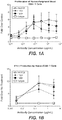
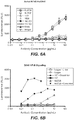
- FIGS. 1A-1D depict functional activation of human T cells in vitro after treatment with the exemplary anti-OX40 antibody Hu3738.
- FIG. 1A depicts the proliferation of human peripheral blood CD4+ T cells after treatment with anti-OX40 antibody Hu3738, or literature antibody 11D4 or 18D8.
- FIG. 1B depicts the increase in interferon-gamma (IFN- ⁇ ) production by human CD4+ T cells after treatment with anti-OX40 antibody Hu3738, or literature antibody 11D4 or 18D8.
- FIG. 1C depicts the proliferation of human peripheral blood CD4+ T cells after treatment with Hu3738, or literature antibody 1A7.
- FIG. 1D depicts the increase in IFN- ⁇ production by human CD4+ T cells after treatment with Hu3738, or literature antibody 1A7.
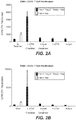
- FIGS. 2A-2B show the effect of exemplary anti-OX40 antibody Hu3738 on human T regulatory (Treg) cell-mediated suppression in vitro.
- the Treg suppression assay was set up using two different ratios of CD4+/CD25- responder T cells (Tresp) to CD4+/CD25+/CD127low T regulatory cells (Treg).
- Treg Suppression Inspector reagent beads (Insp) were added to culture wells at 1:1 bead-to-cell ratio for stimulation. The clear bar represents proliferation of Tresp cells in the presence of Insp.
- Anti-OX40 and isotype control human IgG 1 antibodies were tested in triplicate at 10 ⁇ g/mL final concentration in the absence or presence of cross-linking reagent (F(ab') 2 goat anti-human IgG, Fc specific) at 1:4 ratio. Plates were incubated at 37 °C in 5% CO 2 for four days. 1 ⁇ Ci/well 3 H-thymidine was added and the plates were further incubated for another 16 hours. Graphs represent proliferation as shown in counts per minute (cpm).
- FIG. 2A depicts results with Tresp to Treg at 2:1 ratio
- FIG. 2B depicts results with Tresp to Treg at 4:1 ratio.
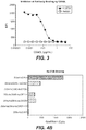
- FIG. 3 depicts the inhibition of binding of exemplary anti-OX40 antibody Hu3738 in the presence of soluble human OX40 ligand (OX40L).
- the graph shows mean fluorescence intensity (MFI) vs. concentration of OX40L ( ⁇ g/mL).
- MFI mean fluorescence intensity
- Human OX40-expressing Jurkat cells were co-stained with a titration of unlabeled soluble OX40L and 0.2 ⁇ g/mL Hu3738 or isotype control antibody.
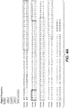
- FIG. 4A shows an amino acid sequence alignment of human OX40 (SEQ ID NO:1) with mouse OX40 (SEQ ID NO:3).
- FIG. 4B depicts the binding activity of exemplary anti-OX40 antibody Hu3738 to cell-surface expressed human, murine, or chimeric human-mouse OX40 molecules containing mouse cysteine-rich domains (CRDs) swapped out for the corresponding human regions.
- CCDs mouse cysteine-rich domains
- Human OX40 is shown as "293s-huOX40," chimeric human OX40 with murine CRDI is shown as “293s-huOX40-muCRDI,” chimeric human OX40 with murine CRDII is shown as “293s-huOX40-muCRDII,” chimeric human OX40 with murine CRDIII is shown as “293s-huOX40-muCRDIII,” chimeric human OX40 with murine CRDIV is shown as "293s-huOX40-muCRDIV,” chimeric human OX40 with murine CRDII and murine CRDIII is shown as “293s-huOX40-muCRDII+III,” and murine OX40 is shown as "293s-muOX40.”
- FIG. 5 depicts competition for cell surface human OX40 binding by exemplary anti-OX40 antibody Hu3738 or a literature antibody (11D4, 18D8, 106-222, 119-122, or 1A7).
- FIG. 6A depicts the activation of NF- ⁇ B in human OX40-transfected Jurkat reporter cell lines upon treatment with exemplary anti-OX40 antibody Mu3738 or Hu3738, or literature antibody 11D4, 18D8, 106-222, or 119-122, or isotype control in the absence of an added cross-linker.
- FIG. 6B depicts the activation of NF- ⁇ B in human OX40-transfected Jurkat reporter cell lines upon treatment with exemplary anti-OX40 antibody Hu3738, literature antibody 1A7, or isotype control in the presence or absence of an added cross-linker.
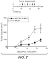
- FIG. 7 depicts anti-tumor activity of exemplary anti-OX40 antibody Hu3738 in a human PC3 adoptive cell tumor model in NSG mice.
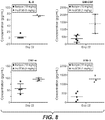
- FIG. 8 depicts levels of interleukin-8 (IL-8), granulocyte macrophage colony-stimulating factor (GM-CSF), tumor necrosis factor alpha (TNF- ⁇ ), and interferon-gamma (IFN- ⁇ ) in a human peripheral blood mononuclear cell (PBMC) mediated graft-versus-host disease (GVHD) model in NSG mice, after treating the mice with 1 mg/kg Hu3738 or human IgG 1 isotype control once every 7 days for a total of 4 doses.
- PBMC peripheral blood mononuclear cell
- GVHD graft-versus-host disease
- the present disclosure concerns antibodies and fragments thereof that specifically bind OX40, compositions comprising the antibodies, polynucleotides encoding anti-OX40 antibodies, host cells capable of producing the antibodies, methods and compositions useful for making the antibodies, and various methods of using the same.
- V H heavy chain variable domain
- V L light chain variable domain
- polypeptide sequences described herein are, in many embodiments, described by way of their respective polypeptide or polynucleotide sequences. Unless indicated otherwise, polypeptide sequences are provided in N ⁇ C orientation; polynucleotide sequences in 5' ⁇ 3' orientation. For polypeptide sequences, the conventional three or one-letter abbreviations for the genetically encoded amino acids may be used, as noted in TABLE 1, below.
- Certain sequences are defined by structural formulae specifying amino acid residues belonging to certain classes (e.g., aliphatic, hydrophobic, etc .).
- the various classes to which the genetically encoded amino acids belong as used herein are noted in TABLE 2, below. Some amino acids may belong to more than one class. Cysteine, which contains a sulfhydryl group, and proline, which is conformationally constrained, are not assigned classes.
- TABLE 2 Encoded Amino Acid Classes Class Amino Acids Aliphatic A, I, L, V Aromatic F, Y, W Non-Polar M, A, I, L, V Polar N, Q, S, T Basic H, K, R Acidic D, E Small A, G
- OX40 is a co-stimulatory molecule that has a critical role in the enhancement of nascent immune responses and concomitantly acts to suppress regulatory T cell activity.
- OX40 also known as CD 134 or tumor necrosis factor receptor superfamily 4 (TNFRSF4), is a Type I transmembrane cell surface member of the tumor necrosis factor (TNF) receptor superfamily transiently expressed on recently activated T cells and constitutively expressed on activated T regulatory cells.
- the extracellular ligand binding domain of OX40 is composed of three cysteine-rich domains (CRD) and a fourth partial CRD (CRDI, CRDII, CRDIII, and CRDIV, respectively).
- OX40 While primarily expressed by activated CD4+ T cells, OX40 can be expressed on B cells, CD8+ T cells, and natural killer (NK) and natural killer T (NKT) cells following activation. Neutrophils have also been reported to express OX40 and signaling through OX40 on human neutrophils inhibits apoptotic cell death.
- the ligand for OX40 also known as tumor necrosis factor ligand superfamily 4 (TNFSF4), CD252 or glycoprotein 34 (gp34), is upregulated by activated antigen-presenting cells and B cells. Ligand binding to OX40 on antigen-activated T cells results in downstream NF- ⁇ B translocation and AKT pathway activation.
- TNFSF4 tumor necrosis factor ligand superfamily 4
- gp34 glycoprotein 34
- NF- ⁇ B translocation leads to upregulation of pro-survival molecules such as Bcl-2, Bcl-XL and cell survival.
- Activating antibodies directed at OX40 are intended at least in part to enhance antigen-specific immune responses by prolonging activation and differentiation of T effector cells.
- targeting OX40 expressed by T regulatory cells may also contribute to the putative mechanism of action.
- T regulatory cells express high levels of OX40 within the tumor microenvironment. OX40 activation has been shown to impact suppressive capacity of T regulatory cells and to lead to the active depletion of OX40 positive T regulatory cells from the tumor microenvironment.
- the disclosure concerns antibodies that specifically bind OX40.
- the term "antibody” refers to an immunoglobulin molecule that specifically binds to a particular antigen- here, OX40.
- the anti-OX40 antibodies of the disclosure bind to human OX40 (SEQ ID NO:1) (NCBI Reference Sequence NP003318) and thereby modulate the immune system. The resulting immune system response is cytotoxic to tumor cells.
- Anti-OX40 antibodies comprise complementarity determining regions (CDRs), also known as hypervariable regions, in both the light chain and the heavy chain variable domains. The more highly conserved portions of variable domains are called the framework (FR).
- the amino acid position/boundary delineating a hypervariable region of an antibody can vary, depending on the context and the various definitions known in the art. Some positions within a variable domain may be viewed as hybrid hypervariable positions in that these positions can be deemed to be within a hypervariable region under one set of criteria while being deemed to be outside a hypervariable region under a different set of criteria. One or more of these positions can also be found in extended hypervariable regions.
- the disclosure provides antibodies comprising modifications in these hybrid hypervariable positions.
- variable domains of native heavy and light chains each comprise four FR regions, largely by adopting a ⁇ -sheet configuration, connected by three CDRs, which form loops connecting, and in some cases forming part of, the ⁇ -sheet structure.
- the CDRs in each chain are held together in close proximity by the FR regions and, with the CDRs from the other chain, contribute to the formation of the target binding site of antibodies. See Kabat et al., Sequences of Proteins of Immunological Interest (National Institute of Health, Bethesda, Md. 1987 ).
- numbering of immunoglobulin amino acid residues is done according to the immunoglobulin amino acid residue numbering system of Kabat et al. unless otherwise indicated.
- the antibodies of the disclosure may be polyclonal, monoclonal, genetically engineered, and/or otherwise modified in nature, including but not limited to chimeric antibodies, humanized antibodies, and human antibodies.
- the constant region is an isotype selected from: IgA (e.g., IgA 1 or IgA 2 ), IgD, IgE, IgG ( e.g., IgG 1 , IgG 2 , IgG 3 or IgG 4 ), and IgM.
- an anti-OX40 antibody described herein comprises an IgG 1 .
- the anti-OX40 antibodies comprise an IgG 2 or IgG 4 .
- the "constant region" of an antibody includes the natural constant region, allotypes or natural variants, such as D356E and L358M, or A431G in human IgG 1 . See, e.g., Jefferis and Lefranc, MAbs, 1(4): 332-338 (Jul-Aug 2009 ).
- the light constant region of an anti-OX40 antibody may be a kappa ( ⁇ ) light region or a lambda ( ⁇ ) region.
- a ⁇ light region can be any one of the known subtypes, e.g ., ⁇ 1 , ⁇ 2 , ⁇ 3 , or ⁇ 4 .
- the anti-OX40 antibody comprises a kappa ( ⁇ ) light region.
- monoclonal antibody as used herein is not limited to antibodies produced through hybridoma technology.
- a monoclonal antibody is derived from a single clone, including any eukaryotic, prokaryotic, or phage clone, by any means available or known in the art.
- Monoclonal antibodies useful with the present disclosure can be prepared using a wide variety of techniques known in the art including the use of hybridoma, recombinant, and phage display technologies, or a combination thereof. In many uses of the present disclosure, including in vivo use of the anti-OX40 antibodies in humans, chimeric, humanized, or human antibodies can be used.
- chimeric antibody refers to an antibody having variable sequences derived from a non-human immunoglobulin, such as a rat or a mouse antibody, and human immunoglobulin constant regions, typically chosen from a human immunoglobulin template.
- Methods for producing chimeric antibodies are known in the art. See, e.g., Morrison, 1985, Science 229(4719):1202-7 ; Oi et al., 1986, BioTechniques 4:214-221 ; Gillies et al., 1985, J. Immunol. Methods 125:191-202 ; U.S. Patent Nos. 5,807,715 ; 4,816,567 ; and 4,816,397 .
- Humanized forms of non-human (e.g ., murine) antibodies are chimeric immunoglobulins that contain minimal sequences derived from non-human immunoglobulin.
- a humanized antibody will comprise substantially all of at least one, and typically two, variable domains, in which all or substantially all of the CDR regions correspond to those of a non-human immunoglobulin and all or substantially all of the FR regions are those of a human immunoglobulin sequence.
- the humanized antibody can also comprise at least a portion of an immunoglobulin constant region (Fc), typically that of a human immunoglobulin consensus sequence.
- Fc immunoglobulin constant region
- Human antibodies include antibodies having the amino acid sequence of a human immunoglobulin and include antibodies isolated from human immunoglobulin libraries or from animals transgenic for one or more human immunoglobulins and that do not express endogenous immunoglobulins. Human antibodies can be made by a variety of methods known in the art including phage display methods using antibody libraries derived from human immunoglobulin sequences. See U.S. Patent Nos. 4,444,887 and 4,716,111 ; and PCT publications WO 98/46645 ; WO 98/50433 ; WO 98/24893 ; WO 98/16654 ; WO 96/34096 ; WO 96/33735 ; and WO 91/10741 .
- Human antibodies can also be produced using transgenic mice which are incapable of expressing functional endogenous immunoglobulins but which can express human immunoglobulin genes. See, e.g., PCT publications WO 98/24893 ; WO 92/01047 ; WO 96/34096 ; WO 96/33735 ; U.S. Patent Nos. 5,413,923 ; 5,625,126 ; 5,633,425 ; 5,569,825 ; 5,661,016 ; 5,545,806 ; 5,814,318 ; 5,885,793 ; 5,916,771 ; and 5,939,598 . In addition, companies such as LakePharma, Inc.
- anti-OX40 antibody binding fragments include those that are capable of specifically binding OX40.
- antibody binding fragments include by way of example and not limitation, Fab, Fab', F(ab') 2 , Fv fragments, single chain Fv (scFv) fragments and single domain fragments.
- a Fab fragment contains the constant domain of the light chain and the first constant domain (CH1) of the heavy chain.
- Fab' fragments differ from Fab fragments by the addition of a few residues at the carboxyl terminus of the heavy chain CH1 domain including one or more cysteines from the antibody hinge region.
- Fab' fragments are produced by cleavage of the disulfide bond at the hinge cysteines of the F(ab') 2 pepsin digestion product. Additional chemical couplings of antibody fragments are known to those of ordinary skill in the art.
- Fab and F(ab') 2 fragments lack the Fragment crystallizable (Fc) region of an intact antibody, clear more rapidly from the circulation of animals, and may have less non-specific tissue binding than an intact antibody (see, e.g., Wahl et al., 1983, J. Nucl. Med. 24:316 ).
- an "Fc” region is the Fragment crystallizable constant region of an antibody not comprising an antigen-specific binding region.
- the Fc region is composed of two identical protein fragments, derived from the second and third constant domains (CH2 and CH3 domains, respectively) of the two heavy chains of an antibody.
- IgM and IgE Fc regions contain three heavy chain constant domains (CH2, CH3, and CH4 domains) in each polypeptide chain.
- an “Fv” fragment is the minimum fragment of an antibody that contains a complete target recognition and binding site.
- This region consists of a dimer of one heavy and one light chain variable domain in a tight, non-covalent association (V H -V L dimer). It is in this configuration that the three CDRs of each variable domain interact to define a target binding site on the surface of the V H -V L dimer. Often, the six CDRs confer target binding specificity to the antibody. However, in some instances even a single variable domain (or half of an Fv comprising only three CDRs specific for a target) can have the ability to recognize and bind target, although at a lower affinity than the entire binding site.
- Single-chain Fv or “scFv” antibody binding fragments comprise the V H and V L domains of an antibody, where these domains are present in a single polypeptide chain.
- the Fv polypeptide further comprises a polypeptide linker between the V H and V L domains which enables the scFv to form a structure favorable for target binding.
- Single domain fragments are composed of a single V H or V L domains which exhibit sufficient affinity to OX40.
- the single domain fragment is camelized ( See, e.g ., Riechmann, 1999, Journal of Immunological Methods 231:25-38 ).
- Anti-OX40 antibodies of the disclosure include derivatized antibodies.
- derivatized antibodies are typically modified by glycosylation, acetylation, pegylation, phosphorylation, amidation, derivatization by known protecting/blocking groups, proteolytic cleavage, linkage to a cellular ligand or other protein. Any of numerous chemical modifications can be carried out by known techniques, including, but not limited to, specific chemical cleavage, acetylation, formylation, metabolic synthesis of tunicamycin, etc.
- the derivative can contain one or more non-natural amino acids, e.g ., using ambrx technology (See, e.g., Wolfson, 2006, Chem. Biol. 13(10):1011-2 ).
- the anti-OX40 antibodies may be antibodies whose sequences have been modified to alter at least one constant region-mediated biological effector function.
- an anti-OX40 antibody may be modified to reduce at least one constant region-mediated biological effector function relative to the unmodified antibody, e.g ., reduced binding to one or more of the Fc receptors (Fc ⁇ R) such as Fc ⁇ RI, Fc ⁇ RIIA, Fc ⁇ RIIB, Fc ⁇ RIIIA and/or Fc ⁇ RIIIB.
- Fc ⁇ R binding can be reduced by mutating the immunoglobulin constant region segment of the antibody at particular regions necessary for Fc ⁇ R interactions ( See, e.g., Canfield and Morrison, 1991, J. Exp. Med.
- a variant CH2 domain having a V263L, V273C, V273E, V273F, V273L, V273M, V273S, or V273Y substitution in the CH2 domain of the Fc region can exhibit reduced affinity to FcyRIIB as compared to the corresponding wild type constant region.
- the anti-OX40 antibody described herein include antibodies that have been modified to acquire or improve at least one constant region-mediated biological effector function relative to an unmodified antibody, e.g., to enhance Fc ⁇ R interactions ( See, e.g., US Patent Appl. No. 2006/0134709 ).
- an anti-OX40 antibody of the disclosure can have a constant region that binds Fc ⁇ RI, Fc ⁇ RIIA, Fc ⁇ RIIB, Fc ⁇ RIIIA and/or Fc ⁇ RIIIB with greater affinity than the corresponding wild type constant region.
- a variant CH2 domain having a V263L, V273C, V273E, V273F, V273L, V273M, V273S, or V273Y substitution in the CH2 domain of the Fc region can exhibit greater affinity to Fc ⁇ RIIIA as compared to the corresponding wild type constant region.
- anti-OX40 antibodies of the disclosure may have alterations in biological activity that result in increased or decreased opsonization, phagocytosis, or ADCC.
- Such alterations are known in the art.
- modifications in antibodies that reduce ADCC activity are described in U.S. Patent No. 5,834,597 .
- An exemplary ADCC lowering variant corresponds to "mutant 3" (also known as "M3,” shown in FIG. 4 of U.S. Patent No. 5,834,597 ) in which residues 234 and 237 (using EU numbering) are substituted with alanines.
- a mutant 3 (also known as "M3") variation may be used in a number of antibody isotypes, e.g ., human IgG 2 M3.
- Additional substitutions that can modify FcyR binding and/or ADCC effector function of an anti-OX40 antibody include the K322A substitution or the L234A and L235A double substitution in the Fc region, for example, a human IgG 1 having the L234A/L235A double substitution. See, e.g., Hezareh, et al. J. Virol., 75 (24): 12161-12168 (2001 ).
- the anti-OX40 antibodies have low levels of, or lack, fucose.
- Antibodies lacking fucose have been correlated with enhanced ADCC activity, especially at low doses of antibody. See Shields et al., 2002, J. Biol. Chem. 277:26733-26740 ; Shinkawa et al., 2003, J. Biol. Chem. 278:3466-73 .
- Methods of preparing fucose-less antibodies include growth in rat myeloma YB2/0 cells (ATCC CRL 1662).
- YB2/0 cells express low levels of FUT8 mRNA, which encodes ⁇ -1,6-fucosyltransferase, an enzyme necessary for fucosylation of polypeptides.
- Anti-OX40 antibodies can comprise modified (or variant) CH2 domains or entire Fc domains that include amino acid substitutions that increase binding to Fc ⁇ RIIB and/or reduced binding to FcyRIIIA as compared to the binding of a corresponding wild-type CH2 or Fc region.
- Variant CH2 or variant Fc domains have been described in U.S. Patent Appl. No. 2014/0377253 .
- a variant CH2 or variant Fc domain typically includes one or more substitutions at position 263, position 266, position 273, and position 305, wherein the numbering of the residues in the Fc domain is that of the EU index as in Kabat.
- the anti-OX40 antibodies comprise one or more substitutions selected from V263L, V266L, V273C, V273E, V273F, V273L, V273M, V273S, V273Y, V305K, and V305W, relative to the wild-type CH2 domain.
- the one or more substitutions of the CH2 domain are selected from V263L, V273E, V273F, V273M, V273S, and V273Y, relative to the CH2 domain of a human IgG 1 .
- the one or more substitutions of an IgG 1 CH2 domain can be V273E.
- the anti-OX40 antibody of the disclosure comprises a variant IgG 1 CH2 domain comprising the amino acid substitution V263L.
- variant CH2 or variant Fc domains that can afford increased binding to Fc ⁇ RIIB and/or reduced binding to Fc ⁇ RIIIA as compared to the binding of a corresponding wild-type CH2 or Fc region include those found in Vonderheide, et al. Clin. Cancer Res., 19(5), 1035-1043 (2013 ), such as S267E or S267E/L328F in human IgG 1 .
- the anti-OX40 antibodies include modifications that increase or decrease their binding affinities to the fetal Fc receptor, FcRn, for example, by mutating the immunoglobulin constant region segment at particular regions involved in FcRn interactions (see, e.g ., WO 2005/123780 ).
- an anti-OX40 antibody of the IgG class is mutated such that at least one of amino acid residues 250, 314, and 428 of the heavy chain constant region is substituted alone, or in any combinations thereof, such as at positions 250 and 428, or at positions 250 and 314, or at positions 314 and 428, or at positions 250, 314, and 428, with positions 250 and 428 a specific combination.
- the substituting amino acid residue can be any amino acid residue other than threonine, including, but not limited to, alanine, cysteine, aspartic acid, glutamic acid, phenylalanine, glycine, histidine, isoleucine, lysine, leucine, methionine, asparagine, proline, glutamine, arginine, serine, valine, tryptophan, or tyrosine.
- the substituting amino acid residue can be any amino acid residue other than leucine, including, but not limited to, alanine, cysteine, aspartic acid, glutamic acid, phenylalanine, glycine, histidine, isoleucine, lysine, methionine, asparagine, proline, glutamine, arginine, serine, threonine, valine, tryptophan, or tyrosine.
- the substituting amino acid residues can be any amino acid residue other than methionine, including, but not limited to, alanine, cysteine, aspartic acid, glutamic acid, phenylalanine, glycine, histidine, isoleucine, lysine, leucine, asparagine, proline, glutamine, arginine, serine, threonine, valine, tryptophan, or tyrosine.
- An exemplary substitution known to modify Fc effector function is the Fc substitution M428L, which can occur in combination with the Fc substitution T250Q. Additional specific combinations of suitable amino acid substitutions are identified in Table 1 of U.S. Patent No. 7,217,797 . Such mutations increase binding to FcRn, which protects the antibody from degradation and increases its half-life.
- An anti-OX40 antibody may have one or more amino acids inserted into one or more of its CDRs, for example as described in Jung and Plückthun, 1997, Protein Engineering 10:8, 959-966 ; Yazaki et al., 2004, Protein Eng. Des Sel. 17(5):481-9 . Epub 2004 Aug 17; and U.S. Pat. Appl. No. 2007/0280931 .
- Anti-OX40 antibodies with high affinity for human OX40 may be desirable for therapeutic and diagnostic uses. Accordingly, the present disclosure contemplates antibodies having a high binding affinity to human OX40.
- the anti-OX40 antibodies bind human OX40 with an affinity of at least about 100 nM, but may exhibit higher affinity, for example, at least about 90 nM, 80 nM, 70 nM, 60 nM, 50 nM, 40 nM, 30 nM, 25 nM, 20 nM, 15 nM, 10 nM, 7 nM, 6 nM, 5 nM, 4 nM, 3 nM, 2 nM, 1 nM, 0.1 nM, 0.01 nM, or even higher.
- the antibodies bind human OX40 with an affinity in the range of about 1 pM to about 100 nM, or an affinity ranging between any of the foregoing values, such as but not limited to from about 0.001 to 10 nM, 0.001 to 5 nM, 0.01 to 100 nM, 0.01 to 50 nM, 0.01 to 10 nM, 0.01 to 5 nM, or 0.01 to 1 nM.
- Affinity of anti-OX40 antibodies for human OX40 can be determined using techniques well known in the art or described herein, such as for example, but not by way of limitation, ELISA, isothermal titration calorimetry (ITC), surface plasmon resonance, or fluorescent polarization assay.
- Anti-OX40 antibodies generally comprise a heavy chain comprising a variable region (V H ) having three complementarity determining regions ("CDRs") referred to herein (in N ⁇ C order) as V H CDR#1, V H CDR#2, and V H CDR#3, and a light chain comprising a variable region (V L ) having three complementarity determining regions referred to herein (in N ⁇ C order) as V L CDR#1, V L CDR#2, and V L CDR#3.
- CDRs complementarity determining regions
- V L variable region having three complementarity determining regions referred to herein (in N ⁇ C order) as V L CDR#1, V L CDR#2, and V L CDR#3.
- the amino acid sequences of exemplary CDRs, as well as the amino acid sequence of the V H and V L regions of the heavy and light chains of exemplary anti-OX40 are provided herein. Specific embodiments of anti-OX40 antibodies include these exemplary CDRs and/or V H and/or
- the amino acid sequences of the CDRs of an anti-OX40 antibody have sequences selected from their respective V H and V L CDR sequences in TABLE 3 below: TABLE 3 Exemplary CDR Sequences CDR Sequence Identifier
- V H CDR#1 GFTFSRYGMS (SEQ ID NO:101) GYSIASGYYWN (SEQ ID NO:111) GFNIKDTYMH (SEQ ID NO:121) GFSLTSYGVH (SEQ ID NO:131)
- V H CDR#2 TINSNGGRTYYPDSVKG (SEQ ID NO:102) YISYDGSNNYNPSLG (SEQ ID NO:112) RIDPANGNTKYDPKFQG (SEQ ID NO:122) VIWSGGSTDYNAAFIS (SEQ ID NO:132)
- an anti-OX40 antibody has the CDRs according to SEQ ID NOS: 101, 102, 103, 104, 105, and 106. In some embodiments, an anti-OX40 antibody has the CDRs according to SEQ ID NOS: 111, 112, 113, 114, 115, and 116. In some embodiments, an anti-OX40 antibody has the CDRs according to SEQ ID NOS: 121, 122, 123, 114, 125, and 126. In some embodiments, an anti-OX40 antibody has the CDRs according to SEQ ID NOS: 131, 132, 133, 114, 115, and 136.
- the CDRs described herein form binding elements within V H and V L chains of anti-OX40 antibodies of the disclosure.
- TABLES 4 and 5 below describe V H and V L chains corresponding to exemplary anti-OX40 antibodies containing the above-described CDRs.
- the CDRs are underlined below in TABLES 4 and 5.
- an anti-OX40 antibody comprises a V H chain having an amino acid sequence as described in TABLE 4: TABLE 4 Exemplary V H Sequences V H Sequence Identifier Mu3738 V H (SEQ ID NO:21) Hu3738 V H .1b (SEQ ID NO:22) Mu3726 V H (SEQ ID NO:23) Hu3726 V H .1a (SEQ ID NO:24) Mu3739 V H (SEQ ID NO:25) Hu3739 V H .1b (SEQ ID NO:26) Mu3741 V H (SEQ ID NO:27) Hu3741 V H .2b (SEQ ID NO:28) and a V L chain having an amino acid sequence as described in TABLE 5: TABLE 5 Exemplary V L Sequences V L Sequence Identifier Mu3738 V L (SEQ ID NO:31) Hu3738 V L .1 (SEQ ID NO:32) Mu3726 V L (SEQ ID NO:33) Hu3726 V L .1b (SEQ ID NO:
- an anti-OX40 antibody comprises a V H chain having an amino acid sequence according to SEQ ID NO:21, and a V L chain having an amino acid sequence according to SEQ ID NO:31. In some embodiments, an anti-OX40 antibody comprises a V H chain having an amino acid sequence according to SEQ ID NO:23, and a V L chain having an amino acid sequence according to SEQ ID NO:33. In some embodiments, an anti-OX40 antibody comprises a V H chain having an amino acid sequence according to SEQ ID NO:25, and a V L chain having an amino acid sequence according to SEQ ID NO:35. In some embodiments, an anti-OX40 antibody comprises a V H chain having an amino acid sequence according to SEQ ID NO:27, and a V L chain having an amino acid sequence according to SEQ ID NO:37.
- an anti-OX40 antibody is suitable for administration to humans.
- the anti-OX40 antibody is humanized.
- an anti-OX40 antibody comprises a V H chain having an amino acid sequence according to SEQ ID NO:22, and a V L chain having an amino acid sequence according to SEQ ID NO:32.
- an anti-OX40 antibody comprises a V H chain having an amino acid sequence according to SEQ ID NO:24, and a V L chain having an amino acid sequence according to SEQ ID NO:34.
- an anti-OX40 antibody comprises a V H chain having an amino acid sequence according to SEQ ID NO:26, and a V L chain having an amino acid sequence according to SEQ ID NO:36.
- an anti-OX40 antibody comprises a V H chain having an amino acid sequence according to SEQ ID NO:28, and a V L chain having an amino acid sequence according to SEQ ID NO:38.
- an anti-OX40 antibody comprises a V H sequence having at least 85%, at least 90%, at least 93%, at least 95%, at least 96%, at least 97%, at least 98%, or at least 99% sequence identity to any one of the V H sequences shown in TABLE 4.
- An anti-OX40 antibody can comprise a V H sequence having up to 8, up to 7, up to 6, up to 5, up to 4, up to 3, or up to 2 mutations compared with any one of the V H sequences shown in TABLE 4.
- an anti-OX40 antibody can comprise a V H sequence having 5 or fewer, 4 or fewer, 3 or fewer, or 2 or fewer mutations compared with any one of the V H sequences shown in TABLE 4.
- an anti-OX40 antibody comprises a V L sequence having at least 85%, at least 90%, at least 93%, at least 95%, at least 96%, at least 97%, at least 98%, or at least 99% sequence identity to any one of the V L sequences shown in TABLE 5.
- An anti-OX40 antibody can comprise a V L sequence having up to 8, up to 7, up to 6, up to 5, up to 4, up to 3, or up to 2 mutations compared with any one of the V L sequences shown in TABLE 5.
- an anti-OX40 antibody can comprise a V L sequence having 5 or fewer, 4 or fewer, 3 or fewer, or 2 or fewer mutations compared with any one of the V L sequences shown in TABLE 5.
- Full length heavy and light chain amino acid sequences generally comprise an above-described V H or V L chain linked to an appropriate immunoglobulin constant region, e.g., human IgG 1 or kappa light constant region.
- Post-translational modifications to the full length sequences of an anti-OX40 antibody may occur, such as cleavage of one or more (e.g., 1, 2, 3, or more) amino acid residues on the C-terminal end of the antibody heavy chain.
- Such cleavage products may comprise some or all of the anti-OX40 antibody as expressed.
- an anti-OX40 antibody comprises a heavy chain amino acid sequence as described in TABLE 6: TABLE 6 Exemplary Heavy Chain Sequences Sequence Identifier SEQ ID NO: 41 SEQ ID NO: 42 SEQ ID NO: 43 SEQ ID NO: 44 SEQ ID NO: 45 SEQ ID NO: 46 SEQ ID NO: 47 SEQ ID NO: 48 and a light chain amino acid sequence as described in TABLE 7: TABLE 7 Exemplary Light Chain Sequences Sequence Identifier SEQ ID NO: 51 SEQ ID NO: 52 SEQ ID NO: 53 SEQ ID NO: 54 wherein the underlined amino acids represent the CDRs and the italicized amino acids represent the constant regions.
- an anti-OX40 antibody comprises a heavy chain amino acid sequence according to SEQ ID NO:41 or 42, and a light chain amino acid sequence according to SEQ ID NO:51. In some embodiments, an anti-OX40 antibody comprises a heavy chain amino acid sequence according to SEQ ID NO:43 or 44, and a light chain amino acid sequence according to SEQ ID NO:52. In some embodiments, an anti-OX40 antibody comprises a heavy chain amino acid sequence according to SEQ ID NO:45 or 46, and a light chain amino acid sequence according to SEQ ID NO:53. In some embodiments, an anti-OX40 antibody comprises a heavy chain amino acid sequence according to SEQ ID NO:47 or 48, and a light chain amino acid sequence according to SEQ ID NO:54.
- an anti-OX40 antibody comprises a heavy chain sequence having at least 85%, at least 90%, at least 93%, at least 95%, at least 96%, at least 97%, at least 98%, or at least 99% sequence identity to the heavy chain sequence according to any one of SEQ ID NOS:41-48.
- An anti-OX40 antibody can comprise a heavy chain sequence having up to 8, up to 7, up to 6, up to 5, up to 4, up to 3, or up to 2 mutations compared with the heavy chain sequence according to any one of SEQ ID NOS:41-48.
- an anti-OX40 antibody can comprise a heavy chain sequence having 5 or fewer, 4 or fewer, 3 or fewer, or 2 or fewer mutations compared with the heavy chain sequence according to any one of SEQ ID NOS:41-48.
- an anti-OX40 antibody comprises a light chain sequence having at least 85%, at least 90%, at least 93%, at least 95%, at least 96%, at least 97%, at least 98%, or at least 99% sequence identity to the light chain sequence according to any one of SEQ ID NOS:51-54.
- An anti-OX40 antibody can comprise a light chain sequence having up to 8, up to 7, up to 6, up to 5, up to 4, up to 3, or up to 2 mutations compared with the light chain sequence according to any one of SEQ ID NOS:51-54.
- an anti-OX40 antibody can comprise a light chain sequence having 5 or fewer, 4 or fewer, 3 or fewer, or 2 or fewer mutations compared with the light chain sequence according to any one of SEQ ID NOS:51-54.
- Additional post-translational modifications of an anti-OX40 antibody may include glycosylation.
- Common biantennary complexes can be composed of a core structure having two N-acetylglucosamine (GlcNAc), three mannose, and two GlcNAc residues that are ⁇ -12 linked to ⁇ -6 mannose and ⁇ -3 mannose to form two antennae.
- GlcNAc N-acetylglucosamine
- Gal galactose
- high mannose glycans Man-5 or Man-9 bisecting GlcNAc
- sialic acid including N-acetylneuraminic acid (NANA) or N-glycolylneuraminic acid (NGNA) residues may be attached to the core.
- NANA N-acetylneuraminic acid
- NGNA N-glycolylneuraminic acid
- N-linked glycoforms may include G0 (protein having a core biantennary glycosylation structure), G0F (fucosylated G0), G0F GlcNAc, G1 (protein having a core glycosylation structure with one galactose residue), G1F (fucosylated G1), G2 (protein having a core glycosylation structure with two galactose residues), and/or G2F (fucosylated G2).
- the anti-OX40 antibodies compete for binding human OX40 (SEQ ID NO:1) in in vitro assays with a reference antibody. In some embodiments, the anti-OX40 antibodies compete for binding human OX40 on cells expressing human OX40.
- the reference antibody may be any of the anti-OX40 antibodies described herein. In some embodiments, the reference antibody is an antibody having a V H according to one described in TABLE 4 and a V L according to one described in TABLE 5.
- the reference antibody is mouse antibody comprising Mu3726 V H and Mu3726 V L ("Mu3726”), mouse antibody comprising Mu3738 V H and Mu3738 V L ("Mu3738”), mouse antibody comprising Mu3739 V H and Mu3739 V L ("Mu3739”), or mouse antibody comprising Mu3741 V H and Mu3741 V L (“Mu3741”).
- the reference antibody is a humanized version of Mu3726, Mu3738, Mu3739, or Mu3741.
- the reference antibody is a humanized antibody comprising a heavy chain according to SEQ ID NO:41 or 42 and a light chain according to SEQ ID NO:51 (“Hu3738”), a humanized antibody comprising a heavy chain according to SEQ ID NO:43 or 44 and a light chain according to SEQ ID NO:52 (“Hu3726”), a humanized antibody comprising a heavy chain according to SEQ ID NO:45 or 46 and a light chain according to SEQ ID NO:53 (“Hu3739”), or a humanized antibody comprising a heavy chain according to SEQ ID NO:47 or 48 and a light chain according to SEQ ID NO:54 (“Hu3741").
- the anti-OX40 antibodies described herein generally bind specifically to human OX40.
- Cross reactivity of the antibodies for binding to OX40 from other species, for example, from monkey, e.g ., cynomolgus monkey may offer advantages, such as the ability to test in monkey animal models for biological activity.
- Such animal model testing may be used to screen anti-OX40 antibodies to select properties related to efficacy, e.g., favorable pharmacokinetics, or those related to safety, e.g., decreased hepatic toxicity.
- an anti-OX40 antibody binds to cynomolgus OX40 (SEQ ID NO:2) (NCBI Reference Sequence XP005545179) as well as human OX40.
- an anti-OX40 antibody does not bind to mouse OX40 (SEQ ID NO:3) (NCBI Reference Sequence NP037181).
- Assays for competition include, but are not limited to, a radioactive material labeled immunoassay (RIA), an enzyme-linked immunosorbent assay (ELISA), a sandwich ELISA, fluorescence activated cell sorting (FACS) assays, and surface plasmon resonance assays.
- RIA radioactive material labeled immunoassay
- ELISA enzyme-linked immunosorbent assay
- FACS fluorescence activated cell sorting
- SPR Surface plasmon resonance
- the anti-OX40 antibodies have a K D of at least about 100 nM, but may exhibit higher affinity, for example, at least about 90 nM, 80 nM, 70 nM, 60 nM, 50 nM, 40 nM, 30 nM, 25 nM, 20 nM, 15 nM, 10 nM, 7 nM, 6 nM, 5 nM, 4 nM, 3 nM, 2 nM, 1 nM, 0.1 nM, 0.01 nM, or even higher.
- the anti-OX40 antibody has a K D in the range of about 1 pM to about 100 nM, or an affinity ranging between any of the foregoing values, such as but not limited to from about 0.001 to 10 nM, 0.001 to 5 nM, 0.01 to 100 nM, 0.01 to 50 nM, 0.01 to 10 nM, 0.01 to 5 nM, or about 0.01 to 1 nM.
- an anti-OX40 antibody has a dissociation constant k d no more than about 10 sec -1 , for example, no more than about 1, 0.5, 0.2, 0.1, 0.05, 0.01, 0.005, 0.001 sec -1 , or even lower.
- the anti-OX40 antibody has a k d in the range of about 0.001 sec -1 to about 10 sec -1 , or a k d ranging between any of the foregoing values, such as but not limited to from about 0.01 to 10 sec -1 , 0.001 to 0.5 sec -1 , 0.001 to 0.2 sec -1 , 0.001 to 0.1 sec -1 , 0.01 to 1 sec -1 , 0.001 to 0.05 sec -1 , or about 0.001 to 1 sec -1 .
- an anti-OX40 antibody has an association constant k a at least about 10 4 M -1 -sec -1 , for example, at least about 1 ⁇ 10 4 , 5 ⁇ 10 4 , 1 ⁇ 10 5 , 5 ⁇ 10 5 , 1 ⁇ 10 6 , 5 ⁇ 10 6 , 1 ⁇ 10 7 M -1 -sec -1 , or even greater.
- the anti-OX40 antibody has a k d in the range of about 10 4 M -1 -sec -1 to about 10 7 M -1 -sec -1 , or a k a ranging between any of the foregoing values, such as but not limited to from about 5 ⁇ 10 4 to 1 ⁇ 10 7 M -1 -sec -1 , 5 ⁇ 10 4 to 5 ⁇ 10 6 M -1 -sec -1 , or about 1 ⁇ 10 4 to 5 ⁇ 10 6 M -1 -sec -1 .
- An anti-OX40 antibody of the disclosure may exhibit a K D , k d , or k a in a range around a binding kinetics constant measured for any one of the exemplary anti-OX40 antibodies described herein.
- an anti-OX40 antibody has a dissociation constant k d in a range of from about 0.01 to about 100-fold, e.g., about 0.1 to about 10-fold, or about 0.5 to about 5-fold, the k d of any one of Hu3738, Hu3726, Hu3739, and Hu3741.
- an anti-OX40 antibody has an association constant k a in a range of from about 0.01 to about 100-fold, e.g., about 0.1 to about 10-fold, or about 0.5 to about 5-fold, the k a of any one of Hu3738, Hu3726, Hu3739, and Hu3741.
- a detectable label such as a fluorophore, biotin or an enzymatic (or even radioactive) label to enable subsequent identification.
- a detectable label such as a fluorophore, biotin or an enzymatic (or even radioactive) label.
- cells expressing human OX40 are incubated with unlabeled test antibody, labeled reference antibody is added, and the intensity of the bound label is measured. If the test antibody competes with the labeled reference antibody by binding to an overlapping epitope, the intensity will be decreased relative to a control reaction carried out without test antibody.
- the concentration of labeled reference antibody that yields 80% of maximal binding (“conc 80% ”) under the assay conditions is first determined, and a competition assay carried out with 10X conc 80% of unlabeled test antibody and cOnc 80% of labeled reference antibody.
- K i IC 50 / 1 + reference Ab concentration / K d , where IC 50 is the concentration of test antibody that yields a 50% reduction in binding of the reference antibody and K d is the dissociation constant of the reference antibody, a measure of its affinity for human OX40.
- Antibodies that compete with anti-OX40 antibodies disclosed herein can have a K i from 10 pM to 100 nM under assay conditions described herein.
- a test antibody is considered to compete with a reference antibody if it decreases binding of the reference antibody by at least about 20% or more, for example, by at least about 20%, 30%, 40%, 50%, 60%, 70%, 80%, 90%, 95% or even more, or by a percentage ranging between any of the foregoing values, at a reference antibody concentration that is 80% of maximal binding under the specific assay conditions used, and a test antibody concentration that is 10-fold higher than the reference antibody concentration.
- the anti-OX40 antibodies of the disclosure activate human OX40 (SEQ ID NO: 1).
- OX40 receptor activation can occur by a number of mechanisms, for example, by affording ligand-like activity against OX40 receptor.
- an anti-OX40 antibody competes for binding to OX40 receptor with human OX40 ligand (OX40L, CD252; UniProtKB/Swiss-Prot Code P23510.1) (SEQ ID NO:4).
- An anti-OX40 antibody of the disclosure can generally activate OX40 receptor in the presence of cross-linking.
- a specific assay and assay conditions useful for assessing whether an anti-OX40 antibody can activate OX40 receptor e.g., human OX40 receptor (SEQ ID NO: 1), in the presence of cross-linking is provided in Section 8.1.8.
- an anti-OX40 antibody activates human OX40 receptor in the presence of cross-linking with an EC 50 of from about 1 pM to about 500 nM, such as but not limited to from about 0.01 to about 300 nM, from about 0.01 to about 100 nM, from about 0.01 to about 10 nM, from about 0.01 to about 1 nM, from about 0.1 to about 300 nM, from about 0.1 nM to about 100 nM, from about 1 nM to about 100 nM, or from about 0.1 nM to about 100 nM.
- an EC 50 of from about 1 pM to about 500 nM, such as but not limited to from about 0.01 to about 300 nM, from about 0.01 to about 100 nM, from about 0.01 to about 10 nM, from about 0.01 to about 1 nM, from about 0.1 to about 300 nM, from about 0.1 nM to about 100 nM, from about 1 nM to about 100 nM, or from
- an anti-OX40 antibody at 100 ⁇ g/mL can activate human OX40 receptor in the presence of cross-linking to an activity at least about 3-fold, such as from about 3 to about 1000, e.g., about 5, 10, 15, 20, 30, 40, 50, 60, 80, 100, 200, 400, 500, 700, 800 or about 1000-fold higher compared with the activity of human OX40 receptor in the absence of the anti-OX40 antibody.
- Cross-linking can be provided by a number of methods, including addition of exogenous cross-linker, e.g., by antibodies or antibody F(ab') 2 fragments specific for heavy, light or variable regions of human or humanized antibodies; by soluble or immobilized protein A; by Fc receptor transfected cell lines; by endogenous Fc receptor expressing cell lines; by directly coating the subject antibodies to plastic surfaces; by plastic surfaces coated with exogenous cross-linking antibodies or Fc receptors; or by beads conjugated to any of the above.
- subject antibodies can be conjugated to a protein such as biotin, and soluble or immobilized avidin or streptavidin is used as a cross-linker.
- the activation of OX40 after binding to an anti-OX40 antibody is expected to occur after receptor cross-linking provided by endogenous Fc ⁇ R+ antigen-presenting cells.
- an anti-OX40 antibody binds to and activates human OX40 receptor in the absence of cross-linking.
- an anti-OX40 antibody activates OX40 receptor, e.g., human OX40 receptor (SEQ ID NO: 1), in the absence of OX40L, e.g., human OX40L (SEQ ID NO:4).
- OX40 receptor e.g., human OX40 receptor (SEQ ID NO: 1)
- OX40L e.g., human OX40L (SEQ ID NO:4).
- a specific assay and assay conditions useful for assessing whether an anti-OX40 antibody can activate OX40 receptor without cross-linking is provided in Section 8.1.8.
- an anti-OX40 antibody activates human OX40 receptor without cross-linking with an EC 50 of from about 1 pM to about 500 nM, such as but not limited to from about 0.01 to about 300 nM, from about 0.01 to about 100 nM, from about 0.1 to about 300 nM, from about 0.1 nM to about 100 nM, from about 1 nM to about 100 nM, from about 0.1 nM to about 100 nM, from about 1 to about 300 nM, from about 1 to about 100 nM, from about 1 to about 50 nM, or from about 10 to about 100 nM.
- an anti-OX40 antibody at 100 ⁇ g/mL can activate human OX40 receptor without cross-linking to an activity at least about 5-fold, such as from about 5 to about 1000, e.g., about 5, 10, 15, 20, 30, 40, 50, 60, 80, 100, 200, 300, 400, 500, 700, 800 or about 1000-fold higher compared with the activity of human OX40 receptor dosed with an equivalent amount of isotype antibody.
- an anti-OX40 antibody at 10 ⁇ g/mL can activate human OX40 receptor without cross-linking to an activity at least about 3-fold, such as from about 3 to about 300, e.g., about 3, 5, 6, 8, 10, 12, 15, 20, 25, 30, 40, 50, 60, 80, 100, 200, or about 300-fold higher compared with the activity of human OX40 receptor dosed with an equivalent amount of isotype antibody.
- an anti-OX40 antibody at 1 ⁇ g/mL can activate human OX40 receptor without cross-linking to an activity at least about 3-fold, such as from about 3 to about 150, e.g., about 3, 4, 5, 6, 7, 8, 10, 12, 15, 20, 25, 30, 40, 50, 60, 80, 100, or about 150-fold higher compared with the activity of human OX40 receptor dosed with an equivalent amount of isotype antibody.
- an anti-OX40 antibody activates OX40 receptor, e.g., human OX40 receptor (SEQ ID NO: 1), at a higher level in the presence of cross-linking compared to without cross-linking.
- OX40 receptor e.g., human OX40 receptor (SEQ ID NO: 1)
- SEQ ID NO: 1 e.g., human OX40 receptor (SEQ ID NO: 1)
- a specific assay and assay conditions useful for determining the level at which an anti-OX40 antibody can activate OX40 receptor without cross-linking is provided in Section 8.1.8.
- the level of activity can be measured, for example, in terms of EC 50 and/or an observed maximal activation.
- the anti-OX40 antibody at 100 ⁇ g/mL activates OX40 receptor, e.g., human OX40 receptor (SEQ ID NO: 1), without cross-linking at from about 20% to about 95% of NF- ⁇ B activity, such as about 25%, 30%, 40%, 50%, 60%, 70%, 80%, or about 90%, as compared to the NF- ⁇ B activity with cross-linking in an assay according to Section 8.1.8.
- OX40 receptor e.g., human OX40 receptor (SEQ ID NO: 1)
- an anti-OX40 antibody activates human OX40 receptor without cross-linking with an EC 50 of from about 0.1 nM to about 500 nM, such as but not limited to from about 1 nM to about 100 nM, from about 0.1 nM to about 100 nM, from about 1 to about 300 nM, from about 1 to about 100 nM, from about 1 to about 50 nM, or from about 10 to about 100 nM, in an assay according to Section 8.1.8.
- an anti-OX40 antibody at 10 ⁇ g/mL can activate human OX40 receptor without cross-linking to an activity at least about 3-fold, such as from about 3 to about 300, e.g., about 3, 5, 6, 8, 10, 12, 15, 20, 25, 30, 40, 50, 60, 80, 100, 200, or about 300-fold higher compared with the activity of human OX40 receptor dosed with an equivalent amount of isotype antibody.
- an anti-OX40 antibody activates human OX40 receptor in the presence of cross-linking with an EC 50 of from about 1 pM to about 300 nM, such as but not limited to from about 0.01 to about 300 nM, from about 0.01 to about 100 nM, from about 0.01 to about 10 nM, from about 0.01 to about 1 nM, from about 0.1 to about 300 nM, from about 0.1 nM to about 100 nM, from about 1 nM to about 100 nM, or from about 0.1 nM to about 100 nM, in an assay according to Section 8.1.8.
- an anti-OX40 antibody can activate human OX40 receptor in the presence of cross-linking at a lower EC 50 , such as from about 1.5 to about 100-fold, such as from about 1.5 to about 10-fold, e.g., about 2, 3, 4, 5, 6, 7, 8, 9, or about 10-fold lower, compared with the EC 50 of antibody 1A7 described in US publication no. 2015/0307617 in an assay according to Section 8.1.8.
- An anti-OX40 antibody of the invention can activate human OX40 receptor without cross-linking with an EC 50 of from about 1 nM to about 100 nM in an assay according to Section 8.1.8, and can activate human OX40 receptor in the presence of cross-linking at a lower EC 50 , such as from about 1.5 to about 10-fold lower, compared with the EC 50 of antibody 1A7 described in US publication no. 2015/0307617 in an assay according to Section 8.1.8.
- Exemplary anti-OX40 antibodies having the above-recited properties include Mu3738 and Hu3738 as described in Examples 2 through 8 herein.
- OX40 activation upon treatment with an anti-OX40 antibody results in a signal transduction, such as an increase in cytokine production (e.g., interferon-gamma (IFN- ⁇ )) and/or an increase in cell proliferation, e.g., CD4+ T cell proliferation.
- cytokine production e.g., interferon-gamma (IFN- ⁇ )
- cell proliferation e.g., CD4+ T cell proliferation.
- the increase in IFN- ⁇ production after treatment with 1 ⁇ g/mL of an anti-OX40 antibody is from about 1.5 to about 50 times, such as about 1.5, 2, 3, 4, 5, 6, 8, 10, 15, 20, 25, 30, 40, or about 50 times the level of IFN- ⁇ production after treatment with an equivalent amount of an isotype antibody.
- the increase in CD4+ T cell proliferation after treatment with 1 ⁇ g/mL of an anti-OX40 antibody is from about 1.5 to about 20 times, such as about 1.5, 2, 3, 4, 5, 6, 8, 10, 15, or about 20 times the level of CD4+ T cell proliferation after treatment with an equivalent amount of an isotype antibody.
- Assays for determining cytokine levels or for determining cell proliferation levels are known in the art. A specific assay and assay conditions for determining IFN- ⁇ production and/or CD4+ T cell proliferation is provided herein in Section 8.1.12.
- the present disclosure encompasses nucleic acid molecules encoding immunoglobulin light and heavy chain genes for anti-OX40 antibodies, vectors comprising such nucleic acids, and host cells capable of producing the anti-OX40 antibodies of the disclosure.
- An anti-OX40 antibody of the disclosure can be prepared by recombinant expression of immunoglobulin light and heavy chain genes in a host cell.
- a host cell is transfected with one or more recombinant expression vectors carrying DNA fragments encoding the immunoglobulin light and heavy chains of the antibody such that the light and heavy chains are expressed in the host cell and, optionally, secreted into the medium in which the host cells are cultured, from which medium the antibodies can be recovered.
- Standard recombinant DNA methodologies are used to obtain antibody heavy and light chain genes, incorporate these genes into recombinant expression vectors and introduce the vectors into host cells, such as those described in Molecular Cloning; A Laboratory Manual, Second Edition (Sambrook, Fritsch and Maniatis (eds), Cold Spring Harbor, N. Y., 1989 ), Current Protocols in Molecular Biology (Ausubel, F.M. et al., eds., Greene Publishing Associates, 1989 ) and in U.S. Patent No. 4,816,397 .
- DNA fragments encoding the light and heavy chain variable regions are first obtained. These DNAs can be obtained by amplification and modification of germline DNA or cDNA encoding light and heavy chain variable sequences, for example using the polymerase chain reaction (PCR).
- PCR polymerase chain reaction
- Germline DNA sequences for human heavy and light chain variable region genes are known in the art (See, e.g., the "VBASE" human germline sequence database; see also Kabat, E. A. et al., 1991, Sequences of Proteins of Immunological Interest, Fifth Edition, U.S. Department of Health and Human Services, NIH Publication No. 91-3242 ; Tomlinson et al., 1992, J. Mol. Biol. 22T: 116-198 ; and Cox et al., 1994, Eur. J. Immunol. 24:827-836 ).
- V L - or V H -encoding DNA fragment is operatively linked to another DNA fragment encoding another protein, such as an antibody constant region or a flexible linker.
- operatively linked is intended to mean that the two DNA fragments are joined such that the amino acid sequences encoded by the two DNA fragments remain in-frame.
- the isolated DNA encoding the V H region can be converted to a full-length heavy chain gene by operatively linking the V H -encoding DNA to another DNA molecule encoding heavy chain constant regions (CH1, CH2, CH3 and, optionally, CH4).
- heavy chain constant regions CH1, CH2, CH3 and, optionally, CH4.
- the sequences of human heavy chain constant region genes are known in the art ( See, e.g., Kabat, E.A., et al., 1991, Sequences of Proteins of Immunological Interest, Fifth Edition, U.S. Department of Health and Human Services, NIH Publication No. 91-3242 ) and DNA fragments encompassing these regions can be obtained by standard PCR amplification.
- the heavy chain constant region can be an IgG 1 , IgG 2 , IgG 3 , IgG 4 , IgA, IgE, IgM or IgD constant region, but in certain embodiments is an IgG 1 or IgG 4 .
- the V H -encoding DNA can be operatively linked to another DNA molecule encoding only the heavy chain CH1 constant region.
- the isolated DNA encoding the V L region can be converted to a full-length light chain gene (as well as a Fab light chain gene) by operatively linking the V L -encoding DNA to another DNA molecule encoding the light chain constant region, CL.
- the sequences of human light chain constant region genes are known in the art ( See, e.g., Kabat, et al., 1991, Sequences of Proteins of Immunological Interest, Fifth Edition, U.S. Department of Health and Human Services, NIH Publication No. 91-3242 ) and DNA fragments encompassing these regions can be obtained by standard PCR amplification.
- the light chain constant region can be a kappa or lambda constant region, but in certain embodiments is a kappa constant region.
- the V H - and V L -encoding DNA fragments are operatively linked to another fragment encoding a flexible linker, e.g., encoding the amino acid sequence (Gl Y4 ⁇ Ser) 3 (SEQ ID NO:60), such that the V H and V L sequences can be expressed as a contiguous single-chain protein, with the V L and V H regions joined by the flexible linker ( See, e.g., Bird et al., 1988, Science 242:423-426 ; Huston et al., 1988, Proc. Natl. Acad. Sci. USA 85:5879-5883 ; McCafferty et al., 1990, Nature 348:552-554 ).
- a flexible linker e.g., encoding the amino acid sequence (Gl Y4 ⁇ Ser) 3 (SEQ ID NO:60)
- DNAs encoding partial or full-length light and heavy chains, obtained as described above, are inserted into expression vectors such that the genes are operatively linked to transcriptional and translational control sequences.
- operatively linked is intended to mean that an antibody gene is ligated into a vector such that transcriptional and translational control sequences within the vector serve their intended function of regulating the transcription and translation of the antibody gene.
- the expression vector and expression control sequences are chosen to be compatible with the expression host cell used.
- the antibody light chain gene and the antibody heavy chain gene can be inserted into separate vectors or, more typically, both genes are inserted into the same expression vector.
- the antibody genes are inserted into the expression vector by standard methods (e.g., ligation of complementary restriction sites on the antibody gene fragment and vector, or blunt end ligation if no restriction sites are present).
- the expression vector Prior to insertion of the anti-OX40 antibody-related light or heavy chain sequences, the expression vector can already carry antibody constant region sequences.
- one approach to converting the anti-OX40 monoclonal antibody-related V H and V L sequences to full-length antibody genes is to insert them into expression vectors already encoding heavy chain constant and light chain constant regions, respectively, such that the V H segment is operatively linked to the CH segment(s) within the vector and the V L segment is operatively linked to the CL segment within the vector.
- the recombinant expression vector can encode a signal peptide that facilitates secretion of the antibody chain from a host cell.
- the antibody chain gene can be cloned into the vector such that the signal peptide is linked in-frame to the amino terminus of the antibody chain gene.
- the signal peptide can be an immunoglobulin signal peptide or a heterologous signal peptide ( i.e. , a signal peptide from a non-immunoglobulin protein).
- the recombinant expression vectors of the disclosure carry regulatory sequences that control the expression of the antibody chain genes in a host cell.
- the term "regulatory sequence” is intended to include promoters, enhancers and other expression control elements (e.g., polyadenylation signals) that control the transcription or translation of the antibody chain genes.
- Such regulatory sequences are described, for example, in Goeddel, Gene Expression Technology: Methods in Enzymology 185, Academic Press, San Diego, CA, 1990 . It will be appreciated by those skilled in the art that the design of the expression vector, including the selection of regulatory sequences may depend on such factors as the choice of the host cell to be transformed, the level of expression of protein desired, etc.
- Suitable regulatory sequences for mammalian host cell expression include viral elements that direct high levels of protein expression in mammalian cells, such as promoters and/or enhancers derived from cytomegalovirus (CMV) (such as the CMV promoter/enhancer), Simian Virus 40 (SV40) (such as the SV40 promoter/enhancer), adenovirus, (e.g., the adenovirus major late promoter (AdMLP)) and polyoma.
- CMV cytomegalovirus
- SV40 Simian Virus 40
- AdMLP adenovirus major late promoter
- the recombinant expression vectors of the disclosure can carry additional sequences, such as sequences that regulate replication of the vector in host cells (e.g., origins of replication) and selectable marker genes.
- the selectable marker gene facilitates selection of host cells into which the vector has been introduced (see, e.g., U.S. Patents Nos. 4,399,216 , 4,634,665 and 5,179,017, all by Axel et al. ) .
- the selectable marker gene confers resistance to drugs, such as G418, hygromycin or methotrexate, on a host cell into which the vector has been introduced.
- Suitable selectable marker genes include the dihydrofolate reductase (DHFR) gene (for use in DHFR - host cells with methotrexate selection/amplification) and the neo gene (for G418 selection).
- DHFR dihydrofolate reductase
- neo gene for G418 selection.
- the expression vector(s) encoding the heavy and light chains is transfected into a host cell by standard techniques.
- the various forms of the term "transfection" are intended to encompass a wide variety of techniques commonly used for the introduction of exogenous DNA into a prokaryotic or eukaryotic host cell, e.g ., electroporation, lipofection, calcium-phosphate precipitation, DEAE-dextran transfection and the like.
- eukaryotic cells e.g ., mammalian host cells
- expression of antibodies is performed in eukaryotic cells, e.g ., mammalian host cells, of optimal secretion of a properly folded and immunologically active antibody.
- eukaryotic cells e.g ., mammalian host cells
- Exemplary mammalian host cells for expressing the recombinant antibodies of the disclosure include Chinese Hamster Ovary (CHO cells) (including DHFR - CHO cells, described in Urlaub and Chasin, 1980, Proc. Natl. Acad. Sci. USA 77:4216-4220 , used with a DHFR selectable marker, e.g., as described in Kaufman and Sharp, 1982, Mol. Biol.
- NSO myeloma cells 159:601-621
- NSO myeloma cells COS cells and SP2 cells.
- the antibodies are produced by culturing the host cells for a period of time sufficient to allow for expression of the antibody in the host cells or secretion of the antibody into the culture medium in which the host cells are grown.
- Antibodies can be recovered from the culture medium using standard protein purification methods.
- Host cells can also be used to produce anti-OX40 binding fragments of antibodies, such as Fab fragments or scFv molecules. It is understood that variations on the above procedure are within the scope of the present disclosure. For example, it can be desirable to transfect a host cell with DNA encoding either the light chain or the heavy chain (but not both) of an anti-OX40 antibody of this disclosure.
- Recombinant DNA technology can also be used to remove some or all of the DNA encoding either or both of the light and heavy chains that is not necessary for binding to human OX40.
- the molecules expressed from such truncated DNA molecules are also encompassed by the antibodies of the disclosure.
- the host cell can be co-transfected with two expression vectors of the disclosure, the first vector encoding a heavy chain derived polypeptide and the second vector encoding a light chain derived polypeptide.
- the two vectors can contain identical selectable markers, or they can each contain a separate selectable marker.
- a single vector can be used which encodes both heavy and light chain polypeptides.
- nucleic acid encoding one or more portions of an anti-OX40 antibody has been obtained, further alterations or mutations can be introduced into the coding sequence, for example to generate nucleic acids encoding antibodies with different CDR sequences, antibodies with reduced affinity to the Fc receptor, or antibodies of different subclasses.
- anti-OX40 antibodies of the disclosure can also be produced by chemical synthesis ( e.g., by the methods described in Solid Phase Peptide Synthesis, 2nd ed., 1984 The Pierce Chemical Co., Rockford, Ill .). Variant antibodies can also be generated using a cell-free platform ( See, e.g., Chu et al., Biochemia No. 2, 2001 (Roche Molecular Biologicals ) and Murray et al., 2013, Current Opinion in Chemical Biology, 17:420-426 ).
- an anti-OX40 antibody of the disclosure can be purified by any method known in the art for purification of an immunoglobulin molecule, for example, by chromatography (e.g ., ion exchange, affinity, and sizing column chromatography), centrifugation, differential solubility, or by any other standard technique for the purification of proteins.
- chromatography e.g ., ion exchange, affinity, and sizing column chromatography
- centrifugation e.g., centrifugation, differential solubility, or by any other standard technique for the purification of proteins.
- the anti-OX40 antibodies of the present disclosure can be fused to heterologous polypeptide sequences described herein or otherwise known in the art to facilitate purification.
- the anti-OX40 antibody can, if desired, be further purified, e.g., by high performance liquid chromatography (see, e.g., Fisher, Laboratory Techniques In Biochemistry And Molecular Biology, Work and Burdon, eds., Elsevier, 1980 ), or by gel filtration chromatography on a SuperdexTM 75 column (Pharmacia Biotech AB, Uppsala, Sweden).
- the anti-OX40 antibodies described herein may be in the form of compositions comprising the antibody and one or more carriers, excipients and/or diluents (all of which are referred to herein as "carriers"), i.e., buffering agents, stabilizing agents, preservatives, isotonifiers, non-ionic detergents, antioxidants, and other miscellaneous additives. See, Remington's Pharmaceutical Sciences, 16th edition (Osol, ed. 1980 ).
- the compositions may be formulated for specific uses, such as for veterinary uses or pharmaceutical uses in humans.
- the form of the composition e.g., dry powder, liquid formulation, etc .
- the carriers used will depend upon the intended uses of the antibody and, for therapeutic uses, the mode of administration.
- compositions may be supplied as part of a sterile, pharmaceutical composition that includes a pharmaceutically acceptable carrier.
- This composition can be in any suitable form (depending upon the desired method of administering it to a patient).
- the pharmaceutical composition can be administered to a patient by a variety of routes such as intravenously, intratumorally, or intrathecally. The most suitable route for administration in any given case will depend on the particular antibody, the subject, and the nature and severity of the disease and the physical condition of the subject. Typically, the pharmaceutical composition will be administered intravenously.
- compositions can be conveniently presented in unit dosage forms containing a predetermined amount of an anti-OX40 antibody described herein per dose.
- the quantity of anti-OX40 antibody included in a unit dose will depend on the disease being treated, as well as other factors as are well known in the art.
- Such unit dosages may be in the form of a lyophilized dry powder containing an amount of antibody suitable for a single administration, or in the form of a liquid.
- Dry powder unit dosage forms may be packaged in a kit with a syringe, a suitable quantity of carrier and/or other components useful for administration.
- Unit dosages in liquid form may be conveniently supplied in the form of a syringe pre-filled with a quantity of the anti-OX40 antibody suitable for a single administration.
- compositions may also be supplied in bulk form containing quantities of anti-OX40 antibody suitable for multiple administrations.
- compositions may be prepared for storage as lyophilized formulations or aqueous solutions by mixing an antibody having the desired degree of purity with optional pharmaceutically-acceptable carriers typically employed in the art.
- Such additives should be nontoxic to the recipients at the dosages and concentrations employed.
- the composition may be in the form of a lyophilized powder that, upon reconstitution with sterile water or other solution suitable for injection or infusion (for example, 0.9% saline, Ringer's solution, lactated Ringer's solution, etc.) provides an aqueous composition.
- sterile water or other solution suitable for injection or infusion for example, 0.9% saline, Ringer's solution, lactated Ringer's solution, etc.
- anti-OX40 antibodies activate OX40 receptor in the presence of cancer cells and exert potent anticancer activity against cancer in vivo. Accordingly, the anti-OX40 antibodies and/or pharmaceutical compositions comprising the anti-OX40 antibodies may be used therapeutically to treat cancers.
- the cancer is a solid tumor.
- Solid tumors that may be treated with the anti-OX40 antibody include bladder cancer, breast cancer (e.g ., triple negative breast cancer), head and neck cancer, kidney cancer (e.g., renal cell carcinoma), liver cancer ( e.g ., hepatocellular carcinoma, cholangiocarcinoma), lung cancer ( e.g ., non-small cell lung cancer, mesothelioma, small cell lung cancer), melanoma ( e.g ., unresectable or metastatic melanoma, advanced malignant melanoma), skin cancer ( e.g ., Merkel cell carcinoma), ovarian cancer, gastric cancer, and tumors with evidence of DNA mismatch repair deficiency.
- bladder cancer e.g ., triple negative breast cancer
- kidney cancer e.g., renal cell carcinoma
- liver cancer e.g ., hepatocellular carcinoma, cholangiocarcinoma
- lung cancer e.g .
- the cancer may be comprised of tumors containing OX40-expressing cells; comprised of tumors, some of which contain OX40-expressing cells and some of which do not; or comprised of tumors lacking OX40-expressing cells.
- the cancer may be newly diagnosed and naive to treatment, or may be relapsed, refractory, or relapsed and refractory, or a metastatic form of a solid tumor.
- the solid tumor is naive to a PD-1 or PD-L1 targeting agent.
- the solid tumor is relapsed or refractory after treatment with a PD-1 or PD-L1 targeting agent.
- the solid tumor is selected from bladder cancer, breast cancer, head and neck cancer, kidney cancer, lung cancer, melanoma, and gastric cancer.
- the solid tumor is selected from: melanoma (e.g., unresectable or metastatic melanoma), lung cancer (e.g., non-small cell lung cancer), and renal cell carcinoma (e.g., advanced renal cell carcinoma).
- the solid tumor is selected from triple negative breast cancer, ovarian cancer, hepatocellular carcinoma, gastric cancer, small cell lung cancer, mesothelioma, cholangiocarcinoma, Merkel cell carcinoma and tumors with evidence of DNA mismatch repair deficiency.
- the lung cancer is metastatic non-small cell lung cancer with progression on or after platinum-based chemotherapy. In certain embodiments, the lung cancer is locally advanced or metastatic non-small cell lung cancer that has failed platinum-based therapy and therapy with a PD-1 or PD-L1 targeting agent.
- the head and neck cancer is recurrent squamous cell head and neck carcinoma that is not a candidate for curative treatment with local or systemic therapy, or metastatic (disseminated) head and neck squamous cell carcinoma of the oral cavity, oropharynx, hypopharynx, and larynx that is considered incurable by local therapies.
- a patient having compromised immune systems may be excluded from treatment.
- a patient is excluded after meeting one or more of the following criteria: (1) Active or prior documented autoimmune disease (including, but not limited to, inflammatory bowel disease, celiac disease, Wegener syndrome) within the past 2 years.
- Active or prior documented autoimmune disease including, but not limited to, inflammatory bowel disease, celiac disease, Wegener syndrome
- An anti-OX40 antibody of the disclosure may be administered alone (monotherapy) or adjunctive to, or with, other anti-cancer therapies and/or targeted or non-targeted anti-cancer agents.
- an anti-OX40 monotherapy one or more antibodies may be used.
- an amount of anti-OX40 antibody is administered such that the overall treatment regimen provides therapeutic benefit.
- therapeutic benefit is meant that the use of anti-OX40 antibodies to treat cancer in a patient results in any demonstrated clinical benefit compared with no therapy (when appropriate) or to a known standard of care.
- Clinical benefit can be assessed by any method known to one of ordinary skill in the art.
- clinical benefit is assessed based on objective response rate (ORR) (determined using RECIST version 1.1), duration of response (DOR), progression-free survival (PFS), and/or overall survival (OS).
- ORR objective response rate
- DOR duration of response
- PFS progression-free survival
- OS overall survival
- a complete response indicates therapeutic benefit.
- a partial response indicates therapeutic benefit.
- stable disease indicates therapeutic benefit.
- an increase in overall survival indicates therapeutic benefit.
- therapeutic benefit constitutes an improvement in time to disease progression and/or an improvement in symptoms or quality of life. In other embodiments, therapeutic benefit does not translate to an increased period of disease control, but rather a markedly reduced symptom burden resulting in improved quality of life. As will be apparent to those of skill in the art, a therapeutic benefit may be observed using the anti-OX40 antibodies alone (monotherapy) or adjunctive to, or with, other anti-cancer therapies and/or targeted or non-targeted anti-cancer agents.
- therapeutic benefit is assessed using standard clinical tests designed to measure the response to a new treatment for cancer.
- RECIST Response Evaluation Criteria In Solid Tumors
- irRECIST immune-related RECIST
- ECOG Eastern Cooperative Oncology Group
- irRC immune-related response criteria
- disease evaluable by assessment of tumor antigens (6) validated patient reported outcome scales, and/or (7) Kaplan-Meier estimates for overall survival and progression free survival.
- CR Response Criteria Complete Response
- Any pathological lymph nodes (whether target or non-target) must have reduction in short axis to ⁇ 10 mm.
- Partial Response (PR) At least a 30% decrease in the sum of diameters of target lesions, taking as reference the baseline sum diameters.
- Progressive Disease (PD) At least a 20% increase in the sum of diameters of target lesions, taking as reference the smallest sum on study (this includes the baseline sum if that is the smallest on study). In addition to the relative increase of 20%, the sum must also demonstrate an absolute increase of at least 5 mm. (Note: the appearance of one or more new lesions is also considered progression).
- Stable Disease (SD) Neither sufficient shrinkage to qualify for PR nor sufficient increase to qualify for PD, taking as reference the smallest sum diameters while on study.
- ORR Objective Response Rate
- PFS Progression Free Survival
- OS Overall Survival
- DOR Duration of Overall Response
- DpR Depth of Response
- ORR is defined as the proportion of the participants who achieve a complete response (CR) or partial response (PR).
- PFS is defined as the time from the first dose date of an anti-OX40 antibody to either disease progression or death, whichever occurs first.
- OS is defined as the length of time from either the date of diagnosis or the start of treatment for a disease, that patients diagnosed with the disease are still alive.
- DOR is defined as the time from the participant's initial CR or PR to the time of disease progression.
- DpR is defined as the percentage of tumor shrinkage observed at the maximal response point compared to baseline tumor load.
- Clinical endpoints for both ORR and PFS can be determined based on RECIST 1.1 criteria described above.
- Additional criteria that may be used for clinical evaluation specific to cancer patients undergoing immune therapy treatment include the standardized immune-related RECIST (irRECIST) criteria. See, e.g., Nishino, M. et al. Eur. J. Radiol., 84(7), pages 1259-1268 (2015 July ). These guidelines modified the RECIST 1.1 criteria above with consideration of potential immunomodulatory effects.
- irRECIST immune-related RECIST
- TABLE 9 provides the definitions of the response criteria used to determine objective tumor response to an immunomodulatory drug, such as the anti-OX40 antibodies described herein.
- TABLE 9 Response Criteria Complete Response (irCR) Complete disappearance of all measurable and non-measurable lesions. Lymph nodes must decrease to ⁇ 10 mm in short axis.
- Partial Response Decrease of ⁇ 30% in total measured tumor burden relative to baseline, non-target lesions are irNN, and no unequivocal progression of new non-measurable lesions Progressive Disease (irPD) At least a 20% increase and at least 5 mm absolute increase in TMTB compared to nadir, or irPD for non-target or new non-measurable lesions. Confirmation of progression is recommended at least 4 weeks after the first irPD assessment.
- Non-irCR or non-irPD No target disease was identified at baseline and at follow-up the patient fails to meet criteria for irCR or irPD Stable Disease (irSD) Neither sufficient shrinkage to qualify for irPR nor sufficient increase to qualify for irPD, taking as reference the smallest sum diameters while on study. irNE Used in exceptional cases where insufficient data exists.
- the ECOG Scale of Performance Status shown in TABLE 10 is used to describe a patient's level of functioning in terms of their ability to care for themselves, daily activity, and physical ability.
- the scale was developed by the Eastern Cooperative Oncology Group (ECOG), now part of the ECOG-ACRIN Cancer Research Group, and published in 1982 .
- irRC immune-related response criteria
- Partial Response Disappearance of all target lesions in two consecutive observations not less than 4 weeks apart
- Partial Response At least a 30% decrease in the sum of the longest diameters of target lesions, taking as reference the baseline sum diameters.
- Progressive Disease At least a 20% increase in the sum of diameters of target lesions, taking as reference the smallest sum on study (this includes the baseline sum if that is the smallest on study). (Note: the appearance of one or more new lesions is not considered progression. The measurement of new lesions is included in the sum of the measurements).
- Stable Disease SD Neither sufficient shrinkage to qualify for PR nor sufficient increase to qualify for PD, taking as reference the smallest sum diameters while on study.
- One exemplary therapeutic benefit resulting from the use of anti-OX40 antibodies described herein to treat solid tumors, whether administered as monotherapy or adjunctive to, or with, other therapies or agents, is a complete response.
- Another exemplary therapeutic benefit resulting from the use of anti-OX40 antibodies to treat solid tumors, whether administered as monotherapy or adjunctive to, or with, other therapies or agents, is a partial response.
- Validated patient reported outcome scales can also be used to denote response provided by each patient through a specific reporting system. Rather than being disease focused, such outcome scales are concerned with retained function while managing a chronic condition.
- One non-limiting example of a validated patient reported outcome scale is PROMIS® (Patient Reported Outcomes Measurement Information System) from the United States National Institutes of Health.
- PROMIS® Physical Function Instrument for adult cancer patients can evaluate self-reported capabilities for the functioning of upper extremities (e.g., dexterity), lower extremities (e.g., walking or mobility), and central regions ( e.g ., neck, back mobility), and includes routine daily activities, such as running errands.
- Kaplan-Meier curves ( Kaplan and Meier, J. Am. Stat. Assoc. 1958; 53(282): 457-481 ) can also be used to estimate overall survival and progression free survival for cancer patients undergoing anti-OX40 antibody therapy in comparison to standard of care.
- the anti-OX40 antibodies may be used adjunctive to, or with, other agents or treatments having anti-cancer properties, including standard of care therapies, such as an anti-PD-1 antibody therapy.
- the anti-OX40 antibody and other agent(s) may be formulated together in a single, combination pharmaceutical formulation, or may be formulated and administered separately, either on a single coordinated dosing regimen or on different dosing regimens.
- Agents administered adjunctive to or with the anti-OX40 antibodies will typically have complementary activities to the anti-OX40 antibodies such that the antibodies and other agents do not adversely affect each other.
- the amount of anti-OX40 antibodies administered will depend upon a variety of factors, including but not limited to, the particular type of cancer treated, the stage of the cancer being treated, the mode of administration, the frequency of administration, the desired therapeutic benefit, and other parameters such as the age, weight and other characteristics of the patient, etc. Determination of dosages effective to provide therapeutic benefit for specific modes and frequency of administration is within the capabilities of those skilled in the art.
- Dosages effective to provide therapeutic benefit may be estimated initially from in vivo animal models. Suitable animal models for a wide variety of diseases are known in the art.
- the anti-OX40 antibodies disclosed herein may be administered by any route appropriate to the condition to be treated.
- the anti-OX40 antibody is any one of the humanized antibodies with a heavy chain having an amino acid sequence according to any one of SEQ ID NOS:41-48, and a light chain having an amino acid sequence according to any one of SEQ ID NO:51-54.
- the anti-OX40 antibody has a heavy chain having an amino acid sequence according to SEQ ID NO:41 or 42, and a light chain having an amino acid sequence according to SEQ ID NO:51.
- an anti-OX40 antibody will typically be administered parenterally, i.e., infusion, intravenous (IV), intrathecal, bolus, intratumoral injection or epidural ( Shire et al., 2004, J. Pharm. Sciences 93(6):1390-1402 ).
- an anti-OX40 antibody is provided as a lyophilized powder in a vial. Prior to administration, the lyophilized powder is reconstituted with sterile water for injection (SWFI) or other suitable medium to provide a solution containing anti-OX40 antibody. In some embodiments, the resulting reconstituted solution is further diluted with saline or other suitable medium for infusion and administered via an IV infusion once every two weeks, i.e., every 13, 14 or 15 days.
- SWFI sterile water for injection
- the anti-OX40 antibody is administered as an IV infusion once every two weeks at 0.01 mg/kg, 0.1 mg/kg, 1.0 mg/kg, or 3.0 mg/kg.
- the anti-OX40 antibodies When administered adjunctive to or with other agents, such as other chemotherapeutic agents, the anti-OX40 antibodies may be administered on the same schedule as the other agent(s), or on a different schedule. When administered on the same schedule, the anti-OX40 antibody may be administered before, after, or concurrently with the other agent.
- the recommended dosages for the various agents described above may need to be adjusted to optimize patient response and maximize therapeutic benefit.
- Immunolon 4xHB 96-well plates (Thermo Scientific) were coated with 1 ⁇ g/mL of human OX40-FC (R&D Systems) at 4 °C overnight. Plates were blocked with phosphate-buffered saline (PBS) containing 1% bovine serum albumin (BSA) for 30 minutes at room temperature and then washed three times with PBST (PBS with 0.1% Tween 20) using a plate washer. OX40-coated plates were then incubated with indicated concentrations of test antibody at room temperature for one hour.
- PBS phosphate-buffered saline
- BSA bovine serum albumin
- Immunolon 4xHB 96-well plates (Thermo Scientific) were coated with 1 ⁇ g/mL of Cyno OX40-Fc fusion at 4 °C overnight. Plates were blocked with PBS containing 1% bovine serum albumin (BSA) for 30 minutes at RT and then washed three times with PBST (PBS with 0.1% Tween 20). OX40-coated plates were then incubated with indicated concentrations of anti-OX40 antibody at room temperature for one hour.
- BSA bovine serum albumin
- Rhesus macaque ( Macaca mulatta ) OX40 is identical to cynomolgus monkey ( Macaca fascicularis ) OX40 (SEQ ID NO:2) at the amino acid level.
- a 293 NF- ⁇ B reporter cell line expressing rhesus OX40 was cultured in Dulbecco's modified Eagle media (DMEM) containing 10% fetal bovine serum (FBS) and Penicillin/Streptomycin.
- DMEM Dulbecco's modified Eagle media
- FBS fetal bovine serum
- Penicillin/Streptomycin Penicillin/Streptomycin
- a 2X stock of test anti-OX40 antibody or isotype control monoclonal antibody was prepared in a separate dilution plate at 666, 333, 111, 37.03, 12.34, 4.11, 0.457, 0.152, 0.0508, 0.0169, 0.00564 nM in culture media.
- the monoclonal antibodies 50 ⁇ L/well) were transferred into respective wells of the assay plate. Cells were incubated with the primary antibodies for 30 minutes at 4 °C and washed twice with 250 ⁇ L/well of PBS by centrifuging at 800 rpm for 3 minutes.
- Bound antibody was detected with Cy5-Donkey anti-human IgG (H+L) (Jackson ImmunoResearch) diluted to 2 ⁇ g/mL (50 ⁇ L/well) in PBS for 30 minutes at 4 °C. Cells were washed once with 250 ⁇ L/well of PBS, resuspended in PBS containing 1% Formaldehyde and analyzed on a dual laser FACSCalibur (Becton Dickinson).
- the binding kinetics of an anti-OX40 antibody to recombinant soluble OX40 ECD were determined by surface plasmon resonance-based measurements made on a Biacore T200 instrument (GE Healthcare) at 25 °C using an anti-Fc capture assay approach.
- ECDs extracellular domains
- human OX40 residues 1-216
- rhesus macaque OX40 residues 28-214
- ECDs extracellular domains
- Rhesus macaque ( Macaca mulatta ) OX40 is identical to cynomolgus monkey ( Macaca fascicularis ) OX40 (SEQ ID NO:2) at the amino acid level. Chip preparation and binding kinetic measurements were made in the assay buffer HBS-EP+ (10 mM HEPES, pH 7.4, 150 mM NaCl, 3 mM EDTA, 0.05% Tween 20).
- each assay cycle consisted of the following steps: 1) capture of test anti-OX40 antibody on test surface only; 2) analyte injection (OX40 ECD or buffer only) over both reference and test surface, 240 ⁇ L at 80 ⁇ L/min.
- Jurkat cells stably transfected with human OX40 cultured at 2 x 10 5 cells/well were simultaneously incubated with 0.2 ⁇ g/mL test anti-OX40 antibody and a titration of soluble human OX40L (R&D systems) in PBS containing 1% BSA in a round bottom 96-well plate for 30 minutes at RT. Cells were washed twice and incubated for an additional 30 minutes with 100 ⁇ L of 1:500 dilution of goat-anti-human Fc PE per well (Jackson ImmunoResearch). Cells were then washed twice and acquired using FACSCanto (BD Biosciences), and analyzed using FACSDiva.
- a Jurkat NF- ⁇ B reporter cell line expressing human OX40 protein was cultured in DMEM containing 10% FBS and penicillin/streptomycin (pen/strep). For the binding assay, each cell line was resuspended at 5 million cells per mL. 50 ⁇ L (250,000 cells)/well were transferred to each well of a 500 ⁇ L polypropylene 96-well plate (Nunc).
- a 2X stock of test anti-OX40 antibody or isotype control mAb was prepared in a separate dilution plate at 666, 333, 111, 37.03, 12.34, 4.11, 0.457, 0.152, 0.0508, 0.0169, 0.00564 nM in culture media.
- Each antibody 50 ⁇ L/well was transferred into respective wells of the assay plate.
- Cells were incubated with the test anti-OX40 antibody or isotype control antibodies for 30 minutes at 4 °C and washed twice with 250 ⁇ L/well of PBS by centrifuging at 800 rpm for 3 minutes. Bound antibody was detected with Cy5-Donkey anti-human IgG (H+L) (Jackson ImmunoResearch) diluted to 2 ⁇ g/mL (50 ⁇ L/well) in PBS for 30 minutes at 4 °C. Cells were washed once with 250 ⁇ L/well of PBS, resuspended in PBS containing 1% Formaldehyde and analyzed on a dual laser FACSCalibur (Becton Dickinson).
- 293s-based transfectants were generated to express chimeric versions of the human OX40 molecule with mouse OX40 cysteine-rich domains (CRDs) individually swapped into corresponding human CRDs. After G418 selection, surviving cells were sorted for expression on the MoFlo flow cytometer (Beckman): 293s-huOX40, 293s-huOX40-muCRD1, 293s-huOX40-muCRDII, 293s-huOX40-muCRDIII, 293s-huOX40-muCRDIV, 293s-huOX40-muCRDII+III and 293s-muOX40.
- a total of 2 x 10 5 of each 293s OX40 chimeric transfectant cells were added per well into 500 ⁇ L polypropylene 96-well plates (Nunc). After plating cells, 50 ⁇ L of Hu3738 or isotype control antibody at 2 ⁇ g/mL were added to corresponding wells in duplicate for each cell line and allowed to incubate on ice for 30 minutes. Following incubation, 200 ⁇ L of Dulbecco's Phosphate Buffered Saline (DPBS) was added into each well and plates were spun down at 1000 rpm for three minutes.
- DPBS Dulbecco's Phosphate Buffered Saline
- Jurkat-NF- ⁇ B-huOX40 and 293-NF- ⁇ B-RhOX40, NF- ⁇ B reporter cell lines expressing the human and rhesus OX40 proteins, respectively, were maintained in culture media comprising DMEM containing 10% FBS and penicillin/streptomycin (100 U/mL).
- DMEM fetal calf serum
- penicillin/streptomycin 100 U/mL
- the NF- ⁇ B reporter assay the Jurkat-NF- ⁇ B-huOX40 cell line was resuspended in growth media (identical to culture media) at 1 million/mL (final 50,000 cells/well) and 293-NF- ⁇ B-RhOX40 cell line was resuspended in growth media at 0.5 million/mL (final 25,000 cells/well).
- ⁇ L/well 50 ⁇ L/well were transferred to the inner 60 wells of a white/clear bottom 96-well assay plate (Costar 3903).
- a 3X stock of the following antibodies were made in a separate U-bottom 96-well dilution plate (Becton Dickinson): anti-PD-1 antibody used as a negative control antibody, and anti-OX40 antibody.
- the dilution series to test the activity of the antibodies without exogenous cross-linker included 2000, 500, 125, 31.25, 7.812, 1.953, 0.488, 0.122, 0.0305, 0.00762 nM in culture media.
- the dilution series to test the effect of cross-linker on the activity of the anti-OX40 antibody included 200, 50, 12.5, 3.125, 0.7812, 0.1953, 0.0488, 0.0122, 0.00305, 0.000762 nM antibody.
- 50 ⁇ L/well of the antibodies were transferred into respective wells of the assay plate.
- 50 ⁇ L/well of media was added to the inner 60 wells.
- ADCC effector cells expressing human FcyRIII (Promega) were thawed and grown as per protocol recommendations. Cells were split twice before use. HEK293 cells stably transfected with either human or rhesus OX40 were used as target cells. These cells were propagated in HyCloneTM DMEM with 10% heat inactivated FBS (Sigma) and 5 ⁇ g/mL Blasticidin (Gibco Life Technologies).
- OX40-expressing HEK293 target cells were harvested with 0.25% Trypsin (Gibco Life Technologies). Cells were washed, counted, and plated at 10,000 cells/well in 96-well Costar Plates (Corning). Plates were incubated at 37°C overnight in DMEM 10% FBS. ADCC Bioassay Effector Cells, Propagation Model protocol G7102 was followed for the assay. Effector to Target cell ratio was 7.5:1. Luminescence was measured with EnSpire Alpha reader (Perkin Elmer) using EnSpireManager software. Antibodies that were tested in this assay included isotype control antibody and anti-OX40 antibodies.
- Human PBMCs were isolated from buffy coats purchased from Stanford Blood Center (Palo Alto, CA). Briefly, buffy coats were diluted in a 1:1 ratio with PBS without magnesium and calcium (GE Healthcare). Diluted blood (30 mL) was layered over 15 mL of 90% Ficoll-Paque Plus (GE Healthcare) prepared in PBS without magnesium and calcium (GE Healthcare) contained in SepMate tubes (Stemcell Technologies). The tubes were spun at 1200 g for 10 minutes. The interphase was collected and washed twice in IX PBS. CD4+ T cells were isolated using Stemcell Technologies CD4 enrichment kit (Stem Cell Technologies). Cells were resuspended to 2x10 6 cells/mL in RPMI/10%FBS. Dynal CD3/28 beads (Life Technologies) were added at a 1:1 ratio. Cells were incubated on an end over end rotator at room temp for 20 minutes. The cells were cultured in 6-well plates for 24 hours at 37 °C.
- Cynomolgus monkey whole blood was purchased from Worldwide Primates. For isolation of PBMCs, whole blood was diluted in a 1:1 ratio with PBS without magnesium and calcium (GE Healthcare). Diluted blood (30 mL) was layered under 13 mL of 95% Ficoll-Paque Plus (GE Healthcare) prepared in PBS without magnesium and calcium (GE Healthcare) in 50-mL conical tubes. The tubes were spun at 1000 g for 25 minutes. The interphase was collected and washed twice in IX PBS. Cells were resuspended to 2 x 10 6 cells/mL in RPMI/10% FBS.
- PHA phytohemagglutinin
- IL-2 human interleukin-2
- Test anti-OX40 antibody was titrated in a 4-fold dilution series starting at 1 ⁇ g/mL. Cells were stained for 30 minutes and washed twice.
- Human buffy coats were purchased from Stanford Blood Center (Palo Alto, CA). For isolation of human PBMCs, buffy coats were diluted in a 1:1 ratio with PBS without magnesium and calcium (GE Healthcare). Diluted blood (30 mL) was layered over 15 mL of 90% Ficoll-Paque Plus (GE Healthcare) prepared in PBS without magnesium and calcium (GE Healthcare) contained in SepMate tubes (Stemcell Technologies). The tubes were spun at 1200 g for 10 minutes. The interphase was collected and washed twice in IX PBS. CD4+ T cells were isolated from the PBMCs using EasySep CD4+ T cell enrichment kit (Stemcell Technologies).
- CD4+ T cells were cultured at 2 x 10 6 cells/mL in RPMI+10% FCS plus 2 ⁇ g/mL PHA (Sigma) and 20 IU/mL recombinant human IL-2 (Proleukin®, Prometheus) in 6-well plates for 72 hours.
- Biocoat T cell activation control plates-96-well plates (Corning) were coated with 2 ⁇ g/mL goat anti-mouse IgG Fc-specific (Jackson ImmunoResearch) and 2 ⁇ g/mL goat anti-human IgG-Fc specific (Jackson ImmunoResearch) in 100 ⁇ L/well PBS overnight at 4 °C. Plates were blocked with 200 ⁇ L/well of 1% BSA (Rockland) in PBS for 30 minutes at room temp. Plates were washed twice with 200 ⁇ L/well PBS. 4 ng/mL of anti-human CD3 OKT3 (eBioscience) was added in 100 ⁇ L/well PBS and incubated for 90 minutes at 37°C.
- Plates were washed twice with 200 ⁇ L/well PBS. A 3-fold dilution series of anti-OX40 antibody and isotype control monoclonal antibody starting at 5 ⁇ g/mL was added to the plates in 100 ⁇ L PBS. The plates were incubated for 90 minutes at 37°C. Plates were washed twice with 200 ⁇ L/well PBS. The washed PHA and IL-2 activated CD4+ T cells (2 x 10 5 ) were added to each well.
- PBMCs peripheral blood mononuclear cells
- Treg Suppression assay was set up using two different ratios of Tresp to Treg at 2:1 and 4:1. For a 2:1 ratio, 5 x 10 4 Tresp cells and 2.5 x 10 4 Treg cells were added to 96-well U-bottom plates. For a 4:1 ratio, 5 x 10 4 Tresp cells and 1.25 x 10 4 Treg cells were added to the 96-wells plate. Treg Suppression Inspector bead reagent (Miltenyi Biotec) was also added to wells at 1:1 bead-to-cell ratio for stimulation.
- Anti-OX40 antibody and isotype control human IgG 1 were tested in triplicate at 10 ⁇ g/mL final concentration in the absence or presence of F(ab') 2 goat anti-human (GxHu) IgG, Fc specific (Jackson ImmunoResearch) at 1:4 ratio. Plates were incubated at 37 °C in 5% CO 2 for four days. Plates were treated with 1 ⁇ Ci/well 3 H-thymidine and further incubated for another 16 hours at 37 °C in 5% CO 2 . After incubation, plates were harvested and proliferation measured using Ultima Gold Scintillation fluid (Perkin Elmer) and the 1450 Microbeta Wallac Trilux scintillation counter (PerkinElmer).
- PBMCs Human peripheral blood mononuclear cells
- AllCells (Oakland, CA).
- Immunodeficient NSG mice (NOD.Cg- Prkdc scid Il2rg tmlWjl /SzJ) were inoculated with 2 x 10 7 human PBMC intraperitoneally on day 1.
- Anti-OX40 antibody Hu3738 or isotype control was administered intraperitoneally once a week starting on day 1.
- mice exhibited behavioral signs of graft-versus-host disease (GVHD) (e.g ., hunched posture, ruffled fur), serum samples were obtained, and levels of cytokines in the serum were determined using a Luminex bead array assay (Millipore).
- GVHD graft-versus-host disease
- mice were immunized according to the methods known in the art ( E. Harlow, D. Lane. Antibody: A Laboratory Manual, (Cold Spring Harbor Laboratory Press, Cold Spring Harbor, NY, 1998 )). Isotype of each monoclonal antibody was determined using the Mouse Isotyping kit (Roche). Hybridoma clones producing antibodies of interest were purified and further characterized for affinity by surface plasmon resonance and for ligand competition by FACS.
- Cloning and construction of the expression vector were accomplished by methods known in the art for expression of recombinant monoclonal antibodies.
- Anti-OX40 mouse antibodies Mu3726, Mu3738, Mu3739, and Mu3741 were humanized according to the method described above.
- the humanized version of Mu3726 V H was Hu3726 V H .1a which had human V H 4-28 framework regions, with seven back mutations of I48M, V67I, M69I, V71R, F78V, A93V, and R94K.
- Hu3726 V H .1a was combined with its respective humanized light chain Hu3726 V L .1b which had human VK1-39 framework regions, with two back mutations of Y48F and F71Y.
- the humanized version of Mu3738 V H was Hu3738 V H .1b which had human V H 3-7 framework regions, with one back mutation of W47L.
- Hu3738 V H .1b was combined with its respective humanized light chain Hu3738 V L .1 which had human VK4-1 framework regions and no back mutations.
- the humanized version of Mu3739 V H was Hu3739 V H .1b which had human V H 1-69 framework regions, with four back mutations of M48I, V67A, E73T, and S76N.
- Hu3739 V H .1b was combined with its respective humanized light chain Hu3739 V L .1b which had human VK1-39 framework regions, with two back mutations of V58L and F71Y.
- the humanized version of Mu3741 V H was Hu3741 V H .2b which had human V H 3-66 framework regions, with seven back mutations of A24V, V48L, S49G, F67L, R71K, N76S, and L78V.
- Hu3741 V H .2b was combined with its respective humanized light chain Hu3741 V L .1c which had human VK1-39 framework regions, with two back mutations of Y36F and F71Y.
- Table 3-1 below shows in vitro binding affinity data of exemplary anti-OX40 antibody Hu3738, or literature anti-OX40 antibodies 11D4 or 18D8 described in US Patent No. 7,960,515 .
- Each of 11D4 and 18D8 is a human IgG 1 , with a light kappa region.
- 11D4 has a V H with amino acid sequence according to: a V L with amino acid sequence according to:
- 18D8 has a V H with amino acid sequence according to: a V L with amino acid sequence according to:
- Hu3738 exhibited potent binding properties to human OX40 by surface plasmon resonance, or in transfected Jurkat NF- ⁇ B reporter cells expressing human OX40 as measured in the assays of Example 1, and higher binding affinity by SPR as compared with Hu3739 or Hu3741.
- Exemplary anti-OX40 antibody Hu3738 exhibited cross reactivity against cynomolgus or rhesus monkey OX40, but did not demonstrate significant binding against mouse or rat OX40.
- the binding activity of Hu3738 to recombinant human or cynomolgus (cyno) OX40, or to cell-surface human or rhesus monkey OX40, as determined by the assays described in Example 1 is summarized in Table 3-2.
- Table 3-2 Binding Properties of Hu3738 against Human, Cynomolgus or Rhesus OX40 Assay EC 50 (nM) ELISA Human 0.044 Cyno 0.039 Jurkat NF- ⁇ B cell Human 0.50 rhesus 2.1
- Table 4-1 summarizes the data for binding of Hu3738 on cell surface OX40 on CD3/CD28 bead activated human CD4+ T cells, or phytohemagglutinin (PHA) and interleukin-2 (IL-2) activated cynomolgus monkey CD4+ T cells, according to the assays described in Example 1.
- Hu3738 potently bound to OX40 on activated CD4+ T cells in human and cynomolgus T cell cultures.
- Table 4-1 CD4+ T cell Binding of Exemplary Antibody Hu3738 OX40 Species EC 50 (nM) Human 0.053 Cynomolgus 0.024
- Hu3738 effected an increased proliferation of human peripheral blood CD4+ T cells of from about 1.5- to about 6-fold as compared to isotype control huIgG 1 , which was comparable or greater than the increase in proliferation observed when T cells were dosed with an equivalent amount of literature anti-OX40 antibody 11D4 or 18D8.
- FIG. 1C shows that proliferation of human peripheral blood CD4+ T cells after in vitro treatment with Hu3738 was similar to literature anti-OX40 antibody 1A7 over a broad range of antibody concentrations from about 0.001 to about 1 ⁇ g/mL, when each was tested according to the T cell proliferation assay described in Section 8.1.12.
- Antibody 1A7 described in US publication no. 2015/0307617 is a human IgG 1 with kappa light chains, having a V H amino acid sequence according to: a V L amino acid sequence according to:
- IFN- ⁇ production increased in human CD4+ T cells from about 2- to about 10-fold when treated with Hu3738 across the concentration range tested.
- Literature anti-OX40 antibodies 11D4 and 18D8 also demonstrated a similar effect on IFN- ⁇ production under these assay conditions.
- FIG. 1D shows that Hu3738 effects a higher IFN- ⁇ production increase in human CD4+ T cells as compared with literature anti-OX40 antibody 1A7, when each was tested according to the T cell IFN- ⁇ production assay described in Section 8.1.12.
- the exemplary anti-OX40 antibody Hu3738 In addition to the downstream signaling activation effects in increasing proliferation of CD4+ T cells and enhancing production of IFN- ⁇ , the exemplary anti-OX40 antibody Hu3738 also inhibited human T regulatory cell activity in vitro, suggesting that T regulatory cells within a solid tumor, which can inhibit an immunological response by the body to attack the tumor, may be suppressed with administration of Hu3738.
- the isotype control used for these T regulatory suppression assay was huIgG 1 with the constant region variants L234A and L235A. Separate experiments performed using the huIgG 1 isotype control with cross-linker showed no effect on the assay.
- Hu3738 resulted in complete restoration of the Tresp proliferation ( FIGS. 2A and 2B ).
- Hu3738 in the presence of cross-linker enhanced proliferation in this assay above the level of the Tresp response to the activator beads alone. This result suggested that OX40 signals may overcome T regulatory cell mediated suppression and may enhance antigen-specific responses consistent with results reported above.
- ADCC activity mediated by Hu3738 was evaluated using a commercially available ADCC reporter assay as described in Section 8.1.9. This assay utilized engineered Jurkat cells expressing human FcyRIIIa and a nuclear factor of activated T cells (NFAT) reporter as the effector cells. HEK 293 cells expressing human OX40 were used as target cells, and the anti-OX40 antibody was expected to bind to OX40 expressed on the target cells. Additionally, the Fc region of Hu3738 was expected to bind to FcyRIIIa receptors on the cell surface of the reporter cells.
- NFAT nuclear factor of activated T cells
- FIG. 4A A sequence alignment of human OX40 (SEQ ID NO:1) with mouse OX40 (SEQ ID NO:3) is shown in FIG. 4A . From the analysis of the sequences, a series of 293s transfectants expressing chimeric versions of the human OX40 receptor with swapped-in mouse CRD sequences were generated and stained with Hu3738.
- the amino acid sequences (including signal sequences) of the human-mouse OX40 receptor chimeras, with murine swapped-in regions indicated as underline, are as follows.
- the human OX40 chimera with murine CRDI replacing human CRDI has an amino acid sequence according to: the human OX40 chimera with murine CRDII replacing human CRDII has an amino acid sequence according to: the human OX40 chimera with murine CRDIII replacing human CRDIII has an amino acid sequence according to: the human OX40 chimera with murine CRDIV replacing human CRDIV has an amino acid sequence according to: and the human OX40 chimera with murine CRDII and CRDIII replacing human CRDII and CRDIII has an amino acid sequence according to:
- Binding determination of Hu3738 to this series of chimeras was performed according to the assay described in Section 8.1.7 to localize the binding site to specific CRD regions. A loss in binding suggested which CRDs were critical for OX40 recognition by a particular antibody. In this instance, the absence of detectable binding to a specific CRD-swapped region in the mouse suggested the region of human OX40 receptor recognized by Hu3738. The anti-OX40 antibody Hu3738 was shown to lose binding when the human CRDII was replaced with the corresponding mouse CRDII, consistent with Hu3738 binding to CRDII of human OX40 ( FIG. 4B ).
- Antibodies 106-222 and 119-122 are human IgG 1 with kappa light chains.
- Antibody 106-222 has a V H amino acid sequence according to: a V L amino acid sequence according to:
- Antibody 119-122 has a V H amino acid sequence according to: a V L amino acid sequence according to:
- the Jurkat NF- ⁇ B cell data highlights the activating ability of exemplary anti-OX40 antibody Hu3738, even in the absence of a cross-linker ( FIGS. 6A, 6B ).
- FIG. 6A the only anti-OX40 antibodies that demonstrated significant NF- ⁇ B signaling activity across the range of concentrations from about 0.001 to about 100 ⁇ g/mL antibody were Hu3738 and the corresponding murine Mu3738.
- Literature anti-OX40 antibodies 11D4, 18D8, 106-222, and 119-122 each lacked a significant NF- ⁇ B signaling effect up to about 100 ⁇ g/mL antibody.
- Hu3738 in the absence of exogenous cross-linking contrasted to the lack of activity of other literature anti-OX40 antibodies under the same assay conditions.
- a summary of the NF- ⁇ B cell signaling data is shown in Tables 6-1 and 6-2.
- Hu3738 competed to bind human OX40 with literature anti-OX40 antibodies 106-222 and 1A7, Hu3738 exhibited a different functional activity compared to each of the antibodies in the absence of a cross-linker.
- Hu3738 In addition to its ability to effect NF- ⁇ B signaling in the absence of exogenous cross-linker in the Jurkat-NF- ⁇ B-huOX40 cell, Hu3738 also demonstrated greater potency as measured by EC 50 with cross-linker, compared with literature anti-OX40 antibodies 11D4, 18D8, 106-222, and 119-122 (Table 6-1).
- FIG. 6B A side-by-side comparison of Hu3738 with 1A7 is shown in FIG. 6B and summarized in Table 6-2 below.
- Table 6-2 A side-by-side comparison of Hu3738 with 1A7 is shown in FIG. 6B and summarized in Table 6-2 below.
- each of the literature anti-OX40 antibodies described above also exhibited a lower EC 50 as compared with Hu3738.
- Hu3738 demonstrated anti-tumor activity in an in vivo NSG mouse model after a single dose inoculation with human PC3 cells, T cells and autologous monocyte-derived dendritic cells (moDC) according to the protocol described in Section 8.1.14 ( FIG. 7 ).
- human T cells, moDC and PC3 cells were delivered by subcutaneous injection to each NSG mouse.
- Hu3738 significantly inhibited the growth of the PC3 tumor in NSG mice 17 days post-inoculation as compared with an equivalent dose of isotype control antibody.
- mice were inoculated with human PBMC intraperitoneally. The mice were then treated with 1 mg/kg Hu3738 or huIgG 1 isotype control q7d X 4 ( i.e., once every 7 days for a total of 4 doses), with the first dose at day 1 immediately after inoculation with human cells. On day 22, the mice were sacrificed and levels of cytokines in the serum were determined using a Luminex bead array assay (Millipore).
- FIG. 8 demonstrated that enhancement in interleukin-8 (IL-8), granulocyte macrophage colony-stimulating factor (GM-CSF), tumor necrosis factor alpha (TNF- ⁇ ), and interferon-gamma (IFN- ⁇ ) was observed after dosing of anti-OX40 antibody Hu3738 as compared with isotype, suggestive of an increase in the immunological response in the mouse due to Hu3738.
- IL-8 interleukin-8
- GM-CSF granulocyte macrophage colony-stimulating factor
- TNF- ⁇ tumor necrosis factor alpha
- IFN- ⁇ interferon-gamma
Claims (13)
- Anti-OX40-Antikörper, der (i) eine VH-Kette, umfassend drei CDR; und (ii) eine VL-Kette, umfassend drei CDR, umfasst, wobei:
die VH-CDR#1 die Aminosäuresequenz GFTFSRYGMS (SEQ ID NO:101) aufweist, die VH-CDR#2 die Aminosäuresequenz TINSNGGRTYYPDSVKG (SEQ ID NO:102) aufweist, die VH-CDR#3 die Aminosäuresequenz EGITTAYAMDY (SEQ ID NO:103) aufweist, die VL-CDR#1 die Aminosäuresequenz KASQSVDYDGDSYMH (SEQ ID NO:104) aufweist, die VL-CDR#2 die Aminosäuresequenz AASILES (SEQ ID NO:105) aufweist, und die VL-CDR#3 eine Aminosäuresequenz QQSNEDPRT (SEQ ID NO:106) aufweist. - Anti-OX40-Antikörper nach Anspruch 1, der monoklonal ist.
- Anti-OX40-Antikörper nach Anspruch 1, der humanisiert ist.
- Anti-OX40-Antikörper nach Anspruch 3, der eine VH-Kette, die die Folgende Aminosäuresequenz aufweist:und eine VL-Kette, die die Folgende Aminosäuresequenz aufweist:


- Anti-OX40-Antikörper nach einem der Ansprüche 1 bis 4, der ein IgG ist.
- Anti-OX40-Antikörper nach Anspruch 5, der ein IgG1 ist.
- Anti-OX40-Antikörper nach Anspruch 4, der eine schwere Kette, die die Aminosäuresequenz von SEQ ID NO: 41 oder 42 aufweist, und eine leichte Kette, die die Aminosäuresequenz von SEQ ID NO: 51 aufweist, umfasst.
- Eine oder mehrere Nukleinsäuren, umfassend Nukleotidsequenzen, die für den Anti-OX40-Antikörper nach einem der Ansprüche 1 bis 7 kodieren.
- Ein oder mehrere Vektoren, umfassend die eine oder die mehreren Nukleinsäuren nach Anspruch 8.
- Eukaryotische Wirtszelle, die mit dem einen oder den mehreren Vektoren nach Anspruch 9 transformiert wird.
- Eukaryotische Wirtszelle, die manipuliert ist, um die eine oder die mehreren Nukleinsäuren nach Anspruch 8 zu exprimieren.
- Eukaryotische Wirtszelle nach Anspruch 10 oder 11, die eine Säugetier-Wirtszelle ist.
- Verfahren zum Herstellen eines Anti-OX40-Antikörpers, umfassend: (a) Kultivieren der Wirtszelle nach Anspruch 10 oder Anspruch 11; und (b) Gewinnen des Anti-OX40-Antikörpers.
Priority Applications (5)
| Application Number | Priority Date | Filing Date | Title |
|---|---|---|---|
| PL17826089T PL3504242T3 (pl) | 2016-12-15 | 2017-12-15 | Przeciwciała anty-ox40 i ich zastosowania |
| SI201730380T SI3504242T1 (sl) | 2016-12-15 | 2017-12-15 | Protitelesa proti OX40 in njihove uporabe |
| RS20200923A RS60664B1 (sr) | 2016-12-15 | 2017-12-15 | Anti-ox40 antitela i njihove upotrebe |
| EP20173858.0A EP3725809A1 (de) | 2016-12-15 | 2017-12-15 | Anti-ox40-antikörper und deren verwendungen |
| HRP20201259TT HRP20201259T1 (hr) | 2016-12-15 | 2020-08-11 | Protutijela protiv ox40 i njihova upotreba |
Applications Claiming Priority (2)
| Application Number | Priority Date | Filing Date | Title |
|---|---|---|---|
| US201662434761P | 2016-12-15 | 2016-12-15 | |
| PCT/US2017/066680 WO2018112346A1 (en) | 2016-12-15 | 2017-12-15 | Anti-ox40 antibodies and their uses |
Related Child Applications (2)
| Application Number | Title | Priority Date | Filing Date |
|---|---|---|---|
| EP20173858.0A Division-Into EP3725809A1 (de) | 2016-12-15 | 2017-12-15 | Anti-ox40-antikörper und deren verwendungen |
| EP20173858.0A Division EP3725809A1 (de) | 2016-12-15 | 2017-12-15 | Anti-ox40-antikörper und deren verwendungen |
Publications (2)
| Publication Number | Publication Date |
|---|---|
| EP3504242A1 EP3504242A1 (de) | 2019-07-03 |
| EP3504242B1 true EP3504242B1 (de) | 2020-06-24 |
Family
ID=60937946
Family Applications (2)
| Application Number | Title | Priority Date | Filing Date |
|---|---|---|---|
| EP17826089.9A Active EP3504242B1 (de) | 2016-12-15 | 2017-12-15 | Anti-ox40-antikörper und deren verwendungen |
| EP20173858.0A Withdrawn EP3725809A1 (de) | 2016-12-15 | 2017-12-15 | Anti-ox40-antikörper und deren verwendungen |
Family Applications After (1)
| Application Number | Title | Priority Date | Filing Date |
|---|---|---|---|
| EP20173858.0A Withdrawn EP3725809A1 (de) | 2016-12-15 | 2017-12-15 | Anti-ox40-antikörper und deren verwendungen |
Country Status (36)
| Country | Link |
|---|---|
| US (7) | US10556962B2 (de) |
| EP (2) | EP3504242B1 (de) |
| JP (2) | JP6772385B2 (de) |
| KR (2) | KR102341926B1 (de) |
| CN (1) | CN110573527A (de) |
| AR (1) | AR110526A1 (de) |
| AU (1) | AU2017377036B2 (de) |
| BR (2) | BR112019012328A2 (de) |
| CA (1) | CA3045940A1 (de) |
| CL (1) | CL2019001646A1 (de) |
| CO (1) | CO2019007288A2 (de) |
| CR (1) | CR20190330A (de) |
| CY (1) | CY1123274T1 (de) |
| DK (1) | DK3504242T3 (de) |
| DO (1) | DOP2019000165A (de) |
| EC (1) | ECSP19050049A (de) |
| ES (1) | ES2813057T3 (de) |
| HR (1) | HRP20201259T1 (de) |
| HU (1) | HUE050399T2 (de) |
| IL (1) | IL267070A (de) |
| LT (1) | LT3504242T (de) |
| MA (1) | MA53184A (de) |
| MX (1) | MX2019007121A (de) |
| MY (1) | MY198059A (de) |
| PE (1) | PE20191403A1 (de) |
| PH (1) | PH12019501357A1 (de) |
| PL (1) | PL3504242T3 (de) |
| PT (1) | PT3504242T (de) |
| RS (1) | RS60664B1 (de) |
| RU (1) | RU2753493C2 (de) |
| SG (1) | SG10201914126RA (de) |
| SI (1) | SI3504242T1 (de) |
| TW (1) | TWI734879B (de) |
| UA (1) | UA125041C2 (de) |
| UY (1) | UY37523A (de) |
| WO (1) | WO2018112346A1 (de) |
Families Citing this family (18)
| Publication number | Priority date | Publication date | Assignee | Title |
|---|---|---|---|---|
| SG10201914126RA (en) * | 2016-12-15 | 2020-02-27 | Abbvie Biotherapeutics Inc | Anti-ox40 antibodies and their uses |
| EP3635011A1 (de) * | 2017-06-09 | 2020-04-15 | GlaxoSmithKline Intellectual Property Development Limited | Kombinationstherapie mit icos-agonist und ox40-agonisten zur behandlung von krebs |
| US11242398B2 (en) * | 2017-08-01 | 2022-02-08 | Remd Biotherapeutics, Inc. | Anti-OX40 antibodies and methods of activating OX40 |
| CN110790837A (zh) * | 2018-08-02 | 2020-02-14 | 上海君实生物医药科技股份有限公司 | 抗btla抗体 |
| WO2020119789A1 (en) * | 2018-12-14 | 2020-06-18 | Wuxi Biologics (Shanghai) Co., Ltd. | Fully human antibodies against ox40, method for preparing the same, and use thereof |
| EP3904383A4 (de) * | 2018-12-25 | 2022-08-17 | Hanx Biopharmaceutics, Inc | Monoklonaler anti-ox40-antikörper und anwendung davon |
| CN110172090B (zh) * | 2019-06-03 | 2020-04-03 | 中山标佳生物科技有限公司 | Cd134单克隆抗体及其制备方法和癌症治疗中的应用 |
| WO2021098851A1 (en) * | 2019-11-20 | 2021-05-27 | Eucure (Beijing) Biopharma Co., Ltd | Anti-ctla4/ox40 bispecific antibodies and uses thereof |
| CA3162705A1 (en) | 2019-12-17 | 2021-06-24 | Amgen Inc. | Dual interleukin-2 /tnf receptor agonist for use in therapy |
| CN111303285B (zh) * | 2019-12-27 | 2023-06-02 | 百力司康生物医药(杭州)有限公司 | 靶向ox40的抗体及其制备方法和应用 |
| CN113045654A (zh) * | 2019-12-27 | 2021-06-29 | 南开大学 | 抗ox40抗体及其用途 |
| CN113429483A (zh) * | 2020-03-23 | 2021-09-24 | 百奥泰生物制药股份有限公司 | 一种免疫细胞激活剂的开发及应用 |
| CN115461371A (zh) * | 2020-06-30 | 2022-12-09 | 和铂医药(上海)有限责任公司 | 靶向ox40的抗体及其制备方法和应用 |
| CN111704671B (zh) * | 2020-08-19 | 2020-11-24 | 广东赛尔生物科技有限公司 | Ox40抗体及其在治疗癌症中的应用 |
| CN112442121A (zh) * | 2020-08-20 | 2021-03-05 | 山东兴瑞生物科技有限公司 | Ox40抗体、其编码基因、及抗体的制备方法和在增强人体免疫功能中的应用 |
| CN114106173A (zh) * | 2020-08-26 | 2022-03-01 | 上海泰槿生物技术有限公司 | 抗ox40抗体、其药物组合物及应用 |
| CN111763258B (zh) * | 2020-09-01 | 2020-12-29 | 北京百奥赛图基因生物技术有限公司 | 抗ox40抗体及其用途 |
| WO2022117572A2 (en) | 2020-12-02 | 2022-06-09 | Oncurious Nv | An ltbr agonist in combination therapy against cancer |
Family Cites Families (59)
| Publication number | Priority date | Publication date | Assignee | Title |
|---|---|---|---|---|
| US4444887A (en) | 1979-12-10 | 1984-04-24 | Sloan-Kettering Institute | Process for making human antibody producing B-lymphocytes |
| US4399216A (en) | 1980-02-25 | 1983-08-16 | The Trustees Of Columbia University | Processes for inserting DNA into eucaryotic cells and for producing proteinaceous materials |
| US4634665A (en) | 1980-02-25 | 1987-01-06 | The Trustees Of Columbia University In The City Of New York | Processes for inserting DNA into eucaryotic cells and for producing proteinaceous materials |
| US5179017A (en) | 1980-02-25 | 1993-01-12 | The Trustees Of Columbia University In The City Of New York | Processes for inserting DNA into eucaryotic cells and for producing proteinaceous materials |
| US4716111A (en) | 1982-08-11 | 1987-12-29 | Trustees Of Boston University | Process for producing human antibodies |
| US4510245A (en) | 1982-11-18 | 1985-04-09 | Chiron Corporation | Adenovirus promoter system |
| GB8308235D0 (en) | 1983-03-25 | 1983-05-05 | Celltech Ltd | Polypeptides |
| US4816567A (en) | 1983-04-08 | 1989-03-28 | Genentech, Inc. | Recombinant immunoglobin preparations |
| US5807715A (en) | 1984-08-27 | 1998-09-15 | The Board Of Trustees Of The Leland Stanford Junior University | Methods and transformed mammalian lymphocyte cells for producing functional antigen-binding protein including chimeric immunoglobulin |
| US5168062A (en) | 1985-01-30 | 1992-12-01 | University Of Iowa Research Foundation | Transfer vectors and microorganisms containing human cytomegalovirus immediate-early promoter-regulatory DNA sequence |
| US4968615A (en) | 1985-12-18 | 1990-11-06 | Ciba-Geigy Corporation | Deoxyribonucleic acid segment from a virus |
| GB8607679D0 (en) | 1986-03-27 | 1986-04-30 | Winter G P | Recombinant dna product |
| US5225539A (en) | 1986-03-27 | 1993-07-06 | Medical Research Council | Recombinant altered antibodies and methods of making altered antibodies |
| US5530101A (en) | 1988-12-28 | 1996-06-25 | Protein Design Labs, Inc. | Humanized immunoglobulins |
| US5413923A (en) | 1989-07-25 | 1995-05-09 | Cell Genesys, Inc. | Homologous recombination for universal donor cells and chimeric mammalian hosts |
| GB8928874D0 (en) | 1989-12-21 | 1990-02-28 | Celltech Ltd | Humanised antibodies |
| WO1991010741A1 (en) | 1990-01-12 | 1991-07-25 | Cell Genesys, Inc. | Generation of xenogeneic antibodies |
| GB9015198D0 (en) | 1990-07-10 | 1990-08-29 | Brien Caroline J O | Binding substance |
| US5661016A (en) | 1990-08-29 | 1997-08-26 | Genpharm International Inc. | Transgenic non-human animals capable of producing heterologous antibodies of various isotypes |
| ES2108048T3 (es) | 1990-08-29 | 1997-12-16 | Genpharm Int | Produccion y utilizacion de animales inferiores transgenicos capaces de producir anticuerpos heterologos. |
| US5633425A (en) | 1990-08-29 | 1997-05-27 | Genpharm International, Inc. | Transgenic non-human animals capable of producing heterologous antibodies |
| US5545806A (en) | 1990-08-29 | 1996-08-13 | Genpharm International, Inc. | Ransgenic non-human animals for producing heterologous antibodies |
| US5814318A (en) | 1990-08-29 | 1998-09-29 | Genpharm International Inc. | Transgenic non-human animals for producing heterologous antibodies |
| US5625126A (en) | 1990-08-29 | 1997-04-29 | Genpharm International, Inc. | Transgenic non-human animals for producing heterologous antibodies |
| DE69233482T2 (de) | 1991-05-17 | 2006-01-12 | Merck & Co., Inc. | Verfahren zur Verminderung der Immunogenität der variablen Antikörperdomänen |
| US5565332A (en) | 1991-09-23 | 1996-10-15 | Medical Research Council | Production of chimeric antibodies - a combinatorial approach |
| ES2227512T3 (es) | 1991-12-02 | 2005-04-01 | Medical Research Council | Produccion de anticuerpos contra auto-antigenos a partir de repertorios de segmentos de anticuerpos fijados en un fago. |
| US5639641A (en) | 1992-09-09 | 1997-06-17 | Immunogen Inc. | Resurfacing of rodent antibodies |
| CA2219361C (en) | 1995-04-27 | 2012-02-28 | Abgenix, Inc. | Human antibodies derived from immunized xenomice |
| EP0823941A4 (de) | 1995-04-28 | 2001-09-19 | Abgenix Inc | Menschliche antikörper aus xenomäusen |
| US5834597A (en) | 1996-05-20 | 1998-11-10 | Protein Design Labs, Inc. | Mutated nonactivating IgG2 domains and anti CD3 antibodies incorporating the same |
| US5916771A (en) | 1996-10-11 | 1999-06-29 | Abgenix, Inc. | Production of a multimeric protein by cell fusion method |
| EP1500329B1 (de) | 1996-12-03 | 2012-03-21 | Amgen Fremont Inc. | Menschliche Antikörper, die ausdrücklich menschliches TNF Alpha binden |
| CN100387621C (zh) | 1997-04-14 | 2008-05-14 | 麦可麦脱股份公司 | 抗人抗原受体的新的生产方法及其用途 |
| US6235883B1 (en) | 1997-05-05 | 2001-05-22 | Abgenix, Inc. | Human monoclonal antibodies to epidermal growth factor receptor |
| ES2315008T5 (es) | 1998-02-24 | 2012-03-06 | Sisters Of Providence In Oregon | Agente de unión a receptor ox-40 para su uso en métodos de mejora de la respuesta inmune espec�?fica de ant�?geno tumoral. |
| US6312700B1 (en) | 1998-02-24 | 2001-11-06 | Andrew D. Weinberg | Method for enhancing an antigen specific immune response with OX-40L |
| ES2301254T3 (es) | 1998-11-18 | 2008-06-16 | Genentech, Inc. | Variantes de anticuerpos con una afinidad de union mayor en comparacion con los anticuerpos parentales. |
| US7829084B2 (en) | 2001-01-17 | 2010-11-09 | Trubion Pharmaceuticals, Inc. | Binding constructs and methods for use thereof |
| DE60317677T2 (de) | 2002-06-13 | 2008-10-30 | Crucell Holland B.V. | Ox40 (=cd134) rezeptor agonisten und therapeutische verwendung |
| US7217797B2 (en) | 2002-10-15 | 2007-05-15 | Pdl Biopharma, Inc. | Alteration of FcRn binding affinities or serum half-lives of antibodies by mutagenesis |
| WO2005123780A2 (en) | 2004-04-09 | 2005-12-29 | Protein Design Labs, Inc. | Alteration of fcrn binding affinities or serum half-lives of antibodies by mutagenesis |
| TWI380996B (zh) | 2004-09-17 | 2013-01-01 | Hoffmann La Roche | 抗ox40l抗體 |
| AU2005335714B2 (en) | 2004-11-10 | 2012-07-26 | Macrogenics, Inc. | Engineering Fc antibody regions to confer effector function |
| TWI461436B (zh) * | 2005-11-25 | 2014-11-21 | Kyowa Hakko Kirin Co Ltd | 人類cd134(ox40)之人類單株抗體及其製造及使用方法 |
| WO2007084559A2 (en) | 2006-01-13 | 2007-07-26 | Board Of Regents, The University Of Texas System | Methods to treat disease states by influencing the signaling of ox40-receptors and high throughput screening methods for identifying substances therefor |
| PT2594590E (pt) | 2007-12-14 | 2015-01-14 | Bristol Myers Squibb Co | Moléculas de ligação ao recetor humano ox40 |
| MY171312A (en) | 2010-08-23 | 2019-10-08 | Univ Texas | Anti-ox40 antibodies and methods of using the same |
| AU2012299421B2 (en) * | 2011-08-23 | 2016-02-04 | Board Of Regents, The University Of Texas System | Anti-OX40 antibodies and methods of using the same |
| EP2970436B1 (de) | 2013-03-15 | 2018-09-05 | AbbVie Biotherapeutics Inc. | Fc-varianten |
| SG11201507781VA (en) * | 2013-03-18 | 2015-10-29 | Biocerox Prod Bv | Humanized anti-cd134 (ox40) antibodies and uses thereof |
| CN106132439A (zh) | 2014-03-31 | 2016-11-16 | 豪夫迈·罗氏有限公司 | 包含抗血管发生剂和ox40结合激动剂的组合疗法 |
| TW201619200A (zh) | 2014-10-10 | 2016-06-01 | 麥迪紐有限責任公司 | 人類化抗-ox40抗體及其用途 |
| US20160160290A1 (en) * | 2014-11-03 | 2016-06-09 | Genentech, Inc. | Methods and biomarkers for predicting efficacy and evaluation of an ox40 agonist treatment |
| KR20180015650A (ko) * | 2015-05-07 | 2018-02-13 | 아게누스 인코포레이티드 | 항-ox40 항체 및 이의 사용 방법 |
| MX2017014699A (es) * | 2015-05-21 | 2018-04-11 | Alligator Bioscience Ab | Polipeptidos novedosos. |
| US20160347848A1 (en) * | 2015-05-28 | 2016-12-01 | Medimmune Limited | Therapeutic combinations and methods for treating neoplasia |
| EP3383914A4 (de) | 2015-12-02 | 2019-10-30 | Agenus Inc. | Anti-ox40-antikörper und verfahren zur verwendung davon |
| SG10201914126RA (en) * | 2016-12-15 | 2020-02-27 | Abbvie Biotherapeutics Inc | Anti-ox40 antibodies and their uses |
-
2017
- 2017-12-15 SG SG10201914126RA patent/SG10201914126RA/en unknown
- 2017-12-15 SI SI201730380T patent/SI3504242T1/sl unknown
- 2017-12-15 UA UAA201907981A patent/UA125041C2/uk unknown
- 2017-12-15 PE PE2019001249A patent/PE20191403A1/es unknown
- 2017-12-15 MA MA053184A patent/MA53184A/fr unknown
- 2017-12-15 PL PL17826089T patent/PL3504242T3/pl unknown
- 2017-12-15 RU RU2019121895A patent/RU2753493C2/ru active
- 2017-12-15 AR ARP170103527A patent/AR110526A1/es unknown
- 2017-12-15 TW TW106144235A patent/TWI734879B/zh active
- 2017-12-15 JP JP2019531934A patent/JP6772385B2/ja active Active
- 2017-12-15 HU HUE17826089A patent/HUE050399T2/hu unknown
- 2017-12-15 BR BR112019012328-8A patent/BR112019012328A2/pt not_active Application Discontinuation
- 2017-12-15 LT LTEP17826089.9T patent/LT3504242T/lt unknown
- 2017-12-15 PT PT178260899T patent/PT3504242T/pt unknown
- 2017-12-15 KR KR1020197020437A patent/KR102341926B1/ko active IP Right Grant
- 2017-12-15 MX MX2019007121A patent/MX2019007121A/es unknown
- 2017-12-15 MY MYPI2019003403A patent/MY198059A/en unknown
- 2017-12-15 CR CR20190330A patent/CR20190330A/es unknown
- 2017-12-15 EP EP17826089.9A patent/EP3504242B1/de active Active
- 2017-12-15 WO PCT/US2017/066680 patent/WO2018112346A1/en unknown
- 2017-12-15 RS RS20200923A patent/RS60664B1/sr unknown
- 2017-12-15 CN CN201780086610.XA patent/CN110573527A/zh active Pending
- 2017-12-15 UY UY0001037523A patent/UY37523A/es not_active Application Discontinuation
- 2017-12-15 DK DK17826089.9T patent/DK3504242T3/da active
- 2017-12-15 KR KR1020217041290A patent/KR20210157471A/ko active Application Filing
- 2017-12-15 US US15/843,281 patent/US10556962B2/en active Active
- 2017-12-15 AU AU2017377036A patent/AU2017377036B2/en active Active
- 2017-12-15 EP EP20173858.0A patent/EP3725809A1/de not_active Withdrawn
- 2017-12-15 BR BR122020025629-0A patent/BR122020025629B1/pt active IP Right Grant
- 2017-12-15 CA CA3045940A patent/CA3045940A1/en active Pending
- 2017-12-15 ES ES17826089T patent/ES2813057T3/es active Active
-
2018
- 2018-02-28 US US15/908,221 patent/US10040864B2/en active Active
- 2018-08-06 US US16/056,042 patent/US20180346593A1/en not_active Abandoned
-
2019
- 2019-06-03 IL IL267070A patent/IL267070A/en unknown
- 2019-06-13 DO DO2019000165A patent/DOP2019000165A/es unknown
- 2019-06-14 PH PH12019501357A patent/PH12019501357A1/en unknown
- 2019-06-14 CL CL2019001646A patent/CL2019001646A1/es unknown
- 2019-07-08 CO CONC2019/0007288A patent/CO2019007288A2/es unknown
- 2019-07-12 EC ECSENADI201950049A patent/ECSP19050049A/es unknown
- 2019-10-24 US US16/663,131 patent/US10604584B2/en active Active
-
2020
- 2020-02-20 US US16/796,622 patent/US20200181282A1/en not_active Abandoned
- 2020-08-11 HR HRP20201259TT patent/HRP20201259T1/hr unknown
- 2020-08-21 CY CY20201100785T patent/CY1123274T1/el unknown
- 2020-09-29 JP JP2020162944A patent/JP2021019603A/ja active Pending
- 2020-10-02 US US17/062,474 patent/US20210017289A1/en not_active Abandoned
-
2021
- 2021-12-23 US US17/560,664 patent/US20220112304A1/en not_active Abandoned
Non-Patent Citations (1)
| Title |
|---|
| None * |
Also Published As
Similar Documents
| Publication | Publication Date | Title |
|---|---|---|
| EP3504242B1 (de) | Anti-ox40-antikörper und deren verwendungen | |
| US10233258B2 (en) | Bispecific binding proteins that bind CD40 and mesothelin | |
| EP3464362B1 (de) | Anti-4-1bb-antikörper und deren verwendungen | |
| RU2752049C2 (ru) | Антитела к cd40 и пути их применения | |
| EP3374399A1 (de) | Zusammensetzung und verfahren für anti-tnfr2-antikörper |
Legal Events
| Date | Code | Title | Description |
|---|---|---|---|
| STAA | Information on the status of an ep patent application or granted ep patent |
Free format text: STATUS: UNKNOWN |
|
| STAA | Information on the status of an ep patent application or granted ep patent |
Free format text: STATUS: THE INTERNATIONAL PUBLICATION HAS BEEN MADE |
|
| PUAI | Public reference made under article 153(3) epc to a published international application that has entered the european phase |
Free format text: ORIGINAL CODE: 0009012 |
|
| STAA | Information on the status of an ep patent application or granted ep patent |
Free format text: STATUS: REQUEST FOR EXAMINATION WAS MADE |
|
| 17P | Request for examination filed |
Effective date: 20190325 |
|
| AK | Designated contracting states |
Kind code of ref document: A1 Designated state(s): AL AT BE BG CH CY CZ DE DK EE ES FI FR GB GR HR HU IE IS IT LI LT LU LV MC MK MT NL NO PL PT RO RS SE SI SK SM TR |
|
| AX | Request for extension of the european patent |
Extension state: BA ME |
|
| GRAP | Despatch of communication of intention to grant a patent |
Free format text: ORIGINAL CODE: EPIDOSNIGR1 |
|
| STAA | Information on the status of an ep patent application or granted ep patent |
Free format text: STATUS: GRANT OF PATENT IS INTENDED |
|
| GRAJ | Information related to disapproval of communication of intention to grant by the applicant or resumption of examination proceedings by the epo deleted |
Free format text: ORIGINAL CODE: EPIDOSDIGR1 |
|
| STAA | Information on the status of an ep patent application or granted ep patent |
Free format text: STATUS: REQUEST FOR EXAMINATION WAS MADE |
|
| INTG | Intention to grant announced |
Effective date: 20190729 |
|
| GRAP | Despatch of communication of intention to grant a patent |
Free format text: ORIGINAL CODE: EPIDOSNIGR1 |
|
| STAA | Information on the status of an ep patent application or granted ep patent |
Free format text: STATUS: GRANT OF PATENT IS INTENDED |
|
| INTC | Intention to grant announced (deleted) | ||
| INTG | Intention to grant announced |
Effective date: 20190919 |
|
| GRAJ | Information related to disapproval of communication of intention to grant by the applicant or resumption of examination proceedings by the epo deleted |
Free format text: ORIGINAL CODE: EPIDOSDIGR1 |
|
| STAA | Information on the status of an ep patent application or granted ep patent |
Free format text: STATUS: REQUEST FOR EXAMINATION WAS MADE |
|
| INTC | Intention to grant announced (deleted) | ||
| GRAP | Despatch of communication of intention to grant a patent |
Free format text: ORIGINAL CODE: EPIDOSNIGR1 |
|
| STAA | Information on the status of an ep patent application or granted ep patent |
Free format text: STATUS: GRANT OF PATENT IS INTENDED |
|
| INTG | Intention to grant announced |
Effective date: 20200123 |
|
| REG | Reference to a national code |
Ref country code: HK Ref legal event code: DE Ref document number: 40003363 Country of ref document: HK |
|
| GRAS | Grant fee paid |
Free format text: ORIGINAL CODE: EPIDOSNIGR3 |
|
| GRAA | (expected) grant |
Free format text: ORIGINAL CODE: 0009210 |
|
| STAA | Information on the status of an ep patent application or granted ep patent |
Free format text: STATUS: THE PATENT HAS BEEN GRANTED |
|
| AK | Designated contracting states |
Kind code of ref document: B1 Designated state(s): AL AT BE BG CH CY CZ DE DK EE ES FI FR GB GR HR HU IE IS IT LI LT LU LV MC MK MT NL NO PL PT RO RS SE SI SK SM TR |
|
| AX | Request for extension of the european patent |
Extension state: BA ME |
|
| REG | Reference to a national code |
Ref country code: GB Ref legal event code: FG4D |
|
| REG | Reference to a national code |
Ref country code: CH Ref legal event code: EP |
|
| REG | Reference to a national code |
Ref country code: AT Ref legal event code: REF Ref document number: 1283815 Country of ref document: AT Kind code of ref document: T Effective date: 20200715 |
|
| REG | Reference to a national code |
Ref country code: DE Ref legal event code: R096 Ref document number: 602017018836 Country of ref document: DE |
|
| REG | Reference to a national code |
Ref country code: IE Ref legal event code: FG4D |
|
| REG | Reference to a national code |
Ref country code: CH Ref legal event code: NV Representative=s name: RENTSCH PARTNER AG, CH |
|
| REG | Reference to a national code |
Ref country code: HR Ref legal event code: TUEP Ref document number: P20201259T Country of ref document: HR |
|
| REG | Reference to a national code |
Ref country code: DK Ref legal event code: T3 Effective date: 20200810 |
|
| REG | Reference to a national code |
Ref country code: SE Ref legal event code: TRGR |
|
| REG | Reference to a national code |
Ref country code: PT Ref legal event code: SC4A Ref document number: 3504242 Country of ref document: PT Date of ref document: 20200826 Kind code of ref document: T Free format text: AVAILABILITY OF NATIONAL TRANSLATION Effective date: 20200812 |
|
| REG | Reference to a national code |
Ref country code: FI Ref legal event code: FGE |
|
| REG | Reference to a national code |
Ref country code: NL Ref legal event code: FP |
|
| REG | Reference to a national code |
Ref country code: NO Ref legal event code: T2 Effective date: 20200624 |
|
| REG | Reference to a national code |
Ref country code: GR Ref legal event code: EP Ref document number: 20200402365 Country of ref document: GR Effective date: 20201014 |
|
| REG | Reference to a national code |
Ref country code: SK Ref legal event code: T3 Ref document number: E 35105 Country of ref document: SK |
|
| REG | Reference to a national code |
Ref country code: HR Ref legal event code: T1PR Ref document number: P20201259 Country of ref document: HR |
|
| REG | Reference to a national code |
Ref country code: EE Ref legal event code: FG4A Ref document number: E019779 Country of ref document: EE Effective date: 20200826 |
|
| REG | Reference to a national code |
Ref country code: HR Ref legal event code: ODRP Ref document number: P20201259 Country of ref document: HR Payment date: 20201214 Year of fee payment: 4 |
|
| REG | Reference to a national code |
Ref country code: HU Ref legal event code: AG4A Ref document number: E050399 Country of ref document: HU |
|
| PG25 | Lapsed in a contracting state [announced via postgrant information from national office to epo] |
Ref country code: AL Free format text: LAPSE BECAUSE OF FAILURE TO SUBMIT A TRANSLATION OF THE DESCRIPTION OR TO PAY THE FEE WITHIN THE PRESCRIBED TIME-LIMIT Effective date: 20200624 |
|
| VS25 | Lapsed in a validation state [announced via postgrant information from nat. office to epo] |
Ref country code: MD Free format text: LAPSE BECAUSE OF FAILURE TO SUBMIT A TRANSLATION OF THE DESCRIPTION OR TO PAY THE FEE WITHIN THE PRESCRIBED TIME-LIMIT Effective date: 20200624 |
|
| REG | Reference to a national code |
Ref country code: ES Ref legal event code: FG2A Ref document number: 2813057 Country of ref document: ES Kind code of ref document: T3 Effective date: 20210322 |
|
| REG | Reference to a national code |
Ref country code: DE Ref legal event code: R097 Ref document number: 602017018836 Country of ref document: DE |
|
| PLBE | No opposition filed within time limit |
Free format text: ORIGINAL CODE: 0009261 |
|
| STAA | Information on the status of an ep patent application or granted ep patent |
Free format text: STATUS: NO OPPOSITION FILED WITHIN TIME LIMIT |
|
| VS25 | Lapsed in a validation state [announced via postgrant information from nat. office to epo] |
Ref country code: MA Free format text: LAPSE BECAUSE OF FAILURE TO SUBMIT A TRANSLATION OF THE DESCRIPTION OR TO PAY THE FEE WITHIN THE PRESCRIBED TIME-LIMIT Effective date: 20200624 |
|
| 26N | No opposition filed |
Effective date: 20210325 |
|
| REG | Reference to a national code |
Ref country code: HR Ref legal event code: ODRP Ref document number: P20201259 Country of ref document: HR Payment date: 20211208 Year of fee payment: 5 |
|
| REG | Reference to a national code |
Ref country code: AT Ref legal event code: UEP Ref document number: 1283815 Country of ref document: AT Kind code of ref document: T Effective date: 20200624 |
|
| PGFP | Annual fee paid to national office [announced via postgrant information from national office to epo] |
Ref country code: SM Payment date: 20220919 Year of fee payment: 6 |
|
| PGFP | Annual fee paid to national office [announced via postgrant information from national office to epo] |
Ref country code: RS Payment date: 20220929 Year of fee payment: 6 Ref country code: PL Payment date: 20220914 Year of fee payment: 6 |
|
| REG | Reference to a national code |
Ref country code: HR Ref legal event code: ODRP Ref document number: P20201259 Country of ref document: HR Payment date: 20221207 Year of fee payment: 6 |
|
| PGFP | Annual fee paid to national office [announced via postgrant information from national office to epo] |
Ref country code: SK Payment date: 20221118 Year of fee payment: 6 Ref country code: SE Payment date: 20221208 Year of fee payment: 6 Ref country code: RO Payment date: 20221128 Year of fee payment: 6 Ref country code: PT Payment date: 20221125 Year of fee payment: 6 Ref country code: NO Payment date: 20221125 Year of fee payment: 6 Ref country code: NL Payment date: 20221114 Year of fee payment: 6 Ref country code: MC Payment date: 20221128 Year of fee payment: 6 Ref country code: LV Payment date: 20221110 Year of fee payment: 6 Ref country code: LU Payment date: 20221114 Year of fee payment: 6 Ref country code: LT Payment date: 20221207 Year of fee payment: 6 Ref country code: IE Payment date: 20221125 Year of fee payment: 6 Ref country code: GB Payment date: 20221109 Year of fee payment: 6 Ref country code: FR Payment date: 20221110 Year of fee payment: 6 Ref country code: FI Payment date: 20221128 Year of fee payment: 6 Ref country code: EE Payment date: 20221110 Year of fee payment: 6 Ref country code: DK Payment date: 20221129 Year of fee payment: 6 Ref country code: DE Payment date: 20221109 Year of fee payment: 6 Ref country code: CZ Payment date: 20221111 Year of fee payment: 6 Ref country code: BG Payment date: 20220930 Year of fee payment: 6 Ref country code: AT Payment date: 20221125 Year of fee payment: 6 |
|
| PGFP | Annual fee paid to national office [announced via postgrant information from national office to epo] |
Ref country code: SI Payment date: 20221118 Year of fee payment: 6 Ref country code: IS Payment date: 20221110 Year of fee payment: 6 Ref country code: HU Payment date: 20221203 Year of fee payment: 6 Ref country code: HR Payment date: 20221207 Year of fee payment: 6 Ref country code: GR Payment date: 20221125 Year of fee payment: 6 Ref country code: BE Payment date: 20221110 Year of fee payment: 6 |
|
| PGFP | Annual fee paid to national office [announced via postgrant information from national office to epo] |
Ref country code: MT Payment date: 20220916 Year of fee payment: 6 Ref country code: MK Payment date: 20221111 Year of fee payment: 6 |
|
| PGFP | Annual fee paid to national office [announced via postgrant information from national office to epo] |
Ref country code: TR Payment date: 20221212 Year of fee payment: 6 Ref country code: ES Payment date: 20230109 Year of fee payment: 6 Ref country code: CH Payment date: 20230101 Year of fee payment: 6 |
|
| PGFP | Annual fee paid to national office [announced via postgrant information from national office to epo] |
Ref country code: IT Payment date: 20221215 Year of fee payment: 6 Ref country code: CY Payment date: 20221125 Year of fee payment: 6 |
|
| P01 | Opt-out of the competence of the unified patent court (upc) registered |
Effective date: 20230331 |
|
| PGFP | Annual fee paid to national office [announced via postgrant information from national office to epo] |
Ref country code: AL Payment date: 20221212 Year of fee payment: 6 |