EP3338086B1 - System und verfahren zur automatisierten erkennung und überwachung von dysplasie - Google Patents
System und verfahren zur automatisierten erkennung und überwachung von dysplasie Download PDFInfo
- Publication number
- EP3338086B1 EP3338086B1 EP16882553.7A EP16882553A EP3338086B1 EP 3338086 B1 EP3338086 B1 EP 3338086B1 EP 16882553 A EP16882553 A EP 16882553A EP 3338086 B1 EP3338086 B1 EP 3338086B1
- Authority
- EP
- European Patent Office
- Prior art keywords
- cells
- cell
- classifier
- abnormal
- cancerous
- Prior art date
- Legal status (The legal status is an assumption and is not a legal conclusion. Google has not performed a legal analysis and makes no representation as to the accuracy of the status listed.)
- Active
Links
Images



Classifications
-
- G—PHYSICS
- G01—MEASURING; TESTING
- G01N—INVESTIGATING OR ANALYSING MATERIALS BY DETERMINING THEIR CHEMICAL OR PHYSICAL PROPERTIES
- G01N15/00—Investigating characteristics of particles; Investigating permeability, pore-volume or surface-area of porous materials
- G01N15/10—Investigating individual particles
- G01N15/14—Optical investigation techniques, e.g. flow cytometry
- G01N15/1429—Signal processing
- G01N15/1433—Signal processing using image recognition
-
- A—HUMAN NECESSITIES
- A61—MEDICAL OR VETERINARY SCIENCE; HYGIENE
- A61K—PREPARATIONS FOR MEDICAL, DENTAL OR TOILETRY PURPOSES
- A61K31/00—Medicinal preparations containing organic active ingredients
- A61K31/16—Amides, e.g. hydroxamic acids
- A61K31/165—Amides, e.g. hydroxamic acids having aromatic rings, e.g. colchicine, atenolol, progabide
- A61K31/167—Amides, e.g. hydroxamic acids having aromatic rings, e.g. colchicine, atenolol, progabide having the nitrogen of a carboxamide group directly attached to the aromatic ring, e.g. lidocaine, paracetamol
-
- A—HUMAN NECESSITIES
- A61—MEDICAL OR VETERINARY SCIENCE; HYGIENE
- A61K—PREPARATIONS FOR MEDICAL, DENTAL OR TOILETRY PURPOSES
- A61K31/00—Medicinal preparations containing organic active ingredients
- A61K31/557—Eicosanoids, e.g. leukotrienes or prostaglandins
- A61K31/5578—Eicosanoids, e.g. leukotrienes or prostaglandins having a pentalene ring system, e.g. carbacyclin, iloprost
-
- A—HUMAN NECESSITIES
- A61—MEDICAL OR VETERINARY SCIENCE; HYGIENE
- A61K—PREPARATIONS FOR MEDICAL, DENTAL OR TOILETRY PURPOSES
- A61K31/00—Medicinal preparations containing organic active ingredients
- A61K31/59—Compounds containing 9, 10- seco- cyclopenta[a]hydrophenanthrene ring systems
- A61K31/593—9,10-Secocholestane derivatives, e.g. cholecalciferol, i.e. vitamin D3
-
- A—HUMAN NECESSITIES
- A61—MEDICAL OR VETERINARY SCIENCE; HYGIENE
- A61P—SPECIFIC THERAPEUTIC ACTIVITY OF CHEMICAL COMPOUNDS OR MEDICINAL PREPARATIONS
- A61P35/00—Antineoplastic agents
-
- A—HUMAN NECESSITIES
- A61—MEDICAL OR VETERINARY SCIENCE; HYGIENE
- A61P—SPECIFIC THERAPEUTIC ACTIVITY OF CHEMICAL COMPOUNDS OR MEDICINAL PREPARATIONS
- A61P35/00—Antineoplastic agents
- A61P35/04—Antineoplastic agents specific for metastasis
-
- G—PHYSICS
- G01—MEASURING; TESTING
- G01N—INVESTIGATING OR ANALYSING MATERIALS BY DETERMINING THEIR CHEMICAL OR PHYSICAL PROPERTIES
- G01N15/00—Investigating characteristics of particles; Investigating permeability, pore-volume or surface-area of porous materials
- G01N15/10—Investigating individual particles
- G01N15/14—Optical investigation techniques, e.g. flow cytometry
- G01N15/1434—Optical arrangements
-
- G—PHYSICS
- G01—MEASURING; TESTING
- G01N—INVESTIGATING OR ANALYSING MATERIALS BY DETERMINING THEIR CHEMICAL OR PHYSICAL PROPERTIES
- G01N15/00—Investigating characteristics of particles; Investigating permeability, pore-volume or surface-area of porous materials
- G01N15/10—Investigating individual particles
- G01N15/14—Optical investigation techniques, e.g. flow cytometry
- G01N15/1468—Optical investigation techniques, e.g. flow cytometry with spatial resolution of the texture or inner structure of the particle
-
- G—PHYSICS
- G01—MEASURING; TESTING
- G01N—INVESTIGATING OR ANALYSING MATERIALS BY DETERMINING THEIR CHEMICAL OR PHYSICAL PROPERTIES
- G01N33/00—Investigating or analysing materials by specific methods not covered by groups G01N1/00 - G01N31/00
- G01N33/48—Biological material, e.g. blood, urine; Haemocytometers
- G01N33/483—Physical analysis of biological material
- G01N33/4833—Physical analysis of biological material of solid biological material, e.g. tissue samples, cell cultures
-
- G—PHYSICS
- G01—MEASURING; TESTING
- G01N—INVESTIGATING OR ANALYSING MATERIALS BY DETERMINING THEIR CHEMICAL OR PHYSICAL PROPERTIES
- G01N33/00—Investigating or analysing materials by specific methods not covered by groups G01N1/00 - G01N31/00
- G01N33/48—Biological material, e.g. blood, urine; Haemocytometers
- G01N33/50—Chemical analysis of biological material, e.g. blood, urine; Testing involving biospecific ligand binding methods; Immunological testing
- G01N33/5005—Chemical analysis of biological material, e.g. blood, urine; Testing involving biospecific ligand binding methods; Immunological testing involving human or animal cells
- G01N33/5008—Chemical analysis of biological material, e.g. blood, urine; Testing involving biospecific ligand binding methods; Immunological testing involving human or animal cells for testing or evaluating the effect of chemical or biological compounds, e.g. drugs, cosmetics
- G01N33/5011—Chemical analysis of biological material, e.g. blood, urine; Testing involving biospecific ligand binding methods; Immunological testing involving human or animal cells for testing or evaluating the effect of chemical or biological compounds, e.g. drugs, cosmetics for testing antineoplastic activity
-
- G—PHYSICS
- G01—MEASURING; TESTING
- G01N—INVESTIGATING OR ANALYSING MATERIALS BY DETERMINING THEIR CHEMICAL OR PHYSICAL PROPERTIES
- G01N33/00—Investigating or analysing materials by specific methods not covered by groups G01N1/00 - G01N31/00
- G01N33/48—Biological material, e.g. blood, urine; Haemocytometers
- G01N33/50—Chemical analysis of biological material, e.g. blood, urine; Testing involving biospecific ligand binding methods; Immunological testing
- G01N33/5005—Chemical analysis of biological material, e.g. blood, urine; Testing involving biospecific ligand binding methods; Immunological testing involving human or animal cells
- G01N33/5008—Chemical analysis of biological material, e.g. blood, urine; Testing involving biospecific ligand binding methods; Immunological testing involving human or animal cells for testing or evaluating the effect of chemical or biological compounds, e.g. drugs, cosmetics
- G01N33/502—Chemical analysis of biological material, e.g. blood, urine; Testing involving biospecific ligand binding methods; Immunological testing involving human or animal cells for testing or evaluating the effect of chemical or biological compounds, e.g. drugs, cosmetics for testing non-proliferative effects
- G01N33/5026—Chemical analysis of biological material, e.g. blood, urine; Testing involving biospecific ligand binding methods; Immunological testing involving human or animal cells for testing or evaluating the effect of chemical or biological compounds, e.g. drugs, cosmetics for testing non-proliferative effects on cell morphology
-
- G—PHYSICS
- G06—COMPUTING OR CALCULATING; COUNTING
- G06T—IMAGE DATA PROCESSING OR GENERATION, IN GENERAL
- G06T7/00—Image analysis
- G06T7/0002—Inspection of images, e.g. flaw detection
- G06T7/0012—Biomedical image inspection
-
- G—PHYSICS
- G06—COMPUTING OR CALCULATING; COUNTING
- G06V—IMAGE OR VIDEO RECOGNITION OR UNDERSTANDING
- G06V20/00—Scenes; Scene-specific elements
- G06V20/60—Type of objects
- G06V20/64—Three-dimensional [3D] objects
-
- G—PHYSICS
- G06—COMPUTING OR CALCULATING; COUNTING
- G06V—IMAGE OR VIDEO RECOGNITION OR UNDERSTANDING
- G06V20/00—Scenes; Scene-specific elements
- G06V20/60—Type of objects
- G06V20/69—Microscopic objects, e.g. biological cells or cellular parts
- G06V20/698—Matching; Classification
-
- G—PHYSICS
- G01—MEASURING; TESTING
- G01N—INVESTIGATING OR ANALYSING MATERIALS BY DETERMINING THEIR CHEMICAL OR PHYSICAL PROPERTIES
- G01N1/00—Sampling; Preparing specimens for investigation
- G01N1/28—Preparing specimens for investigation including physical details of (bio-)chemical methods covered elsewhere, e.g. G01N33/50, C12Q
- G01N1/40—Concentrating samples
-
- G—PHYSICS
- G01—MEASURING; TESTING
- G01N—INVESTIGATING OR ANALYSING MATERIALS BY DETERMINING THEIR CHEMICAL OR PHYSICAL PROPERTIES
- G01N15/00—Investigating characteristics of particles; Investigating permeability, pore-volume or surface-area of porous materials
- G01N15/10—Investigating individual particles
- G01N15/14—Optical investigation techniques, e.g. flow cytometry
- G01N15/149—Optical investigation techniques, e.g. flow cytometry specially adapted for sorting particles, e.g. by their size or optical properties
-
- G—PHYSICS
- G01—MEASURING; TESTING
- G01N—INVESTIGATING OR ANALYSING MATERIALS BY DETERMINING THEIR CHEMICAL OR PHYSICAL PROPERTIES
- G01N15/00—Investigating characteristics of particles; Investigating permeability, pore-volume or surface-area of porous materials
- G01N15/10—Investigating individual particles
- G01N2015/1006—Investigating individual particles for cytology
-
- G—PHYSICS
- G01—MEASURING; TESTING
- G01N—INVESTIGATING OR ANALYSING MATERIALS BY DETERMINING THEIR CHEMICAL OR PHYSICAL PROPERTIES
- G01N15/00—Investigating characteristics of particles; Investigating permeability, pore-volume or surface-area of porous materials
- G01N15/10—Investigating individual particles
- G01N15/14—Optical investigation techniques, e.g. flow cytometry
- G01N15/1434—Optical arrangements
- G01N2015/144—Imaging characterised by its optical setup
- G01N2015/1445—Three-dimensional imaging, imaging in different image planes, e.g. under different angles or at different depths, e.g. by a relative motion of sample and detector, for instance by tomography
-
- G—PHYSICS
- G01—MEASURING; TESTING
- G01N—INVESTIGATING OR ANALYSING MATERIALS BY DETERMINING THEIR CHEMICAL OR PHYSICAL PROPERTIES
- G01N2800/00—Detection or diagnosis of diseases
- G01N2800/70—Mechanisms involved in disease identification
- G01N2800/7023—(Hyper)proliferation
- G01N2800/7028—Cancer
-
- G—PHYSICS
- G06—COMPUTING OR CALCULATING; COUNTING
- G06T—IMAGE DATA PROCESSING OR GENERATION, IN GENERAL
- G06T2207/00—Indexing scheme for image analysis or image enhancement
- G06T2207/10—Image acquisition modality
- G06T2207/10072—Tomographic images
- G06T2207/10101—Optical tomography; Optical coherence tomography [OCT]
-
- G—PHYSICS
- G06—COMPUTING OR CALCULATING; COUNTING
- G06T—IMAGE DATA PROCESSING OR GENERATION, IN GENERAL
- G06T2207/00—Indexing scheme for image analysis or image enhancement
- G06T2207/30—Subject of image; Context of image processing
- G06T2207/30004—Biomedical image processing
- G06T2207/30024—Cell structures in vitro; Tissue sections in vitro
Definitions
- the present invention relates to optical tomography on a cellular and subcellular scale. More particularly, the invention relates to a system and method for determining dysplasia in a sample analyzed by an optical tomography system adapted for detection of dysplastic cells.
- Lung cancer is the second most prevalent cancer in the United States and is the most lethal. Over 31 million patients in the United States (US) are at high risk for the development of lung cancer, primarily due to age, smoking history, and pollution and other factors including radon exposure, family history of lung cancer, etc. Approximately 160,000 US patients die of lung cancer each year. At the time of this writing, lung cancer can only be cured with surgery when detected in early stages, mainly stage I and II. However, lung cancer is known to be preceded by pre-cancerous conditions presenting as dysplastic cells. The detection of such pre-cancerous conditions can trigger preventative treatment that can reduce the risk of contracting lung cancer.
- the class of patients proven to benefit from the iloprost treatment is limited to former smokers because, as a class, they are at high risk for lung cancer and it has been shown that the treatment would benefit former smokers as a class even without further testing for dysplasia.
- a chemopreventive drug would not be prescribed in the absence of a diagnostic test for dysplasia.
- non-smokers may also have an unacceptably high incidence of lung cancer due to factors such as exposure to radon gas, coal dust, chemicals and other causes, both known and unknown. And former smokers who do not exhibit dysplasia may not be receiving any benefit from treatment with the drug. Therefore, a non-invasive test for reliably detecting dysplastic cells is urgently needed in order to identify subjects at risk of developing lung cancer, whether smokers, former smokers or non-smokers.
- Another study is in continuing clinical trials as of April 2016 to determine whether calcitriol may prevent lung cancer in patients with metaplasia or dysplasia of the lungs.
- the purpose of this clinical trial is studying the side effects and best dose of calcitriol in preventing lung cancer in current smokers and former smokers at high risk of lung cancer.
- the study is being conducted by Roswell Park Cancer Institute in collaboration with the National Cancer Institute (NCI).
- NCI National Cancer Institute
- the primary objectives of the study are to establish the safety of calcitriol in patients at high risk of lung cancer. To determine the dose-limiting toxicities of calcitriol in these patients.
- US 2003/031352 A1 discloses a method for the detection of cells with molecular marker compartmentalization associated with malignancy and/or disease including a cancer.
- the method comprises a) obtaining a cell sample having a plurality of cells and suspending the plurality of cells in a solution, b) if required, fixing the plurality of cells of the cell sample, c) labeling at least one molecular marker in the plurality of cells with tagged molecular probes or stains, and d) illuminating at least one cell of the plurality of cells with at least one point source of light.
- the 3D reconstructed digital image then remains available for analysis in order to enable the quantification through the measurement of sub-cellular structures, molecules or molecular probes of interest.
- An object such as a biological cell may be stained or labeled with at least one absorbing contrast agent or tagged molecular probe, and the measured amount and structure of this biomarker may yield important information about the disease state of the cell, including, but not limited to, various cancers such as lung, breast, prostate, cervical, stomach and pancreatic cancers, and various stages if dysplasia.
- a patient can be treated with a chemoprevention pharmaceutical such as iloprost in order to lower the risk of developing lung cancer in a more focused class of patients at risk than alternatively only administering treatment to patients who are at extremely high risk of lung cancer due to age and exposure to carcinogens.
- a chemoprevention pharmaceutical such as iloprost
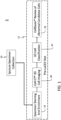
- the invention is defined by an optical tomography system comprising the features of claim 1 and a method for automated detection and monitoring of dysplasia by analyzing feature data from 3D mages of cells obtained from a sputum specimen obtained from a subject comprising the features of claim 3.
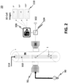
- the following disclosure describes a method for automated detection and monitoring of dysplasia by analyzing 3D images of cells obtained from a sputum specimen.
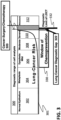
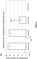
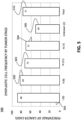
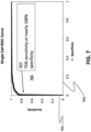
- the LuCED AROC value is 0.991.
- an operating point was selected that provides 75% sensitivity and 100% specificity.
- Invagination Voids Based on a threshold T, volume, surface area, shape, location of voids connected to invaginations.
- Nucleoli Based on a threshold T, volume, surface area, and shape, and location of objects likely to be nucleoli or chromatin condensations.
- Nuclear texture The technique of a blur residue, using various sized features structure elements, is used to separate various sized features within the nucleus.
- Overall 3D volume is then computed as are the number of discrete components, the volume histogram, average volume and variance, and shape histogram.
- Distance metrics Metrics describe spatial relationships between nucleoli, invaginations, voids, and the nuclear envelope. For example if three nucleoli are found the mean and variance, minimum and maximum inter-nucleoli distance may be found.
- voids occurring in 3D biological objects have now been found to be useful classification features based on measurement criteria including comparison with a calculated or selected threshold.
- Another characteristic related to voids may include the number of voids in an object.
- Another characteristic related to voids includes volume of a void or number of voids.
- Yet another characteristic includes surface area of a void or number of voids. Shape and location of inter-nuclear voids may also be employed as a useful feature characteristic. Additionally, combinations of feature characteristics may also be used to build a classifier as described hereinabove.
Landscapes
- Health & Medical Sciences (AREA)
- Engineering & Computer Science (AREA)
- Life Sciences & Earth Sciences (AREA)
- Chemical & Material Sciences (AREA)
- General Health & Medical Sciences (AREA)
- Physics & Mathematics (AREA)
- General Physics & Mathematics (AREA)
- Immunology (AREA)
- Medicinal Chemistry (AREA)
- Biomedical Technology (AREA)
- Biochemistry (AREA)
- Pathology (AREA)
- Analytical Chemistry (AREA)
- Veterinary Medicine (AREA)
- Pharmacology & Pharmacy (AREA)
- Public Health (AREA)
- Animal Behavior & Ethology (AREA)
- Molecular Biology (AREA)
- Epidemiology (AREA)
- Dispersion Chemistry (AREA)
- Theoretical Computer Science (AREA)
- Urology & Nephrology (AREA)
- Hematology (AREA)
- Nuclear Medicine, Radiotherapy & Molecular Imaging (AREA)
- Multimedia (AREA)
- Food Science & Technology (AREA)
- Cell Biology (AREA)
- Computer Vision & Pattern Recognition (AREA)
- Organic Chemistry (AREA)
- Pain & Pain Management (AREA)
- Radiology & Medical Imaging (AREA)
- Quality & Reliability (AREA)
- Medical Informatics (AREA)
- Microbiology (AREA)
- Chemical Kinetics & Catalysis (AREA)
- Toxicology (AREA)
- General Chemical & Material Sciences (AREA)
- Bioinformatics & Cheminformatics (AREA)
- Tropical Medicine & Parasitology (AREA)
- Biotechnology (AREA)
Claims (4)
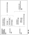
- Ein optisches Tomografiesystem, welches zur automatisierten Detektion und Überwachung einer Dysplasie durch Analysieren von Merkmalsdaten aus 3D-Bildern von Zellen konfiguriert ist, wobei die Zellen aus einer von einem Subjekt erhaltenen Sputumprobe erhalten wurden, wobei das optische Tomografiesystem Folgendes umfasst:einen Probenadäquanzklassifikator, der gekoppelt ist, um die Merkmalsdaten zu empfangen und einen Probenadäquanzwert zu generieren;einen Klassifikator für normale/abnormale Zellen, welcher gekoppelt ist, um die Merkmalsdaten zu empfangen und normale und abnormale Zellen zu detektieren; dadurch gekennzeichnet, dass es ferner Folgendes umfasst:einen Klassifikator für abnormale Zellen, der mit dem Klassifikator für normale/abnormale Zellen gekoppelt ist, um Daten für die abnormalen Zellen zu empfangen und ferner die abnormalen Zellen in kanzeröse und präkanzeröse Zellen einzuteilen;einen Klassifikator für präkanzeröse Zellen, der mit dem Klassifikator für abnormale Zellen gekoppelt ist, um Daten präkanzeröser Zellen zu empfangen und jede präkanzeröse Zelle als präkanzeröse Zelle glandulären Ursprungs oder als dysplastisch zu identifizieren;einen Klassifikator für dysplastische Zellen, welcher mit dem Klassifikator für präkanzeröse Zellen gekoppelt ist, um Daten dysplastischer Zellen zu empfangen und ferner dysplastische Zellen nach leichter bis mäßiger Dysplasie, mäßiger bis schwerer Dysplasie, oder schwerer Dysplasie einzuteilen; undeinen Behandlungsprotokollgenerator, welcher geeignet ist, Klassifikationsdaten von dem Klassifikator für abnormale Zellen, dem Klassifikator für präkanzeröse Zellen und dem Klassifikator für dysplastische Zellen zu empfangen und in Reaktion darauf ein Protokoll zum Verabreichen eines Pharmazeutikums zur Chemoprävention von Krebs an das Subjekt über einen festgelegten Zeitraum, wenn dies durch die Detektion von Krebs, präkanzerösen oder dysplastischen Zellen angezeigt ist, zu erzeugen.
- Das System nach Anspruch 1, wobei das Pharmazeutikum zur Chemoprävention von Krebs ein Arzneimittel umfasst, welches ausgewählt ist aus der Gruppe bestehend aus einem Prostacyclinanalog, Iloprost, einem chimären Antigenrezeptor (CAR) für T-Zellen, Vorinostat, HDAC-Inhibitoren, Cholecalciferol, Calcitriol und Kombinationen davon.
- Ein Verfahren zur automatisierten Detektion und Überwachung einer Dysplasie durch Analysieren von Merkmalsdaten aus 3D-Bildern von Zellen, wobei die Zellen aus einer von einem Subjekt erhaltenen Sputumprobe erhalten wurden, wobei das Verfahren Folgendes umfasst:Bereitstellen von 3D-Bildgebungsdaten für eine in der Sputumprobe enthaltene Zelle;Betreiben eines Probenadäquanzklassifikators, welcher gekoppelt ist, um die Merkmalsdaten zu empfangen und einen Probenadäquanzwert zu generieren;falls eine von dem Probenadäquanzklassifikator ausgeführte Bestimmung der Probenadäquanz festgelegte Kriterien erfüllt, dann Betreiben eines Normal/Abnormal-Klassifikators, welcher gekoppelt ist, um Merkmalsdaten zu empfangen und normale und abnormale Zellen zu detektieren; gekennzeichnet durchBetreiben eines Abnormal-Klassifikators, welcher mit dem Normal/Abnormal-Klassifikator gekoppelt ist, um Merkmalsdaten für die abnormalen Zellen zu empfangen und ferner die abnormalen Zellen in kanzeröse und präkanzeröse Zellen einzuteilen;Betreiben eines Klassifikators für präkanzeröse Zellen, welcher mit dem Klassifikator für abnormale Zellen gekoppelt ist, um Daten präkanzeröser Zellen zu empfangen und jede präkanzeröse Zelle als präkanzeröse Zelle glandulären Ursprungs oder als dysplastisch zu identifizieren;Betreiben eines Klassifikators für dysplastische Zellen, welcher mit dem Klassifikator für präkanzeröse Zellen gekoppelt ist, um Daten dysplastischer Zellen zu empfangen und ferner dysplastische Zellen nach leichter bis mäßiger Dysplasie, mäßiger bis schwerer Dysplasie, oder schwerer Dysplasie einzuteilen; undBetreiben eines Behandlungsprotokollgenerators, um Klassifikationsdaten von dem Klassifikator für abnormale Zellen, dem Klassifikator für präkanzeröse Zellen und dem Klassifikator für dysplastische Zellen zu empfangen und in Reaktion darauf ein Protokoll zum Verabreichen eines Pharmazeutikums zur Chemoprävention von Krebs an das Subjekt über einen festgelegten Zeitraum, wenn dies durch die Detektion von Krebs, präkanzerösen oder dysplastischen Zellen angezeigt ist, zu erzeugen.
- Das Verfahren nach Anspruch 3, wobei das Pharmazeutikum zur Chemoprävention von Krebs ein Arzneimittel umfasst, welches ausgewählt ist aus der Gruppe bestehend aus einem Prostacyclinanalog, Iloprost, einem chimären Antigenrezeptor (CAR) für T-Zellen, Vorinostat, HDAC-Inhibitoren, Cholecalciferol, Calcitriol und Kombinationen davon.
Applications Claiming Priority (3)
| Application Number | Priority Date | Filing Date | Title |
|---|---|---|---|
| US201562273127P | 2015-12-30 | 2015-12-30 | |
| US201662367000P | 2016-07-26 | 2016-07-26 | |
| PCT/US2016/068859 WO2017117210A1 (en) | 2015-12-30 | 2016-12-28 | System and method for automated detection and monitoring of dysplasia and administration of chemoprevention |
Publications (5)
| Publication Number | Publication Date |
|---|---|
| EP3338086A1 EP3338086A1 (de) | 2018-06-27 |
| EP3338086A4 EP3338086A4 (de) | 2018-11-14 |
| EP3338086C0 EP3338086C0 (de) | 2024-07-03 |
| EP3338086B1 true EP3338086B1 (de) | 2024-07-03 |
| EP3338086B8 EP3338086B8 (de) | 2024-08-14 |
Family
ID=59225666
Family Applications (1)
| Application Number | Title | Priority Date | Filing Date |
|---|---|---|---|
| EP16882553.7A Active EP3338086B8 (de) | 2015-12-30 | 2016-12-28 | System und verfahren zur automatisierten erkennung und überwachung von dysplasie |
Country Status (9)
| Country | Link |
|---|---|
| US (1) | US11069054B2 (de) |
| EP (1) | EP3338086B8 (de) |
| JP (1) | JP2019506602A (de) |
| CN (1) | CN108603876A (de) |
| AU (1) | AU2016381656A1 (de) |
| CA (1) | CA3009813A1 (de) |
| ES (1) | ES2987312T3 (de) |
| HU (1) | HUE068588T2 (de) |
| WO (1) | WO2017117210A1 (de) |
Families Citing this family (13)
| Publication number | Priority date | Publication date | Assignee | Title |
|---|---|---|---|---|
| US10692211B2 (en) * | 2017-06-20 | 2020-06-23 | Case Western Reserve University | Intra-perinodular textural transition (IPRIS): a three dimenisonal (3D) descriptor for nodule diagnosis on lung computed tomography (CT) images |
| EP3724854A1 (de) * | 2017-12-15 | 2020-10-21 | Verily Life Sciences LLC | Erzeugung von virtuell gefärbten bildern von ungefärbten proben |
| CA3088939A1 (en) | 2018-01-05 | 2019-07-11 | Visiongate, Inc. | Morphometric genotyping of cells using optical tomography for detecting tumor mutational burden |
| WO2019169157A1 (en) * | 2018-02-28 | 2019-09-06 | Visiongate, Inc. | Morphometric detection of malignancy associated change |
| CN108846838B (zh) * | 2018-06-04 | 2021-05-11 | 卢龙 | 一种三维mri半自动病灶图像分割方法及系统 |
| WO2020159879A2 (en) * | 2019-01-28 | 2020-08-06 | Nano Global Corp. | Amelioration based on detection of biological cells or biological substances |
| TW202032574A (zh) * | 2019-02-26 | 2020-09-01 | 沛智生醫科技股份有限公司 | 細胞分類方法、系統與醫療分析平台 |
| US12019674B2 (en) | 2019-05-29 | 2024-06-25 | New York University | System and method for tumor characterization |
| US11354804B1 (en) | 2019-09-27 | 2022-06-07 | Verily Life Sciences Llc | Transforming multispectral images to enhanced resolution images enabled by machine learning |
| CN116097373A (zh) * | 2020-08-27 | 2023-05-09 | 梅迪安技术公司 | 用于通过机器学习免疫检查点抑制剂应答预测进行患者分层的系统和方法 |
| CN112053769B (zh) * | 2020-09-30 | 2023-03-10 | 沈阳东软智能医疗科技研究院有限公司 | 一种三维医学影像标注方法、装置及相关产品 |
| WO2024030346A1 (en) | 2022-08-02 | 2024-02-08 | Visiongate, Inc. | Ai-based cell classification method and system |
| GB2641850A (en) * | 2023-03-14 | 2025-12-17 | Mitsubishi Electric Corp | Particle analysis device, particle analysis system, and particle analysis method |
Family Cites Families (258)
| Publication number | Priority date | Publication date | Assignee | Title |
|---|---|---|---|---|
| US3560754A (en) | 1965-11-17 | 1971-02-02 | Ibm | Photoelectric particle separator using time delay |
| US3470373A (en) | 1966-10-18 | 1969-09-30 | Litton Systems Inc | Method for analysis and identification of biologic entities by phosphorescence |
| US3497690A (en) | 1967-09-21 | 1970-02-24 | Bausch & Lomb | Method and apparatus for classifying biological cells by measuring the size and fluorescent response thereof |
| US3598471A (en) | 1968-11-22 | 1971-08-10 | Corning Glass Works | Optical contrast enhancement system |
| US3705771A (en) | 1970-01-14 | 1972-12-12 | Bio Physics Systems Inc | Photoanalysis apparatus |
| US3657537A (en) | 1970-04-03 | 1972-04-18 | Bausch & Lomb | Computerized slit-scan cyto-fluorometer for automated cell recognition |
| US3748468A (en) | 1971-12-22 | 1973-07-24 | Gen Electric | Automatic electron microscope field counter |
| US3999047A (en) | 1972-09-05 | 1976-12-21 | Green James E | Method and apparatus utilizing color algebra for analyzing scene regions |
| US3833762A (en) | 1973-06-04 | 1974-09-03 | Rockwell International Corp | Solid state integrating, image motion compensating imager |
| US4200353A (en) | 1974-06-05 | 1980-04-29 | Robert Hoffman | Modulation contrast microscope with three regions |
| US3960449A (en) | 1975-06-05 | 1976-06-01 | The Board Of Trustees Of Leland Stanford Junior University | Measurement of angular dependence of scattered light in a flowing stream |
| DE2543310C2 (de) | 1975-09-27 | 1982-04-29 | Gesellschaft für Strahlen- und Umweltforschung mbH, 8000 München | Einrichtung zum Zählen und Klassifizieren von in einer Flüssigkeit suspendierten Teilchen |
| NL7609885A (nl) | 1976-09-06 | 1978-03-08 | Optische Ind De Oude Delft Nv | Stelsel voor het elementsgewijs reconstrueren van een tomogram van een dwarsdoorsnede van een object. |
| US4081277A (en) | 1976-10-08 | 1978-03-28 | Eastman Kodak Company | Method for making a solid-state color imaging device having an integral color filter and the device |
| US4175860A (en) | 1977-05-31 | 1979-11-27 | Rush-Presbyterian-St. Luke's Medical Center | Dual resolution method and apparatus for use in automated classification of pap smear and other samples |
| US4183623A (en) | 1977-10-11 | 1980-01-15 | Haines Kenneth A | Tomographic cross-sectional imaging using incoherent optical processing |
| US4293221A (en) | 1979-04-17 | 1981-10-06 | Research Corporation | Multidimensional slit-scan flow system |
| US4422146A (en) | 1979-11-22 | 1983-12-20 | The President Of Tokyo Institute Of Technology | Reconstruction method of a computed tomographic image from a few X-ray projections |
| US4360885A (en) | 1980-01-02 | 1982-11-23 | Edgar Albert D | Micro-optical tomography |
| US4667830A (en) | 1981-06-15 | 1987-05-26 | The Board Of Trustees Of The Leland Stanford Junior University | Method and means for sorting individual particles into containers for culturing, cloning, analysis, or the like |
| DE3278129D1 (en) | 1981-12-08 | 1988-03-31 | Ici Plc | Sedimentation field flow fractionation |
| DE3424108A1 (de) | 1984-06-29 | 1986-01-09 | Bernhard Prof. Dr.-Ing. 4300 Essen Schrader | Probenanordnung zur spektrometrie, verfahren zur messung von lumineszenz und streuung und verwendung der probenanordnung |
| US4702598A (en) | 1985-02-25 | 1987-10-27 | Research Corporation | Flow cytometer |
| US5281517A (en) | 1985-11-04 | 1994-01-25 | Cell Analysis Systems, Inc. | Methods for immunoploidy analysis |
| US5771070A (en) | 1985-11-15 | 1998-06-23 | Canon Kabushiki Kaisha | Solid state image pickup apparatus removing noise from the photoelectric converted signal |
| DE3583040D1 (de) | 1985-12-17 | 1991-07-04 | Ibm | Bild-vorverarbeitungsverfahren zur rauschentfernung. |
| US4873653A (en) | 1986-04-09 | 1989-10-10 | Carl-Zeiss-Stiftung | Microscope system for providing three-dimensional resolution |
| US4694342A (en) | 1986-05-01 | 1987-09-15 | Eastman Kodak Company | Spatial filter useful for removing noise from video images and for preserving detail therein |
| DE3619298C1 (de) | 1986-06-07 | 1987-08-13 | Westfalia Separator Ag | Kontinuierlich arbeitende Schleudertrommel |
| US4786165A (en) | 1986-07-10 | 1988-11-22 | Toa Medical Electronics Co., Ltd. | Flow cytometry and apparatus therefor |
| US4858128A (en) | 1986-08-11 | 1989-08-15 | General Electric Company | View-to-view image correction for object motion |
| GB8619717D0 (en) | 1986-08-13 | 1986-09-24 | Johnson Matthey Plc | Conversion of methane |
| US4891829A (en) | 1986-11-19 | 1990-01-02 | Exxon Research And Engineering Company | Method and apparatus for utilizing an electro-optic detector in a microtomography system |
| KR970007077B1 (ko) | 1987-03-13 | 1997-05-02 | 코울터 일렉트로닉스 인커퍼레이티드 | 광산란 기술을 이용한 다중-부분식별 분석 방법 |
| DE3905619C2 (de) | 1988-02-23 | 2000-04-13 | Olympus Optical Co | Bildeingabe-/Ausgabevorrichtung |
| US5545671A (en) | 1989-10-05 | 1996-08-13 | Schering Aktiengesellschaft | Antimetastically acting agents |
| GB2239575B (en) | 1989-10-17 | 1994-07-27 | Mitsubishi Electric Corp | Motion vector detecting apparatus and image blur correcting apparatus, and video camera including such apparatus |
| US5034613A (en) | 1989-11-14 | 1991-07-23 | Cornell Research Foundation, Inc. | Two-photon laser microscopy |
| US5141609A (en) | 1990-11-16 | 1992-08-25 | The Trustees Of The Leland Stanford Junior University | Method and device employing time-delayed integration for detecting sample components after separation |
| JP3084295B2 (ja) | 1991-02-27 | 2000-09-04 | シスメックス株式会社 | フローイメージサイトメータ |
| US5496850A (en) | 1991-04-11 | 1996-03-05 | Toray Industries, Inc. | Antimetastasis agent of malignant tumors |
| FR2675927B1 (fr) | 1991-04-25 | 1993-07-30 | Inst Nat Rech Inf Automat | Procede et dispositif d'aide a l'inspection d'un corps, notamment pour la tomographie. |
| US6485413B1 (en) | 1991-04-29 | 2002-11-26 | The General Hospital Corporation | Methods and apparatus for forward-directed optical scanning instruments |
| US5321501A (en) | 1991-04-29 | 1994-06-14 | Massachusetts Institute Of Technology | Method and apparatus for optical imaging with means for controlling the longitudinal range of the sample |
| US5117466A (en) | 1991-04-30 | 1992-05-26 | The United States Of America As Represented By The United States Department Of Energy | Integrated fluorescence analysis system |
| JPH04337446A (ja) | 1991-05-15 | 1992-11-25 | Hitachi Ltd | 微粒子計測方法、定量方法および微粒子計測装置 |
| US5177466A (en) | 1991-08-05 | 1993-01-05 | Lai Chung C | Lock structure with a warning device |
| US5548395A (en) | 1991-09-20 | 1996-08-20 | Toa Medical Electronics Co., Ltd. | Particle analyzer |
| US5333164A (en) | 1991-12-11 | 1994-07-26 | General Electric Company | Method and apparatus for acquiring and processing only a necessary volume of radon data consistent with the overall shape of the object for efficient three dimensional image reconstruction |
| GB9218482D0 (en) | 1992-09-01 | 1992-10-14 | Dixon Arthur E | Apparatus and method for scanning laser imaging of macroscopic samples |
| JP3384580B2 (ja) | 1992-04-30 | 2003-03-10 | 株式会社リコー | 地肌除去方法 |
| US5668887A (en) | 1992-05-29 | 1997-09-16 | Eastman Kodak Company | Coating density analyzer and method using non-synchronous TDI camera |
| JP3327948B2 (ja) | 1992-06-09 | 2002-09-24 | オリンパス光学工業株式会社 | 光学像再構成装置 |
| US5390226A (en) | 1992-07-02 | 1995-02-14 | General Electric Company | Method and apparatus for pre-processing cone beam projection data for exact three dimensional computer tomographic image reconstruction of a portion of an object |
| US5312535A (en) | 1992-07-17 | 1994-05-17 | Beckman Instruments, Inc. | Capillary electrophoresis detection |
| DE69333642T2 (de) | 1992-07-31 | 2005-10-06 | Fuji Photo Film Co., Ltd., Minami-Ashigara | Verfahren und Vorrichtung zur Gewinnung dreidimensionaler Information von Proben |
| US5757981A (en) | 1992-08-20 | 1998-05-26 | Toyo Ink Mfg. Co., Ltd. | Image inspection device |
| US6159686A (en) | 1992-09-14 | 2000-12-12 | Sri International | Up-converting reporters for biological and other assays |
| US6026174A (en) | 1992-10-14 | 2000-02-15 | Accumed International, Inc. | System and method for automatically detecting malignant cells and cells having malignancy-associated changes |
| US5547849A (en) | 1993-02-17 | 1996-08-20 | Biometric Imaging, Inc. | Apparatus and method for volumetric capillary cytometry |
| WO1994024545A1 (en) | 1993-04-15 | 1994-10-27 | Board Of Regents, The University Of Texas System | Raman spectrometer |
| US5676631A (en) | 1993-07-06 | 1997-10-14 | Westfalia Separator Aktiengesellschaft | Centrifuge drum for concentrating suspended solids |
| US5402460A (en) | 1993-08-02 | 1995-03-28 | University Of Washington | Three-dimensional microtomographic analysis system |
| US6215587B1 (en) | 1994-02-14 | 2001-04-10 | Robert R. Alfano | Microscope imaging inside highly scattering media |
| JPH07286953A (ja) | 1994-04-19 | 1995-10-31 | Toa Medical Electronics Co Ltd | イメージングフローサイトメータ |
| AU3629295A (en) | 1994-09-20 | 1996-04-09 | Neopath, Inc. | Apparatus for automated identification of thick cell groupings on a biological specimen |
| US5978497A (en) | 1994-09-20 | 1999-11-02 | Neopath, Inc. | Apparatus for the identification of free-lying cells |
| WO1996010801A1 (en) | 1994-09-30 | 1996-04-11 | Neopath, Inc. | Method and apparatus for highly efficient computer aided screening |
| US5552605A (en) | 1994-11-18 | 1996-09-03 | Picker International, Inc. | Motion correction based on reprojection data |
| FI98765C (fi) | 1995-01-16 | 1997-08-11 | Erkki Soini | Virtaussytometrinen menetelmä ja laite |
| US5550892A (en) | 1995-03-24 | 1996-08-27 | The Regents Of The University Of California | Enhanced local tomography |
| US5539800A (en) | 1995-03-24 | 1996-07-23 | The Regents Of The University Of California, Office Of Technology Transfer | Pseudolocal tomography |
| US5710429A (en) | 1995-04-06 | 1998-01-20 | Alfano; Robert R. | Ultrafast optical imaging of objects in or behind scattering media |
| US5582705A (en) | 1995-05-19 | 1996-12-10 | Iowa State University Research Foundation, Inc. | Multiplexed capillary electrophoresis system |
| US6252979B1 (en) | 1995-06-07 | 2001-06-26 | Tripath Imaging, Inc. | Interactive method and apparatus for sorting biological specimens |
| JPH095443A (ja) | 1995-06-23 | 1997-01-10 | Toshiba Corp | 核医学診断装置 |
| DE19525567A1 (de) | 1995-07-13 | 1997-01-16 | Krauss Maffei Ag | Stülpfilterzentrifuge |
| JP3221291B2 (ja) | 1995-07-26 | 2001-10-22 | ソニー株式会社 | 画像処理装置、画像処理方法、ノイズ除去装置及びノイズ除去方法 |
| JP3783238B2 (ja) | 1995-07-31 | 2006-06-07 | ソニー株式会社 | 撮像システム、画像処理装置、符号化装置、符号化方法及びランダムノイズを除去する方法 |
| WO1997012975A1 (en) | 1995-10-02 | 1997-04-10 | The Government Of The United States Of America, Represented By The Secretary, Department Of Health And Human Services | An epithelial protein and dna thereof for use in early cancer detection |
| FI97665C (fi) | 1995-11-21 | 1997-01-27 | Planmed Oy | Menetelmät ja laitteet kohteen kuvantamisessa |
| US6165734A (en) | 1995-12-12 | 2000-12-26 | Applied Spectral Imaging Ltd. | In-situ method of analyzing cells |
| US6007994A (en) | 1995-12-22 | 1999-12-28 | Yale University | Multiparametric fluorescence in situ hybridization |
| US6463182B1 (en) | 1995-12-28 | 2002-10-08 | Canon Kabushiki Kaisha | Image processing apparatus and method for removing noise near an edge of an image |
| FR2743462B1 (fr) | 1996-01-04 | 1998-01-30 | Commissariat Energie Atomique | Dispositif de lecture de barrettes de detecteurs avec effet tdi |
| GB2309368B (en) | 1996-01-18 | 1999-09-15 | Hamamatsu Photonics Kk | An optical computer tomographic apparatus and image reconstruction method using optical computer tomography |
| JPH09247473A (ja) | 1996-03-07 | 1997-09-19 | Minolta Co Ltd | 画像形成装置 |
| JPH09247545A (ja) | 1996-03-11 | 1997-09-19 | Matsushita Electric Ind Co Ltd | スキャナ型電子カメラ |
| US6078681A (en) | 1996-03-18 | 2000-06-20 | Marine Biological Laboratory | Analytical imaging system and process |
| JP3640461B2 (ja) | 1996-04-03 | 2005-04-20 | シスメックス株式会社 | 粒子分析装置 |
| US6038067A (en) | 1996-05-23 | 2000-03-14 | The Regents Of The University Of California | Scanning computed confocal imager |
| US5915048A (en) | 1996-06-05 | 1999-06-22 | Zetetic Institute | Method and apparatus for discriminating in-focus images from out-of-focus light signals from background and foreground light sources |
| US5880838A (en) | 1996-06-05 | 1999-03-09 | California Institute Of California | System and method for optically measuring a structure |
| US5673300A (en) | 1996-06-11 | 1997-09-30 | Wisconsin Alumni Research Foundation | Method of registering a radiation treatment plan to a patient |
| US6047080A (en) | 1996-06-19 | 2000-04-04 | Arch Development Corporation | Method and apparatus for three-dimensional reconstruction of coronary vessels from angiographic images |
| DE69730053T2 (de) | 1996-11-29 | 2005-07-21 | Imaging Diagnostic System, Inc., Plantation | Verfahren zur abbildung eines körpers mittels abtastung durch eine laser-bilderzeugungs-vorrichtung |
| US5768440A (en) | 1996-12-18 | 1998-06-16 | Xerox Corporation | Adaptive noise removal for video images with less correction of current pixel for higher variations between surrounding pixels |
| DE19701036A1 (de) | 1997-01-15 | 1998-07-16 | Philips Patentverwaltung | MR-Verfahren zur bildgestützten Überwachung der Verschiebung eines Objektes und MR-Anordnung zur Durchführung des Verfahrens |
| US5760901A (en) | 1997-01-28 | 1998-06-02 | Zetetic Institute | Method and apparatus for confocal interference microscopy with background amplitude reduction and compensation |
| US6091983A (en) | 1997-02-07 | 2000-07-18 | Alfano; Robert R. | Imaging of objects in turbid media based upon the preservation of polarized luminescence emitted from contrast agents |
| JPH10260131A (ja) | 1997-03-19 | 1998-09-29 | Seitai Hikarijoho Kenkyusho:Kk | 光計測装置 |
| DE19714221A1 (de) | 1997-04-07 | 1998-10-08 | Zeiss Carl Fa | Konfokales Mikroskop mit einem motorischen Scanningtisch |
| US6002480A (en) | 1997-06-02 | 1999-12-14 | Izatt; Joseph A. | Depth-resolved spectroscopic optical coherence tomography |
| US20030049841A1 (en) | 1997-06-16 | 2003-03-13 | Short Jay M. | High throughput or capillary-based screening for a bioactivity or biomolecule |
| US5878103A (en) | 1997-06-30 | 1999-03-02 | Siemens Corporate Research, Inc. | Adaptive detector masking for speed-up of cone beam reconstruction |
| CA2301004A1 (en) | 1997-09-12 | 1999-03-18 | The Board Of Trustees Of The Leland Stanford Junior University | Flow-through microcentrifuge |
| US5909476A (en) | 1997-09-22 | 1999-06-01 | University Of Iowa Research Foundation | Iterative process for reconstructing cone-beam tomographic images |
| US6540895B1 (en) | 1997-09-23 | 2003-04-01 | California Institute Of Technology | Microfabricated cell sorter for chemical and biological materials |
| US6388809B1 (en) | 1997-10-29 | 2002-05-14 | Digital Optical Imaging Corporation | Methods and apparatus for improved depth resolution use of out-of-focus information in microscopy |
| US6037579A (en) | 1997-11-13 | 2000-03-14 | Biophotonics Information Laboratories, Ltd. | Optical interferometer employing multiple detectors to detect spatially distorted wavefront in imaging of scattering media |
| US6201628B1 (en) | 1997-11-19 | 2001-03-13 | University Of Washington | High throughput optical scanner |
| US6251615B1 (en) | 1998-02-20 | 2001-06-26 | Cell Analytics, Inc. | Cell analysis methods |
| US6433822B1 (en) | 1998-03-31 | 2002-08-13 | Intel Corporation | Method and apparatus for self-calibration and fixed-pattern noise removal in imager integrated circuits |
| US6291824B1 (en) | 1998-04-13 | 2001-09-18 | Board Of Supervisors Of Louisiana State University And Agricultural And Mechanical College | Apparatus and method for high-bandwidth optical tomography |
| US6248988B1 (en) | 1998-05-05 | 2001-06-19 | Kla-Tencor Corporation | Conventional and confocal multi-spot scanning optical microscope |
| US6529614B1 (en) | 1998-08-05 | 2003-03-04 | California Institute Of Technology | Advanced miniature processing handware for ATR applications |
| EP1105710B1 (de) | 1998-08-14 | 2011-12-28 | On-Site Analysis, Inc. | Verfahren zur Normierung der durch optische Emissionsspektrometrie gemessenen Intensität eines Spektrums |
| JP2000121550A (ja) | 1998-10-16 | 2000-04-28 | Japan Science & Technology Corp | ヘテロダイン検波による生体試料の画像検出方法及びその装置 |
| US6295384B1 (en) | 1998-11-04 | 2001-09-25 | Schlumberger Technologies, Inc. | Removing noise caused by artifacts from a digital image signal |
| EP1002497B1 (de) | 1998-11-20 | 2006-07-26 | Fuji Photo Film Co., Ltd. | Blutgefäss Bilddarstellungssystem |
| US6256096B1 (en) | 1999-01-11 | 2001-07-03 | Softray | Flow cytometry apparatus and method |
| JP4473358B2 (ja) | 1999-01-21 | 2010-06-02 | 株式会社東芝 | 診断装置 |
| US6640014B1 (en) | 1999-01-22 | 2003-10-28 | Jeffrey H. Price | Automatic on-the-fly focusing for continuous image acquisition in high-resolution microscopy |
| US6249341B1 (en) | 1999-01-25 | 2001-06-19 | Amnis Corporation | Imaging and analyzing parameters of small moving objects such as cells |
| US6473176B2 (en) | 1999-01-25 | 2002-10-29 | Amnis Corporation | Imaging and analyzing parameters of small moving objects such as cells |
| US6975400B2 (en) | 1999-01-25 | 2005-12-13 | Amnis Corporation | Imaging and analyzing parameters of small moving objects such as cells |
| US6608682B2 (en) | 1999-01-25 | 2003-08-19 | Amnis Corporation | Imaging and analyzing parameters of small moving objects such as cells |
| US7450229B2 (en) | 1999-01-25 | 2008-11-11 | Amnis Corporation | Methods for analyzing inter-cellular phenomena |
| WO2000049447A1 (en) | 1999-02-17 | 2000-08-24 | Lucid, Inc. | Tissue specimen holder |
| US6904163B1 (en) | 1999-03-19 | 2005-06-07 | Nippon Telegraph And Telephone Corporation | Tomographic image reading method, automatic alignment method, apparatus and computer readable medium |
| KR100319557B1 (ko) | 1999-04-16 | 2002-01-09 | 윤종용 | 블럭 단위로 부호화된 영상의 블럭 경계 잡음 성분 제거 방법 |
| US6640017B1 (en) | 1999-05-26 | 2003-10-28 | Intel Corporation | Method and apparatus for adaptively sharpening an image |
| DE19931954A1 (de) | 1999-07-10 | 2001-01-11 | Leica Microsystems | Beleuchtungseinrichtung für ein DUV-Mikroskop |
| WO2001011341A2 (en) | 1999-08-05 | 2001-02-15 | Cellomics, Inc. | A system for cell-based screening |
| US6239871B1 (en) | 1999-08-24 | 2001-05-29 | Waters Investments Limited | Laser induced fluorescence capillary interface |
| JP3998229B2 (ja) | 1999-09-08 | 2007-10-24 | カシオ計算機株式会社 | 撮像装置およびその信号処理方法 |
| US6621937B1 (en) | 1999-10-08 | 2003-09-16 | Eastman Kodak Company | Removing chroma noise from digital images by using variable shape pixel neighborhood regions |
| US7003143B1 (en) | 1999-11-02 | 2006-02-21 | Hewitt Charles W | Tomographic microscope for high resolution imaging and method of analyzing specimens |
| US6775399B1 (en) | 1999-11-17 | 2004-08-10 | Analogic Corporation | ROI segmentation image processing system |
| US6365367B1 (en) | 1999-12-06 | 2002-04-02 | Cellomics, Inc. | Environmental chamber for the analysis of live cells |
| DE10000185A1 (de) | 2000-01-05 | 2001-07-12 | Philips Corp Intellectual Pty | Verfahren zur Darstellung des zeitlichen Verlaufs des Blutflusses in einem Untersuchungsobjekt |
| US6549308B1 (en) | 2000-01-11 | 2003-04-15 | Zebra Imaging, Inc. | Unibiased light field models for rendering and holography |
| JP3893849B2 (ja) | 2000-05-15 | 2007-03-14 | 株式会社日立製作所 | キャピラリアレイ電気泳動装置及び電気泳動方法 |
| JP4632481B2 (ja) | 2000-05-22 | 2011-02-16 | 忠 田邉 | プロスタサイクリン合成酵素遺伝子含有医薬組成物 |
| US6875973B2 (en) | 2000-08-25 | 2005-04-05 | Amnis Corporation | Auto focus for a flow imaging system |
| US6507391B2 (en) | 2000-08-25 | 2003-01-14 | Amnis Corporation | Measuring the velocity of small moving objects such as cells |
| EP1313883A4 (de) | 2000-08-29 | 2005-01-12 | Yeda Res & Dev | Methode zur isolation von genen welche proteine mit spezieller funktion kodierenund zum screening von pharmazeutisch aktiven substanzen |
| JP2002119502A (ja) | 2000-10-17 | 2002-04-23 | Toshiba Corp | 医用装置 |
| AU1576401A (en) | 2000-10-27 | 2002-05-06 | Praelux Inc | Method and apparatus for screening chemical compounds |
| US6646246B1 (en) | 2000-11-21 | 2003-11-11 | Eastman Kodak Company | Method and system of noise removal for a sparsely sampled extended dynamic range image sensing device |
| US7050087B2 (en) | 2000-12-06 | 2006-05-23 | Bioview Ltd. | Data acquisition and display system and method |
| JP2002185862A (ja) | 2000-12-15 | 2002-06-28 | Mega Chips Corp | 画像信号のノイズ除去方法 |
| DE60144249D1 (de) | 2000-12-15 | 2011-04-28 | Kla Tencor Corp | Verfahren und vorrichtung zum untersuchen eines substrats |
| US6470070B2 (en) | 2000-12-20 | 2002-10-22 | Cedara Software Corp. | Image reconstruction using multiple X-ray projections |
| US6937772B2 (en) | 2000-12-20 | 2005-08-30 | Eastman Kodak Company | Multiresolution based method for removing noise from digital images |
| DE10100572A1 (de) | 2001-01-09 | 2002-07-11 | Philips Corp Intellectual Pty | Verfahren zur Darstellung des Blutflusses in einem Gefäßbaum |
| JP2002281311A (ja) | 2001-03-15 | 2002-09-27 | Ricoh Co Ltd | 画像処理装置及び画像読取装置 |
| US7907765B2 (en) | 2001-03-28 | 2011-03-15 | University Of Washington | Focal plane tracking for optical microtomography |
| US6522775B2 (en) | 2001-03-28 | 2003-02-18 | Alan C. Nelson | Apparatus and method for imaging small objects in a flow stream using optical tomography |
| US20060023219A1 (en) | 2001-03-28 | 2006-02-02 | Meyer Michael G | Optical tomography of small objects using parallel ray illumination and post-specimen optical magnification |
| US6519355B2 (en) * | 2001-03-28 | 2003-02-11 | Alan C. Nelson | Optical projection imaging system and method for automatically detecting cells having nuclear and cytoplasmic densitometric features associated with disease |
| US6944322B2 (en) | 2001-03-28 | 2005-09-13 | Visiongate, Inc. | Optical tomography of small objects using parallel ray illumination and post-specimen optical magnification |
| US6591003B2 (en) | 2001-03-28 | 2003-07-08 | Visiongate, Inc. | Optical tomography of small moving objects using time delay and integration imaging |
| DE10116682A1 (de) | 2001-04-04 | 2002-10-10 | Philips Corp Intellectual Pty | Verfahren und Vorrichtung zur Rekonstruktion dreidimensionaler Bilder aus Kegelstrahl-Projektionsdaten |
| US6755969B2 (en) | 2001-04-25 | 2004-06-29 | Phase Inc. | Centrifuge |
| US20020173034A1 (en) | 2001-05-21 | 2002-11-21 | Emilio Barbera-Guillem | Centrifuge apparatus and methods for separating components from a cell culture device |
| GB0112392D0 (en) | 2001-05-22 | 2001-07-11 | Medical Res Council | Optical imaging appartus and associated specimen support means |
| US6801672B1 (en) | 2001-06-01 | 2004-10-05 | Bruce A. Thomas | Removing noise from a color image using wavelets |
| JP2003008885A (ja) | 2001-06-19 | 2003-01-10 | Fuji Photo Film Co Ltd | 画像再生システム |
| WO2003012405A2 (en) | 2001-08-03 | 2003-02-13 | Rollins Andrew M | Aspects of basic oct engine technologies for high speed optical coherence tomography and light source and other improvements in oct |
| JP2004538509A (ja) | 2001-08-06 | 2004-12-24 | バイオビュー リミテッド | 蛍光撮像における画像合焦 |
| US6741730B2 (en) | 2001-08-10 | 2004-05-25 | Visiongate, Inc. | Method and apparatus for three-dimensional imaging in the fourier domain |
| US6636623B2 (en) * | 2001-08-10 | 2003-10-21 | Visiongate, Inc. | Optical projection imaging system and method for automatically detecting cells with molecular marker compartmentalization associated with malignancy and disease |
| US6842297B2 (en) | 2001-08-31 | 2005-01-11 | Cdm Optics, Inc. | Wavefront coding optics |
| US6763142B2 (en) | 2001-09-07 | 2004-07-13 | Nline Corporation | System and method for correlated noise removal in complex imaging systems |
| JP3931606B2 (ja) | 2001-09-20 | 2007-06-20 | ソニー株式会社 | 撮像装置およびノイズ除去方法 |
| AU2002343394A1 (en) * | 2001-09-21 | 2003-04-01 | The Regents Of The University Of California | Methods of cytodiagnostic staging of neoplasia and squamous cell carcinoma |
| US7039455B1 (en) | 2001-10-09 | 2006-05-02 | Medrad, Inc. | Apparatus and method for removing magnetic resonance imaging-induced noise from ECG signals |
| US6850587B1 (en) | 2001-10-24 | 2005-02-01 | Analogic Corporation | Reprojection-based three-dimensional image reconstruction |
| US6931160B2 (en) | 2001-10-31 | 2005-08-16 | Eastman Kodak Company | Method of spatially filtering digital image for noise removal, noise estimation or digital image enhancement |
| US6512807B1 (en) | 2001-11-21 | 2003-01-28 | Koninklijke Philips Electronics, N.V. | Low signal correction for perfusion measurements |
| US20030108512A1 (en) | 2001-12-10 | 2003-06-12 | Robert Shorr | Modified prostaglandin compounds and analogs thereof, compositions containing the same useful for the treatment of cancer |
| AU2003217783A1 (en) | 2002-02-27 | 2003-09-09 | Wake Forest University | Angiotensin-(1-7) and angiotensin-(1-7) agonists for inhibition of cancer cell growth |
| US7050650B2 (en) | 2002-02-28 | 2006-05-23 | Hewlett-Packard Development Company, L.P. | User selected background noise removal for scanned document images |
| US7283253B2 (en) | 2002-03-13 | 2007-10-16 | Applied Precision, Llc | Multi-axis integration system and method |
| US7260253B2 (en) | 2002-04-19 | 2007-08-21 | Visiongate, Inc. | Method for correction of relative object-detector motion between successive views |
| US7197355B2 (en) | 2002-04-19 | 2007-03-27 | Visiongate, Inc. | Variable-motion optical tomography of small objects |
| US7811825B2 (en) | 2002-04-19 | 2010-10-12 | University Of Washington | System and method for processing specimens and images for optical tomography |
| US7738945B2 (en) | 2002-04-19 | 2010-06-15 | University Of Washington | Method and apparatus for pseudo-projection formation for optical tomography |
| US20050085708A1 (en) | 2002-04-19 | 2005-04-21 | University Of Washington | System and method for preparation of cells for 3D image acquisition |
| US6697508B2 (en) | 2002-05-10 | 2004-02-24 | Visiongate, Inc. | Tomographic reconstruction of small objects using a priori knowledge |
| US6770893B2 (en) | 2002-05-13 | 2004-08-03 | Visiongate, Inc. | Method and apparatus for emission computed tomography using temporal signatures |
| US20040008515A1 (en) | 2002-06-11 | 2004-01-15 | Applied Precision | Fluorescence illumination optimization for three-dimensional microscopy |
| WO2004017993A1 (en) | 2002-08-12 | 2004-03-04 | Actelion Pharmaceuticals Ltd | Combination of prostacyclin or prostacyclin analogues and endothelin receptor antagonists for the treatment of pulmonary arterial hypertension |
| US6894779B2 (en) | 2002-08-23 | 2005-05-17 | Coulter International Corp. | Apparatus for detecting back-scatter in a laser-based blood analysis system |
| US7274809B2 (en) | 2002-08-29 | 2007-09-25 | Perceptronix Medical, Inc. And British Columbia Cancer Agency | Computerized methods and systems related to the detection of malignancy-associated changes (MAC) to detect cancer |
| CA2490578A1 (en) | 2002-08-30 | 2004-03-11 | Medical Research Council | Optical projection tomography |
| US7092017B2 (en) | 2002-09-13 | 2006-08-15 | Eastman Kodak Company | Fixed pattern noise removal in CMOS imagers across various operational conditions |
| US7443431B2 (en) | 2002-09-13 | 2008-10-28 | Eastman Kodak Company | Fixed pattern noise removal in CMOS imagers across various operational conditions |
| JP4156893B2 (ja) | 2002-09-27 | 2008-09-24 | 富士フイルム株式会社 | 画像処理装置、方法及びプログラム |
| KR100460755B1 (ko) | 2002-10-10 | 2004-12-14 | 매그나칩 반도체 유한회사 | 이미지센서의 화소배열부 및 그를 포함하는 이미지센서 및이미지센서의 플리커 잡음 제거방법 |
| US7444014B2 (en) | 2003-02-18 | 2008-10-28 | Oklahoma Medical Research Foundation | Extended depth of focus microscopy |
| US6985232B2 (en) | 2003-03-13 | 2006-01-10 | Tokyo Electron Limited | Scatterometry by phase sensitive reflectometer |
| US20040197839A1 (en) | 2003-04-04 | 2004-10-07 | Bioview Ltd. | Methods of detecting cancer cells in biological samples |
| JP2006524087A (ja) | 2003-04-22 | 2006-10-26 | コーニンクレッカ フィリップス エレクトロニクス エヌ ヴィ | 血管造影x線写真撮影用の機器 |
| CN102697790A (zh) | 2003-05-22 | 2012-10-03 | 联合治疗公司 | 化合物和释放前列环素类似物的方法 |
| US7408176B2 (en) | 2003-06-19 | 2008-08-05 | Applied Precision, Llc | System and method employing photokinetic techniques in cell biology imaging applications |
| US7505551B2 (en) | 2003-08-20 | 2009-03-17 | Koninklijke Philips Electronics N.V. | Method and device for flow reconstruction |
| US7362911B1 (en) | 2003-11-13 | 2008-04-22 | Pixim, Inc. | Removal of stationary noise pattern from digital images |
| US7444011B2 (en) | 2004-02-10 | 2008-10-28 | University Of Chicago | Imaging system performing substantially exact reconstruction and using non-traditional trajectories |
| JP4426871B2 (ja) | 2004-02-25 | 2010-03-03 | エスアイアイ・ナノテクノロジー株式会社 | Fib/sem複合装置の画像ノイズ除去 |
| JP4965433B2 (ja) | 2004-04-21 | 2012-07-04 | コーニンクレッカ フィリップス エレクトロニクス エヌ ヴィ | トランケートされた投影と事前に取得した3次元ct画像を用いる円錐ビームct装置 |
| JP4576876B2 (ja) | 2004-05-10 | 2010-11-10 | 株式会社ニコン | 顕微鏡システム |
| KR100541961B1 (ko) | 2004-06-08 | 2006-01-12 | 삼성전자주식회사 | 선명도 향상 및 잡음처리가 가능한 영상신호 처리장치 및방법 |
| WO2005122878A1 (en) * | 2004-06-15 | 2005-12-29 | Umc Utrecht Holding B.V. | Method and device for the detection of cancer |
| US7075647B2 (en) | 2004-06-30 | 2006-07-11 | Beckman Coulter, Inc. | Back-scatter detection in flow cytometers |
| GB0415789D0 (en) | 2004-07-15 | 2004-08-18 | Astrazeneca Ab | Novel combination |
| EP1778119A1 (de) | 2004-08-09 | 2007-05-02 | Koninklijke Philips Electronics N.V. | Verarbeitung von bildern interventioneller instrumente mit markierungen |
| US20070258122A1 (en) | 2004-10-06 | 2007-11-08 | Bc Cancer Agency | Computer-Tomography Microscope and Computer-Tomography Image Reconstruction Methods |
| US6991738B1 (en) | 2004-10-13 | 2006-01-31 | University Of Washington | Flow-through drum centrifuge |
| US20060096358A1 (en) | 2004-10-28 | 2006-05-11 | University Of Washington | Optical projection tomography microscope |
| KR100648802B1 (ko) | 2004-10-30 | 2006-11-23 | 매그나칩 반도체 유한회사 | 가로줄 노이즈 개선을 위한 이미지 센서 |
| US7494809B2 (en) | 2004-11-09 | 2009-02-24 | Visiongate, Inc. | Automated cell sample enrichment preparation method |
| US20060099707A1 (en) | 2004-11-09 | 2006-05-11 | Nelson Alan C | Automated cell preparation system and method |
| KR101119969B1 (ko) | 2004-11-09 | 2012-02-22 | 삼성전자주식회사 | 디지털 카메라에서 고정 패턴 노이즈 제거 장치 및 방법 |
| US7491943B2 (en) | 2005-01-13 | 2009-02-17 | Whitehead Institute For Biomedical Research | Method and apparatus for UV imaging |
| US7224540B2 (en) | 2005-01-31 | 2007-05-29 | Datalogic Scanning, Inc. | Extended depth of field imaging system using chromatic aberration |
| DE102005014279A1 (de) | 2005-03-24 | 2006-10-05 | Henkel Kgaa | Duftabgabesystem |
| RU2007149245A (ru) | 2005-06-02 | 2009-07-20 | Те Риджентс Оф Те Юниверсити Оф Колорадо (Us) | Способ снижения риска развития рака и предраковой дисплазии |
| US8650017B2 (en) * | 2005-06-13 | 2014-02-11 | Optimata Ltd. | System and method of evaluation of stochastic interactions of a soluble ligand with a target cell population for optimization of drug design and delivery |
| WO2007067999A2 (en) | 2005-12-09 | 2007-06-14 | Amnis Corporation | Extended depth of field imaging for high speed object analysis |
| JP4764161B2 (ja) | 2005-12-22 | 2011-08-31 | キヤノン株式会社 | 露光装置、露光方法及びデバイス製造方法 |
| WO2007111669A2 (en) | 2005-12-22 | 2007-10-04 | Visen Medical, Inc. | Combined x-ray and optical tomographic imaging system |
| JP4450799B2 (ja) | 2006-03-10 | 2010-04-14 | 富士フイルム株式会社 | 対象画像検出装置の制御方法 |
| US7569789B2 (en) | 2006-03-16 | 2009-08-04 | Visiongate, Inc. | Cantilevered coaxial flow injector apparatus and method for sorting particles |
| US7253627B1 (en) | 2006-07-19 | 2007-08-07 | Univ King Fahd Pet & Minerals | Method for removing noise from nuclear magnetic resonance signals and images |
| DE102006046733B4 (de) | 2006-09-29 | 2008-07-31 | Siemens Ag | Verfahren und Vorrichtung zur gemeinsamen Darstellung von 2D Fluoroskopiebildern und einem statischen 3D Bilddatensatz |
| US20100048693A1 (en) | 2006-12-04 | 2010-02-25 | The Regents Of The University Of Colorado, A Body Corporate | Treatment of COPD |
| US20080248033A1 (en) * | 2006-12-19 | 2008-10-09 | Genentech, Inc. | VEGF-specific antagonists for adjuvant and neoadjuvant therapy and the treatment of early stage tumors |
| US8023710B2 (en) | 2007-02-12 | 2011-09-20 | The United States Of America As Represented By The Secretary Of The Department Of Health And Human Services | Virtual colonoscopy via wavelets |
| US7867778B2 (en) | 2007-02-23 | 2011-01-11 | Visiongate, Inc. | Fluid focusing for positional control of a specimen for 3-D imaging |
| JP4285555B2 (ja) | 2007-03-28 | 2009-06-24 | ソニー株式会社 | 固定パターンノイズ除去回路、固定パターンノイズ除去方法、プログラムおよび撮像装置 |
| US7835561B2 (en) | 2007-05-18 | 2010-11-16 | Visiongate, Inc. | Method for image processing and reconstruction of images for optical tomography |
| US7787112B2 (en) | 2007-10-22 | 2010-08-31 | Visiongate, Inc. | Depth of field extension for optical tomography |
| US8143600B2 (en) * | 2008-02-18 | 2012-03-27 | Visiongate, Inc. | 3D imaging of live cells with ultraviolet radiation |
| CN102016552A (zh) * | 2008-03-05 | 2011-04-13 | 神谷来克斯公司 | 用于分子的高灵敏性检测的方法和组合物 |
| US8090183B2 (en) | 2009-03-12 | 2012-01-03 | Visiongate, Inc. | Pattern noise correction for pseudo projections |
| WO2009155151A2 (en) | 2008-06-20 | 2009-12-23 | Visiongate, Inc. | Functional imaging of cells optical projection tomography |
| US8254023B2 (en) | 2009-02-23 | 2012-08-28 | Visiongate, Inc. | Optical tomography system with high-speed scanner |
| ES2611207T3 (es) | 2009-03-25 | 2017-05-05 | Trustees Of Boston University | Técnicas de clasificación para diagnóstico médico mediante espectroscopía óptica |
| US8155420B2 (en) | 2009-05-21 | 2012-04-10 | Visiongate, Inc | System and method for detecting poor quality in 3D reconstructions |
| KR20160103175A (ko) * | 2011-07-19 | 2016-08-31 | 오비지오 이미징 시스템스 엔.브이. | 세포 샘플에서의 암 세포들의 검출 및/또는 분류를 위한 방법 및 시스템 |
| US9810704B2 (en) | 2013-02-18 | 2017-11-07 | Theranos, Inc. | Systems and methods for multi-analysis |
| EP2602608B1 (de) * | 2011-12-07 | 2016-09-14 | Imec | Analyse und Sortierung von im Fluss befindlichen biologischen Zellen |
| EA037448B1 (ru) * | 2012-06-08 | 2021-03-30 | Этрис Гмбх | Способ доставки в легкие терапевтической мрнк с pei и фармацевтическая композиция |
| JP6378599B2 (ja) | 2013-10-11 | 2018-08-22 | シスメックス株式会社 | 細胞分析方法および細胞分析装置 |
| US9594072B2 (en) | 2015-06-30 | 2017-03-14 | Visiongate, Inc. | System and method for determining cell adequacy in a cytological analysis system |
| JP6738883B2 (ja) | 2018-11-28 | 2020-08-12 | 株式会社ミツトヨ | プローブユニット及び測定システム |
-
2016
- 2016-12-28 WO PCT/US2016/068859 patent/WO2017117210A1/en not_active Ceased
- 2016-12-28 AU AU2016381656A patent/AU2016381656A1/en not_active Abandoned
- 2016-12-28 HU HUE16882553A patent/HUE068588T2/hu unknown
- 2016-12-28 CN CN201680078804.0A patent/CN108603876A/zh active Pending
- 2016-12-28 EP EP16882553.7A patent/EP3338086B8/de active Active
- 2016-12-28 ES ES16882553T patent/ES2987312T3/es active Active
- 2016-12-28 CA CA3009813A patent/CA3009813A1/en not_active Abandoned
- 2016-12-28 JP JP2018534713A patent/JP2019506602A/ja active Pending
-
2017
- 2017-01-11 US US15/403,940 patent/US11069054B2/en active Active
Also Published As
| Publication number | Publication date |
|---|---|
| HK1256710A1 (en) | 2019-10-04 |
| CN108603876A (zh) | 2018-09-28 |
| JP2019506602A (ja) | 2019-03-07 |
| US11069054B2 (en) | 2021-07-20 |
| US20170140533A1 (en) | 2017-05-18 |
| WO2017117210A1 (en) | 2017-07-06 |
| EP3338086C0 (de) | 2024-07-03 |
| CA3009813A1 (en) | 2017-07-06 |
| EP3338086B8 (de) | 2024-08-14 |
| ES2987312T3 (es) | 2024-11-14 |
| EP3338086A1 (de) | 2018-06-27 |
| HUE068588T2 (hu) | 2025-01-28 |
| AU2016381656A1 (en) | 2018-07-12 |
| EP3338086A4 (de) | 2018-11-14 |
Similar Documents
| Publication | Publication Date | Title |
|---|---|---|
| US11069054B2 (en) | System and method for automated detection and monitoring of dysplasia and administration of immunotherapy and chemotherapy | |
| EP3317662B1 (de) | System und verfahren zur bestimmung der zelladäquanz in einem zytologischen analysesystem | |
| US11885732B2 (en) | Morphometric genotyping of cells in liquid biopsy using optical tomography | |
| US12399178B2 (en) | Specimen enrichment for optical tomography cell analysis | |
| US20250092466A1 (en) | Morphometric genotyping of cells using optical tomography for detecting tumor mutational burden | |
| CN1653480A (zh) | 用与疾病相关的分子标记物划分自动检测细胞的光学投影成像系统及方法 | |
| US12612663B2 (en) | Morphometric detection of DNA mismatch repair deficiency | |
| US12019008B2 (en) | Morphometric detection of malignancy associated change | |
| US12602781B2 (en) | AI-based cell classification method and system | |
| HK1256710B (en) | System and method for automated detection and monitoring of dysplasia | |
| US20190025193A1 (en) | Apparatus and method for measuring microscopic object velocities in flow | |
| Agarwal et al. | Quantification of relative chromatin content in flow cytometry standards using 3D OPTM imaging technique |
Legal Events
| Date | Code | Title | Description |
|---|---|---|---|
| STAA | Information on the status of an ep patent application or granted ep patent |
Free format text: STATUS: THE INTERNATIONAL PUBLICATION HAS BEEN MADE |
|
| PUAI | Public reference made under article 153(3) epc to a published international application that has entered the european phase |
Free format text: ORIGINAL CODE: 0009012 |
|
| STAA | Information on the status of an ep patent application or granted ep patent |
Free format text: STATUS: REQUEST FOR EXAMINATION WAS MADE |
|
| 17P | Request for examination filed |
Effective date: 20180320 |
|
| AK | Designated contracting states |
Kind code of ref document: A1 Designated state(s): AL AT BE BG CH CY CZ DE DK EE ES FI FR GB GR HR HU IE IS IT LI LT LU LV MC MK MT NL NO PL PT RO RS SE SI SK SM TR |
|
| AX | Request for extension of the european patent |
Extension state: BA ME |
|
| A4 | Supplementary search report drawn up and despatched |
Effective date: 20181016 |
|
| RIC1 | Information provided on ipc code assigned before grant |
Ipc: A61P 35/04 20060101ALI20181010BHEP Ipc: G01N 33/487 20060101ALI20181010BHEP Ipc: G01N 33/48 20060101AFI20181010BHEP Ipc: G01N 21/64 20060101ALI20181010BHEP Ipc: A61B 8/13 20060101ALI20181010BHEP Ipc: G01N 1/40 20060101ALI20181010BHEP Ipc: A61P 35/00 20060101ALI20181010BHEP Ipc: G01N 15/14 20060101ALI20181010BHEP Ipc: G01N 33/483 20060101ALI20181010BHEP |
|
| DAV | Request for validation of the european patent (deleted) | ||
| DAX | Request for extension of the european patent (deleted) | ||
| REG | Reference to a national code |
Ref country code: HK Ref legal event code: DE Ref document number: 1256710 Country of ref document: HK |
|
| STAA | Information on the status of an ep patent application or granted ep patent |
Free format text: STATUS: EXAMINATION IS IN PROGRESS |
|
| 17Q | First examination report despatched |
Effective date: 20211213 |
|
| GRAP | Despatch of communication of intention to grant a patent |
Free format text: ORIGINAL CODE: EPIDOSNIGR1 |
|
| STAA | Information on the status of an ep patent application or granted ep patent |
Free format text: STATUS: GRANT OF PATENT IS INTENDED |
|
| RIC1 | Information provided on ipc code assigned before grant |
Ipc: G01N 33/50 20060101ALI20230329BHEP Ipc: G01N 15/14 20060101ALI20230329BHEP Ipc: G01N 1/40 20060101ALI20230329BHEP Ipc: A61B 8/13 20060101ALI20230329BHEP Ipc: A61P 35/04 20060101ALI20230329BHEP Ipc: A61P 35/00 20060101ALI20230329BHEP Ipc: G01N 21/64 20060101ALI20230329BHEP Ipc: G01N 33/487 20060101ALI20230329BHEP Ipc: G01N 33/483 20060101ALI20230329BHEP Ipc: G01N 33/48 20060101AFI20230329BHEP |
|
| INTG | Intention to grant announced |
Effective date: 20230420 |
|
| GRAJ | Information related to disapproval of communication of intention to grant by the applicant or resumption of examination proceedings by the epo deleted |
Free format text: ORIGINAL CODE: EPIDOSDIGR1 |
|
| STAA | Information on the status of an ep patent application or granted ep patent |
Free format text: STATUS: EXAMINATION IS IN PROGRESS |
|
| INTC | Intention to grant announced (deleted) | ||
| GRAP | Despatch of communication of intention to grant a patent |
Free format text: ORIGINAL CODE: EPIDOSNIGR1 |
|
| STAA | Information on the status of an ep patent application or granted ep patent |
Free format text: STATUS: GRANT OF PATENT IS INTENDED |
|
| INTG | Intention to grant announced |
Effective date: 20231020 |
|
| RIN1 | Information on inventor provided before grant (corrected) |
Inventor name: MEYER, MICHAEL G. Inventor name: NELSON, ALAN C. |
|
| GRAS | Grant fee paid |
Free format text: ORIGINAL CODE: EPIDOSNIGR3 |
|
| GRAA | (expected) grant |
Free format text: ORIGINAL CODE: 0009210 |
|
| STAA | Information on the status of an ep patent application or granted ep patent |
Free format text: STATUS: THE PATENT HAS BEEN GRANTED |
|
| AK | Designated contracting states |
Kind code of ref document: B1 Designated state(s): AL AT BE BG CH CY CZ DE DK EE ES FI FR GB GR HR HU IE IS IT LI LT LU LV MC MK MT NL NO PL PT RO RS SE SI SK SM TR |
|
| REG | Reference to a national code |
Ref country code: DE Ref legal event code: R081 Ref document number: 602016088273 Country of ref document: DE Owner name: VISIONGATE, INC., WOODINVILLE, US Free format text: FORMER OWNER: VISIONGATE, INC., PHOENIX, AZ, US |
|
| REG | Reference to a national code |
Ref country code: CH Ref legal event code: EP |
|
| RAP4 | Party data changed (patent owner data changed or rights of a patent transferred) |
Owner name: VISIONGATE, INC. |
|
| REG | Reference to a national code |
Ref country code: DE Ref legal event code: R096 Ref document number: 602016088273 Country of ref document: DE |
|
| REG | Reference to a national code |
Ref country code: CH Ref legal event code: PK Free format text: BERICHTIGUNG B8 |
|
| U01 | Request for unitary effect filed |
Effective date: 20240802 |
|
| U07 | Unitary effect registered |
Designated state(s): AT BE BG DE DK EE FI FR IT LT LU LV MT NL PT RO SE SI Effective date: 20240902 |
|
| REG | Reference to a national code |
Ref country code: ES Ref legal event code: FG2A Ref document number: 2987312 Country of ref document: ES Kind code of ref document: T3 Effective date: 20241114 |
|
| PG25 | Lapsed in a contracting state [announced via postgrant information from national office to epo] |
Ref country code: NO Free format text: LAPSE BECAUSE OF FAILURE TO SUBMIT A TRANSLATION OF THE DESCRIPTION OR TO PAY THE FEE WITHIN THE PRESCRIBED TIME-LIMIT Effective date: 20241003 |
|
| PG25 | Lapsed in a contracting state [announced via postgrant information from national office to epo] |
Ref country code: GR Free format text: LAPSE BECAUSE OF FAILURE TO SUBMIT A TRANSLATION OF THE DESCRIPTION OR TO PAY THE FEE WITHIN THE PRESCRIBED TIME-LIMIT Effective date: 20241004 Ref country code: PL Free format text: LAPSE BECAUSE OF FAILURE TO SUBMIT A TRANSLATION OF THE DESCRIPTION OR TO PAY THE FEE WITHIN THE PRESCRIBED TIME-LIMIT Effective date: 20240703 |
|
| PG25 | Lapsed in a contracting state [announced via postgrant information from national office to epo] |
Ref country code: IS Free format text: LAPSE BECAUSE OF FAILURE TO SUBMIT A TRANSLATION OF THE DESCRIPTION OR TO PAY THE FEE WITHIN THE PRESCRIBED TIME-LIMIT Effective date: 20241103 |
|
| PG25 | Lapsed in a contracting state [announced via postgrant information from national office to epo] |
Ref country code: CZ Free format text: LAPSE BECAUSE OF FAILURE TO SUBMIT A TRANSLATION OF THE DESCRIPTION OR TO PAY THE FEE WITHIN THE PRESCRIBED TIME-LIMIT Effective date: 20240703 Ref country code: HR Free format text: LAPSE BECAUSE OF FAILURE TO SUBMIT A TRANSLATION OF THE DESCRIPTION OR TO PAY THE FEE WITHIN THE PRESCRIBED TIME-LIMIT Effective date: 20240703 |
|
| PG25 | Lapsed in a contracting state [announced via postgrant information from national office to epo] |
Ref country code: RS Free format text: LAPSE BECAUSE OF FAILURE TO SUBMIT A TRANSLATION OF THE DESCRIPTION OR TO PAY THE FEE WITHIN THE PRESCRIBED TIME-LIMIT Effective date: 20241003 |
|
| REG | Reference to a national code |
Ref country code: HU Ref legal event code: AG4A Ref document number: E068588 Country of ref document: HU |
|
| PG25 | Lapsed in a contracting state [announced via postgrant information from national office to epo] |
Ref country code: RS Free format text: LAPSE BECAUSE OF FAILURE TO SUBMIT A TRANSLATION OF THE DESCRIPTION OR TO PAY THE FEE WITHIN THE PRESCRIBED TIME-LIMIT Effective date: 20241003 Ref country code: PL Free format text: LAPSE BECAUSE OF FAILURE TO SUBMIT A TRANSLATION OF THE DESCRIPTION OR TO PAY THE FEE WITHIN THE PRESCRIBED TIME-LIMIT Effective date: 20240703 Ref country code: NO Free format text: LAPSE BECAUSE OF FAILURE TO SUBMIT A TRANSLATION OF THE DESCRIPTION OR TO PAY THE FEE WITHIN THE PRESCRIBED TIME-LIMIT Effective date: 20241003 Ref country code: IS Free format text: LAPSE BECAUSE OF FAILURE TO SUBMIT A TRANSLATION OF THE DESCRIPTION OR TO PAY THE FEE WITHIN THE PRESCRIBED TIME-LIMIT Effective date: 20241103 Ref country code: HR Free format text: LAPSE BECAUSE OF FAILURE TO SUBMIT A TRANSLATION OF THE DESCRIPTION OR TO PAY THE FEE WITHIN THE PRESCRIBED TIME-LIMIT Effective date: 20240703 Ref country code: GR Free format text: LAPSE BECAUSE OF FAILURE TO SUBMIT A TRANSLATION OF THE DESCRIPTION OR TO PAY THE FEE WITHIN THE PRESCRIBED TIME-LIMIT Effective date: 20241004 Ref country code: CZ Free format text: LAPSE BECAUSE OF FAILURE TO SUBMIT A TRANSLATION OF THE DESCRIPTION OR TO PAY THE FEE WITHIN THE PRESCRIBED TIME-LIMIT Effective date: 20240703 |
|
| U20 | Renewal fee for the european patent with unitary effect paid |
Year of fee payment: 9 Effective date: 20241227 |
|
| PG25 | Lapsed in a contracting state [announced via postgrant information from national office to epo] |
Ref country code: SM Free format text: LAPSE BECAUSE OF FAILURE TO SUBMIT A TRANSLATION OF THE DESCRIPTION OR TO PAY THE FEE WITHIN THE PRESCRIBED TIME-LIMIT Effective date: 20240703 |
|
| PG25 | Lapsed in a contracting state [announced via postgrant information from national office to epo] |
Ref country code: SK Free format text: LAPSE BECAUSE OF FAILURE TO SUBMIT A TRANSLATION OF THE DESCRIPTION OR TO PAY THE FEE WITHIN THE PRESCRIBED TIME-LIMIT Effective date: 20240703 |
|
| PLBE | No opposition filed within time limit |
Free format text: ORIGINAL CODE: 0009261 |
|
| STAA | Information on the status of an ep patent application or granted ep patent |
Free format text: STATUS: NO OPPOSITION FILED WITHIN TIME LIMIT |
|
| 26N | No opposition filed |
Effective date: 20250404 |
|
| PG25 | Lapsed in a contracting state [announced via postgrant information from national office to epo] |
Ref country code: MC Free format text: LAPSE BECAUSE OF FAILURE TO SUBMIT A TRANSLATION OF THE DESCRIPTION OR TO PAY THE FEE WITHIN THE PRESCRIBED TIME-LIMIT Effective date: 20240703 |
|
| REG | Reference to a national code |
Ref country code: CH Ref legal event code: PL |
|
| PG25 | Lapsed in a contracting state [announced via postgrant information from national office to epo] |
Ref country code: CH Free format text: LAPSE BECAUSE OF NON-PAYMENT OF DUE FEES Effective date: 20241231 |
|
| PG25 | Lapsed in a contracting state [announced via postgrant information from national office to epo] |
Ref country code: IE Free format text: LAPSE BECAUSE OF NON-PAYMENT OF DUE FEES Effective date: 20241228 |
|
| PGFP | Annual fee paid to national office [announced via postgrant information from national office to epo] |
Ref country code: GB Payment date: 20251229 Year of fee payment: 10 |
|
| PGFP | Annual fee paid to national office [announced via postgrant information from national office to epo] |
Ref country code: HU Payment date: 20251205 Year of fee payment: 10 |
|
| U20 | Renewal fee for the european patent with unitary effect paid |
Year of fee payment: 10 Effective date: 20251229 |
|
| PGFP | Annual fee paid to national office [announced via postgrant information from national office to epo] |
Ref country code: ES Payment date: 20260102 Year of fee payment: 10 |
|
| PG25 | Lapsed in a contracting state [announced via postgrant information from national office to epo] |
Ref country code: CY Free format text: LAPSE BECAUSE OF FAILURE TO SUBMIT A TRANSLATION OF THE DESCRIPTION OR TO PAY THE FEE WITHIN THE PRESCRIBED TIME-LIMIT Effective date: 20240703 |
|
| PG25 | Lapsed in a contracting state [announced via postgrant information from national office to epo] |
Ref country code: TR Free format text: LAPSE BECAUSE OF FAILURE TO SUBMIT A TRANSLATION OF THE DESCRIPTION OR TO PAY THE FEE WITHIN THE PRESCRIBED TIME-LIMIT Effective date: 20240703 |