EP3223866B1 - Verfahren und zusammensetzungen zur radioaktiven markierung der fibronektin typ (iii) domäne mit 18f - Google Patents
Verfahren und zusammensetzungen zur radioaktiven markierung der fibronektin typ (iii) domäne mit 18f Download PDFInfo
- Publication number
- EP3223866B1 EP3223866B1 EP15805711.7A EP15805711A EP3223866B1 EP 3223866 B1 EP3223866 B1 EP 3223866B1 EP 15805711 A EP15805711 A EP 15805711A EP 3223866 B1 EP3223866 B1 EP 3223866B1
- Authority
- EP
- European Patent Office
- Prior art keywords
- protein
- radiolabeled
- based probe
- disease
- subject
- Prior art date
- Legal status (The legal status is an assumption and is not a legal conclusion. Google has not performed a legal analysis and makes no representation as to the accuracy of the status listed.)
- Active
Links
- 238000000034 method Methods 0.000 title claims description 110
- 239000000203 mixture Substances 0.000 title description 71
- 108010067306 Fibronectins Proteins 0.000 title description 15
- 102000016359 Fibronectins Human genes 0.000 title 1
- 238000000163 radioactive labelling Methods 0.000 title 1
- 108090000623 proteins and genes Proteins 0.000 claims description 207
- 102000004169 proteins and genes Human genes 0.000 claims description 205
- 235000018102 proteins Nutrition 0.000 claims description 193
- 239000000523 sample Substances 0.000 claims description 100
- 206010028980 Neoplasm Diseases 0.000 claims description 53
- 229920001223 polyethylene glycol Polymers 0.000 claims description 50
- 239000002202 Polyethylene glycol Substances 0.000 claims description 41
- 201000010099 disease Diseases 0.000 claims description 36
- 208000037265 diseases, disorders, signs and symptoms Diseases 0.000 claims description 36
- 150000003839 salts Chemical class 0.000 claims description 31
- 241000282414 Homo sapiens Species 0.000 claims description 28
- JUJWROOIHBZHMG-UHFFFAOYSA-N Pyridine Chemical group C1=CC=NC=C1 JUJWROOIHBZHMG-UHFFFAOYSA-N 0.000 claims description 25
- IJGRMHOSHXDMSA-UHFFFAOYSA-N nitrogen Substances N#N IJGRMHOSHXDMSA-UHFFFAOYSA-N 0.000 claims description 23
- 239000000427 antigen Substances 0.000 claims description 19
- 108091007433 antigens Proteins 0.000 claims description 19
- 102000036639 antigens Human genes 0.000 claims description 19
- 229910052757 nitrogen Inorganic materials 0.000 claims description 17
- ZUHQCDZJPTXVCU-UHFFFAOYSA-N C1#CCCC2=CC=CC=C2C2=CC=CC=C21 Chemical compound C1#CCCC2=CC=CC=C2C2=CC=CC=C21 ZUHQCDZJPTXVCU-UHFFFAOYSA-N 0.000 claims description 16
- ZPWOOKQUDFIEIX-UHFFFAOYSA-N cyclooctyne Chemical compound C1CCCC#CCC1 ZPWOOKQUDFIEIX-UHFFFAOYSA-N 0.000 claims description 16
- 238000009826 distribution Methods 0.000 claims description 16
- 102000002090 Fibronectin type III Human genes 0.000 claims description 13
- 108050009401 Fibronectin type III Proteins 0.000 claims description 13
- 239000007787 solid Substances 0.000 claims description 13
- 230000001588 bifunctional effect Effects 0.000 claims description 12
- 125000004433 nitrogen atom Chemical group N* 0.000 claims description 12
- 125000000151 cysteine group Chemical group N[C@@H](CS)C(=O)* 0.000 claims description 11
- QJGQUHMNIGDVPM-UHFFFAOYSA-N nitrogen group Chemical group [N] QJGQUHMNIGDVPM-UHFFFAOYSA-N 0.000 claims description 11
- 150000001412 amines Chemical class 0.000 claims description 10
- 239000008194 pharmaceutical composition Substances 0.000 claims description 10
- 125000003178 carboxy group Chemical group [H]OC(*)=O 0.000 claims description 8
- 230000001268 conjugating effect Effects 0.000 claims description 8
- 208000015181 infectious disease Diseases 0.000 claims description 8
- 238000012544 monitoring process Methods 0.000 claims description 8
- 230000001717 pathogenic effect Effects 0.000 claims description 8
- 230000002285 radioactive effect Effects 0.000 claims description 8
- 125000006850 spacer group Chemical group 0.000 claims description 8
- 125000002915 carbonyl group Chemical group [*:2]C([*:1])=O 0.000 claims description 7
- 244000052769 pathogen Species 0.000 claims description 7
- ZJJAWGLRWHRNGC-UHFFFAOYSA-N 1,2-dimethoxy-1-azacyclooct-7-yne Chemical compound COC1CCCCC#CN1OC ZJJAWGLRWHRNGC-UHFFFAOYSA-N 0.000 claims description 6
- 208000023275 Autoimmune disease Diseases 0.000 claims description 5
- 208000024172 Cardiovascular disease Diseases 0.000 claims description 5
- VVFZXPZWVJMYPX-UHFFFAOYSA-N dbco-peg4--maleimide Chemical compound C1C2=CC=CC=C2C#CC2=CC=CC=C2N1C(=O)CCNC(=O)CCOCCOCCOCCOCCNC(=O)CCN1C(=O)C=CC1=O VVFZXPZWVJMYPX-UHFFFAOYSA-N 0.000 claims description 5
- 125000003396 thiol group Chemical group [H]S* 0.000 claims description 5
- 201000005787 hematologic cancer Diseases 0.000 claims description 4
- 230000002489 hematologic effect Effects 0.000 claims description 4
- 208000024200 hematopoietic and lymphoid system neoplasm Diseases 0.000 claims description 4
- 230000004770 neurodegeneration Effects 0.000 claims description 4
- 208000015122 neurodegenerative disease Diseases 0.000 claims description 4
- LKSAMQQFMTVPKD-UHFFFAOYSA-N dbco-peg4-acid Chemical compound OC(=O)CCOCCOCCOCCOCCNC(=O)CCC(=O)N1CC2=CC=CC=C2C#CC2=CC=CC=C12 LKSAMQQFMTVPKD-UHFFFAOYSA-N 0.000 claims description 3
- KTIOBJVNCOFWCL-UHFFFAOYSA-N dbco-peg4-amine Chemical compound NCCOCCOCCOCCOCCC(=O)NCCC(=O)N1CC2=CC=CC=C2C#CC2=CC=CC=C12 KTIOBJVNCOFWCL-UHFFFAOYSA-N 0.000 claims description 3
- RRCXYKNJTKJNTD-UHFFFAOYSA-N dbco-peg4-nhs ester Chemical compound C1C2=CC=CC=C2C#CC2=CC=CC=C2N1C(=O)CCC(=O)NCCOCCOCCOCCOCCC(=O)ON1C(=O)CCC1=O RRCXYKNJTKJNTD-UHFFFAOYSA-N 0.000 claims description 3
- QXKHPYSXPCJSPR-UHFFFAOYSA-M dbco-sulfo-nhs ester Chemical compound [Na+].O=C1C(S(=O)(=O)[O-])CC(=O)N1OC(=O)CCCCC(=O)N1C2=CC=CC=C2C#CC2=CC=CC=C2C1 QXKHPYSXPCJSPR-UHFFFAOYSA-M 0.000 claims description 3
- 125000005439 maleimidyl group Chemical group C1(C=CC(N1*)=O)=O 0.000 claims description 2
- 210000001519 tissue Anatomy 0.000 description 78
- 230000008685 targeting Effects 0.000 description 73
- 210000004027 cell Anatomy 0.000 description 70
- 150000001875 compounds Chemical class 0.000 description 69
- 102100024216 Programmed cell death 1 ligand 1 Human genes 0.000 description 66
- 108010074708 B7-H1 Antigen Proteins 0.000 description 63
- 108090000765 processed proteins & peptides Proteins 0.000 description 59
- 239000000243 solution Substances 0.000 description 56
- WEVYAHXRMPXWCK-UHFFFAOYSA-N Acetonitrile Chemical compound CC#N WEVYAHXRMPXWCK-UHFFFAOYSA-N 0.000 description 53
- 239000003795 chemical substances by application Substances 0.000 description 53
- 238000009739 binding Methods 0.000 description 51
- 230000027455 binding Effects 0.000 description 50
- 102000004196 processed proteins & peptides Human genes 0.000 description 41
- 238000003384 imaging method Methods 0.000 description 40
- 239000000700 radioactive tracer Substances 0.000 description 37
- XLYOFNOQVPJJNP-UHFFFAOYSA-N water Substances O XLYOFNOQVPJJNP-UHFFFAOYSA-N 0.000 description 34
- 238000006243 chemical reaction Methods 0.000 description 30
- 238000002600 positron emission tomography Methods 0.000 description 30
- 229920001184 polypeptide Polymers 0.000 description 28
- XEKOWRVHYACXOJ-UHFFFAOYSA-N Ethyl acetate Chemical compound CCOC(C)=O XEKOWRVHYACXOJ-UHFFFAOYSA-N 0.000 description 26
- 238000003786 synthesis reaction Methods 0.000 description 26
- LFQSCWFLJHTTHZ-UHFFFAOYSA-N Ethanol Chemical compound CCO LFQSCWFLJHTTHZ-UHFFFAOYSA-N 0.000 description 24
- 238000001727 in vivo Methods 0.000 description 24
- IAZDPXIOMUYVGZ-UHFFFAOYSA-N Dimethylsulphoxide Chemical compound CS(C)=O IAZDPXIOMUYVGZ-UHFFFAOYSA-N 0.000 description 23
- 230000015572 biosynthetic process Effects 0.000 description 23
- -1 for example Chemical group 0.000 description 23
- 239000002904 solvent Substances 0.000 description 23
- ZMXDDKWLCZADIW-UHFFFAOYSA-N N,N-Dimethylformamide Chemical compound CN(C)C=O ZMXDDKWLCZADIW-UHFFFAOYSA-N 0.000 description 21
- 239000003814 drug Substances 0.000 description 20
- BWHMMNNQKKPAPP-UHFFFAOYSA-L potassium carbonate Chemical compound [K+].[K+].[O-]C([O-])=O BWHMMNNQKKPAPP-UHFFFAOYSA-L 0.000 description 20
- 241001465754 Metazoa Species 0.000 description 19
- 238000002347 injection Methods 0.000 description 18
- 239000007924 injection Substances 0.000 description 18
- 239000000047 product Substances 0.000 description 18
- 238000004128 high performance liquid chromatography Methods 0.000 description 17
- 239000012216 imaging agent Substances 0.000 description 17
- 238000000338 in vitro Methods 0.000 description 17
- 102100037362 Fibronectin Human genes 0.000 description 14
- 238000007792 addition Methods 0.000 description 14
- 238000003556 assay Methods 0.000 description 14
- 230000000694 effects Effects 0.000 description 14
- 239000012634 fragment Substances 0.000 description 14
- 238000012879 PET imaging Methods 0.000 description 12
- 210000004369 blood Anatomy 0.000 description 12
- 239000008280 blood Substances 0.000 description 12
- LOKCTEFSRHRXRJ-UHFFFAOYSA-I dipotassium trisodium dihydrogen phosphate hydrogen phosphate dichloride Chemical group P(=O)(O)(O)[O-].[K+].P(=O)(O)([O-])[O-].[Na+].[Na+].[Cl-].[K+].[Cl-].[Na+] LOKCTEFSRHRXRJ-UHFFFAOYSA-I 0.000 description 12
- 238000002372 labelling Methods 0.000 description 12
- 239000011541 reaction mixture Substances 0.000 description 12
- 241000700605 Viruses Species 0.000 description 11
- 125000003275 alpha amino acid group Chemical group 0.000 description 11
- 229940024606 amino acid Drugs 0.000 description 11
- 235000001014 amino acid Nutrition 0.000 description 11
- 239000002953 phosphate buffered saline Substances 0.000 description 11
- 238000011282 treatment Methods 0.000 description 11
- DTQVDTLACAAQTR-UHFFFAOYSA-N trifluoroacetic acid Substances OC(=O)C(F)(F)F DTQVDTLACAAQTR-UHFFFAOYSA-N 0.000 description 11
- 150000001413 amino acids Chemical class 0.000 description 10
- 239000012153 distilled water Substances 0.000 description 10
- 239000000463 material Substances 0.000 description 10
- 229910000027 potassium carbonate Inorganic materials 0.000 description 10
- 230000002829 reductive effect Effects 0.000 description 10
- 230000004044 response Effects 0.000 description 10
- 238000002198 surface plasmon resonance spectroscopy Methods 0.000 description 10
- 239000002253 acid Substances 0.000 description 9
- 238000004458 analytical method Methods 0.000 description 9
- 238000010494 dissociation reaction Methods 0.000 description 9
- 230000005593 dissociations Effects 0.000 description 9
- 229940079593 drug Drugs 0.000 description 9
- 235000019439 ethyl acetate Nutrition 0.000 description 9
- 238000004519 manufacturing process Methods 0.000 description 9
- 210000000056 organ Anatomy 0.000 description 9
- 238000002360 preparation method Methods 0.000 description 9
- 241000699670 Mus sp. Species 0.000 description 8
- 239000002738 chelating agent Substances 0.000 description 8
- 230000008878 coupling Effects 0.000 description 8
- 238000010168 coupling process Methods 0.000 description 8
- 238000005859 coupling reaction Methods 0.000 description 8
- 238000005516 engineering process Methods 0.000 description 8
- 230000004927 fusion Effects 0.000 description 8
- 238000011503 in vivo imaging Methods 0.000 description 8
- 238000001990 intravenous administration Methods 0.000 description 8
- 210000003734 kidney Anatomy 0.000 description 8
- 239000003446 ligand Substances 0.000 description 8
- VLKZOEOYAKHREP-UHFFFAOYSA-N n-Hexane Chemical class CCCCCC VLKZOEOYAKHREP-UHFFFAOYSA-N 0.000 description 8
- 230000006320 pegylation Effects 0.000 description 8
- 241000894006 Bacteria Species 0.000 description 7
- FAPWRFPIFSIZLT-UHFFFAOYSA-M Sodium chloride Chemical compound [Na+].[Cl-] FAPWRFPIFSIZLT-UHFFFAOYSA-M 0.000 description 7
- 210000004899 c-terminal region Anatomy 0.000 description 7
- 239000003153 chemical reaction reagent Substances 0.000 description 7
- 238000012650 click reaction Methods 0.000 description 7
- 235000018417 cysteine Nutrition 0.000 description 7
- 229960002433 cysteine Drugs 0.000 description 7
- XUJNEKJLAYXESH-UHFFFAOYSA-N cysteine Natural products SCC(N)C(O)=O XUJNEKJLAYXESH-UHFFFAOYSA-N 0.000 description 7
- 238000001514 detection method Methods 0.000 description 7
- 239000003937 drug carrier Substances 0.000 description 7
- 238000011534 incubation Methods 0.000 description 7
- 230000004048 modification Effects 0.000 description 7
- 238000012986 modification Methods 0.000 description 7
- 239000003921 oil Substances 0.000 description 7
- 235000019198 oils Nutrition 0.000 description 7
- 238000001542 size-exclusion chromatography Methods 0.000 description 7
- 239000000725 suspension Substances 0.000 description 7
- 150000003573 thiols Chemical group 0.000 description 7
- ZYZZLMAEZYMGEU-UHFFFAOYSA-N 2-[2-[2-(2-azidoethoxy)ethoxy]ethoxy]ethyl 4-methylbenzenesulfonate Chemical compound CC1=CC=C(S(=O)(=O)OCCOCCOCCOCCN=[N+]=[N-])C=C1 ZYZZLMAEZYMGEU-UHFFFAOYSA-N 0.000 description 6
- AUFVJZSDSXXFOI-UHFFFAOYSA-N 2.2.2-cryptand Chemical compound C1COCCOCCN2CCOCCOCCN1CCOCCOCC2 AUFVJZSDSXXFOI-UHFFFAOYSA-N 0.000 description 6
- AQODXLWEOPYPHB-UHFFFAOYSA-N 3-[2-[2-[2-(2-azidoethoxy)ethoxy]ethoxy]ethoxy]-2-nitropyridine Chemical compound N(=[N+]=[N-])CCOCCOCCOCCOC=1C(=NC=CC=1)[N+](=O)[O-] AQODXLWEOPYPHB-UHFFFAOYSA-N 0.000 description 6
- 241000233866 Fungi Species 0.000 description 6
- PEDCQBHIVMGVHV-UHFFFAOYSA-N Glycerine Chemical compound OCC(O)CO PEDCQBHIVMGVHV-UHFFFAOYSA-N 0.000 description 6
- 102000008100 Human Serum Albumin Human genes 0.000 description 6
- 108091006905 Human Serum Albumin Proteins 0.000 description 6
- 108010021625 Immunoglobulin Fragments Proteins 0.000 description 6
- 102000008394 Immunoglobulin Fragments Human genes 0.000 description 6
- 241000282567 Macaca fascicularis Species 0.000 description 6
- DNIAPMSPPWPWGF-UHFFFAOYSA-N Propylene glycol Chemical compound CC(O)CO DNIAPMSPPWPWGF-UHFFFAOYSA-N 0.000 description 6
- HEDRZPFGACZZDS-MICDWDOJSA-N Trichloro(2H)methane Chemical compound [2H]C(Cl)(Cl)Cl HEDRZPFGACZZDS-MICDWDOJSA-N 0.000 description 6
- 230000005540 biological transmission Effects 0.000 description 6
- 239000000872 buffer Substances 0.000 description 6
- 238000001035 drying Methods 0.000 description 6
- KRHYYFGTRYWZRS-BJUDXGSMSA-M fluorine-18(1-) Chemical compound [18F-] KRHYYFGTRYWZRS-BJUDXGSMSA-M 0.000 description 6
- 210000002216 heart Anatomy 0.000 description 6
- 238000001802 infusion Methods 0.000 description 6
- 238000007918 intramuscular administration Methods 0.000 description 6
- 235000011181 potassium carbonates Nutrition 0.000 description 6
- 239000002243 precursor Substances 0.000 description 6
- 230000008569 process Effects 0.000 description 6
- 239000002287 radioligand Substances 0.000 description 6
- 238000007920 subcutaneous administration Methods 0.000 description 6
- NLMDJJTUQPXZFG-UHFFFAOYSA-N 1,4,10,13-tetraoxa-7,16-diazacyclooctadecane Chemical compound C1COCCOCCNCCOCCOCCN1 NLMDJJTUQPXZFG-UHFFFAOYSA-N 0.000 description 5
- 238000005160 1H NMR spectroscopy Methods 0.000 description 5
- 102100022153 Tumor necrosis factor receptor superfamily member 4 Human genes 0.000 description 5
- 238000010521 absorption reaction Methods 0.000 description 5
- 239000004480 active ingredient Substances 0.000 description 5
- 150000001540 azides Chemical group 0.000 description 5
- 230000008901 benefit Effects 0.000 description 5
- 230000004071 biological effect Effects 0.000 description 5
- 230000037396 body weight Effects 0.000 description 5
- 239000010949 copper Substances 0.000 description 5
- 238000002059 diagnostic imaging Methods 0.000 description 5
- 239000006185 dispersion Substances 0.000 description 5
- 229940000406 drug candidate Drugs 0.000 description 5
- 238000002474 experimental method Methods 0.000 description 5
- 239000012530 fluid Substances 0.000 description 5
- 238000003682 fluorination reaction Methods 0.000 description 5
- 238000010438 heat treatment Methods 0.000 description 5
- 238000007912 intraperitoneal administration Methods 0.000 description 5
- 210000004072 lung Anatomy 0.000 description 5
- 210000003205 muscle Anatomy 0.000 description 5
- 210000004897 n-terminal region Anatomy 0.000 description 5
- 230000018883 protein targeting Effects 0.000 description 5
- 230000009467 reduction Effects 0.000 description 5
- 238000004007 reversed phase HPLC Methods 0.000 description 5
- 239000011780 sodium chloride Substances 0.000 description 5
- 239000000126 substance Substances 0.000 description 5
- 235000000346 sugar Nutrition 0.000 description 5
- 238000012546 transfer Methods 0.000 description 5
- 238000012800 visualization Methods 0.000 description 5
- RYGMFSIKBFXOCR-UHFFFAOYSA-N Copper Chemical compound [Cu] RYGMFSIKBFXOCR-UHFFFAOYSA-N 0.000 description 4
- IAZDPXIOMUYVGZ-WFGJKAKNSA-N Dimethyl sulfoxide Chemical compound [2H]C([2H])([2H])S(=O)C([2H])([2H])[2H] IAZDPXIOMUYVGZ-WFGJKAKNSA-N 0.000 description 4
- 238000002965 ELISA Methods 0.000 description 4
- 101000679851 Homo sapiens Tumor necrosis factor receptor superfamily member 4 Proteins 0.000 description 4
- CSNNHWWHGAXBCP-UHFFFAOYSA-L Magnesium sulfate Chemical compound [Mg+2].[O-][S+2]([O-])([O-])[O-] CSNNHWWHGAXBCP-UHFFFAOYSA-L 0.000 description 4
- 241000124008 Mammalia Species 0.000 description 4
- VYPSYNLAJGMNEJ-UHFFFAOYSA-N Silicium dioxide Chemical compound O=[Si]=O VYPSYNLAJGMNEJ-UHFFFAOYSA-N 0.000 description 4
- 210000001744 T-lymphocyte Anatomy 0.000 description 4
- COJYQLDXKWXEHG-UHFFFAOYSA-N [3-[2-[2-[2-(2-azidoethoxy)ethoxy]ethoxy]ethoxy]pyridin-2-yl]-trimethylazanium Chemical compound N(=[N+]=[N-])CCOCCOCCOCCOC=1C(=NC=CC=1)[N+](C)(C)C COJYQLDXKWXEHG-UHFFFAOYSA-N 0.000 description 4
- 150000007513 acids Chemical class 0.000 description 4
- 239000003963 antioxidant agent Substances 0.000 description 4
- 235000006708 antioxidants Nutrition 0.000 description 4
- 238000000376 autoradiography Methods 0.000 description 4
- 239000011230 binding agent Substances 0.000 description 4
- 239000012267 brine Substances 0.000 description 4
- 125000001246 bromo group Chemical group Br* 0.000 description 4
- 201000011510 cancer Diseases 0.000 description 4
- 125000001309 chloro group Chemical group Cl* 0.000 description 4
- 239000012230 colorless oil Substances 0.000 description 4
- 239000002131 composite material Substances 0.000 description 4
- 230000021615 conjugation Effects 0.000 description 4
- 229910052802 copper Inorganic materials 0.000 description 4
- 238000012937 correction Methods 0.000 description 4
- 238000010790 dilution Methods 0.000 description 4
- 239000012895 dilution Substances 0.000 description 4
- 150000002148 esters Chemical class 0.000 description 4
- 125000001153 fluoro group Chemical group F* 0.000 description 4
- 239000011521 glass Substances 0.000 description 4
- 210000002865 immune cell Anatomy 0.000 description 4
- 230000005764 inhibitory process Effects 0.000 description 4
- 125000002346 iodo group Chemical group I* 0.000 description 4
- QWTDNUCVQCZILF-UHFFFAOYSA-N isopentane Chemical compound CCC(C)C QWTDNUCVQCZILF-UHFFFAOYSA-N 0.000 description 4
- 239000002502 liposome Substances 0.000 description 4
- 239000007788 liquid Substances 0.000 description 4
- 210000004185 liver Anatomy 0.000 description 4
- 208000020816 lung neoplasm Diseases 0.000 description 4
- 208000037841 lung tumor Diseases 0.000 description 4
- 229910052751 metal Inorganic materials 0.000 description 4
- 239000002184 metal Substances 0.000 description 4
- 239000002245 particle Substances 0.000 description 4
- 102000005962 receptors Human genes 0.000 description 4
- 108020003175 receptors Proteins 0.000 description 4
- 238000012216 screening Methods 0.000 description 4
- 238000011894 semi-preparative HPLC Methods 0.000 description 4
- 210000002966 serum Anatomy 0.000 description 4
- 210000003491 skin Anatomy 0.000 description 4
- 229910000104 sodium hydride Inorganic materials 0.000 description 4
- HPALAKNZSZLMCH-UHFFFAOYSA-M sodium;chloride;hydrate Chemical compound O.[Na+].[Cl-] HPALAKNZSZLMCH-UHFFFAOYSA-M 0.000 description 4
- 230000009870 specific binding Effects 0.000 description 4
- 210000000952 spleen Anatomy 0.000 description 4
- 150000008163 sugars Chemical class 0.000 description 4
- 230000001225 therapeutic effect Effects 0.000 description 4
- 238000002560 therapeutic procedure Methods 0.000 description 4
- 125000002827 triflate group Chemical group FC(S(=O)(=O)O*)(F)F 0.000 description 4
- 210000003462 vein Anatomy 0.000 description 4
- IIZPXYDJLKNOIY-JXPKJXOSSA-N 1-palmitoyl-2-arachidonoyl-sn-glycero-3-phosphocholine Chemical compound CCCCCCCCCCCCCCCC(=O)OC[C@H](COP([O-])(=O)OCC[N+](C)(C)C)OC(=O)CCC\C=C/C\C=C/C\C=C/C\C=C/CCCCC IIZPXYDJLKNOIY-JXPKJXOSSA-N 0.000 description 3
- HZXOOAKGEHCHTD-UHFFFAOYSA-N 3-[2-[2-[2-(2-azidoethoxy)ethoxy]ethoxy]ethoxy]-2-fluoropyridine Chemical compound FC1=NC=CC=C1OCCOCCOCCOCCN=[N+]=[N-] HZXOOAKGEHCHTD-UHFFFAOYSA-N 0.000 description 3
- HUDZNQYZQIWKEM-UHFFFAOYSA-N 3-[2-[2-[2-(2-azidoethoxy)ethoxy]ethoxy]ethoxy]-N,N-dimethylpyridin-2-amine Chemical compound N(=[N+]=[N-])CCOCCOCCOCCOC=1C(=NC=CC=1)N(C)C HUDZNQYZQIWKEM-UHFFFAOYSA-N 0.000 description 3
- 206010002091 Anaesthesia Diseases 0.000 description 3
- 102100025221 CD70 antigen Human genes 0.000 description 3
- 241000282693 Cercopithecidae Species 0.000 description 3
- YMWUJEATGCHHMB-UHFFFAOYSA-N Dichloromethane Chemical compound ClCCl YMWUJEATGCHHMB-UHFFFAOYSA-N 0.000 description 3
- KCXVZYZYPLLWCC-UHFFFAOYSA-N EDTA Chemical compound OC(=O)CN(CC(O)=O)CCN(CC(O)=O)CC(O)=O KCXVZYZYPLLWCC-UHFFFAOYSA-N 0.000 description 3
- LYCAIKOWRPUZTN-UHFFFAOYSA-N Ethylene glycol Chemical compound OCCO LYCAIKOWRPUZTN-UHFFFAOYSA-N 0.000 description 3
- 101000666896 Homo sapiens V-type immunoglobulin domain-containing suppressor of T-cell activation Proteins 0.000 description 3
- 241000725303 Human immunodeficiency virus Species 0.000 description 3
- 102100034980 ICOS ligand Human genes 0.000 description 3
- 108060003951 Immunoglobulin Proteins 0.000 description 3
- 108010054477 Immunoglobulin Fab Fragments Proteins 0.000 description 3
- 102000001706 Immunoglobulin Fab Fragments Human genes 0.000 description 3
- PIWKPBJCKXDKJR-UHFFFAOYSA-N Isoflurane Chemical compound FC(F)OC(Cl)C(F)(F)F PIWKPBJCKXDKJR-UHFFFAOYSA-N 0.000 description 3
- ONIBWKKTOPOVIA-BYPYZUCNSA-N L-Proline Chemical compound OC(=O)[C@@H]1CCCN1 ONIBWKKTOPOVIA-BYPYZUCNSA-N 0.000 description 3
- 241000699666 Mus <mouse, genus> Species 0.000 description 3
- 101100407308 Mus musculus Pdcd1lg2 gene Proteins 0.000 description 3
- 108700030875 Programmed Cell Death 1 Ligand 2 Proteins 0.000 description 3
- 102100024213 Programmed cell death 1 ligand 2 Human genes 0.000 description 3
- ONIBWKKTOPOVIA-UHFFFAOYSA-N Proline Natural products OC(=O)C1CCCN1 ONIBWKKTOPOVIA-UHFFFAOYSA-N 0.000 description 3
- KEAYESYHFKHZAL-UHFFFAOYSA-N Sodium Chemical compound [Na] KEAYESYHFKHZAL-UHFFFAOYSA-N 0.000 description 3
- 239000012505 Superdex™ Substances 0.000 description 3
- 102000007000 Tenascin Human genes 0.000 description 3
- 108010008125 Tenascin Proteins 0.000 description 3
- YXFVVABEGXRONW-UHFFFAOYSA-N Toluene Chemical compound CC1=CC=CC=C1 YXFVVABEGXRONW-UHFFFAOYSA-N 0.000 description 3
- 108060008682 Tumor Necrosis Factor Proteins 0.000 description 3
- 102000000852 Tumor Necrosis Factor-alpha Human genes 0.000 description 3
- 102100024587 Tumor necrosis factor ligand superfamily member 15 Human genes 0.000 description 3
- 102100022203 Tumor necrosis factor receptor superfamily member 25 Human genes 0.000 description 3
- 102100038282 V-type immunoglobulin domain-containing suppressor of T-cell activation Human genes 0.000 description 3
- 150000001345 alkine derivatives Chemical class 0.000 description 3
- 230000037005 anaesthesia Effects 0.000 description 3
- 238000010171 animal model Methods 0.000 description 3
- 230000002146 bilateral effect Effects 0.000 description 3
- 230000000903 blocking effect Effects 0.000 description 3
- 230000008499 blood brain barrier function Effects 0.000 description 3
- 210000001218 blood-brain barrier Anatomy 0.000 description 3
- 229910052799 carbon Inorganic materials 0.000 description 3
- 230000008859 change Effects 0.000 description 3
- KRKNYBCHXYNGOX-UHFFFAOYSA-N citric acid Chemical compound OC(=O)CC(O)(C(O)=O)CC(O)=O KRKNYBCHXYNGOX-UHFFFAOYSA-N 0.000 description 3
- 238000000576 coating method Methods 0.000 description 3
- 238000012875 competitive assay Methods 0.000 description 3
- 230000001143 conditioned effect Effects 0.000 description 3
- 239000002739 cryptand Substances 0.000 description 3
- 102000003675 cytokine receptors Human genes 0.000 description 3
- 108010057085 cytokine receptors Proteins 0.000 description 3
- 239000008367 deionised water Substances 0.000 description 3
- 229910021641 deionized water Inorganic materials 0.000 description 3
- 238000003745 diagnosis Methods 0.000 description 3
- 239000002612 dispersion medium Substances 0.000 description 3
- 238000010828 elution Methods 0.000 description 3
- 238000003818 flash chromatography Methods 0.000 description 3
- 102000018358 immunoglobulin Human genes 0.000 description 3
- 239000004615 ingredient Substances 0.000 description 3
- 230000003993 interaction Effects 0.000 description 3
- 229960002725 isoflurane Drugs 0.000 description 3
- 239000007951 isotonicity adjuster Substances 0.000 description 3
- 239000000787 lecithin Substances 0.000 description 3
- 235000010445 lecithin Nutrition 0.000 description 3
- 229940067606 lecithin Drugs 0.000 description 3
- 125000002496 methyl group Chemical group [H]C([H])([H])* 0.000 description 3
- 210000000822 natural killer cell Anatomy 0.000 description 3
- 231100000252 nontoxic Toxicity 0.000 description 3
- 230000003000 nontoxic effect Effects 0.000 description 3
- 239000012044 organic layer Substances 0.000 description 3
- 239000003960 organic solvent Substances 0.000 description 3
- 235000015497 potassium bicarbonate Nutrition 0.000 description 3
- 229910000028 potassium bicarbonate Inorganic materials 0.000 description 3
- 239000011736 potassium bicarbonate Substances 0.000 description 3
- TYJJADVDDVDEDZ-UHFFFAOYSA-M potassium hydrogencarbonate Chemical compound [K+].OC([O-])=O TYJJADVDDVDEDZ-UHFFFAOYSA-M 0.000 description 3
- 239000000843 powder Substances 0.000 description 3
- 150000003141 primary amines Chemical class 0.000 description 3
- 125000006239 protecting group Chemical group 0.000 description 3
- 238000000746 purification Methods 0.000 description 3
- 238000010188 recombinant method Methods 0.000 description 3
- 238000011160 research Methods 0.000 description 3
- 239000012312 sodium hydride Substances 0.000 description 3
- 239000011550 stock solution Substances 0.000 description 3
- 125000001424 substituent group Chemical group 0.000 description 3
- 238000012360 testing method Methods 0.000 description 3
- 230000000699 topical effect Effects 0.000 description 3
- ITMCEJHCFYSIIV-UHFFFAOYSA-N triflic acid Chemical class OS(=O)(=O)C(F)(F)F ITMCEJHCFYSIIV-UHFFFAOYSA-N 0.000 description 3
- YBJHBAHKTGYVGT-ZKWXMUAHSA-N (+)-Biotin Chemical compound N1C(=O)N[C@@H]2[C@H](CCCCC(=O)O)SC[C@@H]21 YBJHBAHKTGYVGT-ZKWXMUAHSA-N 0.000 description 2
- MTCFGRXMJLQNBG-REOHCLBHSA-N (2S)-2-Amino-3-hydroxypropansäure Chemical compound OC[C@H](N)C(O)=O MTCFGRXMJLQNBG-REOHCLBHSA-N 0.000 description 2
- 125000004169 (C1-C6) alkyl group Chemical group 0.000 description 2
- 238000001644 13C nuclear magnetic resonance spectroscopy Methods 0.000 description 2
- 102000002627 4-1BB Ligand Human genes 0.000 description 2
- 108010082808 4-1BB Ligand Proteins 0.000 description 2
- 102000009027 Albumins Human genes 0.000 description 2
- 108010088751 Albumins Proteins 0.000 description 2
- CIWBSHSKHKDKBQ-JLAZNSOCSA-N Ascorbic acid Chemical compound OC[C@H](O)[C@H]1OC(=O)C(O)=C1O CIWBSHSKHKDKBQ-JLAZNSOCSA-N 0.000 description 2
- 239000004322 Butylated hydroxytoluene Substances 0.000 description 2
- NLZUEZXRPGMBCV-UHFFFAOYSA-N Butylhydroxytoluene Chemical compound CC1=CC(C(C)(C)C)=C(O)C(C(C)(C)C)=C1 NLZUEZXRPGMBCV-UHFFFAOYSA-N 0.000 description 2
- 101150013553 CD40 gene Proteins 0.000 description 2
- 229940045513 CTLA4 antagonist Drugs 0.000 description 2
- 101710167800 Capsid assembly scaffolding protein Proteins 0.000 description 2
- 108010047041 Complementarity Determining Regions Proteins 0.000 description 2
- FBPFZTCFMRRESA-FSIIMWSLSA-N D-Glucitol Natural products OC[C@H](O)[C@H](O)[C@@H](O)[C@H](O)CO FBPFZTCFMRRESA-FSIIMWSLSA-N 0.000 description 2
- FBPFZTCFMRRESA-JGWLITMVSA-N D-glucitol Chemical compound OC[C@H](O)[C@@H](O)[C@H](O)[C@H](O)CO FBPFZTCFMRRESA-JGWLITMVSA-N 0.000 description 2
- 241000725619 Dengue virus Species 0.000 description 2
- RTZKZFJDLAIYFH-UHFFFAOYSA-N Diethyl ether Chemical compound CCOCC RTZKZFJDLAIYFH-UHFFFAOYSA-N 0.000 description 2
- ROSDSFDQCJNGOL-UHFFFAOYSA-N Dimethylamine Chemical compound CNC ROSDSFDQCJNGOL-UHFFFAOYSA-N 0.000 description 2
- 206010061818 Disease progression Diseases 0.000 description 2
- 102100037354 Ectodysplasin-A Human genes 0.000 description 2
- 241000991587 Enterovirus C Species 0.000 description 2
- 108010010803 Gelatin Proteins 0.000 description 2
- 241000282412 Homo Species 0.000 description 2
- 101000934356 Homo sapiens CD70 antigen Proteins 0.000 description 2
- 101000880080 Homo sapiens Ectodysplasin-A Proteins 0.000 description 2
- 101001027128 Homo sapiens Fibronectin Proteins 0.000 description 2
- 101001019455 Homo sapiens ICOS ligand Proteins 0.000 description 2
- 101001117317 Homo sapiens Programmed cell death 1 ligand 1 Proteins 0.000 description 2
- 101000610605 Homo sapiens Tumor necrosis factor receptor superfamily member 10A Proteins 0.000 description 2
- 101000801234 Homo sapiens Tumor necrosis factor receptor superfamily member 18 Proteins 0.000 description 2
- 101000679903 Homo sapiens Tumor necrosis factor receptor superfamily member 25 Proteins 0.000 description 2
- 101000679907 Homo sapiens Tumor necrosis factor receptor superfamily member 27 Proteins 0.000 description 2
- 101000920026 Homo sapiens Tumor necrosis factor receptor superfamily member EDAR Proteins 0.000 description 2
- 101000851007 Homo sapiens Vascular endothelial growth factor receptor 2 Proteins 0.000 description 2
- 241000701085 Human alphaherpesvirus 3 Species 0.000 description 2
- 241000701044 Human gammaherpesvirus 4 Species 0.000 description 2
- 229920001612 Hydroxyethyl starch Polymers 0.000 description 2
- 102100026120 IgG receptor FcRn large subunit p51 Human genes 0.000 description 2
- 101710177940 IgG receptor FcRn large subunit p51 Proteins 0.000 description 2
- 108010067060 Immunoglobulin Variable Region Proteins 0.000 description 2
- 102000017727 Immunoglobulin Variable Region Human genes 0.000 description 2
- QNAYBMKLOCPYGJ-REOHCLBHSA-N L-alanine Chemical compound C[C@H](N)C(O)=O QNAYBMKLOCPYGJ-REOHCLBHSA-N 0.000 description 2
- 241000589248 Legionella Species 0.000 description 2
- 208000007764 Legionnaires' Disease Diseases 0.000 description 2
- 206010058467 Lung neoplasm malignant Diseases 0.000 description 2
- 241000712079 Measles morbillivirus Species 0.000 description 2
- 241000711386 Mumps virus Species 0.000 description 2
- 125000000729 N-terminal amino-acid group Chemical group 0.000 description 2
- ISWSIDIOOBJBQZ-UHFFFAOYSA-N Phenol Chemical compound OC1=CC=CC=C1 ISWSIDIOOBJBQZ-UHFFFAOYSA-N 0.000 description 2
- NBIIXXVUZAFLBC-UHFFFAOYSA-N Phosphoric acid Chemical compound OP(O)(O)=O NBIIXXVUZAFLBC-UHFFFAOYSA-N 0.000 description 2
- 101710130420 Probable capsid assembly scaffolding protein Proteins 0.000 description 2
- ZTHYODDOHIVTJV-UHFFFAOYSA-N Propyl gallate Chemical compound CCCOC(=O)C1=CC(O)=C(O)C(O)=C1 ZTHYODDOHIVTJV-UHFFFAOYSA-N 0.000 description 2
- 241000711798 Rabies lyssavirus Species 0.000 description 2
- 108020004511 Recombinant DNA Proteins 0.000 description 2
- 241000725643 Respiratory syncytial virus Species 0.000 description 2
- 241000283984 Rodentia Species 0.000 description 2
- 241000710799 Rubella virus Species 0.000 description 2
- 101710204410 Scaffold protein Proteins 0.000 description 2
- MTCFGRXMJLQNBG-UHFFFAOYSA-N Serine Natural products OCC(N)C(O)=O MTCFGRXMJLQNBG-UHFFFAOYSA-N 0.000 description 2
- 102000007562 Serum Albumin Human genes 0.000 description 2
- 108010071390 Serum Albumin Proteins 0.000 description 2
- PMZURENOXWZQFD-UHFFFAOYSA-L Sodium Sulfate Chemical compound [Na+].[Na+].[O-]S([O-])(=O)=O PMZURENOXWZQFD-UHFFFAOYSA-L 0.000 description 2
- PXIPVTKHYLBLMZ-UHFFFAOYSA-N Sodium azide Chemical compound [Na+].[N-]=[N+]=[N-] PXIPVTKHYLBLMZ-UHFFFAOYSA-N 0.000 description 2
- 102000004338 Transferrin Human genes 0.000 description 2
- 108090000901 Transferrin Proteins 0.000 description 2
- 108090000138 Tumor necrosis factor ligand superfamily member 15 Proteins 0.000 description 2
- 102100040113 Tumor necrosis factor receptor superfamily member 10A Human genes 0.000 description 2
- 102100033728 Tumor necrosis factor receptor superfamily member 18 Human genes 0.000 description 2
- 102100022202 Tumor necrosis factor receptor superfamily member 27 Human genes 0.000 description 2
- 102100040245 Tumor necrosis factor receptor superfamily member 5 Human genes 0.000 description 2
- 102100030810 Tumor necrosis factor receptor superfamily member EDAR Human genes 0.000 description 2
- 102100033177 Vascular endothelial growth factor receptor 2 Human genes 0.000 description 2
- 238000009825 accumulation Methods 0.000 description 2
- 239000012190 activator Substances 0.000 description 2
- 235000004279 alanine Nutrition 0.000 description 2
- 125000001931 aliphatic group Chemical group 0.000 description 2
- 230000004075 alteration Effects 0.000 description 2
- 125000000539 amino acid group Chemical group 0.000 description 2
- 239000003242 anti bacterial agent Substances 0.000 description 2
- 230000000844 anti-bacterial effect Effects 0.000 description 2
- 239000003429 antifungal agent Substances 0.000 description 2
- 229940121375 antifungal agent Drugs 0.000 description 2
- 238000013459 approach Methods 0.000 description 2
- 210000003719 b-lymphocyte Anatomy 0.000 description 2
- 244000052616 bacterial pathogen Species 0.000 description 2
- SQVRNKJHWKZAKO-UHFFFAOYSA-N beta-N-Acetyl-D-neuraminic acid Natural products CC(=O)NC1C(O)CC(O)(C(O)=O)OC1C(O)C(O)CO SQVRNKJHWKZAKO-UHFFFAOYSA-N 0.000 description 2
- 230000008236 biological pathway Effects 0.000 description 2
- 229960000074 biopharmaceutical Drugs 0.000 description 2
- 239000007853 buffer solution Substances 0.000 description 2
- 235000010354 butylated hydroxytoluene Nutrition 0.000 description 2
- 229940095259 butylated hydroxytoluene Drugs 0.000 description 2
- 239000012876 carrier material Substances 0.000 description 2
- 230000015556 catabolic process Effects 0.000 description 2
- 238000012512 characterization method Methods 0.000 description 2
- 238000002512 chemotherapy Methods 0.000 description 2
- OSASVXMJTNOKOY-UHFFFAOYSA-N chlorobutanol Chemical compound CC(C)(O)C(Cl)(Cl)Cl OSASVXMJTNOKOY-UHFFFAOYSA-N 0.000 description 2
- 238000004587 chromatography analysis Methods 0.000 description 2
- 239000011248 coating agent Substances 0.000 description 2
- 238000007796 conventional method Methods 0.000 description 2
- 238000006731 degradation reaction Methods 0.000 description 2
- 238000013461 design Methods 0.000 description 2
- 230000004069 differentiation Effects 0.000 description 2
- 239000003085 diluting agent Substances 0.000 description 2
- 230000005750 disease progression Effects 0.000 description 2
- 238000006073 displacement reaction Methods 0.000 description 2
- 239000002552 dosage form Substances 0.000 description 2
- 231100000673 dose–response relationship Toxicity 0.000 description 2
- 238000009509 drug development Methods 0.000 description 2
- 125000001495 ethyl group Chemical group [H]C([H])([H])C([H])([H])* 0.000 description 2
- IJJVMEJXYNJXOJ-UHFFFAOYSA-N fluquinconazole Chemical compound C=1C=C(Cl)C=C(Cl)C=1N1C(=O)C2=CC(F)=CC=C2N=C1N1C=NC=N1 IJJVMEJXYNJXOJ-UHFFFAOYSA-N 0.000 description 2
- 125000000524 functional group Chemical group 0.000 description 2
- 238000001502 gel electrophoresis Methods 0.000 description 2
- 239000008273 gelatin Substances 0.000 description 2
- 229920000159 gelatin Polymers 0.000 description 2
- 235000019322 gelatine Nutrition 0.000 description 2
- 235000011852 gelatine desserts Nutrition 0.000 description 2
- 230000002068 genetic effect Effects 0.000 description 2
- 230000036541 health Effects 0.000 description 2
- 238000002868 homogeneous time resolved fluorescence Methods 0.000 description 2
- 239000005556 hormone Substances 0.000 description 2
- 229940088597 hormone Drugs 0.000 description 2
- 229940050526 hydroxyethylstarch Drugs 0.000 description 2
- 230000028993 immune response Effects 0.000 description 2
- 230000002401 inhibitory effect Effects 0.000 description 2
- 230000000977 initiatory effect Effects 0.000 description 2
- 238000001361 intraarterial administration Methods 0.000 description 2
- 238000007913 intrathecal administration Methods 0.000 description 2
- 238000002955 isolation Methods 0.000 description 2
- 238000012004 kinetic exclusion assay Methods 0.000 description 2
- 125000005647 linker group Chemical group 0.000 description 2
- 238000011068 loading method Methods 0.000 description 2
- 230000007774 longterm Effects 0.000 description 2
- 201000005296 lung carcinoma Diseases 0.000 description 2
- 229910052943 magnesium sulfate Inorganic materials 0.000 description 2
- 235000019341 magnesium sulphate Nutrition 0.000 description 2
- 238000012423 maintenance Methods 0.000 description 2
- 238000004949 mass spectrometry Methods 0.000 description 2
- 238000005259 measurement Methods 0.000 description 2
- 239000002609 medium Substances 0.000 description 2
- BDAGIHXWWSANSR-UHFFFAOYSA-N methanoic acid Natural products OC=O BDAGIHXWWSANSR-UHFFFAOYSA-N 0.000 description 2
- 230000009871 nonspecific binding Effects 0.000 description 2
- 238000002414 normal-phase solid-phase extraction Methods 0.000 description 2
- 238000009206 nuclear medicine Methods 0.000 description 2
- 238000011275 oncology therapy Methods 0.000 description 2
- 244000045947 parasite Species 0.000 description 2
- 238000007911 parenteral administration Methods 0.000 description 2
- 230000036961 partial effect Effects 0.000 description 2
- 239000008188 pellet Substances 0.000 description 2
- 210000004197 pelvis Anatomy 0.000 description 2
- 239000012071 phase Substances 0.000 description 2
- 230000004962 physiological condition Effects 0.000 description 2
- 229920003023 plastic Polymers 0.000 description 2
- 239000004033 plastic Substances 0.000 description 2
- 229920005862 polyol Polymers 0.000 description 2
- 150000003077 polyols Chemical class 0.000 description 2
- 230000036515 potency Effects 0.000 description 2
- 230000002035 prolonged effect Effects 0.000 description 2
- 125000001436 propyl group Chemical group [H]C([*])([H])C([H])([H])C([H])([H])[H] 0.000 description 2
- UMJSCPRVCHMLSP-UHFFFAOYSA-N pyridine Natural products COC1=CC=CN=C1 UMJSCPRVCHMLSP-UHFFFAOYSA-N 0.000 description 2
- 238000003127 radioimmunoassay Methods 0.000 description 2
- 239000012217 radiopharmaceutical Substances 0.000 description 2
- 229940121896 radiopharmaceutical Drugs 0.000 description 2
- 230000002799 radiopharmaceutical effect Effects 0.000 description 2
- 230000003439 radiotherapeutic effect Effects 0.000 description 2
- 238000012552 review Methods 0.000 description 2
- 108010038196 saccharide-binding proteins Proteins 0.000 description 2
- 238000013391 scatchard analysis Methods 0.000 description 2
- 238000000926 separation method Methods 0.000 description 2
- SQVRNKJHWKZAKO-OQPLDHBCSA-N sialic acid Chemical compound CC(=O)N[C@@H]1[C@@H](O)C[C@@](O)(C(O)=O)OC1[C@H](O)[C@H](O)CO SQVRNKJHWKZAKO-OQPLDHBCSA-N 0.000 description 2
- 239000000377 silicon dioxide Substances 0.000 description 2
- 238000002415 sodium dodecyl sulfate polyacrylamide gel electrophoresis Methods 0.000 description 2
- 229910052938 sodium sulfate Inorganic materials 0.000 description 2
- GEHJYWRUCIMESM-UHFFFAOYSA-L sodium sulfite Chemical compound [Na+].[Na+].[O-]S([O-])=O GEHJYWRUCIMESM-UHFFFAOYSA-L 0.000 description 2
- 235000011152 sodium sulphate Nutrition 0.000 description 2
- 239000000600 sorbitol Substances 0.000 description 2
- 239000007858 starting material Substances 0.000 description 2
- 239000006228 supernatant Substances 0.000 description 2
- 239000004094 surface-active agent Substances 0.000 description 2
- 229940034880 tencon Drugs 0.000 description 2
- VDZOOKBUILJEDG-UHFFFAOYSA-M tetrabutylammonium hydroxide Chemical compound [OH-].CCCC[N+](CCCC)(CCCC)CCCC VDZOOKBUILJEDG-UHFFFAOYSA-M 0.000 description 2
- UMGDCJDMYOKAJW-UHFFFAOYSA-N thiourea Chemical compound NC(N)=S UMGDCJDMYOKAJW-UHFFFAOYSA-N 0.000 description 2
- 238000011200 topical administration Methods 0.000 description 2
- 230000001988 toxicity Effects 0.000 description 2
- 231100000419 toxicity Toxicity 0.000 description 2
- 239000012581 transferrin Substances 0.000 description 2
- 241000701161 unidentified adenovirus Species 0.000 description 2
- 241001529453 unidentified herpesvirus Species 0.000 description 2
- 241000712461 unidentified influenza virus Species 0.000 description 2
- 210000003932 urinary bladder Anatomy 0.000 description 2
- 210000002700 urine Anatomy 0.000 description 2
- XLYOFNOQVPJJNP-NJFSPNSNSA-N ((18)O)water Chemical compound [18OH2] XLYOFNOQVPJJNP-NJFSPNSNSA-N 0.000 description 1
- QIJRTFXNRTXDIP-UHFFFAOYSA-N (1-carboxy-2-sulfanylethyl)azanium;chloride;hydrate Chemical compound O.Cl.SCC(N)C(O)=O QIJRTFXNRTXDIP-UHFFFAOYSA-N 0.000 description 1
- JPSHPWJJSVEEAX-OWPBQMJCSA-N (2s)-2-amino-4-fluoranylpentanedioic acid Chemical compound OC(=O)[C@@H](N)CC([18F])C(O)=O JPSHPWJJSVEEAX-OWPBQMJCSA-N 0.000 description 1
- MZOFCQQQCNRIBI-VMXHOPILSA-N (3s)-4-[[(2s)-1-[[(2s)-1-[[(1s)-1-carboxy-2-hydroxyethyl]amino]-4-methyl-1-oxopentan-2-yl]amino]-5-(diaminomethylideneamino)-1-oxopentan-2-yl]amino]-3-[[2-[[(2s)-2,6-diaminohexanoyl]amino]acetyl]amino]-4-oxobutanoic acid Chemical compound OC[C@@H](C(O)=O)NC(=O)[C@H](CC(C)C)NC(=O)[C@H](CCCN=C(N)N)NC(=O)[C@H](CC(O)=O)NC(=O)CNC(=O)[C@@H](N)CCCCN MZOFCQQQCNRIBI-VMXHOPILSA-N 0.000 description 1
- 125000004178 (C1-C4) alkyl group Chemical group 0.000 description 1
- GVJHHUAWPYXKBD-IEOSBIPESA-N (R)-alpha-Tocopherol Natural products OC1=C(C)C(C)=C2O[C@@](CCC[C@H](C)CCC[C@H](C)CCCC(C)C)(C)CCC2=C1C GVJHHUAWPYXKBD-IEOSBIPESA-N 0.000 description 1
- KIUIVKNVSSLOAG-UHFFFAOYSA-N 1,4,7,10-tetrazacyclotridecan-11-one Chemical compound O=C1CCNCCNCCNCCN1 KIUIVKNVSSLOAG-UHFFFAOYSA-N 0.000 description 1
- 238000004293 19F NMR spectroscopy Methods 0.000 description 1
- VILCJCGEZXAXTO-UHFFFAOYSA-N 2,2,2-tetramine Chemical compound NCCNCCNCCN VILCJCGEZXAXTO-UHFFFAOYSA-N 0.000 description 1
- 150000003923 2,5-pyrrolediones Chemical class 0.000 description 1
- SLAONPBUWDUSSO-UHFFFAOYSA-N 2-[2-[2-[2-(4-methylphenyl)sulfonyloxyethoxy]ethoxy]ethoxy]ethyl 4-methylbenzenesulfonate Chemical compound C1=CC(C)=CC=C1S(=O)(=O)OCCOCCOCCOCCOS(=O)(=O)C1=CC=C(C)C=C1 SLAONPBUWDUSSO-UHFFFAOYSA-N 0.000 description 1
- GRUVVLWKPGIYEG-UHFFFAOYSA-N 2-[2-[carboxymethyl-[(2-hydroxyphenyl)methyl]amino]ethyl-[(2-hydroxyphenyl)methyl]amino]acetic acid Chemical compound C=1C=CC=C(O)C=1CN(CC(=O)O)CCN(CC(O)=O)CC1=CC=CC=C1O GRUVVLWKPGIYEG-UHFFFAOYSA-N 0.000 description 1
- FDSYTWVNUJTPMA-UHFFFAOYSA-N 2-[3,9-bis(carboxymethyl)-3,6,9,15-tetrazabicyclo[9.3.1]pentadeca-1(15),11,13-trien-6-yl]acetic acid Chemical compound C1N(CC(O)=O)CCN(CC(=O)O)CCN(CC(O)=O)CC2=CC=CC1=N2 FDSYTWVNUJTPMA-UHFFFAOYSA-N 0.000 description 1
- JHALWMSZGCVVEM-UHFFFAOYSA-N 2-[4,7-bis(carboxymethyl)-1,4,7-triazonan-1-yl]acetic acid Chemical compound OC(=O)CN1CCN(CC(O)=O)CCN(CC(O)=O)CC1 JHALWMSZGCVVEM-UHFFFAOYSA-N 0.000 description 1
- SYFGLWDDLZQFNI-UHFFFAOYSA-N 2-[4-(carboxymethyl)-1,4,8,11-tetrazabicyclo[6.6.2]hexadecan-11-yl]acetic acid Chemical compound C1CN(CC(O)=O)CCCN2CCN(CC(=O)O)CCCN1CC2 SYFGLWDDLZQFNI-UHFFFAOYSA-N 0.000 description 1
- GTACSIONMHMRPD-UHFFFAOYSA-N 2-[4-[2-(benzenesulfonamido)ethylsulfanyl]-2,6-difluorophenoxy]acetamide Chemical compound C1=C(F)C(OCC(=O)N)=C(F)C=C1SCCNS(=O)(=O)C1=CC=CC=C1 GTACSIONMHMRPD-UHFFFAOYSA-N 0.000 description 1
- BGFTWECWAICPDG-UHFFFAOYSA-N 2-[bis(4-chlorophenyl)methyl]-4-n-[3-[bis(4-chlorophenyl)methyl]-4-(dimethylamino)phenyl]-1-n,1-n-dimethylbenzene-1,4-diamine Chemical compound C1=C(C(C=2C=CC(Cl)=CC=2)C=2C=CC(Cl)=CC=2)C(N(C)C)=CC=C1NC(C=1)=CC=C(N(C)C)C=1C(C=1C=CC(Cl)=CC=1)C1=CC=C(Cl)C=C1 BGFTWECWAICPDG-UHFFFAOYSA-N 0.000 description 1
- YKHQFTANTNMYPP-UHFFFAOYSA-N 2-bromopyridin-3-ol Chemical compound OC1=CC=CN=C1Br YKHQFTANTNMYPP-UHFFFAOYSA-N 0.000 description 1
- UEQRKEWMEMJXQO-UHFFFAOYSA-N 2-fluoropyridin-3-ol Chemical compound OC1=CC=CN=C1F UEQRKEWMEMJXQO-UHFFFAOYSA-N 0.000 description 1
- HLTDBMHJSBSAOM-UHFFFAOYSA-N 2-nitropyridine Chemical compound [O-][N+](=O)C1=CC=CC=N1 HLTDBMHJSBSAOM-UHFFFAOYSA-N 0.000 description 1
- UQQQAKFVWNQYTP-UHFFFAOYSA-N 3,6,10,13,16,19-hexazabicyclo[6.6.6]icosane-1,8-diamine Chemical compound C1NCCNCC2(N)CNCCNCC1(N)CNCCNC2 UQQQAKFVWNQYTP-UHFFFAOYSA-N 0.000 description 1
- CZIQGBIVPVNKON-UHFFFAOYSA-N 3-[2-[2-[2-(2-azidoethoxy)ethoxy]ethoxy]ethoxy]-2-bromopyridine Chemical compound N(=[N+]=[N-])CCOCCOCCOCCOC=1C(=NC=CC=1)Br CZIQGBIVPVNKON-UHFFFAOYSA-N 0.000 description 1
- ZNYVGVMHKCUCAT-UHFFFAOYSA-N 3-[[4,7-bis[[hydroxy(hydroxymethyl)phosphoryl]methyl]-1,4,7-triazonan-1-yl]methyl-hydroxyphosphoryl]propanoic acid Chemical compound OCP(O)(=O)CN1CCN(CP(O)(=O)CO)CCN(CP(O)(=O)CCC(O)=O)CC1 ZNYVGVMHKCUCAT-UHFFFAOYSA-N 0.000 description 1
- QBPDSKPWYWIHGA-UHFFFAOYSA-N 3-hydroxy-2-nitropyridine Chemical compound OC1=CC=CN=C1[N+]([O-])=O QBPDSKPWYWIHGA-UHFFFAOYSA-N 0.000 description 1
- OSWFIVFLDKOXQC-UHFFFAOYSA-N 4-(3-methoxyphenyl)aniline Chemical compound COC1=CC=CC(C=2C=CC(N)=CC=2)=C1 OSWFIVFLDKOXQC-UHFFFAOYSA-N 0.000 description 1
- QFVHZQCOUORWEI-UHFFFAOYSA-N 4-[(4-anilino-5-sulfonaphthalen-1-yl)diazenyl]-5-hydroxynaphthalene-2,7-disulfonic acid Chemical compound C=12C(O)=CC(S(O)(=O)=O)=CC2=CC(S(O)(=O)=O)=CC=1N=NC(C1=CC=CC(=C11)S(O)(=O)=O)=CC=C1NC1=CC=CC=C1 QFVHZQCOUORWEI-UHFFFAOYSA-N 0.000 description 1
- FWMNVWWHGCHHJJ-SKKKGAJSSA-N 4-amino-1-[(2r)-6-amino-2-[[(2r)-2-[[(2r)-2-[[(2r)-2-amino-3-phenylpropanoyl]amino]-3-phenylpropanoyl]amino]-4-methylpentanoyl]amino]hexanoyl]piperidine-4-carboxylic acid Chemical compound C([C@H](C(=O)N[C@H](CC(C)C)C(=O)N[C@H](CCCCN)C(=O)N1CCC(N)(CC1)C(O)=O)NC(=O)[C@H](N)CC=1C=CC=CC=1)C1=CC=CC=C1 FWMNVWWHGCHHJJ-SKKKGAJSSA-N 0.000 description 1
- FJKROLUGYXJWQN-UHFFFAOYSA-N 4-hydroxybenzoic acid Chemical compound OC(=O)C1=CC=C(O)C=C1 FJKROLUGYXJWQN-UHFFFAOYSA-N 0.000 description 1
- KJSJBKBZMGSIPT-UHFFFAOYSA-N 4-oxo-3-phenylmethoxypyran-2-carboxylic acid Chemical compound O1C=CC(=O)C(OCC=2C=CC=CC=2)=C1C(=O)O KJSJBKBZMGSIPT-UHFFFAOYSA-N 0.000 description 1
- 102100022464 5'-nucleotidase Human genes 0.000 description 1
- 241000235389 Absidia Species 0.000 description 1
- 241000224424 Acanthamoeba sp. Species 0.000 description 1
- 102100022089 Acyl-[acyl-carrier-protein] hydrolase Human genes 0.000 description 1
- 102100031934 Adhesion G-protein coupled receptor G1 Human genes 0.000 description 1
- 208000006400 Arbovirus Encephalitis Diseases 0.000 description 1
- 239000004475 Arginine Substances 0.000 description 1
- 206010003445 Ascites Diseases 0.000 description 1
- 101710130081 Aspergillopepsin-1 Proteins 0.000 description 1
- 241000228212 Aspergillus Species 0.000 description 1
- 229930003347 Atropine Natural products 0.000 description 1
- 102100029822 B- and T-lymphocyte attenuator Human genes 0.000 description 1
- 108010008014 B-Cell Maturation Antigen Proteins 0.000 description 1
- 102000006942 B-Cell Maturation Antigen Human genes 0.000 description 1
- 241000223848 Babesia microti Species 0.000 description 1
- 241000193738 Bacillus anthracis Species 0.000 description 1
- 101000840545 Bacillus thuringiensis L-isoleucine-4-hydroxylase Proteins 0.000 description 1
- 208000035143 Bacterial infection Diseases 0.000 description 1
- 241001235572 Balantioides coli Species 0.000 description 1
- 241000228405 Blastomyces dermatitidis Species 0.000 description 1
- 241000120506 Bluetongue virus Species 0.000 description 1
- 241000589969 Borreliella burgdorferi Species 0.000 description 1
- 101000964894 Bos taurus 14-3-3 protein zeta/delta Proteins 0.000 description 1
- 208000003508 Botulism Diseases 0.000 description 1
- 241000589567 Brucella abortus Species 0.000 description 1
- 125000001433 C-terminal amino-acid group Chemical group 0.000 description 1
- 108010046080 CD27 Ligand Proteins 0.000 description 1
- 102100027207 CD27 antigen Human genes 0.000 description 1
- 102100038078 CD276 antigen Human genes 0.000 description 1
- 108010029697 CD40 Ligand Proteins 0.000 description 1
- 102100032937 CD40 ligand Human genes 0.000 description 1
- 102100036008 CD48 antigen Human genes 0.000 description 1
- 108010021064 CTLA-4 Antigen Proteins 0.000 description 1
- 101100463133 Caenorhabditis elegans pdl-1 gene Proteins 0.000 description 1
- OYPRJOBELJOOCE-UHFFFAOYSA-N Calcium Chemical compound [Ca] OYPRJOBELJOOCE-UHFFFAOYSA-N 0.000 description 1
- 241000222120 Candida <Saccharomycetales> Species 0.000 description 1
- 108010078791 Carrier Proteins Proteins 0.000 description 1
- 108010067225 Cell Adhesion Molecules Proteins 0.000 description 1
- 102000016289 Cell Adhesion Molecules Human genes 0.000 description 1
- 102000000844 Cell Surface Receptors Human genes 0.000 description 1
- 108010001857 Cell Surface Receptors Proteins 0.000 description 1
- 108050001186 Chaperonin Cpn60 Proteins 0.000 description 1
- 102000052603 Chaperonins Human genes 0.000 description 1
- 102000019034 Chemokines Human genes 0.000 description 1
- 108010012236 Chemokines Proteins 0.000 description 1
- 241000606161 Chlamydia Species 0.000 description 1
- 206010008631 Cholera Diseases 0.000 description 1
- 241000223205 Coccidioides immitis Species 0.000 description 1
- 108091026890 Coding region Proteins 0.000 description 1
- 206010009944 Colon cancer Diseases 0.000 description 1
- 206010052360 Colorectal adenocarcinoma Diseases 0.000 description 1
- 208000035473 Communicable disease Diseases 0.000 description 1
- 229940025111 Convidecia Drugs 0.000 description 1
- 241000711573 Coronaviridae Species 0.000 description 1
- 241000709687 Coxsackievirus Species 0.000 description 1
- 201000007336 Cryptococcosis Diseases 0.000 description 1
- 241000221204 Cryptococcus neoformans Species 0.000 description 1
- 241000295636 Cryptosporidium sp. Species 0.000 description 1
- 101710093674 Cyclic nucleotide-gated cation channel beta-1 Proteins 0.000 description 1
- 102000004127 Cytokines Human genes 0.000 description 1
- 108090000695 Cytokines Proteins 0.000 description 1
- 241000701022 Cytomegalovirus Species 0.000 description 1
- 102100031007 Cytosolic non-specific dipeptidase Human genes 0.000 description 1
- 102100039498 Cytotoxic T-lymphocyte protein 4 Human genes 0.000 description 1
- FBPFZTCFMRRESA-KVTDHHQDSA-N D-Mannitol Chemical compound OC[C@@H](O)[C@@H](O)[C@H](O)[C@H](O)CO FBPFZTCFMRRESA-KVTDHHQDSA-N 0.000 description 1
- 150000008574 D-amino acids Chemical class 0.000 description 1
- FEWJPZIEWOKRBE-JCYAYHJZSA-N Dextrotartaric acid Chemical compound OC(=O)[C@H](O)[C@@H](O)C(O)=O FEWJPZIEWOKRBE-JCYAYHJZSA-N 0.000 description 1
- 206010059866 Drug resistance Diseases 0.000 description 1
- 102100025137 Early activation antigen CD69 Human genes 0.000 description 1
- 241001466953 Echovirus Species 0.000 description 1
- 102100029722 Ectonucleoside triphosphate diphosphohydrolase 1 Human genes 0.000 description 1
- LVGKNOAMLMIIKO-UHFFFAOYSA-N Elaidinsaeure-aethylester Natural products CCCCCCCCC=CCCCCCCCC(=O)OCC LVGKNOAMLMIIKO-UHFFFAOYSA-N 0.000 description 1
- 241000196324 Embryophyta Species 0.000 description 1
- 241000224432 Entamoeba histolytica Species 0.000 description 1
- 241000709661 Enterovirus Species 0.000 description 1
- 102000004190 Enzymes Human genes 0.000 description 1
- 108090000790 Enzymes Proteins 0.000 description 1
- 241000588724 Escherichia coli Species 0.000 description 1
- PIICEJLVQHRZGT-UHFFFAOYSA-N Ethylenediamine Chemical compound NCCN PIICEJLVQHRZGT-UHFFFAOYSA-N 0.000 description 1
- 101150089023 FASLG gene Proteins 0.000 description 1
- 241000714165 Feline leukemia virus Species 0.000 description 1
- 241000282326 Felis catus Species 0.000 description 1
- 241000710831 Flavivirus Species 0.000 description 1
- 206010017533 Fungal infection Diseases 0.000 description 1
- 108010001498 Galectin 1 Proteins 0.000 description 1
- 102100021736 Galectin-1 Human genes 0.000 description 1
- 102100031351 Galectin-9 Human genes 0.000 description 1
- 101710121810 Galectin-9 Proteins 0.000 description 1
- 241000224466 Giardia Species 0.000 description 1
- 102000003886 Glycoproteins Human genes 0.000 description 1
- 108090000288 Glycoproteins Proteins 0.000 description 1
- 108050001154 Glypican Proteins 0.000 description 1
- 102000010956 Glypican Human genes 0.000 description 1
- 102100039939 Growth/differentiation factor 8 Human genes 0.000 description 1
- 108010007712 Hepatitis A Virus Cellular Receptor 1 Proteins 0.000 description 1
- 102100034459 Hepatitis A virus cellular receptor 1 Human genes 0.000 description 1
- 102100034458 Hepatitis A virus cellular receptor 2 Human genes 0.000 description 1
- 101710083479 Hepatitis A virus cellular receptor 2 homolog Proteins 0.000 description 1
- 241000700721 Hepatitis B virus Species 0.000 description 1
- 241000228404 Histoplasma capsulatum Species 0.000 description 1
- 101000678236 Homo sapiens 5'-nucleotidase Proteins 0.000 description 1
- 101000824278 Homo sapiens Acyl-[acyl-carrier-protein] hydrolase Proteins 0.000 description 1
- 101000775042 Homo sapiens Adhesion G-protein coupled receptor G1 Proteins 0.000 description 1
- 101000864344 Homo sapiens B- and T-lymphocyte attenuator Proteins 0.000 description 1
- 101000914511 Homo sapiens CD27 antigen Proteins 0.000 description 1
- 101000716130 Homo sapiens CD48 antigen Proteins 0.000 description 1
- 101000934374 Homo sapiens Early activation antigen CD69 Proteins 0.000 description 1
- 101001012447 Homo sapiens Ectonucleoside triphosphate diphosphohydrolase 1 Proteins 0.000 description 1
- 101001037256 Homo sapiens Indoleamine 2,3-dioxygenase 1 Proteins 0.000 description 1
- 101001034652 Homo sapiens Insulin-like growth factor 1 receptor Proteins 0.000 description 1
- 101001057504 Homo sapiens Interferon-stimulated gene 20 kDa protein Proteins 0.000 description 1
- 101001055144 Homo sapiens Interleukin-2 receptor subunit alpha Proteins 0.000 description 1
- 101001138062 Homo sapiens Leukocyte-associated immunoglobulin-like receptor 1 Proteins 0.000 description 1
- 101001023712 Homo sapiens Nectin-3 Proteins 0.000 description 1
- 101001098868 Homo sapiens Proprotein convertase subtilisin/kexin type 9 Proteins 0.000 description 1
- 101000707569 Homo sapiens Splicing factor 3A subunit 3 Proteins 0.000 description 1
- 101000831007 Homo sapiens T-cell immunoreceptor with Ig and ITIM domains Proteins 0.000 description 1
- 101000946843 Homo sapiens T-cell surface glycoprotein CD8 alpha chain Proteins 0.000 description 1
- 101000914514 Homo sapiens T-cell-specific surface glycoprotein CD28 Proteins 0.000 description 1
- 101100207070 Homo sapiens TNFSF8 gene Proteins 0.000 description 1
- 101000764622 Homo sapiens Transmembrane and immunoglobulin domain-containing protein 2 Proteins 0.000 description 1
- 101000830596 Homo sapiens Tumor necrosis factor ligand superfamily member 15 Proteins 0.000 description 1
- 101000764263 Homo sapiens Tumor necrosis factor ligand superfamily member 4 Proteins 0.000 description 1
- 101000610602 Homo sapiens Tumor necrosis factor receptor superfamily member 10C Proteins 0.000 description 1
- 101000610609 Homo sapiens Tumor necrosis factor receptor superfamily member 10D Proteins 0.000 description 1
- 101000798130 Homo sapiens Tumor necrosis factor receptor superfamily member 11B Proteins 0.000 description 1
- 101000795167 Homo sapiens Tumor necrosis factor receptor superfamily member 13B Proteins 0.000 description 1
- 101000795169 Homo sapiens Tumor necrosis factor receptor superfamily member 13C Proteins 0.000 description 1
- 101000801227 Homo sapiens Tumor necrosis factor receptor superfamily member 19 Proteins 0.000 description 1
- 101000679921 Homo sapiens Tumor necrosis factor receptor superfamily member 21 Proteins 0.000 description 1
- 101000611023 Homo sapiens Tumor necrosis factor receptor superfamily member 6 Proteins 0.000 description 1
- 101000597785 Homo sapiens Tumor necrosis factor receptor superfamily member 6B Proteins 0.000 description 1
- 101000851376 Homo sapiens Tumor necrosis factor receptor superfamily member 8 Proteins 0.000 description 1
- 238000006736 Huisgen cycloaddition reaction Methods 0.000 description 1
- 241000598436 Human T-cell lymphotropic virus Species 0.000 description 1
- 241000700588 Human alphaherpesvirus 1 Species 0.000 description 1
- RKUNBYITZUJHSG-UHFFFAOYSA-N Hyosciamin-hydrochlorid Natural products CN1C(C2)CCC1CC2OC(=O)C(CO)C1=CC=CC=C1 RKUNBYITZUJHSG-UHFFFAOYSA-N 0.000 description 1
- 101710093458 ICOS ligand Proteins 0.000 description 1
- DGAQECJNVWCQMB-PUAWFVPOSA-M Ilexoside XXIX Chemical compound C[C@@H]1CC[C@@]2(CC[C@@]3(C(=CC[C@H]4[C@]3(CC[C@@H]5[C@@]4(CC[C@@H](C5(C)C)OS(=O)(=O)[O-])C)C)[C@@H]2[C@]1(C)O)C)C(=O)O[C@H]6[C@@H]([C@H]([C@@H]([C@H](O6)CO)O)O)O.[Na+] DGAQECJNVWCQMB-PUAWFVPOSA-M 0.000 description 1
- 229940076838 Immune checkpoint inhibitor Drugs 0.000 description 1
- 102000018071 Immunoglobulin Fc Fragments Human genes 0.000 description 1
- 108010091135 Immunoglobulin Fc Fragments Proteins 0.000 description 1
- 102100040061 Indoleamine 2,3-dioxygenase 1 Human genes 0.000 description 1
- 206010061218 Inflammation Diseases 0.000 description 1
- 108091008026 Inhibitory immune checkpoint proteins Proteins 0.000 description 1
- 102000037984 Inhibitory immune checkpoint proteins Human genes 0.000 description 1
- 102100039688 Insulin-like growth factor 1 receptor Human genes 0.000 description 1
- 102000003814 Interleukin-10 Human genes 0.000 description 1
- 108090000174 Interleukin-10 Proteins 0.000 description 1
- 102100026878 Interleukin-2 receptor subunit alpha Human genes 0.000 description 1
- 102000013264 Interleukin-23 Human genes 0.000 description 1
- 108010065637 Interleukin-23 Proteins 0.000 description 1
- 102000004889 Interleukin-6 Human genes 0.000 description 1
- 108090001005 Interleukin-6 Proteins 0.000 description 1
- 102000015696 Interleukins Human genes 0.000 description 1
- 108010063738 Interleukins Proteins 0.000 description 1
- 241000701460 JC polyomavirus Species 0.000 description 1
- 102000002698 KIR Receptors Human genes 0.000 description 1
- 108010043610 KIR Receptors Proteins 0.000 description 1
- 241000588748 Klebsiella Species 0.000 description 1
- 239000011786 L-ascorbyl-6-palmitate Substances 0.000 description 1
- QAQJMLQRFWZOBN-LAUBAEHRSA-N L-ascorbyl-6-palmitate Chemical compound CCCCCCCCCCCCCCCC(=O)OC[C@H](O)[C@H]1OC(=O)C(O)=C1O QAQJMLQRFWZOBN-LAUBAEHRSA-N 0.000 description 1
- AYFVYJQAPQTCCC-GBXIJSLDSA-N L-threonine Chemical compound C[C@@H](O)[C@H](N)C(O)=O AYFVYJQAPQTCCC-GBXIJSLDSA-N 0.000 description 1
- 102000017578 LAG3 Human genes 0.000 description 1
- 101150030213 Lag3 gene Proteins 0.000 description 1
- 241001245510 Lambia <signal fly> Species 0.000 description 1
- 241000222727 Leishmania donovani Species 0.000 description 1
- 206010024238 Leptospirosis Diseases 0.000 description 1
- 102100020943 Leukocyte-associated immunoglobulin-like receptor 1 Human genes 0.000 description 1
- 208000016604 Lyme disease Diseases 0.000 description 1
- 241000712899 Lymphocytic choriomeningitis mammarenavirus Species 0.000 description 1
- 102000004083 Lymphotoxin-alpha Human genes 0.000 description 1
- 108090000542 Lymphotoxin-alpha Proteins 0.000 description 1
- KDXKERNSBIXSRK-UHFFFAOYSA-N Lysine Natural products NCCCCC(N)C(O)=O KDXKERNSBIXSRK-UHFFFAOYSA-N 0.000 description 1
- 239000004472 Lysine Substances 0.000 description 1
- 102000004137 Lysophosphatidic Acid Receptors Human genes 0.000 description 1
- 108090000642 Lysophosphatidic Acid Receptors Proteins 0.000 description 1
- 241000829100 Macaca mulatta polyomavirus 1 Species 0.000 description 1
- 102100028198 Macrophage colony-stimulating factor 1 receptor Human genes 0.000 description 1
- 101710150918 Macrophage colony-stimulating factor 1 receptor Proteins 0.000 description 1
- FYYHWMGAXLPEAU-UHFFFAOYSA-N Magnesium Chemical compound [Mg] FYYHWMGAXLPEAU-UHFFFAOYSA-N 0.000 description 1
- PEEHTFAAVSWFBL-UHFFFAOYSA-N Maleimide Chemical compound O=C1NC(=O)C=C1 PEEHTFAAVSWFBL-UHFFFAOYSA-N 0.000 description 1
- 229930195725 Mannitol Natural products 0.000 description 1
- 108010061593 Member 14 Tumor Necrosis Factor Receptors Proteins 0.000 description 1
- 206010027476 Metastases Diseases 0.000 description 1
- 241000713333 Mouse mammary tumor virus Species 0.000 description 1
- 241000235395 Mucor Species 0.000 description 1
- 241000235388 Mucorales Species 0.000 description 1
- 241000714177 Murine leukemia virus Species 0.000 description 1
- 241000711408 Murine respirovirus Species 0.000 description 1
- 101100046559 Mus musculus Tnfrsf12a gene Proteins 0.000 description 1
- 101100207071 Mus musculus Tnfsf8 gene Proteins 0.000 description 1
- 101000597780 Mus musculus Tumor necrosis factor ligand superfamily member 18 Proteins 0.000 description 1
- 241000186362 Mycobacterium leprae Species 0.000 description 1
- 241000187479 Mycobacterium tuberculosis Species 0.000 description 1
- 208000031888 Mycoses Diseases 0.000 description 1
- 108010056852 Myostatin Proteins 0.000 description 1
- QPCDCPDFJACHGM-UHFFFAOYSA-N N,N-bis{2-[bis(carboxymethyl)amino]ethyl}glycine Chemical compound OC(=O)CN(CC(O)=O)CCN(CC(=O)O)CCN(CC(O)=O)CC(O)=O QPCDCPDFJACHGM-UHFFFAOYSA-N 0.000 description 1
- MBBZMMPHUWSWHV-BDVNFPICSA-N N-methylglucamine Chemical compound CNC[C@H](O)[C@@H](O)[C@H](O)[C@H](O)CO MBBZMMPHUWSWHV-BDVNFPICSA-N 0.000 description 1
- 238000005481 NMR spectroscopy Methods 0.000 description 1
- 241000224438 Naegleria fowleri Species 0.000 description 1
- 238000011887 Necropsy Methods 0.000 description 1
- 102100035487 Nectin-3 Human genes 0.000 description 1
- 241000588652 Neisseria gonorrhoeae Species 0.000 description 1
- 241000588650 Neisseria meningitidis Species 0.000 description 1
- 108010032605 Nerve Growth Factor Receptors Proteins 0.000 description 1
- 208000012902 Nervous system disease Diseases 0.000 description 1
- 241001181114 Neta Species 0.000 description 1
- 208000025966 Neurological disease Diseases 0.000 description 1
- 241001126259 Nippostrongylus brasiliensis Species 0.000 description 1
- 108010042215 OX40 Ligand Proteins 0.000 description 1
- 102000004473 OX40 Ligand Human genes 0.000 description 1
- 241001631646 Papillomaviridae Species 0.000 description 1
- 241000526686 Paracoccidioides brasiliensis Species 0.000 description 1
- 108010043958 Peptoids Proteins 0.000 description 1
- 241001315609 Pittosporum crassifolium Species 0.000 description 1
- 206010035148 Plague Diseases 0.000 description 1
- 241000223810 Plasmodium vivax Species 0.000 description 1
- 241000233872 Pneumocystis carinii Species 0.000 description 1
- 241000276498 Pollachius virens Species 0.000 description 1
- ZLMJMSJWJFRBEC-UHFFFAOYSA-N Potassium Chemical compound [K] ZLMJMSJWJFRBEC-UHFFFAOYSA-N 0.000 description 1
- 108091000054 Prion Proteins 0.000 description 1
- 102000029797 Prion Human genes 0.000 description 1
- 101710094000 Programmed cell death 1 ligand 1 Proteins 0.000 description 1
- 101710089372 Programmed cell death protein 1 Proteins 0.000 description 1
- 102100040678 Programmed cell death protein 1 Human genes 0.000 description 1
- 102100038955 Proprotein convertase subtilisin/kexin type 9 Human genes 0.000 description 1
- 241000588769 Proteus <enterobacteria> Species 0.000 description 1
- 241000125945 Protoparvovirus Species 0.000 description 1
- 241000589516 Pseudomonas Species 0.000 description 1
- 241000589517 Pseudomonas aeruginosa Species 0.000 description 1
- 102000014128 RANK Ligand Human genes 0.000 description 1
- 108010025832 RANK Ligand Proteins 0.000 description 1
- 108010052562 RELT Proteins 0.000 description 1
- 102000018795 RELT Human genes 0.000 description 1
- 241000700159 Rattus Species 0.000 description 1
- 102000007056 Recombinant Fusion Proteins Human genes 0.000 description 1
- 108010008281 Recombinant Fusion Proteins Proteins 0.000 description 1
- 241000702263 Reovirus sp. Species 0.000 description 1
- 241000235527 Rhizopus Species 0.000 description 1
- 241000606651 Rickettsiales Species 0.000 description 1
- 241000702670 Rotavirus Species 0.000 description 1
- 101001037255 Saccharomyces cerevisiae (strain ATCC 204508 / S288c) Indoleamine 2,3-dioxygenase Proteins 0.000 description 1
- 241000607142 Salmonella Species 0.000 description 1
- 241000607720 Serratia Species 0.000 description 1
- BQCADISMDOOEFD-UHFFFAOYSA-N Silver Chemical compound [Ag] BQCADISMDOOEFD-UHFFFAOYSA-N 0.000 description 1
- 241000710960 Sindbis virus Species 0.000 description 1
- 102100031710 Splicing factor 3A subunit 3 Human genes 0.000 description 1
- 241001149962 Sporothrix Species 0.000 description 1
- 241000295644 Staphylococcaceae Species 0.000 description 1
- 241000193985 Streptococcus agalactiae Species 0.000 description 1
- 241000193996 Streptococcus pyogenes Species 0.000 description 1
- 206010042618 Surgical procedure repeated Diseases 0.000 description 1
- 102100039367 T-cell immunoglobulin and mucin domain-containing protein 4 Human genes 0.000 description 1
- 101710174757 T-cell immunoglobulin and mucin domain-containing protein 4 Proteins 0.000 description 1
- 229940126547 T-cell immunoglobulin mucin-3 Drugs 0.000 description 1
- 102100024834 T-cell immunoreceptor with Ig and ITIM domains Human genes 0.000 description 1
- 208000000389 T-cell leukemia Diseases 0.000 description 1
- 208000028530 T-cell lymphoblastic leukemia/lymphoma Diseases 0.000 description 1
- 102100034922 T-cell surface glycoprotein CD8 alpha chain Human genes 0.000 description 1
- 102100027213 T-cell-specific surface glycoprotein CD28 Human genes 0.000 description 1
- PZBFGYYEXUXCOF-UHFFFAOYSA-N TCEP Chemical compound OC(=O)CCP(CCC(O)=O)CCC(O)=O PZBFGYYEXUXCOF-UHFFFAOYSA-N 0.000 description 1
- 108010014401 TWEAK Receptor Proteins 0.000 description 1
- 102000016946 TWEAK Receptor Human genes 0.000 description 1
- FEWJPZIEWOKRBE-UHFFFAOYSA-N Tartaric acid Natural products [H+].[H+].[O-]C(=O)C(O)C(O)C([O-])=O FEWJPZIEWOKRBE-UHFFFAOYSA-N 0.000 description 1
- 206010043376 Tetanus Diseases 0.000 description 1
- 208000003217 Tetany Diseases 0.000 description 1
- WDLRUFUQRNWCPK-UHFFFAOYSA-N Tetraxetan Chemical compound OC(=O)CN1CCN(CC(O)=O)CCN(CC(O)=O)CCN(CC(O)=O)CC1 WDLRUFUQRNWCPK-UHFFFAOYSA-N 0.000 description 1
- AYFVYJQAPQTCCC-UHFFFAOYSA-N Threonine Natural products CC(O)C(N)C(O)=O AYFVYJQAPQTCCC-UHFFFAOYSA-N 0.000 description 1
- 239000004473 Threonine Substances 0.000 description 1
- 241000223997 Toxoplasma gondii Species 0.000 description 1
- 102000004887 Transforming Growth Factor beta Human genes 0.000 description 1
- 108090001012 Transforming Growth Factor beta Proteins 0.000 description 1
- 102100025946 Transforming growth factor beta activator LRRC32 Human genes 0.000 description 1
- 101710169732 Transforming growth factor beta activator LRRC32 Proteins 0.000 description 1
- 102100026224 Transmembrane and immunoglobulin domain-containing protein 2 Human genes 0.000 description 1
- 241000589884 Treponema pallidum Species 0.000 description 1
- 241000223105 Trypanosoma brucei Species 0.000 description 1
- 241000223109 Trypanosoma cruzi Species 0.000 description 1
- 108060008683 Tumor Necrosis Factor Receptor Proteins 0.000 description 1
- 102100024584 Tumor necrosis factor ligand superfamily member 12 Human genes 0.000 description 1
- 101710097155 Tumor necrosis factor ligand superfamily member 12 Proteins 0.000 description 1
- 102100035283 Tumor necrosis factor ligand superfamily member 18 Human genes 0.000 description 1
- 102100026890 Tumor necrosis factor ligand superfamily member 4 Human genes 0.000 description 1
- 102100031988 Tumor necrosis factor ligand superfamily member 6 Human genes 0.000 description 1
- 102100032100 Tumor necrosis factor ligand superfamily member 8 Human genes 0.000 description 1
- 102100040115 Tumor necrosis factor receptor superfamily member 10C Human genes 0.000 description 1
- 102100040110 Tumor necrosis factor receptor superfamily member 10D Human genes 0.000 description 1
- 102100032236 Tumor necrosis factor receptor superfamily member 11B Human genes 0.000 description 1
- 102100029675 Tumor necrosis factor receptor superfamily member 13B Human genes 0.000 description 1
- 102100029690 Tumor necrosis factor receptor superfamily member 13C Human genes 0.000 description 1
- 102100028785 Tumor necrosis factor receptor superfamily member 14 Human genes 0.000 description 1
- 102100033725 Tumor necrosis factor receptor superfamily member 16 Human genes 0.000 description 1
- 102100033760 Tumor necrosis factor receptor superfamily member 19 Human genes 0.000 description 1
- 101710187743 Tumor necrosis factor receptor superfamily member 1A Proteins 0.000 description 1
- 102100033732 Tumor necrosis factor receptor superfamily member 1A Human genes 0.000 description 1
- 102100033733 Tumor necrosis factor receptor superfamily member 1B Human genes 0.000 description 1
- 101710187830 Tumor necrosis factor receptor superfamily member 1B Proteins 0.000 description 1
- 102100022205 Tumor necrosis factor receptor superfamily member 21 Human genes 0.000 description 1
- 101710165473 Tumor necrosis factor receptor superfamily member 4 Proteins 0.000 description 1
- 102100035284 Tumor necrosis factor receptor superfamily member 6B Human genes 0.000 description 1
- 102100036857 Tumor necrosis factor receptor superfamily member 8 Human genes 0.000 description 1
- 208000035896 Twin-reversed arterial perfusion sequence Diseases 0.000 description 1
- XSQUKJJJFZCRTK-UHFFFAOYSA-N Urea Natural products NC(N)=O XSQUKJJJFZCRTK-UHFFFAOYSA-N 0.000 description 1
- 102100038929 V-set domain-containing T-cell activation inhibitor 1 Human genes 0.000 description 1
- 241000700618 Vaccinia virus Species 0.000 description 1
- 108010019530 Vascular Endothelial Growth Factors Proteins 0.000 description 1
- 102000005789 Vascular Endothelial Growth Factors Human genes 0.000 description 1
- 241000711975 Vesicular stomatitis virus Species 0.000 description 1
- 208000036142 Viral infection Diseases 0.000 description 1
- 208000000260 Warts Diseases 0.000 description 1
- 241000607479 Yersinia pestis Species 0.000 description 1
- 238000002835 absorbance Methods 0.000 description 1
- 239000003070 absorption delaying agent Substances 0.000 description 1
- 230000009471 action Effects 0.000 description 1
- 230000003213 activating effect Effects 0.000 description 1
- 239000013543 active substance Substances 0.000 description 1
- 239000002671 adjuvant Substances 0.000 description 1
- 230000002776 aggregation Effects 0.000 description 1
- 238000004220 aggregation Methods 0.000 description 1
- 229910052784 alkaline earth metal Inorganic materials 0.000 description 1
- 150000001342 alkaline earth metals Chemical class 0.000 description 1
- 125000002355 alkine group Chemical group 0.000 description 1
- 229940087168 alpha tocopherol Drugs 0.000 description 1
- 229910000147 aluminium phosphate Inorganic materials 0.000 description 1
- 125000003368 amide group Chemical group 0.000 description 1
- 150000001408 amides Chemical class 0.000 description 1
- 125000003277 amino group Chemical group 0.000 description 1
- 125000002344 aminooxy group Chemical group [H]N([H])O[*] 0.000 description 1
- 210000003484 anatomy Anatomy 0.000 description 1
- 239000002870 angiogenesis inducing agent Substances 0.000 description 1
- 230000003078 antioxidant effect Effects 0.000 description 1
- 239000012736 aqueous medium Substances 0.000 description 1
- 239000007864 aqueous solution Substances 0.000 description 1
- ODKSFYDXXFIFQN-UHFFFAOYSA-N arginine Natural products OC(=O)C(N)CCCNC(N)=N ODKSFYDXXFIFQN-UHFFFAOYSA-N 0.000 description 1
- 159000000032 aromatic acids Chemical class 0.000 description 1
- 235000010323 ascorbic acid Nutrition 0.000 description 1
- 229960005070 ascorbic acid Drugs 0.000 description 1
- 239000011668 ascorbic acid Substances 0.000 description 1
- 235000010385 ascorbyl palmitate Nutrition 0.000 description 1
- QVGXLLKOCUKJST-UHFFFAOYSA-N atomic oxygen Chemical compound [O] QVGXLLKOCUKJST-UHFFFAOYSA-N 0.000 description 1
- RKUNBYITZUJHSG-SPUOUPEWSA-N atropine Chemical compound O([C@H]1C[C@H]2CC[C@@H](C1)N2C)C(=O)C(CO)C1=CC=CC=C1 RKUNBYITZUJHSG-SPUOUPEWSA-N 0.000 description 1
- 229960000396 atropine Drugs 0.000 description 1
- 230000001363 autoimmune Effects 0.000 description 1
- 238000000211 autoradiogram Methods 0.000 description 1
- 238000010533 azeotropic distillation Methods 0.000 description 1
- 230000001580 bacterial effect Effects 0.000 description 1
- 208000022362 bacterial infectious disease Diseases 0.000 description 1
- 208000007456 balantidiasis Diseases 0.000 description 1
- JUHORIMYRDESRB-UHFFFAOYSA-N benzathine Chemical compound C=1C=CC=CC=1CNCCNCC1=CC=CC=C1 JUHORIMYRDESRB-UHFFFAOYSA-N 0.000 description 1
- 108010010804 beta2 Heterotrimer Lymphotoxin alpha1 Proteins 0.000 description 1
- 230000003851 biochemical process Effects 0.000 description 1
- 230000031018 biological processes and functions Effects 0.000 description 1
- 239000000090 biomarker Substances 0.000 description 1
- 229960002685 biotin Drugs 0.000 description 1
- 235000020958 biotin Nutrition 0.000 description 1
- 239000011616 biotin Substances 0.000 description 1
- 210000001124 body fluid Anatomy 0.000 description 1
- 239000010839 body fluid Substances 0.000 description 1
- 210000000988 bone and bone Anatomy 0.000 description 1
- 229940056450 brucella abortus Drugs 0.000 description 1
- 239000007975 buffered saline Substances 0.000 description 1
- RMRJXGBAOAMLHD-IHFGGWKQSA-N buprenorphine Chemical compound C([C@]12[C@H]3OC=4C(O)=CC=C(C2=4)C[C@@H]2[C@]11CC[C@]3([C@H](C1)[C@](C)(O)C(C)(C)C)OC)CN2CC1CC1 RMRJXGBAOAMLHD-IHFGGWKQSA-N 0.000 description 1
- 229960001736 buprenorphine Drugs 0.000 description 1
- 235000019282 butylated hydroxyanisole Nutrition 0.000 description 1
- 239000006227 byproduct Substances 0.000 description 1
- FJDQFPXHSGXQBY-UHFFFAOYSA-L caesium carbonate Chemical compound [Cs+].[Cs+].[O-]C([O-])=O FJDQFPXHSGXQBY-UHFFFAOYSA-L 0.000 description 1
- 229910000024 caesium carbonate Inorganic materials 0.000 description 1
- 229910052791 calcium Inorganic materials 0.000 description 1
- 239000011575 calcium Substances 0.000 description 1
- 238000004364 calculation method Methods 0.000 description 1
- 238000011088 calibration curve Methods 0.000 description 1
- 238000002619 cancer immunotherapy Methods 0.000 description 1
- 150000001720 carbohydrates Chemical class 0.000 description 1
- 235000014633 carbohydrates Nutrition 0.000 description 1
- 125000004432 carbon atom Chemical group C* 0.000 description 1
- 150000001732 carboxylic acid derivatives Chemical group 0.000 description 1
- 150000001735 carboxylic acids Chemical group 0.000 description 1
- 239000000969 carrier Substances 0.000 description 1
- 239000003054 catalyst Substances 0.000 description 1
- 125000003636 chemical group Chemical group 0.000 description 1
- 229960004926 chlorobutanol Drugs 0.000 description 1
- VDANGULDQQJODZ-UHFFFAOYSA-N chloroprocaine Chemical compound CCN(CC)CCOC(=O)C1=CC=C(N)C=C1Cl VDANGULDQQJODZ-UHFFFAOYSA-N 0.000 description 1
- 229960002023 chloroprocaine Drugs 0.000 description 1
- OEYIOHPDSNJKLS-UHFFFAOYSA-N choline Chemical compound C[N+](C)(C)CCO OEYIOHPDSNJKLS-UHFFFAOYSA-N 0.000 description 1
- 229960001231 choline Drugs 0.000 description 1
- 238000003776 cleavage reaction Methods 0.000 description 1
- 238000011260 co-administration Methods 0.000 description 1
- 230000000052 comparative effect Effects 0.000 description 1
- 230000000295 complement effect Effects 0.000 description 1
- 238000012790 confirmation Methods 0.000 description 1
- 239000000356 contaminant Substances 0.000 description 1
- 230000002596 correlated effect Effects 0.000 description 1
- 108091008034 costimulatory receptors Proteins 0.000 description 1
- 230000009260 cross reactivity Effects 0.000 description 1
- 239000013058 crude material Substances 0.000 description 1
- 239000013078 crystal Substances 0.000 description 1
- 150000001923 cyclic compounds Chemical class 0.000 description 1
- 125000004122 cyclic group Chemical group 0.000 description 1
- 229960001305 cysteine hydrochloride Drugs 0.000 description 1
- 230000001934 delay Effects 0.000 description 1
- 210000004443 dendritic cell Anatomy 0.000 description 1
- 230000001419 dependent effect Effects 0.000 description 1
- 238000010511 deprotection reaction Methods 0.000 description 1
- 108700013553 diamsar chelate Proteins 0.000 description 1
- 150000001991 dicarboxylic acids Chemical class 0.000 description 1
- 235000005911 diet Nutrition 0.000 description 1
- 230000037213 diet Effects 0.000 description 1
- 235000014113 dietary fatty acids Nutrition 0.000 description 1
- ZBCBWPMODOFKDW-UHFFFAOYSA-N diethanolamine Chemical compound OCCNCCO ZBCBWPMODOFKDW-UHFFFAOYSA-N 0.000 description 1
- 229940043237 diethanolamine Drugs 0.000 description 1
- UGMCXQCYOVCMTB-UHFFFAOYSA-K dihydroxy(stearato)aluminium Chemical compound CCCCCCCCCCCCCCCCCC(=O)O[Al](O)O UGMCXQCYOVCMTB-UHFFFAOYSA-K 0.000 description 1
- 238000007865 diluting Methods 0.000 description 1
- 206010013023 diphtheria Diseases 0.000 description 1
- 239000002270 dispersing agent Substances 0.000 description 1
- 238000004980 dosimetry Methods 0.000 description 1
- 238000012377 drug delivery Methods 0.000 description 1
- 238000007876 drug discovery Methods 0.000 description 1
- 238000004520 electroporation Methods 0.000 description 1
- 230000008030 elimination Effects 0.000 description 1
- 238000003379 elimination reaction Methods 0.000 description 1
- 239000003995 emulsifying agent Substances 0.000 description 1
- 230000002357 endometrial effect Effects 0.000 description 1
- 229940007078 entamoeba histolytica Drugs 0.000 description 1
- 230000002255 enzymatic effect Effects 0.000 description 1
- 102000052116 epidermal growth factor receptor activity proteins Human genes 0.000 description 1
- 108700015053 epidermal growth factor receptor activity proteins Proteins 0.000 description 1
- BEFDCLMNVWHSGT-UHFFFAOYSA-N ethenylcyclopentane Chemical compound C=CC1CCCC1 BEFDCLMNVWHSGT-UHFFFAOYSA-N 0.000 description 1
- 239000002024 ethyl acetate extract Substances 0.000 description 1
- 238000003810 ethyl acetate extraction Methods 0.000 description 1
- LVGKNOAMLMIIKO-QXMHVHEDSA-N ethyl oleate Chemical compound CCCCCCCC\C=C/CCCCCCCC(=O)OCC LVGKNOAMLMIIKO-QXMHVHEDSA-N 0.000 description 1
- 229940093471 ethyl oleate Drugs 0.000 description 1
- 229940012017 ethylenediamine Drugs 0.000 description 1
- 238000011156 evaluation Methods 0.000 description 1
- 238000001704 evaporation Methods 0.000 description 1
- 230000007717 exclusion Effects 0.000 description 1
- 230000029142 excretion Effects 0.000 description 1
- 238000000605 extraction Methods 0.000 description 1
- 239000000194 fatty acid Substances 0.000 description 1
- 229930195729 fatty acid Natural products 0.000 description 1
- 150000004665 fatty acids Chemical class 0.000 description 1
- 238000011049 filling Methods 0.000 description 1
- 238000001914 filtration Methods 0.000 description 1
- 239000012467 final product Substances 0.000 description 1
- 238000000684 flow cytometry Methods 0.000 description 1
- 239000011888 foil Substances 0.000 description 1
- 229940014144 folate Drugs 0.000 description 1
- OVBPIULPVIDEAO-LBPRGKRZSA-N folic acid Chemical compound C=1N=C2NC(N)=NC(=O)C2=NC=1CNC1=CC=C(C(=O)N[C@@H](CCC(O)=O)C(O)=O)C=C1 OVBPIULPVIDEAO-LBPRGKRZSA-N 0.000 description 1
- 235000019152 folic acid Nutrition 0.000 description 1
- 239000011724 folic acid Substances 0.000 description 1
- 235000019253 formic acid Nutrition 0.000 description 1
- 238000009472 formulation Methods 0.000 description 1
- 238000004108 freeze drying Methods 0.000 description 1
- 244000053095 fungal pathogen Species 0.000 description 1
- 238000002523 gelfiltration Methods 0.000 description 1
- 238000003881 globally optimized alternating phase rectangular pulse Methods 0.000 description 1
- 125000003827 glycol group Chemical group 0.000 description 1
- 230000013595 glycosylation Effects 0.000 description 1
- 238000006206 glycosylation reaction Methods 0.000 description 1
- PCHJSUWPFVWCPO-UHFFFAOYSA-N gold Chemical compound [Au] PCHJSUWPFVWCPO-UHFFFAOYSA-N 0.000 description 1
- 239000010931 gold Substances 0.000 description 1
- 229910052737 gold Inorganic materials 0.000 description 1
- 239000003102 growth factor Substances 0.000 description 1
- 229940093915 gynecological organic acid Drugs 0.000 description 1
- 150000004820 halides Chemical class 0.000 description 1
- 229910052736 halogen Inorganic materials 0.000 description 1
- 125000005843 halogen group Chemical group 0.000 description 1
- 150000002367 halogens Chemical class 0.000 description 1
- 208000006454 hepatitis Diseases 0.000 description 1
- 231100000283 hepatitis Toxicity 0.000 description 1
- 210000005260 human cell Anatomy 0.000 description 1
- 238000009396 hybridization Methods 0.000 description 1
- 230000036571 hydration Effects 0.000 description 1
- 238000006703 hydration reaction Methods 0.000 description 1
- BHEPBYXIRTUNPN-UHFFFAOYSA-N hydridophosphorus(.) (triplet) Chemical compound [PH] BHEPBYXIRTUNPN-UHFFFAOYSA-N 0.000 description 1
- 229910052739 hydrogen Inorganic materials 0.000 description 1
- 150000002433 hydrophilic molecules Chemical class 0.000 description 1
- 125000004356 hydroxy functional group Chemical group O* 0.000 description 1
- 125000002887 hydroxy group Chemical group [H]O* 0.000 description 1
- 238000010191 image analysis Methods 0.000 description 1
- 239000012274 immune-checkpoint protein inhibitor Substances 0.000 description 1
- 238000002649 immunization Methods 0.000 description 1
- 230000003053 immunization Effects 0.000 description 1
- 230000002163 immunogen Effects 0.000 description 1
- 229940072221 immunoglobulins Drugs 0.000 description 1
- 230000002055 immunohistochemical effect Effects 0.000 description 1
- 238000002513 implantation Methods 0.000 description 1
- 238000000099 in vitro assay Methods 0.000 description 1
- 238000005462 in vivo assay Methods 0.000 description 1
- 238000011065 in-situ storage Methods 0.000 description 1
- 208000027866 inflammatory disease Diseases 0.000 description 1
- 230000004054 inflammatory process Effects 0.000 description 1
- 239000003112 inhibitor Substances 0.000 description 1
- 239000007972 injectable composition Substances 0.000 description 1
- 230000002452 interceptive effect Effects 0.000 description 1
- 102000002467 interleukin receptors Human genes 0.000 description 1
- 108010093036 interleukin receptors Proteins 0.000 description 1
- 229940047122 interleukins Drugs 0.000 description 1
- 238000010253 intravenous injection Methods 0.000 description 1
- 238000004255 ion exchange chromatography Methods 0.000 description 1
- 150000002540 isothiocyanates Chemical class 0.000 description 1
- 210000001821 langerhans cell Anatomy 0.000 description 1
- 230000003902 lesion Effects 0.000 description 1
- 230000000670 limiting effect Effects 0.000 description 1
- 238000004895 liquid chromatography mass spectrometry Methods 0.000 description 1
- 230000004807 localization Effects 0.000 description 1
- 238000003670 luciferase enzyme activity assay Methods 0.000 description 1
- 125000003588 lysine group Chemical group [H]N([H])C([H])([H])C([H])([H])C([H])([H])C([H])([H])C([H])(N([H])[H])C(*)=O 0.000 description 1
- 210000002540 macrophage Anatomy 0.000 description 1
- 229910052749 magnesium Inorganic materials 0.000 description 1
- 239000011777 magnesium Substances 0.000 description 1
- 230000014759 maintenance of location Effects 0.000 description 1
- 230000003211 malignant effect Effects 0.000 description 1
- 239000000594 mannitol Substances 0.000 description 1
- 235000010355 mannitol Nutrition 0.000 description 1
- 150000008146 mannosides Chemical class 0.000 description 1
- 239000011159 matrix material Substances 0.000 description 1
- KJLLKLRVCJAFRY-UHFFFAOYSA-N mebutizide Chemical compound ClC1=C(S(N)(=O)=O)C=C2S(=O)(=O)NC(C(C)C(C)CC)NC2=C1 KJLLKLRVCJAFRY-UHFFFAOYSA-N 0.000 description 1
- 230000007246 mechanism Effects 0.000 description 1
- 230000001404 mediated effect Effects 0.000 description 1
- 108020004999 messenger RNA Proteins 0.000 description 1
- 230000004060 metabolic process Effects 0.000 description 1
- 230000009401 metastasis Effects 0.000 description 1
- 239000004530 micro-emulsion Substances 0.000 description 1
- 238000001471 micro-filtration Methods 0.000 description 1
- 244000000010 microbial pathogen Species 0.000 description 1
- 244000005700 microbiome Species 0.000 description 1
- 150000007522 mineralic acids Chemical class 0.000 description 1
- 238000010172 mouse model Methods 0.000 description 1
- 230000035772 mutation Effects 0.000 description 1
- YOHYSYJDKVYCJI-UHFFFAOYSA-N n-[3-[[6-[3-(trifluoromethyl)anilino]pyrimidin-4-yl]amino]phenyl]cyclopropanecarboxamide Chemical compound FC(F)(F)C1=CC=CC(NC=2N=CN=C(NC=3C=C(NC(=O)C4CC4)C=CC=3)C=2)=C1 YOHYSYJDKVYCJI-UHFFFAOYSA-N 0.000 description 1
- 235000008390 olive oil Nutrition 0.000 description 1
- 239000004006 olive oil Substances 0.000 description 1
- 230000006548 oncogenic transformation Effects 0.000 description 1
- 238000005457 optimization Methods 0.000 description 1
- 150000007524 organic acids Chemical class 0.000 description 1
- 235000005985 organic acids Nutrition 0.000 description 1
- 150000002895 organic esters Chemical class 0.000 description 1
- 239000012074 organic phase Substances 0.000 description 1
- 229910052760 oxygen Inorganic materials 0.000 description 1
- 239000001301 oxygen Substances 0.000 description 1
- 230000007170 pathology Effects 0.000 description 1
- 229940023041 peptide vaccine Drugs 0.000 description 1
- 238000002823 phage display Methods 0.000 description 1
- 239000008177 pharmaceutical agent Substances 0.000 description 1
- 230000035479 physiological effects, processes and functions Effects 0.000 description 1
- 230000007505 plaque formation Effects 0.000 description 1
- 230000036470 plasma concentration Effects 0.000 description 1
- 210000004910 pleural fluid Anatomy 0.000 description 1
- 229920000642 polymer Polymers 0.000 description 1
- 238000011176 pooling Methods 0.000 description 1
- 230000004481 post-translational protein modification Effects 0.000 description 1
- 229910052700 potassium Inorganic materials 0.000 description 1
- 239000011591 potassium Substances 0.000 description 1
- 239000003755 preservative agent Substances 0.000 description 1
- 230000002265 prevention Effects 0.000 description 1
- MFDFERRIHVXMIY-UHFFFAOYSA-N procaine Chemical compound CCN(CC)CCOC(=O)C1=CC=C(N)C=C1 MFDFERRIHVXMIY-UHFFFAOYSA-N 0.000 description 1
- 229960004919 procaine Drugs 0.000 description 1
- 238000004393 prognosis Methods 0.000 description 1
- 239000000473 propyl gallate Substances 0.000 description 1
- 235000010388 propyl gallate Nutrition 0.000 description 1
- 229940075579 propyl gallate Drugs 0.000 description 1
- 238000000159 protein binding assay Methods 0.000 description 1
- 230000004850 protein–protein interaction Effects 0.000 description 1
- 230000005258 radioactive decay Effects 0.000 description 1
- 238000001959 radiotherapy Methods 0.000 description 1
- 210000003289 regulatory T cell Anatomy 0.000 description 1
- 230000003252 repetitive effect Effects 0.000 description 1
- 238000007151 ring opening polymerisation reaction Methods 0.000 description 1
- 210000003296 saliva Anatomy 0.000 description 1
- 238000009738 saturating Methods 0.000 description 1
- 230000007017 scission Effects 0.000 description 1
- 150000003335 secondary amines Chemical group 0.000 description 1
- 210000000582 semen Anatomy 0.000 description 1
- 230000035945 sensitivity Effects 0.000 description 1
- 230000009919 sequestration Effects 0.000 description 1
- 238000007086 side reaction Methods 0.000 description 1
- FZHAPNGMFPVSLP-UHFFFAOYSA-N silanamine Chemical compound [SiH3]N FZHAPNGMFPVSLP-UHFFFAOYSA-N 0.000 description 1
- 229910052709 silver Inorganic materials 0.000 description 1
- 239000004332 silver Substances 0.000 description 1
- 201000010153 skin papilloma Diseases 0.000 description 1
- 229910052708 sodium Inorganic materials 0.000 description 1
- 239000011734 sodium Substances 0.000 description 1
- WBHQBSYUUJJSRZ-UHFFFAOYSA-M sodium bisulfate Chemical compound [Na+].OS([O-])(=O)=O WBHQBSYUUJJSRZ-UHFFFAOYSA-M 0.000 description 1
- 229910000342 sodium bisulfate Inorganic materials 0.000 description 1
- 229940100996 sodium bisulfate Drugs 0.000 description 1
- HRZFUMHJMZEROT-UHFFFAOYSA-L sodium disulfite Chemical compound [Na+].[Na+].[O-]S(=O)S([O-])(=O)=O HRZFUMHJMZEROT-UHFFFAOYSA-L 0.000 description 1
- 229940001584 sodium metabisulfite Drugs 0.000 description 1
- 235000010262 sodium metabisulphite Nutrition 0.000 description 1
- 229940001482 sodium sulfite Drugs 0.000 description 1
- 235000010265 sodium sulphite Nutrition 0.000 description 1
- 239000007790 solid phase Substances 0.000 description 1
- 210000001082 somatic cell Anatomy 0.000 description 1
- 235000010199 sorbic acid Nutrition 0.000 description 1
- 239000004334 sorbic acid Substances 0.000 description 1
- 229940075582 sorbic acid Drugs 0.000 description 1
- 238000009987 spinning Methods 0.000 description 1
- 210000004988 splenocyte Anatomy 0.000 description 1
- 238000010186 staining Methods 0.000 description 1
- 238000012289 standard assay Methods 0.000 description 1
- 238000010561 standard procedure Methods 0.000 description 1
- 230000001954 sterilising effect Effects 0.000 description 1
- 238000004659 sterilization and disinfection Methods 0.000 description 1
- 238000012414 sterilization procedure Methods 0.000 description 1
- 238000003756 stirring Methods 0.000 description 1
- 210000002784 stomach Anatomy 0.000 description 1
- 238000003860 storage Methods 0.000 description 1
- 150000005846 sugar alcohols Polymers 0.000 description 1
- 108010033090 surfactant protein A receptor Proteins 0.000 description 1
- 239000011975 tartaric acid Substances 0.000 description 1
- 235000002906 tartaric acid Nutrition 0.000 description 1
- 229940052907 telazol Drugs 0.000 description 1
- 238000011191 terminal modification Methods 0.000 description 1
- ZRKFYGHZFMAOKI-QMGMOQQFSA-N tgfbeta Chemical compound C([C@H](NC(=O)[C@H](C(C)C)NC(=O)CNC(=O)[C@H](CCC(O)=O)NC(=O)[C@H](CCCNC(N)=N)NC(=O)[C@H](CC(N)=O)NC(=O)[C@H](CC(C)C)NC(=O)[C@H]([C@@H](C)O)NC(=O)[C@H](CCC(O)=O)NC(=O)[C@H]([C@@H](C)O)NC(=O)[C@H](CC(C)C)NC(=O)CNC(=O)[C@H](C)NC(=O)[C@H](CO)NC(=O)[C@H](CCC(N)=O)NC(=O)[C@@H](NC(=O)[C@H](C)NC(=O)[C@H](C)NC(=O)[C@@H](NC(=O)[C@H](CC(C)C)NC(=O)[C@@H](N)CCSC)C(C)C)[C@@H](C)CC)C(=O)N[C@@H]([C@@H](C)O)C(=O)N[C@@H](C(C)C)C(=O)N[C@@H](CC=1C=CC=CC=1)C(=O)N[C@@H](C)C(=O)N1[C@@H](CCC1)C(=O)N[C@@H]([C@@H](C)O)C(=O)N[C@@H](CC(N)=O)C(=O)N[C@@H](CCC(O)=O)C(=O)N[C@@H](C)C(=O)N[C@@H](CC=1C=CC=CC=1)C(=O)N[C@@H](CCCNC(N)=N)C(=O)N[C@@H](C)C(=O)N[C@@H](CC(C)C)C(=O)N1[C@@H](CCC1)C(=O)N1[C@@H](CCC1)C(=O)N[C@@H](CCCNC(N)=N)C(=O)N[C@@H](CCC(O)=O)C(=O)N[C@@H](CCCNC(N)=N)C(=O)N[C@@H](CO)C(=O)N[C@@H](CCCNC(N)=N)C(=O)N[C@@H](CC(C)C)C(=O)N[C@@H](CC(C)C)C(O)=O)C1=CC=C(O)C=C1 ZRKFYGHZFMAOKI-QMGMOQQFSA-N 0.000 description 1
- AOBORMOPSGHCAX-DGHZZKTQSA-N tocofersolan Chemical compound OCCOC(=O)CCC(=O)OC1=C(C)C(C)=C2O[C@](CCC[C@H](C)CCC[C@H](C)CCCC(C)C)(C)CCC2=C1C AOBORMOPSGHCAX-DGHZZKTQSA-N 0.000 description 1
- 229960000984 tocofersolan Drugs 0.000 description 1
- 231100000331 toxic Toxicity 0.000 description 1
- 230000002588 toxic effect Effects 0.000 description 1
- 230000002110 toxicologic effect Effects 0.000 description 1
- 231100000759 toxicological effect Toxicity 0.000 description 1
- 230000014723 transformation of host cell by virus Effects 0.000 description 1
- 230000014616 translation Effects 0.000 description 1
- ITMCEJHCFYSIIV-UHFFFAOYSA-M triflate Chemical compound [O-]S(=O)(=O)C(F)(F)F ITMCEJHCFYSIIV-UHFFFAOYSA-M 0.000 description 1
- PPPHYGCRGMTZNA-UHFFFAOYSA-M trifluoromethyl sulfate Chemical compound [O-]S(=O)(=O)OC(F)(F)F PPPHYGCRGMTZNA-UHFFFAOYSA-M 0.000 description 1
- 210000004881 tumor cell Anatomy 0.000 description 1
- 102000003298 tumor necrosis factor receptor Human genes 0.000 description 1
- 210000003171 tumor-infiltrating lymphocyte Anatomy 0.000 description 1
- 238000001291 vacuum drying Methods 0.000 description 1
- 238000009777 vacuum freeze-drying Methods 0.000 description 1
- 230000002792 vascular Effects 0.000 description 1
- 235000015112 vegetable and seed oil Nutrition 0.000 description 1
- 239000008158 vegetable oil Substances 0.000 description 1
- 239000003981 vehicle Substances 0.000 description 1
- 230000003612 virological effect Effects 0.000 description 1
- 210000001835 viscera Anatomy 0.000 description 1
- 239000011534 wash buffer Substances 0.000 description 1
- 239000003643 water by type Substances 0.000 description 1
- 229920003169 water-soluble polymer Polymers 0.000 description 1
- 230000003442 weekly effect Effects 0.000 description 1
- 238000001262 western blot Methods 0.000 description 1
- 239000000080 wetting agent Substances 0.000 description 1
- 239000002076 α-tocopherol Substances 0.000 description 1
- 235000004835 α-tocopherol Nutrition 0.000 description 1
Images


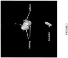
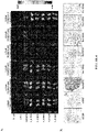
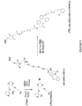
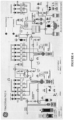

Classifications
-
- C—CHEMISTRY; METALLURGY
- C07—ORGANIC CHEMISTRY
- C07B—GENERAL METHODS OF ORGANIC CHEMISTRY; APPARATUS THEREFOR
- C07B59/00—Introduction of isotopes of elements into organic compounds ; Labelled organic compounds per se
- C07B59/002—Heterocyclic compounds
-
- A—HUMAN NECESSITIES
- A61—MEDICAL OR VETERINARY SCIENCE; HYGIENE
- A61K—PREPARATIONS FOR MEDICAL, DENTAL OR TOILETRY PURPOSES
- A61K51/00—Preparations containing radioactive substances for use in therapy or testing in vivo
- A61K51/02—Preparations containing radioactive substances for use in therapy or testing in vivo characterised by the carrier, i.e. characterised by the agent or material covalently linked or complexing the radioactive nucleus
- A61K51/04—Organic compounds
- A61K51/041—Heterocyclic compounds
- A61K51/044—Heterocyclic compounds having nitrogen as a ring hetero atom, e.g. guanethidine, rifamycins
- A61K51/0455—Heterocyclic compounds having nitrogen as a ring hetero atom, e.g. guanethidine, rifamycins having six-membered rings with one nitrogen as the only ring hetero atom
-
- A—HUMAN NECESSITIES
- A61—MEDICAL OR VETERINARY SCIENCE; HYGIENE
- A61K—PREPARATIONS FOR MEDICAL, DENTAL OR TOILETRY PURPOSES
- A61K51/00—Preparations containing radioactive substances for use in therapy or testing in vivo
- A61K51/02—Preparations containing radioactive substances for use in therapy or testing in vivo characterised by the carrier, i.e. characterised by the agent or material covalently linked or complexing the radioactive nucleus
- A61K51/04—Organic compounds
- A61K51/08—Peptides, e.g. proteins, carriers being peptides, polyamino acids, proteins
- A61K51/088—Peptides, e.g. proteins, carriers being peptides, polyamino acids, proteins conjugates with carriers being peptides, polyamino acids or proteins
-
- A—HUMAN NECESSITIES
- A61—MEDICAL OR VETERINARY SCIENCE; HYGIENE
- A61P—SPECIFIC THERAPEUTIC ACTIVITY OF CHEMICAL COMPOUNDS OR MEDICINAL PREPARATIONS
- A61P25/00—Drugs for disorders of the nervous system
- A61P25/28—Drugs for disorders of the nervous system for treating neurodegenerative disorders of the central nervous system, e.g. nootropic agents, cognition enhancers, drugs for treating Alzheimer's disease or other forms of dementia
-
- A—HUMAN NECESSITIES
- A61—MEDICAL OR VETERINARY SCIENCE; HYGIENE
- A61P—SPECIFIC THERAPEUTIC ACTIVITY OF CHEMICAL COMPOUNDS OR MEDICINAL PREPARATIONS
- A61P31/00—Antiinfectives, i.e. antibiotics, antiseptics, chemotherapeutics
-
- A—HUMAN NECESSITIES
- A61—MEDICAL OR VETERINARY SCIENCE; HYGIENE
- A61P—SPECIFIC THERAPEUTIC ACTIVITY OF CHEMICAL COMPOUNDS OR MEDICINAL PREPARATIONS
- A61P31/00—Antiinfectives, i.e. antibiotics, antiseptics, chemotherapeutics
- A61P31/04—Antibacterial agents
-
- A—HUMAN NECESSITIES
- A61—MEDICAL OR VETERINARY SCIENCE; HYGIENE
- A61P—SPECIFIC THERAPEUTIC ACTIVITY OF CHEMICAL COMPOUNDS OR MEDICINAL PREPARATIONS
- A61P31/00—Antiinfectives, i.e. antibiotics, antiseptics, chemotherapeutics
- A61P31/10—Antimycotics
-
- A—HUMAN NECESSITIES
- A61—MEDICAL OR VETERINARY SCIENCE; HYGIENE
- A61P—SPECIFIC THERAPEUTIC ACTIVITY OF CHEMICAL COMPOUNDS OR MEDICINAL PREPARATIONS
- A61P31/00—Antiinfectives, i.e. antibiotics, antiseptics, chemotherapeutics
- A61P31/12—Antivirals
-
- A—HUMAN NECESSITIES
- A61—MEDICAL OR VETERINARY SCIENCE; HYGIENE
- A61P—SPECIFIC THERAPEUTIC ACTIVITY OF CHEMICAL COMPOUNDS OR MEDICINAL PREPARATIONS
- A61P35/00—Antineoplastic agents
-
- A—HUMAN NECESSITIES
- A61—MEDICAL OR VETERINARY SCIENCE; HYGIENE
- A61P—SPECIFIC THERAPEUTIC ACTIVITY OF CHEMICAL COMPOUNDS OR MEDICINAL PREPARATIONS
- A61P35/00—Antineoplastic agents
- A61P35/02—Antineoplastic agents specific for leukemia
-
- A—HUMAN NECESSITIES
- A61—MEDICAL OR VETERINARY SCIENCE; HYGIENE
- A61P—SPECIFIC THERAPEUTIC ACTIVITY OF CHEMICAL COMPOUNDS OR MEDICINAL PREPARATIONS
- A61P37/00—Drugs for immunological or allergic disorders
- A61P37/02—Immunomodulators
-
- A—HUMAN NECESSITIES
- A61—MEDICAL OR VETERINARY SCIENCE; HYGIENE
- A61P—SPECIFIC THERAPEUTIC ACTIVITY OF CHEMICAL COMPOUNDS OR MEDICINAL PREPARATIONS
- A61P9/00—Drugs for disorders of the cardiovascular system
-
- A—HUMAN NECESSITIES
- A61—MEDICAL OR VETERINARY SCIENCE; HYGIENE
- A61P—SPECIFIC THERAPEUTIC ACTIVITY OF CHEMICAL COMPOUNDS OR MEDICINAL PREPARATIONS
- A61P9/00—Drugs for disorders of the cardiovascular system
- A61P9/10—Drugs for disorders of the cardiovascular system for treating ischaemic or atherosclerotic diseases, e.g. antianginal drugs, coronary vasodilators, drugs for myocardial infarction, retinopathy, cerebrovascula insufficiency, renal arteriosclerosis
-
- C—CHEMISTRY; METALLURGY
- C07—ORGANIC CHEMISTRY
- C07B—GENERAL METHODS OF ORGANIC CHEMISTRY; APPARATUS THEREFOR
- C07B59/00—Introduction of isotopes of elements into organic compounds ; Labelled organic compounds per se
- C07B59/008—Peptides; Proteins
-
- C—CHEMISTRY; METALLURGY
- C07—ORGANIC CHEMISTRY
- C07D—HETEROCYCLIC COMPOUNDS
- C07D213/00—Heterocyclic compounds containing six-membered rings, not condensed with other rings, with one nitrogen atom as the only ring hetero atom and three or more double bonds between ring members or between ring members and non-ring members
- C07D213/02—Heterocyclic compounds containing six-membered rings, not condensed with other rings, with one nitrogen atom as the only ring hetero atom and three or more double bonds between ring members or between ring members and non-ring members having three double bonds between ring members or between ring members and non-ring members
- C07D213/04—Heterocyclic compounds containing six-membered rings, not condensed with other rings, with one nitrogen atom as the only ring hetero atom and three or more double bonds between ring members or between ring members and non-ring members having three double bonds between ring members or between ring members and non-ring members having no bond between the ring nitrogen atom and a non-ring member or having only hydrogen or carbon atoms directly attached to the ring nitrogen atom
- C07D213/60—Heterocyclic compounds containing six-membered rings, not condensed with other rings, with one nitrogen atom as the only ring hetero atom and three or more double bonds between ring members or between ring members and non-ring members having three double bonds between ring members or between ring members and non-ring members having no bond between the ring nitrogen atom and a non-ring member or having only hydrogen or carbon atoms directly attached to the ring nitrogen atom with hetero atoms or with carbon atoms having three bonds to hetero atoms with at the most one bond to halogen, e.g. ester or nitrile radicals, directly attached to ring carbon atoms
- C07D213/62—Oxygen or sulfur atoms
- C07D213/63—One oxygen atom
- C07D213/65—One oxygen atom attached in position 3 or 5
-
- G—PHYSICS
- G01—MEASURING; TESTING
- G01T—MEASUREMENT OF NUCLEAR OR X-RADIATION
- G01T1/00—Measuring X-radiation, gamma radiation, corpuscular radiation, or cosmic radiation
- G01T1/29—Measurement performed on radiation beams, e.g. position or section of the beam; Measurement of spatial distribution of radiation
- G01T1/2914—Measurement of spatial distribution of radiation
- G01T1/2985—In depth localisation, e.g. using positron emitters; Tomographic imaging (longitudinal and transverse section imaging; apparatus for radiation diagnosis sequentially in different planes, steroscopic radiation diagnosis)
-
- C—CHEMISTRY; METALLURGY
- C07—ORGANIC CHEMISTRY
- C07B—GENERAL METHODS OF ORGANIC CHEMISTRY; APPARATUS THEREFOR
- C07B2200/00—Indexing scheme relating to specific properties of organic compounds
- C07B2200/05—Isotopically modified compounds, e.g. labelled
Definitions
- the invention relates to 18 F-prosthetic groups and the synthesis and use of 18 F-labeled compositions for imaging various processes within the body, for detecting the location of molecules associated with disease pathology, and for monitoring disease progression.
- PET Positron emission tomography
- PET relies on the design and synthesis of molecules labeled with a positron-emitting radioisotopes including 18 F, 64 Cu, 11 C, 15 0, 13 N, 66 Ga, 68 Ga, 76 Br, 89 Zr, 94 mTc, 86 Y and 124 I.
- these radiotracers or radioligands emit positrons from the nucleus of the isotope with different energies depending on the isotope used.
- the energy of the ejected positron controls the average distance that it travels before it collides with an electron resulting in the emission of two 511 keV gamma rays in opposite directions.
- the gamma rays produced by this positron annihilation event are detected by the PET imaging scanner to produce planar and tomographic images that reveal distribution of the radiotracer as a function of time. Accordingly, isotopes that are pure positron emitters with low ejection energy isotopes are preferred for PET imaging to minimize the distance traveled by the positron before annihilation and dosimetry problems caused by other emissions such as gamma rays, alpha particles or beta particles.
- the half-life of the isotope used in PET imaging must be long enough to allow synthesis and analysis of the radiotracer molecule, injection into the patient, in vivo localization, clearance from non-target tissues and the production of a clear image.
- 18 F ( ⁇ + 635 keV 97%, t 1/2 110 min) is one of the most widely used PET emitting isotopes because of its low positron emission energy, lack of side emissions and suitable half-life.
- the present invention is based at least in part on the discovery of an 18 F-labeled prosthetic group that contains a nitro-pyridine linked to a polyethylene glycol (PEG) moiety and a terminal azide, which prosthetic group is water soluble and less volatile than other agents used for 18F -labeling, and that reactions incorporating this 18 F-labeled prosthetic group into biological molecules (e.g., peptides and proteins) can be monitored by UV.
- PEG polyethylene glycol
- biomolecules e.g., peptides and proteins
- bifunctional conjugating moieties e.g., with ring constrained alkyne groups, such as bifunctional chelators
- the UV absorbance of the resultant product further provides a practical, sensitive and rapid analytical method for determining the radiochemical purity of the product.
- the invention relates to an 18 F-radiolabeled protein-based probe comprising an 18 F-radiolabeled prosthetic group, a bifunctional conjugating (BFC) moiety and a protein with the following structure, or pharmaceutically acceptable salt thereof, wherein
- a 18 F-labeled prosthetic group with the following structure, or a pharmaceutically acceptable salt thereof, wherein x is an integer from 1 to 8. In some embodiments, x is an integer from 2 to 6. In some embodiments x is an integer from 3 to 5. In some embodiments, x is 4. In some embodiments, the [O(CH 2 ) 2 ] x moiety is present in the 1-2, 1-3 or 1-4 configuration relative to the nitrogen on the pyridine ring. 18 F is attached to the pyridine ortho to the N atom. In some embodiments, the pharmaceutically acceptable salt is selected from the group consisting of fluoro, bromo, chloro and iodo salts.
- the salt is a trifluoromethanesulfonate salt.
- the 18 F-radiolabeled prosthetic group or pharmaceutically acceptable salt is water soluble. In some embodiments, the 18 F-radiolabeled prosthetic group or pharmaceutically acceptable salt is nonvolatile.
- the 18 F-radiolabeled compound has the structure or a pharmaceutically acceptable salt thereof, wherein x is an integer from 1 to 8. In some embodiments, x is an integer from 2 to 6. In some embodiments x is an integer from 3 to 5. In some embodiments, x is 4. In some embodiments, the [O(CH 2 ) 2 ] x moiety is present in the 1-2, 1-3 or 1-4 configuration relative to the nitrogen on the pyridine ring. In some embodiments, the pharmaceutically acceptable salt is selected from the group consisting of fluoro, bromo, chloro and iodo salts. In one embodiment, the salt is a trifluoromethanesulfonate salt. In some embodiments, the 18 F-radiolabeled prosthetic group or pharmaceutically acceptable salt is nonvolatile.
- the 18 F-radiolabeled compound has the structure or a pharmaceutically acceptable salt thereof, wherein x is an integer from 1 to 8. In some embodiments, x is an integer from 2 to 6. In some embodiments x is an integer from 3 to 5. In some embodiments, x is 4. In some embodiments, the pharmaceutically acceptable salt is selected from the group consisting of fluoro, bromo, chloro and iodo salts. In one embodiment, the salt is a trifluoromethanesulfonate salt. In some embodiments, the 18 F-radiolabeled prosthetic group or pharmaceutically acceptable salt is nonvolatile.
- the 18 F-radiolabeled compound has the structure or a pharmaceutically acceptable salt thereof.
- the pyridine ring of the 18 F-labeled prosthetic group contains one or more additional substituents which do not interfere with fluorination of the molecule.
- the additional substituent is a C 1-6 alkyl, for example, methyl, ethyl or propyl.
- the invention also relates to an 18 F-radiolabeled protein-based probe comprising 18 F-radiolabeled prosthetic group, a bifunctional conjugating (BFC) moiety and a protein with the following structure or a pharmaceutically acceptable salt thereof, wherein
- an 18 F-radiolabeled prosthetic group having the structure wherein "OPEG" is [O(CH 2 ) 2 ] x , and x is an integer from 1 to 8, or a pharmaceutically acceptable salt thereof.
- x is an integer from 2 to 6.
- x is an integer from 3 to 5.
- x is 4.
- the 18 F-radiolabeled prosthetic group or pharmaceutically acceptable salt is nonvolatile.
- the BFC is a cyclooctyne comprising a reactive group that forms a covalent bond with an amine, carboxyl, carbonyl or thiol functional group on the protein.
- the cyclooctyne is selected from the group consisting of dibenzocyclooctyne (DIBO), biarylazacyclooctynone (BARAC), dimethoxyazacyclooctyne (DIMAC) and dibenzocyclooctyne (DBCO).
- the cyclooctyne is DBCO.
- the BFC further comprises a polyethylene glycol (PEG)Y spacer arm, wherein y is an integer from 1 to 8. In some embodiments, y is an integer from 2 to 6. In some embodiments, y is 4 or 5.
- PEG polyethylene glycol
- the BFC is DBCO-PEG4-NHS-Ester, DBCO- Sulfo -NHS-Ester, DBCO-PEG4-Acid, DBCO-PEG4-Amine or DBCO-PEG4-Maleimide. In some embodiments, the BFC is DBCO-PEG4-Maleimide.
- an 18 F-radiolabeled protein-based probe with the following structure, wherein the maleimide group of the BFC is covalently linked to the thiol group on a cysteine residue of the protein.
- the cysteine residue is at the C-terminus of the protein.
- the protein portion of the probe comprises a fibronectin based scaffold (FBS).
- FBS fibronectin based scaffold
- the protein portion of the 18 F-radiolabeled protein-based probe binds to a biological molecule associated with a disease.
- the disease is selected from the group consisting of solid cancers, hematopoietic cancers, hematological cancers, autoimmune disease, neurodegenerative disease, cardiovascular disease and pathogenic infections.
- the probe binds to a tumor-associated antigen.
- the probe binds to a protein present on a pathogenic organism, e.g., a virus, bacterium or fungus.
- the 18 F-radiolabeled protein-based probe provided herein may be in the form of a pharmaceutical composition.
- a method of obtaining an image of an 18 F-radiolabeled protein-based probe as provided herein including the steps of (a) administering the 18 F-radiolabeled protein-based probe to a subject; and (b) imaging in vivo the distribution of the 18 F-radiolabeled protein-based probe by positron emission tomography (PET) scanning.
- PET positron emission tomography
- the imaged distribution of the 18 F-radiolabeled protein-based probe is indicative of the presence or absence of a disease.
- the invention relates to the 18 F-radiolabeled protein-based probe according to the invention for use in a method of detecting the presence of a disease in a subject, the method including the steps of (a) administering to a subject in need thereof the 18 F-radiolabeled protein-based probe which binds to a target molecule associated with the presence of the disease; and (b) obtaining an radio-image of at least a portion of the subject to detect the presence or absence of the 18 F-radiolabeled protein-based probe; wherein the presence and location of the 18 F-radiolabeled protein-based probe above background is indicative of the presence and location of the disease.
- the invention relates to the 18 F-radiolabeled protein-based probe according to the invention for use in a method of monitoring the progress of a disease in a subject, the method including the steps of (a) administering to a subject in need thereof the 18 F-radiolabeled protein-based probe which binds to a target molecule associated with the presence of the disease at a first time point and obtaining an image of at least a portion of the subject to determine the amount of the diseased cells or tissue; and (b) administering to the subject the 18 F-radiolabeled protein-based probe at one or more subsequent time points and obtaining an image of at least a portion of the subject at each time point; wherein the dimension and location of the diseased cells or tissue at each time point is indicative of the progress of the disease.
- the invention relates to the 18 F-radiolabeled protein-based probe according to the invention for use in a method of quantifying diseased cells or tissues in a subject, the method including the steps of (a) administering to a subject having diseased cells or tissues with the 18 F-radiolabeled protein-based probe which binds to a target molecule located with the diseased cells or tissues; and (b) detecting radioactive emissions of the 18 F in the diseased cells or tissue, wherein the level and distribution of the radioactive emissions in the diseased cells or tissues is a quantitative measure of the diseased cells or tissues.
- a method of screening for an agent for treating a disease including the steps of (a) contacting a cells expressing a target protein associated with the disease with an 18 F-radiolabeled protein-based probe as provided herein which binds to the target protein in the presence and absence of a candidate agent; and (b) imaging the cells in the presence and absence of the candidate agent using positron emission tomography (PET), wherein a decrease in the amount of radioactive emissions in the presence of the candidate agent is indicative of that the agent binds to the target protein.
- PET positron emission tomography
- the disease is selected from the group consisting of solid cancers, hematopoietic cancers, hematological cancers, autoimmune disease, neurodegenerative disease, cardiovascular disease and pathogenic infection (e.g., viral, bacterial or fungal infections).
- a method of obtaining a quantitative image of tissues or cells expressing a target protein including the steps of contacting the cells or tissue with an 18 F-radiolabeled protein-based probe as provided herein which binds to the target protein, and detecting or quantifying the tissue expressing the target protein using positron emission tomography (PET).
- PET positron emission tomography
- the 18 F-radiolabeled protein-based probe comprises a ligand. In some embodiments, the 18 F-radiolabeled protein-based probe comprises an antibody or antibody fragment. In some embodiments, the 18 F-radiolabeled protein-based probe comprises a fibronectin based scaffold (FBS). In some embodiments, the 18 F-radiolabeled protein-based probe binds to a tumor-associated antigen. In still other embodiments, the 18 F-radiolabeled protein-based probe binds to a protein present on a pathogenic organism (e.g., a virus, bacterium or fungus).
- a pathogenic organism e.g., a virus, bacterium or fungus
- kits containing the reaction precursors for producing the 18 F-radiolabeled protein-based probes provided herein e.g., a non-radiolabeled prosthetic group, BFC-linked protein probe and reagents for carrying out a bioorthogonal click reaction
- the reaction precursors for producing the 18 F-radiolabeled protein-based probes provided herein e.g., a non-radiolabeled prosthetic group, BFC-linked protein probe and reagents for carrying out a bioorthogonal click reaction
- instructions for producing the 18 F-radiolabeled protein-based probe e.g., a non-radiolabeled prosthetic group, BFC-linked protein probe and reagents for carrying out a bioorthogonal click reaction
- said method produces an 18 F-pyridine prosthetic group with the following structure b (where 18 F is ortho to the N atom), and includes the steps of (a) providing a solution of the compound of the structure (where X is ortho to the N atom) where X is NO 2 , Br or (b) providing a mixture of 18 F in 18 Owater, 4,7,13,16,21,24-hexaoxa-1,10-diazabicyclo[8.8.8]hexacosane and weak base; (c) drying the mixture from step (b) to form a solid; and (d) reacting the solution from step (a) with the solid from step (c) to form the 18 F-labeled compound.
- the starting compound from step a) described above, X is NO 2 .
- X is attached to the carbon atom ortho to the N atom and the PEG-N 3 side chain.
- the weak base in step b) is K 2 CO 3 , cesium carbonate or tetrabutylammonium hydroxide. In one embodiment, the weak base in step b) is K 2 CO 3 .
- a method for the 18 F labeling of a protein which includes the step of coupling the 18 F-radiolabeled prosthetic group of formula b with the protein to be radiolabeled in a "click reaction" to form the 18 F-radiolabeled protein.
- the bioorthogonal click reaction is metal free (e.g., copper free click reaction).
- the protein comprises a bifunctional conjugating (BFC) moiety (e.g., a bifunctional chelator).
- the protein comprises a cyclooctyne BFC.
- the protein comprises DBCO-PEG4-Maleimide or is covalently linked to it.
- Described herein but not forming part of the invention are 18 F- prosthetic groups and methods for producing the 18 F-prosthetic groups. Also described herein but not forming part of the invention are radiolabeled compositions containing the 18 F-prosthetic groups and the use of these radiolabeled compositions to diagnose, localize, monitor and/or assess diseased cells and/or tissues, and related biological conditions.
- medical imaging refers to the techniques and processes used to create images of the subject's body (or parts thereof) for clinical purposes (medical procedures seeking to reveal, diagnose or monitor disease) or medical science (including the study of normal anatomy and physiology).
- PET positron emission tomography
- tracer positron-emitting radionuclide
- PET imaging tools have a wide variety of uses and aid in drug development both preclinically and clinically. Exemplary applications include direct visualization of in vivo saturation of targets; monitoring uptake in normal tissues to anticipate toxicity or patient to patient variation; quantifying diseased tissue; tumor metastasis; and monitoring drug efficacy over time, or resistance over time.
- bioorthogonal chemistry refers to any chemical reaction that can occur inside of living systems without interfering with native biochemical processes.
- the term includes chemical reactions that are chemical reactions that occur in vitro at physiological pH in, or in the presence of water. To be considered bioorthogonol, the reactions are selective and avoid side-reactions with other functional groups found in the starting compounds.
- the resulting covalent bond between the reaction partners should be strong and chemically inert to biological reactions and should not affect the biological activity of the desired molecule.
- click chemistry refers to a set of reliable and selective bioorthogonal reactions for the rapid synthesis of new compounds and combinatorial libraries. Properties of for click reactions include modularity, wideness in scope, high yielding, stereospecificity and simple product isolation (separation from inert by-products by non-chromatographic methods) to produce compounds that are stable under physiological conditions.
- click chemisty is a generic term for a set of labeling reactions which make use of selective and modular building blocks and enable chemoselective ligations to radiolabel biologically relevant compounds in the absence of catalysts.
- a "click reaction” can be with copper, or it can be a copper-free click reaction.
- prosthetic group or "bifunctional labeling agent” refers to a small organic molecule containing a radionulide (e.g ., 18 F) that is capable of being linked to peptides or proteins.
- a radionulide e.g ., 18 F
- chelator ligand refers to a bifunctional chelator or bifunctional conjugating (BFC) moiety (used interchangeably herein) that covalently links a radiolabeled prosthetic group to a biologically active targeting molecule (e.g., peptide or protein).
- BFCs utilize functional groups such as carboxylic acids or activated esters for amide couplings, isothiocyanates for thiourea couplings and maleimides for thiol couplings.
- target as a general reference to a “biological target” refers to a cell, tissue (e.g., cancer or tumor), a pathogenic microorganism (e.g., bacteria, virus, fungus, plant, prion, protozoa or portion thereof) or other molecule associated with a biological pathway, or a biological phenomenon, such as tissue inflammation, plaque formation, etc.
- tissue e.g., cancer or tumor
- pathogenic microorganism e.g., bacteria, virus, fungus, plant, prion, protozoa or portion thereof
- other molecule associated with a biological pathway e.g., a cell, tissue (e.g., cancer or tumor), a pathogenic microorganism (e.g., bacteria, virus, fungus, plant, prion, protozoa or portion thereof) or other molecule associated with a biological pathway, or a biological phenomenon, such as tissue inflammation, plaque formation, etc.
- a biological phenomenon such as tissue inflammation, plaque formation, etc.
- targeting ligand a targeting agent
- targeting molecule a molecule, such as peptide, protein, glycoprotein, etc., that binds to another molecule.
- a targeting agent is bound to the 18 F-prosthetic group in order to "target” a molecule associated with a particular cell, tissue, pathogen or biological pathway.
- Polypeptide refers to any sequence of two or more amino acids, regardless of length, post-translation modification, or function. "Polypeptide,” “peptide,” and “protein” are used interchangeably herein. Polypeptides can include natural amino acids and non-natural amino acids such as those described in U.S. Pat. No. 6,559,126 .
- Polypeptides can also be modified in any of a variety of standard chemical ways (e.g., an amino acid can be modified with a protecting group; the carboxy-terminal amino acid can be made into a terminal amide group; the amino-terminal residue can be modified with groups to, e.g., enhance lipophilicity; or the polypeptide can be chemically glycosylated or otherwise modified to increase stability or in vivo half-life).
- Polypeptide modifications can include the attachment of another structure such as a cyclic compound or other molecule to the polypeptide and can also include polypeptides that contain one or more amino acids in an altered configuration (i.e., R or S; or, L or D).
- an "isolated" polypeptide is one that has been identified and separated and/or recovered from a component of its natural environment. Contaminant components of its natural environment are materials that would interfere with diagnostic or therapeutic uses for the polypeptide, and may include enzymes, hormones, and other proteinaceous or nonproteinaceous solutes.
- the polypeptide will be purified (1) to greater than 95% by weight of polypeptide as determined by the Lowry method, and most preferably more than 99% by weight, (2) to a degree sufficient to obtain at least residues of N-terminal or internal amino acid sequence by use of a spinning cup sequenator, or (3) to homogeneity by SDS-PAGE under reducing or nonreducing condition using Coomassie blue or, preferably, silver stain.
- Isolated polypeptide includes the polypeptide in situ within recombinant cells since at least one component of the polypeptide's natural environment will not be present. Ordinarily, however, isolated polypeptide will be prepared by at least one purification step.
- telomere binding refers to an peptide or polypeptide that exhibits affinity for a biological target, but does not significantly bind (e.g., less than about 10% binding) to a other molecules as measured by a technique available in the art such as, but not limited to, Scatchard analysis and/or competitive binding assays (e.g., competition ELISA, BIACORE assay).
- preferentially binds refers to the situation in which an peptide or protein binds a selected biological target at least about 20% greater than it binds a different biological target as measured by a technique available in the art such as, but not limited to, Scatchard analysis and/or competitive binding assays (e.g., competition ELISA, BIACORE assay).
- K D is intended to refer to the dissociation equilibrium constant of a protein- protein interaction (e.g., 18 F-radiolabeled-protein based probe-target molecule) or the affinity of an 18 P-radiolabeled-protein based probe for a target protein, as measured using a surface plasmon resonance assay or a cell binding assay.
- a “desired K D ,” as used herein, refers to a K D of an 18 P-radiolabeled-protein based probe that is sufficient for the purposes contemplated.
- a desired K D may refer to the K D of an 18 F-radiolabeled-protein based probe required to elicit a functional effect in an in vivo imaging assay or in vitro assay, e.g., a cell-based luciferase assay.
- k ass is intended to refer to the association rate constant for the association of an 18 F-radiolabeled-protein based probe into an 18 P-radiolabeled-protein based probe/target protein complex.
- k diss is intended to refer to the dissociation rate constant for the dissociation of an 18 F-radiolabeled-protein based probe from the 18 F-radiolabeled-protein based probe/target protein complex.
- IC 50 refers to the concentration of an 18 F-radiolabeled-protein based probe that inhibits a response, either in an in vitro or an in vivo assay, to a level that is 50% of the maximal inhibitory response, i.e., halfway between the maximal inhibitory response and the untreated response.
- PK is an acronym for "pharmacokinetic” and encompasses properties of a compound including, by way of example, absorption, distribution, metabolism, and elimination by a subject.
- a "PK modulation protein” or “PK moiety” as used herein refers to any protein, peptide, or moiety that affects the pharmacokinetic properties of a biologically active molecule when fused to or administered together with the biologically active molecule.
- Examples of a PK modulation protein or PK moiety include PEG, human serum albumin (HSA) binders (as disclosed in U.S. Publication Nos. 2005/0287153 and 2007/0003549 , PCT Publication Nos. WO 2009/083804 and WO 2009/133208 ), human serum albumin and variants thereof, transferrin and variants thereof, Fc or Fc fragments and variants thereof, and sugars (e.g., sialic acid).
- the "serum half-life" of a protein or compound can generally be defined as the time taken for the serum concentration of the polypeptide to be reduced by 50%, in vivo, for example due to degradation of the sequence or compound and/or clearance or sequestration of the sequence or compound by natural mechanisms.
- the half-life can be determined in any manner known per se, such as by pharmacokinetic analysis.
- Suitable techniques will be clear to the person skilled in the art, and may for example generally involve the steps of suitably administering to a subject a suitable dose of the amino acid sequence or compound described herein; collecting blood samples or other samples from the subject at regular intervals; determining the level or concentration of the amino acid sequence or compound described herein in said blood sample; and calculating, from (a plot of) the data thus obtained, the time until the level or concentration of the amino acid sequence or compound described herein has been reduced by 50% compared to the initial level upon dosing.
- Half-life can be expressed using parameters such as the t 1/2 -alpha, t 1/2 -beta, HL_Lambda_z, and the area under the curve (AUC).
- an “increase in half-life” refers to an increase in any one of these parameters, any two of these parameters, any three of these parameters or all four of these parameters.
- An “increase in half-life” in particular refers to an increase in the t 1/2 -beta, and/or HL_Lambda_z, either with or without an increase in the t 1/2 -alpha and/or the AUC or both.
- linkage refers to the association of two or more molecules.
- the linkage can be covalent or non-covalent.
- the linkage also can be genetic ( i.e., recombinantly fused). Such linkages can be achieved using a wide variety of art recognized techniques, such as chemical conjugation and recombinant protein production.
- diagnosis or “detection” can be used interchangeably. Whereas diagnosis usually refers to defining a tissue's specific histological status, detection recognizes and locates a tissue, lesion or organism containing a particular detectable target.
- detectable refers to the ability to detect a signal over the background signal.
- detectable signal as used herein in the context of imaging agents and diagnostics, is a signal derived from non-invasive imaging techniques such as, but not limited to, positron emission tomography (PET).
- PET positron emission tomography
- the detectable signal is detectable and distinguishable from other background signals that may be generated from the subject.
- there is a measurable and statistically significant difference e.g., a statistically significant difference is enough of a difference to distinguish among the detectable signal and the background, such as about 0.1%, 1%, 3%, 5%, 10%, 15%, 20%, 25%, 30%, or 40% or more difference between the detectable signal and the background
- Standards and/or calibration curves can be used to determine the relative intensity of the detectable signal and/or the background.
- a "detectably effective amount" of a composition comprising an imaging agent described herein is defined as an amount sufficient to yield an acceptable image using equipment that is available for clinical use.
- a detectably effective amount of an imaging agent provided herein may be administered in more than one injection.
- the detectably effective amount can vary according to factors such as the degree of susceptibility of the individual, the age, sex, and weight of the individual, idiosyncratic responses of the individual, and the like. Detectably effective amounts of imaging compositions can also vary according to instrument and methodologies used. Optimization of such factors is well within the level of skill in the art.
- administering refers to the physical introduction of a composition comprising an imaging agent to a subject, using any of the various methods and delivery systems known to those skilled in the art.
- Preferred routes of administration for the imaging agents described herein include intravenous, intraperitoneal, intramuscular, subcutaneous, spinal or other parenteral routes of administration, for example by injection or infusion.
- parenteral administration means modes of administration other than enteral and topical administration, usually by injection, and includes, without limitation, intravenous, intraperitoneal, intramuscular, intraarterial, intrathecal, intralymphatic, intralesional, intracapsular, intraorbital, intracardiac, intradermal, transtracheal, subcutaneous, subcuticular, intraarticular, subcapsular, subarachnoid, intraspinal, epidural and intrasternal injection and infusion, as well as in vivo electroporation.
- an imaging agent described herein can be administered via a non-parenteral route, such as a topical, epidermal or mucosal route of administration, for example, intranasally, orally, vaginally, rectally, sublingually or topically.
- Administering can also be performed, for example, once, a plurality of times, and/or over one or more extended periods.
- co-administration are meant to encompass administration of the selected pharmaceutical agents to a single patient, and are intended to include regimens in which the agents are administered by the same or different route of administration or at the same or different time.
- patient and “subject” refer to any human or non-human animal that receives a composition comprising an imaging agent described herein.
- body fluids and cell samples of the above subjects will be suitable for use, such as blood, urine, or tissue samples.
- sample can refer to a tissue sample, cell sample, a fluid sample, and the like.
- the sample may be taken from a subject.
- the tissue sample can include hair (including roots), buccal swabs, blood, saliva, semen, muscle, or from any internal organs.
- the fluid may be, but is not limited to, urine, blood, ascites, pleural fluid, spinal fluid, and the like.
- the body tissue can include, but is not limited to, skin, muscle, endometrial, uterine, and cervical tissue.
- isotopically pure means that the element, compound, or composition contains a greater proportion of one isotope in relation to other isotopes. In certain embodiments, the element, compound, or composition is greater than about 40%, 50%, or 60% isotopically pure.
- a labeled molecule is "purified" when the labeled molecule is partially or wholly separated from unlabeled molecules, so that the fraction of labeled molecules is enriched compared to the starting mixture.
- a "purified" labeled molecule may comprise a mixture of labeled and unlabeled molecules in almost any ratio, including but not limited to about 5:95; 10:90; 15:85; 20:80; 25:75; 30:70; 40:60; 50:50; 60:40; 70:30; 75:25; 80:20; 85:15; 90:10; 95:5; 97:3; 98:2; 99:1 or 100:0.
- Of refers to triflate having the formula CF 3 SO 3 or trifluoromethanesulfate.
- halo or, alternatively, "halogen” or “halide” means fluoro, chloro, bromo or iodo.
- an 18 F-radiolabeled compound containing a prosthetic group for use in a bioorthogonal reaction involving 1,3-dipolar cycloaddition between an azide and a cyclooctyne which proceeds selectively under water tolerant conditions.
- the 18 P-radiolabeled prosthetic groups of the invention are soluble in 100% aqueous, and there is no need for an organic phase to link the prosthetic group to a peptide or protein molecule. This feature is particularly advantageous as many biologies ( e.g., peptides or proteins), cannot withstand even small amounts of organic solvents, with degradation and aggregation issues.
- the 18 F fluorination reaction can be monitored with UV.
- the 18 F-radiolabeled prosthetic groups described herein are not volatile.
- 18 P-radiolabeled prosthetic groups provided may be incorporated into biologics using a copper free click chemisty, e.g ., as described in the Examples, thus avoiding the stability issues observed in some biologics when copper mediated click chemistry is used.
- a PEGylated 18 F-pyridine covalently bound to an azide with the following structure, or a pharmaceutically acceptable salt thereof, wherein x is an integer from 1 to 8. In some embodiments, x is an integer from 2 to 6. In some embodiments x is an integer from 3 to 5. In some embodiments, x is 4. In related embodiments, 18 F is attached to the pyridine ortho to the N atom.
- the [O(CH 2 ) 2 ] x moiety is present in the 1-3 configuration relative to the nitrogen on the pyridine ring. In some embodiments, the [O(CH 2 ) 2 ] x moiety is present in the 1-2 configuration relative to the nitrogen on the pyridine ring. In other embodiments, the [O(CH 2 ) 2 ] x moiety is present in the 1-4 configuration relative to the nitrogen on the pyridine ring.
- the 18 F-radiolabeled compound has the structure or a pharmaceutically acceptable salt thereof, wherein x is an integer from 1 to 8. In some embodiments, x is an integer from 2 to 6. In some embodiments x is an integer from 3 to 5. In some embodiments, x is 4. In some embodiments, the [O(CH 2 ) 2 ] x moiety is present in the 1-3 configuration relative to the nitrogen on the pyridine ring. In some embodiments, the [O(CH 2 ) 2 ] x moiety is present in the 1-2 configuration relative to the nitrogen on the pyridine ring. In other embodiments, the [O(CH 2 ) 2 ] x moiety is present in the 1-4 configuration relative to the nitrogen on the pyridine ring.
- the 18 F-radiolabeled compound has the structure wherein x is an integer from 1 to 8. In some embodiments, x is an integer from 2 to 6. In some embodiments x is an integer from 3 to 5. In some embodiments, x is 4.
- the 18 F-radiolabeled compound is [ 18 F]-3-(2-(2-(2-(2-(2-azidoethoxy)ethoxy)ethoxy)-2-fluoropyridine ( 18 F-FPPEGA) and has the structure
- the 18 F-radiolabeled prosthetic group may contain additional groups on the pyridine ring which do not interfere with the fluorination reaction.
- additions to the pyridine ring include C 1-6 alkyl groups, for example methyl, ethyl and propyl.
- the 18 F-radiolabeled prosthetic group is a fused ring system with the following structure: wherein "OPEG" is [O(CH 2 ) 2 ] x , and x is an integer from 1 to 8. In some embodiments, x is an integer from 2 to 6. In some embodiments x is an integer from 3 to 5. In some embodiments, x is 4.
- the method produces a 18 F-pyridine prosthetic group with the following structure b (where 18 F is ortho to the N atom), and includes the steps of
- the method further comprises the step of producing a compound with the following structure a according to the Scheme I shown below:
- the method comprises producing the 18 F-pyridine prosthetic group [ 18 F]-3-(2-(2-(2-(2-azidoethoxy)ethoxy)ethoxy)-2-fluoropyridine ( 18 F-FPPEGA), e, from_d, according to the following reaction conditions:
- the present invention relates to 18 F-radiolabeled probes or agents with the following structure, wherein
- Bifunctional chelators or conjugating (BFC) moieties which can be used in the radiolabeled compositions disclosed herein, are commercially available (e.g., Sigma Aldrich; Click Chemistry Tools), or may be synthesized according to well-known chemical reactions.
- the BFC is selected from cyclooctyne based agents (e.g., DBCO, DIBO), DFO, DOTA and its derivatives (CB-DO2A, 3p-C-DEPA, TCMC, Oxo-DO3A), TE2A, CB-TE2A, CB-TE1A1P, CB-TE2P, MM-TE2A, DM-TE2A, diamsar and derivatives, NODASA, NODAGA, NOTA, NETA, TACN-TM, DTPA, 1B4M-DTPA, CHX-A"-DTPA, TRAP (PRP9), NOPO, AAZTA and derivatives (DATA), H 2 dedpa, H 4 octapa, H 2 azapa, Hsdecapa, H 6 phospa, HBED, SHBED, BPCA, CP256, PCTA, HEHA, PEPA, EDTA, TETA, and TRITA
- the BFC is a cyclooctyne comprising a reactive group that forms a covalent bond with an amine, carboxyl, carbonyl or thiol functional group on the targeting protein or peptide.
- Reactive groups on the cyclooctyne include esters, acids, hydroxyl groups, aminooxy groups, maliemides, ⁇ -halogenketones and ⁇ -halogenacetamides.
- the BFC is a cyclooctyne, such as dibenzocyclooctyne (DIBO), biarylazacyclooctynone (BARAC), dimethoxyazacyclooctyne (DIMAC) and dibenzocyclooctyne (DBCO).
- the cyclootyne is DBCO.
- the cyclooctyne further comprises a hydrophilic polyethylene glycol (PEG)Y spacer arm, wherein y is an integer from 1 to 8. In certain embodiments, y is an integer from 2 to 6. In certain embodiments, y is 4 or 5.
- PEG polyethylene glycol
- the BFC is DBCO-PEG4-NHS-Ester or DBCO -Sulfo- NHS-Ester which react specifically and efficiently with a primary amine (e.g., side chain of lysine residues or aminosilane-coated surfaces).
- the BFC is DBCO-PEG4-Acid with terminal carboxylic acid (-COOH) that can be reacted with primary or secondary amine groups in the presence activators (e.g. EDC) forming a stable amide bond.
- the BFC is DBCO-PEG4-Amine which reacts with carboxyl groups in the presence of activators (e.g. EDC, or DCC) or with activated esters (e.g. NHS esters) forming stable amide bonds.
- the BFC is DBCO-PEG4-Maleimide which reacts with sulfhydryl groups on cysteine residues, e.g., cysteine residues near the C-terminus of the polypeptide.
- the protein is first modified to incorporate a cysteine for attaching the prosthetic group.
- a cysteine may be added to the C-terminus of the protein.
- said protein is a fibronectin type III (Fn3) domain.
- PxCy wherein P is proline, C is cysteine, x is an integer that is at least 0 (e.g., 0, 1 or 2) and y is an integer that is at least 1, is added to the C-terminus of the protein.
- the 18 F-radiolabeled probe or agent has the following structure a, wherein, the BFC is conjugated to the protein at a cysteine residue.
- the protein is a fibronectin type III (Fn3) domain.
- the 18 F-radiolabeled targeting agents described herein may be produced using bioorthogonal, metal free click chemistry in medium suitable for direct use in vivo (e.g., saline) according to the procedures described herein.
- Protein/Peptide Targeting Molecules (only fibronectin type III (Fn3) domain forming part of the invention)
- the 18 F-prosthetic groups provided herein but not forming part of the invention may be attached to virtually any targeting molecule, so long as it contains a derivatizable group that may be modified without affecting the interaction between the targeting molecule and the in vivo biological target (e.g., cell or tissue).
- a derivatizable group that may be modified without affecting the interaction between the targeting molecule and the in vivo biological target (e.g., cell or tissue).
- the targeting molecule is a peptide or protein, including, but not limited to, antibodies, antibody fragments, fibronectin based molecules and ligands (e.g., hormones, growth factors, cytokines, chemokines, interleukins and angiogenic factors).
- the targeting molecule will comprise one or more binding sites for a target associated with a disease or condition, such as a tumor associated or autoimmune antigen, or a protein displayed by a pathogenic organism such as a virus, bacterium, fungus or protozoan.
- the targeting molecule according to the invention is a protein which comprises a fibronectin type III (Fn3) domain.
- the 18 F-labeled peptides or proteins may be selected to bind directly to a targeted cell, tissue, pathogenic organism or other target for imaging and/or detection.
- 18 F labeled proteins or peptides may be selected to bind directly or indirectly to the in vivo target molecule. For example, a first protein or peptide may administered to the subject, followed by a second 18 F-labeled molecule which binds to the first.
- Peptides having as few as two amino acid residues, preferably two to ten residues, may be used and may also be coupled to other moieties.
- the targetable construct may also comprise unnatural amino acids, e.g., D-amino acids, in the backbone structure to increase the stability of the peptide in vivo.
- other backbone structures such as those constructed from non-natural amino acids or peptoids may be used.
- Peptides which may be used include ligands, peptide vaccines, and epitopes.
- the peptides used as targetable constructs are conveniently synthesized on an automated peptide synthesizer using a solid-phase support and standard techniques of repetitive orthogonal deprotection and coupling. N-terminal residues may be acetylated to increase serum stability.
- Such protecting groups will be known to the skilled artisan. See Greene and Wuts Protective Groups in Organic Synthesis, 1999 (John Wiley and Sons, N.Y .).
- the targeting molecule used in the radiotracer composition described herein may be an antibody.
- antibody as used to herein may include whole antibodies and any antigen binding fragments (i.e., “antigen-binding portions") or single chains thereof.
- antibody may refer to both naturally occurring and non-naturally occurring antibodies; monoclonal and polyclonal antibodies; chimeric and humanized antibodies; human and nonhuman antibodies; bispecific antibodies; wholly synthetic antibodies; and single chain antibodies.
- the term "antigen” refers to any natural or synthetic immunogenic substance, such as a protein, peptide, or hapten.
- the targeting molecules described herein may incorporate any antibody or fragment known in the art that has binding specificity for a target antigen associated with a disease state or condition.
- Antibodies useful as targeting molecules may be commercially obtained from a wide variety of sources (e.g., ATTC, Manassas, VA), and/or have published variable region sequences which may be produced according to art recognized recombinant techniques.
- Exemplary antibodies for use in the present methods include an anti-CTLA-4 antibody, an anti-PD-1 antibody, an anti-PDL-1 antibody, an anti-OX40 (also known as CD134, TNFRSF4, ACT35 and/or TXGP1L) antibody, or an anti-LAG-3 antibody.
- Antibodies used in the compositions and methods described herein can be produced using a variety of known techniques. Immunization protocols and techniques for isolation of immunized splenocytes are well established in the art. The production of monoclonal antibodies using the standard somatic cell hybridization technique described by Kohler and Milstein, Nature 256: 495 (1975 ), as well as viral or oncogenic transformation of B lymphocytes, phage display technique using libraries of human antibody genes are also routine. In addition, standard methodologies for the production of chimeric and humanized antibodies are readily available (see e.g., U.S. Patent No. 4,816,567 to Cabilly et al. ; U.S. Patent No. 5,225,539 to Winter , and U.S. Patent Nos. 5,530,101 ; 5,585,089 ; 5,693,762 and 6,180,370 to Queen et al. ) .
- the targeting molecule used in the radiotracer composition may be an antigen binding fragment.
- the term "antigen-binding portion" of an antibody refers to one or more fragments of an antibody that retain the ability to specifically bind to an antigen. Examples of binding fragments encompassed within the term “antigen-binding portion” of an antibody include (i) a Fab fragment, a monovalent fragment consisting of the V L , V H , CL and CH1 domains; (ii) a F(ab') 2 fragment, a bivalent fragment comprising two Fab fragments linked by a disulfide bridge at the hinge region; (iii) a Fd fragment consisting of the V H and CH1 domains; (iv) a Fv fragment consisting of the V L and V H domains of a single arm of an antibody, (v) a dAb fragment ( Ward et al., (1989) Nature 341:544-546 ), which consists of a V H domain; and (vi) an isolated complement
- the two domains of the Fv fragment, V L and V H are coded for by separate genes, they can be joined, using recombinant methods, by a synthetic linker that enables them to be made as a single protein chain in which the V L and V H regions pair to form monovalent molecules known as single chain Fv (scFv); see e.g., Bird et al. (1988) Science 242:423-426 ; and Huston et al. (1988) Proc. Natl. Acad. Sci. USA 85:5879-5883 ).
- single chain Fv single chain Fv
- Such single chain antibodies are also intended to be encompassed within the term "antigen-binding portion" of an antibody.
- Immunol. 10:301 These antibody fragments are obtained using conventional techniques known to those with skill in the art, and the fragments are screened for utility in the same manner as are intact antibodies.
- Antigen-binding portions can be produced by recombinant DNA techniques, or by enzymatic or chemical cleavage of intact immunoglobulins.
- the antibody used may be modified to modulate, e.g., decrease the half-life of the antibody or rapid clearance for use in the medical imaging methods described herein.
- Modifications such as I253A ( Hornick et al. (2000) J. Nucl. Med. 41:355 ) and H435A/R I253A or H310A ( Kim et al. (2000) Eur. J. Immunol. 29:2819 ) in Fc of human IgG1 can decrease FcRn binding. See also Kenanova et al. (2005) Cancer Res. 65:622 .
- Other means to enhance clearance include formatting the antigen binding domains as antibody fragments lacking the ability to bind FcRn, such as Fab fragments.
- Such modification can reduce the circulating half-life of an antibody from a couple of weeks to a matter of hours.
- Selective PEGylation of antibody fragments can then be used to fine-tune (increase in increments) the half-life of the antibody fragments if necessary. Chapman et al. (1999) Nat. Biotechnol. 17:780 .
- Radiotracer compositions containing an antibody or antigen binding fragment thereof can be assayed for retention of binding specificity in vitro and/or in vivo.
- Methods for analyzing binding affinity, cross-reactivity, and binding kinetics of various antibody compositions include standard assays known in the art, for example, ELISA, Western Blotting, flow cytometry, and BIACORE ® surface plasmon resonance (SPR) analysis using a BIACORE ® 2000 SPR instrument (Biacore AB, Uppsala, Sweden).
- Exemplary proteins for use in the radiolabeled probes described herein include any known antibody or alternative scaffold protein, such as Adnectins, that specifically binds to a target, and does significantly cross-react with unrelated targets.
- Adnectins any known antibody or alternative scaffold protein, such as Adnectins, that specifically binds to a target, and does significantly cross-react with unrelated targets.
- FBS Fibronectin Based Protein
- the targeting molecule used in the radiotracer compositions described herein is a FBS protein, in particular a fibronectin type III (Fn3) domain.
- FBS protein molecules have inherently rapid blood clearance rates, which can be advantageous for use with 18 F in imaging technologies by minimizing the amount of time needed for background probe signals from non-relevant tissue. Rapid clearing probers allow high contrast images to be collected the same day the probe is injected, and very importantly, can also serve to reduce overall radiation exposure to the subject.
- a "fibronectin based scaffold” or “FBS” protein or moiety refers to proteins or moieties that are based on a fibronectin type III (“Fn3") repeat.
- Fn3 is a small (about 10 kDa) domain that has the structure of an immunoglobulin (1g) fold (i.e., an Ig-like ⁇ -sandwich structure, consisting of seven ⁇ -strands and six loops).
- Fibronectin has 18 Fn3 repeats, and while the sequence homology between the repeats is low, they all share a high similarity in tertiary structure.
- Fn3 domains are also present in many proteins other than fibronectin, such as adhesion molecules, cell surface molecules, e.g., cytokine receptors, and carbohydrate binding domains.
- adhesion molecules e.g., cell surface molecules, e.g., cytokine receptors, and carbohydrate binding domains.
- cell surface molecules e.g., cytokine receptors
- carbohydrate binding domains e.g., cytokine receptors
- the term "FBS" protein or moiety is intended to include scaffolds based on Fn3 domains from these other proteins ( i.e ., non fibronectin molecules).
- Fn3 domain is small, monomeric, soluble, and stable. It lacks disulfide bonds and, therefore, is stable under reducing conditions.
- Fn3 domains comprise, in order from N-terminus to C-terminus, a beta or beta-like strand, A; a loop, AB; a beta or beta-like strand, B; a loop, BC; a beta or beta-like strand, C; a loop, CD; a beta or beta-like strand, D; a loop, DE; a beta or beta-like strand, E; a loop, EF; a beta or beta-like strand, F; a loop, FG; and a beta or beta-like strand, G.
- the seven antiparallel ⁇ -strands are arranged as two beta sheets that form a stable core, while creating two "faces" composed of the loops that connect the beta or beta-like strands.
- Loops AB, CD, and EF are located at one face ("the south pole") and loops BC, DE, and FG are located on the opposing face ("the north pole”).
- the loops in Fn3 molecules are structurally similar to complementary determining regions (CDRs) of antibodies, and when altered, may be involved in binding of the Fn3 molecule to a target, e.g., a target protein.
- Other regions of Fn3 molecules such as the beta or beta-like strands and N-terminal or C-terminal regions, when altered, may also be involved in binding to a target.
- Any or all of loops AB, BC, CD, DE, EF and FG may participate in binding to a target.
- Any of the beta or beta-like strands may be involved in binding to a target.
- Fn3 domains may also bind to a target through one or more loops and one or more beta or beta-like strands.
- Binding may also require the N-terminal or C-terminal regions.
- An FBS domain for use in a protein may comprise all loops, all beta or beta-like strands, or only a portion of them, wherein certain loops and/or beta or beta-like strands and/or N- or C-terminal regions are modified (or altered), provided that the FBS domain preferably binds specifically to a target.
- an FBS domain may comprise 1, 2, 3, 4, 5 or 6 loops, 1, 2, 3, 4, 5, 6, 7, or 8 beta strands, and optionally an N-terminal and/or C-terminal region, wherein one or more loops, one or more beta strands, the N-terminal region and/or the C-terminal regions are modified relative to the wild-type FBS domain.
- FBS proteins that are based on human 10 Fn3 domains are adnectins (Adnexus, a wholly owned subsidiary of Bristol-Myers Squibb).
- Adnectins are 10 Fn3 molecules in which CDR-like loop regions, ⁇ -strands, N-terminal and/or C-terminal regions of a 10 Fn3 domain has been modified to evolve a protein capable of binding to a compound of interest.
- U.S. Patent No. 7,115,396 describes 10 Fn3 domain proteins wherein alterations to the BC, DE, and FG loops result in high affinity TNF ⁇ binders.
- U.S. Patent No. 7,858,739 describes Fn3 domain proteins wherein alterations to the BC, DE, and FG loops result in high affinity VEGFR2 binders.
- an FBS moiety is based on an Fn3 repeat other than the 10 th repeat of the type III domain of fibronectin, e.g., human fibronectin.
- an FBS moiety may be similar to any of the other fibronectin type III repeats, e.g., the 1 st , 2 nd , 3 rd , 4 th , 5 th , 6 th , 7 th , 8 th , 9 th , 11 th , 12 th , 13 th , 14 th , 15 th , 16 th , 17 th , and 18 th Fn3 repeats.
- an FBS moiety may be from a molecule other than fibronectin.
- Exemplary PBS moieties may be derived from tenascin, a protein that is composed of 15 Fn3 domains with similar sequence similarities to one another as found in fibronectin. These repeats are described, e.g., in Jacobs et al., Protein Engineering, Design & Selection, 25:107 (2012 ). Based on the homology of the repeats in the fibronectin molecule and those in the tenascin molecule, artificial molecules based on these homologies have been created.
- Fibcon and FibconB Proteins comprising a consensus amino acid sequence based on the homology of the domains in the fibronectin molecule are referred to as Fibcon and FibconB ( WO 2010/093627 and Jacobs et al. (2012) supra. ) and those based on the homology of the domains in the tenascin molecule are referred to as Tencon ( WO 2010/051274 , WO 2010/051310 and WO 2011/137319 ).
- a Fibcon, FibconB or Tencon moiety, or target binding variants thereof, whether by istelf or linked to a heterologous moiety may be fused as described herein.
- Fn3 domains from other proteins, e.g., cell surface hormone and cytokine receptors, chaperonins, and carbohydrate-binding domains, may be conjugated as described herein.
- FBS proteins specific for any desired target molecule can be generated and tested using art recognized methods. Methods for testing the binding properties of FBS proteins are also well-known. For example, one way to rapidly make and test Fn3 domains with specific binding properties is the nucleic acid-protein fusion technology of Adnexus, a Bristol-Myers Squibb R&D Company. This disclosure utilizes the in vitro expression and tagging technology, termed 'PROfusion' which exploits nucleic acid-protein fusions (RNA- and DNA-protein fusions) to identify novel polypeptides and amino acid motifs that are important for binding to proteins. Nucleic acid-protein fusion technology is a technology that covalently couples a protein to its encoding genetic information.
- RNA-protein fusion technology and fibronectin-based scaffold protein library screening methods
- Szostak et al. U.S. Pat. Nos. 6,258,558 , 6,261,804 , 6,214,553 , 6,281,344 , 6,207,446 , 6,518,018 and 6,818,418 ; Roberts et al., Proc. Natl. Acad. Sci., 1997;94:12297-12302 ; and Kurz et al., Molecules, 2000;5:1259-64 .
- FBS proteins or moieties included are not limited to those which bind to mesothelian, glypican, TL1A, CD8, myostatin, LPA1 receptors, TNF-alpha, VEGFR2, PCSK9, IL-23, EGFR or IGFIR and those which are described, e.g., in WO 2010/093627 , WO 2011/130324 , WO 2009/083804 , WO 2009/133208 , WO 02/04523 , WO 2012/016245 , WO 2009/023184 , WO 2010/051310 , WO 2011/020033 , WO 2011/051333 , WO 2011/051466 , WO 2011/092233 , WO 2011/100700 , WO 2011/130324 , WO 2011/130328 , WO 2011/137319 , WO 2010/051274 , WO 2009/086116 , WO 09/058379 ,
- the FBS protein binds to PDL-1. In some embodiments, the FBS protein comprises the amino acid sequence of SEQ ID NO: 1 or SEQ ID NO: 2.
- an imaging agent e.g., comprising an FBS protein
- the clearance rate of the polypeptide in a mammal e.g., mouse, rat, or human
- Moieties that slow clearance of a protein from the blood include polyoxyalkylene moieties (e.g., polyethylene glycol), sugars (e.g., sialic acid), and well-tolerated protein moieties (e.g., Fc and fragments and variants thereof, transferrin, or serum albumin).
- the FBS protein may also be fused to albumin or a fragment (portion) or variant of albumin as described in U.S. Publication No. 2007/0048282 , or may be fused to one or more serum albumin binding FBS proteins, as described herein.
- PK moieties that can be used in the invention include those described in Kontermann et al., (Current Opinion in Biotechnology 2011;22:868-76 ). Such PK moieties include, but are not limited to, human serum albumin fusions, human serum albumin conjugates, human serum albumin binders (e.g., Adnectin PKE, AlbudAb, ABD), XTEN fusions, PAS fusions (i.e., recombinant PEG mimetics based on the three amino acids proline, alanine, and serine), carbohydrate conjugates (e.g., hydroxyethyl starch (HES)), glycosylation, polysialic acid conjugates, and fatty acid conjugates.
- human serum albumin fusions e.g., human serum albumin conjugates, human serum albumin binders (e.g., Adnectin PKE, AlbudAb, ABD), XTEN fusions, PAS fusions (i.
- the invention provides 18 F-labeled FBS proteins fused to a PK moiety that is a polymeric sugar.
- the PK moiety is a polyethylene glycol moiety.
- PEG is a well-known, water soluble polymer that is commercially available or can be prepared by ring-opening polymerization of ethylene glycol according to methods well known in the art ( Sandler and Karo, Polymer Synthesis, Academic Press, New York, Vol. 3, pages 138-161 ).
- PEG polyethylene glycol molecule
- n 2 or more, e.g., 20 to 2300 and X is H or a terminal modification, e.g., a C 1-4 alkyl.
- PEG can contain further chemical groups which are necessary for binding reactions, which result from the chemical synthesis of the molecule; or which act as a spacer for optimal distance of parts of the molecule.
- such a PEG can consist of one or more PEG side-chains which are linked together. PEGs with more than one PEG chain are called multiarmed or branched PEGs. Branched PEGs are described in, for example, European Published Application No. 473084A and U.S. Pat. No. 5,932,462 .
- One or more PEG molecules may be attached at different positions on the protein, and such attachment may be achieved by reaction with amines, thiols or other suitable reactive groups.
- the amine moiety may be, for example, a primary amine found at the N-terminus of a polypeptide or an amine group present in an amino acid, such as lysine or arginine.
- the PEG moiety is attached at a position on the polypeptide selected from the group consisting of: a) the N-terminus; b) between the N-terminus and the most N-terminal beta strand or beta-like strand; c) a loop positioned on a face of the polypeptide opposite the target-binding site; d) between the C-terminus and the most C-terminal beta strand or beta-like strand; and e) at the C-terminus.
- PEGylation may be achieved by site-directed PEGylation, wherein a suitable reactive group is introduced into the protein to create a site where PEGylation preferentially occurs.
- the protein is modified to introduce a cysteine residue at a desired position, permitting site-directed PEGylation on the cysteine.
- Mutations may be introduced into a protein coding sequence to generate cysteine residues. This might be achieved, for example, by mutating one or more amino acid residues to cysteine.
- Preferred amino acids for mutating to a cysteine residue include serine, threonine, alanine and other hydrophilic residues.
- the residue to be mutated to cysteine is a surface-exposed residue.
- Algorithms are well-known in the art for predicting surface accessibility of residues based on primary sequence or a protein.
- surface residues may be predicted by comparing the amino acid sequences of binding polypeptides, given that the crystal structure of the framework, based on which binding polypeptides are designed and evolved, has been solved (see Himanen et al., Nature 2001;414:933-8 ) and thus the surface-exposed residues identified.
- PEGylation of cysteine residues may be carried out using, for example, PEG-maleimide, PEG-vinylsulfone, PEG-iodoacetamide, or PEG-orthopyridyl disulfide.
- the PEG is typically activated with a suitable activating group appropriate for coupling to a desired site on the polypeptide.
- PEGylation methods are well-known in the art and further described in Zalipsky, S., et al., "Use of Functionalized Poly(Ethylene Glycols) for Modification of Polypeptides" in Polyethylene Glycol Chemistry: Biotechnical and Biomedical Applications, J. M. Harris, Plenus Press, New York (1992 ), and in Zalipsky (1995) Advanced Drug Reviews 16: 157-182 .
- PEG may vary widely in molecular weight and may be branched or linear. Typically, the weight-average molecular weight of PEG is from about 100 Daltons to about 150,000 Daltons. Exemplary weight-average molecular weights for PEG include about 1,000, Daltons, about 2, 000 Daltons, about 5,000 Daltons, about 10,000, Daltons, about 20,000 Daltons, about 40,000 Daltons, about 60,000 Daltons and about 80,000 Daltons. In certain embodiments, the molecular weight of PEG is about 5,000 Daltons. Branched versions of PEG having a total molecular weight of any of the foregoing can also be used. In some embodiments, the PEG has two branches. In other embodiments, the PEG has four branches. In one embodiment, the PEG is a bis-PEG (NOF Corporation, DE-200MA).
- selective PEGylation of adnectins can be used to fine-tune (increase in increments) the half-life of the adnectins if necessary.
- Exemplary in vivo target molecules which bind the 18 F-labeled probes described herein are those associated with various diseases or conditions, such as a malignant disease, a cardiovascular disease, an infectious disease, an inflammatory disease, an autoimmune disease, or a neurological disease.
- 18 F labeled imaging agents e.g., [ 18 F]-moiety-4PEG-DBCO-FPPEGA, wherein the moiety binds specifically to a target molecule, such as a target protein on the surface of human cells.
- the moiety may be a peptide; an antibody, or antigen binding portion thereof or a variant of an antibody; or an alternative scaffold, such as an Fn3 (e.g., a human Fn3) domain, such as an FBS, e.g., a human 10 Fn3 domain.
- the moiety in the 18 F-radiolabeled protein-based probes of the invention is a protein which comprises a fibronectin type III (Fn3) domain.
- the moiety binds to a cell surface molecule, e.g., a cell surface molecule on a tumor cell or a cell in the tumor, e.g., a tumor infiltrating lymphocyte that is located in the tumor.
- the moiety binds to a cell surface molecule on an immune cell, e.g., a T cell (e.g., a Treg cell), a Teff cell, a B cell, a macrophage, a dendritic cell, an NK cell or a Langerhans cell.
- an 18 F labeled imaging agent comprises a moiety that binds specifically to an immuno-oncology target (receptor or ligand), such as a co-stimulatory receptor on an immune cell (e.g., T cell or NK cell) or an inhibitor on an immune cell (e.g., a T cell or NK cell), which targets modulate immune responses.
- an immuno-oncology target receptor or ligand
- a co-stimulatory receptor on an immune cell e.g., T cell or NK cell
- an inhibitor on an immune cell e.g., a T cell or NK cell
- the moiety binds to one of the following molecules or ligand or receptor thereof: an immunoglobulin super family (IgSF) member; a member of the B7 family, which includes B7-1, B7-2, B7-H1 (PD-L1), B7-DC (PD-L2), B7-H2 (ICOS-L), B7-H3, B7-H4, B7-H5 (VISTA), and B7-H6; a member of the TNF receptor superfamily or its ligand, e.g., CD40, CD40L, OX-40, OX-40L, CD70, CD27L, CD30, CD30L, 4-1BBL, CD137, GITR, TRAIL/Apo2-L, TRAILR1/DR4, TRAII,R2/DRS, TRAILR3, TRAILR4, OPG, RANK, RANKL, TWEAKR/Fn14, TWEAK, BAFFR, EDAR, XEDAR, XEDAR
- the targeting molecule binds to an antigen or receptor of a pathogen, including but not limited to fungi, viruses, parasites and bacteria.
- pathogenic viruses detectable by methods described herein include HIV, hepatitis (A, B, or C), herpes virus (e.g., VZV, HSV-1, HAV-6, HSV-II, and CMV, Epstein Barr virus), adenovirus, influenza virus, flaviviruses, echovirus, rhinovirus, coxsackie virus, coronavirus, respiratory syncytial virus, mumps virus, rotavirus, measles virus, rubella virus, parvovirus, vaccinia virus, HTLV virus, dengue virus, papillomavirus, molluscum virus, poliovirus, rabies virus, JC virus and arboviral encephalitis virus, human immunodeficiency virus (HIV), herpes virus, cytomegalovirus, rabies virus, influenza virus,
- HIV
- bacteria and fungi examples include, Streptococcus agalactiae, Legionella pneumophilia, Streptococcus pyogenes, Escherichia coli, Neisseria gonorrhoeae, Neisseria meningitidis, Pneumococcus, Hemophilis influenzae B, Treponema pallidum, Lyme disease spirochetes, Pseudomonas aeruginosa, Mycobacterium leprae, Brucella abortus, Mycobacterium tuberculosis and Chlostridium tetani.
- pathogenic bacteria causing infections detectable by methods described herein include chlamydia, rickettsial bacteria, mycobacteria, staphylococci, streptococci, pneumonococci, meningococci and gonococci, klebsiella, proteus, serratia, pseudomonas, legionella, diphtheria, salmonella, bacilli, cholera, tetanus, botulism, anthrax, plague, leptospirosis, and Lyme disease bacteria.
- pathogenic fungi causing infections detectable by methods described herein include Candida (albicans, krusei, glabrata, tropicalis, etc.), Cryptococcus neoformans, Aspergillus (fumigatus, niger, etc.), Genus Mucorales (mucor, absidia, rhizopus), Sporothrix schenkii, Blastomyces dermatitidis, Paracoccidioides brasiliensis, Coccidioides immitis and Histoplasma capsulatum.
- pathogenic parasites causing infections detectable by methods described herein include Entamoeba histolytica, Balantidium coli, Naegleriafowleri, Acanthamoeba sp., Giardia lambia, Cryptosporidium sp., Pneumocystis carinii, Plasmodium vivax, Babesia microti, Trypanosoma brucei, Trypanosoma cruzi, Leishmania donovani, Toxoplasma gondii, Nippostrongylus brasiliensis.
- Binding of the protein targeting molecules described herein to a target molecule may be assessed in terms of equilibrium constants (e.g., dissociation, K D ) and in terms of kinetic constants (e.g., on-rate constant, k on and off-rate constant, k off ).
- a protein targeting molecule will generally bind to a target molecule with a K D of less than 500 nM, 100 nM, 10 nM, 1 nM, 500 pM, 200 pM, or 100 pM, although higher K D values may be tolerated where the k off is sufficiently low or the k on , is sufficiently high.
- Exemplary assays for determining the binding affinity of a protein targeting molecule include, but are not limited to, solution phase methods such as the kinetic exclusion assay (KinExA) ( Blake et al., JBC 1996; 271:27677-85 ; Drake et al., Anal Biochem 2004; 328:35-43 ), surface plasmon resonance (SPR) with the Biacore system (Uppsala, Sweden) ( Welford et al., Opt. Quant.
- KinExA kinetic exclusion assay
- SPR surface plasmon resonance
- biomolecular interactions can be monitored in real time with the Biacore system, which uses SPR to detect changes in the resonance angle of light at the surface of a thin gold film on a glass support due to changes in the refractive index of the surface up to 300 nm away.
- Biacore analysis generates association rate constants, dissociation rate constants, equilibrium dissociation constants, and affinity constants. Binding affinity is obtained by assessing the association and dissociation rate constants using a Biacore surface plasmon resonance system (Biacore, Inc.).
- a biosensor chip is activated for covalent coupling of the target The target is then diluted and injected over the chip to obtain a signal in response units of immobilized material.
- the protein targeting molecules described herein exhibit a K D in the SPR affinity assay of 500 nM or less, 400 nM or less, 300 nM or less, 200 nM or less, 150 nM or less, 100 nM or less, 90 nM or less, 80 nM or less, 70 nM or less, 60 nM or less, 50 nM or less, 40 nM or less, 30 nM or less, 20 nM or less, 15 nM or less, 10 nM or less, 5 nM or less, or 1 nM or less.
- the assays described herein above are exemplary, and that any method known in the art for determining the binding affinity between proteins (e.g., fluorescence based-transfer (FRET), enzyme-linked immunosorbent assay, and competitive binding assays (e.g., radioimmunoassays)) can be used to assess the binding affinities of the protein targeting molecules described herein.
- FRET fluorescence based-transfer
- enzyme-linked immunosorbent assay e.g., enzyme-linked immunosorbent assay
- competitive binding assays e.g., radioimmunoassays
- compositions e.g ., a pharmaceutical compositions, containing one or a combination of 18 F-labeled targeting agents, described herein, formulated together with a pharmaceutically acceptable carrier.
- Such compositions may include one or a combination of ( e.g., two or more different) agents described herein.
- a pharmaceutical composition described herein can comprise a combination of 18 F-labeled targeting agent and a drug.
- pharmaceutically acceptable carrier includes any and all solvents, dispersion media, coatings, antibacterial and antifungal agents, isotonic and absorption delaying agents, and the like that are physiologically compatible.
- the carrier is suitable for intravenous, intramuscular, subcutaneous, parenteral, spinal or epidermal administration ( e.g ., by injection or infusion).
- 18 F-labeled targeting agent may be coated in a material to protect the compound from the action of acids and other natural conditions that may inactivate the compound.
- the pharmaceutical compounds described herein may include one or more pharmaceutically acceptable salts.
- a "pharmaceutically acceptable salt” refers to a salt that retains the desired biological activity of the parent compound and does not impart any undesired toxicological effects (see e.g., Berge, S.M., et al. (1977) J. Pharm. Sci. 66:1-19 ). Examples of such salts include acid addition salts and base addition salts.
- Acid addition salts include those derived from nontoxic inorganic acids, such as hydrochloric, nitric, phosphoric, sulfuric, hydrobromic, hydroiodic, phosphorous and the like, as well as from nontoxic organic acids such as aliphatic mono- and dicarboxylic acids, phenyl-substituted alkanoic acids, hydroxy alkanoic acids, aromatic acids, aliphatic and aromatic sulfonic acids and the like.
- nontoxic inorganic acids such as hydrochloric, nitric, phosphoric, sulfuric, hydrobromic, hydroiodic, phosphorous and the like
- nontoxic organic acids such as aliphatic mono- and dicarboxylic acids, phenyl-substituted alkanoic acids, hydroxy alkanoic acids, aromatic acids, aliphatic and aromatic sulfonic acids and the like.
- Base addition salts include those derived from alkaline earth metals, such as sodium, potassium, magnesium, calcium and the like, as well as from nontoxic organic amines, such as N,N'-dibenzylethylenediamine, N-methylglucamine, chloroprocaine, choline, diethanolamine, ethylenediamine, procaine and the like.
- a pharmaceutical composition described herein also may include a pharmaceutically acceptable anti-oxidant.
- pharmaceutically acceptable antioxidants include: (1) water soluble antioxidants, such as ascorbic acid, cysteine hydrochloride, sodium bisulfate, sodium metabisulfite, sodium sulfite and the like; (2) oil-soluble antioxidants, such as ascorbyl palmitate, butylated hydroxyanisole (BHA), butylated hydroxytoluene (BHT), lecithin, propyl gallate, alpha-tocopherol, and the like; and (3) metal chelating agents, such as citric acid, ethylenediamine tetraacetic acid (EDTA), sorbitol, tartaric acid, phosphoric acid, and the like.
- water soluble antioxidants such as ascorbic acid, cysteine hydrochloride, sodium bisulfate, sodium metabisulfite, sodium sulfite and the like
- oil-soluble antioxidants such as ascorbyl palmitate, butylated
- aqueous and nonaqueous carriers examples include water, ethanol, polyols (such as glycerol, propylene glycol, polyethylene glycol, and the like), and suitable mixtures thereof, vegetable oils, such as olive oil, and injectable organic esters, such as ethyl oleate.
- polyols such as glycerol, propylene glycol, polyethylene glycol, and the like
- vegetable oils such as olive oil
- injectable organic esters such as ethyl oleate.
- Proper fluidity can be maintained, for example, by the use of coating materials, such as lecithin, by the maintenance of the required particle size in the case of dispersions, and by the use of surfactants.
- compositions may also contain adjuvants such as preservatives, wetting agents, emulsifying agents and dispersing agents. Prevention of presence of microorganisms may be ensured both by sterilization procedures, supra, and by the inclusion of various antibacterial and antifungal agents, for example, paraben, chlorobutanol, phenol sorbic acid, and the like. It may also be desirable to include isotonic agents, such as sugars, sodium chloride, and the like into the compositions. In addition, prolonged absorption of the injectable pharmaceutical form may be brought about by the inclusion of agents which delay absorption such as aluminum monostearate and gelatin.
- Pharmaceutically acceptable carriers include sterile aqueous solutions or dispersions and sterile powders for the extemporaneous preparation of sterile injectable solutions or dispersion.
- sterile aqueous solutions or dispersions and sterile powders for the extemporaneous preparation of sterile injectable solutions or dispersion.
- the use of such media and agents for pharmaceutically active substances is known in the art. Except insofar as any conventional media or agent is incompatible with the active compound, use thereof in the pharmaceutical compositions described herein is contemplated. Supplementary active compounds can also be incorporated into the compositions.
- compositions typically must be sterile and stable under the conditions of manufacture and storage.
- the composition can be formulated as a solution, microemulsion, liposome, or other ordered structure suitable to high drug concentration.
- the carrier can be a solvent or dispersion medium containing, for example, water, ethanol, polyol (for example, glycerol, propylene glycol, and liquid polyethylene glycol, and the like), and suitable mixtures thereof.
- the proper fluidity can be maintained, for example, by the use of a coating such as lecithin, by the maintenance of the required particle size in the case of dispersion and by the use of surfactants.
- isotonic agents for example, sugars, polyalcohols such as mannitol, sorbitol, or sodium chloride in the composition.
- Prolonged absorption of the injectable compositions can be brought about by including in the composition an agent that delays absorption, for example, monostearate salts and gelatin.
- Sterile injectable solutions can be prepared by incorporating the 18 F-labeled targeting agent in the required amount in an appropriate solvent with one or a combination of ingredients enumerated above, as required, followed by sterilization microfiltration.
- dispersions are prepared by incorporating the active compound into a sterile vehicle that contains a basic dispersion medium and the required other ingredients from those enumerated above.
- the preferred methods of preparation are vacuum drying and freeze-drying (lyophilization) that yield a powder of the active ingredient plus any additional desired ingredient from a previously sterile-filtered solution thereof.
- the amount of 18 F-labeled targeting agent which can be combined with a carrier material to produce a single dosage form will vary depending upon the subject being treated, and the particular mode of administration.
- the amount of 18 F-labeled targeting agent which can be combined with a carrier material to produce a single dosage form will generally be that amount of the composition which produces a detectable effect. Generally, out of one hundred per cent, this amount will range from about 0.01 per cent to about ninety-nine percent of active ingredient, preferably from about 0.1 per cent to about 70 per cent, most preferably from about 1 per cent to about 30 per cent of active ingredient in combination with a pharmaceutically acceptable carrier.
- the 18 P-labeled targeting agents described herein are useful in a variety of in vivo imaging applications (e.g ., for tissue or whole body imaging).
- the 18 F-labeled targeting agent can be used to image target-positive cells or tissues, e.g., target expressing tumors.
- the labeled 18 F-labeled targeting agent is administered to a subject in an amount sufficient to uptake the 18 F-labeled targeting agent into the tissue of interest.
- the subject is then imaged using an imaging system such as PET for an amount of time appropriate for the 18 F radionuclide.
- the 18 F-labeled targeting agent-bound to cells or tissues expressing the targeting agent are then detected by the imaging system.
- a dosage of protein or peptide that is in the range of from about 1 mg to 200 mg as a single intravenous infusion, although a lower or higher dosage also may be administered as circumstances dictate.
- a dosage that is in the range of from about 1 mg to 10 mg per square meter of body surface area of the protein or peptide for the typical adult although a lower or higher dosage also may be administered as circumstances dictate.
- dosages proteins or peptides that may be administered to a human subject for imaging purposes are about 1 to 200 mg, about 1 to 70 mg, about 1 to 20 mg, and about 1 to 10 mg, although higher or lower doses may be used.
- administration occurs in an amount of 18 F-radiolabeled - protein of between about 0.005 ⁇ g/kg of body weight to about 50 ⁇ g/kg of body weight per day, usually between 0.02 ⁇ g/kg of body weight to about 3 ⁇ g/kg of body weight.
- the mass associated with a PET tracer is in the form of the natural isotope, namely 19 F for the 18 F PET tracer.
- a particular analytical dosage for the instant composition includes from about 0.5 ⁇ g to about 100 ⁇ g of an 18 P-radiolabeled protein. The dosage will usually be from about 1 ⁇ g to about 50 ⁇ g of an 18 P-radiolabeled protein.
- Dosage regimens are adjusted to provide the optimum detectable amount for obtaining a clear image of the tissue or cells which uptake the 18 P-labeled targeting agent. It is especially advantageous to formulate parenteral compositions in dosage unit form for ease of administration and uniformity of dosage.
- Dosage unit form as used herein refers to physically discrete units suited as unitary dosages for the subjects to which the 18 P-labeled targeting agent is to be administered. The specification for the dosage unit forms described herein are dictated by and directly dependent on (a) the unique characteristics of the targeting portion of the 18 P-labeled targeting agent; (b) the tissue or cells to be targeted; (c) the limitations inherent in the imaging technology used.
- the dosage used will depend upon the disease type, targeting compound used, the age, physical condition, and gender of the subject, the degree of the disease, the site to be examined, and others. In particular, sufficient care has to be taken about exposure doses to a subject.
- a saturating dose of 18- F is administered to the patient.
- the amount of radioactivity of 18 F-labeled targeting agent usually ranges from 3.7 megabecquerels to 3.7 gigabecquerels, and preferably from 18 megabecquerels to 740 megabecquerels.
- the dosage may be measured by millicuries,for example.
- the amount of 18 F imaging administered for imaging studies is 5 to 10 mCi. In other embodiments, an effective amount will be the amount of compound sufficient to produce emissions in the range of from about 1-5 mCi.
- Actual dosage levels of the active ingredients in the pharmaceutical compositions described herein may be varied so as to obtain an amount of the active ingredient which is effective to achieve the desired uptake of the 18 P-labeled targeting agent in the cells or tissues of a particular patient, composition, and mode of administration, without being toxic to the patient. It will be understood, however, that the total daily usage of the 18 P-labeled targeting agent of the present disclosure will be decided by the attending physician or other attending professional within the scope of sound medical judgment.
- the specific effective dose level for any particular subject will depend upon a variety of factors, including for example, the activity of the specific composition employed; the specific composition employed; the age, body weight, general health, sex, and diet of the host; the time of administration; the route of administration; the rate of excretion of the specific compound employed; the duration of the treatment; other drugs, compounds and/or materials used in combination with the particular compositions employed, the age, sex, weight, condition, general health and prior medical history of the patient being treated, and like factors well known in the medical arts.
- the amount of 18 F-radiolabeled probe administered into a human subject required for imaging will be determined by the prescribing physician with the dosage generally varying according to the quantity of emission from the 18 F-radionuclide.
- a composition described herein can be administered via one or more routes of administration using one or more of a variety of methods known in the art. As will be appreciated by the skilled artisan, the route and/or mode of administration will vary depending upon the desired results. Preferred routes of administration for 18 P-labeled targeting agent described herein include intravenous, intramuscular, intradermal, intraperitoneal, subcutaneous, spinal or other parenteral routes of administration, for example by injection or infusion.
- parenteral administration means modes of administration other than enteral and topical administration, usually by injection, and includes, without limitation, intravenous, intramuscular, intraarterial, intrathecal, intracapsular, intraorbital, intracardiac, intradermal, intraperitoneal, transtracheal, subcutaneous, subcuticular, intraarticular, subcapsular, subarachnoid, intraspinal, epidural and intrasternal injection and infusion.
- 18 P-radiolabeled targeting compound is administered intravenously.
- an 18 P-labeled targeting agent described herein can be administered via a non-parenteral route, such as a topical, epidermal or mucosal route of administration, for example, intranasally, orally, vaginally, rectally, sublingually or topically.
- a non-parenteral route such as a topical, epidermal or mucosal route of administration, for example, intranasally, orally, vaginally, rectally, sublingually or topically.
- the 18 F-labeled targeting agent described herein can be formulated to ensure proper distribution in vivo.
- the blood-brain barrier excludes many highly hydrophilic compounds. Agents may cross the BBB by formulating them, for example, in liposomes.
- the liposomes may comprise one or more moieties which are selectively transported into specific cells or organs, thus enhance targeted drug delivery (see, e.g., V.V. Ranade (1989) J. Clin. Pharmacol. 29:685 ).
- Exemplary targeting moieties include folate or biotin (see, e.g., U.S. Patent 5,416,016 to Low et al. ); mannosides ( Umezawa et al., (1988) Biochem. Biophys. Res. Commun. 153:1038 ); antibodies ( P.G. Bloeman et al. (1995) FEBS Lett. 357:140 ; M. Owais et al. (1995) Antimicrob. Agents Chemother. 39:180 ); surfactant protein A receptor ( Briscoe et al. (1995) Am. J. Physiol. 1233:134 ); p120 ( Schreier et al. (1994) J. Biol. Chem. 269:9090 ); see also K. Keinanen; M.L. Laukkanen (1994) FEBS Lett. 346:123 ; J.J. Killion; I.J. Fidler (1994).
- the following illustrative procedure may be utilized when performing PET imaging studies on patients in the clinic.
- the patient is premedicated with unlabeled protein some time prior to the day of the experiment and is fasted for at least 12 hours allowing water intake ad libitum.
- a 20 G two-inch venous catheter is inserted into the contralateral ulnar vein for radiotracer administration concentration in the blood.
- the patient is positioned in the PET camera and a tracer dose of the PET tracer of 18 F-radiolabeled protein-based probe such as [ 18 F] Example 9 compound ( ⁇ 20 mCi) is administered via i.v. catheter.
- Either arterial or venous blood samples are taken at appropriate time intervals throughout the PET scan in order to analyze and quantitate the fraction of unmetabolized PET tracer of [ 18 F] Example 2 compound in plasma. Images are acquired for up to 120 min. Within ten minutes of the injection of radiotracer and at the end of the imaging session, 1 ml blood samples are obtained for determining the plasma concentration of any unlabeled protein which may have been administered before the PET tracer.
- Tomographic images are obtained through image reconstruction.
- regions of interest ROIs
- Radiotracer uptakes over time in these regions are used to generate time activity curves (TAC) obtained in the absence of any intervention or in the presence of the unlabeled protein at the various dosing paradigms examined.
- TAC time activity curves
- Data are expressed as radioactivity per unit time per unit volume ( ⁇ Ci/cc/mCi injected dose).
- TAC data are processed with various methods well-known in the field to yield quantitative parameters, such as Binding Potential (BP) or Volume of Distribution (V T ), that are proportional to the density of unoccupied target positive tissue.
- BP Binding Potential
- V T Volume of Distribution
- kits for producing the 18 F-radiolabeled targeting compositions described herein and instructions for use typically include a packaged combination of reagents in predetermined amounts with instructions and a label indicating the intended use of the contents of the kit.
- the term label includes any writing, or recorded material supplied on or with the kit, or which otherwise accompanies the kit.
- the kit contains the reagents necessary for the prosthetic group in condition to be fluorinated on site with 18 F, and then linking the radiolabeled prosthetic group to the BFC-linked targeting molecule (e.g., protein or peptide) prior to administration.
- the BFC-linked targeting molecule e.g., protein or peptide
- a kit comprises one or more reagents necessary for forming an 18 F labeled anti-PD-L1 Adnectin in vivo imaging agent, such as a PD-L1 Adnectin-PEG4-DBCO- 18 F, as further described herein.
- a kit may comprise a first vial comprising anti-PD-L1 Adnectin-PEG-4-DBCO and a second vial comprising [ 18 F]FPPEGA.
- a kit may comprise a first vial comprising anti-PD-L1 Adnectin-PEG-4-DBCO, a second vial comprising 4-PEG-tosyl-azide and a third vial comprising 18 F in O 18 water.
- the kits may further comprise vials, solutions and optionally additional reagents necessary for the manufacture of PD-L1 Adnectin-PEG4-DBCO- 18 F.
- the kit can further contain at least one additional reagent (e.g., pharmaceutically acceptable carrier).
- the kit includes the reaction precursors to be used to generate the labeled probe according to the methods disclosed herein.
- the components of the kit can be tailored to the particular biological condition to be monitored as described herein.
- the kit can further include appropriate buffers and reagents known in the art for administering various combinations of the components listed above to the host cell or host organism.
- the imaging agent and carrier may be provided in solution or in lyophilized form. When the imaging agent and carrier of the kit are in lyophilized form, the kit may optionally contain a sterile and physiologically acceptable reconstitution medium such as water, saline, buffered saline, and the like.
- Suitable containers include, for example, bottles, vials, syringes, and test tubes.
- the containers may be formed from a variety of materials such as glass or plastic. It may further include other materials desirable from a commercial and user standpoint, including other buffers, diluents, filters, needles, syringes, and package inserts with instructions for use.
- Positron emission tomography (PET) tracers such as the present 18 P-radiolabeled protein-based PET probes can be used with currently available PET technology for use in exploratory and diagnostic imaging applications in vitro and in vivo.
- Imaging techniques and equipment for 18 F imaging by PET scanning are well known in the art (see, e.g., U.S. Pat. Nos. 6,358,489 ; 6,953,567 ; Page et al., Nuclear Medicine And Biology, 21:911-919, 1994 ; Choi et al., Cancer Research 55:5323-5329, 1995 ; Zalutsky et al., J. Nuclear Med., 33:575-582, 1992 ) and any such known PET imaging technique or apparatus may be utilized.
- In vivo applications of the imaging methods provided herein include disease diagnosis, monitoring of disease progression, prognosis, determining likelihood of a subject to respond to a treatment, determining eligibility to a treatment, monitoring of clinical response to therapy, clinical evaluation and dose selection of therapeutic compounds, preclinical studies of potential drug candidates in animal models, and the study of regional distribution and concentration of target molecules in tissues and organs.
- In vitro applications include screening of drug candidates in cell assays (e.g., competition assays, affinity assays, etc.)
- the 18 F-labeled targeting agents can be used to determine the relationship between level of tissue occupancy by candidate therapeutic compounds and clinical efficacy in patients; to determine dose selection for clinical trials of drug candidates prior to initiation of long term clinical studies; and to compare potencies of different drug candidates.
- the 18 F-radiolabeled targeting compound is used in a method for in in vivo imaging normal or diseased tissues and/or organs (e.g., lungs, heart, kidneys, liver, and skin).
- the 18 F-radiolabeled targeting compound is administered to a subject in an amount effective to result in uptake of the 18 F-radiolabeled targeting compound into the cells or tissue of interest.
- the subject is then introduced to an appropriate imaging system (e.g., PET system) for a sufficient amount of time to allow detection of the 18 F-radiolabeled targeting compound.
- the location of the detected signal from the 18 F-radiolabeled targeting compound can be correlated with the location of the cells or tissue of interest.
- the dimensions of the location can be determined as well.
- In vivo imaging is described herein. See also U.S. Pat. Nos. 6,126,916 ; 6,077,499 ; 6,010,680 ; 5,776,095 ; 5,776,094 ; 5,776,093 ; 5,772,981 ; 5,753,206 ; 5,746,996 ; 5,697,902 ; 5,328,679 ; 5,128,119 ; 5,101,827 ; and 4,735,210 .
- a 18 F-radiolabeled protein-based probe for use in a method of obtaining an image of an 18 F-radiolabeled protein-based probe, the method comprising administering the 18 F-radiolabeled protein-based probe to a subject, and imaging in vivo the distribution of the 18 F-radiolabeled protein-based probe by PET.
- the subject is a mammal, for example, a human, dog, cat, ape, monkey, rat, or mouse.
- a 18 F-radiolabeled protein-based probe for use in a method of diagnosing the presence of a disease in a subject, the method comprising administering to a subject in need thereof an 18 F-radiolabeled protein-based probe which binds to a target molecule associated with the presence of the disease, and obtaining a radio-image of at least a portion of the subject to detect the presence or absence of the 18 F-radiolabeled protein-based probe.
- the disease is a solid cancer, hematopoietic cancer, hematological cancer, autoimmune disease, neurodegenerative disease, cardiovascular disease or pathogenic infection.
- PET imaging with an 18 F-radiolabeled targeting compound may be used to qualitatively or quantitatively detect the targeting compound.
- An 18 F-radiolabeled targeting compound imaging agent may be used as a biomarker, and the presence or absence of a positive signal in a subject may be indicative that, e.g., the subject would be responsive to a given therapy, e.g., a cancer therapy, or that the subject is responding or not to a therapy.
- the steps of this method can be repeated at determined intervals so that the location and/or size of the disease can be monitored as a function of time and/or treatment.
- the 18 F-radiolabeled targeting compound can be used in a subject undergoing treatment (e.g., chemotherapy, etc.), to aid in visualizing response to the treatment.
- the 18 F-radiolabeled targeting compound is typically visualized and sized prior to treatment, and periodically (e.g., daily, weekly, monthly, intervals in between these, and the like) during treatment to monitor the progression or regression of the disease in the patient.
- a 18 F-radiolabeled protein-based probe for use in a method of monitoring the progress of a disease in a subject in need thereof, the method comprising administering to the subject an 18 F-radiolabeled protein-based probe which binds to a target molecule associated with the presence of the disease at a first time point and obtaining an image of at least a portion of the subject to determine the amount of diseased cells or tissue, and administering to the subject the 18 F-radiolabeled protein-based probe at one or more subsequent time points and obtaining an image of at least a portion of the subject at each subsequent time point ( e.g ., same portion as the first time point).
- the size of a tumor can be monitored in a subject undergoing cancer therapy (e.g., chemotherapy, radiotherapy) and the extent of regression of the tumor can be monitored in real-time based on detection of 18 F-radiolabeled tumor targeting.
- cancer therapy e.g., chemotherapy, radiotherapy
- the methods herein are used to evaluate the patient's response to therapy. In some embodiments, the methods are used to select or modify the dosage of therapeutic compounds. In some embodiments, the methods are used to monitor the uptake of the 18 F-radiolabeled targeting compound in normal tissues to analyze toxicity or patient to patient variation. In some embodiments, the methods are used to monitor drug efficacy or to detect drug resistance.
- the radiolabeled compounds are administered to mammals, preferably humans, in a pharmaceutical composition, either alone or in combination with pharmaceutically acceptable carriers or diluents according to standard pharmaceutical practice.
- Such compositions can be administered orally or parenterally, including the intravenous, intramuscular, intraperitoneal, subcutaneous, rectal and topical routes of administration.
- administration is intravenous.
- the radiolabeled compound is administered via intravenous injection within less than one hour of synthesis.
- the biological activity of the 18 F-radiolabeled targeting agent in vivo may be measured in terms of organ-specific uptake by biodistribution studies and dynamic small animal PET imaging studies in an appropriate animal model.
- biodistribution studies a group of animals are injected with the 18 F-radiolabeled targeting agent and the subsets of the animals are sacrificed at one or more time intervals ( e.g., 5 min., 10 min., 30 min., 60 min., 2 h). Organs and tissues of interest are rapidly excised and weighed, and radioactivity determined. Accumulated radioactivity in organs and selected tissues is calculated as the percentage of injected dose (%ID).
- the 18 F-radiolabeled targeting agent provided herein is used in vitro as a screening tool to select compounds for use in treating tissues or cells.
- diseased cells are incubated with the 18 F-radiolabeled targeting compound during or after exposure to one or more candidate drugs.
- the ability of the drug candidate to affect the disease can be imaged over time using the 18 F-radiolabeled targeting compound.
- the integrity of biological activity of the 18 F-radiolabeled targeting agent in vitro in terms of specific binding to the selected target molecule and uptake of the radiolabeled composition is assessed in a cell line expressing the target molecule.
- cells are incubated at 4°C or 37°C for an appropriate time with the 18 F-radiolabeled targeting composition.
- Nonspecific binding is determined by the addition of an excess of unlabeled targeting agent.
- the extent of specific binding is calculated by subtracting the nonspecific binding from the total binding. Uptake is expressed as a percentage of the total added dose of targeting agent to the cells per microgram of protein (%ID/ ⁇ g cell protein)
- the present invention provides a diagnostic or radiopharmaceutical composition for in vivo or in vitro, which includes an 18 F-radiolabeled protein-based probe, and a pharmaceutically acceptable carrier.
- the solvent was removed using partial vacuum and then loaded onto a 40 gram silica cartridge and was purified using flash chromatography (IscoCombiFlash - eluted using a linear gradient method starting from 10% ethyl acetate in hexanes going to a 90% ethyl acetate in hexanes over a 45 minute period.
- the pooled fractions were checked by TLC and combined to give 2-(2-(2-(2-azidoethoxy)ethoxy)ethoxy)ethyl 4-methylbenzenesulfonate as a colorless oil. Due to the reactive nature of the 2-(2-(2-(2-azidoethoxy)ethoxy)ethoxy)ethyl 4-methylbenzenesulfonate product this material was used "as is" without any further characterizations.
- 3-(2-(2-(2-(2-azidoethoxy)ethoxy)ethoxy)ethoxy)-2-nitropyridine was eluted between 30-40 minute portion of the chromatogram and the pooled fractions were evaporated under reduced pressure, then under vacuum for 2 hours to give 3-(2-(2-(2-(2-azidoethoxy)ethoxy)ethoxy)-2-nitropyridine (687 mg, 1.973 mmol, 72.0 % yield) as a light yellow oil.
- the crude reaction was reconstituted in 50 mL of ethyl acetate, washed with 2 ⁇ 50 mL of a aqueous brine solution and the organic layer was dried over magnesium sulfate, filtered and concentrated in vacuo.
- the crude reaction was purified using normal-phase chromatography to give 3-(2-(2-(2-(2-(2-azidoethoxy)ethoxy)ethoxy)ethoxy)-N,N-dimethylpyridin-2-amine (140 mg, 0.413 mmol, 81 % yield) as a colorless oil.
- Methyl trifluoromethanesufonate (0.065 mL, 0.589 mmol) was added to the solution of 3-(2-(2-(2-(2-azidoethoxy)ethoxy)ethoxy)ethoxy)-N,N-dimethylpyridin-2-amine (40 mg, 0.118 mmol) in toluene (1.5 mL) in a sealed container under a steady stream of nitrogen. The reaction mixture was stirred at room temperature over a 14 hour period.
- aqueous [ 18 F]-Fluoride solution (2.0 ml,33.3 GBq/ 900 mCi) was purchased from P.E.T. Net ® Pharmaceuticals in West Point PA and directly transferred to a Sep-Pak light QMA [The Sep-Pak light QMA cartridge was pre-conditioned sequentially with 5ml of 0.5 M potassium bicarbonate, 5 ml of deionized water, and 5 ml of MeCN before use.] Upon completion of this transfer, the aqueous [ 18 F] fluoride was released from the QMA Sep-Pak by the sequential addition of potassium carbonate ( 15 mg/ml; 0.1ml) followed by a mixture of potassium carbonate ( 30 mg/ml, 0.1 ml), 4,7,13,16,21,24-hexaoxa-1,10-diazabicyclo[8.8.8]hexacosane ( 15 mg, 0.04 mmol) and 1.2 ml of MeCN.
- the [ 18 F]-3-(2-(2-(2-(2-azidoethoxy)ethoxy)ethoxy)-2-fluoropyridine may be used to generate 18 F labeled biologic products by taking advantage of the "click" azide-alkyne reaction with the appropriate biologic containing an alkynes.
- [ 18 F]-3-(2-(2-(2-(2-(2-azidoethoxy)ethoxy)ethoxy)-2-fluoropyridine was used to radiolabel a target protein as illustrated in Figure 1 and 5 .
- This product was collected into a 100 ml flask that contained 15 ml of DI water and the entire contents were delivered to a Sep PakVac tC18 6 cc 1g sep pack. PN WAT036795.
- the [ 18 F]-FPPEGA was released from the Sep Pak using 2.5 ml of ethanol and this solution was reduced with 98 °C N 2 and vacuum over a 15 minute period until dryness.
- This compound was dissolved in 0.1 ml 1 X PBS (phosphate buffered saline).
- the E01 Adnectin was first modified by adding a proline followed by a cysteine at its C-terminus using routine recombinant techniques.
- a 4-fold molar excess of Maleimide-PEG4-DBCO was dissolved in DMSO and added to the purified modified E01 Adnectin in the presence of 1mM TCEP. Final DMSO concentrations did not exceed 5% in the conjugation mixtures.
- the conjugation mixture was left at room temperature for one hour before mass spec analysis. After MS confirmation of conjugation, the sample was purified by size-exclusion chromatography using a HiLoad 26/60 Superdex 75 column (GE Healthcare) equilibrated in PBS pH 7.2.
- [ 18 F]-E01-4PEG-DBCO-FPPEGA was analyzed via SEC with co-injection of non-radioactive standard, RP HPLC using a PLRPS column and gel electrophoresis.
- the 18 F-radiolabeled E01-4PEG-DBCO can be used in a variety of in vitro and/or in vivo imaging applications, including diagnostic imaging, basic research, and radiotherapeutic applications.
- diagnostic imaging and radiotherapeutic applications include determining the location, the relative activity and/or quantifying of PD-L1 positive tumors, radioimmunoassay of PD-L1 positive tumors, and autoradiography to determine the distribution of PD-L1 positive tumors in a mammal or an organ or tissue sample thereof.
- the 18 F-radiolabeled E01-4PEG-DBCO is useful for positron emission tomographic (PET) imaging of PD-L1 positive tumors in the lung, heart, kidneys, liver and skin and other organs of humans and experimental animals.
- PET positron emission tomographic
- PET imaging using the 18 F-radiolabeled E01-4PEG-DBCO can be used to obtain the following information: relationship between level of tissue occupancy by candidate PD-L1 tumor-treating medicaments and clinical efficacy in patients; dose selection for clinical trials of PD-L1 tumor-treating medicaments prior to initiation of long term clinical studies; comparative potencies of structurally novel PD-L1 tumor-treating medicaments; investigating the influence of PD-L1 tumor-treating medicaments on in vivo transporter affinity and density during the treatment of clinical targets with PD-L1 tumor-treating medicaments; changes in the density and distribution of PD-L1 positive tumors during effective and ineffective treatment.
- inhibition of PD-L1 can calculated based on the change of BP or V T by equilibrium analysis in the presence of PD-L1 tumor-treating medicament at the various dosing paradigms as compared to the BP or V T in the unmedicated state. Inhibition curves are generated by plotting the above data vs. the dose (concentration) of PD-L1 tumor-treating medicament.
- Inhibition of PD-L1 positive tumors is then calculated based on the maximal reduction of PET radioligands V T or BP that can be achieved by a blocking drug at E max , T max or T min and the change of its non-specific volume of distribution ( V ND ) and the BP in the presence of PD-L1 tumor-treating medicaments at the various dosing paradigms as compared to the BP or V T in the unmedicated state.
- the ID50 values are obtained by curve fitting the dose-rate/inhibition curves.
- 18 F-radiolabeled E01-4PEG-DBCO solution was prepared in PBS + 0.5% BSA at a concentration of 300nCi/200 ⁇ L. Portions of this solution were supplemented with either cold (unlabeled) E01 Adnectin or cold (unlabeled) Adnectin (control) to a final concentration of 450nM.
- mice bearing bilateral xenograft tumors were produced by introducing 1 ⁇ 10 6 PD-L1(+) L2987 human lung carcinoma cells and 1.5 ⁇ 10 6 PD-L1(-) HT-29 human colon carcinoma cells subcutaneously on opposite sides of the mouse. Once tumors reached approximately 300mm 3 (approximately 2-3 weeks after cell implantation) animals were selected for imaging. For imaging, animals were placed under anesthesia with 2% isoflurane and tail vein catheters were installed. Mice were then placed into a custom animal holder with capacity for 4 animals, where they remained under anesthesia for the duration of the study. The animal holder was transferred to the microPET ® F120 TM scanner (Siemens Preclinical Solutions, Knoxville, TN). The axial field of view of this instrument is 7.6 cm. With this limitation, animals were positioned such that the scanning region was from immediately in front of the eyes to approximately the base of the tail.
- a 10-minute transmission image was first acquired using a 57 Co point source for the purpose of attenuation correction of the final PET images.
- radiotracer solutions were administered via the previously installed tail vein catheters and a 2 hour emission image was acquired.
- Injected radiotracer solutions consisted of either approximately 200 ⁇ Ci 8 F-radiolabeled A02 or 200 ⁇ Ci 18 F-radiolabeled A02 supplemented with 3 mg/kg final concentration of cold, unlabeled A02 Adnectin (based on individual animal weight). All injections were formulated in 200 ⁇ L saline prior to injection. Exact injected doses were calculated by taking direct measurement of the formulated dose and subtracting the radioactivity remaining in the syringe and the tail vein catheter.
- radiotracer uptake in PD-L1(+) L2987 xenografts was 3.05 ⁇ that seen PD-L1(-) HT-29 xenografts in animals receiving only the 64 Cu-A01 radiotracer.
- Adnectin uptake in the PD-L1(+) L2987 xenografts was only 1.04 ⁇ that seen in PD-L1(-) HT-29 xenografts.
- mice were sacrificed via cervical dislocation immediately following imaging. Necropsy was then performed on the animals, and individual tissues were collected (blood, heart, lung, liver, spleen, kidney, muscle, stomach, bone, L2987 tumor, and HT-29 tumor) into pre-weighed tubes. All tissues were then weighed again to determine the weight of each tissue. The radioactivity in each tissue was then directly measured ex vivo using a Perkin-Elmer Wizard3 gamma counter. For all tissues, measured values in counts per minute (CPM) were normalized to the injected radioactive dose for the individual animals and corrected for radioactive decay. These results were then plotted to show the biodistribution of the radiotracer as shown in Figure 2 .
- CPM counts per minute
- the 18 F-radiolabeled E01imaging agents also showed similar results when performed in cynomolgus monkeys.
- the 18 F-E01 anti-PD-L1 produced as described in Example 9, was tested for the ability to produce high-contrast images in cynomolgus monkeys.
- the anti-PD-L1 adnectins described here maintain high affinity for cynomolgus PD-L1 (but have low affinity for rodent PD-L1).
- Cynomolgus male monkeys with a previously installed vascular access port were anesthetized with 0.02 mg/kg atropine, 5 mg/kg Telazol and 0.01 mg/kg buprenorphine I.M. (all drawn into a single syringe).
- An i.v. catheter is then placed in the cephalic vessel for fluid administration during the imaging procedure to maintain hydration. Animals were intubated with an endotracheal tube - usually 3.0 mm and transferred to the imaging bed of a microPET ® F220 TM PET instrument (Siemens Preclinical Solutions, Knoxville, TN). Anesthesia was maintained with isoflurane and oxygen and I.V.
- a 10 minute transmission image was first acquired using a 57 Co point source for the purpose of attenuation correction of the final PET images.
- approximately 1.5 mCi (approximately 0.015 mg/kg) of the 18 F-E01 adnectin radiotracer was administered via the installed VAP.
- Five minute duration emission scans were then sequentially acquired for each bed position, beginning at position 1 centered approximately at the heart and moving toward the pelvis of the animal. Once images were acquired at each position (1 through 5), the imaging bed was moved back to bed position 1 and the process was repeated. Using this procedure, a total of 5 distinct images were acquired for each bed position over the duration of the imaging study.
- Final composite images were then produced by aligning images from all 5 bed positions obtained from a single pass (i.e. a single composite image was produced from each set of sequential images from bed positions 1 through 5) covering the duration of the imaging study ( Figure 3 ).
- Final images were visually inspected to note areas of visible radiotracer uptake (i.e. spleen, kidney, bladder) and background tissue (muscle). Background accumulation of 18 F-E01 adnectin was very low, with little signal visible in background tissues such as muscle.
- Human lung tumor tissues were embedded in OCT and chilled in 2-methylbutane for 2-5 minutes until frozen. The samples were stored in -80 °C degree freezer until use. Human xenograft tissues were also included in the assay. Mice bearing bilateral xenografts were produced by introducing 4 ⁇ 10 6 PD-L1(+) L2987 cells and 1.5 ⁇ 10 6 PD-L1(-) HT-29 t cells subcutaneously into opposite flanks of nu/nu mice. Once resulting xenograft tumors reached appropriate size (approx. 200-300mm 3 ) mice were anesthetized with 2% isoflurane and sacrificed via cervical dislocation.
- Fresh tumor tissues were excised, immersed into OCT and chilled in 2-methylbutane for 2-5 minutes until frozen. The tissues were then wrapped in foil/ZIPLOCO bag and stored at -80 °C until use. For all tissues (human lung tumor and xenografts) sections of 5 ⁇ m thickness (collected as 2 sections/slide) were cut using a cryostat, thaw-mounted on glass microscope slides, and allowed to air dry for approximately 30 minutes.
- Blocking studies with cold (unlabeled) A02 adnectin at 0.025 nM, 0.25 nM, 2.5 nM and 25 nM respectively and 25 nM non-PD-L1 binding adnectin were conducted using the following conditions.
- the individual slides, 1 slide per concentration, were placed in plastic slide cassettes and pre-incubated in Dako serum-free protein block solution for 30 minutes. Slides were then transferred to glass slide incubation chambers for further incubation. Separately, a stock solution of 0.25 nM 18 F-A02 adnectin was produced by diluting 10.6 ⁇ l of the original stock radioligand solution (7064 nM at the time of experiment) with 300 ml of PBS + 0.5% BSA.
- the apparent displacement of the 18 F-A02 adnectin radioligand when compared to the total binding sections was determined for 4 different concentrations (0.025 nM, 0.25 nM, 2.5 nM and 25 nM) of unlabeled A02 adnectin in both human lung tumor sections as well as human xenograft sections.
- a dose dependent displacement of 18 F-A02 was seen in all tissue sections with the addition of unlabeled A02 adnectin.
- 25 nM non-PD-L1 binding adnectin showed minimal blockade in all tissues compared to total binding ( Figure 4A ).
- the aqueous [ 18 F]-Fluoride solution (2.0 ml, 29.6 GBq/ 800 mCi) was delivered to a Sep-Pak light QMA [The Sep-Pak light QMA cartridge was pre-conditioned sequentially with 5ml of 0.5 M potassium bicarbonate, 5 ml of deionized water, and 5 ml of acetonitrile before use.] Upon completion of this transfer, the aqueous [ 18 F] fluoride was released from the QMA Sep-Pak by the addition of the elution mixture (from "V1") into the reactor. The solvent was evaporated under a gentle stream of nitrogen and vacuum.
- the aqueous [ 18 F]-Fluoride solution (2.0 ml, 29.6 GBq/ 800 mCi) was delivered to a Sep-Pak light QMA [The Sep-Pak light QMA cartridge was pre-conditioned sequentially with 5ml of 0.5 M potassium bicarbonate, 5 ml of deionized water, and 5 ml of acetonitrile before use.] Upon completion of this transfer, the aqueous [ 18 F] fluoride was released from the QMA Sep-Pak by the addition of the elution mixture (from "V1") into the reactor. The solvent was evaporated under a gentle stream of nitrogen and vacuum.
Claims (17)
- Mit 18F radioaktiv markierte proteinbasierte Sonde, umfassend eine mit 18F radioaktiv markierte prosthetische Gruppe, eine bifunktionelle konjugierende Einheit (BFC-Einheit) und ein Protein mit der folgenden Struktur:
 (a) das 18F ortho zu dem N-Atom ist und x eine ganze Zahl von 1 bis 8 ist;(b) die BFC-Einheit ein Cyclooctyn ist, umfassend (i) eine reaktive Gruppe, die eine kovalente Bindung mit einer amin-, carboxyl-, carbonyl- oder thiolfunktionellen Gruppe an dem Protein bildet, und (ii) einen Polyethylenglykoly-Spacer-Arm (PEGy-Spacer-Arm), wobei y eine ganze Zahl von 1 bis 8 ist; und(c) das Protein eine Fibronektin-Typ-III-Domäne (Fn3-Domäne) umfasst.
 - Mit 18F radioaktiv markierte proteinbasierte Sonde nach Anspruch 1, wobei die Fn3-Domäne eine humane 10Fn3-Domäne ist.
- Mit 18F radioaktiv markierte proteinbasierte Sonde nach Anspruch 1 oder 2, wobei die [O(CH2)2]x-Einheit in der 1-3-Konfiguration im Verhältnis zu dem Stickstoff an dem Pyridinring, der 1-2-Konfiguration im Verhältnis zu dem Stickstoff an dem Pyridinring oder der 1-4-Konfiguration im Verhältnis zu dem Stickstoff an dem Pyridinring vorliegt.
- Mit 18F radioaktiv markierte proteinbasierte Sonde nach einem der Ansprüche 1-3, wobei x eine ganze Zahl von 2 bis 6 ist, x eine ganze Zahl von 3 bis 5 ist oder x 4 ist.
- Mit 18F radioaktiv markierte proteinbasierte Sonde nach einem der Ansprüche 1-4, wobei die mit 18F radioaktiv markierte prosthetische Gruppe die Struktur aufweist:

- Mit 18F radioaktiv markierte proteinbasierte Sonde, umfassend eine mit 18F radioaktiv markierte prosthetische Gruppe, eine bifunktionelle konjugierende Einheit (BFC-Einheit) und ein Protein mit der folgenden Struktur:
 (a) "OPEG" [O(CH2)2]x ist und x eine ganze Zahl von 1 bis 8 ist;(b) die BFC-Einheit ein Cyclooctyn ist, umfassend (i) eine reaktive Gruppe, die eine kovalente Bindung mit einer amin-, carboxyl-, carbonyl- oder thiolfunktionellen Gruppe an dem Protein bildet, und (ii) einen Polyethylenglykoly-Spacer-Arm (PEGy-Spacer-Arm), wobei y eine ganze Zahl von 1 bis 8 ist; und(c) das Protein eine Fibronektin-Typ-III-Domäne (Fn3-Domäne) umfasst.
 - Mit 18F radioaktiv markierte proteinbasierte Sonde nach Anspruch 6, wobei die Fn3-Domäne eine humane 10Fn3-Domäne ist.
- Mit 18F radioaktiv markierte proteinbasierte Sonde nach Anspruch 6 oder 7, wobei x eine ganze Zahl von 2 bis 6 ist, x eine ganze Zahl von 3 bis 5 ist oder x 4 ist.
- Mit 18F radioaktiv markierte proteinbasierte Sonde nach einem der Ansprüche 1-8, wobei das Cyclooctyn aus der Gruppe bestehend aus Dibenzocyclooctyn (DIBO), Biarylazacyclooctynon (BARAC), Dimethoxyazacyclooctyn (DIMAC) und Dibenzocyclooctyn (DBCO) ausgewählt ist.
- Mit 18F radioaktiv markierte proteinbasierte Sonde nach einem der Ansprüche 1-9, wobei die BFC DBCO-PEG4-NHS-Ester, DBCO-Sulfo-NHS-Ester, DBCO-PEG4-Säure, DBCO-PEG4-Amin oder DBCO-PEG4-Maleinimid ist.
- Mit 18F radioaktiv markierte proteinbasierte Sonde nach Anspruch 1, aufweisend die folgende Struktur:

- Mit 18F radioaktiv markierte proteinbasierte Sonde nach einem der Ansprüche 1-11, wobei der Proteinteil der Sonde an ein biologisches Molekül bindet, das mit einer Krankheit assoziiert wird, die aus der Gruppe bestehend aus soliden Krebserkrankungen, hämatopoetische Krebserkrankungen, hämatologische Krebserkrankungen, einer Autoimmunkrankheit, einer neurodegenerativen Krankheit, einer Herz-Kreislauf-Krankheit und einer pathogenen Infektion ausgewählt ist.
- Mit 18F radioaktiv markierte proteinbasierte Sonde nach einem der Ansprüche 1-12, wobei die Sonde an ein tumorassoziiertes Antigen oder ein Protein, das an einem pathogenen Organismus vorliegt, bindet.
- Mit 18F radioaktiv markierte proteinbasierte Sonde nach Anspruch 12 oder 13 zur Verwendung in einem Verfahren zum Nachweis des Vorliegens einer Krankheit bei einem Probanden, wobei das Verfahren umfasst:(a) Verabreichen der mit 18F radioaktiv markierten proteinbasierten Sonde, die an ein Zielmolekül bindet, das mit dem Vorliegen der Krankheit assoziiert wird, an einen Probanden, der dieser bedarf; und(b) Erhalten eines Röntgenbilds von mindestens einem Teil des Probanden, um das Vorliegen oder das Fehlen der mit 18F radioaktiv markierten proteinbasierten Sonde nachzuweisen;wobei das Vorliegen und die Lage der mit 18F radioaktiv markierten proteinbasierten Sonde über dem Hintergrund das Vorliegen und die Lage der Krankheit angeben.
- Mit 18F radioaktiv markierte proteinbasierte Sonde nach Anspruch 12 oder 13 zur Verwendung in einem Verfahren zur Überwachung der Progredienz einer Krankheit bei einem Probanden, umfassend:(a) Verabreichen der mit 18F radioaktiv markierten proteinbasierten Sonde, die an ein Zielmolekül bindet, das mit dem Vorliegen der Krankheit assoziiert wird, an einen Probanden, der dieser bedarf, zu einem ersten Zeitpunkt und Erhalten eines Bilds von mindestens einem Teil des Probanden, um die Menge der erkrankten Zellen oder des erkrankten Gewebes zu bestimmen; und(b) Verabreichen der mit 18F radioaktiv markierten proteinbasierten Sonde an den Probanden zu einem oder mehreren anschließenden Zeitpunkten und Erhalten eines Bilds von mindestens einem Teil des Probanden zu jedem Zeitpunkt; wobei die Abmessung und die Lage der erkrankten Zellen oder des erkrankten Gewebes zu jedem Zeitpunkt die Progredienz der Krankheit angeben.
- Mit 18F radioaktiv markierte proteinbasierte Sonde nach Anspruch 12 oder 13 zur Verwendung in einem Verfahren zur Quantifizierung von erkrankten Zellen oder Geweben bei einem Probanden, wobei das Verfahren umfasst:(a) Verabreichen der mit 18F radioaktiv markierten proteinbasierten Sonde, die an ein Zielmolekül bindet, das mit erkrankten Zellen oder Geweben lokalisiert ist, an einen Probanden mit erkrankten Zellen oder Geweben und(b) Erfassen von radioaktiven Emissionen des 18F in den erkrankten Zellen oder dem erkrankten Gewebe, wobei das Niveau und die Verteilung der radioaktiven Emissionen in den erkrankten Zellen oder Geweben ein quantitatives Maß der erkrankten Zellen oder Gewebe ist.
- Pharmazeutische Zusammensetzung, umfassend die mit 18F radioaktiv markierte proteinbasierte Sonde nach einem der Ansprüche 1 bis 13.
Priority Applications (1)
| Application Number | Priority Date | Filing Date | Title |
|---|---|---|---|
| EP23154378.6A EP4218832A3 (de) | 2014-11-25 | 2015-11-24 | Verfahren und zusammensetzungen zur radioaktiven markierung der fibronektin typ (iii) domäne mit 18f |
Applications Claiming Priority (2)
| Application Number | Priority Date | Filing Date | Title |
|---|---|---|---|
| US201462084366P | 2014-11-25 | 2014-11-25 | |
| PCT/US2015/062502 WO2016086036A2 (en) | 2014-11-25 | 2015-11-24 | Methods and compositions for 18f-radiolabeling of biologics |
Related Child Applications (2)
| Application Number | Title | Priority Date | Filing Date |
|---|---|---|---|
| EP23154378.6A Division EP4218832A3 (de) | 2014-11-25 | 2015-11-24 | Verfahren und zusammensetzungen zur radioaktiven markierung der fibronektin typ (iii) domäne mit 18f |
| EP23154378.6A Division-Into EP4218832A3 (de) | 2014-11-25 | 2015-11-24 | Verfahren und zusammensetzungen zur radioaktiven markierung der fibronektin typ (iii) domäne mit 18f |
Publications (2)
| Publication Number | Publication Date |
|---|---|
| EP3223866A2 EP3223866A2 (de) | 2017-10-04 |
| EP3223866B1 true EP3223866B1 (de) | 2023-03-08 |
Family
ID=54834953
Family Applications (2)
| Application Number | Title | Priority Date | Filing Date |
|---|---|---|---|
| EP23154378.6A Pending EP4218832A3 (de) | 2014-11-25 | 2015-11-24 | Verfahren und zusammensetzungen zur radioaktiven markierung der fibronektin typ (iii) domäne mit 18f |
| EP15805711.7A Active EP3223866B1 (de) | 2014-11-25 | 2015-11-24 | Verfahren und zusammensetzungen zur radioaktiven markierung der fibronektin typ (iii) domäne mit 18f |
Family Applications Before (1)
| Application Number | Title | Priority Date | Filing Date |
|---|---|---|---|
| EP23154378.6A Pending EP4218832A3 (de) | 2014-11-25 | 2015-11-24 | Verfahren und zusammensetzungen zur radioaktiven markierung der fibronektin typ (iii) domäne mit 18f |
Country Status (10)
| Country | Link |
|---|---|
| US (2) | US11229713B2 (de) |
| EP (2) | EP4218832A3 (de) |
| JP (2) | JP6701217B2 (de) |
| CN (1) | CN107207379B (de) |
| BR (1) | BR112017010414A2 (de) |
| CA (1) | CA2968961A1 (de) |
| EA (1) | EA034516B1 (de) |
| ES (1) | ES2941895T3 (de) |
| MX (1) | MX2017006785A (de) |
| WO (1) | WO2016086036A2 (de) |
Families Citing this family (24)
| Publication number | Priority date | Publication date | Assignee | Title |
|---|---|---|---|---|
| US20160067337A1 (en) * | 2013-03-14 | 2016-03-10 | Bristol-Myers Squibb Company | Combination of dr5 agonist and anti-pd-1 antagonist and methods of use |
| US9856292B2 (en) | 2014-11-14 | 2018-01-02 | Bristol-Myers Squibb Company | Immunomodulators |
| ES2822990T3 (es) | 2014-11-25 | 2021-05-05 | Bristol Myers Squibb Co | Novedosos polipéptidos de unión a PD-L1 para obtención de imágenes |
| CN107207379B (zh) | 2014-11-25 | 2021-08-10 | 百时美施贵宝公司 | 用于生物制品的18f-放射性标记的方法和组合物 |
| EA038019B1 (ru) | 2016-05-19 | 2021-06-23 | Бристол-Маерс Сквибб Компани | Иммуномодуляторы для пэт-визуализации |
| US10994033B2 (en) | 2016-06-01 | 2021-05-04 | Bristol-Myers Squibb Company | Imaging methods using 18F-radiolabeled biologics |
| CN109562195A (zh) | 2016-06-01 | 2019-04-02 | 百时美施贵宝公司 | 用pd-l1结合多肽进行pet成像 |
| WO2018048882A1 (en) | 2016-09-06 | 2018-03-15 | The Research Foundation For The State University Of New York | Positron imaging tomography imaging agent composition and method for bacterial infection |
| WO2018111978A1 (en) * | 2016-12-14 | 2018-06-21 | Janssen Biotech, Inc. | Cd137 binding fibronectin type iii domains |
| EP3554535A4 (de) | 2016-12-14 | 2020-10-21 | Janssen Biotech, Inc. | Pd-l1-bindende fibronectin-typ iii-domänen |
| US10626165B2 (en) * | 2016-12-14 | 2020-04-21 | Janssen Biotech, Inc. | CD8a-binding fibronectin type III domains |
| JP7206222B2 (ja) | 2017-06-23 | 2023-01-17 | ブリストル-マイヤーズ スクイブ カンパニー | Pd-1のアンタゴニストとして作用する免疫調節剤 |
| EP3692053A1 (de) | 2017-10-03 | 2020-08-12 | Bristol-Myers Squibb Company | Immunmodulatoren |
| CA3092421A1 (en) | 2018-03-01 | 2019-09-06 | Vrije Universiteit Brussel | Human pd-l1-binding immunoglobulins |
| US11766243B2 (en) * | 2018-03-13 | 2023-09-26 | Trust Bio-Sonics, Inc. | Composition and methods for sensitive molecular analysis |
| CN108680637A (zh) * | 2018-05-15 | 2018-10-19 | 苏州大学 | 基于马来酰亚胺衍生物组合的非同位素双标记方法 |
| CN109239758B (zh) * | 2018-07-27 | 2022-08-12 | 国家海洋局南海环境监测中心(中国海监南海区检验鉴定中心) | 一种生物样品中伽马能谱核素检测分析方法 |
| CN109350751B (zh) * | 2018-09-05 | 2021-08-31 | 南方医科大学南方医院 | 一种靶向egfr的多肽类pet显像剂及其制备方法和应用 |
| US11628222B2 (en) | 2019-10-14 | 2023-04-18 | Aro Biotherapeutics Company | CD71 binding fibronectin type III domains |
| WO2021076574A2 (en) | 2019-10-14 | 2021-04-22 | Aro Biotherapeutics Company | Fn3 domain-sirna conjugates and uses thereof |
| JP2023515633A (ja) | 2020-02-28 | 2023-04-13 | ブリストル-マイヤーズ スクイブ カンパニー | 放射性標識されたフィブロネクチンに基づく足場および抗体ならびにそのセラノスティクス的使用 |
| CN111856546B (zh) * | 2020-06-23 | 2022-07-29 | 中国辐射防护研究院 | 一种关于海洋生物灰中Cs-137放化分析的制样方法 |
| GB202108605D0 (en) * | 2021-06-16 | 2021-07-28 | Ge Healthcare Uk Ltd | Effect of water content |
| WO2023014999A1 (en) * | 2021-08-06 | 2023-02-09 | The Johns Hopkins University | Aluminum fluoride radiosynthesis of [18f]dk222 |
Citations (1)
| Publication number | Priority date | Publication date | Assignee | Title |
|---|---|---|---|---|
| WO2016086021A1 (en) * | 2014-11-25 | 2016-06-02 | Bristol-Myers Squibb Company | Novel pd-l1 binding polypeptides for imaging |
Family Cites Families (95)
| Publication number | Priority date | Publication date | Assignee | Title |
|---|---|---|---|---|
| US4522811A (en) | 1982-07-08 | 1985-06-11 | Syntex (U.S.A.) Inc. | Serial injection of muramyldipeptides and liposomes enhances the anti-infective activity of muramyldipeptides |
| US4816567A (en) | 1983-04-08 | 1989-03-28 | Genentech, Inc. | Recombinant immunoglobin preparations |
| US5374548A (en) | 1986-05-02 | 1994-12-20 | Genentech, Inc. | Methods and compositions for the attachment of proteins to liposomes using a glycophospholipid anchor |
| MX9203291A (es) | 1985-06-26 | 1992-08-01 | Liposome Co Inc | Metodo para acoplamiento de liposomas. |
| US5101827A (en) | 1985-07-05 | 1992-04-07 | Immunomedics, Inc. | Lymphographic and organ imaging method and kit |
| US4735210A (en) | 1985-07-05 | 1988-04-05 | Immunomedics, Inc. | Lymphographic and organ imaging method and kit |
| US5776093A (en) | 1985-07-05 | 1998-07-07 | Immunomedics, Inc. | Method for imaging and treating organs and tissues |
| US5225539A (en) | 1986-03-27 | 1993-07-06 | Medical Research Council | Recombinant altered antibodies and methods of making altered antibodies |
| US5128119A (en) | 1989-06-12 | 1992-07-07 | Immunomedics, Inc. | Methods for technetium/rhenium labeling of f(ab1)2 fragments |
| US5328679A (en) | 1988-04-01 | 1994-07-12 | Immunomedics, Inc. | Methods for technetium/rhenium labeling of proteins |
| US5530101A (en) | 1988-12-28 | 1996-06-25 | Protein Design Labs, Inc. | Humanized immunoglobulins |
| US5108921A (en) | 1989-04-03 | 1992-04-28 | Purdue Research Foundation | Method for enhanced transmembrane transport of exogenous molecules |
| JP3051145B2 (ja) | 1990-08-28 | 2000-06-12 | 住友製薬株式会社 | 新規なポリエチレングリコール誘導体修飾ペプチド |
| IL113610A0 (en) | 1994-06-03 | 1995-08-31 | Immunomedics Inc | A method of radiolabeling a protein, radiolabeled proteins prepared thereby and diagnostic kits containing the same |
| US5746996A (en) | 1994-06-03 | 1998-05-05 | Immunomedics, Inc. | Thiolation of peptides for radionuclide-based radiodetection and radiotherapy |
| US5932462A (en) | 1995-01-10 | 1999-08-03 | Shearwater Polymers, Inc. | Multiarmed, monofunctional, polymer for coupling to molecules and surfaces |
| US5753206A (en) | 1995-06-07 | 1998-05-19 | Immunomedics, Inc. | Radiometal-binding analogues of luteinizing hormone releasing hormone |
| WO1997041898A1 (en) | 1996-05-03 | 1997-11-13 | Immunomedics, Inc. | Targeted combination immunotherapy of cancer |
| AU725827B2 (en) | 1996-07-12 | 2000-10-19 | Immunomedics Inc. | Radiometal-binding peptide analogues |
| US6261804B1 (en) | 1997-01-21 | 2001-07-17 | The General Hospital Corporation | Selection of proteins using RNA-protein fusions |
| ATE332368T1 (de) | 1997-01-21 | 2006-07-15 | Gen Hospital Corp | Selektion von proteinen mittels rns-protein fusionen |
| WO1999011590A1 (en) | 1997-09-03 | 1999-03-11 | Immunomedics, Inc. | Fluorination of proteins and peptides for f-18 positron emission tomography |
| US6818418B1 (en) | 1998-12-10 | 2004-11-16 | Compound Therapeutics, Inc. | Protein scaffolds for antibody mimics and other binding proteins |
| US7115396B2 (en) | 1998-12-10 | 2006-10-03 | Compound Therapeutics, Inc. | Protein scaffolds for antibody mimics and other binding proteins |
| US20050287153A1 (en) | 2002-06-28 | 2005-12-29 | Genentech, Inc. | Serum albumin binding peptides for tumor targeting |
| EP1268544A2 (de) | 2000-03-31 | 2003-01-02 | Institut Pasteur | Peptide die das vaskuläre endothelzellen wachstumsfaktor (vegf)-vermittelte angiogenese blockieren, polynukleotide die dafür kodieren und verwendungen davon |
| ES2564161T3 (es) | 2000-07-11 | 2016-03-18 | Research Corporation Technologies, Inc | Polipéptidos de anticuerpos artificiales |
| US7556925B2 (en) | 2001-04-04 | 2009-07-07 | University Of Rochester | ανβ3 integrin-binding polypeptide monobodies and their use |
| KR20030033007A (ko) | 2001-05-31 | 2003-04-26 | 코울터 파머수티컬, 인코포레이티드 | 세포독소, 약물전구체, 링커 및 이에 유용한 안정화제 |
| US7696320B2 (en) | 2004-08-24 | 2010-04-13 | Domantis Limited | Ligands that have binding specificity for VEGF and/or EGFR and methods of use therefor |
| AU2004296376B2 (en) | 2003-12-05 | 2010-03-04 | Bristol-Myers Squibb Company | Inhibitors of type 2 vascular endothelial growth factor receptors |
| MXPA06009072A (es) | 2004-02-09 | 2007-03-29 | Human Genome Sciences Inc | Proteinas de fusion de albumina. |
| US7691962B2 (en) | 2004-05-19 | 2010-04-06 | Medarex, Inc. | Chemical linkers and conjugates thereof |
| CA2564076C (en) | 2004-05-19 | 2014-02-18 | Medarex, Inc. | Chemical linkers and conjugates thereof |
| US7714016B2 (en) | 2005-04-08 | 2010-05-11 | Medarex, Inc. | Cytotoxic compounds and conjugates with cleavable substrates |
| CA2970873C (en) | 2005-05-09 | 2022-05-17 | E. R. Squibb & Sons, L.L.C. | Human monoclonal antibodies to programmed death 1 (pd-1) and methods for treating cancer using anti-pd-1 antibodies alone or in combination with other immunotherapeutics |
| CN104356236B (zh) | 2005-07-01 | 2020-07-03 | E.R.施贵宝&圣斯有限责任公司 | 抗程序性死亡配体1(pd-l1)的人单克隆抗体 |
| BRPI0617546A2 (pt) | 2005-09-26 | 2011-07-26 | Medarex Inc | conjugado de fÁrmaco-anticorpo, formulaÇço farmacÊutica, mÉtodo para matar uma cÉlula de tumor, mÉtodo para retardar ou interromper o crescimento de um tumor em um sujeito mamÍfero e composto |
| CA2627046C (en) | 2005-10-26 | 2015-09-15 | Medarex, Inc. | Methods and compounds for preparing cc-1065 analogs |
| CA2627190A1 (en) | 2005-11-10 | 2007-05-24 | Medarex, Inc. | Duocarmycin derivatives as novel cytotoxic compounds and conjugates |
| BRPI0719597A2 (pt) | 2006-11-22 | 2013-12-17 | Adnexus A Bristol Myers Squibb R & D Company | Produtos terapêuticos objetivados baseados em proteínas manipuladas para receptores de quinases de tirosina, incluindo igf-ir |
| WO2008073458A2 (en) | 2006-12-11 | 2008-06-19 | Bracco Imaging S.P.A. | Fibrin-binding peptides and conjugates thereof |
| TWI412367B (zh) | 2006-12-28 | 2013-10-21 | Medarex Llc | 化學鏈接劑與可裂解基質以及其之綴合物 |
| US8398956B2 (en) | 2007-01-11 | 2013-03-19 | Immunomedics, Inc. | In vivo copper-free click chemistry for delivery of therapeutic and/or diagnostic agents |
| TW200900059A (en) | 2007-02-21 | 2009-01-01 | Medarex Inc | Chemical linkers with single amino acids and conjugates thereof |
| AU2008287426B2 (en) | 2007-08-10 | 2014-06-26 | Protelica, Inc. | Universal fibronectin type III binding-domain libraries |
| EP2036576A1 (de) | 2007-09-17 | 2009-03-18 | Bayer Schering Pharma Aktiengesellschaft | ED-B-Fibronektin als Stratifizierungsmarker für Antitumormittel |
| CA2704229C (en) | 2007-10-31 | 2019-05-07 | Medimmune, Llc | Protein scaffolds comprising seven beta strand domains and six loop regions |
| JP2011507529A (ja) | 2007-12-19 | 2011-03-10 | セントコア・オーソ・バイオテツク・インコーポレーテツド | M13ファージのpIXへの融合を介する代替の足場タンパク質融合ファージ提示 |
| EP2072525A1 (de) | 2007-12-21 | 2009-06-24 | Affibody AB | Neue Polypeptide mit Bindungsaffinität für HER2 |
| US20100322930A1 (en) | 2007-12-27 | 2010-12-23 | Frank Kolbinger | Fibronectin-based binding molecules and their use |
| WO2009102421A2 (en) | 2008-02-14 | 2009-08-20 | Bristol-Myers Squibb Company | Targeted therapeutics based on engineered proteins that bind egfr |
| WO2009133208A1 (en) | 2008-05-02 | 2009-11-05 | Novartis Ag | Improved fibronectin-based binding molecules and uses thereof |
| JP2011520961A (ja) | 2008-05-22 | 2011-07-21 | ブリストル−マイヤーズ スクイブ カンパニー | 多価フィブロネクチンをベースとする足場ドメインタンパク質 |
| US8415291B2 (en) | 2008-10-31 | 2013-04-09 | Centocor Ortho Biotech Inc. | Anti-TNF alpha fibronectin type III domain based scaffold compositions, methods and uses |
| PL2356269T3 (pl) | 2008-10-31 | 2016-12-30 | Kompozycje białek rusztowania oparte na domenie fibronektyny typu III, sposoby i zastosowania | |
| TWI496582B (zh) | 2008-11-24 | 2015-08-21 | 必治妥美雅史谷比公司 | 雙重專一性之egfr/igfir結合分子 |
| CN102596992B (zh) | 2009-02-12 | 2015-09-09 | 詹森生物科技公司 | 基于ⅲ型纤连蛋白结构域的支架组合物、方法及用途 |
| US9217210B2 (en) | 2009-03-24 | 2015-12-22 | North Carolina State University | Process of making composite inorganic/polymer nanofibers |
| EP2464663A4 (de) | 2009-08-13 | 2013-05-29 | Massachusetts Inst Technology | Manipulierte proteine mit mutierenden fibronectin-domänen |
| ES2860453T3 (es) | 2009-10-30 | 2021-10-05 | Novartis Ag | Bibliotecas universales del dominio de unión del lado inferior de la fibronectina de tipo III |
| WO2011051466A1 (en) | 2009-11-02 | 2011-05-05 | Novartis Ag | Anti-idiotypic fibronectin-based binding molecules and uses thereof |
| WO2011092233A1 (en) | 2010-01-29 | 2011-08-04 | Novartis Ag | Yeast mating to produce high-affinity combinations of fibronectin-based binders |
| US8961977B2 (en) | 2010-02-12 | 2015-02-24 | University Of Rochester | Antigenic mimics of discontinuous epitopes of pathogen recognized by broadly neutralizing antibodies |
| WO2011130324A1 (en) | 2010-04-13 | 2011-10-20 | Medimmune, Llc | Fibronectin type iii domain-based multimeric scaffolds |
| EP3569256B1 (de) | 2010-04-30 | 2022-06-15 | Janssen Biotech, Inc. | Stabilisierte fibronektindomänenzusammensetzungen, verfahren und verwendungen |
| TW201138808A (en) | 2010-05-03 | 2011-11-16 | Bristol Myers Squibb Co | Serum albumin binding molecules |
| US9562089B2 (en) | 2010-05-26 | 2017-02-07 | Bristol-Myers Squibb Company | Fibronectin based scaffold proteins having improved stability |
| EP3248987A1 (de) | 2010-07-30 | 2017-11-29 | Novartis AG | Fibronectin-cradle-moleküle und bibliotheken |
| PT2697257T (pt) | 2011-04-13 | 2016-12-28 | Bristol Myers Squibb Co | Proteínas de fusão fc compreendendo novos ligantes ou arranjos |
| WO2013010573A1 (en) | 2011-07-18 | 2013-01-24 | Universitaet Muenster | Compounds with matrix-metalloproteinase inhibitory activity |
| US9522951B2 (en) | 2011-10-31 | 2016-12-20 | Bristol-Myers Squibb Company | Fibronectin binding domains with reduced immunogenicity |
| US9856320B2 (en) | 2012-05-15 | 2018-01-02 | Bristol-Myers Squibb Company | Cancer immunotherapy by disrupting PD-1/PD-L1 signaling |
| CN115093480A (zh) | 2012-05-31 | 2022-09-23 | 索伦托药业有限公司 | 与pd-l1结合的抗原结合蛋白 |
| CN104768969B (zh) | 2012-09-13 | 2021-04-16 | 百时美施贵宝公司 | 结合至肌生成抑制素的基于纤连蛋白的支架结构域蛋白 |
| ES2755804T3 (es) | 2012-12-03 | 2020-04-23 | Curasight Aps | Péptidos marcados con un radionúclido que emite positrones para obtención de imágenes de PET del uPAR humana |
| US20150361159A1 (en) | 2013-02-01 | 2015-12-17 | Bristol-Myers Squibb Company | Fibronectin based scaffold proteins |
| EP3406629B1 (de) | 2013-02-06 | 2020-06-24 | Bristol-Myers Squibb Company | Fibronectin-typ-iii-domain-proteine mit verbesserter löslichkeit |
| CN105377287A (zh) | 2013-02-15 | 2016-03-02 | 托马斯杰弗逊大学 | 用于肿瘤成像的试剂盒 |
| US9433689B2 (en) | 2013-03-12 | 2016-09-06 | The Board Of Trustees Of The Leland Stanford Junio | Probes and methods of imaging non-Hodgkins lymphoma |
| US10556024B2 (en) | 2013-11-13 | 2020-02-11 | Whitehead Institute For Biomedical Research | 18F labeling of proteins using sortases |
| EP3647322B1 (de) | 2014-03-20 | 2021-10-20 | Bristol-Myers Squibb Company | Stabilisierte fibronectinbasierte gerüstmoleküle |
| MX2016011580A (es) | 2014-03-20 | 2016-11-29 | Bristol Myers Squibb Co | Dominios de fibronectina tipo iii que se unen a albumina de suero. |
| WO2015190613A1 (en) | 2014-06-09 | 2015-12-17 | Takeda Pharmaceutical Company Limited | Radiolabeled compounds |
| CA2994927A1 (en) | 2014-08-08 | 2016-02-11 | The Board Of Trustees Of The Leland Stanford Junior University | High affinity pd-1 agents and methods of use |
| US9856292B2 (en) | 2014-11-14 | 2018-01-02 | Bristol-Myers Squibb Company | Immunomodulators |
| CN107207379B (zh) | 2014-11-25 | 2021-08-10 | 百时美施贵宝公司 | 用于生物制品的18f-放射性标记的方法和组合物 |
| WO2016162368A1 (en) | 2015-04-07 | 2016-10-13 | INSERM (Institut National de la Santé et de la Recherche Médicale) | Non-invasive imaging of tumor pd-l1 expression |
| EP3708580B1 (de) | 2015-09-23 | 2023-11-01 | Bristol-Myers Squibb Company | Schnell ablaufende serumalbuminbindende fibronektin-typ-iii-domänen |
| ES2809125T3 (es) | 2015-09-23 | 2021-03-03 | Bristol Myers Squibb Co | Moléculas de armazón a base de fibronectina de unión a glipicano-3 |
| JP6990652B2 (ja) | 2015-10-30 | 2022-02-03 | アフィボディ・アーベー | 新規ポリペプチド |
| CN108290931A (zh) | 2015-10-30 | 2018-07-17 | 阿菲博迪公司 | 对pd-l1具有亲和力的新的多肽 |
| ES2856968T3 (es) | 2016-01-15 | 2021-09-28 | Rigshospitalet | Adquisición de imágenes de TEP cuantitativas de la expresión de factor tisular usando factor VII inhibido en el punto activo y marcado con 18F |
| US10994033B2 (en) | 2016-06-01 | 2021-05-04 | Bristol-Myers Squibb Company | Imaging methods using 18F-radiolabeled biologics |
| CN109562195A (zh) | 2016-06-01 | 2019-04-02 | 百时美施贵宝公司 | 用pd-l1结合多肽进行pet成像 |
-
2015
- 2015-11-24 CN CN201580073777.3A patent/CN107207379B/zh active Active
- 2015-11-24 EP EP23154378.6A patent/EP4218832A3/de active Pending
- 2015-11-24 BR BR112017010414-8A patent/BR112017010414A2/pt active Search and Examination
- 2015-11-24 US US15/526,584 patent/US11229713B2/en active Active
- 2015-11-24 CA CA2968961A patent/CA2968961A1/en active Pending
- 2015-11-24 EA EA201791167A patent/EA034516B1/ru not_active IP Right Cessation
- 2015-11-24 WO PCT/US2015/062502 patent/WO2016086036A2/en active Application Filing
- 2015-11-24 EP EP15805711.7A patent/EP3223866B1/de active Active
- 2015-11-24 ES ES15805711T patent/ES2941895T3/es active Active
- 2015-11-24 MX MX2017006785A patent/MX2017006785A/es unknown
- 2015-11-24 JP JP2017545878A patent/JP6701217B2/ja active Active
-
2020
- 2020-05-01 JP JP2020081102A patent/JP7027480B2/ja active Active
-
2021
- 2021-12-01 US US17/539,442 patent/US20220184239A1/en active Pending
Patent Citations (1)
| Publication number | Priority date | Publication date | Assignee | Title |
|---|---|---|---|---|
| WO2016086021A1 (en) * | 2014-11-25 | 2016-06-02 | Bristol-Myers Squibb Company | Novel pd-l1 binding polypeptides for imaging |
Also Published As
| Publication number | Publication date |
|---|---|
| JP2018503679A (ja) | 2018-02-08 |
| WO2016086036A3 (en) | 2016-07-07 |
| US11229713B2 (en) | 2022-01-25 |
| US20220184239A1 (en) | 2022-06-16 |
| EP3223866A2 (de) | 2017-10-04 |
| EP4218832A2 (de) | 2023-08-02 |
| WO2016086036A2 (en) | 2016-06-02 |
| MX2017006785A (es) | 2017-09-05 |
| US20180326098A1 (en) | 2018-11-15 |
| CN107207379B (zh) | 2021-08-10 |
| JP7027480B2 (ja) | 2022-03-01 |
| EA201791167A1 (ru) | 2017-11-30 |
| CA2968961A1 (en) | 2016-06-02 |
| EA034516B1 (ru) | 2020-02-14 |
| BR112017010414A2 (pt) | 2018-05-15 |
| ES2941895T3 (es) | 2023-05-26 |
| EP4218832A3 (de) | 2023-08-23 |
| JP2020122007A (ja) | 2020-08-13 |
| CN107207379A (zh) | 2017-09-26 |
| JP6701217B2 (ja) | 2020-05-27 |
Similar Documents
| Publication | Publication Date | Title |
|---|---|---|
| US20220184239A1 (en) | Methods and compositions for 18f-radiolabeling of biologics | |
| US11511000B2 (en) | Imaging methods using 18F-radiolabeled biologics | |
| US20220347324A1 (en) | Pet imaging with pd-l1 binding polypeptides | |
| EP3702367B1 (de) | Neuartige pd-l1-bindende polypeptide zur bildgebung | |
| JP2019520328A (ja) | Petイメージング用免疫修飾因子 | |
| US11623013B2 (en) | Methods and compositions for deuterated biologics | |
| JP2023515633A (ja) | 放射性標識されたフィブロネクチンに基づく足場および抗体ならびにそのセラノスティクス的使用 | |
| KR20230174243A (ko) | β선을 방출하는 핵종으로 표지된 인간화 항체 | |
| CN116655793A (zh) | Bcma特异性诊疗一体化分子影像探针的制备方法及应用 | |
| CN117377690A (zh) | 抗egfr抗体的放射性复合物和放射性药物 |
Legal Events
| Date | Code | Title | Description |
|---|---|---|---|
| STAA | Information on the status of an ep patent application or granted ep patent |
Free format text: STATUS: THE INTERNATIONAL PUBLICATION HAS BEEN MADE |
|
| PUAI | Public reference made under article 153(3) epc to a published international application that has entered the european phase |
Free format text: ORIGINAL CODE: 0009012 |
|
| STAA | Information on the status of an ep patent application or granted ep patent |
Free format text: STATUS: REQUEST FOR EXAMINATION WAS MADE |
|
| 17P | Request for examination filed |
Effective date: 20170621 |
|
| AK | Designated contracting states |
Kind code of ref document: A2 Designated state(s): AL AT BE BG CH CY CZ DE DK EE ES FI FR GB GR HR HU IE IS IT LI LT LU LV MC MK MT NL NO PL PT RO RS SE SI SK SM TR |
|
| AX | Request for extension of the european patent |
Extension state: BA ME |
|
| DAV | Request for validation of the european patent (deleted) | ||
| DAX | Request for extension of the european patent (deleted) | ||
| STAA | Information on the status of an ep patent application or granted ep patent |
Free format text: STATUS: EXAMINATION IS IN PROGRESS |
|
| 17Q | First examination report despatched |
Effective date: 20190527 |
|
| STAA | Information on the status of an ep patent application or granted ep patent |
Free format text: STATUS: EXAMINATION IS IN PROGRESS |
|
| GRAP | Despatch of communication of intention to grant a patent |
Free format text: ORIGINAL CODE: EPIDOSNIGR1 |
|
| STAA | Information on the status of an ep patent application or granted ep patent |
Free format text: STATUS: GRANT OF PATENT IS INTENDED |
|
| INTG | Intention to grant announced |
Effective date: 20221011 |
|
| RIN1 | Information on inventor provided before grant (corrected) |
Inventor name: DONNELLY, DAVID |
|
| GRAS | Grant fee paid |
Free format text: ORIGINAL CODE: EPIDOSNIGR3 |
|
| GRAA | (expected) grant |
Free format text: ORIGINAL CODE: 0009210 |
|
| STAA | Information on the status of an ep patent application or granted ep patent |
Free format text: STATUS: THE PATENT HAS BEEN GRANTED |
|
| AK | Designated contracting states |
Kind code of ref document: B1 Designated state(s): AL AT BE BG CH CY CZ DE DK EE ES FI FR GB GR HR HU IE IS IT LI LT LU LV MC MK MT NL NO PL PT RO RS SE SI SK SM TR |
|
| REG | Reference to a national code |
Ref country code: GB Ref legal event code: FG4D |
|
| REG | Reference to a national code |
Ref country code: CH Ref legal event code: EP Ref country code: AT Ref legal event code: REF Ref document number: 1552138 Country of ref document: AT Kind code of ref document: T Effective date: 20230315 |
|
| REG | Reference to a national code |
Ref country code: DE Ref legal event code: R096 Ref document number: 602015082751 Country of ref document: DE |
|
| REG | Reference to a national code |
Ref country code: IE Ref legal event code: FG4D |
|
| REG | Reference to a national code |
Ref country code: ES Ref legal event code: FG2A Ref document number: 2941895 Country of ref document: ES Kind code of ref document: T3 Effective date: 20230526 |
|
| P01 | Opt-out of the competence of the unified patent court (upc) registered |
Effective date: 20230417 |
|
| REG | Reference to a national code |
Ref country code: LT Ref legal event code: MG9D |
|
| REG | Reference to a national code |
Ref country code: NL Ref legal event code: MP Effective date: 20230308 |
|
| PG25 | Lapsed in a contracting state [announced via postgrant information from national office to epo] |
Ref country code: RS Free format text: LAPSE BECAUSE OF FAILURE TO SUBMIT A TRANSLATION OF THE DESCRIPTION OR TO PAY THE FEE WITHIN THE PRESCRIBED TIME-LIMIT Effective date: 20230308 Ref country code: NO Free format text: LAPSE BECAUSE OF FAILURE TO SUBMIT A TRANSLATION OF THE DESCRIPTION OR TO PAY THE FEE WITHIN THE PRESCRIBED TIME-LIMIT Effective date: 20230608 Ref country code: LV Free format text: LAPSE BECAUSE OF FAILURE TO SUBMIT A TRANSLATION OF THE DESCRIPTION OR TO PAY THE FEE WITHIN THE PRESCRIBED TIME-LIMIT Effective date: 20230308 Ref country code: LT Free format text: LAPSE BECAUSE OF FAILURE TO SUBMIT A TRANSLATION OF THE DESCRIPTION OR TO PAY THE FEE WITHIN THE PRESCRIBED TIME-LIMIT Effective date: 20230308 Ref country code: HR Free format text: LAPSE BECAUSE OF FAILURE TO SUBMIT A TRANSLATION OF THE DESCRIPTION OR TO PAY THE FEE WITHIN THE PRESCRIBED TIME-LIMIT Effective date: 20230308 |
|
| REG | Reference to a national code |
Ref country code: AT Ref legal event code: MK05 Ref document number: 1552138 Country of ref document: AT Kind code of ref document: T Effective date: 20230308 |
|
| PG25 | Lapsed in a contracting state [announced via postgrant information from national office to epo] |
Ref country code: SE Free format text: LAPSE BECAUSE OF FAILURE TO SUBMIT A TRANSLATION OF THE DESCRIPTION OR TO PAY THE FEE WITHIN THE PRESCRIBED TIME-LIMIT Effective date: 20230308 Ref country code: NL Free format text: LAPSE BECAUSE OF FAILURE TO SUBMIT A TRANSLATION OF THE DESCRIPTION OR TO PAY THE FEE WITHIN THE PRESCRIBED TIME-LIMIT Effective date: 20230308 Ref country code: GR Free format text: LAPSE BECAUSE OF FAILURE TO SUBMIT A TRANSLATION OF THE DESCRIPTION OR TO PAY THE FEE WITHIN THE PRESCRIBED TIME-LIMIT Effective date: 20230609 Ref country code: FI Free format text: LAPSE BECAUSE OF FAILURE TO SUBMIT A TRANSLATION OF THE DESCRIPTION OR TO PAY THE FEE WITHIN THE PRESCRIBED TIME-LIMIT Effective date: 20230308 |
|
| PG25 | Lapsed in a contracting state [announced via postgrant information from national office to epo] |
Ref country code: SM Free format text: LAPSE BECAUSE OF FAILURE TO SUBMIT A TRANSLATION OF THE DESCRIPTION OR TO PAY THE FEE WITHIN THE PRESCRIBED TIME-LIMIT Effective date: 20230308 Ref country code: RO Free format text: LAPSE BECAUSE OF FAILURE TO SUBMIT A TRANSLATION OF THE DESCRIPTION OR TO PAY THE FEE WITHIN THE PRESCRIBED TIME-LIMIT Effective date: 20230308 Ref country code: PT Free format text: LAPSE BECAUSE OF FAILURE TO SUBMIT A TRANSLATION OF THE DESCRIPTION OR TO PAY THE FEE WITHIN THE PRESCRIBED TIME-LIMIT Effective date: 20230710 Ref country code: EE Free format text: LAPSE BECAUSE OF FAILURE TO SUBMIT A TRANSLATION OF THE DESCRIPTION OR TO PAY THE FEE WITHIN THE PRESCRIBED TIME-LIMIT Effective date: 20230308 Ref country code: CZ Free format text: LAPSE BECAUSE OF FAILURE TO SUBMIT A TRANSLATION OF THE DESCRIPTION OR TO PAY THE FEE WITHIN THE PRESCRIBED TIME-LIMIT Effective date: 20230308 Ref country code: AT Free format text: LAPSE BECAUSE OF FAILURE TO SUBMIT A TRANSLATION OF THE DESCRIPTION OR TO PAY THE FEE WITHIN THE PRESCRIBED TIME-LIMIT Effective date: 20230308 |
|
| PG25 | Lapsed in a contracting state [announced via postgrant information from national office to epo] |
Ref country code: SK Free format text: LAPSE BECAUSE OF FAILURE TO SUBMIT A TRANSLATION OF THE DESCRIPTION OR TO PAY THE FEE WITHIN THE PRESCRIBED TIME-LIMIT Effective date: 20230308 Ref country code: PL Free format text: LAPSE BECAUSE OF FAILURE TO SUBMIT A TRANSLATION OF THE DESCRIPTION OR TO PAY THE FEE WITHIN THE PRESCRIBED TIME-LIMIT Effective date: 20230308 Ref country code: IS Free format text: LAPSE BECAUSE OF FAILURE TO SUBMIT A TRANSLATION OF THE DESCRIPTION OR TO PAY THE FEE WITHIN THE PRESCRIBED TIME-LIMIT Effective date: 20230708 |
|
| PGFP | Annual fee paid to national office [announced via postgrant information from national office to epo] |
Ref country code: FR Payment date: 20230929 Year of fee payment: 9 |
|
| REG | Reference to a national code |
Ref country code: DE Ref legal event code: R097 Ref document number: 602015082751 Country of ref document: DE |
|
| PLBE | No opposition filed within time limit |
Free format text: ORIGINAL CODE: 0009261 |
|
| STAA | Information on the status of an ep patent application or granted ep patent |
Free format text: STATUS: NO OPPOSITION FILED WITHIN TIME LIMIT |
|
| PGFP | Annual fee paid to national office [announced via postgrant information from national office to epo] |
Ref country code: GB Payment date: 20231006 Year of fee payment: 9 |
|
| PGFP | Annual fee paid to national office [announced via postgrant information from national office to epo] |
Ref country code: ES Payment date: 20231212 Year of fee payment: 9 |
|
| PG25 | Lapsed in a contracting state [announced via postgrant information from national office to epo] |
Ref country code: SI Free format text: LAPSE BECAUSE OF FAILURE TO SUBMIT A TRANSLATION OF THE DESCRIPTION OR TO PAY THE FEE WITHIN THE PRESCRIBED TIME-LIMIT Effective date: 20230308 Ref country code: DK Free format text: LAPSE BECAUSE OF FAILURE TO SUBMIT A TRANSLATION OF THE DESCRIPTION OR TO PAY THE FEE WITHIN THE PRESCRIBED TIME-LIMIT Effective date: 20230308 |
|
| PGFP | Annual fee paid to national office [announced via postgrant information from national office to epo] |
Ref country code: IT Payment date: 20231010 Year of fee payment: 9 Ref country code: DE Payment date: 20230929 Year of fee payment: 9 |
|
| 26N | No opposition filed |
Effective date: 20231211 |