EP3133088B1 - Agents se liant aux cd33 - Google Patents
Agents se liant aux cd33 Download PDFInfo
- Publication number
- EP3133088B1 EP3133088B1 EP16185559.8A EP16185559A EP3133088B1 EP 3133088 B1 EP3133088 B1 EP 3133088B1 EP 16185559 A EP16185559 A EP 16185559A EP 3133088 B1 EP3133088 B1 EP 3133088B1
- Authority
- EP
- European Patent Office
- Prior art keywords
- seqid
- light chain
- heavy chain
- variable region
- chain variable
- Prior art date
- Legal status (The legal status is an assumption and is not a legal conclusion. Google has not performed a legal analysis and makes no representation as to the accuracy of the status listed.)
- Active
Links
- 239000011230 binding agent Substances 0.000 title description 124
- 101100273728 Mus musculus Cd33 gene Proteins 0.000 title 1
- 101000934338 Homo sapiens Myeloid cell surface antigen CD33 Proteins 0.000 claims description 206
- 102100025243 Myeloid cell surface antigen CD33 Human genes 0.000 claims description 194
- 108090000765 processed proteins & peptides Proteins 0.000 claims description 67
- 102000004196 processed proteins & peptides Human genes 0.000 claims description 63
- 238000000034 method Methods 0.000 claims description 58
- 229920001184 polypeptide Polymers 0.000 claims description 57
- 108090000623 proteins and genes Proteins 0.000 claims description 36
- 230000027455 binding Effects 0.000 claims description 32
- 238000006467 substitution reaction Methods 0.000 claims description 22
- 239000013598 vector Substances 0.000 claims description 22
- 239000013604 expression vector Substances 0.000 claims description 18
- 230000014509 gene expression Effects 0.000 claims description 18
- 238000004519 manufacturing process Methods 0.000 claims description 14
- 150000001413 amino acids Chemical group 0.000 claims description 13
- 230000035772 mutation Effects 0.000 claims description 11
- 210000004027 cell Anatomy 0.000 description 121
- 239000003112 inhibitor Substances 0.000 description 74
- 206010028980 Neoplasm Diseases 0.000 description 49
- 239000000203 mixture Substances 0.000 description 44
- 241000282414 Homo sapiens Species 0.000 description 38
- 125000003275 alpha amino acid group Chemical group 0.000 description 38
- 230000010056 antibody-dependent cellular cytotoxicity Effects 0.000 description 37
- 208000031261 Acute myeloid leukaemia Diseases 0.000 description 36
- 239000000427 antigen Substances 0.000 description 34
- 108091007433 antigens Proteins 0.000 description 34
- 102000036639 antigens Human genes 0.000 description 34
- 239000003795 chemical substances by application Substances 0.000 description 34
- 208000033776 Myeloid Acute Leukemia Diseases 0.000 description 32
- 239000003814 drug Substances 0.000 description 32
- 201000011510 cancer Diseases 0.000 description 29
- 229950002950 lintuzumab Drugs 0.000 description 27
- 238000006206 glycosylation reaction Methods 0.000 description 26
- 230000013595 glycosylation Effects 0.000 description 25
- 208000002250 Hematologic Neoplasms Diseases 0.000 description 24
- 230000000694 effects Effects 0.000 description 23
- 206010066476 Haematological malignancy Diseases 0.000 description 21
- 229940079593 drug Drugs 0.000 description 21
- 239000012636 effector Substances 0.000 description 21
- 238000011282 treatment Methods 0.000 description 21
- -1 IGF-1R Proteins 0.000 description 20
- 108060003951 Immunoglobulin Proteins 0.000 description 17
- 102000018358 immunoglobulin Human genes 0.000 description 17
- 235000018102 proteins Nutrition 0.000 description 17
- 102000004169 proteins and genes Human genes 0.000 description 17
- 201000003793 Myelodysplastic syndrome Diseases 0.000 description 16
- 208000032839 leukemia Diseases 0.000 description 16
- 101000692455 Homo sapiens Platelet-derived growth factor receptor beta Proteins 0.000 description 15
- 102100026547 Platelet-derived growth factor receptor beta Human genes 0.000 description 15
- 235000001014 amino acid Nutrition 0.000 description 15
- 108010021625 Immunoglobulin Fragments Proteins 0.000 description 14
- 102000008394 Immunoglobulin Fragments Human genes 0.000 description 14
- 101100335081 Mus musculus Flt3 gene Proteins 0.000 description 13
- 102100033177 Vascular endothelial growth factor receptor 2 Human genes 0.000 description 13
- 230000003211 malignant effect Effects 0.000 description 13
- 108091008605 VEGF receptors Proteins 0.000 description 12
- 229940024606 amino acid Drugs 0.000 description 12
- 150000007523 nucleic acids Chemical class 0.000 description 12
- 210000001185 bone marrow Anatomy 0.000 description 11
- 102000056982 human CD33 Human genes 0.000 description 11
- 210000004881 tumor cell Anatomy 0.000 description 11
- XLYOFNOQVPJJNP-UHFFFAOYSA-N water Chemical compound O XLYOFNOQVPJJNP-UHFFFAOYSA-N 0.000 description 11
- 150000001875 compounds Chemical class 0.000 description 10
- 230000004048 modification Effects 0.000 description 10
- 238000012986 modification Methods 0.000 description 10
- 239000002773 nucleotide Substances 0.000 description 10
- 125000003729 nucleotide group Chemical group 0.000 description 10
- 208000032791 BCR-ABL1 positive chronic myelogenous leukemia Diseases 0.000 description 9
- 102000004127 Cytokines Human genes 0.000 description 9
- 108090000695 Cytokines Proteins 0.000 description 9
- 108020004414 DNA Proteins 0.000 description 9
- 239000012634 fragment Substances 0.000 description 9
- 230000006870 function Effects 0.000 description 9
- 238000002347 injection Methods 0.000 description 9
- 239000007924 injection Substances 0.000 description 9
- 230000007246 mechanism Effects 0.000 description 9
- 230000001404 mediated effect Effects 0.000 description 9
- 102000039446 nucleic acids Human genes 0.000 description 9
- 108020004707 nucleic acids Proteins 0.000 description 9
- 229940124597 therapeutic agent Drugs 0.000 description 9
- 208000010833 Chronic myeloid leukaemia Diseases 0.000 description 8
- 208000033761 Myelogenous Chronic BCR-ABL Positive Leukemia Diseases 0.000 description 8
- 239000005557 antagonist Substances 0.000 description 8
- 230000004540 complement-dependent cytotoxicity Effects 0.000 description 8
- 238000000338 in vitro Methods 0.000 description 8
- 230000001965 increasing effect Effects 0.000 description 8
- 238000011534 incubation Methods 0.000 description 8
- 210000002540 macrophage Anatomy 0.000 description 8
- 239000000463 material Substances 0.000 description 8
- 150000002482 oligosaccharides Polymers 0.000 description 8
- 239000000243 solution Substances 0.000 description 8
- 230000008685 targeting Effects 0.000 description 8
- 102000019034 Chemokines Human genes 0.000 description 7
- 108010012236 Chemokines Proteins 0.000 description 7
- FAPWRFPIFSIZLT-UHFFFAOYSA-M Sodium chloride Chemical compound [Na+].[Cl-] FAPWRFPIFSIZLT-UHFFFAOYSA-M 0.000 description 7
- 241000700605 Viruses Species 0.000 description 7
- 238000005516 engineering process Methods 0.000 description 7
- 102000052116 epidermal growth factor receptor activity proteins Human genes 0.000 description 7
- 108700015053 epidermal growth factor receptor activity proteins Proteins 0.000 description 7
- 239000003102 growth factor Substances 0.000 description 7
- 238000001802 infusion Methods 0.000 description 7
- YOHYSYJDKVYCJI-UHFFFAOYSA-N n-[3-[[6-[3-(trifluoromethyl)anilino]pyrimidin-4-yl]amino]phenyl]cyclopropanecarboxamide Chemical compound FC(F)(F)C1=CC=CC(NC=2N=CN=C(NC=3C=C(NC(=O)C4CC4)C=CC=3)C=2)=C1 YOHYSYJDKVYCJI-UHFFFAOYSA-N 0.000 description 7
- 238000002560 therapeutic procedure Methods 0.000 description 7
- WEVYAHXRMPXWCK-UHFFFAOYSA-N Acetonitrile Chemical compound CC#N WEVYAHXRMPXWCK-UHFFFAOYSA-N 0.000 description 6
- 101100495352 Candida albicans CDR4 gene Proteins 0.000 description 6
- 108010087819 Fc receptors Proteins 0.000 description 6
- 102000009109 Fc receptors Human genes 0.000 description 6
- 108010073929 Vascular Endothelial Growth Factor A Proteins 0.000 description 6
- 102000005789 Vascular Endothelial Growth Factors Human genes 0.000 description 6
- 108010019530 Vascular Endothelial Growth Factors Proteins 0.000 description 6
- 230000000259 anti-tumor effect Effects 0.000 description 6
- 210000004369 blood Anatomy 0.000 description 6
- 239000008280 blood Substances 0.000 description 6
- 239000000872 buffer Substances 0.000 description 6
- 208000037265 diseases, disorders, signs and symptoms Diseases 0.000 description 6
- 238000002474 experimental method Methods 0.000 description 6
- 238000001943 fluorescence-activated cell sorting Methods 0.000 description 6
- 230000002401 inhibitory effect Effects 0.000 description 6
- 238000003780 insertion Methods 0.000 description 6
- 230000037431 insertion Effects 0.000 description 6
- 239000007788 liquid Substances 0.000 description 6
- 238000012423 maintenance Methods 0.000 description 6
- 230000036210 malignancy Effects 0.000 description 6
- 210000001616 monocyte Anatomy 0.000 description 6
- 208000025113 myeloid leukemia Diseases 0.000 description 6
- 229920001542 oligosaccharide Polymers 0.000 description 6
- 239000000047 product Substances 0.000 description 6
- 102000005962 receptors Human genes 0.000 description 6
- 108020003175 receptors Proteins 0.000 description 6
- 102100022005 B-lymphocyte antigen CD20 Human genes 0.000 description 5
- 108091026890 Coding region Proteins 0.000 description 5
- 101710112752 Cytotoxin Proteins 0.000 description 5
- 108090000288 Glycoproteins Proteins 0.000 description 5
- 102000003886 Glycoproteins Human genes 0.000 description 5
- 101000897405 Homo sapiens B-lymphocyte antigen CD20 Proteins 0.000 description 5
- 102000004890 Interleukin-8 Human genes 0.000 description 5
- 108090001007 Interleukin-8 Proteins 0.000 description 5
- 208000014767 Myeloproliferative disease Diseases 0.000 description 5
- 108020004511 Recombinant DNA Proteins 0.000 description 5
- 239000002253 acid Substances 0.000 description 5
- 125000000539 amino acid group Chemical group 0.000 description 5
- 230000037396 body weight Effects 0.000 description 5
- 231100000599 cytotoxic agent Toxicity 0.000 description 5
- 239000002619 cytotoxin Substances 0.000 description 5
- 238000012217 deletion Methods 0.000 description 5
- 230000037430 deletion Effects 0.000 description 5
- 230000001419 dependent effect Effects 0.000 description 5
- 208000035475 disorder Diseases 0.000 description 5
- 108020001507 fusion proteins Proteins 0.000 description 5
- 102000037865 fusion proteins Human genes 0.000 description 5
- 230000001976 improved effect Effects 0.000 description 5
- 238000001727 in vivo Methods 0.000 description 5
- 229940096397 interleukin-8 Drugs 0.000 description 5
- XKTZWUACRZHVAN-VADRZIEHSA-N interleukin-8 Chemical compound C([C@H](NC(=O)[C@H](CC(O)=O)NC(=O)[C@H](CC=1C2=CC=CC=C2NC=1)NC(=O)[C@@H](NC(C)=O)CCSC)C(=O)N[C@@H](CC(O)=O)C(=O)N[C@@H](CC(O)=O)C(=O)N[C@@H](CC(C)C)C(=O)N[C@@H](CC(N)=O)C(=O)N[C@@H](CC=1C=CC=CC=1)C(=O)N[C@@H]([C@@H](C)O)C(=O)NCC(=O)N[C@@H](CCSC)C(=O)N1[C@H](CCC1)C(=O)N1[C@H](CCC1)C(=O)N[C@@H](C)C(=O)N[C@H](CC(O)=O)C(=O)N[C@H](CCC(O)=O)C(=O)N[C@H](CC(O)=O)C(=O)N[C@H](CC=1C=CC(O)=CC=1)C(=O)N[C@H](CO)C(=O)N1[C@H](CCC1)C(N)=O)C1=CC=CC=C1 XKTZWUACRZHVAN-VADRZIEHSA-N 0.000 description 5
- 238000001990 intravenous administration Methods 0.000 description 5
- 239000003550 marker Substances 0.000 description 5
- 238000002703 mutagenesis Methods 0.000 description 5
- 231100000350 mutagenesis Toxicity 0.000 description 5
- 239000008194 pharmaceutical composition Substances 0.000 description 5
- 239000013612 plasmid Substances 0.000 description 5
- 238000012545 processing Methods 0.000 description 5
- 210000002966 serum Anatomy 0.000 description 5
- 208000011580 syndromic disease Diseases 0.000 description 5
- 230000001225 therapeutic effect Effects 0.000 description 5
- 239000003053 toxin Substances 0.000 description 5
- 231100000765 toxin Toxicity 0.000 description 5
- 230000003442 weekly effect Effects 0.000 description 5
- MZOFCQQQCNRIBI-VMXHOPILSA-N (3s)-4-[[(2s)-1-[[(2s)-1-[[(1s)-1-carboxy-2-hydroxyethyl]amino]-4-methyl-1-oxopentan-2-yl]amino]-5-(diaminomethylideneamino)-1-oxopentan-2-yl]amino]-3-[[2-[[(2s)-2,6-diaminohexanoyl]amino]acetyl]amino]-4-oxobutanoic acid Chemical compound OC[C@@H](C(O)=O)NC(=O)[C@H](CC(C)C)NC(=O)[C@H](CCCN=C(N)N)NC(=O)[C@H](CC(O)=O)NC(=O)CNC(=O)[C@@H](N)CCCCN MZOFCQQQCNRIBI-VMXHOPILSA-N 0.000 description 4
- VBICKXHEKHSIBG-UHFFFAOYSA-N 1-monostearoylglycerol Chemical compound CCCCCCCCCCCCCCCCCC(=O)OCC(O)CO VBICKXHEKHSIBG-UHFFFAOYSA-N 0.000 description 4
- WEVYNIUIFUYDGI-UHFFFAOYSA-N 3-[6-[4-(trifluoromethoxy)anilino]-4-pyrimidinyl]benzamide Chemical compound NC(=O)C1=CC=CC(C=2N=CN=C(NC=3C=CC(OC(F)(F)F)=CC=3)C=2)=C1 WEVYNIUIFUYDGI-UHFFFAOYSA-N 0.000 description 4
- NMUSYJAQQFHJEW-KVTDHHQDSA-N 5-azacytidine Chemical compound O=C1N=C(N)N=CN1[C@H]1[C@H](O)[C@H](O)[C@@H](CO)O1 NMUSYJAQQFHJEW-KVTDHHQDSA-N 0.000 description 4
- 208000024893 Acute lymphoblastic leukemia Diseases 0.000 description 4
- 206010006895 Cachexia Diseases 0.000 description 4
- 102000000013 Chemokine CCL3 Human genes 0.000 description 4
- 108010047041 Complementarity Determining Regions Proteins 0.000 description 4
- AOJJSUZBOXZQNB-TZSSRYMLSA-N Doxorubicin Chemical compound O([C@H]1C[C@@](O)(CC=2C(O)=C3C(=O)C=4C=CC=C(C=4C(=O)C3=C(O)C=21)OC)C(=O)CO)[C@H]1C[C@H](N)[C@H](O)[C@H](C)O1 AOJJSUZBOXZQNB-TZSSRYMLSA-N 0.000 description 4
- 101150029707 ERBB2 gene Proteins 0.000 description 4
- 108091008794 FGF receptors Proteins 0.000 description 4
- 108010074328 Interferon-gamma Proteins 0.000 description 4
- 102000003814 Interleukin-10 Human genes 0.000 description 4
- 108090000174 Interleukin-10 Proteins 0.000 description 4
- 102000013462 Interleukin-12 Human genes 0.000 description 4
- 108010065805 Interleukin-12 Proteins 0.000 description 4
- 102000004889 Interleukin-6 Human genes 0.000 description 4
- 108090001005 Interleukin-6 Proteins 0.000 description 4
- 102000004058 Leukemia inhibitory factor Human genes 0.000 description 4
- 108090000581 Leukemia inhibitory factor Proteins 0.000 description 4
- 102000000424 Matrix Metalloproteinase 2 Human genes 0.000 description 4
- 108010016165 Matrix Metalloproteinase 2 Proteins 0.000 description 4
- 241000699666 Mus <mouse, genus> Species 0.000 description 4
- 208000033833 Myelomonocytic Chronic Leukemia Diseases 0.000 description 4
- 108091034117 Oligonucleotide Proteins 0.000 description 4
- 206010057249 Phagocytosis Diseases 0.000 description 4
- 108060008682 Tumor Necrosis Factor Proteins 0.000 description 4
- 102100040247 Tumor necrosis factor Human genes 0.000 description 4
- 229940124674 VEGF-R inhibitor Drugs 0.000 description 4
- JLCPHMBAVCMARE-UHFFFAOYSA-N [3-[[3-[[3-[[3-[[3-[[3-[[3-[[3-[[3-[[3-[[3-[[5-(2-amino-6-oxo-1H-purin-9-yl)-3-[[3-[[3-[[3-[[3-[[3-[[5-(2-amino-6-oxo-1H-purin-9-yl)-3-[[5-(2-amino-6-oxo-1H-purin-9-yl)-3-hydroxyoxolan-2-yl]methoxy-hydroxyphosphoryl]oxyoxolan-2-yl]methoxy-hydroxyphosphoryl]oxy-5-(5-methyl-2,4-dioxopyrimidin-1-yl)oxolan-2-yl]methoxy-hydroxyphosphoryl]oxy-5-(6-aminopurin-9-yl)oxolan-2-yl]methoxy-hydroxyphosphoryl]oxy-5-(6-aminopurin-9-yl)oxolan-2-yl]methoxy-hydroxyphosphoryl]oxy-5-(6-aminopurin-9-yl)oxolan-2-yl]methoxy-hydroxyphosphoryl]oxy-5-(6-aminopurin-9-yl)oxolan-2-yl]methoxy-hydroxyphosphoryl]oxyoxolan-2-yl]methoxy-hydroxyphosphoryl]oxy-5-(5-methyl-2,4-dioxopyrimidin-1-yl)oxolan-2-yl]methoxy-hydroxyphosphoryl]oxy-5-(4-amino-2-oxopyrimidin-1-yl)oxolan-2-yl]methoxy-hydroxyphosphoryl]oxy-5-(5-methyl-2,4-dioxopyrimidin-1-yl)oxolan-2-yl]methoxy-hydroxyphosphoryl]oxy-5-(5-methyl-2,4-dioxopyrimidin-1-yl)oxolan-2-yl]methoxy-hydroxyphosphoryl]oxy-5-(6-aminopurin-9-yl)oxolan-2-yl]methoxy-hydroxyphosphoryl]oxy-5-(6-aminopurin-9-yl)oxolan-2-yl]methoxy-hydroxyphosphoryl]oxy-5-(4-amino-2-oxopyrimidin-1-yl)oxolan-2-yl]methoxy-hydroxyphosphoryl]oxy-5-(4-amino-2-oxopyrimidin-1-yl)oxolan-2-yl]methoxy-hydroxyphosphoryl]oxy-5-(4-amino-2-oxopyrimidin-1-yl)oxolan-2-yl]methoxy-hydroxyphosphoryl]oxy-5-(6-aminopurin-9-yl)oxolan-2-yl]methoxy-hydroxyphosphoryl]oxy-5-(4-amino-2-oxopyrimidin-1-yl)oxolan-2-yl]methyl [5-(6-aminopurin-9-yl)-2-(hydroxymethyl)oxolan-3-yl] hydrogen phosphate Polymers Cc1cn(C2CC(OP(O)(=O)OCC3OC(CC3OP(O)(=O)OCC3OC(CC3O)n3cnc4c3nc(N)[nH]c4=O)n3cnc4c3nc(N)[nH]c4=O)C(COP(O)(=O)OC3CC(OC3COP(O)(=O)OC3CC(OC3COP(O)(=O)OC3CC(OC3COP(O)(=O)OC3CC(OC3COP(O)(=O)OC3CC(OC3COP(O)(=O)OC3CC(OC3COP(O)(=O)OC3CC(OC3COP(O)(=O)OC3CC(OC3COP(O)(=O)OC3CC(OC3COP(O)(=O)OC3CC(OC3COP(O)(=O)OC3CC(OC3COP(O)(=O)OC3CC(OC3COP(O)(=O)OC3CC(OC3COP(O)(=O)OC3CC(OC3COP(O)(=O)OC3CC(OC3COP(O)(=O)OC3CC(OC3COP(O)(=O)OC3CC(OC3CO)n3cnc4c(N)ncnc34)n3ccc(N)nc3=O)n3cnc4c(N)ncnc34)n3ccc(N)nc3=O)n3ccc(N)nc3=O)n3ccc(N)nc3=O)n3cnc4c(N)ncnc34)n3cnc4c(N)ncnc34)n3cc(C)c(=O)[nH]c3=O)n3cc(C)c(=O)[nH]c3=O)n3ccc(N)nc3=O)n3cc(C)c(=O)[nH]c3=O)n3cnc4c3nc(N)[nH]c4=O)n3cnc4c(N)ncnc34)n3cnc4c(N)ncnc34)n3cnc4c(N)ncnc34)n3cnc4c(N)ncnc34)O2)c(=O)[nH]c1=O JLCPHMBAVCMARE-UHFFFAOYSA-N 0.000 description 4
- 239000012911 assay medium Substances 0.000 description 4
- 230000004071 biological effect Effects 0.000 description 4
- 230000015572 biosynthetic process Effects 0.000 description 4
- 150000001720 carbohydrates Chemical group 0.000 description 4
- 201000010902 chronic myelomonocytic leukemia Diseases 0.000 description 4
- 239000002299 complementary DNA Substances 0.000 description 4
- 238000013270 controlled release Methods 0.000 description 4
- 230000009089 cytolysis Effects 0.000 description 4
- 231100000135 cytotoxicity Toxicity 0.000 description 4
- 238000011161 development Methods 0.000 description 4
- 230000018109 developmental process Effects 0.000 description 4
- 238000010790 dilution Methods 0.000 description 4
- 239000012895 dilution Substances 0.000 description 4
- 229940121647 egfr inhibitor Drugs 0.000 description 4
- 102000052178 fibroblast growth factor receptor activity proteins Human genes 0.000 description 4
- 230000012010 growth Effects 0.000 description 4
- 210000004408 hybridoma Anatomy 0.000 description 4
- 229940121354 immunomodulator Drugs 0.000 description 4
- 230000002637 immunotoxin Effects 0.000 description 4
- 229940051026 immunotoxin Drugs 0.000 description 4
- 239000002596 immunotoxin Substances 0.000 description 4
- 231100000608 immunotoxin Toxicity 0.000 description 4
- 229940076144 interleukin-10 Drugs 0.000 description 4
- 229940117681 interleukin-12 Drugs 0.000 description 4
- 229940100601 interleukin-6 Drugs 0.000 description 4
- 210000004962 mammalian cell Anatomy 0.000 description 4
- 210000000066 myeloid cell Anatomy 0.000 description 4
- 230000037361 pathway Effects 0.000 description 4
- 230000008782 phagocytosis Effects 0.000 description 4
- 230000035755 proliferation Effects 0.000 description 4
- 230000009467 reduction Effects 0.000 description 4
- 230000004044 response Effects 0.000 description 4
- 239000011780 sodium chloride Substances 0.000 description 4
- 210000000130 stem cell Anatomy 0.000 description 4
- 239000000126 substance Substances 0.000 description 4
- 238000003786 synthesis reaction Methods 0.000 description 4
- 210000001519 tissue Anatomy 0.000 description 4
- 238000013518 transcription Methods 0.000 description 4
- 230000035897 transcription Effects 0.000 description 4
- 241000701161 unidentified adenovirus Species 0.000 description 4
- MTCFGRXMJLQNBG-REOHCLBHSA-N (2S)-2-Amino-3-hydroxypropansäure Chemical compound OC[C@H](N)C(O)=O MTCFGRXMJLQNBG-REOHCLBHSA-N 0.000 description 3
- STQGQHZAVUOBTE-UHFFFAOYSA-N 7-Cyan-hept-2t-en-4,6-diinsaeure Natural products C1=2C(O)=C3C(=O)C=4C(OC)=CC=CC=4C(=O)C3=C(O)C=2CC(O)(C(C)=O)CC1OC1CC(N)C(O)C(C)O1 STQGQHZAVUOBTE-UHFFFAOYSA-N 0.000 description 3
- 208000014697 Acute lymphocytic leukaemia Diseases 0.000 description 3
- 241000201370 Autographa californica nucleopolyhedrovirus Species 0.000 description 3
- 229940123711 Bcl2 inhibitor Drugs 0.000 description 3
- WVDDGKGOMKODPV-UHFFFAOYSA-N Benzyl alcohol Chemical compound OCC1=CC=CC=C1 WVDDGKGOMKODPV-UHFFFAOYSA-N 0.000 description 3
- 102100032367 C-C motif chemokine 5 Human genes 0.000 description 3
- 108010055166 Chemokine CCL5 Proteins 0.000 description 3
- UHDGCWIWMRVCDJ-CCXZUQQUSA-N Cytarabine Chemical compound O=C1N=C(N)C=CN1[C@H]1[C@@H](O)[C@H](O)[C@@H](CO)O1 UHDGCWIWMRVCDJ-CCXZUQQUSA-N 0.000 description 3
- 241000588724 Escherichia coli Species 0.000 description 3
- 102000005720 Glutathione transferase Human genes 0.000 description 3
- 108010070675 Glutathione transferase Proteins 0.000 description 3
- 101000979342 Homo sapiens Nuclear factor NF-kappa-B p105 subunit Proteins 0.000 description 3
- 102100029098 Hypoxanthine-guanine phosphoribosyltransferase Human genes 0.000 description 3
- OUYCCCASQSFEME-QMMMGPOBSA-N L-tyrosine Chemical compound OC(=O)[C@@H](N)CC1=CC=C(O)C=C1 OUYCCCASQSFEME-QMMMGPOBSA-N 0.000 description 3
- 239000002147 L01XE04 - Sunitinib Substances 0.000 description 3
- 239000005536 L01XE08 - Nilotinib Substances 0.000 description 3
- 208000028018 Lymphocytic leukaemia Diseases 0.000 description 3
- 241001529936 Murinae Species 0.000 description 3
- 241000699660 Mus musculus Species 0.000 description 3
- 201000007224 Myeloproliferative neoplasm Diseases 0.000 description 3
- 102100023050 Nuclear factor NF-kappa-B p105 subunit Human genes 0.000 description 3
- 108091028043 Nucleic acid sequence Proteins 0.000 description 3
- 102000057297 Pepsin A Human genes 0.000 description 3
- 108090000284 Pepsin A Proteins 0.000 description 3
- 208000006664 Precursor Cell Lymphoblastic Leukemia-Lymphoma Diseases 0.000 description 3
- 208000007541 Preleukemia Diseases 0.000 description 3
- DNIAPMSPPWPWGF-UHFFFAOYSA-N Propylene glycol Chemical compound CC(O)CO DNIAPMSPPWPWGF-UHFFFAOYSA-N 0.000 description 3
- 208000009527 Refractory anemia Diseases 0.000 description 3
- 206010072684 Refractory cytopenia with unilineage dysplasia Diseases 0.000 description 3
- 108010039491 Ricin Proteins 0.000 description 3
- 239000006146 Roswell Park Memorial Institute medium Substances 0.000 description 3
- 108010000449 TNF-Related Apoptosis-Inducing Ligand Receptors Proteins 0.000 description 3
- 102000002259 TNF-Related Apoptosis-Inducing Ligand Receptors Human genes 0.000 description 3
- 239000005463 Tandutinib Substances 0.000 description 3
- 238000010521 absorption reaction Methods 0.000 description 3
- 230000021736 acetylation Effects 0.000 description 3
- 238000006640 acetylation reaction Methods 0.000 description 3
- 239000004480 active ingredient Substances 0.000 description 3
- 238000007792 addition Methods 0.000 description 3
- 230000002411 adverse Effects 0.000 description 3
- 230000004075 alteration Effects 0.000 description 3
- 239000003708 ampul Substances 0.000 description 3
- 230000006907 apoptotic process Effects 0.000 description 3
- 238000013459 approach Methods 0.000 description 3
- 210000003719 b-lymphocyte Anatomy 0.000 description 3
- 210000003969 blast cell Anatomy 0.000 description 3
- 239000002775 capsule Substances 0.000 description 3
- 210000004978 chinese hamster ovary cell Anatomy 0.000 description 3
- 238000003776 cleavage reaction Methods 0.000 description 3
- WDDPHFBMKLOVOX-AYQXTPAHSA-N clofarabine Chemical compound C1=NC=2C(N)=NC(Cl)=NC=2N1[C@@H]1O[C@H](CO)[C@@H](O)[C@@H]1F WDDPHFBMKLOVOX-AYQXTPAHSA-N 0.000 description 3
- 238000010367 cloning Methods 0.000 description 3
- 238000007796 conventional method Methods 0.000 description 3
- 231100000433 cytotoxic Toxicity 0.000 description 3
- 230000001472 cytotoxic effect Effects 0.000 description 3
- 230000003013 cytotoxicity Effects 0.000 description 3
- 229960000975 daunorubicin Drugs 0.000 description 3
- STQGQHZAVUOBTE-VGBVRHCVSA-N daunorubicin Chemical compound O([C@H]1C[C@@](O)(CC=2C(O)=C3C(=O)C=4C=CC=C(C=4C(=O)C3=C(O)C=21)OC)C(C)=O)[C@H]1C[C@H](N)[C@H](O)[C@H](C)O1 STQGQHZAVUOBTE-VGBVRHCVSA-N 0.000 description 3
- 210000004443 dendritic cell Anatomy 0.000 description 3
- MHMNJMPURVTYEJ-UHFFFAOYSA-N fluorescein-5-isothiocyanate Chemical compound O1C(=O)C2=CC(N=C=S)=CC=C2C21C1=CC=C(O)C=C1OC1=CC(O)=CC=C21 MHMNJMPURVTYEJ-UHFFFAOYSA-N 0.000 description 3
- 238000009472 formulation Methods 0.000 description 3
- 229960003297 gemtuzumab ozogamicin Drugs 0.000 description 3
- 210000003630 histaminocyte Anatomy 0.000 description 3
- 239000012642 immune effector Substances 0.000 description 3
- 239000000411 inducer Substances 0.000 description 3
- 230000003993 interaction Effects 0.000 description 3
- 210000000265 leukocyte Anatomy 0.000 description 3
- 208000003747 lymphoid leukemia Diseases 0.000 description 3
- 229940124302 mTOR inhibitor Drugs 0.000 description 3
- 239000003628 mammalian target of rapamycin inhibitor Substances 0.000 description 3
- 239000012528 membrane Substances 0.000 description 3
- 210000004379 membrane Anatomy 0.000 description 3
- 229960001428 mercaptopurine Drugs 0.000 description 3
- 238000013508 migration Methods 0.000 description 3
- 230000005012 migration Effects 0.000 description 3
- 238000010369 molecular cloning Methods 0.000 description 3
- 229960001346 nilotinib Drugs 0.000 description 3
- HHZIURLSWUIHRB-UHFFFAOYSA-N nilotinib Chemical compound C1=NC(C)=CN1C1=CC(NC(=O)C=2C=C(NC=3N=C(C=CN=3)C=3C=NC=CC=3)C(C)=CC=2)=CC(C(F)(F)F)=C1 HHZIURLSWUIHRB-UHFFFAOYSA-N 0.000 description 3
- 229940111202 pepsin Drugs 0.000 description 3
- 239000002935 phosphatidylinositol 3 kinase inhibitor Substances 0.000 description 3
- 230000026731 phosphorylation Effects 0.000 description 3
- 238000006366 phosphorylation reaction Methods 0.000 description 3
- 230000000069 prophylactic effect Effects 0.000 description 3
- 238000000746 purification Methods 0.000 description 3
- 229940044601 receptor agonist Drugs 0.000 description 3
- 239000000018 receptor agonist Substances 0.000 description 3
- 238000003259 recombinant expression Methods 0.000 description 3
- 238000012552 review Methods 0.000 description 3
- 229960004641 rituximab Drugs 0.000 description 3
- 239000000523 sample Substances 0.000 description 3
- 230000007017 scission Effects 0.000 description 3
- 238000012216 screening Methods 0.000 description 3
- 230000019491 signal transduction Effects 0.000 description 3
- 239000002904 solvent Substances 0.000 description 3
- 229960001796 sunitinib Drugs 0.000 description 3
- WINHZLLDWRZWRT-ATVHPVEESA-N sunitinib Chemical compound CCN(CC)CCNC(=O)C1=C(C)NC(\C=C/2C3=CC(F)=CC=C3NC\2=O)=C1C WINHZLLDWRZWRT-ATVHPVEESA-N 0.000 description 3
- 238000001356 surgical procedure Methods 0.000 description 3
- 230000004083 survival effect Effects 0.000 description 3
- 229950009893 tandutinib Drugs 0.000 description 3
- UXXQOJXBIDBUAC-UHFFFAOYSA-N tandutinib Chemical compound COC1=CC2=C(N3CCN(CC3)C(=O)NC=3C=CC(OC(C)C)=CC=3)N=CN=C2C=C1OCCCN1CCCCC1 UXXQOJXBIDBUAC-UHFFFAOYSA-N 0.000 description 3
- 238000011830 transgenic mouse model Methods 0.000 description 3
- 238000013519 translation Methods 0.000 description 3
- 210000004981 tumor-associated macrophage Anatomy 0.000 description 3
- 239000003981 vehicle Substances 0.000 description 3
- 230000003612 virological effect Effects 0.000 description 3
- LOGFVTREOLYCPF-KXNHARMFSA-N (2s,3r)-2-[[(2r)-1-[(2s)-2,6-diaminohexanoyl]pyrrolidine-2-carbonyl]amino]-3-hydroxybutanoic acid Chemical compound C[C@@H](O)[C@@H](C(O)=O)NC(=O)[C@H]1CCCN1C(=O)[C@@H](N)CCCCN LOGFVTREOLYCPF-KXNHARMFSA-N 0.000 description 2
- CSGQVNMSRKWUSH-IAGOWNOFSA-N (3r,4r)-4-amino-1-[[4-(3-methoxyanilino)pyrrolo[2,1-f][1,2,4]triazin-5-yl]methyl]piperidin-3-ol Chemical compound COC1=CC=CC(NC=2C3=C(CN4C[C@@H](O)[C@H](N)CC4)C=CN3N=CN=2)=C1 CSGQVNMSRKWUSH-IAGOWNOFSA-N 0.000 description 2
- YABJJWZLRMPFSI-UHFFFAOYSA-N 1-methyl-5-[[2-[5-(trifluoromethyl)-1H-imidazol-2-yl]-4-pyridinyl]oxy]-N-[4-(trifluoromethyl)phenyl]-2-benzimidazolamine Chemical compound N=1C2=CC(OC=3C=C(N=CC=3)C=3NC(=CN=3)C(F)(F)F)=CC=C2N(C)C=1NC1=CC=C(C(F)(F)F)C=C1 YABJJWZLRMPFSI-UHFFFAOYSA-N 0.000 description 2
- RWEVIPRMPFNTLO-UHFFFAOYSA-N 2-(2-fluoro-4-iodoanilino)-N-(2-hydroxyethoxy)-1,5-dimethyl-6-oxo-3-pyridinecarboxamide Chemical compound CN1C(=O)C(C)=CC(C(=O)NOCCO)=C1NC1=CC=C(I)C=C1F RWEVIPRMPFNTLO-UHFFFAOYSA-N 0.000 description 2
- FGTCROZDHDSNIO-UHFFFAOYSA-N 3-(4-quinolinylmethylamino)-N-[4-(trifluoromethoxy)phenyl]-2-thiophenecarboxamide Chemical compound C1=CC(OC(F)(F)F)=CC=C1NC(=O)C1=C(NCC=2C3=CC=CC=C3N=CC=2)C=CS1 FGTCROZDHDSNIO-UHFFFAOYSA-N 0.000 description 2
- QFCXANHHBCGMAS-UHFFFAOYSA-N 4-[[4-(4-chloroanilino)furo[2,3-d]pyridazin-7-yl]oxymethyl]-n-methylpyridine-2-carboxamide Chemical compound C1=NC(C(=O)NC)=CC(COC=2C=3OC=CC=3C(NC=3C=CC(Cl)=CC=3)=NN=2)=C1 QFCXANHHBCGMAS-UHFFFAOYSA-N 0.000 description 2
- CBRHYTYNUKLOBK-DDWIOCJRSA-N 4-[[5-bromo-4-[[(2R)-1-hydroxypropan-2-yl]amino]pyrimidin-2-yl]amino]benzenesulfonamide hydrochloride Chemical compound Cl.C1=C(Br)C(N[C@@H](CO)C)=NC(NC=2C=CC(=CC=2)S(N)(=O)=O)=N1 CBRHYTYNUKLOBK-DDWIOCJRSA-N 0.000 description 2
- ALKJNCZNEOTEMP-UHFFFAOYSA-N 4-n-(5-cyclopropyl-1h-pyrazol-3-yl)-6-(4-methylpiperazin-1-yl)-2-n-[(3-propan-2-yl-1,2-oxazol-5-yl)methyl]pyrimidine-2,4-diamine Chemical compound O1N=C(C(C)C)C=C1CNC1=NC(NC=2NN=C(C=2)C2CC2)=CC(N2CCN(C)CC2)=N1 ALKJNCZNEOTEMP-UHFFFAOYSA-N 0.000 description 2
- XAUDJQYHKZQPEU-KVQBGUIXSA-N 5-aza-2'-deoxycytidine Chemical compound O=C1N=C(N)N=CN1[C@@H]1O[C@H](CO)[C@@H](O)C1 XAUDJQYHKZQPEU-KVQBGUIXSA-N 0.000 description 2
- KDCGOANMDULRCW-UHFFFAOYSA-N 7H-purine Chemical compound N1=CNC2=NC=NC2=C1 KDCGOANMDULRCW-UHFFFAOYSA-N 0.000 description 2
- OONFNUWBHFSNBT-HXUWFJFHSA-N AEE788 Chemical compound C1CN(CC)CCN1CC1=CC=C(C=2NC3=NC=NC(N[C@H](C)C=4C=CC=CC=4)=C3C=2)C=C1 OONFNUWBHFSNBT-HXUWFJFHSA-N 0.000 description 2
- RZVAJINKPMORJF-UHFFFAOYSA-N Acetaminophen Chemical compound CC(=O)NC1=CC=C(O)C=C1 RZVAJINKPMORJF-UHFFFAOYSA-N 0.000 description 2
- 206010000830 Acute leukaemia Diseases 0.000 description 2
- 208000036762 Acute promyelocytic leukaemia Diseases 0.000 description 2
- CIWBSHSKHKDKBQ-JLAZNSOCSA-N Ascorbic acid Chemical compound OC[C@H](O)[C@H]1OC(=O)C(O)=C1O CIWBSHSKHKDKBQ-JLAZNSOCSA-N 0.000 description 2
- 239000012664 BCL-2-inhibitor Substances 0.000 description 2
- 208000019838 Blood disease Diseases 0.000 description 2
- 101710155856 C-C motif chemokine 3 Proteins 0.000 description 2
- 108700012434 CCL3 Proteins 0.000 description 2
- 108091007914 CDKs Proteins 0.000 description 2
- KLWPJMFMVPTNCC-UHFFFAOYSA-N Camptothecin Natural products CCC1(O)C(=O)OCC2=C1C=C3C4Nc5ccccc5C=C4CN3C2=O KLWPJMFMVPTNCC-UHFFFAOYSA-N 0.000 description 2
- 101800001318 Capsid protein VP4 Proteins 0.000 description 2
- 241000701489 Cauliflower mosaic virus Species 0.000 description 2
- SRBFZHDQGSBBOR-IOVATXLUSA-N D-xylopyranose Chemical compound O[C@@H]1COC(O)[C@H](O)[C@H]1O SRBFZHDQGSBBOR-IOVATXLUSA-N 0.000 description 2
- 101100481408 Danio rerio tie2 gene Proteins 0.000 description 2
- 102000004190 Enzymes Human genes 0.000 description 2
- 108090000790 Enzymes Proteins 0.000 description 2
- 208000032027 Essential Thrombocythemia Diseases 0.000 description 2
- 238000012413 Fluorescence activated cell sorting analysis Methods 0.000 description 2
- PEDCQBHIVMGVHV-UHFFFAOYSA-N Glycerine Chemical compound OCC(O)CO PEDCQBHIVMGVHV-UHFFFAOYSA-N 0.000 description 2
- 244000060234 Gmelina philippensis Species 0.000 description 2
- 241000238631 Hexapoda Species 0.000 description 2
- 229940122957 Histamine H2 receptor antagonist Drugs 0.000 description 2
- 101001055144 Homo sapiens Interleukin-2 receptor subunit alpha Proteins 0.000 description 2
- OAKJQQAXSVQMHS-UHFFFAOYSA-N Hydrazine Chemical compound NN OAKJQQAXSVQMHS-UHFFFAOYSA-N 0.000 description 2
- 101710178477 Hypoxanthine-guanine phosphoribosyltransferase Proteins 0.000 description 2
- 101710154356 Hypoxanthine/guanine phosphoribosyltransferase Proteins 0.000 description 2
- XDXDZDZNSLXDNA-TZNDIEGXSA-N Idarubicin Chemical compound C1[C@H](N)[C@H](O)[C@H](C)O[C@H]1O[C@@H]1C2=C(O)C(C(=O)C3=CC=CC=C3C3=O)=C3C(O)=C2C[C@@](O)(C(C)=O)C1 XDXDZDZNSLXDNA-TZNDIEGXSA-N 0.000 description 2
- XDXDZDZNSLXDNA-UHFFFAOYSA-N Idarubicin Natural products C1C(N)C(O)C(C)OC1OC1C2=C(O)C(C(=O)C3=CC=CC=C3C3=O)=C3C(O)=C2CC(O)(C(C)=O)C1 XDXDZDZNSLXDNA-UHFFFAOYSA-N 0.000 description 2
- 108010054477 Immunoglobulin Fab Fragments Proteins 0.000 description 2
- 102000001706 Immunoglobulin Fab Fragments Human genes 0.000 description 2
- 108010001127 Insulin Receptor Proteins 0.000 description 2
- 102100037850 Interferon gamma Human genes 0.000 description 2
- 102000008070 Interferon-gamma Human genes 0.000 description 2
- 102000003777 Interleukin-1 beta Human genes 0.000 description 2
- 108090000193 Interleukin-1 beta Proteins 0.000 description 2
- 102000000588 Interleukin-2 Human genes 0.000 description 2
- 108010002350 Interleukin-2 Proteins 0.000 description 2
- 102100026019 Interleukin-6 Human genes 0.000 description 2
- 102000042838 JAK family Human genes 0.000 description 2
- 108091082332 JAK family Proteins 0.000 description 2
- 101150009057 JAK2 gene Proteins 0.000 description 2
- FBOZXECLQNJBKD-ZDUSSCGKSA-N L-methotrexate Chemical compound C=1N=C2N=C(N)N=C(N)C2=NC=1CN(C)C1=CC=C(C(=O)N[C@@H](CCC(O)=O)C(O)=O)C=C1 FBOZXECLQNJBKD-ZDUSSCGKSA-N 0.000 description 2
- AYFVYJQAPQTCCC-GBXIJSLDSA-N L-threonine Chemical compound C[C@@H](O)[C@H](N)C(O)=O AYFVYJQAPQTCCC-GBXIJSLDSA-N 0.000 description 2
- 239000005517 L01XE01 - Imatinib Substances 0.000 description 2
- 239000002118 L01XE12 - Vandetanib Substances 0.000 description 2
- UIARLYUEJFELEN-LROUJFHJSA-N LSM-1231 Chemical compound C12=C3N4C5=CC=CC=C5C3=C3C(=O)NCC3=C2C2=CC=CC=C2N1[C@]1(C)[C@](CO)(O)C[C@H]4O1 UIARLYUEJFELEN-LROUJFHJSA-N 0.000 description 2
- 102000004856 Lectins Human genes 0.000 description 2
- 108090001090 Lectins Proteins 0.000 description 2
- 241000124008 Mammalia Species 0.000 description 2
- 241001465754 Metazoa Species 0.000 description 2
- 101100481410 Mus musculus Tek gene Proteins 0.000 description 2
- 102100038895 Myc proto-oncogene protein Human genes 0.000 description 2
- 101710135898 Myc proto-oncogene protein Proteins 0.000 description 2
- NWIBSHFKIJFRCO-WUDYKRTCSA-N Mytomycin Chemical compound C1N2C(C(C(C)=C(N)C3=O)=O)=C3[C@@H](COC(N)=O)[C@@]2(OC)[C@@H]2[C@H]1N2 NWIBSHFKIJFRCO-WUDYKRTCSA-N 0.000 description 2
- 230000004989 O-glycosylation Effects 0.000 description 2
- 229940116355 PI3 kinase inhibitor Drugs 0.000 description 2
- 241000276498 Pollachius virens Species 0.000 description 2
- 101710182846 Polyhedrin Proteins 0.000 description 2
- 229940079156 Proteasome inhibitor Drugs 0.000 description 2
- 102000003923 Protein Kinase C Human genes 0.000 description 2
- 108090000315 Protein Kinase C Proteins 0.000 description 2
- 240000004808 Saccharomyces cerevisiae Species 0.000 description 2
- MTCFGRXMJLQNBG-UHFFFAOYSA-N Serine Natural products OCC(N)C(O)=O MTCFGRXMJLQNBG-UHFFFAOYSA-N 0.000 description 2
- 102000001555 Sialic Acid Binding Ig-like Lectin 3 Human genes 0.000 description 2
- 108010029180 Sialic Acid Binding Ig-like Lectin 3 Proteins 0.000 description 2
- 102000007073 Sialic Acid Binding Immunoglobulin-like Lectins Human genes 0.000 description 2
- 108010047827 Sialic Acid Binding Immunoglobulin-like Lectins Proteins 0.000 description 2
- 108091081024 Start codon Proteins 0.000 description 2
- NKANXQFJJICGDU-QPLCGJKRSA-N Tamoxifen Chemical compound C=1C=CC=CC=1C(/CC)=C(C=1C=CC(OCCN(C)C)=CC=1)/C1=CC=CC=C1 NKANXQFJJICGDU-QPLCGJKRSA-N 0.000 description 2
- 238000012338 Therapeutic targeting Methods 0.000 description 2
- FOCVUCIESVLUNU-UHFFFAOYSA-N Thiotepa Chemical compound C1CN1P(N1CC1)(=S)N1CC1 FOCVUCIESVLUNU-UHFFFAOYSA-N 0.000 description 2
- 239000004473 Threonine Substances 0.000 description 2
- 241000723873 Tobacco mosaic virus Species 0.000 description 2
- 101710150448 Transcriptional regulator Myc Proteins 0.000 description 2
- 102000004243 Tubulin Human genes 0.000 description 2
- 108090000704 Tubulin Proteins 0.000 description 2
- 206010054094 Tumour necrosis Diseases 0.000 description 2
- XSQUKJJJFZCRTK-UHFFFAOYSA-N Urea Chemical compound NC(N)=O XSQUKJJJFZCRTK-UHFFFAOYSA-N 0.000 description 2
- 238000002835 absorbance Methods 0.000 description 2
- RJURFGZVJUQBHK-UHFFFAOYSA-N actinomycin D Natural products CC1OC(=O)C(C(C)C)N(C)C(=O)CN(C)C(=O)C2CCCN2C(=O)C(C(C)C)NC(=O)C1NC(=O)C1=C(N)C(=O)C(C)=C2OC(C(C)=CC=C3C(=O)NC4C(=O)NC(C(N5CCCC5C(=O)N(C)CC(=O)N(C)C(C(C)C)C(=O)OC4C)=O)C(C)C)=C3N=C21 RJURFGZVJUQBHK-UHFFFAOYSA-N 0.000 description 2
- 239000013543 active substance Substances 0.000 description 2
- 230000001154 acute effect Effects 0.000 description 2
- 229960001686 afatinib Drugs 0.000 description 2
- ULXXDDBFHOBEHA-CWDCEQMOSA-N afatinib Chemical compound N1=CN=C2C=C(O[C@@H]3COCC3)C(NC(=O)/C=C/CN(C)C)=CC2=C1NC1=CC=C(F)C(Cl)=C1 ULXXDDBFHOBEHA-CWDCEQMOSA-N 0.000 description 2
- 239000000556 agonist Substances 0.000 description 2
- 238000012867 alanine scanning Methods 0.000 description 2
- SHGAZHPCJJPHSC-YCNIQYBTSA-N all-trans-retinoic acid Chemical compound OC(=O)\C=C(/C)\C=C\C=C(/C)\C=C\C1=C(C)CCCC1(C)C SHGAZHPCJJPHSC-YCNIQYBTSA-N 0.000 description 2
- 230000003321 amplification Effects 0.000 description 2
- 239000003242 anti bacterial agent Substances 0.000 description 2
- 230000000340 anti-metabolite Effects 0.000 description 2
- 229940100197 antimetabolite Drugs 0.000 description 2
- 239000002256 antimetabolite Substances 0.000 description 2
- 229960003005 axitinib Drugs 0.000 description 2
- RITAVMQDGBJQJZ-FMIVXFBMSA-N axitinib Chemical compound CNC(=O)C1=CC=CC=C1SC1=CC=C(C(\C=C\C=2N=CC=CC=2)=NN2)C2=C1 RITAVMQDGBJQJZ-FMIVXFBMSA-N 0.000 description 2
- 229960002756 azacitidine Drugs 0.000 description 2
- 230000001580 bacterial effect Effects 0.000 description 2
- 239000011324 bead Substances 0.000 description 2
- SQVRNKJHWKZAKO-UHFFFAOYSA-N beta-N-Acetyl-D-neuraminic acid Natural products CC(=O)NC1C(O)CC(O)(C(O)=O)OC1C(O)C(O)CO SQVRNKJHWKZAKO-UHFFFAOYSA-N 0.000 description 2
- 210000001772 blood platelet Anatomy 0.000 description 2
- 229960001467 bortezomib Drugs 0.000 description 2
- GXJABQQUPOEUTA-RDJZCZTQSA-N bortezomib Chemical compound C([C@@H](C(=O)N[C@@H](CC(C)C)B(O)O)NC(=O)C=1N=CC=NC=1)C1=CC=CC=C1 GXJABQQUPOEUTA-RDJZCZTQSA-N 0.000 description 2
- 229930195731 calicheamicin Natural products 0.000 description 2
- HXCHCVDVKSCDHU-LULTVBGHSA-N calicheamicin Chemical compound C1[C@H](OC)[C@@H](NCC)CO[C@H]1O[C@H]1[C@H](O[C@@H]2C\3=C(NC(=O)OC)C(=O)C[C@](C/3=C/CSSSC)(O)C#C\C=C/C#C2)O[C@H](C)[C@@H](NO[C@@H]2O[C@H](C)[C@@H](SC(=O)C=3C(=C(OC)C(O[C@H]4[C@@H]([C@H](OC)[C@@H](O)[C@H](C)O4)O)=C(I)C=3C)OC)[C@@H](O)C2)[C@@H]1O HXCHCVDVKSCDHU-LULTVBGHSA-N 0.000 description 2
- VSJKWCGYPAHWDS-FQEVSTJZSA-N camptothecin Chemical compound C1=CC=C2C=C(CN3C4=CC5=C(C3=O)COC(=O)[C@]5(O)CC)C4=NC2=C1 VSJKWCGYPAHWDS-FQEVSTJZSA-N 0.000 description 2
- 229940127093 camptothecin Drugs 0.000 description 2
- 108010021331 carfilzomib Proteins 0.000 description 2
- 229960002438 carfilzomib Drugs 0.000 description 2
- BLMPQMFVWMYDKT-NZTKNTHTSA-N carfilzomib Chemical compound C([C@@H](C(=O)N[C@@H](CC(C)C)C(=O)N[C@@H](CC=1C=CC=CC=1)C(=O)N[C@@H](CC(C)C)C(=O)[C@]1(C)OC1)NC(=O)CN1CCOCC1)CC1=CC=CC=C1 BLMPQMFVWMYDKT-NZTKNTHTSA-N 0.000 description 2
- 239000000969 carrier Substances 0.000 description 2
- 239000002458 cell surface marker Substances 0.000 description 2
- 238000005119 centrifugation Methods 0.000 description 2
- 238000006243 chemical reaction Methods 0.000 description 2
- 238000002512 chemotherapy Methods 0.000 description 2
- 238000004587 chromatography analysis Methods 0.000 description 2
- 208000032852 chronic lymphocytic leukemia Diseases 0.000 description 2
- 229960000928 clofarabine Drugs 0.000 description 2
- 239000011248 coating agent Substances 0.000 description 2
- 238000000576 coating method Methods 0.000 description 2
- 230000000295 complement effect Effects 0.000 description 2
- 125000000151 cysteine group Chemical group N[C@@H](CS)C(=O)* 0.000 description 2
- 229960000684 cytarabine Drugs 0.000 description 2
- 229950006418 dactolisib Drugs 0.000 description 2
- JOGKUKXHTYWRGZ-UHFFFAOYSA-N dactolisib Chemical compound O=C1N(C)C2=CN=C3C=CC(C=4C=C5C=CC=CC5=NC=4)=CC3=C2N1C1=CC=C(C(C)(C)C#N)C=C1 JOGKUKXHTYWRGZ-UHFFFAOYSA-N 0.000 description 2
- 229960003603 decitabine Drugs 0.000 description 2
- 230000003111 delayed effect Effects 0.000 description 2
- 230000001335 demethylating effect Effects 0.000 description 2
- 238000001514 detection method Methods 0.000 description 2
- 238000003745 diagnosis Methods 0.000 description 2
- VSJKWCGYPAHWDS-UHFFFAOYSA-N dl-camptothecin Natural products C1=CC=C2C=C(CN3C4=CC5=C(C3=O)COC(=O)C5(O)CC)C4=NC2=C1 VSJKWCGYPAHWDS-UHFFFAOYSA-N 0.000 description 2
- 239000003968 dna methyltransferase inhibitor Substances 0.000 description 2
- 239000002552 dosage form Substances 0.000 description 2
- 229960004679 doxorubicin Drugs 0.000 description 2
- 230000002255 enzymatic effect Effects 0.000 description 2
- 229940088598 enzyme Drugs 0.000 description 2
- 229950009760 epratuzumab Drugs 0.000 description 2
- 230000002349 favourable effect Effects 0.000 description 2
- 229960001751 fluoxymesterone Drugs 0.000 description 2
- YLRFCQOZQXIBAB-RBZZARIASA-N fluoxymesterone Chemical compound C1CC2=CC(=O)CC[C@]2(C)[C@]2(F)[C@@H]1[C@@H]1CC[C@](C)(O)[C@@]1(C)C[C@@H]2O YLRFCQOZQXIBAB-RBZZARIASA-N 0.000 description 2
- 230000004927 fusion Effects 0.000 description 2
- RWSXRVCMGQZWBV-WDSKDSINSA-N glutathione Chemical compound OC(=O)[C@@H](N)CCC(=O)N[C@@H](CS)C(=O)NCC(O)=O RWSXRVCMGQZWBV-WDSKDSINSA-N 0.000 description 2
- 201000009277 hairy cell leukemia Diseases 0.000 description 2
- 208000014951 hematologic disease Diseases 0.000 description 2
- 210000003958 hematopoietic stem cell Anatomy 0.000 description 2
- 208000018706 hematopoietic system disease Diseases 0.000 description 2
- 238000004128 high performance liquid chromatography Methods 0.000 description 2
- 239000003485 histamine H2 receptor antagonist Substances 0.000 description 2
- PPZMYIBUHIPZOS-UHFFFAOYSA-N histamine dihydrochloride Chemical compound Cl.Cl.NCCC1=CN=CN1 PPZMYIBUHIPZOS-UHFFFAOYSA-N 0.000 description 2
- 229940121372 histone deacetylase inhibitor Drugs 0.000 description 2
- 239000003276 histone deacetylase inhibitor Substances 0.000 description 2
- 229960000908 idarubicin Drugs 0.000 description 2
- 229960002411 imatinib Drugs 0.000 description 2
- KTUFNOKKBVMGRW-UHFFFAOYSA-N imatinib Chemical compound C1CN(C)CCN1CC1=CC=C(C(=O)NC=2C=C(NC=3N=C(C=CN=3)C=3C=NC=CC=3)C(C)=CC=2)C=C1 KTUFNOKKBVMGRW-UHFFFAOYSA-N 0.000 description 2
- 230000003053 immunization Effects 0.000 description 2
- 229940072221 immunoglobulins Drugs 0.000 description 2
- 239000007943 implant Substances 0.000 description 2
- 239000004615 ingredient Substances 0.000 description 2
- 229960003130 interferon gamma Drugs 0.000 description 2
- 238000007918 intramuscular administration Methods 0.000 description 2
- 238000002955 isolation Methods 0.000 description 2
- 239000002523 lectin Substances 0.000 description 2
- 229950001845 lestaurtinib Drugs 0.000 description 2
- 239000002502 liposome Substances 0.000 description 2
- 230000007774 longterm Effects 0.000 description 2
- 229950002736 marizomib Drugs 0.000 description 2
- 238000004949 mass spectrometry Methods 0.000 description 2
- GLVAUDGFNGKCSF-UHFFFAOYSA-N mercaptopurine Chemical compound S=C1NC=NC2=C1NC=N2 GLVAUDGFNGKCSF-UHFFFAOYSA-N 0.000 description 2
- 125000001360 methionine group Chemical group N[C@@H](CCSC)C(=O)* 0.000 description 2
- 229960000485 methotrexate Drugs 0.000 description 2
- LXCFILQKKLGQFO-UHFFFAOYSA-N methylparaben Chemical compound COC(=O)C1=CC=C(O)C=C1 LXCFILQKKLGQFO-UHFFFAOYSA-N 0.000 description 2
- 239000003094 microcapsule Substances 0.000 description 2
- 229960001156 mitoxantrone Drugs 0.000 description 2
- KKZJGLLVHKMTCM-UHFFFAOYSA-N mitoxantrone Chemical compound O=C1C2=C(O)C=CC(O)=C2C(=O)C2=C1C(NCCNCCO)=CC=C2NCCNCCO KKZJGLLVHKMTCM-UHFFFAOYSA-N 0.000 description 2
- VMGAPWLDMVPYIA-HIDZBRGKSA-N n'-amino-n-iminomethanimidamide Chemical compound N\N=C\N=N VMGAPWLDMVPYIA-HIDZBRGKSA-N 0.000 description 2
- 210000000822 natural killer cell Anatomy 0.000 description 2
- 230000007935 neutral effect Effects 0.000 description 2
- 210000000440 neutrophil Anatomy 0.000 description 2
- 238000003199 nucleic acid amplification method Methods 0.000 description 2
- 238000011275 oncology therapy Methods 0.000 description 2
- 210000005259 peripheral blood Anatomy 0.000 description 2
- 239000011886 peripheral blood Substances 0.000 description 2
- 210000003819 peripheral blood mononuclear cell Anatomy 0.000 description 2
- 239000002504 physiological saline solution Substances 0.000 description 2
- 239000000843 powder Substances 0.000 description 2
- 238000011533 pre-incubation Methods 0.000 description 2
- 239000002243 precursor Substances 0.000 description 2
- 238000002360 preparation method Methods 0.000 description 2
- 239000003755 preservative agent Substances 0.000 description 2
- 239000003207 proteasome inhibitor Substances 0.000 description 2
- 238000010791 quenching Methods 0.000 description 2
- 229950001626 quizartinib Drugs 0.000 description 2
- 230000006798 recombination Effects 0.000 description 2
- 230000002829 reductive effect Effects 0.000 description 2
- 229960004836 regorafenib Drugs 0.000 description 2
- FNHKPVJBJVTLMP-UHFFFAOYSA-N regorafenib Chemical compound C1=NC(C(=O)NC)=CC(OC=2C=C(F)C(NC(=O)NC=3C=C(C(Cl)=CC=3)C(F)(F)F)=CC=2)=C1 FNHKPVJBJVTLMP-UHFFFAOYSA-N 0.000 description 2
- 230000001105 regulatory effect Effects 0.000 description 2
- BOLDJAUMGUJJKM-LSDHHAIUSA-N renifolin D Natural products CC(=C)[C@@H]1Cc2c(O)c(O)ccc2[C@H]1CC(=O)c3ccc(O)cc3O BOLDJAUMGUJJKM-LSDHHAIUSA-N 0.000 description 2
- 238000011160 research Methods 0.000 description 2
- OHRURASPPZQGQM-GCCNXGTGSA-N romidepsin Chemical compound O1C(=O)[C@H](C(C)C)NC(=O)C(=C/C)/NC(=O)[C@H]2CSSCC\C=C\[C@@H]1CC(=O)N[C@H](C(C)C)C(=O)N2 OHRURASPPZQGQM-GCCNXGTGSA-N 0.000 description 2
- NGWSFRIPKNWYAO-SHTIJGAHSA-N salinosporamide A Chemical compound C([C@@H]1[C@H](O)[C@]23C(=O)O[C@]2([C@H](C(=O)N3)CCCl)C)CCC=C1 NGWSFRIPKNWYAO-SHTIJGAHSA-N 0.000 description 2
- SQVRNKJHWKZAKO-OQPLDHBCSA-N sialic acid Chemical compound CC(=O)N[C@@H]1[C@@H](O)C[C@@](O)(C(O)=O)OC1[C@H](O)[C@H](O)CO SQVRNKJHWKZAKO-OQPLDHBCSA-N 0.000 description 2
- 230000011664 signaling Effects 0.000 description 2
- 150000003384 small molecules Chemical class 0.000 description 2
- AJPJDKMHJJGVTQ-UHFFFAOYSA-M sodium dihydrogen phosphate Chemical compound [Na+].OP(O)([O-])=O AJPJDKMHJJGVTQ-UHFFFAOYSA-M 0.000 description 2
- 229910000162 sodium phosphate Inorganic materials 0.000 description 2
- 239000007787 solid Substances 0.000 description 2
- 230000002269 spontaneous effect Effects 0.000 description 2
- 239000003381 stabilizer Substances 0.000 description 2
- 235000000346 sugar Nutrition 0.000 description 2
- 239000006228 supernatant Substances 0.000 description 2
- 239000004094 surface-active agent Substances 0.000 description 2
- 239000000725 suspension Substances 0.000 description 2
- 230000002459 sustained effect Effects 0.000 description 2
- 230000009885 systemic effect Effects 0.000 description 2
- 239000003826 tablet Substances 0.000 description 2
- 238000004885 tandem mass spectrometry Methods 0.000 description 2
- 229950003046 tesevatinib Drugs 0.000 description 2
- HVXKQKFEHMGHSL-QKDCVEJESA-N tesevatinib Chemical compound N1=CN=C2C=C(OC[C@@H]3C[C@@H]4CN(C)C[C@@H]4C3)C(OC)=CC2=C1NC1=CC=C(Cl)C(Cl)=C1F HVXKQKFEHMGHSL-QKDCVEJESA-N 0.000 description 2
- 231100000331 toxic Toxicity 0.000 description 2
- 230000002588 toxic effect Effects 0.000 description 2
- OUYCCCASQSFEME-UHFFFAOYSA-N tyrosine Natural products OC(=O)C(N)CC1=CC=C(O)C=C1 OUYCCCASQSFEME-UHFFFAOYSA-N 0.000 description 2
- 230000034512 ubiquitination Effects 0.000 description 2
- 238000010798 ubiquitination Methods 0.000 description 2
- 229960000241 vandetanib Drugs 0.000 description 2
- UHTHHESEBZOYNR-UHFFFAOYSA-N vandetanib Chemical compound COC1=CC(C(/N=CN2)=N/C=3C(=CC(Br)=CC=3)F)=C2C=C1OCC1CCN(C)CC1 UHTHHESEBZOYNR-UHFFFAOYSA-N 0.000 description 2
- 239000002525 vasculotropin inhibitor Substances 0.000 description 2
- 229960003862 vemurafenib Drugs 0.000 description 2
- GPXBXXGIAQBQNI-UHFFFAOYSA-N vemurafenib Chemical compound CCCS(=O)(=O)NC1=CC=C(F)C(C(=O)C=2C3=CC(=CN=C3NC=2)C=2C=CC(Cl)=CC=2)=C1F GPXBXXGIAQBQNI-UHFFFAOYSA-N 0.000 description 2
- KSOVGRCOLZZTPF-QMKUDKLTSA-N (1s,2s,3r,4r)-3-[[5-fluoro-2-[3-methyl-4-(4-methylpiperazin-1-yl)anilino]pyrimidin-4-yl]amino]bicyclo[2.2.1]hept-5-ene-2-carboxamide Chemical compound N([C@H]1[C@H]([C@@]2([H])C[C@@]1(C=C2)[H])C(N)=O)C(C(=CN=1)F)=NC=1NC(C=C1C)=CC=C1N1CCN(C)CC1 KSOVGRCOLZZTPF-QMKUDKLTSA-N 0.000 description 1
- WCWUXEGQKLTGDX-LLVKDONJSA-N (2R)-1-[[4-[(4-fluoro-2-methyl-1H-indol-5-yl)oxy]-5-methyl-6-pyrrolo[2,1-f][1,2,4]triazinyl]oxy]-2-propanol Chemical compound C1=C2NC(C)=CC2=C(F)C(OC2=NC=NN3C=C(C(=C32)C)OC[C@H](O)C)=C1 WCWUXEGQKLTGDX-LLVKDONJSA-N 0.000 description 1
- DIGQNXIGRZPYDK-WKSCXVIASA-N (2R)-6-amino-2-[[2-[[(2S)-2-[[2-[[(2R)-2-[[(2S)-2-[[(2R,3S)-2-[[2-[[(2S)-2-[[2-[[(2S)-2-[[(2S)-2-[[(2R)-2-[[(2S,3S)-2-[[(2R)-2-[[(2S)-2-[[(2S)-2-[[(2S)-2-[[2-[[(2S)-2-[[(2R)-2-[[2-[[2-[[2-[(2-amino-1-hydroxyethylidene)amino]-3-carboxy-1-hydroxypropylidene]amino]-1-hydroxy-3-sulfanylpropylidene]amino]-1-hydroxyethylidene]amino]-1-hydroxy-3-sulfanylpropylidene]amino]-1,3-dihydroxypropylidene]amino]-1-hydroxyethylidene]amino]-1-hydroxypropylidene]amino]-1,3-dihydroxypropylidene]amino]-1,3-dihydroxypropylidene]amino]-1-hydroxy-3-sulfanylpropylidene]amino]-1,3-dihydroxybutylidene]amino]-1-hydroxy-3-sulfanylpropylidene]amino]-1-hydroxypropylidene]amino]-1,3-dihydroxypropylidene]amino]-1-hydroxyethylidene]amino]-1,5-dihydroxy-5-iminopentylidene]amino]-1-hydroxy-3-sulfanylpropylidene]amino]-1,3-dihydroxybutylidene]amino]-1-hydroxy-3-sulfanylpropylidene]amino]-1,3-dihydroxypropylidene]amino]-1-hydroxyethylidene]amino]-1-hydroxy-3-sulfanylpropylidene]amino]-1-hydroxyethylidene]amino]hexanoic acid Chemical compound C[C@@H]([C@@H](C(=N[C@@H](CS)C(=N[C@@H](C)C(=N[C@@H](CO)C(=NCC(=N[C@@H](CCC(=N)O)C(=NC(CS)C(=N[C@H]([C@H](C)O)C(=N[C@H](CS)C(=N[C@H](CO)C(=NCC(=N[C@H](CS)C(=NCC(=N[C@H](CCCCN)C(=O)O)O)O)O)O)O)O)O)O)O)O)O)O)O)N=C([C@H](CS)N=C([C@H](CO)N=C([C@H](CO)N=C([C@H](C)N=C(CN=C([C@H](CO)N=C([C@H](CS)N=C(CN=C(C(CS)N=C(C(CC(=O)O)N=C(CN)O)O)O)O)O)O)O)O)O)O)O)O DIGQNXIGRZPYDK-WKSCXVIASA-N 0.000 description 1
- KGWWHPZQLVVAPT-STTJLUEPSA-N (2r,3r)-2,3-dihydroxybutanedioic acid;6-(4-methylpiperazin-1-yl)-n-(5-methyl-1h-pyrazol-3-yl)-2-[(e)-2-phenylethenyl]pyrimidin-4-amine Chemical compound OC(=O)[C@H](O)[C@@H](O)C(O)=O.C1CN(C)CCN1C1=CC(NC2=NNC(C)=C2)=NC(\C=C\C=2C=CC=CC=2)=N1 KGWWHPZQLVVAPT-STTJLUEPSA-N 0.000 description 1
- WQAVPPWWLLVGFK-FRDWYVIJSA-N (2s)-3-(4-methoxyphenyl)-n-[(2s)-1-[(2s)-2-methyloxiran-2-yl]-1-oxo-3-phenylpropan-2-yl]-2-[[(2s)-2-[(2-morpholin-4-ylacetyl)amino]propanoyl]amino]propanamide Chemical compound C1=CC(OC)=CC=C1C[C@@H](C(=O)N[C@@H](CC=1C=CC=CC=1)C(=O)[C@@]1(C)OC1)NC(=O)[C@H](C)NC(=O)CN1CCOCC1 WQAVPPWWLLVGFK-FRDWYVIJSA-N 0.000 description 1
- MYBGWENAVMIGMM-GIFXNVAJSA-N (2s)-4-(2,5-difluorophenyl)-n-[(3r,4s)-3-fluoro-1-methylpiperidin-4-yl]-2-(hydroxymethyl)-n-methyl-2-phenyl-2,5-dihydro-1h-pyrrole-1-carboxamide Chemical compound N1([C@](C=C(C1)C=1C(=CC=C(F)C=1)F)(CO)C=1C=CC=CC=1)C(=O)N(C)[C@H]1CCN(C)C[C@H]1F MYBGWENAVMIGMM-GIFXNVAJSA-N 0.000 description 1
- KYBXNPIASYUWLN-WUCPZUCCSA-N (2s)-5-hydroxypyrrolidine-2-carboxylic acid Chemical compound OC1CC[C@@H](C(O)=O)N1 KYBXNPIASYUWLN-WUCPZUCCSA-N 0.000 description 1
- MSFGWBJSNPMREF-PSLIRLAXSA-N (2s,3r)-2-amino-n-[3-(4-amino-2-chloroquinazolin-7-yl)phenyl]sulfonyl-3-hydroxybutanamide Chemical compound C[C@@H](O)[C@H](N)C(=O)NS(=O)(=O)C1=CC=CC(C=2C=C3N=C(Cl)N=C(N)C3=CC=2)=C1 MSFGWBJSNPMREF-PSLIRLAXSA-N 0.000 description 1
- CVCLJVVBHYOXDC-IAZSKANUSA-N (2z)-2-[(5z)-5-[(3,5-dimethyl-1h-pyrrol-2-yl)methylidene]-4-methoxypyrrol-2-ylidene]indole Chemical compound COC1=C\C(=C/2N=C3C=CC=CC3=C\2)N\C1=C/C=1NC(C)=CC=1C CVCLJVVBHYOXDC-IAZSKANUSA-N 0.000 description 1
- MQLNLMKPNAHPDZ-LLMUXGIESA-N (3'r,3'as,6's,6as,6bs,7'ar,9s,12as,12br)-3',6',11,12b-tetramethylspiro[2,5,6,6a,6b,7,8,10,12,12a-decahydro-1h-naphtho[2,1-a]azulene-9,2'-3a,4,5,6,7,7a-hexahydro-3h-furo[3,2-b]pyridine]-3-one Chemical compound C([C@@]1(CC(C)=C2C3)O[C@@H]4C[C@H](C)CN[C@H]4[C@H]1C)C[C@H]2[C@H]1[C@H]3[C@@]2(C)CCC(=O)C=C2CC1 MQLNLMKPNAHPDZ-LLMUXGIESA-N 0.000 description 1
- KCOYQXZDFIIGCY-CZIZESTLSA-N (3e)-4-amino-5-fluoro-3-[5-(4-methylpiperazin-1-yl)-1,3-dihydrobenzimidazol-2-ylidene]quinolin-2-one Chemical compound C1CN(C)CCN1C1=CC=C(N\C(N2)=C/3C(=C4C(F)=CC=CC4=NC\3=O)N)C2=C1 KCOYQXZDFIIGCY-CZIZESTLSA-N 0.000 description 1
- SRSHBZRURUNOSM-DEOSSOPVSA-N (4-chlorophenyl) (1s)-6-chloro-1-(4-methoxyphenyl)-1,3,4,9-tetrahydropyrido[3,4-b]indole-2-carboxylate Chemical compound C1=CC(OC)=CC=C1[C@H]1C(NC=2C3=CC(Cl)=CC=2)=C3CCN1C(=O)OC1=CC=C(Cl)C=C1 SRSHBZRURUNOSM-DEOSSOPVSA-N 0.000 description 1
- DEQANNDTNATYII-OULOTJBUSA-N (4r,7s,10s,13r,16s,19r)-10-(4-aminobutyl)-19-[[(2r)-2-amino-3-phenylpropanoyl]amino]-16-benzyl-n-[(2r,3r)-1,3-dihydroxybutan-2-yl]-7-[(1r)-1-hydroxyethyl]-13-(1h-indol-3-ylmethyl)-6,9,12,15,18-pentaoxo-1,2-dithia-5,8,11,14,17-pentazacycloicosane-4-carboxa Chemical compound C([C@@H](N)C(=O)N[C@H]1CSSC[C@H](NC(=O)[C@H]([C@@H](C)O)NC(=O)[C@H](CCCCN)NC(=O)[C@@H](CC=2C3=CC=CC=C3NC=2)NC(=O)[C@H](CC=2C=CC=CC=2)NC1=O)C(=O)N[C@H](CO)[C@H](O)C)C1=CC=CC=C1 DEQANNDTNATYII-OULOTJBUSA-N 0.000 description 1
- SWXOGPJRIDTIRL-DOUNNPEJSA-N (4r,7s,10s,13r,16s,19r)-10-(4-aminobutyl)-n-[(2s)-1-amino-3-(1h-indol-3-yl)-1-oxopropan-2-yl]-19-[[(2r)-2-amino-3-phenylpropanoyl]amino]-16-[(4-hydroxyphenyl)methyl]-13-(1h-indol-3-ylmethyl)-6,9,12,15,18-pentaoxo-7-propan-2-yl-1,2-dithia-5,8,11,14,17-pent Chemical compound C([C@H]1C(=O)N[C@H](CC=2C3=CC=CC=C3NC=2)C(=O)N[C@@H](CCCCN)C(=O)N[C@H](C(N[C@@H](CSSC[C@@H](C(=O)N1)NC(=O)[C@H](N)CC=1C=CC=CC=1)C(=O)N[C@@H](CC=1C2=CC=CC=C2NC=1)C(N)=O)=O)C(C)C)C1=CC=C(O)C=C1 SWXOGPJRIDTIRL-DOUNNPEJSA-N 0.000 description 1
- SLUHYAXFRJQTGB-KRWDZBQOSA-N (5-chloro-2-fluorophenyl)-[(3s)-3-[4-(3-cyclopropyl-3-fluoroazetidin-1-yl)-6-[(5-methyl-1h-pyrazol-3-yl)amino]pyrimidin-2-yl]oxypyrrolidin-1-yl]methanone Chemical compound N1N=C(C)C=C1NC1=CC(N2CC(F)(C2)C2CC2)=NC(O[C@@H]2CN(CC2)C(=O)C=2C(=CC=C(Cl)C=2)F)=N1 SLUHYAXFRJQTGB-KRWDZBQOSA-N 0.000 description 1
- SWDZPNJZKUGIIH-QQTULTPQSA-N (5z)-n-ethyl-5-(4-hydroxy-6-oxo-3-propan-2-ylcyclohexa-2,4-dien-1-ylidene)-4-[4-(morpholin-4-ylmethyl)phenyl]-2h-1,2-oxazole-3-carboxamide Chemical compound O1NC(C(=O)NCC)=C(C=2C=CC(CN3CCOCC3)=CC=2)\C1=C1/C=C(C(C)C)C(O)=CC1=O SWDZPNJZKUGIIH-QQTULTPQSA-N 0.000 description 1
- FPVKHBSQESCIEP-UHFFFAOYSA-N (8S)-3-(2-deoxy-beta-D-erythro-pentofuranosyl)-3,6,7,8-tetrahydroimidazo[4,5-d][1,3]diazepin-8-ol Natural products C1C(O)C(CO)OC1N1C(NC=NCC2O)=C2N=C1 FPVKHBSQESCIEP-UHFFFAOYSA-N 0.000 description 1
- FDKXTQMXEQVLRF-ZHACJKMWSA-N (E)-dacarbazine Chemical compound CN(C)\N=N\c1[nH]cnc1C(N)=O FDKXTQMXEQVLRF-ZHACJKMWSA-N 0.000 description 1
- LKJPYSCBVHEWIU-KRWDZBQOSA-N (R)-bicalutamide Chemical compound C([C@@](O)(C)C(=O)NC=1C=C(C(C#N)=CC=1)C(F)(F)F)S(=O)(=O)C1=CC=C(F)C=C1 LKJPYSCBVHEWIU-KRWDZBQOSA-N 0.000 description 1
- NNYBQONXHNTVIJ-QGZVFWFLSA-N (R)-etodolac Chemical compound C1CO[C@](CC)(CC(O)=O)C2=C1C(C=CC=C1CC)=C1N2 NNYBQONXHNTVIJ-QGZVFWFLSA-N 0.000 description 1
- UEJJHQNACJXSKW-VIFPVBQESA-N (S)-thalidomide Chemical compound O=C1C2=CC=CC=C2C(=O)N1[C@H]1CCC(=O)NC1=O UEJJHQNACJXSKW-VIFPVBQESA-N 0.000 description 1
- GLBZSOQDAOLMGC-UHFFFAOYSA-N 1-(2-hydroxy-2-methylpropyl)-n-[5-(7-methoxyquinolin-4-yl)oxypyridin-2-yl]-5-methyl-3-oxo-2-phenylpyrazole-4-carboxamide Chemical compound C=1C=NC2=CC(OC)=CC=C2C=1OC(C=N1)=CC=C1NC(=O)C(C1=O)=C(C)N(CC(C)(C)O)N1C1=CC=CC=C1 GLBZSOQDAOLMGC-UHFFFAOYSA-N 0.000 description 1
- WVWOOAYQYLJEFD-UHFFFAOYSA-N 1-(2-nitroimidazol-1-yl)-3-piperidin-1-ylpropan-2-ol Chemical compound C1=CN=C([N+]([O-])=O)N1CC(O)CN1CCCCC1 WVWOOAYQYLJEFD-UHFFFAOYSA-N 0.000 description 1
- FAYAUAZLLLJJGH-UHFFFAOYSA-N 1-(3-chlorophenyl)-3-[5-[2-(4-thieno[3,2-d]pyrimidinylamino)ethyl]-2-thiazolyl]urea Chemical compound ClC1=CC=CC(NC(=O)NC=2SC(CCNC=3C=4SC=CC=4N=CN=3)=CN=2)=C1 FAYAUAZLLLJJGH-UHFFFAOYSA-N 0.000 description 1
- OEWYWFJWBZNJJG-UHFFFAOYSA-N 1-(aziridin-1-yl)-3-(2-nitroimidazol-1-yl)propan-2-ol Chemical compound C1=CN=C([N+]([O-])=O)N1CC(O)CN1CC1 OEWYWFJWBZNJJG-UHFFFAOYSA-N 0.000 description 1
- GZGLPBNOIFLLRE-UHFFFAOYSA-N 1-[(5-methoxy-2-thiophen-2-ylquinazolin-4-yl)amino]-3-methylpyrrole-2,5-dione Chemical compound C=12C(OC)=CC=CC2=NC(C=2SC=CC=2)=NC=1NN1C(=O)C=C(C)C1=O GZGLPBNOIFLLRE-UHFFFAOYSA-N 0.000 description 1
- SPMVMDHWKHCIDT-UHFFFAOYSA-N 1-[2-chloro-4-[(6,7-dimethoxy-4-quinolinyl)oxy]phenyl]-3-(5-methyl-3-isoxazolyl)urea Chemical compound C=12C=C(OC)C(OC)=CC2=NC=CC=1OC(C=C1Cl)=CC=C1NC(=O)NC=1C=C(C)ON=1 SPMVMDHWKHCIDT-UHFFFAOYSA-N 0.000 description 1
- PVCULFYROUOVGJ-UHFFFAOYSA-N 1-[2-chloroethyl(methylsulfonyl)amino]-3-methyl-1-methylsulfonylurea Chemical compound CNC(=O)N(S(C)(=O)=O)N(S(C)(=O)=O)CCCl PVCULFYROUOVGJ-UHFFFAOYSA-N 0.000 description 1
- MARIUIDCPUZLKZ-FUKQBSRTSA-N 1-[[(2r,4as,5r,10bs)-5-phenyl-9-(trifluoromethyl)-3,4,4a,5,6,10b-hexahydro-2h-pyrano[3,2-c]quinolin-2-yl]methyl]-3-[2-(dimethylamino)ethyl]urea Chemical compound C1([C@H]2[C@@H]3CC[C@@H](O[C@@H]3C3=CC(=CC=C3N2)C(F)(F)F)CNC(=O)NCCN(C)C)=CC=CC=C1 MARIUIDCPUZLKZ-FUKQBSRTSA-N 0.000 description 1
- MTJHLONVHHPNSI-IBGZPJMESA-N 1-ethyl-3-[2-methoxy-4-[5-methyl-4-[[(1S)-1-(3-pyridinyl)butyl]amino]-2-pyrimidinyl]phenyl]urea Chemical compound N([C@@H](CCC)C=1C=NC=CC=1)C(C(=CN=1)C)=NC=1C1=CC=C(NC(=O)NCC)C(OC)=C1 MTJHLONVHHPNSI-IBGZPJMESA-N 0.000 description 1
- VSNHCAURESNICA-NJFSPNSNSA-N 1-oxidanylurea Chemical compound N[14C](=O)NO VSNHCAURESNICA-NJFSPNSNSA-N 0.000 description 1
- FPYJSJDOHRDAMT-KQWNVCNZSA-N 1h-indole-5-sulfonamide, n-(3-chlorophenyl)-3-[[3,5-dimethyl-4-[(4-methyl-1-piperazinyl)carbonyl]-1h-pyrrol-2-yl]methylene]-2,3-dihydro-n-methyl-2-oxo-, (3z)- Chemical compound C=1C=C2NC(=O)\C(=C/C3=C(C(C(=O)N4CCN(C)CC4)=C(C)N3)C)C2=CC=1S(=O)(=O)N(C)C1=CC=CC(Cl)=C1 FPYJSJDOHRDAMT-KQWNVCNZSA-N 0.000 description 1
- UEJJHQNACJXSKW-UHFFFAOYSA-N 2-(2,6-dioxopiperidin-3-yl)-1H-isoindole-1,3(2H)-dione Chemical compound O=C1C2=CC=CC=C2C(=O)N1C1CCC(=O)NC1=O UEJJHQNACJXSKW-UHFFFAOYSA-N 0.000 description 1
- PIMQWRZWLQKKBJ-SFHVURJKSA-N 2-[(2S)-1-[3-ethyl-7-[(1-oxido-3-pyridin-1-iumyl)methylamino]-5-pyrazolo[1,5-a]pyrimidinyl]-2-piperidinyl]ethanol Chemical compound C=1C(N2[C@@H](CCCC2)CCO)=NC2=C(CC)C=NN2C=1NCC1=CC=C[N+]([O-])=C1 PIMQWRZWLQKKBJ-SFHVURJKSA-N 0.000 description 1
- VQCDZFWRGXFUMS-UHFFFAOYSA-N 2-[2-[4-[4-(4-tert-butylanilino)phenoxy]-6-methoxyquinolin-7-yl]oxyethylamino]ethanol Chemical compound C1=CN=C2C=C(OCCNCCO)C(OC)=CC2=C1OC(C=C1)=CC=C1NC1=CC=C(C(C)(C)C)C=C1 VQCDZFWRGXFUMS-UHFFFAOYSA-N 0.000 description 1
- PDMUGYOXRHVNMO-UHFFFAOYSA-N 2-[4-[3-(6-quinolinylmethyl)-5-triazolo[4,5-b]pyrazinyl]-1-pyrazolyl]ethanol Chemical compound C1=NN(CCO)C=C1C1=CN=C(N=NN2CC=3C=C4C=CC=NC4=CC=3)C2=N1 PDMUGYOXRHVNMO-UHFFFAOYSA-N 0.000 description 1
- JOIXGLLMSDPZDN-UHFFFAOYSA-N 2-[4-phenyl-1-[4-(quinolin-2-ylmethoxy)phenyl]butyl]sulfanylacetic acid Chemical compound C=1C=C(OCC=2N=C3C=CC=CC3=CC=2)C=CC=1C(SCC(=O)O)CCCC1=CC=CC=C1 JOIXGLLMSDPZDN-UHFFFAOYSA-N 0.000 description 1
- RTQWWZBSTRGEAV-PKHIMPSTSA-N 2-[[(2s)-2-[bis(carboxymethyl)amino]-3-[4-(methylcarbamoylamino)phenyl]propyl]-[2-[bis(carboxymethyl)amino]propyl]amino]acetic acid Chemical compound CNC(=O)NC1=CC=C(C[C@@H](CN(CC(C)N(CC(O)=O)CC(O)=O)CC(O)=O)N(CC(O)=O)CC(O)=O)C=C1 RTQWWZBSTRGEAV-PKHIMPSTSA-N 0.000 description 1
- YZBAXVICWUUHGG-UHFFFAOYSA-N 2-[[4-[2-[dimethyl(oxido)azaniumyl]ethylamino]-5,8-dihydroxy-9,10-dioxoanthracen-1-yl]amino]-n,n-dimethylethanamine oxide Chemical compound O=C1C2=C(O)C=CC(O)=C2C(=O)C2=C1C(NCC[N+](C)(C)[O-])=CC=C2NCC[N+](C)([O-])C YZBAXVICWUUHGG-UHFFFAOYSA-N 0.000 description 1
- 125000000979 2-amino-2-oxoethyl group Chemical group [H]C([*])([H])C(=O)N([H])[H] 0.000 description 1
- XPBJPGMCFKYBBV-UHFFFAOYSA-N 2-bromoethyl-[2-hydroxy-3-(2-nitroimidazol-1-yl)propyl]azanium;bromide Chemical compound Br.BrCCNCC(O)CN1C=CN=C1[N+]([O-])=O XPBJPGMCFKYBBV-UHFFFAOYSA-N 0.000 description 1
- CTRPRMNBTVRDFH-UHFFFAOYSA-N 2-n-methyl-1,3,5-triazine-2,4,6-triamine Chemical compound CNC1=NC(N)=NC(N)=N1 CTRPRMNBTVRDFH-UHFFFAOYSA-N 0.000 description 1
- MHIITNFQDPFSES-UHFFFAOYSA-N 25,26,27,28-tetrazahexacyclo[16.6.1.13,6.18,11.113,16.019,24]octacosa-1(25),2,4,6,8(27),9,11,13,15,17,19,21,23-tridecaene Chemical class N1C(C=C2C3=CC=CC=C3C(C=C3NC(=C4)C=C3)=N2)=CC=C1C=C1C=CC4=N1 MHIITNFQDPFSES-UHFFFAOYSA-N 0.000 description 1
- NDMPLJNOPCLANR-UHFFFAOYSA-N 3,4-dihydroxy-15-(4-hydroxy-18-methoxycarbonyl-5,18-seco-ibogamin-18-yl)-16-methoxy-1-methyl-6,7-didehydro-aspidospermidine-3-carboxylic acid methyl ester Natural products C1C(CC)(O)CC(CC2(C(=O)OC)C=3C(=CC4=C(C56C(C(C(O)C7(CC)C=CCN(C67)CC5)(O)C(=O)OC)N4C)C=3)OC)CN1CCC1=C2NC2=CC=CC=C12 NDMPLJNOPCLANR-UHFFFAOYSA-N 0.000 description 1
- AXRCEOKUDYDWLF-UHFFFAOYSA-N 3-(1-methyl-3-indolyl)-4-[1-[1-(2-pyridinylmethyl)-4-piperidinyl]-3-indolyl]pyrrole-2,5-dione Chemical compound C12=CC=CC=C2N(C)C=C1C(C(NC1=O)=O)=C1C(C1=CC=CC=C11)=CN1C(CC1)CCN1CC1=CC=CC=N1 AXRCEOKUDYDWLF-UHFFFAOYSA-N 0.000 description 1
- NUGLIYXAARVRPQ-UHFFFAOYSA-N 3-(2-nitroimidazol-1-yl)propane-1,2-diol Chemical compound OCC(O)CN1C=CN=C1[N+]([O-])=O NUGLIYXAARVRPQ-UHFFFAOYSA-N 0.000 description 1
- HXHAJRMTJXHJJZ-UHFFFAOYSA-N 3-[(4-bromo-2,6-difluorophenyl)methoxy]-5-(4-pyrrolidin-1-ylbutylcarbamoylamino)-1,2-thiazole-4-carboxamide Chemical compound S1N=C(OCC=2C(=CC(Br)=CC=2F)F)C(C(=O)N)=C1NC(=O)NCCCCN1CCCC1 HXHAJRMTJXHJJZ-UHFFFAOYSA-N 0.000 description 1
- OCUQMWSIGPQEMX-UHFFFAOYSA-N 3-[3-[N-[4-[(dimethylamino)methyl]phenyl]-C-phenylcarbonimidoyl]-2-hydroxy-1H-indol-6-yl]-N-ethylprop-2-ynamide Chemical compound CCNC(=O)C#CC1=CC2=C(C=C1)C(=C(N2)O)C(=NC3=CC=C(C=C3)CN(C)C)C4=CC=CC=C4 OCUQMWSIGPQEMX-UHFFFAOYSA-N 0.000 description 1
- QTBWCSQGBMPECM-UHFFFAOYSA-N 3-[4-[4-[2-[3-[(dimethylamino)methyl]phenyl]-1H-pyrrolo[2,3-b]pyridin-4-yl]-1-ethyl-3-pyrazolyl]phenyl]-1,1-dimethylurea Chemical compound N=1N(CC)C=C(C=2C=3C=C(NC=3N=CC=2)C=2C=C(CN(C)C)C=CC=2)C=1C1=CC=C(NC(=O)N(C)C)C=C1 QTBWCSQGBMPECM-UHFFFAOYSA-N 0.000 description 1
- PBVAJRFEEOIAGW-UHFFFAOYSA-N 3-[bis(2-carboxyethyl)phosphanyl]propanoic acid;hydrochloride Chemical compound Cl.OC(=O)CCP(CCC(O)=O)CCC(O)=O PBVAJRFEEOIAGW-UHFFFAOYSA-N 0.000 description 1
- KIWODJBCHRADND-UHFFFAOYSA-N 3-anilino-4-[1-[3-(1-imidazolyl)propyl]-3-indolyl]pyrrole-2,5-dione Chemical compound O=C1NC(=O)C(C=2C3=CC=CC=C3N(CCCN3C=NC=C3)C=2)=C1NC1=CC=CC=C1 KIWODJBCHRADND-UHFFFAOYSA-N 0.000 description 1
- WHMXDBPHBVLYRC-OFVILXPXSA-N 3-chloro-n-[(2s)-1-[[2-(dimethylamino)acetyl]amino]-3-[4-[8-[(1s)-1-hydroxyethyl]imidazo[1,2-a]pyridin-2-yl]phenyl]propan-2-yl]-4-propan-2-yloxybenzamide Chemical compound C1=C(Cl)C(OC(C)C)=CC=C1C(=O)N[C@H](CNC(=O)CN(C)C)CC1=CC=C(C=2N=C3C([C@H](C)O)=CC=CN3C=2)C=C1 WHMXDBPHBVLYRC-OFVILXPXSA-N 0.000 description 1
- AOJJSUZBOXZQNB-VTZDEGQISA-N 4'-epidoxorubicin Chemical compound O([C@H]1C[C@@](O)(CC=2C(O)=C3C(=O)C=4C=CC=C(C=4C(=O)C3=C(O)C=21)OC)C(=O)CO)[C@H]1C[C@H](N)[C@@H](O)[C@H](C)O1 AOJJSUZBOXZQNB-VTZDEGQISA-N 0.000 description 1
- LCVIRAZGMYMNNT-UHFFFAOYSA-N 4-(3-chloro-2-fluorophenoxy)-1-[[6-(2-thiazolylamino)-2-pyridinyl]methyl]-1-cyclohexanecarboxylic acid Chemical compound C1CC(OC=2C(=C(Cl)C=CC=2)F)CCC1(C(=O)O)CC(N=1)=CC=CC=1NC1=NC=CS1 LCVIRAZGMYMNNT-UHFFFAOYSA-N 0.000 description 1
- BMCKWRUTJXVUFF-UHFFFAOYSA-N 4-(4-methylpiperazin-1-yl)-n-[5-(2-thiophen-2-ylacetyl)-4,6-dihydro-1h-pyrrolo[3,4-c]pyrazol-3-yl]benzamide Chemical compound C1CN(C)CCN1C1=CC=C(C(=O)NC2=C3CN(CC3=NN2)C(=O)CC=2SC=CC=2)C=C1 BMCKWRUTJXVUFF-UHFFFAOYSA-N 0.000 description 1
- XXJWYDDUDKYVKI-UHFFFAOYSA-N 4-[(4-fluoro-2-methyl-1H-indol-5-yl)oxy]-6-methoxy-7-[3-(1-pyrrolidinyl)propoxy]quinazoline Chemical compound COC1=CC2=C(OC=3C(=C4C=C(C)NC4=CC=3)F)N=CN=C2C=C1OCCCN1CCCC1 XXJWYDDUDKYVKI-UHFFFAOYSA-N 0.000 description 1
- ZLHFILGSQDJULK-UHFFFAOYSA-N 4-[[9-chloro-7-(2-fluoro-6-methoxyphenyl)-5H-pyrimido[5,4-d][2]benzazepin-2-yl]amino]-2-methoxybenzoic acid Chemical compound C1=C(C(O)=O)C(OC)=CC(NC=2N=C3C4=CC=C(Cl)C=C4C(=NCC3=CN=2)C=2C(=CC=CC=2F)OC)=C1 ZLHFILGSQDJULK-UHFFFAOYSA-N 0.000 description 1
- KISUPFXQEHWGAR-RRKCRQDMSA-N 4-amino-5-bromo-1-[(2r,4s,5r)-4-hydroxy-5-(hydroxymethyl)oxolan-2-yl]pyrimidin-2-one Chemical compound C1=C(Br)C(N)=NC(=O)N1[C@@H]1O[C@H](CO)[C@@H](O)C1 KISUPFXQEHWGAR-RRKCRQDMSA-N 0.000 description 1
- GPSZYOIFQZPWEJ-UHFFFAOYSA-N 4-methyl-5-[2-(4-morpholin-4-ylanilino)pyrimidin-4-yl]-1,3-thiazol-2-amine Chemical compound N1=C(N)SC(C=2N=C(NC=3C=CC(=CC=3)N3CCOCC3)N=CC=2)=C1C GPSZYOIFQZPWEJ-UHFFFAOYSA-N 0.000 description 1
- QSUPQMGDXOHVLK-FFXKMJQXSA-N 4-n-[3-chloro-4-(1,3-thiazol-2-ylmethoxy)phenyl]-6-n-[(4r)-4-methyl-4,5-dihydro-1,3-oxazol-2-yl]quinazoline-4,6-diamine;4-methylbenzenesulfonic acid Chemical compound CC1=CC=C(S(O)(=O)=O)C=C1.CC1=CC=C(S(O)(=O)=O)C=C1.C[C@@H]1COC(NC=2C=C3C(NC=4C=C(Cl)C(OCC=5SC=CN=5)=CC=4)=NC=NC3=CC=2)=N1 QSUPQMGDXOHVLK-FFXKMJQXSA-N 0.000 description 1
- NMUSYJAQQFHJEW-UHFFFAOYSA-N 5-Azacytidine Natural products O=C1N=C(N)N=CN1C1C(O)C(O)C(CO)O1 NMUSYJAQQFHJEW-UHFFFAOYSA-N 0.000 description 1
- CTNPALGJUAXMMC-PMFHANACSA-N 5-[(z)-(5-fluoro-2-oxo-1h-indol-3-ylidene)methyl]-n-[(2s)-2-hydroxy-3-morpholin-4-ylpropyl]-2,4-dimethyl-1h-pyrrole-3-carboxamide Chemical compound C([C@@H](O)CNC(=O)C=1C(C)=C(\C=C/2C3=CC(F)=CC=C3NC\2=O)NC=1C)N1CCOCC1 CTNPALGJUAXMMC-PMFHANACSA-N 0.000 description 1
- ZHJGWYRLJUCMRT-QGZVFWFLSA-N 5-[6-[(4-methyl-1-piperazinyl)methyl]-1-benzimidazolyl]-3-[(1R)-1-[2-(trifluoromethyl)phenyl]ethoxy]-2-thiophenecarboxamide Chemical compound O([C@H](C)C=1C(=CC=CC=1)C(F)(F)F)C(=C(S1)C(N)=O)C=C1N(C1=C2)C=NC1=CC=C2CN1CCN(C)CC1 ZHJGWYRLJUCMRT-QGZVFWFLSA-N 0.000 description 1
- 229940117976 5-hydroxylysine Drugs 0.000 description 1
- WYWHKKSPHMUBEB-UHFFFAOYSA-N 6-Mercaptoguanine Natural products N1C(N)=NC(=S)C2=C1N=CN2 WYWHKKSPHMUBEB-UHFFFAOYSA-N 0.000 description 1
- JRWCBEOAFGHNNU-UHFFFAOYSA-N 6-[difluoro-[6-(1-methyl-4-pyrazolyl)-[1,2,4]triazolo[4,3-b]pyridazin-3-yl]methyl]quinoline Chemical compound C1=NN(C)C=C1C1=NN2C(C(F)(F)C=3C=C4C=CC=NC4=CC=3)=NN=C2C=C1 JRWCBEOAFGHNNU-UHFFFAOYSA-N 0.000 description 1
- SDEAXTCZPQIFQM-UHFFFAOYSA-N 6-n-(4,4-dimethyl-5h-1,3-oxazol-2-yl)-4-n-[3-methyl-4-([1,2,4]triazolo[1,5-a]pyridin-7-yloxy)phenyl]quinazoline-4,6-diamine Chemical compound C=1C=C(OC2=CC3=NC=NN3C=C2)C(C)=CC=1NC(C1=C2)=NC=NC1=CC=C2NC1=NC(C)(C)CO1 SDEAXTCZPQIFQM-UHFFFAOYSA-N 0.000 description 1
- BIGPXXAUSQLTQR-UHFFFAOYSA-N 7-chloro-9-oxoindeno[1,2-b]pyrazine-2,3-dicarbonitrile Chemical group N#CC1=C(C#N)N=C2C3=CC=C(Cl)C=C3C(=O)C2=N1 BIGPXXAUSQLTQR-UHFFFAOYSA-N 0.000 description 1
- BUROJSBIWGDYCN-GAUTUEMISA-N AP 23573 Chemical compound C1C[C@@H](OP(C)(C)=O)[C@H](OC)C[C@@H]1C[C@@H](C)[C@H]1OC(=O)[C@@H]2CCCCN2C(=O)C(=O)[C@](O)(O2)[C@H](C)CC[C@H]2C[C@H](OC)/C(C)=C/C=C/C=C/[C@@H](C)C[C@@H](C)C(=O)[C@H](OC)[C@H](O)/C(C)=C/[C@@H](C)C(=O)C1 BUROJSBIWGDYCN-GAUTUEMISA-N 0.000 description 1
- GBJVVSCPOBPEIT-UHFFFAOYSA-N AZT-1152 Chemical compound N=1C=NC2=CC(OCCCN(CC)CCOP(O)(O)=O)=CC=C2C=1NC(=NN1)C=C1CC(=O)NC1=CC=CC(F)=C1 GBJVVSCPOBPEIT-UHFFFAOYSA-N 0.000 description 1
- 206010069754 Acquired gene mutation Diseases 0.000 description 1
- 206010000890 Acute myelomonocytic leukaemia Diseases 0.000 description 1
- 102100029457 Adenine phosphoribosyltransferase Human genes 0.000 description 1
- 108010024223 Adenine phosphoribosyltransferase Proteins 0.000 description 1
- 229920000936 Agarose Polymers 0.000 description 1
- 108010088751 Albumins Proteins 0.000 description 1
- 102000009027 Albumins Human genes 0.000 description 1
- MXPOCMVWFLDDLZ-NSCUHMNNSA-N Apaziquone Chemical compound CN1C(\C=C\CO)=C(CO)C(C2=O)=C1C(=O)C=C2N1CC1 MXPOCMVWFLDDLZ-NSCUHMNNSA-N 0.000 description 1
- 102100021569 Apoptosis regulator Bcl-2 Human genes 0.000 description 1
- 101001084702 Arabidopsis thaliana Histone H2B.10 Proteins 0.000 description 1
- BFYIZQONLCFLEV-DAELLWKTSA-N Aromasine Chemical compound O=C1C=C[C@]2(C)[C@H]3CC[C@](C)(C(CC4)=O)[C@@H]4[C@@H]3CC(=C)C2=C1 BFYIZQONLCFLEV-DAELLWKTSA-N 0.000 description 1
- GOLCXWYRSKYTSP-UHFFFAOYSA-N Arsenious Acid Chemical compound O1[As]2O[As]1O2 GOLCXWYRSKYTSP-UHFFFAOYSA-N 0.000 description 1
- 108010024976 Asparaginase Proteins 0.000 description 1
- 108010011485 Aspartame Proteins 0.000 description 1
- 108091008875 B cell receptors Proteins 0.000 description 1
- 108010028006 B-Cell Activating Factor Proteins 0.000 description 1
- 208000010839 B-cell chronic lymphocytic leukemia Diseases 0.000 description 1
- MLDQJTXFUGDVEO-UHFFFAOYSA-N BAY-43-9006 Chemical compound C1=NC(C(=O)NC)=CC(OC=2C=CC(NC(=O)NC=3C=C(C(Cl)=CC=3)C(F)(F)F)=CC=2)=C1 MLDQJTXFUGDVEO-UHFFFAOYSA-N 0.000 description 1
- YUXMAKUNSXIEKN-BTJKTKAUSA-N BGT226 Chemical compound OC(=O)\C=C/C(O)=O.C1=NC(OC)=CC=C1C1=CC=C(N=CC2=C3N(C=4C=C(C(N5CCNCC5)=CC=4)C(F)(F)F)C(=O)N2C)C3=C1 YUXMAKUNSXIEKN-BTJKTKAUSA-N 0.000 description 1
- QULDDKSCVCJTPV-UHFFFAOYSA-N BIIB021 Chemical compound COC1=C(C)C=NC(CN2C3=NC(N)=NC(Cl)=C3N=C2)=C1C QULDDKSCVCJTPV-UHFFFAOYSA-N 0.000 description 1
- LQVXSNNAFNGRAH-QHCPKHFHSA-N BMS-754807 Chemical compound C([C@@]1(C)C(=O)NC=2C=NC(F)=CC=2)CCN1C(=NN1C=CC=C11)N=C1NC(=NN1)C=C1C1CC1 LQVXSNNAFNGRAH-QHCPKHFHSA-N 0.000 description 1
- 235000014469 Bacillus subtilis Nutrition 0.000 description 1
- 241000894006 Bacteria Species 0.000 description 1
- 108010070075 Bacteriochlorophyll A Proteins 0.000 description 1
- 229940122035 Bcl-XL inhibitor Drugs 0.000 description 1
- 229940118364 Bcr-Abl inhibitor Drugs 0.000 description 1
- 108010006654 Bleomycin Proteins 0.000 description 1
- 241000167854 Bourreria succulenta Species 0.000 description 1
- 108010037003 Buserelin Proteins 0.000 description 1
- PYMDEDHDQYLBRT-DRIHCAFSSA-N Buserelin acetate Chemical compound CC(O)=O.CCNC(=O)[C@@H]1CCCN1C(=O)[C@H](CCCN=C(N)N)NC(=O)[C@H](CC(C)C)NC(=O)[C@@H](COC(C)(C)C)NC(=O)[C@@H](NC(=O)[C@H](CO)NC(=O)[C@H](CC=1C2=CC=CC=C2NC=1)NC(=O)[C@H](CC=1NC=NC=1)NC(=O)[C@H]1NC(=O)CC1)CC1=CC=C(O)C=C1 PYMDEDHDQYLBRT-DRIHCAFSSA-N 0.000 description 1
- COVZYZSDYWQREU-UHFFFAOYSA-N Busulfan Chemical compound CS(=O)(=O)OCCCCOS(C)(=O)=O COVZYZSDYWQREU-UHFFFAOYSA-N 0.000 description 1
- 102100021943 C-C motif chemokine 2 Human genes 0.000 description 1
- 101710155857 C-C motif chemokine 2 Proteins 0.000 description 1
- UJKPHYRXOLRVJJ-MLSVHJFASA-N CC(O)C1=C(C)/C2=C/C3=N/C(=C\C4=C(CCC(O)=O)C(C)=C(N4)/C=C4\N=C(\C=C\1/N\2)C(C)=C4C(C)O)/C(CCC(O)=O)=C3C Chemical class CC(O)C1=C(C)/C2=C/C3=N/C(=C\C4=C(CCC(O)=O)C(C)=C(N4)/C=C4\N=C(\C=C\1/N\2)C(C)=C4C(C)O)/C(CCC(O)=O)=C3C UJKPHYRXOLRVJJ-MLSVHJFASA-N 0.000 description 1
- 108010065524 CD52 Antigen Proteins 0.000 description 1
- 102000013135 CD52 Antigen Human genes 0.000 description 1
- FVLVBPDQNARYJU-XAHDHGMMSA-N C[C@H]1CCC(CC1)NC(=O)N(CCCl)N=O Chemical compound C[C@H]1CCC(CC1)NC(=O)N(CCCl)N=O FVLVBPDQNARYJU-XAHDHGMMSA-N 0.000 description 1
- 101100454808 Caenorhabditis elegans lgg-2 gene Proteins 0.000 description 1
- 101100421200 Caenorhabditis elegans sep-1 gene Proteins 0.000 description 1
- 101100507655 Canis lupus familiaris HSPA1 gene Proteins 0.000 description 1
- GAGWJHPBXLXJQN-UORFTKCHSA-N Capecitabine Chemical compound C1=C(F)C(NC(=O)OCCCCC)=NC(=O)N1[C@H]1[C@H](O)[C@H](O)[C@@H](C)O1 GAGWJHPBXLXJQN-UORFTKCHSA-N 0.000 description 1
- GAGWJHPBXLXJQN-UHFFFAOYSA-N Capecitabine Natural products C1=C(F)C(NC(=O)OCCCCC)=NC(=O)N1C1C(O)C(O)C(C)O1 GAGWJHPBXLXJQN-UHFFFAOYSA-N 0.000 description 1
- DLGOEMSEDOSKAD-UHFFFAOYSA-N Carmustine Chemical compound ClCCNC(=O)N(N=O)CCCl DLGOEMSEDOSKAD-UHFFFAOYSA-N 0.000 description 1
- 102000000844 Cell Surface Receptors Human genes 0.000 description 1
- 108010001857 Cell Surface Receptors Proteins 0.000 description 1
- 231100000023 Cell-mediated cytotoxicity Toxicity 0.000 description 1
- 206010057250 Cell-mediated cytotoxicity Diseases 0.000 description 1
- JWBOIMRXGHLCPP-UHFFFAOYSA-N Chloditan Chemical compound C=1C=CC=C(Cl)C=1C(C(Cl)Cl)C1=CC=C(Cl)C=C1 JWBOIMRXGHLCPP-UHFFFAOYSA-N 0.000 description 1
- 206010008583 Chloroma Diseases 0.000 description 1
- 101710164760 Chlorotoxin Proteins 0.000 description 1
- PTOAARAWEBMLNO-KVQBGUIXSA-N Cladribine Chemical compound C1=NC=2C(N)=NC(Cl)=NC=2N1[C@H]1C[C@H](O)[C@@H](CO)O1 PTOAARAWEBMLNO-KVQBGUIXSA-N 0.000 description 1
- 108020004705 Codon Proteins 0.000 description 1
- 241000699802 Cricetulus griseus Species 0.000 description 1
- 239000004971 Cross linker Substances 0.000 description 1
- 229920000858 Cyclodextrin Polymers 0.000 description 1
- CMSMOCZEIVJLDB-UHFFFAOYSA-N Cyclophosphamide Chemical compound ClCCN(CCCl)P1(=O)NCCCO1 CMSMOCZEIVJLDB-UHFFFAOYSA-N 0.000 description 1
- 229940127399 DNA Polymerase Inhibitors Drugs 0.000 description 1
- 229940126161 DNA alkylating agent Drugs 0.000 description 1
- 239000012624 DNA alkylating agent Substances 0.000 description 1
- 230000004544 DNA amplification Effects 0.000 description 1
- 229940126190 DNA methyltransferase inhibitor Drugs 0.000 description 1
- 229940124087 DNA topoisomerase II inhibitor Drugs 0.000 description 1
- 230000004568 DNA-binding Effects 0.000 description 1
- 102000016928 DNA-directed DNA polymerase Human genes 0.000 description 1
- 108010014303 DNA-directed DNA polymerase Proteins 0.000 description 1
- 108010092160 Dactinomycin Proteins 0.000 description 1
- ZBNZXTGUTAYRHI-UHFFFAOYSA-N Dasatinib Chemical compound C=1C(N2CCN(CCO)CC2)=NC(C)=NC=1NC(S1)=NC=C1C(=O)NC1=C(C)C=CC=C1Cl ZBNZXTGUTAYRHI-UHFFFAOYSA-N 0.000 description 1
- 108010002156 Depsipeptides Proteins 0.000 description 1
- 206010012422 Derealisation Diseases 0.000 description 1
- GJKXGJCSJWBJEZ-XRSSZCMZSA-N Deslorelin Chemical compound CCNC(=O)[C@@H]1CCCN1C(=O)[C@H](CCCN=C(N)N)NC(=O)[C@H](CC(C)C)NC(=O)[C@H](NC(=O)[C@H](CC=1C=CC(O)=CC=1)NC(=O)[C@H](CO)NC(=O)[C@H](CC=1C2=CC=CC=C2NC=1)NC(=O)[C@H](CC=1NC=NC=1)NC(=O)[C@H]1NC(=O)CC1)CC1=CNC2=CC=CC=C12 GJKXGJCSJWBJEZ-XRSSZCMZSA-N 0.000 description 1
- 102100024746 Dihydrofolate reductase Human genes 0.000 description 1
- 108010053187 Diphtheria Toxin Proteins 0.000 description 1
- 102000016607 Diphtheria Toxin Human genes 0.000 description 1
- 241000255601 Drosophila melanogaster Species 0.000 description 1
- 102000001301 EGF receptor Human genes 0.000 description 1
- 108060006698 EGF receptor Proteins 0.000 description 1
- 238000002965 ELISA Methods 0.000 description 1
- 241000196324 Embryophyta Species 0.000 description 1
- 102100031983 Ephrin type-B receptor 4 Human genes 0.000 description 1
- YQYJSBFKSSDGFO-UHFFFAOYSA-N Epihygromycin Natural products OC1C(O)C(C(=O)C)OC1OC(C(=C1)O)=CC=C1C=C(C)C(=O)NC1C(O)C(O)C2OCOC2C1O YQYJSBFKSSDGFO-UHFFFAOYSA-N 0.000 description 1
- HTIJFSOGRVMCQR-UHFFFAOYSA-N Epirubicin Natural products COc1cccc2C(=O)c3c(O)c4CC(O)(CC(OC5CC(N)C(=O)C(C)O5)c4c(O)c3C(=O)c12)C(=O)CO HTIJFSOGRVMCQR-UHFFFAOYSA-N 0.000 description 1
- 101100306202 Escherichia coli (strain K12) rpoB gene Proteins 0.000 description 1
- 108010008165 Etanercept Proteins 0.000 description 1
- HKVAMNSJSFKALM-GKUWKFKPSA-N Everolimus Chemical compound C1C[C@@H](OCCO)[C@H](OC)C[C@@H]1C[C@@H](C)[C@H]1OC(=O)[C@@H]2CCCCN2C(=O)C(=O)[C@](O)(O2)[C@H](C)CC[C@H]2C[C@H](OC)/C(C)=C/C=C/C=C/[C@@H](C)C[C@@H](C)C(=O)[C@H](OC)[C@H](O)/C(C)=C/[C@@H](C)C(=O)C1 HKVAMNSJSFKALM-GKUWKFKPSA-N 0.000 description 1
- 108010074860 Factor Xa Proteins 0.000 description 1
- GHASVSINZRGABV-UHFFFAOYSA-N Fluorouracil Chemical compound FC1=CNC(=O)NC1=O GHASVSINZRGABV-UHFFFAOYSA-N 0.000 description 1
- 229930091371 Fructose Natural products 0.000 description 1
- 239000005715 Fructose Substances 0.000 description 1
- RFSUNEUAIZKAJO-ARQDHWQXSA-N Fructose Chemical compound OC[C@H]1O[C@](O)(CO)[C@@H](O)[C@@H]1O RFSUNEUAIZKAJO-ARQDHWQXSA-N 0.000 description 1
- VWUXBMIQPBEWFH-WCCTWKNTSA-N Fulvestrant Chemical compound OC1=CC=C2[C@H]3CC[C@](C)([C@H](CC4)O)[C@@H]4[C@@H]3[C@H](CCCCCCCCCS(=O)CCCC(F)(F)C(F)(F)F)CC2=C1 VWUXBMIQPBEWFH-WCCTWKNTSA-N 0.000 description 1
- KGPGFQWBCSZGEL-ZDUSSCGKSA-N GSK690693 Chemical compound C=12N(CC)C(C=3C(=NON=3)N)=NC2=C(C#CC(C)(C)O)N=CC=1OC[C@H]1CCCNC1 KGPGFQWBCSZGEL-ZDUSSCGKSA-N 0.000 description 1
- RVAQIUULWULRNW-UHFFFAOYSA-N Ganetespib Chemical compound C1=C(O)C(C(C)C)=CC(C=2N(C(O)=NN=2)C=2C=C3C=CN(C)C3=CC=2)=C1O RVAQIUULWULRNW-UHFFFAOYSA-N 0.000 description 1
- 108010010803 Gelatin Proteins 0.000 description 1
- 108700028146 Genetic Enhancer Elements Proteins 0.000 description 1
- WQZGKKKJIJFFOK-GASJEMHNSA-N Glucose Natural products OC[C@H]1OC(O)[C@H](O)[C@@H](O)[C@@H]1O WQZGKKKJIJFFOK-GASJEMHNSA-N 0.000 description 1
- 108010024636 Glutathione Proteins 0.000 description 1
- 108700023372 Glycosyltransferases Proteins 0.000 description 1
- 102000051366 Glycosyltransferases Human genes 0.000 description 1
- 108010069236 Goserelin Proteins 0.000 description 1
- 108010017080 Granulocyte Colony-Stimulating Factor Proteins 0.000 description 1
- 102000004269 Granulocyte Colony-Stimulating Factor Human genes 0.000 description 1
- 108010017213 Granulocyte-Macrophage Colony-Stimulating Factor Proteins 0.000 description 1
- 102100039620 Granulocyte-macrophage colony-stimulating factor Human genes 0.000 description 1
- 229940125497 HER2 kinase inhibitor Drugs 0.000 description 1
- 101710113864 Heat shock protein 90 Proteins 0.000 description 1
- 102100034051 Heat shock protein HSP 90-alpha Human genes 0.000 description 1
- 241001446459 Heia Species 0.000 description 1
- 101000600756 Homo sapiens 3-phosphoinositide-dependent protein kinase 1 Proteins 0.000 description 1
- 101000971171 Homo sapiens Apoptosis regulator Bcl-2 Proteins 0.000 description 1
- 101001034652 Homo sapiens Insulin-like growth factor 1 receptor Proteins 0.000 description 1
- 101001057504 Homo sapiens Interferon-stimulated gene 20 kDa protein Proteins 0.000 description 1
- 101000998120 Homo sapiens Interleukin-3 receptor subunit alpha Proteins 0.000 description 1
- 101000904173 Homo sapiens Progonadoliberin-1 Proteins 0.000 description 1
- 101000777293 Homo sapiens Serine/threonine-protein kinase Chk1 Proteins 0.000 description 1
- 101000669447 Homo sapiens Toll-like receptor 4 Proteins 0.000 description 1
- 101000997832 Homo sapiens Tyrosine-protein kinase JAK2 Proteins 0.000 description 1
- 101000851007 Homo sapiens Vascular endothelial growth factor receptor 2 Proteins 0.000 description 1
- 101001117146 Homo sapiens [Pyruvate dehydrogenase (acetyl-transferring)] kinase isozyme 1, mitochondrial Proteins 0.000 description 1
- 241000701024 Human betaherpesvirus 5 Species 0.000 description 1
- 101100321817 Human parvovirus B19 (strain HV) 7.5K gene Proteins 0.000 description 1
- DOMWKUIIPQCAJU-LJHIYBGHSA-N Hydroxyprogesterone caproate Chemical compound C1CC2=CC(=O)CC[C@]2(C)[C@@H]2[C@@H]1[C@@H]1CC[C@@](C(C)=O)(OC(=O)CCCCC)[C@@]1(C)CC2 DOMWKUIIPQCAJU-LJHIYBGHSA-N 0.000 description 1
- 206010020843 Hyperthermia Diseases 0.000 description 1
- 108010091358 Hypoxanthine Phosphoribosyltransferase Proteins 0.000 description 1
- 108091069885 IAP family Proteins 0.000 description 1
- 102000040104 IAP family Human genes 0.000 description 1
- XQFRJNBWHJMXHO-RRKCRQDMSA-N IDUR Chemical compound C1[C@H](O)[C@@H](CO)O[C@H]1N1C(=O)NC(=O)C(I)=C1 XQFRJNBWHJMXHO-RRKCRQDMSA-N 0.000 description 1
- 108010031794 IGF Type 1 Receptor Proteins 0.000 description 1
- 102000038455 IGF Type 1 Receptor Human genes 0.000 description 1
- 102100026120 IgG receptor FcRn large subunit p51 Human genes 0.000 description 1
- 101710177940 IgG receptor FcRn large subunit p51 Proteins 0.000 description 1
- 108010009817 Immunoglobulin Constant Regions Proteins 0.000 description 1
- 102000009786 Immunoglobulin Constant Regions Human genes 0.000 description 1
- 108010067060 Immunoglobulin Variable Region Proteins 0.000 description 1
- 102000017727 Immunoglobulin Variable Region Human genes 0.000 description 1
- 101000668058 Infectious salmon anemia virus (isolate Atlantic salmon/Norway/810/9/99) RNA-directed RNA polymerase catalytic subunit Proteins 0.000 description 1
- 108020005350 Initiator Codon Proteins 0.000 description 1
- 102000003746 Insulin Receptor Human genes 0.000 description 1
- 102100036721 Insulin receptor Human genes 0.000 description 1
- 102100039688 Insulin-like growth factor 1 receptor Human genes 0.000 description 1
- 238000012695 Interfacial polymerization Methods 0.000 description 1
- 102000006992 Interferon-alpha Human genes 0.000 description 1
- 108010047761 Interferon-alpha Proteins 0.000 description 1
- 108090000467 Interferon-beta Proteins 0.000 description 1
- 102000003996 Interferon-beta Human genes 0.000 description 1
- 102100026878 Interleukin-2 receptor subunit alpha Human genes 0.000 description 1
- 102100033493 Interleukin-3 receptor subunit alpha Human genes 0.000 description 1
- 229940121730 Janus kinase 2 inhibitor Drugs 0.000 description 1
- QNAYBMKLOCPYGJ-REOHCLBHSA-N L-alanine Chemical compound C[C@H](N)C(O)=O QNAYBMKLOCPYGJ-REOHCLBHSA-N 0.000 description 1
- LRQKBLKVPFOOQJ-YFKPBYRVSA-N L-norleucine Chemical compound CCCC[C@H]([NH3+])C([O-])=O LRQKBLKVPFOOQJ-YFKPBYRVSA-N 0.000 description 1
- 239000005411 L01XE02 - Gefitinib Substances 0.000 description 1
- 239000005551 L01XE03 - Erlotinib Substances 0.000 description 1
- 239000005511 L01XE05 - Sorafenib Substances 0.000 description 1
- 239000002067 L01XE06 - Dasatinib Substances 0.000 description 1
- 239000002136 L01XE07 - Lapatinib Substances 0.000 description 1
- 239000003798 L01XE11 - Pazopanib Substances 0.000 description 1
- 239000002145 L01XE14 - Bosutinib Substances 0.000 description 1
- 239000002139 L01XE22 - Masitinib Substances 0.000 description 1
- UCEQXRCJXIVODC-PMACEKPBSA-N LSM-1131 Chemical compound C1CCC2=CC=CC3=C2N1C=C3[C@@H]1C(=O)NC(=O)[C@H]1C1=CNC2=CC=CC=C12 UCEQXRCJXIVODC-PMACEKPBSA-N 0.000 description 1
- 208000031671 Large B-Cell Diffuse Lymphoma Diseases 0.000 description 1
- 108010000817 Leuprolide Proteins 0.000 description 1
- NNJVILVZKWQKPM-UHFFFAOYSA-N Lidocaine Chemical compound CCN(CC)CC(=O)NC1=C(C)C=CC=C1C NNJVILVZKWQKPM-UHFFFAOYSA-N 0.000 description 1
- 208000022435 Light chain deposition disease Diseases 0.000 description 1
- 208000020647 Light chain disease Diseases 0.000 description 1
- GQYIWUVLTXOXAJ-UHFFFAOYSA-N Lomustine Chemical compound ClCCN(N=O)C(=O)NC1CCCCC1 GQYIWUVLTXOXAJ-UHFFFAOYSA-N 0.000 description 1
- 208000031422 Lymphocytic Chronic B-Cell Leukemia Diseases 0.000 description 1
- 239000004907 Macro-emulsion Substances 0.000 description 1
- 102000009112 Mannose-Binding Lectin Human genes 0.000 description 1
- 108010087870 Mannose-Binding Lectin Proteins 0.000 description 1
- 108010090665 Mannosyl-Glycoprotein Endo-beta-N-Acetylglucosaminidase Proteins 0.000 description 1
- 208000035490 Megakaryoblastic Acute Leukemia Diseases 0.000 description 1
- 244000246386 Mentha pulegium Species 0.000 description 1
- 235000016257 Mentha pulegium Nutrition 0.000 description 1
- 235000004357 Mentha x piperita Nutrition 0.000 description 1
- 102000003792 Metallothionein Human genes 0.000 description 1
- 108090000157 Metallothionein Proteins 0.000 description 1
- 206010027476 Metastases Diseases 0.000 description 1
- FQISKWAFAHGMGT-SGJOWKDISA-M Methylprednisolone sodium succinate Chemical compound [Na+].C([C@@]12C)=CC(=O)C=C1[C@@H](C)C[C@@H]1[C@@H]2[C@@H](O)C[C@]2(C)[C@@](O)(C(=O)COC(=O)CCC([O-])=O)CC[C@H]21 FQISKWAFAHGMGT-SGJOWKDISA-M 0.000 description 1
- 102000029749 Microtubule Human genes 0.000 description 1
- 108091022875 Microtubule Proteins 0.000 description 1
- 208000035489 Monocytic Acute Leukemia Diseases 0.000 description 1
- 101150097381 Mtor gene Proteins 0.000 description 1
- 208000034578 Multiple myelomas Diseases 0.000 description 1
- 101100306001 Mus musculus Mst1r gene Proteins 0.000 description 1
- 241000699670 Mus sp. Species 0.000 description 1
- 229940124160 Myc inhibitor Drugs 0.000 description 1
- 208000033835 Myelomonocytic Acute Leukemia Diseases 0.000 description 1
- QJZRFPJCWMNVAV-HHHXNRCGSA-N N-(3-aminopropyl)-N-[(1R)-1-[7-chloro-4-oxo-3-(phenylmethyl)-2-quinazolinyl]-2-methylpropyl]-4-methylbenzamide Chemical compound NCCCN([C@H](C(C)C)C=1N(C(=O)C2=CC=C(Cl)C=C2N=1)CC=1C=CC=CC=1)C(=O)C1=CC=C(C)C=C1 QJZRFPJCWMNVAV-HHHXNRCGSA-N 0.000 description 1
- OUSFTKFNBAZUKL-UHFFFAOYSA-N N-(5-{[(5-tert-butyl-1,3-oxazol-2-yl)methyl]sulfanyl}-1,3-thiazol-2-yl)piperidine-4-carboxamide Chemical compound O1C(C(C)(C)C)=CN=C1CSC(S1)=CN=C1NC(=O)C1CCNCC1 OUSFTKFNBAZUKL-UHFFFAOYSA-N 0.000 description 1
- VIUAUNHCRHHYNE-JTQLQIEISA-N N-[(2S)-2,3-dihydroxypropyl]-3-(2-fluoro-4-iodoanilino)-4-pyridinecarboxamide Chemical compound OC[C@@H](O)CNC(=O)C1=CC=NC=C1NC1=CC=C(I)C=C1F VIUAUNHCRHHYNE-JTQLQIEISA-N 0.000 description 1
- VNBRGSXVFBYQNN-UHFFFAOYSA-N N-[4-[(2-amino-3-chloro-4-pyridinyl)oxy]-3-fluorophenyl]-4-ethoxy-1-(4-fluorophenyl)-2-oxo-3-pyridinecarboxamide Chemical compound O=C1C(C(=O)NC=2C=C(F)C(OC=3C(=C(N)N=CC=3)Cl)=CC=2)=C(OCC)C=CN1C1=CC=C(F)C=C1 VNBRGSXVFBYQNN-UHFFFAOYSA-N 0.000 description 1
- XKFTZKGMDDZMJI-HSZRJFAPSA-N N-[5-[(2R)-2-methoxy-1-oxo-2-phenylethyl]-4,6-dihydro-1H-pyrrolo[3,4-c]pyrazol-3-yl]-4-(4-methyl-1-piperazinyl)benzamide Chemical compound O=C([C@H](OC)C=1C=CC=CC=1)N(CC=12)CC=1NN=C2NC(=O)C(C=C1)=CC=C1N1CCN(C)CC1 XKFTZKGMDDZMJI-HSZRJFAPSA-N 0.000 description 1
- UFICVEHDQUKCEA-UHFFFAOYSA-N N-[[3-fluoro-4-[[2-(1-methyl-4-imidazolyl)-7-thieno[3,2-b]pyridinyl]oxy]anilino]-sulfanylidenemethyl]-2-phenylacetamide Chemical compound CN1C=NC(C=2SC3=C(OC=4C(=CC(NC(=S)NC(=O)CC=5C=CC=CC=5)=CC=4)F)C=CN=C3C=2)=C1 UFICVEHDQUKCEA-UHFFFAOYSA-N 0.000 description 1
- OVRNDRQMDRJTHS-FMDGEEDCSA-N N-acetyl-beta-D-glucosamine Chemical group CC(=O)N[C@H]1[C@H](O)O[C@H](CO)[C@@H](O)[C@@H]1O OVRNDRQMDRJTHS-FMDGEEDCSA-N 0.000 description 1
- ZDZOTLJHXYCWBA-VCVYQWHSSA-N N-debenzoyl-N-(tert-butoxycarbonyl)-10-deacetyltaxol Chemical compound O([C@H]1[C@H]2[C@@](C([C@H](O)C3=C(C)[C@@H](OC(=O)[C@H](O)[C@@H](NC(=O)OC(C)(C)C)C=4C=CC=CC=4)C[C@]1(O)C3(C)C)=O)(C)[C@@H](O)C[C@H]1OC[C@]12OC(=O)C)C(=O)C1=CC=CC=C1 ZDZOTLJHXYCWBA-VCVYQWHSSA-N 0.000 description 1
- 230000004988 N-glycosylation Effects 0.000 description 1
- 238000005481 NMR spectroscopy Methods 0.000 description 1
- 108010006232 Neuraminidase Proteins 0.000 description 1
- 102000005348 Neuraminidase Human genes 0.000 description 1
- 102000005650 Notch Receptors Human genes 0.000 description 1
- 108010070047 Notch Receptors Proteins 0.000 description 1
- 108010016076 Octreotide Proteins 0.000 description 1
- 108020005187 Oligonucleotide Probes Proteins 0.000 description 1
- 241000283973 Oryctolagus cuniculus Species 0.000 description 1
- 101710160107 Outer membrane protein A Proteins 0.000 description 1
- 238000012408 PCR amplification Methods 0.000 description 1
- SUDAHWBOROXANE-SECBINFHSA-N PD 0325901 Chemical compound OC[C@@H](O)CONC(=O)C1=CC=C(F)C(F)=C1NC1=CC=C(I)C=C1F SUDAHWBOROXANE-SECBINFHSA-N 0.000 description 1
- 229940124611 PDK1 inhibitor Drugs 0.000 description 1
- OYONTEXKYJZFHA-SSHUPFPWSA-N PHA-665752 Chemical compound CC=1C(C(=O)N2[C@H](CCC2)CN2CCCC2)=C(C)NC=1\C=C(C1=C2)/C(=O)NC1=CC=C2S(=O)(=O)CC1=C(Cl)C=CC=C1Cl OYONTEXKYJZFHA-SSHUPFPWSA-N 0.000 description 1
- 229910019142 PO4 Inorganic materials 0.000 description 1
- 229940126033 PPAR agonist Drugs 0.000 description 1
- 108010079844 PR-957 Proteins 0.000 description 1
- 229930012538 Paclitaxel Natural products 0.000 description 1
- SHGAZHPCJJPHSC-UHFFFAOYSA-N Panrexin Chemical compound OC(=O)C=C(C)C=CC=C(C)C=CC1=C(C)CCCC1(C)C SHGAZHPCJJPHSC-UHFFFAOYSA-N 0.000 description 1
- HZLFFNCLTRVYJG-WWGOJCOQSA-N Patidegib Chemical compound C([C@@]1(CC(C)=C2C3)O[C@@H]4C[C@H](C)CN[C@H]4[C@H]1C)C[C@H]2[C@H]1[C@H]3[C@@]2(C)CC[C@@H](NS(C)(=O)=O)C[C@H]2CC1 HZLFFNCLTRVYJG-WWGOJCOQSA-N 0.000 description 1
- 102000007079 Peptide Fragments Human genes 0.000 description 1
- 108010033276 Peptide Fragments Proteins 0.000 description 1
- 108010055817 Peptide-N4-(N-acetyl-beta-glucosaminyl) Asparagine Amidase Proteins 0.000 description 1
- 102000000447 Peptide-N4-(N-acetyl-beta-glucosaminyl) Asparagine Amidase Human genes 0.000 description 1
- 102000003993 Phosphatidylinositol 3-kinases Human genes 0.000 description 1
- 108090000430 Phosphatidylinositol 3-kinases Proteins 0.000 description 1
- 102000004160 Phosphoric Monoester Hydrolases Human genes 0.000 description 1
- 108090000608 Phosphoric Monoester Hydrolases Proteins 0.000 description 1
- 108091000080 Phosphotransferase Proteins 0.000 description 1
- 241000235648 Pichia Species 0.000 description 1
- 206010035226 Plasma cell myeloma Diseases 0.000 description 1
- 102100024028 Progonadoliberin-1 Human genes 0.000 description 1
- ONIBWKKTOPOVIA-UHFFFAOYSA-N Proline Natural products OC(=O)C1CCCN1 ONIBWKKTOPOVIA-UHFFFAOYSA-N 0.000 description 1
- 208000033826 Promyelocytic Acute Leukemia Diseases 0.000 description 1
- 102000004245 Proteasome Endopeptidase Complex Human genes 0.000 description 1
- 108090000708 Proteasome Endopeptidase Complex Proteins 0.000 description 1
- 241000589516 Pseudomonas Species 0.000 description 1
- 101000762949 Pseudomonas aeruginosa (strain ATCC 15692 / DSM 22644 / CIP 104116 / JCM 14847 / LMG 12228 / 1C / PRS 101 / PAO1) Exotoxin A Proteins 0.000 description 1
- 206010037660 Pyrexia Diseases 0.000 description 1
- 102000014128 RANK Ligand Human genes 0.000 description 1
- 108010025832 RANK Ligand Proteins 0.000 description 1
- 229940127361 Receptor Tyrosine Kinase Inhibitors Drugs 0.000 description 1
- 108010008281 Recombinant Fusion Proteins Proteins 0.000 description 1
- 102000007056 Recombinant Fusion Proteins Human genes 0.000 description 1
- 241000235070 Saccharomyces Species 0.000 description 1
- BFZKMNSQCNVFGM-UCEYFQQTSA-N Sagopilone Chemical compound O1C(=O)C[C@H](O)C(C)(C)C(=O)[C@H](CC=C)[C@@H](O)[C@@H](C)CCC[C@@]2(C)O[C@H]2C[C@H]1C1=CC=C(SC(C)=N2)C2=C1 BFZKMNSQCNVFGM-UCEYFQQTSA-N 0.000 description 1
- 108010084592 Saporins Proteins 0.000 description 1
- 102100031081 Serine/threonine-protein kinase Chk1 Human genes 0.000 description 1
- 229920001800 Shellac Polymers 0.000 description 1
- 102100029957 Sialic acid-binding Ig-like lectin 5 Human genes 0.000 description 1
- 101710110535 Sialic acid-binding Ig-like lectin 5 Proteins 0.000 description 1
- 241000700584 Simplexvirus Species 0.000 description 1
- 102220497176 Small vasohibin-binding protein_T47D_mutation Human genes 0.000 description 1
- DWAQJAXMDSEUJJ-UHFFFAOYSA-M Sodium bisulfite Chemical compound [Na+].OS([O-])=O DWAQJAXMDSEUJJ-UHFFFAOYSA-M 0.000 description 1
- 101000895926 Streptomyces plicatus Endo-beta-N-acetylglucosaminidase H Proteins 0.000 description 1
- 101000996723 Sus scrofa Gonadotropin-releasing hormone receptor Proteins 0.000 description 1
- 108700002718 TACI receptor-IgG Fc fragment fusion Proteins 0.000 description 1
- NAVMQTYZDKMPEU-UHFFFAOYSA-N Targretin Chemical compound CC1=CC(C(CCC2(C)C)(C)C)=C2C=C1C(=C)C1=CC=C(C(O)=O)C=C1 NAVMQTYZDKMPEU-UHFFFAOYSA-N 0.000 description 1
- 229940123237 Taxane Drugs 0.000 description 1
- 108010017842 Telomerase Proteins 0.000 description 1
- BPEGJWRSRHCHSN-UHFFFAOYSA-N Temozolomide Chemical compound O=C1N(C)N=NC2=C(C(N)=O)N=CN21 BPEGJWRSRHCHSN-UHFFFAOYSA-N 0.000 description 1
- CBPNZQVSJQDFBE-FUXHJELOSA-N Temsirolimus Chemical compound C1C[C@@H](OC(=O)C(C)(CO)CO)[C@H](OC)C[C@@H]1C[C@@H](C)[C@H]1OC(=O)[C@@H]2CCCCN2C(=O)C(=O)[C@](O)(O2)[C@H](C)CC[C@H]2C[C@H](OC)/C(C)=C/C=C/C=C/[C@@H](C)C[C@@H](C)C(=O)[C@H](OC)[C@H](O)/C(C)=C/[C@@H](C)C(=O)C1 CBPNZQVSJQDFBE-FUXHJELOSA-N 0.000 description 1
- PDMMFKSKQVNJMI-BLQWBTBKSA-N Testosterone propionate Chemical compound C1CC2=CC(=O)CC[C@]2(C)[C@@H]2[C@@H]1[C@@H]1CC[C@H](OC(=O)CC)[C@@]1(C)CC2 PDMMFKSKQVNJMI-BLQWBTBKSA-N 0.000 description 1
- 239000004098 Tetracycline Substances 0.000 description 1
- AYFVYJQAPQTCCC-UHFFFAOYSA-N Threonine Natural products CC(O)C(N)C(O)=O AYFVYJQAPQTCCC-UHFFFAOYSA-N 0.000 description 1
- 108090000190 Thrombin Proteins 0.000 description 1
- 102000006601 Thymidine Kinase Human genes 0.000 description 1
- 108020004440 Thymidine kinase Proteins 0.000 description 1
- ATJFFYVFTNAWJD-UHFFFAOYSA-N Tin Chemical compound [Sn] ATJFFYVFTNAWJD-UHFFFAOYSA-N 0.000 description 1
- 102000002689 Toll-like receptor Human genes 0.000 description 1
- 108020000411 Toll-like receptor Proteins 0.000 description 1
- 102100039360 Toll-like receptor 4 Human genes 0.000 description 1
- 239000000317 Topoisomerase II Inhibitor Substances 0.000 description 1
- 108700019146 Transgenes Proteins 0.000 description 1
- 102100023935 Transmembrane glycoprotein NMB Human genes 0.000 description 1
- 108010050144 Triptorelin Pamoate Proteins 0.000 description 1
- 239000013504 Triton X-100 Substances 0.000 description 1
- 229920004890 Triton X-100 Polymers 0.000 description 1
- 102100036922 Tumor necrosis factor ligand superfamily member 13B Human genes 0.000 description 1
- YJQCOFNZVFGCAF-UHFFFAOYSA-N Tunicamycin II Natural products O1C(CC(O)C2C(C(O)C(O2)N2C(NC(=O)C=C2)=O)O)C(O)C(O)C(NC(=O)C=CCCCCCCCCC(C)C)C1OC1OC(CO)C(O)C(O)C1NC(C)=O YJQCOFNZVFGCAF-UHFFFAOYSA-N 0.000 description 1
- 102000007537 Type II DNA Topoisomerases Human genes 0.000 description 1
- 108010046308 Type II DNA Topoisomerases Proteins 0.000 description 1
- 102100033444 Tyrosine-protein kinase JAK2 Human genes 0.000 description 1
- 241000700618 Vaccinia virus Species 0.000 description 1
- JXLYSJRDGCGARV-WWYNWVTFSA-N Vinblastine Natural products O=C(O[C@H]1[C@](O)(C(=O)OC)[C@@H]2N(C)c3c(cc(c(OC)c3)[C@]3(C(=O)OC)c4[nH]c5c(c4CCN4C[C@](O)(CC)C[C@H](C3)C4)cccc5)[C@@]32[C@H]2[C@@]1(CC)C=CCN2CC3)C JXLYSJRDGCGARV-WWYNWVTFSA-N 0.000 description 1
- 229940122803 Vinca alkaloid Drugs 0.000 description 1
- 102000013814 Wnt Human genes 0.000 description 1
- 108050003627 Wnt Proteins 0.000 description 1
- 208000027418 Wounds and injury Diseases 0.000 description 1
- HCHKCACWOHOZIP-UHFFFAOYSA-N Zinc Chemical compound [Zn] HCHKCACWOHOZIP-UHFFFAOYSA-N 0.000 description 1
- FKCMADOPPWWGNZ-YUMQZZPRSA-N [(2r)-1-[(2s)-2-amino-3-methylbutanoyl]pyrrolidin-2-yl]boronic acid Chemical compound CC(C)[C@H](N)C(=O)N1CCC[C@H]1B(O)O FKCMADOPPWWGNZ-YUMQZZPRSA-N 0.000 description 1
- LUJZZYWHBDHDQX-QFIPXVFZSA-N [(3s)-morpholin-3-yl]methyl n-[4-[[1-[(3-fluorophenyl)methyl]indazol-5-yl]amino]-5-methylpyrrolo[2,1-f][1,2,4]triazin-6-yl]carbamate Chemical compound C=1N2N=CN=C(NC=3C=C4C=NN(CC=5C=C(F)C=CC=5)C4=CC=3)C2=C(C)C=1NC(=O)OC[C@@H]1COCCN1 LUJZZYWHBDHDQX-QFIPXVFZSA-N 0.000 description 1
- DFPAKSUCGFBDDF-ZQBYOMGUSA-N [14c]-nicotinamide Chemical compound N[14C](=O)C1=CC=CN=C1 DFPAKSUCGFBDDF-ZQBYOMGUSA-N 0.000 description 1
- XSMVECZRZBFTIZ-UHFFFAOYSA-M [2-(aminomethyl)cyclobutyl]methanamine;2-oxidopropanoate;platinum(4+) Chemical compound [Pt+4].CC([O-])C([O-])=O.NCC1CCC1CN XSMVECZRZBFTIZ-UHFFFAOYSA-M 0.000 description 1
- 102100024148 [Pyruvate dehydrogenase (acetyl-transferring)] kinase isozyme 1, mitochondrial Human genes 0.000 description 1
- 108010023617 abarelix Proteins 0.000 description 1
- 229960002184 abarelix Drugs 0.000 description 1
- AIWRTTMUVOZGPW-HSPKUQOVSA-N abarelix Chemical compound C([C@@H](C(=O)N[C@H](CC(N)=O)C(=O)N[C@@H](CC(C)C)C(=O)N[C@@H](CCCCNC(C)C)C(=O)N1[C@@H](CCC1)C(=O)N[C@H](C)C(N)=O)N(C)C(=O)[C@H](CO)NC(=O)[C@@H](CC=1C=NC=CC=1)NC(=O)[C@@H](CC=1C=CC(Cl)=CC=1)NC(=O)[C@@H](CC=1C=C2C=CC=CC2=CC=1)NC(C)=O)C1=CC=C(O)C=C1 AIWRTTMUVOZGPW-HSPKUQOVSA-N 0.000 description 1
- 230000002159 abnormal effect Effects 0.000 description 1
- 150000001242 acetic acid derivatives Chemical class 0.000 description 1
- 230000002378 acidificating effect Effects 0.000 description 1
- 208000017733 acquired polycythemia vera Diseases 0.000 description 1
- RJURFGZVJUQBHK-IIXSONLDSA-N actinomycin D Chemical compound C[C@H]1OC(=O)[C@H](C(C)C)N(C)C(=O)CN(C)C(=O)[C@@H]2CCCN2C(=O)[C@@H](C(C)C)NC(=O)[C@H]1NC(=O)C1=C(N)C(=O)C(C)=C2OC(C(C)=CC=C3C(=O)N[C@@H]4C(=O)N[C@@H](C(N5CCC[C@H]5C(=O)N(C)CC(=O)N(C)[C@@H](C(C)C)C(=O)O[C@@H]4C)=O)C(C)C)=C3N=C21 RJURFGZVJUQBHK-IIXSONLDSA-N 0.000 description 1
- 230000009471 action Effects 0.000 description 1
- 230000004913 activation Effects 0.000 description 1
- 208000037831 acute erythroleukemic leukemia Diseases 0.000 description 1
- 208000037832 acute lymphoblastic B-cell leukemia Diseases 0.000 description 1
- 208000037833 acute lymphoblastic T-cell leukemia Diseases 0.000 description 1
- 208000013593 acute megakaryoblastic leukemia Diseases 0.000 description 1
- 208000020700 acute megakaryocytic leukemia Diseases 0.000 description 1
- 208000011912 acute myelomonocytic leukemia M4 Diseases 0.000 description 1
- 208000036676 acute undifferentiated leukemia Diseases 0.000 description 1
- 229960000643 adenine Drugs 0.000 description 1
- 150000003838 adenosines Chemical class 0.000 description 1
- 239000002671 adjuvant Substances 0.000 description 1
- 239000000443 aerosol Substances 0.000 description 1
- 229960002833 aflibercept Drugs 0.000 description 1
- 108010081667 aflibercept Proteins 0.000 description 1
- 235000004279 alanine Nutrition 0.000 description 1
- 229960000548 alemtuzumab Drugs 0.000 description 1
- 229940100198 alkylating agent Drugs 0.000 description 1
- 239000002168 alkylating agent Substances 0.000 description 1
- 208000026935 allergic disease Diseases 0.000 description 1
- 230000000735 allogeneic effect Effects 0.000 description 1
- WQZGKKKJIJFFOK-PHYPRBDBSA-N alpha-D-galactose Chemical compound OC[C@H]1O[C@H](O)[C@H](O)[C@@H](O)[C@H]1O WQZGKKKJIJFFOK-PHYPRBDBSA-N 0.000 description 1
- 229960000473 altretamine Drugs 0.000 description 1
- 229950007861 alvespimycin Drugs 0.000 description 1
- KUFRQPKVAWMTJO-LMZWQJSESA-N alvespimycin Chemical compound N1C(=O)\C(C)=C\C=C/[C@H](OC)[C@@H](OC(N)=O)\C(C)=C\[C@H](C)[C@@H](O)[C@@H](OC)C[C@H](C)CC2=C(NCCN(C)C)C(=O)C=C1C2=O KUFRQPKVAWMTJO-LMZWQJSESA-N 0.000 description 1
- 230000009435 amidation Effects 0.000 description 1
- 238000007112 amidation reaction Methods 0.000 description 1
- 229960001097 amifostine Drugs 0.000 description 1
- JKOQGQFVAUAYPM-UHFFFAOYSA-N amifostine Chemical compound NCCCNCCSP(O)(O)=O JKOQGQFVAUAYPM-UHFFFAOYSA-N 0.000 description 1
- 229940126575 aminoglycoside Drugs 0.000 description 1
- 229960001220 amsacrine Drugs 0.000 description 1
- XCPGHVQEEXUHNC-UHFFFAOYSA-N amsacrine Chemical compound COC1=CC(NS(C)(=O)=O)=CC=C1NC1=C(C=CC=C2)C2=NC2=CC=CC=C12 XCPGHVQEEXUHNC-UHFFFAOYSA-N 0.000 description 1
- 229960001694 anagrelide Drugs 0.000 description 1
- OTBXOEAOVRKTNQ-UHFFFAOYSA-N anagrelide Chemical compound N1=C2NC(=O)CN2CC2=C(Cl)C(Cl)=CC=C21 OTBXOEAOVRKTNQ-UHFFFAOYSA-N 0.000 description 1
- 238000004458 analytical method Methods 0.000 description 1
- 229960002932 anastrozole Drugs 0.000 description 1
- YBBLVLTVTVSKRW-UHFFFAOYSA-N anastrozole Chemical compound N#CC(C)(C)C1=CC(C(C)(C#N)C)=CC(CN2N=CN=C2)=C1 YBBLVLTVTVSKRW-UHFFFAOYSA-N 0.000 description 1
- 230000033115 angiogenesis Effects 0.000 description 1
- 239000004037 angiogenesis inhibitor Substances 0.000 description 1
- 238000010171 animal model Methods 0.000 description 1
- 238000005571 anion exchange chromatography Methods 0.000 description 1
- 238000000137 annealing Methods 0.000 description 1
- 230000003042 antagnostic effect Effects 0.000 description 1
- RGHILYZRVFRRNK-UHFFFAOYSA-N anthracene-1,2-dione Chemical compound C1=CC=C2C=C(C(C(=O)C=C3)=O)C3=CC2=C1 RGHILYZRVFRRNK-UHFFFAOYSA-N 0.000 description 1
- 229940045799 anthracyclines and related substance Drugs 0.000 description 1
- 229960005505 anti-CD22 immunotoxin Drugs 0.000 description 1
- 230000003474 anti-emetic effect Effects 0.000 description 1
- 230000003432 anti-folate effect Effects 0.000 description 1
- 230000003388 anti-hormonal effect Effects 0.000 description 1
- 230000000692 anti-sense effect Effects 0.000 description 1
- 229940088710 antibiotic agent Drugs 0.000 description 1
- 229940125683 antiemetic agent Drugs 0.000 description 1
- 239000002111 antiemetic agent Substances 0.000 description 1
- 229940127074 antifolate Drugs 0.000 description 1
- 239000003080 antimitotic agent Substances 0.000 description 1
- 239000002246 antineoplastic agent Substances 0.000 description 1
- 229940045719 antineoplastic alkylating agent nitrosoureas Drugs 0.000 description 1
- 229940045688 antineoplastic antimetabolites pyrimidine analogues Drugs 0.000 description 1
- 229940041181 antineoplastic drug Drugs 0.000 description 1
- 229940127079 antineoplastic immunimodulatory agent Drugs 0.000 description 1
- 239000003963 antioxidant agent Substances 0.000 description 1
- 235000006708 antioxidants Nutrition 0.000 description 1
- NOFOAYPPHIUXJR-APNQCZIXSA-N aphidicolin Chemical compound C1[C@@]23[C@@]4(C)CC[C@@H](O)[C@@](C)(CO)[C@@H]4CC[C@H]3C[C@H]1[C@](CO)(O)CC2 NOFOAYPPHIUXJR-APNQCZIXSA-N 0.000 description 1
- SEKZNWAQALMJNH-YZUCACDQSA-N aphidicolin Natural products C[C@]1(CO)CC[C@]23C[C@H]1C[C@@H]2CC[C@H]4[C@](C)(CO)[C@H](O)CC[C@]34C SEKZNWAQALMJNH-YZUCACDQSA-N 0.000 description 1
- 239000012062 aqueous buffer Substances 0.000 description 1
- PYMYPHUHKUWMLA-UHFFFAOYSA-N arabinose Natural products OCC(O)C(O)C(O)C=O PYMYPHUHKUWMLA-UHFFFAOYSA-N 0.000 description 1
- 239000003886 aromatase inhibitor Substances 0.000 description 1
- 229940046844 aromatase inhibitors Drugs 0.000 description 1
- 125000003118 aryl group Chemical group 0.000 description 1
- MCGDSOGUHLTADD-UHFFFAOYSA-N arzoxifene Chemical compound C1=CC(OC)=CC=C1C1=C(OC=2C=CC(OCCN3CCCCC3)=CC=2)C2=CC=C(O)C=C2S1 MCGDSOGUHLTADD-UHFFFAOYSA-N 0.000 description 1
- 229950005529 arzoxifene Drugs 0.000 description 1
- 235000010323 ascorbic acid Nutrition 0.000 description 1
- 229960005070 ascorbic acid Drugs 0.000 description 1
- 239000011668 ascorbic acid Substances 0.000 description 1
- 125000000613 asparagine group Chemical group N[C@@H](CC(N)=O)C(=O)* 0.000 description 1
- 239000000605 aspartame Substances 0.000 description 1
- IAOZJIPTCAWIRG-QWRGUYRKSA-N aspartame Chemical compound OC(=O)C[C@H](N)C(=O)N[C@H](C(=O)OC)CC1=CC=CC=C1 IAOZJIPTCAWIRG-QWRGUYRKSA-N 0.000 description 1
- 235000010357 aspartame Nutrition 0.000 description 1
- 229960003438 aspartame Drugs 0.000 description 1
- 238000003556 assay Methods 0.000 description 1
- 229950009925 atacicept Drugs 0.000 description 1
- 229950004810 atamestane Drugs 0.000 description 1
- PEPMWUSGRKINHX-TXTPUJOMSA-N atamestane Chemical compound C1C[C@@H]2[C@@]3(C)C(C)=CC(=O)C=C3CC[C@H]2[C@@H]2CCC(=O)[C@]21C PEPMWUSGRKINHX-TXTPUJOMSA-N 0.000 description 1
- 108010044540 auristatin Proteins 0.000 description 1
- 239000005441 aurora Substances 0.000 description 1
- VSRXQHXAPYXROS-UHFFFAOYSA-N azanide;cyclobutane-1,1-dicarboxylic acid;platinum(2+) Chemical compound [NH2-].[NH2-].[Pt+2].OC(=O)C1(C(O)=O)CCC1 VSRXQHXAPYXROS-UHFFFAOYSA-N 0.000 description 1
- 229960002170 azathioprine Drugs 0.000 description 1
- LMEKQMALGUDUQG-UHFFFAOYSA-N azathioprine Chemical compound CN1C=NC([N+]([O-])=O)=C1SC1=NC=NC2=C1NC=N2 LMEKQMALGUDUQG-UHFFFAOYSA-N 0.000 description 1
- ZSERVQBSOBTXFV-DHHJBRQQSA-M bacteriochlorophyll a Chemical compound C1([C@H](C(=O)OC)C(=O)C2=C3C)=C2N2C3=CC([C@@H](CC)[C@@H]3C)=[N+]4C3=CC3=C(C(C)=O)C(C)=C5N3[Mg]42[N+]2=C1[C@@H](CCC(=O)OC\C=C(/C)CCC[C@H](C)CCC[C@H](C)CCCC(C)C)[C@H](C)C2=C5 ZSERVQBSOBTXFV-DHHJBRQQSA-M 0.000 description 1
- 229950002365 bafetinib Drugs 0.000 description 1
- 229950010936 banoxantrone Drugs 0.000 description 1
- 210000003651 basophil Anatomy 0.000 description 1
- 230000008901 benefit Effects 0.000 description 1
- 235000019445 benzyl alcohol Nutrition 0.000 description 1
- SRBFZHDQGSBBOR-UHFFFAOYSA-N beta-D-Pyranose-Lyxose Natural products OC1COC(O)C(O)C1O SRBFZHDQGSBBOR-UHFFFAOYSA-N 0.000 description 1
- WQZGKKKJIJFFOK-VFUOTHLCSA-N beta-D-glucose Chemical compound OC[C@H]1O[C@@H](O)[C@H](O)[C@@H](O)[C@@H]1O WQZGKKKJIJFFOK-VFUOTHLCSA-N 0.000 description 1
- 102000005936 beta-Galactosidase Human genes 0.000 description 1
- 108010005774 beta-Galactosidase Proteins 0.000 description 1
- 229960000397 bevacizumab Drugs 0.000 description 1
- 229960002938 bexarotene Drugs 0.000 description 1
- 229960000997 bicalutamide Drugs 0.000 description 1
- 230000008512 biological response Effects 0.000 description 1
- 238000001815 biotherapy Methods 0.000 description 1
- 229960001561 bleomycin Drugs 0.000 description 1
- OYVAGSVQBOHSSS-UAPAGMARSA-O bleomycin A2 Chemical compound N([C@H](C(=O)N[C@H](C)[C@@H](O)[C@H](C)C(=O)N[C@@H]([C@H](O)C)C(=O)NCCC=1SC=C(N=1)C=1SC=C(N=1)C(=O)NCCC[S+](C)C)[C@@H](O[C@H]1[C@H]([C@@H](O)[C@H](O)[C@H](CO)O1)O[C@@H]1[C@H]([C@@H](OC(N)=O)[C@H](O)[C@@H](CO)O1)O)C=1N=CNC=1)C(=O)C1=NC([C@H](CC(N)=O)NC[C@H](N)C(N)=O)=NC(N)=C1C OYVAGSVQBOHSSS-UAPAGMARSA-O 0.000 description 1
- 229960003008 blinatumomab Drugs 0.000 description 1
- 239000002981 blocking agent Substances 0.000 description 1
- 230000000903 blocking effect Effects 0.000 description 1
- 238000004820 blood count Methods 0.000 description 1
- 238000009534 blood test Methods 0.000 description 1
- 238000006664 bond formation reaction Methods 0.000 description 1
- 208000015322 bone marrow disease Diseases 0.000 description 1
- 229960003736 bosutinib Drugs 0.000 description 1
- UBPYILGKFZZVDX-UHFFFAOYSA-N bosutinib Chemical compound C1=C(Cl)C(OC)=CC(NC=2C3=CC(OC)=C(OCCCN4CCN(C)CC4)C=C3N=CC=2C#N)=C1Cl UBPYILGKFZZVDX-UHFFFAOYSA-N 0.000 description 1
- 229960000455 brentuximab vedotin Drugs 0.000 description 1
- 229960005064 buserelin acetate Drugs 0.000 description 1
- 229960002092 busulfan Drugs 0.000 description 1
- 239000001506 calcium phosphate Substances 0.000 description 1
- 229910000389 calcium phosphate Inorganic materials 0.000 description 1
- 235000011010 calcium phosphates Nutrition 0.000 description 1
- 229960004117 capecitabine Drugs 0.000 description 1
- 239000004202 carbamide Substances 0.000 description 1
- 238000012511 carbohydrate analysis Methods 0.000 description 1
- 229960004562 carboplatin Drugs 0.000 description 1
- 229960005243 carmustine Drugs 0.000 description 1
- 239000005018 casein Substances 0.000 description 1
- BECPQYXYKAMYBN-UHFFFAOYSA-N casein, tech. Chemical compound NCCCCC(C(O)=O)N=C(O)C(CC(O)=O)N=C(O)C(CCC(O)=N)N=C(O)C(CC(C)C)N=C(O)C(CCC(O)=O)N=C(O)C(CC(O)=O)N=C(O)C(CCC(O)=O)N=C(O)C(C(C)O)N=C(O)C(CCC(O)=N)N=C(O)C(CCC(O)=N)N=C(O)C(CCC(O)=N)N=C(O)C(CCC(O)=O)N=C(O)C(CCC(O)=O)N=C(O)C(COP(O)(O)=O)N=C(O)C(CCC(O)=N)N=C(O)C(N)CC1=CC=CC=C1 BECPQYXYKAMYBN-UHFFFAOYSA-N 0.000 description 1
- 235000021240 caseins Nutrition 0.000 description 1
- 238000012219 cassette mutagenesis Methods 0.000 description 1
- 239000003054 catalyst Substances 0.000 description 1
- 229960002412 cediranib Drugs 0.000 description 1
- 229960000590 celecoxib Drugs 0.000 description 1
- RZEKVGVHFLEQIL-UHFFFAOYSA-N celecoxib Chemical compound C1=CC(C)=CC=C1C1=CC(C(F)(F)F)=NN1C1=CC=C(S(N)(=O)=O)C=C1 RZEKVGVHFLEQIL-UHFFFAOYSA-N 0.000 description 1
- 230000024245 cell differentiation Effects 0.000 description 1
- 230000017455 cell-cell adhesion Effects 0.000 description 1
- 230000005890 cell-mediated cytotoxicity Effects 0.000 description 1
- 230000001413 cellular effect Effects 0.000 description 1
- AEULIVPVIDOLIN-UHFFFAOYSA-N cep-11981 Chemical compound C1=C2C3=C4CNC(=O)C4=C4C5=CN(C)N=C5CCC4=C3N(CC(C)C)C2=CC=C1NC1=NC=CC=N1 AEULIVPVIDOLIN-UHFFFAOYSA-N 0.000 description 1
- 108700008462 cetrorelix Proteins 0.000 description 1
- 229960003230 cetrorelix Drugs 0.000 description 1
- SBNPWPIBESPSIF-MHWMIDJBSA-N cetrorelix Chemical compound C([C@@H](C(=O)N[C@H](CCCNC(N)=O)C(=O)N[C@@H](CC(C)C)C(=O)N[C@@H](CCCNC(N)=N)C(=O)N1[C@@H](CCC1)C(=O)N[C@H](C)C(N)=O)NC(=O)[C@H](CO)NC(=O)[C@@H](CC=1C=NC=CC=1)NC(=O)[C@@H](CC=1C=CC(Cl)=CC=1)NC(=O)[C@@H](CC=1C=C2C=CC=CC2=CC=1)NC(C)=O)C1=CC=C(O)C=C1 SBNPWPIBESPSIF-MHWMIDJBSA-N 0.000 description 1
- 229960005395 cetuximab Drugs 0.000 description 1
- 239000002738 chelating agent Substances 0.000 description 1
- VVIPLSCLYCWUQT-XMMPIXPASA-N chembl1094164 Chemical compound C1([C@H](O)CNC2=C(C(NC=C2)=O)C=2NC=3C=C(C=C(C=3N=2)C)N2CCN(CCC#N)CC2)=CC=CC(Cl)=C1 VVIPLSCLYCWUQT-XMMPIXPASA-N 0.000 description 1
- JXDYOSVKVSQGJM-UHFFFAOYSA-N chembl3109738 Chemical compound N1C2=CC(Br)=CC=C2CN(C)CCCCCOC2=CC3=C1N=CN=C3C=C2OC JXDYOSVKVSQGJM-UHFFFAOYSA-N 0.000 description 1
- LOLPPWBBNUVNQZ-UHFFFAOYSA-N chembl495727 Chemical compound C=1NN=C(C=2NC3=CC=C(CN4CCOCC4)C=C3N=2)C=1NC(=O)NC1CC1 LOLPPWBBNUVNQZ-UHFFFAOYSA-N 0.000 description 1
- PIQCTGMSNWUMAF-UHFFFAOYSA-N chembl522892 Chemical compound C1CN(C)CCN1C1=CC=C(NC(=N2)C=3C(NC4=CC=CC(F)=C4C=3N)=O)C2=C1 PIQCTGMSNWUMAF-UHFFFAOYSA-N 0.000 description 1
- NVGFSTMGRRADRG-IOJSEOPQSA-N chembl553939 Chemical compound CS(O)(=O)=O.O=C1CC(C)(C)CC2=C1C(C(F)(F)F)=NN2C(C=1)=CC=C(C(N)=O)C=1N[C@H]1CC[C@H](OC(=O)CN)CC1 NVGFSTMGRRADRG-IOJSEOPQSA-N 0.000 description 1
- 150000005829 chemical entities Chemical class 0.000 description 1
- 238000007385 chemical modification Methods 0.000 description 1
- 230000000973 chemotherapeutic effect Effects 0.000 description 1
- 235000019693 cherries Nutrition 0.000 description 1
- 229960004630 chlorambucil Drugs 0.000 description 1
- JCKYGMPEJWAADB-UHFFFAOYSA-N chlorambucil Chemical compound OC(=O)CCCC1=CC=C(N(CCCl)CCCl)C=C1 JCKYGMPEJWAADB-UHFFFAOYSA-N 0.000 description 1
- 229960005534 chlorotoxin Drugs 0.000 description 1
- QPAKKWCQMHUHNI-GQIQPHNSSA-N chlorotoxin Chemical compound C([C@H]1C(=O)NCC(=O)N2CCC[C@H]2C(=O)N[C@@H](CCC(N)=O)C(=O)N[C@H]2CSSC[C@H]3C(=O)N[C@@H](CC(O)=O)C(=O)N[C@@H](CC(O)=O)C(=O)N[C@H]4CSSC[C@@H](C(N[C@@H](CCSC)C(=O)N5CCC[C@H]5C(=O)N[C@@H](CSSC[C@@H](C(=O)N1)NC(=O)[C@H](CCCCN)NC(=O)CNC(=O)[C@H](CCCNC(N)=N)NC(=O)CNC(=O)[C@H](CCCCN)NC(=O)CNC(=O)CNC(=O)[C@H](CSSC[C@H](NC(=O)[C@H](CC(C)C)NC2=O)C(=O)N[C@@H](CCCNC(N)=N)C(N)=O)NC4=O)C(=O)N[C@@H](CC=1C=CC=CC=1)C(=O)N[C@@H]([C@@H](C)O)C(=O)N[C@@H]([C@@H](C)O)C(=O)N[C@@H](CC(O)=O)C(=O)N[C@@H](CC=1N=CNC=1)C(=O)N[C@@H](CCC(N)=O)C(=O)N[C@@H](CCSC)C(=O)N[C@@H](C)C(=O)N[C@@H](CCCNC(N)=N)C(=O)N[C@@H](CCCCN)C(=O)N3)=O)NC(=O)[C@@H](N)CCSC)C1=CC=C(O)C=C1 QPAKKWCQMHUHNI-GQIQPHNSSA-N 0.000 description 1
- 210000000349 chromosome Anatomy 0.000 description 1
- 208000024207 chronic leukemia Diseases 0.000 description 1
- 229960004316 cisplatin Drugs 0.000 description 1
- DQLATGHUWYMOKM-UHFFFAOYSA-L cisplatin Chemical compound N[Pt](N)(Cl)Cl DQLATGHUWYMOKM-UHFFFAOYSA-L 0.000 description 1
- 150000001860 citric acid derivatives Chemical class 0.000 description 1
- 229950006647 cixutumumab Drugs 0.000 description 1
- 229960002436 cladribine Drugs 0.000 description 1
- 229940103380 clolar Drugs 0.000 description 1
- 238000003501 co-culture Methods 0.000 description 1
- 238000005354 coacervation Methods 0.000 description 1
- 229960002271 cobimetinib Drugs 0.000 description 1
- BSMCAPRUBJMWDF-KRWDZBQOSA-N cobimetinib Chemical compound C1C(O)([C@H]2NCCCC2)CN1C(=O)C1=CC=C(F)C(F)=C1NC1=CC=C(I)C=C1F BSMCAPRUBJMWDF-KRWDZBQOSA-N 0.000 description 1
- 239000003086 colorant Substances 0.000 description 1
- 238000004440 column chromatography Methods 0.000 description 1
- 230000002860 competitive effect Effects 0.000 description 1
- 239000012141 concentrate Substances 0.000 description 1
- 235000008504 concentrate Nutrition 0.000 description 1
- 238000007596 consolidation process Methods 0.000 description 1
- 238000010276 construction Methods 0.000 description 1
- 230000002596 correlated effect Effects 0.000 description 1
- 230000000875 corresponding effect Effects 0.000 description 1
- DYNHJHQFHQTFTP-UHFFFAOYSA-N crenolanib Chemical compound C=1C=C2N(C=3N=C4C(N5CCC(N)CC5)=CC=CC4=CC=3)C=NC2=CC=1OCC1(C)COC1 DYNHJHQFHQTFTP-UHFFFAOYSA-N 0.000 description 1
- 238000000315 cryotherapy Methods 0.000 description 1
- 239000013078 crystal Substances 0.000 description 1
- 239000012228 culture supernatant Substances 0.000 description 1
- 229940121384 cxc chemokine receptor type 4 (cxcr4) antagonist Drugs 0.000 description 1
- 229940043378 cyclin-dependent kinase inhibitor Drugs 0.000 description 1
- 229960004397 cyclophosphamide Drugs 0.000 description 1
- 229960000978 cyproterone acetate Drugs 0.000 description 1
- UWFYSQMTEOIJJG-FDTZYFLXSA-N cyproterone acetate Chemical compound C1=C(Cl)C2=CC(=O)[C@@H]3C[C@@H]3[C@]2(C)[C@@H]2[C@@H]1[C@@H]1CC[C@@](C(C)=O)(OC(=O)C)[C@@]1(C)CC2 UWFYSQMTEOIJJG-FDTZYFLXSA-N 0.000 description 1
- 238000004163 cytometry Methods 0.000 description 1
- 239000000824 cytostatic agent Substances 0.000 description 1
- 230000001085 cytostatic effect Effects 0.000 description 1
- 229960003901 dacarbazine Drugs 0.000 description 1
- 229960000640 dactinomycin Drugs 0.000 description 1
- 229960002448 dasatinib Drugs 0.000 description 1
- 230000034994 death Effects 0.000 description 1
- 231100000517 death Toxicity 0.000 description 1
- 230000007423 decrease Effects 0.000 description 1
- 230000003247 decreasing effect Effects 0.000 description 1
- 230000002950 deficient Effects 0.000 description 1
- 230000022811 deglycosylation Effects 0.000 description 1
- SJFBTAPEPRWNKH-CCKFTAQKSA-N delanzomib Chemical compound CC(C)C[C@@H](B(O)O)NC(=O)[C@H]([C@@H](C)O)NC(=O)C1=CC=CC(C=2C=CC=CC=2)=N1 SJFBTAPEPRWNKH-CCKFTAQKSA-N 0.000 description 1
- YSMODUONRAFBET-UHFFFAOYSA-N delta-DL-hydroxylysine Natural products NCC(O)CCC(N)C(O)=O YSMODUONRAFBET-UHFFFAOYSA-N 0.000 description 1
- 229960001251 denosumab Drugs 0.000 description 1
- 229960005408 deslorelin Drugs 0.000 description 1
- 108700025485 deslorelin Proteins 0.000 description 1
- 229960003957 dexamethasone Drugs 0.000 description 1
- UREBDLICKHMUKA-CXSFZGCWSA-N dexamethasone Chemical compound C1CC2=CC(=O)C=C[C@]2(C)[C@]2(F)[C@@H]1[C@@H]1C[C@@H](C)[C@@](C(=O)CO)(O)[C@@]1(C)C[C@@H]2O UREBDLICKHMUKA-CXSFZGCWSA-N 0.000 description 1
- 239000008121 dextrose Substances 0.000 description 1
- RGLYKWWBQGJZGM-ISLYRVAYSA-N diethylstilbestrol Chemical compound C=1C=C(O)C=CC=1C(/CC)=C(\CC)C1=CC=C(O)C=C1 RGLYKWWBQGJZGM-ISLYRVAYSA-N 0.000 description 1
- 229960000452 diethylstilbestrol Drugs 0.000 description 1
- 230000029087 digestion Effects 0.000 description 1
- 108020001096 dihydrofolate reductase Proteins 0.000 description 1
- 239000003085 diluting agent Substances 0.000 description 1
- 229950009859 dinaciclib Drugs 0.000 description 1
- 230000008034 disappearance Effects 0.000 description 1
- 201000010099 disease Diseases 0.000 description 1
- 239000002270 dispersing agent Substances 0.000 description 1
- 239000003534 dna topoisomerase inhibitor Substances 0.000 description 1
- 229960003668 docetaxel Drugs 0.000 description 1
- AMRJKAQTDDKMCE-UHFFFAOYSA-N dolastatin Chemical compound CC(C)C(N(C)C)C(=O)NC(C(C)C)C(=O)N(C)C(C(C)C)C(OC)CC(=O)N1CCCC1C(OC)C(C)C(=O)NC(C=1SC=CN=1)CC1=CC=CC=C1 AMRJKAQTDDKMCE-UHFFFAOYSA-N 0.000 description 1
- 229930188854 dolastatin Natural products 0.000 description 1
- 229950005778 dovitinib Drugs 0.000 description 1
- 230000003828 downregulation Effects 0.000 description 1
- 239000003937 drug carrier Substances 0.000 description 1
- 238000012377 drug delivery Methods 0.000 description 1
- 238000009510 drug design Methods 0.000 description 1
- 230000009977 dual effect Effects 0.000 description 1
- 229940125436 dual inhibitor Drugs 0.000 description 1
- 238000004520 electroporation Methods 0.000 description 1
- 230000008030 elimination Effects 0.000 description 1
- 238000003379 elimination reaction Methods 0.000 description 1
- 238000010828 elution Methods 0.000 description 1
- 239000000839 emulsion Substances 0.000 description 1
- 229940073621 enbrel Drugs 0.000 description 1
- 238000005538 encapsulation Methods 0.000 description 1
- 238000009261 endocrine therapy Methods 0.000 description 1
- 229940034984 endocrine therapy antineoplastic and immunomodulating agent Drugs 0.000 description 1
- 239000002158 endotoxin Substances 0.000 description 1
- 239000003623 enhancer Substances 0.000 description 1
- 239000002702 enteric coating Substances 0.000 description 1
- 238000009505 enteric coating Methods 0.000 description 1
- 229950002189 enzastaurin Drugs 0.000 description 1
- 230000006862 enzymatic digestion Effects 0.000 description 1
- 229960001904 epirubicin Drugs 0.000 description 1
- 229930013356 epothilone Natural products 0.000 description 1
- HESCAJZNRMSMJG-HGYUPSKWSA-N epothilone A Natural products O=C1[C@H](C)[C@H](O)[C@H](C)CCC[C@H]2O[C@H]2C[C@@H](/C(=C\c2nc(C)sc2)/C)OC(=O)C[C@H](O)C1(C)C HESCAJZNRMSMJG-HGYUPSKWSA-N 0.000 description 1
- HESCAJZNRMSMJG-KKQRBIROSA-N epothilone A Chemical class C/C([C@@H]1C[C@@H]2O[C@@H]2CCC[C@@H]([C@@H]([C@@H](C)C(=O)C(C)(C)[C@@H](O)CC(=O)O1)O)C)=C\C1=CSC(C)=N1 HESCAJZNRMSMJG-KKQRBIROSA-N 0.000 description 1
- QXRSDHAAWVKZLJ-TYFQHMATSA-N epothilone b Chemical compound C/C([C@@H]1C[C@@H]2O[C@@]2(C)CCC[C@@H]([C@H]([C@H](C)C(=O)C(C)(C)[C@H](O)CC(=O)O1)O)C)=C\C1=CSC(C)=N1 QXRSDHAAWVKZLJ-TYFQHMATSA-N 0.000 description 1
- 229960001433 erlotinib Drugs 0.000 description 1
- AAKJLRGGTJKAMG-UHFFFAOYSA-N erlotinib Chemical compound C=12C=C(OCCOC)C(OCCOC)=CC2=NC=NC=1NC1=CC=CC(C#C)=C1 AAKJLRGGTJKAMG-UHFFFAOYSA-N 0.000 description 1
- YSMODUONRAFBET-UHNVWZDZSA-N erythro-5-hydroxy-L-lysine Chemical compound NC[C@H](O)CC[C@H](N)C(O)=O YSMODUONRAFBET-UHNVWZDZSA-N 0.000 description 1
- 229960001842 estramustine Drugs 0.000 description 1
- FRPJXPJMRWBBIH-RBRWEJTLSA-N estramustine Chemical compound ClCCN(CCCl)C(=O)OC1=CC=C2[C@H]3CC[C@](C)([C@H](CC4)O)[C@@H]4[C@@H]3CCC2=C1 FRPJXPJMRWBBIH-RBRWEJTLSA-N 0.000 description 1
- WCDWBPCFGJXFJZ-UHFFFAOYSA-N etanidazole Chemical compound OCCNC(=O)CN1C=CN=C1[N+]([O-])=O WCDWBPCFGJXFJZ-UHFFFAOYSA-N 0.000 description 1
- 229950006566 etanidazole Drugs 0.000 description 1
- 229960005420 etoposide Drugs 0.000 description 1
- VJJPUSNTGOMMGY-MRVIYFEKSA-N etoposide Chemical compound COC1=C(O)C(OC)=CC([C@@H]2C3=CC=4OCOC=4C=C3[C@@H](O[C@H]3[C@@H]([C@@H](O)[C@@H]4O[C@H](C)OC[C@H]4O3)O)[C@@H]3[C@@H]2C(OC3)=O)=C1 VJJPUSNTGOMMGY-MRVIYFEKSA-N 0.000 description 1
- 229960000752 etoposide phosphate Drugs 0.000 description 1
- LIQODXNTTZAGID-OCBXBXKTSA-N etoposide phosphate Chemical compound COC1=C(OP(O)(O)=O)C(OC)=CC([C@@H]2C3=CC=4OCOC=4C=C3[C@@H](O[C@H]3[C@@H]([C@@H](O)[C@@H]4O[C@H](C)OC[C@H]4O3)O)[C@@H]3[C@@H]2C(OC3)=O)=C1 LIQODXNTTZAGID-OCBXBXKTSA-N 0.000 description 1
- 210000003527 eukaryotic cell Anatomy 0.000 description 1
- 238000011156 evaluation Methods 0.000 description 1
- 229960005167 everolimus Drugs 0.000 description 1
- 229960000255 exemestane Drugs 0.000 description 1
- LVZYXEALRXBLJZ-ISQYCPACSA-N f60ne4xb53 Chemical compound N1([C@@H]2O[C@@H]([C@H](C2)NP(O)(=S)OC[C@@H]2[C@H](C[C@@H](O2)N2C3=NC=NC(N)=C3N=C2)NP(S)(=O)OC[C@@H]2[C@H](C[C@@H](O2)N2C3=NC=NC(N)=C3N=C2)N)COP(O)(=S)N[C@H]2C[C@@H](O[C@@H]2COP(O)(=S)N[C@H]2C[C@@H](O[C@@H]2COP(O)(=S)N[C@H]2C[C@@H](O[C@@H]2COP(O)(=S)N[C@H]2C[C@@H](O[C@@H]2COP(O)(=S)N[C@H]2C[C@@H](O[C@@H]2COP(O)(=S)N[C@H]2C[C@@H](O[C@@H]2COP(O)(=S)N[C@H]2C[C@@H](O[C@@H]2COP(O)(=S)N[C@H]2C[C@@H](O[C@@H]2COP(O)(=S)N[C@H]2C[C@@H](O[C@@H]2COP(O)(=S)N[C@H]2C[C@@H](O[C@@H]2COP(O)(=S)OCC(O)CNC(=O)CCCCCCCCCCCCCCC)N2C(NC(=O)C(C)=C2)=O)N2C3=NC=NC(N)=C3N=C2)N2C3=C(C(NC(N)=N3)=O)N=C2)N2C3=C(C(NC(N)=N3)=O)N=C2)N2C3=C(C(NC(N)=N3)=O)N=C2)N2C(NC(=O)C(C)=C2)=O)N2C(NC(=O)C(C)=C2)=O)N2C3=NC=NC(N)=C3N=C2)N2C3=C(C(NC(N)=N3)=O)N=C2)N2C3=NC=NC(N)=C3N=C2)C=CC(N)=NC1=O LVZYXEALRXBLJZ-ISQYCPACSA-N 0.000 description 1
- 206010016256 fatigue Diseases 0.000 description 1
- 239000000835 fiber Substances 0.000 description 1
- 229950000133 filanesib Drugs 0.000 description 1
- LLXISKGBWFTGEI-FQEVSTJZSA-N filanesib Chemical compound C1([C@]2(CCCN)SC(=NN2C(=O)N(C)OC)C=2C(=CC=C(F)C=2)F)=CC=CC=C1 LLXISKGBWFTGEI-FQEVSTJZSA-N 0.000 description 1
- 229960004039 finasteride Drugs 0.000 description 1
- DBEPLOCGEIEOCV-WSBQPABSSA-N finasteride Chemical compound N([C@@H]1CC2)C(=O)C=C[C@]1(C)[C@@H]1[C@@H]2[C@@H]2CC[C@H](C(=O)NC(C)(C)C)[C@@]2(C)CC1 DBEPLOCGEIEOCV-WSBQPABSSA-N 0.000 description 1
- 238000009093 first-line therapy Methods 0.000 description 1
- 239000000796 flavoring agent Substances 0.000 description 1
- ODKNJVUHOIMIIZ-RRKCRQDMSA-N floxuridine Chemical compound C1[C@H](O)[C@@H](CO)O[C@H]1N1C(=O)NC(=O)C(F)=C1 ODKNJVUHOIMIIZ-RRKCRQDMSA-N 0.000 description 1
- 229960000390 fludarabine Drugs 0.000 description 1
- GIUYCYHIANZCFB-FJFJXFQQSA-N fludarabine phosphate Chemical compound C1=NC=2C(N)=NC(F)=NC=2N1[C@@H]1O[C@H](COP(O)(O)=O)[C@@H](O)[C@@H]1O GIUYCYHIANZCFB-FJFJXFQQSA-N 0.000 description 1
- 239000012530 fluid Substances 0.000 description 1
- 229960002949 fluorouracil Drugs 0.000 description 1
- 229960002074 flutamide Drugs 0.000 description 1
- MKXKFYHWDHIYRV-UHFFFAOYSA-N flutamide Chemical compound CC(C)C(=O)NC1=CC=C([N+]([O-])=O)C(C(F)(F)F)=C1 MKXKFYHWDHIYRV-UHFFFAOYSA-N 0.000 description 1
- 239000004052 folic acid antagonist Substances 0.000 description 1
- 201000003444 follicular lymphoma Diseases 0.000 description 1
- 235000013355 food flavoring agent Nutrition 0.000 description 1
- 235000003599 food sweetener Nutrition 0.000 description 1
- 229960004421 formestane Drugs 0.000 description 1
- OSVMTWJCGUFAOD-KZQROQTASA-N formestane Chemical compound O=C1CC[C@]2(C)[C@H]3CC[C@](C)(C(CC4)=O)[C@@H]4[C@@H]3CCC2=C1O OSVMTWJCGUFAOD-KZQROQTASA-N 0.000 description 1
- 230000022244 formylation Effects 0.000 description 1
- 238000006170 formylation reaction Methods 0.000 description 1
- 229950011423 forodesine Drugs 0.000 description 1
- 238000013467 fragmentation Methods 0.000 description 1
- 238000006062 fragmentation reaction Methods 0.000 description 1
- 229960002737 fructose Drugs 0.000 description 1
- 229960002258 fulvestrant Drugs 0.000 description 1
- 229930182830 galactose Natural products 0.000 description 1
- 229950001109 galiximab Drugs 0.000 description 1
- 239000007789 gas Substances 0.000 description 1
- 210000001035 gastrointestinal tract Anatomy 0.000 description 1
- 229960002584 gefitinib Drugs 0.000 description 1
- XGALLCVXEZPNRQ-UHFFFAOYSA-N gefitinib Chemical compound C=12C=C(OCCCN3CCOCC3)C(OC)=CC2=NC=NC=1NC1=CC=C(F)C(Cl)=C1 XGALLCVXEZPNRQ-UHFFFAOYSA-N 0.000 description 1
- 238000005227 gel permeation chromatography Methods 0.000 description 1
- 229920000159 gelatin Polymers 0.000 description 1
- 239000008273 gelatin Substances 0.000 description 1
- 239000007903 gelatin capsule Substances 0.000 description 1
- 235000019322 gelatine Nutrition 0.000 description 1
- 235000011852 gelatine desserts Nutrition 0.000 description 1
- 229960005277 gemcitabine Drugs 0.000 description 1
- SDUQYLNIPVEERB-QPPQHZFASA-N gemcitabine Chemical compound O=C1N=C(N)C=CN1[C@H]1C(F)(F)[C@H](O)[C@@H](CO)O1 SDUQYLNIPVEERB-QPPQHZFASA-N 0.000 description 1
- 229960000578 gemtuzumab Drugs 0.000 description 1
- 239000011521 glass Substances 0.000 description 1
- 229950007540 glesatinib Drugs 0.000 description 1
- 229960003180 glutathione Drugs 0.000 description 1
- 150000004676 glycans Chemical class 0.000 description 1
- YQEMORVAKMFKLG-UHFFFAOYSA-N glycerine monostearate Natural products CCCCCCCCCCCCCCCCCC(=O)OC(CO)CO YQEMORVAKMFKLG-UHFFFAOYSA-N 0.000 description 1
- 235000011187 glycerol Nutrition 0.000 description 1
- SVUQHVRAGMNPLW-UHFFFAOYSA-N glycerol monostearate Natural products CCCCCCCCCCCCCCCCC(=O)OCC(O)CO SVUQHVRAGMNPLW-UHFFFAOYSA-N 0.000 description 1
- XLXSAKCOAKORKW-UHFFFAOYSA-N gonadorelin Chemical compound C1CCC(C(=O)NCC(N)=O)N1C(=O)C(CCCN=C(N)N)NC(=O)C(CC(C)C)NC(=O)CNC(=O)C(NC(=O)C(CO)NC(=O)C(CC=1C2=CC=CC=C2NC=1)NC(=O)C(CC=1NC=NC=1)NC(=O)C1NC(=O)CC1)CC1=CC=C(O)C=C1 XLXSAKCOAKORKW-UHFFFAOYSA-N 0.000 description 1
- 229960003690 goserelin acetate Drugs 0.000 description 1
- 239000008187 granular material Substances 0.000 description 1
- 210000003714 granulocyte Anatomy 0.000 description 1
- 239000001963 growth medium Substances 0.000 description 1
- 230000003394 haemopoietic effect Effects 0.000 description 1
- 239000003481 heat shock protein 90 inhibitor Substances 0.000 description 1
- 230000002489 hematologic effect Effects 0.000 description 1
- 230000011132 hemopoiesis Effects 0.000 description 1
- UUVWYPNAQBNQJQ-UHFFFAOYSA-N hexamethylmelamine Chemical compound CN(C)C1=NC(N(C)C)=NC(N(C)C)=N1 UUVWYPNAQBNQJQ-UHFFFAOYSA-N 0.000 description 1
- 229960001340 histamine Drugs 0.000 description 1
- 229960004931 histamine dihydrochloride Drugs 0.000 description 1
- 108700020746 histrelin Proteins 0.000 description 1
- 229960002193 histrelin Drugs 0.000 description 1
- HHXHVIJIIXKSOE-QILQGKCVSA-N histrelin Chemical compound CCNC(=O)[C@@H]1CCCN1C(=O)[C@H](CCCNC(N)=N)NC(=O)[C@H](CC(C)C)NC(=O)[C@H](NC(=O)[C@H](CC=1C=CC(O)=CC=1)NC(=O)[C@H](CO)NC(=O)[C@H](CC=1C2=CC=CC=C2NC=1)NC(=O)[C@H](CC=1N=CNC=1)NC(=O)[C@H]1NC(=O)CC1)CC(N=C1)=CN1CC1=CC=CC=C1 HHXHVIJIIXKSOE-QILQGKCVSA-N 0.000 description 1
- 230000003054 hormonal effect Effects 0.000 description 1
- 229940088597 hormone Drugs 0.000 description 1
- 239000005556 hormone Substances 0.000 description 1
- 235000001050 hortel pimenta Nutrition 0.000 description 1
- 229940048921 humira Drugs 0.000 description 1
- 239000001257 hydrogen Substances 0.000 description 1
- 229910052739 hydrogen Inorganic materials 0.000 description 1
- 125000004435 hydrogen atom Chemical group [H]* 0.000 description 1
- 230000002209 hydrophobic effect Effects 0.000 description 1
- 125000002349 hydroxyamino group Chemical group [H]ON([H])[*] 0.000 description 1
- 229920003063 hydroxymethyl cellulose Polymers 0.000 description 1
- 229940031574 hydroxymethyl cellulose Drugs 0.000 description 1
- 229950000801 hydroxyprogesterone caproate Drugs 0.000 description 1
- 230000036031 hyperthermia Effects 0.000 description 1
- 238000009217 hyperthermia therapy Methods 0.000 description 1
- 229960001001 ibritumomab tiuxetan Drugs 0.000 description 1
- 229950006359 icrucumab Drugs 0.000 description 1
- 229960001101 ifosfamide Drugs 0.000 description 1
- HOMGKSMUEGBAAB-UHFFFAOYSA-N ifosfamide Chemical compound ClCCNP1(=O)OCCCN1CCCl HOMGKSMUEGBAAB-UHFFFAOYSA-N 0.000 description 1
- 229950004291 imetelstat Drugs 0.000 description 1
- 239000012729 immediate-release (IR) formulation Substances 0.000 description 1
- IWKXDMQDITUYRK-KUBHLMPHSA-N immucillin H Chemical compound O[C@@H]1[C@H](O)[C@@H](CO)N[C@H]1C1=CNC2=C1N=CNC2=O IWKXDMQDITUYRK-KUBHLMPHSA-N 0.000 description 1
- 210000002865 immune cell Anatomy 0.000 description 1
- 230000005847 immunogenicity Effects 0.000 description 1
- 229940027941 immunoglobulin g Drugs 0.000 description 1
- 239000002955 immunomodulating agent Substances 0.000 description 1
- 230000008975 immunomodulatory function Effects 0.000 description 1
- 230000001024 immunotherapeutic effect Effects 0.000 description 1
- 238000009169 immunotherapy Methods 0.000 description 1
- 230000006872 improvement Effects 0.000 description 1
- 238000000126 in silico method Methods 0.000 description 1
- 238000000099 in vitro assay Methods 0.000 description 1
- 238000005462 in vivo assay Methods 0.000 description 1
- 238000011065 in-situ storage Methods 0.000 description 1
- 230000006698 induction Effects 0.000 description 1
- 230000001939 inductive effect Effects 0.000 description 1
- 230000002757 inflammatory effect Effects 0.000 description 1
- 230000000977 initiatory effect Effects 0.000 description 1
- 239000007972 injectable composition Substances 0.000 description 1
- 229950004101 inotuzumab ozogamicin Drugs 0.000 description 1
- 229960001388 interferon-beta Drugs 0.000 description 1
- 210000004347 intestinal mucosa Anatomy 0.000 description 1
- 230000003834 intracellular effect Effects 0.000 description 1
- 230000008863 intramolecular interaction Effects 0.000 description 1
- 238000007912 intraperitoneal administration Methods 0.000 description 1
- 238000005342 ion exchange Methods 0.000 description 1
- 150000002500 ions Chemical class 0.000 description 1
- 229950010939 iratumumab Drugs 0.000 description 1
- 229960004768 irinotecan Drugs 0.000 description 1
- UWKQSNNFCGGAFS-XIFFEERXSA-N irinotecan Chemical compound C1=C2C(CC)=C3CN(C(C4=C([C@@](C(=O)OC4)(O)CC)C=4)=O)C=4C3=NC2=CC=C1OC(=O)N(CC1)CCC1N1CCCCC1 UWKQSNNFCGGAFS-XIFFEERXSA-N 0.000 description 1
- 239000007951 isotonicity adjuster Substances 0.000 description 1
- 229950007344 ispinesib Drugs 0.000 description 1
- 229960002014 ixabepilone Drugs 0.000 description 1
- FABUFPQFXZVHFB-CFWQTKTJSA-N ixabepilone Chemical compound C/C([C@@H]1C[C@@H]2O[C@]2(C)CCC[C@@H]([C@@H]([C@H](C)C(=O)C(C)(C)[C@H](O)CC(=O)N1)O)C)=C\C1=CSC(C)=N1 FABUFPQFXZVHFB-CFWQTKTJSA-N 0.000 description 1
- MXAYKZJJDUDWDS-LBPRGKRZSA-N ixazomib Chemical compound CC(C)C[C@@H](B(O)O)NC(=O)CNC(=O)C1=CC(Cl)=CC=C1Cl MXAYKZJJDUDWDS-LBPRGKRZSA-N 0.000 description 1
- 108010011519 keratan-sulfate endo-1,4-beta-galactosidase Proteins 0.000 description 1
- 229940043355 kinase inhibitor Drugs 0.000 description 1
- 238000009533 lab test Methods 0.000 description 1
- 229960004891 lapatinib Drugs 0.000 description 1
- BCFGMOOMADDAQU-UHFFFAOYSA-N lapatinib Chemical compound O1C(CNCCS(=O)(=O)C)=CC=C1C1=CC=C(N=CN=C2NC=3C=C(Cl)C(OCC=4C=C(F)C=CC=4)=CC=3)C2=C1 BCFGMOOMADDAQU-UHFFFAOYSA-N 0.000 description 1
- 229950005692 larotaxel Drugs 0.000 description 1
- SEFGUGYLLVNFIJ-QDRLFVHASA-N larotaxel dihydrate Chemical compound O.O.O([C@@H]1[C@@]2(C[C@@H](C(C)=C(C2(C)C)[C@H](C([C@@]23[C@H]1[C@@]1(CO[C@@H]1C[C@@H]2C3)OC(C)=O)=O)OC(=O)C)OC(=O)[C@H](O)[C@@H](NC(=O)OC(C)(C)C)C=1C=CC=CC=1)O)C(=O)C1=CC=CC=C1 SEFGUGYLLVNFIJ-QDRLFVHASA-N 0.000 description 1
- 229960004942 lenalidomide Drugs 0.000 description 1
- GOTYRUGSSMKFNF-UHFFFAOYSA-N lenalidomide Chemical compound C1C=2C(N)=CC=CC=2C(=O)N1C1CCC(=O)NC1=O GOTYRUGSSMKFNF-UHFFFAOYSA-N 0.000 description 1
- 229960003784 lenvatinib Drugs 0.000 description 1
- WOSKHXYHFSIKNG-UHFFFAOYSA-N lenvatinib Chemical compound C=12C=C(C(N)=O)C(OC)=CC2=NC=CC=1OC(C=C1Cl)=CC=C1NC(=O)NC1CC1 WOSKHXYHFSIKNG-UHFFFAOYSA-N 0.000 description 1
- 229960003881 letrozole Drugs 0.000 description 1
- HPJKCIUCZWXJDR-UHFFFAOYSA-N letrozole Chemical compound C1=CC(C#N)=CC=C1C(N1N=CN=C1)C1=CC=C(C#N)C=C1 HPJKCIUCZWXJDR-UHFFFAOYSA-N 0.000 description 1
- GFIJNRVAKGFPGQ-LIJARHBVSA-N leuprolide Chemical compound CCNC(=O)[C@@H]1CCCN1C(=O)[C@H](CCCNC(N)=N)NC(=O)[C@H](CC(C)C)NC(=O)[C@@H](CC(C)C)NC(=O)[C@@H](NC(=O)[C@H](CO)NC(=O)[C@H](CC=1C2=CC=CC=C2NC=1)NC(=O)[C@H](CC=1N=CNC=1)NC(=O)[C@H]1NC(=O)CC1)CC1=CC=C(O)C=C1 GFIJNRVAKGFPGQ-LIJARHBVSA-N 0.000 description 1
- 229960004338 leuprorelin Drugs 0.000 description 1
- 229950002884 lexatumumab Drugs 0.000 description 1
- 229950007056 liarozole Drugs 0.000 description 1
- UGFHIPBXIWJXNA-UHFFFAOYSA-N liarozole Chemical compound ClC1=CC=CC(C(C=2C=C3NC=NC3=CC=2)N2C=NC=C2)=C1 UGFHIPBXIWJXNA-UHFFFAOYSA-N 0.000 description 1
- 229960004194 lidocaine Drugs 0.000 description 1
- 239000003446 ligand Substances 0.000 description 1
- MPVGZUGXCQEXTM-UHFFFAOYSA-N linifanib Chemical compound CC1=CC=C(F)C(NC(=O)NC=2C=CC(=CC=2)C=2C=3C(N)=NNC=3C=CC=2)=C1 MPVGZUGXCQEXTM-UHFFFAOYSA-N 0.000 description 1
- 229950001762 linsitinib Drugs 0.000 description 1
- PKCDDUHJAFVJJB-VLZXCDOPSA-N linsitinib Chemical compound C1[C@](C)(O)C[C@@H]1C1=NC(C=2C=C3N=C(C=CC3=CC=2)C=2C=CC=CC=2)=C2N1C=CN=C2N PKCDDUHJAFVJJB-VLZXCDOPSA-N 0.000 description 1
- 229950008991 lobaplatin Drugs 0.000 description 1
- 239000003589 local anesthetic agent Substances 0.000 description 1
- 229960002247 lomustine Drugs 0.000 description 1
- 239000007937 lozenge Substances 0.000 description 1
- 229950000128 lumiliximab Drugs 0.000 description 1
- 229950005069 luminespib Drugs 0.000 description 1
- 210000002751 lymph Anatomy 0.000 description 1
- 210000004698 lymphocyte Anatomy 0.000 description 1
- 230000000527 lymphocytic effect Effects 0.000 description 1
- 239000008176 lyophilized powder Substances 0.000 description 1
- 229950001869 mapatumumab Drugs 0.000 description 1
- 238000013507 mapping Methods 0.000 description 1
- 229960004655 masitinib Drugs 0.000 description 1
- WJEOLQLKVOPQFV-UHFFFAOYSA-N masitinib Chemical compound C1CN(C)CCN1CC1=CC=C(C(=O)NC=2C=C(NC=3SC=C(N=3)C=3C=NC=CC=3)C(C)=CC=2)C=C1 WJEOLQLKVOPQFV-UHFFFAOYSA-N 0.000 description 1
- 201000006512 mast cell neoplasm Diseases 0.000 description 1
- 239000011159 matrix material Substances 0.000 description 1
- 238000005259 measurement Methods 0.000 description 1
- 229960004961 mechlorethamine Drugs 0.000 description 1
- HAWPXGHAZFHHAD-UHFFFAOYSA-N mechlorethamine Chemical compound ClCCN(C)CCCl HAWPXGHAZFHHAD-UHFFFAOYSA-N 0.000 description 1
- 239000002609 medium Substances 0.000 description 1
- 229960004616 medroxyprogesterone Drugs 0.000 description 1
- PSGAAPLEWMOORI-PEINSRQWSA-N medroxyprogesterone acetate Chemical compound C([C@@]12C)CC(=O)C=C1[C@@H](C)C[C@@H]1[C@@H]2CC[C@]2(C)[C@@](OC(C)=O)(C(C)=O)CC[C@H]21 PSGAAPLEWMOORI-PEINSRQWSA-N 0.000 description 1
- 229960004296 megestrol acetate Drugs 0.000 description 1
- RQZAXGRLVPAYTJ-GQFGMJRRSA-N megestrol acetate Chemical compound C1=C(C)C2=CC(=O)CC[C@]2(C)[C@@H]2[C@@H]1[C@@H]1CC[C@@](C(C)=O)(OC(=O)C)[C@@]1(C)CC2 RQZAXGRLVPAYTJ-GQFGMJRRSA-N 0.000 description 1
- 229960001924 melphalan Drugs 0.000 description 1
- SGDBTWWWUNNDEQ-LBPRGKRZSA-N melphalan Chemical compound OC(=O)[C@@H](N)CC1=CC=C(N(CCCl)CCCl)C=C1 SGDBTWWWUNNDEQ-LBPRGKRZSA-N 0.000 description 1
- 230000002503 metabolic effect Effects 0.000 description 1
- 230000004060 metabolic process Effects 0.000 description 1
- 230000009401 metastasis Effects 0.000 description 1
- MYWUZJCMWCOHBA-VIFPVBQESA-N methamphetamine Chemical compound CN[C@@H](C)CC1=CC=CC=C1 MYWUZJCMWCOHBA-VIFPVBQESA-N 0.000 description 1
- 239000004292 methyl p-hydroxybenzoate Substances 0.000 description 1
- 235000010270 methyl p-hydroxybenzoate Nutrition 0.000 description 1
- OSWPMRLSEDHDFF-UHFFFAOYSA-N methyl salicylate Chemical compound COC(=O)C1=CC=CC=C1O OSWPMRLSEDHDFF-UHFFFAOYSA-N 0.000 description 1
- 229960002216 methylparaben Drugs 0.000 description 1
- 229960004584 methylprednisolone Drugs 0.000 description 1
- 229960000282 metronidazole Drugs 0.000 description 1
- VAOCPAMSLUNLGC-UHFFFAOYSA-N metronidazole Chemical compound CC1=NC=C([N+]([O-])=O)N1CCO VAOCPAMSLUNLGC-UHFFFAOYSA-N 0.000 description 1
- HPNSFSBZBAHARI-UHFFFAOYSA-N micophenolic acid Natural products OC1=C(CC=C(C)CCC(O)=O)C(OC)=C(C)C2=C1C(=O)OC2 HPNSFSBZBAHARI-UHFFFAOYSA-N 0.000 description 1
- 239000004530 micro-emulsion Substances 0.000 description 1
- 244000005700 microbiome Species 0.000 description 1
- 239000013081 microcrystal Substances 0.000 description 1
- 239000011859 microparticle Substances 0.000 description 1
- 239000004005 microsphere Substances 0.000 description 1
- 210000004688 microtubule Anatomy 0.000 description 1
- OBBCSXFCDPPXOL-UHFFFAOYSA-N misonidazole Chemical compound COCC(O)CN1C=CN=C1[N+]([O-])=O OBBCSXFCDPPXOL-UHFFFAOYSA-N 0.000 description 1
- 229950010514 misonidazole Drugs 0.000 description 1
- CFCUWKMKBJTWLW-BKHRDMLASA-N mithramycin Chemical compound O([C@@H]1C[C@@H](O[C@H](C)[C@H]1O)OC=1C=C2C=C3C[C@H]([C@@H](C(=O)C3=C(O)C2=C(O)C=1C)O[C@@H]1O[C@H](C)[C@@H](O)[C@H](O[C@@H]2O[C@H](C)[C@H](O)[C@H](O[C@@H]3O[C@H](C)[C@@H](O)[C@@](C)(O)C3)C2)C1)[C@H](OC)C(=O)[C@@H](O)[C@@H](C)O)[C@H]1C[C@@H](O)[C@H](O)[C@@H](C)O1 CFCUWKMKBJTWLW-BKHRDMLASA-N 0.000 description 1
- 239000002829 mitogen activated protein kinase inhibitor Substances 0.000 description 1
- 229960004857 mitomycin Drugs 0.000 description 1
- 229960000350 mitotane Drugs 0.000 description 1
- 230000017205 mitotic cell cycle checkpoint Effects 0.000 description 1
- 239000003607 modifier Substances 0.000 description 1
- HDZGCSFEDULWCS-UHFFFAOYSA-N monomethylhydrazine Chemical compound CNN HDZGCSFEDULWCS-UHFFFAOYSA-N 0.000 description 1
- 150000002772 monosaccharides Chemical class 0.000 description 1
- 229950003968 motesanib Drugs 0.000 description 1
- RAHBGWKEPAQNFF-UHFFFAOYSA-N motesanib Chemical compound C=1C=C2C(C)(C)CNC2=CC=1NC(=O)C1=CC=CN=C1NCC1=CC=NC=C1 RAHBGWKEPAQNFF-UHFFFAOYSA-N 0.000 description 1
- 210000002200 mouth mucosa Anatomy 0.000 description 1
- 229940124303 multikinase inhibitor Drugs 0.000 description 1
- 229960000951 mycophenolic acid Drugs 0.000 description 1
- HPNSFSBZBAHARI-RUDMXATFSA-N mycophenolic acid Chemical compound OC1=C(C\C=C(/C)CCC(O)=O)C(OC)=C(C)C2=C1C(=O)OC2 HPNSFSBZBAHARI-RUDMXATFSA-N 0.000 description 1
- 206010028537 myelofibrosis Diseases 0.000 description 1
- 201000005987 myeloid sarcoma Diseases 0.000 description 1
- 230000002071 myeloproliferative effect Effects 0.000 description 1
- RXZMYLDMFYNEIM-UHFFFAOYSA-N n,1,4,4-tetramethyl-8-[4-(4-methylpiperazin-1-yl)anilino]-5h-pyrazolo[4,3-h]quinazoline-3-carboxamide Chemical compound CNC(=O)C1=NN(C)C(C2=N3)=C1C(C)(C)CC2=CN=C3NC(C=C1)=CC=C1N1CCN(C)CC1 RXZMYLDMFYNEIM-UHFFFAOYSA-N 0.000 description 1
- PGXYIBJJCLWJST-MUUNZHRXSA-N n-(3-aminopropyl)-n-[(1r)-1-(3-benzyl-7-chloro-4-oxochromen-2-yl)-2-methylpropyl]-4-methylbenzamide Chemical compound NCCCN([C@H](C(C)C)C1=C(C(=O)C2=CC=C(Cl)C=C2O1)CC=1C=CC=CC=1)C(=O)C1=CC=C(C)C=C1 PGXYIBJJCLWJST-MUUNZHRXSA-N 0.000 description 1
- SMFXSYMLJDHGIE-UHFFFAOYSA-N n-(3-aminopropyl)-n-[1-(5-benzyl-3-methyl-4-oxo-[1,2]thiazolo[5,4-d]pyrimidin-6-yl)-2-methylpropyl]-4-methylbenzamide Chemical compound N=1C=2SN=C(C)C=2C(=O)N(CC=2C=CC=CC=2)C=1C(C(C)C)N(CCCN)C(=O)C1=CC=C(C)C=C1 SMFXSYMLJDHGIE-UHFFFAOYSA-N 0.000 description 1
- SWZXEVABPLUDIO-WSZYKNRRSA-N n-[(2s)-3-methoxy-1-[[(2s)-3-methoxy-1-[[(2s)-1-[(2r)-2-methyloxiran-2-yl]-1-oxo-3-phenylpropan-2-yl]amino]-1-oxopropan-2-yl]amino]-1-oxopropan-2-yl]-2-methyl-1,3-thiazole-5-carboxamide Chemical compound N([C@@H](COC)C(=O)N[C@@H](COC)C(=O)N[C@@H](CC=1C=CC=CC=1)C(=O)[C@]1(C)OC1)C(=O)C1=CN=C(C)S1 SWZXEVABPLUDIO-WSZYKNRRSA-N 0.000 description 1
- WPHXYKUPFJRJDK-AHWVRZQESA-N n-[(3s,5s)-1-(1,3-benzodioxol-5-ylmethyl)-5-(piperazine-1-carbonyl)pyrrolidin-3-yl]-n-[(3-methoxyphenyl)methyl]-3,3-dimethylbutanamide Chemical compound COC1=CC=CC(CN([C@@H]2CN(CC=3C=C4OCOC4=CC=3)[C@@H](C2)C(=O)N2CCNCC2)C(=O)CC(C)(C)C)=C1 WPHXYKUPFJRJDK-AHWVRZQESA-N 0.000 description 1
- YVAFBXLHPINSIK-QHCPKHFHSA-N n-[(5r)-4-(2,2-dimethylpropanoyl)-5-[[2-(ethylamino)ethylsulfonylamino]methyl]-5-phenyl-1,3,4-thiadiazol-2-yl]-2,2-dimethylpropanamide Chemical compound C=1C=CC=CC=1[C@]1(CNS(=O)(=O)CCNCC)SC(NC(=O)C(C)(C)C)=NN1C(=O)C(C)(C)C YVAFBXLHPINSIK-QHCPKHFHSA-N 0.000 description 1
- BLCLNMBMMGCOAS-UHFFFAOYSA-N n-[1-[[1-[[1-[[1-[[1-[[1-[[1-[2-[(carbamoylamino)carbamoyl]pyrrolidin-1-yl]-5-(diaminomethylideneamino)-1-oxopentan-2-yl]amino]-4-methyl-1-oxopentan-2-yl]amino]-3-[(2-methylpropan-2-yl)oxy]-1-oxopropan-2-yl]amino]-3-(4-hydroxyphenyl)-1-oxopropan-2-yl]amin Chemical compound C1CCC(C(=O)NNC(N)=O)N1C(=O)C(CCCN=C(N)N)NC(=O)C(CC(C)C)NC(=O)C(COC(C)(C)C)NC(=O)C(NC(=O)C(CO)NC(=O)C(CC=1C2=CC=CC=C2NC=1)NC(=O)C(CC=1NC=NC=1)NC(=O)C1NC(=O)CC1)CC1=CC=C(O)C=C1 BLCLNMBMMGCOAS-UHFFFAOYSA-N 0.000 description 1
- ZYQXEVJIFYIBHZ-UHFFFAOYSA-N n-[2-[4-[3-chloro-4-[3-(trifluoromethyl)phenoxy]anilino]pyrrolo[3,2-d]pyrimidin-5-yl]ethyl]-3-hydroxy-3-methylbutanamide Chemical compound C=12N(CCNC(=O)CC(C)(O)C)C=CC2=NC=NC=1NC(C=C1Cl)=CC=C1OC1=CC=CC(C(F)(F)F)=C1 ZYQXEVJIFYIBHZ-UHFFFAOYSA-N 0.000 description 1
- KLRRGBHZCJLIEL-UHFFFAOYSA-N n-[2-methyl-5-(methylaminomethyl)phenyl]-4-[(4-phenylquinazolin-2-yl)amino]benzamide Chemical compound CNCC1=CC=C(C)C(NC(=O)C=2C=CC(NC=3N=C4C=CC=CC4=C(C=4C=CC=CC=4)N=3)=CC=2)=C1 KLRRGBHZCJLIEL-UHFFFAOYSA-N 0.000 description 1
- RDSACQWTXKSHJT-NSHDSACASA-N n-[3,4-difluoro-2-(2-fluoro-4-iodoanilino)-6-methoxyphenyl]-1-[(2s)-2,3-dihydroxypropyl]cyclopropane-1-sulfonamide Chemical compound C1CC1(C[C@H](O)CO)S(=O)(=O)NC=1C(OC)=CC(F)=C(F)C=1NC1=CC=C(I)C=C1F RDSACQWTXKSHJT-NSHDSACASA-N 0.000 description 1
- 239000002088 nanocapsule Substances 0.000 description 1
- 239000002105 nanoparticle Substances 0.000 description 1
- 229960000801 nelarabine Drugs 0.000 description 1
- IXOXBSCIXZEQEQ-UHTZMRCNSA-N nelarabine Chemical compound C1=NC=2C(OC)=NC(N)=NC=2N1[C@@H]1O[C@H](CO)[C@@H](O)[C@@H]1O IXOXBSCIXZEQEQ-UHTZMRCNSA-N 0.000 description 1
- 230000009826 neoplastic cell growth Effects 0.000 description 1
- 230000001613 neoplastic effect Effects 0.000 description 1
- 229950008835 neratinib Drugs 0.000 description 1
- JWNPDZNEKVCWMY-VQHVLOKHSA-N neratinib Chemical compound C=12C=C(NC(=O)\C=C\CN(C)C)C(OCC)=CC2=NC=C(C#N)C=1NC(C=C1Cl)=CC=C1OCC1=CC=CC=N1 JWNPDZNEKVCWMY-VQHVLOKHSA-N 0.000 description 1
- 230000003472 neutralizing effect Effects 0.000 description 1
- 206010029410 night sweats Diseases 0.000 description 1
- 230000036565 night sweats Effects 0.000 description 1
- 229960002653 nilutamide Drugs 0.000 description 1
- XWXYUMMDTVBTOU-UHFFFAOYSA-N nilutamide Chemical compound O=C1C(C)(C)NC(=O)N1C1=CC=C([N+]([O-])=O)C(C(F)(F)F)=C1 XWXYUMMDTVBTOU-UHFFFAOYSA-N 0.000 description 1
- 229960004918 nimorazole Drugs 0.000 description 1
- MDJFHRLTPRPZLY-UHFFFAOYSA-N nimorazole Chemical compound [O-][N+](=O)C1=CN=CN1CCN1CCOCC1 MDJFHRLTPRPZLY-UHFFFAOYSA-N 0.000 description 1
- 229950010203 nimotuzumab Drugs 0.000 description 1
- 229960004378 nintedanib Drugs 0.000 description 1
- XZXHXSATPCNXJR-ZIADKAODSA-N nintedanib Chemical compound O=C1NC2=CC(C(=O)OC)=CC=C2\C1=C(C=1C=CC=CC=1)\NC(C=C1)=CC=C1N(C)C(=O)CN1CCN(C)CC1 XZXHXSATPCNXJR-ZIADKAODSA-N 0.000 description 1
- 231100000252 nontoxic Toxicity 0.000 description 1
- 230000003000 nontoxic effect Effects 0.000 description 1
- 239000000346 nonvolatile oil Substances 0.000 description 1
- 229950006584 obatoclax Drugs 0.000 description 1
- 229960003347 obinutuzumab Drugs 0.000 description 1
- 229950005751 ocrelizumab Drugs 0.000 description 1
- 229960002700 octreotide Drugs 0.000 description 1
- 229950008516 olaratumab Drugs 0.000 description 1
- 239000002751 oligonucleotide probe Substances 0.000 description 1
- 229950005750 oprozomib Drugs 0.000 description 1
- 210000001672 ovary Anatomy 0.000 description 1
- 229960001756 oxaliplatin Drugs 0.000 description 1
- DWAFYCQODLXJNR-BNTLRKBRSA-L oxaliplatin Chemical compound O1C(=O)C(=O)O[Pt]11N[C@@H]2CCCC[C@H]2N1 DWAFYCQODLXJNR-BNTLRKBRSA-L 0.000 description 1
- 238000006213 oxygenation reaction Methods 0.000 description 1
- 229960001592 paclitaxel Drugs 0.000 description 1
- 238000002638 palliative care Methods 0.000 description 1
- 229960001972 panitumumab Drugs 0.000 description 1
- 229960005489 paracetamol Drugs 0.000 description 1
- 238000007911 parenteral administration Methods 0.000 description 1
- 229960005415 pasireotide Drugs 0.000 description 1
- VMZMNAABQBOLAK-DBILLSOUSA-N pasireotide Chemical compound C([C@H]1C(=O)N2C[C@@H](C[C@H]2C(=O)N[C@H](C(=O)N[C@H](CC=2C3=CC=CC=C3NC=2)C(=O)N[C@H](C(N[C@@H](CC=2C=CC(OCC=3C=CC=CC=3)=CC=2)C(=O)N1)=O)CCCCN)C=1C=CC=CC=1)OC(=O)NCCN)C1=CC=CC=C1 VMZMNAABQBOLAK-DBILLSOUSA-N 0.000 description 1
- 108700017947 pasireotide Proteins 0.000 description 1
- 229950007460 patupilone Drugs 0.000 description 1
- 229960000639 pazopanib Drugs 0.000 description 1
- CUIHSIWYWATEQL-UHFFFAOYSA-N pazopanib Chemical compound C1=CC2=C(C)N(C)N=C2C=C1N(C)C(N=1)=CC=NC=1NC1=CC=C(C)C(S(N)(=O)=O)=C1 CUIHSIWYWATEQL-UHFFFAOYSA-N 0.000 description 1
- 230000006320 pegylation Effects 0.000 description 1
- 229960005079 pemetrexed Drugs 0.000 description 1
- QOFFJEBXNKRSPX-ZDUSSCGKSA-N pemetrexed Chemical compound C1=N[C]2NC(N)=NC(=O)C2=C1CCC1=CC=C(C(=O)N[C@@H](CCC(O)=O)C(O)=O)C=C1 QOFFJEBXNKRSPX-ZDUSSCGKSA-N 0.000 description 1
- 229960002340 pentostatin Drugs 0.000 description 1
- FPVKHBSQESCIEP-JQCXWYLXSA-N pentostatin Chemical compound C1[C@H](O)[C@@H](CO)O[C@H]1N1C(N=CNC[C@H]2O)=C2N=C1 FPVKHBSQESCIEP-JQCXWYLXSA-N 0.000 description 1
- 238000010647 peptide synthesis reaction Methods 0.000 description 1
- SZFPYBIJACMNJV-UHFFFAOYSA-N perifosine Chemical compound CCCCCCCCCCCCCCCCCCOP([O-])(=O)OC1CC[N+](C)(C)CC1 SZFPYBIJACMNJV-UHFFFAOYSA-N 0.000 description 1
- 229950010632 perifosine Drugs 0.000 description 1
- 239000002307 peroxisome proliferator activated receptor agonist Substances 0.000 description 1
- 229960002087 pertuzumab Drugs 0.000 description 1
- RYYNGWLOYLRZLK-RBUKOAKNSA-N pf03814735 Chemical compound C1([C@H]2CC[C@@H](C1=CC=1)N2C(=O)CNC(=O)C)=CC=1NC(N=1)=NC=C(C(F)(F)F)C=1NC1CCC1 RYYNGWLOYLRZLK-RBUKOAKNSA-N 0.000 description 1
- 238000002823 phage display Methods 0.000 description 1
- 239000000825 pharmaceutical preparation Substances 0.000 description 1
- 238000009521 phase II clinical trial Methods 0.000 description 1
- 235000021317 phosphate Nutrition 0.000 description 1
- 150000003013 phosphoric acid derivatives Chemical class 0.000 description 1
- 102000020233 phosphotransferase Human genes 0.000 description 1
- 239000003757 phosphotransferase inhibitor Substances 0.000 description 1
- 229940109328 photofrin Drugs 0.000 description 1
- 239000003504 photosensitizing agent Substances 0.000 description 1
- LHNIIDJUOCFXAP-UHFFFAOYSA-N pictrelisib Chemical compound C1CN(S(=O)(=O)C)CCN1CC1=CC2=NC(C=3C=4C=NNC=4C=CC=3)=NC(N3CCOCC3)=C2S1 LHNIIDJUOCFXAP-UHFFFAOYSA-N 0.000 description 1
- 239000006187 pill Substances 0.000 description 1
- 229950010456 pimonidazole Drugs 0.000 description 1
- 229960004403 pixantrone Drugs 0.000 description 1
- PEZPMAYDXJQYRV-UHFFFAOYSA-N pixantrone Chemical compound O=C1C2=CN=CC=C2C(=O)C2=C1C(NCCN)=CC=C2NCCN PEZPMAYDXJQYRV-UHFFFAOYSA-N 0.000 description 1
- 239000004033 plastic Substances 0.000 description 1
- 229920003023 plastic Polymers 0.000 description 1
- 150000003057 platinum Chemical class 0.000 description 1
- 229960003171 plicamycin Drugs 0.000 description 1
- 229950008499 plitidepsin Drugs 0.000 description 1
- 108010049948 plitidepsin Proteins 0.000 description 1
- UUSZLLQJYRSZIS-LXNNNBEUSA-N plitidepsin Chemical compound CN([C@H](CC(C)C)C(=O)N[C@@H]1C(=O)N[C@@H]([C@H](CC(=O)O[C@H](C(=O)[C@H](C)C(=O)N[C@@H](CC(C)C)C(=O)N2CCC[C@H]2C(=O)N(C)[C@@H](CC=2C=CC(OC)=CC=2)C(=O)O[C@@H]1C)C(C)C)O)[C@@H](C)CC)C(=O)[C@@H]1CCCN1C(=O)C(C)=O UUSZLLQJYRSZIS-LXNNNBEUSA-N 0.000 description 1
- 230000008488 polyadenylation Effects 0.000 description 1
- 108010054442 polyalanine Proteins 0.000 description 1
- 229920000768 polyamine Polymers 0.000 description 1
- 208000037244 polycythemia vera Diseases 0.000 description 1
- 229920001223 polyethylene glycol Polymers 0.000 description 1
- 102000054765 polymorphisms of proteins Human genes 0.000 description 1
- 229920001282 polysaccharide Polymers 0.000 description 1
- 239000005017 polysaccharide Substances 0.000 description 1
- 229920000136 polysorbate Polymers 0.000 description 1
- 229940068965 polysorbates Drugs 0.000 description 1
- UVSMNLNDYGZFPF-UHFFFAOYSA-N pomalidomide Chemical compound O=C1C=2C(N)=CC=CC=2C(=O)N1C1CCC(=O)NC1=O UVSMNLNDYGZFPF-UHFFFAOYSA-N 0.000 description 1
- 230000004481 post-translational protein modification Effects 0.000 description 1
- 238000001556 precipitation Methods 0.000 description 1
- 229960004618 prednisone Drugs 0.000 description 1
- XOFYZVNMUHMLCC-ZPOLXVRWSA-N prednisone Chemical compound O=C1C=C[C@]2(C)[C@H]3C(=O)C[C@](C)([C@@](CC4)(O)C(=O)CO)[C@@H]4[C@@H]3CCC2=C1 XOFYZVNMUHMLCC-ZPOLXVRWSA-N 0.000 description 1
- 230000001855 preneoplastic effect Effects 0.000 description 1
- 230000002335 preservative effect Effects 0.000 description 1
- CPTBDICYNRMXFX-UHFFFAOYSA-N procarbazine Chemical compound CNNCC1=CC=C(C(=O)NC(C)C)C=C1 CPTBDICYNRMXFX-UHFFFAOYSA-N 0.000 description 1
- 229960000624 procarbazine Drugs 0.000 description 1
- 230000008569 process Effects 0.000 description 1
- 230000002062 proliferating effect Effects 0.000 description 1
- 230000004853 protein function Effects 0.000 description 1
- 239000003197 protein kinase B inhibitor Substances 0.000 description 1
- 230000009145 protein modification Effects 0.000 description 1
- 230000006337 proteolytic cleavage Effects 0.000 description 1
- 238000010926 purge Methods 0.000 description 1
- 150000003230 pyrimidines Chemical class 0.000 description 1
- 229940051022 radioimmunoconjugate Drugs 0.000 description 1
- 238000001959 radiotherapy Methods 0.000 description 1
- 229960004622 raloxifene Drugs 0.000 description 1
- GZUITABIAKMVPG-UHFFFAOYSA-N raloxifene Chemical compound C1=CC(O)=CC=C1C1=C(C(=O)C=2C=CC(OCCN3CCCCC3)=CC=2)C2=CC=C(O)C=C2S1 GZUITABIAKMVPG-UHFFFAOYSA-N 0.000 description 1
- 238000002708 random mutagenesis Methods 0.000 description 1
- 239000011541 reaction mixture Substances 0.000 description 1
- 238000005215 recombination Methods 0.000 description 1
- 230000007115 recruitment Effects 0.000 description 1
- 229940116176 remicade Drugs 0.000 description 1
- 230000010076 replication Effects 0.000 description 1
- 230000000717 retained effect Effects 0.000 description 1
- OAKGNIRUXAZDQF-TXHRRWQRSA-N retaspimycin Chemical compound N1C(=O)\C(C)=C\C=C/[C@H](OC)[C@@H](OC(N)=O)\C(C)=C\[C@H](C)[C@@H](O)[C@@H](OC)C[C@H](C)CC2=C(O)C1=CC(O)=C2NCC=C OAKGNIRUXAZDQF-TXHRRWQRSA-N 0.000 description 1
- 201000006845 reticulosarcoma Diseases 0.000 description 1
- 208000029922 reticulum cell sarcoma Diseases 0.000 description 1
- 229960001302 ridaforolimus Drugs 0.000 description 1
- 229950001808 robatumumab Drugs 0.000 description 1
- 229960003452 romidepsin Drugs 0.000 description 1
- 108010091666 romidepsin Proteins 0.000 description 1
- OHRURASPPZQGQM-UHFFFAOYSA-N romidepsin Natural products O1C(=O)C(C(C)C)NC(=O)C(=CC)NC(=O)C2CSSCCC=CC1CC(=O)NC(C(C)C)C(=O)N2 OHRURASPPZQGQM-UHFFFAOYSA-N 0.000 description 1
- CVHZOJJKTDOEJC-UHFFFAOYSA-N saccharin Chemical compound C1=CC=C2C(=O)NS(=O)(=O)C2=C1 CVHZOJJKTDOEJC-UHFFFAOYSA-N 0.000 description 1
- 235000019204 saccharin Nutrition 0.000 description 1
- 229940081974 saccharin Drugs 0.000 description 1
- 239000000901 saccharin and its Na,K and Ca salt Substances 0.000 description 1
- DFJSJLGUIXFDJP-UHFFFAOYSA-N sapitinib Chemical compound C1CN(CC(=O)NC)CCC1OC(C(=CC1=NC=N2)OC)=CC1=C2NC1=CC=CC(Cl)=C1F DFJSJLGUIXFDJP-UHFFFAOYSA-N 0.000 description 1
- 229950009919 saracatinib Drugs 0.000 description 1
- OUKYUETWWIPKQR-UHFFFAOYSA-N saracatinib Chemical compound C1CN(C)CCN1CCOC1=CC(OC2CCOCC2)=C(C(NC=2C(=CC=C3OCOC3=2)Cl)=NC=N2)C2=C1 OUKYUETWWIPKQR-UHFFFAOYSA-N 0.000 description 1
- 229960005569 saridegib Drugs 0.000 description 1
- 229960005399 satraplatin Drugs 0.000 description 1
- 190014017285 satraplatin Chemical compound 0.000 description 1
- 238000013391 scatchard analysis Methods 0.000 description 1
- HFHDHCJBZVLPGP-UHFFFAOYSA-N schardinger α-dextrin Chemical compound O1C(C(C2O)O)C(CO)OC2OC(C(C2O)O)C(CO)OC2OC(C(C2O)O)C(CO)OC2OC(C(O)C2O)C(CO)OC2OC(C(C2O)O)C(CO)OC2OC2C(O)C(O)C1OC2CO HFHDHCJBZVLPGP-UHFFFAOYSA-N 0.000 description 1
- 230000028327 secretion Effects 0.000 description 1
- 239000006152 selective media Substances 0.000 description 1
- BTIHMVBBUGXLCJ-OAHLLOKOSA-N seliciclib Chemical compound C=12N=CN(C(C)C)C2=NC(N[C@@H](CO)CC)=NC=1NCC1=CC=CC=C1 BTIHMVBBUGXLCJ-OAHLLOKOSA-N 0.000 description 1
- 229950000055 seliciclib Drugs 0.000 description 1
- CYOHGALHFOKKQC-UHFFFAOYSA-N selumetinib Chemical compound OCCONC(=O)C=1C=C2N(C)C=NC2=C(F)C=1NC1=CC=C(Br)C=C1Cl CYOHGALHFOKKQC-UHFFFAOYSA-N 0.000 description 1
- 229960003440 semustine Drugs 0.000 description 1
- 230000035945 sensitivity Effects 0.000 description 1
- 239000004208 shellac Substances 0.000 description 1
- ZLGIYFNHBLSMPS-ATJNOEHPSA-N shellac Chemical compound OCCCCCC(O)C(O)CCCCCCCC(O)=O.C1C23[C@H](C(O)=O)CCC2[C@](C)(CO)[C@@H]1C(C(O)=O)=C[C@@H]3O ZLGIYFNHBLSMPS-ATJNOEHPSA-N 0.000 description 1
- 229940113147 shellac Drugs 0.000 description 1
- 235000013874 shellac Nutrition 0.000 description 1
- 125000005629 sialic acid group Chemical group 0.000 description 1
- SLGIWUWTSWJBQE-VLCCYYTCSA-N simotaxel Chemical compound O([C@@H]1[C@]2(O)C[C@@H](C(=C([C@@H](OC(=O)C3CCCC3)C(=O)[C@]3(C)[C@@H](O)C[C@H]4OC[C@]4([C@H]31)OC(C)=O)C2(C)C)C)OC(=O)[C@H](O)[C@@H](NC(=O)OC(C)C)C=1SC=CC=1)C(=O)C1=CC=CC=C1 SLGIWUWTSWJBQE-VLCCYYTCSA-N 0.000 description 1
- 229950011127 simotaxel Drugs 0.000 description 1
- 229950003804 siplizumab Drugs 0.000 description 1
- 238000002741 site-directed mutagenesis Methods 0.000 description 1
- 238000004513 sizing Methods 0.000 description 1
- 235000010267 sodium hydrogen sulphite Nutrition 0.000 description 1
- 230000037439 somatic mutation Effects 0.000 description 1
- 229960003787 sorafenib Drugs 0.000 description 1
- 229960000487 sorafenib tosylate Drugs 0.000 description 1
- IVDHYUQIDRJSTI-UHFFFAOYSA-N sorafenib tosylate Chemical compound [H+].CC1=CC=C(S([O-])(=O)=O)C=C1.C1=NC(C(=O)NC)=CC(OC=2C=CC(NC(=O)NC=3C=C(C(Cl)=CC=3)C(F)(F)F)=CC=2)=C1 IVDHYUQIDRJSTI-UHFFFAOYSA-N 0.000 description 1
- 238000001179 sorption measurement Methods 0.000 description 1
- 230000009870 specific binding Effects 0.000 description 1
- 230000010473 stable expression Effects 0.000 description 1
- 238000010561 standard procedure Methods 0.000 description 1
- 238000011476 stem cell transplantation Methods 0.000 description 1
- 239000008227 sterile water for injection Substances 0.000 description 1
- 229960001052 streptozocin Drugs 0.000 description 1
- ZSJLQEPLLKMAKR-GKHCUFPYSA-N streptozocin Chemical compound O=NN(C)C(=O)N[C@H]1[C@@H](O)O[C@H](CO)[C@@H](O)[C@@H]1O ZSJLQEPLLKMAKR-GKHCUFPYSA-N 0.000 description 1
- 238000007920 subcutaneous administration Methods 0.000 description 1
- 238000010254 subcutaneous injection Methods 0.000 description 1
- 239000007929 subcutaneous injection Substances 0.000 description 1
- 150000008163 sugars Chemical class 0.000 description 1
- PXQLVRUNWNTZOS-UHFFFAOYSA-N sulfanyl Chemical compound [SH] PXQLVRUNWNTZOS-UHFFFAOYSA-N 0.000 description 1
- 239000000829 suppository Substances 0.000 description 1
- 239000000375 suspending agent Substances 0.000 description 1
- 239000003765 sweetening agent Substances 0.000 description 1
- 208000024891 symptom Diseases 0.000 description 1
- 239000006188 syrup Substances 0.000 description 1
- 235000020357 syrup Nutrition 0.000 description 1
- 229940121503 tafasitamab Drugs 0.000 description 1
- 108010009573 talabostat Proteins 0.000 description 1
- 229950010637 talabostat Drugs 0.000 description 1
- 229960001603 tamoxifen Drugs 0.000 description 1
- 229950007866 tanespimycin Drugs 0.000 description 1
- AYUNIORJHRXIBJ-TXHRRWQRSA-N tanespimycin Chemical compound N1C(=O)\C(C)=C\C=C/[C@H](OC)[C@@H](OC(N)=O)\C(C)=C\[C@H](C)[C@@H](O)[C@@H](OC)C[C@H](C)CC2=C(NCC=C)C(=O)C=C1C2=O AYUNIORJHRXIBJ-TXHRRWQRSA-N 0.000 description 1
- RCINICONZNJXQF-MZXODVADSA-N taxol Chemical compound O([C@@H]1[C@@]2(C[C@@H](C(C)=C(C2(C)C)[C@H](C([C@]2(C)[C@@H](O)C[C@H]3OC[C@]3([C@H]21)OC(C)=O)=O)OC(=O)C)OC(=O)[C@H](O)[C@@H](NC(=O)C=1C=CC=CC=1)C=1C=CC=CC=1)O)C(=O)C1=CC=CC=C1 RCINICONZNJXQF-MZXODVADSA-N 0.000 description 1
- URLYINUFLXOMHP-HTVVRFAVSA-N tcn-p Chemical compound C=12C3=NC=NC=1N(C)N=C(N)C2=CN3[C@@H]1O[C@H](COP(O)(O)=O)[C@@H](O)[C@H]1O URLYINUFLXOMHP-HTVVRFAVSA-N 0.000 description 1
- 229950004186 telatinib Drugs 0.000 description 1
- 229960004964 temozolomide Drugs 0.000 description 1
- 229960000235 temsirolimus Drugs 0.000 description 1
- QFJCIRLUMZQUOT-UHFFFAOYSA-N temsirolimus Natural products C1CC(O)C(OC)CC1CC(C)C1OC(=O)C2CCCCN2C(=O)C(=O)C(O)(O2)C(C)CCC2CC(OC)C(C)=CC=CC=CC(C)CC(C)C(=O)C(OC)C(O)C(C)=CC(C)C(=O)C1 QFJCIRLUMZQUOT-UHFFFAOYSA-N 0.000 description 1
- 229960001278 teniposide Drugs 0.000 description 1
- NRUKOCRGYNPUPR-QBPJDGROSA-N teniposide Chemical compound COC1=C(O)C(OC)=CC([C@@H]2C3=CC=4OCOC=4C=C3[C@@H](O[C@H]3[C@@H]([C@@H](O)[C@@H]4O[C@@H](OC[C@H]4O3)C=3SC=CC=3)O)[C@@H]3[C@@H]2C(OC3)=O)=C1 NRUKOCRGYNPUPR-QBPJDGROSA-N 0.000 description 1
- 238000012360 testing method Methods 0.000 description 1
- 229960001712 testosterone propionate Drugs 0.000 description 1
- 229960002180 tetracycline Drugs 0.000 description 1
- 229930101283 tetracycline Natural products 0.000 description 1
- 235000019364 tetracycline Nutrition 0.000 description 1
- 150000003522 tetracyclines Chemical class 0.000 description 1
- 125000003831 tetrazolyl group Chemical group 0.000 description 1
- 229960003433 thalidomide Drugs 0.000 description 1
- 229960001196 thiotepa Drugs 0.000 description 1
- 125000000341 threoninyl group Chemical group [H]OC([H])(C([H])([H])[H])C([H])(N([H])[H])C(*)=O 0.000 description 1
- 229960004072 thrombin Drugs 0.000 description 1
- 230000002446 thrombocytic effect Effects 0.000 description 1
- 229950004742 tigatuzumab Drugs 0.000 description 1
- 229960003087 tioguanine Drugs 0.000 description 1
- MNRILEROXIRVNJ-UHFFFAOYSA-N tioguanine Chemical compound N1C(N)=NC(=S)C2=NC=N[C]21 MNRILEROXIRVNJ-UHFFFAOYSA-N 0.000 description 1
- 229950002376 tirapazamine Drugs 0.000 description 1
- QVMPZNRFXAKISM-UHFFFAOYSA-N tirapazamine Chemical compound C1=CC=C2[N+]([O-])=NC(=N)N(O)C2=C1 QVMPZNRFXAKISM-UHFFFAOYSA-N 0.000 description 1
- 229950005976 tivantinib Drugs 0.000 description 1
- 230000000699 topical effect Effects 0.000 description 1
- 229940044693 topoisomerase inhibitor Drugs 0.000 description 1
- 229960000303 topotecan Drugs 0.000 description 1
- UCFGDBYHRUNTLO-QHCPKHFHSA-N topotecan Chemical compound C1=C(O)C(CN(C)C)=C2C=C(CN3C4=CC5=C(C3=O)COC(=O)[C@]5(O)CC)C4=NC2=C1 UCFGDBYHRUNTLO-QHCPKHFHSA-N 0.000 description 1
- 229960005026 toremifene Drugs 0.000 description 1
- XFCLJVABOIYOMF-QPLCGJKRSA-N toremifene Chemical compound C1=CC(OCCN(C)C)=CC=C1C(\C=1C=CC=CC=1)=C(\CCCl)C1=CC=CC=C1 XFCLJVABOIYOMF-QPLCGJKRSA-N 0.000 description 1
- 230000002103 transcriptional effect Effects 0.000 description 1
- 238000010361 transduction Methods 0.000 description 1
- 230000026683 transduction Effects 0.000 description 1
- 238000012546 transfer Methods 0.000 description 1
- 108091007466 transmembrane glycoproteins Proteins 0.000 description 1
- 238000002054 transplantation Methods 0.000 description 1
- 229960000575 trastuzumab Drugs 0.000 description 1
- 238000011277 treatment modality Methods 0.000 description 1
- 229960001727 tretinoin Drugs 0.000 description 1
- QORWJWZARLRLPR-UHFFFAOYSA-H tricalcium bis(phosphate) Chemical compound [Ca+2].[Ca+2].[Ca+2].[O-]P([O-])([O-])=O.[O-]P([O-])([O-])=O QORWJWZARLRLPR-UHFFFAOYSA-H 0.000 description 1
- 229950003873 triciribine Drugs 0.000 description 1
- 229960001099 trimetrexate Drugs 0.000 description 1
- NOYPYLRCIDNJJB-UHFFFAOYSA-N trimetrexate Chemical compound COC1=C(OC)C(OC)=CC(NCC=2C(=C3C(N)=NC(N)=NC3=CC=2)C)=C1 NOYPYLRCIDNJJB-UHFFFAOYSA-N 0.000 description 1
- 229960004824 triptorelin Drugs 0.000 description 1
- VXKHXGOKWPXYNA-PGBVPBMZSA-N triptorelin Chemical compound C([C@@H](C(=O)N[C@H](CC=1C2=CC=CC=C2NC=1)C(=O)N[C@@H](CC(C)C)C(=O)N[C@@H](CCCNC(N)=N)C(=O)N1[C@@H](CCC1)C(=O)NCC(N)=O)NC(=O)[C@H](CO)NC(=O)[C@H](CC=1C2=CC=CC=C2NC=1)NC(=O)[C@H](CC=1N=CNC=1)NC(=O)[C@H]1NC(=O)CC1)C1=CC=C(O)C=C1 VXKHXGOKWPXYNA-PGBVPBMZSA-N 0.000 description 1
- 229940086984 trisenox Drugs 0.000 description 1
- 229940046728 tumor necrosis factor alpha inhibitor Drugs 0.000 description 1
- 239000002451 tumor necrosis factor inhibitor Substances 0.000 description 1
- ZHSGGJXRNHWHRS-VIDYELAYSA-N tunicamycin Chemical compound O([C@H]1[C@@H]([C@H]([C@@H](O)[C@@H](CC(O)[C@@H]2[C@H]([C@@H](O)[C@@H](O2)N2C(NC(=O)C=C2)=O)O)O1)O)NC(=O)/C=C/CC(C)C)[C@H]1O[C@H](CO)[C@@H](O)[C@H](O)[C@H]1NC(C)=O ZHSGGJXRNHWHRS-VIDYELAYSA-N 0.000 description 1
- MEYZYGMYMLNUHJ-UHFFFAOYSA-N tunicamycin Natural products CC(C)CCCCCCCCCC=CC(=O)NC1C(O)C(O)C(CC(O)C2OC(C(O)C2O)N3C=CC(=O)NC3=O)OC1OC4OC(CO)C(O)C(O)C4NC(=O)C MEYZYGMYMLNUHJ-UHFFFAOYSA-N 0.000 description 1
- 241000701447 unidentified baculovirus Species 0.000 description 1
- 241001515965 unidentified phage Species 0.000 description 1
- 229960005486 vaccine Drugs 0.000 description 1
- 229960002730 vapreotide Drugs 0.000 description 1
- 108700029852 vapreotide Proteins 0.000 description 1
- 229950000578 vatalanib Drugs 0.000 description 1
- YCOYDOIWSSHVCK-UHFFFAOYSA-N vatalanib Chemical compound C1=CC(Cl)=CC=C1NC(C1=CC=CC=C11)=NN=C1CC1=CC=NC=C1 YCOYDOIWSSHVCK-UHFFFAOYSA-N 0.000 description 1
- 229950000815 veltuzumab Drugs 0.000 description 1
- 229940065658 vidaza Drugs 0.000 description 1
- 229960003048 vinblastine Drugs 0.000 description 1
- JXLYSJRDGCGARV-XQKSVPLYSA-N vincaleukoblastine Chemical compound C([C@@H](C[C@]1(C(=O)OC)C=2C(=CC3=C([C@]45[C@H]([C@@]([C@H](OC(C)=O)[C@]6(CC)C=CCN([C@H]56)CC4)(O)C(=O)OC)N3C)C=2)OC)C[C@@](C2)(O)CC)N2CCC2=C1NC1=CC=CC=C21 JXLYSJRDGCGARV-XQKSVPLYSA-N 0.000 description 1
- 229960004528 vincristine Drugs 0.000 description 1
- OGWKCGZFUXNPDA-XQKSVPLYSA-N vincristine Chemical compound C([N@]1C[C@@H](C[C@]2(C(=O)OC)C=3C(=CC4=C([C@]56[C@H]([C@@]([C@H](OC(C)=O)[C@]7(CC)C=CCN([C@H]67)CC5)(O)C(=O)OC)N4C=O)C=3)OC)C[C@@](C1)(O)CC)CC1=C2NC2=CC=CC=C12 OGWKCGZFUXNPDA-XQKSVPLYSA-N 0.000 description 1
- OGWKCGZFUXNPDA-UHFFFAOYSA-N vincristine Natural products C1C(CC)(O)CC(CC2(C(=O)OC)C=3C(=CC4=C(C56C(C(C(OC(C)=O)C7(CC)C=CCN(C67)CC5)(O)C(=O)OC)N4C=O)C=3)OC)CN1CCC1=C2NC2=CC=CC=C12 OGWKCGZFUXNPDA-UHFFFAOYSA-N 0.000 description 1
- 229960004355 vindesine Drugs 0.000 description 1
- UGGWPQSBPIFKDZ-KOTLKJBCSA-N vindesine Chemical compound C([C@@H](C[C@]1(C(=O)OC)C=2C(=CC3=C([C@]45[C@H]([C@@]([C@H](O)[C@]6(CC)C=CCN([C@H]56)CC4)(O)C(N)=O)N3C)C=2)OC)C[C@@](C2)(O)CC)N2CCC2=C1N=C1[C]2C=CC=C1 UGGWPQSBPIFKDZ-KOTLKJBCSA-N 0.000 description 1
- 229960000922 vinflunine Drugs 0.000 description 1
- NMDYYWFGPIMTKO-HBVLKOHWSA-N vinflunine Chemical compound C([C@@](C1=C(C2=CC=CC=C2N1)C1)(C2=C(OC)C=C3N(C)[C@@H]4[C@@]5(C3=C2)CCN2CC=C[C@]([C@@H]52)([C@H]([C@]4(O)C(=O)OC)OC(C)=O)CC)C(=O)OC)[C@H]2C[C@@H](C(C)(F)F)CN1C2 NMDYYWFGPIMTKO-HBVLKOHWSA-N 0.000 description 1
- 229960002066 vinorelbine Drugs 0.000 description 1
- GBABOYUKABKIAF-GHYRFKGUSA-N vinorelbine Chemical compound C1N(CC=2C3=CC=CC=C3NC=22)CC(CC)=C[C@H]1C[C@]2(C(=O)OC)C1=CC([C@]23[C@H]([C@]([C@H](OC(C)=O)[C@]4(CC)C=CCN([C@H]34)CC2)(O)C(=O)OC)N2C)=C2C=C1OC GBABOYUKABKIAF-GHYRFKGUSA-N 0.000 description 1
- 229960004449 vismodegib Drugs 0.000 description 1
- BPQMGSKTAYIVFO-UHFFFAOYSA-N vismodegib Chemical compound ClC1=CC(S(=O)(=O)C)=CC=C1C(=O)NC1=CC=C(Cl)C(C=2N=CC=CC=2)=C1 BPQMGSKTAYIVFO-UHFFFAOYSA-N 0.000 description 1
- 229950003081 volasertib Drugs 0.000 description 1
- 239000008215 water for injection Substances 0.000 description 1
- 208000016261 weight loss Diseases 0.000 description 1
- 230000004580 weight loss Effects 0.000 description 1
- 239000000080 wetting agent Substances 0.000 description 1
- 239000009637 wintergreen oil Substances 0.000 description 1
- 229950008250 zalutumumab Drugs 0.000 description 1
- 229950009002 zanolimumab Drugs 0.000 description 1
- 239000011701 zinc Substances 0.000 description 1
- 229910052725 zinc Inorganic materials 0.000 description 1
Images
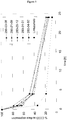
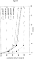
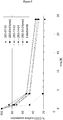
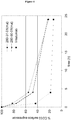
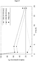
Classifications
-
- C—CHEMISTRY; METALLURGY
- C07—ORGANIC CHEMISTRY
- C07K—PEPTIDES
- C07K16/00—Immunoglobulins [IGs], e.g. monoclonal or polyclonal antibodies
- C07K16/18—Immunoglobulins [IGs], e.g. monoclonal or polyclonal antibodies against material from animals or humans
- C07K16/28—Immunoglobulins [IGs], e.g. monoclonal or polyclonal antibodies against material from animals or humans against receptors, cell surface antigens or cell surface determinants
- C07K16/2803—Immunoglobulins [IGs], e.g. monoclonal or polyclonal antibodies against material from animals or humans against receptors, cell surface antigens or cell surface determinants against the immunoglobulin superfamily
-
- C—CHEMISTRY; METALLURGY
- C07—ORGANIC CHEMISTRY
- C07K—PEPTIDES
- C07K16/00—Immunoglobulins [IGs], e.g. monoclonal or polyclonal antibodies
- C07K16/18—Immunoglobulins [IGs], e.g. monoclonal or polyclonal antibodies against material from animals or humans
- C07K16/28—Immunoglobulins [IGs], e.g. monoclonal or polyclonal antibodies against material from animals or humans against receptors, cell surface antigens or cell surface determinants
-
- A—HUMAN NECESSITIES
- A61—MEDICAL OR VETERINARY SCIENCE; HYGIENE
- A61K—PREPARATIONS FOR MEDICAL, DENTAL OR TOILETRY PURPOSES
- A61K39/00—Medicinal preparations containing antigens or antibodies
- A61K39/395—Antibodies; Immunoglobulins; Immune serum, e.g. antilymphocytic serum
-
- A—HUMAN NECESSITIES
- A61—MEDICAL OR VETERINARY SCIENCE; HYGIENE
- A61P—SPECIFIC THERAPEUTIC ACTIVITY OF CHEMICAL COMPOUNDS OR MEDICINAL PREPARATIONS
- A61P19/00—Drugs for skeletal disorders
-
- A—HUMAN NECESSITIES
- A61—MEDICAL OR VETERINARY SCIENCE; HYGIENE
- A61P—SPECIFIC THERAPEUTIC ACTIVITY OF CHEMICAL COMPOUNDS OR MEDICINAL PREPARATIONS
- A61P29/00—Non-central analgesic, antipyretic or antiinflammatory agents, e.g. antirheumatic agents; Non-steroidal antiinflammatory drugs [NSAID]
-
- A—HUMAN NECESSITIES
- A61—MEDICAL OR VETERINARY SCIENCE; HYGIENE
- A61P—SPECIFIC THERAPEUTIC ACTIVITY OF CHEMICAL COMPOUNDS OR MEDICINAL PREPARATIONS
- A61P35/00—Antineoplastic agents
-
- A—HUMAN NECESSITIES
- A61—MEDICAL OR VETERINARY SCIENCE; HYGIENE
- A61P—SPECIFIC THERAPEUTIC ACTIVITY OF CHEMICAL COMPOUNDS OR MEDICINAL PREPARATIONS
- A61P35/00—Antineoplastic agents
- A61P35/02—Antineoplastic agents specific for leukemia
-
- A—HUMAN NECESSITIES
- A61—MEDICAL OR VETERINARY SCIENCE; HYGIENE
- A61P—SPECIFIC THERAPEUTIC ACTIVITY OF CHEMICAL COMPOUNDS OR MEDICINAL PREPARATIONS
- A61P37/00—Drugs for immunological or allergic disorders
- A61P37/02—Immunomodulators
-
- A—HUMAN NECESSITIES
- A61—MEDICAL OR VETERINARY SCIENCE; HYGIENE
- A61P—SPECIFIC THERAPEUTIC ACTIVITY OF CHEMICAL COMPOUNDS OR MEDICINAL PREPARATIONS
- A61P37/00—Drugs for immunological or allergic disorders
- A61P37/02—Immunomodulators
- A61P37/06—Immunosuppressants, e.g. drugs for graft rejection
-
- A—HUMAN NECESSITIES
- A61—MEDICAL OR VETERINARY SCIENCE; HYGIENE
- A61P—SPECIFIC THERAPEUTIC ACTIVITY OF CHEMICAL COMPOUNDS OR MEDICINAL PREPARATIONS
- A61P43/00—Drugs for specific purposes, not provided for in groups A61P1/00-A61P41/00
-
- C—CHEMISTRY; METALLURGY
- C07—ORGANIC CHEMISTRY
- C07K—PEPTIDES
- C07K16/00—Immunoglobulins [IGs], e.g. monoclonal or polyclonal antibodies
- C07K16/18—Immunoglobulins [IGs], e.g. monoclonal or polyclonal antibodies against material from animals or humans
- C07K16/28—Immunoglobulins [IGs], e.g. monoclonal or polyclonal antibodies against material from animals or humans against receptors, cell surface antigens or cell surface determinants
- C07K16/2851—Immunoglobulins [IGs], e.g. monoclonal or polyclonal antibodies against material from animals or humans against receptors, cell surface antigens or cell surface determinants against the lectin superfamily, e.g. CD23, CD72
-
- C—CHEMISTRY; METALLURGY
- C07—ORGANIC CHEMISTRY
- C07K—PEPTIDES
- C07K2317/00—Immunoglobulins specific features
- C07K2317/20—Immunoglobulins specific features characterized by taxonomic origin
- C07K2317/21—Immunoglobulins specific features characterized by taxonomic origin from primates, e.g. man
-
- C—CHEMISTRY; METALLURGY
- C07—ORGANIC CHEMISTRY
- C07K—PEPTIDES
- C07K2317/00—Immunoglobulins specific features
- C07K2317/30—Immunoglobulins specific features characterized by aspects of specificity or valency
- C07K2317/34—Identification of a linear epitope shorter than 20 amino acid residues or of a conformational epitope defined by amino acid residues
-
- C—CHEMISTRY; METALLURGY
- C07—ORGANIC CHEMISTRY
- C07K—PEPTIDES
- C07K2317/00—Immunoglobulins specific features
- C07K2317/50—Immunoglobulins specific features characterized by immunoglobulin fragments
- C07K2317/51—Complete heavy chain or Fd fragment, i.e. VH + CH1
-
- C—CHEMISTRY; METALLURGY
- C07—ORGANIC CHEMISTRY
- C07K—PEPTIDES
- C07K2317/00—Immunoglobulins specific features
- C07K2317/50—Immunoglobulins specific features characterized by immunoglobulin fragments
- C07K2317/56—Immunoglobulins specific features characterized by immunoglobulin fragments variable (Fv) region, i.e. VH and/or VL
- C07K2317/565—Complementarity determining region [CDR]
-
- C—CHEMISTRY; METALLURGY
- C07—ORGANIC CHEMISTRY
- C07K—PEPTIDES
- C07K2317/00—Immunoglobulins specific features
- C07K2317/70—Immunoglobulins specific features characterized by effect upon binding to a cell or to an antigen
- C07K2317/73—Inducing cell death, e.g. apoptosis, necrosis or inhibition of cell proliferation
- C07K2317/732—Antibody-dependent cellular cytotoxicity [ADCC]
-
- C—CHEMISTRY; METALLURGY
- C07—ORGANIC CHEMISTRY
- C07K—PEPTIDES
- C07K2317/00—Immunoglobulins specific features
- C07K2317/70—Immunoglobulins specific features characterized by effect upon binding to a cell or to an antigen
- C07K2317/77—Internalization into the cell
-
- C—CHEMISTRY; METALLURGY
- C07—ORGANIC CHEMISTRY
- C07K—PEPTIDES
- C07K2317/00—Immunoglobulins specific features
- C07K2317/90—Immunoglobulins specific features characterized by (pharmaco)kinetic aspects or by stability of the immunoglobulin
- C07K2317/92—Affinity (KD), association rate (Ka), dissociation rate (Kd) or EC50 value
Definitions
- the present invention relates to immunotherapies that are based on myeloid cell depletion.
- the present invention relates to methods for manufacturing of CD33 binding agents for use in such therapies, e.g. in the treatment of myeloid cell malignancies and myelodysplastic syndrome (MDS).
- MDS myelodysplastic syndrome
- CD33 was identified as a marker of myeloid leukemias ( Andrews et al., Blood 62, 24-132, 1983 ).
- CD33 is a cell-surface antigen specifically expressed on myeloid cells including myeloid leukemia cells. It is the smallest member of the siglec (sialic acid-binding Ig-related lectins) family.
- CD33 is expressed on early multilineage hematopoietic progenitor cells and myelomonocytic precursors. It is absent from pluripotent hematopoietic stem cells ( Andrews et al., Journal of Experimental Medicine 169, 1721-1731, 1989 ).
- CD33 has also been found to be expressed on human mast cells and blood basophils ( Valent et al., Blood 15; 73(7):1778-85, 1989 ). Monoclonal antibodies directed against CD33 are used in diagnosis of leukemia as well as for therapeutic targeting and in vitro purging of bone marrow for autologous transplantation in acute myeloid leukemia (AML) ( Duzkale et al., Biol Blood Marrow Transplant. 9(6):364-72, 2003 ).
- AML acute myeloid leukemia
- CD33 is a 67 KD transmembrane glycoprotein.
- the sialic acid-binding extracellular domain of CD33 is involved in cell-cell adhesion.
- the intracellular immunoreceptor tyrosine-based inhibitory motifs (ITIM) confer inhibitory signals to the cell, affecting proliferation and cell survival.
- the actual signalling pathways of CD33 are poorly understood but are assumed to involve the ITIM and ITIM-like motifs and the recruitment of tyrosine phosphatases ( von Gunten et al., Ann. N.Y. Acad. Sci. 1143: 61-82, 2008 ).
- CD33 is expressed on malignant myeloid blast cells, which represent the majority of malignant cells in peripheral blood and bone marrow of leukemia patients, and on leukemic stem cells, a relatively small number of less differentiated cells in the bone marrow which are characterized by their capacity for self-renewal and the maintenance of the leukemic clonal hierarchy.
- CD33 targeting immunotoxin Mylotarg® a humanized IgG 4 antibody conjugated to the toxin chaliceamicin is used for the treatment of AML patients by delivering its toxic payload to CD33 positive AML cells ( Amadori et al., Cancer Treat Rev. 34(1):49-60, 2008 ).
- Lintuzumab SGN-33, HuM195, see e.g.
- Targeting AML cell lines with CD33 specific HuM195 in vitro reduces TNF- ⁇ induced secretion of inflammatory cytokines like IL-8, MCP-1 and RANTES ( Sutherland et al., Mabs 1:5, 481-490, 2009 ).
- the relevance of this effect for AML therapy is unknown but modulating the cytokine milieu of the tumor microenvironment may contribute to the therapeutic efficacy of the antibody.
- the antibody induces antibody-dependent cell-mediated cytotoxicity (ADCC) and antibody-dependent cell-mediated phagocytosis (ADCP) of AML cell lines in vitro ( Sutherland et al., Mabs 1:5, 481-490, 2009 ).
- ADCC is considered to be a decisive mechanism for anti-tumor activity of antibodies in hematological malignancies.
- Data from clinical trials with the CD20-specific monoclonal antibody Rituximab have demonstrated the significance of effector cell mediated mechanisms for treatment of B-cell malignancies with respect to response to antibody treatment ( Weng and Levy, J Clin Oncol. 21 (21): 3940-7, 2003 ).
- CD33 antigen is expressed on normal cells of the myelomonocytic lineage and frequently expressed on tumor cells in myeloid leukemias.
- a phase I trial with an antibody against CD33 first signs of efficacy were observed without severe adverse events.
- clinical development of lintuzumab was discontinued after results from a phase II trial in combination with chemotherapy did not yield the expected improvement in efficacy. Therefore, there is a clear need for the development of improved CD33-targeting treatment modalities.
- CD33 binding agents wherein the internalization kinetics of the CD33 binding agents are such that at least 30%, preferably 40%, of the initial amount of the antibody remain on the cell surface of HL60 cells at a time of 4 hours after incubation.
- CD33 binding agents wherein the heavy chain variable region comprises an amino acid sequence selected from SeqID No:85-98 and the light chain variable region comprises an amino acid sequence selected from SeqID No:99-112.
- CD33 binding agents wherein the heavy chain having an amino acid sequence selected from SeqID No: 113-126 and the light chain having an amino acid sequence selected from SeqID No: 127-140.
- CD33 binding agents having mutations in the F c domain that increase ADCC.
- the CD33 binding agents according to the present invention have been found to have high affinity to human CD33 and further to have favourable internalization kinetics, which is characterized by a long presence of the CD33 binding agents when bound to CD33 on the surface of the target cells, which translates into favourable ADCC activity.
- CD33 binding agents according to the present invention bind to a different epitope of the extracellular domain of CD33 compared to Lintuzumab. Without wishing to be bound by any particular theory this is believed to be the reason for the different internalization kinetics of the CD33 binding agents according to the present invention and Lintuzumab.
- binding agent means a protein or peptide that specifically binds to a target antigen.
- a binding agent can be, for example, an antibody, a derivative of such an antibody, or other agent that specifically binds to the target antigen.
- a binding agent can also be a protein comprising an Fv region or a portion thereof (e.g., a V H or V L or a CDR(s) of an antibody that specifically binds to the target antigen).
- the binding agent is an antibody.
- CD33 binding agent refers to a binding agent that specifically binds to CD33, typically a portion of the extracellular domain of human CD33.
- antibody refers to (a) immunoglobulin polypeptides and immunologically active portions of immunoglobulin polypeptides (i.e., polypeptides of the immunoglobulin family, or fragments thereof, that contain an antigen binding site that immunospecifically binds to a specific target antigen), or (b) conservatively substituted derivatives of such immunoglobulin polypeptides or fragments that immunospecifically bind to the target antigen.
- Antibodies are generally described in, for example, Harlow and Lane, Antibodies: A Laboratory Manual (Cold Spring Harbor Laboratory Press, 1988 ).
- antibody refers to intact monoclonal antibodies, polyclonal antibodies, monospecific antibodies, multispecific antibodies (e.g., bispecific antibodies), and antibody fragments that exhibit the desired biological activity (e.g., antigen-binding).
- the antibody can be of any type or class (e.g., IgG, IgE, IgM, IgD, and IgA) or sub-class (e.g., IgGl, lgG2, IgG3, IgG4, IgAl and IgA2), preferably of the IgG class, more preferably an IgG1.
- an “intact” antibody is one which comprises an antigen-binding variable region as well as a light chain constant domain and heavy chain constant domains as appropriate for the antibody class.
- the constant domains may be native sequence constant domains (e.g., human native sequence constant domains) or amino acid sequence variants thereof.
- antibody fragment comprises a portion of an antibody, including the antigen-binding or variable region or a portion thereof.
- antibody fragments include Fab, Fab', F(ab') 2 , and Fv fragments, V H and V L antigen binding fragments, diabodies, triabodies, tetrabodies, single-chain antibody, scFv, scFv-Fc, a SMTP, and multispecific antibodies formed from antibody fragment(s).
- Heavy chain variable region or “V H” means the part of the heavy chain comprising the CDR1, CDR2 and CDR3 and surrounding framework regions.
- Light chain variable region or “V L” means the part of the light chain comprising the CDR4, CDR5 and CDR6 and surrounding framework regions.
- CDR means the hypervariable regions of the heavy and light chains, which determine the complementarity / binding specifity of an antibody or antibody fragment.
- the order of the CDRs in the present application is purely numerically.
- Epitope herein means a part of an antigen, which is recognized by an antibody or antibody fragment. In particular this term refers to parts of CD33, which can be recognized by an antibody.
- mAbs as used herein refers to monoclonal antibodies.
- An antibody may have one or more "effector functions" which refer to those biological activities attributable to the Fc region (a native sequence Fc region or amino acid sequence variant Fc region) of an antibody.
- effector functions include CIq binding; complement dependent cytotoxicity (CDC); Fc receptor binding; antibody- dependent cell-mediated cytotoxicity (ADCC); phagocytosis; down regulation of cell surface receptors (e.g., B cell receptor; BCR), etc.
- Single-chain Fv or “scFv” antibody fragments comprise the V H and V L domains of an antibody, wherein these domains are present in a single polypeptide chain.
- the Fv polypeptide typically further comprises a polypeptide linker between the V H and V L domains which enables the scFv to form the desired structure for antigen-binding.
- Plückthun in The Pharmacology of Monoclonal Antibodies, vol. 113, Rosenburg and Moore, eds., Springer-Verlag, New York, pp. 269-315 (1994 ).
- a binding agent such as an antibody that is “directed to,” “which binds” or that "specifically binds” an antigen of interest (i.e., a target antigen) is one capable of binding that antigen with sufficient affinity such that the binding agent is useful in targeting a cell expressing the antigen.
- the binding agent binds with an affinity of at least about 1x10 7 M -1 , and binds to the predetermined antigen with an affinity that is at least two-fold greater than its affinity for binding to a non-specific antigen (e.g., BSA, casein) other than the predetermined antigen or a closely-related antigen.
- an “antibody derivative” as used herein refers to an antibody, as defined above, that is modified by covalent attachment of a heterologous molecule such as, e.g., by attachment of a heterologous polypeptide, or by glycosylation, deglycosylation, acetylation or phosphorylation or other modification not normally associated with the antibody.
- a heterologous molecule such as, e.g., by attachment of a heterologous polypeptide, or by glycosylation, deglycosylation, acetylation or phosphorylation or other modification not normally associated with the antibody.
- the heterologous molecule is not a therapeutic agent.
- the heterologous molecule does not exhibit a cytostatic or cytotoxic effect by itself.
- CD33 binding agents specifically bind to a receptor, CD33, associated with a given target cell population.
- CD33 is a member of the sialoadhesion family that is expressed on cells of the hematopoietic lineage, including myeloid precursors, monocytes, macrophages, dendritic cells, and mast cells.
- CD33 is also expressed on tumor cells associated with myeloproliferative or mast cell proliferative diseases, including acute myeloid leukemia and myeloplastic syndromes, and on leukemic stem cells.
- Antibodies targeting CD33 and their uses have been generally described (see, e.g., Pierelli et al., 1993, Br. J. Haematol.
- the CD33 binding agent is an antibody (e.g., a monoclonal antibody).
- Useful monoclonal antibodies can be homogeneous populations of antibodies to a CD33 (e.g., the extracellular domain of human CD33).
- a monoclonal antibody (mAb) can be prepared by using any technique known in the art. These include, but are not limited to the hybridoma technique originally described by Köhler and Milstein (1975, Nature 256:495- 497 ), the human B cell hybridoma technique ( Kozbor et al., 1983, Immunology Today 4:72 ), and the EBV-hybridoma technique ( Cole et al., 1985, Monoclonal Antibodies and Cancer Therapy, Alan R. Liss, Inc., pp.
- Such antibodies may be of any immunoglobulin class including IgG, IgM, IgE, IgA and IgD and any subclass thereof.
- the hybridoma producing a monoclonal antibody may be cultivated in vitro or in vivo.
- Useful monoclonal antibodies include, but are not limited to, human monoclonal antibodies, humanized monoclonal antibodies, chimeric monoclonal antibodies and functionally active antibody fragments of any of these.
- Useful CD33 antibodies include antibodies that can achieve a therapeutic effect by various mechanisms known in the art, such as antibody-dependent cell-mediated cyotoxicity (ADCC), anti-dependent cell phagocytosis (ADCP) and/or complement dependent cytotoxicity (CDC).
- ADCC antibody-dependent cell-mediated cyotoxicity
- ADCP anti-dependent cell phagocytosis
- CDC complement dependent cytotoxicity
- the antibody can mediate ADCC by interacting with immune effector cells such as NK cells, monocytes, and macrophages.
- Recombinant antibodies such as chimeric and humanized monoclonal antibodies, comprise both human and non-human portions, which can be made using standard recombinant DNA techniques.
- “Humanized antibodies” are antibody molecules from non-human species having one or more complementarity determining regions (CDRs) from the non-human species and a framework region from a human immunoglobulin molecule.
- CDRs complementarity determining regions
- Such chimeric and humanized monoclonal antibodies can be produced by recombinant DNA techniques known in the art, for example using methods described in International Publication No. WO 87/02671 ; European Patent Publication No. 0184187 ; European Patent Publication No. 0171496 ; European Patent Publication No. 0173494 ; International Publication No. WO 86/01533 ; U.S. Patent No. 4,816,567 ; European Patent Publication No. 012023 ; Berter et al., 1988, Science 240: 1041-1043 ; Liu et al., 1987, Proc. Natl. Acad Set. USA 84:3439-3443 ; Liu et al., 1987, J. Immunol.
- Human monoclonal antibodies may be made by any of numerous techniques known in the art (see, e.g., Teng el al., 1983, Proc. Natl. Acad. Sci. USA. 80: 7308-7312 ; Kozbor et al, 1983, Immunology Today 4:72-79 ; Olsson at al., 1982, Meth. Enzymol. 92:3-16 ; and U.S. Patent Nos. 5,939,598 and 5,770,429 ).
- Fully human antibodies can be produced using transgenic mice that are incapable of expressing endogenous immunoglobulin heavy and light chains genes, but which can express human heavy and light chain genes.
- the transgenic mice are immunized in the normal fashion with a selected antigen, e.g., all or a portion of a CD33 polypeptide.
- Monoclonal antibodies directed against the antigen can be obtained using conventional hybridoma technology.
- the human immunoglobulin transgenes harbored by the transgenic mice rearrange during B cell differentiation, and subsequently undergo class switching and somatic mutation. Thus, using such a technique, it is possible to produce therapeutically useful IgG, IgA, IgM and IgE antibodies.
- Fully human antibodies that recognize a selected epitope also can be generated using a technique referred to as "guided selection".
- a selected non-human monoclonal antibody e.g., a mouse antibody
- Human antibodies also can be produced using various techniques known in the art, including phage display libraries (see, e.g., Hoogenboom and Winter, 1991, J. Mol.
- Useful antibody fragments include, but are not limited to, F(ab') 2 fragments, Fab' fragments, Fab fragments, Fvs, single chain antibodies (SCAs) (e.g., as described in U.S. Patent No. 4,946,778 : Bird, 1988, Science 242:423-42 ; Huston et al., 1988, Proc. Natl. Acad. Sci. USA 85:5879-5883 ; and Ward et al, 1989, Nature 334: 544-54 ), scFv, scFv-Fc, FvdsFv, minibodies, diabodies, triabodies, tetrabodies, SMIPs (see, e.g., Published U.S. Patent Application No. 2005-0238646 ) and any other molecule comprising one or more CDRs and that has the same specificity as the antibody.
- SCAs single chain antibodies
- the antibody is a fusion protein of an antibody, or a functionally active fragment thereof, joined to another protein.
- an antibody or antibody fragment can be fused via a covalent bond (e.g., a peptide bond), at either the N-terminus or the C-terminus to an amino acid sequence of another protein (or portion thereof, typically at least a 10, 20 or 50 amino acid portion of the protein) that is not the antibody or an antibody fragment.
- the antibody or fragment thereof can be covalently linked to the other protein at the C-terminus of the variable domain or a constant domain.
- Antibodies can be modified, e.g., by the covalent attachment of any type of molecule as long as such covalent attachment permits the antibody to retain its antigen-binding immunospecificity.
- a derivative of an antibody can be one that has been further modified, e.g., by glycosylation, de-glycosylation, acetylation, pegylation, phosphorylation, amidation, derealization by known protecting/blocking groups, proteolytic cleavage, linkage to another protein, etc. Any of numerous chemical modifications can be carried out by known techniques, including, but not limited to, specific chemical cleavage, acetylation, formylation, metabolic synthesis in the presence of tunicamycin, or the like. Additionally, the derivative can contain one or more unnatural amino acids.
- Amino acid sequence variants of the antibodies are prepared by introducing appropriate nucleotide changes into the antibody nucleic acid, or by peptide synthesis. Such modifications include, for example, deletions from, and/or insertions into and/or substitutions of, residues within the amino acid sequences of the antibody. Any combination of deletions, insertions, and/or substitutions is made to arrive at the final construct, provided that the final construct possesses the desired characteristics.
- the amino acid changes also may alter post-translational processes of the antibody, such as changing the number or position of glycosylation sites.
- a useful method for identification of certain residues or regions of the antibody that are favored locations for mutagenesis is called "alanine scanning mutagenesis" as described by Cunningham and Wells (1989, Science 244: 1081-1085 ).
- a residue or group of target residues are identified (e.g., charged residues such as arg, asp, his, lys, and glu) and replaced by a neutral or negatively charged amino acid (typically alanine or polyalanine) to affect the interaction of the amino acids with antigen.
- Those amino acid locations demonstrating functional sensitivity to the substitutions then are refined by introducing further or other variants at, or for, the sites of substitution.
- the site for introducing an amino acid sequence variation is predetermined, the nature of the mutation per se need not be predetermined. For example, to analyze the performance of a mutation at a given site, alanine scanning or random mutagenesis is conducted at the target codon or region and the expressed antibody variants are screened for the desired activity.
- Amino acid sequence insertions can include amino- and/or carboxyl-terminal fusions ranging in length from one residue to polypeptides containing a hundred or more residues, as well as intrasequence insertions of single or multiple amino acid residues.
- terminal insertions include an antibody with an N-terminal methionyl residue or the antibody fused to a cytotoxic polypeptide.
- Another type of antibody is an amino acid substitution variant of an antibody.
- Such variants have at least one amino acid residue in the antibody molecule replaced by a different residue.
- the sites of greatest interest for substitutional mutagenesis include the hypervariable regions, but framework region alterations are also contemplated.
- Substantial modifications in the biological properties of the antibody can be accomplished by selecting substitutions that differ significantly in their effect on maintaining (a) the structure of the polypeptide backbone in the area of the substitution, for example, as a sheet or helical conformation, (b) the charge or hydrophobicity of the molecule at the target site, or (c) the bulk of the side chain.
- Naturally-occurring residues are divided into groups based on common side-chain properties:
- Non-conservative substitutions will entail exchanging a member of one of these classes for another class.
- ADCC antibody-dependent cell-mediated cyotoxicity
- ADCP anti-dependent cell phagocytosis
- CDC complement dependent cytotoxicity
- the ADCC activity can be enhanced by introducing amino acid mutations in the constant region of the antibody ( Lazar et al., PNAS 103, 11, 4005-4010, 2006 ). This may be achieved by introducing one or more amino acid substitutions in an Fc region of the antibody, see, e.g., Published U.S. Patent Application No. 2006-0160996 .
- cysteine residue(s) may be introduced in the Fc region, thereby allowing interchain disulfide bond formation in this region.
- the homodimeric antibody thus generated may have improved internalization capability and/or increased CDC and ADCC.
- Homodimeric antibodies with enhanced anti-tumor activity may also be prepared using heterobifunctional cross-linkers as described in Wolff et al., 1993, Cancer Research 53:2560-2565 .
- an antibody can be engineered which has dual Fc regions and may thereby have enhanced complement lysis and ADCC capabilities. (See, e.g., Stevenson et al.. 1989, Anti-Cancer Drug Design 3:219-230 .)
- the antibodies as recited in the claims are Fc variants with amino acid substitutions at positions 332 and/or 239 and/or 236.
- the antibodies of the invention have mutations in the Fc domain selected from the group of
- F c domain it is particularly preferred to introduce mutations in the F c domain at one or more positions selected from amino acids at positions 332 and/or 239 and/or 236 according to the Kabat EU numbering index. Particular preferred are substitutions at positions 239 and 332, especially S239D/I332E.
- Fc variants in the antibodies as recited in the claims are defined according to the amino acid modifications that compose them.
- I332E is an Fc variant with the substitution I332E relative to the parent Fc polypeptide.
- S239D/I332E defines an Fc variant with the substitutions S239D and I332E and S239D/I332E/G236A defines an Fc variant with the substitutions S239D, I332E, and G236A relative to the parent Fc polypeptide.
- a salvage receptor binding epitope refers to an epitope of the Fc region of an IgG molecule (e.g., IgG 1 , IgG 2 , IgG 3 , or IgG 4 ) that is responsible for increasing the in vivo serum half-life of the IgG molecule.
- Antibodies may be glycosylated at conserved positions in their constant regions (see, e.g., Jefferis and Lund, 1997, Chem. Immunol. 65:11 1-128 ; Wright and Morrison, 1997, TibTECH 15:26-32 ).
- the oligosaccharide side chains of the immunoglobulins can affect the protein's function (see, e.g., Boyd et al., 1996, Mol. Immunol. 32: 1311 -1318 ; Wittwe and Howard, 1990, Biochem.
- Oligosaccharides may also serve to target a given glycoprotein to certain molecules based upon specific recognition structures. For example, it has been reported that in a galactosylated IgG, the oligosaccharide moiety 'flips' out of the inter-C H 2 space and terminal N-acetylglucosamine residues become available to bind mannose binding protein (see, e.g., Malhotra et al.
- CAMPATH-1H a recombinant humanized murine monoclonal IgG1 antibody which recognizes the CDw52 antigen of human lymphocytes
- CAMPATH-1H a recombinant humanized murine monoclonal IgG1 antibody which recognizes the CDw52 antigen of human lymphocytes
- CHO Chinese Hamster Ovary
- CMCL or CDC complement mediated lysis
- CHO cells with tetracycline-regulated expression of ⁇ (1.4)-N-acetylglucosaminyltransferase III (GnTIII), a glycosyltransferase catalyzing formation of bisecting GIcNAc, was reported to have improved ADCC activity (see, e.g., Umana et al., 1999, Nature Biotech. 17: 176-180 ).
- N-linked refers to the attachment of the carbohydrate moiety to the side chain of an asparagine residue.
- the tripeptide sequences asparagine-X-serine and asparagine-X-threonine, where X is any amino acid except proline, are the recognition sequences for enzymatic attachment of the carbohydrate moiety to the asparagine side chain.
- X is any amino acid except proline
- O-linked glycosylation refers to the attachment of one of the sugars N-aceylgalaciosamine, galactose, or xylose to a hydroxyamino acid, most commonly serine or threonine, although 5-hydroxyproline or 5-hydroxylysine may also be used.
- Glycosylation variants of antibodies are variants in which the glycosylation pattern of an antibody is altered.
- altering is meant deleting one or more carbohydrate moieties found in the antibody, adding one or more carbohydrate moieties to the antibody, changing the composition of glycosylation (i.e., glycosylation pattern), the extent of glycosylation, or the like.
- glycosylation sites to the antibody is conveniently accomplished by altering the amino acid sequence such that it contains one or more of the above-described tripeptide sequences (for N-linked glycosylation sites).
- the alteration may also be made by the addition of, or substitution by, one or more serine or threonine residues to the sequence of the original antibody (for O-linked glycosylation sites).
- removal of glycosylation sites can be accomplished by amino acid alteration within the native glycosylation sites of the antibody.
- the amino acid sequence is usually altered by altering the underlying nucleic acid sequence.
- These methods include, but are not limited to, isolation from a natural source (in the case of naturally-occurring amino acid sequence variants) or preparation by oligonucleoiide-mediated (or site-directed) mutagenesis, PCR mutagenesis, or cassette mutagenesis of an earlier prepared variant or a non-variant version of the antibody.
- glycosylation may also be altered without altering the amino acid sequence or the underlying nucleotide sequence. Glycosylation largely depends on the host cell used to express the antibody. Since the cell type used for expression of recombinant glycoproteins, e.g., antibodies, as potential therapeutics is rarely the native cell, significant variations in the glycosylation pattern of the antibodies can be expected. (See, e.g., Hse et al., 1997, Biol. Chem. 272:9062-9070 .) In addition to the choice of host cells, factors which affect glycosylation during recombinant production of antibodies include growth mode, media formulation, culture density, oxygenation, pH, purification schemes, and the like.
- glycosylation pattern achieved in a particular host organism including introducing or overexpressing certain enzymes involved in oligosaccharide production (see, e.g., U.S. Patent Nos. 5,047,335 ; 5,510,261 ; and 5,278,299 ).
- Glycosylation, or certain types of glycosylation can be enzymatically removed from the glycoprotein, for example using endoglycosidase H (Endo H).
- the recombinant host cell can be genetically engineered, e.g., made defective in processing certain types of polysaccharides.
- glycosylation structure of antibodies can be readily analyzed by conventional techniques of carbohydrate analysis, including lectin chromatography, NMR, mass spectrometry, HPLC, GPC, monosaccharide compositional analysis, sequential enzymatic digestion, and HPAEC-PAD, which uses high pH anion exchange chromatography to separate oligosaccharides based on charge.
- Methods for releasing oligosaccharides for analytical purposes include, without limitation, enzymatic treatment (commonly performed using peptide-N-glycosidase F/endo- ⁇ -galactosidase), elimination using harsh alkaline environment to release mainly O-linked structures, and chemical methods using anhydrous hydrazine to release both N- and O-linked oligosaccharides.
- the antibodies also can have modifications (e.g., substitutions, deletions or additions) in amino acid residues that interact with Fc receptors.
- antibodies can have modifications in amino acid residues identified as involved in the interaction between the anti-Fc domain and the FcRn receptor (see, e.g., International Publication No. WO 97/34631 ).
- CD33 binding agents binding to human CD33 and that are defined by
- the present disclosure further provides CD33 binding agents wherein the internalization kinetics of the CD33 binding agents are such that at least 30% of the initial amount of the antibody remain on the cell surface of HL60 cells at a time of 4 hours after incubation.
- CD33 binding agents according to the present invention bind to a different epitope than lintuzumab, see example 4 herein. Both epitopes (SeqID No: 141 and SeqID No: 142) are non-overlapping. It is believed that the different epitopes on the exatracellular domain of CD33, which are recognized by the CD33 binding agents according to the present invention and by lintuzumab, are the reason for the different internalization behaviour and ADCC performance of the CD33 binding agents according to the present invention and lintuzumab (cf. examples 2 and 3 herein).
- At least 40% of the initial amount of the CD33 binding agents may remain on the cell surface at a time of 4 hours after incubation.
- the heavy chain variable region comprises an amino acid sequence selected from SeqID No:85-98 and the light chain variable region comprises an amino acid sequence selected from SeqID No:99-112.
- the CD33 binding agent described herein has a heavy chain having an amino acid sequence selected from SeqID No: 113-126 and a light chain having an amino acid sequence selected from SeqID No: 127-140.
- the CD33 binding agent described herein has an affinity to both human CD33 and cynomolgus CD33 with a K D of equal to or less than 10 nM.
- the CD33 binding agent described herein is humanized.
- the CD33 binding agent described herein is fully human.
- the CD33 binding agent described herein further comprises an effector function.
- the effector function is mediated by an F c domain.
- the CD33 binding agent described herein comprises one or more mutations in the F c domain that modulate the function of the F c domain.
- the CD33 binding agent described herein the modulation of the function of the F c domain is an increase of ADCC by at least 10%, preferably 50% or 100%.
- CD33 binding agents including the ones recited n the appended claims are listed in table 1: Table 1 No Clone ID# SeqID CDR1 SeqID CDR2 SeqID CDR3 SeqID CDR4 SeqID CDR5 SeqID CDR6 SeqID V H SeqID V L SeqID heavy chain SeqID light chain 1 280-03-08 1 15 29 43 57 71 85 99 113 127 2 280-21-09 2 16 30 44 58 72 86 100 114 128 3 280-29-12 3 17 31 45 59 73 87 101 115 129 4 280-31-01 4 18 32 46 60 74 88 102 116 130 5 280-31-01 (mut) 5 19 33 47 61 75 89 103 117 131 6 280-34-02 6 20 34 48 62 76 90 104 118 132 7 280-50-01 7 21 35 49 63 77 91 105 133 8 280-50-01 (mut) 8 22 36 50 64
- CD33 as a cell surface marker on primary AML and CML cells which is expressed on malignant cells of 70% to 100% of patients (Scheinberg et al., 1989; Hauswirt et al., 2007; Plesa et al., 2007; Webber et al., 2008).
- CD33 is expressed on malignant myeloid blast cells, which represent the majority of malignant cells in peripheral blood and bone marrow of leukemia patients, and on leukemic stem cells, a relatively small number of less differentiated cells in the bone marrow which are characterized by their capacity for self-renewal and the maintenance of the leukemic clonal hierarchy.
- CD33 The clinical feasibility of targeting CD33 using antibodies has been demonstrated by Mylotarg® (Gemtuzumab ozogamizin), an antibody-calicheamycin conjugate which was approved for the treatment of relapsed AML patients not eligible for other treatment options.
- An alternative approach directed towards CD33 is the development of lintuzumab (SGN-33, HuM195), a humanized IgG1 monoclonal antibody which has shown early signs of efficacy in clinical phase I trials (Raza et al., 2009).
- Acute myeloid leukemia is a malignancy of the myeloid lineage of white blood cells. This hematological neoplasia is a blood and bone marrow disease that, if left untreated, is typically fatal within weeks to months. There is an estimated prevalence of 30000 cases of AML in the US and 47000 in the European Union (10 year prevalence data confirmed by Mattson-Jack, 2010). AML is the most prevalent form of adult acute leukemia (about 90%), comprising about 33% of new leukemia cases. The median age of patients diagnosed with AML is 67 years. AML accounts for about 1.2% of cancer deaths in the United States.
- AML causes non-specific symptoms such as weight loss, fatigue, fever, and night sweats.
- AML is diagnosed by blood tests, bone marrow examination, and laboratory tests to determine the AML subtype and to determine treatment decisions.
- the CD33 binding agents are useful for treating cancer, such as by delaying progression of a cancer and/or reducing cancer-associated cachexia, or preventing or delaying recurrence of a hematological malignancy (e.g., leukemia), in a mammal, preferably a human patient.
- the CD33 binding agent can be administered alone or co-administered with another therapeutic agent.
- the CD33 binding agent is co-administered with a standard of care chemotherapeutic.
- the CD33 binding agent can be administered in an unconjugated form (i.e., not conjugated to a cytotoxin) or as a conjugate.
- a "patient” is a human or other mammal who is undergoing treatment for, or has been diagnosed as having, cancer.
- the CD33 binding agents described herein are useful for delaying progression of a cancer and/or reducing cancer-associated cachexia in a patient by administering to the patient in need thereof an effective dosage of the CD33 binding agent.
- the CD33 binding agent binds to effector or accessory cells of the myeloid or monocytic lineages (e.g., monocytes, macrophages, dendritic cells, and neutrophils), thereby inhibiting or reducing the production of various cytokines, chemokines and growth factors from the effector or accessory cells and/or the tumor cells.
- cytokines, chemokines and growth factors which can promote the growth and proliferation of tumor cells and/or contribute to cancer cachexia, include, but are not limited to, interleukin-1 ⁇ (IL-1 ⁇ ), tumor necrosis factor-a (TNF- ⁇ ), interleukin-6 (IL-6), interleukin-8 (IL-8), interferon-y (IFN- ⁇ ), vascular endothelial growth factor (VEGF), leukemia inhibitory factor (L1F), monocyte chemoattraclant protein-1 (MCP-1), RANTES, interleukin-10 (IL-10), interleukin-12 (IL-12), matrix metalloproteinase 2 (MMP2), IP-10 and/or macrophage inflammatory protein 1 ⁇ (MIP1 ⁇ ).
- the CD33 binding agents also can reduce the migration of macrophages to the site of the tumor cells.
- a CD33 binding agent as described herein reduces the levels of at least one cytokine, chemokine or growth factor, which cytokine, chemokine or growth factor can promote the growth and proliferation of tumor cells, promote the migration of non-malignant effector cells, such as tumor associated macrophages (TAMS), to the vicinity of the tumor site and/or contribute to cancer cachexia.
- cytokine, chemokine or growth factor can promote the growth and proliferation of tumor cells, promote the migration of non-malignant effector cells, such as tumor associated macrophages (TAMS), to the vicinity of the tumor site and/or contribute to cancer cachexia.
- TAMS tumor associated macrophages
- the cytokine, chemokine or growth factor is for example interleukin-1 ⁇ (IL-1 ⁇ ), tumor necrosis factor-a (TNF- ⁇ ), interleukin-6 (IL-6), interleukin-8 (IL-8), interferon- ⁇ (IFN- ⁇ ), vascular endothelial growth factor (VEGF), leukemia inhibitory factor (LIF), monocyte chemoattractant protein-1 (MCP-1), RANTES, interleukin-10 (IL-10), interleukin-12 (IL-12), matrix metalloproteinase 2 (MMP2), IP-10 and/or macrophage inflammatory protein 1 ⁇ (MIP1 ⁇ ).
- IL-1 ⁇ interleukin-1 ⁇
- TNF- ⁇ tumor necrosis factor-a
- IL-6 interleukin-6
- IL-8 interleukin-8
- IFN- ⁇ interferon- ⁇
- VEGF vascular endothelial growth factor
- LIF leukemia inhibitory factor
- MCP-1 monocyte chemoattrac
- a method for delaying progression of a cancer by administering to a patient an effective regimen of a CD33 binding agent that can specifically bind to CD33.
- a CD33 binding agent that can specifically bind to CD33.
- the progression of the cancer is delayed, such as by reducing the growth or proliferation of the tumor cells, decreasing metastasis, reducing the level of at least one cytokine, chemokine or growth factor, reducing non-malignant effector cells in the vicinity of the tumor cells, or the like.
- a method for reducing the tumor burden in a patient by administering to the patient an effective regimen of a CD33 binding agent that can specifically bind to CD33.
- the tumor burden in the patient is arrested or reduced, such as by reducing the size or mass of the tumor, reducing the level of at least one cytokine, chemokine or growth factor, reducing non- malignant effector cells in the vicinity of the tumor cells, inhibiting the migration of macrophages in the vicinity of the tumor cells, reducing the number of non-malignant effector cells (e.g., TAMS or macrophages) in the tumor, or the like.
- non-malignant effector cells e.g., TAMS or macrophages
- a method for reducing the tumor burden or delaying progression of a cancer in a patient by administering to the patient an effective regimen of a CD33 binding agent that can specifically bind to CD33.
- a CD33 binding agent that can specifically bind to CD33.
- the tumor burden in the patient is arrested or reduced, such as by recruiting immune effector cells like NK cells or macrophages or monocytes, which can destroy tumor cells by immune mediated mechanisms.
- ADCC Antibody dependent cellular cytotoxicity
- Weiner GJ Monoclonal antibody mechanisms of action in cancer. Immunol Res. 2007,39(1-3):271-8 ).
- the relevance of ADCC for anti-tumor efficacy has been demonstrated in preclinical models, e.g. in mouse tumor models (e.g. Clynes RA, Towers TL, Presta LG, Ravetch JV.Inhibitory Fc receptors modulate in vivo cytoxicity against tumor targets. Nat Med. 2000 Apr;6(4):443-6 ).
- Data from clinical trials support the relevance of ADCC for clinical efficacy of therapeutic antibodies (e.g.
- the CD33 binding agent can be used to treat a CD33 positive cancer (i.e., a cancer comprised of cancer cells that over express CD33 on their cell surface, or that express CD33 at levels considered acceptable for therapy with CD33 antibodies).
- the CD33 binding agent also can be used to treat a cancer that does not overexpress CD33 on the non-malignant effector cells relative to normal tissue of the same type.
- the cancer can be, for example, a non- hematological malignancy or a hematological malignancy.
- the hematological malignancy can be CD33-positive and can be, for example, acute lymphoid leukemia, acute myeloid leukemia, chronic myelomonocytic leukemia, an erythrocytic leukemia, acute megakaryoblastic leukemia, histiocytic lymphoma, a myeloid sarcoma, a mast cell proliferative disorder or myelodysplastic syndrome (MDS).
- the hematological malignancy can be a CD33-positive malignancy, such as acute myeloid leukemia or myelodysplastic syndrome (MDS).
- the CD33 binding agent can be an unconjugated anti-CD33 antibody.
- the antibody can be a fully human, humanized or chimeric antibody, such as a chimeric or humanized M 195 antibody.
- the antibody also can be another antibody, such as an antibody that competes with M 195 antibody for specific binding to CD33.
- the antibody also can bind to the same epitope as M 195 antibody or to a different epitope.
- the CD33 binding agent can be bound (i.e., conjugated) to a cytotoxin.
- the cytotoxin can be, for example, a peptide toxin, such as saporin, ricin, chlorotoxin, pseudomonas exotoxin, pseudomonas endotoxin or diphtheria toxin.
- the cytotoxin also can be a chemical (i.e., non-peptide-based) toxin, such as a calicheamicin, doxorubicin, a camptothecin, daunorubicin, or other DNA binding agents.
- the cytotoxin also can be an auristatin, a maytansinoid, a dolastatin, or other microtubule blocking agents.
- a CD33 binding agent that is an anti-CD33 antibody can be administered to the patient intravenously or subcutaneously at a dose of 0.1 mg/kg or less to about 25 mg/kg, preferably 1.0 mg/kg to about 10 mg/kg.
- a CD33 binding agent that is an anti-CD33 antibody fragment or other CD33 binding protein can be administered in a dosage equivalent to a dose of 0.1 mg/kg to about 25 mg/kg. 1.0 mg/kg to about 10 mg/kg of intact antibody.
- the CD33 binding agent can be administered intravenously or subcutaneously to the patient on a schedule that is, for example, daily, weekly, biweekly, tri-weekly (i.e., every three weeks) or monthly, or a combination thereof, to the patient.
- the CD33 binding agent can be administered for a period of at least one month, at least two months, at least three months, at least four months, at least five month, at least six months, or more, as needed.
- a treatment phase (supra) of the CD33 binding agent is followed by a maintenance phase, in which doses of the CD33 binding agent are administered less frequently than during the treatment phase.
- maintenance doses can be administered weekly, biweekly, tri-weekly or monthly, for a period of 1-6 months.
- the dosages in the maintenance phase can be the same as the dosages in the treatment phase.
- a hematological malignancy e.g., leukemia
- a hematological malignancy e.g., leukemia
- the CD33 binding agent specifically binds to CD33 on the surface of the hematological malignancy (i.e., leukemic cell) and/or to non-malignant effector cells.
- a "patient” is typically a human who is undergoing treatment for, or has been diagnosed as having, hematological malignancy.
- the hematological malignancy is a CD33-positive hematological malignancy.
- Hematological malignancies include, but are not limited to, leukemias (e.g., acute lymphoblastic leukemia (ALL), acute myelogenous leukemia (AML), chronic myelogenous leukemia (CML), chronic myelomonocytic leukemia, hairy cell leukemia).
- ALL acute lymphoblastic leukemia
- AML acute myelogenous leukemia
- CML chronic myelogenous leukemia
- chronic myelomonocytic leukemia hairy cell leukemia
- MDS myelodysplastic syndrome
- myelofibrosis myeloproliferative disease
- PV polycythemia vera
- ET essential thrombocytosis
- amyloid due to light-chain disease e.g., amyloid due to light-chain disease.
- CD33-positive hematological malignancy refers to a hematological malignancy characterized by the expression of CD33 on the surface of the malignant cells.
- CD33-positive hematological malignancies include, but are not limited to, acute myeloid leukemia (AML), chronic myeloid leukemia (CML), chronic myelomonocytic leukemia, thrombocyte leukemia, a myelodysplastic syndrome, a myeloproliferative disorder, refractory anemia, a preleukemia syndrome, a lymphoid leukemia, or an undifferentiated leukemia.
- the methods include administering to a patient in remission from a CD33 positive hematological malignancy an effective regimen of a CD33 binding agent, whereby the recurrence of the hematological malignancy is prevented or delayed.
- the patient lacks detectable cells of the hematological malignancy.
- a 'lack of detectable cells is determined by standard diagnostic or prognostic methods.
- a patient in remission from AML typically exhibits resolution of abnormal clinical features, return to normal blood counts and normal hematopoiesis in the bone marrow with ⁇ 5% blast cells, a neutrophil count of >1.000-1,500, a platelet count of >100,000, and disappearance of the leukemic clone. See, e.g., The Merck Manual, Sec. 11, Ch. 138 (17th ed. 1997 ): Estey, 2001, Cancer 92(5): 1059-1073 .
- the CD33 binding agent can be, for example, an antibody that specifically binds to CD33 and the hematological malignancy can be acute myeloid leukemia (AML), chronic myeloid leukemia (CML), chronic myelomonocytic leukemia, thymoid leukemia, a myelodysplastic syndrome, a myeloproliferative disorder, refractory anemia, a preleukemia syndrome, a lymphoid leukemia, or an undifferentiated leukemia.
- AML acute myeloid leukemia
- CML chronic myeloid leukemia
- chronic myelomonocytic leukemia thymoid leukemia
- myelodysplastic syndrome a myeloproliferative disorder
- refractory anemia a preleukemia syndrome
- lymphoid leukemia or an undifferentiated leukemia.
- the patient in remission from the hematological malignancy has not undergone a bone marrow transplant.
- the patient in remission from the hematological malignancy has undergone a bone marrow transplant.
- the bone marrow transplant can be either an autologous or an allogeneic bone marrow transplant.
- Blood-borne cancers including, but not limited to: acute lymphoblastic leukemia "ALL”, acute lymphoblastic B-cell leukemia, acute lymphoblastic T-cell leukemia, acute myeloblastic leukemia "AML”, acute promyelocytic leukemia "APL”, acute monoblastic leukemia, acute erythroleukemic leukemia, acute megakaryoblaslic leukemia, acute myelomonocytic leukemia, acule nonlymphocytic leukemia, acute undifferentiated leukemia, chronic myelocytic leukemia "CML”, chronic lymphocytic leukemia "CLL”, hairy cell leukemia, multiple myeloma.
- ALL acute lymphoblastic leukemia
- ALL acute lymphoblastic B-cell leukemia
- acute lymphoblastic T-cell leukemia acute myeloblastic leukemia
- AML acute promyelocytic leukemia
- APL acute monoblastic leuk
- Acute and chronic leukemias which are suitable to be treated by CD33 binding agents include: lymphoblastic, myelogenous, lymphocytic, myelocytic leukemias and thrombocytic leukemia. Further myelodysplasia syndrome, myeloproliferative disorder, refractory anemia, preleukemia syndrome, lymphoid leukemia, or undifferentiated leukemia can be treated by CD33 binding agents.
- the CD33 binding agents described herein may be used on its own or in combination with one or more additional therapeutic agents, in particular selected from DNA damaging, DNA demethylating or tubulin binding agents or therapeutically active compounds that inhibit angiogenesis, signal transduction pathways or mitotic checkpoints in cancer cells or have immunomodulatory function (IMIDs).
- additional therapeutic agents in particular selected from DNA damaging, DNA demethylating or tubulin binding agents or therapeutically active compounds that inhibit angiogenesis, signal transduction pathways or mitotic checkpoints in cancer cells or have immunomodulatory function (IMIDs).
- the additional therapeutic agent may be administered simultaneously with, optionally as a component of the same pharmaceutical preparation, or before or after administration of the CD33 binding agent.
- the additional therapeutic agent may be, without limitation, one or more inhibitors selected from the group of inhibitors of EGFR family, VEGFR family, VEGF, IGF-1R, Insulin receptors, AuroraA, AuroraB, PLK and PI3 kinase, FGFR, PDGFR, Raf, KSP or PDK1.
- additional therapeutic agents are inhibitors of CDKs, Akt, Src, Bcr-Abl, cKit, cMet/HGF, Her2, Her3, c-Myc, Flt3, HSP90, hedgehog antagonists, inhibitors of JAK/STAT, Mek, mTor, NFkappaB, the proteasome, Rho, an inhibitor of Wnt signaling or Notch signaling or an ubiquitination pathway inhibitor.
- additional therapeutic agents are inhibitors of DNA polymerase, topoisomerase II, multityrosine kinase inhibitors, CXCR4 antagonists, IL3RA inhibitors, RAR antagonists, KIR inhibitors, immunotherapeutic vaccines, TUB inhibitors, Hsp70 inducers, IAP family inhibitors, DNA methyltransferase inhibitors, TNF inhibitors, ErbB1 receptor tyrosine kinase inhibitors, multikinase inhibitors, JAK2 inhibitors, RR inhibitors, apoptosis inducers, HGPRTase inhibitors, histamine H2 receptor antagonists and CD25 receptor agnosists.
- Aurora inhibitors are, without limitation, PHA-739358, AZD-1152, AT-9283, CYC-116, R-763, VX-667, MLN-8045, PF-3814735, SNS-314, VX-689, GSK-1070916, TTP-607, PHA-680626, MLN-8237, BI847325 and ENMD-2076.
- PLK inhibitor examples include GSK-461364, BI2536 and BI6727.
- raf inhibitors are BAY-73-4506 (also a VEGFR inhibitor), PLX-4032, RAF-265 (also a VEGFR inhibitor), sorafenib (also a VEGFR inhibitor), XL-281, Nevavar (also an inhibitor of the VEGFR) and PLX4032.
- KSP inhibitors examples include ispinesib, ARRY-520, AZD-4877, CK-1122697, GSK-246053A, GSK-923295, MK-0731, SB-743921, LY-2523355, and EMD-534085.
- Examples for a src and/or bcr-abl inhibitors are dasatinib, AZD-0530, bosutinib, XL-228 (also an IGF-1R inhibitor), nilotinib (also a PDGFR and cKit inhibitor), imatinib (also a cKit inhibitor), NS-187, KX2-391, AP-24534 (also an inhibitor of EGFR, FGFR, Tie2, Flt3), KM-80 and LS-104 (also an inhibitor of Flt3, Jak2).
- An example for a PDK1 inhibitor is AR-12.
- Rho inhibitor An example for a Rho inhibitor is BA-210.
- PI3 kinase inhibitors examples include PX-866, PX-867, BEZ-235 (also an mTor inhibitor), XL-147, and XL-765 (also an mTor inhibitor), BGT-226, CDC-0941.
- inhibitors of cMet or HGF are XL-184 (also an inhibitor of VEGFR, cKit, Flt3), PF-2341066, MK-2461, XL-880 (also an inhibitor of VEGFR), MGCD-265 (also an inhibitor of VEGFR, Ron, Tie2), SU-11274, PHA-665752, AMG-102, AV-299, ARQ-197, MetMAb, CGEN-241, BMS-777607, JNJ-38877605, PF-4217903, SGX-126, CEP-17940, AMG-458, INCB-028060, and E-7050.
- c-Myc inhibitor is CX-3543.
- Flt3 inhibitors are AC-220 (also an inhibitor of cKit and PDGFR), KW-2449, LS-104 (also an inhibitor of bcr-abl and Jak2), MC-2002, SB-1317, lestaurtinib (also an inhibitor of VEGFR, PDGFR, PKC), TG-101348 (also an inhibitor of JAK2), XL-999 (also an inhibitor of cKit, FGFR, PDGFR and VEGFR), sunitinib (also an inhibitor of PDGFR, VEGFR and cKit), and tandutinib (also an inhibitor of PDGFR, and cKit).
- AC-220 also an inhibitor of cKit and PDGFR
- KW-2449 also an inhibitor of bcr-abl and Jak2
- MC-2002 SB-1317
- lestaurtinib also an inhibitor of VEGFR, PDGFR, PKC
- TG-101348 also an inhibitor of JAK2
- HSP90 inhibitors examples include, tanespimycin, alvespimycin, IPI-504, STA-9090, MEDI-561, AUY-922, CNF-2024, and SNX-5422.
- JAK/STAT inhibitors examples include CYT-997 (also interacting with tubulin), TG-101348 (also an inhibitor of Flt3), and XL-019.
- Mek inhibitors are ARRY-142886, AS-703026, PD-325901, AZD-8330, ARRY-704, RDEA-119, and XL-518.
- mTor inhibitors examples include temsirolimus, deforolimus (which also acts as a VEGF inhibitor), everolimus (a VEGF inhibitor in addition), XL-765 (also a PI3 kinase inhibitor), and BEZ-235 (also a PI3 kinase inhibitor).
- Akt inhibitors are perifosine, GSK-690693, RX-0201, and triciribine.
- cKit inhibitors examples include masitinib, OSI-930 (also acts as a VEGFR inhibitor), AC-220 (also an inhibitor of Flt3 and PDGFR), tandutinib (also an inhibitor of Flt3 and PDGFR), axitinib (also an inhibitor of VEGFR and PDGFR), sunitinib (also an inhibitor of Flt3, PDGFR, VEGFR), and XL-820 (also acts as a VEGFR- and PDGFR inhibitor), imatinib (also a bcr-abl inhibitor), nilotinib (also an inhibitor of bcr-abl and PDGFR).
- masitinib, OSI-930 also acts as a VEGFR inhibitor
- AC-220 also an inhibitor of Flt3 and PDGFR
- tandutinib also an inhibitor of Flt3 and PDGFR
- axitinib also an inhibitor of VEGFR and PDGFR
- sunitinib
- hedgehog antagonists examples include IPI-609, CUR-61414, GDC-0449, IPI-926, and XL-139.
- CDK inhibitors are seliciclib, AT-7519, P-276, ZK-CDK (also inhibiting VEGFR2 and PDGFR), PD-332991, R-547, SNS-032, PHA-690509, PHA-848125, and SCH-727965.
- proteasome inhibitors examples include bortezomib, carfilzomib, and NPI-0052 (also an inhibitor of NFkappaB).
- proteasome inhibitors/NFkappaB pathway inhibitors examples include bortezomib, carfilzomib, NPI-0052, CEP-18770, MLN-2238, PR-047, PR-957, AVE-8680, and SPC-839.
- An example for an inhibitor of the ubiquitination pathway is HBX-41108.
- Examples for demethylating agends are 5-azacitidine and decitabine.
- anti-angiogenic agents are inhibitors of the FGFR, PDGFR and VEGF(R), and thalidomides, such agents being selected from, without limitation, bevacizumab, motesanib, CDP-791, SU-14813, telatinib, KRN-951, ZK-CDK (also an inhibitor of CDK), ABT-869, BMS-690514, RAF-265, IMC-KDR, IMC-18F1, IMiDs, thalidomide, CC-4047, lenalidomide, ENMD-0995, IMC-D11, Ki-23057, brivanib, cediranib, 1B3, CP-868596, IMC-3G3, R-1530 (also an inhibitor of Flt3), sunitinib (also an inhibitor of cKit and Flt3), axitinib (also an inhibitor of cKit), lestaurtinib (also an inhibitor of Flt3 and PKC), vatalanib
- the additional therapeutic agent may also be selected from EGFR inhibitors, it may be a small molecule EGFR inhibitor or an anti-EGFR antibody.
- anti-EGFR antibodies without limitation, are cetuximab, panitumumab, nimotuzumab, zalutumumab; examples for small molecule EGFR inhibitors are gefitinib, erlotinib, vandetanib (also an inhibitor of the VEGFR) and afatinib (also an inhibitor of Her2).
- Another example for an EGFR modulator is the EGF fusion toxin.
- EGFR and/or Her2 inhibitors useful for combination with an CD33 binding agent as disclosed herein are lapatinib, trastuzumab, pertuzumab, XL-647, neratinib, BMS-599626 ARRY-334543, AV-412, mAB-806, BMS-690514, JNJ-26483327, AEE-788 (also an inhibitor of VEGFR), AZD-8931, ARRY-380 ARRY-333786, IMC-11F8, Zemab, TAK-285, AZD-4769, and afatinib (dual inhibitor of Her2 and EGFR).
- DNA polymerase inhibitors useful in the combination with an CD33 binding agent as disclosed herein are Ara-C/cytarabine, Clolar/ clofarabine.
- a DNA methyltransferase inhibitor useful in the combination with an CD33 binding agent as disclosed herein is Vidaza/azacitidine.
- An apoptosis inducer useful in the combination with an CD33 binding agent as disclosed herein is Trisenox/arsenice trioxide.
- Topoisomerase II inhibitors useful in the combination with an CD33 binding agent as disclosed herein are idarubicin, daunorubicin and mitoxantrone.
- a RAR antagonist useful in the combination with an CD33 binding agent as disclosed herein is Vesanoid/tretinoin.
- a HGPRTase inhibitor useful in the combination with an CD33 binding agent as disclosed herein is Mercapto/mercaptopurine.
- a histamine H2 receptor antagonist useful in the combination with an CD33 binding agent as disclosed herein is Ceplene/histamine dihydrochloride.
- the additional drug may also be selected from agents that target the IGF-1R and insulin receptor pathways.
- agents include antibodies that bind to IGF-1R (e.g. CP-751871, AMG-479, IMC-A12, MK-0646, AVE-1642, R-1507, BIIB-022, SCH-717454, rhu Mab IGFR and novel chemical entities that target the kinase domain of the IGF1-R (e.g. OSI-906 or BMS-554417, XL-228, BMS-754807).
- IGF-1R e.g. CP-751871, AMG-479, IMC-A12, MK-0646, AVE-1642, R-1507, BIIB-022, SCH-717454, rhu Mab IGFR
- novel chemical entities that target the kinase domain of the IGF1-R (e.g. OSI-906 or BMS-554417, XL-228, BMS-75
- CD33 binding agents may be combined with inhibitors of other surface antigens expressed on leukocytes, in particular antibodies or antibody-like molecules, e.g. anti-CD2 (siplizumab), anti-CD4 (zanolimumab), anti-CD19 (MT-103, MDX-1342, SAR-3419, XmAb-5574), anti-CD22 (epratuzumab), anti-CD23 (lumiliximab), anti-CD30 (iratumumab), anti-CD32B (MGA-321), anti-CD38 (HuMax-CD38), anti-CD40 (SGN40), anti-CD52 (alemtuzumab), anti-CD80 (galiximab).
- anti-CD2 siplizumab
- anti-CD4 zanolimumab
- anti-CD19 MT-103, MDX-1342, SAR-3419, XmAb-5574
- anti-CD22 epratuzumab
- anti-CD23
- CD33 binding agents include immunotoxins like BL-22 (an anti-CD22 immunotoxin), inotuzumab ozogamicin (an anti-CD23 antibody-calicheamicin conjugate), RFT5.dgA (anti-CD25 Ricin toxin A-chain), SGN-35 (an anti-CD30-auristatin E conjugate), and gemtuzumab ozogamicin (an anti-CD33 calicheamicin conjugate), MDX-1411 (anti-CD70 conjugate), or radio labelled antibodies like 90 Y-epratuzumab (anti-CD22 radioimmunoconjugate).
- immunotoxins like BL-22 (an anti-CD22 immunotoxin), inotuzumab ozogamicin (an anti-CD23 antibody-calicheamicin conjugate), RFT5.dgA (anti-CD25 Ricin toxin A-chain), SGN-35 (an anti-CD30-auristatin E conjugate), and gemtuzumab
- CD33 binding agents may be combined with immunomodulators, agents, e.g. antibodies, that induce apoptosis or modify signal transduction pathways like the TRAIL receptor modulators mapatumumab (a TRAIL-1 receptor agonist), lexatumumab (a TRAIL-2 receptor agonist), tigatuzumab, Apomab, AMG-951 and AMG-655; an anti-HLA-DR antibody (like 1D09C3), an anti-CD74, an osteoclast differentiation factor ligand inhibitor (like denosumab), a BAFF antagonist (like AMG-623a) or an agonist of a Toll-like receptor (e.g. TLR-4 or TLR-9).
- TRAIL receptor modulators mapatumumab (a TRAIL-1 receptor agonist), lexatumumab (a TRAIL-2 receptor agonist), tigatuzumab, Apomab, AMG-951 and AMG-655; an anti-HLA-DR antibody (like 1D09
- drugs that may be used in combination with the CD33 binding agents described herein are selected from, but not limited to hormones, hormonal analogues and antihormonals (e.g. tamoxifen, toremifene, raloxifene, fulvestrant, megestrol acetate, flutamide, nilutamide, bicalutamide, cyproterone acetate, finasteride, buserelin acetate, fludrocortinsone, fluoxymesterone, medroxyprogesterone, hydroxyprogesterone caproate, diethylstilbestrol, testosterone propionate, fluoxymesterone/equivalents, octreotide, arzoxifene, pasireotide, vapreotide, adrenocorticosteroids/ antagonists, prednisone, dexamethasone, ainoglutethimide), aromatase inhibitors (e.g., tam
- LHRH agonists and antagonists e.g. goserelin acetate, leuprolide, abarelix, cetrorelix, deslorelin, histrelin, triptorelin
- antimetabolites e.g.
- antifolates like methotrexate, trimetrexate, pemetrexed, pyrimidine analogues like 5-fluorouracil, fluorodeoxyuridine, capecitabine, decitabine, nelarabine, 5-azacytidine, and gemcitabine, purine and adenosine analogues such as mercaptopurine, thioguanine, azathioprine, cladribine and pentostatin, cytarabine, fludarabine, clofarabine); antitumor antibiotics (e.g.
- anthracyclines like doxorubicin, daunorubicin, epirubicin and idarubicin, mitomycin-C, bleomycin dactinomycin, plicamycin, splicamycin, actimomycin D, mitoxantrone, mitoxantroneidarubicin, pixantrone, streptozocin, aphidicolin); platinum derivatives (e.g. cisplatin, oxaliplatin, carboplatin, lobaplatin, satraplatin); alkylating agents (e.g.
- vinca alkaloids like vinblastine, vindesine, vinorelbine, vinflunine and vincristine
- taxanes like paclitaxel, docetaxel and their formulations, larotaxel; simotaxel, and epothilones like ixabepilone, patupilone, ZK-EPO); topoisomerase inhibitors (e.g.
- epipodophyllotoxins like etoposide and etopophos, teniposide, amsacrine, topotecan, irinotecan, banoxantrone, camptothecin) and miscellaneous chemotherapeutics such as retinoic acid derivatives, amifostine, anagrelide, interferon alpha, interferon beta, interferon gamma, interleukin-2, procarbazine, N-methylhydrazine, mitotane, and porfimer, bexarotene, celecoxib, ethylenemine/methyl-melamine, thriethyienemelamine, triethylene thiophosphoramide, hexamethylmelamine, and enzymes L-asparaginase, L-arginase and metronidazole, misonidazole, desmethylmisonidazole, pimonidazole, etanidazole, nimorazole, RSU 1069,
- the CD33 binding agents described herein may also be used in combination with other therapies including surgery, stem cell transplantation, radiotherapy, endocrine therapy, biologic response modifiers, hyperthermia and cryotherapy and agents to attenuate any adverse effect (e.g. antiemetics), G-CSF, GM-CSF, photosensitizers such as hematoporphyrin derivatives, Photofrin, benzoporphyrin derivatives, Npe6, tin etioporphyrin, pheoboride-a bacteriochlorophyll-a, naphthalocyanines, phthalocyanines, zinc phthalocyanines.
- therapies including surgery, stem cell transplantation, radiotherapy, endocrine therapy, biologic response modifiers, hyperthermia and cryotherapy and agents to attenuate any adverse effect (e.g. antiemetics), G-CSF, GM-CSF, photosensitizers such as hematoporphyrin derivatives, Photof
- the CD33 binding agents can be in any form that allows for the composition to be administered to a patient.
- the composition can be in the form of a solid or liquid.
- the preferred mode of application is parenteral, by infusion or injection (intraveneous, intramuscular, subcutaneous, intraperitoneal, intradermal), but other modes of application such as by inhalation, transdermal, intranasal, buccal, oral and intra- tumor may also be applicable.
- Parenteral administration includes subcutaneous injections, intravenous, intramuscular, intrastemal injection or infusion techniques.
- the compositions are administered parenterally.
- the compounds are administered intravenously.
- compositions can be formulated so as to allow a compound to be bioavailable upon administration of the composition to a patient.
- Compositions can take the form of one or more dosage units, where, for example, a container of a compound in aerosol form can hold a plurality of dosage units.
- compositions can be non-toxic in the amounts used. It will be evident to those of ordinary skill in the art that the optimal dosage of the active ingredient(s) in the pharmaceutical composition will depend on a variety of factors. Relevant factors include, without limitation, the type of patient (e.g., human), the particular form of the compound, the manner of administration, and the composition employed.
- the pharmaceutically acceptable carrier or vehicle can be particulate, so that the compositions are, for example, in powder form.
- the carrier(s) can be liquid, with the compositions being, for example, an injectable liquid.
- the composition can be in the form of a liquid, e.g., for parenteral injection.
- a surfactant, preservative, wetting agent, dispersing agent, suspending agent, buffer, stabilizer and isotonic agent can also be included.
- the liquid compositions can also include one or more of the following: sterile diluents such as water for injection, saline solution, preferably physiological saline, Ringer's solution, isotonic sodium chloride, fixed oils such as synthetic mono- or digylcerides which can serve as the solvent or suspending medium, polyethylene glycols, glycerin, cyclodextrin, propylene glycol or other solvents; stabilizers such as amino acids; surfactants such as polysorbates; antibacterial agents such as benzyl alcohol or methyl paraben; antioxidants such as ascorbic acid or sodium bisulfite; chelating agents such as ethylenediaminetetraacelic acid; buffers such as acetates, citrates or phosphates; and agents for the adjustment of tonicity such as sodium chloride or dextrose.
- sterile diluents such as water for injection, saline solution, preferably physiological saline, Ringer's solution, is
- a parenteral composition can be enclosed in ampoule, a disposable syringe or a multiple-dose vial made of glass, plastic or other material.
- Physiological saline is an exemplary adjuvant.
- An injectable composition is preferably sterile.
- the CD33 binding agents may also be dried (freeze-dried, spray-dried, spray-freeze dried, dried by near or supercritical gases, vacuum dried, air-dried), precipitated or crystallized or entrapped in microcapsules that are prepared, for example, by coacervation techniques or by interfacial polymerization using, for example, hydroxymethylcellulose or gelatin and poly-(methylmethacylate), respectively, in colloidal drug delivery systems (for example, liposomes, albumin microspheres, microemulsions, nano-particles and nanocapsules), in macroemulsions or precipitated or immobilized onto carriers or surfaces, for example by pcmc technology (protein coated microcrystals).
- colloidal drug delivery systems for example, liposomes, albumin microspheres, microemulsions, nano-particles and nanocapsules
- macroemulsions or precipitated or immobilized onto carriers or surfaces for example by pcmc technology (protein coated microcrystals).
- the amount of the composition that is effective in the treatment of a particular disorder or condition will depend on the nature of the disorder or condition, and can be determined by standard clinical techniques. In addition, in vitro or in vivo assays can optionally be employed to help identify optimal dosage ranges. The precise dose to be employed in the compositions will also depend on the route of administration, and the seriousness of the disease or disorder, and should be decided according to the judgment of the practitioner and each patient's circumstances.
- compositions comprise an effective amount of a drug(s) or agent(s) such that a suitable dosage will be obtained. Typically, this amount is at least about 0.01% of a drug or agent by weight of the composition. When intended for oral administration, this amount can be varied to range from about 0.1% to about 80% by weight of the composition. In one aspect, oral compositions can comprise from about 4% to about 50% of the compound by weight of the composition. In yet another aspect, present compositions are prepared so that a parenteral dosage unit contains from about 0.01% to about 2% by weight of the compound.
- the composition can comprise from about 1 to about 50 mg of a drug or agent per kg of the patient's body weight.
- the composition can include from about I, 1.5 or 2.5 to about 50 mg of a drug or agent per kg of the patient's body weight.
- the amount administered will be in the range from about 1, 1.5 or 2.5 to about 25 mg/kg of body weight of a drug or agent.
- the dosage administered to a patient can be less than 0.1 mg/kg to about 50 mg/kg of the patient's body weight. (For conversion to mg/mm 2 , a BSA of 1.8 m 2 and a body weight of 80 kg can be used.)
- a CD33 binding agent can be administered intravenously or subcutaneously to the patient on a schedule that is, for example, daily, weekly, biweekly, tri-weekly or monthly to the patient.
- a CD33 binding agent can be administered weekly, for a period of 2 to 10 weeks, typically 3-6 weeks.
- the dosage regimen of the CD33 binding agent maintains a blood serum concentration of antibody at least 5 ⁇ g/ml or at least 10 ⁇ g/ml during the dosage cycle.
- the CD33 binding agent can be administered, for example, from 1-8, or more cycles.
- a CD33 binding agnt can be administered chronically to a subject.
- a method of treating a cancer such as myeloid leukemia, by administering 0.1 mg/kg to 50 mg/kg, for instance about 1.5-8 or 2.5-8 mg/kg, of an anti-CD33 antibody weekly.
- This treatment can be usually be continued for about 1-3 months, typically about two months.
- the dosing schedule can be maintained until a reduction in blasts is noted.
- dosing can be continued up to about 6 months.
- This treatment can be followed by a less frequent dosing schedule, involving for instance biweekly doses (or twice per month).
- This dosing schedule can be maintained 1, 2, 3, 4, 5, 6 months or more to maintain a reduction in blasts and/or a remission.
- a prophylactic agent can be administered with a CD33 binding agent to minimize infusion reactions.
- Suitable prophylactic agents include, for example, methyl prednisolone, diphenyldramine, acetaminophen or other suitable agent.
- the prophylactic agent can be administered prior to or at about the same time as the CD33 binding agent.
- the drug(s) or agent(s) or compositions can be administered by any convenient route, for example, by infusion or bolus injection, by absorption through epithelial or mucocutaneous linings (e.g., oral mucosa, rectal and intestinal mucosa, etc.). Administration can be systemic or local.
- Various delivery systems are known, e.g., encapsulation in liposomes, microparticles, microcapsules, capsules, etc., and can be used to administer a compound.
- more than one drug or agent or composition can be administered to a patient.
- administration can be by direct injection at the site (or former site) of a cancer, tumor or neoplastic or pre-neoplastic tissue.
- the drug(s) or agent(s) or compositions can be delivered in a controlled release system, such as a pump or various polymeric materials.
- a controlled-release system can be placed in proximity of the target of the drug(s) or agent(s) or compositions, thus requiring only a fraction of the systemic dose (see, e.g., Goodson, in Medical Applications of Controlled Release, vol. 2, pp. 115-138, 1984 ).
- Other controlled- release systems discussed in the review by Langer (1990, Science 249: 1527-1533 ) can be used.
- the drugs or agents are formulated in accordance with routine procedures as a pharmaceutical composition adapted for intravenous administration to animals, particularly human beings, as appropriate for the drug or agent.
- the carriers or vehicles for intravenous administration are sterile isotonic aqueous buffer solutions.
- the compositions can also include a solubilizing agent.
- Compositions for intravenous administration can optionally comprise a local anesthetic such as lignocaine to ease pain at the site of the injection.
- the ingredients are supplied either separately or mixed together in unit dosage form, for example, as a dry lyophilized powder or water-free concentrate in a hermetically sealed container such as an ampoule or sachette indicating the quantity of active agent.
- drug or agent is to be administered by infusion
- it can be dispensed, for example, with an infusion bottle containing sterile pharmaceutical grade water or saline.
- an ampoule of sterile water for injection or saline can be provided so that the ingredients can be mixed prior to administration.
- compositions of therapeutic agents also can be administered according to accepted dosage forms in the form of tablets, lozenges, aqueous or oily suspensions, granules, powders, emulsions, capsules, syrups, or elixirs, for example.
- Orally administered compositions can contain one or more optional agents, for example, sweetening agents such as fructose, aspartame or saccharin; flavoring agents such as peppermint, oil of wintergreen, or cherry; coloring agents; and preserving agents, to provide a pharmaceutically palatable preparation.
- sweetening agents such as fructose, aspartame or saccharin
- flavoring agents such as peppermint, oil of wintergreen, or cherry
- coloring agents such as peppermint, oil of wintergreen, or cherry
- preserving agents to provide a pharmaceutically palatable preparation.
- the compositions can be coated to delay disintegration and absorption in the gastrointestinal tract thereby providing a sustained action over an extended period of time.
- Selectively permeable membranes surrounding an osmotically active driving compound are also suitable for orally administered drugs or agents.
- fluid from the environment surrounding the capsule is imbibed by the driving compound, which swells to displace the agent or agent composition through an aperture.
- delivery platforms can provide an essentially zero order delivery profile as opposed to the spiked profiles of immediate release formulations.
- a time-delay material such as glycerol monostearate or glycerol stearate can also be used.
- the composition can include various materials that modify the physical form of a solid or liquid dosage unit.
- the composition can include materials that form a coating shell around the active ingredients.
- the materials that form the coating shell are typically inert, and can be selected from, for example, sugar, shellac, and other enteric coating agents.
- the active ingredients can be encased in a gelatin capsule.
- compositions can be administered to a patient in need thereof at a frequency, or over a period of time, that is determined by the attending physician.
- the compositions can be administered over a period of 1 day, 2 days, 3 days, 5 days, 7 days, 10 days, 14 days, 21 days, 28 days, one month, two months, or longer periods of time. It is understood that the compositions can be administered for any period of time between 1 day and two months or longer.
- Antibodies can be produced using any method useful for the synthesis of antibodies, in particular, such as by recombinant expression or chemical synthesis.
- Recombinant expression of antibodies, or fragments or derivatives thereof typically involves construction of a nucleic acid that encodes the antibody. If the nucleotide sequence of the antibody is known, a nucleic acid encoding the antibody or a polypeptide thereof maybe assembled from chemically synthesized oligonucleotides (e.g., as described in Kutmeier at al., 1994, BioTechniques 17: 242 ), which involves the synthesis of overlapping oligonucleotides containing portions of the sequence encoding the antibody, annealing and ligation of those oligonucleotides, and then amplification of the ligated oligonucleotides, e.g., by PCR.
- oligonucleotides e.g., as described in Kutmeier at al., 1994, BioTechniques 17: 242
- a nucleic acid molecule encoding an antibody or a polypeptide thereof can be generated from a suitable source. If a clone containing the nucleic acid encoding the particular antibody is not available, but the sequence of the antibody is known, a nucleic acid encoding the antibody can be obtained from a suitable source (e.g., an antibody cDNA library, or cDNA library generated from any tissue or cells expressing the immunoglobulin) by, e.g., PCR amplification using synthetic primers hybridizable to the 3' and 5' ends of the sequence or by cloning using an oligonucleotide probe specific for the particular gene sequence.
- a suitable source e.g., an antibody cDNA library, or cDNA library generated from any tissue or cells expressing the immunoglobulin
- antibodies specific for a particular antigen can be generated by any method known in the art, for example, by immunizing a patient, or suitable animal model such as a rabbit or mouse, to generate polyclonal antibodies or, more preferably, by generating monoclonal antibodies, e.g., as described by Kohler and Milstein (1975, Nature 256:495-497 ) or, as described by Kozbor et al. (1983, Immunology Today 4:72 ) or Cole et al.
- a clone encoding at least the Fab portion of the antibody can be obtained by screening Fab expression libraries (e.g., as described in Huse et al., 1989, Science 246, 1275-1281 ) for clones of Fab fragments that bind the specific antigen or by screening antibody libraries (see, e.g., Clackson et al, 1991, Nature 352:624 ; Hane et al, 1997, Proc. Natl. Acad. Sci. USA 94:4937 ).
- nucleic acid sequence encoding at least the variable domain of the antibody can be introduced into a vector containing the nucleotide sequence encoding the constant regions of the antibody (see, e.g., International Publication No. WO 86/05807 ; WO 89/01036 ; and U.S. Patent No. 5,122,464 ).
- Vectors containing the complete light or heavy chain that allow for the expression of a complete antibody molecule are available.
- the nucleic acid encoding the antibody can be used to introduce the nucleotide substitution(s) or deletion(s) necessary to substitute (or delete) the one or more variable region cysteine residues participating in an intrachain disulfide bond with an amino acid residue that does not contain a sulfhydyl group.
- Such modifications can be carried out by any method known in the art for the introduction of specific mutations or deletions in a nucleotide sequence, for example, but not limited to, chemical mutagenesis and in vitro site directed mutagenesis (see, e.g., Hutchinson et al, 1978, J. Biol. Chem. 253:6551 ).
- a chimeric antibody is a molecule in which different portions are derived from different animal species, such as those having a variable region derived from a murine monoclonal antibody and a human immunoglobulin constant region, e.g., humanized antibodies.
- the vector for the production of the antibody can be produced by recombinant DNA technology using techniques known in the art. Methods that are known to those skilled in the art can be used to construct expression vectors containing the antibody coding sequences and appropriate transcriptional and translational control signals. These methods include, for example, in vitro recombinant DNA techniques, synthetic techniques, and in vivo genetic recombination. See, for example, the techniques described in Sambrook et al (1990, Molecular Cloning, A Laboratory Manual.
- An expression vector comprising the nucleotide sequence of an antibody or the nucleotide sequence of an antibody can be transferred to a host cell by conventional techniques (e.g., electroporation, liposomal transaction, calcium phosphate precipitation or transduction), and the resulting cells are then cultured by conventional techniques to produce the
- the expression of the antibody can be regulated by a constitutive, an inducible or a tissue-specific promoter.
- the host cells used to express the recombinant antibody can be either bacterial cells such as Escherichia coli, or, preferably, eukaryotic cells, especially for the expression of recombinant immunoglobulin molecules.
- mammalian cells such as Chinese hamster ovary cells (CHO), in conjunction with a vector containing the major intermediate early gene promoter element from human cytomegalovirus is an effective expression system for immunoglobulins (see, e.g., Foecking et al., 1986, Gene 45:101 ; Cockett el al., 1990, BioTechnology 8:2 ).
- the CHO cell line can be, for example, DG44 or CHO-S.
- an antibody can be expressed using the CHEF system. (See, e.g., U.S. Patent No. 5,888,809 .)
- host-expression vector systems can be utilized to express antibodies.
- Such host-expression systems represent vehicles by which the coding sequences of the antibody can be produced and subsequently purified, but also represent cells that can, when transformed or transfected with the appropriate nucleotide coding sequences, express an antibody immunoglobulin molecule in situ.
- These include, but are not limited to, microorganisms such as bacteria (e.g., E. coli and B.
- subtilis transformed with recombinant bacteriophage DNA, plasmid DNA or cosmid DNA expression vectors containing immunoglobulin coding sequences; yeast (e.g., Saccharomyces pichia) transformed with recombinant yeast expression vectors containing antibody coding sequences; insect cell systems infected with recombinant virus expression vectors (e.g., baculovirus) containing the immunoglobulin coding sequences; plant cell systems infected with recombinant virus expression vectors (e.g.
- cauliflower mosaic virus CaMV
- tobacco mosaic virus TMV
- recombinant plasmid expression vectors e.g., Ti plasmid
- mammalian cell systems e.g., COS, CHO, CHO-S, BH, 293, 293T or 3T3 cells
- promoters derived from the genome of mammalian cells (e.g., metallothionein promoter) or from mammalian viruses (e.g., the adenovirus late promoter; the vaccinia virus 7.5K promoter).
- a number of expression vectors can be advantageously selected depending upon the use intended for the antibody being expressed. For example, when a large quantity of such a protein is to be produced, vectors that direct the expression of high levels of fusion protein products that are readily purified might be desirable.
- Such vectors include, but are not limited, to the E. coli expression vector pUR278 ( Ruther et al, 1983, EMBO J. 2:1791-94 ), in which the antibody coding sequence may be ligated individually into the vector in frame with the lac Z coding region so that a fusion protein is produced; pIN vectors ( Inouye and Inouye, 1985, Nucleic Acids Res.
- pGEX vectors can also be used to express foreign polypeptides as fusion proteins with glutathione S-transferase (GST).
- GST glutathione S-transferase
- fusion proteins are soluble and can easily be purified from lysed cells by adsorption and binding to a matrix glutathione-agarose beads followed by elution in the presence of free glutathione.
- the pGEX vectors are designed to include thrombin or factor Xa protease cleavage sites so that the cloned target gene product can be released from the GST moiety.
- Autographa californica nuclear polyhedrosis virus (AcNPV) or the analogous virus from Drosophila melanogaster can be used as a vector to express foreign genes.
- the virus grows in Spodopiera frugipenta cells.
- the antibody coding sequence can be cloned individually into non-essential regions (for example the polyhedrin gene) of the virus and placed under control of an AcNPV promoter (for example the polyhedrin promoter).
- a number of viral-based expression systems can be utilized.
- the antibody coding sequence of interest can be ligated to an adenovirus transcription/translation control complex, e.g., the late promoter and tripartite leader sequence.
- This chimeric gene can then be inserted in the adenovirus genome by in vitro or in vivo recombination. Insertion in a non-essential region of the viral genome (e.g., region E1 or E3) results in a recombinant virus that is viable and capable of expressing the immunoglobulin molecule in infected hosts.
- a non-essential region of the viral genome e.g., region E1 or E3
- Specific initiation signals can also be required for efficient translation of inserted antibody coding sequences. These signals include the ATG initiation codon and adjacent sequences. Furthermore, the initiation codon is operably related to the reading frame of the desired coding sequence to ensure translation of the entire insert. These exogenous translational control signals and initiation codons can be of a variety of origins, both natural and synthetic. The efficiency of expression can be enhanced by the inclusion of appropriate transcription enhancer elements, transcription terminators, etc. (see, e.g., Bittner et al., 1987, Methods in Enzymol. 153:51-544 ).
- a host cell strain can be chosen to modulate the expression of the inserted sequences, or modify and process the gene product in the specific fashion desired. Such modifications (e.g., glycosylation) and processing (e.g., cleavage) of protein products can be important for the function of the protein.
- Different host cells have characteristic and specific mechanisms for the post-lranslational processing and modification of proteins and gene products. Appropriate cell lines or host systems can be chosen to ensure the correct modification and processing of the foreign protein expressed.
- eukaryotic host cells that possess the cellular machinery for proper processing of the primary transcript, glycosylation, and phosphorylation of the gene product can be used.
- Such mammalian host cells include, but are not limited to, CHO (e.g., DG44 or CHO-S), VERY, BH, HeIa, COS, MDCK, 293, 293T, 3T3, WI38, BT483, Hs578T, HTB2, BT20 and T47D, CRL7030 and Hs578Bst.
- CHO e.g., DG44 or CHO-S
- VERY BH
- HeIa COS
- MDCK 293, 293T, 3T3, WI38
- BT483, Hs578T, HTB2, BT20 and T47D CRL7030 and Hs578Bst.
- cell lines that stably express an antibody can be engineered.
- host cells can be transformed with DNA controlled by appropriate expression control elements (e.g., promoter, enhancer, sequences, transcription terminators, polyadenylation sites, etc.), and a selectable marker.
- appropriate expression control elements e.g., promoter, enhancer, sequences, transcription terminators, polyadenylation sites, etc.
- engineered cells can be allowed to grow for 1-2 days in an enriched media, and then are switched to a selective media.
- the selectable marker in the recombinant plasmid confers resistance to the selection and allows cells to stably integrate the plasmid into their chromosomes and grow to form foci that in turn can be cloned and expanded into cell lines.
- This method can advantageously be used to engineer cell lines which express the antibody.
- Such engineered cell lines can be particularly useful in screening and evaluation of tumor antigens that interact directly or indirectly with the antibody.
- herpes simplex virus thymidine kinase see, e.g., Wigler el al., 1977, Cell 11:223
- hypoxanthine-guanine phosphoribosyltransferase see, e.g., Szybalska and Szybalski, 1992, Proc. Natl Acad. Sci. USA 48:202
- adenine phosphoribosyltransferase see, e.g., Lowy et al., 1980, Cell 22:817 ) genes can be employed in tk-, hgprt- or aprt- cells, respectively.
- antimetabolite resistance can be used as the basis of selection for the following genes: DHFR, which confers resistance to methotrexate (see e.g., Wigler el al., 1980, Proc. Natl. Acad Sci. USA 77:3567-70 ; O'Hare et al., 1981, Proc. Natl. Acad. Sci. USA 78: 1527-31 ); gpt, which confers resistance to mycophenolic acid (see, e.g., Mulligan and Berg, 1981, Proc. Natl. Acad. Sci.
- the expression levels of an antibody can be increased by vector amplification (for a review, see Bebbington and Hentschel, The use of vectors based on gene amplification for the expression of cloned genes in mammalian cells in DNA cloning, Vol. 3., Academic Press, New York, 1987 ).
- a marker in the vector system expressing an antibody is amplifiable, an increase in the level of inhibitor present in culture of host cell will increase the number of copies of the marker gene. Since the amplified region is associated with the nucleotide sequence of the antibody, production of the antibody will also increase (see, e.g., Crouse et al., 1983, Mol. Cell. Biol. 3:257-66 ).
- the host cell can be co-transfected with two expression vectors, the first vector encoding a heavy chain derived polypeptide and the second vector encoding a light chain derived polypeptide.
- the two vectors can contain the same or different selectable markers that enable equal expression of heavy and light chain polypeptides.
- a single vector can be used to encode both heavy and light chain polypeptides.
- the light chain is typically placed before the heavy chain to avoid an excess of toxic free heavy chain (see, e.g., Proudfoot, 1986, Nature 322:562-65 ; Kohler, 1980, Proc. Natl. Acad. Sci. USA 77:2197-9 ).
- the coding sequences for the heavy and light chains can comprise cDNA or genomic DNA.
- the antibody can be purified using any suitable method for purification of an antibody, for example, by chromatography (e.g., ion exchange, affinity, particularly by affinity for the specific antigen after Protein A, and sizing column chromatography), centrifugation, differential solubility, or by any other standard technique for the purification of proteins.
- chromatography e.g., ion exchange, affinity, particularly by affinity for the specific antigen after Protein A, and sizing column chromatography
- centrifugation e.g., centrifugation, differential solubility, or by any other standard technique for the purification of proteins.
- the CD33 binding agents have a K D to both human and cynomolgus CD33 of Affinities of 10 nM or less on the cell line HL60 and HEK293-cynomolgus CD33, respectively.
- CD33 binding agents (fully human monoclonal antibodies as listed in table 1 as No. 1-14, respectively) to human and cynomolgus CD33 were determined by FACS Scatchard analysis as described in Brockhoff et al., (Cytometry. 1994 Sep 1;17(1):75-83 ) on CD33 expressing cells (AML-derived HL60 cell line, recombinant HEK293-cynomolgusCD33 cell line). Briefly, dilutions of a CD33 binding agent were prepared in a 96 well plate starting with 100-400 nM in the first well (80 ⁇ l), followed by 11 dilution steps (1:2, 40 + 40 ⁇ l).
- FITC conjugated secondary antibody concentration 15 ⁇ g/ml; mouse mAb anti-human IgG
- Internalization of antibody refers to the reduction of the amount of antibody/ antigen complexes on the cell surface of the target cell after incubation with antibody. Internalization tests were performed with the CD33 expressing HL60 cell line. Cells were incubated with a fixed amount of a CD33 binding agent (10 ⁇ g/ml of fully human monoclonal antibodies as listed in table 1 as No. 1-14) for defined time periods (0h, 1h, 4h, 24h) at 37° C to allow internalization of the antibody/antigen complex. At the indicated time points acid was added to the incubation mix to prevent further internalization. Thereafter a fixed amount of CD33 binding agent was added to saturate all CD33 antigen sites on the cell surface.
- a CD33 binding agent 10 ⁇ g/ml of fully human monoclonal antibodies as listed in table 1 as No. 1-14
- the total amount of CD33 binding agent bound to the cell surface was determined by FACS analysis utilizing a FITC conjugated anti-human IgG secondary antibody.
- the time point 0h was used to determine the initial level of CD33 antibody/antigen complexes on the surface and was defined as 100%.
- the results are listed in table 3 and illustrated in Figures 1-3 .
- Lintuzumab was included as a reference antibody. Lintuzumab/CD33 complexes rapidly internalized upon binding of lintuzumab which was in accordance to published data. After a 4h-incubation period only about 20% of the initial amount of CD33/lintuzumab complexes was left on the cell surface. It could be demonstrated that, unexpectedly, internalization of all 14 CD33 binding agents according to the present invention was decelerated compared to lintuzumab.
- Decelerated internalization rate translates into increased ADCC activity in vitro.
- target cells HL60
- CD33 binding agent for the 0, 1, 4 and 24h.
- an ADCC assay was performed with IL-2 stimulated PBMC as effector cells and antibody coated HL60 cells as target cells.
- a mAb concentration of 30 ⁇ g/ml was used.
- effector cells with target cells in presence of CD33 binding agent were performed in quadruplicates or triplicates in 96-well round-bottom microtiter plates in a final volume of 200 ⁇ l assay medium per well consisting of 10% human serum and 1% BSA in RPMI in 1:1 ratio.
- First effector cells freshly isolated PBMC cells in 100 ⁇ l 10% human serum in RPMI per well
- target cells and CD33 binding agent solution were plated, followed by target cells and CD33 binding agent solution (diluted in 50 ⁇ l 1% BSA in RPMI).
- effector cells were cultivated in assay medium alone (effector cell control) and target cells were cultivated either in assay medium alone (spontaneous lysis) or in assay medium supplemented with 1% Triton X-100 (maximal lysis).
- the co-culture was incubated at 37°C in a humid CO 2 incubator for 3 hours. At the end of the incubation cells were removed from the culture medium by centrifugation (200 x g, i.e. 1000 rpm; 10 min) at room temperature. Cell free supernatants (100 ⁇ l/well) are transferred into corresponding wells of a 96-well flat-bottom plate.
- Cytotoxicity Detection Kit (LDH; Roche 11 644 793 001) was used to measure ADCC activity. The detection of cytotoxicity is based on the measurement of LDH enzyme activity released from plasma membrane-damaged cells. LDH released into the culture supernatants reduces the tetrazolium salt from the kit to formazan. The absorption maximum of formazan dye is measured at 490 nm against a reference wavelength of 650 nm in an ELISA plate reader. To determine the percentage cell mediated cytotoxicity the average absorbance of the quadruplicates or triplicates was calculated and the background was subtracted. These corrected values were substituted into the following equation to calculate ADCC (%): effector/target cell mix ⁇ effector cell control ⁇ spontaneous release divided by (maximal release-spontaneous release)
- ADCC activity at timepoint 0h was defined as 100% ADCC activity.
- ADCC activity for various timepoints of antibody pre-incubation was calculated relative to timepoint 0h and displayed as relative cytotoxicity (%).
- the binding epitope of CD33 binding agents described herein relative to the epitope of lintuzumab was determined by Hydrogen Exchange Mass Spectrometry (HXMS).
- This method determined the susceptibility of the amide backbone hydrogens of CD33 protein to exchange with D 2 O.
- the experiments were conducted with recombinant CD33 protein alone and CD33 protein with added CD33 binding agent / lintuzumab (in the following in this example referred to as "antibody / antibodies"). Regions of the CD33 protein showing significant protection from exchange due to binding of antibody were thus identified. Resolution of the method is determined by the peptides produced by digestion with pepsin, e.g. the resulting amino acid sequence may be larger than the actual epitope of the antibody. These CD33 derived peptides were identified by additional control experiments with unexchanged samples employing standard accurate mass and HPLC MS/MS technologies.
- CD33 protein and antibody were incubated for 15 minutes at room temperature.
- the final molar ratio antibody/CD33 was 2:1.
- 80 ⁇ l of this solution was then transferred to 80 ⁇ l of quench buffer (1M urea, 0.1M TCEP-HCl) at 4°C and mixed.
- Isolation width was 1.5amu
- collision energy was 35V
- activation time was 30msec.
- Pepsin peptides were identified using fragmentation data and the program Proteome Discoverer (Thermo). Identified peptides were analyzed using an in-house program which calculates the average mass for exchanged peptides.
- CD33 binding agents protected the identical peptide fragment with the amino acid sequence FFHPIPYYDKNSPVHGYW (SeqID No: 141) (Table 4).
- the sequence of CD33 protected by the CD33 binding agents described herein are different and non-overlapping to the peptide sequence of CD33 protected by binding of Lintuzumab (MDPNFWLQVQE, SeqID No: 142).
- MDPNFWLQVQE SeqID No: 142
- a SIGLEC family member homologous to CD33 revealed binding epitopes of all antibodies in the proximal domain of the protein, where the binding epitope of Lintuzumab is different to those of the CD33 binding agents described herein.
- CD33 binding agents described in this patent application bind to a different epitope than Lintuzumab.
- Table 4 No Clone ID # CD33 Epitope 1 280-03-08 FFHPIPYYDKNSPVHGYW 2 280-21-09 FFHPIPYYDKNSPVHGYW 3 280-29-12 FFHPIPYYDKNSPVHGYW 4 280-31-01 FFHPIPYYDKNSPVHGYW 5 280-31-01 (mut) FFHPIPYYDKNSPVHGYW 6 280-34-02 FFHPIPYYDKNSPVHGYW 7 280-50-01 FFHPIPYYDKNSPVHGYW 8 280-50-01 (mut) FFHPIPYYDKNSPVHGYW 9 280-61-07 FFHPIPYYDKNSPVHGYW 10 283-03-03 FFHPIPYYDKNSPVHGYW 11 283-05-01 FFHPIP
Claims (9)
- Méthode de préparation d'un anticorps se liant à CD33, comprenant les étapes suivantes- la co-transfection d'une cellule hôte avec deux vecteurs d'expression, le premier vecteur codant pour un polypeptide dérivé d'une chaîne lourde et le second vecteur codant pour un polypeptide dérivé d'une chaîne légère, dans laquelle ledit polypeptide dérivé d'une chaîne lourde et ledit polypeptide dérivé d'une chaîne légère comprennent :- une région variable de chaîne lourde de SEQ ID N° : 85 et une région variable de chaîne légère de SEQ ID N° : 99, respectivement, ou- une région variable de chaîne lourde de SEQ ID N° : 86 et une région variable de chaîne légère de SEQ ID N° : 100, respectivement, ou- une région variable de chaîne lourde de SEQ ID N° : 87 et une région variable de chaîne légère de SEQ ID N° : 101, respectivement, ou- une région variable de chaîne lourde de SEQ ID N° : 88 et une région variable de chaîne légère de SEQ ID N° : 102, respectivement, ou- une région variable de chaîne lourde de SEQ ID N° : 89 et une région variable de chaîne légère de SEQ ID N° : 103, respectivement, ou- une région variable de chaîne lourde de SEQ ID N° : 90 et une région variable de chaîne légère de SEQ ID N° : 104, respectivement, ou- une région variable de chaîne lourde de SEQ ID N° : 91 et une région variable de chaîne légère de SEQ ID N° : 105, respectivement, ou- une région variable de chaîne lourde de SEQ ID N° : 92 et une région variable de chaîne légère de SEQ ID N° : 106, respectivement, ou- une région variable de chaîne lourde de SEQ ID N° : 93 et une région variable de chaîne légère de SEQ ID N° : 107, respectivement, ou- une région variable de chaîne lourde de SEQ ID N° : 94 et une région variable de chaîne légère de SEQ ID N° : 108, respectivement, ou- une région variable de chaîne lourde de SEQ ID N° : 95 et une région variable de chaîne légère de SEQ ID N° : 109, respectivement, ou- une région variable de chaîne lourde de SEQ ID N° : 96 et une région variable de chaîne légère de SEQ ID N° : 110, respectivement, ou- une région variable de chaîne lourde de SEQ ID N° : 97 et une région variable de chaîne légère de SEQ ID N° : 111, respectivement, ou- une région variable de chaîne lourde de SEQ ID N° : 98 et une région variable de chaîne légère de SEQ ID N° : 112, respectivement,- l'expression de manière recombinante dudit polypeptide dérivé d'une chaîne lourde et dudit polypeptide dérivé d'une chaîne légère, et- facultativement la purification dudit anticorps comprenant ledit polypeptide dérivé d'une chaîne lourde et ledit polypeptide dérivé d'une chaîne légère.
- Méthode de préparation d'un anticorps se liant à CD33, comprenant les étapes suivantes- la co-transfection d'une cellule hôte avec un seul vecteur d'expression qui code pour un polypeptide de chaîne lourde et un polypeptide de chaîne légère, dans laquelle ledit polypeptide de chaîne lourde et ledit polypeptide de chaîne légère comprennent :- une région variable de chaîne lourde de SEQ ID N° : 85 et une région variable de chaîne légère de SEQ ID N° : 99, respectivement, ou- une région variable de chaîne lourde de SEQ ID N° : 86 et une région variable de chaîne légère de SEQ ID N° : 100, respectivement, ou- une région variable de chaîne lourde de SEQ ID N° : 87 et une région variable de chaîne légère de SEQ ID N° : 101, respectivement, ou- une région variable de chaîne lourde de SEQ ID N° : 88 et une région variable de chaîne légère de SEQ ID N° : 102, respectivement, ou- une région variable de chaîne lourde de SEQ ID N° : 89 et une région variable de chaîne légère de SEQ ID N° : 103, respectivement, ou- une région variable de chaîne lourde de SEQ ID N° : 90 et une région variable de chaîne légère de SEQ ID N° : 104, respectivement, ou- une région variable de chaîne lourde de SEQ ID N° : 91 et une région variable de chaîne légère de SEQ ID N° : 105, respectivement, ou- une région variable de chaîne lourde de SEQ ID N° : 92 et une région variable de chaîne légère de SEQ ID N° : 106, respectivement, ou- une région variable de chaîne lourde de SEQ ID N° : 93 et une région variable de chaîne légère de SEQ ID N° : 107, respectivement, ou- une région variable de chaîne lourde de SEQ ID N° : 94 et une région variable de chaîne légère de SEQ ID N° : 108, respectivement, ou- une région variable de chaîne lourde de SEQ ID N° : 95 et une région variable de chaîne légère de SEQ ID N° : 109, respectivement, ou- une région variable de chaîne lourde de SEQ ID N° : 96 et une région variable de chaîne légère de SEQ ID N° : 110, respectivement, ou- une région variable de chaîne lourde de SEQ ID N° : 97 et une région variable de chaîne légère de SEQ ID N° : 111, respectivement, ou- une région variable de chaîne lourde de SEQ ID N° : 98 et une région variable de chaîne légère de SEQ ID N° : 112, respectivement,- l'expression de manière recombinante dudit polypeptide de chaîne lourde et dudit polypeptide de chaîne légère, et- facultativement la purification dudit anticorps comprenant ledit polypeptide de chaîne lourde et ledit polypeptide de chaîne légère.
- Méthode de préparation d'un anticorps se liant à CD33, comprenant les étapes suivantes- la co-transfection d'une cellule hôte avec deux vecteurs d'expression, le premier vecteur codant pour un polypeptide dérivé d'une chaîne lourde et le second vecteur codant pour un polypeptide dérivé d'une chaîne légère, dans laquelle ledit polypeptide dérivé d'une chaîne lourde et ledit polypeptide dérivé d'une chaîne légère consistent en :- une chaîne lourde de SEQ ID N° : 113 et une chaîne légère de SEQ ID N° : 127, respectivement, ou- une chaîne lourde de SEQ ID N° : 114 et une chaîne légère de SEQ ID N° : 128, respectivement, ou- une chaîne lourde de SEQ ID N° : 115 et une chaîne légère de SEQ ID N° : 129, respectivement, ou- une chaîne lourde de SEQ ID N° : 116 et une chaîne légère de SEQ ID N° : 130, respectivement, ou- une chaîne lourde de SEQ ID N° : 117 et une chaîne légère de SEQ ID N° : 131, respectivement, ou- une chaîne lourde de SEQ ID N° : 118 et une chaîne légère de SEQ ID N° : 132, respectivement, ou- une chaîne lourde de SEQ ID N° : 119 et une chaîne légère de SEQ ID N° : 133, respectivement, ou- une chaîne lourde de SEQ ID N° : 120 et une chaîne légère de SEQ ID N° : 134, respectivement, ou- une chaîne lourde de SEQ ID N° : 121 et une chaîne légère de SEQ ID N° : 135, respectivement, ou- une chaîne lourde de SEQ ID N° : 122 et une chaîne légère de SEQ ID N° : 136, respectivement, ou- une chaîne lourde de SEQ ID N° : 123 et une chaîne légère de SEQ ID N° : 137, respectivement, ou- une chaîne lourde de SEQ ID N° : 124 et une chaîne légère de SEQ ID N° : 138, respectivement, ou- une chaîne lourde de SEQ ID N° : 125 et une chaîne légère de SEQ ID N° : 139, respectivement, ou- une chaîne lourde de SEQ ID N° : 126 et une chaîne légère de SEQ ID N° : 140, respectivement,- l'expression de manière recombinante dudit polypeptide dérivé d'une chaîne lourde et dudit polypeptide dérivé d'une chaîne légère, et- facultativement la purification dudit anticorps comprenant ledit polypeptide dérivé d'une chaîne lourde et ledit polypeptide dérivé d'une chaîne légère.
- Méthode de préparation d'un anticorps se liant à CD33, comprenant les étapes suivantes- la co-transfection d'une cellule hôte avec un seul vecteur d'expression qui code pour un polypeptide de chaîne lourde et un polypeptide de chaîne légère, dans laquelle ledit polypeptide de chaîne lourde et ledit polypeptide de chaîne légère consistent en :- une chaîne lourde de SEQ ID N° : 113 et une chaîne légère de SEQ ID N° : 127, respectivement, ou- une chaîne lourde de SEQ ID N° : 114 et une chaîne légère de SEQ ID N° : 128, respectivement, ou- une chaîne lourde de SEQ ID N° : 115 et une chaîne légère de SEQ ID N° : 129, respectivement, ou- une chaîne lourde de SEQ ID N° : 116 et une chaîne légère de SEQ ID N° : 130, respectivement, ou- une chaîne lourde de SEQ ID N° : 117 et une chaîne légère de SEQ ID N° : 131, respectivement, ou- une chaîne lourde de SEQ ID N° : 118 et une chaîne légère de SEQ ID N° : 132, respectivement, ou- une chaîne lourde de SEQ ID N° : 119 et une chaîne légère de SEQ ID N° : 133, respectivement, ou- une chaîne lourde de SEQ ID N° : 120 et une chaîne légère de SEQ ID N° : 134, respectivement, ou- une chaîne lourde de SEQ ID N° : 121 et une chaîne légère de SEQ ID N° : 135, respectivement, ou- une chaîne lourde de SEQ ID N° : 122 et une chaîne légère de SEQ ID N° : 136, respectivement, ou- une chaîne lourde de SEQ ID N° : 123 et une chaîne légère de SEQ ID N° : 137, respectivement, ou- une chaîne lourde de SEQ ID N° : 124 et une chaîne légère de SEQ ID N° : 138, respectivement, ou- une chaîne lourde de SEQ ID N° : 125 et une chaîne légère de SEQ ID N° : 139, respectivement, ou- une chaîne lourde de SEQ ID N° : 126 et une chaîne légère de SEQ ID N° : 140, respectivement,- l'expression dudit polypeptide de chaîne lourde et dudit polypeptide de chaîne légère, et- facultativement la purification dudit anticorps comprenant ledit polypeptide de chaîne lourde et ledit polypeptide de chaîne légère.
- Méthode selon la revendication 3 ou 4, dans laquelle le domaine Fc de la chaîne lourde comprend une ou plusieurs mutations.
- Méthode selon la revendication 5, dans laquelle lesdites mutations dans le domaine Fc sont situées à une ou plusieurs positions sélectionnées parmi les acides aminés aux positions 332 et/ou 239 et/ou 236 selon l'index de numérotation EU de Kabat.
- Méthode selon la revendication 6, dans laquelle lesdites mutations dans le domaine Fc sont une combinaison de substitutions aux positions 239 et 332, de préférence S239D/I332E.
- Méthode selon la revendication 1 ou la revendication 3, dans laquelle lesdits deux vecteurs d'expression contiennent les mêmes marqueurs sélectionnables ou des marqueurs sélectionnables différents qui permettent une expression égale de polypeptides de chaînes lourdes et légères.
- Méthode selon la revendication 2 ou la revendication 4, dans laquelle le gène codant pour ledit polypeptide de chaîne légère est placé avant le gène codant pour ledit polypeptide de chaîne lourde.
Applications Claiming Priority (3)
| Application Number | Priority Date | Filing Date | Title |
|---|---|---|---|
| EP10186468 | 2010-10-04 | ||
| PCT/EP2011/067339 WO2012045752A1 (fr) | 2010-10-04 | 2011-10-04 | Agents se liant aux cd33 |
| EP11764553.1A EP2625201B2 (fr) | 2010-10-04 | 2011-10-04 | Agents se liant aux cd33 |
Related Parent Applications (2)
| Application Number | Title | Priority Date | Filing Date |
|---|---|---|---|
| EP11764553.1A Division EP2625201B2 (fr) | 2010-10-04 | 2011-10-04 | Agents se liant aux cd33 |
| EP11764553.1A Division-Into EP2625201B2 (fr) | 2010-10-04 | 2011-10-04 | Agents se liant aux cd33 |
Publications (2)
| Publication Number | Publication Date |
|---|---|
| EP3133088A1 EP3133088A1 (fr) | 2017-02-22 |
| EP3133088B1 true EP3133088B1 (fr) | 2020-12-09 |
Family
ID=43296954
Family Applications (2)
| Application Number | Title | Priority Date | Filing Date |
|---|---|---|---|
| EP16185559.8A Active EP3133088B1 (fr) | 2010-10-04 | 2011-10-04 | Agents se liant aux cd33 |
| EP11764553.1A Active EP2625201B2 (fr) | 2010-10-04 | 2011-10-04 | Agents se liant aux cd33 |
Family Applications After (1)
| Application Number | Title | Priority Date | Filing Date |
|---|---|---|---|
| EP11764553.1A Active EP2625201B2 (fr) | 2010-10-04 | 2011-10-04 | Agents se liant aux cd33 |
Country Status (37)
| Country | Link |
|---|---|
| US (4) | US9079958B2 (fr) |
| EP (2) | EP3133088B1 (fr) |
| JP (1) | JP5727613B2 (fr) |
| KR (1) | KR101864597B1 (fr) |
| CN (1) | CN103261227B (fr) |
| AP (1) | AP2013006764A0 (fr) |
| AR (1) | AR083293A1 (fr) |
| AU (1) | AU2011311575B2 (fr) |
| BR (1) | BR112013007944B1 (fr) |
| CA (1) | CA2809925C (fr) |
| CL (1) | CL2013000669A1 (fr) |
| CO (1) | CO6710907A2 (fr) |
| CY (1) | CY1119691T1 (fr) |
| DK (1) | DK2625201T4 (fr) |
| EA (1) | EA026360B1 (fr) |
| EC (1) | ECSP13012597A (fr) |
| ES (1) | ES2605014T5 (fr) |
| GE (1) | GEP20166429B (fr) |
| HK (1) | HK1184165A1 (fr) |
| HR (1) | HRP20161656T4 (fr) |
| HU (1) | HUE030150T2 (fr) |
| IL (1) | IL224984A (fr) |
| LT (1) | LT2625201T (fr) |
| MA (1) | MA34546B1 (fr) |
| MX (1) | MX344522B (fr) |
| MY (1) | MY160485A (fr) |
| NZ (1) | NZ607969A (fr) |
| PE (1) | PE20140194A1 (fr) |
| PL (1) | PL2625201T5 (fr) |
| PT (1) | PT2625201T (fr) |
| RS (1) | RS55317B2 (fr) |
| SG (1) | SG189023A1 (fr) |
| SI (1) | SI2625201T2 (fr) |
| TW (1) | TWI535734B (fr) |
| UA (1) | UA112062C2 (fr) |
| UY (1) | UY33647A (fr) |
| WO (1) | WO2012045752A1 (fr) |
Families Citing this family (37)
| Publication number | Priority date | Publication date | Assignee | Title |
|---|---|---|---|---|
| UA112062C2 (uk) | 2010-10-04 | 2016-07-25 | Бьорінгер Інгельхайм Інтернаціональ Гмбх | Cd33-зв'язувальний агент |
| EA201700111A1 (ru) | 2011-10-28 | 2018-02-28 | Тева Фармасьютикал Австралия Пти Лтд | Полипептидные конструкции и их применение |
| WO2013173820A2 (fr) | 2012-05-18 | 2013-11-21 | Scott & White Healthcare | Immunofusion bispécifique (ifb) de scfv |
| US20130309223A1 (en) * | 2012-05-18 | 2013-11-21 | Seattle Genetics, Inc. | CD33 Antibodies And Use Of Same To Treat Cancer |
| CN104936977B (zh) * | 2012-10-24 | 2019-10-08 | 开普敦大学 | 微管修饰化合物 |
| SG10201913777XA (en) * | 2013-03-13 | 2020-03-30 | Sanofi Sa | Compositions comprising anti-cd38 antibodies and carfilzomib |
| RS59925B1 (sr) | 2013-07-05 | 2020-03-31 | H Lee Moffitt Cancer Center And Research Institute Inc | Rastvorljivi cd33 za lečenje mijelodisplastičnih sindroma (mds) |
| AR097648A1 (es) | 2013-09-13 | 2016-04-06 | Amgen Inc | Combinación de factores epigenéticos y compuestos biespecíficos que tienen como diana cd33 y cd3 en el tratamiento de leucemia mieloide |
| WO2015067570A2 (fr) * | 2013-11-06 | 2015-05-14 | Boehringer Ingelheim International Gmbh | Combinaisons pharmaceutiques comprenant des anticorps cd33 et des agents de déméthylation |
| MA39095A1 (fr) * | 2013-12-13 | 2018-08-31 | Genentech Inc | Anticorps et immunoconjugués anti-cd33 |
| US9212225B1 (en) | 2014-07-01 | 2015-12-15 | Amphivena Therapeutics, Inc. | Bispecific CD33 and CD3 binding proteins |
| EP3302555A4 (fr) | 2015-05-29 | 2018-07-11 | Amphivena Therapeutics, Inc. | Procédés d'utilisation de protéines de liaison cd3 et cd33 bispécifiques |
| WO2016201389A2 (fr) | 2015-06-12 | 2016-12-15 | Alector Llc | Anticorps anti-cd33 et leurs procédés d'utilisation |
| US11136390B2 (en) | 2015-06-12 | 2021-10-05 | Alector Llc | Anti-CD33 antibodies and methods of use thereof |
| CN115960235A (zh) | 2015-08-28 | 2023-04-14 | 艾利妥 | 抗siglec-7抗体及其使用方法 |
| WO2017160954A1 (fr) | 2016-03-15 | 2017-09-21 | Seattle Genetics, Inc. | Combinaisons de conjugués anticorps-médicament à base de pbd et d'inhibiteurs de bcl-2 |
| EP3463463A4 (fr) | 2016-06-03 | 2020-01-15 | Seattle Genetics, Inc. | Combinaison de conjugués anticorps cd33 médicament avec des agents chimiothérapeutiques |
| WO2017214433A1 (fr) | 2016-06-09 | 2017-12-14 | Seattle Genetics, Inc. | Combinaisons de conjugués anticorps-médicament à base de pbd avec des d'inhibiteurs de flt3 |
| WO2018057802A1 (fr) | 2016-09-21 | 2018-03-29 | Aptevo Research And Development Llc | Protéines de liaison à cd123 et compositions et procédés associés |
| MY191926A (en) | 2016-12-01 | 2022-07-18 | Regeneron Pharma | Radiolabeled anti-pd-l1 antibodies for immuno-pet imaging |
| CN106834300B (zh) * | 2017-03-06 | 2019-12-31 | 李斯文 | 一种抗cd33单链抗体及光敏剂复合物及其制备方法 |
| JP7305613B2 (ja) * | 2017-07-09 | 2023-07-10 | バイオサイト リミテッド | 併用がん療法 |
| WO2019028292A1 (fr) | 2017-08-03 | 2019-02-07 | Alector Llc | Anticorps anti-trem2 et leurs procédés d'utilisation |
| KR20200033798A (ko) | 2017-08-03 | 2020-03-30 | 알렉터 엘엘씨 | 항-cd33 항체 및 이의 이용 방법 |
| CA3087346A1 (fr) * | 2018-01-08 | 2019-07-11 | Actinum Pharmaceuticals, Inc. | Immunotherapie et chimiotherapie combinees pour le traitement d'une malignite hematologique |
| EP3740507A4 (fr) | 2018-01-15 | 2022-08-24 | Nanjing Legend Biotech Co., Ltd. | Anticorps à domaine unique et des variants de celui-ci dirigés contre pd-1 |
| EP3749346A4 (fr) | 2018-02-08 | 2021-12-15 | Dragonfly Therapeutics, Inc. | Domaines variables d'anticorps ciblant le récepteur nkg2d |
| MX2020008683A (es) * | 2018-02-20 | 2020-12-07 | Dragonfly Therapeutics Inc | Dominios variables de anticuerpo que se dirigen a cd33 y sus usos. |
| GB201806084D0 (en) * | 2018-04-13 | 2018-05-30 | Berlin Chemie Ag | Antibodies |
| JOP20190116A1 (ar) | 2018-05-24 | 2019-11-24 | Janssen Biotech Inc | الأجسام المضادة لتكتل التمايز 33 (cd33)، والأجسام المضادة ثنائية النوعية لتكتل التمايز 33 (cd33)/تكتل التمايز 3 (cd3) واستخداماتها |
| CN112512638A (zh) | 2018-06-08 | 2021-03-16 | 艾利妥 | 抗siglec-7抗体及其使用方法 |
| EA202190235A1 (ru) | 2018-08-31 | 2021-07-02 | Алектор Ллс | Антитела к cd33 и способы их применения |
| EP3850011A4 (fr) * | 2018-09-10 | 2022-10-19 | Nanjing Legend Biotech Co., Ltd. | Anticorps à domaine unique contre cd33 et leurs constructions |
| CN112672757A (zh) * | 2018-09-25 | 2021-04-16 | 中央研究院 | 抗-唾液酸结合性免疫球蛋白样凝集素的抗体、包括该抗体的药学组合物及其用途 |
| MX2023003041A (es) | 2020-09-16 | 2023-05-09 | Amgen Inc | Métodos para administrar dosis terapéuticas de moléculas de acoplamiento a células t biespecíficas para el tratamiento de cáncer. |
| WO2023081898A1 (fr) | 2021-11-08 | 2023-05-11 | Alector Llc | Cd33 soluble en tant que biomarqueur pour une efficacité anti-cd33 |
| WO2023114510A2 (fr) | 2021-12-17 | 2023-06-22 | Denali Therapeutics Inc. | Ingénierie polypeptidique, bibliothèques et polypeptides ingéniérisés de liaison de chaîne lourde de cd98 et de récepteur de la transferrine |
Family Cites Families (71)
| Publication number | Priority date | Publication date | Assignee | Title |
|---|---|---|---|---|
| GB2036891B (en) | 1978-12-05 | 1983-05-05 | Windsor Smith C | Change speed gear |
| GB8308235D0 (en) | 1983-03-25 | 1983-05-05 | Celltech Ltd | Polypeptides |
| US4816567A (en) | 1983-04-08 | 1989-03-28 | Genentech, Inc. | Recombinant immunoglobin preparations |
| NO169637C (no) | 1983-10-11 | 1992-07-22 | Fidia Spa | Fremgangsmaate for fremstilling av terapeutisk aktive hyaluronsyre-fraksjoner |
| JPS6147500A (ja) | 1984-08-15 | 1986-03-07 | Res Dev Corp Of Japan | キメラモノクロ−ナル抗体及びその製造法 |
| EP0173494A3 (fr) | 1984-08-27 | 1987-11-25 | The Board Of Trustees Of The Leland Stanford Junior University | Récepteurs chimériques par liaison et expression de l'ADN |
| GB8422238D0 (en) | 1984-09-03 | 1984-10-10 | Neuberger M S | Chimeric proteins |
| JPS61134325A (ja) | 1984-12-04 | 1986-06-21 | Teijin Ltd | ハイブリツド抗体遺伝子の発現方法 |
| JP2532858B2 (ja) | 1985-04-01 | 1996-09-11 | セルテツク リミテツド | 形質転換したミエロ―マ細胞系 |
| WO1987002671A1 (fr) | 1985-11-01 | 1987-05-07 | International Genetic Engineering, Inc. | Assemblage modulaire de genes d'anticorps, anticorps ainsi prepares et utilisation |
| GB8601597D0 (en) | 1986-01-23 | 1986-02-26 | Wilson R H | Nucleotide sequences |
| US5225539A (en) | 1986-03-27 | 1993-07-06 | Medical Research Council | Recombinant altered antibodies and methods of making altered antibodies |
| US4946778A (en) | 1987-09-21 | 1990-08-07 | Genex Corporation | Single polypeptide chain binding molecules |
| WO1988007089A1 (fr) | 1987-03-18 | 1988-09-22 | Medical Research Council | Anticorps alteres |
| GB8717430D0 (en) | 1987-07-23 | 1987-08-26 | Celltech Ltd | Recombinant dna product |
| US5047335A (en) | 1988-12-21 | 1991-09-10 | The Regents Of The University Of Calif. | Process for controlling intracellular glycosylation of proteins |
| US5530101A (en) | 1988-12-28 | 1996-06-25 | Protein Design Labs, Inc. | Humanized immunoglobulins |
| EP1690935A3 (fr) | 1990-01-12 | 2008-07-30 | Abgenix, Inc. | Génération d'anticorps xenogéniques |
| US5633425A (en) | 1990-08-29 | 1997-05-27 | Genpharm International, Inc. | Transgenic non-human animals capable of producing heterologous antibodies |
| KR100272077B1 (ko) | 1990-08-29 | 2000-11-15 | 젠팜인터내셔날,인코포레이티드 | 이종 항체를 생산할 수 있는 전이유전자를 가진 인간이외의 동물 |
| US5661016A (en) | 1990-08-29 | 1997-08-26 | Genpharm International Inc. | Transgenic non-human animals capable of producing heterologous antibodies of various isotypes |
| US5625126A (en) | 1990-08-29 | 1997-04-29 | Genpharm International, Inc. | Transgenic non-human animals for producing heterologous antibodies |
| US5545806A (en) | 1990-08-29 | 1996-08-13 | Genpharm International, Inc. | Ransgenic non-human animals for producing heterologous antibodies |
| US5770429A (en) | 1990-08-29 | 1998-06-23 | Genpharm International, Inc. | Transgenic non-human animals capable of producing heterologous antibodies |
| US5278299A (en) | 1991-03-18 | 1994-01-11 | Scripps Clinic And Research Foundation | Method and composition for synthesizing sialylated glycosyl compounds |
| EP1260521B1 (fr) | 1991-08-14 | 2007-11-21 | Genentech Inc. | Variantes d'immunoglobulines pour des récepteurs Fc-epsilon spécifiques |
| AU3144193A (en) | 1991-11-21 | 1993-06-15 | Board Of Trustees Of The Leland Stanford Junior University | Controlling degradation of glycoprotein oligosaccharides by extracellular glycosisases |
| ZA932523B (en) * | 1992-04-10 | 1994-10-08 | Res Dev Foundation | Immunotoxins directed against cd33 related surface antigens |
| US5773001A (en) † | 1994-06-03 | 1998-06-30 | American Cyanamid Company | Conjugates of methyltrithio antitumor agents and intermediates for their synthesis |
| US5739277A (en) | 1995-04-14 | 1998-04-14 | Genentech Inc. | Altered polypeptides with increased half-life |
| CA2249195A1 (fr) | 1996-03-18 | 1997-09-25 | Board Of Regents, The University Of Texas System | Domaines analogues a l'immunoglobuline a demi-vies prolongees |
| US5834597A (en) | 1996-05-20 | 1998-11-10 | Protein Design Labs, Inc. | Mutated nonactivating IgG2 domains and anti CD3 antibodies incorporating the same |
| AU3968897A (en) | 1996-08-02 | 1998-02-25 | Bristol-Myers Squibb Company | A method for inhibiting immunoglobulin-induced toxicity resulting from the use of immunoglobulins in therapy and in vivo diagnosis |
| US5888809A (en) | 1997-05-01 | 1999-03-30 | Icos Corporation | Hamster EF-1α transcriptional regulatory DNA |
| ATE472599T1 (de) | 1997-11-21 | 2010-07-15 | Human Genome Sciences Inc | Chemokin alpha-5 |
| CA2320403A1 (fr) | 1998-02-25 | 1999-09-02 | Lexigen Pharmaceuticals Corporation | Amelioration de la demi-vie circulante de proteines hybrides a base d'anticorps |
| CA2323757C (fr) | 1998-04-02 | 2011-08-02 | Genentech, Inc. | Variants d'anticorps et fragments de ceux-ci |
| GB9809951D0 (en) | 1998-05-08 | 1998-07-08 | Univ Cambridge Tech | Binding molecules |
| EP3569610A3 (fr) | 2000-12-12 | 2020-03-18 | Medlmmune, LLC | Molécules à demi-vies longues, compositions et utilisations associées |
| US20030133939A1 (en) | 2001-01-17 | 2003-07-17 | Genecraft, Inc. | Binding domain-immunoglobulin fusion proteins |
| US20040002587A1 (en) | 2002-02-20 | 2004-01-01 | Watkins Jeffry D. | Fc region variants |
| US20040132101A1 (en) | 2002-09-27 | 2004-07-08 | Xencor | Optimized Fc variants and methods for their generation |
| US20040110226A1 (en) | 2002-03-01 | 2004-06-10 | Xencor | Antibody optimization |
| US7317091B2 (en) | 2002-03-01 | 2008-01-08 | Xencor, Inc. | Optimized Fc variants |
| JP4459810B2 (ja) | 2002-08-14 | 2010-04-28 | マクロジェニクス,インコーポレーテッド | FcγRIIB特異的抗体とその利用法 |
| ES2562177T3 (es) | 2002-09-27 | 2016-03-02 | Xencor Inc. | Variantes de Fc optimizadas y métodos para su generación |
| ATE480562T1 (de) | 2002-10-15 | 2010-09-15 | Facet Biotech Corp | Veränderung von fcrn-bindungsaffinitäten oder von serumhalbwertszeiten von antikörpern mittels mutagenese |
| WO2004043344A2 (fr) * | 2002-11-07 | 2004-05-27 | Immunogen, Inc. | Anticorps anti-cd33 et methode de traitement de la leucemie myeloide aigue a l'aide dudit anticorps |
| US7960512B2 (en) | 2003-01-09 | 2011-06-14 | Macrogenics, Inc. | Identification and engineering of antibodies with variant Fc regions and methods of using same |
| JP2006524039A (ja) | 2003-01-09 | 2006-10-26 | マクロジェニクス,インコーポレーテッド | 変異型Fc領域を含む抗体の同定および作製ならびにその利用法 |
| US20060003412A1 (en) | 2003-12-08 | 2006-01-05 | Xencor, Inc. | Protein engineering with analogous contact environments |
| EP1697520A2 (fr) | 2003-12-22 | 2006-09-06 | Xencor, Inc. | Polypeptides fc a nouveaux sites de liaison de ligands fc |
| WO2005092925A2 (fr) | 2004-03-24 | 2005-10-06 | Xencor, Inc. | Variantes d'immunoglobuline a l'exterieur de la region fc |
| WO2006019447A1 (fr) | 2004-07-15 | 2006-02-23 | Xencor, Inc. | Variantes genetiques de fc optimisees |
| WO2006031994A2 (fr) | 2004-09-14 | 2006-03-23 | Xencor, Inc. | Domaines fc monomeres des immunoglobulines |
| WO2006047350A2 (fr) | 2004-10-21 | 2006-05-04 | Xencor, Inc. | Variants d'immunoglobuline igg a fonction effectrice optimisee |
| KR101019525B1 (ko) | 2004-11-12 | 2011-03-07 | 젠코어 인코포레이티드 | FcRn에 대한 변경된 결합성을 갖는 Fc 변이체 |
| TW200726776A (en) * | 2005-07-29 | 2007-07-16 | Friedrich Alexander University Of Erlangen Nuremberg | CD33-specific single-chain immunotoxin and methods of use |
| DK1931709T3 (en) | 2005-10-03 | 2017-03-13 | Xencor Inc | FC VARIETIES WITH OPTIMIZED FC RECEPTOR BINDING PROPERTIES |
| WO2008058021A2 (fr) | 2006-11-02 | 2008-05-15 | Seattle Genetics, Inc. | Méthodes pour traiter des maladies néoplasiques, auto-immunes, et inflammatoires |
| CN101210048A (zh) * | 2006-12-29 | 2008-07-02 | 中国医学科学院血液学研究所 | 用于靶向结合髓系白血病细胞的抗cd33的工程抗体及其表达载体和用途 |
| AU2008234020B2 (en) | 2007-04-03 | 2013-02-07 | Amgen Research (Munich) Gmbh | Cross-species-specific CD3-epsilon binding domain |
| ES2432792T5 (es) | 2007-04-03 | 2023-01-16 | Amgen Res Munich Gmbh | Dominio de unión a CD3-épsilon específico de especies cruzadas |
| PE20140196A1 (es) | 2007-08-09 | 2014-03-19 | Boehringer Ingelheim Int | Anticuerpos anti-cd37 |
| WO2010037838A2 (fr) | 2008-10-01 | 2010-04-08 | Micromet Ag | Anticorps monocaténaire bispécifique à domaine unique, spécifique d'espèces croisées |
| KR101820535B1 (ko) | 2008-10-01 | 2018-01-19 | 암젠 리서치 (뮌헨) 게엠베하 | 종간특이적 psmaxcd3 이중특이적 단일쇄 항체 |
| MX338038B (es) | 2008-10-01 | 2016-03-30 | Amgen Res Munich Gmbh | Anticuerpo de una sola cadena bis-especifico de pscaxcd3, cd19xcd3, c-metxcd3, endosialinaxcd3, epcamxcd3, igf-1rxcd3 o fapalfaxcd3 de especies cruzadas. |
| AR080795A1 (es) | 2010-03-24 | 2012-05-09 | Genentech Inc | Anticuerpos anti-lrp6 (proteina relacionada con el receptor ldl tipo 6) |
| MX2012012927A (es) | 2010-05-06 | 2013-05-01 | Novartis Ag | Composiciones y metodos de uso para anticuerpos multivalentes terapeuticos de proteina relacionada con lipoproteina de baja densidad 6 (lrp6). |
| UA112062C2 (uk) | 2010-10-04 | 2016-07-25 | Бьорінгер Інгельхайм Інтернаціональ Гмбх | Cd33-зв'язувальний агент |
| WO2014029752A1 (fr) | 2012-08-22 | 2014-02-27 | Glaxo Group Limited | Anticorps anti-lrp6 |
-
2011
- 2011-04-10 UA UAA201305316A patent/UA112062C2/uk unknown
- 2011-09-27 US US13/246,022 patent/US9079958B2/en active Active
- 2011-10-03 AR ARP110103669 patent/AR083293A1/es active IP Right Grant
- 2011-10-03 TW TW100135799A patent/TWI535734B/zh active
- 2011-10-03 UY UY33647A patent/UY33647A/es not_active Application Discontinuation
- 2011-10-04 EP EP16185559.8A patent/EP3133088B1/fr active Active
- 2011-10-04 LT LTEP11764553.1T patent/LT2625201T/lt unknown
- 2011-10-04 AU AU2011311575A patent/AU2011311575B2/en active Active
- 2011-10-04 PT PT117645531T patent/PT2625201T/pt unknown
- 2011-10-04 GE GEAP201113079A patent/GEP20166429B/en unknown
- 2011-10-04 CN CN201180058450.0A patent/CN103261227B/zh active Active
- 2011-10-04 AP AP2013006764A patent/AP2013006764A0/xx unknown
- 2011-10-04 JP JP2013529681A patent/JP5727613B2/ja active Active
- 2011-10-04 WO PCT/EP2011/067339 patent/WO2012045752A1/fr active Application Filing
- 2011-10-04 PE PE2013000782A patent/PE20140194A1/es active IP Right Grant
- 2011-10-04 EA EA201300419A patent/EA026360B1/ru not_active IP Right Cessation
- 2011-10-04 MA MA35771A patent/MA34546B1/fr unknown
- 2011-10-04 MY MYPI2013001005A patent/MY160485A/en unknown
- 2011-10-04 RS RS20161031A patent/RS55317B2/sr unknown
- 2011-10-04 HU HUE11764553A patent/HUE030150T2/en unknown
- 2011-10-04 SG SG2013020326A patent/SG189023A1/en unknown
- 2011-10-04 PL PL11764553T patent/PL2625201T5/pl unknown
- 2011-10-04 ES ES11764553T patent/ES2605014T5/es active Active
- 2011-10-04 EP EP11764553.1A patent/EP2625201B2/fr active Active
- 2011-10-04 SI SI201131030T patent/SI2625201T2/sl unknown
- 2011-10-04 NZ NZ607969A patent/NZ607969A/en unknown
- 2011-10-04 DK DK11764553.1T patent/DK2625201T4/da active
- 2011-10-04 BR BR112013007944-4A patent/BR112013007944B1/pt active IP Right Grant
- 2011-10-04 MX MX2013003743A patent/MX344522B/es active IP Right Grant
- 2011-10-04 CA CA2809925A patent/CA2809925C/fr active Active
- 2011-10-04 KR KR1020137010652A patent/KR101864597B1/ko active IP Right Grant
-
2013
- 2013-02-28 IL IL224984A patent/IL224984A/en active IP Right Grant
- 2013-03-11 CL CL2013000669A patent/CL2013000669A1/es unknown
- 2013-04-08 CO CO13090540A patent/CO6710907A2/es unknown
- 2013-04-30 EC ECSP13012597 patent/ECSP13012597A/es unknown
- 2013-10-10 HK HK13111407.9A patent/HK1184165A1/zh not_active IP Right Cessation
-
2015
- 2015-06-18 US US14/742,836 patent/US9550833B2/en active Active
-
2016
- 2016-11-24 CY CY20161101217T patent/CY1119691T1/el unknown
- 2016-12-05 HR HRP20161656T patent/HRP20161656T4/hr unknown
- 2016-12-12 US US15/375,313 patent/US10202451B2/en active Active
-
2018
- 2018-12-17 US US16/221,811 patent/US20190256593A1/en not_active Abandoned
Non-Patent Citations (1)
| Title |
|---|
| None * |
Also Published As
Similar Documents
| Publication | Publication Date | Title |
|---|---|---|
| US10202451B2 (en) | CD33 binding agents | |
| JP7173971B2 (ja) | 抗SIRP-α抗体及びその使用方法 | |
| JP6871155B2 (ja) | 二重特異性her2及びcd3結合分子 | |
| EP2178915B1 (fr) | Anticorps anti-cd37 | |
| JP6482525B2 (ja) | 高親和性抗gd2抗体 | |
| KR20170131699A (ko) | 치료 항체 및 그의 용도 | |
| JP2016509582A (ja) | 抗ヒトb7−h4抗体およびその使用 | |
| JP2015533853A (ja) | 抗cd40抗体および使用方法 | |
| RU2712283C2 (ru) | Медикаменты, применения и способы | |
| OA16352A (en) | CD33 binding agents. | |
| WO2022082005A1 (fr) | Composés de liaison multispécifiques se liant à pd-l1 |
Legal Events
| Date | Code | Title | Description |
|---|---|---|---|
| PUAI | Public reference made under article 153(3) epc to a published international application that has entered the european phase |
Free format text: ORIGINAL CODE: 0009012 |
|
| STAA | Information on the status of an ep patent application or granted ep patent |
Free format text: STATUS: THE APPLICATION HAS BEEN PUBLISHED |
|
| AC | Divisional application: reference to earlier application |
Ref document number: 2625201 Country of ref document: EP Kind code of ref document: P |
|
| AK | Designated contracting states |
Kind code of ref document: A1 Designated state(s): AL AT BE BG CH CY CZ DE DK EE ES FI FR GB GR HR HU IE IS IT LI LT LU LV MC MK MT NL NO PL PT RO RS SE SI SK SM TR |
|
| STAA | Information on the status of an ep patent application or granted ep patent |
Free format text: STATUS: REQUEST FOR EXAMINATION WAS MADE |
|
| 17P | Request for examination filed |
Effective date: 20170822 |
|
| RBV | Designated contracting states (corrected) |
Designated state(s): AL AT BE BG CH CY CZ DE DK EE ES FI FR GB GR HR HU IE IS IT LI LT LU LV MC MK MT NL NO PL PT RO RS SE SI SK SM TR |
|
| STAA | Information on the status of an ep patent application or granted ep patent |
Free format text: STATUS: EXAMINATION IS IN PROGRESS |
|
| 17Q | First examination report despatched |
Effective date: 20180216 |
|
| GRAP | Despatch of communication of intention to grant a patent |
Free format text: ORIGINAL CODE: EPIDOSNIGR1 |
|
| STAA | Information on the status of an ep patent application or granted ep patent |
Free format text: STATUS: GRANT OF PATENT IS INTENDED |
|
| INTG | Intention to grant announced |
Effective date: 20200506 |
|
| GRAS | Grant fee paid |
Free format text: ORIGINAL CODE: EPIDOSNIGR3 |
|
| GRAA | (expected) grant |
Free format text: ORIGINAL CODE: 0009210 |
|
| STAA | Information on the status of an ep patent application or granted ep patent |
Free format text: STATUS: THE PATENT HAS BEEN GRANTED |
|
| AC | Divisional application: reference to earlier application |
Ref document number: 2625201 Country of ref document: EP Kind code of ref document: P |
|
| AK | Designated contracting states |
Kind code of ref document: B1 Designated state(s): AL AT BE BG CH CY CZ DE DK EE ES FI FR GB GR HR HU IE IS IT LI LT LU LV MC MK MT NL NO PL PT RO RS SE SI SK SM TR |
|
| REG | Reference to a national code |
Ref country code: GB Ref legal event code: FG4D |
|
| REG | Reference to a national code |
Ref country code: AT Ref legal event code: REF Ref document number: 1343362 Country of ref document: AT Kind code of ref document: T Effective date: 20201215 Ref country code: CH Ref legal event code: EP |
|
| REG | Reference to a national code |
Ref country code: DE Ref legal event code: R096 Ref document number: 602011069624 Country of ref document: DE |
|
| REG | Reference to a national code |
Ref country code: IE Ref legal event code: FG4D |
|
| PG25 | Lapsed in a contracting state [announced via postgrant information from national office to epo] |
Ref country code: NO Free format text: LAPSE BECAUSE OF FAILURE TO SUBMIT A TRANSLATION OF THE DESCRIPTION OR TO PAY THE FEE WITHIN THE PRESCRIBED TIME-LIMIT Effective date: 20210309 Ref country code: RS Free format text: LAPSE BECAUSE OF FAILURE TO SUBMIT A TRANSLATION OF THE DESCRIPTION OR TO PAY THE FEE WITHIN THE PRESCRIBED TIME-LIMIT Effective date: 20201209 Ref country code: FI Free format text: LAPSE BECAUSE OF FAILURE TO SUBMIT A TRANSLATION OF THE DESCRIPTION OR TO PAY THE FEE WITHIN THE PRESCRIBED TIME-LIMIT Effective date: 20201209 Ref country code: GR Free format text: LAPSE BECAUSE OF FAILURE TO SUBMIT A TRANSLATION OF THE DESCRIPTION OR TO PAY THE FEE WITHIN THE PRESCRIBED TIME-LIMIT Effective date: 20210310 |
|
| REG | Reference to a national code |
Ref country code: AT Ref legal event code: MK05 Ref document number: 1343362 Country of ref document: AT Kind code of ref document: T Effective date: 20201209 |
|
| PG25 | Lapsed in a contracting state [announced via postgrant information from national office to epo] |
Ref country code: LV Free format text: LAPSE BECAUSE OF FAILURE TO SUBMIT A TRANSLATION OF THE DESCRIPTION OR TO PAY THE FEE WITHIN THE PRESCRIBED TIME-LIMIT Effective date: 20201209 Ref country code: SE Free format text: LAPSE BECAUSE OF FAILURE TO SUBMIT A TRANSLATION OF THE DESCRIPTION OR TO PAY THE FEE WITHIN THE PRESCRIBED TIME-LIMIT Effective date: 20201209 Ref country code: BG Free format text: LAPSE BECAUSE OF FAILURE TO SUBMIT A TRANSLATION OF THE DESCRIPTION OR TO PAY THE FEE WITHIN THE PRESCRIBED TIME-LIMIT Effective date: 20210309 |
|
| REG | Reference to a national code |
Ref country code: NL Ref legal event code: MP Effective date: 20201209 |
|
| PG25 | Lapsed in a contracting state [announced via postgrant information from national office to epo] |
Ref country code: HR Free format text: LAPSE BECAUSE OF FAILURE TO SUBMIT A TRANSLATION OF THE DESCRIPTION OR TO PAY THE FEE WITHIN THE PRESCRIBED TIME-LIMIT Effective date: 20201209 Ref country code: NL Free format text: LAPSE BECAUSE OF FAILURE TO SUBMIT A TRANSLATION OF THE DESCRIPTION OR TO PAY THE FEE WITHIN THE PRESCRIBED TIME-LIMIT Effective date: 20201209 |
|
| REG | Reference to a national code |
Ref country code: LT Ref legal event code: MG9D |
|
| PG25 | Lapsed in a contracting state [announced via postgrant information from national office to epo] |
Ref country code: SK Free format text: LAPSE BECAUSE OF FAILURE TO SUBMIT A TRANSLATION OF THE DESCRIPTION OR TO PAY THE FEE WITHIN THE PRESCRIBED TIME-LIMIT Effective date: 20201209 Ref country code: PT Free format text: LAPSE BECAUSE OF FAILURE TO SUBMIT A TRANSLATION OF THE DESCRIPTION OR TO PAY THE FEE WITHIN THE PRESCRIBED TIME-LIMIT Effective date: 20210409 Ref country code: RO Free format text: LAPSE BECAUSE OF FAILURE TO SUBMIT A TRANSLATION OF THE DESCRIPTION OR TO PAY THE FEE WITHIN THE PRESCRIBED TIME-LIMIT Effective date: 20201209 Ref country code: SM Free format text: LAPSE BECAUSE OF FAILURE TO SUBMIT A TRANSLATION OF THE DESCRIPTION OR TO PAY THE FEE WITHIN THE PRESCRIBED TIME-LIMIT Effective date: 20201209 Ref country code: LT Free format text: LAPSE BECAUSE OF FAILURE TO SUBMIT A TRANSLATION OF THE DESCRIPTION OR TO PAY THE FEE WITHIN THE PRESCRIBED TIME-LIMIT Effective date: 20201209 Ref country code: CZ Free format text: LAPSE BECAUSE OF FAILURE TO SUBMIT A TRANSLATION OF THE DESCRIPTION OR TO PAY THE FEE WITHIN THE PRESCRIBED TIME-LIMIT Effective date: 20201209 Ref country code: EE Free format text: LAPSE BECAUSE OF FAILURE TO SUBMIT A TRANSLATION OF THE DESCRIPTION OR TO PAY THE FEE WITHIN THE PRESCRIBED TIME-LIMIT Effective date: 20201209 |
|
| PG25 | Lapsed in a contracting state [announced via postgrant information from national office to epo] |
Ref country code: PL Free format text: LAPSE BECAUSE OF FAILURE TO SUBMIT A TRANSLATION OF THE DESCRIPTION OR TO PAY THE FEE WITHIN THE PRESCRIBED TIME-LIMIT Effective date: 20201209 Ref country code: AT Free format text: LAPSE BECAUSE OF FAILURE TO SUBMIT A TRANSLATION OF THE DESCRIPTION OR TO PAY THE FEE WITHIN THE PRESCRIBED TIME-LIMIT Effective date: 20201209 |
|
| REG | Reference to a national code |
Ref country code: DE Ref legal event code: R097 Ref document number: 602011069624 Country of ref document: DE |
|
| PG25 | Lapsed in a contracting state [announced via postgrant information from national office to epo] |
Ref country code: IS Free format text: LAPSE BECAUSE OF FAILURE TO SUBMIT A TRANSLATION OF THE DESCRIPTION OR TO PAY THE FEE WITHIN THE PRESCRIBED TIME-LIMIT Effective date: 20210409 |
|
| PLBE | No opposition filed within time limit |
Free format text: ORIGINAL CODE: 0009261 |
|
| STAA | Information on the status of an ep patent application or granted ep patent |
Free format text: STATUS: NO OPPOSITION FILED WITHIN TIME LIMIT |
|
| PG25 | Lapsed in a contracting state [announced via postgrant information from national office to epo] |
Ref country code: IT Free format text: LAPSE BECAUSE OF FAILURE TO SUBMIT A TRANSLATION OF THE DESCRIPTION OR TO PAY THE FEE WITHIN THE PRESCRIBED TIME-LIMIT Effective date: 20201209 Ref country code: AL Free format text: LAPSE BECAUSE OF FAILURE TO SUBMIT A TRANSLATION OF THE DESCRIPTION OR TO PAY THE FEE WITHIN THE PRESCRIBED TIME-LIMIT Effective date: 20201209 |
|
| 26N | No opposition filed |
Effective date: 20210910 |
|
| PG25 | Lapsed in a contracting state [announced via postgrant information from national office to epo] |
Ref country code: ES Free format text: LAPSE BECAUSE OF FAILURE TO SUBMIT A TRANSLATION OF THE DESCRIPTION OR TO PAY THE FEE WITHIN THE PRESCRIBED TIME-LIMIT Effective date: 20201209 Ref country code: DK Free format text: LAPSE BECAUSE OF FAILURE TO SUBMIT A TRANSLATION OF THE DESCRIPTION OR TO PAY THE FEE WITHIN THE PRESCRIBED TIME-LIMIT Effective date: 20201209 Ref country code: SI Free format text: LAPSE BECAUSE OF FAILURE TO SUBMIT A TRANSLATION OF THE DESCRIPTION OR TO PAY THE FEE WITHIN THE PRESCRIBED TIME-LIMIT Effective date: 20201209 |
|
| REG | Reference to a national code |
Ref country code: CH Ref legal event code: PL |
|
| PG25 | Lapsed in a contracting state [announced via postgrant information from national office to epo] |
Ref country code: IS Free format text: LAPSE BECAUSE OF FAILURE TO SUBMIT A TRANSLATION OF THE DESCRIPTION OR TO PAY THE FEE WITHIN THE PRESCRIBED TIME-LIMIT Effective date: 20210409 |
|
| REG | Reference to a national code |
Ref country code: BE Ref legal event code: MM Effective date: 20211031 |
|
| PG25 | Lapsed in a contracting state [announced via postgrant information from national office to epo] |
Ref country code: MC Free format text: LAPSE BECAUSE OF FAILURE TO SUBMIT A TRANSLATION OF THE DESCRIPTION OR TO PAY THE FEE WITHIN THE PRESCRIBED TIME-LIMIT Effective date: 20201209 |
|
| PG25 | Lapsed in a contracting state [announced via postgrant information from national office to epo] |
Ref country code: LU Free format text: LAPSE BECAUSE OF NON-PAYMENT OF DUE FEES Effective date: 20211004 Ref country code: BE Free format text: LAPSE BECAUSE OF NON-PAYMENT OF DUE FEES Effective date: 20211031 |
|
| PG25 | Lapsed in a contracting state [announced via postgrant information from national office to epo] |
Ref country code: LI Free format text: LAPSE BECAUSE OF NON-PAYMENT OF DUE FEES Effective date: 20211031 Ref country code: CH Free format text: LAPSE BECAUSE OF NON-PAYMENT OF DUE FEES Effective date: 20211031 |
|
| PG25 | Lapsed in a contracting state [announced via postgrant information from national office to epo] |
Ref country code: IE Free format text: LAPSE BECAUSE OF NON-PAYMENT OF DUE FEES Effective date: 20211004 |
|
| PG25 | Lapsed in a contracting state [announced via postgrant information from national office to epo] |
Ref country code: HU Free format text: LAPSE BECAUSE OF FAILURE TO SUBMIT A TRANSLATION OF THE DESCRIPTION OR TO PAY THE FEE WITHIN THE PRESCRIBED TIME-LIMIT; INVALID AB INITIO Effective date: 20111004 Ref country code: CY Free format text: LAPSE BECAUSE OF FAILURE TO SUBMIT A TRANSLATION OF THE DESCRIPTION OR TO PAY THE FEE WITHIN THE PRESCRIBED TIME-LIMIT Effective date: 20201209 |
|
| P01 | Opt-out of the competence of the unified patent court (upc) registered |
Effective date: 20230508 |
|
| PGFP | Annual fee paid to national office [announced via postgrant information from national office to epo] |
Ref country code: GB Payment date: 20231020 Year of fee payment: 13 |
|
| PGFP | Annual fee paid to national office [announced via postgrant information from national office to epo] |
Ref country code: FR Payment date: 20231026 Year of fee payment: 13 Ref country code: DE Payment date: 20231020 Year of fee payment: 13 |
|
| PG25 | Lapsed in a contracting state [announced via postgrant information from national office to epo] |
Ref country code: MK Free format text: LAPSE BECAUSE OF FAILURE TO SUBMIT A TRANSLATION OF THE DESCRIPTION OR TO PAY THE FEE WITHIN THE PRESCRIBED TIME-LIMIT Effective date: 20201209 |