EP3107577B1 - Traitement d'une maladie par induction d'une réponse immune à des cellules exprimant le trop-2 - Google Patents
Traitement d'une maladie par induction d'une réponse immune à des cellules exprimant le trop-2 Download PDFInfo
- Publication number
- EP3107577B1 EP3107577B1 EP15752087.5A EP15752087A EP3107577B1 EP 3107577 B1 EP3107577 B1 EP 3107577B1 EP 15752087 A EP15752087 A EP 15752087A EP 3107577 B1 EP3107577 B1 EP 3107577B1
- Authority
- EP
- European Patent Office
- Prior art keywords
- antibody
- cells
- cancer
- cell
- antibodies
- Prior art date
- Legal status (The legal status is an assumption and is not a legal conclusion. Google has not performed a legal analysis and makes no representation as to the accuracy of the status listed.)
- Active
Links
- 230000028993 immune response Effects 0.000 title claims description 26
- 230000001939 inductive effect Effects 0.000 title claims description 12
- 208000037265 diseases, disorders, signs and symptoms Diseases 0.000 title description 32
- 201000010099 disease Diseases 0.000 title description 26
- 238000002560 therapeutic procedure Methods 0.000 title description 20
- 206010028980 Neoplasm Diseases 0.000 claims description 207
- 201000011510 cancer Diseases 0.000 claims description 121
- 239000000427 antigen Substances 0.000 claims description 108
- 238000000034 method Methods 0.000 claims description 108
- 108091007433 antigens Proteins 0.000 claims description 104
- 102000036639 antigens Human genes 0.000 claims description 104
- 230000027455 binding Effects 0.000 claims description 73
- -1 IMGN-529 Chemical compound 0.000 claims description 63
- 102000014150 Interferons Human genes 0.000 claims description 63
- 108010050904 Interferons Proteins 0.000 claims description 63
- 239000000611 antibody drug conjugate Substances 0.000 claims description 55
- 229940049595 antibody-drug conjugate Drugs 0.000 claims description 55
- 239000003814 drug Substances 0.000 claims description 49
- 229940079322 interferon Drugs 0.000 claims description 41
- 102000008394 Immunoglobulin Fragments Human genes 0.000 claims description 40
- 108010047761 Interferon-alpha Proteins 0.000 claims description 40
- 102000006992 Interferon-alpha Human genes 0.000 claims description 40
- 238000011282 treatment Methods 0.000 claims description 40
- 108010021625 Immunoglobulin Fragments Proteins 0.000 claims description 39
- 229940076838 Immune checkpoint inhibitor Drugs 0.000 claims description 36
- 239000012274 immune-checkpoint protein inhibitor Substances 0.000 claims description 36
- 239000003795 chemical substances by application Substances 0.000 claims description 33
- 229940124597 therapeutic agent Drugs 0.000 claims description 30
- 102000004127 Cytokines Human genes 0.000 claims description 25
- 108090000695 Cytokines Proteins 0.000 claims description 25
- 102100040678 Programmed cell death protein 1 Human genes 0.000 claims description 22
- 150000001413 amino acids Chemical group 0.000 claims description 22
- 239000012634 fragment Substances 0.000 claims description 22
- 239000004055 small Interfering RNA Substances 0.000 claims description 19
- 108020004459 Small interfering RNA Proteins 0.000 claims description 18
- 108020001507 fusion proteins Proteins 0.000 claims description 17
- 102000037865 fusion proteins Human genes 0.000 claims description 17
- BXTJCSYMGFJEID-XMTADJHZSA-N (2s)-2-[[(2r,3r)-3-[(2s)-1-[(3r,4s,5s)-4-[[(2s)-2-[[(2s)-2-[6-[3-[(2r)-2-amino-2-carboxyethyl]sulfanyl-2,5-dioxopyrrolidin-1-yl]hexanoyl-methylamino]-3-methylbutanoyl]amino]-3-methylbutanoyl]-methylamino]-3-methoxy-5-methylheptanoyl]pyrrolidin-2-yl]-3-met Chemical compound C([C@H](NC(=O)[C@H](C)[C@@H](OC)[C@@H]1CCCN1C(=O)C[C@H]([C@H]([C@@H](C)CC)N(C)C(=O)[C@@H](NC(=O)[C@H](C(C)C)N(C)C(=O)CCCCCN1C(C(SC[C@H](N)C(O)=O)CC1=O)=O)C(C)C)OC)C(O)=O)C1=CC=CC=C1 BXTJCSYMGFJEID-XMTADJHZSA-N 0.000 claims description 16
- 229940079593 drug Drugs 0.000 claims description 16
- 108090000467 Interferon-beta Proteins 0.000 claims description 15
- 229960002621 pembrolizumab Drugs 0.000 claims description 13
- 239000002955 immunomodulating agent Substances 0.000 claims description 12
- 229940121354 immunomodulator Drugs 0.000 claims description 12
- 229960003301 nivolumab Drugs 0.000 claims description 11
- 102000003996 Interferon-beta Human genes 0.000 claims description 10
- 229960004679 doxorubicin Drugs 0.000 claims description 10
- 230000001965 increasing effect Effects 0.000 claims description 10
- 210000004072 lung Anatomy 0.000 claims description 10
- 229960001388 interferon-beta Drugs 0.000 claims description 9
- 229960005386 ipilimumab Drugs 0.000 claims description 9
- 239000003053 toxin Substances 0.000 claims description 8
- 231100000765 toxin Toxicity 0.000 claims description 8
- 108700012359 toxins Proteins 0.000 claims description 8
- 102100020989 Interferon lambda-2 Human genes 0.000 claims description 7
- 108091034117 Oligonucleotide Proteins 0.000 claims description 7
- 210000000481 breast Anatomy 0.000 claims description 7
- 210000002307 prostate Anatomy 0.000 claims description 7
- 102000004190 Enzymes Human genes 0.000 claims description 6
- 108090000790 Enzymes Proteins 0.000 claims description 6
- 101001002469 Homo sapiens Interferon lambda-2 Proteins 0.000 claims description 6
- 102000002698 KIR Receptors Human genes 0.000 claims description 6
- 108010043610 KIR Receptors Proteins 0.000 claims description 6
- 210000001072 colon Anatomy 0.000 claims description 6
- 206010052015 cytokine release syndrome Diseases 0.000 claims description 6
- 229940127089 cytotoxic agent Drugs 0.000 claims description 6
- 229940088597 hormone Drugs 0.000 claims description 6
- 239000005556 hormone Substances 0.000 claims description 6
- 230000002584 immunomodulator Effects 0.000 claims description 6
- 210000002784 stomach Anatomy 0.000 claims description 6
- 210000003932 urinary bladder Anatomy 0.000 claims description 6
- 102000019034 Chemokines Human genes 0.000 claims description 5
- 108010012236 Chemokines Proteins 0.000 claims description 5
- 101001137987 Homo sapiens Lymphocyte activation gene 3 protein Proteins 0.000 claims description 5
- 102100020990 Interferon lambda-1 Human genes 0.000 claims description 5
- 102100020992 Interferon lambda-3 Human genes 0.000 claims description 5
- 101710099621 Interferon lambda-3 Proteins 0.000 claims description 5
- 239000004037 angiogenesis inhibitor Substances 0.000 claims description 5
- 230000016396 cytokine production Effects 0.000 claims description 5
- 239000002254 cytotoxic agent Substances 0.000 claims description 5
- 231100000599 cytotoxic agent Toxicity 0.000 claims description 5
- 229950009791 durvalumab Drugs 0.000 claims description 5
- 210000001672 ovary Anatomy 0.000 claims description 5
- 229950010773 pidilizumab Drugs 0.000 claims description 5
- 230000000861 pro-apoptotic effect Effects 0.000 claims description 5
- 210000004291 uterus Anatomy 0.000 claims description 5
- MFZSNESUTRVBQX-XEURHVNRSA-N (2S)-2-amino-6-[4-[[3-[[(2S)-1-[[(1S,2R,3S,5S,6S,16E,18E,20R,21S)-11-chloro-21-hydroxy-12,20-dimethoxy-2,5,9,16-tetramethyl-8,23-dioxo-4,24-dioxa-9,22-diazatetracyclo[19.3.1.110,14.03,5]hexacosa-10,12,14(26),16,18-pentaen-6-yl]oxy]-1-oxopropan-2-yl]-methylamino]-3-oxopropyl]disulfanyl]pentanoylamino]hexanoic acid Chemical compound CO[C@@H]1\C=C\C=C(C)\Cc2cc(OC)c(Cl)c(c2)N(C)C(=O)C[C@H](OC(=O)[C@H](C)N(C)C(=O)CCSSC(C)CCC(=O)NCCCC[C@H](N)C(O)=O)[C@]2(C)O[C@H]2[C@H](C)[C@@H]2C[C@@]1(O)NC(=O)O2 MFZSNESUTRVBQX-XEURHVNRSA-N 0.000 claims description 4
- ZOHXWSHGANNQGO-DSIKUUPMSA-N 1-amino-4-[[5-[[(2S)-1-[[(1S,2R,3S,5S,6S,16E,18E,20R,21S)-11-chloro-21-hydroxy-12,20-dimethoxy-2,5,9,16-tetramethyl-8,23-dioxo-4,24-dioxa-9,22-diazatetracyclo[19.3.1.110,14.03,5]hexacosa-10,12,14(26),16,18-pentaen-6-yl]oxy]-1-oxopropan-2-yl]-methylamino]-2-methyl-5-oxopentan-2-yl]disulfanyl]-1-oxobutane-2-sulfonic acid Chemical compound CO[C@@H]([C@@]1(O)C[C@H](OC(=O)N1)[C@@H](C)[C@@H]1O[C@@]1(C)[C@@H](OC(=O)[C@H](C)N(C)C(=O)CCC(C)(C)SSCCC(C(N)=O)S(O)(=O)=O)CC(=O)N1C)\C=C\C=C(C)\CC2=CC(OC)=C(Cl)C1=C2 ZOHXWSHGANNQGO-DSIKUUPMSA-N 0.000 claims description 4
- 101710099623 Interferon lambda-1 Proteins 0.000 claims description 4
- 102100024216 Programmed cell death 1 ligand 1 Human genes 0.000 claims description 4
- IEDXPSOJFSVCKU-HOKPPMCLSA-N [4-[[(2S)-5-(carbamoylamino)-2-[[(2S)-2-[6-(2,5-dioxopyrrolidin-1-yl)hexanoylamino]-3-methylbutanoyl]amino]pentanoyl]amino]phenyl]methyl N-[(2S)-1-[[(2S)-1-[[(3R,4S,5S)-1-[(2S)-2-[(1R,2R)-3-[[(1S,2R)-1-hydroxy-1-phenylpropan-2-yl]amino]-1-methoxy-2-methyl-3-oxopropyl]pyrrolidin-1-yl]-3-methoxy-5-methyl-1-oxoheptan-4-yl]-methylamino]-3-methyl-1-oxobutan-2-yl]amino]-3-methyl-1-oxobutan-2-yl]-N-methylcarbamate Chemical compound CC[C@H](C)[C@@H]([C@@H](CC(=O)N1CCC[C@H]1[C@H](OC)[C@@H](C)C(=O)N[C@H](C)[C@@H](O)c1ccccc1)OC)N(C)C(=O)[C@@H](NC(=O)[C@H](C(C)C)N(C)C(=O)OCc1ccc(NC(=O)[C@H](CCCNC(N)=O)NC(=O)[C@@H](NC(=O)CCCCCN2C(=O)CCC2=O)C(C)C)cc1)C(C)C IEDXPSOJFSVCKU-HOKPPMCLSA-N 0.000 claims description 4
- 239000000074 antisense oligonucleotide Substances 0.000 claims description 4
- 238000012230 antisense oligonucleotides Methods 0.000 claims description 4
- 229960000455 brentuximab vedotin Drugs 0.000 claims description 4
- 229950008925 depatuxizumab mafodotin Drugs 0.000 claims description 4
- 229960003297 gemtuzumab ozogamicin Drugs 0.000 claims description 4
- 229950004101 inotuzumab ozogamicin Drugs 0.000 claims description 4
- 210000003734 kidney Anatomy 0.000 claims description 4
- 229950003526 lorvotuzumab mertansine Drugs 0.000 claims description 4
- 229950000143 sacituzumab govitecan Drugs 0.000 claims description 4
- ULRUOUDIQPERIJ-PQURJYPBSA-N sacituzumab govitecan Chemical compound N([C@@H](CCCCN)C(=O)NC1=CC=C(C=C1)COC(=O)O[C@]1(CC)C(=O)OCC2=C1C=C1N(C2=O)CC2=C(C3=CC(O)=CC=C3N=C21)CC)C(=O)COCC(=O)NCCOCCOCCOCCOCCOCCOCCOCCOCCN(N=N1)C=C1CNC(=O)C(CC1)CCC1CN1C(=O)CC(SC[C@H](N)C(O)=O)C1=O ULRUOUDIQPERIJ-PQURJYPBSA-N 0.000 claims description 4
- 229960001612 trastuzumab emtansine Drugs 0.000 claims description 4
- 229950007217 tremelimumab Drugs 0.000 claims description 4
- 102100038078 CD276 antigen Human genes 0.000 claims description 3
- 101710185679 CD276 antigen Proteins 0.000 claims description 3
- 208000017897 Carcinoma of esophagus Diseases 0.000 claims description 3
- 102100034458 Hepatitis A virus cellular receptor 2 Human genes 0.000 claims description 3
- 101001068133 Homo sapiens Hepatitis A virus cellular receptor 2 Proteins 0.000 claims description 3
- 102000017578 LAG3 Human genes 0.000 claims description 3
- 108010079206 V-Set Domain-Containing T-Cell Activation Inhibitor 1 Proteins 0.000 claims description 3
- 102100038929 V-set domain-containing T-cell activation inhibitor 1 Human genes 0.000 claims description 3
- 239000003242 anti bacterial agent Substances 0.000 claims description 3
- 230000003115 biocidal effect Effects 0.000 claims description 3
- 150000001639 boron compounds Chemical class 0.000 claims description 3
- 210000000496 pancreas Anatomy 0.000 claims description 3
- 210000000664 rectum Anatomy 0.000 claims description 3
- 108010074708 B7-H1 Antigen Proteins 0.000 claims description 2
- 102100039498 Cytotoxic T-lymphocyte protein 4 Human genes 0.000 claims description 2
- 101000889276 Homo sapiens Cytotoxic T-lymphocyte protein 4 Proteins 0.000 claims description 2
- 101000611936 Homo sapiens Programmed cell death protein 1 Proteins 0.000 claims description 2
- 101000834898 Homo sapiens Alpha-synuclein Proteins 0.000 claims 1
- 101000652359 Homo sapiens Spermatogenesis-associated protein 2 Proteins 0.000 claims 1
- 229920001481 poly(stearyl methacrylate) Polymers 0.000 claims 1
- 210000004027 cell Anatomy 0.000 description 257
- 210000001744 T-lymphocyte Anatomy 0.000 description 143
- 241000282414 Homo sapiens Species 0.000 description 105
- 108090000623 proteins and genes Proteins 0.000 description 51
- 108090000765 processed proteins & peptides Proteins 0.000 description 48
- 235000001014 amino acid Nutrition 0.000 description 44
- 238000001727 in vivo Methods 0.000 description 38
- 125000003275 alpha amino acid group Chemical group 0.000 description 35
- 210000004369 blood Anatomy 0.000 description 34
- 239000008280 blood Substances 0.000 description 34
- 210000003719 b-lymphocyte Anatomy 0.000 description 33
- 210000000265 leukocyte Anatomy 0.000 description 32
- 230000001225 therapeutic effect Effects 0.000 description 32
- 238000006467 substitution reaction Methods 0.000 description 31
- 101150044980 Akap1 gene Proteins 0.000 description 29
- 230000000694 effects Effects 0.000 description 29
- 239000000203 mixture Substances 0.000 description 29
- 210000003819 peripheral blood mononuclear cell Anatomy 0.000 description 29
- NFGXHKASABOEEW-UHFFFAOYSA-N 1-methylethyl 11-methoxy-3,7,11-trimethyl-2,4-dodecadienoate Chemical group COC(C)(C)CCCC(C)CC=CC(C)=CC(=O)OC(C)C NFGXHKASABOEEW-UHFFFAOYSA-N 0.000 description 28
- 108010092853 peginterferon alfa-2a Proteins 0.000 description 27
- 102000004169 proteins and genes Human genes 0.000 description 27
- 230000015572 biosynthetic process Effects 0.000 description 26
- 230000003013 cytotoxicity Effects 0.000 description 26
- 231100000135 cytotoxicity Toxicity 0.000 description 26
- 238000000338 in vitro Methods 0.000 description 26
- 230000001404 mediated effect Effects 0.000 description 26
- 208000008443 pancreatic carcinoma Diseases 0.000 description 26
- 210000004408 hybridoma Anatomy 0.000 description 25
- 230000008685 targeting Effects 0.000 description 25
- 206010058314 Dysplasia Diseases 0.000 description 24
- 238000004519 manufacturing process Methods 0.000 description 24
- 210000004881 tumor cell Anatomy 0.000 description 24
- 206010025323 Lymphomas Diseases 0.000 description 23
- 206010061902 Pancreatic neoplasm Diseases 0.000 description 23
- 229940047124 interferons Drugs 0.000 description 23
- 125000005647 linker group Chemical group 0.000 description 23
- 230000003389 potentiating effect Effects 0.000 description 23
- 102100025137 Early activation antigen CD69 Human genes 0.000 description 22
- 101000934374 Homo sapiens Early activation antigen CD69 Proteins 0.000 description 22
- 230000006044 T cell activation Effects 0.000 description 22
- 201000002528 pancreatic cancer Diseases 0.000 description 22
- 235000018102 proteins Nutrition 0.000 description 22
- 229940024606 amino acid Drugs 0.000 description 21
- 208000015486 malignant pancreatic neoplasm Diseases 0.000 description 21
- 102000004196 processed proteins & peptides Human genes 0.000 description 21
- 238000012552 review Methods 0.000 description 21
- 102100024222 B-lymphocyte antigen CD19 Human genes 0.000 description 20
- 102100022005 B-lymphocyte antigen CD20 Human genes 0.000 description 20
- 101000980825 Homo sapiens B-lymphocyte antigen CD19 Proteins 0.000 description 20
- 101000897405 Homo sapiens B-lymphocyte antigen CD20 Proteins 0.000 description 20
- 101710089372 Programmed cell death protein 1 Proteins 0.000 description 20
- 239000012636 effector Substances 0.000 description 20
- 102000017420 CD3 protein, epsilon/gamma/delta subunit Human genes 0.000 description 19
- 108050005493 CD3 protein, epsilon/gamma/delta subunit Proteins 0.000 description 19
- 102000001253 Protein Kinase Human genes 0.000 description 19
- 210000000822 natural killer cell Anatomy 0.000 description 19
- 108060006633 protein kinase Proteins 0.000 description 19
- 101000917839 Homo sapiens Low affinity immunoglobulin gamma Fc region receptor III-B Proteins 0.000 description 18
- ROHFNLRQFUQHCH-YFKPBYRVSA-N L-leucine Chemical compound CC(C)C[C@H](N)C(O)=O ROHFNLRQFUQHCH-YFKPBYRVSA-N 0.000 description 18
- 102100029185 Low affinity immunoglobulin gamma Fc region receptor III-B Human genes 0.000 description 18
- 230000000259 anti-tumor effect Effects 0.000 description 18
- 229960003930 peginterferon alfa-2a Drugs 0.000 description 18
- 102100025475 Carcinoembryonic antigen-related cell adhesion molecule 5 Human genes 0.000 description 17
- DHMQDGOQFOQNFH-UHFFFAOYSA-N Glycine Chemical compound NCC(O)=O DHMQDGOQFOQNFH-UHFFFAOYSA-N 0.000 description 17
- 101000946843 Homo sapiens T-cell surface glycoprotein CD8 alpha chain Proteins 0.000 description 17
- 241001529936 Murinae Species 0.000 description 17
- 241000699666 Mus <mouse, genus> Species 0.000 description 17
- 238000011579 SCID mouse model Methods 0.000 description 17
- 102100034922 T-cell surface glycoprotein CD8 alpha chain Human genes 0.000 description 17
- 238000004458 analytical method Methods 0.000 description 17
- 238000000684 flow cytometry Methods 0.000 description 17
- 206010017758 gastric cancer Diseases 0.000 description 17
- 241000699670 Mus sp. Species 0.000 description 16
- 239000013604 expression vector Substances 0.000 description 16
- 241000894007 species Species 0.000 description 16
- 210000001519 tissue Anatomy 0.000 description 16
- 101000917858 Homo sapiens Low affinity immunoglobulin gamma Fc region receptor III-A Proteins 0.000 description 15
- 208000005718 Stomach Neoplasms Diseases 0.000 description 15
- 125000003295 alanine group Chemical group N[C@@H](C)C(=O)* 0.000 description 15
- 238000012063 dual-affinity re-targeting Methods 0.000 description 15
- 210000000225 synapse Anatomy 0.000 description 15
- WHUUTDBJXJRKMK-VKHMYHEASA-N L-glutamic acid Chemical compound OC(=O)[C@@H](N)CCC(O)=O WHUUTDBJXJRKMK-VKHMYHEASA-N 0.000 description 14
- KZSNJWFQEVHDMF-UHFFFAOYSA-N Valine Natural products CC(C)C(N)C(O)=O KZSNJWFQEVHDMF-UHFFFAOYSA-N 0.000 description 14
- 238000000375 direct analysis in real time Methods 0.000 description 14
- 231100000673 dose–response relationship Toxicity 0.000 description 14
- 239000011159 matrix material Substances 0.000 description 14
- 201000011549 stomach cancer Diseases 0.000 description 14
- MTCFGRXMJLQNBG-REOHCLBHSA-N (2S)-2-Amino-3-hydroxypropansäure Chemical compound OC[C@H](N)C(O)=O MTCFGRXMJLQNBG-REOHCLBHSA-N 0.000 description 13
- CKLJMWTZIZZHCS-REOHCLBHSA-N L-aspartic acid Chemical compound OC(=O)[C@@H](N)CC(O)=O CKLJMWTZIZZHCS-REOHCLBHSA-N 0.000 description 13
- AGPKZVBTJJNPAG-WHFBIAKZSA-N L-isoleucine Chemical compound CC[C@H](C)[C@H](N)C(O)=O AGPKZVBTJJNPAG-WHFBIAKZSA-N 0.000 description 13
- 208000015914 Non-Hodgkin lymphomas Diseases 0.000 description 13
- 230000002583 anti-histone Effects 0.000 description 13
- 238000013459 approach Methods 0.000 description 13
- 238000005516 engineering process Methods 0.000 description 13
- 229960004641 rituximab Drugs 0.000 description 13
- 108010021064 CTLA-4 Antigen Proteins 0.000 description 12
- 102000008203 CTLA-4 Antigen Human genes 0.000 description 12
- 108060003951 Immunoglobulin Proteins 0.000 description 12
- DCXYFEDJOCDNAF-REOHCLBHSA-N L-asparagine Chemical compound OC(=O)[C@@H](N)CC(N)=O DCXYFEDJOCDNAF-REOHCLBHSA-N 0.000 description 12
- KDXKERNSBIXSRK-YFKPBYRVSA-N L-lysine Chemical compound NCCCC[C@H](N)C(O)=O KDXKERNSBIXSRK-YFKPBYRVSA-N 0.000 description 12
- KDXKERNSBIXSRK-UHFFFAOYSA-N Lysine Natural products NCCCCC(N)C(O)=O KDXKERNSBIXSRK-UHFFFAOYSA-N 0.000 description 12
- 206010035226 Plasma cell myeloma Diseases 0.000 description 12
- 102000018358 immunoglobulin Human genes 0.000 description 12
- 238000001802 infusion Methods 0.000 description 12
- 201000001441 melanoma Diseases 0.000 description 12
- 102000005962 receptors Human genes 0.000 description 12
- 108020003175 receptors Proteins 0.000 description 12
- 239000000523 sample Substances 0.000 description 12
- 229950000815 veltuzumab Drugs 0.000 description 12
- 102100032937 CD40 ligand Human genes 0.000 description 11
- WHUUTDBJXJRKMK-UHFFFAOYSA-N Glutamic acid Natural products OC(=O)C(N)CCC(O)=O WHUUTDBJXJRKMK-UHFFFAOYSA-N 0.000 description 11
- ZDXPYRJPNDTMRX-VKHMYHEASA-N L-glutamine Chemical compound OC(=O)[C@@H](N)CCC(N)=O ZDXPYRJPNDTMRX-VKHMYHEASA-N 0.000 description 11
- COLNVLDHVKWLRT-QMMMGPOBSA-N L-phenylalanine Chemical compound OC(=O)[C@@H](N)CC1=CC=CC=C1 COLNVLDHVKWLRT-QMMMGPOBSA-N 0.000 description 11
- 230000006052 T cell proliferation Effects 0.000 description 11
- 108060008682 Tumor Necrosis Factor Proteins 0.000 description 11
- 102000000852 Tumor Necrosis Factor-alpha Human genes 0.000 description 11
- 125000000539 amino acid group Chemical group 0.000 description 11
- 229940027941 immunoglobulin g Drugs 0.000 description 11
- OHDXDNUPVVYWOV-UHFFFAOYSA-N n-methyl-1-(2-naphthalen-1-ylsulfanylphenyl)methanamine Chemical compound CNCC1=CC=CC=C1SC1=CC=CC2=CC=CC=C12 OHDXDNUPVVYWOV-UHFFFAOYSA-N 0.000 description 11
- 238000002823 phage display Methods 0.000 description 11
- 230000002829 reductive effect Effects 0.000 description 11
- 102100035360 Cerebellar degeneration-related antigen 1 Human genes 0.000 description 10
- 102100030595 HLA class II histocompatibility antigen gamma chain Human genes 0.000 description 10
- 101001082627 Homo sapiens HLA class II histocompatibility antigen gamma chain Proteins 0.000 description 10
- 102000000588 Interleukin-2 Human genes 0.000 description 10
- 108010002350 Interleukin-2 Proteins 0.000 description 10
- OUYCCCASQSFEME-QMMMGPOBSA-N L-tyrosine Chemical compound OC(=O)[C@@H](N)CC1=CC=C(O)C=C1 OUYCCCASQSFEME-QMMMGPOBSA-N 0.000 description 10
- 230000009089 cytolysis Effects 0.000 description 10
- 230000006870 function Effects 0.000 description 10
- 230000005847 immunogenicity Effects 0.000 description 10
- 230000002147 killing effect Effects 0.000 description 10
- 230000003211 malignant effect Effects 0.000 description 10
- 239000007787 solid Substances 0.000 description 10
- 238000010186 staining Methods 0.000 description 10
- 101150013553 CD40 gene Proteins 0.000 description 9
- 102100039620 Granulocyte-macrophage colony-stimulating factor Human genes 0.000 description 9
- 108010058597 HLA-DR Antigens Proteins 0.000 description 9
- 102000006354 HLA-DR Antigens Human genes 0.000 description 9
- 208000017604 Hodgkin disease Diseases 0.000 description 9
- 101000914324 Homo sapiens Carcinoembryonic antigen-related cell adhesion molecule 5 Proteins 0.000 description 9
- 101000743788 Homo sapiens Zinc finger protein 92 Proteins 0.000 description 9
- 108090000723 Insulin-Like Growth Factor I Proteins 0.000 description 9
- 108090001005 Interleukin-6 Proteins 0.000 description 9
- 102000004889 Interleukin-6 Human genes 0.000 description 9
- AYFVYJQAPQTCCC-GBXIJSLDSA-N L-threonine Chemical compound C[C@@H](O)[C@H](N)C(O)=O AYFVYJQAPQTCCC-GBXIJSLDSA-N 0.000 description 9
- 241001465754 Metazoa Species 0.000 description 9
- 102100040245 Tumor necrosis factor receptor superfamily member 5 Human genes 0.000 description 9
- 102100027212 Tumor-associated calcium signal transducer 2 Human genes 0.000 description 9
- 102100039046 Zinc finger protein 92 Human genes 0.000 description 9
- 229960003008 blinatumomab Drugs 0.000 description 9
- 208000029742 colonic neoplasm Diseases 0.000 description 9
- 239000000032 diagnostic agent Substances 0.000 description 9
- 229940039227 diagnostic agent Drugs 0.000 description 9
- 206010073071 hepatocellular carcinoma Diseases 0.000 description 9
- 208000014018 liver neoplasm Diseases 0.000 description 9
- 229940002988 pegasys Drugs 0.000 description 9
- 230000004044 response Effects 0.000 description 9
- 102100038080 B-cell receptor CD22 Human genes 0.000 description 8
- 108010029697 CD40 Ligand Proteins 0.000 description 8
- 108010022366 Carcinoembryonic Antigen Proteins 0.000 description 8
- 201000009030 Carcinoma Diseases 0.000 description 8
- 206010009944 Colon cancer Diseases 0.000 description 8
- AOJJSUZBOXZQNB-TZSSRYMLSA-N Doxorubicin Chemical compound O([C@H]1C[C@@](O)(CC=2C(O)=C3C(=O)C=4C=CC=C(C=4C(=O)C3=C(O)C=21)OC)C(=O)CO)[C@H]1C[C@H](N)[C@H](O)[C@H](C)O1 AOJJSUZBOXZQNB-TZSSRYMLSA-N 0.000 description 8
- 208000010747 Hodgkins lymphoma Diseases 0.000 description 8
- 101000884305 Homo sapiens B-cell receptor CD22 Proteins 0.000 description 8
- 101000716102 Homo sapiens T-cell surface glycoprotein CD4 Proteins 0.000 description 8
- 102100036011 T-cell surface glycoprotein CD4 Human genes 0.000 description 8
- 230000004913 activation Effects 0.000 description 8
- 235000004279 alanine Nutrition 0.000 description 8
- 238000002648 combination therapy Methods 0.000 description 8
- 239000000539 dimer Substances 0.000 description 8
- 239000003102 growth factor Substances 0.000 description 8
- 238000011534 incubation Methods 0.000 description 8
- 230000003993 interaction Effects 0.000 description 8
- 201000007270 liver cancer Diseases 0.000 description 8
- 230000001105 regulatory effect Effects 0.000 description 8
- 230000003442 weekly effect Effects 0.000 description 8
- 208000031261 Acute myeloid leukaemia Diseases 0.000 description 7
- 208000011691 Burkitt lymphomas Diseases 0.000 description 7
- 108010071942 Colony-Stimulating Factors Proteins 0.000 description 7
- 108010047041 Complementarity Determining Regions Proteins 0.000 description 7
- 102100031940 Epithelial cell adhesion molecule Human genes 0.000 description 7
- 241000282412 Homo Species 0.000 description 7
- 101001057504 Homo sapiens Interferon-stimulated gene 20 kDa protein Proteins 0.000 description 7
- 101001055144 Homo sapiens Interleukin-2 receptor subunit alpha Proteins 0.000 description 7
- 101000934338 Homo sapiens Myeloid cell surface antigen CD33 Proteins 0.000 description 7
- 101000738771 Homo sapiens Receptor-type tyrosine-protein phosphatase C Proteins 0.000 description 7
- 101000851376 Homo sapiens Tumor necrosis factor receptor superfamily member 8 Proteins 0.000 description 7
- 108010074328 Interferon-gamma Proteins 0.000 description 7
- 102100027268 Interferon-stimulated gene 20 kDa protein Human genes 0.000 description 7
- QNAYBMKLOCPYGJ-REOHCLBHSA-N L-alanine Chemical compound C[C@H](N)C(O)=O QNAYBMKLOCPYGJ-REOHCLBHSA-N 0.000 description 7
- FBOZXECLQNJBKD-ZDUSSCGKSA-N L-methotrexate Chemical compound C=1N=C2N=C(N)N=C(N)C2=NC=1CN(C)C1=CC=C(C(=O)N[C@@H](CCC(O)=O)C(O)=O)C=C1 FBOZXECLQNJBKD-ZDUSSCGKSA-N 0.000 description 7
- ROHFNLRQFUQHCH-UHFFFAOYSA-N Leucine Natural products CC(C)CC(N)C(O)=O ROHFNLRQFUQHCH-UHFFFAOYSA-N 0.000 description 7
- 208000034578 Multiple myelomas Diseases 0.000 description 7
- 102100025243 Myeloid cell surface antigen CD33 Human genes 0.000 description 7
- 108700020796 Oncogene Proteins 0.000 description 7
- 206010061535 Ovarian neoplasm Diseases 0.000 description 7
- 102100035194 Placenta growth factor Human genes 0.000 description 7
- 102100037422 Receptor-type tyrosine-protein phosphatase C Human genes 0.000 description 7
- 102000013275 Somatomedins Human genes 0.000 description 7
- 102100036857 Tumor necrosis factor receptor superfamily member 8 Human genes 0.000 description 7
- 108010026331 alpha-Fetoproteins Proteins 0.000 description 7
- 102000013529 alpha-Fetoproteins Human genes 0.000 description 7
- 238000003556 assay Methods 0.000 description 7
- 150000001720 carbohydrates Chemical group 0.000 description 7
- 239000000562 conjugate Substances 0.000 description 7
- 238000006471 dimerization reaction Methods 0.000 description 7
- 238000000799 fluorescence microscopy Methods 0.000 description 7
- 208000032839 leukemia Diseases 0.000 description 7
- 239000003446 ligand Substances 0.000 description 7
- 208000020816 lung neoplasm Diseases 0.000 description 7
- 230000036210 malignancy Effects 0.000 description 7
- 229960000485 methotrexate Drugs 0.000 description 7
- 238000003032 molecular docking Methods 0.000 description 7
- 210000001616 monocyte Anatomy 0.000 description 7
- 208000002154 non-small cell lung carcinoma Diseases 0.000 description 7
- 229920001184 polypeptide Polymers 0.000 description 7
- 238000007920 subcutaneous administration Methods 0.000 description 7
- 230000004083 survival effect Effects 0.000 description 7
- 239000004475 Arginine Substances 0.000 description 6
- 102100032912 CD44 antigen Human genes 0.000 description 6
- 102100024423 Carbonic anhydrase 9 Human genes 0.000 description 6
- 102100041003 Glutamate carboxypeptidase 2 Human genes 0.000 description 6
- 239000004471 Glycine Substances 0.000 description 6
- 101000868273 Homo sapiens CD44 antigen Proteins 0.000 description 6
- 101000892862 Homo sapiens Glutamate carboxypeptidase 2 Proteins 0.000 description 6
- 101000878602 Homo sapiens Immunoglobulin alpha Fc receptor Proteins 0.000 description 6
- 101000853002 Homo sapiens Interleukin-25 Proteins 0.000 description 6
- 101000946889 Homo sapiens Monocyte differentiation antigen CD14 Proteins 0.000 description 6
- 101000972282 Homo sapiens Mucin-5AC Proteins 0.000 description 6
- 101001128431 Homo sapiens Myeloid-derived growth factor Proteins 0.000 description 6
- 101000595923 Homo sapiens Placenta growth factor Proteins 0.000 description 6
- 101001012157 Homo sapiens Receptor tyrosine-protein kinase erbB-2 Proteins 0.000 description 6
- 101000934341 Homo sapiens T-cell surface glycoprotein CD5 Proteins 0.000 description 6
- 101000914484 Homo sapiens T-lymphocyte activation antigen CD80 Proteins 0.000 description 6
- 102100038005 Immunoglobulin alpha Fc receptor Human genes 0.000 description 6
- 102100037877 Intercellular adhesion molecule 1 Human genes 0.000 description 6
- 102000015696 Interleukins Human genes 0.000 description 6
- 108010063738 Interleukins Proteins 0.000 description 6
- FFEARJCKVFRZRR-BYPYZUCNSA-N L-methionine Chemical compound CSCC[C@H](N)C(O)=O FFEARJCKVFRZRR-BYPYZUCNSA-N 0.000 description 6
- 206010058467 Lung neoplasm malignant Diseases 0.000 description 6
- 102100035877 Monocyte differentiation antigen CD14 Human genes 0.000 description 6
- 102100022496 Mucin-5AC Human genes 0.000 description 6
- 102000043276 Oncogene Human genes 0.000 description 6
- 206010033128 Ovarian cancer Diseases 0.000 description 6
- 208000006664 Precursor Cell Lymphoblastic Leukemia-Lymphoma Diseases 0.000 description 6
- 206010060862 Prostate cancer Diseases 0.000 description 6
- 208000000236 Prostatic Neoplasms Diseases 0.000 description 6
- 108010029485 Protein Isoforms Proteins 0.000 description 6
- 102000001708 Protein Isoforms Human genes 0.000 description 6
- 102100030086 Receptor tyrosine-protein kinase erbB-2 Human genes 0.000 description 6
- 108010083644 Ribonucleases Proteins 0.000 description 6
- 102000006382 Ribonucleases Human genes 0.000 description 6
- 206010039491 Sarcoma Diseases 0.000 description 6
- 102100025244 T-cell surface glycoprotein CD5 Human genes 0.000 description 6
- 102100027222 T-lymphocyte activation antigen CD80 Human genes 0.000 description 6
- 238000012452 Xenomouse strains Methods 0.000 description 6
- ODKSFYDXXFIFQN-UHFFFAOYSA-N arginine Natural products OC(=O)C(N)CCCNC(N)=N ODKSFYDXXFIFQN-UHFFFAOYSA-N 0.000 description 6
- 238000006243 chemical reaction Methods 0.000 description 6
- 230000021615 conjugation Effects 0.000 description 6
- 231100000433 cytotoxic Toxicity 0.000 description 6
- 230000001472 cytotoxic effect Effects 0.000 description 6
- 230000002354 daily effect Effects 0.000 description 6
- 208000035475 disorder Diseases 0.000 description 6
- 239000000975 dye Substances 0.000 description 6
- 230000002401 inhibitory effect Effects 0.000 description 6
- 238000002955 isolation Methods 0.000 description 6
- 229910052751 metal Inorganic materials 0.000 description 6
- 239000002184 metal Substances 0.000 description 6
- 229930182817 methionine Natural products 0.000 description 6
- 210000000440 neutrophil Anatomy 0.000 description 6
- 239000002953 phosphate buffered saline Substances 0.000 description 6
- 239000002243 precursor Substances 0.000 description 6
- 239000000047 product Substances 0.000 description 6
- 230000004614 tumor growth Effects 0.000 description 6
- 208000029729 tumor suppressor gene on chromosome 11 Diseases 0.000 description 6
- 239000013598 vector Substances 0.000 description 6
- MZOFCQQQCNRIBI-VMXHOPILSA-N (3s)-4-[[(2s)-1-[[(2s)-1-[[(1s)-1-carboxy-2-hydroxyethyl]amino]-4-methyl-1-oxopentan-2-yl]amino]-5-(diaminomethylideneamino)-1-oxopentan-2-yl]amino]-3-[[2-[[(2s)-2,6-diaminohexanoyl]amino]acetyl]amino]-4-oxobutanoic acid Chemical compound OC[C@@H](C(O)=O)NC(=O)[C@H](CC(C)C)NC(=O)[C@H](CCCN=C(N)N)NC(=O)[C@H](CC(O)=O)NC(=O)CNC(=O)[C@@H](N)CCCCN MZOFCQQQCNRIBI-VMXHOPILSA-N 0.000 description 5
- 102100031585 ADP-ribosyl cyclase/cyclic ADP-ribose hydrolase 1 Human genes 0.000 description 5
- 102100022704 Amyloid-beta precursor protein Human genes 0.000 description 5
- 206010003571 Astrocytoma Diseases 0.000 description 5
- 206010006187 Breast cancer Diseases 0.000 description 5
- 208000026310 Breast neoplasm Diseases 0.000 description 5
- 108010062540 Chorionic Gonadotropin Proteins 0.000 description 5
- 102000011022 Chorionic Gonadotropin Human genes 0.000 description 5
- 208000001333 Colorectal Neoplasms Diseases 0.000 description 5
- 102100022133 Complement C3 Human genes 0.000 description 5
- CMSMOCZEIVJLDB-UHFFFAOYSA-N Cyclophosphamide Chemical compound ClCCN(CCCl)P1(=O)NCCCO1 CMSMOCZEIVJLDB-UHFFFAOYSA-N 0.000 description 5
- 206010018338 Glioma Diseases 0.000 description 5
- 102100026122 High affinity immunoglobulin gamma Fc receptor I Human genes 0.000 description 5
- 101000777636 Homo sapiens ADP-ribosyl cyclase/cyclic ADP-ribose hydrolase 1 Proteins 0.000 description 5
- 101000920667 Homo sapiens Epithelial cell adhesion molecule Proteins 0.000 description 5
- 101000913074 Homo sapiens High affinity immunoglobulin gamma Fc receptor I Proteins 0.000 description 5
- 101000599852 Homo sapiens Intercellular adhesion molecule 1 Proteins 0.000 description 5
- 101000874179 Homo sapiens Syndecan-1 Proteins 0.000 description 5
- 101000934346 Homo sapiens T-cell surface antigen CD2 Proteins 0.000 description 5
- 102100026720 Interferon beta Human genes 0.000 description 5
- 102100037850 Interferon gamma Human genes 0.000 description 5
- 102000003814 Interleukin-10 Human genes 0.000 description 5
- 108090000174 Interleukin-10 Proteins 0.000 description 5
- 108010065805 Interleukin-12 Proteins 0.000 description 5
- 102000013462 Interleukin-12 Human genes 0.000 description 5
- 102100037792 Interleukin-6 receptor subunit alpha Human genes 0.000 description 5
- XUJNEKJLAYXESH-REOHCLBHSA-N L-Cysteine Chemical compound SC[C@H](N)C(O)=O XUJNEKJLAYXESH-REOHCLBHSA-N 0.000 description 5
- KZSNJWFQEVHDMF-BYPYZUCNSA-N L-valine Chemical compound CC(C)[C@H](N)C(O)=O KZSNJWFQEVHDMF-BYPYZUCNSA-N 0.000 description 5
- 102000004083 Lymphotoxin-alpha Human genes 0.000 description 5
- 108090000542 Lymphotoxin-alpha Proteins 0.000 description 5
- 239000004472 Lysine Substances 0.000 description 5
- 102100031789 Myeloid-derived growth factor Human genes 0.000 description 5
- 108010072866 Prostate-Specific Antigen Proteins 0.000 description 5
- 102100038358 Prostate-specific antigen Human genes 0.000 description 5
- 208000006265 Renal cell carcinoma Diseases 0.000 description 5
- MTCFGRXMJLQNBG-UHFFFAOYSA-N Serine Natural products OCC(N)C(O)=O MTCFGRXMJLQNBG-UHFFFAOYSA-N 0.000 description 5
- 102100035721 Syndecan-1 Human genes 0.000 description 5
- 102100025237 T-cell surface antigen CD2 Human genes 0.000 description 5
- 108010008125 Tenascin Proteins 0.000 description 5
- 102000007000 Tenascin Human genes 0.000 description 5
- 108010019530 Vascular Endothelial Growth Factors Proteins 0.000 description 5
- 102000005789 Vascular Endothelial Growth Factors Human genes 0.000 description 5
- 102100033177 Vascular endothelial growth factor receptor 2 Human genes 0.000 description 5
- 241001416177 Vicugna pacos Species 0.000 description 5
- 230000003042 antagnostic effect Effects 0.000 description 5
- 230000010056 antibody-dependent cellular cytotoxicity Effects 0.000 description 5
- 230000006907 apoptotic process Effects 0.000 description 5
- 229960000419 catumaxomab Drugs 0.000 description 5
- 238000010276 construction Methods 0.000 description 5
- 239000010949 copper Substances 0.000 description 5
- 230000000875 corresponding effect Effects 0.000 description 5
- 229960004397 cyclophosphamide Drugs 0.000 description 5
- 230000003247 decreasing effect Effects 0.000 description 5
- 229940088598 enzyme Drugs 0.000 description 5
- 208000021045 exocrine pancreatic carcinoma Diseases 0.000 description 5
- 229940084986 human chorionic gonadotropin Drugs 0.000 description 5
- 210000000987 immune system Anatomy 0.000 description 5
- 229940127121 immunoconjugate Drugs 0.000 description 5
- 208000015181 infectious disease Diseases 0.000 description 5
- 230000005764 inhibitory process Effects 0.000 description 5
- 238000001990 intravenous administration Methods 0.000 description 5
- 201000005202 lung cancer Diseases 0.000 description 5
- 125000001360 methionine group Chemical group N[C@@H](CCSC)C(=O)* 0.000 description 5
- 201000000050 myeloid neoplasm Diseases 0.000 description 5
- 229960003347 obinutuzumab Drugs 0.000 description 5
- 239000002245 particle Substances 0.000 description 5
- 244000052769 pathogen Species 0.000 description 5
- 238000002360 preparation method Methods 0.000 description 5
- 230000009467 reduction Effects 0.000 description 5
- 206010041823 squamous cell carcinoma Diseases 0.000 description 5
- 239000000126 substance Substances 0.000 description 5
- 231100000057 systemic toxicity Toxicity 0.000 description 5
- 238000011830 transgenic mouse model Methods 0.000 description 5
- 230000005883 trogocytosis Effects 0.000 description 5
- 239000004474 valine Substances 0.000 description 5
- 229960004528 vincristine Drugs 0.000 description 5
- OGWKCGZFUXNPDA-XQKSVPLYSA-N vincristine Chemical compound C([N@]1C[C@@H](C[C@]2(C(=O)OC)C=3C(=CC4=C([C@]56[C@H]([C@@]([C@H](OC(C)=O)[C@]7(CC)C=CCN([C@H]67)CC5)(O)C(=O)OC)N4C=O)C=3)OC)C[C@@](C1)(O)CC)CC1=C2NC2=CC=CC=C12 OGWKCGZFUXNPDA-XQKSVPLYSA-N 0.000 description 5
- OGWKCGZFUXNPDA-UHFFFAOYSA-N vincristine Natural products C1C(CC)(O)CC(CC2(C(=O)OC)C=3C(=CC4=C(C56C(C(C(OC(C)=O)C7(CC)C=CCN(C67)CC5)(O)C(=O)OC)N4C=O)C=3)OC)CN1CCC1=C2NC2=CC=CC=C12 OGWKCGZFUXNPDA-UHFFFAOYSA-N 0.000 description 5
- 108091032973 (ribonucleotides)n+m Proteins 0.000 description 4
- 102100024049 A-kinase anchor protein 13 Human genes 0.000 description 4
- 108091007505 ADAM17 Proteins 0.000 description 4
- 208000024893 Acute lymphoblastic leukemia Diseases 0.000 description 4
- 208000014697 Acute lymphocytic leukaemia Diseases 0.000 description 4
- 102400000068 Angiostatin Human genes 0.000 description 4
- 108010079709 Angiostatins Proteins 0.000 description 4
- 206010005003 Bladder cancer Diseases 0.000 description 4
- 108010006654 Bleomycin Proteins 0.000 description 4
- 102100037086 Bone marrow stromal antigen 2 Human genes 0.000 description 4
- 102100032528 C-type lectin domain family 11 member A Human genes 0.000 description 4
- 101710167766 C-type lectin domain family 11 member A Proteins 0.000 description 4
- 108700012439 CA9 Proteins 0.000 description 4
- 102100024217 CAMPATH-1 antigen Human genes 0.000 description 4
- 108010065524 CD52 Antigen Proteins 0.000 description 4
- 108010062802 CD66 antigens Proteins 0.000 description 4
- 102100025221 CD70 antigen Human genes 0.000 description 4
- 102100025470 Carcinoembryonic antigen-related cell adhesion molecule 8 Human genes 0.000 description 4
- 102100032768 Complement receptor type 2 Human genes 0.000 description 4
- 102100031111 Disintegrin and metalloproteinase domain-containing protein 17 Human genes 0.000 description 4
- 108010066687 Epithelial Cell Adhesion Molecule Proteins 0.000 description 4
- 108090000394 Erythropoietin Proteins 0.000 description 4
- 102000003951 Erythropoietin Human genes 0.000 description 4
- 229940126611 FBTA05 Drugs 0.000 description 4
- 102000018233 Fibroblast Growth Factor Human genes 0.000 description 4
- 108050007372 Fibroblast Growth Factor Proteins 0.000 description 4
- 102100037362 Fibronectin Human genes 0.000 description 4
- 108010067306 Fibronectins Proteins 0.000 description 4
- 108010079345 Follicle Stimulating Hormone Proteins 0.000 description 4
- 102000012673 Follicle Stimulating Hormone Human genes 0.000 description 4
- 208000032612 Glial tumor Diseases 0.000 description 4
- 108010017080 Granulocyte Colony-Stimulating Factor Proteins 0.000 description 4
- 102000004269 Granulocyte Colony-Stimulating Factor Human genes 0.000 description 4
- 108010017213 Granulocyte-Macrophage Colony-Stimulating Factor Proteins 0.000 description 4
- 108010043121 Green Fluorescent Proteins Proteins 0.000 description 4
- 108010093488 His-His-His-His-His-His Proteins 0.000 description 4
- 208000021519 Hodgkin lymphoma Diseases 0.000 description 4
- 101000740785 Homo sapiens Bone marrow stromal antigen 2 Proteins 0.000 description 4
- 101000934356 Homo sapiens CD70 antigen Proteins 0.000 description 4
- 101000914320 Homo sapiens Carcinoembryonic antigen-related cell adhesion molecule 8 Proteins 0.000 description 4
- 101000737793 Homo sapiens Cerebellar degeneration-related antigen 1 Proteins 0.000 description 4
- 101000941929 Homo sapiens Complement receptor type 2 Proteins 0.000 description 4
- 101000935587 Homo sapiens Flavin reductase (NADPH) Proteins 0.000 description 4
- 101000878605 Homo sapiens Low affinity immunoglobulin epsilon Fc receptor Proteins 0.000 description 4
- 101001133056 Homo sapiens Mucin-1 Proteins 0.000 description 4
- 101000623901 Homo sapiens Mucin-16 Proteins 0.000 description 4
- 101001133081 Homo sapiens Mucin-2 Proteins 0.000 description 4
- 101000972284 Homo sapiens Mucin-3A Proteins 0.000 description 4
- 101000972286 Homo sapiens Mucin-4 Proteins 0.000 description 4
- 101000914514 Homo sapiens T-cell-specific surface glycoprotein CD28 Proteins 0.000 description 4
- 108010000521 Human Growth Hormone Proteins 0.000 description 4
- 102000002265 Human Growth Hormone Human genes 0.000 description 4
- 239000000854 Human Growth Hormone Substances 0.000 description 4
- 102000037982 Immune checkpoint proteins Human genes 0.000 description 4
- 108091008036 Immune checkpoint proteins Proteins 0.000 description 4
- 102100037852 Insulin-like growth factor I Human genes 0.000 description 4
- 108090000172 Interleukin-15 Proteins 0.000 description 4
- 102000003812 Interleukin-15 Human genes 0.000 description 4
- 108050003558 Interleukin-17 Proteins 0.000 description 4
- 102000013691 Interleukin-17 Human genes 0.000 description 4
- 102000003810 Interleukin-18 Human genes 0.000 description 4
- 108090000171 Interleukin-18 Proteins 0.000 description 4
- 102000004890 Interleukin-8 Human genes 0.000 description 4
- 108090001007 Interleukin-8 Proteins 0.000 description 4
- 208000008839 Kidney Neoplasms Diseases 0.000 description 4
- 102100038007 Low affinity immunoglobulin epsilon Fc receptor Human genes 0.000 description 4
- 102000009151 Luteinizing Hormone Human genes 0.000 description 4
- 108010073521 Luteinizing Hormone Proteins 0.000 description 4
- 108010048043 Macrophage Migration-Inhibitory Factors Proteins 0.000 description 4
- 102100037791 Macrophage migration inhibitory factor Human genes 0.000 description 4
- 102100034256 Mucin-1 Human genes 0.000 description 4
- 102100023123 Mucin-16 Human genes 0.000 description 4
- 102100034263 Mucin-2 Human genes 0.000 description 4
- 102100022497 Mucin-3A Human genes 0.000 description 4
- 102100022693 Mucin-4 Human genes 0.000 description 4
- 241000699660 Mus musculus Species 0.000 description 4
- ONIBWKKTOPOVIA-UHFFFAOYSA-N Proline Natural products OC(=O)C1CCCN1 ONIBWKKTOPOVIA-UHFFFAOYSA-N 0.000 description 4
- 108010003723 Single-Domain Antibodies Proteins 0.000 description 4
- 102100027213 T-cell-specific surface glycoprotein CD28 Human genes 0.000 description 4
- 108010041111 Thrombopoietin Proteins 0.000 description 4
- 102000036693 Thrombopoietin Human genes 0.000 description 4
- 102000011923 Thyrotropin Human genes 0.000 description 4
- 108010061174 Thyrotropin Proteins 0.000 description 4
- 108091008605 VEGF receptors Proteins 0.000 description 4
- 238000010171 animal model Methods 0.000 description 4
- 229940009098 aspartate Drugs 0.000 description 4
- FZCSTZYAHCUGEM-UHFFFAOYSA-N aspergillomarasmine B Natural products OC(=O)CNC(C(O)=O)CNC(C(O)=O)CC(O)=O FZCSTZYAHCUGEM-UHFFFAOYSA-N 0.000 description 4
- 239000011324 bead Substances 0.000 description 4
- 230000008901 benefit Effects 0.000 description 4
- 229960001561 bleomycin Drugs 0.000 description 4
- OYVAGSVQBOHSSS-UAPAGMARSA-O bleomycin A2 Chemical compound N([C@H](C(=O)N[C@H](C)[C@@H](O)[C@H](C)C(=O)N[C@@H]([C@H](O)C)C(=O)NCCC=1SC=C(N=1)C=1SC=C(N=1)C(=O)NCCC[S+](C)C)[C@@H](O[C@H]1[C@H]([C@@H](O)[C@H](O)[C@H](CO)O1)O[C@@H]1[C@H]([C@@H](OC(N)=O)[C@H](O)[C@@H](CO)O1)O)C=1N=CNC=1)C(=O)C1=NC([C@H](CC(N)=O)NC[C@H](N)C(N)=O)=NC(N)=C1C OYVAGSVQBOHSSS-UAPAGMARSA-O 0.000 description 4
- 210000004899 c-terminal region Anatomy 0.000 description 4
- 239000002738 chelating agent Substances 0.000 description 4
- 239000003153 chemical reaction reagent Substances 0.000 description 4
- 208000032852 chronic lymphocytic leukemia Diseases 0.000 description 4
- 238000010367 cloning Methods 0.000 description 4
- 201000010897 colon adenocarcinoma Diseases 0.000 description 4
- 230000000295 complement effect Effects 0.000 description 4
- 239000002299 complementary DNA Substances 0.000 description 4
- 235000018417 cysteine Nutrition 0.000 description 4
- XUJNEKJLAYXESH-UHFFFAOYSA-N cysteine Natural products SCC(N)C(O)=O XUJNEKJLAYXESH-UHFFFAOYSA-N 0.000 description 4
- 238000013461 design Methods 0.000 description 4
- 230000029087 digestion Effects 0.000 description 4
- 230000009977 dual effect Effects 0.000 description 4
- 208000002169 ectodermal dysplasia Diseases 0.000 description 4
- 229940105423 erythropoietin Drugs 0.000 description 4
- 229940126864 fibroblast growth factor Drugs 0.000 description 4
- 229940028334 follicle stimulating hormone Drugs 0.000 description 4
- 208000005017 glioblastoma Diseases 0.000 description 4
- 235000013922 glutamic acid Nutrition 0.000 description 4
- 239000004220 glutamic acid Substances 0.000 description 4
- 239000010931 gold Substances 0.000 description 4
- 230000012010 growth Effects 0.000 description 4
- 230000003394 haemopoietic effect Effects 0.000 description 4
- 201000005787 hematologic cancer Diseases 0.000 description 4
- 230000036039 immunity Effects 0.000 description 4
- 230000002163 immunogen Effects 0.000 description 4
- 238000009169 immunotherapy Methods 0.000 description 4
- NOESYZHRGYRDHS-UHFFFAOYSA-N insulin Chemical compound N1C(=O)C(NC(=O)C(CCC(N)=O)NC(=O)C(CCC(O)=O)NC(=O)C(C(C)C)NC(=O)C(NC(=O)CN)C(C)CC)CSSCC(C(NC(CO)C(=O)NC(CC(C)C)C(=O)NC(CC=2C=CC(O)=CC=2)C(=O)NC(CCC(N)=O)C(=O)NC(CC(C)C)C(=O)NC(CCC(O)=O)C(=O)NC(CC(N)=O)C(=O)NC(CC=2C=CC(O)=CC=2)C(=O)NC(CSSCC(NC(=O)C(C(C)C)NC(=O)C(CC(C)C)NC(=O)C(CC=2C=CC(O)=CC=2)NC(=O)C(CC(C)C)NC(=O)C(C)NC(=O)C(CCC(O)=O)NC(=O)C(C(C)C)NC(=O)C(CC(C)C)NC(=O)C(CC=2NC=NC=2)NC(=O)C(CO)NC(=O)CNC2=O)C(=O)NCC(=O)NC(CCC(O)=O)C(=O)NC(CCCNC(N)=N)C(=O)NCC(=O)NC(CC=3C=CC=CC=3)C(=O)NC(CC=3C=CC=CC=3)C(=O)NC(CC=3C=CC(O)=CC=3)C(=O)NC(C(C)O)C(=O)N3C(CCC3)C(=O)NC(CCCCN)C(=O)NC(C)C(O)=O)C(=O)NC(CC(N)=O)C(O)=O)=O)NC(=O)C(C(C)CC)NC(=O)C(CO)NC(=O)C(C(C)O)NC(=O)C1CSSCC2NC(=O)C(CC(C)C)NC(=O)C(NC(=O)C(CCC(N)=O)NC(=O)C(CC(N)=O)NC(=O)C(NC(=O)C(N)CC=1C=CC=CC=1)C(C)C)CC1=CN=CN1 NOESYZHRGYRDHS-UHFFFAOYSA-N 0.000 description 4
- 210000000244 kidney pelvis Anatomy 0.000 description 4
- 230000000670 limiting effect Effects 0.000 description 4
- 229940040129 luteinizing hormone Drugs 0.000 description 4
- 150000002739 metals Chemical class 0.000 description 4
- 125000002496 methyl group Chemical group [H]C([H])([H])* 0.000 description 4
- 229950003734 milatuzumab Drugs 0.000 description 4
- 238000010369 molecular cloning Methods 0.000 description 4
- 208000025113 myeloid leukemia Diseases 0.000 description 4
- 230000009826 neoplastic cell growth Effects 0.000 description 4
- 230000037361 pathway Effects 0.000 description 4
- 229920000642 polymer Polymers 0.000 description 4
- OXCMYAYHXIHQOA-UHFFFAOYSA-N potassium;[2-butyl-5-chloro-3-[[4-[2-(1,2,4-triaza-3-azanidacyclopenta-1,4-dien-5-yl)phenyl]phenyl]methyl]imidazol-4-yl]methanol Chemical compound [K+].CCCCC1=NC(Cl)=C(CO)N1CC1=CC=C(C=2C(=CC=CC=2)C2=N[N-]N=N2)C=C1 OXCMYAYHXIHQOA-UHFFFAOYSA-N 0.000 description 4
- 238000000746 purification Methods 0.000 description 4
- 208000000587 small cell lung carcinoma Diseases 0.000 description 4
- 238000012360 testing method Methods 0.000 description 4
- 238000004448 titration Methods 0.000 description 4
- OUYCCCASQSFEME-UHFFFAOYSA-N tyrosine Natural products OC(=O)C(N)CC1=CC=C(O)C=C1 OUYCCCASQSFEME-UHFFFAOYSA-N 0.000 description 4
- UEJJHQNACJXSKW-UHFFFAOYSA-N 2-(2,6-dioxopiperidin-3-yl)-1H-isoindole-1,3(2H)-dione Chemical compound O=C1C2=CC=CC=C2C(=O)N1C1CCC(=O)NC1=O UEJJHQNACJXSKW-UHFFFAOYSA-N 0.000 description 3
- JHALWMSZGCVVEM-UHFFFAOYSA-N 2-[4,7-bis(carboxymethyl)-1,4,7-triazonan-1-yl]acetic acid Chemical compound OC(=O)CN1CCN(CC(O)=O)CCN(CC(O)=O)CC1 JHALWMSZGCVVEM-UHFFFAOYSA-N 0.000 description 3
- VVIAGPKUTFNRDU-UHFFFAOYSA-N 6S-folinic acid Natural products C1NC=2NC(N)=NC(=O)C=2N(C=O)C1CNC1=CC=C(C(=O)NC(CCC(O)=O)C(O)=O)C=C1 VVIAGPKUTFNRDU-UHFFFAOYSA-N 0.000 description 3
- FJHBVJOVLFPMQE-QFIPXVFZSA-N 7-Ethyl-10-Hydroxy-Camptothecin Chemical group C1=C(O)C=C2C(CC)=C(CN3C(C4=C([C@@](C(=O)OC4)(O)CC)C=C33)=O)C3=NC2=C1 FJHBVJOVLFPMQE-QFIPXVFZSA-N 0.000 description 3
- WEVYAHXRMPXWCK-UHFFFAOYSA-N Acetonitrile Chemical compound CC#N WEVYAHXRMPXWCK-UHFFFAOYSA-N 0.000 description 3
- 102100035248 Alpha-(1,3)-fucosyltransferase 4 Human genes 0.000 description 3
- 241000024188 Andala Species 0.000 description 3
- 102100021569 Apoptosis regulator Bcl-2 Human genes 0.000 description 3
- DCXYFEDJOCDNAF-UHFFFAOYSA-N Asparagine Natural products OC(=O)C(N)CC(N)=O DCXYFEDJOCDNAF-UHFFFAOYSA-N 0.000 description 3
- 208000010839 B-cell chronic lymphocytic leukemia Diseases 0.000 description 3
- 208000003950 B-cell lymphoma Diseases 0.000 description 3
- 206010004593 Bile duct cancer Diseases 0.000 description 3
- 206010055113 Breast cancer metastatic Diseases 0.000 description 3
- 102100032367 C-C motif chemokine 5 Human genes 0.000 description 3
- 102100025248 C-X-C motif chemokine 10 Human genes 0.000 description 3
- 101710098275 C-X-C motif chemokine 10 Proteins 0.000 description 3
- 102100039398 C-X-C motif chemokine 2 Human genes 0.000 description 3
- 102100025064 Cellular tumor antigen p53 Human genes 0.000 description 3
- 206010008342 Cervix carcinoma Diseases 0.000 description 3
- 108010055166 Chemokine CCL5 Proteins 0.000 description 3
- 102100035954 Choline transporter-like protein 2 Human genes 0.000 description 3
- 102100031162 Collagen alpha-1(XVIII) chain Human genes 0.000 description 3
- 206010010452 Congenital ectodermal dysplasia Diseases 0.000 description 3
- 102000008130 Cyclic AMP-Dependent Protein Kinases Human genes 0.000 description 3
- 108010049894 Cyclic AMP-Dependent Protein Kinases Proteins 0.000 description 3
- 108010079505 Endostatins Proteins 0.000 description 3
- 208000006168 Ewing Sarcoma Diseases 0.000 description 3
- 102100021260 Galactosylgalactosylxylosylprotein 3-beta-glucuronosyltransferase 1 Human genes 0.000 description 3
- 102000003886 Glycoproteins Human genes 0.000 description 3
- 108090000288 Glycoproteins Proteins 0.000 description 3
- 108010067802 HLA-DR alpha-Chains Proteins 0.000 description 3
- 102000006947 Histones Human genes 0.000 description 3
- 108010033040 Histones Proteins 0.000 description 3
- 101000833679 Homo sapiens A-kinase anchor protein 13 Proteins 0.000 description 3
- 101001022185 Homo sapiens Alpha-(1,3)-fucosyltransferase 4 Proteins 0.000 description 3
- 101000823051 Homo sapiens Amyloid-beta precursor protein Proteins 0.000 description 3
- 101000868215 Homo sapiens CD40 ligand Proteins 0.000 description 3
- 101000721661 Homo sapiens Cellular tumor antigen p53 Proteins 0.000 description 3
- 101000894906 Homo sapiens Galactosylgalactosylxylosylprotein 3-beta-glucuronosyltransferase 1 Proteins 0.000 description 3
- 101001046686 Homo sapiens Integrin alpha-M Proteins 0.000 description 3
- 101000599048 Homo sapiens Interleukin-6 receptor subunit alpha Proteins 0.000 description 3
- 101000777628 Homo sapiens Leukocyte antigen CD37 Proteins 0.000 description 3
- 101000961414 Homo sapiens Membrane cofactor protein Proteins 0.000 description 3
- 101000581981 Homo sapiens Neural cell adhesion molecule 1 Proteins 0.000 description 3
- 101001001487 Homo sapiens Phosphatidylinositol-glycan biosynthesis class F protein Proteins 0.000 description 3
- 101000610551 Homo sapiens Prominin-1 Proteins 0.000 description 3
- 101000934376 Homo sapiens T-cell differentiation antigen CD6 Proteins 0.000 description 3
- 101000800116 Homo sapiens Thy-1 membrane glycoprotein Proteins 0.000 description 3
- 101000611023 Homo sapiens Tumor necrosis factor receptor superfamily member 6 Proteins 0.000 description 3
- 101000851370 Homo sapiens Tumor necrosis factor receptor superfamily member 9 Proteins 0.000 description 3
- 102100022338 Integrin alpha-M Human genes 0.000 description 3
- 102100037971 Interferon lambda receptor 1 Human genes 0.000 description 3
- ODKSFYDXXFIFQN-BYPYZUCNSA-N L-arginine Chemical compound OC(=O)[C@@H](N)CCCN=C(N)N ODKSFYDXXFIFQN-BYPYZUCNSA-N 0.000 description 3
- QIVBCDIJIAJPQS-VIFPVBQESA-N L-tryptophane Chemical compound C1=CC=C2C(C[C@H](N)C(O)=O)=CNC2=C1 QIVBCDIJIAJPQS-VIFPVBQESA-N 0.000 description 3
- 102100031586 Leukocyte antigen CD37 Human genes 0.000 description 3
- 208000031422 Lymphocytic Chronic B-Cell Leukemia Diseases 0.000 description 3
- 206010025538 Malignant ascites Diseases 0.000 description 3
- 208000000172 Medulloblastoma Diseases 0.000 description 3
- 102100039373 Membrane cofactor protein Human genes 0.000 description 3
- 206010027476 Metastases Diseases 0.000 description 3
- 101100335081 Mus musculus Flt3 gene Proteins 0.000 description 3
- 208000033776 Myeloid Acute Leukemia Diseases 0.000 description 3
- QPCDCPDFJACHGM-UHFFFAOYSA-N N,N-bis{2-[bis(carboxymethyl)amino]ethyl}glycine Chemical compound OC(=O)CN(CC(O)=O)CCN(CC(=O)O)CCN(CC(O)=O)CC(O)=O QPCDCPDFJACHGM-UHFFFAOYSA-N 0.000 description 3
- 125000000729 N-terminal amino-acid group Chemical group 0.000 description 3
- 238000011789 NOD SCID mouse Methods 0.000 description 3
- 208000034176 Neoplasms, Germ Cell and Embryonal Diseases 0.000 description 3
- 102100027347 Neural cell adhesion molecule 1 Human genes 0.000 description 3
- 108091028043 Nucleic acid sequence Proteins 0.000 description 3
- 108010057464 Prolactin Proteins 0.000 description 3
- 102000003946 Prolactin Human genes 0.000 description 3
- 102100040120 Prominin-1 Human genes 0.000 description 3
- 102100035703 Prostatic acid phosphatase Human genes 0.000 description 3
- 108010076504 Protein Sorting Signals Proteins 0.000 description 3
- 108010008281 Recombinant Fusion Proteins Proteins 0.000 description 3
- 102000007056 Recombinant Fusion Proteins Human genes 0.000 description 3
- 206010038389 Renal cancer Diseases 0.000 description 3
- 241000219061 Rheum Species 0.000 description 3
- 241000283984 Rodentia Species 0.000 description 3
- 239000006146 Roswell Park Memorial Institute medium Substances 0.000 description 3
- 108091007001 SLC44A2 Proteins 0.000 description 3
- 229920002684 Sepharose Polymers 0.000 description 3
- 206010041067 Small cell lung cancer Diseases 0.000 description 3
- FAPWRFPIFSIZLT-UHFFFAOYSA-M Sodium chloride Chemical compound [Na+].[Cl-] FAPWRFPIFSIZLT-UHFFFAOYSA-M 0.000 description 3
- 208000021712 Soft tissue sarcoma Diseases 0.000 description 3
- 102100025131 T-cell differentiation antigen CD6 Human genes 0.000 description 3
- 108010000449 TNF-Related Apoptosis-Inducing Ligand Receptors Proteins 0.000 description 3
- 102000002259 TNF-Related Apoptosis-Inducing Ligand Receptors Human genes 0.000 description 3
- WDLRUFUQRNWCPK-UHFFFAOYSA-N Tetraxetan Chemical compound OC(=O)CN1CCN(CC(O)=O)CCN(CC(O)=O)CCN(CC(O)=O)CC1 WDLRUFUQRNWCPK-UHFFFAOYSA-N 0.000 description 3
- AYFVYJQAPQTCCC-UHFFFAOYSA-N Threonine Natural products CC(O)C(N)C(O)=O AYFVYJQAPQTCCC-UHFFFAOYSA-N 0.000 description 3
- 239000004473 Threonine Substances 0.000 description 3
- 108060008245 Thrombospondin Proteins 0.000 description 3
- 102000002938 Thrombospondin Human genes 0.000 description 3
- 102100033523 Thy-1 membrane glycoprotein Human genes 0.000 description 3
- QIVBCDIJIAJPQS-UHFFFAOYSA-N Tryptophan Natural products C1=CC=C2C(CC(N)C(O)=O)=CNC2=C1 QIVBCDIJIAJPQS-UHFFFAOYSA-N 0.000 description 3
- 102100040403 Tumor necrosis factor receptor superfamily member 6 Human genes 0.000 description 3
- 102100036856 Tumor necrosis factor receptor superfamily member 9 Human genes 0.000 description 3
- 208000007097 Urinary Bladder Neoplasms Diseases 0.000 description 3
- 208000006105 Uterine Cervical Neoplasms Diseases 0.000 description 3
- 108010073929 Vascular Endothelial Growth Factor A Proteins 0.000 description 3
- 208000033559 Waldenström macroglobulinemia Diseases 0.000 description 3
- 208000008383 Wilms tumor Diseases 0.000 description 3
- JLCPHMBAVCMARE-UHFFFAOYSA-N [3-[[3-[[3-[[3-[[3-[[3-[[3-[[3-[[3-[[3-[[3-[[5-(2-amino-6-oxo-1H-purin-9-yl)-3-[[3-[[3-[[3-[[3-[[3-[[5-(2-amino-6-oxo-1H-purin-9-yl)-3-[[5-(2-amino-6-oxo-1H-purin-9-yl)-3-hydroxyoxolan-2-yl]methoxy-hydroxyphosphoryl]oxyoxolan-2-yl]methoxy-hydroxyphosphoryl]oxy-5-(5-methyl-2,4-dioxopyrimidin-1-yl)oxolan-2-yl]methoxy-hydroxyphosphoryl]oxy-5-(6-aminopurin-9-yl)oxolan-2-yl]methoxy-hydroxyphosphoryl]oxy-5-(6-aminopurin-9-yl)oxolan-2-yl]methoxy-hydroxyphosphoryl]oxy-5-(6-aminopurin-9-yl)oxolan-2-yl]methoxy-hydroxyphosphoryl]oxy-5-(6-aminopurin-9-yl)oxolan-2-yl]methoxy-hydroxyphosphoryl]oxyoxolan-2-yl]methoxy-hydroxyphosphoryl]oxy-5-(5-methyl-2,4-dioxopyrimidin-1-yl)oxolan-2-yl]methoxy-hydroxyphosphoryl]oxy-5-(4-amino-2-oxopyrimidin-1-yl)oxolan-2-yl]methoxy-hydroxyphosphoryl]oxy-5-(5-methyl-2,4-dioxopyrimidin-1-yl)oxolan-2-yl]methoxy-hydroxyphosphoryl]oxy-5-(5-methyl-2,4-dioxopyrimidin-1-yl)oxolan-2-yl]methoxy-hydroxyphosphoryl]oxy-5-(6-aminopurin-9-yl)oxolan-2-yl]methoxy-hydroxyphosphoryl]oxy-5-(6-aminopurin-9-yl)oxolan-2-yl]methoxy-hydroxyphosphoryl]oxy-5-(4-amino-2-oxopyrimidin-1-yl)oxolan-2-yl]methoxy-hydroxyphosphoryl]oxy-5-(4-amino-2-oxopyrimidin-1-yl)oxolan-2-yl]methoxy-hydroxyphosphoryl]oxy-5-(4-amino-2-oxopyrimidin-1-yl)oxolan-2-yl]methoxy-hydroxyphosphoryl]oxy-5-(6-aminopurin-9-yl)oxolan-2-yl]methoxy-hydroxyphosphoryl]oxy-5-(4-amino-2-oxopyrimidin-1-yl)oxolan-2-yl]methyl [5-(6-aminopurin-9-yl)-2-(hydroxymethyl)oxolan-3-yl] hydrogen phosphate Polymers Cc1cn(C2CC(OP(O)(=O)OCC3OC(CC3OP(O)(=O)OCC3OC(CC3O)n3cnc4c3nc(N)[nH]c4=O)n3cnc4c3nc(N)[nH]c4=O)C(COP(O)(=O)OC3CC(OC3COP(O)(=O)OC3CC(OC3COP(O)(=O)OC3CC(OC3COP(O)(=O)OC3CC(OC3COP(O)(=O)OC3CC(OC3COP(O)(=O)OC3CC(OC3COP(O)(=O)OC3CC(OC3COP(O)(=O)OC3CC(OC3COP(O)(=O)OC3CC(OC3COP(O)(=O)OC3CC(OC3COP(O)(=O)OC3CC(OC3COP(O)(=O)OC3CC(OC3COP(O)(=O)OC3CC(OC3COP(O)(=O)OC3CC(OC3COP(O)(=O)OC3CC(OC3COP(O)(=O)OC3CC(OC3COP(O)(=O)OC3CC(OC3CO)n3cnc4c(N)ncnc34)n3ccc(N)nc3=O)n3cnc4c(N)ncnc34)n3ccc(N)nc3=O)n3ccc(N)nc3=O)n3ccc(N)nc3=O)n3cnc4c(N)ncnc34)n3cnc4c(N)ncnc34)n3cc(C)c(=O)[nH]c3=O)n3cc(C)c(=O)[nH]c3=O)n3ccc(N)nc3=O)n3cc(C)c(=O)[nH]c3=O)n3cnc4c3nc(N)[nH]c4=O)n3cnc4c(N)ncnc34)n3cnc4c(N)ncnc34)n3cnc4c(N)ncnc34)n3cnc4c(N)ncnc34)O2)c(=O)[nH]c1=O JLCPHMBAVCMARE-UHFFFAOYSA-N 0.000 description 3
- 208000009956 adenocarcinoma Diseases 0.000 description 3
- 230000002411 adverse Effects 0.000 description 3
- 238000001042 affinity chromatography Methods 0.000 description 3
- 230000033115 angiogenesis Effects 0.000 description 3
- 230000001028 anti-proliverative effect Effects 0.000 description 3
- 230000000840 anti-viral effect Effects 0.000 description 3
- 230000000890 antigenic effect Effects 0.000 description 3
- 239000002246 antineoplastic agent Substances 0.000 description 3
- 206010003246 arthritis Diseases 0.000 description 3
- 235000009582 asparagine Nutrition 0.000 description 3
- 229960001230 asparagine Drugs 0.000 description 3
- 210000003169 central nervous system Anatomy 0.000 description 3
- 201000010881 cervical cancer Diseases 0.000 description 3
- 239000013522 chelant Substances 0.000 description 3
- 230000000973 chemotherapeutic effect Effects 0.000 description 3
- 230000001684 chronic effect Effects 0.000 description 3
- 150000001875 compounds Chemical class 0.000 description 3
- 125000000151 cysteine group Chemical group N[C@@H](CS)C(=O)* 0.000 description 3
- 230000007402 cytotoxic response Effects 0.000 description 3
- 230000001419 dependent effect Effects 0.000 description 3
- 238000011161 development Methods 0.000 description 3
- 239000002552 dosage form Substances 0.000 description 3
- 241001493065 dsRNA viruses Species 0.000 description 3
- 210000003162 effector t lymphocyte Anatomy 0.000 description 3
- 102000052116 epidermal growth factor receptor activity proteins Human genes 0.000 description 3
- 108700015053 epidermal growth factor receptor activity proteins Proteins 0.000 description 3
- 238000002474 experimental method Methods 0.000 description 3
- 239000012530 fluid Substances 0.000 description 3
- 102000006815 folate receptor Human genes 0.000 description 3
- 108020005243 folate receptor Proteins 0.000 description 3
- VVIAGPKUTFNRDU-ABLWVSNPSA-N folinic acid Chemical compound C1NC=2NC(N)=NC(=O)C=2N(C=O)C1CNC1=CC=C(C(=O)N[C@@H](CCC(O)=O)C(O)=O)C=C1 VVIAGPKUTFNRDU-ABLWVSNPSA-N 0.000 description 3
- 235000008191 folinic acid Nutrition 0.000 description 3
- 239000011672 folinic acid Substances 0.000 description 3
- 230000004927 fusion Effects 0.000 description 3
- 208000010749 gastric carcinoma Diseases 0.000 description 3
- 230000002068 genetic effect Effects 0.000 description 3
- 229930195712 glutamate Natural products 0.000 description 3
- ZDXPYRJPNDTMRX-UHFFFAOYSA-N glutamine Natural products OC(=O)C(N)CCC(N)=O ZDXPYRJPNDTMRX-UHFFFAOYSA-N 0.000 description 3
- 201000010536 head and neck cancer Diseases 0.000 description 3
- 208000014829 head and neck neoplasm Diseases 0.000 description 3
- 231100000844 hepatocellular carcinoma Toxicity 0.000 description 3
- 230000002267 hypothalamic effect Effects 0.000 description 3
- 230000001976 improved effect Effects 0.000 description 3
- 230000006698 induction Effects 0.000 description 3
- 238000002347 injection Methods 0.000 description 3
- 239000007924 injection Substances 0.000 description 3
- 102000006495 integrins Human genes 0.000 description 3
- 108010044426 integrins Proteins 0.000 description 3
- 229940047122 interleukins Drugs 0.000 description 3
- 238000011835 investigation Methods 0.000 description 3
- 229960000310 isoleucine Drugs 0.000 description 3
- AGPKZVBTJJNPAG-UHFFFAOYSA-N isoleucine Natural products CCC(C)C(N)C(O)=O AGPKZVBTJJNPAG-UHFFFAOYSA-N 0.000 description 3
- 201000010982 kidney cancer Diseases 0.000 description 3
- 229960001691 leucovorin Drugs 0.000 description 3
- 238000012417 linear regression Methods 0.000 description 3
- 210000004698 lymphocyte Anatomy 0.000 description 3
- 239000003550 marker Substances 0.000 description 3
- 108010082117 matrigel Proteins 0.000 description 3
- 238000005259 measurement Methods 0.000 description 3
- GLVAUDGFNGKCSF-UHFFFAOYSA-N mercaptopurine Chemical compound S=C1NC=NC2=C1NC=N2 GLVAUDGFNGKCSF-UHFFFAOYSA-N 0.000 description 3
- 238000012737 microarray-based gene expression Methods 0.000 description 3
- 238000012243 multiplex automated genomic engineering Methods 0.000 description 3
- YOHYSYJDKVYCJI-UHFFFAOYSA-N n-[3-[[6-[3-(trifluoromethyl)anilino]pyrimidin-4-yl]amino]phenyl]cyclopropanecarboxamide Chemical compound FC(F)(F)C1=CC=CC(NC=2N=CN=C(NC=3C=C(NC(=O)C4CC4)C=CC=3)C=2)=C1 YOHYSYJDKVYCJI-UHFFFAOYSA-N 0.000 description 3
- 108020004707 nucleic acids Proteins 0.000 description 3
- 102000039446 nucleic acids Human genes 0.000 description 3
- 150000007523 nucleic acids Chemical class 0.000 description 3
- 201000008968 osteosarcoma Diseases 0.000 description 3
- 229960003330 pentetic acid Drugs 0.000 description 3
- 239000000546 pharmaceutical excipient Substances 0.000 description 3
- 229940097325 prolactin Drugs 0.000 description 3
- 230000035755 proliferation Effects 0.000 description 3
- 108010043671 prostatic acid phosphatase Proteins 0.000 description 3
- 238000000159 protein binding assay Methods 0.000 description 3
- 208000015347 renal cell adenocarcinoma Diseases 0.000 description 3
- 108091008146 restriction endonucleases Proteins 0.000 description 3
- 238000004007 reversed phase HPLC Methods 0.000 description 3
- 201000009410 rhabdomyosarcoma Diseases 0.000 description 3
- 238000012216 screening Methods 0.000 description 3
- 230000003248 secreting effect Effects 0.000 description 3
- 238000003998 size exclusion chromatography high performance liquid chromatography Methods 0.000 description 3
- 239000011780 sodium chloride Substances 0.000 description 3
- 239000000243 solution Substances 0.000 description 3
- 208000037969 squamous neck cancer Diseases 0.000 description 3
- 238000010561 standard procedure Methods 0.000 description 3
- 201000000498 stomach carcinoma Diseases 0.000 description 3
- 239000006228 supernatant Substances 0.000 description 3
- 208000024891 symptom Diseases 0.000 description 3
- 101150047061 tag-72 gene Proteins 0.000 description 3
- 230000009261 transgenic effect Effects 0.000 description 3
- 230000014616 translation Effects 0.000 description 3
- 201000005112 urinary bladder cancer Diseases 0.000 description 3
- FDKXTQMXEQVLRF-ZHACJKMWSA-N (E)-dacarbazine Chemical compound CN(C)\N=N\c1[nH]cnc1C(N)=O FDKXTQMXEQVLRF-ZHACJKMWSA-N 0.000 description 2
- 102000040650 (ribonucleotides)n+m Human genes 0.000 description 2
- VILCJCGEZXAXTO-UHFFFAOYSA-N 2,2,2-tetramine Chemical compound NCCNCCNCCN VILCJCGEZXAXTO-UHFFFAOYSA-N 0.000 description 2
- FSPQCTGGIANIJZ-UHFFFAOYSA-N 2-[[(3,4-dimethoxyphenyl)-oxomethyl]amino]-4,5,6,7-tetrahydro-1-benzothiophene-3-carboxamide Chemical compound C1=C(OC)C(OC)=CC=C1C(=O)NC1=C(C(N)=O)C(CCCC2)=C2S1 FSPQCTGGIANIJZ-UHFFFAOYSA-N 0.000 description 2
- SGOOQMRIPALTEL-UHFFFAOYSA-N 4-hydroxy-N,1-dimethyl-2-oxo-N-phenyl-3-quinolinecarboxamide Chemical compound OC=1C2=CC=CC=C2N(C)C(=O)C=1C(=O)N(C)C1=CC=CC=C1 SGOOQMRIPALTEL-UHFFFAOYSA-N 0.000 description 2
- YXHLJMWYDTXDHS-IRFLANFNSA-N 7-aminoactinomycin D Chemical compound C[C@H]1OC(=O)[C@H](C(C)C)N(C)C(=O)CN(C)C(=O)[C@@H]2CCCN2C(=O)[C@@H](C(C)C)NC(=O)[C@H]1NC(=O)C1=C(N)C(=O)C(C)=C2OC(C(C)=C(N)C=C3C(=O)N[C@@H]4C(=O)N[C@@H](C(N5CCC[C@H]5C(=O)N(C)CC(=O)N(C)[C@@H](C(C)C)C(=O)O[C@@H]4C)=O)C(C)C)=C3N=C21 YXHLJMWYDTXDHS-IRFLANFNSA-N 0.000 description 2
- 108700012813 7-aminoactinomycin D Proteins 0.000 description 2
- 108010059616 Activins Proteins 0.000 description 2
- 206010052747 Adenocarcinoma pancreas Diseases 0.000 description 2
- 108010079054 Amyloid beta-Protein Precursor Proteins 0.000 description 2
- 206010061424 Anal cancer Diseases 0.000 description 2
- 206010073478 Anaplastic large-cell lymphoma Diseases 0.000 description 2
- 102100034608 Angiopoietin-2 Human genes 0.000 description 2
- 108010048036 Angiopoietin-2 Proteins 0.000 description 2
- 108010005853 Anti-Mullerian Hormone Proteins 0.000 description 2
- 108020000948 Antisense Oligonucleotides Proteins 0.000 description 2
- 208000032791 BCR-ABL1 positive chronic myelogenous leukemia Diseases 0.000 description 2
- 102100021663 Baculoviral IAP repeat-containing protein 5 Human genes 0.000 description 2
- 102100032412 Basigin Human genes 0.000 description 2
- 101001069913 Bos taurus Growth-regulated protein homolog beta Proteins 0.000 description 2
- 208000003174 Brain Neoplasms Diseases 0.000 description 2
- 102100021943 C-C motif chemokine 2 Human genes 0.000 description 2
- 101710155857 C-C motif chemokine 2 Proteins 0.000 description 2
- 102100022002 CD59 glycoprotein Human genes 0.000 description 2
- 102100035793 CD83 antigen Human genes 0.000 description 2
- 101100381481 Caenorhabditis elegans baz-2 gene Proteins 0.000 description 2
- 102100025570 Cancer/testis antigen 1 Human genes 0.000 description 2
- 241000282472 Canis lupus familiaris Species 0.000 description 2
- DLGOEMSEDOSKAD-UHFFFAOYSA-N Carmustine Chemical compound ClCCNC(=O)N(N=O)CCCl DLGOEMSEDOSKAD-UHFFFAOYSA-N 0.000 description 2
- 102000014914 Carrier Proteins Human genes 0.000 description 2
- 206010007953 Central nervous system lymphoma Diseases 0.000 description 2
- 102100021809 Chorionic somatomammotropin hormone 1 Human genes 0.000 description 2
- 208000010833 Chronic myeloid leukaemia Diseases 0.000 description 2
- 102000007644 Colony-Stimulating Factors Human genes 0.000 description 2
- 208000035473 Communicable disease Diseases 0.000 description 2
- 102100031506 Complement C5 Human genes 0.000 description 2
- 102100025680 Complement decay-accelerating factor Human genes 0.000 description 2
- 239000004971 Cross linker Substances 0.000 description 2
- 102000011729 Cyclic AMP-Dependent Protein Kinase RIalpha Subunit Human genes 0.000 description 2
- 108010062023 Cyclic AMP-Dependent Protein Kinase RIalpha Subunit Proteins 0.000 description 2
- XUIIKFGFIJCVMT-GFCCVEGCSA-N D-thyroxine Chemical compound IC1=CC(C[C@@H](N)C(O)=O)=CC(I)=C1OC1=CC(I)=C(O)C(I)=C1 XUIIKFGFIJCVMT-GFCCVEGCSA-N 0.000 description 2
- 108020004414 DNA Proteins 0.000 description 2
- KCXVZYZYPLLWCC-UHFFFAOYSA-N EDTA Chemical compound OC(=O)CN(CC(O)=O)CCN(CC(O)=O)CC(O)=O KCXVZYZYPLLWCC-UHFFFAOYSA-N 0.000 description 2
- 238000008157 ELISA kit Methods 0.000 description 2
- 241000710188 Encephalomyocarditis virus Species 0.000 description 2
- 206010014733 Endometrial cancer Diseases 0.000 description 2
- 206010014759 Endometrial neoplasm Diseases 0.000 description 2
- 241000709661 Enterovirus Species 0.000 description 2
- 206010014967 Ependymoma Diseases 0.000 description 2
- 241001379910 Ephemera danica Species 0.000 description 2
- 102000018651 Epithelial Cell Adhesion Molecule Human genes 0.000 description 2
- 241000282326 Felis catus Species 0.000 description 2
- 206010061968 Gastric neoplasm Diseases 0.000 description 2
- 206010017993 Gastrointestinal neoplasms Diseases 0.000 description 2
- 102000006771 Gonadotropins Human genes 0.000 description 2
- 108010086677 Gonadotropins Proteins 0.000 description 2
- 208000002250 Hematologic Neoplasms Diseases 0.000 description 2
- 208000005176 Hepatitis C Diseases 0.000 description 2
- 101000971171 Homo sapiens Apoptosis regulator Bcl-2 Proteins 0.000 description 2
- 101000798441 Homo sapiens Basigin Proteins 0.000 description 2
- 101100165850 Homo sapiens CA9 gene Proteins 0.000 description 2
- 101000897400 Homo sapiens CD59 glycoprotein Proteins 0.000 description 2
- 101000946856 Homo sapiens CD83 antigen Proteins 0.000 description 2
- 101000856237 Homo sapiens Cancer/testis antigen 1 Proteins 0.000 description 2
- 101000856022 Homo sapiens Complement decay-accelerating factor Proteins 0.000 description 2
- 101000599951 Homo sapiens Insulin-like growth factor I Proteins 0.000 description 2
- 101001046683 Homo sapiens Integrin alpha-L Proteins 0.000 description 2
- 101000935043 Homo sapiens Integrin beta-1 Proteins 0.000 description 2
- 101000935040 Homo sapiens Integrin beta-2 Proteins 0.000 description 2
- 101001024605 Homo sapiens Next to BRCA1 gene 1 protein Proteins 0.000 description 2
- 101000614798 Homo sapiens cAMP-dependent protein kinase type II-alpha regulatory subunit Proteins 0.000 description 2
- 206010021143 Hypoxia Diseases 0.000 description 2
- 206010061218 Inflammation Diseases 0.000 description 2
- 102100026818 Inhibin beta E chain Human genes 0.000 description 2
- 102000002746 Inhibins Human genes 0.000 description 2
- 108010004250 Inhibins Proteins 0.000 description 2
- 102000004877 Insulin Human genes 0.000 description 2
- 108090001061 Insulin Proteins 0.000 description 2
- 108090001117 Insulin-Like Growth Factor II Proteins 0.000 description 2
- 102100025947 Insulin-like growth factor II Human genes 0.000 description 2
- 102100022339 Integrin alpha-L Human genes 0.000 description 2
- 102100025304 Integrin beta-1 Human genes 0.000 description 2
- 102100025390 Integrin beta-2 Human genes 0.000 description 2
- 102100040018 Interferon alpha-2 Human genes 0.000 description 2
- 101710145151 Interferon lambda receptor 1 Proteins 0.000 description 2
- 108010079944 Interferon-alpha2b Proteins 0.000 description 2
- 102000008070 Interferon-gamma Human genes 0.000 description 2
- 102000000589 Interleukin-1 Human genes 0.000 description 2
- 108010002352 Interleukin-1 Proteins 0.000 description 2
- 108010072621 Interleukin-1 Receptor-Associated Kinases Proteins 0.000 description 2
- 102000006940 Interleukin-1 Receptor-Associated Kinases Human genes 0.000 description 2
- 108090000177 Interleukin-11 Proteins 0.000 description 2
- 102000003815 Interleukin-11 Human genes 0.000 description 2
- 102000003816 Interleukin-13 Human genes 0.000 description 2
- 108090000176 Interleukin-13 Proteins 0.000 description 2
- 102100020793 Interleukin-13 receptor subunit alpha-2 Human genes 0.000 description 2
- 101800003050 Interleukin-16 Proteins 0.000 description 2
- 102000049772 Interleukin-16 Human genes 0.000 description 2
- 102100030703 Interleukin-22 Human genes 0.000 description 2
- 108010002386 Interleukin-3 Proteins 0.000 description 2
- 102000000646 Interleukin-3 Human genes 0.000 description 2
- 102000004388 Interleukin-4 Human genes 0.000 description 2
- 108090000978 Interleukin-4 Proteins 0.000 description 2
- 108010002616 Interleukin-5 Proteins 0.000 description 2
- 102100039897 Interleukin-5 Human genes 0.000 description 2
- 108010002586 Interleukin-7 Proteins 0.000 description 2
- 102100021592 Interleukin-7 Human genes 0.000 description 2
- 108010002335 Interleukin-9 Proteins 0.000 description 2
- 102000000585 Interleukin-9 Human genes 0.000 description 2
- XEEYBQQBJWHFJM-UHFFFAOYSA-N Iron Chemical compound [Fe] XEEYBQQBJWHFJM-UHFFFAOYSA-N 0.000 description 2
- 208000007766 Kaposi sarcoma Diseases 0.000 description 2
- 239000005551 L01XE03 - Erlotinib Substances 0.000 description 2
- 239000002177 L01XE27 - Ibrutinib Substances 0.000 description 2
- 102000016267 Leptin Human genes 0.000 description 2
- 108010092277 Leptin Proteins 0.000 description 2
- 102100032352 Leukemia inhibitory factor Human genes 0.000 description 2
- 108090000581 Leukemia inhibitory factor Proteins 0.000 description 2
- 102100020862 Lymphocyte activation gene 3 protein Human genes 0.000 description 2
- 108091054437 MHC class I family Proteins 0.000 description 2
- 102000043129 MHC class I family Human genes 0.000 description 2
- 241000124008 Mammalia Species 0.000 description 2
- 206010027406 Mesothelioma Diseases 0.000 description 2
- 108010063954 Mucins Proteins 0.000 description 2
- 102000015728 Mucins Human genes 0.000 description 2
- 201000003793 Myelodysplastic syndrome Diseases 0.000 description 2
- 208000033761 Myelogenous Chronic BCR-ABL Positive Leukemia Diseases 0.000 description 2
- 241001181114 Neta Species 0.000 description 2
- 206010029260 Neuroblastoma Diseases 0.000 description 2
- KUIFHYPNNRVEKZ-VIJRYAKMSA-N O-(N-acetyl-alpha-D-galactosaminyl)-L-threonine Chemical compound OC(=O)[C@@H](N)[C@@H](C)O[C@H]1O[C@H](CO)[C@H](O)[C@H](O)[C@H]1NC(C)=O KUIFHYPNNRVEKZ-VIJRYAKMSA-N 0.000 description 2
- 206010030155 Oesophageal carcinoma Diseases 0.000 description 2
- 238000012408 PCR amplification Methods 0.000 description 2
- 229930012538 Paclitaxel Natural products 0.000 description 2
- 108090000526 Papain Proteins 0.000 description 2
- 102000003982 Parathyroid hormone Human genes 0.000 description 2
- 108090000445 Parathyroid hormone Proteins 0.000 description 2
- 102000057297 Pepsin A Human genes 0.000 description 2
- 108090000284 Pepsin A Proteins 0.000 description 2
- 208000007641 Pinealoma Diseases 0.000 description 2
- 108010003044 Placental Lactogen Proteins 0.000 description 2
- 239000000381 Placental Lactogen Substances 0.000 description 2
- 108010076181 Proinsulin Proteins 0.000 description 2
- 239000004365 Protease Substances 0.000 description 2
- 101100372762 Rattus norvegicus Flt1 gene Proteins 0.000 description 2
- 102100020718 Receptor-type tyrosine-protein kinase FLT3 Human genes 0.000 description 2
- 101710151245 Receptor-type tyrosine-protein kinase FLT3 Proteins 0.000 description 2
- 208000015634 Rectal Neoplasms Diseases 0.000 description 2
- 108090000103 Relaxin Proteins 0.000 description 2
- 102000003743 Relaxin Human genes 0.000 description 2
- 208000004453 Retinal Dysplasia Diseases 0.000 description 2
- 201000000582 Retinoblastoma Diseases 0.000 description 2
- 206010061934 Salivary gland cancer Diseases 0.000 description 2
- UIIMBOGNXHQVGW-UHFFFAOYSA-M Sodium bicarbonate Chemical compound [Na+].OC([O-])=O UIIMBOGNXHQVGW-UHFFFAOYSA-M 0.000 description 2
- 108010002687 Survivin Proteins 0.000 description 2
- 206010042971 T-cell lymphoma Diseases 0.000 description 2
- 101150117918 Tacstd2 gene Proteins 0.000 description 2
- NKANXQFJJICGDU-QPLCGJKRSA-N Tamoxifen Chemical compound C=1C=CC=CC=1C(/CC)=C(C=1C=CC(OCCN(C)C)=CC=1)/C1=CC=CC=C1 NKANXQFJJICGDU-QPLCGJKRSA-N 0.000 description 2
- 208000024313 Testicular Neoplasms Diseases 0.000 description 2
- 108010009583 Transforming Growth Factors Proteins 0.000 description 2
- 102000009618 Transforming Growth Factors Human genes 0.000 description 2
- 206010054094 Tumour necrosis Diseases 0.000 description 2
- 208000023915 Ureteral Neoplasms Diseases 0.000 description 2
- 206010046392 Ureteric cancer Diseases 0.000 description 2
- 208000002495 Uterine Neoplasms Diseases 0.000 description 2
- 108010053099 Vascular Endothelial Growth Factor Receptor-2 Proteins 0.000 description 2
- 206010047741 Vulval cancer Diseases 0.000 description 2
- 208000004354 Vulvar Neoplasms Diseases 0.000 description 2
- 208000017733 acquired polycythemia vera Diseases 0.000 description 2
- RJURFGZVJUQBHK-UHFFFAOYSA-N actinomycin D Natural products CC1OC(=O)C(C(C)C)N(C)C(=O)CN(C)C(=O)C2CCCN2C(=O)C(C(C)C)NC(=O)C1NC(=O)C1=C(N)C(=O)C(C)=C2OC(C(C)=CC=C3C(=O)NC4C(=O)NC(C(N5CCCC5C(=O)N(C)CC(=O)N(C)C(C(C)C)C(=O)OC4C)=O)C(C)C)=C3N=C21 RJURFGZVJUQBHK-UHFFFAOYSA-N 0.000 description 2
- 230000009471 action Effects 0.000 description 2
- 239000000488 activin Substances 0.000 description 2
- 229960000548 alemtuzumab Drugs 0.000 description 2
- SRHNADOZAAWYLV-XLMUYGLTSA-N alpha-L-Fucp-(1->2)-beta-D-Galp-(1->4)-[alpha-L-Fucp-(1->3)]-beta-D-GlcpNAc Chemical compound O[C@H]1[C@H](O)[C@H](O)[C@H](C)O[C@H]1O[C@H]1[C@H](O[C@H]2[C@@H]([C@@H](NC(C)=O)[C@H](O)O[C@@H]2CO)O[C@H]2[C@H]([C@H](O)[C@H](O)[C@H](C)O2)O)O[C@H](CO)[C@H](O)[C@@H]1O SRHNADOZAAWYLV-XLMUYGLTSA-N 0.000 description 2
- 239000005557 antagonist Substances 0.000 description 2
- 230000001772 anti-angiogenic effect Effects 0.000 description 2
- 230000001093 anti-cancer Effects 0.000 description 2
- 230000002924 anti-infective effect Effects 0.000 description 2
- 239000000868 anti-mullerian hormone Substances 0.000 description 2
- 230000006023 anti-tumor response Effects 0.000 description 2
- 210000001106 artificial yeast chromosome Anatomy 0.000 description 2
- 235000003704 aspartic acid Nutrition 0.000 description 2
- 208000006673 asthma Diseases 0.000 description 2
- VSRXQHXAPYXROS-UHFFFAOYSA-N azanide;cyclobutane-1,1-dicarboxylic acid;platinum(2+) Chemical compound [NH2-].[NH2-].[Pt+2].OC(=O)C1(C(O)=O)CCC1 VSRXQHXAPYXROS-UHFFFAOYSA-N 0.000 description 2
- YTKUWDBFDASYHO-UHFFFAOYSA-N bendamustine Chemical compound ClCCN(CCCl)C1=CC=C2N(C)C(CCCC(O)=O)=NC2=C1 YTKUWDBFDASYHO-UHFFFAOYSA-N 0.000 description 2
- 229960002707 bendamustine Drugs 0.000 description 2
- OQFSQFPPLPISGP-UHFFFAOYSA-N beta-carboxyaspartic acid Natural products OC(=O)C(N)C(C(O)=O)C(O)=O OQFSQFPPLPISGP-UHFFFAOYSA-N 0.000 description 2
- 229960000397 bevacizumab Drugs 0.000 description 2
- 230000002457 bidirectional effect Effects 0.000 description 2
- 108010006025 bovine growth hormone Proteins 0.000 description 2
- 229960005539 bryostatin 1 Drugs 0.000 description 2
- MJQUEDHRCUIRLF-TVIXENOKSA-N bryostatin 1 Chemical compound C([C@@H]1CC(/[C@@H]([C@@](C(C)(C)/C=C/2)(O)O1)OC(=O)/C=C/C=C/CCC)=C\C(=O)OC)[C@H]([C@@H](C)O)OC(=O)C[C@H](O)C[C@@H](O1)C[C@H](OC(C)=O)C(C)(C)[C@]1(O)C[C@@H]1C\C(=C\C(=O)OC)C[C@H]\2O1 MJQUEDHRCUIRLF-TVIXENOKSA-N 0.000 description 2
- 229960004562 carboplatin Drugs 0.000 description 2
- 229960005243 carmustine Drugs 0.000 description 2
- 230000003197 catalytic effect Effects 0.000 description 2
- 238000004113 cell culture Methods 0.000 description 2
- 230000022534 cell killing Effects 0.000 description 2
- 230000006037 cell lysis Effects 0.000 description 2
- 229960005395 cetuximab Drugs 0.000 description 2
- 230000002759 chromosomal effect Effects 0.000 description 2
- 229940047120 colony stimulating factors Drugs 0.000 description 2
- 239000000470 constituent Substances 0.000 description 2
- 238000004132 cross linking Methods 0.000 description 2
- 238000002784 cytotoxicity assay Methods 0.000 description 2
- 231100000263 cytotoxicity test Toxicity 0.000 description 2
- 230000007423 decrease Effects 0.000 description 2
- 238000001514 detection method Methods 0.000 description 2
- 239000003085 diluting agent Substances 0.000 description 2
- VSJKWCGYPAHWDS-UHFFFAOYSA-N dl-camptothecin Natural products C1=CC=C2C=C(CN3C4=CC5=C(C3=O)COC(=O)C5(O)CC)C4=NC2=C1 VSJKWCGYPAHWDS-UHFFFAOYSA-N 0.000 description 2
- 208000036552 dowling-degos disease 3 Diseases 0.000 description 2
- 238000012377 drug delivery Methods 0.000 description 2
- 238000007876 drug discovery Methods 0.000 description 2
- 210000002919 epithelial cell Anatomy 0.000 description 2
- 229950009760 epratuzumab Drugs 0.000 description 2
- 229960001433 erlotinib Drugs 0.000 description 2
- AAKJLRGGTJKAMG-UHFFFAOYSA-N erlotinib Chemical compound C=12C=C(OCCOC)C(OCCOC)=CC2=NC=NC=1NC1=CC=CC(C#C)=C1 AAKJLRGGTJKAMG-UHFFFAOYSA-N 0.000 description 2
- 229950008579 ertumaxomab Drugs 0.000 description 2
- VJJPUSNTGOMMGY-MRVIYFEKSA-N etoposide Chemical compound COC1=C(O)C(OC)=CC([C@@H]2C3=CC=4OCOC=4C=C3[C@@H](O[C@H]3[C@@H]([C@@H](O)[C@@H]4O[C@H](C)OC[C@H]4O3)O)[C@@H]3[C@@H]2C(OC3)=O)=C1 VJJPUSNTGOMMGY-MRVIYFEKSA-N 0.000 description 2
- 230000001747 exhibiting effect Effects 0.000 description 2
- 201000010103 fibrous dysplasia Diseases 0.000 description 2
- ODKNJVUHOIMIIZ-RRKCRQDMSA-N floxuridine Chemical compound C1[C@H](O)[C@@H](CO)O[C@H]1N1C(=O)NC(=O)C(F)=C1 ODKNJVUHOIMIIZ-RRKCRQDMSA-N 0.000 description 2
- 238000009472 formulation Methods 0.000 description 2
- 230000002496 gastric effect Effects 0.000 description 2
- 229960002584 gefitinib Drugs 0.000 description 2
- XGALLCVXEZPNRQ-UHFFFAOYSA-N gefitinib Chemical compound C=12C=C(OCCCN3CCOCC3)C(OC)=CC2=NC=NC=1NC1=CC=C(F)C(Cl)=C1 XGALLCVXEZPNRQ-UHFFFAOYSA-N 0.000 description 2
- 229960000578 gemtuzumab Drugs 0.000 description 2
- 239000002622 gonadotropin Substances 0.000 description 2
- 201000009277 hairy cell leukemia Diseases 0.000 description 2
- 230000002440 hepatic effect Effects 0.000 description 2
- 210000003494 hepatocyte Anatomy 0.000 description 2
- 238000004128 high performance liquid chromatography Methods 0.000 description 2
- HNDVDQJCIGZPNO-UHFFFAOYSA-N histidine Natural products OC(=O)C(N)CC1=CN=CN1 HNDVDQJCIGZPNO-UHFFFAOYSA-N 0.000 description 2
- 102000051387 human PRKAR2A Human genes 0.000 description 2
- 125000002887 hydroxy group Chemical group [H]O* 0.000 description 2
- 230000003463 hyperproliferative effect Effects 0.000 description 2
- 201000003535 hypohidrotic ectodermal dysplasia Diseases 0.000 description 2
- 208000035128 hypohidrotic/hair/tooth type autosomal recessive ectodermal dysplasia 10B Diseases 0.000 description 2
- 208000032771 hypohidrotic/hair/tooth type autosomal recessive ectodermal dysplasia 11B Diseases 0.000 description 2
- 230000007954 hypoxia Effects 0.000 description 2
- 229960001507 ibrutinib Drugs 0.000 description 2
- XYFPWWZEPKGCCK-GOSISDBHSA-N ibrutinib Chemical compound C1=2C(N)=NC=NC=2N([C@H]2CN(CCC2)C(=O)C=C)N=C1C(C=C1)=CC=C1OC1=CC=CC=C1 XYFPWWZEPKGCCK-GOSISDBHSA-N 0.000 description 2
- 229960003445 idelalisib Drugs 0.000 description 2
- IFSDAJWBUCMOAH-HNNXBMFYSA-N idelalisib Chemical compound C1([C@@H](NC=2C=3N=CNC=3N=CN=2)CC)=NC2=CC=CC(F)=C2C(=O)N1C1=CC=CC=C1 IFSDAJWBUCMOAH-HNNXBMFYSA-N 0.000 description 2
- 230000002519 immonomodulatory effect Effects 0.000 description 2
- 210000002865 immune cell Anatomy 0.000 description 2
- 230000001900 immune effect Effects 0.000 description 2
- 230000003053 immunization Effects 0.000 description 2
- 238000002649 immunization Methods 0.000 description 2
- 230000003308 immunostimulating effect Effects 0.000 description 2
- 230000002637 immunotoxin Effects 0.000 description 2
- 229940051026 immunotoxin Drugs 0.000 description 2
- 239000002596 immunotoxin Substances 0.000 description 2
- 231100000608 immunotoxin Toxicity 0.000 description 2
- 239000012678 infectious agent Substances 0.000 description 2
- 230000004054 inflammatory process Effects 0.000 description 2
- 239000000893 inhibin Substances 0.000 description 2
- ZPNFWUPYTFPOJU-LPYSRVMUSA-N iniprol Chemical compound C([C@H]1C(=O)NCC(=O)NCC(=O)N[C@H]2CSSC[C@H]3C(=O)N[C@@H](CCCCN)C(=O)N[C@@H](C)C(=O)N[C@@H](CCCNC(N)=N)C(=O)N[C@H](C(N[C@H](C(=O)N[C@@H](CCCNC(N)=N)C(=O)N[C@@H](CC=4C=CC(O)=CC=4)C(=O)N[C@@H](CC=4C=CC=CC=4)C(=O)N[C@@H](CC=4C=CC(O)=CC=4)C(=O)N[C@@H](CC(N)=O)C(=O)N[C@@H](C)C(=O)N[C@@H](CCCCN)C(=O)N[C@@H](C)C(=O)NCC(=O)N[C@@H](CC(C)C)C(=O)N[C@@H](CSSC[C@H](NC(=O)[C@H](CC(O)=O)NC(=O)[C@H](CCC(O)=O)NC(=O)[C@H](C)NC(=O)[C@H](CO)NC(=O)[C@H](CCCCN)NC(=O)[C@H](CC=4C=CC=CC=4)NC(=O)[C@H](CC(N)=O)NC(=O)[C@H](CC(N)=O)NC(=O)[C@H](CCCNC(N)=N)NC(=O)[C@H](CCCCN)NC(=O)[C@H](C)NC(=O)[C@H](CCCNC(N)=N)NC2=O)C(=O)N[C@@H](CCSC)C(=O)N[C@@H](CCCNC(N)=N)C(=O)N[C@@H]([C@@H](C)O)C(=O)N[C@@H](CSSC[C@H](NC(=O)[C@H](CC=2C=CC=CC=2)NC(=O)[C@H](CC(O)=O)NC(=O)[C@H]2N(CCC2)C(=O)[C@@H](N)CCCNC(N)=N)C(=O)N[C@@H](CC(C)C)C(=O)N[C@@H](CCC(O)=O)C(=O)N2[C@@H](CCC2)C(=O)N2[C@@H](CCC2)C(=O)N[C@@H](CC=2C=CC(O)=CC=2)C(=O)N[C@@H]([C@@H](C)O)C(=O)NCC(=O)N2[C@@H](CCC2)C(=O)N3)C(=O)NCC(=O)NCC(=O)N[C@@H](C)C(O)=O)C(=O)N[C@@H](CCC(N)=O)C(=O)N[C@H](C(=O)N[C@@H](CC=2C=CC=CC=2)C(=O)N[C@H](C(=O)N1)C(C)C)[C@@H](C)O)[C@@H](C)CC)=O)[C@@H](C)CC)C1=CC=C(O)C=C1 ZPNFWUPYTFPOJU-LPYSRVMUSA-N 0.000 description 2
- 230000015788 innate immune response Effects 0.000 description 2
- 229940125396 insulin Drugs 0.000 description 2
- 108040003607 interleukin-13 receptor activity proteins Proteins 0.000 description 2
- 108040002039 interleukin-15 receptor activity proteins Proteins 0.000 description 2
- 102000008616 interleukin-15 receptor activity proteins Human genes 0.000 description 2
- 108040001304 interleukin-17 receptor activity proteins Proteins 0.000 description 2
- 102000053460 interleukin-17 receptor activity proteins Human genes 0.000 description 2
- 108040002014 interleukin-18 receptor activity proteins Proteins 0.000 description 2
- 102000008625 interleukin-18 receptor activity proteins Human genes 0.000 description 2
- 108010074108 interleukin-21 Proteins 0.000 description 2
- 108040006852 interleukin-4 receptor activity proteins Proteins 0.000 description 2
- 108040006858 interleukin-6 receptor activity proteins Proteins 0.000 description 2
- 108010028930 invariant chain Proteins 0.000 description 2
- 150000002500 ions Chemical class 0.000 description 2
- 210000004153 islets of langerhan Anatomy 0.000 description 2
- 238000002372 labelling Methods 0.000 description 2
- CBNAAKBWBABMBY-LQCKLLCCSA-N labetuzumab-sn38 Chemical compound N([C@@H](CCCN)C(=O)NC1=CC=C(C=C1)COC(=O)O[C@]1(CC)C(=O)OCC2=C1C=C1N(C2=O)CC2=C(C3=CC(O)=CC=C3N=C21)CC)C(=O)COCC(=O)NCCOCCOCCOCCOCCOCCOCCOCCOCCN(N=N1)C=C1CNC(=O)C(CC1)CCC1CN1C(=O)CC(SC[C@H](N)C(O)=O)C1=O CBNAAKBWBABMBY-LQCKLLCCSA-N 0.000 description 2
- 230000003902 lesion Effects 0.000 description 2
- 208000003747 lymphoid leukemia Diseases 0.000 description 2
- 125000003588 lysine group Chemical group [H]N([H])C([H])([H])C([H])([H])C([H])([H])C([H])([H])C([H])(N([H])[H])C(*)=O 0.000 description 2
- 210000002540 macrophage Anatomy 0.000 description 2
- 208000020984 malignant renal pelvis neoplasm Diseases 0.000 description 2
- 208000023356 medullary thyroid gland carcinoma Diseases 0.000 description 2
- 229960001428 mercaptopurine Drugs 0.000 description 2
- 230000009401 metastasis Effects 0.000 description 2
- 230000001394 metastastic effect Effects 0.000 description 2
- 206010061289 metastatic neoplasm Diseases 0.000 description 2
- 208000037970 metastatic squamous neck cancer Diseases 0.000 description 2
- BDAGIHXWWSANSR-UHFFFAOYSA-N methanoic acid Natural products OC=O BDAGIHXWWSANSR-UHFFFAOYSA-N 0.000 description 2
- 238000000386 microscopy Methods 0.000 description 2
- 230000005012 migration Effects 0.000 description 2
- 238000013508 migration Methods 0.000 description 2
- 230000004048 modification Effects 0.000 description 2
- 238000012986 modification Methods 0.000 description 2
- 201000005962 mycosis fungoides Diseases 0.000 description 2
- 210000000066 myeloid cell Anatomy 0.000 description 2
- 238000011275 oncology therapy Methods 0.000 description 2
- 230000002611 ovarian Effects 0.000 description 2
- 229960001592 paclitaxel Drugs 0.000 description 2
- 201000002094 pancreatic adenocarcinoma Diseases 0.000 description 2
- 201000002530 pancreatic endocrine carcinoma Diseases 0.000 description 2
- 229960001972 panitumumab Drugs 0.000 description 2
- 229940055729 papain Drugs 0.000 description 2
- 235000019834 papain Nutrition 0.000 description 2
- 229960001319 parathyroid hormone Drugs 0.000 description 2
- 239000000199 parathyroid hormone Substances 0.000 description 2
- 229940111202 pepsin Drugs 0.000 description 2
- COLNVLDHVKWLRT-UHFFFAOYSA-N phenylalanine Natural products OC(=O)C(N)CC1=CC=CC=C1 COLNVLDHVKWLRT-UHFFFAOYSA-N 0.000 description 2
- 239000000906 photoactive agent Substances 0.000 description 2
- 238000002428 photodynamic therapy Methods 0.000 description 2
- 238000001126 phototherapy Methods 0.000 description 2
- 230000004962 physiological condition Effects 0.000 description 2
- 208000037244 polycythemia vera Diseases 0.000 description 2
- 150000004032 porphyrins Chemical class 0.000 description 2
- 239000013641 positive control Substances 0.000 description 2
- 229960004618 prednisone Drugs 0.000 description 2
- XOFYZVNMUHMLCC-ZPOLXVRWSA-N prednisone Chemical compound O=C1C=C[C@]2(C)[C@H]3C(=O)C[C@](C)([C@@](CC4)(O)C(=O)CO)[C@@H]4[C@@H]3CCC2=C1 XOFYZVNMUHMLCC-ZPOLXVRWSA-N 0.000 description 2
- 208000016800 primary central nervous system lymphoma Diseases 0.000 description 2
- 229960000624 procarbazine Drugs 0.000 description 2
- CPTBDICYNRMXFX-UHFFFAOYSA-N procarbazine Chemical compound CNNCC1=CC=C(C(=O)NC(C)C)C=C1 CPTBDICYNRMXFX-UHFFFAOYSA-N 0.000 description 2
- 230000008569 process Effects 0.000 description 2
- 229940002612 prodrug Drugs 0.000 description 2
- 239000000651 prodrug Chemical group 0.000 description 2
- 108010087851 prorelaxin Proteins 0.000 description 2
- 150000003180 prostaglandins Chemical class 0.000 description 2
- 230000002285 radioactive effect Effects 0.000 description 2
- 108010061338 ranpirnase Proteins 0.000 description 2
- 206010038038 rectal cancer Diseases 0.000 description 2
- 201000001275 rectum cancer Diseases 0.000 description 2
- 201000007444 renal pelvis carcinoma Diseases 0.000 description 2
- 229960003522 roquinimex Drugs 0.000 description 2
- CXMXRPHRNRROMY-UHFFFAOYSA-N sebacic acid Chemical compound OC(=O)CCCCCCCCC(O)=O CXMXRPHRNRROMY-UHFFFAOYSA-N 0.000 description 2
- 238000013207 serial dilution Methods 0.000 description 2
- 210000002966 serum Anatomy 0.000 description 2
- 230000019491 signal transduction Effects 0.000 description 2
- 239000002904 solvent Substances 0.000 description 2
- 230000002269 spontaneous effect Effects 0.000 description 2
- 208000017572 squamous cell neoplasm Diseases 0.000 description 2
- 210000000130 stem cell Anatomy 0.000 description 2
- WINHZLLDWRZWRT-ATVHPVEESA-N sunitinib Chemical compound CCN(CC)CCNC(=O)C1=C(C)NC(\C=C/2C3=CC(F)=CC=C3NC\2=O)=C1C WINHZLLDWRZWRT-ATVHPVEESA-N 0.000 description 2
- 201000008205 supratentorial primitive neuroectodermal tumor Diseases 0.000 description 2
- 239000000725 suspension Substances 0.000 description 2
- 238000003786 synthesis reaction Methods 0.000 description 2
- RCINICONZNJXQF-MZXODVADSA-N taxol Chemical compound O([C@@H]1[C@@]2(C[C@@H](C(C)=C(C2(C)C)[C@H](C([C@]2(C)[C@@H](O)C[C@H]3OC[C@]3([C@H]21)OC(C)=O)=O)OC(=O)C)OC(=O)[C@H](O)[C@@H](NC(=O)C=1C=CC=CC=1)C=1C=CC=CC=1)O)C(=O)C1=CC=CC=C1 RCINICONZNJXQF-MZXODVADSA-N 0.000 description 2
- NRUKOCRGYNPUPR-QBPJDGROSA-N teniposide Chemical compound COC1=C(O)C(OC)=CC([C@@H]2C3=CC=4OCOC=4C=C3[C@@H](O[C@H]3[C@@H]([C@@H](O)[C@@H]4O[C@@H](OC[C@H]4O3)C=3SC=CC=3)O)[C@@H]3[C@@H]2C(OC3)=O)=C1 NRUKOCRGYNPUPR-QBPJDGROSA-N 0.000 description 2
- 201000003120 testicular cancer Diseases 0.000 description 2
- 229960003433 thalidomide Drugs 0.000 description 2
- 125000003396 thiol group Chemical group [H]S* 0.000 description 2
- 229940034208 thyroxine Drugs 0.000 description 2
- XUIIKFGFIJCVMT-UHFFFAOYSA-N thyroxine-binding globulin Natural products IC1=CC(CC([NH3+])C([O-])=O)=CC(I)=C1OC1=CC(I)=C(O)C(I)=C1 XUIIKFGFIJCVMT-UHFFFAOYSA-N 0.000 description 2
- 229960003989 tocilizumab Drugs 0.000 description 2
- 229960005267 tositumomab Drugs 0.000 description 2
- 238000003151 transfection method Methods 0.000 description 2
- 229960000575 trastuzumab Drugs 0.000 description 2
- 239000013638 trimer Substances 0.000 description 2
- 230000003827 upregulation Effects 0.000 description 2
- 210000000626 ureter Anatomy 0.000 description 2
- 201000011294 ureter cancer Diseases 0.000 description 2
- 206010046766 uterine cancer Diseases 0.000 description 2
- 210000000239 visual pathway Anatomy 0.000 description 2
- 230000004400 visual pathway Effects 0.000 description 2
- 201000005102 vulva cancer Diseases 0.000 description 2
- PXOMSWXCVZBBIV-PQKSKRJKSA-N (2S,3S,4S,5R,6R)-6-[(2S,3R,4S,6R)-4-amino-2-methyl-6-[[(1S,3S)-3,5,12-trihydroxy-3-(2-hydroxyacetyl)-10-methoxy-6,11-dioxo-2,4-dihydro-1H-tetracen-1-yl]oxy]oxan-3-yl]oxy-3,4,5-trihydroxyoxane-2-carboxylic acid Chemical compound C[C@H]1[C@@H]([C@H](C[C@@H](O1)O[C@H]2C[C@@](CC3=C2C(=C4C(=C3O)C(=O)C5=C(C4=O)C(=CC=C5)OC)O)(C(=O)CO)O)N)O[C@H]6[C@@H]([C@H]([C@@H]([C@H](O6)C(=O)O)O)O)O PXOMSWXCVZBBIV-PQKSKRJKSA-N 0.000 description 1
- APOKYMYZOKIMLM-LUMVZWMBSA-N (2s,3s,4s,5r,6s)-3,4,5-trihydroxy-6-[4-[[(2s,3s,4s,6r)-3-hydroxy-2-methyl-6-[[(1s,3s)-3,5,12-trihydroxy-3-(2-hydroxyacetyl)-10-methoxy-6,11-dioxo-2,4-dihydro-1h-tetracen-1-yl]oxy]oxan-4-yl]carbamoyloxymethyl]-2-nitrophenoxy]oxane-2-carboxylic acid Chemical compound N([C@H]1C[C@@H](O[C@@H](C)[C@H]1O)O[C@H]1C[C@@](O)(CC=2C(O)=C3C(=O)C=4C=CC=C(C=4C(=O)C3=C(O)C=21)OC)C(=O)CO)C(=O)OCC(C=C1[N+]([O-])=O)=CC=C1O[C@@H]1O[C@H](C(O)=O)[C@@H](O)[C@H](O)[C@H]1O APOKYMYZOKIMLM-LUMVZWMBSA-N 0.000 description 1
- URCVASXWNJQAEH-HDWVWLDDSA-N (2s,3s,4s,5r,6s)-6-[4-[(5s,5ar,8ar,9r)-5-[[(2r,4ar,6r,7r,8r,8as)-7,8-dihydroxy-2-methyl-4,4a,6,7,8,8a-hexahydropyrano[3,2-d][1,3]dioxin-6-yl]oxy]-8-oxo-5a,6,8a,9-tetrahydro-5h-[2]benzofuro[5,6-f][1,3]benzodioxol-9-yl]-2,6-dimethoxyphenoxy]-3,4,5-trihydrox Chemical compound COC1=CC([C@@H]2C3=CC=4OCOC=4C=C3[C@@H](O[C@H]3[C@@H]([C@@H](O)[C@@H]4O[C@H](C)OC[C@H]4O3)O)[C@@H]3[C@@H]2C(OC3)=O)=CC(OC)=C1O[C@@H]1O[C@H](C(O)=O)[C@@H](O)[C@H](O)[C@H]1O URCVASXWNJQAEH-HDWVWLDDSA-N 0.000 description 1
- MWTUOSWPJOUADP-XDJHFCHBSA-N (5z)-5-(4-hydroxy-6-oxo-3-propan-2-ylcyclohexa-2,4-dien-1-ylidene)-4-(1-methylindol-5-yl)-1,2,4-triazolidin-3-one Chemical compound O=C1C=C(O)C(C(C)C)=C\C1=C\1N(C=2C=C3C=CN(C)C3=CC=2)C(=O)NN/1 MWTUOSWPJOUADP-XDJHFCHBSA-N 0.000 description 1
- NOPNWHSMQOXAEI-PUCKCBAPSA-N (7s,9s)-7-[(2r,4s,5s,6s)-4-(2,3-dihydropyrrol-1-yl)-5-hydroxy-6-methyloxan-2-yl]oxy-6,9,11-trihydroxy-9-(2-hydroxyacetyl)-4-methoxy-8,10-dihydro-7h-tetracene-5,12-dione Chemical compound N1([C@H]2C[C@@H](O[C@@H](C)[C@H]2O)O[C@H]2C[C@@](O)(CC=3C(O)=C4C(=O)C=5C=CC=C(C=5C(=O)C4=C(O)C=32)OC)C(=O)CO)CCC=C1 NOPNWHSMQOXAEI-PUCKCBAPSA-N 0.000 description 1
- FPVKHBSQESCIEP-UHFFFAOYSA-N (8S)-3-(2-deoxy-beta-D-erythro-pentofuranosyl)-3,6,7,8-tetrahydroimidazo[4,5-d][1,3]diazepin-8-ol Natural products C1C(O)C(CO)OC1N1C(NC=NCC2O)=C2N=C1 FPVKHBSQESCIEP-UHFFFAOYSA-N 0.000 description 1
- WEYNBWVKOYCCQT-UHFFFAOYSA-N 1-(3-chloro-4-methylphenyl)-3-{2-[({5-[(dimethylamino)methyl]-2-furyl}methyl)thio]ethyl}urea Chemical compound O1C(CN(C)C)=CC=C1CSCCNC(=O)NC1=CC=C(C)C(Cl)=C1 WEYNBWVKOYCCQT-UHFFFAOYSA-N 0.000 description 1
- 102100025573 1-alkyl-2-acetylglycerophosphocholine esterase Human genes 0.000 description 1
- VSNHCAURESNICA-NJFSPNSNSA-N 1-oxidanylurea Chemical compound N[14C](=O)NO VSNHCAURESNICA-NJFSPNSNSA-N 0.000 description 1
- HAWSQZCWOQZXHI-FQEVSTJZSA-N 10-Hydroxycamptothecin Chemical compound C1=C(O)C=C2C=C(CN3C4=CC5=C(C3=O)COC(=O)[C@]5(O)CC)C4=NC2=C1 HAWSQZCWOQZXHI-FQEVSTJZSA-N 0.000 description 1
- PIMQWRZWLQKKBJ-SFHVURJKSA-N 2-[(2S)-1-[3-ethyl-7-[(1-oxido-3-pyridin-1-iumyl)methylamino]-5-pyrazolo[1,5-a]pyrimidinyl]-2-piperidinyl]ethanol Chemical compound C=1C(N2[C@@H](CCCC2)CCO)=NC2=C(CC)C=NN2C=1NCC1=CC=C[N+]([O-])=C1 PIMQWRZWLQKKBJ-SFHVURJKSA-N 0.000 description 1
- RTQWWZBSTRGEAV-PKHIMPSTSA-N 2-[[(2s)-2-[bis(carboxymethyl)amino]-3-[4-(methylcarbamoylamino)phenyl]propyl]-[2-[bis(carboxymethyl)amino]propyl]amino]acetic acid Chemical compound CNC(=O)NC1=CC=C(C[C@@H](CN(CC(C)N(CC(O)=O)CC(O)=O)CC(O)=O)N(CC(O)=O)CC(O)=O)C=C1 RTQWWZBSTRGEAV-PKHIMPSTSA-N 0.000 description 1
- FZDFGHZZPBUTGP-UHFFFAOYSA-N 2-[[2-[bis(carboxymethyl)amino]-3-(4-isothiocyanatophenyl)propyl]-[2-[bis(carboxymethyl)amino]propyl]amino]acetic acid Chemical compound OC(=O)CN(CC(O)=O)C(C)CN(CC(O)=O)CC(N(CC(O)=O)CC(O)=O)CC1=CC=C(N=C=S)C=C1 FZDFGHZZPBUTGP-UHFFFAOYSA-N 0.000 description 1
- CQOQDQWUFQDJMK-SSTWWWIQSA-N 2-methoxy-17beta-estradiol Chemical compound C([C@@H]12)C[C@]3(C)[C@@H](O)CC[C@H]3[C@@H]1CCC1=C2C=C(OC)C(O)=C1 CQOQDQWUFQDJMK-SSTWWWIQSA-N 0.000 description 1
- YIMDLWDNDGKDTJ-QLKYHASDSA-N 3'-deamino-3'-(3-cyanomorpholin-4-yl)doxorubicin Chemical compound N1([C@H]2C[C@@H](O[C@@H](C)[C@H]2O)O[C@H]2C[C@@](O)(CC=3C(O)=C4C(=O)C=5C=CC=C(C=5C(=O)C4=C(O)C=32)OC)C(=O)CO)CCOCC1C#N YIMDLWDNDGKDTJ-QLKYHASDSA-N 0.000 description 1
- OSWFIVFLDKOXQC-UHFFFAOYSA-N 4-(3-methoxyphenyl)aniline Chemical compound COC1=CC=CC(C=2C=CC(N)=CC=2)=C1 OSWFIVFLDKOXQC-UHFFFAOYSA-N 0.000 description 1
- OBKXEAXTFZPCHS-UHFFFAOYSA-N 4-phenylbutyric acid Chemical compound OC(=O)CCCC1=CC=CC=C1 OBKXEAXTFZPCHS-UHFFFAOYSA-N 0.000 description 1
- IDPUKCWIGUEADI-UHFFFAOYSA-N 5-[bis(2-chloroethyl)amino]uracil Chemical compound ClCCN(CCCl)C1=CNC(=O)NC1=O IDPUKCWIGUEADI-UHFFFAOYSA-N 0.000 description 1
- ODHCTXKNWHHXJC-VKHMYHEASA-N 5-oxo-L-proline Chemical group OC(=O)[C@@H]1CCC(=O)N1 ODHCTXKNWHHXJC-VKHMYHEASA-N 0.000 description 1
- WYWHKKSPHMUBEB-UHFFFAOYSA-N 6-Mercaptoguanine Natural products N1C(N)=NC(=S)C2=C1N=CN2 WYWHKKSPHMUBEB-UHFFFAOYSA-N 0.000 description 1
- STQGQHZAVUOBTE-UHFFFAOYSA-N 7-Cyan-hept-2t-en-4,6-diinsaeure Natural products C1=2C(O)=C3C(=O)C=4C(OC)=CC=CC=4C(=O)C3=C(O)C=2CC(O)(C(C)=O)CC1OC1CC(N)C(O)C(C)O1 STQGQHZAVUOBTE-UHFFFAOYSA-N 0.000 description 1
- 101710170217 A-kinase anchor protein 13 Proteins 0.000 description 1
- 102100031901 A-kinase anchor protein 2 Human genes 0.000 description 1
- 101710109888 A-kinase anchor protein 2 Proteins 0.000 description 1
- 102100040078 A-kinase anchor protein 5 Human genes 0.000 description 1
- 208000030507 AIDS Diseases 0.000 description 1
- 208000002008 AIDS-Related Lymphoma Diseases 0.000 description 1
- 108010066676 Abrin Proteins 0.000 description 1
- 102000007469 Actins Human genes 0.000 description 1
- 108010085238 Actins Proteins 0.000 description 1
- 102100030963 Activating transcription factor 7-interacting protein 1 Human genes 0.000 description 1
- 206010000830 Acute leukaemia Diseases 0.000 description 1
- 101710137115 Adenylyl cyclase-associated protein 1 Proteins 0.000 description 1
- 102100032959 Alpha-actinin-4 Human genes 0.000 description 1
- 101710115256 Alpha-actinin-4 Proteins 0.000 description 1
- 101710092462 Alpha-hemolysin Proteins 0.000 description 1
- 101710197219 Alpha-toxin Proteins 0.000 description 1
- 102000052587 Anaphase-Promoting Complex-Cyclosome Apc3 Subunit Human genes 0.000 description 1
- 108700004606 Anaphase-Promoting Complex-Cyclosome Apc3 Subunit Proteins 0.000 description 1
- 201000003076 Angiosarcoma Diseases 0.000 description 1
- 208000001454 Anhidrotic Ectodermal Dysplasia 1 Diseases 0.000 description 1
- 235000002198 Annona diversifolia Nutrition 0.000 description 1
- 206010059313 Anogenital warts Diseases 0.000 description 1
- 208000007860 Anus Neoplasms Diseases 0.000 description 1
- 101710095342 Apolipoprotein B Proteins 0.000 description 1
- 102100040202 Apolipoprotein B-100 Human genes 0.000 description 1
- 101100218964 Arabidopsis thaliana BON1 gene Proteins 0.000 description 1
- 101100524547 Arabidopsis thaliana RFS5 gene Proteins 0.000 description 1
- BFYIZQONLCFLEV-DAELLWKTSA-N Aromasine Chemical compound O=C1C=C[C@]2(C)[C@H]3CC[C@](C)(C(CC4)=O)[C@@H]4[C@@H]3CC(=C)C2=C1 BFYIZQONLCFLEV-DAELLWKTSA-N 0.000 description 1
- 206010003445 Ascites Diseases 0.000 description 1
- 108010024976 Asparaginase Proteins 0.000 description 1
- 208000037874 Asthma exacerbation Diseases 0.000 description 1
- 206010060971 Astrocytoma malignant Diseases 0.000 description 1
- 101710192393 Attachment protein G3P Proteins 0.000 description 1
- 102100022716 Atypical chemokine receptor 3 Human genes 0.000 description 1
- 208000023275 Autoimmune disease Diseases 0.000 description 1
- 102100035526 B melanoma antigen 1 Human genes 0.000 description 1
- 208000037914 B-cell disorder Diseases 0.000 description 1
- MLDQJTXFUGDVEO-UHFFFAOYSA-N BAY-43-9006 Chemical compound C1=NC(C(=O)NC)=CC(OC=2C=CC(NC(=O)NC=3C=C(C(Cl)=CC=3)C(F)(F)F)=CC=2)=C1 MLDQJTXFUGDVEO-UHFFFAOYSA-N 0.000 description 1
- 206010004146 Basal cell carcinoma Diseases 0.000 description 1
- 101150017888 Bcl2 gene Proteins 0.000 description 1
- 206010005949 Bone cancer Diseases 0.000 description 1
- 208000018084 Bone neoplasm Diseases 0.000 description 1
- 208000013165 Bowen disease Diseases 0.000 description 1
- 208000019337 Bowen disease of the skin Diseases 0.000 description 1
- 206010006143 Brain stem glioma Diseases 0.000 description 1
- COVZYZSDYWQREU-UHFFFAOYSA-N Busulfan Chemical compound CS(=O)(=O)OCCCCOS(C)(=O)=O COVZYZSDYWQREU-UHFFFAOYSA-N 0.000 description 1
- 102100031650 C-X-C chemokine receptor type 4 Human genes 0.000 description 1
- 125000001433 C-terminal amino-acid group Chemical group 0.000 description 1
- OBMZMSLWNNWEJA-XNCRXQDQSA-N C1=CC=2C(C[C@@H]3NC(=O)[C@@H](NC(=O)[C@H](NC(=O)N(CC#CCN(CCCC[C@H](NC(=O)[C@@H](CC4=CC=CC=C4)NC3=O)C(=O)N)CC=C)NC(=O)[C@@H](N)C)CC3=CNC4=C3C=CC=C4)C)=CNC=2C=C1 Chemical compound C1=CC=2C(C[C@@H]3NC(=O)[C@@H](NC(=O)[C@H](NC(=O)N(CC#CCN(CCCC[C@H](NC(=O)[C@@H](CC4=CC=CC=C4)NC3=O)C(=O)N)CC=C)NC(=O)[C@@H](N)C)CC3=CNC4=C3C=CC=C4)C)=CNC=2C=C1 OBMZMSLWNNWEJA-XNCRXQDQSA-N 0.000 description 1
- 108010017987 CD30 Ligand Proteins 0.000 description 1
- 102100036008 CD48 antigen Human genes 0.000 description 1
- 108010084313 CD58 Antigens Proteins 0.000 description 1
- 210000001266 CD8-positive T-lymphocyte Anatomy 0.000 description 1
- 108700020472 CDC20 Proteins 0.000 description 1
- 101150108242 CDC27 gene Proteins 0.000 description 1
- HAWSQZCWOQZXHI-UHFFFAOYSA-N CPT-OH Natural products C1=C(O)C=C2C=C(CN3C4=CC5=C(C3=O)COC(=O)C5(O)CC)C4=NC2=C1 HAWSQZCWOQZXHI-UHFFFAOYSA-N 0.000 description 1
- FVLVBPDQNARYJU-XAHDHGMMSA-N C[C@H]1CCC(CC1)NC(=O)N(CCCl)N=O Chemical compound C[C@H]1CCC(CC1)NC(=O)N(CCCl)N=O FVLVBPDQNARYJU-XAHDHGMMSA-N 0.000 description 1
- 101100005789 Caenorhabditis elegans cdk-4 gene Proteins 0.000 description 1
- OYPRJOBELJOOCE-UHFFFAOYSA-N Calcium Chemical group [Ca] OYPRJOBELJOOCE-UHFFFAOYSA-N 0.000 description 1
- 241000282832 Camelidae Species 0.000 description 1
- 241000282836 Camelus dromedarius Species 0.000 description 1
- KLWPJMFMVPTNCC-UHFFFAOYSA-N Camptothecin Natural products CCC1(O)C(=O)OCC2=C1C=C3C4Nc5ccccc5C=C4CN3C2=O KLWPJMFMVPTNCC-UHFFFAOYSA-N 0.000 description 1
- 102100039510 Cancer/testis antigen 2 Human genes 0.000 description 1
- 102400000730 Canstatin Human genes 0.000 description 1
- 101800000626 Canstatin Proteins 0.000 description 1
- 101710169873 Capsid protein G8P Proteins 0.000 description 1
- 102100024633 Carbonic anhydrase 2 Human genes 0.000 description 1
- 101710167917 Carbonic anhydrase 2 Proteins 0.000 description 1
- 102100024533 Carcinoembryonic antigen-related cell adhesion molecule 1 Human genes 0.000 description 1
- 102100025466 Carcinoembryonic antigen-related cell adhesion molecule 3 Human genes 0.000 description 1
- 102100025473 Carcinoembryonic antigen-related cell adhesion molecule 6 Human genes 0.000 description 1
- 208000005623 Carcinogenesis Diseases 0.000 description 1
- 206010007279 Carcinoid tumour of the gastrointestinal tract Diseases 0.000 description 1
- 108010078791 Carrier Proteins Proteins 0.000 description 1
- 102100025597 Caspase-4 Human genes 0.000 description 1
- 102100026548 Caspase-8 Human genes 0.000 description 1
- 108090000538 Caspase-8 Proteins 0.000 description 1
- 101150023302 Cdc20 gene Proteins 0.000 description 1
- 101150015280 Cel gene Proteins 0.000 description 1
- 102100038099 Cell division cycle protein 20 homolog Human genes 0.000 description 1
- 231100000023 Cell-mediated cytotoxicity Toxicity 0.000 description 1
- 206010057250 Cell-mediated cytotoxicity Diseases 0.000 description 1
- 206010008263 Cervical dysplasia Diseases 0.000 description 1
- JWBOIMRXGHLCPP-UHFFFAOYSA-N Chloditan Chemical compound C=1C=CC=C(Cl)C=1C(C(Cl)Cl)C1=CC=C(Cl)C=C1 JWBOIMRXGHLCPP-UHFFFAOYSA-N 0.000 description 1
- 206010008724 Chondroectodermal dysplasia Diseases 0.000 description 1
- 208000005243 Chondrosarcoma Diseases 0.000 description 1
- 102100039361 Chondrosarcoma-associated gene 2/3 protein Human genes 0.000 description 1
- 201000009047 Chordoma Diseases 0.000 description 1
- 208000006332 Choriocarcinoma Diseases 0.000 description 1
- 208000000419 Chronic Hepatitis B Diseases 0.000 description 1
- 208000006154 Chronic hepatitis C Diseases 0.000 description 1
- VWFCHDSQECPREK-LURJTMIESA-N Cidofovir Chemical compound NC=1C=CN(C[C@@H](CO)OCP(O)(O)=O)C(=O)N=1 VWFCHDSQECPREK-LURJTMIESA-N 0.000 description 1
- PTOAARAWEBMLNO-KVQBGUIXSA-N Cladribine Chemical compound C1=NC=2C(N)=NC(Cl)=NC=2N1[C@H]1C[C@H](O)[C@@H](CO)O1 PTOAARAWEBMLNO-KVQBGUIXSA-N 0.000 description 1
- 201000000304 Cleidocranial dysplasia Diseases 0.000 description 1
- 201000007408 Clouston syndrome Diseases 0.000 description 1
- 108091026890 Coding region Proteins 0.000 description 1
- 108010028780 Complement C3 Proteins 0.000 description 1
- 108010053085 Complement Factor H Proteins 0.000 description 1
- 102000016550 Complement Factor H Human genes 0.000 description 1
- 102000003712 Complement factor B Human genes 0.000 description 1
- 108090000056 Complement factor B Proteins 0.000 description 1
- 108020004635 Complementary DNA Proteins 0.000 description 1
- 208000000907 Condylomata Acuminata Diseases 0.000 description 1
- 108091035707 Consensus sequence Proteins 0.000 description 1
- RYGMFSIKBFXOCR-UHFFFAOYSA-N Copper Chemical compound [Cu] RYGMFSIKBFXOCR-UHFFFAOYSA-N 0.000 description 1
- 208000009798 Craniopharyngioma Diseases 0.000 description 1
- 108010009392 Cyclin-Dependent Kinase Inhibitor p16 Proteins 0.000 description 1
- 102100024458 Cyclin-dependent kinase inhibitor 2A Human genes 0.000 description 1
- UHDGCWIWMRVCDJ-CCXZUQQUSA-N Cytarabine Chemical compound O=C1N=C(N)C=CN1[C@H]1[C@@H](O)[C@H](O)[C@@H](CO)O1 UHDGCWIWMRVCDJ-CCXZUQQUSA-N 0.000 description 1
- 102100025621 Cytochrome b-245 heavy chain Human genes 0.000 description 1
- 102100026234 Cytokine receptor common subunit gamma Human genes 0.000 description 1
- 102000053602 DNA Human genes 0.000 description 1
- 241000450599 DNA viruses Species 0.000 description 1
- 108010092160 Dactinomycin Proteins 0.000 description 1
- ZBNZXTGUTAYRHI-UHFFFAOYSA-N Dasatinib Chemical compound C=1C(N2CCN(CCO)CC2)=NC(C)=NC=1NC(S1)=NC=C1C(=O)NC1=C(C)C=CC=C1Cl ZBNZXTGUTAYRHI-UHFFFAOYSA-N 0.000 description 1
- 208000005335 Dentin Dysplasia Diseases 0.000 description 1
- 108010008532 Deoxyribonuclease I Proteins 0.000 description 1
- 102100030012 Deoxyribonuclease-1 Human genes 0.000 description 1
- 101100216227 Dictyostelium discoideum anapc3 gene Proteins 0.000 description 1
- 208000002699 Digestive System Neoplasms Diseases 0.000 description 1
- 108010053187 Diphtheria Toxin Proteins 0.000 description 1
- 102000016607 Diphtheria Toxin Human genes 0.000 description 1
- 102000001301 EGF receptor Human genes 0.000 description 1
- 108060006698 EGF receptor Proteins 0.000 description 1
- 238000002965 ELISA Methods 0.000 description 1
- 101150029707 ERBB2 gene Proteins 0.000 description 1
- 208000006586 Ectromelia Diseases 0.000 description 1
- 229940126626 Ektomab Drugs 0.000 description 1
- 201000002650 Ellis-van Creveld syndrome Diseases 0.000 description 1
- 201000009051 Embryonal Carcinoma Diseases 0.000 description 1
- 208000031637 Erythroblastic Acute Leukemia Diseases 0.000 description 1
- 208000036566 Erythroleukaemia Diseases 0.000 description 1
- 241000588724 Escherichia coli Species 0.000 description 1
- 208000000461 Esophageal Neoplasms Diseases 0.000 description 1
- 102100038595 Estrogen receptor Human genes 0.000 description 1
- 208000017259 Extragonadal germ cell tumor Diseases 0.000 description 1
- 206010067141 Faciodigitogenital dysplasia Diseases 0.000 description 1
- 108090000382 Fibroblast growth factor 6 Proteins 0.000 description 1
- 102100028075 Fibroblast growth factor 6 Human genes 0.000 description 1
- 208000000571 Fibrocystic breast disease Diseases 0.000 description 1
- 201000008808 Fibrosarcoma Diseases 0.000 description 1
- 208000008961 Fibrous Dysplasia of Bone Diseases 0.000 description 1
- 241000724791 Filamentous phage Species 0.000 description 1
- GHASVSINZRGABV-UHFFFAOYSA-N Fluorouracil Chemical compound FC1=CNC(=O)NC1=O GHASVSINZRGABV-UHFFFAOYSA-N 0.000 description 1
- 102100038651 Four and a half LIM domains protein 1 Human genes 0.000 description 1
- 101710127220 Four and a half LIM domains protein 1 Proteins 0.000 description 1
- 206010073655 Freeman-Sheldon syndrome Diseases 0.000 description 1
- 229910052688 Gadolinium Inorganic materials 0.000 description 1
- 208000022072 Gallbladder Neoplasms Diseases 0.000 description 1
- GYHNNYVSQQEPJS-UHFFFAOYSA-N Gallium Chemical compound [Ga] GYHNNYVSQQEPJS-UHFFFAOYSA-N 0.000 description 1
- 206010051635 Gastrointestinal tract adenoma Diseases 0.000 description 1
- 208000015872 Gaucher disease Diseases 0.000 description 1
- 108700004714 Gelonium multiflorum GEL Proteins 0.000 description 1
- 208000021309 Germ cell tumor Diseases 0.000 description 1
- 201000010915 Glioblastoma multiforme Diseases 0.000 description 1
- 108010024636 Glutathione Proteins 0.000 description 1
- 108010053070 Glutathione Disulfide Proteins 0.000 description 1
- 201000003200 Goldenhar Syndrome Diseases 0.000 description 1
- 208000001258 Hemangiosarcoma Diseases 0.000 description 1
- 241000711549 Hepacivirus C Species 0.000 description 1
- 241000700721 Hepatitis B virus Species 0.000 description 1
- 102100022623 Hepatocyte growth factor receptor Human genes 0.000 description 1
- 206010019851 Hepatotoxicity Diseases 0.000 description 1
- MCAHMSDENAOJFZ-UHFFFAOYSA-N Herbimycin A Natural products N1C(=O)C(C)=CC=CC(OC)C(OC(N)=O)C(C)=CC(C)C(OC)C(OC)CC(C)C(OC)C2=CC(=O)C=C1C2=O MCAHMSDENAOJFZ-UHFFFAOYSA-N 0.000 description 1
- 208000031916 Hidrotic ectodermal dysplasia Diseases 0.000 description 1
- 101710103773 Histone H2B Proteins 0.000 description 1
- 102100021639 Histone H2B type 1-K Human genes 0.000 description 1
- 108010025076 Holoenzymes Proteins 0.000 description 1
- 206010050469 Holt-Oram syndrome Diseases 0.000 description 1
- 101000890614 Homo sapiens A-kinase anchor protein 5 Proteins 0.000 description 1
- 101000678890 Homo sapiens Atypical chemokine receptor 3 Proteins 0.000 description 1
- 101000874316 Homo sapiens B melanoma antigen 1 Proteins 0.000 description 1
- 101000897480 Homo sapiens C-C motif chemokine 2 Proteins 0.000 description 1
- 101000922348 Homo sapiens C-X-C chemokine receptor type 4 Proteins 0.000 description 1
- 101000889128 Homo sapiens C-X-C motif chemokine 2 Proteins 0.000 description 1
- 101000716130 Homo sapiens CD48 antigen Proteins 0.000 description 1
- 101000889345 Homo sapiens Cancer/testis antigen 2 Proteins 0.000 description 1
- 101000914337 Homo sapiens Carcinoembryonic antigen-related cell adhesion molecule 3 Proteins 0.000 description 1
- 101000914326 Homo sapiens Carcinoembryonic antigen-related cell adhesion molecule 6 Proteins 0.000 description 1
- 101000933112 Homo sapiens Caspase-4 Proteins 0.000 description 1
- 101000745414 Homo sapiens Chondrosarcoma-associated gene 2/3 protein Proteins 0.000 description 1
- 101001055227 Homo sapiens Cytokine receptor common subunit gamma Proteins 0.000 description 1
- 101000887490 Homo sapiens Guanine nucleotide-binding protein G(z) subunit alpha Proteins 0.000 description 1
- 101000972946 Homo sapiens Hepatocyte growth factor receptor Proteins 0.000 description 1
- 101001046870 Homo sapiens Hypoxia-inducible factor 1-alpha Proteins 0.000 description 1
- 101001019455 Homo sapiens ICOS ligand Proteins 0.000 description 1
- 101000959820 Homo sapiens Interferon alpha-1/13 Proteins 0.000 description 1
- 101000599613 Homo sapiens Interferon lambda receptor 1 Proteins 0.000 description 1
- 101001002470 Homo sapiens Interferon lambda-1 Proteins 0.000 description 1
- 101000578784 Homo sapiens Melanoma antigen recognized by T-cells 1 Proteins 0.000 description 1
- 101000623900 Homo sapiens Mucin-13 Proteins 0.000 description 1
- 101000972276 Homo sapiens Mucin-5B Proteins 0.000 description 1
- 101000842302 Homo sapiens Protein-cysteine N-palmitoyltransferase HHAT Proteins 0.000 description 1
- 101001109419 Homo sapiens RNA-binding protein NOB1 Proteins 0.000 description 1
- 101001059454 Homo sapiens Serine/threonine-protein kinase MARK2 Proteins 0.000 description 1
- 101000884271 Homo sapiens Signal transducer CD24 Proteins 0.000 description 1
- 101000617830 Homo sapiens Sterol O-acyltransferase 1 Proteins 0.000 description 1
- 101000617130 Homo sapiens Stromal cell-derived factor 1 Proteins 0.000 description 1
- 101000837456 Homo sapiens Transducin beta-like protein 3 Proteins 0.000 description 1
- 101000610605 Homo sapiens Tumor necrosis factor receptor superfamily member 10A Proteins 0.000 description 1
- 101000997835 Homo sapiens Tyrosine-protein kinase JAK1 Proteins 0.000 description 1
- 101000730644 Homo sapiens Zinc finger protein PLAGL2 Proteins 0.000 description 1
- 108090000144 Human Proteins Proteins 0.000 description 1
- 102000003839 Human Proteins Human genes 0.000 description 1
- 241000701074 Human alphaherpesvirus 2 Species 0.000 description 1
- 208000019758 Hypergammaglobulinemia Diseases 0.000 description 1
- 206010020751 Hypersensitivity Diseases 0.000 description 1
- 206010020880 Hypertrophy Diseases 0.000 description 1
- 206010021042 Hypopharyngeal cancer Diseases 0.000 description 1
- 206010056305 Hypopharyngeal neoplasm Diseases 0.000 description 1
- 102100022875 Hypoxia-inducible factor 1-alpha Human genes 0.000 description 1
- 108060006678 I-kappa-B kinase Proteins 0.000 description 1
- 102100034980 ICOS ligand Human genes 0.000 description 1
- XDXDZDZNSLXDNA-TZNDIEGXSA-N Idarubicin Chemical compound C1[C@H](N)[C@H](O)[C@H](C)O[C@H]1O[C@@H]1C2=C(O)C(C(=O)C3=CC=CC=C3C3=O)=C3C(O)=C2C[C@@](O)(C(C)=O)C1 XDXDZDZNSLXDNA-TZNDIEGXSA-N 0.000 description 1
- XDXDZDZNSLXDNA-UHFFFAOYSA-N Idarubicin Natural products C1C(N)C(O)C(C)OC1OC1C2=C(O)C(C(=O)C3=CC=CC=C3C3=O)=C3C(O)=C2CC(O)(C(C)=O)C1 XDXDZDZNSLXDNA-UHFFFAOYSA-N 0.000 description 1
- 108010054477 Immunoglobulin Fab Fragments Proteins 0.000 description 1
- 102000001706 Immunoglobulin Fab Fragments Human genes 0.000 description 1
- 108700005091 Immunoglobulin Genes Proteins 0.000 description 1
- 108010067060 Immunoglobulin Variable Region Proteins 0.000 description 1
- 102000017727 Immunoglobulin Variable Region Human genes 0.000 description 1
- 241000712431 Influenza A virus Species 0.000 description 1
- 206010022004 Influenza like illness Diseases 0.000 description 1
- 108010064593 Intercellular Adhesion Molecule-1 Proteins 0.000 description 1
- 108010064600 Intercellular Adhesion Molecule-3 Proteins 0.000 description 1
- 102100037872 Intercellular adhesion molecule 2 Human genes 0.000 description 1
- 101710148794 Intercellular adhesion molecule 2 Proteins 0.000 description 1
- 102100037871 Intercellular adhesion molecule 3 Human genes 0.000 description 1
- 102000002227 Interferon Type I Human genes 0.000 description 1
- 108010014726 Interferon Type I Proteins 0.000 description 1
- 102100040019 Interferon alpha-1/13 Human genes 0.000 description 1
- 108010086140 Interferon alpha-beta Receptor Proteins 0.000 description 1
- 102000007438 Interferon alpha-beta Receptor Human genes 0.000 description 1
- 101710099622 Interferon lambda-2 Proteins 0.000 description 1
- 102000004551 Interleukin-10 Receptors Human genes 0.000 description 1
- 108010017550 Interleukin-10 Receptors Proteins 0.000 description 1
- 108010065637 Interleukin-23 Proteins 0.000 description 1
- 102000013264 Interleukin-23 Human genes 0.000 description 1
- 208000005016 Intestinal Neoplasms Diseases 0.000 description 1
- 208000032177 Intestinal Polyps Diseases 0.000 description 1
- 206010061252 Intraocular melanoma Diseases 0.000 description 1
- 108010044467 Isoenzymes Proteins 0.000 description 1
- 230000004163 JAK-STAT signaling pathway Effects 0.000 description 1
- 208000001126 Keratosis Diseases 0.000 description 1
- 102100020880 Kit ligand Human genes 0.000 description 1
- 101150105104 Kras gene Proteins 0.000 description 1
- 150000008575 L-amino acids Chemical class 0.000 description 1
- LEVWYRKDKASIDU-IMJSIDKUSA-N L-cystine Chemical compound [O-]C(=O)[C@@H]([NH3+])CSSC[C@H]([NH3+])C([O-])=O LEVWYRKDKASIDU-IMJSIDKUSA-N 0.000 description 1
- 229930182816 L-glutamine Natural products 0.000 description 1
- 125000000510 L-tryptophano group Chemical group [H]C1=C([H])C([H])=C2N([H])C([H])=C(C([H])([H])[C@@]([H])(C(O[H])=O)N([H])[*])C2=C1[H] 0.000 description 1
- 239000005517 L01XE01 - Imatinib Substances 0.000 description 1
- 239000005411 L01XE02 - Gefitinib Substances 0.000 description 1
- 239000002147 L01XE04 - Sunitinib Substances 0.000 description 1
- 239000005511 L01XE05 - Sorafenib Substances 0.000 description 1
- 239000002067 L01XE06 - Dasatinib Substances 0.000 description 1
- 239000002136 L01XE07 - Lapatinib Substances 0.000 description 1
- 239000005536 L01XE08 - Nilotinib Substances 0.000 description 1
- 239000002145 L01XE14 - Bosutinib Substances 0.000 description 1
- 239000002146 L01XE16 - Crizotinib Substances 0.000 description 1
- UVSVTDVJQAJIFG-VURMDHGXSA-N LFM-A13 Chemical compound C\C(O)=C(/C#N)C(=O)NC1=CC(Br)=CC=C1Br UVSVTDVJQAJIFG-VURMDHGXSA-N 0.000 description 1
- 241000282838 Lama Species 0.000 description 1
- 108010085895 Laminin Proteins 0.000 description 1
- 102000007547 Laminin Human genes 0.000 description 1
- 206010023825 Laryngeal cancer Diseases 0.000 description 1
- 208000018142 Leiomyosarcoma Diseases 0.000 description 1
- 206010024305 Leukaemia monocytic Diseases 0.000 description 1
- 206010024503 Limb reduction defect Diseases 0.000 description 1
- GQYIWUVLTXOXAJ-UHFFFAOYSA-N Lomustine Chemical compound ClCCN(N=O)C(=O)NC1CCCCC1 GQYIWUVLTXOXAJ-UHFFFAOYSA-N 0.000 description 1
- 208000028018 Lymphocytic leukaemia Diseases 0.000 description 1
- 206010025312 Lymphoma AIDS related Diseases 0.000 description 1
- 208000030289 Lymphoproliferative disease Diseases 0.000 description 1
- 108091054438 MHC class II family Proteins 0.000 description 1
- 102000043131 MHC class II family Human genes 0.000 description 1
- 231100000070 MTS assay Toxicity 0.000 description 1
- 238000000719 MTS assay Methods 0.000 description 1
- 101710125418 Major capsid protein Proteins 0.000 description 1
- 101710156564 Major tail protein Gp23 Proteins 0.000 description 1
- 208000004059 Male Breast Neoplasms Diseases 0.000 description 1
- 208000006644 Malignant Fibrous Histiocytoma Diseases 0.000 description 1
- 208000030070 Malignant epithelial tumor of ovary Diseases 0.000 description 1
- 206010025557 Malignant fibrous histiocytoma of bone Diseases 0.000 description 1
- 208000032271 Malignant tumor of penis Diseases 0.000 description 1
- 208000025205 Mantle-Cell Lymphoma Diseases 0.000 description 1
- 201000001853 McCune-Albright syndrome Diseases 0.000 description 1
- 208000007054 Medullary Carcinoma Diseases 0.000 description 1
- 208000009018 Medullary thyroid cancer Diseases 0.000 description 1
- 102100022430 Melanocyte protein PMEL Human genes 0.000 description 1
- 102100028389 Melanoma antigen recognized by T-cells 1 Human genes 0.000 description 1
- 102000018697 Membrane Proteins Human genes 0.000 description 1
- 108010052285 Membrane Proteins Proteins 0.000 description 1
- 102000003735 Mesothelin Human genes 0.000 description 1
- 108090000015 Mesothelin Proteins 0.000 description 1
- 206010054949 Metaplasia Diseases 0.000 description 1
- 206010027480 Metastatic malignant melanoma Diseases 0.000 description 1
- 108010020004 Microtubule-Associated Proteins Proteins 0.000 description 1
- 102000009664 Microtubule-Associated Proteins Human genes 0.000 description 1
- 229930192392 Mitomycin Natural products 0.000 description 1
- 108010006519 Molecular Chaperones Proteins 0.000 description 1
- 102100023124 Mucin-13 Human genes 0.000 description 1
- 101100381525 Mus musculus Bcl6 gene Proteins 0.000 description 1
- 101100407308 Mus musculus Pdcd1lg2 gene Proteins 0.000 description 1
- 101000686934 Mus musculus Prolactin-7D1 Proteins 0.000 description 1
- 208000014767 Myeloproliferative disease Diseases 0.000 description 1
- NWIBSHFKIJFRCO-WUDYKRTCSA-N Mytomycin Chemical compound C1N2C(C(C(C)=C(N)C3=O)=O)=C3[C@@H](COC(N)=O)[C@@]2(OC)[C@@H]2[C@H]1N2 NWIBSHFKIJFRCO-WUDYKRTCSA-N 0.000 description 1
- ZDZOTLJHXYCWBA-VCVYQWHSSA-N N-debenzoyl-N-(tert-butoxycarbonyl)-10-deacetyltaxol Chemical compound O([C@H]1[C@H]2[C@@](C([C@H](O)C3=C(C)[C@@H](OC(=O)[C@H](O)[C@@H](NC(=O)OC(C)(C)C)C=4C=CC=CC=4)C[C@]1(O)C3(C)C)=O)(C)[C@@H](O)C[C@H]1OC[C@]12OC(=O)C)C(=O)C1=CC=CC=C1 ZDZOTLJHXYCWBA-VCVYQWHSSA-N 0.000 description 1
- 102100022219 NF-kappa-B essential modulator Human genes 0.000 description 1
- 208000001894 Nasopharyngeal Neoplasms Diseases 0.000 description 1
- 206010061306 Nasopharyngeal cancer Diseases 0.000 description 1
- 206010028851 Necrosis Diseases 0.000 description 1
- 206010061309 Neoplasm progression Diseases 0.000 description 1
- 108010025020 Nerve Growth Factor Proteins 0.000 description 1
- 102000007072 Nerve Growth Factors Human genes 0.000 description 1
- 101710163270 Nuclease Proteins 0.000 description 1
- MSHZHSPISPJWHW-PVDLLORBSA-N O-(chloroacetylcarbamoyl)fumagillol Chemical compound C([C@H]([C@H]([C@@H]1[C@]2(C)[C@H](O2)CC=C(C)C)OC)OC(=O)NC(=O)CCl)C[C@@]21CO2 MSHZHSPISPJWHW-PVDLLORBSA-N 0.000 description 1
- MSHZHSPISPJWHW-UHFFFAOYSA-N O-(chloroacetylcarbamoyl)fumagillol Chemical compound O1C(CC=C(C)C)C1(C)C1C(OC)C(OC(=O)NC(=O)CCl)CCC21CO2 MSHZHSPISPJWHW-UHFFFAOYSA-N 0.000 description 1
- 102000004473 OX40 Ligand Human genes 0.000 description 1
- 108010042215 OX40 Ligand Proteins 0.000 description 1
- 206010051934 Oculoauriculovertebral dysplasia Diseases 0.000 description 1
- 208000008909 Oculodentodigital dysplasia Diseases 0.000 description 1
- 208000004910 Odontodysplasia Diseases 0.000 description 1
- 206010050171 Oesophageal dysplasia Diseases 0.000 description 1
- 201000010133 Oligodendroglioma Diseases 0.000 description 1
- 206010031096 Oropharyngeal cancer Diseases 0.000 description 1
- 206010057444 Oropharyngeal neoplasm Diseases 0.000 description 1
- 241000283973 Oryctolagus cuniculus Species 0.000 description 1
- 208000007571 Ovarian Epithelial Carcinoma Diseases 0.000 description 1
- 206010061328 Ovarian epithelial cancer Diseases 0.000 description 1
- 206010033268 Ovarian low malignant potential tumour Diseases 0.000 description 1
- 108060006580 PRAME Proteins 0.000 description 1
- 102000036673 PRAME Human genes 0.000 description 1
- 102100034640 PWWP domain-containing DNA repair factor 3A Human genes 0.000 description 1
- 108050007154 PWWP domain-containing DNA repair factor 3A Proteins 0.000 description 1
- 206010033661 Pancytopenia Diseases 0.000 description 1
- 208000002774 Paraproteinemias Diseases 0.000 description 1
- 208000000821 Parathyroid Neoplasms Diseases 0.000 description 1
- 206010034016 Paronychia Diseases 0.000 description 1
- 208000002471 Penile Neoplasms Diseases 0.000 description 1
- 206010034299 Penile cancer Diseases 0.000 description 1
- BYPFEZZEUUWMEJ-UHFFFAOYSA-N Pentoxifylline Chemical compound O=C1N(CCCCC(=O)C)C(=O)N(C)C2=C1N(C)C=N2 BYPFEZZEUUWMEJ-UHFFFAOYSA-N 0.000 description 1
- 101710176384 Peptide 1 Proteins 0.000 description 1
- 229940083963 Peptide antagonist Drugs 0.000 description 1
- 208000037581 Persistent Infection Diseases 0.000 description 1
- 101710124951 Phospholipase C Proteins 0.000 description 1
- 108091000080 Phosphotransferase Proteins 0.000 description 1
- 208000007913 Pituitary Neoplasms Diseases 0.000 description 1
- 108010082093 Placenta Growth Factor Proteins 0.000 description 1
- 108010077971 Plasminogen Inactivators Proteins 0.000 description 1
- 102000010752 Plasminogen Inactivators Human genes 0.000 description 1
- 102000004211 Platelet factor 4 Human genes 0.000 description 1
- 108090000778 Platelet factor 4 Proteins 0.000 description 1
- 241000276498 Pollachius virens Species 0.000 description 1
- 229920001054 Poly(ethylene‐co‐vinyl acetate) Polymers 0.000 description 1
- 229920002732 Polyanhydride Polymers 0.000 description 1
- 108010039918 Polylysine Proteins 0.000 description 1
- 208000006994 Precancerous Conditions Diseases 0.000 description 1
- 108700030875 Programmed Cell Death 1 Ligand 2 Proteins 0.000 description 1
- 102100024213 Programmed cell death 1 ligand 2 Human genes 0.000 description 1
- 102100030616 Protein-cysteine N-palmitoyltransferase HHAT Human genes 0.000 description 1
- 241000589516 Pseudomonas Species 0.000 description 1
- 101000762949 Pseudomonas aeruginosa (strain ATCC 15692 / DSM 22644 / CIP 104116 / JCM 14847 / LMG 12228 / 1C / PRS 101 / PAO1) Exotoxin A Proteins 0.000 description 1
- 102100022491 RNA-binding protein NOB1 Human genes 0.000 description 1
- 108091030071 RNAI Proteins 0.000 description 1
- 239000012980 RPMI-1640 medium Substances 0.000 description 1
- 101000884281 Rattus norvegicus Signal transducer CD24 Proteins 0.000 description 1
- 108020004511 Recombinant DNA Proteins 0.000 description 1
- 208000007660 Residual Neoplasm Diseases 0.000 description 1
- 108010039491 Ricin Proteins 0.000 description 1
- 241000702670 Rotavirus Species 0.000 description 1
- 108010005173 SERPIN-B5 Proteins 0.000 description 1
- 108010044012 STAT1 Transcription Factor Proteins 0.000 description 1
- 240000004808 Saccharomyces cerevisiae Species 0.000 description 1
- 208000004337 Salivary Gland Neoplasms Diseases 0.000 description 1
- 108010084592 Saporins Proteins 0.000 description 1
- 239000002262 Schiff base Substances 0.000 description 1
- 150000004753 Schiff bases Chemical class 0.000 description 1
- 101100010298 Schizosaccharomyces pombe (strain 972 / ATCC 24843) pol2 gene Proteins 0.000 description 1
- 201000010208 Seminoma Diseases 0.000 description 1
- 206010070834 Sensitisation Diseases 0.000 description 1
- 102100028904 Serine/threonine-protein kinase MARK2 Human genes 0.000 description 1
- 102100030333 Serpin B5 Human genes 0.000 description 1
- 208000009359 Sezary Syndrome Diseases 0.000 description 1
- 208000021388 Sezary disease Diseases 0.000 description 1
- 102100038081 Signal transducer CD24 Human genes 0.000 description 1
- 102100029904 Signal transducer and activator of transcription 1-alpha/beta Human genes 0.000 description 1
- 108020004682 Single-Stranded DNA Proteins 0.000 description 1
- 208000000453 Skin Neoplasms Diseases 0.000 description 1
- 108091027967 Small hairpin RNA Proteins 0.000 description 1
- 208000000102 Squamous Cell Carcinoma of Head and Neck Diseases 0.000 description 1
- 108010039445 Stem Cell Factor Proteins 0.000 description 1
- 102100021993 Sterol O-acyltransferase 1 Human genes 0.000 description 1
- 101000697584 Streptomyces lavendulae Streptothricin acetyltransferase Proteins 0.000 description 1
- 102100021669 Stromal cell-derived factor 1 Human genes 0.000 description 1
- 108010021188 Superoxide Dismutase-1 Proteins 0.000 description 1
- 102100038836 Superoxide dismutase [Cu-Zn] Human genes 0.000 description 1
- 101800001271 Surface protein Proteins 0.000 description 1
- 230000010782 T cell mediated cytotoxicity Effects 0.000 description 1
- 230000024932 T cell mediated immunity Effects 0.000 description 1
- 230000006043 T cell recruitment Effects 0.000 description 1
- 208000031673 T-Cell Cutaneous Lymphoma Diseases 0.000 description 1
- 208000000389 T-cell leukemia Diseases 0.000 description 1
- 208000028530 T-cell lymphoblastic leukemia/lymphoma Diseases 0.000 description 1
- 208000027585 T-cell non-Hodgkin lymphoma Diseases 0.000 description 1
- 102100033082 TNF receptor-associated factor 3 Human genes 0.000 description 1
- 108010010057 TYK2 Kinase Proteins 0.000 description 1
- 102000015774 TYK2 Kinase Human genes 0.000 description 1
- 206010057644 Testis cancer Diseases 0.000 description 1
- FOCVUCIESVLUNU-UHFFFAOYSA-N Thiotepa Chemical compound C1CN1P(N1CC1)(=S)N1CC1 FOCVUCIESVLUNU-UHFFFAOYSA-N 0.000 description 1
- 201000009365 Thymic carcinoma Diseases 0.000 description 1
- 208000024770 Thyroid neoplasm Diseases 0.000 description 1
- 102000000887 Transcription factor STAT Human genes 0.000 description 1
- 108050007918 Transcription factor STAT Proteins 0.000 description 1
- 102100028683 Transducin beta-like protein 3 Human genes 0.000 description 1
- 102000004887 Transforming Growth Factor beta Human genes 0.000 description 1
- 108090001012 Transforming Growth Factor beta Proteins 0.000 description 1
- 101800004564 Transforming growth factor alpha Proteins 0.000 description 1
- 102400001320 Transforming growth factor alpha Human genes 0.000 description 1
- 206010044407 Transitional cell cancer of the renal pelvis and ureter Diseases 0.000 description 1
- 208000003721 Triple Negative Breast Neoplasms Diseases 0.000 description 1
- 102100032100 Tumor necrosis factor ligand superfamily member 8 Human genes 0.000 description 1
- 102100040113 Tumor necrosis factor receptor superfamily member 10A Human genes 0.000 description 1
- 108091005906 Type I transmembrane proteins Proteins 0.000 description 1
- 102100033438 Tyrosine-protein kinase JAK1 Human genes 0.000 description 1
- 208000015778 Undifferentiated pleomorphic sarcoma Diseases 0.000 description 1
- 206010046431 Urethral cancer Diseases 0.000 description 1
- 206010046458 Urethral neoplasms Diseases 0.000 description 1
- 201000005969 Uveal melanoma Diseases 0.000 description 1
- 101150117115 V gene Proteins 0.000 description 1
- 102000016549 Vascular Endothelial Growth Factor Receptor-2 Human genes 0.000 description 1
- 102100039037 Vascular endothelial growth factor A Human genes 0.000 description 1
- 241000711975 Vesicular stomatitis virus Species 0.000 description 1
- 208000014070 Vestibular schwannoma Diseases 0.000 description 1
- JXLYSJRDGCGARV-WWYNWVTFSA-N Vinblastine Natural products O=C(O[C@H]1[C@](O)(C(=O)OC)[C@@H]2N(C)c3c(cc(c(OC)c3)[C@]3(C(=O)OC)c4[nH]c5c(c4CCN4C[C@](O)(CC)C[C@H](C3)C4)cccc5)[C@@]32[C@H]2[C@@]1(CC)C=CCN2CC3)C JXLYSJRDGCGARV-WWYNWVTFSA-N 0.000 description 1
- 229940122803 Vinca alkaloid Drugs 0.000 description 1
- 241000700605 Viruses Species 0.000 description 1
- 102100032571 Zinc finger protein PLAGL2 Human genes 0.000 description 1
- 229950005186 abagovomab Drugs 0.000 description 1
- 229960000446 abciximab Drugs 0.000 description 1
- 238000002679 ablation Methods 0.000 description 1
- 230000002159 abnormal effect Effects 0.000 description 1
- 108010041979 accutin Proteins 0.000 description 1
- 208000004064 acoustic neuroma Diseases 0.000 description 1
- 208000009621 actinic keratosis Diseases 0.000 description 1
- 229940125666 actinium-225 Drugs 0.000 description 1
- RJURFGZVJUQBHK-IIXSONLDSA-N actinomycin D Chemical compound C[C@H]1OC(=O)[C@H](C(C)C)N(C)C(=O)CN(C)C(=O)[C@@H]2CCCN2C(=O)[C@@H](C(C)C)NC(=O)[C@H]1NC(=O)C1=C(N)C(=O)C(C)=C2OC(C(C)=CC=C3C(=O)N[C@@H]4C(=O)N[C@@H](C(N5CCC[C@H]5C(=O)N(C)CC(=O)N(C)[C@@H](C(C)C)C(=O)O[C@@H]4C)=O)C(C)C)=C3N=C21 RJURFGZVJUQBHK-IIXSONLDSA-N 0.000 description 1
- 230000003213 activating effect Effects 0.000 description 1
- 239000004480 active ingredient Substances 0.000 description 1
- 208000021841 acute erythroid leukemia Diseases 0.000 description 1
- 229960002964 adalimumab Drugs 0.000 description 1
- 230000033289 adaptive immune response Effects 0.000 description 1
- 230000004721 adaptive immunity Effects 0.000 description 1
- 229950009084 adecatumumab Drugs 0.000 description 1
- 208000020990 adrenal cortex carcinoma Diseases 0.000 description 1
- 208000007128 adrenocortical carcinoma Diseases 0.000 description 1
- 208000014619 adult acute lymphoblastic leukemia Diseases 0.000 description 1
- 201000011184 adult acute lymphocytic leukemia Diseases 0.000 description 1
- 229960001686 afatinib Drugs 0.000 description 1
- ULXXDDBFHOBEHA-CWDCEQMOSA-N afatinib Chemical compound N1=CN=C2C=C(O[C@@H]3COCC3)C(NC(=O)/C=C/CN(C)C)=CC2=C1NC1=CC=C(F)C(Cl)=C1 ULXXDDBFHOBEHA-CWDCEQMOSA-N 0.000 description 1
- 229950001741 agatolimod Drugs 0.000 description 1
- 229930013930 alkaloid Natural products 0.000 description 1
- 150000003797 alkaloid derivatives Chemical class 0.000 description 1
- 230000002152 alkylating effect Effects 0.000 description 1
- 201000009961 allergic asthma Diseases 0.000 description 1
- 208000026935 allergic disease Diseases 0.000 description 1
- 230000007815 allergy Effects 0.000 description 1
- 239000002776 alpha toxin Substances 0.000 description 1
- 229910052782 aluminium Inorganic materials 0.000 description 1
- XAGFODPZIPBFFR-UHFFFAOYSA-N aluminium Chemical compound [Al] XAGFODPZIPBFFR-UHFFFAOYSA-N 0.000 description 1
- 229950010817 alvocidib Drugs 0.000 description 1
- BIIVYFLTOXDAOV-YVEFUNNKSA-N alvocidib Chemical compound O[C@@H]1CN(C)CC[C@@H]1C1=C(O)C=C(O)C2=C1OC(C=1C(=CC=CC=1)Cl)=CC2=O BIIVYFLTOXDAOV-YVEFUNNKSA-N 0.000 description 1
- 150000001412 amines Chemical group 0.000 description 1
- 125000003277 amino group Chemical group 0.000 description 1
- 230000003321 amplification Effects 0.000 description 1
- 201000007538 anal carcinoma Diseases 0.000 description 1
- 239000012491 analyte Substances 0.000 description 1
- 229960002932 anastrozole Drugs 0.000 description 1
- YBBLVLTVTVSKRW-UHFFFAOYSA-N anastrozole Chemical compound N#CC(C)(C)C1=CC(C(C)(C#N)C)=CC(CN2N=CN=C2)=C1 YBBLVLTVTVSKRW-UHFFFAOYSA-N 0.000 description 1
- 208000007502 anemia Diseases 0.000 description 1
- 238000000137 annealing Methods 0.000 description 1
- 229940045799 anthracyclines and related substance Drugs 0.000 description 1
- 230000003466 anti-cipated effect Effects 0.000 description 1
- 230000000340 anti-metabolite Effects 0.000 description 1
- 230000000845 anti-microbial effect Effects 0.000 description 1
- 230000002927 anti-mitotic effect Effects 0.000 description 1
- 230000002946 anti-pancreatic effect Effects 0.000 description 1
- 230000002137 anti-vascular effect Effects 0.000 description 1
- 229940088710 antibiotic agent Drugs 0.000 description 1
- 238000011230 antibody-based therapy Methods 0.000 description 1
- 230000007503 antigenic stimulation Effects 0.000 description 1
- 229940100197 antimetabolite Drugs 0.000 description 1
- 239000002256 antimetabolite Substances 0.000 description 1
- 201000011165 anus cancer Diseases 0.000 description 1
- 238000003782 apoptosis assay Methods 0.000 description 1
- 239000008135 aqueous vehicle Substances 0.000 description 1
- 125000000637 arginyl group Chemical group N[C@@H](CCCNC(N)=N)C(=O)* 0.000 description 1
- 230000003305 autocrine Effects 0.000 description 1
- 230000005784 autoimmunity Effects 0.000 description 1
- 229940120638 avastin Drugs 0.000 description 1
- 229960003005 axitinib Drugs 0.000 description 1
- RITAVMQDGBJQJZ-FMIVXFBMSA-N axitinib Chemical compound CNC(=O)C1=CC=CC=C1SC1=CC=C(C(\C=C\C=2N=CC=CC=2)=NN2)C2=C1 RITAVMQDGBJQJZ-FMIVXFBMSA-N 0.000 description 1
- QQOBRRFOVWGIMD-OJAKKHQRSA-N azaribine Chemical compound CC(=O)O[C@@H]1[C@H](OC(C)=O)[C@@H](COC(=O)C)O[C@H]1N1C(=O)NC(=O)C=N1 QQOBRRFOVWGIMD-OJAKKHQRSA-N 0.000 description 1
- 229950010054 azaribine Drugs 0.000 description 1
- 210000000270 basal cell Anatomy 0.000 description 1
- 229960004669 basiliximab Drugs 0.000 description 1
- 229960005347 belatacept Drugs 0.000 description 1
- 229940022836 benlysta Drugs 0.000 description 1
- 229950000321 benralizumab Drugs 0.000 description 1
- 201000007180 bile duct carcinoma Diseases 0.000 description 1
- 208000026900 bile duct neoplasm Diseases 0.000 description 1
- 239000011230 binding agent Substances 0.000 description 1
- 108091008324 binding proteins Proteins 0.000 description 1
- 238000012742 biochemical analysis Methods 0.000 description 1
- 229920000249 biocompatible polymer Polymers 0.000 description 1
- 238000007622 bioinformatic analysis Methods 0.000 description 1
- 229960000074 biopharmaceutical Drugs 0.000 description 1
- 201000001531 bladder carcinoma Diseases 0.000 description 1
- 230000000903 blocking effect Effects 0.000 description 1
- 238000006664 bond formation reaction Methods 0.000 description 1
- 201000008873 bone osteosarcoma Diseases 0.000 description 1
- 208000012172 borderline epithelial tumor of ovary Diseases 0.000 description 1
- GXJABQQUPOEUTA-RDJZCZTQSA-N bortezomib Chemical compound C([C@@H](C(=O)N[C@@H](CC(C)C)B(O)O)NC(=O)C=1N=CC=NC=1)C1=CC=CC=C1 GXJABQQUPOEUTA-RDJZCZTQSA-N 0.000 description 1
- 229960001467 bortezomib Drugs 0.000 description 1
- UBPYILGKFZZVDX-UHFFFAOYSA-N bosutinib Chemical compound C1=C(Cl)C(OC)=CC(NC=2C3=CC(OC)=C(OCCCN4CCN(C)CC4)C=C3N=CC=2C#N)=C1Cl UBPYILGKFZZVDX-UHFFFAOYSA-N 0.000 description 1
- 229960003736 bosutinib Drugs 0.000 description 1
- 101150061829 bre-3 gene Proteins 0.000 description 1
- 208000011803 breast fibrocystic disease Diseases 0.000 description 1
- 208000003362 bronchogenic carcinoma Diseases 0.000 description 1
- 206010006475 bronchopulmonary dysplasia Diseases 0.000 description 1
- 239000000872 buffer Substances 0.000 description 1
- 229960002092 busulfan Drugs 0.000 description 1
- 239000006227 byproduct Substances 0.000 description 1
- 229910052791 calcium Inorganic materials 0.000 description 1
- 239000011575 calcium Substances 0.000 description 1
- VSJKWCGYPAHWDS-FQEVSTJZSA-N camptothecin Chemical compound C1=CC=C2C=C(CN3C4=CC5=C(C3=O)COC(=O)[C@]5(O)CC)C4=NC2=C1 VSJKWCGYPAHWDS-FQEVSTJZSA-N 0.000 description 1
- 229940127093 camptothecin Drugs 0.000 description 1
- 230000036952 cancer formation Effects 0.000 description 1
- 125000000837 carbohydrate group Chemical group 0.000 description 1
- 125000003178 carboxy group Chemical group [H]OC(*)=O 0.000 description 1
- JGPOSNWWINVNFV-UHFFFAOYSA-N carboxyfluorescein diacetate succinimidyl ester Chemical compound C=1C(OC(=O)C)=CC=C2C=1OC1=CC(OC(C)=O)=CC=C1C2(C1=C2)OC(=O)C1=CC=C2C(=O)ON1C(=O)CCC1=O JGPOSNWWINVNFV-UHFFFAOYSA-N 0.000 description 1
- 231100000504 carcinogenesis Toxicity 0.000 description 1
- 229940047495 celebrex Drugs 0.000 description 1
- RZEKVGVHFLEQIL-UHFFFAOYSA-N celecoxib Chemical compound C1=CC(C)=CC=C1C1=CC(C(F)(F)F)=NN1C1=CC=C(S(N)(=O)=O)C=C1 RZEKVGVHFLEQIL-UHFFFAOYSA-N 0.000 description 1
- 239000006143 cell culture medium Substances 0.000 description 1
- 239000012592 cell culture supplement Substances 0.000 description 1
- 230000030833 cell death Effects 0.000 description 1
- 210000000170 cell membrane Anatomy 0.000 description 1
- 239000006285 cell suspension Substances 0.000 description 1
- 230000005890 cell-mediated cytotoxicity Effects 0.000 description 1
- 230000001413 cellular effect Effects 0.000 description 1
- 230000036755 cellular response Effects 0.000 description 1
- 230000005754 cellular signaling Effects 0.000 description 1
- 201000007335 cerebellar astrocytoma Diseases 0.000 description 1
- 208000030239 cerebral astrocytoma Diseases 0.000 description 1
- 230000002490 cerebral effect Effects 0.000 description 1
- 229960003115 certolizumab pegol Drugs 0.000 description 1
- 210000003679 cervix uteri Anatomy 0.000 description 1
- 230000008859 change Effects 0.000 description 1
- 238000012512 characterization method Methods 0.000 description 1
- 208000007287 cheilitis Diseases 0.000 description 1
- OKYYOKGIPDRZJA-CPSXWDTOSA-N chembl2103792 Chemical compound O=C1NC(=O)C(C)=CN1[C@@H]1O[C@H](COP(O)(=S)O[C@@H]2[C@H](O[C@H](C2)N2C(N=C(N)C=C2)=O)COP(O)(=S)O[C@@H]2[C@H](O[C@H](C2)N2C3=C(C(NC(N)=N3)=O)N=C2)COP(O)(=S)O[C@@H]2[C@H](O[C@H](C2)N2C(NC(=O)C(C)=C2)=O)COP(O)(=S)O[C@@H]2[C@H](O[C@H](C2)N2C(N=C(N)C=C2)=O)COP(O)(=S)O[C@@H]2[C@H](O[C@H](C2)N2C3=C(C(NC(N)=N3)=O)N=C2)COP(O)(=S)O[C@@H]2[C@H](O[C@H](C2)N2C(NC(=O)C(C)=C2)=O)COP(O)(=S)O[C@@H]2[C@H](O[C@H](C2)N2C(NC(=O)C(C)=C2)=O)COP(O)(=S)O[C@@H]2[C@H](O[C@H](C2)N2C(NC(=O)C(C)=C2)=O)COP(O)(=S)O[C@@H]2[C@H](O[C@H](C2)N2C(NC(=O)C(C)=C2)=O)COP(O)(=S)O[C@@H]2[C@H](O[C@H](C2)N2C3=C(C(NC(N)=N3)=O)N=C2)COP(O)(=S)O[C@@H]2[C@H](O[C@H](C2)N2C(NC(=O)C(C)=C2)=O)COP(O)(=S)O[C@@H]2[C@H](O[C@H](C2)N2C(N=C(N)C=C2)=O)COP(O)(=S)O[C@@H]2[C@H](O[C@H](C2)N2C3=C(C(NC(N)=N3)=O)N=C2)COP(O)(=S)O[C@@H]2[C@H](O[C@H](C2)N2C(NC(=O)C(C)=C2)=O)COP(O)(=S)O[C@@H]2[C@H](O[C@H](C2)N2C(NC(=O)C(C)=C2)=O)COP(O)(=S)O[C@@H]2[C@H](O[C@H](C2)N2C(NC(=O)C(C)=C2)=O)COP(O)(=S)O[C@@H]2[C@H](O[C@H](C2)N2C(NC(=O)C(C)=C2)=O)COP(O)(=S)O[C@@H]2[C@H](O[C@H](C2)N2C3=C(C(NC(N)=N3)=O)N=C2)COP(O)(=S)O[C@@H]2[C@H](O[C@H](C2)N2C(NC(=O)C(C)=C2)=O)COP(O)(=S)O[C@@H]2[C@H](O[C@H](C2)N2C(N=C(N)C=C2)=O)COP(O)(=S)O[C@@H]2[C@H](O[C@H](C2)N2C3=C(C(NC(N)=N3)=O)N=C2)COP(O)(=S)O[C@@H]2[C@H](O[C@H](C2)N2C(NC(=O)C(C)=C2)=O)COP(O)(=S)O[C@@H]2[C@H](O[C@H](C2)N2C(NC(=O)C(C)=C2)=O)CO)[C@@H](O)C1 OKYYOKGIPDRZJA-CPSXWDTOSA-N 0.000 description 1
- 238000010382 chemical cross-linking Methods 0.000 description 1
- 208000018805 childhood acute lymphoblastic leukemia Diseases 0.000 description 1
- 201000011633 childhood acute lymphocytic leukemia Diseases 0.000 description 1
- 201000002687 childhood acute myeloid leukemia Diseases 0.000 description 1
- 201000004018 childhood brain stem glioma Diseases 0.000 description 1
- 201000004677 childhood cerebellar astrocytic neoplasm Diseases 0.000 description 1
- 201000008522 childhood cerebral astrocytoma Diseases 0.000 description 1
- 201000005793 childhood medulloblastoma Diseases 0.000 description 1
- 229960004630 chlorambucil Drugs 0.000 description 1
- JCKYGMPEJWAADB-UHFFFAOYSA-N chlorambucil Chemical compound OC(=O)CCCC1=CC=C(N(CCCl)CCCl)C=C1 JCKYGMPEJWAADB-UHFFFAOYSA-N 0.000 description 1
- 208000006990 cholangiocarcinoma Diseases 0.000 description 1
- 208000001020 chondrodysplasia punctata Diseases 0.000 description 1
- 210000000349 chromosome Anatomy 0.000 description 1
- 208000016532 chronic granulomatous disease Diseases 0.000 description 1
- 208000020403 chronic hepatitis C virus infection Diseases 0.000 description 1
- 208000024207 chronic leukemia Diseases 0.000 description 1
- 229960000724 cidofovir Drugs 0.000 description 1
- 229960004316 cisplatin Drugs 0.000 description 1
- DQLATGHUWYMOKM-UHFFFAOYSA-L cisplatin Chemical compound N[Pt](N)(Cl)Cl DQLATGHUWYMOKM-UHFFFAOYSA-L 0.000 description 1
- 229960002436 cladribine Drugs 0.000 description 1
- 230000000112 colonic effect Effects 0.000 description 1
- 230000009918 complex formation Effects 0.000 description 1
- 230000001268 conjugating effect Effects 0.000 description 1
- 238000007796 conventional method Methods 0.000 description 1
- 229910052802 copper Inorganic materials 0.000 description 1
- 230000002596 correlated effect Effects 0.000 description 1
- 229940111134 coxibs Drugs 0.000 description 1
- 229960005061 crizotinib Drugs 0.000 description 1
- KTEIFNKAUNYNJU-GFCCVEGCSA-N crizotinib Chemical compound O([C@H](C)C=1C(=C(F)C=CC=1Cl)Cl)C(C(=NC=1)N)=CC=1C(=C1)C=NN1C1CCNCC1 KTEIFNKAUNYNJU-GFCCVEGCSA-N 0.000 description 1
- 239000003431 cross linking reagent Substances 0.000 description 1
- 150000003983 crown ethers Chemical class 0.000 description 1
- 239000013078 crystal Substances 0.000 description 1
- 238000012258 culturing Methods 0.000 description 1
- 201000007241 cutaneous T cell lymphoma Diseases 0.000 description 1
- 208000035250 cutaneous malignant susceptibility to 1 melanoma Diseases 0.000 description 1
- IMBXRZKCLVBLBH-OGYJWPHRSA-N cvp protocol Chemical compound ClCCN(CCCl)P1(=O)NCCCO1.O=C1C=C[C@]2(C)[C@H]3C(=O)C[C@](C)([C@@](CC4)(O)C(=O)CO)[C@@H]4[C@@H]3CCC2=C1.C([C@H](C[C@]1(C(=O)OC)C=2C(=C3C([C@]45[C@H]([C@@]([C@H](OC(C)=O)[C@]6(CC)C=CCN([C@H]56)CC4)(O)C(=O)OC)N3C=O)=CC=2)OC)C[C@@](C2)(O)CC)N2CCC2=C1NC1=CC=CC=C21 IMBXRZKCLVBLBH-OGYJWPHRSA-N 0.000 description 1
- 125000000113 cyclohexyl group Chemical group [H]C1([H])C([H])([H])C([H])([H])C([H])(*)C([H])([H])C1([H])[H] 0.000 description 1
- 239000003255 cyclooxygenase 2 inhibitor Substances 0.000 description 1
- 208000002445 cystadenocarcinoma Diseases 0.000 description 1
- 229960003067 cystine Drugs 0.000 description 1
- 229960000684 cytarabine Drugs 0.000 description 1
- 230000000120 cytopathologic effect Effects 0.000 description 1
- 208000024389 cytopenia Diseases 0.000 description 1
- 210000004292 cytoskeleton Anatomy 0.000 description 1
- 229960003901 dacarbazine Drugs 0.000 description 1
- 229960002806 daclizumab Drugs 0.000 description 1
- 229960000640 dactinomycin Drugs 0.000 description 1
- 230000006378 damage Effects 0.000 description 1
- 229960002204 daratumumab Drugs 0.000 description 1
- 229960002448 dasatinib Drugs 0.000 description 1
- STQGQHZAVUOBTE-VGBVRHCVSA-N daunorubicin Chemical compound O([C@H]1C[C@@](O)(CC=2C(O)=C3C(=O)C=4C=CC=C(C=4C(=O)C3=C(O)C=21)OC)C(C)=O)[C@H]1C[C@H](N)[C@H](O)[C@H](C)O1 STQGQHZAVUOBTE-VGBVRHCVSA-N 0.000 description 1
- 229960000975 daunorubicin Drugs 0.000 description 1
- 230000034994 death Effects 0.000 description 1
- 230000007123 defense Effects 0.000 description 1
- 230000007812 deficiency Effects 0.000 description 1
- 210000004443 dendritic cell Anatomy 0.000 description 1
- 210000003298 dental enamel Anatomy 0.000 description 1
- CFCUWKMKBJTWLW-UHFFFAOYSA-N deoliosyl-3C-alpha-L-digitoxosyl-MTM Natural products CC=1C(O)=C2C(O)=C3C(=O)C(OC4OC(C)C(O)C(OC5OC(C)C(O)C(OC6OC(C)C(O)C(C)(O)C6)C5)C4)C(C(OC)C(=O)C(O)C(C)O)CC3=CC2=CC=1OC(OC(C)C1O)CC1OC1CC(O)C(O)C(C)O1 CFCUWKMKBJTWLW-UHFFFAOYSA-N 0.000 description 1
- UREBDLICKHMUKA-CXSFZGCWSA-N dexamethasone Chemical compound C1CC2=CC(=O)C=C[C@]2(C)[C@]2(F)[C@@H]1[C@@H]1C[C@@H](C)[C@@](C(=O)CO)(O)[C@@]1(C)C[C@@H]2O UREBDLICKHMUKA-CXSFZGCWSA-N 0.000 description 1
- 229960003957 dexamethasone Drugs 0.000 description 1
- 206010012601 diabetes mellitus Diseases 0.000 description 1
- 238000010586 diagram Methods 0.000 description 1
- 208000015799 differentiated thyroid carcinoma Diseases 0.000 description 1
- 230000004069 differentiation Effects 0.000 description 1
- 238000002050 diffraction method Methods 0.000 description 1
- 238000010790 dilution Methods 0.000 description 1
- 239000012895 dilution Substances 0.000 description 1
- 230000003292 diminished effect Effects 0.000 description 1
- 229950009859 dinaciclib Drugs 0.000 description 1
- LOKCTEFSRHRXRJ-UHFFFAOYSA-I dipotassium trisodium dihydrogen phosphate hydrogen phosphate dichloride Chemical compound P(=O)(O)(O)[O-].[K+].P(=O)(O)([O-])[O-].[Na+].[Na+].[Cl-].[K+].[Cl-].[Na+] LOKCTEFSRHRXRJ-UHFFFAOYSA-I 0.000 description 1
- 229940042396 direct acting antivirals thiosemicarbazones Drugs 0.000 description 1
- BFMYDTVEBKDAKJ-UHFFFAOYSA-L disodium;(2',7'-dibromo-3',6'-dioxido-3-oxospiro[2-benzofuran-1,9'-xanthene]-4'-yl)mercury;hydrate Chemical compound O.[Na+].[Na+].O1C(=O)C2=CC=CC=C2C21C1=CC(Br)=C([O-])C([Hg])=C1OC1=C2C=C(Br)C([O-])=C1 BFMYDTVEBKDAKJ-UHFFFAOYSA-L 0.000 description 1
- 239000002270 dispersing agent Substances 0.000 description 1
- 238000010494 dissociation reaction Methods 0.000 description 1
- 230000005593 dissociations Effects 0.000 description 1
- 238000009826 distribution Methods 0.000 description 1
- 229960003668 docetaxel Drugs 0.000 description 1
- 230000003828 downregulation Effects 0.000 description 1
- 208000031068 ectodermal dysplasia syndrome Diseases 0.000 description 1
- 229960000284 efalizumab Drugs 0.000 description 1
- 238000004520 electroporation Methods 0.000 description 1
- 238000000132 electrospray ionisation Methods 0.000 description 1
- 230000003028 elevating effect Effects 0.000 description 1
- 210000001671 embryonic stem cell Anatomy 0.000 description 1
- 239000000839 emulsion Substances 0.000 description 1
- 239000006274 endogenous ligand Substances 0.000 description 1
- 210000002472 endoplasmic reticulum Anatomy 0.000 description 1
- 239000002158 endotoxin Substances 0.000 description 1
- 239000003623 enhancer Substances 0.000 description 1
- INVTYAOGFAGBOE-UHFFFAOYSA-N entinostat Chemical compound NC1=CC=CC=C1NC(=O)C(C=C1)=CC=C1CNC(=O)OCC1=CC=CN=C1 INVTYAOGFAGBOE-UHFFFAOYSA-N 0.000 description 1
- 229950005837 entinostat Drugs 0.000 description 1
- 108010087914 epidermal growth factor receptor VIII Proteins 0.000 description 1
- 208000037828 epithelial carcinoma Diseases 0.000 description 1
- 210000000039 epithelial melanocyte Anatomy 0.000 description 1
- 230000008029 eradication Effects 0.000 description 1
- 229940082789 erbitux Drugs 0.000 description 1
- BPSMYQFMCXXNPC-MFCPCZTFSA-N eritoran Chemical compound O[C@H]1[C@H](OCCCCCCCCCC)[C@@H](NC(=O)CC(=O)CCCCCCCCCCC)[C@@H](OP(O)(O)=O)O[C@@H]1CO[C@H]1[C@H](NC(=O)CCCCCCCCC\C=C/CCCCCC)[C@@H](OCC[C@@H](CCCCCCC)OC)[C@H](OP(O)(O)=O)[C@@H](COC)O1 BPSMYQFMCXXNPC-MFCPCZTFSA-N 0.000 description 1
- 229950007107 eritoran Drugs 0.000 description 1
- 201000004101 esophageal cancer Diseases 0.000 description 1
- 201000005619 esophageal carcinoma Diseases 0.000 description 1
- 210000003238 esophagus Anatomy 0.000 description 1
- 229960001842 estramustine Drugs 0.000 description 1
- FRPJXPJMRWBBIH-RBRWEJTLSA-N estramustine Chemical compound ClCCN(CCCl)C(=O)OC1=CC=C2[C@H]3CC[C@](C)([C@H](CC4)O)[C@@H]4[C@@H]3CCC2=C1 FRPJXPJMRWBBIH-RBRWEJTLSA-N 0.000 description 1
- 108010038795 estrogen receptors Proteins 0.000 description 1
- 229960005420 etoposide Drugs 0.000 description 1
- 229960000752 etoposide phosphate Drugs 0.000 description 1
- LIQODXNTTZAGID-OCBXBXKTSA-N etoposide phosphate Chemical compound COC1=C(OP(O)(O)=O)C(OC)=CC([C@@H]2C3=CC=4OCOC=4C=C3[C@@H](O[C@H]3[C@@H]([C@@H](O)[C@@H]4O[C@H](C)OC[C@H]4O3)O)[C@@H]3[C@@H]2C(OC3)=O)=C1 LIQODXNTTZAGID-OCBXBXKTSA-N 0.000 description 1
- 229960000255 exemestane Drugs 0.000 description 1
- 201000008819 extrahepatic bile duct carcinoma Diseases 0.000 description 1
- 208000024519 eye neoplasm Diseases 0.000 description 1
- 208000030503 familial ossifying fibroma Diseases 0.000 description 1
- 201000007741 female breast cancer Diseases 0.000 description 1
- 201000002276 female breast carcinoma Diseases 0.000 description 1
- 208000008487 fibromuscular dysplasia Diseases 0.000 description 1
- 229960000556 fingolimod Drugs 0.000 description 1
- KKGQTZUTZRNORY-UHFFFAOYSA-N fingolimod Chemical compound CCCCCCCCC1=CC=C(CCC(N)(CO)CO)C=C1 KKGQTZUTZRNORY-UHFFFAOYSA-N 0.000 description 1
- 208000003341 florid cemento-osseous dysplasia Diseases 0.000 description 1
- 229960000961 floxuridine Drugs 0.000 description 1
- 229960000390 fludarabine Drugs 0.000 description 1
- GIUYCYHIANZCFB-FJFJXFQQSA-N fludarabine phosphate Chemical compound C1=NC=2C(N)=NC(F)=NC=2N1[C@@H]1O[C@H](COP(O)(O)=O)[C@@H](O)[C@@H]1O GIUYCYHIANZCFB-FJFJXFQQSA-N 0.000 description 1
- MHMNJMPURVTYEJ-UHFFFAOYSA-N fluorescein-5-isothiocyanate Chemical compound O1C(=O)C2=CC(N=C=S)=CC=C2C21C1=CC=C(O)C=C1OC1=CC(O)=CC=C21 MHMNJMPURVTYEJ-UHFFFAOYSA-N 0.000 description 1
- 238000001943 fluorescence-activated cell sorting Methods 0.000 description 1
- 229960002949 fluorouracil Drugs 0.000 description 1
- 229960002074 flutamide Drugs 0.000 description 1
- MKXKFYHWDHIYRV-UHFFFAOYSA-N flutamide Chemical compound CC(C)C(=O)NC1=CC=C([N+]([O-])=O)C(C(F)(F)F)=C1 MKXKFYHWDHIYRV-UHFFFAOYSA-N 0.000 description 1
- 201000003444 follicular lymphoma Diseases 0.000 description 1
- 235000019253 formic acid Nutrition 0.000 description 1
- 229950005309 fostamatinib Drugs 0.000 description 1
- GKDRMWXFWHEQQT-UHFFFAOYSA-N fostamatinib Chemical compound COC1=C(OC)C(OC)=CC(NC=2N=C(NC=3N=C4N(COP(O)(O)=O)C(=O)C(C)(C)OC4=CC=3)C(F)=CN=2)=C1 GKDRMWXFWHEQQT-UHFFFAOYSA-N 0.000 description 1
- 125000000524 functional group Chemical group 0.000 description 1
- UIWYJDYFSGRHKR-UHFFFAOYSA-N gadolinium atom Chemical compound [Gd] UIWYJDYFSGRHKR-UHFFFAOYSA-N 0.000 description 1
- 201000010175 gallbladder cancer Diseases 0.000 description 1
- 229910052733 gallium Inorganic materials 0.000 description 1
- 229950004161 ganetespib Drugs 0.000 description 1
- 150000002270 gangliosides Chemical class 0.000 description 1
- 210000001035 gastrointestinal tract Anatomy 0.000 description 1
- 229960005277 gemcitabine Drugs 0.000 description 1
- SDUQYLNIPVEERB-QPPQHZFASA-N gemcitabine Chemical compound O=C1N=C(N)C=CN1[C@H]1C(F)(F)[C@H](O)[C@@H](CO)O1 SDUQYLNIPVEERB-QPPQHZFASA-N 0.000 description 1
- 230000009368 gene silencing by RNA Effects 0.000 description 1
- 238000001415 gene therapy Methods 0.000 description 1
- 238000007429 general method Methods 0.000 description 1
- 229940045109 genistein Drugs 0.000 description 1
- TZBJGXHYKVUXJN-UHFFFAOYSA-N genistein Natural products C1=CC(O)=CC=C1C1=COC2=CC(O)=CC(O)=C2C1=O TZBJGXHYKVUXJN-UHFFFAOYSA-N 0.000 description 1
- 235000006539 genistein Nutrition 0.000 description 1
- ZCOLJUOHXJRHDI-CMWLGVBASA-N genistein 7-O-beta-D-glucoside Chemical compound O[C@@H]1[C@@H](O)[C@H](O)[C@@H](CO)O[C@H]1OC1=CC(O)=C2C(=O)C(C=3C=CC(O)=CC=3)=COC2=C1 ZCOLJUOHXJRHDI-CMWLGVBASA-N 0.000 description 1
- 201000007116 gestational trophoblastic neoplasm Diseases 0.000 description 1
- 125000000291 glutamic acid group Chemical group N[C@@H](CCC(O)=O)C(=O)* 0.000 description 1
- RWSXRVCMGQZWBV-WDSKDSINSA-N glutathione Chemical compound OC(=O)[C@@H](N)CCC(=O)N[C@@H](CS)C(=O)NCC(O)=O RWSXRVCMGQZWBV-WDSKDSINSA-N 0.000 description 1
- YPZRWBKMTBYPTK-BJDJZHNGSA-N glutathione disulfide Chemical compound OC(=O)[C@@H](N)CCC(=O)N[C@H](C(=O)NCC(O)=O)CSSC[C@@H](C(=O)NCC(O)=O)NC(=O)CC[C@H](N)C(O)=O YPZRWBKMTBYPTK-BJDJZHNGSA-N 0.000 description 1
- 150000004676 glycans Chemical class 0.000 description 1
- 230000013595 glycosylation Effects 0.000 description 1
- 238000006206 glycosylation reaction Methods 0.000 description 1
- 210000003714 granulocyte Anatomy 0.000 description 1
- 239000000122 growth hormone Substances 0.000 description 1
- 230000009036 growth inhibition Effects 0.000 description 1
- 201000000459 head and neck squamous cell carcinoma Diseases 0.000 description 1
- 230000036541 health Effects 0.000 description 1
- 208000029427 heart-hand syndrome Diseases 0.000 description 1
- 208000025750 heavy chain disease Diseases 0.000 description 1
- 210000002443 helper t lymphocyte Anatomy 0.000 description 1
- 208000024200 hematopoietic and lymphoid system neoplasm Diseases 0.000 description 1
- 208000014845 hemimelia Diseases 0.000 description 1
- 208000002672 hepatitis B Diseases 0.000 description 1
- 208000010710 hepatitis C virus infection Diseases 0.000 description 1
- 231100000304 hepatotoxicity Toxicity 0.000 description 1
- 230000007686 hepatotoxicity Effects 0.000 description 1
- MCAHMSDENAOJFZ-BVXDHVRPSA-N herbimycin Chemical compound N1C(=O)\C(C)=C\C=C/[C@H](OC)[C@@H](OC(N)=O)\C(C)=C\[C@H](C)[C@@H](OC)[C@@H](OC)C[C@H](C)[C@@H](OC)C2=CC(=O)C=C1C2=O MCAHMSDENAOJFZ-BVXDHVRPSA-N 0.000 description 1
- 239000000833 heterodimer Substances 0.000 description 1
- 238000005734 heterodimerization reaction Methods 0.000 description 1
- 201000000284 histiocytoma Diseases 0.000 description 1
- 230000002962 histologic effect Effects 0.000 description 1
- 239000000710 homodimer Substances 0.000 description 1
- 239000003667 hormone antagonist Substances 0.000 description 1
- 230000005745 host immune response Effects 0.000 description 1
- 102000052301 human GNAZ Human genes 0.000 description 1
- 102000046001 human TACSTD2 Human genes 0.000 description 1
- 230000028996 humoral immune response Effects 0.000 description 1
- 230000007062 hydrolysis Effects 0.000 description 1
- 238000006460 hydrolysis reaction Methods 0.000 description 1
- 230000005661 hydrophobic surface Effects 0.000 description 1
- 206010020718 hyperplasia Diseases 0.000 description 1
- 201000006866 hypopharynx cancer Diseases 0.000 description 1
- 229960001001 ibritumomab tiuxetan Drugs 0.000 description 1
- 229960000908 idarubicin Drugs 0.000 description 1
- 229960001101 ifosfamide Drugs 0.000 description 1
- HOMGKSMUEGBAAB-UHFFFAOYSA-N ifosfamide Chemical compound ClCCNP1(=O)OCCCN1CCCl HOMGKSMUEGBAAB-UHFFFAOYSA-N 0.000 description 1
- 229960002411 imatinib Drugs 0.000 description 1
- KTUFNOKKBVMGRW-UHFFFAOYSA-N imatinib Chemical compound C1CN(C)CCN1CC1=CC=C(C(=O)NC=2C=C(NC=3N=C(C=CN=3)C=3C=NC=CC=3)C(C)=CC=2)C=C1 KTUFNOKKBVMGRW-UHFFFAOYSA-N 0.000 description 1
- 150000002466 imines Chemical class 0.000 description 1
- 230000008073 immune recognition Effects 0.000 description 1
- 230000037451 immune surveillance Effects 0.000 description 1
- 229940127130 immunocytokine Drugs 0.000 description 1
- 238000011532 immunohistochemical staining Methods 0.000 description 1
- 230000020287 immunological synapse formation Effects 0.000 description 1
- 230000001506 immunosuppresive effect Effects 0.000 description 1
- 230000001771 impaired effect Effects 0.000 description 1
- 229960000598 infliximab Drugs 0.000 description 1
- 206010022000 influenza Diseases 0.000 description 1
- 239000003112 inhibitor Substances 0.000 description 1
- 238000011221 initial treatment Methods 0.000 description 1
- 230000000977 initiatory effect Effects 0.000 description 1
- 210000005007 innate immune system Anatomy 0.000 description 1
- 238000003780 insertion Methods 0.000 description 1
- 230000037431 insertion Effects 0.000 description 1
- 229950000038 interferon alfa Drugs 0.000 description 1
- 229960003130 interferon gamma Drugs 0.000 description 1
- 108010018844 interferon type III Proteins 0.000 description 1
- 229940117681 interleukin-12 Drugs 0.000 description 1
- 108040006849 interleukin-2 receptor activity proteins Proteins 0.000 description 1
- 230000003834 intracellular effect Effects 0.000 description 1
- 230000006662 intracellular pathway Effects 0.000 description 1
- 208000020082 intraepithelial neoplasia Diseases 0.000 description 1
- 238000004255 ion exchange chromatography Methods 0.000 description 1
- UWKQSNNFCGGAFS-XIFFEERXSA-N irinotecan Chemical compound C1=C2C(CC)=C3CN(C(C4=C([C@@](C(=O)OC4)(O)CC)C=4)=O)C=4C3=NC2=CC=C1OC(=O)N(CC1)CCC1N1CCCCC1 UWKQSNNFCGGAFS-XIFFEERXSA-N 0.000 description 1
- 229960004768 irinotecan Drugs 0.000 description 1
- 229910052742 iron Inorganic materials 0.000 description 1
- 230000007794 irritation Effects 0.000 description 1
- YWXYYJSYQOXTPL-SLPGGIOYSA-N isosorbide mononitrate Chemical compound [O-][N+](=O)O[C@@H]1CO[C@@H]2[C@@H](O)CO[C@@H]21 YWXYYJSYQOXTPL-SLPGGIOYSA-N 0.000 description 1
- 210000002510 keratinocyte Anatomy 0.000 description 1
- 229950000518 labetuzumab Drugs 0.000 description 1
- 229960004891 lapatinib Drugs 0.000 description 1
- BCFGMOOMADDAQU-UHFFFAOYSA-N lapatinib Chemical compound O1C(CNCCS(=O)(=O)C)=CC=C1C1=CC=C(N=CN=C2NC=3C=C(Cl)C(OCC=4C=C(F)C=CC=4)=CC=3)C2=C1 BCFGMOOMADDAQU-UHFFFAOYSA-N 0.000 description 1
- 206010023841 laryngeal neoplasm Diseases 0.000 description 1
- 201000002364 leukopenia Diseases 0.000 description 1
- 231100001022 leukopenia Toxicity 0.000 description 1
- 208000002741 leukoplakia Diseases 0.000 description 1
- 208000012987 lip and oral cavity carcinoma Diseases 0.000 description 1
- 206010024627 liposarcoma Diseases 0.000 description 1
- 238000004811 liquid chromatography Methods 0.000 description 1
- 239000012669 liquid formulation Substances 0.000 description 1
- 238000011068 loading method Methods 0.000 description 1
- 230000004807 localization Effects 0.000 description 1
- 238000001325 log-rank test Methods 0.000 description 1
- 229960002247 lomustine Drugs 0.000 description 1
- 201000005249 lung adenocarcinoma Diseases 0.000 description 1
- 201000005296 lung carcinoma Diseases 0.000 description 1
- 201000005243 lung squamous cell carcinoma Diseases 0.000 description 1
- 239000002932 luster Substances 0.000 description 1
- 208000037829 lymphangioendotheliosarcoma Diseases 0.000 description 1
- 208000012804 lymphangiosarcoma Diseases 0.000 description 1
- 230000000329 lymphopenic effect Effects 0.000 description 1
- 201000000564 macroglobulinemia Diseases 0.000 description 1
- 238000009115 maintenance therapy Methods 0.000 description 1
- 201000003175 male breast cancer Diseases 0.000 description 1
- 208000010907 male breast carcinoma Diseases 0.000 description 1
- 208000030883 malignant astrocytoma Diseases 0.000 description 1
- 208000006178 malignant mesothelioma Diseases 0.000 description 1
- 208000026045 malignant tumor of parathyroid gland Diseases 0.000 description 1
- WPBNNNQJVZRUHP-UHFFFAOYSA-L manganese(2+);methyl n-[[2-(methoxycarbonylcarbamothioylamino)phenyl]carbamothioyl]carbamate;n-[2-(sulfidocarbothioylamino)ethyl]carbamodithioate Chemical compound [Mn+2].[S-]C(=S)NCCNC([S-])=S.COC(=O)NC(=S)NC1=CC=CC=C1NC(=S)NC(=O)OC WPBNNNQJVZRUHP-UHFFFAOYSA-L 0.000 description 1
- 229950008959 marimastat Drugs 0.000 description 1
- OCSMOTCMPXTDND-OUAUKWLOSA-N marimastat Chemical compound CNC(=O)[C@H](C(C)(C)C)NC(=O)[C@H](CC(C)C)[C@H](O)C(=O)NO OCSMOTCMPXTDND-OUAUKWLOSA-N 0.000 description 1
- 238000004949 mass spectrometry Methods 0.000 description 1
- 238000001819 mass spectrum Methods 0.000 description 1
- 239000000463 material Substances 0.000 description 1
- 231100000682 maximum tolerated dose Toxicity 0.000 description 1
- 230000007246 mechanism Effects 0.000 description 1
- 230000010534 mechanism of action Effects 0.000 description 1
- HAWPXGHAZFHHAD-UHFFFAOYSA-N mechlorethamine Chemical compound ClCCN(C)CCCl HAWPXGHAZFHHAD-UHFFFAOYSA-N 0.000 description 1
- 229960004961 mechlorethamine Drugs 0.000 description 1
- SGDBTWWWUNNDEQ-LBPRGKRZSA-N melphalan Chemical compound OC(=O)[C@@H](N)CC1=CC=C(N(CCCl)CCCl)C=C1 SGDBTWWWUNNDEQ-LBPRGKRZSA-N 0.000 description 1
- 229960001924 melphalan Drugs 0.000 description 1
- 210000004379 membrane Anatomy 0.000 description 1
- 239000012528 membrane Substances 0.000 description 1
- 206010027191 meningioma Diseases 0.000 description 1
- 239000003475 metalloproteinase inhibitor Substances 0.000 description 1
- 230000015689 metaplastic ossification Effects 0.000 description 1
- 208000021039 metastatic melanoma Diseases 0.000 description 1
- DYKFCLLONBREIL-KVUCHLLUSA-N minocycline Chemical compound C([C@H]1C2)C3=C(N(C)C)C=CC(O)=C3C(=O)C1=C(O)[C@@]1(O)[C@@H]2[C@H](N(C)C)C(O)=C(C(N)=O)C1=O DYKFCLLONBREIL-KVUCHLLUSA-N 0.000 description 1
- 229960004023 minocycline Drugs 0.000 description 1
- CFCUWKMKBJTWLW-BKHRDMLASA-N mithramycin Chemical compound O([C@@H]1C[C@@H](O[C@H](C)[C@H]1O)OC=1C=C2C=C3C[C@H]([C@@H](C(=O)C3=C(O)C2=C(O)C=1C)O[C@@H]1O[C@H](C)[C@@H](O)[C@H](O[C@@H]2O[C@H](C)[C@H](O)[C@H](O[C@@H]3O[C@H](C)[C@@H](O)[C@@](C)(O)C3)C2)C1)[C@H](OC)C(=O)[C@@H](O)[C@@H](C)O)[C@H]1C[C@@H](O)[C@H](O)[C@@H](C)O1 CFCUWKMKBJTWLW-BKHRDMLASA-N 0.000 description 1
- 210000003470 mitochondria Anatomy 0.000 description 1
- 229960004857 mitomycin Drugs 0.000 description 1
- 229960000350 mitotane Drugs 0.000 description 1
- KKZJGLLVHKMTCM-UHFFFAOYSA-N mitoxantrone Chemical compound O=C1C2=C(O)C=CC(O)=C2C(=O)C2=C1C(NCCNCCO)=CC=C2NCCNCCO KKZJGLLVHKMTCM-UHFFFAOYSA-N 0.000 description 1
- 229960001156 mitoxantrone Drugs 0.000 description 1
- 238000001823 molecular biology technique Methods 0.000 description 1
- 201000006894 monocytic leukemia Diseases 0.000 description 1
- 208000008084 monostotic fibrous dysplasia Diseases 0.000 description 1
- 238000010172 mouse model Methods 0.000 description 1
- 201000006417 multiple sclerosis Diseases 0.000 description 1
- 208000001611 myxosarcoma Diseases 0.000 description 1
- CDOOFZZILLRUQH-GDLZYMKVSA-N n-[3-[6-[4-[(2r)-1,4-dimethyl-3-oxopiperazin-2-yl]anilino]-4-methyl-5-oxopyrazin-2-yl]-2-methylphenyl]-4,5,6,7-tetrahydro-1-benzothiophene-2-carboxamide Chemical compound CN1CCN(C)C(=O)[C@H]1C(C=C1)=CC=C1NC1=NC(C=2C(=C(NC(=O)C=3SC=4CCCCC=4C=3)C=CC=2)C)=CN(C)C1=O CDOOFZZILLRUQH-GDLZYMKVSA-N 0.000 description 1
- 208000018795 nasal cavity and paranasal sinus carcinoma Diseases 0.000 description 1
- 229940086322 navelbine Drugs 0.000 description 1
- 230000017074 necrotic cell death Effects 0.000 description 1
- 208000025189 neoplasm of testis Diseases 0.000 description 1
- 230000001613 neoplastic effect Effects 0.000 description 1
- 201000008026 nephroblastoma Diseases 0.000 description 1
- 229950008835 neratinib Drugs 0.000 description 1
- ZNHPZUKZSNBOSQ-BQYQJAHWSA-N neratinib Chemical compound C=12C=C(NC\C=C\CN(C)C)C(OCC)=CC2=NC=C(C#N)C=1NC(C=C1Cl)=CC=C1OCC1=CC=CC=N1 ZNHPZUKZSNBOSQ-BQYQJAHWSA-N 0.000 description 1
- 201000002120 neuroendocrine carcinoma Diseases 0.000 description 1
- 201000011519 neuroendocrine tumor Diseases 0.000 description 1
- 210000002569 neuron Anatomy 0.000 description 1
- 208000004235 neutropenia Diseases 0.000 description 1
- HHZIURLSWUIHRB-UHFFFAOYSA-N nilotinib Chemical compound C1=NC(C)=CN1C1=CC(NC(=O)C=2C=C(NC=3N=C(C=CN=3)C=3C=NC=CC=3)C(C)=CC=2)=CC(C(F)(F)F)=C1 HHZIURLSWUIHRB-UHFFFAOYSA-N 0.000 description 1
- 229960001346 nilotinib Drugs 0.000 description 1
- 238000003199 nucleic acid amplification method Methods 0.000 description 1
- 238000007899 nucleic acid hybridization Methods 0.000 description 1
- 238000001668 nucleic acid synthesis Methods 0.000 description 1
- 210000004940 nucleus Anatomy 0.000 description 1
- QIQXTHQIDYTFRH-UHFFFAOYSA-N octadecanoic acid Chemical class CCCCCCCCCCCCCCCCCC(O)=O QIQXTHQIDYTFRH-UHFFFAOYSA-N 0.000 description 1
- 201000008106 ocular cancer Diseases 0.000 description 1
- 201000002575 ocular melanoma Diseases 0.000 description 1
- 208000017920 oculo-auriculo-vertebral spectrum Diseases 0.000 description 1
- 229960000572 olaparib Drugs 0.000 description 1
- FAQDUNYVKQKNLD-UHFFFAOYSA-N olaparib Chemical compound FC1=CC=C(CC2=C3[CH]C=CC=C3C(=O)N=N2)C=C1C(=O)N(CC1)CCN1C(=O)C1CC1 FAQDUNYVKQKNLD-UHFFFAOYSA-N 0.000 description 1
- 229960000470 omalizumab Drugs 0.000 description 1
- 208000022982 optic pathway glioma Diseases 0.000 description 1
- 210000000056 organ Anatomy 0.000 description 1
- 210000004789 organ system Anatomy 0.000 description 1
- 201000006958 oropharynx cancer Diseases 0.000 description 1
- 230000002138 osteoinductive effect Effects 0.000 description 1
- 208000002865 osteopetrosis Diseases 0.000 description 1
- 208000021284 ovarian germ cell tumor Diseases 0.000 description 1
- YPZRWBKMTBYPTK-UHFFFAOYSA-N oxidized gamma-L-glutamyl-L-cysteinylglycine Natural products OC(=O)C(N)CCC(=O)NC(C(=O)NCC(O)=O)CSSCC(C(=O)NCC(O)=O)NC(=O)CCC(N)C(O)=O YPZRWBKMTBYPTK-UHFFFAOYSA-N 0.000 description 1
- 208000004019 papillary adenocarcinoma Diseases 0.000 description 1
- 201000010198 papillary carcinoma Diseases 0.000 description 1
- 230000003076 paracrine Effects 0.000 description 1
- 230000005298 paramagnetic effect Effects 0.000 description 1
- 230000036961 partial effect Effects 0.000 description 1
- 230000001717 pathogenic effect Effects 0.000 description 1
- 230000007170 pathology Effects 0.000 description 1
- 208000030940 penile carcinoma Diseases 0.000 description 1
- 201000008174 penis carcinoma Diseases 0.000 description 1
- 229960002340 pentostatin Drugs 0.000 description 1
- FPVKHBSQESCIEP-JQCXWYLXSA-N pentostatin Chemical compound C1[C@H](O)[C@@H](CO)O[C@H]1N1C(N=CNC[C@H]2O)=C2N=C1 FPVKHBSQESCIEP-JQCXWYLXSA-N 0.000 description 1
- 229960001476 pentoxifylline Drugs 0.000 description 1
- 230000000737 periodic effect Effects 0.000 description 1
- 230000002093 peripheral effect Effects 0.000 description 1
- 201000002628 peritoneum cancer Diseases 0.000 description 1
- 230000002085 persistent effect Effects 0.000 description 1
- 229950009215 phenylbutanoic acid Drugs 0.000 description 1
- 208000028591 pheochromocytoma Diseases 0.000 description 1
- 230000026731 phosphorylation Effects 0.000 description 1
- 238000006366 phosphorylation reaction Methods 0.000 description 1
- 102000020233 phosphotransferase Human genes 0.000 description 1
- 230000001766 physiological effect Effects 0.000 description 1
- 230000035479 physiological effects, processes and functions Effects 0.000 description 1
- 208000024724 pineal body neoplasm Diseases 0.000 description 1
- 201000004123 pineal gland cancer Diseases 0.000 description 1
- 201000003113 pineoblastoma Diseases 0.000 description 1
- 208000010916 pituitary tumor Diseases 0.000 description 1
- 208000010626 plasma cell neoplasm Diseases 0.000 description 1
- 239000002797 plasminogen activator inhibitor Substances 0.000 description 1
- 230000004983 pleiotropic effect Effects 0.000 description 1
- 229960003171 plicamycin Drugs 0.000 description 1
- 229950008499 plitidepsin Drugs 0.000 description 1
- UUSZLLQJYRSZIS-LXNNNBEUSA-N plitidepsin Chemical compound CN([C@H](CC(C)C)C(=O)N[C@@H]1C(=O)N[C@@H]([C@H](CC(=O)O[C@H](C(=O)[C@H](C)C(=O)N[C@@H](CC(C)C)C(=O)N2CCC[C@H]2C(=O)N(C)[C@@H](CC=2C=CC(OC)=CC=2)C(=O)O[C@@H]1C)C(C)C)O)[C@@H](C)CC)C(=O)[C@@H]1CCCN1C(=O)C(C)=O UUSZLLQJYRSZIS-LXNNNBEUSA-N 0.000 description 1
- 108010049948 plitidepsin Proteins 0.000 description 1
- 231100000614 poison Toxicity 0.000 description 1
- 108700028325 pokeweed antiviral Proteins 0.000 description 1
- 229920001200 poly(ethylene-vinyl acetate) Polymers 0.000 description 1
- 229920000768 polyamine Polymers 0.000 description 1
- 229920000570 polyether Polymers 0.000 description 1
- 229920000656 polylysine Polymers 0.000 description 1
- 208000001061 polyostotic fibrous dysplasia Diseases 0.000 description 1
- 229920001282 polysaccharide Polymers 0.000 description 1
- 239000005017 polysaccharide Substances 0.000 description 1
- 235000020004 porter Nutrition 0.000 description 1
- 230000004481 post-translational protein modification Effects 0.000 description 1
- 239000000843 powder Substances 0.000 description 1
- 229940071643 prefilled syringe Drugs 0.000 description 1
- 230000001855 preneoplastic effect Effects 0.000 description 1
- 239000003755 preservative agent Substances 0.000 description 1
- 230000002335 preservative effect Effects 0.000 description 1
- 208000025638 primary cutaneous T-cell non-Hodgkin lymphoma Diseases 0.000 description 1
- 238000012545 processing Methods 0.000 description 1
- 230000005522 programmed cell death Effects 0.000 description 1
- 108020001580 protein domains Proteins 0.000 description 1
- 239000003528 protein farnesyltransferase inhibitor Substances 0.000 description 1
- 238000001814 protein method Methods 0.000 description 1
- 230000002797 proteolythic effect Effects 0.000 description 1
- 229960005562 radium-223 Drugs 0.000 description 1
- 229960004622 raloxifene Drugs 0.000 description 1
- GZUITABIAKMVPG-UHFFFAOYSA-N raloxifene Chemical compound C1=CC(O)=CC=C1C1=C(C(=O)C=2C=CC(OCCN3CCCCC3)=CC=2)C2=CC=C(O)C=C2S1 GZUITABIAKMVPG-UHFFFAOYSA-N 0.000 description 1
- 239000011541 reaction mixture Substances 0.000 description 1
- 238000010188 recombinant method Methods 0.000 description 1
- 210000003289 regulatory T cell Anatomy 0.000 description 1
- 208000030859 renal pelvis/ureter urothelial carcinoma Diseases 0.000 description 1
- 238000002271 resection Methods 0.000 description 1
- 229920005989 resin Polymers 0.000 description 1
- 239000011347 resin Substances 0.000 description 1
- 230000004043 responsiveness Effects 0.000 description 1
- 238000003757 reverse transcription PCR Methods 0.000 description 1
- 230000002441 reversible effect Effects 0.000 description 1
- 235000002020 sage Nutrition 0.000 description 1
- 201000003804 salivary gland carcinoma Diseases 0.000 description 1
- 201000000306 sarcoidosis Diseases 0.000 description 1
- 201000008407 sebaceous adenocarcinoma Diseases 0.000 description 1
- 150000003335 secondary amines Chemical class 0.000 description 1
- 229960003440 semustine Drugs 0.000 description 1
- 230000035945 sensitivity Effects 0.000 description 1
- 230000008313 sensitization Effects 0.000 description 1
- 208000002477 septooptic dysplasia Diseases 0.000 description 1
- 239000012679 serum free medium Substances 0.000 description 1
- 231100000004 severe toxicity Toxicity 0.000 description 1
- 230000007781 signaling event Effects 0.000 description 1
- 238000002741 site-directed mutagenesis Methods 0.000 description 1
- 238000012868 site-directed mutagenesis technique Methods 0.000 description 1
- 238000001542 size-exclusion chromatography Methods 0.000 description 1
- 210000002027 skeletal muscle Anatomy 0.000 description 1
- 210000003491 skin Anatomy 0.000 description 1
- 201000000849 skin cancer Diseases 0.000 description 1
- 201000008261 skin carcinoma Diseases 0.000 description 1
- 201000002314 small intestine cancer Diseases 0.000 description 1
- 238000002415 sodium dodecyl sulfate polyacrylamide gel electrophoresis Methods 0.000 description 1
- 239000007909 solid dosage form Substances 0.000 description 1
- 229950011267 solitomab Drugs 0.000 description 1
- 229960003787 sorafenib Drugs 0.000 description 1
- 210000000952 spleen Anatomy 0.000 description 1
- 239000003381 stabilizer Substances 0.000 description 1
- 230000000087 stabilizing effect Effects 0.000 description 1
- 238000011272 standard treatment Methods 0.000 description 1
- 230000004936 stimulating effect Effects 0.000 description 1
- 229960005322 streptomycin Drugs 0.000 description 1
- 229960001052 streptozocin Drugs 0.000 description 1
- ZSJLQEPLLKMAKR-GKHCUFPYSA-N streptozocin Chemical compound O=NN(C)C(=O)N[C@H]1[C@@H](O)O[C@H](CO)[C@@H](O)[C@@H]1O ZSJLQEPLLKMAKR-GKHCUFPYSA-N 0.000 description 1
- 239000000758 substrate Substances 0.000 description 1
- 229960001796 sunitinib Drugs 0.000 description 1
- 239000000375 suspending agent Substances 0.000 description 1
- 201000010965 sweat gland carcinoma Diseases 0.000 description 1
- 206010042863 synovial sarcoma Diseases 0.000 description 1
- 230000002194 synthesizing effect Effects 0.000 description 1
- 229960001603 tamoxifen Drugs 0.000 description 1
- 229960001278 teniposide Drugs 0.000 description 1
- 210000001550 testis Anatomy 0.000 description 1
- ZRKFYGHZFMAOKI-QMGMOQQFSA-N tgfbeta Chemical compound C([C@H](NC(=O)[C@H](C(C)C)NC(=O)CNC(=O)[C@H](CCC(O)=O)NC(=O)[C@H](CCCNC(N)=N)NC(=O)[C@H](CC(N)=O)NC(=O)[C@H](CC(C)C)NC(=O)[C@H]([C@@H](C)O)NC(=O)[C@H](CCC(O)=O)NC(=O)[C@H]([C@@H](C)O)NC(=O)[C@H](CC(C)C)NC(=O)CNC(=O)[C@H](C)NC(=O)[C@H](CO)NC(=O)[C@H](CCC(N)=O)NC(=O)[C@@H](NC(=O)[C@H](C)NC(=O)[C@H](C)NC(=O)[C@@H](NC(=O)[C@H](CC(C)C)NC(=O)[C@@H](N)CCSC)C(C)C)[C@@H](C)CC)C(=O)N[C@@H]([C@@H](C)O)C(=O)N[C@@H](C(C)C)C(=O)N[C@@H](CC=1C=CC=CC=1)C(=O)N[C@@H](C)C(=O)N1[C@@H](CCC1)C(=O)N[C@@H]([C@@H](C)O)C(=O)N[C@@H](CC(N)=O)C(=O)N[C@@H](CCC(O)=O)C(=O)N[C@@H](C)C(=O)N[C@@H](CC=1C=CC=CC=1)C(=O)N[C@@H](CCCNC(N)=N)C(=O)N[C@@H](C)C(=O)N[C@@H](CC(C)C)C(=O)N1[C@@H](CCC1)C(=O)N1[C@@H](CCC1)C(=O)N[C@@H](CCCNC(N)=N)C(=O)N[C@@H](CCC(O)=O)C(=O)N[C@@H](CCCNC(N)=N)C(=O)N[C@@H](CO)C(=O)N[C@@H](CCCNC(N)=N)C(=O)N[C@@H](CC(C)C)C(=O)N[C@@H](CC(C)C)C(O)=O)C1=CC=C(O)C=C1 ZRKFYGHZFMAOKI-QMGMOQQFSA-N 0.000 description 1
- 231100001274 therapeutic index Toxicity 0.000 description 1
- 230000004797 therapeutic response Effects 0.000 description 1
- 229960001196 thiotepa Drugs 0.000 description 1
- 210000000115 thoracic cavity Anatomy 0.000 description 1
- 206010043554 thrombocytopenia Diseases 0.000 description 1
- 201000005990 thymic dysplasia Diseases 0.000 description 1
- 208000008732 thymoma Diseases 0.000 description 1
- 201000002510 thyroid cancer Diseases 0.000 description 1
- 229960003087 tioguanine Drugs 0.000 description 1
- MNRILEROXIRVNJ-UHFFFAOYSA-N tioguanine Chemical compound N1C(N)=NC(=S)C2=NC=N[C]21 MNRILEROXIRVNJ-UHFFFAOYSA-N 0.000 description 1
- 230000003614 tolerogenic effect Effects 0.000 description 1
- 229960000303 topotecan Drugs 0.000 description 1
- UCFGDBYHRUNTLO-QHCPKHFHSA-N topotecan Chemical compound C1=C(O)C(CN(C)C)=C2C=C(CN3C4=CC5=C(C3=O)COC(=O)[C@]5(O)CC)C4=NC2=C1 UCFGDBYHRUNTLO-QHCPKHFHSA-N 0.000 description 1
- 230000001988 toxicity Effects 0.000 description 1
- 231100000419 toxicity Toxicity 0.000 description 1
- 231100000041 toxicology testing Toxicity 0.000 description 1
- 238000013518 transcription Methods 0.000 description 1
- 230000035897 transcription Effects 0.000 description 1
- 238000001890 transfection Methods 0.000 description 1
- 238000011269 treatment regimen Methods 0.000 description 1
- 230000001960 triggered effect Effects 0.000 description 1
- 208000022679 triple-negative breast carcinoma Diseases 0.000 description 1
- 208000029387 trophoblastic neoplasm Diseases 0.000 description 1
- 230000005747 tumor angiogenesis Effects 0.000 description 1
- 230000005909 tumor killing Effects 0.000 description 1
- 230000005751 tumor progression Effects 0.000 description 1
- 208000018417 undifferentiated high grade pleomorphic sarcoma of bone Diseases 0.000 description 1
- 241001515965 unidentified phage Species 0.000 description 1
- 229960001055 uracil mustard Drugs 0.000 description 1
- 208000010570 urinary bladder carcinoma Diseases 0.000 description 1
- 208000012991 uterine carcinoma Diseases 0.000 description 1
- 208000037965 uterine sarcoma Diseases 0.000 description 1
- 206010046885 vaginal cancer Diseases 0.000 description 1
- 208000013139 vaginal neoplasm Diseases 0.000 description 1
- 229950000578 vatalanib Drugs 0.000 description 1
- YCOYDOIWSSHVCK-UHFFFAOYSA-N vatalanib Chemical compound C1=CC(Cl)=CC=C1NC(C1=CC=CC=C11)=NN=C1CC1=CC=NC=C1 YCOYDOIWSSHVCK-UHFFFAOYSA-N 0.000 description 1
- 239000003981 vehicle Substances 0.000 description 1
- 229960003048 vinblastine Drugs 0.000 description 1
- JXLYSJRDGCGARV-XQKSVPLYSA-N vincaleukoblastine Chemical compound C([C@@H](C[C@]1(C(=O)OC)C=2C(=CC3=C([C@]45[C@H]([C@@]([C@H](OC(C)=O)[C@]6(CC)C=CCN([C@H]56)CC4)(O)C(=O)OC)N3C)C=2)OC)C[C@@](C2)(O)CC)N2CCC2=C1NC1=CC=CC=C21 JXLYSJRDGCGARV-XQKSVPLYSA-N 0.000 description 1
- 229960002066 vinorelbine Drugs 0.000 description 1
- GBABOYUKABKIAF-IELIFDKJSA-N vinorelbine Chemical compound C1N(CC=2C3=CC=CC=C3NC=22)CC(CC)=C[C@H]1C[C@]2(C(=O)OC)C1=CC([C@]23[C@H]([C@@]([C@H](OC(C)=O)[C@]4(CC)C=CCN([C@H]34)CC2)(O)C(=O)OC)N2C)=C2C=C1OC GBABOYUKABKIAF-IELIFDKJSA-N 0.000 description 1
- CILBMBUYJCWATM-PYGJLNRPSA-N vinorelbine ditartrate Chemical compound OC(=O)[C@H](O)[C@@H](O)C(O)=O.OC(=O)[C@H](O)[C@@H](O)C(O)=O.C1N(CC=2C3=CC=CC=C3NC=22)CC(CC)=C[C@H]1C[C@]2(C(=O)OC)C1=CC([C@]23[C@H]([C@@]([C@H](OC(C)=O)[C@]4(CC)C=CCN([C@H]34)CC2)(O)C(=O)OC)N2C)=C2C=C1OC CILBMBUYJCWATM-PYGJLNRPSA-N 0.000 description 1
- 230000029812 viral genome replication Effects 0.000 description 1
- 230000003612 virological effect Effects 0.000 description 1
- XLYOFNOQVPJJNP-UHFFFAOYSA-N water Substances O XLYOFNOQVPJJNP-UHFFFAOYSA-N 0.000 description 1
- 239000003643 water by type Substances 0.000 description 1
- 229910052727 yttrium Inorganic materials 0.000 description 1
- VWQVUPCCIRVNHF-UHFFFAOYSA-N yttrium atom Chemical compound [Y] VWQVUPCCIRVNHF-UHFFFAOYSA-N 0.000 description 1
Images
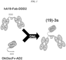

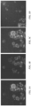
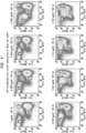
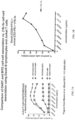
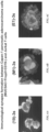


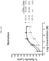
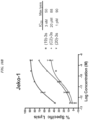
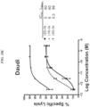
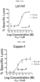


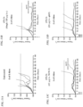
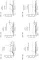

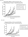


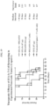
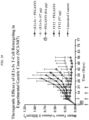


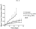
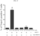

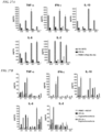
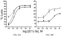
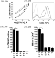
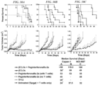
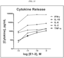


Classifications
-
- C—CHEMISTRY; METALLURGY
- C07—ORGANIC CHEMISTRY
- C07K—PEPTIDES
- C07K16/00—Immunoglobulins [IGs], e.g. monoclonal or polyclonal antibodies
- C07K16/18—Immunoglobulins [IGs], e.g. monoclonal or polyclonal antibodies against material from animals or humans
- C07K16/28—Immunoglobulins [IGs], e.g. monoclonal or polyclonal antibodies against material from animals or humans against receptors, cell surface antigens or cell surface determinants
- C07K16/30—Immunoglobulins [IGs], e.g. monoclonal or polyclonal antibodies against material from animals or humans against receptors, cell surface antigens or cell surface determinants from tumour cells
-
- A—HUMAN NECESSITIES
- A61—MEDICAL OR VETERINARY SCIENCE; HYGIENE
- A61P—SPECIFIC THERAPEUTIC ACTIVITY OF CHEMICAL COMPOUNDS OR MEDICINAL PREPARATIONS
- A61P1/00—Drugs for disorders of the alimentary tract or the digestive system
- A61P1/04—Drugs for disorders of the alimentary tract or the digestive system for ulcers, gastritis or reflux esophagitis, e.g. antacids, inhibitors of acid secretion, mucosal protectants
-
- A—HUMAN NECESSITIES
- A61—MEDICAL OR VETERINARY SCIENCE; HYGIENE
- A61P—SPECIFIC THERAPEUTIC ACTIVITY OF CHEMICAL COMPOUNDS OR MEDICINAL PREPARATIONS
- A61P1/00—Drugs for disorders of the alimentary tract or the digestive system
- A61P1/18—Drugs for disorders of the alimentary tract or the digestive system for pancreatic disorders, e.g. pancreatic enzymes
-
- A—HUMAN NECESSITIES
- A61—MEDICAL OR VETERINARY SCIENCE; HYGIENE
- A61P—SPECIFIC THERAPEUTIC ACTIVITY OF CHEMICAL COMPOUNDS OR MEDICINAL PREPARATIONS
- A61P11/00—Drugs for disorders of the respiratory system
-
- A—HUMAN NECESSITIES
- A61—MEDICAL OR VETERINARY SCIENCE; HYGIENE
- A61P—SPECIFIC THERAPEUTIC ACTIVITY OF CHEMICAL COMPOUNDS OR MEDICINAL PREPARATIONS
- A61P13/00—Drugs for disorders of the urinary system
- A61P13/08—Drugs for disorders of the urinary system of the prostate
-
- A—HUMAN NECESSITIES
- A61—MEDICAL OR VETERINARY SCIENCE; HYGIENE
- A61P—SPECIFIC THERAPEUTIC ACTIVITY OF CHEMICAL COMPOUNDS OR MEDICINAL PREPARATIONS
- A61P13/00—Drugs for disorders of the urinary system
- A61P13/10—Drugs for disorders of the urinary system of the bladder
-
- A—HUMAN NECESSITIES
- A61—MEDICAL OR VETERINARY SCIENCE; HYGIENE
- A61P—SPECIFIC THERAPEUTIC ACTIVITY OF CHEMICAL COMPOUNDS OR MEDICINAL PREPARATIONS
- A61P13/00—Drugs for disorders of the urinary system
- A61P13/12—Drugs for disorders of the urinary system of the kidneys
-
- A—HUMAN NECESSITIES
- A61—MEDICAL OR VETERINARY SCIENCE; HYGIENE
- A61P—SPECIFIC THERAPEUTIC ACTIVITY OF CHEMICAL COMPOUNDS OR MEDICINAL PREPARATIONS
- A61P15/00—Drugs for genital or sexual disorders; Contraceptives
-
- A—HUMAN NECESSITIES
- A61—MEDICAL OR VETERINARY SCIENCE; HYGIENE
- A61P—SPECIFIC THERAPEUTIC ACTIVITY OF CHEMICAL COMPOUNDS OR MEDICINAL PREPARATIONS
- A61P35/00—Antineoplastic agents
-
- A—HUMAN NECESSITIES
- A61—MEDICAL OR VETERINARY SCIENCE; HYGIENE
- A61P—SPECIFIC THERAPEUTIC ACTIVITY OF CHEMICAL COMPOUNDS OR MEDICINAL PREPARATIONS
- A61P37/00—Drugs for immunological or allergic disorders
- A61P37/02—Immunomodulators
- A61P37/04—Immunostimulants
-
- A—HUMAN NECESSITIES
- A61—MEDICAL OR VETERINARY SCIENCE; HYGIENE
- A61P—SPECIFIC THERAPEUTIC ACTIVITY OF CHEMICAL COMPOUNDS OR MEDICINAL PREPARATIONS
- A61P43/00—Drugs for specific purposes, not provided for in groups A61P1/00-A61P41/00
-
- C—CHEMISTRY; METALLURGY
- C07—ORGANIC CHEMISTRY
- C07K—PEPTIDES
- C07K16/00—Immunoglobulins [IGs], e.g. monoclonal or polyclonal antibodies
- C07K16/18—Immunoglobulins [IGs], e.g. monoclonal or polyclonal antibodies against material from animals or humans
- C07K16/28—Immunoglobulins [IGs], e.g. monoclonal or polyclonal antibodies against material from animals or humans against receptors, cell surface antigens or cell surface determinants
- C07K16/2803—Immunoglobulins [IGs], e.g. monoclonal or polyclonal antibodies against material from animals or humans against receptors, cell surface antigens or cell surface determinants against the immunoglobulin superfamily
- C07K16/2809—Immunoglobulins [IGs], e.g. monoclonal or polyclonal antibodies against material from animals or humans against receptors, cell surface antigens or cell surface determinants against the immunoglobulin superfamily against the T-cell receptor (TcR)-CD3 complex
-
- A—HUMAN NECESSITIES
- A61—MEDICAL OR VETERINARY SCIENCE; HYGIENE
- A61K—PREPARATIONS FOR MEDICAL, DENTAL OR TOILETRY PURPOSES
- A61K39/00—Medicinal preparations containing antigens or antibodies
- A61K2039/505—Medicinal preparations containing antigens or antibodies comprising antibodies
-
- C—CHEMISTRY; METALLURGY
- C07—ORGANIC CHEMISTRY
- C07K—PEPTIDES
- C07K2317/00—Immunoglobulins specific features
- C07K2317/30—Immunoglobulins specific features characterized by aspects of specificity or valency
- C07K2317/31—Immunoglobulins specific features characterized by aspects of specificity or valency multispecific
-
- C—CHEMISTRY; METALLURGY
- C07—ORGANIC CHEMISTRY
- C07K—PEPTIDES
- C07K2317/00—Immunoglobulins specific features
- C07K2317/50—Immunoglobulins specific features characterized by immunoglobulin fragments
- C07K2317/55—Fab or Fab'
-
- C—CHEMISTRY; METALLURGY
- C07—ORGANIC CHEMISTRY
- C07K—PEPTIDES
- C07K2317/00—Immunoglobulins specific features
- C07K2317/60—Immunoglobulins specific features characterized by non-natural combinations of immunoglobulin fragments
- C07K2317/62—Immunoglobulins specific features characterized by non-natural combinations of immunoglobulin fragments comprising only variable region components
- C07K2317/622—Single chain antibody (scFv)
-
- C—CHEMISTRY; METALLURGY
- C07—ORGANIC CHEMISTRY
- C07K—PEPTIDES
- C07K2317/00—Immunoglobulins specific features
- C07K2317/70—Immunoglobulins specific features characterized by effect upon binding to a cell or to an antigen
- C07K2317/73—Inducing cell death, e.g. apoptosis, necrosis or inhibition of cell proliferation
-
- C—CHEMISTRY; METALLURGY
- C07—ORGANIC CHEMISTRY
- C07K—PEPTIDES
- C07K2319/00—Fusion polypeptide
Definitions
- the present invention is related to a bispecific antibody for use in a method of treatment by inducing an immune response to a Trop-2 expressing cancer, wherein the method comprises administering the bispecific antibody and a checkpoint inhibitor antibody to a subject with a Trop-2 expressing cancer
- the anti-Trop-2 antibody is a hRS7 antibody.
- the bispecific antibody redirects effector T cells, monocytes, NK cells or neutrophils to target diseased cells or tissues and induces an immune response against the target.
- bispecific antibodies to redirect effector T cells for the targeted killing of tumor cells has shown considerable promise both pre-clinically and clinically (see, e.g., Topp et al., 2012, Blood 120:5185-87 ; Bargou et al., 2008, Science 321:974-77 ).
- the bispecific antibodies developed to date contain a first binding site specific to CD3 for T-cell recruitment and activation and a second binding site for a targeted disease-associated antigen, such as CD19 ( Bassan, 2012, Blood 120:5094-95 ).
- the bispecific antibody brings CD3 + T cells into direct contact with targeted disease cells and induces cell-mediated cytotoxicity (Bassan, 2012).
- Anti-CD3 X anti-CD19 bispecific antibodies have been reported to produce a complete and durable molecular remission at very low concentrations in approximately 70% of adult patients with MRD + ALL ( Topp et al., 2012, Blood 120:5185-87 ). Bispecific antibodies recognizing gliomas and the CD3 epitope on T cells have been successfully used in treating brain tumors in human patients ( Nitta, et al. Lancet 1990; 355:368-371 ).
- Leukocyte redirecting bsAbs are not limited to T cells.
- the bispecific killer engagers comprising scFvs against the NK cell antigen CD16 and a tumor-associated antigen (e.g., CD19, CD22, CD33) have also shown potent anti-cancer activity (e.g., Miller, Hematology Soc Hematol Educ Pogram 2013:247-53 ).
- Other alternatives include trispecific killer engagers (TriKEs), such as anti-CD 16 x anti-CD 19 x anti-CD22 (Miller, 2013; Gleason et al., 2012, Mol Cancer Ther 11:2674-84 ).
- An anti-CD16 x anti-CD33 BiKE was used to treat AML and myelodysplastic syndrome (Miller, 2013; Wiernik et al., 2013, Clin Cancer Res 19:3844-55 ).
- a CD16 x CD33 BiKE led to potent tumor cell killing and cytokine production by NK cells.
- Inhibition of ADAM17 enhanced the CD16 x CD33 BiKE response (Miller, 2013).
- Bispecific antibodies can be produced by the quadroma method, which involves the fusion of two different hybridomas, each producing a monoclonal antibody recognizing a different antigenic site ( Milstein and Cuello, Nature 1983; 305:537-540 ).
- the fused hybridomas are capable of synthesizing two different heavy chains and two different light chains, which can associate randomly to give a heterogeneous population of 10 different antibody structures of which only one of them, amounting to 1/8 of the total antibody molecules, will be bispecific, and therefore must be further purified from the other forms.
- Fused hybridomas are often less stable cytogenetically than the parent hybridomas, making the generation of a production cell line more problematic.
- bispecific antibodies Another method for producing bispecific antibodies uses heterobifunctional crosslinkers to chemically tether two different monoclonal antibodies, so that the resulting hybrid conjugate will bind to two different targets ( Staerz, et al. Nature 1985; 314:628-631 ; Perez, et al. Nature 1985; 316:354-356 ).
- Bispecific antibodies generated by this approach are essentially heteroconjugates of two IgG molecules, which diffuse slowly into tissues and are rapidly removed from the circulation.
- Bispecific antibodies can also be produced by reduction of each of two parental monoclonal antibodies to the respective half molecules, which are then mixed and allowed to reoxidize to obtain the hybrid structure ( Staerz and Bevan. Proc Natl Acad Sci USA 1986; 83:1453-1457 ).
- Discrete V H and V L domains of antibodies produced by recombinant DNA technology may pair with each other to form a dimer (recombinant Fv fragment) with binding capability ( U.S. Pat. No. 4,642,334 ).
- non-covalently associated molecules are not sufficiently stable under physiological conditions to have any practical use.
- Cognate V H and V L domains can be joined with a peptide linker of appropriate composition and length (usually consisting of more than 12 amino acid residues) to form a single-chain Fv (scFv) with binding activity.
- scFv single-chain Fv
- bispecific antibodies targeting CD3 and CD19 are in clinical development.
- DART ® Dual-Affinity Re-Targeting
- DART ® Dual-Affinity Re-Targeting
- BITE ® and DART ® exhibit fast blood clearance due to their small size ( ⁇ 55 kDa), which requires frequent administration to maintain therapeutic levels of the bispecific antibodies.
- Interferons are critical role players in the antitumor and antimicrobial host defense, and have been extensively explored as therapeutic agents for cancer and infectious disease ( Billiau et al., 2006, Cytokine Growth Factor Rev 17:381-409 ; Pestka et al., 2004, Immunol Rev 202:8-32 ).
- type I and II interferons IFN- ⁇ / ⁇ and ⁇
- their use in clinic settings have been limited because of the short circulation half-life, systemic toxicity, and suboptimal responses in patients ( Pestka et al., 2004, Immunol Rev 202:8-32 ; Miller et al., 2009, Ann N Y Acad Sci 1182:69-79 ).
- IFN- ⁇ 2 The therapeutic effectiveness of IFNs has been validated to date by the approval of IFN- ⁇ 2 for treating hairy cell leukemia, chronic myelogenous leukemia, malignant melanoma, follicular lymphoma, condylomata acuminata, AIDs-related Kaposi sarcoma, and chronic hepatitis B and C; IFN- ⁇ for treating multiple sclerosis; and IFN- ⁇ for treating chronic granulomatous disease and malignant osteopetrosis.
- IFN- ⁇ for treating hairy cell leukemia, chronic myelogenous leukemia, malignant melanoma, follicular lymphoma, condylomata acuminata, AIDs-related Kaposi sarcoma, and chronic hepatitis B and C
- IFN- ⁇ for treating multiple sclerosis
- IFN- ⁇ for treating chronic granulomatous disease and malignant osteopetrosis.
- ADCs Antibody-drug conjugates
- ADCs are a potent class of therapeutic constructs that allow targeted delivery of cytotoxic agents to target cells, such as cancer cells. Because of the targeting function, these compounds show a much higher therapeutic index compared to the same systemically delivered agents.
- ADCs have been developed as intact antibodies or antibody fragments, such as scFvs. The antibody or fragment is linked to one or more copies of drug via a linker that is stable under physiological conditions, but that may be cleaved once inside the target cell.
- ADCs approved for therapeutic use include gemtuzumab ozogamicin for AML (subsequently withdrawn from the market), brentuximab vedotin for ALCL and Hodgkin lymphoma, and trastuzumab emtansine for HER2-positive metastatic breast cancer ( Verma et al., 2012, N Engl J Med 367: 1783-91 ; Bross et al., 2001, Clin Cancer Res 7:1490-96 ; Francisco et al., 2003, Blood 102:1458-65 ).
- ADCs inotuzumab ozogamicin (Pfizer), glembatumomab vedotin (Celldex Therapeutics), SAR3419 (Sanofi-Aventis), SAR56658 (Sanofi-Aventis), AMG-172 (Amgen), AMG-595 (Amgen), BAY-94-9343 (Bayer), BIIB015 (Biogen Idec), BT062 (Biotest), SGN-75 (Seattle Genetics), SGN-CD19A (Seattle Genetics), vorsetuzumab mafodotin (Seattle Genetics), ABT-414 (AbbVie), ASG-SME (Agensys), ASG-22ME (Agensys), ASG-16M8F (Agensys), IMGN-529 (ImmunoGen), IMGN-853 (ImmunoGen), MDX-1203
- Pfizer inotuzuma
- Immune checkpoints function as endogenous inhibitory pathways for immune system function that act to maintain self-tolerance and to modulate the duration and extent of immune response to antigenic stimulation (Pardoll, 2012).
- tumor tissues and possibly certain pathogens may co-opt the checkpoint system to reduce the effectiveness of host immune response, resulting in tumor growth and/or chronic infection (see, e.g., Pardoll, 2012, Nature Reviews Cancer 12:252-64 ; Nirschl & Drake, 2013, Clin Cancer Res 19:4917-24 ).
- Checkpoint molecules include CTLA4 (cytotoxic T lymphocyte antigen-4), PD1 (programmed cell death protein 1), PD-L1 (programmed cell death ligand 1), LAG-3 (lymphocyte activation gene-3), TIM-3 (T cell immunoglobulin and mucin protein-3) and several others ( Pardoll, 2012, Nature Reviews Cancer 12:252-64 ; Nirschl & Drake, 2013, Clin Cancer Res 19:4917-24 ). Antibodies against several of the checkpoint proteins (CTLA4, PD1, PD-L1) are in clinical trials and has shown unexpected efficacy againts tumors that were resistant to standard treatments.
- CTLA4 cytotoxic T lymphocyte antigen-4
- PD1 programmed cell death protein 1
- PD-L1 programmeed cell death ligand 1
- LAG-3 lymphocyte activation gene-3
- TIM-3 T cell immunoglobulin and mucin protein-3
- WO 2014/163684 A1 and WO 2014/028560 are related to a combination therapy for inducing immune response to a disease.
- Suitable agents may include leukocyte redirecting complexes, antibody-drug conjugates, interferons and/or checkpoint inhibitor antibodies.
- a bispecific antibody for use in a method of treatment by inducing an immune response to a Trop-2 expressing cancer, wherein the method comprises administering the bispecific antibody and a checkpoint inhibitor antibody to a subject with a Trop-2 expressing cancer, wherein the bispecific antibody comprises a first antibody fragment and a second antibody fragment, wherein both the first antibody fragment and the second antibody fragment are each an scFv and, wherein the bispecific antibody comprises one binding site for Trop-2 and one binding site for CD3, and wherein the bispecific antibody comprises the amino acid sequence of SEQ ID NO: 107 binding to Trop-2.
- the method further comprises administering to the subject at least one therapeutic agent selected from the group consisting of (i) an interferon selected from the group consisting of interferon- ⁇ , interferon- ⁇ , interferon- ⁇ 1, interferon- ⁇ 2 and interferon- ⁇ 3; and (ii) an antibody-drug conjugate (ADC).
- at least one therapeutic agent selected from the group consisting of (i) an interferon selected from the group consisting of interferon- ⁇ , interferon- ⁇ , interferon- ⁇ 1, interferon- ⁇ 2 and interferon- ⁇ 3; and (ii) an antibody-drug conjugate (ADC).
- the interferon is interferon- ⁇ .
- the checkpoint inhibitor antibody is selected from the group consisting of lambrolizumab (MK-3475), nivolumab (BMS-936558), pidilizumab (CT-011), AMP-224, MDX-1105, MEDI4736, MPDL3280A, BMS-936559, ipilimumab, lirlumab, IPH2101 and tremelimumab.
- the checkpoint inhibitor antibody binds to an antigen selected from the group consisting of CTLA4, PD1, PD-L1, LAG3, B7-H3, B7-H4, KIR and TIM3.
- the antibody-drug conjugate is selected from the group consisting of hLL1-doxorubicin, hRS7-SN-38, hMN-14-SN-38, hLL2-SN-38, hA20-SN-38, hPAM4-SN-38, hLL1-SN-38, hRS7-Pro-2-P-Dox, hMN-14-Pro-2-P-Dox, hLL2-Pro-2-P-Dox, hA20-Pro-2-P-Dox, hPAM4-Pro-2-P-Dox, hLL1-Pro-2-P-Dox, P4/D10-doxorubicin, gemtuzumab ozogamicin, brentuximab vedotin, trastuzumab emtansine, inotuzumab ozogamicin, glembatumomab vedotin, SAR3419, SAR566658,
- an ADC is administered prior to any other agent.
- the interferon is administered as free interferon, PEGylated interferon, an interferon fusion protein or interferon conjugated to an antibody.
- the Trop-2 expressing cancer is a carcinoma of the esophagus, pancreas, lung, stomach, colon, rectum, urinary bladder, breast, ovary, uterus, kidney or prostate.
- the method further comprises administering to the subject a therapeutic agent selected from the group consisting of a second antibody or antigen-binding fragment thereof, a drug, a toxin, an enzyme, a cytotoxic agent, an anti-angiogenic agent, a pro-apoptotic agent, an antibiotic, a hormone, an immunomodulator, a cytokine, a chemokine, an antisense oligonucleotide, a small interfering RNA (siRNA), a boron compound and a radioisotope.
- a therapeutic agent selected from the group consisting of a second antibody or antigen-binding fragment thereof, a drug, a toxin, an enzyme, a cytotoxic agent, an anti-angiogenic agent, a pro-apoptotic agent, an antibiotic, a hormone, an immunomodulator, a cytokine, a chemokine, an antisense oligonucleotide, a small interfering RNA (siRNA),
- the bispecific antibody induces an immune response to a Trop-2 expressing cancer without increasing cytokine production to levels capable of inducing cytokine release syndrome (CRS).
- CRS cytokine release syndrome
- the present invention as defined in the claims relates to bispecific antibodies for use in the treatment of Trop-2 + cancer cells by inducing an immune response to said Trop-2 + cancer cells.
- Trop-2 is overexpressed in numerous types of solid tumors, such as carcinomas of the esophagus, pancreas, lung, stomach, colon and rectum, urinary bladder, breast, ovary, uterus, cervix, kidney and prostate.
- the bispecific antibody is of use to treat gastric cancer or pancreatic cancer.
- Administration of the bispecific antibody induces an immune response to cells that are Trop-2 + .
- Trop-2 is also expressed in some normal tissues (e.g., Stepan et al., 2011, J Histochem Cytochem 59:701-10 ), the Examples below demonstrate that anti-Trop-2 antibodies may be administered in vivo in both animal model systems and human subjects, with only tolerable toxicities.
- administration of bispecific antibody to a subject induces an immune response against Trop-2 + cancer cells without elevating levels of cytokines that would induce cytokine release syndrome (CRS).
- the bispecific antibody induces trogocytosis of cell surface antigens between Trop-2 + cancer cells and T cells.
- T-cell antigens are selected from the group consisting of CD2, CD3, CD4, CD5, CD6, CD8, CD25, CD28, CD30, CD40, CD40L, CD44, CD45, CD69 and CD90.
- antigens expressed on NK cells are selected from the group consisting of CD8, CD16, CD56, CD57, ADAM17, KIR and CD137.
- monocyte antigens are selected from the group consisting of CD74, HLA-DR alpha chain, CD14, CD16, CD64 and CD89.
- Exemplary neutrophil antigens are selected from the group consisting of CEACAM6, CEACAM8, CD16b, CD32a, CD89, CD177, CD11a, CD11b and SLC44A2.
- the T-cell antigen is CD3, or the NK cell antigen is CD16.
- Tumor-associated antigens that may be targeted include, but are not limited to, alpha-fetoprotein (AFP), ⁇ -actinin-4, A3, antigen specific for A33 antibody, ART-4, B7, Ba 733, BAGE, BrE3-antigen, CA125, CAMEL, CAP-1, carbonic anhydrase IX, CASP-8/m, CCCL19, CCCL21, CD1, CD1a, CD2, CD3, CD4, CD5, CD8, CD11A, CD14, CD15, CD16, CD18, CD19, CD20, CD21, CD22, CD23, CD25, CD29, CD30, CD32b, CD33, CD37, CD38, CD40, CD40L, CD44, CD45, CD46, CD52, CD54, CD55, CD59, CD64, CD66a-e, CD67, CD70, CD70L, CD74, CD79a, CD79b, CD80
- AFP alpha-fetoprotein
- ⁇ -actinin-4
- anti-TAA antibodies that may be used include, but are not limited to, hA19 (anti-CD19, U.S. Patent No. 7,109,304 ), hR1 (anti-IGF-1R, U.S. Patent Application Serial No. 12/722,645 , filed 3/12/10 ), hPAM4 (anti-MUC5ac, U.S. Patent No. 7,282,567 ), hA20 (anti-CD20, U.S. Patent No. 7,251,164 ), hIMMU31 (anti-AFP, U.S. Patent No. 7,300,655 ), hLL1 (anti-CD74, U.S. Patent No. 7,312,318 ), hLL2 (anti-CD22, U.S.
- Patent No. 7,074,403 hMu-9 (anti-CSAp, U.S. Patent No. 7,387,773 ), hL243 (anti-HLA-DR, U.S. Patent No. 7,612,180 ), hMN-14 (anti-CEACAM5, U.S. Patent No. 6,676,924 ), hMN-15 (anti-CEACAM6, U.S. Patent No. 7,541,440 ), hRS7 (anti-EGP-1, U.S. Patent No. 7,238,785 ), hMN-3 (anti-CEACAM6, U.S. Patent No. 7,541,440 ), Ab124 and Ab125 (anti-CXCR4, U.S. Patent No. 7,138,496 ).
- Alternative antibodies that may be used for treatment of various disease states include, but are not limited to, abciximab (anti-glycoprotein IIb/IIIa), alemtuzumab (anti-CD52), bevacizumab (anti-VEGF), cetuximab (anti-EGFR), gemtuzumab (anti-CD33), ibritumomab (anti-CD20), panitumumab (anti-EGFR), rituximab (anti-CD20), tositumomab (anti-CD20), trastuzumab (anti-ErbB2), lambrolizumab (anti-PD1 receptor), nivolumab (anti-PD1 receptor), ipilimumab (anti-CTLA4), abagovomab (anti-CA-125), adecatumumab (anti-EpCAM), atlizumab (anti-IL-6 receptor), benralizumab (anti-CD125), obinut
- Patent Application 11/983,372 deposited as ATCC PTA-4405 and PTA-4406), D2/B (anti-PSMA, WO 2009/130575 ), tocilizumab (anti-IL-6 receptor), basiliximab (anti-CD25), daclizumab (anti-CD25), efalizumab (anti-CD11a), GA101 (anti-CD20; Glycart Roche), atalizumab (anti- ⁇ 4 integrin), omalizumab (anti-IgE); anti-TNF- ⁇ antibodies such as CDP571 ( Ofei et al., 2011, Diabetes 45:881-85 ), MTNFAI, M2TNFAI, M3TNFAI, M3TNFABI, M302B, M303 (Thermo Scientific, Rockford, IL), infliximab (Centocor, Malvern, PA), certolizumab pegol (UCB, Brussels, Belgium), anti-CD40L (UCB, Brussels, Belgium
- An exemplary design for a leukocyte redirecting bsAb disclosed in the Examples below combined an anti-CD3 scFv with an anti-CD19 F(ab) 2 to form a construct designated (19)-3s, which specifically targeted B cells.
- Other bsAbs combining anti-CD3 with antibody fragments against other tumor-associated antigens, discussed in more detail below, are of use in targeted leukocyte immunotherapy of various solid tumors.
- the advantages of this design include bivalent binding to tumor cells, a larger size ( ⁇ 130 kDa) to preclude rapid renal clearance, and potent leukocyte mediated cytotoxicity.
- the bsAbs mediate the formation of immunological synapses between leukocytes and cognate target cells, induce leukocyte activation and proliferation in the presence of target cells, redirect potent leukocyte mediated killing of target cells in vitro and inhibit growth of human tumors in vivo.
- Leukocyte redirecting bispecific antibodies may be produced as trivalent DNL TM complexes, with longer T 1/2 , better pharmacokinetic properties and increased in vivo stability.
- Methods for production and use of DNL TM complexes, comprising dimers of DDD moieties from human PKA regulatory subunits RI ⁇ , RI ⁇ , RII ⁇ or RII ⁇ , bound to AD moieties from AKAPs, are well known (see, e.g., U.S. Patent Nos.
- DNL TM complexes comprising virtually any combination of effectors may be constructed and used.
- the antibodies can be of various isotypes, preferably human IgG1, IgG2, IgG3 or IgG4, more preferably comprising human IgG1 hinge and constant region sequences.
- the antibodies or fragments thereof can be chimeric human-mouse, humanized (human framework and murine hypervariable (CDR) regions), or fully human, as well as variations thereof, such as half-IgG4 antibodies (referred to as "unibodies”), as described by van der Neut Kolfschoten et al. (Science 2007; 317:1554-1557 ).
- the antibodies or fragments thereof may be designed or selected to comprise human constant region sequences that belong to specific allotypes, which may result in reduced immunogenicity when administered to a human subject.
- Preferred allotypes for administration include a non-G1m1 allotype (nG1m1), such as G1m3, G1m3,1, G1m3,2 or G1m3,1,2. More preferably, the allotype is selected from the group consisting of the nG1m1, G1m3, nG1m1,2 and Km3 allotypes.
- nG1m1 non-G1m1 allotype
- the method comprises administering a checkpoint inhibitor antibody.
- a checkpoint inhibitor antibody is antagonistic for checkpoint inhibitor function.
- Many such antibodies are known in the art, such as lambrolizumab (MK-3475, Merck), nivolumab (BMS-936558, Bristol-Myers Squibb), pidilizumab (CT-011, CureTech Ltd.), AMP-224 (Merck), MDX-1105 (Medarex), MEDI4736 (MedImmune), MPDL3280A (Genentech), BMS-936559 (Bristol-Myers Squibb), ipilimumab (Bristol-Myers Squibb) and tremelimumab (Pfizer).
- Anti-KIR antibodies such as lirlumab (Innate Pharma) and IPH2101 (Innate Pharma) may perform similar functions in NK cells. Any known checkpoint inhibitor antibody may be used in combination with one or more of the other agents. Combination therapy with immunostimulatory antibodies has been reported to enhance efficacy, for example against tumor cells. Morales-Kastresana et al. (2013, Clin Cancer Res 19:6151-62 ) showed that the combination of anti-PD-L1 (10B5) antibody with anti-CD137 (1D8) and anti-OX40 (OX86) antibodies provided enhanced efficacy in a transgenic mouse model of hepatocellular carcinoma.
- Combination of anti-CTLA4 and anti-PD1 antibodies has also been reported to be highly efficacious ( Wolchok et al., 2013, N Engl J Med 369:122-33 ).
- Combination of rituximab with anti-KIR antibody, such as lirlumab (Innate Pharma) or IPH2101 (Innate Pharma) was also more efficacious against hematopoietic tumors (Kohrt et al., 2012).
- the person of ordinary skill will realize that the subject combination therapy may include combinations with multiple antibodies that are immunostimulatory, anti-tumor or anti-infectious agent.
- Interferons of use are known in the art and may include interferon- ⁇ , interferon- ⁇ , interferon- ⁇ 1, interferon- ⁇ 2 or interferon- ⁇ 3.
- the interferon is interferon- ⁇ .
- the subject interferon may be administered as free interferon, PEGylated interferon, an interferon fusion protein or interferon conjugated to an antibody.
- one or more of the immunomodulatory agents discussed above may be used in combination with an antibody-drug conjugate (ADC).
- ADCs are particularly effective for reducing tumor burden without significant systemic toxicity and may act to improve the effectiveness of the immune response induced by leukocyte retargeting bsAb, interferon and/or checkpoint inhibitor antibody.
- Exemplary ADCs of use may include ADCs approved for therapeutic use include gemtuzumab ozogamicin for AML (subsequently withdrawn from the market), brentuximab vedotin for ALCL and Hodgkin lymphoma, and trastuzumab emtansine for HER2-positive metastatic breast cancer ( Verma et al., 2012, N Engl J Med 367:1783-91 ; Bross et al., 2001, Clin Cancer Res 7:1490-96 ; Francisco et al., 2003, Blood 102:1458-65 ).
- ADCs inotuzumab ozogamicin (Pfizer), glembatumomab vedotin (Celldex Therapeutics), SAR3419 (Sanofi-Aventis), SAR56658 (Sanofi-Aventis), AMG-172 (Amgen), AMG-595 (Amgen), BAY-94-9343 (Bayer), BIIB015 (Biogen Idec), BT062 (Biotest), SGN-75 (Seattle Genetics), SGN-CD19A (Seattle Genetics), vorsetuzumab mafodotin (Seattle Genetics), ABT-414 (AbbVie), ASG-5ME (Agensys), ASG-22ME (Agensys), ASG-16M8F (Agensys), IMGN-529 (ImmunoGen), IMGN-853 (ImmunoGen), MDX-1203 (Pfizer), glembatumom
- an ADC is used in combination with an immunomodulator
- the ADC is administered prior to the immunomodulator.
- Immunomodulators may include, but are not limited to, a cytokine, a chemokine, a stem cell growth factor, a lymphotoxin, an hematopoietic factor, a colony stimulating factor (CSF), erythropoietin, thrombopoietin, tumor necrosis factor- ⁇ (TNF), TNF- ⁇ , granulocyte-colony stimulating factor (G-CSF), granulocyte macrophage-colony stimulating factor (GM-CSF), interferon- ⁇ , interferon- ⁇ , interferon- ⁇ , interferon- ⁇ , interferon- ⁇ , stem cell growth factor designated "S1 factor", human growth hormone, N-methionyl human growth hormone, bovine growth hormone, parathyroid hormone, thyroxine, insulin, proinsulin, relaxin, prorelaxin, follicle stimulating hormone (FSH), thyroid stimulating hormone (TSH), lutein
- FSH follicle stimulating hormone
- TSH thyroid
- a leukocyte- redirecting bispecific antibody or antibody fragment may be attached to an immunomodulator, such as a cytokine.
- an immunomodulator such as a cytokine.
- Cytokine complexes are disclosed, for example, in U.S. Patent Nos. 7,906,118 and 8,034,3522 .
- a “therapeutic agent” is an atom, molecule, or compound that is useful in the treatment of a disease.
- therapeutic agents include antibodies, antibody fragments, peptides, drugs, toxins, enzymes, nucleases, hormones, immunomodulators, antisense oligonucleotides, small interfering RNA (siRNA), chelators, boron compounds, photoactive agents, dyes, and radioisotopes.
- an “antibody” as used herein refers to a full-length (i.e., naturally occurring or formed by normal immunoglobulin gene fragment recombinatorial processes) immunoglobulin molecule (e.g., an IgG antibody) or an immunologically active (i.e., specifically binding) portion of an immunoglobulin molecule, like an antibody fragment.
- An “antibody” includes monoclonal, polyclonal, bispecific, multispecific, murine, chimeric, humanized and human antibodies.
- naked antibody is an antibody or antigen binding fragment thereof that is not attached to a therapeutic or diagnostic agent.
- the Fc portion of an intact naked antibody can provide effector functions, such as complement fixation and ADCC (see, e.g. , Markrides, Pharmacol Rev 50:59-87, 1998 ).
- Other mechanisms by which naked antibodies induce cell death may include apoptosis. ( Vaswani and Hamilton, Ann Allergy Asthma Immunol 81: 105-119, 1998 .)
- antibody fragment is a portion of an intact antibody such as F(ab') 2 , F(ab) 2 , Fab', Fab, Fv, scFv, dAb and the like. Regardless of structure, an antibody fragment binds with the same antigen that is recognized by the full-length antibody.
- antibody fragments include isolated fragments consisting of the variable regions, such as the "Fv” fragments consisting of the variable regions of the heavy and light chains or recombinant single chain polypeptide molecules in which light and heavy variable regions are connected by a peptide linker ("scFv proteins").
- Single-chain antibodies often abbreviated as “scFv” consist of a polypeptide chain that comprises both a V H and a V L domain which interact to form an antigen- binding site.
- the V H and V L domains are usually linked by a peptide of 1 to 25 amino acid residues.
- Antibody fragments also include diabodies, triabodies and single domain antibodies (dAb).
- a “chimeric antibody” is a recombinant protein that contains the variable domains including the complementarity determining regions (CDRs) of an antibody derived from one species, preferably a rodent antibody, while the constant domains of the antibody molecule are derived from those of a human antibody.
- the constant domains of the chimeric antibody may be derived from that of other species, such as a cat or dog.
- a “humanized antibody” is a recombinant protein in which the CDRs from an antibody from one species; e.g., a rodent antibody, are transferred from the heavy and light variable chains of the rodent antibody into human heavy and light variable domains, including human framework region (FR) sequences.
- the constant domains of the antibody molecule are derived from those of a human antibody.
- FR amino acid residues from the parent (e.g., murine) antibody may be substituted for the corresponding human FR residues.
- a "human antibody” is an antibody obtained from transgenic mice that have been genetically engineered to produce specific human antibodies in response to antigenic challenge.
- elements of the human heavy and light chain locus are introduced into strains of mice derived from embryonic stem cell lines that contain targeted disruptions of the endogenous heavy chain and light chain loci.
- the transgenic mice can synthesize human antibodies specific for human antigens, and the mice can be used to produce human antibody-secreting hybridomas.
- Methods for obtaining human antibodies from transgenic mice are described by Green et al., Nature Genet. 7:13 (1994 ), Lonberg et al., Nature 368:856 (1994 ), and Taylor et al., Int. Immun. 6:579 (1994 ).
- a human antibody also can be constructed by genetic or chromosomal transfection methods, as well as phage display technology, all of which are known in the art. (See, e.g., McCafferty et al., 1990, Nature 348:552-553 for the production of human antibodies and fragments thereof in vitro, from immunoglobulin variable domain gene repertoires from unimmunized donors).
- antibody variable domain genes are cloned in-frame into either a major or minor coat protein gene of a filamentous bacteriophage, and displayed as functional antibody fragments on the surface of the phage particle.
- the filamentous particle contains a single-stranded DNA copy of the phage genome, selections based on the functional properties of the antibody also result in selection of the gene encoding the antibody exhibiting those properties. In this way, the phage mimics some of the properties of the B cell.
- Phage display can be performed in a variety of formats, for their review, see, e.g. Johnson and Chiswell, Current Opinion in Structural Biology 3:5564-571 (1993 ).
- Human antibodies may also be generated by in vitro activated B cells. (See, U.S. Pat. Nos. 5,567,610 and 5,229,275 ).
- antibody fusion protein is a recombinantly produced antigen-binding molecule in which an antibody or antibody fragment is linked to another protein or peptide, such as the same or different antibody or antibody fragment or a DDD or AD peptide.
- the fusion protein may comprise a single antibody component, a multivalent or multispecific combination of different antibody components or multiple copies of the same antibody component.
- the fusion protein may additionally comprise an antibody or an antibody fragment and a therapeutic agent. Examples of therapeutic agents suitable for such fusion proteins include immunomodulators and toxins.
- One preferred toxin comprises a ribonuclease (RNase), preferably a recombinant RNase.
- a preferred immunomodulator might be an interferon, such as interferon- ⁇ , interferon- ⁇ or interferon- ⁇ .
- a “multispecific antibody” is an antibody that can bind simultaneously to at least two targets that are of different structure, e.g., two different antigens, two different epitopes on the same antigen, or a hapten and/or an antigen or epitope.
- a “multivalent antibody” is an antibody that can bind simultaneously to at least two targets that are of the same or different structure. Valency indicates how many binding arms or sites the antibody has to a single antigen or epitope; i.e., monovalent, bivalent, trivalent or multivalent. The multivalency of the antibody means that it can take advantage of multiple interactions in binding to an antigen, thus increasing the avidity of binding to the antigen.
- Specificity indicates how many antigens or epitopes an antibody is able to bind; i.e., monospecific, bispecific, trispecific, multispecific.
- a natural antibody e.g., an IgG
- Multispecific, multivalent antibodies are constructs that have more than one binding site of different specificity.
- bispecific antibody is an antibody that can bind simultaneously to two targets which are of different structure.
- Bispecific antibodies bsAb and bispecific antibody fragments (bsFab) may have at least one arm that specifically binds to, for example, a T cell, an NK cell, a monocyte or a neutrophil, and at least one other arm that specifically binds to an antigen produced by or associated with a diseased cell, tissue, organ or pathogen, for example a tumor-associated antigen.
- bsAb bispecific antibody fragments
- bsFab bispecific antibody fragments
- a variety of bispecific antibodies can be produced using molecular engineering.
- An antibody preparation, or a composition described herein, is said to be administered in a "therapeutically effective amount" if the amount administered is physiologically significant.
- An agent is physiologically significant if its presence results in a detectable change in the physiology of a recipient subject.
- an antibody preparation is physiologically significant if its presence invokes an antitumor response or mitigates the signs and symptoms of an infectious disease state.
- a physiologically significant effect could also be the evocation of a humoral and/or cellular immune response in the recipient subject leading to growth inhibition or death of target cells.
- the anti-Trop-2 antibody may is a humanized RS7 antibody (see, e.g., U.S. Patent No. 7,238,785 ), comprising the light chain CDR sequences CDR1 (KASQDVSIAVA, SEQ ID NO:115); CDR2 (SASYRYT, SEQ ID NO:116); and CDR3 (QQHYITPLT, SEQ ID NO:117) and the heavy chain CDR sequences CDR1 (NYGMN, SEQ ID NO:118); CDR2 (WINTYTGEPTYTDDFKG, SEQ ID NO:119) and CDR3 (GGFGSSYWYFDV, SEQ ID NO:120).
- RS7 antibody see, e.g., U.S. Patent No. 7,238,785 , comprising the light chain CDR sequences CDR1 (KASQDVSIAVA, SEQ ID NO:115); CDR2 (SASYRYT, SEQ ID NO:116); and CDR3 (QQHYITPLT, SEQ ID
- the RS7 antibody was a murine IgG 1 raised against a crude membrane preparation of a human primary squamous cell lung carcinoma. ( Stein et al., Cancer Res. 50: 1330, 1990 ) The RS7 antibody recognizes a 46-48 kDa glycoprotein, characterized as cluster 13 ( Stein et al., Int. J. Cancer Supp. 8:98-102, 1994 ). The antigen was designated as EGP-1 (epithelial glycoprotein-1), but is also referred to as Trop-2.
- Trop-2 is a type-I transmembrane protein and has been cloned from both human ( Fornaro et al., Int J Cancer 1995; 62:610-8 ) and mouse cells ( Sewedy et al., Int J Cancer 1998; 75:324-30 ).
- human Trop-2 In addition to its role as a tumor-associated calcium signal transducer ( Ripani et al., Int J Cancer 1998;76:671-6 ), the expression of human Trop-2 was shown to be necessary for tumorigenesis and invasiveness of colon cancer cells, which could be effectively reduced with a polyclonal antibody against the extracellular domain of Trop-2 ( Wang et al., Mol Cancer Ther 2008;7:280-5 ).
- RS7 MAb detects antigen on a variety of tumor types, with limited binding to normal human tissue (Stein et al., 1990).
- Trop-2 is expressed primarily by carcinomas such as carcinomas of the lung, stomach, urinary bladder, breast, ovary, uterus, and prostate.
- Localization and therapy studies using radiolabeled murine RS7 MAb in animal models have demonstrated tumor targeting and therapeutic efficacy (Stein et al., 1990; Stein et al., 1991).
- the RS7 MAb is rapidly internalized into target cells (Stein et al., 1993).
- the internalization rate constant for RS7 MAb is intermediate between the internalization rate constants of two other rapidly internalizing MAbs, which have been demonstrated to be useful for immunotoxin production.
- Id. It is well documented that internalization of immunotoxin conjugates is a requirement for anti-tumor activity.
- Pastan et al., Cell 47:641, 1986 Internalization of drug immunoconjugates has been described as a major factor in anti-tumor efficacy ( Yang et al., Proc. Nat'l Acad. Sci. USA 85: 1189, 1988 ).
- the RS7 antibody exhibits several important properties for therapeutic applications.
- hRS7 antibody is preferred
- other anti-Trop-2 antibodies are known and/or publicly available and in alternative embodiments may be utilized in the subject ADCs.
- humanized or human antibodies are preferred for reduced immunogenicity, in alternative embodiments a chimeric antibody may be of use.
- methods of antibody humanization are well known in the art and may be utilized to convert an available murine or chimeric antibody into a humanized form.
- Anti-Trop-2 antibodies are commercially available from a number of sources and include LS-C126418, LS-C178765, LS-C126416, LS-C126417 (LifeSpan BioSciences, Inc., Seattle, WA); 10428-MM01, 10428-MM02, 10428-R001, 10428-R030 (Sino Biological Inc., Beijing, China); MR54 (eBioscience, San Diego, CA); sc-376181, sc-376746, Santa Cruz Biotechnology (Santa Cruz, CA); MM0588-49D6, (Novus Biologicals, Littleton, CO); ab79976, and ab89928 (ABCAM ® , Cambridge, MA).
- anti-Trop-2 antibodies have been disclosed in the patent literature.
- U.S. Publ. No. 2013/0089872 discloses anti-Trop-2 antibodies K5-70 (Accession No. FERM BP-11251), K5-107 (Accession No. FERM BP-11252), K5-116-2-1 (Accession No. FERM BP-11253), T6-16 (Accession No. FERM BP-11346), and T5-86 (Accession No. FERM BP-11254), deposited with the International Patent Organism Depositary, Tsukuba, Japan.
- U.S. Patent No. 5,840,854 disclosed the anti-Trop-2 monoclonal antibody BR110 (ATCC No. HB11698).
- U.S. Patent No. 7,420,040 disclosed an anti-Trop-2 antibody produced by hybridoma cell line AR47A6.4.2, deposited with the IDAC (International Depository Authority of Canada, Winnipeg, Canada) as accession number 141205-05.
- U.S. Patent No. 7,420,041 disclosed an anti-Trop-2 antibody produced by hybridoma cell line AR52A301.5, deposited with the IDAC as accession number 141205-03.
- U.S. Publ. No. 2013/0122020 disclosed anti-Trop-2 antibodies 3E9, 6G11, 7E6, 15E2, 18B1. Hybridomas encoding a representative antibody were deposited with the American Type Culture Collection (ATCC), Accession Nos. PTA-12871 and PTA-12872.
- Patent No. 8,715,662 discloses anti-Trop-2 antibodies produced by hybridomas deposited at the AID-ICLC (Genoa, Italy) with deposit numbers PD 08019, PD 08020 and PD 08021.
- U.S. Patent Application Publ. No. 20120237518 discloses anti-Trop-2 antibodies 77220, KM4097 and KM4590.
- U.S. Patent No. 8,309,094 discloses antibodies A1 and A3, identified by sequence listing.
- Non-patent publication Lipinski et al. (1981, Proc Natl. Acad Sci USA, 78:5147-50 ) disclosed anti-Trop-2 antibodies 162-25.3 and 162-46.2.
- anti-Trop-2 antibodies are known in the art and/or publicly available. As discussed below, methods for preparing antibodies against known antigens were routine in the art. The sequence of the human Trop-2 protein was also known in the art (see, e.g., GenBank Accession No. CAA54801.1). Methods for producing humanized, human or chimeric antibodies were also known. The person of ordinary skill, reading the instant disclosure in light of general knowledge in the art, would have been able to make and use the genus of anti-Trop-2 antibodies in the subject ADCs.
- a variety of antibodies against CD3 that may be used in the practing of the invention as defined in the claims are publicly known and/or commercially available, such as from LSBio (catalog Nos. LS-B6698, LS-B8669; LS-B8765, LS-C96311, LS-C58677, etc.); ABCAM ® (catalog Nos. ab5690, ab16669, ab699, ab828, ab8671, etc.); Santa Cruz Biotechnology (catalog Nos.sc-20047, sc-20080, sc-19590, sc-59008, sc-101442, etc.); and many other suppliers.
- LSBio catalog Nos. LS-B6698, LS-B8669; LS-B8765, LS-C96311, LS-C58677, etc.
- ABCAM ® catalog Nos. ab5690, ab16669, ab699, ab828, ab8671, etc.
- Santa Cruz Biotechnology
- the amino acid sequence of the anti-CD3 moiety used as part of a DNL TM complex, is as disclosed below in SEQ ID NO:96 to SEQ ID NO:101.
- the person of ordinary skill will realize that any known anti-CD3 antibody may be utilized in the in the practing of the invention as defined in the coaims.
- the antibody moieties of use are humanized or human.
- the bispecific antibody comprises an anti-CD3 x anti-Trop-2 antibody.
- various antibodies against CD3 or Trop-2 are known in the art and any such known antibody may be utilized.
- antibodies against other leukocyte antigens than CD3 or against other disease-associated antigens than Trop-2 may be utilized.
- T-cell antigens include CD2, CD4, CD5, CD6, CD8, CD25, CD28, CD30, CD40, CD40L, CD44, CD45, CD69 and CD90.
- Other exemplary antigens may be selected from CD8, CD16, CD56, CD57, ADAM17, and CD137 for NK cells; CD74, HLA-DR alpha chain, CD14, CD16, CD64 and CD89 for monocytes; and CEACAM6, CEACAM8, CD16b, CD32a, CD89, CD177, CD11a, CD11b and SLC44A2 for neutrophils.
- the anti-T-cell antibody binds to CD3, or the anti-NK antibody binds to CD16.
- TAAs tumor-associated antigens
- An exemplary preferred TAA is Trop-2.
- the instant disclosure concerns an anti-CD3 x anti-CD19 bispecific antibody.
- Various bispecific anti-CD3 x anti-CD19 antibodies are known in the art and presently in clinical development, such as BITE ® (Bispecific T-cell Engager) (e.g., Nagorsen et al., 2009, Leukemia & Lymphoma 50:886-91 ; Amann et al., 2009, J Immunother 32:453-64 ; Baeuerle and Reinhardt, 2009, Cancer Res 69:4941-44 ) and DART ® (see, e.g., Moore et al., 2011, Blood 117:4542-51 ; Veri et al., 2010, Arthritis Rheum 62:1933-43 ).
- Blinatumomab is a BITE ® antibody comprising V H and V L domains of anti-CD3 and anti-CD19 antibody fragments, connected with a 5-amino acid linker and expressed as a single polypeptide chain that anneals to itself to form the antigen-binding sites. It is thought that blinatumomab acts by bringing the T-cell-specific CD3 and B-cell specific CD19 antigens into close proximity, to initiate a T-cell cytotoxic response against the juxtaposed B cells, which does not require T-cell specificity to the cancer cells (e.g., Portell et al., 2013, Clin Pharmacol 5(Suppl 1): 5-11 ).
- blinatumomab induced 50% cell lysis of MEC-1 cells at a concentration of 10 pg/mL ( Topp et al., 2012, Blood 120:5185-87 ; Bassan et al., 2012, Blood 120:5094-95 ).
- the anti-CD19 portion of blinatumomab was derived from the HD37 hybridoma (see, e.g., U.S. Patent No. 7,575,923 ), which is publicly available (e.g., Santa Cruz Biotechnology Cat. No. sc-18894).
- the anti-CD3 portion of blinatumomab was derived from the TR66 hybridoma ( U.S. Patent No. 7,575,923 ; Traunecker et al., 1991, EMBO J. 10:3655-59 ), also publicly available (e.g., Enzo Life Sciences, catalog No. ALX-804-822-C100).
- a variety of antibodies against CD19 are publicly known and/or commercially available, such as from Santa Cruz Biotechnology (catalog Nos. sc-390244, sc-373897, sc-18894, sc-18896, etc.); ABCAM ® (catalog Nos. ab25232, ab134114, ab140981, ab1255, etc.); ABBIOTEC TM (catalog Nos. 252262, 252248, 250585, 251063, etc.) and many other vendors.
- the anti-CD19 antibody moiety is a humanized A19 antibody, comprising the light chain CDR sequences CDR1 KASQSVDYDGDSYLN (SEQ ID NO:90); CDR2 DASNLVS (SEQ ID NO:91); and CDR3 QQSTEDPWT (SEQ ID NO:92) and the heavy chain CDR sequences CDR1 SYWMN (SEQ ID NO:93); CDR2 QIWPGDGDTNYNGKFKG (SEQ ID NO:94) and CDR3 RETTTVGRYYYAMDY (SEQ ID NO:95).
- DART ® anti-CD3 x anti-CD19 bispecific antibodies
- DART ® which also incorporates the anti-CD19 Fv sequences of HD37 and the anti-CD3 Fv sequences of TR66
- DART ® bispecific antibodies were more potent at inducing B cell lysis than single-chain, bispecific antibodies (BITE ® ) bearing identical anti-CD19 and anti-CD3 variable region sequences, with EC 50 values in the pg/mL range (Moore et al., 2011).
- any known anti-CD3 x anti-CD19 bispecific antibody may be used to induce an immune response against disease-associated cells.
- Catumaxomab is an anti-CD3 x anti-EpCAM bispecific antibody that has been approved in Europe for treatment of malignant ascites associated with metastasizing cancer ( Chames & Baty, 2009, MAbs 1:539-47 ).
- catumaxomab was able to kill tumor cells at a concentration range of 10 pM and was reported to lead to total eradication of melanoma tumors (Chames & Baty, 2009).
- Human clinical trials with ovarian cancer patients with malignant ascites also showed a statistically significant efficacy (Chames & Baty, 2009).
- the high immunogenicity of the rat/mouse hybrid bsAb may limit i.v.
- anti-tumor bsAbs is not limited to anti-CD3 x anti-CD19, but has also included anti-HER2 x anti-CD64 (MDX-210, MDX-H210), anti-EGFR x anti-CD64 (MDX-447), anti-CD30 x anti-CD16 (HRS-3/A9), anti-HER2 x anti-CD3 (Her2Bi), anti-CD20 x anti-CD3 (CD20Bi, Bi20), anti-EpCAM x anti-CD3 (catumaxomab, MT110), anti-HER2 x anti-CD3 (ertumaxomab), and anti-NG2 x anti-CD28 (rM28) (Chames & Baty, 2009).
- anti-HER2 x anti-CD64 MDX-210, MDX-H210
- anti-EGFR x anti-CD64 MDX-447
- anti-CD30 x anti-CD16 HRS-3/A9
- leukocyte redirecting bispecific antibodies are not limited to anti-CD3 x anti-Trop-2 constructs, but may comprise antibodies against any known disease-associated antigens attached to an anti-CD3 antibody moiety.
- antibodies against other T-cell antigens besides CD3, or other antigens expressed on NK cells, monocytes or neutrophils may also be used.
- Exemplary T-cell antigens include, but are not limited to, CD2, CD4, CD5, CD6, CD8, CD25, CD28, CD30, CD40, CD40L, CD44, CD45, CD69 and CD90.
- exemplary antigens may be selected from CD8, CD16, CD56, CD57, ADAM17, KIR and CD137 for NK cells; CD74, HLA-DR alpha chain, CD14, CD16, CD64 and CD89 for monocytes; and CEACAM6, CEACAM8, CD16b, CD32a, CD89, CD177, CD11a, CD11b and SLC44A2 for neutrophils.
- Antibodies against each of the leukocyte antigens are publicly known and/or publicly available (see, e.g., ABCAM ® catalog numbers ab131276, ab139266, ab8360, ab51312, ab846, ab133616, ab75877, ab133255, ab109217, ab93278, ab17147, ab115851, ab128955, ab13463, ab85986; Santa Cruz Biotechnology catalog numbers sc-46683, sc-59047; Enzo Life Sciences, Inc. catalog number ALX-805-037-C100; Sino Biological Inc. catalog numbers 12211-RP02, 11150-R074; Millipore catalog numbers 04-1102, 04-1102, MAB1406).
- anti-leukocyte antibodies were publicly available and could have been used in the subject leukocyte redirecting bsAbs. As discussed below, numerous antibodies against a wide variety of disease-associated antigens were publicly known and/or commercially available and could have been used in the subject leukocyte redirecting bispecific antibodies.
- Other exemplary leukocyte redirecting bsAbs of potential use include FBTA05 (anti-CD20 x anti-CD3) and TRBS07 (anti-GD2 x anti-CD3).
- the subject bispecific antibodies may be used in combination with one or more interferons, such as interferon- ⁇ , interferon- ⁇ or interferon- ⁇ , preferably interferon- ⁇ .
- interferons are well known in the art and the amino acid sequences of human interferons may be readily obtained from public databases (e.g., GenBank Accession Nos. AAA52716.1; AAA52724; AAC41702.1; EAW56871.1; EAW56870.1; EAW56869.1).
- Human interferons may also be commercially obtained from a variety of vendors (e.g., Cell Signaling Technology, Inc., Danvers, MA; Genentech, South San Francisco, CA; EMD Millipore, Billerica, MA).
- Interferon- ⁇ has been reported to have anti-tumor activity in animal models of cancer ( Ferrantini et al., 1994, J Immunol 153:4604-15 ) and human cancer patients ( Gutterman et al., 1980, Ann Intern Med 93:399-406 ).
- IFN ⁇ can exert a variety of direct anti-tumor effects, including down-regulation of oncogenes, up-regulation of tumor suppressors, enhancement of immune recognition via increased expression of tumor surface MHC class I proteins, potentiation of apoptosis, and sensitization to chemotherapeutic agents ( Gutterman et al., 1994, PNAS USA 91:1198-205 ; Matarrese et al., 2002, Am J Pathol 160:1507-20 ; Mecchia et al., 2000, Gene Ther 7:167-79 ; Sabaawy et al., 1999, Int J Oncol 14:1143-51 ; Takaoka et al, 2003, Nature 424:516-23 ).
- IFN ⁇ can have a direct and potent anti-proliferative effect through activation of STAT1 ( Grimley et al., 1998 Blood 91:3017-27 ).
- Interferon- ⁇ 2b has been conjugated to anti-tumor antibodies, such as the hL243 anti-HLA-DR antibody and depletes lymphoma and myeloma cells in vitro and in vivo ( Rossi et al., 2011, Blood 118:1877-84 ).
- IFN ⁇ can inhibit angiogenesis ( Sidky and Borden, 1987, Cancer Res 47:5155-61 ) and stimulate host immune cells, which may be vital to the overall antitumor response but has been largely under-appreciated ( Belardelli et al., 1996, Immunol Today 17:369-72 ).
- IFN ⁇ has a pleiotropic influence on immune responses through effects on myeloid cells ( Raefsky et al, 1985, J Immunol 135:2507-12 ; Luft et al, 1998, J Immunol 161:1947-53 ), T-cells ( Carrero et al, 2006, J Exp Med 203:933-40 ; Pilling et al., 1999, Eur J Immunol 29:1041-50 ), and B-cells ( Le et al, 2001, Immunity 14:461-70 ).
- IFN ⁇ induces the rapid differentiation and activation of dendritic cells ( Belardelli et al, 2004, Cancer Res 64:6827-30 ; Paquette et al., 1998, J Leukoc Biol 64:358-67 ; Santini et al., 2000, J Exp Med 191:1777-88 ) and enhances the cytotoxicity, migration, cytokine production and antibody-dependent cellular cytotoxicity (ADCC) of NK cells ( Biron et al., 1999, Ann Rev Immunol 17:189-220 ; Brunda et al. 1984, Cancer Res 44:597-601 ).
- Interferon- ⁇ has been reported to be efficacious for therapy of a variety of solid tumors. Patients treated with 6 million units of IFN- ⁇ twice a week for 36 months showed a decreased recurrence of hepatocellular carcinoma after complete resection or ablation of the primary tumor in patients with HCV-related liver cancer ( Ikeda et al., 2000, Hepatology 32:228-32 ). Gene therapy with interferon- ⁇ induced apoptosis of glioma, melanoma and renal cell carcinoma ( Yoshida et al., 2004, Cancer Sci 95:858-65 ). Endogenous IFN- ⁇ has been observed to inhibit tumor growth by inhibiting angiogenesis in vivo ( Jablonska et al., 2010, J Clin Invest. 120:1151-64 .)
- IFN-ks designated as type III interferons
- type III interferons are a newly described group of cytokines that consist of IFN- ⁇ 1, 2, 3 (also referred to as interleukin-29, 28A, and 28B, respectively), that are genetically encoded by three different genes located on chromosome 19 ( Kotenko et al., 2003, Nat Immunol 4:69-77 ; Sheppard et al., 2003, Nat Immunol 4:63-8 ).
- IFN- ⁇ 2 and - ⁇ 3 are is highly homologous, with 96% amino acid identity, while IFN- ⁇ 1 shares approximately 81% homology with IFN- ⁇ 2 and - ⁇ 3 ( Sheppard et al., 2003, Nat Immunol 4:63-8 ).
- IFN- ⁇ s activate signal transduction via the JAK/STAT pathway similar to that induced by type I IFN, including the activation of JAK1 and TYK2 kinases, the phosphorylation of STAT proteins, and the activation of the transcription complex of IFN-stimulated gene factor 3 (ISGF3) ( Witte et al., 2010, Cytokine Growth Factor Rev 21:237-51 ; Zhou et al., 2007, J Virol 81:7749-58 ).
- IFN-stimulated gene factor 3 ISGF3
- IFN- ⁇ / ⁇ signals through two extensively expressed type I interferon receptors, and the resulting systemic toxicity associated with IFN- ⁇ / ⁇ administration has limited their use as therapeutic agents ( Pestka et al., 2007, J Biol Chem 282:20047-51 ).
- IFN- ⁇ s signal through a heterodimeric receptor complex consisting of unique IFN- ⁇ receptor 1 (IFN- ⁇ R1) and IL-10 receptor 2 (IL-10R2).
- IFN- ⁇ R1 has a very restricted expression pattern with the highest levels in epithelial cells, melanocytes, and hepatocytes, and the lowest level in primary central nervous system (CNS) cells.
- Blood immune system cells express high levels of a short IFN- ⁇ receptor splice variant (sIFN- ⁇ R1) that inhibits IFN- ⁇ action.
- sIFN- ⁇ R1 short IFN- ⁇ receptor splice variant
- IFN- ⁇ may be superior to IFN- ⁇ for treatment of chronic HCV infection, as it is less likely to induce leukopenias that are often associated with IFN- ⁇ therapy (Dickensheets et al., 2013).
- IFN- ⁇ s display structural features similar to IL-10-related cytokines, but functionally possess type I IFN-like anti-viral and anti-proliferative activity ( Witte et al., 2009, Genes Immun 10:702-14 ; Ank et al., 2006, J Virol 80:4501-9 ; Robek et al., 2005, J Virol 79:3851-4 ).
- IFN- ⁇ 1 and - ⁇ 2 have been demonstrated to reduce viral replication or the cytopathic effect of various viruses, including DNA viruses (hepatitis B virus ( Robek et al., 2005, J Virol 79:3851-4 , Doyle et al., 2006, Hepatology 44:896-906 ) and herpes simplex virus 2 ( Ank et al., 2008, J Immunol 180:2474-85 )), ss (+) RNA viruses (EMCV; Sheppard et al., 2003, Nat Immunol 4:63-8 ) and hepatitis C virus ( Robek et al., 2005, J Virol 79:3851-4 , Doyle et al., 2006, Hepatology 44:896-906 ; Marcello et al., 2006, Gastroenterol 131:1887-98 ; Pagliaccetti et al., 2008, J Biol Chem 283:30079-89 ), ss (-) RNA viruses
- IFN- ⁇ 3 has been identified from genetic studies as a key cytokine in HCV infection ( Ge et al., 2009, Nature 461:399-401 ), and has also shown potent activity against EMCV ( Dellgren et al., 2009, Genes Immun 10:125-31 ).
- IFN- ⁇ s The anti-proliferative activity of IFN- ⁇ s has been established in several human cancer cell lines, including neuroendocrine carcinoma BON1 ( Zitzmann et al., 2006, Biochem Biophys Res Commun 344:1334-41 ), glioblastoma LN319 ( Meager et al., 2005, Cytokine 31:109-18 ), immortalized keratinocyte HaCaT ( Maher et al., 2008, Cancer Biol Ther 7:1109-15 ), melanoma F01 ( Guenterberg et al., 2010, Mol Cancer Ther 9:510-20 ), and esophageal carcinoma TE-11 ( Li et al., 2010, Eur J Cancer 46:180-90 ).
- neuroendocrine carcinoma BON1 Zitzmann et al., 2006, Biochem Biophys Res Commun 344:1334-41
- glioblastoma LN319 Meager et al., 2005,
- IFN- ⁇ s induce both tumor apoptosis and destruction through innate and adaptive immune responses, suggesting that local delivery of IFN- ⁇ might be a useful adjunctive strategy in the treatment of human malignancies ( Numasaki et al., 2007, J Immunol 178:5086-98 ).
- a Fab-linked interferon- ⁇ was demonstrated to have potent anti-tumor and anti-viral activity in targeted cells ( Liu et al., 2013, PLoS One 8:e63940 ).
- PEGylated IFN- ⁇ 1 (PEG-IFN- ⁇ 1) has been provisionally used for patients with chronic hepatitis C virus infection.
- PEG-IFN- ⁇ 1 has been provisionally used for patients with chronic hepatitis C virus infection.
- the subject leukocyte redirecting bispecific antibodies, ADCs and/or checkpoint inhibitor mAbs may be used in combination with one or more interferons, such as interferon- ⁇ , interferon- ⁇ , interferon- ⁇ 1, interferon- ⁇ 2, or interferon- ⁇ 3.
- interferons such as interferon- ⁇ , interferon- ⁇ , interferon- ⁇ 1, interferon- ⁇ 2, or interferon- ⁇ 3.
- the interferon may be administered prior to, concurrently with, or after the other agent.
- the interferon may be either conjugated to or separate from the other agent.
- checkpoint inhibitors In contrast to the majority of anti-cancer agents, checkpoint inhibitors do not target tumor cells directly, but rather target lymphocyte receptors or their ligands in order to enhance the endogenous antitumor activity of the immune system. ( Pardoll, 2012, Nature Reviews Cancer 12:252-264 ) Because such antibodies act primarily by regulating the immune response to diseased cells, tissues or pathogens, they may be used in combination with other therapeutic modalities, such as the subject leukocyte redirecting bispecific antibodies, ADCs and/or interferons to enhance the anti-tumor effect of such agents.
- PD1 Programmed cell death protein 1
- CD279 encodes a cell surface membrane protein of the immunoglobulin superfamily, which is expressed in B cells and NK cells ( Shinohara et al., 1995, Genomics 23:704-6 ; Blank et al., 2007, Cancer Immunol Immunother 56:739-45 ; Finger et al., 1997, Gene 197:177-87 ; Pardoll, 2012, Nature Reviews Cancer 12:252-264 ).
- the major role of PD1 is to limit the activity of T cells in peripheral tissues during inflammation in response to infection, as well as to limit autoimmunity ( Pardoll, 2012, Nature Reviews Cancer 12:252-264 ).
- PD1 expression is induced in activated T cells and binding of PD1 to one of its endogenous ligants acts to inhibit T-cell activation by inhibiting stimulatory kinases ( Pardoll, 2012, Nature Reviews Cancer 12:252-264 ). PD1 also acts to inhibit the TCR "stop signal" ( Pardoll, 2012, Nature Reviews Cancer 12:252-264 ). PD1 is highly expressed on T reg cells and may increase their proliferation in the presence of ligand ( Pardoll, 2012, Nature Reviews Cancer 12:252-264 ).
- Anti-PD1 antibodies have been used for treatment of melanoma, non-small-cell lung cancer, bladder cancer, prostate cancer, colorectal cancer, head and neck cancer, triple-negative breast cancer, leukemia, lymphoma and renal cell cancer ( Topalian et al., 2012, N Engl J Med 366:2443-54 ; Lipson et al., 2013, Clin Cancer Res 19:462-8 ; Berger et al., 2008, Clin Cancer Res 14:3044-51 ; Gildener-Leapman et al., 2013, Oral Oncol 49:1089-96 ; Menzies & Long, 2013, Ther Adv Med Oncol 5:278-85 ). Because PD1/PD-L1 and CTLA4 act by different pathways, it is possible that combination therapy with checkpoint inhibitor antibodies against each may provide an enhanced immune response.
- anti-PD1 antibodies include lambrolizumab (MK-3475, MERCK), nivolumab (BMS-936558, BRISTOL-MYERS SQUIBB), AMP-224 (MERCK), and pidilizumab (CT-011, CURETECH LTD.).
- Anti-PD1 antibodies are commercially available, for example from ABCAM ® (AB137132), BIOLEGEND ® (EH12.2H7, RMP1-14) and AFFYMETRIX EBIOSCIENCE (J105, J116, MIH4).
- Programmed cell death 1 ligand 1 (PD-L1, also known as CD274 and B7-H1) is a ligand for PD1, found on activated T cells, B cells, myeloid cells and macrophages. Although there are two endogenous ligands for PD1 - PD-L1 and PD-L2, anti-tumor therapies have focused on anti-PD-L1 antibodies. The complex of PD1 and PD-L1 inhibits proliferation of CD8+ T cells and reduces the immune response ( Topalian et al., 2012, N Engl J Med 366:2443-54 ; Brahmer et al., 2012, N Eng J Med 366:2455-65 ).
- Anti-PD-L1 antibodies have been used for treatment of non-small cell lung cancer, melanoma, colorectal cancer, renal-cell cancer, pancreatic cancer, gastric cancer, ovarian cancer, breast cancer, and hematologic malignancies ( Brahmer et al., N Eng J Med 366:2455-65 ; Ott et al., 2013, Clin Cancer Res 19:5300-9 ; Radvanyi et al., 2013, Clin Cancer Res 19:5541 ; Menzies & Long, 2013, Ther Adv Med Oncol 5:278-85 ; Berger et al., 2008, Clin Cancer Res 14:13044-51 ).
- anti-PD-L1 antibodies include MDX-1105 (MEDAREX), MEDI4736 (MEDIMMUNE) MPDL3280A (GENENTECH) and BMS-936559 (BRISTOL-MYERS SQUIBB).
- Anti-PD-L1 antibodies are also commercially available, for example from AFFYMETRIX EBIOSCIENCE (MIH1).
- Cytotoxic T-lymphocyte antigen 4 (CTLA4, also known as CD152) is also a member of the immunoglobulin superfamily that is expressed exclusively on T-cells.
- CTLA4 acts to inhibit T-cell activation and is reported to inhibit helper T-cell activity and enhance regulatory T-cell immunosuppressive activity ( Pardoll, 2012, Nature Reviews Cancer 12:252-264 ).
- CTL4-A Although the precise mechanism of action of CTL4-A remains under investigation, it has been suggested that it inhibits T cell activation by outcompeting CD28 in binding to CD80 and CD86, as well as actively delivering inhibitor signals to the T cell ( Pardoll, 2012, Nature Reviews Cancer 12:252-264 ).
- Anti-CTL4A antibodies have been used in clinical trials for treatment of melanoma, prostate cancer, small cell lung cancer, non-small cell lung cancer ( Robert & Ghiringhelli, 2009, Oncologist 14:848-61 ; Ott et al., 2013, Clin Cancer Res 19:5300 ; Weber, 2007, Oncologist 12:864-72 ; Wada et al., 2013, J Transl Med 11:89 ).
- a significant feature of anti-CTL4A is the kinetics of anti-tumor effect, with a lag period of up to 6 months after initial treatment required for physiologic response ( Pardoll, 2012, Nature Reviews Cancer 12:252-264 ). In some cases, tumors may actually increase in size after treatment initiation, before a reduction is seen ( Pardoll, 2012, Nature Reviews Cancer 12:252-264 ).
- anti-CTLA4 antibodies include ipilimumab (Bristol-Myers Squibb) and tremelimumab (PFIZER).
- Anti-PD1 antibodies are commercially available, for example from ABCAM ® (AB134090), SINO BIOLOGICAL INC. (11159-H03H, 11159-H08H), and THERMO SCIENTIFIC PIERCE (PA5-29572, PA5-23967, PA5-26465, MA1-12205, MA1-35914).
- Ipilimumab has recently received FDA approval for treatment of metastatic melanoma (Wada et al., 2013, J Transl Med 11:89).
- a checkpoint inhibitor antibody may preferably be administered at about 0.3-10 mg/kg, or the maximum tolerated dose, administered about every three weeks or about every six weeks.
- the checkpoint inhibitor antibody may be administered by an escalating dosage regimen including administering a first dosage at about 3 mg/kg, a second dosage at about 5 mg/kg, and a third dosage at about 9 mg/kg.
- the escalating dosage regimen includes administering a first dosage of checkpoint inhibitor antibody at about 5 mg/kg and a second dosage at about 9 mg/kg.
- Another stepwise escalating dosage regimen may include administering a first dosage of checkpoint inhibitor antibody about 3 mg/kg, a second dosage of about 3 mg/kg, a third dosage of about 5 mg/kg, a fourth dosage of about 5 mg/kg, and a fifth dosage of about 9 mg/kg.
- a stepwise escalating dosage regimen may include administering a first dosage of 5 mg/kg, a second dosage of 5 mg/kg, and a third dosage of 9 mg/kg.
- Exemplary reported dosages of checkpoint inhibitor mAbs include 3 mg/kg ipilimumab administered every three weeks for four doses; 10 mg/kg ipilimumab every three weeks for eight cycles; 10 mg/kg every three weeks for four cycles then every 12 weeks for a total of three years; 10 mg/kg MK-3475 every two or every three weeks; 2 mg/kg MK-3475 every three weeks; 15 mg/kg tremilimumab every three months; 0.1, 0.3, 1, 3 or 10 mg/kg nivolumab every two weeks for up to 96 weeks; 0.3, 1, 3, or 10 mg/kg BMS-936559 every two weeks for up to 96 weeks ( Kyi & Postow, October 23, 2013, FEBS Lett [Epub ahead of print ]; Callahan & Wolchok, 2013, J Leukoc Biol 94:41-53 ).
- agents that stimulate immune response to tumors and/or pathogens may be used in combination with leukocyte redirecting bispecific antibodies alone or in further combination with an interferon, such as interferon- ⁇ , and/or an antibody-drug conjugate for improved cancer therapy.
- an interferon such as interferon- ⁇ , and/or an antibody-drug conjugate for improved cancer therapy.
- co-stimulatory pathway modulators that may be used in combination include, but are not limited to, agatolimod, belatacept, blinatumomab, CD40 ligand, anti-B7-1 antibody, anti-B7-2 antibody, anti-B7-H4 antibody, AG4263, eritoran, anti-OX40 antibody, ISF-154, and SGN-70; B7-1, B7-2, ICAM-1, ICAM-2, ICAM-3, CD48, LFA-3, CD30 ligand, CD40 ligand, heat stable antigen, B7h, OX40 ligand, LIGHT, CD70 and CD24.
- anti-KIR antibodies may also be used in combination with leukocyte-redirecting bsAbs, interferons, ADCs and/or checkpoint inhibitor antibodies.
- NK cells mediate anti-tumor and anti-infectious agent activity by spontaneous cytotoxicity and by ADCC when activated by antibodies ( Kohrt et al., 2013, Blood, [Epub ahead of print 12/10/13 ]).
- the degree of cytotoxic response is determined by a balance of inhibitory and activating signals received by the NK cells (Kohrt et al., 2013).
- the killer cell immunoglobulin-like receptor (KIR) mediates an inhibitory signal that decreases NK cell response.
- Anti-KIR antibodies such as lirlumab (Innate Pharma) and IPH2101 (Innate Pharma) have demonstrated anti-tumor activity in multiple myeloma ( Benson et al., 2012, Blood 120:4324-33 ). In vitro, anti-KIR antibodies prevent the tolerogenic interaction of NK cells with target cells and augments the NK cell cytotoxic response to tumor cells (Kohrt et al., 2013).
- anti-KIR antibodies In vivo, in combination with rituximab (anti-CD20), anti-KIR antibodies at a dose of 0.5 mg/kg induced enhanced NK cell-mediated, rituximab-dependent cytotoxicity against lymphoma tumors (Kohrt et al., 2013).
- Anti-KIR mAbs may be combined with ADCs, leukocyte-redirecting bsAbs, interferons and/or checkpoint inhibitor antibodies to potentiate cytotoxicity to tumor cells or pathogenic organisms.
- monoclonal antibodies can be obtained by injecting mice with a composition comprising an antigen, removing the spleen to obtain B-lymphocytes, fusing the B-lymphocytes with myeloma cells to produce hybridomas, cloning the hybridomas, selecting positive clones which produce antibodies to the antigen, culturing the clones that produce antibodies to the antigen, and isolating the antibodies from the hybridoma cultures.
- MAbs can be isolated and purified from hybridoma cultures by a variety of well-established techniques. Such isolation techniques include affinity chromatography with Protein-A Sepharose, size-exclusion chromatography, and ion-exchange chromatography. See, for example, Coligan at pages 2.7.1-2.7.12 and pages 2.9.1-2.9.3. Also, see Baines et al., "Purification of Immunoglobulin G (IgG)," in METHODS IN MOLECULAR BIOLOGY, VOL. 10, pages 79-104 (The Humana Press, Inc. 1992 ).
- the antibodies can be sequenced and subsequently prepared by recombinant techniques. Humanization and chimerization of murine antibodies and antibody fragments are well known to those skilled in the art. The use of antibody components derived from humanized, chimeric or human antibodies obviates potential problems associated with the immunogenicity of murine constant regions.
- a chimeric antibody is a recombinant protein in which the variable regions of a human antibody have been replaced by the variable regions of, for example, a mouse antibody, including the complementarity-determining regions (CDRs) of the mouse antibody.
- Chimeric antibodies exhibit decreased immunogenicity and increased stability when administered to a subject.
- CDRs complementarity-determining regions
- a chimeric or murine monoclonal antibody may be humanized by transferring the mouse CDRs from the heavy and light variable chains of the mouse immunoglobulin into the corresponding variable domains of a human antibody.
- the mouse framework regions (FR) in the chimeric monoclonal antibody are also replaced with human FR sequences.
- additional modification might be required in order to restore the original affinity of the murine antibody. This can be accomplished by the replacement of one or more human residues in the FR regions with their murine counterparts to obtain an antibody that possesses good binding affinity to its epitope.
- the phage display technique may be used to generate human antibodies (e.g., Dantas-Barbosa et al., 2005, Genet. Mol. Res. 4:126-40 ).
- Human antibodies may be generated from normal humans or from humans that exhibit a particular disease state, such as cancer (Dantas-Barbosa et al., 2005).
- the advantage to constructing human antibodies from a diseased individual is that the circulating antibody repertoire may be biased towards antibodies against disease-associated antigens.
- RNAs were converted to cDNAs and used to make Fab cDNA libraries using specific primers against the heavy and light chain immunoglobulin sequences ( Marks et al., 1991, J. Mol. Biol. 222:581-97 ).
- Phage display can be performed in a variety of formats, for their review, see e.g. Johnson and Chiswell, Current Opinion in Structural Biology 3:5564-571 (1993 ).
- Human antibodies may also be generated by in vitro activated B cells. See U.S. Patent Nos. 5,567,610 and 5,229,275 . The skilled artisan will realize that these techniques are exemplary and any known method for making and screening human antibodies or antibody fragments may be utilized.
- transgenic animals that have been genetically engineered to produce human antibodies may be used to generate antibodies against essentially any immunogenic target, using standard immunization protocols.
- Methods for obtaining human antibodies from transgenic mice are disclosed by Green et al., Nature Genet. 7:13 (1994 ), Lonberg et al., Nature 368:856 (1994 ), and Taylor et al., Int. Immun. 6:579 (1994 ).
- a non-limiting example of such a system is the XENOMOUSE ® (e.g., Green et al., 1999, J. Immunol. Methods 231:11-23 ) from Abgenix (Fremont, CA).
- the mouse antibody genes have been inactivated and replaced by functional human antibody genes, while the remainder of the mouse immune system remains intact.
- the XENOMOUSE ® was transformed with germline-configured YACs (yeast artificial chromosomes) that contained portions of the human IgH and Igkappa loci, including the majority of the variable region sequences, along accessory genes and regulatory sequences.
- the human variable region repertoire may be used to generate antibody producing B cells, which may be processed into hybridomas by known techniques.
- a XENOMOUSE ® immunized with a target antigen will produce human antibodies by the normal immune response, which may be harvested and/or produced by standard techniques discussed above.
- a variety of strains of XENOMOUSE ® are available, each of which is capable of producing a different class of antibody.
- Transgenically produced human antibodies have been shown to have therapeutic potential, while retaining the pharmacokinetic properties of normal human antibodies (Green et al., 1999).
- the skilled artisan will realize that the claimed compositions and methods are not limited to use of the XENOMOUSE ® system but may utilize any transgenic animal that has been genetically engineered to produce human antibodies.
- the V genes of an antibody from a cell that expresses a murine antibody can be cloned by PCR amplification and sequenced.
- the cloned V L and V H genes can be expressed in cell culture as a chimeric Ab as described by Orlandi et al., (Proc. Natl. Acad. Sci. USA, 86: 3833 (1989 )).
- a humanized antibody can then be designed and constructed as described by Leung et al. (Mol. Immunol., 32: 1413 (1995 )).
- cDNA can be prepared from any known hybridoma line or transfected cell line producing a murine antibody by general molecular cloning techniques ( Sambrook et al., Molecular Cloning, A laboratory manual, 2nd Ed (1989 )).
- the V ⁇ sequence for the antibody may be amplified using the primers VK1BACK and VK1FOR (Orlandi et al., 1989) or the extended primer set described by Leung et al. (BioTechniques, 15: 286 (1993 )).
- V H sequences can be amplified using the primer pair VH1BACK/VH1FOR (Orlandi et al., 1989) or the primers annealing to the constant region of murine IgG described by Leung et al. ( Hybridoma, 13:469 (1994)).
- Humanized V genes can be constructed by a combination of long oligonucleotide template syntheses and PCR amplification as described by Leung et al. ( Mol. Immunol., 32: 1413 (1995)).
- PCR products for V ⁇ can be subcloned into a staging vector, such as a pBR327-based staging vector, VKpBR, that contains an Ig promoter, a signal peptide sequence and convenient restriction sites.
- PCR products for V H can be subcloned into a similar staging vector, such as the pBluescript-based VHpBS.
- Expression cassettes containing the V ⁇ and V H sequences together with the promoter and signal peptide sequences can be excised from VKpBR and VHpBS and ligated into appropriate expression vectors, such as pKh and pG1g, respectively ( Leung et al., Hybridoma, 13:469 (1994 )).
- the expression vectors can be co-transfected into an appropriate cell and supernatant fluids monitored for production of a chimeric, humanized or human antibody.
- the V ⁇ and V H expression cassettes can be excised and subcloned into a single expression vector, such as pdHL2, as described by Gillies et al. (J. Immunol. Methods 125:191 (1989 ) and also shown in Losman et al., Cancer, 80:2660 (1997 )).
- expression vectors may be transfected into host cells that have been pre-adapted for transfection, growth and expression in serum-free medium.
- Exemplary cell lines that may be used include the Sp/EEE, Sp/ESF and Sp/ESF-X cell lines (see, e.g., U.S.
- Patent Nos. 7,531,327 ; 7,537,930 and 7,608,425 are based on the Sp2/0 myeloma cell line, transfected with a mutant Bcl-EEE gene, exposed to methotrexate to amplify transfected gene sequences and pre-adapted to serum-free cell line for protein expression.
- Antibody fragments which recognize specific epitopes can be generated by known techniques.
- Antibody fragments are antigen binding portions of an antibody, such as F(ab') 2 , Fab', F(ab) 2 , Fab, Fv, scFv and the like.
- F(ab') 2 fragments can be produced by pepsin digestion of the antibody molecule and Fab' fragments can be generated by reducing disulfide bridges of the F(ab') 2 fragments.
- Fab' expression libraries can be constructed ( Huse et al., 1989, Science, 246:1274-1281 ) to allow rapid and easy identification of monoclonal Fab' fragments with the desired specificity.
- F(ab) 2 fragments may be generated by papain digestion of an antibody.
- a single chain Fv molecule comprises a VL domain and a VH domain.
- the VL and VH domains associate to form a target binding site.
- These two domains are further covalently linked by a peptide linker (L).
- L peptide linker
- Single domain antibodies may be obtained, for example, from camels, alpacas or llamas by standard immunization techniques.
- Muyldermans et al. TIBS 26:230-235, 2001 ; Yau et al., J Immunol Methods 281:161-75, 2003 ; Maass et al., J Immunol Methods 324:13-25, 2007 ).
- the VHH may have potent antigen-binding capacity and can interact with novel epitopes that are inacessible to conventional VH-VL pairs.
- Alpaca serum IgG contains about 50% camelid heavy chain only IgG antibodies (HCAbs) (Maass et al., 2007).
- Alpacas may be immunized with known antigens, such as TNF- ⁇ , and VHHs can be isolated that bind to and neutralize the target antigen (Maass et al., 2007).
- PCR primers that amplify virtually all alpaca VHH coding sequences have been identified and may be used to construct alpaca VHH phage display libraries, which can be used for antibody fragment isolation by standard biopanning techniques well known in the art (Maass et al., 2007).
- anti-pancreatic cancer VHH antibody fragments may be utilized in the claimed compositions and methods.
- An antibody fragment can be prepared by proteolytic hydrolysis of the full-length antibody or by expression in E. coli or another host of the DNA coding for the fragment.
- An antibody fragment can be obtained by pepsin or papain digestion of full-length antibodies by conventional methods. These methods are described, for example, by Goldenberg, U.S. Patent Nos. 4,036,945 and 4,331,647 and references contained therein. Also, see Nisonoff et al., Arch Biochem. Biophys. 89: 230 (1960 ); Porter, Biochem. J. 73: 119 (1959 ), Edelman et al., in METHODS IN ENZYMOLOGY VOL. 1, page 422 (Academic Press 1967 ), and Coligan at pages 2.8.1-2.8.10 and 2.10.-2.10.4.
- Immunogenicity of therapeutic antibodies is associated with increased risk of infusion reactions and decreased duration of therapeutic response ( Baert et al., 2003, N Engl J Med 348:602-08 ).
- the extent to which therapeutic antibodies induce an immune response in the host may be determined in part by the allotype of the antibody ( Stickler et al., 2011, Genes and Immunity 12:213-21 ).
- Antibody allotype is related to amino acid sequence variations at specific locations in the constant region sequences of the antibody.
- the allotypes of IgG antibodies containing a heavy chain ⁇ -type constant region are designated as Gm allotypes ( 1976, J Immunol 117:1056-59 ).
- G1m1 For the common IgG1 human antibodies, the most prevalent allotype is G1m1 ( Stickler et al., 2011, Genes and Immunity 12:213-21 ). However, the G1m3 allotype also occurs frequently in Caucasians (Stickler et al., 2011). It has been reported that G1m1 antibodies contain allotypic sequences that tend to induce an immune response when administered to non-G1m1 (nG1m1) recipients, such as G1m3 patients (Stickler et al., 2011). Non-G1m1 allotype antibodies are not as immunogenic when administered to G1m1 patients (Stickler et al., 2011).
- the human G 1m 1 allotype comprises the amino acids aspartic acid at Kabat position 356 and leucine at Kabat position 358 in the CH3 sequence of the heavy chain IgG1.
- the nG1m1 allotype comprises the amino acids glutamic acid at Kabat position 356 and methionine at Kabat position 358.
- Both G1ml and nG1ml allotypes comprise a glutamic acid residue at Kabat position 357 and the allotypes are sometimes referred to as DEL and EEM allotypes.
- a non-limiting example of the heavy chain constant region sequences for G 1m 1 and nG 1m 1 allotype antibodies is shown for the exemplary antibodies rituximab (SEQ ID NO:85) and veltuzumab (SEQ ID NO:86).
- veltuzumab and rituximab are, respectively, humanized and chimeric IgG1 antibodies against CD20, of use for therapy of a wide variety of hematological malignancies and/or autoimmune diseases.
- Table 1 compares the allotype sequences of rituximab vs. veltuzumab.
- rituximab (G1m17,1) is a DEL allotype IgG1, with an additional sequence variation at Kabat position 214 (heavy chain CH1) of lysine in rituximab vs. arginine in veltuzumab.
- veltuzumab is less immunogenic in subjects than rituximab (see, e.g., Morchhauser et al., 2009, J Clin Oncol 27:3346-53 ; Goldenberg et al., 2009, Blood 113:1062-70 ; Robak & Robak, 2011, BioDrugs 25:13-25 ), an effect that has been attributed to the difference between humanized and chimeric antibodies.
- the difference in allotypes between the EEM and DEL allotypes likely also accounts for the lower immunogenicity of veltuzumab. Table 1. Allotypes of Rituximab vs.
- Veltuzumab Heavy chain position and associated allotypes Complete allotype 214 (allotype) 356/358 (allotype) 431 (allotype) Rituximab G1m17,1 K 17 DAL 1 A - Veltuzumab G1m3 R 3 E/M - A -
- the allotype of the antibody In order to reduce the immunogenicity of therapeutic antibodies in individuals of nG1m1 genotype, it is desirable to select the allotype of the antibody to correspond to the G1m3 allotype, characterized by arginine at Kabat 214, and the nG1m1,2 null-allotype, characterized by glutamic acid at Kabat position 356, methionine at Kabat position 358 and alanine at Kabat position 431. Surprisingly, it was found that repeated subcutaneous administration of G1m3 antibodies over a long period of time did not result in a significant immune response.
- the human IgG4 heavy chain in common with the G1m3 allotype has arginine at Kabat 214, glutamic acid at Kabat 356, methionine at Kabat 359 and alanine at Kabat 431. Since immunogenicity appears to relate at least in part to the residues at those locations, use of the human IgG4 heavy chain constant region sequence for therapeutic antibodies is also a preferred embodiment. Combinations of G1m3 IgG1 antibodies with IgG4 antibodies may also be of use for therapeutic administration.
- antibodies may be used that recognize and/or bind to antigens that are expressed at high levels on target cells and that are expressed predominantly or exclusively on diseased cells versus normal tissues.
- Exemplary antibodies of use for therapy of, for example, cancer include but are not limited to LL1 (anti-CD74), LL2 or RFB4 (anti-CD22), veltuzumab (hA20, anti-CD20), rituxumab (anti-CD20), obinutuzumab (GA101, anti-CD20), lambrolizumab (anti-PD1), nivolumab (anti-PD1),MK-3475 (anti-PD1), AMP-224 (anti-PD1), pidilizumab (anti-PD1), MDX-1105 (anti-PD-L1), MEDI4736 (anti-PD-L1), MPDL3280A (anti-PD-L1), BMS-936559 (anti-PD-L1), ipilimumab (anti-CT)
- Such antibodies are known in the art (e.g., U.S. Patent Nos. 5,686,072 ; 5,874,540 ; 6,107,090 ; 6,183,744 ; 6,306,393 ; 6,653,104 ; 6,730.300 ; 6,899,864 ; 6,926,893 ; 6,962,702 ; 7,074,403 ; 7,230,084 ; 7,238,785 ; 7,238,786 ; 7,256,004 ; 7,282,567 ; 7,300,655 ; 7,312,318 ; 7,585,491 ; 7,612,180 ; 7,642,239 ; and U.S. Patent Application Publ. No.
- hPAM4 U.S. Patent No. 7,282,567
- hA20 U.S. Patent No. 7,251,164
- hA19 U.S. Patent No. 7,109,304
- hIMMU-31 U.S. Patent No. 7,300,655
- hLL1 U.S. Patent No. 7,312,318 ,
- hLL2 U.S. Patent No. 7,074,403
- hMu-9 U.S. Patent No. 7,387,773
- hL243 U.S. Patent No.
- Suitable antigens include carbonic anhydrase IX, B7, CCCL19, CCCL21, CSAp, HER-2/new, BrE3, CD1, CD1a, CD2, CD3, CD4, CD5, CD8, CD11A, CD14, CD15, CD16, CD18, CD19, CD20 (e.g., C2B8, hA20, 1F5 MAbs), CD21, CD22, CD23, CD25, CD29, CD30, CD32b, CD33, CD37, CD38, CD40, CD40L, CD44, CD45, CD46, CD52, CD54, CD55, CD59, CD64, CD67, CD70, CD74, CD79a, CD80, CD83, CD95, CD126, CD133, CD138, CD147, CD154, CEACAM5, CEACAM6, CTLA4, alpha-fetoprotein (AFP), VEGF (e.g., AVASTIN ® , fibronectin splice
- AVASTIN ®
- C luster D esignation, or CD C luster D esignation, or CD targets on hematopoietic malignant cells, as shown by flow cytometry and which can be a guide to selecting suitable antibodies for immunotherapy, is Craig and Foon, Blood prepublished online January 15, 2008; DOL 10.1182/blood-2007-11-120535 .
- the CD66 antigens consist of five different glycoproteins with similar structures, CD66a-e, encoded by the carcinoembryonic antigen (CEA) gene family members, BCG, CGM6, NCA, CGM1 and CEA, respectively. These CD66 antigens (e.g., CEACAM6) are expressed mainly in granulocytes, normal epithelial cells of the digestive tract and tumor cells of various tissues. Also included as suitable targets for cancers are cancer testis antigens, such as NY-ESO-1 ( Theurillat et al., Int. J. Cancer 2007; 120(11):2411-7 ), as well as CD79a in myeloid leukemia ( Kozlov et al., Cancer Genet. Cytogenet.
- Anti-cancer antibodies have been demonstrated to bind to histones in some case.
- Kato et al. (1991, Hum Antibodies Hybridomas 2:94-101 ) reported tha the lung cancer-specific human monoclonal antibody HB4C5 binds to histone H2B.
- Garzelli et al. (1994, Immunol Lett 39:277-82 ) observed that Epstein-Barr virus-transformed human B lymphocytes produce natural antibodies to histones.
- antibodies against histones may be of use in the subject combinations.
- Known anti-histone antibodies include, but are not limited to, BWA-3 (anti-histone H2A/H4), LG2-1 (anti-histone H3), MRA12 (anti-histone H1), PR1-1 (anti-histone H2B), LG11-2 (anti-histone H2B), and LG2-2 (anti-histone H2B) (see, e.g., Monestier et al., 1991, Eur J Immunol 21:1725-31 ; Monestier et al., 1993, Molec Immunol 30:1069-75 ).
- CD38 and CD138 Stevenson, Mol Med 2006; 12(11-12):345-346 ; Tassone et al., Blood 2004; 104(12):3688-96 ), CD74 (Stein et al., ibid .), CS1 ( Tai et al., Blood 2008; 112(4):1329-37 , and CD40 ( Tai et al., 2005; Cancer Res. 65(13):5898-5906 ).
- Macrophage migration inhibitory factor is an important regulator of innate and adaptive immunity and apoptosis. It has been reported that CD74 is the endogenous receptor for MIF ( Leng et al., 2003, J Exp Med 197:1467-76 ).
- the therapeutic effect of antagonistic anti-CD74 antibodies on MIF-mediated intracellular pathways may be of use for treatment of a broad range of disease states, such as cancers of the bladder, prostate, breast, lung, colon and chronic lymphocytic leukemia (e.g., Meyer-Siegler et al., 2004, BMC Cancer 12:34 ; Shachar & Haran, 2011, Leuk Lymphoma 52:1446-54 ).
- Milatuzumab (hLL1) is an exemplary anti-CD74 antibody of therapeutic use for treatment of MIF-mediated diseases.
- An example of a most-preferred antibody/antigen pair is LL1, an anti-CD74 MAb (invariant chain, class II-specific chaperone, Ii) (see, e.g., U.S. Patent Nos. 6,653,104 ; 7,312,318 ).
- the CD74 antigen is highly expressed on B-cell lymphomas (including multiple myeloma) and leukemias, certain T-cell lymphomas, melanomas, colonic, lung, and renal cancers, glioblastomas, and certain other cancers ( Ong et al., Immunology 98:296-302 (1999 )).
- a review of the use of CD74 antibodies in cancer is contained in Stein et al., Clin Cancer Res.
- the diseases that are preferably treated with anti-CD74 antibodies include, but are not limited to, non-Hodgkin's lymphoma, Hodgkin's disease, melanoma, lung, renal, colonic cancers, glioblastome multiforme, histiocytomas, myeloid leukemias, and multiple myeloma.
- the methods and compositions disclosed herein may utilize any of a variety of antibodies known in the art.
- Antibodies of use may be commercially obtained from a number of known sources.
- a variety of antibody secreting hybridoma lines are available from the American Type Culture Collection (ATCC, Manassas, VA).
- ATCC American Type Culture Collection
- VA Manassas
- a large number of antibodies against various disease targets, including but not limited to tumor-associated antigens, have been deposited at the ATCC and/or have published variable region sequences and are available for use in the disclosed methods and compositions. See, e.g., U.S. Patent Nos.
- antibody sequences or antibody-secreting hybridomas against almost any disease-associated antigen may be obtained by a simple search of the ATCC, NCBI and/or USPTO databases for antibodies against a selected disease-associated target of interest.
- the antigen binding domains of the cloned antibodies may be amplified, excised, ligated into an expression vector, transfected into an adapted host cell and used for protein production, using standard techniques well known in the art (see, e.g., U.S. Patent Nos. 7,531,327 ; 7,537,930 ; 7,608,425 and 7,785,880 ).
- the antibody complexes bind to a MHC class I, MHC class II or accessory molecule, such as CD40, CD54, CD80 or CD86.
- the antibody complex also may bind to a leukocyte activation cytokine, or to a cytokine mediator, such as NF-xB.
- one of the two different targets may be a cancer cell receptor or cancer-associated antigen, particularly one that is selected from the group consisting of B-cell lineage antigens (CD19, CD20, CD21, CD22, CD23, etc.), VEGF, VEGFR, EGFR, carcinoembryonic antigen (CEA), placental growth factor (PIGF), tenascin, HER-2/neu, EGP-1, EGP-2, CD25, CD30, CD33, CD38, CD40, CD45, CD52, CD74, CD80, CD138, NCA66, CEACAM1, CEACAM6 (carcinoembryonic antigen-related cellular adhesion molecule 6), MUC1, MUC2, MUC3, MUC4, MUC16, IL-6, ⁇ -fetoprotein (AFP), A3, CA125, colon-specific antigen-p (CSAp), folate receptor, HLA-DR, human chorionic gonadotropin (HCG), Ia, EL-2, insulin-
- the antibodies or fragments thereof may be conjugated to one or more therapeutic or diagnostic agents.
- the therapeutic agents do not need to be the same but can be different, e.g. a drug and a radioisotope.
- 131 I can be incorporated into a tyrosine of an antibody or fusion protein and a drug attached to an epsilon amino group of a lysine residue.
- Therapeutic and diagnostic agents also can be attached, for example to reduced SH groups and/or to carbohydrate side chains. Many methods for making covalent or non-covalent conjugates of therapeutic or diagnostic agents with antibodies or fusion proteins are known in the art and any such known method may be utilized.
- a therapeutic or diagnostic agent can be attached at the hinge region of a reduced antibody component via disulfide bond formation.
- such agents can be attached using a heterobifunctional cross-linker, such as N-succinyl 3-(2-pyridyldithio)propionate (SPDP). Yu et al., Int. J. Cancer 56: 244 (1994 ). General techniques for such conjugation are well-known in the art.
- the therapeutic or diagnostic agent can be conjugated via a carbohydrate moiety in the Fc region of the antibody.
- the carbohydrate group can be used to increase the loading of the same agent that is bound to a thiol group, or the carbohydrate moiety can be used to bind a different therapeutic or diagnostic agent.
- the Fc region may be absent if the antibody used as the antibody component of the immunoconjugate is an antibody fragment.
- a carbohydrate moiety into the light chain variable region of a full-length antibody or antibody fragment. See, for example, Leung et al., J. Immunol. 154: 5919 (1995 ); Hansen et al., U.S. Patent No. 5,443,953 (1995 ), Leung et al., U.S. patent No. 6,254,868 .
- the engineered carbohydrate moiety is used to attach the therapeutic or diagnostic agent.
- a chelating agent may be attached to an antibody, antibody fragment or fusion protein and used to chelate a therapeutic or diagnostic agent, such as a radionuclide.
- exemplary chelators include but are not limited to DTPA (such as Mx-DTPA), DOTA, TETA, NETA or NOTA.
- Methods of conjugation and use of chelating agents to attach metals or other ligands to proteins are well known in the art (see, e.g., U.S. Patent No. 7,563,433 ).
- radioactive metals or paramagnetic ions may be attached to proteins or peptides by reaction with a reagent having a long tail, to which may be attached a multiplicity of chelating groups for binding ions.
- Such a tail can be a polymer such as a polylysine, polysaccharide, or other derivatized or derivatizable chains having pendant groups to which can be bound chelating groups such as, e.g., ethylenediaminetetraacetic acid (EDTA), diethylenetriaminepentaacetic acid (DTPA), porphyrins, polyamines, crown ethers, bis-thiosemicarbazones, polyoximes, and like groups known to be useful for this purpose.
- chelating groups such as, e.g., ethylenediaminetetraacetic acid (EDTA), diethylenetriaminepentaacetic acid (DTPA), porphyrins, polyamines, crown ethers, bis-thiosemicarbazones, polyoximes, and like groups known to be useful for this purpose.
- Chelates may be directly linked to antibodies or peptides, for example as disclosed in U.S. Patent 4,824,659 .
- Particularly useful metal-chelate combinations include 2-benzyl-DTPA and its monomethyl and cyclohexyl analogs, used with diagnostic isotopes in the general energy range of 60 to 4,000 keV, such as 125 I, 131 I, 123 I, 124 I, 62 Cu, 64 Cu, 18 F, 111 In, 67 Ga, 68 Ga, 99m Tc, 94m Tc, 11 C, 13 N, 15 O, 76 Br, for radioimaging.
- the same chelates, when complexed with non-radioactive metals, such as manganese, iron and gadolinium are useful for MRI.
- Macrocyclic chelates such as NOTA, DOTA, and TETA are of use with a variety of metals and radiometals, most particularly with radionuclides of gallium, yttrium and copper, respectively. Such metal-chelate complexes can be made very stable by tailoring the ring size to the metal of interest.
- Other ring-type chelates such as macrocyclic polyethers, which are of interest for stably binding nuclides, such as 223 Ra for RAIT are encompassed.
- 18 F-labeling of use in PET scanning techniques for example by reaction of F-18 with a metal or other atom, such as aluminum.
- the 18 F-Al conjugate may be complexed with chelating groups, such as DOTA, NOTA or NETA that are attached directly to antibodies or used to label targetable constructs in pre-targeting methods.
- chelating groups such as DOTA, NOTA or NETA that are attached directly to antibodies or used to label targetable constructs in pre-targeting methods.
- Such F-18 labeling techniques are disclosed in U.S. Patent No. 7,563,433 .
- an immunoconjugate may be an antibody-drug conjugate (ADC).
- ADC antibody-drug conjugate
- Two exemplary drugs of use in ADC production are SN-38 and a prodrug form of 2-pyrrolinodoxorubicine (P2PDox).
- Compositions and methods of production of SN-38-conjugated ADCs are disclosed, for example, in U.S. Patent Nos. 7,999,083 ; 8,080,250 ; 8,741,300 ; 8,759,496 .
- Compositions and methods of production of P2PDox ADCs are disclosed, for example, in U.S. Patent No. 8,877,101 .
- a combination therapy may utilize one or more bispecific antibodies (bsAbs), such as a leukocyte redirecting bsAb.
- bsAbs bispecific antibodies
- a bispecific antibody as used herein is an antibody that contains binding sites for two different antigens, or two different epitopes on the same antigen.
- An antibody that can only bind to a single epitope on a single antigen is monospecific, regardless of the number of antigen-binding sites on the antibody molecule.
- constructs that are capable of producing homogeneous products of single bsAbs, without the need for extensive purification to remove unwanted byproducts.
- constructs have included tandem scFv, diabodies, tandem diabodies, dual variable domain antibodies and heterodimerization using a motif such as Chl/Ck domain or DNL TM ( Chames & Baty, 2009, Curr Opin Drug Discov Devel 12:276-83 ; Chames & Baty, mAbs 1:539-47 ).
- Triomabs is a variation on the quadroma approach that use a combination of mouse IgG2a and rat IgG2b antibodies to preferentially produce the recombinant antibody, compared to the random pairing typically seen in rat/rat or mouse/mouse quadromas ( Chames & Baty, mAbs 1:539-47 ).
- An anti-CD3 ⁇ anti-EpCAM bsAb (catumaxomab) created by this technique was able to efficiently recruit macrophages and NK cells and to activate T cells ( Chames & Baty, mAbs 1:539-47 ).
- catumaxomab has been approved in Europe for treatment of malignant ascites in patients with EpCAM positive carcinomas ( Chames & Baty, mAbs 1:539-47 ).
- the recombinant bsAb was reported to induce only moderate anti-mouse and anti-rat responses in humans ( Chames & Baty, mAbs 1:539-47 ), probably due at least in part to the i.p. route of administration for ascites.
- Ertumaxomab is another triomab targeting HER2, which may be of use for metastatic breast cancer.
- Bi20 is another triomab that targets CD20. In vitro, Bi20 exibited efficient lyis of B cells from CLL patients ( Chames & Baty, mAbs 1:539-47 ).
- BITE ® refers to tandem scFvs that are joined by a short peptide linker ( Chames & Baty, mAbs 1:539-47 ).
- Blinatumomab is an anti-CD19 ⁇ anti-CD3 BITE ® with reported efficacy in hematologic cancers, such as non-Hodgkin's lymphoma and ALL, at very low concentrations ( Nagorsen et al., 2009, Leukemia & Lymphoma 50:886-91 ; Chames & Baty, mAbs 1:539-47 ; Topp et al., 2012, Blood 120:5185-87 ; Bargou et al., 2008, Science 321:974-77 ).
- Another BITE ® with specificity for EpCAM has been used in gastrointestinal, ovarian, colorectal and lung cancer ( Amann et al., 2009, J Immunother 32:452-64 ; Chames & Baty, mAbs 1:539-47 ).
- Another BITE ® (MEDI-565) targeted to CEACAM5 has been proposed for use in melanoma, colorectal, lung, pancreatic, stomach, ovarian, uterine, and breast cancers ( Sanders et al., 1994, J Pathol 172:343-8 ).
- BITE ® has been reported to exhibit anti-tumor activity at picomolar or even femtomolar concentrations ( Chames & Baty, mAbs 1:539-47 ).
- Another method of bsAb formation involving assembly of two heavy and two light chains derived from two different pre-existing antibodies, is based on a knobs-into-holes approach that facilitates heterodimer formation and prevents homodimer formation ( Schaefer et al., 2011, Proc Natl. Acad Sci USA 108:11187-92 ).
- the "CrossMab” technique further involves the exchange of heavy and light chain domains within the Fab of one half of the bispecific antibody, making the two arms so different that light-heavy chain mispairing can not occur (Schaefer et al., 2011).
- the knobs-into-holes approach introduces amino acids with bulky side chains into the CH3 domain of one heavy chain that fit into appropriately designed cavities in the CH3 domain of the other heavy chain.
- the combination of approaches prevents mis-match of both heavy chain to heavy chain and heavy chain to light chain interactions, resulting in primarily a single product.
- the initial CrossMab generated against angiopoietin-2 (Ang-2) and VEGF-A, exhibited binding characteristics comparable to the parent mAbs, with potent anti-angiogenic and anti-tumoral activity ( Schaefer et al., 2011, Proc Natl. Acad Sci USA 108:11187-92 ; Kienast et al., Clin Cancer Res, Oct. 25, 2013, Epub ahead of print ).
- DOCK-AND-LOCK TM DOCK-AND-LOCK TM (DNL TM )
- a bispecific antibody either alone or else complexed to one or more effectors such as cytokines, is formed as a DOCK-AND-LOCK TM (DNL TM ) complex (see, e.g., U.S. Patent Nos.
- the technique takes advantage of the specific and high-affinity binding interactions that occur between a dimerization and docking domain (DDD) sequence of the regulatory (R) subunits of cAMP-dependent protein kinase (PKA) and an anchor domain (AD) sequence derived from any of a variety of AKAP proteins ( Baillie et al., FEBS Letters. 2005; 579: 3264 . Wong and Scott, Nat. Rev. Mol. Cell Biol. 2004; 5: 959 ).
- DDD dimerization and docking domain
- R regulatory subunits of cAMP-dependent protein kinase
- AD anchor domain
- the DDD and AD peptides may be attached to any protein, peptide or other molecule. Because the DDD sequences spontaneously dimerize and bind to the AD sequence, the technique allows the formation of complexes between any selected molecules that may be attached to DDD or AD sequences.
- the standard DNL TM complex comprises a trimer with two DDD-linked molecules attached to one AD-linked molecule
- variations in complex structure allow the formation of dimers, trimers, tetramers, pentamers, hexamers and other multimers.
- the DNL TM complex may comprise two or more antibodies, antibody fragments or fusion proteins which bind to the same antigenic determinant or to two or more different antigens.
- the DNL TM complex may also comprise one or more other effectors, such as proteins, peptides, immunomodulators, cytokines, interleukins, interferons, binding proteins, peptide ligands, carrier proteins, toxins, ribonucleases such as onconase, inhibitory oligonucleotides such as siRNA, antigens or xenoantigens, polymers such as PEG, enzymes, therapeutic agents, hormones, cytotoxic agents, anti-angiogenic agents, pro-apoptotic agents or any other molecule or aggregate.
- effectors such as proteins, peptides, immunomodulators, cytokines, interleukins, interferons, binding proteins, peptide ligands, carrier proteins, toxins, ribonucleases such as onconase, inhibitory oligonucleotides such as siRNA, antigens or xenoantigens, polymers such as PEG, enzymes, therapeutic agents, hormones
- PKA which plays a central role in one of the best studied signal transduction pathways triggered by the binding of the second messenger cAMP to the R subunits, was first isolated from rabbit skeletal muscle in 1968 ( Walsh et al., J. Biol. Chem. 1968;243:3763 ).
- the structure of the holoenzyme consists of two catalytic subunits held in an inactive form by the R subunits ( Taylor, J. Biol. Chem. 1989;264:8443 ).
- Isozymes of PKA are found with two types of R subunits (RI and RII), and each type has a and ⁇ isoforms ( Scott, Pharmacol. Ther. 1991;50:123 ).
- the four isoforms of PKA regulatory subunits are RI ⁇ , RI ⁇ , RII ⁇ and RII ⁇ , each of which comprises a DDD moiety amino acid sequence.
- the R subunits have been isolated only as stable dimers and the dimerization domain has been shown to consist of the first 44 amino-terminal residues of RII ⁇ ( Newlon et al., Nat. Struct. Biol. 1999 ; 6:222).
- similar portions of the amino acid sequences of other regulatory subunits are involved in dimerization and docking, each located near the N-terminal end of the regulatory subunit.
- Binding of cAMP to the R subunits leads to the release of active catalytic subunits for a broad spectrum of serine/threonine kinase activities, which are oriented toward selected substrates through the compartmentalization of PKA via its docking with AKAPs ( Scott et al., J. Biol. Chem. 1990;265;21561 )
- AKAP microtubule-associated protein-2
- the amino acid sequences of the AD are varied among individual AKAPs, with the binding affinities reported for RII dimers ranging from 2 to 90 nM ( Alto et al., Proc. Natl. Acad. Sci. USA 2003;100:4445 ). AKAPs will only bind to dimeric R subunits.
- human RII ⁇ the AD binds to a hydrophobic surface formed by the 23 amino-terminal residues ( Colledge and Scott, Trends Cell Biol. 1999; 6:216 ).
- the dimerization domain and AKAP binding domain of human RII ⁇ are both located within the same N-terminal 44 amino acid sequence ( Newlon et al., Nat. Struct. Biol. 1999;6:222 ; Newlon et al., EMBO J. 2001;20:1651 ), which is termed the DDD herein.
- Entity A is constructed by linking a DDD sequence to a precursor of A, resulting in a first component hereafter referred to as a . Because the DDD sequence would effect the spontaneous formation of a dimer, A would thus be composed of a 2 .
- Entity B is constructed by linking an AD sequence to a precursor of B, resulting in a second component hereafter referred to as b .
- the dimeric motif of DDD contained in a 2 will create a docking site for binding to the AD sequence contained in b, thus facilitating a ready association of a 2 and b to form a binary, trimeric complex composed of a 2 b .
- This binding event is stabilized with a subsequent reaction to covalently secure the two entities via disulfide bridges, which occurs very efficiently based on the principle of effective local concentration because the initial binding interactions should bring the reactive thiol groups placed onto both the DDD and AD into proximity ( Chmura et al., Proc. Natl. Acad. Sci.
- fusion proteins A variety of methods are known for making fusion proteins, including nucleic acid synthesis, hybridization and/or amplification to produce a synthetic double-stranded nucleic acid encoding a fusion protein of interest.
- double-stranded nucleic acids may be inserted into expression vectors for fusion protein production by standard molecular biology techniques (see, e.g. Sambrook et al., MOLECULAR CLONING, A LABORATORY MANUAL, 2nd Ed, 1989 ).
- the AD and/or DDD moiety may be attached to either the N-terminal or C-terminal end of an effector protein or peptide.
- site of attachment of an AD or DDD moiety to an effector moiety may vary, depending on the chemical nature of the effector moiety and the part(s) of the effector moiety involved in its physiological activity.
- Site-specific attachment of a variety of effector moieties may be performed using techniques known in the art, such as the use of bivalent cross-linking reagents and/or other chemical conjugation techniques.
- bsHexAbs Bispecific hexavalent antibodies based on veltuzumab (anti-CD20) and epratuzumab (anti-CD22) were constructed by combining a stabilized (Fab) 2 fused to a dimerization and docking domain (DDD) with an IgG containing an anchor domain (AD) appended at the C-terminus of each heavy chain (C H 3-AD2-IgG) ( Rossi et al., 2009, Blood 113, 6161-71 ).
- DDD dimerization and docking domain
- AD anchor domain
- Fc-bsHexAbs Compared to mixtures of their parental mAbs, these Fc-based bsHexAbs, referred to henceforth as "Fc-bsHexAbs", induced unique signaling events ( Gupta et al., 2010, Blood 116:3258-67 ), and exhibited potent cytotoxicity in vitro.
- the Fc-bsHexAbs were cleared from circulation of mice approximately twice as fast as the parental mAbs ( Rossi et al., 2009, Blood 113, 6161-71 ).
- the Fc-bsHexAbs are highly stable ex vivo, it is possible that some dissociation occurs in vivo, for example by intracellular processing. Further, the Fc-bsHexAbs lack CDC activity.
- Fc-based immunocytokines have also been assembled as DNL TM complexes, comprising two or four molecules of interferon-alpha 2b (IFN ⁇ 2b) fused to the C-terminal end of the C H 3-AD2-IgG Fc ( Rossi et al., 2009, Blood 114:3864-71 ; Rossi et al., 2010, Cancer Res 70:7600-09 ; Rossi et al., 2011, Blood 118:1877-84 ).
- the Fc-IgG-IFN ⁇ maintained high specific activity, approaching that of recombinant IFN ⁇ , and were remarkably potent in vitro and in vivo against non-Hodgkin lymphoma (NHL) xenografts.
- NHL non-Hodgkin lymphoma
- the T 1/2 of the Fc-IgG-IFN ⁇ in mice was longer than PEGylated IFN ⁇ , but half as long as the parental mAbs. Similar to the Fc-bsHexAbs, the Fc-IgG-IFN ⁇ dissociated in vivo over time and exhibited diminished CDC, but ADCC was enhanced.
- AD or DDD sequences may be utilized for different types of DNL TM constructs.
- exemplary DDD and AD sequences are provided below.
- DDD1 and DDD2 are based on the DDD sequence of the human RII ⁇ isoform of protein kinase A.
- the DDD and AD moieties may be based on the DDD sequence of the human RI ⁇ form of protein kinase A and a corresponding AKAP sequence, as exemplified in DDD3, DDD3C and AD3 below.
- DNL TM complexes there are only four variants of human PKA DDD sequences, corresponding to the DDD moieties of PKA RI ⁇ , RII ⁇ , RI ⁇ and RII ⁇ .
- the RII ⁇ DDD sequence is the basis of DDD1 and DDD2 disclosed above.
- the four human PKA DDD sequences are shown below.
- the DDD sequence represents residues 1-44 of RII ⁇ , 1-44 of RII ⁇ , 12-61 of RI ⁇ and 13-66 of RI ⁇ . (Note that the sequence of DDD1 is modified slightly from the human PKA RII ⁇ DDD moiety.)
- DDD moiety sequences are shown in SEQ ID NO:12 to SEQ ID NO:31 below.
- the skilled artisan will realize that alternative species within the genus of DDD moieties can be constructed by standard techniques, for example using a commercial peptide synthesizer or well-known site-directed mutagenesis techniques.
- the effect of the amino acid substitutions on AD moiety binding may also be readily determined by standard binding assays, for example as disclosed in Alto et al. (2003, Proc Natl Acad Set USA 100:4445-50 ).
- Alto et al. (2003, Proc Natl Acad Sci USA 100:4445-50 ) performed a bioinformatic analysis of the AD sequence of various AKAP proteins to design an RII selective AD sequence called AKAP-IS (SEQ ID NO:3), with a binding constant for DDD of 0.4 nM.
- the AKAP-IS sequence was designed as a peptide antagonist of AKAP binding to PKA. Residues in the AKAP-IS sequence where substitutions tended to decrease binding to DDD are underlined in SEQ ID NO:3 below.
- the SuperAKAP-IS sequence may be substituted for the AKAP-IS AD moiety sequence to prepare DNL TM constructs.
- Other alternative sequences that might be substituted for the AKAP-IS AD sequence are shown in SEQ ID NO:51-53. Substitutions relative to the AKAP-IS sequence are underlined. It is anticipated that, as with the AD2 sequence shown in SEQ ID NO:4, the AD moiety may also include the additional N-terminal residues cysteine and glycine and C-terminal residues glycine and cysteine.
- Figure 2 of Gold et al. disclosed additional DDD-binding sequences from a variety of AKAP proteins, shown below.
- AKAP Peptide sequences Peptide Sequence AKAPIS QIEYLAKQIVDNAIQQA (SEQ ID NO:3) AKAPIS-P QIEYLAKQIPDNAIQQA (SEQ ID NO:67) Ht31 KGADLIEEAASRIVDAVIEQVKAAG (SEQ ID NO:68) Ht31-P KGADLIEEAASRIPDAPIEQVKAAG (SEQ ID NO:69) AKAP7 ⁇ -wt-pep PEDAELVRLSKRLVENAVLKAVQQY (SEQ ID NO:70) AKAP7 ⁇ -L304T-pep PEDAELVRTSKRLVENAVLKAVQQY (SEQ ID NO:71) AKAP7 ⁇ -L308D-pep PEDAELVRLSKRDVENAVLKAVQQY (SEQ ID NO:72) AKAP7 ⁇ -P-pep PEDAELVRLSKRLPENAVLKAVQQY (SEQ ID NO:73
- AKAP IS sequence SEQ ID NO:3
- the residues are the same as observed by Alto et al. (2003), with the addition of the C-terminal alanine residue. (See FIG. 4 of Hundsrucker et al. (2006).)
- the sequences of peptide antagonists with particularly high affinities for the RII DDD sequence were those of AKAP-IS, AKAP76-wt-pep, AKAP7 ⁇ -L304T-pep and AKAP7 ⁇ -L308D-pep.
- AKAP-IS QIEYL A KQ IV DN AI QQ A SEQ ID NO:3
- DDD1 SEQ ID NO: 1
- Table 5 A modified set of conservative amino acid substitutions for the DDD1 (SEQ ID NO: 1) sequence, based on the data of Carr et al. (2001) is shown in Table 5 . Even with this reduced set of substituted sequences, there are over 65,000 possible alternative DDD moiety sequences that may be produced, tested and used by the skilled artisan without undue experimentation. The skilled artisan could readily derive such alternative DDD amino acid sequences as disclosed above for Table 2 and Table 3.
- the disclosed methods and compositions may involve production and use of proteins or peptides with one or more substituted amino acid residues.
- the DDD and/or AD sequences used to make DNL TM constructs may be modified as discussed above.
- amino acid substitutions typically involve the replacement of an amino acid with another amino acid of relatively similar properties (i.e., conservative amino acid substitutions).
- conservative amino acid substitutions The properties of the various amino acids and effect of amino acid substitution on protein structure and function have been the subject of extensive study and knowledge in the art.
- the hydropathic index of amino acids may be considered (Kyte & Doolittle, 1982, J. Mol. Biol., 157:105-132 ).
- the relative hydropathic character of the amino acid contributes to the secondary structure of the resultant protein, which in turn defines the interaction of the protein with other molecules.
- Each amino acid has been assigned a hydropathic index on the basis of its hydrophobicity and charge characteristics (Kyte & Doolittle, 1982), these are: isoleucine (+4.5); valine (+4.2); leucine (+3.8); phenylalanine (+2.8); cysteine/cystine (+2.5); methionine (+1.9); alanine (+1.8); glycine (-0.4); threonine (-0.7); serine (-0.8); tryptophan (-0.9); tyrosine (-1.3); proline (-1.6); histidine (-3.2); glutamate (-3.5); glutamine (-3.5); aspartate (-3.5); asparagine (-3.5); lysine (-3.9); and arginine (-4.5).
- the use of amino acids whose hydropathic indices are within ⁇ 2 is preferred, within ⁇ 1 are more preferred, and within ⁇ 0.5 are even more preferred.
- Amino acid substitution may also take into account the hydrophilicity of the amino acid residue (e.g., U.S. Pat. No. 4,554,101 ). Hydrophilicity values have been assigned to amino acid residues: arginine (+3.0); lysine (+3.0); aspartate (+3.0); glutamate (+3.0); serine (+0.3); asparagine (+0.2); glutamine (+0.2); glycine (0); threonine (-0.4); proline (-0.5 .+-.1); alanine (-0.5); histidine (-0.5); cysteine (-1.0); methionine (-1.3); valine (-1.5); leucine (-1.8); isoleucine (-1.8); tyrosine (-2.3); phenylalanine (-2.5); tryptophan (-3.4). Replacement of amino acids with others of similar hydrophilicity is preferred.
- amino acid side chain For example, it would generally not be preferred to replace an amino acid with a compact side chain, such as glycine or serine, with an amino acid with a bulky side chain, e.g., tryptophan or tyrosine.
- a compact side chain such as glycine or serine
- an amino acid with a bulky side chain e.g., tryptophan or tyrosine.
- the effect of various amino acid residues on protein secondary structure is also a consideration. Through empirical study, the effect of different amino acid residues on the tendency of protein domains to adopt an alpha-helical, beta-sheet or reverse turn secondary structure has been determined and is known in the art (see, e.g., Chou & Fasman, 1974, Biochemistry, 13:222-245; 1978 , Ann. Rev. Biochem., 47: 251-276; 1979 , Biophys. J., 26:367-384 ).
- amino acid substitutions include whether or not the residue is located in the interior of a protein or is solvent exposed.
- conservative substitutions would include: Asp and Asn; Ser and Thr; Ser and Ala; Thr and Ala; Ala and Gly; Ile and Val; Val and Leu; Leu and Ile; Leu and Met; Phe and Tyr; Tyr and Trp.
- conservative substitutions would include: Asp and Asn; Asp and Glu; Glu and Gln; Glu and Ala; Gly and Asn; Ala and Pro; Ala and Gly; Ala and Ser; Ala and Lys; Ser and Thr; Lys and Arg; Val and Leu; Leu and Ile; Ile and Val; Phe and Tyr.
- amino acid substitutions In determining amino acid substitutions, one may also consider the existence of intermolecular or intramolecular bonds, such as formation of ionic bonds (salt bridges) between positively charged residues (e.g., His, Arg, Lys) and negatively charged residues (e.g., Asp, Glu) or disulfide bonds between nearby cysteine residues.
- ionic bonds salt bridges
- positively charged residues e.g., His, Arg, Lys
- negatively charged residues e.g., Asp, Glu
- disulfide bonds between nearby cysteine residues.
- therapeutic agents such as cytotoxic agents, anti-angiogenic agents, pro-apoptotic agents, antibiotics, hormones, hormone antagonists, chemokines, drugs, prodrugs, toxins, enzymes or other agents may be used, either conjugated to the subject bsAbs, ADCs and/or antibodies or separately administered before, simultaneously with, or after the bsAbs, ADCs and/or antibodies.
- Drugs of use may possess a pharmaceutical property selected from the group consisting of antimitotic, antikinase, alkylating, antimetabolite, antibiotic, alkaloid, anti-angiogenic, pro-apoptotic agents and combinations thereof.
- Exemplary drugs of use may include, but are not limited to, 5-fluorouracil, afatinib, aplidin, azaribine, anastrozole, anthracyclines, axitinib, AVL-101, AVL-291, bendamustine, bleomycin, bortezomib, bosutinib, bryostatin-1, busulfan, calicheamycin, camptothecin, carboplatin, 10-hydroxycamptothecin, carmustine, celebrex, chlorambucil, cisplatin (CDDP), Cox-2 inhibitors, irinotecan (CPT-11), SN-38, carboplatin, cladribine, camptothecans, crizotinib, cyclophosphamide, cytarabine, dacarbazine, dasatinib, dinaciclib, docetaxel, dactinomycin, daunorubicin, dox
- Toxins of use may include ricin, abrin, alpha toxin, saporin, ribonuclease (RNase), e.g., onconase, DNase I, Staphylococcal enterotoxin-A, pokeweed antiviral protein, gelonin, diphtheria toxin, Pseudomonas exotoxin, and Pseudomonas endotoxin.
- RNase ribonuclease
- Chemokines of use may include RANTES, MCAF, MIP1-alpha, MIP1-Beta and IP-10.
- anti-angiogenic agents such as angiostatin, baculostatin, canstatin, maspin, anti-VEGF antibodies, anti-PlGF peptides and antibodies, anti-vascular growth factor antibodies, anti-Flk-1 antibodies, anti-Flt-1 antibodies and peptides, anti-Kras antibodies, anti-cMET antibodies, anti-MIF (macrophage migration-inhibitory factor) antibodies, laminin peptides, fibronectin peptides, plasminogen activator inhibitors, tissue metalloproteinase inhibitors, interferons, interleukin-12, IP-10, Gro- ⁇ , thrombospondin, 2-methoxyoestradiol, proliferin-related protein, carboxiamidotriazole, CM101, Marimastat, pentosan polysulphate, angiopoietin-2, interferon-alpha, herbimycin A, PNU145156E, 16K prolactin fragment
- Immunomodulators of use may be selected from a cytokine, a stem cell growth factor, a lymphotoxin, a hematopoietic factor, a colony stimulating factor (CSF), an interferon (IFN), erythropoietin, thrombopoietin and a combination thereof.
- cytokine a stem cell growth factor
- lymphotoxin a lymphotoxin
- hematopoietic factor hematopoietic factor
- CSF colony stimulating factor
- IFN interferon
- erythropoietin erythropoietin
- thrombopoietin thrombopoietin
- lymphotoxins such as tumor necrosis factor (TNF), hematopoietic factors, such as interleukin (IL), colony stimulating factor, such as granulocyte-colony stimulating factor (G-CSF) or granulocyte macrophage-colony stimulating factor (GM-CSF), interferon, such as interferons- ⁇ , - ⁇ or - ⁇ , and stem cell growth factor, such as that designated "S1 factor”.
- TNF tumor necrosis factor
- IL interleukin
- colony stimulating factor such as granulocyte-colony stimulating factor (G-CSF) or granulocyte macrophage-colony stimulating factor (GM-CSF)
- interferon such as interferons- ⁇ , - ⁇ or - ⁇
- stem cell growth factor such as that designated "S1 factor”.
- cytokines include growth hormones such as human growth hormone, N-methionyl human growth hormone, and bovine growth hormone; parathyroid hormone; thyroxine; insulin; proinsulin; relaxin; prorelaxin; glycoprotein hormones such as follicle stimulating hormone (FSH), thyroid stimulating hormone (TSH), and luteinizing hormone (LH); hepatic growth factor; prostaglandin, fibroblast growth factor; prolactin; placental lactogen, OB protein; tumor necrosis factor- ⁇ and - ⁇ ; mullerian-inhibiting substance; mouse gonadotropin-associated peptide; inhibin; activin; vascular endothelial growth factor; integrin; thrombopoietin (TPO); nerve growth factors such as NGF- ⁇ ; platelet-growth factor; transforming growth factors (TGFs) such as TGF- ⁇ and TGF- ⁇ ; insulin-like growth factor-I and -II; erythropoie
- Radionuclides of use include, but are not limited to- 111 In, 177 Lu, 212 Bi, 213 Bi, 211 At, 62 Cu, 67 Cu, 90 Y, 125 I, 131 I, 32 P, 33 P, 47 Sc, 111 Ag 67 Ga, 142 Pr, 153 Sm, 161 Tb, 166 Dy, 166 Ho, 186 Re, 188 Re, 189 Re, 212 Pb, 223 Ra, 225 Ac, 59 Fe, 75 Se, 77 As, 89 Sr, 99 Mo, 105 Rh, 109 Pd, 143 Pr, 149 Pm, 169 Er, 194 Ir, 198 Au, 199 Au, 211 Pb, and 227 Th.
- the therapeutic radionuclide preferably has a decay-energy in the range of 20 to 6,000 keV, preferably in the ranges 60 to 200 keV for an Auger emitter, 100-2,500 keV for a beta emitter, and 4,000-6,000 keV for an alpha emitter.
- Maximum decay energies of useful beta-particle-emitting nuclides are preferably 20-5,000 keV, more preferably 100-4,000 keV, and most preferably 500-2,500 keV. Also preferred are radionuclides that substantially decay with Auger-emitting particles.
- Radionuclides that substantially decay with generation of alpha-particles. Such radionuclides include, but are not limited to: Dy-152, At-211, Bi-212, Ra-223, Rn-219, Po-215, Bi-211, Ac-225, Fr-221, At-217, Bi-213, Th-227 and Fm-255.
- Decay energies of useful alpha-particle-emitting radionuclides are preferably 2,000-10,000 keV, more preferably 3,000-8,000 keV, and most preferably 4,000-7,000 keV.
- Additional potential radioisotopes of use include 11 C, 13 N, 15 O, 75 Br, 198 Au, 224 Ac, 126 I, 133 I, 77 Br, 113m In, 95 Ru, 97 Ru, 103 Ru, 105 Ru, 107 Hg, 203 Hg, 121m Te, 122m Te, 125m Te, 165 Tm, 167 Tm, 168 Tm, 197 Pt, 109 Pd, 105 Rh, 142 Pr, 143 Pr, 161 Tb, 166 Ho, 199 Au, 57 Co, 58 Co, 51 Cr, 59 Fe, 75 Se, 201 Tl, 225 Ac, 76 Br, 169 Yb, and the like.
- Some useful diagnostic nuclides may include 18 F, 52 Fe, 62 Cu, 64 Cu, 67 Cu, 67 Ga, 68 Ga, 86 Y, 89 Zr, 94 Tc, 94m Tc, 99m Tc, or 111 In.
- Therapeutic agents may include a photoactive agent or dye.
- Fluorescent compositions such as fluorochrome, and other chromogens, or dyes, such as porphyrins sensitive to visible light, have been used to detect and to treat lesions by directing the suitable light to the lesion. In therapy, this has been termed photoradiation, phototherapy, or photodynamic therapy. See Jori et al. (eds.), PHOTODYNAMIC THERAPY OF TUMORS AND OTHER DISEASES (Libreria Progetto 1985 ); van den Bergh, Chem. Britain (1986), 22:430 . Moreover, monoclonal antibodies have been coupled with photoactivated dyes for achieving phototherapy. See Mew et al., J. Immunol.
- oligonucleotides especially antisense oligonucleotides that preferably are directed against oncogenes and oncogene products, such as bcl-2 or p53.
- a preferred form of therapeutic oligonucleotide is siRNA.
- siRNA siRNA
- the skilled artisan will realize that any siRNA or interference RNA species may be attached to an antibody or fragment thereof for delivery to a targeted tissue. Many siRNA species against a wide variety of targets are known in the art, and any such known siRNA may be utilized in the claimed methods and compositions.
- siRNA species of potential use include those specific for IKK-gamma ( U.S. Patent 7,022,828 ); VEGF, Flt-1 and Flk-1/KDR ( U.S. Patent 7,148,342 ); Bcl2 and EGFR ( U.S. Patent 7,541,453 ); CDC20 ( U.S. Patent 7,550,572 ); transducin (beta)-like 3 ( U.S. Patent 7,576,196 ); KRAS ( U.S. Patent 7,576,197 ); carbonic anhydrase II ( U.S. Patent 7,579,457 ); complement component 3 ( U.S.
- Patent 7,582,746 interleukin-1 receptor-associated kinase 4 (IRAK4) ( U.S. Patent 7,592,443 ); survivin ( U.S. Patent 7,608,7070 ); superoxide dismutase 1 ( U.S. Patent 7,632,938 ); MET proto-oncogene ( U.S. Patent 7,632,939 ); amyloid beta precursor protein (APP) ( U.S. Patent 7,635,771 ); IGF-1R ( U.S. Patent 7,638,621 ); ICAM1 ( U.S. Patent 7,642,349 ); complement factor B ( U.S. Patent 7,696,344 ); p53 ( 7,781,575 ), and apolipoprotein B ( 7,795,421 ).
- IRAK4 interleukin-1 receptor-associated kinase 4
- IRAK4 interleukin-1 receptor-associated kinase 4
- survivin U.S. Patent 7,608,7070
- siRNA species are available from known commercial sources, such as Sigma-Aldrich (St Louis, MO), Invitrogen (Carlsbad, CA), Santa Cruz Biotechnology (Santa Cruz, CA), Ambion (Austin, TX), Dharmacon (Thermo Scientific, Lafayette, CO), Promega (Madison, WI), Mirus Bio (Madison, WI) and Qiagen (Valencia, CA), among many others.
- Other publicly available sources of siRNA species include the siRNAdb database at the Swedish Bioinformatics Centre, the MIT/ICBP siRNA Database, the RNAi Consortium shRNA Library at the Broad Institute, and the Probe database at NCBI. For example, there are 30,852 siRNA species in the NCBI Probe database. The skilled artisan will realize that for any gene of interest, either a siRNA species has already been designed, or one may readily be designed using publicly available software tools. Any such siRNA species may be delivered using the subject DNL TM complexes.
- the subject is a mammal, including humans, domestic or companion pets, such as dogs and cats, comprising administering to the subject a therapeutically effective amount of a combination of cytotoxic and/or immunomodulatory agents.
- cytotoxic bsAbs, ADCs and/or checkpoint inhibitor antibodies can be supplemented by administering concurrently or sequentially a therapeutically effective amount of another antibody that binds to or is reactive with another antigen on the surface of the target cell.
- Preferred additional MAbs comprise at least one humanized, chimeric or human MAb selected from the group consisting of a MAb reactive with CD4, CD5, CD8, CD14, CD15, CD16, CD19, IGF-1R, CD20, CD21, CD22, CD23, CD25, CD30, CD32b, CD33, CD37, CD38, CD40, CD40L, CD45, CD46, CD52, CD54, CD70, CD74, CD79a, CD79b, CD80, CD95, CD126, CD133, CD138, CD154, CEACAM5, CEACAM6, B7, AFP, PSMA, EGP-1, EGP-2, carbonic anhydrase IX, PAM4 antigen, MUC1, MUC2, MUC3, MUC4, MUC5, Ia, MIF, HM1.24, HLA-DR, tenascin, Flt-3, VEGFR, PIGF, ILGF, IL-6, IL-25, tenascin, TRAIL-R1,
- the combination therapy can be further supplemented with the administration, either concurrently or sequentially, of at least one therapeutic agent.
- at least one therapeutic agent for example, "CVB" (1.5 g/m 2 cyclophosphamide, 200-400 mg/m 2 etoposide, and 150-200 mg/m 2 carmustine) is a regimen used to treat non-Hodgkin's lymphoma. Patti et al., Eur. J. Haematol. 51: 18 (1993 ).
- Other suitable combination chemotherapeutic regimens are well-known to those of skill in the art.
- first generation chemotherapeutic regimens for treatment of intermediate-grade non-Hodgkin's lymphoma include C-MOPP (cyclophosphamide, vincristine, procarbazine and prednisone) and CHOP (cyclophosphamide, doxorubicin, vincristine, and prednisone).
- a useful second generation chemotherapeutic regimen is m-BACOD (methotrexate, bleomycin, doxorubicin, cyclophosphamide, vincristine, dexamethasone and leucovorin), while a suitable third generation regimen is MACOP-B (methotrexate, doxorubicin, cyclophosphamide, vincristine, prednisone, bleomycin and leucovorin).
- Additional useful drugs include phenyl butyrate, bendamustine, and bryostatin-1.
- the combinations of therapeutic agents can be formulated according to known methods to prepare pharmaceutically useful compositions, whereby the bsAb, ADC, interferon and/or checkpoint inhibitor antibody is combined in a mixture with a pharmaceutically suitable excipient.
- a pharmaceutically suitable excipient Sterile phosphate-buffered saline is one example of a pharmaceutically suitable excipient.
- Other suitable excipients are well-known to those in the art.
- the subject bsAbs, ADCs, interferons and/or antibodies can be formulated for intravenous administration via, for example, bolus injection or continuous infusion.
- the bsAb, ADC and/or antibody is infused over a period of less than about 4 hours, and more preferably, over a period of less than about 3 hours.
- the first bolus could be infused within 30 minutes, preferably even 15 min, and the remainder infused over the next 2-3 hrs.
- Formulations for injection can be presented in unit dosage form, e.g., in ampoules or in multi-dose containers, with an added preservative.
- compositions can take such forms as suspensions, solutions or emulsions in oily or aqueous vehicles, and can contain formulatory agents such as suspending, stabilizing and/or dispersing agents.
- the active ingredient can be in powder form for constitution with a suitable vehicle, e.g., sterile pyrogen-free water, before use.
- Control release preparations can be prepared through the use of polymers to complex or adsorb the agents to be administered.
- biocompatible polymers include matrices of poly(ethylene-co-vinyl acetate) and matrices of a polyanhydride copolymer of a stearic acid dimer and sebacic acid. Sherwood et al., Bio/Technology 10: 1446 (1992 ). The rate of release from such a matrix depends upon the molecular weight of the therapeutic agent, the amount of agent within the matrix, and the size of dispersed particles. Saltzman et al., Biophys. J.
- the bsAbs, interferons and/or checkpoint inhibitor antibodies may be administered to a mammal subcutaneously or even by other parenteral routes, such as intravenously, intramuscularly, intraperitoneally or intravascularly.
- ADCs may be administered intravenously, intraperitoneally or intravascularly.
- the administration may be by continuous infusion or by single or multiple boluses.
- the bsAb, ADC, interferon and/or checkpoint inhibitor antibody is infused over a period of less than about 4 hours, and more preferably, over a period of less than about 3 hours.
- the dosage of an administered bsAb, ADC, interferon and/or checkpoint inhibitor antibody for humans will vary depending upon such factors as the patient's age, weight, height, sex, general medical condition and previous medical history. It may be desirable to provide the recipient with a dosage of bsAb, ADC and/or antibody that is in the range of from about 1 mg/kg to 25 mg/kg as a single intravenous infusion, although a lower or higher dosage also may be administered as circumstances dictate.
- a dosage of 1-20 mg/kg for a 70 kg patient for example, is 70-1,400 mg, or 41-824 mg/m 2 for a 1.7-m patient.
- the dosage may be repeated as needed, for example, once per week for 4-10 weeks, once per week for 8 weeks, or once per week for 4 weeks. It may also be given less frequently, such as every other week for several months, or monthly or quarterly for many months, as needed in a maintenance therapy.
- a bsAb, ADC, and/or checkpoint inhibitor antibody may be administered as one dosage every 2 or 3 weeks, repeated for a total of at least 3 dosages.
- the combination may be administered twice per week for 4-6 weeks. If the dosage is lowered to approximately 200-300 mg/m 2 (340 mg per dosage for a 1.7-m patient, or 4.9 mg/kg for a 70 kg patient), it may be administered once or even twice weekly for 4 to 10 weeks.
- the dosage schedule may be decreased, namely every 2 or 3 weeks for 2-3 months. It has been determined, however, that even higher doses, such as 20 mg/kg once weekly or once every 2-3 weeks can be administered by slow i.v. infusion, for repeated dosing cycles.
- the dosing schedule can optionally be repeated at other intervals and dosage may be given through various parenteral routes, with appropriate adjustment of the dose and schedule.
- interferon agents should be administered at substantially lower dosages to avoid systemic toxicity.
- Dosages of interferons are more typically in the microgram range, for example 180 ⁇ g s.c. once per week, or 100 to 180 ⁇ g, or 135 ⁇ g, or 135 ⁇ g/1.73 m 2 , or 90 ⁇ g/1.73 m 2 , or 250 ⁇ g s.c. every other day may be of use, depending on the type of interferon.
- the bsAbs, interferons, ADCs and/or checkpoint inhibitor antibodies may be administered as a periodic bolus injection
- the bsAbs, ADCs, interferons and/or checkpoint inhibitor antibodies may be administered by continuous infusion.
- a continuous infusion may be administered for example by indwelling catheter.
- indwelling catheter Such devices are known in the art, such as HICKMAN ® , BROVIAC ® or PORT-A-CATH ® catheters (see, e.g., Skolnik et al., Ther Drug Monit 32:741-48, 2010 ) and any such known indwelling catheter may be used.
- a variety of continuous infusion pumps are also known in the art and any such known infusion pump may be used.
- the dosage range for continuous infusion may be between 0.1 and 3.0 mg/kg per day. More preferably, the bsAbs, ADCs, interferons and/or checkpoint inhibitor antibodies can be administered by intravenous infusions over relatively short periods of 2 to 5 hours, more preferably 2-3 hours.
- squamous cell cancer e.g., epithelial squamous cell cancer
- Ewing sarcoma e.g., Ewing sarcoma
- Wilms tumor astrocytomas
- lung cancer including small-cell lung cancer, non-small cell lung cancer, adenocarcinoma of the lung and squamous carcinoma of the lung, cancer of the peritoneum, hepatocellular cancer, gastric or stomach cancer including gastrointestinal cancer, pancreatic cancer, glioblastoma multiforme, cervical cancer, ovarian cancer, liver cancer, bladder cancer, hepatoma, hepatocellular carcinoma, neuroendocrine tumors, medullary thyroid cancer, differentiated thyroid carcinoma, breast cancer, ovarian cancer, colon cancer, rectal cancer, endometrial cancer or uterine carcinoma, salivary gland carcinoma, kidney or renal cancer, prostate cancer, vulvar cancer, anal carcinoma, penile carcinoma, as well as head-and-neck cancer.
- cancer includes primary malignant cells or tumors (e.g., those whose cells have not migrated to sites in the subject's body other than the site of the original malignancy or tumor) and secondary malignant cells or tumors (e.g., those arising from metastasis, the migration of malignant cells or tumor cells to secondary sites that are different from the site of the original tumor).
- primary malignant cells or tumors e.g., those whose cells have not migrated to sites in the subject's body other than the site of the original malignancy or tumor
- secondary malignant cells or tumors e.g., those arising from metastasis, the migration of malignant cells or tumor cells to secondary sites that are different from the site of the original tumor.
- Cancers conducive to treatment methods of the present invention involves cells which express, over-express, or abnormally express IGF-1R.
- cancers or malignancies include, but are not limited to: Acute Childhood Lymphoblastic Leukemia, Acute Lymphoblastic Leukemia, Acute Lymphocytic Leukemia, Acute Myeloid Leukemia, Adrenocortical Carcinoma, Adult (Primary) Hepatocellular Cancer, Adult (Primary) Liver Cancer, Adult Acute Lymphocytic Leukemia, Adult Acute Myeloid Leukemia, Adult Hodgkin's Lymphoma, Adult Lymphocytic Leukemia, Adult Non-Hodgkin's Lymphoma, Adult Primary Liver Cancer, Adult Soft Tissue Sarcoma, AIDS-Related Lymphoma, AIDS-Related Malignancies, Anal Cancer, Astrocytoma, Bile Duct Cancer, Bladder Cancer, Bone Cancer, Brain Stem Glioma, Brain Tumors, Breast Cancer, Cancer of the Renal Pelvis and Ureter, Central Nervous System (Primary) Lymphoma,
- the bispecific antibody may be used to treat malignant or premalignant conditions and to prevent progression to a neoplastic or malignant state, including but not limited to those disorders described above.
- Such uses are indicated in conditions known or suspected of preceding progression to neoplasia or cancer, in particular, where non-neoplastic cell growth consisting of hyperplasia, metaplasia, or most particularly, dysplasia has occurred (for review of such abnormal growth conditions, see Robbins and Angell, BASIC PATHOLOGY, 2d Ed., W. B. Saunders Co., Philadelphia, pp. 68-79 (1976 )).
- Dysplasia is frequently a forerunner of cancer, and is found mainly in the epithelia. It is the most disorderly form of non-neoplastic cell growth, involving a loss in individual cell uniformity and in the architectural orientation of cells. Dysplasia characteristically occurs where there exists chronic irritation or inflammation.
- Dysplastic disorders which can be treated include, but are not limited to, anhidrotic ectodermal dysplasia, anterofacial dysplasia, asphyxiating thoracic dysplasia, atriodigital dysplasia, bronchopulmonary dysplasia, cerebral dysplasia, cervical dysplasia, chondroectodermal dysplasia, cleidocranial dysplasia, congenital ectodermal dysplasia, craniodiaphysial dysplasia, craniocarpotarsal dysplasia, craniometaphysial dysplasia, dentin dysplasia, diaphysial dysplasia, ectodermal dysplasia, enamel dysplasia, encephalo-ophthalmic dysplasia, dysplasia epiphysialis hemimelia, dysplasia epiphysialis multiplex, dysplasia epiphysialis punctata, epi
- Additional pre-neoplastic disorders which can be treated include, but are not limited to, benign dysproliferative disorders (e.g., benign tumors, fibrocystic conditions, tissue hypertrophy, intestinal polyps or adenomas, and esophageal dysplasia), leukoplakia, keratoses, Bowen's disease, Farmer's Skin, solar cheilitis, and solar keratosis.
- benign dysproliferative disorders e.g., benign tumors, fibrocystic conditions, tissue hypertrophy, intestinal polyps or adenomas, and esophageal dysplasia
- leukoplakia keratoses
- Bowen's disease keratoses
- Farmer's Skin Farmer's Skin
- solar cheilitis solar cheilitis
- the bispecific antibody is used to inhibit growth, progression, and/or metastasis of cancers, in particular those listed above.
- Additional hyperproliferative diseases, disorders, and/or conditions include, but are not limited to, progression, and/or metastases of malignancies and related disorders such as leukemia (including acute leukemias (e.g., acute lymphocytic leukemia, acute myelocytic leukemia (including myeloblastic, promyelocytic, myelomonocytic, monocytic, and erythroleukemia)) and chronic leukemias (e.g., chronic myelocytic (granulocytic) leukemia and chronic lymphocytic leukemia)), polycythemia vera, lymphomas (e.g., Hodgkin's disease and non-Hodgkin's disease), multiple myeloma, Waldenstrom's macroglobulinemia, heavy chain disease, and solid tumors including, but not limited to, sarcomas and carcinomas such as fibrosarcoma, myxosarcoma, liposarcoma
- the instant discosure also concerns DNA sequences comprising a nucleic acid encoding an antibody, antibody fragment, cytokine or constituent fusion protein of a bsAb, such as a DNL TM construct.
- Fusion proteins may comprise an antibody or fragment or cytokine attached to, for example, an AD or DDD moiety.
- the vectors may contain sequences encoding the light and heavy chain constant regions and the hinge region of a human immunoglobulin to which may be attached chimeric, humanized or human variable region sequences.
- the vectors may additionally contain promoters that express the encoded protein(s) in a selected host cell, enhancers and signal or leader sequences.
- Vectors that are particularly useful are pdHL2 or GS.
- the light and heavy chain constant regions and hinge region may be from a human EU myeloma immunoglobulin, where optionally at least one of the amino acid in the allotype positions is changed to that found in a different IgG1 allotype, and wherein optionally amino acid 253 of the heavy chain of EU based on the EU number system may be replaced with alanine.
- an IgG1 sequence may be converted to an IgG4 sequence.
- kits containing components suitable for treating or diagnosing diseased tissue in a patient may contain one or more bsAbs, ADCs, interferons, and/or checkpoint inhibitor antibodies as described herein.
- a device capable of delivering the kit components through some other route may be included.
- One type of device, for applications such as parenteral delivery, is a syringe that is used to inject the composition into the body of a subject. Inhalation devices may also be used.
- a therapeutic agent may be provided in the form of a prefilled syringe or autoinjection pen containing a sterile, liquid formulation or lyophilized preparation.
- the kit components may be packaged together or separated into two or more containers.
- the containers may be vials that contain sterile, lyophilized formulations of a composition that are suitable for reconstitution.
- a kit may also contain one or more buffers suitable for reconstitution and/or dilution of other reagents.
- Other containers that may be used include, but are not limited to, a pouch, tray, box, tube, or the like.
- Kit components may be packaged and maintained sterilely within the containers. Another component that can be included is instructions to a person using a kit for its use.
- DNL TM complexes Several species of exemplary leukocyte redirecting bispecific antibodies were made as DNL TM complexes, as described below. The complexes were effective to induce an immune response against appropriate target cells including, but not limited to, Trop-2 + cancer cells.
- DOCK-AND-LOCK TM DOCK-AND-LOCK TM
- DNL TM DOCK-AND-LOCK TM
- An exemplary leukocyte redirecting bispecific antibody with binding sites for CD3 and CD19 was made as a DNL TM complex, referred to as (19)-3s ( FIG. 1 ).
- An anti-CD19 F(ab) 2 DNL module was constructed by recombinant fusion of a dimerization and docking domain (DDD2) at the carboxyl terminal end of the Fd chain.
- DDD2 dimerization and docking domain
- An anti-CD3-scFv module was designed from Okt3 mAb with addition of an anchor domain (AD2) and assembled in the format V H -L1-V K -L2-6H-L3-AD2 ("6H" disclosed as SEQ ID NO: 105), where the V domains were fused via a flexible peptide linker and the AD2 peptide was preceded by a 6-His linker (SEQ ID NO: 105).
- the sequences of the anti-CD3 variable regions, linkers and AD2 were as shown below.
- Expression vectors and DNL TM modules - DNL TM complexes were constructed comprising antibody moieties against various disease-associated antigens, linked to an anti-CD3 antibody moiety, generally abbreviated as (X)-3s bsAbs.
- Independent production cell lines were developed in SpESFX-10 mouse myeloma cells ( Rossi et al., 2011, Biotechnol Prog 27:766-75 ) for each of the DNL TM modules used to make the (X)-3s bsAbs.
- a cDNA sequence encoding the Okt3scFv-AD2 polypeptide (SEQ ID NOs:96-101) was synthesized and cloned into the pdHL2 expression vector via 5' Xba I and 3' Eag I restriction sites.
- the construct comprised the V H domain fused to the V L in an scFv with the structure V H -L1-V K -L2-6H-L3-AD2 ("6H" disclosed as SEQ ID NO:105).
- the expressed protein had two amino acid substitutions from the original Okt3 mAb.
- a cysteine residue in the CDR-H3 was changed to serine ( Kipryanov, 1997, J Immunol Methods 200:69-77 ).
- the penultimate residue of the V L was changed from aspartate to lysine.
- the Okt3scFv-AD2 module was combined with various C H 1-DDD2-Fab modules to generate a panel of (X)-3s trivalent bsAbs ( Table 6 ).
- the C H 1-DDD2-Fab-pdHL2 expression vectors were constructed as described previously for similar constructs ( Rossi et al., 2008, Cancer Res 68:8384-92 ).
- expression vectors encoding C H 1-DDD2-Fab were generated from the corresponding IgG-pdHL2 expression vectors by excising the coding sequence for the C H 1-Hinge-C H 2-C H 3 domains with Sac II and Eag I restriction enzymes and replacing it with a 507 bp sequence encoding C H 1-DDD2, which was excised from the C H 1-DDD2-Fab-hA20-pdHL2 expression vector ( Rossi et al., 2008, Cancer Res 68:8384-92 ) with the same enzymes.
- C H 1-DDD2-Fab modules were derived from the humanized mAbs hA19 (anti-CD19), labetuzumab (hMN-14, anti-CEACAM5), clivatuzumab (hPAM4, anti-mucin), hMN-15 (anti-CEACAM6), hRS7 (anti-TROP-2), veltuzumab (hA20, anti-CD20), hL243 (anti-HLA-DR) and epratuzumab (hLL2, anti-CD22).
- the mAb designated hA19 was humanized from the mouse anti-CD19 mAb B43 ( Uckun et al., 1988, Blood 71:13-29 ). Each expression vector was linearized by digestion with Sal I restriction enzyme and used to transfect SpESFX-10 cells by electroporation.
- Clones were selected in media containing 0.2 ⁇ M methotrexate (MTX) and screened for protein expression by ELISA.
- Okt3scFv-AD2 was captured on Ni-NTA HisSorb plates (Qiagen) and detected with an anti-AD2 mAb.
- C H 1-DDD2-Fab modules were captured with goat-anti-human-kappa chain and detected with goat-anti-human-F(ab') 2 -HRP.
- Productivity of protein-expression was amplified by stepwise increases in MTX concentration up to 3 ⁇ M.
- Okt3scFv-AD2 and C H 1-DDD2-Fab modules were purified to homogeneity from the broth of roller bottle cultures by affinity chromatography using Ni-SEPHAROSE ® and Kappa-Select resins, respectively.
- the DNL TM method was used to assemble (X)-3s bsAbs via the site-specific conjugation of mole equivalents of Okt3scFv-AD2 and C H 1-DDD2-Fab modules.
- (19)-3s were produced by combining 22 mg of Okt3scFv-AD2 with 80 mg of C H 1-DDD2-Fab-hA19.
- the (19)-3s was resolved by reversed phase HPLC (RP-HPLC) at 60°C, using a 14-min gradient of 30 - 80% acetonitrile in 0.1% aqueous formic acid with an Aeris widepore 3.6 ⁇ m C4 column (Phenomenex).
- RP-HPLC reversed phase HPLC
- the capillary and fragmentor voltages were set to 5500 and 300 V, respectively.
- Capan-1 cell lines were purchased from the American Type Cell Culture Collection (ATCC, Manassas, MD) and Nalm-6 cells were purchased from Deutsche Sammlung von Mikroorganismen und Zellinien (DSMZ, Braunchweig, Germany). All cell lines, except Capan-1, were maintained in RPMI-1640 containing 10% FBS, 1% L-glutamine, 1% penicillin-streptomycin and 1% MEM nonessential amino acids. Capan-1 cells were maintained with 20% FBS. All cell culture media and supplements were purchased from Life Technologies (Carlsbad, CA).
- PBMCs and T cell isolation - Human peripheral blood mononuclear cells were purified from whole donor blood (Blood Center of NJ, East Orange, NJ) using UNI-SEP MAXI tubes (Novamed, Ltd, Jerusalem, Israel).
- CD3-positive T cells were isolated from PBMCs by negative selection using the Pan T Cell Isolation Kit (Miltenyi Biotec, Auburn, CA), according to the manufacturer's protocol. Efficiency of T cell isolation was assessed by FACS after staining the enriched T cells with anti-CD3-PE antibody. In some cases, further staining with CD-19 and CD-14 was performed as well to identify contaminating cells.
- T cell activation - Isolated T cells were plated in 6-well tissue culture plates at a final density of 2.25 ⁇ 10 6 cells/well. Daudi cells were added to some wells at a final density of 1.5 ⁇ 10 6 cells/well, other wells were left to contain only T cells. Alternatively, PBMCs were added to 6-well tissue culture plates at a final cell density of 6 ⁇ 10 6 cells/well. The volume of each well was brought up to 3 mL. To the appropriate wells, 3 ng/mL of (19)-3s, (M1)-3s or (19)-DDD2 was added. After incubation overnight at 37°C, 1 mL of each sample was removed.
- the cells were pelleted and labeled on ice with CD69-APC and CD3-PE for 20 minutes. Cells were washed 2 times with 1% BSA in PBS and analyzed using a FACSCALIBER TM flow cytometer (BD Biosciences, San Jose, CA).
- T-cell proliferation - PBMCs were seeded in T25 flasks at a concentration of 1 ⁇ 10 6 cells/mL containing the specified reagents.
- B cells were removed by negative selection using a B-cell isolation kit from Miltenyi according to manufacturer's protocol. On select days, 100 ⁇ L of media was removed from each flask, labeled with anti-CD7-APC for 20 minutes on ice, washed once and resuspended in 300 ⁇ L of 1% BSA/PBS containing 7-AAD. For each sample, the entire volume is analyzed using a FACSCALIBER TM flow cytometer. Each sample is counted in duplicate. Analysis is performed using FlowJo Software. For each sample, dead (7-AAD+) cells, and debris (based on forward vs. side scatter) was removed. Finally, live CD7+ cells were selected and plotted using Prism software.
- PKH26 Red Fluorescent Cell Linker Kit (Sigma) according to manufacturer's protocol.
- Capan-1 cells were stained with 5 ⁇ M CFSE (carboxyfluorescein diacetate succinimidyl ester, Life Technologies) according to manufacturer's protocol.
- Labeled Capan-1 cells were added to 8-well chamber slides (ThermoWaltham, MA) and allowed to attach overnight. The following day, media was removed and PKH26-labeled Jurkat cells were added in media containing 0.1 ⁇ g/mL of (E1)-3s, (M1)-3s or (19)-3s. Following a 1-hour incubation at 37°C, slides were washed with PBS to remove any unbound cells and observed by fluorescence microscopy.
- Cytotoxicity Assay Hematologic Tumor Cell Lines
- Target cells were labeled with PKH67 Green Fluorescent Cell Linker Kit (Sigma) according to the manufacturer's protocol. Briefly, 5 ⁇ 10 6 target cells were resuspended in 250 ⁇ L of diluent C. In a second tube 1 ⁇ L of PKH26 dye is added to 250 ⁇ L of diluent C. The cell suspension is then added to the dye solution, mixed thoroughly and incubated at RT for 2 minutes. The reaction was quenched by adding an equal volume of FBS. The labeled cells were then washed 3 times with complete RPMI. Unstimulated, isolated T cells were used as effector cells.
- Effector cells and PKH67-labeled target cells were combined at a 10:1 ratio and plated in 48-well plates containing serial dilutions of (19)-3s or (14)-3s. Each well contained 5 ⁇ 10 4 target cells and 5 ⁇ 10 5 effector cells.
- Jeko-1 assays were performed in 20% RPMI. Plates were incubated for 18 -24 hours in a 37°C incubator containing 5% CO 2 . Following incubation, all cells were removed from 48-well plates into flow cytometer tubes and resuspended in 1% BSA/PBS containing 1 ug/mL of 7AAD, to distinguish live from dead cells, and 30,000 COUNTBRIGHT TM Absolute Counting Beads (Life Technologies).
- Cytotoxicity Assay Solid Tumor Cell Lines
- Target cells were labeled with PKH67 Green Fluorescent Cell Linker Kit (Sigma) following the same procedure as for staining with PKH23. Effector cells used were as follows: For Capan-1 assays, CD8+ enriched T cells were used, following purification from a CD8+ enrichment column (R&D Systems, Minneapolis, MN). For LS174T cells: Stimulated T cells were used after incubation of PBMC for 5 days in media containing 25 U/mL IL-2 and 50 ng/mL Okt3 Mab, followed by 2 days incubation in media containing 25 U/mL IL-2 alone.
- Effector cells and PKH67-labeled target cells were combined at a 3:1 ratio (5 ⁇ 10 4 target cells and 1.5 ⁇ 10 5 effector cells/well) and plated over 48-well plates containing serial dilutions of (E1)-3s, (14)-3s or (19)-3s.
- Capan-1 assays were performed in 20% RPMI. Plates were incubated for 42 - 48 hours in a 37°C incubator containing 5% CO 2 . Following incubation, suspension cells were combined with trypsinized attached cells from all wells and transferred into flow cytometer tubes.
- mice Female NOD/SCID mice, 8 weeks old, were purchased from Charles River (Wilmington, MA). Mice were injected s.c. with a mixture of Raji (1 ⁇ 10 6 ) and human PBMCs (5 ⁇ 10 6 cells) mixed 1:1 with matrigel. Therapy began 1 hour later. Treatment regimens, dosages, and number of animals in each experiment are described in the Results . Animals were monitored daily for signs of tumor out-growth. Once tumors appeared, they were measured twice weekly. Tumor volume (TV) was determined by measurements in two dimensions using calipers, with volumes defined as: L ⁇ w 2 /2, where L is the longest dimension of the tumor and w the shortest.
- TTI Tumor volume
- Efficacy was determined by a log-rank test using Prism GraphPad software (v5; LaJolla, CA) on Kaplan-Meier curves using survival surrogate endpoints as time for tumor progression (TTP) to 1.0 cm 3 . Significance was considered at P ⁇ 0.05.
- the DNL TM method was used to generate a panel of (X)-3s, leukocyte redirecting bsAbs for targeting of various tumor-associated antigens including CD19, CD20, HLA-DR, TROP-2, CEACAM5 and MUC5AC.
- the purity of these structures was demonstrated by SE-HPLC and SDS-PAGE analysis, where only bands representing the three constituent polypeptides (Okt3scFv-AD2, hA19-Fd-DDD2 and hA19 kappa) were evident (data not shown).
- FIG. 3 demonstrates that the target lymphoma cells are in direct contact with the targeted T cells.
- a dose-response series was performed for (19)-3s mediated association of T cells to an exemplary B-cell lymphoma line ( FIG. 4 ). As shown in FIG. 4 , under the conditions of this experiment, saturation of (19)-3s-mediated cell-to-cell association of T cells to target cells was reached at a concentration between 0.037 and 0.111 ⁇ g/ml of the DNL TM complex.
- FIG. 5 shows a comparision of the relative efficacies of BITE ® ( FIG. 5A ), DART TM ( FIG. 5A ) and DNL TM ( FIG. 5B ) anti-CD3 ⁇ anti-CD19 complexes for redirecting T cells to targeted CD19 + B cells.
- the data for BITE ® and DART TM was obtained from Moore et al. (2011, Blood 117:4542-51 ). At the lowest concentration tested of 0.0005 ⁇ g/ml, the (19)-3s DNL TM complex was more effective than BITE ® or DART TM at targeting T cells to B-cell lymphoma ( FIG. 5 ).
- the (19)-3s DNL TM complex also induced a slightly higher maximum level of cell-to-cell association than the comparable BITE ® and DART TM complexes ( FIG. 5A ). Although difficult to extrapolate from the single data points generated for the (19)-3s DNL TM complex, the EC 50 levels appeared to be similar for BITE ® , DART TM and DNL TM ( FIG. 5 ).
- Jurkat T cells were coincubated with target tumor cells containing (X)-3s and evaluated by flow cytometry and fluorescence microscopy.
- Jurkat T cells are a CD4+ T cell leukemia line, chosen for their ability to demonstrate T cell binding without depletion of the FITC labeled Daudi cells in the presence of various concentrations of (19)-3s and analyzed by flow cytometry for the detection of double positive (CD3+CD20+) populations indicating T cell-B cell associated complexes.
- Capan-1 expresses high levels of TROP2 and moderate levels of MUC5AC. Therefore, both the TROP2-targeting bsAb, (E1)-3s ( FIG. 6C ), and MUC5AC -targeting bsAb, (M1)-3s ( FIG. 6B ) were compared to the non-targeting control bsAb, (19)-3s ( FIG. 6A ). CFSE-labeled Capan-1 cells were coincubated with PKH26-labeled Jurkat in the presence of these bsAbs.
- (19)-3s specifically induces T cell activation and proliferation.
- the ability of (19)-3s to activate T cells was evaluated either in PBMCs ( FIG. 7A ), or T cells coincubated with Daudi B cells ( FIG. 7B ), by measuring the expression levels of CD69, an early marker of T cell activation.
- Treatment with 3 ng/mL of (19)-3s induced T cell activation in T cells coincubated with Daudi B cells as indicated by a >50-fold increase in CD69 expression compared with non-targeting control antibodies, (19)-DDD2 and (M1)-3s, as well as T cells treated with (19)-3s without Daudi target cells ( FIG. 7B ).
- T cell proliferation was evaluated after treatment of PBMCs with various CD3-targeting antibodies. (19)-3s at 3 nM or 30 pM induced T cell proliferation similar to that of the positive control IL-2/PHA ( FIG. 8A ).
- Non-targeting control antibody, (14)-3s shows some non-specific T cell proliferation at the highest (3 nM) concentration ( FIG. 8A ).
- T cell proliferation was not observed in PBMCs depleted of B cells ( FIG. 8B ), suggesting that target cells are necessary for specific (19)-3s induced T cell proliferation.
- (X)-3s re-directed T-cell mediated killing of malignant cell lines.
- the cytotoxicity of each leukocyte targeting molecule was evaluated by its ability to mediate lysis of specific tumor target cells.
- a 10:1 E:T ratio using an unstimulated, enriched T cell population as the effector cells in an 18 - 24 hour assay demonstrated the optimal assay conditions.
- the non-targeting (14)-3s DNL TM construct had little cytotoxic effect in any of the cell lines tested ( FIG. 9B ). Consistent cytotoxic effects of the (19)-3s construct on the Nalm-6 ALL cell line were obtained with PBMCs obtained from two different donors ( FIG. 9C ).
- the bsAbs induced potent T cell-mediated cytotoxicity in various B cell malignancies, including Burkitt lymphoma (Daudi, Ramos, Namalwa) mantle cell lymphoma (Jeko-1) and acute lymphoblastic leukemia (Nalm-6) ( Table 7 ).
- Burkitt lymphoma Dan, Ramos, Namalwa
- mantle cell lymphoma Jeko-1
- Nalm-6 acute lymphoblastic leukemia
- a non-tumor binding control, (14)-3s induced only moderate T-cell killing at >10 nM.
- the nature of the antigen/epitope particularly its size and proximity to the cell surface, appears to be more important than antigen density for T-cell retargeting potency ( Table 7 ).
- (20)-3s is consistently more potent than (19)-3s and (C2)-3s, even when the expression of CD19 or HLA-DR is considerably higher than CD20, as seen with Namalwa and Jeko-1, respectively ( Table 7 ). This is likely because the CD20 epitope comprises a small extracellular loop having close proximity to the cell surface.
- (22)-3s was the least potent. Compared to CD19 and CD20, CD22 is expressed at the lowest density, is a rapidly internalizing antigen, and its epitope is further away from the cell surface. Each of these factors may contribute to its reduced potency.
- (19)-3s, (20)-3s, (22)-3s and (C2)-3s bind to T cells and target B cells simultaneously and induce T-cell-mediated killing in vitro.
- the modular nature of the DNL method allowed the rapid production of several related conjugates for redirected leukocyte killing of various B cell malignancies, without the need for additional recombinant engineering and protein production.
- the close proximity of the CD20 extracellular epitope to the cell surface resulted in the highest potency for (20)-3s.
- the in vitro cytotoxic effects of leukocyte redirecting bsAbs were also determined in solid tumor cells ( FIG. 11 ).
- optimal assay conditions were determined to be a 3:1 E:T ratio using stimulated T cells in a 42 - 48 hour assay.
- Each bsAb induced specific T-cell mediated lysis of the tumor target cells.
- FIG. 12 A summary of the in vitro cytotoxicity data for various leukocyte redirecting bsAbs in a variety of tumor cell lines is shown in FIG. 12 .
- the various constructs showed a maximal cell lysis of up to 90% or more of the targeted tumor cells, with IC 50 values for cell lines expressing the targeted antiben that were generally in the low picomolar range ( FIG. 12 ).
- FIG. 13 A pilot study was performed using Raji human Burkitt lymphoma xenografts in NOD/SCID mice reconstituted with human PBMCs ( FIG. 13 , FIG. 14 ).
- Raji cells (1 ⁇ 10 6 cells/mouse) were combined with freshly isolated PBMCs (5 ⁇ 10 6 cells/mouse) from a single healthy donor, mixed 1:1 with matrigel, and injected SC into all of the animals in the study on Day 0.
- Groups of 5 mice received i.v. injections of (19)-3s totaling 130 ⁇ g as a single dose on Day 0 ( FIG. 13B ), three doses of 43 ⁇ g (Days 0, 2 and 4) ( FIG.
- FIG. 13C The untreated group ( FIG. 13A ), which was inoculated with the same cell mixture but did not receive (19)-3s, had a median survival time (MST) of 31 days.
- MST median survival time
- FIG. 14 A follow-up study was begun to determine the efficacy of less frequent dosing ( FIG. 14 ).
- Groups of 9 NOD/SCID mice were inoculated with Raji and PBMCs in a similar fashion as above. In this study, therapy was extended to two weeks, compared to one week in the first study. Groups received i.v. injections of (19)-3s totaling 360 ⁇ g as 2 ⁇ 130 ⁇ g ( FIG. 14B ), 4 ⁇ 65- ⁇ g ( FIG. 14D ) or 6 ⁇ 43- ⁇ g doses over two weeks ( FIG. 14E ). An additional group was administered 2 ⁇ 130- ⁇ g doses SC, instead of i.v. ( FIG. 14C ). For comparison, control groups of untreated mice ( FIG.
- mice treated with non-targeting (M1)-3s antibody were prepared.
- M1-3s treatment groups had significantly smaller AUC than the untreated control ( P ⁇ 0.05).
- two weekly doses via the SC route was apparently as effective as greater frequency i.v. dosing.
- FIG. 15 In vivo studies were also performed using solid tumors ( FIG. 15 ). NOD/SCID mouse xenografts were prepared as described above, for the LS174T colon adenocarcinoma ( FIG. 15A, FIG. 15B ) or Capan-1 pancreatic carcinoma ( FIG. 15C, FIG. 15D ). In each case, mice administered the targeting (E1)-3s ( FIG. 15B ) or ( 14 )-3s ( FIG. 15D ) bsAb DNL TM constructs showed improved survival compared to controls.
- E1-3s FIG. 15B
- FIG. 15D bsAb DNL TM constructs showed improved survival compared to controls.
- the leukocyte-retargeting bsAbs including (19)-3s, (E1)-3s and (M1)-3s DNL TM constructs, mediated synapse formation between T cells and B cells, colon adenocarcinoma or pancreatic carcinoma cells, respectively, via monovalent and bivalent binding to CD3 and CD19, respectively.
- T-cell activation, proliferation and target cell killing were induced by the DNL TM bsAbs at pM concentrations in an ex vivo setting.
- Advantageous properties of the DNL TM bsAbs, including bivalent tumor binding and slower clearance, would allow for less frequent dosing and possibly SC administration, compared to BITE ® or DART TM constructs, which are administered i.v.
- the modular nature of the DNL TM method allows the rapid production of a large number of related conjugates for redirected leukocyte killing of various malignancies, without the need for additional recombinant engineering and protein production.
- mice Five week-old female NOD/SCID mice were injected s.c. with a mixture of Capan-1 (5 ⁇ 10 6 ) and human T-cells (2.5 ⁇ 10 6 cells) mixed 1:1 with matrigel (E:T ratio of 1:2). There were six different treatment groups of 8 mice each. Treatment consisted of one group receiving 47 ⁇ g (E1)-3s i.v. every day for five days starting 1 hour after the administration of the Capan-1/T-cell mixture. Two groups were treated with equimolar amounts of IFN, one received the DNL molecule made from IFN- ⁇ 2b-DDD2-CK-hRS7 IgG1 (E1*-2b; 2.5 ⁇ g s.c.
- mice were monitored daily for signs of tumor out-growth. All animals had their tumors measured twice weekly once tumors began to come up. Mice were euthanized for disease progression if their tumor volumes exceeded 1.0 cm 3 in size.
- FIG. 16 Mean tumor volumes for the various groups are shown in FIG. 16 .
- the data containing PEGASYS ® groups ( FIG. 16B ) are shown on a separate graph from the E1*2b groups ( FIG. 16A ) for clarity. All treatments were significantly better at controlling tumor growth in terms of area-under-the-curve (AUC) when compared to the untreated mice out to day 29, which was when the first mouse in the untreated group was euthanized for disease progression ( P ⁇ 0.0009; AUC 29 days ). Combining (E1)-3s with PEGASYS ® resulted in the best anti-tumor response overall in terms of tumor out-growth ( FIG. 16B ).
- AUC area-under-the-curve
- mice treated with (E1)-3s plus E1*-2b and those treated (E1)-3s plus PEGASYS ® had a significantly improved survival outcome than any of the individual treatments (P ⁇ 0.0237) ( FIG. 17 ).
- interferon- ⁇ provides a substantial increase in survival and decrease in tumor growth when combined with a leukocyte redirecting bsAb.
- the person of ordinary skill will realize that the improved efficacy observed with addition of type I or type III interferons (interferon- ⁇ , interferon- ⁇ , or interferon- ⁇ ) is not limited to the specific (E1)-3s bsAb, but will be observed with other leukocyte redirecting bsAbs, made either as DNL TM complexes or in other forms, such as BITE ® or DART TM .
- mice (40) were injected with the Capan-1/T-cell mixture, they were randomized into five treatment groups.
- one group of 11 mice received 47 ⁇ g (E1)-3s i.v. every day starting 1 h post-tumor cell injection and continued for four more consecutive days (qdx5).
- One group of 7 animals received interferon in the form of PEGASYS ® s.c. on a weekly basis for four weeks.
- Another group received a combination of (E1)-3s i.v. plus PEGASYS ® s.c.
- Untreated control animals receive Capan-1/T cells but no treatment.
- a further control group received TF12 at amounts equivalent to the (E1)-3s in terms of moles (57 ⁇ g qdx5).
- mice (8 animals) received a separate injection of only Capan-1 cells (i.e., no T cells) and was treated with PEGASYS ® . All therapy injections were in a volume of 100 ⁇ L.
- Table 9 summarizes the various groups Table 9.
- Treatment Groups for (E1)-3s and TF12 Therapy (E1)-3s Therapy of a Human Pancreatic Carcinoma Xenograft (Capan-1) in NOD/SCID Mice Group (N) Amount Injected Schedule 1 7 Untreated (Capan-1 + T cells only) N.A.
- mice were monitored daily for signs of tumor out-growth. All animals had their tumors measured twice weekly once tumors began to come up. Mice were euthanized for disease progression if their tumor volumes exceeded 1.0 cm 3 in size.
- mice treated with (E1)-3s, PEGASYS ® , or PEGASYS ® demonstrated significant anti-tumor effects when compared to TF12 and untreated control groups ( P ⁇ 0.0102; AUC).
- the mean tumor volume for the mice treated with the combination of (E1)-3s plus PEGASYS ® was 0.083 ⁇ 0.048 cm 3 .
- this treatment group demonstrated a significant anti-tumor effect when compared to all the other treatment groups ( P ⁇ 0.0072; AUC).
- mice were injected s.c. with 5 ⁇ 10 6 NCI-N87 cells + 2.5 ⁇ 10 6 T Cells (1:2 E:T ratio) mixed with matrigel and therapy started 1 h later.
- the treatment groups are shown in Table 10. Table 10.
- leukocyte redirecting bsAb (E1)-3s alone or in combination with interferon are shown in FIG. 20 and FIG. 21 .
- the (E1)-3s bsAb was effective to reduce tumor growth and increase survival in gastric cancer.
- the combination with interferon- ⁇ enhanced the effect of leukocyte redirecting bsAb, even in an interferon resistant tumor.
- the combination therapy was more effective than either agent added alone. Controls with mice treated with TF12 bsAb alone or in combination with interferon- ⁇ showed little effect on tumor growth or mortality, compared to untreated animals.
- CL2A-SN-38-antibody conjugates were prepared as previously described (see, e.g., U.S. Patent Nos. 7,999,083 and 8,080,250 ). Immune-compromised athymic nude mice (female), bearing subcutaneous human pancreatic or colon tumor xenografts were treated with either specific CL2A-SN-38 conjugate or control conjugate or were left untreated. The therapeutic efficacies of the specific conjugates were observed.
- Example 7 In vivo therapy of lung metastases of GW-39 human colonic tumors in nude mice using ADC hMN-14-[CL2-SN-38], IMMU-130
- a lung metastatic model of colonic carcinoma was established in nude mice by i.v. injection of GW-39 human colonic tumor suspension, and therapy was initiated 14 days later.
- Specific anti-CEACAM5 antibody conjugate, hMN14-CL2-SN-38, as well as nontargeting anti-CD22 MAb control conjugate, hLL2-CL2-SN-38 and equidose mixtures of hMN14 and SN-38 were injected at a dose schedule of q4d ⁇ 8, using different doses. Selective therapeutic effects were observed with the hMN-14 ADC (not shown).
- the mice treated with hMN14-CL2-SN-38 showed a median survival of greater than 107 days.
- mice treated with the control conjugated antibody hLL2-CL2-SN-38 which does not specifically target lung cancer cells, showed median survival of 77 days, while mice treated with unconjugated hMN14 IgG and free SN-38 showed a median survival of 45 days, comparable to the untreated saline control of 43.5 days.
- a significant and surprising increase in effectiveness of the conjugated, cancer cell targeted antibody-SN-38 conjugate which was substantially more effective than unconjugated antibody and free chemotherapeutic agent alone, was clearly seen (not shown).
- the dose-responsiveness of therapeutic effect of conjugated antibody was also observed (not shown).
- Example 8 Use of ADC (IMMU-132 or hRS7-SN-38) to Treat Therapy-Refractive Metastatic Colonic Cancer (mCRC)
- the patient was a 62-year-old woman with mCRC who originally presented with metastatic disease in January 2012. She had laparoscopic ileal transverse colectomy as the first therapy a couple of weeks after diagnosis, and then received 4 cycles of FOLFOX (leucovorin, 5-fluorouracil, oxaliplatin) chemotherapy in a neoadjuvant setting prior to right hepatectomy in March 2012 for removal of metastatic lesions in the right lobe of the liver. This was followed by an adjuvant FOLFOX regimen that resumed in June, 2012, for a total of 12 cycles of FOLFOX. In August, oxaliplatin was dropped from the regimen due to worsening neurotoxicity. Her last cycle of 5-FU was on 09/25/12.
- IMMU-132 hRS7-SN-38
- Comorbidities in her medical history include asthma, diabetes mellitus, hypertension, hypercholesteremia, heart murmur, hiatal hernia, hypothyroidism, carpel tunnel syndrome, glaucoma, depression, restless leg syndrome, and neuropathy.
- Her surgical history includes tubo-ligation (1975), thyroidectomy (1983), cholescystectomy (2001), carpel tunnel release (2008), and glaucoma surgery.
- her target lesion was a 3.1-cm tumor in the left lobe of the liver.
- Non-target lesions included several hypo-attenuated masses in the liver.
- Her baseline CEA was 781 ng/mL.
- IMMU-132 was given on a once-weekly schedule by infusion for 2 consecutive weeks, then a rest of one week, this constituting a treatment cycle. These cycles were repeated as tolerated.
- the first infusion of IMMU-132 (8mg/kg) was started on Feb 15, 2013, and completed without notable events. She experienced nausea (Grade 2) and fatigue (Grade 2) during the course of the first cycle and has been continuing the treatment since then without major adverse events. She reported alopecia and constipation in March 2013.
- the first response assessment done (after 6 doses) on 04/08/2013 showed a shrinkage of target lesion by 29% by computed tomography (CT). Her CEA level decreased to 230 ng/mL on March 25, 2013.
- CT computed tomography
- IMMU-132 targets Trop-2, a type I transmembrane protein expressed in high prevalence and specificity by many carcinomas.
- This Example reports a Phase I clinical trial of 25 patients with different metastatic cancers (pancreatic, 7; triple-negative breast [TNBC], 4; colorectal [CRC], 3; gastric, 3, esophageal, prostatic, ovarian, non-small-cell lung, small-cell lung [SCLC], renal, tonsillar, urinary bladder, 1 each) after failing a median of 3 prior treatments (some including topoisomerase-I and -II inhibiting drugs).
- IMMU-132 was administered in repeated 21-day cycles, with each treatment given on days 1 and 8. Dosing started at 8 mg/kg/dose (i.e., 16 mg/kg/cycle), and escalated to 18 mg/kg before encountering dose-limiting neutropenia, in a 3+3 trial design. Fatigue, alopecia, and occasional mild to moderate diarrhea were some of the more common non-hematological toxicities, with 2 patients also reporting a rash. Over 80% of 24 assessable patients had stable disease or tumor shrinkage (SD and PR) among the various metastatic cancers as best response by CT. Three patients (CRC, TNBC, SCLC) have PRs by RECIST; median TTP for all patients, excluding those with pancreatic cancer, is >18 weeks. Neutropenia has been controlled by dose reduction to 8-10 mg/kg/dose (16-20 mg/kg/cycle).
- SD and PR tumor shrinkage
- Example 10 IMMU-130, an SN-38 ADC That Targets CEACAM5, Is Therapeutically Active In Metastatic Colorectal Cancer (mCRC)
- IMMU-130 an ADC of SN-38 conjugated by a pH-sensitive linker (7.6 average drug-antibody ratio) to the humanized anti-CEACAM5 antibody (labetuzumab), is completing two Phase I trials.
- eligible patients with advanced mCRC were required to have failed/relapsed standard treatments, one being the topoisomerase-I inhibiting drug, CPT-11 (irinotecan), and an elevated plasma CEA (>5 ng/mL).
- IMMU-130 was administered every 14 days (EOW) at doses starting from 2.0 mg/kg in the first protocol (IMMU-130-01). Febrile neutropenia occurred in 2 of 3 patients at 24 mg/kg; otherwise at ⁇ 16 mg/kg, neutropenia ( ⁇ Grade 2) was observed in 7 patients, with one also experiencing thrombocytopenia.
- SN-38 is most effective in S-phase cells, a more protracted exposure could improve efficacy.
- IMMU-130-02 Phase I trial
- dosing was intensified to twice-weekly, starting at 6 mg/kg/dose for 2 weeks (4 doses) with 1 week off, as a treatment cycle, in a 3+3 trial design.
- Neutropenia and manageable diarrhea were the major side effects, until dose reduction to 4.0 mg/kg twice-weekly, with early results indicating multiple cycles are well-tolerated.
- tumor shrinkage occurred in 3 patients, with 1 in continuing PR (-46%) by RECIST, among 6 patients who completed ⁇ 4 doses (1 cycle).
- CEA blood titers correlated with tumor response, and high levels did not interfere with therapy. There have been no anti-antibody or anti-SN-38 antibody reactions, based on ELISA tests. In each study, the ADC was cleared by 50% within the first 24 h, which is much longer exposure than with typical doses of the parental molecule, CPT-11. These results indicate that this novel ADC, given in different regimens averaging ⁇ 16-24 mg/kg/cycle, shows a high therapeutic index in advanced mCRC patients. Since CEACAM5 has elevated expression in breast and lung cancers, as well as other epithelial tumors, it may be a useful target in other cancers as well.
- Example 11 Antitumor Activity of Checkpoint Inhibitor Antibody Alone or Combined with Anti-Trop-2 ⁇ anti-CD3 bsAb, IFN- ⁇ or Anti-Trop-2 ADC
- CTLA4 mAb is evaluated alone or in combination with the exemplary T-cell redirecting bsAb (E1)-3s, with interferon- ⁇ (PEGINTERFERON ® ), or with the exemplary ADC hRS7-SN-38 (IMMU-132) in murine tumor models.
- E1 T-cell redirecting bsAb
- IMMU-132 ADC hRS7-SN-38
- M109 lung carcinoma, SA1N fibrosarcoma, and CT26 colon carcinoma models are chosen based on different sensitivity to the various agents and CTLA4 blockade.
- Human T cells are co-administered with the antibodies.
- CTLA4 mAb When used in combination, CTLA4 mAb is initiated one day after the first dose of IMMU-132, (E1)-3s or interferon- ⁇ . Percent tumor growth inhibition and number of days to reach target tumor size are used to evaluate efficacy. Antitumor activity is scored as: complete regression (CR; non-palpable tumor) or partial regression (PR; 50% reduction in tumor volume). Synergy is defined as antitumor activity significantly superior (p ⁇ 0.05) to the activity of monotherapy with each agent.
- CTLA4 mAb to interferon- ⁇ , IMMU-132, or (E1)-3s results in model-dependent synergistic activities. Synergy is observed regardless of the immunogenicity of the tumor and only when at least one of the therapies is active. All combination regimens are well-tolerated and the combination therapies do not appear to inhibit CTLA4 mAb activity. Synergy is observed in tumors unresponsive to CTLA4 mAb alone, suggesting that the other therapeutic agents might induce immunogenic cell death.
- the patient is a 60-year-old man diagnosed with non-small cell lung cancer.
- the patient is given chemotherapy regimens of carboplatin, bevacizumab for 6 months and shows a response, and then after progressing, receives further courses of chemotherapy with carboplatin, etoposide, TAXOTERE ® , gemcitabine over the next 2 years, with occasional responses lasting no more than 2 months.
- the patient then presents with a left mediastinal mass measuring 6.5 ⁇ 4 cm and pleural effusion.
- the patient After signing informed consent, the patient is given IMMU-132 at a dose of 18 mg/kg every other week. After the first week of treatment, the patient is given combination therapy with IMMU-132 and PEGINTERFERON ® . During the first two injections, brief periods of neutropenia and diarrhea are experienced, with 4 bowel movements within 4 hours, but these resolve or respond to symptomatic medications within 2 days. After a total of 6 infusions of IMMU-132 and 5 infusions of PEGINTERFERON ® , CT evaluation of the index lesion shows a 22% reduction, just below a partial response but definite tumor shrinkage.
- the patient continues with this therapy for another two months, when a partial response of 45% tumor shrinkage of the sum of the diameters of the index lesion is noted by CT, thus constituting a partial response by RECIST criteria.
- the combination therapy appears to provide a synergistic response, compared to the two agents administered separately.
- Example 13 Combination Therapy with ADC (IMMU-130) and T-Cell Redirecting bsAb (MT100) to Treat Advanced Colonic Cancer
- the patient is a 75-year-old woman initially diagnosed with metastatic colonic cancer (Stage IV). She has a right partial hemicolectomy and resection of her small intestine and then receives FOLFOX, FOLFOX + bevacizumab, FOLFIRI + ramucirumab, and FOLFIRI + cetuximab therapies for a year and a half, when she shows progression of disease, with spread of disease to the posterior cul-de-sac, omentum, with ascites in her pelvis and a pleural effusion on the right side of her chest cavity. Her baseline CEA titer just before this therapy is 15 ng/mL.
- IMMU-130 anti-CEACAM5-SN-38
- IMMU-132 anti-CEACAM5-SN-38
- leukocyte redirecting bsAb MT110 which is administered by continuous infusion on the same 3-week cycle.
- her plasma CEA titer shrinks modestly to 1.3 ng/mL, but at the 8-week evaluation she shows a 21% shrinkage of the index tumor lesions, which increases to a 27% shrinkage at 13 weeks.
- the patient's ascites and pleural effusion both decrease (with the latter disappearing) at this time, thus improving the patient's overall status remarkably.
- the combination therapy appears to provide a synergistic response, compared to the two agents administered separately.
- Example 14 Combination Therapy With ADC (IMMU-130), Anti-Trop-2 ⁇ Anti-CD3 bsAb ((E1)-3s) and Interferon- ⁇ to Treat Gastric Cancer Patient with Stage IV Metastatic Disease
- the patient is a 52-year-old male who sought medical attention because of gastric discomfort and pain related to eating for about 6 years, and with weight loss during the past 12 months.
- Palpation of the stomach area reveals a firm lump which is then gastroscoped, revealing an ulcerous mass at the lower part of his stomach.
- This is biopsied and diagnosed as a gastric adenocarcinoma.
- Laboratory testing reveals no specific abnormal changes, except that liver function tests, LDH, and CEA are elevated, the latter being 10.2 ng/mL.
- the patent then undergoes a total-body PET scan, which discloses, in addition to the gastric tumor, metastatic disease in the left axilla and in the right lobe of the liver (2 small metastases).
- the patient has his gastric tumor resected, and then has baseline CT measurements of his metastatic tumors.
- CF 5-fluorouracil
- IMMU-130 anti-CEACAM5-SN-38
- combination therapy with IMMU-130, (E1)-3s and interferon- ⁇ is initiated.
- the patient exhibits no evidence of diarrhea or neutropenia over the following 4 weeks.
- the patient then undergoes a CT study to measure his metastatic tumor sizes and to view the original area of gastric resection.
- the radiologist measures, according to RECIST criteria, a decrease of the sum of the metastatic lesions, compared to baseline prior to therapy, of 23%. There does not seem to be any clear lesion in the area of the original gastric resection.
- the patient's CEA titer at this time is 7.2 ng/mL, which is much reduced from the baseline value of 14.5 ng/mL.
- the patient continues on weekly combination therapy, and after a total of 13 infusions, his CT studies show that one liver metastasis has disappeared and the sum of all metastatic lesions is decreased by 41%, constituting a partial response by RECIST.
- the patient's general condition improves and he resumes his usual activities while continuing to receive maintenance therapy every third week.
- the value is 4.8 ng/mL, which is within the normal range for a smoker, which is the case for this patient.
- the plasmid vector pdHL2 has been used to produce a number of antibodies and antibody-based constructs. See Gillies et al., J Immunol Methods (1989), 125:191-202 ; Losman et al., Cancer (Phila) (1997), 80:2660-6 .
- the di-cistronic mammalian expression vector directs the synthesis of the heavy and light chains of IgG.
- the vector sequences are mostly identical for many different IgG-pdHL2 constructs, with the only differences existing in the variable domain (V H and V L ) sequences. Using molecular biology tools known to those skilled in the art, these IgG expression vectors can be converted into Fab-DDD or Fab-AD expression vectors.
- Fab-DDD expression vectors To generate Fab-DDD expression vectors, the coding sequences for the hinge, CH2 and CH3 domains of the heavy chain were replaced with a sequence encoding the first 4 residues of the hinge, a 14 residue linker and a DDD moiety, such as the first 44 residues of human RII ⁇ (referred to as DDD1, SEQ ID NO:1).
- Fab-AD expression vectors To generate Fab-AD expression vectors, the sequences for the hinge, CH2 and CH3 domains of IgG were replaced with a sequence encoding the first 4 residues of the hinge, a 15 residue linker and an AD moiety, such as a 17 residue synthetic AD called AKAP- IS (referred to as AD1, SEQ ID NO:3), which was generated using bioinformatics and peptide array technology and shown to bind RII ⁇ dimers with a very high affinity (0.4 nM). See Alto, et al. Proc. Natl. Acad. Sci., U.S.A (2003), 100:4445-50 . Two shuttle vectors were designed to facilitate the conversion of IgG-pdHL2 vectors to either Fab-DDD1 or Fab-AD1 expression vectors, as described below. Preparation of CH1
- the CH1 domain was amplified by PCR using the pdHL2 plasmid vector as a template.
- the left PCR primer consisted of the upstream (5') end of the CH1 domain and a SacII restriction endonuclease site, which is 5' of the CH1 coding sequence.
- the right primer consisted of the sequence coding for the first 4 residues of the hinge (PKSC, SEQ ID NO:102) followed by four glycines and a serine, with the final two codons (GS) comprising a Barn HI restriction site.
- the 410 bp PCR amplimer was cloned into the PGEMT ® PCR cloning vector (PROMEGA ® , Inc.) and clones were screened for inserts in the T7 (5') orientation.
- a duplex oligonucleotide was synthesized to code for the amino acid sequence of DDD 1 preceded by 11 residues of the linker peptide, with the first two codons comprising a BamHI restriction site. A stop codon and an EagI restriction site are appended to the 3'end.
- the encoded polypeptide sequence is shown below. GSGGGGSGGGGSHIQIPPGLTELLQGYTVEVLRQQPPDLVEFAVEYFTRLREARA (SEQ ID NO:103)
- oligonucleotides designated RIIA1-44 top and RIIA1-44 bottom, which overlap by 30 base pairs on their 3' ends, were synthesized and combined to comprise the central 154 base pairs of the 174 bp DDD 1 sequence.
- the oligonucleotides were annealed and subjected to a primer extension reaction with Taq polymerase. Following primer extension, the duplex was amplified by PCR. The amplimer was cloned into PGEMT ® and screened for inserts in the T7 (5') orientation.
- a duplex oligonucleotide was synthesized to code for the amino acid sequence of AD1 preceded by 11 residues of the linker peptide with the first two codons comprising a BamHI restriction site. A stop codon and an EagI restriction site are appended to the 3'end.
- the encoded polypeptide sequence is shown below. GSGGGGSGGGGS QIEYLAKQIVDNAIQQA (SEQ ID NO:104)
- AKAP-IS Top and AKAP-IS Bottom Two complimentary overlapping oligonucleotides encoding the above peptide sequence, designated AKAP-IS Top and AKAP-IS Bottom, were synthesized and annealed. The duplex was amplified by PCR. The amplimer was cloned into the PGEMT ® vector and screened for inserts in the T7 (5') orientation.
- a 190 bp fragment encoding the DDD1 sequence was excised from PGEMT ® with BamHI and NotI restriction enzymes and then ligated into the same sites in CH1-PGEMT ® to generate the shuttle vector CH1-DDD1-PGEMT ® .
- a 110 bp fragment containing the AD1 sequence was excised from PGEMT ® with BamHI and NotI and then ligated into the same sites in CH1-PGEMT ® to generate the shuttle vector CH1-AD1-PGEMT ® .
- CH1-DDD1 or CH1-AD1 can be incorporated into any IgG construct in the pdHL2 vector.
- the entire heavy chain constant domain is replaced with one of the above constructs by removing the SacII/EagI restriction fragment (CH1-CH3) from pdHL2 and replacing it with the SacII/EagI fragment of CH1-DDD1 or CH1-AD1, which is excised from the respective PGEMT ® shuttle vector.
- C-DDD2-Fd-hMN-14-pdHL2 is an expression vector for production of C-DDD2-Fab-hMN-14, which possesses a dimerization and docking domain sequence of DDD2 (SEQ ID NO:2) appended to the carboxyl terminus of the Fd of hMN-14 via a 14 amino acid residue Gly/Ser peptide linker.
- the fusion protein secreted is composed of two identical copies of hMN-14 Fab held together by non-covalent interaction of the DDD2 domains.
- the expression vector was engineered as follows. Two overlapping, complimentary oligonucleotides, which comprise the coding sequence for part of the linker peptide and residues 1-13 of DDD2, were made synthetically. The oligonucleotides were annealed and phosphorylated with T4 PNK, resulting in overhangs on the 5' and 3' ends that are compatible for ligation with DNA digested with the restriction endonucleases BamHI and PstI, respectively.
- the duplex DNA was ligated with the shuttle vector CH1-DDD1-PGEMT ® , which was prepared by digestion with BamHI and PstI, to generate the shuttle vector CH1-DDD2-PGEMT ® .
- a 507 bp fragment was excised from CH1-DDD2-PGEMT ® with SacII and EagI and ligated with the IgG expression vector hMN-14(I)-pdHL2, which was prepared by digestion with SacII and EagI.
- the final expression construct was designated C-DDD2-Fd-hMN-14-pdHL2. Similar techniques have been utilized to generated DDD2-fusion proteins of the Fab fragments of a number of different humanized antibodies.
- h679-Fab-AD2 was designed to pair to C-DDD2-Fab-hMN-14.
- h679-Fd-AD2-pdHL2 is an expression vector for the production of h679-Fab-AD2, which possesses an anchoring domain sequence of AD2 (SEQ ID NO:4) appended to the carboxyl terminal end of the CH1 domain via a 14 amino acid residue Gly/Ser peptide linker.
- AD2 has one cysteine residue preceding and another one following the anchor domain sequence of AD1.
- the expression vector was engineered as follows. Two overlapping, complimentary oligonucleotides (AD2 Top and AD2 Bottom), which comprise the coding sequence for AD2 and part of the linker sequence, were made synthetically. The oligonucleotides were annealed and phosphorylated with T4 PNK, resulting in overhangs on the 5' and 3' ends that are compatible for ligation with DNA digested with the restriction endonucleases BamHI and SpeI, respectively.
- duplex DNA was ligated into the shuttle vector CH1-AD1-PGEMT ® , which was prepared by digestion with BamHI and SpeI, to generate the shuttle vector CH1-AD2-PGEMT ® .
- a 429 base pair fragment containing CH1 and AD2 coding sequences was excised from the shuttle vector with SacII and EagI restriction enzymes and ligated into h679-pdHL2 vector that prepared by digestion with those same enzymes.
- the final expression vector is h679-Fd-AD2-pdHL2.
- a trimeric DNL TM construct designated TF2 was obtained by reacting C-DDD2-Fab-hMN-14 with h679-Fab-AD2.
- a pilot batch of TF2 was generated with >90% yield as follows.
- Protein L-purified C-DDD2-Fab-hMN-14 200 mg was mixed with h679-Fab-AD2 (60 mg) at a 1.4:1 molar ratio.
- the total protein concentration was 1.5 mg/ml in PBS containing 1 mM EDTA.
- Subsequent steps involved TCEP reduction, HIC chromatography, DMSO oxidation, and IMP 291 affinity chromatography. Before the addition of TCEP, SE-HPLC did not show any evidence of a 2 b formation.
- TF2 was purified to near homogeneity by IMP 291 affinity chromatography (not shown).
- IMP 291 is a synthetic peptide containing the HSG hapten to which the 679 Fab binds ( Rossi et al., 2005, Clin Cancer Res 11:7122s-29s ).
- SE-HPLC analysis of the IMP 291 unbound fraction demonstrated the removal of a 4 , a 2 and free kappa chains from the product (not shown).
- TF2 The functionality of TF2 was determined by BIACORE ® assay.
- TF2, C-DDD1-hMN-14+h679-AD1 (used as a control sample of noncovalent a 2 b complex), or C-DDD2-hMN-14+h679-AD2 (used as a control sample of unreduced a 2 and b components) were diluted to 1 ⁇ g/ml (total protein) and passed over a sensorchip immobilized with HSG.
- the response for TF2 was approximately two-fold that of the two control samples, indicating that only the h679-Fab-AD component in the control samples would bind to and remain on the sensorchip.
- TF10 DNL TM construct comprising two copies of a C-DDD2-Fab-hPAM4 and one copy of C-AD2-Fab-679.
- the TF10 bispecific ([hPAM4] 2 ⁇ h679) antibody was produced using the method disclosed for production of the (anti CEA) 2 ⁇ anti HSG bsAb TF2, as described above.
- the TF10 construct bears two humanized PAM4 Fabs and one humanized 679 Fab.
- the two fusion proteins (hPAM4-DDD2 and h679-AD2) were expressed independently in stably transfected myeloma cells.
- the tissue culture supernatant fluids were combined, resulting in a two-fold molar excess of hPAM4-DDD2.
- the reaction mixture was incubated at room temperature for 24 hours under mild reducing conditions using 1 mM reduced glutathione. Following reduction, the reaction was completed by mild oxidation using 2 mM oxidized glutathione.
- TF10 was isolated by affinity chromatography using IMP291-affigel resin, which binds with high specificity to the h679 Fab.
- the IgG and Fab fusion proteins shown in Table 11 were constructed and incorporated into DNL TM constructs.
- the fusion proteins retained the antigen-binding characteristics of the parent antibodies and the DNL TM constructs exhibited the antigen-binding activities of the incorporated antibodies or antibody fragments. Table 11.
- bsAbs to retarget leukocytes is not limited to antibodies against T cells.
- bsAbs that bind to monocytes, NK cells or neutrophils may also be used for retargeting purposes.
- CD16 is an activating low-affinity Fc- ⁇ receptor for IgG, which is highly expressed by the CD56 dim subset of NK cells ( Gleason et al., 2012, Mol Cancer Ther 11:2674-84 ).
- bsAbs comprising an anti-CD16 antibody component have the ability to activate NK-mediated cytotoxicity through direct signaling of CD16, inducing directed secretion of lytic granules and target cell death (Gleason et al., 2012).
- a CD16/CD19 bispecific killer cell engager (BiKE) and a CD16/CD19/CD22 trispecific killer cell engager (TriKe) are prepared according to ( Gleason et al., 2012, Mol Cancer Ther 11:2674-84 ), using DNA shuffling and ligation techniques as previously reported ( Vallera et al., 2005, Clin Cancer Res 11:3879-88 ).
- the expressed BiKE and TriKE are purified by sequential ion exchange and size-exclusion column chromatography. Resting PBMCs are exposed to primary ALL and CLL tumor cells in the presence of CD16/CD19 BiKE or CD16/CD19/CD22 TriKE (10 ⁇ g/mL).
- a CD16/CD33 BiKE is prepared as disclosed in Wiernik et al. (2013, Clin Cancer Res 19:3844-55 .
- the BiKE is administered to nude mice injected with human HL60 promyelocytic leukemia xenograft cells, co-administered with human PBMCs.
- the BiKE treated mice show a decreased mortality and tumor growth rate compared to mice treated with control bsAbs. Addition of an anti-CD33-SN-38 ADC further enhances the cytotoxic effect of the BiKE.
- a trivalent, trispecific cell targeting construct is made as described in patent EP1309795B1 comprising: (i) chimerizing or humanizing a mouse anti-CD16 mab as described in patent US 618728 from which the Fab of Claim 1 of EP1309795 is derived; (ii) constructing a single chain antibody comprised of the Fv of the humanized anti-Trop-2 antibody described in US 7,238,785 , and joining the scFv by a linker to the carboxyl terminal of the light chain of the anti-CD16 Fab of (i); and (iii) constructing a single chain of the Fv of the humanized anti-CD19 described in US8486395 and joining the scFv by a linker to the carboxyl terminal of the CH1 of the anti-CD16 Fab of (ii).
- the trivalent construct is administered to a subject with metastatic pancreatic cancer, in combination with IMMU-132. A partial response is observed and the tumor shows a regression in size that lasts for 12 months.
- a bispecific antibody was produced as a tandem single-chain variable fragment (scFv) for redirecting T cells via CD3 binding to tumor cells, particularly carcinomas, via Trop-2 targeting.
- Trop-2 is a tumor-associated antigen (TAA) that could be highly effective for targeting various epithelial cancers.
- TAA tumor-associated antigen
- Trop-2 is a 35 kDa transmembrane glycoprotein that is overexpressed relative to normal tissues in a variety of human cancers, including pancreatic and gastric carcinomas, where increased expression correlates with poor prognosis ( Fong et al., 2008, Br J Cancer 99:1290-5 ; Iacobuzio-Donahue et al., 2002, Am J Pathol 160:1239-49 ; Kapoor, 2013, Tumour Biol 34:1967-8 ; Muhlmann et al., 2009, J Clin Pathol 62:152-8 ; Stein et al., 1993, Int J Cancer 55:938-46 ; Stein et al., 1993, Int J Cancer 55:938-46 ).
- VH and VK Variable domains derived from hRS7, the humanized version of the original murine anti-Trop-2 mAb, RS7, were combined with the variable domains of the murine anti-CD3 mAb, Okt3, to generate the E1-3 bsAb.
- SEQ ID NO:106 A double stranded DNA sequence (SEQ ID NO:106) was synthesized and assembled into the pUC57 plasmid vector.
- SEQ ID NO:106 was excised from pUC57 by digestion with Xba I and Eag I restriction endonucleases, and ligated into the pdHL2 mammalian expression vector, which was prepared by digestion with the same enzymes.
- the coding sequence directs the synthesis of a single polypeptide (SEQ ID NO:107) comprising a leader peptide, hRS7VK (SEQ ID NO:108), L1 (SEQ ID NO:109), hRS7VH (SEQ ID NO:110), L2 (SEQ ID NO:111), Okt3VH (SEQ ID NO:112), L3 (SEQ ID NO:113), Okt3VK (SEQ ID NO:114), and 6-His (SEQ ID NO:105).
- a schematic representation of the tandem scFv E1-3 is shown in FIG. 22 .
- VEGGSGGSGGSGGSGGVD (SEQ ID NO:113)
- the E1-3-pdHL2 vector was linearized by digestion with Sal I restriction endonuclease and 30 ⁇ g was used to stably transfect 1 ⁇ 10 7 SpESFX myeloma cells ( Rossi et al., 2011, Biotechnol Prog 27:766-75 ) by electroporation using two pulses at 850 V and 10 ⁇ F. Selection and production media was supplemented with 0.2 ⁇ M methotrexate (MTX). Transfectant clones were selected in 96-well tissue culture plates and screened for E1-3 expression by ELISA using Ni-NTA 96-well plates.
- MTX methotrexate
- the E1-3 protein was purified from the culture broth of roller bottle cultures by immobilized metal affinity chromatography (IMAC) using Nickel-SEPHAROSE ® resin, followed by size exclusion high performance liquid chromatography (SE-HPLC).
- IMAC immobilized metal affinity chromatography
- SE-HPLC size exclusion high performance liquid chromatography
- the purified product resolved as a single SE-HPLC peak (not shown) and a single polypeptide band by SDS-PAGE (not shown), with relative mobilities consistent with its calculated molecular size of 53,423 Da.
- PBMCs Peripheral blood mononuclear cells
- PBMCs Peripheral blood mononuclear cells
- Capan-1 pancreatic cancer, 157,000 Trop-2/cell
- BxPC3 pancreatic cancer, 500,000 Trop-2/cell
- NCI-N87 gastric cancer, 247,000 Trop-2/cell
- BxPC3 and NCI-N87 were maintained in RPMI1640 media supplemented with 10% FBS, while Capan-1 cells were maintained in 20% FBS/RPMI1640.
- CD8 + T cells (1.2 ⁇ 10 5 cells/well) were combined with target cells (2 ⁇ 10 4 cells/well) at a 6:1 ratio in 96-well tissue culture plates. Titrations of E1-3 and (E1)-3s were added to the assay plates. Following a 48-hour incubation at 37°C, plates were washed twice with PBS to remove the T cells, and then 150 ⁇ L of fresh media supplemented with 30% MTS reagent (CELLTITER 96 ® Aqueous One Solution, Promega) was added to each well. Absorbance at 490 nm was measured after 1-2 h at 37°C with an ENVISION plate reader ® .
- E1-3 bispecific antibody was compared with that of the equivalent DNL construct, (E1)-3s, in three Trop-2-expressing cell lines (BxPC3, Capan-1 and NCI-N87) using T cells from three donors for each cell line ( FIG. 23 ). Based on the IC 50 values ( Table 12 ), E1-3 is at least 5-fold more potent than (E1)-3s in all three cell lines, whose relative sensitivity to E1-3 appears to correlate with the Trop-2-antigen density, when compared with T cells from the same donor. However, potency was varied among the donor T cells used.
- mice Female 4-8-week old NOD/SCID mice were administered subcutaneous injections of a mixture of PBMCs and NCI-N87 (2:1) mixed with an equal volume of MATRIGEL ® . Therapy consisted of i.v. injections of 50 ⁇ g of E1-3 on days 1 and 4, or daily injections with 47 ⁇ g of (E1)-3s on days 1 through 5. The untreated group received the mixture of NCI-N87 and PBMCs without bsAb.
- Tumor volume (TV) was determined twice weekly by measurements in two dimensions using calipers, with volumes defined as: L ⁇ W 2 /2, where L is the longest dimension of the tumor and W the shortest ( FIG. 24 ).
- Statistical analysis of tumor growth was based on area under the curve (AUC).
- Profiles of individual tumor growth were obtained through linear-curve modeling.
- An F-test was employed to determine equality of variance between groups prior to statistical analysis of growth curves.
- a Critical Z test on the survival data identified any outliers within a given treatment group with P ⁇ 0.05 censored from the final data analysis.
- a two-tailed t-test was used to assess statistical significance between the various treatment groups and controls, except for the untreated control, where a one-tailed t-test was used.
- efficacy was determined by log-rank using Prism software on Kaplan-Meier curves using survival surrogate endpoints as time for tumor progression (TTP) to 1.0 cm3. Significance was considered at P ⁇ 0.05 for all comparisons.
- E1-3 (P) and (E1)-3s delayed growth of NCI-N87 tumors significantly ( P ⁇ 0.001; AUC day25 ) ( FIG. 24 ).
- Tumors in the control group (PBMCs only) reached the endpoint (TV>1 cm 3 ) at 39.5 days. All mice were tumor-free in the E1-3 group after 78 days.
- (E1)-3s is a T-cell-redirecting trivalent bispecific antibody (bsAb) DNL ® complex, comprising an anti-CD3 scFv covalently linked to a stabilized dimer of a Trop-2-targeting Fab.
- bsAb trivalent bispecific antibody
- BxPC3 cells were detached with trypsin (which does not affect Trop-2) and mixed with purified T cells. Cell mixtures were treated with 0.1 nmol/L bsAbs at 37°C for 1 hour. Cells were stained with either: (i) anti-Trop-2 MABC518 followed by GAM-FITC, or (ii) anti-Trop-2-PE clone MR54 and anti-CD4-APC. Single BxPC3 and T cells were gated from cell conjugates by forward and side scattering, as well as Trop-2 and CD4 fluorescence.
- (E1)-3s induces the formation of immunologic synapses between T cells and target cells. This was shown using Capan-1 pancreatic carcinoma cells ( Rossi et al., 2013, MAbs 6:381-91 ).
- IFN ⁇ enhanced the therapeutic efficacy of (E1)-3s without increasing the production of other cytokines to levels that could induce cytokine release syndrome (CRS).
- Adjuvant therapy with IFN ⁇ , or other cytokines, might be universally applicable for enhanced efficacy of T-cell immunotherapy.
- Cytokine release was measured ex vivo using 5 ⁇ 10 5 cells/0.5 mL/well of either NCI-N87, which were allowed to attach overnight, or Raji. Freshly isolated PBMCs (5 ⁇ 10 6 cells/0.4 mL/well) were added to each well. Treatments (100 ⁇ L, 10x) comprising (19)-3s, 19-3 BiTE, (E1)-3s, peginterferonalfa-2a, or (E1)-3s plus peginterferonalfa-2a were added to 0.1 nmol/L for each reagent. Alternatively, titrations ranging from 1 pmol/L to 10 nmol/L were used for dose-response studies.
- the supernatant fluid was diluted 1:2 (or greater when necessary) and the concentrations of TNF ⁇ , IFNg, IL2, IL6, and IL10 measured using Single-Analyte ELISArray kits (Qiagen), following the manufacturer's protocol.
- TNF ⁇ and IL6 were increased above those of untreated only at ⁇ 1 nmol/L (19)-3 s. Apparently, (19)-3s inhibited TNF ⁇ and IL6 production at lower concentrations. In comparison, TNF ⁇ and IL6 were elevated to >1,000 pg/mL at all concentrations of 19-3 BiTE tested ( ⁇ 1 pmol/L). The levels of IFN ⁇ were not increased significantly by (19)-3s, whereas 19-3 BiTE showed a dose-dependent increase to >2,000 pg/mL.
- the mean TNF ⁇ concentration with 19-3 BiTE (2,284 ⁇ 1,483 pg/mL) was 8-fold higher (P 0.0001) than that with (19)-3s (280 ⁇ 188 pg/mL).
- (E1)-3s in the presence of PBMCs and target cells caused even less cytokine production than (19)-3s,because there is no mixed lymphocyte reaction to elevate the baseline levels ( FIG. 27B ).
- Levels remained low for the proinflammatory cytokines IFN ⁇ ( ⁇ 100 pg/mL), TNF ⁇ ( ⁇ 100 pg/mL), and IL2 ( ⁇ 250 pg/mL) with 4 of 5 donors.
- IL6 was low ( ⁇ 400 pg/mL) in 3 of 5 donors, and moderate (800-1,100 pg/mL) in donors D-2 and D-5.
- Donor D-2 also responded to the (E1)-3s more than the others for IFN ⁇ (1,000 pg/mL) and TNF ⁇ (190 pg/mL).
- IL10 an anti-inflammatory cytokine
- P ⁇ 0.0001 elevated by (E1)-3s to >1,200 pg/mL in 3 of 5 donors.
- donor D-2 who had a uniquely potent proinflammatory response, produced relatively low levels of IL10 (230 pg/mL) after treatmentwith (E1)-3s.
- Peginterferonalfa-2a alone did not increase the level of any cytokine over background.
- Freshly-isolated CD8 + T cells were incubated for 24 h with 0.1 nM peginterferonalfa-2a, 0.1 nM 20*-2b, or media only.
- Treated or untreated T cells and PKH67 green fluorescent-labeled NCI-N87 cells were combined at a 5:1 ratio (5 ⁇ 10 4 target cells and 2.5 ⁇ 10 5 effector cells/well) in 48-well plates containing serial dilutions of (E1)-3s in triplicate.
- Peginterferonalfa-2a or 20*-2b were maintained at 0.1 nM in the appropriate cell mixtures. Plates were incubated for 48 h at 37°C.
- CD8 + T cells were mixed with NCI-N87 cells in the presence or absence of IFN- ⁇ 2 (0.1 nM peginterferonalfa-2a or 20*-2b) along with titrations of (E1)-3s ( FIG. 28 ).
- IFN- ⁇ 2 0.1 nM peginterferonalfa-2a or 20*-2b
- the more potent form of IFN ⁇ , 20*-2b, consisting of 4 IFN- ⁇ molecules fused to a bivalent anti-CD20 mAb by DNL ® , enhanced the potency of (E1)-3s by more than 7-fold (IC 50 0.05 pM; P ⁇ 0.0001).
- IC 50 0.05 pM; P ⁇ 0.0001
- 20*-2b inhibited NCI-N87 by 12.6% in the absence of (E1)-3s.
- the 20*-2b was included only to show enhanced activity with another (more potent) form IFN- ⁇ , and that the effect is not restricted to peginterferonalfa-2a.
- the anti-CD20 mAb moiety is not functional in this experiment.
- Example 3 The preliminary data on in vivo survival reported in Example 3 above were further extended to as long as 126 days. As shown below, the combination of (E1)-3s with IFN- ⁇ provided the greatest benefit for animals bearing Trop-2 + xenograft tumors.
- mice Female 4-8-week old NOD/SCID mice (Charles River, Wilmington, MA) were injected s.c. with a mixture of 5 ⁇ 10 6 tumor cells (Capan-1 or NCI-N87) and T cells (2.5 ⁇ 10 6 ) combined with an equal volume of matrigel. Therapy began 1 h later by i.v. injection, as per the BiTE methodology ( Dreier et al., 2003, J Immunol 170:4397-402 ). Treatment regimens, dosages, and number of animals in each experiment are described in the figure legends. Tumor volume was determined twice weekly by measurements in two dimensions using calipers, with volumes defined as: L ⁇ w 2 /2, where L is the longest dimension of the tumor and w the shortest.
- IFN- ⁇ The mechanism of the observed T-cell enhancement by IFN- ⁇ is unclear.
- IFN- ⁇ specifically increased (up to 3-fold) the release of IFN- ⁇ , which is considered the chief cytotoxic cytokine produced by cytotoxic T cells, whereas none of the other cytokines measured increased consistently.
- IL-2 Systemic administration of IL-2 is known to induce a cytokine storm ( Panelli et al., 2004, J Transl Med 2:17 ), and the severity of adverse events associated with CRS, such as with the TGN1412 catastrophic trial, are correlated with IL-2 release ( Eastwood et al., 2013, Br J Clin Pharmacol 76:299-315 ). Although it is not without side effects, immunotherapy with IFN- ⁇ , which is not produced by T cells, is not typically associated with cytokine storm.
- CRS is a risk associated with immunotherapy using any T-cell directed mAb (e.g., Okt3) or bsAb, including BiTE ( Klinger et al., 2012, Blood 119:6226-33 ).
- T-cell directed mAb e.g., Okt3
- BiTE Klinger et al., 2012, Blood 119:6226-33
- Brandl et al. reported cytokine induction with blinatumomab, where response levels of IL-2, IL-6, IFN- ⁇ , and TNF- ⁇ were variable among donors and typically peaked at > 1 ng/mL, with some donors reaching levels as high as 5 ng/mL ( Brandl et al., 2007, Cancer Immunol Immunother 56:1551-63 ).
- EpCAM is a widely exploited TAA that is overexpressed in many carcinomas.
- Trop-2 is highly expressed in diverse carcinomas, but its expression in normal tissues is under debate.
- T-cell redirecting bispecific tandem scFv, E1-3 was produced as described in Example 19 above, using the variable domains of hRS7 (humanized anti-Trop-2 mAb) and Okt-3 (anti-CD3 mAb).
- hRS7 humanized anti-Trop-2 mAb
- Okt-3 anti-CD3 mAb
- T-cell activation, proliferation, cytokine induction and cytotoxicity were evaluated ex vivo using PBMCs or purified T cells with human pancreatic (Capan-1 and BxPC-3) and gastric (NCI-N87) cancer cell lines as target cells.
- NCI-N87 xenografts were inoculated s.c. in a mixture with twice the number of human PBMCs and matrigel.
- E1-3 potently induced T-cell activation, proliferation and a dose-dependent cytokine production of IL-2 (>2 ng/mL), IL-6 (>1 ng/mL), IL-10 (>7 ng/mL), TNF- ⁇ (>1 ng/mL) and IFN- ⁇ (>50 ng/mL).
- IL-2 >2 ng/mL
- IL-6 >1 ng/mL
- IL-10 >7 ng/mL
- TNF- ⁇ >1 ng/mL
- IFN- ⁇ >50 ng/mL
- T-cell activation and proliferation - Purified CD8 + T cells were mixed 5:1 with NCI-N87 cells, treated for 18 h with 0.01 nM E1-3 and analyzed by flow cytometry.
- CD69 was upregulated by E1-3 in the presence of target cells (not shown).
- Treatments with omission of E1-3 or NCI-N87 target cells did not induce CD69 expression (not shown).
- T cells experienced an increase in forward (FSC) and side scattering (SSC) after culture in the presence of E1-3 and target cells (not shown). T-cell proliferation was evident after three days ( P ⁇ 0.005, data not shown).
- Cytokine release The ability of E1-3 bispecific tandem scFv to induce release of cytokines IFN- ⁇ , TNF- ⁇ , IL-2, IL-6 and IL-10 as a function of dosage was determined. As shown in FIG. 31 , the E1-3 bispecific antibody effectively induced cytokine release in the picomolar concentration range.
- T-cell mediated killing The ability of E1-3 to induce T-cell mediated killing of target pancreatic and gastric cancer cells was determined in the presence of purified CD8 + T cells (1.2 ⁇ 10 5 /well). An exemplary dose-response curve using T-cells from a representative donor are shown in FIG. 32 .
- the IC 50 values for E1-3 were 0.6 pM for Capan-1, 0.1 pM for BxPC-3 and 0.3 pM for NCI-N87.
- the studies above show that Trop-2 is an attractive target for T-cell-mediated killing of pancreatic, gastric and other epithelial cancers.
- the E1-3 anti-Trop-2 ⁇ anti-CD3 bispecific antibody induced potent T-cell activation and cytokine production.
- E1-3 was highly effective at killing solid tumors in vitro and in vivo.
Claims (11)
- Anticorps bispécifique pour son utilisation dans un procédé de traitement d'un cancer exprimant le Trop-2 par induction d'une réponse immunitaire à un cancer exprimant le Trop-2, dans lequel le procédé comprend l'administration de l'anticorps bispécifique et d'un anticorps inhibiteur de point de contrôle à un sujet présentant un cancer exprimant le Trop-2, dans lequel l'anticorps bispécifique comprend un premier fragment d'anticorps et un second fragment d'anticorps, dans lequel le premier fragment d'anticorps et le second fragment d'anticorps sont chacun un scFv et, dans lequel l'anticorps bispécifique comprend un site de liaison pour le Trop-2 et un site de liaison pour le CD3, et dans lequel l'anticorps bispécifique comprend la séquence d'acides aminés de SEQ ID NO : 107 se liant au Trop-2.
- Anticorps bispécifique pour son utilisation selon la revendication 1, dans lequel le procédé comprend en outre l'administration au sujet d'au moins un agent thérapeutique choisi dans le groupe constitué de (i) un interféron choisi dans le groupe constitué de l'interféron-α, de l'interféron-β, de l'interféron-λ1, de l'interférant et de l'interféron-λ3 ; et (ii) un conjugué anticorps-médicament (ADC).
- Anticorps bispécifique pour son utilisation selon la revendication 2, dans lequel l'interféron est l'interféron-α.
- Anticorps bispécifique pour son utilisation selon la revendication 1, dans lequel l'anticorps inhibiteur de point de contrôle est choisi dans le groupe constitué du lambrolizumab (MK-3475), du nivolumab (BMS-936558), du pidilizumab (CT-011), de l'AMP-224, du MDX-1105, du MEDI4736, du MPDL3280A, du BMS-936559, de l'ipilimumab, du lirlumab, de l'IPH2101 et du trémélimumab.
- Anticorps bispécifique pour son utilisation selon la revendication 1, dans lequel l'anticorps inhibiteur de point de contrôle se lie à un antigène choisi dans le groupe constitué de CTLA4, PD1, PD-L1, LAG3, B7-H3, B7-H4, KIR et TIM3.
- Anticorps bispécifique pour son utilisation selon la revendication 2, dans lequel le conjugué anticorps-médicament est choisi dans le groupe constitué de hLL1-doxorubicine, hRS7-SN-38, hMN-14-SN-38, hLL2-SN-38, hA20-SN-38, hPAM4-SN-38, hLL1-SN-38, hRS7-Pro-2-P-Dox, hMN-14-Pro-2-P-Dox, hLL2-Pro-2-P-Dox, hA20-Pro-2-P-Dox, hPAM4-Pro-2-P-Dox, hLL1-Pro-2-P-Dox, P4/D10-doxorubicine, gemtuzumab ozogamicine, brentuximab védotine, trastuzumab emtansine, inotuzumab ozogamicine, glembatumomab védotine, SAR3419, SAR566658, BIIB015, BT062, SGN-75, SGN-CD19A, AMG-172, AMG-595, BAY-94-9343, ASG-5ME, ASG-22ME, ASG-16M8F, MDX-1203, MLN-0264, anti-PSMA ADC, RG-7450, RG-7458, RG-7593, RG-7596, RG-7598, RG-7599, RG-7600, RG-7636, ABT-414, IMGN-853, IMGN-529, vorsetuzumab mafodotine, et lorvotuzumab mertansine.
- Anticorps bispécifique pour son utilisation selon la revendication 2, dans lequel un ADC est administré avant tout autre agent.
- Anticorps bispécifique pour son utilisation selon la revendication 3, dans lequel l'interféron est administré sous forme d'un interféron libre, d'un interféron PEGylé, d'une protéine de fusion d'interféron ou d'un interféron conjugué à un anticorps.
- Anticorps bispécifique pour son utilisation selon la revendication 1, dans lequel le cancer exprimant le Trop-2 est un carcinome de l'oesophage, du pancréas, du poumon, de l'estomac, du côlon, du rectum, de la vessie, du sein, de l'ovaire, de l'utérus, du rein ou de la prostate.
- Anticorps bispécifique pour son utilisation selon la revendication 1, dans lequel le procédé comprend en outre l'administration au sujet d'un agent thérapeutique choisi dans le groupe constitué d'un second anticorps ou d'un fragment de liaison à l'antigène de celui-ci, d'un médicament, d'une toxine, d'une enzyme, d'un agent cytotoxique, d'un agent anti-angiogénique, d'un agent pro-apoptotique, d'un antibiotique, d'une hormone, d'un immunomodulateur, d'une cytokine, d'une chimiokine, d'un oligonucléotide antisens, d'un petit ARN interférent (siRNA), d'un composé de bore et d'un radio-isotope.
- Anticorps bispécifique pour son utilisation selon la revendication 1, dans lequel l'anticorps bispécifique induit une réponse immunitaire contre un cancer exprimant le Trop-2 sans augmenter la production de cytokines à des niveaux capables d'induire le syndrome de libération de cytokines (CRS).
Applications Claiming Priority (3)
| Application Number | Priority Date | Filing Date | Title |
|---|---|---|---|
| US201461942752P | 2014-02-21 | 2014-02-21 | |
| US201462049826P | 2014-09-12 | 2014-09-12 | |
| PCT/US2015/012010 WO2015126548A1 (fr) | 2014-02-21 | 2015-01-20 | Traitement d'une maladie par induction d'une réponse immune à des cellules exprimant le trop-2 |
Publications (3)
| Publication Number | Publication Date |
|---|---|
| EP3107577A1 EP3107577A1 (fr) | 2016-12-28 |
| EP3107577A4 EP3107577A4 (fr) | 2017-10-11 |
| EP3107577B1 true EP3107577B1 (fr) | 2024-03-20 |
Family
ID=53878794
Family Applications (1)
| Application Number | Title | Priority Date | Filing Date |
|---|---|---|---|
| EP15752087.5A Active EP3107577B1 (fr) | 2014-02-21 | 2015-01-20 | Traitement d'une maladie par induction d'une réponse immune à des cellules exprimant le trop-2 |
Country Status (7)
| Country | Link |
|---|---|
| EP (1) | EP3107577B1 (fr) |
| JP (2) | JP6447933B2 (fr) |
| CN (1) | CN106132436B (fr) |
| AU (1) | AU2015219495B2 (fr) |
| CA (1) | CA2937236C (fr) |
| MX (1) | MX2016010683A (fr) |
| WO (1) | WO2015126548A1 (fr) |
Families Citing this family (18)
| Publication number | Priority date | Publication date | Assignee | Title |
|---|---|---|---|---|
| CN114835812A (zh) | 2008-12-09 | 2022-08-02 | 霍夫曼-拉罗奇有限公司 | 抗-pd-l1抗体及它们用于增强t细胞功能的用途 |
| US20160340422A1 (en) * | 2014-01-28 | 2016-11-24 | Beijing Hanmi Pharmaceutical Co., Ltd. | Bifunctional fusion protein, preparation method therefor, and use thereof |
| CN117327650A (zh) * | 2016-04-13 | 2024-01-02 | 维维雅生物技术公司 | 离体bite激活的t细胞 |
| US20190170752A1 (en) * | 2016-08-08 | 2019-06-06 | Beigene, Ltd. | Method for predicting efficacy of immune checkpoint inhibitors in cancer patients |
| US20230242876A1 (en) * | 2016-12-30 | 2023-08-03 | Shanghai Sinobio Biotech Co., Ltd. | Bifunctional molecule and use thereof |
| CN107082812B (zh) * | 2017-03-29 | 2018-11-13 | 上海科医联创生物科技有限公司 | 一种恢复衰竭性免疫细胞功能的融合蛋白及其应用 |
| WO2018234872A1 (fr) * | 2017-06-23 | 2018-12-27 | Oslo Universitetssykehus Hf | Diagnostic et traitement du cancer |
| CN107446050A (zh) * | 2017-08-11 | 2017-12-08 | 百奥泰生物科技(广州)有限公司 | Trop2阳性疾病治疗的化合物及方法 |
| CN112292138A (zh) * | 2018-01-22 | 2021-01-29 | 西雅图儿童医院(Dba西雅图儿童研究所) | Car t细胞的使用方法 |
| CA3112191A1 (fr) * | 2018-09-10 | 2020-03-19 | Berg Llc | Procedes de traitement du cancer par inhibition de l'enzyme de conjugaison de l'ubiquitine e2 k (ube2k) |
| MX2021007369A (es) * | 2018-12-19 | 2021-07-15 | Bayer Ag | Combinacion farmaceutica de anticuerpos anti ceacam6 y tim3. |
| US20220227887A1 (en) * | 2019-06-11 | 2022-07-21 | Ono Pharmaceutical Co., Ltd. | Immunosuppressant |
| TW202221038A (zh) * | 2020-10-14 | 2022-06-01 | 大陸商上海翰森生物醫藥科技有限公司 | 抗trop-2抗體、其抗原結合片段或其突變體、及其醫藥用途 |
| CN112321715B (zh) * | 2020-11-03 | 2022-05-10 | 博奥信生物技术(南京)有限公司 | 抗trop2纳米抗体及其制备方法和应用 |
| CN112379096B (zh) * | 2021-01-13 | 2021-04-13 | 江苏申基生物科技有限公司 | 一种外泌体膜蛋白作为结肠癌诊断标志物的应用以及结肠癌早期诊断试剂盒 |
| IT202100027929A1 (it) * | 2021-11-02 | 2023-05-02 | Univ Degli Studi Magna Graecia Di Catanzaro | Un nuovo anticorpo bispecifico asimmetrico(UMG2/CD1a-CD3e) per il trattamento immunologico della forma corticale di leucemia linfoblastica acuta T (T-ALL) pediatrica e dell’adulto |
| WO2023164513A2 (fr) * | 2022-02-23 | 2023-08-31 | Janux Therapeutics, Inc. | Anticorps optimisés ciblant trop2 et leurs utilisations |
| CN117126289A (zh) * | 2022-05-18 | 2023-11-28 | 三生国健药业(上海)股份有限公司 | 一种抗trop-2/cd3双特异性抗体 |
Citations (1)
| Publication number | Priority date | Publication date | Assignee | Title |
|---|---|---|---|---|
| US20040001825A1 (en) * | 2002-03-01 | 2004-01-01 | Immunomedics, Inc. | RS7 antibodies |
Family Cites Families (216)
| Publication number | Priority date | Publication date | Assignee | Title |
|---|---|---|---|---|
| US618728A (en) | 1899-01-31 | William john bawling | ||
| US4036945A (en) | 1976-05-03 | 1977-07-19 | The Massachusetts General Hospital | Composition and method for determining the size and location of myocardial infarcts |
| US4331647A (en) | 1980-03-03 | 1982-05-25 | Goldenberg Milton David | Tumor localization and therapy with labeled antibody fragments specific to tumor-associated markers |
| US4554101A (en) | 1981-01-09 | 1985-11-19 | New York Blood Center, Inc. | Identification and preparation of epitopes on antigens and allergens on the basis of hydrophilicity |
| EP0088994B1 (fr) | 1982-03-15 | 1991-06-19 | Schering Corporation | ADN hybride, composition liante produite à l'aide de celui-ci et procédés pour la mise en oeuvre |
| US4824659A (en) | 1985-06-07 | 1989-04-25 | Immunomedics, Inc. | Antibody conjugates |
| US5776093A (en) | 1985-07-05 | 1998-07-07 | Immunomedics, Inc. | Method for imaging and treating organs and tissues |
| US5525338A (en) | 1992-08-21 | 1996-06-11 | Immunomedics, Inc. | Detection and therapy of lesions with biotin/avidin conjugates |
| US5057313A (en) | 1986-02-25 | 1991-10-15 | The Center For Molecular Medicine And Immunology | Diagnostic and therapeutic antibody conjugates |
| US6861511B1 (en) | 1986-06-04 | 2005-03-01 | Bayer Corporation | Detection and quantification of neu related proteins in the biological fluids of humans |
| US4946778A (en) | 1987-09-21 | 1990-08-07 | Genex Corporation | Single polypeptide chain binding molecules |
| US4704692A (en) | 1986-09-02 | 1987-11-03 | Ladner Robert C | Computer based system and method for determining and displaying possible chemical structures for converting double- or multiple-chain polypeptides to single-chain polypeptides |
| US5567610A (en) | 1986-09-04 | 1996-10-22 | Bioinvent International Ab | Method of producing human monoclonal antibodies and kit therefor |
| US6893625B1 (en) | 1986-10-27 | 2005-05-17 | Royalty Pharma Finance Trust | Chimeric antibody with specificity to human B cell surface antigen |
| IL85035A0 (en) | 1987-01-08 | 1988-06-30 | Int Genetic Eng | Polynucleotide molecule,a chimeric antibody with specificity for human b cell surface antigen,a process for the preparation and methods utilizing the same |
| US6511665B1 (en) | 1987-11-25 | 2003-01-28 | Immunex Corporation | Antibodies to interleukin-1 receptors |
| US6362325B1 (en) | 1988-11-07 | 2002-03-26 | Advanced Research And Technology Institute, Inc. | Murine 4-1BB gene |
| US6352694B1 (en) | 1994-06-03 | 2002-03-05 | Genetics Institute, Inc. | Methods for inducing a population of T cells to proliferate using agents which recognize TCR/CD3 and ligands which stimulate an accessory molecule on the surface of the T cells |
| US5571714A (en) | 1988-12-22 | 1996-11-05 | Celtrix Pharmaceuticals, Inc. | Monoclonal antibodies which bind both transforming growth factors β1 and β2 and methods of use |
| US6432402B1 (en) | 1989-05-25 | 2002-08-13 | Sloan-Kettering Institute For Cancer Research | Anti-idiotypic antibody which induces an immune response against a glycosphingolipid and use thereof |
| CA2033347C (fr) | 1989-06-02 | 2000-02-29 | James E. Hildreth | Anticorps monoclonaux contre la chaine beta du recepteur responsable de l'adhesion des leucocytes, procede de fabrication et mode d'utilisation |
| US7041293B1 (en) | 1990-04-03 | 2006-05-09 | Genentech, Inc. | HIV env antibodies |
| US5229275A (en) | 1990-04-26 | 1993-07-20 | Akzo N.V. | In-vitro method for producing antigen-specific human monoclonal antibodies |
| US6764681B2 (en) | 1991-10-07 | 2004-07-20 | Biogen, Inc. | Method of prophylaxis or treatment of antigen presenting cell driven skin conditions using inhibitors of the CD2/LFA-3 interaction |
| US5854416A (en) | 1991-11-14 | 1998-12-29 | The United States Of America As Represented By The Department Of Health And Human Services | Streptococcus pneumoniae 37-KDA surface adhesin a protein and nucleic acids coding therefor |
| US5474771A (en) | 1991-11-15 | 1995-12-12 | The Trustees Of Columbia University In The City Of New York | Murine monoclonal antibody (5c8) recognizes a human glycoprotein on the surface of T-lymphocytes, compositions containing same |
| AU3657693A (en) | 1992-01-29 | 1993-09-01 | Regents Of The University Of California, The | Carcinoma associated antigen (SK1) monoclonal antibodies against SK1, methods of producing these antibodies and use therfor |
| CA2131692A1 (fr) | 1992-03-09 | 1993-09-16 | Sybille Muller | Anticoprs antiidiotype et son emploi pour le diagnostic et le traitement des maladies associees au vih |
| US6129914A (en) | 1992-03-27 | 2000-10-10 | Protein Design Labs, Inc. | Bispecific antibody effective to treat B-cell lymphoma and cell line |
| DE69322860T2 (de) | 1992-04-03 | 1999-07-01 | Genentech Inc | Antikörper gegen alpha v beta 3 integrin |
| EP0643583B1 (fr) | 1992-05-06 | 2000-07-26 | Immunomedics, Inc. | Detection et therapie de tumeurs et de lesions pendant un examen peroperatoire, intravasculaire et endoscopique |
| US6096289A (en) | 1992-05-06 | 2000-08-01 | Immunomedics, Inc. | Intraoperative, intravascular, and endoscopic tumor and lesion detection, biopsy and therapy |
| AU4231393A (en) | 1992-05-08 | 1993-12-13 | Genentech Inc. | Antibodies to leukemia inhibitory factor |
| US5686072A (en) | 1992-06-17 | 1997-11-11 | Board Of Regents, The University Of Texas | Epitope-specific monoclonal antibodies and immunotoxins and uses thereof |
| US5397703A (en) | 1992-07-09 | 1995-03-14 | Cetus Oncology Corporation | Method for generation of antibodies to cell surface molecules |
| EP1550729B1 (fr) | 1992-09-25 | 2009-05-27 | Avipep Pty Limited | Polypeptides se fixant à une cible comprenant un domaine de type immunoglobuline (IG-like) VL fusioné à une domaine de type immunoglobuline (IG-like) VH |
| PT752248E (pt) | 1992-11-13 | 2001-01-31 | Idec Pharma Corp | Aplicacao terapeutica de anticorpos quimericos e marcados radioactivamente contra antigenios de diferenciacao restrita de linfocitos b humanos para o tratamento do linfoma de celulas b |
| EP0672142B1 (fr) | 1992-12-04 | 2001-02-28 | Medical Research Council | Proteines de liaison multivalentes et multispecifiques, leur fabrication et leur utilisation |
| KR100220864B1 (ko) | 1993-05-17 | 1999-09-15 | 오트리브 데이비스 더블유 | 비오틴/아비딘-금속 단백질 콘쥬게이트로 병변을 검출 및 치료하기 위한 조성물 |
| DE69434812D1 (de) | 1993-05-28 | 2006-09-14 | Scripps Research Inst | Methoden für inhibition der cd14-abhängigen zellaktivierung |
| US6084067A (en) | 1993-07-26 | 2000-07-04 | Dana-Farber Cancer Institute | CTLA4/CD28 ligands and uses therefor |
| UA40577C2 (uk) | 1993-08-02 | 2001-08-15 | Мерк Патент Гмбх | Біспецифічна молекула, що використовується для лізису пухлинних клітин, спосіб її одержання, моноклональне антитіло (варіанти), фармацевтичний препарат, фармацевтичний набір (варіанти), спосіб видалення пухлинних клітин |
| US6468531B1 (en) | 1993-09-09 | 2002-10-22 | Duke University | Method of promoting cellular function |
| AU1289795A (en) | 1993-11-19 | 1995-06-06 | Baylor College Of Medicine | Monoclonal antibodies specific for human interleukin-5 |
| US5443953A (en) | 1993-12-08 | 1995-08-22 | Immunomedics, Inc. | Preparation and use of immunoconjugates |
| US6432404B1 (en) | 1993-12-23 | 2002-08-13 | Icos Corporation | Methods of inhibiting locomotor damage following spinal cord injury with α D-specific antibodies |
| US5922572A (en) | 1994-01-25 | 1999-07-13 | Human Genome Sciences, Inc. | Polynucleotides encoding haemopoietic maturation factor |
| US6074642A (en) | 1994-05-02 | 2000-06-13 | Alexion Pharmaceuticals, Inc. | Use of antibodies specific to human complement component C5 for the treatment of glomerulonephritis |
| US6100389A (en) | 1995-04-21 | 2000-08-08 | Human Genome Sciences, Inc. | Polynucleotides encoding a human chemotactic protein |
| US5798100A (en) | 1994-07-06 | 1998-08-25 | Immunomedics, Inc. | Multi-stage cascade boosting vaccine |
| US6303769B1 (en) | 1994-07-08 | 2001-10-16 | Immunex Corporation | Lerk-5 dna |
| IL114909A (en) | 1994-08-12 | 1999-10-28 | Immunomedics Inc | Immunoconjugates and humanized antibodies specific for b-cell lymphoma and leukemia cells |
| US5587459A (en) | 1994-08-19 | 1996-12-24 | Regents Of The University Of Minnesota | Immunoconjugates comprising tyrosine kinase inhibitors |
| US6174995B1 (en) | 1994-08-23 | 2001-01-16 | Haodong Li | Human chemokines, CKβ4 and CKβ10/MCP-4 |
| US6458349B1 (en) | 1995-06-02 | 2002-10-01 | Human Genome Sciences, Inc. | Chemokine β-4 polypeptides |
| US6709653B1 (en) | 1994-09-16 | 2004-03-23 | Human Genome Sciences, Inc. | Antibodies specific for human inositol monophosphatase H1 |
| US5874540A (en) | 1994-10-05 | 1999-02-23 | Immunomedics, Inc. | CDR-grafted type III anti-CEA humanized mouse monoclonal antibodies |
| US5750370A (en) | 1995-06-06 | 1998-05-12 | Human Genome Sciences, Inc. | Nucleic acid encoding human endothlein-bombesin receptor and method of producing the receptor |
| US6538121B1 (en) | 1994-11-01 | 2003-03-25 | Human Genome Sciences, Inc. | Interleukin-1 β converting enzyme like apoptosis protease-3 and 4 |
| US5677136A (en) | 1994-11-14 | 1997-10-14 | Systemix, Inc. | Methods of obtaining compositions enriched for hematopoietic stem cells, compositions derived therefrom and methods of use thereof |
| US6521227B1 (en) | 1999-11-18 | 2003-02-18 | Peter L. Hudson | Polynucleotides encoding prostatic growth factor and process for producing prostatic growth factor polypeptides |
| US6537764B1 (en) | 1995-01-19 | 2003-03-25 | Children's Medical Center Corporation | Method of identifying inhibitors of C—C chemokine receptor 3 |
| US5786204A (en) | 1995-01-20 | 1998-07-28 | Human Genome Sciences, Inc. | Human prostatic specific reductase |
| US6312691B1 (en) | 1996-01-26 | 2001-11-06 | Jeffrey L. Browning | Lymphotoxin-α/β complexes and anti-lympotoxin-β receptor antibodies as anti-tumor agents |
| US6949244B1 (en) | 1995-12-20 | 2005-09-27 | The Board Of Trustees Of The University Of Kentucky | Murine monoclonal anti-idiotype antibody 11D10 and methods of use thereof |
| US5798554A (en) | 1995-02-24 | 1998-08-25 | Consorzio Per La Ricerca Sulla Microelettronica Nel Mezzogiorno | MOS-technology power device integrated structure and manufacturing process thereof |
| US5783404A (en) | 1995-04-13 | 1998-07-21 | Amgen Inc. | Methods and compositions for determining HER-2/neu expression using monoclonal antibodies |
| AUPO591797A0 (en) | 1997-03-27 | 1997-04-24 | Commonwealth Scientific And Industrial Research Organisation | High avidity polyvalent and polyspecific reagents |
| US5686292A (en) | 1995-06-02 | 1997-11-11 | Genentech, Inc. | Hepatocyte growth factor receptor antagonist antibodies and uses thereof |
| US5773292A (en) | 1995-06-05 | 1998-06-30 | Cornell University | Antibodies binding portions, and probes recognizing an antigen of prostate epithelial cells but not antigens circulating in the blood |
| US5773252A (en) | 1995-06-05 | 1998-06-30 | Human Genome Sciences, Inc. | Fibroblast growth factor 15 |
| US6605441B1 (en) | 1995-06-05 | 2003-08-12 | Human Genome Sciences, Inc. | Antibodies against fibroblast growth factor 11 |
| WO1996040875A1 (fr) | 1995-06-07 | 1996-12-19 | Novartis Ag | Procedes d'obtention de compositions enrichies en cellules souches hematopoietiques et anticorps utilises a cet effet |
| US5840854A (en) | 1995-10-19 | 1998-11-24 | Bristol-Myers Squibb Company | Monoclonal antibody BR110 and uses thereof |
| US6340459B1 (en) | 1995-12-01 | 2002-01-22 | The Trustees Of Columbia University In The City Of New York | Therapeutic applications for the anti-T-BAM (CD40-L) monoclonal antibody 5C8 in the treatment of reperfusion injury in non-transplant recipients |
| US5783186A (en) | 1995-12-05 | 1998-07-21 | Amgen Inc. | Antibody-induced apoptosis |
| US5945309A (en) | 1996-03-19 | 1999-08-31 | Human Genome Sciences, Inc. | Cytostatin III nucleic acids encoding |
| ATE267610T1 (de) | 1996-03-20 | 2004-06-15 | Immunomedics Inc | Glykosylierte igg antikörper |
| JP2000511403A (ja) | 1996-03-20 | 2000-09-05 | イムノメディクス,インコーポレイテッド | 抗癌胎児抗原抗イディオタイプ抗体のヒト化と腫瘍ワクチンとしての使用及びターゲッティング用途のための使用 |
| US6174992B1 (en) | 1997-03-21 | 2001-01-16 | Human Genome Sciences, Inc. | Human endometrial specific steroid-binding factor I, II and III |
| FR2746398B1 (fr) | 1996-03-21 | 1998-04-30 | Bio Merieux | Anticorps specifique de staphylococcus aureaus, et utilisations |
| US6962981B1 (en) | 1996-03-25 | 2005-11-08 | Medarex, Inc. | Monoclonal antibodies specific for the extracellular domain of prostate-specific membrane antigen |
| US6004780A (en) | 1996-03-26 | 1999-12-21 | Human Genome Sciences, Inc. | Growth factor HTTER36 |
| US6110463A (en) | 1996-03-29 | 2000-08-29 | North Carolina State University | Anti-Cryptosporidium parvum preparations |
| US6066617A (en) | 1996-04-03 | 2000-05-23 | Human Genome Sciences, Inc. | Human cystatin F |
| CA2253904A1 (fr) | 1996-05-03 | 1997-11-13 | Immunomedics, Inc. | Immunotherapie-cible associee contre le cancer |
| US6136311A (en) | 1996-05-06 | 2000-10-24 | Cornell Research Foundation, Inc. | Treatment and diagnosis of cancer |
| US6107090A (en) | 1996-05-06 | 2000-08-22 | Cornell Research Foundation, Inc. | Treatment and diagnosis of prostate cancer with antibodies to extracellur PSMA domains |
| US6764688B2 (en) | 1996-09-03 | 2004-07-20 | Kaneka Corporation | Method for inducing immunosuppressive cells and a culture device to be used therefor |
| ATE284900T1 (de) | 1996-10-09 | 2005-01-15 | Canterbury Health Ltd | Spezifische antikörper für dendritische zellen |
| US6653104B2 (en) | 1996-10-17 | 2003-11-25 | Immunomedics, Inc. | Immunotoxins, comprising an internalizing antibody, directed against malignant and normal cells |
| US6812327B1 (en) | 1996-10-25 | 2004-11-02 | Human Genome Sciences, Inc. | Neutrokine-alpha polypeptides |
| US6528625B1 (en) | 1996-10-28 | 2003-03-04 | Millennium Pharmaceuticals, Inc. | Anti-CCR5 antibodies and kits comprising same |
| WO1998021334A2 (fr) | 1996-11-13 | 1998-05-22 | Morphogenesis, Inc. | Anticorps mg1 reconnaissant un petit sous-ensemble de cellules hematopoietiques humaines |
| US6455040B1 (en) | 1997-01-14 | 2002-09-24 | Human Genome Sciences, Inc. | Tumor necrosis factor receptor 5 |
| US6605699B1 (en) | 1997-01-21 | 2003-08-12 | Human Genome Sciences, Inc. | Galectin-11 polypeptides |
| EP0988385A2 (fr) | 1997-01-21 | 2000-03-29 | Human Genome Sciences | Polypeptides semblables a tace ou a la matrilysime |
| EP1862548A1 (fr) | 1997-01-28 | 2007-12-05 | Human Genome Sciences, Inc. | Domaine de la mort contenant le récepteur 4 (DR4 : récepteur de la mort 4), élément de la famille élargie des récepteurs TNF et liant la piste (APO2-L) |
| WO1998043089A1 (fr) | 1997-03-03 | 1998-10-01 | Bristol-Myers Squibb Company | Anticorps monoclonaux pour le cd6 humain |
| US6919433B2 (en) | 1997-03-14 | 2005-07-19 | Human Genome Sciences, Inc. | Antibodies to protein HPMBQ91 |
| US6951924B2 (en) | 1997-03-14 | 2005-10-04 | Human Genome Sciences, Inc. | Antibodies against secreted protein HTEBYII |
| US6872568B1 (en) | 1997-03-17 | 2005-03-29 | Human Genome Sciences, Inc. | Death domain containing receptor 5 antibodies |
| US6306393B1 (en) | 1997-03-24 | 2001-10-23 | Immunomedics, Inc. | Immunotherapy of B-cell malignancies using anti-CD22 antibodies |
| US6183744B1 (en) | 1997-03-24 | 2001-02-06 | Immunomedics, Inc. | Immunotherapy of B-cell malignancies using anti-CD22 antibodies |
| WO1998050435A1 (fr) | 1997-05-02 | 1998-11-12 | The Government Of The United States Of America, As Represented By The Secretary Of The Department Of Health And Human Services | Immunotoxines, comprenant une proteine onc, dirigees contre des cellules malignes |
| US6284474B1 (en) | 1998-04-23 | 2001-09-04 | The Regents Of The University Of California | Detection and diagnosis of conditions associated with lung injury |
| US6372217B1 (en) | 1997-06-03 | 2002-04-16 | Regents Of The University Of Minnesota | Methods for the treatment of CD7+ viral infection with TXU-7-PAP |
| US6545130B2 (en) | 1997-06-04 | 2003-04-08 | Albert Einstein College Of Medicine Of Yeshiva University | Monoclonal antibodies to mycobacterium tuberculosis and a modified ELISA assay |
| US6358508B1 (en) | 1997-06-11 | 2002-03-19 | Human Genome Sciences, Inc. | Antibodies to human tumor necrosis factor receptor TR9 |
| JP4303883B2 (ja) | 1997-09-18 | 2009-07-29 | ジェネンテック・インコーポレーテッド | DcR3ポリペプチドというTNFR相同体 |
| US6689607B2 (en) | 1997-10-21 | 2004-02-10 | Human Genome Sciences, Inc. | Human tumor, necrosis factor receptor-like proteins TR11, TR11SV1 and TR11SV2 |
| US6355244B1 (en) | 1997-11-17 | 2002-03-12 | University Of Kentucky Research Foundation | Methods and compositions for the treatment of psoriasis |
| US6610833B1 (en) | 1997-11-24 | 2003-08-26 | The Institute For Human Genetics And Biochemistry | Monoclonal human natural antibodies |
| WO1999033878A1 (fr) | 1997-12-25 | 1999-07-08 | Japan Tobacco Inc. | Anticorps monoclonal contre le facteur de croissance du tissu conjonctif et ses mises en applications medicales |
| US6956107B2 (en) | 1998-02-20 | 2005-10-18 | Tanox, Inc. | Inhibitors of complement activation |
| US6861227B2 (en) | 1998-03-19 | 2005-03-01 | Human Genome Sciences, Inc. | Antibodies to cytokine receptor common gamma chain like |
| ES2203141T3 (es) * | 1998-04-21 | 2004-04-01 | Micromet Ag | Polipeptidos cd19 x cd3 especificos y su utilizacion. |
| US6200765B1 (en) | 1998-05-04 | 2001-03-13 | Pacific Northwest Cancer Foundation | Non-invasive methods to detect prostate cancer |
| CA2328528C (fr) | 1998-05-20 | 2009-07-21 | Immunomedics, Inc. | Medicament renfermant un anticorps bispecifique antipathogene/anti-chaine invariante de la classe ii du systeme hla |
| CA2328504A1 (fr) | 1998-06-15 | 1999-12-23 | Altarex Corp. | Composition et methode immunotherapeutiques de traitement du cancer de la prostate |
| US6528269B1 (en) | 1998-06-22 | 2003-03-04 | Case Western Reserve University | Immunological agents specific for prion protein (PRP) |
| US7405320B2 (en) | 1998-06-22 | 2008-07-29 | Immunomedics, Inc. | Therapeutic and diagnostic conjugates for use with multispecific antibodies |
| US7387772B1 (en) | 1999-06-22 | 2008-06-17 | Immunimedics, Inc. | Chimeric, human and humanized anti-CSAP monoclonal antibodies |
| US6962702B2 (en) | 1998-06-22 | 2005-11-08 | Immunomedics Inc. | Production and use of novel peptide-based agents for use with bi-specific antibodies |
| US6312689B1 (en) | 1998-07-23 | 2001-11-06 | Millennium Pharmaceuticals, Inc. | Anti-CCR2 antibodies and methods of use therefor |
| AUPP525198A0 (en) | 1998-08-13 | 1998-09-03 | Medvet Science Pty. Ltd. | Monoclonal antibody inhibitor of GM-CSF, IL-3 and IL-5 and other cytokines and uses thereof |
| US6572856B1 (en) | 1998-09-10 | 2003-06-03 | The University Of Virginia Patent Foundation | Methods for the prevention and treatment of cancer using anti-C3b(i) antibodies |
| US6818749B1 (en) | 1998-10-31 | 2004-11-16 | The United States Of America As Represented By The Department Of Health And Human Services | Variants of humanized anti carcinoma monoclonal antibody cc49 |
| AU1462300A (en) | 1998-11-05 | 2000-05-22 | Board Of Trustees Of The Leland Stanford Junior University | Human pan-hcv human monoclonal antibodies |
| WO2000037025A2 (fr) | 1998-12-21 | 2000-06-29 | Ludwig Institute For Cancer Research | Anticorps antifacteur de croissance endotheliale vasculaire tronque et leur utilisation |
| EP1141376A4 (fr) | 1998-12-23 | 2002-03-13 | Human Genome Sciences | Proteines de reconnaissance de peptidoglycane |
| US6682736B1 (en) | 1998-12-23 | 2004-01-27 | Abgenix, Inc. | Human monoclonal antibodies to CTLA-4 |
| US6488930B1 (en) | 1999-01-15 | 2002-12-03 | Millennium Pharmaceuticals, Inc. | Anti-CCR4 antibodies and methods of use therefor |
| US6342221B1 (en) | 1999-04-28 | 2002-01-29 | Board Of Regents, The University Of Texas System | Antibody conjugate compositions for selectively inhibiting VEGF |
| US7534866B2 (en) | 2005-10-19 | 2009-05-19 | Ibc Pharmaceuticals, Inc. | Methods and compositions for generating bioactive assemblies of increased complexity and uses |
| US7550143B2 (en) | 2005-04-06 | 2009-06-23 | Ibc Pharmaceuticals, Inc. | Methods for generating stably linked complexes composed of homodimers, homotetramers or dimers of dimers and uses |
| US7527787B2 (en) | 2005-10-19 | 2009-05-05 | Ibc Pharmaceuticals, Inc. | Multivalent immunoglobulin-based bioactive assemblies |
| US7666400B2 (en) | 2005-04-06 | 2010-02-23 | Ibc Pharmaceuticals, Inc. | PEGylation by the dock and lock (DNL) technique |
| US6946129B1 (en) | 1999-06-08 | 2005-09-20 | Seattle Genetics, Inc. | Recombinant anti-CD40 antibody and uses thereof |
| PT1194167E (pt) | 1999-06-09 | 2009-10-23 | Immunomedics Inc | Imunoterapia de doenças auto-imunitárias utilizando anticorpos que têm como alvo células b |
| US20020002270A1 (en) | 1999-06-16 | 2002-01-03 | Raymond P. Zinkowski | Purified antigen for alzheimer's disease, and methods of obtaining and using same |
| WO2000078813A2 (fr) | 1999-06-18 | 2000-12-28 | Emory University | Modele cellulaire de la maladie de huntington: cellules pc12 a transfection stable exprimant l'huntingtine mutante |
| US6964854B1 (en) | 1999-07-13 | 2005-11-15 | Science & Technology Corporation | Compositions and methods useful for the diagnosis and treatment of heparin induced thrombocytopenia/thrombosis |
| US6693176B1 (en) | 1999-07-23 | 2004-02-17 | University Of Massachusetts | Antitumor antibodies, proteins, and uses thereof |
| US6376654B1 (en) | 1999-08-13 | 2002-04-23 | Molecular Discoveries, Llc | Myeloma cell and ovarian cancer cell surface glycoproteins, antibodies thereto, and uses thereof |
| US6716966B1 (en) | 1999-08-18 | 2004-04-06 | Altarex Corp. | Therapeutic binding agents against MUC-1 antigen and methods for their use |
| US6630144B1 (en) | 1999-08-30 | 2003-10-07 | The United States Of America As Represented By The Secretary Of The Army | Monoclonal antibodies to Ebola glycoprotein |
| US6824780B1 (en) | 1999-10-29 | 2004-11-30 | Genentech, Inc. | Anti-tumor antibody compositions and methods of use |
| US6835370B2 (en) | 1999-11-08 | 2004-12-28 | Rhode Island Hospital | Diagnosis and treatment of malignant neoplasms |
| US20030031670A1 (en) | 1999-11-08 | 2003-02-13 | Jack R. Wands | Diagnosis and treatment of malignant neoplasms |
| US6994852B1 (en) | 1999-11-12 | 2006-02-07 | Temple University-Of The Commonwealth System Of Higher Education | Inhibition of angiogenesis by antibodies against high molecular weight kininogen domain 5 |
| US6994976B1 (en) | 1999-11-19 | 2006-02-07 | Tittle Thomas V | Tr3-specific binding agents and methods for their use |
| US6319675B1 (en) | 1999-11-24 | 2001-11-20 | Millennium Pharmaceuticals, Inc. | Methods for detecting and/or identifying agents which bind and/or modulate function of “bonzo” chemokine receptor |
| US20070026394A1 (en) | 2000-02-11 | 2007-02-01 | Lawrence Blatt | Modulation of gene expression associated with inflammation proliferation and neurite outgrowth using nucleic acid based technologies |
| US6835549B2 (en) | 2000-02-24 | 2004-12-28 | University Of Medicine & Dentistry Of New Jersey | Immunoassay method for the diagnosis of gastric intestinal metaplasia associated with gastric carcinoma |
| ATE435873T1 (de) | 2000-03-23 | 2009-07-15 | Genentech Inc | Anti-c2/c2a inhibitoren zur komplement aktivierung |
| IL152748A0 (en) | 2000-05-11 | 2003-06-24 | Altarex Inc | Therapeutic method and composition utilizing antigen-antibody complexation and presentation by dendritic cells |
| ATE385504T1 (de) | 2000-06-06 | 2008-02-15 | Bristol Myers Squibb Co | Nukleinsäuren und polypeptide, die sich auf b7 beziehen und ihre verwendungen zur immunmodulierung |
| US6939547B2 (en) | 2000-07-31 | 2005-09-06 | The United States Of America As Represented By The Department Of Health And Human Services | Specific binding agents for KSHV vIL-6 that neutralize a biological activity |
| DE10038300A1 (de) | 2000-08-05 | 2002-02-14 | Bosch Gmbh Robert | Brennstoffeinspritzventil |
| US7060802B1 (en) | 2000-09-18 | 2006-06-13 | The Trustees Of Columbia University In The City Of New York | Tumor-associated marker |
| DE60141844D1 (de) | 2000-10-02 | 2010-05-27 | Oklahoma Med Res Found | Assay zum schnellen nachweis vom humanen aktivierten protein c und hochspezifischer monoklonaler antikörper dagegen |
| EP2896631A1 (fr) | 2000-10-10 | 2015-07-22 | Genentech, Inc. | Inhibition de l'activation du complément C5 pour le traitement et la prévention d'un rejet de xénogreffe retardée ou d'un rejet vasculaire aigu. |
| US7138496B2 (en) | 2002-02-08 | 2006-11-21 | Genetastix Corporation | Human monoclonal antibodies against human CXCR4 |
| CA2433800C (fr) | 2001-01-05 | 2016-09-13 | Pfizer Inc. | Anticorps anti-recepteur du facteur de croissance insulinoide i |
| US6743898B2 (en) | 2001-03-15 | 2004-06-01 | Ochsner Clinic Foundation | Monoclonal antibodies that suppress B cell growth and/or differentiation |
| WO2002078638A2 (fr) | 2001-03-30 | 2002-10-10 | University Of Massachusetts | Imagerie de morpholino et therapie |
| US6824778B2 (en) | 2001-04-23 | 2004-11-30 | The United States Of America As Represented By The Secretary Of The Army | Prophylactic and therapeutic monoclonal antibodies |
| AU2002303832A1 (en) | 2001-05-21 | 2002-12-03 | Beth Israel Deaconess Medical Center, Inc. | P.aeruginosa mucoid exopolysaccharide specific binding peptides |
| NZ513418A (en) | 2001-08-07 | 2004-04-30 | Univ Massey | Vaccine comprising proteins from mycobacterium paratuberculosis |
| US7049060B2 (en) | 2001-11-05 | 2006-05-23 | Ortho-Clinical Diagnostics, Inc. | HCV anti-core monoclonal antibodies |
| WO2003048207A2 (fr) | 2001-11-28 | 2003-06-12 | Immunomedics, Inc. | Anticorps dirige contre le dota |
| JP4498746B2 (ja) | 2002-02-14 | 2010-07-07 | イミューノメディクス、インコーポレイテッド | 抗cd20抗体およびその融合タンパク質ならびに使用法 |
| US8877901B2 (en) | 2002-12-13 | 2014-11-04 | Immunomedics, Inc. | Camptothecin-binding moiety conjugates |
| US9745380B2 (en) | 2002-03-01 | 2017-08-29 | Immunomedics, Inc. | RS7 antibodies |
| ATE477276T1 (de) | 2002-03-01 | 2010-08-15 | Immunomedics Inc | Internalisierung von anti cd74 monoklonalen antikörpern und deren verwendungen |
| US7906118B2 (en) | 2005-04-06 | 2011-03-15 | Ibc Pharmaceuticals, Inc. | Modular method to prepare tetrameric cytokines with improved pharmacokinetics by the dock-and-lock (DNL) technology |
| US7563433B2 (en) | 2007-01-11 | 2009-07-21 | Immunomedics, Inc. | Methods and compositions for F-18 labeling of proteins, peptides and other molecules |
| EP2301968A3 (fr) | 2002-06-14 | 2011-06-29 | Immunomedics, Inc. | Anticorps monoclonal humanisé HPAM4 |
| RU2005100777A (ru) | 2002-06-14 | 2005-08-27 | Иммуномедикс, Инк. (Us) | Моноклональное антитело рам4 и его применение для диагностики и лечения рака поджелудочной железы |
| US7148342B2 (en) | 2002-07-24 | 2006-12-12 | The Trustees Of The University Of Pennyslvania | Compositions and methods for sirna inhibition of angiogenesis |
| JP5138867B2 (ja) | 2002-08-01 | 2013-02-06 | イミューノメディクス、インコーポレイテッド | α−フェトタンパク質Immu31抗体および融合タンパク質ならびにその使用方法 |
| US7541440B2 (en) | 2002-09-30 | 2009-06-02 | Immunomedics, Inc. | Chimeric, human and humanized anti-granulocyte antibodies and methods of use |
| WO2006006948A2 (fr) | 2002-11-14 | 2006-01-19 | Dharmacon, Inc. | Methodes et compositions permettant de selectionner des arnsi presentant une fonctionnalite amelioree |
| JP2006507841A (ja) | 2002-11-14 | 2006-03-09 | ダーマコン, インコーポレイテッド | 機能的siRNAおよび超機能的siRNA |
| US7781575B2 (en) | 2002-11-14 | 2010-08-24 | Dharmacon, Inc. | siRNA targeting tumor protein 53 (p53) |
| AU2003288467A1 (en) | 2002-12-13 | 2004-07-09 | Immunomedics, Inc. | Immunoconjugates with an intracellularly-cleavable linkage |
| US20040202666A1 (en) | 2003-01-24 | 2004-10-14 | Immunomedics, Inc. | Anti-cancer anthracycline drug-antibody conjugates |
| WO2004066937A2 (fr) | 2003-01-28 | 2004-08-12 | Schering Corporation | Anticorps specifiques pour les cellules dendritiques plasmacytoides |
| EP1648512A4 (fr) | 2003-07-31 | 2009-01-21 | Immunomedics Inc | Anticorps anti-cd19 |
| US7902338B2 (en) | 2003-07-31 | 2011-03-08 | Immunomedics, Inc. | Anti-CD19 antibodies |
| WO2006107617A2 (fr) | 2005-04-06 | 2006-10-12 | Ibc Pharmaceuticals, Inc. | Methodes de generation de complexes lies stablement composes d'homodimeres, d'homotetrameres ou de dimeres de dimeres et utilisations associees |
| US8034352B2 (en) | 2005-04-06 | 2011-10-11 | Ibc Pharmaceuticals, Inc. | Tetrameric cytokines with improved biological activity |
| US8003111B2 (en) | 2005-04-06 | 2011-08-23 | Ibc Pharmaceuticals, Inc. | Dimeric alpha interferon pegylated site-specifically shows enhanced and prolonged efficacy in vivo |
| SI1716178T1 (sl) * | 2004-02-16 | 2010-11-30 | Micromet Ag | Manj imunogene vezne molekule |
| US7537930B2 (en) | 2004-07-23 | 2009-05-26 | Immunomedics, Inc. | Mammalian cell lines for increasing longevity and protein yield from a cell culture |
| US7608425B2 (en) | 2004-07-23 | 2009-10-27 | Immunomedics, Inc. | Methods for protein expression in mammalian cells in serum-free medium |
| US7531327B2 (en) | 2004-07-23 | 2009-05-12 | Immunomedics, Inc. | Methods and compositions for increasing longevity and protein yield from a cell culture |
| US7251164B2 (en) | 2004-11-10 | 2007-07-31 | Innovative Silicon S.A. | Circuitry for and method of improving statistical distribution of integrated circuits |
| WO2006094192A2 (fr) | 2005-03-03 | 2006-09-08 | Immunomedics, Inc. | Anticorps humanises l243 |
| CN101534865A (zh) | 2005-10-19 | 2009-09-16 | Ibc药品公司 | 生物活性装配体的制备方法及其用途 |
| US7420040B2 (en) | 2006-02-24 | 2008-09-02 | Arius Research Inc. | Cytotoxicity mediation of cells evidencing surface expression of TROP-2 |
| US8877101B2 (en) | 2007-05-31 | 2014-11-04 | Nthdegree Technologies Worldwide Inc | Method of manufacturing a light emitting, power generating or other electronic apparatus |
| US8076410B2 (en) | 2007-10-04 | 2011-12-13 | Nanosi Advanced Technologies, Inc. | Luminescent silicon nanoparticle-polymer composites, composite wavelength converter and white LED |
| JP5490714B2 (ja) | 2007-11-28 | 2014-05-14 | メディミューン,エルエルシー | タンパク質製剤 |
| ITTO20080313A1 (it) | 2008-04-22 | 2009-10-23 | Marco Colombatti | Anticorpo monoclonale isolato o suo frammento legante l'antigene specifico di membrana della prostata, suoi coniugati e suoi usi |
| WO2010022225A1 (fr) | 2008-08-20 | 2010-02-25 | Ibc Pharmaceuticals, Inc. | Vaccins d'accostage et de verrouillage « dock-and-lock » (dnl) pour une thérapie contre le cancer |
| WO2010089782A1 (fr) | 2009-02-05 | 2010-08-12 | Saverio Alberti | Anticorps monoclonaux anti-trop-2 et leurs utilisations dans le traitement et le diagnostic de tumeurs |
| DK3912643T3 (da) | 2009-02-13 | 2022-10-17 | Immunomedics Inc | Immunokonjugater med en intracellulært-spaltelig binding |
| WO2011079283A1 (fr) * | 2009-12-23 | 2011-06-30 | Bioalliance C.V. | ANTICORPS ANTI-EpCAM QUI INDUISENT L'APOPTOSE DE CELLULES CANCÉREUSES ET LEURS PROCÉDÉS D'UTILISATION |
| US9062100B2 (en) | 2010-05-17 | 2015-06-23 | Livtech, Inc. | Anti-human TROP-2 antibody having anti-tumor activity in vivo |
| EP2594589A1 (fr) | 2010-06-10 | 2013-05-22 | Sapporo Medical University | ANTICORPS ANTI-Trop-2 |
| SG10201605401WA (en) | 2011-04-01 | 2016-08-30 | Wyeth Llc | Antibody-drug conjugates |
| BR112014011331A2 (pt) | 2011-11-11 | 2017-04-25 | Rinat Neuroscience Corp | anticorpos específicos para trop-2 e seus usos |
| US9315567B2 (en) * | 2012-08-14 | 2016-04-19 | Ibc Pharmaceuticals, Inc. | T-cell redirecting bispecific antibodies for treatment of disease |
| EP2981281B1 (fr) * | 2013-04-03 | 2020-07-15 | IBC Pharmaceuticals, Inc. | Polytherapie pour induire une reponse immunitaire a une maladie |
-
2015
- 2015-01-20 AU AU2015219495A patent/AU2015219495B2/en active Active
- 2015-01-20 MX MX2016010683A patent/MX2016010683A/es unknown
- 2015-01-20 JP JP2016552312A patent/JP6447933B2/ja active Active
- 2015-01-20 CA CA2937236A patent/CA2937236C/fr active Active
- 2015-01-20 EP EP15752087.5A patent/EP3107577B1/fr active Active
- 2015-01-20 CN CN201580014156.8A patent/CN106132436B/zh active Active
- 2015-01-20 WO PCT/US2015/012010 patent/WO2015126548A1/fr active Application Filing
-
2018
- 2018-11-22 JP JP2018219273A patent/JP6759508B2/ja active Active
Patent Citations (1)
| Publication number | Priority date | Publication date | Assignee | Title |
|---|---|---|---|---|
| US20040001825A1 (en) * | 2002-03-01 | 2004-01-01 | Immunomedics, Inc. | RS7 antibodies |
Non-Patent Citations (1)
| Title |
|---|
| CHANG, C-H, CANCER RES, vol. 77, no. 19, 1 October 2017 (2017-10-01), pages 5384 - 5394 * |
Also Published As
| Publication number | Publication date |
|---|---|
| MX2016010683A (es) | 2017-05-11 |
| EP3107577A4 (fr) | 2017-10-11 |
| CN106132436B (zh) | 2021-06-15 |
| JP6447933B2 (ja) | 2019-01-09 |
| EP3107577A1 (fr) | 2016-12-28 |
| JP2019048869A (ja) | 2019-03-28 |
| CA2937236C (fr) | 2023-03-07 |
| JP6759508B2 (ja) | 2020-09-23 |
| AU2015219495A1 (en) | 2016-08-04 |
| JP2017507936A (ja) | 2017-03-23 |
| CN106132436A (zh) | 2016-11-16 |
| WO2015126548A1 (fr) | 2015-08-27 |
| AU2015219495B2 (en) | 2019-11-21 |
| CA2937236A1 (fr) | 2015-08-27 |
Similar Documents
| Publication | Publication Date | Title |
|---|---|---|
| US10662252B2 (en) | Disease therapy by inducing immune response to Trop-2 expressing cells | |
| JP6767029B2 (ja) | 疾患に対する免疫反応を誘導するための併用療法 | |
| US10245321B2 (en) | Combination therapy for inducing immune response to disease | |
| JP6759508B2 (ja) | Trop−2発現細胞に対する免疫応答を誘発することによる疾患治療 | |
| US10111954B2 (en) | Combination therapy for inducing immune response to disease | |
| AU2018236844B2 (en) | T-cell redirecting bispecific antibodies for treatment of disease | |
| US10131712B2 (en) | Combination therapy with T-cell redirecting bispecific antibodies and checkpoint inhibitors | |
| WO2017210058A1 (fr) | Polythérapie avec des anticorps bispécifiques de redirection des lymphocytes t et des inhibiteurs de point de contrôle |
Legal Events
| Date | Code | Title | Description |
|---|---|---|---|
| PUAI | Public reference made under article 153(3) epc to a published international application that has entered the european phase |
Free format text: ORIGINAL CODE: 0009012 |
|
| STAA | Information on the status of an ep patent application or granted ep patent |
Free format text: STATUS: REQUEST FOR EXAMINATION WAS MADE |
|
| 17P | Request for examination filed |
Effective date: 20160921 |
|
| AK | Designated contracting states |
Kind code of ref document: A1 Designated state(s): AL AT BE BG CH CY CZ DE DK EE ES FI FR GB GR HR HU IE IS IT LI LT LU LV MC MK MT NL NO PL PT RO RS SE SI SK SM TR |
|
| AX | Request for extension of the european patent |
Extension state: BA ME |
|
| DAX | Request for extension of the european patent (deleted) | ||
| A4 | Supplementary search report drawn up and despatched |
Effective date: 20170908 |
|
| RIC1 | Information provided on ipc code assigned before grant |
Ipc: C07K 16/00 20060101ALI20170904BHEP Ipc: C07K 16/28 20060101ALI20170904BHEP Ipc: C07K 16/30 20060101ALI20170904BHEP Ipc: A61P 35/00 20060101ALI20170904BHEP Ipc: A61K 39/395 20060101AFI20170904BHEP Ipc: A61K 39/00 20060101ALI20170904BHEP |
|
| STAA | Information on the status of an ep patent application or granted ep patent |
Free format text: STATUS: EXAMINATION IS IN PROGRESS |
|
| 17Q | First examination report despatched |
Effective date: 20190114 |
|
| STAA | Information on the status of an ep patent application or granted ep patent |
Free format text: STATUS: EXAMINATION IS IN PROGRESS |
|
| RIC1 | Information provided on ipc code assigned before grant |
Ipc: A61P 43/00 20060101ALI20230907BHEP Ipc: A61P 37/04 20060101ALI20230907BHEP Ipc: A61P 15/00 20060101ALI20230907BHEP Ipc: A61P 13/12 20060101ALI20230907BHEP Ipc: A61P 13/10 20060101ALI20230907BHEP Ipc: A61P 13/08 20060101ALI20230907BHEP Ipc: A61P 11/00 20060101ALI20230907BHEP Ipc: A61P 1/18 20060101ALI20230907BHEP Ipc: A61P 1/04 20060101ALI20230907BHEP Ipc: A61P 35/00 20060101ALI20230907BHEP Ipc: A61K 39/00 20060101ALI20230907BHEP Ipc: C07K 16/30 20060101ALI20230907BHEP Ipc: C07K 16/28 20060101ALI20230907BHEP Ipc: C07K 16/00 20060101ALI20230907BHEP Ipc: A61K 39/395 20060101AFI20230907BHEP |
|
| GRAP | Despatch of communication of intention to grant a patent |
Free format text: ORIGINAL CODE: EPIDOSNIGR1 |
|
| STAA | Information on the status of an ep patent application or granted ep patent |
Free format text: STATUS: GRANT OF PATENT IS INTENDED |
|
| INTG | Intention to grant announced |
Effective date: 20231016 |
|
| GRAS | Grant fee paid |
Free format text: ORIGINAL CODE: EPIDOSNIGR3 |
|
| GRAA | (expected) grant |
Free format text: ORIGINAL CODE: 0009210 |
|
| STAA | Information on the status of an ep patent application or granted ep patent |
Free format text: STATUS: THE PATENT HAS BEEN GRANTED |
|
| AK | Designated contracting states |
Kind code of ref document: B1 Designated state(s): AL AT BE BG CH CY CZ DE DK EE ES FI FR GB GR HR HU IE IS IT LI LT LU LV MC MK MT NL NO PL PT RO RS SE SI SK SM TR |
|
| REG | Reference to a national code |
Ref country code: GB Ref legal event code: FG4D |
|
| REG | Reference to a national code |
Ref country code: CH Ref legal event code: EP |
|
| REG | Reference to a national code |
Ref country code: DE Ref legal event code: R096 Ref document number: 602015087985 Country of ref document: DE |