EP3075356B1 - Méthode de sélection d'un implant méniscal - Google Patents
Méthode de sélection d'un implant méniscal Download PDFInfo
- Publication number
- EP3075356B1 EP3075356B1 EP15182214.5A EP15182214A EP3075356B1 EP 3075356 B1 EP3075356 B1 EP 3075356B1 EP 15182214 A EP15182214 A EP 15182214A EP 3075356 B1 EP3075356 B1 EP 3075356B1
- Authority
- EP
- European Patent Office
- Prior art keywords
- maximum
- lateral
- medial
- distance
- meniscus
- Prior art date
- Legal status (The legal status is an assumption and is not a legal conclusion. Google has not performed a legal analysis and makes no representation as to the accuracy of the status listed.)
- Expired - Lifetime
Links
- 239000007943 implant Substances 0.000 title claims description 50
- 238000000034 method Methods 0.000 title claims description 39
- 238000005259 measurement Methods 0.000 claims description 64
- 230000005499 meniscus Effects 0.000 claims description 55
- 210000000988 bone and bone Anatomy 0.000 claims description 36
- 210000000689 upper leg Anatomy 0.000 claims description 14
- 210000000629 knee joint Anatomy 0.000 claims description 9
- 210000000845 cartilage Anatomy 0.000 description 90
- 239000000463 material Substances 0.000 description 28
- 210000004353 tibial menisci Anatomy 0.000 description 28
- 230000008439 repair process Effects 0.000 description 26
- 238000002595 magnetic resonance imaging Methods 0.000 description 24
- 238000003384 imaging method Methods 0.000 description 23
- 230000011218 segmentation Effects 0.000 description 17
- 239000000523 sample Substances 0.000 description 14
- 230000007547 defect Effects 0.000 description 13
- 210000003127 knee Anatomy 0.000 description 13
- 238000011882 arthroplasty Methods 0.000 description 12
- 210000001188 articular cartilage Anatomy 0.000 description 12
- 238000002560 therapeutic procedure Methods 0.000 description 12
- 210000002303 tibia Anatomy 0.000 description 12
- 238000013459 approach Methods 0.000 description 10
- 238000011282 treatment Methods 0.000 description 10
- 238000013461 design Methods 0.000 description 9
- 230000006378 damage Effects 0.000 description 7
- 239000002407 tissue scaffold Substances 0.000 description 7
- 238000004458 analytical method Methods 0.000 description 6
- 238000004422 calculation algorithm Methods 0.000 description 6
- 230000000694 effects Effects 0.000 description 6
- 210000005065 subchondral bone plate Anatomy 0.000 description 6
- 238000002604 ultrasonography Methods 0.000 description 6
- 208000012659 Joint disease Diseases 0.000 description 5
- 229910001092 metal group alloy Inorganic materials 0.000 description 5
- 230000003287 optical effect Effects 0.000 description 5
- 239000004033 plastic Substances 0.000 description 5
- 229920003023 plastic Polymers 0.000 description 5
- 238000012360 testing method Methods 0.000 description 5
- 238000012937 correction Methods 0.000 description 4
- 230000000875 corresponding effect Effects 0.000 description 4
- 230000004927 fusion Effects 0.000 description 4
- 229910052751 metal Inorganic materials 0.000 description 4
- 239000002184 metal Substances 0.000 description 4
- 201000008482 osteoarthritis Diseases 0.000 description 4
- 210000001519 tissue Anatomy 0.000 description 4
- 230000009466 transformation Effects 0.000 description 4
- 238000012800 visualization Methods 0.000 description 4
- 230000003466 anti-cipated effect Effects 0.000 description 3
- 238000004364 calculation method Methods 0.000 description 3
- 230000006835 compression Effects 0.000 description 3
- 238000007906 compression Methods 0.000 description 3
- 238000002591 computed tomography Methods 0.000 description 3
- 230000007423 decrease Effects 0.000 description 3
- 201000010099 disease Diseases 0.000 description 3
- 208000037265 diseases, disorders, signs and symptoms Diseases 0.000 description 3
- 239000000499 gel Substances 0.000 description 3
- 230000003902 lesion Effects 0.000 description 3
- 238000012986 modification Methods 0.000 description 3
- 230000004048 modification Effects 0.000 description 3
- 230000008569 process Effects 0.000 description 3
- 239000013598 vector Substances 0.000 description 3
- 208000003076 Osteolysis Diseases 0.000 description 2
- RTAQQCXQSZGOHL-UHFFFAOYSA-N Titanium Chemical compound [Ti] RTAQQCXQSZGOHL-UHFFFAOYSA-N 0.000 description 2
- 230000032683 aging Effects 0.000 description 2
- 230000008859 change Effects 0.000 description 2
- 210000001612 chondrocyte Anatomy 0.000 description 2
- 238000013170 computed tomography imaging Methods 0.000 description 2
- 230000007850 degeneration Effects 0.000 description 2
- 238000009795 derivation Methods 0.000 description 2
- 238000009792 diffusion process Methods 0.000 description 2
- 238000009826 distribution Methods 0.000 description 2
- 238000005516 engineering process Methods 0.000 description 2
- 238000001914 filtration Methods 0.000 description 2
- 210000003035 hyaline cartilage Anatomy 0.000 description 2
- 208000014674 injury Diseases 0.000 description 2
- 238000001990 intravenous administration Methods 0.000 description 2
- 208000029791 lytic metastatic bone lesion Diseases 0.000 description 2
- 238000013178 mathematical model Methods 0.000 description 2
- 230000007246 mechanism Effects 0.000 description 2
- 239000000203 mixture Substances 0.000 description 2
- 230000000877 morphologic effect Effects 0.000 description 2
- 238000012014 optical coherence tomography Methods 0.000 description 2
- 238000002600 positron emission tomography Methods 0.000 description 2
- 238000012545 processing Methods 0.000 description 2
- 238000002271 resection Methods 0.000 description 2
- 238000002603 single-photon emission computed tomography Methods 0.000 description 2
- 238000001356 surgical procedure Methods 0.000 description 2
- 239000010936 titanium Substances 0.000 description 2
- 229910052719 titanium Inorganic materials 0.000 description 2
- 238000003325 tomography Methods 0.000 description 2
- 238000012876 topography Methods 0.000 description 2
- 238000012546 transfer Methods 0.000 description 2
- 230000008733 trauma Effects 0.000 description 2
- 238000012285 ultrasound imaging Methods 0.000 description 2
- PXFBZOLANLWPMH-UHFFFAOYSA-N 16-Epiaffinine Natural products C1C(C2=CC=CC=C2N2)=C2C(=O)CC2C(=CC)CN(C)C1C2CO PXFBZOLANLWPMH-UHFFFAOYSA-N 0.000 description 1
- 206010007710 Cartilage injury Diseases 0.000 description 1
- 102000008186 Collagen Human genes 0.000 description 1
- 108010035532 Collagen Proteins 0.000 description 1
- 102000000503 Collagen Type II Human genes 0.000 description 1
- 108010041390 Collagen Type II Proteins 0.000 description 1
- 241000350052 Daniellia ogea Species 0.000 description 1
- 241000160765 Erebia ligea Species 0.000 description 1
- 125000000174 L-prolyl group Chemical group [H]N1C([H])([H])C([H])([H])C([H])([H])[C@@]1([H])C(*)=O 0.000 description 1
- NPPQSCRMBWNHMW-UHFFFAOYSA-N Meprobamate Chemical group NC(=O)OCC(C)(CCC)COC(N)=O NPPQSCRMBWNHMW-UHFFFAOYSA-N 0.000 description 1
- 239000004677 Nylon Substances 0.000 description 1
- 241000282806 Rhinoceros Species 0.000 description 1
- 241000270295 Serpentes Species 0.000 description 1
- 239000004809 Teflon Substances 0.000 description 1
- 229920006362 Teflon® Polymers 0.000 description 1
- 238000002441 X-ray diffraction Methods 0.000 description 1
- 210000003484 anatomy Anatomy 0.000 description 1
- 238000003491 array Methods 0.000 description 1
- 239000012620 biological material Substances 0.000 description 1
- 239000000969 carrier Substances 0.000 description 1
- 210000004027 cell Anatomy 0.000 description 1
- 238000006243 chemical reaction Methods 0.000 description 1
- 239000003795 chemical substances by application Substances 0.000 description 1
- 229920001436 collagen Polymers 0.000 description 1
- 239000003086 colorant Substances 0.000 description 1
- 239000002131 composite material Substances 0.000 description 1
- 238000007796 conventional method Methods 0.000 description 1
- 230000002596 correlated effect Effects 0.000 description 1
- 239000002537 cosmetic Substances 0.000 description 1
- 238000005520 cutting process Methods 0.000 description 1
- 238000003745 diagnosis Methods 0.000 description 1
- 238000006073 displacement reaction Methods 0.000 description 1
- 238000002224 dissection Methods 0.000 description 1
- 230000002708 enhancing effect Effects 0.000 description 1
- 230000005284 excitation Effects 0.000 description 1
- 238000000605 extraction Methods 0.000 description 1
- 238000009661 fatigue test Methods 0.000 description 1
- 210000000968 fibrocartilage Anatomy 0.000 description 1
- 230000036541 health Effects 0.000 description 1
- 238000010191 image analysis Methods 0.000 description 1
- 238000002513 implantation Methods 0.000 description 1
- 238000000338 in vitro Methods 0.000 description 1
- 238000001727 in vivo Methods 0.000 description 1
- 238000012623 in vivo measurement Methods 0.000 description 1
- 208000015181 infectious disease Diseases 0.000 description 1
- 230000036512 infertility Effects 0.000 description 1
- 230000010354 integration Effects 0.000 description 1
- 238000005305 interferometry Methods 0.000 description 1
- 238000004556 laser interferometry Methods 0.000 description 1
- 210000003041 ligament Anatomy 0.000 description 1
- 238000011068 loading method Methods 0.000 description 1
- 238000007477 logistic regression Methods 0.000 description 1
- 230000007774 longterm Effects 0.000 description 1
- 230000005415 magnetization Effects 0.000 description 1
- 239000011159 matrix material Substances 0.000 description 1
- 210000002901 mesenchymal stem cell Anatomy 0.000 description 1
- 238000000491 multivariate analysis Methods 0.000 description 1
- 238000001208 nuclear magnetic resonance pulse sequence Methods 0.000 description 1
- 229920001778 nylon Polymers 0.000 description 1
- 239000013307 optical fiber Substances 0.000 description 1
- 238000012634 optical imaging Methods 0.000 description 1
- 238000005457 optimization Methods 0.000 description 1
- 230000008520 organization Effects 0.000 description 1
- 230000003349 osteoarthritic effect Effects 0.000 description 1
- 230000010363 phase shift Effects 0.000 description 1
- 230000035479 physiological effects, processes and functions Effects 0.000 description 1
- 229920001432 poly(L-lactide) Polymers 0.000 description 1
- 229920001296 polysiloxane Polymers 0.000 description 1
- 230000007425 progressive decline Effects 0.000 description 1
- 238000011084 recovery Methods 0.000 description 1
- 230000008929 regeneration Effects 0.000 description 1
- 238000011069 regeneration method Methods 0.000 description 1
- 239000012779 reinforcing material Substances 0.000 description 1
- 206010039073 rheumatoid arthritis Diseases 0.000 description 1
- 238000005070 sampling Methods 0.000 description 1
- 230000009528 severe injury Effects 0.000 description 1
- 238000007493 shaping process Methods 0.000 description 1
- 229920002379 silicone rubber Polymers 0.000 description 1
- 239000004945 silicone rubber Substances 0.000 description 1
- 238000004513 sizing Methods 0.000 description 1
- 239000007787 solid Substances 0.000 description 1
- 239000010935 stainless steel Substances 0.000 description 1
- 229910001220 stainless steel Inorganic materials 0.000 description 1
- 210000002536 stromal cell Anatomy 0.000 description 1
- 239000000126 substance Substances 0.000 description 1
- 230000001225 therapeutic effect Effects 0.000 description 1
- 238000000844 transformation Methods 0.000 description 1
- 238000013519 translation Methods 0.000 description 1
- 230000000472 traumatic effect Effects 0.000 description 1
- 238000010200 validation analysis Methods 0.000 description 1
- XLYOFNOQVPJJNP-UHFFFAOYSA-N water Substances O XLYOFNOQVPJJNP-UHFFFAOYSA-N 0.000 description 1
Images


Classifications
-
- G—PHYSICS
- G06—COMPUTING; CALCULATING OR COUNTING
- G06T—IMAGE DATA PROCESSING OR GENERATION, IN GENERAL
- G06T7/00—Image analysis
- G06T7/0002—Inspection of images, e.g. flaw detection
- G06T7/0012—Biomedical image inspection
-
- A—HUMAN NECESSITIES
- A61—MEDICAL OR VETERINARY SCIENCE; HYGIENE
- A61F—FILTERS IMPLANTABLE INTO BLOOD VESSELS; PROSTHESES; DEVICES PROVIDING PATENCY TO, OR PREVENTING COLLAPSING OF, TUBULAR STRUCTURES OF THE BODY, e.g. STENTS; ORTHOPAEDIC, NURSING OR CONTRACEPTIVE DEVICES; FOMENTATION; TREATMENT OR PROTECTION OF EYES OR EARS; BANDAGES, DRESSINGS OR ABSORBENT PADS; FIRST-AID KITS
- A61F2/00—Filters implantable into blood vessels; Prostheses, i.e. artificial substitutes or replacements for parts of the body; Appliances for connecting them with the body; Devices providing patency to, or preventing collapsing of, tubular structures of the body, e.g. stents
- A61F2/02—Prostheses implantable into the body
- A61F2/30—Joints
- A61F2/30756—Cartilage endoprostheses
-
- A—HUMAN NECESSITIES
- A61—MEDICAL OR VETERINARY SCIENCE; HYGIENE
- A61B—DIAGNOSIS; SURGERY; IDENTIFICATION
- A61B34/00—Computer-aided surgery; Manipulators or robots specially adapted for use in surgery
- A61B34/10—Computer-aided planning, simulation or modelling of surgical operations
-
- A—HUMAN NECESSITIES
- A61—MEDICAL OR VETERINARY SCIENCE; HYGIENE
- A61F—FILTERS IMPLANTABLE INTO BLOOD VESSELS; PROSTHESES; DEVICES PROVIDING PATENCY TO, OR PREVENTING COLLAPSING OF, TUBULAR STRUCTURES OF THE BODY, e.g. STENTS; ORTHOPAEDIC, NURSING OR CONTRACEPTIVE DEVICES; FOMENTATION; TREATMENT OR PROTECTION OF EYES OR EARS; BANDAGES, DRESSINGS OR ABSORBENT PADS; FIRST-AID KITS
- A61F2/00—Filters implantable into blood vessels; Prostheses, i.e. artificial substitutes or replacements for parts of the body; Appliances for connecting them with the body; Devices providing patency to, or preventing collapsing of, tubular structures of the body, e.g. stents
- A61F2/02—Prostheses implantable into the body
- A61F2/30—Joints
- A61F2/3094—Designing or manufacturing processes
- A61F2/30942—Designing or manufacturing processes for designing or making customized prostheses, e.g. using templates, CT or NMR scans, finite-element analysis or CAD-CAM techniques
-
- A—HUMAN NECESSITIES
- A61—MEDICAL OR VETERINARY SCIENCE; HYGIENE
- A61B—DIAGNOSIS; SURGERY; IDENTIFICATION
- A61B34/00—Computer-aided surgery; Manipulators or robots specially adapted for use in surgery
- A61B34/10—Computer-aided planning, simulation or modelling of surgical operations
- A61B2034/108—Computer aided selection or customisation of medical implants or cutting guides
-
- A—HUMAN NECESSITIES
- A61—MEDICAL OR VETERINARY SCIENCE; HYGIENE
- A61B—DIAGNOSIS; SURGERY; IDENTIFICATION
- A61B5/00—Measuring for diagnostic purposes; Identification of persons
- A61B5/103—Detecting, measuring or recording devices for testing the shape, pattern, colour, size or movement of the body or parts thereof, for diagnostic purposes
- A61B5/107—Measuring physical dimensions, e.g. size of the entire body or parts thereof
- A61B5/1075—Measuring physical dimensions, e.g. size of the entire body or parts thereof for measuring dimensions by non-invasive methods, e.g. for determining thickness of tissue layer
-
- A—HUMAN NECESSITIES
- A61—MEDICAL OR VETERINARY SCIENCE; HYGIENE
- A61B—DIAGNOSIS; SURGERY; IDENTIFICATION
- A61B5/00—Measuring for diagnostic purposes; Identification of persons
- A61B5/45—For evaluating or diagnosing the musculoskeletal system or teeth
- A61B5/4514—Cartilage
-
- A—HUMAN NECESSITIES
- A61—MEDICAL OR VETERINARY SCIENCE; HYGIENE
- A61B—DIAGNOSIS; SURGERY; IDENTIFICATION
- A61B5/00—Measuring for diagnostic purposes; Identification of persons
- A61B5/45—For evaluating or diagnosing the musculoskeletal system or teeth
- A61B5/4528—Joints
-
- A—HUMAN NECESSITIES
- A61—MEDICAL OR VETERINARY SCIENCE; HYGIENE
- A61F—FILTERS IMPLANTABLE INTO BLOOD VESSELS; PROSTHESES; DEVICES PROVIDING PATENCY TO, OR PREVENTING COLLAPSING OF, TUBULAR STRUCTURES OF THE BODY, e.g. STENTS; ORTHOPAEDIC, NURSING OR CONTRACEPTIVE DEVICES; FOMENTATION; TREATMENT OR PROTECTION OF EYES OR EARS; BANDAGES, DRESSINGS OR ABSORBENT PADS; FIRST-AID KITS
- A61F2/00—Filters implantable into blood vessels; Prostheses, i.e. artificial substitutes or replacements for parts of the body; Appliances for connecting them with the body; Devices providing patency to, or preventing collapsing of, tubular structures of the body, e.g. stents
- A61F2/02—Prostheses implantable into the body
- A61F2/30—Joints
- A61F2/32—Joints for the hip
- A61F2/34—Acetabular cups
-
- A—HUMAN NECESSITIES
- A61—MEDICAL OR VETERINARY SCIENCE; HYGIENE
- A61F—FILTERS IMPLANTABLE INTO BLOOD VESSELS; PROSTHESES; DEVICES PROVIDING PATENCY TO, OR PREVENTING COLLAPSING OF, TUBULAR STRUCTURES OF THE BODY, e.g. STENTS; ORTHOPAEDIC, NURSING OR CONTRACEPTIVE DEVICES; FOMENTATION; TREATMENT OR PROTECTION OF EYES OR EARS; BANDAGES, DRESSINGS OR ABSORBENT PADS; FIRST-AID KITS
- A61F2/00—Filters implantable into blood vessels; Prostheses, i.e. artificial substitutes or replacements for parts of the body; Appliances for connecting them with the body; Devices providing patency to, or preventing collapsing of, tubular structures of the body, e.g. stents
- A61F2/02—Prostheses implantable into the body
- A61F2/30—Joints
- A61F2/32—Joints for the hip
- A61F2/36—Femoral heads ; Femoral endoprostheses
-
- A—HUMAN NECESSITIES
- A61—MEDICAL OR VETERINARY SCIENCE; HYGIENE
- A61F—FILTERS IMPLANTABLE INTO BLOOD VESSELS; PROSTHESES; DEVICES PROVIDING PATENCY TO, OR PREVENTING COLLAPSING OF, TUBULAR STRUCTURES OF THE BODY, e.g. STENTS; ORTHOPAEDIC, NURSING OR CONTRACEPTIVE DEVICES; FOMENTATION; TREATMENT OR PROTECTION OF EYES OR EARS; BANDAGES, DRESSINGS OR ABSORBENT PADS; FIRST-AID KITS
- A61F2/00—Filters implantable into blood vessels; Prostheses, i.e. artificial substitutes or replacements for parts of the body; Appliances for connecting them with the body; Devices providing patency to, or preventing collapsing of, tubular structures of the body, e.g. stents
- A61F2/02—Prostheses implantable into the body
- A61F2/30—Joints
- A61F2/38—Joints for elbows or knees
- A61F2/3804—Joints for elbows or knees for elbows
-
- A—HUMAN NECESSITIES
- A61—MEDICAL OR VETERINARY SCIENCE; HYGIENE
- A61F—FILTERS IMPLANTABLE INTO BLOOD VESSELS; PROSTHESES; DEVICES PROVIDING PATENCY TO, OR PREVENTING COLLAPSING OF, TUBULAR STRUCTURES OF THE BODY, e.g. STENTS; ORTHOPAEDIC, NURSING OR CONTRACEPTIVE DEVICES; FOMENTATION; TREATMENT OR PROTECTION OF EYES OR EARS; BANDAGES, DRESSINGS OR ABSORBENT PADS; FIRST-AID KITS
- A61F2/00—Filters implantable into blood vessels; Prostheses, i.e. artificial substitutes or replacements for parts of the body; Appliances for connecting them with the body; Devices providing patency to, or preventing collapsing of, tubular structures of the body, e.g. stents
- A61F2/02—Prostheses implantable into the body
- A61F2/30—Joints
- A61F2/40—Joints for shoulders
-
- A—HUMAN NECESSITIES
- A61—MEDICAL OR VETERINARY SCIENCE; HYGIENE
- A61F—FILTERS IMPLANTABLE INTO BLOOD VESSELS; PROSTHESES; DEVICES PROVIDING PATENCY TO, OR PREVENTING COLLAPSING OF, TUBULAR STRUCTURES OF THE BODY, e.g. STENTS; ORTHOPAEDIC, NURSING OR CONTRACEPTIVE DEVICES; FOMENTATION; TREATMENT OR PROTECTION OF EYES OR EARS; BANDAGES, DRESSINGS OR ABSORBENT PADS; FIRST-AID KITS
- A61F2/00—Filters implantable into blood vessels; Prostheses, i.e. artificial substitutes or replacements for parts of the body; Appliances for connecting them with the body; Devices providing patency to, or preventing collapsing of, tubular structures of the body, e.g. stents
- A61F2/02—Prostheses implantable into the body
- A61F2/30—Joints
- A61F2/42—Joints for wrists or ankles; for hands, e.g. fingers; for feet, e.g. toes
- A61F2/4202—Joints for wrists or ankles; for hands, e.g. fingers; for feet, e.g. toes for ankles
-
- A—HUMAN NECESSITIES
- A61—MEDICAL OR VETERINARY SCIENCE; HYGIENE
- A61F—FILTERS IMPLANTABLE INTO BLOOD VESSELS; PROSTHESES; DEVICES PROVIDING PATENCY TO, OR PREVENTING COLLAPSING OF, TUBULAR STRUCTURES OF THE BODY, e.g. STENTS; ORTHOPAEDIC, NURSING OR CONTRACEPTIVE DEVICES; FOMENTATION; TREATMENT OR PROTECTION OF EYES OR EARS; BANDAGES, DRESSINGS OR ABSORBENT PADS; FIRST-AID KITS
- A61F2/00—Filters implantable into blood vessels; Prostheses, i.e. artificial substitutes or replacements for parts of the body; Appliances for connecting them with the body; Devices providing patency to, or preventing collapsing of, tubular structures of the body, e.g. stents
- A61F2/02—Prostheses implantable into the body
- A61F2/30—Joints
- A61F2/42—Joints for wrists or ankles; for hands, e.g. fingers; for feet, e.g. toes
- A61F2/4241—Joints for wrists or ankles; for hands, e.g. fingers; for feet, e.g. toes for hands, e.g. fingers
-
- A—HUMAN NECESSITIES
- A61—MEDICAL OR VETERINARY SCIENCE; HYGIENE
- A61F—FILTERS IMPLANTABLE INTO BLOOD VESSELS; PROSTHESES; DEVICES PROVIDING PATENCY TO, OR PREVENTING COLLAPSING OF, TUBULAR STRUCTURES OF THE BODY, e.g. STENTS; ORTHOPAEDIC, NURSING OR CONTRACEPTIVE DEVICES; FOMENTATION; TREATMENT OR PROTECTION OF EYES OR EARS; BANDAGES, DRESSINGS OR ABSORBENT PADS; FIRST-AID KITS
- A61F2/00—Filters implantable into blood vessels; Prostheses, i.e. artificial substitutes or replacements for parts of the body; Appliances for connecting them with the body; Devices providing patency to, or preventing collapsing of, tubular structures of the body, e.g. stents
- A61F2/02—Prostheses implantable into the body
- A61F2/30—Joints
- A61F2/42—Joints for wrists or ankles; for hands, e.g. fingers; for feet, e.g. toes
- A61F2/4261—Joints for wrists or ankles; for hands, e.g. fingers; for feet, e.g. toes for wrists
-
- A—HUMAN NECESSITIES
- A61—MEDICAL OR VETERINARY SCIENCE; HYGIENE
- A61F—FILTERS IMPLANTABLE INTO BLOOD VESSELS; PROSTHESES; DEVICES PROVIDING PATENCY TO, OR PREVENTING COLLAPSING OF, TUBULAR STRUCTURES OF THE BODY, e.g. STENTS; ORTHOPAEDIC, NURSING OR CONTRACEPTIVE DEVICES; FOMENTATION; TREATMENT OR PROTECTION OF EYES OR EARS; BANDAGES, DRESSINGS OR ABSORBENT PADS; FIRST-AID KITS
- A61F2/00—Filters implantable into blood vessels; Prostheses, i.e. artificial substitutes or replacements for parts of the body; Appliances for connecting them with the body; Devices providing patency to, or preventing collapsing of, tubular structures of the body, e.g. stents
- A61F2/02—Prostheses implantable into the body
- A61F2/30—Joints
- A61F2/44—Joints for the spine, e.g. vertebrae, spinal discs
-
- A—HUMAN NECESSITIES
- A61—MEDICAL OR VETERINARY SCIENCE; HYGIENE
- A61F—FILTERS IMPLANTABLE INTO BLOOD VESSELS; PROSTHESES; DEVICES PROVIDING PATENCY TO, OR PREVENTING COLLAPSING OF, TUBULAR STRUCTURES OF THE BODY, e.g. STENTS; ORTHOPAEDIC, NURSING OR CONTRACEPTIVE DEVICES; FOMENTATION; TREATMENT OR PROTECTION OF EYES OR EARS; BANDAGES, DRESSINGS OR ABSORBENT PADS; FIRST-AID KITS
- A61F2/00—Filters implantable into blood vessels; Prostheses, i.e. artificial substitutes or replacements for parts of the body; Appliances for connecting them with the body; Devices providing patency to, or preventing collapsing of, tubular structures of the body, e.g. stents
- A61F2/02—Prostheses implantable into the body
- A61F2/30—Joints
- A61F2002/30001—Additional features of subject-matter classified in A61F2/28, A61F2/30 and subgroups thereof
- A61F2002/30108—Shapes
- A61F2002/3011—Cross-sections or two-dimensional shapes
- A61F2002/30112—Rounded shapes, e.g. with rounded corners
- A61F2002/30133—Rounded shapes, e.g. with rounded corners kidney-shaped or bean-shaped
-
- A—HUMAN NECESSITIES
- A61—MEDICAL OR VETERINARY SCIENCE; HYGIENE
- A61F—FILTERS IMPLANTABLE INTO BLOOD VESSELS; PROSTHESES; DEVICES PROVIDING PATENCY TO, OR PREVENTING COLLAPSING OF, TUBULAR STRUCTURES OF THE BODY, e.g. STENTS; ORTHOPAEDIC, NURSING OR CONTRACEPTIVE DEVICES; FOMENTATION; TREATMENT OR PROTECTION OF EYES OR EARS; BANDAGES, DRESSINGS OR ABSORBENT PADS; FIRST-AID KITS
- A61F2/00—Filters implantable into blood vessels; Prostheses, i.e. artificial substitutes or replacements for parts of the body; Appliances for connecting them with the body; Devices providing patency to, or preventing collapsing of, tubular structures of the body, e.g. stents
- A61F2/02—Prostheses implantable into the body
- A61F2/30—Joints
- A61F2/30767—Special external or bone-contacting surface, e.g. coating for improving bone ingrowth
- A61F2/30771—Special external or bone-contacting surface, e.g. coating for improving bone ingrowth applied in original prostheses, e.g. holes or grooves
- A61F2002/30878—Special external or bone-contacting surface, e.g. coating for improving bone ingrowth applied in original prostheses, e.g. holes or grooves with non-sharp protrusions, for instance contacting the bone for anchoring, e.g. keels, pegs, pins, posts, shanks, stems, struts
- A61F2002/30891—Plurality of protrusions
- A61F2002/30894—Plurality of protrusions inclined obliquely with respect to each other
-
- A—HUMAN NECESSITIES
- A61—MEDICAL OR VETERINARY SCIENCE; HYGIENE
- A61F—FILTERS IMPLANTABLE INTO BLOOD VESSELS; PROSTHESES; DEVICES PROVIDING PATENCY TO, OR PREVENTING COLLAPSING OF, TUBULAR STRUCTURES OF THE BODY, e.g. STENTS; ORTHOPAEDIC, NURSING OR CONTRACEPTIVE DEVICES; FOMENTATION; TREATMENT OR PROTECTION OF EYES OR EARS; BANDAGES, DRESSINGS OR ABSORBENT PADS; FIRST-AID KITS
- A61F2/00—Filters implantable into blood vessels; Prostheses, i.e. artificial substitutes or replacements for parts of the body; Appliances for connecting them with the body; Devices providing patency to, or preventing collapsing of, tubular structures of the body, e.g. stents
- A61F2/02—Prostheses implantable into the body
- A61F2/30—Joints
- A61F2/3094—Designing or manufacturing processes
- A61F2/30942—Designing or manufacturing processes for designing or making customized prostheses, e.g. using templates, CT or NMR scans, finite-element analysis or CAD-CAM techniques
- A61F2002/30943—Designing or manufacturing processes for designing or making customized prostheses, e.g. using templates, CT or NMR scans, finite-element analysis or CAD-CAM techniques using mathematical models
-
- A—HUMAN NECESSITIES
- A61—MEDICAL OR VETERINARY SCIENCE; HYGIENE
- A61F—FILTERS IMPLANTABLE INTO BLOOD VESSELS; PROSTHESES; DEVICES PROVIDING PATENCY TO, OR PREVENTING COLLAPSING OF, TUBULAR STRUCTURES OF THE BODY, e.g. STENTS; ORTHOPAEDIC, NURSING OR CONTRACEPTIVE DEVICES; FOMENTATION; TREATMENT OR PROTECTION OF EYES OR EARS; BANDAGES, DRESSINGS OR ABSORBENT PADS; FIRST-AID KITS
- A61F2/00—Filters implantable into blood vessels; Prostheses, i.e. artificial substitutes or replacements for parts of the body; Appliances for connecting them with the body; Devices providing patency to, or preventing collapsing of, tubular structures of the body, e.g. stents
- A61F2/02—Prostheses implantable into the body
- A61F2/30—Joints
- A61F2/3094—Designing or manufacturing processes
- A61F2/30942—Designing or manufacturing processes for designing or making customized prostheses, e.g. using templates, CT or NMR scans, finite-element analysis or CAD-CAM techniques
- A61F2002/30948—Designing or manufacturing processes for designing or making customized prostheses, e.g. using templates, CT or NMR scans, finite-element analysis or CAD-CAM techniques using computerized tomography, i.e. CT scans
-
- A—HUMAN NECESSITIES
- A61—MEDICAL OR VETERINARY SCIENCE; HYGIENE
- A61F—FILTERS IMPLANTABLE INTO BLOOD VESSELS; PROSTHESES; DEVICES PROVIDING PATENCY TO, OR PREVENTING COLLAPSING OF, TUBULAR STRUCTURES OF THE BODY, e.g. STENTS; ORTHOPAEDIC, NURSING OR CONTRACEPTIVE DEVICES; FOMENTATION; TREATMENT OR PROTECTION OF EYES OR EARS; BANDAGES, DRESSINGS OR ABSORBENT PADS; FIRST-AID KITS
- A61F2/00—Filters implantable into blood vessels; Prostheses, i.e. artificial substitutes or replacements for parts of the body; Appliances for connecting them with the body; Devices providing patency to, or preventing collapsing of, tubular structures of the body, e.g. stents
- A61F2/02—Prostheses implantable into the body
- A61F2/30—Joints
- A61F2/3094—Designing or manufacturing processes
- A61F2/30942—Designing or manufacturing processes for designing or making customized prostheses, e.g. using templates, CT or NMR scans, finite-element analysis or CAD-CAM techniques
- A61F2002/30952—Designing or manufacturing processes for designing or making customized prostheses, e.g. using templates, CT or NMR scans, finite-element analysis or CAD-CAM techniques using CAD-CAM techniques or NC-techniques
-
- A—HUMAN NECESSITIES
- A61—MEDICAL OR VETERINARY SCIENCE; HYGIENE
- A61F—FILTERS IMPLANTABLE INTO BLOOD VESSELS; PROSTHESES; DEVICES PROVIDING PATENCY TO, OR PREVENTING COLLAPSING OF, TUBULAR STRUCTURES OF THE BODY, e.g. STENTS; ORTHOPAEDIC, NURSING OR CONTRACEPTIVE DEVICES; FOMENTATION; TREATMENT OR PROTECTION OF EYES OR EARS; BANDAGES, DRESSINGS OR ABSORBENT PADS; FIRST-AID KITS
- A61F2230/00—Geometry of prostheses classified in groups A61F2/00 - A61F2/26 or A61F2/82 or A61F9/00 or A61F11/00 or subgroups thereof
- A61F2230/0002—Two-dimensional shapes, e.g. cross-sections
- A61F2230/0004—Rounded shapes, e.g. with rounded corners
- A61F2230/0015—Kidney-shaped, e.g. bean-shaped
-
- Y—GENERAL TAGGING OF NEW TECHNOLOGICAL DEVELOPMENTS; GENERAL TAGGING OF CROSS-SECTIONAL TECHNOLOGIES SPANNING OVER SEVERAL SECTIONS OF THE IPC; TECHNICAL SUBJECTS COVERED BY FORMER USPC CROSS-REFERENCE ART COLLECTIONS [XRACs] AND DIGESTS
- Y10—TECHNICAL SUBJECTS COVERED BY FORMER USPC
- Y10S—TECHNICAL SUBJECTS COVERED BY FORMER USPC CROSS-REFERENCE ART COLLECTIONS [XRACs] AND DIGESTS
- Y10S623/00—Prosthesis, i.e. artificial body members, parts thereof, or aids and accessories therefor
- Y10S623/909—Method or apparatus for assembling prosthetic
- Y10S623/911—Bone
Definitions
- the present invention relates to methods for determining meniscal size and shape for use in designing therapies for the treatment of various joint diseases. This method is then used to design an implant or articular repair system for use in a joint.
- Hyaline cartilage is found at the articular surfaces of bones, e.g., in the joints, and is responsible for providing the smooth gliding motion characteristic of moveable joints.
- Articular cartilage is firmly attached to the underlying bones and measures typically less than 5mm in thickness in human joints, with considerable variation depending on the joint and more particularly the site within the joint.
- articular cartilage is aneural, avascular, and alymphatic
- the superficial zone of the knee articular cartilage exhibits an increase in tensile strength up to the third decade of life, after which it decreases markedly with age as detectable damage to type II collagen occurs at the articular surface.
- the deep zone cartilage also exhibits a progressive decrease in tensile strength with increasing age, although collagen content does not appear to decrease.
- Implantation of these prosthetic devices is usually associated with loss of underlying tissue and bone without recovery of the full function allowed by the original cartilage and, with some devices, serious long-term complications associated with the loss of significant amounts of tissue and bone can include infection, osteolysis and also loosening of the implant.
- joint arthroplasties are highly invasive and require surgical resection of the entire, or a majority of the, articular surface of one or more bones involved in the repair.
- the marrow space is fairly extensively reamed in order to fit the stem of the prosthesis within the bone. Reaming results in a loss of the patient's bone stock and over time subsequent osteolysis will frequently lead to loosening of the prosthesis. Further, the area where the implant and the bone mate degrades over time requiring the prosthesis to eventually be replaced. Since the patient's bone stock is limited, the number of possible replacement surgeries is also limited for joint arthroplasty. In short, over the course of 15 to 20 years, and in some cases even shorter time periods, the patient can run out of therapeutic options ultimately resulting in a painful, non-functional joint.
- U.S. Patent No. 6,206,927 to Fell, et al, issued March 27, 2001 , and U.S. Patent No. 6,558,421 to Fell, et al., issued May 6, 2003 disclose a surgically implantable knee prosthesis that does not require bone resection.
- This prosthesis is described as substantially elliptical in shape with one or more straight edges. Accordingly, these devices are not designed to substantially conform to the actual shape (contour) of the remaining cartilage in vivo and/or the underlying bone.
- integration of the implant can be extremely difficult due to differences in thickness and curvature between the patient's surrounding cartilage and/or the underlying subchondral bone and the prosthesis.
- U.S. Patent 4,502,161 to Wall issued March 5, 1985 describes a prosthetic meniscus constructed from materials such as silicone rubber or Teflon with reinforcing materials of stainless steel or nylon strands.
- U.S. Patent 4,085,466 to Goodfellow et al. issued March 25, 1978 describes a meniscal component made from plastic materials. Reconstruction of meniscal lesions has also been attempted with carbon-fiber-polyurethane-poly (L-lactide). Leeslag, et al., Biological and Biomechanical Performance of Biomaterials (Christel et al., eds.) Elsevier Science Publishers B.V., Amsterdam. 1986. pp. 347-352 . Reconstruction of meniscal lesions is also possible with bioresorbable materials and tissue scaffolds.
- the invention when the meniscus is present in the subject, the invention includes measuring the dimensions and/or shape parameters of the meniscus.
- dimensions and parameters include the maximum anterior-posterior distance of the meniscus, the maximum medial-lateral distance of the meniscus, the size or area of the meniscal attachment(s), the maximum length of the anterior horn, the maximum and minimum height of the anterior horn, the maximum and minimum height of the body, the maximum and minimum height of the posterior horn, the maximum height and minimum height of the meniscus, the maximum and minimum width of the anterior horn, the maximum and minimum width of the body, the maximum and minimum width of the posterior horn, meniscal radii and angles at various locations. These measurements can then be used to design therapies for the treatment of joint diseases.
- These treatments can include, for example, meniscal repair systems, cartilage repair systems, articular repair systems and arthroplasty systems and they can consist of, for example, biologic materials, tissue scaffolds, plastic, metal or metal alloys, or combinations thereof.
- Therapies can be custom-made, typically utilizing at least one or more of these measurements.
- a pre-made, "off-the-shelf" component closely matching at least one or more of these measurements can be selected.
- the invention includes measuring the dimensions and/or shape parameters of the contralateral meniscus.
- dimensions and parameters include, for example, but are not limited to, the maximum anterior-posterior distance of the meniscus, the maximum medial-lateral distance of the meniscus, the size or area of the meniscal attachment(s), the maximum length of the anterior horn, the maximum length of the body, the maximum length of the posterior horn, the maximum and minimum height of the anterior horn, the maximum and minimum height of the body, the maximum and minimum height of the posterior horn, the maximum height and minimum height of the meniscus, the maximum and minimum width of the anterior horn, the maximum and minimum width of the body, the maximum and minimum width of the posterior horn, meniscal radii, and angles at various locations.
- the meniscus of the opposite compartment can be used to create a mirror image of the meniscus on the diseased side.
- These measurements can then be used to determine meniscal size and/or shape in designing treatments for the diseased joint.
- These treatments can include, for example, meniscal repair systems, cartilage repair systems, articular repair systems and arthroplasty systems and they can consist of, for example, biologic materials, tissue scaffolds, plastic, metal or metal alloys or combinations thereof.
- Therapies can be custom-made, typically utilizing at least one or more of these measurements.
- a pre-made, "off-the-shelf" component matching or closely matching at least one or more of these measurements can be selected.
- the 3D geometry of the meniscus on the affected site can be derived from measurements from neighboring articular surfaces and structures to recreate the shape and size of the diseased meniscus.
- Such measurements include tibial bone dimensions, such as maximum anterior-posterior distance, maximum medial-lateral distance, maximum distance from the tibial spine to the edge, width of the tibial spines, height of the tibial spines, area of tibial plateau occupied by tibial spines, depth of tibial plateau, 2D and 3D shape of tibial plateau; femoral condyle bone dimensions, such as maximum anterior-posterior distance, maximum superior-inferior distance, maximum medial-lateral distance, maximum distance from the trochlea to the medial or lateral edge; width and depth of intercondylar notch, curvature at select regions along the femoral condyle, 2D and 3D shape.
- the invention when applied to the knee joint the invention includes one or more of the following measurements: (1) tibial bone dimensions, for example, maximum anterior-posterior distance, maximum medial-lateral distance, maximum distance from the tibial spine to the edge, width of the tibial spines, height of the tibial spines, area of tibial plateau occupied by tibial spines, depth of tibial plateau, 2D and 3D shape of tibial plateau; (2) tibial cartilage dimensions, including thickness and shape; (3) femoral condyle bone dimensions, for example, maximum anterior-posterior distance, maximum superior-inferior distance, maximum medial-lateral distance, maximum distance from the trochlea to the medial or lateral edge; width and depth of intercondylar notch, curvature at select regions along the femoral condyle, 2D and 3D shape; and (4) femoral cartilage measurements including thickness and shape.
- tibial bone dimensions for example, maximum anterior-pos
- These measurements can then be used to estimate meniscal size and/or shape for the treatment of joint diseases.
- treatments can include, for example, meniscal repair systems, cartilage repair systems, articular repair systems and arthroplasty systems and it can consist of, for example, biologic materials, tissue scaffolds, plastic, metal or metal alloys, or combinations thereof.
- Therapies can be custom-made, typically utilizing at least one or more of these measurements. Alternatively, a pre-made, "off-the-shelf" component closely matching at least one or more of these measurements can be selected.
- meniscal measurements are taken from a reference population possessing normal or near normal menisci.
- Meniscal measurements can include, but are not limited to, for example, the maximum anterior-posterior distance of the meniscus, the maximum medial-lateral distance of the meniscus, the size or area of the meniscal attachment(s), the maximum length of the anterior horn, the maximum length of the body, the maximum length of the posterior horn, the maximum and minimum height of the anterior horn, the maximum and minimum height of the body, the maximum and minimum height of the posterior horn, the maximum height and minimum height of the meniscus, the maximum and minimum width of the anterior horn, the maximum and minimum width of the body, the maximum and minimum width of the posterior horn, meniscal radii and angles at various locations.
- Additional non-meniscal measurements can also be taken using the same reference population and may include one or more of the following: (1) tibial bone dimensions, for example, maximum anterior-posterior distance, maximum medial-lateral distance, maximum distance from the tibial spine to the edge, width of the tibial spines, height of the tibial spines, area of tibial plateau occupied by tibial spines, depth of tibial plateau, 2D and 3D shape of tibial plateau; (2) tibial cartilage dimensions including thickness and shape; (3) femoral condyle bone dimensions, for example, maximum anterior-posterior distance, maximum superior-inferior distance, maximum medial-lateral distance, maximum distance from the trochlea to the medial or lateral edge, width and depth of the intercondylar notch, curvature at select regions along the femoral condyle, 2D and 3D shape, (4) femoral cartilage measurements including thickness and shape; (5) measuring the patellar bone dimensions
- the size and/or shape of the menisci in the reference population can then be correlated to one or more of the additional non-meniscal measurements. Once a correlation is established, the bone and/or cartilage and/or ligamentous dimensions with the highest correlation to meniscal size and/or shape can be used to predict meniscal size and/or shape in designing therapies for persons suffering from joint disease.
- the data from the reference population is typically stored in a database which can be periodically or continuously updated.
- therapies can be devices which include, for example, meniscal repair systems, cartilage repair systems, articular repair systems and arthroplasty systems and they can consist of, for example, biologic materials, tissue scaffolds, plastic, metal or metal alloys, or combinations thereof.
- Therapies can be custom-made, typically utilizing at least one or more of these measurements.
- a pre-made, "off-the-shelf" component closely matching at least one or more of these measurements can be selected.
- a meniscal repair system can be selected utilizing this information.
- this information can be utilized in shaping an interpositional arthroplasty system.
- the practice of the present invention employs, unless otherwise indicated, conventional methods of x-ray imaging and processing, x-ray tomosynthesis, ultrasound including A-scan, B-scan and C-scan, computed tomography (CT scan), magnetic resonance imaging (MRI), optical coherence tomography, single photon emission tomography (SPECT) and positron emission tomography (PET) within the skill of the art.
- CT scan computed tomography
- MRI magnetic resonance imaging
- SPECT single photon emission tomography
- PET positron emission tomography
- the present invention solves the need for methods to recreate natural or near natural relationships between two articular surfaces by providing methods for determining meniscal size and shape.
- Meniscal size and shape can be useful in designing therapies for the treatment of joint diseases including, for example, meniscal repair, meniscal regeneration, and articular repair therapies.
- the methods and compositions described herein can be used to treat defects resulting from disease of the cartilage (e.g., osteoarthritis), bone damage, cartilage damage, trauma, and/or degeneration due to overuse or age.
- the invention allows, among other things, a health practitioner to evaluate and treat such defects.
- size, curvature and/or thickness measurements can be obtained using any suitable technique.
- one dimensional, two dimensional, and/or three dimensional measurements can be obtained using suitable mechanical means, laser devices, electromagnetic or optical tracking systems, molds, materials applied to the articular surface that harden and "memorize the surface contour," and/or one or more imaging techniques known in the art. Measurements can be obtained non-invasively and/or intraoperatively (e.g., using a probe or other surgical device).
- the thickness of the repair device can vary at any given point depending upon the depth of the damage to the cartilage and/or bone to be corrected at any particular location on an articular surface.
- imaging techniques suitable for measuring thickness and/or curvature (e.g., of cartilage and/or bone) or size of areas of diseased cartilage or cartilage loss include the use of x-rays, magnetic resonance imaging (MRI), computed tomography scanning (CT, also known as computerized axial tomography or CAT), optical coherence tomography, SPECT, PET, ultrasound imaging techniques, and optical imaging techniques.
- MRI magnetic resonance imaging
- CT computed tomography scanning
- SPECT computerized axial tomography
- PET computerized axial tomography
- ultrasound imaging techniques and optical imaging techniques.
- Contrast or other enhancing agents can be employed using any route of administration, e.g. intravenous, intra-articular, etc.
- CT or MRI is used to assess tissue, bone, cartilage and any defects therein, for example cartilage lesions or areas of diseased cartilage, to obtain information on subchondral bone or cartilage degeneration and to provide morphologic or biochemical or biomechanical information about the area of damage.
- changes such as fissuring, partial or full thickness cartilage loss, and signal changes within residual cartilage can be detected using one or more of these methods.
- MRI including conventional T1 and T2-weighted spin-echo imaging, gradient recalled echo (GRE) imaging, magnetization transfer contrast (MTC) imaging, fast spin-echo (FSE) imaging, contrast enhanced imaging, rapid acquisition relaxation enhancement (RARE) imaging, gradient echo acquisition in the steady state (GRASS), and driven equilibrium Fourier transform (DEFT) imaging, to obtain information on cartilage, see Alexander, et al., WO 02/22014 .
- GRE gradient recalled echo
- MTC magnetization transfer contrast
- FSE fast spin-echo
- RARE rapid acquisition relaxation enhancement
- GASS gradient echo acquisition in the steady state
- DEFT driven equilibrium Fourier transform
- Two-dimensional and three-dimensional images, or maps, of the cartilage alone or in combination with a movement pattern of the joint can be obtained.
- Three-dimensional images can include information on movement patterns, contact points, contact zone of two or more opposing articular surfaces, and movement of the contact point or zone during joint motion.
- Two and three-dimensional images can include information on biochemical composition of the articular cartilage.
- imaging techniques can be compared over time, for example to provide up-to-date information on the shape and type of repair material needed.
- imaging devices described herein can also be used intra-operatively (see, also below), for example using a hand-held ultrasound and/or optical probe to image the articular surface intra-operatively.
- measurements of the size of an area of diseased cartilage or an area of cartilage loss, measurements of cartilage thickness and/or curvature of cartilage or bone can be obtained intraoperatively during arthroscopy or open arthrotomy. Intraoperative measurements may or may not involve actual contact with one or more areas of the articular surfaces.
- Devices suitable for obtaining intraoperative measurements of cartilage or bone or other articular structures, and to generate a topographical map of the surface include but are not limited to, Placido disks and laser interferometers, and/or deformable materials or devices.
- Placido disks and laser interferometers See, for example, U.S. Patent Numbers 6,382,028 to Wooh et al., issued May 7, 2002 ; 6,057,927 to Levesque et al., issued May 2, 2000 ; 5,523,843 to Yamane et al. issued June 4, 1996 ; 5,847,804 to Sarver et al. issued December 8, 1998 ; and 5,684,562 to Fujieda, issued November 4, 1997 ).
- FIG. 1A illustrates a Placido disk of concentrically arranged circles of light.
- the concentric arrays of the Placido disk project well-defined circles of light of varying radii, generated either with laser or white light transported via optical fiber.
- the Placido disk can be attached to the end of an endoscopic device (or to any probe, for example a hand-held probe) so that the circles of light are projected onto the cartilage surface.
- FIG. 1B illustrates an example of a Placido disk projected onto the surface of a fixed curvature.
- One or more imaging cameras can be used ( e . g ., attached to the device) to capture the reflection of the circles.
- Mathematical analysis is used to determine the surface curvature.
- the curvature can then, for example, be visualized on a monitor as a color-coded, topographical map of the cartilage surface. Additionally, a mathematical model of the topographical map can be used to determine the ideal surface topography to replace any cartilage defects in the area analyzed.
- FIG. 2 shows a reflection resulting from the projection of concentric circles of light (Placido disk) on each femoral condyle, demonstrating the effect of variation in surface contour on reflected circles.
- a laser interferometer can also be attached to the end of an endoscopic device.
- a small sensor can be attached to the device in order to determine the cartilage surface or bone curvature using phase shift interferometry, producing a fringe pattern analysis phase map (wave front) visualization of the cartilage surface.
- the curvature can then be visualized on a monitor as a color coded, topographical map of the cartilage surface.
- a mathematical model of the topographical map can be used to determine the ideal surface topography to replace any cartilage or bone defects in the area analyzed. This computed, ideal surface, or surfaces, can then be visualized on the monitor, and can be used to select the curvature, or curvatures, of the replacement cartilage.
- Mechanical devices can also be used for intraoperative measurements, for example, deformable materials such as gels, molds, any hardening materials (e . g ., materials that remain deformable until they are heated, cooled, or otherwise manipulated). See, e.g., WO 02/34310 to Dickson et al., published May 2, 2002 .
- a deformable gel can be applied to a femoral condyle. The side of the gel pointing towards the condyle can yield a negative impression of the surface contour of the condyle.
- the negative impression can then be used to determine the size of a defect, the depth of a defect and the curvature of the articular surface in and adjacent to a defect. This information can be used to select a therapy, e.g. an articular surface repair system.
- a hardening material can be applied to an articular surface, e.g. a femoral condyle or a tibial plateau. The hardening material can remain on the articular surface until hardening has occurred. The hardening material can then be removed from the articular surface. The side of the hardening material pointing towards the articular surface can yield a negative impression of the articular surface.
- the negative impression can then be used to determine the size of a defect, the depth of a defect and the curvature of the articular surface in and adjacent to a defect. This information can then be used to select a therapy, e.g. an articular surface repair system.
- a therapy e.g. an articular surface repair system.
- the hardening system can remain in place and form the actual articular surface repair system.
- the deformable material comprises a plurality of individually moveable mechanical elements.
- each element When pressed against the surface of interest, each element can be pushed in the opposing direction and the extent to which it is pushed (deformed) can correspond to the curvature of the surface of interest.
- the device can include a brake mechanism so that the elements are maintained in the position that conforms to the surface of the cartilage and/or bone. The device can then be removed from the patient and analyzed for curvature.
- each individual moveable element can include markers indicating the amount and/or degree it is deformed at a given spot.
- a camera can be used to intra-operatively image the device and the image can be saved and analyzed for curvature information. Suitable markers include, but are not limited to, actual linear measurements (metric or empirical), different colors corresponding to different amounts of deformation and/or different shades or hues of the same color(s). Displacement of the moveable elements can also be measured using electronic means.
- Other devices to measure cartilage and subchondral bone intraoperatively include, for example, ultrasound probes.
- An ultrasound probe preferably handheld, can be applied to the cartilage and the curvature of the cartilage and/or the subchondral bone can be measured. Moreover, the size of a cartilage defect can be assessed and the thickness of the articular cartilage can be determined.
- Such ultrasound measurements can be obtained in A-mode, B-mode, or C-mode. If A-mode measurements are obtained, an operator can typically repeat the measurements with several different probe orientations, e.g. mediolateral and anteroposterior, in order to derive a three-dimensional assessment of size, curvature and thickness.
- the probes are preferably handheld.
- the probes or at least a portion of the probe, typically the portion that is in contact with the tissue can be sterile. Sterility can be achieved with use of sterile covers, for example similar to those disclosed in WO 99/08598A1 to Lang, published February 25, 1999 .
- Analysis on the curvature of the articular cartilage or subchondral bone using imaging tests and/or intraoperative measurements can be used to determine the size of an area of diseased cartilage or cartilage loss.
- the curvature can change abruptly in areas of cartilage loss.
- Such abrupt or sudden changes in curvature can be used to detect the boundaries of diseased cartilage or cartilage defects.
- a semi-automated segmentation approach has been implemented based on the live wire algorithm, which provides a high degree of flexibility and therefore holds the potential to improve segmentation of osteoarthritic cartilage considerably.
- Images are optionally pre-processed using a non-linear diffusion filter.
- the live wire algorithm assigns a list of features to each oriented edge between two pixels (boundary element - bel ) in an image.
- the feature values are converted into cost values.
- the costs for each feature are added up by means of a predetermined weighting scheme, resulting in a single joint cost value between 0 and 1 for each bel b that expresses the likelihood of b being part of the cartilage boundary.
- the operator chooses a starting pixel P . Subsequently, the system calculates the least cost bel path from each image pixel to P with a dynamic programming scheme. When the operator selects another pixel, the system displays the calculated path from the current mouse position to P in real time. This current path can be frozen as part of the cartilage contour by the operator. This way, the operator has to assemble the desired contour in each slice from a number of pieces ("strokes").
- the features of a bel b used with this segmentation technique are the gray values left and right of b and the magnitude of the gray level gradient across b .
- segmentation processes described can be automated as desired.
- segmentation techniques including but not limited to thresholding, grey level gradient techniques, snakes, model based segmentation, watershed, clustering, statistical segmentation, filtering including linear diffusion filtering can be employed.
- the cartilage surface extracted from MRI scans can be compared with results obtained from segmentation of the joint surface data which is acquired, for example, using a laser scanner after specimen dissection.
- the resulting two surfaces from MRI and laser scan can be registered using the iterative closest point method, and the distance between each point on the MRI surface to the registered laser scan surface can be used to determine the accuracy of the MRI segmentation results.
- FIG. 5 shows the MRI and digitized surfaces before and after registration. The distance measurements for the two specimens are shown in TABLE 1.
- the data illustrate that the average error between the segmented MRI surface and the laser scan surface is within the range of the resolution of the MRI scan.
- the segmentation approach yields an accuracy within the given MRI scan parameters.
- a suitable approach for calculating the cartilage thickness is based on a 3D Euclidean distance transform (EDT).
- EDT Euclidean distance transform
- An algorithm by Saito and Toriwaki can be used to achieve computationally very fast (less than 10 sec for a 256x256x60 data set on a SGI O2) data processing.
- the algorithm functions by decomposing the calculation into a series of 3 one-dimensional transformations and uses the square of the actual distances. This process accelerates the analysis by avoiding the determination of square roots.
- voxels on the inner cartilage surface (ICS) are given a value of 0, whereas all other voxels, including the ones on the outer cartilage surface (OCS) are set to 1.
- each point is assigned the square of the distance to the closest feature point in the same row in i-direction.
- h ijk min y g iyk + ⁇ j ⁇ y 2 ; 1 ⁇ y ⁇ M
- the algorithm searches each column in j-direction.
- the sum of the square distance between a point (i,j,k) and a point (i,y,k) in the same column, ( ⁇ (j - y)) 2 , and the square distance between (i,y,k) and a particular feature point, g iyk equals the square distance between the point (i,j,k) and that feature point.
- the minimum of these sums is the square distance between (i,j,k) and the closest feature point in the two-dimensional i-j-plane.
- Equation 3 The third dimension is added by equation 3, which is the same transformation as described in equation 2 for the k-direction.
- s ijk min z h ijz + ⁇ k ⁇ z 2 ; 1 ⁇ z ⁇ N
- the thickness of the cartilage for a given point (a,b,c) on the OCS equals the square root of s abc . This results in a truly three-dimensional distance value determined normal to the ICS.
- the x, y, and z position of each pixel located along the bone-cartilage interface is registered on a 3D map and thickness values are translated into color values. In this fashion, the anatomic location of each pixel at the bone-cartilage interface can be displayed simultaneously with the thickness of the cartilage for that given location ( FIG. 6 ).
- curvature Another relevant parameter for the analysis of articular cartilage surfaces is curvature.
- a set of curvature maps can be derived from the cartilage surface data that is extracted from the MRI.
- a local bi-cubic surface patch is fitted to the cartilage surface based on a sub-sampling scheme in which every other surface point is used to generate a mesh of 5 x 5 point elements.
- every other surface point is used to generate a mesh of 5 x 5 point elements.
- FIG. 6 shows an example of the maximum principal curvature maps (value and direction), estimated using the bi-cubic surface patch fitting approach.
- one or more first scans S1 are taken in a first plane. Each of the first scans are parallel to each other. Thereafter, one or more second scans S2 are taken with an imaging plane oriented to the first scan S1 so that the planes intersect.
- scans S1 can be in a first plane while scans S2 are in a plane perpendicular to the first plane. Additional scans in other planes or directions, e.g., S3, S4 ... Sn, can also be obtained in addition to the perpendicular scans or instead of the perpendicular scans.
- S2, and any other scans can have the same in-plane resolution as S1. Any or all of the scans can also contain a sufficient number of slices to cover the entire field of view of S1. In this scenario, two data volumes with information from the same 3D space or overlapping 3D spaces can be generated.
- Data can be merged from these two scans to extract the objects of interest in each scan independently. Further, a subsequent analysis can combine these two segmented data sets in one coordinate system, as is shown in FIG. 6 . This technique is helpful in outlining the boundaries of objects that are oriented parallel to the imaging plane of S1, but therefore will be perpendicular to the imaging plane of S2.
- This third data volume is typically isotropic or near-isotropic with a resolution corresponding to the in-plane resolution of S1 and S2, thus reducing partial volume effects between slices ( FIG.7 ).
- S1 and S2 can first be registered into the same coordinate system. If both scans are acquired during the same session (without moving the patient between scans), the image header information is used to obtain the transformation matrix. Otherwise, a mutual information-based rigid registration is applied.
- the gray value for each voxel V of the third data volume is calculated as follows:
- data can be obtained with isotropic or near isotropic resolution.
- This is possible, for example, with spiral CT acquisition technique or novel MRI pulse sequence such as 3D acquisition techniques.
- 3D acquisition techniques include 3D Driven Equilibrium Transfer (DEFT), 3D Fast Spin-Echo (FSE), 3D SSFP (Steady State Free Precession), 3D Gradient Echo (GRE), 3D Spoiled Gradient Echo (SPGR), and 3D Flexible Equilibrium MR (FEMR) techniques.
- Images can be obtained using fat saturation or using water selective excitation.
- an isotropic resolution of 0.5 x 0.5 x 0.5 mm or less is desirable, although in select circumstances 1.0 x 1.0 x 1.0 and even larger can yield adequate results.
- the variation in voxel dimensions in one or more planes does not usually exceed 50%.
- the dimensions and shape of a personalized interpositional arthroplasty system can be determined by measuring a patient's meniscal shape and size and by evaluating the 3D geometry of the articular cartilage. Many osteoarthritis patients, however, have torn menisci, often times with only small or no meniscal remnants. In these patients, the shape of a personalized interpositional arthroplasty system can be determined by acquiring measurements of surrounding articular surfaces and structures.
- a few measurements can be made on the femoral and tibial bone in MR images of the diseased knee.
- the shape of the superior surface of the implant should resemble that of the superior surface of the respective meniscus. Measurements of the bones can help determine how well meniscal dimensions can be predicted.
- FIG. 8A illustrates an axial view of a meniscus 100 .
- the meniscus has a maximum anterior-posterior distance 1 , and a maximum medial lateral distance 2. In the knee, the meniscus compensates for an anterior horn and a posterior which each have a maximum length 3 , 5 and width 9 , 11 .
- the body itself has a maximum length 4 and width 10 which are a function of the patient's anatomy.
- FIG. 8B illustrates a sagittal view of the meniscus in FIG. 8A .
- the meniscus 100 has a maximum height 6 , 8 which correlates to the maximum height of the anterior horn and the posterior horn.
- FIG. 8C illustrates a coronal view of the meniscus 100 . From the coronal view it is apparent that the body has a maximum and minimum height.
- FIG. 9A a sagittal view of a tibia 110 is shown.
- the tibia has a maximum anterior-posterior distance 12 .
- FIG. 9B illustrates the coronal view of the tibia 110 shown in FIG. 9A . From the sagittal view it is apparent that the tibia has a maximum medial-lateral distance 13 , a maximum distance from the tibial spine to the edge 14 , and a width 15 .
- the tibia mates with the femur 120 , which is shown in a sagittal view in FIG. 10A .
- the femur has a maximum anterior-posterior distance 16 and a maximum superior-interior distance 17 . From the coronal view shown in FIG. 10B the maximum medial-lateral distance 18, the distance from the trochlea to the edge 19, and the width of the intercondylar notch 20 is apparent.
- a Pearson's correlation coefficient r can be obtained for a variety of measurements to assess how well one variable is expressed by another variable. Suitable measurements include, for example, the following measurements:
- the Pearsons' coefficient determines the relationship between two sizes that are measured. The higher the correlation, the better the relationship between two measurements. From the data in TABLE 2, it becomes evident that, in the knee, the AP length of both medial and lateral menisci can be predicted well by measuring the length of the respective femoral condyle. For the medial meniscus, the length of the medial tibial plateau can also be used. The ML width of the medial femoral condyle is a good predictor for the width of the medial meniscus. The height of the medial and lateral tibial spines correlates well with the height of the respective menisci.
- meniscal dimensions can be predicted in a reliable fashion by measuring bony landmarks in MR images.
- the Pearson's coefficient is high (e.g., close to 1)
- the two measurements can, in effect, be used interchangeably to represent the measurement desired.
- the Pearson's coefficient is low (e.g., 0.34)
- a correction factor may be applied to the measurement.
- the measurement as corrected may then equal or approximate the corresponding measurement.
- use of a correction factor may not be feasible or desired.
- other approaches such as logistic regression and multivariate analysis, can be used as an alternative without departing from the scope of the invention.
- a library of measurements can be created, for example for generating one or more correlation factors that can be used for a particular joint. For example, a single correlation factor can be generated using a plurality of measurements on different subjects.
- a plurality of correlation factors can be generated based on, for example, joint assessed, size, weight, body mass index, age, sex of a patient, ethnic background.
- a patient seeking treatment can be assessed. Measurements can be taken of, for example, the medial femoral condyle.
- the correlation factor for the medial femoral condyle in the patient can then be compared to a correlation factor calculated based on samples wherein the sample patients had the same, or were within a defined range for factors, including for example: size, weight, age and sex.
- Digitized surface data from menisci of cadaveric specimens for generation of a generic meniscal model can be acquired using a Titanium FaroArm ® coordinate measurement machine (CMM) (FARO Technologies Inc., Lake Mary, FL).
- CCM Titanium FaroArm ® coordinate measurement machine
- the design workflow for each implant can consist of a combination of one or more of the following steps:
- the meniscus is, to a great extent, depleted, and therefore cannot serve directly as a template from which the superior implant surface can be derived.
- dimensions of the remaining joint bone can be used to adjust the size of a generic meniscal model, which can then serve as a template for the implant.
- the superior surface of an implant can be modeled based on the superior meniscal surface and the joint cartilage surface in those areas that are not covered by the meniscus. Therefore, after the slice-by-slice segmentation of the superior meniscal surface from the SE or FSE or other MRI images and the tibial cartilage surface from the 3D SPGR or FSE or other MRI images, both data sets will be combined ( FIG.11A-C ). To determine the composite surface for the prosthesis, the intersection between the two surfaces is located. In the event that the two surfaces do not intersect in a particular slice, the intersection between the tangential line through the central end of the meniscal surface with the tibial surface will be calculated ( FIG.11A ).
- each point on the meniscal surface is connected to the closest point on the cartilage surface.
- the new point for the adjusted meniscal surface is chosen at 60% of the distance from the tibial cartilage surface.
- Suitable adjustment ratios will vary depending on patient physiology and desired degree of correction and include, for example, ratios that range from 0.2 to 1.5.
- the amount of height adjustment of the implant relative to the natural meniscus will vary depending upon the material that the implant is manufactured from. For example, where the implant is manufactured from a material having a high degree of elasticity, it may be desirable to use an adjustment greater than 1. Where the material has a low degree of elasticity, the adjustment is likely to approach 50%. The appropriate adjustment will also depend upon the joint for which the implant is manufactured.
- an implant manufactured for the knee using a material with a low degree of elasticity can have an adjustment of between 50-70%, while an implant manufactured for the shoulder also using a material with a low degree of elasticity may have a desired adjustment of 60-80%.
- the correction factor for an implant will vary depending upon the target joint and the properties of the material from which the implant is manufactured.
- the adjustment ratio can also vary depending on the location within a joint with a plurality of ratios possible for any given design. For example, in a knee joint, an adjustment ratio close to 0.8 can be used anteriorly, while an adjustment ratio close to 0.5 can be used posteriorly. Additionally, more adjustment ratios can be selected such that the adjustment ratio gradually changes, for example, anteriorly, depending on the anticipated biomechanics of the joint. Changes can also be made to the adjustment ratio as a result of patient specific parameters such as age, sex, weight, ethnicity, and activity level. The adjustment ratio can be selected in order to achieve an optimal biomechanical or functional result. In vitro cadaveric testing, constraint testing, testing of contact surface, fatigue testing and robotic testing can, for example, be used for determining the optimal adjustment ratio(s) for an implant.
- the compressed meniscal surface can be combined with the portion of the tibial cartilage surface that is not covered by the meniscus.
- the shape of, for example, an inferior surface of the implant can be derived from the entire cartilage surface ( FIG.11C ) or the subchondral bone surface. The latter can be used, for example, if there is significant eburnation of the joint and most of the cartilage has been lost.
- meniscal surface cannot be used as a template for an implant surface as described above.
- a generic meniscal model can be used to design the desired implant surface.
- the generic meniscal model can be generated from data that is, for example, collected from cadaveric femoral specimens using a Titanium FaroArm as described above. Alternatively, a laser scanning device or an optical device can be used. In this instance, meniscal surface data can be digitized, for example, from ten frozen cadaveric tibial specimens. All surface data sets obtained can then be matched for size differences using, for example, an affine surface registration scheme. The matched surface points after registration can then be merged into a single point cloud.
- a generic meniscal surface, S g can be fitted through a point cloud using a least-squares optimization, resulting in a "mean" surface of the ten specimens.
- the antero-posterior length L will be calculated from the length of the femoral condyle.
- medio-lateral meniscal width W we can use the position of the medial margin of the tibia for the medial meniscus and the lateral tibial margin for the lateral meniscus.
- the height H can be derived from the highest point of the tibial spine.
- S g can be deformed accordingly.
- Each point P in S g with the coordinates ( x, y, z ) can be transformed into a new point P ' using Equation 4:
- L g , W g , and H g are the respective dimensions of S g .
- the transformed points P ' can form the meniscal surface S that will be used as a template for designing the superior implant surface as described in the previous section.
- the first and second implant surfaces derived from an MR image, as described above, consist of point clouds.
- the point clouds can be converted into a data format that then can be manipulated in, for example, a CAD system.
- the Surface Patch function in the surface modeling program Rhinoceros can be used to approximate a smooth surface patch to the point cloud data ( FIG.12 ).
- This surface can then be exported in the IGES format to be read by the CAD software.
- Other software programs can be used without departing from the scope of the invention. For example, Pro/Engineer, Solid Edge, Alibre and IronCAD are also suitable programs.
- the superior and inferior surfaces can be combined into one design model. Both surfaces can be clipped using the outer meniscal edge as a margin ( FIG.11 ).
- FIGs. 13A and B are views of a joint implant suitable for use on a condyle of the femur. These views are shown from the inferior and superior surface viewpoints. The surfaces, edges and height of the implant can be adjusted to account for the measurements taken to achieve an implant with an optimal patient fit.
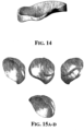
- FIG. 14 is a view of an implant suitable for placement in a joint knee and placed on a portion of the tibial plateau.
- FIGs. 15A-D are views of an implant suitable for the hip. These implants can also be designed so that the surfaces, edges and height of the implants can be adjusted to account for the measurements taken as well as the patient specific criteria, as appropriate or desirable.
- Suitable spiral CT also with intravenous or intra-articular contrast enhancement, or MRI images can be acquired, from which medial and lateral menisci can then be extracted using live wire segmentation, or other suitable mechanisms.
- the generic models for the medial and lateral meniscus can be fitted as described above. For each subject, the medial and lateral meniscus that was segmented from the MRI can be compared to the fitted models as follows:
- the total distance measure D depends on the relative position of the segmented MRI data and the fitted model in the coordinate system. This relative position can be optimized to minimize D by adjusting the rigid body transformation T that positions the model in an iterative registration process based on the iterative closest point algorithm, using D ( T ) as a cost function.
Claims (5)
- Procédé de sélection d'un implant, le procédé comprenant :l'obtention de données d'images électroniques de l'articulation du genou et la génération d'une forme tridimensionnelle de l'articulation du genou, dans lequel les données d'images électroniques sont obtenues par IRM ou TDM ;caractérisé en ce qu'un ou plusieurs dimensions et/ou paramètres de forme d'un ménisque sont déterminés, dans lequel les un ou plusieurs dimensions et paramètres sont sélectionnés dans le groupe comprenant une distance antérieure-postérieure maximale du ménisque, une distance médiale-latérale maximale du ménisque, une taille ou une zone de la ou des attaches méniscales, une longueur maximale de la corne antérieure, une hauteur maximale et minimale de la corne antérieure, une hauteur maximale et minimale du corps, une hauteur maximale et minimale de la corne postérieure, une hauteur maximale et minimale du ménisque, une largeur maximale et minimale de la corne antérieure, une largeur maximale et minimale du corps, une largeur maximale et minimale de la corne postérieure, des rayons et angles méniscaux à différents endroits, etla déduction d'une géométrie 3D d'un ménisque malade à partir des mesures de surfaces et de structures articulaires voisines pour recréer la forme et la taille du ménisque malade, dans lequel les mesures sont sélectionnées parmi une ou plusieurs des dimensions de l'os tibial suivantes : une distance antérieure-postérieure maximale, une distance médiale-latérale maximale , une distance maximale entre l'épine tibiale et le bord, une largeur des épines tibiales, une hauteur des épines tibiales, une zone de plateau tibial occupée par les épines tibiales, une profondeur de plateau tibial, une forme 2D et 3D de plateau tibial, et/ou parmi une ou plusieurs des dimensions de l'os du condyle fémoral suivantes : une distance antérieure-postérieure maximale, une distance supérieure-inférieure maximale, une distance médiale-latérale maximale, une distance maximale entre la trochlée et le bord médial ou latéral ; une largeur et une profondeur de l'échancrure intercondylienne, une courbure dans certaines régions le long du condyle fémoral, une forme en 2D et 3D ;la conception de l'implant utilisable sur un condyle de l'articulation du genou ou d'un implant utilisable sur une partie du plateau tibial en tenant compte des dimensions du ménisque et/ou des paramètres de forme du ménisque ou de la géométrie 3D déduite d'un ménisque malade à partir des mesures de surfaces et structures articulaires voisines ; etl'ajustement d'une ou plusieurs dimensions de l'implant conçu pour obtenir une adaptation optimale à l'articulation du genou du patient.
- Procédé selon la revendication 1, comportant la détermination d'une ou plusieurs dimensions d'un condyle médial et/ou d'un condyle latéral de l'articulation du genou.
- Procédé selon la revendication 1, comportant la détermination de deux dimensions ou plus d'un condyle médial et/ou d'un condyle latéral de l'articulation du genou.
- Procédé selon la revendication 1, dans lequel les une ou plusieurs dimensions d'un condyle fémoral sont sélectionnées dans le groupe consistant en une longueur antéro-postérieure du condyle fémoral médial, une longueur antéro-postérieure du condyle fémoral latéral, une largeur médio-latérale du condyle fémoral médial, une largeur médio-latérale du condyle fémoral latéral, une distance antérieure-postérieure du fémur, une distance supérieure-inférieure du fémur, une distance médio-latérale du fémur, une distance entre la trochlée et un bord du fémur ; et une largeur de l'échancrure intercondylienne.
- Procédé selon la revendication 1, dans lequel les une ou plusieurs dimensions d'un plateau tibial sont sélectionnées dans le groupe consistant en une longueur antérieure-postérieure du plateau tibial médial, une longueur antérieure-postérieure du plateau tibial latéral, une largeur médio-latérale du plateau tibial médial, une largeur médio-latérale du plateau tibial latéral, et une distance entre l'épine tibiale et un bord du plateau tibial.
Applications Claiming Priority (3)
| Application Number | Priority Date | Filing Date | Title |
|---|---|---|---|
| US42496402P | 2002-11-07 | 2002-11-07 | |
| PCT/US2003/036079 WO2004043305A1 (fr) | 2002-11-07 | 2003-11-07 | Procedes pour determiner les dimensions et formes de menisques, et pour mettre au point un traitement |
| EP03783340.7A EP1558181B1 (fr) | 2002-11-07 | 2003-11-07 | Procedes pour determiner les dimensions et formes de menisques, et pour mettre au point un traitement |
Related Parent Applications (1)
| Application Number | Title | Priority Date | Filing Date |
|---|---|---|---|
| EP03783340.7A Division EP1558181B1 (fr) | 2002-11-07 | 2003-11-07 | Procedes pour determiner les dimensions et formes de menisques, et pour mettre au point un traitement |
Publications (2)
| Publication Number | Publication Date |
|---|---|
| EP3075356A1 EP3075356A1 (fr) | 2016-10-05 |
| EP3075356B1 true EP3075356B1 (fr) | 2023-07-05 |
Family
ID=32312904
Family Applications (2)
| Application Number | Title | Priority Date | Filing Date |
|---|---|---|---|
| EP15182214.5A Expired - Lifetime EP3075356B1 (fr) | 2002-11-07 | 2003-11-07 | Méthode de sélection d'un implant méniscal |
| EP03783340.7A Expired - Lifetime EP1558181B1 (fr) | 2002-11-07 | 2003-11-07 | Procedes pour determiner les dimensions et formes de menisques, et pour mettre au point un traitement |
Family Applications After (1)
| Application Number | Title | Priority Date | Filing Date |
|---|---|---|---|
| EP03783340.7A Expired - Lifetime EP1558181B1 (fr) | 2002-11-07 | 2003-11-07 | Procedes pour determiner les dimensions et formes de menisques, et pour mettre au point un traitement |
Country Status (8)
| Country | Link |
|---|---|
| US (6) | US8932363B2 (fr) |
| EP (2) | EP3075356B1 (fr) |
| JP (1) | JP2006505366A (fr) |
| CN (1) | CN1780594A (fr) |
| AU (1) | AU2003290757A1 (fr) |
| CA (1) | CA2505371A1 (fr) |
| HK (1) | HK1072888A1 (fr) |
| WO (1) | WO2004043305A1 (fr) |
Families Citing this family (321)
| Publication number | Priority date | Publication date | Assignee | Title |
|---|---|---|---|---|
| US7534263B2 (en) | 2001-05-25 | 2009-05-19 | Conformis, Inc. | Surgical tools facilitating increased accuracy, speed and simplicity in performing joint arthroplasty |
| US8771365B2 (en) | 2009-02-25 | 2014-07-08 | Conformis, Inc. | Patient-adapted and improved orthopedic implants, designs, and related tools |
| US20090222103A1 (en) * | 2001-05-25 | 2009-09-03 | Conformis, Inc. | Articular Implants Providing Lower Adjacent Cartilage Wear |
| US20070233269A1 (en) * | 2001-05-25 | 2007-10-04 | Conformis, Inc. | Interpositional Joint Implant |
| US10085839B2 (en) | 2004-01-05 | 2018-10-02 | Conformis, Inc. | Patient-specific and patient-engineered orthopedic implants |
| US7468075B2 (en) | 2001-05-25 | 2008-12-23 | Conformis, Inc. | Methods and compositions for articular repair |
| US7618451B2 (en) * | 2001-05-25 | 2009-11-17 | Conformis, Inc. | Patient selectable joint arthroplasty devices and surgical tools facilitating increased accuracy, speed and simplicity in performing total and partial joint arthroplasty |
| US8480754B2 (en) | 2001-05-25 | 2013-07-09 | Conformis, Inc. | Patient-adapted and improved articular implants, designs and related guide tools |
| US20110071802A1 (en) * | 2009-02-25 | 2011-03-24 | Ray Bojarski | Patient-adapted and improved articular implants, designs and related guide tools |
| US8545569B2 (en) | 2001-05-25 | 2013-10-01 | Conformis, Inc. | Patient selectable knee arthroplasty devices |
| US20070100462A1 (en) * | 2001-05-25 | 2007-05-03 | Conformis, Inc | Joint Arthroplasty Devices |
| US8735773B2 (en) | 2007-02-14 | 2014-05-27 | Conformis, Inc. | Implant device and method for manufacture |
| US8882847B2 (en) | 2001-05-25 | 2014-11-11 | Conformis, Inc. | Patient selectable knee joint arthroplasty devices |
| US8083745B2 (en) | 2001-05-25 | 2011-12-27 | Conformis, Inc. | Surgical tools for arthroplasty |
| US9603711B2 (en) | 2001-05-25 | 2017-03-28 | Conformis, Inc. | Patient-adapted and improved articular implants, designs and related guide tools |
| US8556983B2 (en) | 2001-05-25 | 2013-10-15 | Conformis, Inc. | Patient-adapted and improved orthopedic implants, designs and related tools |
| US7239908B1 (en) | 1998-09-14 | 2007-07-03 | The Board Of Trustees Of The Leland Stanford Junior University | Assessing the condition of a joint and devising treatment |
| AU772012B2 (en) | 1998-09-14 | 2004-04-08 | Board Of Trustees Of The Leland Stanford Junior University | Assessing the condition of a joint and preventing damage |
| US9289153B2 (en) | 1998-09-14 | 2016-03-22 | The Board Of Trustees Of The Leland Stanford Junior University | Joint and cartilage diagnosis, assessment and modeling |
| US7184814B2 (en) | 1998-09-14 | 2007-02-27 | The Board Of Trustees Of The Leland Stanford Junior University | Assessing the condition of a joint and assessing cartilage loss |
| ATE426357T1 (de) | 2000-09-14 | 2009-04-15 | Univ Leland Stanford Junior | Beurteilung des zustandes eines gelenkes und planung einer behandlung |
| US6827723B2 (en) * | 2001-02-27 | 2004-12-07 | Smith & Nephew, Inc. | Surgical navigation systems and processes for unicompartmental knee arthroplasty |
| US9308091B2 (en) | 2001-05-25 | 2016-04-12 | Conformis, Inc. | Devices and methods for treatment of facet and other joints |
| US8951260B2 (en) | 2001-05-25 | 2015-02-10 | Conformis, Inc. | Surgical cutting guide |
| US8439926B2 (en) | 2001-05-25 | 2013-05-14 | Conformis, Inc. | Patient selectable joint arthroplasty devices and surgical tools |
| WO2002096268A2 (fr) | 2001-05-25 | 2002-12-05 | Imaging Therapeutics, Inc. | Methodes et compositions d'arthroplastie |
| US8801720B2 (en) | 2002-05-15 | 2014-08-12 | Otismed Corporation | Total joint arthroplasty system |
| DE60336002D1 (de) | 2002-10-07 | 2011-03-24 | Conformis Inc | Minimal invasives gelenkimplantat mit einer den gelenkflächen angepassten dreidimensionalen geometrie |
| EP3075356B1 (fr) | 2002-11-07 | 2023-07-05 | ConforMIS, Inc. | Méthode de sélection d'un implant méniscal |
| WO2004051301A2 (fr) * | 2002-12-04 | 2004-06-17 | Conformis, Inc. | Fusion de plans d'imagerie multiples pour imagerie isotrope en irm et analyse d'image quantitative utilisant l'imagerie isotrope ou quasi isotrope |
| EP1611548A1 (fr) * | 2003-03-27 | 2006-01-04 | Koninklijke Philips Electronics N.V. | Systeme d'imagerie medicale et procede de segmentation d'un objet a etudier |
| WO2004112610A2 (fr) | 2003-06-09 | 2004-12-29 | Vitruvian Orthopaedics, Llc | Dispositif et procede d'orientation chirurgicale |
| US7559931B2 (en) | 2003-06-09 | 2009-07-14 | OrthAlign, Inc. | Surgical orientation system and method |
| ATE454110T1 (de) * | 2003-06-27 | 2010-01-15 | Abs Corp | System für fussgelenksarthroplastie |
| US7862570B2 (en) | 2003-10-03 | 2011-01-04 | Smith & Nephew, Inc. | Surgical positioners |
| US20090088846A1 (en) | 2007-04-17 | 2009-04-02 | David Myung | Hydrogel arthroplasty device |
| US8007448B2 (en) * | 2004-10-08 | 2011-08-30 | Stryker Leibinger Gmbh & Co. Kg. | System and method for performing arthroplasty of a joint and tracking a plumb line plane |
| DE102005014623A1 (de) * | 2005-03-23 | 2006-09-28 | Aesculap Ag & Co. Kg | Chirurgisches System zum Präparieren eines Implantats und Verfahren zum Präparieren eines Implantats |
| EP1893117A1 (fr) * | 2005-04-06 | 2008-03-05 | Depuy International Limited | Systeme et procede d'enregistrement |
| US9301845B2 (en) | 2005-06-15 | 2016-04-05 | P Tech, Llc | Implant for knee replacement |
| US20070179626A1 (en) * | 2005-11-30 | 2007-08-02 | De La Barrera Jose L M | Functional joint arthroplasty method |
| DE102005058760A1 (de) * | 2005-12-05 | 2007-06-06 | Aesculap Ag & Co. Kg | Verfahren und Vorrichtung zum Präparieren eines Implantats aus einem Implantatmaterial |
| EP1981409B1 (fr) | 2006-02-06 | 2017-01-11 | ConforMIS, Inc. | Dispositifs d'arthroplastie articulaire et outils chirurgicaux sélectionnables par le patient |
| US8623026B2 (en) | 2006-02-06 | 2014-01-07 | Conformis, Inc. | Patient selectable joint arthroplasty devices and surgical tools incorporating anatomical relief |
| WO2007097853A2 (fr) | 2006-02-15 | 2007-08-30 | Otismed Corp | Dispositifs d'arthroplastie et procédés correspondants |
| US9808262B2 (en) | 2006-02-15 | 2017-11-07 | Howmedica Osteonics Corporation | Arthroplasty devices and related methods |
| US9339278B2 (en) | 2006-02-27 | 2016-05-17 | Biomet Manufacturing, Llc | Patient-specific acetabular guides and associated instruments |
| US8858561B2 (en) | 2006-06-09 | 2014-10-14 | Blomet Manufacturing, LLC | Patient-specific alignment guide |
| US9173661B2 (en) | 2006-02-27 | 2015-11-03 | Biomet Manufacturing, Llc | Patient specific alignment guide with cutting surface and laser indicator |
| US8133234B2 (en) | 2006-02-27 | 2012-03-13 | Biomet Manufacturing Corp. | Patient specific acetabular guide and method |
| US9907659B2 (en) | 2007-04-17 | 2018-03-06 | Biomet Manufacturing, Llc | Method and apparatus for manufacturing an implant |
| US10278711B2 (en) | 2006-02-27 | 2019-05-07 | Biomet Manufacturing, Llc | Patient-specific femoral guide |
| US7967868B2 (en) | 2007-04-17 | 2011-06-28 | Biomet Manufacturing Corp. | Patient-modified implant and associated method |
| US8377066B2 (en) | 2006-02-27 | 2013-02-19 | Biomet Manufacturing Corp. | Patient-specific elbow guides and associated methods |
| US9289253B2 (en) | 2006-02-27 | 2016-03-22 | Biomet Manufacturing, Llc | Patient-specific shoulder guide |
| US9345548B2 (en) | 2006-02-27 | 2016-05-24 | Biomet Manufacturing, Llc | Patient-specific pre-operative planning |
| US8407067B2 (en) | 2007-04-17 | 2013-03-26 | Biomet Manufacturing Corp. | Method and apparatus for manufacturing an implant |
| US8473305B2 (en) | 2007-04-17 | 2013-06-25 | Biomet Manufacturing Corp. | Method and apparatus for manufacturing an implant |
| US8608749B2 (en) | 2006-02-27 | 2013-12-17 | Biomet Manufacturing, Llc | Patient-specific acetabular guides and associated instruments |
| US8591516B2 (en) | 2006-02-27 | 2013-11-26 | Biomet Manufacturing, Llc | Patient-specific orthopedic instruments |
| US8608748B2 (en) | 2006-02-27 | 2013-12-17 | Biomet Manufacturing, Llc | Patient specific guides |
| US8241293B2 (en) | 2006-02-27 | 2012-08-14 | Biomet Manufacturing Corp. | Patient specific high tibia osteotomy |
| US20150335438A1 (en) | 2006-02-27 | 2015-11-26 | Biomet Manufacturing, Llc. | Patient-specific augments |
| US8282646B2 (en) | 2006-02-27 | 2012-10-09 | Biomet Manufacturing Corp. | Patient specific knee alignment guide and associated method |
| US8603180B2 (en) | 2006-02-27 | 2013-12-10 | Biomet Manufacturing, Llc | Patient-specific acetabular alignment guides |
| US8864769B2 (en) | 2006-02-27 | 2014-10-21 | Biomet Manufacturing, Llc | Alignment guides with patient-specific anchoring elements |
| US8568487B2 (en) | 2006-02-27 | 2013-10-29 | Biomet Manufacturing, Llc | Patient-specific hip joint devices |
| US8092465B2 (en) | 2006-06-09 | 2012-01-10 | Biomet Manufacturing Corp. | Patient specific knee alignment guide and associated method |
| US8298237B2 (en) | 2006-06-09 | 2012-10-30 | Biomet Manufacturing Corp. | Patient-specific alignment guide for multiple incisions |
| US8535387B2 (en) | 2006-02-27 | 2013-09-17 | Biomet Manufacturing, Llc | Patient-specific tools and implants |
| US9918740B2 (en) | 2006-02-27 | 2018-03-20 | Biomet Manufacturing, Llc | Backup surgical instrument system and method |
| US9113971B2 (en) | 2006-02-27 | 2015-08-25 | Biomet Manufacturing, Llc | Femoral acetabular impingement guide |
| US8070752B2 (en) | 2006-02-27 | 2011-12-06 | Biomet Manufacturing Corp. | Patient specific alignment guide and inter-operative adjustment |
| US8246680B2 (en) * | 2006-05-25 | 2012-08-21 | Spinemedica, Llc | Patient-specific spinal implants and related systems and methods |
| US9795399B2 (en) | 2006-06-09 | 2017-10-24 | Biomet Manufacturing, Llc | Patient-specific knee alignment guide and associated method |
| WO2008045807A2 (fr) | 2006-10-09 | 2008-04-17 | Active Implants Corporation | Dispositif de prothèse de ménisque |
| US8192491B2 (en) | 2006-10-09 | 2012-06-05 | Active Implants Corporation | Meniscus prosthetic device |
| US20080097606A1 (en) * | 2006-10-19 | 2008-04-24 | Cragg Andrew H | Knee joint prosthesis and hyaluronate compositions for treatment of osteoarthritis |
| US8460302B2 (en) | 2006-12-18 | 2013-06-11 | Otismed Corporation | Arthroplasty devices and related methods |
| EP2591756A1 (fr) | 2007-02-14 | 2013-05-15 | Conformis, Inc. | Dispositif d'implant et procédé de fabrication |
| EP2194879B1 (fr) | 2007-08-17 | 2020-05-13 | Zimmer, Inc. | Suite logicielle d'analyse de conception d'implant |
| EP2194836B1 (fr) * | 2007-09-25 | 2015-11-04 | Perception Raisonnement Action En Medecine | Appareil pour aider au diagnostic de l'état du cartilage et procédés thérapeutiques |
| US8265949B2 (en) | 2007-09-27 | 2012-09-11 | Depuy Products, Inc. | Customized patient surgical plan |
| US8398645B2 (en) | 2007-09-30 | 2013-03-19 | DePuy Synthes Products, LLC | Femoral tibial customized patient-specific orthopaedic surgical instrumentation |
| WO2011106399A1 (fr) | 2010-02-25 | 2011-09-01 | Depuy Products, Inc. | Blocs de coupe osseuse spécifiques à un patient personnalisés |
| US8357111B2 (en) | 2007-09-30 | 2013-01-22 | Depuy Products, Inc. | Method and system for designing patient-specific orthopaedic surgical instruments |
| US9173662B2 (en) | 2007-09-30 | 2015-11-03 | DePuy Synthes Products, Inc. | Customized patient-specific tibial cutting blocks |
| US8460303B2 (en) | 2007-10-25 | 2013-06-11 | Otismed Corporation | Arthroplasty systems and devices, and related methods |
| USD642263S1 (en) | 2007-10-25 | 2011-07-26 | Otismed Corporation | Arthroplasty jig blank |
| US10582934B2 (en) | 2007-11-27 | 2020-03-10 | Howmedica Osteonics Corporation | Generating MRI images usable for the creation of 3D bone models employed to make customized arthroplasty jigs |
| CA2732274C (fr) | 2007-12-06 | 2017-03-28 | Smith & Nephew, Inc. | Systemes et procedes pour determiner l'axe mecanique d'un femur |
| US8545509B2 (en) | 2007-12-18 | 2013-10-01 | Otismed Corporation | Arthroplasty system and related methods |
| US8715291B2 (en) | 2007-12-18 | 2014-05-06 | Otismed Corporation | Arthroplasty system and related methods |
| US8777875B2 (en) | 2008-07-23 | 2014-07-15 | Otismed Corporation | System and method for manufacturing arthroplasty jigs having improved mating accuracy |
| US8480679B2 (en) | 2008-04-29 | 2013-07-09 | Otismed Corporation | Generation of a computerized bone model representative of a pre-degenerated state and useable in the design and manufacture of arthroplasty devices |
| US8311306B2 (en) | 2008-04-30 | 2012-11-13 | Otismed Corporation | System and method for image segmentation in generating computer models of a joint to undergo arthroplasty |
| US8160345B2 (en) | 2008-04-30 | 2012-04-17 | Otismed Corporation | System and method for image segmentation in generating computer models of a joint to undergo arthroplasty |
| US8221430B2 (en) | 2007-12-18 | 2012-07-17 | Otismed Corporation | System and method for manufacturing arthroplasty jigs |
| US8617171B2 (en) | 2007-12-18 | 2013-12-31 | Otismed Corporation | Preoperatively planning an arthroplasty procedure and generating a corresponding patient specific arthroplasty resection guide |
| US8737700B2 (en) | 2007-12-18 | 2014-05-27 | Otismed Corporation | Preoperatively planning an arthroplasty procedure and generating a corresponding patient specific arthroplasty resection guide |
| US7690864B2 (en) * | 2007-12-27 | 2010-04-06 | Allen Engineering Corporation | Hydraulic riding trowel with automatic load sensing system |
| EP2242453B1 (fr) * | 2008-02-20 | 2018-11-28 | Mako Surgical Corp. | Planification des implants utilisant des informations sur des mouvements articulaires capturés et corrigés |
| US9408618B2 (en) * | 2008-02-29 | 2016-08-09 | Howmedica Osteonics Corporation | Total hip replacement surgical guide tool |
| WO2009111626A2 (fr) | 2008-03-05 | 2009-09-11 | Conformis, Inc. | Implants pour modifier des modèles d’usure de surfaces articulaires |
| EP2259753B8 (fr) * | 2008-03-05 | 2014-12-31 | ConforMIS, Inc. | Procédé de fabrication d'un implant articulaire à bords concordants |
| US8494825B2 (en) * | 2008-03-13 | 2013-07-23 | Robert L. Thornberry | Computer-guided system for orienting the acetabular cup in the pelvis during total hip replacement surgery |
| US8549888B2 (en) | 2008-04-04 | 2013-10-08 | Nuvasive, Inc. | System and device for designing and forming a surgical implant |
| JP5493133B2 (ja) * | 2008-04-08 | 2014-05-14 | 国立大学法人山口大学 | 開曲線フーリエ記述子を用いたx線画像処理方法及びシステム |
| US7611653B1 (en) * | 2008-04-09 | 2009-11-03 | Active Implants Corporation | Manufacturing and material processing for prosthetic devices |
| US7991599B2 (en) | 2008-04-09 | 2011-08-02 | Active Implants Corporation | Meniscus prosthetic device selection and implantation methods |
| US8016884B2 (en) | 2008-04-09 | 2011-09-13 | Active Implants Corporation | Tensioned meniscus prosthetic devices and associated methods |
| US8361147B2 (en) * | 2008-04-09 | 2013-01-29 | Active Implants Corporation | Meniscus prosthetic devices with anti-migration features |
| ITMI20080967A1 (it) | 2008-05-23 | 2009-11-24 | Salvatore Longoni | Metodo di progettazione e/o selezione di un dispositivo e/o materiale impiantabile in tessuti del corpo umano o animale e dispositivo e/o materiale cosi' ottenuto |
| FR2932674B1 (fr) | 2008-06-20 | 2011-11-18 | Tornier Sa | Procede de modelisation d'une surface glenoidienne d'une omoplate, dispositif d'implantation d'un composant glenoidien d'une prothese d'epaule, et procede de fabrication d'un tel compose. |
| US8883915B2 (en) | 2008-07-07 | 2014-11-11 | Biomimedica, Inc. | Hydrophobic and hydrophilic interpenetrating polymer networks derived from hydrophobic polymers and methods of preparing the same |
| US20120209396A1 (en) | 2008-07-07 | 2012-08-16 | David Myung | Orthopedic implants having gradient polymer alloys |
| US8617175B2 (en) | 2008-12-16 | 2013-12-31 | Otismed Corporation | Unicompartmental customized arthroplasty cutting jigs and methods of making the same |
| EP3381382A1 (fr) | 2008-07-24 | 2018-10-03 | OrthAlign, Inc. | Systèmes pour le remplacement d'une articulation |
| US8532362B2 (en) * | 2008-07-24 | 2013-09-10 | Ghent University | Scoring system to monitor natural or drug-modified disease progression in “erosive osteoarthritis” of the interphalangeal finger joints |
| WO2010017282A1 (fr) | 2008-08-05 | 2010-02-11 | Biomimedica, Inc. | Hydrogels greffés au polyuréthane |
| EP2358310B1 (fr) | 2008-09-10 | 2019-07-31 | OrthAlign, Inc. | Systèmes de chirurgie de la hanche |
| US8078440B2 (en) | 2008-09-19 | 2011-12-13 | Smith & Nephew, Inc. | Operatively tuning implants for increased performance |
| US8992538B2 (en) | 2008-09-30 | 2015-03-31 | DePuy Synthes Products, Inc. | Customized patient-specific acetabular orthopaedic surgical instrument and method of use and fabrication |
| CN102171725B (zh) * | 2008-10-07 | 2017-05-03 | 皇家飞利浦电子股份有限公司 | 脑室分析 |
| US9364291B2 (en) * | 2008-12-11 | 2016-06-14 | Mako Surgical Corp. | Implant planning using areas representing cartilage |
| US8538102B2 (en) * | 2008-12-17 | 2013-09-17 | Synarc Inc | Optimised region of interest selection |
| US8170641B2 (en) | 2009-02-20 | 2012-05-01 | Biomet Manufacturing Corp. | Method of imaging an extremity of a patient |
| US8808303B2 (en) | 2009-02-24 | 2014-08-19 | Microport Orthopedics Holdings Inc. | Orthopedic surgical guide |
| WO2010099231A2 (fr) | 2009-02-24 | 2010-09-02 | Conformis, Inc. | Systèmes automatisés de fabrication d'implants orthopédiques spécifiques au patient et instrumentation |
| US9017334B2 (en) | 2009-02-24 | 2015-04-28 | Microport Orthopedics Holdings Inc. | Patient specific surgical guide locator and mount |
| US8808297B2 (en) | 2009-02-24 | 2014-08-19 | Microport Orthopedics Holdings Inc. | Orthopedic surgical guide |
| EP2400921A4 (fr) | 2009-02-25 | 2015-11-25 | Zimmer Inc | Implants orthopédiques personnalisés et procédés associés |
| US9078755B2 (en) | 2009-02-25 | 2015-07-14 | Zimmer, Inc. | Ethnic-specific orthopaedic implants and custom cutting jigs |
| US8241298B2 (en) | 2009-03-27 | 2012-08-14 | Depuy Mitek, Inc. | Methods and devices for delivering and affixing tissue scaffolds |
| US8308814B2 (en) | 2009-03-27 | 2012-11-13 | Depuy Mitek, Inc. | Methods and devices for preparing and implanting tissue scaffolds |
| WO2010121147A1 (fr) | 2009-04-16 | 2010-10-21 | Conformis, Inc. | Dispositifs d'arthroplastie articulaire spécifique au patient pour réparation de ligament |
| US20100274591A1 (en) * | 2009-04-24 | 2010-10-28 | Invivolink | Medical implant tracking and order management |
| US8794977B2 (en) * | 2009-04-29 | 2014-08-05 | Lifemodeler, Inc. | Implant training system |
| CN101579240B (zh) * | 2009-06-19 | 2012-09-19 | 唐佩福 | 人体髋臼骨骼测量方法 |
| US10869771B2 (en) | 2009-07-24 | 2020-12-22 | OrthAlign, Inc. | Systems and methods for joint replacement |
| US8118815B2 (en) | 2009-07-24 | 2012-02-21 | OrthAlign, Inc. | Systems and methods for joint replacement |
| DE102009028503B4 (de) | 2009-08-13 | 2013-11-14 | Biomet Manufacturing Corp. | Resektionsschablone zur Resektion von Knochen, Verfahren zur Herstellung einer solchen Resektionsschablone und Operationsset zur Durchführung von Kniegelenk-Operationen |
| EP2493396B1 (fr) | 2009-10-29 | 2016-11-23 | Zimmer, Inc. | Guide de fraisage spécifique d'un patient |
| EP2493381B1 (fr) * | 2009-10-30 | 2015-09-30 | Koninklijke Philips N.V. | Analyse tridimensionnelle de lésions représentées par des données d'image |
| CA2825042C (fr) * | 2010-01-21 | 2021-01-05 | OrthAlign, Inc. | Systemes et procedes de remplacement d'articulation |
| US8834568B2 (en) | 2010-02-04 | 2014-09-16 | Paul S. Shapiro | Surgical technique using a contoured allograft cartilage as a spacer of the carpo-metacarpal joint of the thumb or tarso-metatarsal joint of the toe |
| EP2538855A4 (fr) | 2010-02-25 | 2016-08-03 | Depuy Products Inc | Blocs de coupe tibiale spécifiques à un patient personnalisés |
| WO2011106407A1 (fr) | 2010-02-25 | 2011-09-01 | Depuy Products, Inc. | Procédé de fabrication de blocs de coupe osseuse spécifiques à un patient personnalisés |
| ES2704658T3 (es) | 2010-02-25 | 2019-03-19 | Depuy Products Inc | Bloques de corte de hueso específicos al paciente personalizados |
| US8632547B2 (en) | 2010-02-26 | 2014-01-21 | Biomet Sports Medicine, Llc | Patient-specific osteotomy devices and methods |
| US9066727B2 (en) | 2010-03-04 | 2015-06-30 | Materialise Nv | Patient-specific computed tomography guides |
| US9579106B2 (en) | 2010-03-31 | 2017-02-28 | New York Society For The Relief Of The Ruptured And Crippled, Maintaining The Hospital For Special Surgery | Shoulder arthroplasty instrumentation |
| WO2011156755A2 (fr) * | 2010-06-11 | 2011-12-15 | Smith & Nephew, Inc. | Instruments adaptés aux patients |
| US8908937B2 (en) | 2010-07-08 | 2014-12-09 | Biomet Manufacturing, Llc | Method and device for digital image templating |
| US8764840B2 (en) | 2010-07-24 | 2014-07-01 | Zimmer, Inc. | Tibial prosthesis |
| ES2632995T3 (es) | 2010-07-24 | 2017-09-18 | Zimmer, Inc. | Componentes asimétricos de tibia para una prótesis de rodilla |
| US8808302B2 (en) | 2010-08-12 | 2014-08-19 | DePuy Synthes Products, LLC | Customized patient-specific acetabular orthopaedic surgical instrument and method of use and fabrication |
| GB201014824D0 (en) * | 2010-09-07 | 2010-10-20 | Goodfellow John | Unicondylar meniscal bearing knee replacement |
| EP2613739B1 (fr) | 2010-09-10 | 2017-06-07 | Zimmer, Inc. | Composants tibiaux facilitant le mouvement pour une prothèse du genou |
| US9271744B2 (en) | 2010-09-29 | 2016-03-01 | Biomet Manufacturing, Llc | Patient-specific guide for partial acetabular socket replacement |
| EP3636174B1 (fr) | 2010-10-29 | 2021-09-08 | The Cleveland Clinic Foundation | Système d'association d'une aide de guidage à un tissu de patient |
| US9877735B2 (en) | 2010-10-29 | 2018-01-30 | The Cleveland Clinic Foundation | System and method for assisting with attachment of a stock implant to a patient tissue |
| US9254155B2 (en) | 2010-10-29 | 2016-02-09 | The Cleveland Clinic Foundation | System and method for assisting with arrangement of a stock instrument with respect to a patient tissue |
| US9717508B2 (en) | 2010-10-29 | 2017-08-01 | The Cleveland Clinic Foundation | System of preoperative planning and provision of patient-specific surgical aids |
| US9968376B2 (en) | 2010-11-29 | 2018-05-15 | Biomet Manufacturing, Llc | Patient-specific orthopedic instruments |
| US8603101B2 (en) | 2010-12-17 | 2013-12-10 | Zimmer, Inc. | Provisional tibial prosthesis system |
| US8917290B2 (en) | 2011-01-31 | 2014-12-23 | Biomet Manufacturing, Llc | Digital image templating |
| EP2754419B1 (fr) | 2011-02-15 | 2024-02-07 | ConforMIS, Inc. | Implants orthopédiques améliorés et adaptés au patient |
| US9241745B2 (en) | 2011-03-07 | 2016-01-26 | Biomet Manufacturing, Llc | Patient-specific femoral version guide |
| US8715289B2 (en) | 2011-04-15 | 2014-05-06 | Biomet Manufacturing, Llc | Patient-specific numerically controlled instrument |
| US9675400B2 (en) | 2011-04-19 | 2017-06-13 | Biomet Manufacturing, Llc | Patient-specific fracture fixation instrumentation and method |
| US8956364B2 (en) | 2011-04-29 | 2015-02-17 | Biomet Manufacturing, Llc | Patient-specific partial knee guides and other instruments |
| US8668700B2 (en) | 2011-04-29 | 2014-03-11 | Biomet Manufacturing, Llc | Patient-specific convertible guides |
| US10130378B2 (en) | 2011-05-11 | 2018-11-20 | The Cleveland Clinic Foundation | Generating patient specific instruments for use as surgical aids |
| US8780362B2 (en) | 2011-05-19 | 2014-07-15 | Covidien Lp | Methods utilizing triangulation in metrology systems for in-situ surgical applications |
| CA2836535C (fr) | 2011-05-19 | 2019-09-24 | The Cleveland Clinic Foundation | Dispositif et procede pour fournir une indication de reference a un tissu de patient |
| US8532807B2 (en) | 2011-06-06 | 2013-09-10 | Biomet Manufacturing, Llc | Pre-operative planning and manufacturing method for orthopedic procedure |
| US9084618B2 (en) | 2011-06-13 | 2015-07-21 | Biomet Manufacturing, Llc | Drill guides for confirming alignment of patient-specific alignment guides |
| EP2720631B1 (fr) | 2011-06-16 | 2022-01-26 | Smith&Nephew, Inc. | Alignement chirurgical à l'aide de références |
| US8641721B2 (en) | 2011-06-30 | 2014-02-04 | DePuy Synthes Products, LLC | Customized patient-specific orthopaedic pin guides |
| US8764760B2 (en) | 2011-07-01 | 2014-07-01 | Biomet Manufacturing, Llc | Patient-specific bone-cutting guidance instruments and methods |
| US20130001121A1 (en) | 2011-07-01 | 2013-01-03 | Biomet Manufacturing Corp. | Backup kit for a patient-specific arthroplasty kit assembly |
| US8597365B2 (en) | 2011-08-04 | 2013-12-03 | Biomet Manufacturing, Llc | Patient-specific pelvic implants for acetabular reconstruction |
| US9295497B2 (en) | 2011-08-31 | 2016-03-29 | Biomet Manufacturing, Llc | Patient-specific sacroiliac and pedicle guides |
| US9066734B2 (en) | 2011-08-31 | 2015-06-30 | Biomet Manufacturing, Llc | Patient-specific sacroiliac guides and associated methods |
| US10603049B2 (en) | 2011-09-02 | 2020-03-31 | Episurf Ip-Management Ab | Implant specific drill bit in surgical kit for cartilage repair |
| US11000387B2 (en) | 2011-09-02 | 2021-05-11 | Episurf Ip-Management Ab | Implant for cartilage repair |
| US9386993B2 (en) | 2011-09-29 | 2016-07-12 | Biomet Manufacturing, Llc | Patient-specific femoroacetabular impingement instruments and methods |
| US20130103157A1 (en) | 2011-10-03 | 2013-04-25 | Lampros Kourtis | Polymeric adhesive for anchoring compliant materials to another surface |
| WO2013056036A1 (fr) * | 2011-10-14 | 2013-04-18 | Conformis, Inc. | Procédés et systèmes pour l'identification, l'évaluation, le modelage et la réparation de disparités anatomiques dans le remplacement d'articulation |
| US9554910B2 (en) | 2011-10-27 | 2017-01-31 | Biomet Manufacturing, Llc | Patient-specific glenoid guide and implants |
| US9451973B2 (en) | 2011-10-27 | 2016-09-27 | Biomet Manufacturing, Llc | Patient specific glenoid guide |
| KR20130046337A (ko) | 2011-10-27 | 2013-05-07 | 삼성전자주식회사 | 멀티뷰 디바이스 및 그 제어방법과, 디스플레이장치 및 그 제어방법과, 디스플레이 시스템 |
| US9301812B2 (en) | 2011-10-27 | 2016-04-05 | Biomet Manufacturing, Llc | Methods for patient-specific shoulder arthroplasty |
| ES2635542T3 (es) | 2011-10-27 | 2017-10-04 | Biomet Manufacturing, Llc | Guías glenoideas específicas para el paciente |
| US9788975B2 (en) * | 2011-10-28 | 2017-10-17 | Zimmer, Inc. | Method of estimating soft tissue balance for knee arthroplasty |
| EP3848005A3 (fr) | 2011-11-18 | 2021-09-15 | Zimmer, Inc. | Élément porteur tibial pour prothèse du genou présentant des caractéristiques articulaires améliorées |
| CA2856512A1 (fr) | 2011-11-21 | 2013-05-30 | Biomimedica, Inc. | Systemes, dispositifs et procedes pour ancrer des implants orthopediques a l'os |
| WO2013077919A1 (fr) | 2011-11-21 | 2013-05-30 | Zimmer, Inc. | Plaque de base tibiale avec placement asymétrique de structures de fixation |
| US9408686B1 (en) | 2012-01-20 | 2016-08-09 | Conformis, Inc. | Devices, systems and methods for manufacturing orthopedic implants |
| WO2013112688A1 (fr) | 2012-01-24 | 2013-08-01 | Zimmer, Inc. | Procédé et système de création d'instruments spécifiques à un patient conçus pour un transfert de greffe cartilagineuse |
| EP2809273B1 (fr) | 2012-01-30 | 2021-05-05 | Zimmer, Inc. | Éléments tibiaux asymétriques pour une prothèse de genou |
| US9237950B2 (en) | 2012-02-02 | 2016-01-19 | Biomet Manufacturing, Llc | Implant with patient-specific porous structure |
| JP5945865B2 (ja) * | 2012-02-22 | 2016-07-05 | アルスロデザイン株式会社 | 人工関節置換術用ガイド器具 |
| US11207132B2 (en) | 2012-03-12 | 2021-12-28 | Nuvasive, Inc. | Systems and methods for performing spinal surgery |
| US9498334B2 (en) * | 2012-03-27 | 2016-11-22 | DePuy Synthes Products, Inc. | Glenoid defect-filling component |
| US9539112B2 (en) | 2012-03-28 | 2017-01-10 | Robert L. Thornberry | Computer-guided system for orienting a prosthetic acetabular cup in the acetabulum during total hip replacement surgery |
| CA3072704C (fr) | 2012-03-28 | 2022-03-22 | Orthosoft Ulc | Chirurgie d'implantation glenoide utilisant une instrumentation specifique au patient |
| US9486226B2 (en) | 2012-04-18 | 2016-11-08 | Conformis, Inc. | Tibial guides, tools, and techniques for resecting the tibial plateau |
| EP2849683A4 (fr) | 2012-05-18 | 2015-11-25 | Orthalign Inc | Dispositifs et méthodes pour arthroplastie du genou |
| EP2854663B1 (fr) | 2012-05-24 | 2022-05-25 | Zimmer Inc. | Instrumentation spécifique pour un patient pour la réparation de jonction articulaire |
| US9675471B2 (en) | 2012-06-11 | 2017-06-13 | Conformis, Inc. | Devices, techniques and methods for assessing joint spacing, balancing soft tissues and obtaining desired kinematics for joint implant components |
| RU2498772C1 (ru) * | 2012-06-13 | 2013-11-20 | Федеральное государственное бюджетное учреждение "Российский научный центр "Восстановительная травматология и ортопедия" имени академика Г.А. Илизарова" Министерства здравоохранения и социального развития Российской Федерации | Способ дифференцированной оценки деформаций коленного сустава и выбора вида хирургического лечения при гонартрозе |
| ES2643061T3 (es) | 2012-07-23 | 2017-11-21 | Orthosoft, Inc. | Instrumentación específica del paciente para la cirugía de corrección de implantes |
| WO2014015433A1 (fr) | 2012-07-24 | 2014-01-30 | Orthosoft Inc. | Instrument spécifique d'un patient et doté d'un système microélectromécanique à utiliser en chirurgie |
| US9649160B2 (en) | 2012-08-14 | 2017-05-16 | OrthAlign, Inc. | Hip replacement navigation system and method |
| US9275302B1 (en) * | 2012-08-24 | 2016-03-01 | Amazon Technologies, Inc. | Object detection and identification |
| US9402637B2 (en) | 2012-10-11 | 2016-08-02 | Howmedica Osteonics Corporation | Customized arthroplasty cutting guides and surgical methods using the same |
| US9129435B2 (en) * | 2012-12-04 | 2015-09-08 | Fuji Xerox Co., Ltd. | Method for creating 3-D models by stitching multiple partial 3-D models |
| US9060788B2 (en) | 2012-12-11 | 2015-06-23 | Biomet Manufacturing, Llc | Patient-specific acetabular guide for anterior approach |
| US9204977B2 (en) | 2012-12-11 | 2015-12-08 | Biomet Manufacturing, Llc | Patient-specific acetabular guide for anterior approach |
| US9387083B2 (en) | 2013-01-30 | 2016-07-12 | Conformis, Inc. | Acquiring and utilizing kinematic information for patient-adapted implants, tools and surgical procedures |
| CN103099680B (zh) * | 2013-03-06 | 2015-04-22 | 吴志宏 | 一种导向模板的制备方法 |
| US9839438B2 (en) | 2013-03-11 | 2017-12-12 | Biomet Manufacturing, Llc | Patient-specific glenoid guide with a reusable guide holder |
| US9579107B2 (en) | 2013-03-12 | 2017-02-28 | Biomet Manufacturing, Llc | Multi-point fit for patient specific guide |
| US9498233B2 (en) | 2013-03-13 | 2016-11-22 | Biomet Manufacturing, Llc. | Universal acetabular guide and associated hardware |
| US9826981B2 (en) | 2013-03-13 | 2017-11-28 | Biomet Manufacturing, Llc | Tangential fit of patient-specific guides |
| US9517145B2 (en) | 2013-03-15 | 2016-12-13 | Biomet Manufacturing, Llc | Guide alignment system and method |
| US9968408B1 (en) | 2013-03-15 | 2018-05-15 | Nuvasive, Inc. | Spinal balance assessment |
| USD702349S1 (en) | 2013-05-14 | 2014-04-08 | Laboratories Bodycad Inc. | Tibial prosthesis |
| USD752222S1 (en) | 2013-05-14 | 2016-03-22 | Laboratoires Bodycad Inc. | Femoral prosthesis |
| JP6635515B2 (ja) | 2013-06-11 | 2020-01-29 | オーソソフト アンリミティド ライアビリティ コーポレイション | 寛骨臼カップ・プロテーゼ位置決め器具及び方法 |
| WO2014197989A1 (fr) | 2013-06-11 | 2014-12-18 | Orthosoft Inc. | Injection sous-chondrale assistée par ordinateur |
| KR101549184B1 (ko) | 2013-08-09 | 2015-09-03 | 국립대학법인 울산과학기술대학교 산학협력단 | 압축 센싱 지원 mmme 시퀀스를 이용한 초고속 3d 스핀 에코 영상법 및 그 영상장치 |
| CA2919717C (fr) | 2013-08-21 | 2021-06-22 | Laboratoires Bodycad Inc. | Guide de resection osseuse et procede |
| CA2919546C (fr) | 2013-08-21 | 2018-11-06 | Laboratoires Bodycad Inc. | Implant orthopedique adapte a l'anatomie et procede de fabrication associe |
| US9925052B2 (en) | 2013-08-30 | 2018-03-27 | Zimmer, Inc. | Method for optimizing implant designs |
| FR3010628B1 (fr) | 2013-09-18 | 2015-10-16 | Medicrea International | Procede permettant de realiser la courbure ideale d'une tige d'un materiel d'osteosynthese vertebrale destinee a etayer la colonne vertebrale d'un patient |
| WO2015048319A1 (fr) | 2013-09-25 | 2015-04-02 | Zimmer, Inc. | Instrumentation spécifique à un patient (psi) de chirurgie orthopédique et systèmes et méthodes d'utilisation de rayons x pour la produire |
| US9848922B2 (en) | 2013-10-09 | 2017-12-26 | Nuvasive, Inc. | Systems and methods for performing spine surgery |
| FR3012030B1 (fr) | 2013-10-18 | 2015-12-25 | Medicrea International | Procede permettant de realiser la courbure ideale d'une tige d'un materiel d'osteosynthese vertebrale destinee a etayer la colonne vertebrale d'un patient |
| US20150112349A1 (en) | 2013-10-21 | 2015-04-23 | Biomet Manufacturing, Llc | Ligament Guide Registration |
| EP3068317B1 (fr) | 2013-11-13 | 2018-08-22 | Tornier | Instrument spécifique à l'épaule d'un patient |
| DK3389012T3 (da) * | 2014-02-07 | 2020-07-20 | Episurf Ip Man Ab | Fremgangsmåde og knude til fremstilling af et kirurgisk kit til bruskreparation |
| DE102015204957A1 (de) * | 2014-03-27 | 2015-10-01 | Siemens Aktiengesellschaft | Bildgebendes Tomosynthesesystem, insbesondere Mammographiesystem |
| US10282488B2 (en) | 2014-04-25 | 2019-05-07 | Biomet Manufacturing, Llc | HTO guide with optional guided ACL/PCL tunnels |
| WO2015168415A1 (fr) | 2014-04-30 | 2015-11-05 | Zimmer, Inc. | Impaction de cotyle au moyen d'instruments spécifiques du patient |
| US9408616B2 (en) | 2014-05-12 | 2016-08-09 | Biomet Manufacturing, Llc | Humeral cut guide |
| US9561040B2 (en) | 2014-06-03 | 2017-02-07 | Biomet Manufacturing, Llc | Patient-specific glenoid depth control |
| US9839436B2 (en) | 2014-06-03 | 2017-12-12 | Biomet Manufacturing, Llc | Patient-specific glenoid depth control |
| EP3151759B1 (fr) | 2014-06-03 | 2021-03-17 | Zimmer, Inc. | Bloc de coupe spécifique à un patient, et son procédé de fabrication |
| US20170172744A1 (en) * | 2014-07-09 | 2017-06-22 | Episurf Ip-Management Ab | Surgical joint implant and a bone-mountable rig |
| WO2016004991A1 (fr) | 2014-07-09 | 2016-01-14 | Episurf Ip-Management Ab | Implant personnalisé pour la réparation de cartilage et procédé de conception correspondant |
| JP5918325B2 (ja) * | 2014-09-11 | 2016-05-18 | 日立アロカメディカル株式会社 | 超音波診断装置 |
| US9833245B2 (en) | 2014-09-29 | 2017-12-05 | Biomet Sports Medicine, Llc | Tibial tubercule osteotomy |
| US9826994B2 (en) | 2014-09-29 | 2017-11-28 | Biomet Manufacturing, Llc | Adjustable glenoid pin insertion guide |
| US9913669B1 (en) | 2014-10-17 | 2018-03-13 | Nuvasive, Inc. | Systems and methods for performing spine surgery |
| US10762623B2 (en) | 2014-11-12 | 2020-09-01 | Materialise N.V. | System and method of improving surgical devices using captured images for efficient surgical plan development |
| WO2016123703A1 (fr) | 2015-02-02 | 2016-08-11 | Orthosoft Inc. | Dispositif numériseur de bord d'acétabulum et procédé |
| US10363149B2 (en) | 2015-02-20 | 2019-07-30 | OrthAlign, Inc. | Hip replacement navigation system and method |
| CN107405169B (zh) | 2015-03-25 | 2021-01-26 | 奥尔索夫特无限责任公司 | 用于帮助在诸如肩胛骨之类的薄骨骼中的植入物放置的系统 |
| US9820868B2 (en) | 2015-03-30 | 2017-11-21 | Biomet Manufacturing, Llc | Method and apparatus for a pin apparatus |
| CN107924709B (zh) | 2015-05-28 | 2022-05-17 | 捷迈有限公司 | 患者特异性骨移植系统和方法 |
| US10226262B2 (en) | 2015-06-25 | 2019-03-12 | Biomet Manufacturing, Llc | Patient-specific humeral guide designs |
| US10568647B2 (en) | 2015-06-25 | 2020-02-25 | Biomet Manufacturing, Llc | Patient-specific humeral guide designs |
| US10582969B2 (en) | 2015-07-08 | 2020-03-10 | Zimmer, Inc. | Patient-specific instrumentation for implant revision surgery |
| US11077228B2 (en) | 2015-08-10 | 2021-08-03 | Hyalex Orthopaedics, Inc. | Interpenetrating polymer networks |
| US9855146B2 (en) | 2015-08-24 | 2018-01-02 | Arthrex, Inc. | Arthroscopic resurfacing techniques |
| CN108135701B (zh) | 2015-09-21 | 2019-12-24 | 捷迈有限公司 | 包括胫骨承载组件的假体系统 |
| CN108348340B (zh) | 2015-09-30 | 2021-08-10 | 捷迈有限公司 | 用于髌骨表面修复术的患者特定器械和方法 |
| US10034753B2 (en) | 2015-10-22 | 2018-07-31 | DePuy Synthes Products, Inc. | Customized patient-specific orthopaedic instruments for component placement in a total hip arthroplasty |
| EP3370657B1 (fr) | 2015-11-04 | 2023-12-27 | Medicrea International | Appareil pour chirurgie de reconstruction spinale et mesure de la longueur spinale |
| US10624764B2 (en) | 2015-11-26 | 2020-04-21 | Orthosoft Ulc | System and method for the registration of an anatomical feature |
| AU2016371425B2 (en) | 2015-12-16 | 2021-09-02 | Howmedica Osteonics Corp. | Patient specific instruments and methods for joint prosthesis |
| EP3181050B1 (fr) | 2015-12-18 | 2020-02-12 | Episurf IP Management AB | Système et procédé de création d'un matériau de support de décision indiquant l'endommagement d'une articulation anatomique |
| US11526988B2 (en) | 2015-12-18 | 2022-12-13 | Episurf Ip-Management Ab | System and method for creating a decision support material indicating damage to an anatomical joint |
| US10010372B1 (en) | 2016-01-06 | 2018-07-03 | Paul Beck | Marker Positioning Apparatus |
| US10004564B1 (en) | 2016-01-06 | 2018-06-26 | Paul Beck | Accurate radiographic calibration using multiple images |
| EP3422940B1 (fr) | 2016-03-02 | 2021-06-16 | Nuvasive, Inc. | Systèmes et procédés de planification chirurgicale de correction rachidienne |
| USD808524S1 (en) | 2016-11-29 | 2018-01-23 | Laboratoires Bodycad Inc. | Femoral implant |
| WO2018109556A1 (fr) | 2016-12-12 | 2018-06-21 | Medicrea International | Systèmes et procédés pour des implants rachidiens spécifiques au patient |
| EP3592299B1 (fr) | 2017-03-10 | 2021-06-09 | Zimmer, Inc. | Prothèse tibiale présentant un élément de fixation pour l'élément porteur tibial |
| US10722310B2 (en) | 2017-03-13 | 2020-07-28 | Zimmer Biomet CMF and Thoracic, LLC | Virtual surgery planning system and method |
| CA3056382A1 (fr) | 2017-03-14 | 2018-09-20 | OrthAlign, Inc. | Systemes et procedes de guidage pour un remplacement de hanche |
| WO2018169980A1 (fr) | 2017-03-14 | 2018-09-20 | OrthAlign, Inc. | Systèmes et procédés de mesure & d'équilibrage de tissu mou |
| US11185369B2 (en) | 2017-04-21 | 2021-11-30 | Medicrea Nternational | Systems, methods, and devices for developing patient-specific spinal treatments, operations, and procedures |
| CA3063415C (fr) | 2017-05-12 | 2021-10-19 | Zimmer, Inc. | Protheses femorales presentant des capacites d'agrandissement et de reduction de taille |
| US11250561B2 (en) | 2017-06-16 | 2022-02-15 | Episurf Ip-Management Ab | Determination and visualization of damage to an anatomical joint |
| CA3069517A1 (fr) | 2017-07-11 | 2019-01-17 | Tornier, Inc. | Guides et instruments d'amelioration de precision de mise en place d'implant glenoide |
| US10959742B2 (en) | 2017-07-11 | 2021-03-30 | Tornier, Inc. | Patient specific humeral cutting guides |
| CN107374663B (zh) * | 2017-08-27 | 2020-07-10 | 上海道衡科贸有限公司 | 人体膝关节伸屈运动的瞬时中心轴和瞬时中心曲面定位方法 |
| US11426282B2 (en) | 2017-11-16 | 2022-08-30 | Zimmer, Inc. | Implants for adding joint inclination to a knee arthroplasty |
| US10918422B2 (en) | 2017-12-01 | 2021-02-16 | Medicrea International | Method and apparatus for inhibiting proximal junctional failure |
| CA3027410A1 (fr) | 2017-12-12 | 2019-06-12 | Orthosoft Inc. | Instrumentation propre au patient destine a une chirurgie de revision d'implant |
| RU2692997C1 (ru) * | 2018-04-02 | 2019-06-28 | Федеральное Государственное Бюджетное Образовательное Учреждение Высшего Образования "Красноярский Государственный Медицинский Университет Имени Профессора В.Ф. Войно-Ясенецкого" Министерства Здравоохранения Российской Федерации | Способ определения границ тела и рогов менисков коленного сустава человека |
| AU2019263150B2 (en) | 2018-04-30 | 2023-09-14 | Paragon 28, Inc. | Laser-based implant alignment and resection guide systems and related methods |
| US10835380B2 (en) | 2018-04-30 | 2020-11-17 | Zimmer, Inc. | Posterior stabilized prosthesis system |
| DE102018113580A1 (de) * | 2018-06-07 | 2019-12-12 | Christoph Karl | Verfahren und vorrichtung zur herstellung eines implantats |
| EP3810013A1 (fr) | 2018-06-19 | 2021-04-28 | Tornier, Inc. | Réseau neuronal de recommandation d'un type de chirurgie de l'épaule |
| US11051829B2 (en) | 2018-06-26 | 2021-07-06 | DePuy Synthes Products, Inc. | Customized patient-specific orthopaedic surgical instrument |
| US10869950B2 (en) | 2018-07-17 | 2020-12-22 | Hyalex Orthopaedics, Inc. | Ionic polymer compositions |
| AU2019397526A1 (en) | 2018-12-13 | 2021-07-29 | Paragon 28, Inc. | Alignment instruments and methods for use in total ankle replacement |
| CA3122429A1 (fr) | 2018-12-13 | 2020-06-18 | Paragon 28, Inc. | Instruments specifiques a un patient et procedes d'utilisation |
| US11645749B2 (en) | 2018-12-14 | 2023-05-09 | Episurf Ip-Management Ab | Determination and visualization of damage to an anatomical joint |
| CN109662716B (zh) * | 2018-12-19 | 2021-10-22 | 上海联影医疗科技股份有限公司 | 软骨厚度测量方法、装置、计算机设备和存储介质 |
| WO2020201353A1 (fr) | 2019-04-02 | 2020-10-08 | Medicrea International | Systèmes, procédés et dispositifs pour développer des implants spinaux spécifiques d'un patient, des traitements, des opérations et/ou des procédures |
| US11925417B2 (en) | 2019-04-02 | 2024-03-12 | Medicrea International | Systems, methods, and devices for developing patient-specific spinal implants, treatments, operations, and/or procedures |
| CN109993700B (zh) * | 2019-04-03 | 2023-07-04 | 百度在线网络技术(北京)有限公司 | 数据处理方法、装置、电子设备及计算机可读存储介质 |
| AU2020344704A1 (en) | 2019-09-13 | 2022-04-14 | Treace Medical Concepts, Inc. | Patient-specific surgical methods and instrumentation |
| US11324601B2 (en) * | 2019-09-30 | 2022-05-10 | Paragon Advanced Technologies, Inc. | Talus implant |
| US11832893B2 (en) * | 2019-10-01 | 2023-12-05 | Smith & Nephew, Inc. | Methods of accessing joints for arthroscopic procedures |
| US11769251B2 (en) | 2019-12-26 | 2023-09-26 | Medicrea International | Systems and methods for medical image analysis |
| USD920517S1 (en) | 2020-01-08 | 2021-05-25 | Restor3D, Inc. | Osteotomy wedge |
| USD920515S1 (en) | 2020-01-08 | 2021-05-25 | Restor3D, Inc. | Spinal implant |
| EP3858279A1 (fr) * | 2020-01-29 | 2021-08-04 | Ottokar-Kernsto Leibnitz | Détermination de la taille d'un implant |
| CN112950595B (zh) * | 2021-03-10 | 2021-10-22 | 西北民族大学 | 一种基于spect成像的人体部位切分方法及系统 |
| CN113288346B (zh) * | 2021-05-20 | 2023-12-29 | 博思研生物技术(苏州)有限公司 | 一种用于治疗肝癌的定位切除装置 |
| CN113040980B (zh) * | 2021-05-28 | 2021-08-24 | 北京大学第三医院(北京大学第三临床医学院) | 一种实心聚氨酯半月板移植替代物及其制作方法 |
| US11324525B1 (en) | 2021-06-30 | 2022-05-10 | Kinos Medical Inc. | Surgical alignment guide assembly for total ankle replacement and method of using the same |
| US11806028B1 (en) | 2022-10-04 | 2023-11-07 | Restor3D, Inc. | Surgical guides and processes for producing and using the same |
| US11960266B1 (en) | 2023-08-23 | 2024-04-16 | Restor3D, Inc. | Patient-specific medical devices and additive manufacturing processes for producing the same |
Citations (1)
| Publication number | Priority date | Publication date | Assignee | Title |
|---|---|---|---|---|
| WO2004032806A1 (fr) * | 2002-10-07 | 2004-04-22 | Conformis, Inc. | Implant articulaire par chirurgie non effractive a geometrie tridimensionnelle correspondant aux surfaces articulaires |
Family Cites Families (545)
| Publication number | Priority date | Publication date | Assignee | Title |
|---|---|---|---|---|
| US60885A (en) * | 1867-01-01 | William b | ||
| US100953A (en) * | 1870-03-15 | Improvement in the manufacture of rosinroil | ||
| US60884A (en) * | 1867-01-01 | Improvement in convebting wheel oaemaqes into sleighs | ||
| US60883A (en) * | 1867-01-01 | George hadfield | ||
| US3314420A (en) | 1961-10-23 | 1967-04-18 | Haeger Potteries Inc | Prosthetic parts and methods of making the same |
| US3605123A (en) | 1969-04-29 | 1971-09-20 | Melpar Inc | Bone implant |
| GB1324990A (en) | 1969-08-25 | 1973-07-25 | Nat Res Dev | Prosthetic shoulder joint devices |
| CA962806A (en) | 1970-06-04 | 1975-02-18 | Ontario Research Foundation | Surgical prosthetic device |
| US3938198A (en) | 1970-08-04 | 1976-02-17 | Cutter Laboratories, Inc. | Hip joint prosthesis |
| GB1395896A (en) | 1971-06-01 | 1975-05-29 | Nat Res Dev | Endoprosthetic knee joint devices |
| US3798679A (en) | 1971-07-09 | 1974-03-26 | Ewald Frederick | Joint prostheses |
| US3808606A (en) | 1972-02-22 | 1974-05-07 | R Tronzo | Bone implant with porous exterior surface |
| DE2306552B2 (de) | 1973-02-10 | 1975-07-03 | Friedrichsfeld Gmbh Steinzeug- Und Kunststoffwerke, 6800 Mannheim | Gelenkendoprothese |
| US3852830A (en) | 1973-02-15 | 1974-12-10 | Richards Mfg Co | Knee prosthesis |
| US3843975A (en) | 1973-04-09 | 1974-10-29 | R Tronzo | Prosthesis for femoral shaft |
| DE2340546A1 (de) | 1973-08-10 | 1975-02-27 | Pfaudler Werke Ag | Metallisches implantat und verfahren zu seiner herstellung |
| US4085466A (en) * | 1974-11-18 | 1978-04-25 | National Research Development Corporation | Prosthetic joint device |
| US4219893A (en) | 1977-09-01 | 1980-09-02 | United States Surgical Corporation | Prosthetic knee joint |
| US3991425A (en) * | 1975-11-20 | 1976-11-16 | Minnesota Mining And Manufacturing Company | Prosthetic bone joint devices |
| FR2336666A1 (fr) * | 1975-12-24 | 1977-07-22 | Farnier Andre | Compteur pour fluide |
| US4055862A (en) | 1976-01-23 | 1977-11-01 | Zimmer Usa, Inc. | Human body implant of graphitic carbon fiber reinforced ultra-high molecular weight polyethylene |
| US4052753A (en) | 1976-08-02 | 1977-10-11 | Dedo Richard G | Knee spacer and method of reforming sliding body surfaces |
| US4098626A (en) | 1976-11-15 | 1978-07-04 | Thiokol Corporation | Hydroxy terminated polybutadiene based polyurethane bound propellant grains |
| DE2703059C3 (de) | 1977-01-26 | 1981-09-03 | Sanitätshaus Schütt & Grundei, Werkstätten für Orthopädie-Technik, 2400 Lübeck | Kniegelenk-Endoprothese |
| US4203444A (en) | 1977-11-07 | 1980-05-20 | Dyonics, Inc. | Surgical instrument suitable for closed surgery such as of the knee |
| US4164793A (en) * | 1978-04-26 | 1979-08-21 | Swanson Alfred B | Lunate implant |
| US4213816A (en) | 1978-06-12 | 1980-07-22 | Glasrock Products, Inc. | Method for bonding porous coating to rigid structural member |
| US4207627A (en) | 1979-01-18 | 1980-06-17 | Cloutier Jean Marie | Knee prosthesis |
| US4211228A (en) | 1979-01-24 | 1980-07-08 | Cloutier Jean Marie | Multipurpose tibial template |
| US4280231A (en) | 1979-06-14 | 1981-07-28 | Swanson Alfred B | Elbow prosthesis |
| US4309778A (en) | 1979-07-02 | 1982-01-12 | Biomedical Engineering Corp. | New Jersey meniscal bearing knee replacement |
| US4340978A (en) | 1979-07-02 | 1982-07-27 | Biomedical Engineering Corp. | New Jersey meniscal bearing knee replacement |
| JPS6026892Y2 (ja) | 1979-11-30 | 1985-08-14 | ナショナル住宅産業株式会社 | ビス締め機調整装置 |
| US4344192A (en) | 1980-07-14 | 1982-08-17 | Imbert Jean C | Full dual element prosthesis designed for the femoro-patellar joint |
| US4344193A (en) | 1980-11-28 | 1982-08-17 | Kenny Charles H | Meniscus prosthesis |
| US4575805A (en) * | 1980-12-24 | 1986-03-11 | Moermann Werner H | Method and apparatus for the fabrication of custom-shaped implants |
| US4368040A (en) | 1981-06-01 | 1983-01-11 | Ipco Corporation | Dental impression tray for forming a dental prosthesis in situ |
| US4502161A (en) | 1981-09-21 | 1985-03-05 | Wall W H | Prosthetic meniscus for the repair of joints |
| US4436684A (en) * | 1982-06-03 | 1984-03-13 | Contour Med Partners, Ltd. | Method of forming implantable prostheses for reconstructive surgery |
| US4459985A (en) | 1983-03-04 | 1984-07-17 | Howmedica Inc. | Tibial prosthesis extractor and method for extracting a tibial implant |
| DE3315401A1 (de) | 1983-04-28 | 1984-10-31 | Feldmühle AG, 4000 Düsseldorf | Kniegelenkendoprothese |
| US4601290A (en) | 1983-10-11 | 1986-07-22 | Cabot Medical Corporation | Surgical instrument for cutting body tissue from a body area having a restricted space |
| DE8406730U1 (de) | 1984-03-05 | 1984-04-26 | Waldemar Link (Gmbh & Co), 2000 Hamburg | Chirurgischer Meißel |
| US4609551A (en) | 1984-03-20 | 1986-09-02 | Arnold Caplan | Process of and material for stimulating growth of cartilage and bony tissue at anatomical sites |
| JPS61247448A (ja) | 1985-04-25 | 1986-11-04 | 日石三菱株式会社 | 人工関節の製造方法 |
| US4594380A (en) | 1985-05-01 | 1986-06-10 | At&T Bell Laboratories | Elastomeric controlled release formulation and article comprising same |
| DE3516743A1 (de) | 1985-05-09 | 1986-11-13 | orthoplant Endoprothetik GmbH, 2800 Bremen | Endoprothese fuer einen hueftkopf |
| US4627853A (en) | 1985-05-29 | 1986-12-09 | American Hospital Supply Corporation | Method of producing prostheses for replacement of articular cartilage and prostheses so produced |
| US4699156A (en) | 1985-06-06 | 1987-10-13 | Diagnospine Research Inc. | Non invasive method and equipment for the detection of torsional injuries in the lumar spine of a patient |
| US4655227A (en) * | 1985-06-06 | 1987-04-07 | Diagnospine Research Inc. | Equipment for the detection of mechanical injuries in the lumbar spine of a patient, using a mathematical model |
| DE3535112A1 (de) | 1985-10-02 | 1987-04-16 | Witzel Ulrich | Tibiaplateauteil einer kniegelenkendoprothese |
| FR2589720A1 (fr) | 1985-11-14 | 1987-05-15 | Aubaniac Jean | Ensemble prothetique pour l'articulation du genou |
| US4714474A (en) | 1986-05-12 | 1987-12-22 | Dow Corning Wright Corporation | Tibial knee joint prosthesis with removable articulating surface insert |
| US4822365A (en) | 1986-05-30 | 1989-04-18 | Walker Peter S | Method of design of human joint prosthesis |
| US4936862A (en) * | 1986-05-30 | 1990-06-26 | Walker Peter S | Method of designing and manufacturing a human joint prosthesis |
| US4769040A (en) | 1986-11-18 | 1988-09-06 | Queen's University At Kingston | Tibial prosthesis |
| US5041138A (en) | 1986-11-20 | 1991-08-20 | Massachusetts Institute Of Technology | Neomorphogenesis of cartilage in vivo from cell culture |
| CN86209787U (zh) | 1986-11-29 | 1987-11-18 | 于也宽 | 环套形人造肘关节 |
| US4714472A (en) | 1987-01-20 | 1987-12-22 | Osteonics Corp. | Knee prosthesis with accommodation for angular misalignment |
| US4846835A (en) | 1987-06-15 | 1989-07-11 | Grande Daniel A | Technique for healing lesions in cartilage |
| US4880429A (en) | 1987-07-20 | 1989-11-14 | Stone Kevin R | Prosthetic meniscus |
| US5681353A (en) | 1987-07-20 | 1997-10-28 | Regen Biologics, Inc. | Meniscal augmentation device |
| US5007934A (en) | 1987-07-20 | 1991-04-16 | Regen Corporation | Prosthetic meniscus |
| US5306311A (en) * | 1987-07-20 | 1994-04-26 | Regen Corporation | Prosthetic articular cartilage |
| US4813436A (en) | 1987-07-30 | 1989-03-21 | Human Performance Technologies, Inc. | Motion analysis system employing various operating modes |
| US5303148A (en) | 1987-11-27 | 1994-04-12 | Picker International, Inc. | Voice actuated volume image controller and display controller |
| US4888021A (en) | 1988-02-02 | 1989-12-19 | Joint Medical Products Corporation | Knee and patellar prosthesis |
| GB8802671D0 (en) | 1988-02-05 | 1988-03-02 | Goodfellow J W | Orthopaedic joint components tools & methods |
| US4823807A (en) * | 1988-02-11 | 1989-04-25 | Board Of Regents, Univ. Of Texas System | Device for non-invasive diagnosis and monitoring of articular and periarticular pathology |
| JP2784766B2 (ja) | 1988-03-30 | 1998-08-06 | 京セラ株式会社 | 人工膝関節 |
| US4979949A (en) | 1988-04-26 | 1990-12-25 | The Board Of Regents Of The University Of Washington | Robot-aided system for surgery |
| US4883488A (en) | 1988-06-13 | 1989-11-28 | Harrington Arthritis Research Center | Tibial component for a knee prosthesis |
| GB8817908D0 (en) | 1988-07-27 | 1988-09-01 | Howmedica | Tibial component for replacement knee prosthesis |
| US4944757A (en) | 1988-11-07 | 1990-07-31 | Martinez David M | Modulator knee prosthesis system |
| US5510418A (en) | 1988-11-21 | 1996-04-23 | Collagen Corporation | Glycosaminoglycan-synthetic polymer conjugates |
| US5162430A (en) | 1988-11-21 | 1992-11-10 | Collagen Corporation | Collagen-polymer conjugates |
| US5099859A (en) | 1988-12-06 | 1992-03-31 | Bell Gene D | Method and apparatus for comparative analysis of videofluoroscopic joint motion |
| US4872452A (en) | 1989-01-09 | 1989-10-10 | Minnesota Mining And Manufacturing Company | Bone rasp |
| US4936853A (en) * | 1989-01-11 | 1990-06-26 | Kirschner Medical Corporation | Modular knee prosthesis |
| US5108452A (en) | 1989-02-08 | 1992-04-28 | Smith & Nephew Richards Inc. | Modular hip prosthesis |
| US5234433A (en) | 1989-09-26 | 1993-08-10 | Kirschner Medical Corporation | Method and instrumentation for unicompartmental total knee arthroplasty |
| US5059216A (en) | 1989-09-29 | 1991-10-22 | Winters Thomas F | Knee joint replacement apparatus |
| EP0425714A1 (fr) | 1989-10-28 | 1991-05-08 | Metalpraecis Berchem + Schaberg Gesellschaft Für Metallformgebung Mbh | Procédé de fabrication d'une prothÀ¨se d'articulation implantable |
| US5067964A (en) | 1989-12-13 | 1991-11-26 | Stryker Corporation | Articular surface repair |
| EP0528080A1 (fr) | 1989-12-13 | 1993-02-24 | Stryker Corporation | Implant pour la réparation partielle de cartilage articulaire |
| US5246013A (en) | 1989-12-22 | 1993-09-21 | Massachusetts Institute Of Technology | Probe, system and method for detecting cartilage degeneration |
| US5171244A (en) | 1990-01-08 | 1992-12-15 | Caspari Richard B | Methods and apparatus for arthroscopic prosthetic knee replacement |
| US5129908A (en) | 1990-01-23 | 1992-07-14 | Petersen Thomas D | Method and instruments for resection of the patella |
| US5019103A (en) | 1990-02-05 | 1991-05-28 | Boehringer Mannheim Corporation | Tibial wedge system |
| US5171322A (en) | 1990-02-13 | 1992-12-15 | Kenny Charles H | Stabilized meniscus prosthesis |
| US5246530A (en) | 1990-04-20 | 1993-09-21 | Dynamet Incorporated | Method of producing porous metal surface |
| US5523843A (en) | 1990-07-09 | 1996-06-04 | Canon Kabushiki Kaisha | Position detecting system |
| GB9018782D0 (en) | 1990-08-28 | 1990-10-10 | Goodfellow John W | Phosthetic femoral components |
| US5021061A (en) | 1990-09-26 | 1991-06-04 | Queen's University At Kingston | Prosthetic patello-femoral joint |
| US5274565A (en) | 1990-10-03 | 1993-12-28 | Board Of Regents, The University Of Texas System | Process for making custom joint replacements |
| US5154178A (en) | 1990-10-09 | 1992-10-13 | Sri International | Method and apparatus for obtaining in-vivo nmr data from a moving subject |
| US5226914A (en) | 1990-11-16 | 1993-07-13 | Caplan Arnold I | Method for treating connective tissue disorders |
| US5197985A (en) | 1990-11-16 | 1993-03-30 | Caplan Arnold I | Method for enhancing the implantation and differentiation of marrow-derived mesenchymal cells |
| US6197325B1 (en) * | 1990-11-27 | 2001-03-06 | The American National Red Cross | Supplemented and unsupplemented tissue sealants, methods of their production and use |
| US5123927A (en) | 1990-12-05 | 1992-06-23 | University Of British Columbia | Method and apparatus for antibiotic knee prothesis |
| US5853746A (en) | 1991-01-31 | 1998-12-29 | Robert Francis Shaw | Methods and compositions for the treatment and repair of defects or lesions in cartilage or bone using functional barrier |
| US5206023A (en) * | 1991-01-31 | 1993-04-27 | Robert F. Shaw | Method and compositions for the treatment and repair of defects or lesions in cartilage |
| GB9102348D0 (en) | 1991-02-04 | 1991-03-20 | Inst Of Orthopaedics The | Prosthesis for knee replacement |
| JP3007903B2 (ja) | 1991-03-29 | 2000-02-14 | 京セラ株式会社 | 人工椎間板 |
| CA2041532C (fr) | 1991-04-30 | 2002-01-01 | Hamdy Khalil | Agent de scellement en urethane a fermete amelioree |
| US5133759A (en) | 1991-05-24 | 1992-07-28 | Turner Richard H | Asymmetrical femoral condye total knee arthroplasty prosthesis |
| US5417210A (en) | 1992-05-27 | 1995-05-23 | International Business Machines Corporation | System and method for augmentation of endoscopic surgery |
| US5282868A (en) | 1991-06-17 | 1994-02-01 | Andre Bahler | Prosthetic arrangement for a complex joint, especially knee joint |
| US5245282A (en) * | 1991-06-28 | 1993-09-14 | University Of Virginia Alumni Patents Foundation | Three-dimensional magnetic resonance imaging |
| GB9114603D0 (en) | 1991-07-05 | 1991-08-21 | Johnson David P | Improvements relating to patella prostheses |
| US5306307A (en) | 1991-07-22 | 1994-04-26 | Calcitek, Inc. | Spinal disk implant |
| US5270300A (en) | 1991-09-06 | 1993-12-14 | Robert Francis Shaw | Methods and compositions for the treatment and repair of defects or lesions in cartilage or bone |
| US5152797A (en) | 1991-10-25 | 1992-10-06 | Johnson & Johnson Orthopaedics, Inc. | Modular prosthesis |
| GB2261672A (en) | 1991-11-18 | 1993-05-26 | Michael Braden | The use of biomaterials for tissue repair |
| US5344459A (en) | 1991-12-03 | 1994-09-06 | Swartz Stephen J | Arthroscopically implantable prosthesis |
| CA2057108C (fr) | 1991-12-05 | 1996-12-31 | Kelvin B. James | Systeme pour controler l'action d'une articulation artificielle du genou dans une prothese femorale |
| DE4202717C1 (fr) | 1991-12-11 | 1993-06-17 | Dietmar Prof. Dr. 3350 Kreiensen De Kubein-Meesenburg | |
| US5326365A (en) | 1992-04-10 | 1994-07-05 | Alvine Franklin G | Ankle implant |
| US5503162A (en) | 1992-04-21 | 1996-04-02 | Board Of Regents, University Of Texas System | Arthroscopic cartilage evaluator and method for using the same |
| EP0637221A4 (fr) | 1992-04-21 | 1998-09-09 | Univ Texas | Instrument de mesure arthroscopique par entaillage et son procede d'utilisation. |
| DE4213597A1 (de) | 1992-04-24 | 1993-10-28 | Klaus Draenert | Mit Knochenzement zu verankernde Femurprothesenkomponente und Verfahren zu ihrer Herstellung |
| US5423828A (en) | 1992-05-14 | 1995-06-13 | Bentwood Place, Inc. | Method and apparatus for simplifying prosthetic joint replacements |
| US5365996A (en) | 1992-06-10 | 1994-11-22 | Amei Technologies Inc. | Method and apparatus for making customized fixation devices |
| DE4219939C2 (de) | 1992-06-18 | 1995-10-19 | Klaus Dipl Ing Radermacher | Vorrichtung zur Ausrichtung, Positionierung und Führung von Bearbeitungswerkzeugen, Bearbeitungs- oder Meßvorrichtungen zur Bearbeitung einer knöchernen Struktur und Verfahren zur Erstellung dieser Vorrichtung |
| US5824102A (en) | 1992-06-19 | 1998-10-20 | Buscayret; Christian | Total knee prosthesis |
| US5306446A (en) * | 1992-07-10 | 1994-04-26 | Howe Robert J | Apparatus with roller and for irradiation of photopolymers |
| US5326363A (en) | 1992-09-14 | 1994-07-05 | Zimmer, Inc. | Provisional implant |
| US5478739A (en) | 1992-10-23 | 1995-12-26 | Advanced Tissue Sciences, Inc. | Three-dimensional stromal cell and tissue culture system |
| US5320102A (en) | 1992-11-18 | 1994-06-14 | Ciba-Geigy Corporation | Method for diagnosing proteoglycan deficiency in cartilage based on magnetic resonance image (MRI) |
| DE59209723D1 (de) | 1992-11-20 | 1999-08-12 | Sulzer Orthopaedie Ag | Körper zum Verteilen von Knochenzement für die Verankerung von Implantaten |
| US5445152A (en) | 1992-11-23 | 1995-08-29 | Resonex Holding Company | Kinematic device for producing precise incremental flexing of the knee |
| FR2698537B1 (fr) | 1992-12-01 | 1995-01-06 | Medinov Sa | Prothèse tricompartimentale du genou. |
| KR950703901A (ko) | 1992-12-14 | 1995-11-17 | 제이. 파퍼스 마이클 | 고정 베어링 관절 내보철 |
| FR2699271B1 (fr) | 1992-12-15 | 1995-03-17 | Univ Joseph Fourier | Procédé de détermination du point d'ancrage fémoral d'un ligament croisé de genou. |
| US5360446A (en) * | 1992-12-18 | 1994-11-01 | Zimmer, Inc. | Interactive prosthesis design system for implantable prosthesis |
| US5489309A (en) * | 1993-01-06 | 1996-02-06 | Smith & Nephew Richards Inc. | Modular humeral component system |
| US5728162A (en) * | 1993-01-28 | 1998-03-17 | Board Of Regents Of University Of Colorado | Asymmetric condylar and trochlear femoral knee component |
| US5724970A (en) | 1993-04-06 | 1998-03-10 | Fonar Corporation | Multipositional MRI for kinematic studies of movable joints |
| US5405395A (en) | 1993-05-03 | 1995-04-11 | Wright Medical Technology, Inc. | Modular femoral implant |
| FR2705785B1 (fr) | 1993-05-28 | 1995-08-25 | Schlumberger Ind Sa | Procédé pour déterminer la fonction d'atténuation d'un objet par rapport à la transmission d'une épaisseur de référence d'un matériau de référence et dispositif pour la mise en Óoeuvre du procédé. |
| US5413116A (en) | 1993-06-24 | 1995-05-09 | Bioresearch | Method and apparatus for diagnosing joints |
| AU684546B2 (en) | 1993-09-10 | 1997-12-18 | University Of Queensland, The | Stereolithographic anatomical modelling process |
| SE508404C2 (sv) | 1993-11-10 | 1998-10-05 | Hans Mueller | Anordning vid tvåstegsutstötare |
| US5522900A (en) | 1993-12-17 | 1996-06-04 | Avanta Orthopaedics | Prosthetic joint and method of manufacture |
| JPH07194569A (ja) | 1994-01-11 | 1995-08-01 | Toshiba Medical Eng Co Ltd | Mri用膝関節固定具 |
| WO1995019796A1 (fr) | 1994-01-21 | 1995-07-27 | Brown University Research Foundation | Implants biocompatibles |
| JP3333211B2 (ja) | 1994-01-26 | 2002-10-15 | レイリー,マーク・エイ | 骨の治療に関する外科的方法で使用される、改良に係る拡張可能な装置 |
| US5885298A (en) * | 1994-02-23 | 1999-03-23 | Biomet, Inc. | Patellar clamp and reamer with adjustable stop |
| JP2980805B2 (ja) | 1994-03-01 | 1999-11-22 | 株式会社三協精機製作所 | 人工骨材およびその加工方法 |
| ATE193643T1 (de) | 1994-03-15 | 2000-06-15 | Sulzer Orthopaedie Ag | Tibiaplateau für eine künstliches kniegelenk |
| GB9407153D0 (en) | 1994-04-11 | 1994-06-01 | Corin Medical Ltd | Unicompartmental knee prosthesis |
| BE1008372A3 (nl) | 1994-04-19 | 1996-04-02 | Materialise Nv | Werkwijze voor het vervaardigen van een geperfektioneerd medisch model uitgaande van digitale beeldinformatie van een lichaamsdeel. |
| FR2719466B1 (fr) | 1994-05-04 | 1997-06-06 | Ysebaert Sa | Prothèse du genou à ménisque mobile. |
| US5723331A (en) | 1994-05-05 | 1998-03-03 | Genzyme Corporation | Methods and compositions for the repair of articular cartilage defects in mammals |
| US5616146A (en) | 1994-05-16 | 1997-04-01 | Murray; William M. | Method and apparatus for machining bone to fit an orthopedic surgical implant |
| GB9413607D0 (en) | 1994-07-06 | 1994-08-24 | Goodfellow John W | Endoprosthetic knee joint device |
| FR2722392A1 (fr) | 1994-07-12 | 1996-01-19 | Biomicron | Appareil de resection des condyles de genou pour la mise en place d'une prothese et procede de mise en place d'un tel appareil |
| GB9415180D0 (en) | 1994-07-28 | 1994-09-21 | Walker Peter S | Stabilised mobile bearing knee |
| US5632745A (en) | 1995-02-07 | 1997-05-27 | R&D Biologicals, Inc. | Surgical implantation of cartilage repair unit |
| US5769899A (en) | 1994-08-12 | 1998-06-23 | Matrix Biotechnologies, Inc. | Cartilage repair unit |
| US6695848B2 (en) | 1994-09-02 | 2004-02-24 | Hudson Surgical Design, Inc. | Methods for femoral and tibial resection |
| US5755803A (en) | 1994-09-02 | 1998-05-26 | Hudson Surgical Design | Prosthetic implant |
| DE4434539C2 (de) | 1994-09-27 | 1998-06-04 | Luis Dr Med Schuster | Verfahren zur Herstellung einer Endoprothese als Gelenkersatz bei Kniegelenken |
| CA2160198C (fr) | 1994-10-27 | 2003-12-30 | Michael J. Pappas | Fixation pour prothese |
| EP0843529B1 (fr) | 1994-10-28 | 2002-06-05 | LaserSight Technologies, Inc. | Systeme d'analyse corneenne au moyen de plusieurs appareils de prise de vues |
| JP3490520B2 (ja) | 1994-12-12 | 2004-01-26 | 株式会社ニデック | 眼科装置 |
| JP3419931B2 (ja) | 1994-12-26 | 2003-06-23 | 京セラ株式会社 | 人工膝関節 |
| US6102955A (en) | 1995-01-19 | 2000-08-15 | Mendes; David | Surgical method, surgical tool and artificial implants for repairing knee joints |
| US5560096B1 (en) | 1995-01-23 | 1998-03-10 | Smith & Nephew Richards Inc | Method of manufacturing femoral knee implant |
| US5749874A (en) | 1995-02-07 | 1998-05-12 | Matrix Biotechnologies, Inc. | Cartilage repair unit and method of assembling same |
| US5611802A (en) * | 1995-02-14 | 1997-03-18 | Samuelson; Kent M. | Method and apparatus for resecting bone |
| US5683468A (en) | 1995-03-13 | 1997-11-04 | Pappas; Michael J. | Mobile bearing total joint replacement |
| US5906934A (en) * | 1995-03-14 | 1999-05-25 | Morphogen Pharmaceuticals, Inc. | Mesenchymal stem cells for cartilage repair |
| US5571191A (en) | 1995-03-16 | 1996-11-05 | Fitz; William R. | Artificial facet joint |
| US5900245A (en) | 1996-03-22 | 1999-05-04 | Focal, Inc. | Compliant tissue sealants |
| US5832422A (en) | 1995-04-11 | 1998-11-03 | Wiedenhoefer; Curt | Measuring device |
| US6132463A (en) | 1995-05-19 | 2000-10-17 | Etex Corporation | Cell seeding of ceramic compositions |
| US6046379A (en) * | 1995-06-07 | 2000-04-04 | Stone; Kevin R. | Meniscal xenografts |
| US5865849A (en) | 1995-06-07 | 1999-02-02 | Crosscart, Inc. | Meniscal heterografts |
| DE69619526T2 (de) | 1995-06-12 | 2002-10-31 | Yeda Res & Dev | Fgf9 als spezifischer ligand für fgfr3 |
| DE19521597A1 (de) | 1995-06-14 | 1996-12-19 | Kubein Meesenburg Dietmar | Künstliches Gelenk, insbesondere Endoprothese zum Ersatz natürlicher Gelenke |
| US5968051A (en) | 1995-07-27 | 1999-10-19 | Johnson & Johnson Professional, Inc. | Patella clamping device |
| US5840443A (en) | 1995-07-31 | 1998-11-24 | Midwest Research Institute | Redox polymer electrodes for advanced batteries |
| US5671741A (en) | 1995-08-04 | 1997-09-30 | The Regents Of The University Of California | Magnetic resonance imaging technique for tissue characterization |
| GB2304051B (en) | 1995-08-09 | 1999-01-27 | Corin Medical Ltd | A knee prosthesis |
| US5871546A (en) * | 1995-09-29 | 1999-02-16 | Johnson & Johnson Professional, Inc. | Femoral component condyle design for knee prosthesis |
| FR2740326B1 (fr) | 1995-10-31 | 1998-02-20 | Osteal Medical Lab | Prothese femoro-patellaire du genou |
| US5716361A (en) | 1995-11-02 | 1998-02-10 | Masini; Michael A. | Bone cutting guides for use in the implantation of prosthetic joint components |
| US5682886A (en) * | 1995-12-26 | 1997-11-04 | Musculographics Inc | Computer-assisted surgical system |
| US6200606B1 (en) | 1996-01-16 | 2001-03-13 | Depuy Orthopaedics, Inc. | Isolation of precursor cells from hematopoietic and nonhematopoietic tissues and their use in vivo bone and cartilage regeneration |
| CA2168283A1 (fr) | 1996-01-29 | 1997-07-30 | John Michael Lee | Preparation de substances biologiques pour implants |
| JP2965137B2 (ja) | 1996-02-02 | 1999-10-18 | 瑞穂医科工業株式会社 | 人工膝関節 |
| US5702463A (en) | 1996-02-20 | 1997-12-30 | Smith & Nephew Inc. | Tibial prosthesis with polymeric liner and liner insertion/removal instrument |
| US5681354A (en) | 1996-02-20 | 1997-10-28 | Board Of Regents, University Of Colorado | Asymmetrical femoral component for knee prosthesis |
| US5842477A (en) | 1996-02-21 | 1998-12-01 | Advanced Tissue Sciences, Inc. | Method for repairing cartilage |
| US6352558B1 (en) | 1996-02-22 | 2002-03-05 | Ed. Geistlich Soehne Ag Fuer Chemische Industrie | Method for promoting regeneration of surface cartilage in a damage joint |
| HU219444B (hu) * | 1996-02-26 | 2001-04-28 | Gábor Krakovits | Felületpótló térdprotézis |
| US5876217A (en) * | 1996-03-14 | 1999-03-02 | Molex Incorporated | Electric connector assembly with improved retention characteristics |
| US5683466A (en) | 1996-03-26 | 1997-11-04 | Vitale; Glenn C. | Joint surface replacement system |
| CA2201057C (fr) | 1996-03-29 | 2002-01-01 | Kenji Morimoto | Methode de traitement d'une image sectionnee d'un echantillon osseux comprenant la partie corticale et la partie spongieuse d'un os |
| US6299905B1 (en) | 1996-04-16 | 2001-10-09 | Depuy Orthopaedics, Inc. | Bioerodable polymeric adhesives for tissue repair |
| GB9611060D0 (en) | 1996-05-28 | 1996-07-31 | Howmedica | Tibial element for a replacment knee prosthesis |
| US5939323A (en) | 1996-05-28 | 1999-08-17 | Brown University | Hyaluronan based biodegradable scaffolds for tissue repair |
| ATE250666T1 (de) | 1996-06-04 | 2003-10-15 | Sulzer Orthopedics Ltd | Verfahren zur herstellung von knorpelgewebe und von implantaten |
| US6126690A (en) | 1996-07-03 | 2000-10-03 | The Trustees Of Columbia University In The City Of New York | Anatomically correct prosthesis and method and apparatus for manufacturing prosthesis |
| US5964808A (en) | 1996-07-11 | 1999-10-12 | Wright Medical Technology, Inc. | Knee prosthesis |
| US5871540A (en) | 1996-07-30 | 1999-02-16 | Osteonics Corp. | Patellar implant component and method |
| US6569172B2 (en) | 1996-08-30 | 2003-05-27 | Verigen Transplantation Service International (Vtsi) | Method, instruments, and kit for autologous transplantation |
| US5989269A (en) | 1996-08-30 | 1999-11-23 | Vts Holdings L.L.C. | Method, instruments and kit for autologous transplantation |
| US6175655B1 (en) | 1996-09-19 | 2001-01-16 | Integrated Medical Systems, Inc. | Medical imaging system for displaying, manipulating and analyzing three-dimensional images |
| GB2318058B (en) | 1996-09-25 | 2001-03-21 | Ninian Spenceley Peckitt | Improvements relating to prosthetic implants |
| SE9603540D0 (sv) | 1996-09-27 | 1996-09-27 | Ingvar Eriksson | Ortopedisk anordning |
| US5824085A (en) * | 1996-09-30 | 1998-10-20 | Integrated Surgical Systems, Inc. | System and method for cavity generation for surgical planning and initial placement of a bone prosthesis |
| US6351558B1 (en) * | 1996-11-13 | 2002-02-26 | Seiko Epson Corporation | Image processing system, image processing method, and medium having an image processing control program recorded thereon |
| DE19646891A1 (de) | 1996-11-13 | 1998-05-14 | Kubein Meesenburg Dietmar | Künstliches Gelenk, insbesondere Endoprothese zum Ersatz natürlicher Gelenke |
| DE19647155C2 (de) | 1996-11-14 | 1998-11-19 | Plus Endoprothetik Ag | Implantat |
| EP0873145A2 (fr) | 1996-11-15 | 1998-10-28 | Advanced Bio Surfaces, Inc. | Systeme de materiaux biocompatibles pour la reparation in situ de tissus |
| US5928945A (en) | 1996-11-20 | 1999-07-27 | Advanced Tissue Sciences, Inc. | Application of shear flow stress to chondrocytes or chondrocyte stem cells to produce cartilage |
| GB2336317B (en) | 1996-12-09 | 2001-02-14 | Jacques Afriat | Complete knee joint prosthesis |
| US8480754B2 (en) | 2001-05-25 | 2013-07-09 | Conformis, Inc. | Patient-adapted and improved articular implants, designs and related guide tools |
| US9603711B2 (en) | 2001-05-25 | 2017-03-28 | Conformis, Inc. | Patient-adapted and improved articular implants, designs and related guide tools |
| US8771365B2 (en) | 2009-02-25 | 2014-07-08 | Conformis, Inc. | Patient-adapted and improved orthopedic implants, designs, and related tools |
| US8556983B2 (en) | 2001-05-25 | 2013-10-15 | Conformis, Inc. | Patient-adapted and improved orthopedic implants, designs and related tools |
| US20070233269A1 (en) | 2001-05-25 | 2007-10-04 | Conformis, Inc. | Interpositional Joint Implant |
| US8545569B2 (en) | 2001-05-25 | 2013-10-01 | Conformis, Inc. | Patient selectable knee arthroplasty devices |
| US8083745B2 (en) | 2001-05-25 | 2011-12-27 | Conformis, Inc. | Surgical tools for arthroplasty |
| US20110071802A1 (en) | 2009-02-25 | 2011-03-24 | Ray Bojarski | Patient-adapted and improved articular implants, designs and related guide tools |
| US20090222103A1 (en) | 2001-05-25 | 2009-09-03 | Conformis, Inc. | Articular Implants Providing Lower Adjacent Cartilage Wear |
| US20110071645A1 (en) | 2009-02-25 | 2011-03-24 | Ray Bojarski | Patient-adapted and improved articular implants, designs and related guide tools |
| US7468075B2 (en) | 2001-05-25 | 2008-12-23 | Conformis, Inc. | Methods and compositions for articular repair |
| US7618451B2 (en) | 2001-05-25 | 2009-11-17 | Conformis, Inc. | Patient selectable joint arthroplasty devices and surgical tools facilitating increased accuracy, speed and simplicity in performing total and partial joint arthroplasty |
| US8882847B2 (en) | 2001-05-25 | 2014-11-11 | Conformis, Inc. | Patient selectable knee joint arthroplasty devices |
| US10085839B2 (en) | 2004-01-05 | 2018-10-02 | Conformis, Inc. | Patient-specific and patient-engineered orthopedic implants |
| US7534263B2 (en) | 2001-05-25 | 2009-05-19 | Conformis, Inc. | Surgical tools facilitating increased accuracy, speed and simplicity in performing joint arthroplasty |
| US8735773B2 (en) | 2007-02-14 | 2014-05-27 | Conformis, Inc. | Implant device and method for manufacture |
| US20070100462A1 (en) | 2001-05-25 | 2007-05-03 | Conformis, Inc | Joint Arthroplasty Devices |
| GB9700508D0 (en) | 1997-01-11 | 1997-02-26 | Smith & Nephew | Hydrogels |
| US5866165A (en) | 1997-01-15 | 1999-02-02 | Orquest, Inc. | Collagen-polysaccharide matrix for bone and cartilage repair |
| CA2226240A1 (fr) | 1997-01-17 | 1998-07-17 | Ceramtec Ag | Fixation d'une partie tibiale a un plateau tibial d'une endoprothese du genou |
| EP0971638A4 (fr) | 1997-01-28 | 2003-07-30 | New York Society | Procede et appareil destines a une resection du femur |
| US5779651A (en) | 1997-02-07 | 1998-07-14 | Bio Syntech | Medical apparatus for the diagnosis of cartilage degeneration via spatial mapping of compression-induced electrical potentials |
| WO1998034655A1 (fr) | 1997-02-07 | 1998-08-13 | Stryker Corporation | Dispositif osteogeniques sans matrice, implants et methodes d'utilisation associees |
| US6146385A (en) | 1997-02-11 | 2000-11-14 | Smith & Nephew, Inc. | Repairing cartilage |
| AU6160798A (en) | 1997-02-14 | 1998-09-08 | Government Of The United States Of America, As Represented By The Secretary Of The Department Of Health And Human Services, The | Method for measuring mechanical properties of the collagen network in cartilage |
| US6205411B1 (en) * | 1997-02-21 | 2001-03-20 | Carnegie Mellon University | Computer-assisted surgery planner and intra-operative guidance system |
| US5880976A (en) | 1997-02-21 | 1999-03-09 | Carnegie Mellon University | Apparatus and method for facilitating the implantation of artificial components in joints |
| DE19721661A1 (de) | 1997-05-23 | 1998-11-26 | Zimmer Markus | Knochen- und Knorpel Ersatzstrukturen |
| CN2305966Y (zh) | 1997-06-09 | 1999-02-03 | 山东省文登整骨医院 | 骨、关节发生体 |
| JPH1119104A (ja) | 1997-06-30 | 1999-01-26 | Kazumasa Itokazu | 膝脛骨顆部陥没骨折用の人工骨補填材料 |
| US6078680A (en) | 1997-07-25 | 2000-06-20 | Arch Development Corporation | Method, apparatus, and storage medium for detection of nodules in biological tissue using wavelet snakes to characterize features in radiographic images |
| US6110209A (en) | 1997-08-07 | 2000-08-29 | Stone; Kevin R. | Method and paste for articular cartilage transplantation |
| EP0896825B1 (fr) | 1997-08-14 | 2002-07-17 | Sulzer Innotec Ag | Composition et dispositif pour la réparation de cartilage in vivo comprenant des nanocapsules avec des facteurs ostéoinductifs et/ou chondroinductifs |
| WO1999008598A1 (fr) | 1997-08-19 | 1999-02-25 | Mendlein John D | Films et dispositifs de transmission d'ultrasons notamment pour surfaces de transducteurs hygieniques |
| WO1999012483A1 (fr) | 1997-09-11 | 1999-03-18 | Genzyme Corporation | Dispositif chirurgical endoscopique de rotation d'implant pouvant etre articule et mode d'utilisation de ce dispositif |
| US5913821A (en) | 1997-10-14 | 1999-06-22 | Cornell Research Foundation, Inc. | Diagnostic method and apparatus for assessing canine hip dysplasia |
| FR2769826B1 (fr) | 1997-10-21 | 1999-12-03 | Aesculap Sa | Prothese du genou comportant une cale tibiale d'epaisseur |
| US6161080A (en) | 1997-11-17 | 2000-12-12 | The Trustees Of Columbia University In The City Of New York | Three dimensional multibody modeling of anatomical joints |
| WO1999025263A1 (fr) | 1997-11-18 | 1999-05-27 | Pappas Michael J | Guide pour resection femorale anterieure-posterieure comprenant un ensemble de pieces cylindriques amovibles |
| JPH11155142A (ja) | 1997-11-19 | 1999-06-08 | Mitsubishi Electric Corp | 医療支援システム |
| US6082364A (en) | 1997-12-15 | 2000-07-04 | Musculoskeletal Development Enterprises, Llc | Pluripotential bone marrow cell line and methods of using the same |
| DE19803673A1 (de) | 1998-01-30 | 1999-08-05 | Norbert M Dr Meenen | Biohybrider Gelenkflächenersatz |
| US6282003B1 (en) * | 1998-02-02 | 2001-08-28 | Uniphase Corporation | Method and apparatus for optimizing SBS performance in an optical communication system using at least two phase modulation tones |
| US5916220A (en) | 1998-02-02 | 1999-06-29 | Medidea, Llc | Bone cutting guide and method to accommodate different-sized implants |
| ATE190212T1 (de) | 1998-02-11 | 2000-03-15 | Plus Endoprothetik Ag | Femorale hüftgelenkprothese |
| DE19807603A1 (de) | 1998-02-17 | 1999-08-19 | Krehl | Inlay für Kniegelenksendoprothesen |
| JPH11239165A (ja) | 1998-02-20 | 1999-08-31 | Fuji Photo Film Co Ltd | メディカルネットワークシステム |
| US6057927A (en) | 1998-02-25 | 2000-05-02 | American Iron And Steel Institute | Laser-ultrasound spectroscopy apparatus and method with detection of shear resonances for measuring anisotropy, thickness, and other properties |
| US6171340B1 (en) | 1998-02-27 | 2001-01-09 | Mcdowell Charles L. | Method and device for regenerating cartilage in articulating joints |
| US6123729A (en) | 1998-03-10 | 2000-09-26 | Bristol-Myers Squibb Company | Four compartment knee |
| WO1999047186A1 (fr) | 1998-03-18 | 1999-09-23 | University Of Pittsburgh | Matieres composites a base de chitosane contenant du glycosaminoglycane pour la reparation du cartilage |
| JP3694584B2 (ja) | 1998-03-31 | 2005-09-14 | 京セラ株式会社 | 表面修飾骨補綴部材およびその製造方法 |
| US6219571B1 (en) | 1998-04-06 | 2001-04-17 | Board Of Trustees Of The Leland Stanford Junior University | Magnetic resonance imaging using driven equilibrium fourier transform |
| US5882929A (en) | 1998-04-07 | 1999-03-16 | Tissue Engineering, Inc. | Methods and apparatus for the conditioning of cartilage replacement tissue |
| WO1999054784A1 (fr) | 1998-04-21 | 1999-10-28 | University Of Connecticut | Nanofabrication a structure libre utilisant une excitation multiphotonique |
| US6090144A (en) | 1998-05-12 | 2000-07-18 | Letot; Patrick | Synthetic knee system |
| US6835377B2 (en) | 1998-05-13 | 2004-12-28 | Osiris Therapeutics, Inc. | Osteoarthritis cartilage regeneration |
| ES2228043T3 (es) | 1998-05-28 | 2005-04-01 | Orthosoft, Inc. | Sistema quirurgico interactivo asistido por ordenador. |
| JP2954576B1 (ja) | 1998-06-29 | 1999-09-27 | 三菱電機株式会社 | 挿抜装置及び電子機器システム |
| US6081577A (en) | 1998-07-24 | 2000-06-27 | Wake Forest University | Method and system for creating task-dependent three-dimensional images |
| SG115519A1 (en) | 1998-08-14 | 2005-10-28 | Verigen Transplantation Serv | Methods, instruments and materials for chondrocyte cell transplantation |
| US6132468A (en) | 1998-09-10 | 2000-10-17 | Mansmann; Kevin A. | Arthroscopic replacement of cartilage using flexible inflatable envelopes |
| US6530956B1 (en) | 1998-09-10 | 2003-03-11 | Kevin A. Mansmann | Resorbable scaffolds to promote cartilage regeneration |
| US7184814B2 (en) | 1998-09-14 | 2007-02-27 | The Board Of Trustees Of The Leland Stanford Junior University | Assessing the condition of a joint and assessing cartilage loss |
| US9289153B2 (en) | 1998-09-14 | 2016-03-22 | The Board Of Trustees Of The Leland Stanford Junior University | Joint and cartilage diagnosis, assessment and modeling |
| US7239908B1 (en) | 1998-09-14 | 2007-07-03 | The Board Of Trustees Of The Leland Stanford Junior University | Assessing the condition of a joint and devising treatment |
| AU772012B2 (en) | 1998-09-14 | 2004-04-08 | Board Of Trustees Of The Leland Stanford Junior University | Assessing the condition of a joint and preventing damage |
| US6443991B1 (en) | 1998-09-21 | 2002-09-03 | Depuy Orthopaedics, Inc. | Posterior stabilized mobile bearing knee |
| CA2345797C (fr) | 1998-10-02 | 2006-06-13 | Synthes (U.S.A.) | Ecarteur de l'espace intervertebral |
| US6152960A (en) | 1998-10-13 | 2000-11-28 | Biomedical Engineering Trust I | Femoral component for knee endoprosthesis |
| US6310619B1 (en) | 1998-11-10 | 2001-10-30 | Robert W. Rice | Virtual reality, tissue-specific body model having user-variable tissue-specific attributes and a system and method for implementing the same |
| US6328765B1 (en) | 1998-12-03 | 2001-12-11 | Gore Enterprise Holdings, Inc. | Methods and articles for regenerating living tissue |
| US6302582B1 (en) | 1998-12-22 | 2001-10-16 | Bio-Imaging Technologies, Inc. | Spine phantom simulating cortical and trabecular bone for calibration of dual energy x-ray bone densitometers |
| US6623526B1 (en) | 1999-01-08 | 2003-09-23 | Corin Limited | Knee prosthesis |
| US6146422A (en) | 1999-01-25 | 2000-11-14 | Lawson; Kevin Jon | Prosthetic nucleus replacement for surgical reconstruction of intervertebral discs and treatment method |
| US6156069A (en) | 1999-02-04 | 2000-12-05 | Amstutz; Harlan C. | Precision hip joint replacement method |
| WO2000048550A2 (fr) | 1999-02-16 | 2000-08-24 | Sulzer Biologics, Inc. | Dispositif et procede de regeneration et de reparation des lesions du cartilage |
| GB2348373B (en) | 1999-03-09 | 2001-03-14 | Corin Medical Ltd | A knee prosthesis |
| US6120541A (en) | 1999-03-23 | 2000-09-19 | Johnson; Lanny L. | Apparatus for use in grafting articular cartilage |
| DE60036863T2 (de) | 1999-03-25 | 2008-07-31 | Metabolix, Inc., Cambridge | Medizinische vorrichtungen und verwendungen von polyhydroxyalkanoatpolymeren |
| US6558421B1 (en) | 2000-09-19 | 2003-05-06 | Barry M. Fell | Surgically implantable knee prosthesis |
| CN1183884C (zh) * | 1999-04-02 | 2005-01-12 | 巴里M·费尔 | 可外科手术植入的膝假体 |
| US6206927B1 (en) | 1999-04-02 | 2001-03-27 | Barry M. Fell | Surgically implantable knee prothesis |
| US6911044B2 (en) * | 1999-05-10 | 2005-06-28 | Barry M. Fell | Surgically implantable knee prosthesis having medially shifted tibial surface |
| US6923831B2 (en) | 1999-05-10 | 2005-08-02 | Barry M. Fell | Surgically implantable knee prosthesis having attachment apertures |
| US6310477B1 (en) | 1999-05-10 | 2001-10-30 | General Electric Company | MR imaging of lesions and detection of malignant tumors |
| US6866684B2 (en) * | 1999-05-10 | 2005-03-15 | Barry M. Fell | Surgically implantable knee prosthesis having different tibial and femoral surface profiles |
| US7297161B2 (en) | 1999-05-10 | 2007-11-20 | Fell Barry M | Surgically implantable knee prosthesis |
| US6855165B2 (en) | 1999-05-10 | 2005-02-15 | Barry M. Fell | Surgically implantable knee prosthesis having enlarged femoral surface |
| US6893463B2 (en) | 1999-05-10 | 2005-05-17 | Barry M. Fell | Surgically implantable knee prosthesis having two-piece keyed components |
| US20050033424A1 (en) * | 1999-05-10 | 2005-02-10 | Fell Barry M. | Surgically implantable knee prosthesis |
| US6966928B2 (en) | 1999-05-10 | 2005-11-22 | Fell Barry M | Surgically implantable knee prosthesis having keels |
| DE19922279A1 (de) | 1999-05-11 | 2000-11-16 | Friedrich Schiller Uni Jena Bu | Verfahren zur Generierung patientenspezifischer Implantate |
| US6178225B1 (en) | 1999-06-04 | 2001-01-23 | Edge Medical Devices Ltd. | System and method for management of X-ray imaging facilities |
| US6251143B1 (en) | 1999-06-04 | 2001-06-26 | Depuy Orthopaedics, Inc. | Cartilage repair unit |
| DE19926083A1 (de) | 1999-06-08 | 2000-12-14 | Universitaetsklinikum Freiburg | Biologisches Gelenkkonstrukt |
| GB9914074D0 (en) | 1999-06-16 | 1999-08-18 | Btg Int Ltd | Tibial component |
| FR2795945B1 (fr) | 1999-07-09 | 2001-10-26 | Scient X | Implant intersomatique anatomique et pince de prehension pour un tel implant |
| US6299645B1 (en) | 1999-07-23 | 2001-10-09 | William S. Ogden | Dove tail total knee replacement unicompartmental |
| US6179840B1 (en) | 1999-07-23 | 2001-01-30 | Ethicon, Inc. | Graft fixation device and method |
| FR2796836B1 (fr) | 1999-07-26 | 2002-03-22 | Michel Bercovy | Nouvelle prothese du genou |
| DE19936682C1 (de) | 1999-08-04 | 2001-05-10 | Luis Schuster | Verfahren zur Herstellung einer Endoprothese als Gelenkersatz bei Kniegelenken |
| GB9918884D0 (en) | 1999-08-10 | 1999-10-13 | Novarticulate Bv | Method and apparatus for delivering cement to bones |
| US6322588B1 (en) * | 1999-08-17 | 2001-11-27 | St. Jude Medical, Inc. | Medical devices with metal/polymer composites |
| US6429013B1 (en) | 1999-08-19 | 2002-08-06 | Artecel Science, Inc. | Use of adipose tissue-derived stromal cells for chondrocyte differentiation and cartilage repair |
| FR2798671A1 (fr) | 1999-09-16 | 2001-03-23 | Univ Paris Curie | Compositions de chondrocytes, preparation et utilisations |
| US6322563B1 (en) | 1999-09-17 | 2001-11-27 | Genzyme Corporation | Small tissue and membrane fixation apparatus and methods for use thereof |
| CA2427590C (fr) | 1999-11-01 | 2008-07-08 | Arthrovision, Inc. | Evaluation de la progression d'une maladie au moyen de l'imagerie par resonance magnetique |
| WO2001035968A1 (fr) | 1999-11-19 | 2001-05-25 | Children's Medical Center Corporation | Procedes permettant d'induire la chondrogenese et de produire du cartilage de novo in vitro |
| US6592624B1 (en) | 1999-11-24 | 2003-07-15 | Depuy Acromed, Inc. | Prosthetic implant element |
| US6379388B1 (en) | 1999-12-08 | 2002-04-30 | Ortho Development Corporation | Tibial prosthesis locking system and method of repairing knee joint |
| BR0016474A (pt) | 1999-12-17 | 2002-08-20 | Dispositivo protético | |
| US6623963B1 (en) | 1999-12-20 | 2003-09-23 | Verigen Ag | Cellular matrix |
| US6334066B1 (en) | 1999-12-21 | 2001-12-25 | Siemens Aktiengesellschaft | Method for monitoring growth disorder therapy |
| US6770078B2 (en) | 2000-01-14 | 2004-08-03 | Peter M. Bonutti | Movable knee implant and methods therefor |
| US6702821B2 (en) | 2000-01-14 | 2004-03-09 | The Bonutti 2003 Trust A | Instrumentation for minimally invasive joint replacement and methods for using same |
| US7635390B1 (en) | 2000-01-14 | 2009-12-22 | Marctec, Llc | Joint replacement component having a modular articulating surface |
| US6508821B1 (en) | 2000-01-28 | 2003-01-21 | Depuy Orthopaedics, Inc. | Soft tissue repair material fixation apparatus and method |
| US6342075B1 (en) * | 2000-02-18 | 2002-01-29 | Macarthur A. Creig | Prosthesis and methods for total knee arthroplasty |
| US6382028B1 (en) | 2000-02-23 | 2002-05-07 | Massachusetts Institute Of Technology | Ultrasonic defect detection system |
| US6371958B1 (en) | 2000-03-02 | 2002-04-16 | Ethicon, Inc. | Scaffold fixation device for use in articular cartilage repair |
| US6591581B2 (en) | 2000-03-08 | 2003-07-15 | Arthrex, Inc. | Method for preparing and inserting round, size specific osteochondral cores in the knee |
| WO2001068800A1 (fr) | 2000-03-11 | 2001-09-20 | The Trustees Of Columbia University In The City Of New York | Bioreacteur destine a la production de tissu cartilagineux fonctionnel |
| US6626945B2 (en) | 2000-03-14 | 2003-09-30 | Chondrosite, Llc | Cartilage repair plug |
| US6712856B1 (en) | 2000-03-17 | 2004-03-30 | Kinamed, Inc. | Custom replacement device for resurfacing a femur and method of making the same |
| US6629997B2 (en) | 2000-03-27 | 2003-10-07 | Kevin A. Mansmann | Meniscus-type implant with hydrogel surface reinforced by three-dimensional mesh |
| US6998841B1 (en) | 2000-03-31 | 2006-02-14 | Virtualscopics, Llc | Method and system which forms an isotropic, high-resolution, three-dimensional diagnostic image of a subject from two-dimensional image data scans |
| WO2001077988A2 (fr) | 2000-04-05 | 2001-10-18 | Therics, Inc. | Systeme et procede permettant de personnaliser rapidement un modele et de produire a distance des dispositifs biomedicaux au moyen d'un systeme informatique |
| US6772026B2 (en) | 2000-04-05 | 2004-08-03 | Therics, Inc. | System and method for rapidly customizing design, manufacture and/or selection of biomedical devices |
| US20020016543A1 (en) * | 2000-04-06 | 2002-02-07 | Tyler Jenny A. | Method for diagnosis of and prognosis for damaged tissue |
| AU2001256992A1 (en) | 2000-04-07 | 2001-10-23 | Stephen R. Aylward | Systems and methods for tubular object processing |
| JP3819214B2 (ja) * | 2000-04-27 | 2006-09-06 | 東洋ゴム工業株式会社 | 車両ポンプ用防振装置 |
| US6375658B1 (en) * | 2000-04-28 | 2002-04-23 | Smith & Nephew, Inc. | Cartilage grafting |
| US6520964B2 (en) | 2000-05-01 | 2003-02-18 | Std Manufacturing, Inc. | System and method for joint resurface repair |
| US8177841B2 (en) | 2000-05-01 | 2012-05-15 | Arthrosurface Inc. | System and method for joint resurface repair |
| WO2001082677A2 (fr) | 2000-05-01 | 2001-11-08 | Std Manufacturing, Inc. | Systeme et procede servant a reparer la surface d'une articulation |
| US6679917B2 (en) | 2000-05-01 | 2004-01-20 | Arthrosurface, Incorporated | System and method for joint resurface repair |
| US6373250B1 (en) | 2000-05-19 | 2002-04-16 | Ramot University Authority For Applied Research And Industrial Development Ltd. | Method of magnetic resonance imaging |
| US6478799B1 (en) | 2000-06-29 | 2002-11-12 | Richard V. Williamson | Instruments and methods for use in performing knee surgery |
| US6479996B1 (en) | 2000-07-10 | 2002-11-12 | Koninklijke Philips Electronics | Magnetic resonance imaging of several volumes |
| IL153594A0 (en) | 2000-07-13 | 2003-07-06 | Sankyo Co | Amino alcohol derivatives and pharmaceutical compositions containing the same |
| DK177997B1 (da) | 2000-07-19 | 2015-02-23 | Ed Geistlich Söhne Ag Für Chemische Ind | Knoglemateriale og collagenkombination til opheling af beskadigede led |
| DE10036207B4 (de) | 2000-07-25 | 2006-11-30 | Siemens Ag | Verfahren zum Durchführen einer Perfusionsmessung mittels Magnetresonanzbildgebung |
| FR2812541B1 (fr) | 2000-08-01 | 2003-07-04 | Jean Manuel Aubaniac | Prothese unicompartimentale du genou |
| US6249692B1 (en) | 2000-08-17 | 2001-06-19 | The Research Foundation Of City University Of New York | Method for diagnosis and management of osteoporosis |
| JP2004521666A (ja) * | 2000-08-28 | 2004-07-22 | アドバンスト バイオ サーフェイシズ,インコーポレイティド | 哺乳動物の関節を増強する方法およびシステム |
| CN1498385A (zh) | 2000-08-29 | 2004-05-19 | 成像治疗仪公司 | X图像量析的方法与装置 |
| US6904123B2 (en) | 2000-08-29 | 2005-06-07 | Imaging Therapeutics, Inc. | Methods and devices for quantitative analysis of x-ray images |
| US7050534B2 (en) | 2000-08-29 | 2006-05-23 | Imaging Therapeutics, Inc. | Methods and devices for quantitative analysis of x-ray images |
| US7467892B2 (en) | 2000-08-29 | 2008-12-23 | Imaging Therapeutics, Inc. | Calibration devices and methods of use thereof |
| DE60136549D1 (de) | 2000-09-14 | 2008-12-24 | Univ R | Verfahren zur manipulation medizinischer bilder |
| ATE426357T1 (de) | 2000-09-14 | 2009-04-15 | Univ Leland Stanford Junior | Beurteilung des zustandes eines gelenkes und planung einer behandlung |
| DE60116804T2 (de) * | 2000-09-18 | 2006-08-10 | Fuji Photo Film Co., Ltd., Minami-Ashigara | System zum Anzeigen von Kunstknochenschablonen |
| US20070047794A1 (en) | 2000-10-11 | 2007-03-01 | Philipp Lang | Methods and devices for analysis of x-ray images |
| US6690761B2 (en) | 2000-10-11 | 2004-02-10 | Imaging Therapeutics, Inc. | Methods and devices for analysis of X-ray images |
| JP2004512099A (ja) | 2000-10-25 | 2004-04-22 | エスディージーアイ・ホールディングス・インコーポレーテッド | 自己共形する整形外科用インプラント |
| EP1337266A4 (fr) | 2000-10-31 | 2006-11-02 | Depuy Spine Inc | Matrice a base de polysaccharide-collagene mineralise destinee a reparer les os et le cartilage |
| WO2002037423A2 (fr) | 2000-10-31 | 2002-05-10 | École De Technologie Supérieure | Modelage de haute precision d'une partie d'un corps au moyen d'un systeme d'imagerie tridimensionnelle |
| US6510334B1 (en) * | 2000-11-14 | 2003-01-21 | Luis Schuster | Method of producing an endoprosthesis as a joint substitute for a knee joint |
| US6786930B2 (en) | 2000-12-04 | 2004-09-07 | Spineco, Inc. | Molded surgical implant and method |
| US6494914B2 (en) | 2000-12-05 | 2002-12-17 | Biomet, Inc. | Unicondylar femoral prosthesis and instruments |
| US7192445B2 (en) | 2000-12-06 | 2007-03-20 | Astra Tech Ab | Medical prosthetic devices and implants having improved biocompatibility |
| US20020072821A1 (en) | 2000-12-11 | 2002-06-13 | Baker Gregg S. | Parametric input to a design and production system |
| US6503280B2 (en) | 2000-12-26 | 2003-01-07 | John A. Repicci | Prosthetic knee and method of inserting |
| WO2002061688A2 (fr) | 2001-01-29 | 2002-08-08 | The Acrobot Company Limited | Modelisation en chirurgie |
| US6575986B2 (en) | 2001-02-26 | 2003-06-10 | Ethicon, Inc. | Scaffold fixation device for use in articular cartilage repair |
| US6743232B2 (en) | 2001-02-26 | 2004-06-01 | David W. Overaker | Tissue scaffold anchor for cartilage repair |
| US7547307B2 (en) * | 2001-02-27 | 2009-06-16 | Smith & Nephew, Inc. | Computer assisted knee arthroplasty instrumentation, systems, and processes |
| US20020120335A1 (en) | 2001-02-28 | 2002-08-29 | Angelucci Christopher M. | Laminoplasty implants and methods of use |
| US8062377B2 (en) | 2001-03-05 | 2011-11-22 | Hudson Surgical Design, Inc. | Methods and apparatus for knee arthroplasty |
| US6632235B2 (en) | 2001-04-19 | 2003-10-14 | Synthes (U.S.A.) | Inflatable device and method for reducing fractures in bone and in treating the spine |
| US6719794B2 (en) * | 2001-05-03 | 2004-04-13 | Synthes (U.S.A.) | Intervertebral implant for transforaminal posterior lumbar interbody fusion procedure |
| US6444222B1 (en) | 2001-05-08 | 2002-09-03 | Verigen Transplantation Services International Ag | Reinforced matrices |
| US6816607B2 (en) | 2001-05-16 | 2004-11-09 | Siemens Corporate Research, Inc. | System for modeling static and dynamic three dimensional anatomical structures by 3-D models |
| US20070083266A1 (en) | 2001-05-25 | 2007-04-12 | Vertegen, Inc. | Devices and methods for treating facet joints, uncovertebral joints, costovertebral joints and other joints |
| US20130211531A1 (en) | 2001-05-25 | 2013-08-15 | Conformis, Inc. | Patient-adapted and improved articular implants, designs and related guide tools |
| US9308091B2 (en) | 2001-05-25 | 2016-04-12 | Conformis, Inc. | Devices and methods for treatment of facet and other joints |
| US8951260B2 (en) | 2001-05-25 | 2015-02-10 | Conformis, Inc. | Surgical cutting guide |
| WO2002096268A2 (fr) * | 2001-05-25 | 2002-12-05 | Imaging Therapeutics, Inc. | Methodes et compositions d'arthroplastie |
| US8439926B2 (en) | 2001-05-25 | 2013-05-14 | Conformis, Inc. | Patient selectable joint arthroplasty devices and surgical tools |
| US20070156171A1 (en) | 2001-05-25 | 2007-07-05 | Conformis, Inc. | Implant Grasper |
| EP1389947B1 (fr) * | 2001-05-25 | 2009-08-26 | Imaging Therapeutics, Inc. | Procedes pour le diagnostic, le traitement et la prevention de perte osseuse |
| US6482209B1 (en) | 2001-06-14 | 2002-11-19 | Gerard A. Engh | Apparatus and method for sculpting the surface of a joint |
| US7174282B2 (en) | 2001-06-22 | 2007-02-06 | Scott J Hollister | Design methodology for tissue engineering scaffolds and biomaterial implants |
| WO2003007788A2 (fr) * | 2001-07-16 | 2003-01-30 | Depuy Products, Inc. | Dispositif et procede chirurgicaux unitaires |
| DE10135771B4 (de) | 2001-07-23 | 2006-02-16 | Aesculap Ag & Co. Kg | Facettengelenkimplantat |
| WO2003009762A1 (fr) * | 2001-07-24 | 2003-02-06 | Sunlight Medical, Ltd. | Analyse d'articulations par ultrasons |
| CA2452547A1 (fr) * | 2001-07-27 | 2003-02-13 | Virtualscopics, Llc | Systeme et procede d'evaluation quantitative des maladies articulaires et des modifications dans le temps des maladies articulaires |
| GB0119541D0 (en) | 2001-08-10 | 2001-10-03 | Depuy Int Ltd | Guide for locating femur resection plane |
| US7058209B2 (en) * | 2001-09-20 | 2006-06-06 | Eastman Kodak Company | Method and computer program product for locating facial features |
| GB2397769B (en) | 2001-11-02 | 2005-09-21 | Eugene Sherry | A surgical jig for application to a femur |
| FR2831794B1 (fr) * | 2001-11-05 | 2004-02-13 | Depuy France | Procede de selection d'elements de prothese de genou et dispositif pour sa mise en oeuvre |
| EP1460938A4 (fr) * | 2001-11-05 | 2006-07-26 | Computerized Med Syst Inc | Appareil et procede de reperage, de guidage, et de ciblage d'une therapie au moyen du rayonnement de faisceaux externes |
| US7020314B1 (en) | 2001-11-13 | 2006-03-28 | Koninklijke Philips Electronics N.V. | Black blood angiography method and apparatus |
| US20030100953A1 (en) | 2001-11-28 | 2003-05-29 | Rosa Richard A. | Knee joint prostheses |
| CN1596091A (zh) | 2001-12-04 | 2005-03-16 | 迪斯酋有限公司 | 用于承重用途的植入体 |
| US7238203B2 (en) | 2001-12-12 | 2007-07-03 | Vita Special Purpose Corporation | Bioactive spinal implants and method of manufacture thereof |
| US6873741B2 (en) | 2002-01-10 | 2005-03-29 | Sharp Laboratories Of America | Nonlinear edge-enhancement filter |
| ATE321510T1 (de) | 2002-01-11 | 2006-04-15 | Zimmer Gmbh | Implantierbare knieprothese mit kielen |
| GB0201149D0 (en) | 2002-01-18 | 2002-03-06 | Finsbury Dev Ltd | Prosthesis |
| WO2003061522A2 (fr) | 2002-01-22 | 2003-07-31 | Advanced Bio Surfaces, Inc. | Systeme et procede d'arthroplastie avec interposition |
| US20020106625A1 (en) | 2002-02-07 | 2002-08-08 | Hung Clark T. | Bioreactor for generating functional cartilaginous tissue |
| NO315217B1 (no) | 2002-02-08 | 2003-07-28 | Scandinavian Customized Prosth | System og fremgangsmåte for utarbeidelse og overföring av spesifikasjoner for pasienttilpassede proteser |
| CA2646389A1 (fr) | 2002-02-20 | 2003-08-28 | Donald M. Smucker | Prothese d'arthroplastie du genou et procede associe |
| US7172596B2 (en) * | 2002-03-05 | 2007-02-06 | Coon Thomas M | Minimally invasive total knee arthroplasty method and instrumentation |
| US6946001B2 (en) | 2003-02-03 | 2005-09-20 | Zimmer Technology, Inc. | Mobile bearing unicompartmental knee |
| US8801720B2 (en) * | 2002-05-15 | 2014-08-12 | Otismed Corporation | Total joint arthroplasty system |
| CN1652734A (zh) | 2002-05-24 | 2005-08-10 | 麦迪斯纳洛治公司 | 用于膝关节造形的股骨部件 |
| US7922772B2 (en) | 2002-05-24 | 2011-04-12 | Zimmer, Inc. | Implants and related methods and apparatus for securing an implant on an articulating surface of an orthopedic joint |
| US7615081B2 (en) | 2002-05-24 | 2009-11-10 | Zimmer, Inc. | Femoral components for knee arthroplasty |
| EP1550024A2 (fr) * | 2002-06-21 | 2005-07-06 | Cedara Software Corp. | Systeme et procede assistes par ordinateur de remplacement de hanche, de remplacement partiel de genou et de remplacement complet de genou a invasion minimale |
| US20040006393A1 (en) * | 2002-07-03 | 2004-01-08 | Brian Burkinshaw | Implantable prosthetic knee for lateral compartment |
| DE10231538C1 (de) | 2002-07-11 | 2003-10-09 | Hjs Gelenk System Gmbh | Künstliches Gelenk |
| AU2003247952A1 (en) | 2002-07-11 | 2004-02-02 | Advanced Bio Surfaces, Inc. | Method and kit for interpositional arthroplasty |
| US20040045940A1 (en) * | 2002-09-05 | 2004-03-11 | Angel Jeffrey R. | Pinch weld gun with curvilinear shunt |
| US8086336B2 (en) | 2002-09-30 | 2011-12-27 | Medical Modeling Inc. | Method for design and production of a custom-fit prosthesis |
| US6978188B1 (en) | 2002-09-30 | 2005-12-20 | Medical Modeling, Llc | Method for contouring bone reconstruction plates |
| EP3075356B1 (fr) | 2002-11-07 | 2023-07-05 | ConforMIS, Inc. | Méthode de sélection d'un implant méniscal |
| US6770099B2 (en) | 2002-11-19 | 2004-08-03 | Zimmer Technology, Inc. | Femoral prosthesis |
| US6749638B1 (en) | 2002-11-22 | 2004-06-15 | Zimmer Technology, Inc. | Modular knee prosthesis |
| US20040102852A1 (en) | 2002-11-22 | 2004-05-27 | Johnson Erin M. | Modular knee prosthesis |
| WO2004051301A2 (fr) | 2002-12-04 | 2004-06-17 | Conformis, Inc. | Fusion de plans d'imagerie multiples pour imagerie isotrope en irm et analyse d'image quantitative utilisant l'imagerie isotrope ou quasi isotrope |
| US6866683B2 (en) | 2002-12-13 | 2005-03-15 | Medicine Lodge, Inc. | Modular implant for joint reconstruction and method of use |
| US6869447B2 (en) | 2002-12-20 | 2005-03-22 | Depuy Products, Inc. | Prosthetic knee implant with modular augment |
| ES2363767T3 (es) | 2002-12-20 | 2011-08-16 | SMITH & NEPHEW, INC. | Prótesis de rodilla de alto rendimiento. |
| EP1437101A3 (fr) | 2002-12-31 | 2004-12-22 | DePuy Spine, Inc. | Ligament prothétique pour les facettes articulaires |
| US7033397B2 (en) | 2003-02-03 | 2006-04-25 | Zimmer Technology, Inc. | Mobile bearing unicondylar tibial knee prosthesis |
| US6916324B2 (en) | 2003-02-04 | 2005-07-12 | Zimmer Technology, Inc. | Provisional orthopedic prosthesis for partially resected bone |
| US6916341B2 (en) | 2003-02-20 | 2005-07-12 | Lindsey R. Rolston | Device and method for bicompartmental arthroplasty |
| WO2004073550A2 (fr) | 2003-02-20 | 2004-09-02 | Murray Ian P | Element d'espacement pour genou |
| EP1605824A2 (fr) | 2003-03-25 | 2005-12-21 | Imaging Therapeutics, Inc. | Procedes de compensation de technique d'imagerie dans le traitement d'images radiographiques |
| US7364590B2 (en) | 2003-04-08 | 2008-04-29 | Thomas Siebel | Anatomical knee prosthesis |
| WO2005016175A2 (fr) | 2003-06-27 | 2005-02-24 | Advanced Bio Surfaces, Inc. | Procede et appareil associes a des implants preservant le menisque |
| GB2403416A (en) | 2003-07-02 | 2005-01-05 | Biomet Merck Ltd | Prosthesis with artificial ligament |
| US7803162B2 (en) * | 2003-07-21 | 2010-09-28 | Spine Solutions, Inc. | Instruments and method for inserting an intervertebral implant |
| AU2003904379A0 (en) * | 2003-08-18 | 2003-08-28 | David John Wood | Two thirds prosthetic arthroplasty |
| US9254137B2 (en) | 2003-08-29 | 2016-02-09 | Lanterna Medical Technologies Ltd | Facet implant |
| US8290564B2 (en) | 2003-09-19 | 2012-10-16 | Imatx, Inc. | Method for bone structure prognosis and simulated bone remodeling |
| US7799085B2 (en) | 2003-11-18 | 2010-09-21 | Depuy Products, Inc. | Modular implant system with fully porous coated sleeve |
| US7867236B2 (en) | 2003-12-30 | 2011-01-11 | Zimmer, Inc. | Instruments and methods for preparing a joint articulation surface for an implant |
| US8175683B2 (en) | 2003-12-30 | 2012-05-08 | Depuy Products, Inc. | System and method of designing and manufacturing customized instrumentation for accurate implantation of prosthesis by utilizing computed tomography data |
| ATE547998T1 (de) | 2004-01-12 | 2012-03-15 | Depuy Products Inc | Systeme für den kompartimentenersatz in einem knie |
| US7846183B2 (en) | 2004-02-06 | 2010-12-07 | Spinal Elements, Inc. | Vertebral facet joint prosthesis and method of fixation |
| US20070190108A1 (en) | 2004-05-17 | 2007-08-16 | Arindam Datta | High performance reticulated elastomeric matrix preparation, properties, reinforcement, and use in surgical devices, tissue augmentation and/or tissue repair |
| WO2006012370A2 (fr) | 2004-07-20 | 2006-02-02 | Alexandria Research Technologies, Llc | Dispositif modulaire et procede pour sculpter la surface d'une articulation |
| WO2006039358A2 (fr) * | 2004-09-30 | 2006-04-13 | The Regents Of The University Of California | Methode d'evaluation de caracteristiques de structure-fonction de structures dans un corps humain ou animal |
| US20060085078A1 (en) | 2004-10-20 | 2006-04-20 | Steffensmeier Scott J | Mobile bearing unicondylar knee prosthesis |
| US20060111722A1 (en) | 2004-11-19 | 2006-05-25 | Hacene Bouadi | Surgical cutting tool |
| US20060111780A1 (en) | 2004-11-22 | 2006-05-25 | Orthopedic Development Corporation | Minimally invasive facet joint hemi-arthroplasty |
| US7776090B2 (en) | 2004-12-13 | 2010-08-17 | Warsaw Orthopedic, Inc. | Inter-cervical facet implant and method |
| WO2006065774A1 (fr) | 2004-12-13 | 2006-06-22 | St. Francis Medical Technologies, Inc. | Implant inter-facettes |
| WO2006074503A1 (fr) | 2005-01-12 | 2006-07-20 | Richarson Rodney Ian Walter | Genou prothetique |
| WO2006086606A2 (fr) | 2005-02-11 | 2006-08-17 | The Brigham And Women's Hospital, Inc. | Appareil et methode pour arthroplastie de l'epaule |
| US7718109B2 (en) | 2005-02-14 | 2010-05-18 | Mayo Foundation For Medical Education And Research | Tissue support structure |
| US20060190086A1 (en) | 2005-02-22 | 2006-08-24 | Mako Surgical Corporation | Knee implant |
| US20060195108A1 (en) | 2005-02-25 | 2006-08-31 | Fox Michael D | Patellar bone tunneling system |
| GB0504172D0 (en) | 2005-03-01 | 2005-04-06 | King S College London | Surgical planning |
| GB0510194D0 (en) | 2005-05-19 | 2005-06-22 | Mcminn Derek J W | Knee prosthesis |
| US7572293B2 (en) | 2005-06-30 | 2009-08-11 | Depuy Products, Inc. | Tibial insert and associated surgical method |
| US7983777B2 (en) | 2005-08-19 | 2011-07-19 | Mark Melton | System for biomedical implant creation and procurement |
| WO2007045000A2 (fr) | 2005-10-14 | 2007-04-19 | Vantus Technology Corporation | Implants medicaux et instruments chirurgicaux orthopediques s'adaptant a la morphologie et leurs procedes de fabrication |
| US20070118055A1 (en) | 2005-11-04 | 2007-05-24 | Smith & Nephew, Inc. | Systems and methods for facilitating surgical procedures involving custom medical implants |
| NL1030364C2 (nl) | 2005-11-07 | 2007-05-08 | Ft Innovations Fti B V | Implantaat en werkwijze voor het vervaardigen van een dergelijk implantaat. |
| WO2007062080A2 (fr) | 2005-11-21 | 2007-05-31 | Philipp Lang | Dispositifs intervertebraux et procedes associes |
| US8211181B2 (en) | 2005-12-14 | 2012-07-03 | New York University | Surface guided knee replacement |
| US8070821B2 (en) | 2005-12-27 | 2011-12-06 | Howmedica Osteonics Corp. | Hybrid femoral implant |
| EP1981409B1 (fr) | 2006-02-06 | 2017-01-11 | ConforMIS, Inc. | Dispositifs d'arthroplastie articulaire et outils chirurgicaux sélectionnables par le patient |
| US8623026B2 (en) | 2006-02-06 | 2014-01-07 | Conformis, Inc. | Patient selectable joint arthroplasty devices and surgical tools incorporating anatomical relief |
| US20070233156A1 (en) | 2006-02-16 | 2007-10-04 | Robert Metzger | Surgical instrument |
| US7967868B2 (en) | 2007-04-17 | 2011-06-28 | Biomet Manufacturing Corp. | Patient-modified implant and associated method |
| EP1993483B1 (fr) * | 2006-03-13 | 2013-06-19 | Mako Surgical Corp. | Dispositif prothetique et procede pour planifier l'implantation |
| US7842092B2 (en) | 2006-03-14 | 2010-11-30 | Mako Surgical Corp. | Prosthetic device and system and method for implanting prosthetic device |
| AU2007227129B2 (en) | 2006-03-17 | 2012-06-14 | Mohamed Mahfouz | Methods of predetermining the contour of a resected bone surface and assessing the fit of a prosthesis on the bone |
| AU2007226924A1 (en) | 2006-03-21 | 2007-09-27 | Conformis, Inc. | Interpositional joint implant |
| US20070239165A1 (en) | 2006-03-29 | 2007-10-11 | Farid Amirouche | Device and method of spacer and trial design during joint arthroplasty |
| US8246680B2 (en) | 2006-05-25 | 2012-08-21 | Spinemedica, Llc | Patient-specific spinal implants and related systems and methods |
| US7695520B2 (en) | 2006-05-31 | 2010-04-13 | Biomet Manufacturing Corp. | Prosthesis and implementation system |
| DE502006005408D1 (de) | 2006-08-08 | 2009-12-31 | Brainlab Ag | Planungsverfahren und System zur Freiform-Implantatsanpassung |
| WO2008021494A2 (fr) | 2006-08-18 | 2008-02-21 | Smith & Nephew, Inc. | Systèmes et procédés pour la conception, l'analyse et l'utilisation de dispositifs orthopédiques |
| WO2008034101A2 (fr) | 2006-09-15 | 2008-03-20 | Imaging Therapeutics, Inc. | Procédé et système permettant de fournir une classification fracture / pas de fracture |
| US20080177311A1 (en) | 2006-10-30 | 2008-07-24 | St. Francis Medical Technologies, Inc. | Facet joint implant sizing tool |
| US20080119938A1 (en) | 2006-11-17 | 2008-05-22 | Sang Soo Oh | Knee joint prosthesis for bi-compartmental knee replacement and surgical devices thereof |
| US8214016B2 (en) | 2006-12-12 | 2012-07-03 | Perception Raisonnement Action En Medecine | System and method for determining an optimal type and position of an implant |
| US8460302B2 (en) | 2006-12-18 | 2013-06-11 | Otismed Corporation | Arthroplasty devices and related methods |
| US8313530B2 (en) | 2007-02-12 | 2012-11-20 | Jmea Corporation | Total knee arthroplasty system |
| US7603192B2 (en) | 2007-02-13 | 2009-10-13 | Orthohelix Surgical Designs, Inc. | Method of making orthopedic implants and the orthopedic implants |
| EP2591756A1 (fr) | 2007-02-14 | 2013-05-15 | Conformis, Inc. | Dispositif d'implant et procédé de fabrication |
| GB2447702A (en) | 2007-03-23 | 2008-09-24 | Univ Leeds | Surgical bone cutting template |
| GB0712290D0 (en) | 2007-06-25 | 2007-08-01 | Depuy Orthopaedie Gmbh | Surgical instrument |
| GB0723452D0 (en) | 2007-11-29 | 2008-01-09 | Ortho Pro Teknica Ltd | Method for manufacturing orthodontic appliances |
| JP5523353B2 (ja) | 2008-03-04 | 2014-06-18 | マコ サージカル コーポレーション | 膝蓋骨構成要素の移動を伴うマルチコンパートメント補綴装置 |
| EP2259753B8 (fr) | 2008-03-05 | 2014-12-31 | ConforMIS, Inc. | Procédé de fabrication d'un implant articulaire à bords concordants |
| WO2009111626A2 (fr) | 2008-03-05 | 2009-09-11 | Conformis, Inc. | Implants pour modifier des modèles d’usure de surfaces articulaires |
| US7611653B1 (en) | 2008-04-09 | 2009-11-03 | Active Implants Corporation | Manufacturing and material processing for prosthetic devices |
| CN100581490C (zh) | 2008-06-20 | 2010-01-20 | 周一新 | 膝关节假体植入方法所使用的截骨模块及其制造装置 |
| WO2009158318A1 (fr) | 2008-06-27 | 2009-12-30 | Zimmer, Inc. | Modèle tibial abritant le ligament croisé antérieur |
| US8192498B2 (en) | 2008-06-30 | 2012-06-05 | Depuy Products, Inc. | Posterior cructiate-retaining orthopaedic knee prosthesis having controlled condylar curvature |
| US8206451B2 (en) | 2008-06-30 | 2012-06-26 | Depuy Products, Inc. | Posterior stabilized orthopaedic prosthesis |
| US8236061B2 (en) | 2008-06-30 | 2012-08-07 | Depuy Products, Inc. | Orthopaedic knee prosthesis having controlled condylar curvature |
| GB0812631D0 (en) | 2008-07-10 | 2008-08-20 | Imp Innovations Ltd | Modular knee implants |
| EP2370939A1 (fr) | 2008-12-02 | 2011-10-05 | Zimmer, Inc. | Production de masse de dispositifs médicaux individualisés |
| WO2010099231A2 (fr) | 2009-02-24 | 2010-09-02 | Conformis, Inc. | Systèmes automatisés de fabrication d'implants orthopédiques spécifiques au patient et instrumentation |
| AU2010217903B2 (en) | 2009-02-25 | 2015-12-10 | Conformis, Inc. | Patient-adapted and improved orthopedic implants, designs and related tools |
| US9078755B2 (en) | 2009-02-25 | 2015-07-14 | Zimmer, Inc. | Ethnic-specific orthopaedic implants and custom cutting jigs |
| US20100217270A1 (en) | 2009-02-25 | 2010-08-26 | Conformis, Inc. | Integrated Production of Patient-Specific Implants and Instrumentation |
| WO2010121147A1 (fr) | 2009-04-16 | 2010-10-21 | Conformis, Inc. | Dispositifs d'arthroplastie articulaire spécifique au patient pour réparation de ligament |
| US9031637B2 (en) * | 2009-04-27 | 2015-05-12 | Smith & Nephew, Inc. | Targeting an orthopaedic implant landmark |
| BRPI1012934A2 (pt) | 2009-05-29 | 2019-09-17 | W Mckinnon Brian | métodos e aparelho para desempenhar artroplastia de joelho |
| WO2010140036A1 (fr) | 2009-06-05 | 2010-12-09 | Stellenbosch University | Méthode de conception d'une prothèse du genou |
| CN102711670B (zh) | 2009-08-26 | 2016-05-25 | 康复米斯公司 | 患者特异性整形植入物和模型 |
| WO2011049603A1 (fr) * | 2009-10-22 | 2011-04-28 | Dana-Farber Cancer Institute, Inc. | Biomarqueurs permettant d'identifier des sous-ensembles de cellules t spécifiques du vih |
| EP2496183B1 (fr) | 2009-11-04 | 2015-09-02 | ConforMIS, Inc. | Implants orthopédiques améliorés et adaptés au patient, modèles et outils associés |
| EP2512381B1 (fr) | 2009-12-18 | 2017-10-25 | ConforMIS, Inc. | Implants orthopédiques adaptés à un patient et améliorés, modèles et outils associés |
| ES2632995T3 (es) * | 2010-07-24 | 2017-09-18 | Zimmer, Inc. | Componentes asimétricos de tibia para una prótesis de rodilla |
| CN103153238A (zh) | 2010-08-13 | 2013-06-12 | 史密夫和内修有限公司 | 用于优化骨科流程参数的系统和方法 |
| EP2754419B1 (fr) | 2011-02-15 | 2024-02-07 | ConforMIS, Inc. | Implants orthopédiques améliorés et adaptés au patient |
| EP2739251A4 (fr) | 2011-08-03 | 2015-07-29 | Conformis Inc | Conception, sélection, fabrication et implantation d'implants articulaires, conceptions et outils de guidage associés adaptés au patient et améliorés |
| SG11201400064YA (en) | 2011-08-15 | 2014-09-26 | Conformis Inc | Revision systems, tools and methods for revising joint arthroplasty implants |
| WO2013056036A1 (fr) | 2011-10-14 | 2013-04-18 | Conformis, Inc. | Procédés et systèmes pour l'identification, l'évaluation, le modelage et la réparation de disparités anatomiques dans le remplacement d'articulation |
| US10960454B2 (en) | 2012-02-07 | 2021-03-30 | Biomet Manufacturing, Llc | Acetabular prosthesis |
| CN104271078A (zh) | 2012-03-02 | 2015-01-07 | 康复米斯公司 | 适用于患者的后侧稳定型膝植入物、设计及相关方法和工具 |
| JP2015515325A (ja) | 2012-04-06 | 2015-05-28 | コンフォーミス・インコーポレイテッドConforMIS, Inc. | 十字靱帯温存型の膝インプラントのための先進の方法、技術、装置、およびシステム |
| US20130297031A1 (en) | 2012-05-02 | 2013-11-07 | Conformis, Inc. | Patient specific instruments and related methods for joint replacement |
| WO2014035991A1 (fr) | 2012-08-27 | 2014-03-06 | Conformis, Inc. | Procédés, dispositifs et techniques de positionnement et de fixation améliorés de composants d'implant d'épaule |
| US9636229B2 (en) | 2012-09-20 | 2017-05-02 | Conformis, Inc. | Solid freeform fabrication of implant components |
| US9849019B2 (en) | 2012-09-21 | 2017-12-26 | Conformis, Inc. | Methods and systems for optimizing design and manufacture of implant components using solid freeform fabrication |
-
2003
- 2003-11-07 EP EP15182214.5A patent/EP3075356B1/fr not_active Expired - Lifetime
- 2003-11-07 WO PCT/US2003/036079 patent/WO2004043305A1/fr active Application Filing
- 2003-11-07 CA CA002505371A patent/CA2505371A1/fr not_active Abandoned
- 2003-11-07 AU AU2003290757A patent/AU2003290757A1/en not_active Abandoned
- 2003-11-07 US US10/704,208 patent/US8932363B2/en active Active
- 2003-11-07 US US10/704,325 patent/US7796791B2/en active Active
- 2003-11-07 EP EP03783340.7A patent/EP1558181B1/fr not_active Expired - Lifetime
- 2003-11-07 JP JP2004552133A patent/JP2006505366A/ja active Pending
- 2003-11-07 CN CNA2003801027957A patent/CN1780594A/zh active Pending
-
2005
- 2005-08-03 HK HK05106672.7A patent/HK1072888A1/xx not_active IP Right Cessation
-
2010
- 2010-08-10 US US12/853,599 patent/US8077950B2/en not_active Expired - Lifetime
-
2011
- 2011-12-06 US US13/312,339 patent/US8634617B2/en not_active Expired - Lifetime
-
2014
- 2014-01-17 US US14/157,707 patent/US8965088B2/en not_active Expired - Lifetime
-
2015
- 2015-01-12 US US14/594,492 patent/US20150216615A1/en not_active Abandoned
Patent Citations (1)
| Publication number | Priority date | Publication date | Assignee | Title |
|---|---|---|---|---|
| WO2004032806A1 (fr) * | 2002-10-07 | 2004-04-22 | Conformis, Inc. | Implant articulaire par chirurgie non effractive a geometrie tridimensionnelle correspondant aux surfaces articulaires |
Also Published As
| Publication number | Publication date |
|---|---|
| EP3075356A1 (fr) | 2016-10-05 |
| US20150216615A1 (en) | 2015-08-06 |
| US20100303317A1 (en) | 2010-12-02 |
| US8634617B2 (en) | 2014-01-21 |
| US20120093377A1 (en) | 2012-04-19 |
| CA2505371A1 (fr) | 2004-05-27 |
| CN1780594A (zh) | 2006-05-31 |
| US7796791B2 (en) | 2010-09-14 |
| AU2003290757A1 (en) | 2004-06-03 |
| US8077950B2 (en) | 2011-12-13 |
| US8932363B2 (en) | 2015-01-13 |
| US8965088B2 (en) | 2015-02-24 |
| US20140153798A1 (en) | 2014-06-05 |
| JP2006505366A (ja) | 2006-02-16 |
| EP1558181A1 (fr) | 2005-08-03 |
| WO2004043305A1 (fr) | 2004-05-27 |
| US20040147927A1 (en) | 2004-07-29 |
| HK1072888A1 (en) | 2005-09-16 |
| US20040153079A1 (en) | 2004-08-05 |
| EP1558181B1 (fr) | 2015-08-26 |
Similar Documents
| Publication | Publication Date | Title |
|---|---|---|
| EP3075356B1 (fr) | Méthode de sélection d'un implant méniscal | |
| US20210186704A1 (en) | Implants for Altering Wear Patterns of Articular Surfaces | |
| US9681956B2 (en) | Acquiring and utilizing kinematic information for patient-adapted implants, tools and surgical procedures | |
| US8882847B2 (en) | Patient selectable knee joint arthroplasty devices | |
| US8690945B2 (en) | Patient selectable knee arthroplasty devices | |
| US20070100462A1 (en) | Joint Arthroplasty Devices | |
| US20170258598A1 (en) | Advanced Methods of Modeling Knee Joint Kinematics and Designing Surgical Repair Systems |
Legal Events
| Date | Code | Title | Description |
|---|---|---|---|
| PUAI | Public reference made under article 153(3) epc to a published international application that has entered the european phase |
Free format text: ORIGINAL CODE: 0009012 |
|
| AC | Divisional application: reference to earlier application |
Ref document number: 1558181 Country of ref document: EP Kind code of ref document: P |
|
| AK | Designated contracting states |
Kind code of ref document: A1 Designated state(s): AT BE BG CH CY CZ DE DK EE ES FI FR GB GR HU IE IT LI LU MC NL PT RO SE SI SK TR |
|
| STAA | Information on the status of an ep patent application or granted ep patent |
Free format text: STATUS: REQUEST FOR EXAMINATION WAS MADE |
|
| 17P | Request for examination filed |
Effective date: 20170331 |
|
| RBV | Designated contracting states (corrected) |
Designated state(s): AT BE BG CH CY CZ DE DK EE ES FI FR GB GR HU IE IT LI LU MC NL PT RO SE SI SK TR |
|
| RAP1 | Party data changed (applicant data changed or rights of an application transferred) |
Owner name: CONFORMIS, INC. |
|
| STAA | Information on the status of an ep patent application or granted ep patent |
Free format text: STATUS: EXAMINATION IS IN PROGRESS |
|
| 17Q | First examination report despatched |
Effective date: 20180409 |
|
| 111Z | Information provided on other rights and legal means of execution |
Free format text: AT BE BG CH CY CZ DE DK EE ES FI FR GB GR HU IE IT LU MC NL PT RO SE SI SK TR Effective date: 20180801 |
|
| STAA | Information on the status of an ep patent application or granted ep patent |
Free format text: STATUS: EXAMINATION IS IN PROGRESS |
|
| STAA | Information on the status of an ep patent application or granted ep patent |
Free format text: STATUS: EXAMINATION IS IN PROGRESS |
|
| GRAP | Despatch of communication of intention to grant a patent |
Free format text: ORIGINAL CODE: EPIDOSNIGR1 |
|
| STAA | Information on the status of an ep patent application or granted ep patent |
Free format text: STATUS: GRANT OF PATENT IS INTENDED |
|
| INTG | Intention to grant announced |
Effective date: 20230420 |
|
| GRAS | Grant fee paid |
Free format text: ORIGINAL CODE: EPIDOSNIGR3 |
|
| GRAA | (expected) grant |
Free format text: ORIGINAL CODE: 0009210 |
|
| STAA | Information on the status of an ep patent application or granted ep patent |
Free format text: STATUS: THE PATENT HAS BEEN GRANTED |
|
| AC | Divisional application: reference to earlier application |
Ref document number: 1558181 Country of ref document: EP Kind code of ref document: P |
|
| AK | Designated contracting states |
Kind code of ref document: B1 Designated state(s): AT BE BG CH CY CZ DE DK EE ES FI FR GB GR HU IE IT LI LU MC NL PT RO SE SI SK TR |
|
| REG | Reference to a national code |
Ref country code: CH Ref legal event code: EP |
|
| REG | Reference to a national code |
Ref country code: AT Ref legal event code: REF Ref document number: 1584128 Country of ref document: AT Kind code of ref document: T Effective date: 20230715 |
|
| REG | Reference to a national code |
Ref country code: DE Ref legal event code: R096 Ref document number: 60352750 Country of ref document: DE |
|
| REG | Reference to a national code |
Ref country code: IE Ref legal event code: FG4D |
|
| REG | Reference to a national code |
Ref country code: NL Ref legal event code: MK Effective date: 20231106 |
|
| REG | Reference to a national code |
Ref country code: CH Ref legal event code: PL |
|
| REG | Reference to a national code |
Ref country code: BE Ref legal event code: MK Effective date: 20231107 |
|
| REG | Reference to a national code |
Ref country code: AT Ref legal event code: MK05 Ref document number: 1584128 Country of ref document: AT Kind code of ref document: T Effective date: 20230705 |
|
| PG25 | Lapsed in a contracting state [announced via postgrant information from national office to epo] |
Ref country code: GR Free format text: LAPSE BECAUSE OF FAILURE TO SUBMIT A TRANSLATION OF THE DESCRIPTION OR TO PAY THE FEE WITHIN THE PRESCRIBED TIME-LIMIT Effective date: 20231006 |
|
| PG25 | Lapsed in a contracting state [announced via postgrant information from national office to epo] |
Ref country code: ES Free format text: LAPSE BECAUSE OF FAILURE TO SUBMIT A TRANSLATION OF THE DESCRIPTION OR TO PAY THE FEE WITHIN THE PRESCRIBED TIME-LIMIT Effective date: 20230705 |
|
| PG25 | Lapsed in a contracting state [announced via postgrant information from national office to epo] |
Ref country code: SE Free format text: LAPSE BECAUSE OF FAILURE TO SUBMIT A TRANSLATION OF THE DESCRIPTION OR TO PAY THE FEE WITHIN THE PRESCRIBED TIME-LIMIT Effective date: 20230705 Ref country code: PT Free format text: LAPSE BECAUSE OF FAILURE TO SUBMIT A TRANSLATION OF THE DESCRIPTION OR TO PAY THE FEE WITHIN THE PRESCRIBED TIME-LIMIT Effective date: 20231106 Ref country code: IE Free format text: LAPSE BECAUSE OF EXPIRATION OF PROTECTION Effective date: 20231107 Ref country code: GR Free format text: LAPSE BECAUSE OF FAILURE TO SUBMIT A TRANSLATION OF THE DESCRIPTION OR TO PAY THE FEE WITHIN THE PRESCRIBED TIME-LIMIT Effective date: 20231006 Ref country code: ES Free format text: LAPSE BECAUSE OF FAILURE TO SUBMIT A TRANSLATION OF THE DESCRIPTION OR TO PAY THE FEE WITHIN THE PRESCRIBED TIME-LIMIT Effective date: 20230705 Ref country code: CZ Free format text: LAPSE BECAUSE OF EXPIRATION OF PROTECTION Effective date: 20231107 Ref country code: AT Free format text: LAPSE BECAUSE OF FAILURE TO SUBMIT A TRANSLATION OF THE DESCRIPTION OR TO PAY THE FEE WITHIN THE PRESCRIBED TIME-LIMIT Effective date: 20230705 |