EP2803371B1 - Kollagenstruktur und verfahren zur herstellung der kollagenstruktur - Google Patents
Kollagenstruktur und verfahren zur herstellung der kollagenstruktur Download PDFInfo
- Publication number
- EP2803371B1 EP2803371B1 EP13735626.7A EP13735626A EP2803371B1 EP 2803371 B1 EP2803371 B1 EP 2803371B1 EP 13735626 A EP13735626 A EP 13735626A EP 2803371 B1 EP2803371 B1 EP 2803371B1
- Authority
- EP
- European Patent Office
- Prior art keywords
- collagen
- fibers
- collagen fibers
- crude
- solution
- Prior art date
- Legal status (The legal status is an assumption and is not a legal conclusion. Google has not performed a legal analysis and makes no representation as to the accuracy of the status listed.)
- Active
Links
Images

Classifications
-
- A—HUMAN NECESSITIES
- A61—MEDICAL OR VETERINARY SCIENCE; HYGIENE
- A61L—METHODS OR APPARATUS FOR STERILISING MATERIALS OR OBJECTS IN GENERAL; DISINFECTION, STERILISATION OR DEODORISATION OF AIR; CHEMICAL ASPECTS OF BANDAGES, DRESSINGS, ABSORBENT PADS OR SURGICAL ARTICLES; MATERIALS FOR BANDAGES, DRESSINGS, ABSORBENT PADS OR SURGICAL ARTICLES
- A61L31/00—Materials for other surgical articles, e.g. stents, stent-grafts, shunts, surgical drapes, guide wires, materials for adhesion prevention, occluding devices, surgical gloves, tissue fixation devices
- A61L31/14—Materials characterised by their function or physical properties, e.g. injectable or lubricating compositions, shape-memory materials, surface modified materials
-
- A—HUMAN NECESSITIES
- A61—MEDICAL OR VETERINARY SCIENCE; HYGIENE
- A61K—PREPARATIONS FOR MEDICAL, DENTAL OR TOILETRY PURPOSES
- A61K8/00—Cosmetics or similar toiletry preparations
- A61K8/18—Cosmetics or similar toiletry preparations characterised by the composition
- A61K8/30—Cosmetics or similar toiletry preparations characterised by the composition containing organic compounds
- A61K8/64—Proteins; Peptides; Derivatives or degradation products thereof
- A61K8/65—Collagen; Gelatin; Keratin; Derivatives or degradation products thereof
-
- A—HUMAN NECESSITIES
- A61—MEDICAL OR VETERINARY SCIENCE; HYGIENE
- A61K—PREPARATIONS FOR MEDICAL, DENTAL OR TOILETRY PURPOSES
- A61K8/00—Cosmetics or similar toiletry preparations
- A61K8/02—Cosmetics or similar toiletry preparations characterised by special physical form
- A61K8/0241—Containing particulates characterized by their shape and/or structure
- A61K8/027—Fibers; Fibrils
-
- A—HUMAN NECESSITIES
- A61—MEDICAL OR VETERINARY SCIENCE; HYGIENE
- A61L—METHODS OR APPARATUS FOR STERILISING MATERIALS OR OBJECTS IN GENERAL; DISINFECTION, STERILISATION OR DEODORISATION OF AIR; CHEMICAL ASPECTS OF BANDAGES, DRESSINGS, ABSORBENT PADS OR SURGICAL ARTICLES; MATERIALS FOR BANDAGES, DRESSINGS, ABSORBENT PADS OR SURGICAL ARTICLES
- A61L27/00—Materials for grafts or prostheses or for coating grafts or prostheses
- A61L27/14—Macromolecular materials
- A61L27/22—Polypeptides or derivatives thereof, e.g. degradation products
- A61L27/24—Collagen
-
- A—HUMAN NECESSITIES
- A61—MEDICAL OR VETERINARY SCIENCE; HYGIENE
- A61L—METHODS OR APPARATUS FOR STERILISING MATERIALS OR OBJECTS IN GENERAL; DISINFECTION, STERILISATION OR DEODORISATION OF AIR; CHEMICAL ASPECTS OF BANDAGES, DRESSINGS, ABSORBENT PADS OR SURGICAL ARTICLES; MATERIALS FOR BANDAGES, DRESSINGS, ABSORBENT PADS OR SURGICAL ARTICLES
- A61L31/00—Materials for other surgical articles, e.g. stents, stent-grafts, shunts, surgical drapes, guide wires, materials for adhesion prevention, occluding devices, surgical gloves, tissue fixation devices
- A61L31/04—Macromolecular materials
- A61L31/043—Proteins; Polypeptides; Degradation products thereof
- A61L31/044—Collagen
-
- A—HUMAN NECESSITIES
- A61—MEDICAL OR VETERINARY SCIENCE; HYGIENE
- A61P—SPECIFIC THERAPEUTIC ACTIVITY OF CHEMICAL COMPOUNDS OR MEDICINAL PREPARATIONS
- A61P17/00—Drugs for dermatological disorders
- A61P17/02—Drugs for dermatological disorders for treating wounds, ulcers, burns, scars, keloids, or the like
-
- A—HUMAN NECESSITIES
- A61—MEDICAL OR VETERINARY SCIENCE; HYGIENE
- A61P—SPECIFIC THERAPEUTIC ACTIVITY OF CHEMICAL COMPOUNDS OR MEDICINAL PREPARATIONS
- A61P19/00—Drugs for skeletal disorders
- A61P19/02—Drugs for skeletal disorders for joint disorders, e.g. arthritis, arthrosis
-
- A—HUMAN NECESSITIES
- A61—MEDICAL OR VETERINARY SCIENCE; HYGIENE
- A61P—SPECIFIC THERAPEUTIC ACTIVITY OF CHEMICAL COMPOUNDS OR MEDICINAL PREPARATIONS
- A61P27/00—Drugs for disorders of the senses
- A61P27/02—Ophthalmic agents
-
- A—HUMAN NECESSITIES
- A61—MEDICAL OR VETERINARY SCIENCE; HYGIENE
- A61P—SPECIFIC THERAPEUTIC ACTIVITY OF CHEMICAL COMPOUNDS OR MEDICINAL PREPARATIONS
- A61P35/00—Antineoplastic agents
-
- A—HUMAN NECESSITIES
- A61—MEDICAL OR VETERINARY SCIENCE; HYGIENE
- A61P—SPECIFIC THERAPEUTIC ACTIVITY OF CHEMICAL COMPOUNDS OR MEDICINAL PREPARATIONS
- A61P37/00—Drugs for immunological or allergic disorders
- A61P37/02—Immunomodulators
-
- A—HUMAN NECESSITIES
- A61—MEDICAL OR VETERINARY SCIENCE; HYGIENE
- A61P—SPECIFIC THERAPEUTIC ACTIVITY OF CHEMICAL COMPOUNDS OR MEDICINAL PREPARATIONS
- A61P43/00—Drugs for specific purposes, not provided for in groups A61P1/00-A61P41/00
-
- A—HUMAN NECESSITIES
- A61—MEDICAL OR VETERINARY SCIENCE; HYGIENE
- A61P—SPECIFIC THERAPEUTIC ACTIVITY OF CHEMICAL COMPOUNDS OR MEDICINAL PREPARATIONS
- A61P7/00—Drugs for disorders of the blood or the extracellular fluid
- A61P7/04—Antihaemorrhagics; Procoagulants; Haemostatic agents; Antifibrinolytic agents
-
- A—HUMAN NECESSITIES
- A61—MEDICAL OR VETERINARY SCIENCE; HYGIENE
- A61Q—SPECIFIC USE OF COSMETICS OR SIMILAR TOILETRY PREPARATIONS
- A61Q19/00—Preparations for care of the skin
- A61Q19/08—Anti-ageing preparations
-
- A—HUMAN NECESSITIES
- A61—MEDICAL OR VETERINARY SCIENCE; HYGIENE
- A61K—PREPARATIONS FOR MEDICAL, DENTAL OR TOILETRY PURPOSES
- A61K2800/00—Properties of cosmetic compositions or active ingredients thereof or formulation aids used therein and process related aspects
- A61K2800/40—Chemical, physico-chemical or functional or structural properties of particular ingredients
- A61K2800/41—Particular ingredients further characterized by their size
- A61K2800/412—Microsized, i.e. having sizes between 0.1 and 100 microns
-
- A—HUMAN NECESSITIES
- A61—MEDICAL OR VETERINARY SCIENCE; HYGIENE
- A61K—PREPARATIONS FOR MEDICAL, DENTAL OR TOILETRY PURPOSES
- A61K2800/00—Properties of cosmetic compositions or active ingredients thereof or formulation aids used therein and process related aspects
- A61K2800/80—Process related aspects concerning the preparation of the cosmetic composition or the storage or application thereof
- A61K2800/91—Injection
-
- A—HUMAN NECESSITIES
- A61—MEDICAL OR VETERINARY SCIENCE; HYGIENE
- A61K—PREPARATIONS FOR MEDICAL, DENTAL OR TOILETRY PURPOSES
- A61K47/00—Medicinal preparations characterised by the non-active ingredients used, e.g. carriers or inert additives; Targeting or modifying agents chemically bound to the active ingredient
- A61K47/30—Macromolecular organic or inorganic compounds, e.g. inorganic polyphosphates
- A61K47/42—Proteins; Polypeptides; Degradation products thereof; Derivatives thereof, e.g. albumin, gelatin or zein
-
- A—HUMAN NECESSITIES
- A61—MEDICAL OR VETERINARY SCIENCE; HYGIENE
- A61K—PREPARATIONS FOR MEDICAL, DENTAL OR TOILETRY PURPOSES
- A61K9/00—Medicinal preparations characterised by special physical form
- A61K9/70—Web, sheet or filament bases ; Films; Fibres of the matrix type containing drug
- A61K9/7007—Drug-containing films, membranes or sheets
-
- A—HUMAN NECESSITIES
- A61—MEDICAL OR VETERINARY SCIENCE; HYGIENE
- A61L—METHODS OR APPARATUS FOR STERILISING MATERIALS OR OBJECTS IN GENERAL; DISINFECTION, STERILISATION OR DEODORISATION OF AIR; CHEMICAL ASPECTS OF BANDAGES, DRESSINGS, ABSORBENT PADS OR SURGICAL ARTICLES; MATERIALS FOR BANDAGES, DRESSINGS, ABSORBENT PADS OR SURGICAL ARTICLES
- A61L2300/00—Biologically active materials used in bandages, wound dressings, absorbent pads or medical devices
- A61L2300/40—Biologically active materials used in bandages, wound dressings, absorbent pads or medical devices characterised by a specific therapeutic activity or mode of action
- A61L2300/412—Tissue-regenerating or healing or proliferative agents
- A61L2300/414—Growth factors
-
- A—HUMAN NECESSITIES
- A61—MEDICAL OR VETERINARY SCIENCE; HYGIENE
- A61L—METHODS OR APPARATUS FOR STERILISING MATERIALS OR OBJECTS IN GENERAL; DISINFECTION, STERILISATION OR DEODORISATION OF AIR; CHEMICAL ASPECTS OF BANDAGES, DRESSINGS, ABSORBENT PADS OR SURGICAL ARTICLES; MATERIALS FOR BANDAGES, DRESSINGS, ABSORBENT PADS OR SURGICAL ARTICLES
- A61L2300/00—Biologically active materials used in bandages, wound dressings, absorbent pads or medical devices
- A61L2300/40—Biologically active materials used in bandages, wound dressings, absorbent pads or medical devices characterised by a specific therapeutic activity or mode of action
- A61L2300/418—Agents promoting blood coagulation, blood-clotting agents, embolising agents
-
- A—HUMAN NECESSITIES
- A61—MEDICAL OR VETERINARY SCIENCE; HYGIENE
- A61L—METHODS OR APPARATUS FOR STERILISING MATERIALS OR OBJECTS IN GENERAL; DISINFECTION, STERILISATION OR DEODORISATION OF AIR; CHEMICAL ASPECTS OF BANDAGES, DRESSINGS, ABSORBENT PADS OR SURGICAL ARTICLES; MATERIALS FOR BANDAGES, DRESSINGS, ABSORBENT PADS OR SURGICAL ARTICLES
- A61L2300/00—Biologically active materials used in bandages, wound dressings, absorbent pads or medical devices
- A61L2300/40—Biologically active materials used in bandages, wound dressings, absorbent pads or medical devices characterised by a specific therapeutic activity or mode of action
- A61L2300/42—Anti-thrombotic agents, anticoagulants, anti-platelet agents
-
- A—HUMAN NECESSITIES
- A61—MEDICAL OR VETERINARY SCIENCE; HYGIENE
- A61L—METHODS OR APPARATUS FOR STERILISING MATERIALS OR OBJECTS IN GENERAL; DISINFECTION, STERILISATION OR DEODORISATION OF AIR; CHEMICAL ASPECTS OF BANDAGES, DRESSINGS, ABSORBENT PADS OR SURGICAL ARTICLES; MATERIALS FOR BANDAGES, DRESSINGS, ABSORBENT PADS OR SURGICAL ARTICLES
- A61L2300/00—Biologically active materials used in bandages, wound dressings, absorbent pads or medical devices
- A61L2300/80—Biologically active materials used in bandages, wound dressings, absorbent pads or medical devices characterised by a special chemical form
-
- Y—GENERAL TAGGING OF NEW TECHNOLOGICAL DEVELOPMENTS; GENERAL TAGGING OF CROSS-SECTIONAL TECHNOLOGIES SPANNING OVER SEVERAL SECTIONS OF THE IPC; TECHNICAL SUBJECTS COVERED BY FORMER USPC CROSS-REFERENCE ART COLLECTIONS [XRACs] AND DIGESTS
- Y10—TECHNICAL SUBJECTS COVERED BY FORMER USPC
- Y10T—TECHNICAL SUBJECTS COVERED BY FORMER US CLASSIFICATION
- Y10T428/00—Stock material or miscellaneous articles
- Y10T428/29—Coated or structually defined flake, particle, cell, strand, strand portion, rod, filament, macroscopic fiber or mass thereof
- Y10T428/2913—Rod, strand, filament or fiber
- Y10T428/298—Physical dimension
Definitions
- the present disclosure relates to a collagen structure comprising collagen fibers and a method of producing the collagen structure.
- Collagen is a principal protein that constitutes skins, tendons, bones and the like of, for example, fish, pigs and cows. Since collagen is highly homologous among animals, it has a low antigenicity and is excellent in its biocompatibility and histocompatibility. Thus, collagen has excellent properties as a medical material. As artificial materials and the like that are capable of stably providing an implant tissue and avoiding immunorejection in the case of some sort of abnormality in a biological tissue, various members utilizing collagen as a raw material have been developed.
- Patent Literature 1 a cell-invasive medical material in which modified collagen having a helix content of 0 to 80% is bound to or coated on a carrier made of a synthetic resin or the like.
- collagen has excellent tissue affinity, it is degraded by collagenase in vivo.
- collagen in order to avoid such degradation, collagen whose properties for remaining in the body are improved by a cross-linking treatment is used.
- the cell-invasive medical material according to Patent Literature 1 shows resistance to catabolic enzymes in the body, retains necessary mechanical strength for a certain period of time, has good affinity to cells and tissues, and is likely to allow proliferating cells to readily migrate into the inside.
- Patent Literature 2 There has been also disclosed a technology of using, as an artificial skin, a cross-linked collagen sponge which is obtained by adjusting the pH of a diluted collagen solution with acetic acid, adding glutaraldehyde thereto and then freeze-drying the resulting solution.
- a collagen sponge implanted into an affected part such as a bum is known to provide numerous pores suitable for fibroblast proliferation because of its porous structure, help the fibroblast proliferation and thereby facilitate the healing of the affected part; however, in the preparation of conventional collagen sponges, the step of foaming a collagen solution is complex.
- Patent Literature 2 it is described that a collagen sponge can be prepared without foaming a collagen solution.
- Patent Literature 3 a collagen sponge comprising a microporous collagen hydrogel
- the invention of Patent Literature 3 is characterized in that a collagen sponge prepared in advance is impregnated with an aqueous solution of a hydrophilic organic solvent and then dried by a freeze-drying treatment.
- Collagen sponges can be used as an artificial skin, a wound-covering material or the like; however, conventional collagen sponges are stored being immersed in a solution and this is likely to cause deterioration of collagen. On the other hand, collagen sponges undergo contraction when they are stored in a dry state.
- the invention of Patent Literature 3 was made in view of these points.
- a porcine tendon-derived atelocollagen having a concentration of 0.3% was homogenized on ice, frozen in a square molding frame and then freeze-dried under vacuum and further heat-dried under vacuum to be cross-linked, followed by immersion in a glutaraldehyde solution for further cross-linking. It is described that, by impregnating the collagen sponge prepared in this manner with an aqueous solution of a hydrophilic organic solvent and subsequently freeze-drying it at a temperature of -80°C or lower where contraction hardly occurs in general, the cracking of the resulting dry article can be reduced.
- a technology of producing a collagen structure by molding a collagen solution into a tubular or sheet form while concentrating the collagen solution (Patent Literature 4).
- a circular collagen structure is formed by bringing a collagen solution into contact with a thickening agent such as polyethylene glycol via a permeable member so as to concentrate the collagen solution to a collagen concentration of 50 to 100 mg/ml and subsequently molding the concentrated solution into a circular form.
- a collagen gel comprising collagen fibers that are cross-linked by bringing a collagen solution not subjected to fiber formation into contact with an aqueous salt solution having buffering capacity and a cross-linking agent simultaneously (Patent Literature 5).
- Collagen gels are effective as cell carriers, medical materials and the like; however, they have poor thermal stability and their gel strength may not be satisfactory.
- a conventional cross-linking method where a collagen gel is brought into contact with a protein cross-linking agent, although cross-linking takes place on the surfaces of collagen fibers, since the cross-linking agent does not infiltrate into the central part of the gel, the thermal stability of the gel is not sufficiently improved.
- Patent Literature 5 by allowing cross-linking reaction to take place between fibers in the middle of collagen fiber formation, the mechanical strength and thermal stability of the resulting collagen gel can be improved by the cross-linking and fiber formation.
- Patent Literature 6 a collagen material comprising a laminate in which a collagen ultra-fine fibrous nonwoven fabric-like multilayer structure is sandwiched between non-fibrous collagen layers.
- the invention of Patent Literature 6 was made in view of such problems that medical materials in which collagen is combined with a synthetic polymer material such as nylon may cause granulation, inflammation and/or the like that is attributed to the synthetic polymer material; and that cross-linked collagens using glutaraldehyde or epoxy pose a problem of toxicity caused by the cross-linking agent.
- a collagen implant having a density of about 0.01 to 0.3 g/cm 3 (Patent Literature 7).
- This collagen implant is produced by: adding an alkali to an acidic aqueous solution of atelocollagen to allow collagen to be precipitated; preparing a dispersion by dissolving the resulting precipitates; casting the dispersion at a desired thickness; flash-freezing the thus casted dispersion to form a collagen matrix; and then compressing the collagen matrix to a thickness of about 1 to 20 mm. It is described that at least 80% of pores of this collagen implant have a diameter of 35 to 282 ⁇ m.
- Patent Literatures 8 and 9 there have been disclosed methods of producing a high-density cultured tissue which comprise performing circulation culture of a cell culture solution containing collagen and animal cells so as to allow the collagen and animal cells to be accumulated at a high density. According to these methods disclosed in Patent Literatures 8 and 9, an artificial tissue in which collagen and animal cells are accumulated at a high density can be quickly produced with simple operations.
- collagen exists extracellularly in a fibrous form and constitutes a variety of tissues at high concentrations of 25% in skin, 32% in tendons, 16% in cartilage, 23% in bone and 18% in dentin, per unit wet weight.
- In vivo collagen has a structure in which three polypeptide chains are twisted together into a triple helix and forms tropocollagens having a length of about 300 nm and a thickness about 1.5 nm, which associate with each other in a slightly staggered manner to form a thick and long fiber called "collagen microfibril".
- the bone matrix and cartilage matrix are constituted by the collagen microfibrils.
- Collagen fibers have a thickness of several micrometers to several tens of micrometers and constitute the skin dermis, tendons and the like. In this manner, collagen molecules form collagen fibers suitable for tissues through association, thereby exerting a wide variety of functions.
- those collagen materials that are disclosed in the above-described Patent Literatures 1 to 3, 6 and 7 are all prepared using a collagen solution having a collagen concentration lower than the in vivo collagen concentration; therefore, in the resulting products, the collagen concentration is low or thick and long collagen fibers are not formed, so that these collagen materials cannot be tissue-equivalent materials.
- Example 1 of Patent Literature 1 while stirring 0.3-w/v% atelocollagen solution, 0.3-w/v% denature atelocollagen solution is added thereto, and the resulting solution is subsequently subjected to rapid freezing and freeze-drying.
- this collagen solution since collagen molecules are discretely dissolved, no thick and long collagen fiber is formed, so that the dry article obtained by freeze-drying this collagen solution is not constituted by collagen fibers.
- Example 3 of Patent Literature 2 glutaraldehyde is added to a solution having a collagen concentration of 3 mg/ml to a final glutaraldehyde concentration of 0.05 mM; 50 g of the resulting glutaraldehyde-containing diluted collagen solution is poured into a stainless-steel frame for freeze-drying (11 cm ⁇ 8.5 cm); the stainless-steel frame is cooled to -40°C to freeze the collagen foam solution; and the thus frozen collagen foam solution is then freeze-dried under reduced pressure (0.01 mmHg) at 30°C for 24 hours. Since collagen molecules are discretely dissolved in the collagen foam solution, similarly to Patent Literature 1, it is believed that no thick and long collagen fiber is formed.
- Example 1 of Patent Literature 3 porcine tendon-derived atelocollagen having a concentration of 0.3% and pH of 3.0 is homogenized on ice and then frozen in a square frame, followed by freeze-drying under vacuum; therefore, similarly to Patent Literature 1, no thick and long collagen fiber is formed.
- Example 1 of Patent Literature 6 1-wt% collagen solution is poured into a Petri dish to form a collagen solution layer, which is frozen at -20°C for 24 hours, freeze-dried at -80°C for 24 hours and then compressed to form a non-fibrous collagen layer.
- This non-fibrous collagen layer is also not constituted by collagen fibers.
- Patent Literature 7 in order to produce a collagen matrix, a collagen solution is vacuum-suctioned at -20°C for 24 hours and then dried for about 8 hours under vacuum so as to remove the remaining water content. Since collagen molecules are discretely dissolved in this collagen solution, no thick and long collagen fiber is formed, so that the resulting collagen matrix is also not constituted by collagen fibers.
- Patent Literatures 4 and 5 are both hydrates.
- Native collagen retaining a triple-helical structure has excellent moisture-retaining property and shows excellent cell adhesion activity; however, collagen dissolved in a solution has a low thermal denaturation temperature and is thus denatured even at normal temperature, so that it must be stored under refrigeration.
- these products of Patent Literatures 4 and 5 are both hydrates, they have poor thermal stability and are thus likely to be denatured by bacterial contamination or the like.
- these products have a water content of 90 (w/w)% or higher, storage and transportation of these products are expensive. Therefore, it is desired to develop a collagen structure that has excellent biocompatibility and thermal stability as well as a low water content.
- the regenerative medicine material When an artificial medical material such as an artificial tissue or an artificial bone is used in regenerative medicine, the regenerative medicine material is applied to a defective site of dermis, bone, joint cartilage, tendon or the like to maintain a space where cells can migrate to promote regeneration. In order to allow such regeneration to take place smoothly, it is required that the medical material has excellent biocompatibility and is capable of maintaining cells and that the cells are able to moderately proliferate.
- the above-described cell-invasive medical material disclosed in Patent Literature 1 uses a synthetic resin such as polyester, polyurethane or vinyl chloride as a carrier; however, if the cell-invasive medical material could be constituted only by biological materials, inflammation and the like that are caused by the synthetic resin would be avoidable.
- the above-described methods disclosed in Patent Literatures 8 and 9 are excellent in that they are capable of culturing animal cells in three dimensions; however, considering the convenience in storage and transportation, it is desired to develop a dry collagen structure.
- an object of the present disclosure is to provide a collage structure which has a low water content and can be used in a wide range of medical applications and the like.
- Another object of the present disclosure is to provide a method by which a collagen structure can be easily produced.
- the present inventors discovered that: when collagen fibers are generated by adding a neutral buffer to an acidic collagen solution and the resulting solution is gently stirred, association of collagen molecules is facilitated, so that thick and long collagen fibers are precipitated; by filtering this solution, crude collagen fibers having a collagen fiber concentration of 12 to 50 (w/v)% can be obtained; the crude collagen fibers, after being separated and molded into a prescribed shape, can be dried by freeze-drying or the like; the crude collagen fibers can also be dehydrated efficiently by dispersing them in a hydrophilic organic solvent; and a collagen structure can be produced by molding the separated collagen fibers into a prescribed shape and then air-drying the resultant, thereby completing the present disclosure.
- the present disclosure provides collagen structure, which is constituted by collagen fibers of 1 to 5 ⁇ m in average diameter; and has a water content of 0 to 15 (w/w)% and a collagen density of 50 to 800 mg/cm 3 .
- the present disclosure also provides the collagen structure described above which further comprises at least one factor selected from the group consisting of cell chemotactic factors, growth factors, cell proliferation factors, blood coagulation factors and anticoagulant factors.
- the present disclosure provides the collagen structure described above which is used as an artificial medical material, a member for disease treatment, a cosmetic material or a cell culture material.
- the present disclosure provides a method of producing a collagen structure, which comprises the steps of:
- the present disclosure provides the above-described method of producing a collagen structure, the method being characterized by further comprising the steps of, following the step of forming the crude collagen fibers: after dispersing the crude collagen fibers in a hydrophilic organic solvent, separating the collagen fibers from the hydrophilic organic solvent and dehydrating the thus separated collagen fibers; and molding the thus dehydrated collagen fibers.
- the present disclosure provides the above-described method of producing a collagen structure, the method being characterized by further comprising the steps of, following the step of dehydrating the collagen fibers: subjecting the dehydrated collagen fibers to a cross-linking treatment and/or a chemical treatment; and drying the thus treated collagen fibers.
- a collagen structure which is constituted by collagen fibers of 1 to 5 ⁇ m in average diameter and 1 to 10 mm in average length which are measured for 20 fibers observed in a stereoscopic micrograph; and has a water content of 0 to 15 (w/w) % which is defined as percentage of an amount of water with respect to a mass of a collagen structure after heating at 120°C for 2 hours, whereby the change in the mass before and after the heating is determined as the amount of water, and the water content is defined as the percentage (%) of this amount with respect to the mass of collagen; and a collagen density of 50 to 800 mg/cm3, whereby a test piece with a defined thickness is dissolved in acetic acid and the collagen concentration is measured by a micorburet method, form the volume and collagen concentration of the test piece, the amount of collagen per unit volume is calculated as the collagen density.
- Another aspect of the invention is a method of producing a collagen structure, which comprises the steps of:generating collagen fibers having 1 to 100 ⁇ m in average diameter and 1 to 10 mm in average length which are measured for 20 fibers observed in a stereoscopic micrograph, by neutralizing and gentle stirring an acidic collagen solution to facilitate an association of collagen fibers;forming crude collagen fibers having a collagen concentration of 12 to 50 (w/v)% by separating the collagen fibers from the solution containing the collagen fibers;molding the crude collagen fibers into a prescribed shape; anddrying a molded article obtained in the molding step.
- a collagen structure is prepared by drying crude collagen fibers having a collagen concentration of 12 to 50 (w/v)% which is constituted by collagen fibers of 1 to 5 ⁇ m in average diameter and 1 to 10 mm in average length in a prescribed shape; therefore, the collagen structure is equivalent to an in vivo collagen tissue.
- the collagen structure is prepared using collagen fibers formed by association of plural collagen molecules as raw material, the collagen structure has excellent mechanical strength as well.
- the collagen structure of the present disclosure has a water content of 0 to 15 (w/w)%; therefore, it has excellent thermal stability and is thus capable of efficiently avoiding deterioration caused by bacteria and the like.
- drying can be performed by air-drying; therefore, in addition to a sheet-form article, a three-dimensional article can also be easily produced.
- the first embodiment of the present disclosure is a collagen structure, which is composed of collagen fibers of 1 to 5 ⁇ m in average diameter; and having a water content of 0 to 15 (w/w)% and a collagen density of 50 to 800 mg/cm 3 .
- the second embodiment of the present disclosure is the collagen structure described above which is used as an artificial medical material, a member for disease treatment, a cosmetic material or a cell culture material. The present disclosure will now be described in detail.
- collagen used herein refers to a protein constituting dermis, ligaments, tendons, bones, cartilages and the like. A molecule in which three peptide chains of collagen protein are twisted together into a triple helix is called “collagen molecule”.
- collagen fiber refers to an assembly of collagen microfibrils and the term “collagen microfibril” refers to an assembly of plural collagen molecules.
- type I to type XXIX collagens are known, and the collagen used in the present disclosure may be any of these collagens or a newly discovered collagen.
- the majority of collagens contained in a living body are insoluble in water and, in the present disclosure, those collagens that are capable of forming collagen fibers can be widely used.
- a "solubilized collagen" which is obtained by solubilizing collagen contained in a raw material such as skin or bone of an animal by an addition of an enzyme such as protease, can be used.
- biological materials such as animal skins and bones may also contain a trace amount of "soluble collagen” that is soluble in a neutral salt solution and/or an acidic solution, such soluble collagen can be used also in the present disclosure.
- the constituent amino acids in the above-described "solubilized collagen” and "soluble collagen” may also be modified in performing a chemical treatment.
- collagen molecules constituting the collagen fiber may also be collagen derivatives.
- collagen derivative means the above-described collagen molecule whose constituent amino acid(s) is/are modified with other functional group.
- examples of such "collagen derivative” include acylated collagens and esterified collagens.
- acylated collagens for example, succinylated collagens, phthalated collagens and maleylated collagens can be mentioned.
- Examples of “collagen derivative” also include acylated collagens such as succinylated collagens, phthalated collagens and maleylated collagens, which are obtained by adjusting an atelocollagen solution extracted by an enzyme treatment to have a pH of 9 to 12 and then adding thereto an acid anhydride such as succinic anhydride, phthalic anhydride or maleic anhydride.
- examples of the esterified collagens include, in addition to those solubilized collagens that are esterified, insoluble collagens that are esterified and then solubilized by an enzyme reaction or the like.
- the term “collagen structure” refers to a solid material having a prescribed shape. Therefore, the term “collagen structure” does not encompass any fluid such as powder or granule.
- the prescribed shape include film-forms, sheet-forms, and block-forms such as those of a cylinder, a cone, a polygonal column and a sphere.
- the prescribed shape may be any shape as long as it can be maintained, or it may be an amorphous shape as well.
- the term “film-form” refers to the form of a thin film having a thickness of less than 200 ⁇ m and the term “sheet-form” refers to the form of a film having a thickness of not less than 200 ⁇ m.
- block-form refers to an aggregate of planar material having a thickness in the vertical direction.
- the collagen structure of the present disclosure comprises collagen fibers having an average diameter of 1 to 5 ⁇ m in a dry state.
- collagen molecules having a triple-helical structure are discretely dissolved; therefore, when such a collagen solution is molded into a film form by air-drying or the like, a film is formed by the collagen molecules and assemblies thereof. Since the collagen molecules and assemblies thereof are thin and short and the gaps between the collagen molecules and between the assemblies are thus small, cells cannot pass through the gaps. Even if cells were cultured on such a film, the cells would be localized on the film surface, not being able to migrate into the film. In addition, since the film is constituted by thin and short collagen molecules and the like, the mechanical strength of the film is low.
- a collagen structure is constituted by thick collagen fibers of 1 to 5 ⁇ m in average diameter that are obtained by further association of collagen microfibrils formed by association of collagen molecules having a triple-helical structure, the gaps between the collagen fibers are large, so that cells can freely pass therethrough.
- the collagen structure of the present disclosure is loaded to a living body, cells migrate into the collagen structure.
- the fiber structure of such collagen is similar to that of collagen found in the connective tissues of a living body such as tendons and ligaments. Therefore, the mechanical strength of the collagen itself can be maintained at a high level.
- the collagen fibers constituting the collagen structure of the present disclosure have, in a dry state, an average diameter of 1 to 5 ⁇ m, more preferably 2 to 3 ⁇ m. In this range, a collagen structure having excellent cell infiltration property can be obtained.
- the average fiber length is generally 1 to 10 mm as long as the collagen fibers are not subjected to physical cutting or any other treatment after the formation.
- the average diameter and the average fiber length of the above-described collagen fibers are defined as the values that are measured for a collagen structure in a dry state, that is, in a state of having a water content of 0 to 15 (w/w)%, by the respective methods described below in the section of Examples.
- the collagen structure of the present disclosure has a water content of 0 to 15 (w/w)%, more preferably 0 to 10 (w/w)%. Since the collagen structure is a dry material having a low water content, it has excellent thermal stability and is capable of avoiding deterioration caused by bacterial contamination and the like. In addition, the collagen structure is different from powder and the like in that it is a molded article in the form of a film, sheet, block or the like; therefore, by molding the collagen structure into the shape of a defective part of a living body, the collagen structure can be easily attached or loaded to the living body. It is noted here that, in the present disclosure, the water content is defined as the value measured by the method described below in the section of Examples.
- the collagen density is 50 to 800 mg/cm 3 , more preferably 110 to 600 mg/cm 3 , particularly preferably 120 to 400 mg/cm 3 .
- Collagen exists in an insoluble form in vivo, forming connective tissues at high concentration of 25 (w/v)% in the skin tissue and 32 (w/v)% in the tendon tissue.
- collagen is required to be solubilized but the resulting solubilized collagen is highly viscous. Therefore, it is difficult to prepare a highly concentrated collagen solution and there has been thus no high-density collagen structure.
- a collagen structure having a collagen density of 50 to 800 mg/cm 3 which is equivalent to the in vivo collagen density, can be provided, and this collagen structure can be used as a tissue-equivalent material.
- the collagen density is defined as the value measured by the method described below in the section of Examples.
- the collagen structure of the present disclosure has a porosity of 20 to 90%, more preferably 30 to 80%, particularly preferably 40 to 70%. Since the collagen structure is porous, it is quickly swollen when immersed in a solvent. It is noted here that, in the present disclosure, the porosity is defined as the value measured by the method described below in the section of Examples.
- the collagen structure of the present disclosure is a porous structure which comprises collagen fibers and has an average pore size of 1 to 50 ⁇ m, more preferably 5 to 30 ⁇ m.
- the collagen structure of the present disclosure is constituted in such a manner that the above-described collagen fibers are folded and overlapped as in a nonwoven fabric. Accordingly, the pores serve as communicating pores that can be in communication with other pores. Therefore, cells entering the pores can migrate into the inside of the collagen structure through the communicating pores.
- the "average pore size" is defined as the value measured by the method described below in the section of Examples.
- the collagen structure of the present disclosure may also comprise at least one factor selected from the group consisting of cell chemotactic factors, growth factors, cell proliferation factors, blood coagulation factors and anticoagulant factors.
- the collagen structure can be imparted with efficacies such as wound healing, inhibition of tumor cell proliferation, immunoregulation, osteogenesis, hematopoietic regulation, hemostasis and anticoagulation.
- chemotactic factors examples include cytokines such as erythropoietin and interleukin 1 (IL-1); and chemokines such as interleukin 8 (IL-8), NAP-2 and MIP-2.
- cytokines such as erythropoietin and interleukin 1 (IL-1); and chemokines such as interleukin 8 (IL-8), NAP-2 and MIP-2.
- IL-8 interleukin 8
- growth factors include epidermal growth factors (EGFs), insulin-like growth factor (IGFs), transforming growth factors (TGFs), nerve growth factors (NGFs) and platelet-derived growth factors (PDGFs).
- EGFs epidermal growth factors
- IGFs insulin-like growth factor
- TGFs transforming growth factors
- NGFs nerve growth factors
- PDGFs platelet-derived growth factors
- proliferation factors examples include brain-derived neurotrophic factors (BDNFs), vascular endothelial growth factors (VEGFs), granulocyte colony-stimulating factors (G-CSFs), granulocyte-macrophage colony-stimulating factors (GM-CSFs), erythropoietin (EPO), thrombopoietin (TPO), basic fibroblast growth factors (bFGF and FGF2) and hepatocyte growth factors (HGFs).
- BDNFs brain-derived neurotrophic factors
- VEGFs vascular endothelial growth factors
- G-CSFs granulocyte colony-stimulating factors
- GM-CSFs granulocyte-macrophage colony-stimulating factors
- EPO erythropoietin
- TPO thrombopoietin
- HGFs basic fibroblast growth factors
- examples of the coagulation factors include fibrinogen/fibrin (Factor I), prothrombin/thrombin (Factor II) and tissue factors (Factor III, thromboplastin), and examples of the anticoagulant factors include heparin and antithrombin III.
- additives may be bound to the collagen structure by impregnation or the like, or may be bound to the collagen structure via a bonding means, and the additives can be selected as appropriate in accordance with the intended use.
- the collagen structure which is impregnated with a solution containing the above-described component(s) to allow the component(s) to adsorb to the collagen structure and subsequently dried, can be used as a member of a drug delivery system or the like because it slowly releases the above-described component(s) upon being loaded to a wound.
- binding means examples include polypeptide chains of collagen-binding domains, such as the collagen-binding domain of von Willebrand factor and that of collagenase.
- the collagen structure of the present disclosure may also be formed by performing cross-linking within each collagen fiber or between collagen fibers. Since collagen is a biological constituent, it is degraded in vivo by collagenase or the like. Accordingly, in cases where the collagen structure is used as a bone material or the like at a site or in an application where biodegradation is desired to be avoided, a cross-linked structure is introduced. By introducing a cross-linked structure, biodegradation is inhibited, so that the mechanical strength can be improved. Such a cross-linked structure may be introduced only to the surface of the collagen structure, or may be introduced to the inside of the collagen structure as well.
- the collagen structure of the present disclosure is molded into a film form, a sheet form or a block form.
- the block-form may be a columnar-form, a spherical form or a cone-form, or the collagen structure may be molded into an arbitrary shape.
- the collagen structure may also be molded into a specific shape of a biological tissue. Examples of the specific shape include biological shapes of a crescent constituting a knee joint, a tympanic membrane, a finger, a nose, an ear and the like; and those shapes of certain cartilages.
- the neighboring cells are allowed to proliferate and, by applying the collagen structure of the present disclosure as an artificial skin to form a boundary between inside and outside the body, invasion of bacteria and the like can be inhibited and the regenerative function can be facilitated.
- the collagen structure of the present disclosure may also comprise other layer(s) laminated thereon.
- a conventional collagen sponge may be compressed into the form of a sheet having a high collagen density.
- a collagen sponge is not constituted by collagen fibers, it cannot secure such a strength that can be provided by collagen fibers.
- the collagen structure of the present disclosure is formed in prescribed shape without any compression processing; therefore, it has excellent cell infiltration property and is capable of maintaining a strength provided by the collagen fibers even when it is used in a hydrated state.
- the collagen structure of the present disclosure can be used as an artificial medical material, a member for disease treatment, a cosmetic material, a cell culture materials or the like.
- the collagen structure of the present disclosure is capable of adapting to a defective part of dermis, bone, joint cartilage, tendon, ligament, blood vessel or the like so as to facilitate the maintenance of space, introduction of cells and the like.
- Such an artificial medical material can be applied to regenerative medicine.
- the collagen structure that is in a film form and impregnated with a hemostatic agent can be coated over a bleeding site to be used as a hemostatic material.
- the collagen structure of the present disclosure can be used in the treatment of, for example, eye injury, severe burn, skin-grafted site, decubitus ulcer, diabetic ulcer, surgical incision wound or keloid-forming wound.
- the collagen structure of the present disclosure can be used as a pack material by cutting the film-form or sheet-form collagen structure into a face shape and impregnating it with a cosmetic lotion or the like.
- the collagen structure of the present disclosure As a cell culture material, by using the collagen structure of the present disclosure as a three-dimensional cell culture medium, cells can be subcultured. Further, since the collagen structure of the present disclosure has excellent cell infiltration and cell immobilization properties, it can be also used as, for example, a substrate for drug permeability test. Examples of subject cells to which the collagen structure of the present disclosure can be applied include ES cells and iPS cells.
- the collagen structure of the present disclosure can be used as a carrier of a drug delivery system.
- the collagen structure bound with various components is applied or loaded to a living body, the collagen structure releases the drug components with time, functioning as a drug delivery system.
- the method of producing the above-described collagen structure is not particularly restricted.
- the collagen structure can be produced by performing the steps of: generating collagen fibers by neutralizing an acidic collagen solution; forming crude collagen fibers having a collagen concentration of 12 to 50 (w/v)% by separating the collagen fibers from a solution containing the collagen fiber; molding the crude collagen fibers into a prescribed shape; and drying a molded article obtained in the molding step.
- the collagen structure can also be produced by further performing the steps of, following the step of forming the crude collagen fibers: after dispersing the crude collagen fibers in a hydrophilic organic solvent, separating the collagen fibers from the hydrophilic organic solvent and dehydrating the thus separated collagen fibers; and molding and drying the thus dehydrated collagen fibers.
- a cross-linked collagen structure can be produced by further performing the steps of, following the step of dehydrating the collagen fibers: subjecting the dehydrated collagen fibers to a cross-linking treatment and/or a chemical treatment; and drying the thus treated collagen fibers.
- the collagen to be used in the present disclosure can be collected from a skin of an animal such as cow, pig, bird or fish or other collagen-containing tissue.
- collagen is contained in a large amount in animal connective tissues; however, when extracted by a heat treatment, collagen is thermally denatured and its unique triple-helical structure is broken, causing the collagen to be in a gelatinous state.
- a collagen having a triple-helical structure is used.
- a solubilization method in which a material such as animal bone or skin is subjected to an acid treatment and/or an enzyme treatment can be employed.
- Preferred examples of the material from which the collagen is extracted include dermis and tendons of cow, pig, chicken, ostrich, horse, fish and the like. It is preferred to use a tissue of a young animal, such as an embryo-derived tissue, since the yield is improved.
- a tissue obtained by grinding and defatting the dermal layer of a bovine skin can be used. After suspending this tissue in distilled water to a final collagen concentration of 0.5 to 5 (w/v)%, the pH of the resulting suspension is adjusted to 3.0 by adding thereto hydrochloric acid. Then, acid protease is added in an amount of one-hundredth of the collagen weight to perform a solubilization treatment at 25°C for 72 hours.
- the thus obtained enzyme-solubilized collagen solution is subjected to salt precipitation, and the recovered salt precipitates are then dispersed in distilled water to a collagen concentration of 1 to 5 (w/v)% and uniformly dissolved with an addition of hydrochloric acid, thereby a collagen solution can be obtained.
- the pH of the above-described acidic collagen solution is preferably 1.0 to 6.0, more preferably 3.0 to 4.0. When the pH is higher than this range, it may be difficult to form collagen fibers.
- the above-described acidic collagen solution is neutralized. Regardless of whether the collagen solution is prepared by an enzyme treatment or an acid treatment, the collagen solution is acidic for dissolving collagen molecules therein.
- An alkaline or neutral buffer is added to such acidic collagen solution.
- an alkaline solution for example, a sodium hydroxide solution or a potassium hydroxide solution can be used.
- neutral buffer a buffer which shows buffering action in the vicinity of pH 7.0, such as a phosphate buffer which comprises phosphoric acid and sodium phosphate and has a pH of 7.0 to 9.5, a HEPES (2-[4-(2-hydroxyethyl)-1-piperazinyl]ethanesulfonic acid) buffer (pH: 6.8 to 8.2), a citrate-phosphate buffer (pH: 2.6 to 7.0), a 50 mM Tris buffer (pH: 7.4) or a 50 mM phosphoric acid (pH: 7.4), can be widely used.
- neutral pH may be any pH of 6.0 to 9.0.
- the above-described alkaline solution and neutral buffer may also contain other salt and the like in such an amount that does not change the pH.
- a salt include sodium chloride and potassium chloride.
- the salt be added in such an amount that allows the osmotic pressure of the collagen solution after the neutralization treatment to be isotonic to human body fluid.
- the collagen solution after the neutralization treatment has a collagen concentration of 0.01 to 5 (w/v)%, more preferably 0.1 to 5 (w/v)%, particularly preferably 0.3 to 5 (w/v)%.
- the collagen concentration is lower than 0.01 (w/v)%, the subsequent concentration process is not easily carried out. Meanwhile, since collagen is highly viscous, it is difficult to prepare a collagen solution having a concentration of higher than 5 (w/v)%.
- the resulting collagen solution is left to stand in a temperature range of 4 to 45°C, more preferably 30 to 37°C. In this temperature range, the collagen molecules dissolved in the collagen solution are allowed to associate with each other in the solution by the neutralization treatment, thereby forming a collagen gel.
- the resulting collagen gel is subsequently stirred gently.
- association of the collagen molecules constituting the collagen gel is facilitated and the moisture contained between the fibers is released while the structure of the collagen fibers is maintained, so that thick and long collagen fibers are precipitated in the solution. Therefore, the stirring may be performed at any level as long as association of the collagen molecules can be facilitated.
- the generated collagen fibers are physically broken into thin and short collagen fibers.
- the collagen fibers precipitated in the solution by gentle stirring have an average diameter of 1 to 100 ⁇ m and a length of 1 to 10 mm.
- the average diameter and the average fiber length of collagen fibers precipitated out of a collagen solution are defined as the values of average diameter and average length that are measured for 20 fibers randomly selected from those fibers observed in a stereoscopic micrograph, respectively.
- the collagen fibers By filtering or centrifuging this solution in which the collagen fibers are precipitated, the collagen fibers can be separated and recovered.
- the collagen fibers that are separated from the collagen solution are referred to as "crude collagen fibers".
- the crude collagen fibers comprise collagen fibers and water as main components.
- the concentration of the collagen fibers contained in the crude collagen fibers is less than 12 (w/v)%, by again performing centrifugation, filtration or the like, the crude collagen fibers are further concentrated to a collagen concentration of 12 to 50 (w/v)%, more preferably 15 to 40 (w/v)%, particularly preferably 18 to 30 (w/v)%.
- a filter paper having a pore size of 1 ⁇ m to 1 mm, more preferably 10 ⁇ m to 100 ⁇ m. As long as the pore size is in the above-described range, a large amount of collagen fibers can be efficiently processed.
- crude collagen fibers can also be separated by centrifuging the above-described collagen solution.
- the collagen solution is centrifuged at 10,000 to 20,000 rpm for 10 minutes to 1 hour.
- centrifugation can be performed a plurality of times.
- the thus recovered crude collagen fibers are molded into a prescribed shape.
- the shape of the molded crude collagen fibers can be molded into a film form, a sheet form or a variety of three-dimensional configurations.
- the collagen structure is used for filling a tissue, it may be molded into a shape that conforms to the part to be filled in the subject body.
- the crude collagen fibers can be deposited in a sheet or block form on the filter paper.
- the filter paper mount deformed into a prescribed shape in advance as a mold the crude collagen fibers may be deposited on the filter paper mount and molded into a prescribed shape.
- the step of forming crude collagen fibers and the molding step are performed continuously.
- the crude collagen fibers deposited on the filter paper may be molded by being filled into a mold having a prescribed shape.
- the above-described molding methods can be applied in the same manner also in those cases where crude collagen fibers are formed by centrifugation.
- crude collagen fibers can be centrifuged and molded into a prescribed shape at the same time.
- This method is another example of embodiment where the step of forming crude collagen fibers and the molding step are performed continuously.
- the crude collagen fibers may also be molded by being filled into a mold having a prescribed shape.
- the molded crude collagen fibers are dried.
- a collagen structure can be produced by dehydrating and drying the molded crude collagen fibers by freeze-drying, air-drying, hot-air drying, vacuum suction and/or the like.
- a collagen structure having the shape of a cylinder, a column or the like may be obtained in advance and this may be further shaped by scraping or the like.
- the molded crude collagen fibers are dried to a water content of 0 to 15 (w/w)%. This is because the molded crude collagen fibers have superior storage stability as compared to the collagen solution.
- the collagen structure of the present disclosure may also be subjected to compression molding after the above-described drying step.
- the collagen fibers constituting the collagen structure of the present disclosure have an average diameter of 1 to 5 ⁇ m and a length of 1 to 10 mm.
- Such thick and long collagen fibers are deposited in a nonwoven fabric-like form and the cell infiltration property and the strength are thereby maintained; therefore, even when the collagen structure of the present disclosure is subjected to compression molding, the collagen concentration can be increased without reduction in the cell infiltration property or the strength.
- Such compression molding may also be performed in a step other than those performed after the drying, for example, at the time of molding the crude collagen fibers into a prescribed shape.
- a hydrophilic organic solvent solution dispersing crude collagen fibers may be prepared by adding a hydrophilic organic solvent to the crude collagen fibers in an amount of 3 to 2,000 parts by mass, preferably 5 to 1,000 parts by mass, more preferably 10 to 100 parts by mass, particularly preferably 10 to 30 parts by mass, and then the crude collagen fibers may be separated by filtration and dehydrated.
- the crude collagen fibers used in the present disclosure has a collagen concentration of 12 to 50 (w/v)%, the concentration is higher than that of a conventional collagen solution.
- Such a hydrophilic organic solvent solution dispersing crude collagen fibers has a higher fluidity than the collagen solution, so that the filtration efficiency of solution as well as the drying efficiency of the molded crude collagen fibers can be both improved. Since the occurrence of clogging is inhibited during the filtration operation, a thick block-form collagen structure can be produced.
- Dehydration of collagen by ethanol or the like is conventionally known and it has been a common practice to dehydrate collagen while gradually increasing the alcohol concentration.
- the crude collagen fibers have a high collagen concentration of 12 to 50 (w/v)%, for example, even when ethanol is used, 100% ethanol can be used. Therefore, dehydration by a hydrophilic organic solvent can be performed simply and efficiently.
- the hydrophilic organic solvent for dispersing crude collagen fibers may be any carbon-containing solvent as long as it is miscible with water, and examples thereof include alcohols, ketones, ethers, esters and polar aprotic solvents.
- the alcohols include monohydric alcohols having 1 to 6 carbon atoms, such as methanol, ethanol, isopropanol and t-butanol; and polyhydric alcohols such as ethylene glycol and propylene glycol.
- the ketones include acetone and methyl ethyl ketone.
- examples of the ethers include glycol ethers such as diethyl ether, methyl ethyl ether, ethylene glycol monomethyl ether and diethylene glycol monobutyl ether; and cyclic ethers such as tetrahydrofuran and dioxane.
- examples of the esters include ethyl acetate and ethyl lactate, and examples of the polar aprotic solvents include dimethyl sulfoxide (DMSO), dimethylformamide (DMF) and pyridine.
- examples of preferred solvents that are miscible with water at an arbitrary ratio include acetone, methanol, ethanol, isopropanol, acetonitrile, tetrahydrofuran, dimethyl sulfoxide and dimethylformamide.
- acetone methanol, ethanol, isopropanol, acetonitrile, tetrahydrofuran, dimethyl sulfoxide and dimethylformamide.
- ethanol, acetone, diethyl ether, or a mixed solution thereof can be suitably used.
- the temperature of the hydrophilic organic solvent to be used is preferably not higher than 15°C. This is because collagen fibers are not denatured at such a temperature and the triple-helical structure of the collagen molecules can thus be maintained.
- the crude collagen fibers By filtration or the like of the hydrophilic organic solvent solution dispersing crude collagen fibers, the crude collagen fibers can be isolated from the hydrophilic organic solvent solution and, consequently, the crude collagen fibers can be dehydrated.
- the crude collagen fibers are deposited in a sheet form on the filter paper.
- dehydration and molding of the crude collagen fibers can be performed continuously.
- the crude collagen fibers can be molded into a block form. It is noted here that the dehydrated crude collagen fibers can also be molded using a prescribed mold.
- Collagen is highly hydrophilic and thus not readily dried. Particularly, it is not easy to dry a three-dimensional collagen.
- crude collagen fibers having the above-described collagen concentration are dehydrated using a hydrophilic organic solvent, a collagen structure which has a high collagen density and is capable of retaining a three-dimensional shape can be produced.
- the crude collagen fibers can be dried also by freeze-drying or air-drying, although the drying method is variable depending on the shape and the size thereof. Air-drying is inexpensive and it can inhibit thermal denaturation of the collagen fibers.
- the collagen structure of the present disclosure may further comprise a cross-linked structure.
- a cross-linked structure By introducing a cross-linked structure, decomposition of the collagen structure after it is embedded in a living body can be inhibited.
- the cross-linking method can be selected as appropriate in accordance with the intended use.
- a cross-linked structure can be introduced by bringing the collagen fibers or collagen structure into contact with an aldehyde such as formaldehyde or glutaraldehyde, xylose, glucose, mannose, galactose or the like.
- the collagen structure can be cross-linked by adding thereto a carbodiimide-based, epoxide-based and/or imidazole-based cross-linking agent(s).
- the collagen structure can also be cross-linked by irradiating it with ultraviolet ray, ⁇ -ray, electron beam or the like. It is noted here that, when collagen is naturally dried, a cross-linked structure is partially formed in some cases.
- the collagen structure of the present disclosure may be bound with at least one factor selected from the group consisting of cell chemotactic factors, growth factors, cell proliferation factors, blood coagulation factors and anticoagulant factors.
- factor(s) may be bound by chemical bonding or by physical bonding such as adsorption or deposition.
- the step of binding the factor(s) can be performed in any of the steps for producing a collagen structure.
- at least one factor selected from the group consisting of cell chemotactic factors, growth factors, cell proliferation factors, blood coagulation factors and anticoagulant factors can be bound to the collagen fibers.
- the step of binding the factor(s) can be selected as appropriate in accordance with the chemical properties and the like of the component(s) to be added.
- a collagen structure can be produced by: adding the above-described factor(s) to crude collagen fibers; uniformly stirring the resulting mixture to physically bind the factor(s) to the crude collagen fibers; molding the crude collagen fibers into a prescribed shape; and then drying the resultant.
- a collagen structure can be produced by: dispersing crude collagen fibers in a hydrophilic organic solvent; filtering the solvent to dehydrate the crude collagen fibers; mixing the thus dehydrated crude collagen fibers with above-described component(s); and then drying the resulting mixture.
- the collagen structure may be impregnated with an aqueous solution of the above-described factor(s) and then dried again to a water content of 0 to 15 (w/w)%.
- the factor(s) which a collagen-binding means is formed in advance may be used.
- a binding means include the polypeptide chain of the collagen-binding domain of von Willebrand factor and that of the collagen-binding domain of collagenase.
- An amino acid sequence of a collagen-binding domain is capable of specifically bind to collagen in the same manner as a collagenase which is an enzyme whose substrate is collagen.
- the collagen structure of the present disclosure is characterized in that it has a high collagen density and is molded into a desired shape.
- a film form, a sheet form, a block form or the like can be selected in accordance with the intended use.
- Thin-layer molded articles such as film-form and sheet-form molded articles, as well as collagen sponges and tubular collagen structures have been available; however, there has been no block-form collagen structure having a high collagen concentration. This is because it is difficult to improve the collagen concentration prior to drying.
- a large deposit of the crude collagen fibers can be simply formed and easily dried by air-drying or the like. Further, by filling the large deposit into a mold having a prescribed shape, it can be molded and a collagen structure having a complex shape can be produced.
- a tissue which was prepared by grinding the dermal layer of a porcine skin using a meat grinder or the like and then defatting and sufficiently washing the resultant, was used as a raw material.
- a solubilized aqueous solution in which pepsin and acetic acid were mixed at final concentrations of 5 mg/ml and 50 mM, respectively, the raw material was suspended to a final collagen concentration of 4.5 (w/v)%, and the resulting suspension was subjected to an overnight solubilization treatment at 4°C.
- sodium chloride was added to a final concentration of 5 (w/v)% to perform salt precipitation, and the thus formed precipitates were recovered by centrifugation.
- the recovered salt precipitates were dispersed in distilled water to a collagen concentration of 3 (w/v)% and then uniformly dissolved by adjusting the pH to 3.0 with an addition of hydrochloric acid, thereby preparing a collagen solution.
- the water content of the above-described collagen structure was measured to be 9.4 (w/w)% by the following method.
- the mass of the collagen structure (w1) is measured. Then, after heating the collagen structure at 120°C for 2 hours to evaporate water, the mass of the resulting collagen structure (w2) is measured. The change in the mass before and after the heating (w1 - w2) is determined as the amount of water, and the water content is defined as the percentage (%) of this amount of water with respect to the mass of the collagen structure (w1).
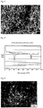
- FIG. 2 is a stereoscopic micrograph thereof. Under the stereoscopic microscope, 20 crude collagen fibers were randomly selected, their diameters and lengths were measured, and the average values of the 20 fibers were calculated. The 20 crude collagen fibers had an average diameter of 1.15 ⁇ m and an average length of 4.09 mm. It is noted here that the shortest fiber length was 1.9 mm and the longest fiber length was 8.75 mm.
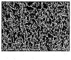
- the surface fiber structure of the thus obtained collagen structure was observed under a scanning electron microscope (SEM). The result thereof is shown in FIG. 3 .
- the collagen structure had an average pore size of 18.47 ⁇ m, meaning that the collagen structure had sufficient spaces for allowing cells of 5 to 7 ⁇ m in diameter to infiltrate.
- the fiber structure of a cross-section of the collagen structure was observed under a scanning electron microscope (SEM). The result thereof is shown in FIG. 4 .
- the denaturation temperature of the thus obtained collagen structure and that of the collagen solution used as a control were measured using a differential scanning calorimeter (DSC) at a heating rate of 2°C/minute. The results thereof are shown in FIG. 5 and Table 2.
- the collagen structure was observed to have a peak of denaturation temperature at 115.03°C, while the collagen solution had a denaturation temperature of 42.75°C. Therefore, it was revealed that the collagen structure had superior thermal stability as compared to the collagen solution.
- the collagen density and the porosity were measured by the following methods. As a result, the collagen density was found to be 200 mg/cm 3 and the porosity was found to be 40.9%.
- the collagen structure is cut precisely into a size of 1-cm square to prepare a test piece.
- the thickness of this test piece is precisely measured using a thickness gauge so as to calculate the volume.
- the test piece is dissolved in 5 ml of 5mM acetic acid solution and the collagen concentration is measured by a microburet method. From the volume and collagen concentration of the test piece, the amount of collagen per unit volume is calculated as the collagen density.
- the porosity is measured by mercury intrusion porosimetry using Pascal 140 and 440 (manufactured by Carlo-Erba Instruments, Ltd.).
- the thus obtained collagen structure was swollen with DMEM/10% FBS and inoculated with HFF cells at a cell density of 1.0 ⁇ 10 4 cells/cm 2 . Then, 20 hours later, the cells were stained with calcein AM and observed under a fluorescence microscope. The result thereof is shown in FIG. 6 .
- a tissue which was prepared by grinding the dermal layer of a porcine skin using a meat grinder or the like and then defatting and sufficiently washing the resultant, was used as a raw material.
- a solubilized aqueous solution in which pepsin and acetic acid were mixed at final concentrations of 5 mg/ml and 50 mM, respectively, the raw material was suspended to a final collagen concentration of 4.5 (w/v)%, and the resulting suspension was subjected to an overnight solubilization treatment at 4°C.
- sodium chloride was added to a final concentration of 5 (w/v)% to perform salt precipitation, and the thus formed precipitates were recovered by centrifugation.
- the recovered salt precipitates were dispersed in distilled water to a collagen concentration of 3 (w/v)% and then uniformly dissolved by adjusting the pH to 3.0 with an addition of hydrochloric acid, thereby preparing a collagen solution.
- this collagen solution temperature: 4°C
- 95 ml of phosphate-buffered saline pH: 7.5, temperature: 4°C was added, and the resultant was left to stand at 37°C for 24 hours.
- the water content, the collagen density, the porosity, and the average diameter of collagen fibers were measured in the same manner as in Example 1. As a result, it was found that this collagen structure had a water content of 6.7 (w/w)%, a collagen density of 127 mg/cm 3 and a porosity of 76.6%. Further, the average diameter of the collagen fibers was 1.59 ⁇ m.
- the collagen solution obtained in Example 1 was diluted to a concentration of 0.4 (w/v)% by adding thereto distilled water and then mixed with an equivolume of 2 ⁇ concentrated phosphate-buffered saline (pH: 7.5) at 4°C. The resulting mixture was gently poured on a cell culture plate and this plate was left to stand at 37°C for 24 hours to produce a gelatinous material.
- gelatinous material was freeze-dried as it was, without isolating collagen fibers therefrom.
- Example 2 The water content and the collagen density were measured in the same manner as in Example 1. As a result, it was found that this freeze-dried material had a water content of 10 to 15 (w/w)% and a collagen density of 2.0 mg/cm 3 .
- the average diameter and pore size of the collagen fibers constituting the freeze-dried material were measured in the same manner as in Example 1.
- the collagen fibers constituting this film were found to have an average diameter of 0.17 ⁇ m. It is noted here that the fiber length could not be measured since the collagen fibers were in contact with each other. The results are shown in Table 1.
- the gelatinous material before being freeze-dried was observed under a scanning electron microscope (SEM). The result thereof is shown in FIG. 8 .
- the gelatinous material before being freeze-dried was acclimated with DMEM/10% FBS and inoculated with HFF cells at a cell density of 1.0 ⁇ 10 4 cells/cm 2 in the same manner as in Example 1. Then 20 hours later, the cells were stained with calcein AM and observed under a fluorescence microscope. The result thereof is shown in FIG. 9 .
- FIG. 6 of Example 1 a condition where the cells were three-dimensionally arranged was observed with both in-focus cells and out-of-focus cells existing at the same time; however, in FIG. 9 , since the cells were in focus, it was observed that the cells existed two-dimensionally in a single plane.
- the collagen solution obtained in Example 1 was adjusted to have a collagen concentration of 0.075 (w/v)% by adding thereto fivefold concentrated phosphate-buffered saline (pH: 7.5), and the resultant was stirred overnight at 37°C rather intensely (600 rpm) to form collagen fibers.
- the resulting collagen fiber-containing solution was stirred using a homogenizer to physically cut the collagen fibers or to inhibit binding of collagen molecules in the longitudinal direction.
- the collagen assembly contained in this solution had an average diameter of 1.13 ⁇ m and a length of 213 ⁇ m.
- the collagen assembly was recovered by 20-minute centrifugation at 17,500 rpm and centrifugation was repeated until a collagen concentration of 30 (w/v)% was attained.
- the resulting dispersion was filtered through a nylon mesh having a pore size of 80 ⁇ m to recover crude collagen assembly on the mesh. The thus obtained precipitates did not form a sheet and were in the form of powder.
- the collagen solution obtained in Example 1 was diluted to a concentration of 0.8 (w/v)% by adding thereto distilled water and then freeze-dried as it was without neutralization, thereby preparing a collagen sponge.
- the thus obtained collagen sponge was porous and film-like structure and was not constituted by collagen fibers.
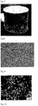
- FIG. 10 shows an electron micrograph of the collagen sponge. It is noted here that the collagen sponge had a collagen density of 8 mg/cm 3 .
- Example 1 Comparative Example 1 Average fiber diameter 2.53 ⁇ m 0.17 ⁇ m Average pore size 18.47 ⁇ m 1.15 ⁇ m
- Example 2 Example 1 Collagen solution Collagen structure Denaturation temperature 42.75°C 115.03°C Amount of denaturation heat 48.13 mJ/mg 35.55 mJ/mg
- the collagen structure according to the present disclosure is a dry material having a high collagen density.
- the collagen structure according to the present disclosure is useful since it also has a high thermal stability and can thus be used as a tissue-equivalent material in regenerative medicine.
Landscapes
- Health & Medical Sciences (AREA)
- Life Sciences & Earth Sciences (AREA)
- Veterinary Medicine (AREA)
- Public Health (AREA)
- General Health & Medical Sciences (AREA)
- Animal Behavior & Ethology (AREA)
- Chemical & Material Sciences (AREA)
- Medicinal Chemistry (AREA)
- Epidemiology (AREA)
- Chemical Kinetics & Catalysis (AREA)
- Pharmacology & Pharmacy (AREA)
- Organic Chemistry (AREA)
- Nuclear Medicine, Radiotherapy & Molecular Imaging (AREA)
- General Chemical & Material Sciences (AREA)
- Engineering & Computer Science (AREA)
- Bioinformatics & Cheminformatics (AREA)
- Dermatology (AREA)
- Birds (AREA)
- Immunology (AREA)
- Surgery (AREA)
- Heart & Thoracic Surgery (AREA)
- Vascular Medicine (AREA)
- Transplantation (AREA)
- Oral & Maxillofacial Surgery (AREA)
- Biophysics (AREA)
- Gerontology & Geriatric Medicine (AREA)
- Hematology (AREA)
- Diabetes (AREA)
- Rheumatology (AREA)
- Ophthalmology & Optometry (AREA)
- Orthopedic Medicine & Surgery (AREA)
- Physical Education & Sports Medicine (AREA)
- Materials For Medical Uses (AREA)
- Cosmetics (AREA)
- Medicines That Contain Protein Lipid Enzymes And Other Medicines (AREA)
- Prostheses (AREA)
Claims (7)
- Kollagenstruktur, die durch Kollagenfasern mit einem mittleren Durchmesser von 1 bis 5 m und einer mittleren Länge 1 bis 10 mm erzeugt wird, die für 20 Fasern aufgezeichnet in einem Stereomikrobild gemessen werden; und
einen Wassergehalt von 0 bis 15 Gewichts-(w/w) %, der als Gehalt einer Menge Wasser in Bezug auf eine Masse einer Kollagenstruktur nach Erhitzen auf 120°C für 2 Stunden definiert wird, wobei die Änderung der Masse vor und nach dem Erhitzen als die Wassermenge bestimmt wird und der Wassergehalt ist definiert als Gewichtsprozent dieser Menge in Bezug auf die Kollagenmasse; und eine Kollagendichte von 50 bis 800 mg/cm3, wobei ein Probestück mit einer definierten Dicke in Essigsäure gelöst wird und die Kollegenkonzentration mit einem Mikrobüretteverfahren gemessen wird, aus dem Volumen und der Kollegenkonzentration des Probestückes die Menge Kollegen pro Volumeneinheit als Kollegendichte berechnet wird. - Kollegenstruktur nach Anspruch 1, die eine mittleren Porengröße von 1 bis 50 µm, gemessen für 20 Poren in der Kollegenstruktur mit einem Rasterelektronenmikroskop, aufweist.
- Kollagenstruktur nach einem der Ansprüche 1 oder 2, des weiteren umfassend wenigstens einen Faktor ausgewählt aus der Gruppe bestehend aus Zellchemotaktische Faktoren, Wachstumsfaktoren, Zellproliferationsfaktoren, Blutkoagulationsfaktoren und Antikoalugantfaktoren.
- Kollegenstruktur nach einem der Ansprüche 1 bis 3, die als ein künstliches medizinisches Material, ein Teil zur Krankheitsbehandlung, ein kosmetisches Material oder ein Zellkulturmaterial verwendet wird.
- Verfahren zur Herstellung einer Kollegenstruktur, umfassen die Schritte:Erzeugung von Kollagenfasern mit einem mittleren Durchmesser von 1 bis 100 µm und einer mittleren Länge von 1 bis 10 mm, die für 20 Fasern aufgezeichnet in einem Stereomikrobild durch Neutralisation und leichtes Rühren einer sauren Kollagenlösung, um die Assoziierung der Kollagenfasern zu erleichtern, gemessen wird;Bildung von Rohkollagenfasern mit einer Kollagenkonzentration von 12 bis 50 (w/v) % durch Trennen der Kollagenfasern von der Lösung, die die Kollagenfasern enthält;Pressen der Rohkollagenfasern in eine vorbeschriebene Form; undTrocknen des in dem Pressverfahren erhaltenen gepressten Artikels.
- Verfahren nach Anspruch 5, des weiteren umfassend nach dem Schritt der Bildung der Rohkollagenfasern die Schritte:nach dem Dispergieren des Rohkollagenfasern in einem hydrophilen organischen Lösungsmittels werden die Kollagenfasern von dem hydrophilen organischen Lösungsmittel getrennt und die so getrennten Kollagenfasern werden dehydratisiert; unddie so erhaltenen Kollagenfasern werden gepresst.
- Verfahren zur Herstellung einer Kollagenstruktur nach Anspruch 6, des weiteren umfassend nach dem Wasserentzugsschritt der Kollagenfasern die Schritte:Unterwerfen der dehydratisierten Kollagenfasern einer Quervernetzungsbehandlung und/oder einer chemischen Behandlung; undTrocknen der so behandelten Kollagenfasern.
Applications Claiming Priority (2)
| Application Number | Priority Date | Filing Date | Title |
|---|---|---|---|
| JP2012003883 | 2012-01-12 | ||
| PCT/JP2013/050484 WO2013105665A1 (ja) | 2012-01-12 | 2013-01-11 | コラーゲン構造体、およびコラーゲン構造体の製造方法 |
Publications (3)
| Publication Number | Publication Date |
|---|---|
| EP2803371A1 EP2803371A1 (de) | 2014-11-19 |
| EP2803371A4 EP2803371A4 (de) | 2015-08-05 |
| EP2803371B1 true EP2803371B1 (de) | 2019-10-30 |
Family
ID=48781605
Family Applications (1)
| Application Number | Title | Priority Date | Filing Date |
|---|---|---|---|
| EP13735626.7A Active EP2803371B1 (de) | 2012-01-12 | 2013-01-11 | Kollagenstruktur und verfahren zur herstellung der kollagenstruktur |
Country Status (6)
| Country | Link |
|---|---|
| US (1) | US20150004414A1 (de) |
| EP (1) | EP2803371B1 (de) |
| JP (1) | JP6100702B2 (de) |
| CN (1) | CN104144715B (de) |
| CA (1) | CA2861027C (de) |
| WO (1) | WO2013105665A1 (de) |
Families Citing this family (25)
| Publication number | Priority date | Publication date | Assignee | Title |
|---|---|---|---|---|
| JP6023996B2 (ja) * | 2012-08-09 | 2016-11-09 | 島根県 | コラーゲン線維からなるコラーゲン人工皮膚およびそれを用いた紫外線ダメージの評価方法 |
| US11623947B2 (en) * | 2013-04-08 | 2023-04-11 | Innocoll Pharmaceuticals Limited | Collagen powder |
| US10173375B2 (en) | 2014-03-05 | 2019-01-08 | Bacterin International, Inc. | Shaped fiber-based products and method of manufacture thereof |
| JP6429490B2 (ja) * | 2014-05-12 | 2018-11-28 | 多木化学株式会社 | コラーゲン線維架橋多孔体 |
| JP6432967B2 (ja) * | 2014-05-12 | 2018-12-05 | 多木化学株式会社 | 溶解性コラーゲン線維多孔体 |
| JP6566290B2 (ja) * | 2014-12-04 | 2019-08-28 | 学校法人北里研究所 | 軟骨再生用移植材料、軟骨再生用移植材料の製造方法 |
| US20160287747A1 (en) * | 2015-03-31 | 2016-10-06 | Mark Schallenberger | Shaped collagen tissue implant and method of manufacturing thereof |
| WO2016178586A2 (en) * | 2015-05-01 | 2016-11-10 | Auckland Uniservices Limited | Collagen compositions and preparation and uses thereof |
| US10821004B2 (en) | 2015-06-30 | 2020-11-03 | Bacterin Interational, Inc. | Expandable bone grafts and methods of manufacture thereof |
| JP6856969B2 (ja) * | 2015-11-05 | 2021-04-14 | 多木化学株式会社 | 線状コラーゲン架橋多孔体の製造方法 |
| TWI635877B (zh) * | 2015-12-18 | 2018-09-21 | 財團法人工業技術研究院 | 用於保護眼內組織的薄膜及其保護方法 |
| CN106913908B (zh) * | 2015-12-25 | 2020-05-26 | 北京瑞健高科生物科技有限公司 | 一种具有结构记忆特性的细胞生长支架 |
| CN106913907B (zh) * | 2015-12-25 | 2022-04-05 | 北京瑞健高科生物科技有限公司 | 一种具有结构记忆特性的细胞生长支架的制备方法 |
| JP7165326B2 (ja) * | 2016-12-28 | 2022-11-04 | 株式会社高研 | 高強度コラーゲンスポンジ |
| JP6452020B1 (ja) | 2017-05-17 | 2019-01-16 | 国立研究開発法人農業・食品産業技術総合研究機構 | ビトリゲル膜乾燥体の製造方法及び製造装置 |
| CN107354800B (zh) * | 2017-06-30 | 2020-02-18 | 华南理工大学 | 一种基于造纸法制备具有ecm结构的人体组织代用材料的方法 |
| CN107551314A (zh) * | 2017-09-21 | 2018-01-09 | 浙江大学 | 一种促进骨髓间充质干细胞粘附的e7‑胶原膜及其制备方法 |
| US20210238542A1 (en) * | 2018-04-27 | 2021-08-05 | Toppan Printing Co., Ltd. | Extracellular-matrix-containing composition, method for producing same, three-dimensional tissue construct, and three-dimensional tissue construct formation agent |
| US20200000962A1 (en) * | 2018-07-02 | 2020-01-02 | Medtronic Vascular, Inc. | Load bearing crowded collagen constructs |
| EP3880234A4 (de) * | 2018-11-13 | 2022-08-10 | Northeastern University | Flüssigkristalline kollagenmaterialien und verwendung in der bindegewebereparatur |
| CZ309204B6 (cs) * | 2019-12-17 | 2022-05-18 | Všeobecná fakultní nemocnice v Praze | Kolagenní kompozit pro řízené uvolňování aktivních látek a způsob jeho přípravy |
| CN111346267A (zh) * | 2020-04-14 | 2020-06-30 | 中国人民解放军海军军医大学第三附属医院 | 一种带可吸收止血抗菌胰腺导管引流管 |
| WO2021210639A1 (ja) * | 2020-04-17 | 2021-10-21 | 学校法人日本大学 | 骨増生用構造体 |
| CN112426560A (zh) * | 2020-12-10 | 2021-03-02 | 广州创尔生物技术股份有限公司 | 一种胶原蛋白海绵及其制备方法 |
| CN114182381B (zh) * | 2022-01-24 | 2022-07-12 | 浙江启宏新材料科技有限责任公司 | 一种胶原纤维的制备方法、胶原纤维、应用 |
Family Cites Families (19)
| Publication number | Priority date | Publication date | Assignee | Title |
|---|---|---|---|---|
| CA1073360A (en) * | 1975-10-22 | 1980-03-11 | John R. Daniels | Non-antigenic collagen and articles of manufacture |
| US4861714A (en) * | 1985-04-04 | 1989-08-29 | Verax Corporation | Weighted collagen microsponge for immobilizing bioactive material |
| JPH0622579B2 (ja) | 1988-03-09 | 1994-03-30 | テルモ株式会社 | 細胞侵入性医用材料 |
| US4950483A (en) * | 1988-06-30 | 1990-08-21 | Collagen Corporation | Collagen wound healing matrices and process for their production |
| JPH07100B2 (ja) | 1990-01-31 | 1995-01-11 | グンゼ株式会社 | コラーゲンスポンジの乾燥法 |
| US5378469A (en) * | 1990-04-06 | 1995-01-03 | Organogenesis, Inc. | Collagen threads |
| US5256418A (en) | 1990-04-06 | 1993-10-26 | Organogenesis, Inc. | Collagen constructs |
| TW501934B (en) * | 1996-11-20 | 2002-09-11 | Tapic Int Co Ltd | Collagen material and process for making the same |
| WO2000007639A1 (en) * | 1998-08-07 | 2000-02-17 | Tissue Engineering, Inc. | Bone precursor compositions |
| DE60018814T2 (de) * | 1999-01-21 | 2006-04-06 | Nipro Corp. | Nähbare Membran zur Adhäsionsverhinderung |
| AU2002219515B2 (en) * | 2000-12-22 | 2006-09-14 | Hokuyo Co., Ltd. | Process for producing regenerated collagen fiber and process for setting the same |
| JP4681214B2 (ja) | 2003-06-10 | 2011-05-11 | グンゼ株式会社 | コラーゲンスポンジの製造方法及び人工皮膚の製造方法 |
| WO2005079879A1 (ja) * | 2004-02-25 | 2005-09-01 | Ihara & Company Ltd. | コラーゲンゲルおよびその製造方法 |
| WO2006088029A1 (ja) | 2005-02-15 | 2006-08-24 | School Juridical Person Kitasato Gakuen | 高密度培養組織の製造方法及び高密度培養組織 |
| CN100348730C (zh) * | 2005-12-02 | 2007-11-14 | 成都佰乐金生物科技有限公司 | 一种医用胶原蛋白粉剂的制备方法 |
| CA2652138C (en) * | 2006-05-16 | 2016-01-19 | Purdue Research Foundation | Three dimensional purified collagen matrices |
| JP5274964B2 (ja) * | 2007-10-04 | 2013-08-28 | 共栄化学工業株式会社 | 化粧料 |
| JP2010172247A (ja) * | 2009-01-29 | 2010-08-12 | Kitasato Institute | 積層型高密度培養人工組織の製造方法及び積層型高密度培養人工組織 |
| JP2010273847A (ja) * | 2009-05-28 | 2010-12-09 | Tokyo Institute Of Technology | 高密度多孔質複合体 |
-
2013
- 2013-01-11 EP EP13735626.7A patent/EP2803371B1/de active Active
- 2013-01-11 US US14/371,845 patent/US20150004414A1/en not_active Abandoned
- 2013-01-11 JP JP2013553333A patent/JP6100702B2/ja active Active
- 2013-01-11 CA CA2861027A patent/CA2861027C/en active Active
- 2013-01-11 WO PCT/JP2013/050484 patent/WO2013105665A1/ja not_active Ceased
- 2013-01-11 CN CN201380005501.2A patent/CN104144715B/zh not_active Expired - Fee Related
Non-Patent Citations (1)
| Title |
|---|
| None * |
Also Published As
| Publication number | Publication date |
|---|---|
| US20150004414A1 (en) | 2015-01-01 |
| JPWO2013105665A1 (ja) | 2015-05-11 |
| CN104144715A (zh) | 2014-11-12 |
| WO2013105665A1 (ja) | 2013-07-18 |
| EP2803371A4 (de) | 2015-08-05 |
| CN104144715B (zh) | 2016-04-27 |
| EP2803371A1 (de) | 2014-11-19 |
| CA2861027A1 (en) | 2013-07-18 |
| CA2861027C (en) | 2020-08-25 |
| JP6100702B2 (ja) | 2017-03-22 |
Similar Documents
| Publication | Publication Date | Title |
|---|---|---|
| EP2803371B1 (de) | Kollagenstruktur und verfahren zur herstellung der kollagenstruktur | |
| Klemm et al. | Nanocelluloses as innovative polymers in research and application | |
| Da et al. | Progress in development of bioderived materials for dermal wound healing | |
| Hosty et al. | Extracellular matrix-inspired biomaterials for wound healing | |
| US20020146393A1 (en) | EB Matrix Production from Fetal Tissues and its Use for Tissue Repair | |
| Wang et al. | Enhanced physical and biological properties of chitosan scaffold by silk proteins cross-linking | |
| AU2002309898A1 (en) | EB matrix production from fetal tissues and its use for tissue repair | |
| CN110917386B (zh) | 干细胞海绵支架及制备方法和应用 | |
| Kangjian et al. | Preparation and characterization of collagen–chitosan–chondroitin sulfate composite membranes | |
| CN113301928B (zh) | 生物支架和用于制备其的方法 | |
| Yao et al. | Facile incorporation of REDV into porous silk fibroin scaffolds for enhancing vascularization of thick tissues | |
| EP3223749A1 (de) | Verfahren zur herstellung einer geweberegenerationsmatrix | |
| Ficai et al. | Advances in the field of soft tissue engineering: From pure regenerative to integrative solutions | |
| JP6566290B2 (ja) | 軟骨再生用移植材料、軟骨再生用移植材料の製造方法 | |
| JP6532112B2 (ja) | コラーゲン生体材料 | |
| WO2003094985A1 (fr) | Matrice extracellulaire artificielle et procede de fabrication associe | |
| JP2015213676A (ja) | コラーゲン線維架橋多孔体 | |
| WO2017205740A1 (en) | Process for preparing tissue regeneration matrix | |
| JP5679684B2 (ja) | Danceタンパク質含有組織再生用基材 | |
| US20240042095A1 (en) | Composition for treating wound comprising dermal tissue-derived extracellular matrix and method for preparing same | |
| CN116848132A (zh) | 产生具有受控特性的人胶原蛋白结构的方法 | |
| Klemm¹ et al. | Springer-Verlag Berlin Heidelberg 2006 Published online: 30 August 2006 Nanocelluloses as Innovative Polymers in Research and Application | |
| WO2002096263A2 (en) | Eb matrix production from fetal tissues and its use for tissue repair | |
| KR20050023298A (ko) | 케라틴 유래의 정형외과 재료 | |
| PL238231B1 (pl) | Sposób wytwarzania elektroprzędzonych materiałów zawierających natywne białko albuminy, mata otrzymana tym sposobem oraz jej zastosowanie, zwłaszcza jako materiałów opatrunkowych |
Legal Events
| Date | Code | Title | Description |
|---|---|---|---|
| PUAI | Public reference made under article 153(3) epc to a published international application that has entered the european phase |
Free format text: ORIGINAL CODE: 0009012 |
|
| 17P | Request for examination filed |
Effective date: 20140731 |
|
| AK | Designated contracting states |
Kind code of ref document: A1 Designated state(s): AL AT BE BG CH CY CZ DE DK EE ES FI FR GB GR HR HU IE IS IT LI LT LU LV MC MK MT NL NO PL PT RO RS SE SI SK SM TR |
|
| DAX | Request for extension of the european patent (deleted) | ||
| RIC1 | Information provided on ipc code assigned before grant |
Ipc: A61L 31/04 20060101ALI20150528BHEP Ipc: A61K 9/70 20060101ALI20150528BHEP Ipc: A61P 19/02 20060101ALI20150528BHEP Ipc: A61K 47/42 20060101ALI20150528BHEP Ipc: A61K 8/65 20060101ALI20150528BHEP Ipc: A61L 27/24 20060101AFI20150528BHEP Ipc: A61P 35/00 20060101ALI20150528BHEP Ipc: A61K 8/02 20060101ALI20150528BHEP Ipc: A61P 43/00 20060101ALI20150528BHEP Ipc: A61P 37/02 20060101ALI20150528BHEP Ipc: A61Q 19/08 20060101ALI20150528BHEP Ipc: A61P 7/04 20060101ALI20150528BHEP Ipc: A61L 31/14 20060101ALI20150528BHEP Ipc: A61P 17/02 20060101ALI20150528BHEP Ipc: A61K 38/17 20060101ALI20150528BHEP |
|
| RA4 | Supplementary search report drawn up and despatched (corrected) |
Effective date: 20150707 |
|
| RIC1 | Information provided on ipc code assigned before grant |
Ipc: A61K 9/70 20060101ALI20150629BHEP Ipc: A61K 8/02 20060101ALI20150629BHEP Ipc: A61L 31/04 20060101ALI20150629BHEP Ipc: A61P 37/02 20060101ALI20150629BHEP Ipc: A61P 7/04 20060101ALI20150629BHEP Ipc: A61L 27/24 20060101AFI20150629BHEP Ipc: A61K 38/17 20060101ALI20150629BHEP Ipc: A61K 47/42 20060101ALI20150629BHEP Ipc: A61K 8/65 20060101ALI20150629BHEP Ipc: A61L 31/14 20060101ALI20150629BHEP Ipc: A61P 43/00 20060101ALI20150629BHEP Ipc: A61P 17/02 20060101ALI20150629BHEP Ipc: A61P 35/00 20060101ALI20150629BHEP Ipc: A61P 19/02 20060101ALI20150629BHEP Ipc: A61Q 19/08 20060101ALI20150629BHEP |
|
| STAA | Information on the status of an ep patent application or granted ep patent |
Free format text: STATUS: EXAMINATION IS IN PROGRESS |
|
| 17Q | First examination report despatched |
Effective date: 20170510 |
|
| GRAP | Despatch of communication of intention to grant a patent |
Free format text: ORIGINAL CODE: EPIDOSNIGR1 |
|
| STAA | Information on the status of an ep patent application or granted ep patent |
Free format text: STATUS: GRANT OF PATENT IS INTENDED |
|
| INTG | Intention to grant announced |
Effective date: 20190531 |
|
| GRAS | Grant fee paid |
Free format text: ORIGINAL CODE: EPIDOSNIGR3 |
|
| GRAA | (expected) grant |
Free format text: ORIGINAL CODE: 0009210 |
|
| STAA | Information on the status of an ep patent application or granted ep patent |
Free format text: STATUS: THE PATENT HAS BEEN GRANTED |
|
| AK | Designated contracting states |
Kind code of ref document: B1 Designated state(s): AL AT BE BG CH CY CZ DE DK EE ES FI FR GB GR HR HU IE IS IT LI LT LU LV MC MK MT NL NO PL PT RO RS SE SI SK SM TR |
|
| REG | Reference to a national code |
Ref country code: GB Ref legal event code: FG4D |
|
| REG | Reference to a national code |
Ref country code: CH Ref legal event code: EP Ref country code: CH Ref legal event code: NV Representative=s name: ROTTMANN, ZIMMERMANN + PARTNER AG, CH |
|
| REG | Reference to a national code |
Ref country code: AT Ref legal event code: REF Ref document number: 1195457 Country of ref document: AT Kind code of ref document: T Effective date: 20191115 |
|
| REG | Reference to a national code |
Ref country code: DE Ref legal event code: R096 Ref document number: 602013062243 Country of ref document: DE |
|
| REG | Reference to a national code |
Ref country code: IE Ref legal event code: FG4D |
|
| REG | Reference to a national code |
Ref country code: LT Ref legal event code: MG4D |
|
| PG25 | Lapsed in a contracting state [announced via postgrant information from national office to epo] |
Ref country code: BG Free format text: LAPSE BECAUSE OF FAILURE TO SUBMIT A TRANSLATION OF THE DESCRIPTION OR TO PAY THE FEE WITHIN THE PRESCRIBED TIME-LIMIT Effective date: 20200130 Ref country code: FI Free format text: LAPSE BECAUSE OF FAILURE TO SUBMIT A TRANSLATION OF THE DESCRIPTION OR TO PAY THE FEE WITHIN THE PRESCRIBED TIME-LIMIT Effective date: 20191030 Ref country code: GR Free format text: LAPSE BECAUSE OF FAILURE TO SUBMIT A TRANSLATION OF THE DESCRIPTION OR TO PAY THE FEE WITHIN THE PRESCRIBED TIME-LIMIT Effective date: 20200131 Ref country code: PL Free format text: LAPSE BECAUSE OF FAILURE TO SUBMIT A TRANSLATION OF THE DESCRIPTION OR TO PAY THE FEE WITHIN THE PRESCRIBED TIME-LIMIT Effective date: 20191030 Ref country code: NO Free format text: LAPSE BECAUSE OF FAILURE TO SUBMIT A TRANSLATION OF THE DESCRIPTION OR TO PAY THE FEE WITHIN THE PRESCRIBED TIME-LIMIT Effective date: 20200130 Ref country code: LV Free format text: LAPSE BECAUSE OF FAILURE TO SUBMIT A TRANSLATION OF THE DESCRIPTION OR TO PAY THE FEE WITHIN THE PRESCRIBED TIME-LIMIT Effective date: 20191030 Ref country code: SE Free format text: LAPSE BECAUSE OF FAILURE TO SUBMIT A TRANSLATION OF THE DESCRIPTION OR TO PAY THE FEE WITHIN THE PRESCRIBED TIME-LIMIT Effective date: 20191030 Ref country code: NL Free format text: LAPSE BECAUSE OF FAILURE TO SUBMIT A TRANSLATION OF THE DESCRIPTION OR TO PAY THE FEE WITHIN THE PRESCRIBED TIME-LIMIT Effective date: 20191030 Ref country code: ES Free format text: LAPSE BECAUSE OF FAILURE TO SUBMIT A TRANSLATION OF THE DESCRIPTION OR TO PAY THE FEE WITHIN THE PRESCRIBED TIME-LIMIT Effective date: 20191030 Ref country code: LT Free format text: LAPSE BECAUSE OF FAILURE TO SUBMIT A TRANSLATION OF THE DESCRIPTION OR TO PAY THE FEE WITHIN THE PRESCRIBED TIME-LIMIT Effective date: 20191030 Ref country code: PT Free format text: LAPSE BECAUSE OF FAILURE TO SUBMIT A TRANSLATION OF THE DESCRIPTION OR TO PAY THE FEE WITHIN THE PRESCRIBED TIME-LIMIT Effective date: 20200302 |
|
| REG | Reference to a national code |
Ref country code: NL Ref legal event code: MP Effective date: 20191030 |
|
| PG25 | Lapsed in a contracting state [announced via postgrant information from national office to epo] |
Ref country code: RS Free format text: LAPSE BECAUSE OF FAILURE TO SUBMIT A TRANSLATION OF THE DESCRIPTION OR TO PAY THE FEE WITHIN THE PRESCRIBED TIME-LIMIT Effective date: 20191030 Ref country code: HR Free format text: LAPSE BECAUSE OF FAILURE TO SUBMIT A TRANSLATION OF THE DESCRIPTION OR TO PAY THE FEE WITHIN THE PRESCRIBED TIME-LIMIT Effective date: 20191030 Ref country code: IS Free format text: LAPSE BECAUSE OF FAILURE TO SUBMIT A TRANSLATION OF THE DESCRIPTION OR TO PAY THE FEE WITHIN THE PRESCRIBED TIME-LIMIT Effective date: 20200229 |
|
| PG25 | Lapsed in a contracting state [announced via postgrant information from national office to epo] |
Ref country code: AL Free format text: LAPSE BECAUSE OF FAILURE TO SUBMIT A TRANSLATION OF THE DESCRIPTION OR TO PAY THE FEE WITHIN THE PRESCRIBED TIME-LIMIT Effective date: 20191030 |
|
| PG25 | Lapsed in a contracting state [announced via postgrant information from national office to epo] |
Ref country code: DK Free format text: LAPSE BECAUSE OF FAILURE TO SUBMIT A TRANSLATION OF THE DESCRIPTION OR TO PAY THE FEE WITHIN THE PRESCRIBED TIME-LIMIT Effective date: 20191030 Ref country code: EE Free format text: LAPSE BECAUSE OF FAILURE TO SUBMIT A TRANSLATION OF THE DESCRIPTION OR TO PAY THE FEE WITHIN THE PRESCRIBED TIME-LIMIT Effective date: 20191030 Ref country code: CZ Free format text: LAPSE BECAUSE OF FAILURE TO SUBMIT A TRANSLATION OF THE DESCRIPTION OR TO PAY THE FEE WITHIN THE PRESCRIBED TIME-LIMIT Effective date: 20191030 Ref country code: RO Free format text: LAPSE BECAUSE OF FAILURE TO SUBMIT A TRANSLATION OF THE DESCRIPTION OR TO PAY THE FEE WITHIN THE PRESCRIBED TIME-LIMIT Effective date: 20191030 |
|
| REG | Reference to a national code |
Ref country code: DE Ref legal event code: R097 Ref document number: 602013062243 Country of ref document: DE |
|
| REG | Reference to a national code |
Ref country code: AT Ref legal event code: MK05 Ref document number: 1195457 Country of ref document: AT Kind code of ref document: T Effective date: 20191030 |
|
| PG25 | Lapsed in a contracting state [announced via postgrant information from national office to epo] |
Ref country code: MC Free format text: LAPSE BECAUSE OF FAILURE TO SUBMIT A TRANSLATION OF THE DESCRIPTION OR TO PAY THE FEE WITHIN THE PRESCRIBED TIME-LIMIT Effective date: 20191030 Ref country code: SM Free format text: LAPSE BECAUSE OF FAILURE TO SUBMIT A TRANSLATION OF THE DESCRIPTION OR TO PAY THE FEE WITHIN THE PRESCRIBED TIME-LIMIT Effective date: 20191030 Ref country code: SK Free format text: LAPSE BECAUSE OF FAILURE TO SUBMIT A TRANSLATION OF THE DESCRIPTION OR TO PAY THE FEE WITHIN THE PRESCRIBED TIME-LIMIT Effective date: 20191030 |
|
| PLBE | No opposition filed within time limit |
Free format text: ORIGINAL CODE: 0009261 |
|
| STAA | Information on the status of an ep patent application or granted ep patent |
Free format text: STATUS: NO OPPOSITION FILED WITHIN TIME LIMIT |
|
| 26N | No opposition filed |
Effective date: 20200731 |
|
| REG | Reference to a national code |
Ref country code: BE Ref legal event code: MM Effective date: 20200131 |
|
| PG25 | Lapsed in a contracting state [announced via postgrant information from national office to epo] |
Ref country code: LU Free format text: LAPSE BECAUSE OF NON-PAYMENT OF DUE FEES Effective date: 20200111 |
|
| PG25 | Lapsed in a contracting state [announced via postgrant information from national office to epo] |
Ref country code: AT Free format text: LAPSE BECAUSE OF FAILURE TO SUBMIT A TRANSLATION OF THE DESCRIPTION OR TO PAY THE FEE WITHIN THE PRESCRIBED TIME-LIMIT Effective date: 20191030 Ref country code: SI Free format text: LAPSE BECAUSE OF FAILURE TO SUBMIT A TRANSLATION OF THE DESCRIPTION OR TO PAY THE FEE WITHIN THE PRESCRIBED TIME-LIMIT Effective date: 20191030 Ref country code: BE Free format text: LAPSE BECAUSE OF NON-PAYMENT OF DUE FEES Effective date: 20200131 |
|
| PG25 | Lapsed in a contracting state [announced via postgrant information from national office to epo] |
Ref country code: IE Free format text: LAPSE BECAUSE OF NON-PAYMENT OF DUE FEES Effective date: 20200111 |
|
| PG25 | Lapsed in a contracting state [announced via postgrant information from national office to epo] |
Ref country code: TR Free format text: LAPSE BECAUSE OF FAILURE TO SUBMIT A TRANSLATION OF THE DESCRIPTION OR TO PAY THE FEE WITHIN THE PRESCRIBED TIME-LIMIT Effective date: 20191030 Ref country code: MT Free format text: LAPSE BECAUSE OF FAILURE TO SUBMIT A TRANSLATION OF THE DESCRIPTION OR TO PAY THE FEE WITHIN THE PRESCRIBED TIME-LIMIT Effective date: 20191030 Ref country code: CY Free format text: LAPSE BECAUSE OF FAILURE TO SUBMIT A TRANSLATION OF THE DESCRIPTION OR TO PAY THE FEE WITHIN THE PRESCRIBED TIME-LIMIT Effective date: 20191030 |
|
| PG25 | Lapsed in a contracting state [announced via postgrant information from national office to epo] |
Ref country code: MK Free format text: LAPSE BECAUSE OF FAILURE TO SUBMIT A TRANSLATION OF THE DESCRIPTION OR TO PAY THE FEE WITHIN THE PRESCRIBED TIME-LIMIT Effective date: 20191030 |
|
| REG | Reference to a national code |
Ref country code: DE Ref legal event code: R082 Ref document number: 602013062243 Country of ref document: DE Representative=s name: PUSCHMANN BORCHERT KAISER KLETTNER PATENTANWAE, DE Ref country code: DE Ref legal event code: R082 Ref document number: 602013062243 Country of ref document: DE Representative=s name: PATCARE PATENTANWAELTE PARTNERSCHAFT MBB, DE |
|
| PGFP | Annual fee paid to national office [announced via postgrant information from national office to epo] |
Ref country code: DE Payment date: 20250130 Year of fee payment: 13 |
|
| PGFP | Annual fee paid to national office [announced via postgrant information from national office to epo] |
Ref country code: CH Payment date: 20250201 Year of fee payment: 13 |
|
| PGFP | Annual fee paid to national office [announced via postgrant information from national office to epo] |
Ref country code: FR Payment date: 20250123 Year of fee payment: 13 |
|
| PGFP | Annual fee paid to national office [announced via postgrant information from national office to epo] |
Ref country code: IT Payment date: 20250131 Year of fee payment: 13 Ref country code: GB Payment date: 20250123 Year of fee payment: 13 |
|
| REG | Reference to a national code |
Ref country code: CH Ref legal event code: U11 Free format text: ST27 STATUS EVENT CODE: U-0-0-U10-U11 (AS PROVIDED BY THE NATIONAL OFFICE) Effective date: 20260201 |