EP2526774A2 - Zielgesteuerte Genabgabe für die Immunisierung mit Dendritenzellen - Google Patents
Zielgesteuerte Genabgabe für die Immunisierung mit Dendritenzellen Download PDFInfo
- Publication number
- EP2526774A2 EP2526774A2 EP12158285A EP12158285A EP2526774A2 EP 2526774 A2 EP2526774 A2 EP 2526774A2 EP 12158285 A EP12158285 A EP 12158285A EP 12158285 A EP12158285 A EP 12158285A EP 2526774 A2 EP2526774 A2 EP 2526774A2
- Authority
- EP
- European Patent Office
- Prior art keywords
- virus
- cells
- polypeptides
- antigen
- gene
- Prior art date
- Legal status (The legal status is an assumption and is not a legal conclusion. Google has not performed a legal analysis and makes no representation as to the accuracy of the status listed.)
- Ceased
Links
Images


Classifications
-
- C—CHEMISTRY; METALLURGY
- C12—BIOCHEMISTRY; BEER; SPIRITS; WINE; VINEGAR; MICROBIOLOGY; ENZYMOLOGY; MUTATION OR GENETIC ENGINEERING
- C12N—MICROORGANISMS OR ENZYMES; COMPOSITIONS THEREOF; PROPAGATING, PRESERVING, OR MAINTAINING MICROORGANISMS; MUTATION OR GENETIC ENGINEERING; CULTURE MEDIA
- C12N15/00—Mutation or genetic engineering; DNA or RNA concerning genetic engineering, vectors, e.g. plasmids, or their isolation, preparation or purification; Use of hosts therefor
- C12N15/09—Recombinant DNA-technology
- C12N15/63—Introduction of foreign genetic material using vectors; Vectors; Use of hosts therefor; Regulation of expression
- C12N15/79—Vectors or expression systems specially adapted for eukaryotic hosts
- C12N15/85—Vectors or expression systems specially adapted for eukaryotic hosts for animal cells
- C12N15/86—Viral vectors
-
- A—HUMAN NECESSITIES
- A61—MEDICAL OR VETERINARY SCIENCE; HYGIENE
- A61K—PREPARATIONS FOR MEDICAL, DENTAL OR TOILETRY PURPOSES
- A61K31/00—Medicinal preparations containing organic active ingredients
- A61K31/70—Carbohydrates; Sugars; Derivatives thereof
-
- A—HUMAN NECESSITIES
- A61—MEDICAL OR VETERINARY SCIENCE; HYGIENE
- A61K—PREPARATIONS FOR MEDICAL, DENTAL OR TOILETRY PURPOSES
- A61K35/00—Medicinal preparations containing materials or reaction products thereof with undetermined constitution
- A61K35/66—Microorganisms or materials therefrom
- A61K35/76—Viruses; Subviral particles; Bacteriophages
-
- A—HUMAN NECESSITIES
- A61—MEDICAL OR VETERINARY SCIENCE; HYGIENE
- A61K—PREPARATIONS FOR MEDICAL, DENTAL OR TOILETRY PURPOSES
- A61K38/00—Medicinal preparations containing peptides
- A61K38/16—Peptides having more than 20 amino acids; Gastrins; Somatostatins; Melanotropins; Derivatives thereof
- A61K38/17—Peptides having more than 20 amino acids; Gastrins; Somatostatins; Melanotropins; Derivatives thereof from animals; from humans
- A61K38/177—Receptors; Cell surface antigens; Cell surface determinants
- A61K38/1774—Immunoglobulin superfamily (e.g. CD2, CD4, CD8, ICAM molecules, B7 molecules, Fc-receptors, MHC-molecules)
-
- A—HUMAN NECESSITIES
- A61—MEDICAL OR VETERINARY SCIENCE; HYGIENE
- A61K—PREPARATIONS FOR MEDICAL, DENTAL OR TOILETRY PURPOSES
- A61K38/00—Medicinal preparations containing peptides
- A61K38/16—Peptides having more than 20 amino acids; Gastrins; Somatostatins; Melanotropins; Derivatives thereof
- A61K38/17—Peptides having more than 20 amino acids; Gastrins; Somatostatins; Melanotropins; Derivatives thereof from animals; from humans
- A61K38/177—Receptors; Cell surface antigens; Cell surface determinants
- A61K38/178—Lectin superfamily, e.g. selectins
-
- A—HUMAN NECESSITIES
- A61—MEDICAL OR VETERINARY SCIENCE; HYGIENE
- A61K—PREPARATIONS FOR MEDICAL, DENTAL OR TOILETRY PURPOSES
- A61K38/00—Medicinal preparations containing peptides
- A61K38/16—Peptides having more than 20 amino acids; Gastrins; Somatostatins; Melanotropins; Derivatives thereof
- A61K38/17—Peptides having more than 20 amino acids; Gastrins; Somatostatins; Melanotropins; Derivatives thereof from animals; from humans
- A61K38/177—Receptors; Cell surface antigens; Cell surface determinants
- A61K38/1793—Receptors; Cell surface antigens; Cell surface determinants for cytokines; for lymphokines; for interferons
-
- A—HUMAN NECESSITIES
- A61—MEDICAL OR VETERINARY SCIENCE; HYGIENE
- A61K—PREPARATIONS FOR MEDICAL, DENTAL OR TOILETRY PURPOSES
- A61K38/00—Medicinal preparations containing peptides
- A61K38/16—Peptides having more than 20 amino acids; Gastrins; Somatostatins; Melanotropins; Derivatives thereof
- A61K38/17—Peptides having more than 20 amino acids; Gastrins; Somatostatins; Melanotropins; Derivatives thereof from animals; from humans
- A61K38/19—Cytokines; Lymphokines; Interferons
- A61K38/191—Tumor necrosis factors [TNF], e.g. lymphotoxin [LT], i.e. TNF-beta
-
- A—HUMAN NECESSITIES
- A61—MEDICAL OR VETERINARY SCIENCE; HYGIENE
- A61K—PREPARATIONS FOR MEDICAL, DENTAL OR TOILETRY PURPOSES
- A61K38/00—Medicinal preparations containing peptides
- A61K38/16—Peptides having more than 20 amino acids; Gastrins; Somatostatins; Melanotropins; Derivatives thereof
- A61K38/17—Peptides having more than 20 amino acids; Gastrins; Somatostatins; Melanotropins; Derivatives thereof from animals; from humans
- A61K38/19—Cytokines; Lymphokines; Interferons
- A61K38/193—Colony stimulating factors [CSF]
-
- A—HUMAN NECESSITIES
- A61—MEDICAL OR VETERINARY SCIENCE; HYGIENE
- A61K—PREPARATIONS FOR MEDICAL, DENTAL OR TOILETRY PURPOSES
- A61K38/00—Medicinal preparations containing peptides
- A61K38/16—Peptides having more than 20 amino acids; Gastrins; Somatostatins; Melanotropins; Derivatives thereof
- A61K38/17—Peptides having more than 20 amino acids; Gastrins; Somatostatins; Melanotropins; Derivatives thereof from animals; from humans
- A61K38/19—Cytokines; Lymphokines; Interferons
- A61K38/20—Interleukins [IL]
- A61K38/2013—IL-2
-
- A—HUMAN NECESSITIES
- A61—MEDICAL OR VETERINARY SCIENCE; HYGIENE
- A61K—PREPARATIONS FOR MEDICAL, DENTAL OR TOILETRY PURPOSES
- A61K38/00—Medicinal preparations containing peptides
- A61K38/16—Peptides having more than 20 amino acids; Gastrins; Somatostatins; Melanotropins; Derivatives thereof
- A61K38/17—Peptides having more than 20 amino acids; Gastrins; Somatostatins; Melanotropins; Derivatives thereof from animals; from humans
- A61K38/19—Cytokines; Lymphokines; Interferons
- A61K38/20—Interleukins [IL]
- A61K38/2026—IL-4
-
- A—HUMAN NECESSITIES
- A61—MEDICAL OR VETERINARY SCIENCE; HYGIENE
- A61K—PREPARATIONS FOR MEDICAL, DENTAL OR TOILETRY PURPOSES
- A61K38/00—Medicinal preparations containing peptides
- A61K38/16—Peptides having more than 20 amino acids; Gastrins; Somatostatins; Melanotropins; Derivatives thereof
- A61K38/17—Peptides having more than 20 amino acids; Gastrins; Somatostatins; Melanotropins; Derivatives thereof from animals; from humans
- A61K38/19—Cytokines; Lymphokines; Interferons
- A61K38/20—Interleukins [IL]
- A61K38/204—IL-6
-
- A—HUMAN NECESSITIES
- A61—MEDICAL OR VETERINARY SCIENCE; HYGIENE
- A61K—PREPARATIONS FOR MEDICAL, DENTAL OR TOILETRY PURPOSES
- A61K38/00—Medicinal preparations containing peptides
- A61K38/16—Peptides having more than 20 amino acids; Gastrins; Somatostatins; Melanotropins; Derivatives thereof
- A61K38/17—Peptides having more than 20 amino acids; Gastrins; Somatostatins; Melanotropins; Derivatives thereof from animals; from humans
- A61K38/19—Cytokines; Lymphokines; Interferons
- A61K38/20—Interleukins [IL]
- A61K38/2046—IL-7
-
- A—HUMAN NECESSITIES
- A61—MEDICAL OR VETERINARY SCIENCE; HYGIENE
- A61K—PREPARATIONS FOR MEDICAL, DENTAL OR TOILETRY PURPOSES
- A61K38/00—Medicinal preparations containing peptides
- A61K38/16—Peptides having more than 20 amino acids; Gastrins; Somatostatins; Melanotropins; Derivatives thereof
- A61K38/17—Peptides having more than 20 amino acids; Gastrins; Somatostatins; Melanotropins; Derivatives thereof from animals; from humans
- A61K38/19—Cytokines; Lymphokines; Interferons
- A61K38/20—Interleukins [IL]
- A61K38/2086—IL-13 to IL-16
-
- A—HUMAN NECESSITIES
- A61—MEDICAL OR VETERINARY SCIENCE; HYGIENE
- A61K—PREPARATIONS FOR MEDICAL, DENTAL OR TOILETRY PURPOSES
- A61K39/00—Medicinal preparations containing antigens or antibodies
- A61K39/12—Viral antigens
-
- A—HUMAN NECESSITIES
- A61—MEDICAL OR VETERINARY SCIENCE; HYGIENE
- A61K—PREPARATIONS FOR MEDICAL, DENTAL OR TOILETRY PURPOSES
- A61K39/00—Medicinal preparations containing antigens or antibodies
- A61K39/12—Viral antigens
- A61K39/21—Retroviridae, e.g. equine infectious anemia virus
-
- A—HUMAN NECESSITIES
- A61—MEDICAL OR VETERINARY SCIENCE; HYGIENE
- A61P—SPECIFIC THERAPEUTIC ACTIVITY OF CHEMICAL COMPOUNDS OR MEDICINAL PREPARATIONS
- A61P31/00—Antiinfectives, i.e. antibiotics, antiseptics, chemotherapeutics
-
- A—HUMAN NECESSITIES
- A61—MEDICAL OR VETERINARY SCIENCE; HYGIENE
- A61P—SPECIFIC THERAPEUTIC ACTIVITY OF CHEMICAL COMPOUNDS OR MEDICINAL PREPARATIONS
- A61P31/00—Antiinfectives, i.e. antibiotics, antiseptics, chemotherapeutics
- A61P31/04—Antibacterial agents
-
- A—HUMAN NECESSITIES
- A61—MEDICAL OR VETERINARY SCIENCE; HYGIENE
- A61P—SPECIFIC THERAPEUTIC ACTIVITY OF CHEMICAL COMPOUNDS OR MEDICINAL PREPARATIONS
- A61P31/00—Antiinfectives, i.e. antibiotics, antiseptics, chemotherapeutics
- A61P31/10—Antimycotics
-
- A—HUMAN NECESSITIES
- A61—MEDICAL OR VETERINARY SCIENCE; HYGIENE
- A61P—SPECIFIC THERAPEUTIC ACTIVITY OF CHEMICAL COMPOUNDS OR MEDICINAL PREPARATIONS
- A61P31/00—Antiinfectives, i.e. antibiotics, antiseptics, chemotherapeutics
- A61P31/12—Antivirals
-
- A—HUMAN NECESSITIES
- A61—MEDICAL OR VETERINARY SCIENCE; HYGIENE
- A61P—SPECIFIC THERAPEUTIC ACTIVITY OF CHEMICAL COMPOUNDS OR MEDICINAL PREPARATIONS
- A61P31/00—Antiinfectives, i.e. antibiotics, antiseptics, chemotherapeutics
- A61P31/12—Antivirals
- A61P31/14—Antivirals for RNA viruses
- A61P31/18—Antivirals for RNA viruses for HIV
-
- A—HUMAN NECESSITIES
- A61—MEDICAL OR VETERINARY SCIENCE; HYGIENE
- A61P—SPECIFIC THERAPEUTIC ACTIVITY OF CHEMICAL COMPOUNDS OR MEDICINAL PREPARATIONS
- A61P33/00—Antiparasitic agents
-
- A—HUMAN NECESSITIES
- A61—MEDICAL OR VETERINARY SCIENCE; HYGIENE
- A61P—SPECIFIC THERAPEUTIC ACTIVITY OF CHEMICAL COMPOUNDS OR MEDICINAL PREPARATIONS
- A61P35/00—Antineoplastic agents
-
- A—HUMAN NECESSITIES
- A61—MEDICAL OR VETERINARY SCIENCE; HYGIENE
- A61P—SPECIFIC THERAPEUTIC ACTIVITY OF CHEMICAL COMPOUNDS OR MEDICINAL PREPARATIONS
- A61P37/00—Drugs for immunological or allergic disorders
- A61P37/02—Immunomodulators
- A61P37/04—Immunostimulants
-
- A—HUMAN NECESSITIES
- A61—MEDICAL OR VETERINARY SCIENCE; HYGIENE
- A61P—SPECIFIC THERAPEUTIC ACTIVITY OF CHEMICAL COMPOUNDS OR MEDICINAL PREPARATIONS
- A61P37/00—Drugs for immunological or allergic disorders
- A61P37/02—Immunomodulators
- A61P37/06—Immunosuppressants, e.g. drugs for graft rejection
-
- A—HUMAN NECESSITIES
- A61—MEDICAL OR VETERINARY SCIENCE; HYGIENE
- A61P—SPECIFIC THERAPEUTIC ACTIVITY OF CHEMICAL COMPOUNDS OR MEDICINAL PREPARATIONS
- A61P43/00—Drugs for specific purposes, not provided for in groups A61P1/00-A61P41/00
-
- C—CHEMISTRY; METALLURGY
- C07—ORGANIC CHEMISTRY
- C07K—PEPTIDES
- C07K14/00—Peptides having more than 20 amino acids; Gastrins; Somatostatins; Melanotropins; Derivatives thereof
- C07K14/005—Peptides having more than 20 amino acids; Gastrins; Somatostatins; Melanotropins; Derivatives thereof from viruses
-
- C—CHEMISTRY; METALLURGY
- C12—BIOCHEMISTRY; BEER; SPIRITS; WINE; VINEGAR; MICROBIOLOGY; ENZYMOLOGY; MUTATION OR GENETIC ENGINEERING
- C12N—MICROORGANISMS OR ENZYMES; COMPOSITIONS THEREOF; PROPAGATING, PRESERVING, OR MAINTAINING MICROORGANISMS; MUTATION OR GENETIC ENGINEERING; CULTURE MEDIA
- C12N15/00—Mutation or genetic engineering; DNA or RNA concerning genetic engineering, vectors, e.g. plasmids, or their isolation, preparation or purification; Use of hosts therefor
- C12N15/09—Recombinant DNA-technology
- C12N15/63—Introduction of foreign genetic material using vectors; Vectors; Use of hosts therefor; Regulation of expression
- C12N15/79—Vectors or expression systems specially adapted for eukaryotic hosts
- C12N15/85—Vectors or expression systems specially adapted for eukaryotic hosts for animal cells
- C12N15/86—Viral vectors
- C12N15/867—Retroviral vectors
-
- C—CHEMISTRY; METALLURGY
- C12—BIOCHEMISTRY; BEER; SPIRITS; WINE; VINEGAR; MICROBIOLOGY; ENZYMOLOGY; MUTATION OR GENETIC ENGINEERING
- C12N—MICROORGANISMS OR ENZYMES; COMPOSITIONS THEREOF; PROPAGATING, PRESERVING, OR MAINTAINING MICROORGANISMS; MUTATION OR GENETIC ENGINEERING; CULTURE MEDIA
- C12N7/00—Viruses; Bacteriophages; Compositions thereof; Preparation or purification thereof
-
- A—HUMAN NECESSITIES
- A61—MEDICAL OR VETERINARY SCIENCE; HYGIENE
- A61K—PREPARATIONS FOR MEDICAL, DENTAL OR TOILETRY PURPOSES
- A61K39/00—Medicinal preparations containing antigens or antibodies
- A61K2039/51—Medicinal preparations containing antigens or antibodies comprising whole cells, viruses or DNA/RNA
- A61K2039/525—Virus
- A61K2039/5256—Virus expressing foreign proteins
-
- C—CHEMISTRY; METALLURGY
- C12—BIOCHEMISTRY; BEER; SPIRITS; WINE; VINEGAR; MICROBIOLOGY; ENZYMOLOGY; MUTATION OR GENETIC ENGINEERING
- C12N—MICROORGANISMS OR ENZYMES; COMPOSITIONS THEREOF; PROPAGATING, PRESERVING, OR MAINTAINING MICROORGANISMS; MUTATION OR GENETIC ENGINEERING; CULTURE MEDIA
- C12N2740/00—Reverse transcribing RNA viruses
- C12N2740/00011—Details
- C12N2740/10011—Retroviridae
- C12N2740/13011—Gammaretrovirus, e.g. murine leukeamia virus
- C12N2740/13041—Use of virus, viral particle or viral elements as a vector
- C12N2740/13043—Use of virus, viral particle or viral elements as a vector viral genome or elements thereof as genetic vector
-
- C—CHEMISTRY; METALLURGY
- C12—BIOCHEMISTRY; BEER; SPIRITS; WINE; VINEGAR; MICROBIOLOGY; ENZYMOLOGY; MUTATION OR GENETIC ENGINEERING
- C12N—MICROORGANISMS OR ENZYMES; COMPOSITIONS THEREOF; PROPAGATING, PRESERVING, OR MAINTAINING MICROORGANISMS; MUTATION OR GENETIC ENGINEERING; CULTURE MEDIA
- C12N2740/00—Reverse transcribing RNA viruses
- C12N2740/00011—Details
- C12N2740/10011—Retroviridae
- C12N2740/13011—Gammaretrovirus, e.g. murine leukeamia virus
- C12N2740/13041—Use of virus, viral particle or viral elements as a vector
- C12N2740/13045—Special targeting system for viral vectors
-
- C—CHEMISTRY; METALLURGY
- C12—BIOCHEMISTRY; BEER; SPIRITS; WINE; VINEGAR; MICROBIOLOGY; ENZYMOLOGY; MUTATION OR GENETIC ENGINEERING
- C12N—MICROORGANISMS OR ENZYMES; COMPOSITIONS THEREOF; PROPAGATING, PRESERVING, OR MAINTAINING MICROORGANISMS; MUTATION OR GENETIC ENGINEERING; CULTURE MEDIA
- C12N2740/00—Reverse transcribing RNA viruses
- C12N2740/00011—Details
- C12N2740/10011—Retroviridae
- C12N2740/15011—Lentivirus, not HIV, e.g. FIV, SIV
- C12N2740/15041—Use of virus, viral particle or viral elements as a vector
- C12N2740/15043—Use of virus, viral particle or viral elements as a vector viral genome or elements thereof as genetic vector
-
- C—CHEMISTRY; METALLURGY
- C12—BIOCHEMISTRY; BEER; SPIRITS; WINE; VINEGAR; MICROBIOLOGY; ENZYMOLOGY; MUTATION OR GENETIC ENGINEERING
- C12N—MICROORGANISMS OR ENZYMES; COMPOSITIONS THEREOF; PROPAGATING, PRESERVING, OR MAINTAINING MICROORGANISMS; MUTATION OR GENETIC ENGINEERING; CULTURE MEDIA
- C12N2740/00—Reverse transcribing RNA viruses
- C12N2740/00011—Details
- C12N2740/10011—Retroviridae
- C12N2740/15011—Lentivirus, not HIV, e.g. FIV, SIV
- C12N2740/15041—Use of virus, viral particle or viral elements as a vector
- C12N2740/15045—Special targeting system for viral vectors
-
- C—CHEMISTRY; METALLURGY
- C12—BIOCHEMISTRY; BEER; SPIRITS; WINE; VINEGAR; MICROBIOLOGY; ENZYMOLOGY; MUTATION OR GENETIC ENGINEERING
- C12N—MICROORGANISMS OR ENZYMES; COMPOSITIONS THEREOF; PROPAGATING, PRESERVING, OR MAINTAINING MICROORGANISMS; MUTATION OR GENETIC ENGINEERING; CULTURE MEDIA
- C12N2770/00—MICROORGANISMS OR ENZYMES; COMPOSITIONS THEREOF; PROPAGATING, PRESERVING, OR MAINTAINING MICROORGANISMS; MUTATION OR GENETIC ENGINEERING; CULTURE MEDIA ssRNA viruses positive-sense
- C12N2770/00011—Details
- C12N2770/36011—Togaviridae
- C12N2770/36111—Alphavirus, e.g. Sindbis virus, VEE, EEE, WEE, Semliki
- C12N2770/36122—New viral proteins or individual genes, new structural or functional aspects of known viral proteins or genes
-
- C—CHEMISTRY; METALLURGY
- C12—BIOCHEMISTRY; BEER; SPIRITS; WINE; VINEGAR; MICROBIOLOGY; ENZYMOLOGY; MUTATION OR GENETIC ENGINEERING
- C12N—MICROORGANISMS OR ENZYMES; COMPOSITIONS THEREOF; PROPAGATING, PRESERVING, OR MAINTAINING MICROORGANISMS; MUTATION OR GENETIC ENGINEERING; CULTURE MEDIA
- C12N2770/00—MICROORGANISMS OR ENZYMES; COMPOSITIONS THEREOF; PROPAGATING, PRESERVING, OR MAINTAINING MICROORGANISMS; MUTATION OR GENETIC ENGINEERING; CULTURE MEDIA ssRNA viruses positive-sense
- C12N2770/00011—Details
- C12N2770/36011—Togaviridae
- C12N2770/36111—Alphavirus, e.g. Sindbis virus, VEE, EEE, WEE, Semliki
- C12N2770/36134—Use of virus or viral component as vaccine, e.g. live-attenuated or inactivated virus, VLP, viral protein
-
- C—CHEMISTRY; METALLURGY
- C12—BIOCHEMISTRY; BEER; SPIRITS; WINE; VINEGAR; MICROBIOLOGY; ENZYMOLOGY; MUTATION OR GENETIC ENGINEERING
- C12N—MICROORGANISMS OR ENZYMES; COMPOSITIONS THEREOF; PROPAGATING, PRESERVING, OR MAINTAINING MICROORGANISMS; MUTATION OR GENETIC ENGINEERING; CULTURE MEDIA
- C12N2810/00—Vectors comprising a targeting moiety
- C12N2810/50—Vectors comprising as targeting moiety peptide derived from defined protein
- C12N2810/60—Vectors comprising as targeting moiety peptide derived from defined protein from viruses
- C12N2810/609—Vectors comprising as targeting moiety peptide derived from defined protein from viruses positive strand RNA viruses
-
- C—CHEMISTRY; METALLURGY
- C12—BIOCHEMISTRY; BEER; SPIRITS; WINE; VINEGAR; MICROBIOLOGY; ENZYMOLOGY; MUTATION OR GENETIC ENGINEERING
- C12N—MICROORGANISMS OR ENZYMES; COMPOSITIONS THEREOF; PROPAGATING, PRESERVING, OR MAINTAINING MICROORGANISMS; MUTATION OR GENETIC ENGINEERING; CULTURE MEDIA
- C12N2810/00—Vectors comprising a targeting moiety
- C12N2810/50—Vectors comprising as targeting moiety peptide derived from defined protein
- C12N2810/80—Vectors comprising as targeting moiety peptide derived from defined protein from vertebrates
- C12N2810/85—Vectors comprising as targeting moiety peptide derived from defined protein from vertebrates mammalian
- C12N2810/855—Vectors comprising as targeting moiety peptide derived from defined protein from vertebrates mammalian from receptors; from cell surface antigens; from cell surface determinants
-
- Y—GENERAL TAGGING OF NEW TECHNOLOGICAL DEVELOPMENTS; GENERAL TAGGING OF CROSS-SECTIONAL TECHNOLOGIES SPANNING OVER SEVERAL SECTIONS OF THE IPC; TECHNICAL SUBJECTS COVERED BY FORMER USPC CROSS-REFERENCE ART COLLECTIONS [XRACs] AND DIGESTS
- Y02—TECHNOLOGIES OR APPLICATIONS FOR MITIGATION OR ADAPTATION AGAINST CLIMATE CHANGE
- Y02A—TECHNOLOGIES FOR ADAPTATION TO CLIMATE CHANGE
- Y02A50/00—TECHNOLOGIES FOR ADAPTATION TO CLIMATE CHANGE in human health protection, e.g. against extreme weather
- Y02A50/30—Against vector-borne diseases, e.g. mosquito-borne, fly-borne, tick-borne or waterborne diseases whose impact is exacerbated by climate change
Definitions
- the invention relates generally to targeted gene delivery, and more particularly to the use of a recombinant virus comprising a targeting molecule that targets and binds dendritic cells and can thus be used for dendritic cell vaccination.
- Immunization is one of the most productive tools in modem medical practice but remains burdened by limitations. Certain infectious diseases such as HIV/AIDS, malaria, and tuberculosis are not currently controlled at all by immunization, while other infectious diseases are controlled by complex immunization regimens. Cancer is a promising target for immunotherapeutic treatments, but clinical outcomes in experimental trials have been disappointing ( Rosenberg, S.A. et al. 2004. Nat. Med. 10:909-915 , which is incorporated herein by reference in its entirety). Novel methods of immunization are needed, for example, to reliably induce anti-tumor immunity.
- DCs Dendritic cells play critical roles in both innate and adaptive immunity.
- DCs are specialized antigen-presenting cells with the unique capability to capture and process antigens, migrate from the periphery to a lymphoid organ, and present the antigens to resting T cells in a major histocompatibility complex (MHC)-restricted fashion ( Banchereau, J. & Steinman, R.M. 1998. Nature 392:245-252 ; Steinman, R.M., et al. 2003. Ann Rev Immunol 21: 685-711 , each of which is incorporated herein by reference in its entirety).
- MHC major histocompatibility complex
- Immature DCs are adept at antigen ingestion and are distributed as sentinels in peripheral tissue throughout the body. However, maturation of DCs is required in order to mount an efficient immune response (Steinman, R.M., et al. 2003. supra ) .
- the matured DCs express high levels of MHC-antigen complex and other costimulatory molecules (such as CD40, B7-1, B7-2 and CD1a) ( Steinman, R.M. 1991. Ann Rev Immunol 9: 271-296 , which is incorporated herein by reference in its entirety; Banchereau, J. and R.M. Steinman. 1998. supra ) . These molecules play key roles in stimulating T cells.
- the present invention is directed inter alia to targeting, antigen loading and activation of DCs in vivo, which results in vivo treatment of diseases by generating a beneficial immune response in the patient.
- the invention thus fulfills a longstanding need for effective and efficient regimes for immunization/vaccination.
- methods of delivering a polynucleotide to a dendritic cell expressing DC-SIGN comprise transducing the dendritic cell with a recombinant virus, wherein the recombinant virus comprises the polynucleotide to be delivered and a targeting molecule that binds DC-SIGN.
- the targeting molecule is specific for DC-SIGN.
- the recombinant virus comprises sequences from a lentivirus genome, such as an HIV genome.
- the recombinant virus comprises sequences from a gammaretrovirus genome, such as sequences from a Mouse Stem Cell Virus (MSCV) genome or a Murine Leukemia Virus (MLV) genome.
- a gammaretrovirus genome such as sequences from a Mouse Stem Cell Virus (MSCV) genome or a Murine Leukemia Virus (MLV) genome.
- the methods utilize a targeting molecule comprising a viral glycoprotein derived from at least one virus selected from the group of: Sindbis virus, influenza virus, Lassa fever virus, tick-borne encephalitis virus, Dengue virus, Hepatitis B virus, Rabies virus, Semliki Forest virus, Ross River virus, Aura virus, Boma disease virus, Hantaan virus, and SARS-CoV virus.
- the targeting molecule comprises a modified viral glycoprotein derived from Sindbis virus (SIN or SVG).
- the targeting molecule is SINmu also known as SVGmu (SEQ ID NO: 11).
- the polynucleotide to be delivered to a dendritic cell comprises at least one of the following: a gene encoding a protein of interest, a gene encoding a siRNA, and a gene encoding a microRNA.
- the gene encoding a protein of interest may encode an antigen, such as a tumor antigen or an HIV antigen.
- the recombinant virus may be produced by transfecting a packaging cell line with a viral vector comprising the polynucleotide to be delivered and a vector comprising a gene encoding the targeting molecule; culturing the transfected packaging cell line; and recovering the recombinant virus from the packaging cell culture.
- the packaging cell line is a 293 cell line.
- the polynucleotide is delivered to a dendritic cell in vitro, while in other embodiments the polynucleotide is delivered to a dendritic cell in a subject in vivo.
- the subject is typically a mammal, such as a human, mouse or guinea pig.
- recombinant virus comprising: a polynucleotide of interest; and a targeting molecule that binds DC-SIGN are provided.
- the targeting molecule specifically binds DC-SIGN.
- the recombinant virus may comprise sequences from a lentivirus genome, such as sequences from an HIV genome.
- the recombinant virus comprises sequences from a gammaretrovirus genome, such as sequences from a Mouse Stem Cell Virus (MSCV) genome or a Murine Leukemia Virus (MLV) genome.
- MSCV Mouse Stem Cell Virus
- MMV Murine Leukemia Virus
- the targeting molecule may comprise a viral glycoprotein derived from at least one virus selected from the group of: Sindbis virus, influenza virus, Lassa fever virus, tick-borne encephalitis virus, Dengue virus, Hepatitis B virus, Rabies virus, Semliki Forest virus, Ross River virus, Aura virus, Boma disease virus, Hantaan virus, and SARS-CoV virus.
- the targeting molecule is a viral glycoprotein derived from Sindbis virus.
- the targeting molecule is SVGmu (SEQ ID NO: 11).
- the polynucleotide may be, for example, at least one of the following: a gene encoding a protein of interest, a gene encoding a siRNA, and a gene encoding a microRNA of interest.
- the polynucleotide encodes an antigen, such as a tumor antigen or an HIV antigen.
- a polynucleotide encoding an antigen to which an immune response is desired is delivered to dendritic cells expressing DC-SIGN by contacting the dendritic cells with a recombinant virus comprising the polynucleotide and a targeting molecule that binds DC-SIGN.
- the targeting molecule is specific for DC-SIGN and does not bind appreciably to other molecules. In other embodiments the targeting molecule binds preferentially to DC-SIGN.
- vectors encoding targeting molecules that bind DC-SIGN are provided.
- the targeting molecule is a modified viral glycoprotein.
- the targeting molecule is SVGmu (SEQ ID NO: 11).
- the targeting molecule specifically binds DC-SIGN in some embodiments.
- the vector may additionally encode one or more genes of interest, such as a gene encoding an antigen and/or a gene encoding a dendritic cell maturation factor.
- a recombinant virus is administered to the patient, where the recombinant virus comprises a polynucleotide encoding an antigen associated with the disease and a targeting molecule that binds DC-SIGN.
- the targeting molecule may be derived from a viral glycoprotein.
- the targeting molecule is SVGmu (SEQ ID NO: 11).
- the disease to be treated is generally one for which an antigen is known or can be identified.
- the disease to be treated is cancer.
- the disease is HIV/AIDS.
- Dendritic cells transduced with a recombinant virus are also provided, where the recombinant virus comprises a polynucleotide of interest and a targeting molecule that binds DC-SIGN.
- the targeting molecule comprising a viral glycoprotein derived from at least one virus selected from the group of: Sindbis virus, influenza virus, Lassa fever virus, tick-borne encephalitis virus, Dengue virus, Hepatitis B virus, Rabies virus, Semliki Forest virus, Ross River virus, Aura virus, Boma disease virus, Hantaan virus, and SARS-CoV virus.
- the targeting molecule is SVGmu (SEQ ID NO: 11).
- methods of immunizing a mammal by delivering a polynucleotide encoding an antigen to dendritic cells expressing DC-SIGN are also provided in which the dendritic cells are contacted with a recombinant virus comprising a polynucleotide encoding an antigen and a targeting molecule that binds DC-SIGN.
- the dendritic cells are contacted with the recombinant virus ex vivo. In other embodiments, the dendritic cells are contacted with the recombinant virus in vivo.
- the methods disclosed herein can also be used to stimulate an immune response to a specific antigen in a mammal by delivery of a polynucleotide encoding the antigen to dendritic cells using a recombinant virus comprising the polynucleotide and a targeting molecule that binds DC-SIGN.
- the immune response may be modulated by providing a further polynucleotide whose expression in the dendritic cell modulates the immune response.
- a polynucleotide encoding a dendritic maturation factor may be delivered.
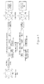
- Figure 1 is a schematic representation of a general strategy to target dendritic cells (DCs) for antigen delivery.
- DCs dendritic cells
- Sindbis virus wild-type glycoprotein is mutated at the heparan sulfate binding site to abolish its binding ability.
- the resulting mutant glycoprotein (SVGmu) binds DC-SIGN but does not bind heparin sulfate.
- DC-SIGN Dendritic Cell Specific ICAM-3 (Intracellular Adhesion Molecules 3)-Grabbing Nonintegrin.
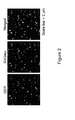
- Figure 2 illustrates laser confocal microscope images of virus particles harvested from virus-producing cells transiently transfected with lentiviral vector, plasmids encoding GFP-vpr and SVGmu, and other necessary packaging constructs.
- the virus particles are labeled with GFP (green).
- the surface incorporation of SVGmu was detected by immunostaining with an anti-HA tag antibody (red) to label SVGmu.
- GFP green
- an anti-HA tag antibody red
- viral particles where only GFP is expressed are green
- viral particles where only SVGmu is incorporated into the surface are red
- viral particles expressing both GFP and containing SVGmu are yellow.
- the overlay of the green and red colors (yellow) indicates the viral particles containing SVGmu, which represent the majority of the total virus particles.
- the scale bar represents 2 ⁇ m.
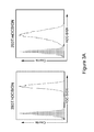
- Figure 3A shows flow cytometric analysis of constructed target cell lines 293T.hDCSIGN expressing human DC-SIGN, and 293T.mDCSIGN expressing murine DC-SIGN.
- Solid line expression of DC-SIGN in target cell lines; shaded area: background staining in 293T cells.
- Figure 3B shows flow cytometry results for detection of GFP expressed in 293T cells transduced with lentivector enveloped with wild-type Sindbis glycoprotein (FUGW/SVG) or mutant Sindbis glycoprotein (FUGW/SVGmu).
- FUGW/SVG wild-type Sindbis glycoprotein
- FUGW/SVGmu mutant Sindbis glycoprotein
- One milliliter of fresh viral supernatants of FUGW/SVG and FUGW/SVGmu were used to transduce 293T cells (2x10 5 ) expressing human DC-SIGN (293T.hDCSIGN) or murine DC-SIGN (293T.mDCSIGN).
- the parental 293T cells lacking the expression of DC-SIGN were included as controls.
- lentivector enveloped with the mutant Sindbis virus glycoprotein is able to specifically transduce 293T cells expressing human or mouse DC-SIGN.
- the specific transduction titer of FUGW/SVGmu was estimated to be approximately 1x10 6 TU/ml for 293T.hDC-SIGN and approximately 0.8x10 6 TU/ml for 293T.mDC-SIGN.
- Figure 4A shows flow cytometry results that illustrate the ability of the FUGW lentivirus enveloped with the mutant Sindbis glycoprotein (FUGW/SVGmu) to specifically transduce mouse dendritic cells expressing DC-SIGN in a primary mixed bone marrow culture.
- Whole bone marrow cells isolated from B6 mice were exposed to the fresh viral supernatant of FUGW/SVGmu.
- the FUGW lentivector pseudotyped with the ecotropic glycoprotein (FUGW/Eco) was included as a non-targeting control.
- Surface antigens of the GFP-positive cells were assessed by staining with anti-CD11c and anti-DC-SIGN antibodies.
- Figure 4B shows flow cytometry results indicating that FUGW lentivirus enveloped with the mutant Sindbis glycoprotein (FUGW/SVGmu) does not transduce other cell types including primary T cells (CD3 + , top panel) and B cells (CD19 + , bottom panel).
- Primary CD3 + T cells and CD19 + B cells were isolated from the mouse spleen and transduced with the fresh viral supernatant of either the targeting FUGW/SVGmu or non-targeting FUGW/Eco vector.
- GFP expression was analyzed by flow cytometry. Solid line: cells exposed to indicated lentiviral vector; shaded area: cells without transduction (as a negative control).
- FIG. 5 shows flow cytometry results that illustrate the ability of the FUGW lentivirus enveloped with the mutant Sindbis glycoprotein (FUGW/SVGmu) to specifically transduce bone marrow-derived DCs (BMDCs).
- BMDCs were generated by culturing freshly isolated bone marrow cells in the presence of cytokine GM-CSF for 6 days. The cells were then transduced with the fresh viral supernatant of either the targeting FUGW/SVGmu or non-targeting FUGW/Eco vector. GFP and CD11c expression were measured by flow cytometry.
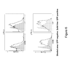
- Figure 6 shows activation of BMDCs after targeted transduction with FUGW/SVGmu.
- DC activation was assessed by analyzing the surface expression of CD86 and I-A b using flow cytometry. The addition of LPS (1 ⁇ g/ml) overnight was used as a synergistic stimulator for the activation of transduced BMDCs. Shaded area: GFP negative (untransduced); solid line: GFP positive (transduced).
- FIGs 7A, 7B and 7C illustrate targeting of DCs in vivo using FUGW/SVGmu lentivirus.
- B6 mice were injected with 50x10 6 TU of FUGW/SVGmu and analyzed 3 days later. Non-immunized mice were included as a control.
- Figure 7A the images show the size of a representative inguinal lymph node close to the injection site compared to that of the equivalent lymph node distant from the injection site.
- Figure 7B illustrates the total cell number counts of the indicated lymph nodes in Figure 7A.
- Figure 7C illustrates representative flow cytometric analysis of CD11c + cells from the two lymph nodes shown in Figure 7A . The numbers indicate the fraction of GFP + DC populations
- Figure 8 provides a schematic representation of the lentivector encoding the OVA antigen (FOVA) (top) and the lentivector encoding GFP (FUGW) as a control (bottom).
- FOVA OVA antigen
- FUGW lentivector encoding GFP
- Figure 9 illustrates in vitro stimulation of CD8 + OT1 T cells by dendritic cells that were transduced with the FOVA/SVGmu (DC/FOVA) or FUGW/SVGmu lentivector (DC/FUGW), or by non-transduced BMDCs pulsed with OVAp peptide (SIINFEKL) (DC/OVAp).
- Patterns of surface activation markers of OT1 T cells cocultured with BMDCs were assessed by antibody staining for CD25, CD69, CD62L, and CD44. Shaded area: naive OT1 T cells harvested from transgenic animals; solid line: OT1 T cells cocultured with indicated BMDCs.
- Figure 10A illustrates the measurement of IFN- ⁇ by ELISA in OT1 T cells mixed with various dilutions of BMDCs transduced with FOVA/SVGmu ( ⁇ ), FUGW/SVGmu (•), or pulsed with OVAp peptide ( ⁇ ) and cultured for 3 days.
- Figure 10B illustrates the proliferative responses of treated OT1 T cells from Figure 10A measured by a [ 3 H] thymidine incorporation assay for 12 hours.
- Figure 11 illustrates in vitro stimulation of CD4 + OT2 T cells by dendritic cells that were transduced with the FOVA/SVGmu (DC/FOVA) or FUGW/SVGmu lentivector (DC/FUGW), or by non-transduced BMDCs pulsed with OVAp* peptide (ISQAVHAAHAEINEAGR) (DC/OVAp*).
- Patterns of surface activation markers of OT2 transgenic T cells cocultured with BMDCs were assessed by antibody staining for CD25, CD69, CD62L, and CD44. Shaded area: naive OT2 T cells harvested from transgenic animals; solid line: OT2 T cells cocultured with BMDCs.
- Figure 12 illustrates the measurement of IFN- ⁇ by ELISA in OT2 T cells mixed with various dilutions of BMDCs transduced with FOVA/SVGmu ( ⁇ ), FUGW/SVGmu (•), or pulsed with OVAp* peptide ( ⁇ ) and cultured for 3 days.
- Figure 13A provides a schematic representation of the retroviral vector MIG-OT1 used for genetic modification of murine hematopoietic stem cells (HSCs).
- Figure 13B illustrates how CD8 + OT1 T cells derived from the MIG-OT1-modified HSCs in reconstituted mice were identified by the co-expression of GFP and TCR V ⁇ 2 or V ⁇ 5.
- HSCs from B6 mice were infected with MIG-OT1 pseudotyped with Eco (MIG-OT1/Eco) and transferred into irradiated B6 recipient mice.
- Eight weeks post-transfer, the CD8 + OT1 T cells were identified by flow cytometry.
- Figure 14A illustrates assessment of patterns of surface activation markers on GFP + OT1 + T cells isolated from the spleens of reconstituted and immunized mice.
- Mice reconstituted by MIG-OT1 modified HSCs were immunized by direct subcutaneous injection of 10x10 6 TU of either FOVA/SVGmu or FUGW/SVGmu (as a control) and analyzed seven days later. Detection of surface staining for CD69, CD62L and CD44 was conducted.
- Solid line GFP + OT1 + T cells from FOVA/SVGmu-immunized mice; dotted line: GFP + OT1 + T cells from control FUGW/SVGmu-immunized mice; shaded area: GFP + OT1 + T cells from non-immunized mice.
- Figure 14B illustrates the total number of OT 1 cells harvested from lymph nodes (LN, ⁇ ) or spleens (SP, ⁇ ) of non-immunized mice (no imm) or mice immunized with FUGW/SVGmu or FOVA/SVGmu.
- Figure 15 illustrates in vivo stimulation of antigen specific T cell and antibody responses in wild-type mice following a subcutaneous injection of the DC-targeting lentivector FOVA/SVGmu.
- B6 mice were immunized subcutaneously with 50x10 6 TU of either FOVA/SVGmu or FUGW/SVGmu (as a control). Mice without immunization (no imm.) were included as a negative control.
- Figures 16A and 16B illustrate in vivo OVA-specific T cell responses seen in mice receiving different subcutaneous doses of FOVA/SVGmu.
- OVA-specific T cells were identified by tetramer staining as in Figure 17 .
- Figure 16A shows the measured percentage of OVA-specific T cells following immunization with 100x10 6 TU of FOVA/SVGmu.
- Figure 16B shows the dose responses of OVA-specific T cells following injection of the indicated doses of FOVA/SVGmu.
- Figure 17A illustrates the patterns of surface activation markers of OVA-specific CD8 + T cells (identified as tetramer positive cells) isolated from FOVA/SVGmu immunized mice 2 weeks post-injection.
- the surface activation markers were assessed by antibody staining for CD25, CD69, CD62L and CD44.
- Solid line tetramer + CD8 + T cells from FOVA/SVGmu-immunized mice; shaded area: naive CD8 + T cells from non-immunized mice.
- Figure 17B illustrates the OVA-specific serum IgG titer of B6 mice following immunization with 50x10 6 TU FOVA/SVGmu.
- Sera were collected on day 7 and day 14 post-immunization and were analyzed for the titer of OVA-specific IgG using ELISA at serial 10x dilutions, starting at 1:100.
- the titer values were determined by the highest dilution at which the optical density was 2x standard derivations higher than that of the baseline serum at the equivalent dilution.
- Figure 18 illustrates tumor size as a function of time in a murine E.G7 tumor model.
- B6 mice were immunized with subcutaneous injection of 50x10 6 TU of either FOVA/SVGmu ( ⁇ ) or mock vector FUW/SVGmu (•). No immunization ( ⁇ ) was included as a control.
- Four mice were included in each group.
- the mice were challenged with 5x10 6 of either E.G7 tumor cells (expressing the OVA antigen, left panel) or the parental EL4 tumor cells (lacking the OVA antigen, as a control, right panel) subcutaneously. Tumor growth was measured with a fine caliper and is shown as the product of the two largest perpendicular diameters (mm 2 ).
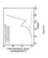
- Figure 19 illustrates the in vivo the kinetic growth of tumors in a murine E.G7 tumor eradication model.
- An albino strain of B6 mice were implanted with 5x10 6 E.G7 tumor cells stably expressing a firefly luciferase imaging gene (E.G7.luc).
- a mouse (#1) without tumor implantation was included as a control.
- Mice bearing tumors were treated without immunization (#2), or with immunization by the injection of 50x10 6 TU of FOVA/SVGmu at days 3 and 10 (#3, #4) post tumor challenge.
- the kinetic growth of the tumors was monitored by live animal imaging using BLI.
- the p/s/cm 2 /sr represents photons/sec/cm 2 /steridian.
- Figure 20 shows the quantitation of luminescence signals generated by the E.G7 tumors in Figure 19 .
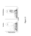
- Figure 21 illustrates the percentage of OVA-specific T cells present following immunization with 100x10 6 TU of FOVA/SVGmu in the albino strain of B6 mice.
- Albino B6 mice were immunized subcutaneously with 50x10 6 TU of FOVA/SVGmu. Mice without immunization (no imm.) were included as a negative control.
- Fourteen days post-immunization spleen cells were harvested and analyzed for the presence of OVA-specific T cells measured by H-2K b -SIIIVFEKL-PE tetramer and CD44 staining. Indicated percentages are the percent of total CD8 + T cells.
- Figure 22A provides a schematic representation of a DC-targeted lentivector encoding an imaging gene firefly luciferase (Luc), designated as Fluc/SVGmu.
- Figure 22B illustrates bioluminescence imaging of mice injected subcutaneously with 50x10 6 TU of either the DC-targeting Fluc/SVGmu lentivector (shown in Figure 25A ) or a non-targeting Fluc/VSVG lentivector.
- the representative image was obtained at day 30 post-injection using IVIS200® (Xenogen).
- Figure 23 illustrates that administration of a single dose of recombinant DC-specific lentivector FOVA/SVGmu can generate IFN- ⁇ + CD8 + T cells in B6 mice.
- Na ⁇ ve B6 mice are immunized by subcutaneous injection of 50x10 6 TU of FOVA/SVGmu lentivector, or the same dose of FUGW/SVGmu as a control.
- the non-immunized B6 mice (no imm.) were included as a negative control.
- spleen cells were harvested from the experimental mice, and were analyzed for intracellular IFN- ⁇ production using flow cytometry with or without OVAp peptide restimulation. Indicated percentages are the percent of IFN- ⁇ + CD8 + T cells of the total CD8 + T cells.
- Figure 24 illustrates a schematic representation of lentiviral constructs for preparation of DC-targeting recombinant viruses.
- Figure 25 shows a schematic representation of an embodiment of in situ vaccination against HIV/AIDS.
- DCs dendritic cells
- the inventors have discovered novel methods and compositions for efficient and specific targeting of DCs in vitro and in vivo. The methods and compositions can be used to induce antigen-specific immune responses, for example for immunotherapy.
- Embodiments of the invention include methods and compositions for targeting dendritic cells (DCs) by using a recombinant virus to deliver a polynucleotide to the DCs. This is preferably accomplished through targeting the DC-specific surface molecule DC-SIGN (Dendritic Cell Specific ICAM-3 (Intracellular Adhesion Molecules 3)-Grabbing Nonintegrin; also known as CD209).
- DC-SIGN is a C-type lectin-like receptor capable of rapid binding and endocytosis of materials ( Geijtenbeek, T.B., et al. 2004. Annu. Rev. Immunol. 22: 33-54 , which is incorporated herein by reference in its entirety).
- recombinant viruses are enveloped with a designed targeting molecule that is specific in its recognition for DC-SIGN.
- the polynucleotide can include, but is not limited to, a gene of interest, siRNA(s), and/or microRNA(s).
- the polynucleotide encodes an antigen.
- the recombinant virus delivers more than one gene to DCs. For example, genes encoding two or more antigens could be delivered. The delivery of more than one gene can be achieved, for example, by linking the genes with an Internal Ribosome Entry Site (IRES), and/or with 2A sequences, and driving the expression using a single promoter/enhancer.
- IRS Internal Ribosome Entry Site
- a packaging cell line is transfected with a viral vector encoding a polynucleotide of interest (typically encoding an antigen), at least one plasmid encoding virus packaging components (such as gag and pol) and a targeting molecule that is engineered to bind dendritic cells.
- a viral vector encoding a polynucleotide of interest (typically encoding an antigen)
- at least one plasmid encoding virus packaging components such as gag and pol
- a targeting molecule that is engineered to bind dendritic cells.
- the targeting molecule is genetically engineered to specifically bind the DC-SIGN cell surface marker of dendritic cells.
- the targeting molecule which is expressed in the packaging cell membrane, is incorporated into the viral envelope.
- the retroviral particles comprise a core including the polynucleotide of interest and an envelope comprising the targeting molecule on its surface.
- the targeting molecule is able to bind DC-SIGN on a dendritic cell, and the virus is able to deliver the gene of interest to the dendritic cell.
- the binding induces endocytosis, bringing the virus into an endosome, triggering membrane fusion, and allowing the virus core to enter the cytosol.
- the genome of the virus integrates into the target cell genome, incorporating the polynucleotide of interest into the genome of the target cell.
- the DC then expresses the polynucleotide of interest (typically encoding an antigen).
- the antigen is then processed and presented to T and B cells by DCs, generating an antigen-specific immune response.
- the specific pathway described above is not required so long as the dendritic cell is able to stimulate an antigen-specific immune response.
- Embodiments of the present invention include methods and compositions for direct targeting of a gene of interest to DCs both in vitro and in vivo.
- the gene of interest is delivered to DCs without in vitro culture of DCs.
- the gene of interest may be delivered to DCs via a direct administration of the targeting virus into a living subject.
- the gene of interest preferably encodes an antigen against which an immune response is desired.
- Exemplary antigens include: tumor specific antigens, tumor-associated antigens, tissue-specific antigens, bacterial antigens, viral antigens, yeast antigens, fungal antigens, protozoan antigens, parasite antigens, mitogens, and the like.
- Other antigens will be apparent to one of skill in the art and can be utilized without undue experimentation.
- the methods disclosed herein may be readily adopted to utilize targeting molecules that are specific for DCs or that can be manipulated to provide the desired specificity.
- the targeting molecule is preferably an engineered viral glycoprotein that binds DC-SIGN in dendritic cells and that facilitates delivery of the gene of interest into the dendritic cells.
- Exemplary targeting molecules include, but are not limited to, glycoproteins derived from the following: Sindbis virus, influenza virus, Lassa fever virus, tick-borne encephalitis virus, Dengue virus, Hepatitis B virus, Rabies virus, Semliki Forest virus, Ross River virus, Aura virus, Boma disease virus, Hantaan virus, and SARS-CoV virus.
- the targeting molecule is preferably membrane bound. If necessary, a DC-SIGN-specific targeting molecule that is designed or derived from a viral glycoprotein for use in the recombinant virus can be modified to a membrane bound form.
- a targeting molecule specific for DCs a viral glycoprotein that interacts with a dendritic cell-specific surface marker is provided.
- the viral glycoprotein interacts with DC-SIGN.
- the viral glycoprotein can also interact with at least a second cell surface marker such as, for example, heparin sulfate (HS), which is expressed on cell types other than DCs.
- HS heparin sulfate
- the viral glycoprotein is modified such that its ability to interact with the DC-specific surface marker is maintained while its ability to interact with additional cell surface markers is decreased or eliminated.
- the modification can be a mutation in at least one residue of the viral glycoprotein amino acid sequence.
- the mutation can be a deletion, addition or substitution of the residue, and it can be carried out by standard methods known in the art. The desired specificity can readily be confirmed.
- the viral glycoprotein can be used to prepare a recombinant virus by co-transfection with a viral vector containing a reporter gene and at least one plasmid encoding virus packaging components into a packaging cell line.
- the glycoprotein is incorporated into the viral envelope during budding of the virus.
- the virus can be used to transfect both a pure population of DCs as well as a mixed population of cells containing DCs, and specificity of the viral transduction of DCs can be confirmed by assaying the cells for expression of the reporter gene in DCs and not to a significant extent in other cell types. If the specificity is not sufficiently stringent (for example, if undesired levels of infection of other cell types is observed), the viral glycoprotein can be modified further and assayed as described until the desired specificity is achieved.
- Embodiments of the present invention include the delivery to DCs of DC activators and/or maturation factors in conjunction with antigens.
- DC activators and maturation factors include, but are not limited to, stimulation molecules, cytokines, chemokines, antibodies and other agents such as Flt-3 ligands.
- the DC maturation factors can include at least one of the following: GM-CSF, IL-2, IL-4, IL-6, IL-7, IL-15, IL-21, IL-23, TNF ⁇ , B7.1, B7.2, 4-1BB, CD40 ligand (CD40L) and drug-inducible CD40 (iCD40) ( Hanks, B.A., et al. 2005. Nat Med 11:130-137 , which is incorporated herein by reference in its entirety).
- Embodiments of the present invention also include methods and compositions related to administration of recombinant virus as described above, or DCs infected with recombinant virus, into patients to stimulate antigen-specific immune responses, such as, for example, T cell responses (cellular immune responses) and B cell responses (humoral immune responses).
- antigen-specific immune responses such as, for example, T cell responses (cellular immune responses) and B cell responses (humoral immune responses).
- activated CD4 T cells can coordinate and orchestrate the CD8 + cytotoxic T cells and the B cells in an antigen-specific response.
- the recombinant virus and/or DCs infected with recombinant virus are used to stimulate immune responses for the prevention and treatment of diseases such as, but not limited to, cancer and AIDS/HIV. Any disease can be treated for which an immune response to a particular antigen is beneficial, including, but not limited to, neoplastic disease, infectious disease, and immune-related diseases.
- nucleic acid As used herein, the terms nucleic acid, polynucleotide and nucleotide are interchangeable and refer to any nucleic acid, whether composed of phosphodiester linkages or modified linkages such as phosphotriester, phosphoramidate, siloxane, carbonate, carboxymethylester, acetamidate, carbamate, thioether, bridged phosphoramidate, bridged methylene phosphonate, bridged phosphoramidate, bridged phosphoramidate, bridged methylene phosphonate, phosphorothioate, methylphosphonate, phosphorodithioate, bridged phosphorothioate or sultone linkages, and combinations of such linkages.
- phosphodiester linkages or modified linkages such as phosphotriester, phosphoramidate, siloxane, carbonate, carboxymethylester, acetamidate, carbamate, thioether, bridged phosphoramidate, bridged methylene
- nucleic acid, polynucleotide and nucleotide also specifically include nucleic acids composed of bases other than the five biologically occurring bases (adenine, guanine, thymine, cytosine and uracil).
- nucleic acid molecule is said to be "isolated” when the nucleic acid molecule is substantially separated from contaminant nucleic acid molecules encoding other polypeptides.
- Immunization refers to the provision of antigen to a host.
- antigen is provided to antigen-presenting cells, such as dendritic cells.
- antigen-presenting cells such as dendritic cells.
- recombinant virus comprising a gene encoding an antigen can be targeted to dendritic cells with an affinity molecule specific to DC-SIGN on dendritic cells.
- the antigen to which an immune response is desired can be delivered to the dendritic cells.
- Other methods of immunization are well known in the art.
- immunological response is the development of a beneficial humoral (antibody mediated) and/or a cellular (mediated by antigen-specific T cells or their secretion products) response directed against an amyloid peptide in a recipient patient.
- Such a response can be an active response induced by administration of immunogen or a passive response induced by administration of antibody or primed T-cells.
- a cellular immune response is elicited by the presentation of polypeptide epitopes in association with Class I or Class II MHC molecules to activate antigen-specific CD4 + T helper cells and/or CD8 + cytotoxic T cells.
- the response may also involve activation of monocytes, macrophages, NK cells, basophils, dendritic cells, astrocytes, microglia cells, eosinophils or other components of innate immunity.
- the presence of a cell-mediated immunological response can be determined by proliferation assays (CD4 + T cells) or CTL (cytotoxic T lymphocyte) assays ( Burke et al., J. Inf. Dis. 170, 1110-19 (1994 )), by antigen-dependent killing (cytotoxic T lymphocyte assay, Tigges et al., J. Immunol. 156, 3901-3910 ) or by cytokine secretion.
- the relative contributions of humoral and cellular responses to the protective or therapeutic effect of an immunogen can be distinguished by separately isolating IgG and T-cells from an immunized syngeneic animal and measuring protective or therapeutic effect in a second subject.
- an “immunogenic agent” or “immunogen” is capable of inducing an immunological response against itself on administration to a patient, optionally in conjunction with an adjuvant.
- adjuvant refers to a compound that when administered in conjunction with an antigen augments, enhances, and/or boosts the immune response to the antigen, but when administered alone does not generate an immune response to the antigen.
- An adjuvant can be administered with the recombinant virus of the invention as a single composition, or can be administered before, concurrent with or after administration of the recombinant virus of the invention.
- Adjuvants can enhance an immune response by several mechanisms including lymphocyte recruitment, stimulation of B and/or T cells, and stimulation of macrophages.
- Antibodies are glycoproteins having the same structural characteristics. While antibodies exhibit binding specificity to a specific antigen, immunoglobulins include both antibodies and other antibody-like molecules that lack antigen specificity. Polypeptides of the latter kind are, for example, produced at low levels by the lymph system and at increased levels by myelomas.
- antibody is used in the broadest sense and specifically covers human, non-human (e.g. murine), chimeric, and humanized monoclonal antibodies (including full length monoclonal antibodies), polyclonal antibodies, multi-specific antibodies (e.g., bispecific antibodies), single-chain antibodies, and antibody fragments so long as they exhibit the desired biological activity. Typically, fragments compete with the intact antibody from which they were derived for specific binding to an antigen.
- epitopes or "antigenic determinant” refers to a site on an antigen to which B and/or T cells respond.
- B-cell epitopes can be formed both from contiguous amino acids or noncontiguous amino acids juxtaposed by tertiary folding of a protein. Epitopes formed from contiguous amino acids are typically retained on exposure to denaturing solvents whereas epitopes formed by tertiary folding are typically lost on treatment with denaturing solvents.
- An epitope typically includes at least 3, and more usually, at least 5 or 8-10 amino acids in a unique spatial conformation. Methods of determining spatial conformation of epitopes include, for example, x-ray crystallography and 2-dimensional nuclear magnetic resonance.
- T-cells recognize continuous epitopes of about nine amino acids for CD8 cells or about 13-15 amino acids for CD4 cells.
- T cells that recognize the epitope can be identified by in vitro assays that measure antigen-dependent proliferation, as determined by 3 H-thymidine incorporation by primed T cells in response to an epitope (see Burke, supra; Tigges, supra).
- Target cells are any cells to which delivery of a polynucleotide or in which expression of a gene of interest is desired.
- target cells are dendritic cells, particularly dendritic cells that express DC-SIGN.
- mammal is defined as an individual belonging to the class Mammalia and includes, without limitation, humans, domestic and farm animals, and zoo, sports, and pet animals, such as sheep, dogs, horses, cats and cows.
- subject or “patient” includes human and other mammalian subjects that receive either prophylactic or therapeutic treatment.
- treatment is a clinical intervention that may be therapeutic or prophylactic.
- pharmaceutical compositions or medicants are administered to a patient suspected of, or already suffering from such a disease in an amount sufficient to cure, or at least partially arrest, the symptoms of the disease and its complications.
- pharmaceutical compositions or medicants are administered to a patient susceptible to, or otherwise at risk of, a particular disease in an amount sufficient to eliminate or reduce the risk or delay the outset of the disease.
- An amount adequate to accomplish this is defined as a therapeutically- or pharmaceutically-effective dose.
- Such an amount can be administered as a single dosage or can be administered according to a regimen, whereby it is effective.
- the amount can cure a disease but, typically, is administered in order to ameliorate the symptoms of a disease, or to effect prophylaxis of a disease or disorder from developing.
- agents are usually administered in several dosages until a sufficient immune response has been achieved.
- the immune response is monitored and repeated dosages are given if the immune response starts to fade. "Treatment" need not completely eliminate a disease, nor need it completely prevent a subject from becoming ill with the disease or disorder.
- Tumor refers to all neoplastic cell growth and proliferation, whether malignant or benign, and all pre-cancerous and cancerous cells and tissues.
- cancer refers to a disease or disorder that is characterized by unregulated cell growth.
- examples of cancer include, but are not limited to, carcinoma, lymphoma, blastoma and sarcoma.
- specific cancers include, but are not limited to, lung cancer, colon cancer, breast cancer, testicular cancer, stomach cancer, pancreatic cancer, ovarian cancer, liver cancer, bladder cancer, colorectal cancer, and prostate cancer. Additional cancers are well known to those of skill in the art and include, but are not limited to: leukemia, lymphoma, cervical cancer, glioma tumors, adenocarcinomas and skin cancer.
- Exemplary cancers include, but are not limited to, a bladder tumor, breast tumor, prostate tumor, basal cell carcinoma, biliary tract cancer, bladder cancer, bone cancer, brain and CNS cancer (e.g., glioma tumor), cervical cancer, choriocarcinoma, colon and rectum cancer, connective tissue cancer, cancer of the digestive system; endometrial cancer, esophageal cancer; eye cancer; cancer of the head and neck; gastric cancer; intra-epithelial neoplasm; kidney cancer; larynx cancer; leukemia; liver cancer; lung cancer (e.g.
- lymphoma including Hodgkin's and Non-Hodgkin's lymphoma; melanoma; myeloma, neuroblastoma, oral cavity cancer (e.g., lip, tongue, mouth, and pharynx); ovarian cancer; pancreatic cancer, retinoblastoma; rhabdomyosarcoma; rectal cancer, renal cancer, cancer of the respiratory system; sarcoma, skin cancer; stomach cancer, testicular cancer, thyroid cancer; uterine cancer, cancer of the urinary system, as well as other carcinomas and sarcomas. Cancer also includes neoplasias and malignant disorders in mammals that are well known in the art.
- a “vector” is a nucleic acid that is capable of transporting another nucleic acid.
- Vectors may be, for example, plasmids, cosmids or phage.
- An "expression vector” is a vector that is capable of directing expression of a protein or proteins encoded by one or more genes carried by the vector when it is present in the appropriate environment.
- regulatory element and “expression control element” are used interchangeably and refer to nucleic acid molecules that can influence the transcription and/or translation of an operably linked coding sequence in a particular environment. These terms are used broadly and cover all elements that promote or regulate transcription, including promoters, core elements required for basic interaction of RNA polymerase and transcription factors, upstream elements, enhancers, and response elements (see, e.g., Lewin, "Genes V” (Oxford University Press, Oxford) pages 847-873 ). Exemplary regulatory elements in prokaryotes include promoters, operator sequences and a ribosome binding sites. Regulatory elements that are used in eukaryotic cells may include, without limitation, promoters, enhancers, splicing signals and polyadenylation signals.
- transfection refers to the introduction of a nucleic acid into a host cell.
- “Retroviruses” are viruses having an RNA genome.
- lentivirus refers to a genus of retroviruses that are capable of infecting dividing and non-dividing cells.
- HIV human immunodeficiency virus: including HIV type 1, and HIV type 2
- AIDS human acquired immunodeficiency syndrome
- visna-maedi which causes encephalitis (visna) or pneumonia (maedi) in sheep, the caprine arthritis-encephalitis virus, which causes immune deficiency, arthritis, and encephalopathy in goats
- equine infectious anemia virus which causes autoimmune hemolytic anemia, and encephalopathy in horses
- feline immunodeficiency virus (FIV) which causes immune deficiency in cats
- bovine immune deficiency virus (BIV) which causes lymphadenopathy, lymphocytosis, and possibly central nervous system infection in cattle
- SIV simian immunodeficiency virus
- a lentiviral genome is generally organized into a 5' long terminal repeat (LTR), the gag gene, the pol gene, the env gene, the accessory genes (nef, vif, vpr, vpu) and a 3' LTR.
- the viral LTR is divided into three regions called U3, R and U5.
- the U3 region contains the enhancer and promoter elements.
- the U5 region contains the polyadenylation signals.
- the R (repeat) region separates the U3 and U5 regions and transcribed sequences of the R region appear at both the 5' and 3' ends of the viral RNA. See, for example, " RNA Viruses: A Practical Approach" (Alan J.
- Gammaretrovirus refers to a genus of the retroviridae family.
- exemplary gammaretroviruses include, but are not limited to, mouse stem cell virus, murine leukemia virus, feline leukemia virus, feline sarcoma virus, and avian reticuloendotheliosis viruses.
- hybrid virus refers to a virus having components from one or more other viral vectors, including element from non-retroviral vectors, for example, adenoviral-retroviral hybrids.
- hybrid vectors having a retroviral component are to be considered within the scope of the retroviruses.
- RNA genome is usually a recombinant RNA genome and thus may contain an RNA sequence that is exogenous to the native viral genome.
- the RNA genome may also comprise a defective endogenous viral sequence.
- a "pseudotyped" retrovirus is a retroviral particle having an envelope protein that is from a virus other than the virus from which the RNA genome is derived.
- the envelope protein can be, for example and without limitation, from a different retrovirus or from a non-retroviral origin.
- the envelope protein can be a native envelope protein or an envelope protein that is modified, mutated or engineered as described herein.
- an envelope protein is a DC-SIGN-specific viral glycoprotein that is derived from a glycoprotein from one of the following: Sindbis virus, influenza virus, Lassa fever virus, tick-borne encephalitis virus, Dengue virus, Hepatitis B virus, Rabies virus, Semliki Forest virus, Ross River virus, Aura virus, Boma disease virus, Hantaan virus, and SARS-CoV virus.
- Transformation describes a process by which exogenous DNA enters a target cell. Transformation may rely on any known method for the insertion of foreign nucleic acid sequences into a prokaryotic or eukaryotic host cell and may include, but is not limited to, viral infection, electroporation, heat shock, lipofection, and particle bombardment.
- Transformed cells include stably transformed cells in which the inserted nucleic acid is capable of replication either as an autonomously replicating plasmid or as part of the host chromosome. Also included are cells that transiently express a gene of interest.
- a "fusogenic molecule,” as described herein, is any molecule that can trigger membrane fusion when present on the surface of a virus and allows a virus core to pass through the membrane and, typically, enter the cytosol of a target cell. Fusogenic molecules can be, for example, viral glycoproteins.
- Exemplary viral glycoproteins contemplated as fusogenic molecules include, but are not limited to hemagglutinin, mutant hemagglutinin, SIN and viral glycoproteins from the following viruses: Sindbis virus, influenza virus, Lassa fever virus, tick-borne encephalitis virus, Dengue virus, Hepatitis B virus, Rabies virus, Semliki Forest virus, Ross River virus, Aura virus, Boma disease virus, Hantaan virus, and SARS-CoV virus.
- Glycoproteins can be native or modified to have desired activity.
- transgene any nucleotide sequence, particularly a DNA sequence, that is integrated into one or more chromosomes of a host cell by human intervention, such as by the methods of the present invention.
- the transgene preferably comprises a "gene of interest.”
- a "gene of interest” is not limited in any way and may be any nucleic acid, without limitation, that is desired to be delivered to, integrated, transcribed, translated, and/or expressed in a target cell.
- the gene of interest may encode a functional product, such as a protein or an RNA molecule.
- the gene of interest encodes a protein or other molecule, the expression of which is desired in the target cell.
- the gene of interest is generally operatively linked to other sequences that are useful for obtaining the desired expression of the gene of interest, such as transcriptional regulatory sequences.
- a gene of interest is preferably one that encodes an antigen to which an immune response is desired.
- Other genes of interest that may be used in some embodiments are genes that encode dendritic cell activators and/or maturation factors.
- a “functional relationship” and “operably linked” mean, with respect to the gene of interest, that the gene is in the correct location and orientation with respect to the promoter and/or enhancer that expression of the gene will be affected when the promoter and/or enhancer is contacted with the appropriate molecules.
- 2A sequences or elements are small peptides introduced as a linker between two proteins, allowing autonomous intraribosomal self-processing of polyproteins (de Felipe. Genetic Vaccines and Ther. 2:13 (2004 ); de Felipe et al. Traffic 5:616-626 (2004 )).
- the short peptides allow co-expression of multiple proteins from a single vector, such as co-expression of a fusogenic molecule and affinity molecule from the same vector.
- polynucleotides encoding the 2A elements are incorporated into a vector between polynucleotides encoding proteins to be expressed.
- DC maturation factors are compounds that can induce activation or stimulation of DCs such that DCs facilitate the elicitation of cellular and humoral immune responses.
- Typical DC maturation factors are known in the art and include, but are not limited to, stimulation molecules, cytokines, chemokines, antibodies and other agents such as Flt-3 ligands ( Figdor, C.G., et al. 2004. Nat Med 10:475-480 ; Pulendran, B., et al. 2000. J Immunol 165: 566-572 ; Maraskovsky, E., et al. 2000. Blood 96:878-884 , each of which is incorporated herein by reference in its entirety).
- Exemplary DC maturation factors can include, but are not limited to, GM-CSF, IL-2, IL-4, IL-6, IL-7, IL-15, IL-21, IL-23, TNF ⁇ , B7.1, B7.2, 4-1BB, CD40 ligand (CD40L) and drug-inducible CD40 (iCD40).
- CD40L CD40 ligand
- iCD40 drug-inducible CD40
- a targeting molecule is incorporated into a recombinant virus to target the virus to dendritic cells that express DC-SIGN.
- the targeting molecule preferably also mediates fusion with the cell membrane and efficient transduction and delivery of the desired polynucleotide(s) into the dendritic cell.
- the targeting molecule is typically a fusogenic molecule (FM) with the desired binding specificity.
- FM fusogenic molecule
- the targeting molecule is modified, if necessary, such that it binds to DC-SIGN on dendritic cells.
- the targeting molecule specifically binds to DC-SIGN.
- targeting molecule preferentially directs the recombinant virus to dendritic cells that express DC-SIGN relative to other cell types.
- targeting molecules are created by eliminating the ability of a FM to bind to other targets, such as hemagglutinin, while retaining the ability to bind DC-SIGN.
- the targeting molecule can be modified to eliminate native binding specificity to non-DC-SIGN molecules and components thereof and add or improve binding specificity for DC-SIGN.
- the targeting molecules While some nonspecific binding to other molecules, and thus other cell types, may occur even if the targeting molecule is specific for DC-SIGN, the targeting molecules are modified to have sufficient specificity to avoid undesired side effects, such as side effects that may reduce the desired immune response.
- Targeting molecules are generally molecules that are able to pseudotype virus and thus be incorporated in the envelope of recombinant viruses, target dendritic cells and, under the right conditions, induce membrane fusion and allow entry of a gene of interest to the dendritic cells.
- Preferred targeting molecules are viral glycoproteins.
- targeting molecules are preferably resistant to ultracentrifugation to allow concentration, which can be important for in vivo gene delivery.
- Targeting molecules preferably induce membrane fusion at a low pH, independently of binding.
- targeting molecule-induced membrane fusion occurs once the virus comprising the targeting molecule is inside the endosome of a target cell and the viral core component, including a polynucleotide of interest, is delivered to the cytosol.
- a tag sequence is incorporated into a targeting molecule to allow detection of targeting molecule expression and the presence of the targeting molecule in viral particles.
- class I fusogens trigger membrane fusion using helical coiled-coil structures whereas the class II fusogens trigger fusion with ⁇ barrels. These two structures have different mechanics and kinetics ( D. S. Dimitrov, Nature Rev. Microbio. 2, 109 (2004 )).
- class I fusogens are used.
- class II fusogens are used.
- both class I and class II fusogens are used.
- surface glycoproteins that may be used as targeting molecules (or as fusogenic molecules in embodiments where the viral binding and fusion functions are separate), either in the wild type or in modified form, include glycoproteins from alphaviruses, such as Semliki Forest virus (SFV), Ross River virus (RRV) and Aura virus (AV), which comprise surface glycoproteins such as E1, E2, and E3.
- alphaviruses such as Semliki Forest virus (SFV), Ross River virus (RRV) and Aura virus (AV), which comprise surface glycoproteins such as E1, E2, and E3.
- the E2 glycoproteins derived from the Sindbis virus (SIN) and the hemagglutinin (HA) of influenza virus are non-retroviral glycoproteins that specifically bind particular molecules on cell surfaces (heparin sulfate glycosaminoglycan for E2, sialic acid for HA) and can be used to create targeting molecules in some embodiments.
- surface glycoproteins of Lassa fever virus, Hepatitis B virus, Rabies virus, Boma disease virus, Hantaan virus, or SARS-CoV virus can be utilized as fusion molecules.
- flavivirus-based surface glycoproteins may be used as the basis for targeting molecules.
- flaviviruses use the class II fusion molecule to mediate infection ( Mukhopadhyay et al. (2005) Rev. Microbio. 3, 13-22 ).
- prM about 165 amino acids
- E about 495 amino acids
- the ligand-binding pocket for one flavivirus, Dengue virus (DV) has been well-characterized.
- DC-SIGIV has been suggested to specifically interact with the carbohydrate residues on the DV E protein to enhance viral entry ( Mukhopadhyay et al. (2005) Nat. Rev. Microbio.
- lentiviruses enveloped only by DV E protein, or by modified DV E protein can be used to target DCs.
- the TBE and DV E proteins, as well as other fusion molecules described, may be engineered to provide the desired binding specificity or to be binding deficient and fusion competent if necessary.
- HA hemagglutinin
- FPV hemagglutinin
- a form of FPV HA is used ( T. Hatziioannou, S. Valsesia-Wittmann, S. J. Russell, F. L. Cosset, J. Virol. 72, 5313 (1998 )).
- FPV HA A. H. Lin et al., Hum. Gene. Ther. 12, 323 (2001 )).
- HA-mediated fusion is generally considered to be independent of receptor binding ( D. Lavillette, S. J. Russell, F. L. Cosset, Curr. Opin. Biotech. 12, 461 (2001 )).
- a class II FM is used, preferably the Sindbis virus glycoprotein from the alphavirus family ( K. S. Wang, R. J. Kuhn, E. G. Strauss, S. Ou, J. H. Strauss, J. Virol. 66, 4992 (1992 )), herein also referred to as SVG.
- SVG includes two transmembrane proteins ( S. Mukhopadhyay, R. J. Kuhn, M. G. Rossmann, Nature Rev. Microbio. 3, 13 (2005 )), a first protein responsible for fusion (E1), and a second protein for cell binding (E2).
- SVG is known to pseudotype both oncoretroviruses and lentiviruses.
- a modified SVG that preferentially binds DC-SIGN is utilized.
- a binding-deficient and fusion-competent SVG, SVGmu can be used as the fusogenic molecule in combination with a separate targeting molecule, such as an antibody to DC-SIGN or another dendritic cell specific protein.
- a SVG fusogenic molecule can be used in which the immunoglobulin G binding domain of protein A (ZZ domain) is incorporated into the E2 protein and one or more additional mutations are made to inactivate the receptor binding sites ( K. Morizono et al., Nature Med. 11, 346 (2005 )).
- the gene encoding the targeting molecule is preferably cloned into an expression vector, such as pcDNA3 (Invitrogen).
- Packaging cells such as 293T cells are then co-transfected with the viral vector encoding a gene of interest (typically encoding an antigen), at least one plasmid encoding virus packing components, and a vector for expression of the targeting molecule. If the targeting function is separated from the fusogenic function, one or more vectors encoding an affinity molecule and any associated components is also provided, The targeting molecule is expressed on the membrane of the packaging cell and incorporated into the recombinant virus. Expression of targeting molecules on the packaging cell surface can be analyzed, for example, by FACS.
- mutagenesis may be employed to generate the mutant forms of glycoproteins that maintain their fusogenic ability but have the desired binding specificity and/or level of binding.
- Several mutants may be created for each glycoprotein and assayed using the methods described below, or other methods known in the art, to identify FMs with the most desirable characteristics.
- targeting molecules can be tested for the ability to specifically deliver antigens to dendritic cells by determining their ability to stimulate an immune response without causing undesired side effects in a mammal.
- the ability to specifically target dendritic cells can also be tested directly, for example, in cell culture as described below.
- viruses bearing the targeting molecule are prepared and tested for their selectivity and/or their ability to facilitate penetration of the target cell membrane.
- Viruses that display a wild-type glycoprotein can be used as controls for examining titer effects in mutants.
- Cells expressing the binding partner of the targeting molecule (or affinity molecule, where appropriate) are transduced by the virus using a standard infection assay. After a specified time, for example 48 hours post-transduction, cells can be collected and the percentage of cells infected by the virus comprising the targeting molecule (or affinity molecule and fusogenic molecule) can be determined by, for example, FACS.
- the selectivity can be scored by calculating the percentage of cells infected by virus.
- the effect of mutations on viral titer can be quantified by dividing the percentage of cells infected by virus comprising a mutant targeting molecule by the percentage of cells infected by virus comprising the corresponding wild type targeting molecule. A preferred mutant will give the best combination of selectivity and infectious titer.
- viral concentration assays may be performed to confirm that viruses enveloped by the FM can be concentrated. Viral supernatants are collected and concentrated by ultracentrifugation.
- the titers of viruses can be determined by limited dilution of viral stock solution and transduction of cells expressing the binding partner of the affinity molecule.
- BlaM-Vpr fusion protein may be utilized to evaluate viral penetration, and thus the efficacy of a fusion molecule (wild-type or mutant).
- Virus may be prepared, for example, by transient transfection of packaging cells with one or more vectors comprising the viral elements, BlaM-Vpr, and the FM of interest (and an affinity molecule if appropriate). The resulting viruses can be used to infect cells expressing a molecule the targeting molecule (or affinity molecule) specifically binds in the absence or presence of the free inhibitor of binding (such as an antibody). Cells can then be washed with CO 2 -independent medium and loaded with CCF2 dye (Aurora Bioscience).
- the cells After incubation at room temperature to allow completion of the cleavage reaction, the cells can be fixed by paraformaldehyde and analyzed by FACS and microscopy. The presence of blue cells indicates the penetration of viruses into the cytoplasm; fewer blue cells would be expected when blocking antibody is added.
- NH 4 Cl will neutralize the acidic compartments of endosomes.
- he disappearance of blue cells will indicate that penetration of viruses is low pH-dependent.
- lysosomotropic agents such as ammonium chloride, chloroquine, concanamycin, bafilomycin Al, monensin, nigericin, etc.
- these agents can elevate the pH within the endosomal compartments (e.g., Drose and Altendorf, J. Exp. Biol. 200, 1-8, 1997 ).
- the inhibitory effect of these agents will reveal the role of pH for viral fusion and entry.
- the different entry kinetics between viruses displaying different fusogenic molecules may be compared and the most suitable selected for a particular application.
- PCR entry assays may be utilized to monitor reverse transcription and thus measure kinetics of viral DNA synthesis as an indication of the kinetics of viral entry.
- viral particles comprising a particular targeting molecule may be incubated with packaging cells, such as 293T cells, expressing the appropriate cognate for the targeting molecule (or a separate affinity molecule in some embodiments). Either immediately, or after incubation (to allow infection to occur) unbound viruses are removed and aliquots of the cells are analyzed. DNA may then be extracted from these aliquots and semi-quantitative performed using LTR-specific primers. The appearance of LTR-specific DNA products will indicate the success of viral entry and uncoating.
- the targeting molecule can have both viral binding and fusion functions
- the viral binding and fusion functions are separated into two distinct components.
- the recombinant virus comprises both (i) an affinity molecule that mediates viral binding and precisely targets the virus to dendritic cells, and (ii) a distinct fusogenic molecule (FM) that mediates efficient transduction and delivery of the desired polynucleotide into the dendritic cells.
- affinity molecules that mediates viral binding and precisely targets the virus to dendritic cells
- FM fusogenic molecule
- the methods disclosed herein may be readily adopted to utilize any of a variety of affinity molecules and fusogenic molecules.
- other exemplary fusogenic molecules and related methods are described, for example, in U.S. Patent Application No. 11/071,785, filed March 2, 2005 (published as U.S. Patent Application Publication 2005-0238626 ), and in U.S. Patent Application No. 11/446,353, filed June 1, 2006 (published as U.S. Patent Application Publication 2007/0020238 ),
- the affinity molecule is one that binds a dendritic cell surface marker.
- the affinity molecule binds DC-SIGN with specificity. That is, the binding of the affinity molecule to DC-SIGN is preferably specific enough to avoid undesired side effects due to interaction with markers on other cell types.
- the affinity molecule can be, for example, an antibody that specifically binds DC-SIGN.
- the fusion molecule is a viral glycoprotein that mediates fusion or otherwise facilitates delivery of the gene of interest to the dendritic cell, preferably in response to the low pH environment of the endosome.
- the fusion molecule preferably exhibits fast enough kinetics that the viral contents can empty into the cytosol before the degradation of the viral particle.
- the fusion molecule can be modified to reduce or eliminate any binding activity and thus reduce or eliminate any non-specific binding. That is, by reducing the binding ability of the fusion molecules, binding of the virus to the target cell is determined predominantly or entirely by the affinity molecule, allowing for high target specificity and reducing undesired effects.
- Exemplary fusion molecules include, but are not limited to viral glycoproteins derived from one of the following viruses: Sindbis virus, influenza virus, Lassa fever virus, tick-borne encephalitis virus, Dengue virus, Hepatitis B virus, Rabies virus, Semliki Forest virus, Ross River virus, Aura virus, Boma disease virus, Hantaan virus, and SARS-CoV virus.
- one or more vectors are used to introduce polynucleotide sequences into a packaging cell line for the preparation of a recombinant virus as described herein.
- the vectors can contain polynucleotide sequences encoding the various components of the recombinant virus including the DC-specific targeting molecule, a gene(s) of interest (typically encoding an antigen), and any components necessary for the production of the virus that are not provided by the packaging cell.
- vectors containing polynucleotide sequences that encode a DC-specific affinity molecule and a separate fugosenic molecule are substituted for a vector that encodes a DC-specific targeting molecule in the preparation of the virus.
- Eukaryotic cell expression vectors are well known in the art and are available from a number of commercial sources.
- vectors containing polynucleotide sequences that encode DC maturation factors are also used in the preparation of the virus. These polynucleotides are typically under the control of one or more regulatory elements that direct the expression of the coding sequences in the packaging cell and the target cell, as appropriate.
- Several lines of evidence have shown the success of DC vaccination is dependent on the maturation state of DCs ( Banchereau, J and Palucka, A.K. Nat. Rev. Immunol. 5:296-306 (2005 ); Schuler, G. et al. Curr. Opin. Immunol. 15:138-147 (2003 ); Figdor, C.G. et al. Nat. Med.
- the vector includes genes that encode the stimulatory molecules to trigger the desired DC maturation.
- Such stimulatory molecules are also referred to as maturation factors or maturation stimulatory factors.
- packaging cells are co-transfected with a viral vector encoding an antigen and one or more additional vectors.
- a second vector preferably carries a gene encoding a targeting molecule that binds dendritic cells, such SVGmu, as described elsewhere in the application.
- the targeting molecule encodes a modified viral glycoprotein that is specific for DC-SIGN.
- the modified viral glycoprotein is preferably one derived from at least one of the following: Sindbis virus, influenza virus, Lassa fever virus, tick-borne encephalitis virus, Dengue virus, Hepatitis B virus, Rabies virus, Semliki Forest virus, Ross River virus, Aura virus, Boma disease virus, Hantaan virus, and SARS-CoV virus.
- the viral vector encoding an antigen also includes a polynucleotide sequence encoding a DC maturation factor.
- the polynucleotide sequence encoding a DC maturation factor is contained in a third vector that is co-transfected with the viral vector encoding an antigen and the one or more additional vectors into the packaging cells.
- one or more multicistronic expression vectors are utilized that include two or more of the elements (e.g., the viral genes, gene(s) of interest, the targeting molecule, DC maturation factors) necessary for production of the desired recombinant virus in packaging cells.
- the use of multicistronic vectors reduces the total number of vectors required and thus avoids the possible difficulties associated with coordinating expression from multiple vectors.
- the various elements to be expressed are operably linked to one or more promoters (and other expression control elements as necessary).
- a multicistronic vector comprising a gene of interest, a reporter gene, and viral elements is used.
- the gene of interest typically encodes an antigen and, optionally, a DC maturation factor.
- Such a vector may be cotransfected, for example, along with a vector encoding a targeting molecule, or, in some embodiments, a multicistronic vector encoding both an FM and an affinity molecule.
- the multicistronic vector comprises a gene encoding an antigen, a gene encoding a DC maturation factor and viral elements.
- Each component to be expressed in a multicistronic expression vector may be separated, for example, by an IRES element or a viral 2A element, to allow for separate expression of the various proteins from the same promoter.
- IRES elements and 2A elements are known in the art ( U.S. Patent No. 4,937,190 ; de Felipe et al. 2004. Traffic 5: 616-626 , each of which is incorporated herein by reference in its entirety).
- oligonucleotides encoding furin cleavage site sequences RAM (Fang et al. 2005. Nat.
- Biotech 23: 584-590 which is incorporated herein by reference in its entirety
- 2A-like sequences from foot-and-mouth diseases virus (FMDV), equine rhinitis A virus (ERAV), and thosea asigna virus (TaV) are used to separate genetic elements in a multicistronic vector.
- FMDV foot-and-mouth diseases virus
- EAV equine rhinitis A virus
- TaV thosea asigna virus
- the efficacy of a particular multicistronic vector for use in synthesizing the desired recombinant virus can readily be tested by detecting expression of each of the genes using standard protocols.
- Generation of the vector(s) can be accomplished using any suitable genetic engineering techniques known in the art, including, without limitation, the standard techniques of restriction endonuclease digestion, ligation, transformation, plasmid purification, and DNA sequencing, for example as described in Sambrook et al. (1989. Molecular Cloning: A Laboratory Manual. Cold Spring Harbor Laboratory Press, N.Y .), Coffin et al. (Retroviruses. Cold Spring Harbor Laboratory Press, N.Y. (1997 )) and " RNA Viruses: A Practical Approach” (Alan J. Cann, Ed., Oxford University Press, (2000 ), each of the foregoing which is incorporated herein by reference in its entirety.
- the vector(s) may incorporate sequences from the genome of any known organism.
- the sequences may be incorporated in their native form or may be modified in any way.
- the sequences may comprise insertions, deletions or substitutions.
- Expression control elements that may be used for regulating the expression of the components are known in the art and include, but are not limited to, inducible promoters, constitutive promoters, secretion signals, enhancers and other regulatory elements.
- a vector can include a prokaryotic replicon, i.e., a DNA sequence having the ability to direct autonomous replication and maintenance of the recombinant DNA molecule extrachromosomally in a prokaryotic host cell, such as a bacterial host cell, transformed therewith.
- a prokaryotic replicon i.e., a DNA sequence having the ability to direct autonomous replication and maintenance of the recombinant DNA molecule extrachromosomally in a prokaryotic host cell, such as a bacterial host cell, transformed therewith.
- a prokaryotic replicon i.e., a DNA sequence having the ability to direct autonomous replication and maintenance of the recombinant DNA molecule extrachromosomally in a prokaryotic host cell, such as a bacterial host cell, transformed therewith.
- vectors that include a prokaryotic replicon may also include a gene whose expression confers a detectable marker such as a drug resistance.
- the vector(s) may include one or more genes for selectable markers that are effective in a eukaryotic cell, such as a gene for a drug resistance selection marker. This gene encodes a factor necessary for the survival or growth of transformed host cells grown in a selective culture medium. Host cells not transformed with the vector containing the selection gene will not survive in the culture medium. Typical selection genes encode proteins that confer resistance to antibiotics or other toxins, e.g., ampicillin, neomycin, methotrexate, or tetracycline, complement auxotrophic deficiencies, or supply critical nutrients withheld from the media.
- the selectable marker can optionally be present on a separate plasmid and introduced by co-transfection.
- Vectors will usually contain a promoter that is recognized by the packaging cell and that is operably linked to the polynucleotide(s) encoding the targeting molecule, viral components, and the like.
- a promoter is an expression control element formed by a nucleic acid sequence that permits binding of RNA polymerase and transcription to occur. Promoters are untranslated sequences that are located upstream (5') to the start codon of a structural gene (generally within about 100 to 1000 bp) and control the transcription and translation of the antigen-specific polynucleotide sequence to which they are operably linked. Promoters may be inducible or constitutive. The activity of the inducible promoters is induced by the presence or absence of biotic or abiotic factors.
- Inducible promoters can be a useful tool in genetic engineering because the expression of genes to which they are operably linked can be turned on or off at certain stages of development of an organism or in a particular tissue.
- Inducible promoters can be grouped as chemically-regulated promoters, and physically-regulated promoters.
- Typical chemically-regulated promoters include, not are not limited to, alcohol-regulated promoters (e.g. alcohol dehydrogenase I (alcA) gene promoter), tetracycline-regulated promoters (e.g. tetracycline-responsive promoter), steroid-regulated promoter (e.g.
- rat glucocorticoid receptor (GR)-based promoter rat glucocorticoid receptor (GR)-based promoter, human estrogen receptor (ER)-based promoter, moth ecdysone receptor-based promoter, and the promoters based on the steroid/retinoid/thyroid receptor superfamily
- metal-regulated promoters e.g. metallothionein gene-based promoters
- pathogenesis-related promoters e.g. Arabidopsis and maize pathogen-related (PR) protein-based promoters
- Typical physically-regulated promoters include, but are not limited to, temperature-regulated promoters (e.g. heat shock promoters), and light-regulated promoters (e.g. soybean SSU promoter).
- Other exemplary promoters are described elsewhere, for example, in hyper text transfer protocol://www.patentlens.net/daisy/promoters/768/271.html, which is incorporated herein by reference in
- promoters are well known in the art, as are methods for operably linking the promoter to the gene to be expressed. Both native promoter sequences and many heterologous promoters may be used to direct expression in the packaging cell and target cell. However, heterologous promoters are preferred, as they generally permit greater transcription and higher yields of the desired protein as compared to the native promoter.
- the promoter may be obtained, for example, from the genomes of viruses such as polyoma virus, fowlpox virus, adenovirus, bovine papilloma virus, avian sarcoma virus, cytomegalovirus, a retrovirus, hepatitis-B virus and Simian Virus 40 (SV40).
- the promoter may also be, for example, a heterologous mammalian promoter, e.g., the actin promoter or an immunoglobulin promoter, a heat-shock promoter, or the promoter normally associated with the native sequence, provided such promoters are compatible with the target cell.
- the promoter is the naturally occurring viral promoter in a viral expression system.
- the promoter is a dendritic cell-specific promoter.
- the dendritic cell-specific promoter can be, for example, CD11c promoter.
- Enhancers are typically cis-acting elements of DNA, usually about 10 to 300 bp in length, that act on a promoter to increase its transcription. Many enhancer sequences are now known from mammalian genes (globin, elastase, albumin, ⁇ -fetoprotein, and insulin). Preferably an enhancer from a eukaryotic cell virus will be used. Examples include the SV40 enhancer on the late side of the replication origin (bp 100-270), the cytomegalovirus early promoter enhancer, the polyoma enhancer on the late side of the replication origin, and adenovirus enhancers. The enhancer may be spliced into the vector at a position 5' or 3' to the antigen-specific polynucleotide sequence, but is preferably located at a site 5' from the promoter.
- Expression vectors will also contain sequences necessary for the termination of transcription and for stabilizing the mRNA. These sequences are often found in the 5' and, occasionally 3', untranslated regions of eukaryotic or viral DNAs or cDNAs and are well known in the art.
- Plasmid vectors containing one or more of the components described above are readily constructed using standard techniques well known in the art.
- the plasmid may be replicated in E . coli, purified, and analyzed by restriction endonuclease digestion, and/or sequenced by conventional methods.
- Transient expression involves the use of an expression vector that is able to replicate efficiently in a host cell, such that the host cell accumulates many copies of the expression vector and, in turn, synthesizes high levels of a the polypeptide encoded by the antigen-specific polynucleotide in the expression vector. See Sambrook et al., supra, pp. 16.17-16.22.
- Transformation of packaging cells with vectors of the present invention is accomplished by well-known methods, and the method to be used is not limited in any way.
- a number of non-viral delivery systems are known in the art, including for example, electroporation, lipid-based delivery systems including liposomes, delivery of "naked” DNA, and delivery using polycyclodextrin compounds, such as those described in Schatzlein AG. (2001. Non-Viral Vectors in Cancer Gene Therapy: Principles and Progresses. Anticancer Drugs , which is incorporated herein by reference in its entirety).
- Cationic lipid or salt treatment methods are typically employed, see, for example, Graham et al. (1973. Virol. 52:456 ; Wigler et al. (1979. Proc. Natl.
- viral vector encodes the core virus (the "viral vector").
- viral vectors include vectors based on RNA viruses, such as retrovirus-derived vectors, e.g., Moloney murine leukemia virus (MLV)-derived vectors, and include more complex retrovirus-derived vectors, e.g., lentivirus-derived vectors.
- retrovirus-derived vectors e.g., Moloney murine leukemia virus (MLV)-derived vectors
- retrovirus-derived vectors e.g., Moloney murine leukemia virus (MLV)-derived vectors
- retrovirus-derived vectors e.g., Moloney murine leukemia virus (MLV)-derived vectors
- retrovirus-derived vectors e.g., Moloney murine leukemia virus (MLV)-derived vectors
- HIV-1-derived vectors belong to this category.
- Other examples include lentivirus vectors derived from HIV-2, feline immunodeficiency virus (FIV), equine infectious anemia virus, simian immunodeficiency virus (SIV) and maedi/visna virus.
- the viral vector preferably comprises one or more genes encoding components of the recombinant virus as well as one or more genes of interest, such as, for example, an antigen and/or a DC maturation factor.
- the viral vector may also comprise genetic elements that facilitate expression of the gene of interest in a target cell, such as promoter and enhancer sequences. In order to prevent replication in the target cell, endogenous viral genes required for replication may be removed and provided separately in the packaging cell line.
- the viral vector comprises an intact retroviral 5' LTR and a self-inactivating 3' LTR.
- the viral vector (along with other vectors encoding the gene of interest, the DC-specific targeting molecule, etc.) is preferably introduced into a packaging cell line that packages viral genomic RNA based on the viral vector into viral particles.
- the packaging cell line provides the viral proteins that are required in trans for the packaging of the viral genomic RNA into viral particles.
- the packaging cell line may be any cell line that is capable of expressing retroviral proteins.
- Preferred packaging cell lines include 293 (ATCC CCL X), HeLa (ATCC CCL 2), D17 (ATCC CCL 183), MDCK (ATCC CCL 34), BHK (ATCC CCL-10) and Cf2Th (ATCC CRL 1430).
- the packaging cell line may stably express the necessary viral proteins.
- Such a packaging cell line is described, for example, in U.S. Patent No. 6,218,181 , which is incorporated herein by reference in its entirety.
- a packaging cell line may be transiently transfected with plasmids comprising nucleic acid that encodes one or more necessary viral proteins, including the DC-specific targeting molecule (or alternatively, a DC-specific affinity molecule and fusogenic molecule) along with the viral vectors encoding the gene of interest, which typically encodes an antigen and can additionally encode a DC maturation factor.
- the DC-specific targeting molecule or alternatively, a DC-specific affinity molecule and fusogenic molecule
- Viral particles comprising a polynucleotide with the gene of interest and a targeting molecule that is specific for dendritic cells are collected and allowed to infect the target cell.
- the virus is pseudotyped to achieve target cell specificity. Methods for pseudotyping are well known in the art and also described herein.
- the recombinant virus used to deliver the gene of interest is a modified lentivirus and the viral vector is based on a lentivirus.
- lentiviruses are able to infect both dividing and non-dividing cells, in this embodiment it is not necessary for target cells to be dividing (or to stimulate the target cells to divide).
- the recombinant virus used to deliver the gene of interest is a modified gammaretrovirus and the viral vector is based on a gammaretrovirus.
- the vector is based on the murine stem cell virus (MSCV; ( Hawley, R. G., et al. (1996) Proc. Natl. Acad. Sci. USA 93:10297-10302 ; Keller, G., et al. (1998) Blood 92:877-887 ; Hawley, R. G., et al. (1994) Gene Ther. 1:136-138 , each of the foregoing which is incorporated herein by reference in its entirety).
- MSCV murine stem cell virus
- the MSCV vector provides long-term stable expression in target cells, particularly hematopoietic precursor cells and their differentiated progeny.
- the vector is based on a modified Moloney virus, for example a Moloney Murine Leukemia Virus.
- the viral vector can also can be based on a hybrid virus such as that described in Choi, J.K., et al. (2001. Stem Cells 19, No. 3, 236-246 , which is incorporated herein by reference in its entirety).
- a DNA viral vector may be used, including, for example adenovirus-based vectors and adeno-associated virus (AAV)-based vectors.
- AAV adeno-associated virus
- retroviral-adenoviral vectors also can be used with the methods of the invention.
- vectors also can be used for polynucleotide delivery including vectors derived from herpes simplex viruses (HSVs), including amplicon vectors, replication-defective HSV and attenuated HSV ( Krisky DM, Marconi PC, Oligino TJ, Rouse RJ, Fink DJ, et al. 1998. Development of herpes simplex virus replication-defective multigene vectors for combination gene therapy applications. Gene Ther. 5: 1517-30 , which is incorporated herein by reference in its entirety).
- HSVs herpes simplex viruses
- vectors that have recently been developed for gene therapy uses can also be used with the methods of the invention.
- Such vectors include those derived from baculoviruses and alpha-viruses. ( Jolly DJ. 1999. Emerging viral vectors. pp 209-40 in Friedmann T, ed. 1999. The development of human gene therapy. New York: Cold Spring Harbor Lab , which is incorporated herein by reference in its entirety).
- the viral construct comprises sequences from a lentivirus genome, such as the HIV genome or the SIV genome.
- the viral construct preferably comprises sequences from the 5' and 3' LTRs of a lentivirus. More preferably the viral construct comprises the R and U5 sequences from the 5' LTR of a lentivirus and an inactivated or self-inactivating 3' LTR from a lentivirus.
- the LTR sequences may be LTR sequences from any lentivirus from any species. For example, they may be LTR sequences from HIV, SIV, FIV or BIV. Preferably the LTR sequences are HIV LTR sequences.
- the viral construct preferably comprises an inactivated or self-inactivating 3' LTR.
- the 3' LTR may be made self-inactivating by any method known in the art.
- the U3 element of the 3' LTR contains a deletion of its enhancer sequence, preferably the TATA box, Spl and NF-kappa B sites.
- the provirus that is integrated into the host cell genome will comprise an inactivated 5' LTR.
- the U3 sequence from the lentiviral 5' LTR may be replaced with a promoter sequence in the viral construct. This may increase the titer of virus recovered from the packaging cell line.
- An enhancer sequence may also be included. Any enhancer/promoter combination that increases expression of the viral RNA genome in the packaging cell line may be used. In a preferred embodiment the CMV enhancer/promoter sequence is used.
- the viral construct comprises sequences from a gammaretrovirus genome, such as the mouse stem cell virus (MSCV) genome or the murine leukemia virus (MLV) genome.
- the viral construct preferably comprises sequences from the 5' and 3' LTRs of a gammaretrovirus.
- the LTR sequences may be LTR sequences from any gammaretrovirus from any species.
- they may be LTR sequences from mouse stem cell virus (MSCV), murine leukemia virus (MLV), feline leukemia virus (FLV), feline sarcoma virus (FAV), and avian reticuloendotheliosis viruses (ARV).
- the LTR sequences are MSCV and MLV LTR sequences.
- the viral construct preferably comprises an inactivated or self-inactivating 3' LTR.
- the 3' LTR may be made self-inactivating by any method known in the art.
- the U3 element of the 3' LTR contains a deletion of its enhancer sequence, preferably the TATA box, Spl and NF-kappa B sites.
- the provirus that is integrated into the host cell genome will comprise an inactivated 5' LTR.
- the U3 sequence from the gammaretroviral 5' LTR may be replaced with a promoter sequence in the viral construct. This may increase the titer of virus recovered from the packaging cell line.
- An enhancer sequence may also be included. Any enhancer/promoter combination that increases expression of the viral RNA genome in the packaging cell line may be used. In a preferred embodiment the CMV enhancer/promoter sequence is used.
- the viral construct generally comprises a gene that encodes an antigen that is desirably expressed in one or more target cells.
- the gene of interest is located between the 5' LTR and 3' LTR sequences.
- the gene of interest is preferably in a functional relationship with other genetic elements, for example transcription regulatory sequences such as promoters and/or enhancers, to regulate expression of the gene of interest in a particular manner once the gene is incorporated into the target cell.
- transcription regulatory sequences such as promoters and/or enhancers
- the gene of interest is in a functional relationship with internal promoter/enhancer regulatory sequences.
- An "internal" promoter/enhancer is one that is located between the 5' LTR and the 3' LTR sequences in the viral construct and is operably linked to the gene that is desirably expressed.
- the internal promoter/enhancer may be any promoter, enhancer or promoter/enhancer combination known to increase expression of a gene with which it is in a functional relationship.
- a "functional relationship” and “operably linked” mean, without limitation, that the gene is in the correct location and orientation with respect to the promoter and/or enhancer that expression of the gene will be affected when the promoter and/or enhancer is contacted with the appropriate molecules.
- the internal promoter/enhancer is preferably selected based on the desired expression pattern of the gene of interest and the specific properties of known promoters/enhancers.
- the internal promoter may be a constitutive promoter.
- constitutive promoters include the promoter for ubiquitin, CMV ( Karasuyama et al. 1989. J. Exp. Med. 169:13 , which is incorporated herein by reference in its entirety), beta-actin ( Gunning et al. 1989. Proc. Natl. Acad. Sci. USA 84:4831-4835 , which is incorporated herein by reference in its entirety) and pgk (see, for example, Adra et al. 1987.
- the promoter may be a tissue specific promoter.
- the promoter is a target cell-specific promoter.
- the promoter can be the dendritic cell-specific promoter CD11c ( Masood, R., et al. 2001. Int J Mol Med 8:335-343 ; Somia, N.V., et al. 1995. Proc Acad Sci USA 92:7570-7574 , each of which is incorporated herein by reference in its entirety.)
- promoters may be selected to allow for inducible expression of the gene.
- a number of systems for inducible expression are known in the art, including the tetracycline responsive system and the lac operator-repressor system.
- promoters may be used to obtain the desired expression of the gene of interest.
- the skilled artisan will be able to select a promoter based on the desired expression pattern of the gene in the organism and/or the target cell of interest.
- the viral construct preferably comprises at least one RNA Polymerase II or III promoter.
- the RNA Polymerase II or III promoter is operably linked to the gene of interest and can also be linked to a termination sequence.
- more than one RNA Polymerase II or III promoters may be incorporated.
- RNA polymerase II and III promoters are well known to one of skill in the art.
- a suitable range of RNA polymerase III promoters can be found, for example, in Paule and White. Nucleic Acids Research., Vol 28, pp 1283-1298 (2000 ), which is incorporated herein by reference in its entirety.
- the definition of RNA polymerase II or III promoters, respectively, also include any synthetic or engineered DNA fragment that can direct RNA polymerase II or III, respectively, to transcribe its downstream RNA coding sequences.
- the RNA polymerase II or III (Pol II or III) promoter or promoters used as part of the viral vector can be inducible. Any suitable inducible Pol II or III promoter can be used with the methods of the invention.
- Particularly suited Pol II or III promoters include the tetracycline responsive promoters provided in Ohkawa and Taira Human Gene Therapy, Vol. 11, pp 577-585 (2000 ) and in Meissner et al. Nucleic Acids Research, Vol. 29, pp 1672-1682 (2001 ), each of which is incorporated herein by reference in its entirety.
- An internal enhancer may also be present in the viral construct to increase expression of the gene of interest.
- the CMV enhancer Karasuyama et al. 1989. J. Exp. Med. 169:13, which is incorporated herein by reference in its entirety
- the CMV enhancer can be used in combination with the chicken ⁇ -actin promoter.
- One of skill in the art will be able to select the appropriate enhancer based on the desired expression pattern.
- the polynucleotide or gene of interest is not limited in any way and includes any nucleic acid that the skilled practitioner desires to have integrated, transcribed, translated, and/or expressed in the target cell.
- the polynucleotide can be a gene that encodes an antigen against which an immune response is desired.
- the polynucleotide can be a gene encoding a small inhibiting RNA (siRNA) or a microRNA (miRNA) of interest that down-regulates expression of a molecule.
- siRNA small inhibiting RNA
- miRNA microRNA
- the gene encoding an siRNA or a microRNA can be used to down-regulate expression of negative regulators in a cell, including those that inhibit activation or maturation of dendritic cells.
- siRNAs and microRNAs are known in the art and describe elsewhere ( Shen, L. et al. 2004. Nat Biotech 22(12): 1546-1553 ; Zhou, H. et al. 2006. Biochemical and Biophysical Research Communications 347:200-207 ; Song, X-T., et al. 2006. PLoS Medicine 3(1):e11 ; Kobayashi, T. and A. Yoshimura. 2005. TRENDS in Immunology 26(4):177-179 ; Taganov, K., et al. 2007. Immunity 26:133-137 ; Dahlberg, J.E. and E. Lund. 2007. Sci. STKE 387:pe25 , each of which is incorporated herein by reference in its entirety).
- the polynucleotide can contain more than one gene of interest, which can be placed in functional relationship with the viral promoter.
- the gene of interest can encode a protein, a siRNA, or a microRNA.
- the polynucleotide to be delivered can comprise multiple genes encoding at least one protein, at least one siRNA, at least one microRNA, or any combinations thereof.
- the polynucleotide to be delivered can include one or more genes that encode one or more antigens against which an immune response is desired. The one or more antigens can be associated with a single disease or disorder, or the can be associated with multiple diseases and/or disorders.
- a gene encoding an immune regulatory protein can be constructed with a primary gene encoding an antigen against which an immune response is desired, and the combination can elicit and regulate the immune response to the desired direction and magnitude.
- a gene encoding an siRNA or microRNA can be constructed with a primary gene encoding an antigen against which an immune response is desired, and the combination can regulate the scope of the immune response.
- a gene encoding a marker protein can be placed after a primary gene of interest to allow for identification of cells that are expressing the desired protein.
- a fluorescent marker protein preferably green fluorescent protein (GFP)
- GFP green fluorescent protein
- IRES internal ribosomal entry site
- 2A elements are also preferably included, separating the primary gene of interest from a reporter gene and/or any other gene of interest. The IRES or 2A sequences may facilitate the expression of the reporter gene, or other genes.
- the viral construct may also contain additional genetic elements.
- the types of elements that may be included in the construct are not limited in any way and will be chosen by the skilled practitioner to achieve a particular result.
- a signal that facilitates nuclear entry of the viral genome in the target cell may be included.
- An example of such a signal is the HIV-1 flap signal.
- elements may be included that facilitate the characterization of the provirus integration site in the target cell.
- a tRNA amber suppressor sequence may be included in the construct.
- the construct may contain one or more genetic elements designed to enhance expression of the gene of interest.
- a woodchuck hepatitis virus responsive element (WRE) may be placed into the construct ( Zufferey et al. 1999. J. Virol. 74:3668-3681 ; Deglon et al. 2000. Hum. Gene Ther. 11:179-190 , each of which is incorporated herein by reference in its entirety).
- a chicken ⁇ -globin insulator may also be included in the viral construct. This element has been shown to reduce the chance of silencing the integrated provirus in the target cell due to methylation and heterochromatinization effects.
- the insulator may shield the internal enhancer, promoter and exogenous gene from positive or negative positional effects from surrounding DNA at the integration site on the chromosome.
- Any additional genetic elements are preferably inserted 3' of the gene of interest.
- the viral vector comprises: a cytomegalovirus (CMV) enhancer/promoter sequence; the R and U5 sequences from the HIV 5' LTR; the HIV-1 flap signal; an internal enhancer; an internal promoter; a gene of interest; the woodchuck hepatitis virus responsive element; a tRNA amber suppressor sequence; a U3 element with a deletion of its enhancer sequence; the chicken beta-globin insulator; and the R and U5 sequences of the 3'HIV LTR.
- CMV cytomegalovirus
- the viral construct is preferably cloned into a plasmid that may be transfected into a packaging cell line.
- the preferred plasmid preferably comprises sequences useful for replication of the plasmid in bacteria.
- the virus may be delivered to a target cell in any way that allows the virus to contact the target dendritic cells (DCs) in which delivery of a polynucleotide of interest is desired.
- DCs target dendritic cells
- a suitable amount of virus is introduced into an animal directly ( in vivo ), for example though injection into the body.
- the viral particles are injected into a mammal's peripheral blood stream.
- the viral particles are injected into a mammal through intradermal injection, subcutaneous injection, intra-peritoneal cavity injection, or intra-venal injection.
- the virus may be delivered using a subdermal injection device such the devices disclosed in US Patent Nos.
- intra-lymph node injection intra-spleen injection, or intra-bone marrow injection may be used to deliver virus to the lymph node, the spleen and the bone marrow, respectively.
- introduction can be carried out through other means including for example, inhalation, or direct contact with epithelial tissues, for example those in the eye, mouth or skin.
- target cells are provided and contacted with the virus in vitro, such as in culture plates.
- the target cells are typically dendritic cells obtained from a healthy subject or a subject in need of treatment.
- the target cells are dendritic cells obtained from a subject in whom it is desired to stimulate an immune response to an antigen.
- Methods to obtain cells from a subject are well known in the art.
- the virus may be suspended in media and added to the wells of a culture plate, tube or other container. The media containing the virus may be added prior to the plating of the cells or after the cells have been plated.
- cells are incubated in an appropriate amount of media to provide viability and to allow for suitable concentrations of virus in the media such that infection of the host cell occurs.
- the cells are preferably incubated with the virus for a sufficient amount of time to allow the virus to infect the cells.
- the cells are incubated with virus for at least 1 hour, more preferably at least 5 hours and even more preferably at least 10 hours.
- any concentration of virus that is sufficient to infect the desired target cells may be used, as can be readily determined by the skilled artisan.
- the concentration of the viral particles is at least 1 PFU/ ⁇ l, more preferably at least 10 PFU/ ⁇ l, even more preferably at least 400 PFU/ ⁇ l and even more preferably at least 1x10 4 PFU/ ⁇ l.
- target cells can be introduced (or re-introduced) into an animal.
- the cells can be introduced into the dermis, under the dermis, or into the peripheral blood stream.
- the cells introduced into an animal are preferably cells derived from that animal, to avoid an adverse immune response.
- Cells also can be used that are derived from a donor animal having a similar immune background. Other cells also can be used, including those designed to avoid an adverse immunogenic response.
- the target cells may be analyzed, for example for integration, transcription and/or expression of the polynucleotide or gene(s) of interest, the number of copies of the gene integrated, and the location of the integration. Such analysis may be carried out at any time and may be carried out by any methods known in the art.
- Subjects in which a recombinant virus or virus-infected DCs are administered can be analyzed for location of infected cells, expression of the virus-delivered polynucleotide or gene of interest, stimulation of an immune response, and monitored for symptoms associated with a disease or disorder by any methods known in the art.
- the methods of infecting cells disclosed above do not depend upon individual-specific characteristics of the cells. As a result, they are readily extended to all mammals.
- the recombinant virus is delivered to a human or to human dendritic cells.
- the recombinant virus is delivered to a mouse or to mouse dendritic cells.
- the recombinant virus is delivered to an animal other than a human or a mouse, or to dendritic cells from an animal other than a human or a mouse.
- the recombinant virus can be pseudotyped to confer upon it a broad host range as well as target cell specificity.
- One of skill in the art would also be aware of appropriate internal promoters to achieve the desired expression of a polynucleotide or gene of interest in a particular animal species. Thus, one of skill in the art will be able to modify the method of infecting dendritic cells derived from any species.
- the recombinant virus can be evaluated to determine the specificity of the targeting molecule incorporated into the virus that targets dendritic cells.
- a mixed population of bone marrow cells can be obtained from a subject and cultured in vitro.
- the recombinant virus can be administered to the mixed population of bone marrow cells, and expression of a reporter gene incorporated into the virus can be assayed in the cultured cells.
- at least about 50%, more preferably at least about 60%, 70%, 80% or 90%, still more preferably at least about 95% of transduced cells in the mixed cell population are dendritic cells that express DC-SIGN.
- the methods of the present invention can be used to prevent or treat a wide variety of diseases or disorders, particularly those for which activation of an immune response in a patient would be beneficial. Many such diseases are well known in the art.
- diseases or disorders that are amenable to treatment or prevention by the methods of the present invention include, without limitation, cancers, autoimmune diseases, and infections, including viral, bacterial, fungal and parasitic infections.
- a disease is treated by using recombinant viruses to deliver a gene of interest to dendritic cells, wherein expression of the gene produces a disease-specific antigen and leads to stimulation of antigen-specific cellular immune responses and humoral immune responses.
- a recombinant virus is used to deliver polynucleotides encoding an antigen against which an immune response is desired to dendritic cells.
- the delivery can be achieved by contacting dendritic cells with the recombinant virus in vitro, whereupon the transduced dendritic cells are provided to a patient.
- the delivery can be achieved by delivering the virus to a subject for contact with dendritic cells in vivo. The dendritic cells then stimulate antigen-specific T cells or B cells in a patient to induce cellular and humoral immune responses to the expressed antigen.
- a patient that is suffering from a disease or disorder is treated by generating immune cells with a desired specificity.
- any antigen that is associated with a disease or disorder can be delivered to dendritic cells using the recombinant viruses as described herein.
- An antigen that is associated with the disease or disorder is identified for preparation of a recombinant virus that targets dendritic cells.
- Antigens associated with many diseases and disorders are well known in the art.
- An antigen may be previously known to be associated with the disease or disorder, or may be identified by any method known in the art.
- an antigen to a type of cancer from which a patient is suffering may be known, such as a tumor associated antigen.
- the invention provides a method to deliver genes encoding tumor antigens and other necessary proteins to DCs in vivo using engineered recombinant lentivirus.
- an antigen related to the disease or disorder is identified from the patient to be treated.
- an antigen associated with a tumor may be identified from the tumor itself by any method known in the art.
- Tumor associated antigens are not limited in any way and include, for example, antigens that are identified on cancerous cells from the patient to be treated.
- Tumor associated antigens are known for a variety of cancers including, for example, prostate cancer and breast cancer.
- the Her-2 receptor is overexpressed on the surface of cancerous cells.
- Exemplary tumor antigens include, but are not limited to: MAGE, BAGE, RAGE, and NY-ESO, which are nonmutated antigens expressed in the immune-privileged areas of the testes and in a variety of tumor cells; lineage-specific tumor antigens such as the melanocyte-melanoma lineage antigens MART-1/Melan-A, gp100, gp75, mda-7, tyrosinase and tyrosinase-related protein, or the prostate specific membrane antigen (PSMA) and prostate-specific antigen (PSA), which are antigens expressed in normal and neoplastic cells derived from the same tissue; epitope proteins/peptides derived from genes mutated in tumor cells or genes transcribed at different levels in tumor compared to normal
- PSMA
- the antigen can also be an antigen associated with an infectious disease, such as, for example, HIV/AIDS.
- the antigen can be, for example, gp120 ( Klimstra, W.B., et al. 2003. J Virol 77:12022-12032 ; Bernard, K.A., et al. 2000. Virology 276:93-103 ; Byrnes, A.P., et al. 1998. J Virol 72: 7349-7356 , each of which is incorporated herein by reference in its entirety).
- Other exemplary antigens include, but are not limited to: gag, pol, env, tat, nef and rev ( Lieberman, J. et al. 1997.
- viral antigens include, but are not limited to, adenovirus polypeptides, alphavirus polypeptides, calicivirus polypeptides, e.g., a calicivirus capsid antigen, coronavirus polypeptides, distemper virus polypeptides, Ebola virus polypeptides, enterovirus polypeptides, flavivirus polypeptides, hepatitis virus (AE) polypeptides, e.g., a hepatitis B core or surface antigen, herpesvirus polypeptides, e.g., a herpes simplex virus or varicella zoster virus glycoprotein, immunodeficiency virus polypeptides, e.g., the human immunodeficiency virus envelope or protease, infectious peritonitis virus polypeptides, influenza virus polypeptides, e.g., an influenza A hemagglutinin, neuraminidase, or nucleoprotein, leuk
- bacterial antigens include, but are not limited to, Actinomyces polypeptides, Bacillus polypeptides, Bacteroides polypeptides, Bordetella polypeptides, Bartonella polypeptides, Borrelia polypeptides, e.g., B.
- influenzae type b outer membrane protein Helicobacter polypeptides, Klebsiella polypeptides, L-form bacteria polypeptides, Leptospira polypeptides, Listeria polypeptides, Mycobacteria polypeptides, Mycoplasma polypeptides, Neisseria polypeptides, Neorickettsia polypeptides, Nocardia polypeptides, Pasteurella polypeptides, Peptococcus polypeptides, Peptostreptococcus polypeptides, Pneumococcus polypeptides, Proteus polypeptides, Pseudomonas polypeptides, Rickettsia polypeptides, Rochalimaea polypeptides, Salmonella polypeptides, Shigella polypeptides, Staphylococcus polypeptides, Streptococcus polypeptides, e.g., S. pyogenes M proteins, Treponema polypeptides,
- fungal antigens include, but are not limited to, Absidia polypeptides, Acremonium polypeptides, Alternaria polypeptides, Aspergillus polypeptides, Basidiobolus polypeptides, Bipolaris polypeptides, Blastomyces polypeptides, Candida polypeptides, Coccidioides polypeptides, Conidiobolus polypeptides, Cryptococcus polypeptides, Curvalaria polypeptides, Epidermophyton polypeptides, Exophiala polypeptides, Geotrichum polypeptides, Histoplasma polypeptides, Madurella polypeptides, Malassezia polypeptides, Microsporum polypeptides, Moniliella polypeptides, Mortierella polypeptides, Mucor polypeptides, Paecilomyces polypeptides, Penicillium polypeptides, Phialemonium polypeptides, Phialophora polypeptides, Prototheca polypeptides, P
- protozoan parasite antigens include, but are not limited to, Babesia polypeptides, Balantidium polypeptides, Besnoitia polypeptides, Cryptosporidium polypeptides, Eimeria polypeptides, Encephalitozoon polypeptides, Entamoeba polypeptides, Giardia polypeptides, Hammondia polypeptides, Hepatozoon polypeptides, Isospora polypeptides, Leishmania polypeptides, Microsporidia polypeptides, Neospora polypeptides, Nosema polypeptides, Pentatrichomonas polypeptides, Plasmodium polypeptides, e.g., P.
- PfCSP falciparum circumsporozoite
- PfSSP2 sporozoite surface protein 2
- PfLSA1 c-term carboxyl terminus of liver state antigen 1
- PfExp-1 exported protein 1
- Pneumocystis polypeptides Sarcocystis polypeptides
- Schistosoma polypeptides Theileria polypeptides
- Toxoplasma polypeptides Toxoplasma polypeptides
- Trypanosoma polypeptides Trypanosoma polypeptides.
- helminth parasite antigens include, but are not limited to, Acanthocheilonema polypeptides, Aelurostrongylus polypeptides, Ancylostoma polypeptides, Angiostrongylus polypeptides, Ascaris polypeptides, Brugia polypeptides, Bunostomum polypeptides, Capillaria polypeptides, Chabertia polypeptides, Cooperia polypeptides, Crenosoma polypeptides, Dictyocaulus polypeptides, Dioctophyme polypeptides, Dipetalonema polypeptides, Diphyllobothrium polypeptides, Diplydium polypeptides, Dirofilaria polypeptides, Dracunculus polypeptides, Enterobius polypeptides, Filaroides polypeptides, Haemonchus polypeptides, Lagochilascaris polypeptides, Loa polypeptides, Mansonella polypeptides,
- ectoparasite antigens include, but are not limited to, polypeptides (including protective antigens as well as allergens) from fleas; ticks, including hard ticks and soft ticks; flies, such as midges, mosquitos, sand flies, black flies, horse flies, horn flies, deer flies, tsetse flies, stable flies, myiasis-causing flies and biting gnats; ants; spiders, lice; mites; and true bugs, such as bed bugs and kissing bugs.
- polypeptides including protective antigens as well as allergens
- ticks including hard ticks and soft ticks
- flies such as midges, mosquitos, sand flies, black flies, horse flies, horn flies, deer flies, tsetse flies, stable flies, myiasis-causing flies and biting gnats
- a polynucleotide that encodes the desired antigen is identified.
- the polynucleotide comprises a cDNA.
- the polynucleotides encoding the antigen are preferably introduced into target dendritic cells using a recombinant virus, more preferably a recombinant lentivirus or gammaretrovirus, including a targeting molecule that binds DC-SIGN as described above.
- the recombinant virus first binds to the dendritic cell membrane by way of the DC-SIGN targeting molecule, and the viral core containing a polynucleotide encoding the antigen subsequently enters the cytosol.
- the polynucleotide (e.g., one encoding the antigen) is then preferably integrated into the cell's genome and expressed. If contacted ex vivo, the target dendritic cells are then transferred back to the patient, for example by injection, where they interact with immune cells that are capable of generating an immune response against the desired antigen. In preferred embodiments, the recombinant virus is injected into the patient where it transduces the targeted dendritic cells in situ. The dendritic cells then express the particular antigen associated with a disease or disorder to be treated, and the patient is able to mount an effective immune response against the disease or disorder.
- the target dendritic cells are then transferred back to the patient, for example by injection, where they interact with immune cells that are capable of generating an immune response against the desired antigen.
- the recombinant virus is injected into the patient where it transduces the targeted dendritic cells in situ.
- the dendritic cells then express the particular antigen associated with a disease or disorder to
- the recombinant virus contains a polynucleotide sequence encoding more than one antigen, and upon transduction of a target dendritic cell, generates immune responses to the multitude of antigens delivered to the cell.
- the antigens are related to a single disease or disorder. In other embodiments, the antigens are related to multiple diseases or disorders.
- DC maturation factors that activate and/or stimulate maturation of the DCs are delivered in conjunction with the recombinant virus carrying the polynucleotide or gene of interest.
- the DCs are activated by delivery of DC maturation factors prior to delivery of the virus.
- the DCs are activated by delivery of DC maturation factors after delivery of the virus.
- the DCs are activated by delivery of DC maturation factors simultaneously with delivery of the virus.
- DC maturation factors are provided together with administration of the virus. In other embodiments, DC maturation factors are provided separately from administration of the virus.
- one or more DC maturation factors can be encoded by one or more genes that are contained in the virus and expressed after the virus transduces a dendritic cell.
- the one or more genes encoding DC maturation factors can be included in a viral vector encoding an antigen.
- the one or more genes encoding DC maturation factors can be included in a viral vector that encodes more than one antigen.
- the one or more genes encoding DC maturation factors can be provided in a separate vector that is co-transfected with the viral vector encoding one or more antigens in a packaging cell line.
- the methods of the present invention can be used for adoptive immunotherapy in a patient.
- an antigen against which an immune response is desired is identified.
- a polynucleotide encoding the desired antigen is obtained and packaged into a recombinant virus.
- Target dendritic cells are obtained from the patient and transduced with a recombinant virus containing a polynucleotide that encodes the desired antigen. The dendritic cells are then transferred back into the patient.
- various engineered targeting molecules that bind the DC-SIGN surface dendritic cell marker are contemplated for use in producing recombinant virus that delivers a gene encoding an antigen to DCs.
- the virus can be used to transduce DCs in vitro or in vivo for prevention of a disease or disorder.
- the Sindbis virus envelope glycoprotein can be engineered to bind preferentially to DC-SIGN and used to pseudotype a recombinant virus.
- a gene encoding an antigen against which an immune response is desired, such as for cancer (for example, Mart-1), or another disease/disorder (such as viral infection) may be delivered to DCs using the methods described herein.
- multiple genes encoding multiple antigens can be delivered to DCs using the methods described herein, through the use of multiple viral vectors, or, preferably, a multicistronic vector system.
- the one or more genes for the one or more antigens may be accompanied by genes encoding stimulatory molecules (such as GM-CSF, IL-2, IL-4, IL-6, IL-7, IL-15, IL-21, IL-23, TNF ⁇ , B7.1, B7.2, 4-1BB, CD40 ligand (CD40L), drug-inducible CD40 (iCD40), and the like) and/or a reporter molecule (such as GFP, luciferase and the like) using multiple vectors or, preferably, a multicistronic vector system.
- stimulatory molecules such as GM-CSF, IL-2, IL-4, IL-6, IL-7, IL-15, IL-21, IL-23, TNF ⁇ , B7.1, B7.2, 4-1BB, CD40 ligand (
- human DCs are generated by obtaining CD34 ⁇ + human hematopoietic progenitors and using an in vitro culture method as described elsewhere (e.g., Banchereau et al. Cell 106, 271-274 (2001 )).
- Viruses bearing a targeting molecule that binds DC-SIGN are generated comprising a gene encoding an antigen against which an immune response is desired and are used to transduce human DCs. Transduction specificity and efficiency may be quantified by FACS.
- Maturation of DCs can be characterized by FACS analysis of up-regulation of surface marker such as MHC II.
- virus may be injected in vivo, where it contacts natural DCs and delivers a polynucleotide of interest, typically a gene encoding an antigen.
- the amount of viral particles is at least 50 x 10 6 TU, and can be at least 1 x 10 7 TU, at least 2 x 10 7 TU, at least 3 x 10 7 , at least 4 x 10 7 TU, or at least 5 x 10 7 TU.
- DCs from the recipient's lymphoid organs may be used to measure expression, for example, by observing marker expression, such as GFP or luciferase.
- T cells from lymph nodes and spleens of virus-treated recipients may be measured from the magnitude and durability of response to antigen stimulation.
- Tissue cells other than DCs such as epithelial cells and lymphoid cells, may be analyzed for the specificity of in vivo gene delivery.
- DC vaccination is used.
- a gene is cloned encoding a viral protein, such as those described above, into a viral vector.
- Patients are infected with viruses comprising a targeting molecule that binds DC-SIGN in DCs, preferably with specificity such that undesired side effects are avoided.
- the targeting molecule can be, for example, an engineered Sindbis virus envelope glycoprotein, and the administration of virus can be carried out, for example, by injection.
- molecularly cloned HIV reporter viruses NFNSZ-r-HSAS, NL-r-HSAS
- clinical isolates may be used to challenge the animals by tail vein injection.
- Evidence of infection may be monitored over time in splenocytes, lymph nodes, and peripheral blood.
- PCR for HIV-gag protein and FACS for HAS in the reporter viruses may be used to test for viral integration and replication.
- Productive in situ DC vaccination may increase resistance to a HIV challenge. See Examples 17-20.
- dendritic cells transduced with a recombinant virus as described herein are provided for the prevention of or treatment of a disease or disorder.
- the dendritic cells express an antigen against which an immune response is desired.
- the antigen is typically one that is not normally expressed in a dendritic cell but is expressed after the target cell is transduced with the recombinant virus containing a polynucleotide encoding the antigen.
- the dendritic cells further express a DC maturation factor which is provided to the dendritic cell by a recombinant virus as described herein.
- an adjuvant is administered in conjunction with a recombinant virus of the invention.
- the adjuvant may be administered with the recombinant virus, before the recombinant virus, or after the recombinant virus.
- adjuvants can be used in combination with the recombinant virus of the invention to elicit an immune response to the antigen encoded to the recombinant virus.
- Preferred adjuvants augment the intrinsic response to an antigen without causing conformational changes in the antigen that affect the qualitative form of the response.
- Preferred adjuvants include alum, 3 De-O-acylated monophosphoryl lipid A (MPL) (see GB 2220211 ).
- MPL 3 De-O-acylated monophosphoryl lipid A
- QS21 is a triterpene glycoside or saponin isolated from the bark of the Quillaja Saponaria Molina tree found in South America (see Kensil et al., in Vaccine Design: The Subunit and Adjuvant Approach (eds.
- adjuvants are oil in water emulsions (such as squalene or peanut oil), optionally in combination with immune stimulants, such as monophosphoryl lipid A (see Stoute et al., N. Engl. J. Med. 336, 86-91 (1997 )). Another adjuvant is CpG ( Bioworld Today, Nov. 15, 1998 ).
- a ⁇ can be coupled to an adjuvant.
- a lipopeptide version of A ⁇ can be prepared by coupling palmitic acid or other lipids directly to the N-terminus of A ⁇ as described for hepatitis B antigen vaccination ( Livingston, J.
- Adjuvants can be administered as a component of a therapeutic composition with an active agent or can be administered separately, before, concurrently with, or after administration of the therapeutic agent.
- a preferred class of adjuvants is aluminum salts (alum), such as aluminum hydroxide, aluminum phosphate, aluminum sulfate. Such adjuvants can be used with or without other specific immunostimulating agents such as MPL or 3-DMP, QS21, polymeric or monomeric amino acids such as polyglutamic acid or polylysine.
- Another class of adjuvants is oii-in-water emulsion formulations.
- Such adjuvants can be used with or without other specific immunostimulating agents such as muramyl peptides (e.g., N-acetylmuramyl-L-threonyl-D-isoglutamine (thr-MDP), N-acetyl-normuramyl-L-alanyl-D-isoglutamine (nor-MDP), N-acetylmuramyl-L-alanyl-D-isoglutaminyl-L-alanine-2-(1'-2'dipalmitoyl-sn-glycero-3-hydroxyphosphoryloxy)-ethylamine (MTP-PE), N-acetylglucsaminyl-N-acetylmuramyl-L-Al-D-isoglu-L-Ala-dipalmitoxy propylamide (DTP-DPP) theramide TM ), or other bacterial cell wall components.
- muramyl peptides
- Oil-in-water emulsions include (a) MF59 ( WO 90/14837 ), containing 5% Squalene, 0.5% Tween 80, and 0.5% Span 85 (optionally containing various amounts of MTP-PE) formulated into submicron particles using a microfluidizer such as Model 110Y microfluidizer (Microfluidics, Newton MA), (b) SAF, containing 10% Squalane, 0.4% Tween 80, 5% pluronic-blocked polymer L121, and thr-MDP, either microfluidized into a submicron emulsion or vortexed to generate a larger particle size emulsion, and (c) RibiTM adjuvant system (RAS), (Ribi Immunochem, Hamilton, MT) containing 2% squalene, 0.2% Tween 80, and one or more bacterial cell wall components from the group consisting of monophosphorylipid A (MPL), trehalose dimycolate (TDM), and cell
- Another class of preferred adjuvants is saponin adjuvants, such as StimulonTM (QS21, Aquila, Worcester, MA) or particles generated therefrom such as ISCOMs (immunostimulating complexes) and ISCOMATRIX.
- Other adjuvants include Complete Freund's Adjuvant (CFA) and Incomplete Freund's Adjuvant (IFA).
- Other adjuvants include cytokines, such as interleukins (IL-1,IL-2, and IL-12), macrophage colony stimulating factor (M-CSF), tumor necrosis factor (TNF).
- An adjuvant can be administered with the recombinant virus of the invention as a single composition, or can be administered before, concurrent with or after administration of the recombinant virus of the invention.
- Immunogen and adjuvant can be packaged and supplied in the same vial or can be packaged in separate vials and mixed before use. Immunogen and adjuvant are typically packaged with a label indicating the intended therapeutic application. If immunogen and adjuvant are packaged separately, the packaging typically includes instructions for mixing before use.
- an adjuvant and/or carrier depends on the stability of the vaccine containing the adjuvant, the route of administration, the dosing schedule, the efficacy of the adjuvant for the species being vaccinated, and, in humans, a pharmaceutically acceptable adjuvant is one that has been approved or is approvable for human administration by pertinent regulatory bodies.
- Complete Freund's adjuvant is not suitable for human administration.
- Alum, MPL and QS21 are preferred.
- two or more different adjuvants can be used simultaneously. Preferred combinations include alum with MPL, alum with QS21, MPL with QS21, and alum, QS21 and MPL together.
- Incomplete Freund's ajuvant can be used ( Chang et al., Advanced Drug Delivery Reviews 32, 173-186 (1998 )), optionally in combination with any of alum, QS21, and MPL and all combinations thereof.
- compositions and kits containing a recombinant virus provided herein and one or more components.
- Pharmaceutical compositions can include a recombinant virus provided herein and a pharmaceutical carrier.
- Kits can include the pharmaceutical compositions and/or combinations provided herein, and one or more components, such as instructions for use, a device for administering a compound to a subject, and a device for administering a compound to a subject.
- compositions containing a virus provided herein and a suitable pharmaceutical carrier can be in various forms, e.g., in solid, liquid, powder, aqueous, or lyophilized form.
- suitable pharmaceutical carriers are known in the art.
- Such carriers and/or additives can be formulated by conventional methods and can be administered to the subject at a suitable dose.
- Stabilizing agents such as lipids, nuclease inhibitors, polymers, and chelating agents can preserve the compositions from degradation within the body.
- kits can optionally include one or more components such as instructions for use, devices, and additional reagents, and components, such as tubes, containers and syringes for practice of the methods.
- Exemplary kits can include the viruses provided herein, and can optionally include instructions for use, a device for detecting a virus in a subject, a device for administering the virus to a subject, and a device for administering a compound to a subject.
- Kits comprising polynucleotides encoding a gene of interest (typically an antigen) are also contemplated herein.
- the kit includes at least one plasmid encoding virus packaging components and vector encoding a targeting molecule that is engineered to bind dendritic cells, preferably with specificity.
- the kit includes at least one plasmid encoding virus packaging components, a vector encoding a targeting molecule that is engineered to bind dendritic cells and a vector encoding at least one DC maturation factor.
- Kits comprising a viral vector encoding a gene of interest (typically an antigen) and optionally, a polynucleotide sequence encoding a DC maturation factor are also contemplated herein.
- the kit includes at least one plasmid encoding virus packaging components and vector encoding a targeting molecule that is engineered to bind dendritic cells.
- a kit can contain instructions. Instructions typically include a tangible expression describing the virus and, optionally, other components included in the kit, and methods for administration, including methods for determining the proper state of the subject, the proper dosage amount, and the proper administration method, for administering the virus. Instructions can also include guidance for monitoring the subject over the duration of the treatment time.
- Kits provided herein also can include a device for administering a virus to a subject. Any of a variety of devices known in the art for administering medications or vaccines can be included in the kits provided herein. Exemplary devices include, but are not limited to, a hypodermic needle, an intravenous needle, a catheter, a needle-less injection device, an inhaler, and a liquid dispenser, such as an eyedropper.
- the device for administering a virus of the kit will be compatible with the virus of the kit; for example, a needle-less injection device such as a high pressure injection device can be included in kits with viruses not damaged by high pressure injection, but is typically not included in kits with viruses damaged by high pressure injection.
- Kits provided herein also can include a device for administering a compound, such as a DC activator or stimulator, to a subject.
- a device for administering a compound such as a DC activator or stimulator
- a compound such as a DC activator or stimulator
- Any of a variety of devices known in the art for administering medications to a subject can be included in the kits provided herein.
- Exemplary devices include a hypodermic needle, an intravenous needle, a catheter, a needle-less injection, but are not limited to, a hypodermic needle, an intravenous needle, a catheter, a needle-less injection device, an inhaler, and a liquid dispenser such as an eyedropper.
- the device for administering the compound of the kit will be compatible with the desired method of administration of the compound.
- Lentiviral vectors can be rationally engineered to make them capable of transducing DCs in a cell-specific manner. Certain subsets of DCs bear on their surface the DC-SIGN protein (Geijtenbeek, T.B., et al. 2000; Geijtenbeek, T.B., et al. 2000, supra ), a C-type lectin-like receptor capable of rapid binding and endocytosis of materials (Geijtenbeek, T.B., et al. 2004, supra. ), which can be used as a targeting receptor on DCs.
- Sindbis virus SV
- Sindbis virus a member of the Alphavirus genus and the Togaviridae family-is able to infect DCs through DC-SIGN
- the canonical viral receptor for the laboratory strain of SV is cell-surface heparan sulfate (HS), which is expressed by many cell types ( Strauss, J.H., et al. 1994. Arch. Virol. 9: 473-484 ; Byrnes, A.P., and D.E. Griffin. 1998. J. Virol.
- the cDNA for wild-type SVG was obtained from the laboratory of Dr. J.H. Strauss laboratory at the California Institute of Technology and cloned into the pcDNA3 vector (Invitrogen) by PCR to generate plasmid pSVG.
- a ten-residue tag (MYPYDVPDYA) sequence was inserted into E2 protein between amino acids 71 and 74 by PCR mutagenesis to disrupt the HS binding site ( Karavans, G., et al. 1998. Crit Rev Oncol Hemat 28: 7-30 ; Lavillete, D., et al. 2001. Curr Opin Biotech 12: 461-466 ; Russell, S.J. and F.L. Cosset. 1999.
- SVGmu SEQ ID NO: 11
- pSVGmu SEQ ID NO: 3
- the lentiviral transfer vectors (FUGW and its derivatives) used in these studies are third generation HIV-based lentiviral vectors, in which most of the U3 region of the 3' LTR is deleted, resulting in a self-inactivating 3'-LTR (SIN).
- 293T cells cultured in 6-cm tissue culture dishes were transfected with the appropriate lentiviral transfer vector plasmid (5 ⁇ g), along with 2.5 ⁇ g each of the envelope plasmid (SVG, SVGmu, Eco, or VSVG) and the packaging plasmids (pMDLg/pRRE and pRSV-Rev).
- the viral supernatants were harvested 48 and 72 hours post-transfection and filtered through a 0.45- ⁇ m filter (Coming).
- the viral supernatants were ultracentrifugated (Optima L-80 K preparative ultracentrifuge, Beckman Coulter) at 50,000 x g for 90 min. The pellets were then resuspended in an appropriate volume of cold PBS.
- FUGW/SVGmu The resultant viruses pseudotyped with SVGmu are hereafter referred to as FUGW/SVGmu.
- Control viruses enveloped with the wild-type SVG glycoprotein are hereafter referred to as FUGW/SVG.
- GFP-vpr-labeled lentivectors were produced as described in Example 2, except with use of FUW lentivector (which does not contain the GFP reporter gene) and with a separate plasmid encoding GFP-vpr (2.5 ⁇ g).
- Fresh viral supernatant was overlaid on polylysine-coated coverslips in a 6-well culture dish and centrifuged at 3,700 x g at 4°C for 2 hours using a Sorvall Legend RT centrifuge.
- the coverslips were rinsed with cold PBS twice and immunostained by anti-HA-biotin antibody (Miltenyi Biotec) and Cy5-streptavidin (Invitrogen). Fluorescent images were taken by a Zeiss LSM 510 laser scanning confocal microscope equipped with filter sets for fluorescein and Cy5. A plan-apochromat oil immersion objective (63x/1.4) was used for imaging.
- Figure 2 shows the results of the confocal imaging of the recombinant virus produced by the protocol.
- the scale bar represents 2 ⁇ m.
- Particles in the "GFP" slide are stained green, particles in the "SVGmu” slide are stained red, and particles in the "Merged” slide are stained green where only GFP is expressed, red where only SVGmu is expressed, and yellow/yellow-orange where GFP and SVGmu are both expressed.
- the production of lentiviral particles displaying SVGmu was confirmed through confocal imaging.
- DC-SIGN cell lines expressing human DC-SIGN hereafter referred to as 293T.hDCSIGN
- murine DC-SIGN hereafter referred to as 293T.mDCSIGN
- the 293T.hDCSIGN and 293T.mDCSIGN cell lines were generated by stable transduction of parental 293T cells with a VSVG-pseudotyped lentivector.
- the cDNAs for human DC-SIGN and murine DC-SIGN were amplified from plasmids pUNO-hDCSIGN1Aa and pUNO-mDCSIGN (InvivoGene) and cloned downstream of the human ubiquitin-C promoter in the lentiviral plasmid FUW to generate FUW-hDCSIGN (SEQ ID NO: 5) and FUW-mDCSIGN (SEQ ID NO: 6), respectively.
- the lentivectors were then pseudotyped with VSVG and used to transduce 293T cells.
- the resulting cells were subjected to antibody staining (anti-human DC-SIGN antibody from BD Biosciences and anti-murine DC-SIGN from eBioscience) and cell sorting to yield a uniform population of DC-SIGN + 293T.hDCSIGN and mDC-SIGN + 293T.mDCSIGN cell lines
- Example 2 To assess the transduction efficiency and specificity of FUGW/SVG or FUGW/SVGmu (Example 2), the viruses were used to transduce the 293T.hDCSIGN and 293T.mDCSIGN cell lines (Example 4). Transduction efficiency was measured by GFP expression within the cell lines.
- Target cells (293T.hDCSIGN, 293T.mDCSIGN, or 293T cells; 0.2x10 6 per well) were seeded in a 24-well culture dish (Coming or BD Biosciences) and spin-infected with viral supernatants (1 ml per well) at 2,500 rpm and 30°C for 90 min by using a Sorvall Legend centrifuge. Subsequently, the supernatants were replaced with fresh culture medium and incubated for 3 days at 37°C with 5% of CO 2 . The percentage of GFP + cells was measured by flow cytometry. The transduction titer was determined by the dilution ranges that exhibited a linear response
- the FUGW/SVGmu vector (containing the mutant SVG envelope glycoprotein) could specifically transduce 293T.hDCSIGN and 293T.mDCSIGN cells with a 42% and 34% transduction efficiency, respectively, but not the 293T cells ( Figure 3B ).
- a pseudotyped lentiviral vector displaying SVGmu can specifically transduce cells expressing either human or murine DC-SIGN.
- the mutant SVG gave more efficient transduction of DC-SIGN-expressing cells than of wild type SVG.
- the stable integration of the FUGW lentiviral vector in the transduced cells was confirmed by PCR analysis of the genomic integration of the GFP reporter gene.
- the addition of soluble anti-human DC-SIGN antibody to the FUGW/SVGmu viral supernatant before its exposure to 293T.hDCSIGN cells reduced the transduction efficiency (data not shown).
- the specific titer of FUGW/SVGmu for 293T.mDCSIGN was estimated to be 1x10 6 TU (Transduction Units)/ml.
- the titer of FUGW/SVGmu for 293T.hDCSIGN was estimated to be 1-2x10 6 TU/ml.
- BM cells total bone marrow (Example 2).
- a protocol to generate mouse DCs from progenitors grown in BM cultures was adapted for use in the experiment ( Buchholz, C.J., et al. 1998. Nat Biotech 16:951-954 , which is incorporated herein by reference in its entirety).
- Total bone marrow cells were harvested from B6 female mouse (Charles River Breeding Laboratories), and BMDCs were generated as described elsewhere ( Yang, L. and D. Baltimore. 2005. Proc. Natl. Acad. Sci. USA 102: 4518-4523 , which is incorporated herein by reference in its entirety). Either total BM cells or BMDCs were plated in a 24-well culture dish (2x10 6 cells per well), and spin-infected with viral supernatant (1 ml per well) at 2,500rpm and 30°C for 90min using a Sorvall RT7 centrifuge. After the spin, the supernatant was removed and replaced with fresh RPMI medium containing 10% FBS and GM-CSF (1:20 J558L conditioned medium). The cells were cultured for 3 days and were analyzed for GFP expression using flow cytometry.
- mice The BM cells isolated from mice were transduced directly with either FUGW/SVGmu viral vector or with a control vector.
- FUGW/Eco an ecotropic murine leukemia virus glycoprotein (Eco)-enveloped lentivector
- Eco Eco can infect rodent cells with a broad specificity.
- flow cytometry Figure 4A . Approximately 9% of the cells in the mixed BM cultures were DCs (as indicated by the expression of CD11c) of which most (approximately 80%) were DC-SIGN high (data not shown).
- BM-derived DCs bone marrow-derived DCs
- GM-CSF granulocyte-macrophage colony-stimulating factor
- the recombinant lentivirus was further examined to determine whether it could specifically target, transduce and activate DCs into mature DCs.
- the surface up-regulation of the co-stimulatory molecule B7.2 (CD86) and the MHC class II molecule I-A b which are considered to be signatures of DC activation ( Steinman, R.M., et al. 2003. Annu. Rev. Immunol. 21: 685-711 , which is incorporated herein by reference in its entirety), was measured in DCs exposed to recombinant virus.
- BMDCs were generated and infected with FUGW/SVGmu as described in Example 6. LPS at a concentration of 1 ⁇ g/ml was also added overnight for further activation of transduced BMDCs.
- a lentiviral vector encoding a firefly luciferase was constructed.
- the cDNA of firefly luciferase was amplified from pGL4.2LucP (Promega) and cloned into FUGW (Lois, C. et al. 2002. supra .) to replace GFP, yielding the construct Fluc (SEQ ID NO: 4) ( Figure 22A ).
- the luciferase reporter gene was then used to visualize the in vivo transduction of the tissue cells using standard protocols of bioluminescence imaging (BLI).
- the recombinant lentivector (hereafter referred to as Fluc/SVGmu) was injected subcutaneously at the left flank of the mouse.
- Fluc/VSVG a lentivector pseudotyped with vesicular stomatitis viral glycoprotein
- Vector-treated mice were then imaged non-invasively using BLI.
- Fluc/VSVG-treated mice had a strong and permanent signal at the injection site, indicating that non-specific tissues were transduced to express luciferase ( Figure 22B ). This is consistent with the fact that VSVG-enveloped virus has broad specificity.
- mice injected with Fluc/SVGmu were subjected to biodistribution analysis by quantitative RT-PCR and no detectable copy of the lentivector was observed in all isolated organs (heart, liver, spleen, kidney, gonad, lung, skin, lymph node), verifying the lack of non-specific infection in the animals and thus the specificity of the targeted vector for DCs.
- OVA chicken ovalbumin
- the lentivector expressing OVA was constructed from FUGW ( Figure 8 , bottom) by replacing the GFP with the cDNA of chicken ovalbumin.
- the BMDCs (Example 6) were transduced on day 6 of culture with either recombinant lentivirus FOVA/SVGmu or control recombinant lentivirus FUGW/SVGmu (encoding a non-relevant reporter gene GFP).
- the day 6 BMDCs were spin-infected with viral supernatant, and cultured for an additional 3 days.
- the non-adherent cells were collected and re-cultured in RPMI medium containing 10% FBS, GM-CSF (1:20 J558L conditional medium), and 1 ⁇ g/ml LPS (Sigma).
- the cells were collected and used for T cell stimulation.
- the modified BMDCs were designated as DC/FOVA and DC/FUGW, depending on the lentivector used for transduction.
- non-adherent cells were collected from non-transduced day 9 BMDC culture, and were re-cultured in the same medium (RPMI containing 10% FBS, GM-CSF and LPS).
- the cells were collected and loaded with either OVAp (OVA 257-269 , specifically bound by OT1 T-cell receptors, hereafter referred to as DC/OVAp) or OVAp* (OVA 323-339 , specifically bound by OT2 T-cell receptors, hereafter referred to as DC/OVAp*), and used as positive controls for T cell stimulation.
- OVAp OVA 257-269
- OVAp* OVAp*
- DC/OVAp* OVA 323-339
- spleen cells were collected from the OT1and OT2 transgenic mice and cultured with the lentivector-transduced BMDCs, or BMDCs loaded with either OVAp or OVAp*, at the indicated ratio. Three days later, the supernatant was collected and assayed for IFN- ⁇ production using ELISA and the cells were collected and analyzed for their surface activation markers using flow cytometry. T cell proliferation was assayed using [ 3 H] thymidine incorporation.
- OT1 T cells responded vigorously as measured by the release of IFN- ⁇ ( Figure 10A ) and T cell proliferation ( Figure 10B ). As expected, no obvious OVA response was detected using DC/FUGW ( Figures 10A and 10B ). It was also observed that the transgenic expression of OVA was even more efficient than peptide-loading for stimulation of an OT1 T cell response, which is consistent with the notion that MHC class I favors the presentation of endogenously produced peptides.
- TCR T-cell receptor
- HSCs murine hematopoietic stem cells
- BM bone marrow
- the cells were spin-infected with the MIG-OT1 retroviral vector pseudotyped with Eco (2 ml viral supernatant per well) at 2,500rpm and 30°C for 90min. After each spin, the supernatant was removed and replaced with fresh BM culture medium.
- the transduced BM cells were collected and transferred into B6 recipient mice receiving 1,200 rads of total body irradiation. Eight weeks post-transfer, the mice were used for the in vivo immunization study. Each mouse received one dose of subcutaneous injection of 10x10 6 TU of targeting lentivector. Seven days later, spleen and lymph node cells were harvested and analyzed for the presence of OT1 T cells and their surface activation markers using flow cytometry.
- the GFP + OT1 + T cells derived from FOVA/SVGmu-immunized mice exhibited an effector memory phenotype (CD69 low CD62 high CD44 high ), indicating these cells have gone through a productive immune response ( Figure 14A ).
- Wild-type B6 mice (Charles River Breeding Laboratories) were given a single injection of targeting lentivector (50x10 6 TU of FUGW/SVG or FOVA/SVGmu) subcutaneously on the right flank at the indicated dose.
- targeting lentivector 50x10 6 TU of FUGW/SVG or FOVA/SVGmu
- blood was collected from the immunized mice through tail bleeding, and the serum anti-OVA IgG was measured using ELISA.
- spleen and lymph node cells were harvested and analyzed for the presence of OVA-specific T cells and their surface activation markers using flow cytometry.
- OVA-specific T cells The presence of OVA-specific T cells was measured by measuring cytokine secretion and tetramer staining. At day 14 post-injection, T cells harvested from peripheral lymphoid organs were analyzed. Lentivector targeting to native DCs was able to elicit OVA-responsive CD8 + T cells in both the lymph node (data not shown) and spleen ( Figure 23 ). Administration of a single dose of recombinant FOVA/SVGmu was sufficient to generate CD8 + T cells, which could be primed to secrete IFN- ⁇ upon OVAp restimulation ( Figure 23 ). Administration of the control vector FUGW/SVGmu failed to generate any OVAp-specific responses ( Figure 23 ).
- the OVAp-specific CD8 + T cells was measured by MHC class I tetramer staining.
- a high frequency of OVAp-specific T cells (>6%) was obtained following a single dose injection ( Figure 15 ); no tetramer-positive cells were detected in the mice treated with FUGW/SVGmu ( Figure 15 ).
- the data generated by tetramer quantitation correlated well with the analysis of CD8 + effector cells assayed by intracellular IFN- ⁇ staining ( Figure 23 ). Phenotype analysis of these OVAp-positive T cells showed that these cells displayed the surface characteristics of effector memory T cells (CD25 low CD69 low CD62L high CD44 high ) ( Figure 17A ).
- the serum IgG levels specific for OVA in mice were examined on the 7th and 14th days after immunization with FOVA/SVGmu (50x10 6 TU).
- the IgG serum titer was 1:10,000 on day 7 and 1:30,000 on day 14 ( Figure 17B ). This is a rather spectacular antibody response for a single dose injection without additional adjuvant or other stimuli, indicating that targeted lentivector immunization can also elicit significant B cell secretion of antigen-specific antibodies.
- the anti-tumor immunity generated after an in vivo administration of DC-targeted lentivector was evaluated.
- An E.G7 tumor model (Wang, L. and D. Baltimore. 2005. supra .) was used in which OVA serves as the tumor antigen.
- the tumor cell lines EL4 C57BL/6J, H-2 b , thymoma
- E.G7 EL4 cells stably expressing one copy of chicken OVA cDNA
- B6 mice Charles River Breeding Laboratories
- 50x10 6 TU of the targeting lentivector FOVA/SVGmu or FUGW/SVGmu
- 5x10 6 EL4 or E.G7 cells were injected subcutaneously into the left flank of the mice. Tumor size was measured every other day using fine calipers and was shown as the product of the two largest perpendicular diameters a x b (mm 2 ). The mice were killed when the tumors reached 400mm 2 .
- Vaccination with 50x10 6 TU FOVA/SVGmu completely protected the mice from the E.G7 tumor challenge ( Figure 18 , left), while tumors grew rapidly in mice receiving a mock vaccination with a lentivector lacking the OVA transgene ( Figure 18 , left). This protection was OVA-specific because the vaccinated mice grew control EL4 tumors that lack expression of OVA ( Figure 18 , right), regardless of the lentivector used for immunization.
- E.G7 tumor cells expressing the firefly luciferase gene were used to challenge mice, allowing close monitoring of tumor growth kinetics in live animals using BLI.
- an albino strain of B6 mice was used. These mice lack pigmentation and therefore have low background absorption of the luminescence signal.
- mice receiving the DC-targeting lentivector immunization showed a decline of tumor growth starting at day 9, followed by tumor regression and a reduction of luminescence below the detection level on day 11 ( Figures 19 and 20 ).
- minimal tumor recurrence was observed from day 12 to day 16
- mice treated with FOVA/SVGmu were free of disease at the end of day 18 and thereafter; no tumor relapse was observed for as long as the experiment ran (>60 days).
- tumors grew progressively in the mice receiving no treatment and the mice had to be removed from the experiment after day 16 due to the large size of the tumors. It was a interesting to note that tumor regression was observed starting at 7 days after the lentivector immunization. The timing of tumor regression correlates well with the kinetics of an antigen-specific immune response induced by vaccination.
- DC vaccination can depend on the maturation state of DCs ( Banchereau, J. and A.K. Palucka. 2005. Nat Rev Immunol 5:296-306 ; Schuler, G., et al. 2003. Curr Opin Immunol 15: 138-147 ; Figdor, C.G., et al. 2004. Nat Med 10: 475-480 , each of which is incorporated herein by reference in its entirety). Therefore, genes can be included in the lentiviral vectors that encode the stimulatory molecules to trigger the desired DC-maturation. Cytokines that can be used include, but are not limited to, GM-CSF, IL-4, TNF ⁇ , IL-6, and the like.
- the maturation agent that is used is the CD40 ligand (CD40L), which is typically expressed on CD4 T cells and serves as a ligand for the CD40 receptor on DCs ( Matano, T., et al. 1995. J Gen Virol 76: 3165-3169 ; Nguyen, T.H., et al. 1998. Hum Gene Ther 9: 2469-2479 , each of which is incorporated herein by reference in its entirety).
- a drug-inducible CD40 receptor iCD40 is adapted into the gene delivery system in some embodiments.
- iCD40 was designed and consists of a cytoplasmic domain of CD40 fused to ligand-binding domains and a membrane-targeting sequence ( Hanks, B.A., et al. 2005. Nat Med 11: 130-137 , which is herein incorporated by reference in its entirety).
- iCD40 When iCD40 is expressed, maturation and activation of DCs is regulated with a lipid-permeable, dimerizing drug.
- the cDNAs for ovalbumin (OVA, as described in Example 10), GM-CSF, IL-4, TNF ⁇ , IL-6 and CD40L are obtained.
- the iCD40 is constructed as described elsewhere (Hanks, B. A., et al. 2005. supra ).
- IRES and 2A-like sequences multicistronic lentiviral vectors capable of efficiently translating up to four proteins are constructed.
- This system is adapted to construct lentiviral vectors co-expressing the following genes: OVA and a maturation factor molecule (GM-CSF, IL-4, TNFa, IL-6, CD40L or iCD40) ( Figure 24a , labeled as "FUOIM").
- SVGmu-enveloped lentiviruses are prepared (as described in Example 2), and the lentiviruses are transduced in vitro into cultured mouse BMDCs (generated as described in Example 6) to specifically deliver these genes into the cells.
- Maturation of BMDCs is measured by FACS analysis for up-regulation of several key molecules that have essential roles in the process of T cell stimulation. Typical representative markers are ICAM-1 (CD54), B7.1 (CD80), MHC class I, MHC class II and endogenous CD40.
- BMDCs transduced with lentiviruses encoding only OVA and GFP genes serve as controls for the experiment. It is observed that up-regulation of maturation markers is achieved when iCD40-modified DCs are exposed to an effective amount of dimeric drug AP20187.
- the uptake of FITC-tagged dextran is used to quantify the endocytosis of transduced DCs.
- the mature DCs are also used to stimulate T cells expressing OT1 T cell receptors (TCRs) (as described in Example 10), in order to evaluate their capacity to mount an immune response. It is observed that when iCD40-modified DCs are exposed to an effective amount of dimeric drug AP20187, the uptake of FITC-tagged dextran is reduced relative to that of non-iCD40-modified DCs.
- OT1 T cells respond more vigorously as measured by the release of IFN- ⁇ and T cell proliferation than do those co-cultured with non-iCD40-modified DCs.
- two maturation factor molecules can be delivered by lentiviral vector to targeted DCs, as the vector configuration has the capacity to express four proteins.
- Recombinant viruses packaged with FUOIM lentiviral vector are prepared as described in Example 15. The viruses are administered to naive B6 mice to deliver OVA antigen and maturation factor molecules to DCs, and induction of immunity to graded doses of viruses is evaluated as described in Example 11.
- iCD40-containing lentiviruses increases the number of OVA responsive T cells as compared to the control mice, which are either not immunized, immunized with a non-OVA containing lentivirus (e.g. FUGW/SVGmu), or immunized with non-iCD40 containing lentivirus (e.g. FOVA/SVGmu).
- a non-OVA containing lentivirus e.g. FUGW/SVGmu
- FOVA/SVGmu non-iCD40 containing lentivirus
- the resistance of the animals to a tumor challenge is assessed with the iCD40-containing lentivectors, as described in Example 13.
- the mice are injected with the following lentivectors in the tumor challenge experiment: FUOIM/SVGmu, FOVA/SVGmu, or FUGW/SVGmu.
- the following cell lines are used for tumor challenge: EL4 (C57BL/6J, H-2 b , thymoma) and E.G7 ((EL4 cells stably expressing one copy of chicken OVA cDNA). It is observed that the mice receiving immunization by the DC-targeting lentivectors FUOIM/SVGmu and FOVA/SVGmu are protected from the tumor challenge.
- the potential of this method to eradicate an established tumor is assessed with the iCD40-containing lentivectors, as described in Example 14.
- the following lentivectors are used for immunization in the experiment: FUOIM/SVGmu and FOVA/SVGmu.
- the following cell lines are used for tumor treatment: EL4 and E.G7. It is observed that the tumor cell-injected mice receiving immunization by the DC-targeting lentivectors (FUOIM/SVGmu and FOVA/SVGmu) show a decline of tumor growth, followed by tumor regression and a reduction of luminescence below the detection level. Further, no tumor relapse is observed for as long as the experiment runs (>60 days). In contrast, tumors grow progressively in the mice receiving no treatment.
- DCs are generated based on the described gene delivery strategy.
- the “dual functional" DCs are efficacious at both eliciting neutralizing antibodies (Nabs) and inducing T cell immunity ( Figure 25 ).
- Nabs neutralizing antibodies
- T cell immunity Figure 25
- gp120m chimeric membrane-bound gp120
- Gp120 is an envelope glycoprotein for HIV and is considered to be the most potent immunogen ( Klimstra, W.B., et al. 2003. J Virol 77:12022-12032 ; Bernard, K.A., et al. 2000. Virology 276:93-103 ; Byrnes, A.P., et al. 1998.
- CD40L or iCD40 can result in maturation and activation of DCs in the absence of CD4 T cells.
- the engineered CD40L or iCD40 as described in Example 16, which functions as a maturation and stimulatory molecule, is incorporated into the DC-targeting virus.
- the lentiviral construct for genetically modifying DCs is illustrated in Figure 24b and is labeled as FUGmID (SEQ ID NO: 8). Codon-optimized cDNAs for gp120 from NIH AIDS Research & Reference Reagent Program are obtained. The codon-optimized sequence can achieve exceptionally high levels of gene expression outside of the context of the HIV-1 genome.
- the construct is prepared by fusion of gp120 with the transmembrane domain of the vesicular stomatitis virus glycoprotein.
- CD19 + B cells are isolated from the spleens of naive B6 mice using anti-CD 19 microbeads (MiHenyi Biotech, Auburn, CA) and co-cultured with modified DCs in the presence of IL-4 and IL-6.
- the lentiviral vector FUmGID is co-transfected with SVGmu in cell lines to prepare the FUmGID/SVGmu virus, as described in Example 2.
- the resultant viruses are transduced into bone marrow-derived DCs (BMDCs).
- the transduced DCs are be irradiated (3,000 rad) and used as antigen presenting cells (APCs) in co-culture with B cells.
- APCs antigen presenting cells
- the time course of the proliferation of B cells in response to transduced BMDCs is measured. It is observed that B cells proliferate to a greater extent in co-culture with transduced BMDCs than those that are co-cultured with mock-transduced BMDCs.
- the co-culture method as previously described is employed with the exception that the DCs are not irradiated.
- the titer of various isotypes of HIV-specific antibody in culture supernatants is determined by ELISA using recombinant gp120 (available from NIB: AIDS Research & Reference Reagent Program) as the antigen.
- Expression of the various isotypes of HIV-specific antibody are greater in B-cells co-cultured with transduced BMDCs than in those cocultured with mock-transduced BMDCs.
- CD3 + T cells are isolated from naive B6 mice and co-cultured with lentivirus-infected and irradiated DCs. The time course of T cell proliferation is measured. T cell proliferation is found to be greater in T cell cultures co-cultured with transduced and irradiated DCs than in those co-cultured with mock-transduced DCs.
- BMDCs transduced with the FUmGID/SVGmu lentivector is effective in both stimulation of B-cells to produce neutralizing antibodies (Nabs) and in inducing T cell immunity against HIV/AIDS.
- B6 mice are immunized by subcutaneous injection with the recombinant lentiviruses prepared as described in Example 17.
- Controls include mice injected with lentiviruses encoding antigens alone, lentiviruses encoding maturation molecules alone, and naive mice without any treatment.
- serum antibodies against HIV are measured by ELISA.
- the antibody titer is found to be higher in those mice injected with the FUmGID/SVGmu virus as well as in those injected with lentivirus encoding antigens alone.
- the antibody titer is relatively low in those mice immunized with lentivirus encoding maturation molecules alone and in na ⁇ ve mice.
- the recombinant viruses described are injected into B6 mice. Seven days later, T cells are isolated, and their proliferation and cytokine secretion, after in vitro restimulation with genetically modified DCs, is measured as described in Example 12. The durability of the effector T cell responses is also monitored. Lentivector targeting to native DCs is able to elicit HIV-responsive T cells in both the lymph node and spleen. Administration of recombinant FUmGID/SVGmu is sufficient to generate T cells which secrete IFN- ⁇ . In contrast, administration of a mock control vector (e.g. FUGW/SVGmu) fails to elicit an HIV-specific response.
- a mock control vector e.g. FUGW/SVGmu
- the RAG2 -/- ⁇ c -/- mouse can be reconstituted with a human adaptive immune system ( Strauss, J.H., et al. 1994. Archives of Virology 9:473-484 , which is incorporated herein by reference in its entirety).
- the RAG2 -/- ⁇ c -/- mice lack B, T, and NK cells ( Morizono, K., et al. 2001. J Virol 75: 8016-8020 , which is incorporated herein by reference in its entirety).
- the human/mouse chimeras are administered recombinant viruses enveloped with SVGmu by injection.
- the recombinant viruses encode gp120m antigen (Example 17) in conjunction with a maturation stimulator (for example, CD40L or iCD40 as in Example 15), and they are prepared and concentrated as described in Example 2.
- the immunized mice are then inoculated with HIV according to methods well known in the art, such as, for example, via intraperitoneal or intravenous routes.
- mice maintain human CD4 T cells
- the animals are challenged with molecularly cloned HIV reporter viruses, NFNSX-r-HSAS (CCR5-tropic), NL-r-HSAS (CXCR4-tropic) and clinical isolates ( Baenziger, et al. 2006. Proc Natl Acad Sci USA 103:15951-15956 , which is incorporated herein by reference in its entirety).
- the replication-competent reporter viruses also contain the heat-stable antigen (HSA) in the vpr region.
- HSA heat-stable antigen
- PBMCs peripheral blood mononuclear cells
- HIV infection is monitored over time in spleens, lymph nodes, PBMCs, and peripheral blood.
- FACS for HSA in the HIV reporter viruses is used to test for HIV viral integration and replication. HIV viral load is also measured from plasma using RT-PCR. Through evaluation of HIV infection by these methods, it is observed that productive in situ DC vaccination makes the immunized mice more resistant to the HIV challenge than those which are not immunized.
- human/mouse chimeras are first challenged with molecularly cloned HIV reporter virus, NFNSX-r-HSAS (CCR5-tropic), as described in Example 19. Active HIV infection is monitored by FACS analysis of HSA expression in human CD4 T cells. Once successful HIV infection is confirmed, the engineered recombinant viruses (Example 19) are injected into animals via subcutaneous injection or by an optimal route determined by one of skill in the art (for example, s.c., i.d., i.v. or i.p.). The HIV viral load is then monitored by RT-PCR, and peripheral CD4 counts are followed. It is observed that DC vaccination is able to lower HIV viral load and to clear an established HIV infection in immunized mice compared to non-vaccinated controls.
- NFNSX-r-HSAS CCR5-tropic
- HAART Highly active antiretroviral therapy
- utilizing a three-drug strategy has significantly improved AIDS morbidity and mortality.
- the strategy outlined above can be adapted to this paradigm by simultaneously transducing DC cells in vivo with engineered recombinant viruses.
- the above studies are repeated to evaluate the ability to prevent or reduce infection after HIV challenge (Example 19) and to clear an active HIV infection.
- a human patient is diagnosed with a malignant tumor.
- the patient is administered a suitable amount of recombinant virus containing a gene that encodes an antigen specific for the tumor and enveloped with a DC-SIGN specific targeting molecule, such as, for example, SVGmu.
- the virus optionally contains a gene encoding a DC maturation factor, as described in Example 15.
- the virus is administered by weekly intravenous injection for the duration of treatment. At periodic times during and after the treatment regimen, tumor burden is assessed by magnetic resonance imaging (MRI). Significant reductions in tumor size are found as treatment progresses.
- MRI magnetic resonance imaging
- a group of human patients is administered a suitable amount of recombinant virus containing at least one gene encoding an antigen that is commonly and specifically associated with tumor cells and optionally containing a gene encoding a DC maturation factor, as described in Example 15.
- the virus is enveloped with a DC-SIGN specific targeting molecule, such as, for example, SVGmu (Example 2).
- a DC-SIGN specific targeting molecule such as, for example, SVGmu (Example 2).
- Patients in the experimental group and in a control group are monitored periodically for tumor growth. It is observed that the incidence of malignant tumor formation is lower in patients to whom the virus is administered than in the control group.
- a human patient is diagnosed with HIV/AIDS.
- the patient is administered a suitable amount of recombinant virus containing a gene that encodes Gp120 (Example 17) and enveloped with a DC-SIGN specific targeting molecule, such as, for example, SVGmu (Example 2).
- the virus optionally contains a gene encoding a DC maturation factor, as described in Example 15.
- the virus is administered by weekly intravenous injection for the duration of treatment.
- HIV viral load is assessed by measuring antibodies in the patient's blood against HIV using ELISA.
- the patient's T-cell count is also evaluated. It is observed that a significant reduction in HIV viral load is achieved as treatment progresses. Furthermore, it is observed that the patient's T-cell count stops decreasing as treatment progresses.
- a group of human patients considered at risk for HIV infection is administered a suitable amount of recombinant virus containing a gene encoding GP120 (Example 17) and optionally containing a gene encoding a DC maturation factor, as described in Example 15.
- the virus is enveloped with a DC-SIGN specific targeting molecule, such as, for example, SVGmu (Example 2).
- Patients in the experimental group and in a control group are tested every 6 months for HIV infection and if positive, monitored for HIV viral load and T-cell counts. In positively infected patients within the vaccinated group, it is observed that HIV viral load stays low and T-cell counts remain high relative to the positively-infected patients of the control group.
- the invention is further defined by the following numbered embodiments:
Applications Claiming Priority (3)
| Application Number | Priority Date | Filing Date | Title |
|---|---|---|---|
| US83249706P | 2006-07-21 | 2006-07-21 | |
| US92026007P | 2007-03-27 | 2007-03-27 | |
| EP07799765.8A EP2048955B1 (de) | 2006-07-21 | 2007-07-23 | Zielgesteuerte genabgabe für die immunisierung mit dendritischen zellen |
Related Parent Applications (1)
| Application Number | Title | Priority Date | Filing Date |
|---|---|---|---|
| EP07799765.8 Division | 2007-07-23 |
Publications (2)
| Publication Number | Publication Date |
|---|---|
| EP2526774A2 true EP2526774A2 (de) | 2012-11-28 |
| EP2526774A3 EP2526774A3 (de) | 2013-03-06 |
Family
ID=38957694
Family Applications (3)
| Application Number | Title | Priority Date | Filing Date |
|---|---|---|---|
| EP12158285A Ceased EP2526774A3 (de) | 2006-07-21 | 2007-07-23 | Zielgesteuerte Genabgabe für die Immunisierung mit Dendritenzellen |
| EP07799765.8A Active EP2048955B1 (de) | 2006-07-21 | 2007-07-23 | Zielgesteuerte genabgabe für die immunisierung mit dendritischen zellen |
| EP12158282.9A Active EP2520168B1 (de) | 2006-07-21 | 2007-07-23 | Zielgesteuerte Genabgabe für die Immunisierung mit Dendritenzellen |
Family Applications After (2)
| Application Number | Title | Priority Date | Filing Date |
|---|---|---|---|
| EP07799765.8A Active EP2048955B1 (de) | 2006-07-21 | 2007-07-23 | Zielgesteuerte genabgabe für die immunisierung mit dendritischen zellen |
| EP12158282.9A Active EP2520168B1 (de) | 2006-07-21 | 2007-07-23 | Zielgesteuerte Genabgabe für die Immunisierung mit Dendritenzellen |
Country Status (16)
| Country | Link |
|---|---|
| US (9) | US8329162B2 (de) |
| EP (3) | EP2526774A3 (de) |
| JP (3) | JP5539718B2 (de) |
| KR (2) | KR20140035537A (de) |
| AU (1) | AU2007275010B2 (de) |
| BR (1) | BRPI0714495B8 (de) |
| CA (1) | CA2659529C (de) |
| DK (2) | DK2048955T3 (de) |
| ES (2) | ES2459192T3 (de) |
| HK (1) | HK1175365A1 (de) |
| IL (1) | IL196639A (de) |
| PL (1) | PL2048955T3 (de) |
| PT (2) | PT2520168E (de) |
| SG (1) | SG173337A1 (de) |
| WO (1) | WO2008011636A2 (de) |
| ZA (1) | ZA200900530B (de) |
Families Citing this family (65)
| Publication number | Priority date | Publication date | Assignee | Title |
|---|---|---|---|---|
| ATE486936T1 (de) * | 2003-03-05 | 2010-11-15 | Chemo Sero Therapeut Res Inst | Verfahren zur herstellung eines heterologen proteins in e. coli |
| AU2006252406B2 (en) | 2005-06-01 | 2012-05-17 | California Institute Of Technology | Method of targeted gene delivery using viral vectors |
| PT2520168E (pt) | 2006-07-21 | 2014-04-29 | California Inst Of Techn | Administração de gene alvo para vacinação de células dendríticas |
| WO2008042814A2 (en) | 2006-09-29 | 2008-04-10 | California Institute Of Technology | Mart-1 t cell receptors |
| US20090257983A1 (en) * | 2008-04-11 | 2009-10-15 | Scheiber Lane Bernard | Medical treatment device for treating aids by utilizing modified human immunodeficiency virus virions to insert anti-viral medications into t-helper cells |
| US20090257982A1 (en) * | 2008-04-11 | 2009-10-15 | Scheiber Lane Bernard | medical device for treating diabetes mellitus, obesity, chronic fatigue, aging, and other medical conditions by utilizing modified virus virions to insert messenger ribonucleic acid molecules into cells |
| US20090258879A1 (en) * | 2008-04-12 | 2009-10-15 | Scheiber Lane Bernard | Method for treating cancer, rheumatoid arthritis and other medical diseases by utilizing modified virus virions to insert medications into targeted cells |
| EA027693B1 (ru) * | 2008-09-26 | 2017-08-31 | Токаджен Инк. | Рекомбинантный репликационно-компетентный ретровирус с цитозиндезаминазной активностью для генной терапии нарушений клеточной пролиферации |
| EP2413962A1 (de) | 2009-03-30 | 2012-02-08 | Mount Sinai School of Medicine | Impfstoffe gegen das grippevirus und ihre verwendungen |
| WO2010138564A1 (en) | 2009-05-26 | 2010-12-02 | Mount Sinai School Of Medicine Of New York University | Monoclonal antibodies against influenza virus generated by cyclical administration and uses thereof |
| PL2456786T5 (pl) * | 2009-07-24 | 2017-10-31 | Immune Design Corp | Wektory lentiwirusowe pseudotypowane glikoproteiną otoczki wirusa sindbis |
| EP2536425B1 (de) | 2010-02-18 | 2019-06-19 | Icahn School of Medicine at Mount Sinai | Impfstoffe zur verwendung in der prophylaxe und behandlung einer influenzaviruserkrankung |
| WO2011119628A2 (en) * | 2010-03-23 | 2011-09-29 | The Regents Of The University Of California | Compositions and methods for self-adjuvanting vaccines against microbes and tumors |
| CA2792537A1 (en) | 2010-03-30 | 2011-10-06 | Mount Sinai School Of Medicine | Influenza virus vaccines and uses thereof |
| EP2694099B1 (de) | 2011-04-08 | 2019-10-16 | Immune Design Corp. | Immunogene zusammensetzungen und verfahren zur verwendung der zusammensetzungen zur induzierung humoraler und zellulärer immunreaktionen |
| EP2758075B1 (de) | 2011-09-20 | 2023-05-03 | Icahn School of Medicine at Mount Sinai | Influenzavirus-impfstoffe und verwendungen davon |
| US9713635B2 (en) | 2012-03-30 | 2017-07-25 | Immune Design Corp. | Materials and methods for producing improved lentiviral vector particles |
| US8323662B1 (en) | 2012-03-30 | 2012-12-04 | Immune Design Corp. | Methods useful for generating highly mannosylated pseudotyped lentiviral vector particles comprising a Vpx protein |
| EA038702B1 (ru) | 2012-03-30 | 2021-10-07 | Иммьюн Дизайн Корп. | Лентивирусные векторные частицы, имеющие улучшенную эффективность трансдукции клеток, экспрессирующих dc-sign |
| CN102702356B (zh) * | 2012-07-18 | 2013-12-25 | 上海大学 | 鼠源dcf1特异性多克隆抗体及其制备方法 |
| CN104884627A (zh) | 2012-10-25 | 2015-09-02 | 托卡根公司 | 具有小型启动子盒的逆转录病毒载体 |
| JP2016508133A (ja) | 2012-12-18 | 2016-03-17 | アイカーン スクール オブ メディシン アット マウント サイナイ | インフルエンザウイルスワクチン及びその使用 |
| ES2746240T3 (es) * | 2013-03-13 | 2020-03-05 | Health Research Inc | Composiciones y procedimientos para la utilización de receptores de células T recombinantes para el reconocimiento directo de un antígeno tumoral |
| US9908930B2 (en) | 2013-03-14 | 2018-03-06 | Icahn School Of Medicine At Mount Sinai | Antibodies against influenza virus hemagglutinin and uses thereof |
| US20160058856A1 (en) * | 2013-04-05 | 2016-03-03 | Kyushu University, National University Corporation | Anti-tumor dna vaccine |
| SG11201606625RA (en) * | 2014-02-14 | 2016-09-29 | Immune Design Corp | Immunotherapy of cancer through combination of local and systemic immune stimulation |
| CN103933558B (zh) * | 2014-05-13 | 2015-11-18 | 无锡伊琳生物技术有限公司 | 一种新型、广谱的治疗性肿瘤疫苗的制备和使用方法 |
| WO2015191508A1 (en) | 2014-06-09 | 2015-12-17 | Voyager Therapeutics, Inc. | Chimeric capsids |
| MX2017005834A (es) | 2014-11-05 | 2017-11-17 | Voyager Therapeutics Inc | Polinucleotidos aad para el tratamiento de la enfermedad de parkinson. |
| CN112375760A (zh) | 2014-11-14 | 2021-02-19 | 沃雅戈治疗公司 | 调节性多核苷酸 |
| AU2015346162B2 (en) | 2014-11-14 | 2022-02-10 | Voyager Therapeutics, Inc. | Compositions and methods of treating amyotrophic lateral sclerosis (ALS) |
| WO2016094783A1 (en) | 2014-12-12 | 2016-06-16 | Voyager Therapeutics, Inc. | Compositions and methods for the production of scaav |
| JP2018504412A (ja) | 2015-01-23 | 2018-02-15 | アイカーン スクール オブ メディシン アット マウント サイナイ | インフルエンザウイルスワクチン接種レジメン |
| CN104830786A (zh) * | 2015-05-05 | 2015-08-12 | 杨光华 | 基于her-2/neu抗原的dc细胞、靶向性免疫细胞群及其制备方法和用途 |
| WO2016185481A2 (en) | 2015-05-20 | 2016-11-24 | Yeda Research And Development Co. Ltd. | Method of targeting senescent cells |
| CA2997749A1 (en) | 2015-09-09 | 2017-03-16 | Immune Design Corp. | Ny-eso-1 specific tcrs and methods of use thereof |
| CN106916791B (zh) * | 2015-12-25 | 2021-05-04 | 中国医学科学院医学生物学研究所 | 人轮状病毒毒种ztr-18毒株及其分离,培养及鉴定 |
| JP2019509275A (ja) | 2016-02-23 | 2019-04-04 | イミューン デザイン コーポレイション | マルチゲノムレトロウイルスベクター調製物ならびにそれらを産生および使用するための方法およびシステム |
| MX2018012472A (es) | 2016-04-15 | 2019-08-12 | Alpine Immune Sciences Inc | Proteinas inmunomoduladoras variantes de ligando icos y sus usos. |
| MA43552A (fr) | 2016-04-15 | 2018-11-07 | Alpine Immune Sciences Inc | Protéines immunomodulatrices à variants de cd80 et leurs utilisations |
| WO2017189959A1 (en) | 2016-04-29 | 2017-11-02 | Voyager Therapeutics, Inc. | Compositions for the treatment of disease |
| EP3448987A4 (de) | 2016-04-29 | 2020-05-27 | Voyager Therapeutics, Inc. | Zusammensetzungen zur behandlung einer erkrankung |
| SG11201809699XA (en) | 2016-05-18 | 2018-12-28 | Voyager Therapeutics Inc | Modulatory polynucleotides |
| RU2764587C2 (ru) | 2016-05-18 | 2022-01-18 | Вояджер Терапьютикс, Инк. | Способы и композиции для лечения хореи гентингтона |
| EP3471767A4 (de) | 2016-06-15 | 2020-01-15 | Icahn School of Medicine at Mount Sinai | Influenzavirus-hämagglutinin-proteine und verwendungen davon |
| CA3032120A1 (en) | 2016-07-28 | 2018-02-01 | Alpine Immune Sciences, Inc. | Cd155 variant immunomodulatory proteins and uses thereof |
| US11471488B2 (en) | 2016-07-28 | 2022-10-18 | Alpine Immune Sciences, Inc. | CD155 variant immunomodulatory proteins and uses thereof |
| WO2018022945A1 (en) | 2016-07-28 | 2018-02-01 | Alpine Immune Sciences, Inc. | Cd112 variant immunomodulatory proteins and uses thereof |
| US11298041B2 (en) | 2016-08-30 | 2022-04-12 | The Regents Of The University Of California | Methods for biomedical targeting and delivery and devices and systems for practicing the same |
| EP3872180A1 (de) | 2016-10-20 | 2021-09-01 | Alpine Immune Sciences, Inc. | Sekretierbare variante immunmodulatorische proteine und manipulierte zelltherapie |
| WO2018148180A2 (en) | 2017-02-07 | 2018-08-16 | Immune Design Corp. | Materials and methods for identifying and treating cancer patients |
| NZ756395A (en) | 2017-03-16 | 2024-01-26 | Alpine Immune Sciences Inc | Cd80 variant immunomodulatory proteins and uses thereof |
| JP2020511144A (ja) | 2017-03-16 | 2020-04-16 | アルパイン イミューン サイエンシズ インコーポレイテッド | Pd−l2バリアント免疫調節タンパク質及びその使用 |
| FI3596116T3 (fi) | 2017-03-16 | 2023-11-09 | Alpine Immune Sciences Inc | Immunomodulatorisia pd-l1-varianttiproteiineja ja niiden käyttöjä |
| EP3606555A4 (de) | 2017-04-07 | 2021-08-04 | Icahn School of Medicine at Mount Sinai | Neuraminidase-antikörper gegen influenza-b-virus und verwendungen davon |
| JP2020518258A (ja) | 2017-05-05 | 2020-06-25 | ボイジャー セラピューティクス インコーポレイテッドVoyager Therapeutics,Inc. | 筋萎縮性側索硬化症(als)治療組成物および方法 |
| SG11201909868YA (en) | 2017-05-05 | 2019-11-28 | Voyager Therapeutics Inc | Compositions and methods of treating huntington's disease |
| JOP20190269A1 (ar) | 2017-06-15 | 2019-11-20 | Voyager Therapeutics Inc | بولي نوكليوتيدات aadc لعلاج مرض باركنسون |
| US20210139889A1 (en) * | 2017-06-19 | 2021-05-13 | Yale University | Compositions and Methods for Multiplexed Genome Editing and Screening |
| CN111132626B (zh) | 2017-07-17 | 2024-01-30 | 沃雅戈治疗公司 | 轨迹阵列引导系统 |
| WO2019079242A1 (en) | 2017-10-16 | 2019-04-25 | Voyager Therapeutics, Inc. | TREATMENT OF AMYOTROPHIC LATERAL SCLEROSIS (ALS) |
| JP2021502060A (ja) | 2017-10-16 | 2021-01-28 | ボイジャー セラピューティクス インコーポレイテッドVoyager Therapeutics,Inc. | 筋萎縮性側索硬化症(als)の治療 |
| KR20200085777A (ko) | 2017-10-18 | 2020-07-15 | 알파인 이뮨 사이언시즈, 인코포레이티드 | 변이체 icos 리간드 면역조절 단백질 및 관련 조성물 및 방법 |
| WO2019241758A1 (en) | 2018-06-15 | 2019-12-19 | Alpine Immune Sciences, Inc. | Pd-1 variant immunomodulatory proteins and uses thereof |
| AU2019389151A1 (en) | 2018-11-30 | 2021-06-10 | Alpine Immune Sciences, Inc. | CD86 variant immunomodulatory proteins and uses thereof |
Citations (26)
| Publication number | Priority date | Publication date | Assignee | Title |
|---|---|---|---|---|
| US4886499A (en) | 1986-12-18 | 1989-12-12 | Hoffmann-La Roche Inc. | Portable injection appliance |
| GB2220211A (en) | 1988-06-29 | 1990-01-04 | Ribi Immunochem Research Inc | Modified lipopolysaccharides |
| US4937190A (en) | 1987-10-15 | 1990-06-26 | Wisconsin Alumni Research Foundation | Translation enhancer |
| WO1990014837A1 (en) | 1989-05-25 | 1990-12-13 | Chiron Corporation | Adjuvant formulation comprising a submicron oil droplet emulsion |
| US5057540A (en) | 1987-05-29 | 1991-10-15 | Cambridge Biotech Corporation | Saponin adjuvant |
| US5279552A (en) | 1993-01-11 | 1994-01-18 | Anton Magnet | Intradermal injection device |
| US5328483A (en) | 1992-02-27 | 1994-07-12 | Jacoby Richard M | Intradermal injection device with medication and needle guard |
| US5848991A (en) | 1990-12-13 | 1998-12-15 | Elan Medical Technologies Limited Athlone, Co. | Intradermal drug delivery device and method for intradermal delivery of drugs |
| US5997501A (en) | 1993-11-18 | 1999-12-07 | Elan Corporation, Plc | Intradermal drug delivery device |
| US6013516A (en) | 1995-10-06 | 2000-01-11 | The Salk Institute For Biological Studies | Vector and method of use for nucleic acid delivery to non-dividing cells |
| US6218181B1 (en) | 1998-03-18 | 2001-04-17 | The Salk Institute For Biological Studies | Retroviral packaging cell line |
| US6494865B1 (en) | 1999-10-14 | 2002-12-17 | Becton Dickinson And Company | Intradermal delivery device including a needle assembly |
| US6569143B2 (en) | 1999-10-14 | 2003-05-27 | Becton, Dickinson And Company | Method of intradermally injecting substances |
| US6670349B1 (en) | 1995-02-24 | 2003-12-30 | East Carolina University | Composition & method for altering levels of or sensitivity to adenosine with a dehydroepiandrosterone &/or a ubiquinone |
| US6689118B2 (en) | 1999-10-14 | 2004-02-10 | Becton Dickinson And Company | Method of intradermally injecting substances |
| US6776776B2 (en) | 1999-10-14 | 2004-08-17 | Becton, Dickinson And Company | Prefillable intradermal delivery device |
| US6780171B2 (en) | 2002-04-02 | 2004-08-24 | Becton, Dickinson And Company | Intradermal delivery device |
| US6808506B2 (en) | 2002-02-04 | 2004-10-26 | Becton, Dickinson And Company | Device and method for delivering or withdrawing a substance through the skin |
| US20050238626A1 (en) | 2004-04-01 | 2005-10-27 | Lili Yang | Antigen specific T cell therapy |
| US6971999B2 (en) | 2001-11-14 | 2005-12-06 | Medical Instill Technologies, Inc. | Intradermal delivery device and method |
| US7047070B2 (en) | 2002-04-02 | 2006-05-16 | Becton, Dickinson And Company | Valved intradermal delivery device and method of intradermally delivering a substance to a patient |
| US7108679B2 (en) | 2004-03-11 | 2006-09-19 | Becton, Dickinson And Company | Intradermal syringe and needle assembly |
| US7115108B2 (en) | 2002-04-02 | 2006-10-03 | Becton, Dickinson And Company | Method and device for intradermally delivering a substance |
| US20070020238A1 (en) | 2005-06-01 | 2007-01-25 | David Baltimore | Method of targeted gene delivery using viral vectors |
| US7178505B2 (en) | 2004-03-31 | 2007-02-20 | Isuzu Motors Limited | Diesel engine |
| US7241275B2 (en) | 1999-10-14 | 2007-07-10 | Becton, Dickinson And Company | Intradermal needle |
Family Cites Families (44)
| Publication number | Priority date | Publication date | Assignee | Title |
|---|---|---|---|---|
| US5168062A (en) | 1985-01-30 | 1992-12-01 | University Of Iowa Research Foundation | Transfer vectors and microorganisms containing human cytomegalovirus immediate-early promoter-regulatory DNA sequence |
| US5298420A (en) | 1990-08-03 | 1994-03-29 | Tanox Biosystems, Inc. | Antibodies specific for isotype specific domains of human IgM and human IgG expressed or the B cell surface |
| JPH08504091A (ja) | 1992-09-22 | 1996-05-07 | メディカル・リサーチ・カウンシル | 外部表面に非ウィルス性ポリペプチドを提示する組換えウィルス |
| US6534051B1 (en) | 1992-11-20 | 2003-03-18 | University Of Medicine And Dentistry Of New Jersey | Cell type specific gene transfer using retroviral vectors containing antibody-envelope fusion proteins and wild-type envelope fusion proteins |
| AU4594996A (en) | 1994-11-30 | 1996-06-19 | Chiron Viagene, Inc. | Recombinant alphavirus vectors |
| EP0840797B1 (de) | 1995-07-25 | 2014-09-03 | Crucell Holland B.V. | Verfahren und mittel zur gezielten freisetzung von genen |
| US5910434A (en) | 1995-12-15 | 1999-06-08 | Systemix, Inc. | Method for obtaining retroviral packaging cell lines producing high transducing efficiency retroviral supernatant |
| US6734014B1 (en) * | 1996-02-08 | 2004-05-11 | The United States Of America As Represented By The Department Of Health And Human Services | Methods and compositions for transforming dendritic cells and activating T cells |
| FR2747046B1 (fr) * | 1996-04-05 | 1998-06-19 | Univ Paris Curie | Nouveaux vaccins issus de plasmovirus |
| US6432699B1 (en) | 1997-03-28 | 2002-08-13 | New York University | Viral vectors having chimeric envelope proteins containing the IgG-binding domain of protein A |
| AU7158398A (en) * | 1997-04-17 | 1998-11-11 | Regents Of The University Of California, The | Use of lentiviral vectors for antigen presentation in dendritic cells |
| US6531123B1 (en) | 1997-05-01 | 2003-03-11 | Lung-Ji Chang | Lentiviral vectors |
| US6099847A (en) * | 1997-05-15 | 2000-08-08 | The United States Of America As Represented By The Department Of Health And Human Services | Chimeric Gag pseudovirions |
| WO1999013905A1 (en) | 1997-09-18 | 1999-03-25 | The Trustees Of The University Of Pennsylvania | Receptor-binding pocket mutants of influenza a virus hemagglutinin for use in targeted gene delivery |
| ZA988446B (en) | 1997-09-18 | 2000-03-22 | Res Dev Foundation | Production of vaccines using arthropod vectored viruses. |
| US7078483B2 (en) | 1998-04-29 | 2006-07-18 | University Of Southern California | Retroviral vectors including modified envelope escort proteins |
| WO2000055335A1 (en) * | 1999-03-16 | 2000-09-21 | Dana-Farber Cancer Institute, Inc. | Pseudotyped lentiviral vectors and uses thereof |
| JP4637368B2 (ja) * | 1999-04-14 | 2011-02-23 | ノバルティス バクシンズ アンド ダイアグノスティックス,インコーポレーテッド | アルファウイルスに基づくベクター系を利用する免疫応答を生成するための組成物および方法 |
| EP1046651A1 (de) | 1999-04-19 | 2000-10-25 | Koninklijke Universiteit Nijmegen | Zusammensetzungen und Verfahren zur Modulierung des interaktion zwischen dendritischer und T-zellen |
| AU6930200A (en) | 1999-08-27 | 2001-03-26 | Regents Of The University Of California, The | Use of lentiviral vectors for antigen presentation in dendritic cells |
| EP1210433A2 (de) | 1999-08-31 | 2002-06-05 | The Board Of Regents, The University Of Texas System | Verfahren und zusammmensetzungen mit einem serinprotease-inhibitor |
| DE60043708D1 (de) | 1999-10-13 | 2010-03-04 | Novartis Vaccines & Diagnostic | Verfahren zur erhaltung zellimmuneantworten gegen proteinen |
| US6627442B1 (en) | 2000-08-31 | 2003-09-30 | Virxsys Corporation | Methods for stable transduction of cells with hiv-derived viral vectors |
| EP1201750A1 (de) | 2000-10-26 | 2002-05-02 | Genopoietic | Synthetische Viren und ihre Verwendungen |
| WO2002070651A2 (en) * | 2001-03-02 | 2002-09-12 | Merck & Co., Inc. | Viral reporter particles |
| US7737124B2 (en) | 2001-09-13 | 2010-06-15 | California Institute Of Technology | Method for expression of small antiviral RNA molecules with reduced cytotoxicity within a cell |
| WO2003022228A2 (en) | 2001-09-13 | 2003-03-20 | California Institute Of Technology | Method for producing transgenic birds and fish |
| DE60233333D1 (de) | 2001-09-13 | 2009-09-24 | California Inst Of Techn | Verfahren zur expression von kleinen antiviralen rna-molekülen innerhalb einer zelle |
| US7195916B2 (en) | 2001-09-13 | 2007-03-27 | California Institute Of Technology | Method for expression of small antiviral RNA molecules within a cell |
| US6863884B2 (en) * | 2002-05-01 | 2005-03-08 | Cell Genesys, Inc. | Pseudotyped retroviral vectors |
| US7455833B2 (en) | 2002-07-15 | 2008-11-25 | Board Of Regents, The University Of Texas System | Methods and compositions for treating viral infections using antibodies and immunoconjugates to aminophospholipids |
| WO2004016280A1 (ja) | 2002-08-16 | 2004-02-26 | Japan Science And Technology Agency | 組換えbcgワクチン |
| US7250251B2 (en) * | 2002-09-09 | 2007-07-31 | The J. David Gladstone Institutes | Virion-based fusion assay |
| WO2004056966A2 (en) | 2002-12-18 | 2004-07-08 | Salk Institute For Biological Studies | Methods of inhibiting gene expression by rna interference |
| WO2004067710A2 (en) | 2003-01-21 | 2004-08-12 | Salk Institute For Biological Studies | Compositions and methods for tissue specific targeting of lentivirus vectors |
| US20050112139A1 (en) | 2003-10-23 | 2005-05-26 | Nmk Research, Llc | Immunogenic composition and method of developing a vaccine based on factor H binding sites |
| WO2005113584A1 (en) | 2004-04-29 | 2005-12-01 | Board Of Regents, University Of Texas System | Methods and compositions comprising protein l immunoglobulin binding domains for cell-specific targeting |
| WO2005118802A2 (en) * | 2004-06-03 | 2005-12-15 | The Regents Of The University Of California | Targeting pseudotyped retroviral vectors |
| GB0417494D0 (en) | 2004-08-05 | 2004-09-08 | Glaxosmithkline Biolog Sa | Vaccine |
| US7429481B2 (en) | 2004-09-14 | 2008-09-30 | University Of Pittsburgh | Targeting viruses using a modified sindbis glycoprotein |
| US7879974B2 (en) * | 2006-06-29 | 2011-02-01 | The Invention Science Fund I, Llc | Methods for arbitrary peptide synthesis |
| PT2520168E (pt) * | 2006-07-21 | 2014-04-29 | California Inst Of Techn | Administração de gene alvo para vacinação de células dendríticas |
| FR2917303B1 (fr) * | 2007-06-12 | 2015-04-03 | Imv Technologies | Element filtrant et paillette munie d'un bouchon comportant un tel element filtrant |
| TR201802323T4 (tr) | 2007-12-11 | 2018-03-21 | Univ North Carolina Chapel Hill | Polipürin yolu modifiye edilmiş retroviral vektörler. |
-
2007
- 2007-07-23 PT PT121582829T patent/PT2520168E/pt unknown
- 2007-07-23 DK DK07799765.8T patent/DK2048955T3/da active
- 2007-07-23 CA CA2659529A patent/CA2659529C/en active Active
- 2007-07-23 EP EP12158285A patent/EP2526774A3/de not_active Ceased
- 2007-07-23 EP EP07799765.8A patent/EP2048955B1/de active Active
- 2007-07-23 US US11/781,865 patent/US8329162B2/en active Active
- 2007-07-23 AU AU2007275010A patent/AU2007275010B2/en not_active Ceased
- 2007-07-23 PT PT77997658T patent/PT2048955E/pt unknown
- 2007-07-23 ES ES12158282.9T patent/ES2459192T3/es active Active
- 2007-07-23 BR BRPI0714495A patent/BRPI0714495B8/pt active IP Right Grant
- 2007-07-23 KR KR1020147005359A patent/KR20140035537A/ko not_active Application Discontinuation
- 2007-07-23 SG SG2011048410A patent/SG173337A1/en unknown
- 2007-07-23 WO PCT/US2007/074142 patent/WO2008011636A2/en active Application Filing
- 2007-07-23 DK DK12158282.9T patent/DK2520168T3/da active
- 2007-07-23 EP EP12158282.9A patent/EP2520168B1/de active Active
- 2007-07-23 KR KR1020097003611A patent/KR101421312B1/ko active IP Right Grant
- 2007-07-23 PL PL07799765T patent/PL2048955T3/pl unknown
- 2007-07-23 JP JP2009521932A patent/JP5539718B2/ja not_active Expired - Fee Related
- 2007-07-23 ES ES07799765T patent/ES2428067T3/es active Active
-
2009
- 2009-01-21 IL IL196639A patent/IL196639A/en active IP Right Grant
- 2009-01-23 ZA ZA2009/00530A patent/ZA200900530B/en unknown
-
2010
- 2010-01-15 US US12/688,779 patent/US8372390B2/en active Active
- 2010-01-15 US US12/688,689 patent/US8715640B2/en active Active
-
2011
- 2011-11-21 US US13/301,545 patent/US8273345B2/en active Active
-
2013
- 2013-02-21 HK HK13102255.1A patent/HK1175365A1/xx not_active IP Right Cessation
- 2013-05-06 US US13/887,908 patent/US8906359B2/en active Active
-
2014
- 2014-05-01 JP JP2014094816A patent/JP5833701B2/ja active Active
- 2014-11-04 US US14/532,371 patent/US9303072B2/en active Active
-
2015
- 2015-08-31 JP JP2015169923A patent/JP6283005B2/ja active Active
-
2016
- 2016-02-24 US US15/051,907 patent/US9840721B2/en active Active
-
2017
- 2017-11-22 US US15/820,741 patent/US10266847B2/en active Active
-
2019
- 2019-03-05 US US16/293,151 patent/US10696984B2/en active Active
Patent Citations (28)
| Publication number | Priority date | Publication date | Assignee | Title |
|---|---|---|---|---|
| US4886499A (en) | 1986-12-18 | 1989-12-12 | Hoffmann-La Roche Inc. | Portable injection appliance |
| US5057540A (en) | 1987-05-29 | 1991-10-15 | Cambridge Biotech Corporation | Saponin adjuvant |
| US4937190A (en) | 1987-10-15 | 1990-06-26 | Wisconsin Alumni Research Foundation | Translation enhancer |
| GB2220211A (en) | 1988-06-29 | 1990-01-04 | Ribi Immunochem Research Inc | Modified lipopolysaccharides |
| WO1990014837A1 (en) | 1989-05-25 | 1990-12-13 | Chiron Corporation | Adjuvant formulation comprising a submicron oil droplet emulsion |
| US5848991A (en) | 1990-12-13 | 1998-12-15 | Elan Medical Technologies Limited Athlone, Co. | Intradermal drug delivery device and method for intradermal delivery of drugs |
| US5328483A (en) | 1992-02-27 | 1994-07-12 | Jacoby Richard M | Intradermal injection device with medication and needle guard |
| US5279552A (en) | 1993-01-11 | 1994-01-18 | Anton Magnet | Intradermal injection device |
| US5997501A (en) | 1993-11-18 | 1999-12-07 | Elan Corporation, Plc | Intradermal drug delivery device |
| US6670349B1 (en) | 1995-02-24 | 2003-12-30 | East Carolina University | Composition & method for altering levels of or sensitivity to adenosine with a dehydroepiandrosterone &/or a ubiquinone |
| US6013516A (en) | 1995-10-06 | 2000-01-11 | The Salk Institute For Biological Studies | Vector and method of use for nucleic acid delivery to non-dividing cells |
| US6218181B1 (en) | 1998-03-18 | 2001-04-17 | The Salk Institute For Biological Studies | Retroviral packaging cell line |
| US6689118B2 (en) | 1999-10-14 | 2004-02-10 | Becton Dickinson And Company | Method of intradermally injecting substances |
| US6494865B1 (en) | 1999-10-14 | 2002-12-17 | Becton Dickinson And Company | Intradermal delivery device including a needle assembly |
| US6776776B2 (en) | 1999-10-14 | 2004-08-17 | Becton, Dickinson And Company | Prefillable intradermal delivery device |
| US7241275B2 (en) | 1999-10-14 | 2007-07-10 | Becton, Dickinson And Company | Intradermal needle |
| US6569143B2 (en) | 1999-10-14 | 2003-05-27 | Becton, Dickinson And Company | Method of intradermally injecting substances |
| US7083599B2 (en) | 1999-10-14 | 2006-08-01 | Becton, Dickinson And Company | Prefillable intradermal delivery device |
| US6971999B2 (en) | 2001-11-14 | 2005-12-06 | Medical Instill Technologies, Inc. | Intradermal delivery device and method |
| US7083592B2 (en) | 2002-02-04 | 2006-08-01 | Becton Dickinson And Company | Device and method for delivering withdrawing a substance through the skin |
| US6808506B2 (en) | 2002-02-04 | 2004-10-26 | Becton, Dickinson And Company | Device and method for delivering or withdrawing a substance through the skin |
| US7047070B2 (en) | 2002-04-02 | 2006-05-16 | Becton, Dickinson And Company | Valved intradermal delivery device and method of intradermally delivering a substance to a patient |
| US7115108B2 (en) | 2002-04-02 | 2006-10-03 | Becton, Dickinson And Company | Method and device for intradermally delivering a substance |
| US6780171B2 (en) | 2002-04-02 | 2004-08-24 | Becton, Dickinson And Company | Intradermal delivery device |
| US7108679B2 (en) | 2004-03-11 | 2006-09-19 | Becton, Dickinson And Company | Intradermal syringe and needle assembly |
| US7178505B2 (en) | 2004-03-31 | 2007-02-20 | Isuzu Motors Limited | Diesel engine |
| US20050238626A1 (en) | 2004-04-01 | 2005-10-27 | Lili Yang | Antigen specific T cell therapy |
| US20070020238A1 (en) | 2005-06-01 | 2007-01-25 | David Baltimore | Method of targeted gene delivery using viral vectors |
Non-Patent Citations (115)
| Title |
|---|
| "Epitope Mapping Protocols in Methods in Molecular Biology", vol. 66, 1996 |
| "RNA Viruses: A Practical Approach", 2000, OXFORD UNIVERSITY PRESS |
| A. H. LIN ET AL., HUM. GENE. THER., vol. 12, 2001, pages 323 |
| ADRA ET AL., GENE, vol. 60, 1987, pages 65 - 74 |
| BAENZIGER ET AL., PROC NATL ACAD SCI USA, vol. 103, 2006, pages 15951 - 15956 |
| BANCHEREAU ET AL., CELL, vol. 106, 2001, pages 271 - 274 |
| BANCHEREAU, J.; A.K. PALUCKA, NAT REV IMMUNOL, vol. 5, 2005, pages 296 - 306 |
| BANCHEREAU, J.; PALUCKA, A.K., NAT. REV. IMMUNOL., vol. 5, 2005, pages 296 - 306 |
| BANCHEREAU, J.; STEINMAN, R.M., NATURE, vol. 392, 1998, pages 245 - 252 |
| BANCHEREAU, J; PALUCKA, A.K., NAT. REV. IMMUNOL., vol. 5, 2005, pages 296 - 306 |
| BARNETT, A.L. ET AL., PROC NAT ACAD SCI USA, vol. 98, 2001, pages 4113 - 4118 |
| BATTINI, J.L. ET AL., J VIROL, vol. 72, 1998, pages 428 - 435 |
| BENEDICT, C.A. ET AL., HUM GENE THER, vol. 10, 2002, pages 545 - 557 |
| BERNARD, K.A. ET AL., VIROLOGY, vol. 276, 2000, pages 93 - 103 |
| BIOWORLD TODAY, 15 November 1998 (1998-11-15) |
| BOON T; CEROTTINI J C; VANDENEYNDE B; VANDERBRUGGEN P; VANPEL A: "Tumor-Antigens Recognized By T-Lymphocytes", ANNUAL REVIEW OF IMMUNOLOGY, vol. 12, 1994, pages 337 - 365, XP000882320, DOI: doi:10.1146/annurev.iy.12.040194.002005 |
| BUCHHOLZ, C.J. ET AL., NAT BIOTECH, vol. 16, 1998, pages 951 - 954 |
| BURKE ET AL., J. INF DIS., vol. 170, 1994, pages 1110 - 19 |
| BUTLER, S.L. ET AL., NAT. MED., vol. 7, 2001, pages 631 - 634 |
| BYRNES, A.P. ET AL., J VIROL, vol. 72, 1998, pages 7349 - 7356 |
| BYRNES, A.P.; D.E. GRIFFIN, J. VIROL., vol. 72, 1998, pages 7349 - 7356 |
| CHANG ET AL., ADVANCED DRUG DELIVERY REVIEWS, vol. 32, 1998, pages 173 - 186 |
| CHOI, J.K. ET AL., STEM CELLS, vol. 19, no. 3, 2001, pages 236 - 246 |
| COFFIN ET AL.: "Retroviruses", 1997, COLD SPRING HARBOR LABORATORY PRESS |
| COFFIN, J. M. ET AL.: "Retroviruses", 1997, COLD SPRING HARBOR LABORATORY PRESS |
| COSSET, F.L. ET AL., J VIROI, vol. 69, 1995, pages 6314 - 6322 |
| D. LAVILLETTE; S. J. RUSSELL; F. L. COSSET, CURR. OPIN. BIOTECH., vol. 12, 2001, pages 461 |
| D. S. DIMITROV, NATURE REV. MICROBIO., vol. 2, 2004, pages 109 |
| DAHLBERG, J.E.; E. LUND., SCI. STKE, vol. 387, 2007, pages PE25 |
| DE FELIPE ET AL., TRAFFIC, vol. 5, 2004, pages 616 - 626 |
| DE FELIPE, GENETIC VACCINES AND THER., vol. 2, 2004, pages 13 |
| DEFELIPE ET AL., TRAFFIC, vol. 5, 2004, pages 616 - 626 |
| DEGLON ET AL., HUM. GENE THER., vol. 11, 2000, pages 179 - 190 |
| DOBSON ET AL., NUCLEIC ACIDS RES., vol. 10, 1982, pages 2635 - 2637 |
| DROSE; ALTENDORF, J. EXP. BIOL., vol. 200, 1997, pages 1 - 8 |
| FANG ET AL., NAT. BIOTECH, vol. 23, 2005, pages 584 - 590 |
| FIELDS ET AL.: "Fundamental Virology", 1990, RAVEN PRESS |
| FIGDOR, C.G. ET AL., NAT MED, vol. 10, 2004, pages 475 - 480 |
| FIGDOR, C.G. ET AL., NAT. MED., vol. 10, 2004, pages 475 - 480 |
| GEIJTENBEEK, T.B. ET AL., ANNU. REV. IMMUNOL., vol. 22, 2004, pages 33 - 54 |
| GOLLAN, T.J.; M.R. GREEN., J VIROL, vol. 76, 2002, pages 3558 - 3563 |
| GRAHAM ET AL., VIROL., vol. 52, 1973, pages 456 |
| GUNNING ET AL., PROC. NATL. ACAD. SCI. USA, vol. 84, 1989, pages 4831 - 4835 |
| HANKS, B.A. ET AL., NAT MED, vol. 11, 2005, pages 130 - 137 |
| HAWLEY, R. G. ET AL., GENE THER., vol. 1, 1994, pages 136 - 138 |
| HAWLEY, R. G. ET AL., PROC. NATL. ACAD. SCI. USA, vol. 93, 1996, pages 10297 - 10302 |
| JOLLY DJ.: "The development of human gene therapy", 1999, COLD SPRING HARBOR LAB, article "Emerging viral vectors", pages: 209 - 40 |
| K. MORIZONO ET AL., NATURE MED., vol. 11, 2005, pages 346 |
| K. S. WANG; R. J. KUHN; E. G. STRAUSS; S. OU; J. H. STRAUSS, J. VIROL., vol. 66, 1992, pages 4992 |
| KARASUYAMA ET AL., J. EXP. MED., vol. 169, 1989, pages 13 |
| KARAVANS, G. ET AL., CRIT REV ONCOL HEMAT, vol. 28, 1998, pages 7 - 30 |
| KAYMAN, S.C. ET AL., J. VIROL, vol. 73, 1999, pages 1802 - 1808 |
| KELLER, G. ET AL., BLOOD, vol. 92, 1998, pages 877 - 887 |
| KENSIL ET AL.: "Vaccine Design: The Subunit and Adjuvant Approach", 1995, PLENUM PRESS |
| KLIMSTRA, W.B. ET AL., J VIROL, vol. 72, 1998, pages 7357 - 7366 |
| KLIMSTRA, W.B. ET AL., J VIROL, vol. 77, 2003, pages 12022 - 12032 |
| KLIMSTRA, W.B. ET AL., J. VIROL., vol. 77, 2003, pages 12022 - 12032 |
| KOBAYASHI, T.; A. YOSHIMURA, TRENDS IN IMMUNOLOGY, vol. 26, no. 4, 2005, pages 177 - 179 |
| KRISKY DM; MARCONI PC; OLIGINO TJ; ROUSE RJ; FINK DJ ET AL.: "Development of herpes simplex virus replication-defective multigene vectors for combination gene therapy applications", GENE THER., vol. 5, 1998, pages 1517 - 30 |
| LAVILLETE, D. ET AL., CURR OPIN BIOTECH, vol. 12, 2001, pages 461 - 466 |
| LEWIN: "Genes V", OXFORD UNIVERSITY PRESS, pages: 847 - 873 |
| LIEBERMAN, J. ET AL., AIDS RES HUM RETROVIRUSES, vol. 13, no. 5, 1997, pages 383 - 392 |
| LIVINGSTON, J. IMMUNOL., vol. 159, 1997, pages 1383 - 1392 |
| LOIS, C. ET AL., SCIENCE, vol. 295, 2002, pages 868 - 872 |
| LORIMAR, I.A.J.; S.J. LAVICTOIRE, J IMMUNOL METHODS, vol. 237, 2000, pages 147 - 157 |
| MARASKOVSKY, E. ET AL., BLOOD, vol. 96, 2000, pages 878 - 884 |
| MASOOD, R. ET AL., INT J MOL MED, vol. 8, 2001, pages 335 - 343 |
| MATANO, T. ET AL., J GEN VIROL, vol. 76, 1995, pages 3165 - 3169 |
| MEISSNER ET AL., NUCLEIC ACIDS RESEARCH, vol. 29, 2001, pages 1672 - 1682 |
| MENENDEZ-ARIAS, L. ET AL., VIRAL IMMUNOL, vol. LL, no. 4, 1998, pages 167 - 181 |
| MIYOSHI H; BLOMER U; TAKAHASHI M; GAGE FH; VERMA IM, J VIROL., vol. 72, no. 10, 1998, pages 8150 - 7 |
| MORIZONO ET AL., J. VIROL., vol. 75, pages 8016 - 8020 |
| MORIZONO, K. ET AL., J VIROL, vol. 75, 2001, pages 8016 - 8020 |
| MUKHOPADHYAY ET AL., NAT. REV. MICROBIO., vol. 3, 2005, pages 13 - 22 |
| MUKHOPADHYAY ET AL., REV. MICROBIO., vol. 3, 2005, pages 13 - 22 |
| NGUYEN, T.H. ET AL., HUM GENE THER, vol. 9, 1998, pages 2469 - 2479 |
| O NARAYAN; CLEMENTS J, GEN. VIROLOGY, vol. 70, 1989, pages 1617 - 1639 |
| OHKAWA; TAIRA, HUMAN GENE THERAPY, vol. 11, 2000, pages 577 - 585 |
| PAULE; WHITE, NUCLEIC ACIDS RESEARCH, vol. 28, 2000, pages 1283 - 1298 |
| PFEIFER, A.; LM. VERMA, ANNU. REV. GENOMICS HUM. GENET., vol. 2, 2001, pages 177 - 211 |
| PULENDRAN, B. ET AL., J IMMUNOL, vol. 165, 2000, pages 566 - 572 |
| RENKVIST N; CASTELLI C; ROBBINS P F; PARMIANI G: "A listing of human tumor antigens recognized by T cells", CANCER IMMUNOLOGY IMMUNOTHERAPY, vol. 50, no. 1, 3 March 2001 (2001-03-03) |
| ROSENBERG, S.A. ET AL., NAT. MED., vol. 10, 2004, pages 909 - 915 |
| RUSSELL, S.J.; F.L. COSSET, J GENE MED, vol. 1, 1999, pages 300 - 311 |
| S. MUKHOPADHYAY; R. J. KUHN; M. G. ROSSMANN, NATURE REV. MICROBIO., vol. 3, 2005, pages 13 |
| SAMBROOK ET AL.: "Molecular Cloning, A Laboratory Manual", 1989, COLD SPRINGS HARBOR PRESS |
| SAMBROOK ET AL.: "Molecular Cloning: A Laboratory Manual", 1989, COLD SPRING HARBOR LABORATORY PRESS |
| SANDRIN, V. ET AL., CURR TOP MICROBIOL, vol. 281, 2003, pages 137 - 178 |
| SCHATZLEIN AG: "Non-Viral Vectors in Cancer Gene Therapy: Principles and Progresses", ANTICANCER DRUGS, 2001 |
| SCHULER, G. ET AL., CURR OPIN IMMUNOL, vol. 15, 2003, pages 138 - 147 |
| SCHULER, G. ET AL., CURR. OPIN. IMMUNOL., vol. 15, 2003, pages 138 - 147 |
| SHEN, L. ET AL., NAT BIOTECH, vol. 22, no. 12, 2004, pages 1546 - 1553 |
| SINGER-SAM ET AL., GENE, vol. 32, 1984, pages 409 - 417 |
| SINGLETON ET AL.: "Dictionary of Microbiology and Molecular Biology", 1994, J. WILEY & SONS |
| SKEHEL; WILEY, ANNU. REV. BIOCHEM., vol. 69, 2000, pages 531 - 569 |
| SMIT, J. ET AL., J. VIROL., vol. 73, 1999, pages 8476 - 8484 |
| SOMIA, N.V. ET AL., PROC ACAD SCI USA, vol. 92, 1995, pages 7570 - 7574 |
| SONG, X-T. ET AL., PLOS MEDICINE, vol. 3, no. 1, 2006 |
| STEINMAN, R.M. ET AL., ANN REV IMMUNOL, vol. 21, 2003, pages 685 - 711 |
| STEINMAN, R.M. ET AL., ANNU. REV. IMMUNOL., vol. 21, 2003, pages 685 - 711 |
| STEINMAN, R.M., ANN REV IMMUNOL, vol. 9, 1991, pages 271 - 296 |
| STOUTE ET AL., N. ENGL. J MED., vol. 336, 1997, pages 86 - 91 |
| STRAUSS, J.H. ET AL., ARCH. VIROL., vol. 9, 1994, pages 473 - 484 |
| STRAUSS, J.H. ET AL., ARCHIVES OF VIROLOGY, vol. 9, 1994, pages 473 - 484 |
| SZYMCZAK ET AL., NAT. BIOTECHNOL., vol. 22, 2004, pages 589 - 594 |
| T. HATZIIOANNOU; S. VALSESIA-WITTMANN; S. J. RUSSELL; F. L. COSSET, J. VIROL., vol. 72, 1998, pages 5313 |
| TAGANOV, K. ET AL., IMMUNITY, vol. 26, 2007, pages 133 - 137 |
| TIGGES ET AL., J. IMMUNOL., vol. 156, pages 3901 - 3910 |
| VALSESIAWITTMANN, S. ET AL., J VIROL, vol. 68, 1994, pages 4609 - 4619 |
| VERHOEYEN, E.; F.L. COSSET, J GENE MED, vol. 6, 2004, pages S83 - S94 |
| WIGLER ET AL., PROC. NATL. ACAD. SCI. USA, vol. 76, 1979, pages 1373 - 76 |
| WU, B.W. ET AL., VIROLOGY, vol. 269, 2000, pages 7 - 17 |
| YANG, L.; D. BALTIMORE, PROC. NATL. ACAD. SCI. USA, vol. 102, 2005, pages 4518 - 4523 |
| ZHOU, H. ET AL., BIOCHEMICAL AND BIOPHYSICAL RESEARCH COMMUNICATIONS, vol. 347, 2006, pages 200 - 207 |
| ZUFFEREY ET AL., J VIROL., vol. 74, 1999, pages 3668 - 3681 |
Also Published As
Similar Documents
| Publication | Publication Date | Title |
|---|---|---|
| US10696984B2 (en) | Packaging cell for making a pseudotyped lentivirus | |
| DK2456786T4 (en) | LENTIVIRUS VECTORS DETERMINED PSEUDOTYPE WITH A COAT PEGYCLOPROTEIN FROM SINDBIS VIRUS | |
| AU2013224658A1 (en) | Targeted gene delivery for dendritic cell vaccination |
Legal Events
| Date | Code | Title | Description |
|---|---|---|---|
| PUAI | Public reference made under article 153(3) epc to a published international application that has entered the european phase |
Free format text: ORIGINAL CODE: 0009012 |
|
| AC | Divisional application: reference to earlier application |
Ref document number: 2048955 Country of ref document: EP Kind code of ref document: P |
|
| AK | Designated contracting states |
Kind code of ref document: A2 Designated state(s): AT BE BG CH CY CZ DE DK EE ES FI FR GB GR HU IE IS IT LI LT LU LV MC MT NL PL PT RO SE SI SK TR |
|
| PUAL | Search report despatched |
Free format text: ORIGINAL CODE: 0009013 |
|
| AK | Designated contracting states |
Kind code of ref document: A3 Designated state(s): AT BE BG CH CY CZ DE DK EE ES FI FR GB GR HU IE IS IT LI LT LU LV MC MT NL PL PT RO SE SI SK TR |
|
| RIC1 | Information provided on ipc code assigned before grant |
Ipc: A61K 38/17 20060101ALI20130125BHEP Ipc: A01N 63/00 20060101AFI20130125BHEP Ipc: A61K 31/70 20060101ALI20130125BHEP Ipc: A61K 38/19 20060101ALI20130125BHEP Ipc: A61K 38/20 20060101ALI20130125BHEP |
|
| REG | Reference to a national code |
Ref country code: HK Ref legal event code: DE Ref document number: 1174788 Country of ref document: HK |
|
| 17P | Request for examination filed |
Effective date: 20130906 |
|
| RBV | Designated contracting states (corrected) |
Designated state(s): AT BE BG CH CY CZ DE DK EE ES FI FR GB GR HU IE IS IT LI LT LU LV MC MT NL PL PT RO SE SI SK TR |
|
| 17Q | First examination report despatched |
Effective date: 20140508 |
|
| STAA | Information on the status of an ep patent application or granted ep patent |
Free format text: STATUS: EXAMINATION IS IN PROGRESS |
|
| APBK | Appeal reference recorded |
Free format text: ORIGINAL CODE: EPIDOSNREFNE |
|
| APBN | Date of receipt of notice of appeal recorded |
Free format text: ORIGINAL CODE: EPIDOSNNOA2E |
|
| APBR | Date of receipt of statement of grounds of appeal recorded |
Free format text: ORIGINAL CODE: EPIDOSNNOA3E |
|
| APAF | Appeal reference modified |
Free format text: ORIGINAL CODE: EPIDOSCREFNE |
|
| APAF | Appeal reference modified |
Free format text: ORIGINAL CODE: EPIDOSCREFNE |
|
| APAV | Appeal reference deleted |
Free format text: ORIGINAL CODE: EPIDOSDREFNE |
|
| APBT | Appeal procedure closed |
Free format text: ORIGINAL CODE: EPIDOSNNOA9E |
|
| STAA | Information on the status of an ep patent application or granted ep patent |
Free format text: STATUS: THE APPLICATION HAS BEEN REFUSED |
|
| 18R | Application refused |
Effective date: 20210925 |
|
| REG | Reference to a national code |
Ref country code: HK Ref legal event code: WD Ref document number: 1174788 Country of ref document: HK |