EP2419022B1 - Multiple aperture ultrasound array alignment fixture - Google Patents
Multiple aperture ultrasound array alignment fixture Download PDFInfo
- Publication number
- EP2419022B1 EP2419022B1 EP10765107.7A EP10765107A EP2419022B1 EP 2419022 B1 EP2419022 B1 EP 2419022B1 EP 10765107 A EP10765107 A EP 10765107A EP 2419022 B1 EP2419022 B1 EP 2419022B1
- Authority
- EP
- European Patent Office
- Prior art keywords
- transducer
- transducer elements
- array
- probe
- elements
- Prior art date
- Legal status (The legal status is an assumption and is not a legal conclusion. Google has not performed a legal analysis and makes no representation as to the accuracy of the status listed.)
- Active
Links
- 238000002604 ultrasonography Methods 0.000 title claims description 41
- 239000000523 sample Substances 0.000 claims description 128
- 238000012360 testing method Methods 0.000 claims description 37
- 238000000034 method Methods 0.000 claims description 22
- 239000000463 material Substances 0.000 claims description 17
- 239000007788 liquid Substances 0.000 claims description 4
- 239000007787 solid Substances 0.000 claims description 4
- 238000001228 spectrum Methods 0.000 claims description 3
- 238000003491 array Methods 0.000 description 48
- 230000000712 assembly Effects 0.000 description 13
- 238000000429 assembly Methods 0.000 description 13
- 210000001519 tissue Anatomy 0.000 description 12
- 238000013016 damping Methods 0.000 description 11
- 238000003384 imaging method Methods 0.000 description 10
- 238000005259 measurement Methods 0.000 description 9
- 238000006073 displacement reaction Methods 0.000 description 8
- 238000012937 correction Methods 0.000 description 5
- 238000010586 diagram Methods 0.000 description 5
- 238000012285 ultrasound imaging Methods 0.000 description 5
- 230000000747 cardiac effect Effects 0.000 description 4
- 239000013078 crystal Substances 0.000 description 4
- 238000002592 echocardiography Methods 0.000 description 4
- 239000012530 fluid Substances 0.000 description 4
- 230000006870 function Effects 0.000 description 4
- 238000000926 separation method Methods 0.000 description 4
- 230000008901 benefit Effects 0.000 description 3
- 238000010276 construction Methods 0.000 description 3
- 230000003601 intercostal effect Effects 0.000 description 3
- 238000004519 manufacturing process Methods 0.000 description 3
- 101100173585 Schizosaccharomyces pombe (strain 972 / ATCC 24843) fft1 gene Proteins 0.000 description 2
- 101100173586 Schizosaccharomyces pombe (strain 972 / ATCC 24843) fft2 gene Proteins 0.000 description 2
- 238000006243 chemical reaction Methods 0.000 description 2
- 230000001419 dependent effect Effects 0.000 description 2
- 238000001514 detection method Methods 0.000 description 2
- 238000007689 inspection Methods 0.000 description 2
- 238000001990 intravenous administration Methods 0.000 description 2
- 238000004382 potting Methods 0.000 description 2
- 230000008569 process Effects 0.000 description 2
- 238000000275 quality assurance Methods 0.000 description 2
- XLYOFNOQVPJJNP-UHFFFAOYSA-N water Substances O XLYOFNOQVPJJNP-UHFFFAOYSA-N 0.000 description 2
- 0 C(CC1)CC1*1=C=C=C1 Chemical compound C(CC1)CC1*1=C=C=C1 0.000 description 1
- KSUZOZDYZLBWGE-UHFFFAOYSA-N C(CC1)CC1C1C#[IH]#C1 Chemical compound C(CC1)CC1C1C#[IH]#C1 KSUZOZDYZLBWGE-UHFFFAOYSA-N 0.000 description 1
- 241001465754 Metazoa Species 0.000 description 1
- 206010028980 Neoplasm Diseases 0.000 description 1
- 210000001015 abdomen Anatomy 0.000 description 1
- 230000003187 abdominal effect Effects 0.000 description 1
- 238000004458 analytical method Methods 0.000 description 1
- 230000005540 biological transmission Effects 0.000 description 1
- 230000015572 biosynthetic process Effects 0.000 description 1
- 210000000988 bone and bone Anatomy 0.000 description 1
- 238000007796 conventional method Methods 0.000 description 1
- 230000007547 defect Effects 0.000 description 1
- 230000001934 delay Effects 0.000 description 1
- 238000009795 derivation Methods 0.000 description 1
- 238000013461 design Methods 0.000 description 1
- 230000001066 destructive effect Effects 0.000 description 1
- 239000003814 drug Substances 0.000 description 1
- 230000009977 dual effect Effects 0.000 description 1
- 239000000499 gel Substances 0.000 description 1
- 238000010348 incorporation Methods 0.000 description 1
- 230000010354 integration Effects 0.000 description 1
- 230000002452 interceptive effect Effects 0.000 description 1
- 238000012423 maintenance Methods 0.000 description 1
- 230000007246 mechanism Effects 0.000 description 1
- 229910052751 metal Inorganic materials 0.000 description 1
- 239000002184 metal Substances 0.000 description 1
- 150000002739 metals Chemical class 0.000 description 1
- 210000000056 organ Anatomy 0.000 description 1
- 229920003023 plastic Polymers 0.000 description 1
- 239000004033 plastic Substances 0.000 description 1
- 230000009467 reduction Effects 0.000 description 1
- 238000009877 rendering Methods 0.000 description 1
- 230000004044 response Effects 0.000 description 1
- 238000005070 sampling Methods 0.000 description 1
- 239000011800 void material Substances 0.000 description 1
Images


Classifications
-
- A—HUMAN NECESSITIES
- A61—MEDICAL OR VETERINARY SCIENCE; HYGIENE
- A61B—DIAGNOSIS; SURGERY; IDENTIFICATION
- A61B8/00—Diagnosis using ultrasonic, sonic or infrasonic waves
- A61B8/42—Details of probe positioning or probe attachment to the patient
-
- A—HUMAN NECESSITIES
- A61—MEDICAL OR VETERINARY SCIENCE; HYGIENE
- A61B—DIAGNOSIS; SURGERY; IDENTIFICATION
- A61B8/00—Diagnosis using ultrasonic, sonic or infrasonic waves
-
- A—HUMAN NECESSITIES
- A61—MEDICAL OR VETERINARY SCIENCE; HYGIENE
- A61B—DIAGNOSIS; SURGERY; IDENTIFICATION
- A61B8/00—Diagnosis using ultrasonic, sonic or infrasonic waves
- A61B8/42—Details of probe positioning or probe attachment to the patient
- A61B8/4209—Details of probe positioning or probe attachment to the patient by using holders, e.g. positioning frames
- A61B8/4218—Details of probe positioning or probe attachment to the patient by using holders, e.g. positioning frames characterised by articulated arms
-
- A—HUMAN NECESSITIES
- A61—MEDICAL OR VETERINARY SCIENCE; HYGIENE
- A61B—DIAGNOSIS; SURGERY; IDENTIFICATION
- A61B8/00—Diagnosis using ultrasonic, sonic or infrasonic waves
- A61B8/44—Constructional features of the ultrasonic, sonic or infrasonic diagnostic device
- A61B8/4444—Constructional features of the ultrasonic, sonic or infrasonic diagnostic device related to the probe
-
- A—HUMAN NECESSITIES
- A61—MEDICAL OR VETERINARY SCIENCE; HYGIENE
- A61B—DIAGNOSIS; SURGERY; IDENTIFICATION
- A61B8/00—Diagnosis using ultrasonic, sonic or infrasonic waves
- A61B8/44—Constructional features of the ultrasonic, sonic or infrasonic diagnostic device
- A61B8/4477—Constructional features of the ultrasonic, sonic or infrasonic diagnostic device using several separate ultrasound transducers or probes
-
- A—HUMAN NECESSITIES
- A61—MEDICAL OR VETERINARY SCIENCE; HYGIENE
- A61B—DIAGNOSIS; SURGERY; IDENTIFICATION
- A61B8/00—Diagnosis using ultrasonic, sonic or infrasonic waves
- A61B8/44—Constructional features of the ultrasonic, sonic or infrasonic diagnostic device
- A61B8/4483—Constructional features of the ultrasonic, sonic or infrasonic diagnostic device characterised by features of the ultrasound transducer
- A61B8/4494—Constructional features of the ultrasonic, sonic or infrasonic diagnostic device characterised by features of the ultrasound transducer characterised by the arrangement of the transducer elements
-
- A—HUMAN NECESSITIES
- A61—MEDICAL OR VETERINARY SCIENCE; HYGIENE
- A61B—DIAGNOSIS; SURGERY; IDENTIFICATION
- A61B8/00—Diagnosis using ultrasonic, sonic or infrasonic waves
- A61B8/58—Testing, adjusting or calibrating the diagnostic device
- A61B8/587—Calibration phantoms
-
- G—PHYSICS
- G01—MEASURING; TESTING
- G01S—RADIO DIRECTION-FINDING; RADIO NAVIGATION; DETERMINING DISTANCE OR VELOCITY BY USE OF RADIO WAVES; LOCATING OR PRESENCE-DETECTING BY USE OF THE REFLECTION OR RERADIATION OF RADIO WAVES; ANALOGOUS ARRANGEMENTS USING OTHER WAVES
- G01S15/00—Systems using the reflection or reradiation of acoustic waves, e.g. sonar systems
- G01S15/88—Sonar systems specially adapted for specific applications
- G01S15/89—Sonar systems specially adapted for specific applications for mapping or imaging
- G01S15/8906—Short-range imaging systems; Acoustic microscope systems using pulse-echo techniques
- G01S15/8909—Short-range imaging systems; Acoustic microscope systems using pulse-echo techniques using a static transducer configuration
- G01S15/8913—Short-range imaging systems; Acoustic microscope systems using pulse-echo techniques using a static transducer configuration using separate transducers for transmission and reception
-
- G—PHYSICS
- G01—MEASURING; TESTING
- G01S—RADIO DIRECTION-FINDING; RADIO NAVIGATION; DETERMINING DISTANCE OR VELOCITY BY USE OF RADIO WAVES; LOCATING OR PRESENCE-DETECTING BY USE OF THE REFLECTION OR RERADIATION OF RADIO WAVES; ANALOGOUS ARRANGEMENTS USING OTHER WAVES
- G01S7/00—Details of systems according to groups G01S13/00, G01S15/00, G01S17/00
- G01S7/52—Details of systems according to groups G01S13/00, G01S15/00, G01S17/00 of systems according to group G01S15/00
- G01S7/52017—Details of systems according to groups G01S13/00, G01S15/00, G01S17/00 of systems according to group G01S15/00 particularly adapted to short-range imaging
- G01S7/5205—Means for monitoring or calibrating
-
- A—HUMAN NECESSITIES
- A61—MEDICAL OR VETERINARY SCIENCE; HYGIENE
- A61B—DIAGNOSIS; SURGERY; IDENTIFICATION
- A61B8/00—Diagnosis using ultrasonic, sonic or infrasonic waves
- A61B8/08—Clinical applications
- A61B8/0883—Clinical applications for diagnosis of the heart
-
- G—PHYSICS
- G01—MEASURING; TESTING
- G01N—INVESTIGATING OR ANALYSING MATERIALS BY DETERMINING THEIR CHEMICAL OR PHYSICAL PROPERTIES
- G01N2291/00—Indexing codes associated with group G01N29/00
- G01N2291/10—Number of transducers
- G01N2291/106—Number of transducers one or more transducer arrays
-
- Y—GENERAL TAGGING OF NEW TECHNOLOGICAL DEVELOPMENTS; GENERAL TAGGING OF CROSS-SECTIONAL TECHNOLOGIES SPANNING OVER SEVERAL SECTIONS OF THE IPC; TECHNICAL SUBJECTS COVERED BY FORMER USPC CROSS-REFERENCE ART COLLECTIONS [XRACs] AND DIGESTS
- Y10—TECHNICAL SUBJECTS COVERED BY FORMER USPC
- Y10T—TECHNICAL SUBJECTS COVERED BY FORMER US CLASSIFICATION
- Y10T29/00—Metal working
- Y10T29/49—Method of mechanical manufacture
- Y10T29/49764—Method of mechanical manufacture with testing or indicating
- Y10T29/49778—Method of mechanical manufacture with testing or indicating with aligning, guiding, or instruction
Definitions
- the present invention relates generally to imaging techniques used in medicine, and more particularly to medical ultrasound, and still more particularly to an apparatus for producing ultrasonic images using multiple apertures.
- a beam formed either by a phased array or a shaped transducer is scanned over the tissues to be examined.
- the same transducer or array is used to detect the returning echoes.
- This design configuration lies at the heart of one of the most significant limitations in the use of ultrasonic imaging for medical purposes; namely, poor lateral resolution.
- the lateral resolution could be improved by increasing the aperture of the ultrasonic probe, but the practical problems involved with aperture size increase have kept apertures small and lateral resolution poor.
- ultrasonic imaging has been very useful even with this limitation, but it could be more effective with better resolution.
- each array's ultrasound beam displacement relative to a central array Z axis In constructing and maintaining a Universal Multiple Aperture Probe using a combination of two or more individual arrays, attention must be paid to each array's ultrasound beam displacement relative to a central array Z axis.
- the displacement or rotational axes referred to are X, Y and Z.
- X varies about the longitudinal array axis
- Y varies about the central array axes, also termed twist
- Z varies about the transverse or lateral array axis.
- Element position is equally important as displacement from the central array Z axis.
- the positional relationship of each array element to every other element needs to be established within an individual array and from array to array.
- each array is irrelevant. That is, any one, one and a half, or two dimensional crystal arrays (ID, 1.5D, 2D, such as a piezoelectric array) and all types of Capacitive Micromachined Ultrasonic Transducers (CMUT) can be utilized in multi-aperture configurations.
- ID, 1.5D, 2D such as a piezoelectric array
- CMUT Capacitive Micromachined Ultrasonic Transducers
- US 5,230,339 discloses a test system for evaluating the performance of ultrasonic examination equipment including ultrasonic imaging systems.
- the present invention provides a system and method for measuring and aligning the positions of transducer elements in a multi-aperture ultrasound probe according to claims 1 and 11.
- the present invention relates to a system for measuring and aligning the positions of transducer elements in a multi-aperture ultrasound probe, comprising an alignment assembly configured to hold a plurality of transducer elements, a test block, an ultrasonic sensor configured to receive ultrasonic pulses through the test block from at least one of the plurality of transducer elements, and a controller configured to evaluate data from the ultrasonic sensor and provide transducer calibration data.
- the test block comprises a tank filled with a liquid having a known speed of sound. In other embodiments, the test block comprises a tank filled with a gelatinous material having known speed of sound. In additional embodiments, the test block comprises a solid block having a known speed of sound.
- the system can further comprise a signal generator configured to excite at least one of the plurality of transducer elements to transmit ultrasonic pulses.
- the signal generator is configured to excite the plurality of transducer elements with a short (wideband) pulse.
- the signal generator is configured to excite the plurality of transducer elements with a spread spectrum waveform.
- the signal generator is configured to excite at least one of the plurality of transducer elements with a chirp waveform.
- the alignment assembly comprises an automated alignment assembly configured to automatically align the plurality of transducer elements based on the transducer calibration data from the controller.
- the alignment assembly can comprise at least one stepper motor and a stepper motor controller, for example.
- the stepper motor controller drives the at least one stepper motor to align the transducer element.
- the alignment assembly comprises a manual alignment assembly.
- the manual alignment assembly can include manual controls configured to manipulate the plurality of transducer elements in the x, y, and z axes.
- the controller runs algorithms configured to detect relative elapsed times to a plurality of receiving transducer elements disposed on the ultrasonic sensor. In other embodiments, the controller runs algorithms configured to compute complete transit times from at least one of the plurality of transducer elements to a plurality of receiving transducer elements disposed on the ultrasonic sensor. The controller runs algorithms configured to compute the relative position of the plurality of transducer elements based on the transducer calibration data.
- system further comprises a graphical user interface configured to display the transducer calibration data.
- the alignment assembly is configured to hold a probe containing the plurality of transducer elements.
- the ultrasonic sensor includes a plurality of receiving transducer elements.
- the controller is configured to digitize and store the received ultrasonic pulses.
- a system for measuring and reporting the positions of transducer elements in a multi-aperture ultrasound probe comprising a plurality of transducer elements, a calibration assembly configured to hold the plurality of transducer elements, a test block, an ultrasonic sensor configured to receive ultrasonic pulses through the test block from at least one of the plurality of transducer elements, and a controller configured to evaluate data from the ultrasonic sensor and provide transducer calibration data.
- the test block comprises a tank filled with a liquid having a known speed of sound. In other embodiments, the test block comprises a tank filled with a gelatinous material having known speed of sound. In additional embodiments, the test block comprises a solid block having a known speed of sound.
- the calibration assembly is configured to automatically determine the relative positions of the plurality of transducer elements based on the transducer calibration data from the controller.
- the controller runs algorithms configured to detect relative elapsed times to a plurality of receiving transducer elements disposed on the ultrasonic sensor. In other embodiments, the controller runs algorithms configured to compute complete transit times from the relative elapsed times. The controller runs algorithms configured to compute the relative position of the plurality of transducer elements based on the transducer calibration data.
- system further comprises a graphical user interface configured to display the transducer calibration data.
- system further comprises memory in the multi-aperture ultrasound probe configured to record the transducer calibration data.
- a method for measuring and aligning the positions of transducer elements in a multi-aperture ultrasound probe comprising mounting a plurality of transducer elements in an alignment assembly, transmitting ultrasonic pulses through a test block from at least one of the plurality of transducer elements, receiving the ultrasonic pulses with an ultrasonic sensor, and evaluating the received ultrasonic pulses from the ultrasonic sensor with a controller to provide transducer calibration data.
- the method further comprises aligning the plurality of transducer elements based on the transducer calibration data.
- the method comprises automatically aligning the plurality of transducer elements based on the transducer calibration data. In other embodiments, the method comprises manually aligning the plurality of transducer elements based on the transducer calibration data.
- the controller runs an algorithm configured to detect relative elapsed times to a plurality of receiving transducer elements disposed on the ultrasonic sensor. In other embodiments, the controller runs an algorithm configured to compute complete transit times from the transducer element to a receiving transducer element disposed on the ultrasonic sensor. The controller runs an algorithm configured to compute the relative position of the plurality of transducer elements based on the transducer calibration data.
- a Multiple Aperture Ultrasound Imaging (MAUI) Probe or Transducer can vary by medical application. That is, a general radiology probe can contain multiple transducers that maintain separate physical points of contact with the patient's skin, allowing multiple physical apertures.
- a cardiac probe may contain as few as two transmitters and receivers where the probe fits simultaneously between two or more intercostal spaces.
- An intracavity version of the probe will space transmit and receive transducers along the length of the wand, while an intravenous version will allow transducers to be located on the distal length the catheter and separated by mere millimeters.
- operation of multiple aperture ultrasound transducers can be greatly enhanced if they are constructed so that the elements of the arrays are aligned within a particular scan plane.
- One aspect of the invention solves the problem of constructing a multiple aperture probe that functionally houses multiple transducers which may not be in alignment relative to each other.
- the solution involves bringing separated elements or arrays of elements into alignment within a known scan plane.
- the separation can be a physical separation or simply a separation in concept wherein some of the elements of the array can be shared for the two (transmitting or receiving) functions.
- a physical separation, whether incorporated in the construction of the probe's casing, or accommodated via an articulated linkage, is also important for wide apertures to accommodate the curvature of the body or to avoid non-echogenic tissue or structures (such as bone).
- Any single omni-directional receive element can gather information necessary to reproduce a two-dimensional section of the body.
- a pulse of ultrasound energy is transmitted along a particular path; the signal received by the omni-directional probe can be recorded into a line of memory.
- the memory can be used to reconstruct the image.
- acoustic energy is intentionally transmitted to as wide a two-dimensional slice as possible. Therefore all of the beam formation must be achieved by the software or firmware associated with the receive arrays. There are several advantages to doing this: 1) It is impossible to focus tightly on transmit because the transmit pulse would have to be focused at a particular depth and would be somewhat out of focus at all other depths, and 2) An entire two-dimensional slice can be insonified with a single transmit pulse.
- Omni-directional probes can be placed almost anywhere on or in the body: in multiple or intercostal spaces, the suprasternal notch, the substernal window, multiple apertures along the abdomen and other parts of the body, on an intracavity probe or on the end of a catheter.
- the construction of the individual transducer elements used in the apparatus is not a limitation of use in multi-aperture systems. Any one, one and a half, or two dimensional crystal arrays (1D, 1.5D, 2D, such as a piezoelectric array) and all types of Capacitive Micromachined Ultrasonic Transducers (CMUT) can be utilized in multi-aperture configurations to improve overall resolution and field of view.
- CMUT Capacitive Micromachined Ultrasonic Transducers
- Transducers can be placed either on the image plane, off of it, or any combination. When placed away from the image plane, omni-probe information can be used to narrow the thickness of the sector scanned. Two dimensional scanned data can best improve image resolution and speckle noise reduction when it is collected from within the same scan plane.
- the large effective aperture (the total aperture of the several sub apertures) can be made viable by compensation for the variation of speed of sound in the tissue. This can be accomplished in one of several ways to enable the increased aperture to be effective rather than destructive.
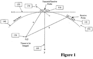
- the simplest multi-aperture system consists of two apertures, as shown in Figure 1 .
- One aperture could be used entirely for transmit elements 110 and the other for receive elements 120.
- Transmit elements can be interspersed with receive elements, or some elements could be used both for transmit and receive.
- the probes have two different lines of sight to the tissue to be imaged 130. That is, they maintain two separate physical apertures on the surface of the skin 140.
- Multiple Aperture Ultrasonic Transducers are not limited to use from the surface of the skin, they can be used anywhere in or on the body to include intracavity and intravenous probes.
- the positions of the individual elements T x 1 through T x n can be measure in three different axes.
- a Transmit Probe containing ultrasound transmitting elements T1, T2, ... Tn 110 and a Receive Probe 120 containing ultrasound receive elements R1, R2, ... Rm are placed on the surface of a body to be examined (such as a human or animal). Both probes can be sensitive to the same plane of scan, and the mechanical position of each element of each probe is known precisely relative to a common reference such as one of the probes.
- an ultrasound image can be produced by insonifying the entire region to be imaged (e.g., a plane through the heart, organ, tumor, or other portion of the body) with a transmitting element (e.g., transmit element T x 1), and then "walking" down the elements on the Transmit probe (e.g., T x 2, ... T x n) and insonifying the region to be imaged with each of the transmit elements.
- a transmitting element e.g., transmit element T x 1
- the images taken from each transmit element may not be sufficient to provide a high resolution image, but the combination of all the images can provide a high resolution image of the region to be imaged.
- FIG. 2 Another multi-aperture system is shown Figure 2 and consists of transducer elements in three apertures.
- elements in the center aperture 210 can be used for transmit and then elements in the left 220 and right 230 apertures can be used for receive.
- elements in all three apertures can be used for both transmit and receive, although the compensation for speed of sound variation would be more complicated under these conditions.
- Positioning elements or arrays around the tissue to be imaged 240 provides much more data than simply having a single probe 210 over the top of the tissue.
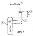
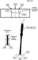
- FIGS. 3 and 4 demonstrate how a single omni-probe 310 or 410 can be attached to a main transducer (phased array or otherwise) so as to collect data, or conversely, to act as a transmitter where the main probe then becomes a receiver.
- the omni-probe is already aligned within the scan plan. Therefore, only the x and y positions 350 need be calculated and transmitted to the processor. It is also possible to construct a probe with the omni-probe out of the scan plane for better transverse focus.
- An aspect of the omni-probe apparatus includes returning echoes from a separate relatively non-directional receive transducer 310 and 410 located away from the insonifying probe transmit transducer 320 and 420, and the non-directional receive transducer can be placed in a different acoustic window from the insonifying probe.
- the omni-directional probe can be designed to be sensitive to a wide field of view for this purpose.
- the echoes detected at the omni-probe may be digitized and stored separately. If the echoes detected at the omni-probe (310 in Figure 3 and 410 in Figure 4 ) are stored separately for every pulse from the insonifying transducer, it is surprising to note that the entire two-dimensional image can be formed from the information received by the one omni. Additional copies of the image can be formed by additional omni-directional probes collecting data from the same set of insonifying pulses.
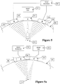
- the entire probe when assembled together, is used as an add-on device. It is connected to both an add-on instrument or MAUI Electronics 580 and to any host ultrasound system 540.
- the center array 510 can be used for transmit only.
- the outrigger arrays 520 and 530 can be used for receive only and are illustrated here on top of the skin line 550. Reflected energy off of scatterer 570 can therefore only be received by the outrigger arrays 520 and 530.
- the angulation of the outboard arrays 520 and 530 are illustrated as angles ⁇ 1 560 or ⁇ 2 565. These angles can be varied to achieve optimum beamforming for different depths or fields of view.
- ⁇ 1 and ⁇ 2 are often the same for outboard arrays, however, there is no requirement to do so.
- the MAUI Electronics can analyze the angles and accommodate unsymmetrical configurations.
- Fig. 5a demonstrates the right transducer 510 being used to transmit, and the other transducer 520 is being used to receive.
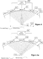
- Figure 6 is much like Figure 5 , except the Multiple Aperture Ultrasound Imaging System (MAUI Electronics) 640 used with the probe is a stand-alone system with its own onboard transmitter (i.e., no host ultrasound system is used). This system may use any element on any transducer 610, 620, or 630 for transmit or receive.
- the angulation of the outboard arrays 610 and 630 is illustrated as angle ⁇ 660. This angle can be varied to achieve optimum beamforming for different depths or fields of view. The angle is often the same for outboard arrays; however, there is no requirement to do so.
- the MAUI Electronics will analyze the angle and accommodate unsymmetrical configurations.
- Fig. 6a shows the right array 610 transmitting, and all three arrays 610, 620 and 630 receiving.
- Figure 6b shows elements on the left array 610 transmitting, and elements on the right array 620 receiving.
- Figure 6b is much like Figure 5a , except the Multiple Aperture Ultrasound Imaging System (MAUI Electronics) 640 used with the probe is a stand-alone system with its own onboard transmitter. This system may use any element on any array 610 or 620 for transmit or receive as is shown in Figure 6c . As shown in either Figure 6b or Figure 6c , a transmitting array provides angle off from the target that adds to the collective aperture width 690 the same way two receive only transducers would contribute.
- MAUI Electronics Multiple Aperture Ultrasound Imaging System
- Embodiments described herein include a precision carrier for the proper alignment of a universal multiple aperture ultrasound transducer.
- transducer array 2161 can be already “potted” in its own fixture 2161 with lens 2162 intact. Potting procedures are conventional methods to secure the transducer array to its lens and to the case. Flex circuitry, cabling, and attachment to the larger multiple aperture ultrasound transducer fixture can take place after the potting procedure is complete. A benefit of the invention is that it does not require the same transducers to be utilized during the alignment. Different transducers with different "pots" can be utilized in any location of the alignment fixture thanks to the flexibility of the alignment carrier.
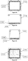
- Figures 8a - 8c provide views of the basic structure and features of embodiments of a precision carrier 2150 for a multiple aperture ultrasound transducer array.
- Figure 8a shows a top view of a precision array carrier 2150 with six positioning screws 2151.
- Figure 8b shows a side view of a precision array carrier 2150 having two threaded screw holes 2180 on each side. When positioning screws 2151 are inserted into threaded screw holes (e.g., screw holes 2155 and 2156 in Figure 7b ), adjustments may be made to employ longitudinal corrections 2159 to the "seated" array.
- Figure 8c shows a side view of a precision carrier 2150 with threaded screw holes 2180 located on each end. When positioning screws are inserted into these threaded screw holes, adjustments may be made to employ lateral corrections 2160 to the "seated" array (as illustrated in Figure 7c ).
- Figures 9a - 9d show a precision array carrier 2150 with an array-centering gasket 2152 installed.
- Figure 9a is a top view of the precision carrier 2150, with an array-centering gasket 2152 placed at the bottom of the carrier where the lens 2162 located in the center.
- Figures 9b - 9d show side, end, and bottom views of the carrier, respectively.
- the gasket 2152 extends the entire length of the carrier over the L shaped shoulder 2181.
- the gasket 2152 extends around the corners of the L shaped shoulder 2181 to cover the ends of the carrier as it illustrated in Figure 9c .
- the gasket provides the array translational centering and a pivot point for positioning adjustments during operation without interfering with the integrity of the lens 2162.
- Figure 9d provides a view of the lens 2162, the bottom of the precision carrier array centering gasket 2152, and finally the L shaped shoulder 2181.
- FIGs 7a - 7c show top, end, and side views, respectively of a precision array carrier 2150 with an array 2161 inserted therein.
- the array 2161 is supported end-to-end by positioning screws 2155 and 2156.
- the array can be supported from each side by positioning screws 2153, 2154, 2157, 2158 and from the bottom by the array centering gasket 2152.
- Figure 7b shows the array 2161 in the precision array carrier 2150 being supported by array centering gasket 2152 and ready for longitudinal adjustment. Alternately tightening and loosening positioning screws 2155 and 2156 allows the array 2161 to be adjusted through arc 2159 to correct longitudinal axis errors.
- Figure 7c shows the array 2161 in the precision array carrier 2150 supported by the array centering gasket 2152 ready for transverse alignment. Alternately adjusting positioning screw pairs 2157, 2158 and 2153, 2154 allow the array 2161 to be corrected for transverse axis errors.
- Figures 10a and 10b show a top views of a precision array carrier 2150 with the array 2161 inserted. Arrows depict, respectively, counter-clockwise and clockwise rotational adjusting by way of selective screw adjustments.
- Figure 10a shows a tightening of position screws 2153 and 2158 while loosening position screws 2154 and 2157 shifting the array 2161 in a counter-clockwise arc 2165 to correct rotational axis errors.
- Figure 10b shows a tightening position of screws 2154 and 2157 while loosening position screws 2153 and 2158 to shift the array 2161 in a clockwise arc 2166 to correct rotational axis errors.
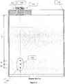
- FIG 11 shows an end view of a precision array carrier 2150 installed on a tissue equivalent phantom or test block 2182 and ready to transmit and receive during alignment.
- a 'phantom' is a structure filled with tissue equivalent material that has a speed of sound characteristics similar to that of human tissue with known voids and reflectors placed at known locations within the phantom.
- This end view of the phantom shows one embodiment including three targets 2167 in profile view. These targets can be echogenic, very reflective, or anechoic, void of reflection.
- the top target can be at a pre-determined depth D from the surface of the phantom and the face of array carrier 2150.
- the other targets can be spaced at distances D1 and D2 from the top target.
- the pre-determined depth D can be 100 mm from the top target to the face of the array.
- the other targets can have D1 and D2 distances of 10 mm, for example.
- any range of depths for the targets 2167 can be used, depending on the desired application of the transducer arrays.
- the perpendicular targets 2167 serve to assist during the longitudinal adjustment of the array positioning. When correctly positioned, the three targets would be displayed as exactly perpendicular to the front of the array, and further, each target 2167 would be displayed equidistantly one a top the other.
- Figure 12 shows a side view of the phantom 2182 with the ends of the targets 2167 visible. Once transmitting and receiving, a lateral adjustment could be made to the array 2163 in the carrier 2150. The correct alignment is for achieved when all targets are visible above and below the center target 2168.
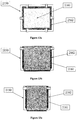
- Figures 13a - 13c show a precision array carrier 2150 with an array 2161 inserted and aligned, in top, side, and end views, respectively. At this stage an acoustic damping material 2162 can be poured into the gap between the array and the carrier to stabilize the position of arrays 2161.
- Figure 13b is a side view of the precision array carrier 2150 showing the gap between the array 2161 and the precision array carrier 2150 filled with acoustic damping material 2162.
- Figure 13c shows the gap between the array 2161 and the precision array carrier 2150 filled with acoustic damping material 2162.
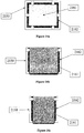
- Figures 14a - 14c show the precision array carrier 2150 with the array 2161 inserted and aligned in top, side, and end views, respectively.
- the acoustic damping material 2162 has cured and the six alignment screws have been removed.
- Figure 14b is a side view of the precision array carrier 2150 with the array 2161 inserted, aligned, the acoustic damping material 2162 cured and the position alignment screws removed: At this point, the precision array carrier 2150 with its captured array becomes a precision carrier array assembly 2163.
- Figure 15 shows a multi-aperture ultrasound probe assembly 2183 constructed with precision transducer receptacles surrounded by structural supports 2164.
- the structural supports 2164 can be constructed out of many hard materials (e.g. metals or plastics) and usually are built into a larger structure such as the probe 2200 in Figure 22 .
- the three precision carrier array assemblies 2163 are inserted into the precision transducer receptacles 2166.
- Figure 16 shows the multi-aperture probe assembly 2183 having precision transducer receptacles 2166 with the precision array assemblies 2163 each locked into the receptacles, thus completing the construction of the multi-aperture ultrasound probe 2184 having three transducer arrays.
- Figure 22 shows a completed probe 2200 with arrays 1701, 1702, and 1703 fitted in array receptacles and ready for submission to the calibration cycle.
- a MAUI alignment fixture for aligning a multi-aperture probe uses one or more precision angular alignment controls, precision stage assemblies that provide for the adjustment, in 6 degrees of freedom of the each array under test.
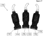
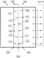
- Figure 17 shows a probe system 1700 comprising three probes 1701, 1702, and 1703 working together as a multi-aperture transducer though not assembled in a single shell.
- a multi-aperture transducer can comprise of any number of arrays 1710, 1720, 1730 (two or more), or even individual elements.
- arrays in probes can easily be manufactured with a large number of elements and element spacing within a head can be well controlled. If one can precisely position the end elements of each probe, it is possible to imply the positions of the other elements. Therefore, a fixture will be described which finds the positions of the elements. This apparatus could determine the exact location of independent elements either inside or outside of an array; however, because arrays are typically constructed in a linear format, the embodiment discussed here only identifies the end elements.
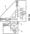
- a precision alignment stage assembly is shown in Figure 18A .
- the far left area of the assembly 1801 allows for the mechanical connection of a single probe, such as 1701 from Figure 17 .
- the precision alignment stage assembly has three separate mechanisms 1801, 1802 and 1803 that control the position of the attached array in x, y and z axes.
- Several alignment stage assemblies can be used in concert so that multiple probe arrays can be manipulated independently.
- Figure 18B allows the operator to manipulate an array in any axis by using controls 1805, 1806, 1807, 1808, and bearing 1809.
- Precision screws 1804, 1805, 1806, 1807, and 1808 can be adjusted, and bearing 1809 can be rotated to affect one or more axes for the array during the alignment process.
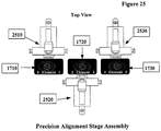
- Figure 25 shows the arrays 1710, 1720 and 1730 attached in line to precision alignment stages 2510, 2520 and 2530. With the arrays set in place, they can now transmit to common points of interest and compare their points of impact with the other arrays.
- Figure 20 illustrates probes 1701, 1702 and 1703 from Figure 17 now attached to alignment stage assemblies above a tank or test block 2012.
- the tank can be filled with any liquid, fluid, gel, solid, or other medium 2014 that is desirable for manufacture and safety considerations, as long as the speed of sound for the fluid is known.
- the tank can include a mounting location for the alignment stage assemblies.
- multiple alignment stage assemblies holding transducer elements can be mounted on the test block. From this position, it is possible to transmit ultrasonic pulses from the elements of any of the arrays to be received by ultrasonic sensor or hydrophones 2085 at the other end of the tank 2012.
- a multi-axis ultrasonic sensor or hydrophone 2085 may be used to detect the X, Y and Z positions of each element of a single array or multiple arrays under test.
- the multi-axis hydrophone 2085 can include a transverse hydrophone 2086, and right and left hydrophones 2087 and 2088.
- the common targets for the probes 1701, 1702 and 1703 to shoot at are elements 2091, 2092 and 2093 on the right hydrophone 2087.
- elements 2094, 2095, and 2096 are the targets.
- the probe can be attached to a signal generator configured to excite any of the transducer elements to transmit ultrasonic pulses.
- An ultrasonic signal is transmitted which exhibits good autocorrelation properties (e.g., a long frequency sweep, or 'chirp' waveform, a short (wideband) pulse, a spread spectrum waveform, etc) from at least one element in arrays 1710, 1720 and 1730.
- the transmitted ultrasound signal can travel through the test block and be received by the receiving hydrophone transducer elements 2091, 2092, 2093, 2094, 2095, 2096 and the transverse hydrophone 2086. It is important to note that detection of the ultrasonic signal or pulse as received by the hydrophone arrays cannot be detected accurately enough by cross correlation with the signal impressed on the probe element because the probe element itself distorts the signal.
- the first technique is to use cross correlation between the signal received at one element of the hydrophone (for example 2091) and the signal received at another element of the same hydrophone (for example 2093). The correlation peak will yield the time difference and thus the distance difference.
- the second technique is to interpolate between samples of the received waveforms to obtain better time resolution than simply the sampling interval. Perhaps the best way to accomplish both of these tasks is to take the Fourier transform of both signals, fill in zeros for the high frequency components of a much larger transform. Call these larger transforms FFT1 and FFT2. Then find the peak of the inverse transform of (FFT1 *(conjugate of FFT2)).
- a third technique is necessary to convert differential distances to total distance.
- the point b represents one of the elements for which we need to compute a position
- c and e are known reference points in the bottom of the water tank. It is desired to measure the lengths d 4 and d 0 by triangulation, but just knowing the difference between d 0 and d4 is not enough.
- a transverse hydrophone see 2086 In Figure 19a
- Let e, d, and c be the locations of the hydrophones 2094, 2095 and 2096 or 2091, 2092 and 2093 of Figure 19a .
- the hydrophones 2094, 2095 and 2096 must be on the same line and on a parallel line to that formed by 2091, 2092 and 2093.
- the distance between 2094 and 2095 is designated d 1
- the distance between 2095 and 2096 is designated d 3 .
- d 1 and d 3 must be known precisely as this becomes the reference "yardstick" for the other measurements.
- 2095 should be roughly centered between 2094 and 2096LN, but d 1 does not need to equal d 3 .
- the same requirements apply to R0, RC, and RN.
- Two parallel "yardsticks" or right and left hydrophones are provided in the bottom of the tank in order to measure position along the z axis from Figure 1 , and as is illustrated in Figure 23b . It will be the goal to position all of the probe elements from all three arrays 1701, 1702 and 1703 in a line midway between the two yardsticks using the various controls illustrated in Figure 18b .
- distance a ((d 0 2 -d 4 2 + (d 1 +d 3 ) 2 )/(2(d 1 +d 3 ))
- the position along the x' axis is d 1 -a.
- the position along the y' axis is sqrt((d 0 2-a 2 -(zr/2) 2 )).
- the same computations for x' and y' can be made using the left yardstick 2094, 2095 and 2095; and, the results can be averaged for increased accuracy.
- the main reason for having two yardsticks is the ability to measure the z axis; the elements position in or out of the scan plane as illustrated in Figure 1 .
- the array alignment apparatus can display it (see Figure 23a , 2300), and thus allow either manual ( Figure 18b ) or automatic ( Figure 24 ) correction and alignment.
- the z variable is proportional to the time of arrival difference of the pulse as received at RC 2092 and LC 2095. The probe position should be adjusted until the time difference is close to zero. When this is done, all of the x and y measurements will be accurate and the relative positions of all of the elements will be known.
- a controller (such as a computer) can scan and find the maximum signal strength on the transverse hydrophone 2086 and record the angular displacement for the probe element.
- multiple aperture ultrasound transducers will already be fully assembled, such as the embodiment illustrated in Figure 22 . Therefore, all of these measurements will have to be referenced to axes on the probe assembly.
- the multi-aperture transducer probe assembly 2200 shown in Figure 22 it would be reasonable to rotate and translate all measurements to a new coordinate system (x,y) centered on the center array. The appropriate coordinate system would be dependent on the ultrasound imaging system for which the probe assembly would be used.
- the multi-aperture probe can have a resident calibration memory or cal chip 2201 that can be programmed with calibration data received from the automated precision stage assembly, described below.
- the transmit synchronization module 2202 is not related to calibration, but is necessary to identify the start of pulse when the probe is used as an add-on device with a host machine transmitting.
- the probe displacement sensor 2203 can be an accelerometer or gyroscope that senses the three dimensional movement of the probe. During calibration, the probe must be securely attached to the array alignment apparatus so that the probe is still.
- a proprietary graphical user interface or GUI 2300 allows the elemental array data to be visualized in real-time allowing for correction of the x, y and z variation errors.
- the two wide vertical lines 2001 and 2003 represent the z positions of the yardsticks RO-RC-RN (2091, 2092, and 2093 from Figure 19a ) and LO-LC-LN (2094, 2095, and 2096 from Figure 19a ).
- the vertical position is the x coordinate.
- Each small square such as 2305, 2306, 2307, 2308, 2309, 2310 and 2011, is the position of a probe element in the x-z plane.
- the thin horizontal lines 2312, 2313, 2314, 2315, 2316, 2317 and 2018 represent the directivity and angular spread of each element as detected on the multi-axis hydrophone.
- a useful angular spread measure is the number of hydrophone elements on the transverse hydrophone array which record signal strength greater or equal to half of the maximum strength.
- Figure 23c depicts a probe element with its z position 2305 offset toward the right hydrophone.
- the resulting display shows the small square, 2305, to the right of centerline, 2302. Note that in this case, the element position is in error, but the element directivity remains over the centerline as indicated on the display by the horizontal line 2312 remaining centered over centerline, 2302.
- the directivity 2312 is misaligned in this case with an offset toward the left hydrophone as indicated by the horizontal line shifted to the left of centerline, 2302.
- the directivity needs to be corrected by adjusting the angulation to bring the directivity back over center. This could be accomplished, for example, by using controls 1805 and 1807 in Figure 18b .
- element position and directivity can be monitored simultaneously and both brought into alignment.
- Adjustments of the probe position and angulation with the precision alignment stage assembly or assemblies should continue until all of the small squares and all of the horizontal lines are aligned on the center vertical line as closely as practicable, ensuring in alignment in the z axis. As this is done, the x and y positions will be computed accurately and no separate iteration will be required for these.
- arrays 2406 could be loaded into an automated precision stage assembly like the one in Figure 24 .
- arrays while still within their nose pieces can still be manipulated.
- Stepper motor controller, 2401 drives the transducer, 2405, under test in response to instructions from controller, 2402.
- the controller, 2401 evaluates data from the hydrophone assembly, 2404, and calculates transducer corrections.
- Test programs residing in the controller, 2402 provide transducer specific calibration data back to the transducer, 2405, under test incorporation in it's on board calibration chip, 2201. This automatically acquired element and array position data would be MAUI probe specific and would be used to optimize probe and system performance.
- FIGs 26a and 26b illustrate array alignment systems 2610 attached to the control unit 2620 of an ultrasound machine 2600. A cut away shows hydrophone assembly 2085 is located at the bottom of the fluid filled system 2610.
- a MAUI general radiology probe 2630 is affixed to the system for testing.
- a MAUI cardiac probe 2640 is affixed to the system for calibration. The portability of this system, therefore allows for calibration of probes in the field multiple times per day. Additionally the MAUI system would alert the operator if service or maintenance was required.
- MAUI electronic apparatus can send a test pattern to the arrays in the probe to transmit to the hydrophone assembly 2085.
- the positions of the probes and their directivities are reported as a result of the sequence, the positions of all of the elements can be downloaded to a file specific to that probe.
- Each file is stored in the probe calibration chip 2201.
- the calibration chip reports element positions in x, y and z axes to every MAUI electronic apparatus it connects to, and therefore can perform multiple aperture imaging without recalibrating before use with a different MAUI apparatus.
- the calibration chip memory can also be used to analyze probe performance and reliability.
Landscapes
- Health & Medical Sciences (AREA)
- Life Sciences & Earth Sciences (AREA)
- Engineering & Computer Science (AREA)
- Physics & Mathematics (AREA)
- Biomedical Technology (AREA)
- Heart & Thoracic Surgery (AREA)
- Veterinary Medicine (AREA)
- Public Health (AREA)
- Biophysics (AREA)
- Nuclear Medicine, Radiotherapy & Molecular Imaging (AREA)
- Pathology (AREA)
- Radiology & Medical Imaging (AREA)
- General Health & Medical Sciences (AREA)
- Animal Behavior & Ethology (AREA)
- Medical Informatics (AREA)
- Molecular Biology (AREA)
- Surgery (AREA)
- Remote Sensing (AREA)
- Radar, Positioning & Navigation (AREA)
- Acoustics & Sound (AREA)
- Computer Networks & Wireless Communication (AREA)
- General Physics & Mathematics (AREA)
- Gynecology & Obstetrics (AREA)
- Ultra Sonic Daignosis Equipment (AREA)
Applications Claiming Priority (2)
| Application Number | Priority Date | Filing Date | Title |
|---|---|---|---|
| US16920009P | 2009-04-14 | 2009-04-14 | |
| PCT/US2010/031067 WO2010120907A2 (en) | 2009-04-14 | 2010-04-14 | Multiple aperture ultrasound array alignment fixture |
Publications (3)
| Publication Number | Publication Date |
|---|---|
| EP2419022A2 EP2419022A2 (en) | 2012-02-22 |
| EP2419022A4 EP2419022A4 (en) | 2014-04-16 |
| EP2419022B1 true EP2419022B1 (en) | 2019-11-06 |
Family
ID=42981660
Family Applications (1)
| Application Number | Title | Priority Date | Filing Date |
|---|---|---|---|
| EP10765107.7A Active EP2419022B1 (en) | 2009-04-14 | 2010-04-14 | Multiple aperture ultrasound array alignment fixture |
Country Status (5)
| Country | Link |
|---|---|
| US (2) | US8473239B2 (enExample) |
| EP (1) | EP2419022B1 (enExample) |
| JP (2) | JP5485373B2 (enExample) |
| KR (1) | KR101659723B1 (enExample) |
| WO (1) | WO2010120907A2 (enExample) |
Families Citing this family (47)
| Publication number | Priority date | Publication date | Assignee | Title |
|---|---|---|---|---|
| US8105239B2 (en) | 2006-02-06 | 2012-01-31 | Maui Imaging, Inc. | Method and apparatus to visualize the coronary arteries using ultrasound |
| EP2088932B1 (en) | 2006-10-25 | 2020-04-08 | Maui Imaging, Inc. | Method and apparatus to produce ultrasonic images using multiple apertures |
| US9282945B2 (en) * | 2009-04-14 | 2016-03-15 | Maui Imaging, Inc. | Calibration of ultrasound probes |
| US10226234B2 (en) | 2011-12-01 | 2019-03-12 | Maui Imaging, Inc. | Motion detection using ping-based and multiple aperture doppler ultrasound |
| JP5666446B2 (ja) | 2008-08-08 | 2015-02-12 | マウイ イマギング,インコーポレーテッド | マルチアパーチャ方式の医用超音波技術を用いた画像形成方法及びアドオンシステムの同期方法 |
| KR101659723B1 (ko) | 2009-04-14 | 2016-09-26 | 마우이 이미징, 인코포레이티드 | 복수 개구 초음파 어레이 정렬 설비 |
| EP2536339B1 (en) | 2010-02-18 | 2024-05-15 | Maui Imaging, Inc. | Point source transmission and speed-of-sound correction using multi-aperture ultrasound imaging |
| EP3563768A3 (en) | 2010-10-13 | 2020-02-12 | Maui Imaging, Inc. | Concave ultrasound transducers and 3d arrays |
| WO2012051305A2 (en) | 2010-10-13 | 2012-04-19 | Mau Imaging, Inc. | Multiple aperture probe internal apparatus and cable assemblies |
| EP2643658A4 (en) | 2010-11-24 | 2014-05-21 | Hysitron Inc | MECHANICAL TEST INSTRUMENTS WITH BOARD DATA |
| US9649091B2 (en) * | 2011-01-07 | 2017-05-16 | General Electric Company | Wireless ultrasound imaging system and method for wireless communication in an ultrasound imaging system |
| EP2713889A2 (en) * | 2011-05-25 | 2014-04-09 | Orcasonix Ltd. | Ultrasound imaging system and method |
| JP2015503404A (ja) | 2011-12-29 | 2015-02-02 | マウイ イマギング,インコーポレーテッド | 任意経路のmモード超音波イメージング |
| EP2816958B1 (en) | 2012-02-21 | 2020-03-25 | Maui Imaging, Inc. | Determining material stiffness using multiple aperture ultrasound |
| IN2014DN07243A (enExample) | 2012-03-26 | 2015-04-24 | Maui Imaging Inc | |
| EP2883079B1 (en) * | 2012-08-10 | 2017-09-27 | Maui Imaging, Inc. | Calibration of multiple aperture ultrasound probes |
| KR102176319B1 (ko) * | 2012-08-21 | 2020-11-09 | 마우이 이미징, 인코포레이티드 | 초음파 이미징 시스템 메모리 아키텍처 |
| CN103676827A (zh) | 2012-09-06 | 2014-03-26 | Ip音乐集团有限公司 | 用于远程控制音频设备的系统和方法 |
| WO2014160291A1 (en) * | 2013-03-13 | 2014-10-02 | Maui Imaging, Inc. | Alignment of ultrasound transducer arrays and multiple aperture probe assembly |
| US9883848B2 (en) | 2013-09-13 | 2018-02-06 | Maui Imaging, Inc. | Ultrasound imaging using apparent point-source transmit transducer |
| TWI511478B (zh) * | 2013-10-04 | 2015-12-01 | Genesys Logic Inc | 超音波資料擷取系統、方法及超音波接收裝置 |
| US9476859B2 (en) * | 2013-12-16 | 2016-10-25 | Olympus Ndt, Inc. | Automatic calibration for phased array inspection of girth weld |
| FR3016967B1 (fr) * | 2014-01-30 | 2016-03-04 | Schneider Electric Ind Sas | Systeme d'aide a l'alignement pour emetteur a ultrasons, ensemble de detecteur a ultrasons et procede d'alignement |
| EP3102933B1 (en) | 2014-02-05 | 2018-11-28 | Verathon INC. | Ultrasound scanner calibration |
| US10557828B2 (en) | 2014-02-17 | 2020-02-11 | Westinghouse Electric Company Llc | Ultrasonic phased array transducer for the NDE inspection of the jet pump riser welds and welded attachments |
| JP2016034435A (ja) * | 2014-08-04 | 2016-03-17 | キヤノン株式会社 | 被検体情報取得装置 |
| KR102617888B1 (ko) | 2014-08-18 | 2023-12-22 | 마우이 이미징, 인코포레이티드 | 네트워크-기반 초음파 이미징 시스템 |
| US11304676B2 (en) | 2015-01-23 | 2022-04-19 | The University Of North Carolina At Chapel Hill | Apparatuses, systems, and methods for preclinical ultrasound imaging of subjects |
| KR102681141B1 (ko) | 2015-03-30 | 2024-07-02 | 마우이 이미징, 인코포레이티드 | 오브젝트 모션을 검출하기 위한 초음파 이미징 시스템들 및 방법들 |
| US20180092626A1 (en) * | 2015-06-10 | 2018-04-05 | Koninklijke Philips N.V. | Ultrasound imaging apparatus |
| US20170045614A1 (en) * | 2015-08-13 | 2017-02-16 | Daniel N. Harres | Ultrasonic ranging sensors |
| KR101693226B1 (ko) * | 2015-08-25 | 2017-01-05 | 주식회사 에네스지 | 튜브 용접부 검사용 다중 초음파 탐촉자 |
| US11209297B2 (en) * | 2015-10-21 | 2021-12-28 | Texas Instruments Incorporated | Ultrasonic transducer system and method using broadband system responses |
| WO2017132517A1 (en) | 2016-01-27 | 2017-08-03 | Maui Imaging, Inc. | Ultrasound imaging with sparse array probes |
| EP3432801B1 (en) | 2016-03-23 | 2020-05-06 | Koninklijke Philips N.V. | A method and apparatus for improving the measurement of flow velocity of blood |
| US20190162833A1 (en) * | 2016-04-22 | 2019-05-30 | Fidelity Technologies Corporation | Ultrasonic position detection system |
| US10064604B2 (en) | 2016-07-01 | 2018-09-04 | Carestream Health, Inc. | Ultrasound calibration fixture |
| FR3060762B1 (fr) * | 2016-12-20 | 2020-06-12 | Thales | Systeme reparti modulaire de detection acoustique des menaces sous-marines sur une zone sensible |
| US10416122B2 (en) * | 2017-10-31 | 2019-09-17 | Westinghouse Electric Company Llc | Ultrasonic phased array transducer apparatus for the nondestructive inspection of a component under test |
| EP3513733A1 (en) | 2018-01-23 | 2019-07-24 | Koninklijke Philips N.V. | Ultrasound imaging apparatus and method |
| EP3801705A1 (en) | 2018-05-31 | 2021-04-14 | Matt McGrath Design & Co, LLC | Biocompatible material with pain management capability and method of use thereof |
| WO2020131020A1 (en) * | 2018-12-17 | 2020-06-25 | Emerge Now Inc. | Systems for interfacing with immersive computing environments |
| CN113994230A (zh) * | 2019-06-04 | 2022-01-28 | Tdk电子股份有限公司 | 超声转换器和用于制造超声转换器的方法 |
| JP7445513B2 (ja) * | 2020-05-15 | 2024-03-07 | キヤノンメディカルシステムズ株式会社 | 超音波診断装置 |
| CN112083075B (zh) * | 2020-09-30 | 2024-12-31 | 浙江浙能技术研究院有限公司 | 基于平面波超声技术的枞树型叶根高效检测装置及方法 |
| CN112362758A (zh) * | 2020-11-20 | 2021-02-12 | 西安热工研究院有限公司 | 一种相控阵超声校准试块及校准方法 |
| US20250072866A1 (en) * | 2021-09-16 | 2025-03-06 | Brainlab Ag | Segmented ultrasound probe |
Family Cites Families (470)
| Publication number | Priority date | Publication date | Assignee | Title |
|---|---|---|---|---|
| US3174286A (en) | 1963-06-27 | 1965-03-23 | Gen Motors Corp | Master cylinder |
| JPS4911189A (enExample) | 1972-05-29 | 1974-01-31 | ||
| US3895381A (en) | 1973-02-21 | 1975-07-15 | Winston E Kock | Synthetic aperture imaging systems |
| US4072922A (en) | 1975-11-13 | 1978-02-07 | Seiscom Delta Inc. | Method for seismic exploration for enhanced results in simulated cylindrical or plane waves |
| US4105018A (en) | 1976-02-02 | 1978-08-08 | University Of Utah | Acoustic examination, material characterization and imaging of the internal structure of a body by measurement of the time-of-flight of acoustic energy therethrough |
| US4097835A (en) | 1976-09-20 | 1978-06-27 | Sri International | Dual transducer arrangement for ultrasonic imaging system |
| US4055988A (en) | 1976-12-09 | 1977-11-01 | J. B. Engineering And Sales Company, Inc. | Alignment control apparatus for a turntable used in an ultrasonic testing system |
| JPS5444375A (en) | 1977-09-14 | 1979-04-07 | Oki Electric Ind Co Ltd | Ultrasonic wave reflection system |
| US4333474A (en) | 1978-02-06 | 1982-06-08 | New York Institute Of Technology | Ultrasonic imaging system |
| US4271842A (en) * | 1978-03-03 | 1981-06-09 | Smith Kline Instruments, Inc. | Apparatus and method for providing multiple ultrasonic sector image displays |
| JPS55103839A (en) | 1979-02-03 | 1980-08-08 | Fujitsu Ltd | Ultrasonic diagnosis apparatus |
| CA1137210A (en) | 1979-04-26 | 1982-12-07 | Francis S. Foster | Ultrasonic imaging method and device using one transducer having a line focus aligned with another transducer |
| US4259733A (en) | 1979-05-14 | 1981-03-31 | Seiscom Delta, Inc. | Multi-dimensional seismic imaging |
| US4325257A (en) | 1980-02-20 | 1982-04-20 | Kino Gordon S | Real-time digital, synthetic-focus, acoustic imaging system |
| JPS5849137A (ja) * | 1981-09-18 | 1983-03-23 | 株式会社東芝 | 超音波血流測定装置 |
| US6324453B1 (en) | 1998-12-31 | 2001-11-27 | Automotive Technologies International, Inc. | Methods for determining the identification and position of and monitoring objects in a vehicle |
| US4452084A (en) | 1982-10-25 | 1984-06-05 | Sri International | Inherent delay line ultrasonic transducer and systems |
| JPS59101143U (ja) | 1982-12-27 | 1984-07-07 | 四国変圧器株式会社 | 電気温水器 |
| JPH064074B2 (ja) | 1983-02-14 | 1994-01-19 | 株式会社日立製作所 | 超音波診断装置およびこれを用いる音速計測方法 |
| JPS59174151A (ja) | 1983-03-25 | 1984-10-02 | 横河メディカルシステム株式会社 | 超音波映像装置 |
| US5141738A (en) | 1983-04-15 | 1992-08-25 | Schering Aktiengesellschaft | Ultrasonic contrast medium comprising gas bubbles and solid lipophilic surfactant-containing microparticles and use thereof |
| JPS6013109U (ja) | 1983-07-07 | 1985-01-29 | アロカ株式会社 | 超音波診断装置 |
| JPS6068836A (ja) | 1983-09-24 | 1985-04-19 | 株式会社島津製作所 | 超音波診断装置 |
| US4539847A (en) | 1984-01-03 | 1985-09-10 | Texaco Inc. | Acoustic method and apparatus for measuring thickness of a coating layer on a substrate |
| JPS60150735A (ja) | 1984-01-18 | 1985-08-08 | 株式会社東芝 | 超音波診断装置 |
| US4662222A (en) | 1984-12-21 | 1987-05-05 | Johnson Steven A | Apparatus and method for acoustic imaging using inverse scattering techniques |
| US4781199A (en) | 1985-01-07 | 1988-11-01 | Kabushiki Kaisha Toshiba | System and method for measuring sound velocity of internal tissue in an object being investigated |
| JPS61203949A (ja) * | 1985-03-04 | 1986-09-09 | 株式会社東芝 | 超音波診断装置 |
| US4669482A (en) | 1985-10-28 | 1987-06-02 | Board Of Regents, The University Of Texas System | Pulse echo method and apparatus for sound velocity estimation in vivo |
| US4817434A (en) | 1985-11-19 | 1989-04-04 | Forrest Anderson | Device for imaging three dimensions using simultaneous multiple beam formation |
| US4831601A (en) | 1986-10-31 | 1989-05-16 | Siemens Aktiengesellschaft | Apparatus for transmitting and receiving ultrasonic signals |
| JPS63216539A (ja) | 1987-03-05 | 1988-09-08 | テルモ株式会社 | 心拍出量の相対的変化の連続測定装置 |
| US4893628A (en) * | 1988-04-04 | 1990-01-16 | Bjorn Angelsen | Dual element ultrasonic transducer probe for combined imaging of tissue structures and blood flow in real time |
| US4893284A (en) | 1988-05-27 | 1990-01-09 | General Electric Company | Calibration of phased array ultrasound probe |
| US5197475A (en) | 1988-08-10 | 1993-03-30 | The Board Of Regents, The University Of Texas System | Method and apparatus for analyzing material properties using ultrasound |
| US5050588A (en) * | 1990-02-08 | 1991-09-24 | Richard Grey | High energy ultrasonic lens assembly with mounting facets |
| JP3015527B2 (ja) | 1991-08-14 | 2000-03-06 | 株式会社東芝 | 超音波診断装置 |
| US5161536A (en) | 1991-03-22 | 1992-11-10 | Catheter Technology | Ultrasonic position indicating apparatus and methods |
| US5191890A (en) | 1991-04-22 | 1993-03-09 | Interspec, Inc. | Ultrasonic probe assembly |
| US5230339A (en) | 1991-06-13 | 1993-07-27 | Array Tech, Inc. | Performance evaluation of ultrasonic examination equipment |
| US5442462A (en) * | 1992-06-10 | 1995-08-15 | D.V.P. Technologies Ltd. | Apparatus and method for smoothing images |
| US5704361A (en) | 1991-11-08 | 1998-01-06 | Mayo Foundation For Medical Education And Research | Volumetric image ultrasound transducer underfluid catheter system |
| US5278757A (en) | 1991-11-15 | 1994-01-11 | The Trustees Of The University Of Pennsylvania | Synthetic aperture ultrasonic imaging system using a minimum or reduced redundancy phased array |
| JP3043873B2 (ja) | 1991-11-29 | 2000-05-22 | フクダ電子株式会社 | 超音波開口面合成装置 |
| US5269309A (en) | 1991-12-11 | 1993-12-14 | Fort J Robert | Synthetic aperture ultrasound imaging system |
| US5226019A (en) | 1992-01-10 | 1993-07-06 | Amoco Corporation | Method of geophysical exploration |
| US7497828B1 (en) * | 1992-01-10 | 2009-03-03 | Wilk Ultrasound Of Canada, Inc. | Ultrasonic medical device and associated method |
| US5301674A (en) | 1992-03-27 | 1994-04-12 | Diasonics, Inc. | Method and apparatus for focusing transmission and reception of ultrasonic beams |
| US5744898A (en) | 1992-05-14 | 1998-04-28 | Duke University | Ultrasound transducer array with transmitter/receiver integrated circuitry |
| US5409010A (en) * | 1992-05-19 | 1995-04-25 | Board Of Regents Of The University Of Washington | Vector doppler medical devices for blood velocity studies |
| US5339282A (en) | 1992-10-02 | 1994-08-16 | University Of Utah Research Foundation | Resolution enhancement for ultrasonic reflection mode imaging |
| JPH06125908A (ja) | 1992-10-19 | 1994-05-10 | Toshiba Corp | 超音波診断装置 |
| US5355888A (en) | 1992-11-12 | 1994-10-18 | Massachusetts Institute Of Technology | High resolution phased array echo imager |
| DE4302538C1 (de) | 1993-01-29 | 1994-04-07 | Siemens Ag | Therapiegerät zur Ortung und Behandlung einer im Körper eines Lebewesens befindlichen Zone mit akustischen Wellen |
| US5340510A (en) | 1993-04-05 | 1994-08-23 | Materials Systems Incorporated | Method for making piezoelectric ceramic/polymer composite transducers |
| US5305756A (en) * | 1993-04-05 | 1994-04-26 | Advanced Technology Laboratories, Inc. | Volumetric ultrasonic imaging with diverging elevational ultrasound beams |
| US5293871A (en) | 1993-05-05 | 1994-03-15 | Cornell Research Foundation Inc. | System for ultrasonically determining corneal layer thicknesses and shape |
| US5345426A (en) | 1993-05-12 | 1994-09-06 | Hewlett-Packard Company | Delay interpolator for digital phased array ultrasound beamformers |
| US5398216A (en) | 1993-08-30 | 1995-03-14 | General Electric Company | Method for detecting two-dimensional flow for ultrasound color flow imaging |
| US5842473A (en) | 1993-11-29 | 1998-12-01 | Life Imaging Systems | Three-dimensional imaging system |
| IT1268599B1 (it) | 1994-01-14 | 1997-03-06 | Igea Srl | Sistema di misura ad ultrasuoni per la rilevazione della densita' e struttura ossea. |
| JPH07204201A (ja) | 1994-01-25 | 1995-08-08 | Aloka Co Ltd | 超音波診断装置 |
| US5522393A (en) | 1994-05-24 | 1996-06-04 | Duke University | Multi-dimensional real-time ultrasonic blood flow imaging apparatus and method |
| US5625149A (en) | 1994-07-27 | 1997-04-29 | Hewlett-Packard Company | Ultrasonic transductor |
| JPH10507936A (ja) | 1994-08-05 | 1998-08-04 | アキュソン コーポレイション | 送信ビーム生成器システムのための方法及び装置 |
| US5570691A (en) | 1994-08-05 | 1996-11-05 | Acuson Corporation | Method and apparatus for real-time, concurrent adaptive focusing in an ultrasound beamformer imaging system |
| US5581517A (en) | 1994-08-05 | 1996-12-03 | Acuson Corporation | Method and apparatus for focus control of transmit and receive beamformer systems |
| NO943214D0 (no) | 1994-08-30 | 1994-08-30 | Vingmed Sound As | Fremgangsmåte ved ultralydavbildning |
| US5503152A (en) | 1994-09-28 | 1996-04-02 | Tetrad Corporation | Ultrasonic transducer assembly and method for three-dimensional imaging |
| US5930730A (en) | 1994-12-12 | 1999-07-27 | Amoco Corporation | Method and apparatus for seismic signal processing and exploration |
| US5563949A (en) | 1994-12-12 | 1996-10-08 | Amoco Corporation | Method of seismic signal processing and exploration |
| US5544659A (en) | 1994-12-29 | 1996-08-13 | Siemens Medical Systems, Inc. | Ultrasonic doppler imager having a reduced hardware adaptive tissue rejection filter arrangement |
| JP3612358B2 (ja) | 1995-03-17 | 2005-01-19 | 株式会社日立メディコ | 超音波診断装置 |
| US5515853A (en) * | 1995-03-28 | 1996-05-14 | Sonometrics Corporation | Three-dimensional digital ultrasound tracking system |
| GB9508525D0 (en) | 1995-04-27 | 1995-06-14 | Geco As | Method of processing seismic data |
| JP3358167B2 (ja) * | 1995-05-12 | 2002-12-16 | 北海道大学長 | 被検体同定方法、装置およびシステム |
| US5558092A (en) | 1995-06-06 | 1996-09-24 | Imarx Pharmaceutical Corp. | Methods and apparatus for performing diagnostic and therapeutic ultrasound simultaneously |
| US5999836A (en) | 1995-06-06 | 1999-12-07 | Nelson; Robert S. | Enhanced high resolution breast imaging device and method utilizing non-ionizing radiation of narrow spectral bandwidth |
| US5651365A (en) | 1995-06-07 | 1997-07-29 | Acuson Corporation | Phased array transducer design and method for manufacture thereof |
| US5675550A (en) | 1995-06-08 | 1997-10-07 | Ekhaus; Ira B. | Reduced wavenumber synthetic aperture |
| IL116701A0 (en) | 1995-10-04 | 1996-10-16 | Sunlight Ultrasound Technologi | Ultrasonic device for determining bone characteristics |
| JPH09103429A (ja) | 1995-10-13 | 1997-04-22 | Hitachi Medical Corp | 超音波診断装置 |
| JP3707882B2 (ja) | 1995-11-21 | 2005-10-19 | 株式会社東芝 | 超音波診断装置 |
| WO1997029678A2 (en) | 1996-02-15 | 1997-08-21 | Biosense Inc. | Catheter calibration and usage monitoring system |
| AU1983397A (en) * | 1996-02-29 | 1997-09-16 | Acuson Corporation | Multiple ultrasound image registration system, method and transducer |
| US5784334A (en) | 1996-03-13 | 1998-07-21 | Atlantic Richfield Company | Method and system for detecting hydrocarbon reservoirs using amplitude versus offset analysis of seismic signals |
| US5720291A (en) | 1996-03-22 | 1998-02-24 | Advanced Technology Laboratories, Inc. | Three dimensional medical ultrasonic diagnostic image of tissue texture and vasculature |
| US5628320A (en) | 1996-03-29 | 1997-05-13 | Siemens Medical Systems, Inc. | Ultrasound image reconstruction using back-propagation |
| RU2169931C2 (ru) | 1996-04-12 | 2001-06-27 | Амоко Корпорейшн | Способ и устройство для обработки сейсмического сигнала и проведения разведки полезных ископаемых |
| US5673697A (en) | 1996-04-24 | 1997-10-07 | Raytheon Company | High-resolution three, dimensional ultrasound imaging device |
| US5862100A (en) | 1996-05-28 | 1999-01-19 | Atlantic Richfield Company | Method and system for detecting hydrocarbon reservoirs using statistical normalization of amplitude-versus-offset indicators based upon seismic signals |
| GB9611801D0 (en) | 1996-06-06 | 1996-08-07 | Univ Bristol | Apparatus for and method of detecting a reflector with a medium |
| GB9611800D0 (en) | 1996-06-06 | 1996-08-07 | Univ Bristol | Post-reception focusing in remote detection systems |
| US6416475B1 (en) | 1996-06-28 | 2002-07-09 | Sonosite, Inc. | Ultrasonic signal processor for a hand held ultrasonic diagnostic instrument |
| EP0909395B1 (en) | 1996-07-02 | 2002-02-27 | B-K Medical A/S | Apparatus for determining movements and velocities of moving objects |
| NO963175D0 (no) | 1996-07-30 | 1996-07-30 | Vingmed Sound As | Analyse- og målemetode |
| US6213958B1 (en) | 1996-08-29 | 2001-04-10 | Alan A. Winder | Method and apparatus for the acoustic emission monitoring detection, localization, and classification of metabolic bone disease |
| US5795297A (en) | 1996-09-12 | 1998-08-18 | Atlantis Diagnostics International, L.L.C. | Ultrasonic diagnostic imaging system with personal computer architecture |
| GB2318414B (en) | 1996-10-19 | 2001-02-14 | Univ Cranfield | Improvements relating to flow measurement |
| US5769079A (en) | 1996-10-22 | 1998-06-23 | Acuson Corporation | Method and apparatus for determining quantitative measures of flow parameters |
| US5797845A (en) | 1996-11-04 | 1998-08-25 | Barabash; Leonid S. | Ultrasound apparatus for three dimensional image reconstruction |
| US5850622A (en) | 1996-11-08 | 1998-12-15 | Amoco Corporation | Time-frequency processing and analysis of seismic data using very short-time fourier transforms |
| US7104956B1 (en) | 1996-11-08 | 2006-09-12 | Research Corporation Technologies, Inc. | Finite amplitude distortion-based inhomogeneous pulse echo ultrasonic imaging |
| US5870691A (en) | 1996-12-06 | 1999-02-09 | Amoco Corporation | Spectral decomposition for seismic interpretation |
| US5891038A (en) | 1996-12-30 | 1999-04-06 | General Electric Company | Method, apparatus and applications for combining transmit wave functions to obtain synthetic waveform in ultrasonic imaging system |
| US5720708A (en) | 1997-01-02 | 1998-02-24 | Mayo Foundation For Medical Education And Research | High frame rate imaging with limited diffraction beams |
| US6166853A (en) | 1997-01-09 | 2000-12-26 | The University Of Connecticut | Method and apparatus for three-dimensional deconvolution of optical microscope images |
| US6122538A (en) * | 1997-01-16 | 2000-09-19 | Acuson Corporation | Motion--Monitoring method and system for medical devices |
| JPH10216128A (ja) | 1997-02-05 | 1998-08-18 | Olympus Optical Co Ltd | 超音波診断装置 |
| US5876342A (en) * | 1997-06-30 | 1999-03-02 | Siemens Medical Systems, Inc. | System and method for 3-D ultrasound imaging and motion estimation |
| US6948794B2 (en) | 1997-07-15 | 2005-09-27 | Silverbrook Reserach Pty Ltd | Printhead re-capping assembly for a print and demand digital camera system |
| US6614560B1 (en) | 1997-07-15 | 2003-09-02 | Silverbrook Research Pty Ltd | Integrated camera circuit including image sensor, image processing, and printer drive circuits |
| US6196739B1 (en) | 1997-07-15 | 2001-03-06 | Silverbrook Research Pty Ltd | Paper guide system in a print on demand digital camera system |
| US6738096B1 (en) | 1998-07-10 | 2004-05-18 | Silverbrook Research Pty Ltd | Low-cost disposable camera including print media carrying indication of postage paid |
| US5940778A (en) | 1997-07-31 | 1999-08-17 | Bp Amoco Corporation | Method of seismic attribute generation and seismic exploration |
| US6490474B1 (en) | 1997-08-01 | 2002-12-03 | Cardiac Pathways Corporation | System and method for electrode localization using ultrasound |
| US6148095A (en) | 1997-09-08 | 2000-11-14 | University Of Iowa Research Foundation | Apparatus and method for determining three-dimensional representations of tortuous vessels |
| JP3888744B2 (ja) | 1997-09-16 | 2007-03-07 | アロカ株式会社 | 超音波骨計測装置 |
| US20030166107A1 (en) * | 1997-09-18 | 2003-09-04 | Genentech, Inc. | Secreted and transmembrane polypeptides and nucleic acids encoding the same |
| US5990598A (en) * | 1997-09-23 | 1999-11-23 | Hewlett-Packard Company | Segment connections for multiple elevation transducers |
| US5957850A (en) | 1997-09-29 | 1999-09-28 | Acuson Corporation | Multi-array pencil-sized ultrasound transducer and method of imaging and manufacture |
| US6050943A (en) | 1997-10-14 | 2000-04-18 | Guided Therapy Systems, Inc. | Imaging, therapy, and temperature monitoring ultrasonic system |
| US6007499A (en) | 1997-10-31 | 1999-12-28 | University Of Washington | Method and apparatus for medical procedures using high-intensity focused ultrasound |
| KR100280197B1 (ko) | 1997-11-10 | 2001-02-01 | 이민화 | 초음파영상화시스템의초음파신호집속방법및장치 |
| JPH11239578A (ja) * | 1997-12-17 | 1999-09-07 | Nippon Koden Corp | 三次元位置校正器および校正方法 |
| US6193663B1 (en) | 1997-12-18 | 2001-02-27 | Acuson Corporation | Diagnostic ultrasound imaging method and system with improved frame rate |
| US5919139A (en) | 1997-12-19 | 1999-07-06 | Diasonics Ultrasound | Vibrational doppler ultrasonic imaging |
| NO305720B1 (no) | 1997-12-22 | 1999-07-12 | Eureka Oil Asa | FremgangsmÕte for Õ °ke oljeproduksjonen fra et oljereservoar |
| IL122839A0 (en) | 1997-12-31 | 1998-08-16 | Ultra Guide Ltd | Calibration method and apparatus for calibrating position sensors on scanning transducers |
| US6092026A (en) | 1998-01-22 | 2000-07-18 | Bp Amoco Corporation | Seismic signal processing and exploration |
| JP4373605B2 (ja) * | 1998-01-26 | 2009-11-25 | ボストン サイエンティフィック リミテッド | 遠方誘導結合器および埋め込み伝送路を備えたカテーテルアセンブリ |
| US6077224A (en) | 1998-03-23 | 2000-06-20 | Lang; Philipp | Methods and device for improving broadband ultrasonic attenuation and speed of sound measurements using anatomical landmarks |
| US6585649B1 (en) | 1998-05-02 | 2003-07-01 | John D. Mendlein | Methods and devices for improving ultrasonic measurements using multiple angle interrogation |
| US6013032A (en) | 1998-03-13 | 2000-01-11 | Hewlett-Packard Company | Beamforming methods and apparatus for three-dimensional ultrasound imaging using two-dimensional transducer array |
| US6847737B1 (en) | 1998-03-13 | 2005-01-25 | University Of Houston System | Methods for performing DAF data filtering and padding |
| US6385474B1 (en) * | 1999-03-19 | 2002-05-07 | Barbara Ann Karmanos Cancer Institute | Method and apparatus for high-resolution detection and characterization of medical pathologies |
| US6200266B1 (en) | 1998-03-31 | 2001-03-13 | Case Western Reserve University | Method and apparatus for ultrasound imaging using acoustic impedance reconstruction |
| US6238342B1 (en) | 1998-05-26 | 2001-05-29 | Riverside Research Institute | Ultrasonic tissue-type classification and imaging methods and apparatus |
| US6511426B1 (en) * | 1998-06-02 | 2003-01-28 | Acuson Corporation | Medical diagnostic ultrasound system and method for versatile processing |
| DE69903306T2 (de) | 1998-07-07 | 2003-05-22 | Lightouch Medical, Inc. | Verfahren zur gewebemodulation für die quantitative nichtinvasive in vivo spektroskopische analyse von gewebe |
| WO2000004831A1 (en) | 1998-07-21 | 2000-02-03 | Acoustic Sciences Associates | Synthetic structural imaging and volume estimation of biological tissue organs |
| US6058074A (en) | 1998-07-31 | 2000-05-02 | Atlantic Richfield Company | Method and system for detecting hydrocarbon reservoirs using amplitude-versus-offset analysis with improved measurement of background statistics |
| US6138075A (en) | 1998-08-05 | 2000-10-24 | Landmark Graphics Corporation | Methods and apparatus for analyzing seismic data |
| US6135960A (en) | 1998-08-31 | 2000-10-24 | Holmberg; Linda Jean | High-resolution, three-dimensional whole body ultrasound imaging system |
| US6425867B1 (en) * | 1998-09-18 | 2002-07-30 | University Of Washington | Noise-free real time ultrasonic imaging of a treatment site undergoing high intensity focused ultrasound therapy |
| US6048315A (en) | 1998-09-28 | 2000-04-11 | General Electric Company | Method and apparatus for ultrasonic synthetic transmit aperture imaging using orthogonal complementary codes |
| US6547732B2 (en) | 1998-10-01 | 2003-04-15 | Koninklijke Philips Electronics N.V. | Adaptive image processing for spatial compounding |
| CN1103449C (zh) | 1998-10-23 | 2003-03-19 | 李钢 | 钢轨超声探轮和探伤装置 |
| US7837624B1 (en) | 1998-11-20 | 2010-11-23 | Siemens Medical Solutions Usa, Inc. | Medical diagnostic ultrasound imaging methods for extended field of view |
| US6166384A (en) | 1998-11-06 | 2000-12-26 | General Electric Company | Method and apparatus for minimizing blurring and generating a high resolution image in a radiation imaging system |
| US6605043B1 (en) * | 1998-11-19 | 2003-08-12 | Acuson Corp. | Diagnostic medical ultrasound systems and transducers utilizing micro-mechanical components |
| US6526163B1 (en) | 1998-11-23 | 2003-02-25 | G.E. Diasonics Ltd. | Ultrasound system with parallel processing architecture |
| US6278949B1 (en) | 1998-11-25 | 2001-08-21 | M. Aftab Alam | Method for multi-attribute identification of structure and stratigraphy in a volume of seismic data |
| US6123670A (en) | 1998-12-15 | 2000-09-26 | General Electric Company | Ultrasound imaging with optimal image quality in region of interest |
| US6193665B1 (en) * | 1998-12-31 | 2001-02-27 | General Electric Company | Doppler angle unfolding in ultrasound color flow and Doppler |
| GB9901306D0 (en) | 1999-01-21 | 1999-03-10 | Smythe David | 3D/4D ultrasound imaging system |
| GB9901270D0 (en) | 1999-01-21 | 1999-03-10 | Quadrant Healthcare Uk Ltd | Method and apparatus for ultrasound contrast imaging |
| JP2000217837A (ja) * | 1999-02-01 | 2000-08-08 | Siemens Ag | 位置決め装置を保持するための装置及びこのような装置を備えた治療ヘッド |
| IL129461A0 (en) | 1999-04-15 | 2000-02-29 | F R A Y Project Dev Ltd | 3-D ultrasound imaging system |
| US6167297A (en) * | 1999-05-05 | 2000-12-26 | Benaron; David A. | Detecting, localizing, and targeting internal sites in vivo using optical contrast agents |
| US7410469B1 (en) * | 1999-05-21 | 2008-08-12 | Exogen, Inc. | Apparatus and method for ultrasonically and electromagnetically treating tissue |
| US6423002B1 (en) | 1999-06-24 | 2002-07-23 | Acuson Corporation | Intra-operative diagnostic ultrasound multiple-array transducer probe and optional surgical tool |
| US6056693A (en) * | 1999-08-16 | 2000-05-02 | General Electric Company | Ultrasound imaging with synthetic transmit focusing |
| US6251073B1 (en) | 1999-08-20 | 2001-06-26 | Novasonics, Inc. | Miniaturized ultrasound apparatus and method |
| US6264609B1 (en) | 1999-09-15 | 2001-07-24 | Wake Forest University | Ultrasound apparatus and method for tissue characterization |
| US6480790B1 (en) | 1999-10-29 | 2002-11-12 | Exxonmobil Upstream Research Company | Process for constructing three-dimensional geologic models having adjustable geologic interfaces |
| US6210335B1 (en) | 1999-12-08 | 2001-04-03 | General Electric Company | Acoustic flash to increase penetration |
| JP4610719B2 (ja) | 1999-12-27 | 2011-01-12 | Geヘルスケア・ジャパン株式会社 | 超音波撮影装置 |
| US6692450B1 (en) | 2000-01-19 | 2004-02-17 | Medtronic Xomed, Inc. | Focused ultrasound ablation devices having selectively actuatable ultrasound emitting elements and methods of using the same |
| US6361500B1 (en) | 2000-02-07 | 2002-03-26 | Scimed Life Systems, Inc. | Three transducer catheter |
| US6304684B1 (en) | 2000-02-15 | 2001-10-16 | Cyberecord, Inc. | Information processing system and method of using same |
| US6374185B1 (en) | 2000-02-18 | 2002-04-16 | Rdsp I, L.P. | Method for generating an estimate of lithological characteristics of a region of the earth's subsurface |
| US6517484B1 (en) | 2000-02-28 | 2003-02-11 | Wilk Patent Development Corporation | Ultrasonic imaging system and associated method |
| US6309356B1 (en) | 2000-03-06 | 2001-10-30 | Acuson Corporation | Method and apparatus for forming medical ultrasound images |
| US6551246B1 (en) | 2000-03-06 | 2003-04-22 | Acuson Corporation | Method and apparatus for forming medical ultrasound images |
| US6565510B1 (en) | 2000-03-22 | 2003-05-20 | General Electric Company | Method and apparatus for servicing remote ultrasound beamformer from central service facility |
| WO2001078010A2 (en) * | 2000-04-07 | 2001-10-18 | Aylward Stephen R | Systems and methods for tubular object processing |
| US6543272B1 (en) | 2000-04-21 | 2003-04-08 | Insightec-Txsonics Ltd. | Systems and methods for testing and calibrating a focused ultrasound transducer array |
| US7085400B1 (en) | 2000-06-14 | 2006-08-01 | Surgical Navigation Technologies, Inc. | System and method for image based sensor calibration |
| US6468216B1 (en) | 2000-08-24 | 2002-10-22 | Kininklijke Philips Electronics N.V. | Ultrasonic diagnostic imaging of the coronary arteries |
| US6450960B1 (en) | 2000-08-29 | 2002-09-17 | Barbara Ann Karmanos Cancer Institute | Real-time three-dimensional acoustoelectronic imaging and characterization of objects |
| JP2002143156A (ja) * | 2000-09-05 | 2002-05-21 | Koninkl Philips Electronics Nv | 媒体中の散乱物を撮像する超音波システム及び超音波診断装置 |
| US6690962B2 (en) * | 2000-09-15 | 2004-02-10 | Institut fur Diagnostikforshung GmbH | Process for graphic visualization and diagnosis of thrombi by means of nuclear spin tomography with use of particulate contrast media |
| US6508768B1 (en) | 2000-11-22 | 2003-01-21 | University Of Kansas Medical Center | Ultrasonic elasticity imaging |
| US7615008B2 (en) | 2000-11-24 | 2009-11-10 | U-Systems, Inc. | Processing and displaying breast ultrasound information |
| US6487502B1 (en) | 2000-12-01 | 2002-11-26 | Rdsp I, L.P. | System for estimating the locations of shaley subsurface formations |
| US6475150B2 (en) | 2000-12-01 | 2002-11-05 | The Regents Of The University Of California | System and method for ultrasonic tomography |
| DE10100572A1 (de) | 2001-01-09 | 2002-07-11 | Philips Corp Intellectual Pty | Verfahren zur Darstellung des Blutflusses in einem Gefäßbaum |
| JP2002209894A (ja) | 2001-01-19 | 2002-07-30 | Fuji Photo Film Co Ltd | 超音波用探触子 |
| GB2371623B (en) | 2001-01-26 | 2004-07-14 | David Nathaniel Alleyne | Inspection of non axi-symmetric elongate bodies |
| US20020111568A1 (en) | 2001-02-12 | 2002-08-15 | Shmuel Bukshpan | Method for phased array ultrasonic transmission |
| JP2002253549A (ja) | 2001-03-02 | 2002-09-10 | Fuji Photo Film Co Ltd | 超音波撮像装置、超音波撮像方法及び探触子 |
| JP2002253548A (ja) | 2001-03-02 | 2002-09-10 | Fuji Photo Film Co Ltd | 超音波検査装置 |
| GB2374744B (en) | 2001-04-18 | 2003-04-16 | Voxar Ltd | Correction of boundary artefacts in image data processing |
| US6589175B2 (en) | 2001-04-30 | 2003-07-08 | Koninklijke Philips Electronics N.V. | Real-time arbitrary mmode for ultrasonic imaging system |
| US7366704B2 (en) * | 2001-06-28 | 2008-04-29 | Waters Investments, Limited | System and method for deconvoluting the effect of topography on scanning probe microscopy measurements |
| US6620101B2 (en) | 2001-07-26 | 2003-09-16 | Dentosonic Ltd. | Bone measurement device |
| US6668654B2 (en) | 2001-08-15 | 2003-12-30 | Lockheed Martin Corporation | Method and apparatus for generating specific frequency response for ultrasound testing |
| JP4022393B2 (ja) | 2001-12-12 | 2007-12-19 | 株式会社日立メディコ | 超音波診断装置 |
| AU2003207947A1 (en) | 2002-01-07 | 2003-07-24 | Medson Ltd. | A system and method of mapping irregularities of hard tissue |
| US7285094B2 (en) | 2002-01-30 | 2007-10-23 | Nohara Timothy J | 3D ultrasonic imaging apparatus and method |
| US6755789B2 (en) | 2002-02-05 | 2004-06-29 | Inceptio Medical Technologies, Llc | Ultrasonic vascular imaging system and method of blood vessel cannulation |
| US7806828B2 (en) | 2002-02-05 | 2010-10-05 | Inceptio Medical Technologies, Lc | Multiplanar ultrasonic vascular sensor assembly and apparatus for movably affixing a sensor assembly to a body |
| US7231072B2 (en) | 2002-02-27 | 2007-06-12 | Konica Corporation | Image processing method and image converting apparatus |
| GB0205000D0 (en) * | 2002-03-04 | 2002-04-17 | Isis Innovation | Unsupervised data segmentation |
| EP1549221B1 (en) | 2002-03-08 | 2010-09-15 | University Of Virginia Patent Foundation | An intuitive ultrasonic imaging system and related method thereof |
| JP4201311B2 (ja) * | 2002-03-12 | 2008-12-24 | 株式会社日立メディコ | 超音波診断装置 |
| US6719693B2 (en) | 2002-03-29 | 2004-04-13 | Becs Technology, Inc. | Apparatus and system for real-time synthetic focus ultrasonic imaging |
| US7534211B2 (en) | 2002-03-29 | 2009-05-19 | Sonosite, Inc. | Modular apparatus for diagnostic ultrasound |
| US6679847B1 (en) | 2002-04-30 | 2004-01-20 | Koninklijke Philips Electronics N.V. | Synthetically focused ultrasonic diagnostic imaging system for tissue and flow imaging |
| US7197193B2 (en) | 2002-05-03 | 2007-03-27 | Creatv Microtech, Inc. | Apparatus and method for three dimensional image reconstruction |
| US7450746B2 (en) * | 2002-06-07 | 2008-11-11 | Verathon Inc. | System and method for cardiac imaging |
| DE10225518B4 (de) * | 2002-06-10 | 2004-07-08 | Rayonex Schwingungstechnik Gmbh | Verfahren und Vorrichtung zur Steuerung und Positionsbestimmung eines Instruments oder Gerätes |
| US6843770B2 (en) * | 2002-06-26 | 2005-01-18 | Acuson Corporation | Compound tuning method and system |
| US6780152B2 (en) * | 2002-06-26 | 2004-08-24 | Acuson Corporation | Method and apparatus for ultrasound imaging of the heart |
| US6837853B2 (en) | 2002-06-27 | 2005-01-04 | Acuson Corporation | System and method for using an ultrasound transducer with an integrated transducer information system |
| US6695778B2 (en) * | 2002-07-03 | 2004-02-24 | Aitech, Inc. | Methods and systems for construction of ultrasound images |
| WO2004017021A2 (de) * | 2002-07-17 | 2004-02-26 | Agfa Ndt Gmbh | Verfahren zur bestimmung der schallgeschwindigkeit in einem basismaterial, insbesondere für eine schichtdickenmessung |
| US6681185B1 (en) | 2002-07-26 | 2004-01-20 | Eseis | Method of seismic signal processing |
| JP4202697B2 (ja) * | 2002-08-12 | 2008-12-24 | 株式会社東芝 | 超音波診断装置、超音波画像表示装置および超音波画像表示方法 |
| US7838296B2 (en) | 2002-08-28 | 2010-11-23 | Separation Technology, Inc. | Methods and apparatus for ultrasonic determination of red blood cell indices |
| FR2844058B1 (fr) | 2002-09-02 | 2004-11-12 | Centre Nat Rech Scient | Procede et dispositif d'imagerie utilisant des ondes de cisaillement |
| US6866632B1 (en) * | 2002-09-18 | 2005-03-15 | Zonare Medical Systems, Inc. | Adaptive receive aperture for ultrasound image reconstruction |
| US6764448B2 (en) | 2002-10-07 | 2004-07-20 | Duke University | Methods, systems, and computer program products for imaging using virtual extended shear wave sources |
| JP2004167092A (ja) | 2002-11-21 | 2004-06-17 | Aloka Co Ltd | 超音波診断装置 |
| US7053530B2 (en) | 2002-11-22 | 2006-05-30 | General Electric Company | Method for making electrical connection to ultrasonic transducer through acoustic backing material |
| US7283652B2 (en) | 2002-11-27 | 2007-10-16 | General Electric Company | Method and system for measuring disease relevant tissue changes |
| ITSV20020058A1 (it) * | 2002-11-28 | 2004-05-29 | Esaote Spa | Metodo e dispositivo per la formazione di fasci di onde acustiche, in particolare ad ultrasuoni ed in special modo per |
| US7466848B2 (en) | 2002-12-13 | 2008-12-16 | Rutgers, The State University Of New Jersey | Method and apparatus for automatically detecting breast lesions and tumors in images |
| US6837854B2 (en) * | 2002-12-18 | 2005-01-04 | Barbara Ann Karmanos Cancer Institute | Methods and systems for using reference images in acoustic image processing |
| US6926672B2 (en) * | 2002-12-18 | 2005-08-09 | Barbara Ann Karmanos Cancer Institute | Electret acoustic transducer array for computerized ultrasound risk evaluation system |
| US8088067B2 (en) | 2002-12-23 | 2012-01-03 | Insightec Ltd. | Tissue aberration corrections in ultrasound therapy |
| US9244160B2 (en) | 2003-01-14 | 2016-01-26 | University Of Virginia Patent Foundation | Ultrasonic transducer drive |
| EP1622514B1 (en) | 2003-01-14 | 2012-05-09 | University Of Virginia Patent Foundation | Ultrasound imaging beam-former apparatus and method |
| ATE493936T1 (de) * | 2003-01-15 | 2011-01-15 | Univ Virginia | Effizientes ultraschallsystem für die zweidimensionale c-scan-darstellung und verwandte verfahren |
| JP2004215987A (ja) | 2003-01-16 | 2004-08-05 | Matsushita Electric Ind Co Ltd | 超音波診断装置および超音波診断方法 |
| US7090643B2 (en) | 2003-01-23 | 2006-08-15 | 3G Ultrasound, Inc. | Ultrasonic imaging device, system and method of use |
| JP2004226230A (ja) * | 2003-01-23 | 2004-08-12 | Mitsubishi Heavy Ind Ltd | 超音波探傷装置 |
| JP2004229979A (ja) * | 2003-01-31 | 2004-08-19 | Aloka Co Ltd | 超音波診断装置及び超音波探触子 |
| US7574026B2 (en) * | 2003-02-12 | 2009-08-11 | Koninklijke Philips Electronics N.V. | Method for the 3d modeling of a tubular structure |
| US7087023B2 (en) * | 2003-02-14 | 2006-08-08 | Sensant Corporation | Microfabricated ultrasonic transducers with bias polarity beam profile control and method of operating the same |
| US7150716B2 (en) | 2003-02-20 | 2006-12-19 | Siemens Medical Solutions Usa, Inc. | Measuring transducer movement methods and systems for multi-dimensional ultrasound imaging |
| FR2851662B1 (fr) | 2003-02-24 | 2006-08-25 | Socomate Internat | Procede et dispositif de detection de discontinuites dans un milieu |
| US7443765B2 (en) | 2003-03-06 | 2008-10-28 | General Electric Company | Reconfigurable linear sensor arrays for reduced channel count |
| US7313053B2 (en) | 2003-03-06 | 2007-12-25 | General Electric Company | Method and apparatus for controlling scanning of mosaic sensor array |
| US6932767B2 (en) * | 2003-03-20 | 2005-08-23 | Siemens Medical Solutions Usa, Inc. | Diagnostic medical ultrasound system having a pipes and filters architecture |
| JP4262517B2 (ja) | 2003-05-16 | 2009-05-13 | オリンパス株式会社 | 超音波画像処理装置 |
| DE10322739B4 (de) * | 2003-05-20 | 2006-10-26 | Siemens Ag | Verfahren zur markerlosen Navigation in präoperativen 3D-Bildern unter Verwendung eines intraoperativ gewonnenen 3D-C-Bogen-Bildes |
| US7303530B2 (en) | 2003-05-22 | 2007-12-04 | Siemens Medical Solutions Usa, Inc. | Transducer arrays with an integrated sensor and methods of use |
| US6974415B2 (en) | 2003-05-22 | 2005-12-13 | Magnetus Llc | Electromagnetic-acoustic imaging |
| US7090639B2 (en) | 2003-05-29 | 2006-08-15 | Biosense, Inc. | Ultrasound catheter calibration system |
| US7850613B2 (en) | 2003-05-30 | 2010-12-14 | Orison Corporation | Apparatus and method for three dimensional ultrasound breast imaging |
| US7156551B2 (en) | 2003-06-23 | 2007-01-02 | Siemens Medical Solutions Usa, Inc. | Ultrasound transducer fault measurement method and system |
| DE602004002523T2 (de) | 2003-06-25 | 2007-05-10 | Aloka Co. Ltd., Mitaka | Diagnostische ultraschall-bildgebende Vorrichtung mit einem 2D Schallkopf mit variablen Subarrays |
| WO2005001510A1 (en) | 2003-06-30 | 2005-01-06 | Koninklijke Philips Electronics, N.V. | Two-dimensional transducer arrays with beam controlling for improved field of view |
| CA2473963A1 (en) | 2003-07-14 | 2005-01-14 | Sunnybrook And Women's College Health Sciences Centre | Optical image-based position tracking for magnetic resonance imaging |
| US7611465B2 (en) | 2003-07-15 | 2009-11-03 | Board Of Regents, The University Of Texas System | Rapid and accurate detection of bone quality using ultrasound critical angle reflectometry |
| US7207942B2 (en) * | 2003-07-25 | 2007-04-24 | Siemens Medical Solutions Usa, Inc. | Adaptive grating lobe suppression in ultrasound imaging |
| US7447535B2 (en) | 2003-08-04 | 2008-11-04 | Koninklijke Philips Electronics N.V. | Mapping the coronary arteries on a sphere |
| US7033320B2 (en) * | 2003-08-05 | 2006-04-25 | Siemens Medical Solutions Usa, Inc. | Extended volume ultrasound data acquisition |
| WO2005013829A1 (ja) | 2003-08-06 | 2005-02-17 | Hitachi Medical Corporation | 超音波撮像装置および超音波撮像方法 |
| US20050053305A1 (en) * | 2003-09-10 | 2005-03-10 | Yadong Li | Systems and methods for implementing a speckle reduction filter |
| US7269299B2 (en) * | 2003-10-10 | 2007-09-11 | Orbimage Si Opco, Inc. | Image warp |
| EP1523939B1 (en) | 2003-10-14 | 2012-03-07 | Olympus Corporation | Ultrasonic diagnostic apparatus |
| US7331927B2 (en) | 2003-10-28 | 2008-02-19 | General Electric Company | Methods and systems for medical imaging |
| US7972271B2 (en) | 2003-10-28 | 2011-07-05 | The Board Of Trustees Of The Leland Stanford Junior University | Apparatus and method for phased subarray imaging |
| FI20035205A0 (fi) * | 2003-11-12 | 2003-11-12 | Valtion Teknillinen | Menetelmä lyhyen- ja pitkänakselin sydänkuvien yhdistämiseen sydämen kvantifioinnissa |
| US20070088213A1 (en) | 2003-11-20 | 2007-04-19 | Koninklijke Philips Electronics N.V. | Ultrasonic diagnostic imaging with automatic adjustment of beamforming parameters |
| US20050124883A1 (en) | 2003-11-20 | 2005-06-09 | Hunt Thomas J. | Adaptive parallel artifact mitigation |
| US7527591B2 (en) * | 2003-11-21 | 2009-05-05 | General Electric Company | Ultrasound probe distributed beamformer |
| US9310475B2 (en) | 2003-11-21 | 2016-04-12 | General Electric Company | Method and apparatus for transmitting multiple beams |
| CN1882982A (zh) | 2003-11-21 | 2006-12-20 | 皇家飞利浦电子股份有限公司 | 使用具有波束扫描反转的机械探针的三维超声波成像 |
| US7497830B2 (en) * | 2003-11-21 | 2009-03-03 | Koninklijke Philips Electronics N.V. | Three dimensional ultrasonic imaging using mechanical probes with beam scanning reversal |
| WO2005104729A2 (en) | 2004-04-26 | 2005-11-10 | U-Systems, Inc. | Versatile breast ultrasound scanning |
| US7833163B2 (en) | 2003-12-10 | 2010-11-16 | Siemens Medical Solutions Usa, Inc. | Steering angle varied pattern for ultrasound imaging with a two-dimensional array |
| US7288068B2 (en) | 2003-12-15 | 2007-10-30 | Siemens Medical Solutions Usa, Inc. | Automatic optimization for ultrasound medical imaging |
| US20050165312A1 (en) | 2004-01-26 | 2005-07-28 | Knowles Heather B. | Acoustic window for ultrasound probes |
| EP2314218B1 (en) | 2004-02-06 | 2017-02-01 | Wake Forest University Health Sciences | Non-invasive imaging for determining global tissue characteristics |
| US8202219B2 (en) | 2004-02-23 | 2012-06-19 | Cyberlogic, Inc. | Ultrasonic bone assessment apparatus and method |
| US7637871B2 (en) | 2004-02-26 | 2009-12-29 | Siemens Medical Solutions Usa, Inc. | Steered continuous wave doppler methods and systems for two-dimensional ultrasound transducer arrays |
| CA2558584A1 (en) | 2004-03-01 | 2005-10-27 | Sunnybrook And Women's College Health Sciences Centre | System and method for ecg-triggered retrospective color flow ultrasound imaging |
| US7567696B2 (en) | 2004-03-02 | 2009-07-28 | Siemens Medical Solutions Usa, Inc. | System and method for detecting the aortic valve using a model-based segmentation technique |
| US7744532B2 (en) | 2004-03-31 | 2010-06-29 | Siemens Medical Solutions Usa, Inc. | Coherence factor adaptive ultrasound imaging methods and systems |
| EP1744676B1 (en) | 2004-04-15 | 2015-07-22 | The Johns Hopkins University | Ultrasound calibration and real-time quality assurance based on closed form formulation |
| US7494467B2 (en) | 2004-04-16 | 2009-02-24 | Ethicon Endo-Surgery, Inc. | Medical system having multiple ultrasound transducers or an ultrasound transducer and an RF electrode |
| US20080194959A1 (en) * | 2004-06-04 | 2008-08-14 | Shih-Ping Wang | Breast Ultrasound Scanning Promoting Patient Comfort and Improved Imaging Near Chest Wall |
| US7914454B2 (en) | 2004-06-25 | 2011-03-29 | Wilk Ultrasound Of Canada, Inc. | Real-time 3D ultrasonic imaging apparatus and method |
| US20080294050A1 (en) | 2004-07-08 | 2008-11-27 | Ryuichi Shinomura | Ultrasonic Imaging Apparatus |
| US20060074320A1 (en) | 2004-08-27 | 2006-04-06 | Yoo Yang M | Home ultrasound system |
| JP5529378B2 (ja) * | 2004-08-31 | 2014-06-25 | ユニヴァーシティ オブ ワシントン | 狭窄血管における壁振動を評価するための超音波技法 |
| DE102004043695B4 (de) * | 2004-09-09 | 2006-09-28 | Siemens Ag | Verfahren zur einfachen geometrischen Visualisierung tubulärer anatomischer Strukturen |
| US7819805B2 (en) | 2004-09-20 | 2010-10-26 | Mgb Investments Limited Partnership | Sub-nyquist sampling of acoustic signals in ultrasound imaging |
| US7862508B2 (en) * | 2004-09-20 | 2011-01-04 | Innervision Medical Technologies Inc. | Systems and methods for ultrasound imaging |
| US7850611B2 (en) | 2004-09-20 | 2010-12-14 | Innervision Medical Technologies Inc. | System and methods for improved ultrasound imaging |
| US8234923B2 (en) | 2004-09-20 | 2012-08-07 | Innervision Medical Technologies Inc. | Systems and methods for ultrasound imaging |
| JP2008514264A (ja) | 2004-09-29 | 2008-05-08 | コーニンクレッカ フィリップス エレクトロニクス エヌ ヴィ | 乳房の超音波画像診断を高精度で行う方法および装置 |
| JP4787569B2 (ja) | 2004-09-29 | 2011-10-05 | 日立アロカメディカル株式会社 | 超音波診断装置 |
| US20060074315A1 (en) | 2004-10-04 | 2006-04-06 | Jianming Liang | Medical diagnostic ultrasound characterization of cardiac motion |
| WO2006042067A2 (en) | 2004-10-05 | 2006-04-20 | The University Of Virginia Patent Foundation | Efficient architecture for 3d and planar ultrasonic imaging - synthetic axial acquisition and method thereof |
| US8133180B2 (en) | 2004-10-06 | 2012-03-13 | Guided Therapy Systems, L.L.C. | Method and system for treating cellulite |
| KR20200021102A (ko) | 2004-10-06 | 2020-02-27 | 가이디드 테라피 시스템스, 엘.엘.씨. | 초음파 치료 시스템 |
| US7627386B2 (en) * | 2004-10-07 | 2009-12-01 | Zonaire Medical Systems, Inc. | Ultrasound imaging system parameter optimization via fuzzy logic |
| US8515527B2 (en) * | 2004-10-13 | 2013-08-20 | General Electric Company | Method and apparatus for registering 3D models of anatomical regions of a heart and a tracking system with projection images of an interventional fluoroscopic system |
| US20060094962A1 (en) | 2004-10-29 | 2006-05-04 | Clark David W | Aperture shading estimation techniques for reducing ultrasound multi-line image distortion |
| US8016758B2 (en) | 2004-10-30 | 2011-09-13 | Sonowise, Inc. | User interface for medical imaging including improved pan-zoom control |
| US7722541B2 (en) | 2004-12-10 | 2010-05-25 | Siemens Medical Solutions Usa, Inc. | Multiple receive beams for rapid acquisition |
| DE102004059856B4 (de) | 2004-12-11 | 2006-09-14 | Fraunhofer-Gesellschaft zur Förderung der angewandten Forschung e.V. | Verfahren zur zerstörungsfreien Untersuchung eines Prüfkörpers mittels Ultraschall |
| KR100748178B1 (ko) * | 2005-01-05 | 2007-08-09 | 주식회사 메디슨 | 임의 m-모드 영상을 디스플레이하는 초음파 진단 시스템및 방법 |
| EP1854413B1 (en) | 2005-01-18 | 2010-12-08 | Esaote S.p.A. | An ultrasound probe, particularly for diagnostic imaging |
| GB0502651D0 (en) | 2005-02-09 | 2005-03-16 | Univ Bristol | Methods and apparatus for measuring the internal structure of an object |
| US7444875B1 (en) | 2005-02-18 | 2008-11-04 | University Of Rochester | Real time visualization of shear wave propagation in soft materials with sonoelastography |
| US7750311B2 (en) | 2005-02-25 | 2010-07-06 | Intramedical Imaging, Llc | Positron emission detectors and configurations |
| WO2006113445A1 (en) | 2005-04-14 | 2006-10-26 | Verasonics, Inc. | Ultrasound imaging system with pixel oriented processing |
| JP2006303874A (ja) * | 2005-04-20 | 2006-11-02 | Matsushita Electric Ind Co Ltd | 超音波プローブ |
| US20080304729A1 (en) | 2005-04-25 | 2008-12-11 | Koninklijke Philips Electronics, N.V. | Method and Apparatus for Continuous Imaging by Ultrasound Transducer System |
| US7505143B2 (en) | 2005-05-17 | 2009-03-17 | Kla-Tencor Corporation | Dynamic reference plane compensation |
| WO2006134686A1 (ja) | 2005-06-17 | 2006-12-21 | Hitachi, Ltd. | 超音波撮像装置 |
| US7625343B2 (en) | 2005-07-01 | 2009-12-01 | Scimed Life Systems, Inc. | Concave phased array imaging catheter |
| US7514851B2 (en) | 2005-07-13 | 2009-04-07 | Siemens Medical Solutions Usa, Inc. | Curved capacitive membrane ultrasound transducer array |
| GB0514715D0 (en) * | 2005-07-18 | 2005-08-24 | Isis Innovation | Combination of images |
| US8002705B1 (en) | 2005-07-22 | 2011-08-23 | Zonaire Medical Systems, Inc. | Continuous transmit focusing method and apparatus for ultrasound imaging system |
| US8182428B2 (en) * | 2005-07-26 | 2012-05-22 | Surf Technology As | Dual frequency band ultrasound transducer arrays |
| WO2007017781A2 (en) | 2005-08-05 | 2007-02-15 | Koninklijke Philips Electronics, N.V. | Curved 2-d array ultrasound transducer and method for volumetric imaging |
| KR100806331B1 (ko) * | 2005-08-11 | 2008-02-27 | 주식회사 메디슨 | 초음파영상 합성방법 |
| US7764817B2 (en) * | 2005-08-15 | 2010-07-27 | Siemens Medical Solutions Usa, Inc. | Method for database guided simultaneous multi slice object detection in three dimensional volumetric data |
| US7621873B2 (en) * | 2005-08-17 | 2009-11-24 | University Of Washington | Method and system to synchronize acoustic therapy with ultrasound imaging |
| EP1757955B1 (en) | 2005-08-24 | 2010-11-17 | Medison Co., Ltd. | Apparatus and method for processing an ultrasound image |
| CN100525711C (zh) | 2005-08-29 | 2009-08-12 | 深圳迈瑞生物医疗电子股份有限公司 | 基于运动插值的解剖m型成像方法和装置 |
| US7914451B2 (en) | 2005-09-15 | 2011-03-29 | Innervision Medical Technologies Inc. | Determining attributes using ultrasound |
| US7682311B2 (en) | 2005-09-22 | 2010-03-23 | Siemens Medical Solutions Usa, Inc. | Phase unwrapped velocity display for ultrasound medical imaging |
| US7878977B2 (en) * | 2005-09-30 | 2011-02-01 | Siemens Medical Solutions Usa, Inc. | Flexible ultrasound transducer array |
| DE102005051781A1 (de) | 2005-10-28 | 2007-05-03 | Fraunhofer-Gesellschaft zur Förderung der angewandten Forschung e.V. | Verfahren zur zerstörungsfreien Untersuchung eines Prüfkörpers mittels Ultraschall |
| CA2935422C (en) | 2005-11-02 | 2019-01-08 | Visualsonics Inc. | High frequency array ultrasound system |
| US7872399B2 (en) * | 2005-11-11 | 2011-01-18 | Hitachi Medical Corporation | Ultrasonic probe and ultrasonic diagnosis apparatus |
| US20070167824A1 (en) * | 2005-11-30 | 2007-07-19 | Warren Lee | Method of manufacture of catheter tips, including mechanically scanning ultrasound probe catheter tip, and apparatus made by the method |
| US7817839B2 (en) | 2005-12-07 | 2010-10-19 | Siemens Corporation | System and method for adaptive spatial compounding for ultrasound imaging |
| US8465431B2 (en) | 2005-12-07 | 2013-06-18 | Siemens Medical Solutions Usa, Inc. | Multi-dimensional CMUT array with integrated beamformation |
| US7963919B2 (en) * | 2005-12-07 | 2011-06-21 | Siemens Medical Solutions Usa, Inc. | Ultrasound imaging transducer array for synthetic aperture |
| US7960671B2 (en) | 2005-12-20 | 2011-06-14 | Metal Improvement Company Llc | Laser shock processing with momentum trap |
| JP2007175110A (ja) * | 2005-12-27 | 2007-07-12 | Matsushita Electric Ind Co Ltd | 超音波探触子 |
| JP4805669B2 (ja) | 2005-12-27 | 2011-11-02 | 株式会社東芝 | 超音波画像処理装置及び超音波画像処理装置の制御プログラム |
| US20070161898A1 (en) | 2006-01-10 | 2007-07-12 | Siemens Medical Solutions Usa, Inc. | Raw data reprocessing in ultrasound diagnostic imaging |
| DE102006003978A1 (de) | 2006-01-27 | 2007-08-09 | Fraunhofer-Gesellschaft zur Förderung der angewandten Forschung e.V. | Verfahren zur zerstörungsfreien Untersuchung eines wenigstens einen akustisch anisotropen Werkstoffbereich aufweisenden Prüfkörpers |
| US7677078B2 (en) * | 2006-02-02 | 2010-03-16 | Siemens Medical Solutions Usa, Inc. | Line-based calibration of ultrasound transducer integrated with a pose sensor |
| US8105239B2 (en) | 2006-02-06 | 2012-01-31 | Maui Imaging, Inc. | Method and apparatus to visualize the coronary arteries using ultrasound |
| CA2642481C (en) | 2006-02-16 | 2016-04-05 | David W. Smith | System utilizing radio frequency signals for tracking and improving navigation of slender instruments during insertion into the body |
| WO2007099473A1 (en) | 2006-03-01 | 2007-09-07 | Koninklijke Philips Electronics, N.V. | Linear array ultrasound transducer with microbeamformer |
| KR100908252B1 (ko) | 2006-03-10 | 2009-07-20 | 주식회사 메디슨 | 영상 처리 시스템 및 방법 |
| JP4713382B2 (ja) | 2006-03-28 | 2011-06-29 | 富士フイルム株式会社 | 超音波診断装置及びデータ解析計測装置 |
| FR2899336B1 (fr) | 2006-03-29 | 2008-07-04 | Super Sonic Imagine | Procede et dispositif pour l'imagerie d'un milieu viscoelastique |
| WO2007114375A1 (ja) | 2006-03-31 | 2007-10-11 | Kabushiki Kaisha Toshiba | 超音波診断装置及び超音波診断装置制御方法 |
| US8128568B2 (en) | 2006-05-02 | 2012-03-06 | U-Systems, Inc. | Handheld volumetric ultrasound scanning device |
| CN101453955B (zh) | 2006-05-12 | 2012-07-04 | 皇家飞利浦电子股份有限公司 | 非相干式回顾性动态发射聚焦 |
| JP2009542286A (ja) | 2006-06-27 | 2009-12-03 | コーニンクレッカ フィリップス エレクトロニクス エヌ ヴィ | 高フレームレートでマルチライン取得を使用する超音波撮像システム及び方法 |
| US8150128B2 (en) | 2006-08-30 | 2012-04-03 | The Trustees Of Columbia University In The City Of New York | Systems and method for composite elastography and wave imaging |
| US8038622B2 (en) * | 2007-08-03 | 2011-10-18 | Innoscion, Llc | Wired and wireless remotely controlled ultrasonic transducer and imaging apparatus |
| US8540638B2 (en) | 2006-10-24 | 2013-09-24 | Alla Gourevitch | 3-D quantitative-imaging ultrasonic method for bone inspections and device for its implementation |
| EP2088932B1 (en) | 2006-10-25 | 2020-04-08 | Maui Imaging, Inc. | Method and apparatus to produce ultrasonic images using multiple apertures |
| US20080114246A1 (en) | 2006-11-10 | 2008-05-15 | Penrith Corporation | Transducer array imaging system |
| US7984651B2 (en) | 2006-11-10 | 2011-07-26 | Penrith Corporation | Transducer array imaging system |
| US9084574B2 (en) | 2006-11-10 | 2015-07-21 | Siemens Medical Solution Usa, Inc. | Transducer array imaging system |
| US20080112265A1 (en) | 2006-11-10 | 2008-05-15 | Penrith Corporation | Transducer array imaging system |
| US20080114241A1 (en) | 2006-11-10 | 2008-05-15 | Penrith Corporation | Transducer array imaging system |
| US9295444B2 (en) * | 2006-11-10 | 2016-03-29 | Siemens Medical Solutions Usa, Inc. | Transducer array imaging system |
| US8079263B2 (en) | 2006-11-10 | 2011-12-20 | Penrith Corporation | Transducer array imaging system |
| US8312771B2 (en) * | 2006-11-10 | 2012-11-20 | Siemens Medical Solutions Usa, Inc. | Transducer array imaging system |
| US20080114247A1 (en) | 2006-11-10 | 2008-05-15 | Penrith Corporation | Transducer array imaging system |
| US20080114250A1 (en) | 2006-11-10 | 2008-05-15 | Penrith Corporation | Transducer array imaging system |
| US8490489B2 (en) | 2006-11-10 | 2013-07-23 | Siemens Medical Solutions Usa, Inc. | Transducer array imaging system |
| US8499634B2 (en) | 2006-11-10 | 2013-08-06 | Siemens Medical Solutions Usa, Inc. | Transducer array imaging system |
| US8220334B2 (en) | 2006-11-10 | 2012-07-17 | Penrith Corporation | Transducer array imaging system |
| US20070161904A1 (en) | 2006-11-10 | 2007-07-12 | Penrith Corporation | Transducer array imaging system |
| US8600299B2 (en) * | 2006-11-10 | 2013-12-03 | Siemens Medical Solutions Usa, Inc. | Transducer array imaging system |
| JP4884930B2 (ja) | 2006-11-10 | 2012-02-29 | 三菱重工業株式会社 | 超音波探傷装置及び方法 |
| CN101190134B (zh) | 2006-11-28 | 2011-09-07 | 深圳迈瑞生物医疗电子股份有限公司 | 超声波诊断系统中的多波束发射和接收方法及其装置 |
| US8449467B2 (en) * | 2006-11-28 | 2013-05-28 | Siemens Medical Solutions Usa, Inc. | Helical acoustic array for medical ultrasound |
| CN101190133B (zh) | 2006-11-28 | 2011-05-18 | 深圳迈瑞生物医疗电子股份有限公司 | 超声波诊断系统中宽波束的发射方法和装置 |
| US8206305B2 (en) | 2006-11-28 | 2012-06-26 | Siemens Medical Solutions Usa, Inc. | Multi-twisted acoustic array for medical ultrasound |
| CN101199430B (zh) | 2006-12-15 | 2011-12-28 | 深圳迈瑞生物医疗电子股份有限公司 | 空间复合成像方法、设备及其超声成像系统 |
| WO2008075302A2 (en) | 2006-12-20 | 2008-06-26 | Koninklijke Philips Electronics N.V. | Multi-beam transmit isolation |
| US7996057B2 (en) | 2007-01-31 | 2011-08-09 | Biosense Webster, Inc. | Ultrasound catheter calibration with enhanced accuracy |
| US20080188750A1 (en) * | 2007-02-05 | 2008-08-07 | Penrith Corporation | Automated movement detection with audio and visual information |
| US8118744B2 (en) | 2007-02-09 | 2012-02-21 | Duke University | Methods, systems and computer program products for ultrasound shear wave velocity estimation and shear modulus reconstruction |
| US8574157B2 (en) * | 2007-02-14 | 2013-11-05 | General Electric Company | Method and apparatus for generating an ultrasound image of moving objects using deformable models |
| US20080208061A1 (en) * | 2007-02-23 | 2008-08-28 | General Electric Company | Methods and systems for spatial compounding in a handheld ultrasound device |
| FR2913875B1 (fr) | 2007-03-21 | 2009-08-07 | Echosens Sa | Dispositif pour mesurer des proprietes viscoelastiques de tissus biologiques et procede utilisant ce dispositif |
| KR101055589B1 (ko) * | 2007-03-23 | 2011-08-23 | 삼성메디슨 주식회사 | 초음파 영상을 형성하는 초음파 시스템 및 방법 |
| US9380992B2 (en) | 2007-03-30 | 2016-07-05 | General Electric Company | Method and apparatus for measuring flow in multi-dimensional ultrasound |
| JP5536300B2 (ja) | 2007-04-10 | 2014-07-02 | 株式会社日立メディコ | 超音波探触子及び超音波診断装置 |
| WO2008132835A1 (ja) | 2007-04-24 | 2008-11-06 | Panasonic Corporation | 超音波診断装置 |
| US8870771B2 (en) | 2007-05-04 | 2014-10-28 | Barbara Ann Karmanos Cancer Institute | Method and apparatus for categorizing breast density and assessing cancer risk utilizing acoustic parameters |
| US8241220B2 (en) | 2007-05-21 | 2012-08-14 | Siemens Medical Solutions Usa, Inc. | Biplane ultrasound imaging and corresponding transducer |
| JP5478814B2 (ja) | 2007-06-05 | 2014-04-23 | 株式会社東芝 | 超音波診断装置及び超音波による速度測定方法 |
| US7780601B2 (en) | 2007-06-05 | 2010-08-24 | Siemens Medical Solutions Usa, Inc. | Adaptive clinical marker preservation in spatial compound ultrasound imaging |
| WO2008154632A2 (en) | 2007-06-12 | 2008-12-18 | University Of Virginia Patent Foundation | System and method for combined ecg-echo for cardiac diagnosis |
| JP2008307087A (ja) * | 2007-06-12 | 2008-12-25 | Toshiba Corp | 超音波診断装置 |
| US8771188B2 (en) | 2007-06-20 | 2014-07-08 | Perception Raisonnement Action En Medecine | Ultrasonic bone motion tracking system |
| US7984637B2 (en) | 2007-07-06 | 2011-07-26 | General Electric Company | System and method for field calibration of flow meters |
| EP2026280B1 (en) | 2007-07-23 | 2013-10-23 | Esaote S.p.A. | Method and corresponding apparatus for quantitative measurements on sequences of images, particularly ultrasonic images |
| WO2009020617A1 (en) * | 2007-08-06 | 2009-02-12 | Orison Corporation | System and method for three-dimensional ultrasound imaging |
| US7750537B2 (en) * | 2007-08-16 | 2010-07-06 | University Of Virginia Patent Foundation | Hybrid dual layer diagnostic ultrasound transducer array |
| JP5135346B2 (ja) | 2007-08-27 | 2013-02-06 | 株式会社日立メディコ | 超音波画像装置 |
| US8277380B2 (en) * | 2007-09-11 | 2012-10-02 | Siemens Medical Solutions Usa, Inc. | Piezoelectric and CMUT layered ultrasound transducer array |
| US8137278B2 (en) * | 2007-09-12 | 2012-03-20 | Sonosite, Inc. | System and method for spatial compounding using phased arrays |
| WO2009042867A1 (en) | 2007-09-27 | 2009-04-02 | University Of Southern California | High frequency ultrasonic convex array transducers and tissue imaging |
| US10226234B2 (en) | 2011-12-01 | 2019-03-12 | Maui Imaging, Inc. | Motion detection using ping-based and multiple aperture doppler ultrasound |
| US9282945B2 (en) * | 2009-04-14 | 2016-03-15 | Maui Imaging, Inc. | Calibration of ultrasound probes |
| JP5014051B2 (ja) * | 2007-10-09 | 2012-08-29 | 株式会社ユネクス | 血管超音波画像測定方法 |
| US20090099483A1 (en) | 2007-10-11 | 2009-04-16 | Andrey Rybyanets | Apparatus and method for ultrasound treatment |
| EP2053420B1 (en) | 2007-10-25 | 2012-12-05 | Samsung Medison Co., Ltd. | Method of removing an effect of side lobes in forming an ultrasound synthetic image by motion estimation and compensation |
| US7508737B1 (en) | 2007-11-07 | 2009-03-24 | Aloka Co., Ltd. | Ultrasound receive beamformer |
| KR101132531B1 (ko) | 2007-11-14 | 2012-04-03 | 삼성메디슨 주식회사 | 서로 마주 보는 트랜스듀서를 구비하는 초음파 진단 장치 |
| US8170809B2 (en) * | 2007-11-14 | 2012-05-01 | Fbs, Inc. | Guided waves for nondestructive testing of pipes |
| CN101438967B (zh) | 2007-11-22 | 2012-09-05 | Ge医疗系统环球技术有限公司 | 超声成像设备 |
| CN101449983B (zh) * | 2007-11-29 | 2014-12-10 | 深圳迈瑞生物医疗电子股份有限公司 | 超声线阵偏转成像的扫描变换插值方法和装置 |
| US8320711B2 (en) * | 2007-12-05 | 2012-11-27 | Biosense Webster, Inc. | Anatomical modeling from a 3-D image and a surface mapping |
| WO2009073979A1 (en) | 2007-12-12 | 2009-06-18 | Carson Jeffrey J L | Three-dimensional photoacoustic imager and methods for calibrating an imager |
| JP5269427B2 (ja) * | 2008-01-31 | 2013-08-21 | 株式会社東芝 | 超音波診断装置、画像診断装置、及びプログラム |
| US20090203997A1 (en) * | 2008-02-07 | 2009-08-13 | Kutay Ustuner | Ultrasound displacement imaging with spatial compounding |
| US8478382B2 (en) | 2008-02-11 | 2013-07-02 | C. R. Bard, Inc. | Systems and methods for positioning a catheter |
| US9117439B2 (en) | 2008-03-13 | 2015-08-25 | Supersonic Imagine | Method and apparatus for ultrasound synthetic imagining |
| KR101048554B1 (ko) | 2008-03-28 | 2011-07-11 | 연세대학교 산학협력단 | 초음파를 이용한 생체 조직 탄성도 및 경화 측정 시스템 |
| US20090259128A1 (en) | 2008-04-14 | 2009-10-15 | Stribling Mark L | Moveable ultrasound elements for use in medical diagnostic equipment |
| US20090264760A1 (en) | 2008-04-21 | 2009-10-22 | Siemens Medical Solutions Usa, Inc. | Compounding in medical diagnostic ultrasound for infant or adaptive imaging |
| DE102008002859A1 (de) | 2008-05-28 | 2009-12-03 | Ge Inspection Technologies Gmbh | Vorrichtung und Verfahren zur zerstörungsfreien Prüfung von Gegenständen mittels Ultraschall sowie Verwendung von Matrix-Phased-Array-Prüfköpfen |
| JP4839338B2 (ja) | 2008-05-30 | 2011-12-21 | 株式会社日立製作所 | 超音波探傷装置及び方法 |
| JP5473381B2 (ja) * | 2008-06-23 | 2014-04-16 | キヤノン株式会社 | 超音波装置 |
| US10914826B2 (en) | 2008-06-26 | 2021-02-09 | Verasonics, Inc. | High frame rate quantitative doppler flow imaging using unfocused transmit beams |
| DE102008040266A1 (de) | 2008-07-09 | 2010-01-14 | Biotronik Crm Patent Ag | Implantierbare Messanordnung |
| JP5666446B2 (ja) | 2008-08-08 | 2015-02-12 | マウイ イマギング,インコーポレーテッド | マルチアパーチャ方式の医用超音波技術を用いた画像形成方法及びアドオンシステムの同期方法 |
| EP2324337A4 (en) | 2008-08-18 | 2012-02-22 | Univ Virginia Patent Found | FRONT CIRCUIT ASSEMBLY FOR IMAGE FORMING SYSTEMS AND METHODS OF USE |
| US8133182B2 (en) | 2008-09-09 | 2012-03-13 | Siemens Medical Solutions Usa, Inc. | Multi-dimensional transducer array and beamforming for ultrasound imaging |
| US10080544B2 (en) | 2008-09-15 | 2018-09-25 | Teratech Corporation | Ultrasound 3D imaging system |
| US20120179044A1 (en) | 2009-09-30 | 2012-07-12 | Alice Chiang | Ultrasound 3d imaging system |
| JP5376877B2 (ja) | 2008-09-17 | 2013-12-25 | 株式会社東芝 | 超音波診断装置および画像表示プログラム |
| US9364194B2 (en) | 2008-09-18 | 2016-06-14 | General Electric Company | Systems and methods for detecting regions of altered stiffness |
| JP2010099452A (ja) | 2008-09-25 | 2010-05-06 | Fujifilm Corp | 超音波診断装置及び超音波診断方法 |
| US8634615B2 (en) | 2008-10-08 | 2014-01-21 | Tomtec Imaging Systems Gmbh | Method of filtering an image dataset |
| EP2182352A3 (en) | 2008-10-29 | 2011-08-03 | Hitachi Ltd. | Apparatus and method for ultrasonic testing |
| US8199953B2 (en) | 2008-10-30 | 2012-06-12 | Avago Technologies Wireless Ip (Singapore) Pte. Ltd. | Multi-aperture acoustic horn |
| WO2010053156A1 (ja) | 2008-11-10 | 2010-05-14 | 国立大学法人京都大学 | 超音波診断システムおよび超音波診断装置 |
| US8956296B2 (en) | 2008-11-24 | 2015-02-17 | Fujifilm Sonosite, Inc. | Systems and methods for active optimized spatio-temporal sampling |
| CN102326093A (zh) | 2009-02-20 | 2012-01-18 | 皇家飞利浦电子股份有限公司 | 利用折射率可变透镜的超声成像 |
| US8147410B2 (en) | 2009-03-23 | 2012-04-03 | The Hong Kong Polytechnic University | Method and apparatus for ultrasound imaging and elasticity measurement |
| US8416643B2 (en) | 2009-03-24 | 2013-04-09 | Texas Instruments Incorporated | Receive beamformer for ultrasound having delay value sorting |
| JP2012523920A (ja) | 2009-04-14 | 2012-10-11 | マウイ イマギング,インコーポレーテッド | ユニバーサルな複数開口の医療用超音波探触子 |
| KR101659723B1 (ko) | 2009-04-14 | 2016-09-26 | 마우이 이미징, 인코포레이티드 | 복수 개구 초음파 어레이 정렬 설비 |
| JP4926199B2 (ja) | 2009-04-16 | 2012-05-09 | 富士フイルム株式会社 | 診断支援装置、診断支援プログラムおよび診断支援方法 |
| US20100286527A1 (en) | 2009-05-08 | 2010-11-11 | Penrith Corporation | Ultrasound system with multi-head wireless probe |
| JP5449852B2 (ja) | 2009-05-08 | 2014-03-19 | 株式会社東芝 | 超音波診断装置 |
| US8170318B2 (en) | 2009-06-05 | 2012-05-01 | Siemens Medical Solutions Usa, Inc. | Filter bank for ultrasound image enhancement |
| US20100324418A1 (en) | 2009-06-23 | 2010-12-23 | Essa El-Aklouk | Ultrasound transducer |
| US8594398B2 (en) | 2009-06-26 | 2013-11-26 | International Business Machines Corporation | Systems and methods for cardiac view recognition and disease recognition |
| US8245577B2 (en) * | 2009-07-08 | 2012-08-21 | Siemens Medical Solutions Usa, Inc. | Pulse period jitter for artifact detection or reduction in ultrasound imaging |
| US8795182B2 (en) * | 2009-07-22 | 2014-08-05 | Siemens Medical Solutions Usa, Inc. | Switch for aperture control in medical diagnostic ultrasound imaging |
| WO2011015952A1 (en) * | 2009-08-07 | 2011-02-10 | Medinol Ltd. | Method and system for stabilizing a series of intravascular ultrasound images and extracting vessel lumen from the images |
| JP2013503681A (ja) | 2009-09-03 | 2013-02-04 | コーニンクレッカ フィリップス エレクトロニクス エヌ ヴィ | 対側アレイベースの経頭蓋超音波収差補正 |
| US8500639B2 (en) | 2009-09-11 | 2013-08-06 | Mr Holdings (Hk) Limited | Systems and methods for shear wave field formation |
| US20110112400A1 (en) | 2009-11-06 | 2011-05-12 | Ardian, Inc. | High intensity focused ultrasound catheter apparatuses, systems, and methods for renal neuromodulation |
| BR112012010958B1 (pt) | 2009-11-09 | 2021-09-08 | Sonosite, Inc | Método para operar um sistema de ultrassom e sistema para realizar o método para operar um sistema de ultrassom |
| KR101175497B1 (ko) | 2009-11-19 | 2012-08-20 | 삼성메디슨 주식회사 | 초음파 공간 합성 영상을 제공하는 초음파 시스템 및 방법 |
| EP2504716B1 (en) | 2009-11-25 | 2014-07-30 | Koninklijke Philips N.V. | Ultrasonic shear wave imaging with focused scanline beamforming |
| US8942454B2 (en) | 2009-12-03 | 2015-01-27 | The United States Of America, As Represented By The Secretary, Department Of Health And Human Services | Signal to-noise enhancement in imaging applications using a time-series of images |
| US8532951B2 (en) | 2009-12-22 | 2013-09-10 | Delphinus Medical Technologies, Inc. | Method for calibrating a transducer array |
| WO2011100697A1 (en) | 2010-02-12 | 2011-08-18 | Delphinus Medical Technologies, Inc. | Method of characterizing tissue of a patient |
| EP2536339B1 (en) | 2010-02-18 | 2024-05-15 | Maui Imaging, Inc. | Point source transmission and speed-of-sound correction using multi-aperture ultrasound imaging |
| WO2011123529A1 (en) | 2010-04-02 | 2011-10-06 | Duke University | Methods, systems and apparatuses for van-cittert zernike imaging |
| WO2012051305A2 (en) * | 2010-10-13 | 2012-04-19 | Mau Imaging, Inc. | Multiple aperture probe internal apparatus and cable assemblies |
| EP3563768A3 (en) * | 2010-10-13 | 2020-02-12 | Maui Imaging, Inc. | Concave ultrasound transducers and 3d arrays |
| JP2015503404A (ja) | 2011-12-29 | 2015-02-02 | マウイ イマギング,インコーポレーテッド | 任意経路のmモード超音波イメージング |
-
2010
- 2010-04-14 KR KR1020117026955A patent/KR101659723B1/ko not_active Expired - Fee Related
- 2010-04-14 EP EP10765107.7A patent/EP2419022B1/en active Active
- 2010-04-14 JP JP2012506167A patent/JP5485373B2/ja not_active Expired - Fee Related
- 2010-04-14 WO PCT/US2010/031067 patent/WO2010120907A2/en not_active Ceased
- 2010-04-14 US US12/760,327 patent/US8473239B2/en not_active Expired - Fee Related
-
2013
- 2013-05-14 US US13/894,192 patent/US9192355B2/en active Active
-
2014
- 2014-02-19 JP JP2014029055A patent/JP5837634B2/ja not_active Expired - Fee Related
Non-Patent Citations (1)
| Title |
|---|
| None * |
Also Published As
| Publication number | Publication date |
|---|---|
| EP2419022A2 (en) | 2012-02-22 |
| JP5485373B2 (ja) | 2014-05-07 |
| US20100268503A1 (en) | 2010-10-21 |
| JP5837634B2 (ja) | 2015-12-24 |
| US9192355B2 (en) | 2015-11-24 |
| KR20120005031A (ko) | 2012-01-13 |
| WO2010120907A3 (en) | 2011-01-13 |
| JP2014113507A (ja) | 2014-06-26 |
| US8473239B2 (en) | 2013-06-25 |
| WO2010120907A2 (en) | 2010-10-21 |
| EP2419022A4 (en) | 2014-04-16 |
| JP2012523917A (ja) | 2012-10-11 |
| US20130247350A1 (en) | 2013-09-26 |
| KR101659723B1 (ko) | 2016-09-26 |
Similar Documents
| Publication | Publication Date | Title |
|---|---|---|
| EP2419022B1 (en) | Multiple aperture ultrasound array alignment fixture | |
| US11051791B2 (en) | Calibration of ultrasound probes | |
| US12171621B2 (en) | Calibration of multiple aperture ultrasound probes | |
| US4669482A (en) | Pulse echo method and apparatus for sound velocity estimation in vivo | |
| US10675000B2 (en) | Determining material stiffness using multiple aperture ultrasound | |
| US20020151790A1 (en) | Ultrasound probe with progressive element sizing | |
| US6293914B1 (en) | Ultrasonic system and method for measurement of fluid flow | |
| Lanzoni et al. | Calibration of multibeam echo sounders: a comparison between two methodologies | |
| Lanzoni et al. | A method for field calibration of a multibeam echo sounder | |
| HK1206817B (en) | Calibration of multiple aperture ultrasound probes | |
| HK1202038B (en) | Determining material stiffness using multiple aperture ultrasound | |
| JPS60249945A (ja) | 超音波組織診断方法及びその装置 | |
| JPS61290939A (ja) | 超音波音速測定装置 |
Legal Events
| Date | Code | Title | Description |
|---|---|---|---|
| PUAI | Public reference made under article 153(3) epc to a published international application that has entered the european phase |
Free format text: ORIGINAL CODE: 0009012 |
|
| 17P | Request for examination filed |
Effective date: 20111109 |
|
| AK | Designated contracting states |
Kind code of ref document: A2 Designated state(s): AT BE BG CH CY CZ DE DK EE ES FI FR GB GR HR HU IE IS IT LI LT LU LV MC MK MT NL NO PL PT RO SE SI SK SM TR |
|
| DAX | Request for extension of the european patent (deleted) | ||
| A4 | Supplementary search report drawn up and despatched |
Effective date: 20140314 |
|
| RIC1 | Information provided on ipc code assigned before grant |
Ipc: G01N 29/24 20060101ALI20140310BHEP Ipc: A61B 8/08 20060101AFI20140310BHEP |
|
| STAA | Information on the status of an ep patent application or granted ep patent |
Free format text: STATUS: EXAMINATION IS IN PROGRESS |
|
| 17Q | First examination report despatched |
Effective date: 20171012 |
|
| GRAP | Despatch of communication of intention to grant a patent |
Free format text: ORIGINAL CODE: EPIDOSNIGR1 |
|
| STAA | Information on the status of an ep patent application or granted ep patent |
Free format text: STATUS: GRANT OF PATENT IS INTENDED |
|
| INTG | Intention to grant announced |
Effective date: 20190508 |
|
| GRAJ | Information related to disapproval of communication of intention to grant by the applicant or resumption of examination proceedings by the epo deleted |
Free format text: ORIGINAL CODE: EPIDOSDIGR1 |
|
| STAA | Information on the status of an ep patent application or granted ep patent |
Free format text: STATUS: EXAMINATION IS IN PROGRESS |
|
| GRAR | Information related to intention to grant a patent recorded |
Free format text: ORIGINAL CODE: EPIDOSNIGR71 |
|
| GRAS | Grant fee paid |
Free format text: ORIGINAL CODE: EPIDOSNIGR3 |
|
| STAA | Information on the status of an ep patent application or granted ep patent |
Free format text: STATUS: GRANT OF PATENT IS INTENDED |
|
| GRAA | (expected) grant |
Free format text: ORIGINAL CODE: 0009210 |
|
| STAA | Information on the status of an ep patent application or granted ep patent |
Free format text: STATUS: THE PATENT HAS BEEN GRANTED |
|
| INTC | Intention to grant announced (deleted) | ||
| INTG | Intention to grant announced |
Effective date: 20190925 |
|
| AK | Designated contracting states |
Kind code of ref document: B1 Designated state(s): AT BE BG CH CY CZ DE DK EE ES FI FR GB GR HR HU IE IS IT LI LT LU LV MC MK MT NL NO PL PT RO SE SI SK SM TR |
|
| REG | Reference to a national code |
Ref country code: GB Ref legal event code: FG4D |
|
| REG | Reference to a national code |
Ref country code: CH Ref legal event code: EP Ref country code: AT Ref legal event code: REF Ref document number: 1197683 Country of ref document: AT Kind code of ref document: T Effective date: 20191115 |
|
| REG | Reference to a national code |
Ref country code: IE Ref legal event code: FG4D |
|
| REG | Reference to a national code |
Ref country code: DE Ref legal event code: R096 Ref document number: 602010061833 Country of ref document: DE |
|
| REG | Reference to a national code |
Ref country code: NL Ref legal event code: FP |
|
| REG | Reference to a national code |
Ref country code: LT Ref legal event code: MG4D |
|
| PG25 | Lapsed in a contracting state [announced via postgrant information from national office to epo] |
Ref country code: LV Free format text: LAPSE BECAUSE OF FAILURE TO SUBMIT A TRANSLATION OF THE DESCRIPTION OR TO PAY THE FEE WITHIN THE PRESCRIBED TIME-LIMIT Effective date: 20191106 Ref country code: SE Free format text: LAPSE BECAUSE OF FAILURE TO SUBMIT A TRANSLATION OF THE DESCRIPTION OR TO PAY THE FEE WITHIN THE PRESCRIBED TIME-LIMIT Effective date: 20191106 Ref country code: GR Free format text: LAPSE BECAUSE OF FAILURE TO SUBMIT A TRANSLATION OF THE DESCRIPTION OR TO PAY THE FEE WITHIN THE PRESCRIBED TIME-LIMIT Effective date: 20200207 Ref country code: PL Free format text: LAPSE BECAUSE OF FAILURE TO SUBMIT A TRANSLATION OF THE DESCRIPTION OR TO PAY THE FEE WITHIN THE PRESCRIBED TIME-LIMIT Effective date: 20191106 Ref country code: LT Free format text: LAPSE BECAUSE OF FAILURE TO SUBMIT A TRANSLATION OF THE DESCRIPTION OR TO PAY THE FEE WITHIN THE PRESCRIBED TIME-LIMIT Effective date: 20191106 Ref country code: ES Free format text: LAPSE BECAUSE OF FAILURE TO SUBMIT A TRANSLATION OF THE DESCRIPTION OR TO PAY THE FEE WITHIN THE PRESCRIBED TIME-LIMIT Effective date: 20191106 Ref country code: PT Free format text: LAPSE BECAUSE OF FAILURE TO SUBMIT A TRANSLATION OF THE DESCRIPTION OR TO PAY THE FEE WITHIN THE PRESCRIBED TIME-LIMIT Effective date: 20200306 Ref country code: NO Free format text: LAPSE BECAUSE OF FAILURE TO SUBMIT A TRANSLATION OF THE DESCRIPTION OR TO PAY THE FEE WITHIN THE PRESCRIBED TIME-LIMIT Effective date: 20200206 Ref country code: BG Free format text: LAPSE BECAUSE OF FAILURE TO SUBMIT A TRANSLATION OF THE DESCRIPTION OR TO PAY THE FEE WITHIN THE PRESCRIBED TIME-LIMIT Effective date: 20200206 Ref country code: FI Free format text: LAPSE BECAUSE OF FAILURE TO SUBMIT A TRANSLATION OF THE DESCRIPTION OR TO PAY THE FEE WITHIN THE PRESCRIBED TIME-LIMIT Effective date: 20191106 |
|
| PG25 | Lapsed in a contracting state [announced via postgrant information from national office to epo] |
Ref country code: HR Free format text: LAPSE BECAUSE OF FAILURE TO SUBMIT A TRANSLATION OF THE DESCRIPTION OR TO PAY THE FEE WITHIN THE PRESCRIBED TIME-LIMIT Effective date: 20191106 Ref country code: IS Free format text: LAPSE BECAUSE OF FAILURE TO SUBMIT A TRANSLATION OF THE DESCRIPTION OR TO PAY THE FEE WITHIN THE PRESCRIBED TIME-LIMIT Effective date: 20200306 |
|
| PG25 | Lapsed in a contracting state [announced via postgrant information from national office to epo] |
Ref country code: CZ Free format text: LAPSE BECAUSE OF FAILURE TO SUBMIT A TRANSLATION OF THE DESCRIPTION OR TO PAY THE FEE WITHIN THE PRESCRIBED TIME-LIMIT Effective date: 20191106 Ref country code: RO Free format text: LAPSE BECAUSE OF FAILURE TO SUBMIT A TRANSLATION OF THE DESCRIPTION OR TO PAY THE FEE WITHIN THE PRESCRIBED TIME-LIMIT Effective date: 20191106 Ref country code: DK Free format text: LAPSE BECAUSE OF FAILURE TO SUBMIT A TRANSLATION OF THE DESCRIPTION OR TO PAY THE FEE WITHIN THE PRESCRIBED TIME-LIMIT Effective date: 20191106 Ref country code: EE Free format text: LAPSE BECAUSE OF FAILURE TO SUBMIT A TRANSLATION OF THE DESCRIPTION OR TO PAY THE FEE WITHIN THE PRESCRIBED TIME-LIMIT Effective date: 20191106 |
|
| REG | Reference to a national code |
Ref country code: DE Ref legal event code: R097 Ref document number: 602010061833 Country of ref document: DE |
|
| REG | Reference to a national code |
Ref country code: AT Ref legal event code: MK05 Ref document number: 1197683 Country of ref document: AT Kind code of ref document: T Effective date: 20191106 |
|
| PG25 | Lapsed in a contracting state [announced via postgrant information from national office to epo] |
Ref country code: SK Free format text: LAPSE BECAUSE OF FAILURE TO SUBMIT A TRANSLATION OF THE DESCRIPTION OR TO PAY THE FEE WITHIN THE PRESCRIBED TIME-LIMIT Effective date: 20191106 Ref country code: SM Free format text: LAPSE BECAUSE OF FAILURE TO SUBMIT A TRANSLATION OF THE DESCRIPTION OR TO PAY THE FEE WITHIN THE PRESCRIBED TIME-LIMIT Effective date: 20191106 |
|
| PLBE | No opposition filed within time limit |
Free format text: ORIGINAL CODE: 0009261 |
|
| STAA | Information on the status of an ep patent application or granted ep patent |
Free format text: STATUS: NO OPPOSITION FILED WITHIN TIME LIMIT |
|
| 26N | No opposition filed |
Effective date: 20200807 |
|
| PG25 | Lapsed in a contracting state [announced via postgrant information from national office to epo] |
Ref country code: MC Free format text: LAPSE BECAUSE OF FAILURE TO SUBMIT A TRANSLATION OF THE DESCRIPTION OR TO PAY THE FEE WITHIN THE PRESCRIBED TIME-LIMIT Effective date: 20191106 Ref country code: SI Free format text: LAPSE BECAUSE OF FAILURE TO SUBMIT A TRANSLATION OF THE DESCRIPTION OR TO PAY THE FEE WITHIN THE PRESCRIBED TIME-LIMIT Effective date: 20191106 Ref country code: AT Free format text: LAPSE BECAUSE OF FAILURE TO SUBMIT A TRANSLATION OF THE DESCRIPTION OR TO PAY THE FEE WITHIN THE PRESCRIBED TIME-LIMIT Effective date: 20191106 |
|
| REG | Reference to a national code |
Ref country code: CH Ref legal event code: PL |
|
| PG25 | Lapsed in a contracting state [announced via postgrant information from national office to epo] |
Ref country code: LI Free format text: LAPSE BECAUSE OF NON-PAYMENT OF DUE FEES Effective date: 20200430 Ref country code: CH Free format text: LAPSE BECAUSE OF NON-PAYMENT OF DUE FEES Effective date: 20200430 Ref country code: LU Free format text: LAPSE BECAUSE OF NON-PAYMENT OF DUE FEES Effective date: 20200414 |
|
| REG | Reference to a national code |
Ref country code: BE Ref legal event code: MM Effective date: 20200430 |
|
| PG25 | Lapsed in a contracting state [announced via postgrant information from national office to epo] |
Ref country code: BE Free format text: LAPSE BECAUSE OF NON-PAYMENT OF DUE FEES Effective date: 20200430 |
|
| PG25 | Lapsed in a contracting state [announced via postgrant information from national office to epo] |
Ref country code: IE Free format text: LAPSE BECAUSE OF NON-PAYMENT OF DUE FEES Effective date: 20200414 |
|
| PG25 | Lapsed in a contracting state [announced via postgrant information from national office to epo] |
Ref country code: TR Free format text: LAPSE BECAUSE OF FAILURE TO SUBMIT A TRANSLATION OF THE DESCRIPTION OR TO PAY THE FEE WITHIN THE PRESCRIBED TIME-LIMIT Effective date: 20191106 Ref country code: MT Free format text: LAPSE BECAUSE OF FAILURE TO SUBMIT A TRANSLATION OF THE DESCRIPTION OR TO PAY THE FEE WITHIN THE PRESCRIBED TIME-LIMIT Effective date: 20191106 Ref country code: CY Free format text: LAPSE BECAUSE OF FAILURE TO SUBMIT A TRANSLATION OF THE DESCRIPTION OR TO PAY THE FEE WITHIN THE PRESCRIBED TIME-LIMIT Effective date: 20191106 |
|
| PG25 | Lapsed in a contracting state [announced via postgrant information from national office to epo] |
Ref country code: MK Free format text: LAPSE BECAUSE OF FAILURE TO SUBMIT A TRANSLATION OF THE DESCRIPTION OR TO PAY THE FEE WITHIN THE PRESCRIBED TIME-LIMIT Effective date: 20191106 |
|
| PGFP | Annual fee paid to national office [announced via postgrant information from national office to epo] |
Ref country code: NL Payment date: 20230224 Year of fee payment: 14 |
|
| PGFP | Annual fee paid to national office [announced via postgrant information from national office to epo] |
Ref country code: FR Payment date: 20230221 Year of fee payment: 14 |
|
| PGFP | Annual fee paid to national office [announced via postgrant information from national office to epo] |
Ref country code: IT Payment date: 20230310 Year of fee payment: 14 Ref country code: GB Payment date: 20230223 Year of fee payment: 14 |
|
| P01 | Opt-out of the competence of the unified patent court (upc) registered |
Effective date: 20230517 |
|
| PGFP | Annual fee paid to national office [announced via postgrant information from national office to epo] |
Ref country code: DE Payment date: 20230222 Year of fee payment: 14 |
|
| REG | Reference to a national code |
Ref country code: DE Ref legal event code: R119 Ref document number: 602010061833 Country of ref document: DE |
|
| REG | Reference to a national code |
Ref country code: NL Ref legal event code: MM Effective date: 20240501 |
|
| GBPC | Gb: european patent ceased through non-payment of renewal fee |
Effective date: 20240414 |
|
| PG25 | Lapsed in a contracting state [announced via postgrant information from national office to epo] |
Ref country code: DE Free format text: LAPSE BECAUSE OF NON-PAYMENT OF DUE FEES Effective date: 20241105 |
|
| PG25 | Lapsed in a contracting state [announced via postgrant information from national office to epo] |
Ref country code: NL Free format text: LAPSE BECAUSE OF NON-PAYMENT OF DUE FEES Effective date: 20240501 |
|
| PG25 | Lapsed in a contracting state [announced via postgrant information from national office to epo] |
Ref country code: GB Free format text: LAPSE BECAUSE OF NON-PAYMENT OF DUE FEES Effective date: 20240414 |
|
| PG25 | Lapsed in a contracting state [announced via postgrant information from national office to epo] |
Ref country code: FR Free format text: LAPSE BECAUSE OF NON-PAYMENT OF DUE FEES Effective date: 20240430 |
|
| PG25 | Lapsed in a contracting state [announced via postgrant information from national office to epo] |
Ref country code: NL Free format text: LAPSE BECAUSE OF NON-PAYMENT OF DUE FEES Effective date: 20240501 Ref country code: GB Free format text: LAPSE BECAUSE OF NON-PAYMENT OF DUE FEES Effective date: 20240414 Ref country code: FR Free format text: LAPSE BECAUSE OF NON-PAYMENT OF DUE FEES Effective date: 20240430 Ref country code: DE Free format text: LAPSE BECAUSE OF NON-PAYMENT OF DUE FEES Effective date: 20241105 |
|
| PG25 | Lapsed in a contracting state [announced via postgrant information from national office to epo] |
Ref country code: IT Free format text: LAPSE BECAUSE OF NON-PAYMENT OF DUE FEES Effective date: 20240414 |