EP2173863B1 - Automated method and apparatus for embryonic stem cell culture - Google Patents
Automated method and apparatus for embryonic stem cell culture Download PDFInfo
- Publication number
- EP2173863B1 EP2173863B1 EP08781187.3A EP08781187A EP2173863B1 EP 2173863 B1 EP2173863 B1 EP 2173863B1 EP 08781187 A EP08781187 A EP 08781187A EP 2173863 B1 EP2173863 B1 EP 2173863B1
- Authority
- EP
- European Patent Office
- Prior art keywords
- cells
- cell
- media
- automated
- population
- Prior art date
- Legal status (The legal status is an assumption and is not a legal conclusion. Google has not performed a legal analysis and makes no representation as to the accuracy of the status listed.)
- Active
Links
Images


Classifications
-
- C—CHEMISTRY; METALLURGY
- C12—BIOCHEMISTRY; BEER; SPIRITS; WINE; VINEGAR; MICROBIOLOGY; ENZYMOLOGY; MUTATION OR GENETIC ENGINEERING
- C12N—MICROORGANISMS OR ENZYMES; COMPOSITIONS THEREOF; PROPAGATING, PRESERVING, OR MAINTAINING MICROORGANISMS; MUTATION OR GENETIC ENGINEERING; CULTURE MEDIA
- C12N5/00—Undifferentiated human, animal or plant cells, e.g. cell lines; Tissues; Cultivation or maintenance thereof; Culture media therefor
- C12N5/06—Animal cells or tissues; Human cells or tissues
- C12N5/0602—Vertebrate cells
- C12N5/0603—Embryonic cells ; Embryoid bodies
- C12N5/0606—Pluripotent embryonic cells, e.g. embryonic stem cells [ES]
-
- C—CHEMISTRY; METALLURGY
- C12—BIOCHEMISTRY; BEER; SPIRITS; WINE; VINEGAR; MICROBIOLOGY; ENZYMOLOGY; MUTATION OR GENETIC ENGINEERING
- C12M—APPARATUS FOR ENZYMOLOGY OR MICROBIOLOGY; APPARATUS FOR CULTURING MICROORGANISMS FOR PRODUCING BIOMASS, FOR GROWING CELLS OR FOR OBTAINING FERMENTATION OR METABOLIC PRODUCTS, i.e. BIOREACTORS OR FERMENTERS
- C12M23/00—Constructional details, e.g. recesses, hinges
- C12M23/50—Means for positioning or orientating the apparatus
-
- C—CHEMISTRY; METALLURGY
- C12—BIOCHEMISTRY; BEER; SPIRITS; WINE; VINEGAR; MICROBIOLOGY; ENZYMOLOGY; MUTATION OR GENETIC ENGINEERING
- C12M—APPARATUS FOR ENZYMOLOGY OR MICROBIOLOGY; APPARATUS FOR CULTURING MICROORGANISMS FOR PRODUCING BIOMASS, FOR GROWING CELLS OR FOR OBTAINING FERMENTATION OR METABOLIC PRODUCTS, i.e. BIOREACTORS OR FERMENTERS
- C12M33/00—Means for introduction, transport, positioning, extraction, harvesting, peeling or sampling of biological material in or from the apparatus
- C12M33/04—Means for introduction, transport, positioning, extraction, harvesting, peeling or sampling of biological material in or from the apparatus by injection or suction, e.g. using pipettes, syringes, needles
- C12M33/06—Means for introduction, transport, positioning, extraction, harvesting, peeling or sampling of biological material in or from the apparatus by injection or suction, e.g. using pipettes, syringes, needles for multiple inoculation or multiple collection of samples
-
- C—CHEMISTRY; METALLURGY
- C12—BIOCHEMISTRY; BEER; SPIRITS; WINE; VINEGAR; MICROBIOLOGY; ENZYMOLOGY; MUTATION OR GENETIC ENGINEERING
- C12M—APPARATUS FOR ENZYMOLOGY OR MICROBIOLOGY; APPARATUS FOR CULTURING MICROORGANISMS FOR PRODUCING BIOMASS, FOR GROWING CELLS OR FOR OBTAINING FERMENTATION OR METABOLIC PRODUCTS, i.e. BIOREACTORS OR FERMENTERS
- C12M33/00—Means for introduction, transport, positioning, extraction, harvesting, peeling or sampling of biological material in or from the apparatus
- C12M33/04—Means for introduction, transport, positioning, extraction, harvesting, peeling or sampling of biological material in or from the apparatus by injection or suction, e.g. using pipettes, syringes, needles
- C12M33/07—Dosage or metering devices therefore
-
- C—CHEMISTRY; METALLURGY
- C12—BIOCHEMISTRY; BEER; SPIRITS; WINE; VINEGAR; MICROBIOLOGY; ENZYMOLOGY; MUTATION OR GENETIC ENGINEERING
- C12M—APPARATUS FOR ENZYMOLOGY OR MICROBIOLOGY; APPARATUS FOR CULTURING MICROORGANISMS FOR PRODUCING BIOMASS, FOR GROWING CELLS OR FOR OBTAINING FERMENTATION OR METABOLIC PRODUCTS, i.e. BIOREACTORS OR FERMENTERS
- C12M41/00—Means for regulation, monitoring, measurement or control, e.g. flow regulation
- C12M41/48—Automatic or computerized control
-
- C—CHEMISTRY; METALLURGY
- C12—BIOCHEMISTRY; BEER; SPIRITS; WINE; VINEGAR; MICROBIOLOGY; ENZYMOLOGY; MUTATION OR GENETIC ENGINEERING
- C12N—MICROORGANISMS OR ENZYMES; COMPOSITIONS THEREOF; PROPAGATING, PRESERVING, OR MAINTAINING MICROORGANISMS; MUTATION OR GENETIC ENGINEERING; CULTURE MEDIA
- C12N2501/00—Active agents used in cell culture processes, e.g. differentation
- C12N2501/70—Enzymes
-
- C—CHEMISTRY; METALLURGY
- C12—BIOCHEMISTRY; BEER; SPIRITS; WINE; VINEGAR; MICROBIOLOGY; ENZYMOLOGY; MUTATION OR GENETIC ENGINEERING
- C12N—MICROORGANISMS OR ENZYMES; COMPOSITIONS THEREOF; PROPAGATING, PRESERVING, OR MAINTAINING MICROORGANISMS; MUTATION OR GENETIC ENGINEERING; CULTURE MEDIA
- C12N2501/00—Active agents used in cell culture processes, e.g. differentation
- C12N2501/999—Small molecules not provided for elsewhere
-
- C—CHEMISTRY; METALLURGY
- C12—BIOCHEMISTRY; BEER; SPIRITS; WINE; VINEGAR; MICROBIOLOGY; ENZYMOLOGY; MUTATION OR GENETIC ENGINEERING
- C12N—MICROORGANISMS OR ENZYMES; COMPOSITIONS THEREOF; PROPAGATING, PRESERVING, OR MAINTAINING MICROORGANISMS; MUTATION OR GENETIC ENGINEERING; CULTURE MEDIA
- C12N2509/00—Methods for the dissociation of cells, e.g. specific use of enzymes
Definitions
- the instant invention concerns mammalian tissue cell culture systems. More specifically, the invention concerns automated stem cells culture systems.
- the present invention overcomes limitations in the prior art by providing methods for the efficient passage and expansion of embryonic stem cells.
- the present invention relates to the embodiments as characterized in the claims. Thus, it relates, in particular, to the following items:
- growth media may comprise serum, such as human or bovine serum.
- growth media may be defined as serum free media, serum protein free media or 25 protein free media.
- no or essentially no differentiation occurs in the cultured expanded ES cells; for example, in the below examples at least 97% of the cultured expanded ES cells remained in an undifferentiated state, based on Oct4 expression.
- media according to the present disclosure may comprise a number constituents including but not limited to vitamins, buffers, glutamine, sugars (e.g., pyruvate), 30 reducing agents (e.g., beta mercaptoethanol), antibiotics, antifungal agents, cytokines or growth factors.
- media for use according to the invention may comprise components that reduce apoptosis in disassociated ES cells.
- media may comprise a Rho-associated kinase (ROCK) inhibitor, such as Y-27632, HA-100, H-1152 or a derivative thereof (Watanabe et al., 2007).
- a growth media according to the invention may comprise an effective amount of a ROCK inhibitor, such as an amount that is effective to prevent apoptosis in about or more than about 50%, 60%, 70%, 80%, 90% or 95% of cells during cell separation.
- media may be a "defined media" wherein the exact constituents of the media formulation are known; for example, defined media do not contain “undefined” animal products such as serum, which varies in content between batches.
- media for use in the invention may be TeSR media, such as defined TeSR media (Table 1; Ludwig & Thompson, 2007; Ludwig et al., 2006).
- ESCs are grown in adherent culture systems such as on tissue culture plates.
- Culture plates for use in the invention may comprise a gel matrix such as a collagen or hydrogel matrix (e.g., a MATRIGELTM).
- Culture! plates may be coated with, e.g ., collagen IV, fibronectin, laminin, and vitronectin in combination may be used to provide a solid support for embryonic cell culturing and maintenance, as described in Ludwig et al. (2006).
- Matrix components which may be used with the present invention to coat tissue culture plates includes a collagen such as collagen IV, laminin, vitronectin, MatrigelTM, gelatin, polylysine, thrombospondin ( e.g ., TSP-1, -2, -3, -4 and/or -5), and/or ProNectin-FTM.
- a collagen such as collagen IV, laminin, vitronectin, MatrigelTM, gelatin, polylysine, thrombospondin (e.g ., TSP-1, -2, -3, -4 and/or -5), and/or ProNectin-FTM.
- Three dimensional support matrices for use in tissue culture have been previously described for example in U.S. Publication Nos. 20060198827 and 20060210596 .
- adherent tissue culture cells may be defined by the cell density or confluency.
- methods of the invention involve expansion of proliferating cells from a high density to a lower density to facilitate further cell proliferation.
- methods for expanding cells according to the invention may involve a first population of ES cells that is between about 50% and 99% confluent.
- the first population of ES cells may be about or less than about 60%, 70%, 80%, 90% or 95% confluent.
- expansion or passage of adherent ES cells may involve seeding separated cells in fresh growth media.
- seeding cells means dispersing cells in growth media such that the resultant cell culture(s) are of approximately uniform density.
- seeding of cells may involve mixing separated cells with fresh growth media and/or spatially dispersing separated cells over the surface of a tissue culture plate.
- methods of the invention may involve seeding cells to a particular density in fresh media.
- methods may be defined by the relative density used for seeding of separated cells in fresh media.
- separated cells may be seeded over a larger plate surface area than the surface area that comprised the first population of ES cells.
- the surface area of a new cell culture plate(s) may be about or between about 5 to about 35, between about 10 and about 35, between about 15 and about 30, or between about 28 and about 34, or about 30, 31, or 32 times greater than the surface area of the plate comprising the first population of ES cells.
- expansion of cells according to the invention involves seeding cells on larger culture plates, however in some cases cells may be seeded on multiple plates wherein new plate surface area is defined as the sum of the surface areas of the plates unto which the separated cells are seeded.
- methods of the invention may be used to produce a plurality of cell culture populations from a starting cell culture population.
- ES cells may be mechanically or chemically separated. Chemical separation may be achieved by using chelating molecules (e.g., EDTA, EGTA, citrate or similar molecules that can efficiently chelate or complex calcium and/or magnesium ions).
- chelating molecules e.g., EDTA, EGTA, citrate or similar molecules that can efficiently chelate or complex calcium and/or magnesium ions.
- urea may be used to separate or remove cells from a cell culture plate. Removal of these ions distorts proteins required for attachement of the cells to each other and to the vessel surface.
- EDTA is present in most Trypsin reagens sold for the purpose of detaching and individualizing cells.
- Chemicals may be used in final concentrations of from about 0.01 mM to about 100mM in the media to sufficiently break up and individualize the cells.
- cells separation may be facilitated by contacting the cells with an enzyme such as a proteolytic enzyme.
- a proteolytic enzyme may be trypsin or typsin-like proteinase, such as purified or recombinant proteinase.
- enzymes for use according to the invention may be recombinant enzymes that are essentially free from other human or animal proteins or nucleic acids.
- a proteinase for use in the invention may be TRYPLETM.
- cells may be contacted with a 1x concentration of TRYPLETM enzyme solution.
- methods of the invention may comprise a system for cell separation comprising (i) removing the media from the first ES cell population, (ii) contacting the ES cell population with a proteolytic enzyme, and (iii) incubating the cell population with a proteolytic enzyme to separate cell clusters.
- ES cells are incubated with the proteolytic enzyme or chemical for between about 2 and about 10 minutes such as for about or at most about 3, 4, 5, 6, 7, 8 or 9 minutes.
- the proteolytic enzyme or chemical for between about 2 and about 10 minutes such as for about or at most about 3, 4, 5, 6, 7, 8 or 9 minutes.
- enzymatic activity is typically temperature dependent thus activity may be modulated by changing the incubation temperature.
- ES cells may be incubated with an enzyme such as trypsin at between about 25°C and about 40°C, such as at about 26°C, 27°C, 28°C, 29°C, 30°C, 31°C, 32°C, 33°C, 34°C, 35°C, 36°C, 37°C, 38°C or 39°C.
- proteinase incubation may be monitored to determine the length of the incubation that is required to separate cells from a tissue culture plate or to separate cell clusters.
- incubation may be monitored via microscopy or by flow cytometry (e.g., to assess the size of cell clusters). Methods for performing flow cytometry are well known in the art see for example, U.S.
- Proteinase incubation may be monitored by a computer and the incubation may be halted ( e.g., by addition of a proteinase inhibitor) when optimal cell separation is achieved.
- fresh media for cell dilution or seeding may comprise an enzyme inhibitor, such a protease inhibitor.
- fresh media for use according to the invention may comprise an inhibitor of the proteolytic enzyme used for cell separation.
- fresh media may comprise an amount of an enzyme inhibitor sufficient to inhibit about or at least about 70%, 80%, 90%, 95%, 98%, 99% or substantially all of enzyme activity.
- fresh media for may comprise a trypsin inhibitor such as soybean trypsin inhibitor.
- fresh growth media may comprise about 0.5 mg/ml of soybean trypsin inhibitor.
- Natural trypsin inhibitors, such as the ones present in serum may be used with the present invention, e.g., to be included in a media during the splitting of cells.
- Cell growth media may be further replaced with a media that does not comprise an enzyme inhibitor after the enzyme has been essentially inactivated.
- protease inhibitors may be used with the present invention. In most cases dilution of the proteolytic enzyme is sufficient to prevent damage to the cells.
- Non-limiting examples of protease inhibitors that may be used with the present invention include may be obtained from: serum (e.g., ⁇ 1-antitrypsin, a ⁇ 52 kDa serum trypsin inhibitor), lima beans ( e.g., six lima bean inhibitors are known which are ⁇ 8-10 kDa), bovine pancreas ( e.g., Kunitz inhibitor, also known as aprotinin, ⁇ 6.5 kDa), avian egg whites ( e.g., ovomucoids are glycoprotein protease inhibitors found in avian egg white, ⁇ 8-10 kDa), and/or soybeans (several inhibitors are known, typically ⁇ 20.7-22.3 kDa).
- serum e.g., ⁇ 1-antitrypsin,
- methods according to the invention may be automated.
- a liquid handler robot may be used to automate the methods described herein.
- a wide array of liquid handler robots are known in the art and may be used according to the invention, for example see U.S. Patent 6,325,114 .
- a robot for use according to the invention may be a Beckman Coulter BIOMEK® 2000 liquid handler (B2K).
- B2K Beckman Coulter BIOMEK® 2000 liquid handler
- an automated system or apparatus for use according to the invention may comprise a bioreactor wherein fluid transfer and/or cell seeding is mediated by pumps or pressure gradients.
- an automated apparatus and system was produced for feeding and reproducibly splitting human ESCs; the human ESC's cultured using this apparatus and system were of high quality and did not display significant differentiation (greater than 97% undifferentiated, as measured by Oct4 FACS analysis). While the specific cells used in the below examples were human ESC's, the inventors anticipate that other human or mammalian stem cells or iPS cells may be cultured, expanded, and maintained in an undifferentiated state according to the present invention.
- the methods of the present invention may be used to screen compounds which may modulate the differentiation state of a cell. As shown in the below examples, the inventors have demonstrated successfully that this technology may be used for human ESC cell culture-based small molecule screening using defined culture conditions.
- the methods apparatus and systems of the present invention may be used to screen one or more candidate compound(s) which may affect the differentiation state of a cell.
- the candidate compound may promote differentiation of a stem cell towards a specific lineage (e.g., hematopoietic, etc .).
- the candidate compound may promote de-differentiation or maintain a de-differentiated state in a cell (e.g., promote the generation of an iPS cell from a fibroblast or other cell).
- methods as disclosed herein may comprise an apparatus or system for separating cells comprising a combination or mechanical separation and enzymatic separation.
- cells may be incubated with an enzyme such as trypsin followed by mechanical agitation to further separate cell clusters.
- mechanical agitation may comprise subjecting cells to shear forces, such as by pipetting the cells repeatedly through an aperture.
- ES cell culture system comprises "feeder cells” that supply, in trans, factors that mediate ES cell growth and/or differentiation.
- methods of the invention concern a population of ES cells that is essentially free from non-ES cells or essentially free from non-human cells.
- a method of the invention may be defined as an automated method for serial expansion of embryonic stem (ES) cells comprising (a) obtaining a first population of ES cells in growth media, (b) separating the ES cells with an automated separation system, (c) suspending the separated cells in fresh growth media to provide an expanded population of ES cells, (d) incubating the expanded ES cell population under conditions supporting cell growth and (e) repeating steps b-d one or ore times to provide a serially expanded population of ES cells.
- methods of the invention may be used for the passage or expansion of a population of stem cells for any number of passages from initial ES culture to senescence of the cells.
- an operating program may comprise steps for (i) removing media from a first ES cell population, (ii) contacting ES cells with a proteolytic enzyme, (iii) incubating the cells with a proteolytic enzyme to separate cell clusters and/or (iv) subjecting the incubated cells to mechanical agitation to further separate cell clusters.
- an operating program may be used to move cells and or fluids between different chambers in the system. Cell culture plates may be moved for one chamber to another ( e.g., into or out of an incubator).
- a liquid handler may comprise a gripper tool and a liquid handling tool.
- a liquid handling tool may be an essentially closed system or apparatus wherein cells and/or fluids are moved between chambers by a pressure gradient.
- pluripotent stem cell or cell line e.g., human embryonic stem cells or induced pluripotent stem cells (iPS cells)
- iPS cells induced pluripotent stem cells
- human embryonic stem cell line H1, H9, hES2, hES3, hES4, hES5, hES6, BG01, BG02, BG03, HSF1, HSF6, H1, H7, H9, H13B, and/or H14 etc. may be used with the present disclosure.
- stem cell lines which subsequently become available may also be used with the present invention.
- human embryonic stem cells are preferably used with the present invention, in some instances it may also be possible to use other embryonic stem cells, such as mammal, mouse, primate, etc. with the present invention.
- induced pluripotent stem cells are a type of pluripotent stem cell artificially derived from a non-pluripotent cell, typically an adult somatic cell, by inserting certain genes.
- Induced pluripotent stem cells are believed to be identical to natural pluripotent stem cells, such as embryonic stem cells in many respects, such as in terms of the expression of certain stem cell genes and proteins, chromatin methylation patterns, doubling time, embryoid body formation, teratoma formation, viable chimera formation, and potency and differentiability, but the full extent of their relation to natural pluripotent stem cells is still being assessed.
- Embodiments discussed in the context of a methods and/or composition of the invention may be employed with respect to any other method or composition described herein. Thus, an embodiment pertaining to one method or composition may be applied to other methods and compositions of the invention as well.
- ESCs Human stem cells are currently being developed for use in a variety of therapeutic and diagnostic applications.
- ESCs maybe differentiated into a variety of cell types and thus may be used to treat or study disease of a variety of human tissues.
- availability of large numbers of cultured human stem cells has proven to be a major limitation in the field.
- ESCs are very sensitive to growth conditions and the surrounding microenvironment can modulate the cell viability and the speed at which ESCs proliferate.
- ESC culture is very human labor intensive thereby increasing the cost of expanding cell populations and increasing the probability of contamination of cell cultures. Even these laborious methods of cell culture have typically only enabled about a 1:12 expansion ratio, thereby limiting the number of cells that could be grown over a particular period of time and increasing the frequency of cell splitting required to maintain maximal cell proliferation rates.
- the instant invention addresses many of the deficiencies of previous methods for ESC culture in providing an automated method for passaging and expanding ESC cell cultures.
- an apparatus and system was produced which allowed for the automated feeding and splitting of ES cells which allowed for the expansion of one well of a 6-well plate of HES cells ( ⁇ 2.5 million cells) to a final number of 160 plates ( ⁇ 2-3 billion cells). This is the equivalent to a 1000-fold expansion over 3 weeks, an otherwise nearly impossible task for a single person.
- Oct4 staining the vast majority of these stem cells, i.e., greater than 97%, remained in an undifferentiated state.
- the instant invention provided an automated ESC culture system that employs a limited enzymatic treatment of cell clusters to separate the cells for seeding on new plates.
- mechanical agitation of cell cultures is limited and a larger portion of viable ESCs are carried for passage to passage.
- methods and compositions provided herein enabled cells to be expanded from one plate to 30 plates ( i.e., to a 30 X greater surface area) in a single split., which is an improvement over hand-splitting methods, which typically allow for no more than 12-fold expansion at any given time.
- automated systems described here greatly reduce the need for human labor and thus the cost of culturing cells. Such automated apparatus and systems may be less prone to contamination and are preferred for stem cell products that may be ultimately used as therapeutics.
- the instant invention may enable rapid commercial development of ESC therapeutics such as, e.g., ESC derived blood for use in transfusion.
- ES cell culture A variety of media an culture conditions for ES cell culture are known in the art.
- Cells may be grown with feeder cells such a fibroblasts or in fibroblast conditioned media. However, in some instances it may be preferred that ES cells are grown in the absence of feeder cells.
- cells may be grown in a defined media such as TeSR ( e.g., MTESRTM1 available from BD Biosciences) ( Ludwig et al., 2006a, U.S. Application 2006/0084168 ).
- TeSR e.g., MTESRTM1 available from BD Biosciences
- Such media may be used for serum free culture of ES cells.
- growth media may be the media defined in Table 1.
- an ES growth media may comprise the ingredients as shown in Table 1, wherein the media is supplemented with bovine serum in place of the indicated "growth factors and proteins," as exemplified herein. Table 1.
- media for use in the invention may comprise one or more Rho-associated kinase (ROCK) inhibitor such a Y-27632 or a derivative thereof.
- ROCK Rho-associated kinase
- media of the invention may comprise HA-100: or a derivative thereof.
- the HA-100 may be present in an ES cell growth media, e.g., at a concentration of about 1-15 ⁇ M, 5-15 ⁇ M, 1-30 ⁇ M, 5-30 ⁇ M, or about 5, 6, 7, 8, 9, 10, 11, 12, 13, 14, 15, 16, 17, 18, 19, 20, 21, 22, 23, 24, 25, 26, 27, 28, 29, or 30 ⁇ M, or any range derivable therein. In certain embodiments HA-100 is present in an ES cell growth media at about 10-20 ⁇ M.
- ROCK inhibitors which may be included in an ES cell growth media according to the present invention include H-1152 ((S)-(+)-2-Methyl-1-[(4-methyl-5-isoquinolinyl)sulfonyl]homopiperazine).
- H-1152 exhibits an approximately ten-fold greater potency than HA-100.
- H-1152 may be present in an ES cell growth media, e.g., at a concentration of about 0.1 - 10 ⁇ M, about 0.5-5 ⁇ M, about 1-3 ⁇ M, or about 0.5, 0.6, 0.7, 0.8, 0.9, 1, 2, 3, 4, or 5 ⁇ M, or any range derivable therein.
- HA-100 is present in an ES cell growth media at about 1 ⁇ M.
- H-1152 which allows for very efficient seeding of individualized human ES cells in 96-well plates (similar to HA-100 but at 10-fold lower concentration). Individualized HES cells that are otherwise passaged in cell clumps allow more uniform cell densities per well, which is a stringent prerequisite for cell-based small molecule screening. H-1152 can thus be used in protocols for ES cell-based small molecule screening which involve automated cell culture according to the present invention. H-1152 has been previously described in, e.g ., Ikenoya et al. (2002) and Sasaki et al. (2002).
- ROCK inhibitors which may be included in an ES cell growth media include Y-27632, N-(4-Pyridyl)-N'-(2,4,6-trichlorophenyl)urea, 3-(4-Pyridyl)-1H-indole, glycyl-H1152 ((S)-(+)-2-Methyl-4-glycyl-1-(4-methylisoquinolinyl-5-sulfonyl)homopiperazine) and/or HA1100 (Hydroxyfausdil).
- Y-27632 ((R)-(+)-trans-4-(1-Aminoethyl)-N-(4-Pyridyl)cyclohexanecarboxamide) is commercially available from Sigma-Aldrich and has been described previously (see, e.g., Maekawa et al., 1999; Davies et al., 2000).
- the present disclosure may take advantage of bioreactor technology.
- Growing cells in a bioreactor allows for large scale production of fully biologically-active cells capable of further differentiation for end use.
- Bioreactors have been widely used for the production of biological products from both suspension and anchorage dependent animal cell cultures.
- Microcarrier cell culture in stirred tank bioreactor provides very high volume-specific culture surface area and has been used for the production of viral vaccines (Griffiths, 1986).
- stirred tank bioreactors have industrially been proven to be scaleable, however such technologies may only be employed when cells may be grown in anchorage independent cultures.
- the multiplate CELLCUBETM cell culture system manufactured by Corning-Costar also offers a very high volume-specific culture surface area.
- CELLCUBETM culture unit is disposable. This is very desirable at the early stage production of clinical product because of the reduced capital expenditure, quality control and quality assurance costs associated with disposable systems.
- roller bottle Being little more than a large, differently shaped T-flask, simplicity of the system makes it very dependable and, hence, attractive. Fully automated robots are available that can handle thousands of roller bottles per day, thus eliminating the risk of contamination and inconsistency associated with the otherwise required intense human handling.
- van Wezel (1967) developed the concept of the microcarrier culturing systems.
- cells are propagated on the surface of small solid particles suspended in the growth medium by slow agitation. Cells attach to the microcarriers and grow gradually to confluency on the microcarrier surface.
- this large scale culture system upgrades the attachment dependent culture from a single disc process to a unit process in which both monolayer and suspension culture have been brought together.
- combining the necessary surface for a cell to grow with the advantages of the homogeneous suspension culture increases production.
- microcarrier cultures offer a high surface-to-volume ratio (variable by changing the carrier concentration) which leads to high cell density yields and a potential for obtaining highly concentrated cell products.
- Cell yields are up to 1-2x10 7 cells/ml when cultures are propagated in a perfused reactor mode.
- cells can be propagated in one unit process vessels instead of using many small low-productivity vessels (i.e., flasks or dishes). This results in far better nutrient utilization and a considerable saving of culture medium.
- propagation in a single reactor leads to reduction in need for facility space and in the number of handling steps required per cell, thus reducing labor cost and risk of contamination.
- the well-mixed and homogeneous microcarrier suspension culture makes it possible to monitor and control environmental conditions (e . g ., pH, pO 2 , and concentration of medium components), thus leading to more reproducible cell propagation and product recovery.
- environmental conditions e . g ., pH, pO 2 , and concentration of medium components
- microcarriers settle out of suspension quickly, use of a fed-batch process or harvesting of cells can be done relatively easily.
- microcarrier cultures are relatively easily scaled up using conventional equipment used for cultivation of microbial and animal cells in suspension.
- microencapsulation One method which has shown to be particularly useful for culturing mammalian cells is microencapsulation.
- the mammalian cells are retained inside a semipermeable hydrogel membrane.
- a porous membrane is formed around the cells permitting the exchange of nutrients, gases, and metabolic products with the bulk medium surrounding the capsule.
- Several methods have been developed that are gentle, rapid and non-toxic and where the resulting membrane is sufficiently porous and strong to sustain the growing cell mass throughout the term of the culture. These methods are all based on soluble alginate gelled by droplet contact with a calcium-containing solution. Lim ( 1982, U.S. Pat. No.
- 4,352,883 describes cells concentrated in an approximately 1% solution of sodium alginate which are forced through a small orifice, forming droplets, and breaking free into an approximately 1% calcium chloride solution. The droplets are then cast in a layer of polyamino acid that ionically bonds to the surface alginate. Finally the alginate is reliquefied by treating the droplet in a chelating agent to remove the calcium ions. Other methods use cells in a calcium solution to be dropped into a alginate solution, thus creating a hollow alginate sphere. A similar approach involves cells in a chitosan solution dropped into alginate, also creating hollow spheres.
- Microencapsulated cells are easily propagated in stirred tank reactors and, with beads sizes in the range of 150-1500 ⁇ m in diameter, are easily retained in a perfused reactor using a fine-meshed screen.
- the ratio of capsule volume to total media volume can be maintained from as dense as 1:2 to 1:10.
- intracapsular cell densities of up to 10 8 the effective cell density in the culture is 1-5x10 7 .
- microencapsulation over other processes include the protection from the deleterious effects of shear stresses which occur from sparging and agitation, the ability to easily retain beads for the purpose of using perfused systems, scale up is relatively straightforward and the ability to use the beads for implantation.
- Perfused attachment systems are a preferred form of the present invention.
- Perfusion refers to continuous flow at a steady rate, through or over a population of cells (of a physiological nutrient solution). It implies the retention of the cells within the culture unit as opposed to continuous-flow culture which washes the cells out with the withdrawn media (e.g., chemostat).
- the idea of perfusion has been known since the beginning of the century, and has been applied to keep small pieces of tissue viable for extended microscopic observation. The technique was initiated to mimic the cells milieu in vivo where cells are continuously supplied with blood, lymph, or other body fluids. Without perfusion, cells in culture go through alternating phases of being fed and starved, thus limiting full expression of their growth and metabolic potential.
- perfused culture is in response to the challenge of growing cells at high densities (i.e ., 0.1-5x10 8 cells/ml).
- the medium In order to increase densities beyond 2-4x10 6 cells/ml, the medium has to be constantly replaced with a fresh supply in order to make up for nutritional deficiencies and to remove toxic products.
- Perfusion allows for a far better control of the culture environment (pH, pO 2 , nutrient levels, etc.) and is a means of significantly increasing the utilization of the surface area within a culture for cell attachment.
- this reactor comprises an improved reactor for culturing of both anchorage- and non-anchorage-dependent cells.
- the reactor is designed as a packed bed with a means to provide internal recirculation.
- a fiber matrix carrier is placed in a basket within the reactor vessel. A top and bottom portion of the basket has holes, allowing the medium to flow through the basket.
- a specially designed impeller provides recirculation of the medium through the space occupied by the fiber matrix for assuring a uniform supply of nutrient and the removal of wastes. This simultaneously assures that a negligible amount of the total cell mass is suspended in the medium.
- the combination of the basket and the recirculation also provides a bubble-free flow of oxygenated medium through the fiber matrix.
- the fiber matrix is a non-woven fabric having a "pore" diameter of from 10 ⁇ m to 100 ⁇ m, providing for a high internal volume with pore volumes corresponding to 1 to 20 times the volumes of individual cells.
- this approach offers several significant advantages.
- the cells are protected against mechanical stress from agitation and foaming.
- the free medium flow through the basket provides the cells with optimum regulated levels of oxygen, pH, and nutrients. Products can be continuously removed from the culture and the harvested products are free of cells and can be produced in low-protein medium which facilitates subsequent purification steps.
- the unique design of this reactor system offers an easier way to scale up the reactor. Currently, sizes up to 30 liter are available. One hundred liter and 300 liter versions are in development and theoretical calculations support up to a 1000 liter reactor. This technology is explained in detail in WO 94/17178 (Aug. 4, 1994, Freedman et al .),
- the CELLCUBETM (Corning-Costar) module provides a large styrenic surface area for the immobilization and growth of substrate attached cells. It is an integrally encapsulated sterile single-use device that has a series of parallel culture plate joined to create thin sealed laminar flow spaces between adjacent plates.
- the CELLCUBETM module has inlet and outlet ports that are diagonally opposite each other and help regulate the flow of media.
- the amount of time between the initial seeding and the start of the media perfusion is dependent on the density of cells in the seeding inoculum and the cell growth rate.
- the measurement of nutrient concentration in the circulating media is a good indicator of the status of the culture.
- Cells within the system reach a higher density of solution (cells/ml) than in traditional culture systems.
- Many typically used basal media are designed to support 1-2x10 6 cells/ml/day.
- a typical CELLCUBETM run with an 85,000 cm 2 surface, contains approximately 6L media within the module. The cell density often exceeds 10 7 cells/mL in the culture vessel. At confluence, 2-4 reactor volumes of media are required per day.
- an exemplary device can comprise a viable ES cell population (102), a liquid handler unit (100) in fluid communication with an incubator (104) and a controller (106) comprising an operating program for cell separation.
- apparatus as used herein is not limited to devices in a single housing, and may include mulitple devices linked together, for example, via electrical, mechanical, or other coupling mechanisms.
- a liquid handler may be a robotic handler such as a Hamilton MICROLAB® STAR work station or a Beckman Coulter BIOMEK® 2000 liquid handler (B2K). See also, U.S. Patent 6,325,114 concerning robotic liquid handlers.
- a liquid handler maybe a device that does not comprise a robotic arm but rather moves liquid by actuation of valves and the application of pressure gradients, such as a fluidic or microfluidic liquid handler.
- an incubator may be a Kendro CYTOMATTM incubator.
- pluripotent or ES cell expansion apparatus and systems in certain embodiments of the invention may comprise a controller (106) for the control of ES cell expansion.
- a controller (106) for the control of ES cell expansion may be in electronic communication with liquid handler unit (100), a fluid communication device (108) and/or an incubator (104).
- an operating apparatus or system may be comprised in a computer or a computer-readable medium.
- An example operating program for use in embodiments of the invention may comprise the steps depicted in FIG. 1 .
- the operating controller directs ES cell separation, that is effected by: (i) removing media; (ii) contacting cells of the ES cell population with a proteolytic chemical or enzyme such as trypsin; (iii) incubating and agitating the cells to ensure disassociation of the cells; and (iv) seeding the separated ES cells in fresh media.
- a proteolytic chemical or enzyme such as trypsin
- the operating apparatus may be effected by means of computer automation, whereby the operating apparatus directs and controls the various hardware devices.
- An exemplary operating program that may be employed to effect integration of hardware elements is the OVERLORDTM Integration software program (Biosero, Inc.), which employs a simple drag- and-drop system for setting up communication between instruments.
- the software also permits a range of programming elements such as numeric and string variables, conditional statements (e.g., IF THEN, ELSE), and control loops (e.g., FORNEXT).
- an apparatus as disclosed herein may comprise fluid communication device (108) that facilitates fluid communication between incubator (104) and liquid handler unit (100).
- fluid communication device (108) may be a robotic device, such as a device that moves plates of cells between a liquid handler unit and an incubator.
- a robotic device may be a Hudson Platecrane XL.
- a pluripotent or ES cell expansion system may comprise one or more reservoirs (110, 112, 114) that comprise reagent for the liquid handler unit (100).
- reservoirs may comprise: cell growth media (e.g., media comprising a ROCK inhibitor) with or without a proteinase inhibitor; cell culture plates; a proteolytic enzyme solution; phosphate buffered saline (PBS); and/or pipette tips. Additional! robotic devices may be used to facilitate communication between a liquid handler device and a reservoir.
- a reservoir may contain a TeSR media, optionally with a ROCK inhibitor and/or a protease inhibitor such as a soybean trypsin inhibitor.
- the reservoir may contain a solution comprising a proteolytic enzyme (e.g., trypsin, EDTA, etc .),
- a Beckman Coulter Stacker Carousel may be used to facilitate communication between a reservoir (e.g ., a plate or pipette reservoir) and a liquid handler device.
- the reservoirs may be housed in a temperature control unit, such as a refrigerator.
- the temperature control unit may optionally comprise a heating unit to preheat solutions to a desired temperature (e.g., about 37°C); however, the inventors have discovered that a heating unit is not necessary in certain embodiments, as a simple refrigerator has been successfully used in the below examples.
- a top view of an apparatus 50 for providing automated cell culture comprises a stacker carousel (141), a liquid handler unit (100), an incubator (104), a fluid communication device (108), a controller (106), and a series of reservoirs (110, 112, 114).
- the stacker carousel (141) may be mechanically coupled to or comprises part of the liquid handler unit (100).
- the term "reservoir” includes any device capable of retaining a volume of fluid. It is also understood that various components shown in FIG. 2B can be combined or separated. For example, reservoirs (110, 112, 114) can be integral with liquid handler unit (100), or separate from liquid handler unit (100).
- Fluid communication device (108) may be a robotic arm, e.g. a Hudson Platecrance XT.
- Liquid handler unit (100) may be a Biomek2000 and incubator 400 is a Cytomat6000 model.
- liquid handler unit (100) further comprises a tool station 121 that comprises various sizes of liquid handling tools, e.g. pipetting tools that can be used to pipette different volumes of liquid.
- Tool station (121) can also comprise a gripper tool that can be used, e.g. to remove and/or install lids from cell culture plates during various steps of the cell culture process.
- liquid handler unit (100) comprises a station (122) that includes P250 barrier tips, which can be used with MP200 pipette tools of station (121).
- liquid handler unit (100) comprises a station (123) for source plates, a station (124) for lids for daughter plates, and a station (125) for daughter plates.
- liquid handler unit (100) also comprises a station (132) that serves as a proteolytic enzyme (e.g., trypsin solution) or chemical reservoir, a station (133) that provides lids for source plates, a station (134) that provides lids for daughter plates, and a station (135) that provides daughter plates.
- a proteolytic enzyme e.g., trypsin solution
- Automated passaging may be accomplished using the following exemplary protocol: After retrieval of a mother plate from incubator (104) via fluid communication device (108), a wash tool from station (121) removes the spent media. A pitetting tool (e.g., an 8-channel 200 ⁇ l pipetting tool MP200) from station (121) can then add about 3 ml of trypsin (0.1%) from station (132). Fluid communication device can then transfer the plate to incubator 104. After an incubation of about 7 minutes, fluid communication device (108) transfers the treated plate back to liquid handler unit (100).
- a pitetting tool e.g., an 8-channel 200 ⁇ l pipetting tool MP200
- Fluid communication device can then transfer the plate to incubator 104. After an incubation of about 7 minutes, fluid communication device (108) transfers the treated plate back to liquid handler unit (100).
- a mixture of 3 ml TeSR medium containing 2 ⁇ M H-1152 and lmg/ml Invitrogen Soybean Trypsin Inhibitor is then added to each well from one or more of reservoirs (110, 112, 114).
- the cells can then be washed off the plate surface and mixed using a pipetting tool from station 121 by repetitive dispensing and aspiration.
- the cells can then be dispensed to daughter plates provided from station 125 or 135 using the pipetting tool from station 121.
- the cells can then be seeded at a ratio of, e.g., 1:32 onto precoated Matrigel plates loaded from the stacker carousel (131) to the liquid handler (100) ( e.g ., Biomek2000). Seeding can be done after aspirating the Matrigel coating media and replacing it with a modified TeSR media containing H-1152 and soybean inhibitor (e.g., TeSR containing 1 ⁇ M HA-1152 and 0.5 mg/ml Invitrogen Soybean Trypsin Inhibitor) provided from one or more of reservoirs (110, 112, 114). Controller (106) may be used to control the movements of liquid handler unit (100), fluid communication device (108), and/or incubator (104). A gripper tool from station 121 may also be used to remove or install lids from plates during appropriate steps in the automated cell culture method.
- Controller (106) may be used to control the movements of liquid handler unit (100), fluid communication device (108), and/or incubator (104).
- the H-1152 could be replaced with another ROCK inhibitor such as H-100 if desired.
- the cells may be separated and split without the need for physically of removing the proteolytic enzyme from the growth media; for example, using this approach, the inactivated trypsin does not need to be physically removed from the media, e.g., via centrifugation.
- multiple robotic components may be utilized to further expedite the culturing protocol and increase the high throughput of the system.
- multiple robotic arms may be utilized for a operating separate tools, and a liquid handling system like the Tecan Cellerity system, which has been successfully established for maintenance of other attached cell lines, may also be used.
- H1 cells passage 185 and 62 were cultured using TeSR media and split using a Beckman Coulter Biomek 2000 liquid handler (B2K), Gibco TrypLE Trypsin, TeSR media and TeSR Plus (containing 10 ⁇ M HA-100 and 0.5mg/ml Invitrogen Soybean Trypsin Inhibitor).
- H1 cells, approximately 70% confluent, were placed on the B2K work surface along with 6-well plates coated with 8.6 ⁇ g/cm 2 MATRIGELTM (BD Bioscience) and a reservoir containing TRYPLETM Trypsin. Using the Gripper tool, the lid from plate to be split was removed.
- the robot discarded the Gripper tool, and loaded the Wash1 tool to aspirate media from the plate with cells.
- TRYPLETM enzyme was transferred to the plate to be split. The lid was replaced on the plate and the plate was moved into the 37°C CYTOMATTM incubator for 7 minutes to allow for cells to dissociate from plate.
- TeSR plus media was aspirated and fresh regular TeSR media was added to each plate of cells.
- the lids were replaced and the plates were again placed back in incubator, and fed every 24 hours with regular TeSR media (TeSR without HA-100 and Soybean Trypsin Inhibitor) until they needed to be split again, approximately 4-5 days later.
- TeSR media TeSR without HA-100 and Soybean Trypsin Inhibitor
- HES cells In order to improve labor and time-intensive maintenance of HES cells the inventors first automated feeding of HES cells. The proposed experiment was to maintain 10 6-well plates between passages by automated media exchange to establish sterile and reproducible conditions. The inventors achieved that goal by successfully automating media change using an established liquid handling system. This was an important step toward the automation of HES cell culture in this embodiment.
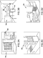
- FIGS. 3A-B show the automated system used in the below experiments of exemplary embodiments.
- the system includes a Biomek2000 System (pin tools, wash tools, stacker, single and 8-channel 20, 200 and 1000 microliter pipetting tools (P20, P200, P1000, Beckman), a Hudson Platecrane XT, and a Heraeus Cytomat 6000.
- a simple soda refrigerator was used for media storage from which the media was delivered directly to the culture wells. An in-line heating of the delivered media did not appear to be necessary based on experiments.
- the integration was done in collaboration with Biosero using Overlord software.
- the complete system was housed in a class 100 clean room (i.e.
- BSL2 biosaftety level 2
- Rectangular 4-well and 8-well Nunclon ⁇ plates purchased from Nunc were used instead of the round 6-well plates commonly used in manual procedures.
- the penalty of using regular round 6-well plates would have been a reduction of almost half of the capacity of the automated incubator to less than 100 plates.
- the rectangular geometry of the plates allowed the use of the 8-channel liquid handling tools since 6-well plates contain more inaccessible areas. The later fact also resulted in a 1.46-fold increase in usable culture surface area (84 cm2 for 4- and 8-well plates compared to 57.6 cm2 for a 6-well plate).
- the feeding and seeding system used 4-well and 8-well plates with the wash tool and the 8-channel tool (F IGS. 3 C-D ).
- robotic components were used for splitting and feeding HES cells.
- the single channel wash tool was used initially to aspirate spent media and feed 4-well or 8well plates.
- a 6-well plate is used to serve as a reservoir for trypsin.
- the 8-channel tool P200 was used to mix and dispense the trypsinized and individualized HES cells from a 4-well mother plate to an 8-well daughter plate in the final split.
- the surface was Nunclon ⁇ manufactured by Nunc and coated with Matrigel.
- the following robotic components can be used for splitting and feeding HES cells: the Platecrane connects the liquid handling robot Biomek2000 with the incubator Cytomat6000. The gripper of the Platecrane is positioned above the turntable of the Incubator.
- the refrigerator housing the media had tubing connecting the peristaltic pump of the wash tool (box with the tape) and the waste bottle for spent media coming from the vacuum was controlled by a valve in the wash tool which was provided by an in house vacuum system.
- the plate In the manual process the plate would be tipped at an angle to collect the spent media at the bottom for sufficient removal. Such an angle was not implemented with these robotics to maintain reliability.
- the new media (6 and 3 ml per well respectively) was dispensed immediately before moving on to the next well or plate.
- the delidding and relidding process as well as the height alignment of the wash tool required occasional intervention. These were the most prominent causes (about 1 in 100 movements) of errors that required operator presence and intervention.
- the flow rate of the wash tool did not seem to negatively affect the quality of the culture as the cells could not be washed off the surface by the pressure generated by the peristaltic pump of the wash tool.
- the quality of the culture was judged by visual assessment of the culture after each day of the culturing process ( FIGS. 4A-C ).
- Example 1 The inventors utilized the system described in Example 1 and developed procedures for passaging of HES cells that were based on simple liquid handling protocols. Recent innovations in splitting techniques allowed for efficient automation of this otherwise demanding manual procedure. Since HES cells require cell-cell contacts for survival in TeSR media, they needed to be seeded in clumps which required scraping of attached cells, a procedure which would be very hard to automate. However, the small molecule HA-100 and its 10-fold more specific derivative H-1152 was determined to allow the survival of HES cells after trypsin treatment.
- the wash tool removes the spent media as described in aim 1.
- the 8-channel 200 ⁇ l pipetting tool MP200 is used to add 3 ml of trypsin (0.1%).
- a mixture of 3 ml TeSR medium containing 2 ⁇ M H-1152 and 1mg/ml Invitrogen Soybean Trypsin Inhibitor is added to the well.
- the cells are washed off the plate surface and mixed using the 8-channel MP200 tool by repetitive dispensing and aspiration. Cells are then dispensed to the daughter plates using the MP200 tool.
- the cells are then seeded 1 to 32 onto precoated Matrigel plates loaded from the stacker to the Biomek2000. Seeding is done after aspirating the Matrigel coating media and replacing it with the modified TeSR media containing H-1152 and soybean inhibitor as described in the feeding protocol above.
- the total surface area of the initially used Omnitray turned out to be too large for the throughput of the system at the stage of the next split once the inventors tested methods for passaging HES cells in aim 2.
- the time it took to distribute the cells from the mother plates to an average of 25 daughter plates exceeded the time the individualized cells could survive in suspension before they would be seeded.
- the inventors used karyotypically normal very high passage (>p200) H1 HES cell cultures in the beginning to establish the robotic protocols with very robust growing cells to establish feasibility of these new protocols. These cells still produced hematopoietic precursors and cardiomyocytes to demonstrate their differentiation potential. However the demands for a robust protocol are certainly greater for lower passage cell cultures, since they react more sensitively to sub-optimal conditions, which leads to greater variability in cell culture. In later experiments, the inventors were able to confirm the validity of the derived procedures with lower passage H1 cells (>p60) in smaller scale experiments with 5 plates over 3 to 5 passages. This evidence supports the ability to culture these lower passage cells using the automated system.
- the inventors used the single channel tool for seeding by adding cells at multiple positions of the well to achieve homogeneous distribution of cells. Although the inventors could accomplish that successfully with high and low passage HES cultures, the throughput of the procedure required the use of the 8-channel P200 tool when the inventors expanded the cells in the final step of the scale-up experiment to 160 8-well plates. Although a reduced homogeneous distribution and decrease in cell densities of the cells was observed in the daughter plates, the inventors anticipate that this method may be optimized to improve these characteristics. The expansion in this experiment was done from a single well of a 6-well plate to 5 needed 4-well plates in the first passage and then into 160 final 8-well plates in the second passage.
- the inventors maintained all 160 plates by feeding 2 days after passage with the protocol established above and then every day.
- the inventors inspected all plates visually for differentiation and density.
- the inventors randomly picked 30 plates and stained them with trypan blue staining for scanning and evaluation of cell distribution. From the visual inspection of the plates it was apparent that further improvements can and should be made to homogeneity in the interest of high reproducibility.
- the inventors picked plate 2 and 10 randomly to test for Oct4 content of the cells after 2 passages and 1000-fold expansion over 20 days. Oct4 FACS analysis revealed a high quality of undifferentiated cells in this first scale-up experiment.
- H1152 which actively increased colony formation of HES cells in TeSR1 media from individualized cells and represents a 2nd generation small molecule derived from HA-100.
- HA-100 was discovered in one of the first HES cell-based small molecule screens which led to the discovery of related compounds like H1152 in follow-up studies.
- H-1152 allows for very efficient seeding of individualized HES cells in 96-well plates (similar to HA-100 but at 10-fold lower concentration), enabling HES cell-based small molecule screening.
- Individualized HES cells that are otherwise passaged in cell clumps allow more uniform cell densities per well, which is a stringent prerequisite for cell-based small molecule screening.
- the inventors also reasoned that the current small molecule (HA-100) and a related compound H1152 were sufficient for passaging human ES cells.
- the inventors used a small molecule library (2000 compounds with known bioactivity) to screen for compounds that may have the ability to increase hematopoiesis.
- the inventors have demonstrated that the system described above is capable of plating HES cells in 96-well format to provide a platform for small molecule screening as well as screening of other agents and conditions.
- the inventors have derived a screening assay and protocol suitable for automation that uses directed differentiation methods leading to hematopoietic precursers.
- the inventors have successfully performed a screen under these conditions and identified 28 initial candidate compounds which are now being validated. Although the performance of the screen can be further improved through a more thorough assay development, the key goal of plating HES cells for automated screening has been accomplished.
- the assay used in this protocol was developed based on an ELISA protocol which can detect the presence of two characteristic cell surface markers (CD34 and CD43). These markers identify potent hematopoietic precursors that the inventors can subsequently isolate to differentiate the cells into the desired blood lineages or provide as such as a starting material.
- the inventors used the automated platform to seed individualized human ES cells in 96-well plates and grow them for four days. Then the media was switched to defined differentiation media and compound to be screened was added by the robot to a final concentration of 20 micromolar using a 96-channel pin tool.
- the inventors were able to use the AmplexUltraRed and the SensiFlex assays (Invitrogen) in a multiplex ELISA and did show with controls, that these assays were compatible and offer the possibility for SCP to simultaneously screen for two cell markers (on the surface or inside the cells, after addition of a separate wash step in the protocol). This is particularly important when screening for cell lineages that require more than one marker for sufficient recognition.
- the inventors were able to obtain very good homogeneity in plating into 96-well plates using a modified procedure established above. For this purpose however the inventors plated 16,000 cells per well, which is a 5-fold higher seeding density, than in the regular propagation and maintenance of undifferentiated HES cells with the automated procedure. This particular change was used in order to accommodate attachment to an alternate proprietary matrix other than Matrigel, which turned out to be a crucial step in the differentiation protocol. Successful seeding in 96-well format to facilitate robust screening may be used.
- the inventors had the capacity to perform the media changes for this screen with the automated liquid handling system at hand, the inventors decided in the interest of time and money, to change media and perform the ELISA assay, by aspirating manually using a vacuum 12-channel wand and dispensing using an automated dispenser without stacker. While the inventors did not take advantage of the reproducibility of a fully automated system, the inventors saved significant time and reagents. A Matrix Wellmate automated dispenser was used.
- the above examples demonstrate a successfully automated HES cell culture and maintenance.
- the above data demonstrates that the inventors were able to solve the major challenges towards providing an automatable procedure for HES cell culture.
- the inventors further anticipate that additional throughtput can be achieved via optimization of the system.
- the inventors anticipate that improved quality control, monitoring of cell growth, and improved passaging can be achieved using the above system. It is anticipated that with an optimized procedure, one may not need to identify and isolate differentiated impurities as is necessary in manual protocols.
- the above system may be easily modified to include a liquid handling system like the Tecan Cellerity system, which has been successfully established for maintenance of other attached cell lines.
- a liquid handling system like the Tecan Cellerity system
- the major step towards the application of such a system was the use of trypsin to passage cells.
- compositions and methods disclosed and claimed herein can be made and executed without undue experimentation in light of the present disclosure. While the compositions and methods of this invention have been described in terms of preferred embodiments, it will be apparent to those of skill in the art that variations may be applied to the compositions and methods and in the steps or in the sequence of steps of the method described herein without departing from the concept, spirit and scope of the invention. More specifically, it will be apparent that certain agents which are both chemically and physiologically related may be substituted for the agents described herein while the same or similar results would be achieved.
Priority Applications (2)
| Application Number | Priority Date | Filing Date | Title |
|---|---|---|---|
| DK17192886.4T DK3293256T3 (da) | 2007-06-29 | 2008-06-30 | Automatiseret fremgangsmåde og apparatur til dyrkning af embryonale stamceller |
| EP17192886.4A EP3293256B1 (en) | 2007-06-29 | 2008-06-30 | Automated method and apparatus for embryonic stem cell culture |
Applications Claiming Priority (2)
| Application Number | Priority Date | Filing Date | Title |
|---|---|---|---|
| US94701307P | 2007-06-29 | 2007-06-29 | |
| PCT/US2008/068814 WO2009006422A1 (en) | 2007-06-29 | 2008-06-30 | Automated method and apparatus for embryonic stem cell culture |
Related Child Applications (2)
| Application Number | Title | Priority Date | Filing Date |
|---|---|---|---|
| EP17192886.4A Division EP3293256B1 (en) | 2007-06-29 | 2008-06-30 | Automated method and apparatus for embryonic stem cell culture |
| EP17192886.4A Division-Into EP3293256B1 (en) | 2007-06-29 | 2008-06-30 | Automated method and apparatus for embryonic stem cell culture |
Publications (2)
| Publication Number | Publication Date |
|---|---|
| EP2173863A1 EP2173863A1 (en) | 2010-04-14 |
| EP2173863B1 true EP2173863B1 (en) | 2018-10-10 |
Family
ID=39672032
Family Applications (2)
| Application Number | Title | Priority Date | Filing Date |
|---|---|---|---|
| EP17192886.4A Active EP3293256B1 (en) | 2007-06-29 | 2008-06-30 | Automated method and apparatus for embryonic stem cell culture |
| EP08781187.3A Active EP2173863B1 (en) | 2007-06-29 | 2008-06-30 | Automated method and apparatus for embryonic stem cell culture |
Family Applications Before (1)
| Application Number | Title | Priority Date | Filing Date |
|---|---|---|---|
| EP17192886.4A Active EP3293256B1 (en) | 2007-06-29 | 2008-06-30 | Automated method and apparatus for embryonic stem cell culture |
Country Status (8)
| Country | Link |
|---|---|
| US (2) | US8815585B2 (da) |
| EP (2) | EP3293256B1 (da) |
| JP (4) | JP5991796B2 (da) |
| KR (2) | KR20160005142A (da) |
| AU (1) | AU2008272949B2 (da) |
| CA (1) | CA2691793A1 (da) |
| DK (2) | DK3293256T3 (da) |
| WO (1) | WO2009006422A1 (da) |
Families Citing this family (77)
| Publication number | Priority date | Publication date | Assignee | Title |
|---|---|---|---|---|
| EP3354723B1 (en) | 2005-08-29 | 2023-12-13 | Technion Research & Development Foundation Ltd. | Media for culturing stem cells |
| DK3441459T3 (da) | 2006-08-02 | 2021-06-07 | Technion Res & Dev Foundation | Fremgangsmåder til ekspansion af embryonale stamceller i en suspensionskultur |
| US9080145B2 (en) | 2007-07-01 | 2015-07-14 | Lifescan Corporation | Single pluripotent stem cell culture |
| CN101952415B (zh) | 2007-07-31 | 2017-06-27 | 生命扫描有限公司 | 人胚胎干细胞的分化 |
| WO2009105570A2 (en) | 2008-02-21 | 2009-08-27 | Centocor Ortho Biotech Inc. | Methods, surface modified plates and compositions for cell attachment, cultivation and detachment |
| SG188918A1 (da) | 2008-03-17 | 2013-04-30 | Agency Science Tech & Res | |
| US8828720B2 (en) | 2008-03-17 | 2014-09-09 | Agency For Science, Technology And Research | Microcarriers for stem cell culture |
| US8691569B2 (en) | 2008-03-17 | 2014-04-08 | Agency For Science, Technology And Research | Microcarriers for stem cell culture |
| US9458431B2 (en) | 2008-03-17 | 2016-10-04 | Agency For Science, Technology And Research | Microcarriers for stem cell culture |
| SG10201607710UA (en) | 2008-03-17 | 2016-11-29 | Scripps Research Inst | Combined chemical and genetic approaches for generation of induced pluripotent stem cells |
| JP5734183B2 (ja) | 2008-06-30 | 2015-06-17 | ヤンセン バイオテツク,インコーポレーテツド | 多能性幹細胞の分化 |
| DK2295538T3 (da) * | 2008-07-07 | 2015-12-21 | Takara Bio Inc | Fremgangsmåde til fremstilling af en pluripotent stamcelle |
| RU2555538C2 (ru) | 2008-11-20 | 2015-07-10 | Сентокор Орто Байотек Инк. | Культура плюрипотентных стволовых клеток на микроносителях |
| AU2009316583B2 (en) | 2008-11-20 | 2016-04-21 | Janssen Biotech, Inc. | Methods and compositions for cell attachment and cultivation on planar substrates |
| CA2747398C (en) | 2008-12-17 | 2023-06-20 | The Scripps Research Institute | Generation and maintenance of stem cells |
| EP2398897B1 (en) | 2009-02-20 | 2017-06-28 | Cellular Dynamics International, Inc. | Methods and compositions for the differentiation of stem cells |
| EP2408903B1 (en) * | 2009-03-20 | 2014-08-06 | Agency For Science, Technology And Research | Culture of pluripotent and multipotent cells on microcarriers |
| EP2456862A4 (en) | 2009-07-20 | 2013-02-27 | Janssen Biotech Inc | DIFFERENTIATION OF HUMAN EMBRYONIC STEM CELLS |
| US9008406B2 (en) | 2009-10-09 | 2015-04-14 | Kawasaki Jukogyo Kabushiki Kaisha | Method and apparatus for discriminating undifferentiated pluripotent stem cells, and automated culture method and system |
| WO2011047300A1 (en) * | 2009-10-16 | 2011-04-21 | The Scripps Research Institute | Induction of pluripotent cells |
| ES2539487T3 (es) * | 2009-11-04 | 2015-07-01 | Cellular Dynamics International, Inc. | Reprogramación episómica con compuestos químicos |
| EP4166652A1 (en) | 2009-11-12 | 2023-04-19 | Technion Research & Development Foundation Ltd. | Culture media, cell cultures and methods of culturing pluripotent stem cells in an undifferentiated state |
| US8349609B2 (en) * | 2009-11-24 | 2013-01-08 | University Of Connecticut | Differentiation of human embryonic and induced pluripotent stem cells |
| CN102741395B (zh) | 2009-12-23 | 2016-03-16 | 詹森生物科技公司 | 人胚胎干细胞的分化 |
| AU2011223900A1 (en) | 2010-03-01 | 2012-09-13 | Janssen Biotech, Inc. | Methods for purifying cells derived from pluripotent stem cells |
| EP3936608A1 (en) | 2010-03-31 | 2022-01-12 | The Scripps Research Institute | Reprogramming cells |
| WO2011140231A1 (en) * | 2010-05-05 | 2011-11-10 | Caridianbct, Inc. | Method of reseeding adherent cells grown in a hollow fiber bioreactor system |
| US20130071927A1 (en) * | 2010-05-05 | 2013-03-21 | Sydney Ivf Limited | Media and methods for cell culture |
| WO2011159726A2 (en) | 2010-06-14 | 2011-12-22 | The Scripps Research Institute | Reprogramming of cells to a new fate |
| JP5696144B2 (ja) | 2010-06-25 | 2015-04-08 | 川崎重工業株式会社 | 多能性幹細胞コロニーの識別方法及び装置並びに多能性幹細胞の自動培養方法及び装置 |
| AU2011296381B2 (en) | 2010-08-31 | 2016-03-31 | Janssen Biotech, Inc. | Differentiation of human embryonic stem cells |
| CA2810488A1 (en) | 2010-09-07 | 2012-03-15 | Technion Research & Development Foundation Limited | Novel methods and culture media for culturing pluripotent stem cells |
| KR20140063501A (ko) | 2010-12-22 | 2014-05-27 | 페이트 세러퓨틱스, 인코포레이티드 | 단세포 분류 및 iPSC의 증강된 재프로그래밍을 위한 세포 배양 플랫폼 |
| KR101293300B1 (ko) * | 2011-04-05 | 2013-08-09 | (주)로고스바이오시스템스 | 세포의 분리시기를 판정하는 방법과, 이를 이용한 세포의 계대 배양 방법 및 계대 배양 장치 |
| US20140248698A1 (en) * | 2011-10-21 | 2014-09-04 | Arkray, Inc. | Method for culturing pluripotency-maintained singly dispersed cells by means of laminar flow |
| US10428309B2 (en) | 2011-12-01 | 2019-10-01 | New York Stem Cell Foundation, Inc. | Systems and methods for producing stem cells and differentiated cells |
| KR102039202B1 (ko) * | 2011-12-01 | 2019-10-31 | 뉴욕 스템 셀 파운데이션, 인코포레이티드 | 유도 다능성 줄기 세포 또는 분화된 세포를 제조하기 위한 자동화 시스템 |
| AU2012355698B2 (en) | 2011-12-22 | 2018-11-29 | Janssen Biotech, Inc. | Differentiation of human embryonic stem cells into single hormonal insulin positive cells |
| CN102604894B (zh) * | 2012-02-29 | 2014-07-30 | 中国科学院广州生物医药与健康研究院 | 用于制备神经干细胞的培养基及其用途 |
| CN108103006A (zh) | 2012-06-08 | 2018-06-01 | 詹森生物科技公司 | 人胚胎干细胞向胰腺内分泌细胞的分化 |
| KR102036780B1 (ko) | 2012-12-31 | 2019-10-25 | 얀센 바이오테크 인코포레이티드 | Hb9 조절제를 사용하는 인간 배아 줄기세포의 췌장 내분비 세포로의 분화 |
| JP6529440B2 (ja) | 2012-12-31 | 2019-06-12 | ヤンセン バイオテツク,インコーポレーテツド | 膵内分泌細胞への分化のためのヒト多能性細胞の懸濁及びクラスタリング |
| US10370644B2 (en) | 2012-12-31 | 2019-08-06 | Janssen Biotech, Inc. | Method for making human pluripotent suspension cultures and cells derived therefrom |
| KR102084561B1 (ko) | 2012-12-31 | 2020-03-04 | 얀센 바이오테크 인코포레이티드 | 췌장 내분비 세포로의 분화를 위한 공기-액체 계면에서의 인간 배아 줄기세포의 배양 |
| US9790465B2 (en) | 2013-04-30 | 2017-10-17 | Corning Incorporated | Spheroid cell culture well article and methods thereof |
| AU2014287013B2 (en) * | 2013-07-12 | 2020-01-23 | President And Fellows Of Harvard College | Systems and methods for cell culture device interconnection and fluidic device interconnection |
| WO2015023658A2 (en) * | 2013-08-12 | 2015-02-19 | Invivosciences Inc. | Automated cell culture system and method |
| EP3604499A1 (en) | 2014-03-04 | 2020-02-05 | Fate Therapeutics, Inc. | Improved reprogramming methods and cell culture platforms |
| CA2949056A1 (en) | 2014-05-16 | 2015-11-19 | Janssen Biotech, Inc. | Use of small molecules to enhance mafa expression in pancreatic endocrine cells |
| WO2016028880A1 (en) | 2014-08-19 | 2016-02-25 | Cellular Dynamics International, Inc. | Neural networks formed from cells derived from pluripotent stem cells |
| JP6930914B2 (ja) | 2014-10-29 | 2021-09-01 | コーニング インコーポレイテッド | 灌流バイオリアクタ・プラットフォーム |
| SG11201703493SA (en) | 2014-10-29 | 2017-05-30 | Corning Inc | Cell culture insert |
| EP3234109A4 (en) | 2014-12-19 | 2018-06-27 | Janssen Biotech, Inc. | Suspension culturing of pluripotent stem cells |
| US9944894B2 (en) | 2015-01-16 | 2018-04-17 | General Electric Company | Pluripotent stem cell expansion and passage using a rocking platform bioreactor |
| WO2016208755A1 (ja) * | 2015-06-25 | 2016-12-29 | 株式会社カネカ | 液体注入方法 |
| WO2017007278A1 (ko) * | 2015-07-09 | 2017-01-12 | 사회복지법인 삼성생명공익재단 | 자동 세포 배양기 및 그 배양기의 동작 방법 |
| CN114717183A (zh) | 2015-08-31 | 2022-07-08 | 爱平世股份有限公司 | 多能干细胞制造系统和生产诱导多能干细胞的方法 |
| CN108367290B (zh) | 2015-10-01 | 2021-06-04 | 伯克利之光生命科技公司 | 井孔板培养器 |
| JP7263005B2 (ja) | 2015-10-16 | 2023-04-24 | フェイト セラピューティクス,インコーポレイテッド | 基底状態の多能性の誘導及び維持に関するプラットフォーム |
| CN109069870B (zh) | 2016-02-24 | 2022-04-29 | 洛克菲勒大学 | 基于胚胎细胞的用于亨廷顿氏病的治疗候选物筛选系统、模型及它们的应用 |
| WO2017162467A1 (en) * | 2016-03-21 | 2017-09-28 | General Electric Company | Pluripotent stem cell expansion and passage using a stirred tank bioreactor |
| MA45479A (fr) | 2016-04-14 | 2019-02-20 | Janssen Biotech Inc | Différenciation de cellules souches pluripotentes en cellules de l'endoderme de l'intestin moyen |
| JP2018000130A (ja) * | 2016-07-05 | 2018-01-11 | 株式会社Ihi | 細胞培養装置 |
| IL303869A (en) * | 2016-07-19 | 2023-08-01 | Accellta Ltd | A medium for growing pluripotent stem cells in suspension |
| DE102016114043B3 (de) | 2016-07-29 | 2017-08-10 | Technische Universität Dresden | Vorrichtung zur Isolierung von Stammzellen aus fötalen Geweben |
| CN109689853B (zh) | 2016-08-27 | 2022-08-23 | 三维生物科技有限公司 | 生物反应器 |
| WO2018089681A1 (en) | 2016-11-10 | 2018-05-17 | Becton, Dickinson And Company | Timeline system for monitoring a culture media protocol |
| JP6968169B2 (ja) * | 2016-12-01 | 2021-11-17 | バークレー ライツ,インコーポレイテッド | ウェルプレートインキュベーター |
| US20190382706A1 (en) * | 2017-02-27 | 2019-12-19 | Koji Tanabe | Somatic cell production system |
| WO2019014610A1 (en) | 2017-07-14 | 2019-01-17 | Corning Incorporated | CELL CULTURE CONTAINER FOR 3D CULTURE AND METHODS OF CULTURING 3D CELLS |
| CN111065729B (zh) | 2017-07-14 | 2024-04-26 | 康宁股份有限公司 | 细胞培养容器 |
| JP7245222B2 (ja) | 2017-07-14 | 2023-03-23 | コーニング インコーポレイテッド | 手動又は自動で培地を交換するための3d細胞培養容器 |
| US11857970B2 (en) | 2017-07-14 | 2024-01-02 | Corning Incorporated | Cell culture vessel |
| KR102220125B1 (ko) * | 2018-04-26 | 2021-02-25 | (주)세포바이오 | 세포배양 자동화 장치 및 방법 |
| JP7171695B2 (ja) | 2018-07-13 | 2022-11-15 | コーニング インコーポレイテッド | 液体培地送達面を含む側壁を有するマイクロキャビティ皿 |
| CN111065725B (zh) | 2018-07-13 | 2024-03-29 | 康宁股份有限公司 | 包括具有互联的壁的微板的流体装置 |
| WO2020013845A1 (en) | 2018-07-13 | 2020-01-16 | Corning Incorporated | Cell culture vessels with stabilizer devices |
Family Cites Families (20)
| Publication number | Priority date | Publication date | Assignee | Title |
|---|---|---|---|---|
| US4352883A (en) | 1979-03-28 | 1982-10-05 | Damon Corporation | Encapsulation of biological material |
| US4284412A (en) | 1979-07-13 | 1981-08-18 | Ortho Diagnostics, Inc. | Method and apparatus for automated identification and enumeration of specified blood cell subclasses |
| JPS58155087A (ja) * | 1982-03-12 | 1983-09-14 | Olympus Optical Co Ltd | 細胞の自動培養装置 |
| US4498766A (en) | 1982-03-25 | 1985-02-12 | Becton, Dickinson And Company | Light beam focal spot elongation in flow cytometry devices |
| US4661913A (en) | 1984-09-11 | 1987-04-28 | Becton, Dickinson And Company | Apparatus and method for the detection and classification of articles using flow cytometry techniques |
| US4767206A (en) | 1984-12-24 | 1988-08-30 | Flow Cytometry Standards Corporation | Calibration method for flow cytometry using fluorescent microbeads and synthesis thereof |
| US4774189A (en) | 1984-12-24 | 1988-09-27 | Flow Cytometry Standards Corp. | Fluorescent calibration microbeads simulating stained cells |
| US4857451A (en) | 1984-12-24 | 1989-08-15 | Flow Cytometry Standards Corporation | Method of compensating and calibrating a flow cytometer, and microbead standards kit therefor |
| US4989977A (en) | 1985-07-29 | 1991-02-05 | Becton, Dickinson And Company | Flow cytometry apparatus with improved light beam adjustment |
| US4714682A (en) | 1985-12-11 | 1987-12-22 | Flow Cytometry Standards Corporation | Fluorescent calibration microbeads simulating stained cells |
| US5160974A (en) | 1990-06-25 | 1992-11-03 | Flow Science, Inc. | Closed sample cell for use in flow cytometry |
| US5478722A (en) | 1991-02-17 | 1995-12-26 | The Curators Of The University Of Missouri | Preserved cell preparations for flow cytometry and immunology |
| EP0682697A4 (en) | 1993-01-29 | 1997-07-30 | New Brunswick Scientific Co | METHOD AND APPARATUS FOR CULTURING ANCHOR AND SUSPENSION CELLS. |
| US5843780A (en) | 1995-01-20 | 1998-12-01 | Wisconsin Alumni Research Foundation | Primate embryonic stem cells |
| US6325114B1 (en) | 2000-02-01 | 2001-12-04 | Incyte Genomics, Inc. | Pipetting station apparatus |
| AU2003218572B2 (en) * | 2002-04-08 | 2009-08-20 | Octane Biotech, Inc. | Automated tissue engineering system |
| DE102004043256B4 (de) * | 2004-09-07 | 2013-09-19 | Rheinische Friedrich-Wilhelms-Universität Bonn | Skalierbarer Prozess zur Kultivierung undifferenzierter Stammzellen in Suspension |
| MX2007002390A (es) | 2004-09-08 | 2007-04-23 | Wisconsin Alumni Res Found | Medio y cultivo de celulas progenitoras embrionarias. |
| WO2006084040A2 (en) | 2005-02-04 | 2006-08-10 | Massachusetts Institute Of Technology | Engineering vascularized muscle tissue |
| US20060199265A1 (en) | 2005-03-02 | 2006-09-07 | Wolf Michael F | Seeding implantable medical devices with cells |
-
2008
- 2008-06-30 DK DK17192886.4T patent/DK3293256T3/da active
- 2008-06-30 JP JP2010515226A patent/JP5991796B2/ja active Active
- 2008-06-30 US US12/164,969 patent/US8815585B2/en active Active
- 2008-06-30 KR KR1020157036767A patent/KR20160005142A/ko not_active Application Discontinuation
- 2008-06-30 AU AU2008272949A patent/AU2008272949B2/en active Active
- 2008-06-30 WO PCT/US2008/068814 patent/WO2009006422A1/en active Application Filing
- 2008-06-30 EP EP17192886.4A patent/EP3293256B1/en active Active
- 2008-06-30 KR KR1020107002003A patent/KR20100059789A/ko active Application Filing
- 2008-06-30 CA CA2691793A patent/CA2691793A1/en not_active Abandoned
- 2008-06-30 DK DK08781187.3T patent/DK2173863T3/da active
- 2008-06-30 EP EP08781187.3A patent/EP2173863B1/en active Active
-
2013
- 2013-03-19 US US13/847,349 patent/US20130316445A1/en not_active Abandoned
-
2014
- 2014-03-26 JP JP2014063625A patent/JP2014110817A/ja active Pending
-
2016
- 2016-06-24 JP JP2016125186A patent/JP2016168056A/ja active Pending
- 2016-11-08 JP JP2016217785A patent/JP2017023158A/ja active Pending
Non-Patent Citations (1)
| Title |
|---|
| None * |
Also Published As
| Publication number | Publication date |
|---|---|
| EP3293256A1 (en) | 2018-03-14 |
| AU2008272949A1 (en) | 2009-01-08 |
| US20090029462A1 (en) | 2009-01-29 |
| CA2691793A1 (en) | 2009-01-08 |
| JP2016168056A (ja) | 2016-09-23 |
| WO2009006422A1 (en) | 2009-01-08 |
| KR20160005142A (ko) | 2016-01-13 |
| DK3293256T3 (da) | 2019-08-12 |
| JP2014110817A (ja) | 2014-06-19 |
| AU2008272949B2 (en) | 2014-05-15 |
| JP2010532173A (ja) | 2010-10-07 |
| JP5991796B2 (ja) | 2016-09-14 |
| JP2017023158A (ja) | 2017-02-02 |
| EP3293256B1 (en) | 2019-05-22 |
| KR20100059789A (ko) | 2010-06-04 |
| DK2173863T3 (da) | 2019-01-21 |
| EP2173863A1 (en) | 2010-04-14 |
| US8815585B2 (en) | 2014-08-26 |
| US20130316445A1 (en) | 2013-11-28 |
Similar Documents
| Publication | Publication Date | Title |
|---|---|---|
| EP2173863B1 (en) | Automated method and apparatus for embryonic stem cell culture | |
| Ludwig et al. | Defined, feeder‐independent medium for human embryonic stem cell culture | |
| Kehoe et al. | Scalable stirred-suspension bioreactor culture of human pluripotent stem cells | |
| CN102307991B (zh) | 微载体上的多能干细胞培养 | |
| EP1797172B1 (en) | Culturing human embryonic stem cells | |
| IL224431A (en) | Basic media is simplified into multi-potential human cell culture | |
| Chen et al. | Expansion of human embryonic stem cells on cellulose microcarriers | |
| Hartung et al. | Clump passaging and expansion of human embryonic and induced pluripotent stem cells on mouse embryonic fibroblast feeder cells | |
| AU2014201955B2 (en) | Automated method and apparatus for embryonic stem cell culture | |
| Ting et al. | Differentiation of human embryonic stem cells to cardiomyocytes on microcarrier cultures | |
| JP7092309B2 (ja) | 単一細胞ヒト多能性幹細胞の継代及び採取用配合物 | |
| WO2015008275A1 (en) | Methods for large scale generation of stem cells | |
| KR20220113349A (ko) | 생물반응기의 현탁액에서 줄기세포의 확장 | |
| Yamada et al. | Using piggyBac transposon gene expression vectors to transfect Zscan5b gene into mouse pluripotent stem cells | |
| Lu et al. | Protocol for germ lineage differentiation of primed human pluripotent stem cells using chemically defined, nutrient-balanced media | |
| AU2008204566A1 (en) | Novel mesenchymal progenitor cells derived from human blastocyst-derived stem cells | |
| Borys et al. | Large‐scale expansion of feeder‐free mouse embryonic stem cells serially passaged in stirred suspension bioreactors at low inoculation densities directly from cryopreservation | |
| Jacobson et al. | Large-Scale Culture of 3D Aggregates of Human Pluripotent Stem Cells |
Legal Events
| Date | Code | Title | Description |
|---|---|---|---|
| PUAI | Public reference made under article 153(3) epc to a published international application that has entered the european phase |
Free format text: ORIGINAL CODE: 0009012 |
|
| 17P | Request for examination filed |
Effective date: 20100122 |
|
| AK | Designated contracting states |
Kind code of ref document: A1 Designated state(s): AT BE BG CH CY CZ DE DK EE ES FI FR GB GR HR HU IE IS IT LI LT LU LV MC MT NL NO PL PT RO SE SI SK TR |
|
| AX | Request for extension of the european patent |
Extension state: AL BA MK RS |
|
| DAX | Request for extension of the european patent (deleted) | ||
| 17Q | First examination report despatched |
Effective date: 20120507 |
|
| STAA | Information on the status of an ep patent application or granted ep patent |
Free format text: STATUS: EXAMINATION IS IN PROGRESS |
|
| REG | Reference to a national code |
Ref country code: DE Ref legal event code: R079 Ref document number: 602008057356 Country of ref document: DE Free format text: PREVIOUS MAIN CLASS: C12N0005060000 Ipc: C12N0005020000 |
|
| GRAP | Despatch of communication of intention to grant a patent |
Free format text: ORIGINAL CODE: EPIDOSNIGR1 |
|
| STAA | Information on the status of an ep patent application or granted ep patent |
Free format text: STATUS: GRANT OF PATENT IS INTENDED |
|
| RIC1 | Information provided on ipc code assigned before grant |
Ipc: C12N 5/02 20060101AFI20171106BHEP |
|
| INTG | Intention to grant announced |
Effective date: 20171205 |
|
| GRAJ | Information related to disapproval of communication of intention to grant by the applicant or resumption of examination proceedings by the epo deleted |
Free format text: ORIGINAL CODE: EPIDOSDIGR1 |
|
| STAA | Information on the status of an ep patent application or granted ep patent |
Free format text: STATUS: EXAMINATION IS IN PROGRESS |
|
| GRAS | Grant fee paid |
Free format text: ORIGINAL CODE: EPIDOSNIGR3 |
|
| STAA | Information on the status of an ep patent application or granted ep patent |
Free format text: STATUS: GRANT OF PATENT IS INTENDED |
|
| GRAP | Despatch of communication of intention to grant a patent |
Free format text: ORIGINAL CODE: EPIDOSNIGR1 |
|
| INTC | Intention to grant announced (deleted) | ||
| INTG | Intention to grant announced |
Effective date: 20180508 |
|
| RAP1 | Party data changed (applicant data changed or rights of an application transferred) |
Owner name: FUJIFILM CELLULAR DYNAMICS, INC. |
|
| GRAA | (expected) grant |
Free format text: ORIGINAL CODE: 0009210 |
|
| STAA | Information on the status of an ep patent application or granted ep patent |
Free format text: STATUS: THE PATENT HAS BEEN GRANTED |
|
| AK | Designated contracting states |
Kind code of ref document: B1 Designated state(s): AT BE BG CH CY CZ DE DK EE ES FI FR GB GR HR HU IE IS IT LI LT LU LV MC MT NL NO PL PT RO SE SI SK TR |
|
| REG | Reference to a national code |
Ref country code: GB Ref legal event code: FG4D |
|
| REG | Reference to a national code |
Ref country code: CH Ref legal event code: EP Ref country code: AT Ref legal event code: REF Ref document number: 1051254 Country of ref document: AT Kind code of ref document: T Effective date: 20181015 |
|
| REG | Reference to a national code |
Ref country code: IE Ref legal event code: FG4D |
|
| REG | Reference to a national code |
Ref country code: DE Ref legal event code: R096 Ref document number: 602008057356 Country of ref document: DE |
|
| REG | Reference to a national code |
Ref country code: NL Ref legal event code: FP |
|
| REG | Reference to a national code |
Ref country code: SE Ref legal event code: TRGR |
|
| REG | Reference to a national code |
Ref country code: CH Ref legal event code: NV Representative=s name: VOSSIUS AND PARTNER PATENTANWAELTE RECHTSANWAE, CH |
|
| REG | Reference to a national code |
Ref country code: DK Ref legal event code: T3 Effective date: 20190109 |
|
| REG | Reference to a national code |
Ref country code: LT Ref legal event code: MG4D |
|
| REG | Reference to a national code |
Ref country code: AT Ref legal event code: MK05 Ref document number: 1051254 Country of ref document: AT Kind code of ref document: T Effective date: 20181010 |
|
| PG25 | Lapsed in a contracting state [announced via postgrant information from national office to epo] |
Ref country code: LV Free format text: LAPSE BECAUSE OF FAILURE TO SUBMIT A TRANSLATION OF THE DESCRIPTION OR TO PAY THE FEE WITHIN THE PRESCRIBED TIME-LIMIT Effective date: 20181010 Ref country code: AT Free format text: LAPSE BECAUSE OF FAILURE TO SUBMIT A TRANSLATION OF THE DESCRIPTION OR TO PAY THE FEE WITHIN THE PRESCRIBED TIME-LIMIT Effective date: 20181010 Ref country code: HR Free format text: LAPSE BECAUSE OF FAILURE TO SUBMIT A TRANSLATION OF THE DESCRIPTION OR TO PAY THE FEE WITHIN THE PRESCRIBED TIME-LIMIT Effective date: 20181010 Ref country code: NO Free format text: LAPSE BECAUSE OF FAILURE TO SUBMIT A TRANSLATION OF THE DESCRIPTION OR TO PAY THE FEE WITHIN THE PRESCRIBED TIME-LIMIT Effective date: 20190110 Ref country code: BG Free format text: LAPSE BECAUSE OF FAILURE TO SUBMIT A TRANSLATION OF THE DESCRIPTION OR TO PAY THE FEE WITHIN THE PRESCRIBED TIME-LIMIT Effective date: 20190110 Ref country code: PL Free format text: LAPSE BECAUSE OF FAILURE TO SUBMIT A TRANSLATION OF THE DESCRIPTION OR TO PAY THE FEE WITHIN THE PRESCRIBED TIME-LIMIT Effective date: 20181010 Ref country code: ES Free format text: LAPSE BECAUSE OF FAILURE TO SUBMIT A TRANSLATION OF THE DESCRIPTION OR TO PAY THE FEE WITHIN THE PRESCRIBED TIME-LIMIT Effective date: 20181010 Ref country code: LT Free format text: LAPSE BECAUSE OF FAILURE TO SUBMIT A TRANSLATION OF THE DESCRIPTION OR TO PAY THE FEE WITHIN THE PRESCRIBED TIME-LIMIT Effective date: 20181010 Ref country code: IS Free format text: LAPSE BECAUSE OF FAILURE TO SUBMIT A TRANSLATION OF THE DESCRIPTION OR TO PAY THE FEE WITHIN THE PRESCRIBED TIME-LIMIT Effective date: 20190210 Ref country code: FI Free format text: LAPSE BECAUSE OF FAILURE TO SUBMIT A TRANSLATION OF THE DESCRIPTION OR TO PAY THE FEE WITHIN THE PRESCRIBED TIME-LIMIT Effective date: 20181010 |
|
| PG25 | Lapsed in a contracting state [announced via postgrant information from national office to epo] |
Ref country code: GR Free format text: LAPSE BECAUSE OF FAILURE TO SUBMIT A TRANSLATION OF THE DESCRIPTION OR TO PAY THE FEE WITHIN THE PRESCRIBED TIME-LIMIT Effective date: 20190111 Ref country code: PT Free format text: LAPSE BECAUSE OF FAILURE TO SUBMIT A TRANSLATION OF THE DESCRIPTION OR TO PAY THE FEE WITHIN THE PRESCRIBED TIME-LIMIT Effective date: 20190210 |
|
| REG | Reference to a national code |
Ref country code: DE Ref legal event code: R097 Ref document number: 602008057356 Country of ref document: DE |
|
| PG25 | Lapsed in a contracting state [announced via postgrant information from national office to epo] |
Ref country code: CZ Free format text: LAPSE BECAUSE OF FAILURE TO SUBMIT A TRANSLATION OF THE DESCRIPTION OR TO PAY THE FEE WITHIN THE PRESCRIBED TIME-LIMIT Effective date: 20181010 Ref country code: IT Free format text: LAPSE BECAUSE OF FAILURE TO SUBMIT A TRANSLATION OF THE DESCRIPTION OR TO PAY THE FEE WITHIN THE PRESCRIBED TIME-LIMIT Effective date: 20181010 |
|
| PLBE | No opposition filed within time limit |
Free format text: ORIGINAL CODE: 0009261 |
|
| STAA | Information on the status of an ep patent application or granted ep patent |
Free format text: STATUS: NO OPPOSITION FILED WITHIN TIME LIMIT |
|
| PG25 | Lapsed in a contracting state [announced via postgrant information from national office to epo] |
Ref country code: RO Free format text: LAPSE BECAUSE OF FAILURE TO SUBMIT A TRANSLATION OF THE DESCRIPTION OR TO PAY THE FEE WITHIN THE PRESCRIBED TIME-LIMIT Effective date: 20181010 Ref country code: SK Free format text: LAPSE BECAUSE OF FAILURE TO SUBMIT A TRANSLATION OF THE DESCRIPTION OR TO PAY THE FEE WITHIN THE PRESCRIBED TIME-LIMIT Effective date: 20181010 Ref country code: EE Free format text: LAPSE BECAUSE OF FAILURE TO SUBMIT A TRANSLATION OF THE DESCRIPTION OR TO PAY THE FEE WITHIN THE PRESCRIBED TIME-LIMIT Effective date: 20181010 |
|
| 26N | No opposition filed |
Effective date: 20190711 |
|
| PG25 | Lapsed in a contracting state [announced via postgrant information from national office to epo] |
Ref country code: SI Free format text: LAPSE BECAUSE OF FAILURE TO SUBMIT A TRANSLATION OF THE DESCRIPTION OR TO PAY THE FEE WITHIN THE PRESCRIBED TIME-LIMIT Effective date: 20181010 |
|
| PG25 | Lapsed in a contracting state [announced via postgrant information from national office to epo] |
Ref country code: MC Free format text: LAPSE BECAUSE OF FAILURE TO SUBMIT A TRANSLATION OF THE DESCRIPTION OR TO PAY THE FEE WITHIN THE PRESCRIBED TIME-LIMIT Effective date: 20181010 |
|
| REG | Reference to a national code |
Ref country code: BE Ref legal event code: MM Effective date: 20190630 |
|
| PG25 | Lapsed in a contracting state [announced via postgrant information from national office to epo] |
Ref country code: TR Free format text: LAPSE BECAUSE OF FAILURE TO SUBMIT A TRANSLATION OF THE DESCRIPTION OR TO PAY THE FEE WITHIN THE PRESCRIBED TIME-LIMIT Effective date: 20181010 |
|
| PG25 | Lapsed in a contracting state [announced via postgrant information from national office to epo] |
Ref country code: LU Free format text: LAPSE BECAUSE OF NON-PAYMENT OF DUE FEES Effective date: 20190630 Ref country code: BE Free format text: LAPSE BECAUSE OF NON-PAYMENT OF DUE FEES Effective date: 20190630 |
|
| PG25 | Lapsed in a contracting state [announced via postgrant information from national office to epo] |
Ref country code: CY Free format text: LAPSE BECAUSE OF FAILURE TO SUBMIT A TRANSLATION OF THE DESCRIPTION OR TO PAY THE FEE WITHIN THE PRESCRIBED TIME-LIMIT Effective date: 20181010 |
|
| PG25 | Lapsed in a contracting state [announced via postgrant information from national office to epo] |
Ref country code: HU Free format text: LAPSE BECAUSE OF FAILURE TO SUBMIT A TRANSLATION OF THE DESCRIPTION OR TO PAY THE FEE WITHIN THE PRESCRIBED TIME-LIMIT; INVALID AB INITIO Effective date: 20080630 Ref country code: MT Free format text: LAPSE BECAUSE OF FAILURE TO SUBMIT A TRANSLATION OF THE DESCRIPTION OR TO PAY THE FEE WITHIN THE PRESCRIBED TIME-LIMIT Effective date: 20181010 |
|
| PGFP | Annual fee paid to national office [announced via postgrant information from national office to epo] |
Ref country code: SE Payment date: 20230314 Year of fee payment: 16 |
|
| PGFP | Annual fee paid to national office [announced via postgrant information from national office to epo] |
Ref country code: NL Payment date: 20230515 Year of fee payment: 16 Ref country code: IE Payment date: 20230510 Year of fee payment: 16 Ref country code: FR Payment date: 20230510 Year of fee payment: 16 Ref country code: DK Payment date: 20230613 Year of fee payment: 16 Ref country code: DE Payment date: 20230502 Year of fee payment: 16 |
|
| PGFP | Annual fee paid to national office [announced via postgrant information from national office to epo] |
Ref country code: GB Payment date: 20230511 Year of fee payment: 16 Ref country code: CH Payment date: 20230702 Year of fee payment: 16 |