EP2129772B1 - Gewebegezüchtete seidenorgane - Google Patents
Gewebegezüchtete seidenorgane Download PDFInfo
- Publication number
- EP2129772B1 EP2129772B1 EP08730804.5A EP08730804A EP2129772B1 EP 2129772 B1 EP2129772 B1 EP 2129772B1 EP 08730804 A EP08730804 A EP 08730804A EP 2129772 B1 EP2129772 B1 EP 2129772B1
- Authority
- EP
- European Patent Office
- Prior art keywords
- tissue
- cells
- organ
- lamellae
- engineered
- Prior art date
- Legal status (The legal status is an assumption and is not a legal conclusion. Google has not performed a legal analysis and makes no representation as to the accuracy of the status listed.)
- Active
Links
Images


Classifications
-
- A—HUMAN NECESSITIES
- A61—MEDICAL OR VETERINARY SCIENCE; HYGIENE
- A61L—METHODS OR APPARATUS FOR STERILISING MATERIALS OR OBJECTS IN GENERAL; DISINFECTION, STERILISATION OR DEODORISATION OF AIR; CHEMICAL ASPECTS OF BANDAGES, DRESSINGS, ABSORBENT PADS OR SURGICAL ARTICLES; MATERIALS FOR BANDAGES, DRESSINGS, ABSORBENT PADS OR SURGICAL ARTICLES
- A61L27/00—Materials for grafts or prostheses or for coating grafts or prostheses
- A61L27/36—Materials for grafts or prostheses or for coating grafts or prostheses containing ingredients of undetermined constitution or reaction products thereof, e.g. transplant tissue, natural bone, extracellular matrix
- A61L27/3604—Materials for grafts or prostheses or for coating grafts or prostheses containing ingredients of undetermined constitution or reaction products thereof, e.g. transplant tissue, natural bone, extracellular matrix characterised by the human or animal origin of the biological material, e.g. hair, fascia, fish scales, silk, shellac, pericardium, pleura, renal tissue, amniotic membrane, parenchymal tissue, fetal tissue, muscle tissue, fat tissue, enamel
-
- A—HUMAN NECESSITIES
- A61—MEDICAL OR VETERINARY SCIENCE; HYGIENE
- A61L—METHODS OR APPARATUS FOR STERILISING MATERIALS OR OBJECTS IN GENERAL; DISINFECTION, STERILISATION OR DEODORISATION OF AIR; CHEMICAL ASPECTS OF BANDAGES, DRESSINGS, ABSORBENT PADS OR SURGICAL ARTICLES; MATERIALS FOR BANDAGES, DRESSINGS, ABSORBENT PADS OR SURGICAL ARTICLES
- A61L27/00—Materials for grafts or prostheses or for coating grafts or prostheses
- A61L27/14—Macromolecular materials
- A61L27/22—Polypeptides or derivatives thereof, e.g. degradation products
- A61L27/225—Fibrin; Fibrinogen
-
- A—HUMAN NECESSITIES
- A61—MEDICAL OR VETERINARY SCIENCE; HYGIENE
- A61L—METHODS OR APPARATUS FOR STERILISING MATERIALS OR OBJECTS IN GENERAL; DISINFECTION, STERILISATION OR DEODORISATION OF AIR; CHEMICAL ASPECTS OF BANDAGES, DRESSINGS, ABSORBENT PADS OR SURGICAL ARTICLES; MATERIALS FOR BANDAGES, DRESSINGS, ABSORBENT PADS OR SURGICAL ARTICLES
- A61L27/00—Materials for grafts or prostheses or for coating grafts or prostheses
- A61L27/14—Macromolecular materials
- A61L27/22—Polypeptides or derivatives thereof, e.g. degradation products
- A61L27/227—Other specific proteins or polypeptides not covered by A61L27/222, A61L27/225 or A61L27/24
-
- A—HUMAN NECESSITIES
- A61—MEDICAL OR VETERINARY SCIENCE; HYGIENE
- A61L—METHODS OR APPARATUS FOR STERILISING MATERIALS OR OBJECTS IN GENERAL; DISINFECTION, STERILISATION OR DEODORISATION OF AIR; CHEMICAL ASPECTS OF BANDAGES, DRESSINGS, ABSORBENT PADS OR SURGICAL ARTICLES; MATERIALS FOR BANDAGES, DRESSINGS, ABSORBENT PADS OR SURGICAL ARTICLES
- A61L27/00—Materials for grafts or prostheses or for coating grafts or prostheses
- A61L27/36—Materials for grafts or prostheses or for coating grafts or prostheses containing ingredients of undetermined constitution or reaction products thereof, e.g. transplant tissue, natural bone, extracellular matrix
- A61L27/38—Materials for grafts or prostheses or for coating grafts or prostheses containing ingredients of undetermined constitution or reaction products thereof, e.g. transplant tissue, natural bone, extracellular matrix containing added animal cells
- A61L27/3804—Materials for grafts or prostheses or for coating grafts or prostheses containing ingredients of undetermined constitution or reaction products thereof, e.g. transplant tissue, natural bone, extracellular matrix containing added animal cells characterised by specific cells or progenitors thereof, e.g. fibroblasts, connective tissue cells, kidney cells
-
- A—HUMAN NECESSITIES
- A61—MEDICAL OR VETERINARY SCIENCE; HYGIENE
- A61L—METHODS OR APPARATUS FOR STERILISING MATERIALS OR OBJECTS IN GENERAL; DISINFECTION, STERILISATION OR DEODORISATION OF AIR; CHEMICAL ASPECTS OF BANDAGES, DRESSINGS, ABSORBENT PADS OR SURGICAL ARTICLES; MATERIALS FOR BANDAGES, DRESSINGS, ABSORBENT PADS OR SURGICAL ARTICLES
- A61L27/00—Materials for grafts or prostheses or for coating grafts or prostheses
- A61L27/36—Materials for grafts or prostheses or for coating grafts or prostheses containing ingredients of undetermined constitution or reaction products thereof, e.g. transplant tissue, natural bone, extracellular matrix
- A61L27/38—Materials for grafts or prostheses or for coating grafts or prostheses containing ingredients of undetermined constitution or reaction products thereof, e.g. transplant tissue, natural bone, extracellular matrix containing added animal cells
- A61L27/3839—Materials for grafts or prostheses or for coating grafts or prostheses containing ingredients of undetermined constitution or reaction products thereof, e.g. transplant tissue, natural bone, extracellular matrix containing added animal cells characterised by the site of application in the body
-
- A—HUMAN NECESSITIES
- A61—MEDICAL OR VETERINARY SCIENCE; HYGIENE
- A61L—METHODS OR APPARATUS FOR STERILISING MATERIALS OR OBJECTS IN GENERAL; DISINFECTION, STERILISATION OR DEODORISATION OF AIR; CHEMICAL ASPECTS OF BANDAGES, DRESSINGS, ABSORBENT PADS OR SURGICAL ARTICLES; MATERIALS FOR BANDAGES, DRESSINGS, ABSORBENT PADS OR SURGICAL ARTICLES
- A61L27/00—Materials for grafts or prostheses or for coating grafts or prostheses
- A61L27/36—Materials for grafts or prostheses or for coating grafts or prostheses containing ingredients of undetermined constitution or reaction products thereof, e.g. transplant tissue, natural bone, extracellular matrix
- A61L27/38—Materials for grafts or prostheses or for coating grafts or prostheses containing ingredients of undetermined constitution or reaction products thereof, e.g. transplant tissue, natural bone, extracellular matrix containing added animal cells
- A61L27/3886—Materials for grafts or prostheses or for coating grafts or prostheses containing ingredients of undetermined constitution or reaction products thereof, e.g. transplant tissue, natural bone, extracellular matrix containing added animal cells comprising two or more cell types
- A61L27/3891—Materials for grafts or prostheses or for coating grafts or prostheses containing ingredients of undetermined constitution or reaction products thereof, e.g. transplant tissue, natural bone, extracellular matrix containing added animal cells comprising two or more cell types as distinct cell layers
-
- A—HUMAN NECESSITIES
- A61—MEDICAL OR VETERINARY SCIENCE; HYGIENE
- A61L—METHODS OR APPARATUS FOR STERILISING MATERIALS OR OBJECTS IN GENERAL; DISINFECTION, STERILISATION OR DEODORISATION OF AIR; CHEMICAL ASPECTS OF BANDAGES, DRESSINGS, ABSORBENT PADS OR SURGICAL ARTICLES; MATERIALS FOR BANDAGES, DRESSINGS, ABSORBENT PADS OR SURGICAL ARTICLES
- A61L27/00—Materials for grafts or prostheses or for coating grafts or prostheses
- A61L27/50—Materials characterised by their function or physical properties, e.g. injectable or lubricating compositions, shape-memory materials, surface modified materials
- A61L27/56—Porous materials, e.g. foams or sponges
-
- C—CHEMISTRY; METALLURGY
- C12—BIOCHEMISTRY; BEER; SPIRITS; WINE; VINEGAR; MICROBIOLOGY; ENZYMOLOGY; MUTATION OR GENETIC ENGINEERING
- C12N—MICROORGANISMS OR ENZYMES; COMPOSITIONS THEREOF; PROPAGATING, PRESERVING, OR MAINTAINING MICROORGANISMS; MUTATION OR GENETIC ENGINEERING; CULTURE MEDIA
- C12N5/00—Undifferentiated human, animal or plant cells, e.g. cell lines; Tissues; Cultivation or maintenance thereof; Culture media therefor
- C12N5/0068—General culture methods using substrates
-
- C—CHEMISTRY; METALLURGY
- C12—BIOCHEMISTRY; BEER; SPIRITS; WINE; VINEGAR; MICROBIOLOGY; ENZYMOLOGY; MUTATION OR GENETIC ENGINEERING
- C12N—MICROORGANISMS OR ENZYMES; COMPOSITIONS THEREOF; PROPAGATING, PRESERVING, OR MAINTAINING MICROORGANISMS; MUTATION OR GENETIC ENGINEERING; CULTURE MEDIA
- C12N5/00—Undifferentiated human, animal or plant cells, e.g. cell lines; Tissues; Cultivation or maintenance thereof; Culture media therefor
- C12N5/06—Animal cells or tissues; Human cells or tissues
- C12N5/0602—Vertebrate cells
- C12N5/0618—Cells of the nervous system
- C12N5/0621—Eye cells, e.g. cornea, iris pigmented cells
-
- G—PHYSICS
- G01—MEASURING; TESTING
- G01N—INVESTIGATING OR ANALYSING MATERIALS BY DETERMINING THEIR CHEMICAL OR PHYSICAL PROPERTIES
- G01N33/00—Investigating or analysing materials by specific methods not covered by groups G01N1/00 - G01N31/00
- G01N33/48—Biological material, e.g. blood, urine; Haemocytometers
- G01N33/50—Chemical analysis of biological material, e.g. blood, urine; Testing involving biospecific ligand binding methods; Immunological testing
- G01N33/5005—Chemical analysis of biological material, e.g. blood, urine; Testing involving biospecific ligand binding methods; Immunological testing involving human or animal cells
- G01N33/5008—Chemical analysis of biological material, e.g. blood, urine; Testing involving biospecific ligand binding methods; Immunological testing involving human or animal cells for testing or evaluating the effect of chemical or biological compounds, e.g. drugs, cosmetics
- G01N33/5044—Chemical analysis of biological material, e.g. blood, urine; Testing involving biospecific ligand binding methods; Immunological testing involving human or animal cells for testing or evaluating the effect of chemical or biological compounds, e.g. drugs, cosmetics involving specific cell types
-
- A—HUMAN NECESSITIES
- A61—MEDICAL OR VETERINARY SCIENCE; HYGIENE
- A61L—METHODS OR APPARATUS FOR STERILISING MATERIALS OR OBJECTS IN GENERAL; DISINFECTION, STERILISATION OR DEODORISATION OF AIR; CHEMICAL ASPECTS OF BANDAGES, DRESSINGS, ABSORBENT PADS OR SURGICAL ARTICLES; MATERIALS FOR BANDAGES, DRESSINGS, ABSORBENT PADS OR SURGICAL ARTICLES
- A61L2300/00—Biologically active materials used in bandages, wound dressings, absorbent pads or medical devices
- A61L2300/40—Biologically active materials used in bandages, wound dressings, absorbent pads or medical devices characterised by a specific therapeutic activity or mode of action
- A61L2300/412—Tissue-regenerating or healing or proliferative agents
-
- C—CHEMISTRY; METALLURGY
- C12—BIOCHEMISTRY; BEER; SPIRITS; WINE; VINEGAR; MICROBIOLOGY; ENZYMOLOGY; MUTATION OR GENETIC ENGINEERING
- C12N—MICROORGANISMS OR ENZYMES; COMPOSITIONS THEREOF; PROPAGATING, PRESERVING, OR MAINTAINING MICROORGANISMS; MUTATION OR GENETIC ENGINEERING; CULTURE MEDIA
- C12N2533/00—Supports or coatings for cell culture, characterised by material
- C12N2533/50—Proteins
Definitions
- This invention relates to a tissue-engineered silk organs, such as tissue-engineered silk corneas, and processes used to make the tissue-engineered silk organs.
- Corneal damage causes significant vision loss in the general world population, second only to cataracts, each year. Corneal replacement is a developing technology that is rapidly becoming a necessity for many patients. Many of the reasons for corneal replacement therapies include scarring from disease (e.g. herpes infection), complications from LASIK surgery, hereditary problems (e.g., Fuch's disease), and complications from other surgeries (e.g., cataracts).
- disease e.g. herpes infection
- complications from LASIK surgery e.g., hereditary problems
- other surgeries e.g., cataracts
- This invention relates to a lamellae tissue layer, comprising a grooved silk fibroin substrate comprising tissue-specific cells as defined in appended claim 1.
- a multitude of lamellae tissue layers can be used to create a tissue-engineered organ having the lamellae tissue layers.
- the invention also relates to a process for preparing a tissue-engineered organ.
- the process involves preparing a multitude of lamellae tissue layers. Each lamellae tissue layer comprises a grooved silk fibroin substrate seeded with tissue-specific cells capable of extracellular matrix deposition. The process then involves culturing the cells on the silk fibroin substrate for a sufficient period of time to allow for the cells and deposited extracellular matrix to form a tissue-engineered organ.
- the invention also relates to a tissue-engineered cornea.
- the tissue-engineered cornea contains at least two lamellae tissue layers, an endothelial cell sheet on the bottom of the lamellae tissue layers, and an epithelial cell sheet on the top of the lamellae tissue layers.
- Each of the lamellae tissue layers comprise a grooved silk fibroin substrate seeded with fibroblast cells having an extracellular matrix deposition.
- the invention also relates to an in vivo implantation system for tissue-engineered organs.
- the system involves forming a tissue-engineered organ comprising cultured tissue-specific cells on a grooved silk fibroin substrate; allowing the silk fibroin material in the tissue-engineered organ to at least partially degrade; and implanting the tissue-engineered organ into a native tissue.
- the present disclosure also relates to a kit for preparing a tissue-engineered organ.
- the kit includes a grooved silk fibroin substrate capable of receiving tissue-specific cells having an extracellular matrix deposition.
- the kit also includes instructions for seeding the tissue-specific cells on the silk fibroin substrate, organizing the silk fibroin substrates to form lamellae tissue layers, culturing the tissue-specific cells on the silk fibroin substrate, or a combination thereof.
- the invention also relates to an in vitro testing system for animal organ replacement.
- the system involves forming a cultured tissue system comprising cultured tissue-specific cells on a grooved silk fibroin substrate; incubating the cultured tissue system for a time period until the tissue construct of the tissue-specific cells is able to maintain native tissue characteristics and form cultured tissue; and testing the cultured tissue under in vitro research conditions to aid in the development of animal organ replacement.
- This invention relates to a lamellae tissue layer, comprising a grooved silk fibroin substrate comprising tissue-specific cells.
- a multitude of lamellae tissue layers can be used to create a tissue-engineered organ having the lamellae tissue layers.
- Silk fibroin provides a versatile material for constructing lamellae tissue layers.
- the stability (good strength and toughness), biocompatibility (less immunogenic and proinflammatory than collagens or polyesters), slow degradability, purity (no known bioburdens), and modifiability (easily modified to achieve specific cell-binding or cell-activation characteristics) make silk fibroin an ideal candidate for preparing lamellae tissue layers and tissue-engineered organs.
- Tissue-engineered corneas are particularly desirable because of the transparency of the silk fibroin, including its ability to transmit nearly 100% of visible light.
- the tissue-specific cells may be any biological cells of the desired tissue.
- the tissue-specific cells should be biological cells having corneal properties or being capable of being used with other biological cells having corneal properties.
- Suitable tissue-specific cells include, but are not limited to, stem cells, fibroblasts, endothelial, epithelial, adipose cells capable of generating tissue-specific extracellular matrix, and combinations thereof
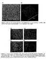
- the grooved silk fibroin substrate is a fabricated silk film designed to induce cell and extracellular matrix alignment.
- the thickness of grooved silk fibroin substrate ranges from about 10 nm to about 1 mm, the groove size is at least 125 nm, and the groove thickness depth is at least 100 nm. More preferably, the thickness of grooved silk fibroin substrate ranges from about 100 nm to about 100 ⁇ m, the groove size ranges from about 500 nm to about 10 ⁇ m, and the groove thickness depth ranges from about 250 nm to about 5 ⁇ m.
- the thickness of grooved silk fibroin substrate ranges from about 500 nm to about 10 ⁇ m
- the groove size ranges from about 1 ⁇ m to about 5 ⁇ m
- the groove thickness depth ranges from about 500 nm to about 3 ⁇ m.
- the silk diffraction gratings may be characterized for surface morphology by known techniques, such as field emission scanning electron and atomic force microscopy. See Fig. 1 .
- Silk fibroin may be used to make the silk fibroin substrate.
- Silk fibroin produced by silkworms is the most common and is generally preferred. However, there are many different silks, including spider silk, transgenic silks, genetically engineered silks, and variants thereof, that may alternatively be used.
- the term "fibroin" includes silkworm fibroin and insect or spider silk protein ( Lucas et al., Adv. Protein Chern 13: 107- 242 (1958 )).
- fibroin is obtained from a solution containing a dissolved silkworm silk or spider silk.
- the silkworm silk protein may be obtained, for example, from Bombyx mori, and the spider silk is obtained from Nephila clavzes.
- the silk proteins can be obtained from a solution containing a genetically engineered silk, such as from bacteria, yeast, mammalian cells, transgenic animals or transgenic plants. See, for example, PCT Publication WO 97/08315 and U.S. Patent No. 5,245,012 .
- aqueous silk fibroin solution maybe prepared from the silk fibroin using techniques known in the art. Suitable processes for preparing silk fibroin solution are disclosed in PCT Application No. PCT/US2004/011199 , and U.S. Provisional Application No. 60/856,297, filed November 3, 2006 , entitled "Biopolymer Photonic Crystals and Method of Manufacturing the Same,"
- the silk fibroin solution may be obtained by extracting sericin from the cocoons of a silkworm silk, such as Bombyx mori.
- a silkworm silk such as Bombyx mori.
- B. mori cocoons can be boiled for about 30 minutes in an aqueous solution, preferably an aqueous solution having about 0.02M Na 2 CO 3 .
- the cocoons can then be rinsed with water, for example, to extract the sericin proteins.
- the extracted silk can then be dissolved in an aqueous solution, preferably an aqueous salt solution.
- Salts useful for this purpose include lithium bromide, lithium thiocyanate, calcium nitrate or other chemicals capable of solubilizing silk.
- the extracted silk is dissolved in about 9-12 M LiBr solution.
- the salt may later be removed using, for example, dialysis.
- the silk fibroin solution may be obtained from a solution containing a dissolved spider silk, such as Nephila ciavipes.
- the silk fibroin solution can also be obtained from a solution containing a genetically engineered silk.
- the genetically engineered silk can, for example, comprise a therapeutic agent, e.g., a fusion protein with a cytokine, an enzyme, or any number of hormones or peptide-based drugs, antimicrobials and related substrates.
- Biocompatible polymers can also be added to the silk solution to generate composite matrices.
- Biocompatible polymers useful in the compositions described herein include, for example, polyethylene oxide (PEO) ( U.S. Patent No. 6,302,848 ), polyethylene glycol (PEG) ( U.S. Patent No. 6,395,734 ), collagen ( U.S. Patent No. 6,127,143 ), fibronectin ( U.S. Patent No. 5,263,992 ), keratin ( U.S. Patent No. 6,379,690 ), polyaspartic acid ( U.S. Patent No. 5,015,476 ), polylysine ( U.S. Patent No. 4,806,355 ), alginate ( U.S. Patent No.
- the silk fibroin substrate may be prepared by casting a silk solution onto patterned diffractive optical surfaces, such as different holographic diffraction gratings with varying line pitches as masks. After the silk solution has been cast, they can be dried as films. Drying may be accomplished by any means known in the art. For instance, the films may be placed in a vacuum environment and water annealed for two or more hours, removed, and allowed to further dry. Removal from the grating mask can be accomplished by simple mechanical lifting of the grating from the substrate.
- the grooves of the silk fibroin substrate induce cell and ECM alignment.
- the tissue-specific cells have deposited extracellular matrix on the substrate.
- the cultured cells grown on the silk films align and form their ECM parallel to the grooved film pattern.
- This allows one to control the direction of cell orientation and tissue alignment upon the lamellae layer, something that is helpful in tissue formation.
- Geometrical shapes imprinted on the substrates may also be used to grow the cells in aligned patterns. See Fig. 4 . Varying the geometrical pattern upon the silk film allows for defined spatial patterning of cell growth for the targeted tissue system.
- the individual lamellae tissue layers each of which is virtually a two-dimensional (2-D) construct containing the silk film and tissue-specific cell types, can then be stacked upon one another or otherwise combined to produce a three-dimensional (3-D) tissue-engineered organ.
- These 2-D lamellae structures can thus serve as "building-blocks" for 3-D tissue engineered constructs.
- Such a system exploits the advantageous material features of silk fibroin, including its slow degradation, biocompatibility, optical transparency, and durability for handling and suturing.
- the edges of the multi-layer construct may be sealed to prevent diffusion. Because the silk substrates are self adhering when exposed to applied pressure, a dulled biopsy punch which corresponds to the round silk film geometry can be used to apply pressure upon seeding.
- a dulled biopsy punch which corresponds to the round silk film geometry can be used to apply pressure upon seeding.
- polydimethylsulfoxide (PDMS) discs of similar diameter to the silk substrate may be employed to act as a clamping mechanism to hold the layers in place. PDMS has been shown to be inert in cell culture, has low water sorption, and can induce a passive clamping force. If the self-adherence mechanism is not sufficient, fibrin glue or similar adhesive can also be used.
- the lamellae "building-block" tissue system is applicable to multiple organ systems, provides a range of tunable material properties, and is completely, or at least mostly, composed of biomaterials that are non-immunogenic upon implantation.
- the resulting tissue-engineered organ thus contains silk fibroin substrates with cultured cells and deposited extracellular matrix.
- tissue-engineered organs may contain a vasculature network able to supply nutrient diffusion throughout the entire tissue-engineered construct. Incorporating micropores into the tissue-engineered organ mimics the convection for efficient nutrient transport and allows for nutrient diffusion or vascularization to each lamellae tissue layer of the biological construct.
- the vasculature-like micropores may be "drilled" in defined patterns within the silk film using polymer phase separation chemistry, laser ablation techniques, or other techniques known in the art.
- Laser ablation technology such as Femtosecond laser ablation technology, involves high energy pulsed laser light to minimize peripheral damage to the material.
- Polymer phase separation chemistry involves mixing two polymers that do not mix well (immiscible, akin to oil and water) to exploit their inability to mix well, such that they separate into different domains to generate materials morphologies based on the polymers and their respective amounts.
- Preferred polymers for use in polymer phase separation include polyethylene oxide or similar alternative polymers and waxes.
- Porosity within the silk films can often times be systematically controlled.
- porosity can be altered based on the size or the focus of the laser.
- porosity size range and pore density can be controlled by varying the concentration of the polymer. By holding constant the other variables, such as casting volume, casting surface area, and silk concentration (or multiple variables changed at the same time), porosity can be dependent, do a large degree, on polymer concentration. See Example 4.
- the micropores should be small enough so that the material properties of the lamellae tissue layers are not compromised and can still maintain a robust matrix for tissue development, yet large enough to provide the desired vascularization.
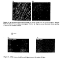
- the average diameter of the micropores ranges from about 100 nm to about 100 micron. More preferably, the average diameter ranges from about 800 nm to about 20 micron. Most preferably, the average diameter ranges from about 1 micron to about 10 micron. [confirm preferred ranges]. See Fig. 5 .
- micropores are preferably incorporated into the tissue-engineered organ in patterns designed to optimize nutrient diffusion to culture surface area specific to the targeted tissue system. As the layers are stacked upon one another, the microporous structure allows for nutrient diffusion to all regions of the 3-D construct.
- micropores can be used for tissue-engineered corneas.
- a 50-100 ⁇ m spacing is preferred because it provides an averaged spaced vascular network similar to that found in corneal tissue (although corneal tissue is not vascularized by blood capillaries, it contains a network that assists in nutrient diffusion through the tissue).
- Generating arrays of micron- or nano-sized holes interspersed in grid spacing provides nutrient diffusive ports throughout the stacked silk lamellae tissue layers.
- the lamellae tissue layers may be used to produce various tissue types and organ systems. Such systems may include bone, skin, cardiac tissue, cardiac muscle, muscle tissue, dense connecting tissue, basement membranes, smooth muscle tissue, endothelial tissue, and nervous tissue.
- the organ can be made up of any tissue that can be formed by the assembly of the silk fibroin substrates with cultured cells and deposited extra cellular matrix.
- the organ is a cornea, which is particularly suited for silk-based lamellae tissue layers because of the optically clear, non-immunogenic properties, and biocompatibility of the silk fibroin.
- the invention also relates to a process for preparing a tissue-engineered organ.
- the process involves preparing a multitude of lamellae tissue layers. Each lamellae tissue layer comprises a grooved silk fibroin substrate seeded with tissue-specific cells capable of extracellular matrix deposition.
- the process then involves culturing the cells on the silk fibroin substrate for a sufficient period of time to allow for the cells and deposited extracellular matrix to form a tissue-engineered organ.
- the process also involves creating micropores in the silk film.
- the tissue-engineered organ can be both non-immunogenic and biocompatible.
- Seeding the silk fibroin substrate may be accomplished through means known in the art.
- the cells should be seeded in the substrate in a manner that best enables their ability to be cultured within the substrate construct.
- the cells should typically be distributed at a density of about 1000 cells/cm 2 to about 50,000 cells/cm 2 .
- the cells should be distributed at a density of about 5000 cells/cm 2 to about 20,000 cells/cm 2 .
- these distribution patterns may differ depending on the size of the cell, the seeding conditions, and other factors appreciated by those of skill in the art.
- the cells When seeding the cells in the silk substrate, the cells may be supplemented with other materials known in the art to promote cell growth.
- Materials known to promote cell growth include cell growth media, such as Dulbecco's Modified Eagle Medium (DMEM), fetal bovine serum (FBS), non-essential amino acids and antibiotics, and growth and morphogen factors such as basic fibroblast growth factor (bFGF), transforming growth factors (TGFs), Vascular endothelial growth factor (VEGF), insulin-like growth factor (IGF-I), bone morphogenetic growth factors (BMPs), nerve growth factors and related proteins.
- DMEM Dulbecco's Modified Eagle Medium
- FBS fetal bovine serum
- growth and morphogen factors such as basic fibroblast growth factor (bFGF), transforming growth factors (TGFs), Vascular endothelial growth factor (VEGF), insulin-like growth factor (IGF-I), bone morphogenetic growth factors (BMPs), nerve growth factors and related proteins.
- Additional materials that can be included when seeding the cell include DNA, siRNA, antisense, plasmids, liposomes and related systems for delivery of genetic materials; peptides and proteins to active cellular signaling cascades; peptides and proteins to promote mineralization or related events from cells; adhesion peptides and proteins to improve gel-tissue interfaces; antimicrobial and antifungal peptides and proteins and related compounds.
- the additional materials can also include common components found in pharmaceutical formulations, such as known excipients. Exemplary excipients include diluents, solvents, buffers, solubilizers, suspending agents, viscosity controlling agents, binders, lubricants, surfactants, preservatives and stabilizers.
- the silk fibroin substrate of the lamellae tissue layers provides a mechanically robust matrix to support cellular growth until the culture has reproduced at least part of its original matrix.
- the amount of time involved in culturing the cells is dependent on the type of cells used, the conditions of the culturing, the degree to which the culture should represent the original matrix, and other variables appreciated by those of skill in the art.
- the cells are cultured for approximately 1-6 weeks, whereupon the silk fibroin protein will have been at least partially degraded and the new tissue construct exhibits properties consistent with those of the original matrix. It is at this point that the tissue-engineered organ is typically ready for in vivo use or as a tissue system for ex vivo studies.
- the cells may be cultured in accordance with techniques known in the art.
- the cells may be cultured in an organotypic environment, such as in submerged culture, at air-liquid interfaces or in selective perfusion bioreactors, or other environments suitable for culture cells on silk fibroin substrates.
- an endothelial cell sheet may be added on the bottom of the lamellae tissue layers and an epithelial cell sheet may be added on the top of the lamellae tissue layers.
- the invention relates to a tissue-engineered cornea containing at least two lamellae tissue layers, an endothelial cell sheet on the bottom of the lamellae tissue layers, and an epithelial cell sheet on the top of the lamellae tissue layers.
- Each of the lamellae tissue layers comprise a grooved silk fibroin substrate seeded with fibroblast cells having an extracellular matrix deposition.
- the cornea is optically clear, non-immunogenic, and biocompatible, and the lamellae tissue layers comprise micropores that allow for nutrient diffusion to each layer.
- the invention also relates to an in vivo implantation system for tissue-engineered organs.
- the system involves forming a tissue-engineered organ comprising cultured tissue-specific cells on a grooved silk fibroin substrate; allowing the silk fibroin material in the tissue-engineered organ to at least partially degrade; and implanting the tissue-engineered organ into a native tissue.
- the in vivo implantation system may be applicable to multiple organ systems, provides a range of tunable material properties, and is completely, or at least mostly, composed of biomaterials that are non-immunogenic upon implantation.
- tissue-engineered organ is ready for implantation should generally be left to the expertise of the skilled artisan.
- implantation into a native tissue may be done when the silk fibroin material has at least partially degraded and the culture has reproduced to a point at or near its original matrix.
- the disclosure also relates to a kit for preparing a tissue-engineered organ.
- the kit includes a grooved silk fibroin substrate capable of receiving tissue-specific cells having an extracellular matrix deposition.
- the kit also includes instructions for seeding the tissue-specific cells on the silk fibroin substrate, organizing the silk fibroin substrates to form lamellae tissue layers, culturing the tissue-specific cells on the silk fibroin substrate, or a combination thereof.
- the instructions need only be provided in detail sufficient to enable a skilled artisan to use the kit consistent with its primary use of preparing a tissue-engineered organ.
- the kit may additionally include other tools known in the art to be helpful in preparing a tissue-engineered organ.
- the kit may contain a collecting and containment tool set for acquiring cells from a primary cell source, such as a human or an animal.
- the kit may also contain a bioreactor for culturing the silk fibroin substrate after the substrate has been seeded with the tissue-specific cells.
- the bioreactor is preferably capable of maintaining sterile and nutrient-filled conditions until the tissue-specific cells have been cultured.
- the kit may be used in the academia and industry for commercial testing or other types of commercial use.
- the kit may be used to prepare a tissue-engineered organ for commercial testing purposes.
- the invention also relates to an in vitro testing system for animal organ replacement.
- the system involves forming a cultured tissue system comprising cultured tissue-specific cells on a grooved silk fibroin substrate; incubating the cultured tissue system for a time period until the tissue construct of the tissue-specific cells is able to maintain native tissue characteristics and form cultured tissue; and testing the cultured tissue under in vitro research conditions to aid in the development of animal organ replacement.
- the time period for incubating the cultured tissue system varies depending on the tissue system, the incubation conditions, the amount of organ that is being replaced, and other variables appreciated by those of skill in the art. If only a small portion of an organ is being replaced under favorable incubation conditions, the time period will obviously be shorter than if a large portion or a complete organ is being replaced under strained conditions. The exact time period can be determined by one of skill in the art under the circumstances of the organ replacement. Typically, the cultured tissue system is incubated for approximately 1-6 weeks.
- Testing the cultured tissue involves any aspects of testing that one of skill in the art would typically conduct in a research setting.
- the results, or lack of results, obtained through this testing should be viewed as aiding in the development of animal organ replacement.
- Example 1 Silk as a substrate for corneal cell growth
- Silk has been shown to be a viable substrate for a number of different cell types.
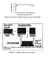
- Silk tissue layers are also capable of supporting corneal cell growth comparative to tissue culture plastic using the Alamar Blue metabolic cell assay. See Fig. 6 .
- similar cellular metabolic rates were found upon silk, glass and TCP substrates.
- rabbit stromal fibroblasts to grow on silk fibroin substrates was assessed.
- a monolayer of 8% fibroin solution was deposited on 12 mm round glass coverslips using a spin coater (Laurell Technologies).
- the spin coater produces an even distribution of the fibroin polymer across the glass surface.
- the fibroin coated coverslips were steam sterilized and placed within 24-well culture dishes.
- Primary corneal fibroblasts were isolated from adult rabbit corneas and cultured to confluency on TCP.
- the cells were then seeded at a density of 10,000 cells/cm 2 on the fibroin coated coverslips, and supplemented with media containing Dulbecco's Modified Eagle Medium (DMEM), 10% fetal bovine serum (FBS), and 1% Penstrep (an antibiotic treatment to reduce contamination of the cell cultures).
- DMEM Dulbecco's Modified Eagle Medium
- FBS fetal bovine serum
- Penstrep an antibiotic treatment to reduce contamination of the cell cultures.
- the cells where grown to confluency and the morphology of the cells appeared similar to fibroblasts grown on TCP. See Fig. 7 .
- Silk fibroin substrates may be transformed into lamellae tissue layers capable of guiding cell and ECM development and can also be vascularized or porous networks generated to support nutrient diffusion. These lamellae tissue layers can then be combined to form a 3-D tissue construct, or a tissue-engineered organ.
- the corneal tissue system offers a unique set of parameters that make it a useful model system for the use of the lamellae structures, in that the tissue is largely composed of stacked layers of extracellular matrix interspersed with cell layers.
- the lamellae tissue layers provide the correct set of material characteristics needed to form corneal tissue in that silk fibroin is robust in its mechanical properties, degrades naturally with native tissue replacement by the seeded cells, is non-immunogenic, and is optically clear with near 100% transmission of visible light. See Fig. 8 .
- the tissue layers may be used to produce tissue-engineered organs, such as corneal stromal tissue analogs.
- tissue-engineered organs such as corneal stromal tissue analogs.
- a schematic of a process that can be used to form an entire corneal tissue is shown Fig. 9 .
- Other configurations can also be used.
- stacked lamellae tissue layers are placed between endothelial and epithelial cell sheets on the bottom and top of the stacked stroma region respectively. See Nishida et al., 2003, for a description of the endothelial and epithelian cell sheets.
- tissue-engineered organ can then be cultured in an organotypic environment until it is ready for implantation or ex vivo tissue experimentation. Studies show that cell growth was detected within the central regions of the tissue-engineered silk cornea construct, suggesting cell viability is present throughout the construct. See Fig. 10 .
- a silk fibroin aqueous solution is prepared using standard protocols known in the art.
- the silk/PEO films are cast on PDMS, such as patterned or flat surfaces.
- a 5% wt. vol. PEO solution was prepared.
- 100-uL of silk/PEO solution dilution was cast onto 2-cm 2 patterned and flat round PDMS surfaces.
- the film was allowed to dry for 12-hrs. Upon drying, the films were water annealed for 6-hrs to induce primary beta-sheet crystallinity. After water annealing, the films were submerge in a water bath to prevent excessive film dehydration. The water bath was covered and the films allowed to sit for 24-hrs.
- One film was transferred from the water bath into a 100% MeOH bath for secondary beta-sheet crystallinity induction. The film was allowed to sit for 2-minutes, and then peeled off of their respective PDMS surfaces while submerged in the methanol bath. Once removed, the films were floated onto a Teflon coated glass slide, removed from the MeOH bath, and placed onto a Teflon coated drying surface (i.e. acrylic coated surfaces).
- Micro-pores were created within silk films by using PEO phase separation chemistry techniques to promote increased nutrient diffusion and cell-to-cell interaction throughout a stacked film construct. See Jin et al., 2004.
- the ratio of silk-to-PEO used in casting directly influenced the pores generated in the films once formed, dried and the PEO is extracted. Pores ranging between 1-3 microns in diameter were produced within the film. See Fig. 11(a) . In contrast, no pores were exhibited in controls not using the PEO separation method. See Fig. 11(b) as a comparative example.
Landscapes
- Health & Medical Sciences (AREA)
- Life Sciences & Earth Sciences (AREA)
- Engineering & Computer Science (AREA)
- Biomedical Technology (AREA)
- Chemical & Material Sciences (AREA)
- General Health & Medical Sciences (AREA)
- Zoology (AREA)
- Medicinal Chemistry (AREA)
- Transplantation (AREA)
- Epidemiology (AREA)
- Animal Behavior & Ethology (AREA)
- Cell Biology (AREA)
- Public Health (AREA)
- Oral & Maxillofacial Surgery (AREA)
- Veterinary Medicine (AREA)
- Dermatology (AREA)
- Bioinformatics & Cheminformatics (AREA)
- Biotechnology (AREA)
- Wood Science & Technology (AREA)
- Organic Chemistry (AREA)
- Genetics & Genomics (AREA)
- Botany (AREA)
- Chemical Kinetics & Catalysis (AREA)
- Urology & Nephrology (AREA)
- Microbiology (AREA)
- Biochemistry (AREA)
- General Engineering & Computer Science (AREA)
- Molecular Biology (AREA)
- Immunology (AREA)
- Hematology (AREA)
- Dispersion Chemistry (AREA)
- Ophthalmology & Optometry (AREA)
- Vascular Medicine (AREA)
- Neurosurgery (AREA)
- Neurology (AREA)
- Tropical Medicine & Parasitology (AREA)
- Toxicology (AREA)
- General Physics & Mathematics (AREA)
- Food Science & Technology (AREA)
- Analytical Chemistry (AREA)
Claims (14)
- Lamellengewebeschicht, die ein gerilltes Seidenfibroinsubstrat umfasst, das darauf angeordnete gewebespezifische Zellen aufweist,
wobei die Dicke des Substrats im Bereich von 10 nm bis 1 mm liegt, die Rillenbreite mindestens 125 nm beträgt und Rillendickentiefe mindestens 100 nm beträgt, sodass die Rillen in dem Substrat die Anordnung von Zellen und extrazellulärer Matrix induzieren, wenn die Zellen darauf kultiviert werden; und wobei die gewebespezifischen Zellen aus der Gruppe ausgewählt sind, die aus Folgenden besteht: Stammzellen, Fibroblasten, Endothelzellen, Epithelzellen und Fettzellen, die extrazelluläre Matrix auf dem Substrat erzeugen können, und Kombinationen davon. - Lamellengewebeschicht nach Anspruch 1, wobei die gewebespezifischen Zellen extrazelluläre Matrix auf dem Substrat ablagern.
- Aus Gewebe konstruiertes Organ, das eine Vielzahl an Lamellengewebeschichten umfasst, wobei jede Schicht ein gerilltes Seidenfibroinsubstrat nach Anspruch 2 umfasst.
- Aus Gewebe konstruiertes Organ nach Anspruch 3, wobei das Organ Mikroporen umfasst, die eine Nährstoffdiffusion oder Vaskularisierung zu jeder Schicht ermöglichen,
wobei ein durchschnittlicher Durchmesser der Mikroporen optional im Bereich von 500 nm bis 100 Mikron liegt und
wobei die Mikroporen optional durch die Verwendung von Poly(ethylenoxid)-Phasentrennungschemie, Laserablationstechniken oder einer Kombination davon erzeugt werden und
wobei das Organ optional jedwedes Gewebe ist, das durch den Aufbau der Seidenfibroinsubstrate mit kultivierten Zellen und abgelagerter extrazellulärer Matrix gebildet werden kann. - Aus Gewebe konstruiertes Organ nach einem der Ansprüche 3 oder 4, wobei das Organ eine Hornhaut ist.
- Aus Gewebe konstruiertes Organ nach Anspruch 5, das Folgendes umfasst:mindestens zwei Lamellengewebeschichten, wobei jede Schicht ein gerilltes Seidenfibroinsubstrat umfasst, das darauf angeordnete Fibroblastenzellen umfasst, sodass die Rillen in dem Substrat die Anordnung von Zellen und extrazellulärer Matrix induzieren, wenn die Zellen darauf kultiviert werden,eine Endothelzell-Platte auf der Unterseite der Lamellengewebeschichten undeine Epithelzell-Platte auf der Oberseite der Lamellengewebeschichten.
- Aus Gewebe konstruiertes Organ nach Anspruch 6, wobei die Hornhaut optisch klar, nicht immunogen und biokompatibel ist und
wobei die Lamellengewebeschichten optional Mikroporen umfassen, die eine Nährstoffdiffusion zu jeder Schicht ermöglichen. - Verfahren für die Herstellung eines aus Gewebe konstruierten Organs nach Anspruch 3, das Folgendes umfasst:Besiedeln von gewebespezifischen Zellen, die extrazelluläre Matrix ablagern können, auf dem gerillten Seidenfibroinsubstrat, um eine Lamellengewebeschicht zu bilden;Aufbauen einer Vielzahl der Lamellengewebeschichten undKultivieren der Zellen auf dem Seidenfibroinsubstrat für eine ausreichende Zeitdauer, um zu ermöglichen, dass die Zellen und die abgelagerte extrazelluläre Matrix das aus Gewebe konstruierte Organ bilden.
- Verfahren nach Anspruch 8, das weiter den Schritt des Erzeugens von Mikroporen in dem Seidenfibroinsubstrat umfasst,
wobei die Mikroporen optional durch die Verwendung von Poly(ethylenoxid)-Phasentrennungschemie, Laserablationstechniken oder einer Kombination davon erzeugt werden, und/oder
wobei der durchschnittliche Durchmesser der Mikroporen im Bereich von 500 nm bis 100 Mikron liegt,
und/oder
wobei das aus Gewebe konstruierte Organ nicht immunogen und biokompatibel ist. - Verfahren nach Anspruch 8 oder 9, wobei das Organ aus der Gruppe ausgewählt ist, die aus Folgenden besteht: Hornhaut, Knochen, Haut, Herzgewebe, Herzmuskel, Muskelgewebe, dichtem Bindegewebe, Basalmembranen, glattem Muskelgewebe, Endothelgewebe und Nervengewebe, und
wobei die gewebespezifischen Zellen optional aus der Gruppe ausgewählt sind, die aus Folgenden besteht: Stammzellen, Fibroblasten, Fettzellen, die gewebespezifische extrazelluläre Matrix erzeugen können, und Kombinationen davon, und
wobei die Herstellung des aus Gewebe konstruierten Organs optional weiter das Hinzufügen einer Endothelzell-Platte auf die Unterseite der Lamellengewebeschichten und einer Epithelzell-Platte auf die Oberseite der Lamellengewebeschichten umfasst. - Aus Gewebe konstruiertes Organ nach Anspruch 3 für die Verwendung in einem Verfahren der in-vivo-Implantation in ein natives Gewebe, wobei das Verfahren Folgendes umfasst: Ermöglichen des zumindest teilweisen Abbaus des Seidenfibroinmaterials in dem aus Gewebe konstruierten Organ, sodass das aus Gewebe konstruierte Organ für die Implantation in das native Gewebe geeignet ist.
- Aus Gewebe konstruiertes Organ nach Anspruch 3 für die Verwendung nach Anspruch 11, und
wobei das aus Gewebe konstruierte Organ optional aus der Gruppe ausgewählt ist, die aus Folgenden besteht: Knochen, Haut, Herzgewebe, Herzmuskel, Muskelgewebe, dichtem Bindegewebe, Basalmembranen, glattem Muskelgewebe, Endothelgewebe und Nervengewebe, oder
wobei das aus Gewebe konstruierte Organ optional eine Hornhaut ist und wobei die aus Gewebe konstruierte Hornhaut optional Folgendes enthält: Fibroblastenzellen, Endothelzellen, Epithelzellen, Stammzellen, Fettzellen, die gewebespezifische extrazelluläre Matrix erzeugen können. - In-vitro-Assay für einen Tierorganersatz, der Folgendes umfasst:Besiedeln von gewebespezifischen Zellen, die extrazelluläre Matrix ablagern können, auf dem gerillten Seidenfibroinsubstrat, wie in Anspruch 1 definiert, um eine Lamellengewebeschicht zu bilden;Aufbauen einer Vielzahl der Lamellengewebeschichten zu einem kultivierten Gewebesystem;Inkubieren des kultivierten Gewebesystems für eine Zeitdauer bis das Gewebesystem ein mehrschichtiges Organ bildet, das die Eigenschaften von nativem Gewebe aufrechterhalten kann, undTesten des kultivierten Gewebesystems unter in-vitro-Forschungsbedingungen, um die Entwicklung von Tierorganersatz zu unterstützen.
- Assay nach Anspruch 13, wobei das kultivierte Gewebesystem Mikroporen umfasst, die eine Nährstoffdiffusion zu jeder Schicht ermöglichen, und
wobei das kultivierte Gewebesystem optional ein aus Gewebe konstruiertes Organ ist, das aus der Gruppe ausgewählt ist, die aus Folgenden besteht: Hornhaut, Knochen, Haut, Herzgewebe, Herzmuskel, Muskelgewebe, dichtem Bindegewebe, Basalmembranen, glattem Muskelgewebe, Endothelgewebe und Nervengewebe, und
wobei, wenn das aus Gewebe konstruierte Organ eine Hornhaut ist, die Hornhaut optional Folgendes enthält: Stammzellen, Fibroblasten, Endothelzellen, Epithelzellen und Fettzellen, die gewebespezifische extrazelluläre Matrix erzeugen können, oder Kombinationen davon, und optional optisch klar, nicht immunogen und biokompatibel ist.
Applications Claiming Priority (3)
| Application Number | Priority Date | Filing Date | Title |
|---|---|---|---|
| US90380007P | 2007-02-27 | 2007-02-27 | |
| US97302307P | 2007-09-17 | 2007-09-17 | |
| PCT/US2008/055072 WO2008106485A2 (en) | 2007-02-27 | 2008-02-27 | Tissue-engineered silk organs |
Publications (2)
| Publication Number | Publication Date |
|---|---|
| EP2129772A2 EP2129772A2 (de) | 2009-12-09 |
| EP2129772B1 true EP2129772B1 (de) | 2016-07-27 |
Family
ID=39494931
Family Applications (1)
| Application Number | Title | Priority Date | Filing Date |
|---|---|---|---|
| EP08730804.5A Active EP2129772B1 (de) | 2007-02-27 | 2008-02-27 | Gewebegezüchtete seidenorgane |
Country Status (4)
| Country | Link |
|---|---|
| US (3) | US9102916B2 (de) |
| EP (1) | EP2129772B1 (de) |
| JP (3) | JP2010522583A (de) |
| WO (1) | WO2008106485A2 (de) |
Cited By (2)
| Publication number | Priority date | Publication date | Assignee | Title |
|---|---|---|---|---|
| US9450043B2 (en) | 2004-06-04 | 2016-09-20 | The Board Of Trustees Of The University Of Illinois | Methods and devices for fabricating and assembling printable semiconductor elements |
| US9554484B2 (en) | 2012-03-30 | 2017-01-24 | The Board Of Trustees Of The University Of Illinois | Appendage mountable electronic devices conformable to surfaces |
Families Citing this family (64)
| Publication number | Priority date | Publication date | Assignee | Title |
|---|---|---|---|---|
| CA2525994C (en) * | 2002-06-24 | 2012-10-16 | Tufts University | Silk biomaterials and methods of use thereof |
| US7842780B2 (en) | 2003-01-07 | 2010-11-30 | Trustees Of Tufts College | Silk fibroin materials and use thereof |
| EP1613796B1 (de) * | 2003-04-10 | 2017-03-22 | Tufts University | Konzentrierte wässrige seidenfibroinlösung und deren verwendung |
| WO2005000483A1 (en) * | 2003-06-06 | 2005-01-06 | Tufts University | Method for forming inorganic coatings |
| EP2129772B1 (de) | 2007-02-27 | 2016-07-27 | Trustees Of Tufts College | Gewebegezüchtete seidenorgane |
| JP2010529230A (ja) | 2007-05-29 | 2010-08-26 | トラスティーズ オブ タフツ カレッジ | 音波処理を用いた絹フィブロインのゲル化のための方法 |
| EP2249886A4 (de) * | 2008-02-07 | 2013-05-22 | Tufts College | 3-dimensionale seiden-hydroxylapatit-zusammensetzungen |
| JP5727366B2 (ja) * | 2008-05-15 | 2015-06-03 | タフツ ユニバーシティー/トラスティーズ オブ タフツ カレッジ | 絹ポリマーに基づくアデノシン放出:てんかんに対する潜在的治療能力 |
| US8501172B2 (en) * | 2008-09-26 | 2013-08-06 | Trustees Of Tufts College | pH-induced silk gels and uses thereof |
| US8372726B2 (en) | 2008-10-07 | 2013-02-12 | Mc10, Inc. | Methods and applications of non-planar imaging arrays |
| US8886334B2 (en) | 2008-10-07 | 2014-11-11 | Mc10, Inc. | Systems, methods, and devices using stretchable or flexible electronics for medical applications |
| US8097926B2 (en) | 2008-10-07 | 2012-01-17 | Mc10, Inc. | Systems, methods, and devices having stretchable integrated circuitry for sensing and delivering therapy |
| US8389862B2 (en) | 2008-10-07 | 2013-03-05 | Mc10, Inc. | Extremely stretchable electronics |
| JP5646492B2 (ja) | 2008-10-07 | 2014-12-24 | エムシー10 インコーポレイテッドMc10,Inc. | 伸縮可能な集積回路およびセンサアレイを有する装置 |
| CN105268020A (zh) | 2008-10-09 | 2016-01-27 | 塔夫茨大学信托人 | 含有甘油的改性丝膜 |
| US9308070B2 (en) | 2008-12-15 | 2016-04-12 | Allergan, Inc. | Pliable silk medical device |
| EP2421549B1 (de) * | 2009-04-20 | 2013-06-12 | Allergan, Inc. | Seiden-fibroin-hydrogele und ihre verwendungen |
| WO2011005381A2 (en) | 2009-06-01 | 2011-01-13 | Trustees Of Tufts College | Vortex-induced silk fibroin gelation for encapsulation and delivery |
| WO2011008842A2 (en) | 2009-07-14 | 2011-01-20 | Trustees Of Tufts College | Electrospun silk material systems for wound healing |
| JP2012533780A (ja) * | 2009-07-20 | 2012-12-27 | タフツ ユニバーシティー/トラスティーズ オブ タフツ カレッジ | タンパク質のみからなる移植可能な吸収性反射体 |
| EP2483460B1 (de) | 2009-09-28 | 2015-09-02 | Trustees Of Tufts College | Verfahren zur herstellung gezogene seiden-egel-fasern |
| JP5730317B2 (ja) | 2009-09-29 | 2015-06-10 | タフツ ユニバーシティー/トラスティーズ オブ タフツ カレッジ | 絹ナノスフェアおよび絹マイクロスフェアならびにこれらを作製する方法 |
| WO2011041727A1 (en) | 2009-10-01 | 2011-04-07 | Mc10, Inc. | Protective cases with integrated electronics |
| JP6046491B2 (ja) | 2009-12-16 | 2016-12-21 | ザ ボード オブ トラスティーズ オブ ザ ユニヴァーシティー オブ イリノイ | コンフォーマル電子機器を使用した生体内での電気生理学 |
| US10441185B2 (en) | 2009-12-16 | 2019-10-15 | The Board Of Trustees Of The University Of Illinois | Flexible and stretchable electronic systems for epidermal electronics |
| US9936574B2 (en) | 2009-12-16 | 2018-04-03 | The Board Of Trustees Of The University Of Illinois | Waterproof stretchable optoelectronics |
| US9603971B2 (en) | 2010-03-05 | 2017-03-28 | Trustees Of Tufts College | Silk-based ionomeric compositions |
| KR101724273B1 (ko) | 2010-03-17 | 2017-04-07 | 더 보드 오브 트러스티즈 오브 더 유니버시티 오브 일리노이 | 생체흡수성 기판 상 이식가능한 바이오의료 장치 |
| WO2012031144A2 (en) | 2010-09-01 | 2012-03-08 | Trustees Of Tufts College | Silk fibroin and polyethylene glycol-based biomaterials |
| JP2013542181A (ja) | 2010-09-03 | 2013-11-21 | タフツ ユニバーシティー/トラスティーズ オブ タフツ カレッジ | プラズモンナノ粒子ドープ絹材料 |
| JP2014502416A (ja) | 2010-09-27 | 2014-01-30 | タフツ ユニバーシティー/トラスティーズ オブ タフツ カレッジ | 絹ベースの圧電性材料 |
| BR112013009609A2 (pt) | 2010-10-19 | 2016-07-12 | Tufts College | microagulhas à base de fibroína de seda e métodos para fazer os mesmos |
| US10335519B2 (en) | 2011-04-20 | 2019-07-02 | Trustees Of Tufts College | Dynamic silk coatings for implantable devices |
| US9765934B2 (en) | 2011-05-16 | 2017-09-19 | The Board Of Trustees Of The University Of Illinois | Thermally managed LED arrays assembled by printing |
| JP2014523633A (ja) | 2011-05-27 | 2014-09-11 | エムシー10 インコーポレイテッド | 電子的、光学的、且つ/又は機械的装置及びシステム並びにこれらの装置及びシステムを製造する方法 |
| US8934965B2 (en) | 2011-06-03 | 2015-01-13 | The Board Of Trustees Of The University Of Illinois | Conformable actively multiplexed high-density surface electrode array for brain interfacing |
| WO2013040047A1 (en) * | 2011-09-12 | 2013-03-21 | Cornell University | Biopolymer films and methods of making and using same |
| FR2980209B1 (fr) * | 2011-09-19 | 2013-09-13 | Inst Curie | Dispositif de guidage de la migration cellulaire et procede de guidage de la migration cellulaire mettant en oeuvre un tel dispositif |
| US20140370094A1 (en) | 2011-11-08 | 2014-12-18 | Tufts University | Silk-based scaffold platform for engineering tissue constructs |
| ES2953309T3 (es) | 2011-11-09 | 2023-11-10 | Tufts College | Partículas de fibroína de seda inyectables y usos de las mismas |
| CA2890372A1 (en) | 2011-11-09 | 2013-05-16 | Trustees Of Tufts College | Injectable silk fibroin foams and uses thereof |
| HK1204205A1 (en) | 2011-12-01 | 2015-11-06 | The Board Of Trustees Of The University Of Illinois | Transient devices designed to undergo programmable transformations |
| CN102488926B (zh) * | 2011-12-16 | 2013-10-30 | 东华大学 | 一种用于尿道重建的组织工程支架及其制备方法 |
| WO2013102193A1 (en) | 2011-12-29 | 2013-07-04 | Trustees Of Tufts College | Functionalization of biomaterials to control regeneration and inflammation responses |
| EP2811987B1 (de) | 2012-02-06 | 2021-01-20 | Children's Medical Center Corporation | Mehrschichtiges biomaterial zur geweberegenerierung und wundheilung |
| WO2013152265A1 (en) | 2012-04-06 | 2013-10-10 | Trustees Of Tufts College | Methods of producing and using silk microfibers |
| US10653786B2 (en) | 2012-04-25 | 2020-05-19 | Trustees Of Tufts College | Silk microspheres and methods for surface lubrication |
| JP2014014590A (ja) * | 2012-07-11 | 2014-01-30 | Shinkan Kogyo Kk | 膜状結合組織体形成用基材及びこれを用いた膜状結合組織体の生産方法 |
| US9171794B2 (en) | 2012-10-09 | 2015-10-27 | Mc10, Inc. | Embedding thin chips in polymer |
| ITTO20130014A1 (it) * | 2013-01-09 | 2014-07-10 | Univ Degli Studi Torino | Scaffold polimerico per la rigenerazione cardiaca e la protezione dai danni da riperfusione |
| JP2014221011A (ja) * | 2013-05-13 | 2014-11-27 | 独立行政法人物質・材料研究機構 | 細胞培養用支持体の製造方法、細胞培養用支持体および細胞培養方法 |
| GB201415681D0 (en) | 2014-09-04 | 2014-10-22 | Cambridge Entpr Ltd And President And Fellows Of Harvard College | Protien Capsules |
| MX2017015587A (es) | 2015-06-01 | 2018-08-23 | Univ Illinois | Metodo alternativo para sensor uv. |
| BR112017025609A2 (pt) | 2015-06-01 | 2018-08-07 | The Board Of Trustees Of The University Of Illinois | sistemas eletrônicos miniaturizados com potência sem fio e capacidades de comunicação de campo próximo |
| CN105251052B (zh) * | 2015-10-15 | 2018-06-29 | 天津市天津医院 | 软骨细胞外基质与丝素蛋白复合取向软骨支架及其制备方法 |
| US10925543B2 (en) | 2015-11-11 | 2021-02-23 | The Board Of Trustees Of The University Of Illinois | Bioresorbable silicon electronics for transient implants |
| US10864299B2 (en) * | 2016-04-29 | 2020-12-15 | Trustees Of Tufts College | Artificial silk based innervated cornea |
| EP3315147A1 (de) * | 2016-10-28 | 2018-05-02 | Bioengineering Laboratories S.r.l. | Zur regeneration von geweben geeignetes hybridgerüst und herstellungsverfahren |
| WO2018081805A1 (en) | 2016-10-31 | 2018-05-03 | Sofregen Medical, Inc. | Compositions comprising low molecular weight silk fibroin fragments and plasticizers |
| CA3096036A1 (en) | 2018-04-03 | 2019-10-10 | Vaxess Technologies, Inc. | Microneedle comprising silk fibroin applied to a dissolvable base |
| US11864990B2 (en) | 2019-02-26 | 2024-01-09 | Avedro, Inc. | Bioresorbable corneal implants |
| WO2021076798A1 (en) | 2019-10-15 | 2021-04-22 | Sofregen Medical, Inc. | Delivery devices for delivering and methods of delivering compositions |
| US20250387603A1 (en) | 2022-06-24 | 2025-12-25 | Vaxess Technologies, Inc. | Applicator for medicament patch |
| CN121263229A (zh) | 2023-06-23 | 2026-01-02 | 瓦克塞斯技术公司 | 药用贴片施敷器 |
Family Cites Families (116)
| Publication number | Priority date | Publication date | Assignee | Title |
|---|---|---|---|---|
| BE380083A (de) * | 1929-12-23 | |||
| US3424164A (en) | 1966-05-20 | 1969-01-28 | Ethicon Inc | Silk suture |
| JPS5838449B2 (ja) | 1979-04-17 | 1983-08-23 | カネボウ株式会社 | 微粉末状絹フィプロインの製造法 |
| JPS56166235A (en) | 1980-05-24 | 1981-12-21 | Kanebo Ltd | Hydrophilic porous body and its preparation |
| JPS5838449A (ja) | 1981-08-31 | 1983-03-05 | Matsushita Electronics Corp | 高圧ナトリウムランプ装置 |
| US4806355A (en) | 1983-06-06 | 1989-02-21 | Connaught Laboratories Limited | Microencapsulation of living tissue and cells |
| JPS60142259A (ja) | 1983-12-29 | 1985-07-27 | Kanebo Ltd | 固定化抗体 |
| JPS60259677A (ja) | 1984-05-31 | 1985-12-21 | 水島 繁三郎 | 動物蛋白吸着再生繊維からなる原糸、織物、編物及びその製造方法 |
| US5263992A (en) | 1986-10-17 | 1993-11-23 | Bio-Metric Systems, Inc. | Biocompatible device with covalently bonded biocompatible agent |
| US4798722A (en) * | 1986-11-12 | 1989-01-17 | Zotos International, Inc. | Permanent waving composition |
| CA1340581C (en) * | 1986-11-20 | 1999-06-08 | Joseph P. Vacanti | Chimeric neomorphogenesis of organs by controlled cellular implantation using artificial matrices |
| JPS63190604A (ja) * | 1987-02-03 | 1988-08-08 | Agency Of Ind Science & Technol | 新規の水−アルコ−ル分離膜 |
| US5606019A (en) * | 1987-10-29 | 1997-02-25 | Protien Polymer Technologies, Inc. | Synthetic protein as implantables |
| JPH0694518B2 (ja) | 1987-11-02 | 1994-11-24 | 工業技術院長 | 絹フィブロイン多孔質体の製造方法 |
| GB8800078D0 (en) * | 1988-01-05 | 1988-02-10 | Ciba Geigy Ag | Novel antibodies |
| EP0330134A1 (de) | 1988-02-25 | 1989-08-30 | Akzo Nobel N.V. | Modifizierte Cellulose für biocompatible Dialysemembranen IV und Verfahren zu deren Herstellung |
| JPH0296512A (ja) | 1988-09-29 | 1990-04-09 | Seiwa Kasei:Kk | パーマネントウェーブ用第一剤 |
| US5015476A (en) | 1989-08-11 | 1991-05-14 | Paravax, Inc. | Immunization implant and method |
| US5270419A (en) | 1990-01-19 | 1993-12-14 | Nova Pharmaceutical Corporation | Polyanhydrides of oligomerized unsaturated aliphatic acids |
| US5290494A (en) * | 1990-03-05 | 1994-03-01 | Board Of Regents, The University Of Texas System | Process of making a resorbable implantation device |
| US5245012A (en) | 1990-04-19 | 1993-09-14 | The United States Of America As Represented By The Secretary Of The Army | Method to achieve solubilization of spider silk proteins |
| US5989894A (en) * | 1990-04-20 | 1999-11-23 | University Of Wyoming | Isolated DNA coding for spider silk protein, a replicable vector and a transformed cell containing the DNA |
| GB2245279B (en) * | 1990-06-20 | 1993-04-07 | Unilever Plc | Shampoo composition |
| JPH0669487B2 (ja) * | 1991-02-15 | 1994-09-07 | 東京大学長 | 生体細胞の成長並びに機能分化の促進・制御方法 |
| JPH04263611A (ja) | 1991-02-16 | 1992-09-18 | Pola Chem Ind Inc | 水不溶性固形フィブロイン成形物及びその製造法 |
| JPH0543600A (ja) | 1991-08-08 | 1993-02-23 | Kanebo Ltd | 抗体または抗原固定化絹フイブロイン膜および免疫測定用センサー |
| JPH05163132A (ja) | 1991-12-13 | 1993-06-29 | Kanebo Ltd | 皮膚化粧料 |
| JPH06346314A (ja) | 1993-06-02 | 1994-12-20 | Toyobo Co Ltd | 再生絹フィブロイン繊維およびその製造方法 |
| US5932462A (en) * | 1995-01-10 | 1999-08-03 | Shearwater Polymers, Inc. | Multiarmed, monofunctional, polymer for coupling to molecules and surfaces |
| JPH08295697A (ja) | 1995-04-26 | 1996-11-12 | Kanebo Ltd | 高濃度絹フィブロイン水溶液の製造方法 |
| BR9612625A (pt) | 1995-08-22 | 1999-06-01 | Agricola Tech Inc | Processos de clonagem para proteínas de séda de aranha de alta resisténcia |
| US5576881A (en) | 1995-08-29 | 1996-11-19 | Lucent Technologies Inc. | Multi-frequency optical signal source having reduced distortion and crosstalk |
| US5855613A (en) | 1995-10-13 | 1999-01-05 | Islet Sheet Medical, Inc. | Retrievable bioartificial implants having dimensions allowing rapid diffusion of oxygen and rapid biological response to physiological change |
| JPH1036676A (ja) | 1996-07-26 | 1998-02-10 | Kanebo Ltd | タンパク質水溶液の濃縮方法 |
| US5814328A (en) | 1997-01-13 | 1998-09-29 | Gunasekaran; Subramanian | Preparation of collagen using papain and a reducing agent |
| AU7486798A (en) | 1997-05-12 | 1998-12-08 | Metabolix, Inc. | Polyhydroxyalkanoates for (in vivo) applications |
| EP0920875A1 (de) * | 1997-06-18 | 1999-06-09 | JAPAN as repr. by DIR. GENERAL of NATIONAL INST. OF SERICULTURAL & ENTOMOLOGICAL SCIENCE MINISTRY OF AGR, FORESTRY & FISHERI | Wundverbandsmaterial, das seidenfibroin und seidensericin als hauptbestandteile enthält sowie verfahren zu seiner herstellung |
| JPH1126344A (ja) | 1997-06-30 | 1999-01-29 | Hitachi Ltd | パターン形成方法及び装置並びに半導体装置の製造方法 |
| WO1999001089A1 (en) | 1997-07-01 | 1999-01-14 | Brown University Research Foundation | Implantable prosthetic devices coated with bioactive molecules |
| US6123819A (en) * | 1997-11-12 | 2000-09-26 | Protiveris, Inc. | Nanoelectrode arrays |
| JP3094125B2 (ja) | 1997-11-18 | 2000-10-03 | 農林水産省蚕糸・昆虫農業技術研究所長 | 表皮細胞増殖活性化素材 |
| US6440740B1 (en) * | 1997-11-18 | 2002-08-27 | Japan As Represented By Director General Of National Institute Of Sericultural And Entomological Science, Ministry Of Agriculture, Forestry And Fisheries | Material for activating epidermal cell multiplication |
| US5932552A (en) | 1997-11-26 | 1999-08-03 | Keraplast Technologies Ltd. | Keratin-based hydrogel for biomedical applications and method of production |
| US5994099A (en) * | 1997-12-31 | 1999-11-30 | The University Of Wyoming | Extremely elastic spider silk protein and DNA coding therefor |
| US6362254B2 (en) | 1998-03-12 | 2002-03-26 | Shearwater Corporation | Poly(ethylene glycol) derivatives with proximal reactive groups |
| US5902800A (en) | 1998-03-27 | 1999-05-11 | Glenpharma | Dextran formulations and method for treatment of inflammatory joint disorders |
| US6110590A (en) * | 1998-04-15 | 2000-08-29 | The University Of Akron | Synthetically spun silk nanofibers and a process for making the same |
| WO1999061422A1 (en) | 1998-05-29 | 1999-12-02 | Sugen, Inc. | Pyrrole substituted 2-indolinone protein kinase inhibitors |
| US7662409B2 (en) * | 1998-09-25 | 2010-02-16 | Gel-Del Technologies, Inc. | Protein matrix materials, devices and methods of making and using thereof |
| US20030007991A1 (en) * | 1998-09-25 | 2003-01-09 | Masters David B. | Devices including protein matrix materials and methods of making and using thereof |
| US6302848B1 (en) | 1999-07-01 | 2001-10-16 | Sonotech, Inc. | In vivo biocompatible acoustic coupling media |
| EP1013756A1 (de) | 1998-12-21 | 2000-06-28 | Corning Incorporated | Vorrichtung Zur Vermehrung von zellen die die Zellenverbindungdurch das Wachstumsprozes ermöglicht und Verfahren zur ihre Herstellung |
| US6592623B1 (en) * | 1999-08-31 | 2003-07-15 | Virginia Commonwealth University Intellectual Property Foundation | Engineered muscle |
| JP3151665B2 (ja) | 1999-03-23 | 2001-04-03 | 農林水産省蚕糸・昆虫農業技術研究所長 | 生体高分子/ポリアリルアミン複合体およびその製造方法 |
| US6103255A (en) | 1999-04-16 | 2000-08-15 | Rutgers, The State University | Porous polymer scaffolds for tissue engineering |
| US6267776B1 (en) | 1999-05-03 | 2001-07-31 | O'connell Paul T. | Vena cava filter and method for treating pulmonary embolism |
| US6325810B1 (en) | 1999-06-30 | 2001-12-04 | Ethicon, Inc. | Foam buttress for stapling apparatus |
| IT1309453B1 (it) | 1999-10-01 | 2002-01-23 | Consorzio Per Gli Studi Uni | Substrato bioartificiale per la realizzazione di tessuti e organianimali, in particolare umani. |
| WO2001036531A1 (fr) | 1999-11-15 | 2001-05-25 | Zaidan-Houjin Ueda Sen-I Kagaku Shinkoukai | Materiau polymere moleculairement composite en fibroine/cellulose et procede de production de ce materiau |
| US6310188B1 (en) | 2000-01-24 | 2001-10-30 | Board Of Supervisors Of Louisiana State University And Agricultural And Mechanical College | Method for producing chitin or chitosan |
| CA2398635A1 (en) | 2000-02-03 | 2001-08-09 | Nexia Biotechnologies, Inc. | Surgical sutures containing spider silk |
| AU2001294671A1 (en) * | 2000-09-25 | 2002-04-08 | The Board Of Trustees Of The University Of Illinois | Microfabrication of membranes for the growth of cells |
| IT1316885B1 (it) * | 2000-10-02 | 2003-05-13 | Consorzio Per Gli Studi Uni | Procedimento per la preparazione di un tessuto non tessuto in fibroinadi seta. |
| JP2002128691A (ja) * | 2000-10-24 | 2002-05-09 | National Institute Of Agrobiological Sciences | セリシン含有素材、その製造方法およびその使用方法 |
| US7265186B2 (en) * | 2001-01-19 | 2007-09-04 | Nektar Therapeutics Al, Corporation | Multi-arm block copolymers as drug delivery vehicles |
| CA2405850A1 (en) | 2001-03-14 | 2002-10-10 | Japan As Represented By President Of Tokyo University Of Agriculture And Technology | Method for producing fiber and film of silk and silk-like material |
| ITVR20010098A1 (it) | 2001-09-11 | 2003-03-11 | Consorzio Per Gli Studi Uni | Procedimento per l'ottenimento di idrogeli di fibroina di seta. |
| US20030068446A1 (en) | 2001-10-02 | 2003-04-10 | Northwestern University | Protein and peptide nanoarrays |
| GB0126118D0 (en) | 2001-10-31 | 2002-01-02 | Vollrath Friedrich W L | Precursor feedstock for forming filaments |
| US6902932B2 (en) * | 2001-11-16 | 2005-06-07 | Tissue Regeneration, Inc. | Helically organized silk fibroin fiber bundles for matrices in tissue engineering |
| JP3803962B2 (ja) | 2001-12-28 | 2006-08-02 | 独立行政法人科学技術振興機構 | 絹タンパク質フィルムとその製造方法 |
| US7057023B2 (en) * | 2002-01-11 | 2006-06-06 | Nexia Biotechnologies Inc. | Methods and apparatus for spinning spider silk protein |
| JP3772207B2 (ja) * | 2002-06-19 | 2006-05-10 | 独立行政法人農業生物資源研究所 | 生分解性生体高分子材料、その製造方法、およびこの高分子材料からなる機能性素材 |
| CA2525994C (en) | 2002-06-24 | 2012-10-16 | Tufts University | Silk biomaterials and methods of use thereof |
| AU2003294240B2 (en) | 2002-11-01 | 2009-07-16 | Trustees Of Tufts College | Templated native silk smectic gels |
| US7842780B2 (en) * | 2003-01-07 | 2010-11-30 | Trustees Of Tufts College | Silk fibroin materials and use thereof |
| KR101141985B1 (ko) | 2003-02-06 | 2012-05-17 | 고지 니시다 | 각막 상피 형성용 세포 시트, 그들의 제조 방법 및 그들의이용 방법 |
| EP1613796B1 (de) | 2003-04-10 | 2017-03-22 | Tufts University | Konzentrierte wässrige seidenfibroinlösung und deren verwendung |
| WO2005000483A1 (en) * | 2003-06-06 | 2005-01-06 | Tufts University | Method for forming inorganic coatings |
| WO2005123114A2 (en) | 2004-06-11 | 2005-12-29 | Trustees Of Tufts College | Silk-based drug delivery system |
| US10010574B2 (en) * | 2004-07-31 | 2018-07-03 | Brainguard Co., Ltd. | Silk peptide for improving neuroprotective and neurofunctional effects and a method of its preparation |
| US7960509B2 (en) * | 2005-01-14 | 2011-06-14 | Trustees Of Tufts College | Fibrous protein fusions and use thereof in the formation of advanced organic/inorganic composite materials |
| US9290579B2 (en) * | 2005-04-20 | 2016-03-22 | Trustees Of Tufts College | Covalently immobilized protein gradients in three-dimensional porous scaffolds |
| CA2645934C (en) | 2005-08-02 | 2014-04-29 | Trustees Of Tufts College | Methods for stepwise deposition of silk fibroin coatings |
| GB0516846D0 (en) * | 2005-08-17 | 2005-09-21 | Knight David P | Meniscal repair device |
| NL1030122C2 (nl) | 2005-10-06 | 2007-04-10 | Kobato Polytechnologie B V | Polymeersamenstelling bevattende een warmteaccumulerend faseovergangsmateriaal, bereidingswijze daarvan en product waarin deze polymeersamenstelling is toegepast. |
| US20080058400A1 (en) * | 2006-08-29 | 2008-03-06 | Fujifilm Corporation | Skin external preparation |
| US20100028451A1 (en) * | 2006-09-26 | 2010-02-04 | Trustees Of Tufts College | Silk microspheres for encapsulation and controlled release |
| US20110121485A1 (en) * | 2006-10-30 | 2011-05-26 | Spintec Engineering Gmbh | Method and apparatus for the manufacture of a fiber |
| GB2443401A (en) | 2006-10-30 | 2008-05-07 | Spin'tec Engineering Gmbh | Producing fibres by extruding onto a treatment device |
| PL2089343T3 (pl) | 2006-10-31 | 2011-12-30 | Baseclick Gmbh | Reakcja chemiczna typu click do wytwarzania cząsteczek reporterowych |
| CA2704309C (en) * | 2006-11-03 | 2017-02-28 | Trustees Of Tufts College | Electroactive biopolymer optical and electro-optical devices and method of manufacturing the same |
| EP2101975A2 (de) * | 2006-11-03 | 2009-09-23 | Trustees of Tufts College | Biopolymersensor und herstellungsverfahren dafür |
| US20100068740A1 (en) * | 2006-11-03 | 2010-03-18 | Trustees Of Tufts College | Microfluidic device with a cylindrical microchannel and a method for fabricating same |
| WO2008127404A2 (en) | 2006-11-03 | 2008-10-23 | Trustees Of Tufts College | Nanopatterned biopolymer optical device and method of manufacturing the same |
| WO2008118211A2 (en) * | 2006-11-03 | 2008-10-02 | Trustees Of Tufts College | Biopolymer photonic crystals and method of manufacturing the same |
| EP2129772B1 (de) | 2007-02-27 | 2016-07-27 | Trustees Of Tufts College | Gewebegezüchtete seidenorgane |
| JP2010529230A (ja) * | 2007-05-29 | 2010-08-26 | トラスティーズ オブ タフツ カレッジ | 音波処理を用いた絹フィブロインのゲル化のための方法 |
| US9808557B2 (en) * | 2007-08-10 | 2017-11-07 | Trustees Of Tufts College | Tubular silk compositions and methods of use thereof |
| JP2011504421A (ja) | 2007-11-05 | 2011-02-10 | トラスティーズ オブ タフツ カレッジ | ナノコンタクトインプリンティングによる絹フィブロインフォトニック構造の作製 |
| EP2249886A4 (de) | 2008-02-07 | 2013-05-22 | Tufts College | 3-dimensionale seiden-hydroxylapatit-zusammensetzungen |
| US8206774B2 (en) * | 2008-03-13 | 2012-06-26 | Trustees Of Tufts College | Diazonium salt modification of silk polymer |
| US9068282B2 (en) * | 2008-04-08 | 2015-06-30 | Trustees Of Tufts College | System and method for making biomaterial structures |
| JP5727366B2 (ja) | 2008-05-15 | 2015-06-03 | タフツ ユニバーシティー/トラスティーズ オブ タフツ カレッジ | 絹ポリマーに基づくアデノシン放出:てんかんに対する潜在的治療能力 |
| US20090297588A1 (en) * | 2008-05-28 | 2009-12-03 | Spin'tec Engineering Gmbh | Antibiotic dressing for the treatment of infected wounds |
| WO2009155397A2 (en) | 2008-06-18 | 2009-12-23 | Trustees Of Tufts College | Edible holographic silk products |
| GB2461125A (en) | 2008-06-25 | 2009-12-30 | Spintec Engineering Gmbh | A silk membrane for bone graft material |
| US8501172B2 (en) * | 2008-09-26 | 2013-08-06 | Trustees Of Tufts College | pH-induced silk gels and uses thereof |
| JP5885505B2 (ja) | 2008-09-26 | 2016-03-15 | タフツ ユニバーシティー/トラスティーズ オブ タフツ カレッジ | 活性なシルク粘液接着剤、シルク電気的ゲル化方法、およびデバイス |
| GB2464348A (en) * | 2008-10-17 | 2010-04-21 | Spintec Engineering Gmbh | Applying a liquid protein onto a permeable surface, and silk mono-filament having specific properties |
| EP2191867A1 (de) | 2008-11-27 | 2010-06-02 | KPSS-Kao Professional Salon Services GmbH | Zusammensetzung zum Bleichen/ zur Aufhellung |
| EP2253336A1 (de) * | 2009-05-15 | 2010-11-24 | Spintec Engineering GmbH | Medizinische Vorrichtung aus Seide mit antimikrobieller Wirkung und ihr Herstellungsverfahren |
| EP2451953A4 (de) | 2009-07-10 | 2013-07-03 | Tufts College | Genmanipulierte seidenprotein-basierte nukleinsäure-liefersysteme |
| US9603971B2 (en) | 2010-03-05 | 2017-03-28 | Trustees Of Tufts College | Silk-based ionomeric compositions |
| KR101317420B1 (ko) * | 2010-03-11 | 2013-10-10 | 한국과학기술원 | 고분자량의 재조합 실크 또는 실크 유사 단백질 및 이를 이용하여 제조된 마이크로 또는 나노 크기의 거미줄 또는 거미줄 유사 섬유 |
| WO2013152265A1 (en) | 2012-04-06 | 2013-10-10 | Trustees Of Tufts College | Methods of producing and using silk microfibers |
-
2008
- 2008-02-27 EP EP08730804.5A patent/EP2129772B1/de active Active
- 2008-02-27 WO PCT/US2008/055072 patent/WO2008106485A2/en not_active Ceased
- 2008-02-27 US US12/528,634 patent/US9102916B2/en active Active
- 2008-02-27 JP JP2009551814A patent/JP2010522583A/ja active Pending
-
2014
- 2014-04-25 JP JP2014090875A patent/JP5970497B2/ja active Active
-
2015
- 2015-07-07 US US14/793,464 patent/US9655993B2/en active Active
- 2015-10-16 JP JP2015204147A patent/JP6140240B2/ja not_active Expired - Fee Related
-
2017
- 2017-05-19 US US15/600,351 patent/US10478524B2/en active Active
Non-Patent Citations (2)
| Title |
|---|
| D. J. DONOHUE ET AL: "Numerical modeling of the cornea's lamellar structure and birefringence properties", JOURNAL OF THE OPTICAL SOCIETY OF AMERICA A, vol. 12, no. 7, 1 July 1995 (1995-07-01), pages 1425 - 1438, XP055098759, ISSN: 1084-7529, DOI: 10.1364/JOSAA.12.001425 * |
| X. F. WALBOOMERS ET AL: "Attachment of fibroblasts on smooth and microgrooved polystyrene", JOURNAL OF BIOMEDICAL MATERIALS RESEARCH, vol. 46, no. 2, 1 August 1999 (1999-08-01), pages 212 - 220, XP055098758, ISSN: 0021-9304, DOI: 10.1002/(SICI)1097-4636(199908)46:2<212::AID-JBM10>3.0.CO;2-Y * |
Cited By (2)
| Publication number | Priority date | Publication date | Assignee | Title |
|---|---|---|---|---|
| US9450043B2 (en) | 2004-06-04 | 2016-09-20 | The Board Of Trustees Of The University Of Illinois | Methods and devices for fabricating and assembling printable semiconductor elements |
| US9554484B2 (en) | 2012-03-30 | 2017-01-24 | The Board Of Trustees Of The University Of Illinois | Appendage mountable electronic devices conformable to surfaces |
Also Published As
| Publication number | Publication date |
|---|---|
| JP2014158952A (ja) | 2014-09-04 |
| JP5970497B2 (ja) | 2016-08-17 |
| JP2010522583A (ja) | 2010-07-08 |
| JP2016041262A (ja) | 2016-03-31 |
| JP6140240B2 (ja) | 2017-05-31 |
| EP2129772A2 (de) | 2009-12-09 |
| US20160151538A1 (en) | 2016-06-02 |
| US20180036453A1 (en) | 2018-02-08 |
| US10478524B2 (en) | 2019-11-19 |
| WO2008106485A3 (en) | 2008-11-06 |
| US9102916B2 (en) | 2015-08-11 |
| WO2008106485A2 (en) | 2008-09-04 |
| US20100191328A1 (en) | 2010-07-29 |
| US9655993B2 (en) | 2017-05-23 |
Similar Documents
| Publication | Publication Date | Title |
|---|---|---|
| US10478524B2 (en) | Tissue-engineered silk organs | |
| Fornasari et al. | Natural-based biomaterials for peripheral nerve injury repair | |
| US12390555B2 (en) | Silk fibroin biocompatible polyurethane membranes | |
| Lawrence et al. | Silk film biomaterials for cornea tissue engineering | |
| P Prabhakaran et al. | Electrospun biocomposite nanofibrous patch for cardiac tissue engineering | |
| RU2645473C2 (ru) | Тканевые конструкции, полученные с помощью биоинженерии, и способы их получения и применения | |
| CN103041450B (zh) | 用于修复周围神经损伤的细胞基质修饰的组织工程神经移植物及其制备方法 | |
| Cheng et al. | Promoting osteogenic differentiation in pre-osteoblasts and reducing tibial fracture healing time using functional nanofibers | |
| US20060153815A1 (en) | Tissue engineering devices for the repair and regeneration of tissue | |
| WO2006068972A2 (en) | Tissue engineering devices for the repair and regeneration of tissue | |
| Jang et al. | Tracheal regeneration using polycaprolactone/collagen-nanofiber coated with umbilical cord serum after partial resection | |
| RU2483756C1 (ru) | СПОСОБ ПОЛУЧЕНИЯ БИОДЕГРАДИРУЕМОГО КОМПОЗИТНОГО МАТРИКСА НА ОСНОВЕ РЕГЕНЕРИРОВАННОГО ФИБРОИНА ШЕЛКА Bombyx mori И ЕГО ПРИМЕНЕНИЕ | |
| JP2016524967A (ja) | 組織再生のための微細組織を用いて機能化された三次元スキャフォールド | |
| US20160136330A1 (en) | Three-Dimensional Scaffold Functionalized with Micro-Tissues for Tissue Regeneration | |
| You et al. | Composite bioink incorporating cell-laden liver decellularized extracellular matrix for bioprinting of scaffolds for bone tissue engineering | |
| RU2464987C1 (ru) | Способ получения резорбируемой полилактидной матрицы для культивирования и имплантации клеток, предназначенных для заживления ран | |
| Sumanasingh et al. | The applications of biotextiles in tissue engineering | |
| CA3018765C (en) | Improved silk fibroin biocompatible polyurethane membranes | |
| CA3018447C (en) | Improved silk fibroin glycerol membranes | |
| Liu et al. | Chapter 6: Extra Cellular Matrix and Its Application as Coating on Synthetic 3D Scaffolds for Guided Tissue Regeneration | |
| SHAOPING | Nanofabrication, architecture control, and crosslinking of collagen scaffolds and the potential in corneal tissue engineering application |
Legal Events
| Date | Code | Title | Description |
|---|---|---|---|
| PUAI | Public reference made under article 153(3) epc to a published international application that has entered the european phase |
Free format text: ORIGINAL CODE: 0009012 |
|
| 17P | Request for examination filed |
Effective date: 20090925 |
|
| AK | Designated contracting states |
Kind code of ref document: A2 Designated state(s): AT BE BG CH CY CZ DE DK EE ES FI FR GB GR HR HU IE IS IT LI LT LU LV MC MT NL NO PL PT RO SE SI SK TR |
|
| RIN1 | Information on inventor provided before grant (corrected) |
Inventor name: OMENETTO, FIORENZO Inventor name: MARCHANT, JEFFREY Inventor name: PANJWANI, NOORJAHAN Inventor name: LAWRENCE, BRIAN Inventor name: KAPLAN, DAVID L. |
|
| DAX | Request for extension of the european patent (deleted) | ||
| 17Q | First examination report despatched |
Effective date: 20130320 |
|
| REG | Reference to a national code |
Ref country code: DE Ref legal event code: R079 Ref document number: 602008045316 Country of ref document: DE Free format text: PREVIOUS MAIN CLASS: C12N0005060000 Ipc: C12N0005000000 |
|
| GRAP | Despatch of communication of intention to grant a patent |
Free format text: ORIGINAL CODE: EPIDOSNIGR1 |
|
| RIC1 | Information provided on ipc code assigned before grant |
Ipc: A61L 27/38 20060101ALI20160203BHEP Ipc: C12N 5/00 20060101AFI20160203BHEP |
|
| INTG | Intention to grant announced |
Effective date: 20160219 |
|
| RIN1 | Information on inventor provided before grant (corrected) |
Inventor name: KAPLAN, DAVID L. Inventor name: LAWRENCE, BRIAN Inventor name: PANJWANI, NOORJAHAN Inventor name: OMENETTO, FIORENZO Inventor name: MARCHANT, JEFFREY |
|
| GRAS | Grant fee paid |
Free format text: ORIGINAL CODE: EPIDOSNIGR3 |
|
| GRAA | (expected) grant |
Free format text: ORIGINAL CODE: 0009210 |
|
| AK | Designated contracting states |
Kind code of ref document: B1 Designated state(s): AT BE BG CH CY CZ DE DK EE ES FI FR GB GR HR HU IE IS IT LI LT LU LV MC MT NL NO PL PT RO SE SI SK TR |
|
| REG | Reference to a national code |
Ref country code: GB Ref legal event code: FG4D |
|
| REG | Reference to a national code |
Ref country code: CH Ref legal event code: EP |
|
| REG | Reference to a national code |
Ref country code: AT Ref legal event code: REF Ref document number: 815825 Country of ref document: AT Kind code of ref document: T Effective date: 20160815 |
|
| REG | Reference to a national code |
Ref country code: IE Ref legal event code: FG4D |
|
| REG | Reference to a national code |
Ref country code: DE Ref legal event code: R096 Ref document number: 602008045316 Country of ref document: DE |
|
| REG | Reference to a national code |
Ref country code: LT Ref legal event code: MG4D |
|
| REG | Reference to a national code |
Ref country code: NL Ref legal event code: MP Effective date: 20160727 |
|
| REG | Reference to a national code |
Ref country code: AT Ref legal event code: MK05 Ref document number: 815825 Country of ref document: AT Kind code of ref document: T Effective date: 20160727 |
|
| PG25 | Lapsed in a contracting state [announced via postgrant information from national office to epo] |
Ref country code: FI Free format text: LAPSE BECAUSE OF FAILURE TO SUBMIT A TRANSLATION OF THE DESCRIPTION OR TO PAY THE FEE WITHIN THE PRESCRIBED TIME-LIMIT Effective date: 20160727 Ref country code: IT Free format text: LAPSE BECAUSE OF FAILURE TO SUBMIT A TRANSLATION OF THE DESCRIPTION OR TO PAY THE FEE WITHIN THE PRESCRIBED TIME-LIMIT Effective date: 20160727 Ref country code: NL Free format text: LAPSE BECAUSE OF FAILURE TO SUBMIT A TRANSLATION OF THE DESCRIPTION OR TO PAY THE FEE WITHIN THE PRESCRIBED TIME-LIMIT Effective date: 20160727 Ref country code: NO Free format text: LAPSE BECAUSE OF FAILURE TO SUBMIT A TRANSLATION OF THE DESCRIPTION OR TO PAY THE FEE WITHIN THE PRESCRIBED TIME-LIMIT Effective date: 20161027 Ref country code: HR Free format text: LAPSE BECAUSE OF FAILURE TO SUBMIT A TRANSLATION OF THE DESCRIPTION OR TO PAY THE FEE WITHIN THE PRESCRIBED TIME-LIMIT Effective date: 20160727 Ref country code: IS Free format text: LAPSE BECAUSE OF FAILURE TO SUBMIT A TRANSLATION OF THE DESCRIPTION OR TO PAY THE FEE WITHIN THE PRESCRIBED TIME-LIMIT Effective date: 20161127 Ref country code: LT Free format text: LAPSE BECAUSE OF FAILURE TO SUBMIT A TRANSLATION OF THE DESCRIPTION OR TO PAY THE FEE WITHIN THE PRESCRIBED TIME-LIMIT Effective date: 20160727 |
|
| REG | Reference to a national code |
Ref country code: FR Ref legal event code: PLFP Year of fee payment: 10 |
|
| PG25 | Lapsed in a contracting state [announced via postgrant information from national office to epo] |
Ref country code: ES Free format text: LAPSE BECAUSE OF FAILURE TO SUBMIT A TRANSLATION OF THE DESCRIPTION OR TO PAY THE FEE WITHIN THE PRESCRIBED TIME-LIMIT Effective date: 20160727 Ref country code: AT Free format text: LAPSE BECAUSE OF FAILURE TO SUBMIT A TRANSLATION OF THE DESCRIPTION OR TO PAY THE FEE WITHIN THE PRESCRIBED TIME-LIMIT Effective date: 20160727 Ref country code: BE Free format text: LAPSE BECAUSE OF FAILURE TO SUBMIT A TRANSLATION OF THE DESCRIPTION OR TO PAY THE FEE WITHIN THE PRESCRIBED TIME-LIMIT Effective date: 20160727 Ref country code: GR Free format text: LAPSE BECAUSE OF FAILURE TO SUBMIT A TRANSLATION OF THE DESCRIPTION OR TO PAY THE FEE WITHIN THE PRESCRIBED TIME-LIMIT Effective date: 20161028 Ref country code: PT Free format text: LAPSE BECAUSE OF FAILURE TO SUBMIT A TRANSLATION OF THE DESCRIPTION OR TO PAY THE FEE WITHIN THE PRESCRIBED TIME-LIMIT Effective date: 20161128 Ref country code: PL Free format text: LAPSE BECAUSE OF FAILURE TO SUBMIT A TRANSLATION OF THE DESCRIPTION OR TO PAY THE FEE WITHIN THE PRESCRIBED TIME-LIMIT Effective date: 20160727 Ref country code: LV Free format text: LAPSE BECAUSE OF FAILURE TO SUBMIT A TRANSLATION OF THE DESCRIPTION OR TO PAY THE FEE WITHIN THE PRESCRIBED TIME-LIMIT Effective date: 20160727 Ref country code: SE Free format text: LAPSE BECAUSE OF FAILURE TO SUBMIT A TRANSLATION OF THE DESCRIPTION OR TO PAY THE FEE WITHIN THE PRESCRIBED TIME-LIMIT Effective date: 20160727 |
|
| PG25 | Lapsed in a contracting state [announced via postgrant information from national office to epo] |
Ref country code: RO Free format text: LAPSE BECAUSE OF FAILURE TO SUBMIT A TRANSLATION OF THE DESCRIPTION OR TO PAY THE FEE WITHIN THE PRESCRIBED TIME-LIMIT Effective date: 20160727 Ref country code: EE Free format text: LAPSE BECAUSE OF FAILURE TO SUBMIT A TRANSLATION OF THE DESCRIPTION OR TO PAY THE FEE WITHIN THE PRESCRIBED TIME-LIMIT Effective date: 20160727 |
|
| REG | Reference to a national code |
Ref country code: DE Ref legal event code: R097 Ref document number: 602008045316 Country of ref document: DE |
|
| PG25 | Lapsed in a contracting state [announced via postgrant information from national office to epo] |
Ref country code: BG Free format text: LAPSE BECAUSE OF FAILURE TO SUBMIT A TRANSLATION OF THE DESCRIPTION OR TO PAY THE FEE WITHIN THE PRESCRIBED TIME-LIMIT Effective date: 20161027 Ref country code: SK Free format text: LAPSE BECAUSE OF FAILURE TO SUBMIT A TRANSLATION OF THE DESCRIPTION OR TO PAY THE FEE WITHIN THE PRESCRIBED TIME-LIMIT Effective date: 20160727 Ref country code: CZ Free format text: LAPSE BECAUSE OF FAILURE TO SUBMIT A TRANSLATION OF THE DESCRIPTION OR TO PAY THE FEE WITHIN THE PRESCRIBED TIME-LIMIT Effective date: 20160727 Ref country code: DK Free format text: LAPSE BECAUSE OF FAILURE TO SUBMIT A TRANSLATION OF THE DESCRIPTION OR TO PAY THE FEE WITHIN THE PRESCRIBED TIME-LIMIT Effective date: 20160727 |
|
| PLBE | No opposition filed within time limit |
Free format text: ORIGINAL CODE: 0009261 |
|
| STAA | Information on the status of an ep patent application or granted ep patent |
Free format text: STATUS: NO OPPOSITION FILED WITHIN TIME LIMIT |
|
| 26N | No opposition filed |
Effective date: 20170502 |
|
| PG25 | Lapsed in a contracting state [announced via postgrant information from national office to epo] |
Ref country code: SI Free format text: LAPSE BECAUSE OF FAILURE TO SUBMIT A TRANSLATION OF THE DESCRIPTION OR TO PAY THE FEE WITHIN THE PRESCRIBED TIME-LIMIT Effective date: 20160727 |
|
| PG25 | Lapsed in a contracting state [announced via postgrant information from national office to epo] |
Ref country code: MC Free format text: LAPSE BECAUSE OF FAILURE TO SUBMIT A TRANSLATION OF THE DESCRIPTION OR TO PAY THE FEE WITHIN THE PRESCRIBED TIME-LIMIT Effective date: 20160727 |
|
| REG | Reference to a national code |
Ref country code: CH Ref legal event code: PL |
|
| PG25 | Lapsed in a contracting state [announced via postgrant information from national office to epo] |
Ref country code: LI Free format text: LAPSE BECAUSE OF NON-PAYMENT OF DUE FEES Effective date: 20170228 Ref country code: CH Free format text: LAPSE BECAUSE OF NON-PAYMENT OF DUE FEES Effective date: 20170228 |
|
| REG | Reference to a national code |
Ref country code: IE Ref legal event code: MM4A |
|
| PG25 | Lapsed in a contracting state [announced via postgrant information from national office to epo] |
Ref country code: LU Free format text: LAPSE BECAUSE OF NON-PAYMENT OF DUE FEES Effective date: 20170227 |
|
| REG | Reference to a national code |
Ref country code: FR Ref legal event code: PLFP Year of fee payment: 11 |
|
| PG25 | Lapsed in a contracting state [announced via postgrant information from national office to epo] |
Ref country code: IE Free format text: LAPSE BECAUSE OF NON-PAYMENT OF DUE FEES Effective date: 20170227 |
|
| PG25 | Lapsed in a contracting state [announced via postgrant information from national office to epo] |
Ref country code: MT Free format text: LAPSE BECAUSE OF NON-PAYMENT OF DUE FEES Effective date: 20170227 |
|
| PG25 | Lapsed in a contracting state [announced via postgrant information from national office to epo] |
Ref country code: HU Free format text: LAPSE BECAUSE OF FAILURE TO SUBMIT A TRANSLATION OF THE DESCRIPTION OR TO PAY THE FEE WITHIN THE PRESCRIBED TIME-LIMIT; INVALID AB INITIO Effective date: 20080227 |
|
| PG25 | Lapsed in a contracting state [announced via postgrant information from national office to epo] |
Ref country code: CY Free format text: LAPSE BECAUSE OF NON-PAYMENT OF DUE FEES Effective date: 20160727 |
|
| PG25 | Lapsed in a contracting state [announced via postgrant information from national office to epo] |
Ref country code: TR Free format text: LAPSE BECAUSE OF FAILURE TO SUBMIT A TRANSLATION OF THE DESCRIPTION OR TO PAY THE FEE WITHIN THE PRESCRIBED TIME-LIMIT Effective date: 20160727 |
|
| P01 | Opt-out of the competence of the unified patent court (upc) registered |
Effective date: 20230601 |
|
| PGFP | Annual fee paid to national office [announced via postgrant information from national office to epo] |
Ref country code: DE Payment date: 20250227 Year of fee payment: 18 |
|
| PGFP | Annual fee paid to national office [announced via postgrant information from national office to epo] |
Ref country code: FR Payment date: 20250225 Year of fee payment: 18 |
|
| PGFP | Annual fee paid to national office [announced via postgrant information from national office to epo] |
Ref country code: GB Payment date: 20250227 Year of fee payment: 18 |