EP2021035B1 - Microsphères à matrice cellulaire, procédés de préparation et applications - Google Patents
Microsphères à matrice cellulaire, procédés de préparation et applications Download PDFInfo
- Publication number
- EP2021035B1 EP2021035B1 EP07872058.8A EP07872058A EP2021035B1 EP 2021035 B1 EP2021035 B1 EP 2021035B1 EP 07872058 A EP07872058 A EP 07872058A EP 2021035 B1 EP2021035 B1 EP 2021035B1
- Authority
- EP
- European Patent Office
- Prior art keywords
- microspheres
- cell
- cells
- matrix
- medium
- Prior art date
- Legal status (The legal status is an assumption and is not a legal conclusion. Google has not performed a legal analysis and makes no representation as to the accuracy of the status listed.)
- Active
Links
Images


Classifications
-
- C—CHEMISTRY; METALLURGY
- C12—BIOCHEMISTRY; BEER; SPIRITS; WINE; VINEGAR; MICROBIOLOGY; ENZYMOLOGY; MUTATION OR GENETIC ENGINEERING
- C12N—MICROORGANISMS OR ENZYMES; COMPOSITIONS THEREOF; PROPAGATING, PRESERVING, OR MAINTAINING MICROORGANISMS; MUTATION OR GENETIC ENGINEERING; CULTURE MEDIA
- C12N5/00—Undifferentiated human, animal or plant cells, e.g. cell lines; Tissues; Cultivation or maintenance thereof; Culture media therefor
- C12N5/0012—Cell encapsulation
-
- C—CHEMISTRY; METALLURGY
- C12—BIOCHEMISTRY; BEER; SPIRITS; WINE; VINEGAR; MICROBIOLOGY; ENZYMOLOGY; MUTATION OR GENETIC ENGINEERING
- C12N—MICROORGANISMS OR ENZYMES; COMPOSITIONS THEREOF; PROPAGATING, PRESERVING, OR MAINTAINING MICROORGANISMS; MUTATION OR GENETIC ENGINEERING; CULTURE MEDIA
- C12N5/00—Undifferentiated human, animal or plant cells, e.g. cell lines; Tissues; Cultivation or maintenance thereof; Culture media therefor
- C12N5/06—Animal cells or tissues; Human cells or tissues
- C12N5/0602—Vertebrate cells
-
- A—HUMAN NECESSITIES
- A61—MEDICAL OR VETERINARY SCIENCE; HYGIENE
- A61P—SPECIFIC THERAPEUTIC ACTIVITY OF CHEMICAL COMPOUNDS OR MEDICINAL PREPARATIONS
- A61P19/00—Drugs for skeletal disorders
-
- A—HUMAN NECESSITIES
- A61—MEDICAL OR VETERINARY SCIENCE; HYGIENE
- A61P—SPECIFIC THERAPEUTIC ACTIVITY OF CHEMICAL COMPOUNDS OR MEDICINAL PREPARATIONS
- A61P21/00—Drugs for disorders of the muscular or neuromuscular system
-
- A—HUMAN NECESSITIES
- A61—MEDICAL OR VETERINARY SCIENCE; HYGIENE
- A61P—SPECIFIC THERAPEUTIC ACTIVITY OF CHEMICAL COMPOUNDS OR MEDICINAL PREPARATIONS
- A61P43/00—Drugs for specific purposes, not provided for in groups A61P1/00-A61P41/00
-
- A—HUMAN NECESSITIES
- A61—MEDICAL OR VETERINARY SCIENCE; HYGIENE
- A61P—SPECIFIC THERAPEUTIC ACTIVITY OF CHEMICAL COMPOUNDS OR MEDICINAL PREPARATIONS
- A61P9/00—Drugs for disorders of the cardiovascular system
- A61P9/10—Drugs for disorders of the cardiovascular system for treating ischaemic or atherosclerotic diseases, e.g. antianginal drugs, coronary vasodilators, drugs for myocardial infarction, retinopathy, cerebrovascula insufficiency, renal arteriosclerosis
-
- C—CHEMISTRY; METALLURGY
- C12—BIOCHEMISTRY; BEER; SPIRITS; WINE; VINEGAR; MICROBIOLOGY; ENZYMOLOGY; MUTATION OR GENETIC ENGINEERING
- C12M—APPARATUS FOR ENZYMOLOGY OR MICROBIOLOGY; APPARATUS FOR CULTURING MICROORGANISMS FOR PRODUCING BIOMASS, FOR GROWING CELLS OR FOR OBTAINING FERMENTATION OR METABOLIC PRODUCTS, i.e. BIOREACTORS OR FERMENTERS
- C12M25/00—Means for supporting, enclosing or fixing the microorganisms, e.g. immunocoatings
- C12M25/14—Scaffolds; Matrices
-
- C—CHEMISTRY; METALLURGY
- C12—BIOCHEMISTRY; BEER; SPIRITS; WINE; VINEGAR; MICROBIOLOGY; ENZYMOLOGY; MUTATION OR GENETIC ENGINEERING
- C12N—MICROORGANISMS OR ENZYMES; COMPOSITIONS THEREOF; PROPAGATING, PRESERVING, OR MAINTAINING MICROORGANISMS; MUTATION OR GENETIC ENGINEERING; CULTURE MEDIA
- C12N5/00—Undifferentiated human, animal or plant cells, e.g. cell lines; Tissues; Cultivation or maintenance thereof; Culture media therefor
- C12N5/06—Animal cells or tissues; Human cells or tissues
- C12N5/0602—Vertebrate cells
- C12N5/0652—Cells of skeletal and connective tissues; Mesenchyme
- C12N5/0655—Chondrocytes; Cartilage
-
- C—CHEMISTRY; METALLURGY
- C12—BIOCHEMISTRY; BEER; SPIRITS; WINE; VINEGAR; MICROBIOLOGY; ENZYMOLOGY; MUTATION OR GENETIC ENGINEERING
- C12N—MICROORGANISMS OR ENZYMES; COMPOSITIONS THEREOF; PROPAGATING, PRESERVING, OR MAINTAINING MICROORGANISMS; MUTATION OR GENETIC ENGINEERING; CULTURE MEDIA
- C12N5/00—Undifferentiated human, animal or plant cells, e.g. cell lines; Tissues; Cultivation or maintenance thereof; Culture media therefor
- C12N5/06—Animal cells or tissues; Human cells or tissues
- C12N5/0602—Vertebrate cells
- C12N5/0652—Cells of skeletal and connective tissues; Mesenchyme
- C12N5/0662—Stem cells
- C12N5/0663—Bone marrow mesenchymal stem cells (BM-MSC)
-
- A—HUMAN NECESSITIES
- A61—MEDICAL OR VETERINARY SCIENCE; HYGIENE
- A61K—PREPARATIONS FOR MEDICAL, DENTAL OR TOILETRY PURPOSES
- A61K35/00—Medicinal preparations containing materials or reaction products thereof with undetermined constitution
- A61K35/12—Materials from mammals; Compositions comprising non-specified tissues or cells; Compositions comprising non-embryonic stem cells; Genetically modified cells
-
- C—CHEMISTRY; METALLURGY
- C12—BIOCHEMISTRY; BEER; SPIRITS; WINE; VINEGAR; MICROBIOLOGY; ENZYMOLOGY; MUTATION OR GENETIC ENGINEERING
- C12N—MICROORGANISMS OR ENZYMES; COMPOSITIONS THEREOF; PROPAGATING, PRESERVING, OR MAINTAINING MICROORGANISMS; MUTATION OR GENETIC ENGINEERING; CULTURE MEDIA
- C12N2510/00—Genetically modified cells
- C12N2510/02—Cells for production
-
- C—CHEMISTRY; METALLURGY
- C12—BIOCHEMISTRY; BEER; SPIRITS; WINE; VINEGAR; MICROBIOLOGY; ENZYMOLOGY; MUTATION OR GENETIC ENGINEERING
- C12N—MICROORGANISMS OR ENZYMES; COMPOSITIONS THEREOF; PROPAGATING, PRESERVING, OR MAINTAINING MICROORGANISMS; MUTATION OR GENETIC ENGINEERING; CULTURE MEDIA
- C12N2533/00—Supports or coatings for cell culture, characterised by material
- C12N2533/50—Proteins
- C12N2533/54—Collagen; Gelatin
-
- C—CHEMISTRY; METALLURGY
- C12—BIOCHEMISTRY; BEER; SPIRITS; WINE; VINEGAR; MICROBIOLOGY; ENZYMOLOGY; MUTATION OR GENETIC ENGINEERING
- C12N—MICROORGANISMS OR ENZYMES; COMPOSITIONS THEREOF; PROPAGATING, PRESERVING, OR MAINTAINING MICROORGANISMS; MUTATION OR GENETIC ENGINEERING; CULTURE MEDIA
- C12N2533/00—Supports or coatings for cell culture, characterised by material
- C12N2533/90—Substrates of biological origin, e.g. extracellular matrix, decellularised tissue
Definitions
- the present invention relates generally to cell-matrix microspheres, associated products, methods for preparation and applications. More specifically, it relates to methods producing cell-matrix microspheres and immobilizing living cells, methods and processes for culturing these cells. Also described are methods and processes for using these microspheres as therapeutics, as three dimensional ("3D") microcarriers, and for production of biomolecules such as therapeutic proteins, in a more efficient and economical manner.
- 3D three dimensional
- Cell-based therapy provides a minimally invasive approach by local injection at the site of defect via microsyringe needles and presents a promising approach for tissue repair and regenerative medicine.
- technological challenges associated with the localization, long term viability, host tissue-integration and functional remodeling Tatard, et al., Curr. Drug Targets, 6(1):81-96(2005 ); Tatard, et al., Biomaterials, 26(17):3727-37 (2005 ); Pittenger and Martin, Circ.
- Microencapsulation entraps cells within the confinement of a semi-permeable membrane or a homologous solid. It has been used for many years to aid immunoisolation during allogenic or xenogenic cell transplantation ( Uludag et al., Adv. Drug Deliv. Rev., 42(1-2):29-64 (2000 ); Orive, et al., Trends Biotechnol., 22(2):87-92 (2004 )). Sodium alginate dominates the field while other materials such as agarose ( Batorsky, et al., Biotechnol.
- Natural extracellular matrices such as collagen, fibrin and hyaluronic acid are suitable materials supporting cell growth ( Yannas, Natural Materials, Ratner, et al. editors, Biomaterials Sciences - An introduction to materials in medicine, California, Academic Press, pp.84-93 (1996 )).
- Yannas, Natural Materials, Ratner, et al. editors, Biomaterials Sciences - An introduction to materials in medicine, California, Academic Press, pp.84-93 (1996 ) there is no microencapsulation system for these materials because of their poor mechanical and shape stability ( Yannas, Natural Materials, Ratner, et al. editors, Biomaterials Sciences - An introduction to materials in medicine, California, Academic Press, pp.84-93 (1996 ); Crevensten, et al., Ann. Biomed.
- Existing encapsulation techniques include formation of emulsions with an oil phase and generation of cell-containing droplets in a stirred collection bath using a custom-made droplet generator, or injection of cells into preformed matrix microspheres or microcapsules using a microinjector ( Grohn, et al., Biotechniques, 22(5):970-5 (1997 ); Batorsky, et al., Biotechnol, Bioeng., 92(4):492-500 (2005 )).
- these methods encounter problems when the matrix materials are low in concentration, or with poor shape and mechanical stability such as collagen gel or hyaluoronic acid gel.
- the cell-matrix droplets or emulsions formed barely survive the shear stress generated upon stirring during emulsification or stirring in the liquid collection bath.
- stirring is required immediately after addition of the cell-matrix phase to mix well with the oil phase during emulsification and to prevent the cell-matrix droplets from fusing together during droplet generation. This does not allow sufficient time for the formation of cell-matrix microspheres if the phase transition of the matrix takes a longer time and fragmentizes the microspheres, leading to low encapsulation efficiency.
- Microcarriers have been used for large scale cell culture as early as the 70's.
- the first generation microcarrier, CYTODEX®, dextran microspheres with a cationic surface has been used to scale up cell cultures by dramatically increasing the total surface area for cell binding.
- gelatin beads and CELLAGEN®, which are porous collagen beads ( Overstreet, et al., In Vitro Cell Dev. Biol. Anim., 39(5-6):228-34 (2003 )).
- CELLAGEN® which are porous collagen beads
- These systems employ technologically demanding fabrication process for the bead preparations, making the commercial preparations costly.
- the bead preparation has to be separated from the cell attachment procedure since most of the bead fabrication systems employ harsh conditions such as high temperature, freeze-drying, organic solvent extraction and chemical crosslinking treatment that cells do not survive.
- the cell attachment procedure is a rate-limiting step of the microcarrier culture system ( Sun, et al,, J. Biosci.
- 3D culture provides a platform for cells to proliferate rapidly in an unrestricted manner ( Geserick, et al., Biotechnol. Bioeng., 69(3):266-74 (2000 )) for scaling up ( Durrschmid, et al., Biotechnol. Bioeng., 83(6):681-6 (2003 )).
- productivity of actively and unrestrictedly proliferating cells is usually low because these cells may not synthesize proteins at a maximal rate outside their tissue-specific microenvironment and in actively proliferating cells, most of the metabolic energy is devoted to reproduction rather than synthetic activities ( Sanchez-Bustamante, et al., Biotechnol.
- Controlled proliferation technologies such as starvation of cells for essential nutrient or addition of DNA synthesis inhibitors ( Suzuki and Ollis, Biotechnol. Prog., 6(3):231-6 (1990 )), isolation of specific cell lines such as temperature sensitive CHO cells, which produce more proteins upon temperature shift to 39°C ( Jenkins and Hovey, Biotechnol. Bioeng., 42(9):1029-36 (1993 )) and genetic manipulation with growth cycle controlling genes such as over-expressing tumor suppressor genes p53 ( Kastan, et al., Cancer Research, 51:6304-11 (1991 )), p21 ( Watanabe, Biotechnol.
- EP 0691400 discloses a method for culturing animal cells under embedded conditions, which comprises a cell dispersion step, and embedding step and a culture step.
- a method has been developed to produce stable cell-matrix microspheres with up to 100% encapsulation efficiency and high cell viability, using matrix or biomaterial systems with poor shape and mechanical stability for applications including cell therapeutics via microinjection or surgical implantation, 3D culture for in vitro expansion without repeated cell splitting using enzymatic digestion or mechanical dissociation and for enhanced production of therapeutic biomolecules, and in vitro modelling for morphogenesis studies.
- the modified droplet generation method is simple and scalable and enables the production of cell-matrix microspheres when the matrix or biomaterial system used has low concentration, with slow phase transition, with poor shape and mechanical stability.
- the method uses a formulation including cells, a first extracellular matrix (ECM), and other biomolecules.
- the first ECM being capable of providing support to the cells, interacting with the cells to allow cell growth without introducing toxicity, and permitting cell migration and penetration, is a collagen, or other material that supports cell growth and migration and has phase transition properties at conditions mild enough to support cell survival, such as. fibrin and hyaluoronic acid.
- the composition can include a second ECM, such as a proteoglycan or GAG These can interact in such a way that the interaction leads to a change in cellular responses in growth and differentiation and physical properties of microspheres such as the volume of the structures, ECM density, cell density, mechanical property and stability, etc.
- the composition can also include a growth-stimulating signal such as human serum, platelet rich plasma or other blood products. Therapeutic components such as anti-inflammatory drugs and antibiotics can also be incorporated into the composition.
- the method of forming microspheres includes the steps of mixing and dispensing the composition into liquid droplets in the right order and the right time with a dispensing unit.
- the pH of the first matrix is adjusted to be suitable for cell survival.
- the cell suspension and other biomolecules are mixed thoroughly with the matrix components as soon as possible to evenly distribute cells throughout the solution form of the matrix before accelerating phase transition.
- the liquid droplets are collected with a dry collection platform without mechanical disturbance.
- the platform has a surface property for maintaining the spherical shape of the liquid droplets, such as high surface tension.
- the volume of the microspheres dispensed is preferably about 2.5 ⁇ l.
- the diameter of the dispensed liquid droplets is preferably 2mm.
- the method comprises accelerating the rate of matrix phase transition after dispensing by raising the temperature at the collection platform preferably to 37 °C.
- the matrix components of the liquid droplets are allowed to undergo phase transition to form cell-matrix microspheres for a period of time, sufficient for phase transition to reach equilibrium.
- the microspheres are stabilized by collecting the cell-matrix microspheres from the collection platform with minimal mechanical disturbance; maintaining the microspheres free-floating in a first medium for an extended period of time until the size of the microspheres becomes substantially constant; and releasing the microspheres from the first medium.
- the microspheres can be maintained free-floating either by keeping in suspension status in non-adhesive culture dish such as a Petri dish, or by culturing the microspheres in spinner flask or rotating vessel bioreactors.
- the size and mechanical strength of the microspheres can be controlled by controlled by at least one of the following parameters:: cell density, collagen concentration, serum concentration, composition of the ECM, ratio of the first and second ECM, volume of the liquid droplets, duration of the free-floating status of microspheres.
- cell density Increasing cell density, decreasing matrix concentration or decreasing volume of liquid droplets decreases size and increases stability of cell-matrix microspheres.
- a system for producing microspheres includes the composition described above, a dispensing unit for dispensing the composition into liquid droplets; and a collection platform for collecting the dispensed liquid droplets, comprising a surface with a surface property such that the spherical shape of the microspheres can be maintained; and for gelation of the matrix to form cell-matrix microspheres.
- the system can further comprise a control unit for controlling the dispensing speed and volume, as well as a temperature control unit for maintaining the temperature of the composition during dispensing and during phase transition of the matrix.
- the cell-matrix microspheres consisting of undifferentiated and viable cells can be injected or implanted, for example, for tissue repair or regeneration.
- the method includes the steps of sedimenting the microspheres; removing the excess supernatant; re-suspcnding the microspheres in a liquid for injection or implantation; adding the microsphere suspension in the container for injection or implantation; and injecting or implanting the microspheres into defective tissues in animals or humans such as a skin wound or an injured cartilage.
- the method includes injecting or implanting the cell-matrix microspheres consisting of specifically differentiated and viable cells, wherein the matrix embeds these cells into defective tissues of animals or humans.
- the cell matrix microspheres are also useful for 3D cultures. These have numerous advantages over the existing microcarrier systems.
- the system can be used to culture protein-secreting cells in physiologically relevant microenvironment with significantly increased protein productivity via matrix-induced proliferation control as compared to monolayer cultures with unrestricted proliferation.
- encapsulated in microspheres refers to formation of a nanofibrous microsphere having embedded therein cells as a result of a phase change of the material forming the microsphere.
- ECM refers to an extracellular matrix material, in pure, isolated, partially isolated, recombinant or synthetic form, or a synthetic material having comparable physical and biological properties to ECM
- a mechanically stable microsphere is one that, after reaching equilibrium, can be mechanically manipulated by forceps and are resistant to the shear stress and turbulence generated during pipetting up and down at rapid rate such as 20ml/min or even vortexed with maximal speed.
- a method has been developed to produce stable cell-matrix microspheres with up to 100% encapsulation efficiency and high cell viability, using matrix or biomaterial systems with poor shape and mechanical stability for applications including cell therapeutics via microinjection or surgical implantation, 3D culture for in vitro expansion without repeated cell splitting using enzymatic digestion or mechanical dissociation and for enhanced production of therapeutic biomolecules, and in vitro modeling for morphogenesis studies.
- the modified droplet generation method is simple and scalable and enables the production of cell-matrix microspheres when the matrix or biomaterial system used has low concentration, with slow phase transition, with poor shape and mechanical stability.
- the method uses a formulation including cells, a first extracellular matrix (ECM), optionally a second ECM, and other biomolecules.
- ECM extracellular matrix
- the composition includes at least one ECM.
- the ECM must be capable of providing support to the cells, interacting with the cells to allow cell growth without introducing toxicity, and permitting cell migration and penetration, can be collagen of different types, such as type I, II, and III, or any materials that are good in supporting cell growth and migration and have phase transition properties at conditions mild enough to support cell survival, such as. fibrin and hyaluoronic acid.
- the collagen used can be of bovine origin such as those used in FDA-approved skin equivalents Integra® and ApligrafR and the soft tissue fillers or products that have been used clinically for wrinkle reduction such as DermaLive and DermaDeep ( Bergeret-Galley, et al., Aesthetic Plast.
- the ECM can be derived from either natural or synthetic sources, and it can be induced to reconstitute into solid form under specific conditions that are mild enough to support cellular survival and growth.
- the ECM can be produced from isolation or extraction from various animal sources, such as rat tail, porcine skin, bovine Achilles tendon, or human placenta.
- the first ECM is isolated from different fractions during the extraction process, such as acid-soluble, pepsin-soluble, or insoluble fractions.
- the composition can further comprise a second ECM, which can be a proteoglycan or glycosaminoglycan ("GAG") produced from shark cartilage, fibrin, elastin or hyaluronic acid.
- the first ECM can interact with living cells or with the second ECM in such a way that the interaction leads to a change in cellular responses in growth and differentiation and physical properties of microspheres such as the volume of the structures, ECM density, cell density, mechanical property and stability, etc.
- the matrix components also include other hydrogels whose fabrication conditions are mild enough to maintain high cell viability after encapsulation without the use of organic solvents or other substances toxic to cells, and without harsh conditions, such as alginate gel which is gelled by addition of calcium.
- the cells may be mature cells or stem cells from human or clinically feasible sources, such as autologous, allogeneic, fetal, embryonic and xenogenic sources.
- the cells can be of diverse origin.
- cells are human bone marrow derived mesenchymal stem cells (hMSCs), human embryonic and fetal stem cells capable of therapeutic use, adult stem cells isolated from sources including but are not limited to human skin, Gl tract, adipose tissue, placenta, and adult cells capable of therapeutic use such as those from healthy biopsies of the intervertebral discs, cartilage, muscles, skin, tendon and ligament, etc.
- the cells can be genetically manipulated to over-express one or more specific biomolecules such as proteins or physiologically active in secreting one or more specific biomolecules such as proteins, examples are HEK293 cells, 3T3 fibroblasts, osteosarchoma cells, C2Cl2 cell line, human bone marrow derived mesenchymal stem cells (hMSCs), etc.
- the cells can also be obtained from allogenic sources capable for therapeutic use, such as rabbit MSCs, mouse MSCs and other animal cells for therapeutic use in animal disease models.
- the cells are bone marrow-derived mesenchymal stem cells (MSCs), either autologous or allogeneic from HLA-matched donors.
- the mature cells are keratinocytes isolated from biopsies of healthy skin from burn patients; chondrocytes isolated from biopsies of healthy articular cartilage from osteoarthritis patients; intervertebral disc cells isolated from biopsies of healthy discs from patients with severely degenerated discs; or Schwann cells isolated from autologous peripheral nerve grafts in patients with spinal cord or other CNS injuries.
- HEK293 secreting GDNF as an example of production of biomolecules.
- Other cells such as 3T3 fibroblasts and CHO cells can be used while other useful biomolecules such as glycoproteins and proteoglycans can also be used.
- the composition can also include a growth-stimulating signal such as human serum, platelet rich plasma or other blood products.
- a growth-stimulating signal such as human serum, platelet rich plasma or other blood products.
- an additional factor affecting the differentiation ofMSCs is included in the composition.
- Exemplary factors include TGF-beta for chondrogenic lineage.
- the aqueous media can be culture medium, with or without serum, buffered saline, or other liquid phase compatible with cell viability and with suitable ionic strength.
- Typical mediums include DMEM, DMEM-LG, MEM, and RPMI.
- the formulation can also include therapeutic, prophylactic or diagnostic agents.
- therapeutic components such as anti-inflammatory drugs and antibiotics can also be incorporated into the composition.
- Diagnostic agents such as dyes or radio-opaque agents can be incorporated.
- Preservatives can be included for storage.
- the system for producing microspheres includes the composition described above, a unit for dispensing the composition as liquid droplets; and a collection platform for collecting the dispensed liquid droplets, comprising a surface with a surface property such that the spherical shape of the microspheres can be maintained; and for gelation of the matrix to form cell-matrix microspheres.
- the substratum onto which the droplets are dispensed can be a parafilm wrapped or a gelatin coated, or other material having similar surface properties, plastic or metal or glass platform having a high surface tension to maintain the spherical shape of the droplets as much as possible.
- the system can further comprise a control unit for controlling the dispensing speed and volume, as well as a temperature control unit for maintaining the temperature of the composition during dispensing and during phase transition of the matrix.
- the method of forming microspheres includes the steps of mixing and dispensing the composition into liquid droplets in the right order and the right time with a dispensing unit.
- the pH of the ECM matrix is adjusted to be suitable for cell survival.
- the first and the second matrix are mixed well if specific interaction between these components is needed.
- the cell suspension and other biomolecules are mixed thoroughly with the matrix components as soon as possible to evenly distribute cells throughout the solution forming the matrix before accelerating phase transition.
- the dispensing unit can be manual or automatic.
- the liquid droplets are collected with a dry collection platform without mechanical disturbance.
- the platform has a surface property for maintaining the spherical shape of the liquid droplets, such as high surface tension.
- the dispensing environment is maintained at a temperature between -5 to 20 °C, 0 to 15°C, or more preferably between 0°C to 10°C. By maintaining the temperature low, the rate of matrix phase transition can be controlled as low as possible, for example, within a range between 2 minutes to 10 hours depending on the matrix concentration, preferably 30 minutes.
- the volume of the microspheres dispensed is controlled at between about 0.01 to 100 ⁇ l, 0.05 to 50 ⁇ l, 0.1 to 20 ⁇ 1, 0.1 to 10 ⁇ l, 0.5 ⁇ 1 ⁇ l, or preferably about 2.5 ⁇ l.
- the diameter of the dispensed liquid droplets ranges from 0.5mm to 3mm, preferably 2mm.
- Sol-gel transition process of the liquid matrix is initiated by controlling the temperature, the pH and the ionic strength of the liquid environment at appropriate time.
- the temperature is raised from 4°C to 10°C, 16°C, 25°C, 37°C and preferably 37°C.
- the pH is raised from 2 to 5, 6, 7, 8, great than 8 and preferably 7.
- the speed of gelation of the gelling matrix can be slowed immediately after initiating the gelation by maintaining the temperature of the mixtures as low as 4°C.
- the speed of gelation of the gelling matrix can be increased immediately after dispensing the gelling matrix into droplets at the collecting unit by raising the temperature of the mixture to 37°C or by increasing the ionic strength of the solution.
- the matrix components of the liquid droplets undergo phase transition to form cell-matrix microspheres for a period of time sufficient for phase transition to reach equilibrium, typically for about 10 to 30 minutes, 15 to 60 minutes, 0.5 to 5 hours, preferably 45 minutes.
- the gelled droplets are detached or released from the collecting platform by gentle flushing with liquid such as culture medium and phosphate buffered saline, or immersing the platform in a liquid bath with gentle agitation, or other appropriate methods mild enough to retain the integrity of the soft microspheres.
- the microspheres are stabilized by collecting the cell-matrix microspheres from the collection platform with minimal mechanical disturbance; maintaining the microspheres free-floating in a first medium for an extended period of time until the size of the microspheres becomes substantially constant; and releasing the microspheres from the first medium.
- the microspheres can be maintained free-floating either by keeping in suspension in non-adhesive culture dish such as a Petri dish, or by culturing the microspheres in spinner flask or rotating vessel bioreactors.
- the microspheres can be maintained free-floating from 2 hours to 10 days, 12 hours to 8 days, or about 2 to 7 days, most preferably 3 days.
- the temperature can be maintained at about 25 to 45 °C. 30 to 40°C, or about 37°C.
- the size and mechanical strength of the microspheres can be controlled by at least one of the following parameters: cell density, collagen concentration, serum concentration, composition of the ECM, ratio of the first and optional second ECM, volume of the liquid droplets, and duration of the free-floating status of microspheres.
- the mechanical strength and size of the microspheres can be controlled by at least one of the parameters mentioned above.
- the initial cell density or the cell number per microsphere can be controlled at a range between 1 to 2500, 1 to 1000, 1 to 500, or about 250 cells/microsphere; concentration of the first ECM can be controlled at a range between about 0.01 to 10.0 mg/ml, 0.1 to 5.0 mg/ml, 0.1 to 3.0 mg/ml, or about 0.5mg/ml; the ratio of the first ECM to the second ECM can ranged from 1: 10 to 10:1. 1:5 to 5:1, 1:2 to 2: 1, or about 1: 1; the serum concentration can range from 0.1% to 50%, 0.5 to 30%, 1 to 25%, 5% to 15%, or about 10% .
- concentration of the first ECM can be controlled at a range between about 0.01 to 10.0 mg/ml, 0.1 to 5.0 mg/ml, 0.1 to 3.0 mg/ml, or about 0.5mg/ml
- the ratio of the first ECM to the second ECM can ranged from 1: 10 to 10:1. 1:5 to 5:1,
- the size of the microspheres can be precisely controlled by multiple parameters including, but are not limited to, the initial cell density or the cell number per microsphere, ranging from 1 to 2500 preferably 250 cells per microsphere; the concentration of the collagen ranging from 0.1 to 8.0 preferably 0.5 mg/ml; the serum concentration ranging from 2 to 20 preferably 10%; and the ratio between different matrix components such as collagen to GAGs ranging from 10:1 to 1:10 preferably 1:1.
- Formulation parameters of cell-matrix microspheres can be optimized for better differentiation of encapsulated stem cells.
- collagen-mesenchymal stem cells (MSC) microspheres can be prepared with different parameters such as cell density and collagen concentration. Parameters can be varied to give optimized differentiation outcomes such as GAGs production.
- Cell density between 1x10 4 to 1x10 7 cells per ml, preferably 5x10 5 cells per ml, can be used.
- Increasing cell density favors differentiation of human MSCs inside the microspheres into chondrocyte-like cells.
- Collagen concentration between 0.1 to 10mg/ml. preferably 2mg/ml, can be used.
- Increasing collagen concentration favors differentiation of human MSCs inside the microspheres into chondrocyte-like cells. Therefore these parameters can be optimized to give the best differentiation outcome during tissue regeneration.
- the mechanical strength of the microspheres is controlled by controlling initial cell density or the cell number per microsphere, ranging from 1 to 2500 preferably 250 cells per microsphere; the initial concentration of the collagen matrix ranging from 0.1 to 8.0 preferably 0.5mg/ml; the serum concentration ranging from 2 to 20 preferably 10%; the ratio between different matrix components such as collagen to GAGs ranging from 10:1 to 1:10, preferably 1:1.; and the duration of the free-floating incubation ranging from 2 hours to 14 days, preferably 48 hours.
- Increasing cell density, decreasing matrix concentration and decreasing volume of liquid droplets dispensed, decreasing GAGs composition, increasing serum concentration and an increasing free-floating incubation time result in an increasing mechanical strength of microspheres obtained.
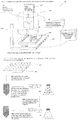
- Fig. 1 shows a schematic drawing of a system 101 for producing the cell-matrix microspheres.
- the system 101 comprises a dispensing unit 100, which consists of a manual or automatic dispenser 102, a control unit 104 controlling the volume of the dispensing liquid, the speed of dispensing and X-Y position of the dispenser, and a cooling unit 106 (or ice chamber) accommodating the dispensing unit 100 at around 4°C.
- the system also includes a collecting unit 108, which consists of a scalable collecting platform 110 with non-adhering surface, a moving X-Y-Z stage 112 to which the collecting platform is mounted, a plate control unit 114 controlling the speed, frequency and direction of movement of the moving stage.
- the system may be manual or automatic, custom-made, or modified from commercially available instruments such as liquid handlers.
- the method of forming microspheres via the system 101 comprises dispensing small liquid droplets such as 0.5 ⁇ l of mixtures containing the cells, the aqueous media and the liquid matrix, mixed in the right order of sequence and timing.
- the rate of matrix phase transition can be controlled by adjusting the temperature of the dispensing chamber side and is maintained slow by keeping the temperature low at the dispending side.
- the diameter of the droplets range from 0.5mm to 3mm, preferably 2mm.
- the liquid droplets collected at the collection platform are allowed to gel at 37°C for sufficiently long to form the solid cell-matrix microspheres.
- the microspheres are released from the collection platform and maintained free-floating in culture conditions until stable microspheres are formed.
- Figure 2 shows a method of producing cell-matrix microspheres with controllable size and mechanical strength.
- the size and mechanical strength of the microspheres are controlled by multiple parameters including, but not limited to, the volume of cell-matrix mixture, the cell density or cell number per microsphere, the matrix density, the ratio of different matrix composition, the serum concentration, the volume of the liquid droplets dispensed and the duration of free floating incubation of the cell-matrix microspheres.
- the method comprises Step 200: maintaining the gelled cell-matrix microspheres free floating or in suspension in static non-adhesive culture vessels, such as those for bacterial culture, or in spinning or rotating culture vessels, after releasing or detaching them from the non-adhesive collection platform 110, for sufficiently long period of time, ranging from 2 hours to 14 days. preferably 72 hours.
- Step 202 collecting the microspheres by flushing the microspheres with a medium on the collection platform 110.
- Step 203 involves allowing the microspheres to maintain for a period of time.
- Step 204 Examine the size of the microspheres until equilibrium is reached, i.e., the size of microspheres become constant.
- Step 206 collect the mechanically stable microspheres for injection or implantation or 3D cultures.
- the duration of free floating depends on the rate of the cell-matrix interaction before reaching equilibrium where the size of the microspheres becomes constant. The rate of interaction is thus cell type and matrix type dependent.
- the diameter of the cell-matrix microspheres at equilibrium should be appropriate for in vivo therapeutic injection, ranging from 50 to 800 microns, preferably 300 microns.
- Cell migration from microspheres can be manipulated by providing a mechanical support to the microspheres by plating them onto a solid substratum of culture dishes or placing them into a gelling matrix or onto a gelled matrix; plating the microspheres into a culture dish or gelling matrix or onto gelled matrix wherein each microsphere is kept at a distance from each other; adding a second medium into the culture system for holding the microspheres; allowing cells to migrate out from the microspheres for a period of time: and releasing the microspheres from the attached substratum and the second medium.
- the microspheres are typically held in the culture system for a period of time before the second medium is added, for example, 30, 60,90, or 120 minutes, preferably 60 minutes.
- the steps can be repeated several times to allow full migration of the cells. For example, the steps can be repeated 3, 5, 7, and preferably 10 times.
- the period of time for cell migration can range from 2, 4, 12, 48 hours to 12 days, preferably about 3 days.
- the cells that migrate out from the microspheres are allowed to grow for a period of time and can be harvested for future use or re-encapsulated.
- the culture system can be a 2D or 3D environment.
- the medium is preferably DMEM, DMEM-LG, MEM, or RPMI.
- the cell-matrix microspheres produced are mechanically stable and able to resist shear stress produced during rapid injection at a flow rate of as high as 20ml/min, are able to survive mechanical manipulation such as picking up and down by forceps, and are able to survive turbulence produced during vortexing at maximal speed.
- the collagen and other matrix provide the natural microenvironment stimulating growth of the cells, protect the cells from enzymatic digestion at the local hostile environment upon injection or implantation, immobilize soluble reagents, stimulating growth and differentiation if necessary, and the adhesive cell-matrix microspheres easily fuse together with the host tissue and fill any irregular gaps with the host tissue, therefore filling the tissue defects.
- the cell-matrix microspheres are permeable to free exchange of nutrients and metabolites. According to the rules of mass transfer in 3D tissue-like structures, the dimension of tissue-like structure is limited ( Muschler, et al., J. Bone Joint Surg.
- the size of the microspheres can be always controlled to as small as 100-300 microns, which is the suitable dimension of 3D structures with sufficient nutrient exchange.
- Another factor affecting the size of the microsphere is the matrix concentration. A higher concentration results in a stiffer matrix and therefore the traction force generated as the cells interact with the low concentration gelling matrix is not sufficient to produce a large extent of volume reduction.
- the encapsulated cells are able to migrate out the microspheres to integrate with the host tissue or grow rapidly when there is a close contact between the microspheres and the attached environment such as the host tissue thus may promote implants-host integration and engraftment rate.
- the cell-matrix microspheres can also be used to immobilize other therapeutic molecules, for example, factors inducing specific differentiation lineage such as Transforming Growth Factor (TGF) beta for chondrogenic differentiation, and anti-inflammatory drugs.
- TGF Transforming Growth Factor
- the method includes steps of controlling the amount of cells obtained and the speed of obtaining such number of cells in a predictable manner by factors including but are not limited to the plating density of the microspheres ranged from 0.5 to 500 preferably 5 microspheres per cm 2 ; cell density ranged from 1 to 2500 preferably 250 cells / microsphere; concentration of matrix such as collagen ranged from 0.1 to 5.0 preferably 0.5mg/ml; collagen to GAGs ratio ranged from 1:10 to 10:1 preferably 1:1; serum concentration ranged from 2 to 20 preferably 10% .
- An increasing plating density, an increasing cell density with a decreasing matrix concentration, a decreasing collagen to GAGs ratio and an increasing serum concentration result in an increasing number of cells obtained and an increasing speed of obtaining such number of cells.
- Step 300 involves production of mechanically stable microspheres according to the steps described in Fig. 1 and Fig. 2 .
- Step 301 involves providing a mechanical support for the microspheres to attach, in the absence of medium supplementation, for a period of time sufficient for attachment, 5 minutes to 60 minutes, half an hour to 6 hours, preferably 45 minutes, via Steps 302-304.
- Step 302 and 303 involves plating microspheres (302a) onto the solid substratum of a culture dish and a layer of collagen gel (303a), respectively.
- Step 304 involves suspending microspheres in a volume of gelling collagen matrix (304a) and cast the mixture into a culture dish for gelation by raising the temperature to 37°C.
- Step 305 involves adding full medium (305a) into the culture dishes without disturbing the attached microspheres.
- Step 306 involves allowing cell migration (306b) from the circumference of the microspheres for a period of time, from 12 hours to 3 days, from 2 days to 8 days, from 4 days to 14 days, preferably 3 days.
- Step 307 involves retrieval or releasing of the cell-matrix microspheres from the attached substratum by methods such as gentle flushing the substratum with full medium.
- Step 308 involved immediate supplementation of full medium to the culture dish where the migrated cells (308a) are allowed to continue to grow with regular medium change.
- Step 309 involves recycle of the detached microspheres for plating and cell outgrowths in steps 301-307 (not shown). The cells migrated can be harvested in Step 310 for future use.
- the method includes providing attachment to the floating microspheres by plating the contracted cell-matrix microspheres onto an adhesive substratum such as culture dishes in the absence of medium for certain period of time ranging from 30 minutes to 12 hours, preferably 45 minutes, until attachment is achieved.
- the method also includes steps of replenishing the microspheres with sufficient medium without disturbing the attached microspheres.
- the method includes steps of allowing cell outgrowth from the attached cell-matrix microspheres.
- the collagen matrix is permissive to cell migration. Together with the population pressure built up in the fully contracted cell-matrix microspheres, it results in cell outgrowth from the periphery of the attached microspheres onto the substratum or surrounding environment and the migrated cells proliferate. Cell outgrowth is induced for a certain period of time ranged from 12 hours to 14 days preferably 3 days.
- the method includes removing the attachment of the cell-matrix microspheres when sufficient number of cells migrate out from the microspheres that the microspheres are surrounded with cell outgrowths. This occurs at 12 hours to 14 days, preferably 3 days.
- the step does not need enzymatic digestion such as trypsinization, which may change the surface marker and cellular activities, and without mechanical disruption of the microspheres; but simply by flushing the microspheres gently with medium or PBS or by picking up with forceps.
- the cell-matrix microspheres are intact and are collected by sendimentation or mild centrifugation ranged from 800 to 2000rpm preferably 1000rpm.
- the cell-matrix microspheres can be replated in new empty culture vessels multiple times, up to 10 platings depending on the microsphere size, cell density per microsphere, so as to provide large number of cells in the same passage without changing the cell growth and differentiation potential as well as surface markers.
- the method can be used to provide cells on demand by maintaining the cell-matrix microspheres in suspension for at least one week if no cells are needed.
- the free-floating microspheres are provided with attachment in order to allow cell migration and outgrowth until confluence for future use when cells are needed.
- the method enables the constant supply of cells from the same passage in the cell-matrix microspheres by plating the microspheres for multiple times until the cell migration ceases.
- the migrated cells can be obtained at regular intervals, preferably daily, for a period of time ranging from 2 to 30 days, preferably 10 days, that is the period of time necessary for building multi-layered heterogenous tissue-like structure including, but are not limited to, the IVD, GI tract and blood vessels.
- the cell-matrix microspheres can be dissembled simply by enzymatic digestion specific to the matrix components of the microspheres, such as collagenase for collagen, chondroitinase for chondroitin sulfate GAGs.
- the single cell suspension can be used further.
- the natural extracellular matrix materials such as collagen used to encapsulate cells provide a physiologically relevant microenvironment in 3D to the encapsulated cell by forming cell-matrix microspheres.
- the tissue-like matrix microenvironment imposed on the encapsulated cells constrains cell proliferation.
- the cells are temporarily controlled for proliferation and the cell number in 3D microspheres increases only slightly higher than 2 fold on day 4 while cells in traditional monolayer cultures increase over 20 fold for the same period of time.
- the proliferation index for cells in 3D microspheres is consistently lower than that in traditional monolayer culture at different time points and for different cell densities. This method presents a naturally occurring and externally applied control for proliferation that no genetic manipulation on cell metabolism is needed.
- the methods and cell matrix microspheres are useful for treatment of cardiovascular diseases such as repair of myocardial infarction, for neurological diseases such as spinal cord injury and for musculoskeletal diseases such as cartilage injury, disc degeneration and muscular dystrophy.
- Services or products associated with stem cell therapies can also use the method and cell matrix microspheres to culture stem cells in 3D or in combination with monolayer cultures, to produce stem cells in large quantity and with a request-by-demand approach without changing their identity, self-renewal and differentiation capacity.
- the cell-matrix microspheres consisting of undifferentiated and viable cells can be injected or implanted, for example, for tissue repair or regeneration.
- the method includes the steps of sedimenting the microspheres for a period of time, preferably 10 minutes or centrifuging the microsphere suspensions mildly at a speed of 800-2000rpm, preferably 800rpm, for a period of time, preferably 5 minutes; removing the excess supernatant; re-suspending the microspheres in a liquid for injection or implantation such as saline or medium or phosphate buffered saline or a low concentration hydrogel such as collagen gel with a known volume; adding the microsphere suspension to a container for injection or implantation such as a syringe with G18-030 needles, preferably G27; and injecting or implanting the microspheres into defective tissues in animals or humans such as a skin wound or an injured cartilage.
- the method includes injecting or implanting the cell-matrix microspheres consisting of specifically differentiated and viable cells, wherein the matrix embeds these cells into defective tissues of animals or humans.
- the method comprises adding differentiation medium where chemical signals such as TGF-b is included in the microsphere suspensions for a period of time sufficient to induce chondrogenic differentiation of stem cells present in the microspheres into chondrogenic cells; allowing the microspheres with differentiated cells to sediment for a certain period of time, preferably 10 minutes, or centrifuging the microsphere suspensions mildly at a speed of 800-2000rpm, preferably 800rpm, for a period of time, preferably 5 minutes; removing the excess supernatant; re-suspending the microspheres in a liquid for injection or implantation such as saline or medium or phosphate buffered saline or a low concentration hydrogel such as collagen gel with a known volume; adding the microsphere suspension in the container for injection or implantation such as a syringe with G18-
- the cell-matrix microspheres can be used as long term sources for cells from the same passage without the need for cell splitting as conventionally used in both monolayer culture and seeding cells on 3D scaffolds such as using repeated enzymatic digestion and mechanical disruption.
- Cell-matrix microspheres can be used for 3D cultures as traditional microcarrier cultures used in biotechnology and pharmaceutical industries. They can be cultured as suspensions so as to easily increase the bed volume and scale up the culture system. Comparing with traditional microcarrier cultures, the method provides several important advantages in enhancing efficiency and reducing costs. Cells are immobilized within the gelling matrix at almost 100% encapsulation efficiency that almost all cells can be encapsulated. This encapsulation procedure takes only 30-60 minutes in static culture conditions.
- microcarrier culture system needs a time-consuming and inefficient cell binding procedure to allow cells to bind to the surface of the pre-fabricated microcarriers in suspension.
- complicated culture vessel design such as rotating vessel bioreactors, perfused bioreactors, spinning flasks and other methods such as constant agitation are needed.
- This cell binding step sometime may take more than I week. Since the method eliminates the necessity of these complicated and time-consuming steps, it presents a more efficient and less costly 3D culture method.
- the microencapsulation system combines the formation of the microspheres and the immobilization of cells in a matrix system into one step. This significantly reduces the cost and saves time.
- microencapsulation system is conducted in physiologically relevant temperature and conditions that assures high cell viability.
- residues of organic solvents and toxic chemical crosslinking reagent such as glutaraldehyde used during fabrication process of microcarriers may compromise cell viability.
- the cell matrix microspheres are also useful for 3D cultures. These have numerous advantages over the existing microcarrier systems.
- the system can be used to culture protein-secreting cells in physiologically relevant microenvironment with significantly increased protein productivity via matrix-induced proliferation control as compared to monolayer cultures with unrestricted proliferation.
- the productivity in 3D microspheres increases when the microspheres are cultured in low serum concentrations such as 2%. This indicates that the culture system can be maintained at low serum concentrations and the downstream purification of proteins can be simplified.
- the cells are cultured in culture vessels or bioreactors with or without constant agitation or spinning or rotation or perfusion.
- the secreted proteins in the culture medium are harvested with replacement at regular periods ranging from 1 day to 10 days, preferably 2 days, before reaching the maximal cell density inside the microspheres, for a period of time after microsphere suspension culture, ranging from 7 day to 3 months preferably 14 days.
- Wider windows for protein production can be achieved by encapsulating the cells in sub-optimal concentrations in the liquid matrix, ranging from 1x10 4 to 1x10 7 cells/ml, preferably at 1x10 5 cells/ml.
- the productivity of cells is significantly increased when cells proliferation inside the matrix microspheres is controlled. This window can be maintained for at least 2 weeks, much longer than the window with enhanced productivity in other controlled proliferation strategies.
- this method provides a temporary and reversible control for proliferation by encapsulating the cells in natural extracellular matrix materials at appropriate concentrations and time.
- the cell number increases linearly during the log phase.
- total protein production increased linearly with cell number and the productivity of specific proteins is reduced. This may be due to the fact that more energy is channeled to reproduction rather than protein production.
- Specific proteins produced during this phase can also be harvested. Cells will reach a maximal or optimal density due to the space limitation and nutrients competition, and the proliferation rate of the cells will be reduced. This is usually accompanied by an increase in protein production.
- cell apoptosis and death may be induced and the release of intracellular contents may degrade the secreted products. It is therefore advantageous to harvest the secreted proteins at appropriate time points during the optimization. Unlike other systems, this method allows separation of high protein productivity and rapid proliferation.
- This protein production phase is prior to and accompanied by a log proliferation phase. This system is therefore sustainable and typically ends with dramatically increased cell numbers.
- Efficiency and cost reduction of the culture system can be achieved by recycling the encapsulated cells after reaching the optimal cell density after a period of time ranging from 2 weeks to 3 months, preferably one month, via re-encapsulation. It is economically efficient because the protein harvest can start well before active proliferation phase while the output of the system includes not only the specific proteins produced but also increased cell numbers with high viability. Moreover, the cycle can be restarted by enzymatically releasing the cells and re-encapsulating into new matrix microspheres so that the cycle of matrix-induced proliferation control with high protein productivity and the subsequent active cell proliferation in 3D microspheres can be initiated again.
- Another advantage of this system is that, unlike traditional proliferation controlling technologies such as nutrients deprivation and over-expression of tumor suppressor genes, to control cell metabolism and to induce cell apoptosis or death, where high cell viability (almost 100%) is always maintained at different time points, with different cell densities and serum concentrations, proliferation of cells can be controlled directly, increasing protein productivity.
- the productivity of cells in 3D microspheres is always higher (ranging from 3 to 67 fold) than that from the monolayer cultures at different time points, with different cell densities and at different serum concentrations.
- Rat-tail collagen solution type I in acetic acid which mainly consists of triple helical monomers, was neutralized by NaOH and diluted into a final concentration of 0.5mg/ml. All procedures were done in an ice-bath to prevent collagen gel formation.
- a dispenser was then loaded with the ice-cold cell mixture and a small volume of 2.5 ⁇ l was dispensed at a time onto a bacterial culture dish covered with UV-irradiated parafilm. To prevent air-bubble formation in the liquid droplets.
- the dispenser was moved upwards or the collection platform downwards after dispensing the liquid droplets.
- the liquid droplets were thermally induced to reconstitute into a gel meshwork of organized collagen fibrils, interacting with the encapsulated cells, to form solid microspheres by incubating in a 37°C incubator for 1 hr.
- the cell-matrix microspheres formed were collected into a full medium containing bath with non-adherent substratum by gently flushing the parafilm with medium.
- Example 2 Production of mechanically stable cell-matrix microspheres and controlling parameters of the microsphere size.
- rat-tail collagen solution type I was neutralized and diluted into different concentrations (0.5, 1.0, 2.0 and 3.0mg/ml) in the presence of different concentrations (2x10 4 , 1x10 5 and 5x10 5 cells/ml) of human bone marrow derived MSCs in DMEM medium with 10% FBS as described in Example 1.
- the cell-matrix microspheres were collected into a DMEM medium containing bacterial culture dish. The collected microspheres were maintained at their free-floating state in the culture vessel at 37°C for 2 to 7 days until the equilibrium is reached as characterized by a constant microsphere size.
- the extent of hMSC-induced collagen microspheres contraction was directly proportional to the cell density, collagen concentration and droplet volume ( Figures 4A, 4B and 4C , respectively), establishing that that these parameters can be used to control the final size of the microspheres.
- the hMSC-collagen microspheres, after reaching the equilibrium, can be mechanically manipulated by forceps and are resistant to the shear stress and turbulence generated during pipetting up and down at rapid rate such as 20ml/min or even vortexed with maximal speed. As a result, these microspheres are mechanically stable enough to resist shear stress generated during microsyringe injection and are ready for injection and implantation for cell therapy and tissue engineering purposes.
- Example 3 Controlling the growth rate of encapsulated hMSCs inside the collagen microspheres.
- hMSCs were isolated from bone marrow aspirates from donors with informed consent in compliance with the Institute human ethics regulations. hMSCs were cultured as described by Li et al. 2004. Cells harvested from passage 2 and 3 by traditional monolayer culture were cryopreserved and used for production of cell-matrix microspheres with different collagen density: 0.5 and 2mg/ml. Fully contracted mechanically stable hMSCs-collagen microspheres were obtained as described in Examples 1 and 2. These microspheres were incubated with 2 ⁇ M Calcein AM and 4 ⁇ M Ethidium homodimer-1 for 45 minutes for simultaneous staining of live and dead cells.
- microspheres were fixed in 4% paraformaldehyde for 1 hour and examined using a laser confocal scanning microscope for stacked images.
- microspheres with 100 microspheres per plate in triplicates, were cultured in full medium for 10hours, 3, 6 and 9 days.
- the microspheres were digested with bacterial collagenase at 100U/ml for 45-80minutes at 37oC followed by digestion with 0.05% Trypsin/EDTA for 5 minutes. The single cell suspension obtained was counted for cell number and viability.
- Example 4 Controlling the migration and growth of encapsulated hMSCs from collagen microcapsules
- Free-floating hMSC-collagen microspheres with cell densities of 2x10 4 , 1x10, 5 or 5x10 5 cells/ml were transferred into 100mm diameter tissue culture plates at 3 days post-encapsulation, at plating densities of 63, 125 or 250 microspheres per plate corresponding to 0.64, 1.59 and 3.18 microspheres / cm 2 .
- the microspheres were allowed to attach to the culture plate for 1 hour after aspiration of excess medium and supplemented with full medium. At 72 hours, the microspheres were detached from the culture plates by gently flushing with full medium, allowed to sediment and replated in new culture plates several times until cell out-growing ceased.
- microspheres were seeded on collagen gel at 0.5mg/ml to evaluate whether cells would migrate into soft substratum.
- Cells growing out from the microspheres after different platings were cultured for 12 days with regular medium change while their morphology recorded at 3, 38 and 154hrs.
- the outgrowing cells from 250 microspheres were trypsinzed for cell count for comparison with the conventional monolayer cultures seeded with the same initial cell number.
- the detached microspheres could be replated several times without enzymatic digestion.
- Microspheres encapsulated with 250 cells could be plated for at least three times while those with 500 cells for at least six times.
- Microencapsulated hMSCs were also able to migrate into soft collagen gel. The viability of cells from different platings showed no difference. Microspheres aggregation and disintegration can be observed after 72 hours post-attachment. Outgrowing cells at 24 hours post-attachment penetrated into the collagen gel.
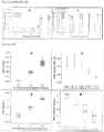
- Example 5 Maintenance of the surface marker of hMSCs outgrowing from the microspheres after multiple platings.
- hMSCs migrated out from microspheres after 10 platings were tryspinized with 0.05% trypsin in EDTA for 6-9 minutes and then fixed and labeled with antibodies against surface markers including CD34, CD14, CD29, CD105, CD45 and HLA.
- Flow cytometry was performed using appropriate isotype controls as described previously (Li et al. 2004).
- hMSCs showed the same surface markers panels, negative for CD14, CD34 and CD45, positive for CD105, CD29 and HLA-A,B,C, as that obtained from P1 of monolayer cultures. This indicates that the identity of the hMSCs was not changed even after 10 cycles of plating and replating of the hMSC-collagen microspheres. Since these cells are from a single aliquot of monolayer cultures of the same passage and these plating cycles can be repeated using another aliquot or aliquots from another passage from the monolayer culture, by combining this 3D culture system and traditional monolayer cultures, long term storage and rich sources for cells, which are necessary for cell-based assays and fabrication of tissue-like structures in tissue engineering can be achieved.
- Example 6 Maintenance of the self-renewal capacity of hMSCs outgrowing from the microspheres after multiple platings
- hMSCs obtained from the 3D and the monolayer cultures were seeded at very low density at 250 cells per 100mm diameter culture plate in triplicates and were cultured for 14 days with regular medium replenishment.
- the colonies formed were stained and fixed with 5% crystal violet (Sigma) in methanol for 10 minutes and rinsed with distilled water twice. The number of colonies with diameter greater than 2mm was counted and the single-cell-derived colony forming efficiency was calculated as the percentage of colony formed of all cells seeded.
- Example 7 Maintenance of the multiple differentiation potential of bMSCs outgrowing from the microspheres after multiple platings.
- Full medium was further supplemented with 100nM dexamethasone, 50 ⁇ M ascorbic acid 2-phosphate, and 10mM ⁇ -glycerophosphate as the oesteogenic differentiation induction medium.
- hMSCs were seeded at 3x 10 3 cells / cm 2 in 4 or 6 well plates in duplicates or triplicates and were maintained in the differentiation induction medium for 3 weeks with regular medium replacement every 3 days.
- cells were rinsed with PBS, fixed with 10% buffered formalin for 10 minutes at room temperature and stained with 5% silver nitrate (Nakarai Tesque, Kyoto, Japan) for von Kossa staining.
- Adipogenic differentiation induction medium was prepared by supplementing the full medium with 1 ⁇ M dexamethasone, 0.2mM indomethacin, 10 ⁇ g/ml insulin and 0.5mM 3-isobutyl-1-methylxanthine while the maintenance medium was prepared by supplementing the full medium with only 10 ⁇ g/ml insulin.
- hMSCs were seeded at 2x 10 4 cells / cm 2 in 4 or 6 well plates in duplicates or triplicates in full medium until confluence.
- Adipogenic differentiation induction medium was added for 3 days followed by 2 days in maintenance medium and three induction / maintenance cycles were performed. Cells were rinsed, fixed and then stained with 0.3% Oil-Red-O (Nakarai) for oil droplets staining.
- hMSCs Multiple differentiation potential of hMSCs was maintained after multiple cycles of plating.
- Example 8 Chondrogenic differentiation of hMSC in collagen microspheres
- hMSCs at a final concentration of 5x10 6 cells/ml was suspended in 100 ⁇ l of neutralized collagen solution (2mg/ml).
- Microspheres so prepared were incubated in chondrogenic differentiation medium for 3 weeks as described in Example 7.
- the differentiated microspheres were stained for cartilage-specific matrix markers as described in Example 7.
- Cartilage micro-tissues could be formed as the microencapsulated hMSCs were able to be chondrogenically differentiated into chondrocyte-like cells with typical round morphology. The differentiated cells lost their ability to migrate and produced cartilage-specific extracellular matrix as shown by the positive staining for glycosaminoglycans, aggrecan and type II collagen.
- Example 9 In vivo subcutaneous implantation of collagen-bMSC microspheres in NOD/SCID mice.
- microspheres remained intact and localized at the implantation site while the encapsulated cells were retained viable in NOD/SCID mice for at least 14 days.
- Clusters of microspheres encapsulating viable hMSCs which were stained with vital fluorescent dye, can be identified on day 2, 7 and 14 days post-implantation.
- Viable hMSCs exhibited their typical elongated morphology.
- Their human origin was confirmed by the immunopositive staining of human antigen beta2-microglobulin. Immunopositive staining for the human antigen was also found in cells, which are participating in the formation of blood vessels.
- Example 10 Extent of contraction of fibroblast-seeded collagen gel in the presence of glycosaminoglycans (GAGs).
- GAGs glycosaminoglycans
- Acidic rat-tail collagen solution was neutralized by NaOH and diluted into a final concentration of 0.5mg/ml.
- Glycosaminoglycans Chodroitin-6-sulfate
- MSCs Human bone marrow derived mesenchymal stem cells
- DMEM-LG with 10% FBS and 1% P/S was then suspended thoroughly in the neutralized collagen solution with GAGs as soon as possible to make a final density of 1 or 5x10 5 cells/ml.
- the mixture was cast in 4well culture plate and incubated in a 37°C incubator for 1 hr to allow for gelation.
- the gel was detached from the walls of the culture plate using a syringe needle and supplemented with sufficient medium.
- the size of the gel was measured under a dissection microscope at different time points to record the extent of contraction.
- fibroblasts-seeded gels contracted significantly over time due to the migration and proliferation of cells, as shown by Fig. 8A and 8B .
- the presence of a second matrix such as GAGs did affect the cellular responses in collagen gel.
- presence of GAGs reduced the extent of fibroblasts-induced gel contraction and was in a dose-dependent manner that a higher mass ratio of GAGs:Collagen resulted in a less extent of contraction, as demonstrated by comparing the results for 1 x 10 5 cells/ml in Fig. 8A with the results for 5 x 10 5 cells/ml in Fig. 8B .
- Example 11 HEK293 cell culture and encapsulation
- HEK293 cells (Passage 4) were transfected to over-express GDNF.
- Cells were cultured at 37°C with 5% CO 2 using T75 flask with 10ml complete Dulbecoo's Modified Eagle Medium - High Glucose (DMEM-HG. 2%, 5% or 10% FBS, 1% PS) and 500 ⁇ g/ml G418 Sulfate. Fresh medium and G418 sulfate were replaced every 2 days. These cells were used for the subsequent encapsulation.
- DMEM-HG. 2%, 5% or 10% FBS, 1% PS Dulbecoo's Modified Eagle Medium - High Glucose
- the HEK293 cells were trypsinized using 0.25% Trypsin-EDTA.
- Rat-tail collagen solution type I was neutralized by NaOH and diluted into a final concentration of 4mg/ml in the presence of HEK293 cells in DMEM.
- the cell mixture was kept at 4°C in an ice bath before use.
- the dispenser was loaded with the cell mixture and dispensed a small volume of 5 ⁇ l at a time onto a collection platform or a bacterial culture dish covered with UV-irradiated parafilm.
- the microdroplets were allowed to reconstitute into solid microspheres by incubating in a 37°C incubator with 5% CO 2 for 1 hr.
- the cell-matrix microspheres formed were collected into a DMEM medium containing bath with non-adherent substratum by gently flushing the parafilm with medium. Complete medium was used to suspend the cell-encapsulated capsules in 35mm petri dish. Cell proliferation, cell viability, GDNF productivity of HEK293 cells in 3D microspheres were compared with that cultured in traditional monolayer cultures.
- the cell-encapsulated capsules were viewed under an inverted microscope. Diameter of the capsules was measured at 40x magnification. The diameters of 5 out of 50 cell-encapsulated capsules in each set up were measured and the average value determined. The rate of contraction of the cell-encapsulated capsules depended on the cell-seeding density, as shown by Fig. 9 . With a higher cell-seeding density, the cell-encapsulated capsules contracted at an earlier time point and at a higher rate. Using the high cell-seeding density of 5000 cell/capsule, contraction began at day 4. The mean diameters were 2.42 mm ⁇ 0.05 and 1.77 mm ⁇ 0.07 on day 0 and day 12 respectively.
- the mean diameter was 2.4 mm ⁇ 0.01 on day 0. There were no contractions until day 8 and the mean diameter was 2.24 mm ⁇ 0.15 on day 12.
- the mean diameter of capsule was 2.4 mm ⁇ 0.04 on both day 0 and day 12. This showed that there were no observable contractions of the microspheres.
- the percentage changes of the mean diameters were (-)26.9%, (-)6.67% and 0% for cell-seeding density of 5000, 500 and 50 cell/microsphere respectively. Colonies or aggregates of cells started to form at day 6 and the size of the aggregates increased throughout day 8 to day 14.
- Example 12 Fate of encapsulated cells, viability and number of cells
- the HEK293 cells were trypsinized using 0.25% trypsin-EDTA. The viability tests were done using the trypan blue staining. The numbers of cells were counted using the hemacytometer. For 3D microspheres, collagenase (30 unit/ml) was used to digest the cell-encapsulated capsules. Trypsin/EDTA was then added into the colonies suspension and was incubated at 37°C with 5% CO 2 for 3 min to prepare single cell suspension. Viability test using trypan blue and cell counting were conducted.
- GDNF was measured using the GDNF E max ® ImmunoAssay System following the instructions provided by the manufacturer (Promega).
- the 96-well plates were coated with Anti-GDNF Monoclonal Antibody (mAb), which binds soluble GDNF, overnight and at 4°C without shaking.
- the captured GDNF is bound by Anti-Human GDNF polyclonal antibody (pAb, 1 ⁇ g/ml) and incubated without shaking at 4°C overnight. After washing, the amount of specifically bound pAb is then detected by incubating with Anti-Chicken IgY, horseradish peroxidase (HRP) conjugate for 2 hr at room temperature with regular agitation.
- HRP horseradish peroxidase
- the unbound conjugate is removed by washing, and followed by incubating with the TMB One Solution, a chromogenic substrate, for 15 min at room temperature without shaking.
- the reaction is stopped by adding 1N HCl.
- the absorbance at 450nm was measured using a microplate reader within 30 min after stopping the reaction.
- the amount of GDNF in the test solutions is proportional to the color generated in the oxidation-reduction reaction.
- This ELISA system can detect a minimum of 31.2 pg/ml of GDNF and the linear range is from 31pg/ml to 1000pg/ml GDNF.
- the HEK293 cells were trypsinized using 0.25% Trypsin-EDTA.
- 2ml complete medium DMEM, 10% FBS, 1% PS
- 500 ⁇ g/ml G418 sulfate DMEM, 10% FBS, 1% PS
- microspheres were formed as described above. The cell-seeding density was 500 cells/microsphere and the number of microsphere per 35mm petri dish was 50.
- the HEK293 cells were trypsinized using 0.25% Trypsin-EDTA.
- 2.5 x 10 3 , 2.5 x 10 4 or 2.5 x 10 5 HEK293 cells were seeded onto the 6-well plate with 2ml complete medium (DMEM, 10% FBS, 1% PS) and 500 ⁇ g/ml G418 sulfate.
- the microspheres were formed as stated in Example 2.
- the cell-seeding density was 50, 500 or 5000 cells/microsphere.
- Fifty microspheres were suspended in each 35mm petri dish with 2ml complete medium (DMEM, 10% FBS, 1% PS) and 500 ⁇ g/ml G418 sulfate.
- the HEK293 cells were trypsinized using 0.25% trypsin-EDTA.
- 2.5 x 10 4 HEK293 cells were seeded onto the 6.well plate with 2ml medium having different serum percentage (2%, 5%, 10%) and 500 ⁇ g/ml G418 sulfate.
- microspheres were formed as stated in Example 2.
- the cell-seeding density was 500 cell/microspheres and there were 50 microspheres per 35mm petri dish equivalent to an initial cell number of 2.5 x 10 4 cells.
- the microspheres were suspended in 2ml medium having different serum percentage (2%, 5%, 10%) and 500 ⁇ g/ml G418 sulfate.
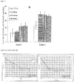
- Fig. 11A and 11B The cell viability and number of HEK293 cells in both monolayer and 3D microspheres at each time point (Day 2, 4, 8, 10, 14, 18, 22, 26 amd 30) are shown in Fig. 11A and 11B , respectively.
- the cell number of the monolayer group was always higher than that of the microsphere group and there was a significant difference between them. Apart from an initial lower cell viability of around 80% in 3D microspheres, the cell viability of all groups in all subsequent time points was close to 100%.
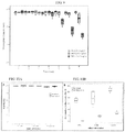
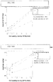
- the accumulative secretion of GDNF was linearly proportional to time in both the monolayer group and the microsphere group ( Figure 12 ). There was a significant difference in accumulative secretion of GDNF between monolayer group and microsphere group.
- the total GDNF secretion of both monolayer and microsphere groups were linearly proportional to the cell-seeding density ( Figure 12C ). There was a significant difference in total GDNF secretion between the monolayer and microsphere groups, and among different cell densities. The total GDNF secretion of the monolayer group is higher than that of the microsphere group, due to the large difference in cell number. At each cell-seeding density, the number of cell in monolayer group is about 7 - 20 times higher than the number of cell in microsphere group at day 12. However, the secretion rate of GDNF from HEK293 cells was found to be higher than that from monolayer cultures in all groups ( Fig. 12D ) and the difference was statistically significant between monolayers and microsphere cultures and among groups with different initial cell numbers.
- the total GDNF secreted from day 0 to day 12 was linearly proportional to the serum percentage in medium in both monolayer and microsphere groups ( Figure 12E ). There were significant differences in total GDNF secretion between the monolayer and microsphere groups and among different serum percentages in medium. The total ODNF secretion in monolayer group was always higher than that in microsphere group and the differences were less than 2 folds at all serum percentages. There was no interaction between group and serum percentage in medium. However, the secretion rate of GDNF from HEK293 cells per million cells per day ( Fig. 12F ) was significantly higher than that from monolayer cultures at all serum percentages.
- GDNF secretion rate in 3D microspheres was more than 4 folds in all serum concentrations indicating that the enhanced productivity of HEK293 cells in 3D microspheres was independent of the serum percentage. It also demonstrated that the encapsulated cells secreted the highest rate of GDNF when they were sustained with medium with 2% scrum.
- the cell viability and number of HEK293 cells in microspheres cultured with different serum concentration are shown in Fig. 13A and 13B , respectively and the viability in all serum concentrations showed almost 100% ( Fig. 13A ) indicating that the cell viability was not significantly affected by reducing the serum concentration, as a result, reducing serum concentration can be used to ease the downstream purification steps of the secreted proteins.
- Example 14 Bioactivity assay of GDNF in conditioned medium of HEK293 cells
- PC 12 cells were grown on 24-well plates in 81.5% F12K medium, supplemented with 15% House serum, 2.5% Fetal bovine serum and 1% PS.
- Conditioned medium containing the secreted GDNF was mixed with the full medium for PC 12 culture at 1:1 volume ratio and used to culture the PC12 cells.
- the cells were plated at a density of approximately 3000 cells (in 800 ul) per well. After 2 days, the cells were fixed for visualization under the phase microscope. Cells with neurite outgrowth longer than one body length of the cell were regarded as positive results. Standard GDNF with known concentrations were also used as positive controls. Full medium without conditioned medium was used as negative control.
- Example 15 Bioactivity assay of GDNF in conditioned medium of HEK293 cells
- GDNF-secreting HEK293 cells were encapsulated in 3D collagen microspheres and cultured for 14 days. The microspheres were fixed in 4% paraformaldehyde for 3 hours followed by 30% sucrose solution for overnight before cryosections of 10 ⁇ m thick were prepared. Immunohistochemistry of GDNF was conducted to confirm its secretion using a primary antibody, chicken anti-human GDNF polyclonal antibody (Promega) at 1:100-1:50 dilution and a secondary antibody, rabbit anti-chicken IgY (Promega) at 1:100-1:50 dilution. HRP-DAB substrate system was used to visualize the immuno-positively stained GDNF inside the microspheres. The synthesis of GDNF was confirmed by immunohistochemistry. Immunopositive staining of GDNF was localized at the cell colonies inside the 3D microspheres.
- DMEM chondrogenic differentiation medium
- DMEM high glucose, 10ng/ml recombinant human transforming growth factor beta 3 (Merck), 100nM dexamethasone (Sigma), 6 ⁇ g/ml insulin (Merck), 100mM 2-phospho-L-ascorbate (Fluka), 1mM sodium pyruvate (Gibco), 6 ⁇ g/ml transferrin (Sigma), 0.35mM L-proline (Merck) and 1.25mg/ml bovine serum albumin (Sigma)).
- DMEM high glucose, 10ng/ml recombinant human transforming growth factor beta 3 (Merck), 100nM dexamethasone (Sigma), 6 ⁇ g/ml insulin (Merck), 100mM 2-phospho-L-ascorbate (Fluka), 1mM sodium pyruvate (Gibco), 6 ⁇ g/ml transferrin (Sigma), 0.35mM L-proline (Merck) and
- the microspheres were cultured for 21 days, with medium changed every two days. After 21 days of culture, the samples were either processed for immunohistochemistry and histology for qualitative analysis or digested in papain solution (300 ⁇ g/ml papain in 50mM PB, containing 5mM L-cysteine and 5mM EDTA) at 60°C overnight for quantitative analysis of chondrogenesis. 1,9-Dimethylmethylene blue (DMMB) assay was used for GAG quantification. In brief, 1ml of DMMB dye solution was added to the sample digest in a tube and the content was mixed on a shaker for 30 minutes. The tube was centrifuged at 13.2k rpm for 10minutes to form the GAG-dye complex precipitate.
- papain solution 300 ⁇ g/ml papain in 50mM PB, containing 5mM L-cysteine and 5mM EDTA
- 1,9-Dimethylmethylene blue (DMMB) assay was used for GAG quantification.
- the precipitate was resuspended in 200 ⁇ l of dissociation reagent and mixed by vortex and the absorbance at 656nm was measured. From the digestion mixture, DNA content was also quantified by a fluorometric assay. In brief, 100 ⁇ l of Hoechst 33258 dye solution was added to the sample digest and fluorescence measurement was made with excitation and emission at 365nm and 458nm respectively. The GAGs content was then normalized by DNA content.
- the amount of GAGs deposited into the collagen microspheres increased with increasing cell seeding density and collagen concentration, as shown in Fig. 14A .
- Statistical analysis shows that the difference among the four cell seeding density groups and among collagen density groups are all significant (p ⁇ 0.05).
- the amount of GAG/DNA also increased with increasing cell seeding density and collagen concentration, as demonstrated by Fig. 14B .
- Statistical analysis also shows significant difference among various groups (p ⁇ 0.05).
Landscapes
- Health & Medical Sciences (AREA)
- Engineering & Computer Science (AREA)
- Life Sciences & Earth Sciences (AREA)
- Bioinformatics & Cheminformatics (AREA)
- Chemical & Material Sciences (AREA)
- Organic Chemistry (AREA)
- Biomedical Technology (AREA)
- Wood Science & Technology (AREA)
- Zoology (AREA)
- Biotechnology (AREA)
- Genetics & Genomics (AREA)
- General Health & Medical Sciences (AREA)
- Biochemistry (AREA)
- General Engineering & Computer Science (AREA)
- Microbiology (AREA)
- Cell Biology (AREA)
- Rheumatology (AREA)
- Immunology (AREA)
- Developmental Biology & Embryology (AREA)
- Pharmacology & Pharmacy (AREA)
- Medicinal Chemistry (AREA)
- Chemical Kinetics & Catalysis (AREA)
- General Chemical & Material Sciences (AREA)
- Nuclear Medicine, Radiotherapy & Molecular Imaging (AREA)
- Veterinary Medicine (AREA)
- Public Health (AREA)
- Animal Behavior & Ethology (AREA)
- Sustainable Development (AREA)
- Hematology (AREA)
- Physical Education & Sports Medicine (AREA)
- Urology & Nephrology (AREA)
- Heart & Thoracic Surgery (AREA)
- Vascular Medicine (AREA)
- Cardiology (AREA)
- Neurology (AREA)
- Orthopedic Medicine & Surgery (AREA)
- Micro-Organisms Or Cultivation Processes Thereof (AREA)
- Medicines Containing Material From Animals Or Micro-Organisms (AREA)
Claims (3)
- Procédé de production de microsphères, comprenant mélanger une première matrice extracellulaire (ECM) en combinaison avec des cellules à une température de 0°C à 10°C pour répartir les cellules à travers la forme de solution de la matrice avant d'accélérer une transition de phase,
distribuer le mélange ECM cellulaire pour former des gouttelettes sur une surface ayant une tension de surface élevée pour maintenir la forme sphérique des gouttelettes,
accélérer la transition de phase ou la gélification de l'ECM pour former les microsphères à matrice cellulaire;
collecter les microsphères à partir de la plate-forme de collection avec une perturbation mécanique minimale;
stabiliser les microsphères après la transition de phase en les plaçant en flottation libre dans un premier milieu
maintenir les microsphères en flottation libre dans un premier milieu pendant au moins 3 jours, tel que de 3 jours à 14 jours, jusqu'à ce que la taille des microsphères devienne sensiblement constante; et
libérer les microsphères du premier milieu. - Procédé selon la revendication 1, comprenant la distribution des microsphères à matrice cellulaire sur une plate-forme de collection; et comprenant soit
augmenter la température de la plate-forme de collection à 25-37 °C pour accélérer la transition de phase pendant une période de temps suffisante pour la gélification de l'ECM mais insuffisante pour le changement morphologique cellulaire; ou
dans lequel la vitesse de gélification de la matrice après distribution est ralentie immédiatement après l'initiation de la gélification en maintenant la température des mélanges aussi basse que 4°C; ou
dans lequel la gélification de la matrice est accélérée immédiatement après distribution de la matrice de gélification en gouttelettes sur la plate-forme de collection en augmentant la température du mélange à 37°C. - Procédé selon la revendication 1, dans lequel les microsphères sont maintenues en flottation libre pendant environ 3 à environ 7 jours à une température d'environ 37°C.
Applications Claiming Priority (2)
| Application Number | Priority Date | Filing Date | Title |
|---|---|---|---|
| US80197506P | 2006-05-19 | 2006-05-19 | |
| PCT/IB2007/004413 WO2008075206A2 (fr) | 2006-05-19 | 2007-05-18 | Microsphères à matrice cellulaire, procédés de préparation et applications |
Publications (3)
| Publication Number | Publication Date |
|---|---|
| EP2021035A2 EP2021035A2 (fr) | 2009-02-11 |
| EP2021035A4 EP2021035A4 (fr) | 2010-08-25 |
| EP2021035B1 true EP2021035B1 (fr) | 2020-08-05 |
Family
ID=39536803
Family Applications (1)
| Application Number | Title | Priority Date | Filing Date |
|---|---|---|---|
| EP07872058.8A Active EP2021035B1 (fr) | 2006-05-19 | 2007-05-18 | Microsphères à matrice cellulaire, procédés de préparation et applications |
Country Status (4)
| Country | Link |
|---|---|
| US (2) | US8679809B2 (fr) |
| EP (1) | EP2021035B1 (fr) |
| CN (1) | CN101448527B (fr) |
| WO (1) | WO2008075206A2 (fr) |
Families Citing this family (50)
| Publication number | Priority date | Publication date | Assignee | Title |
|---|---|---|---|---|
| EP1807506B1 (fr) * | 2004-10-08 | 2013-04-17 | Georgia Tech Research Corporation | Microencapsulation de cellules dans des hydrogels a l'aide de potentiels electrostatiques |
| US8198083B1 (en) | 2007-10-31 | 2012-06-12 | William Gunter Loudon | Organotypic slices of the central nervous system |
| AU2009266859A1 (en) * | 2008-07-02 | 2010-01-07 | Allergan, Inc. | Compositions and methods for tissue filling and regeneration |
| MX2011001730A (es) | 2008-08-14 | 2011-03-24 | Osiris Therapeutics Inc | Composiciones purificadas de celulas madre mesenquimales y metodos para purificar composiciones de celulas madre mesenquimales. |
| US8926552B2 (en) | 2009-08-12 | 2015-01-06 | Medtronic, Inc. | Particle delivery |
| US8883210B1 (en) | 2010-05-14 | 2014-11-11 | Musculoskeletal Transplant Foundation | Tissue-derived tissuegenic implants, and methods of fabricating and using same |
| US10130736B1 (en) | 2010-05-14 | 2018-11-20 | Musculoskeletal Transplant Foundation | Tissue-derived tissuegenic implants, and methods of fabricating and using same |
| US9352003B1 (en) | 2010-05-14 | 2016-05-31 | Musculoskeletal Transplant Foundation | Tissue-derived tissuegenic implants, and methods of fabricating and using same |
| US11786636B2 (en) * | 2010-06-15 | 2023-10-17 | Versitech Limited | Methods for complex tissue engineering |
| US9938501B2 (en) * | 2010-07-28 | 2018-04-10 | Jointechlabs, Inc. | Encapsulated adipose-derived stem cells, methods for preparation and theraputic use |
| US8834928B1 (en) | 2011-05-16 | 2014-09-16 | Musculoskeletal Transplant Foundation | Tissue-derived tissugenic implants, and methods of fabricating and using same |
| EP2970904A1 (fr) * | 2013-03-15 | 2016-01-20 | Discgenics, Inc. | Cellules discogéniques isolées, procédés d'utilisation et procédés de préparation de celles-ci à partir d'un tissu de mammifère |
| CA2919374C (fr) | 2013-07-30 | 2019-12-03 | Musculoskeletal Transplant Foundation | Matrices derivees de tissu mou acellulaire et leurs procedes de preparation |
| EP3685884A1 (fr) | 2013-09-19 | 2020-07-29 | The Board of Trustees of the Leland Stanford Junior University | Procédés et compositions pour la production de cellules de type hépatocytes |
| US10533191B2 (en) | 2014-01-15 | 2020-01-14 | Serucell Corporation | Therapeutic serum obtained from co-cultured cells |
| CA2937801C (fr) * | 2014-01-23 | 2023-04-04 | Nissan Chemical Industries, Ltd. | Composition de vehicule de culture liquide comprenant de la chitine dispersee ou des nanofibres de chitosane |
| CN103816570A (zh) * | 2014-01-28 | 2014-05-28 | 浙江大学 | 一种丝素蛋白/干细胞复合微球及应用 |
| CN104263632B (zh) * | 2014-09-15 | 2016-06-29 | 中国科学院南海海洋研究所 | 一种微生物包埋微球连续制备装置及方法 |
| CN104436306B (zh) * | 2014-11-11 | 2017-07-25 | 四川大学 | 一种细胞‑凝胶材料复合微球及其制备方法和应用 |
| CN104450605A (zh) * | 2014-11-26 | 2015-03-25 | 荆东辉 | 一种液体微胶囊包埋、分化全能干细胞成心肌细胞的方法 |
| US9238090B1 (en) | 2014-12-24 | 2016-01-19 | Fettech, Llc | Tissue-based compositions |
| CA2986702C (fr) | 2015-05-21 | 2023-04-04 | David Wang | Fibres osseuses corticales demineralisees modifiees |
| US10912864B2 (en) | 2015-07-24 | 2021-02-09 | Musculoskeletal Transplant Foundation | Acellular soft tissue-derived matrices and methods for preparing same |
| US11052175B2 (en) | 2015-08-19 | 2021-07-06 | Musculoskeletal Transplant Foundation | Cartilage-derived implants and methods of making and using same |
| EP3150696A1 (fr) * | 2015-09-30 | 2017-04-05 | Shibuya Corporation | Appareil de production de feuille de cellules en série et procédé de production de feuille de cellules en série |
| JP6639634B2 (ja) * | 2016-02-22 | 2020-02-05 | 国立大学法人大阪大学 | 立体的細胞組織の製造方法 |
| JP7044330B2 (ja) * | 2016-06-17 | 2022-03-30 | 国立大学法人横浜国立大学 | 毛髪再生用細胞包埋ビーズ及びその製造方法、並びに毛髪再生用キット |
| CN106492288A (zh) * | 2016-10-31 | 2017-03-15 | 广州昕生医学材料有限公司 | 可注射的脱细胞脂肪基质微粒及其在植入剂中的应用 |
| CN106492285A (zh) * | 2016-10-31 | 2017-03-15 | 广州昕生医学材料有限公司 | 可注射的脱细胞软骨基质微粒及其在植入剂中的应用 |
| CN108274734B (zh) * | 2017-12-12 | 2020-04-03 | 上海大学 | 一种具有可控芯壳结构的体外3d肿瘤微球模型制造系统及方法 |
| CA3093069A1 (fr) * | 2018-03-06 | 2019-09-12 | Epibone, Inc. | Compositions de reparation des cartilages, tendons et ligaments en vente libre, et leurs procedes d'utilisation |
| CN108802410B (zh) * | 2018-03-22 | 2021-07-02 | 广州市第一人民医院(广州消化疾病中心、广州医科大学附属市一人民医院、华南理工大学附属第二医院) | 一种液滴芯片细胞分析系统及其分析方法 |
| CN108853585B (zh) * | 2018-08-01 | 2019-09-27 | 北京大学 | 多孔微支架制作方法及在组织再生修复中的应用 |
| CN109125806A (zh) * | 2018-08-29 | 2019-01-04 | 广东克瑞斯普生物科技有限公司 | 一种皮下注射用干细胞微球凝胶复合物及其应用 |
| CN109529106B (zh) * | 2018-11-14 | 2021-09-10 | 山东隽秀生物科技股份有限公司 | 一种慢性创面修复基质的制备方法 |
| CN110283777B (zh) * | 2019-07-10 | 2020-12-29 | 中国海洋大学 | 一种对虾细胞连续培养方法 |
| CN110468096A (zh) * | 2019-07-31 | 2019-11-19 | 同济大学 | 一种牙周膜细胞微球的制备方法 |
| US20230047254A1 (en) * | 2019-12-29 | 2023-02-16 | Gloriana Therapeutics | Mammalian Cells Secreting GDNF and Their Therapeutic Use |
| CN111269882B (zh) * | 2020-02-02 | 2022-01-04 | 中山大学附属第一医院 | 种植体的表面处理方法及仿生种植体 |
| CN111518755A (zh) * | 2020-05-09 | 2020-08-11 | 苏州大学 | 一种仿生骨膜、骨膜-骨替代物及制备方法 |
| WO2021237136A2 (fr) * | 2020-05-22 | 2021-11-25 | Georgia Tech Research Corporation | Organoïdes invertis stabilisés et leurs procédés de production et d'utilisation |
| CN112294846B (zh) * | 2020-10-22 | 2023-03-21 | 清华大学深圳国际研究生院 | 干细胞微球及其应用 |
| CN113201525B (zh) * | 2021-04-12 | 2023-03-21 | 清华大学深圳国际研究生院 | 干细胞微球组及干细胞的体外扩增方法和应用 |
| CN113509594B (zh) * | 2021-05-28 | 2022-05-10 | 四川大学 | 诱导msc细胞向软骨方向分化的仿生材料、制备方法及应用 |
| CN114292743A (zh) * | 2021-11-25 | 2022-04-08 | 浙江大学 | 一种电场辅助下的打印装置、水凝胶微球的制备方法及应用 |
| WO2023129514A2 (fr) * | 2021-12-28 | 2023-07-06 | The Texas A&M University System | Récupération d'échantillons biologiques environnementaux après encapsulation dans du gel à l'intérieur de gouttelettes |
| CN114931562B (zh) * | 2022-05-05 | 2023-11-07 | 大连工业大学 | 一种基于不溶性膳食纤维的微胶囊递送系统 |
| CN115322953A (zh) * | 2022-08-15 | 2022-11-11 | 中国医学科学院生物医学工程研究所 | 一种用于细胞三维培养的高效接种方法及其应用 |
| CN115887762B (zh) * | 2022-10-11 | 2024-03-15 | 苏州大学 | 一种微流控静电纺纤维膜微球及其制备方法与在血管修复方面的应用 |
| CN117482292A (zh) * | 2023-09-15 | 2024-02-02 | 北京帝康医药投资管理有限公司 | 一种胶原蛋白多孔微球及其制备方法和应用 |
Family Cites Families (10)
| Publication number | Priority date | Publication date | Assignee | Title |
|---|---|---|---|---|
| EP0691400B1 (fr) * | 1993-12-30 | 2002-03-06 | Nitta Gelatin Inc. | Procedes permettant d'enrober des cultures de cellules animales |
| US5916790A (en) * | 1995-03-03 | 1999-06-29 | Metabolex, Inc. | Encapsulation compositions, and methods |
| US5965125A (en) * | 1995-10-25 | 1999-10-12 | Transkaryotic Therapies, Inc. | Hybrid matrix implants and explants |
| US6274341B1 (en) | 1997-10-09 | 2001-08-14 | James E. Bailey | Cytostatic process increases the productivity of cultured cells |
| US6197061B1 (en) * | 1999-03-01 | 2001-03-06 | Koichi Masuda | In vitro production of transplantable cartilage tissue cohesive cartilage produced thereby, and method for the surgical repair of cartilage damage |
| US6916640B2 (en) * | 2000-10-12 | 2005-07-12 | Agency For Science, Technology And Research | Multi-layer cell encapsulation for tissue engineering |
| AUPR289601A0 (en) * | 2001-02-05 | 2001-03-01 | Commonwealth Scientific And Industrial Research Organisation | Method of tissue repair |
| US20040054414A1 (en) * | 2002-09-18 | 2004-03-18 | Trieu Hai H. | Collagen-based materials and methods for augmenting intervertebral discs |
| CN1609200A (zh) * | 2004-11-19 | 2005-04-27 | 清华大学 | 一种复杂组织器官前体的制备方法 |
| EP2019653B1 (fr) * | 2006-04-28 | 2016-11-16 | The University of Hong Kong | Disques intervertébraux mis au point par génie biologique et procédés pour leur préparation |
-
2007
- 2007-05-18 US US11/750,863 patent/US8679809B2/en active Active
- 2007-05-18 WO PCT/IB2007/004413 patent/WO2008075206A2/fr active Application Filing
- 2007-05-18 EP EP07872058.8A patent/EP2021035B1/fr active Active
- 2007-05-18 CN CN2007800182968A patent/CN101448527B/zh active Active
-
2014
- 2014-02-18 US US14/183,260 patent/US9464271B2/en active Active
Non-Patent Citations (1)
| Title |
|---|
| None * |
Also Published As
| Publication number | Publication date |
|---|---|
| EP2021035A4 (fr) | 2010-08-25 |
| WO2008075206A3 (fr) | 2008-10-30 |
| CN101448527A (zh) | 2009-06-03 |
| US20080031858A1 (en) | 2008-02-07 |
| US20140170745A1 (en) | 2014-06-19 |
| EP2021035A2 (fr) | 2009-02-11 |
| WO2008075206A2 (fr) | 2008-06-26 |
| US9464271B2 (en) | 2016-10-11 |
| US8679809B2 (en) | 2014-03-25 |
| CN101448527B (zh) | 2013-12-18 |
Similar Documents
| Publication | Publication Date | Title |
|---|---|---|
| EP2021035B1 (fr) | Microsphères à matrice cellulaire, procédés de préparation et applications | |
| Chen et al. | Exosomes derived from mature chondrocytes facilitate subcutaneous stable ectopic chondrogenesis of cartilage progenitor cells | |
| Yoon et al. | Enhanced cartilage formation via three-dimensional cell engineering of human adipose-derived stem cells | |
| Solorio et al. | Spatiotemporal regulation of chondrogenic differentiation with controlled delivery of transforming growth factor-β1 from gelatin microspheres in mesenchymal stem cell aggregates | |
| Ho et al. | Cell migration and bone formation from mesenchymal stem cell spheroids in alginate hydrogels are regulated by adhesive ligand density | |
| Awad et al. | Chondrogenic differentiation of adipose-derived adult stem cells in agarose, alginate, and gelatin scaffolds | |
| Meinel et al. | Bone tissue engineering using human mesenchymal stem cells: effects of scaffold material and medium flow | |
| Xie et al. | In vitro mesenchymal trilineage differentiation and extracellular matrix production by adipose and bone marrow derived adult equine multipotent stromal cells on a collagen scaffold | |
| Galateanu et al. | Layer-shaped alginate hydrogels enhance the biological performance of human adipose-derived stem cells | |
| Wong et al. | A 3D collagen microsphere culture system for GDNF-secreting HEK293 cells with enhanced protein productivity | |
| Griffon et al. | A comparative study of seeding techniques and three‐dimensional matrices for mesenchymal cell attachment | |
| WO2010087397A1 (fr) | Procédé de fabrication de tissu artificiel stratifié cultivé à haute densité et tissu artificiel stratifié cultivé à haute densité | |
| Yao et al. | Injectable cell/hydrogel microspheres induce the formation of fat lobule-like microtissues and vascularized adipose tissue regeneration | |
| US20120225483A1 (en) | Microsphere-Containing Cell Aggregate | |
| Wang et al. | Adipose tissue engineering with human adipose tissue‐derived adult stem cells and a novel porous scaffold | |
| JP6434014B2 (ja) | 球状軟骨細胞治療剤の製造方法 | |
| Wang et al. | Role of N-cadherin in a niche-mimicking microenvironment for chondrogenesis of mesenchymal stem cells in vitro | |
| US10619133B2 (en) | Engineered living tissue substitute | |
| Zhu et al. | Enhancement of adipose-derived stem cell differentiation in scaffolds with IGF-I gene impregnation under dynamic microenvironment | |
| Surguchenko et al. | The cell‐engineered construct of cartilage on the basis of biopolymer hydrogel matrix and human adipose tissue‐derived mesenchymal stromal cells (in vitro study) | |
| Wang et al. | Adult stem cells and hydrogels for cartilage regeneration | |
| EP2582410B1 (fr) | Procédés pour produire des tissus complexes par génie biologique | |
| Shen et al. | Boosting cartilage repair with silk fibroin-DNA hydrogel-based cartilage organoid precursor | |
| CN113383068A (zh) | 可植入性构建体及其制造方法和用途 | |
| Gamboa‐Martínez et al. | Fibrin‐chitosan composite substrate for in vitro culture of chondrocytes |
Legal Events
| Date | Code | Title | Description |
|---|---|---|---|
| PUAI | Public reference made under article 153(3) epc to a published international application that has entered the european phase |
Free format text: ORIGINAL CODE: 0009012 |
|
| 17P | Request for examination filed |
Effective date: 20081210 |
|
| AK | Designated contracting states |
Kind code of ref document: A2 Designated state(s): AT BE BG CH CY CZ DE DK EE ES FI FR GB GR HU IE IS IT LI LT LU LV MC MT NL PL PT RO SE SI SK TR |
|
| AX | Request for extension of the european patent |
Extension state: AL BA HR MK RS |
|
| DAX | Request for extension of the european patent (deleted) | ||
| A4 | Supplementary search report drawn up and despatched |
Effective date: 20100728 |
|
| 17Q | First examination report despatched |
Effective date: 20110914 |
|
| APBK | Appeal reference recorded |
Free format text: ORIGINAL CODE: EPIDOSNREFNE |
|
| APBN | Date of receipt of notice of appeal recorded |
Free format text: ORIGINAL CODE: EPIDOSNNOA2E |
|
| APBR | Date of receipt of statement of grounds of appeal recorded |
Free format text: ORIGINAL CODE: EPIDOSNNOA3E |
|
| APBV | Interlocutory revision of appeal recorded |
Free format text: ORIGINAL CODE: EPIDOSNIRAPE |
|
| APAV | Appeal reference deleted |
Free format text: ORIGINAL CODE: EPIDOSDREFNE |
|
| RAP1 | Party data changed (applicant data changed or rights of an application transferred) |
Owner name: VERSITECH LIMITED |
|
| APBT | Appeal procedure closed |
Free format text: ORIGINAL CODE: EPIDOSNNOA9E |
|
| RIC1 | Information provided on ipc code assigned before grant |
Ipc: C12N 5/0775 20100101ALI20191216BHEP Ipc: A61K 9/16 20060101ALI20191216BHEP Ipc: A61K 48/00 20060101AFI20191216BHEP Ipc: A61K 35/12 20150101ALI20191216BHEP Ipc: A61P 19/00 20060101ALI20191216BHEP Ipc: A61P 9/10 20060101ALI20191216BHEP Ipc: C12N 5/07 20100101ALI20191216BHEP Ipc: A61K 38/16 20060101ALI20191216BHEP Ipc: A61K 38/39 20060101ALI20191216BHEP Ipc: A61P 21/00 20060101ALI20191216BHEP Ipc: C12N 5/077 20100101ALI20191216BHEP |
|
| GRAP | Despatch of communication of intention to grant a patent |
Free format text: ORIGINAL CODE: EPIDOSNIGR1 |
|
| STAA | Information on the status of an ep patent application or granted ep patent |
Free format text: STATUS: GRANT OF PATENT IS INTENDED |
|
| INTG | Intention to grant announced |
Effective date: 20200213 |
|
| GRAS | Grant fee paid |
Free format text: ORIGINAL CODE: EPIDOSNIGR3 |
|
| GRAA | (expected) grant |
Free format text: ORIGINAL CODE: 0009210 |
|
| STAA | Information on the status of an ep patent application or granted ep patent |
Free format text: STATUS: THE PATENT HAS BEEN GRANTED |
|
| AK | Designated contracting states |
Kind code of ref document: B1 Designated state(s): AT BE BG CH CY CZ DE DK EE ES FI FR GB GR HU IE IS IT LI LT LU LV MC MT NL PL PT RO SE SI SK TR |
|
| REG | Reference to a national code |
Ref country code: GB Ref legal event code: FG4D |
|
| REG | Reference to a national code |
Ref country code: CH Ref legal event code: EP |
|
| REG | Reference to a national code |
Ref country code: AT Ref legal event code: REF Ref document number: 1297789 Country of ref document: AT Kind code of ref document: T Effective date: 20200815 |
|
| REG | Reference to a national code |
Ref country code: DE Ref legal event code: R096 Ref document number: 602007060521 Country of ref document: DE |
|
| REG | Reference to a national code |
Ref country code: IE Ref legal event code: FG4D |
|
| REG | Reference to a national code |
Ref country code: LT Ref legal event code: MG4D |
|
| REG | Reference to a national code |
Ref country code: NL Ref legal event code: MP Effective date: 20200805 |
|
| REG | Reference to a national code |
Ref country code: AT Ref legal event code: MK05 Ref document number: 1297789 Country of ref document: AT Kind code of ref document: T Effective date: 20200805 |
|
| PG25 | Lapsed in a contracting state [announced via postgrant information from national office to epo] |
Ref country code: SE Free format text: LAPSE BECAUSE OF FAILURE TO SUBMIT A TRANSLATION OF THE DESCRIPTION OR TO PAY THE FEE WITHIN THE PRESCRIBED TIME-LIMIT Effective date: 20200805 Ref country code: FI Free format text: LAPSE BECAUSE OF FAILURE TO SUBMIT A TRANSLATION OF THE DESCRIPTION OR TO PAY THE FEE WITHIN THE PRESCRIBED TIME-LIMIT Effective date: 20200805 Ref country code: GR Free format text: LAPSE BECAUSE OF FAILURE TO SUBMIT A TRANSLATION OF THE DESCRIPTION OR TO PAY THE FEE WITHIN THE PRESCRIBED TIME-LIMIT Effective date: 20201106 Ref country code: PT Free format text: LAPSE BECAUSE OF FAILURE TO SUBMIT A TRANSLATION OF THE DESCRIPTION OR TO PAY THE FEE WITHIN THE PRESCRIBED TIME-LIMIT Effective date: 20201207 Ref country code: BG Free format text: LAPSE BECAUSE OF FAILURE TO SUBMIT A TRANSLATION OF THE DESCRIPTION OR TO PAY THE FEE WITHIN THE PRESCRIBED TIME-LIMIT Effective date: 20201105 Ref country code: ES Free format text: LAPSE BECAUSE OF FAILURE TO SUBMIT A TRANSLATION OF THE DESCRIPTION OR TO PAY THE FEE WITHIN THE PRESCRIBED TIME-LIMIT Effective date: 20200805 Ref country code: AT Free format text: LAPSE BECAUSE OF FAILURE TO SUBMIT A TRANSLATION OF THE DESCRIPTION OR TO PAY THE FEE WITHIN THE PRESCRIBED TIME-LIMIT Effective date: 20200805 Ref country code: LT Free format text: LAPSE BECAUSE OF FAILURE TO SUBMIT A TRANSLATION OF THE DESCRIPTION OR TO PAY THE FEE WITHIN THE PRESCRIBED TIME-LIMIT Effective date: 20200805 |
|
| PG25 | Lapsed in a contracting state [announced via postgrant information from national office to epo] |
Ref country code: IS Free format text: LAPSE BECAUSE OF FAILURE TO SUBMIT A TRANSLATION OF THE DESCRIPTION OR TO PAY THE FEE WITHIN THE PRESCRIBED TIME-LIMIT Effective date: 20201205 Ref country code: LV Free format text: LAPSE BECAUSE OF FAILURE TO SUBMIT A TRANSLATION OF THE DESCRIPTION OR TO PAY THE FEE WITHIN THE PRESCRIBED TIME-LIMIT Effective date: 20200805 Ref country code: PL Free format text: LAPSE BECAUSE OF FAILURE TO SUBMIT A TRANSLATION OF THE DESCRIPTION OR TO PAY THE FEE WITHIN THE PRESCRIBED TIME-LIMIT Effective date: 20200805 Ref country code: NL Free format text: LAPSE BECAUSE OF FAILURE TO SUBMIT A TRANSLATION OF THE DESCRIPTION OR TO PAY THE FEE WITHIN THE PRESCRIBED TIME-LIMIT Effective date: 20200805 |
|
| PG25 | Lapsed in a contracting state [announced via postgrant information from national office to epo] |
Ref country code: RO Free format text: LAPSE BECAUSE OF FAILURE TO SUBMIT A TRANSLATION OF THE DESCRIPTION OR TO PAY THE FEE WITHIN THE PRESCRIBED TIME-LIMIT Effective date: 20200805 Ref country code: CZ Free format text: LAPSE BECAUSE OF FAILURE TO SUBMIT A TRANSLATION OF THE DESCRIPTION OR TO PAY THE FEE WITHIN THE PRESCRIBED TIME-LIMIT Effective date: 20200805 Ref country code: EE Free format text: LAPSE BECAUSE OF FAILURE TO SUBMIT A TRANSLATION OF THE DESCRIPTION OR TO PAY THE FEE WITHIN THE PRESCRIBED TIME-LIMIT Effective date: 20200805 Ref country code: DK Free format text: LAPSE BECAUSE OF FAILURE TO SUBMIT A TRANSLATION OF THE DESCRIPTION OR TO PAY THE FEE WITHIN THE PRESCRIBED TIME-LIMIT Effective date: 20200805 |
|
| REG | Reference to a national code |
Ref country code: DE Ref legal event code: R097 Ref document number: 602007060521 Country of ref document: DE |
|
| PLBE | No opposition filed within time limit |
Free format text: ORIGINAL CODE: 0009261 |
|
| STAA | Information on the status of an ep patent application or granted ep patent |
Free format text: STATUS: NO OPPOSITION FILED WITHIN TIME LIMIT |
|
| PG25 | Lapsed in a contracting state [announced via postgrant information from national office to epo] |
Ref country code: SK Free format text: LAPSE BECAUSE OF FAILURE TO SUBMIT A TRANSLATION OF THE DESCRIPTION OR TO PAY THE FEE WITHIN THE PRESCRIBED TIME-LIMIT Effective date: 20200805 |
|
| 26N | No opposition filed |
Effective date: 20210507 |
|
| PG25 | Lapsed in a contracting state [announced via postgrant information from national office to epo] |
Ref country code: IT Free format text: LAPSE BECAUSE OF FAILURE TO SUBMIT A TRANSLATION OF THE DESCRIPTION OR TO PAY THE FEE WITHIN THE PRESCRIBED TIME-LIMIT Effective date: 20200805 |
|
| PG25 | Lapsed in a contracting state [announced via postgrant information from national office to epo] |
Ref country code: SI Free format text: LAPSE BECAUSE OF FAILURE TO SUBMIT A TRANSLATION OF THE DESCRIPTION OR TO PAY THE FEE WITHIN THE PRESCRIBED TIME-LIMIT Effective date: 20200805 |
|
| REG | Reference to a national code |
Ref country code: CH Ref legal event code: PL |
|
| PG25 | Lapsed in a contracting state [announced via postgrant information from national office to epo] |
Ref country code: LI Free format text: LAPSE BECAUSE OF NON-PAYMENT OF DUE FEES Effective date: 20210531 Ref country code: MC Free format text: LAPSE BECAUSE OF FAILURE TO SUBMIT A TRANSLATION OF THE DESCRIPTION OR TO PAY THE FEE WITHIN THE PRESCRIBED TIME-LIMIT Effective date: 20200805 Ref country code: LU Free format text: LAPSE BECAUSE OF NON-PAYMENT OF DUE FEES Effective date: 20210518 Ref country code: CH Free format text: LAPSE BECAUSE OF NON-PAYMENT OF DUE FEES Effective date: 20210531 |
|
| REG | Reference to a national code |
Ref country code: BE Ref legal event code: MM Effective date: 20210531 |
|
| PG25 | Lapsed in a contracting state [announced via postgrant information from national office to epo] |
Ref country code: IE Free format text: LAPSE BECAUSE OF NON-PAYMENT OF DUE FEES Effective date: 20210518 |
|
| PG25 | Lapsed in a contracting state [announced via postgrant information from national office to epo] |
Ref country code: FR Free format text: LAPSE BECAUSE OF NON-PAYMENT OF DUE FEES Effective date: 20210531 |
|
| PG25 | Lapsed in a contracting state [announced via postgrant information from national office to epo] |
Ref country code: BE Free format text: LAPSE BECAUSE OF NON-PAYMENT OF DUE FEES Effective date: 20210531 |
|
| PG25 | Lapsed in a contracting state [announced via postgrant information from national office to epo] |
Ref country code: HU Free format text: LAPSE BECAUSE OF FAILURE TO SUBMIT A TRANSLATION OF THE DESCRIPTION OR TO PAY THE FEE WITHIN THE PRESCRIBED TIME-LIMIT; INVALID AB INITIO Effective date: 20070518 Ref country code: CY Free format text: LAPSE BECAUSE OF FAILURE TO SUBMIT A TRANSLATION OF THE DESCRIPTION OR TO PAY THE FEE WITHIN THE PRESCRIBED TIME-LIMIT Effective date: 20200805 |
|
| PGFP | Annual fee paid to national office [announced via postgrant information from national office to epo] |
Ref country code: GB Payment date: 20230330 Year of fee payment: 17 |
|
| PGFP | Annual fee paid to national office [announced via postgrant information from national office to epo] |
Ref country code: DE Payment date: 20230331 Year of fee payment: 17 |