EP2019653B1 - Disques intervertébraux mis au point par génie biologique et procédés pour leur préparation - Google Patents
Disques intervertébraux mis au point par génie biologique et procédés pour leur préparation Download PDFInfo
- Publication number
- EP2019653B1 EP2019653B1 EP07872042.2A EP07872042A EP2019653B1 EP 2019653 B1 EP2019653 B1 EP 2019653B1 EP 07872042 A EP07872042 A EP 07872042A EP 2019653 B1 EP2019653 B1 EP 2019653B1
- Authority
- EP
- European Patent Office
- Prior art keywords
- collagen
- density
- ecm
- cells
- gags
- Prior art date
- Legal status (The legal status is an assumption and is not a legal conclusion. Google has not performed a legal analysis and makes no representation as to the accuracy of the status listed.)
- Active
Links
- 238000000034 method Methods 0.000 title claims description 83
- 238000002360 preparation method Methods 0.000 title description 2
- 210000004027 cell Anatomy 0.000 claims description 153
- 108010035532 Collagen Proteins 0.000 claims description 142
- 102000008186 Collagen Human genes 0.000 claims description 142
- 229920001436 collagen Polymers 0.000 claims description 142
- 102000010834 Extracellular Matrix Proteins Human genes 0.000 claims description 78
- 108010037362 Extracellular Matrix Proteins Proteins 0.000 claims description 78
- 210000002744 extracellular matrix Anatomy 0.000 claims description 78
- 229920002683 Glycosaminoglycan Polymers 0.000 claims description 66
- 239000011159 matrix material Substances 0.000 claims description 31
- 230000001965 increasing effect Effects 0.000 claims description 26
- XLYOFNOQVPJJNP-UHFFFAOYSA-N water Substances O XLYOFNOQVPJJNP-UHFFFAOYSA-N 0.000 claims description 25
- 238000004519 manufacturing process Methods 0.000 claims description 20
- 230000018044 dehydration Effects 0.000 claims description 18
- 238000006297 dehydration reaction Methods 0.000 claims description 18
- 238000004132 cross linking Methods 0.000 claims description 14
- 230000008859 change Effects 0.000 claims description 13
- 238000002513 implantation Methods 0.000 claims description 12
- 210000002966 serum Anatomy 0.000 claims description 10
- 238000011534 incubation Methods 0.000 claims description 9
- 230000003247 decreasing effect Effects 0.000 claims description 8
- 230000003993 interaction Effects 0.000 claims description 8
- 239000003153 chemical reaction reagent Substances 0.000 claims description 6
- 230000002165 photosensitisation Effects 0.000 claims description 6
- 239000003504 photosensitizing agent Substances 0.000 claims description 6
- 230000003833 cell viability Effects 0.000 claims description 5
- 230000001678 irradiating effect Effects 0.000 claims description 3
- 238000010030 laminating Methods 0.000 claims description 3
- 230000000638 stimulation Effects 0.000 claims description 2
- 210000004271 bone marrow stromal cell Anatomy 0.000 claims 1
- 210000002901 mesenchymal stem cell Anatomy 0.000 description 67
- 239000004005 microsphere Substances 0.000 description 40
- 239000000203 mixture Substances 0.000 description 28
- 239000000243 solution Substances 0.000 description 25
- 239000010410 layer Substances 0.000 description 24
- 230000008602 contraction Effects 0.000 description 19
- 239000002609 medium Substances 0.000 description 15
- 239000003094 microcapsule Substances 0.000 description 14
- 241000282414 Homo sapiens Species 0.000 description 13
- 101000958041 Homo sapiens Musculin Proteins 0.000 description 13
- 210000001519 tissue Anatomy 0.000 description 12
- 210000001185 bone marrow Anatomy 0.000 description 11
- 239000000463 material Substances 0.000 description 11
- 206010061246 Intervertebral disc degeneration Diseases 0.000 description 10
- 239000000512 collagen gel Substances 0.000 description 10
- 238000010899 nucleation Methods 0.000 description 10
- 238000013459 approach Methods 0.000 description 9
- 239000002953 phosphate buffered saline Substances 0.000 description 9
- 238000007906 compression Methods 0.000 description 8
- 230000006835 compression Effects 0.000 description 8
- 230000006870 function Effects 0.000 description 8
- 239000000499 gel Substances 0.000 description 8
- 238000000338 in vitro Methods 0.000 description 8
- 238000001338 self-assembly Methods 0.000 description 8
- 239000012528 membrane Substances 0.000 description 7
- 238000007747 plating Methods 0.000 description 7
- 102000004887 Transforming Growth Factor beta Human genes 0.000 description 6
- 108090001012 Transforming Growth Factor beta Proteins 0.000 description 6
- 230000015572 biosynthetic process Effects 0.000 description 6
- 102000046949 human MSC Human genes 0.000 description 6
- ZRKFYGHZFMAOKI-QMGMOQQFSA-N tgfbeta Chemical compound C([C@H](NC(=O)[C@H](C(C)C)NC(=O)CNC(=O)[C@H](CCC(O)=O)NC(=O)[C@H](CCCNC(N)=N)NC(=O)[C@H](CC(N)=O)NC(=O)[C@H](CC(C)C)NC(=O)[C@H]([C@@H](C)O)NC(=O)[C@H](CCC(O)=O)NC(=O)[C@H]([C@@H](C)O)NC(=O)[C@H](CC(C)C)NC(=O)CNC(=O)[C@H](C)NC(=O)[C@H](CO)NC(=O)[C@H](CCC(N)=O)NC(=O)[C@@H](NC(=O)[C@H](C)NC(=O)[C@H](C)NC(=O)[C@@H](NC(=O)[C@H](CC(C)C)NC(=O)[C@@H](N)CCSC)C(C)C)[C@@H](C)CC)C(=O)N[C@@H]([C@@H](C)O)C(=O)N[C@@H](C(C)C)C(=O)N[C@@H](CC=1C=CC=CC=1)C(=O)N[C@@H](C)C(=O)N1[C@@H](CCC1)C(=O)N[C@@H]([C@@H](C)O)C(=O)N[C@@H](CC(N)=O)C(=O)N[C@@H](CCC(O)=O)C(=O)N[C@@H](C)C(=O)N[C@@H](CC=1C=CC=CC=1)C(=O)N[C@@H](CCCNC(N)=N)C(=O)N[C@@H](C)C(=O)N[C@@H](CC(C)C)C(=O)N1[C@@H](CCC1)C(=O)N1[C@@H](CCC1)C(=O)N[C@@H](CCCNC(N)=N)C(=O)N[C@@H](CCC(O)=O)C(=O)N[C@@H](CCCNC(N)=N)C(=O)N[C@@H](CO)C(=O)N[C@@H](CCCNC(N)=N)C(=O)N[C@@H](CC(C)C)C(=O)N[C@@H](CC(C)C)C(O)=O)C1=CC=C(O)C=C1 ZRKFYGHZFMAOKI-QMGMOQQFSA-N 0.000 description 6
- 229920001287 Chondroitin sulfate Polymers 0.000 description 5
- 102000016611 Proteoglycans Human genes 0.000 description 5
- 108010067787 Proteoglycans Proteins 0.000 description 5
- 235000009233 Stachytarpheta cayennensis Nutrition 0.000 description 5
- 240000003186 Stachytarpheta cayennensis Species 0.000 description 5
- 230000001413 cellular effect Effects 0.000 description 5
- 230000012010 growth Effects 0.000 description 5
- 238000011068 loading method Methods 0.000 description 5
- 238000004264 monolayer culture Methods 0.000 description 5
- 230000035515 penetration Effects 0.000 description 5
- 230000009467 reduction Effects 0.000 description 5
- 239000007787 solid Substances 0.000 description 5
- 230000008961 swelling Effects 0.000 description 5
- 238000012360 testing method Methods 0.000 description 5
- XKRFYHLGVUSROY-UHFFFAOYSA-N Argon Chemical compound [Ar] XKRFYHLGVUSROY-UHFFFAOYSA-N 0.000 description 4
- 102000000503 Collagen Type II Human genes 0.000 description 4
- 108010041390 Collagen Type II Proteins 0.000 description 4
- LFQSCWFLJHTTHZ-UHFFFAOYSA-N Ethanol Chemical compound CCO LFQSCWFLJHTTHZ-UHFFFAOYSA-N 0.000 description 4
- 241000283973 Oryctolagus cuniculus Species 0.000 description 4
- 241000700159 Rattus Species 0.000 description 4
- 238000004458 analytical method Methods 0.000 description 4
- 238000005119 centrifugation Methods 0.000 description 4
- 230000001276 controlling effect Effects 0.000 description 4
- 239000000017 hydrogel Substances 0.000 description 4
- 230000001939 inductive effect Effects 0.000 description 4
- 230000014759 maintenance of location Effects 0.000 description 4
- 230000008569 process Effects 0.000 description 4
- 238000009864 tensile test Methods 0.000 description 4
- 102000016284 Aggrecans Human genes 0.000 description 3
- 108010067219 Aggrecans Proteins 0.000 description 3
- 241000283690 Bos taurus Species 0.000 description 3
- 102000012422 Collagen Type I Human genes 0.000 description 3
- 108010022452 Collagen Type I Proteins 0.000 description 3
- 239000006144 Dulbecco’s modified Eagle's medium Substances 0.000 description 3
- 102000016942 Elastin Human genes 0.000 description 3
- 108010014258 Elastin Proteins 0.000 description 3
- 238000013019 agitation Methods 0.000 description 3
- 238000007605 air drying Methods 0.000 description 3
- 108010045569 atelocollagen Proteins 0.000 description 3
- 239000012620 biological material Substances 0.000 description 3
- 239000010836 blood and blood product Substances 0.000 description 3
- 229940125691 blood product Drugs 0.000 description 3
- 210000000845 cartilage Anatomy 0.000 description 3
- 230000009816 chondrogenic differentiation Effects 0.000 description 3
- 230000002648 chondrogenic effect Effects 0.000 description 3
- 229940096422 collagen type i Drugs 0.000 description 3
- 208000018180 degenerative disc disease Diseases 0.000 description 3
- 230000004069 differentiation Effects 0.000 description 3
- 229920002549 elastin Polymers 0.000 description 3
- 238000005538 encapsulation Methods 0.000 description 3
- 238000011010 flushing procedure Methods 0.000 description 3
- 239000003102 growth factor Substances 0.000 description 3
- 230000036571 hydration Effects 0.000 description 3
- 238000006703 hydration reaction Methods 0.000 description 3
- 238000002347 injection Methods 0.000 description 3
- 239000007924 injection Substances 0.000 description 3
- 208000021600 intervertebral disc degenerative disease Diseases 0.000 description 3
- 210000004623 platelet-rich plasma Anatomy 0.000 description 3
- 238000001556 precipitation Methods 0.000 description 3
- 238000011536 re-plating Methods 0.000 description 3
- 239000002904 solvent Substances 0.000 description 3
- 229920001059 synthetic polymer Polymers 0.000 description 3
- 230000001225 therapeutic effect Effects 0.000 description 3
- IICCLYANAQEHCI-UHFFFAOYSA-N 4,5,6,7-tetrachloro-3',6'-dihydroxy-2',4',5',7'-tetraiodospiro[2-benzofuran-3,9'-xanthene]-1-one Chemical compound O1C(=O)C(C(=C(Cl)C(Cl)=C2Cl)Cl)=C2C21C1=CC(I)=C(O)C(I)=C1OC1=C(I)C(O)=C(I)C=C21 IICCLYANAQEHCI-UHFFFAOYSA-N 0.000 description 2
- QGZKDVFQNNGYKY-UHFFFAOYSA-N Ammonia Chemical compound N QGZKDVFQNNGYKY-UHFFFAOYSA-N 0.000 description 2
- 241000251730 Chondrichthyes Species 0.000 description 2
- 102000003974 Fibroblast growth factor 2 Human genes 0.000 description 2
- 108090000379 Fibroblast growth factor 2 Proteins 0.000 description 2
- 101000599951 Homo sapiens Insulin-like growth factor I Proteins 0.000 description 2
- 102100037852 Insulin-like growth factor I Human genes 0.000 description 2
- 102000011782 Keratins Human genes 0.000 description 2
- 108010076876 Keratins Proteins 0.000 description 2
- 241001465754 Metazoa Species 0.000 description 2
- 102000056172 Transforming growth factor beta-3 Human genes 0.000 description 2
- 108090000097 Transforming growth factor beta-3 Proteins 0.000 description 2
- 239000002250 absorbent Substances 0.000 description 2
- 230000002745 absorbent Effects 0.000 description 2
- 210000001361 achilles tendon Anatomy 0.000 description 2
- 239000002253 acid Substances 0.000 description 2
- 239000000853 adhesive Substances 0.000 description 2
- 230000001070 adhesive effect Effects 0.000 description 2
- 230000000735 allogeneic effect Effects 0.000 description 2
- 229910052786 argon Inorganic materials 0.000 description 2
- 230000001580 bacterial effect Effects 0.000 description 2
- 210000004204 blood vessel Anatomy 0.000 description 2
- 230000010261 cell growth Effects 0.000 description 2
- 239000006285 cell suspension Substances 0.000 description 2
- 230000006364 cellular survival Effects 0.000 description 2
- 230000007850 degeneration Effects 0.000 description 2
- 229940039227 diagnostic agent Drugs 0.000 description 2
- 239000000032 diagnostic agent Substances 0.000 description 2
- 208000037265 diseases, disorders, signs and symptoms Diseases 0.000 description 2
- 238000006073 displacement reaction Methods 0.000 description 2
- 239000012153 distilled water Substances 0.000 description 2
- 238000009826 distribution Methods 0.000 description 2
- 239000000839 emulsion Substances 0.000 description 2
- 230000001605 fetal effect Effects 0.000 description 2
- 239000012530 fluid Substances 0.000 description 2
- 239000001963 growth medium Substances 0.000 description 2
- 229920002674 hyaluronan Polymers 0.000 description 2
- 230000002706 hydrostatic effect Effects 0.000 description 2
- 230000006698 induction Effects 0.000 description 2
- 238000002955 isolation Methods 0.000 description 2
- 239000007788 liquid Substances 0.000 description 2
- 230000033001 locomotion Effects 0.000 description 2
- 238000002156 mixing Methods 0.000 description 2
- 229940021317 other blood product in atc Drugs 0.000 description 2
- 210000002826 placenta Anatomy 0.000 description 2
- 210000002381 plasma Anatomy 0.000 description 2
- 229920001606 poly(lactic acid-co-glycolic acid) Polymers 0.000 description 2
- 229950008885 polyglycolic acid Drugs 0.000 description 2
- 239000004633 polyglycolic acid Substances 0.000 description 2
- 239000000047 product Substances 0.000 description 2
- 230000035755 proliferation Effects 0.000 description 2
- 229930187593 rose bengal Natural products 0.000 description 2
- 229940081623 rose bengal Drugs 0.000 description 2
- STRXNPAVPKGJQR-UHFFFAOYSA-N rose bengal A Natural products O1C(=O)C(C(=CC=C2Cl)Cl)=C2C21C1=CC(I)=C(O)C(I)=C1OC1=C(I)C(O)=C(I)C=C21 STRXNPAVPKGJQR-UHFFFAOYSA-N 0.000 description 2
- 239000007790 solid phase Substances 0.000 description 2
- 210000000130 stem cell Anatomy 0.000 description 2
- 230000002194 synthesizing effect Effects 0.000 description 2
- 230000007704 transition Effects 0.000 description 2
- 230000035899 viability Effects 0.000 description 2
- 238000003260 vortexing Methods 0.000 description 2
- 238000003809 water extraction Methods 0.000 description 2
- KIUKXJAPPMFGSW-DNGZLQJQSA-N (2S,3S,4S,5R,6R)-6-[(2S,3R,4R,5S,6R)-3-Acetamido-2-[(2S,3S,4R,5R,6R)-6-[(2R,3R,4R,5S,6R)-3-acetamido-2,5-dihydroxy-6-(hydroxymethyl)oxan-4-yl]oxy-2-carboxy-4,5-dihydroxyoxan-3-yl]oxy-5-hydroxy-6-(hydroxymethyl)oxan-4-yl]oxy-3,4,5-trihydroxyoxane-2-carboxylic acid Chemical compound CC(=O)N[C@H]1[C@H](O)O[C@H](CO)[C@@H](O)[C@@H]1O[C@H]1[C@H](O)[C@@H](O)[C@H](O[C@H]2[C@@H]([C@@H](O[C@H]3[C@@H]([C@@H](O)[C@H](O)[C@H](O3)C(O)=O)O)[C@H](O)[C@@H](CO)O2)NC(C)=O)[C@@H](C(O)=O)O1 KIUKXJAPPMFGSW-DNGZLQJQSA-N 0.000 description 1
- RBTBFTRPCNLSDE-UHFFFAOYSA-N 3,7-bis(dimethylamino)phenothiazin-5-ium Chemical compound C1=CC(N(C)C)=CC2=[S+]C3=CC(N(C)C)=CC=C3N=C21 RBTBFTRPCNLSDE-UHFFFAOYSA-N 0.000 description 1
- SQDAZGGFXASXDW-UHFFFAOYSA-N 5-bromo-2-(trifluoromethoxy)pyridine Chemical compound FC(F)(F)OC1=CC=C(Br)C=N1 SQDAZGGFXASXDW-UHFFFAOYSA-N 0.000 description 1
- FHVDTGUDJYJELY-UHFFFAOYSA-N 6-{[2-carboxy-4,5-dihydroxy-6-(phosphanyloxy)oxan-3-yl]oxy}-4,5-dihydroxy-3-phosphanyloxane-2-carboxylic acid Chemical compound O1C(C(O)=O)C(P)C(O)C(O)C1OC1C(C(O)=O)OC(OP)C(O)C1O FHVDTGUDJYJELY-UHFFFAOYSA-N 0.000 description 1
- 206010002091 Anaesthesia Diseases 0.000 description 1
- 108010001781 Apligraf Proteins 0.000 description 1
- 208000008035 Back Pain Diseases 0.000 description 1
- 239000004132 Calcium polyphosphate Substances 0.000 description 1
- 235000008733 Citrus aurantifolia Nutrition 0.000 description 1
- 229920000045 Dermatan sulfate Polymers 0.000 description 1
- 206010016654 Fibrosis Diseases 0.000 description 1
- SXRSQZLOMIGNAQ-UHFFFAOYSA-N Glutaraldehyde Chemical compound O=CCCCC=O SXRSQZLOMIGNAQ-UHFFFAOYSA-N 0.000 description 1
- 241000282412 Homo Species 0.000 description 1
- 208000008930 Low Back Pain Diseases 0.000 description 1
- 241000124008 Mammalia Species 0.000 description 1
- 241000699670 Mus sp. Species 0.000 description 1
- 241000906034 Orthops Species 0.000 description 1
- 208000002193 Pain Diseases 0.000 description 1
- 229930040373 Paraformaldehyde Natural products 0.000 description 1
- 239000004698 Polyethylene Substances 0.000 description 1
- 102000007056 Recombinant Fusion Proteins Human genes 0.000 description 1
- 108010008281 Recombinant Fusion Proteins Proteins 0.000 description 1
- 235000011941 Tilia x europaea Nutrition 0.000 description 1
- GLNADSQYFUSGOU-GPTZEZBUSA-J Trypan blue Chemical compound [Na+].[Na+].[Na+].[Na+].C1=C(S([O-])(=O)=O)C=C2C=C(S([O-])(=O)=O)C(/N=N/C3=CC=C(C=C3C)C=3C=C(C(=CC=3)\N=N\C=3C(=CC4=CC(=CC(N)=C4C=3O)S([O-])(=O)=O)S([O-])(=O)=O)C)=C(O)C2=C1N GLNADSQYFUSGOU-GPTZEZBUSA-J 0.000 description 1
- 206010046543 Urinary incontinence Diseases 0.000 description 1
- 241000251539 Vertebrata <Metazoa> Species 0.000 description 1
- 230000002378 acidificating effect Effects 0.000 description 1
- 238000004115 adherent culture Methods 0.000 description 1
- 230000001464 adherent effect Effects 0.000 description 1
- 229940072056 alginate Drugs 0.000 description 1
- 235000010443 alginic acid Nutrition 0.000 description 1
- 229920000615 alginic acid Polymers 0.000 description 1
- 229910021529 ammonia Inorganic materials 0.000 description 1
- 230000037005 anaesthesia Effects 0.000 description 1
- 238000001949 anaesthesia Methods 0.000 description 1
- 239000003242 anti bacterial agent Substances 0.000 description 1
- 229940124599 anti-inflammatory drug Drugs 0.000 description 1
- 229940088710 antibiotic agent Drugs 0.000 description 1
- 239000005313 bioactive glass Substances 0.000 description 1
- 229960000074 biopharmaceutical Drugs 0.000 description 1
- 239000000316 bone substitute Substances 0.000 description 1
- 210000000481 breast Anatomy 0.000 description 1
- 235000019827 calcium polyphosphate Nutrition 0.000 description 1
- 239000002775 capsule Substances 0.000 description 1
- 238000004113 cell culture Methods 0.000 description 1
- 230000005779 cell damage Effects 0.000 description 1
- 208000037887 cell injury Diseases 0.000 description 1
- 238000002659 cell therapy Methods 0.000 description 1
- 210000003850 cellular structure Anatomy 0.000 description 1
- 238000012512 characterization method Methods 0.000 description 1
- 210000001612 chondrocyte Anatomy 0.000 description 1
- 229940059329 chondroitin sulfate Drugs 0.000 description 1
- 230000011382 collagen catabolic process Effects 0.000 description 1
- 239000002299 complementary DNA Substances 0.000 description 1
- 239000012141 concentrate Substances 0.000 description 1
- 230000006378 damage Effects 0.000 description 1
- 238000007405 data analysis Methods 0.000 description 1
- 230000003412 degenerative effect Effects 0.000 description 1
- 230000001419 dependent effect Effects 0.000 description 1
- 230000008021 deposition Effects 0.000 description 1
- AVJBPWGFOQAPRH-FWMKGIEWSA-L dermatan sulfate Chemical compound CC(=O)N[C@H]1[C@H](O)O[C@H](CO)[C@H](OS([O-])(=O)=O)[C@@H]1O[C@H]1[C@H](O)[C@@H](O)[C@H](O)[C@H](C([O-])=O)O1 AVJBPWGFOQAPRH-FWMKGIEWSA-L 0.000 description 1
- 229940051593 dermatan sulfate Drugs 0.000 description 1
- 238000013461 design Methods 0.000 description 1
- LOKCTEFSRHRXRJ-UHFFFAOYSA-I dipotassium trisodium dihydrogen phosphate hydrogen phosphate dichloride Chemical compound P(=O)(O)(O)[O-].[K+].P(=O)(O)([O-])[O-].[Na+].[Na+].[Cl-].[K+].[Cl-].[Na+] LOKCTEFSRHRXRJ-UHFFFAOYSA-I 0.000 description 1
- 238000011038 discontinuous diafiltration by volume reduction Methods 0.000 description 1
- 201000010099 disease Diseases 0.000 description 1
- JMWHLOJMXZVRMC-UHFFFAOYSA-L disodium;4,7-dichloro-2',4',5',7'-tetraiodo-3-oxospiro[2-benzofuran-1,9'-xanthene]-3',6'-diolate Chemical compound [Na+].[Na+].O1C(=O)C(C(=CC=C2Cl)Cl)=C2C21C1=CC(I)=C([O-])C(I)=C1OC1=C(I)C([O-])=C(I)C=C21 JMWHLOJMXZVRMC-UHFFFAOYSA-L 0.000 description 1
- 208000035475 disorder Diseases 0.000 description 1
- 238000002224 dissection Methods 0.000 description 1
- 230000002526 effect on cardiovascular system Effects 0.000 description 1
- 229920001971 elastomer Polymers 0.000 description 1
- 238000000605 extraction Methods 0.000 description 1
- 210000002950 fibroblast Anatomy 0.000 description 1
- 230000004761 fibrosis Effects 0.000 description 1
- 239000000945 filler Substances 0.000 description 1
- 238000011049 filling Methods 0.000 description 1
- 230000004927 fusion Effects 0.000 description 1
- 210000001035 gastrointestinal tract Anatomy 0.000 description 1
- 238000001415 gene therapy Methods 0.000 description 1
- 238000003306 harvesting Methods 0.000 description 1
- 238000010438 heat treatment Methods 0.000 description 1
- KIUKXJAPPMFGSW-MNSSHETKSA-N hyaluronan Chemical group CC(=O)N[C@H]1[C@H](O)O[C@H](CO)[C@@H](O)C1O[C@H]1[C@H](O)[C@@H](O)[C@H](O[C@H]2[C@@H](C(O[C@H]3[C@@H]([C@@H](O)[C@H](O)[C@H](O3)C(O)=O)O)[C@H](O)[C@@H](CO)O2)NC(C)=O)[C@@H](C(O)=O)O1 KIUKXJAPPMFGSW-MNSSHETKSA-N 0.000 description 1
- 229960003160 hyaluronic acid Drugs 0.000 description 1
- 238000003384 imaging method Methods 0.000 description 1
- 238000003364 immunohistochemistry Methods 0.000 description 1
- 239000007943 implant Substances 0.000 description 1
- 230000001976 improved effect Effects 0.000 description 1
- 230000010354 integration Effects 0.000 description 1
- 230000002262 irrigation Effects 0.000 description 1
- 238000003973 irrigation Methods 0.000 description 1
- 239000000644 isotonic solution Substances 0.000 description 1
- 210000002429 large intestine Anatomy 0.000 description 1
- 210000003041 ligament Anatomy 0.000 description 1
- 239000004571 lime Substances 0.000 description 1
- 238000012423 maintenance Methods 0.000 description 1
- 238000005259 measurement Methods 0.000 description 1
- 238000011133 mesenchymal stem cell therapy Methods 0.000 description 1
- 229910052751 metal Inorganic materials 0.000 description 1
- 239000002184 metal Substances 0.000 description 1
- 229960000907 methylthioninium chloride Drugs 0.000 description 1
- 210000004088 microvessel Anatomy 0.000 description 1
- 230000005012 migration Effects 0.000 description 1
- 238000013508 migration Methods 0.000 description 1
- 230000000877 morphologic effect Effects 0.000 description 1
- 230000004660 morphological change Effects 0.000 description 1
- 230000004899 motility Effects 0.000 description 1
- 210000003205 muscle Anatomy 0.000 description 1
- 210000002346 musculoskeletal system Anatomy 0.000 description 1
- 239000002105 nanoparticle Substances 0.000 description 1
- 230000000926 neurological effect Effects 0.000 description 1
- 238000006386 neutralization reaction Methods 0.000 description 1
- 230000003472 neutralizing effect Effects 0.000 description 1
- 235000015097 nutrients Nutrition 0.000 description 1
- 230000003287 optical effect Effects 0.000 description 1
- 230000008520 organization Effects 0.000 description 1
- 230000036407 pain Effects 0.000 description 1
- 229920002866 paraformaldehyde Polymers 0.000 description 1
- 238000009931 pascalization Methods 0.000 description 1
- 230000008506 pathogenesis Effects 0.000 description 1
- 230000037361 pathway Effects 0.000 description 1
- 239000012071 phase Substances 0.000 description 1
- -1 polyethylene Polymers 0.000 description 1
- 229920000573 polyethylene Polymers 0.000 description 1
- 229920001296 polysiloxane Polymers 0.000 description 1
- 239000011148 porous material Substances 0.000 description 1
- 239000000843 powder Substances 0.000 description 1
- 230000001376 precipitating effect Effects 0.000 description 1
- 230000002028 premature Effects 0.000 description 1
- 238000012545 processing Methods 0.000 description 1
- 230000001737 promoting effect Effects 0.000 description 1
- 230000000069 prophylactic effect Effects 0.000 description 1
- 108090000623 proteins and genes Proteins 0.000 description 1
- 102000004169 proteins and genes Human genes 0.000 description 1
- 230000001172 regenerating effect Effects 0.000 description 1
- 230000008929 regeneration Effects 0.000 description 1
- 238000011069 regeneration method Methods 0.000 description 1
- 230000001105 regulatory effect Effects 0.000 description 1
- 230000003252 repetitive effect Effects 0.000 description 1
- 238000012827 research and development Methods 0.000 description 1
- 230000004044 response Effects 0.000 description 1
- 229940006123 rose bengal at Drugs 0.000 description 1
- 238000005070 sampling Methods 0.000 description 1
- 230000028327 secretion Effects 0.000 description 1
- 239000002356 single layer Substances 0.000 description 1
- 210000000813 small intestine Anatomy 0.000 description 1
- 210000004872 soft tissue Anatomy 0.000 description 1
- 239000011343 solid material Substances 0.000 description 1
- 238000005507 spraying Methods 0.000 description 1
- 230000006641 stabilisation Effects 0.000 description 1
- 238000011105 stabilization Methods 0.000 description 1
- 238000010186 staining Methods 0.000 description 1
- 238000009168 stem cell therapy Methods 0.000 description 1
- 238000009580 stem-cell therapy Methods 0.000 description 1
- 230000004936 stimulating effect Effects 0.000 description 1
- 238000003756 stirring Methods 0.000 description 1
- 238000003860 storage Methods 0.000 description 1
- 239000000126 substance Substances 0.000 description 1
- 239000000758 substrate Substances 0.000 description 1
- QAOWNCQODCNURD-UHFFFAOYSA-L sulfate group Chemical group S(=O)(=O)([O-])[O-] QAOWNCQODCNURD-UHFFFAOYSA-L 0.000 description 1
- 239000006228 supernatant Substances 0.000 description 1
- 230000008093 supporting effect Effects 0.000 description 1
- 238000003786 synthesis reaction Methods 0.000 description 1
- 229920002994 synthetic fiber Polymers 0.000 description 1
- 230000002123 temporal effect Effects 0.000 description 1
- 238000002560 therapeutic procedure Methods 0.000 description 1
- 230000009772 tissue formation Effects 0.000 description 1
- 230000008467 tissue growth Effects 0.000 description 1
- 230000001988 toxicity Effects 0.000 description 1
- 231100000419 toxicity Toxicity 0.000 description 1
- 238000002054 transplantation Methods 0.000 description 1
- 210000000689 upper leg Anatomy 0.000 description 1
- 230000037331 wrinkle reduction Effects 0.000 description 1
- 229910052724 xenon Inorganic materials 0.000 description 1
- FHNFHKCVQCLJFQ-UHFFFAOYSA-N xenon atom Chemical compound [Xe] FHNFHKCVQCLJFQ-UHFFFAOYSA-N 0.000 description 1
Images


Classifications
-
- A—HUMAN NECESSITIES
- A61—MEDICAL OR VETERINARY SCIENCE; HYGIENE
- A61F—FILTERS IMPLANTABLE INTO BLOOD VESSELS; PROSTHESES; DEVICES PROVIDING PATENCY TO, OR PREVENTING COLLAPSING OF, TUBULAR STRUCTURES OF THE BODY, e.g. STENTS; ORTHOPAEDIC, NURSING OR CONTRACEPTIVE DEVICES; FOMENTATION; TREATMENT OR PROTECTION OF EYES OR EARS; BANDAGES, DRESSINGS OR ABSORBENT PADS; FIRST-AID KITS
- A61F2/00—Filters implantable into blood vessels; Prostheses, i.e. artificial substitutes or replacements for parts of the body; Appliances for connecting them with the body; Devices providing patency to, or preventing collapsing of, tubular structures of the body, e.g. stents
- A61F2/02—Prostheses implantable into the body
- A61F2/30—Joints
- A61F2/44—Joints for the spine, e.g. vertebrae, spinal discs
- A61F2/442—Intervertebral or spinal discs, e.g. resilient
-
- A—HUMAN NECESSITIES
- A61—MEDICAL OR VETERINARY SCIENCE; HYGIENE
- A61L—METHODS OR APPARATUS FOR STERILISING MATERIALS OR OBJECTS IN GENERAL; DISINFECTION, STERILISATION OR DEODORISATION OF AIR; CHEMICAL ASPECTS OF BANDAGES, DRESSINGS, ABSORBENT PADS OR SURGICAL ARTICLES; MATERIALS FOR BANDAGES, DRESSINGS, ABSORBENT PADS OR SURGICAL ARTICLES
- A61L27/00—Materials for grafts or prostheses or for coating grafts or prostheses
- A61L27/36—Materials for grafts or prostheses or for coating grafts or prostheses containing ingredients of undetermined constitution or reaction products thereof, e.g. transplant tissue, natural bone, extracellular matrix
- A61L27/3604—Materials for grafts or prostheses or for coating grafts or prostheses containing ingredients of undetermined constitution or reaction products thereof, e.g. transplant tissue, natural bone, extracellular matrix characterised by the human or animal origin of the biological material, e.g. hair, fascia, fish scales, silk, shellac, pericardium, pleura, renal tissue, amniotic membrane, parenchymal tissue, fetal tissue, muscle tissue, fat tissue, enamel
- A61L27/3633—Extracellular matrix [ECM]
-
- A—HUMAN NECESSITIES
- A61—MEDICAL OR VETERINARY SCIENCE; HYGIENE
- A61L—METHODS OR APPARATUS FOR STERILISING MATERIALS OR OBJECTS IN GENERAL; DISINFECTION, STERILISATION OR DEODORISATION OF AIR; CHEMICAL ASPECTS OF BANDAGES, DRESSINGS, ABSORBENT PADS OR SURGICAL ARTICLES; MATERIALS FOR BANDAGES, DRESSINGS, ABSORBENT PADS OR SURGICAL ARTICLES
- A61L27/00—Materials for grafts or prostheses or for coating grafts or prostheses
- A61L27/36—Materials for grafts or prostheses or for coating grafts or prostheses containing ingredients of undetermined constitution or reaction products thereof, e.g. transplant tissue, natural bone, extracellular matrix
- A61L27/38—Materials for grafts or prostheses or for coating grafts or prostheses containing ingredients of undetermined constitution or reaction products thereof, e.g. transplant tissue, natural bone, extracellular matrix containing added animal cells
- A61L27/3804—Materials for grafts or prostheses or for coating grafts or prostheses containing ingredients of undetermined constitution or reaction products thereof, e.g. transplant tissue, natural bone, extracellular matrix containing added animal cells characterised by specific cells or progenitors thereof, e.g. fibroblasts, connective tissue cells, kidney cells
- A61L27/3834—Cells able to produce different cell types, e.g. hematopoietic stem cells, mesenchymal stem cells, marrow stromal cells, embryonic stem cells
-
- A—HUMAN NECESSITIES
- A61—MEDICAL OR VETERINARY SCIENCE; HYGIENE
- A61L—METHODS OR APPARATUS FOR STERILISING MATERIALS OR OBJECTS IN GENERAL; DISINFECTION, STERILISATION OR DEODORISATION OF AIR; CHEMICAL ASPECTS OF BANDAGES, DRESSINGS, ABSORBENT PADS OR SURGICAL ARTICLES; MATERIALS FOR BANDAGES, DRESSINGS, ABSORBENT PADS OR SURGICAL ARTICLES
- A61L27/00—Materials for grafts or prostheses or for coating grafts or prostheses
- A61L27/36—Materials for grafts or prostheses or for coating grafts or prostheses containing ingredients of undetermined constitution or reaction products thereof, e.g. transplant tissue, natural bone, extracellular matrix
- A61L27/38—Materials for grafts or prostheses or for coating grafts or prostheses containing ingredients of undetermined constitution or reaction products thereof, e.g. transplant tissue, natural bone, extracellular matrix containing added animal cells
- A61L27/3839—Materials for grafts or prostheses or for coating grafts or prostheses containing ingredients of undetermined constitution or reaction products thereof, e.g. transplant tissue, natural bone, extracellular matrix containing added animal cells characterised by the site of application in the body
- A61L27/3843—Connective tissue
- A61L27/3852—Cartilage, e.g. meniscus
- A61L27/3856—Intervertebral discs
-
- A—HUMAN NECESSITIES
- A61—MEDICAL OR VETERINARY SCIENCE; HYGIENE
- A61P—SPECIFIC THERAPEUTIC ACTIVITY OF CHEMICAL COMPOUNDS OR MEDICINAL PREPARATIONS
- A61P19/00—Drugs for skeletal disorders
-
- C—CHEMISTRY; METALLURGY
- C12—BIOCHEMISTRY; BEER; SPIRITS; WINE; VINEGAR; MICROBIOLOGY; ENZYMOLOGY; MUTATION OR GENETIC ENGINEERING
- C12N—MICROORGANISMS OR ENZYMES; COMPOSITIONS THEREOF; PROPAGATING, PRESERVING, OR MAINTAINING MICROORGANISMS; MUTATION OR GENETIC ENGINEERING; CULTURE MEDIA
- C12N5/00—Undifferentiated human, animal or plant cells, e.g. cell lines; Tissues; Cultivation or maintenance thereof; Culture media therefor
- C12N5/06—Animal cells or tissues; Human cells or tissues
- C12N5/0602—Vertebrate cells
- C12N5/0652—Cells of skeletal and connective tissues; Mesenchyme
- C12N5/0662—Stem cells
- C12N5/0663—Bone marrow mesenchymal stem cells (BM-MSC)
-
- A—HUMAN NECESSITIES
- A61—MEDICAL OR VETERINARY SCIENCE; HYGIENE
- A61F—FILTERS IMPLANTABLE INTO BLOOD VESSELS; PROSTHESES; DEVICES PROVIDING PATENCY TO, OR PREVENTING COLLAPSING OF, TUBULAR STRUCTURES OF THE BODY, e.g. STENTS; ORTHOPAEDIC, NURSING OR CONTRACEPTIVE DEVICES; FOMENTATION; TREATMENT OR PROTECTION OF EYES OR EARS; BANDAGES, DRESSINGS OR ABSORBENT PADS; FIRST-AID KITS
- A61F2/00—Filters implantable into blood vessels; Prostheses, i.e. artificial substitutes or replacements for parts of the body; Appliances for connecting them with the body; Devices providing patency to, or preventing collapsing of, tubular structures of the body, e.g. stents
- A61F2/02—Prostheses implantable into the body
- A61F2/30—Joints
- A61F2/3094—Designing or manufacturing processes
-
- A—HUMAN NECESSITIES
- A61—MEDICAL OR VETERINARY SCIENCE; HYGIENE
- A61F—FILTERS IMPLANTABLE INTO BLOOD VESSELS; PROSTHESES; DEVICES PROVIDING PATENCY TO, OR PREVENTING COLLAPSING OF, TUBULAR STRUCTURES OF THE BODY, e.g. STENTS; ORTHOPAEDIC, NURSING OR CONTRACEPTIVE DEVICES; FOMENTATION; TREATMENT OR PROTECTION OF EYES OR EARS; BANDAGES, DRESSINGS OR ABSORBENT PADS; FIRST-AID KITS
- A61F2/00—Filters implantable into blood vessels; Prostheses, i.e. artificial substitutes or replacements for parts of the body; Appliances for connecting them with the body; Devices providing patency to, or preventing collapsing of, tubular structures of the body, e.g. stents
- A61F2/02—Prostheses implantable into the body
- A61F2/28—Bones
- A61F2002/2817—Bone stimulation by chemical reactions or by osteogenic or biological products for enhancing ossification, e.g. by bone morphogenetic or morphogenic proteins [BMP] or by transforming growth factors [TGF]
-
- A—HUMAN NECESSITIES
- A61—MEDICAL OR VETERINARY SCIENCE; HYGIENE
- A61F—FILTERS IMPLANTABLE INTO BLOOD VESSELS; PROSTHESES; DEVICES PROVIDING PATENCY TO, OR PREVENTING COLLAPSING OF, TUBULAR STRUCTURES OF THE BODY, e.g. STENTS; ORTHOPAEDIC, NURSING OR CONTRACEPTIVE DEVICES; FOMENTATION; TREATMENT OR PROTECTION OF EYES OR EARS; BANDAGES, DRESSINGS OR ABSORBENT PADS; FIRST-AID KITS
- A61F2/00—Filters implantable into blood vessels; Prostheses, i.e. artificial substitutes or replacements for parts of the body; Appliances for connecting them with the body; Devices providing patency to, or preventing collapsing of, tubular structures of the body, e.g. stents
- A61F2/02—Prostheses implantable into the body
- A61F2/30—Joints
- A61F2002/30001—Additional features of subject-matter classified in A61F2/28, A61F2/30 and subgroups thereof
- A61F2002/30003—Material related properties of the prosthesis or of a coating on the prosthesis
- A61F2002/30004—Material related properties of the prosthesis or of a coating on the prosthesis the prosthesis being made from materials having different values of a given property at different locations within the same prosthesis
- A61F2002/30006—Material related properties of the prosthesis or of a coating on the prosthesis the prosthesis being made from materials having different values of a given property at different locations within the same prosthesis differing in density or specific weight
-
- A—HUMAN NECESSITIES
- A61—MEDICAL OR VETERINARY SCIENCE; HYGIENE
- A61F—FILTERS IMPLANTABLE INTO BLOOD VESSELS; PROSTHESES; DEVICES PROVIDING PATENCY TO, OR PREVENTING COLLAPSING OF, TUBULAR STRUCTURES OF THE BODY, e.g. STENTS; ORTHOPAEDIC, NURSING OR CONTRACEPTIVE DEVICES; FOMENTATION; TREATMENT OR PROTECTION OF EYES OR EARS; BANDAGES, DRESSINGS OR ABSORBENT PADS; FIRST-AID KITS
- A61F2/00—Filters implantable into blood vessels; Prostheses, i.e. artificial substitutes or replacements for parts of the body; Appliances for connecting them with the body; Devices providing patency to, or preventing collapsing of, tubular structures of the body, e.g. stents
- A61F2/02—Prostheses implantable into the body
- A61F2/30—Joints
- A61F2002/30001—Additional features of subject-matter classified in A61F2/28, A61F2/30 and subgroups thereof
- A61F2002/30003—Material related properties of the prosthesis or of a coating on the prosthesis
- A61F2002/30004—Material related properties of the prosthesis or of a coating on the prosthesis the prosthesis being made from materials having different values of a given property at different locations within the same prosthesis
- A61F2002/30016—Material related properties of the prosthesis or of a coating on the prosthesis the prosthesis being made from materials having different values of a given property at different locations within the same prosthesis differing in hardness, e.g. Vickers, Shore, Brinell
-
- A—HUMAN NECESSITIES
- A61—MEDICAL OR VETERINARY SCIENCE; HYGIENE
- A61F—FILTERS IMPLANTABLE INTO BLOOD VESSELS; PROSTHESES; DEVICES PROVIDING PATENCY TO, OR PREVENTING COLLAPSING OF, TUBULAR STRUCTURES OF THE BODY, e.g. STENTS; ORTHOPAEDIC, NURSING OR CONTRACEPTIVE DEVICES; FOMENTATION; TREATMENT OR PROTECTION OF EYES OR EARS; BANDAGES, DRESSINGS OR ABSORBENT PADS; FIRST-AID KITS
- A61F2/00—Filters implantable into blood vessels; Prostheses, i.e. artificial substitutes or replacements for parts of the body; Appliances for connecting them with the body; Devices providing patency to, or preventing collapsing of, tubular structures of the body, e.g. stents
- A61F2/02—Prostheses implantable into the body
- A61F2/30—Joints
- A61F2002/30001—Additional features of subject-matter classified in A61F2/28, A61F2/30 and subgroups thereof
- A61F2002/30108—Shapes
- A61F2002/3011—Cross-sections or two-dimensional shapes
- A61F2002/30138—Convex polygonal shapes
- A61F2002/30153—Convex polygonal shapes rectangular
-
- A—HUMAN NECESSITIES
- A61—MEDICAL OR VETERINARY SCIENCE; HYGIENE
- A61F—FILTERS IMPLANTABLE INTO BLOOD VESSELS; PROSTHESES; DEVICES PROVIDING PATENCY TO, OR PREVENTING COLLAPSING OF, TUBULAR STRUCTURES OF THE BODY, e.g. STENTS; ORTHOPAEDIC, NURSING OR CONTRACEPTIVE DEVICES; FOMENTATION; TREATMENT OR PROTECTION OF EYES OR EARS; BANDAGES, DRESSINGS OR ABSORBENT PADS; FIRST-AID KITS
- A61F2/00—Filters implantable into blood vessels; Prostheses, i.e. artificial substitutes or replacements for parts of the body; Appliances for connecting them with the body; Devices providing patency to, or preventing collapsing of, tubular structures of the body, e.g. stents
- A61F2/02—Prostheses implantable into the body
- A61F2/30—Joints
- A61F2002/30001—Additional features of subject-matter classified in A61F2/28, A61F2/30 and subgroups thereof
- A61F2002/30667—Features concerning an interaction with the environment or a particular use of the prosthesis
- A61F2002/30677—Means for introducing or releasing pharmaceutical products, e.g. antibiotics, into the body
-
- A—HUMAN NECESSITIES
- A61—MEDICAL OR VETERINARY SCIENCE; HYGIENE
- A61F—FILTERS IMPLANTABLE INTO BLOOD VESSELS; PROSTHESES; DEVICES PROVIDING PATENCY TO, OR PREVENTING COLLAPSING OF, TUBULAR STRUCTURES OF THE BODY, e.g. STENTS; ORTHOPAEDIC, NURSING OR CONTRACEPTIVE DEVICES; FOMENTATION; TREATMENT OR PROTECTION OF EYES OR EARS; BANDAGES, DRESSINGS OR ABSORBENT PADS; FIRST-AID KITS
- A61F2/00—Filters implantable into blood vessels; Prostheses, i.e. artificial substitutes or replacements for parts of the body; Appliances for connecting them with the body; Devices providing patency to, or preventing collapsing of, tubular structures of the body, e.g. stents
- A61F2/02—Prostheses implantable into the body
- A61F2/30—Joints
- A61F2/3094—Designing or manufacturing processes
- A61F2002/30971—Laminates, i.e. layered products
-
- A—HUMAN NECESSITIES
- A61—MEDICAL OR VETERINARY SCIENCE; HYGIENE
- A61F—FILTERS IMPLANTABLE INTO BLOOD VESSELS; PROSTHESES; DEVICES PROVIDING PATENCY TO, OR PREVENTING COLLAPSING OF, TUBULAR STRUCTURES OF THE BODY, e.g. STENTS; ORTHOPAEDIC, NURSING OR CONTRACEPTIVE DEVICES; FOMENTATION; TREATMENT OR PROTECTION OF EYES OR EARS; BANDAGES, DRESSINGS OR ABSORBENT PADS; FIRST-AID KITS
- A61F2/00—Filters implantable into blood vessels; Prostheses, i.e. artificial substitutes or replacements for parts of the body; Appliances for connecting them with the body; Devices providing patency to, or preventing collapsing of, tubular structures of the body, e.g. stents
- A61F2/02—Prostheses implantable into the body
- A61F2/30—Joints
- A61F2/44—Joints for the spine, e.g. vertebrae, spinal discs
- A61F2/442—Intervertebral or spinal discs, e.g. resilient
- A61F2002/4445—Means for culturing intervertebral disc tissue
-
- A—HUMAN NECESSITIES
- A61—MEDICAL OR VETERINARY SCIENCE; HYGIENE
- A61F—FILTERS IMPLANTABLE INTO BLOOD VESSELS; PROSTHESES; DEVICES PROVIDING PATENCY TO, OR PREVENTING COLLAPSING OF, TUBULAR STRUCTURES OF THE BODY, e.g. STENTS; ORTHOPAEDIC, NURSING OR CONTRACEPTIVE DEVICES; FOMENTATION; TREATMENT OR PROTECTION OF EYES OR EARS; BANDAGES, DRESSINGS OR ABSORBENT PADS; FIRST-AID KITS
- A61F2/00—Filters implantable into blood vessels; Prostheses, i.e. artificial substitutes or replacements for parts of the body; Appliances for connecting them with the body; Devices providing patency to, or preventing collapsing of, tubular structures of the body, e.g. stents
- A61F2/02—Prostheses implantable into the body
- A61F2/30—Joints
- A61F2/44—Joints for the spine, e.g. vertebrae, spinal discs
- A61F2/442—Intervertebral or spinal discs, e.g. resilient
- A61F2002/445—Intervertebral disc tissue harvest sites
-
- A—HUMAN NECESSITIES
- A61—MEDICAL OR VETERINARY SCIENCE; HYGIENE
- A61F—FILTERS IMPLANTABLE INTO BLOOD VESSELS; PROSTHESES; DEVICES PROVIDING PATENCY TO, OR PREVENTING COLLAPSING OF, TUBULAR STRUCTURES OF THE BODY, e.g. STENTS; ORTHOPAEDIC, NURSING OR CONTRACEPTIVE DEVICES; FOMENTATION; TREATMENT OR PROTECTION OF EYES OR EARS; BANDAGES, DRESSINGS OR ABSORBENT PADS; FIRST-AID KITS
- A61F2230/00—Geometry of prostheses classified in groups A61F2/00 - A61F2/26 or A61F2/82 or A61F9/00 or A61F11/00 or subgroups thereof
- A61F2230/0002—Two-dimensional shapes, e.g. cross-sections
- A61F2230/0017—Angular shapes
- A61F2230/0019—Angular shapes rectangular
-
- A—HUMAN NECESSITIES
- A61—MEDICAL OR VETERINARY SCIENCE; HYGIENE
- A61F—FILTERS IMPLANTABLE INTO BLOOD VESSELS; PROSTHESES; DEVICES PROVIDING PATENCY TO, OR PREVENTING COLLAPSING OF, TUBULAR STRUCTURES OF THE BODY, e.g. STENTS; ORTHOPAEDIC, NURSING OR CONTRACEPTIVE DEVICES; FOMENTATION; TREATMENT OR PROTECTION OF EYES OR EARS; BANDAGES, DRESSINGS OR ABSORBENT PADS; FIRST-AID KITS
- A61F2250/00—Special features of prostheses classified in groups A61F2/00 - A61F2/26 or A61F2/82 or A61F9/00 or A61F11/00 or subgroups thereof
- A61F2250/0014—Special features of prostheses classified in groups A61F2/00 - A61F2/26 or A61F2/82 or A61F9/00 or A61F11/00 or subgroups thereof having different values of a given property or geometrical feature, e.g. mechanical property or material property, at different locations within the same prosthesis
- A61F2250/0015—Special features of prostheses classified in groups A61F2/00 - A61F2/26 or A61F2/82 or A61F9/00 or A61F11/00 or subgroups thereof having different values of a given property or geometrical feature, e.g. mechanical property or material property, at different locations within the same prosthesis differing in density or specific weight
-
- A—HUMAN NECESSITIES
- A61—MEDICAL OR VETERINARY SCIENCE; HYGIENE
- A61F—FILTERS IMPLANTABLE INTO BLOOD VESSELS; PROSTHESES; DEVICES PROVIDING PATENCY TO, OR PREVENTING COLLAPSING OF, TUBULAR STRUCTURES OF THE BODY, e.g. STENTS; ORTHOPAEDIC, NURSING OR CONTRACEPTIVE DEVICES; FOMENTATION; TREATMENT OR PROTECTION OF EYES OR EARS; BANDAGES, DRESSINGS OR ABSORBENT PADS; FIRST-AID KITS
- A61F2250/00—Special features of prostheses classified in groups A61F2/00 - A61F2/26 or A61F2/82 or A61F9/00 or A61F11/00 or subgroups thereof
- A61F2250/0014—Special features of prostheses classified in groups A61F2/00 - A61F2/26 or A61F2/82 or A61F9/00 or A61F11/00 or subgroups thereof having different values of a given property or geometrical feature, e.g. mechanical property or material property, at different locations within the same prosthesis
- A61F2250/0018—Special features of prostheses classified in groups A61F2/00 - A61F2/26 or A61F2/82 or A61F9/00 or A61F11/00 or subgroups thereof having different values of a given property or geometrical feature, e.g. mechanical property or material property, at different locations within the same prosthesis differing in elasticity, stiffness or compressibility
-
- A—HUMAN NECESSITIES
- A61—MEDICAL OR VETERINARY SCIENCE; HYGIENE
- A61F—FILTERS IMPLANTABLE INTO BLOOD VESSELS; PROSTHESES; DEVICES PROVIDING PATENCY TO, OR PREVENTING COLLAPSING OF, TUBULAR STRUCTURES OF THE BODY, e.g. STENTS; ORTHOPAEDIC, NURSING OR CONTRACEPTIVE DEVICES; FOMENTATION; TREATMENT OR PROTECTION OF EYES OR EARS; BANDAGES, DRESSINGS OR ABSORBENT PADS; FIRST-AID KITS
- A61F2250/00—Special features of prostheses classified in groups A61F2/00 - A61F2/26 or A61F2/82 or A61F9/00 or A61F11/00 or subgroups thereof
- A61F2250/0014—Special features of prostheses classified in groups A61F2/00 - A61F2/26 or A61F2/82 or A61F9/00 or A61F11/00 or subgroups thereof having different values of a given property or geometrical feature, e.g. mechanical property or material property, at different locations within the same prosthesis
- A61F2250/0019—Special features of prostheses classified in groups A61F2/00 - A61F2/26 or A61F2/82 or A61F9/00 or A61F11/00 or subgroups thereof having different values of a given property or geometrical feature, e.g. mechanical property or material property, at different locations within the same prosthesis differing in hardness, e.g. Vickers, Shore, Brinell
-
- A—HUMAN NECESSITIES
- A61—MEDICAL OR VETERINARY SCIENCE; HYGIENE
- A61F—FILTERS IMPLANTABLE INTO BLOOD VESSELS; PROSTHESES; DEVICES PROVIDING PATENCY TO, OR PREVENTING COLLAPSING OF, TUBULAR STRUCTURES OF THE BODY, e.g. STENTS; ORTHOPAEDIC, NURSING OR CONTRACEPTIVE DEVICES; FOMENTATION; TREATMENT OR PROTECTION OF EYES OR EARS; BANDAGES, DRESSINGS OR ABSORBENT PADS; FIRST-AID KITS
- A61F2310/00—Prostheses classified in A61F2/28 or A61F2/30 - A61F2/44 being constructed from or coated with a particular material
- A61F2310/00005—The prosthesis being constructed from a particular material
- A61F2310/00365—Proteins; Polypeptides; Degradation products thereof
-
- A—HUMAN NECESSITIES
- A61—MEDICAL OR VETERINARY SCIENCE; HYGIENE
- A61K—PREPARATIONS FOR MEDICAL, DENTAL OR TOILETRY PURPOSES
- A61K35/00—Medicinal preparations containing materials or reaction products thereof with undetermined constitution
- A61K35/12—Materials from mammals; Compositions comprising non-specified tissues or cells; Compositions comprising non-embryonic stem cells; Genetically modified cells
- A61K2035/126—Immunoprotecting barriers, e.g. jackets, diffusion chambers
- A61K2035/128—Immunoprotecting barriers, e.g. jackets, diffusion chambers capsules, e.g. microcapsules
-
- A—HUMAN NECESSITIES
- A61—MEDICAL OR VETERINARY SCIENCE; HYGIENE
- A61L—METHODS OR APPARATUS FOR STERILISING MATERIALS OR OBJECTS IN GENERAL; DISINFECTION, STERILISATION OR DEODORISATION OF AIR; CHEMICAL ASPECTS OF BANDAGES, DRESSINGS, ABSORBENT PADS OR SURGICAL ARTICLES; MATERIALS FOR BANDAGES, DRESSINGS, ABSORBENT PADS OR SURGICAL ARTICLES
- A61L2430/00—Materials or treatment for tissue regeneration
- A61L2430/38—Materials or treatment for tissue regeneration for reconstruction of the spine, vertebrae or intervertebral discs
-
- C—CHEMISTRY; METALLURGY
- C12—BIOCHEMISTRY; BEER; SPIRITS; WINE; VINEGAR; MICROBIOLOGY; ENZYMOLOGY; MUTATION OR GENETIC ENGINEERING
- C12N—MICROORGANISMS OR ENZYMES; COMPOSITIONS THEREOF; PROPAGATING, PRESERVING, OR MAINTAINING MICROORGANISMS; MUTATION OR GENETIC ENGINEERING; CULTURE MEDIA
- C12N2533/00—Supports or coatings for cell culture, characterised by material
- C12N2533/50—Proteins
- C12N2533/54—Collagen; Gelatin
Definitions
- the present invention relates generally to bioengineered intervertebral discs. More specifically, it relates to methods of producing intervertebral disc-like structures with living cells and natural extracellular matrix, and with mechanical properties comparable to native discs, for replacing degenerated discs in particular severe cases, and to the resulting bioengineered intervertebral discs,
- the Intervertebral disc separates the vertebrae of the spine and functions to resist loading that the spine is subjected to during daily life. It has a unique structure with an inner water-rich gel-like nucleus pulposus (NP) with random organization of extracellular matrix (ECM); an outer fibrous annulus fibrosis (AF) with well-organized collagen sheets; and thin cartilaginous end-plates supplying nutrients to the disc.
- NP water-rich gel-like nucleus pulposus
- ECM extracellular matrix
- AF extracellular annulus fibrosis
- AF extracellular matrix
- thin cartilaginous end-plates supplying nutrients to the disc.
- Normal disc function is enabled by the special configuration and differential hydration properties of NP and AF.
- the NP is predominately in proteoglycans, which is hydrophilic, and therefore maintains a more than 80% hydration, providing high hydrostatic pressure to resist loading.
- the AF also contains more than 60% water and is predominately in closely-packed collagen providing strong tensile strength and assisting the NP in resisting loading.
- Disc cells are chondrocyte-like cells able to produce ECM in particular proteoglycans so as to maintain the hydration properties of IVD.
- TGF-b transforming growth factor-beta
- IGF-1 insulin-like growth factor-1
- bFGF basic fibroblast growth factor
- the substitute consists of a scaffold, which provides structural and functional support with good stability for substantial amount of lime to allow new tissue growth; cellular components embedded in the scaffolds, they arc originated from the patient's own cell sources that are able to take up normal disc cells' job in synthesizing and regulating new ECM in response to the local milieu so as to maintain the disc structure and function; and growth-stimulating signals, corroborating with the local milieu, which could be biological and physical, to guide the cellular components to perform appropriately.
- Seeding cells onto or into pre-cast scaffolds is the approach that dominates the field of tissue engineering.
- Porous hybrid materials such as bioactive glass and synthetic polymers such as D,L poly(lactide-co-glycolide) (PLGA) have been used as the substrates and surgically inserted into degenerated discs together with cells extracted from the nucleus, as discussed in U.S. Patent Nos. 5,964,807 and 6,240,926 .
- U.S. Patent No. 6,723,333 discloses using deceilularized IVD nucleus fluid from donor vertebrate for seeding of living cells from the donor, after stabilization using photooxidizing crosslinking.
- Atelocollagen scaffold has been developed to replace the NP ( Sato et al., Med. BioL. Eng.
- the distribution of cells depends on the penetration and migration of the cells into the scaffolds. Unfortunately, penetration of the cells into preformed scaffolds is usually limited to the surface ( Seguin, et al., Spine, 29(12):1299-306 (2004 )). The penetration of cells into scaffolds also depends on the pore size of the scaffolds. Large size ensures better penetration but compromises the mechanical properties. Efforts such as agitation during seeding and creating channels in the scaffolds have improved the amount of cells reaching the half thickness of the scaffolds to around 38% ( Rose, et al., Biomaterials, 25(24):5507-14 (2004 )) but the high speed agitation detrimentally affects cell viability and wide channels detrimentally affect the scaffold properties. Another limitation of the cell-seeding approach is that the distribution of cells in the scaffolds is not homogenous, which may affect the quality of the engineered tissue structures.
- U.S. Patent No. 6,783,546 discloses a heterogeous structure with a keratin hydrogel sandwiched between layers of synthetic polymers such as silicone and polyethylene for breast reconstruction and NP replacement.
- synthetic polymers such as silicone and polyethylene for breast reconstruction and NP replacement.
- these synthetic materials have limitations in biocompatibilities. No mechanical properties were reported.
- the processing of the keratin hydrogel includes heating to temperatures, such as 90°C, which is above the protein denaturaiion and cell damage so that no living components can be included during the fabrication process.
- a bioengineered IVD for disc replacement as a clinically feasible treatment for patients with severely degenerated IVD has been developed.
- the disc is built of layers of cells such as MSCs that are encapsulated in a material such as collagen or extracellular matrix.
- the composition can include factors affecting the differentiation of MSCs such as TGF-beta for chondrocytic linkage.
- the bioengineered IVD is produced by inducing the self-assembly of a multi-layered structure containing the extracellular matrix and the living cells via MSCs-induced matrix contraction.
- the MSCs or other living cells can be evenly distributed throughout the bulk of the ECM using this method.
- the method produces a bioengineered IVD with extra high density collagen as the outermost lamellae of the multi-layered structure with nucleus, inner and outer annulus, strengthened and stabilized by photochemical crosslinking followed by controlled dehydration.
- the method dehydrates the outermost ECM lamellae in a controlled manner.
- a method for expanding MSCs from bone marrow aspirates to sufficient numbers at a controlled rate matching the schedule of "growing" the disc structure and maintaining MSCs in more physiological three dimensional environment in microspheres and with the least extent of in vitro manipulation has also been developed.
- a means of differentiating mesenchymal stem cells toward chondrogenic-like cells similar to those in intervertebral disc has also been developed.
- the method enables the "growth" of IVD-like structures using living cells and natural extracellular matrix in a short period of time, preferably 10 days, so as to minimize the time of in vitro manipulation of the living cells.
- the methods can produce multiple bioengineered IVDs that can be used for replacement of multiple discs at the same time, or for selection of the structure with the best functional characteristics after in vitro testing using a bioreactor such as that from the commercial source. These methods are capable of producing other multi-layered or heterogeous tissue structures including, but not limited, to, G.I. tract and blood vessels.
- the methods can also be used for cell encapsulation and delivery therapies for disorders such as, cardiovascular, neurological and musculoskeletal systems.
- the bioengineered IVD is intact, viable and mechanically comparable to native discs and is ready for implantation as a treatment for severely degenerated IVD that needs replacement.
- the bioengineered IVD is structurally similar to the native disc with heterogenous structure including a distinct nucleus having collagen and glycosaminoglycans (GAGs) at a ratio similar to that of the native discs, an inner annulus with clear demarcation separating the nucleus and with multiple lamellae of collagen matrix with increasing density and GAGs with decreasing density together with evenly distributed cells of increasing density; and an outer annulus with several lamellae of high density collagen matrix and cells.
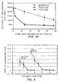
- the collagen-GAGs core can be produced at different ratios and with water content similar to those in native tissues. The method of production also enhances the retention of GAGs in the collagen-GAGs core.
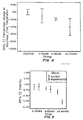
- the bioengineered IVD has mechanical properties comparable to that of the native disc.
- a bioengineered IVD for disc replacement comprises an extracellular matrix (ECM) providing support to living cellular components and interacting with the living cellular components during the fabrication process without introducing toxicity.
- ECM extracellular matrix
- the ECM composition can be from both natural or synthetic sources but preferably natural sources and induced to self-assemble or reconstitute into its solid form under conditions that are mild enough to support cellular survival and growth.
- the cellular components of the mixture are induced to interact with the matrix in such a way that leads to contraction or shrinkage in volume of the structures, leading to changes in dimension, ECM density, cell density, mechanical property and stability, etc.
- the extent of the change in volume of the composition can be precisely controlled by factors including, but are not limited to, the density of the ECM, the composition of different ECM, the density of the living cells, the timing for interaction and the serum concentration.
- a composition for producing a bioengineered IVD structurally similar to the native disc with heterogeneous structure including a distinct nucleus, an inner annulus and an outer annulus.
- the composition includes a nucleus consisting of living cells, collagen or other ECM and GAGs at a ratio similar to that of the native discs.
- the composition can also include an inner annulus with dense ECM lamellae as the clear demarcation separating the nucleus and with multiple lamellae of collagen matrix with increasing density and GAGs with decreasing density (decreasing GAGs: collagen ratio) together with evenly distributed cells with increasing density.
- the composition can include an outer annulus with several lamellae of high density collagen and cells.
- the composition consists of collagen and/or other extracellular matrix materials such as proteoglycans / GAGs, elastin, etc., together with mesenchymal stem cells (MSCs) or living cells from human or other clinically feasible sources, and with growth-stimulating signals such as human serum, platelet rich plasma, other blood products, etc.
- MSCs mesenchymal stem cells
- the composition can be induced to self-assemble or "grow" to pre-designed structures before implantation or replacement procedure.
- IVD cells isolated from NP and AF have been used in developing engineered IVD ( Mizuno, et al., Spine, 29(12):1290-7 (2004 ), Seguin, et al., Spine, 29(12):1299-306 (2004 )).
- these cells may not be available because they are depleted and non-functional in advanced disc degeneration.
- sampling autologous disc cells creates donor site morbidities such as degeneration and is thus not preferred.
- the composition preferably includes mesenchymal stem cells (MSCs) from human or other clinically feasible sources including, but not limited to, autologous, allogeneic, fetal, embryonic or xenogenie sources, either in single cell suspension or encapsulated in matrix microspheres.
- MSCs mesenchymal stem cells
- the ECM providing support to the living cell components can be from either natural or synthetic sources but is preferably natural.
- the ECM can be induced to self-assemble or reconstitute into its solid form under conditions that are mild enough to support cellular survival and growth.
- the ECM can interact with the living cells or with other ECM components in such a way that the interaction leads to contraction of the structure and expelling of excessive water.
- the contraction and the reduction in volume can result in an increase in density of the ECM components and the density of the living cells and hence the mechanical properties.
- the extent of ECM contraction can be precisely controlled by factors including, but not limited to, the density of the ECM, the density of the living cells, the timing for interaction and the serum concentration.
- the ECM can be collagen of various types including but not limited to, type I, II, III, isolated or extracted or prepared from various animal sources including, but not limited to, rat tail, porcine skin, bovine Achilles tendon, and human placenta, in different fractions such as acid-soluble, pepsin-soluble or insoluble.
- the composition can further comprise other ECM such as proteoglycans/GAGs extracted from cartilage, elastin and hyaluronic acid, or other similar materials.
- Collagen can be isolated or extracted or prepared from animal sources, such as rat tail, porcine skin, bovine Achilles tendon, or human placenta, and can be from different fractions during collagen extraction, acid-soluble, pepsin-soluble or insoluble, preferably acid-soluble.
- the composition can further include growth-stimulating signals such as human serum, platelet rich plasma and other blood products.
- the composition can further comprise of other factors affecting the differentiation of MSCs such as TGF-beta for chondrogenic lineage.
- the composition can also contain other factors affecting the differentiation of MSCs such as TGF-beta for chondrogenic lineage.
- the composition can also be exposed to growth-stimulating signals other than soluble factors and blood products such as mechanical stimtilation simulating that of the forces that an IVD is exposed to, prior to the implantation and replacement procedure via bioreactors or equivalent devices.
- the composition can be induced to "grow" the pre-designed structures before implantation or replacement procedure.
- the composition can also be exposed to growth-stimulating signals other than soluble factors and blood products such as mechanical stimulation simulating that of the forces that an IVD is exposed to, prior to the implantation and replacement procedure.
- Autologous serum or plasma, pooled human plasma and platelet-rich-plasma, blood products from matched donor can be included during the fabrication prior to implantation or replacement procedure.
- Recombinant protein products such as TGF-beta can be included.
- composition can further comprise of other therapeutic, prophylactie or diagnostic agents such as anti-inflammatory drugs and antibiotics.
- a method is provided to produce a bioengineered IVD structurally similar to the native disc with heterogeous structure including a distinct nucleus, an inner annulus and an outer annulus.
- the method produces the nucleus by inducing precipitation of acid soluble collagen and GAGs such as chondroitin-6-sulfate by methods such as stirring, vortexing, centrifugation, shaking, etc at a particular ratio, as demonstrated by the examples.
- the collagen-GAGs cores are preferably seeded with living cells.
- the cell density, collagen and GAGs ratios can be predetermined with a certain ratio, as demonstrated by the examples.
- ECM layers can be laminated by enveloping the previously developed structures by incorporating the previously developed structures into the centre of the reconstituting ECM so that the structures are entrapped and positioned inside the gelling matrix and prior to the formation of the solid phase.
- the method can be repeated to produce structure of different sizes.
- the mixture can be cast in a container such as a 4-well culture plate, or a pre-designed container with the shape simulating the IVD, and incubated in a 37°C incubator for self-assembly or reconstitution for a period of time ranging from 5 minutes to 5 hours, preferably 30 minutes.
- the structure can be isolated from the surrounding attachment and transferred to a non-adhesive culture plate such as a bacterial culture dish.
- the structure is incubated to allow for interaction with the living cells for a period of time ranging from 12 hours to 4 days, preferably 1 day, until a pre-designed constant size, preferably the real size of the NP of the mammal such as human, is obtained.
- BM-MSCs can be encapsulated in collagen microcapsules which act as temporary storage for MSCs.
- Collagen solution is made into appropriate concentrations ranging from 0.1 to 100mg/ml and preferably used at a concentration of 0.3-4.0mg/ml, which is neutralized and mixed with MSCs in medium and degassed by methods such as mild centrifugation if necessary.
- the cell-encapsulating microcapsules can be placed into an incubator at 37°C with 5% CO 2 for a period of time ranging from 5 minutes to 10 hours, preferably 30 minutes for reconstitution into collagen gel encapsulating MSCs.
- MSC encapsulated collagen microspheres were washed in sterile PBS and resuspended in full medium preferably DMEM with HS.
- the MSC density in collagen solution ranging from 1 x 10 3 cells/ml to 1 x 10 7 cells/ml, and preferably 1x10 5 cells/ml.
- the MSC-encapsulated microspheres are plated onto culture plates at a density between 1x10 1 to 1x10 4 /cm 2 , preferably 1x10 3 /cm 2 , in the presence of full medium.
- the microspheres are incubated at 37°C for a period of time ranging from 5 minutes to 10 hours, preferably 30 minutes, to allow attachment before filling with full medium.
- MSCs migrate from the microspheres as early as 1 hour post-plating. When MSCs migrating from the spheres reach a certain density after a period of time ranging from 12 hours to 10 days, preferably 1 day, the microspheres are removed from the culture plates without trypsinization by methods such as gentle flushing with PBS or medium, or picking of the microspheres.
- the collected microspheres are sedimented in the incubator for a period of time ranging from 15 minutes to 5 hours, preferably 1 hour, or briefly centrifuged at a speed ranging from 500 to 2000rpm, preferably 1000rpm, and replated into new culture plate.
- the procedure can be repeated at a successive interval of a period of time ranging from 12 hours to 5 days, preferably 1 day.
- MSCs reaching 80-90% confluence can be trypsinixed as in monolayer culture for making the IVD structure.
- the method allows bone marrow derived MSCs to be expanded to a sufficient number at a a controlled rate, with a more physiological environment, and with minimal extent of in vitro manipulation.
- the method can include microencapsulation of BM-MSCs in three dimensional collagen microspheres using methods including, but not limited to, formation of emulsions, droplets generation and injection of MSCs into preformed collagen microspheres.
- the method can further include culture and maintenance of the MSC encapsulated collagen microspheres. MSCs outgrowing from the collagen microspheres can be used to produce the IVD structure.
- the method can further include steps of replating the MSC encapsulated collagen microspheres for multiple times at regular intervals so as to obtain a continuous supply of MSCs without trypsinization.
- the rate of MSCs outgrowth can be controlled by variables including, but are not limited to, the cell density in microspheres, ECM concentration and plating densities of the microspheres.
- the MSCs or other living cells are evenly distributed throughout the bulk of the ECM irrespective of the dimension and shape of the structures.
- the method comprises bringing living cells in contact with and mixing them thoroughly with the collagen or ECM solution immediately after reconstitution at low temperature, before the sol-gel transition is completed, to form the solid phase during the fabrication process.
- Collagen self-assembly or reconstitution from a solution to a solid gel consisting of fibrils is induced by means including, but not limited to, pH change, preferably an increase from acidic pH to alkaline pH, temperature change, preferably an increase from 4°C to 37°C, a change in ionic strength by contact with a solution of high ionic strength.
- the living cells are then brought into contact with the collagen solution mixture containing all necessary growth-stimulation factors and other ECM.
- Self-assembly or reconstitution of ECM can be speeded up at optimal temperature, preferably at 37°C.
- the MSCs or other living cells can be evenly distributed throughout the bulk of the ECM irrespective of the dimension and shape of the structures.
- the rate of such self-assembly or reconstitution is reduced by maintaining a low temperature, preferably 4°C, during self-assembly or reconstitution so that the MSC of other living cells can be mixed well and thoroughly with the solution collagen containing other soluble growth-stimulation factors and other ECM.
- the self-assembly or reconstitution rate can be optimized by raising the temperature to 37°C or by contact with a solution having a high ionic strength such as a PBS concentrate.
- ECM containing evenly distributed MSCs or other living cells can be obtained after completion of the self-assembly and for a period of time from 5 minutes to 5 hours, and preferably 30 minutes.
- the cells embedded in the self-assembled or reconstituted ECM can then be allowed to interact with the ECM, resulting in a change in the properties of the resulting structures, including, but not limited to, the dimension, volume, water content, ECM density, cell density, optical properties, mechanical properties, stability, after isolating the structures from the surrounding environment.
- the extent of contraction of the ECM can be controlled by adjusting variables, including but not limited to, the concentration of collagen solution, concentration of MSCs, concentration of GAGs and other ECM, concentration of serum, ratio of collagen to GAGs, duration of incubation.
- the lamellae "enveloping" the nucleus act as the clear demarcation between the nucleus and the annulus and can be produced by first neutralizing collagen solution at a low concentration ranging from 0.25 to 4mg/ml, preferably at 0.5mg/ml, second, mixing well with other ECM such as GAGs at a lower ratio preferably at 1.5:1 (i.e., GAGs:collagen ratio approximately 1:5) and with MSCs at a high concentration ranging from 1x10 4 to 1x10 7 cells/ml, preferably 5x10 5 cells/ml, at 4°C before inducing reconstitution in 37°C incubator.
- Multiple dense ECM layers enveloping the previously developed nucleus with an increasing collagen density, a decreasing GAGs density and an increasing cell density, at regular intervals, are laminated to form the inner annulus.
- Multiple dense ECM layers enveloping the previously developed inner annulus with an increasing collagen density and cell density, at regular intervals, can be laminated to form the outer annulus.
- Several denser collagen lamellae ranging from three to twelve, preferably three, are laminated onto the two-layer structure as the inner annulus.
- Collagen solution at increasing concentration ranging from 0.25 to 4mg/ml, and preferably at 0.5, 1 and 2 mg/ml, respectively, are neutralized, mixed with descending GAGs:Col ratio ranging from 1:5 to 1:100, and preferably at 1:9, 1:19 and 1:29, as well as MSCs at increasing concentrations ranging from 1x10 4 to 5x10 9 cells/, preferably at 5x10 5 , 1x10 6 and 5x10 6 cells/ml in medium, respectively. All procedures are conducted in an ice bath before induction for reconstitution in 37°C incubator.
- the growing structure can be inserted into the center of the newly reconstituted structure and the structure can be allowed to interact with the living cells for a period of time, ranging from 4 hours to 4 days, and preferably 1 day, to a constant size ranging from 50 microns to 10mm, and preferably 100 microns thicker than the previous structure, before a new cell-matrix layer can be laminated.
- an outer annulus with several lamellae of high density collagen and cells can be produced.
- Several, ranging from three to fifteen, and preferably three, very dense collagen lamellae are laminated onto the previous multi-layered structures as the outer annulus.
- Collagen solution at increasing concentrations ranging from 0.25 to 4mg/ml, and preferably at 0,5, 1 and 2mg/ml, respectively are neutralized and mixed with MSCs at increasing concentrations ranging from 1x10 4 to 1x10 7 cells/ml, and preferably at 5x10 5 , 1x10 6 and 5x10 6 cells/ml. All procedures can be conducted in an ice bath before induction for reconstitution in 37°C incubator.
- the growing structure is inserted into the center of the new layer and allowed to interact with the living cells for a period of time ranging from 4 hours to 4 days, preferably 1day, to a constant size ranging from 50 microns to 10mm, and preferably 100 microns thicker than the previous structure, before a new layer can be laminated.
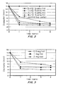
- the bioengineered IVD is engineered to have mechanical properties comparable to that of the native disc. This can be done by four approaches:
- the mechanical properties are comparable to that of the native disc, as demonstrated by the examples.
- the density of the ECM lamellae can be adjusted by adjusting the variables controlling the extent of the ECM contraction, including but are not limited to, cell density, ECM density, duration of interaction between cells and ECM, and serum concentration.
- the method can also comprise dehydrating each collagen lamellae before formation of successive lamellae without significant compromise in cell viability. Enclosing or enveloping GAGs-containing or retraining nucleus structure with multiple lamellae with low GAGs density, so that the inner structure absorbs more water and thus is more swellable upon rehydration can ube used to maintain higher hydrostatic pressure.
- the IVD structure can also be enveloped with a very dense outermost collagen layer strengthened and stabilized by photochemically crosslinking and controlled dehydration.
- the high density collagen layer can be laminated onto the previously developed multi-layered structure and the structure brought into contact with photosensitizing reagent before irradiating with a light source.
- the photosensitizing reagent employed can be rose Bengal, methylene blue, etc.
- the light source used can be a laser, a LED, a xenon lamp, etc.
- the final structure can be dehydrated by methods including, but not limited to, placing against strong water absorbents such as filter paper in a container and gently centrifuging at 500-5000 rpm preferably 1000 rpm for a period of time, ranging from 1 to 100 minutes, preferably 10 minutes to remove excess water.
- the crosslinked structure is centrifuged in all directions until the outermost layer dehydrates to form a dense layer.
- the crosslinked structure can be brought in contact with solvents such as ethanol using a method which limits the ethanol to the surface by methods such as spraying until the structure is dehydrated to the appropriate dimension, preferably at 100 microns thicker than the previous multi-layered structure.
- the structure can be applied with a non-damaging compression force repetitively against water absorbent through the compression platen of a bioreactor from commercial source or a standard weight.
- the structure can be rehydrated in medium and ready for various tests and implantation.
- the structure can also be air-dried at a controllable rate by limiting the time of air-drying ranging between 1 hr to 100 hrs, at a time, preferably 5 hrs, and by controlling the number of times of air-drying procedures, ranging from once to 100 times, preferably 3 times, each with substantial rehydration in medium.
- the time of manufacture is minimized by minimizing the time for MSC induced collagen matrix contraction for each lamellae using optimized cell and matrix concentration and accelerating the speed of dehydration of the outermost crosslinked lamellae using centrifugation or compression or solvent water extraction.
- the methods also enable the production of multiple bioengineered IVDs that can be used for replacement of multiple discs at the same time, or for selection of the structure with best functional characteristics after in vitro testing using a bioreactor such as that from commercial sources.
- the method is also capable of producing other bioengineered tissues with heterogenous structures with multilayer designs including, but not limited to, GI tract such as oesophagus, small intestine, large intestine, and blood vessels, with all possible sizes and shapes, using living cells of different origin and ECM of different types.
- GI tract such as oesophagus, small intestine, large intestine, and blood vessels, with all possible sizes and shapes, using living cells of different origin and ECM of different types.
- the bioengineered IVD is structurally similar to the native disc with a heterogeous structure including a distinct nucleus consisting of collagen type I and GAGs at a ratio similar to that of the native discs and with a relatively low cell density, an inner annulus with clear demarcation separating the nucleus and with multiple lamellae of collagen matrix with increasing density and GAGs with decreasing density together with evenly distributed cells of increasing density; and an outer annulus with several lamellae of high density collagen and cells, as shown schematically in Figure 1 .
- the patient having a badly degenerated IVD can be scheduled for IVD replacement after producing the multilayered structure.
- the structure can be inserted between two vertebrae surgically with posterior ligaments and muscles intact under general anaesthesia. Internal fixation can be used to temporarily provide support and removed after host integration of the implant assessed by radiographic methods.
- Example 1 Encapsulation of MSCs in collagen microspheres .
- Rattail collagen solution type I was neutralized and diluted into 0.5 mg/ml in the presence of different concentrations (2x10 4 , 1x10 5 and 5x10 5 cells/ml) of human bone marrow derived MSCs in DMEM medium with 10%FBS. The solution was kept at 4°C in an ice bath before use. A small volume of 5 ⁇ l of mixture was dropped onto a non-adhesive substratum using a dispenser. The hMSC containing microdroplets were allowed to reconstitute into microspheres encapsulating the cells by incubating in a 37°C incubator for 2 hours and were then collected into a DMEM medium containing bath with non-adherent substratum. The collected microcapsules were incubated in 37°C for three dimensional cultures and the morphological change and the dimensional change of the spheres were recorded for 48 hours before they were ready to be plated onto traditional culture dishes.
- Microspheres at day 0 showed individual cells embedding in the collagen matrices and that the microspheres were still transparent. The microspheres contact over time and become more opaque and dense, indicating that cells are reorganizing the matrix to form a tighter matrix containing microspheres.
- Example 2 Three dimensional culture of MSCs in collagen microcapsules , outgrowth from the attached microcapsules and replating of the microcapsules
- Attachment of the microcapsules onto culture flasks is necessary for cell outgrowth from the microspheres and depends on the cell density and plating density of the cell-encapsulating collagen microspheres on the culture dish.
- hMSC migrating out from the collagen microspheres showed similar morphology with spindle and elongated shapes.
- the hMSC-encapsulated collagen microspheres were released from the culture dish by flushing with full medium and replated onto new culture dishes while the cells migrated out from these microspheres were trypsinized for cell count.
- the growth rate of the hMSCs migrating out from the collagen microspheres was dependent on both the cell density in microspheres and plating density of microspheres in culture dish, as shown by Figures 4A and 4B , respectively.
- MSCs from rabbit bone marrow when migrating out from the collagen microspheres, were also similar in morphology compared with MSCs in traditional monolayer cultures.
- rMSCs migrating out from the collagen microcapsules grow at a similar rate with that in monolayer culture and become confluent at a similar period of time, day 4 after plating, as that in the monolayer culture, as shown by Figure 4C
- the migrated MSCs were then trypsinized for subsequent production of IVD structures.
- the collagen microspheres were reused by first detaching from the culture flasks by gentle flushing with full medium or by picking. The microcapsules were then sedimented before replating onto a new culture flask for attachment. The replanted MSCs migrating out from capsules were similar in morphology and growth rate as compared with that from the monolayer cultures.
- Example 3 Production of MSCs seeded collagen matrix, MSCs induced collagen matrix contraction; analysis of the controlling parameters of the extent of contraction
- Rattail collagen type I solution was neutralized and diluted into concentrations at 0.5, 1 and 2mg/ml in the presence of rabbit bone marrow derived MSCs at a concentration of 4x10 3 , 2x10 4 , 1x10 5 , 5x10 5 cells/ml.
- the collagen MSC mixtures were thoroughly mixed and degassed if necessary and were cast in 4-well plate and incubated for 30 minutes at 37°C in an incubator.
- the solidified structure was detached from the culture plate by using a sterile pipette tip or a syringe needle without damaging the structure and was transferred into a non-adherent culture dish such as a bacterial culture dish.
- the dimension of the structure and the morphology of MSCs inside the structure were recorded against time as the index for volume contraction of the structures.
- Contraction of the collagen matrix depends on the presence of living cells. There is no reduction in volume in the absence of the cells. The extent of ECM contraction and reduction in volume depends on the cell density. There was an increasing in the opaqueness of the structure as the cell density increased. The reduction in diameter of the disc structures was measured under a dissection microscope against a micrometer. The extent of matrix contraction is associated with the cell density positively, with an increasing cell density promoting the extent of contraction, and with collagen density inversely, with increasing collagen density reducing the extent of contraction. The reduction in volume was estimated by measuring the diameter of the contracted structure 4 days after incubation and measuring the thickness of the frozen sections of the structures under the microscope. MSCs at different concentrations (4x10 3 to 5x10 5 cells/ml) resulted in volume reduction of collagen matrix ranging from 20 to 75 fold.
- Example 4 Fabrication of multi-layered structures with different cell and matrix density.
- MSCs migrating out from the collagen microcapsules as described in Example 1 and 2 were trypsinzed for production of muiti-layered structures.
- Rattail collagen type 1 solution was neutralized and diluted to 0.5mg/ml in the presence of rabbit bone marrow derived MSCs at a concentration of 5x 10 4 cells/ml
- the collagen MSCs mixture was cast in 4-well culture plate and induced to self-assemble or reconstitute into solid gel at 37°C for 30 minutes. The structure was detached from the culture dish and was allowed to interact and contract at 37°C for 24 hours.
- the structure was incubated at 37°C briefly for 5 minutes before inserting the contracted structure into the center of the second structure. After incubation for another 30 minutes, the bi-layered structure was again detached from the culture plate and incubated for 24 hours at 37°C.
- a third collagen MSCs structure was produced using 0.5mg/ml collagen solution and 5x10 5 cells/ml MSCs prepared as described in Examples 1 and 2.
- the third structure was induced to reconstitute briefly for 5 minutes at 37°C before inserting the bi-layered structure into the centre of the third structure. After incubation for 24 hours at 37°C, the dimensions of the three-layered structures were measured and fixed with 4% paraformaldehyde before making 10 microns frozen sections for histological analysis.
- Example 5 Fabrication of the collagen membrane strengthened and stabilized by photochemical crosslinking.
- Rattail collagen was reconstituted as described in Example 3 and was equilibrated with excess photosensitizing reagent, rose Bengal at a concentration of 0.01% (w/v) in distilled water, for 2 hours with constant agitation followed by a thorough rinsing with distilled water.
- An argon laser was used for crosslinking.
- the laser beam has a spot size of 1.5cm in diameter covering the whole collagen gel and was delivered in 125 pulses at a power of 0.2W with pulse duration of 1s, equivalent to 25J.
- Untreated collagen gel was used as the control.
- Dye control and laser control groups were collagen gels treated with the photosensitizing reagent alone and laser irradiation alone, respectively.
- Treated collagen gels were air-dried for more than 96 hours to obtain membranes for morphological analysis and measurement of physico-chemical properties.
- An additional control group of chemically crosslinked collagen produced by treating collagen gel with 0.2% glutaraldehyde solution for 2 hours was used for comparison.
- Collagen membranes were fully rehydrated in PBS (pH 7.4) at room temperature for more than 24 hours and were carved into a dumpbell shape with a width of 3mm and a gauge length of 6mm using a custom-made punch while the gauge length was marked with ink.
- a CCD camera was used to record the temporal change of the markers at the gauge length of the collagen samples during the tensile test at a rate of 25 frames per second with resolution of 540x720 pixels.
- the relative displacement between the markers was measured by pixel counting using Photoshop 7.0 for analysis of the strain. Stress-strain curves were obtained and the mechanical properties of the collagen scaffolds, including the peak load, ultimate stress, rupture strain and tangent modulus at 90% of the rupture strain were calculated
- Crosslinked dry collagen scaffold prepared as described above was rehydrated in PBS (pH7.4) or water for 30 minutes.
- the rehydrated collagen membrane was rinsed in water or other isotonic solution and the water on the surface of the rehydrated scaffolds removed by blotting dry using kimwipes or other absorbants.
- the scaffold was carefully laid down at the bottom of a container for reconstitution of collagen gel.
- the container has a dimension of 15mm diameter and approximately 7.5mm thick.
- the membrane-containing collagen solution was then placed in the ammonia chamber for 30 minutes. The procedures were repeated to obtain bi-layered crosslinked collagen membrane.
- the scaffold was dehydrated by air-drying for more than 24 hours.
- collagen scaffolds appropriate thickness were obtained.
- 6- or 10-layer collagen scaffold of approximately 1-2mm thickness produced by the methods described in the examples can be used for intervertebral disc tissue engineering.
- the multi-lamellae collagen structures made in Example 2 have been fully swollen in phosphate buffered saline at pH 7.4 for 2 weeks. Uncrosslinked structures swell extensively to form a soft gel-like structure with a thickness of around 2mm. The structures were so weak that they can not be manipulated with forceps and sutures can not be put through. In contrast, crosslinked structures well moderately to a thickness of around 1mm and were tough. The structures were then mounted on the sample stage of a microforce testing machine fitted with a 100N load cell.
- the strain was set to zero and dynamic compression cycles at a frequency of 0.1Hz and a maximal strain of 0.5 were conducted.
- the load and unload-diaplacement curves were evaluated while the curve at the 20th cycle was used for data analysis.
- the compression module normalized by the dimension of the samples were obtained.
- Fig. 13 shows load curve of the twentieth cycle of repetitive compression of photochemically crosslinked collagen layers.
- Fig. 14 shows the mean compression moduli of photochemically crosslinked collagen layers comparing with that of the lumbar movement segment in human, rabbits, rats and mice native discs.
- a collagen and glycosaminoglycans (GAGs) core was fabricated by precipitating an acid soluble solution of collagen with chondroitin-6-sulfate. Precipitation was induced by vortexing the mixture for 1 minute, centrifuging the solid material which were then rinsed with PBS. An optimal mass ratio of collagen to GAGs at 4:1 has been determined, as shown by Figure 5 .
- the collagen-GAGs core can be photochemically crosslinked as described in Example 5.
- the collagen-GAGs cores were dehydrated, weighted and rehydrated in PBS and the swelling ratio of the cores were calculated by measuring the wet weight at different time points, as shown by Fig. 6 .
- the uncrosslinked collagen-GAGs rapidly swell to a ratio of 7.5 which is equivalent to a water content of almost 88% while the crosslinked ones swell less rapidly and to a ratio of around 3, equivalent to a water content of around 75%.
- the high water content is important in creating the swelling pressure when it is inserted into the disc space.
- the swelling ratio starts to decrease in the collagen-GAGs core as the GAGs content is highly water soluble and the core starts to release GAGs into the surrounding liquid.
- Photochemical crosslinking is able to retain more GAGs as shown in Fig. 7 as compared to the uncrosslinked ones. This is in agreement with the steady level of the swelling ratio at later time points.
- the retention of GAGs in the collagen-GAGs core is important in maintaining the structure of the core after implantation.
- the collagen-GAGs cores are composed of nano-sized fibrils and are porous, thus allowing cell and molecules penetration.
- Cell seeded collagen layers were fabricated as described in Example 4 using fibroblasts and collagen type I solution.
- the collagen-cell constructs were then dehydrated in air in a controlled manner by supplying minimal amount of culture medium to prevent complete dehydration in the incubators. Humid culture chambers were used to maintain a better local humidity.
- the collagen-cell layers were condensed to a much higher density after the controlled dehydration and the average thickness of the layers reduced after dehydration of different period of time, as shown in Figure 8 .
- the collagen-cell constructs after dehydration were enzymatically digested to examine cell viability inside the constructs using trypan blue test.
- Controlled dehydration for a continuous period of 8 hours did not differ from that of the controls, which were maintained with a full supply of culture medium while dehydration overnight for 20 hours induced significant higher loss in viability, as shown by Figure 9 .
- the enzymatically released cells were then replated on culture dishes and the morphology of the cells examined. Those from dehydrated layers are the same as those from the controls, indicating that the cells are still viable and are able to attach and grow normally after dehydration.
- Example 9 In vitro chondrogenic differentiation of human mesenchymal stem cell in collagen gel
- MSCs were seeded in collagen gel either in the form of microspheres or layers as described in Example 1, 3 and 4. Samples were fabricated with two varying parameters: cell seeding density (1x10 5 or 5x10 6 cells/ml) and collagen concentration (1 or 3 mg/ml). The hMSC-seeded gels were cultured in chondrogenic differentiation medium containing transforming growth factor beta 3 (TGF- ⁇ 3) for 3 weeks.
- TGF- ⁇ 3 transforming growth factor beta 3
Landscapes
- Health & Medical Sciences (AREA)
- Life Sciences & Earth Sciences (AREA)
- Engineering & Computer Science (AREA)
- Biomedical Technology (AREA)
- Chemical & Material Sciences (AREA)
- General Health & Medical Sciences (AREA)
- Zoology (AREA)
- Animal Behavior & Ethology (AREA)
- Veterinary Medicine (AREA)
- Public Health (AREA)
- Oral & Maxillofacial Surgery (AREA)
- Transplantation (AREA)
- Cell Biology (AREA)
- Chemical Kinetics & Catalysis (AREA)
- Medicinal Chemistry (AREA)
- Epidemiology (AREA)
- Dermatology (AREA)
- Botany (AREA)
- Vascular Medicine (AREA)
- Developmental Biology & Embryology (AREA)
- Bioinformatics & Cheminformatics (AREA)
- Organic Chemistry (AREA)
- Neurology (AREA)
- Urology & Nephrology (AREA)
- Orthopedic Medicine & Surgery (AREA)
- Biotechnology (AREA)
- Hematology (AREA)
- Wood Science & Technology (AREA)
- Genetics & Genomics (AREA)
- Biochemistry (AREA)
- Rheumatology (AREA)
- Microbiology (AREA)
- Immunology (AREA)
- Heart & Thoracic Surgery (AREA)
- General Engineering & Computer Science (AREA)
- Cardiology (AREA)
- Biophysics (AREA)
- Molecular Biology (AREA)
- General Chemical & Material Sciences (AREA)
- Physical Education & Sports Medicine (AREA)
Claims (10)
- Procédé de réalisation d'une structure IVD, le procédé comprenant le fait d'amener des cellules vivantes en contact avec un collagène reconstituant, une matrice extracellulaire et des GAG :a) former un coeur de noyau, qui comprend des cellules vivantes, du collagène et des GAG à un rapport similaire à celui de disques natifs ;b) stratifier de multiples lamelles de MEC denses enveloppant le noyau avec une densité de collagène croissante, une densité de GAG décroissante et une densité de cellule croissante pour former un espace annulaire interne ;c) stratifier de multiples lamelles de MEC denses enveloppant l'espace annulaire interne précédemment développé avec une densité de collagène croissante et une densité de cellule croissante pour former un espace annulaire externe ;d) réticuler le collagène, la MEC et les GAG.
- Procédé de la revendication 1, dans lequel les cellules vivantes sont réparties uniformément à travers la masse de la MEC et peuvent varier en densité d'une couche à l'autre ou former un gradient en densité.
- Procédé de la revendication 1, dans lequel l'étendue du changement de volume induit par la cellule des structures est régulée par la concentration de la solution de collagène, la concentration des MSC, la concentration des GAG et autre MEC, la concentration de sérum, le rapport de collagène aux GAG, la durée d'incubation.
- Procédé de la revendication 1, comprenant en outre l'exposition de la structure IVD à une stimulation mécanique pour simuler des forces auxquelles la structure IVD est exposée, avant une procédure d'implantation et de remplacement.
- Procédé de la revendication 1, dans lequel l'IVD comprend en outre un espace annulaire interne avec des lamelles de MEC denses séparant le noyau et avec de multiples lamelles de matrice de collagène avec une densité croissante et des GAG avec une densité décroissante conjointement avec les cellules réparties uniformément de densité croissante.
- Procédé de la revendication 5, dans lequel l'IVD comprend en outre un espace annulaire externe ayant une pluralité de lamelles de collagène haute densité et de cellules.
- Procédé de la revendication 1, dans lequel la densité des lamelles de MEC est accrue en ajustant la densité de cellules, la densité de MEC, la durée d'interaction entre les cellules et la MEC ou la concentration de sérum.
- Procédé de la revendication 1, dans lequel le noyau a une structure de noyau contenant des GAG qui absorbe plus d'eau que les lamelles voisines.
- Procédé de la revendication 8, dans lequel l'espace annulaire interne ou externe comprend une pluralité de lamelles de collagène, chacune des lamelles de collagène étant soumise à une déshydratation avant de former les lamelles successives sans compromettre sensiblement la viabilité de la cellule.
- Procédé de la revendication 1, comprenant en outre le fait d'amener la structure en contact avec un réactif de photosensibilisation avant l'irradiation avec une source de lumière.
Applications Claiming Priority (2)
| Application Number | Priority Date | Filing Date | Title |
|---|---|---|---|
| US79608806P | 2006-04-28 | 2006-04-28 | |
| PCT/IB2007/003272 WO2008078157A2 (fr) | 2006-04-28 | 2007-04-30 | Disques intervertébraux mis au point par génie biologique et procédés pour leur préparation |
Publications (2)
| Publication Number | Publication Date |
|---|---|
| EP2019653A2 EP2019653A2 (fr) | 2009-02-04 |
| EP2019653B1 true EP2019653B1 (fr) | 2016-11-16 |
Family
ID=39563006
Family Applications (1)
| Application Number | Title | Priority Date | Filing Date |
|---|---|---|---|
| EP07872042.2A Active EP2019653B1 (fr) | 2006-04-28 | 2007-04-30 | Disques intervertébraux mis au point par génie biologique et procédés pour leur préparation |
Country Status (4)
| Country | Link |
|---|---|
| US (1) | US8734827B2 (fr) |
| EP (1) | EP2019653B1 (fr) |
| CN (1) | CN101478934B (fr) |
| WO (1) | WO2008078157A2 (fr) |
Families Citing this family (19)
| Publication number | Priority date | Publication date | Assignee | Title |
|---|---|---|---|---|
| US9101687B2 (en) | 2002-07-15 | 2015-08-11 | University Of Leeds | Beta sheet tapes ribbons in tissue engineering |
| US8586539B2 (en) | 2002-07-15 | 2013-11-19 | University Of Leeds | Beta sheet tapes ribbons in tissue engineering |
| EP2021035B1 (fr) * | 2006-05-19 | 2020-08-05 | Versitech Limited | Microsphères à matrice cellulaire, procédés de préparation et applications |
| US9096831B2 (en) * | 2006-05-19 | 2015-08-04 | Versitech Limited | Methods to enhance cell migration and engraftment |
| WO2009007979A2 (fr) * | 2007-07-11 | 2009-01-15 | Technion Research & Development Foundation Ltd. | Cellules souches mésenchymateuses encapsulées et leurs procédés d'utilisation |
| WO2009142770A2 (fr) * | 2008-05-23 | 2009-11-26 | The Regents Of The University Of California | Compositions et procédés permettant la génération de tissu musculo-squelettique |
| US8926552B2 (en) * | 2009-08-12 | 2015-01-06 | Medtronic, Inc. | Particle delivery |
| US11786636B2 (en) * | 2010-06-15 | 2023-10-17 | Versitech Limited | Methods for complex tissue engineering |
| US20140008545A1 (en) * | 2012-07-06 | 2014-01-09 | Stephen George Opuszynski | Methods, apparatus and compositions for diffusing and mitigating laser energy, infrared energy and electron beams |
| US11891626B2 (en) * | 2013-03-15 | 2024-02-06 | Discgenics, Inc. | Isolated discogenic cells, methods of using, and methods of preparing same from mammalian tissue |
| GB201306338D0 (en) | 2013-04-08 | 2013-05-22 | Univ Leeds | Novel proteoglycans |
| CN105343933B (zh) * | 2015-05-15 | 2018-06-22 | 广州悦欣生物医学科技有限公司 | 一种光氧化胶原交联的方法及其应用 |
| CN105343934B (zh) * | 2015-05-15 | 2018-05-29 | 广州悦清再生医学科技有限公司 | 一种人工角膜及其制备方法 |
| WO2017044570A1 (fr) * | 2015-09-08 | 2017-03-16 | Clemson University | Biomatériau décellularisé et son procédé de formation |
| US11285177B2 (en) | 2018-01-03 | 2022-03-29 | Globus Medical, Inc. | Allografts containing viable cells and methods thereof |
| CN108144128B (zh) * | 2018-02-07 | 2021-11-05 | 陕西佰傲再生医学有限公司 | 一种多次交联乳房补片及其制备方法 |
| US20210047612A1 (en) * | 2018-03-06 | 2021-02-18 | Epibone, Inc. | Injectable off-the- shelf cartilage, tendon, and ligament repair compositions and methods of use |
| WO2020228733A1 (fr) * | 2019-05-16 | 2020-11-19 | The Chinese University Of Hong Kong | Substance de matrice extracellulaire et utilisations associées |
| CN116761641A (zh) * | 2021-01-08 | 2023-09-15 | 香港大学 | 具有高糖胺聚糖/羟脯氨酸比率的生物材料、组合物、方法和应用 |
Family Cites Families (17)
| Publication number | Priority date | Publication date | Assignee | Title |
|---|---|---|---|---|
| US5108438A (en) * | 1989-03-02 | 1992-04-28 | Regen Corporation | Prosthetic intervertebral disc |
| US5609635A (en) * | 1988-06-28 | 1997-03-11 | Michelson; Gary K. | Lordotic interbody spinal fusion implants |
| EP2311471A3 (fr) | 1996-04-19 | 2013-05-15 | Osiris Therapeutics, Inc. | Régéneration et augmentation d'os utilisant des cellules souches mésenchymateuses |
| US5964807A (en) | 1996-08-08 | 1999-10-12 | Trustees Of The University Of Pennsylvania | Compositions and methods for intervertebral disc reformation |
| US6340369B1 (en) * | 1999-08-13 | 2002-01-22 | Bret A. Ferree | Treating degenerative disc disease with harvested disc cells and analogues of the extracellular matrix |
| US6783546B2 (en) | 1999-09-13 | 2004-08-31 | Keraplast Technologies, Ltd. | Implantable prosthetic or tissue expanding device |
| US6723335B1 (en) * | 2000-04-07 | 2004-04-20 | Jeffrey William Moehlenbruck | Methods and compositions for treating intervertebral disc degeneration |
| US20030165473A1 (en) * | 2001-11-09 | 2003-09-04 | Rush-Presbyterian-St. Luke's Medical Center | Engineered intervertebral disc tissue |
| US8673333B2 (en) * | 2002-09-25 | 2014-03-18 | The Johns Hopkins University | Cross-linked polymer matrices, and methods of making and using same |
| CA2513055A1 (fr) * | 2003-01-31 | 2004-08-19 | Zimmer Orthobiologics, Inc. | Compositions de matrices hydrogel composites et procedes pour la reparation ou le traitement de tissus mammaliens |
| WO2004093932A1 (fr) * | 2003-04-21 | 2004-11-04 | Verigen Ag | Support resistant au dechirement, pourvu de cellules en contact avec un substrat |
| US8273347B2 (en) * | 2003-05-13 | 2012-09-25 | Depuy Spine, Inc. | Autologous treatment of degenerated disc with cells |
| GB0404656D0 (en) * | 2004-03-02 | 2004-04-07 | Univ Manchester | Treatment of spinal conditions |
| EP1756267A2 (fr) * | 2004-05-14 | 2007-02-28 | Becton, Dickinson and Company | Populations de cellules souches et méthodes d'utilisation |
| WO2006017195A1 (fr) * | 2004-07-13 | 2006-02-16 | Ramot At Tel-Aviv University Ltd. | Particules de glycoproteines lipidees et leurs methodes d'utilisation |
| WO2006029571A1 (fr) * | 2004-09-14 | 2006-03-23 | The University Of Hong Kong | Echafaudages de collagene a reticulation photochimique et procedes de preparation associes |
| US20080045949A1 (en) * | 2005-06-17 | 2008-02-21 | Hunt Margaret M | Method of treating degenerative spinal disorders |
-
2007
- 2007-04-30 EP EP07872042.2A patent/EP2019653B1/fr active Active
- 2007-04-30 US US11/742,040 patent/US8734827B2/en active Active
- 2007-04-30 CN CN2007800227901A patent/CN101478934B/zh active Active
- 2007-04-30 WO PCT/IB2007/003272 patent/WO2008078157A2/fr active Application Filing
Also Published As
| Publication number | Publication date |
|---|---|
| EP2019653A2 (fr) | 2009-02-04 |
| CN101478934A (zh) | 2009-07-08 |
| CN101478934B (zh) | 2012-08-08 |
| WO2008078157A3 (fr) | 2008-11-20 |
| US20070292514A1 (en) | 2007-12-20 |
| US8734827B2 (en) | 2014-05-27 |
| WO2008078157A2 (fr) | 2008-07-03 |
Similar Documents
| Publication | Publication Date | Title |
|---|---|---|
| EP2019653B1 (fr) | Disques intervertébraux mis au point par génie biologique et procédés pour leur préparation | |
| Gao et al. | Biomaterial–related cell microenvironment in tissue engineering and regenerative medicine | |
| EP2522304B1 (fr) | Composition et procédés pour la production de tissus biologiques et de constructions tissulaires | |
| Aufderheide et al. | Assessment of a bovine co-culture, scaffold-free method for growing meniscus-shaped constructs | |
| Kamelger et al. | A comparative study of three different biomaterials in the engineering of skeletal muscle using a rat animal model | |
| US9259510B2 (en) | System and method for forming bone, ligament, and bone-ligament constructs | |
| Yao et al. | In vitro angiogenesis of 3D tissue engineered adipose tissue | |
| EP3517144B1 (fr) | Composition pour la régénération du cartilage et son procédé de préparation | |
| JP6196217B2 (ja) | 筋線維断片を利用した筋組織再生 | |
| KR101472045B1 (ko) | 코어-쉘 섬유상 세포전달체 및 이를 포함하는 골조직 또는 연골조직 재생용 조성물 | |
| EA038957B1 (ru) | Изготовление модифицированных внеклеточным матриксом тканеинженерных нервных трансплантатов для восстановления поврежденных периферических нервов | |
| Liu et al. | Vascularized bone tissue formation induced by fiber-reinforced scaffolds cultured with osteoblasts and endothelial cells | |
| JP4122280B2 (ja) | 組織プラグの製造方法 | |
| US9629939B2 (en) | Collagenous foam materials | |
| EP2582410B1 (fr) | Procédés pour produire des tissus complexes par génie biologique | |
| EP3021880A1 (fr) | Treillis tridimensionnel fonctionnalisé avec des micro-tissus pour la régénération tissulaire | |
| CN111282021B (zh) | 半月板复合支架及其制备方法 | |
| Martin et al. | Producing prefabricated tissues and organs via tissue engineering | |
| US20160136330A1 (en) | Three-Dimensional Scaffold Functionalized with Micro-Tissues for Tissue Regeneration | |
| Chiu et al. | Nanomaterials for cartilage tissue engineering | |
| CN115317672B (zh) | 仿生骨软骨一体化修复植入体及其制备方法与应用 | |
| CN116036379A (zh) | 一种3d打印血管化脂肪再生支架及应用 | |
| Harvanová et al. | Collagen/hyaluronan membrane as a scaffold for chondrocytes cultivation | |
| Li et al. | Progress in cartilage tissue engineering with nanostructured extracellular matrix | |
| Scale et al. | Engineering Biomaterials for Tissue Engineering |
Legal Events
| Date | Code | Title | Description |
|---|---|---|---|
| PUAI | Public reference made under article 153(3) epc to a published international application that has entered the european phase |
Free format text: ORIGINAL CODE: 0009012 |
|
| 17P | Request for examination filed |
Effective date: 20081124 |
|
| AK | Designated contracting states |
Kind code of ref document: A2 Designated state(s): AT BE BG CH CY CZ DE DK EE ES FI FR GB GR HU IE IS IT LI LT LU LV MC MT NL PL PT RO SE SI SK TR |
|
| AX | Request for extension of the european patent |
Extension state: AL BA HR MK RS |
|
| DAX | Request for extension of the european patent (deleted) | ||
| 17Q | First examination report despatched |
Effective date: 20101201 |
|
| REG | Reference to a national code |
Ref country code: DE Ref legal event code: R079 Ref document number: 602007048824 Country of ref document: DE Free format text: PREVIOUS MAIN CLASS: A61F0002440000 Ipc: A61L0027360000 |
|
| RIC1 | Information provided on ipc code assigned before grant |
Ipc: A61L 27/38 20060101ALI20160411BHEP Ipc: A61F 2/28 20060101ALN20160411BHEP Ipc: A61F 2/44 20060101ALI20160411BHEP Ipc: A61K 35/12 20060101ALN20160411BHEP Ipc: A61L 27/36 20060101AFI20160411BHEP Ipc: A61F 2/30 20060101ALN20160411BHEP Ipc: C12N 5/0775 20100101ALN20160411BHEP |
|
| GRAP | Despatch of communication of intention to grant a patent |
Free format text: ORIGINAL CODE: EPIDOSNIGR1 |
|
| INTG | Intention to grant announced |
Effective date: 20160603 |
|
| GRAS | Grant fee paid |
Free format text: ORIGINAL CODE: EPIDOSNIGR3 |
|
| GRAA | (expected) grant |
Free format text: ORIGINAL CODE: 0009210 |
|
| AK | Designated contracting states |
Kind code of ref document: B1 Designated state(s): AT BE BG CH CY CZ DE DK EE ES FI FR GB GR HU IE IS IT LI LT LU LV MC MT NL PL PT RO SE SI SK TR |
|
| REG | Reference to a national code |
Ref country code: GB Ref legal event code: FG4D |
|
| REG | Reference to a national code |
Ref country code: CH Ref legal event code: EP |
|
| REG | Reference to a national code |
Ref country code: IE Ref legal event code: FG4D |
|
| REG | Reference to a national code |
Ref country code: AT Ref legal event code: REF Ref document number: 845301 Country of ref document: AT Kind code of ref document: T Effective date: 20161215 |
|
| REG | Reference to a national code |
Ref country code: DE Ref legal event code: R096 Ref document number: 602007048824 Country of ref document: DE |
|
| REG | Reference to a national code |
Ref country code: CH Ref legal event code: NV Representative=s name: HEPP WENGER RYFFEL AG, CH |
|
| PG25 | Lapsed in a contracting state [announced via postgrant information from national office to epo] |
Ref country code: LV Free format text: LAPSE BECAUSE OF FAILURE TO SUBMIT A TRANSLATION OF THE DESCRIPTION OR TO PAY THE FEE WITHIN THE PRESCRIBED TIME-LIMIT Effective date: 20161116 |
|
| REG | Reference to a national code |
Ref country code: FR Ref legal event code: PLFP Year of fee payment: 11 |
|
| REG | Reference to a national code |
Ref country code: NL Ref legal event code: MP Effective date: 20161116 |
|
| REG | Reference to a national code |
Ref country code: LT Ref legal event code: MG4D |
|
| REG | Reference to a national code |
Ref country code: AT Ref legal event code: MK05 Ref document number: 845301 Country of ref document: AT Kind code of ref document: T Effective date: 20161116 |
|
| PG25 | Lapsed in a contracting state [announced via postgrant information from national office to epo] |
Ref country code: LT Free format text: LAPSE BECAUSE OF FAILURE TO SUBMIT A TRANSLATION OF THE DESCRIPTION OR TO PAY THE FEE WITHIN THE PRESCRIBED TIME-LIMIT Effective date: 20161116 Ref country code: GR Free format text: LAPSE BECAUSE OF FAILURE TO SUBMIT A TRANSLATION OF THE DESCRIPTION OR TO PAY THE FEE WITHIN THE PRESCRIBED TIME-LIMIT Effective date: 20170217 Ref country code: SE Free format text: LAPSE BECAUSE OF FAILURE TO SUBMIT A TRANSLATION OF THE DESCRIPTION OR TO PAY THE FEE WITHIN THE PRESCRIBED TIME-LIMIT Effective date: 20161116 Ref country code: NL Free format text: LAPSE BECAUSE OF FAILURE TO SUBMIT A TRANSLATION OF THE DESCRIPTION OR TO PAY THE FEE WITHIN THE PRESCRIBED TIME-LIMIT Effective date: 20161116 |
|
| PG25 | Lapsed in a contracting state [announced via postgrant information from national office to epo] |
Ref country code: PL Free format text: LAPSE BECAUSE OF FAILURE TO SUBMIT A TRANSLATION OF THE DESCRIPTION OR TO PAY THE FEE WITHIN THE PRESCRIBED TIME-LIMIT Effective date: 20161116 Ref country code: AT Free format text: LAPSE BECAUSE OF FAILURE TO SUBMIT A TRANSLATION OF THE DESCRIPTION OR TO PAY THE FEE WITHIN THE PRESCRIBED TIME-LIMIT Effective date: 20161116 Ref country code: FI Free format text: LAPSE BECAUSE OF FAILURE TO SUBMIT A TRANSLATION OF THE DESCRIPTION OR TO PAY THE FEE WITHIN THE PRESCRIBED TIME-LIMIT Effective date: 20161116 Ref country code: ES Free format text: LAPSE BECAUSE OF FAILURE TO SUBMIT A TRANSLATION OF THE DESCRIPTION OR TO PAY THE FEE WITHIN THE PRESCRIBED TIME-LIMIT Effective date: 20161116 Ref country code: PT Free format text: LAPSE BECAUSE OF FAILURE TO SUBMIT A TRANSLATION OF THE DESCRIPTION OR TO PAY THE FEE WITHIN THE PRESCRIBED TIME-LIMIT Effective date: 20170316 |
|
| PG25 | Lapsed in a contracting state [announced via postgrant information from national office to epo] |
Ref country code: RO Free format text: LAPSE BECAUSE OF FAILURE TO SUBMIT A TRANSLATION OF THE DESCRIPTION OR TO PAY THE FEE WITHIN THE PRESCRIBED TIME-LIMIT Effective date: 20161116 Ref country code: CZ Free format text: LAPSE BECAUSE OF FAILURE TO SUBMIT A TRANSLATION OF THE DESCRIPTION OR TO PAY THE FEE WITHIN THE PRESCRIBED TIME-LIMIT Effective date: 20161116 Ref country code: DK Free format text: LAPSE BECAUSE OF FAILURE TO SUBMIT A TRANSLATION OF THE DESCRIPTION OR TO PAY THE FEE WITHIN THE PRESCRIBED TIME-LIMIT Effective date: 20161116 Ref country code: EE Free format text: LAPSE BECAUSE OF FAILURE TO SUBMIT A TRANSLATION OF THE DESCRIPTION OR TO PAY THE FEE WITHIN THE PRESCRIBED TIME-LIMIT Effective date: 20161116 Ref country code: SK Free format text: LAPSE BECAUSE OF FAILURE TO SUBMIT A TRANSLATION OF THE DESCRIPTION OR TO PAY THE FEE WITHIN THE PRESCRIBED TIME-LIMIT Effective date: 20161116 |
|
| REG | Reference to a national code |
Ref country code: DE Ref legal event code: R097 Ref document number: 602007048824 Country of ref document: DE |
|
| PG25 | Lapsed in a contracting state [announced via postgrant information from national office to epo] |
Ref country code: BG Free format text: LAPSE BECAUSE OF FAILURE TO SUBMIT A TRANSLATION OF THE DESCRIPTION OR TO PAY THE FEE WITHIN THE PRESCRIBED TIME-LIMIT Effective date: 20170216 Ref country code: BE Free format text: LAPSE BECAUSE OF FAILURE TO SUBMIT A TRANSLATION OF THE DESCRIPTION OR TO PAY THE FEE WITHIN THE PRESCRIBED TIME-LIMIT Effective date: 20161116 Ref country code: IT Free format text: LAPSE BECAUSE OF FAILURE TO SUBMIT A TRANSLATION OF THE DESCRIPTION OR TO PAY THE FEE WITHIN THE PRESCRIBED TIME-LIMIT Effective date: 20161116 |
|
| PLBE | No opposition filed within time limit |
Free format text: ORIGINAL CODE: 0009261 |
|
| STAA | Information on the status of an ep patent application or granted ep patent |
Free format text: STATUS: NO OPPOSITION FILED WITHIN TIME LIMIT |
|
| 26N | No opposition filed |
Effective date: 20170817 |
|
| PG25 | Lapsed in a contracting state [announced via postgrant information from national office to epo] |
Ref country code: SI Free format text: LAPSE BECAUSE OF FAILURE TO SUBMIT A TRANSLATION OF THE DESCRIPTION OR TO PAY THE FEE WITHIN THE PRESCRIBED TIME-LIMIT Effective date: 20161116 |
|
| REG | Reference to a national code |
Ref country code: IE Ref legal event code: MM4A |
|
| PG25 | Lapsed in a contracting state [announced via postgrant information from national office to epo] |
Ref country code: MC Free format text: LAPSE BECAUSE OF FAILURE TO SUBMIT A TRANSLATION OF THE DESCRIPTION OR TO PAY THE FEE WITHIN THE PRESCRIBED TIME-LIMIT Effective date: 20161116 |
|
| PG25 | Lapsed in a contracting state [announced via postgrant information from national office to epo] |
Ref country code: LU Free format text: LAPSE BECAUSE OF NON-PAYMENT OF DUE FEES Effective date: 20170430 |
|
| REG | Reference to a national code |
Ref country code: FR Ref legal event code: PLFP Year of fee payment: 12 |
|
| PG25 | Lapsed in a contracting state [announced via postgrant information from national office to epo] |
Ref country code: IE Free format text: LAPSE BECAUSE OF NON-PAYMENT OF DUE FEES Effective date: 20170430 |
|
| PG25 | Lapsed in a contracting state [announced via postgrant information from national office to epo] |
Ref country code: MT Free format text: LAPSE BECAUSE OF NON-PAYMENT OF DUE FEES Effective date: 20170430 |
|
| PG25 | Lapsed in a contracting state [announced via postgrant information from national office to epo] |
Ref country code: HU Free format text: LAPSE BECAUSE OF FAILURE TO SUBMIT A TRANSLATION OF THE DESCRIPTION OR TO PAY THE FEE WITHIN THE PRESCRIBED TIME-LIMIT; INVALID AB INITIO Effective date: 20070430 |
|
| PG25 | Lapsed in a contracting state [announced via postgrant information from national office to epo] |
Ref country code: CY Free format text: LAPSE BECAUSE OF NON-PAYMENT OF DUE FEES Effective date: 20161116 |
|
| PG25 | Lapsed in a contracting state [announced via postgrant information from national office to epo] |
Ref country code: TR Free format text: LAPSE BECAUSE OF FAILURE TO SUBMIT A TRANSLATION OF THE DESCRIPTION OR TO PAY THE FEE WITHIN THE PRESCRIBED TIME-LIMIT Effective date: 20161116 |
|
| PG25 | Lapsed in a contracting state [announced via postgrant information from national office to epo] |
Ref country code: IS Free format text: LAPSE BECAUSE OF FAILURE TO SUBMIT A TRANSLATION OF THE DESCRIPTION OR TO PAY THE FEE WITHIN THE PRESCRIBED TIME-LIMIT Effective date: 20170316 |
|
| PGFP | Annual fee paid to national office [announced via postgrant information from national office to epo] |
Ref country code: FR Payment date: 20230309 Year of fee payment: 17 |
|
| PGFP | Annual fee paid to national office [announced via postgrant information from national office to epo] |
Ref country code: DE Payment date: 20230307 Year of fee payment: 17 Ref country code: CH Payment date: 20230502 Year of fee payment: 17 |
|
| PGFP | Annual fee paid to national office [announced via postgrant information from national office to epo] |
Ref country code: GB Payment date: 20240307 Year of fee payment: 18 |