EP1574997A2 - Verfahren zum Erzeugen eines tomographischen Bildes und Röntgen-Computertomograph - Google Patents
Verfahren zum Erzeugen eines tomographischen Bildes und Röntgen-Computertomograph Download PDFInfo
- Publication number
- EP1574997A2 EP1574997A2 EP05250529A EP05250529A EP1574997A2 EP 1574997 A2 EP1574997 A2 EP 1574997A2 EP 05250529 A EP05250529 A EP 05250529A EP 05250529 A EP05250529 A EP 05250529A EP 1574997 A2 EP1574997 A2 EP 1574997A2
- Authority
- EP
- European Patent Office
- Prior art keywords
- data
- reconstruction
- image
- projection
- axial scan
- Prior art date
- Legal status (The legal status is an assumption and is not a legal conclusion. Google has not performed a legal analysis and makes no representation as to the accuracy of the status listed.)
- Withdrawn
Links
Images






Classifications
-
- G—PHYSICS
- G06—COMPUTING OR CALCULATING; COUNTING
- G06T—IMAGE DATA PROCESSING OR GENERATION, IN GENERAL
- G06T12/00—Tomographic reconstruction from projections
- G06T12/20—Inverse problem, i.e. transformations from projection space into object space
-
- G—PHYSICS
- G03—PHOTOGRAPHY; CINEMATOGRAPHY; ANALOGOUS TECHNIQUES USING WAVES OTHER THAN OPTICAL WAVES; ELECTROGRAPHY; HOLOGRAPHY
- G03G—ELECTROGRAPHY; ELECTROPHOTOGRAPHY; MAGNETOGRAPHY
- G03G21/00—Arrangements not provided for by groups G03G13/00 - G03G19/00, e.g. cleaning, elimination of residual charge
-
- G—PHYSICS
- G03—PHOTOGRAPHY; CINEMATOGRAPHY; ANALOGOUS TECHNIQUES USING WAVES OTHER THAN OPTICAL WAVES; ELECTROGRAPHY; HOLOGRAPHY
- G03G—ELECTROGRAPHY; ELECTROPHOTOGRAPHY; MAGNETOGRAPHY
- G03G2215/00—Apparatus for electrophotographic processes
- G03G2215/01—Apparatus for electrophotographic processes for producing multicoloured copies
- G03G2215/0103—Plural electrographic recording members
- G03G2215/0119—Linear arrangement adjacent plural transfer points
-
- G—PHYSICS
- G03—PHOTOGRAPHY; CINEMATOGRAPHY; ANALOGOUS TECHNIQUES USING WAVES OTHER THAN OPTICAL WAVES; ELECTROGRAPHY; HOLOGRAPHY
- G03G—ELECTROGRAPHY; ELECTROPHOTOGRAPHY; MAGNETOGRAPHY
- G03G2221/00—Processes not provided for by group G03G2215/00, e.g. cleaning or residual charge elimination
- G03G2221/16—Mechanical means for facilitating the maintenance of the apparatus, e.g. modular arrangements and complete machine concepts
- G03G2221/1651—Mechanical means for facilitating the maintenance of the apparatus, e.g. modular arrangements and complete machine concepts for connecting the different parts
- G03G2221/1657—Mechanical means for facilitating the maintenance of the apparatus, e.g. modular arrangements and complete machine concepts for connecting the different parts transmitting mechanical drive power
Definitions
- the present invention relates to a CT (computed tomography) image producing method and an X-ray CT apparatus, and more particularly to a CT image producing method and an X-ray CT apparatus for producing a CT image by applying Z-filtering to axial scan data collected using a multi-row detector.
- CT computed tomography
- the present invention therefore seeks to provide an image producing method and an X-ray CT apparatus for producing a CT image by applying Z-filtering to axial scan data collected using a multi-row detector.
- the present invention provides a CT image producing method characterized in comprising: collecting first axial scan data while rotating at least one of an X-ray tube and a multi-row detector relative to and around a subject to be imaged at a first position in a direction of a body axis of the subject to be imaged; extracting from said first axial scan data projection data corresponding to a plurality of reconstruction fields arranged in the direction of the body axis of the subject to be imaged; if part of projection data of a reconstruction field near an end of said multi-row detector cannot be extracted from said first axial scan data, collecting second axial scan data while rotating at least one of said X-ray tube and multi-row detector relative to and around the subject to be imaged at a second position to which said X-ray tube and multi-row detector are rectilinearly moved relative to the subject to be imaged from said first position toward the end of said multi-row detector; extracting from said second axial scan data the projection data that could not be extracted from said first axial
- second axial scan data is collected at a second position Z2 to which the X-ray tube 21 and multi-row detector 24 are rectilinearly moved relative to the subject to be imaged from the first position Z1 toward the end of the multi-row detector 24, and projection data corresponding to the central portion a2 of the reconstruction field P3 near the multi-row detector 24 is extracted from the second axial scan data.
- One CT image is then produced by applying Z-filtering based on the projection data of the reconstruction fields P1, P2 and P3, and therefore omission of projection data is avoided, thus improving image quality.
- the phrase "rotating relative to” or the like includes: for a subject to be imaged placed in between the X-ray tube and multi-row detector, rotating at least one of the X-ray tube and multi-row detector around the subject to be imaged without rotating the subject to be imaged; rotating the subject to be imaged around its axis without rotating the X-ray tube and multi-row detector; and rotating the subject to be imaged around its axis and rotating at least one of the X-ray tube and multi-row detector around the subject to be imaged.
- the phrase "rectilinearly moving relative to" or the like includes: for a subject to be imaged placed in between the X-ray tube and multi-row detector, rectilinearly moving the subject to be imaged (or the table on which the subject to be imaged is laid) without rectilinearly moving the X-ray tube and multi-row detector; rectilinearly moving the X-ray tube and multi-row detector without rectilinearly moving the subject to be imaged (or the table on which the subject to be imaged is laid); and rectilinearly moving the subject to be imaged (or the table on which the subject to be imaged is laid) and rectilinearly moving the X-ray tube and multi-row detector.
- the present invention provides the CT image producing method having the aforementioned configuration, characterized in that: said second position is a position distant from said first position by a cone beam width at a center of rotation.
- a distance L between the first position Z1 and second position Z2 may be set to be equal to a cone beam width W at the center of X-ray rotation IC.
- the second position Z2 is defined as a position distant from the first position Z1 by the cone beam width W at the center of rotation IC.
- the distance L between the first position Z1 and second position Z2 may be smaller than the cone beam width W at the center of X-ray rotation IC if partial overlap of data is tolerated, while the distance L between the first position Z1 and second position Z2 may be larger than the cone beam width W at the center of X-ray rotation IC if partial omission of data is tolerated.
- the present invention provides the CT image producing method having the aforementioned configuration, characterized in comprising: determining combined projection data by applying weighted addition to said extracted projection data across the reconstruction fields; and producing one CT image from said combined projection data.
- the present invention provides the CT image producing method having the aforementioned configuration, characterized in that: a weight for the weighted addition on said projection data is determined from a mutual relationship between the reconstruction fields, and from a predetermined weighting function.
- the profile of a CT image in the thickness direction can be controlled by selection of the weighting function.
- the present invention provides the CT image producing method having the aforementioned configuration, characterized in comprising: producing respective CT images from said extracted projection data of the reconstruction fields; and applying weighted addition to said CT images to produce one CT image.
- CT images of the reconstruction fields and one CT image combining them can be simultaneously obtained.
- the present invention provides the CT image producing method having the aforementioned configuration, characterized in that: a weight for the weighted addition on said CT images is determined from a mutual relationship between the reconstruction fields, and from a predetermined weighting function.
- the profile of a CT image in the thickness direction can be controlled by selection of the weighting function.
- the present invention provides the CT image producing method having the aforementioned configuration, characterized in comprising: extracting from said first axial scan data projection data corresponding to one line or a plurality of parallel lines at spacings of a plurality of pixels on each of said reconstruction planes; if part of projection data of a reconstruction field near an end of said multi-row detector cannot be extracted from said first axial scan data, extracting the projection data from said second axial scan data; generating projection line data by multiplying said projection data by a cone beam reconstruction weight; determining combined projection line data by applying weighted addition to said projection line data of corresponding lines across the reconstruction fields; generating image-positional line data by filtering said combined projection line data; determining backprojected pixel data of pixels on an image plane based on said image-positional line data; determining backprojected data by adding the backprojected pixel data on a pixel-by-pixel basis across all views used in image reconstruction; and thereby producing one CT image.
- the CT image producing method of the seventh aspect since projection data corresponding to lines is processed, the amount of calculation is reduced. Moreover, since the weighted addition (Z-filtering) is applied to a projection line data stage, the amount of calculation is reduced.
- the present invention provides the CT image producing method having the aforementioned configuration, characterized in comprising: extracting from said first axial scan data projection data corresponding to one line or a plurality of parallel lines at spacings of a plurality of pixels on each of said reconstruction planes; if part of projection data of a reconstruction field near an end of said multi-row detector cannot be extracted from said first axial scan data, extracting the projection data from said second axial scan data; generating projection line data by multiplying said projection data by a cone beam reconstruction weight; generating image-positional line data by filtering said projection line data; determining backprojected pixel data on an image plane based on said image-positional line data; determining backprojected data by adding the backprojected pixel data on a pixel-by-pixel basis across all views used in image reconstruction; applying weighted addition to the corresponding backprojected data across said reconstruction fields; and producing one CT image.
- CT image producing method of the eighth aspect since projection data corresponding to lines is processed, the amount of calculation is reduced. Moreover, since weighted addition (Z-filtering) is applied after determining backprojected data of the reconstruction fields, CT images of the reconstruction fields and one CT image combining them can be simultaneously obtained.
- the present invention provides an X-ray CT apparatus characterized in comprising: an X-ray tube; a multi-row detector; first axial scanning means for collecting first axial scan data while rotating at least one of said X-ray tube and multi-row detector relative to and around a subject to be imaged at a first position in a direction of a body axis of the subject to be imaged; first projection data extracting means for extracting from said first axial scan data projection data corresponding to a plurality of reconstruction fields arranged in the direction of the body axis of the subject to be imaged; second axial scanning means for, if part of projection data of a reconstruction field near an end of said multi-row detector cannot be extracted from said first axial scan data, collecting second axial scan data while rotating at least one of said X-ray tube and multi-row detector relative to and around the subject to be imaged at a second position to which said X-ray tube and multi-row detector are rectilinearly moved relative to the subject to be imaged from said first
- the CT image producing method of the first aspect can be suitably implemented.
- the present invention provides the X-ray CT apparatus having the aforementioned configuration, characterized in that: said second position is a position distant from said first position by a cone beam width at a center of rotation.
- the CT image producing method of the second aspect can be suitably implemented.
- the present invention provides the X-ray CT apparatus having the aforementioned configuration, characterized in that: said CT image producing means determines combined projection data by applying weighted addition to said extracted projection data across the reconstruction fields, and produces one CT image from said combined projection data.
- the CT image producing method of the third aspect can be suitably implemented.
- the present invention provides the X-ray CT apparatus having the aforementioned configuration, characterized in that: a weight for the weighted addition on said projection data is determined from a mutual relationship between the reconstruction fields, and from a predetermined weighting function.
- the CT image producing method of the fourth aspect can be suitably implemented.
- the present invention provides the X-ray CT apparatus having the aforementioned configuration, characterized in that: said CT image producing means produces respective CT images from said extracted projection data of the reconstruction fields, and applies weighted addition to said CT images to produce one CT image.
- the CT image producing method of the fifth aspect can be suitably implemented.
- the present invention provides the X-ray CT apparatus having the aforementioned configuration, characterized in that: a weight for the weighted addition on said CT images is determined from a mutual relationship between the reconstruction fields, and from a predetermined weighting function.
- the CT image producing method of the sixth aspect can be suitably implemented.
- the present invention provides the X-ray CT apparatus having the aforementioned configuration, characterized in that: said first projection data extracting means extracts from said first axial scan data projection data corresponding to one line or a plurality of parallel lines at spacings of a plurality of pixels on each of said reconstruction planes; said second projection data extracting means extracts from said second axial scan data the projection data that could not be extracted from said first axial scan data of a reconstruction field near an end of said multi-row detector; and said CT image producing means generates projection line data by multiplying said projection data by a cone beam reconstruction weight, determines combined projection line data by applying weighted addition to said projection line data of corresponding lines across said reconstruction fields, generates image-positional line data by filtering said combined projection line data, determines backprojected pixel data of pixels on an image plane based on said image-positional line data, and determines backprojected data by adding the backprojected pixel data on a pixel-by-pixel basis across all views used in image reconstruction.
- the CT image producing method of the seventh aspect can be suitably implemented.
- the present invention provides the X-ray CT apparatus having the aforementioned configuration, characterized in that: said first projection data extracting means extracts from said first axial scan data projection data corresponding to one line or a plurality of parallel lines at spacings of a plurality of pixels on each of said reconstruction planes; said second projection data extracting means extracts from said second axial scan data the projection data that could not be extracted from said first axial scan data of a reconstruction field near an end of said multi-row detector; said CT image producing means generates projection line data by multiplying said projection data by a cone beam reconstruction weight, generates image-positional line data by filtering said projection line data, determines backprojected pixel data of pixels on an image plane based on said image-positional line data, determines backprojected data by adding the backprojected pixel data on a pixel-by-pixel basis across all views used in image reconstruction, applies weighted addition to the corresponding backprojected data across said reconstruction fields, and produces one CT image.
- the CT image producing method of the eighth aspect can be suitably implemented.
- the slice thickness can be arbitrarily controlled, and artifacts and noise can be reduced.
- the CT image producing method and X-ray CT apparatus of the present invention can be used for improving image quality of a CT image.
- Figure 1 is a block configuration diagram showing an X-ray CT apparatus 100 in accordance with Example 1.
- the X-ray CT apparatus 100 comprises an operation console 1, a table apparatus 10, and a scan gantry 20.
- the operation console 1 comprises an input device 2 for accepting inputs by a human operator, a central processing apparatus 3 for executing scan control processing, image reconstruction processing etc., a data collection buffer 5 for collecting data acquired at the scan gantry 20, a CRT 6 for displaying a produced CT image, and a storage device 7 for storing programs, data, and CT images.
- the table apparatus 10 comprises a table 12 for laying thereon a subject to be imaged and transporting the subject into/out of a bore (cavity portion) of the scan gantry 20.
- the table 12 is vertically and horizontally/rectilinearly moved by a motor incorporated in the table apparatus 10.
- the scan gantry 20 comprises an X-ray tube 21, an X-ray controller 22, a collimator 23, a multi-row detector 24, a DAS (data acquisition system) 25, a rotator-side controller 26 for controlling the X-ray controller 22, collimator 23 and DAS 25, an overall controller 29 for communicating control signals etc. with the operation console 1 and imaging table 10, and a slip ring 30.
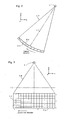
- Figures 2 and 3 are explanatory diagrams of the X-ray tube 21 and multi-row detector 24.
- the X-ray tube 21 and multi-row detector 24 rotate around a center of rotation IC. Representing the direction of rectilinear motion of the table 12 as z-direction, a direction perpendicular to the upper surface of the table 12 as y-direction, and a direction orthogonal to the z- and y-directions as x-direction, a plane of rotation of the X-ray tube 21 and multi-row detector 24 is an x-y plane.
- the X-ray tube 21 generates an X-ray beam CB generally referred to as a cone beam.
- a view angle view 0° is defined.
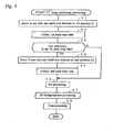
- Figure 4 is a flow chart showing data collection processing.
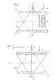
- Step S1 the X-ray tube 21 and multi-row detector 24 are moved to a first position Z1 as shown in Figure 5 (in practice, the table 12 is rectilinearly moved).
- first axial scan data D0(Z1, view, j, i) is collected while rotating the X-ray tube 21 and multi-row detector 24 around the center of rotation IC, as shown in Figure 5.
- Figure 6 shows a format of storage of the collected axial scan data.
- Step S3 a decision is made as to whether any data is deficient in the first axial scan data D0(Z1, view, j, i) based on the position of a reconstruction field, and if so, the process goes to Step S4; otherwise, to Step S6.
- reconstruction fields P1, P2 and P3 are defined as shown in Figure 7, the reconstruction fields P1 and P2 have no deficient data in the first axial scan data D0(Z1, view, j, i), although the reconstruction field P3 has deficient data in a central portion a2 because the first axial scan data D0(Z1, view, j, i) covers only a peripheral portion a1.
- Step S4 the X-ray tube 21 and multi-row detector 24 are moved to a second position Z2, as shown in Figure 8 (in practice, the table 12 is rectilinearly moved).
- Step S5 second axial scan data D0(Z view, j, i) is collected while rotating the X-ray tube 21 and multi-row detector 24 around the center of rotation IC, as shown in Figure 8. The process then goes to Step S6.
- a distance L between the first position Z1 and second position Z2 may be set to be equal to a cone beam width W at the center of X-ray rotation IC.
- data of the central portion a2 in the reconstruction field P3 can be obtained from the second axial scan data D0(Z view, j, i) as shown in Figure 10.
- Step S6 the data D0(Z1, view, j, i) and data D0(Z2, view, j, i) are subjected to pre-processing (offset correction, log correction, X-ray dose correction and sensitivity correction).
- Step S7 the pre-processed data D0(Z1, view, j, i) and D0(Z2, view, j, i ) are subjected to three-dimensional backprojection processing (which will be discussed later with reference to Figure 11) to determine backprojected data D3(x, y).
- Step S8 the backprojected data D3(x, y) is subjected to post-processing to obtain a CT image.
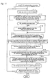
- FIG 11 is a flow chart showing details of the three-dimensional backprojection processing (Step S7 in Figure 4).
- one view is taken as a view of interest in a view range needed in image reconstruction.
- the view range is, for example, "180° + fan angle” or "360°.”
- Figure 12 exemplarily shows a plurality of parallel lines L0 ⁇ L8 on the reconstruction plane P1.
- the number of lines is 1/64 ⁇ 1/2 of the maximum number of pixels in a reconstruction plane in a direction orthogonal to the lines. For example, if the number of pixels in a reconstruction plane P is 512x512, the number of lines is nine.
- the line direction is defined as the x-direction for ⁇ 45° ⁇ view ⁇ 45° (or a view angle range mainly including the range and also including its vicinity) and 135° ⁇ view ⁇ 225° (or a view angle range mainly including the range and also including its vicinity).
- the line direction is defined as the y-direction for 45° ⁇ view ⁇ 135° (or a view angle range mainly including the range and also including its vicinity) and 225° ⁇ view ⁇ 315° (or a view angle range mainly including the range and also including its vicinity).
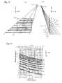
- Figure 13 shows lines T0 ⁇ T8 formed by projecting the plurality of parallel lines L0 ⁇ L8 on the reconstruction plane P1 onto a detector plane dp in a direction of X-ray transmission.
- the direction of X-ray transmission is determined by the geometry of the X-ray tube 21, multi-row detector 24 and lines L0 ⁇ L8.
- the projection data Dr corresponding to the lines L0 ⁇ L8 can be obtained by extracting data at the detector row j and channel i corresponding to the lines T0 ⁇ T8 projected onto the detector plane dp .
- data corresponding to the lines T0 ⁇ T8 is extracted from among the first axial scan data D0(z1, view, j, i ), and is developed as data Dr1 over the lines L0 ⁇ L8 on the reconstruction field P1.
- data Dr3 can be similarly extracted for the lines L0, L1, L2, L6, L7 and L8 in the peripheral portion a1, although data Dr3 cannot be extracted from the first axial scan data D0(z1, view, j, i) for the lines L3, L4 and L5 in the central portion a2.
- Step R3 if some projection data is deficient, the process goes to Step R4; otherwise, to Step R5.
- Step R4 as shown in Figure 14, data Dr3 for the lines L3, L4 and L5 in the central portion a2 of the reconstruction field P3 is extracted from the second axial scan data D0(z2. view, j, i).
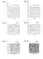
- Step R5 the projection data Drn is multiplied by a cone beam reconstruction weight to generate projection line data Dpn as shown in Figure 16.
- the cone beam reconstruction weight is ( r 1/ r 0) 2 , where r 0 is the distance from the focal spot of the X-ray tube 21 to the j -th detector row and the i -th channel of the multi-row detector 24 corresponding to projection data Drn, and r 1 is the distance from the focal spot of the X-ray tube 21 to a point on the reconstruction plane Pn corresponding to the projection data Drn.
- Step R6 the projection line data Dp1, Dp2 and Dp3 are subjected to weighted addition to obtain combined projection line data Dp as shown in Figure 17.
- This processing represents the Z-filtering.
- a weight for the weighted addition is determined from a mutual relationship between the reconstruction fields Pn, and from an appropriate weighting function W, as shown in Figure 41.
- the combined projection line data Dp is filtered. Specifically, the combined projection line data Dp is subjected to FFT, multiplied by a filter function (reconstruction function), and subjected to inverse FFT to generate image-positional line data Df as shown in Figure 18.
- Step R8 the image-positional line data Df is sampled and interpolated/extrapolated as needed to obtain backprojected pixel data D2 of pixels on the lines L0 ⁇ L8, as shown in Figure 19.
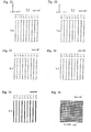
- Step R9 the image-positional line data Df is sampled and interpolated/extrapolated to obtain backprojected pixel data D2 of pixels in between the lines L0 ⁇ L8, as shown in Figure 20.
- Step R10 the backprojected pixel data D2 shown in Figure 20 or 26 are added on a pixel-by-pixel basis, as shown in Figure 27.
- Step R11 Steps R1 ⁇ R10 are repeated across all views needed in image reconstruction to obtain backprojected data D3(x, y). The process is then terminated.
- first axial scan data is collected at the first position Z1
- second axial scan data is collected at the second position Z2
- deficient data in the first axial scan data is complemented by the second axial scan data to generate projection data of the reconstruction fields P1, P2 and P3, and the projection data are subjected to Z-filtering to produce one CT image; therefore, omission of projection data is avoided, thus improving image quality.
- the Z-filtering is applied to a projection data stage, the amount of calculation is reduced.
- Figure 28 is a flow chart showing details of the three-dimensional backprojection processing (Step S7 in Figure 4) in accordance with Example 2.
- one view is taken as a view of interest in a view range needed in image reconstruction.
- the view range is, for example, "180° + fan angle” or "360°".
- Step H3 projection data Drn corresponding to a plurality of parallel lines at spacings of a plurality of pixels on the reconstruction field of interest Pn is extracted from among the first axial scan data D0(z1, view, j, i) at the view of interest.
- Figure 29 shows the projection data Drn.
- Step H4 if any projection data is deficient, the process goes to Step H5; otherwise, to Step H6.
- the reconstruction fields P1 and P2 have no deficient projection data, although the reconstruction field P3 has deficient projection data because data Dr3 cannot be extracted from the first axial scan data D0(z1, view, j, i) for the lines L3, L4 and L5 in the central portion a2.
- Step H5 as shown in Figure 14, data Dr3 for the lines L3, L4 and L5 in the central portion a2 of the reconstruction field P3 is extracted from the second axial scan data D0(z2, view, j, i) .
- the projection data Drn is multiplied by a cone beam reconstruction weight to generate projection line data Dpn as shown in Figure 30.
- the projection line data Dpn is filtered. Specifically, the projection line data Dpn is subjected to FFT, multiplied by a filter function (reconstruction function), and subjected to inverse FFT to generate image-positional line data Dfn as shown in Figure 31.
- FFT Fast Fourier transform
- a filter function reconstruction function
- Step H8 the image-positional line data Dfn is sampled and interpolated/extrapolated as needed to obtain backprojected pixel data D2 of pixels on the lines L0 ⁇ L8, as shown in Figure 32.
- the image-positional line data Dfn is sampled and interpolated/extrapolated to obtain backprojected pixel data D2 of pixels in between the lines L0 ⁇ L8, as shown in Figure 33.
- Step H10 the backprojected pixel data D2 shown in Figure 33 or 38 are added on a pixel-by-pixel basis, as shown in Figure 39.
- Step H 11 Steps H2 ⁇ H10 are repeated across all views needed in image reconstruction to obtain backprojected data D3n( x , y ). Once the backprojected data D3n( x , y ) has been obtained for the reconstruction field Pn of interest, the process goes to Step H12.
- Step H13 the backprojected data D3n(x, y) for all reconstruction fields Pn are subjected to weighted addition on a pixel-by-pixel basis, as shown in Figure 40.
- This processing represents the Z-filtering. The process is then terminated.
- the weight wn for the weighted addition is determined from a mutual relationship between the reconstruction fields Pn, and from an appropriate weighting function W, as shown in Figure 41.
- first axial scan data is collected at the first position Z1
- second axial scan data is collected at the second position Z2
- deficient data in the first axial scan data is complemented by the second axial scan data to generate projection data of the reconstruction field P1, P2 and P3
- CT images for the reconstruction fields P1, P2 and P3 are produced from the projection data
- the CT images of the reconstruction fields P1, P2 and P3 are subjected to Z-filtering to produce one CT image; therefore, omission of projection data is avoided, thus improving image quality.
- CT images of the reconstruction fields P1, P2 and P3 and a CT image combining them through Z-filtering can be simultaneously obtained.
- the technique for image reconstruction may be a three-dimensional image reconstruction technique according to the conventionally known Feldkamp method.
- three-dimensional image reconstruction techniques proposed in Japanese Patent Application Nos. 2002-147061, 2002-147231, 2002-235561, 2002-267833, 2002-322756 and 2002-338947 may be employed.
- a two-dimensional image reconstruction technique may be employed.
Landscapes
- Physics & Mathematics (AREA)
- General Physics & Mathematics (AREA)
- Engineering & Computer Science (AREA)
- Theoretical Computer Science (AREA)
- Apparatus For Radiation Diagnosis (AREA)
- Image Processing (AREA)
Applications Claiming Priority (2)
| Application Number | Priority Date | Filing Date | Title |
|---|---|---|---|
| JP2004065003 | 2004-03-09 | ||
| JP2004065003A JP4091008B2 (ja) | 2004-03-09 | 2004-03-09 | Ct画像生成方法およびx線ct装置 |
Publications (2)
| Publication Number | Publication Date |
|---|---|
| EP1574997A2 true EP1574997A2 (de) | 2005-09-14 |
| EP1574997A3 EP1574997A3 (de) | 2010-07-28 |
Family
ID=34824545
Family Applications (1)
| Application Number | Title | Priority Date | Filing Date |
|---|---|---|---|
| EP05250529A Withdrawn EP1574997A3 (de) | 2004-03-09 | 2005-02-01 | Verfahren zum Erzeugen eines tomographischen Bildes und Röntgen-Computertomograph |
Country Status (5)
| Country | Link |
|---|---|
| US (1) | US7260175B2 (de) |
| EP (1) | EP1574997A3 (de) |
| JP (1) | JP4091008B2 (de) |
| KR (1) | KR20060043586A (de) |
| CN (1) | CN100400000C (de) |
Families Citing this family (7)
| Publication number | Priority date | Publication date | Assignee | Title |
|---|---|---|---|---|
| JP2007044207A (ja) * | 2005-08-09 | 2007-02-22 | Ge Medical Systems Global Technology Co Llc | 放射線ct撮影方法およびx線ct装置 |
| JP4509903B2 (ja) * | 2005-09-27 | 2010-07-21 | ジーイー・メディカル・システムズ・グローバル・テクノロジー・カンパニー・エルエルシー | X線ct装置 |
| JP4611168B2 (ja) * | 2005-10-07 | 2011-01-12 | ジーイー・メディカル・システムズ・グローバル・テクノロジー・カンパニー・エルエルシー | 画像再構成方法、およびx線ct装置 |
| JP2007236662A (ja) * | 2006-03-09 | 2007-09-20 | Ge Medical Systems Global Technology Co Llc | X線ct装置およびそのx線ct画像再構成方法、x線ct画像撮影方法。 |
| JP2008006032A (ja) * | 2006-06-29 | 2008-01-17 | Ge Medical Systems Global Technology Co Llc | X線ct装置およびx線ct撮影方法 |
| US8284892B2 (en) * | 2008-12-22 | 2012-10-09 | General Electric Company | System and method for image reconstruction |
| JP5511189B2 (ja) * | 2009-01-05 | 2014-06-04 | ジーイー・メディカル・システムズ・グローバル・テクノロジー・カンパニー・エルエルシー | 画像再構成方法およびx線ct装置 |
Family Cites Families (29)
| Publication number | Priority date | Publication date | Assignee | Title |
|---|---|---|---|---|
| JPH05130984A (ja) | 1991-11-14 | 1993-05-28 | Toshiba Corp | X線ct装置 |
| JP2914891B2 (ja) * | 1995-07-05 | 1999-07-05 | 株式会社東芝 | X線コンピュータ断層撮影装置 |
| JPH09154838A (ja) | 1995-12-05 | 1997-06-17 | Toshiba Corp | ヘリカルスキャンの投影データ作成方法及びx線ct装置 |
| JP3124254B2 (ja) | 1997-07-24 | 2001-01-15 | ジーイー横河メディカルシステム株式会社 | 放射線断層撮影装置 |
| JP3174288B2 (ja) | 1997-09-01 | 2001-06-11 | ジーイー横河メディカルシステム株式会社 | Ct画像作成方法およびx線ct装置 |
| DE19905974A1 (de) * | 1999-02-12 | 2000-09-07 | Siemens Ag | Verfahren zur Abtastung eines Untersuchungsobjekts mittels eines CT-Geräts |
| US6185271B1 (en) * | 1999-02-16 | 2001-02-06 | Richard Estyn Kinsinger | Helical computed tomography with feedback scan control |
| JP3866431B2 (ja) * | 1999-02-17 | 2007-01-10 | 株式会社東芝 | X線ct装置 |
| US6272200B1 (en) * | 1999-07-28 | 2001-08-07 | Arch Development Corporation | Fourier and spline-based reconstruction of helical CT images |
| JP4726287B2 (ja) * | 1999-10-20 | 2011-07-20 | 株式会社日立メディコ | マルチスライスx線ct装置 |
| JP2001187046A (ja) | 1999-12-27 | 2001-07-10 | Ge Medical Systems Global Technology Co Llc | マルチスライスx線ct装置及びその制御方法 |
| US6301325B1 (en) | 1999-12-22 | 2001-10-09 | Ge Medical Systems Global Technology Company Llc | Half-scan algorithm for use with a high speed multi-row fan beam helical detector |
| US6442228B1 (en) | 2000-04-20 | 2002-08-27 | Ge Medical Systems Global Technology Company, Llc | Data acquisition modifications for improved reconstruction with conventional CT |
| US6539074B1 (en) * | 2000-08-25 | 2003-03-25 | General Electric Company | Reconstruction of multislice tomographic images from four-dimensional data |
| JP4652555B2 (ja) | 2000-11-10 | 2011-03-16 | イビデン株式会社 | 触媒コンバータ、触媒コンバータ用保持シール材 |
| JP3458100B2 (ja) | 2000-11-17 | 2003-10-20 | 繁雄 小川 | 雪庇防止装置 |
| DE10060291A1 (de) | 2000-12-05 | 2002-06-06 | Bosch Gmbh Robert | Strömungstelleinheit |
| JP3942142B2 (ja) * | 2000-12-15 | 2007-07-11 | ジーイー・メディカル・システムズ・グローバル・テクノロジー・カンパニー・エルエルシー | 放射線断層撮影装置およびその方法 |
| US6463118B2 (en) | 2000-12-29 | 2002-10-08 | Ge Medical Systems Global Technology Company, Llc | Computed tomography (CT) weighting for high quality image recontruction |
| JP2002235561A (ja) | 2001-02-08 | 2002-08-23 | Denso Corp | スロットル弁調節装置 |
| JP2002267833A (ja) | 2001-03-14 | 2002-09-18 | Toray Ind Inc | 液晶ディスプレイ用カラーフィルタの製造方法。 |
| JP2002322756A (ja) | 2001-04-27 | 2002-11-08 | Sekisui Chem Co Ltd | 屋根構造、該屋根構造の構築方法及び該屋根構造に用いられる屋根パネル |
| JP2002338947A (ja) | 2001-05-15 | 2002-11-27 | Hitachi Kasei Polymer Co Ltd | 1成分形シーリング材 |
| JP4175791B2 (ja) | 2001-08-20 | 2008-11-05 | ジーイー・メディカル・システムズ・グローバル・テクノロジー・カンパニー・エルエルシー | 画像生成方法およびx線ct装置 |
| JP2003159244A (ja) | 2001-09-17 | 2003-06-03 | Ge Medical Systems Global Technology Co Llc | 画像再構成方法およびx線ct装置 |
| JP3870105B2 (ja) | 2002-02-22 | 2007-01-17 | ジーイー・メディカル・システムズ・グローバル・テクノロジー・カンパニー・エルエルシー | 逆投影方法およびx線ct装置 |
| JP4106251B2 (ja) | 2002-03-12 | 2008-06-25 | ジーイー・メディカル・システムズ・グローバル・テクノロジー・カンパニー・エルエルシー | 3次元逆投影方法およびx線ct装置 |
| JP3950782B2 (ja) | 2002-05-22 | 2007-08-01 | ジーイー・メディカル・システムズ・グローバル・テクノロジー・カンパニー・エルエルシー | 3次元逆投影方法およびx線ct装置 |
| US6850585B2 (en) * | 2003-03-05 | 2005-02-01 | Ge Medical Systems Global Technology Company, Llc | Progressive updating approach for volumetric CT image reconstruction |
-
2004
- 2004-03-09 JP JP2004065003A patent/JP4091008B2/ja not_active Expired - Fee Related
-
2005
- 2005-02-01 EP EP05250529A patent/EP1574997A3/de not_active Withdrawn
- 2005-03-09 KR KR1020050019606A patent/KR20060043586A/ko not_active Abandoned
- 2005-03-09 CN CNB2005100539499A patent/CN100400000C/zh not_active Expired - Fee Related
- 2005-04-08 US US11/074,228 patent/US7260175B2/en not_active Expired - Lifetime
Non-Patent Citations (2)
| Title |
|---|
| GRASS M ET AL: "3D CONE-BEAM CT RECONSTRUCTION FOR CIRCULAR TRAJECTORIES" PHYSICS IN MEDICINE AND BIOLOGY, TAYLOR AND FRANCIS LTD. LONDON, GB LNKD- DOI:10.1088/0031-9155/45/2/306, vol. 45, no. 2, 1 February 2000 (2000-02-01), pages 329-347, XP000993403 ISSN: 0031-9155 * |
| KÖHLER TH ET AL: "A fast and efficient method for sequential cone-beam tomography" MEDICAL PHYSICS, AIP, MELVILLE, NY, US LNKD- DOI:10.1118/1.1395025, vol. 28, no. 11, 1 November 2001 (2001-11-01), pages 2318-2327, XP012011337 ISSN: 0094-2405 * |
Also Published As
| Publication number | Publication date |
|---|---|
| KR20060043586A (ko) | 2006-05-15 |
| CN1666714A (zh) | 2005-09-14 |
| US7260175B2 (en) | 2007-08-21 |
| EP1574997A3 (de) | 2010-07-28 |
| JP2005253485A (ja) | 2005-09-22 |
| JP4091008B2 (ja) | 2008-05-28 |
| US20050201511A1 (en) | 2005-09-15 |
| CN100400000C (zh) | 2008-07-09 |
Similar Documents
| Publication | Publication Date | Title |
|---|---|---|
| JP3950782B2 (ja) | 3次元逆投影方法およびx線ct装置 | |
| EP1741062B1 (de) | Conus-strahl-ct-vorrichtung mit abgeschnittenen projektionen und einem zuvor erfassten 3d-ct-bild | |
| JP2007000406A (ja) | X線ct撮影方法およびx線ct装置 | |
| DE602004009359T2 (de) | Computertomograph und Verfahren zur Erzeugung von tomographischen Bildern | |
| JP2005040582A (ja) | X線ct撮像方法およびx線ct装置 | |
| US7173997B2 (en) | X-ray CT imaging method and X-ray CT apparatus | |
| US5561695A (en) | Methods and apparatus for reducing image artifacts | |
| JP4106251B2 (ja) | 3次元逆投影方法およびx線ct装置 | |
| JP4679348B2 (ja) | X線ct装置 | |
| US20050094760A1 (en) | CT image producing method and X-ray CT apparatus | |
| US7260175B2 (en) | CT image producing method and X-ray CT apparatus | |
| EP1542166B1 (de) | Verfahren und Vorrichtung zur dreidimensionalen Rückprojektion und Röntgenstrahlencomputertomograph | |
| US6101235A (en) | Methods and apparatus for altering spatial characteristics of a digital image | |
| JP2007236502A (ja) | X線ct装置 | |
| US7519144B2 (en) | Multi-positional CT image producing method and X-ray CT apparatus | |
| JP5781011B2 (ja) | 画像処理方法、画像処理装置および撮影装置並びにプログラム | |
| JP4156629B2 (ja) | 3次元逆投影方法およびx線ct装置 | |
| EP1544798A2 (de) | Verfahren zur Korrektur von Daten und Röntgen-Tomographiegerät | |
| US7215735B2 (en) | CT image production method and X-ray CT system | |
| JP2004230030A (ja) | 超低x線量ct画像生成方法およびx線ct装置 | |
| JPH10155788A (ja) | コンピュータ断層撮影装置 |
Legal Events
| Date | Code | Title | Description |
|---|---|---|---|
| PUAI | Public reference made under article 153(3) epc to a published international application that has entered the european phase |
Free format text: ORIGINAL CODE: 0009012 |
|
| AK | Designated contracting states |
Kind code of ref document: A2 Designated state(s): AT BE BG CH CY CZ DE DK EE ES FI FR GB GR HU IE IS IT LI LT LU MC NL PL PT RO SE SI SK TR |
|
| AX | Request for extension of the european patent |
Extension state: AL BA HR LV MK YU |
|
| PUAL | Search report despatched |
Free format text: ORIGINAL CODE: 0009013 |
|
| AK | Designated contracting states |
Kind code of ref document: A3 Designated state(s): AT BE BG CH CY CZ DE DK EE ES FI FR GB GR HU IE IS IT LI LT LU MC NL PL PT RO SE SI SK TR |
|
| AX | Request for extension of the european patent |
Extension state: AL BA HR LV MK YU |
|
| AKY | No designation fees paid | ||
| REG | Reference to a national code |
Ref country code: DE Ref legal event code: R108 Effective date: 20110309 Ref country code: DE Ref legal event code: 8566 |
|
| STAA | Information on the status of an ep patent application or granted ep patent |
Free format text: STATUS: THE APPLICATION IS DEEMED TO BE WITHDRAWN |
|
| 18D | Application deemed to be withdrawn |
Effective date: 20110129 |