EP1504721B1 - Echographe - Google Patents
Echographe Download PDFInfo
- Publication number
- EP1504721B1 EP1504721B1 EP03748538A EP03748538A EP1504721B1 EP 1504721 B1 EP1504721 B1 EP 1504721B1 EP 03748538 A EP03748538 A EP 03748538A EP 03748538 A EP03748538 A EP 03748538A EP 1504721 B1 EP1504721 B1 EP 1504721B1
- Authority
- EP
- European Patent Office
- Prior art keywords
- ultrasonic
- image
- tomographic
- images
- diagnostic apparatus
- Prior art date
- Legal status (The legal status is an assumption and is not a legal conclusion. Google has not performed a legal analysis and makes no representation as to the accuracy of the status listed.)
- Expired - Lifetime
Links
- 238000012545 processing Methods 0.000 claims abstract description 107
- 238000000034 method Methods 0.000 claims abstract description 27
- 230000008569 process Effects 0.000 claims abstract description 13
- 239000003550 marker Substances 0.000 claims description 163
- 239000000523 sample Substances 0.000 claims description 31
- 239000002775 capsule Substances 0.000 claims description 19
- 230000009545 invasion Effects 0.000 abstract description 8
- 238000010586 diagram Methods 0.000 description 61
- 239000013598 vector Substances 0.000 description 25
- 238000010276 construction Methods 0.000 description 21
- 230000003287 optical effect Effects 0.000 description 20
- 230000003902 lesion Effects 0.000 description 14
- 210000000056 organ Anatomy 0.000 description 13
- 230000008901 benefit Effects 0.000 description 11
- 238000006243 chemical reaction Methods 0.000 description 10
- 230000033001 locomotion Effects 0.000 description 8
- 238000004891 communication Methods 0.000 description 6
- 210000001198 duodenum Anatomy 0.000 description 5
- 230000008859 change Effects 0.000 description 4
- 238000003745 diagnosis Methods 0.000 description 4
- 210000003238 esophagus Anatomy 0.000 description 4
- 210000001035 gastrointestinal tract Anatomy 0.000 description 4
- 230000001678 irradiating effect Effects 0.000 description 4
- 239000000463 material Substances 0.000 description 4
- 210000002784 stomach Anatomy 0.000 description 4
- 238000004804 winding Methods 0.000 description 4
- 210000004204 blood vessel Anatomy 0.000 description 3
- 238000001514 detection method Methods 0.000 description 3
- 230000004044 response Effects 0.000 description 3
- 238000012935 Averaging Methods 0.000 description 2
- 230000005540 biological transmission Effects 0.000 description 2
- 239000003086 colorant Substances 0.000 description 2
- 230000001934 delay Effects 0.000 description 2
- 230000006870 function Effects 0.000 description 2
- 238000003780 insertion Methods 0.000 description 2
- 230000037431 insertion Effects 0.000 description 2
- 230000010355 oscillation Effects 0.000 description 2
- 210000003240 portal vein Anatomy 0.000 description 2
- 230000009747 swallowing Effects 0.000 description 2
- 238000011282 treatment Methods 0.000 description 2
- 241000167880 Hirundinidae Species 0.000 description 1
- 206010028980 Neoplasm Diseases 0.000 description 1
- 239000004698 Polyethylene Substances 0.000 description 1
- 230000003187 abdominal effect Effects 0.000 description 1
- 230000015572 biosynthetic process Effects 0.000 description 1
- 230000000740 bleeding effect Effects 0.000 description 1
- 238000007796 conventional method Methods 0.000 description 1
- 201000010099 disease Diseases 0.000 description 1
- 208000037265 diseases, disorders, signs and symptoms Diseases 0.000 description 1
- 238000002592 echocardiography Methods 0.000 description 1
- 239000000835 fiber Substances 0.000 description 1
- 239000012530 fluid Substances 0.000 description 1
- 239000013307 optical fiber Substances 0.000 description 1
- 238000010422 painting Methods 0.000 description 1
- 210000000496 pancreas Anatomy 0.000 description 1
- 239000012188 paraffin wax Substances 0.000 description 1
- -1 polyethylene Polymers 0.000 description 1
- 229920000573 polyethylene Polymers 0.000 description 1
- 238000004393 prognosis Methods 0.000 description 1
- 238000001454 recorded image Methods 0.000 description 1
- 210000001835 viscera Anatomy 0.000 description 1
- XLYOFNOQVPJJNP-UHFFFAOYSA-N water Substances O XLYOFNOQVPJJNP-UHFFFAOYSA-N 0.000 description 1
Images


Classifications
-
- A—HUMAN NECESSITIES
- A61—MEDICAL OR VETERINARY SCIENCE; HYGIENE
- A61B—DIAGNOSIS; SURGERY; IDENTIFICATION
- A61B8/00—Diagnosis using ultrasonic, sonic or infrasonic waves
- A61B8/44—Constructional features of the ultrasonic, sonic or infrasonic diagnostic device
- A61B8/4444—Constructional features of the ultrasonic, sonic or infrasonic diagnostic device related to the probe
- A61B8/4461—Features of the scanning mechanism, e.g. for moving the transducer within the housing of the probe
-
- A—HUMAN NECESSITIES
- A61—MEDICAL OR VETERINARY SCIENCE; HYGIENE
- A61B—DIAGNOSIS; SURGERY; IDENTIFICATION
- A61B8/00—Diagnosis using ultrasonic, sonic or infrasonic waves
- A61B8/12—Diagnosis using ultrasonic, sonic or infrasonic waves in body cavities or body tracts, e.g. by using catheters
-
- A—HUMAN NECESSITIES
- A61—MEDICAL OR VETERINARY SCIENCE; HYGIENE
- A61B—DIAGNOSIS; SURGERY; IDENTIFICATION
- A61B8/00—Diagnosis using ultrasonic, sonic or infrasonic waves
- A61B8/13—Tomography
- A61B8/14—Echo-tomography
-
- A—HUMAN NECESSITIES
- A61—MEDICAL OR VETERINARY SCIENCE; HYGIENE
- A61B—DIAGNOSIS; SURGERY; IDENTIFICATION
- A61B8/00—Diagnosis using ultrasonic, sonic or infrasonic waves
- A61B8/42—Details of probe positioning or probe attachment to the patient
- A61B8/4245—Details of probe positioning or probe attachment to the patient involving determining the position of the probe, e.g. with respect to an external reference frame or to the patient
- A61B8/4254—Details of probe positioning or probe attachment to the patient involving determining the position of the probe, e.g. with respect to an external reference frame or to the patient using sensors mounted on the probe
-
- A—HUMAN NECESSITIES
- A61—MEDICAL OR VETERINARY SCIENCE; HYGIENE
- A61B—DIAGNOSIS; SURGERY; IDENTIFICATION
- A61B8/00—Diagnosis using ultrasonic, sonic or infrasonic waves
- A61B8/44—Constructional features of the ultrasonic, sonic or infrasonic diagnostic device
- A61B8/4416—Constructional features of the ultrasonic, sonic or infrasonic diagnostic device related to combined acquisition of different diagnostic modalities, e.g. combination of ultrasound and X-ray acquisitions
-
- A—HUMAN NECESSITIES
- A61—MEDICAL OR VETERINARY SCIENCE; HYGIENE
- A61B—DIAGNOSIS; SURGERY; IDENTIFICATION
- A61B8/00—Diagnosis using ultrasonic, sonic or infrasonic waves
- A61B8/44—Constructional features of the ultrasonic, sonic or infrasonic diagnostic device
- A61B8/4444—Constructional features of the ultrasonic, sonic or infrasonic diagnostic device related to the probe
- A61B8/445—Details of catheter construction
-
- A—HUMAN NECESSITIES
- A61—MEDICAL OR VETERINARY SCIENCE; HYGIENE
- A61B—DIAGNOSIS; SURGERY; IDENTIFICATION
- A61B8/00—Diagnosis using ultrasonic, sonic or infrasonic waves
- A61B8/46—Ultrasonic, sonic or infrasonic diagnostic devices with special arrangements for interfacing with the operator or the patient
- A61B8/461—Displaying means of special interest
- A61B8/463—Displaying means of special interest characterised by displaying multiple images or images and diagnostic data on one display
-
- A—HUMAN NECESSITIES
- A61—MEDICAL OR VETERINARY SCIENCE; HYGIENE
- A61B—DIAGNOSIS; SURGERY; IDENTIFICATION
- A61B8/00—Diagnosis using ultrasonic, sonic or infrasonic waves
- A61B8/48—Diagnostic techniques
- A61B8/483—Diagnostic techniques involving the acquisition of a 3D volume of data
Definitions
- a second observation is performed for finding how deep the lesion invades in a direction vertical to a surface of the lumen.
- US 5,924,989 discloses a method and apparatus for generating a diagnostically usable three-dimensional image data using comparatively complex calculation with four weighting factors.
- the ultrasonic diagnostic apparatus When an operator operating the ultrasonic diagnostic apparatus causes the display of a menu having different items and inputs a selection of an item from the displayed menu by using the keyboard 15 and/or the mouse 16, the data of the selected item is transmitted from the external input control circuit 37 to the controller 39.
- the controller 39 drives and controls the circuits 31 to 36.
- the construction of an ultrasonic tomographic image, the slicing of the tomographic image and/or the construction of tomographic parallel images thereof may be performed.
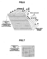
- the controller 39 causes the image constructing circuit 31 to sequentially construct ultrasonic tomographic images as shown in Fig. 4 based on reflected echoes from the ultrasonic transducer 25 at a step S103.
- reference numerals 1 to n are given to the ultrasonic tomographic images in order of the construction.
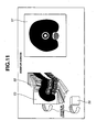
- the controller 39 controls the monitor 14 to display side by side the newest ultrasonic tomographic image and the newest tomographic parallel images.
- the tomographic parallel images 53 are re-constructed after the operator moves the slicing line marker 57.
- the rotated tomographic parallel images 53 can be re-constructed every time the slicing line marker slightly moves if the processing by the controller 39, bus 38, image processing circuit 33, display circuit 34 and so on is fast enough. In this case, it appears to an operator that the slicing line marker 57 and the tomographic parallel images 53 vary in connection with each other simultaneously.
- a twist occurs in the flexible shaft 26.
- the twist causes ununiformity among plural ultrasonic tomographic images, and a distortion may occur on tomographic parallel images. This is because, in mechanical radial scanning, a position of a rotational angle of a motor is detected by a rotary encoder adjacent to the motor.
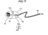
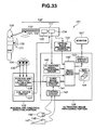
- Fig. 17 is a block diagram showing an ultrasonic endoscope according to this variation example.
- This display example can be achieved by performing a following operation.
- Auxiliary image creating means creates an auxiliary image including a coordinates marker indicating a coordinates system, which is a reference for calculating position data or direction data of a scan plane. Under this construction, at which angle the locus is observed for a given auxiliary image can be easily understood.
Landscapes
- Life Sciences & Earth Sciences (AREA)
- Health & Medical Sciences (AREA)
- Medical Informatics (AREA)
- Biophysics (AREA)
- Nuclear Medicine, Radiotherapy & Molecular Imaging (AREA)
- Pathology (AREA)
- Radiology & Medical Imaging (AREA)
- Engineering & Computer Science (AREA)
- Biomedical Technology (AREA)
- Heart & Thoracic Surgery (AREA)
- Physics & Mathematics (AREA)
- Molecular Biology (AREA)
- Surgery (AREA)
- Animal Behavior & Ethology (AREA)
- General Health & Medical Sciences (AREA)
- Public Health (AREA)
- Veterinary Medicine (AREA)
- Ultra Sonic Daignosis Equipment (AREA)
- Steroid Compounds (AREA)
- Surgical Instruments (AREA)
- Investigating Or Analyzing Materials By The Use Of Ultrasonic Waves (AREA)
Claims (17)
- Appareil de diagnostic à ultrasons obtenant de multiples images échotomographiques d'un processus où une sonde à ultrasons (11a) se déplace et scanne à l'intérieur d'une cavité corporelle d'un corps à examiner, l'appareil comprenant :un moyen de détection d'une information de position (13) pour détecter une information de position de multiples images échotomographiques (51) obtenues dans un processus où la sonde à ultrasons se déplace à l'intérieur d'une cavité corporelle d'un corps à examiner ;un moyen de construction d'images tomographiques parallèles (12) pour construire de multiples images tomographiques parallèles agencées le long d'un chemin de balayage de la sonde à ultrasons sur la base de l'information de position obtenue par le moyen de détection d'une information de position (13), etun moyen de découpage pour découper l'image échotomographique et créer des tranches des images échotomographiques,dans lequel le moyen de construction d'images tomographiques parallèles construit des images tomographiques parallèles en agençant les tranches,
caractérisé en ce que le moyen de construction d'images parallèles (12) est adapté pour- stocker sous la forme d'une valeur initiale une valeur maximale stockable dans une cellule tampon Z,- si la distance d'un pixel depuis un plan présumé est plus petite qu'une valeur stockée dans la cellule tampon Z, pour stocker une distance du pixel le plus proche immédiatement sous les pixels du plan présumé sur une pluralité de tranches (52) créées depuis la pluralité d'images échotomographiques (51), et- si la distance d'un pixel depuis le plan présumé est supérieure ou égale à la valeur dans la cellule tampon Z, pour ne réaliser aucune transformation. - Appareil de diagnostic à ultrasons selon la revendication 1, comprenant en outre un moyen de contrôle d'affichage (34) pour faire afficher au moyen d'affichage l'image échotomographique et les images tomographiques parallèles de sorte à les comparer.
- Appareil de diagnostic à ultrasons selon la revendication 1 ou 2, dans lequel le moyen de construction d'images tomographiques parallèles (12) construit de nouvelles images tomographiques parallèles par réécriture des pixels correspondant aux images tomographiques parallèles avec les pixels correspondant à l'image échotomographique chaque fois que l'image échotomographique est créée dans le processus où la sonde à ultrasons (11a) se déplace et balaye à l'intérieur d'une cavité corporelle d'un corps à examiner.
- Appareil de diagnostic à ultrasons selon l'une quelconque des revendications 1 à 3, dans lequel le moyen de détection d'une information de position (13) est adapté pour détecter les positions et orientations de multiples images échotomographiques.
- Appareil de diagnostic à ultrasons selon la revendication 4, comprenant en outre un moyen d'affichage affichant l'image échotomographique et les images tomographiques parallèles de sorte à les comparer.
- Appareil de diagnostic à ultrasons selon la revendication 2 ou 5, dans lequel le moyen d'affichage affiche l'image échotomographique et les images tomographiques parallèles sur un écran (14) de sorte à les comparer.
- Appareil de diagnostic à ultrasons selon la revendication 2 ou 5, dans lequel le moyen d'affichage affiche sur les images tomographiques parallèles un marqueur d'image échotomographique (55) indiquant une position de l'image échotomographique.
- Appareil de diagnostic à ultrasons selon la revendication 7, comprenant en outre un moyen de réglage du marqueur d'image échotomographique pour régler une position du marqueur d'image échotomographique (55), dans lequel le moyen d'affichage choisit et affiche l'image échotomographique selon une position du marqueur d'image échotomographique réglée par le moyen de réglage du marqueur d'image échotomographique.
- Appareil de diagnostic à ultrasons selon l'une quelconque des revendications 1 à 8, comprenant en outre un moyen de réglage d'une position de découpage pour régler une position de découpage de l'image échotomographique, dans lequel le moyen de découpage découpe une image échotomographique au niveau d'une position réglée par le moyen de réglage d'une position de découpage et crée des tranches de celle-ci.
- Appareil de diagnostic à ultrasons selon l'une quelconque des revendications 1 à 9, comprenant en outre un moyen de rotation pour construire de nouvelles images tomographiques parallèles (53) qui sont le résultat de la rotation des images tomographiques parallèles.
- Appareil de diagnostic à ultrasons selon l'une quelconque des revendications 1 à 10, dans lequel le moyen d'affichage affiche les images tomographiques parallèles et un indicateur indiquant une orientation des images tomographiques parallèles par rapport au moyen de détection de la position et l'orientation.
- Appareil de diagnostic à ultrasons selon la revendication 4, dans lequel le moyen de construction d'images tomographiques parallèles construit de nouvelles images tomographiques parallèles par réécriture des images tomographiques parallèles avec des pixels sur l'image échotomographique chaque fois que l'image échotomographique est créée dans un processus où une sonde à ultrasons se déplace à l'intérieur d'une cavité corporelle d'un corps à examiner.
- Appareil de diagnostic à ultrasons selon l'une quelconque des revendications 1 à 12, dans lequel le moyen de construction d'images tomographiques parallèles détermine les pixels à réécrire sur la base de la position et l'orientation détectées par le moyen de détection de position et d'orientation.
- Appareil de diagnostic à ultrasons selon l'une quelconque des revendications 1 à 13, dans lequel la sonde à ultrasons (11a) constitue un endoscope à ultrasons de type balayage radial mécanique réalisant un balayage radial mécanique.
- Appareil de diagnostic à ultrasons selon l'une quelconque des revendications 1 à 13, dans lequel la sonde à ultrasons (11a) constitue un endoscope à ultrasons de type balayage radial électronique réalisant un balayage radial électronique.
- Appareil de diagnostic à ultrasons selon l'une quelconque des revendications 1 à 13, dans lequel la sonde à ultrasons (11a) constitue un endoscope à ultrasons de type capsule.
- Appareil de diagnostic à ultrasons selon l'une quelconque des revendications 1 à 13, dans lequel la sonde à ultrasons (11a) constitue un endoscope à ultrasons de type balayage convexe réalisant un balayage convexe.
Priority Applications (1)
| Application Number | Priority Date | Filing Date | Title |
|---|---|---|---|
| EP08021668A EP2042102B1 (fr) | 2002-09-27 | 2003-09-18 | Appareil de diagnostic ultrasonique |
Applications Claiming Priority (5)
| Application Number | Priority Date | Filing Date | Title |
|---|---|---|---|
| JP2002283802A JP2004113628A (ja) | 2002-09-27 | 2002-09-27 | 超音波診断装置 |
| JP2002283802 | 2002-09-27 | ||
| JP2002288951 | 2002-10-01 | ||
| JP2002288951A JP4276825B2 (ja) | 2002-10-01 | 2002-10-01 | 超音波診断装置 |
| PCT/JP2003/011891 WO2004028375A1 (fr) | 2002-09-27 | 2003-09-18 | Echographe |
Related Child Applications (1)
| Application Number | Title | Priority Date | Filing Date |
|---|---|---|---|
| EP08021668A Division EP2042102B1 (fr) | 2002-09-27 | 2003-09-18 | Appareil de diagnostic ultrasonique |
Publications (3)
| Publication Number | Publication Date |
|---|---|
| EP1504721A1 EP1504721A1 (fr) | 2005-02-09 |
| EP1504721A4 EP1504721A4 (fr) | 2008-03-05 |
| EP1504721B1 true EP1504721B1 (fr) | 2010-01-06 |
Family
ID=32044639
Family Applications (2)
| Application Number | Title | Priority Date | Filing Date |
|---|---|---|---|
| EP03748538A Expired - Lifetime EP1504721B1 (fr) | 2002-09-27 | 2003-09-18 | Echographe |
| EP08021668A Expired - Lifetime EP2042102B1 (fr) | 2002-09-27 | 2003-09-18 | Appareil de diagnostic ultrasonique |
Family Applications After (1)
| Application Number | Title | Priority Date | Filing Date |
|---|---|---|---|
| EP08021668A Expired - Lifetime EP2042102B1 (fr) | 2002-09-27 | 2003-09-18 | Appareil de diagnostic ultrasonique |
Country Status (5)
| Country | Link |
|---|---|
| US (1) | US7775977B2 (fr) |
| EP (2) | EP1504721B1 (fr) |
| AT (2) | ATE499880T1 (fr) |
| DE (2) | DE60336281D1 (fr) |
| WO (1) | WO2004028375A1 (fr) |
Families Citing this family (36)
| Publication number | Priority date | Publication date | Assignee | Title |
|---|---|---|---|---|
| JP4537756B2 (ja) * | 2004-04-30 | 2010-09-08 | オリンパス株式会社 | 超音波診断装置 |
| EP1813194B1 (fr) * | 2004-09-16 | 2013-11-27 | Olympus Medical Systems Corp. | Sonde ultrasonore |
| AU2006201646B2 (en) * | 2005-04-26 | 2011-01-06 | Biosense Webster, Inc. | Display of catheter tip with beam direction for ultrasound system |
| US7604601B2 (en) * | 2005-04-26 | 2009-10-20 | Biosense Webster, Inc. | Display of catheter tip with beam direction for ultrasound system |
| JP4727302B2 (ja) * | 2005-06-02 | 2011-07-20 | 富士フイルム株式会社 | 超音波内視鏡システムおよび電子内視鏡システム |
| US20070066880A1 (en) * | 2005-09-09 | 2007-03-22 | Warren Lee | Image-based probe guidance system |
| US9084556B2 (en) * | 2006-01-19 | 2015-07-21 | Toshiba Medical Systems Corporation | Apparatus for indicating locus of an ultrasonic probe, ultrasonic diagnostic apparatus |
| JP4838032B2 (ja) * | 2006-03-31 | 2011-12-14 | テルモ株式会社 | 画像診断装置およびその処理方法 |
| JP4868959B2 (ja) * | 2006-06-29 | 2012-02-01 | オリンパスメディカルシステムズ株式会社 | 体腔内プローブ装置 |
| JP4805056B2 (ja) * | 2006-08-02 | 2011-11-02 | オリンパス株式会社 | 被検体内導入装置、体外受信装置、及び被検体内情報収集システム |
| WO2008030482A2 (fr) | 2006-09-06 | 2008-03-13 | Innurvation Inc | Système et procédé pour un échange d'informations acoustiques mettant en jeu une capsule à faible puissance pouvant être ingérée |
| JP5118867B2 (ja) * | 2007-03-16 | 2013-01-16 | オリンパス株式会社 | 内視鏡観察装置および内視鏡の作動方法 |
| US8527032B2 (en) * | 2007-05-16 | 2013-09-03 | General Electric Company | Imaging system and method of delivery of an instrument to an imaged subject |
| US20080287805A1 (en) * | 2007-05-16 | 2008-11-20 | General Electric Company | System and method to guide an instrument through an imaged subject |
| JP5394622B2 (ja) * | 2007-07-31 | 2014-01-22 | オリンパスメディカルシステムズ株式会社 | 医用ガイドシステム |
| JP5179812B2 (ja) * | 2007-09-07 | 2013-04-10 | 株式会社東芝 | 超音波診断装置、超音波画像処理装置、及び超音波画像処理プログラム |
| JP5416900B2 (ja) | 2007-11-22 | 2014-02-12 | 株式会社東芝 | 超音波診断装置及び穿刺支援用制御プログラム |
| US8870778B2 (en) * | 2007-12-24 | 2014-10-28 | Olympus Medical Systems Corp. | Tranlumen endoscope insertion surgery |
| JP5291955B2 (ja) * | 2008-03-10 | 2013-09-18 | 富士フイルム株式会社 | 内視鏡検査システム |
| JP2010000183A (ja) * | 2008-06-19 | 2010-01-07 | Fujinon Corp | 電子内視鏡用プロセッサ装置 |
| WO2010005571A2 (fr) | 2008-07-09 | 2010-01-14 | Innurvation, Inc. | Affichage de données image d’une capsule de numériseur |
| US8376951B2 (en) * | 2008-07-15 | 2013-02-19 | Hitachi Medical Corporation | Ultrasonic diagnostic apparatus and method for displaying probe operation guide |
| DE102009007868B3 (de) | 2009-02-06 | 2010-05-20 | Fraunhofer-Gesellschaft zur Förderung der angewandten Forschung e.V. | Sensorsystem und Verfahren zur bildgebenden Erfassung eines Objektes |
| AU2010231365A1 (en) * | 2009-03-31 | 2010-10-07 | Fujifilm Corporation | Image processing apparatus and method and program |
| JP5508765B2 (ja) * | 2009-06-03 | 2014-06-04 | 株式会社東芝 | 3次元超音波診断装置 |
| WO2011039983A1 (fr) * | 2009-09-30 | 2011-04-07 | テルモ株式会社 | Appareil d'imagerie diagnostique et procédé pour le commander |
| US8647259B2 (en) * | 2010-03-26 | 2014-02-11 | Innurvation, Inc. | Ultrasound scanning capsule endoscope (USCE) |
| JP6054089B2 (ja) * | 2011-08-19 | 2016-12-27 | 東芝メディカルシステムズ株式会社 | 超音波診断装置、医用画像処理装置および医用画像処理プログラム |
| EP2679167B1 (fr) | 2012-02-01 | 2018-04-18 | Olympus Corporation | Échographe |
| KR101386102B1 (ko) | 2012-03-09 | 2014-04-16 | 삼성메디슨 주식회사 | 초음파 영상 제공 방법 및 그를 위한 초음파 장치 |
| US9651525B2 (en) * | 2013-06-27 | 2017-05-16 | TecScan Systems Inc. | Method and apparatus for scanning an object |
| CN103558291B (zh) * | 2013-11-13 | 2017-01-04 | 北京安铁软件技术有限公司 | 一种车轮检测数据的显示方法及装置 |
| JP6263447B2 (ja) * | 2014-06-30 | 2018-01-17 | ジーイー・メディカル・システムズ・グローバル・テクノロジー・カンパニー・エルエルシー | 超音波診断装置及びプログラム |
| KR102656560B1 (ko) * | 2018-02-06 | 2024-04-12 | 삼성메디슨 주식회사 | 초음파 진단 장치 및 그 제어 방법 |
| CN113316418A (zh) * | 2018-10-22 | 2021-08-27 | 深圳迈瑞生物医疗电子股份有限公司 | 一种超声成像方法、系统 |
| US11717967B2 (en) | 2021-03-04 | 2023-08-08 | TecScan Systems Inc. | System and method for scanning an object using an array of ultrasonic transducers |
Family Cites Families (34)
| Publication number | Priority date | Publication date | Assignee | Title |
|---|---|---|---|---|
| JPH0724402Y2 (ja) | 1989-08-28 | 1995-06-05 | 株式会社フジクラ | 水道水熱を利用した路面融雪装置 |
| JP3251631B2 (ja) | 1992-03-25 | 2002-01-28 | 株式会社東芝 | 体腔内超音波診断装置 |
| JP3040306B2 (ja) | 1994-04-12 | 2000-05-15 | 新日本製鐵株式会社 | 蒸気洗浄装置 |
| JPH08117237A (ja) * | 1994-10-20 | 1996-05-14 | Fuji Photo Optical Co Ltd | 超音波診断装置 |
| US5924989A (en) * | 1995-04-03 | 1999-07-20 | Polz; Hans | Method and device for capturing diagnostically acceptable three-dimensional ultrasound image data records |
| US6256529B1 (en) * | 1995-07-26 | 2001-07-03 | Burdette Medical Systems, Inc. | Virtual reality 3D visualization for surgical procedures |
| JPH09192128A (ja) | 1996-01-23 | 1997-07-29 | Hitachi Medical Corp | 超音波診断装置 |
| JPH10192A (ja) | 1996-04-15 | 1998-01-06 | Olympus Optical Co Ltd | 超音波画像診断装置 |
| JP3693308B2 (ja) | 1996-06-20 | 2005-09-07 | アロカ株式会社 | 超音波診断装置 |
| JP3296193B2 (ja) * | 1996-06-26 | 2002-06-24 | 富士写真光機株式会社 | 超音波画像生成装置 |
| US5830145A (en) | 1996-09-20 | 1998-11-03 | Cardiovascular Imaging Systems, Inc. | Enhanced accuracy of three-dimensional intraluminal ultrasound (ILUS) image reconstruction |
| US5724978A (en) | 1996-09-20 | 1998-03-10 | Cardiovascular Imaging Systems, Inc. | Enhanced accuracy of three-dimensional intraluminal ultrasound (ILUS) image reconstruction |
| JPH10216127A (ja) | 1997-02-07 | 1998-08-18 | Hiromi Nanba | 超音波診断装置とその画像処理用アダプタ装置 |
| JPH1147133A (ja) * | 1997-08-07 | 1999-02-23 | Nippon Telegr & Teleph Corp <Ntt> | 超音波診断装置 |
| US6248074B1 (en) | 1997-09-30 | 2001-06-19 | Olympus Optical Co., Ltd. | Ultrasonic diagnosis system in which periphery of magnetic sensor included in distal part of ultrasonic endoscope is made of non-conductive material |
| JP3808990B2 (ja) | 1997-10-16 | 2006-08-16 | オリンパス株式会社 | 超音波画像診断装置 |
| US5921934A (en) * | 1997-11-25 | 1999-07-13 | Scimed Life Systems, Inc. | Methods and apparatus for non-uniform rotation distortion detection in an intravascular ultrasound imaging system |
| AU2001217746A1 (en) * | 1998-05-14 | 2002-05-27 | Calypso Medical, Inc. | Systems and methods for locating and defining a target location within a human body |
| JP3325226B2 (ja) | 1998-05-18 | 2002-09-17 | オリンパス光学工業株式会社 | 超音波診断装置 |
| JP2000023980A (ja) | 1998-07-08 | 2000-01-25 | Olympus Optical Co Ltd | 超音波診断装置 |
| JP3776597B2 (ja) * | 1998-07-13 | 2006-05-17 | オリンパス株式会社 | 超音波診断装置 |
| US8636648B2 (en) * | 1999-03-01 | 2014-01-28 | West View Research, Llc | Endoscopic smart probe |
| US7343195B2 (en) | 1999-05-18 | 2008-03-11 | Mediguide Ltd. | Method and apparatus for real time quantitative three-dimensional image reconstruction of a moving organ and intra-body navigation |
| JP4350214B2 (ja) | 1999-07-06 | 2009-10-21 | 株式会社東芝 | 超音波診断装置 |
| US6379302B1 (en) * | 1999-10-28 | 2002-04-30 | Surgical Navigation Technologies Inc. | Navigation information overlay onto ultrasound imagery |
| JP2001224595A (ja) * | 1999-12-08 | 2001-08-21 | Olympus Optical Co Ltd | 顕微鏡下手術用超音波プローブ |
| DE10015826A1 (de) | 2000-03-30 | 2001-10-11 | Siemens Ag | System und Verfahren zur Erzeugung eines Bildes |
| US6438401B1 (en) * | 2000-04-28 | 2002-08-20 | Alpha Intervention Technology, Inc. | Indentification and quantification of needle displacement departures from treatment plan |
| US7826889B2 (en) * | 2000-08-21 | 2010-11-02 | Spectrum Dynamics Llc | Radioactive emission detector equipped with a position tracking system and utilization thereof with medical systems and in medical procedures |
| US8565860B2 (en) * | 2000-08-21 | 2013-10-22 | Biosensors International Group, Ltd. | Radioactive emission detector equipped with a position tracking system |
| US7597663B2 (en) * | 2000-11-24 | 2009-10-06 | U-Systems, Inc. | Adjunctive ultrasound processing and display for breast cancer screening |
| US7044913B2 (en) * | 2001-06-15 | 2006-05-16 | Kabushiki Kaisha Toshiba | Ultrasonic diagnosis apparatus |
| WO2003028224A2 (fr) * | 2001-09-24 | 2003-04-03 | Given Imaging Ltd. | Systeme et procede de commande d'un dispositif in vivo |
| JP4551051B2 (ja) * | 2002-04-17 | 2010-09-22 | オリンパス株式会社 | 超音波診断装置 |
-
2003
- 2003-09-18 WO PCT/JP2003/011891 patent/WO2004028375A1/fr active Application Filing
- 2003-09-18 US US10/516,700 patent/US7775977B2/en active Active
- 2003-09-18 EP EP03748538A patent/EP1504721B1/fr not_active Expired - Lifetime
- 2003-09-18 DE DE60336281T patent/DE60336281D1/de not_active Expired - Lifetime
- 2003-09-18 EP EP08021668A patent/EP2042102B1/fr not_active Expired - Lifetime
- 2003-09-18 AT AT08021668T patent/ATE499880T1/de not_active IP Right Cessation
- 2003-09-18 AT AT03748538T patent/ATE454092T1/de not_active IP Right Cessation
- 2003-09-18 DE DE60330858T patent/DE60330858D1/de not_active Expired - Lifetime
Also Published As
| Publication number | Publication date |
|---|---|
| ATE499880T1 (de) | 2011-03-15 |
| EP1504721A4 (fr) | 2008-03-05 |
| EP1504721A1 (fr) | 2005-02-09 |
| WO2004028375A1 (fr) | 2004-04-08 |
| ATE454092T1 (de) | 2010-01-15 |
| EP2042102A1 (fr) | 2009-04-01 |
| DE60336281D1 (de) | 2011-04-14 |
| EP2042102B1 (fr) | 2011-03-02 |
| US7775977B2 (en) | 2010-08-17 |
| DE60330858D1 (de) | 2010-02-25 |
| US20050256402A1 (en) | 2005-11-17 |
Similar Documents
| Publication | Publication Date | Title |
|---|---|---|
| EP1504721B1 (fr) | Echographe | |
| JP5433032B2 (ja) | 超音波診断装置 | |
| JP5394622B2 (ja) | 医用ガイドシステム | |
| US6245017B1 (en) | 3D ultrasonic diagnostic apparatus | |
| JP5208495B2 (ja) | 医療用システム | |
| JP3354619B2 (ja) | 超音波診断装置 | |
| US20070239009A1 (en) | Ultrasonic diagnostic apparatus | |
| US20040249287A1 (en) | Ultrasonic diagnosis apparatus | |
| JP2008006108A (ja) | 体腔内プローブ装置 | |
| JP4328077B2 (ja) | 超音波診断装置 | |
| JP2004121488A (ja) | 超音波診断装置 | |
| JP3808990B2 (ja) | 超音波画像診断装置 | |
| JP2006087599A (ja) | 超音波診断装置 | |
| JP4077810B2 (ja) | 超音波診断装置 | |
| JP2004113628A (ja) | 超音波診断装置 | |
| JP4197993B2 (ja) | 超音波診断装置 | |
| JP2008006308A (ja) | 医用ガイド装置 | |
| JP2008036447A (ja) | 体腔内プローブ装置 | |
| JP4700434B2 (ja) | 超音波診断装置 | |
| JP3947530B2 (ja) | 超音波診断装置 | |
| JP3349233B2 (ja) | 超音波診断装置 | |
| JP4700405B2 (ja) | 超音波診断装置 | |
| EP1491146B1 (fr) | Appareil de diagnostic par ultrasons | |
| JPH11113912A (ja) | 超音波画像診断装置 | |
| JP2006204947A (ja) | 超音波画像診断装置 |
Legal Events
| Date | Code | Title | Description |
|---|---|---|---|
| PUAI | Public reference made under article 153(3) epc to a published international application that has entered the european phase |
Free format text: ORIGINAL CODE: 0009012 |
|
| 17P | Request for examination filed |
Effective date: 20041110 |
|
| AK | Designated contracting states |
Kind code of ref document: A1 Designated state(s): AT BE BG CH CY CZ DE DK EE ES FI FR GB GR HU IE IT LI LU MC NL PT RO SE SI SK TR |
|
| A4 | Supplementary search report drawn up and despatched |
Effective date: 20080201 |
|
| RIC1 | Information provided on ipc code assigned before grant |
Ipc: A61B 8/12 20060101AFI20040416BHEP Ipc: A61B 8/13 20060101ALI20080128BHEP |
|
| 17Q | First examination report despatched |
Effective date: 20080612 |
|
| GRAP | Despatch of communication of intention to grant a patent |
Free format text: ORIGINAL CODE: EPIDOSNIGR1 |
|
| GRAS | Grant fee paid |
Free format text: ORIGINAL CODE: EPIDOSNIGR3 |
|
| GRAA | (expected) grant |
Free format text: ORIGINAL CODE: 0009210 |
|
| AK | Designated contracting states |
Kind code of ref document: B1 Designated state(s): AT BE BG CH CY CZ DE DK EE ES FI FR GB GR HU IE IT LI LU MC NL PT RO SE SI SK TR |
|
| REG | Reference to a national code |
Ref country code: GB Ref legal event code: FG4D |
|
| REG | Reference to a national code |
Ref country code: CH Ref legal event code: EP |
|
| REG | Reference to a national code |
Ref country code: IE Ref legal event code: FG4D |
|
| REF | Corresponds to: |
Ref document number: 60330858 Country of ref document: DE Date of ref document: 20100225 Kind code of ref document: P |
|
| REG | Reference to a national code |
Ref country code: NL Ref legal event code: VDEP Effective date: 20100106 |
|
| PG25 | Lapsed in a contracting state [announced via postgrant information from national office to epo] |
Ref country code: SI Free format text: LAPSE BECAUSE OF FAILURE TO SUBMIT A TRANSLATION OF THE DESCRIPTION OR TO PAY THE FEE WITHIN THE PRESCRIBED TIME-LIMIT Effective date: 20100106 |
|
| PG25 | Lapsed in a contracting state [announced via postgrant information from national office to epo] |
Ref country code: AT Free format text: LAPSE BECAUSE OF FAILURE TO SUBMIT A TRANSLATION OF THE DESCRIPTION OR TO PAY THE FEE WITHIN THE PRESCRIBED TIME-LIMIT Effective date: 20100106 |
|
| PG25 | Lapsed in a contracting state [announced via postgrant information from national office to epo] |
Ref country code: NL Free format text: LAPSE BECAUSE OF FAILURE TO SUBMIT A TRANSLATION OF THE DESCRIPTION OR TO PAY THE FEE WITHIN THE PRESCRIBED TIME-LIMIT Effective date: 20100106 Ref country code: ES Free format text: LAPSE BECAUSE OF FAILURE TO SUBMIT A TRANSLATION OF THE DESCRIPTION OR TO PAY THE FEE WITHIN THE PRESCRIBED TIME-LIMIT Effective date: 20100417 Ref country code: PT Free format text: LAPSE BECAUSE OF FAILURE TO SUBMIT A TRANSLATION OF THE DESCRIPTION OR TO PAY THE FEE WITHIN THE PRESCRIBED TIME-LIMIT Effective date: 20100506 |
|
| PG25 | Lapsed in a contracting state [announced via postgrant information from national office to epo] |
Ref country code: FI Free format text: LAPSE BECAUSE OF FAILURE TO SUBMIT A TRANSLATION OF THE DESCRIPTION OR TO PAY THE FEE WITHIN THE PRESCRIBED TIME-LIMIT Effective date: 20100106 |
|
| PG25 | Lapsed in a contracting state [announced via postgrant information from national office to epo] |
Ref country code: RO Free format text: LAPSE BECAUSE OF FAILURE TO SUBMIT A TRANSLATION OF THE DESCRIPTION OR TO PAY THE FEE WITHIN THE PRESCRIBED TIME-LIMIT Effective date: 20100106 Ref country code: SE Free format text: LAPSE BECAUSE OF FAILURE TO SUBMIT A TRANSLATION OF THE DESCRIPTION OR TO PAY THE FEE WITHIN THE PRESCRIBED TIME-LIMIT Effective date: 20100106 Ref country code: BE Free format text: LAPSE BECAUSE OF FAILURE TO SUBMIT A TRANSLATION OF THE DESCRIPTION OR TO PAY THE FEE WITHIN THE PRESCRIBED TIME-LIMIT Effective date: 20100106 Ref country code: CY Free format text: LAPSE BECAUSE OF FAILURE TO SUBMIT A TRANSLATION OF THE DESCRIPTION OR TO PAY THE FEE WITHIN THE PRESCRIBED TIME-LIMIT Effective date: 20100106 Ref country code: EE Free format text: LAPSE BECAUSE OF FAILURE TO SUBMIT A TRANSLATION OF THE DESCRIPTION OR TO PAY THE FEE WITHIN THE PRESCRIBED TIME-LIMIT Effective date: 20100106 Ref country code: GR Free format text: LAPSE BECAUSE OF FAILURE TO SUBMIT A TRANSLATION OF THE DESCRIPTION OR TO PAY THE FEE WITHIN THE PRESCRIBED TIME-LIMIT Effective date: 20100407 |
|
| PLBE | No opposition filed within time limit |
Free format text: ORIGINAL CODE: 0009261 |
|
| STAA | Information on the status of an ep patent application or granted ep patent |
Free format text: STATUS: NO OPPOSITION FILED WITHIN TIME LIMIT |
|
| PG25 | Lapsed in a contracting state [announced via postgrant information from national office to epo] |
Ref country code: SK Free format text: LAPSE BECAUSE OF FAILURE TO SUBMIT A TRANSLATION OF THE DESCRIPTION OR TO PAY THE FEE WITHIN THE PRESCRIBED TIME-LIMIT Effective date: 20100106 Ref country code: BG Free format text: LAPSE BECAUSE OF FAILURE TO SUBMIT A TRANSLATION OF THE DESCRIPTION OR TO PAY THE FEE WITHIN THE PRESCRIBED TIME-LIMIT Effective date: 20100406 Ref country code: CZ Free format text: LAPSE BECAUSE OF FAILURE TO SUBMIT A TRANSLATION OF THE DESCRIPTION OR TO PAY THE FEE WITHIN THE PRESCRIBED TIME-LIMIT Effective date: 20100106 |
|
| 26N | No opposition filed |
Effective date: 20101007 |
|
| PG25 | Lapsed in a contracting state [announced via postgrant information from national office to epo] |
Ref country code: DK Free format text: LAPSE BECAUSE OF FAILURE TO SUBMIT A TRANSLATION OF THE DESCRIPTION OR TO PAY THE FEE WITHIN THE PRESCRIBED TIME-LIMIT Effective date: 20100106 |
|
| PG25 | Lapsed in a contracting state [announced via postgrant information from national office to epo] |
Ref country code: IT Free format text: LAPSE BECAUSE OF FAILURE TO SUBMIT A TRANSLATION OF THE DESCRIPTION OR TO PAY THE FEE WITHIN THE PRESCRIBED TIME-LIMIT Effective date: 20100106 |
|
| PG25 | Lapsed in a contracting state [announced via postgrant information from national office to epo] |
Ref country code: MC Free format text: LAPSE BECAUSE OF NON-PAYMENT OF DUE FEES Effective date: 20100930 |
|
| REG | Reference to a national code |
Ref country code: CH Ref legal event code: PL |
|
| PG25 | Lapsed in a contracting state [announced via postgrant information from national office to epo] |
Ref country code: LI Free format text: LAPSE BECAUSE OF NON-PAYMENT OF DUE FEES Effective date: 20100930 Ref country code: IE Free format text: LAPSE BECAUSE OF NON-PAYMENT OF DUE FEES Effective date: 20100918 Ref country code: CH Free format text: LAPSE BECAUSE OF NON-PAYMENT OF DUE FEES Effective date: 20100930 |
|
| PG25 | Lapsed in a contracting state [announced via postgrant information from national office to epo] |
Ref country code: LU Free format text: LAPSE BECAUSE OF NON-PAYMENT OF DUE FEES Effective date: 20100918 Ref country code: HU Free format text: LAPSE BECAUSE OF FAILURE TO SUBMIT A TRANSLATION OF THE DESCRIPTION OR TO PAY THE FEE WITHIN THE PRESCRIBED TIME-LIMIT Effective date: 20100707 |
|
| PG25 | Lapsed in a contracting state [announced via postgrant information from national office to epo] |
Ref country code: TR Free format text: LAPSE BECAUSE OF FAILURE TO SUBMIT A TRANSLATION OF THE DESCRIPTION OR TO PAY THE FEE WITHIN THE PRESCRIBED TIME-LIMIT Effective date: 20100106 |
|
| PGFP | Annual fee paid to national office [announced via postgrant information from national office to epo] |
Ref country code: GB Payment date: 20120912 Year of fee payment: 10 |
|
| PGFP | Annual fee paid to national office [announced via postgrant information from national office to epo] |
Ref country code: FR Payment date: 20120926 Year of fee payment: 10 |
|
| PGFP | Annual fee paid to national office [announced via postgrant information from national office to epo] |
Ref country code: DE Payment date: 20130911 Year of fee payment: 11 |
|
| GBPC | Gb: european patent ceased through non-payment of renewal fee |
Effective date: 20130918 |
|
| REG | Reference to a national code |
Ref country code: FR Ref legal event code: ST Effective date: 20140530 |
|
| PG25 | Lapsed in a contracting state [announced via postgrant information from national office to epo] |
Ref country code: GB Free format text: LAPSE BECAUSE OF NON-PAYMENT OF DUE FEES Effective date: 20130918 |
|
| PG25 | Lapsed in a contracting state [announced via postgrant information from national office to epo] |
Ref country code: FR Free format text: LAPSE BECAUSE OF NON-PAYMENT OF DUE FEES Effective date: 20130930 |
|
| REG | Reference to a national code |
Ref country code: DE Ref legal event code: R119 Ref document number: 60330858 Country of ref document: DE |
|
| PG25 | Lapsed in a contracting state [announced via postgrant information from national office to epo] |
Ref country code: DE Free format text: LAPSE BECAUSE OF NON-PAYMENT OF DUE FEES Effective date: 20150401 |