EP1223956B1 - Supernatant from mesenchymal stem cells for prevention and treatment of immune responses in transplantation - Google Patents
Supernatant from mesenchymal stem cells for prevention and treatment of immune responses in transplantation Download PDFInfo
- Publication number
- EP1223956B1 EP1223956B1 EP00974007A EP00974007A EP1223956B1 EP 1223956 B1 EP1223956 B1 EP 1223956B1 EP 00974007 A EP00974007 A EP 00974007A EP 00974007 A EP00974007 A EP 00974007A EP 1223956 B1 EP1223956 B1 EP 1223956B1
- Authority
- EP
- European Patent Office
- Prior art keywords
- cells
- mesenchymal stem
- transplant
- donor
- immune response
- Prior art date
- Legal status (The legal status is an assumption and is not a legal conclusion. Google has not performed a legal analysis and makes no representation as to the accuracy of the status listed.)
- Expired - Lifetime
Links
- 210000002901 mesenchymal stem cell Anatomy 0.000 title claims abstract description 87
- 230000028993 immune response Effects 0.000 title claims abstract description 42
- 239000006228 supernatant Substances 0.000 title claims description 46
- 238000011282 treatment Methods 0.000 title abstract description 6
- 238000002054 transplantation Methods 0.000 title description 3
- 230000002265 prevention Effects 0.000 title description 2
- 238000000034 method Methods 0.000 claims abstract description 16
- 230000002829 reductive effect Effects 0.000 claims abstract description 12
- 210000004027 cell Anatomy 0.000 claims description 132
- 238000007799 mixed lymphocyte reaction assay Methods 0.000 claims description 74
- 210000001744 T-lymphocyte Anatomy 0.000 claims description 64
- 230000000735 allogeneic effect Effects 0.000 claims description 40
- 239000003814 drug Substances 0.000 claims description 19
- 238000004113 cell culture Methods 0.000 claims description 17
- 230000000961 alloantigen Effects 0.000 claims description 15
- 238000002360 preparation method Methods 0.000 claims description 11
- 239000012636 effector Substances 0.000 claims description 10
- 210000000056 organ Anatomy 0.000 claims description 8
- 230000002411 adverse Effects 0.000 claims description 7
- 238000000338 in vitro Methods 0.000 claims description 6
- 239000003018 immunosuppressive agent Substances 0.000 claims description 5
- 239000000203 mixture Substances 0.000 claims description 5
- 230000007420 reactivation Effects 0.000 claims description 5
- 239000003937 drug carrier Substances 0.000 claims description 4
- 229940125721 immunosuppressive agent Drugs 0.000 claims description 4
- 239000008194 pharmaceutical composition Substances 0.000 claims description 4
- 239000007787 solid Substances 0.000 claims description 4
- 210000002216 heart Anatomy 0.000 claims description 3
- 210000003734 kidney Anatomy 0.000 claims description 3
- 210000004185 liver Anatomy 0.000 claims description 3
- 210000004072 lung Anatomy 0.000 claims description 3
- 210000000496 pancreas Anatomy 0.000 claims description 3
- 238000001727 in vivo Methods 0.000 claims description 2
- 230000008569 process Effects 0.000 claims description 2
- 208000024908 graft versus host disease Diseases 0.000 abstract description 10
- 208000009329 Graft vs Host Disease Diseases 0.000 abstract description 9
- 230000001939 inductive effect Effects 0.000 abstract description 2
- 210000004271 bone marrow stromal cell Anatomy 0.000 description 87
- 210000003819 peripheral blood mononuclear cell Anatomy 0.000 description 51
- 101000958041 Homo sapiens Musculin Proteins 0.000 description 43
- 210000001185 bone marrow Anatomy 0.000 description 39
- 210000001519 tissue Anatomy 0.000 description 32
- 238000001802 infusion Methods 0.000 description 31
- 241001504519 Papio ursinus Species 0.000 description 23
- 241000282472 Canis lupus familiaris Species 0.000 description 20
- 210000003491 skin Anatomy 0.000 description 20
- 108020004414 DNA Proteins 0.000 description 19
- 239000000427 antigen Substances 0.000 description 17
- 238000003556 assay Methods 0.000 description 17
- 239000002609 medium Substances 0.000 description 17
- 230000001629 suppression Effects 0.000 description 17
- 108091007433 antigens Proteins 0.000 description 16
- 102000036639 antigens Human genes 0.000 description 16
- 239000012591 Dulbecco’s Phosphate Buffered Saline Substances 0.000 description 14
- 238000010361 transduction Methods 0.000 description 14
- 230000026683 transduction Effects 0.000 description 14
- IAZDPXIOMUYVGZ-UHFFFAOYSA-N Dimethylsulphoxide Chemical compound CS(C)=O IAZDPXIOMUYVGZ-UHFFFAOYSA-N 0.000 description 12
- 210000004369 blood Anatomy 0.000 description 12
- 239000008280 blood Substances 0.000 description 12
- 230000004044 response Effects 0.000 description 12
- 241000282465 Canis Species 0.000 description 11
- 238000003306 harvesting Methods 0.000 description 11
- 239000000047 product Substances 0.000 description 11
- 241001465754 Metazoa Species 0.000 description 10
- 239000000243 solution Substances 0.000 description 10
- 230000000052 comparative effect Effects 0.000 description 9
- LFQSCWFLJHTTHZ-UHFFFAOYSA-N Ethanol Chemical compound CCO LFQSCWFLJHTTHZ-UHFFFAOYSA-N 0.000 description 8
- 238000011887 Necropsy Methods 0.000 description 8
- IQFYYKKMVGJFEH-XLPZGREQSA-N Thymidine Chemical compound O=C1NC(=O)C(C)=CN1[C@@H]1O[C@H](CO)[C@@H](O)C1 IQFYYKKMVGJFEH-XLPZGREQSA-N 0.000 description 8
- 238000002955 isolation Methods 0.000 description 8
- 239000012981 Hank's balanced salt solution Substances 0.000 description 7
- 230000000694 effects Effects 0.000 description 7
- 108010048367 enhanced green fluorescent protein Proteins 0.000 description 7
- 210000005087 mononuclear cell Anatomy 0.000 description 7
- 210000005259 peripheral blood Anatomy 0.000 description 7
- 239000011886 peripheral blood Substances 0.000 description 7
- 238000011084 recovery Methods 0.000 description 7
- 210000002966 serum Anatomy 0.000 description 7
- 230000004083 survival effect Effects 0.000 description 7
- XLYOFNOQVPJJNP-UHFFFAOYSA-N water Chemical compound O XLYOFNOQVPJJNP-UHFFFAOYSA-N 0.000 description 7
- QTBSBXVTEAMEQO-UHFFFAOYSA-N Acetic acid Chemical compound CC(O)=O QTBSBXVTEAMEQO-UHFFFAOYSA-N 0.000 description 6
- 102100037850 Interferon gamma Human genes 0.000 description 6
- 108010074328 Interferon-gamma Proteins 0.000 description 6
- 230000005867 T cell response Effects 0.000 description 6
- 231100000673 dose–response relationship Toxicity 0.000 description 6
- 210000002758 humerus Anatomy 0.000 description 6
- 230000000977 initiatory effect Effects 0.000 description 6
- 238000001990 intravenous administration Methods 0.000 description 6
- 230000001177 retroviral effect Effects 0.000 description 6
- 241001430294 unidentified retrovirus Species 0.000 description 6
- 238000010222 PCR analysis Methods 0.000 description 5
- 239000006285 cell suspension Substances 0.000 description 5
- 238000005119 centrifugation Methods 0.000 description 5
- 239000012737 fresh medium Substances 0.000 description 5
- 238000002347 injection Methods 0.000 description 5
- 239000007924 injection Substances 0.000 description 5
- 210000000265 leukocyte Anatomy 0.000 description 5
- 230000035755 proliferation Effects 0.000 description 5
- 108090000623 proteins and genes Proteins 0.000 description 5
- -1 1 time with ISCOVES Substances 0.000 description 4
- IJGRMHOSHXDMSA-UHFFFAOYSA-N Atomic nitrogen Chemical compound N#N IJGRMHOSHXDMSA-UHFFFAOYSA-N 0.000 description 4
- 108091003079 Bovine Serum Albumin Proteins 0.000 description 4
- PMATZTZNYRCHOR-CGLBZJNRSA-N Cyclosporin A Chemical compound CC[C@@H]1NC(=O)[C@H]([C@H](O)[C@H](C)C\C=C\C)N(C)C(=O)[C@H](C(C)C)N(C)C(=O)[C@H](CC(C)C)N(C)C(=O)[C@H](CC(C)C)N(C)C(=O)[C@@H](C)NC(=O)[C@H](C)NC(=O)[C@H](CC(C)C)N(C)C(=O)[C@H](C(C)C)NC(=O)[C@H](CC(C)C)N(C)C(=O)CN(C)C1=O PMATZTZNYRCHOR-CGLBZJNRSA-N 0.000 description 4
- 108010036949 Cyclosporine Proteins 0.000 description 4
- SHIBSTMRCDJXLN-UHFFFAOYSA-N Digoxigenin Natural products C1CC(C2C(C3(C)CCC(O)CC3CC2)CC2O)(O)C2(C)C1C1=CC(=O)OC1 SHIBSTMRCDJXLN-UHFFFAOYSA-N 0.000 description 4
- YQEZLKZALYSWHR-UHFFFAOYSA-N Ketamine Chemical compound C=1C=CC=C(Cl)C=1C1(NC)CCCCC1=O YQEZLKZALYSWHR-UHFFFAOYSA-N 0.000 description 4
- 241000282520 Papio Species 0.000 description 4
- 108090000631 Trypsin Proteins 0.000 description 4
- 102000004142 Trypsin Human genes 0.000 description 4
- 230000001464 adherent effect Effects 0.000 description 4
- 238000004458 analytical method Methods 0.000 description 4
- 239000012911 assay medium Substances 0.000 description 4
- 238000001574 biopsy Methods 0.000 description 4
- QONQRTHLHBTMGP-UHFFFAOYSA-N digitoxigenin Natural products CC12CCC(C3(CCC(O)CC3CC3)C)C3C11OC1CC2C1=CC(=O)OC1 QONQRTHLHBTMGP-UHFFFAOYSA-N 0.000 description 4
- SHIBSTMRCDJXLN-KCZCNTNESA-N digoxigenin Chemical compound C1([C@@H]2[C@@]3([C@@](CC2)(O)[C@H]2[C@@H]([C@@]4(C)CC[C@H](O)C[C@H]4CC2)C[C@H]3O)C)=CC(=O)OC1 SHIBSTMRCDJXLN-KCZCNTNESA-N 0.000 description 4
- 239000012091 fetal bovine serum Substances 0.000 description 4
- 229960003299 ketamine Drugs 0.000 description 4
- 239000011325 microbead Substances 0.000 description 4
- 210000000440 neutrophil Anatomy 0.000 description 4
- 239000008188 pellet Substances 0.000 description 4
- 239000013612 plasmid Substances 0.000 description 4
- 238000011321 prophylaxis Methods 0.000 description 4
- 231100000419 toxicity Toxicity 0.000 description 4
- 230000001988 toxicity Effects 0.000 description 4
- 239000012588 trypsin Substances 0.000 description 4
- 210000000689 upper leg Anatomy 0.000 description 4
- 230000035899 viability Effects 0.000 description 4
- 238000002965 ELISA Methods 0.000 description 3
- 108091092878 Microsatellite Proteins 0.000 description 3
- 239000004743 Polypropylene Substances 0.000 description 3
- 208000004550 Postoperative Pain Diseases 0.000 description 3
- 206010052779 Transplant rejections Diseases 0.000 description 3
- 230000004913 activation Effects 0.000 description 3
- 230000000202 analgesic effect Effects 0.000 description 3
- 230000000844 anti-bacterial effect Effects 0.000 description 3
- RMRJXGBAOAMLHD-IHFGGWKQSA-N buprenorphine Chemical compound C([C@]12[C@H]3OC=4C(O)=CC=C(C2=4)C[C@@H]2[C@]11CC[C@]3([C@H](C1)[C@](C)(O)C(C)(C)C)OC)CN2CC1CC1 RMRJXGBAOAMLHD-IHFGGWKQSA-N 0.000 description 3
- 229960001736 buprenorphine Drugs 0.000 description 3
- 229960001139 cefazolin Drugs 0.000 description 3
- MLYYVTUWGNIJIB-BXKDBHETSA-N cefazolin Chemical compound S1C(C)=NN=C1SCC1=C(C(O)=O)N2C(=O)[C@@H](NC(=O)CN3N=NN=C3)[C@H]2SC1 MLYYVTUWGNIJIB-BXKDBHETSA-N 0.000 description 3
- 239000006143 cell culture medium Substances 0.000 description 3
- 238000011072 cell harvest Methods 0.000 description 3
- 238000007398 colorimetric assay Methods 0.000 description 3
- 238000005138 cryopreservation Methods 0.000 description 3
- 230000034994 death Effects 0.000 description 3
- 239000008367 deionised water Substances 0.000 description 3
- 229910021641 deionized water Inorganic materials 0.000 description 3
- 238000010790 dilution Methods 0.000 description 3
- 239000012895 dilution Substances 0.000 description 3
- 230000014509 gene expression Effects 0.000 description 3
- 229920000669 heparin Polymers 0.000 description 3
- 210000000987 immune system Anatomy 0.000 description 3
- 239000007788 liquid Substances 0.000 description 3
- 210000004698 lymphocyte Anatomy 0.000 description 3
- 229940127249 oral antibiotic Drugs 0.000 description 3
- 238000007747 plating Methods 0.000 description 3
- 229920001155 polypropylene Polymers 0.000 description 3
- 239000000523 sample Substances 0.000 description 3
- 230000000638 stimulation Effects 0.000 description 3
- 238000003860 storage Methods 0.000 description 3
- 210000003462 vein Anatomy 0.000 description 3
- 229910001868 water Inorganic materials 0.000 description 3
- YBJHBAHKTGYVGT-ZKWXMUAHSA-N (+)-Biotin Chemical compound N1C(=O)N[C@@H]2[C@H](CCCCC(=O)O)SC[C@@H]21 YBJHBAHKTGYVGT-ZKWXMUAHSA-N 0.000 description 2
- CPKVUHPKYQGHMW-UHFFFAOYSA-N 1-ethenylpyrrolidin-2-one;molecular iodine Chemical compound II.C=CN1CCCC1=O CPKVUHPKYQGHMW-UHFFFAOYSA-N 0.000 description 2
- UPXRTVAIJMUAQR-UHFFFAOYSA-N 4-(9h-fluoren-9-ylmethoxycarbonylamino)-1-[(2-methylpropan-2-yl)oxycarbonyl]pyrrolidine-2-carboxylic acid Chemical compound C1C(C(O)=O)N(C(=O)OC(C)(C)C)CC1NC(=O)OCC1C2=CC=CC=C2C2=CC=CC=C21 UPXRTVAIJMUAQR-UHFFFAOYSA-N 0.000 description 2
- 108700028369 Alleles Proteins 0.000 description 2
- NLXLAEXVIDQMFP-UHFFFAOYSA-N Ammonia chloride Chemical compound [NH4+].[Cl-] NLXLAEXVIDQMFP-UHFFFAOYSA-N 0.000 description 2
- 102000007499 CD27 Ligand Human genes 0.000 description 2
- 108010046080 CD27 Ligand Proteins 0.000 description 2
- 229930105110 Cyclosporin A Natural products 0.000 description 2
- 241000701022 Cytomegalovirus Species 0.000 description 2
- 238000007399 DNA isolation Methods 0.000 description 2
- KCXVZYZYPLLWCC-UHFFFAOYSA-N EDTA Chemical compound OC(=O)CN(CC(O)=O)CCN(CC(O)=O)CC(O)=O KCXVZYZYPLLWCC-UHFFFAOYSA-N 0.000 description 2
- WSFSSNUMVMOOMR-UHFFFAOYSA-N Formaldehyde Chemical compound O=C WSFSSNUMVMOOMR-UHFFFAOYSA-N 0.000 description 2
- WQZGKKKJIJFFOK-GASJEMHNSA-N Glucose Natural products OC[C@H]1OC(O)[C@H](O)[C@@H](O)[C@@H]1O WQZGKKKJIJFFOK-GASJEMHNSA-N 0.000 description 2
- SXRSQZLOMIGNAQ-UHFFFAOYSA-N Glutaraldehyde Chemical compound O=CCCCC=O SXRSQZLOMIGNAQ-UHFFFAOYSA-N 0.000 description 2
- WZUVPPKBWHMQCE-UHFFFAOYSA-N Haematoxylin Chemical compound C12=CC(O)=C(O)C=C2CC2(O)C1C1=CC=C(O)C(O)=C1OC2 WZUVPPKBWHMQCE-UHFFFAOYSA-N 0.000 description 2
- 101100456320 Homo sapiens NR3C2 gene Proteins 0.000 description 2
- KFZMGEQAYNKOFK-UHFFFAOYSA-N Isopropanol Chemical compound CC(C)O KFZMGEQAYNKOFK-UHFFFAOYSA-N 0.000 description 2
- 108010025815 Kanamycin Kinase Proteins 0.000 description 2
- 238000000585 Mann–Whitney U test Methods 0.000 description 2
- 102000003979 Mineralocorticoid Receptors Human genes 0.000 description 2
- 108090000375 Mineralocorticoid Receptors Proteins 0.000 description 2
- NWIBSHFKIJFRCO-WUDYKRTCSA-N Mytomycin Chemical compound C1N2C(C(C(C)=C(N)C3=O)=O)=C3[C@@H](COC(N)=O)[C@@]2(OC)[C@@H]2[C@H]1N2 NWIBSHFKIJFRCO-WUDYKRTCSA-N 0.000 description 2
- 229920002274 Nalgene Polymers 0.000 description 2
- 229920000153 Povidone-iodine Polymers 0.000 description 2
- FAPWRFPIFSIZLT-UHFFFAOYSA-M Sodium chloride Chemical compound [Na+].[Cl-] FAPWRFPIFSIZLT-UHFFFAOYSA-M 0.000 description 2
- 108010090804 Streptavidin Proteins 0.000 description 2
- 230000006052 T cell proliferation Effects 0.000 description 2
- IUJDSEJGGMCXSG-UHFFFAOYSA-N Thiopental Chemical compound CCCC(C)C1(CC)C(=O)NC(=S)NC1=O IUJDSEJGGMCXSG-UHFFFAOYSA-N 0.000 description 2
- GLNADSQYFUSGOU-GPTZEZBUSA-J Trypan blue Chemical compound [Na+].[Na+].[Na+].[Na+].C1=C(S([O-])(=O)=O)C=C2C=C(S([O-])(=O)=O)C(/N=N/C3=CC=C(C=C3C)C=3C=C(C(=CC=3)\N=N\C=3C(=CC4=CC(=CC(N)=C4C=3O)S([O-])(=O)=O)S([O-])(=O)=O)C)=C(O)C2=C1N GLNADSQYFUSGOU-GPTZEZBUSA-J 0.000 description 2
- 241000700605 Viruses Species 0.000 description 2
- 210000003815 abdominal wall Anatomy 0.000 description 2
- 230000003444 anaesthetic effect Effects 0.000 description 2
- 230000002146 bilateral effect Effects 0.000 description 2
- 238000004820 blood count Methods 0.000 description 2
- IFKLAQQSCNILHL-QHAWAJNXSA-N butorphanol Chemical compound N1([C@@H]2CC3=CC=C(C=C3[C@@]3([C@]2(CCCC3)O)CC1)O)CC1CCC1 IFKLAQQSCNILHL-QHAWAJNXSA-N 0.000 description 2
- 229960001113 butorphanol Drugs 0.000 description 2
- FLKYBGKDCCEQQM-WYUVZMMLSA-M cefazolin sodium Chemical compound [Na+].S1C(C)=NN=C1SCC1=C(C([O-])=O)N2C(=O)[C@@H](NC(=O)CN3N=NN=C3)[C@H]2SC1 FLKYBGKDCCEQQM-WYUVZMMLSA-M 0.000 description 2
- 230000003833 cell viability Effects 0.000 description 2
- 238000006243 chemical reaction Methods 0.000 description 2
- 239000003153 chemical reaction reagent Substances 0.000 description 2
- 229960001265 ciclosporin Drugs 0.000 description 2
- 239000002577 cryoprotective agent Substances 0.000 description 2
- 239000013078 crystal Substances 0.000 description 2
- 229930182912 cyclosporin Natural products 0.000 description 2
- 230000007547 defect Effects 0.000 description 2
- 238000012217 deletion Methods 0.000 description 2
- 230000037430 deletion Effects 0.000 description 2
- 238000013461 design Methods 0.000 description 2
- AAOVKJBEBIDNHE-UHFFFAOYSA-N diazepam Chemical compound N=1CC(=O)N(C)C2=CC=C(Cl)C=C2C=1C1=CC=CC=C1 AAOVKJBEBIDNHE-UHFFFAOYSA-N 0.000 description 2
- 229960003529 diazepam Drugs 0.000 description 2
- LOKCTEFSRHRXRJ-UHFFFAOYSA-I dipotassium trisodium dihydrogen phosphate hydrogen phosphate dichloride Chemical compound P(=O)(O)(O)[O-].[K+].P(=O)(O)([O-])[O-].[Na+].[Na+].[Cl-].[K+].[Cl-].[Na+] LOKCTEFSRHRXRJ-UHFFFAOYSA-I 0.000 description 2
- 238000011156 evaluation Methods 0.000 description 2
- 230000007717 exclusion Effects 0.000 description 2
- 238000002474 experimental method Methods 0.000 description 2
- 239000012997 ficoll-paque Substances 0.000 description 2
- 239000012530 fluid Substances 0.000 description 2
- 238000007710 freezing Methods 0.000 description 2
- 230000008014 freezing Effects 0.000 description 2
- 239000008103 glucose Substances 0.000 description 2
- 239000001963 growth medium Substances 0.000 description 2
- ZFGMDIBRIDKWMY-PASTXAENSA-N heparin Chemical compound CC(O)=N[C@@H]1[C@@H](O)[C@H](O)[C@@H](COS(O)(=O)=O)O[C@@H]1O[C@@H]1[C@@H](C(O)=O)O[C@@H](O[C@H]2[C@@H]([C@@H](OS(O)(=O)=O)[C@@H](O[C@@H]3[C@@H](OC(O)[C@H](OS(O)(=O)=O)[C@H]3O)C(O)=O)O[C@@H]2O)CS(O)(=O)=O)[C@H](O)[C@H]1O ZFGMDIBRIDKWMY-PASTXAENSA-N 0.000 description 2
- 102000046949 human MSC Human genes 0.000 description 2
- 239000012642 immune effector Substances 0.000 description 2
- 229940121354 immunomodulator Drugs 0.000 description 2
- 230000006698 induction Effects 0.000 description 2
- 230000028709 inflammatory response Effects 0.000 description 2
- 230000002401 inhibitory effect Effects 0.000 description 2
- 238000003780 insertion Methods 0.000 description 2
- 230000037431 insertion Effects 0.000 description 2
- 238000002642 intravenous therapy Methods 0.000 description 2
- 229960004184 ketamine hydrochloride Drugs 0.000 description 2
- 238000002372 labelling Methods 0.000 description 2
- FVVLHONNBARESJ-NTOWJWGLSA-H magnesium;potassium;trisodium;(2r,3s,4r,5r)-2,3,4,5,6-pentahydroxyhexanoate;acetate;tetrachloride;nonahydrate Chemical compound O.O.O.O.O.O.O.O.O.[Na+].[Na+].[Na+].[Mg+2].[Cl-].[Cl-].[Cl-].[Cl-].[K+].CC([O-])=O.OC[C@@H](O)[C@@H](O)[C@H](O)[C@@H](O)C([O-])=O FVVLHONNBARESJ-NTOWJWGLSA-H 0.000 description 2
- 239000003550 marker Substances 0.000 description 2
- 239000000463 material Substances 0.000 description 2
- 238000005259 measurement Methods 0.000 description 2
- 229910052757 nitrogen Inorganic materials 0.000 description 2
- 239000002773 nucleotide Substances 0.000 description 2
- 125000003729 nucleotide group Chemical group 0.000 description 2
- 239000002953 phosphate buffered saline Substances 0.000 description 2
- 239000004033 plastic Substances 0.000 description 2
- 229920003023 plastic Polymers 0.000 description 2
- 229960001621 povidone-iodine Drugs 0.000 description 2
- 102000004169 proteins and genes Human genes 0.000 description 2
- 230000009467 reduction Effects 0.000 description 2
- 238000003345 scintillation counting Methods 0.000 description 2
- 239000011780 sodium chloride Substances 0.000 description 2
- DAEPDZWVDSPTHF-UHFFFAOYSA-M sodium pyruvate Chemical compound [Na+].CC(=O)C([O-])=O DAEPDZWVDSPTHF-UHFFFAOYSA-M 0.000 description 2
- 229960005322 streptomycin Drugs 0.000 description 2
- UCSJYZPVAKXKNQ-HZYVHMACSA-N streptomycin Chemical compound CN[C@H]1[C@H](O)[C@@H](O)[C@H](CO)O[C@H]1O[C@@H]1[C@](C=O)(O)[C@H](C)O[C@H]1O[C@@H]1[C@@H](NC(N)=N)[C@H](O)[C@@H](NC(N)=N)[C@H](O)[C@H]1O UCSJYZPVAKXKNQ-HZYVHMACSA-N 0.000 description 2
- 238000007920 subcutaneous administration Methods 0.000 description 2
- 230000003319 supportive effect Effects 0.000 description 2
- 238000001356 surgical procedure Methods 0.000 description 2
- 230000009885 systemic effect Effects 0.000 description 2
- 229960003279 thiopental Drugs 0.000 description 2
- DGVVWUTYPXICAM-UHFFFAOYSA-N β‐Mercaptoethanol Chemical compound OCCS DGVVWUTYPXICAM-UHFFFAOYSA-N 0.000 description 2
- KSXTUUUQYQYKCR-LQDDAWAPSA-M 2,3-bis[[(z)-octadec-9-enoyl]oxy]propyl-trimethylazanium;chloride Chemical compound [Cl-].CCCCCCCC\C=C/CCCCCCCC(=O)OCC(C[N+](C)(C)C)OC(=O)CCCCCCC\C=C/CCCCCCCC KSXTUUUQYQYKCR-LQDDAWAPSA-M 0.000 description 1
- JKMHFZQWWAIEOD-UHFFFAOYSA-N 2-[4-(2-hydroxyethyl)piperazin-1-yl]ethanesulfonic acid Chemical compound OCC[NH+]1CCN(CCS([O-])(=O)=O)CC1 JKMHFZQWWAIEOD-UHFFFAOYSA-N 0.000 description 1
- PCDWFBFHIIKIPM-UHFFFAOYSA-N 3-ethyl-2h-1,3-benzothiazole-2-sulfonic acid Chemical compound C1=CC=C2N(CC)C(S(O)(=O)=O)SC2=C1 PCDWFBFHIIKIPM-UHFFFAOYSA-N 0.000 description 1
- 241000242764 Aequorea victoria Species 0.000 description 1
- APKFDSVGJQXUKY-KKGHZKTASA-N Amphotericin-B Natural products O[C@H]1[C@@H](N)[C@H](O)[C@@H](C)O[C@H]1O[C@H]1C=CC=CC=CC=CC=CC=CC=C[C@H](C)[C@@H](O)[C@@H](C)[C@H](C)OC(=O)C[C@H](O)C[C@H](O)CC[C@@H](O)[C@H](O)C[C@H](O)C[C@](O)(C[C@H](O)[C@H]2C(O)=O)O[C@H]2C1 APKFDSVGJQXUKY-KKGHZKTASA-N 0.000 description 1
- 102100024222 B-lymphocyte antigen CD19 Human genes 0.000 description 1
- SPFYMRJSYKOXGV-UHFFFAOYSA-N Baytril Chemical compound C1CN(CC)CCN1C(C(=C1)F)=CC2=C1C(=O)C(C(O)=O)=CN2C1CC1 SPFYMRJSYKOXGV-UHFFFAOYSA-N 0.000 description 1
- 102100027207 CD27 antigen Human genes 0.000 description 1
- 241000283707 Capra Species 0.000 description 1
- 206010068051 Chimerism Diseases 0.000 description 1
- 108020004705 Codon Proteins 0.000 description 1
- 239000003155 DNA primer Substances 0.000 description 1
- 239000006144 Dulbecco’s modified Eagle's medium Substances 0.000 description 1
- 108010067770 Endopeptidase K Proteins 0.000 description 1
- 206010015548 Euthanasia Diseases 0.000 description 1
- 102000015212 Fas Ligand Protein Human genes 0.000 description 1
- 108010039471 Fas Ligand Protein Proteins 0.000 description 1
- 108010043121 Green Fluorescent Proteins Proteins 0.000 description 1
- 102000004144 Green Fluorescent Proteins Human genes 0.000 description 1
- HTTJABKRGRZYRN-UHFFFAOYSA-N Heparin Chemical compound OC1C(NC(=O)C)C(O)OC(COS(O)(=O)=O)C1OC1C(OS(O)(=O)=O)C(O)C(OC2C(C(OS(O)(=O)=O)C(OC3C(C(O)C(O)C(O3)C(O)=O)OS(O)(=O)=O)C(CO)O2)NS(O)(=O)=O)C(C(O)=O)O1 HTTJABKRGRZYRN-UHFFFAOYSA-N 0.000 description 1
- 229920000209 Hexadimethrine bromide Polymers 0.000 description 1
- 101000980825 Homo sapiens B-lymphocyte antigen CD19 Proteins 0.000 description 1
- 101000914511 Homo sapiens CD27 antigen Proteins 0.000 description 1
- 101000946889 Homo sapiens Monocyte differentiation antigen CD14 Proteins 0.000 description 1
- 108010076876 Keratins Proteins 0.000 description 1
- 102000011782 Keratins Human genes 0.000 description 1
- 108700018351 Major Histocompatibility Complex Proteins 0.000 description 1
- 102100035877 Monocyte differentiation antigen CD14 Human genes 0.000 description 1
- 241001529936 Murinae Species 0.000 description 1
- 108020005187 Oligonucleotide Probes Proteins 0.000 description 1
- 208000001388 Opportunistic Infections Diseases 0.000 description 1
- 241001631646 Papillomaviridae Species 0.000 description 1
- 241000282516 Papio anubis Species 0.000 description 1
- 229930182555 Penicillin Natural products 0.000 description 1
- JGSARLDLIJGVTE-MBNYWOFBSA-N Penicillin G Chemical compound N([C@H]1[C@H]2SC([C@@H](N2C1=O)C(O)=O)(C)C)C(=O)CC1=CC=CC=C1 JGSARLDLIJGVTE-MBNYWOFBSA-N 0.000 description 1
- 102000003992 Peroxidases Human genes 0.000 description 1
- 108010040201 Polymyxins Proteins 0.000 description 1
- 241000288906 Primates Species 0.000 description 1
- 102000007327 Protamines Human genes 0.000 description 1
- 108010007568 Protamines Proteins 0.000 description 1
- 102000006382 Ribonucleases Human genes 0.000 description 1
- 108010083644 Ribonucleases Proteins 0.000 description 1
- 240000004808 Saccharomyces cerevisiae Species 0.000 description 1
- 241000242583 Scyphozoa Species 0.000 description 1
- 206010039897 Sedation Diseases 0.000 description 1
- QAOWNCQODCNURD-UHFFFAOYSA-L Sulfate Chemical compound [O-]S([O-])(=O)=O QAOWNCQODCNURD-UHFFFAOYSA-L 0.000 description 1
- 230000006044 T cell activation Effects 0.000 description 1
- 230000033540 T cell apoptotic process Effects 0.000 description 1
- 108060008683 Tumor Necrosis Factor Receptor Proteins 0.000 description 1
- 230000003187 abdominal effect Effects 0.000 description 1
- 230000009603 aerobic growth Effects 0.000 description 1
- 238000011316 allogeneic transplantation Methods 0.000 description 1
- 235000019270 ammonium chloride Nutrition 0.000 description 1
- APKFDSVGJQXUKY-INPOYWNPSA-N amphotericin B Chemical compound O[C@H]1[C@@H](N)[C@H](O)[C@@H](C)O[C@H]1O[C@H]1/C=C/C=C/C=C/C=C/C=C/C=C/C=C/[C@H](C)[C@@H](O)[C@@H](C)[C@H](C)OC(=O)C[C@H](O)C[C@H](O)CC[C@@H](O)[C@H](O)C[C@H](O)C[C@](O)(C[C@H](O)[C@H]2C(O)=O)O[C@H]2C1 APKFDSVGJQXUKY-INPOYWNPSA-N 0.000 description 1
- 229960003942 amphotericin b Drugs 0.000 description 1
- 238000010171 animal model Methods 0.000 description 1
- 239000003242 anti bacterial agent Substances 0.000 description 1
- 229940088710 antibiotic agent Drugs 0.000 description 1
- 210000000612 antigen-presenting cell Anatomy 0.000 description 1
- 230000000890 antigenic effect Effects 0.000 description 1
- 230000007503 antigenic stimulation Effects 0.000 description 1
- 238000002617 apheresis Methods 0.000 description 1
- 230000006907 apoptotic process Effects 0.000 description 1
- 238000013459 approach Methods 0.000 description 1
- 239000008346 aqueous phase Substances 0.000 description 1
- 239000012298 atmosphere Substances 0.000 description 1
- 150000001540 azides Chemical class 0.000 description 1
- 210000003719 b-lymphocyte Anatomy 0.000 description 1
- 210000001142 back Anatomy 0.000 description 1
- 229940105596 baytril Drugs 0.000 description 1
- 230000009286 beneficial effect Effects 0.000 description 1
- 230000003115 biocidal effect Effects 0.000 description 1
- 229960002685 biotin Drugs 0.000 description 1
- 235000020958 biotin Nutrition 0.000 description 1
- 239000011616 biotin Substances 0.000 description 1
- 210000003969 blast cell Anatomy 0.000 description 1
- 230000000740 bleeding effect Effects 0.000 description 1
- 210000000601 blood cell Anatomy 0.000 description 1
- 238000010241 blood sampling Methods 0.000 description 1
- 230000037396 body weight Effects 0.000 description 1
- 210000000988 bone and bone Anatomy 0.000 description 1
- 239000000872 buffer Substances 0.000 description 1
- 230000030833 cell death Effects 0.000 description 1
- 239000013592 cell lysate Substances 0.000 description 1
- 230000002759 chromosomal effect Effects 0.000 description 1
- 230000001332 colony forming effect Effects 0.000 description 1
- 150000001875 compounds Chemical class 0.000 description 1
- 238000012790 confirmation Methods 0.000 description 1
- 210000002808 connective tissue Anatomy 0.000 description 1
- 238000010276 construction Methods 0.000 description 1
- 230000004940 costimulation Effects 0.000 description 1
- 230000000139 costimulatory effect Effects 0.000 description 1
- 238000012258 culturing Methods 0.000 description 1
- 230000009089 cytolysis Effects 0.000 description 1
- 230000003247 decreasing effect Effects 0.000 description 1
- 230000003111 delayed effect Effects 0.000 description 1
- 238000000432 density-gradient centrifugation Methods 0.000 description 1
- 210000004207 dermis Anatomy 0.000 description 1
- 238000001514 detection method Methods 0.000 description 1
- 238000002405 diagnostic procedure Methods 0.000 description 1
- 238000011833 dog model Methods 0.000 description 1
- 229940079593 drug Drugs 0.000 description 1
- 238000002651 drug therapy Methods 0.000 description 1
- 210000005069 ears Anatomy 0.000 description 1
- 239000003792 electrolyte Substances 0.000 description 1
- 239000002158 endotoxin Substances 0.000 description 1
- 238000005516 engineering process Methods 0.000 description 1
- YQGOJNYOYNNSMM-UHFFFAOYSA-N eosin Chemical compound [Na+].OC(=O)C1=CC=CC=C1C1=C2C=C(Br)C(=O)C(Br)=C2OC2=C(Br)C(O)=C(Br)C=C21 YQGOJNYOYNNSMM-UHFFFAOYSA-N 0.000 description 1
- 239000003797 essential amino acid Substances 0.000 description 1
- 235000020776 essential amino acid Nutrition 0.000 description 1
- 102000018823 fas Receptor Human genes 0.000 description 1
- 108010052621 fas Receptor Proteins 0.000 description 1
- 230000002550 fecal effect Effects 0.000 description 1
- 239000012467 final product Substances 0.000 description 1
- 235000013305 food Nutrition 0.000 description 1
- 238000009472 formulation Methods 0.000 description 1
- PGBHMTALBVVCIT-VCIWKGPPSA-N framycetin Chemical compound N[C@@H]1[C@@H](O)[C@H](O)[C@H](CN)O[C@@H]1O[C@H]1[C@@H](O)[C@H](O[C@H]2[C@@H]([C@@H](N)C[C@@H](N)[C@@H]2O)O[C@@H]2[C@@H]([C@@H](O)[C@H](O)[C@@H](CN)O2)N)O[C@@H]1CO PGBHMTALBVVCIT-VCIWKGPPSA-N 0.000 description 1
- 239000007903 gelatin capsule Substances 0.000 description 1
- 230000002068 genetic effect Effects 0.000 description 1
- 238000010353 genetic engineering Methods 0.000 description 1
- 239000005090 green fluorescent protein Substances 0.000 description 1
- 230000003394 haemopoietic effect Effects 0.000 description 1
- 210000003958 hematopoietic stem cell Anatomy 0.000 description 1
- 230000002008 hemorrhagic effect Effects 0.000 description 1
- 229960002897 heparin Drugs 0.000 description 1
- 238000010562 histological examination Methods 0.000 description 1
- 230000008629 immune suppression Effects 0.000 description 1
- 229940124589 immunosuppressive drug Drugs 0.000 description 1
- 238000002650 immunosuppressive therapy Methods 0.000 description 1
- 230000002779 inactivation Effects 0.000 description 1
- 238000010348 incorporation Methods 0.000 description 1
- 238000011534 incubation Methods 0.000 description 1
- 230000010354 integration Effects 0.000 description 1
- 230000003993 interaction Effects 0.000 description 1
- 230000000366 juvenile effect Effects 0.000 description 1
- 239000003446 ligand Substances 0.000 description 1
- 238000004020 luminiscence type Methods 0.000 description 1
- 239000006166 lysate Substances 0.000 description 1
- 238000002826 magnetic-activated cell sorting Methods 0.000 description 1
- 230000001404 mediated effect Effects 0.000 description 1
- 229960004857 mitomycin Drugs 0.000 description 1
- 238000002156 mixing Methods 0.000 description 1
- 238000012986 modification Methods 0.000 description 1
- 230000004048 modification Effects 0.000 description 1
- 238000010369 molecular cloning Methods 0.000 description 1
- 210000001616 monocyte Anatomy 0.000 description 1
- 229940053050 neomycin sulfate Drugs 0.000 description 1
- 229940063121 neoral Drugs 0.000 description 1
- 230000007935 neutral effect Effects 0.000 description 1
- KJONHKAYOJNZEC-UHFFFAOYSA-N nitrazepam Chemical compound C12=CC([N+](=O)[O-])=CC=C2NC(=O)CN=C1C1=CC=CC=C1 KJONHKAYOJNZEC-UHFFFAOYSA-N 0.000 description 1
- 239000002751 oligonucleotide probe Substances 0.000 description 1
- 230000003287 optical effect Effects 0.000 description 1
- 238000004806 packaging method and process Methods 0.000 description 1
- 239000012188 paraffin wax Substances 0.000 description 1
- 230000036961 partial effect Effects 0.000 description 1
- 244000052769 pathogen Species 0.000 description 1
- 230000001575 pathological effect Effects 0.000 description 1
- 229940049954 penicillin Drugs 0.000 description 1
- 229960001412 pentobarbital Drugs 0.000 description 1
- WEXRUCMBJFQVBZ-UHFFFAOYSA-N pentobarbital Chemical compound CCCC(C)C1(CC)C(=O)NC(=O)NC1=O WEXRUCMBJFQVBZ-UHFFFAOYSA-N 0.000 description 1
- KHIWWQKSHDUIBK-UHFFFAOYSA-N periodic acid Chemical compound OI(=O)(=O)=O KHIWWQKSHDUIBK-UHFFFAOYSA-N 0.000 description 1
- 230000002093 peripheral effect Effects 0.000 description 1
- 108040007629 peroxidase activity proteins Proteins 0.000 description 1
- 238000003752 polymerase chain reaction Methods 0.000 description 1
- 239000011148 porous material Substances 0.000 description 1
- 239000013641 positive control Substances 0.000 description 1
- 239000013615 primer Substances 0.000 description 1
- 238000012545 processing Methods 0.000 description 1
- 229940048914 protamine Drugs 0.000 description 1
- 238000000746 purification Methods 0.000 description 1
- 230000005855 radiation Effects 0.000 description 1
- 230000004043 responsiveness Effects 0.000 description 1
- 210000003705 ribosome Anatomy 0.000 description 1
- 229940063122 sandimmune Drugs 0.000 description 1
- 230000036280 sedation Effects 0.000 description 1
- 239000000932 sedative agent Substances 0.000 description 1
- 230000001624 sedative effect Effects 0.000 description 1
- 238000005204 segregation Methods 0.000 description 1
- 239000002356 single layer Substances 0.000 description 1
- 238000007390 skin biopsy Methods 0.000 description 1
- 229940054269 sodium pyruvate Drugs 0.000 description 1
- 238000001179 sorption measurement Methods 0.000 description 1
- 241000894007 species Species 0.000 description 1
- 238000001228 spectrum Methods 0.000 description 1
- 238000010186 staining Methods 0.000 description 1
- 229910001220 stainless steel Inorganic materials 0.000 description 1
- 239000010935 stainless steel Substances 0.000 description 1
- 210000002536 stromal cell Anatomy 0.000 description 1
- 239000000758 substrate Substances 0.000 description 1
- 230000020382 suppression by virus of host antigen processing and presentation of peptide antigen via MHC class I Effects 0.000 description 1
- 239000000725 suspension Substances 0.000 description 1
- 238000012360 testing method Methods 0.000 description 1
- 238000010257 thawing Methods 0.000 description 1
- 210000001541 thymus gland Anatomy 0.000 description 1
- 239000003104 tissue culture media Substances 0.000 description 1
- 238000004448 titration Methods 0.000 description 1
- 230000003614 tolerogenic effect Effects 0.000 description 1
- 230000002463 transducing effect Effects 0.000 description 1
- 238000012546 transfer Methods 0.000 description 1
- 230000001052 transient effect Effects 0.000 description 1
- 201000008827 tuberculosis Diseases 0.000 description 1
- 102000003298 tumor necrosis factor receptor Human genes 0.000 description 1
- 239000012808 vapor phase Substances 0.000 description 1
- 229920002554 vinyl polymer Polymers 0.000 description 1
- 230000003612 virological effect Effects 0.000 description 1
- 230000003442 weekly effect Effects 0.000 description 1
- BPICBUSOMSTKRF-UHFFFAOYSA-N xylazine Chemical compound CC1=CC=CC(C)=C1NC1=NCCCS1 BPICBUSOMSTKRF-UHFFFAOYSA-N 0.000 description 1
- 229960001600 xylazine Drugs 0.000 description 1
Images


Classifications
-
- A—HUMAN NECESSITIES
- A61—MEDICAL OR VETERINARY SCIENCE; HYGIENE
- A61K—PREPARATIONS FOR MEDICAL, DENTAL OR TOILETRY PURPOSES
- A61K35/00—Medicinal preparations containing materials or reaction products thereof with undetermined constitution
- A61K35/12—Materials from mammals; Compositions comprising non-specified tissues or cells; Compositions comprising non-embryonic stem cells; Genetically modified cells
- A61K35/28—Bone marrow; Haematopoietic stem cells; Mesenchymal stem cells of any origin, e.g. adipose-derived stem cells
-
- A—HUMAN NECESSITIES
- A61—MEDICAL OR VETERINARY SCIENCE; HYGIENE
- A61K—PREPARATIONS FOR MEDICAL, DENTAL OR TOILETRY PURPOSES
- A61K39/00—Medicinal preparations containing antigens or antibodies
- A61K39/0005—Vertebrate antigens
- A61K39/001—Preparations to induce tolerance to non-self, e.g. prior to transplantation
-
- A—HUMAN NECESSITIES
- A61—MEDICAL OR VETERINARY SCIENCE; HYGIENE
- A61P—SPECIFIC THERAPEUTIC ACTIVITY OF CHEMICAL COMPOUNDS OR MEDICINAL PREPARATIONS
- A61P37/00—Drugs for immunological or allergic disorders
- A61P37/02—Immunomodulators
-
- A—HUMAN NECESSITIES
- A61—MEDICAL OR VETERINARY SCIENCE; HYGIENE
- A61P—SPECIFIC THERAPEUTIC ACTIVITY OF CHEMICAL COMPOUNDS OR MEDICINAL PREPARATIONS
- A61P37/00—Drugs for immunological or allergic disorders
- A61P37/02—Immunomodulators
- A61P37/06—Immunosuppressants, e.g. drugs for graft rejection
-
- A—HUMAN NECESSITIES
- A61—MEDICAL OR VETERINARY SCIENCE; HYGIENE
- A61P—SPECIFIC THERAPEUTIC ACTIVITY OF CHEMICAL COMPOUNDS OR MEDICINAL PREPARATIONS
- A61P43/00—Drugs for specific purposes, not provided for in groups A61P1/00-A61P41/00
-
- C—CHEMISTRY; METALLURGY
- C12—BIOCHEMISTRY; BEER; SPIRITS; WINE; VINEGAR; MICROBIOLOGY; ENZYMOLOGY; MUTATION OR GENETIC ENGINEERING
- C12N—MICROORGANISMS OR ENZYMES; COMPOSITIONS THEREOF; PROPAGATING, PRESERVING, OR MAINTAINING MICROORGANISMS; MUTATION OR GENETIC ENGINEERING; CULTURE MEDIA
- C12N5/00—Undifferentiated human, animal or plant cells, e.g. cell lines; Tissues; Cultivation or maintenance thereof; Culture media therefor
- C12N5/06—Animal cells or tissues; Human cells or tissues
- C12N5/0602—Vertebrate cells
- C12N5/0634—Cells from the blood or the immune system
- C12N5/0636—T lymphocytes
-
- C—CHEMISTRY; METALLURGY
- C12—BIOCHEMISTRY; BEER; SPIRITS; WINE; VINEGAR; MICROBIOLOGY; ENZYMOLOGY; MUTATION OR GENETIC ENGINEERING
- C12N—MICROORGANISMS OR ENZYMES; COMPOSITIONS THEREOF; PROPAGATING, PRESERVING, OR MAINTAINING MICROORGANISMS; MUTATION OR GENETIC ENGINEERING; CULTURE MEDIA
- C12N5/00—Undifferentiated human, animal or plant cells, e.g. cell lines; Tissues; Cultivation or maintenance thereof; Culture media therefor
- C12N5/06—Animal cells or tissues; Human cells or tissues
- C12N5/0602—Vertebrate cells
- C12N5/0652—Cells of skeletal and connective tissues; Mesenchyme
- C12N5/0662—Stem cells
- C12N5/0663—Bone marrow mesenchymal stem cells (BM-MSC)
-
- A—HUMAN NECESSITIES
- A61—MEDICAL OR VETERINARY SCIENCE; HYGIENE
- A61K—PREPARATIONS FOR MEDICAL, DENTAL OR TOILETRY PURPOSES
- A61K35/00—Medicinal preparations containing materials or reaction products thereof with undetermined constitution
- A61K35/12—Materials from mammals; Compositions comprising non-specified tissues or cells; Compositions comprising non-embryonic stem cells; Genetically modified cells
- A61K2035/122—Materials from mammals; Compositions comprising non-specified tissues or cells; Compositions comprising non-embryonic stem cells; Genetically modified cells for inducing tolerance or supression of immune responses
-
- A—HUMAN NECESSITIES
- A61—MEDICAL OR VETERINARY SCIENCE; HYGIENE
- A61K—PREPARATIONS FOR MEDICAL, DENTAL OR TOILETRY PURPOSES
- A61K35/00—Medicinal preparations containing materials or reaction products thereof with undetermined constitution
- A61K35/12—Materials from mammals; Compositions comprising non-specified tissues or cells; Compositions comprising non-embryonic stem cells; Genetically modified cells
- A61K2035/124—Materials from mammals; Compositions comprising non-specified tissues or cells; Compositions comprising non-embryonic stem cells; Genetically modified cells the cells being hematopoietic, bone marrow derived or blood cells
-
- A—HUMAN NECESSITIES
- A61—MEDICAL OR VETERINARY SCIENCE; HYGIENE
- A61K—PREPARATIONS FOR MEDICAL, DENTAL OR TOILETRY PURPOSES
- A61K39/00—Medicinal preparations containing antigens or antibodies
- A61K2039/51—Medicinal preparations containing antigens or antibodies comprising whole cells, viruses or DNA/RNA
- A61K2039/515—Animal cells
- A61K2039/5158—Antigen-pulsed cells, e.g. T-cells
-
- C—CHEMISTRY; METALLURGY
- C12—BIOCHEMISTRY; BEER; SPIRITS; WINE; VINEGAR; MICROBIOLOGY; ENZYMOLOGY; MUTATION OR GENETIC ENGINEERING
- C12N—MICROORGANISMS OR ENZYMES; COMPOSITIONS THEREOF; PROPAGATING, PRESERVING, OR MAINTAINING MICROORGANISMS; MUTATION OR GENETIC ENGINEERING; CULTURE MEDIA
- C12N2501/00—Active agents used in cell culture processes, e.g. differentation
- C12N2501/20—Cytokines; Chemokines
- C12N2501/24—Interferons [IFN]
-
- C—CHEMISTRY; METALLURGY
- C12—BIOCHEMISTRY; BEER; SPIRITS; WINE; VINEGAR; MICROBIOLOGY; ENZYMOLOGY; MUTATION OR GENETIC ENGINEERING
- C12N—MICROORGANISMS OR ENZYMES; COMPOSITIONS THEREOF; PROPAGATING, PRESERVING, OR MAINTAINING MICROORGANISMS; MUTATION OR GENETIC ENGINEERING; CULTURE MEDIA
- C12N2502/00—Coculture with; Conditioned medium produced by
- C12N2502/13—Coculture with; Conditioned medium produced by connective tissue cells; generic mesenchyme cells, e.g. so-called "embryonic fibroblasts"
- C12N2502/1352—Mesenchymal stem cells
- C12N2502/1358—Bone marrow mesenchymal stem cells (BM-MSC)
Definitions
- the present invention relates to inhibiting a T cell response to an alloantigen and further relates to inhibiting and/or preventing reactivation of previously activated T cells. More particularly, the present invention relates to the field of preventing, reducing or treating an immune response caused by immune effector cells to foreign tissue and/or cells and/or organs. The invention further relates to preventing, reducing or treating transplant rejection and/or graft versus host reaction.
- Tolerance is the acquired lack of specific responsiveness to an antigen to which an immune response would normally occur.
- Tolerance there must be an exposure to a tolerizing antigen, which results in the death or functional inactivation of certain lymphocytes.
- Complete tolerance is characterized by the lack of a detectable immune response, to the second antigenic challenge. Partial tolerance is typified by the quantitative reduction of an immune response.
- T cell tolerance is achieved 1) in the thymus where thymocytes reactive for self-peptides are eliminated by clonal deletion (central tolerance), and 2) in the periphery by exposure to self-antigens under tolerogenic conditions (peripheral tolerance).
- Clonal deletion can also result from expression of cell death molecules on antigen presenting cells.
- Classic examples of death molecules are Fas ligand (FasL) and TRAIL ligand, which ligate their receptors, Fas and DR4, respectively, on activated T cells, inducing apoptosis of the T cells.
- FasL Fas ligand
- TRAIL ligand TRAIL ligand
- transplanted tissue does not distinguish beneficial intruders, such as transplanted tissue, from those that are harmful, and thus the immune system rejects transplanted tissue or organs. Rejection of transplanted organs is significantly mediated by alloreactive T cells present in the host which recognize donor alloantigens or xenoantigens.
- the effector cells are T cells.
- the supernatant may be obtained from mesenchymal stem cells co-cultured with T cells undergoing a mixed lymphocyte reaction.
- an in vitro process for preventing or reducing reactivation of activated T cells that have been previously activated by an alloantigen comprising contacting the activated T cells with a supernatant from a mesenchymal stem cell culture.
- the mesenchymal stem cells may be autologous (obtained from the same host) or allogeneic to the recipient. If the mesenchymal stem cells are allogeneic to the recipient, the mesenchymal stem cells may be obtained from the donor of the transplant. The mesenchymal stem cells may be allogeneic or xenogeneic to both the donor of the transplant and the recipient.
- the medicament is for preventing rejection of the transplant by the recipient, for reducing the severity of rejection of the transplant by the recipient or for treating rejection of the transplant by the recipient.
- the medicament may be a part of the transplant.
- the medicament is to be administered in conjunction with an immunosuppressive agent.
- a use of a supernatant from a mesenchymal stem cell culture for the preparation of a medicament for reducing in a transplant recipient an immune response against the recipient by the transplant The mesenchymal stem cells may be autologous to the recipient or to the donor of the transplant. The mesenchymal stem cells may be allogeneic to both the donor and recipient.
- the medicament is to be administered in conjunction with an immunosuppressive agent.
- a pharmaceutical composition for reducing an adverse immune response against a donor transplant comprising a supernatant from a mesenchymal stem cell culture in an amount effective to inhibit or reduce an adverse immune response against a donor transplant, and a pharmaceutical carrier.
- a pharmaceutical composition for reducing an adverse immune response against a graft recipient caused by a graft comprising a supernatant from a mesenchymal stem cell culture, and a pharmaceutical carrier.
- said effector cells are T cells previously activated and said immune response is the reactivation of said T cells.
- the immune response to be reduced may be an in vitro immune response or an in vivo immune response.
- the immune response to be reduced may be an immune response to a donor transplant wherein the donor transplant may be a xenogeneic donor transplant.
- an allogeneic mesenchymal stem cell is obtained from a different individual of the same species as the recipient.
- Donor antigen refers to antigens expressed by the donor tissue to be transplanted into the recipient
- Alloantigens are antigens which differ from antigens expressed by the recipient Donor tissue, organs or cells to be transplanted is the transplant
- transplants may be included skin, bone marrow, and solid organs such as heart, pancreas, kidney, lung and liver.
- an alloantigen is an antigen that is foreign to the recipent.
- MLR mixed lymphocyte reaction
- supernatants derived from mesenchymal stem cell cultures can suppress an MLR between allogeneic cells.
- supernatants derived from mesenchymal stem cell cultures also referred to herein as "MSC supernatant”
- MSC supernatant can be obtained from mesenchymal stem cells cultured alone or mesenchymal stem cells co-cultured with cells undergoing an immune response, i.e. T cells undergoing a mixed lymphocyte reaction.
- Mesenchymal stem cell supernatants actively reduced the allogeneic T cell response in mixed lymphocyte reactions in a dose dependent manner.
- Supernatants from mesenchymal stem cell cultures from different donors did not exhibit specificity of reduced response with regard to MHC type.
- the supernatants derived from mixed lymphocyte reactions contacted with mesenchymal stem cells can also suppress an MLR between allogeneic cells.
- MLR/mesenchymal stem cell supernatants actively reduced the allogeneic T cell response in mixed lymphocyte reactions in a dose dependent manner and did not exhibit specificity of reduced response with regard to MHC type.
- a soluble factor or compound may be secreted into the mesenchymal stem cell culture medium that has a suppressive effect on mixed lymphocyte reactions.
- a stronger suppressive effect is seen using supernatants from MSCs exposed to a mixed lymphocyte reaction.
- mesenchymal stem cells can be isolated, preferably from bone marrow, purified, and expanded in culture, i.e. in vitro .
- Mesenchymal stem cells the formative pluripotent blast cells found in the bone, are normally present at very low frequencies in bone marrow (1:100,000) and other mesenchymal tissues. See, Caplan and Haynesworth, U.S. Patent No. 5,486,359 . Gene transduction of mesenchymal stem cells is disclosed in Gerson et al U.S. Patent No. 5,591,625 .
- the mixed lymphocyte reaction measures the compatibility of the donor's surface antigens and is an indication of the likelihood of rejection of donor tissue.
- Cell surface antigens responsible for eliciting transplant rejection are class I and class II MHC antigens.
- T cells are alloreactive to foreign MHC antigens.
- Class I and II MHC molecules stimulate the mixed lymphocyte reaction.
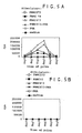
- T A T cells from individual A
- mPBMC B mPBMC B
- the mPBMC B s were seeded at 20K and 100K.
- the cultures were pulsed with 3 H-thymidine for the last 18 hours of the culture period to measure T cell proliferation.
- the results shown in Figure 1 indicate that the T A cells recognized the PBMC B as being foreign. (See bars under "T A + mPBMC B ".) With more PBMC B s present, the more the T cells proliferated.
- hMSCs human mesenchymal stem cells
- Fig. 1 See Figure 1 "T A + transduced hMSCs" demonstrate that the T lymphocytes were nonresponsive (did not proliferate) to the human mesenchymal stem cells, i.e., they were not recognized as being foreign.
- MLR mixed lymphocyte reactions
- PBMC A 10 5 PBMCs from individual A
- PBMC B 10 5 target individual B's PBMC's
- the PBMC B s were irradiated with 3000 rads X irradiation to prevent their proliferation due to activation by PBMC A s. Thus, only PBMC A s would proliferate.
- PBMC A s and PBMC B s were mixed, a mixed lymphocyte reaction occurred wherein the PBMC A cells (responder cells) were activated by the surface antigens on the PBMC B s (stimulator cells).
- the cultures were incubated over an interval of 7 days and were pulsed with 3 H-thymidine during the final 18 hours.
- the PBMC A s proliferated giving counts of 40,000. See Figure 2 , 1st bar, ("NONE" refers to no mesenchymal stem cells present.).
- PBMC A s and PBMC B s were mixed in the presence of mesenchymal stem cells, the mixed lymphocyte reaction was suppressed.
- 10 5 PBMC A s were mixed with 10 5 PBMC B s in microtiter plate wells coated with an adherent monolayer of human mesenchymal stem cells.
- the mesenchymal stem cells were plated in the wells in amounts ranging from 7500 to 22,500 mesenchymal stem cells per well.
- Two mesenchymal stem cell populations were tested: human mesenchymal stem cells were obtained from an individual B and human mesenchymal stem cells were obtained from an individual that did not match either individual A's or B's MHC type (a third party).
- the cultures were incubated over an interval of 7 days and were pulsed with 3 H-thymidine during the final 18 hours.
- the MLR was suppressed. See Figure 2 .
- the mesenchymal stem cells suppressed the mixed lymphocyte reaction.
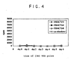
- T cells from donor 248 were primed by allogeneic PBMCs from donor 273 (d 273) for 7 days, then cultured for 3 additional days alone or in the presence of IFN- ⁇ -treated MSCs from the same donor (d273). Cells were then re-stimulated by the same donor (d273), autologous (d248) or "third party" (d244) PBMCs.
- PBMC Peripheral blood mononuclear cells
- PBMCs were depleted of monocytes and B cells by immunomagnetic negative selection.
- PBMCs were incubated with mouse anti-human CD19 and CD14 mAbs (no azide/low endotoxin (NA/LE) format) followed by biotin-conjugated goat anti-mouse IgG (multiple adsorption) Ab (all reagents from Pharmingen) and streptavidin microbeads (Miltenyi Biotec). Cells were then separated using a magnetic cell sorter (MACS, Miltenyi Biotec). The T cell-enriched fraction contained about 70-90% CD3+ cells.
- Human MSCs were isolated from bone marrow as described in U.S.Patent No. 5,486,359 and were maintained in culture with MSC medium and were used at passages from 3 to 6. Cells were lifted using 0.05% Trypsin/EDTA solution, washed once with MSC medium and plated at 70-80% confluent density which was 1x10 6 /plate for 10 cm tissue culture dish. The day after plating, IFN- ⁇ (Boehringer Mannheim) at 500 U/ml was added and the cells were incubated an additional 3 days. Before transferring T cells, MSC plates were washed 4 times with HBSS, 1 time with ISCOVES, and assay medium was added at 10 ml/well in 10 cm tissue culture dishes.
- T cells were activated by irradiated PBMCs (d 273).
- PBMCs used for stimulation were X-ray irradiated with 3,000 rad using Cabinet X ray system (Faxitron X ray, Buffalo Grove, IL).
- 2 x 10 7 responders were mixed with 2 x 10 7 stimulators in 20 mls assay medium in 10 cm tissue culture dishes. The cells were incubated at 37°C in 5% CO 2 atmosphere for 7 days.
- T cells activated in the 1° MLR were collected, washed once with MSC medium and re-suspended in assay medium at 10 6 /ml in 10 ml and were added to 10 cm tissue culture dishes containing autologous or allogeneic MSCs or medium alone, and incubated for an additional 3 days.
- T cells cultured with MSCs or media were collected, washed once with MSC media, and restimulated with irradiated PBMCs from the original donor, an unrelated donor or autologous PBMCs.
- 5 x 10 4 primed responders and 5 x 10 4 irradiated stimulators were incubated in 96-well plates. Assays were performed in triplicate. Cultures were pulsed with 1 ⁇ Ci of [ 3 H] thymidine (Amersham) for 18 hours before harvesting. Cultures were collected using Harvester 96 (Tomtec), filters were analyzed using Microbeta Trilux liquid scintillation and luminescence counter (E.G.&G Wallac). Data are presented as mean cpm ⁇ SD of three replicates.
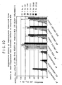
- T cells cultured alone showed an accelerated response to "same donor” re-stimulation with peak at day 2.
- "Third party” response was also accelerated, practically with the same kinetics as “same donor”, but with a lower maximum and a slightly delayed start. ( Fig 3 ).
- T cells cultured on allogeneic MSCs subsequently showed no response either to "same donor” or "third party” PBMCs during 6 days of culture ( Fig.4 ).
- T cells from donor 413 were stimulated with irradiated PBMCs from donor 273 for 7 days (1.5 x 10 6 ml each, bulk 20 ml cultures). MSCs from different donors 413, 418 and 273 were plated in 10 cm tissue culture dishes at 1x10 6 /dish, pretreated with IFN- ⁇ for 3 days and washed prior to mixing with preactivated T cells.
- T cells preactivated in the MLR for 7 days were incubated alone or with MSCs for an additional 3 days (1.0 x 10 6 /ml T cells, 10 ml/dish). After 3 days of incubation with MSCs, T cells were collected and re-stimulated with irradiated PBMC 273 (original donor), 413 (autologous), PBMC10 (third party) or PHA (5 ⁇ g/ml) in the presence or absence of autologous (d413) PBMC. Cells were added at 5 x 10 4 /well, cultures were pulsed with [ 3 H]thymidine at indicated time points for an additional 18 hours.
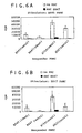
- Canine PBMCs were purified from peripheral blood by centrifugation on Ficoll-Paque gradient (1.077). Stimulator PBMCs were X-ray irradiated 2200 rad (7 min 70 kV). 10 5 irradiated stimulators were mixed with 10 5 responder PBMCs in 96-well plates in the presence or absence of pre-plated canine MSC (E647, 2 x 10 4 /well). Cultures were incubated for 6 days and pulsed with [ 3 H]TdR (5Ci/mmol, 1 ⁇ Ci/well) for an additional 16 hours. Results are shown in Figures 6A-6D . E647 and E645 were litter mates ( DLA identical). The results showed that autologous as well as allogeneic MSCs suppressed the primary MLR.
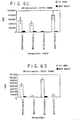
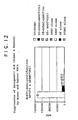
- T cells from d273 (2 x 10 5 /well) were mixed with irradiated PBMCs from d244 (2 x 10 5 /well) and different numbers of MSCs.
- MSCs from dD244 or d273 were pre-treated with IFN- ⁇ (900 U/ml for 3 days) or left untreated, trypsinized on the day of experiment and added at the same time as T cells and PBMCs. Cultures were incubated for 7 days,[ 3 H]TdR (5Ci/mmol, 1 ⁇ Ci/well) was added for an additional 16-18 hours. Results are shown in Figure 7 and demonstrate that non-adherent MSCs also suppressed a primary MLR.
- Juvenile baboons ( papio anubis ) were studied. Male and non-pregnant female baboons weighed 7-20 kg and were between 3-16 years of age. They were screened for tuberculosis papilloma virus, titered for cytomegalovirus (CMV), and tested with the primate viral screen consisting of testing for simian virus, and including fecal floatation and smears. Donor and recipient pairs were determined by major histocompatibility complex (MFiC)-disparity through PCR typing. During the study period, the baboons were housed in an individual area beside a companion animal.
- MMV cytomegalovirus
- Needle marrow aspirates were obtained from the iliac crest for isolation and culture-expansion of the MSCs.
- the marrow aspirate was obtained from an alternate side once a week for four consecutive weeks.
- the volume of the aspirate was determined by an estimate of 10% of the animal's blood volume. Blood volume (liters) is estimated to be 7% of body weight. A 10 kg baboon then, would have an estimated blood volume of 0.7 liters. An aspirate of 10% of the blood volume would then be 70 milliliters.
- cefazolin Prior to the procedure, 500 mg cefazolin was administered intramuscularly (IM) for perioperative antibacterial prophylaxis.
- Baboons were sedated and anesthetized for the procedure with ketamine at 10 mg/kg IM, and xylazine 1 mg/kg IM.
- the sites of needle insertion were scrubbed with povidone-iodine and then rinsed with alcohol.
- Aspirates were obtained from the iliac crest using a 16-gauge, 2-inch bone marrow needle. A syringe was attached to the needle, and suction was applied to remove the marrow.
- the analgesic Buprenorphine was given at 0.03 mg/kg IM Q12 x 2 doses.
- Bone marrow aspirates were transferred from the syringe to a sterile Vacutainer® containing sodium heparin.
- the tubes were placed in a StyrofoamTM container and shipped at room temperature (RT) by overnight delivery to the cell processing facility.
- DPBS Dulbecco's Phosphate Buffered Saline
- the cell suspensions were centrifuged at 2200 RPM for 10 minutes at room temperature (RT). Total nucleated cell counts were determined in 4% acetic acid.
- Cells were then diluted in DPBS for a final concentration of 20 x 10 6 cells/ml.
- Ten ml or 200 x 10 6 cells were loaded onto 20 ml of Percoll (sp.gr. 1.073 gm/ml) in a 50 ml conical tube and underwent centrifugation at 1300 RPM for 20 minutes.
- the cell interface containing mononuclear cells was washed in DPBS, resuspended in complete media, and counted to obtain a recovery.
- the washed mononuclear cells obtained at the Percoll interface were cells were then established in T-185 flasks containing 30 ml of complete media and 15-20 x 10 6 cells/flask (8.1 x 10 4 MSC/cm 2 ) and placed in a 37°C incubator at 5% CO 2 .
- the media in the triple flasks was decanted, and the flasks were rinsed with 50 ml DPBS. After decanting the DPBS, 23 ml of 0.05% trypsin was added to each triple flask. The flasks were placed in a 37°C incubator for 3 minutes. After cell detachment, 23 ml complete medium was added to each flask. The cell suspensions were transferred to 50 ml conical tubes and the flasks were washed with 30 ml HBSS. The tubes were centrifuged at 2200 RPM for 5 minutes at RT.
- the harvested MSCs were formulated at approximately 10 x 10 6 cells per ml in cryoprotectant solution consisting of 85% Plasma-Lyte A (Baxter IV Therapy), 10% DMSO, and 5% MSC-donor serum, and cryopreserved in bags containing 15-20 ml.
- cryoprotectant solution consisting of 85% Plasma-Lyte A (Baxter IV Therapy), 10% DMSO, and 5% MSC-donor serum, and cryopreserved in bags containing 15-20 ml.
- Cells were cryopreserved using a controlled-rate freezer (Cryomed, Forma Scientific) at 1-2° per minute to -90°C. The samples were then transferred to a liquid nitrogen storage freezer in the vapor phase (-120 to -150°C).
- the final product was prepared at 115% of the dose required on infusion day.
- the baboon Prior to surgery the baboon was given cefazolin at 500 mg IM as a perioperative antibacterial prophylaxis. The baboon was sedated with ketamine at 10 mg/kg IM and anesthetized by intravenous Thiopental induction, a 1-2% isofluorane inhalational anesthetic. Skin was harvested from the anterior abdominal wall, placed on a pre-labeled moistened saline gauze pad. The wound defect was then closed. The baboon was returned to the colony after awakening. For postoperative pain, the analgesic Buprenorphine was administered at Q12 x 2 doses and Ancef daily for 2 days.
- baboon Prior to surgery the baboon was given cefazolin at 500 mg IM as an antibacterial prophylaxis perioperatively.
- the baboon was sedated and anesthetized with ketamine at 10 mg/kg IM and intravenous Thiopental induction, a 1-2% isofluorane inhalational anesthetic.
- Skin was harvested from the anterior abdominal wall and placed on a pre-labeled moistened saline gauze pad. This skin was divided into two grafts; one was used as the third party control for another recipient baboon and one was used as an autologous control for this same animal. The animal was then placed in a prone position.
- the baboon received an intravenous infusion of MSC at a dose of 20 x 10 6 donor MSC/kg.
- Peripheral blood samples were obtained at pre-MSC, 1 hour, and days 1-3 post-MSC; marrow aspirates were obtained on day 0 post-MSC, day 3, 14, and 30.
- the analgesic Buprenorphine was administered at Q12 x 2 doses and Ancef daily for 2 days. The animal was observed daily, and the grafts were photographed every other day beginning on post-graft day 7.
- Each baboon was sedated with ketamine 10mg/kg IM for examination. While sedated, two-three milliliters of marrow were obtained from the iliac crest by needle aspiration and collected in sodium heparin on days 4, 13, and 30, the end of study. A skin biopsy was harvested on the same day that marrow aspirates were obtained.
- Untreated control animals had a mean skin allograft survival time of 8.0 ⁇ 0 days.
- the infusion of unrelated-MSC-donor MSCs donor resulted in a prolongation of skin graft survival time to a mean survival time of 11.5 ⁇ 0.71 days (Mann-Whitney U Test, P ⁇ 0.05).
- the infusion of unrelated-third-party donor MSC on donor allografts resulted in a significant prolongation of skin graft survival times to a mean survival time of 12.3 ⁇ 0.96 days (Mann-Whitney U Test, P ⁇ 0.003).
- Recipients 6140 and 6200 received allografts from the MSC donor 6243, from each other (a third party graft), and from themselves (an autograft). Twenty-four hours prior to skin graft harvesting from the MSC donor, 6243, MSCs from 6243 were injected under the anterior abdominal skin which had been delineated for grafting. After grafting, the recipients were administered an intravenous infusion of 20 x 10 6 MSC/kg (6243). Both third-party allografts were rejected on day 13. The MSC-donor (6243) allografts were found to be hemorrhagic on day 4, a finding usually attributed to a technical failure.
- Recipients 6654 and 6659 received allografts from the MSC donor 6593, from each other (a third-party graft), and from themselves (an autograft). After grafting, the recipients were administered intravenous infusions of 20 x 10 6 MSC/kg. The MSC-donor allografts were rejected on days 11 and 12, and the third-party donor allografts were rejected on days 11 and 12. The autografts were not rejected.
- recipients 6663 and 6658 received allografts from the MSC donor 6656, from each other (a third-party graft), and from themselves (an autograft). After grafting, the recipients were administered intravenous infusions of 20 x 10 6 MSC/kg. The MSC-donor allografts were rejected on day 11, and the third-party donor allografts were rejected on days 10 and 12. The autografts were not rejected.
- the purpose of the study was to demonstrate the feasibility and safety in dogs of the infusion of a moderately high dose of donor dog leukocyte antigen (DLA)-identical littermate canine mesenchymal stem cell (cMSC) at 10 x 10 6 cells/kg in an allogeneic marrow graft setting.
- DLA donor dog leukocyte antigen
- cMSC canine mesenchymal stem cell
- Beagles were used for the study. Two male and two female DLA-identical littermates were used in the study, aged 7 or 9 months on day 0.
- the method for typing used involves the use of highly polymorphic microsatellite markers to follow inheritance of the Class II DRB region in the Dog Leukocyte Antigen (DLA), the canine equivalent of the major histocompatability complex.
- DLA Dog Leukocyte Antigen
- Microsatellites are small di- tri- or tetra nucleotide repeats, which show sufficient length variation in alleles that they may be used to follow the inheritance of chromosomal segments through multigeneration crosses. Segregation of alleles is typically monitored using a single-step polymerase chain reaction with primers derived from unique sequences of DNA that surround each repeat.
- mixed leukocyte reactions were performed on the DLA-identical littermate pairs chosen for study to provide confirmation of the PCR microsatellite marker assay results.
- the dogs underwent transplantation with cMSC and bone marrow from the same DLA-identical littermate donor.
- the marrow graft was harvested from each of the two DLA-identical littermates on day 0 prior to total body irradiation (TBI) and exchanged.
- TBI total body irradiation
- Myeloablation was induced by exposing the dogs on day 0 to a single TBI dose of 920 centigray (cGy) (midline air exposure from two opposing 60 Co sources delivered at a rate of 7 cGy (9.3R)/min.
- cMSC Culture-expanded cMSC isolated from a donor marrow aspirate at 4 or more weeks prior to transplantation, were transduced with Papp@OT-24, containing the genes for green fluorescence protein (GFP) and neomycin phosphotransferase (neo).
- the cMSC were cryopreserved after passage 1 (PI) or passage 2 (P2). Following TBI, the cMSC were thawed and delivered intravenously via a portable infusion pump over a 15-minute time period.
- PI green fluorescence protein
- P2 passage 2
- the cMSC were thawed and delivered intravenously via a portable infusion pump over a 15-minute time period.
- the bone marrow graft was infused intravenously at a dose of ⁇ 1 x 10 8 total nucleated cell (TNC)/kg.
- Cyclosporin was administered to all four dogs for graft-versus-host-disease (GVHD) prophylaxis intravenously on days 0 through 5 at a dose of 10 mg/kg BID (20 mg/kg/day) (Sandimmune® Injection Solution, Sandoz Pharmaceuticals Corporation). On days 6 through 50 (end of study) for group I.1.a, or 6 through 100 for group I.1.b, cyclosporin was administered at 10 mg/kg BID PO, (20 mg/kg/day) (Neoral® Soft Gelatin Capsules, Sandoz Pharmaceuticals Corporation). The usual supportive care with oral antibiotics for the recipient began on day -5 and systemic antibiotics started on day 0 and continued until engraftment was achieved. Fluid support was given as necessary.
- BID graft-versus-host-disease
- CBCs complete blood counts
- Serum chemistry analysis was performed on days 0, 2, and weekly thereafter.
- Peripheral blood samples were taken on day 0 pre-MSC infusion, 5- and 15-minutes, 1- and 2-hours, and 1-, 2-, 3-, and 4-day time points for DNA isolation.
- the DNA was evaluated for the presence of GFP marked cells by an Anti-EGFP DNA PCR Elisa with digoxigenin incorporated into the product and a second step anti-digoxigenin colorimetric assay.
- a marrow aspirate was obtained when the platelet counts consistently reached 50,000/mm 3 and examined for the presence of GFP marked cells using the same PCR method.
- CMSC cultures were established to examine colony forming units (CFU), and to expand the cMSC for further Anti-EGFP PCR analysis.
- CFU colony forming units
- peripheral blood, bone marrow aspirates, and bone marrow biopsies were obtained for Anti-EGFP PCR analysis.
- CFU assays were performed on the bone marrow aspirates, and the Anti-EGFP PCR analysis was performed on culture-expanded cMSC. An histological analysis was performed for the presence of GFP in various tissues.
- Bilateral bone marrow aspirates were obtained for cMSC isolation and culture establishment on week -4 for dogs CAN-07-01 and CAN-07-02 and on week -9 for dogs CAN-07-03 and CAN-07-04.
- Fifteen ml of marrow (7 ml from each humerus) were obtained from each dog. Dogs were anesthetized by the injection of Butorphanol followed by injection of a mixture of Diazepam and ketamine hydrochloride (Aveco Co., Inc., Fort Dodge, IA). The sites of needle insertion were scrubbed with povidone-iodine and then rinsed with alcohol.
- Aspirates were obtained from each humeral condyle of each dog using a 16-gauge, 2-inch bone marrow needle. A syringe was attached to the needle, and suction was applied to remove 8 ml of marrow from each humerus. Bone marrow aspirates were transferred to 15 ml polypropylene conical tubes using sterile technique. Following the procedure, the dog was then placed on a warming pad to recover.
- DPBS Dulbecco's Phosphate Buffered Saline
- the cell suspensions underwent centrifugation at 2200 RPM for 10 minutes at room temperature (RT). Total nucleated cell counts were determined in 4% acetic acid.
- Cells were then diluted in DPBS for a final concentration of 20 x 10 6 cells/ml.
- Ten ml or 200 x 10 6 cells were loaded onto 20 ml of Percoll (sp.gr. 1.073 gm/ml) in a 50 ml conical tube and underwent centrifugation at 1300 RPM for 20 minutes.
- the cell interface containing mononuclear cells was washed in DPBS, resuspended in complete media, and counted to obtain a recovery percentage. The cells were then diluted in complete media, cultures were established as described below, and placed in a 37°C incubator at 5% CO 2 .
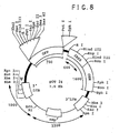
- the green fluorescent protein (EGFP) retrovirus was constructed by isolating EGFP-1 gene from the jellyfish Aequorea victoria (Clontech, CA). EGFP gene was cloned into retroviral vector pJM573-neo (resulting plasmid was named pOT-24). The plasmid pJM573-neo was derived from pN2 ( Keller et.
- FIG. 8 A schematic map of EGFP pOT24 plasmid is shown in Figure 8 .
- pOT-24 was transfected into GP&E86 ecotropic producer cells using DOTAP (Boehringer Manheim) as suggested by manufacturer.
- the transfected cells were grown in DMEM-high glucose (HG) medium supplemented with 10% heat inactivated FBS, Penicillin-Streptomycin (Life Technologies) and 0.5mg/ml of protamine sulfate-G418 (Sigma) as a selective marker.
- Cultures were maintained up to 70% confluency at which point medium was replaced with fresh retroviral media (without G418) and cells were maintained at 32°C for 2 days.
- the culture medium containing the retrovirus was collected, filtered through 0.45 ⁇ m filter and stored at - 70°C.
- Amphotropic retrovirus was prepared by transducing PA317 cells twice with ecotropic virus using a centrifugal transduction procedure followed by selection with G418 (0.5 mg/ml). Retroviral supernatant was collected. The titer of the pooled EGFP retrovirus on 3T3 cells was 1.2x10 6 CFU/ml. GFP-retroviral supernatants were cryopreserved at -70°C.
- the washed mononuclear cells obtained at the Percoll interface were established in 10, T-185 flasks containing 30 ml of complete media and 10 x 10 6 cells/flask.
- the supernatant was removed and the cell pellets were resuspended in complete media.
- the cells were pooled, counted and examined for viability. Cells were plated into 15, T80 flasks containing 18 ml of complete medium and 0.4 x 10 6 cells per flask.
- the first transduction was performed on 15 of the 18 flasks.
- the media was removed. Aliquots of the retroviral supernatant were thawed and polybrene was added to a final concentration of 8 ⁇ g/ml to make the transduction cocktail.
- the cell medium was replaced with 10 ml of the transduction cocktail, and the flasks were centrifuged at 3000 RPM for 1 hour at 32°C. After centrifugation, 10 ml of complete media prepared using heat inactivated fetal bovine serum (FBS) was added to each flask (with the transduction cocktail) and the flasks were returned to the incubator. Three flasks were not transduced, and fresh media was replaced. On day 16 of culture, the media was replaced with fresh complete media. On day 17 of culture the transduction procedure was repeated.
- FBS heat inactivated fetal bovine serum
- the cells were harvested as described above and taken from P1 to P2.
- Three x 10 6 cells were added to 100 ml of complete medium, and poured into triple-flasks (500 cm 2 ).
- Fifteen triple-flasks were prepared with transduced cells and three were prepared with untransduced cells. Any remaining cells were cryopreserved.
- a freeze solution was prepared containing 10% DMSO and 90% FBS.
- Ten x 10 6 cells were resuspended in 1 ml of freezing solution. The vials were labeled and cryopreserved in a Nalgene Cryo container for a minimum of 4 hours at -70°C, and stored at -70°C.
- the washed mononuclear cells obtained at the Percoll interface were established in 15, T-75 flasks containing 20 ml of complete media and 12 x 10 6 cells/flask.
- the 15 ml bone marrow aspirates yielded 910, 1212, 856, and 1948 x 10 6 nucleated cells for donors CAN-07-01, CAN-07-02, CAN-07-03, and CAN-07-04, respectively.
- Mononuclear cell counts obtained from the Percoll interface were 612, 666, 588, and 462 x 10 6 , resulting in recoveries of 67.2, 55, 68.7, and 23.7%.
- the cell viability was a mean of 97.1 (range 93.3 to 100)%.
- the cell viability of the transduced cells was a mean of 96.7 (range 96.3 to 97.9)%.
- the untransduced cells were 95.4 (range 93.3 to 96.9)% viable.
- the viability of the transduced cells was a mean of 99.4 (range 97.4 to 100)% and the untransduced cells were 99.4 (range 97.6 to 100)% viable (Table 4).
- the transduced cMSC yield per flask for donors CAN-07-01 and CAN-07-02, harvested 4 days after passage 2 and plated at 3 x 10 6 per flask was 5.9 and 6.7 x 10 6
- the untransduced cMSC yield per flask was 8.4 and 7.5 x 10 6
- the transduced cMSC yield per flask for donors CAN-07-03 and CAN-07-04, harvested 4 days after passage 1 (different transduction and passage design) and plated at 3 x 10 6 per flask was 20.0 and 14.0 x 10 6
- the untransduced cMSC yield per flask was 25.3 and 18.0 x 10 6 .
- CFU colony assays were prepared at the time of primary culture establishment by plating 0.5 x 10 6 cells in triplicate in 100 mm dishes containing 10 ml complete media. The dishes were incubated at 37°C and 5% CO 2 . The media was replaced with fresh media each 2 to 4 days. On day 10 in culture, the CFU assay dishes were rinsed with HBSS twice, fixed with 1% gluteraldehyde for 15 minutes, rinsed with HBSS twice, and air dried. The cMSC in the dishes were then stained with 0.1% crystal violet, rinsed with deionized water three times, and air dried. Colonies were counted to calculate the number of colonies forming per 10 6 cells plated.
- CFU assays plated on day of mononuclear cell isolation and culture establishment and harvested on day 10 yielded 56, 46.7, 114, and 72 colonies per 10 6 cells for dogs CAN-07-01, CAN-07-02, CAN-07-03, and CAN-07-04, respectively.
- the media in the triple flasks was decanted, and the flasks were rinsed with 50 ml DPBS. After decanting the DPBS, 23 ml of 0.25% trypsin was added to each triple flask. The flasks were placed in a 37°C incubator for 3 minutes. After cell detachment, 23 ml complete medium was added to each flask. The cell suspensions were transferred to 50 ml conical tubes and the flasks were washed with 30 ml HBSS. The tubes were centrifuged at 2200 RPM for 5 minutes at RT. The pellets containing the transduced or untransduced cells, respectively, were pooled and counted. One aliquot of 1 x 10 7 cells was set aside for determination of the transduction percentage by an Anti-EGFP DNA PCR Elisa assay.
- the recovered P1 or P2 transduced and culture-expanded cMSCs centrifuged at 1300 RPM for 5 minutes and resuspended in 1 ml aliquots with 1 x 10 7 cMSC/ml in ice-cold cryoprotectant solution containing 85% Plasma-Lyte A (Baxter IV Therapy), 10% DMSO, and 5% autologous canine serum.
- Cell aliquots were dispensed into separate cryo-vials containing 1 ml each.
- the tubes were labeled with the canine donor number and total viable cell count.
- the cMSCs were cryopreserved by placing the cell vials into a Nalgene freezing container and placed in a -70°C freezer for 4 hours, then moved to storage at -70°C.
- the cMSC suspension was infused via a catheter inserted into the cephalic vein using a hand-held Harvard Bard Mini Infuser to deliver the 50 ml over a 15-20 minute period.
- Moderately high doses of 7.49, 7.35, 10.0, and 10.0 (mean 8.7) x 10 6 viable cMSC/kg were infused on day 0 to dogs CAN-07-01, CAN-07-02, CAN-07-03, and CAN-07-04, respectively. These doses represent a 4- to 10-fold increase over the typical dose that a patient would receive.
- Total viable cMSC infused ranged from 67.7 to 129 (mean 93.9) x 10 6 cMSC.
- the viability of the cells ranged from 92.1 to 97.6 (mean 94.9) as determined by trypan blue exclusion. CMSC Infusions were given between 71 and 146 (mean 110) minutes post-TBI.
- Blood samples (2 ml) were obtained before (pre) and during the cMSC infusion at five and fifteen minutes after the start of the infusion, as well as 1- and 2-hour, and 1-, 2-, 3-, and 4-day time points.
- Cell lysates were prepared using the PuregeneTM (Gentra Systems, Inc.) DNA Isolation Kit for use in an Anti EGFP DNA PCR Elisa with digoxigenin incorporated into the product and a second step Anti-digoxigenin colorimetric assay to detect of the level of GFP marked cMSC in the bloodstream.
- Bone marrow to be used as the transplant graft was harvested from the DLA-identical littermate prior to TBI. Aspirates were obtained from each humerus using an 11-gauge, 4-6 inch ball-top stainless steel marrow harvest needle, attached to polyvinyl tubing originating from a vacuum flask containing 100 ml Tissue Culture Medium 199 and 4 ml (4000 U) heparin. The marrow is passed through 300- and 200-um pore size, and stored at 4°C in a transfer pack container, labeled with the donor and recipient, until infusion later that day. The bone marrow total nucleated cell count (BM-TNC) of the marrow is corrected to exclude any nucleated cells which would be present in the volume of peripheral blood obtained during the marrow harvest.
- BM-TNC bone marrow total nucleated cell count
- the total nucleated cell count (TNC) of the bone marrow was corrected to exclude any TNC which would be present in the volume of peripheral blood obtained during the marrow harvest.
- Corrected doses of marrow were 4.3, 3.5, 3.1, and 2.0 (mean 3.2) x 10 8 TNC/kg to dogs CAN-07-01, CAN-07-02, CAN-07-03, and CAN-07-04, respectively.
- Uncorrected bone marrow doses were 5.6, 4.2, 4.5, and 2.7 (mean 4.3) x 10 8 TNC/kg.
- the marrow was placed at room temperature.
- the marrow was infused intravenously through a butterfly needle inserted into the cephalic vein, by exerting pressure on the bag over 1 to 2 minutes.
- oral antibiotics neomycin sulfate and polymyxin sulfate
- these oral antibiotics were administered until absolute neutrophil counts reached 500/mm 3 .
- the systemic antibiotic Baytril was administered intravenously twice daily and continued until absolute neutrophil counts reached 1,000/mm 3 consistently. Fluid and electrolytes lost as results of transient radiation toxicity were replaced by subcutaneous administration of 500 ml of Ringers Solution, twice daily until food and water were accepted.
- Blood samples (2 ml) were collected from either the jugular or cephalic vein on the mornings of the marrow aspirate for isolation of cMSC, days 0 through 50 and biweekly thereafter through the end of study.
- the blood was transferred into a vacutainer containing EDTA.
- Total white blood cell (WBC) and platelet counts per mm 3 are measured using a Sysmex E2500 and differential cell counts were determined manually after fixation and staining with Wrights stain.
- the tissues were trimmed to about 1 inch square pieces and placed into separate labeled 50 ml conical tubes filled with 10% Neutral Buffered Formalin (pH 6.8 - 7.2).
- the tissues were embedded in paraffin, sectioned and stained with Hematoxylin and Eosin. Bone marrow samples were stained with Periodic Acid Schiff's stain.
- Bone marrow aspirates obtained prior to necropsy were collected in 15 ml labeled tubes from the left and right humieri, femora, and iliac crests from each dog. A subset of the tissue samples were obtained during necropsy and trimmed to about 1/4 inch square pieces, wrapped in PBS-soaked gauze and placed separately in a labeled zip-lock bag. The bone marrow aspirates were held on ice.
- CFU colony assays performed on cMSC obtained from bone marrow obtained at necropsy were prepared by plating 0.5 x 10 6 cells in triplicate in 100-mm dishes containing 10 ml complete media. The dishes were incubated at 37°C and 5% CO 2 . The media was replaced with fresh media each 2-4 days. On day 10 in culture, the CFU assay dishes were rinsed with HBSS twice, fixed with 1% gluteraldehyde for 15 minutes, rinsed with HBSS twice, and air dried. The cMSC in the dishes were then stained with 0.1% crystal violet, rinsed with deionized water three times, and air dried. Colonies were counted to calculate the number of colonies per 10 6 cells plated.
- PBS Phosphate Buffered Saline
- the anti-EGFP DNA PCR Elisa assay used in these studies detects infused cMSCs utilizing oligonucleotide primers specific for GFP.
- PCR-ELISA DIG labeling/detection
- PCR was performed in the presence of digoxigenin-labeled nucleotides to label the amplified product.
- 25 ⁇ l of the PCR product was denatured and allowed to hybridize in solution to 5'-biotinylated oligonucleotide probe at 37°C in streptavidin-coated microtiter plate.
- the bound probe-PCR product was detected by an anti-digoxigenin peroxidase conjugate and by use of the colorimetric substrate 2,2'Azinobis (3-ethylbenzthiazoline-sulfonic Acid) (ABTS). Titration standard curves were generated using transfected control cMSC to approximate concentration of DNA per quantity of DNA used in the assay. By first correlating with an internal standard for PCR of the DLA Class II genomic DNA, and then correlating DNA concentration to cell equivalents, and assuming one retrovirus integration event per transduced cell, an estimation of cell number can be obtained.
- the mean day to a threshold (to 3 consecutive values) of platelets to 10,000/mm 3 was 12.8 (range 11-17), to 50,000/mm 3 was 19.8 (range 16-25), and to 100,000/mm 3 23.0 (range 20-27).
- the mean day to a threshold value (to 3 consecutive days) of absolute neutrophil cells to 500/mm 3 was 9.3 (range 8-11), and to 1,000/mm 3 was 10.5 (range 9-13).
- Detectable DNA signal could be found within 1 hour of infusion and again at 2 days.
- One sample could be quantitatively measured at 3 days post infusion for GFP DNA. This timepoint is consistent with the previous observations in the autologous canine transplant study in which signal was found at 2 and 3 days
- GVHD skin graft-versus-host-disease
- T cells from donor 155 were purified from PBMC by negative immunomagnetic selection with anti-CD 19 and anti-CD14 MicroBeads (Miltenyi Biotec).
- PBMC from donor 413 were X-ray irradiated with 3600 rad (12 min at 70 kV).
- MSCs from different donors (219, 459, 461 - all at passage 5) were added into the cultures at 1.2 x10 5 cells/well for 3 additional days. In control cultures, the same volume of medium was added instead of MSCs.
- the same number of MSCs were plated alone, and cultured for 3 additional days. After 3 days of culture (on day 6 after initiation of the primary MLR), the cells were resuspended by pipetting, and 200 ⁇ l of the cell suspensions were transferred into a 96-well plate in triplicate, and pulsed with [H 3 ]TdR (5 Ci/mmol, 1 ⁇ Ci/well) for 18 hours to determine the level of proliferation. The remaining cells were centrifuged at 1250 rpm for 10 minutes, the supernatants were collected, aliquoted and kept frozen at -80°C.
- T cells from donor 155 were purified from PBMCs by negative immunomagnetic selection with anti-CD19 and anti-CD14 MicroBeads (Miltenyi Biotec). PBMCs from donor 413 or from donor 273 were X-ray irradiated with 3600 rad (12 min at 70 kV). In 96-well tissue culture plates T cells (1.5 x 10 5 /well) were mixed with PBMCs (1.5 x 10 5 /well) in the presence or absence of different supernatants that were added at the initiation of cultures at the dilutions (1/8, 1/32, 1/128, 1/512, 1/2048 and 1/8192). In control cultures, the same volume of medium was added instead of supernatants. Cultures were incubated for 6 days, then pulsed with [H 3 ]TdR (5 Ci/mmol, 1 ⁇ Ci/well) for 18 hours.
- [H 3 ]TdR 5 Ci/mmol, 1 ⁇ Ci/well
- Figure 9 shows the results of the MLR between T 155 x PBMC 413.
- Primary MLR in the presence of supernatant of MLR alone (#5) was not suppressed.
- T cells from donor 155 were purified from PBMCs by negative immunomagnetic selection with anti-CD19 and anti-CD14 MicroBeads (Miltenyi Biotec).
- PBMCs from donor 413 were X-ray irradiated with 3600 rad (12 min at 70 kV).
- T cells 1.5 X 10 5 /well
- PBMCs 1.5 x 10 5 /well
- supernatants were added into the cultures at different dilutions (1/8, 1/32, 1/128, 1/512, 1/2048 and 1/8192).
- the same volume of medium was added instead of supernatants. Cultures were incubated for 2 additional days (6 days after initiation of the MLR), then pulsed with [H 3 ]TdR (5 Ci/mmol, 1 ⁇ Ci/well) for 18 h.
- FIG 10 shows the results of the MLR between T 155 x PBMC 413.
- Supernatants from all MSCs + MLR #1) MLR +MSC219 (#2) MLR +MSC459 (#3) MLR +MSC461 showed strong suppression of ongoing MLRs.
- Supernatants from MSCs alone #8) MSC219 alone (#9) MSC459 alone (#10) MSC461 alone suppressed in a dose dependent manner with a significant effect up to 1/512 dilution. Ongoing MLR in the presence of supernatant of MLR alone (#5) was not suppressed.
- Lymphoproliferation was determined on day 7 of culture by pulsing the cells with 3 H-thymidine for the final 18 hours prior to cell harvest for scintillation counting.
- the results illustrated in Figure 11 show that baboon MSCs suppressed robust human MLRs by greater than 50%.
Landscapes
- Health & Medical Sciences (AREA)
- Life Sciences & Earth Sciences (AREA)
- Engineering & Computer Science (AREA)
- Immunology (AREA)
- Chemical & Material Sciences (AREA)
- Biomedical Technology (AREA)
- Bioinformatics & Cheminformatics (AREA)
- Organic Chemistry (AREA)
- General Health & Medical Sciences (AREA)
- Biotechnology (AREA)
- Zoology (AREA)
- Developmental Biology & Embryology (AREA)
- Cell Biology (AREA)
- Genetics & Genomics (AREA)
- Wood Science & Technology (AREA)
- Animal Behavior & Ethology (AREA)
- Public Health (AREA)
- Veterinary Medicine (AREA)
- Medicinal Chemistry (AREA)
- Pharmacology & Pharmacy (AREA)
- Microbiology (AREA)
- Hematology (AREA)
- General Engineering & Computer Science (AREA)
- Biochemistry (AREA)
- Epidemiology (AREA)
- Chemical Kinetics & Catalysis (AREA)
- Transplantation (AREA)
- General Chemical & Material Sciences (AREA)
- Nuclear Medicine, Radiotherapy & Molecular Imaging (AREA)
- Virology (AREA)
- Rheumatology (AREA)
- Mycology (AREA)
- Medicines Containing Material From Animals Or Micro-Organisms (AREA)
Priority Applications (1)
| Application Number | Priority Date | Filing Date | Title |
|---|---|---|---|
| EP10010855A EP2269616A3 (en) | 1999-10-26 | 2000-10-26 | Supernatant from mesenchymal stem cells for prevention and treatment of immune responses in transplantation |
Applications Claiming Priority (3)
| Application Number | Priority Date | Filing Date | Title |
|---|---|---|---|
| US427333 | 1999-10-26 | ||
| US09/427,333 US6368636B1 (en) | 1998-03-18 | 1999-10-26 | Mesenchymal stem cells for prevention and treatment of immune responses in transplantation |
| PCT/US2000/029815 WO2001032189A1 (en) | 1999-10-26 | 2000-10-26 | Supernatant from mesenchymal stem cells for prevention and treatment of immune responses in transplantation |
Publications (2)
| Publication Number | Publication Date |
|---|---|
| EP1223956A1 EP1223956A1 (en) | 2002-07-24 |
| EP1223956B1 true EP1223956B1 (en) | 2010-09-29 |
Family
ID=23694416
Family Applications (2)
| Application Number | Title | Priority Date | Filing Date |
|---|---|---|---|
| EP00974007A Expired - Lifetime EP1223956B1 (en) | 1999-10-26 | 2000-10-26 | Supernatant from mesenchymal stem cells for prevention and treatment of immune responses in transplantation |
| EP10010855A Withdrawn EP2269616A3 (en) | 1999-10-26 | 2000-10-26 | Supernatant from mesenchymal stem cells for prevention and treatment of immune responses in transplantation |
Family Applications After (1)
| Application Number | Title | Priority Date | Filing Date |
|---|---|---|---|
| EP10010855A Withdrawn EP2269616A3 (en) | 1999-10-26 | 2000-10-26 | Supernatant from mesenchymal stem cells for prevention and treatment of immune responses in transplantation |
Country Status (10)
| Country | Link |
|---|---|
| US (2) | US6368636B1 (enExample) |
| EP (2) | EP1223956B1 (enExample) |
| JP (1) | JP2004506598A (enExample) |
| AT (1) | ATE482712T1 (enExample) |
| AU (1) | AU782064B2 (enExample) |
| CA (1) | CA2387542C (enExample) |
| DE (1) | DE60045034D1 (enExample) |
| ES (1) | ES2351203T3 (enExample) |
| PT (1) | PT1223956E (enExample) |
| WO (1) | WO2001032189A1 (enExample) |
Families Citing this family (115)
| Publication number | Priority date | Publication date | Assignee | Title |
|---|---|---|---|---|
| EP1062321B1 (en) * | 1998-03-13 | 2004-12-29 | Osiris Therapeutics, Inc. | Uses for humane non-autologous mesenchymal stem cells |
| US20040265285A1 (en) * | 1999-04-15 | 2004-12-30 | Monash University | Normalization of defective T cell responsiveness through manipulation of thymic regeneration |
| US20050042679A1 (en) * | 1999-04-15 | 2005-02-24 | Monash University | Diagnostic indicator of thymic function |
| AU2003901668A0 (en) * | 2003-03-28 | 2003-05-01 | Medvet Science Pty. Ltd. | Non-haemopoietic precursor cells |
| AUPQ147799A0 (en) * | 1999-07-07 | 1999-07-29 | Medvet Science Pty. Ltd. | Mesenchymal precursor cell |
| US20050158289A1 (en) * | 1999-07-07 | 2005-07-21 | Simmons Paul J. | Mesenchymal precursor cell and use thereof in the repair of bone defects and fractures in mammals |
| US8062675B2 (en) | 1999-07-07 | 2011-11-22 | Angioblast Systems, Inc. | Mesenchymal precursor cell |
| US7670628B2 (en) * | 1999-07-07 | 2010-03-02 | Angioblast Systems, Inc. | Mesenchymal precursor cell |
| US8147824B2 (en) | 1999-08-05 | 2012-04-03 | Athersys, Inc. | Immunomodulatory properties of multipotent adult progenitor cells and uses thereof |
| ATE460475T1 (de) * | 2000-04-11 | 2010-03-15 | Univ Southern California | Methode zur verhinderung der transplantatabstossung unter verwendung von tgf- beta zur induktion von t-suppressorzellen |
| US20060088512A1 (en) * | 2001-10-15 | 2006-04-27 | Monash University | Treatment of T cell disorders |
| JP2004521877A (ja) * | 2000-11-22 | 2004-07-22 | ジェロン コーポレイション | 多能性幹細胞の同種移植片の寛容化 |
| US20080152629A1 (en) * | 2000-12-06 | 2008-06-26 | James Edinger | Placental stem cell populations |
| WO2002046373A1 (en) * | 2000-12-06 | 2002-06-13 | Hariri Robert J | Method of collecting placental stem cells |
| US7311905B2 (en) | 2002-02-13 | 2007-12-25 | Anthrogenesis Corporation | Embryonic-like stem cells derived from post-partum mammalian placenta, and uses and methods of treatment using said cells |
| NZ528035A (en) * | 2001-02-14 | 2005-07-29 | Robert J Hariri | Renovation and repopulation of decellularized tissues and cadaveric organs by stem cells |
| NZ527849A (en) * | 2001-02-14 | 2006-09-29 | Anthrogenesis Corp | Post-partum mammalian placenta, its use and placental stem cells therefrom |
| WO2003024462A1 (en) * | 2001-09-19 | 2003-03-27 | Henry Ford Health System | Cardiac transplantation of stem cells for the treatment of heart failure |
| JP2005531507A (ja) * | 2002-03-02 | 2005-10-20 | ザ ボード オブ リージェンツ,ザ ユニバーシティ オブ テキサス システム | ストローマ細胞前駆体による抗癌物質の局所的な産生および/または送達方法 |
| AU2003240512B2 (en) * | 2002-06-04 | 2009-11-05 | The Board Of Trustees Of The Leland Stanford Junior University | Device and method for rapid aspiration and collection of body tissue from within an enclosed body space |
| AU2003295444A1 (en) * | 2002-11-15 | 2004-06-15 | The Board Of Trustees Of The University Of Illinois | Methods for in vitro expansion of hematopoietic stem cells |
| US20040101959A1 (en) * | 2002-11-21 | 2004-05-27 | Olga Marko | Treatment of tissue with undifferentiated mesenchymal cells |
| AU2003289907A1 (en) * | 2002-11-28 | 2004-06-18 | Genethor Gmbh | Cells not presenting antigen for suppressing pathological immune reactions |
| CA2521217C (en) * | 2003-04-01 | 2013-05-14 | United States Of America Department Of Veteran's Affairs | Stem-cell, precursor cell, or target cell-based treatment of multi-organ failure and renal dysfunction |
| GB0321337D0 (en) * | 2003-09-11 | 2003-10-15 | Massone Mobile Advertising Sys | Method and system for distributing advertisements |
| US20080279812A1 (en) * | 2003-12-05 | 2008-11-13 | Norwood Immunology, Ltd. | Disease Prevention and Vaccination Prior to Thymic Reactivation |
| CA2934682C (en) * | 2004-03-22 | 2021-12-07 | Mesoblast International Sarl | Mesenchymal stem cells and uses therefor |
| EP2298861B1 (en) | 2004-03-22 | 2017-09-13 | Mesoblast International Sàrl | Mesenchymal stem cells and uses therefor |
| US7622108B2 (en) * | 2004-04-23 | 2009-11-24 | Bioe, Inc. | Multi-lineage progenitor cells |
| ATE506431T1 (de) * | 2004-04-23 | 2011-05-15 | Bioe Llc | Mehrfachlinien-vorläuferzellen |
| WO2005113751A1 (en) * | 2004-05-14 | 2005-12-01 | Becton, Dickinson And Company | Cell culture environments for the serum-free expansion of mesenchymal stem cells |
| CA2504451A1 (en) * | 2004-08-10 | 2006-02-10 | Geron Corporation | Dendritic cell vaccines for treating cancer made from embryonic stem cells |
| US20060045872A1 (en) | 2004-08-25 | 2006-03-02 | Universidad Autonoma De Madrid Ciudad Universitaria de Cantoblanco | Use of adipose tissue-derived stromal stem cells in treating fistula |
| WO2006031539A2 (en) * | 2004-09-10 | 2006-03-23 | Cognate Therapeutics, Inc. | Liver stromal cells for prevention and treatment of immune responses in transplantation |
| AU2005287855B2 (en) * | 2004-09-24 | 2011-06-16 | Mesoblast, Inc. | Method of enhancing proliferation and/or survival of mesenchymal precursor cells (MPC) |
| CA2512667A1 (en) * | 2005-01-07 | 2006-07-07 | Takahiro Ochiya | Human hepatocyte-like cells and uses thereof |
| EP1841322A4 (en) * | 2005-01-11 | 2009-12-09 | Cognate Therapeutics Inc | BONE MARROW BINDING WEB CELLS FOR IMMUNE PROTECTION OF TRANSPLANTED NEURAL STEM CELLS |
| US20080241112A1 (en) * | 2005-05-10 | 2008-10-02 | Christof Westenfelder | Therapy of Kidney Diseases and Multiorgan Failure with Mesenchymal Stem Cells and Mesenchymal Stem Cell Conditioned Media |
| JP5925408B2 (ja) * | 2005-09-23 | 2016-05-25 | タイジェニックス、ソシエダッド、アノニマ、ウニペルソナルTigenix S.A.U. | 免疫調節活性を有する細胞集団、単離方法および使用 |
| US20070082328A1 (en) * | 2005-09-27 | 2007-04-12 | Donnie Rudd | Rotatable time varying electromagnetic force bioreactor and method of using the same |
| NZ597304A (en) | 2005-10-13 | 2013-06-28 | Anthrogenesis Corp | Immunomodulation using placental stem cells |
| US11000546B2 (en) | 2005-11-09 | 2021-05-11 | Athersys, Inc. | Immunomodulatory properties of MAPCs and uses thereof |
| US10117900B2 (en) | 2005-11-09 | 2018-11-06 | Athersys, Inc. | MAPC treatment of brain injuries and diseases |
| JP2009521930A (ja) | 2005-12-29 | 2009-06-11 | アントフロゲネシス コーポレーション | 胎盤幹細胞及び第2供給源由来幹細胞の共存培養 |
| DK2471904T3 (en) * | 2005-12-29 | 2019-02-18 | Celularity Inc | Placenta stem cell populations |
| ES2415855T3 (es) | 2006-01-13 | 2013-07-29 | Osiris Therapeutics, Inc. | Células troncales mesenquimatosas que expresan el receptor de TNF |
| US11992507B2 (en) | 2006-01-23 | 2024-05-28 | Abt Holding Company | MAPC therapeutics without adjunctive immunosuppressive treatment |
| US7727763B2 (en) * | 2006-04-17 | 2010-06-01 | Bioe, Llc | Differentiation of multi-lineage progenitor cells to respiratory epithelial cells |
| WO2007143139A1 (en) | 2006-05-31 | 2007-12-13 | The Brigham And Women's Hospital, Inc. | Abcb5 positive mesenchymal stem cells as immunomodulators |
| MX2008015645A (es) * | 2006-06-09 | 2009-02-06 | Anthrogenesis Corp | Nicho placentario y uso de este para cultivar celulas primordiales. |
| US7993918B2 (en) * | 2006-08-04 | 2011-08-09 | Anthrogenesis Corporation | Tumor suppression using placental stem cells |
| EP2054506A1 (en) * | 2006-08-15 | 2009-05-06 | Agency for Science, Technology and Research | Mesenchymal stem cell conditioned medium |
| JP5537153B2 (ja) | 2006-11-03 | 2014-07-02 | アストロム バイオサイエンシーズ, インコーポレイテッド | 組織修復のための混合細胞集団および細胞処理のための単離技術 |
| HRP20130765T1 (hr) | 2007-02-12 | 2013-10-25 | Anthrogenesis Corporation | Lijeäśenje protuupalnih bolesti putem matiäśnih stanica posteljice |
| EP2607477B1 (en) | 2007-05-03 | 2020-09-23 | The Brigham and Women's Hospital, Inc. | Multipotent stem cells and uses thereof |
| TWM322542U (en) * | 2007-05-23 | 2007-11-21 | Universal Scient Ind Co Ltd | Testing machine |
| EA201070022A1 (ru) * | 2007-06-15 | 2010-06-30 | Гарнет Байотерапьютикс Инк. | Лечение заболеваний и нарушений с использованием самообновляющихся образующих колонии клеток, культивированных и размноженных in vitro |
| CN101835479A (zh) * | 2007-07-25 | 2010-09-15 | 佰欧益有限公司 | 多系祖细胞分化为软骨细胞 |
| AU2008307633C1 (en) | 2007-09-28 | 2015-04-30 | Celularity Inc. | Tumor suppression using human placental perfusate and human placenta-derived intermediate natural killer cells |
| JP5539886B2 (ja) * | 2007-10-17 | 2014-07-02 | ティクセル | Tr1細胞、間葉系幹細胞およびその使用 |
| US9011840B2 (en) * | 2008-03-14 | 2015-04-21 | The Board Of Trustees Of The University Of Illinois | Activated mesenchymal stem cells for wound healing and impaired tissue regeneration |
| WO2009114860A2 (en) * | 2008-03-14 | 2009-09-17 | The Board Of Trustees Of The University Of Illinois | Activated mesenchymal stem cells for the prevention and repair of inflammatory states |
| US20110150845A1 (en) * | 2008-05-02 | 2011-06-23 | Massachusetts Institute Of Technology | Methods and compositions for modulating immunological tolerance |
| WO2009143241A2 (en) * | 2008-05-21 | 2009-11-26 | Bioe, Inc. | Differentiation of multi-lineage progenitor cells to pancreatic cells |
| JP5950577B2 (ja) * | 2008-08-20 | 2016-07-13 | アンスロジェネシス コーポレーション | 単離胎盤細胞を使用した脳卒中の治療 |
| MX339624B (es) | 2008-08-20 | 2016-06-02 | Anthrogenesis Corp | Composiciones mejoradas de celulas y metodos para preparar las mismas. |
| EP2331109B1 (en) | 2008-08-22 | 2013-05-29 | Anthrogenesis Corporation | Methods and compositions for treatment of bone defects with placental cell populations |
| US11542328B2 (en) | 2008-11-14 | 2023-01-03 | The Brigham And Women's Hospital, Inc. | Therapeutic and diagnostic methods relating to cancer stem cells |
| ES2796111T3 (es) | 2008-11-14 | 2020-11-25 | Brigham & Womens Hospital Inc | Métodos terapéuticos relacionados con células madre cancerosas |
| CA2743566C (en) * | 2008-11-19 | 2021-11-09 | Anthrogenesis Corporation | Amnion derived adherent cells |
| WO2010060031A1 (en) * | 2008-11-21 | 2010-05-27 | Anthrogenesis Corporation | Treatment of diseases, disorders or conditions of the lung using placental cells |
| US9301975B2 (en) | 2009-05-01 | 2016-04-05 | Biocardia, Inc. | Method of preparing autologous cells and method of use for therapy |
| AU2010276201B2 (en) * | 2009-07-21 | 2013-10-17 | Healios K.K. | Use of stem cells to reduce leukocyte extravasation |
| DK3284818T3 (da) * | 2010-01-26 | 2022-06-20 | Celularity Inc | Behandling af knoglerelateret kræft ved hjælp af placenta stamceller |
| TW201703777A (zh) | 2010-04-07 | 2017-02-01 | 安瑟吉納西斯公司 | 利用胎盤幹細胞之血管新生 |
| NZ602798A (en) | 2010-04-08 | 2014-10-31 | Anthrogenesis Corp | Treatment of sarcoidosis using placental stem cells |
| KR101203954B1 (ko) | 2010-04-15 | 2012-11-22 | 서울대학교산학협력단 | 혈관 내피 세포성 중간엽 줄기세포를 포함하는 면역 조절용 조성물 |
| SG10201913920PA (en) | 2010-05-12 | 2020-03-30 | Abt Holding Co | Modulation of splenocytes in cell therapy |
| WO2012009422A1 (en) | 2010-07-13 | 2012-01-19 | Anthrogenesis Corporation | Methods of generating natural killer cells |
| US20120087868A1 (en) | 2010-10-08 | 2012-04-12 | Gabriele Todd | Nanoparticle-loaded cells |
| WO2012048298A2 (en) | 2010-10-08 | 2012-04-12 | Caridianbct, Inc. | Methods and systems of growing and harvesting cells in a hollow fiber bioreactor system with control conditions |
| AR093183A1 (es) | 2010-12-31 | 2015-05-27 | Anthrogenesis Corp | Aumento de la potencia de celulas madre de placenta usando moleculas de arn moduladoras |
| CN104220081A (zh) | 2011-06-01 | 2014-12-17 | 人类起源公司 | 利用胎盘干细胞治疗疼痛 |
| WO2013055476A1 (en) | 2011-09-09 | 2013-04-18 | Anthrogenesis Corporation | Treatment of amyotrophic lateral sclerosis using placental stem cells |
| KR20140096105A (ko) * | 2011-11-04 | 2014-08-04 | 더 텍사스 에이 & 엠 유니버시티 시스템 | 중간엽 줄기 세포 및/또는 tsg-6 단백질에 의한 이식 거부의 예방 및 치료 |
| WO2014123879A1 (en) | 2013-02-05 | 2014-08-14 | Anthrogenesis Corporation | Natural killer cells from placenta |
| US9801912B2 (en) | 2013-02-19 | 2017-10-31 | Va Boston Healthcare System | ABCB5(+) stem cells for treating ocular disease |
| AU2014250761B2 (en) | 2013-04-12 | 2019-02-28 | Robert J. Deans | Improving organs for transplantation |
| BR112015028193B8 (pt) | 2013-05-10 | 2023-04-18 | Childrens Medical Center | Suporte para cicatrização de feridas |
| EP3662915B1 (en) * | 2013-05-22 | 2023-02-22 | National Center of Neurology and Psychiatry | Mesenchymal stem cells and immunogens for use in inducing acquired immunological tolerance |
| KR101723265B1 (ko) | 2013-08-16 | 2017-04-04 | 가톨릭대학교 산학협력단 | mTOR/STAT3 신호억제제 처리된 면역조절능을 갖는 간엽줄기세포 및 이를 포함하는 면역질환의 예방 또는 치료용 세포치료제 조성물 |
| WO2015023147A1 (ko) * | 2013-08-16 | 2015-02-19 | 가톨릭대학교 산학협력단 | mTOR/STAT3 신호억제제 처리된 면역조절능을 갖는 간엽 줄기세포 및 이를 포함하는 면역질환의 예방 또는 치료용 세포치료제 조성물 |
| WO2015073918A1 (en) | 2013-11-16 | 2015-05-21 | Terumo Bct, Inc. | Expanding cells in a bioreactor |
| US11008547B2 (en) | 2014-03-25 | 2021-05-18 | Terumo Bct, Inc. | Passive replacement of media |
| WO2016049421A1 (en) | 2014-09-26 | 2016-03-31 | Terumo Bct, Inc. | Scheduled feed |
| RU2644650C2 (ru) | 2014-12-01 | 2018-02-13 | Общество с ограниченной ответственностью "Т-Хелпер Клеточные Технологии" | Материал стволовых клеток и способ его получения |
| US20160287752A1 (en) * | 2014-12-31 | 2016-10-06 | Applied Biologics, Llc | Reconstituted amniotic membrane-amniotic fluid combination tissue graft with standardized stem cell component and method of forming same |
| CN113106053A (zh) * | 2015-04-21 | 2021-07-13 | 恩立夫克治疗有限责任公司 | 治疗性汇集的血液凋亡细胞制剂与其用途 |
| WO2017004592A1 (en) | 2015-07-02 | 2017-01-05 | Terumo Bct, Inc. | Cell growth with mechanical stimuli |
| US11389141B2 (en) | 2016-02-01 | 2022-07-19 | RegenMed Systems, Inc. | Cannula for tissue disruption |
| GB201604304D0 (en) | 2016-03-14 | 2016-04-27 | Tigenix S A U | Adipose tissue-derived stromal stem cells for use in treating refractory complex perianal fistulas in crohn's disease |
| US11965175B2 (en) | 2016-05-25 | 2024-04-23 | Terumo Bct, Inc. | Cell expansion |
| RU2708329C2 (ru) | 2016-05-31 | 2019-12-05 | Общество с ограниченной ответственностью "Т-Хелпер Клеточные Технологии" | Материал стволовых клеток, композиции и способы применения |
| US11104874B2 (en) | 2016-06-07 | 2021-08-31 | Terumo Bct, Inc. | Coating a bioreactor |
| US11685883B2 (en) | 2016-06-07 | 2023-06-27 | Terumo Bct, Inc. | Methods and systems for coating a cell growth surface |
| US12234441B2 (en) | 2017-03-31 | 2025-02-25 | Terumo Bct, Inc. | Cell expansion |
| US11624046B2 (en) | 2017-03-31 | 2023-04-11 | Terumo Bct, Inc. | Cell expansion |
| US11629332B2 (en) | 2017-03-31 | 2023-04-18 | Terumo Bct, Inc. | Cell expansion |
| WO2018227363A1 (zh) * | 2017-06-13 | 2018-12-20 | 广州赛莱拉干细胞科技股份有限公司 | 一种防治糖尿病足的干细胞组合物及其应用、干细胞制剂 |
| CN112004923A (zh) | 2017-11-22 | 2020-11-27 | 迈索布拉斯特国际有限公司 | 细胞组合物及治疗方法 |
| KR20210005118A (ko) | 2018-04-25 | 2021-01-13 | 칠드런'즈 메디컬 센터 코포레이션 | Abcb5 리간드 및 기질 |
| GB2619893A (en) | 2021-03-23 | 2023-12-20 | Terumo Bct Inc | Cell capture and expansion |
| US12209689B2 (en) | 2022-02-28 | 2025-01-28 | Terumo Kabushiki Kaisha | Multiple-tube pinch valve assembly |
| PT118103A (pt) | 2022-07-13 | 2024-01-15 | Univ Aveiro | Biomaterial para imunomodulação, processo de produção e suas aplicações |
| USD1099116S1 (en) | 2022-09-01 | 2025-10-21 | Terumo Bct, Inc. | Display screen or portion thereof with a graphical user interface for displaying cell culture process steps and measurements of an associated bioreactor device |
Family Cites Families (7)
| Publication number | Priority date | Publication date | Assignee | Title |
|---|---|---|---|---|
| US5733542A (en) * | 1990-11-16 | 1998-03-31 | Haynesworth; Stephen E. | Enhancing bone marrow engraftment using MSCS |
| US5486359A (en) | 1990-11-16 | 1996-01-23 | Osiris Therapeutics, Inc. | Human mesenchymal stem cells |
| US5591625A (en) | 1993-11-24 | 1997-01-07 | Case Western Reserve University | Transduced mesenchymal stem cells |
| US6797269B2 (en) * | 1998-04-03 | 2004-09-28 | Osiris Therapeutics, Inc. | Mesenchymal stem cells as immunosuppressants |
| US6685936B2 (en) * | 1999-10-12 | 2004-02-03 | Osiris Therapeutics, Inc. | Suppressor cells induced by culture with mesenchymal stem cells for treatment of immune responses in transplantation |
| CA2490682A1 (en) | 2004-12-16 | 2006-06-16 | Ibm Canada Limited - Ibm Canada Limitee | Method and system for advanced downloadings of urls for web navigation |
| TW200819387A (en) | 2006-10-19 | 2008-05-01 | Univ Yuan Ze | Micro-reacting device having a micro-channel flow-guiding block |
-
1999
- 1999-10-26 US US09/427,333 patent/US6368636B1/en not_active Expired - Lifetime
-
2000
- 2000-10-26 ES ES00974007T patent/ES2351203T3/es not_active Expired - Lifetime
- 2000-10-26 CA CA2387542A patent/CA2387542C/en not_active Expired - Lifetime
- 2000-10-26 DE DE60045034T patent/DE60045034D1/de not_active Expired - Lifetime
- 2000-10-26 AU AU12445/01A patent/AU782064B2/en not_active Expired
- 2000-10-26 EP EP00974007A patent/EP1223956B1/en not_active Expired - Lifetime
- 2000-10-26 PT PT00974007T patent/PT1223956E/pt unknown
- 2000-10-26 WO PCT/US2000/029815 patent/WO2001032189A1/en not_active Ceased
- 2000-10-26 AT AT00974007T patent/ATE482712T1/de not_active IP Right Cessation
- 2000-10-26 JP JP2001534394A patent/JP2004506598A/ja active Pending
- 2000-10-26 EP EP10010855A patent/EP2269616A3/en not_active Withdrawn
-
2001
- 2001-11-13 US US10/067,121 patent/US6875430B2/en not_active Expired - Lifetime
Also Published As
| Publication number | Publication date |
|---|---|
| AU782064B2 (en) | 2005-06-30 |
| CA2387542A1 (en) | 2001-05-10 |
| CA2387542C (en) | 2012-01-24 |
| AU1244501A (en) | 2001-05-14 |
| EP2269616A3 (en) | 2012-06-27 |
| DE60045034D1 (de) | 2010-11-11 |
| WO2001032189A1 (en) | 2001-05-10 |
| ES2351203T3 (es) | 2011-02-01 |
| US6875430B2 (en) | 2005-04-05 |
| EP2269616A2 (en) | 2011-01-05 |
| ATE482712T1 (de) | 2010-10-15 |
| EP1223956A1 (en) | 2002-07-24 |
| US6368636B1 (en) | 2002-04-09 |
| PT1223956E (pt) | 2010-12-06 |
| US20020085996A1 (en) | 2002-07-04 |
| JP2004506598A (ja) | 2004-03-04 |
| WO2001032189A9 (en) | 2002-07-04 |
Similar Documents
| Publication | Publication Date | Title |
|---|---|---|
| EP1223956B1 (en) | Supernatant from mesenchymal stem cells for prevention and treatment of immune responses in transplantation | |
| US6328960B1 (en) | Mesenchymal stem cells for prevention and treatment of immune responses in transplantation | |
| US6355239B1 (en) | Uses for non-autologous mesenchymal stem cells | |
| JP6422344B2 (ja) | 同種抗原反応性の制御性t細胞を増大させる方法 | |
| AU2007247725B2 (en) | Immune privileged and modulatory progenitor cells | |
| JP2015513403A5 (enExample) | ||
| AU766064B2 (en) | Stromal cell use | |
| Khanna et al. | Impact of Flt-3 ligand on donor-derived antigen presenting cells and alloimmune reactivity in heart graft recipients given adjuvant donor bone marrow | |
| AU2004200071B2 (en) | Stromal cell use |
Legal Events
| Date | Code | Title | Description |
|---|---|---|---|
| PUAI | Public reference made under article 153(3) epc to a published international application that has entered the european phase |
Free format text: ORIGINAL CODE: 0009012 |
|
| 17P | Request for examination filed |
Effective date: 20020427 |
|
| AK | Designated contracting states |
Kind code of ref document: A1 Designated state(s): AT BE CH CY DE DK ES FI FR GB GR IE IT LI LU MC NL PT SE |
|
| AX | Request for extension of the european patent |
Free format text: AL;LT;LV;MK;RO;SI |
|
| RIC1 | Information provided on ipc code assigned before grant |
Ipc: C12N 5/077 20100101ALI20100129BHEP Ipc: A61K 35/28 20060101AFI20100129BHEP |
|
| GRAP | Despatch of communication of intention to grant a patent |
Free format text: ORIGINAL CODE: EPIDOSNIGR1 |
|
| GRAS | Grant fee paid |
Free format text: ORIGINAL CODE: EPIDOSNIGR3 |
|
| GRAA | (expected) grant |
Free format text: ORIGINAL CODE: 0009210 |
|
| AK | Designated contracting states |
Kind code of ref document: B1 Designated state(s): AT BE CH CY DE DK ES FI FR GB GR IE IT LI LU MC NL PT SE |
|
| REG | Reference to a national code |
Ref country code: GB Ref legal event code: FG4D |
|
| REG | Reference to a national code |
Ref country code: CH Ref legal event code: EP |
|
| REG | Reference to a national code |
Ref country code: CH Ref legal event code: NV Representative=s name: SERVOPATENT GMBH |
|
| REG | Reference to a national code |
Ref country code: IE Ref legal event code: FG4D |
|
| REF | Corresponds to: |
Ref document number: 60045034 Country of ref document: DE Date of ref document: 20101111 Kind code of ref document: P |
|
| REG | Reference to a national code |
Ref country code: PT Ref legal event code: SC4A Free format text: AVAILABILITY OF NATIONAL TRANSLATION Effective date: 20101126 |
|
| REG | Reference to a national code |
Ref country code: NL Ref legal event code: T3 |
|
| PG25 | Lapsed in a contracting state [announced via postgrant information from national office to epo] |
Ref country code: AT Free format text: LAPSE BECAUSE OF FAILURE TO SUBMIT A TRANSLATION OF THE DESCRIPTION OR TO PAY THE FEE WITHIN THE PRESCRIBED TIME-LIMIT Effective date: 20100929 Ref country code: FI Free format text: LAPSE BECAUSE OF FAILURE TO SUBMIT A TRANSLATION OF THE DESCRIPTION OR TO PAY THE FEE WITHIN THE PRESCRIBED TIME-LIMIT Effective date: 20100929 |
|
| REG | Reference to a national code |
Ref country code: ES Ref legal event code: FG2A Effective date: 20110120 |
|
| PG25 | Lapsed in a contracting state [announced via postgrant information from national office to epo] |
Ref country code: SE Free format text: LAPSE BECAUSE OF FAILURE TO SUBMIT A TRANSLATION OF THE DESCRIPTION OR TO PAY THE FEE WITHIN THE PRESCRIBED TIME-LIMIT Effective date: 20100929 Ref country code: GR Free format text: LAPSE BECAUSE OF FAILURE TO SUBMIT A TRANSLATION OF THE DESCRIPTION OR TO PAY THE FEE WITHIN THE PRESCRIBED TIME-LIMIT Effective date: 20101230 |
|
| PG25 | Lapsed in a contracting state [announced via postgrant information from national office to epo] |
Ref country code: MC Free format text: LAPSE BECAUSE OF NON-PAYMENT OF DUE FEES Effective date: 20101031 Ref country code: IT Free format text: LAPSE BECAUSE OF FAILURE TO SUBMIT A TRANSLATION OF THE DESCRIPTION OR TO PAY THE FEE WITHIN THE PRESCRIBED TIME-LIMIT Effective date: 20100929 |
|
| PLBE | No opposition filed within time limit |
Free format text: ORIGINAL CODE: 0009261 |
|
| STAA | Information on the status of an ep patent application or granted ep patent |
Free format text: STATUS: NO OPPOSITION FILED WITHIN TIME LIMIT |
|
| PG25 | Lapsed in a contracting state [announced via postgrant information from national office to epo] |
Ref country code: DK Free format text: LAPSE BECAUSE OF FAILURE TO SUBMIT A TRANSLATION OF THE DESCRIPTION OR TO PAY THE FEE WITHIN THE PRESCRIBED TIME-LIMIT Effective date: 20100929 |
|
| 26N | No opposition filed |
Effective date: 20110630 |
|
| REG | Reference to a national code |
Ref country code: DE Ref legal event code: R097 Ref document number: 60045034 Country of ref document: DE Effective date: 20110630 |
|
| PG25 | Lapsed in a contracting state [announced via postgrant information from national office to epo] |
Ref country code: CY Free format text: LAPSE BECAUSE OF FAILURE TO SUBMIT A TRANSLATION OF THE DESCRIPTION OR TO PAY THE FEE WITHIN THE PRESCRIBED TIME-LIMIT Effective date: 20100929 |
|
| PG25 | Lapsed in a contracting state [announced via postgrant information from national office to epo] |
Ref country code: LU Free format text: LAPSE BECAUSE OF NON-PAYMENT OF DUE FEES Effective date: 20101026 |
|
| REG | Reference to a national code |
Ref country code: FR Ref legal event code: PLFP Year of fee payment: 16 |
|
| REG | Reference to a national code |
Ref country code: FR Ref legal event code: PLFP Year of fee payment: 17 |
|
| REG | Reference to a national code |
Ref country code: FR Ref legal event code: PLFP Year of fee payment: 18 |
|
| REG | Reference to a national code |
Ref country code: DE Ref legal event code: R082 Ref document number: 60045034 Country of ref document: DE Representative=s name: ZWICKER SCHNAPPAUF & PARTNER PATENTANWAELTE PA, DE Ref country code: DE Ref legal event code: R082 Ref document number: 60045034 Country of ref document: DE Representative=s name: ZSP PATENTANWAELTE PARTG MBB, DE Ref country code: DE Ref legal event code: R081 Ref document number: 60045034 Country of ref document: DE Owner name: MESOBLAST INTERNATIONAL SARL, CH Free format text: FORMER OWNER: OSIRIS THERAPEUTICS, INC., BALTIMORE, MD., US |
|
| REG | Reference to a national code |
Ref country code: CH Ref legal event code: PUE Owner name: MESOBLAST INTERNATIONAL SARL, CH Free format text: FORMER OWNER: OSIRIS THERAPEUTICS, INC., US Ref country code: CH Ref legal event code: PVP |
|
| REG | Reference to a national code |
Owner name: MESOBLAST INTERNATIONAL SARL Effective date: 20180704 Ref country code: ES Ref legal event code: PC2A |
|
| REG | Reference to a national code |
Ref country code: NL Ref legal event code: PD Owner name: MESOBLAST INTERNATIONAL SARL; CH Free format text: DETAILS ASSIGNMENT: CHANGE OF OWNER(S), ASSIGNMENT; FORMER OWNER NAME: OSIRIS THERAPEUTICS, INC. Effective date: 20180702 |
|
| REG | Reference to a national code |
Ref country code: GB Ref legal event code: 732E Free format text: REGISTERED BETWEEN 20180816 AND 20180822 |
|
| REG | Reference to a national code |
Ref country code: FR Ref legal event code: PLFP Year of fee payment: 19 |
|
| REG | Reference to a national code |
Ref country code: BE Ref legal event code: PD Owner name: MESOBLAST INTERNATIONAL SARL; CH Free format text: DETAILS ASSIGNMENT: CHANGE OF OWNER(S), CESSION; FORMER OWNER NAME: OSIRIS THERAPEUTICS, INC. Effective date: 20180802 |
|
| REG | Reference to a national code |
Ref country code: FR Ref legal event code: CA Effective date: 20181016 Ref country code: FR Ref legal event code: TP Owner name: MESOBLAST INTERNATIONAL SARL, CH Effective date: 20181016 |
|
| PGFP | Annual fee paid to national office [announced via postgrant information from national office to epo] |
Ref country code: FR Payment date: 20190927 Year of fee payment: 20 |
|
| REG | Reference to a national code |
Ref country code: DE Ref legal event code: R082 Ref document number: 60045034 Country of ref document: DE Representative=s name: ZWICKER SCHNAPPAUF & PARTNER PATENTANWAELTE PA, DE |
|
| PGFP | Annual fee paid to national office [announced via postgrant information from national office to epo] |
Ref country code: BE Payment date: 20190926 Year of fee payment: 20 |
|
| REG | Reference to a national code |
Ref country code: CH Ref legal event code: NV Representative=s name: SERVOPATENT GMBH, CH |
|
| PGFP | Annual fee paid to national office [announced via postgrant information from national office to epo] |
Ref country code: IE Payment date: 20191010 Year of fee payment: 20 Ref country code: NL Payment date: 20191014 Year of fee payment: 20 Ref country code: PT Payment date: 20191017 Year of fee payment: 20 Ref country code: DE Payment date: 20191015 Year of fee payment: 20 |
|
| PGFP | Annual fee paid to national office [announced via postgrant information from national office to epo] |
Ref country code: ES Payment date: 20191104 Year of fee payment: 20 |
|
| PGFP | Annual fee paid to national office [announced via postgrant information from national office to epo] |
Ref country code: CH Payment date: 20191015 Year of fee payment: 20 |
|
| PGFP | Annual fee paid to national office [announced via postgrant information from national office to epo] |
Ref country code: GB Payment date: 20191024 Year of fee payment: 20 |
|
| REG | Reference to a national code |
Ref country code: CH Ref legal event code: PCAR Free format text: NEW ADDRESS: WANNERSTRASSE 9/1, 8045 ZUERICH (CH) |
|
| REG | Reference to a national code |
Ref country code: DE Ref legal event code: R071 Ref document number: 60045034 Country of ref document: DE |
|
| REG | Reference to a national code |
Ref country code: NL Ref legal event code: MK Effective date: 20201025 |
|
| REG | Reference to a national code |
Ref country code: CH Ref legal event code: PL |
|
| REG | Reference to a national code |
Ref country code: GB Ref legal event code: PE20 Expiry date: 20201025 |
|
| REG | Reference to a national code |
Ref country code: IE Ref legal event code: MK9A |
|
| REG | Reference to a national code |
Ref country code: BE Ref legal event code: MK Effective date: 20201026 |
|
| PG25 | Lapsed in a contracting state [announced via postgrant information from national office to epo] |
Ref country code: IE Free format text: LAPSE BECAUSE OF EXPIRATION OF PROTECTION Effective date: 20201026 Ref country code: PT Free format text: LAPSE BECAUSE OF EXPIRATION OF PROTECTION Effective date: 20201104 Ref country code: GB Free format text: LAPSE BECAUSE OF EXPIRATION OF PROTECTION Effective date: 20201025 |
|
| REG | Reference to a national code |
Ref country code: ES Ref legal event code: FD2A Effective date: 20220128 |
|
| PG25 | Lapsed in a contracting state [announced via postgrant information from national office to epo] |
Ref country code: ES Free format text: LAPSE BECAUSE OF EXPIRATION OF PROTECTION Effective date: 20201027 |