WO2019057122A1 - Nouveaux complexes polypeptidiques bispécifiques - Google Patents
Nouveaux complexes polypeptidiques bispécifiques Download PDFInfo
- Publication number
- WO2019057122A1 WO2019057122A1 PCT/CN2018/106766 CN2018106766W WO2019057122A1 WO 2019057122 A1 WO2019057122 A1 WO 2019057122A1 CN 2018106766 W CN2018106766 W CN 2018106766W WO 2019057122 A1 WO2019057122 A1 WO 2019057122A1
- Authority
- WO
- WIPO (PCT)
- Prior art keywords
- engineered
- antibody
- cbeta
- antigen
- polypeptide complex
- Prior art date
Links
- 108090000765 processed proteins & peptides Proteins 0.000 title claims abstract description 438
- 102000004196 processed proteins & peptides Human genes 0.000 title claims abstract description 427
- 229920001184 polypeptide Polymers 0.000 title claims abstract description 425
- 230000027455 binding Effects 0.000 claims abstract description 344
- 238000009739 binding Methods 0.000 claims abstract description 340
- 239000000427 antigen Substances 0.000 claims abstract description 307
- 108091007433 antigens Proteins 0.000 claims abstract description 305
- 102000036639 antigens Human genes 0.000 claims abstract description 305
- 238000000034 method Methods 0.000 claims abstract description 52
- 102000040430 polynucleotide Human genes 0.000 claims abstract description 52
- 108091033319 polynucleotide Proteins 0.000 claims abstract description 52
- 239000002157 polynucleotide Substances 0.000 claims abstract description 52
- 239000013598 vector Substances 0.000 claims abstract description 32
- 239000000203 mixture Substances 0.000 claims abstract description 23
- 239000008194 pharmaceutical composition Substances 0.000 claims abstract description 14
- 108091008874 T cell receptors Proteins 0.000 claims description 317
- 102000016266 T-Cell Antigen Receptors Human genes 0.000 claims description 313
- 210000004027 cell Anatomy 0.000 claims description 137
- 125000000539 amino acid group Chemical group 0.000 claims description 117
- 125000000151 cysteine group Chemical group N[C@@H](CS)C(=O)* 0.000 claims description 99
- 206010028980 Neoplasm Diseases 0.000 claims description 63
- 239000000539 dimer Substances 0.000 claims description 60
- 230000000890 antigenic effect Effects 0.000 claims description 58
- 238000006471 dimerization reaction Methods 0.000 claims description 50
- 238000006206 glycosylation reaction Methods 0.000 claims description 49
- 210000001744 T-lymphocyte Anatomy 0.000 claims description 46
- 230000013595 glycosylation Effects 0.000 claims description 41
- 102220144835 rs148716754 Human genes 0.000 claims description 31
- 102100024222 B-lymphocyte antigen CD19 Human genes 0.000 claims description 29
- 101000980825 Homo sapiens B-lymphocyte antigen CD19 Proteins 0.000 claims description 28
- 230000003993 interaction Effects 0.000 claims description 26
- 230000000087 stabilizing effect Effects 0.000 claims description 25
- 102220098981 rs878854155 Human genes 0.000 claims description 19
- 230000004988 N-glycosylation Effects 0.000 claims description 17
- 102220347773 c.185C>G Human genes 0.000 claims description 17
- 102000005962 receptors Human genes 0.000 claims description 17
- 108020003175 receptors Proteins 0.000 claims description 17
- 210000000822 natural killer cell Anatomy 0.000 claims description 14
- 102220482843 Nucleoredoxin-like protein 2_S76C_mutation Human genes 0.000 claims description 13
- 230000009881 electrostatic interaction Effects 0.000 claims description 13
- 229910052739 hydrogen Inorganic materials 0.000 claims description 13
- 239000001257 hydrogen Substances 0.000 claims description 13
- 102220328583 rs111822347 Human genes 0.000 claims description 12
- 102220467129 Runt-related transcription factor 1_L13V_mutation Human genes 0.000 claims description 11
- 230000004989 O-glycosylation Effects 0.000 claims description 10
- 102200067159 rs113099187 Human genes 0.000 claims description 9
- 102220253361 rs1553234832 Human genes 0.000 claims description 9
- 102200088128 rs3213422 Human genes 0.000 claims description 9
- 102200114233 rs797045190 Human genes 0.000 claims description 8
- 102220597058 Essential MCU regulator, mitochondrial_E14C_mutation Human genes 0.000 claims description 7
- 102220543365 Glucagon-like peptide 1 receptor_D58C_mutation Human genes 0.000 claims description 7
- 230000002209 hydrophobic effect Effects 0.000 claims description 7
- 102200080929 rs104894870 Human genes 0.000 claims description 7
- 102220298347 rs201415443 Human genes 0.000 claims description 7
- 102200037606 rs370860696 Human genes 0.000 claims description 7
- 102220485766 Glycophorin-A_F87C_mutation Human genes 0.000 claims description 6
- 102220471848 Glycosylphosphatidylinositol-anchored high density lipoprotein-binding protein 1_F14C_mutation Human genes 0.000 claims description 6
- 102220520254 Phospholipase A2 group XV_D46C_mutation Human genes 0.000 claims description 6
- 210000004898 n-terminal fragment Anatomy 0.000 claims description 6
- 102220053993 rs28929485 Human genes 0.000 claims description 6
- 102220082846 rs373113507 Human genes 0.000 claims description 6
- 102220041923 rs567706422 Human genes 0.000 claims description 6
- 102220058328 rs573130482 Human genes 0.000 claims description 6
- 102200067143 rs80357438 Human genes 0.000 claims description 6
- 230000003612 virological effect Effects 0.000 claims description 6
- 101800000716 Tumor necrosis factor, membrane form Proteins 0.000 claims description 5
- 102400000700 Tumor necrosis factor, membrane form Human genes 0.000 claims description 5
- 210000004900 c-terminal fragment Anatomy 0.000 claims description 5
- 239000003937 drug carrier Substances 0.000 claims description 5
- 238000005734 heterodimerization reaction Methods 0.000 claims description 5
- 238000012258 culturing Methods 0.000 claims description 4
- 102220106606 rs749361514 Human genes 0.000 claims description 4
- 239000002253 acid Substances 0.000 claims description 3
- 239000012645 endogenous antigen Substances 0.000 claims description 3
- 101100384798 Lupinus albus Cgamma gene Proteins 0.000 claims 37
- 102220101274 rs777100532 Human genes 0.000 claims 11
- 102200027714 rs62642946 Human genes 0.000 claims 5
- 208000037265 diseases, disorders, signs and symptoms Diseases 0.000 abstract description 17
- 208000035475 disorder Diseases 0.000 abstract description 9
- 201000010099 disease Diseases 0.000 abstract description 8
- 108090000623 proteins and genes Proteins 0.000 description 86
- 238000013461 design Methods 0.000 description 83
- 102000004169 proteins and genes Human genes 0.000 description 82
- 235000018102 proteins Nutrition 0.000 description 76
- 150000001413 amino acids Chemical class 0.000 description 74
- 230000014509 gene expression Effects 0.000 description 51
- 241000282414 Homo sapiens Species 0.000 description 50
- 235000001014 amino acid Nutrition 0.000 description 47
- 229940024606 amino acid Drugs 0.000 description 47
- 230000002829 reductive effect Effects 0.000 description 43
- 102000017420 CD3 protein, epsilon/gamma/delta subunit Human genes 0.000 description 38
- 108050005493 CD3 protein, epsilon/gamma/delta subunit Proteins 0.000 description 38
- 238000006467 substitution reaction Methods 0.000 description 37
- 238000002415 sodium dodecyl sulfate polyacrylamide gel electrophoresis Methods 0.000 description 32
- 102100040678 Programmed cell death protein 1 Human genes 0.000 description 29
- 229940045513 CTLA4 antagonist Drugs 0.000 description 28
- 230000035772 mutation Effects 0.000 description 27
- 239000013642 negative control Substances 0.000 description 26
- 238000001943 fluorescence-activated cell sorting Methods 0.000 description 25
- 101710089372 Programmed cell death protein 1 Proteins 0.000 description 24
- 108020001507 fusion proteins Proteins 0.000 description 23
- 101000889276 Homo sapiens Cytotoxic T-lymphocyte protein 4 Proteins 0.000 description 22
- 102000037865 fusion proteins Human genes 0.000 description 22
- 125000006850 spacer group Chemical group 0.000 description 21
- 101000820585 Homo sapiens SUN domain-containing ossification factor Proteins 0.000 description 20
- 239000012634 fragment Substances 0.000 description 20
- 102100035360 Cerebellar degeneration-related antigen 1 Human genes 0.000 description 19
- 102100021651 SUN domain-containing ossification factor Human genes 0.000 description 19
- FAPWRFPIFSIZLT-UHFFFAOYSA-M Sodium chloride Chemical compound [Na+].[Cl-] FAPWRFPIFSIZLT-UHFFFAOYSA-M 0.000 description 19
- 238000000746 purification Methods 0.000 description 19
- 102100039498 Cytotoxic T-lymphocyte protein 4 Human genes 0.000 description 18
- 201000011510 cancer Diseases 0.000 description 18
- 230000004927 fusion Effects 0.000 description 18
- 150000007523 nucleic acids Chemical group 0.000 description 18
- 238000003998 size exclusion chromatography high performance liquid chromatography Methods 0.000 description 18
- XUJNEKJLAYXESH-REOHCLBHSA-N L-Cysteine Chemical compound SC[C@H](N)C(O)=O XUJNEKJLAYXESH-REOHCLBHSA-N 0.000 description 17
- 230000000875 corresponding effect Effects 0.000 description 17
- 239000013078 crystal Substances 0.000 description 17
- 239000000047 product Substances 0.000 description 17
- 108010047041 Complementarity Determining Regions Proteins 0.000 description 15
- 238000002965 ELISA Methods 0.000 description 15
- 108060003951 Immunoglobulin Proteins 0.000 description 15
- 210000004899 c-terminal region Anatomy 0.000 description 15
- 239000003814 drug Substances 0.000 description 15
- 102000018358 immunoglobulin Human genes 0.000 description 15
- -1 anionic carboxylate Chemical class 0.000 description 14
- 230000000694 effects Effects 0.000 description 13
- 238000005516 engineering process Methods 0.000 description 13
- 238000011282 treatment Methods 0.000 description 13
- 102000053602 DNA Human genes 0.000 description 12
- 108020004414 DNA Proteins 0.000 description 12
- 102000001301 EGF receptor Human genes 0.000 description 12
- 108060006698 EGF receptor Proteins 0.000 description 12
- 101000611936 Homo sapiens Programmed cell death protein 1 Proteins 0.000 description 12
- 238000012512 characterization method Methods 0.000 description 12
- 230000006870 function Effects 0.000 description 12
- 101100112922 Candida albicans CDR3 gene Proteins 0.000 description 11
- 239000003795 chemical substances by application Substances 0.000 description 11
- XUJNEKJLAYXESH-UHFFFAOYSA-N cysteine Natural products SCC(N)C(O)=O XUJNEKJLAYXESH-UHFFFAOYSA-N 0.000 description 11
- 235000018417 cysteine Nutrition 0.000 description 11
- 231100000673 dose–response relationship Toxicity 0.000 description 11
- 239000000499 gel Substances 0.000 description 11
- 108020004705 Codon Proteins 0.000 description 10
- 239000000872 buffer Substances 0.000 description 10
- 238000011161 development Methods 0.000 description 10
- 230000018109 developmental process Effects 0.000 description 10
- 238000004519 manufacturing process Methods 0.000 description 10
- 102200082939 rs33972975 Human genes 0.000 description 10
- 239000011780 sodium chloride Substances 0.000 description 10
- 239000000126 substance Substances 0.000 description 10
- 210000004881 tumor cell Anatomy 0.000 description 10
- 239000011534 wash buffer Substances 0.000 description 10
- 241001465754 Metazoa Species 0.000 description 9
- 108091028043 Nucleic acid sequence Proteins 0.000 description 9
- 210000003719 b-lymphocyte Anatomy 0.000 description 9
- 238000004587 chromatography analysis Methods 0.000 description 9
- 230000004048 modification Effects 0.000 description 9
- 238000012986 modification Methods 0.000 description 9
- 238000011160 research Methods 0.000 description 9
- 239000000523 sample Substances 0.000 description 9
- 239000006228 supernatant Substances 0.000 description 9
- LFQSCWFLJHTTHZ-UHFFFAOYSA-N Ethanol Chemical compound CCO LFQSCWFLJHTTHZ-UHFFFAOYSA-N 0.000 description 8
- 102000008394 Immunoglobulin Fragments Human genes 0.000 description 8
- 108010021625 Immunoglobulin Fragments Proteins 0.000 description 8
- AYFVYJQAPQTCCC-GBXIJSLDSA-N L-threonine Chemical compound C[C@@H](O)[C@H](N)C(O)=O AYFVYJQAPQTCCC-GBXIJSLDSA-N 0.000 description 8
- 210000000662 T-lymphocyte subset Anatomy 0.000 description 8
- 238000003556 assay Methods 0.000 description 8
- 230000015572 biosynthetic process Effects 0.000 description 8
- 238000002022 differential scanning fluorescence spectroscopy Methods 0.000 description 8
- MTCFGRXMJLQNBG-REOHCLBHSA-N (2S)-2-Amino-3-hydroxypropansäure Chemical compound OC[C@H](N)C(O)=O MTCFGRXMJLQNBG-REOHCLBHSA-N 0.000 description 7
- DHMQDGOQFOQNFH-UHFFFAOYSA-N Glycine Chemical compound NCC(O)=O DHMQDGOQFOQNFH-UHFFFAOYSA-N 0.000 description 7
- 101000950687 Homo sapiens Mitogen-activated protein kinase 7 Proteins 0.000 description 7
- 102100037805 Mitogen-activated protein kinase 7 Human genes 0.000 description 7
- 241000699666 Mus <mouse, genus> Species 0.000 description 7
- 230000022534 cell killing Effects 0.000 description 7
- 230000009977 dual effect Effects 0.000 description 7
- 239000000833 heterodimer Substances 0.000 description 7
- 102000048362 human PDCD1 Human genes 0.000 description 7
- 210000004408 hybridoma Anatomy 0.000 description 7
- 230000003211 malignant effect Effects 0.000 description 7
- 102000039446 nucleic acids Human genes 0.000 description 7
- 108020004707 nucleic acids Proteins 0.000 description 7
- 229920000642 polymer Polymers 0.000 description 7
- 102220005410 rs34504387 Human genes 0.000 description 7
- 210000002966 serum Anatomy 0.000 description 7
- 239000000243 solution Substances 0.000 description 7
- 239000002904 solvent Substances 0.000 description 7
- 238000012360 testing method Methods 0.000 description 7
- XLYOFNOQVPJJNP-UHFFFAOYSA-N water Chemical compound O XLYOFNOQVPJJNP-UHFFFAOYSA-N 0.000 description 7
- 102100024423 Carbonic anhydrase 9 Human genes 0.000 description 6
- WQZGKKKJIJFFOK-GASJEMHNSA-N Glucose Natural products OC[C@H]1OC(O)[C@H](O)[C@@H](O)[C@@H]1O WQZGKKKJIJFFOK-GASJEMHNSA-N 0.000 description 6
- 101000851376 Homo sapiens Tumor necrosis factor receptor superfamily member 8 Proteins 0.000 description 6
- KDXKERNSBIXSRK-YFKPBYRVSA-N L-lysine Chemical compound NCCCC[C@H](N)C(O)=O KDXKERNSBIXSRK-YFKPBYRVSA-N 0.000 description 6
- KDXKERNSBIXSRK-UHFFFAOYSA-N Lysine Natural products NCCCCC(N)C(O)=O KDXKERNSBIXSRK-UHFFFAOYSA-N 0.000 description 6
- DNIAPMSPPWPWGF-UHFFFAOYSA-N Propylene glycol Chemical compound CC(O)CO DNIAPMSPPWPWGF-UHFFFAOYSA-N 0.000 description 6
- 102100020718 Receptor-type tyrosine-protein kinase FLT3 Human genes 0.000 description 6
- 240000004808 Saccharomyces cerevisiae Species 0.000 description 6
- 235000014680 Saccharomyces cerevisiae Nutrition 0.000 description 6
- 102100036857 Tumor necrosis factor receptor superfamily member 8 Human genes 0.000 description 6
- 239000003963 antioxidant agent Substances 0.000 description 6
- 235000006708 antioxidants Nutrition 0.000 description 6
- 229910052799 carbon Inorganic materials 0.000 description 6
- DQLATGHUWYMOKM-UHFFFAOYSA-L cisplatin Chemical compound N[Pt](N)(Cl)Cl DQLATGHUWYMOKM-UHFFFAOYSA-L 0.000 description 6
- KRKNYBCHXYNGOX-UHFFFAOYSA-N citric acid Chemical compound OC(=O)CC(O)(C(O)=O)CC(O)=O KRKNYBCHXYNGOX-UHFFFAOYSA-N 0.000 description 6
- 238000010367 cloning Methods 0.000 description 6
- 230000001472 cytotoxic effect Effects 0.000 description 6
- 238000001514 detection method Methods 0.000 description 6
- 238000010828 elution Methods 0.000 description 6
- RAXXELZNTBOGNW-UHFFFAOYSA-N imidazole Natural products C1=CNC=N1 RAXXELZNTBOGNW-UHFFFAOYSA-N 0.000 description 6
- 238000011534 incubation Methods 0.000 description 6
- 230000002401 inhibitory effect Effects 0.000 description 6
- 239000007924 injection Substances 0.000 description 6
- 238000002347 injection Methods 0.000 description 6
- 108010044426 integrins Proteins 0.000 description 6
- 102000006495 integrins Human genes 0.000 description 6
- 238000004255 ion exchange chromatography Methods 0.000 description 6
- 238000004949 mass spectrometry Methods 0.000 description 6
- YBYRMVIVWMBXKQ-UHFFFAOYSA-N phenylmethanesulfonyl fluoride Chemical compound FS(=O)(=O)CC1=CC=CC=C1 YBYRMVIVWMBXKQ-UHFFFAOYSA-N 0.000 description 6
- 239000013641 positive control Substances 0.000 description 6
- 230000003405 preventing effect Effects 0.000 description 6
- 238000010188 recombinant method Methods 0.000 description 6
- 102220006559 rs137853934 Human genes 0.000 description 6
- 239000007787 solid Substances 0.000 description 6
- 230000009258 tissue cross reactivity Effects 0.000 description 6
- 102100022005 B-lymphocyte antigen CD20 Human genes 0.000 description 5
- 108010022366 Carcinoembryonic Antigen Proteins 0.000 description 5
- 102100025475 Carcinoembryonic antigen-related cell adhesion molecule 5 Human genes 0.000 description 5
- 241000282693 Cercopithecidae Species 0.000 description 5
- KCXVZYZYPLLWCC-UHFFFAOYSA-N EDTA Chemical compound OC(=O)CN(CC(O)=O)CCN(CC(O)=O)CC(O)=O KCXVZYZYPLLWCC-UHFFFAOYSA-N 0.000 description 5
- 108010066687 Epithelial Cell Adhesion Molecule Proteins 0.000 description 5
- 102000018651 Epithelial Cell Adhesion Molecule Human genes 0.000 description 5
- PEDCQBHIVMGVHV-UHFFFAOYSA-N Glycerine Chemical compound OCC(O)CO PEDCQBHIVMGVHV-UHFFFAOYSA-N 0.000 description 5
- 101000897405 Homo sapiens B-lymphocyte antigen CD20 Proteins 0.000 description 5
- 101000914484 Homo sapiens T-lymphocyte activation antigen CD80 Proteins 0.000 description 5
- 108010054477 Immunoglobulin Fab Fragments Proteins 0.000 description 5
- 102000001706 Immunoglobulin Fab Fragments Human genes 0.000 description 5
- 206010027476 Metastases Diseases 0.000 description 5
- 229910019142 PO4 Inorganic materials 0.000 description 5
- 229920002684 Sepharose Polymers 0.000 description 5
- 102100027222 T-lymphocyte activation antigen CD80 Human genes 0.000 description 5
- 108060008682 Tumor Necrosis Factor Proteins 0.000 description 5
- 102100033177 Vascular endothelial growth factor receptor 2 Human genes 0.000 description 5
- 239000000969 carrier Substances 0.000 description 5
- 239000012636 effector Substances 0.000 description 5
- 238000009472 formulation Methods 0.000 description 5
- 210000000987 immune system Anatomy 0.000 description 5
- 239000008176 lyophilized powder Substances 0.000 description 5
- 239000000463 material Substances 0.000 description 5
- 230000001404 mediated effect Effects 0.000 description 5
- 239000012528 membrane Substances 0.000 description 5
- 230000009401 metastasis Effects 0.000 description 5
- PXHVJJICTQNCMI-UHFFFAOYSA-N nickel Substances [Ni] PXHVJJICTQNCMI-UHFFFAOYSA-N 0.000 description 5
- 239000002773 nucleotide Substances 0.000 description 5
- 125000003729 nucleotide group Chemical group 0.000 description 5
- 239000000546 pharmaceutical excipient Substances 0.000 description 5
- NBIIXXVUZAFLBC-UHFFFAOYSA-K phosphate Chemical compound [O-]P([O-])([O-])=O NBIIXXVUZAFLBC-UHFFFAOYSA-K 0.000 description 5
- 238000002360 preparation method Methods 0.000 description 5
- 230000001105 regulatory effect Effects 0.000 description 5
- 239000011347 resin Substances 0.000 description 5
- 229920005989 resin Polymers 0.000 description 5
- 102200082935 rs34404985 Human genes 0.000 description 5
- 238000001542 size-exclusion chromatography Methods 0.000 description 5
- 239000000758 substrate Substances 0.000 description 5
- 230000001225 therapeutic effect Effects 0.000 description 5
- YBJHBAHKTGYVGT-ZKWXMUAHSA-N (+)-Biotin Chemical compound N1C(=O)N[C@@H]2[C@H](CCCCC(=O)O)SC[C@@H]21 YBJHBAHKTGYVGT-ZKWXMUAHSA-N 0.000 description 4
- JWDFQMWEFLOOED-UHFFFAOYSA-N (2,5-dioxopyrrolidin-1-yl) 3-(pyridin-2-yldisulfanyl)propanoate Chemical compound O=C1CCC(=O)N1OC(=O)CCSSC1=CC=CC=N1 JWDFQMWEFLOOED-UHFFFAOYSA-N 0.000 description 4
- 102100038080 B-cell receptor CD22 Human genes 0.000 description 4
- 241000894006 Bacteria Species 0.000 description 4
- 102100028757 Chondroitin sulfate proteoglycan 4 Human genes 0.000 description 4
- AOJJSUZBOXZQNB-TZSSRYMLSA-N Doxorubicin Chemical compound O([C@H]1C[C@@](O)(CC=2C(O)=C3C(=O)C=4C=CC=C(C=4C(=O)C3=C(O)C=21)OC)C(=O)CO)[C@H]1C[C@H](N)[C@H](O)[C@H](C)O1 AOJJSUZBOXZQNB-TZSSRYMLSA-N 0.000 description 4
- 102100041003 Glutamate carboxypeptidase 2 Human genes 0.000 description 4
- 101000884305 Homo sapiens B-cell receptor CD22 Proteins 0.000 description 4
- 101000892862 Homo sapiens Glutamate carboxypeptidase 2 Proteins 0.000 description 4
- 101001057504 Homo sapiens Interferon-stimulated gene 20 kDa protein Proteins 0.000 description 4
- 101001055144 Homo sapiens Interleukin-2 receptor subunit alpha Proteins 0.000 description 4
- 101001012157 Homo sapiens Receptor tyrosine-protein kinase erbB-2 Proteins 0.000 description 4
- 101000934346 Homo sapiens T-cell surface antigen CD2 Proteins 0.000 description 4
- 101000914514 Homo sapiens T-cell-specific surface glycoprotein CD28 Proteins 0.000 description 4
- 101000610604 Homo sapiens Tumor necrosis factor receptor superfamily member 10B Proteins 0.000 description 4
- 101000851007 Homo sapiens Vascular endothelial growth factor receptor 2 Proteins 0.000 description 4
- 108010067060 Immunoglobulin Variable Region Proteins 0.000 description 4
- 102000017727 Immunoglobulin Variable Region Human genes 0.000 description 4
- 108010074328 Interferon-gamma Proteins 0.000 description 4
- 102100026878 Interleukin-2 receptor subunit alpha Human genes 0.000 description 4
- FFEARJCKVFRZRR-BYPYZUCNSA-N L-methionine Chemical group CSCC[C@H](N)C(O)=O FFEARJCKVFRZRR-BYPYZUCNSA-N 0.000 description 4
- OUYCCCASQSFEME-QMMMGPOBSA-N L-tyrosine Chemical compound OC(=O)[C@@H](N)CC1=CC=C(O)C=C1 OUYCCCASQSFEME-QMMMGPOBSA-N 0.000 description 4
- 208000031671 Large B-Cell Diffuse Lymphoma Diseases 0.000 description 4
- 108700018351 Major Histocompatibility Complex Proteins 0.000 description 4
- 102100025243 Myeloid cell surface antigen CD33 Human genes 0.000 description 4
- 241000283973 Oryctolagus cuniculus Species 0.000 description 4
- 239000012980 RPMI-1640 medium Substances 0.000 description 4
- 102100030086 Receptor tyrosine-protein kinase erbB-2 Human genes 0.000 description 4
- MTCFGRXMJLQNBG-UHFFFAOYSA-N Serine Natural products OCC(N)C(O)=O MTCFGRXMJLQNBG-UHFFFAOYSA-N 0.000 description 4
- 102100025237 T-cell surface antigen CD2 Human genes 0.000 description 4
- 102100027213 T-cell-specific surface glycoprotein CD28 Human genes 0.000 description 4
- 239000004473 Threonine Substances 0.000 description 4
- 102000000852 Tumor Necrosis Factor-alpha Human genes 0.000 description 4
- 102100040112 Tumor necrosis factor receptor superfamily member 10B Human genes 0.000 description 4
- 241000700605 Viruses Species 0.000 description 4
- 238000002835 absorbance Methods 0.000 description 4
- RJURFGZVJUQBHK-UHFFFAOYSA-N actinomycin D Natural products CC1OC(=O)C(C(C)C)N(C)C(=O)CN(C)C(=O)C2CCCN2C(=O)C(C(C)C)NC(=O)C1NC(=O)C1=C(N)C(=O)C(C)=C2OC(C(C)=CC=C3C(=O)NC4C(=O)NC(C(N5CCCC5C(=O)N(C)CC(=O)N(C)C(C(C)C)C(=O)OC4C)=O)C(C)C)=C3N=C21 RJURFGZVJUQBHK-UHFFFAOYSA-N 0.000 description 4
- 238000007792 addition Methods 0.000 description 4
- 230000008901 benefit Effects 0.000 description 4
- WQZGKKKJIJFFOK-VFUOTHLCSA-N beta-D-glucose Chemical compound OC[C@H]1O[C@@H](O)[C@H](O)[C@@H](O)[C@@H]1O WQZGKKKJIJFFOK-VFUOTHLCSA-N 0.000 description 4
- 230000004071 biological effect Effects 0.000 description 4
- 230000008859 change Effects 0.000 description 4
- 231100000433 cytotoxic Toxicity 0.000 description 4
- 239000008121 dextrose Substances 0.000 description 4
- 206010012818 diffuse large B-cell lymphoma Diseases 0.000 description 4
- 229940079593 drug Drugs 0.000 description 4
- 239000000839 emulsion Substances 0.000 description 4
- 238000002474 experimental method Methods 0.000 description 4
- 239000012091 fetal bovine serum Substances 0.000 description 4
- 230000012010 growth Effects 0.000 description 4
- 238000004128 high performance liquid chromatography Methods 0.000 description 4
- 102000043321 human CTLA4 Human genes 0.000 description 4
- 239000003112 inhibitor Substances 0.000 description 4
- 210000003292 kidney cell Anatomy 0.000 description 4
- 230000002147 killing effect Effects 0.000 description 4
- 239000003446 ligand Substances 0.000 description 4
- 239000002609 medium Substances 0.000 description 4
- 238000002844 melting Methods 0.000 description 4
- 230000008018 melting Effects 0.000 description 4
- 210000003071 memory t lymphocyte Anatomy 0.000 description 4
- 229930182817 methionine Chemical group 0.000 description 4
- 125000002496 methyl group Chemical group [H]C([H])([H])* 0.000 description 4
- 230000001613 neoplastic effect Effects 0.000 description 4
- 210000004882 non-tumor cell Anatomy 0.000 description 4
- 239000002245 particle Substances 0.000 description 4
- 210000003819 peripheral blood mononuclear cell Anatomy 0.000 description 4
- 229960002087 pertuzumab Drugs 0.000 description 4
- 239000010452 phosphate Substances 0.000 description 4
- 239000013612 plasmid Substances 0.000 description 4
- 238000000159 protein binding assay Methods 0.000 description 4
- 230000001047 pyretic effect Effects 0.000 description 4
- 229960004641 rituximab Drugs 0.000 description 4
- 150000003839 salts Chemical class 0.000 description 4
- 241000894007 species Species 0.000 description 4
- 230000020382 suppression by virus of host antigen processing and presentation of peptide antigen via MHC class I Effects 0.000 description 4
- 229940124597 therapeutic agent Drugs 0.000 description 4
- 238000002560 therapeutic procedure Methods 0.000 description 4
- 125000003396 thiol group Chemical group [H]S* 0.000 description 4
- 210000001519 tissue Anatomy 0.000 description 4
- 238000001890 transfection Methods 0.000 description 4
- MZOFCQQQCNRIBI-VMXHOPILSA-N (3s)-4-[[(2s)-1-[[(2s)-1-[[(1s)-1-carboxy-2-hydroxyethyl]amino]-4-methyl-1-oxopentan-2-yl]amino]-5-(diaminomethylideneamino)-1-oxopentan-2-yl]amino]-3-[[2-[[(2s)-2,6-diaminohexanoyl]amino]acetyl]amino]-4-oxobutanoic acid Chemical compound OC[C@@H](C(O)=O)NC(=O)[C@H](CC(C)C)NC(=O)[C@H](CCCN=C(N)N)NC(=O)[C@H](CC(O)=O)NC(=O)CNC(=O)[C@@H](N)CCCCN MZOFCQQQCNRIBI-VMXHOPILSA-N 0.000 description 3
- NFGXHKASABOEEW-UHFFFAOYSA-N 1-methylethyl 11-methoxy-3,7,11-trimethyl-2,4-dodecadienoate Chemical compound COC(C)(C)CCCC(C)CC=CC(C)=CC(=O)OC(C)C NFGXHKASABOEEW-UHFFFAOYSA-N 0.000 description 3
- STQGQHZAVUOBTE-UHFFFAOYSA-N 7-Cyan-hept-2t-en-4,6-diinsaeure Natural products C1=2C(O)=C3C(=O)C=4C(OC)=CC=CC=4C(=O)C3=C(O)C=2CC(O)(C(C)=O)CC1OC1CC(N)C(O)C(C)O1 STQGQHZAVUOBTE-UHFFFAOYSA-N 0.000 description 3
- WEVYAHXRMPXWCK-UHFFFAOYSA-N Acetonitrile Chemical compound CC#N WEVYAHXRMPXWCK-UHFFFAOYSA-N 0.000 description 3
- 208000023275 Autoimmune disease Diseases 0.000 description 3
- 208000010839 B-cell chronic lymphocytic leukemia Diseases 0.000 description 3
- UHOVQNZJYSORNB-UHFFFAOYSA-N Benzene Chemical compound C1=CC=CC=C1 UHOVQNZJYSORNB-UHFFFAOYSA-N 0.000 description 3
- 206010006187 Breast cancer Diseases 0.000 description 3
- 102100024217 CAMPATH-1 antigen Human genes 0.000 description 3
- 101150013553 CD40 gene Proteins 0.000 description 3
- 210000001266 CD8-positive T-lymphocyte Anatomy 0.000 description 3
- 241000283707 Capra Species 0.000 description 3
- 208000035473 Communicable disease Diseases 0.000 description 3
- 108010092160 Dactinomycin Proteins 0.000 description 3
- 102100025137 Early activation antigen CD69 Human genes 0.000 description 3
- 102100037241 Endoglin Human genes 0.000 description 3
- LYCAIKOWRPUZTN-UHFFFAOYSA-N Ethylene glycol Chemical compound OCCO LYCAIKOWRPUZTN-UHFFFAOYSA-N 0.000 description 3
- 239000004471 Glycine Substances 0.000 description 3
- 101100165850 Homo sapiens CA9 gene Proteins 0.000 description 3
- 101000916489 Homo sapiens Chondroitin sulfate proteoglycan 4 Proteins 0.000 description 3
- 101000934374 Homo sapiens Early activation antigen CD69 Proteins 0.000 description 3
- 101000917858 Homo sapiens Low affinity immunoglobulin gamma Fc region receptor III-A Proteins 0.000 description 3
- 101000917839 Homo sapiens Low affinity immunoglobulin gamma Fc region receptor III-B Proteins 0.000 description 3
- 101000934338 Homo sapiens Myeloid cell surface antigen CD33 Proteins 0.000 description 3
- 101001117317 Homo sapiens Programmed cell death 1 ligand 1 Proteins 0.000 description 3
- 101000934341 Homo sapiens T-cell surface glycoprotein CD5 Proteins 0.000 description 3
- 101000679857 Homo sapiens Tumor necrosis factor receptor superfamily member 3 Proteins 0.000 description 3
- VEXZGXHMUGYJMC-UHFFFAOYSA-N Hydrochloric acid Chemical compound Cl VEXZGXHMUGYJMC-UHFFFAOYSA-N 0.000 description 3
- 102100037850 Interferon gamma Human genes 0.000 description 3
- 102100033493 Interleukin-3 receptor subunit alpha Human genes 0.000 description 3
- 125000003338 L-glutaminyl group Chemical group O=C([*])[C@](N([H])[H])([H])C([H])([H])C([H])([H])C(=O)N([H])[H] 0.000 description 3
- 102100029185 Low affinity immunoglobulin gamma Fc region receptor III-B Human genes 0.000 description 3
- 208000031422 Lymphocytic Chronic B-Cell Leukemia Diseases 0.000 description 3
- NWIBSHFKIJFRCO-WUDYKRTCSA-N Mytomycin Chemical compound C1N2C(C(C(C)=C(N)C3=O)=O)=C3[C@@H](COC(N)=O)[C@@]2(OC)[C@@H]2[C@H]1N2 NWIBSHFKIJFRCO-WUDYKRTCSA-N 0.000 description 3
- 208000015914 Non-Hodgkin lymphomas Diseases 0.000 description 3
- 108010076504 Protein Sorting Signals Proteins 0.000 description 3
- 241000700159 Rattus Species 0.000 description 3
- 108010003723 Single-Domain Antibodies Proteins 0.000 description 3
- HEMHJVSKTPXQMS-UHFFFAOYSA-M Sodium hydroxide Chemical compound [OH-].[Na+] HEMHJVSKTPXQMS-UHFFFAOYSA-M 0.000 description 3
- 102100025244 T-cell surface glycoprotein CD5 Human genes 0.000 description 3
- AYFVYJQAPQTCCC-UHFFFAOYSA-N Threonine Natural products CC(O)C(N)C(O)=O AYFVYJQAPQTCCC-UHFFFAOYSA-N 0.000 description 3
- 102100022156 Tumor necrosis factor receptor superfamily member 3 Human genes 0.000 description 3
- 102100040245 Tumor necrosis factor receptor superfamily member 5 Human genes 0.000 description 3
- 102000005789 Vascular Endothelial Growth Factors Human genes 0.000 description 3
- 108010019530 Vascular Endothelial Growth Factors Proteins 0.000 description 3
- 239000000556 agonist Substances 0.000 description 3
- 239000003242 anti bacterial agent Substances 0.000 description 3
- 229940088710 antibiotic agent Drugs 0.000 description 3
- 210000000612 antigen-presenting cell Anatomy 0.000 description 3
- 239000004599 antimicrobial Substances 0.000 description 3
- QVGXLLKOCUKJST-UHFFFAOYSA-N atomic oxygen Chemical compound [O] QVGXLLKOCUKJST-UHFFFAOYSA-N 0.000 description 3
- 239000011324 bead Substances 0.000 description 3
- 239000011230 binding agent Substances 0.000 description 3
- 239000012472 biological sample Substances 0.000 description 3
- 150000001720 carbohydrates Chemical group 0.000 description 3
- 238000004113 cell culture Methods 0.000 description 3
- 230000010261 cell growth Effects 0.000 description 3
- 230000004663 cell proliferation Effects 0.000 description 3
- 238000005119 centrifugation Methods 0.000 description 3
- 208000032852 chronic lymphocytic leukemia Diseases 0.000 description 3
- 150000001875 compounds Chemical class 0.000 description 3
- 230000021615 conjugation Effects 0.000 description 3
- 238000010276 construction Methods 0.000 description 3
- 230000006378 damage Effects 0.000 description 3
- STQGQHZAVUOBTE-VGBVRHCVSA-N daunorubicin Chemical compound O([C@H]1C[C@@](O)(CC=2C(O)=C3C(=O)C=4C=CC=C(C=4C(=O)C3=C(O)C=21)OC)C(C)=O)[C@H]1C[C@H](N)[C@H](O)[C@H](C)O1 STQGQHZAVUOBTE-VGBVRHCVSA-N 0.000 description 3
- 239000003995 emulsifying agent Substances 0.000 description 3
- 239000003623 enhancer Substances 0.000 description 3
- 108010087914 epidermal growth factor receptor VIII Proteins 0.000 description 3
- 235000019441 ethanol Nutrition 0.000 description 3
- 238000000684 flow cytometry Methods 0.000 description 3
- 229960001743 golimumab Drugs 0.000 description 3
- 230000036541 health Effects 0.000 description 3
- 102000048776 human CD274 Human genes 0.000 description 3
- 230000001900 immune effect Effects 0.000 description 3
- 230000028993 immune response Effects 0.000 description 3
- 229960005386 ipilimumab Drugs 0.000 description 3
- 239000006193 liquid solution Substances 0.000 description 3
- 238000001819 mass spectrum Methods 0.000 description 3
- QJGQUHMNIGDVPM-UHFFFAOYSA-N nitrogen group Chemical group [N] QJGQUHMNIGDVPM-UHFFFAOYSA-N 0.000 description 3
- 231100000252 nontoxic Toxicity 0.000 description 3
- 230000003000 nontoxic effect Effects 0.000 description 3
- 230000003647 oxidation Effects 0.000 description 3
- 238000007254 oxidation reaction Methods 0.000 description 3
- 239000000843 powder Substances 0.000 description 3
- 125000002924 primary amino group Chemical group [H]N([H])* 0.000 description 3
- 239000012474 protein marker Substances 0.000 description 3
- 238000009877 rendering Methods 0.000 description 3
- 230000010076 replication Effects 0.000 description 3
- 230000000717 retained effect Effects 0.000 description 3
- 229920002477 rna polymer Polymers 0.000 description 3
- 238000002864 sequence alignment Methods 0.000 description 3
- 230000009870 specific binding Effects 0.000 description 3
- 239000000725 suspension Substances 0.000 description 3
- 208000024891 symptom Diseases 0.000 description 3
- 239000003981 vehicle Substances 0.000 description 3
- RTQWWZBSTRGEAV-PKHIMPSTSA-N 2-[[(2s)-2-[bis(carboxymethyl)amino]-3-[4-(methylcarbamoylamino)phenyl]propyl]-[2-[bis(carboxymethyl)amino]propyl]amino]acetic acid Chemical compound CNC(=O)NC1=CC=C(C[C@@H](CN(CC(C)N(CC(O)=O)CC(O)=O)CC(O)=O)N(CC(O)=O)CC(O)=O)C=C1 RTQWWZBSTRGEAV-PKHIMPSTSA-N 0.000 description 2
- FSPQCTGGIANIJZ-UHFFFAOYSA-N 2-[[(3,4-dimethoxyphenyl)-oxomethyl]amino]-4,5,6,7-tetrahydro-1-benzothiophene-3-carboxamide Chemical compound C1=C(OC)C(OC)=CC=C1C(=O)NC1=C(C(N)=O)C(CCCC2)=C2S1 FSPQCTGGIANIJZ-UHFFFAOYSA-N 0.000 description 2
- 101100451087 Acetobacter pasteurianus hisC gene Proteins 0.000 description 2
- 229920000936 Agarose Polymers 0.000 description 2
- 241000024188 Andala Species 0.000 description 2
- CIWBSHSKHKDKBQ-JLAZNSOCSA-N Ascorbic acid Chemical compound OC[C@H](O)[C@H]1OC(=O)C(O)=C1O CIWBSHSKHKDKBQ-JLAZNSOCSA-N 0.000 description 2
- IJGRMHOSHXDMSA-UHFFFAOYSA-N Atomic nitrogen Chemical compound N#N IJGRMHOSHXDMSA-UHFFFAOYSA-N 0.000 description 2
- 108090001008 Avidin Proteins 0.000 description 2
- 208000036170 B-Cell Marginal Zone Lymphoma Diseases 0.000 description 2
- 208000003950 B-cell lymphoma Diseases 0.000 description 2
- 241000283690 Bos taurus Species 0.000 description 2
- 108091003079 Bovine Serum Albumin Proteins 0.000 description 2
- 101100070731 Bradyrhizobium diazoefficiens (strain JCM 10833 / BCRC 13528 / IAM 13628 / NBRC 14792 / USDA 110) hisE2 gene Proteins 0.000 description 2
- 208000026310 Breast neoplasm Diseases 0.000 description 2
- 108700012439 CA9 Proteins 0.000 description 2
- 108010058905 CD44v6 antigen Proteins 0.000 description 2
- 108010065524 CD52 Antigen Proteins 0.000 description 2
- 241000282472 Canis lupus familiaris Species 0.000 description 2
- 208000024172 Cardiovascular disease Diseases 0.000 description 2
- 206010053567 Coagulopathies Diseases 0.000 description 2
- 102100032768 Complement receptor type 2 Human genes 0.000 description 2
- 102000036364 Cullin Ring E3 Ligases Human genes 0.000 description 2
- 229920000858 Cyclodextrin Polymers 0.000 description 2
- 102000004127 Cytokines Human genes 0.000 description 2
- 108090000695 Cytokines Proteins 0.000 description 2
- SRBFZHDQGSBBOR-IOVATXLUSA-N D-xylopyranose Chemical compound O[C@@H]1COC(O)[C@H](O)[C@H]1O SRBFZHDQGSBBOR-IOVATXLUSA-N 0.000 description 2
- 241000702421 Dependoparvovirus Species 0.000 description 2
- 241000255925 Diptera Species 0.000 description 2
- 239000006144 Dulbecco’s modified Eagle's medium Substances 0.000 description 2
- 101150084967 EPCAM gene Proteins 0.000 description 2
- 241000196324 Embryophyta Species 0.000 description 2
- 108010036395 Endoglin Proteins 0.000 description 2
- 241000588724 Escherichia coli Species 0.000 description 2
- 108010008165 Etanercept Proteins 0.000 description 2
- 108010087819 Fc receptors Proteins 0.000 description 2
- 102000009109 Fc receptors Human genes 0.000 description 2
- 241000233866 Fungi Species 0.000 description 2
- 108700028146 Genetic Enhancer Elements Proteins 0.000 description 2
- 108010058597 HLA-DR Antigens Proteins 0.000 description 2
- 102000006354 HLA-DR Antigens Human genes 0.000 description 2
- 241000238631 Hexapoda Species 0.000 description 2
- 102100026122 High affinity immunoglobulin gamma Fc receptor I Human genes 0.000 description 2
- 208000017604 Hodgkin disease Diseases 0.000 description 2
- 208000021519 Hodgkin lymphoma Diseases 0.000 description 2
- 208000010747 Hodgkins lymphoma Diseases 0.000 description 2
- 101000941929 Homo sapiens Complement receptor type 2 Proteins 0.000 description 2
- 101000913074 Homo sapiens High affinity immunoglobulin gamma Fc receptor I Proteins 0.000 description 2
- 101100396742 Homo sapiens IL3RA gene Proteins 0.000 description 2
- 101001034652 Homo sapiens Insulin-like growth factor 1 receptor Proteins 0.000 description 2
- 101000777628 Homo sapiens Leukocyte antigen CD37 Proteins 0.000 description 2
- 101000878605 Homo sapiens Low affinity immunoglobulin epsilon Fc receptor Proteins 0.000 description 2
- 101000623901 Homo sapiens Mucin-16 Proteins 0.000 description 2
- 101001109501 Homo sapiens NKG2-D type II integral membrane protein Proteins 0.000 description 2
- 101000610551 Homo sapiens Prominin-1 Proteins 0.000 description 2
- 101000611183 Homo sapiens Tumor necrosis factor Proteins 0.000 description 2
- 101000611023 Homo sapiens Tumor necrosis factor receptor superfamily member 6 Proteins 0.000 description 2
- 108091006905 Human Serum Albumin Proteins 0.000 description 2
- 102000008100 Human Serum Albumin Human genes 0.000 description 2
- DGAQECJNVWCQMB-PUAWFVPOSA-M Ilexoside XXIX Chemical compound C[C@@H]1CC[C@@]2(CC[C@@]3(C(=CC[C@H]4[C@]3(CC[C@@H]5[C@@]4(CC[C@@H](C5(C)C)OS(=O)(=O)[O-])C)C)[C@@H]2[C@]1(C)O)C)C(=O)O[C@H]6[C@@H]([C@H]([C@@H]([C@H](O6)CO)O)O)O.[Na+] DGAQECJNVWCQMB-PUAWFVPOSA-M 0.000 description 2
- 102000016844 Immunoglobulin-like domains Human genes 0.000 description 2
- 108050006430 Immunoglobulin-like domains Proteins 0.000 description 2
- 206010061218 Inflammation Diseases 0.000 description 2
- 102100039688 Insulin-like growth factor 1 receptor Human genes 0.000 description 2
- 102100037877 Intercellular adhesion molecule 1 Human genes 0.000 description 2
- 102000003814 Interleukin-10 Human genes 0.000 description 2
- 108090000174 Interleukin-10 Proteins 0.000 description 2
- 108090001005 Interleukin-6 Proteins 0.000 description 2
- 244000285963 Kluyveromyces fragilis Species 0.000 description 2
- 241001138401 Kluyveromyces lactis Species 0.000 description 2
- WTDRDQBEARUVNC-LURJTMIESA-N L-DOPA Chemical compound OC(=O)[C@@H](N)CC1=CC=C(O)C(O)=C1 WTDRDQBEARUVNC-LURJTMIESA-N 0.000 description 2
- QNAYBMKLOCPYGJ-REOHCLBHSA-N L-alanine Chemical compound C[C@H](N)C(O)=O QNAYBMKLOCPYGJ-REOHCLBHSA-N 0.000 description 2
- LRQKBLKVPFOOQJ-YFKPBYRVSA-N L-norleucine Chemical group CCCC[C@H]([NH3+])C([O-])=O LRQKBLKVPFOOQJ-YFKPBYRVSA-N 0.000 description 2
- 125000002842 L-seryl group Chemical group O=C([*])[C@](N([H])[H])([H])C([H])([H])O[H] 0.000 description 2
- 102100031586 Leukocyte antigen CD37 Human genes 0.000 description 2
- GQYIWUVLTXOXAJ-UHFFFAOYSA-N Lomustine Chemical compound ClCCN(N=O)C(=O)NC1CCCCC1 GQYIWUVLTXOXAJ-UHFFFAOYSA-N 0.000 description 2
- 102100038007 Low affinity immunoglobulin epsilon Fc receptor Human genes 0.000 description 2
- 206010025323 Lymphomas Diseases 0.000 description 2
- 201000003791 MALT lymphoma Diseases 0.000 description 2
- 208000025205 Mantle-Cell Lymphoma Diseases 0.000 description 2
- 102100023123 Mucin-16 Human genes 0.000 description 2
- 101100346932 Mus musculus Muc1 gene Proteins 0.000 description 2
- 241000699670 Mus sp. Species 0.000 description 2
- 102100022680 NKG2-D type II integral membrane protein Human genes 0.000 description 2
- 108090000028 Neprilysin Proteins 0.000 description 2
- 102000003729 Neprilysin Human genes 0.000 description 2
- 208000030852 Parasitic disease Diseases 0.000 description 2
- 101710123753 Parathyroid hormone-related protein Proteins 0.000 description 2
- 102100040120 Prominin-1 Human genes 0.000 description 2
- ZTHYODDOHIVTJV-UHFFFAOYSA-N Propyl gallate Chemical compound CCCOC(=O)C1=CC(O)=C(O)C(O)=C1 ZTHYODDOHIVTJV-UHFFFAOYSA-N 0.000 description 2
- 101710120463 Prostate stem cell antigen Proteins 0.000 description 2
- 102100036735 Prostate stem cell antigen Human genes 0.000 description 2
- 102000007066 Prostate-Specific Antigen Human genes 0.000 description 2
- 108010072866 Prostate-Specific Antigen Proteins 0.000 description 2
- 101710100969 Receptor tyrosine-protein kinase erbB-3 Proteins 0.000 description 2
- 102100029986 Receptor tyrosine-protein kinase erbB-3 Human genes 0.000 description 2
- 101710151245 Receptor-type tyrosine-protein kinase FLT3 Proteins 0.000 description 2
- 239000008156 Ringer's lactate solution Substances 0.000 description 2
- 241000607720 Serratia Species 0.000 description 2
- VYPSYNLAJGMNEJ-UHFFFAOYSA-N Silicium dioxide Chemical compound O=[Si]=O VYPSYNLAJGMNEJ-UHFFFAOYSA-N 0.000 description 2
- VMHLLURERBWHNL-UHFFFAOYSA-M Sodium acetate Chemical compound [Na+].CC([O-])=O VMHLLURERBWHNL-UHFFFAOYSA-M 0.000 description 2
- 241000256251 Spodoptera frugiperda Species 0.000 description 2
- NINIDFKCEFEMDL-UHFFFAOYSA-N Sulfur Chemical compound [S] NINIDFKCEFEMDL-UHFFFAOYSA-N 0.000 description 2
- 230000006044 T cell activation Effects 0.000 description 2
- 108010092262 T-Cell Antigen Receptors Proteins 0.000 description 2
- IQFYYKKMVGJFEH-XLPZGREQSA-N Thymidine Chemical compound O=C1NC(=O)C(C)=CN1[C@@H]1O[C@H](CO)[C@@H](O)C1 IQFYYKKMVGJFEH-XLPZGREQSA-N 0.000 description 2
- 108010033576 Transferrin Receptors Proteins 0.000 description 2
- 102100026144 Transferrin receptor protein 1 Human genes 0.000 description 2
- DTQVDTLACAAQTR-UHFFFAOYSA-N Trifluoroacetic acid Chemical compound OC(=O)C(F)(F)F DTQVDTLACAAQTR-UHFFFAOYSA-N 0.000 description 2
- 239000007983 Tris buffer Substances 0.000 description 2
- 102100022153 Tumor necrosis factor receptor superfamily member 4 Human genes 0.000 description 2
- 101710165473 Tumor necrosis factor receptor superfamily member 4 Proteins 0.000 description 2
- 102100040403 Tumor necrosis factor receptor superfamily member 6 Human genes 0.000 description 2
- VGQOVCHZGQWAOI-UHFFFAOYSA-N UNPD55612 Natural products N1C(O)C2CC(C=CC(N)=O)=CN2C(=O)C2=CC=C(C)C(O)=C12 VGQOVCHZGQWAOI-UHFFFAOYSA-N 0.000 description 2
- 102100033179 Vascular endothelial growth factor receptor 3 Human genes 0.000 description 2
- 241000251539 Vertebrata <Metazoa> Species 0.000 description 2
- JXLYSJRDGCGARV-WWYNWVTFSA-N Vinblastine Natural products O=C(O[C@H]1[C@](O)(C(=O)OC)[C@@H]2N(C)c3c(cc(c(OC)c3)[C@]3(C(=O)OC)c4[nH]c5c(c4CCN4C[C@](O)(CC)C[C@H](C3)C4)cccc5)[C@@]32[C@H]2[C@@]1(CC)C=CCN2CC3)C JXLYSJRDGCGARV-WWYNWVTFSA-N 0.000 description 2
- 208000033559 Waldenström macroglobulinemia Diseases 0.000 description 2
- 208000027418 Wounds and injury Diseases 0.000 description 2
- 240000008042 Zea mays Species 0.000 description 2
- 235000005824 Zea mays ssp. parviglumis Nutrition 0.000 description 2
- 235000002017 Zea mays subsp mays Nutrition 0.000 description 2
- RJURFGZVJUQBHK-IIXSONLDSA-N actinomycin D Chemical compound C[C@H]1OC(=O)[C@H](C(C)C)N(C)C(=O)CN(C)C(=O)[C@@H]2CCCN2C(=O)[C@@H](C(C)C)NC(=O)[C@H]1NC(=O)C1=C(N)C(=O)C(C)=C2OC(C(C)=CC=C3C(=O)N[C@@H]4C(=O)N[C@@H](C(N5CCC[C@H]5C(=O)N(C)CC(=O)N(C)[C@@H](C(C)C)C(=O)O[C@@H]4C)=O)C(C)C)=C3N=C21 RJURFGZVJUQBHK-IIXSONLDSA-N 0.000 description 2
- 230000009471 action Effects 0.000 description 2
- 229950009084 adecatumumab Drugs 0.000 description 2
- OIRDTQYFTABQOQ-KQYNXXCUSA-N adenosine Chemical compound C1=NC=2C(N)=NC=NC=2N1[C@@H]1O[C@H](CO)[C@@H](O)[C@H]1O OIRDTQYFTABQOQ-KQYNXXCUSA-N 0.000 description 2
- 238000001042 affinity chromatography Methods 0.000 description 2
- 235000004279 alanine Nutrition 0.000 description 2
- 229960000548 alemtuzumab Drugs 0.000 description 2
- 238000012870 ammonium sulfate precipitation Methods 0.000 description 2
- 238000004458 analytical method Methods 0.000 description 2
- VGQOVCHZGQWAOI-HYUHUPJXSA-N anthramycin Chemical compound N1[C@@H](O)[C@@H]2CC(\C=C\C(N)=O)=CN2C(=O)C2=CC=C(C)C(O)=C12 VGQOVCHZGQWAOI-HYUHUPJXSA-N 0.000 description 2
- RWZYAGGXGHYGMB-UHFFFAOYSA-N anthranilic acid Chemical compound NC1=CC=CC=C1C(O)=O RWZYAGGXGHYGMB-UHFFFAOYSA-N 0.000 description 2
- 230000030741 antigen processing and presentation Effects 0.000 description 2
- 239000008135 aqueous vehicle Substances 0.000 description 2
- 210000004436 artificial bacterial chromosome Anatomy 0.000 description 2
- 210000004507 artificial chromosome Anatomy 0.000 description 2
- 210000001106 artificial yeast chromosome Anatomy 0.000 description 2
- 125000000613 asparagine group Chemical group N[C@@H](CC(N)=O)C(=O)* 0.000 description 2
- 229940120638 avastin Drugs 0.000 description 2
- 229960000397 bevacizumab Drugs 0.000 description 2
- 230000001588 bifunctional effect Effects 0.000 description 2
- 229960002685 biotin Drugs 0.000 description 2
- 235000020958 biotin Nutrition 0.000 description 2
- 239000011616 biotin Substances 0.000 description 2
- 208000015294 blood coagulation disease Diseases 0.000 description 2
- 150000001721 carbon Chemical group 0.000 description 2
- 125000003178 carboxy group Chemical group [H]OC(*)=O 0.000 description 2
- 229960005395 cetuximab Drugs 0.000 description 2
- 239000002738 chelating agent Substances 0.000 description 2
- 238000002512 chemotherapy Methods 0.000 description 2
- OSASVXMJTNOKOY-UHFFFAOYSA-N chlorobutanol Chemical compound CC(C)(O)C(Cl)(Cl)Cl OSASVXMJTNOKOY-UHFFFAOYSA-N 0.000 description 2
- 238000011097 chromatography purification Methods 0.000 description 2
- 208000013056 classic Hodgkin lymphoma Diseases 0.000 description 2
- 230000009852 coagulant defect Effects 0.000 description 2
- 239000011248 coating agent Substances 0.000 description 2
- 238000000576 coating method Methods 0.000 description 2
- 230000000295 complement effect Effects 0.000 description 2
- 239000000356 contaminant Substances 0.000 description 2
- 238000007796 conventional method Methods 0.000 description 2
- 235000005822 corn Nutrition 0.000 description 2
- 239000007822 coupling agent Substances 0.000 description 2
- 229960000640 dactinomycin Drugs 0.000 description 2
- 229960000975 daunorubicin Drugs 0.000 description 2
- 230000007423 decrease Effects 0.000 description 2
- CFCUWKMKBJTWLW-UHFFFAOYSA-N deoliosyl-3C-alpha-L-digitoxosyl-MTM Natural products CC=1C(O)=C2C(O)=C3C(=O)C(OC4OC(C)C(O)C(OC5OC(C)C(O)C(OC6OC(C)C(O)C(C)(O)C6)C5)C4)C(C(OC)C(=O)C(O)C(C)O)CC3=CC2=CC=1OC(OC(C)C1O)CC1OC1CC(O)C(O)C(C)O1 CFCUWKMKBJTWLW-UHFFFAOYSA-N 0.000 description 2
- 230000001627 detrimental effect Effects 0.000 description 2
- 238000003745 diagnosis Methods 0.000 description 2
- 239000003085 diluting agent Substances 0.000 description 2
- 238000010790 dilution Methods 0.000 description 2
- 239000012895 dilution Substances 0.000 description 2
- 229960004679 doxorubicin Drugs 0.000 description 2
- 230000008030 elimination Effects 0.000 description 2
- 238000003379 elimination reaction Methods 0.000 description 2
- 230000002708 enhancing effect Effects 0.000 description 2
- 229950009760 epratuzumab Drugs 0.000 description 2
- 229940082789 erbitux Drugs 0.000 description 2
- 125000001495 ethyl group Chemical group [H]C([H])([H])C([H])([H])* 0.000 description 2
- DEFVIWRASFVYLL-UHFFFAOYSA-N ethylene glycol bis(2-aminoethyl)tetraacetic acid Chemical compound OC(=O)CN(CC(O)=O)CCOCCOCCN(CC(O)=O)CC(O)=O DEFVIWRASFVYLL-UHFFFAOYSA-N 0.000 description 2
- 239000013604 expression vector Substances 0.000 description 2
- 108010003374 fms-Like Tyrosine Kinase 3 Proteins 0.000 description 2
- 238000004108 freeze drying Methods 0.000 description 2
- 229960003297 gemtuzumab ozogamicin Drugs 0.000 description 2
- 230000002068 genetic effect Effects 0.000 description 2
- 239000011521 glass Substances 0.000 description 2
- 239000008103 glucose Substances 0.000 description 2
- 235000011187 glycerol Nutrition 0.000 description 2
- 229940022353 herceptin Drugs 0.000 description 2
- 101150118121 hisC1 gene Proteins 0.000 description 2
- 125000004435 hydrogen atom Chemical group [H]* 0.000 description 2
- 230000003463 hyperproliferative effect Effects 0.000 description 2
- 229960001001 ibritumomab tiuxetan Drugs 0.000 description 2
- 230000036039 immunity Effects 0.000 description 2
- 230000001976 improved effect Effects 0.000 description 2
- 238000000338 in vitro Methods 0.000 description 2
- 238000001727 in vivo Methods 0.000 description 2
- 230000002458 infectious effect Effects 0.000 description 2
- 230000004054 inflammatory process Effects 0.000 description 2
- 229960000598 infliximab Drugs 0.000 description 2
- 239000004615 ingredient Substances 0.000 description 2
- 208000014674 injury Diseases 0.000 description 2
- NOESYZHRGYRDHS-UHFFFAOYSA-N insulin Chemical compound N1C(=O)C(NC(=O)C(CCC(N)=O)NC(=O)C(CCC(O)=O)NC(=O)C(C(C)C)NC(=O)C(NC(=O)CN)C(C)CC)CSSCC(C(NC(CO)C(=O)NC(CC(C)C)C(=O)NC(CC=2C=CC(O)=CC=2)C(=O)NC(CCC(N)=O)C(=O)NC(CC(C)C)C(=O)NC(CCC(O)=O)C(=O)NC(CC(N)=O)C(=O)NC(CC=2C=CC(O)=CC=2)C(=O)NC(CSSCC(NC(=O)C(C(C)C)NC(=O)C(CC(C)C)NC(=O)C(CC=2C=CC(O)=CC=2)NC(=O)C(CC(C)C)NC(=O)C(C)NC(=O)C(CCC(O)=O)NC(=O)C(C(C)C)NC(=O)C(CC(C)C)NC(=O)C(CC=2NC=NC=2)NC(=O)C(CO)NC(=O)CNC2=O)C(=O)NCC(=O)NC(CCC(O)=O)C(=O)NC(CCCNC(N)=N)C(=O)NCC(=O)NC(CC=3C=CC=CC=3)C(=O)NC(CC=3C=CC=CC=3)C(=O)NC(CC=3C=CC(O)=CC=3)C(=O)NC(C(C)O)C(=O)N3C(CCC3)C(=O)NC(CCCCN)C(=O)NC(C)C(O)=O)C(=O)NC(CC(N)=O)C(O)=O)=O)NC(=O)C(C(C)CC)NC(=O)C(CO)NC(=O)C(C(C)O)NC(=O)C1CSSCC2NC(=O)C(CC(C)C)NC(=O)C(NC(=O)C(CCC(N)=O)NC(=O)C(CC(N)=O)NC(=O)C(NC(=O)C(N)CC=1C=CC=CC=1)C(C)C)CC1=CN=CN1 NOESYZHRGYRDHS-UHFFFAOYSA-N 0.000 description 2
- 230000003834 intracellular effect Effects 0.000 description 2
- 238000001990 intravenous administration Methods 0.000 description 2
- 239000007951 isotonicity adjuster Substances 0.000 description 2
- 210000003734 kidney Anatomy 0.000 description 2
- 229950000518 labetuzumab Drugs 0.000 description 2
- JVTAAEKCZFNVCJ-UHFFFAOYSA-N lactic acid Chemical compound CC(O)C(O)=O JVTAAEKCZFNVCJ-UHFFFAOYSA-N 0.000 description 2
- 208000032839 leukemia Diseases 0.000 description 2
- 229950002950 lintuzumab Drugs 0.000 description 2
- 239000007788 liquid Substances 0.000 description 2
- 210000005229 liver cell Anatomy 0.000 description 2
- 238000011068 loading method Methods 0.000 description 2
- 210000004698 lymphocyte Anatomy 0.000 description 2
- 210000002540 macrophage Anatomy 0.000 description 2
- HQKMJHAJHXVSDF-UHFFFAOYSA-L magnesium stearate Chemical compound [Mg+2].CCCCCCCCCCCCCCCCCC([O-])=O.CCCCCCCCCCCCCCCCCC([O-])=O HQKMJHAJHXVSDF-UHFFFAOYSA-L 0.000 description 2
- 230000014759 maintenance of location Effects 0.000 description 2
- 210000004962 mammalian cell Anatomy 0.000 description 2
- 229950001869 mapatumumab Drugs 0.000 description 2
- 201000007924 marginal zone B-cell lymphoma Diseases 0.000 description 2
- 208000021937 marginal zone lymphoma Diseases 0.000 description 2
- 239000003550 marker Substances 0.000 description 2
- 230000007246 mechanism Effects 0.000 description 2
- 201000001441 melanoma Diseases 0.000 description 2
- GLVAUDGFNGKCSF-UHFFFAOYSA-N mercaptopurine Chemical compound S=C1NC=NC2=C1NC=N2 GLVAUDGFNGKCSF-UHFFFAOYSA-N 0.000 description 2
- MYWUZJCMWCOHBA-VIFPVBQESA-N methamphetamine Chemical compound CN[C@@H](C)CC1=CC=CC=C1 MYWUZJCMWCOHBA-VIFPVBQESA-N 0.000 description 2
- BDAGIHXWWSANSR-UHFFFAOYSA-N methanoic acid Natural products OC=O BDAGIHXWWSANSR-UHFFFAOYSA-N 0.000 description 2
- CFCUWKMKBJTWLW-BKHRDMLASA-N mithramycin Chemical compound O([C@@H]1C[C@@H](O[C@H](C)[C@H]1O)OC=1C=C2C=C3C[C@H]([C@@H](C(=O)C3=C(O)C2=C(O)C=1C)O[C@@H]1O[C@H](C)[C@@H](O)[C@H](O[C@@H]2O[C@H](C)[C@H](O)[C@H](O[C@@H]3O[C@H](C)[C@@H](O)[C@@](C)(O)C3)C2)C1)[C@H](OC)C(=O)[C@@H](O)[C@@H](C)O)[C@H]1C[C@@H](O)[C@H](O)[C@@H](C)O1 CFCUWKMKBJTWLW-BKHRDMLASA-N 0.000 description 2
- 229960004857 mitomycin Drugs 0.000 description 2
- 239000003068 molecular probe Substances 0.000 description 2
- 229960003816 muromonab-cd3 Drugs 0.000 description 2
- 201000001119 neuropathy Diseases 0.000 description 2
- 230000007823 neuropathy Effects 0.000 description 2
- 229950010203 nimotuzumab Drugs 0.000 description 2
- 229960003301 nivolumab Drugs 0.000 description 2
- 239000002687 nonaqueous vehicle Substances 0.000 description 2
- 229960002450 ofatumumab Drugs 0.000 description 2
- 229960000470 omalizumab Drugs 0.000 description 2
- 229950007283 oregovomab Drugs 0.000 description 2
- 229960001972 panitumumab Drugs 0.000 description 2
- 238000007911 parenteral administration Methods 0.000 description 2
- 210000001322 periplasm Anatomy 0.000 description 2
- 230000002572 peristaltic effect Effects 0.000 description 2
- 238000002823 phage display Methods 0.000 description 2
- 239000008363 phosphate buffer Substances 0.000 description 2
- BASFCYQUMIYNBI-UHFFFAOYSA-N platinum Chemical compound [Pt] BASFCYQUMIYNBI-UHFFFAOYSA-N 0.000 description 2
- 229960003171 plicamycin Drugs 0.000 description 2
- 238000002264 polyacrylamide gel electrophoresis Methods 0.000 description 2
- 229920001223 polyethylene glycol Polymers 0.000 description 2
- 235000010482 polyoxyethylene sorbitan monooleate Nutrition 0.000 description 2
- 229920000053 polysorbate 80 Polymers 0.000 description 2
- 239000001267 polyvinylpyrrolidone Substances 0.000 description 2
- 229920000036 polyvinylpyrrolidone Polymers 0.000 description 2
- 235000013855 polyvinylpyrrolidone Nutrition 0.000 description 2
- 230000004481 post-translational protein modification Effects 0.000 description 2
- 238000004393 prognosis Methods 0.000 description 2
- 230000001737 promoting effect Effects 0.000 description 2
- AQHHHDLHHXJYJD-UHFFFAOYSA-N propranolol Chemical compound C1=CC=C2C(OCC(O)CNC(C)C)=CC=CC2=C1 AQHHHDLHHXJYJD-UHFFFAOYSA-N 0.000 description 2
- 239000012521 purified sample Substances 0.000 description 2
- RXWNCPJZOCPEPQ-NVWDDTSBSA-N puromycin Chemical compound C1=CC(OC)=CC=C1C[C@H](N)C(=O)N[C@H]1[C@@H](O)[C@H](N2C3=NC=NC(=C3N=C2)N(C)C)O[C@@H]1CO RXWNCPJZOCPEPQ-NVWDDTSBSA-N 0.000 description 2
- 229940116176 remicade Drugs 0.000 description 2
- 239000003352 sequestering agent Substances 0.000 description 2
- 239000011734 sodium Substances 0.000 description 2
- 239000001632 sodium acetate Substances 0.000 description 2
- 235000017281 sodium acetate Nutrition 0.000 description 2
- 239000001488 sodium phosphate Substances 0.000 description 2
- 229910000162 sodium phosphate Inorganic materials 0.000 description 2
- 239000003381 stabilizer Substances 0.000 description 2
- 238000000547 structure data Methods 0.000 description 2
- JJAHTWIKCUJRDK-UHFFFAOYSA-N succinimidyl 4-(N-maleimidomethyl)cyclohexane-1-carboxylate Chemical compound C1CC(CN2C(C=CC2=O)=O)CCC1C(=O)ON1C(=O)CCC1=O JJAHTWIKCUJRDK-UHFFFAOYSA-N 0.000 description 2
- 235000000346 sugar Nutrition 0.000 description 2
- 150000008163 sugars Chemical class 0.000 description 2
- CWERGRDVMFNCDR-UHFFFAOYSA-N thioglycolic acid Chemical compound OC(=O)CS CWERGRDVMFNCDR-UHFFFAOYSA-N 0.000 description 2
- WYWHKKSPHMUBEB-UHFFFAOYSA-N tioguanine Chemical compound N1C(N)=NC(=S)C2=C1N=CN2 WYWHKKSPHMUBEB-UHFFFAOYSA-N 0.000 description 2
- 230000009261 transgenic effect Effects 0.000 description 2
- 230000007704 transition Effects 0.000 description 2
- 229960000575 trastuzumab Drugs 0.000 description 2
- LWIHDJKSTIGBAC-UHFFFAOYSA-K tripotassium phosphate Chemical compound [K+].[K+].[K+].[O-]P([O-])([O-])=O LWIHDJKSTIGBAC-UHFFFAOYSA-K 0.000 description 2
- LENZDBCJOHFCAS-UHFFFAOYSA-N tris Chemical compound OCC(N)(CO)CO LENZDBCJOHFCAS-UHFFFAOYSA-N 0.000 description 2
- 230000004614 tumor growth Effects 0.000 description 2
- 238000004704 ultra performance liquid chromatography Methods 0.000 description 2
- 238000000108 ultra-filtration Methods 0.000 description 2
- 241000701161 unidentified adenovirus Species 0.000 description 2
- 241001430294 unidentified retrovirus Species 0.000 description 2
- 229960003048 vinblastine Drugs 0.000 description 2
- JXLYSJRDGCGARV-XQKSVPLYSA-N vincaleukoblastine Chemical compound C([C@@H](C[C@]1(C(=O)OC)C=2C(=CC3=C([C@]45[C@H]([C@@]([C@H](OC(C)=O)[C@]6(CC)C=CCN([C@H]56)CC4)(O)C(=O)OC)N3C)C=2)OC)C[C@@](C2)(O)CC)N2CCC2=C1NC1=CC=CC=C21 JXLYSJRDGCGARV-XQKSVPLYSA-N 0.000 description 2
- 229960004528 vincristine Drugs 0.000 description 2
- OGWKCGZFUXNPDA-XQKSVPLYSA-N vincristine Chemical compound C([N@]1C[C@@H](C[C@]2(C(=O)OC)C=3C(=CC4=C([C@]56[C@H]([C@@]([C@H](OC(C)=O)[C@]7(CC)C=CCN([C@H]67)CC5)(O)C(=O)OC)N4C=O)C=3)OC)C[C@@](C1)(O)CC)CC1=C2NC2=CC=CC=C12 OGWKCGZFUXNPDA-XQKSVPLYSA-N 0.000 description 2
- OGWKCGZFUXNPDA-UHFFFAOYSA-N vincristine Natural products C1C(CC)(O)CC(CC2(C(=O)OC)C=3C(=CC4=C(C56C(C(C(OC(C)=O)C7(CC)C=CCN(C67)CC5)(O)C(=O)OC)N4C=O)C=3)OC)CN1CCC1=C2NC2=CC=CC=C12 OGWKCGZFUXNPDA-UHFFFAOYSA-N 0.000 description 2
- 238000002424 x-ray crystallography Methods 0.000 description 2
- 229940099073 xolair Drugs 0.000 description 2
- DYIOSHGVFJTOAR-JGWLITMVSA-N (2r,3r,4s,5r)-6-sulfanylhexane-1,2,3,4,5-pentol Chemical compound OC[C@@H](O)[C@@H](O)[C@H](O)[C@@H](O)CS DYIOSHGVFJTOAR-JGWLITMVSA-N 0.000 description 1
- VIYKYVYAKVNDPS-HKGPVOKGSA-N (2s)-2-azanyl-3-[3,4-bis(oxidanyl)phenyl]propanoic acid Chemical compound OC(=O)[C@@H](N)CC1=CC=C(O)C(O)=C1.OC(=O)[C@@H](N)CC1=CC=C(O)C(O)=C1 VIYKYVYAKVNDPS-HKGPVOKGSA-N 0.000 description 1
- IEUUDEWWMRQUDS-UHFFFAOYSA-N (6-azaniumylidene-1,6-dimethoxyhexylidene)azanium;dichloride Chemical compound Cl.Cl.COC(=N)CCCCC(=N)OC IEUUDEWWMRQUDS-UHFFFAOYSA-N 0.000 description 1
- FDKXTQMXEQVLRF-ZHACJKMWSA-N (E)-dacarbazine Chemical compound CN(C)\N=N\c1[nH]cnc1C(N)=O FDKXTQMXEQVLRF-ZHACJKMWSA-N 0.000 description 1
- IAKHMKGGTNLKSZ-INIZCTEOSA-N (S)-colchicine Chemical compound C1([C@@H](NC(C)=O)CC2)=CC(=O)C(OC)=CC=C1C1=C2C=C(OC)C(OC)=C1OC IAKHMKGGTNLKSZ-INIZCTEOSA-N 0.000 description 1
- ICLYJLBTOGPLMC-KVVVOXFISA-N (z)-octadec-9-enoate;tris(2-hydroxyethyl)azanium Chemical compound OCCN(CCO)CCO.CCCCCCCC\C=C/CCCCCCCC(O)=O ICLYJLBTOGPLMC-KVVVOXFISA-N 0.000 description 1
- UKAUYVFTDYCKQA-UHFFFAOYSA-N -2-Amino-4-hydroxybutanoic acid Natural products OC(=O)C(N)CCO UKAUYVFTDYCKQA-UHFFFAOYSA-N 0.000 description 1
- NWUYHJFMYQTDRP-UHFFFAOYSA-N 1,2-bis(ethenyl)benzene;1-ethenyl-2-ethylbenzene;styrene Chemical compound C=CC1=CC=CC=C1.CCC1=CC=CC=C1C=C.C=CC1=CC=CC=C1C=C NWUYHJFMYQTDRP-UHFFFAOYSA-N 0.000 description 1
- TZCPCKNHXULUIY-RGULYWFUSA-N 1,2-distearoyl-sn-glycero-3-phosphoserine Chemical compound CCCCCCCCCCCCCCCCCC(=O)OC[C@H](COP(O)(=O)OC[C@H](N)C(O)=O)OC(=O)CCCCCCCCCCCCCCCCC TZCPCKNHXULUIY-RGULYWFUSA-N 0.000 description 1
- VILFTWLXLYIEMV-UHFFFAOYSA-N 1,5-difluoro-2,4-dinitrobenzene Chemical compound [O-][N+](=O)C1=CC([N+]([O-])=O)=C(F)C=C1F VILFTWLXLYIEMV-UHFFFAOYSA-N 0.000 description 1
- ZJNLYGOUHDJHMG-UHFFFAOYSA-N 1-n,4-n-bis(5-methylhexan-2-yl)benzene-1,4-diamine Chemical compound CC(C)CCC(C)NC1=CC=C(NC(C)CCC(C)C)C=C1 ZJNLYGOUHDJHMG-UHFFFAOYSA-N 0.000 description 1
- VGONTNSXDCQUGY-RRKCRQDMSA-N 2'-deoxyinosine Chemical group C1[C@H](O)[C@@H](CO)O[C@H]1N1C(N=CNC2=O)=C2N=C1 VGONTNSXDCQUGY-RRKCRQDMSA-N 0.000 description 1
- YBBNVCVOACOHIG-UHFFFAOYSA-N 2,2-diamino-1,4-bis(4-azidophenyl)-3-butylbutane-1,4-dione Chemical compound C=1C=C(N=[N+]=[N-])C=CC=1C(=O)C(N)(N)C(CCCC)C(=O)C1=CC=C(N=[N+]=[N-])C=C1 YBBNVCVOACOHIG-UHFFFAOYSA-N 0.000 description 1
- JKMHFZQWWAIEOD-UHFFFAOYSA-N 2-[4-(2-hydroxyethyl)piperazin-1-yl]ethanesulfonic acid Chemical compound OCC[NH+]1CCN(CCS([O-])(=O)=O)CC1 JKMHFZQWWAIEOD-UHFFFAOYSA-N 0.000 description 1
- BGFTWECWAICPDG-UHFFFAOYSA-N 2-[bis(4-chlorophenyl)methyl]-4-n-[3-[bis(4-chlorophenyl)methyl]-4-(dimethylamino)phenyl]-1-n,1-n-dimethylbenzene-1,4-diamine Chemical compound C1=C(C(C=2C=CC(Cl)=CC=2)C=2C=CC(Cl)=CC=2)C(N(C)C)=CC=C1NC(C=1)=CC=C(N(C)C)C=1C(C=1C=CC(Cl)=CC=1)C1=CC=C(Cl)C=C1 BGFTWECWAICPDG-UHFFFAOYSA-N 0.000 description 1
- KZMAWJRXKGLWGS-UHFFFAOYSA-N 2-chloro-n-[4-(4-methoxyphenyl)-1,3-thiazol-2-yl]-n-(3-methoxypropyl)acetamide Chemical compound S1C(N(C(=O)CCl)CCCOC)=NC(C=2C=CC(OC)=CC=2)=C1 KZMAWJRXKGLWGS-UHFFFAOYSA-N 0.000 description 1
- LKKMLIBUAXYLOY-UHFFFAOYSA-N 3-Amino-1-methyl-5H-pyrido[4,3-b]indole Chemical compound N1C2=CC=CC=C2C2=C1C=C(N)N=C2C LKKMLIBUAXYLOY-UHFFFAOYSA-N 0.000 description 1
- OSWFIVFLDKOXQC-UHFFFAOYSA-N 4-(3-methoxyphenyl)aniline Chemical compound COC1=CC=CC(C=2C=CC(N)=CC=2)=C1 OSWFIVFLDKOXQC-UHFFFAOYSA-N 0.000 description 1
- ALEVUYMOJKJJSA-UHFFFAOYSA-N 4-hydroxy-2-propylbenzoic acid Chemical class CCCC1=CC(O)=CC=C1C(O)=O ALEVUYMOJKJJSA-UHFFFAOYSA-N 0.000 description 1
- 108020005029 5' Flanking Region Proteins 0.000 description 1
- SQDAZGGFXASXDW-UHFFFAOYSA-N 5-bromo-2-(trifluoromethoxy)pyridine Chemical compound FC(F)(F)OC1=CC=C(Br)C=N1 SQDAZGGFXASXDW-UHFFFAOYSA-N 0.000 description 1
- MJZJYWCQPMNPRM-UHFFFAOYSA-N 6,6-dimethyl-1-[3-(2,4,5-trichlorophenoxy)propoxy]-1,6-dihydro-1,3,5-triazine-2,4-diamine Chemical compound CC1(C)N=C(N)N=C(N)N1OCCCOC1=CC(Cl)=C(Cl)C=C1Cl MJZJYWCQPMNPRM-UHFFFAOYSA-N 0.000 description 1
- XZIIFPSPUDAGJM-UHFFFAOYSA-N 6-chloro-2-n,2-n-diethylpyrimidine-2,4-diamine Chemical compound CCN(CC)C1=NC(N)=CC(Cl)=N1 XZIIFPSPUDAGJM-UHFFFAOYSA-N 0.000 description 1
- 102100031585 ADP-ribosyl cyclase/cyclic ADP-ribose hydrolase 1 Human genes 0.000 description 1
- 208000024893 Acute lymphoblastic leukemia Diseases 0.000 description 1
- 241000256118 Aedes aegypti Species 0.000 description 1
- 241000256173 Aedes albopictus Species 0.000 description 1
- 102000002260 Alkaline Phosphatase Human genes 0.000 description 1
- 108020004774 Alkaline Phosphatase Proteins 0.000 description 1
- 108700028369 Alleles Proteins 0.000 description 1
- GUBGYTABKSRVRQ-XLOQQCSPSA-N Alpha-Lactose Chemical compound O[C@@H]1[C@@H](O)[C@@H](O)[C@@H](CO)O[C@H]1O[C@@H]1[C@@H](CO)O[C@H](O)[C@H](O)[C@H]1O GUBGYTABKSRVRQ-XLOQQCSPSA-N 0.000 description 1
- 102100022749 Aminopeptidase N Human genes 0.000 description 1
- QGZKDVFQNNGYKY-UHFFFAOYSA-O Ammonium Chemical compound [NH4+] QGZKDVFQNNGYKY-UHFFFAOYSA-O 0.000 description 1
- 241000228212 Aspergillus Species 0.000 description 1
- 241000351920 Aspergillus nidulans Species 0.000 description 1
- 241000228245 Aspergillus niger Species 0.000 description 1
- 241001203868 Autographa californica Species 0.000 description 1
- 108010008014 B-Cell Maturation Antigen Proteins 0.000 description 1
- 102000006942 B-Cell Maturation Antigen Human genes 0.000 description 1
- 101710117995 B-lymphocyte antigen CD19 Proteins 0.000 description 1
- 241000194108 Bacillus licheniformis Species 0.000 description 1
- 235000014469 Bacillus subtilis Nutrition 0.000 description 1
- 208000035143 Bacterial infection Diseases 0.000 description 1
- 102100021663 Baculoviral IAP repeat-containing protein 5 Human genes 0.000 description 1
- DWRXFEITVBNRMK-UHFFFAOYSA-N Beta-D-1-Arabinofuranosylthymine Natural products O=C1NC(=O)C(C)=CN1C1C(O)C(O)C(CO)O1 DWRXFEITVBNRMK-UHFFFAOYSA-N 0.000 description 1
- 206010005003 Bladder cancer Diseases 0.000 description 1
- 108010006654 Bleomycin Proteins 0.000 description 1
- 241000255789 Bombyx mori Species 0.000 description 1
- 241000409811 Bombyx mori nucleopolyhedrovirus Species 0.000 description 1
- 206010006143 Brain stem glioma Diseases 0.000 description 1
- COVZYZSDYWQREU-UHFFFAOYSA-N Busulfan Chemical compound CS(=O)(=O)OCCCCOS(C)(=O)=O COVZYZSDYWQREU-UHFFFAOYSA-N 0.000 description 1
- 239000004322 Butylated hydroxytoluene Substances 0.000 description 1
- NLZUEZXRPGMBCV-UHFFFAOYSA-N Butylhydroxytoluene Chemical compound CC1=CC(C(C)(C)C)=C(O)C(C(C)(C)C)=C1 NLZUEZXRPGMBCV-UHFFFAOYSA-N 0.000 description 1
- 239000002126 C01EB10 - Adenosine Substances 0.000 description 1
- 229940116741 CD137 agonist Drugs 0.000 description 1
- 102100032912 CD44 antigen Human genes 0.000 description 1
- 102100025221 CD70 antigen Human genes 0.000 description 1
- 108010086433 CDX 1307 Proteins 0.000 description 1
- 102000008203 CTLA-4 Antigen Human genes 0.000 description 1
- 108010021064 CTLA-4 Antigen Proteins 0.000 description 1
- 102100035350 CUB domain-containing protein 1 Human genes 0.000 description 1
- 108050007957 Cadherin Proteins 0.000 description 1
- 102000000905 Cadherin Human genes 0.000 description 1
- 101100004988 Caenorhabditis elegans cdh-3 gene Proteins 0.000 description 1
- OYPRJOBELJOOCE-UHFFFAOYSA-N Calcium Chemical compound [Ca] OYPRJOBELJOOCE-UHFFFAOYSA-N 0.000 description 1
- 241000189662 Calla Species 0.000 description 1
- 102100025570 Cancer/testis antigen 1 Human genes 0.000 description 1
- 241000222120 Candida <Saccharomycetales> Species 0.000 description 1
- 241000282465 Canis Species 0.000 description 1
- 229920002134 Carboxymethyl cellulose Polymers 0.000 description 1
- DLGOEMSEDOSKAD-UHFFFAOYSA-N Carmustine Chemical compound ClCCNC(=O)N(N=O)CCCl DLGOEMSEDOSKAD-UHFFFAOYSA-N 0.000 description 1
- 102100035882 Catalase Human genes 0.000 description 1
- 108010053835 Catalase Proteins 0.000 description 1
- 241000700198 Cavia Species 0.000 description 1
- 102100025064 Cellular tumor antigen p53 Human genes 0.000 description 1
- 206010007953 Central nervous system lymphoma Diseases 0.000 description 1
- 206010008342 Cervix carcinoma Diseases 0.000 description 1
- 241000282552 Chlorocebus aethiops Species 0.000 description 1
- 229920001287 Chondroitin sulfate Polymers 0.000 description 1
- KRKNYBCHXYNGOX-UHFFFAOYSA-K Citrate Chemical compound [O-]C(=O)CC(O)(CC([O-])=O)C([O-])=O KRKNYBCHXYNGOX-UHFFFAOYSA-K 0.000 description 1
- 102000008186 Collagen Human genes 0.000 description 1
- 108010035532 Collagen Proteins 0.000 description 1
- 206010009944 Colon cancer Diseases 0.000 description 1
- 108010071942 Colony-Stimulating Factors Proteins 0.000 description 1
- 102000007644 Colony-Stimulating Factors Human genes 0.000 description 1
- 208000001333 Colorectal Neoplasms Diseases 0.000 description 1
- 108020004635 Complementary DNA Proteins 0.000 description 1
- 108010060123 Conjugate Vaccines Proteins 0.000 description 1
- 241000557626 Corvus corax Species 0.000 description 1
- 229920000742 Cotton Polymers 0.000 description 1
- 241000699800 Cricetinae Species 0.000 description 1
- UHDGCWIWMRVCDJ-CCXZUQQUSA-N Cytarabine Chemical compound O=C1N=C(N)C=CN1[C@H]1[C@@H](O)[C@H](O)[C@@H](CO)O1 UHDGCWIWMRVCDJ-CCXZUQQUSA-N 0.000 description 1
- FBPFZTCFMRRESA-KVTDHHQDSA-N D-Mannitol Chemical compound OC[C@@H](O)[C@@H](O)[C@H](O)[C@H](O)CO FBPFZTCFMRRESA-KVTDHHQDSA-N 0.000 description 1
- GUBGYTABKSRVRQ-WFVLMXAXSA-N DEAE-cellulose Chemical compound OC1C(O)C(O)C(CO)O[C@H]1O[C@@H]1C(CO)OC(O)C(O)C1O GUBGYTABKSRVRQ-WFVLMXAXSA-N 0.000 description 1
- 101100285408 Danio rerio eng2a gene Proteins 0.000 description 1
- 101100481408 Danio rerio tie2 gene Proteins 0.000 description 1
- WEAHRLBPCANXCN-UHFFFAOYSA-N Daunomycin Natural products CCC1(O)CC(OC2CC(N)C(O)C(C)O2)c3cc4C(=O)c5c(OC)cccc5C(=O)c4c(O)c3C1 WEAHRLBPCANXCN-UHFFFAOYSA-N 0.000 description 1
- 102100030438 Derlin-1 Human genes 0.000 description 1
- 101710178882 Derlin-1 Proteins 0.000 description 1
- 229920002307 Dextran Polymers 0.000 description 1
- SHIBSTMRCDJXLN-UHFFFAOYSA-N Digoxigenin Natural products C1CC(C2C(C3(C)CCC(O)CC3CC2)CC2O)(O)C2(C)C1C1=CC(=O)OC1 SHIBSTMRCDJXLN-UHFFFAOYSA-N 0.000 description 1
- 241000255601 Drosophila melanogaster Species 0.000 description 1
- 101150029707 ERBB2 gene Proteins 0.000 description 1
- MBYXEBXZARTUSS-QLWBXOBMSA-N Emetamine Natural products O(C)c1c(OC)cc2c(c(C[C@@H]3[C@H](CC)CN4[C@H](c5c(cc(OC)c(OC)c5)CC4)C3)ncc2)c1 MBYXEBXZARTUSS-QLWBXOBMSA-N 0.000 description 1
- 241000588914 Enterobacter Species 0.000 description 1
- 241000588921 Enterobacteriaceae Species 0.000 description 1
- 102000004190 Enzymes Human genes 0.000 description 1
- 108090000790 Enzymes Proteins 0.000 description 1
- 102400001368 Epidermal growth factor Human genes 0.000 description 1
- 101800003838 Epidermal growth factor Proteins 0.000 description 1
- 102100031940 Epithelial cell adhesion molecule Human genes 0.000 description 1
- 241000283086 Equidae Species 0.000 description 1
- 241000588698 Erwinia Species 0.000 description 1
- 241000588722 Escherichia Species 0.000 description 1
- 241000701959 Escherichia virus Lambda Species 0.000 description 1
- 241001524679 Escherichia virus M13 Species 0.000 description 1
- 208000000461 Esophageal Neoplasms Diseases 0.000 description 1
- 241000206602 Eukaryota Species 0.000 description 1
- 241000197727 Euscorpius alpha Species 0.000 description 1
- 241001497337 Euscorpius gamma Species 0.000 description 1
- 208000016937 Extranodal nasal NK/T cell lymphoma Diseases 0.000 description 1
- 108091008794 FGF receptors Proteins 0.000 description 1
- 241000272190 Falco peregrinus Species 0.000 description 1
- 241000282326 Felis catus Species 0.000 description 1
- 102100027842 Fibroblast growth factor receptor 3 Human genes 0.000 description 1
- 101710182396 Fibroblast growth factor receptor 3 Proteins 0.000 description 1
- 241000724791 Filamentous phage Species 0.000 description 1
- PXGOKWXKJXAPGV-UHFFFAOYSA-N Fluorine Chemical compound FF PXGOKWXKJXAPGV-UHFFFAOYSA-N 0.000 description 1
- GHASVSINZRGABV-UHFFFAOYSA-N Fluorouracil Chemical compound FC1=CNC(=O)NC1=O GHASVSINZRGABV-UHFFFAOYSA-N 0.000 description 1
- 102000010451 Folate receptor alpha Human genes 0.000 description 1
- 108050001931 Folate receptor alpha Proteins 0.000 description 1
- 101710181403 Frizzled Proteins 0.000 description 1
- 229930091371 Fructose Natural products 0.000 description 1
- 239000005715 Fructose Substances 0.000 description 1
- RFSUNEUAIZKAJO-ARQDHWQXSA-N Fructose Chemical compound OC[C@H]1O[C@](O)(CO)[C@@H](O)[C@@H]1O RFSUNEUAIZKAJO-ARQDHWQXSA-N 0.000 description 1
- 229930182566 Gentamicin Natural products 0.000 description 1
- CEAZRRDELHUEMR-URQXQFDESA-N Gentamicin Chemical compound O1[C@H](C(C)NC)CC[C@@H](N)[C@H]1O[C@H]1[C@H](O)[C@@H](O[C@@H]2[C@@H]([C@@H](NC)[C@@](C)(O)CO2)O)[C@H](N)C[C@@H]1N CEAZRRDELHUEMR-URQXQFDESA-N 0.000 description 1
- 108010073178 Glucan 1,4-alpha-Glucosidase Proteins 0.000 description 1
- 102100022624 Glucoamylase Human genes 0.000 description 1
- SXRSQZLOMIGNAQ-UHFFFAOYSA-N Glutaraldehyde Chemical compound O=CCCCC=O SXRSQZLOMIGNAQ-UHFFFAOYSA-N 0.000 description 1
- ZWZWYGMENQVNFU-UHFFFAOYSA-N Glycerophosphorylserin Natural products OC(=O)C(N)COP(O)(=O)OCC(O)CO ZWZWYGMENQVNFU-UHFFFAOYSA-N 0.000 description 1
- 244000068988 Glycine max Species 0.000 description 1
- 235000010469 Glycine max Nutrition 0.000 description 1
- 241000219146 Gossypium Species 0.000 description 1
- 108010026389 Gramicidin Proteins 0.000 description 1
- 102000009465 Growth Factor Receptors Human genes 0.000 description 1
- 108010009202 Growth Factor Receptors Proteins 0.000 description 1
- 108020005004 Guide RNA Proteins 0.000 description 1
- 239000007995 HEPES buffer Substances 0.000 description 1
- 108010053491 HLA-DR beta-Chains Proteins 0.000 description 1
- 108010027814 HSP72 Heat-Shock Proteins Proteins 0.000 description 1
- 102100032510 Heat shock protein HSP 90-beta Human genes 0.000 description 1
- 102100039165 Heat shock protein beta-1 Human genes 0.000 description 1
- 101710100504 Heat shock protein beta-1 Proteins 0.000 description 1
- 208000002250 Hematologic Neoplasms Diseases 0.000 description 1
- 102100031573 Hematopoietic progenitor cell antigen CD34 Human genes 0.000 description 1
- HTTJABKRGRZYRN-UHFFFAOYSA-N Heparin Chemical compound OC1C(NC(=O)C)C(O)OC(COS(O)(=O)=O)C1OC1C(OS(O)(=O)=O)C(O)C(OC2C(C(OS(O)(=O)=O)C(OC3C(C(O)C(O)C(O3)C(O)=O)OS(O)(=O)=O)C(CO)O2)NS(O)(=O)=O)C(C(O)=O)O1 HTTJABKRGRZYRN-UHFFFAOYSA-N 0.000 description 1
- 241000282412 Homo Species 0.000 description 1
- 101000777636 Homo sapiens ADP-ribosyl cyclase/cyclic ADP-ribose hydrolase 1 Proteins 0.000 description 1
- 101000757160 Homo sapiens Aminopeptidase N Proteins 0.000 description 1
- 101000980814 Homo sapiens CAMPATH-1 antigen Proteins 0.000 description 1
- 101100383038 Homo sapiens CD19 gene Proteins 0.000 description 1
- 101000868273 Homo sapiens CD44 antigen Proteins 0.000 description 1
- 101000934356 Homo sapiens CD70 antigen Proteins 0.000 description 1
- 101000737742 Homo sapiens CUB domain-containing protein 1 Proteins 0.000 description 1
- 101000856237 Homo sapiens Cancer/testis antigen 1 Proteins 0.000 description 1
- 101000721661 Homo sapiens Cellular tumor antigen p53 Proteins 0.000 description 1
- 101001033280 Homo sapiens Cytokine receptor common subunit beta Proteins 0.000 description 1
- 101000935587 Homo sapiens Flavin reductase (NADPH) Proteins 0.000 description 1
- 101001016856 Homo sapiens Heat shock protein HSP 90-beta Proteins 0.000 description 1
- 101000777663 Homo sapiens Hematopoietic progenitor cell antigen CD34 Proteins 0.000 description 1
- 101001103039 Homo sapiens Inactive tyrosine-protein kinase transmembrane receptor ROR1 Proteins 0.000 description 1
- 101001078143 Homo sapiens Integrin alpha-IIb Proteins 0.000 description 1
- 101000599852 Homo sapiens Intercellular adhesion molecule 1 Proteins 0.000 description 1
- 101001002657 Homo sapiens Interleukin-2 Proteins 0.000 description 1
- 101000998120 Homo sapiens Interleukin-3 receptor subunit alpha Proteins 0.000 description 1
- 101000945335 Homo sapiens Killer cell immunoglobulin-like receptor 2DL5B Proteins 0.000 description 1
- 101000945339 Homo sapiens Killer cell immunoglobulin-like receptor 2DS2 Proteins 0.000 description 1
- 101000868279 Homo sapiens Leukocyte surface antigen CD47 Proteins 0.000 description 1
- 101000916644 Homo sapiens Macrophage colony-stimulating factor 1 receptor Proteins 0.000 description 1
- 101000946889 Homo sapiens Monocyte differentiation antigen CD14 Proteins 0.000 description 1
- 101001133056 Homo sapiens Mucin-1 Proteins 0.000 description 1
- 101001024605 Homo sapiens Next to BRCA1 gene 1 protein Proteins 0.000 description 1
- 101001103036 Homo sapiens Nuclear receptor ROR-alpha Proteins 0.000 description 1
- 101000606310 Homo sapiens Pre T-cell antigen receptor alpha Proteins 0.000 description 1
- 101000880770 Homo sapiens Protein SSX2 Proteins 0.000 description 1
- 101000880774 Homo sapiens Protein SSX4 Proteins 0.000 description 1
- 101000738771 Homo sapiens Receptor-type tyrosine-protein phosphatase C Proteins 0.000 description 1
- 101000884271 Homo sapiens Signal transducer CD24 Proteins 0.000 description 1
- 101000798076 Homo sapiens T cell receptor delta constant Proteins 0.000 description 1
- 101000679306 Homo sapiens T cell receptor gamma constant 1 Proteins 0.000 description 1
- 101000679307 Homo sapiens T cell receptor gamma constant 2 Proteins 0.000 description 1
- 101001125582 Homo sapiens Transcriptional repressor NF-X1 Proteins 0.000 description 1
- 101000801433 Homo sapiens Trophoblast glycoprotein Proteins 0.000 description 1
- 101000610605 Homo sapiens Tumor necrosis factor receptor superfamily member 10A Proteins 0.000 description 1
- 101000851370 Homo sapiens Tumor necrosis factor receptor superfamily member 9 Proteins 0.000 description 1
- 101000851018 Homo sapiens Vascular endothelial growth factor receptor 1 Proteins 0.000 description 1
- 101000851030 Homo sapiens Vascular endothelial growth factor receptor 3 Proteins 0.000 description 1
- 108010001336 Horseradish Peroxidase Proteins 0.000 description 1
- 102000003839 Human Proteins Human genes 0.000 description 1
- 108090000144 Human Proteins Proteins 0.000 description 1
- 241001502974 Human gammaherpesvirus 8 Species 0.000 description 1
- PMMYEEVYMWASQN-DMTCNVIQSA-N Hydroxyproline Chemical compound O[C@H]1CN[C@H](C(O)=O)C1 PMMYEEVYMWASQN-DMTCNVIQSA-N 0.000 description 1
- 206010020751 Hypersensitivity Diseases 0.000 description 1
- 102000026633 IL6 Human genes 0.000 description 1
- 108700005091 Immunoglobulin Genes Proteins 0.000 description 1
- 102100039615 Inactive tyrosine-protein kinase transmembrane receptor ROR1 Human genes 0.000 description 1
- 108090000191 Inhibitor of growth protein 1 Proteins 0.000 description 1
- 102000003781 Inhibitor of growth protein 1 Human genes 0.000 description 1
- 102100023915 Insulin Human genes 0.000 description 1
- 108090001061 Insulin Proteins 0.000 description 1
- 102100025323 Integrin alpha-1 Human genes 0.000 description 1
- 102100025306 Integrin alpha-IIb Human genes 0.000 description 1
- 108010041341 Integrin alpha1 Proteins 0.000 description 1
- 108010055795 Integrin alpha1beta1 Proteins 0.000 description 1
- 108010008212 Integrin alpha4beta1 Proteins 0.000 description 1
- 108010064593 Intercellular Adhesion Molecule-1 Proteins 0.000 description 1
- 102000006992 Interferon-alpha Human genes 0.000 description 1
- 108010047761 Interferon-alpha Proteins 0.000 description 1
- 102000003996 Interferon-beta Human genes 0.000 description 1
- 108090000467 Interferon-beta Proteins 0.000 description 1
- 102000008070 Interferon-gamma Human genes 0.000 description 1
- 108010050904 Interferons Proteins 0.000 description 1
- 102000014150 Interferons Human genes 0.000 description 1
- 108010002352 Interleukin-1 Proteins 0.000 description 1
- 108090000177 Interleukin-11 Proteins 0.000 description 1
- 102000003815 Interleukin-11 Human genes 0.000 description 1
- 102000013462 Interleukin-12 Human genes 0.000 description 1
- 108010065805 Interleukin-12 Proteins 0.000 description 1
- 108010002350 Interleukin-2 Proteins 0.000 description 1
- 102000000588 Interleukin-2 Human genes 0.000 description 1
- 108010038453 Interleukin-2 Receptors Proteins 0.000 description 1
- 102000010789 Interleukin-2 Receptors Human genes 0.000 description 1
- 108010002386 Interleukin-3 Proteins 0.000 description 1
- 102000000646 Interleukin-3 Human genes 0.000 description 1
- 102000004388 Interleukin-4 Human genes 0.000 description 1
- 108090000978 Interleukin-4 Proteins 0.000 description 1
- 108010002616 Interleukin-5 Proteins 0.000 description 1
- 102000000743 Interleukin-5 Human genes 0.000 description 1
- 102000004889 Interleukin-6 Human genes 0.000 description 1
- 108010002586 Interleukin-7 Proteins 0.000 description 1
- 102000000704 Interleukin-7 Human genes 0.000 description 1
- 108090001007 Interleukin-8 Proteins 0.000 description 1
- 102000004890 Interleukin-8 Human genes 0.000 description 1
- 108010002335 Interleukin-9 Proteins 0.000 description 1
- 102000000585 Interleukin-9 Human genes 0.000 description 1
- 102000015696 Interleukins Human genes 0.000 description 1
- 108010063738 Interleukins Proteins 0.000 description 1
- 108010043610 KIR Receptors Proteins 0.000 description 1
- 102100033628 Killer cell immunoglobulin-like receptor 2DL5B Human genes 0.000 description 1
- 241000588748 Klebsiella Species 0.000 description 1
- 241000235649 Kluyveromyces Species 0.000 description 1
- 241000235058 Komagataella pastoris Species 0.000 description 1
- HNDVDQJCIGZPNO-YFKPBYRVSA-N L-histidine Chemical compound OC(=O)[C@@H](N)CC1=CN=CN1 HNDVDQJCIGZPNO-YFKPBYRVSA-N 0.000 description 1
- UKAUYVFTDYCKQA-VKHMYHEASA-N L-homoserine Chemical group OC(=O)[C@@H](N)CCO UKAUYVFTDYCKQA-VKHMYHEASA-N 0.000 description 1
- QEFRNWWLZKMPFJ-ZXPFJRLXSA-N L-methionine (R)-S-oxide Chemical group C[S@@](=O)CC[C@H]([NH3+])C([O-])=O QEFRNWWLZKMPFJ-ZXPFJRLXSA-N 0.000 description 1
- QEFRNWWLZKMPFJ-UHFFFAOYSA-N L-methionine sulphoxide Chemical group CS(=O)CCC(N)C(O)=O QEFRNWWLZKMPFJ-UHFFFAOYSA-N 0.000 description 1
- FBOZXECLQNJBKD-ZDUSSCGKSA-N L-methotrexate Chemical compound C=1N=C2N=C(N)N=C(N)C2=NC=1CN(C)C1=CC=C(C(=O)N[C@@H](CCC(O)=O)C(O)=O)C=C1 FBOZXECLQNJBKD-ZDUSSCGKSA-N 0.000 description 1
- QIVBCDIJIAJPQS-VIFPVBQESA-N L-tryptophane Chemical compound C1=CC=C2C(C[C@H](N)C(O)=O)=CNC2=C1 QIVBCDIJIAJPQS-VIFPVBQESA-N 0.000 description 1
- 241000481961 Lachancea thermotolerans Species 0.000 description 1
- 241000235651 Lachancea waltii Species 0.000 description 1
- GUBGYTABKSRVRQ-QKKXKWKRSA-N Lactose Natural products OC[C@H]1O[C@@H](O[C@H]2[C@H](O)[C@@H](O)C(O)O[C@@H]2CO)[C@H](O)[C@@H](O)[C@H]1O GUBGYTABKSRVRQ-QKKXKWKRSA-N 0.000 description 1
- 101000839464 Leishmania braziliensis Heat shock 70 kDa protein Proteins 0.000 description 1
- 101000988090 Leishmania donovani Heat shock protein 83 Proteins 0.000 description 1
- 241000713666 Lentivirus Species 0.000 description 1
- 102100032913 Leukocyte surface antigen CD47 Human genes 0.000 description 1
- NNJVILVZKWQKPM-UHFFFAOYSA-N Lidocaine Chemical compound CCN(CC)CC(=O)NC1=C(C)C=CC=C1C NNJVILVZKWQKPM-UHFFFAOYSA-N 0.000 description 1
- 235000007688 Lycopersicon esculentum Nutrition 0.000 description 1
- 102000004083 Lymphotoxin-alpha Human genes 0.000 description 1
- 108090000542 Lymphotoxin-alpha Proteins 0.000 description 1
- 239000004472 Lysine Substances 0.000 description 1
- 241000282553 Macaca Species 0.000 description 1
- 241000282567 Macaca fascicularis Species 0.000 description 1
- 102100028198 Macrophage colony-stimulating factor 1 receptor Human genes 0.000 description 1
- FYYHWMGAXLPEAU-UHFFFAOYSA-N Magnesium Chemical compound [Mg] FYYHWMGAXLPEAU-UHFFFAOYSA-N 0.000 description 1
- PEEHTFAAVSWFBL-UHFFFAOYSA-N Maleimide Chemical compound O=C1NC(=O)C=C1 PEEHTFAAVSWFBL-UHFFFAOYSA-N 0.000 description 1
- 241000124008 Mammalia Species 0.000 description 1
- 229930195725 Mannitol Natural products 0.000 description 1
- 102000000440 Melanoma-associated antigen Human genes 0.000 description 1
- 108050008953 Melanoma-associated antigen Proteins 0.000 description 1
- 102000012750 Membrane Glycoproteins Human genes 0.000 description 1
- 108010090054 Membrane Glycoproteins Proteins 0.000 description 1
- 102000018697 Membrane Proteins Human genes 0.000 description 1
- 108010052285 Membrane Proteins Proteins 0.000 description 1
- 208000002030 Merkel cell carcinoma Diseases 0.000 description 1
- 108090000015 Mesothelin Proteins 0.000 description 1
- 102000003735 Mesothelin Human genes 0.000 description 1
- 206010027406 Mesothelioma Diseases 0.000 description 1
- 102220589950 Microtubule-associated protein RP/EB family member 2_N68S_mutation Human genes 0.000 description 1
- VFKZTMPDYBFSTM-KVTDHHQDSA-N Mitobronitol Chemical compound BrC[C@@H](O)[C@@H](O)[C@H](O)[C@H](O)CBr VFKZTMPDYBFSTM-KVTDHHQDSA-N 0.000 description 1
- 229930192392 Mitomycin Natural products 0.000 description 1
- 102100035877 Monocyte differentiation antigen CD14 Human genes 0.000 description 1
- 102100034256 Mucin-1 Human genes 0.000 description 1
- 102000016943 Muramidase Human genes 0.000 description 1
- 108010014251 Muramidase Proteins 0.000 description 1
- 101100481410 Mus musculus Tek gene Proteins 0.000 description 1
- 208000031888 Mycoses Diseases 0.000 description 1
- HSHXDCVZWHOWCS-UHFFFAOYSA-N N'-hexadecylthiophene-2-carbohydrazide Chemical compound CCCCCCCCCCCCCCCCNNC(=O)c1cccs1 HSHXDCVZWHOWCS-UHFFFAOYSA-N 0.000 description 1
- 108010062010 N-Acetylmuramoyl-L-alanine Amidase Proteins 0.000 description 1
- OVRNDRQMDRJTHS-UHFFFAOYSA-N N-acelyl-D-glucosamine Natural products CC(=O)NC1C(O)OC(CO)C(O)C1O OVRNDRQMDRJTHS-UHFFFAOYSA-N 0.000 description 1
- OVRNDRQMDRJTHS-RTRLPJTCSA-N N-acetyl-D-glucosamine Chemical compound CC(=O)N[C@H]1C(O)O[C@H](CO)[C@@H](O)[C@@H]1O OVRNDRQMDRJTHS-RTRLPJTCSA-N 0.000 description 1
- MBLBDJOUHNCFQT-LXGUWJNJSA-N N-acetylglucosamine Natural products CC(=O)N[C@@H](C=O)[C@@H](O)[C@H](O)[C@H](O)CO MBLBDJOUHNCFQT-LXGUWJNJSA-N 0.000 description 1
- SQVRNKJHWKZAKO-LUWBGTNYSA-N N-acetylneuraminic acid Chemical compound CC(=O)N[C@@H]1[C@@H](O)CC(O)(C(O)=O)O[C@H]1[C@H](O)[C@H](O)CO SQVRNKJHWKZAKO-LUWBGTNYSA-N 0.000 description 1
- 238000005481 NMR spectroscopy Methods 0.000 description 1
- 208000002454 Nasopharyngeal Carcinoma Diseases 0.000 description 1
- 206010061306 Nasopharyngeal cancer Diseases 0.000 description 1
- 241000221960 Neurospora Species 0.000 description 1
- 241000221961 Neurospora crassa Species 0.000 description 1
- 244000061176 Nicotiana tabacum Species 0.000 description 1
- 235000002637 Nicotiana tabacum Nutrition 0.000 description 1
- 206010030155 Oesophageal carcinoma Diseases 0.000 description 1
- 108020005187 Oligonucleotide Probes Proteins 0.000 description 1
- 206010033128 Ovarian cancer Diseases 0.000 description 1
- 206010061535 Ovarian neoplasm Diseases 0.000 description 1
- 102000004316 Oxidoreductases Human genes 0.000 description 1
- 108090000854 Oxidoreductases Proteins 0.000 description 1
- 238000012408 PCR amplification Methods 0.000 description 1
- 101150038994 PDGFRA gene Proteins 0.000 description 1
- 229930012538 Paclitaxel Natural products 0.000 description 1
- 206010061902 Pancreatic neoplasm Diseases 0.000 description 1
- 241001631646 Papillomaviridae Species 0.000 description 1
- 102000043299 Parathyroid hormone-related Human genes 0.000 description 1
- 102100036899 Parathyroid hormone-related protein Human genes 0.000 description 1
- 235000019483 Peanut oil Nutrition 0.000 description 1
- 241001494479 Pecora Species 0.000 description 1
- 229930182555 Penicillin Natural products 0.000 description 1
- JGSARLDLIJGVTE-MBNYWOFBSA-N Penicillin G Chemical compound N([C@H]1[C@H]2SC([C@@H](N2C1=O)C(O)=O)(C)C)C(=O)CC1=CC=CC=C1 JGSARLDLIJGVTE-MBNYWOFBSA-N 0.000 description 1
- 241000228143 Penicillium Species 0.000 description 1
- 102000010292 Peptide Elongation Factor 1 Human genes 0.000 description 1
- 108010077524 Peptide Elongation Factor 1 Proteins 0.000 description 1
- 108010055817 Peptide-N4-(N-acetyl-beta-glucosaminyl) Asparagine Amidase Proteins 0.000 description 1
- 102000000447 Peptide-N4-(N-acetyl-beta-glucosaminyl) Asparagine Amidase Human genes 0.000 description 1
- 241000577979 Peromyscus spicilegus Species 0.000 description 1
- 240000007377 Petunia x hybrida Species 0.000 description 1
- 206010057249 Phagocytosis Diseases 0.000 description 1
- 108010004729 Phycoerythrin Proteins 0.000 description 1
- 241000255972 Pieris <butterfly> Species 0.000 description 1
- 206010035226 Plasma cell myeloma Diseases 0.000 description 1
- 108010051742 Platelet-Derived Growth Factor beta Receptor Proteins 0.000 description 1
- 102100030485 Platelet-derived growth factor receptor alpha Human genes 0.000 description 1
- 101710148465 Platelet-derived growth factor receptor alpha Proteins 0.000 description 1
- 102100026547 Platelet-derived growth factor receptor beta Human genes 0.000 description 1
- 229920000805 Polyaspartic acid Polymers 0.000 description 1
- 239000004698 Polyethylene Substances 0.000 description 1
- 239000002202 Polyethylene glycol Substances 0.000 description 1
- 239000004372 Polyvinyl alcohol Substances 0.000 description 1
- 102100025067 Potassium voltage-gated channel subfamily H member 4 Human genes 0.000 description 1
- 101710163352 Potassium voltage-gated channel subfamily H member 4 Proteins 0.000 description 1
- 102100039824 Pre T-cell antigen receptor alpha Human genes 0.000 description 1
- 206010065857 Primary Effusion Lymphoma Diseases 0.000 description 1
- 206010036711 Primary mediastinal large B-cell lymphomas Diseases 0.000 description 1
- 241000288906 Primates Species 0.000 description 1
- HCBIBCJNVBAKAB-UHFFFAOYSA-N Procaine hydrochloride Chemical compound Cl.CCN(CC)CCOC(=O)C1=CC=C(N)C=C1 HCBIBCJNVBAKAB-UHFFFAOYSA-N 0.000 description 1
- ONIBWKKTOPOVIA-UHFFFAOYSA-N Proline Natural products OC(=O)C1CCCN1 ONIBWKKTOPOVIA-UHFFFAOYSA-N 0.000 description 1
- 206010060862 Prostate cancer Diseases 0.000 description 1
- 208000000236 Prostatic Neoplasms Diseases 0.000 description 1
- 229940124158 Protease/peptidase inhibitor Drugs 0.000 description 1
- 102100037686 Protein SSX2 Human genes 0.000 description 1
- 102100037727 Protein SSX4 Human genes 0.000 description 1
- 108010067787 Proteoglycans Proteins 0.000 description 1
- 102000016611 Proteoglycans Human genes 0.000 description 1
- 241000588769 Proteus <enterobacteria> Species 0.000 description 1
- 102000016971 Proto-Oncogene Proteins c-kit Human genes 0.000 description 1
- 108010014608 Proto-Oncogene Proteins c-kit Proteins 0.000 description 1
- 241000589516 Pseudomonas Species 0.000 description 1
- 241000700157 Rattus norvegicus Species 0.000 description 1
- 101001039269 Rattus norvegicus Glycine N-methyltransferase Proteins 0.000 description 1
- 102100037422 Receptor-type tyrosine-protein phosphatase C Human genes 0.000 description 1
- 208000006265 Renal cell carcinoma Diseases 0.000 description 1
- 108091081062 Repeated sequence (DNA) Proteins 0.000 description 1
- 108700008625 Reporter Genes Proteins 0.000 description 1
- AUVVAXYIELKVAI-UHFFFAOYSA-N SJ000285215 Natural products N1CCC2=CC(OC)=C(OC)C=C2C1CC1CC2C3=CC(OC)=C(OC)C=C3CCN2CC1CC AUVVAXYIELKVAI-UHFFFAOYSA-N 0.000 description 1
- 241000607142 Salmonella Species 0.000 description 1
- 241000293869 Salmonella enterica subsp. enterica serovar Typhimurium Species 0.000 description 1
- 206010039491 Sarcoma Diseases 0.000 description 1
- 241000235347 Schizosaccharomyces pombe Species 0.000 description 1
- 241000311088 Schwanniomyces Species 0.000 description 1
- 241001123650 Schwanniomyces occidentalis Species 0.000 description 1
- 241000607768 Shigella Species 0.000 description 1
- 102100038081 Signal transducer CD24 Human genes 0.000 description 1
- 241000700584 Simplexvirus Species 0.000 description 1
- 206010041067 Small cell lung cancer Diseases 0.000 description 1
- 206010068771 Soft tissue neoplasm Diseases 0.000 description 1
- 240000003768 Solanum lycopersicum Species 0.000 description 1
- 244000061456 Solanum tuberosum Species 0.000 description 1
- 235000002595 Solanum tuberosum Nutrition 0.000 description 1
- 229920002472 Starch Polymers 0.000 description 1
- 241000187747 Streptomyces Species 0.000 description 1
- QTENRWWVYAAPBI-YZTFXSNBSA-N Streptomycin sulfate Chemical compound OS(O)(=O)=O.OS(O)(=O)=O.OS(O)(=O)=O.CN[C@H]1[C@H](O)[C@@H](O)[C@H](CO)O[C@H]1O[C@@H]1[C@](C=O)(O)[C@H](C)O[C@H]1O[C@H]1[C@H](N=C(N)N)[C@@H](O)[C@H](N=C(N)N)[C@@H](O)[C@@H]1O.CN[C@H]1[C@H](O)[C@@H](O)[C@H](CO)O[C@H]1O[C@@H]1[C@](C=O)(O)[C@H](C)O[C@H]1O[C@H]1[C@H](N=C(N)N)[C@@H](O)[C@H](N=C(N)N)[C@@H](O)[C@@H]1O QTENRWWVYAAPBI-YZTFXSNBSA-N 0.000 description 1
- ZSJLQEPLLKMAKR-UHFFFAOYSA-N Streptozotocin Natural products O=NN(C)C(=O)NC1C(O)OC(CO)C(O)C1O ZSJLQEPLLKMAKR-UHFFFAOYSA-N 0.000 description 1
- 229930006000 Sucrose Natural products 0.000 description 1
- CZMRCDWAGMRECN-UGDNZRGBSA-N Sucrose Chemical compound O[C@H]1[C@H](O)[C@@H](CO)O[C@@]1(CO)O[C@@H]1[C@H](O)[C@@H](O)[C@H](O)[C@@H](CO)O1 CZMRCDWAGMRECN-UGDNZRGBSA-N 0.000 description 1
- 239000012505 Superdex™ Substances 0.000 description 1
- 101800001271 Surface protein Proteins 0.000 description 1
- 108010002687 Survivin Proteins 0.000 description 1
- 241000282898 Sus scrofa Species 0.000 description 1
- 230000024932 T cell mediated immunity Effects 0.000 description 1
- 102100032272 T cell receptor delta constant Human genes 0.000 description 1
- 102100022590 T cell receptor gamma constant 1 Human genes 0.000 description 1
- 102100022571 T cell receptor gamma constant 2 Human genes 0.000 description 1
- 230000005867 T cell response Effects 0.000 description 1
- 101150056647 TNFRSF4 gene Proteins 0.000 description 1
- 108010017842 Telomerase Proteins 0.000 description 1
- 102100038126 Tenascin Human genes 0.000 description 1
- 108010008125 Tenascin Proteins 0.000 description 1
- 201000009365 Thymic carcinoma Diseases 0.000 description 1
- 241001149964 Tolypocladium Species 0.000 description 1
- 101710120037 Toxin CcdB Proteins 0.000 description 1
- 102100029497 Transcriptional repressor NF-X1 Human genes 0.000 description 1
- 102000004338 Transferrin Human genes 0.000 description 1
- 108090000901 Transferrin Proteins 0.000 description 1
- 102000004887 Transforming Growth Factor beta Human genes 0.000 description 1
- 108090001012 Transforming Growth Factor beta Proteins 0.000 description 1
- 241000223259 Trichoderma Species 0.000 description 1
- 102100033579 Trophoblast glycoprotein Human genes 0.000 description 1
- QIVBCDIJIAJPQS-UHFFFAOYSA-N Tryptophan Natural products C1=CC=C2C(CC(N)C(O)=O)=CNC2=C1 QIVBCDIJIAJPQS-UHFFFAOYSA-N 0.000 description 1
- 102100040247 Tumor necrosis factor Human genes 0.000 description 1
- 102100031988 Tumor necrosis factor ligand superfamily member 6 Human genes 0.000 description 1
- 108050002568 Tumor necrosis factor ligand superfamily member 6 Proteins 0.000 description 1
- 102100040113 Tumor necrosis factor receptor superfamily member 10A Human genes 0.000 description 1
- 102100036856 Tumor necrosis factor receptor superfamily member 9 Human genes 0.000 description 1
- 102100039094 Tyrosinase Human genes 0.000 description 1
- 108060008724 Tyrosinase Proteins 0.000 description 1
- GBOGMAARMMDZGR-UHFFFAOYSA-N UNPD149280 Natural products N1C(=O)C23OC(=O)C=CC(O)CCCC(C)CC=CC3C(O)C(=C)C(C)C2C1CC1=CC=CC=C1 GBOGMAARMMDZGR-UHFFFAOYSA-N 0.000 description 1
- 208000007097 Urinary Bladder Neoplasms Diseases 0.000 description 1
- 241000282458 Ursus sp. Species 0.000 description 1
- 208000006105 Uterine Cervical Neoplasms Diseases 0.000 description 1
- 108091008605 VEGF receptors Proteins 0.000 description 1
- 244000000188 Vaccinium ovalifolium Species 0.000 description 1
- 108010053099 Vascular Endothelial Growth Factor Receptor-2 Proteins 0.000 description 1
- 108010053100 Vascular Endothelial Growth Factor Receptor-3 Proteins 0.000 description 1
- 102000009484 Vascular Endothelial Growth Factor Receptors Human genes 0.000 description 1
- 102100033178 Vascular endothelial growth factor receptor 1 Human genes 0.000 description 1
- 208000036142 Viral infection Diseases 0.000 description 1
- 238000012452 Xenomouse strains Methods 0.000 description 1
- TVXBFESIOXBWNM-UHFFFAOYSA-N Xylitol Natural products OCCC(O)C(O)C(O)CCO TVXBFESIOXBWNM-UHFFFAOYSA-N 0.000 description 1
- 241000235013 Yarrowia Species 0.000 description 1
- 229960000446 abciximab Drugs 0.000 description 1
- 238000005903 acid hydrolysis reaction Methods 0.000 description 1
- 230000002378 acidificating effect Effects 0.000 description 1
- 229930183665 actinomycin Natural products 0.000 description 1
- 230000003213 activating effect Effects 0.000 description 1
- 239000004480 active ingredient Substances 0.000 description 1
- 229960002964 adalimumab Drugs 0.000 description 1
- 239000000654 additive Substances 0.000 description 1
- 230000000996 additive effect Effects 0.000 description 1
- 229960005305 adenosine Drugs 0.000 description 1
- 239000002671 adjuvant Substances 0.000 description 1
- 230000002411 adverse Effects 0.000 description 1
- 230000009824 affinity maturation Effects 0.000 description 1
- 150000001299 aldehydes Chemical class 0.000 description 1
- 229960002459 alefacept Drugs 0.000 description 1
- 229940100198 alkylating agent Drugs 0.000 description 1
- 239000002168 alkylating agent Substances 0.000 description 1
- 230000007815 allergy Effects 0.000 description 1
- WQZGKKKJIJFFOK-PHYPRBDBSA-N alpha-D-galactose Chemical compound OC[C@H]1O[C@H](O)[C@H](O)[C@@H](O)[C@H]1O WQZGKKKJIJFFOK-PHYPRBDBSA-N 0.000 description 1
- MXKCYTKUIDTFLY-ZNNSSXPHSA-N alpha-L-Fucp-(1->2)-beta-D-Galp-(1->4)-[alpha-L-Fucp-(1->3)]-beta-D-GlcpNAc-(1->3)-D-Galp Chemical compound O[C@H]1[C@H](O)[C@H](O)[C@H](C)O[C@H]1O[C@H]1[C@H](O[C@H]2[C@@H]([C@@H](NC(C)=O)[C@H](O[C@H]3[C@H]([C@@H](CO)OC(O)[C@@H]3O)O)O[C@@H]2CO)O[C@H]2[C@H]([C@H](O)[C@H](O)[C@H](C)O2)O)O[C@H](CO)[C@H](O)[C@@H]1O MXKCYTKUIDTFLY-ZNNSSXPHSA-N 0.000 description 1
- 108010087408 alpha-beta T-Cell Antigen Receptors Proteins 0.000 description 1
- 102000006707 alpha-beta T-Cell Antigen Receptors Human genes 0.000 description 1
- 229950001537 amatuximab Drugs 0.000 description 1
- 125000003277 amino group Chemical group 0.000 description 1
- 230000003321 amplification Effects 0.000 description 1
- 239000003708 ampul Substances 0.000 description 1
- 229940035674 anesthetics Drugs 0.000 description 1
- 239000003957 anion exchange resin Substances 0.000 description 1
- 150000001450 anions Chemical class 0.000 description 1
- MWPLVEDNUUSJAV-UHFFFAOYSA-N anthracene Chemical compound C1=CC=CC2=CC3=CC=CC=C3C=C21 MWPLVEDNUUSJAV-UHFFFAOYSA-N 0.000 description 1
- 229940045799 anthracyclines and related substance Drugs 0.000 description 1
- 230000003092 anti-cytokine Effects 0.000 description 1
- 230000003474 anti-emetic effect Effects 0.000 description 1
- 230000000340 anti-metabolite Effects 0.000 description 1
- 230000010056 antibody-dependent cellular cytotoxicity Effects 0.000 description 1
- 230000009227 antibody-mediated cytotoxicity Effects 0.000 description 1
- 239000002111 antiemetic agent Substances 0.000 description 1
- 229940125683 antiemetic agent Drugs 0.000 description 1
- 229940100197 antimetabolite Drugs 0.000 description 1
- 239000002256 antimetabolite Substances 0.000 description 1
- 239000003080 antimitotic agent Substances 0.000 description 1
- 229950003145 apolizumab Drugs 0.000 description 1
- 238000013459 approach Methods 0.000 description 1
- PYMYPHUHKUWMLA-UHFFFAOYSA-N arabinose Natural products OCC(O)C(O)C(O)C=O PYMYPHUHKUWMLA-UHFFFAOYSA-N 0.000 description 1
- 125000003118 aryl group Chemical group 0.000 description 1
- 235000010323 ascorbic acid Nutrition 0.000 description 1
- 229960005070 ascorbic acid Drugs 0.000 description 1
- 239000011668 ascorbic acid Substances 0.000 description 1
- 125000004429 atom Chemical group 0.000 description 1
- 230000001580 bacterial effect Effects 0.000 description 1
- 208000022362 bacterial infectious disease Diseases 0.000 description 1
- 244000052616 bacterial pathogen Species 0.000 description 1
- 230000003385 bacteriostatic effect Effects 0.000 description 1
- 229960004669 basiliximab Drugs 0.000 description 1
- 229950007843 bavituximab Drugs 0.000 description 1
- 230000006399 behavior Effects 0.000 description 1
- 229960003270 belimumab Drugs 0.000 description 1
- 229960000686 benzalkonium chloride Drugs 0.000 description 1
- UREZNYTWGJKWBI-UHFFFAOYSA-M benzethonium chloride Chemical compound [Cl-].C1=CC(C(C)(C)CC(C)(C)C)=CC=C1OCCOCC[N+](C)(C)CC1=CC=CC=C1 UREZNYTWGJKWBI-UHFFFAOYSA-M 0.000 description 1
- 229960001950 benzethonium chloride Drugs 0.000 description 1
- CADWTSSKOVRVJC-UHFFFAOYSA-N benzyl(dimethyl)azanium;chloride Chemical compound [Cl-].C[NH+](C)CC1=CC=CC=C1 CADWTSSKOVRVJC-UHFFFAOYSA-N 0.000 description 1
- 102000012740 beta Adrenergic Receptors Human genes 0.000 description 1
- 108010079452 beta Adrenergic Receptors Proteins 0.000 description 1
- SRBFZHDQGSBBOR-UHFFFAOYSA-N beta-D-Pyranose-Lyxose Natural products OC1COC(O)C(O)C1O SRBFZHDQGSBBOR-UHFFFAOYSA-N 0.000 description 1
- 102000005936 beta-Galactosidase Human genes 0.000 description 1
- 108010005774 beta-Galactosidase Proteins 0.000 description 1
- IQFYYKKMVGJFEH-UHFFFAOYSA-N beta-L-thymidine Natural products O=C1NC(=O)C(C)=CN1C1OC(CO)C(O)C1 IQFYYKKMVGJFEH-UHFFFAOYSA-N 0.000 description 1
- SQVRNKJHWKZAKO-UHFFFAOYSA-N beta-N-Acetyl-D-neuraminic acid Natural products CC(=O)NC1C(O)CC(O)(C(O)=O)OC1C(O)C(O)CO SQVRNKJHWKZAKO-UHFFFAOYSA-N 0.000 description 1
- 230000008827 biological function Effects 0.000 description 1
- 229960001561 bleomycin Drugs 0.000 description 1
- OYVAGSVQBOHSSS-UAPAGMARSA-O bleomycin A2 Chemical compound N([C@H](C(=O)N[C@H](C)[C@@H](O)[C@H](C)C(=O)N[C@@H]([C@H](O)C)C(=O)NCCC=1SC=C(N=1)C=1SC=C(N=1)C(=O)NCCC[S+](C)C)[C@@H](O[C@H]1[C@H]([C@@H](O)[C@H](O)[C@H](CO)O1)O[C@@H]1[C@H]([C@@H](OC(N)=O)[C@H](O)[C@@H](CO)O1)O)C=1N=CNC=1)C(=O)C1=NC([C@H](CC(N)=O)NC[C@H](N)C(N)=O)=NC(N)=C1C OYVAGSVQBOHSSS-UAPAGMARSA-O 0.000 description 1
- 229960003008 blinatumomab Drugs 0.000 description 1
- 210000004369 blood Anatomy 0.000 description 1
- 239000008280 blood Substances 0.000 description 1
- 230000037396 body weight Effects 0.000 description 1
- RSIHSRDYCUFFLA-DYKIIFRCSA-N boldenone Chemical compound O=C1C=C[C@]2(C)[C@H]3CC[C@](C)([C@H](CC4)O)[C@@H]4[C@@H]3CCC2=C1 RSIHSRDYCUFFLA-DYKIIFRCSA-N 0.000 description 1
- 229940098773 bovine serum albumin Drugs 0.000 description 1
- 229960002092 busulfan Drugs 0.000 description 1
- 235000010354 butylated hydroxytoluene Nutrition 0.000 description 1
- 229940095259 butylated hydroxytoluene Drugs 0.000 description 1
- 239000006227 byproduct Substances 0.000 description 1
- 239000011575 calcium Substances 0.000 description 1
- 229910052791 calcium Inorganic materials 0.000 description 1
- BPKIGYQJPYCAOW-FFJTTWKXSA-I calcium;potassium;disodium;(2s)-2-hydroxypropanoate;dichloride;dihydroxide;hydrate Chemical compound O.[OH-].[OH-].[Na+].[Na+].[Cl-].[Cl-].[K+].[Ca+2].C[C@H](O)C([O-])=O BPKIGYQJPYCAOW-FFJTTWKXSA-I 0.000 description 1
- BMLSTPRTEKLIPM-UHFFFAOYSA-I calcium;potassium;disodium;hydrogen carbonate;dichloride;dihydroxide;hydrate Chemical compound O.[OH-].[OH-].[Na+].[Na+].[Cl-].[Cl-].[K+].[Ca+2].OC([O-])=O BMLSTPRTEKLIPM-UHFFFAOYSA-I 0.000 description 1
- 238000004364 calculation method Methods 0.000 description 1
- 229940112129 campath Drugs 0.000 description 1
- 125000001314 canonical amino-acid group Chemical group 0.000 description 1
- 239000002775 capsule Substances 0.000 description 1
- 235000014633 carbohydrates Nutrition 0.000 description 1
- 125000004432 carbon atom Chemical group C* 0.000 description 1
- 229910002091 carbon monoxide Inorganic materials 0.000 description 1
- 125000002915 carbonyl group Chemical group [*:2]C([*:1])=O 0.000 description 1
- 239000001768 carboxy methyl cellulose Substances 0.000 description 1
- 235000010948 carboxy methyl cellulose Nutrition 0.000 description 1
- UHBYWPGGCSDKFX-UHFFFAOYSA-N carboxyglutamic acid Chemical compound OC(=O)C(N)CC(C(O)=O)C(O)=O UHBYWPGGCSDKFX-UHFFFAOYSA-N 0.000 description 1
- 239000008112 carboxymethyl-cellulose Substances 0.000 description 1
- 229960005243 carmustine Drugs 0.000 description 1
- 239000003729 cation exchange resin Substances 0.000 description 1
- 125000002091 cationic group Chemical group 0.000 description 1
- 229960000419 catumaxomab Drugs 0.000 description 1
- 230000003915 cell function Effects 0.000 description 1
- 230000007969 cellular immunity Effects 0.000 description 1
- 230000019522 cellular metabolic process Effects 0.000 description 1
- 239000001913 cellulose Substances 0.000 description 1
- 229920002678 cellulose Polymers 0.000 description 1
- 210000003169 central nervous system Anatomy 0.000 description 1
- 208000025997 central nervous system neoplasm Diseases 0.000 description 1
- 201000010881 cervical cancer Diseases 0.000 description 1
- 208000019065 cervical carcinoma Diseases 0.000 description 1
- 229910052798 chalcogen Inorganic materials 0.000 description 1
- 150000001787 chalcogens Chemical group 0.000 description 1
- NDAYQJDHGXTBJL-MWWSRJDJSA-N chembl557217 Chemical compound C1=CC=C2C(C[C@H](NC(=O)[C@@H](CC(C)C)NC(=O)[C@H](CC=3C4=CC=CC=C4NC=3)NC(=O)[C@@H](CC(C)C)NC(=O)[C@H](CC=3C4=CC=CC=C4NC=3)NC(=O)[C@@H](CC(C)C)NC(=O)[C@H](CC=3C4=CC=CC=C4NC=3)NC(=O)[C@@H](C(C)C)NC(=O)[C@H](C(C)C)NC(=O)[C@@H](C(C)C)NC(=O)[C@H](C)NC(=O)[C@H](NC(=O)CNC(=O)[C@@H](NC=O)C(C)C)CC(C)C)C(=O)NCCO)=CNC2=C1 NDAYQJDHGXTBJL-MWWSRJDJSA-N 0.000 description 1
- 238000006243 chemical reaction Methods 0.000 description 1
- 239000003638 chemical reducing agent Substances 0.000 description 1
- 210000004978 chinese hamster ovary cell Anatomy 0.000 description 1
- 229960004630 chlorambucil Drugs 0.000 description 1
- JCKYGMPEJWAADB-UHFFFAOYSA-N chlorambucil Chemical compound OC(=O)CCCC1=CC=C(N(CCCl)CCCl)C=C1 JCKYGMPEJWAADB-UHFFFAOYSA-N 0.000 description 1
- 229960004926 chlorobutanol Drugs 0.000 description 1
- 229940059329 chondroitin sulfate Drugs 0.000 description 1
- 108010039524 chondroitin sulfate proteoglycan 4 Proteins 0.000 description 1
- 238000011098 chromatofocusing Methods 0.000 description 1
- 229960004316 cisplatin Drugs 0.000 description 1
- 239000007979 citrate buffer Substances 0.000 description 1
- 229950006647 cixutumumab Drugs 0.000 description 1
- 229950002334 clenoliximab Drugs 0.000 description 1
- 238000010372 cloning stem cell Methods 0.000 description 1
- 230000004186 co-expression Effects 0.000 description 1
- 238000012761 co-transfection Methods 0.000 description 1
- 230000001149 cognitive effect Effects 0.000 description 1
- 229920001436 collagen Polymers 0.000 description 1
- 229940047120 colony stimulating factors Drugs 0.000 description 1
- 239000003086 colorant Substances 0.000 description 1
- 239000002299 complementary DNA Substances 0.000 description 1
- 238000010668 complexation reaction Methods 0.000 description 1
- 229940031670 conjugate vaccine Drugs 0.000 description 1
- 239000005289 controlled pore glass Substances 0.000 description 1
- 230000001276 controlling effect Effects 0.000 description 1
- 239000013256 coordination polymer Substances 0.000 description 1
- 229920001577 copolymer Polymers 0.000 description 1
- 235000005687 corn oil Nutrition 0.000 description 1
- 239000002285 corn oil Substances 0.000 description 1
- 230000000139 costimulatory effect Effects 0.000 description 1
- 235000012343 cottonseed oil Nutrition 0.000 description 1
- 239000002385 cottonseed oil Substances 0.000 description 1
- 150000001896 cresols Chemical class 0.000 description 1
- 230000037029 cross reaction Effects 0.000 description 1
- 208000017763 cutaneous neuroendocrine carcinoma Diseases 0.000 description 1
- 229940097362 cyclodextrins Drugs 0.000 description 1
- 229960000684 cytarabine Drugs 0.000 description 1
- GBOGMAARMMDZGR-TYHYBEHESA-N cytochalasin B Chemical compound C([C@H]1[C@@H]2[C@@H](C([C@@H](O)[C@@H]3/C=C/C[C@H](C)CCC[C@@H](O)/C=C/C(=O)O[C@@]23C(=O)N1)=C)C)C1=CC=CC=C1 GBOGMAARMMDZGR-TYHYBEHESA-N 0.000 description 1
- GBOGMAARMMDZGR-JREHFAHYSA-N cytochalasin B Natural products C[C@H]1CCC[C@@H](O)C=CC(=O)O[C@@]23[C@H](C=CC1)[C@H](O)C(=C)[C@@H](C)[C@@H]2[C@H](Cc4ccccc4)NC3=O GBOGMAARMMDZGR-JREHFAHYSA-N 0.000 description 1
- 210000001151 cytotoxic T lymphocyte Anatomy 0.000 description 1
- 230000003013 cytotoxicity Effects 0.000 description 1
- 231100000135 cytotoxicity Toxicity 0.000 description 1
- 238000002784 cytotoxicity assay Methods 0.000 description 1
- 231100000263 cytotoxicity test Toxicity 0.000 description 1
- 125000001295 dansyl group Chemical group [H]C1=C([H])C(N(C([H])([H])[H])C([H])([H])[H])=C2C([H])=C([H])C([H])=C(C2=C1[H])S(*)(=O)=O 0.000 description 1
- 238000013480 data collection Methods 0.000 description 1
- 230000003247 decreasing effect Effects 0.000 description 1
- 230000002950 deficient Effects 0.000 description 1
- RSIHSRDYCUFFLA-UHFFFAOYSA-N dehydrotestosterone Natural products O=C1C=CC2(C)C3CCC(C)(C(CC4)O)C4C3CCC2=C1 RSIHSRDYCUFFLA-UHFFFAOYSA-N 0.000 description 1
- 238000012217 deletion Methods 0.000 description 1
- 230000037430 deletion Effects 0.000 description 1
- 210000004443 dendritic cell Anatomy 0.000 description 1
- 239000008355 dextrose injection Substances 0.000 description 1
- 238000000502 dialysis Methods 0.000 description 1
- CEJLBZWIKQJOAT-UHFFFAOYSA-N dichloroisocyanuric acid Chemical compound ClN1C(=O)NC(=O)N(Cl)C1=O CEJLBZWIKQJOAT-UHFFFAOYSA-N 0.000 description 1
- 238000000113 differential scanning calorimetry Methods 0.000 description 1
- 230000029087 digestion Effects 0.000 description 1
- QONQRTHLHBTMGP-UHFFFAOYSA-N digitoxigenin Natural products CC12CCC(C3(CCC(O)CC3CC3)C)C3C11OC1CC2C1=CC(=O)OC1 QONQRTHLHBTMGP-UHFFFAOYSA-N 0.000 description 1
- SHIBSTMRCDJXLN-KCZCNTNESA-N digoxigenin Chemical compound C1([C@@H]2[C@@]3([C@@](CC2)(O)[C@H]2[C@@H]([C@@]4(C)CC[C@H](O)C[C@H]4CC2)C[C@H]3O)C)=CC(=O)OC1 SHIBSTMRCDJXLN-KCZCNTNESA-N 0.000 description 1
- 125000005442 diisocyanate group Chemical group 0.000 description 1
- 239000012470 diluted sample Substances 0.000 description 1
- 230000000447 dimerizing effect Effects 0.000 description 1
- 238000000375 direct analysis in real time Methods 0.000 description 1
- 239000007884 disintegrant Substances 0.000 description 1
- BNIILDVGGAEEIG-UHFFFAOYSA-L disodium hydrogen phosphate Chemical compound [Na+].[Na+].OP([O-])([O-])=O BNIILDVGGAEEIG-UHFFFAOYSA-L 0.000 description 1
- 235000019800 disodium phosphate Nutrition 0.000 description 1
- 229910000397 disodium phosphate Inorganic materials 0.000 description 1
- 239000002270 dispersing agent Substances 0.000 description 1
- 238000010494 dissociation reaction Methods 0.000 description 1
- 230000005593 dissociations Effects 0.000 description 1
- PMMYEEVYMWASQN-UHFFFAOYSA-N dl-hydroxyproline Natural products OC1C[NH2+]C(C([O-])=O)C1 PMMYEEVYMWASQN-UHFFFAOYSA-N 0.000 description 1
- 238000011162 downstream development Methods 0.000 description 1
- 238000012063 dual-affinity re-targeting Methods 0.000 description 1
- 229960001776 edrecolomab Drugs 0.000 description 1
- 229960000284 efalizumab Drugs 0.000 description 1
- 229960004137 elotuzumab Drugs 0.000 description 1
- 239000012149 elution buffer Substances 0.000 description 1
- AUVVAXYIELKVAI-CKBKHPSWSA-N emetine Chemical compound N1CCC2=CC(OC)=C(OC)C=C2[C@H]1C[C@H]1C[C@H]2C3=CC(OC)=C(OC)C=C3CCN2C[C@@H]1CC AUVVAXYIELKVAI-CKBKHPSWSA-N 0.000 description 1
- 229960002694 emetine Drugs 0.000 description 1
- AUVVAXYIELKVAI-UWBTVBNJSA-N emetine Natural products N1CCC2=CC(OC)=C(OC)C=C2[C@H]1C[C@H]1C[C@H]2C3=CC(OC)=C(OC)C=C3CCN2C[C@H]1CC AUVVAXYIELKVAI-UWBTVBNJSA-N 0.000 description 1
- 229940073621 enbrel Drugs 0.000 description 1
- 230000012202 endocytosis Effects 0.000 description 1
- 229940088598 enzyme Drugs 0.000 description 1
- 229940116977 epidermal growth factor Drugs 0.000 description 1
- 229950008579 ertumaxomab Drugs 0.000 description 1
- 201000004101 esophageal cancer Diseases 0.000 description 1
- 150000002148 esters Chemical class 0.000 description 1
- 229960000403 etanercept Drugs 0.000 description 1
- 238000012869 ethanol precipitation Methods 0.000 description 1
- 229960005542 ethidium bromide Drugs 0.000 description 1
- ZMMJGEGLRURXTF-UHFFFAOYSA-N ethidium bromide Chemical compound [Br-].C12=CC(N)=CC=C2C2=CC=C(N)C=C2[N+](CC)=C1C1=CC=CC=C1 ZMMJGEGLRURXTF-UHFFFAOYSA-N 0.000 description 1
- 229960005420 etoposide Drugs 0.000 description 1
- VJJPUSNTGOMMGY-MRVIYFEKSA-N etoposide Chemical compound COC1=C(O)C(OC)=CC([C@@H]2C3=CC=4OCOC=4C=C3[C@@H](O[C@H]3[C@@H]([C@@H](O)[C@@H]4O[C@H](C)OC[C@H]4O3)O)[C@@H]3[C@@H]2C(OC3)=O)=C1 VJJPUSNTGOMMGY-MRVIYFEKSA-N 0.000 description 1
- 230000007717 exclusion Effects 0.000 description 1
- 108010052621 fas Receptor Proteins 0.000 description 1
- 102000018823 fas Receptor Human genes 0.000 description 1
- 210000002950 fibroblast Anatomy 0.000 description 1
- 229940126864 fibroblast growth factor Drugs 0.000 description 1
- 102000052178 fibroblast growth factor receptor activity proteins Human genes 0.000 description 1
- 239000012997 ficoll-paque Substances 0.000 description 1
- 239000000945 filler Substances 0.000 description 1
- 238000001914 filtration Methods 0.000 description 1
- GNBHRKFJIUUOQI-UHFFFAOYSA-N fluorescein Chemical compound O1C(=O)C2=CC=CC=C2C21C1=CC=C(O)C=C1OC1=CC(O)=CC=C21 GNBHRKFJIUUOQI-UHFFFAOYSA-N 0.000 description 1
- 238000001506 fluorescence spectroscopy Methods 0.000 description 1
- 239000007850 fluorescent dye Substances 0.000 description 1
- 229910052731 fluorine Inorganic materials 0.000 description 1
- 239000011737 fluorine Substances 0.000 description 1
- 150000002222 fluorine compounds Chemical class 0.000 description 1
- 229960002949 fluorouracil Drugs 0.000 description 1
- 108020005243 folate receptor Proteins 0.000 description 1
- 102000006815 folate receptor Human genes 0.000 description 1
- 201000003444 follicular lymphoma Diseases 0.000 description 1
- 230000037406 food intake Effects 0.000 description 1
- 235000019253 formic acid Nutrition 0.000 description 1
- 125000002485 formyl group Chemical group [H]C(*)=O 0.000 description 1
- 238000005194 fractionation Methods 0.000 description 1
- 238000002825 functional assay Methods 0.000 description 1
- 125000000524 functional group Chemical group 0.000 description 1
- 230000001408 fungistatic effect Effects 0.000 description 1
- 229930182830 galactose Natural products 0.000 description 1
- 229950001109 galiximab Drugs 0.000 description 1
- 229940044627 gamma-interferon Drugs 0.000 description 1
- QPJBWNIQKHGLAU-IQZHVAEDSA-N ganglioside GM1 Chemical compound O[C@@H]1[C@@H](O)[C@H](OC[C@H](NC(=O)CCCCCCCCCCCCCCCCC)[C@H](O)\C=C\CCCCCCCCCCCCC)O[C@H](CO)[C@H]1O[C@H]1[C@H](O)[C@@H](O[C@]2(O[C@H]([C@H](NC(C)=O)[C@@H](O)C2)[C@H](O)[C@H](O)CO)C(O)=O)[C@@H](O[C@H]2[C@@H]([C@@H](O[C@H]3[C@@H]([C@@H](O)[C@@H](O)[C@@H](CO)O3)O)[C@@H](O)[C@@H](CO)O2)NC(C)=O)[C@@H](CO)O1 QPJBWNIQKHGLAU-IQZHVAEDSA-N 0.000 description 1
- 150000002270 gangliosides Chemical class 0.000 description 1
- 206010017758 gastric cancer Diseases 0.000 description 1
- 208000010749 gastric carcinoma Diseases 0.000 description 1
- 238000001502 gel electrophoresis Methods 0.000 description 1
- 239000003193 general anesthetic agent Substances 0.000 description 1
- 210000004602 germ cell Anatomy 0.000 description 1
- 208000005017 glioblastoma Diseases 0.000 description 1
- 239000003862 glucocorticoid Substances 0.000 description 1
- 235000001727 glucose Nutrition 0.000 description 1
- PCHJSUWPFVWCPO-UHFFFAOYSA-N gold Chemical compound [Au] PCHJSUWPFVWCPO-UHFFFAOYSA-N 0.000 description 1
- 229910052737 gold Inorganic materials 0.000 description 1
- 239000010931 gold Substances 0.000 description 1
- 210000003714 granulocyte Anatomy 0.000 description 1
- 239000003102 growth factor Substances 0.000 description 1
- 239000001963 growth medium Substances 0.000 description 1
- 229960000789 guanidine hydrochloride Drugs 0.000 description 1
- ZRALSGWEFCBTJO-UHFFFAOYSA-O guanidinium Chemical compound NC(N)=[NH2+] ZRALSGWEFCBTJO-UHFFFAOYSA-O 0.000 description 1
- PJJJBBJSCAKJQF-UHFFFAOYSA-N guanidinium chloride Chemical compound [Cl-].NC(N)=[NH2+] PJJJBBJSCAKJQF-UHFFFAOYSA-N 0.000 description 1
- 229910052736 halogen Inorganic materials 0.000 description 1
- 210000002443 helper t lymphocyte Anatomy 0.000 description 1
- 230000002489 hematologic effect Effects 0.000 description 1
- 229920000669 heparin Polymers 0.000 description 1
- 229960002897 heparin Drugs 0.000 description 1
- 206010073071 hepatocellular carcinoma Diseases 0.000 description 1
- HNDVDQJCIGZPNO-UHFFFAOYSA-N histidine Natural products OC(=O)C(N)CC1=CN=CN1 HNDVDQJCIGZPNO-UHFFFAOYSA-N 0.000 description 1
- 125000000487 histidyl group Chemical group [H]N([H])C(C(=O)O*)C([H])([H])C1=C([H])N([H])C([H])=N1 0.000 description 1
- 210000003701 histiocyte Anatomy 0.000 description 1
- 239000000710 homodimer Substances 0.000 description 1
- 238000002744 homologous recombination Methods 0.000 description 1
- 230000006801 homologous recombination Effects 0.000 description 1
- 229940088597 hormone Drugs 0.000 description 1
- 239000005556 hormone Substances 0.000 description 1
- 102000055647 human CSF2RB Human genes 0.000 description 1
- 102000057041 human TNF Human genes 0.000 description 1
- 238000011577 humanized mouse model Methods 0.000 description 1
- 229940048921 humira Drugs 0.000 description 1
- 230000004727 humoral immunity Effects 0.000 description 1
- 238000004191 hydrophobic interaction chromatography Methods 0.000 description 1
- 125000002349 hydroxyamino group Chemical group [H]ON([H])[*] 0.000 description 1
- 238000012872 hydroxylapatite chromatography Methods 0.000 description 1
- 229960002591 hydroxyproline Drugs 0.000 description 1
- 239000001866 hydroxypropyl methyl cellulose Substances 0.000 description 1
- 235000010979 hydroxypropyl methyl cellulose Nutrition 0.000 description 1
- 229920003088 hydroxypropyl methyl cellulose Polymers 0.000 description 1
- 239000007946 hypodermic tablet Substances 0.000 description 1
- 229950006359 icrucumab Drugs 0.000 description 1
- 150000002463 imidates Chemical class 0.000 description 1
- 229940127121 immunoconjugate Drugs 0.000 description 1
- 230000005847 immunogenicity Effects 0.000 description 1
- 230000016784 immunoglobulin production Effects 0.000 description 1
- 229940072221 immunoglobulins Drugs 0.000 description 1
- 230000001506 immunosuppresive effect Effects 0.000 description 1
- 230000001024 immunotherapeutic effect Effects 0.000 description 1
- 238000000099 in vitro assay Methods 0.000 description 1
- 230000001939 inductive effect Effects 0.000 description 1
- 238000001802 infusion Methods 0.000 description 1
- 239000007972 injectable composition Substances 0.000 description 1
- 150000002484 inorganic compounds Chemical class 0.000 description 1
- 229910010272 inorganic material Inorganic materials 0.000 description 1
- 238000003780 insertion Methods 0.000 description 1
- 230000037431 insertion Effects 0.000 description 1
- 229940125396 insulin Drugs 0.000 description 1
- 230000002687 intercalation Effects 0.000 description 1
- 238000009830 intercalation Methods 0.000 description 1
- 229940047124 interferons Drugs 0.000 description 1
- 229940047122 interleukins Drugs 0.000 description 1
- 238000007918 intramuscular administration Methods 0.000 description 1
- 238000007912 intraperitoneal administration Methods 0.000 description 1
- PGLTVOMIXTUURA-UHFFFAOYSA-N iodoacetamide Chemical compound NC(=O)CI PGLTVOMIXTUURA-UHFFFAOYSA-N 0.000 description 1
- 238000005342 ion exchange Methods 0.000 description 1
- 239000004310 lactic acid Substances 0.000 description 1
- 235000014655 lactic acid Nutrition 0.000 description 1
- 239000008101 lactose Substances 0.000 description 1
- 229910052747 lanthanoid Inorganic materials 0.000 description 1
- 150000002602 lanthanoids Chemical class 0.000 description 1
- 229950007278 lenercept Drugs 0.000 description 1
- 230000003902 lesion Effects 0.000 description 1
- 229950002884 lexatumumab Drugs 0.000 description 1
- 229960004194 lidocaine Drugs 0.000 description 1
- 230000000670 limiting effect Effects 0.000 description 1
- 238000012417 linear regression Methods 0.000 description 1
- 239000002502 liposome Substances 0.000 description 1
- 239000006194 liquid suspension Substances 0.000 description 1
- 239000003589 local anesthetic agent Substances 0.000 description 1
- 229960005015 local anesthetics Drugs 0.000 description 1
- 229960002247 lomustine Drugs 0.000 description 1
- 239000000314 lubricant Substances 0.000 description 1
- 229950004563 lucatumumab Drugs 0.000 description 1
- 229950000128 lumiliximab Drugs 0.000 description 1
- 210000005265 lung cell Anatomy 0.000 description 1
- 239000004325 lysozyme Substances 0.000 description 1
- 235000010335 lysozyme Nutrition 0.000 description 1
- 229960000274 lysozyme Drugs 0.000 description 1
- 239000012516 mab select resin Substances 0.000 description 1
- 239000011777 magnesium Substances 0.000 description 1
- 229910052749 magnesium Inorganic materials 0.000 description 1
- ZLNQQNXFFQJAID-UHFFFAOYSA-L magnesium carbonate Chemical compound [Mg+2].[O-]C([O-])=O ZLNQQNXFFQJAID-UHFFFAOYSA-L 0.000 description 1
- 239000001095 magnesium carbonate Substances 0.000 description 1
- 229910000021 magnesium carbonate Inorganic materials 0.000 description 1
- 235000019359 magnesium stearate Nutrition 0.000 description 1
- 208000015486 malignant pancreatic neoplasm Diseases 0.000 description 1
- 239000000594 mannitol Substances 0.000 description 1
- 235000010355 mannitol Nutrition 0.000 description 1
- 239000011159 matrix material Substances 0.000 description 1
- 229950008001 matuzumab Drugs 0.000 description 1
- 238000005259 measurement Methods 0.000 description 1
- 229960004961 mechlorethamine Drugs 0.000 description 1
- HAWPXGHAZFHHAD-UHFFFAOYSA-N mechlorethamine Chemical compound ClCCN(C)CCCl HAWPXGHAZFHHAD-UHFFFAOYSA-N 0.000 description 1
- 238000002483 medication Methods 0.000 description 1
- 229960001924 melphalan Drugs 0.000 description 1
- SGDBTWWWUNNDEQ-LBPRGKRZSA-N melphalan Chemical compound OC(=O)[C@@H](N)CC1=CC=C(N(CCCl)CCCl)C=C1 SGDBTWWWUNNDEQ-LBPRGKRZSA-N 0.000 description 1
- 229960001428 mercaptopurine Drugs 0.000 description 1
- HEBKCHPVOIAQTA-UHFFFAOYSA-N meso ribitol Natural products OCC(O)C(O)C(O)CO HEBKCHPVOIAQTA-UHFFFAOYSA-N 0.000 description 1
- 230000001394 metastastic effect Effects 0.000 description 1
- 206010061289 metastatic neoplasm Diseases 0.000 description 1
- 229960000485 methotrexate Drugs 0.000 description 1
- LSDPWZHWYPCBBB-UHFFFAOYSA-O methylsulfide anion Chemical compound [SH2+]C LSDPWZHWYPCBBB-UHFFFAOYSA-O 0.000 description 1
- 238000012737 microarray-based gene expression Methods 0.000 description 1
- 244000005700 microbiome Species 0.000 description 1
- 238000013508 migration Methods 0.000 description 1
- 230000005012 migration Effects 0.000 description 1
- 229950003734 milatuzumab Drugs 0.000 description 1
- 229960005485 mitobronitol Drugs 0.000 description 1
- KKZJGLLVHKMTCM-UHFFFAOYSA-N mitoxantrone Chemical compound O=C1C2=C(O)C=CC(O)=C2C(=O)C2=C1C(NCCNCCO)=CC=C2NCCNCCO KKZJGLLVHKMTCM-UHFFFAOYSA-N 0.000 description 1
- 229960001156 mitoxantrone Drugs 0.000 description 1
- 229950003063 mitumomab Drugs 0.000 description 1
- 238000002156 mixing Methods 0.000 description 1
- 108091005601 modified peptides Proteins 0.000 description 1
- 239000000178 monomer Substances 0.000 description 1
- PJUIMOJAAPLTRJ-UHFFFAOYSA-N monothioglycerol Chemical compound OCC(O)CS PJUIMOJAAPLTRJ-UHFFFAOYSA-N 0.000 description 1
- 238000012243 multiplex automated genomic engineering Methods 0.000 description 1
- 238000002703 mutagenesis Methods 0.000 description 1
- 231100000350 mutagenesis Toxicity 0.000 description 1
- 231100000219 mutagenic Toxicity 0.000 description 1
- 230000003505 mutagenic effect Effects 0.000 description 1
- 201000000050 myeloid neoplasm Diseases 0.000 description 1
- 201000011216 nasopharynx carcinoma Diseases 0.000 description 1
- 229960005027 natalizumab Drugs 0.000 description 1
- 210000000581 natural killer T-cell Anatomy 0.000 description 1
- 229940060155 neuac Drugs 0.000 description 1
- CERZMXAJYMMUDR-UHFFFAOYSA-N neuraminic acid Natural products NC1C(O)CC(O)(C(O)=O)OC1C(O)C(O)CO CERZMXAJYMMUDR-UHFFFAOYSA-N 0.000 description 1
- 230000007935 neutral effect Effects 0.000 description 1
- LGQLOGILCSXPEA-UHFFFAOYSA-L nickel sulfate Chemical compound [Ni+2].[O-]S([O-])(=O)=O LGQLOGILCSXPEA-UHFFFAOYSA-L 0.000 description 1
- 229910000363 nickel(II) sulfate Inorganic materials 0.000 description 1
- 229910052757 nitrogen Inorganic materials 0.000 description 1
- 208000002154 non-small cell lung carcinoma Diseases 0.000 description 1
- 239000000346 nonvolatile oil Substances 0.000 description 1
- 238000003199 nucleic acid amplification method Methods 0.000 description 1
- 235000015097 nutrients Nutrition 0.000 description 1
- 229950008516 olaratumab Drugs 0.000 description 1
- 239000002751 oligonucleotide probe Substances 0.000 description 1
- 229920001542 oligosaccharide Polymers 0.000 description 1
- 150000002482 oligosaccharides Chemical group 0.000 description 1
- 229940029358 orthoclone okt3 Drugs 0.000 description 1
- 229910052760 oxygen Inorganic materials 0.000 description 1
- 239000001301 oxygen Substances 0.000 description 1
- 239000006179 pH buffering agent Substances 0.000 description 1
- 229960001592 paclitaxel Drugs 0.000 description 1
- 229960000402 palivizumab Drugs 0.000 description 1
- 201000002528 pancreatic cancer Diseases 0.000 description 1
- 208000008443 pancreatic carcinoma Diseases 0.000 description 1
- 230000036961 partial effect Effects 0.000 description 1
- 244000052769 pathogen Species 0.000 description 1
- 230000001717 pathogenic effect Effects 0.000 description 1
- 239000000312 peanut oil Substances 0.000 description 1
- 239000008188 pellet Substances 0.000 description 1
- 229960005570 pemtumomab Drugs 0.000 description 1
- 229940049954 penicillin Drugs 0.000 description 1
- 239000000137 peptide hydrolase inhibitor Substances 0.000 description 1
- FWZLYKYJQSQEPN-SKLAJPBESA-N peregrine Chemical compound OC1[C@H]2[C@@H]3C4([C@@H]5C6OC(C)=O)C(OC)CC[C@@]5(C)CN(CC)[C@H]4C6[C@@]2(OC)C[C@H](OC)[C@H]1C3 FWZLYKYJQSQEPN-SKLAJPBESA-N 0.000 description 1
- FWZLYKYJQSQEPN-UHFFFAOYSA-N peregrine Natural products OC1C2C3C4(C5C6OC(C)=O)C(OC)CCC5(C)CN(CC)C4C6C2(OC)CC(OC)C1C3 FWZLYKYJQSQEPN-UHFFFAOYSA-N 0.000 description 1
- KHIWWQKSHDUIBK-UHFFFAOYSA-N periodic acid Chemical compound OI(=O)(=O)=O KHIWWQKSHDUIBK-UHFFFAOYSA-N 0.000 description 1
- 108700010839 phage proteins Proteins 0.000 description 1
- 230000008782 phagocytosis Effects 0.000 description 1
- 230000000144 pharmacologic effect Effects 0.000 description 1
- 150000002989 phenols Chemical class 0.000 description 1
- WVDDGKGOMKODPV-ZQBYOMGUSA-N phenyl(114C)methanol Chemical compound O[14CH2]C1=CC=CC=C1 WVDDGKGOMKODPV-ZQBYOMGUSA-N 0.000 description 1
- BZQFBWGGLXLEPQ-REOHCLBHSA-N phosphoserine Chemical compound OC(=O)[C@@H](N)COP(O)(O)=O BZQFBWGGLXLEPQ-REOHCLBHSA-N 0.000 description 1
- 239000006187 pill Substances 0.000 description 1
- 208000007525 plasmablastic lymphoma Diseases 0.000 description 1
- 239000013600 plasmid vector Substances 0.000 description 1
- 229910052697 platinum Inorganic materials 0.000 description 1
- 108010064470 polyaspartate Proteins 0.000 description 1
- 239000000244 polyoxyethylene sorbitan monooleate Substances 0.000 description 1
- 229940068968 polysorbate 80 Drugs 0.000 description 1
- 229920002451 polyvinyl alcohol Polymers 0.000 description 1
- 208000017805 post-transplant lymphoproliferative disease Diseases 0.000 description 1
- 229910000160 potassium phosphate Inorganic materials 0.000 description 1
- 235000011009 potassium phosphates Nutrition 0.000 description 1
- 230000003389 potentiating effect Effects 0.000 description 1
- 108010082091 pre-T cell receptor alpha Proteins 0.000 description 1
- 239000002243 precursor Substances 0.000 description 1
- 239000003755 preservative agent Substances 0.000 description 1
- 230000002265 prevention Effects 0.000 description 1
- 208000016800 primary central nervous system lymphoma Diseases 0.000 description 1
- 229960004919 procaine Drugs 0.000 description 1
- MFDFERRIHVXMIY-UHFFFAOYSA-N procaine Chemical compound CCN(CC)CCOC(=O)C1=CC=C(N)C=C1 MFDFERRIHVXMIY-UHFFFAOYSA-N 0.000 description 1
- 229960001309 procaine hydrochloride Drugs 0.000 description 1
- 238000012545 processing Methods 0.000 description 1
- 230000035755 proliferation Effects 0.000 description 1
- 125000001500 prolyl group Chemical group [H]N1C([H])(C(=O)[*])C([H])([H])C([H])([H])C1([H])[H] 0.000 description 1
- XJMOSONTPMZWPB-UHFFFAOYSA-M propidium iodide Chemical compound [I-].[I-].C12=CC(N)=CC=C2C2=CC=C(N)C=C2[N+](CCC[N+](C)(CC)CC)=C1C1=CC=CC=C1 XJMOSONTPMZWPB-UHFFFAOYSA-M 0.000 description 1
- 229960003712 propranolol Drugs 0.000 description 1
- 239000000473 propyl gallate Substances 0.000 description 1
- 235000010388 propyl gallate Nutrition 0.000 description 1
- 229940075579 propyl gallate Drugs 0.000 description 1
- 230000012846 protein folding Effects 0.000 description 1
- 238000001742 protein purification Methods 0.000 description 1
- 230000017854 proteolysis Effects 0.000 description 1
- 229950010131 puromycin Drugs 0.000 description 1
- 125000004076 pyridyl group Chemical group 0.000 description 1
- HNJBEVLQSNELDL-UHFFFAOYSA-N pyrrolidin-2-one Chemical compound O=C1CCCN1 HNJBEVLQSNELDL-UHFFFAOYSA-N 0.000 description 1
- 238000011002 quantification Methods 0.000 description 1
- 238000000163 radioactive labelling Methods 0.000 description 1
- 238000001959 radiotherapy Methods 0.000 description 1
- 238000003753 real-time PCR Methods 0.000 description 1
- 230000006798 recombination Effects 0.000 description 1
- 230000009467 reduction Effects 0.000 description 1
- 230000012121 regulation of immune response Effects 0.000 description 1
- 208000015347 renal cell adenocarcinoma Diseases 0.000 description 1
- 229940107685 reopro Drugs 0.000 description 1
- 238000012827 research and development Methods 0.000 description 1
- 230000004044 response Effects 0.000 description 1
- 230000004043 responsiveness Effects 0.000 description 1
- 238000012340 reverse transcriptase PCR Methods 0.000 description 1
- 238000004007 reversed phase HPLC Methods 0.000 description 1
- 238000012552 review Methods 0.000 description 1
- PYWVYCXTNDRMGF-UHFFFAOYSA-N rhodamine B Chemical compound [Cl-].C=12C=CC(=[N+](CC)CC)C=C2OC2=CC(N(CC)CC)=CC=C2C=1C1=CC=CC=C1C(O)=O PYWVYCXTNDRMGF-UHFFFAOYSA-N 0.000 description 1
- 229960005560 rindopepimut Drugs 0.000 description 1
- 102220033831 rs145989498 Human genes 0.000 description 1
- 102220259718 rs34120878 Human genes 0.000 description 1
- CVHZOJJKTDOEJC-UHFFFAOYSA-N saccharin Chemical compound C1=CC=C2C(=O)NS(=O)(=O)C2=C1 CVHZOJJKTDOEJC-UHFFFAOYSA-N 0.000 description 1
- 238000005185 salting out Methods 0.000 description 1
- 239000012723 sample buffer Substances 0.000 description 1
- HFHDHCJBZVLPGP-UHFFFAOYSA-N schardinger α-dextrin Chemical compound O1C(C(C2O)O)C(CO)OC2OC(C(C2O)O)C(CO)OC2OC(C(C2O)O)C(CO)OC2OC(C(O)C2O)C(CO)OC2OC(C(C2O)O)C(CO)OC2OC2C(O)C(O)C1OC2CO HFHDHCJBZVLPGP-UHFFFAOYSA-N 0.000 description 1
- 238000012216 screening Methods 0.000 description 1
- 230000003248 secreting effect Effects 0.000 description 1
- 230000035945 sensitivity Effects 0.000 description 1
- 238000000926 separation method Methods 0.000 description 1
- 210000000717 sertoli cell Anatomy 0.000 description 1
- 239000008159 sesame oil Substances 0.000 description 1
- 235000011803 sesame oil Nutrition 0.000 description 1
- 230000019491 signal transduction Effects 0.000 description 1
- 230000003584 silencer Effects 0.000 description 1
- 239000000377 silicon dioxide Substances 0.000 description 1
- 229960003323 siltuximab Drugs 0.000 description 1
- 229940115586 simulect Drugs 0.000 description 1
- 229950003804 siplizumab Drugs 0.000 description 1
- 238000002741 site-directed mutagenesis Methods 0.000 description 1
- 208000000587 small cell lung carcinoma Diseases 0.000 description 1
- 229910052708 sodium Inorganic materials 0.000 description 1
- 229960004249 sodium acetate Drugs 0.000 description 1
- WBHQBSYUUJJSRZ-UHFFFAOYSA-M sodium bisulfate Chemical compound [Na+].OS([O-])(=O)=O WBHQBSYUUJJSRZ-UHFFFAOYSA-M 0.000 description 1
- 229910000342 sodium bisulfate Inorganic materials 0.000 description 1
- 239000008354 sodium chloride injection Substances 0.000 description 1
- 235000011008 sodium phosphates Nutrition 0.000 description 1
- AKHNMLFCWUSKQB-UHFFFAOYSA-L sodium thiosulfate Chemical compound [Na+].[Na+].[O-]S([O-])(=O)=S AKHNMLFCWUSKQB-UHFFFAOYSA-L 0.000 description 1
- 235000019345 sodium thiosulphate Nutrition 0.000 description 1
- 239000008137 solubility enhancer Substances 0.000 description 1
- 229940035044 sorbitan monolaurate Drugs 0.000 description 1
- 238000001228 spectrum Methods 0.000 description 1
- 230000002269 spontaneous effect Effects 0.000 description 1
- 230000006641 stabilisation Effects 0.000 description 1
- 238000013112 stability test Methods 0.000 description 1
- 238000011105 stabilization Methods 0.000 description 1
- 239000008107 starch Substances 0.000 description 1
- 235000019698 starch Nutrition 0.000 description 1
- 210000000130 stem cell Anatomy 0.000 description 1
- 238000011146 sterile filtration Methods 0.000 description 1
- 239000008223 sterile water Substances 0.000 description 1
- 230000000638 stimulation Effects 0.000 description 1
- 201000000498 stomach carcinoma Diseases 0.000 description 1
- 239000012089 stop solution Substances 0.000 description 1
- 239000012536 storage buffer Substances 0.000 description 1
- 229960001052 streptozocin Drugs 0.000 description 1
- ZSJLQEPLLKMAKR-GKHCUFPYSA-N streptozocin Chemical compound O=NN(C)C(=O)N[C@H]1[C@@H](O)O[C@H](CO)[C@@H](O)[C@@H]1O ZSJLQEPLLKMAKR-GKHCUFPYSA-N 0.000 description 1
- 238000007920 subcutaneous administration Methods 0.000 description 1
- 239000005720 sucrose Substances 0.000 description 1
- 229910052717 sulfur Inorganic materials 0.000 description 1
- 239000011593 sulfur Substances 0.000 description 1
- 239000013589 supplement Substances 0.000 description 1
- 238000002198 surface plasmon resonance spectroscopy Methods 0.000 description 1
- 238000001356 surgical procedure Methods 0.000 description 1
- 239000000375 suspending agent Substances 0.000 description 1
- 238000004114 suspension culture Methods 0.000 description 1
- 238000013268 sustained release Methods 0.000 description 1
- 239000012730 sustained-release form Substances 0.000 description 1
- 229940036185 synagis Drugs 0.000 description 1
- 230000002195 synergetic effect Effects 0.000 description 1
- 239000006188 syrup Substances 0.000 description 1
- 235000020357 syrup Nutrition 0.000 description 1
- 229940037128 systemic glucocorticoids Drugs 0.000 description 1
- 239000003826 tablet Substances 0.000 description 1
- 101150047061 tag-72 gene Proteins 0.000 description 1
- RCINICONZNJXQF-MZXODVADSA-N taxol Chemical compound O([C@@H]1[C@@]2(C[C@@H](C(C)=C(C2(C)C)[C@H](C([C@]2(C)[C@@H](O)C[C@H]3OC[C@]3([C@H]21)OC(C)=O)=O)OC(=O)C)OC(=O)[C@H](O)[C@@H](NC(=O)C=1C=CC=CC=1)C=1C=CC=CC=1)O)C(=O)C1=CC=CC=C1 RCINICONZNJXQF-MZXODVADSA-N 0.000 description 1
- 229960001278 teniposide Drugs 0.000 description 1
- NRUKOCRGYNPUPR-QBPJDGROSA-N teniposide Chemical compound COC1=C(O)C(OC)=CC([C@@H]2C3=CC=4OCOC=4C=C3[C@@H](O[C@H]3[C@@H]([C@@H](O)[C@@H]4O[C@@H](OC[C@H]4O3)C=3SC=CC=3)O)[C@@H]3[C@@H]2C(OC3)=O)=C1 NRUKOCRGYNPUPR-QBPJDGROSA-N 0.000 description 1
- 229960002372 tetracaine Drugs 0.000 description 1
- GKCBAIGFKIBETG-UHFFFAOYSA-N tetracaine Chemical compound CCCCNC1=CC=C(C(=O)OCCN(C)C)C=C1 GKCBAIGFKIBETG-UHFFFAOYSA-N 0.000 description 1
- MPLHNVLQVRSVEE-UHFFFAOYSA-N texas red Chemical compound [O-]S(=O)(=O)C1=CC(S(Cl)(=O)=O)=CC=C1C(C1=CC=2CCCN3CCCC(C=23)=C1O1)=C2C1=C(CCC1)C3=[N+]1CCCC3=C2 MPLHNVLQVRSVEE-UHFFFAOYSA-N 0.000 description 1
- ZRKFYGHZFMAOKI-QMGMOQQFSA-N tgfbeta Chemical compound C([C@H](NC(=O)[C@H](C(C)C)NC(=O)CNC(=O)[C@H](CCC(O)=O)NC(=O)[C@H](CCCNC(N)=N)NC(=O)[C@H](CC(N)=O)NC(=O)[C@H](CC(C)C)NC(=O)[C@H]([C@@H](C)O)NC(=O)[C@H](CCC(O)=O)NC(=O)[C@H]([C@@H](C)O)NC(=O)[C@H](CC(C)C)NC(=O)CNC(=O)[C@H](C)NC(=O)[C@H](CO)NC(=O)[C@H](CCC(N)=O)NC(=O)[C@@H](NC(=O)[C@H](C)NC(=O)[C@H](C)NC(=O)[C@@H](NC(=O)[C@H](CC(C)C)NC(=O)[C@@H](N)CCSC)C(C)C)[C@@H](C)CC)C(=O)N[C@@H]([C@@H](C)O)C(=O)N[C@@H](C(C)C)C(=O)N[C@@H](CC=1C=CC=CC=1)C(=O)N[C@@H](C)C(=O)N1[C@@H](CCC1)C(=O)N[C@@H]([C@@H](C)O)C(=O)N[C@@H](CC(N)=O)C(=O)N[C@@H](CCC(O)=O)C(=O)N[C@@H](C)C(=O)N[C@@H](CC=1C=CC=CC=1)C(=O)N[C@@H](CCCNC(N)=N)C(=O)N[C@@H](C)C(=O)N[C@@H](CC(C)C)C(=O)N1[C@@H](CCC1)C(=O)N1[C@@H](CCC1)C(=O)N[C@@H](CCCNC(N)=N)C(=O)N[C@@H](CCC(O)=O)C(=O)N[C@@H](CCCNC(N)=N)C(=O)N[C@@H](CO)C(=O)N[C@@H](CCCNC(N)=N)C(=O)N[C@@H](CC(C)C)C(=O)N[C@@H](CC(C)C)C(O)=O)C1=CC=C(O)C=C1 ZRKFYGHZFMAOKI-QMGMOQQFSA-N 0.000 description 1
- 230000004797 therapeutic response Effects 0.000 description 1
- 239000002562 thickening agent Substances 0.000 description 1
- RTKIYNMVFMVABJ-UHFFFAOYSA-L thimerosal Chemical compound [Na+].CC[Hg]SC1=CC=CC=C1C([O-])=O RTKIYNMVFMVABJ-UHFFFAOYSA-L 0.000 description 1
- 229940033663 thimerosal Drugs 0.000 description 1
- 229940035024 thioglycerol Drugs 0.000 description 1
- CNHYKKNIIGEXAY-UHFFFAOYSA-N thiolan-2-imine Chemical compound N=C1CCCS1 CNHYKKNIIGEXAY-UHFFFAOYSA-N 0.000 description 1
- 125000000341 threoninyl group Chemical group [H]OC([H])(C([H])([H])[H])C([H])(N([H])[H])C(*)=O 0.000 description 1
- 229940104230 thymidine Drugs 0.000 description 1
- 208000008732 thymoma Diseases 0.000 description 1
- 208000013077 thyroid gland carcinoma Diseases 0.000 description 1
- 229960003087 tioguanine Drugs 0.000 description 1
- RUELTTOHQODFPA-UHFFFAOYSA-N toluene 2,6-diisocyanate Chemical compound CC1=C(N=C=O)C=CC=C1N=C=O RUELTTOHQODFPA-UHFFFAOYSA-N 0.000 description 1
- 230000000699 topical effect Effects 0.000 description 1
- 239000003053 toxin Substances 0.000 description 1
- 239000011573 trace mineral Substances 0.000 description 1
- 235000013619 trace mineral Nutrition 0.000 description 1
- FGMPLJWBKKVCDB-UHFFFAOYSA-N trans-L-hydroxy-proline Natural products ON1CCCC1C(O)=O FGMPLJWBKKVCDB-UHFFFAOYSA-N 0.000 description 1
- 238000013518 transcription Methods 0.000 description 1
- 230000035897 transcription Effects 0.000 description 1
- 230000005026 transcription initiation Effects 0.000 description 1
- 230000005030 transcription termination Effects 0.000 description 1
- 239000012581 transferrin Substances 0.000 description 1
- 230000009466 transformation Effects 0.000 description 1
- 238000000844 transformation Methods 0.000 description 1
- 229950007217 tremelimumab Drugs 0.000 description 1
- 229940117013 triethanolamine oleate Drugs 0.000 description 1
- RYFMWSXOAZQYPI-UHFFFAOYSA-K trisodium phosphate Chemical compound [Na+].[Na+].[Na+].[O-]P([O-])([O-])=O RYFMWSXOAZQYPI-UHFFFAOYSA-K 0.000 description 1
- 101150079396 trpC2 gene Proteins 0.000 description 1
- 102000003390 tumor necrosis factor Human genes 0.000 description 1
- 208000029729 tumor suppressor gene on chromosome 11 Diseases 0.000 description 1
- OUYCCCASQSFEME-UHFFFAOYSA-N tyrosine Natural products OC(=O)C(N)CC1=CC=C(O)C=C1 OUYCCCASQSFEME-UHFFFAOYSA-N 0.000 description 1
- 241000701447 unidentified baculovirus Species 0.000 description 1
- 241001529453 unidentified herpesvirus Species 0.000 description 1
- 241001515965 unidentified phage Species 0.000 description 1
- 229950005972 urelumab Drugs 0.000 description 1
- 201000005112 urinary bladder cancer Diseases 0.000 description 1
- VBEQCZHXXJYVRD-GACYYNSASA-N uroanthelone Chemical compound C([C@@H](C(=O)N[C@H](C(=O)N[C@@H](CS)C(=O)N[C@@H](CC(N)=O)C(=O)N[C@@H](CS)C(=O)N[C@H](C(=O)N[C@@H]([C@@H](C)CC)C(=O)NCC(=O)N[C@@H](CC=1C=CC(O)=CC=1)C(=O)N[C@@H](CO)C(=O)NCC(=O)N[C@@H](CC(O)=O)C(=O)N[C@@H](CCCNC(N)=N)C(=O)N[C@@H](CS)C(=O)N[C@@H](CCC(N)=O)C(=O)N[C@@H]([C@@H](C)O)C(=O)N[C@@H](CCCNC(N)=N)C(=O)N[C@@H](CC(O)=O)C(=O)N[C@@H](CC(C)C)C(=O)N[C@@H](CCCNC(N)=N)C(=O)N[C@@H](CC=1C2=CC=CC=C2NC=1)C(=O)N[C@@H](CC=1C2=CC=CC=C2NC=1)C(=O)N[C@@H](CCC(O)=O)C(=O)N[C@@H](CC(C)C)C(=O)N[C@@H](CCCNC(N)=N)C(O)=O)C(C)C)[C@@H](C)O)NC(=O)[C@H](CO)NC(=O)[C@H](CC(O)=O)NC(=O)[C@H](CC(C)C)NC(=O)[C@H](CO)NC(=O)[C@H](CCC(O)=O)NC(=O)[C@@H](NC(=O)[C@H](CC=1NC=NC=1)NC(=O)[C@H](CCSC)NC(=O)[C@H](CS)NC(=O)[C@@H](NC(=O)CNC(=O)CNC(=O)[C@H](CC(N)=O)NC(=O)[C@H](CC(C)C)NC(=O)[C@H](CS)NC(=O)[C@H](CC=1C=CC(O)=CC=1)NC(=O)CNC(=O)[C@H](CC(O)=O)NC(=O)[C@H](CC=1C=CC(O)=CC=1)NC(=O)[C@H](CO)NC(=O)[C@H](CO)NC(=O)[C@H]1N(CCC1)C(=O)[C@H](CS)NC(=O)CNC(=O)[C@H]1N(CCC1)C(=O)[C@H](CC=1C=CC(O)=CC=1)NC(=O)[C@H](CO)NC(=O)[C@@H](N)CC(N)=O)C(C)C)[C@@H](C)CC)C1=CC=C(O)C=C1 VBEQCZHXXJYVRD-GACYYNSASA-N 0.000 description 1
- 229960003824 ustekinumab Drugs 0.000 description 1
- 229960005486 vaccine Drugs 0.000 description 1
- 230000002792 vascular Effects 0.000 description 1
- 210000005166 vasculature Anatomy 0.000 description 1
- 235000013311 vegetables Nutrition 0.000 description 1
- 229950000815 veltuzumab Drugs 0.000 description 1
- 229920002554 vinyl polymer Polymers 0.000 description 1
- 239000013603 viral vector Substances 0.000 description 1
- 229950004393 visilizumab Drugs 0.000 description 1
- 238000012800 visualization Methods 0.000 description 1
- 229950001212 volociximab Drugs 0.000 description 1
- 238000005406 washing Methods 0.000 description 1
- 239000003643 water by type Substances 0.000 description 1
- 239000008215 water for injection Substances 0.000 description 1
- 238000009736 wetting Methods 0.000 description 1
- 239000000080 wetting agent Substances 0.000 description 1
- 239000000811 xylitol Substances 0.000 description 1
- 235000010447 xylitol Nutrition 0.000 description 1
- HEBKCHPVOIAQTA-SCDXWVJYSA-N xylitol Chemical compound OC[C@H](O)[C@@H](O)[C@H](O)CO HEBKCHPVOIAQTA-SCDXWVJYSA-N 0.000 description 1
- 229960002675 xylitol Drugs 0.000 description 1
- 210000005253 yeast cell Anatomy 0.000 description 1
- 229950004899 yttrium (90y) tacatuzumab tetraxetan Drugs 0.000 description 1
- GRTBAGCGDOYUBE-OUBTZVSYSA-N yttrium-90(3+) Chemical compound [90Y+3] GRTBAGCGDOYUBE-OUBTZVSYSA-N 0.000 description 1
- 229950008250 zalutumumab Drugs 0.000 description 1
- 229950009002 zanolimumab Drugs 0.000 description 1
Images


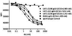


Classifications
-
- A—HUMAN NECESSITIES
- A61—MEDICAL OR VETERINARY SCIENCE; HYGIENE
- A61K—PREPARATIONS FOR MEDICAL, DENTAL OR TOILETRY PURPOSES
- A61K39/00—Medicinal preparations containing antigens or antibodies
-
- A—HUMAN NECESSITIES
- A61—MEDICAL OR VETERINARY SCIENCE; HYGIENE
- A61K—PREPARATIONS FOR MEDICAL, DENTAL OR TOILETRY PURPOSES
- A61K39/00—Medicinal preparations containing antigens or antibodies
- A61K39/0005—Vertebrate antigens
- A61K39/0011—Cancer antigens
- A61K39/001102—Receptors, cell surface antigens or cell surface determinants
- A61K39/001111—Immunoglobulin superfamily
- A61K39/001112—CD19 or B4
-
- C—CHEMISTRY; METALLURGY
- C07—ORGANIC CHEMISTRY
- C07K—PEPTIDES
- C07K14/00—Peptides having more than 20 amino acids; Gastrins; Somatostatins; Melanotropins; Derivatives thereof
- C07K14/435—Peptides having more than 20 amino acids; Gastrins; Somatostatins; Melanotropins; Derivatives thereof from animals; from humans
- C07K14/705—Receptors; Cell surface antigens; Cell surface determinants
- C07K14/70503—Immunoglobulin superfamily
- C07K14/7051—T-cell receptor (TcR)-CD3 complex
-
- C—CHEMISTRY; METALLURGY
- C07—ORGANIC CHEMISTRY
- C07K—PEPTIDES
- C07K16/00—Immunoglobulins [IGs], e.g. monoclonal or polyclonal antibodies
- C07K16/18—Immunoglobulins [IGs], e.g. monoclonal or polyclonal antibodies against material from animals or humans
- C07K16/28—Immunoglobulins [IGs], e.g. monoclonal or polyclonal antibodies against material from animals or humans against receptors, cell surface antigens or cell surface determinants
- C07K16/2803—Immunoglobulins [IGs], e.g. monoclonal or polyclonal antibodies against material from animals or humans against receptors, cell surface antigens or cell surface determinants against the immunoglobulin superfamily
- C07K16/2809—Immunoglobulins [IGs], e.g. monoclonal or polyclonal antibodies against material from animals or humans against receptors, cell surface antigens or cell surface determinants against the immunoglobulin superfamily against the T-cell receptor (TcR)-CD3 complex
-
- C—CHEMISTRY; METALLURGY
- C07—ORGANIC CHEMISTRY
- C07K—PEPTIDES
- C07K16/00—Immunoglobulins [IGs], e.g. monoclonal or polyclonal antibodies
- C07K16/18—Immunoglobulins [IGs], e.g. monoclonal or polyclonal antibodies against material from animals or humans
- C07K16/28—Immunoglobulins [IGs], e.g. monoclonal or polyclonal antibodies against material from animals or humans against receptors, cell surface antigens or cell surface determinants
- C07K16/2803—Immunoglobulins [IGs], e.g. monoclonal or polyclonal antibodies against material from animals or humans against receptors, cell surface antigens or cell surface determinants against the immunoglobulin superfamily
- C07K16/2818—Immunoglobulins [IGs], e.g. monoclonal or polyclonal antibodies against material from animals or humans against receptors, cell surface antigens or cell surface determinants against the immunoglobulin superfamily against CD28 or CD152
-
- C—CHEMISTRY; METALLURGY
- C07—ORGANIC CHEMISTRY
- C07K—PEPTIDES
- C07K16/00—Immunoglobulins [IGs], e.g. monoclonal or polyclonal antibodies
- C07K16/18—Immunoglobulins [IGs], e.g. monoclonal or polyclonal antibodies against material from animals or humans
- C07K16/28—Immunoglobulins [IGs], e.g. monoclonal or polyclonal antibodies against material from animals or humans against receptors, cell surface antigens or cell surface determinants
- C07K16/2896—Immunoglobulins [IGs], e.g. monoclonal or polyclonal antibodies against material from animals or humans against receptors, cell surface antigens or cell surface determinants against molecules with a "CD"-designation, not provided for elsewhere
-
- C—CHEMISTRY; METALLURGY
- C07—ORGANIC CHEMISTRY
- C07K—PEPTIDES
- C07K16/00—Immunoglobulins [IGs], e.g. monoclonal or polyclonal antibodies
- C07K16/46—Hybrid immunoglobulins
- C07K16/468—Immunoglobulins having two or more different antigen binding sites, e.g. multifunctional antibodies
-
- C—CHEMISTRY; METALLURGY
- C12—BIOCHEMISTRY; BEER; SPIRITS; WINE; VINEGAR; MICROBIOLOGY; ENZYMOLOGY; MUTATION OR GENETIC ENGINEERING
- C12N—MICROORGANISMS OR ENZYMES; COMPOSITIONS THEREOF; PROPAGATING, PRESERVING, OR MAINTAINING MICROORGANISMS; MUTATION OR GENETIC ENGINEERING; CULTURE MEDIA
- C12N15/00—Mutation or genetic engineering; DNA or RNA concerning genetic engineering, vectors, e.g. plasmids, or their isolation, preparation or purification; Use of hosts therefor
- C12N15/09—Recombinant DNA-technology
- C12N15/63—Introduction of foreign genetic material using vectors; Vectors; Use of hosts therefor; Regulation of expression
-
- A—HUMAN NECESSITIES
- A61—MEDICAL OR VETERINARY SCIENCE; HYGIENE
- A61K—PREPARATIONS FOR MEDICAL, DENTAL OR TOILETRY PURPOSES
- A61K39/00—Medicinal preparations containing antigens or antibodies
- A61K2039/505—Medicinal preparations containing antigens or antibodies comprising antibodies
-
- A—HUMAN NECESSITIES
- A61—MEDICAL OR VETERINARY SCIENCE; HYGIENE
- A61K—PREPARATIONS FOR MEDICAL, DENTAL OR TOILETRY PURPOSES
- A61K38/00—Medicinal preparations containing peptides
-
- C—CHEMISTRY; METALLURGY
- C07—ORGANIC CHEMISTRY
- C07K—PEPTIDES
- C07K2317/00—Immunoglobulins specific features
-
- C—CHEMISTRY; METALLURGY
- C07—ORGANIC CHEMISTRY
- C07K—PEPTIDES
- C07K2317/00—Immunoglobulins specific features
- C07K2317/30—Immunoglobulins specific features characterized by aspects of specificity or valency
- C07K2317/31—Immunoglobulins specific features characterized by aspects of specificity or valency multispecific
-
- C—CHEMISTRY; METALLURGY
- C07—ORGANIC CHEMISTRY
- C07K—PEPTIDES
- C07K2317/00—Immunoglobulins specific features
- C07K2317/30—Immunoglobulins specific features characterized by aspects of specificity or valency
- C07K2317/33—Crossreactivity, e.g. for species or epitope, or lack of said crossreactivity
-
- C—CHEMISTRY; METALLURGY
- C07—ORGANIC CHEMISTRY
- C07K—PEPTIDES
- C07K2317/00—Immunoglobulins specific features
- C07K2317/40—Immunoglobulins specific features characterized by post-translational modification
- C07K2317/41—Glycosylation, sialylation, or fucosylation
-
- C—CHEMISTRY; METALLURGY
- C07—ORGANIC CHEMISTRY
- C07K—PEPTIDES
- C07K2317/00—Immunoglobulins specific features
- C07K2317/50—Immunoglobulins specific features characterized by immunoglobulin fragments
- C07K2317/52—Constant or Fc region; Isotype
-
- C—CHEMISTRY; METALLURGY
- C07—ORGANIC CHEMISTRY
- C07K—PEPTIDES
- C07K2317/00—Immunoglobulins specific features
- C07K2317/50—Immunoglobulins specific features characterized by immunoglobulin fragments
- C07K2317/52—Constant or Fc region; Isotype
- C07K2317/524—CH2 domain
-
- C—CHEMISTRY; METALLURGY
- C07—ORGANIC CHEMISTRY
- C07K—PEPTIDES
- C07K2317/00—Immunoglobulins specific features
- C07K2317/50—Immunoglobulins specific features characterized by immunoglobulin fragments
- C07K2317/52—Constant or Fc region; Isotype
- C07K2317/526—CH3 domain
-
- C—CHEMISTRY; METALLURGY
- C07—ORGANIC CHEMISTRY
- C07K—PEPTIDES
- C07K2317/00—Immunoglobulins specific features
- C07K2317/50—Immunoglobulins specific features characterized by immunoglobulin fragments
- C07K2317/52—Constant or Fc region; Isotype
- C07K2317/53—Hinge
-
- C—CHEMISTRY; METALLURGY
- C07—ORGANIC CHEMISTRY
- C07K—PEPTIDES
- C07K2317/00—Immunoglobulins specific features
- C07K2317/50—Immunoglobulins specific features characterized by immunoglobulin fragments
- C07K2317/55—Fab or Fab'
-
- C—CHEMISTRY; METALLURGY
- C07—ORGANIC CHEMISTRY
- C07K—PEPTIDES
- C07K2317/00—Immunoglobulins specific features
- C07K2317/50—Immunoglobulins specific features characterized by immunoglobulin fragments
- C07K2317/56—Immunoglobulins specific features characterized by immunoglobulin fragments variable (Fv) region, i.e. VH and/or VL
-
- C—CHEMISTRY; METALLURGY
- C07—ORGANIC CHEMISTRY
- C07K—PEPTIDES
- C07K2317/00—Immunoglobulins specific features
- C07K2317/60—Immunoglobulins specific features characterized by non-natural combinations of immunoglobulin fragments
- C07K2317/64—Immunoglobulins specific features characterized by non-natural combinations of immunoglobulin fragments comprising a combination of variable region and constant region components
-
- C—CHEMISTRY; METALLURGY
- C07—ORGANIC CHEMISTRY
- C07K—PEPTIDES
- C07K2317/00—Immunoglobulins specific features
- C07K2317/60—Immunoglobulins specific features characterized by non-natural combinations of immunoglobulin fragments
- C07K2317/66—Immunoglobulins specific features characterized by non-natural combinations of immunoglobulin fragments comprising a swap of domains, e.g. CH3-CH2, VH-CL or VL-CH1
-
- C—CHEMISTRY; METALLURGY
- C07—ORGANIC CHEMISTRY
- C07K—PEPTIDES
- C07K2317/00—Immunoglobulins specific features
- C07K2317/70—Immunoglobulins specific features characterized by effect upon binding to a cell or to an antigen
- C07K2317/73—Inducing cell death, e.g. apoptosis, necrosis or inhibition of cell proliferation
-
- C—CHEMISTRY; METALLURGY
- C07—ORGANIC CHEMISTRY
- C07K—PEPTIDES
- C07K2317/00—Immunoglobulins specific features
- C07K2317/90—Immunoglobulins specific features characterized by (pharmaco)kinetic aspects or by stability of the immunoglobulin
- C07K2317/94—Stability, e.g. half-life, pH, temperature or enzyme-resistance
-
- C—CHEMISTRY; METALLURGY
- C07—ORGANIC CHEMISTRY
- C07K—PEPTIDES
- C07K2319/00—Fusion polypeptide
-
- C—CHEMISTRY; METALLURGY
- C07—ORGANIC CHEMISTRY
- C07K—PEPTIDES
- C07K2319/00—Fusion polypeptide
- C07K2319/33—Fusion polypeptide fusions for targeting to specific cell types, e.g. tissue specific targeting, targeting of a bacterial subspecies
Definitions
- the present disclosure generally relates to soluble polypeptide complexes comprising antibody variable regions fused to the TCR constant regions, and bispecific polypeptide complexes comprising the same.
- Bispecific antibodies are growing to be the new category of therapeutic antibodies. They can bind two different targets or two different epitopes on a target, creating additive or synergistic effect superior to the effect of individual antibodies.
- a lot of antibody engineering efforts have been put into designing new bispecific formats, such as DVD-Ig, CrossMab, BiTE etc. (Spiess et al., Molecular Immunology, 67 (2) , pp. 95-106 (2015) ) .
- these formats may potentially have various limitations in stability, solubility, short half-life, and immunogenicity.
- an IgG-like bispecific antibody is a common format: one arm binding to target A and another arm binding to target B. Structurally it is made from half of antibody A and half of antibody B, with the similar size and shape as natural IgG.
- bispecific molecules can be easily produced like a normal IgG from a single host cell with high expression level and correctly assembled form.
- the pairing of cognate light-heavy chains as well as the assembly of two different half antibodies cannot be automatically controlled. All kinds of mispairings in a random manner could result in significant product heterogeneity.
- VH-VL variable domain
- CH1-CL constant domain
- Several strategies have been applied into designing orthogonal interfaces to facilitate cognate pairing.
- the present disclosure provides a polypeptide complex comprising a first polypeptide comprising, from N-terminus to C-terminus, a first heavy chain variable domain (VH) of a first antibody operably linked to a first T cell receptor (TCR) constant region (C1) , and a second polypeptide comprising, from N-terminus to C-terminus, a first light chain variable domain (VL) of the first antibody operably linked to a second TCR constant region (C2) , wherein C1 and C2 are capable of forming a dimer comprising at least one non-native interchain bond between C1 and C2, and the non-native interchain bond is capable of stabilizing the dimer, and the first antibody has a first antigenic specificity.
- the present disclosure provides a bispecific polypeptide complex, comprising a first antigen-binding moiety associated with a second antigen-binding moiety, wherein the first antigen-binding moiety comprising a first polypeptide comprising, from N-terminal to C-terminal, a first heavy chain variable domain (VH) of a first antibody operably linked to a first T cell receptor (TCR) constant region (C1) , and a second polypeptide comprising, from N-terminal to C-terminal, a first light chain variable domain (VL) of the first antibody operably linked to a second TCR constant region (C2) , wherein C1 and C2 are capable of forming a dimer comprising at least one non-native interchain bond between a first mutated residue comprised in C1 and a second mutated residue comprised in C2, and the non-native interchain bond is capable of stabilizing the dimer, and the first antibody has a first antigenic specificity, a second antigen-
- the present disclosure provides herein a bispecific polypeptide complex, comprising a first antigen binding moiety comprising the polypeptide complex provided herein having a first antigenic specificity, associated with a second antigen binding moiety having a second antigenic specificity which is different from the first antigenic specificity, and the first antigen-binding moiety and the second antigen-binding moiety are less prone to mispair than otherwise would have been if both the first and the second antigen-binding moieties are counterparts of natural Fab.
- the present disclosure provides a bispecific fragment of the bispecific polypeptide complex provided herein.
- the present disclosure provides herein a conjugate comprising the polypeptide complex provided herein, or the bispecific polypeptide complex provided herein conjugated to a moiety.
- the present disclosure provides herein an isolated polynucleotide encoding the polypeptide complex provided herein, or the bispecific polypeptide complex provided herein.
- the present disclosure provides herein an isolated vector comprising the polynucleotide provided herein.
- the present disclosure provides herein a host cell comprising the isolated polynucleotide provided herein or the isolated vector provided herein.
- the present disclosure provides herein a method of expressing the polypeptide complex provided herein, or the bispecific polypeptide complex provided herein, comprising culturing the host cell provided herein under the condition at which the polypeptide complex, or the bispecific polypeptide complex is expressed.
- the present disclosure provides herein a method of producing the polypeptide complex provided herein, comprising a) introducing to a host cell a first polynucleotide encoding a first polypeptide comprising, from N-terminal to C-terminal, a first heavy chain variable domain (VH) of a first antibody operably linked to a first TCR constant domain (C1) , and a second polynucleotide encoding a second polypeptide comprising, from N-terminal to C-terminal, a first light chain variable domain (VL) of the first antibody operably linked to a second TCR constant domain (C2) , wherein C1 and C2 are capable of forming a dimer comprising at least one non-native interchain bond between a first mutated residue comprised in C1 and a second mutated residue comprised in C2, and the non-native interchain bond is capable of stabilizing the dimer of C1 and C2, and the first antibody has a first antigenic specificity
- the present disclosure provides herein a method of producing the bispecific polypeptide complex provided herein, comprising a) introducing to a host cell a first polynucleotide encoding a first polypeptide comprising, from N-terminal to C-terminal, a first heavy chain variable domain (VH) of a first antibody operably linked to a first TCR constant region (C1) , a second polynucleotide encoding a second polypeptide comprising, from N-terminal to C-terminal, a first light chain variable domain (VL) of the first antibody operably linked to a second TCR constant region (C2) , a third polynucleotide encoding a third polypeptide comprising VH of a second antibody, and a fourth polynucleotide encoding a fourth polypeptide comprising VL of the second antibody, wherein C1 and C2 are capable of forming a dimer comprising at least one non-native interchain bond between C
- the method of producing the bispecific polypeptide complex provided herein further comprising isolating the polypeptide complex.
- the present disclosure provides a composition comprising the polypeptide complex provided herein, or the bispecific polypeptide complex provided herein.
- the present disclosure provides herein a pharmaceutical composition
- a pharmaceutical composition comprising the polypeptide complex provided herein, or the bispecific polypeptide complex provided herein and a pharmaceutically acceptable carrier.
- the present disclosure provides herein a method of treating a condition in a subject in need thereof, comprising administrating to the subject a therapeutically effective amount of the polypeptide complex provided herein, or the bispecific polypeptide complex provided herein.
- the condition can be alleviated, eliminated, treated, or prevented when the first antigen and the second antigen are both modulated.
- the non-native interchain bond is formed between a first mutated residue comprised in C1 and a second mutated residue comprised in C2.
- at least one of the first and the second mutated residues is a cysteine residue.
- the non-native interchain bond is a disulphide bond.
- the first mutated residue is comprised within a contact interface of C1
- the second mutated residue is comprised within a contact interface of C2.
- At least one native cysteine residue is absent or present in C1 and/or C2.
- the native cysteine residue at position C74 of engineered CBeta is absent or present.
- the native C74 is absent in CBeta.
- At least one native N-glycosylation site is absent or present in C1 and/or C2. In certain embodiments, the native N-glycosylation sites are absent in C1 and/or C2.
- the dimer comprises 1, 2, 3, 4, 5, 6, 7, 8, 9, 10, 11, 12, 13, 14, 15 or more non-native interchain bonds. In certain embodiments, at least one of the non-native interchain bonds is disulphide bond. In certain embodiments, the dimer comprises 1, 2, 3, 4, 5, 6, 7, 8, 9, 10, 11, 12, 13, 14, 15 or more disulphide bonds.
- C1 comprises an engineered CBeta, and C2 comprises an engineered CAlpha
- C1 comprises an engineered CAlpha
- C2 comprises an engineered CBeta
- C1 comprises an engineered CBeta
- C2 comprises an engineered CPre-Alpha
- C1 comprises an engineered CPre-Alpha
- C2 comprises an engineered CBeta
- e) comprises an engineered CGamma
- C2 comprises an engineered CDelta
- C1 comprises an engineered CDelta, and C2 comprises an engineered CGamma.
- the first VH is operably linked to C1 at a first conjunction domain
- the first VL is operably linked to C2 at a second conjunction domain.
- the first VH associates to C1 at a first conjunction domain via a connector
- the first VL associates to C2 at a second conjunction domain via a connector.
- the first and/or the second conjunction domain comprises a proper length (e.g., 0, 1, 2, 3, 4, 5, 6, 7, 8, 9, or 10 amino acid residues) of the C terminal fragment of antibody V/C conjunction, and a proper length (e.g., 0, 1, 2, 3, 4, 5, 6, 7, 8, 9, or 10 amino acid residues) of the N terminal fragment of TCR V/C conjunction.
- a proper length e.g., 0, 1, 2, 3, 4, 5, 6, 7, 8, 9, or 10 amino acid residues
- the engineered CBeta comprises a mutated cysteine residue within a contact interface selected from the group consisting of amino acid residues 9-35, 52-66, 71-86, and 122-127; and/or the engineered CAlpha comprises a mutated cysteine residue within a contact interface selected from a group consisting of amino acid residues 6-29, 37-67, and 86-95.
- the engineered CBeta comprises a mutated cysteine residue that substitutes for an amino acid residue at a position selected from: S56C, S16C, F13C, V12C, E14C, L62C, D58C, S76C, and R78C
- the engineered CAlpha comprises a mutated cysteine residue that substitutes for an amino acid residue at a position selected from: T49C, Y11C, L13C, S16C, V23C, Y44C, T46C, L51C, and S62C.
- the engineered CBeta and the engineered CAlpha comprise a pair of mutated cysteine residues that substitute for a pair of amino acid residues selected from the group consisting of: S16C in CBeta and Y11C in CAlpha, F13C in CBeta and L13C in CAlpha, S16C in CBeta and L13C in CAlpha, V12C in CBeta and S16C in CAlpha, E14C in CBeta and S16C in CAlpha, F13C in CBeta and V23C in CAlpha, L62C in CBeta and Y44C in CAlpha, D58C in CBeta and T46C in CAlpha, S76C in CBeta and T46C in CAlpha, S56C in CBeta and T49C in CAlpha, S56C in CBeta and L51C in CAlpha, S56C in CBeta and S62C in CAl
- At least one native glycosylation site is absent or present in the engineered CBeta and/or in the engineered CAlpha.
- the native glycosylation site in the engineered CBeta is N69, and/or the native glycosylation site (s) in the engineered CAlpha is/are selected from N34, N68, N79, and any combination thereof.
- the engineered CBeta lacks or retains a FG loop encompassing amino acid residues 101-117 of the native CBeta and/or a DE loop encompassing amino acid residues 66-71 of the native CBeta.
- the engineered CAlpha comprises any one of SEQ ID NOs: 43-48, and/or the engineered CBeta comprises any one of SEQ ID NOs: 33-41 and 306.
- C1 comprises the engineered CBeta
- C2 comprises the engineered CAlpha
- the first conjunction domain comprises or is SEQ ID NO: 49 or 50
- the second conjunction domain comprises or is SEQ ID NO: 51 or 52.
- the C1 comprises the engineered CAlpha
- the C2 comprises the engineered CBeta
- the first conjunction domain comprises or is SEQ ID NO: 129 or 130
- the second conjunction domain comprises or is SEQ ID NO: 49 or 50.
- the engineered CBeta comprises a mutated cysteine residue within a contact interface selected from the group consisting of: amino acid residues 9-35, 52-66, 71-86 and 122-127; and/or the engineered CPre-Alpha comprises a mutated cysteine residue within a contact interface selected from a group consisting of: amino acid residues 7-19, 26-34, 56-75 and 103-106.
- the engineered CBeta comprises a mutated cysteine residue that substitutes for an amino acid residue at a position selected from: S16C, A18C, E19C, F13C, A11C, S56C, and S76C
- the engineered CPre-Alpha comprises a mutated cysteine residue that substitutes for an amino acid residue at a position selected from S11C, A13C, I16C, S62C, T65C, and Y59C.
- the engineered CBeta and the engineered CPre-Alpha comprise a pair of mutated cysteine residues that substitute for a pair of amino acid residues selected from the group consisting of: S16C in CBeta and S11C in CPre-Alpha, A18C in CBeta and S11C in CPre-Alpha, E19C in CBeta and S11C in CPre-Alpha, F13C in CBeta and A13C in CPre-Alpha, S16C in CBeta and A13C in CPre-Alpha, A11C in CBeta and I16C in CPre-Alpha, S56C in CBeta and S62C in CPre-Alpha, S56C in CBeta and T65C in CPre-Alpha, and S76C in CBeta, and Y59C in CPre-Alpha, and wherein the pair of mutated cysteine residues are capable of
- At least one native glycosylation site is absent in the engineered CBeta and/or in the engineered CPre-Alpha.
- the absent or present glycosylation site in the engineered CBeta is N69, and/or the absent glycosylation site in the engineered CPre-Alpha is N50.
- the engineered CBeta lacks or retains a FG loop encompassing the amino acid residues 101-107 of the native CBeta and/or a DE loop at position encompassing the amino acid residues 66-71 of the native CBeta.
- the engineered CPre-Alpha comprises any one of SEQ ID NOs: 82, 83, and 311-318; and/or the engineered CBeta comprises any one of SEQ ID NOs: 84, 33-41, and 319-324.
- C1 comprises the engineered CBeta
- C2 comprises the engineered CPre-Alpha
- the first conjunction domain comprises SEQ ID NO: 49 or 50
- the second conjunction domain comprises SEQ ID NO: 81 or 131.
- C1 comprises the engineered CPre-Alpha
- C2 comprises the engineered CBeta
- the first conjunction domain comprises SEQ ID NO: 132 or 133
- the second conjunction domain comprises SEQ ID NO: 49 or 50.
- the engineered CDelta comprises a mutated cysteine residue within a contact interface selected from the group consisting of: amino acid residues 8-26, 43-64, and 84-88; and/or the engineered CGamma comprises a mutated cysteine residue within a contact interface selected from a group consisting of: amino acid residues 11-35 and 55-76.
- the engineered CGamma comprises a mutated cysteine residue that substitutes for an amino acid residue at a position selected from: S17C, E20C, F14C, T12C, M62C, Q57C, and A19C
- the engineered CDelta comprises a mutated cysteine residue that substitutes for an amino acid residue at a position selected from: F12C, M14C, N16C, D46C, V50C, F87C, and E88C.
- the engineered CGamma and the engineered CDelta comprise a pair of mutated cysteine residues that substitute for a pair of amino acid residues selected from the group consisting of: S17C in CGamma and F12C in CDelta, E20C in CGamma and F12C in CDelta, F14C in CGamma and M14C in CDelta, T12C in CGamma and N16C in CDelta, M62C in CGamma and D46C in CDelta, Q57C in CGamma and V50C in CDelta, A19C in CGamma and F87C in CDelta, and A19C in CGamma and E88C in CDelta, and wherein the introduced pair of cysteine residues are capable of forming an interchain disulphide bond.
- At least one native glycosylation site is absent or present in the engineered CGamma and/or in the engineered CDelta.
- the native glycosylation site in the engineered CGamma is N65, and/or the native glycosylation site (s) in the engineered CDelta is/are one or both of N16 and N79.
- the engineered CGamma comprises SEQ ID NO: 113, 114, 333, 334, 335, 336, 337, 338, 339, or 340
- the engineered CDelta comprises SEQ ID NO: 115, 116, 310, 325, 326, 327, 328, 329, 330, 331, or 332.
- C1 comprises the engineered CGamma
- C2 comprises the engineered CDelta
- the first conjunction domain comprises SEQ ID NO: 117 or 118
- the second conjunction domain comprises SEQ ID NO: 119 or 120.
- C1 comprises the engineered CDelta
- C2 comprises the engineered CGamma
- the first conjunction domain comprises SEQ ID NO: 123 or 124
- the second conjunction domain comprises SEQ ID NO: 125 or 126.
- the first polypeptide further comprises an antibody CH2 domain, and/or an antibody CH3 domain.
- the first antigenic specificity and the second antigenic specificity are directed to two different antigens, or are directed to two different epitopes on one antigen.
- the first antigen-binding moiety binds to CD3. In certain embodiments, the second antigen-binding moiety binds to CD19. In certain embodiments, the first antigen-binding moiety binds to CD19. In certain embodiments, the second antigen-binding moiety binds to CD3.
- the first antigen-binding moiety binds to CTLA-4. In certain embodiments, the second antigen-binding moiety binds to PD-1. In certain embodiments, the first antigen-binding moiety binds to PD-1. In certain embodiments, the second antigen-binding moiety binds to CTLA-4.
- the association is via a connecter, a disulphide bond, a hydrogen bond, electrostatic interaction, a salt bridge, or hydrophobic-hydrophilic interaction, or the combination thereof.
- the second antigen-binding moiety comprises a heavy chain variable domain and a light chain variable domain of a second antibody having the second antigenic specificity.
- the second antigen-binding moiety comprises a Fab.
- the first antigenic specificity and the second antigenic specificity are directed to two different antigens, or are directed to two different epitopes on one antigen.
- one of the first and the second antigenic specificities is directed to a T-cell specific receptor molecule and/or a natural killer cell (NK cell) specific receptor molecule, and the other is directed to a tumor associated antigen.
- one of the first and the second antigenic specificities is directed to CD3, and the other is directed to a tumor associated antigen.
- one of the first and the second antigenic specificities is directed to CD3, and the other is directed to CD19.
- the first antigen-binding moiety further comprises a first dimerization domain
- the second antigen-binding moiety further comprises a second dimerization domain, wherein the first and the second dimerization domains are associated.
- the association is via a connecter, a disulphide bond, a hydrogen bond, electrostatic interaction, a salt bridge, or hydrophobic-hydrophilic interaction, or the combination thereof.
- the first and/or the second dimerization domain comprises at least a portion of an antibody hinge region, optionally derived from IgG1, IgG2 or IgG4.
- the first and/or the second dimerization domain further comprises a dimerization domain.
- the dimerization domain comprises at least a portion of an antibody hinge region, an antibody CH2 domain, and/or an antibody CH3 domain.
- the first dimerization domain is operably linked to the first TCR constant region (C1) at a third conjunction domain.
- C1 comprises an engineered CBeta, and the third conjunction domain is comprised in SEQ ID NO: 53 or 54; b) C1 comprises an engineered CAlpha, and the third conjunction domain is comprised in SEQ ID NO: 134, 135, 140, or 141; c) C1 comprises an engineered CPre-Alpha, and the third conjunction domain is comprised in SEQ ID NO: 134, 135, 140, or 141; d) C1 comprises an engineered CGamma, and the third conjunction domain is comprised in SEQ ID NO: 121 or 122; or e) C1 comprises an engineered CDelta, and the third conjunction domain is comprised in SEQ ID NO: 127 or 128.
- the second dimerization domain is operably linked to the heavy chain variable domain of the second antigen-binding moiety.
- the first and the second dimerization domains are different and associate in a way that discourages homodimerization and/or favors heterodimerization.
- the first and the second dimerization domains are capable of associating into heterodimers via knobs-into-holes, hydrophobic interaction, electrostatic interaction, hydrophilic interaction, or increased flexibility.
- the first antigen-binding moiety comprising the first polypeptide comprising VH operably linked to a chimeric constant region
- the second polypeptide comprises VL operably linked to C2
- the chimeric constant region and C2 comprises a pair of sequences selected from the group consisting of: SEQ ID NOs: 177/176, 179/178, 184/183, 185/183, 180/176, 181/178, 182/178, 184/186, 185/186, 188/187, 196/187, 190/189, 192/191, 192/193, 195/194, 198/197, 200/199, 202/201, 203/201, 203/204, 205/204, 206/204, 208/207, 208/209, 211/210, 213/212, 213/215, 213/151, 214/212, 214/151, 232/231, 216/215, 218/217, 220/219, 222/221, 224/223, 226/225,
- the first antigenicity is directed to CD3, and the first polypeptide and the second polypeptide comprise a pair of sequences selected from the group consisting of: SEQ ID NOs: 2/1, 3/4/, 5/1, 6/3, 7/3, 9/8, 10/8, 9/11, 10/11, 13/12, 15/14, 17/16, 17/18, 20/19, 21/12, 65/64, 67/66, 69/68, 70/68, 70/71, 72/71, 73/71, 75/74, 75/76, 78/77, 86/85, 90/89, 91/92/, 94/93, 96/95, 98/97, 99/95, 101/100, 101/102, 106/105, 108/107, 110/109, 112/111, 137/136, 138/136, 137/139, and 138/139.
- SEQ ID NOs 2/1, 3/4/, 5/1, 6/3, 7/3, 9/8, 10/8, 9/11, 10/11, 13/12, 15/14, 17/16
- the first antigen-binding moiety and the second antigen-binding moiety comprise a four-sequence combination selected from the group consisting of: SEQ ID NOs: 22/12/24/23, 25/12/26/23, and 25/12/27/23, wherein the first antigen-binding moiety is capable of binding to CD3, and the second antigen-binding moiety is capable of binding to CD19.
- the polypeptide complex provided herein can be made into a Fab, a (Fab) 2 , a bibody, a tribody, a triFabs, tandem linked Fabs, a Fab-Fv, tandem linked V domains, tandem linked scFvs, and among other formats.
- the present disclosure provides a kit comprising the polypeptide complex provided herein for detection, diagnosis, prognosis, or treatment of a disease or condition.
- FIG 1 presents schematic representations of studied antibody formats. Both anti-CD3 antibody T3 and anti-CD19 antibody U4 were developed. The constant region (CL and CH1) of T3 was replaced by the constant domains of TCR to design unique light-heavy chain interface that is orthogonal to regular antibody. The TCR-modified T3 and native U4 in conjunction with “knobs-into-holes” mutations in Fc domain were used to design bispecific antibody formats E17 and F16.
- Figures 2A-2D present superimposed poses of antibody Fv model and TCR structure providing guidance in fusing antibody Fv and TCR constant region.
- Figure 2A presents an antibody Fv structure model that was built based on the sequence of an anti-CD3 antibody T3 developed in-house.
- Figure 2B presents the TCR structure from PDB 4L4T.
- Figure 2C presents an antibody Fv structural model superimposed on the TCR variable region in different orientations. Rough chimeric proteins were created by removing the TCR variable domain in the superimposed poses, as shown in Figure 2D. The overlapped residues in the conjunction area helped design conjunction region.
- the antibody VL chain and the TCR alpha chain were colored in white.
- the VH and beta chains were colored in black.
- Figures 3A-3B show a comparison between TCR constant region and antibody Fab constant region.
- Figure 3A shows a TCR crystal structure from PDB 4L4T.
- Figerie 3B shows antibody Fab structural model made by the Fv domain of T3 model and the constant domain of antibody from PDB 5DK3. The obvious differences in FG and DE loops between constant domains of TCR and constant domains of antibody Fab were marked by displaying all the residues side chains.
- Figure 4 shows SDS-PAGE results of the de-glycosylation mutants of TCR-antibody chimeric antibodies with CAlpha and CBeta chains.
- Samples were all harvested supernatants from the production of Expi293 expressions.
- Lanes 1, 3, 5, 7 and 9 are the non-reduced pages of Design_2-QQQQ, Design_2-AAAA, Design_2-QSKE, Design_2-ASKE and Design_2-QQQQQQ, respectively.
- Lanes 2, 4, 6, 8 and 10 are the corresponding reduced pages.
- Figure 5 shows dose-dependent FACS bindings of all the de-glycosylated mutants binding to CD3-expressed Jurkat cells. All samples were harvested supernatants of the de-glycosylation mutants expressed in Expi293. The wild type anti-CD3 antibody (T3-IgG1) was used as the positive control.
- Figures 6A-6B show SDS-PAGE results of the chain mispairing tests of antibody T3 and U4 in IgG1 ( Figure 6A) and IgG4 ( Figure 6B) isotype.
- Lanes 1-2 are the pairs of T3_light-U4_heavy and T3_heavy-U4_light, respectively.
- Lanes 3-4 are the same pair order as lanes 1-2, but with the modified T3 using TCR constant region.
- Lanes 1-4 in both pictures are the non-reduced samples, and Lanes 5-8 are the corresponding reduced samples.
- FIGS 7A-7B show SDS-PAGE results of purified bispecific antibody, E17-Design_2-QQQQ in ( Figure 7A) IgG1 and ( Figure 7B) IgG4.
- the IgG1 isotype was purified by three step purifications: protein A chromatography, Ion-Exchange Chromatography (IEC) and Size Exclusion Chromatography (SEC) .
- the IgG4 was obtained after two-step purifications: protein A chromatography and SEC.
- Figures 7C-7D show SEC-HPLC data for the purified samples of IgG1 ( Figure 7C) and IgG4 ( Figure 7D) to determine the purities of the samples.
- Figure 8 shows SDS-PAGE results of Fab fragments of chimeric T3 with a 6xHis-tag, purified by Ni Sepharose TM excel chromatography. Lanes 1 and 3 are bands for T3-Fab-Design_2. his1, and Lanes 2 and 4 are bands for T3-Fab-Design_2. his2.
- Figure 9 illustrates dose-dependent FACS bindings of the Fab fragment of the TCR-modified chimeric T3.
- the monovalent form of wild type T3 antibody (T3-Fab-IgG4) was used as a positive control.
- a regular human IgG4 antibody was used as a negative control.
- Figures 10A-10B show dose-dependent FACS bindings of the designed bispecific antibody, E17-Design_2-QQQQ, to CD3+ Jurkat cells.
- a irrelevant human IgG1 or IgG4 antibody was used as a negative control.
- Figures 11A-11B show comparison of the FACS bindings of two designed bispecific antibodies, E17-Design_2-QQQQ and F16-Design_2-QQQQ, to CD3 on Jurkat cells ( Figure 11A) and CD19 expressed on Ramos cells ( Figure 11B) .
- the bispecific antibodies in both IgG1 and IgG4 isotypes were tested.
- a regular human IgG1 or IgG4 antibody was used as a negative control.
- Figure 12 illustrates the cytotoxic assay of T-cell directed killing malignant B cell, mediated by the designed bispecific antibodies E17-Design_2-QQQQ in both IgG1 and IgG4.
- the parental monospecific anti-CD3 (T3-IgG4) , anti-CD19 (U4-IgG) antibody and an irrelevant human IgG1 antibody was used as the negative control.
- Figure 13 compares the activity of two designed bispecific antibody, E17-Design_2-QQQQ and F16-Design_2-QQQQ in mediating T-cell engaged malignant B cell killing.
- An irrelevant human IgG antibody was used as the negative control.
- Figures 14A-14B show deconvoluted mass spectra of bispecific antibody E17-Design_2-QQQQ in non-reduced ( Figure 14A) and reduced ( Figure 14B) conditions.
- Peak at 148180.53 in Figure 14A is the correct molecular weight of the intact WuXiBody.
- the peak with 22877 Da indicates the light chain found in the reduced mass spectra in Figure 14B.
- the small peak at 149128.45 Da in Figure 14A was deduced to be the O-glycosylation (approximately (947.92 Da more) located on the light chain, as showed in Figure 14B.
- Figures 15A-15B show the role of interchain disulphide bond in antibody expression at alpha/beta interface characterized by SDS-PAGE.
- Figure 15A shows the antibody containing interchain disulphide bond between CAlpha and CBeta;
- Figure 15B shows the antibody without interchain disulphide bond between CAlpha and CBeta;
- Lanes 1 and 3 are the non-reduced PAGE results of Design_2-QQQQ-IgG4 with and without introduced disulphide bond, respectively.
- Lanes 2 and 4 are the reduced PAGE results of Design_2-QQQQ-IgG4 with and without introduced disulphide bond, respectively.
- Figure 16 shows SDS-PAGE of designed disulphide bond at pre-alpha/beta interface.
- Lane 1 and Lane 2 are “Design_5_Pre_TCR_Conjunction’1_Cys13” and “Design_6_Pre_TCR_Conjunction’1_Cys14” , respectively, treated in non-reduced condition.
- Lane 4 and lane 5 are “Design_5_Pre_TCR_Conjunction’1_Cys13” and “Design_6_Pre_TCR_Conjunction’1_Cys14” , respectively, treated in reduced condition.
- Figures 17A-17B show SDS-PAGE of designed disulphide bond at delta/gamma interface.
- Lane 6 and lane 8 are “Design_2_Cys5_no_Glyco” and “Design_2_hypeCys2_no_Glyco” , respectively.
- Figure 17A is non-reduced SDS-PAGE.
- Figure 17B is reduced SDS-PAGE.
- Figure 18A shows the sequence of native TCR alpha chain and its counterpart sequence with mutated cysteine residues.
- TRAC_Human is a natural sequence of alpha chain constant region.
- 4L4T_Alpha_Crystal is the sequence of a crystal structure (PDB code 4L4T) with S55C mutations that can form inter-chain disulphide bond.
- the gray region is the constant region used as backbone of chimeric protein in this invention.
- Figure 18B shows the sequence of native TCR beta chain and its counterpart sequence with mutated cysteine residues.
- TRBC1_Human and TRBC2_Human are natural sequences of beta constant region.
- Figure 18C shows the sequences of native TCR pre-alpha chain.
- PTCRA_Human is a natural sequence of pre-alpha chain constant region (pre-alpha chain only has no variable region) .
- 3OF6_PreAlpha_Crystal is the sequence of a crystal structure (PDB code 3OF6) .
- the gray region is the constant region used above to define the numbering.
- Figure 18D shows the sequences of native TCR delta chain.
- TRA@_Human is the natural sequences of delta constant region.
- 4LFH_Delta_Crystal is the constant region of a delta chain sequence of a crystal structure (PDB code 4LFH) .
- the gray region is the constant region used above to define the numbering.
- Figure 18E shows the sequences of native TCR gamma chain.
- TRGC1_Human and TRGC2_Human are natural sequences of gamma constant region.
- 4LFH_Gamma_Crystal is the constant region of a gamma chain sequence of a crystal structure (PDB code 4LFH) .
- the gray region is the constant region used above to define the numbering.
- Figures 19A-19E show the sequences and numbering of the TCR constant regions.
- Figure 19A shows the sequences and numbering of the TCR Alpha constant region.
- Figure 19B shows the sequences and numbering of the TCR Beta constant region.
- Figure 19C shows the sequences and numbering of the TCR Pre-Alpha constant region.
- Figure 19D shows the sequences and numbering of the TCR Delta constant region.
- Figure 19E shows the sequences and numbering of the TCR Gamma constant region.
- Figures 20A-20D show the sequences and numbering of the IgG1 and IgG4 knobs-into-holes.
- Figure 20A shows the sequences and numbering of the IgG1 “knob” mutations.
- Figure 20B shows the sequences and numbering of the IgG4 “knob” mutations.
- Figure 20C shows the sequences and numbering of the IgG1 “hole” mutations.
- Figure 20D shows the sequences and numbering of the IgG4 “hole” mutations.
- Figures 21A-21B show bindings of E17-Design_2-QQQQ in both IgG4 ( Figure 21A) and wild type IgG1 ( Figure 21B) formats to human C1Q by ELISA.
- a human IgG1 antibody was used as control.
- Figure 22 shows schematic description of four symmetric WuXiBody formats G19, G19R, G25 and G25R.
- formats G19 and G25 two TCR-containing chimeric Fab-like domains were grafted at the C-terminus and N-terminus of a normal antibody, respectively.
- the rectangles indicate TCR constant domains, and the ovals indicate variable and constant domains of an antibody.
- the difference between formats G19 and G19R or G25 and G25R is the switched position of normal Fab and chimeric Fab.
- These formats can accommodate different variable regions from different antibody pairs and usually have a molecular weight around 240-250 kD.
- Figures 23A-23B show SDS-PAGE ( Figure 23A) and SEC-HPLC ( Figure 23B) characterizations of two purified bispecific antibodies in G19 format.
- the lane numbers in SDS-PAGE are consistent with the label numbers in the SEC-HPLC figure.
- Lanes 1 and 2 are the T1U6 and U6T1 antibody pair, respectively.
- T1U6 T1 (anti-CTLA-4) was on the N-terminus of the format
- U6T1 U6 anti-PD-1
- Both bispecific molecules were purified by protein A chromatography, and purities around 90%was achieved.
- Figures 24A-24B show dose-dependent FACS bindings of purified U6T1 and T1U6 antibodies in G19 format to human PD-1 ( Figure 24A) and CTLA-4 ( Figure 24B) engineered cells.
- An IgG4 antibody was used as the negative control.
- Figures 25A-25B show SDS-PAGE (Figure 25A) and SEC-HPLC ( Figure 25B) characterizations of the Protein A-purified bispecifc antibodies in different symmetric formats.
- Lanes 1-3 are the U6T1 antibody pair in G19R, G25, and G25R formats, respectively.
- PC is a control protein known to have 250 kD molecular weight. All of the three bispecific molecules had more than 90%purity.
- the lane numbers in SDS-PAGE are consistent with the label numbers in SEC-HPLC figures.
- Figures 26A-26B show dose-dependent FACS bindings of purified U6T1 bispeicifc antibodies in G19R, G25, and G25R formats to human PD-1 ( Figure 26A) and CTLA-4 ( Figure 26B) engineered cells.
- a benchmark bispecific anti-CTAL-4 x PD-1 antibody (BMK1. IgG1) was used as a control, and an IgG4 antibody was used as the negative control.
- Figures 27A-27B show FACS competition assays of the designed bispecific antibodies in G19R, G25, and G25R formats to block human PD-L1 binding to PD-1 (Figure 27A) and CD80 binding to CTLA-4 ( Figure 27B) , respectively.
- a benchmark bispecific anti-CTAL-4 x PD-1 antibody (BMK1. IgG1) was used as a control, and an IgG4 antibody was used as the negative control.
- Figures 28A-28B show SDS-PAGE ( Figure 28A) and SEC-HPLC ( Figure 28B) characterizations of the Protein A-purified bispecifc antibodies in different symmetric formats.
- Lanes 1-4 are the U6T5 antibody pair in G19, G19R, G25, and G25R formats, respectively.
- PC is a control protein with 250 kD molecular weight. All the three bispecific molecules had more than 90%purity.
- the lane numbers in SDS-PAGE are consistent with the label numbers in SEC-HPLC figures.
- Figures 29A-29B show dose-dependent FACS bindings of purified bispecifc antibodies in G19, G19R, G25, and G25R formats to human PD-1 (Figure 29A) and CTLA-4 ( Figure 29B) engineered cells.
- a benchmark bispecific anti-CTAL-4 x PD-1 antibody (BMK1. IgG1) was used as a control, and an IgG4 antibody was used as the negative control.
- Figure 30 shows ELISA dual binding assay of two molecules U6T5. G25 and U6T1. G25R.
- a benchmark bispecific anti-CTAL-4 x PD-1 antibody (BMK1. IgG1) was used as a control, and an IgG4 antibody was used as the negative control.
- Figures 31A-31B show FACS competition assays of the designed bispecific antibodies U6T5. G25 and U6T1. G25R to block human PD-L1 binding to PD-1 ( Figure 31A) , and CD80 binding to CTLA-4 ( Figure 31B) , respectively.
- a benchmark bispecific anti-CTAL-4 x PD-1 antibody (BMK1. IgG1) was used as a control, and an IgG4 antibody was used as the negative control.
- Figure 32 shows schematic description of three symmetric formats G26, G27, and G26R with light-heavy switched chimeric Fab-like domains.
- Figures 33A-33B show SDS-PAGE ( Figure 33A) and SEC-HPLC ( Figure 33B) characterizations of the Protein A-purified bispecifc antibodies in G27 and G26R formats.
- Lanes 1-2 are the T4U6 antibody pair in G27 and G26R formats, respectively. Only the one in G26R format achieved 90%purity after purification.
- the lane numbers in SDS-PAGE are consistent with the label numbers in SEC-HPLC figures.
- Figures 34A-34B show dose-dependent FACS bindings of purified bispecifc T4U6 antibody pair in G26R format to human PD-1 ( Figure 34A) and CTLA-4 ( Figure 34B) engineered cells.
- a benchmark bispecific anti-CTAL-4 x PD-1 antibody (BMK1. IgG1) was used as a control, and an IgG4 antibody was used as the negative control.
- Figures 35A-35B show SDS-PAGE ( Figure 35A) and SEC-HPLC ( Figure 35B) characterizations of the Protein A-purified bispecifc U6T4 antibody pair in G26 format. It achieved 90%purity after purification.
- Figures 36A-36D show dose-dependent ELISA bindings of purified bispecifc U6T4 antibody pair in G26 format to human PD-1 ( Figure 36A) and CTLA-4 ( Figure 36B) engineered cells, as well as dose-dependent FACS bindings of purified bispecifc U6T4 antibody pair in G26 format to human PD-1 ( Figure 36C) and CTLA-4 ( Figure 36D) engineered cells.
- a benchmark bispecific anti-CTAL-4 x PD-1 antibody (BMK1. IgG1
- IgG1 was used as a control
- an irrelevant IgG4 antibody was used as the negative control.
- Figure 37 shows flow cytometry histograms of cynomolgus-CD19 transfected cell line WBP701. CHO-K1. cpro1. FL. C9 and CHO-K1 parental cell line.
- Figure 38 shows SDS-PAGE of W3438-T3U4.
- SP. M Protein marker; Lane1: W3438-T3U4.
- Figure 39 shows SEC-HPLC of W3438-T3U4. F16-1. uIgG4.
- Figure 40 shows SDS-PAGE of W3438-T3U4.
- SP. M Protein marker
- Figure 41 shows SEC-HPLC of W3438-T3U4. E17-1. uIgG4. SP.
- Figures 42A-42B show binding of W3438-T3U4. E17-1. uIgG4. SP to Ramos cells (Figure 42A) and Jurkat cells ( Figure 42B) by FACS.
- Figures 43A-43B show binding of W3438-T3U4. F16-1. uIgG4. SP to Ramos cells (Figure 43A) and Jurkat cells ( Figure 43B) by FACS.
- Figure 44 shows binding of W3438-T3U4. E17-1. uIgG4. SP to cynomolgus-CD19 expressing cell by FACS.
- Figure 45 shows binding of W3438-T3U4. E17-1. uIgG4. SP to cynomolgus CD3 by ELISA.
- Figures 46A-46B show affinity of W3438-T3U4. E17-1. uIgG4. SP to human CD19 and CD3 as measured by binding to Ramos (Figure 46A) and Jurkat ( Figure 46B) cells.
- Figures 47A-47B show W3438-T3U4. E17-1. uIgG4. SP-mediated CD3+ cells binding to CD19+ cells ( Figure 47A) . An irrelevant IgG was used as a negative control ( Figure 47B) .
- Figures 48A-48B show cytotoxic activity of W3438-T3U4. E17-1. uIgG4. SP mediated T cells killing on Raji cell ( Figure 48A) and cytotoxic activity of W3438-T3U4. F16-1. uIgG4. SP mediated T cells killing on Raji cell ( Figure 48B) .
- Figures 49A-49D show CD69 and CD25 expression on T cell in the presence or absence of CD19+ target cells. Percentage of CD69+ expression T cell in CD4+ T cell subset ( Figure 49A) ; Percentage of CD69 expression T cell in CD8+ T cell subset ( Figure 49B) ; Percentage of CD25 expression T cell in CD4+ T cell subset ( Figure 49C) ; Percentage of CD25 expression T cell in CD8+ T cell subset ( Figure 49D) .
- Figures 50A-50D show IFN- ⁇ and TNF- ⁇ cytokine release of T cell in the presence or absence of CD19+ target cells. Release of IFN- ⁇ in CD4+ T cell subset ( Figure 50A) ; Release of TNF- ⁇ in CD4+ T cell subset ( Figure 50B) ; Release of IFN- ⁇ in CD8+ T cell subset ( Figure 50C) ; Release of TNF- ⁇ in CD8+ T cell subset ( Figure 50D) .
- Figures 51A-51B show stability of W3438-T3U4. E17-1. uIgG4. SP in human serum. Binding of W3438-T3U4. E17-1. uIgG4. SP samples incubated in serum to Ramos at indicated days ( Figure 51A) ; Binding of serum incubated W3438-T3U4. E17-1. uIgG4. SP samples to Jurkat at indicated days ( Figure 51B) .
- Figure 52 shows binding of W3438-T3U4. E17-1. uIgG4. SP to C1Q by ELISA. An IgG1 antibody was used as the control.
- Figure 53 shows tumor volume trace after administering W3438-T3U4.
- Data points represent group mean, and error bars represent standard error of the mean (SEM) .
- An IgG4 antibody was used as a negative control.
- Figure 54 shows pharmacokinetics of W3438-T3U4. E17-1. uIgG4. SP in cynomolgus monkey. The serum samples from two monkeys were detected by ELISA.
- Figures 55A-55B show the anti-drug antibody (ADA detected by ELISA) in serum samples from monkey #1 ( Figure 55A) and monkey #2 ( Figure 55B) , including both predose and postdose of W3438-T3U4. E17-1. uIgG4. SP.
- ADA detected by ELISA anti-drug antibody
- Figures 56A-56B show SDS-PAGE characterizations of W3248-U6T5. G25-1. uIgG4. SP and W3248-U6T1. G25R-1. uIgG4. SP. M: Protein marker. PC: a positive control of a bispecific antibody at around 250 kDa ( Figure 56A) and SEC-HPLC characterizations of W3248-U6T1. G25R-1. uIgG4. SP and W3248-U6T5. G25-1. uIgG4. SP ( Figure 56B) .
- Figure 57 shows melting temperatures of W3248-U6T1. G25R-1. uIgG4. SP, W3248-U6T5. G25-1. uIgG4. SP, and a benchmark bispecific anti-CTLA-4 x PD-1antibody WBP324-BMK1. uIgG1. KDL.
- Figure 58 shows FACS bindings of W3248-U6T5.
- G25R-1. uIgG4. SP to human PD-1 engineered cells.
- uIgG4 are different versions of benchmark bispecific anti-CTLA-4 x PD-1 antibodies.
- IgG4 is an anti-PD-1antibody.
- An IgG4 antibody was used as the negative control.
- Figure 59 shows FACS bindings of W3248-U6T5.
- IgG4 are anti-PD-1 antibodies.
- An IgG4 antibody was used as the negative control.
- Figure 60 shows FACS bindings of W3248-U6T5.
- G25R-1. uIgG4. SP to human CTLA-4 engineered cells.
- uIgG4 are different benchmark bispecific anti-CTLA-4 x PD-1 antibodies.
- IgG4 is an anti-CTLA-4-1antibody.
- An IgG4 antibody was used as the negative control.
- Figure 61 shows FACS bindings of W3248-U6T5.
- G25R-1. uIgG4. SP to cynomolgus CTLA-4 engineered cells.
- KDL is a benchmark bispecific anti-CTLA-4 x PD-1 antibody.
- IgG4 are anti-CTLA-4 antibodies. An IgG4 antibody was used as the negative control.
- Figure 62 summarizes binding affinities of W3248-U6T5.
- IgG4 is an anti-CTLA-4-1antibody.
- a parent antibody of anti-PD-1 was used as a control.
- Figure 63 shows FACS competition assays of W3248-U6T5.
- G25R-1. uIgG4. SP to block human PD-L1 protein binding to PD-1 engineered cells.
- KDL is a benchmark bispecific anti-CTLA-4 x PD-1 antibody.
- IgG4 are anti-PD-1 antibodies. An IgG4 antibody was used as the negative control.
- Figure 64 shows FACS competition assays of W3248-U6T5.
- G25R-1. uIgG4. SP to block human CTLA-4 protein binding to CD80 engineered cells.
- KDL is a benchmark bispecific anti-CTLA-4 x PD-1 antibody.
- IgG4 are anti-CTLA-4 antibodies. An IgG4 antibody was used as the negative control.
- Figure 65 shows FACS competition assays of W3248-U6T5.
- G25R-1. uIgG4. SP to block cynomolgus CTLA-4 protein binding to CD80 engineered cells.
- KDL is a benchmark bispecific anti-CTLA-4 x PD-1 antibody.
- IgG4 are anti-CTLA-4 antibodies. An IgG4 antibody was used as the negative control.
- Figure 66 shows ELISA dual binding assay of W3248-U6T5. G25-1. uIgG4. SP and W3248-U6T1. G25R-1. uIgG4. SP. WBP324-BMK1. uIgG1. KDL is a benchmark bispecific anti-CTLA-4 x PD-1 antibody. An IgG4 antibody was used as the negative control.
- Figure 67 shows FACS dual binding of W3248-U6T5.
- An IgG4 antibody was used as the negative control.
- Figures 68A-68B show stability of W3248-U6T5.
- a polypeptide complex means one polypeptide complex or more than one polypeptide complex.
- the term “about” or “approximately” refers to a quantity, level, value, number, frequency, percentage, dimension, size, amount, weight or length that varies by as much as 30, 25, 20, 25, 10, 9, 8, 7, 6, 5, 4, 3, 2 or 1%to a reference quantity, level, value, number, frequency, percentage, dimension, size, amount, weight or length.
- the terms “about” or “approximately” when preceding a numerical value indicates the value plus or minus a range of 15%, 10%, 5%, or 1%.
- polypeptide, ” “peptide, ” and “protein” are used interchangeably herein to refer to a polymer of amino acid residues, or an assembly of multiple polymers of amino acid residues.
- the terms apply to amino acid polymers in which one or more amino acid residue is an artificial chemical mimetic of a corresponding naturally occurring amino acid, as well as to naturally occurring amino acid polymers and non-naturally occurring amino acid polymer.
- amino acid refers to naturally occurring and synthetic amino acids, as well as amino acid analogs and amino acid mimetics that function in a manner similar to the naturally occurring amino acids.
- Naturally occurring amino acids are those encoded by the genetic code, as well as those amino acids that are later modified, e.g., hydroxyproline, gamma-carboxyglutamate, and O-phosphoserine.
- Amino acid analogs refer to compounds that have the same basic chemical structure as a naturally occurring amino acid, i.e., an alpha-carbon that is bound to a hydrogen, a carboxyl group, an amino group, and an R group, e.g., homoserine, norleucine, methionine sulfoxide, methionine methyl sulfonium.
- Such analogs have modified R groups (e.g., norleucine) or modified peptide backbones, but retain the same basic chemical structure as a naturally occurring amino acid.
- An alpha-carbon refers to the first carbon atom that attaches to a functional group, such as a carbonyl.
- a beta-carbon refers to the second carbon atom linked to the alpha-carbon, and the system continues naming the carbons in alphabetical order with Greek letters.
- Amino acid mimetics refers to chemical compounds that have a structure that is different from the general chemical structure of an amino acid, but that functions in a manner similar to a naturally occurring amino acid.
- the term “protein” typically refers to large polypeptides.
- the term “peptide” typically refers to short polypeptides.
- Polypeptide sequences are usually described as the left-hand end of a polypeptide sequence is the amino-terminus (N-terminus) ; the right-hand end of a polypeptide sequence is the carboxyl-terminus (C-terminus) .
- Polypeptide complex refers to a complex comprising one or more polypeptides that are associated to perform certain functions. In certain embodiments, the polypeptides are immune-related.
- antibody encompasses any immunoglobulin, monoclonal antibody, polyclonal antibody, multispecific antibody, or bispecific (bivalent) antibody that binds to a specific antigen.
- a native intact antibody comprises two heavy chains and two light chains. Each heavy chain consists of a variable region ( “HCVR” ) and a first, second, and third constant region (CH1, CH2 and CH3) , while each light chain consists of a variable region ( “LCVR” ) and a constant region (CL) .
- HCVR variable region
- CH1, CH2 and CH3 first, second, and third constant region
- LCVR variable region
- Mammalian heavy chains are classified as ⁇ , ⁇ , ⁇ , ⁇ , and ⁇ , and mammalian light chains are classified as ⁇ or ⁇ .
- the antibody has a “Y” shape, with the stem of the Y consisting of the second and third constant regions of two heavy chains bound together via disulphide bonding.
- Each arm of the Y includes the variable region and first constant region of a single heavy chain bound to the variable and constant regions of a single light chain.
- the variable regions of the light and heavy chains are responsible for antigen binding.
- the variable regions in both chains generally contain three highly variable loops called the complementarity determining regions (CDRs) (light (L) chain CDRs including LCDR1, LCDR2, and LCDR3, heavy (H) chain CDRs including HCDR1, HCDR2, HCDR3) .
- CDRs complementarity determining regions
- CDR boundaries for antibodies may be defined or identified by the conventions of Kabat, Chothia, or Al-Lazikani (Al-Lazikani, B., Chothia, C., Lesk, A.M., J. Mol. Biol., 273 (4) , 927 (1997) ; Chothia, C. et al., J Mol. Biol. Dec 5; 186 (3) : 651-63 (1985) ; Chothia, C. and Lesk, A.M., J. Mol. Biol., 196, 901 (1987) ; Chothia, C. et al., Nature. Dec 21-28; 342 (6252) : 877-83 (1989) ; Kabat E.A.
- Each HCVR and LCVR comprises four FRs, and the CDRs and FRs are arranged from amino terminus to carboxy terminus in the order: FR1, CDR1, FR2, CDR2, FR3, CDR3, FR4.
- the constant regions of the heavy and light chains are not involved in antigen binding, but exhibit various effector functions. Antibodies are assigned to classes based on the amino acid sequence of the constant region of their heavy chain.
- the five major classes or isotypes of antibodies are IgA, IgD, IgE, IgG, and IgM, which are characterized by the presence of ⁇ , ⁇ , ⁇ , ⁇ , and ⁇ heavy chains, respectively.
- IgG1 ( ⁇ 1 heavy chain) IgG2 ( ⁇ 2 heavy chain)
- IgG3 ( ⁇ 3 heavy chain) IgG4 ( ⁇ 4 heavy chain)
- IgA1 ( ⁇ 1 heavy chain) ⁇ 2 heavy chain
- IgA2 ( ⁇ 2 heavy chain) Several of the major antibody classes are divided into subclasses such as IgG1 ( ⁇ 1 heavy chain) , IgG2 ( ⁇ 2 heavy chain) , IgG3 ( ⁇ 3 heavy chain) , IgG4 ( ⁇ 4 heavy chain) , IgA1 ( ⁇ 1 heavy chain) , or IgA2 ( ⁇ 2 heavy chain) .
- variable domain refers to an antibody variable region or a fragment thereof comprising one or more CDRs.
- a variable domain may comprise an intact variable region (such as HCVR or LCVR) , it is also possible to comprise less than an intact variable region yet still retain the capability of binding to an antigen or forming an antigen-binding site.
- antigen-binding moiety refers to an antibody fragment formed from a portion of an antibody comprising one or more CDRs, or any other antibody fragment that binds to an antigen but does not comprise an intact native antibody structure.
- antigen-binding moiety include, without limitation, a variable domain, a variable region, a diabody, a Fab, a Fab′, a F (ab′) 2 , an Fv fragment, a disulphide stabilized Fv fragment (dsFv) , a (dsFv) 2 , a bispecific dsFv (dsFv-dsFv′) , a disulphide stabilized diabody (ds diabody) , a multispecific antibody, a camelized single domain antibody, a nanobody, a domain antibody, and a bivalent domain antibody.
- an antigen-binding moiety is capable of binding to the same antigen to which the parent antibody binds.
- an antigen-binding moiety may comprise one or more CDRs from a particular human antibody grafted to a framework region from one or more different human antibodies.
- Fab with regard to an antibody refers to that portion of the antibody consisting of a single light chain (both variable and constant regions) associating to the variable region and first constant region of a single heavy chain by a disulphide bond.
- the constant regions of both the light chain and heavy chain are replaced with TCR constant regions.
- Fab′ refers to a Fab fragment that includes a portion of the hinge region.
- F (ab′) 2 refers to a dimer of Fab’.
- “Bibody” refers to a fusion protein formed by fusing a scFv to the C-terminus of either the light chain (Fab-L-scFv) or Fd (Fab-H-scFv) .
- Tribody refers to a fusion protein formed by fusing a scFv to both light chain and heavy chain (Fab- (scFv) 2 ) .
- a “WuXiBody” is a bispecific antibody comprising soluble chimeric protein with variable domains of an antibody and the constant domains of TCR, wherein the subunits (such as alpha and beta domains) of TCR constant domains are linked by engineered disulfide bond.
- a “fragment difficult (Fd) ” with regard to an antibody refers to the amino-terminal half of the heavy chain fragment that can be combined with the light chain to form Fab.
- Fc with regard to an antibody refers to that portion of the antibody consisting of the second (CH2) and third (CH3) constant regions of a first heavy chain bound to the second and third constant regions of a second heavy chain via disulphide bonding.
- the Fc portion of the antibody is responsible for various effector functions such as ADCC, and CDC, but does not function in antigen binding.
- “Hinge region” in terms of an antibody includes the portion of a heavy chain molecule that joins the CH1 domain to the CH2 domain. This hinge region comprises approximately 25 amino acid residues and is flexible, thus allowing the two N-terminus antigen binding regions to move independently.
- CH2 domain refers to includes the portion of a heavy chain molecule that extends, e.g., from about amino acid 244 to amino acid 360 of an IgG antibody using conventional numbering schemes (amino acids 244 to 360, Kabat numbering system; and amino acids 231-340, EU numbering system; see Kabat, E., et al., U.S. Department of Health and Human Services, (1983) ) .
- the “CH3 domain” extends from the CH2 domain to the C-terminus of the IgG molecule and comprises approximately 108 amino acids.
- Certain immunoglobulin classes, e.g., IgM, further include a CH4 region.
- Fv with regard to an antibody refers to the smallest fragment of the antibody to bear the complete antigen binding site.
- An Fv fragment consists of the variable domain of a single light chain bound to the variable domain of a single heavy chain.
- a number of Fv designs have been provided, including dsFvs, in which the association between the two domains is enhanced by an introduced disulphide bond; and scFvs can be formed using a peptide linker to bind the two domains together as a single polypeptide.
- Fvs constructs containing a variable domain of a heavy or light immunoglobulin chain associated to the variable and constant domain of the corresponding immunoglobulin heavy or light chain have also been produced.
- Fvs have also been multimerised to form diabodies and triabodies (Maynard et al., Annu Rev Biomed Eng 2 339-376 (2000) ) .
- ScFab refers to a fusion polypeptide with a Fd linked to a light chain via a polypeptide linker, resulting in the formation of a single chain Fab fragment (scFab) .
- TriFabs refers to a trivalent, bispecific fusion protein composed of three units with Fab-functionalities. TriFabs harbor two regular Fabs fused to an asymmetric Fab-like moiety.
- Fab-Fab refers to a fusion protein formed by fusing the Fd chain of a first Fab arm to the N-terminus of the Fd chain of a second Fab arm.
- Fab-Fv refers to a fusion protein formed by fusing a HCVR to the C-terminus of a Fd chain and a LCVR to the C-terminus of a light chain.
- a “Fab-dsFv” molecule can be formed by introducing an interdomain disulphide bond between the HCVR domain and the LCVR domain.
- MAb-Fv or “IgG-Fv” refers to a fusion protein formed by fusion of HCVR domain to the C-terminus of one Fc chain and the LCVR domain either expressed separately or fused to the C-terminus of the other resulted in a bispecific, trivalent IgG-Fv (mAb-Fv) fusion protein, with the Fv stabilized by an interdomain disulphide bond.
- ScFab-Fc-scFv 2 and “ScFab-Fc-scFv” refer to a fusion protein formed by fusion of a single-chain Fab with Fc and disulphide-stabilized Fv domains.
- Appended IgG refers to a fusion protein with a Fab arm fused to an IgG to form the format of bispecific (Fab) 2 -Fc. It can form a “IgG-Fab” or a “Fab-IgG” , with a Fab fused to the C-terminus or N-terminus of an IgG molecule with or without a connector. In certain embodiments, the appended IgG can be further modified to a format of IgG-Fab 4 (see, Brinkman et al., 2017, Supra) .
- DVD-Ig refers to a dual-variable-domain antibody that is formed by fusion of an additional HCVR domain and LCVR domain of a second specificity to an IgG heavy chain and light chain.
- CODV-Ig refers to a related format where the two HCVR and two LCVR domains are linked in a way that allows crossover pairing of the variable HCVR-LCVR domains, which are arranged either (from N-to C-terminus) in the order HCVRA-HCVRB and LCVRB-LCVRA, or in the order HCVRB-HCVRA and LCVRA-LCVRB.
- a “CrossMab” refers to a technology of pairing of unmodified light chain with the corresponding unmodified heavy chain and pairing of the modified light chain with the corresponding modified heavy chain, thus resulting an antibody with reduced mispairing in the light chain.
- a “BiTE” is a bispecific T-cell engager molecule, comprising a first scFv with a first antigen specificity in the LCVR-HCVR orientation linked to a second scFv with a second specificity in the HCVR-LCVR orientation.
- Percent (%) sequence identity with respect to amino acid sequence (or nucleic acid sequence) is defined as the percentage of amino acid (or nucleic acid) residues in a candidate sequence that are identical to the amino acid (or nucleic acid) residues in a reference sequence, after aligning the sequences and, if necessary, introducing gaps, to achieve the maximum number of identical amino acids (or nucleic acids) . Conservative substitution of the amino acid residues may or may not be considered as identical residues. Alignment for purposes of determining percent amino acid (or nucleic acid) sequence identity can be achieved, for example, using publicly available tools such as BLASTN, BLASTp (available on the website of U.S. National Center for Biotechnology Information (NCBI) , see also, Altschul S.F.
- an “antigen” or “Ag” as used herein refers to a compound, composition, peptide, polypeptide, protein or substance that can stimulate the production of antibodies or a T cell response in cell culture or in an animal, including compositions (such as one that includes a cancer-specific protein) that are added to a cell culture (such as a hybridoma) , or injected or absorbed into an animal.
- An antigen reacts with the products of specific humoral or cellular immunity (such as an antibody) , including those induced by heterologous antigens.
- epitopes refers to the region of an antigen to which a binding agent (such as an antibody) binds.
- a binding agent such as an antibody
- Epitopes can be formed both from contiguous amino acids (also called linear or sequential epitope) or noncontiguous amino acids juxtaposed by tertiary folding of a protein (also called configurational or conformational epitope) .
- Epitopes formed from contiguous amino acids are typically arranged linearly along the primary amino acid residues on the protein and the small segments of the contiguous amino acids can be digested from an antigen binding with major histocompatibility complex (MHC) molecules or retained on exposure to denaturing solvents whereas epitopes formed by tertiary folding are typically lost on treatment with denaturing solvents.
- An epitope typically includes at least 3, and more usually, at least 5, about 7, or about 8-10 amino acids in a unique spatial conformation.
- the term “specific binding” or “specifically binds” as used herein refers to a non-random binding reaction between two molecules, such as for example between an antibody and an antigen.
- the polypeptide complex and the bispecific polypeptide complex provided herein specifically bind an antigen with a binding affinity (K D ) of ⁇ 10 -6 M (e.g., ⁇ 5x10 -7 M, ⁇ 2x10 -7 M, ⁇ 10 -7 M, ⁇ 5x10 -8 M, ⁇ 2x10 -8 M, ⁇ 10 -8 M, ⁇ 5x10 -9 M, ⁇ 2x10 -9 M, ⁇ 10 -9 M, or ⁇ 10 -10 M) .
- K D refers to the ratio of the dissociation rate to the association rate (k off /k on ) , may be determined using surface plasmon resonance methods for example using instrument such as Biacore.
- operably link refers to a juxtaposition, with or without a spacer or linker, of two or more biological sequences of interest in such a way that they are in a relationship permitting them to function in an intended manner.
- polypeptides it is intended to mean that the polypeptide sequences are linked in such a way that permits the linked product to have the intended biological function.
- an antibody variable region may be operably linked to a constant region so as to provide for a stable product with antigen-binding activity.
- the term may also be used with respect to polynucleotides.
- a polynucleotide encoding a polypeptide when operably linked to a regulatory sequence (e.g., promoter, enhancer, silencer sequence, etc. ) , it is intended to mean that the polynucleotide sequences are linked in such a way that permits regulated expression of the polypeptide from the polynucleotide.
- a regulatory sequence e.g., promoter, enhancer, silencer sequence, etc.
- fusion refers to combination of two or more amino acid sequences, for example by chemical bonding or recombinant means, into a single amino acid sequence which does not exist naturally.
- a fusion amino acid sequence may be produced by genetic recombination of two encoding polynucleotide sequences, and can be expressed by a method of introducing a construct containing the recombinant polynucleotides into a host cell.
- spacer refers to an artificial amino acid sequence having 1, 2, 3, 4 or 5 amino acid residues, or a length of between 5 and 15, 20, 30, 50 or more amino acid residues, joined by peptide bonds and are used to link one or more polypeptides.
- a spacer may or may not have a secondary structure.
- Spacer sequences are known in the art, see, for example, Holliger et al., Proc. Natl. Acad. Sci. USA 90: 6444-6448 (1993) ; Poljak et al. Structure 2: 1121-1123 (1994) .
- Any suitable spacers known in the art can be used.
- a useful spacer in the present disclosure may be rich in glycine and proline residues.
- Examples include spacers having a single or repeated sequences composed of threonine/serine and glycine, such as TGGGG (SEQ ID NO: 266) , GGGGS (SEQ ID NO: 267) or SGGGG (SEQ ID NO: 268) or its tandem repeats (e.g. 2, 3, 4, or more repeats) .
- a spacer may be a long peptide chain containing one or more sequential or tandem repeats of the amino acid sequence of GAPGGGGGAAAAAGGGGG (SEQ ID NO: 269) .
- the spacer comprises 1, 2, 3, 4 or more sequential or tandem repeats of SEQ ID NO: 269.
- antigenic specificity refers to a particular antigen or an epitope thereof that is selectively recognized by an antigen-binding molecule.
- substitution refers to naturally occurring or induced replacement of one or more amino acids with another in a peptide, polypeptide or protein. Substitution in a polypeptide may result in diminishment, enhancement, or elimination of the polypeptide’s function.
- Substitution can also be “conservative substitution” with reference to amino acid sequence refers to replacing an amino acid residue with a different amino acid residue having a side chain with similar physiochemical properties or substitution of those amino acids that are not critical to the activity of the polypeptide.
- conservative substitutions can be made among amino acid residues with nonpolar side chains (e.g., Met, Ala, Val, Leu, and Ile, Pro, Phe, Trp) , among residues with uncharged polar side chains (e.g., Cys, Ser, Thr, Asn, Gly and Gln) , among residues with acidic side chains (e.g., Asp, Glu) , among amino acids with basic side chains (e.g., His, Lys, and Arg) , among amino acids with beta-branched side chains (e.g., Thr, Val and Ile) , among amino acids with sulfur-containing side chains (e.g., Cys and Met) , or among residues with aromatic side chains (e.g.
- substitutions, deletions or additions can also be considered as “conservative substitution. ”
- the number of amino acids that are inserted or deleted can be in the range of about 1 to 5. Conservative substitution usually does not cause significant change in the protein conformational structure, and therefore could retain the biological activity of a protein.
- mutation or “mutated” with regard to amino acid residue as used herein refers to substitution, insertion, or addition of an amino acid residue.
- a “homologue sequence” and “homologous sequence” are used interchangeably and refer to polynucleotide sequences (or its complementary strand) or amino acid sequences that have sequences identity of at least 80% (e.g. at least 85%, 88%, 90%, 91%, 92%, 93%, 94%, 95%, 96%, 97%, 98%, 99%) to another sequences when optionally aligned.
- T cell refers to a type of lymphocyte that plays a critical role in the cell-mediated immunity, including helper T cells (e.g. CD4 + T cells, T helper 1 type T cells, T helper 2 type T cells, T helper 3 type T cells, T helper 17 type T cells) , cytotoxic T cells (e.g. CD8 + T cells) , memory T cells (e.g. central memory T cells (TCM cells) , effector memory T cells (TEM cells and TEMRA cells) and resident memory T cells (TRM) that are either CD8+ or CD4+) , natural killer T (NKT) cells and inhibitory T cells.
- helper T cells e.g. CD4 + T cells, T helper 1 type T cells, T helper 2 type T cells, T helper 3 type T cells, T helper 17 type T cells
- cytotoxic T cells e.g. CD8 + T cells
- memory T cells e.g. central memory T cells (TCM cells) , effector memory T cells (TEM
- a native “T cell receptor” or a native “TCR” is a heterodimeric T cell surface protein which is associated with invariant CD3 chains to form a complex capable of mediating signal transduction.
- TCR belongs to the immunoglobulin superfamily, and is similar to a half antibody with a single heavy chain and a single light chain.
- Native TCR has an extracellular portion, a transmembrane portion and an intracellular portion.
- the extracellular domain of a TCR has a membrane-proximal constant region and a membrane-distal variable region.
- subject or “individual” or “animal” or “patient” as used herein refers to human or non-human animal, including a mammal or a primate, in need of diagnosis, prognosis, amelioration, prevention and/or treatment of a disease or disorder.
- Mammalian subjects include humans, domestic animals, farm animals, and zoo, sports, or pet animals such as dogs, cats, guinea pigs, rabbits, rats, mice, horses, swine, cows, bears, and so on.
- novel polypeptide complexes that comprise an antibody heavy chain variable domain operably linked to a first T cell receptor (TCR) constant region, and an antibody light chain variable domain operably linked to a second TCR constant region, wherein the first TCR constant region and the second TCR constant region are associated via at least one non-native interchain bond.
- the polypeptide complex comprises at least two polypeptide chains, each of which comprises a variable domain derived from an antibody and a constant region derived from a TCR.
- the two polypeptide chains of the polypeptide complexes comprise a pair of heavy chain variable domain and a light chain variable domain, which are operably linked to a pair of TCR constant regions respectively.
- pairs of TCR constant regions include, for example, alpha/beta, pre-alpha/beta, and gamma/delta TCR constant regions.
- the TCR constant regions in the polypeptide complexes provided herein can be in full length or in a fragment, and can be engineered, as long as the pair of TCR constant regions are capable of associating with each other to form a dimer.
- the polypeptide complexes provided herein with at least one non-native interchain bond can be recombinantly expressed and assembled into the desired conformation, which stabilizes the TCR constant region dimer while providing for good antigen-binding activity of the antibody variable regions.
- the polypeptide complexes are found to well tolerate routine antibody engineering, for example, modification of glycosylation sites, and removal of some natural sequences.
- the polypeptide complexes provided herein can be incorporated into a bispecific format which can be readily expressed and assembled with minimal or substantially no mispairing of the antigen-binding sequences due to the presence of the TCR constant regions in the polypeptide complexes. Additional advantages of the polypeptide complexes and constructs provided herein will become more evident in the following disclosure below.
- the present disclosure provides polypeptide complexes, comprising a first polypeptide comprising, from N-terminus to C-terminus, a first heavy chain variable domain (VH) of a first antibody operably linked to a first T cell receptor (TCR) constant region (C1) , and a second polypeptide comprising, from N-terminus to C-terminus, a first light chain variable domain (VL) of the first antibody operably linked to a second TCR constant region (C2) , wherein: C1 and C2 are capable of forming a dimer comprising at least one non-native interchain bond between C1 and C2, and the non-native interchain bond is capable of stabilizing the dimer, and the first antibody has a first antigenic specificity.
- polypeptide complexes provided herein comprise constant regions derived from a TCR.
- Native TCR consists of two polypeptide chains, and has in general two types: one consists of alpha and beta chains (i.e. alpha/beta TCR) , and the other consists of gamma and delta chains (i.e.gamma/delta TCR) . These two types are structurally similar but have distinct locations and functions. About 95%human T cells have alpha/beta TCRs, whereas the rest 5%have gamma/delta TCRs. A precursor of alpha chain is also found and named as pre-alpha chain.
- Each of the two TCR polypeptide chains comprises an immunoglobulin domain and a membrane proximal region.
- the immunoglobulin region comprises a variable region and a constant region, and is characterized by the presence of an immunoglobulin-type fold.
- Each TCR polypeptide chain has a cysteine residue (e.g. at C terminal of the constant domain or at N terminal of the membrane proximal region) which together can form a disulphide bond that tethers the two TCR chains together.
- Figures 18A-18E set forth the amino acid sequences of native TCR constant regions of TCR alpha, pre-alpha, beta, gamma and delta chains. For clarity and consistency, each of the amino acid residues in these sequences are numbered in Figures 19A-19E, and such numbering is used throughout the present disclosure to refer to a particular amino acid residue on a particular TCR constant region.
- TRAC Human TCR alpha chain constant region is known as TRAC, with the NCBI accession number of P01848, or an amino acid sequence of SEQ ID NO: 254.
- TRBC1 and TRBC2 Human TCR beta chain constant region has two different variants, known as TRBC1 and TRBC2 (IMGT nomenclature) , with corresponding sequences set forth in SEQ ID NO: 256 and SEQ ID NO: 257, respectively (see also Toyonaga B, et al., PNAs, Vol. 82, pp. 8624-8628, Immunology (1985) ) .
- TRBC1 and TRBC2 IMGT nomenclature
- the native TCR beta chain contains a native cysteine residue at position 74 (see Figure 19B) , which is unpaired and therefore does not form a disulphide bond in a native alpha/beta TCR.
- this native cysteine residue is absent or mutated to another residue. This may be useful to avoid incorrect intrachain or interchain pairing.
- the native cysteine residue is substituted for another residue, for example serine or alanine. In certain embodiments, the substitution in certain embodiments can improve the TCR refolding efficiencies in vitro.
- TRGC1 and TRGC2 Human TCR gamma chain constant regions have two variants, known as TRGC1 and TRGC2 (see Lefranc et al., Eur. J. Immunol. 19: 989-994 (1989) ) , with the NCBI accession number of A26659 and P03986, respectively, or amino acid sequences of SEQ ID NO: 263 and SEQ ID NO: 265, respectively.
- TRDC Human TCR delta chain constant region is known as TRDC, with the NCBI accession number of A35591, or an amino acid sequence of SEQ ID NO: 261.
- the constant region of TCR in the polypeptide complexes provided herein may also be derived from pre-T-cell antigen receptor (pre-TCR) .
- Pre-TCR is expressed by immature thymocytes, which has a pivotal role in early T-cell development.
- Pre-TCR has a regular beta chain, but a special pre-alpha chain with only constant region available, with sequence and structure distinct from those of regular alpha chain (see Harald von Boehmer, Nat Rev Immunol, Jul; 5 (7) : 571-7 (2005) ) .
- the sequence of human pre-alpha chain constant region has the NCBI accession number of AAF89556.1, or an amino acid sequence of SEQ ID NO: 259.
- the first and the second TCR constant regions of the polypeptide complexes provided herein are capable of forming a dimer comprising, between the TCR constant regions, at least one non-native interchain bond that is capable of stabilizing the dimer.
- dimer refers to an associated structure formed by two molecules, such as polypeptides or proteins, via covalent or non-covalent interactions.
- a homodimer or homodimerization is formed by two identical molecules, and a heterodimer or heterodimerization is formed by two different molecules.
- the dimer formed by the first and the second TCR constant regions is a heterodimer.
- an interchain bond is formed between one amino acid residue on one TCR constant region and another amino acid residue on the other TCR constant region.
- the non-native interchain bond can be any bond or interaction that is capable of associating two TCR constant regions into a dimer.
- suitable non-native interchain bond include, a disulphide bond, a hydrogen bond, electrostatic interaction, a salt bridge, or hydrophobic-hydrophilic interaction, a knobs-into-holes or the combination thereof.
- a “disulphide bond” refers to a covalent bond with the structure R-S-S-R’.
- the amino acid cysteine comprises a thiol group that can form a disulphide bond with a second thiol group, for example from another cysteine residue.
- the disulphide bond can be formed between the thiol groups of two cysteine residues residing respectively on the two polypeptide chains, thereby forming an interchain bridge or interchain bond.
- Electrostatic interaction is non-covalent interaction and is important in protein folding, stability, flexibility and function, including ionic interactions, hydrogen bonding and halogen bonding. Electrostatic interactions can be formed in a polypeptide, for example, between Lys and Asp, between Lys and Glu, between Glu and Arg, or between Glu, Trp on the first chain and Arg, Val or Thr on the second chain.
- a salt bridge is close-range electrostatic interactions that mainly arises from the anionic carboxylate of either Asp or Glu and the cationic ammonium from Lys or the guanidinium of Arg, which are spatially proximal pairs of oppositely charged residues in native protein structures. Charged and polar residues in largely hydrophobic interfaces may act as hot spots for binding. Among others, residues with ionizable side chains such as His, Tyr, and Ser can also participate the formation of a salt bridge.
- a hydrophobic interaction can be formed between one or more Val, Tyr and Ala on the first chain and one or more Val, Leu, and Trp on the second chain, or His and Ala on the first chain and Thr and Phe on the second chain (see Brinkmann, et al., 2017, Supra) .
- a hydrogen bond is formed by electrostatic attraction between two polar groups when a hydrogen atom covalently bound to a highly electronegative atom such as nitrogen, oxygen, or fluorine.
- a hydrogen bond can be formed in a polypeptide between the backbone oxygens (e.g. chalcogen groups) and amide hydrogens (nitrogen group) of two residues, respectively, such as a nitrogen group in Asn and an oxygen group in His, or an oxygen group in Asn and a nitrogen group in Lys.
- a hydrogen bond is stronger than a Van der Waals interaction, but weaker than covalent or ionic bonds, and is critical in maintaining the secondary structure and tertiary structure.
- an alpha helix is formed when the spacing of amino acid residues occurs regularly between positions i and i+4, and a beta sheet is a stretch of peptide chain 3-10 amino acids long formed when two peptides joined by at least two or three backbone hydrogen bonds, forming a twisted, pleated sheet.
- “Knobs-into-holes” refers to an interaction between two polypeptides, where one polypeptide has a protuberance (i.e. “knob” ) due to presence of an amino acid residue having a bulky side chain (e.g. tyrosine or tryptophan) , and the other polypeptide has a cavity (i.e. “hole” ) where a small side chain amino acid residue resides (e.g. alanine or threonine) , and the protuberance is positionable in the cavity so as to promote interaction of the two polypeptides to form a heterodimer or a complex.
- Methods of generating polypeptides with knobs-into-holes are known in the art, e.g., as described in U.S. Pat. No. 5,731,168.
- the TCR constant region dimer comprises 1, 2, 3, 4, 5, 6, 7, 8, 9 or 10 non-native interchain bonds.
- at least one of the 1, 2, 3, 4, 5, 6, 7, 8, 9 or 10 non-native interchain bonds are disulphide bonds, hydrogen bonds, electrostatic interaction, salt bridge, or hydrophobic-hydrophilic interaction, or any combination thereof.
- non-native interchain bond refers to an interchain bond which is not found in a native association of the native counterpart TCR constant regions.
- a non-native interchain bond can be formed between a mutated amino acid residue and a native amino acid residue, each residing on a respective TCR constant region; or alternatively between two mutated amino acid residues residing respectively on the TCR constant regions.
- the at least one non-native interchain bond is formed between a first mutated residue comprised in the first TCR constant region and a second mutated residue comprised in the second TCR constant region of the polypeptide complex.
- a “mutated” amino acid residue refers to one which is substituted, inserted or added and is different from its native counterpart residue in a corresponding native TCR constant region. For example, if an amino acid residue at a particular position in the wild-type TCR constant region is referred to as the “native” residue, then its mutated counterpart is any residue that is different from the native residue but resides at the same position on the TCR constant region.
- a mutated residue can be a different residue which substitutes the native residue at the same position, or which is inserted before the native residue and therefore takes up its original position.
- the mutated residue may be a naturally-occurring amino acid residue.
- at least one of the first and the second non-native amino acid residues is a mutated cysteine residue.
- one or more of the non-native interchain bond is a disulphide bond.
- the non-native disulphide bond can be formed between two mutated cysteine residues comprised in the first and the second TCR constant regions respectively.
- At least one of the first and the second mutated residues is a non-naturally-occurring amino acid residue.
- a non-naturally-occurring amino acid residue refers to an amino acid residue that is not naturally found in human proteins but can be expressed via a nucleic acid codon that can be incorporated into the encoding polynucleotide.
- non-naturally occurring amino acid such as L -3, 4-dihydroxyphenylalanine ( L -DOPA) can react and crosslink to natural amino acids such as cysteine, histidine and lysine by periodate induced oxidation.
- the mutated amino acid residue in the first and/or the second TCR constant regions may comprise a non-naturally occurring amino acid residue such as L -DOPA which can crosslink with a natural amino acid residue (or alternatively a non-naturally occurring amino acid residue) to form a non-native interchain covalent bond.
- a non-naturally occurring amino acid residue such as L -DOPA which can crosslink with a natural amino acid residue (or alternatively a non-naturally occurring amino acid residue) to form a non-native interchain covalent bond.
- At least one non-native disulphide bonds is formed between a mutated cysteine residue and a native cysteine residue. In certain embodiments, the non-native disulphide bonds are formed between two mutated cysteine residues. In certain embodiments, at least one of the cysteine residues forming the non-native disulphide bond is a mutated cysteine residue. In certain embodiments, both of the cysteine residues forming the non-native disulphide bond are mutated cysteine residues on the first and the second TCR constant regions respectively.
- the first and/or the second TCR constant regions can be engineered to comprise one or more mutated amino acid residues that is responsible for forming the non-native interchain bond.
- an encoding sequence of a TCR region can be manipulated to for example, substitute a codon encoding a native residue for the codon encoding the mutated residue, or to insert a codon encoding the mutated residue before the codon of the native residue.
- One or more desired mutated amino acid residues can be introduced to the TCR constant region, for example, one or more amino acid residue (e.g.
- cysteine residue that is capable of forming a disulphide bond, that may lead to electrostatic interactions between the two TCR constant regions, that may increase the flexibility of the TCR constant regions, that position at least one of the covalent bond forming amino acids away from the TCR constant domain, such as a hydrogen bond, that may contribute to formation of a salt bridge; hydrophobic amino acid residues capable of leading to hydrophobic interactions; and hydrophilic amino acid residues capable of leading to hydrophilic interactions, and so on.
- the first and/or the second TCR constant regions can be engineered to comprise one or more mutated cysteine residues.
- a non-cysteine residue can be replaced to a cysteine residue, or a cysteine residue can be inserted in between two originally adjacent native non-cysteine residues.
- the positions of replacement can be determined such that, after replacement to cysteine residues, a non-native interchain disulphide bond could be formed between the two TCR constant regions.
- the cysteine residues forming the disulphide bond may be in sufficiently close proximity, may have suitable alpha-beta bond orientation, the thiol groups of the cysteine residues may be oriented to face each other, the residue to be replaced may have a side chain with relatively similar chemical property to that of cysteine, and/or the replacement would not substantially perturb the tertiary structure of the TCR constant region or the polypeptide complex itself.
- a skilled person in the art may determine the distance and angle between two amino acid residues to be replaced using suitable methods known in the art, for example without limitation, distance maps by photodetection, computer modelling, NMR spectroscopy or X-ray crystallography.
- suitable methods known in the art, for example without limitation, distance maps by photodetection, computer modelling, NMR spectroscopy or X-ray crystallography.
- an interested polypeptide such as a TCR constant region
- its protein crystal structure can be obtained from public databases such as PDB database, or alternatively be elucidated using methods such as X-ray crystallography.
- Suitable computer software can be used to determine distances and angles between amino acid residues based on the protein crystal structure data.
- a disulphide bond in the polypeptide complex provided herein, can be formed between mutated cysteine residues having respective beta carbons sufficiently close, for example, a distance less than 8 angstroms, 7 angstroms, 6 angstroms, 5 angstroms, 4 angstroms, 3 angstroms, 2 angstroms, 1 angstrom, or less when the complex is correctly folded.
- interchain disulphide bond can be determined by suitable methods known in the art.
- the expressed protein product can be subject to reduced and non-reduced SDS-PAGE respectively, followed by comparison of the resulting bands to identify potential difference which indicates presence of interchain disulphide bond.
- the non-native interchain bond is capable of stabilizing the polypeptide complex.
- Such effects in stablization can be embodied in various ways.
- the presence of the mutated amino acid residue or the non-native interchain bond can enable the polypeptide complex to stably express, and/or to express in a high level, and/or to associate into a stable complex having the desired biological activity (e.g. antigen binding activity) , and/or to express and assemble into a high level of desired stable complex having the desired biological activity.
- the capability of the interchain bond to stabilize the first and the second TCR constant regions can be assessed using proper methods known in the art, such as the molecular weight displayed on SDS-PAGE, or thermostability measured by differential scanning calorimetry (DSC) or differential scanning fluorimetry (DSF) .
- DSC differential scanning calorimetry
- DSF differential scanning fluorimetry
- formation of a stable polypeptide complex provided herein can be confirmed by SDS-PAGE, if a product shows a molecular weight comparable to the combined molecular weight of the first and the second polypeptides.
- the polypeptide complex provided herein is stable in that its thermal stability is no less than 50%, 60%, 70%, 80%, or 90%of that of a natural Fab.
- the polypeptide complex provided herein is stable in that its thermal stability is comparable to that of a natural Fab.
- the non-native interchain bond (such as a disulphide bond) formed between the first and the second TCR constant regions in the polypeptide complexes are capable of stabilizing the heterodimer of TCR constant regions, thereby enhancing the level of correct folding, the structural stability and/or the expression level of the heterodimer and of the polypeptide complexes.
- the non-native interchain bond such as a disulphide bond
- heterodimers of native TCR extracellular domains are found to be much less stable, despite of its similarity to antibody Fab in 3D structure.
- the TCR constant region comprising a mutated residue is also referred to herein as an “engineered” TCR constant region.
- the first TCR constant region (C1) of the polypeptide complex comprises an engineered TCR Alpha chain (CAlpha)
- the second TCR constant region (C2) comprises an engineered TCR Beta chain (CBeta)
- C1 comprises an engineered CBeta
- C2 comprises an engineered CAlpha
- C1 comprises an engineered TCR Pre-Alpha chain (CPre-Alpha)
- C2 comprises an engineered CBeta
- C1 comprises an engineered CBeta
- C2 comprises an engineered CPre-Alpha.
- C1 comprises an engineered TCR Gamma chain (CGamma)
- C2 comprises an engineered TCR Delta chain (CDelta)
- C1 comprises an engineered CDelta
- C2 comprises an engineered CGamma.
- the engineered TCR constant region comprises one or more mutated cysteine residue.
- the one or more mutated residue is comprised within a contact interface of the first and/or the second engineered TCR constant regions.
- contact interface refers to the particular region (s) on the polypeptides where the polypeptides interact/associate with each other.
- a contact interface comprises one or more amino acid residues that are capable of interacting with the corresponding amino acid residue (s) that comes into contact or association when interaction occurs.
- the amino acid residues in a contact interface may or may not be in a consecutive sequence. For example, when the interface is three-dimensional, the amino acid residues within the interface may be separated at different positions on the linear sequence.
- the engineered CBeta comprises a mutated cysteine residue within a contact interface selected from the group consisting of: amino acid residues 9-35, 52-66, 71-86, and 122-127.
- the engineered CAlpha comprises a mutated cysteine residue within a contact interface selected from a group consisting of: amino acid residues 6-29, 37-67, and 86-95. Unless specified, the numbering of amino acid residues in the TCR constant region in the present disclosure is as set forth in Figures 19A-19E.
- one or more disulphide bonds can be formed between the engineered CAlpha and the engineered CBeta.
- the mutated cysteine residue in CBeta can be a substitution selected from the group consisting of: S56C, S16C, F13C, V12C, E14C, F13C, L62C, D58C, S76C, and R78C, and/or the mutated cysteine residues in CAlpha can be a substitution selected from the group consisting of: T49C, Y11C, L13C, S16C, V23C, Y44C, T46C, L51C, and S62C.
- the pair of mutated cysteine residues can be a pair of substitutions selected from the group consisting of: S16C in CBeta and Y11C in CAlpha, F13C in CBeta and L13C in CAlpha, S16C in CBeta and L13C in CAlpha, V12C in CBeta and S16C in CAlpha, E14C in CBeta and S16C in CAlpha, F13C in CBeta and V23C in CAlpha, L62C in CBeta and Y44C in CAlpha, D58C in CBeta and T46C in CAlpha, S76C in CBeta and T46C in CAlpha, S56C in CBeta and T49C in CAlpha, S56C in CBeta and L51C in CAlpha, S56C in CBeta and S62C in CAlpha, and R78C in CBeta and S62C in CAlpha
- XnY with respect to a TCR constant region is intended to mean that the n th amino acid residue X on the TCR constant region (based on the numbering in Figures 19A-19E as provided herein) is replaced by amino acid residue Y, where X and Y are respectively the one-letter abbreviation of a particular amino acid residue. It should be noted that the number n is solely based on the numbering provided in Figures 19A-19E, and it could appear different from its actual position. To illustrate, the sequence of CBeta (S56C) (N69Q) shown in SEQ ID NO: 34 is used as an example.
- substitution of S to C occurs at the 48 th residue in SEQ ID NO: 34, the very residue is designated as the 56 th residue based on the numbering system in Figures 19A-19E, and therefore that substitution of S to C is designated as S56C, but not S48C.
- substitution of N to Q is also designated as N69Q based on the numbering system in Figures 19A-19E.
- This designation rule of amino acid residue substitution applies to all TCR constant region in the present disclosure, unless otherwise specified.
- “XnY” when used with respect to an Fc region is intended to mean that the n th amino acid residue X on the Fc constant region (based on the numbering in Figures 20A-20D as provided herein) is replaced by amino acid residue Y.
- the engineered CBeta comprises or is any one of SEQ ID NOs: 33-41
- the engineered CAlpha comprises or is any one of SEQ ID NOs: 43-48.
- one or more non-native disulphide bonds can be formed within the contact interfaces between CPre-Alpha and CBeta.
- the contact interface on CPre-Alpha is selected from substitutions at position amino acid residues 7-19, 26-34, 56-75 and 103-106.
- the contact interface on CBeta is selected from substitutions at position amino acid residues 9-35, 52-66, 71-86 and 122-127.
- one or more disulphide bonds can be formed between the engineered Pre-TCR alpha constant region (CPre-Alpha) and beta chain constant region (CBeta) .
- the mutated cysteine residues in CBeta can be a substitution selected from the group consisting of: S16C, A18C, E19C, F13C, A11C, S56C, and S76C, and/or the mutated cysteine residues in CPre-Alpha can be a substitution selected from the group consisting of: S11C, A13C, I16C, S62C, T65C, and Y59.
- the pair of mutated cysteine residues can be a pair of substitutions selected from the group consisting of: S16C in CBeta and S11C in CPre-Alpha, A18C in CBeta and S11C in CPre-Alpha, E19C in CBeta and S11C in CPre-Alpha, F13C in CBeta and A13C in CPre-Alpha, S16C in CBeta and A13C in CPre-Alpha, A11C in CBeta and I16C in CPre-Alpha, S56C in CBeta and S62C in CPre-Alpha, S56C in CBeta and T65C in CPre-Alpha, and S76C in CBeta, and Y59C in CPre-Alpha, and wherein the pair of mutated cysteine residues are capable of forming a non-native interchain disulphide bond.
- the engineered CBeta comprises or is any one of SEQ ID NOs: 33-41
- the engineered CPre-Alpha comprises or is any one of SEQ ID NOs: 82 and 83.
- one or more non-native disulphide bonds can be formed within the contact interfaces between CGamma and CDelta.
- the contact interface on CGamma is selected from substitutions at position amino acid residues 11-35 and 55-76.
- the contact interface on CDelta is selected from substitutions at position amino acid residues 8-26, 43-64, and 84-88.
- one or more disulphide bonds can be formed between the engineered CGamma and CDelta.
- the mutated cysteine residue in CGamma can be a substitution selected from the group consisting of: S17C, E20C, F14C, T12C, M62C, Q57C, and A19C
- the mutated cysteine residues in CDelta can be a substitution selected from the group consisting of: F12C, M14C, N16C, D46C, V50C, F87C, and E88C.
- the pair of mutated cysteine residues can be a pair of substitutions selected from the group consisting of: S17C in CGamma and F12C in CDelta, E20C in CGamma and F12C in CDelta, F14C in CGamma and M14C in CDelta, T12C in CGamma and N16C in CDelta, M62C in CGamma and D46C in CDelta, Q57C in CGamma and V50C in CDelta, A19C in CGamma and F87C in CDelta, and A19C in CGamma and E88C in CDelta, and wherein the introduced pair of cysteine residues are capable of forming an interchain disulphide bond.
- the engineered CGamma comprises or is any one of SEQ ID NOs: 113 and 114
- the engineered CDelta comprises or is any one of SEQ ID NOs: 115 and 116.
- the engineered TCR constant region in certain embodiments may further comprise an additional modification to one or more native residues in the wild-type TCR constant region sequence.
- additional modification include, such as modification to a native cysteine residue, modification to a native glycosylation site, and/or modification to a native loop.
- Certain native TCR constant regions (such as CBeta) comprise a native cysteine residue which, in some embodiments of the present disclosure could be modified (e.g. removed) , or alternatively could be kept in some other embodiments.
- a native disulphide bond on the alpha/beta heterodimeric TCR between the TRAC and TRBC1 or TRBC2 constant domain i.e. between Cys4 of exon 2 of TRAC and Cys2 of exon 2 of TRBC1 or TRBC2, according to IMGT TCR nomenclature, may be present or absent.
- At least one native cysteine residue is absent or present in the engineered CBeta.
- the native cysteine residue at position C74 of CBeta may be present or absent in the engineered CBeta.
- the engineered CBeta in which the native cysteine residue C74 is absent comprises or is any one of SEQ ID NOs: 32-41.
- the polypeptide complex provided herein is advantageous in that it tolerates both presence and absence of the native cysteine residue on the CBeta.
- the polypeptide complex provided herein can tolerate presence of this native cysteine residue without negatively affecting its antigen-binding activity.
- the polypeptide complex provided herein in the absence of the native cysteine residue expressed at high level despite of the contrary teachings by Wu et al. mAbs, 7 (2) , pp. 364-376 (2005) that native disulphide bond in the TCR heterodimer is good for stabilizing the TCR heterodimer.
- one or more native glycosylation site present in the native TCR constant regions may be modified (e.g. removed) or kept in the polypeptide complex provided in the present disclosure.
- glycosylation site refers to an amino acid residue with a side chain to which a carbohydrate moiety (e.g. an oligosaccharide structure) can be attached. Glycosylation of polypeptides like antibodies is typically either N-linked or O-linked.
- N-linked refers to the attachment of the carbohydrate moiety to the side chain of an asparagine residue, for example, an asparagine residue in a tripeptide sequence such as asparagine-X-serine and asparagine-X-threonine, where X is any amino acid except proline.
- O-linked glycosylation refers to the attachment of one of the sugars N-aceylgalactosamine, galactose, or xylose to a hydroxyamino acid, most commonly to serine or threonine.
- Removal of native glycosylation sites can be conveniently accomplished by altering the amino acid sequence such that one or more of the above-described tripeptide sequences (for N-linked glycosylation sites) or one or more serine or threonine residues (for O-linked glycosylation sites) are substituted.
- At least one native glycosylation site is absent or present in the engineered TCR constant regions, for example, in the first and/or the second TCR constant regions.
- the polypeptide complex provided herein can tolerate removal of all or part of the glycosylation sites without affecting the protein expression and stability, in contrast to existing teachings that presence of N-linked glycosylation sites on TCR constant region, such as CAlpha (i.e. N34, N68, and N79) and CBeta (i.e. N69) are necessary for protein expression and stability (see Wu et al., Mabs, 7: 2, 364-376, 2015) .
- At least one of the N-glycosylation sites in the engineered CAlpha e.g. N34, N68, N79 and N61 are absent or present.
- the engineered CAlpha sequences absent of a glycosylation site comprises or is any one of SEQ ID NOs: 44-48.
- at least one of the N-glycosylation sites in the engineered CBeta e.g. N69, is absent or present.
- the engineered CBeta sequences (TRBC1) absent of glycosylation site comprises or is any one of SEQ ID NOs: 34-36.
- the engineered CBeta sequences (TRBC2) absent of a glycosylation site comprises or is any one of SEQ ID NOs: 38-40.
- At least one of the N-glycosylation sites in the engineered CPre-Alpha is absent or present.
- the engineered CPre-Alpha sequence absent of a glycosylation site comprises or is SEQ ID NO: 83.
- At least one of the N-glycosylation sites in the engineered CGamma is absent or present.
- the engineered CGamma sequence absent of a glycosylation site comprises or is SEQ ID NO: 114.
- at least one of the N-glycosylation sites in the engineered CDelta, e.g. N16 and N79 is absent or present.
- the engineered CDelta sequence absent of glycosylation site comprises or is SEQ ID NO: 116.
- one or more native secondary structure present in the native TCR constant regions may be modified (e.g. removed) or kept in the polypeptide complex provided in the present disclosure.
- a native loop (such as FG loop and/or DE loop of native CBeta) is modified (e.g. removed) or kept in the polypeptide complex provided herein.
- the term “FG loop” and “DE loop” are structures mainly found in the TCR beta chain constant domain. The FG loop encompasses amino acid residues 101-117 of the native CBeta and is an unusually elongated, solvent-exposed structural element that forms one component of an alpha/beta TCR cavity against CD3.
- the DE loop encompasses amino acid residues 66-71 of the native CBeta. Alignment of sequence of TCR beta chain constant region with that of an immunoglobulin CH1 constant region revealed that the FG loop of TCR beta chain constant region are significantly longer. Figure 3 shows the differences of constant regions between T cell beta chain and antibody heavy chain.
- the sequence at FG loop (YGLSENDEWTQDRAKPVT, SEQ ID NO: 79) is absent and/or replaced with YPSN (SEQ ID NO: 80) .
- the sequence at native DE loop (QPALNDSR, SEQ ID NO: 88) is absent and/or replaced with QSGR (SEQ ID NO: 87) .
- the CBeta sequences absent of native FG loop comprises or is any one of SEQ ID NOs: 37-40. In certain embodiments, the CBeta sequence absent of both native FG loop and native DE loop comprises or is SEQ ID NO: 41.
- the constant regions derived from a TCR are operably linked to the variable regions derived from an antibody.
- the heavy chain or light chain variable region of an antibody can be operably linked to a TCR constant region, with or without a spacer.
- the first antibody variable domain (VH) is fused to the first TCR constant region (C1) at a first conjunction domain
- the first antibody variable domain (VL) is fused to the second TCR constant region (C2) at a second conjunction domain.
- Conjunction domain refers to a boundary or border region where two amino acid sequences are fused or combined.
- the conjunction domain comprises at least a portion of C terminal fragment from a first fusion partner, fused to at least a portion of N terminal fragment from a second fusion partner, with or without a spacer in between.
- the conjunction domain comprises fragments of both fusion partners, and the fusion point resides at the point where the two fragments link to each other, for example, directly or via a spacer.
- the conjunction domain consists of a fragment of one fusion partner. In such embodiments, the fusion point could be at either end of the conjunction domain.
- the first conjunction domain comprises at least a portion of the C terminal fragment of an antibody V/C conjunction, and at least a portion of the N-terminal fragment of a TCR V/C conjunction.
- antibody V/C conjunction refers to the boundary of antibody variable domain and constant domain, for example, the boundary between heavy chain variable domain and the CH1 domain, or between light chain variable domain and the light chain constant domain.
- TCR V/C conjunction refers to the boundary of TCR variable domain and constant domain, for example, the boundary between TCR Alpha variable domain and constant domain, or between TCR Beta variable domain and constant domain.
- the antibody V/C conjunction and the TCR V/C conjunction would also be aligned.
- An example is given in Table 1 below, where antibody heavy chain V/C conjunction (SEQ ID NO: 270) is aligned to TCR Beta V/C conjunction (SEQ ID NO: 271) , and antibody light chain V/C conjunction (SEQ ID NO: 272) is aligned to TCR Beta V/C conjunction (SEQ ID NO: 273) .
- the first and/or the second conjunction domains of the polypeptide complex as provided herein can be selected such that it comprises a proper length (e.g., 0, 1, 2, 3, 4, 5, 6, 7, 8, 9, or 10 amino acid residues) of the C terminal fragment of antibody V/C conjunction, and a proper length (e.g., 0, 1, 2, 3, 4, 5, 6, 7, 8, 9, or 10 amino acid residues) of the N terminal fragment of TCR V/C conjunction.
- the conjunction domain may be selected to have all sequence from the TCR V/C conjunction (see, e.g. SEQ ID NO: 145) , or most sequence (see, e.g. SEQ ID NO: 147) , or some sequence (see, e.g.
- the conjunction domain may comprise more residues from TCR V/C conjunction than from antibody V/C conjunction (see, e.g. SEQ ID NO: 147) , or vice versa (see, e.g. SEQ ID NO: 146) .
- the first and/or the second conjunction domains of the polypeptide complex as provided herein has a total length comparable to that of the antibody V/C conjunction or that of the TCR V/C conjunction.
- a proper conjunction domain can be determined on a structural basis.
- the three-dimensional structures of antibody and TCR may be superimposed, and overlappings of the antibody V/C conjunction and the TCR V/C conjunction on the superimposed structure may be determined and considered when determining the length or proportion of sequences from antibody or TCR V/C conjunction.
- the first and/or the second conjunction domain comprises a spacer in between the fragments from antibody V/C conjunction and TCR V/C conjunction.
- Any suitable sequences or length of spacer sequences can be used, as long as it does not negatively affect the antigen binding or stability of the polypeptide complex.
- C1 comprises an engineered CBeta and C2 comprises an engineered CAlpha.
- Table 1 shows the exemplary designs for the conjunction domains useful for antibody VH fused to TCR CBeta, or for antibody VL fused to TCR CAlpha.
- the antibody VH/constant domain boundary is aligned to TCR variable/CBeta boundary
- antibody VL/constant domain boundary is aligned to TCR variable/CAlpha boundary.
- Exemplary designs of the conjunction domains are also provided in an alignment form (see, e.g., SEQ ID NO: 144, 145, 146, or 147) , with the first or the second conjunction domain shown with underline.
- the first conjunction domain comprises or is SEQ ID NO: 49 or 50.
- the second conjunction domain comprises or is SEQ ID NO: 51 or 52.
- C1 comprises an engineered CAlpha and C2 comprises an engineered CBeta.
- Table 2 shows the exemplary designs for the conjunction domains useful for antibody VH fused to TCR CAlpha, or for antibody VL fused to TCR CBeta.
- the antibody VH/constant domain boundary is aligned to TCR variable/CAlpha boundary, and antibody VL/constant domain boundary is aligned to TCR variable/CBeta boundary.
- Exemplary designs of the conjunction domains are also provided in an alignment form (see, e.g., SEQ ID NO: 148, 149, or 150) , with the first or the second conjunction domain shown with underline.
- the first conjunction domain comprises or is SEQ ID NO: 129 or 130.
- the second conjunction domain comprises or is SEQ ID NO: 49 or 50.
- C1 comprises an engineered CBeta and C2 comprises an engineered CPre-Alpha.
- Table 3 shows the exemplary designs for the conjunction domains useful for antibody VH fused to TCR CBeta, or for antibody VL fused to TCR CPre-Alpha.
- the antibody VH/constant domain boundary is aligned to TCR variable/CBeta boundary
- antibody VL/constant domain boundary is aligned to TCR variable/CPre-Alpha boundary.
- Exemplary designs of the conjunction domains are also provided in an alignment form (see, e.g., SEQ ID NO: 170, 171, 169, or 156) , with the first or the second conjunction domain shown with underline.
- the first conjunction domain comprises or is SEQ ID NO: 49 or 50.
- the second conjunction domain comprises or is SEQ ID NO: 81 or 131.
- C1 comprises an engineered CPre-Alpha and C2 comprises an engineered CBeta.
- Table 4 shows the exemplary designs for the conjunction domains useful for antibody VH fused to TCR CPre-Alpha, or for antibody VL fused to TCR CBeta.
- the antibody VH/constant domain boundary is aligned to TCR variable/CPre-Alpha boundary
- antibody VL/constant domain boundary is aligned to TCR variable/CBeta boundary.
- Exemplary designs of the conjunction domains are also provided in an alignment form (see, e.g., SEQ ID NO: 172, 173, 174, or 175) , with the first or the second conjunction domain shown with underline.
- the first conjunction domain comprises or is SEQ ID NO: 81, 131, 132, or 133.
- the second conjunction domain comprises or is SEQ ID NO: 49 or 50.
- C1 comprises an engineered CGamma and C2 comprises an engineered CDelta.
- Table 5 shows the exemplary designs for the conjunction domains useful for antibody VH fused to TCR CGamma, or for antibody VL fused to TCR CDelta.
- the antibody VH/constant domain boundary is aligned to TCR variable/CGamma boundary
- antibody VL/constant domain boundary is aligned to TCR variable/CDelta boundary.
- Exemplary designs of the conjunction domains are also provided in an alignment form (see, e.g., SEQ ID NO: 157, 158, 159, or 160) , with the first or the second conjunction domain shown with underline.
- the first conjunction domain comprises or is SEQ ID NO: 117 or 118.
- the second conjunction domain comprises or is SEQ ID NO: 119 or 120.
- C1 comprises an engineered CDelta and C2 comprises an engineered CGamma.
- Table 6 shows the exemplary designs for the conjunction domains useful for antibody VH fused to TCR CDelta, or for antibody VL fused to TCR CGamma.
- the antibody VH/constant domain boundary is aligned to TCR variable/CDelta boundary
- antibody VL/constant domain boundary is aligned to TCR variable/CGamma boundary.
- Exemplary designs of the conjunction domains are also provided in an alignment form (see, e.g., SEQ ID NO: 161, 162, 163, or 164) , with the first or the second conjunction domain shown with underline.
- the first conjunction domain comprises or is SEQ ID NO: 123 or 124.
- the second conjunction domain comprises or is SEQ ID NO: 125 or 126.
- the first polypeptide comprises a sequence comprising domains operably linked as in formula (I) : VH-HCJ-C1
- the second polypeptide comprises a sequence comprising domains operably linked as in formula (II) : VL-LCJ-C2, wherein:
- VH is a heavy chain variable domain of an antibody
- HCJ is a first conjunction domain as defined supra;
- C1 is a first TCR constant domain as defined supra;
- VL is a light chain variable domain of an antibody
- LCJ is a second conjunction domain as defined supra;
- C2 is a second TCR constant domain as defined supra.
- C1 is an engineered CAlpha which comprises or is a sequence selected from a group consisting of: SEQ ID NOs: 42-48
- C2 is an engineered CBeta which comprises or is a sequence selected from a group consisting of: SEQ ID NOs: 32-41
- the HCJ comprises or is a sequence selected from a group consisting of: SEQ ID NOs: 49 and 50
- LCJ comprises or is a sequence selected from a group consisting of: 51 and 52.
- C1 is an engineered CBeta which comprises or is a sequence selected from a group consisting of: SEQ ID NOs: 32-41
- C2 is an engineered CAlpha which comprises or is a sequence selected from a group consisting of: SEQ ID NOs: 42-48
- the HCJ comprises or is a sequence selected from a group consisting of: SEQ ID NOs: 129 and 130
- LCJ comprises or is a sequence selected from a group consisting of: 49 and 50.
- C1 is an engineered CBeta which comprises or is a sequence selected from a group consisting of: SEQ ID NOs: 32-41, 84, 319, 320, 321, 322, 323, and 324
- C2 is an engineered CPre-Alpha which comprises or is a sequence selected from a group consisting of: SEQ ID NOs: 311, 312, 313, 314, 315, 316, 317, and 318
- the HCJ comprises or is a sequence selected from a group consisting of: SEQ ID NOs: 49 and 50
- LCJ comprises or is a sequence selected from a group consisting of: SEQ ID NOs: 81 and 131.
- C1 is an engineered CPre-Alpha which comprises or is a sequence selected from a group consisting of: SEQ ID NOs: 311, 312, 313, 314, 315, 316, 317, and 318
- C2 is an engineered CBeta which comprises or is a sequence selected from a group consisting of: SEQ ID NOs: 32-41
- the HCJ comprises or is a sequence selected from a group consisting of: SEQ ID NOs: 81, 131, 132, and 133
- LCJ comprises or is a sequence selected from a group consisting of: 49 and 50.
- C1 is an engineered CGamma which comprises or is a sequence selected from a group consisting of: SEQ ID NOs: 113, 114, 333, 334, 335, 336, 337, 338, 339, and 340
- C2 is an engineered CDelta which comprises or is a sequence selected from a group consisting of: SEQ ID NOs: 325, 326, 327, 328, 329, 330, 331, and 332
- the HCJ comprises or is a sequence selected from a group consisting of: SEQ ID NOs: 117 and 118
- LCJ comprises or is a sequence selected from a group consisting of: SEQ ID NOs: 119 and 120.
- C1 is an engineered CDelta which comprises or is a sequence selected from a group consisting of: SEQ ID NOs: 325, 326, 327, 328, 329, 330, 331, and 332
- C2 is an engineered CGamma which comprises or is a sequence selected from a group consisting of: SEQ ID NOs: 113, 114, 333, 334, 335, 336, 337, 338, 339, and 340
- the HCJ comprises or is a sequence selected from a group consisting of: SEQ ID NOs: 123 and 124
- LCJ comprises or is a sequence selected from a group consisting of: SEQ ID NOs: 125 and 126.
- U.S. Patent No. 9,683,052 discloses that certain residues within the contact interface between TCR constant regions can be engineered into an Fc region to facilitate the hetero- dimeric pairing of two heavy chains. Such residues and/or corresponding residues within the contact interface between TCR constant regions disclosed herein can also be engieered into a Fab region, e.g., the CH1 and CL domains, to facilitate the pairing between a light chain and a heavy chain.
- the polypeptide complex provided herein comprises a first heavy chain variable domain (VH) and a first light chain variable domain (VL) of the first antibody.
- a variable region comprises three CDR regions interposed by flanking framework (FR) regions, for example, as set forth in the following formula: FR1-CDR1-FR2-CDR2-FR3-CDR3-FR4, from N-terminus to C-terminus.
- FR flanking framework
- the polypeptide complex provided herein can comprise but is not limited to such a conventional antibody variable region.
- the variable domain may comprise all three or less than three of the CDRs, with all four or less than four of the FRs of the antibody heavy or light chain, as long as the variable domain is capable of specifically binding to an antigen.
- the first antibody has a first antigenic specificity.
- VH and VL form an antigen-binding site which can specifically bind to an antigen or an epitope.
- the antigenic specificity can be directed to any suitable antigen or epitope, for example, one that is exogenous antigen, endogenous antigen, autoantigen, neoantigen, viral antigen or tumor antigen.
- An exogenous antigen enters a body by inhalation, ingestion or injection, and can be presented by the antigen-presenting cells (APCs) by endocytosis or phagocytosis and form MHC II complex.
- An endogenous antigen is generated within normal cells as a result of cell metabolism, intracellular viral or bacterial infection, which can form MHC I complex.
- an autoantigen e.g. peptide, DNA or RNA, etc.
- a neoantigen is entirely absent from the normal body, and is generated because of a certain disease, such as tumor or cancer.
- the antigen is associated with a certain disease (e.g. tumor or cancer, autoimmune diseases, infectious and parasitic diseases, cardiovascular diseases, neuropathies, neuropsychiatric conditions, injuries, inflammations, coagulation disorder) .
- the antigen is associated with immune system (e.g. immunological cells such as B cell, T cell, NK cells, macrophages, etc. ) .
- the first antigenic specificity is directed to an immune-related antigen or an epitope thereof.
- an immune-related antigen include a T-cell specific receptor molecule and/or a natural killer cell (NK cell) specific receptor molecule.
- the T-cell specific receptor molecule allows a T cell to bind to and, if additional signals are present, to be activated by and respond to an epitope/antigen presented by another cell called the antigen-presenting cell or APC.
- the T-cell specific receptor molecule can be TCR, CD3, CD28, CD134 (also termed OX40) , 4-1 BB, CD5, and CD95 (also known as the Fas receptor) .
- NK cell specific receptor molecule examples include CD16, a low affinity Fc receptor and NKG2D, and CD2.
- the first antigenic specificity is directed to a tumor-associated antigen or an epitope thereof.
- tumor-associated antigen refers to an antigen that is or can be presented on a tumor cell surface and that is located on or within tumor cells.
- the tumor associated antigens can be presented only by tumor cells and not by normal, i.e. non-tumor cells.
- the tumor associated antigens can be exclusively expressed on tumor cells or may represent a tumor specific mutation compared to non-tumor cells.
- the tumor associated antigens can be found in both tumor cells and non-tumor cells, but is overexpressed on tumor cells when compared to non-tumor cells or are accessible for antibody binding in tumor cells due to the less compact structure of the tumor tissue compared to non-tumor tissue.
- the tumor associated antigen is located on the vasculature of a tumor.
- Illustrative examples of a tumor associated surface antigen are CD10, CD19, CD20, CD22, CD21, CD22, CD25, CD30, CD33, CD34, CD37, CD44v6, CD45, CD133, Fms-like tyrosine kinase 3 (FLT-3, CD135) , chondroitin sulfate proteoglycan 4 (CSPG4, melanoma-associated chondroitin sulfate proteoglycan) , Epidermal growth factor receptor (EGFR) , Her2neu, Her3, IGFR, IL3R, fibroblast activating protein (FAP) , CDCP1, Derlin1, Tenascin, frizzled 1-10, the vascular antigens VEGFR2 (KDR/FLK1) , VEGFR3 (FLT4, CD309) , PDGFR-alpha (CD140a) , PDGFR-beta (CD140b) Endoglin, CLEC14, Tem1-8
- Further examples may include A33, CAMPATH-1 (CDw52) , Carcinoembryonic antigen (CEA) , Carboanhydrase IX (MN/CA IX) , de2-7 EGFR, EGFRvIII, EpCAM, Ep-CAM, Folate-binding protein, G250, Fms-like tyrosine kinase 3 (FLT-3, CD135) , c-Kit (CD117) , CSF1R (CD115) , HLA-DR, IGFR, IL-2 receptor, IL3R, MCSP (Melanoma-associated cell surface chondroitin sulfate proteoglycane) , Muc-1, Prostate-specific membrane antigen (PSMA) , Prostate stem cell antigen (PSCA) , Prostate specific antigen (PSA) , and TAG-72.
- CAMPATH-1 CDw52
- CEA Carcinoembryonic antigen
- MN/CA IX
- the first antigenic specificity is directed to an antigen or an epitope thereof, selected from the group consisting of: CD3, 4.1BB (CD137) , OX40 (CD134) , CD16, CD47, CD19, CD20, CD22, CD33, CD38, CD123, CD133, CEA, cdH3, EpCAM, epidermal growth factor receptor (EGFR) , EGFRvIII (a mutant form of EGFR) , HER2, HER3, dLL3, BCMA, Sialyl-Lea, 5T4, ROR1, melanoma-associated chondroitin sulfate proteoglycan, mesothelin, folate receptor 1, VEGF receptor, EpCAM, HER2/neu, HER3/neu, G250, CEA, MAGE, proteoglycans, VEGF, FGFR, alphaVbeta3-integrin, HLA, HLA-DR, ASC, CD1, CD2, CD4, CD5, CD6, CD7,
- the antibody variable domains can be derived from a parent antibody.
- a parent antibody can be any type of antibody, including for example, a fully human antibody, a humanized antibody, or an animal antibody (e.g. mouse, rat, rabbit, sheep, cow, dog, etc. ) .
- the parent antibody can be a monoclonal antibody or a polyclonal antibody.
- the parent antibody is a monoclonal antibody.
- a monoclonal antibody can be produced by various methods known in the art, for example, hybridoma technology, recombinant method, phage display, or any combination thereof.
- Hybridoma technology involves fusion of antibody-expressing B cells with an immortal B cell line to produce hybridomas, which are further screened for desirable characteristics such as high production level of antibody production, good growth of hybridoma cells, and strong binding or good biological activity of the antibody (see, for example, Harlow et al., (1988) Antibodies: A Laboratory Manual, (Cold Spring Harbor Laboratory Press, 2nd ed. ) .
- Recombinant method is another way to produce a parent antibody. Briefly, cells such as lymphocytes secreting antibodies of interest are obtained are identified and single cells are isolated, followed by reverse transcriptase PCR to produce heavy-and light-chain variable region cDNAs. These cDNA sequences of the variable regions can be used to construct the encoding sequence of the polypeptide complex provided herein, and then expressed in a suitable host cell (for reviews, please see, for example, U.S. Pat. No. 5,627,052; PCT Publication No. WO 92/02551; and Babcock et al., (1996) Proc. Natl. Acad. Sci. USA 93: 7843-7848) .
- Antibody libraries are still an alternative for obtaining a parent antibody. Briefly, one can screen an antibody library to identify an antibody having the desired binding specificity. Methods for such screening of recombinant antibody libraries are well known in the art and include methods described in, for example, U.S. Pat. No. 5,223,409; PCT Publication Nos. WO 92/18619; WO 91/17271; WO 92/20791; WO 92/15679; WO 93/01288; WO 92/01047; WO 92/09690; and WO 97/29131; Fuchs et al., (1991) Bio/Technology 9: 1370-1372; Hay et al., (1992) Hum. Antibod.
- phage display Another illustrative method to obtain a parent antibody is phage display (see, e.g., Brinkman et al., (1995) J. Immunol. Methods 182: 41-50; Ames et al., (1995) J. Immunol. Methods 184: 177-186; Kettleborough et al., (1994) Eur. J. Immunol. 24: 952-958; Persic et al., (1997) Gene 187 9-18; and U.S. Pat. Nos.
- Polynucleotide sequences encoding antibody domains are introduced to phage particles to generate a library of phage particles displaying a variety of functional antibody domains.
- Fd and M13 are filamentous phage commonly used, and the functional antibody domains displayed on the phage particles can be, for example, Fab, Fv or disulphide stabilized Fv antibody domains, which is recombinantly fused to a phage protein encoded by gene III or gene VIII.
- the phage library can be screened using an antigen of interest, for example, which is optionally labeled or bound or captured to a solid substrate (e.g. a bead) .
- a solid substrate e.g. a bead
- a library of yeast can be generated displaying antibody variable domains by tethering the antibody domains to the yeast cell wall (see, for example, U.S. Pat. No. 6,699,658) , and then screened with a bound antigen to obtain a parent antibody useful for construction of the polypeptide complex provided herein.
- a parent antibody can also be produced by injecting an antigen of interest to a transgenic non-human animal comprising some, or all, of the human immunoglobulin locus, for example, OmniRat, OmniMouse (see, for example, Osborn M. et al., Journal of Immunology, 2013, 190: 1481-90; Ma B. et al., Journal of Immunological Methods 400-401 (2013) 78-86; Geurts A. et al., Science, 2009, 325: 433; U.S. Pat. 8,907,157; EP patent 2152880B1; EP patent 2336329B1) , HuMab mice (see, for details, Lonberg, N.
- OmniRat OmniMouse
- parent antibodies described herein can be further modified, for example, to graft the CDR sequences to a different framework or scaffold, to substitute one or more amino acid residues in one or more framework regions, to replace one or more residues in one or more CDR regions for affinity maturation, and so on. These can be accomplished by a person skilled in the art using conventional techniques.
- the parent antibody can also be a therapeutic antibody known in the art, for example those approved by FDA for therapeutic or diagnostic use, or those under clinical trial for treating a condition, or those in research and development.
- Polynucleotide sequences and protein sequences for the variable regions of known antibodies can be obtained from public databases such as, for example, e.g., www. ncbi. nlm nih gov/entrez-/query. fcgi; www. atcc. org/phage/hdb. html; www. sciquest. com/; www. abcam. com/; www. antibodyresource. com/onlinecomp. html .
- therapeutic antibodies include, but are not limited to, rituximab (Rituxan, IDEC/Genentech/Roche) (see for example U.S. Pat. No. 5,736,137) , a chimeric anti-CD20 antibody approved to treat Non-Hodgkin′s lymphoma; HuMax-CD20, an anti-CD20 currently being developed by Genmab, an anti-CD20 antibody described in U.S. Pat. No. 5,500,362, AME-133 (Applied Molecular Evolution) , hA20 (Immunomedics, Inc. ) , HumaLYM (Intracel) , and PRO70769 (PCT Application No.
- trastuzumab Herceptin, Genentech
- trastuzumab Herceptin, Genentech
- trastuzumab Herceptin, Genentech
- pertuzumab rhuMab-2C4, Omnitarg
- cetuximab Erbitux, Imclone
- PCT Publication No. WO 96/40210 a chimeric anti-EGFR antibody in clinical trials for a variety of cancers
- ABX-EGF U.S.
- beta. 2 antibody being developed by Cambridge Antibody Technology, ABT 874 (J695) , an anti-IL-12 p40 antibody being developed by Abbott, CAT-192, an anti-TGF.
- beta. 1 antibody being developed by Cambridge Antibody Technology and Genzyme, CAT-213, an anti-Eotaxinl antibody being developed by Cambridge Antibody Technology, LymphoStat-B an anti-Blys antibody being developed by Cambridge Antibody Technology and Human Genome Sciences Inc.
- TRAIL-RlmAb an anti-TRAIL-R1 antibody being developed by Cambridge Antibody Technology and Human Genome Sciences, Inc.
- Avastin bevacizumab, rhuMAb-VEGF an anti-VEGF antibody being developed by Genentech, an anti-HER receptor family antibody being developed by Genentech, Anti-Tissue Factor (ATF) , an anti-Tissue Factor antibody being developed by Genentech, Xolair (Omalizumab) , an anti-IgE antibody being developed by Genentech,
- the therapeutics include KRN330 (Kirin) ; huA33 antibody (A33, Ludwig Institute for Cancer Research) ; CNTO 95 (alpha V integrins, Centocor) ; MEDI-522 (alpha V. beta.
- EP1444268 MDX447 (EMD 82633) (Bispecific CD64 EGFR, Medarex) ; Catumaxomab (removab) (Bispecific EpCAM anti-CD3, Trion/Fres) ; Ertumaxomab (bispecific HER2/CD3, Fresenius Biotech) ; oregovomab (OvaRex) (CA-125, ViRexx) ; Rencarex (WX G250) (carbonic anhydrase IX, Wilex) ; CNTO 888 (CCL2, Centocor) ; TRC105 (CD105 (endoglin) , Tracon) ; BMS-663513 (CD137 agonist, Brystol Myers Squibb) ; MDX-1342 (CD19, Medarex) ; Siplizumab (MEDI-507) (CD2, Medimmune) ; Ofatumumab (Humax-CD20) (CD20, Genmab)
- the first antigen-binding moiety or the second antigen-binding moiety is an anti-CD3 binding moiety derived from an anti-CD3 antibody compring 1, 2, or 3 heavy chain CDR sequences selected from the group consisting of: SEQ ID NOs: 342-344 and/or 1, 2, or 3 light chain CDR sequences selected from SEQ ID NOs: 345-347.
- CDR sequences are derived from the anti-CD3 antibody shown in Table A below.
- the CDR sequences of the WBP3311_2.306.4 antibody are provided below.
- Amino acid sequence (SEQ ID NO: 348) :
- Amino acid sequence (SEQ ID NO: 350) :
- CDRs are known to be responsible for antigen binding, however, it has been found that not all of the 6 CDRs are indispensable or unchangeable. In other words, it is possible to replace or change or modify one or more CDRs in the anti-CD3 binding moiety derived from WBP3311_2.306.4, yet substantially retain the specific binding affinity to CD3.
- the anti-CD3 binding moiety provided herein comprises a heavy chain CDR3 sequence of one of the anti-CD3 antibodies WBP3311_2.306.4. In certain embodiments, the anti-CD3 binding moiety provided herein comprises a heavy chain CDR3 comprising SEQ ID NO: 344. Heavy chain CDR3 regions are located at the center of the antigen-binding site, and therefore are believed to make the most contact with antigen and provide the most free energy to the affinity of antibody to antigen. It is also believed that the heavy chain CDR3 is by far the most diverse CDR of the antigen-binding site in terms of length, amino acid composition and conformation by multiple diversification mechanisms (Tonegawa S., Nature. 302: 575-81 (1983) ) .
- the diversity in the heavy chain CDR3 is sufficient to produce most antibody specificities (Xu JL, Davis MM., Immunity. 13: 37-45 (2000) ) as well as desirable antigen-binding affinity (Schier R, et al., J Mol Biol. 263: 551-67 (1996) ) .
- the anti-CD3 binding moiety provided herein comprises suitable framework region (FR) sequences, as long as the anti-CD3 binding moiety can specifically bind to CD3.
- FR framework region
- the CDR sequences provided in Table A are obtained from mouse antibodies, but they can be grafted to any suitable FR sequences of any suitable species such as mouse, human, rat, rabbit, among others, using suitable methods known in the art such as recombinant techniques.
- the anti-CD3 binding moiety provided herein is humanized.
- the humanized antigen binding moiety provided herein is composed of substantially all human sequences except for the CDR sequences which are non-human.
- the variable region FRs, and constant regions if present are entirely or substantially from human immunoglobulin sequences.
- the human FR sequences and human constant region sequences may be derived different human immunoglobulin genes, for example, FR sequences derived from one human antibody and constant region from another human antibody.
- the humanized antigen binding moiety comprises human FR1-4.
- the heavy chain and light chain variable region sequences of the anti-CD3 humanized antibody WBP3311_2.306.4-z1 are provided below.
- Amino acid sequence (SEQ ID NO: 352) :
- Amino acid sequence (SEQ ID NO: 354) :
- the first antigen-binding moiety or the second antigen-binding moiety is an anti-CD19 binding moiety derived from an anti-CD19 antibody comprising 1, 2, or 3 heavy chain CDR sequences selected from the group consisting of SEQ ID NOs: 356-359, and/or 1, 2, or 3 kappa light chain CDR sequences selected from the group consisting of: SEQ ID NOs: 360-362.
- CDR sequences are derived from the antibodies shown in Table B below.
- the CDR sequences of these anti-CD19 antibodies are provided below.
- Amino acid sequence (SEQ ID NO: 363) :
- Amino acid sequence (SEQ ID NO: 365) :
- the anti-CD19 binding moiety disclosed herein comprises a heavy chain CDR3 sequence of the anti-CD19 antibody WBP7011_4.155.8 or W7011-4.155.8-z1-P15.
- the anti-CD19 binding moiety provided herein comprises a heavy chain CDR3 sequence comprising SEQ ID NO: 358.
- Heavy chain CDR3 regions are located at the center of the antigen-binding site, and therefore are believed to make the most contact with antigen and provide the most free energy to the affinity of antibody to antigen. It is also believed that the heavy chain CDR3 is by far the most diverse CDR of the antigen-binding site in terms of length, amino acid composition and conformation by multiple diversification mechanisms (Tonegawa S., Nature.
- the anti-CD19 antibodies disclosed herein are humanized.
- the heavy chain and light chain variable region sequences for the anti-CD19 humanized antibody W7011-4.155.8-z1-P15 are provided below.
- Amino acid sequence (SEQ ID NO: 367) :
- Amino acid sequence (SEQ ID NO: 369) :
- the present disclosure provides herein a bispecific polypeptide complex.
- the term “bispecific” as used herein means that, there are two antigen-binding moieties, each of which is capable of specifically binding to a different antigen or a different epitope on the same antigen.
- the bispecific polypeptide complex provided herein comprises a first antigen-binding moiety comprising a first heavy chain variable domain operably linked to a first TCR constant region (C1) and a first light chain variable domain operably linked to a second TCR constant region (C2) , wherein C1 and C2 are capable of forming a dimer comprising at least one non-native and stabilizing interchain bond between C1 and C2.
- the bispecific polypeptide complex provided herein further comprises a second antigen-binding moiety comprising a second antigen-binding site but does not contain a sequence derived from a TCR constant region.
- the present disclosure provides a bispecific polypeptide complex, comprising a first antigen-binding moiety associated with a second antigen-binding moiety, wherein:
- the first antigen-binding moiety comprising:
- a first polypeptide comprising, from N-terminus to C-terminus, a first heavy chain variable domain (VH) of a first antibody operably linked to a first T cell receptor (TCR) constant region (C1) , and
- a second polypeptide comprising, from N-terminus to C-terminus, a first light chain variable domain (VL) of the first antibody operably linked to a second TCR constant region (C2) ,
- C1 and C2 are capable of forming a dimer comprising at least one non-native interchain bond between a first mutated residue comprised in C1 and a second mutated residue comprised in C2, and the non-native interchain bond is capable of stabilizing the dimer, and
- the first antibody has a first antigenic specificity
- a second antigen-binding moiety has a second antigenic specificity which is different from the first antigenic specificity
- the first antigen-binding moiety and the second antigen-binding moiety are less prone to mispair than otherwise would have been if both the first and the second antigen-binding moieties are counterparts of natural Fab.
- the bispecific polypeptide complex provided herein is significantly less prone to have mispaired heavy chain and light chain variable domains. Without wishing to be bound by any theory, it is believed that the stabilized TCR constant regions in the first antigen-binding moiety can specifically associate with each other and therefore contribute to the highly specific pairing of the intended VH1 and VL1, while discouraging unwanted mispairings of VH1 or VL1 with other variable regions that do not provide for the intended antigen-binding sites.
- the bispecific polypeptide complexes in WuXiBody formats have longer in vivo half life and are relatively easier to manufacture when comprared to bispecific polypeptide complexes in other formats.
- the second antigen-binding moiety of the bispecific polypeptide complex provided herein comprises a second heavy chain variable domain (VH2) and a second light chain variable domain (VL2) of a second antibody.
- VH2 and VL2 are operably linked to an antibody constant region, or both VH2 and VL2 are operably linked to antibody heavy chain and light chain constant regions respectively.
- the second antigen-binding moiety further comprises an antibody constant CH1 domain operably linked to VH2, and an antibody light chain constant domain operably linked to VL2.
- the second antigen-binding moiety comprises a Fab.
- variable domains e.g. VH1, VH2, VL1 and VL2
- VH1 specifically pairs with VL1
- VH2 specifically pairs with VL2
- the resulting bispecific protein product would have the correct antigen-binding specificities.
- existing technologies such as hybrid-hybridoma (or quadroma)
- random pairing of VH1, VH2, VL1 and VL2 occurs and consequently results in generation of up to ten different species, of which only one is the functional bispecific antigen-binding molecule. This not only reduces production yields but also complicates the purification of the target product.
- the bispecific polypeptide complexes provided herein are exceptional in that the variable domains are less prone to mispair than otherwise would have been if both the first and the second antigen-binding moieties are counterparts of natural Fab.
- the first antigen-binding domain comprises VH1-C1 paired with VL1-C2
- the second antigen-binding domain comprises VH2-CH1 paired with VL2-CL. It has been surprisingly found that C1 and C2 preferentially associates with each other, and are less prone to associate with CL or CH1, thereby formation of unwanted pairs such as C1-CH, C1-CL, C2-CH, and C2-CL are discouraged and significantly reduced.
- VH1 specifically pairs with VL1 and thereby rendering the first antigen binding site
- CH1 specifically pairs with CL
- VH2-VL2 which provides for the second antigen binding site.
- the first antigen binding moiety and the second antigen binding moiety are less prone to mismatch, and mispairings between for example VH1-VL2, VH2-VL1, VH1-VH2, VL1-VL2 would be significantly reduced otherwise could have been if both the first and the second antigen-binding moieties are counterparts of natural Fabs, e.g. in the form of VH1-CH1, VL1-CL, VH2-CH1, and VL2-CL.
- the bispecific polypeptide complex provided herein when expressed from a cell, would have significantly less mispairing products (e.g., at least 1, 2, 3, 4, 5 or more mispairing products less) and/or significantly higher production yield (e.g., at least 10%, 20%, 30%, 40%, 50%, 60%or more higher yield) , than a reference molecule expressed under comparable conditions, wherein the reference molecule is otherwise identical to the bispecific polypeptide complex except having a native CH1 in the place of C1 and a native CL in the place of C2.
- the first and/or the second antigen binding moiety is multivalent, such as bivalent, trivalent, tetravalent.
- valent refers to the presence of a specified number of antigen binding sites in a given molecule.
- bivalent tetravalent
- hexavalent denote the presence of two binding site, four binding sites, and six binding sites, respectively, in an antigen-binding molecule.
- a bivalent molecule can be monospecific if the two binding sites are both for specific binding of the same antigen or the same epitope.
- a trivalent molecule can be bispecific, for example, when two binding sites are monospecific for a first antigen (or epitope) and the third binding site is specific for a second antigen (or epitope) .
- the first and/or the second antigen-binding moiety in the bispecific polypeptide complex provided herein can be bivalent, trivalent, or tetravalent, with at least two binding sites specific for the same antigen or epitope. This, in certain embodiments, provides for stronger binding to the antigen or the epitope than a monovalent counterpart.
- the first valent of binding site and the second valent of binding site are structurally identical (i.e. having the same sequences) , or structurally different (i.e. having different sequences albeit with the same specificity) .
- the first and/or the second antigen binding moiety is multivalent and comprises two or more antigen binding sites operably linked together, with or without a spacer.
- the first and/or the second antigen binding moiety comprises one or more Fab, Fab′, Fab′-SH, F (ab′) 2 , Fd, Fv, and scFv fragments, and other fragments described in Spiess et al., 2015, supra and Brinkmann et al., 2017, supra, or the combination thereof, which are linked with or without a spacer at the heavy chain and/or the light chain and forms at least one are capable of binding to a second antibody.
- the second antigen binding moiety comprises two or more Fab of the second antibody.
- the two Fabs can be operably linked to each other, for example the first Fab can be covalently attached to the second Fab via heavy chain, with or without a spacer in between.
- the first antigen-binding moiety further comprises a first dimerization domain
- the second antigen-binding moiety further comprises a second dimerization domain.
- dimerization domain refers to the peptide domain which is capable of associating with each other to form a dimer, or in some examples, enables spontaneous dimerization of two peptides.
- the first dimerization domain can be associated with the second dimerization domain.
- the association can be via any suitable interaction or linkage or bonding, for example, via a connecter, a disulphide bond, a hydrogen bond, electrostatic interaction, a salt bridge, or hydrophobic-hydrophilic interaction, or the combination thereof.
- Exemplary dimerization domains include, without limitation, antibody hinge region, an antibody CH2 domain, an antibody CH3 domain, and other suitable protein monomers capable of dimerizing and associating with each other.
- Hinge region, CH2 and/or CH3 domain can be derived from any antibody isotypes, such as IgG1, IgG2, and IgG4.
- the first and/or the second dimerization domain comprises at least a portion of an antibody hinge region. In certain embodiments, the first and/or the second dimerization domain may further comprise an antibody CH2 domain, and/or an antibody CH3 domain. In certain embodiments, the first and/or the second dimerization domain comprises at least a portion of Hinge-Fc region, i.e. Hinge-CH2-CH3 domain. In certain embodiments, the first dimerization domain can be operably linked to the C terminal of the first TCR constant region. In certain embodiments, the second dimerization domain can be operably linked to the C terminal of the antibody CH1 constant region of the second antigen-binding moiety.
- the first dimerization domain is operably linked to (with or without a spacer in between) the first TCR constant region (C1) at a third conjunction domain.
- the Fv region of an immunoglobulin is aligned with a TCR immunoglobulin-like domain
- the antibody Hinge N terminal and the TCR Hinge N terminal would also be aligned.
- An example is given in Table 7 below, where antibody Hinge N terminal (SEQ ID NO: 278 or 279) is aligned to TCR Beta Hinge N terminal (SEQ ID NO: 280) .
- the third conjunction domain of the bispecific polypeptide complex as provided herein can be selected such that it comprises a proper length (e.g., 0, 1, 2, 3, 4, 5, 6, 7, 8, 9, or 10 amino acid residues) of the antibody Hinge N terminal, and a proper length (e.g., 0, 1, 2, 3, 4, 5, 6, 7, 8, 9, or 10 amino acid residues) of the TCR Hinge N terminal.
- the term “Hinge N terminal” as used herein refers to the most N terminal fragment of the hinge region.
- the conjunction domain may be selected to have all, or most, or some sequences from the antibody Hinge N terminal or from the TCR Hinge N terminal, or may comprise more residues from antibody Hinge N terminal than from TCR Hinge N terminal, or vice versa.
- the third conjunction domains of the polypeptide complex as provided herein have a total length comparable to that of the antibody Hinge N terminal or that of the TCR Hinge N terminal.
- a proper third conjunction domain can be determined on a structural basis.
- the three-dimensional structures of antibody and TCR may be superimposed, and overlappings of the antibody Hinge N terminal and the TCR Hinge N terminal on the superimposed structure may be determined and considered when determining the length or proportion of sequences from antibody or TCR Hinge N terminal.
- the third conjunction domain comprises a spacer in between the fragments from antibody Hinge N terminal and TCR Hinge N terminal. Any suitable sequences or length of spacer sequences can be used, as long as it does not negatively affect the antigen binding or stability of the polypeptide complex.
- C1 comprises an engineered CBeta and the first dimerization domain comprises hinge and Fc of IgG1 or IgG4.
- Table 7 shows the exemplary designs for the conjunction domains useful for TCR CBeta fused to antibody Hinge.
- the antibody Hinge N terminal is aligned to TCR Beta Hinge N terminal.
- Exemplary designs of the conjunction domains are also provided in an alignment form (see, e.g., SEQ ID NO: 152 or 153) .
- the third conjunction domain is comprised in SEQ ID NO: 53 or 54 (which encompass the third conjunction domain and the Hinge C terminal) .
- C1 comprises an engineered CAlpha or CPre-Alpha and the first dimerization domain comprises hinge and Fc of IgG1 or IgG4.
- Table 8 shows the exemplary designs for the conjunction domains useful for TCR CAlpha or CPre-Alpha fused to antibody Hinge.
- the antibody Hinge N terminal is aligned to TCR Alpha or CPre-Alpha Hinge N terminal.
- the third conjunction domain is comprised in SEQ ID NO: 134, 135, 140, or 141 (which encompass the third conjunction domain and the Hinge C terminal) .
- C1 comprises an engineered CGamma and the first dimerization domain comprises hinge and Fc of IgG1 or IgG4.
- Table 9 shows the exemplary designs for the conjunction domains useful for TCR CGamma fused to antibody Hinge.
- the antibody Hinge N terminal is aligned to TCR Gamma Hinge N terminal.
- Exemplary designs of the conjunction domains are also provided in an alignment form (see, e.g., SEQ ID NO: 165 or 166) .
- the third conjunction domain is comprised in SEQ ID NO: 121 or 122 (which encompass the third conjunction domain and the Hinge C terminal) .
- C1 comprises an engineered CDelta and the first dimerization domain comprises hinge and Fc of IgG1 or IgG4.
- Table 10 shows the exemplary designs for the conjunction domains useful for TCR CDelta fused to antibody Hinge.
- the antibody Hinge N terminal is aligned to TCR Delta Hinge N terminal.
- Exemplary designs of the conjunction domains are also provided in an alignment form.
- the third conjunction domain is comprised in SEQ ID NO: 127, or 128 (which encompass the third conjunction domain and the Hinge C terminal) .
- the first dimerization domain is operably linked to the C-terminal of an engineered TCR constant region, and together forms a chimeric constant region.
- the chimeric constant region comprises the first dimerization domain operably linked with the engineered TCR constant region.
- the chimeric constant region comprises an engineered CBeta attached to the first hinge-Fc region derived from IgG1, IgG2 or IgG4.
- Exemplary sequences of such a chimeric constant region are provided in Tables 11, 12, 13 and 14.
- the chimeric constant region comprises an engineered CAlpha attached to the first hinge derived from IgG1, IgG2 or IgG4. Exemplary sequences of such chimeric constant region are provided in Tables 11, 12, and 13.
- the chimeric constant region comprises an engineered CPre-Alpha attached to the first hinge derived from IgG1, IgG2 or IgG4, at the third conjunction domain comprising or being SEQ ID NO: 134, 135, 140 or 141.
- Exemplary sequences of such a chimeric constant region are provided in Tables 15 and 16.
- the chimeric constant region comprises an engineered CGamma attached to the first hinge derived from IgG1, IgG2 or IgG4. Exemplary sequences of such a chimeric constant region are provided in Tables 17, 18.
- the chimeric constant region comprises an engineered CDelta attached to the first hinge derived from IgG1, IgG2 or IgG4. Exemplary sequences of such a chimeric constant region are provided in Tables 17 and 18.
- the chimeric constant region further comprises a first antibody CH2 domain, and/or a first antibody CH3 domain.
- the chimeric constant region further comprises a first antibody CH2-CH3 domain attached to the C-terminus of the third conjunction domain. Exemplary sequences of such chimeric constant region are provided in Table 19.
- the first chimeric constant region and the second TCR constant domain comprises a pair of sequences selected from the group consisting of SEQ ID NOs: 177/176, 179/178, 184/183, 185/183, 180/176, 181/178, 182/178, 184/186, 185/186, 188/187, 196/187, 190/189, 192/191, 192/193, 195/194, 198/197, 200/199, 202/201, 203/201, 203/204, 205/204, 206/204, 208/207, 208/209, 211/210, 213/212, 213/151, 214/212, 214/151, 234/233, 232/231, 216/215, 218/217, 220/219, 222/221, 224/223, 226/225, 227/223, 229/228, 229/230, 236/235 and 238/237, as shown in Table 19.
- pairs of chimeric constant regions and second TCR constant domains are useful in that they can be manipulated to fuse to a desired antibody variable region, so as to provide for the polypeptide complex as disclosed herein.
- an antibody heavy chain variable region can be fused to the chimeric constant region (comprising C1) , thereby rendering the first polypeptide chain of the polypeptide complex provided herein; and similarly, an antibody light chain variable region can be fused to the second TCR constant domain (comprising C2) , thereby rendering the second polypeptide chain of the polypeptide complex provided herein.
- pairs of chimeric constant regions and second TCR constant domains can be used as a platform for generating the first antigen-binding moiety of the bispecific polypeptide complexes provided herein.
- variable regions of a first antibody can be fused at the N-terminus of the platform sequences (e.g. fusing the VH to the chimeric constant domain and the VL to the TCR constant domain, respectively) .
- the second antigen-binding moiety can be designed and produced, so as to associate into the bispecific polypeptide complex provided herein.
- the second dimerization domain comprises a hinge region.
- the hinge region may be derived from an antibody, such as IgG1, IgG2, or IgG4.
- the second dimerization domain may optionally further comprise an antibody CH2 domain, and/or an antibody CH3 domain, for example such as a hinge-Fc region.
- the hinge region may be attached to the antibody heavy chain of the second antigen binding site (e.g. Fab) .
- the first and the second dimerization domain are capable of associating into a dimer.
- the first and the second dimerization domains are different and associate in a way that discourages homodimerization and/or favors heterodimerization.
- the first and the second dimerization domains can be selected so that they are not identical and that they preferentially form heterodimers between each other rather than to form homodimers within themselves.
- the first and the second dimerization domains are capable of associating into heterodimers via formation of knob-into-hole, hydrophobic interaction, electrostatic interaction, hydrophilic interaction, or increased flexibility.
- the first and the second dimerization domains comprise CH2 and/or CH3 domains which are respectively mutated to be capable of forming a knobs-into-holes.
- a knob can be obtained by replacement of a small amino acid residue with a larger one in the first CH2/CH3 polypeptide, and a hole can be obtained by replacement of a large residue with a smaller one.
- the first and the second dimerization domains comprise a first CH3 domain of the IgG1 isotype containing S139C and T151W substitution (SEQ ID NO: 295, knob) and a second CH3 domain of the IgG1 isotype containing Y134C, T151S, L153A and Y192V substitution (SEQ ID NO: 296, hole) .
- the first and the second dimerization domains comprise a first CH3 domain of the IgG4 isotype containing S136C and T148W substitution (SEQ ID NO: 298, knob) and a second CH3 domain of the IgG4 isotype containing Y131C, T148S, L150A and Y189V substitution (SEQ ID NO: 299, hole) .
- the sequences and numberings of wild type Fc IgG1 (SEQ ID NO: 294) and Fc IgG4 (SEQ ID NO: 297) are shown in Figures 20A-20D.
- XnY when referring to Fc region (e.g. CH3 domain of the Fc region) , the numbering of the amino acid residue is based on the numbering shown in Figures 20A-20D.
- the first and the second dimerization domains further comprise a first hinge region and a second hinge region.
- charge pairs of substitution can be introduced into the hinge region of IgG1 and IgG2 to promote heterodimerization.
- the bispecific polypeptide complex provided herein can be in any suitable bispecific format known in the art.
- the bispecific polypeptide complex is based on a reference bispecific antibody format. “Based on” as used herein with respect to a bispecific format means that the bispecific polypeptide complex provided herein takes the same bispecific format of a reference bispecific antibody, except that one of the antigen-binding moiety has been modified to comprise a VH operably linked to C1 and a VL operably linked to C2 wherein C1 and C2 are associated with at least one non-native interchain bond, as defined above.
- bispecific antibody formats include, without limitation, (i) a bispecific antibody with symmetric Fc, (ii) a bispecific antibody with asymmetric Fc, (iii) a regular antibody appended with an additional antigen-binding moiety, (iv) a bispecific antibody fragment, (v) a regular antibody fragment appended with an additional antigen-binding moiety, (vi) a bispecific antibody appended with human albumin or human albumin-binding peptide.
- BsIgG is monovalent for each antigen and can be produced by co-expression of the two light and two heavy chains in a single host cell.
- An appending IgG is engineered to form bispecific IgG by appending either the amino or carboxy termini of either light or heavy chains with additional antigen-binding units.
- the additional antigen-binding units can be single domain antibodies (unpaired VL or VH) , such as DVD-Ig, paired antibody variable domains (e.g. Fv or scFv) or engineered protein scaffolds.
- any of the antigen-binding units in BsIgG in particular paired VH-CH1/VL-CL, can be modified to replace the CH1 to C1 and CL to C2 as disclosed herein, to render the bispecific polypeptide complex as provided herein.
- Bispecific antibody fragments are antigen-binding fragments that are derived from an antibody but lack some or all of the antibody constant domains. Examples of such a bispecific antibody fragment include, for example, such as single domain antibody, Fv, Fab and diabody etc.
- an antigen-binding site e.g. particular paired VH-CH1/VL-CL
- VH-C1/CL-C2 can be modified to comprise the polypeptide complex as disclosed herein (e.g. VH-C1/CL-C2) .
- the bispecific polypeptide complex as provided herein is based on the format of a “whole” antibody, such as whole IgG or IgG-like molecules, and small recombinant formats, such as tandem single chain variable fragment molecules (taFvs) , diabodies (Dbs) , single chain diabodies (scDbs) and various other derivatives of these (cf. bispecific antibody formats as described by Byrne H. et al. (2013) Trends Biotech, 31 (11) : 621-632.
- Examples of bispecific antibody is based on a format which include, but is not limited to, quadroma, chemically coupled Fab (fragment antigen binding) , and BiTE (bispecific T cell engager) .
- the bispecific polypeptide complex as provided herein is based on a bispecific format selected from Triomabs; hybrid hybridoma (quadroma) ; Multispecific anticalin platform (Pieris) ; Diabodies; Single chain diabodies; Tandem single chain Fv fragments; TandAbs, Trispecific Abs (Affimed) ; Darts (dual affinity retargeting; Macrogenics) ; Bispecific Xmabs (Xencor) ; Bispecific T cell engagers (Bites; Amgen; 55 kDa) ; Triplebodies; Tribody (Fab-scFv) Fusion Protein (CreativeBiolabs) multifunctional recombinant antibody derivates; Duobody platform (Genmab) ; Dock and lock platform; Knob into hole (KIH) platform; Humanized bispecific IgG antibody (REGN1979) (Regeneron) ; Mab 2 bispecific antibodies (F-Star) ; DVD-Ig (dual variable domain immuno
- the bispecific polypeptide complex as provided herein is based on a bispecific format selected from bispecific IgG-like antibodies (BsIgG) comprising CrossMab; DAF (two-in-one) ; DAF (four-in-one) ; DutaMab; DT-IgG; Knobs-in-holes common LC; Knobs-in-holes assembly; Charge pair; Fab-arm exchange; SEEDbody; Triomab; LUZ-Y; Fcab; kappa-lamda-body; and Orthogonal Fab.
- BsIgG bispecific IgG-like antibodies
- the bispecific polypeptide complex as provided herein is based on a bispecific format selected from IgG-appended antibodies with an additional antigen-binding moiety comprising DVD-IgG; IgG (H) -scFv; scFv- (H) IgG; IgG (L) -scFv; scFV- (L) IgG; IgG (L, H) -Fv; IgG (H) -V; V (H) -IgG; IgG (L) -V; V (L) -IgG; KIH IgG-scFab; 2scFv-IgG; IgG-2scFv; scFv4-Ig; scFv4-Ig; Zybody; and DVI-IgG (four-in-one) (see Id. ) .
- the bispecific polypeptide complex as provided herein is based on a format selected from bispecific antibody fragments comprising Nanobody; Nanobody-HAS; BiTE; Diabody; DART; TandAb; scDiabody; sc-Diabody-CH3; Diabody-CH3; Triple Body; Miniantibody; Minibody; TriBi minibody; scFv-CH3 KIH; Fab-scFv; scFv-CH-CL-scFv; F (ab′) 2; F (ab′) 2-scFv2; scFv-KIH; Fab-scFv-Fc; Tetravalent HCAb; scDiabody-Fc; Diabody-Fc; Tandem scFv-Fc; and Intrabody (see Id. ) .
- the bispecific polypeptide complex as provided herein is based on a bispecific format such as Dock and Lock; ImmTAC; HSAbody; scDiabody-HAS; and Tandem scFv-Toxin (see Id. ) .
- the bispecific polypeptide complex as provided herein is based on a format selected from bispecific antibody conjugates comprising IgG-IgG; Cov-X-Body; and scFv1-PEG-scFv2 (see Id. ) .
- the first antigen-binding moiety and the second binding moiety can be associated into an Ig-like structure.
- An Ig-like structure is like a natural antibody having a Y shaped construct, with two arms for antigen-binding and one stem for association and stabilization.
- the resemblance to natural antibody can provide for various advantages such as good in vivo pharmakinetics, desired immunological response and stability etc. It has been found that the Ig-like structure comprising the first antigen-binding moiety provided herein associated with the second antigen-binding moiety provided herein has thermal stability which is comparable to that of an Ig (e.g. an IgG) .
- the Ig-like structure provided herein is at least 70%, 80%, 90%, 95%or 100%of that of a natural IgG.
- the bispecific polypeptide complex comprises four polypeptide chains: i) VH1-C1-Hinge-CH2-CH3; ii) VL1-C2; iii) VH2-CH1-Hinge-CH2-CH3, and iv) VL2-CL, wherein the C1 and C2 are capable of forming a dimer comprising at least one non-native interchain bond, and the two hinge regions and/or the two CH3 domains are capable of forming one or more interchain bond that can facilitate dimerization.
- the bispecific complex provided herein have two antigenic specificities.
- the first and the second antigen-binding moieties are directed to the first and the second antigenic specificities respectively.
- the first and the second antigenic specificities may be identical, in other words, the first and the second antigen-binding moieties binds to the same antigen molecule, or to the same epitope of the same antigen molecule.
- the first and the second antigenic specificities may be distinct.
- the first and the second antigen-binding moieties can bind to different antigens.
- Such a bispecific polypeptide complex could be useful in, for example, bringing the two different antigens into close proximity and thereby promoting their interactions (e.g. bringing immunological cells in close proximity to a tumor antigen or a pathogen antigen and hence promoting recognition or elimination of such an antigen by the immune system) .
- the first and the second antigen-binding moieties can bind to different (and optionally non-overlapping) epitopes of one antigen. This may be helpful in enhancing the recognition of or binding to a target antigen, in particular one which is susceptible to mutation (e.g. a viral antigen) .
- one of the antigenic specificity of the bispecific complex provided herein is directed to a T-cell specific receptor molecule and/or a natural killer cell (NK cell) specific receptor molecule.
- one of the first and second antigen-binding moiety is capable of specifically binding to CD3, TCR, CD28, CD16, NKG2D, Ox40, 4-1BB, CD2, CD5 or CD95, and the other is capable of specifically binding to a tumor associated antigen.
- one of the antigenic specificity of the bispecific complex provided herein is directed to CD3.
- the first antigen-binding moiety of the bispecific complex is capable of specifically binding to CD3.
- the second antigen-binding moiety of the bispecific complex is capable of specifically binding to CD3.
- the antigen-binding moiety of the bispecific complex comprises a VH1 and a VL1 both derived from an anti-CD3 antibody.
- the polypeptide complex or the bispecific polypeptide complex provided herein, wherein the first polypeptide and the second polypeptide comprise a pair of sequences selected from the group consisting of SEQ ID NOs: 2/1, 3/4/, 5/1, 6/3, 7/3, 9/8, 10/8, 9/11, 10/11, 13/12, 15/14, 17/16, 17/18, 20/19, 21/12, 28/3, 29/3, 30/12, 31/12, 65/64, 67/66, 69/68, 70/68, 70/71, 72/71, 73/71, 75/74, 75/76, 78/77, 86/85, 90/89, 91/92/, 94/93, 96/95, 98/97, 99/95, 101/100, 101/102, 106/105, 108/107, 110/109, 112/111
- one of the antigenic specificity of the bispecific complex provided herein is directed to a T-cell specific receptor molecule and/or a natural killer cell (NK cell) specific receptor molecule, and the other antigenic specificity is directed to a tumor associated surface antigen.
- the first antigen-binding moiety of the bispecific complex is capable of specifically binding to T-cell specific receptor molecule and/or a natural killer cell (NK cell) specific receptor molecule (such as CD3)
- the second antigen-binding moiety is capable of specifically binding to a tumor associated antigen (such as CD19) , or vice versa.
- the bispecific polypeptide complex comprises a four-sequence combination selected from the group consisting of: SEQ ID NOs: 22/12/24/23 (E17, IgG1) , 25/12/26/23 (E17, IgG4) , and 25/12/27/23 (F16) , as shown in Example 8 and Table 20, wherein the first antigen binding moiety binds to CD3, and the second antigen binding moiety binds to CD19.
- E17 is a bispecific, bivalent antibody
- F16 is a bispecific and trivalent antigen-binding complex, with two repeats of anti-CD19 antibody Fab.
- the bispecific polypeptide complex comprises a first antigen binding moiety that binds to CTLA-4, and a second antigen binding moiety that binds to PD-1, or vice versa.
- the bispecific polypeptide complex comprises four polypeptide chains comprising: i) VH1 operably linked to a first chimeric constant region; ii) VL1 operably linked to a second chimeric constant region; iii) VH2 operably linked to conventional antibody heavy chain constant region, and iv) VL2 operably linked to conventional antibody light chain constant region.
- the first chimeric constant region can comprise C1-Hinge-CH2-CH3, each as defined supra.
- the second chimeric constant region can comprise C2, as defined supra.
- the conventional antibody heavy chain constant region can comprise CH1-Hinge-CH2-CH3, each as defined supra.
- the conventional antibody light chain constant region can comprise CL, as defined supra.
- the present disclosure provides isolated nucleic acids or polynucleotides that encode the polypeptide complex, and the bispecific polypeptide complex provided herein.
- nucleic acid or “polynucleotide” as used herein refers to deoxyribonucleic acids (DNA) or ribonucleic acids (RNA) and polymers thereof in either single-or double-stranded form. Unless specifically limited, the term encompasses polynucleotides containing known analogues of natural nucleotides that have similar binding properties as the reference nucleic acid and are metabolized in a manner similar to naturally occurring nucleotides.
- a particular polynucleotide sequence also implicitly encompasses conservatively modified variants thereof (e.g., degenerate codon substitutions) , alleles, orthologs, SNPs, and complementary sequences as well as the sequence explicitly indicated.
- degenerate codon substitutions may be achieved by generating sequences in which the third position of one or more selected (or all) codons is substituted with mixed-base and/or deoxyinosine residues (see Batzer et al., Nucleic Acid Res. 19: 5081 (1991) ; Ohtsuka et al., J. Biol. Chem. 260: 2605-2608 (1985) ; and Rossolini et al., Mol. Cell. Probes 8: 91-98 (1994) ) .
- nucleic acids or polynucleotides encoding the polypeptide complex and the bispecific polypeptide complex provided herein can be constructed using recombinant techniques.
- DNA encoding an antigen-binding moiety of a parent antibody (such as CDR or variable region) can be isolated and sequenced using conventional procedures (e.g., by using oligonucleotide probes that are capable of binding specifically to genes encoding the heavy and light chains of the antibody) .
- DNA encoding a TCR constant region can also be obtained.
- the polynucleotide sequence encoding the variable domain (VH) and the polynucleotide sequence encoding the first TCR constant region (C1) are obtained and operably linked to allow transcription and expression in a host cell to produce the first polypeptide.
- polynucleotide sequence encoding VL are operably linked to polynucleotide sequence encoding C1, so as to allow expression of the second polypeptide in the host cell.
- encoding polynucleotide sequences for one or more spacers are also operably linked to the other encoding sequences to allow expression of the desired product.
- the encoding polynucleotide sequences can be further operably linked to one or more regulatory sequences, optionally in an expression vector, such that the expression or production of the first and the second polypeptides is feasible and under proper control.
- the encoding polynucleotide sequence (s) can be inserted into a vector for further cloning (amplification of the DNA) or for expression, using recombinant techniques known in the art.
- the polypeptide complex and the bispecific polypeptide complex provided herein may be produced by homologous recombination known in the art.
- Many vectors are available.
- the vector components generally include, but are not limited to, one or more of the following: a signal sequence, an origin of replication, one or more marker genes, an enhancer element, a promoter (e.g. SV40, CMV, EF-1 ⁇ ) , and a transcription termination sequence.
- vector refers to a vehicle into which a polynucleotide encoding a protein may be operably inserted so as to bring about the expression of that protein.
- the construct also includes appropriate regulatory sequences.
- the polynucleotide molecule can include regulatory sequences located in the 5’-flanking region of the nucleotide sequence encoding the guide RNA and/or the nucleotide sequence encoding a site-directed modifying polypeptide, operably linked to the coding sequences in a manner capable of expressing the desired transcript/gene in a host cell.
- a vector may be used to transform, transduce, or transfect a host cell so as to bring about expression of the genetic element it carries within the host cell.
- vectors include plasmids, phagemids, cosmids, artificial chromosomes such as yeast artificial chromosome (YAC) , bacterial artificial chromosome (BAC) , or P1-derived artificial chromosome (PAC) , bacteriophages such as lambda phage or M13 phage, and animal viruses.
- a vector may contain a variety of elements for controlling expression, including promoter sequences, transcription initiation sequences, enhancer sequences, selectable elements, and reporter genes. In addition, the vector may contain an origin of replication.
- a vector may also include materials to aid in its entry into the cell, including but not limited to a viral particle, a liposome, or a protein coating.
- the vector system includes mammalian, bacterial, yeast systems, etc., and comprises plasmids such as, but not limited to, pALTER, pBAD, pcDNA, pCal, pL, pET, pGEMEX, pGEX, pCI, pCMV, pEGFP, pEGFT, pSV2, pFUSE, pVITRO, pVIVO, pMAL, pMONO, pSELECT, pUNO, pDUO, Psg5L, pBABE, pWPXL, pBI, p15TV-L, pPro18, pTD, pRS420, pLexA, pACT2.2 etc., and other laboratorial and commercially available vectors.
- Suitable vectors may include, plasmid, or viral vectors (e.g., replication defective retroviruses, adenoviruses and adeno-associated viruses) .
- Vectors comprising the polynucleotide sequence (s) provided herein can be introduced to a host cell for cloning or gene expression.
- host cell refers to a cell into which an exogenous polynucleotide and/or a vector has been introduced.
- Suitable host cells for cloning or expressing the DNA in the vectors herein are the prokaryote, yeast, or higher eukaryote cells described above.
- Suitable prokaryotes for this purpose include eubacteria, such as Gram-negative or Gram-positive organisms, for example, Enterobacteriaceae such as Escherichia, e.g., E. coli, Enterobacter, Erwinia, Klebsiella, Proteus, Salmonella, e.g., Salmonella typhimurium, Serratia, e.g., Serratia marcescans, and Shigella, as well as Bacilli such as B. subtilis and B. licheniformis, Pseudomonas such as P. aeruginosa, and Streptomyces.
- Enterobacteriaceae such as Escherichia, e.g., E. coli, Enterobacter, Erwinia, Klebsiella,
- eukaryotic microbes such as filamentous fungi or yeast are suitable cloning or expression hosts for the vectors encoding the polypeptide complex and the bispecific polypeptide complex.
- Saccharomyces cerevisiae or common baker′s yeast, is the most commonly used among lower eukaryotic host microorganisms.
- a number of other genera, species, and strains are commonly available and useful herein, such as Schizosaccharomyces pombe; Kluyveromyces hosts such as, e.g., K. lactis, K. fragilis (ATCC 12,424) , K. bulgaricus (ATCC 16, 045) , K.
- wickeramii ATCC 24, 178) , K. waltii (ATCC 56, 500) , K.drosophilarum (ATCC 36, 906) , K. thermotolerans, and K. marxianus; yarrowia (EP 402, 226) ; Pichia pastoris (EP 183, 070) ; Candida; Trichoderma reesia (EP 244, 234) ; Neurospora crassa; Schwanniomyces such as Schwanniomyces occidentalis; and filamentous fungi such as, e.g., Neurospora, Penicillium, Tolypocladium, and Aspergillus hosts such as A. nidulans and A. niger.
- Suitable host cells for the expression of glycosylated polypeptide complex, the bispecific polypeptide complex provided herein are derived from multicellular organisms.
- invertebrate cells include plant and insect cells.
- Numerous baculoviral strains and variants and corresponding permissive insect host cells from hosts such as Spodoptera frugiperda (caterpillar) , Aedes aegypti (mosquito) , Aedes albopictus (mosquito) , Drosophila melanogaster (fruiffly) , and Bombyx mori have been identified.
- a variety of viral strains for transfection are publicly available, e.g., the L-1 variant of Autographa californica NPV and the Bm-5 strain of Bombyx mori NPV, and such viruses may be used as the virus herein according to the present invention, particularly for transfection of Spodoptera frugiperda cells.
- Plant cell cultures of cotton, corn, potato, soybean, petunia, tomato, and tobacco can also be utilized as hosts.
- vertebrate cells have been greatest in vertebrate cells, and propagation of vertebrate cells in culture (tissue culture) has become a routine procedure.
- useful mammalian host cell lines are monkey kidney CV1 line transformed by SV40 (COS-7, ATCC CRL 1651) ; human embryonic kidney line (293 or 293 cells subcloned for growth in suspension culture, Graham et al., J. Gen Virol. 36: 59 (1977) ) , such as Expi293; baby hamster kidney cells (BHK, ATCC CCL 10) ; Chinese hamster ovary cells/-DHFR (CHO, Urlaub et al., Proc. Natl. Acad. Sci.
- mice sertoli cells TM4, Mather, Biol. Reprod. 23: 243-251 (1980) ) ; monkey kidney cells (CV1 ATCC CCL 70) ; African green monkey kidney cells (VERO-76, ATCC CRL-1587) ; human cervical carcinoma cells (HELA, ATCC CCL 2) ; canine kidney cells (MDCK, ATCC CCL 34) ; buffalo rat liver cells (BRL 3A, ATCC CRL 1442) ; human lung cells (W138, ATCC CCL 75) ; human liver cells (Hep G2, HB 8065) ; mouse mammary tumor (MMT 060562, ATCC CCL51) ; TRI cells (Mather et al., Annals N. Y. Acad. Sci. 383: 44-68 (1982) ) ; MRC 5 cells; FS4 cells; and a human hepatoma line (Hep G2) .
- Host cells are transformed with the above-described expression or cloning vectors can be cultured in conventional nutrient media modified as appropriate for inducing promoters, selecting transformants, or amplifying the cloning vectors.
- the host cells transformed with the expression vector may be cultured in a variety of media.
- Commercially available media such as Ham′s F10 (Sigma) , Minimal Essential Medium (MEM) , (Sigma) , RPMI-1640 (Sigma) , and Dulbecco′s Modified Eagle′s Medium (DMEM) , Sigma) are suitable for culturing the host cells.
- any of these media may be supplemented as necessary with hormones and/or other growth factors (such as insulin, transferrin, or epidermal growth factor) , salts (such as sodium chloride, calcium, magnesium, and phosphate) , buffers (such as HEPES) , nucleotides (such as adenosine and thymidine) , antibiotics (such as GENTAMYCIN TM drug) , trace elements (defined as inorganic compounds usually present at final concentrations in the micromolar range) , and glucose or an equivalent energy source. Any other necessary supplements may also be included at appropriate concentrations that would be known to those skilled in the art.
- the culture conditions such as temperature, pH, and the like, are those previously used with the host cell selected for expression, and will be apparent to the ordinarily skilled artisan.
- the present disclosure provides a method of expressing the polypeptide complex and the bispecific polypeptide complex provided herein, comprising culturing the host cell provided herein under the condition at which the polypeptide complex, or the bispecific polypeptide complex is expressed.
- the present disclosure provides a method of producing the polypeptide complex provided herein, comprising a) introducing to a host cell: a first polynucleotide encoding a first polypeptide comprising, from N-terminus to C-terminus, a first heavy chain variable domain (VH) of a first antibody operably linked to a first TCR constant region (C1) , and a second polynucleotide encoding a second polypeptide comprising, from N- terminus to C-terminus, a first light chain variable domain (VL) of the first antibody operably linked to a second TCR constant region (C2) , wherein: C1 and C2 are capable of forming a dimer comprising at least one non-native interchain bond between C1 and C2, and the non-native interchain bond is capable of stabilizing the dimer of C1 and C2, and the first antibody has a first antigenic specificity; b) allowing the host cell to express the polypeptide complex.
- the present disclosure provides a method of producing the bispecific polypeptide complex provided herein, comprising a) introducing to a host cell: a first polynucleotide encoding a first polypeptide comprising, from N-terminus to C-terminus, a first heavy chain variable domain (VH) of a first antibody operably linked to a first TCR constant region (C1) , a second polynucleotide encoding a second polypeptide comprising, from N-terminus to C-terminus, a first light chain variable domain (VL) of the first antibody operably linked to a second TCR constant region (C2) , and one or more additional polynucleotides encoding a second antigen-binding moiety, wherein: C1 and C2 are capable of forming a dimer comprising at least one non-native interchain bond between a first mutated residue comprised in C1 and a second mutated residue comprised in C2, and the non-native inter
- the method further comprises isolating the polypeptide complex.
- the polypeptide complex, the bispecific polypeptide complex provided herein can be produced intracellularly, in the periplasmic space, or directly secreted into the medium. If the product is produced intracellularly, as a first step, the particulate debris, either host cells or lysed fragments, is removed, for example, by centrifugation or ultrafiltration. Carter et al., Bio/Technology 10: 163-167 (1992) describe a procedure for isolating antibodies which are secreted to the periplasmic space of E. coli.
- cell paste is thawed in the presence of sodium acetate (pH 3.5) , EDTA, and phenylmethylsulfonylfluoride (PMSF) over about 30 min.
- PMSF phenylmethylsulfonylfluoride
- Cell debris can be removed by centrifugation.
- supernatants from such expression systems are generally first concentrated using a commercially available protein concentration filter, for example, an Amicon or Millipore Pellicon ultrafiltration unit.
- a protease inhibitor such as PMSF may be included in any of the foregoing steps to inhibit proteolysis and antibiotics may be included to prevent the growth of adventitious contaminants.
- polypeptide complex and the bispecific polypeptide complex provided herein prepared from the cells can be purified using, for example, hydroxylapatite chromatography, gel electrophoresis, dialysis, DEAE-cellulose ion exchange chromatography, ammonium sulfate precipitation, salting out, and affinity chromatography, with affinity chromatography being the preferred purification technique.
- protein A can be used as an affinity ligand, depending on the species and isotype of the Fc domain that is present in the polypeptide complex.
- Protein A can be used for purification of polypeptide complexes based on human ⁇ 1, ⁇ 2, or ⁇ 4 heavy chains (Lindmark et al., J. Immunol. Meth. 62: 1-13 (1983) ) .
- Protein G is recommended for all mouse isotypes and for human ⁇ 3 (Guss et al., EMBO J. 5: 1567 1575 (1986) ) .
- the matrix to which the affinity ligand is attached is most often agarose, but other matrices are available.
- Mechanically stable matrices such as controlled pore glass or poly (styrenedivinyl) benzene allow for faster flow rates and shorter processing times than can be achieved with agarose.
- the Bakerbond ABX resin J. T. Baker, Phillipsburg, N. J.
- Other techniques for protein purification such as fractionation on an ion-exchange column, ethanol precipitation, Reverse Phase HPLC, chromatography on silica, chromatography on heparin SEPHAROSE TM chromatography on an anion or cation exchange resin (such as a polyaspartic acid column) , chromatofocusing, SDS-PAGE, and ammonium sulfate precipitation are also available depending on the antibody to be recovered.
- the mixture comprising the polypeptide complex of interest and contaminants may be subjected to low pH hydrophobic interaction chromatography using an elution buffer at a pH between about 2.5-4.5, preferably performed at low salt concentrations (e.g., from about 0-0.25M salt) .
- the bispecific polypeptide complex provided herein can be readily purified with high yields using conventional methods.
- One of the advantages of the bispecific polypeptide complex is the significantly reduced mispairing between heavy chain and light chain variable domain sequences. This reduces production of unwanted byproducts and make it possible to obtain high purity product in high yields using relatively simple purification processes.
- polypeptide complex or the bispecific polypeptide complex can be used as the base of conjugation with desired conjugates.
- conjugates may be linked to the polypeptide complex or the bispecific polypeptide complex provided herein (see, for example, “Conjugate Vaccines” , Contributions to Microbiology and Immunology, J.M. Cruse and R.E. Lewis, Jr. (eds. ) , Carger Press, New York, (1989) ) .
- conjugates may be linked to the polypeptide complex or the bispecific polypeptide complex by covalent binding, affinity binding, intercalation, coordinate binding, complexation, association, blending, or addition, among other methods.
- the polypeptide complex or the bispecific polypeptide complex provided herein may be engineered to contain specific sites outside the epitope binding portion that may be utilized for binding to one or more conjugates.
- a site may include one or more reactive amino acid residues, such as for example cysteine or histidine residues, to facilitate covalent linkage to a conjugate.
- polypeptide complex or the bispecific polypeptide complex may be linked to a conjugate indirectly, or indirectly for example through another conjugate or through a linker.
- the polypeptide complex or the bispecific polypeptide complex having a reactive residue such as cysteine may be linked to a thiol-reactive agent in which the reactive group is, for example, a maleimide, an iodoacetamide, a pyridyl disulphide, or other thiol-reactive conjugation partner (Haugland, 2003, Molecular Probes Handbook of Fluorescent Probes and Research Chemicals, Molecular Probes, Inc.; Brinkley, 1992, Bioconjugate Chem. 3: 2; Garman, 1997, Non-Radioactive Labelling: A Practical Approach, Academic Press, London; Means (1990) Bioconjugate Chem. 1: 2; Hermanson, G. in Bioconjugate Techniques (1996) Academic Press, San Diego, pp. 40-55, 643-671) .
- the reactive group is, for example, a maleimide, an iodoacetamide, a pyridyl disulphide, or other thiol-reactive
- polypeptide complex or the bispecific polypeptide complex may be conjugated to biotin, then indirectly conjugated to a second conjugate that is conjugated to avidin.
- polypeptide complex or the bispecific polypeptide complex may be linked to a linker which further links to the conjugate.
- linkers include bifunctional coupling agents such as N-succinimidyl-3- (2-pyridyldithio) propionate (SPDP) , succinimidyl-4- (N-maleimidomethyl) cyclohexane-1-carboxylate (SMCC) , iminothiolane (IT) , bifunctional derivatives of imidoesters (such as dimethyl adipimidate HCl) , active esters (such as disuccinimidyl suherate) , aldehydes (such as glutaraldehyde) , bis-azido compounds (such as bis (p-azidobenzoyl) hexanediamine) , bis-diazonium derivatives (such as bis- (p-diazoniumbenzoyl) -ethylenediamine) , diisocyanates (such as toluene 2, 6-diisocyanate) , and his-active fluorine compounds (such as
- Particularly preferred coupling agents include N-succinimidyl-3- (2-pyridyldithio) propionate (SPDP) (Carlsson et al., Biochem. J. 173: 723-737 (1978) ) and N-succinimidyl-4- (2-pyridylthio) pentanoate (SPP) to provide for a disulphide linkage.
- SPDP N-succinimidyl-3- (2-pyridyldithio) propionate
- SPP N-succinimidyl-4- (2-pyridylthio) pentanoate
- the conjugate can be a detectable label, a pharmacokinetic modifying moiety, a purification moiety, or a cytotoxic moiety.
- detectable label may include a fluorescent labels (e.g. fluorescein, rhodamine, dansyl, phycoerythrin, or Texas Red) , enzyme-substrate labels (e.g. horseradish peroxidase, alkaline phosphatase, luceriferases, glucoamylase, lysozyme, saccharide oxidases or ⁇ -D-galactosidase) , radioisotopes (e.g.
- the conjugate can be a pharmacokinetic modifying moiety such as PEG which helps increase half-life of the antibody.
- conjugate can be a purification moiety such as a magnetic bead.
- a “cytotoxic moiety” can be any agent that is detrimental to cells or that can damage or kill cells.
- cytotoxic moiety examples include, without limitation, taxol, cytochalasin B, gramicidin D, ethidium bromide, emetine, mitomycin, etoposide, tenoposide, vincristine, vinblastine, colchicin, doxorubicin, daunorubicin, dihydroxy anthracin dione, mitoxantrone, mithramycin, actinomycin D, 1-dehydrotestosterone, glucocorticoids, procaine, tetracaine, lidocaine, propranolol, puromycin and analogs thereof, antimetabolites (e.g., methotrexate, 6-mercaptopurine, 6-thioguanine, cytarabine, 5-fluorouracil decarbazine) , alkylating agents (e.g., mechlorethamine, thioepa chlorambucil, melphalan, carmustine (BSNU) and lo
- the present disclosure also provides a pharmaceutical composition
- a pharmaceutical composition comprising the polypeptide complex or the bispecific polypeptide complex provided herein and a pharmaceutically acceptable carrier.
- pharmaceutically acceptable indicates that the designated carrier, vehicle, diluent, excipient (s) , and/or salt is generally chemically and/or physically compatible with the other ingredients comprising the formulation, and physiologically compatible with the recipient thereof.
- a “pharmaceutically acceptable carrier” refers to an ingredient in a pharmaceutical formulation, other than an active ingredient, which is bioactivity acceptable and nontoxic to a subject.
- Pharmaceutical acceptable carriers for use in the pharmaceutical compositions disclosed herein may include, for example, pharmaceutically acceptable liquid, gel, or solid carriers, aqueous vehicles, nonaqueous vehicles, antimicrobial agents, isotonic agents, buffers, antioxidants, anesthetics, suspending/dispending agents, sequestering or chelating agents, diluents, adjuvants, excipients, or non-toxic auxiliary substances, other components known in the art, or various combinations thereof.
- Suitable components may include, for example, antioxidants, fillers, binders, disintegrants, buffers, preservatives, lubricants, flavorings, thickeners, coloring agents, emulsifiers or stabilizers such as sugars and cyclodextrins.
- Suitable antioxidants may include, for example, methionine, ascorbic acid, EDTA, sodium thiosulfate, platinum, catalase, citric acid, cysteine, thioglycerol, thioglycolic acid, thiosorbitol, butylated hydroxanisol, butylated hydroxytoluene, and/or propyl gallate.
- compositions that comprise the polypeptide complex or the bispecific polypeptide complex disclosed herein and one or more antioxidants such as methionine.
- pharmaceutical acceptable carriers may include, for example, aqueous vehicles such as sodium chloride injection, Ringer′s injection, isotonic dextrose injection, sterile water injection, or dextrose and lactated Ringer′s injection, nonaqueous vehicles such as fixed oils of vegetable origin, cottonseed oil, corn oil, sesame oil, or peanut oil, antimicrobial agents at bacteriostatic or fungistatic concentrations, isotonic agents such as sodium chloride or dextrose, buffers such as phosphate or citrate buffers, antioxidants such as sodium bisulfate, local anesthetics such as procaine hydrochloride, suspending and dispersing agents such as sodium carboxymethylcelluose, hydroxypropyl methylcellulose, or polyvinylpyrrolidone, emulsifying agents such as Polysorbate 80 (TWEEN-80) , sequestering or chelating agents such as EDTA (ethylenediaminetetraacetic acid) or EGTA (
- Antimicrobial agents utilized as carriers may be added to pharmaceutical compositions in multiple-dose containers that include phenols or cresols, mercurials, benzyl alcohol, chlorobutanol, methyl and propyl p-hydroxybenzoic acid esters, thimerosal, benzalkonium chloride and benzethonium chloride.
- Suitable excipients may include, for example, water, saline, dextrose, glycerol, or ethanol.
- Suitable non-toxic auxiliary substances may include, for example, wetting or emulsifying agents, pH buffering agents, stabilizers, solubility enhancers, or agents such as sodium acetate, sorbitan monolaurate, triethanolamine oleate, or cyclodextrin.
- compositions can be a liquid solution, suspension, emulsion, pill, capsule, tablet, sustained release formulation, or powder.
- Oral formulations can include standard carriers such as pharmaceutical grades of mannitol, lactose, starch, magnesium stearate, polyvinyl pyrollidone, sodium saccharine, cellulose, magnesium carbonate, etc.
- the pharmaceutical compositions are formulated into an injectable composition.
- the injectable pharmaceutical compositions may be prepared in any conventional form, such as for example liquid solution, suspension, emulsion, or solid forms suitable for generating liquid solution, suspension, or emulsion.
- Preparations for injection may include sterile and/or non-pyretic solutions ready for injection, sterile dry soluble products, such as lyophilized powders, ready to be combined with a solvent just prior to use, including hypodermic tablets, sterile suspensions ready for injection, sterile dry insoluble products ready to be combined with a vehicle just prior to use, and sterile and/or non-pyretic emulsions.
- the solutions may be either aqueous or nonaqueous.
- unit-dose parenteral preparations are packaged in an ampoule, a vial or a syringe with a needle. All preparations for parenteral administration should be sterile and not pyretic, as is known and practiced in the art.
- a sterile, lyophilized powder is prepared by dissolving the polypeptide complex or the bispecific polypeptide complex as disclosed herein in a suitable solvent.
- the solvent may contain an excipient which improves the stability or other pharmacological components of the powder or reconstituted solution, prepared from the powder. Excipients that may be used include, but are not limited to, water, dextrose, sorbital, fructose, corn syrup, xylitol, glycerin, glucose, sucrose or other suitable agent.
- the solvent may contain a buffer, such as citrate, sodium or potassium phosphate or other such buffer known to those of skill in the art at, in one embodiment, about neutral pH.
- the resulting solution will be apportioned into vials for lyophilization.
- Each vial can contain a single dosage or multiple dosages of the polypeptide complex, the bispecific polypeptide complex provided herein or composition thereof. Overfilling vials with a small amount above that needed for a dose or set of doses (e.g., about 10%) is acceptable so as to facilitate accurate sample withdrawal and accurate dosing.
- the lyophilized powder can be stored under appropriate conditions, such as at about 4 °C to room temperature.
- Reconstitution of a lyophilized powder with water for injection provides a formulation for use in parenteral administration.
- the sterile and/or non-pyretic water or other liquid suitable carrier is added to lyophilized powder. The precise amount depends upon the selected therapy being given, and can be empirically determined.
- Therapeutic methods comprising: administering a therapeutically effective amount of the polypeptide complex or the bispecific polypeptide complex provided herein to a subject in need thereof, thereby treating or preventing a condition or a disorder.
- the subject has been identified as having a disorder or condition likely to respond to the polypeptide complex or the bispecific polypeptide complex provided herein.
- Treating” or “treatment” of a condition as used herein includes preventing or alleviating a condition, slowing the onset or rate of development of a condition, reducing the risk of developing a condition, preventing or delaying the development of symptoms associated with a condition, reducing or ending symptoms associated with a condition, generating a complete or partial regression of a condition, curing a condition, or some combination thereof.
- the therapeutically effective amount of the polypeptide complex and the bispecific polypeptide complex provided herein will depend on various factors known in the art, such as for example body weight, age, past medical history, present medications, state of health of the subject and potential for cross-reaction, allergies, sensitivities and adverse side-effects, as well as the administration route and extent of disease development. Dosages may be proportionally reduced or increased by one of ordinary skill in the art (e.g., physician or veterinarian) as indicated by these and other circumstances or requirements.
- the polypeptide complex or the bispecific polypeptide complex provided herein may be administered at a therapeutically effective dosage of about 0.01 mg/kg to about 100 mg/kg (e.g., about 0.01 mg/kg, about 0.5 mg/kg, about 1 mg/kg, about 2 mg/kg, about 5 mg/kg, about 10 mg/kg, about 15 mg/kg, about 20 mg/kg, about 25 mg/kg, about 30 mg/kg, about 35 mg/kg, about 40 mg/kg, about 45 mg/kg, about 50 mg/kg, about 55 mg/kg, about 60 mg/kg, about 65 mg/kg, about 70 mg/kg, about 75 mg/kg, about 80 mg/kg, about 85 mg/kg, about 90 mg/kg, about 95 mg/kg, or about 100 mg/kg) .
- the polypeptide complex or the bispecific polypeptide complex provided herein is administered at a dosage of about 50 mg/kg or less, and in certain of these embodiments the dosage is 10 mg/kg or less, 5 mg/kg or less, 1 mg/kg or less, 0.5 mg/kg or less, or 0.1 mg/kg or less.
- the administration dosage may change over the course of treatment. For example, in certain embodiments the initial administration dosage may be higher than subsequent administration dosages. In certain embodiments, the administration dosage may vary over the course of treatment depending on the reaction of the subject.
- Dosage regimens may be adjusted to provide the optimum desired response (e.g., a therapeutic response) .
- a single dose may be administered, or several divided doses may be administered over time.
- polypeptide complex or the bispecific polypeptide complex provided herein may be administered by any route known in the art, such as for example parenteral (e.g., subcutaneous, intraperitoneal, intravenous, including intravenous infusion, intramuscular, or intradermal injection) or non-parenteral (e.g., oral, intranasal, intraocular, sublingual, rectal, or topical) routes.
- parenteral e.g., subcutaneous, intraperitoneal, intravenous, including intravenous infusion, intramuscular, or intradermal injection
- non-parenteral e.g., oral, intranasal, intraocular, sublingual, rectal, or topical routes.
- condition or disorder treated by the polypeptide complex or the bispecific polypeptide complex provided herein is cancer or a cancerous condition, autoimmune diseases, infectious and parasitic diseases, cardiovascular diseases, neuropathies, neuropsychiatric conditions, injuries, inflammations, or coagulation disorder.
- Cancer or “cancerous condition” as used herein refers to any medical condition mediated by neoplastic or malignant cell growth, proliferation, or metastasis, and includes both solid cancers and non-solid cancers such as leukemia.
- Tumor refers to a solid mass of neoplastic and/or malignant cells.
- treating may refer to inhibiting or slowing neoplastic or malignant cell growth, proliferation, or metastasis, preventing or delaying the development of neoplastic or malignant cell growth, proliferation, or metastasis, or some combination thereof.
- treating includes eradicating all or part of a tumor, inhibiting or slowing tumor growth and metastasis, preventing or delaying the development of a tumor, or some combination thereof.
- a therapeutically effective amount is the dosage or concentration of the polypeptide complex capable of eradicating all or part of a tumor, inhibiting or slowing tumor growth, inhibiting growth or proliferation of cells mediating a cancerous condition, inhibiting tumor cell metastasis, ameliorating any symptom or marker associated with a tumor or cancerous condition, preventing or delaying the development of a tumor or cancerous condition, or some combination thereof.
- the conditions and disorders include tumors and cancers, for example, non-small cell lung cancer, small cell lung cancer, renal cell cancer, colorectal cancer, ovarian cancer, breast cancer, pancreatic cancer, gastric carcinoma, bladder cancer, esophageal cancer, mesothelioma, melanoma, head and neck cancer, thyroid cancer, sarcoma, prostate cancer, glioblastoma, cervical cancer, thymic carcinoma, leukemia, lymphomas, myelomas, mycoses fungoids, merkel cell cancer, and other hematologic malignancies, such as classical Hodgkin lymphoma (CHL) , primary mediastinal large B-cell lymphoma, T-cell/histiocyte-rich B-cell lymphoma, EBV-positive and -negative PTLD, and EBV-associated diffuse large B-cell lymphoma (DLBCL) , plasmablastic lymphoma, extranodal NK/T-
- CHL
- the conditions and disorders include a CD19-related disease or condition, such as, B cell lymphoma, optionally Hodgkin lymphoma or non-Hodgkin lymphoma, wherein the non-Hodgkin lymphoma comprises: Diffuse large B-cell lymphoma (DLBCL) , Follicular lymphoma, Marginal zone B-cell lymphoma (MZL) , Mucosa-Associated Lymphatic Tissue lymphoma (MALT) , Small lymphocytic lymphoma (chronic lymphocytic leukemia, CLL) , or Mantle cell lymphoma (MCL) , Acute Lymphoblastic Leukemia (ALL) , or Waldenstrom′s Macroglobulinemia (WM) .
- B cell lymphoma optionally Hodgkin lymphoma or non-Hodgkin lymphoma
- non-Hodgkin lymphoma comprises: Diffuse large B-cell lymphoma
- the conditions and disorders include hyperproliferative conditions or infectious diseases that can be treated via regulation of immune responses by CTLA-4 and/or PD-1.
- hyperproliferative conditions include, but are not limited to, solid tumors, hematological cancers, soft tissue tumors, and metastatic lesions.
- polypeptide complex or the bispecific polypeptide complex may be administered alone or in combination with one or more additional therapeutic means or agents.
- the polypeptide complex or the bispecific polypeptide complex provided herein may be administered in combination with chemotherapy, radiation therapy, surgery for the treatment of cancer (e.g., tumorectomy) , one or more anti-emetics or other treatments for complications arising from chemotherapy, or any other therapeutic agent for use in the treatment of cancer or any medical disorder that related.
- “Administered in combination” as used herein includes administeration simultaneously as part of the same pharmaceutical composition, simultaneously as separate compositions, or at different timings as separate compositions. A composition administered prior to or after another agent is considered to be administered “in combination” with that agent as the phrase is used herein, even if the composition and the second agent are administered via different routes.
- additional therapeutic agents administered in combination with the polypeptide complex or the bispecific polypeptide complex provided herein are administered according to the schedule listed in the product information sheet of the additional therapeutic agent, or according to the Physicians′Desk Reference (Physicians′Desk Reference, 70th Ed (2016) ) or protocols well known in the art.
- the therapeutic agents can induce or boost immune response against cancer.
- a tumor vaccine can be used to induce immune response to certain tumor or cancer.
- Cytokine therapy can also be used to enhance tumor antigen presentation to the immune system.
- examples of cytokine therapy include, without limitation, interferons such as interferon- ⁇ , - ⁇ , and - ⁇ , colony stimulating factors such as macrophage-CSF, granulocyte macrophage CSF, and granulocyte-CSF, interleukins such IL-1, IL-1 ⁇ , IL-2, IL-3, IL-4, IL-5, IL-6, IL-7, IL-8, IL-9, IL-10, IL-11, and IL-12, tumor necrosis factors such as TNF- ⁇ and TNF- ⁇ .
- interferons such as interferon- ⁇ , - ⁇ , and - ⁇
- colony stimulating factors such as macrophage-CSF, granulocyte macrophage CSF, and granulocyte-CSF
- agents that inactivate immunosuppressive targets can also be used, for example, TGF-beta inhibitors, IL-10 inhibitors, and Fas ligand inhibitors.
- TGF-beta inhibitors IL-10 inhibitors
- Fas ligand inhibitors Another group of agents include those that activate immune responsiveness to tumor or cancer cells, for example, those enhance T cell activation (e.g. agonist of T cell costimulatory molecules such as CTLA-4, ICOS and OX-40) , and those enhance dendritic cell function and antigen presentation.
- kits comprising the polypeptide complex or the bispecific polypeptide complex provided herein.
- the kits are useful for detecting the presence or level of, or capturing or enriching one or more target of interest in a biological sample.
- the biological sample can comprise a cell or a tissue.
- the kit comprises the polypeptide complex or the bispecific polypeptide complex provided herein which is conjugated with a detectable label.
- the kit comprises an unlabeled polypeptide complex or the bispecific polypeptide complex provided herein, and further comprises a secondary labeled antibody which is capable of binding to the unlabeled polypeptide complex or the bispecific polypeptide complex provided herein.
- the kit may further comprise an instruction of use, and a package that separates each of the components in the kit.
- the polypeptide complex or the bispecific polypeptide complex provided herein are associated with a substrate or a device.
- a substrate or a device can be, for example, magnetic beads, microtiter plate, or test strip.
- Such can be useful for a binding assay (such as ELISA) , an immunographic assay, capturing or enriching of a target molecule in a biological sample.
- EXAMPLE 1 Design and engineering of antibody and TCR chimeric proteins
- TCRs are heterodimeric proteins made up of two chains. About 95%human T cells have TCRs consisting of alpha and beta chains, whereas the rest 5%have TCRs composed of gamma and delta chains. The constant region of human alpha chain has only one gene TRAC. The constant region of human beta chain has two subclasses: gene TRBC1 and TRBC2.
- TRBC1 Protein Data Bank
- TRBC1 sequences were chosen as the major backbone to design the polypeptide complex disclosed herein ( “WuXiBody” ) .
- a typical amino acid sequence of TRBC1 can be found in PDB structure 4L4T.
- TCR crystal structures were used to guide our WuXiBody design. Unlike native TCR anchored on the membrane of T cell surface, soluble TCR molecules are less stable, although its 3D structure is very similar to antibody Fab. As a matter of fact, the instability of TCR in soluble condition used to be a big obstacle that prevents the elucidation of its crystal structure (Wang 2014, supra) . We adopted a strategy of introducing a pair of Cys mutations in the TCR constant region and found it can significantly improve chain assembly and enhance expression.
- constructs with and/or without disulphide bonds in the TCR constant region of the chimeric antibodies were expressed.
- SDS-PAGE results of the expressed WuXiBody were shown in Figures 15-17. All the WuXiBody expressed were whole IgG-like construct with two identical arms. Expression of constructs with and without cysteine mutations between S56 in CBeta and T49 in CAlpha, expression of constructs with cysteine mutations between S16 or A18 in CBeta and S11 in CPre-Alpha, between Q57 in CGamma and V50 in CDelta, and between A19 in CGamma and E88 in CDelta were tested.
- constructs expression with disulphide bond absent in CBeta/CAlpha indicates that constructs absent of disulphide bonds were unable to maintain the antibody structure (see Figure 15B) .
- Expression of constructs with disulphide bond absent in CBeta/CPre-Alpha and CGamma/CDelta was also tested and show similar results.
- constructs containing mutated cysteine residues were able to form interchain disulphide bonds, which were capable of maintaining the Ig-like structures (see Figure 15A) .
- the cysteine pair mutations (numbering in reference of the sequences in Figures 19A-19E) in the TCR constant regions were incorporated into different construct designs for the TCR chimeric antibodies, which were shown in Table 21.
- T49C-S56C disulphide bond was used for all the designs.
- VH-CBeta/VL-CAlpha As the fusion orientations of both VH-CBeta/VL-CAlpha and the crossed VH-CAlpha/VL-CBeta could correctly assemble the chimeric protein, we designed and tested both orientations.
- the sequence homology of VH-VL is closer to the TCR VBeta-VAlpha.
- VH-CBeta/VL-CAlpha formulas as “normal orientation”
- VH-CAlpha/VL-CBeta as “crossed orientation” .
- Table 1 and Table 2 in the present disclosure showed the structure-based sequence alignment for two studied orientations. As it was challenging to predict which domain would be compatible with which conjunction domain, we checked how antibody and TCR conjunctions overlapped on the superimposed structures, and estimated the possible replacement using one to the other. The designs of the conjunction domains were listed in Table 1 and Table 2.
- T3 An anti-CD3 antibody developed in-house, named T3, was selected to conduct this Proof-of-Concept study.
- the constant domains CH1 and CL of T3 IgG were replaced by the corresponding TCR constant region (CAlpha and CBeta) . All the nine different strategies listed in Table 22 (see above) were applied, and all constructs were expressed in Expi293 system.
- Table 24 listed the expression level of the designed proteins in harvested supernatants quantified by Q-ELISA.
- most of the “normal orientation” designs had better expression than the “crossed orientation” formats, and most of the “TCR conjunctions” are better than the “antibody conjunctions” .
- Two constructs of “normal orientation” , Design_5 and Design-6 had expression comparable to Design_2) .
- Design_7, 8 had better expression than Design_3, 4.
- the low expression of extra-long FG loop in TCR CBeta was observed, suggesting that this FG loop might cause significant steric clashes with fused antibody VL domain.
- Design_2 was selected as the final format to proceed.
- Post translational modifications like N-glycosylation sites on an antibody may cause heterogeneity of the proteins, becoming a challenge in development stages. Therefore, an attempt was conducted to remove the N-glycosylation sites on TCR constant region. There are total four N-glycosylation sites found in the TCR constant region. One is on CBeta (N69, see SEQ ID NO: 244) , and the other three are on CAlpha (N34, N68 and N79, see SEQ ID NO: 241) . The expression data of the present disclosure suggested that these sites, especially the sites on CAlpha, were indeed heavily glycosylated when the molecule was expressed in mammalian cell.
- pre-T-cell antigen receptor expressed by immature thymocytes, has a pivotal role in early T-cell development.
- Pre-TCR has a regular beta chain, but a special pre-alpha chain with only constant region available, whose sequence and structure are quite distinct from those of regular alpha chain. Since the constant region of regular TCR is compatible with antibody variable region, the Pre-TCR (see PDB 3OF6, SEQ ID NO: 246) was expected to help design chimeric protein, too.
- the antibody designs were shown in Table 27.
- the third conjunction domains in normal orientation were designed identical to that in Table 7, and the third conjunction domains in cross orientation were designed identical to that in Table 8 (TCR alpha/beta based chimeric antibodies) .
- Pre-TCR does not have native interchain disulphide bond above the third conjunction domain. Similar to the engineering work conducted on regular TCR, we rationally introduced disulphide bond at the beta and pre-alpha interface in the constant region to improve the stability of the chimeric protein (see Table 11) . All the interfacial residues on pre-TCR crystal structure (PDB 3OF6) were inspected and the list of interchain pairs was obtained whose CAlpha and CBeta carbon atoms were within and respectively (see Table 11) . Each identified pair was then substituted to Cys residues and the mutein was expressed in Expi293 cells.
- PDB 3OF6 interfacial residues on pre-TCR crystal structure
- TCRs that are made up of gamma and delta chain are less common, but the heterodimeric nature of the protein could also help design new chimeric format.
- PDB 4LFH The structure of delta-gamma TCR (PDB 4LFH, see SEQ ID NO: 249 and 252) was used to facilitate the structure-guided sequence alignment between antibody and TCR.
- Table 5 and Table 6 listed designed conjunction domains for “normal orientation” and “crossed orientation” , respectively.
- the structure of delta-gamma TCR is more similar to antibody, rather than that of alpha-beta TCR. No additional FG and DE loop designs were performed. N-glycosylation sites (N65 on gamma, and N16 and N79 on delta, see SEQ ID NO: 250) were all removed by Gln (Q) substitutions.
- the contact interface disulphide bond was designed based on the same strategy introduced in Example 4.
- TCR beta and alpha-replaced CH1 and CL domain can assemble with normal IgG heavy chain and light chain when they were co-expressed in a single host cell.
- TCR-modified antibody Fab can be used to design bispecific antibody
- Fab fragments truncated at two positions were constructed.
- Figure 8 shows that the TCR-modified T3 Fabs with N-glycan removed were successfully expressed and purified (T3-Fab-Design_2. his1 (SEQ ID NO: 30/12) and T3-Fab-Design_2. his2 (SEQ ID NO: 31/12) ) .
- Their binding capability to CD3 were also evaluated on CD3+ Jurkat cells, and compared to the monovalent form of wild type T3.
- Figure 9 showed that the chimeric Fab and monovalent T3 had qualitatively similar binding behaviors. The deviations might result from the difference in detection methods for proteins with His and Fc tag.
- TCR-modified T3 and wild type U4 with “knobs-into-holes” mutations employed in Fc CH3 domain were co-expressed from Expi293 cells.
- the mutations for “knobs-into-holes” were made at S139C and T151W in the CH3 domain (SEQ ID NO: 295, knob) of T3 and Y134C, T151S, L153A and Y192V in the CH3 domain (SEQ ID NO: 296, hole) of U4 in the IgG1 isotype.
- knobs-into-holes mutations were made at S136C and T148W in the CH3 domain (SEQ ID NO: 298, knob) of T3 and Y131C, T148S, L150A and Y189V in the CH3 domain (SEQ ID NO: 299, hole) of U4 in the IgG4 isotype.
- Figures 7A-7B show the SDS-PAGE data of the produced proteins in IgG1 and IgG4 after purifications.
- the yield after first-step protein A purification achieved 125 mg/L and 173.7 mg/L, for IgG1 and IgG4 respectively.
- FIG. 10A and Figure 10B show the FACS binding results of the designed bispecific antibody to CD3+ Jurkat cells and CD19+ Ramos cells, respectively.
- the arm of the TCR-modified T3 exhibited moderate binding loss compared to the wild type T3, but IgG4 was better than IgG1 and close to the native protein.
- the binding of the U4 arm was not reduced by the neighboring engineered T3 arm.
- FIGS 11A-11B showed its FACS binding data compared to previously designed E17 and two parental antibodies T3 and U4. It is interesting that the F16-Design_2-QQQQ improved both CD3-and CD19-binding (SEQ ID NO: 25/12/27/23 in the order HC/LC (anti-CD3) /HC/LC (anti-CD19) ) . Its CD19-binding (SEQ ID NO: 27/23) was comparable to the wild type antibody U4. The data confirmed that our chimeric design on T3 can be applied to different bispecific formats.
- Table 33 listed measured T on , T m values of the new constructs. Overall, all molecules displayed reasonable thermal stability. IgG1-like molecules were more stable than IgG4-like molecules. The T m value of the native T3 antibody was 74 °C. The TCR antibody chimeric proteins had a relatively lower T m of around 60 °C, suggesting that TCR CBeta-CAlpha might be less resistant to the elevated temperatures compared to the CH1-CL of normal antibody. This is consistent to what reported from Wu’s study (Wu et al. 2015, supra) , and the CAlpha domain was suggested to be less stable than CBeta (Toughiri et al. mAbs, 862 (July) , pp. 1276-1285 (2016) ) .
- Antibody Fv structural model was built based on its Fv amino acid sequences using software Discovery Studio (BIOVIA) . Both light and heavy chain sequences were firstly annotated in Kabat numbering to identify three CDRs as well as the framework of each chain. Each segment (either CDR or framework) was then BLAST-searched in the antibody database with sequences from all antibody structures in PDB. The structure of the most matched sequence, if having high resolution and low B factor, were used to build the homology model. All modeled segments were then assembled to construct the light and heavy chain structural model. The relative orientation between two modeled chains was predicted by taking the angle of the antibody structure that had the most similar overall sequence. All molecular visualization and analysis work was conducted using PyMOL software
- VL, VH, Ck, CH1 genes were amplified by PCR from existing in-house DNA templates.
- CAlpha and CBeta genes were synthesized by Genewiz Inc.
- Native or chimeric light chain genes were inserted into a linearized vector containing a CMV promoter and a kappa signal peptide.
- the DNA fragments of VH-CH1 or VH-CBeta were inserted into a linearized vector containing human IgG4/IgG1 constant region CH2-CH3.
- the vector contains a CMV promoter and a human antibody heavy chain signal peptide.
- Plasmid ligations, transformations, DNA preparations were performed using standard molecular biology protocols.
- the site-directed mutagenesis was conducted by PCR amplification using mutagenic primers and followed by DpnI digestion of template DNA.
- the constructed vectors of heavy chain and light chain were co-transfected into Expi293 cells (Thermofisher Scientific) .
- the ratio of different vectors for co-transfection was adjusted according to the expected structure of the antibodies and the initial expression result shown on SDS-PAGE. Briefly, 40 ⁇ g plasmid and 108 ⁇ l of expifectamine were used to transfect 40 ml volume of 1.2 x 10 8 cells. Enhancer 1 and Enhancer 2 were added 20 hours after transfection.
- the transfected cells were cultured at 37°C with 8%CO 2 on an orbital shaker, rotating at 120 rpm. Five days after transfection, the supernatants were harvested by centrifuge and cell fragments were removed by 0.22 ⁇ m filtering.
- MabSelect TM SuRe TM Protein A resins were acquired from GE Healthcare and packed into glass columns (BioRad) . Purification by Protein A chromatography was performed at room temperature using peristaltic pump as power at a flow rate of 0.2 ml/min. After samples were loaded, 10 column volume of 100 mM Glycine, pH3.5 was used for elution, and different fractions were collected. The protein concentration in different fractions was measured using a NanoDrop TM 2000 (Thermo Fisher Scientific) . The protein purity was detected by SDS-PAGE and SEC-HPLC.
- the IEC chromatographic experiments were performed using a Hi trap SP HP 1 ml column from GE Healthcare life sciences with an Pure system (GE Healthcare) .
- the programmed method settings were: wash the column 10 CV with wash buffer A (10 mM NaH 2 PO 4 , pH 6.0) ; apply the sample using sample inlet; equilibrate the column 10 CV with wash buffer A (10 mM NaH 2 PO 4 , pH 6.0) ; elute column with wash buffer A and wash buffer B (10 mM NaH 2 PO 4 , 1 M NaCl, pH 6.0) .
- a gradient elution condition was applied as liner step for 50 CV with 30%wash buffer B, liner step for 5 CV with 100%wash buffer B and a step with fill for 10 CV with 100%wash buffer B.
- the fractions was collected as 0.5 ml per tube according to the UV absorbance value (collection threshold was set as 5 mAU) .
- the chromatographic experiments were performed using a Superdex TM 200 increase 10/300 GL column and an system from GE Healthcare life sciences. The experiment was run using PBS (137 mM NaCl, 2.68 mM KCl, 1.76 mM KH 2 PO 4 , 10 Mm Na2HPO4, pH 7.0) at 0.5 ml/min. The fractions were collected using automated collection program (collect value was set as 5 mAU of UV absorbance) with 0.5 ml of each fractions.
- Ni Sepharose TM excel resins were purchased from GE Healthcare. The resin was packed into glass columns (BioRad) . After the column was washed with 10 column volume (CV) ddH 2 O to removal the resin storage buffer, it was used for purification of 6xHis tagged proteins. Briefly, purification by Ni column was performed at room temperature using peristaltic pump at a flow rate of 0.2 ml/min.
- the column was regenerated using 10 CV ddH 2 O, 10 CV stripping buffer (50 mM Tris, 500 mM NaCl, 50 mM EDTA, pH 7.4) for sanitation, 10 CV 6 M Guanidine hydrochloride, pH 7.4 and 10 CV 0.1 M Nickel sulfate.
- the regenerated column was filled with 20%ethanol and stored in 4°C.
- ELISA plates were coated with 200 ng/ml (Fab) 2 form of goat anti-human IgG-Fc in coating buffer (200 mM Na 2 CO 3 /NaHCO 3 , pH 9.2) . After incubation over night at 4°C, the plates were washed once with PBS buffer using a deep well washer machine (Biotek ELx405) . Then the plates were blocked with 2%BSA in PBS buffer and incubated at room temperature for 1 hour. The plates were washed 3 times with washing buffer, and the positive control antibody and the diluted samples were added.
- coating buffer 200 mM Na 2 CO 3 /NaHCO 3 , pH 9.2
- CD3+ Jurkat and CD19+ Ramos cell lines were obtained from American Type Culture Collection (ATCC) , and were maintained in RPMI 1640 medium (Invitrogen, Cat. No. 22400105) , supplemented with 10%fetal bovine serum (FBS, Corning, Cat. No. 35-076-CV) .
- ATCC American Type Culture Collection
- FBS fetal bovine serum
- peripheral blood mononuclear cells In order to obtain human T cells, peripheral blood mononuclear cells (PBMCs) from healthy donors were freshly isolated by Ficoll-Paque PLUS (GE Healthcare-17-1440-03) density centrifugation from heparinized venous blood. After being cultured in RPMI 1640 medium supplemented with 10%FBS, 1%Penicillin/Streptomycin Solution (ScienCell, Cat. No.: 0503) , 50 units per mL of human IL-2 ligand protein and 10 ng/mL OKT3 antibody (EBioscience, Cat. No.: 16-0037-85) for 6 days, the PBMCs were passed through EasySep (Stemcell, Cat. No.: 19053) columns for the enrichment of CD8+ T cells. The CD8+ T cells from the negative selection columns were used as effector cells.
- PBMCs peripheral blood mononuclear cells
- CD19+ Raji cells as target cells were pre-labeled with 20 nM CellTrace Far Red (Invitrogen, Cat. No. C34564) at 37 °C for 30 minutes. The cell pellets were then washed twice with phenol-free RPMI 1640 medium (Invitrogen, Cat. No. 11835030) supplemented with 10%FBS. In 96-well round-bottom plate (Corning, Cat. No. 3799) , Far Red-stained Raji B cells (20,000 cells/well) were incubated with isolated CD8+ T cells (target: effector cell ratio 1: 5) and serial-diluted bispecific antibodies at 37 °C for 4 hours.
- CD8+ T cells target: effector cell ratio 1: 5
- PI propidium iodide
- Invitrogen Cat. No. P3566
- PI propidium iodide
- the bispecific antibody-mediated cytotoxicity can be defined as the percentage of PI-positive target cells in Far Red-positive target cells.
- EC50 of T cell engaged cytotoxicity were determined using Prism software (GraphPad Software, Inc. ) .
- the protein was diluted to 0.4 mg/mL and de-glycosylated by incubation with 1 ⁇ L of PNGase F (Glyko, GKE-5006D) (protein to enzyme ratio 40: 1) in 100 ⁇ L of 20 mM Tris buffer (pH 8.0) at 37 °C for at least 4 hours.
- a aliquot of de-glycosylated bispecific antibodies were partially reduced by addition of 2 ⁇ L 1M DTT to final concentration of 20 mM at room temperature for 15 minutes.
- Each sample at 2 ⁇ g was injected onto a Acquity UPLC BEH300 C4 column (2.1 ⁇ 100 mm, 1.7 ⁇ m) at 0.4 mL/min.
- Mobile phase A was 0.1%Formic Acid (FA) in HPLC grade water.
- Mobile phase B was 0.1%FA in acetonitrile.
- FFA Formmic Acid
- acetonitrile For both non-reduced and reduced conditions, an efficient elution gradient of 24%B to 34%B from 3.0 to 15.0 minutes was used.
- MS signals were deconvoluted using BiophamaLynx 1.3 software. Theoretical mass-averaged molecular weights of the light chain and heavy chain components were determined using the GPMaw program (v. 6.00) .
- a DSF assay was performed using 7500 Fast Real-Time PCR system (Applied Biosystems) . Briefly, 19 ⁇ L of antibody solution was mixed with 1 ⁇ L of 62.5 X SYPRO Orange solution (Invitrogen) and added to a 96 well plate (Biosystems) . The plate was heated from 26 °C to 95 °C at a rate of 2 °C/min, and the resulting fluorescence data were collected. The negative derivatives of the fluorescence changes with respect to different temperatures were calculated, and the maximal value was defined as melting temperature T h . If a protein has multiple unfolding transitions, the first two T h were reported, named as T h1 and T h2 .
- T h1 is always interpreted as the formal melting temperature T m to facilitate comparisons between different proteins. Data collection and T h calculation were conducted automatically by its operation software. Once the plot of negative derivatives of different temperatures was reported by the software, the point in the plot where the curve starts to decrease from a pre-transition baseline could be roughly estimated as the onset temperature T on .
- Previous mass spectrometry data discovered O-glycans on the TCR-modified T3 light chain. Unlike N-glycosylation sites, which can be located based on amino acid sequence patterns, O-glycosylation sites are difficult to predict from the sequence.
- This T3 TCR-chimeric light chain was composed of the V region of the T3 parental antibody as well as the constant region of TCR alpha chain. Both regions could potentially have O-glycosylation sites. Mass spectrometry analysis was conducted again on the original T3 monoclonal antibody and it was found that this parental antibody was free of O-glycans, which indicated that the O-glycans were located in the TCR alpha constant region.
- O-glycosylation mostly happens on Ser or Thr residues, and there are 21 Ser/Thr residues in the sequence of the TCR alpha constant region (shown in bold in the sequence below) .
- Ala scanning was carried out to substitute each individual Ser/Thr with Ala, and 21 TCR-modified monospecific T3 molecules were constructed.
- the potential O-glycans on each mutant were released from the protein, labeled with 2-amino benzoic acid and quantified by HPLC coupled with Fluorescence Detector. The loss of O-glycan signal could guide us the location of O-glycosylation site.
- the released GalN content was directly correlated to the amount of O-glycan as it is the specific monosaccharide to the O-glycans.
- the results reported the amount of mol GalN per mol protein which stands for one mol protein contains the amount of mol O-glycan.
- Table 34 shows the quantified O-glycan levels on all mutants.
- Bispecific molecule E17-Design_2-QQQQ was used as a control protein. The data showed that there were 0.24 mol O-glycans available on each mole of E17-Design_2-QQQQ protein. Since this is a bispecific antibody having only one TCR-modified T3 light chain, the total O-glycan level of two chains should be doubled, i.e. at around 0.48 mol/mol. Among all the 21 mutants, most of them kept the expected O-glycan quantity. Samples #3, #8, #10 and #20 had slight signal decrease. Sample #19 exhibited obvious O-glycan loss. The signal was even lower than that from the control protein. Therefore position S91 was identified as the major O-glycosylation site. S19, S36, S41 and S94 were identified as possible O-glycosylation sites.
- Antibody binding affinity to Fc ⁇ Rs was detected using Biacore T200 (or Biacore 8K) . Each receptor was captured on an anti-his antibody immobilized CM5 sensor chip (GE) . Antibodies at different concentrations were injected over the sensor chip at a flow rate of 30 uL/min for an association phase of 60 s, followed by 60 s dissociation. The chip was then regenerated by 10 mM glycine (pH 1.5) after each binding cycle.
- the sensorgrams of blank surface and buffer channel were subtracted from the test sensorgrams.
- the experimental data was fitted by 1: 1 model using Langmiur analysis (for Fc ⁇ RI) or steady state model (for other receptors) .
- Molecular weight of 150 KDa was used to calculate the molar concentration of antibodies.
- ELISA Plates (Nunc) were coated with antibody samples at 3 ⁇ g/mL overnight at 4 °C. After blocking and washing, C1q was gradient diluted starting from 600 ⁇ g/mL and incubated at room temperature for 2 hr. The plates were then washed and subsequently incubated with sheep anti-human C1q Ab-HRP for 1 hr. After washing, TMB substrate was added and the interaction was stopped by 2 M HCl. The absorbance at 450 nm was read using a microplate reader (Molecular Device) .
- Antibody binding affinity to FcRn was detected using Biacore T200 (or Biacore 8K) .
- Each antibody was immobilized on CM5 sensor chip (GE) .
- FcRn at different concentrations in running buffer 50 mM Na2HPO4/NaH2PO4, 150 mM NaCl, 0.05%Tween20, pH 6.0
- running buffer 50 mM Na2HPO4/NaH2PO4, 150 mM NaCl, 0.05%Tween20, pH 6.0
- the chip was then regenerated by 1XPBS (pH 7.4) after each binding cycle.
- the sensorgrams of blank surface and buffer channel were subtracted from the test sensorgrams.
- the experimental data was fitted by steady state model.
- a molecular weight of 45 KDa was used to calculate the molar concentration of FcRn.
- the affinities were summarized in Table 35 (IgG4) and Table 36 (wild type IgG1) . Both molecules showed typical human IgG4 and wild type IgG1 binding affinity to all the Fc ⁇ receptors.
- Antibody Therapeutic targets like CD3 x CD19 benefit from a bispecific antibody with monovalent CD3 binding, due to safety concerns.
- asymmetric bispecific formats E17 and F16 via integrating the WuXiBody Fab as well as the knobs-into-holes techniques were designed and successfully generated.
- Some bispecific targets like CTLA-4 x PD-1 benefit from a symmetric format, which can assemble two different antibodies while keeping their original valances (i.e. tetravalent in total) to achieve desired synergetic effects.
- the core unit of WuXiBody is a chimeric Fab, which can be easily incorporated into both asymmetric and symmetric formats to assure the correct pairing of cognate light-heavy chains.
- Four WuXiBodybased symmetric formats, named G19, G19R, G25 and G25R were designed.
- Figure 22 provides a schematic description of four symmetric formats.
- G19 and G25 two WuXiBody chimeric Fabs were grafted at the c-terminus and n-terminus of a normal antibody, respectively.
- the heavy parts of two Fabs as well as the IgG-Fc were integrated into one chain, while both light chains were free to fold and assemble independently.
- heavy-heavy association were expected to take place like normal antibodies during expression process, while each light chain was expected to self-assemble to its own cognate partner on the heavy chain.
- the bispecific CTLA-4 x PD-1 antibodies in symmetric WuXiBody format were designed.
- a novel anti-PD-1 antibody W3055_1.153.7 (named as U6) and a commercial anti-CTLA-4 antibody ipilimumab (named as T1) were adopted to plug in the new formats.
- IgG4 isotype was chosen to assure the depletion of ADCC and CDC effect on the molecule. Because both U6 and T1 could be put on the top or bottom side of the format (named as U6T1 and T1U6, respectively) , single format G19 was firstly used to invenstigate both cases.
- Figure 30 proved the effective dual bindings of the molecules to both targets simultaneously.
- the data in Figures 31A-31B confirmed that U6T5. G25 had significantly improved competition capability with CD80 to CTLA-4.
- IgG4 had correct molecular weight and high purity.
- the binding capability of the later molecule was characterized in FACS binding.
- Figures 34A-34B showed that the PD-1 binding, since located in the bottom side, was affected, while the CTLA binding side showed full recovery as it was put at the top side of the format.
- the human CD19 is a type I transmembrane protein belonging to the immunoglobulin superfamily (Carter et al., Curr Dir Autoimmun, 2004, 7: 4-32) . It is expressed on most B cells, but not detected on plasma cells, stem cells, or on normal myeloid lineage (Tedder, Nat Rev Rheumatol, 2009, 5 (10) : 572-577) . CD19 is critically involved in establishing intrinsic B cell signaling thresholds through modulating both B cell receptor (BCR) -dependent and independent signaling (Wang et al., Experimental Hematology &Oncology, 2012, 1: 36) . CD19 has broader expression than CD20.
- BCR B cell receptor
- the pattern of CD19 expression is maintained in B-cell malignancies, covering all subtypes of B-cell lymphoma, from indolent to aggressive forms, as well as B-cell chronic lymphocytic leukemia and non-T acute lymphoblastic leukemia, and allows the targeting of tumor indications of early B cells, such as acute lymphoblastic leukemia (ALL) , which cannot be targeted by Rituximab.
- ALL acute lymphoblastic leukemia
- Rituximab Several CD19 monoclonal antibody have been explored for lymphoma therapy (U.S. Patent Application Publication No. 20140072587 A1, U.S. Patent No. 8,242,252 B2, and U.S. Patent No. 8,097,703 B2) .
- the CD3 T-cell co-receptor is a protein complex composed of four distinct chains, a CD3gamma chain, a CD3delta chain, and two CD3epsilon chains.
- the four chains associate with a molecule known as T-cell receptor (TCR) and the zeta-chain to generate activation signal in T lymphocytes.
- TCR T-cell receptor
- the TCR, zetachain, and CD3 molecules compose the TCR complex, in which TCR as a subunit recognizes and binds to antigen, and CD3 as a subunit transfers and conveys the antigen stimulation to signaling pathway, and ultimately regulates T-cell activity.
- the CD3 protein is present in virtually all T cells.
- the CD3-TCR complex modulates T cell functions in both innate and adoptive immune response, as well as cellular and humoral immune functions. These include eliminating pathogenic organisms and controlling tumor growth by broad range of cytotoxic effects.
- Mouse monoclonal antibodies specific for human CD3, such as OKT3 Kung et al., Science, 1979, 206: 347-9) , were the first generation CD3 antibodies developed for treatment.
- OKT3 has strong immunosuppressive potency, its clinical use was hampered by serious side effects linked to its immunogenic and mitogenic potentials (Chatenoud, Nature Reviews, 2003, 3: 123-132) .
- OKT3 induced an anti-globulin response, promoting its own rapid clearance and neutralization (Chatenoud et al., Eur. J.
- a bispecific antibody targeting CD3 and CD19 can bind to T cells and B cells simultaneously. Once the bispecific antibody binds to a CD3-positive T cell and a CD19-positive B cell, a cytolytic synapse is formed. Cytotoxicity is then induced by the release of perforin and granzymes from granules in the cytotoxic T cell, the latter inducing apoptosis and lysis of the malignant B cell.
- blinatumomab has been proved to be independent of antigen presentation by class I MHC and TCR recognition. Therefore, it can circumvent a variety of tumor-mediated immune escape mechanisms, such as impairment of antigen presentation machinery and activation of negative costimulatory signals in the tumor microenvironment.
- ALL acute lymphoblastic leukemia
- NHL non-Hodgkin lymphoma
- the gene of full length human or cynomolgus monkey CD19 was cloned into pcDNA3.3 vector. Each expression vector was then transfected into CHO-K1 cells respectively using Lipofectamine 2000. The cells were cultured in F12-K with 10%FBS. Blasticidin was added 24-48 hours after transfection. After two to three passages of selection, the cells were enriched by PE conjugated anti-CD19 antibody and Anti-PE Microbeads (Miltenyi-013-048-801) . Stable single cell clones were isolated by limiting dilution and screened by FACS using anti-CD19 antibody.
- Raji and Jurkat cells were from ATCC.
- Ramos cell was from ECACC. All the tumor cells were cultured in RPMI1640/10%FBS.
- VL, VH, Ck, CH1 genes were amplified by PCR from existing in-house DNA templates.
- CAlpha and CBeta genes were synthesized by Genewiz Inc.
- Anti CD19 Native or Anti CD3 chimeric light chain genes were inserted into a linearized vector containing a CMV promoter and a kappa signal peptide.
- the DNA fragments of Anti CD3 VH-CBeta were inserted into a linearized vector containing human IgG4S228P constant region CH2-CH3 with a knob mutation.
- the DNA fragments of Anti CD19 VH-CH1 were inserted into a linearized vector containing human IgG4S228P constant region CH2-CH3 with a hole mutation.
- the vector contains a CMV promoter and a human antibody heavy chain signal peptide.
- Heavy chain and light chain expression plasmids were co-transfected into Expi293 cells using Expi293 expression system kit (ThermoFisher-A14635) according to the manufacturer’s instructions. Five days after transfection, the supernatants were collected and the protein was purified using Protein A column (GE Healthcare-17543802) and further size exclusion column (GE Healthcare-17104301) . Antibody concentration was measured by Nano Drop. The purity of proteins was evaluated by SDS-PAGE and HPLC-SEC.
- the binding of bi-specific antibodies to CD3-and CD19-expressing cells was evaluated using Jurkat and Ramos, respectively.
- a non-relevant antibody was used as an isotype control.
- Cells were spread in 96-well plates (Corning-3799) at a density of 10 5 cells/well and washed with PBS/1%BSA. The antibodies were serial-diluted and incubated with cells at 4 °C for 1hr. PE-conjugated goat anti-human IgG Fc Antibody (Jackson-109-115-098) was used for detection. After washing and resuspending, cells were analyzed by flow cytometry (Canto II, BD Biosciences) . Data were analyzed using FlowJo software. Four-parameter non-linear regression analysis was used to calculate EC 50 values using Prism GraphPad Software.
- TMB Tetramethylbenzidine
- Sigma-860336 Tetramethylbenzidine Substrate solution
- the extent of bispecific antibody binding to Cynomolgus CD3 was determined by measuring the OD450 absorbance using the M5e microplate reader. Wherever appropriate, binding EC50 values were obtained by the four-parameter non-linear regression analysis using GraphPad Prism5 software.
- Binding of the CD3 ⁇ CD19 bispecific antibody to Cynomoglus CD19 target protein expressed on CHOK1 cells was determined by flow cytometry analysis.
- cynomoglus CD19 over-expressed stable cell line WBP701. CHOK1. cPro1. C9, WuXi Biologics
- WBP701. CHOK1. cPro1. C9, WuXi Biologics were harvested with trypsin and diluted to 1 ⁇ 10 6 cells/ml in 1%BSA/1XPBS. 1 ⁇ 10 5 cells/well (100 ul) were added to each well of a 96-well U-plate (Corning, #3799) and centrifuged at 1500rpm (Eppendorf, #5810R) for 5 minutes before removing the supernatant.
- Antibodies serially diluted in 1%BSA/1XPBS were added at 100ul/well to the pelleted cells and incubated at 4°C for 1 hour.
- a non-related hIgG4 antibody was used as an isotype control.
- Cells were washed two times with 180 ul/well of 1%BSA/1XPBS by centrifugation at 1500rpm for 5 minutes at 4 °C. Pelleted cells were resuspended in 100 ul/well Fluorescence-labeled anti-human IgG Fc antibody (Jackson, #109-115-098) 1: 150 diluted in 1%BSA/1XPBS for 30 minutes at 4°C in the dark. Cells were then washed two times as described above.
- Binding affinity to CD3 and CD19 was determined by flow cytometry using Jurkat and Ramos cells, respectively.
- the cells were transferred in to 96-well U-bottom plates (BD) at a density of 5x10 4 cells/well.
- Antibodies to be tested were 1: 2-fold serially diluted in 1%1XPBS/1%BSA and incubated with cells at 4 °C for 1 hr. Then, the plates were centrifuged at 1500 rpm for 4 mins and the supernatant discarded.
- the secondary antibody Alexa647 conjugated goat anti-human IgG Fc (Jackson, Cat#109-605-098) or FITC conjugated goat anti-His (Bethyl, Cat#A190-113F) was added to re-suspended cells and incubated at 4 °C in the dark for 30 min. The cells were washed once and re-suspended in 100 ⁇ L 1XPBS/1%BSA. Fluorescence intensity was measured by flow cytometry (BD Canto II) and analyzed by FlowJo. Fluorescence intensity was converted to bound molecules/cell based on the quantitative beads (QuantumTM MESF Kits, Bangs Laboratories) . K D was calculated by Graphpad Prism5.
- PBMCs Peripheral Blood Mononuclear Cells
- Ficoll-Paque PLUS GE Healthcare-17-1440-03 density centrifugation from heparinized venous blood.
- PBMCs were passed through EasySep (Stemcell-19053) columns for the enrichment of CD8+ T cells, which were used as effector cells.
- the efficacy of the antibodies to mediate tumor cell lysis by CD8+ T cells was determined by flow cytometry.
- Raji CD19 B cells as target cells were pre-labeled with 20 nM CellTrace Far Red (Invitrogen-C34564) at 37°C for 30 min followed by washing the cell pellets twice with phenol-free RPMI 1640 (Invitrogen-11835030) supplemented with 1%FBS.
- Far Red-stained Raji (20000 cells per well) was incubated in 96-well round-bottom plate (Corning-3799) with isolated CD8+ T cells (effector/target cells ratio 5: 1) and serial-diluted antibodies at 37°C for 4 h. Following incubation, 3 ⁇ M Propidium Iodide (PI, Invitrogen-P3566) was mixed thoroughly for identifying dead cells.
- PI Propidium Iodide
- the Ab-mediated cytotoxicity can be defined as the PI-positive target cells percentage in Far Red-positive target cells. EC50 of the cytotoxicity was determined using Prism.
- T cells were activated was reflected by the quantity of TNF ⁇ and IFN ⁇ secreted to supernatant.
- the isolation procedure of CD4 and CD8 positive T cells was described in Section “T Cell Activation (Intracellular Cytokine TNF ⁇ &IFN ⁇ Staining) ” .
- the mixture of Raji human B cells (2*10 4 cells/well) , CD4 or CD8 T cells (1*10 5 cells/well) , and antibodies was co-incubated at 37°C for 24 h. The supernatant was collected followed by centrifuging the reaction mixture at 1500 rpm for 5 min.
- TNF ⁇ and IFN ⁇ in the supernatant were determined by Human TNF ELISA Set (R&D-DY210) and Human IFN ⁇ ELISA Set (Capture Ab: Thermo Fisher-M700A, Detection Ab: Thermo Fisher-M701B, Standard substance: PEROTECH-300-02) respectively.
- sandwich ELISA was as follows. 96-well high protein binding ELISA plates (ThermoFisher-442404) were coated overnight at 4°C or room temperature with 50 ⁇ l/well capture antibody in Carbonate-bicarbonate buffer (20 mM Na2CO3, 180 mM NaHCO3, pH 9.2) according to the kit specifications. All wells were washed three times with 300 ⁇ l per well of PBS/0.5%Tween-20 (v/v) and all the following wash steps in the assay were performed the same.
- TMB Tetramethylbenzidine
- T cells were activated was reflected by staining signals of surface receptors CD25 and CD69.
- the isolation procedure of CD4 and CD8 positive T cells was described in Section “T Cell Activation (Intracellular Cytokine TNF ⁇ &IFN ⁇ Staining) ” .
- the mixture of Raji human B cells (2*10 4 cells/well) , CD4 or CD8 T cells (1*10 5 cells/well) , and antibodies was co-incubated at 37°C for 24 h.
- the cell pellets were resuspended with staining buffer containing FITC Mouse Anti-human CD4 (BD-550628) or PerCpCy5.5 Mouse Anti-human CD8 (BD-560662) , PE Mouse Anti-human CD69 (BD-560968) and APC Mouse Anti-human CD25 (BD-555434) , followed by a 30 min incubation at 4°C. After washing cells twice, the percentage of PE and APC positive cells in FITC or PerCpCy5.5 positive cells was determined by flow cytometry.
- Tm Melting temperature of antibodies was investigated using QuantStudio TM 7 Flex Real-Time PCR system (Applied Biosystems) . 19 ⁇ L of antibody solution was mixed with 1 ⁇ L of 62.5 X SYPRO Orange solution (Invitrogen) and transferred to a 96 well plate (Biosystems) . The plate was heated from 26 °C to 95 °C at a rate of 0.9 °C/min, and the resulting fluorescence data was collected. The negative derivatives of the fluorescence changes with respect to different temperatures were calculated, and the maximal value was defined as melting temperature Tm. If a protein had multiple unfolding transitions, the first two Tm were reported, named as Tm1 and Tm2. Data collection and Tm calculation were conducted automatically by the operation software.
- Human blood was freshly collected from selected donors to polystyrene tubes without anticoagulant. Following 30 min’s standing at room temperature, the human blood was centrifuged at 4000 rpm for 10 min to collect the serum layer. The centrifugation step was repeated until the serum was clarifying. Antibodies were mixed under detection with collected serum at the ratio of 1: 9, and aliquots were drawn at 37 °C for the indicated times: 0 day, 1 day, 4 days, 7 days and 14 days. The samples were quick-frozen at different time points in liquid nitrogen and stored at -80°C until use. The samples were analyzed by FACS to assess the binding ability on Jurkat CD3 T cells and Ramos CD19 B cells by comparison with that of corresponding antibodies without serum treatment.
- Antibody binding affinity to Fc ⁇ Rs was detected using Biacore T200 (or Biacore 8K) . Each receptor was captured on an anti-his antibody immobilized CM5 sensor chip (GE) . Antibodies at different concentrations were injected over the sensor chip at a flow rate of 30 uL/min for an association phase of 60 s, followed by 60 s dissociation. The chip was then regenerated by 10 mM glycine (pH 1.5) after each binding cycle.
- the sensorgrams of blank surface and buffer channel were subtracted from the test sensorgrams.
- the experimental data was fitted by 1: 1 model using Langmiur analysis (for Fc ⁇ RI) or steady state model (for other receptors) .
- a molecular weight of 150 KDa was used to calculate the molar concentration of antibodies.
- ELISA Plates (Nunc) were coated with antibody samples at 3 ⁇ g/mL overnight at 4 °C. After blocking and washing, C1q was gradient diluted starting from 600 ⁇ g/mL and incubated at room temperature for 2 hr. The plates were then washed and subsequently incubated with sheep anti-human C1q Ab-HRP for 1 hr. After washing, TMB substrate was added and the interaction was stopped by 2 M HCl. The absorbance at 450 nm was read using a microplate reader (Molecular Device) .
- Antibody binding affinity to FcRn was detected using Biacore T200 (or Biacore 8K) .
- Each antibody was immobilized on CM5 sensor chip (GE) .
- FcRn at different concentrations in running buffer 50 mM Na2HPO4/NaH2PO4, 150 mM NaCl, 0.05%Tween20, pH 6.0
- running buffer 50 mM Na2HPO4/NaH2PO4, 150 mM NaCl, 0.05%Tween20, pH 6.0
- the chip was then regenerated by 1XPBS (pH 7.4) after each binding cycle.
- the sensorgrams of blank surface and buffer channel were subtracted from the test sensorgrams.
- the experimental data was fitted by steady state model.
- Molecular weight of 45 KDa was used to calculate the molar concentration of FcRn.
- the Raji tumor cells ( CCL-86 TM ) were maintained in vitro as a monolayer culture in RPMI-1640 medium supplemented with 10%fetal bovine serum, 100 U/ml penicillin and 100 ⁇ g/ml streptomycin at 37°C in an atmosphere of 5%CO 2 in air.
- the tumor cells were routinely subcultured twice weekly.
- the cells growing in an exponential growth phase were harvested and counted for tumor inoculation.
- Human PBMCs were isolated from heparin whole blood by using Ficoll-Paque Plus per manufacturer’s instructions.
- Each mouse was co-inoculated subcutaneously at the right flank with Raji tumor cells mixed with Matrigel and fresh PBMC in 0.2 ml of PBS on D0.
- Antibodies injection was conducted from D3 (i. v. BIW x 4 times) .
- the T/C value (in percent) is an indication of antitumor effectiveness.
- PK blood samples were collected at pre-dose (Day-1) , 0.25h, 0.5h, 1h, 4h, 8h, 24h, Day 3, Day 7, Day 14, Day 21 and Day 28.
- Antidrug antibody (ADA) samples were collected at 3d, 14 d (312 h) and 28 d (480 h) .
- Serum concentrations of WBP3438 and ADA in serum samples were determined by ELISA.
- the serum concentration of WBP3438 in monkeys was subjected to a non-compartmental pharmacokinetic analysis by using the Phoenix WinNonlin software (version 6.3, Pharsight, Mountain View, CA) .
- the linear/log trapezoidal rule was applied in obtaining the PK parameters.
- CBC hematology
- ADVIA2120 hematology analyzer
- HITACHI 7180 chemistry analyzer
- FIG 1 presents schematic representations of studied antibodies and formats. Both anti-CD3 antibody T3 and anti-CD19 antibody U4 were developed. The constant region (CL and CH1) of T3 was replaced by the constant domain of TCR to design unique light-heavy chain interface that is orthogonal to regular antibody. The TCR-modified T3 and native U4 in conjunction with “knobs-into-holes” mutations in Fc domain were used to design bispecific antibody format E17 and F16.
- E17-1. uIgG4. SP was also investigated in T cell activation assays of cytokine release in the presence or absence of CD19+ target cells. The results demonstrated that W3438-T3U4. E17-1. uIgG4. SP induces IFN- ⁇ and TNF- ⁇ release in a dose-dependent manner only in the presence of CD19+ target cells ( Figures 50A-50D) . When the B cell is absent, no IFN- ⁇ and TNF- ⁇ was detected in both CD4+ and CD8+ T cell subsets.
- the mean tumor size of the isotype control treatment group reached 342 mm 3 .
- the treatment with 1.5 mg/kg and 0.5 mg/kg of W3438-T3U4.
- SP produced a significant antitumor activity.
- E17-1. uIgG4. SP at very low dose (0.06 mg/kg) did not show any antitumor activity.
- EXAMPLE 18 Bispecific Anti-CTLA-4 x PD-1 WuXiBody
- CTLA-4 Cytotoxic T-lymphocyte-associated protein 4
- B7-1 and CD86 B7-2 on antigen presenting cells leads to T cell proliferation by inducing production of interleukin-2 and anti-apoptotic factors.
- CTLA-4 Due to much higher affinity binding of CTLA-4 to CD80 and CD86 than that of CD28, CTLA-4 can out-compete with CD28 binding on CD80 and CD86, leading to suppression of T cell activation. In addition to induced expression on activated T cells, CTLA-4 is constitutively expressed on the surface of regulatory T cells (Treg) , suggesting that CTLA-4 may be required for contact-mediated suppression and associated with Treg production of immunosuppressive cytokines such as transforming growth factor beta and iterleukin-10.
- Treg regulatory T cells
- CTLA-4 blockade can induce tumor regression, as demonstrated in a number of preclinical and clinical studies.
- Two antibodies against CTLA-4 are in clinical development.
- Ipilimumab (MDX-010, BMS-734016) , a fully human anti-CTLA-4 monoclonal antibody of IgG1-kappa isotype, is an immunomodulatory agent that has been approved as monotherapy for treatment of advanced melanoma.
- the proposed mechanism of action for Ipilimumab is interference in the interaction of CTLA-4, which is expressed on a subset of activated T cells, with CD80/CD86 molecules on professional antigen presenting cells.
- T-cell potentiation due to blockade of the inhibitory modulation of T-cell activation promoted by the CTLA-4 and CD80/CD86 interaction.
- the resulting T-cell activation, proliferation and lymphocyte infiltration into tumors leads to tumor cell death.
- the commercial dosage form is a 5 mg/ml concentrate for solution for infusion.
- Ipilimumab is also under clinical investigation for other tumor types, including prostate and lung cancers.
- the second anti-CTLA-4 antibody in clinical development, Tremelimumab was evaluated as monotherapy in melanoma and malignant mesothelioma.
- PD-1 Programmed Death-1
- CD279 is a member of CD28 family expressed on activated T cells and other immune cells. Engagement of PD-1 inhibits function in these immune cells.
- PD-1 has two known ligands, PD-L1 (B7-H1, CD274) and PD-L2 (B7-DC, CD273) , both belong to B7 family.
- PD-L1 expression is inducible on a variety of cell types in lymphoid and peripheral tissues, whereas PD-L2 is more restricted to myeloid cells including dendritic cells.
- the major role of PD-1 pathway is to reduce inflammatory immune response in tissues and organs.
Landscapes
- Health & Medical Sciences (AREA)
- Chemical & Material Sciences (AREA)
- Immunology (AREA)
- Life Sciences & Earth Sciences (AREA)
- Organic Chemistry (AREA)
- General Health & Medical Sciences (AREA)
- Medicinal Chemistry (AREA)
- Genetics & Genomics (AREA)
- Molecular Biology (AREA)
- Biophysics (AREA)
- Biochemistry (AREA)
- Proteomics, Peptides & Aminoacids (AREA)
- Microbiology (AREA)
- Epidemiology (AREA)
- Veterinary Medicine (AREA)
- Public Health (AREA)
- Animal Behavior & Ethology (AREA)
- Pharmacology & Pharmacy (AREA)
- Mycology (AREA)
- Cell Biology (AREA)
- Oncology (AREA)
- Engineering & Computer Science (AREA)
- Zoology (AREA)
- Bioinformatics & Cheminformatics (AREA)
- Biotechnology (AREA)
- Wood Science & Technology (AREA)
- General Engineering & Computer Science (AREA)
- Biomedical Technology (AREA)
- Toxicology (AREA)
- Gastroenterology & Hepatology (AREA)
- Plant Pathology (AREA)
- Physics & Mathematics (AREA)
- Peptides Or Proteins (AREA)
- Micro-Organisms Or Cultivation Processes Thereof (AREA)
- Preparation Of Compounds By Using Micro-Organisms (AREA)
- Medicines Containing Antibodies Or Antigens For Use As Internal Diagnostic Agents (AREA)
Abstract
Priority Applications (16)
| Application Number | Priority Date | Filing Date | Title |
|---|---|---|---|
| BR112020005676-6A BR112020005676A2 (pt) | 2017-09-22 | 2018-09-20 | novos complexos polipeptídicos biespecíficos |
| KR1020207009257A KR20200055729A (ko) | 2017-09-22 | 2018-09-20 | 신규한 이중특이적 폴리펩티드 복합체 |
| US16/648,995 US11845796B2 (en) | 2017-09-22 | 2018-09-20 | Bispecific polypeptide complexes |
| SG11202002261VA SG11202002261VA (en) | 2017-09-22 | 2018-09-20 | Novel bispecific polypeptide complexes |
| JP2020537829A JP7387611B2 (ja) | 2017-09-22 | 2018-09-20 | 新規二重特異性ポリペプチド複合体 |
| CN201880061333.1A CN111133003B (zh) | 2017-09-22 | 2018-09-20 | 新型双特异性多肽复合物 |
| CN202311792168.6A CN117843810A (zh) | 2017-09-22 | 2018-09-20 | 新型双特异性多肽复合物 |
| MX2020003145A MX2020003145A (es) | 2017-09-22 | 2018-09-20 | Nuevos complejos de polipeptidos biespecificos. |
| IL310960A IL310960A (en) | 2017-09-22 | 2018-09-20 | New bispecific polypeptide complexes |
| EP18857846.2A EP3684805A4 (fr) | 2017-09-22 | 2018-09-20 | Nouveaux complexes polypeptidiques bispécifiques |
| EA202090812A EA202090812A1 (ru) | 2017-09-22 | 2018-09-20 | Новые биспецифические полипептидные комплексы |
| CA3075693A CA3075693A1 (fr) | 2017-09-22 | 2018-09-20 | Complexes polypeptidiques bispecifiques |
| AU2018334886A AU2018334886A1 (en) | 2017-09-22 | 2018-09-20 | Novel bispecific polypeptide complexes |
| IL273217A IL273217A (en) | 2017-09-22 | 2020-03-11 | Novel bispecific polypeptide complexes |
| US18/480,252 US20240124586A1 (en) | 2017-09-22 | 2023-10-03 | Novel bispecific polypeptide complexes |
| JP2023194514A JP2024012612A (ja) | 2017-09-22 | 2023-11-15 | 新規二重特異性ポリペプチド複合体 |
Applications Claiming Priority (2)
| Application Number | Priority Date | Filing Date | Title |
|---|---|---|---|
| CN2017103030 | 2017-09-22 | ||
| CNPCT/CN2017/103030 | 2017-09-22 |
Related Child Applications (2)
| Application Number | Title | Priority Date | Filing Date |
|---|---|---|---|
| US16/648,995 A-371-Of-International US11845796B2 (en) | 2017-09-22 | 2018-09-20 | Bispecific polypeptide complexes |
| US18/480,252 Division US20240124586A1 (en) | 2017-09-22 | 2023-10-03 | Novel bispecific polypeptide complexes |
Publications (1)
| Publication Number | Publication Date |
|---|---|
| WO2019057122A1 true WO2019057122A1 (fr) | 2019-03-28 |
Family
ID=65810646
Family Applications (1)
| Application Number | Title | Priority Date | Filing Date |
|---|---|---|---|
| PCT/CN2018/106766 WO2019057122A1 (fr) | 2017-09-22 | 2018-09-20 | Nouveaux complexes polypeptidiques bispécifiques |
Country Status (15)
| Country | Link |
|---|---|
| US (2) | US11845796B2 (fr) |
| EP (1) | EP3684805A4 (fr) |
| JP (2) | JP7387611B2 (fr) |
| KR (1) | KR20200055729A (fr) |
| CN (2) | CN111133003B (fr) |
| AU (1) | AU2018334886A1 (fr) |
| BR (1) | BR112020005676A2 (fr) |
| CA (1) | CA3075693A1 (fr) |
| EA (1) | EA202090812A1 (fr) |
| IL (2) | IL310960A (fr) |
| MA (1) | MA50188A (fr) |
| MX (1) | MX2020003145A (fr) |
| SG (1) | SG11202002261VA (fr) |
| TW (1) | TWI786188B (fr) |
| WO (1) | WO2019057122A1 (fr) |
Cited By (40)
| Publication number | Priority date | Publication date | Assignee | Title |
|---|---|---|---|---|
| WO2020156405A1 (fr) * | 2019-01-28 | 2020-08-06 | Wuxi Biologics (Shanghai) Co. Ltd. | Nouveaux complexes polypeptidiques cd3/cd20 bispécifiques |
| WO2021008559A1 (fr) * | 2019-07-16 | 2021-01-21 | Wuxi Biologics (Shanghai) Co., Ltd. | Anticorps bispécifiques dirigés contre pd-1 et lag-3 |
| WO2021046072A1 (fr) * | 2019-09-06 | 2021-03-11 | Eli Lilly And Company | Protéines comprenant des domaines constants de récepteur de lymphocytes t |
| WO2021104430A1 (fr) * | 2019-11-29 | 2021-06-03 | Wuxi Biologics (Shanghai) Co., Ltd. | Nouvel anticorps bispécifique anti-cd3/anti-egfr et ses utilisations |
| WO2021110995A1 (fr) | 2019-12-04 | 2021-06-10 | Ac Immune Sa | Nouvelles molécules de thérapie et de diagnostic |
| WO2021139758A1 (fr) | 2020-01-09 | 2021-07-15 | 江苏恒瑞医药股份有限公司 | Nouveau complexe polypeptidique |
| WO2021158521A1 (fr) | 2020-02-03 | 2021-08-12 | Vir Biotechnology, Inc. | Anticorps dirigés contre le sras-cov-2 et leurs procédés d'utilisation |
| EP3872091A1 (fr) | 2020-02-26 | 2021-09-01 | VIR Biotechnology, Inc. | Anticorps contre le sars-cov-2 et leurs procédés d'utilisation |
| WO2021203053A1 (fr) | 2020-04-03 | 2021-10-07 | Vir Biotechnology, Inc. | Immunothérapie ciblant une région conservée dans des coronavirus sras |
| WO2021208839A1 (fr) * | 2020-04-13 | 2021-10-21 | Wuxi Biologics (Shanghai) Co., Ltd. | Purification d'anticorps bispécifiques |
| WO2021211775A1 (fr) | 2020-04-14 | 2021-10-21 | Vir Biotechnology, Inc. | Anticorps dirigés contre le sras-cov-2 et leurs procédés d'utilisation |
| WO2021218684A1 (fr) | 2020-04-29 | 2021-11-04 | 三生国健药业(上海)股份有限公司 | Anticorps bispécifique tétravalent, son procédé de préparation et son utilisation |
| WO2021226560A1 (fr) | 2020-05-08 | 2021-11-11 | Vir Biotechnology, Inc. | Anticorps contre sars-cov-2 |
| WO2021247925A1 (fr) | 2020-06-03 | 2021-12-09 | Vir Biotechnology, Inc. | Immunothérapie guidée par une structure dirigée contre le sars-cov-2 |
| WO2021252878A1 (fr) | 2020-06-12 | 2021-12-16 | Vir Biotechnology, Inc. | Thérapies à base d'anticorps contre une infection à sars-cov-2 |
| WO2022067269A2 (fr) | 2020-09-28 | 2022-03-31 | Humabs Biomed Sa | Anticorps contre le sars-cov-2 |
| WO2022079036A1 (fr) | 2020-10-13 | 2022-04-21 | Almirall, S.A. | Molécules bispécifiques et procédés de traitement les utilisant |
| WO2022109291A1 (fr) | 2020-11-23 | 2022-05-27 | Vir Biotechnology, Inc. | Anticorps contre les virus de la grippe a |
| WO2022109309A1 (fr) | 2020-11-23 | 2022-05-27 | Vir Biotechnology, Inc. | Anticorps neutralisants à large spectre dirigés contre la neuraminidase de la grippe |
| WO2022109317A1 (fr) | 2020-11-23 | 2022-05-27 | Vir Biotechnology, Inc. | Anticorps contre la grippe et combinaisons de ces derniers |
| WO2022115486A1 (fr) | 2020-11-25 | 2022-06-02 | Vir Biotechnology, Inc. | Anticorps se liant à plusieurs bétacoronavirus |
| WO2022121239A1 (fr) * | 2019-12-09 | 2022-06-16 | 启愈生物技术(上海)有限公司 | Anticorps bispécifique et application correspondante |
| US11365239B2 (en) | 2020-03-20 | 2022-06-21 | Tsb Therapeutics (Beijing) Co., Ltd. | Anti-SARS-COV-2 antibodies and uses thereof |
| US11365254B2 (en) | 2017-09-22 | 2022-06-21 | WuXi Biologics Ireland Limited | Bispecific CD3/CD19 polypeptide complexes |
| WO2022204202A1 (fr) | 2021-03-23 | 2022-09-29 | Vir Biotechnology, Inc. | Anticorps qui se lient à de multiples sarbecovirus |
| WO2022223001A1 (fr) * | 2021-04-22 | 2022-10-27 | 广东菲鹏制药股份有限公司 | Polypeptide de fusion multifonctionnel bispécifique |
| WO2022237882A1 (fr) | 2021-05-14 | 2022-11-17 | 江苏恒瑞医药股份有限公司 | Molécule de liaison à l'antigène |
| WO2022251119A2 (fr) | 2021-05-24 | 2022-12-01 | Vir Biotechnology, Inc. | Polypeptides modifiés |
| WO2023034871A1 (fr) | 2021-09-01 | 2023-03-09 | Vir Biotechnology, Inc. | Thérapies par anticorps à haute concentration contre infection par sars-cov-2 |
| WO2023034866A1 (fr) | 2021-09-01 | 2023-03-09 | Vir Biotechnology, Inc. | Thérapies à base d'anticorps pour infection à sars-cov-2 chez des sujets pédiatriques |
| WO2023072159A1 (fr) | 2021-10-27 | 2023-05-04 | Virtuoso Binco, Inc. | Anticorps multispécifiques pour traiter des maladies associées à cd47 |
| WO2023201256A1 (fr) | 2022-04-12 | 2023-10-19 | Vir Biotechnology, Inc. | Thérapies par anticorps à haute dose contre une infection par le sars-cov-2 |
| WO2023230448A1 (fr) | 2022-05-23 | 2023-11-30 | Vir Biotechnology, Inc. | Immunothérapie combinée contre la grippe |
| WO2023230445A2 (fr) | 2022-05-23 | 2023-11-30 | Humabs Biomed Sa | Anticorps largement neutralisants dirigés contre la neuraminidase de la grippe |
| US11845796B2 (en) | 2017-09-22 | 2023-12-19 | WuXi Biologics Ireland Limited | Bispecific polypeptide complexes |
| WO2023245078A1 (fr) | 2022-06-15 | 2023-12-21 | Humabs Biomed Sa | Anticorps anti-parvovirus et leurs utilisations |
| WO2024006472A1 (fr) | 2022-06-30 | 2024-01-04 | Vir Biotechnology, Inc. | Anticorps qui se lient à de multiples sarbecovirus |
| WO2024026411A1 (fr) | 2022-07-27 | 2024-02-01 | Humabs Biomed Sa | Anticorps neutralisants à large spectre contre les paramyxovirus rsv et mpv |
| EP4086281A4 (fr) * | 2019-12-30 | 2024-02-14 | China Immunotech Beijing Biotechnology Co Ltd | Récepteur star de lymphocytes t amélioré et son application |
| US11987615B2 (en) | 2020-04-26 | 2024-05-21 | Biocytogen Pharmaceuticals (Beijing) Co., Ltd. | Modified immunoglobulins |
Families Citing this family (17)
| Publication number | Priority date | Publication date | Assignee | Title |
|---|---|---|---|---|
| JP2019516396A (ja) | 2016-03-25 | 2019-06-20 | ビオミューネクス・ファルマシューティカルBiomunex Pharmaceuticals | Cd38及びpd−l1に対する結合分子 |
| CA3057567A1 (fr) | 2017-03-27 | 2018-10-04 | Biomunex Pharmaceuticals | Anticorps multispecifiques stables |
| US20220119533A1 (en) * | 2018-10-23 | 2022-04-21 | Dragonfly Therapeutics, Inc. | Heterodimeric fc-fused proteins |
| TW202041534A (zh) * | 2018-12-24 | 2020-11-16 | 法商賽諾菲公司 | 基於假Fab的多特異性結合蛋白 |
| WO2021253335A1 (fr) * | 2020-06-18 | 2021-12-23 | 深圳迈瑞生物医疗电子股份有限公司 | Anticorps anti-sous-type cd14 soluble, kit et utilisation correspondante |
| WO2022152289A1 (fr) * | 2021-01-18 | 2022-07-21 | Wuxi Biologics (Shanghai) Co., Ltd. | Anticorps modifié et conjugués anticorps-médicament le comprenant |
| KR20230133878A (ko) * | 2021-01-19 | 2023-09-19 | 우시 바이올로직스 아일랜드 리미티드 | 안정성 및 발현이 향상된 폴리펩티드 복합체 |
| TWI830124B (zh) * | 2021-01-19 | 2024-01-21 | 大陸商上海藥明生物技術有限公司 | 具有改善的穩定性和表現的多肽複合物 |
| CN112961245B (zh) * | 2021-02-24 | 2023-07-25 | 重庆精准生物技术有限公司 | 一种靶向cd96的双特异性抗体及其制备方法和用途 |
| IL308257A (en) | 2021-05-05 | 2024-01-01 | Immatics Biotechnologies Gmbh | Antigen binding proteins that uniquely bind PRAME |
| KR20240038974A (ko) * | 2021-07-29 | 2024-03-26 | 난트셀, 인크. | 바이러스 감염 및 암의 예방 및 치료를 위한 변형된 t 세포 수용체 |
| WO2023036137A1 (fr) * | 2021-09-10 | 2023-03-16 | Wuxi Biologics (Shanghai) Co. Ltd. | Procédé de préparation de conjugués anticorps-médicament hautement homogènes pour anticorps modifiés |
| CN116178565A (zh) * | 2021-09-24 | 2023-05-30 | 广东菲鹏制药股份有限公司 | 一种双特异性多肽复合物 |
| WO2023064856A1 (fr) * | 2021-10-13 | 2023-04-20 | The Wistar Institute Of Anatomy And Biology | Domaines protéiques interagissant in vivo et procédés d'utilisation associé |
| WO2023179370A1 (fr) * | 2022-03-21 | 2023-09-28 | The Hong Kong Polytechnic University | Anticorps bispécifique peptidique, ses procédés de préparation et ses utilisations |
| WO2023219120A1 (fr) * | 2022-05-12 | 2023-11-16 | アステラス製薬株式会社 | Anticorps bispécifique anti-cd37 et anti-cd3 |
| WO2023246911A1 (fr) * | 2022-06-24 | 2023-12-28 | 北京可瑞生物科技有限公司 | Molécule polypeptidique bispécifique à base de récepteur de lymphocytes t et son utilisation |
Citations (7)
| Publication number | Priority date | Publication date | Assignee | Title |
|---|---|---|---|---|
| CN1561343A (zh) * | 2001-08-31 | 2005-01-05 | 艾维德克斯有限公司 | 可溶性t细胞受体 |
| CN1714102A (zh) * | 2002-11-09 | 2005-12-28 | 阿维德克斯有限公司 | T细胞受体展示 |
| CN1745099A (zh) * | 2003-02-22 | 2006-03-08 | 阿维德克斯有限公司 | 修饰的可溶性t细胞受体 |
| WO2006054096A2 (fr) * | 2004-11-18 | 2006-05-26 | Avidex Ltd | Proteines bifonctionnelles solubles |
| WO2014014796A1 (fr) * | 2012-07-18 | 2014-01-23 | Eli Lilly And Company | Hybrides multispécifiques igg-(fab)2 contenant des domaines constants de récepteur de lymphocyte t |
| WO2017060300A1 (fr) * | 2015-10-07 | 2017-04-13 | Biontech Cell & Gene Therapies Gmbh | Récepteurs d'antigènes et leurs utilisations |
| CN107072184A (zh) * | 2014-09-19 | 2017-08-18 | 瑞泽恩制药公司 | 嵌合抗原受体 |
Family Cites Families (31)
| Publication number | Priority date | Publication date | Assignee | Title |
|---|---|---|---|---|
| US5260223A (en) | 1986-07-03 | 1993-11-09 | President & Fellows Of Harvard College | Methods for detection of human gamma, γ T cell receptor |
| US5906936A (en) | 1988-05-04 | 1999-05-25 | Yeda Research And Development Co. Ltd. | Endowing lymphocytes with antibody specificity |
| AU1736495A (en) | 1994-01-31 | 1995-08-15 | Trustees Of Boston University | Polyclonal antibody libraries |
| NZ570811A (en) | 2002-11-09 | 2009-11-27 | Immunocore Ltd | T cell receptor display |
| CA2558811A1 (fr) | 2004-03-08 | 2005-09-22 | Zymogenetics, Inc. | Proteines de fusion dimeres et materiaux et methodes de production de ces proteines |
| DE602005011617D1 (de) * | 2004-05-19 | 2009-01-22 | Medigene Ltd | Hochaffiner ny-eso-t-zellen-rezeptor |
| GB0425732D0 (en) | 2004-11-23 | 2004-12-22 | Avidex Ltd | Gamma-delta t cell receptors |
| EP1945246A4 (fr) | 2005-09-22 | 2009-03-11 | Irun R Cohen | Fragments immunogènes de domaines constants du récepteur de lymphocytes t et peptides dérivés de ceux-ci |
| EP1976991A1 (fr) | 2006-01-24 | 2008-10-08 | Domantis Limited | Protéines de fusion contenant des jonctions naturelles |
| ES2593484T3 (es) | 2007-03-29 | 2016-12-09 | Genmab A/S | Anticuerpos biespecíficos y métodos de producción de los mismos |
| GB0911566D0 (en) | 2009-07-03 | 2009-08-12 | Immunocore Ltd | T cell receptors |
| SG10201602371VA (en) | 2011-03-25 | 2016-04-28 | Glenmark Pharmaceuticals Sa | Hetero-dimeric immunoglobulins |
| WO2013041865A1 (fr) | 2011-09-22 | 2013-03-28 | Immunocore Limited | Récepteurs de lymphocytes t |
| CN105017422A (zh) | 2014-04-30 | 2015-11-04 | 山东百因制药技术有限公司 | 一种抗cd3/抗cd19双特异性抗体及其应用 |
| TWI751102B (zh) | 2014-08-28 | 2022-01-01 | 美商奇諾治療有限公司 | 對cd19具專一性之抗體及嵌合抗原受體 |
| TN2017000106A1 (en) | 2014-09-26 | 2018-07-04 | Macrogenics Inc | Bi-specific monovalent diabodies that are capable of binding cd19 and cd3, and uses thereof |
| GB201417803D0 (en) | 2014-10-08 | 2014-11-19 | Adaptimmune Ltd | T cell receptors |
| AU2015338984A1 (en) | 2014-10-31 | 2017-04-27 | The Trustees Of The University Of Pennsylvania | Methods and compositions for modified T cells |
| MX2017007826A (es) | 2014-12-19 | 2018-02-13 | Biotecnol Ltd | Proteina de fusion que conprende tres dominios de union a 5t4 y cd3. |
| CN104829726B (zh) | 2015-01-21 | 2019-03-05 | 武汉友芝友生物制药有限公司 | 一种双特异性抗体cd19xcd3的构建及应用 |
| CN106279404A (zh) | 2015-05-20 | 2017-01-04 | 广州市香雪制药股份有限公司 | 一种可溶且稳定的异质二聚tcr |
| WO2017027392A1 (fr) | 2015-08-07 | 2017-02-16 | Novartis Ag | Traitement du cancer à l'aide des protéines de récepteur cd3 chimères |
| WO2017053469A2 (fr) | 2015-09-21 | 2017-03-30 | Aptevo Research And Development Llc | Polypeptides de liaison à cd3 |
| JP6937746B2 (ja) | 2015-10-02 | 2021-09-22 | エフ・ホフマン−ラ・ロシュ・アクチェンゲゼルシャフト | 二重特異性抗cd19×cd3t細胞活性化抗原結合分子 |
| CN115925973A (zh) | 2015-10-23 | 2023-04-07 | 优瑞科生物技术公司 | 抗体/t细胞受体嵌合构建体及其用途 |
| CN106883298B (zh) | 2015-12-16 | 2021-12-17 | 上海康岱生物医药技术股份有限公司 | 双特异性偶联抗体及其制法和用途 |
| US11026969B2 (en) | 2015-12-23 | 2021-06-08 | Fred Hutchinson Cancer Research Center | High affinity T cell receptors and uses thereof |
| WO2017192536A1 (fr) | 2016-05-02 | 2017-11-09 | University Of Kansas | Élimination de la restriction par le cmh du récepteur de lymphocyte t en tant que stratégie d'immunothérapie |
| CN110573630A (zh) * | 2017-03-24 | 2019-12-13 | 小利兰·斯坦福大学托管委员会 | 从患者肿瘤分离的识别野生型抗原和有效肽模拟表位的t细胞受体的抗原发现 |
| BR112020004992A2 (pt) | 2017-09-21 | 2020-10-06 | WuXi Biologics Ireland Limited | anticorpos, composição farmacêutica, polinucleotídeo, vetor, célula hospedeira, métodos de expressar o anticorpo e de modulação, métodos para tratar uma doença, para ativar células t, para promover o processamento, para detectar e para diagnosticar, uso do anticorpo e kit |
| JP7387611B2 (ja) | 2017-09-22 | 2023-11-28 | ウーシー バイオロジクス アイルランド リミテッド | 新規二重特異性ポリペプチド複合体 |
-
2018
- 2018-09-20 JP JP2020537829A patent/JP7387611B2/ja active Active
- 2018-09-20 CN CN201880061333.1A patent/CN111133003B/zh active Active
- 2018-09-20 MX MX2020003145A patent/MX2020003145A/es unknown
- 2018-09-20 AU AU2018334886A patent/AU2018334886A1/en active Pending
- 2018-09-20 US US16/648,995 patent/US11845796B2/en active Active
- 2018-09-20 SG SG11202002261VA patent/SG11202002261VA/en unknown
- 2018-09-20 KR KR1020207009257A patent/KR20200055729A/ko not_active Application Discontinuation
- 2018-09-20 EA EA202090812A patent/EA202090812A1/ru unknown
- 2018-09-20 CN CN202311792168.6A patent/CN117843810A/zh active Pending
- 2018-09-20 IL IL310960A patent/IL310960A/en unknown
- 2018-09-20 TW TW107133224A patent/TWI786188B/zh active
- 2018-09-20 BR BR112020005676-6A patent/BR112020005676A2/pt unknown
- 2018-09-20 MA MA050188A patent/MA50188A/fr unknown
- 2018-09-20 EP EP18857846.2A patent/EP3684805A4/fr active Pending
- 2018-09-20 CA CA3075693A patent/CA3075693A1/fr active Pending
- 2018-09-20 WO PCT/CN2018/106766 patent/WO2019057122A1/fr active Application Filing
-
2020
- 2020-03-11 IL IL273217A patent/IL273217A/en unknown
-
2023
- 2023-10-03 US US18/480,252 patent/US20240124586A1/en active Pending
- 2023-11-15 JP JP2023194514A patent/JP2024012612A/ja active Pending
Patent Citations (7)
| Publication number | Priority date | Publication date | Assignee | Title |
|---|---|---|---|---|
| CN1561343A (zh) * | 2001-08-31 | 2005-01-05 | 艾维德克斯有限公司 | 可溶性t细胞受体 |
| CN1714102A (zh) * | 2002-11-09 | 2005-12-28 | 阿维德克斯有限公司 | T细胞受体展示 |
| CN1745099A (zh) * | 2003-02-22 | 2006-03-08 | 阿维德克斯有限公司 | 修饰的可溶性t细胞受体 |
| WO2006054096A2 (fr) * | 2004-11-18 | 2006-05-26 | Avidex Ltd | Proteines bifonctionnelles solubles |
| WO2014014796A1 (fr) * | 2012-07-18 | 2014-01-23 | Eli Lilly And Company | Hybrides multispécifiques igg-(fab)2 contenant des domaines constants de récepteur de lymphocyte t |
| CN107072184A (zh) * | 2014-09-19 | 2017-08-18 | 瑞泽恩制药公司 | 嵌合抗原受体 |
| WO2017060300A1 (fr) * | 2015-10-07 | 2017-04-13 | Biontech Cell & Gene Therapies Gmbh | Récepteurs d'antigènes et leurs utilisations |
Non-Patent Citations (3)
| Title |
|---|
| See also references of EP3684805A4 * |
| SEIMIYA, H. ET AL.: "T cell receptor-extracellular constant regions as hetero-cross-linkers for immunoglobulin variable regions", J. BIOCHEM., vol. 113, no. 6, 31 December 1993 (1993-12-31), pages 687 - 691, XP008165120 * |
| WU, X.F. ET AL.: "Protein design of IgG/TCR chimeras for the co-expression of Fab-like moieties within bispecific antibodies", MABS, vol. 7, no. 2, 22 January 2015 (2015-01-22), pages 364 - 376, XP055417077, DOI: doi:10.1080/19420862.2015.1007826 * |
Cited By (51)
| Publication number | Priority date | Publication date | Assignee | Title |
|---|---|---|---|---|
| US11365254B2 (en) | 2017-09-22 | 2022-06-21 | WuXi Biologics Ireland Limited | Bispecific CD3/CD19 polypeptide complexes |
| US11845796B2 (en) | 2017-09-22 | 2023-12-19 | WuXi Biologics Ireland Limited | Bispecific polypeptide complexes |
| WO2020156405A1 (fr) * | 2019-01-28 | 2020-08-06 | Wuxi Biologics (Shanghai) Co. Ltd. | Nouveaux complexes polypeptidiques cd3/cd20 bispécifiques |
| WO2021008559A1 (fr) * | 2019-07-16 | 2021-01-21 | Wuxi Biologics (Shanghai) Co., Ltd. | Anticorps bispécifiques dirigés contre pd-1 et lag-3 |
| CN114144429B (zh) * | 2019-07-16 | 2023-10-13 | 上海药明生物技术有限公司 | 针对pd-1和lag-3的双特异性抗体 |
| CN114144429A (zh) * | 2019-07-16 | 2022-03-04 | 上海药明生物技术有限公司 | 针对pd-1和lag-3的双特异性抗体 |
| WO2021046072A1 (fr) * | 2019-09-06 | 2021-03-11 | Eli Lilly And Company | Protéines comprenant des domaines constants de récepteur de lymphocytes t |
| CN114502578A (zh) * | 2019-09-06 | 2022-05-13 | 伊莱利利公司 | 包含t-细胞受体恒定结构域的蛋白 |
| CN114761429B (zh) * | 2019-11-29 | 2023-11-10 | 无锡智康弘义生物科技有限公司 | 新型抗cd3/抗egfr双特异性抗体及其用途 |
| WO2021104430A1 (fr) * | 2019-11-29 | 2021-06-03 | Wuxi Biologics (Shanghai) Co., Ltd. | Nouvel anticorps bispécifique anti-cd3/anti-egfr et ses utilisations |
| CN114761429A (zh) * | 2019-11-29 | 2022-07-15 | 上海药明生物技术有限公司 | 新型抗cd3/抗egfr双特异性抗体及其用途 |
| EP4065604A4 (fr) * | 2019-11-29 | 2023-12-27 | Wuxi Biologics Ireland Limited | Nouvel anticorps bispécifique anti-cd3/anti-egfr et ses utilisations |
| WO2021110995A1 (fr) | 2019-12-04 | 2021-06-10 | Ac Immune Sa | Nouvelles molécules de thérapie et de diagnostic |
| WO2022121239A1 (fr) * | 2019-12-09 | 2022-06-16 | 启愈生物技术(上海)有限公司 | Anticorps bispécifique et application correspondante |
| EP4086281A4 (fr) * | 2019-12-30 | 2024-02-14 | China Immunotech Beijing Biotechnology Co Ltd | Récepteur star de lymphocytes t amélioré et son application |
| WO2021139758A1 (fr) | 2020-01-09 | 2021-07-15 | 江苏恒瑞医药股份有限公司 | Nouveau complexe polypeptidique |
| WO2021158521A1 (fr) | 2020-02-03 | 2021-08-12 | Vir Biotechnology, Inc. | Anticorps dirigés contre le sras-cov-2 et leurs procédés d'utilisation |
| EP4245373A2 (fr) | 2020-02-26 | 2023-09-20 | VIR Biotechnology, Inc. | Anticorps contre le sars-cov-2 |
| EP3872091A1 (fr) | 2020-02-26 | 2021-09-01 | VIR Biotechnology, Inc. | Anticorps contre le sars-cov-2 et leurs procédés d'utilisation |
| WO2021173753A1 (fr) | 2020-02-26 | 2021-09-02 | Vir Biotechnology, Inc. | Anticorps dirigés contre le sras-cov-2 et leurs procédés d'utilisation |
| US11365239B2 (en) | 2020-03-20 | 2022-06-21 | Tsb Therapeutics (Beijing) Co., Ltd. | Anti-SARS-COV-2 antibodies and uses thereof |
| WO2021203053A1 (fr) | 2020-04-03 | 2021-10-07 | Vir Biotechnology, Inc. | Immunothérapie ciblant une région conservée dans des coronavirus sras |
| WO2021208839A1 (fr) * | 2020-04-13 | 2021-10-21 | Wuxi Biologics (Shanghai) Co., Ltd. | Purification d'anticorps bispécifiques |
| WO2021211775A1 (fr) | 2020-04-14 | 2021-10-21 | Vir Biotechnology, Inc. | Anticorps dirigés contre le sras-cov-2 et leurs procédés d'utilisation |
| US11987615B2 (en) | 2020-04-26 | 2024-05-21 | Biocytogen Pharmaceuticals (Beijing) Co., Ltd. | Modified immunoglobulins |
| WO2021218684A1 (fr) | 2020-04-29 | 2021-11-04 | 三生国健药业(上海)股份有限公司 | Anticorps bispécifique tétravalent, son procédé de préparation et son utilisation |
| EP4047018A1 (fr) | 2020-04-29 | 2022-08-24 | Dansheng Pharmaceutical Technology (Shanghai) Co., Ltd. | Anticorps bispécifique tétravalent contre pd-1 et vegf, son procédé de préparation et son utilisation |
| EP4050028A1 (fr) | 2020-04-29 | 2022-08-31 | Dansheng Pharmaceutical Technology (Shanghai) Co., Ltd. | Anticorps bispécifique tétravalent contre pd-1/tgf-béta, son procédé de préparation et son utilisation |
| EP4050031A1 (fr) | 2020-04-29 | 2022-08-31 | Dansheng Pharmaceutical Technology (Shanghai) Co., Ltd. | Anticorps bispécifique tétravalent contre pd-1/lag-3, son procédé de préparation et son utilisation |
| WO2021226560A1 (fr) | 2020-05-08 | 2021-11-11 | Vir Biotechnology, Inc. | Anticorps contre sars-cov-2 |
| WO2021247925A1 (fr) | 2020-06-03 | 2021-12-09 | Vir Biotechnology, Inc. | Immunothérapie guidée par une structure dirigée contre le sars-cov-2 |
| WO2021252878A1 (fr) | 2020-06-12 | 2021-12-16 | Vir Biotechnology, Inc. | Thérapies à base d'anticorps contre une infection à sars-cov-2 |
| WO2022067269A2 (fr) | 2020-09-28 | 2022-03-31 | Humabs Biomed Sa | Anticorps contre le sars-cov-2 |
| WO2022079036A1 (fr) | 2020-10-13 | 2022-04-21 | Almirall, S.A. | Molécules bispécifiques et procédés de traitement les utilisant |
| WO2022109309A1 (fr) | 2020-11-23 | 2022-05-27 | Vir Biotechnology, Inc. | Anticorps neutralisants à large spectre dirigés contre la neuraminidase de la grippe |
| WO2022109291A1 (fr) | 2020-11-23 | 2022-05-27 | Vir Biotechnology, Inc. | Anticorps contre les virus de la grippe a |
| WO2022109317A1 (fr) | 2020-11-23 | 2022-05-27 | Vir Biotechnology, Inc. | Anticorps contre la grippe et combinaisons de ces derniers |
| WO2022115486A1 (fr) | 2020-11-25 | 2022-06-02 | Vir Biotechnology, Inc. | Anticorps se liant à plusieurs bétacoronavirus |
| WO2022204202A1 (fr) | 2021-03-23 | 2022-09-29 | Vir Biotechnology, Inc. | Anticorps qui se lient à de multiples sarbecovirus |
| WO2022223001A1 (fr) * | 2021-04-22 | 2022-10-27 | 广东菲鹏制药股份有限公司 | Polypeptide de fusion multifonctionnel bispécifique |
| WO2022237882A1 (fr) | 2021-05-14 | 2022-11-17 | 江苏恒瑞医药股份有限公司 | Molécule de liaison à l'antigène |
| WO2022251119A2 (fr) | 2021-05-24 | 2022-12-01 | Vir Biotechnology, Inc. | Polypeptides modifiés |
| WO2023034866A1 (fr) | 2021-09-01 | 2023-03-09 | Vir Biotechnology, Inc. | Thérapies à base d'anticorps pour infection à sars-cov-2 chez des sujets pédiatriques |
| WO2023034871A1 (fr) | 2021-09-01 | 2023-03-09 | Vir Biotechnology, Inc. | Thérapies par anticorps à haute concentration contre infection par sars-cov-2 |
| WO2023072159A1 (fr) | 2021-10-27 | 2023-05-04 | Virtuoso Binco, Inc. | Anticorps multispécifiques pour traiter des maladies associées à cd47 |
| WO2023201256A1 (fr) | 2022-04-12 | 2023-10-19 | Vir Biotechnology, Inc. | Thérapies par anticorps à haute dose contre une infection par le sars-cov-2 |
| WO2023230445A2 (fr) | 2022-05-23 | 2023-11-30 | Humabs Biomed Sa | Anticorps largement neutralisants dirigés contre la neuraminidase de la grippe |
| WO2023230448A1 (fr) | 2022-05-23 | 2023-11-30 | Vir Biotechnology, Inc. | Immunothérapie combinée contre la grippe |
| WO2023245078A1 (fr) | 2022-06-15 | 2023-12-21 | Humabs Biomed Sa | Anticorps anti-parvovirus et leurs utilisations |
| WO2024006472A1 (fr) | 2022-06-30 | 2024-01-04 | Vir Biotechnology, Inc. | Anticorps qui se lient à de multiples sarbecovirus |
| WO2024026411A1 (fr) | 2022-07-27 | 2024-02-01 | Humabs Biomed Sa | Anticorps neutralisants à large spectre contre les paramyxovirus rsv et mpv |
Also Published As
| Publication number | Publication date |
|---|---|
| EP3684805A1 (fr) | 2020-07-29 |
| CN111133003B (zh) | 2024-01-12 |
| KR20200055729A (ko) | 2020-05-21 |
| US20200283524A1 (en) | 2020-09-10 |
| US20240124586A1 (en) | 2024-04-18 |
| CA3075693A1 (fr) | 2019-03-28 |
| IL273217A (en) | 2020-04-30 |
| IL310960A (en) | 2024-04-01 |
| CN117843810A (zh) | 2024-04-09 |
| US11845796B2 (en) | 2023-12-19 |
| MX2020003145A (es) | 2020-07-29 |
| SG11202002261VA (en) | 2020-04-29 |
| CN111133003A (zh) | 2020-05-08 |
| BR112020005676A2 (pt) | 2020-10-20 |
| TWI786188B (zh) | 2022-12-11 |
| MA50188A (fr) | 2021-06-02 |
| JP2024012612A (ja) | 2024-01-30 |
| TW201915025A (zh) | 2019-04-16 |
| EP3684805A4 (fr) | 2021-06-09 |
| JP2020534034A (ja) | 2020-11-26 |
| JP7387611B2 (ja) | 2023-11-28 |
| AU2018334886A1 (en) | 2020-04-09 |
| EA202090812A1 (ru) | 2020-08-07 |
Similar Documents
| Publication | Publication Date | Title |
|---|---|---|
| US20240124586A1 (en) | Novel bispecific polypeptide complexes | |
| US11365254B2 (en) | Bispecific CD3/CD19 polypeptide complexes | |
| WO2020156405A1 (fr) | Nouveaux complexes polypeptidiques cd3/cd20 bispécifiques | |
| US20240043566A1 (en) | Bi-functional molecules | |
| US11987615B2 (en) | Modified immunoglobulins | |
| WO2021190580A1 (fr) | Complexes polypeptidiques bispécifiques, compositions et procédés de préparation et d'utilisation | |
| AU2020267504A1 (en) | Materials and methods for modulating T cell mediated immunity | |
| EA045653B1 (ru) | Новые биспецифические полипептидные комплексы | |
| WO2023051727A1 (fr) | Anticorps se liant à cd3, et utilisation associée | |
| CA3227854A1 (fr) | Nouveaux anticorps anti-sirpa |
Legal Events
| Date | Code | Title | Description |
|---|---|---|---|
| 121 | Ep: the epo has been informed by wipo that ep was designated in this application |
Ref document number: 18857846 Country of ref document: EP Kind code of ref document: A1 |
|
| ENP | Entry into the national phase |
Ref document number: 3075693 Country of ref document: CA |
|
| ENP | Entry into the national phase |
Ref document number: 2020537829 Country of ref document: JP Kind code of ref document: A |
|
| NENP | Non-entry into the national phase |
Ref country code: DE |
|
| ENP | Entry into the national phase |
Ref document number: 20207009257 Country of ref document: KR Kind code of ref document: A |
|
| ENP | Entry into the national phase |
Ref document number: 2018334886 Country of ref document: AU Date of ref document: 20180920 Kind code of ref document: A |
|
| ENP | Entry into the national phase |
Ref document number: 2018857846 Country of ref document: EP Effective date: 20200422 |
|
| REG | Reference to national code |
Ref country code: BR Ref legal event code: B01A Ref document number: 112020005676 Country of ref document: BR |
|
| ENP | Entry into the national phase |
Ref document number: 112020005676 Country of ref document: BR Kind code of ref document: A2 Effective date: 20200321 |
|
| WWE | Wipo information: entry into national phase |
Ref document number: 522440090 Country of ref document: SA |