WO2016192168A1 - 基于生物标志物的肿瘤早期检测试剂盒及其制备方法 - Google Patents
基于生物标志物的肿瘤早期检测试剂盒及其制备方法 Download PDFInfo
- Publication number
- WO2016192168A1 WO2016192168A1 PCT/CN2015/083129 CN2015083129W WO2016192168A1 WO 2016192168 A1 WO2016192168 A1 WO 2016192168A1 CN 2015083129 W CN2015083129 W CN 2015083129W WO 2016192168 A1 WO2016192168 A1 WO 2016192168A1
- Authority
- WO
- WIPO (PCT)
- Prior art keywords
- line
- afp
- cea
- biomarker
- monoclonal antibody
- Prior art date
- Legal status (The legal status is an assumption and is not a legal conclusion. Google has not performed a legal analysis and makes no representation as to the accuracy of the status listed.)
- Ceased
Links
Images
Classifications
-
- G—PHYSICS
- G01—MEASURING; TESTING
- G01N—INVESTIGATING OR ANALYSING MATERIALS BY DETERMINING THEIR CHEMICAL OR PHYSICAL PROPERTIES
- G01N33/00—Investigating or analysing materials by specific methods not covered by groups G01N1/00 - G01N31/00
- G01N33/48—Biological material, e.g. blood, urine; Haemocytometers
- G01N33/50—Chemical analysis of biological material, e.g. blood, urine; Testing involving biospecific ligand binding methods; Immunological testing
- G01N33/53—Immunoassay; Biospecific binding assay; Materials therefor
- G01N33/575—Immunoassay; Biospecific binding assay; Materials therefor for cancer
- G01N33/5758—Immunoassay; Biospecific binding assay; Materials therefor for cancer involving compounds serving as markers for tumours, cancers or neoplasias, e.g. cellular determinants, receptors, heat shock/stress proteins, A-protein, oligosaccharides or metabolites
-
- G—PHYSICS
- G01—MEASURING; TESTING
- G01N—INVESTIGATING OR ANALYSING MATERIALS BY DETERMINING THEIR CHEMICAL OR PHYSICAL PROPERTIES
- G01N33/00—Investigating or analysing materials by specific methods not covered by groups G01N1/00 - G01N31/00
- G01N33/48—Biological material, e.g. blood, urine; Haemocytometers
- G01N33/50—Chemical analysis of biological material, e.g. blood, urine; Testing involving biospecific ligand binding methods; Immunological testing
- G01N33/53—Immunoassay; Biospecific binding assay; Materials therefor
- G01N33/543—Immunoassay; Biospecific binding assay; Materials therefor with an insoluble carrier for immobilising immunochemicals
-
- G—PHYSICS
- G01—MEASURING; TESTING
- G01N—INVESTIGATING OR ANALYSING MATERIALS BY DETERMINING THEIR CHEMICAL OR PHYSICAL PROPERTIES
- G01N33/00—Investigating or analysing materials by specific methods not covered by groups G01N1/00 - G01N31/00
- G01N33/48—Biological material, e.g. blood, urine; Haemocytometers
- G01N33/50—Chemical analysis of biological material, e.g. blood, urine; Testing involving biospecific ligand binding methods; Immunological testing
- G01N33/53—Immunoassay; Biospecific binding assay; Materials therefor
- G01N33/543—Immunoassay; Biospecific binding assay; Materials therefor with an insoluble carrier for immobilising immunochemicals
- G01N33/54306—Solid-phase reaction mechanisms
-
- G—PHYSICS
- G01—MEASURING; TESTING
- G01N—INVESTIGATING OR ANALYSING MATERIALS BY DETERMINING THEIR CHEMICAL OR PHYSICAL PROPERTIES
- G01N33/00—Investigating or analysing materials by specific methods not covered by groups G01N1/00 - G01N31/00
- G01N33/48—Biological material, e.g. blood, urine; Haemocytometers
- G01N33/50—Chemical analysis of biological material, e.g. blood, urine; Testing involving biospecific ligand binding methods; Immunological testing
- G01N33/53—Immunoassay; Biospecific binding assay; Materials therefor
- G01N33/543—Immunoassay; Biospecific binding assay; Materials therefor with an insoluble carrier for immobilising immunochemicals
- G01N33/54366—Apparatus specially adapted for solid-phase testing
- G01N33/54386—Analytical elements
- G01N33/54387—Immunochromatographic test strips
-
- G—PHYSICS
- G01—MEASURING; TESTING
- G01N—INVESTIGATING OR ANALYSING MATERIALS BY DETERMINING THEIR CHEMICAL OR PHYSICAL PROPERTIES
- G01N33/00—Investigating or analysing materials by specific methods not covered by groups G01N1/00 - G01N31/00
- G01N33/48—Biological material, e.g. blood, urine; Haemocytometers
- G01N33/50—Chemical analysis of biological material, e.g. blood, urine; Testing involving biospecific ligand binding methods; Immunological testing
- G01N33/53—Immunoassay; Biospecific binding assay; Materials therefor
- G01N33/575—Immunoassay; Biospecific binding assay; Materials therefor for cancer
-
- G—PHYSICS
- G01—MEASURING; TESTING
- G01N—INVESTIGATING OR ANALYSING MATERIALS BY DETERMINING THEIR CHEMICAL OR PHYSICAL PROPERTIES
- G01N33/00—Investigating or analysing materials by specific methods not covered by groups G01N1/00 - G01N31/00
- G01N33/48—Biological material, e.g. blood, urine; Haemocytometers
- G01N33/50—Chemical analysis of biological material, e.g. blood, urine; Testing involving biospecific ligand binding methods; Immunological testing
- G01N33/53—Immunoassay; Biospecific binding assay; Materials therefor
- G01N33/575—Immunoassay; Biospecific binding assay; Materials therefor for cancer
- G01N33/57565—Immunoassay; Biospecific binding assay; Materials therefor for cancer involving carcinoembryonic antigen [CEA]
-
- G—PHYSICS
- G01—MEASURING; TESTING
- G01N—INVESTIGATING OR ANALYSING MATERIALS BY DETERMINING THEIR CHEMICAL OR PHYSICAL PROPERTIES
- G01N33/00—Investigating or analysing materials by specific methods not covered by groups G01N1/00 - G01N31/00
- G01N33/48—Biological material, e.g. blood, urine; Haemocytometers
- G01N33/50—Chemical analysis of biological material, e.g. blood, urine; Testing involving biospecific ligand binding methods; Immunological testing
- G01N33/53—Immunoassay; Biospecific binding assay; Materials therefor
- G01N33/577—Immunoassay; Biospecific binding assay; Materials therefor involving monoclonal antibodies binding reaction mechanisms characterised by the use of monoclonal antibodies
Definitions
- the present invention relates to the field of life health technology, and in particular to a biomarker-based tumor early detection kit and a preparation method thereof.
- Alpha-fetoprotein AFP is a single multimeric peptide-binding glycoprotein with 590 amino acids and a molecular weight of approximately 70KD, the same gene family as serum albumin and vitamin binding protein. AFP begins to synthesize at 6 weeks of the fetus and peaks at 12-15 weeks. 1-2 years after birth fell to adult level. AFP is a tumor marker for diagnosing liver cancer with high sensitivity and specificity. It is the main laboratory index for clinical diagnosis of primary hepatocellular carcinoma, and is often used for clinical examination and screening of primary liver cancer.
- Carcinoembryonic antigen (carcino-embryonic Antigen CEA) is a polysaccharide protein complex with a molecular weight of 180-200 kDa.
- CEA Ceresothelial growth factor
- fetal gastrointestinal tissues and direct and colon cancer tissues of 2-6 months old and also exists in tissues such as gastric cancer, pancreatic cancer, ovarian cancer, and esophageal cancer.
- CEA is considered to be a broad-spectrum tumor marker.
- the early judgment of a broad-spectrum tumor on the market alone relies solely on the detection of a single tumor marker, often because of the long test period and low sensitivity, which cannot ensure the diagnosis rate of the tumor and even affect the subsequent treatment and treatment of tumor patients. Life is healthy.
- the main object of the present invention is to provide a biomarker-based tumor early detection kit and a preparation method thereof, and simultaneously detect AFP and CEA, and synergistically complement the judgment of tumor occurrence, and can be used for early tumor based on actual detection results. Provides auxiliary judgment, shortens the detection period, increases the sensitivity of detection, and is convenient for users.
- the present invention provides a biomarker-based tumor early detection kit.
- the biomarker-based tumor early detection kit includes a test strip and a cassette:
- the card cover is disposed on a periphery of the test strip to surround the test strip;
- the test strip includes a substrate and a coating film disposed on the substrate, and the coating film is sequentially provided with a sample pad, a coating line region and an absorbent paper;
- the coating line area is sequentially provided with a marking line, a quality control line, an AFP detection line, and a CEA detection line.
- the marker line comprises a second AFP monoclonal antibody, a second CEA monoclonal antibody, and a rabbit IgG antibody labeled with a fluorescent latex.
- the quality control line comprises a goat anti-rabbit IgG antibody.
- the card housing includes a loading zone and a window zone:
- the sample application area is disposed at a sample pad of the test strip for dropping a sample
- the window area is disposed in a coating line area of the test strip for observing experimental results.
- the quality control line and the AFP The interval between the detection lines and between the AFP detection line and the CEA detection line is set to be 2 mm to 5 mm.
- the AFP detection line comprises a first AFP monoclonal antibody that specifically binds to the antigen AFP to be detected;
- the CEA detection line comprises a first CEA single that specifically binds to the antigen to be detected CEA Cloning antibodies.
- the marker line comprises a second AFP monoclonal antibody, a second CEA monoclonal antibody, and a rabbit IgG antibody labeled with a fluorescent latex.
- the quality control line comprises a goat anti-rabbit IgG antibody.
- the card housing includes a loading zone and a window zone:
- the sample application area is disposed at a sample pad of the test strip for dropping a sample
- the window area is disposed in a coating line area of the test strip for observing experimental results.
- the quality control line and the AFP The interval between the detection lines and between the AFP detection line and the CEA detection line is set to be 2 mm to 5 mm.
- the antigen to be detected AFP is a human AFP
- the antigen to be detected CEA is a human CEA.
- the marker line comprises a second AFP monoclonal antibody, a second CEA monoclonal antibody, and a rabbit IgG antibody labeled with a fluorescent latex.
- the present invention also provides a method for preparing a biomarker-based tumor early detection kit.
- the preparation method of the biomarker-based tumor early detection kit comprises the following steps:
- the coating film coated with the marking line, the quality control line, the AFP detecting line and the CEA detecting line is placed in an oven for dry balance;
- sample pad and the absorbent paper are sequentially overlapped and pasted on the dry-balanced coating film, the sample pad is disposed at one end of the marking line, and the absorbent paper is disposed at one end of the CEA detecting line;
- a card case is disposed on a periphery of the test strip to surround the test strip.
- the antigen to be detected AFP is human AFP
- the antigen to be detected CEA is human CEA
- the quality control line and the AFP are set to be 2 mm to 5 mm.
- the biomarker-based tumor early detection kit provided by the embodiment of the invention includes a test strip and a card shell, and the card shell is disposed on the periphery of the test strip to surround the test strip
- the test strip comprises a bottom liner and a coating film disposed on the substrate, the sample film, the coating line area and the absorbent paper are sequentially disposed on the coating film; the coating line area is sequentially set as a marking line, a quality control line, and an AFP Detection line and CEA detection line; marker line includes second AFP monoclonal antibody labeled with fluorescent latex, second CEA monoclonal antibody and rabbit IgG antibody, and the control line includes goat anti-rabbit IgG antibody; AFP detection line and CEA detection line
- the first AFP monoclonal antibody capable of specifically binding to the antigen AFP to be detected and the first CEA monoclonal antibody capable of specifically binding to the antigen to be detected CEA are respectively included.
- the biomarker-based tumor early detection kit provided by the embodiments of the present invention cooperates with ACE and CEA to perform synergistic complementation on the judgment of tumor occurrence, and can provide auxiliary judgment for the early stage of the tumor according to the actual detection result, and shorten the detection.
- the cycle increases the sensitivity of the detection and is convenient for the user.
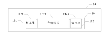
- FIG. 1 is a schematic view showing the internal structure of a preferred embodiment of a biomarker-based tumor early detection kit of the present invention
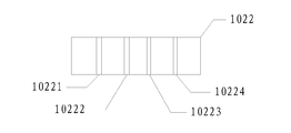
- FIG. 2 is a schematic view showing a coating line region of a preferred embodiment of a biomarker-based tumor early detection kit of the present invention
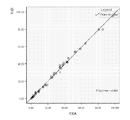
- Figure 4 A correlation diagram of CEA measured for the same clinical sample using the biomarker-based tumor early detection kit of the present invention and Deling Instruments.
- the main object of the present invention is to provide a biomarker-based tumor early detection kit and a preparation method thereof, and simultaneously detect AFP and CEA, and synergistically complement the judgment of tumor occurrence, and can be used for early tumor based on actual detection results. Provides auxiliary judgment, shortens the detection period, increases the sensitivity of detection, and is convenient for users.
- the present invention provides a biomarker-based tumor early detection kit.
- FIG. 1 is a schematic diagram showing the internal structure of a biomarker-based tumor early detection kit according to a preferred embodiment of the present invention
- FIG. 2 is a schematic view showing a coating line region of a biomarker-based tumor early detection kit according to a preferred embodiment of the present invention
- the biomarker-based tumor early detection kit includes a test strip 10 and a cartridge 20:
- the card housing 20 is disposed on the periphery of the test strip 10, surrounding the test strip 10;
- the test strip 10 includes a substrate 101 and a coating film 102 disposed on the substrate 101.
- the coating film 102 is sequentially provided with a sample pad 1021, a coating line region and an absorbent paper:
- the coating line region 1022 is sequentially provided with a marking line 10221, a quality control line 10222, an AFP detecting line 10223, and a CEA detecting line 10224.
- the biomarker-based tumor early detection kit includes a test strip 10 and a cartridge 20, and the cartridge 20 is disposed on the periphery of the test strip 10, and the material of the cartridge 20 may be plastic, and the cartridge 20 is The shape may be a rectangular parallelepiped or may be designed in another shape, but the card cover 20 surrounds the test strip 10 for protection purposes, in accordance with the premise of matching the test strip 10.
- the card housing 20 has two openings, one of which is called a sample application area: corresponding to the sample pad 1021 on the test strip 10, for the convenience of the user to load the sample, the loading of the card case 20
- the zone may be provided with a "small raindrop" proofing mark to prevent the initial user from confusing the sample loading zone, causing the experimental test to fail; the other is called the window zone: corresponding to the covered wire zone 1022 disposed on the test strip 10,
- the experimental results were observed clearly and intuitively.
- the provision of the card housing 20 facilitates the user's experimental operation, reduces human error, and improves the accuracy of the detection result.
- the test strip 10 includes a substrate 101 and a coating film 102 disposed on the substrate 101.
- the coating film 102 is provided with a sample pad 1021, a coating line region 1022, and an absorbent paper 1023.
- the main component of the absorbent paper 1023 is cellulose, which is used to provide the capillary force required for the detection.
- the material of the absorbent paper may be free lint or glass fiber, preferably free lint. Since cellulose is a natural macromolecular organic substance, the cellulose in the paper is staggered in a network, so that there are many voids in the network formed by the interlaced cellulose, and these voids can contain moisture on the absorbent paper 1023, thereby generating tension.
- the test sample which facilitates the dropping is rapidly diffused from the sample to the nitrocellulose.
- the membrane On the membrane, it is conducive to the experiment.
- Nitrocellulose membrane Filter The membrane, referred to as the NC membrane, is the place where the immune response occurs.
- the coating line region 1022 includes a second AFP monoclonal antibody, a second CEA monoclonal antibody, and a second AFP monoclonal antibody disposed in parallel from the end of the sample pad 1021 to the end of the absorbent paper 1023 and spaced apart from each other.
- the fluorescent latex-labeled antibody on the marker line 10221 is diluted with a fluorescent diluent comprising: 1.0%-1.5% BSA, 2.5%-5% sucrose, 0.1%- 0.5% Tween-20, 0.08-0.1% NaN3, PBS with a balance of 0.01-0.02M, pH 7.2
- Bovine serum albumin (BSA) an albumin in bovine serum
- BSA bovine serum albumin
- Tween-20 is a mixture of polyoxyethylene sorbitan monolaurate and a portion of polyoxyethylene bissorbitan monolaurate, which is a surfactant which promotes antibodies and antibodies in the examples of the present invention.
- the role of binding, phosphate buffer, also known as PBS is a solution consisting of sodium dihydrogen phosphate and disodium hydrogen phosphate for dilution of the sample.
- Sodium azide, also known as NaN3 plays an antiseptic role in the present embodiment; the coated antibody on the quality control line 10222, the AFP detection line 10223 and the CEA detection line 10224 is diluted with a coating buffer.
- the coating buffer includes: 0.08-0.1% NaN3, the balance is 0.01-0.02 M, pH 7.2 PBS.
- the coating film 102 is a nitrocellulose membrane, and the nitrocellulose membrane has a creep rate of 95 s. ⁇ 135s/4cm.
- the concentration of the fluorescent latex-labeled second AFP monoclonal antibody and the second CEA monoclonal antibody is 0.25 - 1.0 mg / ml, and the amount on the test strip 10 is 0.02-0.1 ⁇ g/cm 2 , diluted at a ratio of 5% to 30%; the concentration of the fluorescent latex-labeled rabbit IgG antibody is 0.25-1.0 mg/ml, according to 5% Dilute in a ratio of -15%, and the amount on the test strip 10 is 0.001-0.025 ⁇ g/cm 2 .
- the concentration of the rabbit IgG antibody coated on the quality control line 10222 is 0.25-1.0 mg/ml, and the dosage is 20 ⁇ l/27-35 cm.
- the concentration of the AFP monoclonal antibody and the CEA monoclonal antibody coated on the test line were both 0.25-1.0 mg/ml, and the dosage was 20 ⁇ l/27-35 cm.
- the antigen to be detected AFP is alpha-fetoprotein AFP in human blood
- the CEA to be detected is a carcinoembryonic antigen CEA in human blood, wherein whole blood, serum or plasma can be taken from human blood.
- the interval between the detection lines 10223 and between the AFP detection line 10223 and the CEA detection line 10224 is set to 2 mm to 5 mm.
- the setting of the interval spacing considers the use of the matching fluorescent signal reading instrument device in subsequent detection. When the interval is too short, the instrument cannot distinguish the fluorescence signal intensity emitted by both the AFP detection line and the CEA detection line, affecting the experiment.
- the interval between the AFP detecting line 10223 and the CEA detecting line 10224 of the present invention is set to 2 mm to 5 mm.
- the diameter of the matching and subsequent fluorescent latex particles is 0.1 ⁇ m -1 ⁇ m, the wavelength emitted after excitation is from 180 nm to 800 nm.
- the first AFP monoclonal antibody in this experiment is in a different epitope from the second AFP monoclonal antibody when bound to the AFP antigen to be tested; likewise, the first CEA monoclonal antibody and the second CEA monoclonal antibody bind to the CEA antigen to be tested. When in different epitopes.
- a biomarker-based tumor early detection kit includes a test strip and a card case, and the card case is disposed on the periphery of the test strip to surround the test strip;
- the test strip includes a substrate and a coating film disposed on the substrate, the sample film, the coating line area and the absorbent paper are sequentially disposed on the coating film;
- the coating line area is sequentially set as a marking line, a quality control line, an AFP detection line and a CEA detection line.
- the marker line includes a second AFP monoclonal antibody labeled with a fluorescent latex, a second CEA monoclonal antibody, and a rabbit IgG antibody, and the quality control line includes a goat anti-rabbit IgG antibody; the AFP detection line and the CEA detection line respectively include and can be detected The first AFP monoclonal antibody to which the antigen AFP specifically binds and the first CEA monoclonal antibody capable of specifically binding to the antigen to be detected CEA.
- the synergistic effect on the judgment of tumor occurrence can be synergistic and complementary, and the auxiliary detection can be provided for the early stage of the tumor according to the actual detection result, which shortens the detection period and is convenient for users.
- the present invention also provides a method for preparing a biomarker-based tumor early detection kit.
- the preparation method of the biomarker-based tumor early detection kit comprises the following steps:
- the coating film coated with the marking line, the quality control line, the AFP detecting line and the CEA detecting line is placed in an oven for dry balance;
- sample pad and the absorbent paper are sequentially overlapped and pasted on the dry-balanced coating film, the sample pad is disposed at one end of the marking line, and the absorbent paper is disposed at one end of the CEA detecting line;
- a card case is disposed on a periphery of the test strip to surround the test strip.
- the fluorescent latex labeled with the second AFP monoclonal antibody and the fluorescent latex labeled with the second CEA monoclonal antibody are diluted with the fluorescent diluent.
- the fluorescent diluent contains 1.0%-1.5% BSA, 2.5%-5% sucrose, 0.1%-0.5% Tween-20, 0.08-0.1% NaN3, and the balance is 0.01-0.02M, pH 7.2
- the first AFP monoclonal antibody on the detection line capable of specifically binding to the antigen AFP to be detected was diluted with a coating buffer (raised from Hytest).
- CEA Purchased from Hangzhou Qitai Biotechnology Co., Ltd.), and can be tested with the antigen to be tested CEA
- a detection line 10224 wherein the concentration of the goat anti-rabbit IgG antibody coated by the quality control line 10222 is 0.25-1.0mg/ml, The dosage is 20 ⁇ l/27-35cm; the concentration of the first AFP monoclonal antibody coated by AFP detection line 10223 is 0.25-1.0mg/ml, the dosage is 20 ⁇ l/27-35cm; the first CEA single of CEA detection line 10224 is coated.
- the concentration of the cloned antibody is 0.25-1.0 mg/ml, and the dosage is 20 ⁇ l/27-35 cm; the coating buffer contains 0.08-0.1% NaN3, and the balance is 0.01-0.02 M, pH 7.2.
- the coating film coated with the good marking line 10221, the quality control line 10222, the AFP detecting line 10223, and the CEA detecting line 10224 is placed in an oven having a humidity of ⁇ 30%, and the temperature of the oven is set at 35-45 ° C, and dried.
- test strip 10 was sealed and dried at 2 ° C to 30 ° C for 7 days or more.
- the sample pad 1021 and the absorbent paper 1023 are sequentially overlapped and pasted on the dry-balanced coating film 102, and the absorbent paper 1023 is lapped on the end of the coating film 102 near the CEA detecting line 10224.
- the test strip 10 can be made.
- the obtained test strip 10 is matched with the preformed cartridge 20 to obtain a kit.
- the fluorescent latex-labeled antibody is prepared by ultrasonically irradiating the fluorescent latex after covalent activation by a conventional method. 200W-400W after 20-30 seconds, according to 50-200 ⁇ g The ratio of labeled antibody/100 ⁇ l of fluorescent latex was added to a second AFP monoclonal antibody (purchased from Hangzhou Qitai Biotechnology Co., Ltd.), a second CEA monoclonal antibody (purchased from Hytest), and a rabbit IgG antibody (purchased from Hytest).
- the principle of the biomarker-based tumor early detection kit of the present invention is a double-anti-sandwich method; firstly, a latex having a diameter ranging from 0.01 um to 1.5 um is covalently bonded with an AFP antibody and various fluoresceins to form a substance A, and then The latex with a diameter ranging from 0.01um to 1.5um is covalently bound to the CEA antibody and various different fluoresceins to form a substance B. Since the latex itself has a carboxyl group, an amino group and the like, when the substance A is combined with the corresponding AFP antigen, the substance A is utilized. Fluorescein emits fluorescence under excitation light; likewise, when substance B is combined with the corresponding CEA antigen, fluorescein can also emit fluorescence under excitation light.
- the fluorescent latex-labeled second AFP antibody binds to sufficient AFP antigen in the test sample (eg, whole blood, serum or plasma in blood) to form complex A1
- the complex A1 is transferred to AFP by chromatography.
- the first AFP antibody (which binds to another epitope of the AFP antigen) binds to form a double antibody sandwich complex A2, which is aggregated here and is excited by the light source.
- the emitted light of the corresponding wavelength is emitted, and the test strip is placed in a special fluorescent signal reading instrument, and the size of the fluorescent signal can be read to perform quantitative measurement.
- the fluorescent CE-labeled second CEA antibody binds to sufficient CEA antigen in the test sample (eg, whole blood, serum or plasma in blood) to form complex B1
- the complex B1 is transferred by chromatography.
- the CEA detection line bind to the first CEA antibody (which binds to another epitope of the CEA antigen) to form a double antibody sandwich complex B2, where the complex B2 is concentrated and is subjected to a light source. Excitation emits the emitted light of the corresponding wavelength, and the test strip is placed in a special fluorescent signal reading instrument, and the size of the fluorescent signal can be read to perform quantitative determination.
- the main working principle of the fluorescent signal reading instrument is that the fluorescent light source system of the instrument can emit a single-color excitation light, which is irradiated on the AFP detection line and the CEA detection line of the test strip to be detected. At this time, the detection lines AFP and CEA are agglutinated by reaction.
- the fluorescent latex emits a fluorescent signal under the action of monochromatic excitation light, and the fluorescent signal is captured by the detection system of the instrument.
- the detection system After the detection system receives the fluorescent signal, the optical signal is amplified by the photomultiplier tube in the detection system, and then The solid state detector of the instrument performs photoelectric signal conversion, and the electrical signal readout circuit outputs the electrical signal, and then is processed by an automatic software analysis control system to display the result on the display screen, thereby realizing accurate quantification of the sample.
- Table 1 Quantitative determination results of AFP for 10 samples using the kit of the preferred embodiment of the present invention AFP ug/ml sample 1 AFP ug/ml sample 2 AFP ug/ml sample 3 AFP ug/ml sample 4 AFP ug/ml sample 5 AFP ug/ml sample 6 AFP ug/ml sample 7 AFP ug/ml sample 8 AFP ug/ml sample 9 AFP ug/ml sample 1 0 Deling 93.24 15.9 72.03 21.48 114.39 4.96 65.47 17.17 135.98 7.19 Kit of preferred embodiment of the invention 92.71 16.14 72.01 21.86 115.01 4.89 65.61 17.03 142.57 7.34
- CEA ug/ml sample 1 CEA ug/ml sample 2
- CEA ug/ml sample 3 CEA ug/ml sample 4
- CEA ug/ml sample 5 CEA ug/ml sample 6
- CEA ug/ml sample 7 CEA ug/ml sample 8
- CEA ug/ml sample 9 CEA ug/ml sample 1 0 Deling 27.53 4.91 42.59 5.87 35.46 8.19 23.94 1.03 57.24 10.05 Kit of preferred embodiment of the invention 27.06 5.04 42.51 5.65 34.32 8.35 24.17 1.15 58.64 10.17
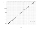
- Figure 3 is a correlation diagram of AFP measured by the biomarker-based tumor early detection kit and Deling instrument for the same clinical sample
- Figure 4 A correlation diagram of CEA measured for the same clinical sample using the biomarker-based tumor early detection kit of the present invention and Deling Instruments.
- AFP tests 60 clinical samples (e.g., whole blood, serum or plasma in blood) with a special protein analyzer from Deling, the correlation coefficient of the results of the two products.
- the kit of this example has a good correlation with the control reagent, the minimum detection limit of AFP is 0.05 mg/l, and the minimum detection limit of CEA is 0.1 ng/l.
- samples with high, low, and median values, respectively, were tested at least 10 times in succession to calculate the coefficient of variation (CV).
- the higher value for AFP content is (115.01 mg/l) and the median value is (16.14).
- Mg/l), low value selection (0.41 mg / l), the above three blood samples were measured 10 times, according to the measured data, using SPSS software statistics to obtain high values (115.54 + 9.13mg/l), CV 2.41%; the same using SPSS software statistics to get the median (16.2 + 0.82 mg / l), CV 3.06%; the same use of SPSS software statistics to obtain a low value of (0.41 + 0.03 mg / l), CV 2. 1%;; test results CV values are less than 12%.
- the higher value for CEA content is (42.51 mg/l), the median value is (5.4 mg/l), and the low value is (0.40). Mg/l), the above three blood samples were measured 10 times, according to the measured data, using SPSS software to obtain high values (42.51 + 4.74mg / l), CV 3.21%; the same using SPSS software statistics to get the median (5.4 + 0.51 mg / l), CV 3.3%; the same using SPSS software statistics to find the low value selection (0.40 + 0.05 mg / l), CV 2. 40%;; test results CV values are less than 12%. The above results show that the precision of the kit of the preferred embodiment is good.
- the invention relates to a biomarker-based tumor early detection kit, comprising a test strip and a card case, the card case is disposed on the periphery of the test strip, surrounding the test strip; the test strip comprises a bottom liner and a bag disposed on the bottom liner
- the coating film and the coating film are sequentially provided with a sample pad, a coating line area and an absorbent paper; the coating line area is sequentially set as a marking line, a quality control line, an AFP detecting line and a CEA detecting line; the marking line includes a mark marked by a fluorescent latex.
- the quality control line comprises a goat anti-rabbit IgG antibody
- the AFP detection line and the CEA detection line respectively comprise a first AFP single which can specifically bind to the antigen AFP to be detected.
Landscapes
- Health & Medical Sciences (AREA)
- Immunology (AREA)
- Life Sciences & Earth Sciences (AREA)
- Engineering & Computer Science (AREA)
- Chemical & Material Sciences (AREA)
- Molecular Biology (AREA)
- Biomedical Technology (AREA)
- Hematology (AREA)
- Urology & Nephrology (AREA)
- Biotechnology (AREA)
- Microbiology (AREA)
- Cell Biology (AREA)
- Food Science & Technology (AREA)
- Medicinal Chemistry (AREA)
- Physics & Mathematics (AREA)
- Analytical Chemistry (AREA)
- Biochemistry (AREA)
- General Health & Medical Sciences (AREA)
- General Physics & Mathematics (AREA)
- Pathology (AREA)
- Chemical Kinetics & Catalysis (AREA)
- Investigating Or Analysing Materials By The Use Of Chemical Reactions (AREA)
Abstract
一种基于生物标志物的肿瘤早期检测试剂盒。基于生物标志物的肿瘤早期检测试剂盒包括试纸条和卡壳:卡壳设置于试纸条的外围,包围试纸条;试纸条包括底衬以及设置于底衬上的包被膜,包被膜上依次设置有样品垫、包被线区和吸水纸;包被线区依次设置为标记线、质控线、AFP检测线和CEA检测线。基于生物标志物的肿瘤早期检测试剂盒通过AFP与CEA两项联检,对于肿瘤发生的判断起到协同互补作用,可以根据实际检测结果,为肿瘤早期提供辅助判断,缩短检测周期,增加了检测的灵敏度,方便了用户使用。
Description
技术领域
本发明涉及生命健康技术领域,具体地说,本发明涉及基于生物标志物的肿瘤早期检测试剂盒及其制备方法。
背景技术
甲胎蛋白 (α-fetoprotein AFP) 是单一多聚体肽键糖蛋白,含590个氨基酸,分子量约为
70KD,与血清白蛋白、维生素结合蛋白属同一基因家族。AFP于胎儿6周开始合成,12-15周达到高峰。出生后1-2年降至成人水平。AFP是诊断肝癌灵敏度高和特异性好的肿瘤标志物,是临床诊断原发性肝细胞癌的主要实验室指标,常用于原发性肝癌临床检查及普查。癌胚抗原(carcino-embryonic
antigen
CEA)是一种分子量为180-200kDa的多糖蛋白复合物。它存在2-6月龄的胎儿胃肠道组织和直、结肠癌细胞组织中,胃癌、胰腺癌、卵巢癌、食道癌等组织中也有存在。通常
CEA被认为是一种广谱的肿瘤标志物。目前市场上的广谱的肿瘤的早期判断仅仅单独依靠单一的肿瘤标志物的检测,常常因为单一的检验周期长,灵敏度低,从而无法确保肿瘤的诊断率以及甚至会影响肿瘤患者后续的治疗和生命健康。
发明内容
本发明的主要目的在于提供一种基于生物标志物的肿瘤早期检测试剂盒及其制备方法,同时检测AFP和CEA,对于肿瘤发生的判断起到协同互补作用,可以根据实际检测结果,为肿瘤早期提供辅助判断,缩短检测周期,增加了检测的灵敏度,方便了用户使用。
为实现上述目的,本发明提供了一种基于生物标志物的肿瘤早期检测试剂盒。
所述基于生物标志物的肿瘤早期检测试剂盒包括试纸条和卡壳:
所述卡壳设置于所述试纸条的外围,包围所述试纸条;
所述试纸条包括底衬以及设置于所述底衬上的包被膜,所述包被膜上依次设置有样品垫、包被线区和吸水纸;
所述包被线区依次设置有标记线、质控线、AFP检测线和CEA检测线。
在其中一个实施例中,所述标记线包括被荧光胶乳标记的第二AFP单克隆抗体、第二CEA单克隆抗体和兔IgG抗体。
在其中一个实施例中,所述质控线包括羊抗兔IgG抗体。
在其中一个实施例中,所述卡壳包括加样区和视窗区:
所述加样区设置于所述试纸条的样品垫处,用于滴加样品;
所述视窗区设置于所述试纸条的包被线区,用于观测实验结果。
在其中一个实施例中,所述标记线和所述质控线之间、所述质控线和所述AFP
检测线之间以及所述AFP检测线和所述CEA检测线之间的间隔设置为2mm-5mm。
在其中一个实施例中,所述AFP检测线包括能与待检测抗原AFP特异性结合的第一AFP单克隆抗体;所述CEA检测线包括能与待检测抗原CEA特异性结合的第一CEA单克隆抗体。
在其中一个实施例中,所述标记线包括被荧光胶乳标记的第二AFP单克隆抗体、第二CEA单克隆抗体和兔IgG抗体。
在其中一个实施例中,所述质控线包括羊抗兔IgG抗体。
在其中一个实施例中,所述卡壳包括加样区和视窗区:
所述加样区设置于所述试纸条的样品垫处,用于滴加样品;
所述视窗区设置于所述试纸条的包被线区,用于观测实验结果。
在其中一个实施例中,所述标记线和所述质控线之间、所述质控线和所述AFP
检测线之间以及所述AFP检测线和所述CEA检测线之间的间隔设置为2mm-5mm。在其中一个实施例中,所述待检测抗原AFP为人源AFP,所述待检测抗原CEA为人源CEA。
在其中一个实施例中,所述标记线包括被荧光胶乳标记的第二AFP单克隆抗体、第二CEA单克隆抗体和兔IgG抗体。
此外,为实现上述目的,本发明还提供一种基于生物标志物的肿瘤早期检测试剂盒的制备方法。
所述基于生物标志物的肿瘤早期检测试剂盒的制备方法包含以下步骤:
将稀释过的标记有第二AFP单克隆抗体的荧光胶乳、标记有第二CEA单克隆抗体的荧光胶乳和标记有兔IgG抗体的荧光胶乳,划线包被于包被膜上的包被线区,形成标记线;
稀释羊抗兔IgG抗体,并将稀释过的所述羊抗兔IgG抗体划线包被于包被膜上的包被线区,形成质控线;
稀释能与待检测抗原AFP特异结合的第一AFP单克隆抗体,并将稀释过的所述能与待检测抗原AFP特异结合的第一AFP单克隆抗体划线包被于包被膜上的包被线区,形成AFP检测线;
稀释能与待检测抗原CEA特异结合的第一CEA单克隆抗体,并将稀释过的所述能与待检测抗原CEA特异结合的第一CEA单克隆抗体划线包被于包被膜上的包被线区,形成CEA检测线;
将包被有所述标记线、质控线、AFP检测线和CEA检测线的包被膜置于烘箱中,进行干燥平衡;
将样品垫和吸水纸依次搭接并粘贴于干燥平衡后的所述包被膜上,所述样品垫设置于所述标记线的一端,所述吸水纸设置于所述CEA检测线的一端;
将所述包被膜粘贴于底衬上,形成试纸条;
将卡壳设置于所述试纸条的外围,包围所述试纸条。
优选地,所述待检测抗原AFP为人源AFP,所述待检测抗原CEA为人源CEA。
优选地,所述标记线和所述质控线之间、所述质控线和所述AFP
检测线之间以及所述AFP检测线和所述CEA检测线之间的间隔设置为2mm-5mm。
本发明采用上述技术方案,带来的技术效果为:本发明实施例提供的基于生物标志物的肿瘤早期检测试剂盒,包括试纸条和卡壳,卡壳设置于试纸条的外围,包围试纸条;试纸条包括底衬以及设置于底衬上的包被膜,包被膜上依次设置有样品垫、包被线区和吸水纸;包被线区依次设置为标记线、质控线、AFP检测线和CEA检测线;标记线包括被荧光胶乳标记的第二AFP单克隆抗体、第二CEA单克隆抗体和兔IgG抗体,质控线包括羊抗兔IgG抗体;AFP检测线和CEA检测线分别包括能与待检测抗原AFP特异性结合的第一AFP单克隆抗体和能与待检测抗原CEA特异性结合的第一CEA单克隆抗体。本发明实施例提供的基于生物标志物的肿瘤早期检测试剂盒通过AFP与CEA两项联检,对于肿瘤发生的判断起到协同互补作用,可以根据实际检测结果,为肿瘤早期提供辅助判断,缩短检测周期,增加了检测的灵敏度,方便了用户使用。
附图说明
图1 为本发明基于生物标志物的肿瘤早期检测试剂盒较佳实施例的内部结构示意图;
图2为本发明基于生物标志物的肿瘤早期检测试剂盒较佳实施例的包被线区的示意图;
图3
为采用本发明基于生物标志物的肿瘤早期检测试剂盒与德灵仪器针对相同临床样本测量出的AFP的相关性图;
图4
为采用本发明基于生物标志物的肿瘤早期检测试剂盒与德灵仪器针对相同临床样本测量出的CEA的相关性图。
本发明目的的实现、功能特点及优点将结合实施例,参照附图做进一步说明。
具体实施方式
应当理解,此处所描述的具体实施例仅仅用以解释本发明,并不用于限定本发明。
本发明的主要目的在于提供一种基于生物标志物的肿瘤早期检测试剂盒及其制备方法,同时检测AFP和CEA,对于肿瘤发生的判断起到协同互补作用,可以根据实际检测结果,为肿瘤早期提供辅助判断,缩短检测周期,增加了检测的灵敏度,方便了用户使用。
为实现上述目的,本发明提供了一种基于生物标志物的肿瘤早期检测试剂盒。
图1为本发明基于生物标志物的肿瘤早期检测试剂盒较佳实施例的内部结构示意图;图2为本发明基于生物标志物的肿瘤早期检测试剂盒较佳实施例的包被线区的示意图。参照图1和图2,在本实施例中,所述基于生物标志物的肿瘤早期检测试剂盒包括试纸条10和卡壳20:
所述卡壳20设置于所述试纸条10的外围,包围所述试纸条10;
所述试纸条10包括底衬101以及设置于所述底衬101上的包被膜102,所述包被膜102上依次设置有样品垫1021、包被线区和吸水纸:
所述包被线区1022依次设置有标记线10221、质控线10222、AFP检测线10223和CEA检测线10224。
在本实施例中,所述基于生物标志物的肿瘤早期检测试剂盒包括试纸条10和卡壳20,卡壳20设置于试纸条10的外围,卡壳20的材质可以是塑料的,卡壳20的形状可以是长方体的,也可以设计成别的形状,但是以匹配试纸条10为前提,卡壳20包围试纸条10,起到保护的作用。作为优选的实施例,卡壳20上有两处开孔的地方,一处称为加样区:对应于试纸条10上的样品垫1021处,为了方便使用者加样,卡壳20的加样区可以设置有“小雨点”的加样标识,防止初次使用者混淆加样区,导致实验检测失败;另一处称为视窗区:对应设置于试纸条10上的包被线区1022,为了清晰直观的观测到实验结果。此外,设置卡壳20方便了使用者的实验操作,减少了人为的误差,提高了检测结果的正确率。
所述试纸条10包括底衬101及设在底衬101上的包被膜102,包被膜102上设有样品垫1021、包被线区1022和吸水纸1023。在检测的过程中,吸水纸1023的主要成分是纤维素,用于提供检测所需的毛细作用力,吸水纸的材料可以是自由棉绒,也可以是玻璃纤维,优选自由棉绒的材料。由于纤维素是天然的大分子有机物,纸张中的纤维素交错呈网状,因此纤维素交错形成的网状中有很多空隙,这些空隙可以含住吸水纸1023上的水分,从而产生张力。此外水由于势能,从浓度高的一侧流向浓度低的一侧,因此在实验操作的过程中,如果从一侧吸水,则有利于滴加的检测样品快速的扩散从加样处到硝酸纤维素膜上,利于实验的进行。硝酸纤维素膜(nitrocellulose
filter
membrane,简称NC膜),是免疫反应的发生处。当通过样品垫1021,在吸水纸1023的作用下把样品从起始上样点转移至包被线区,在检测试纸条10下游控制着样品沿着NC膜膜片流动。
所述包被线区1022包括有从靠近所述样品垫1021端至靠近所述吸水纸1023端依次平行设置、且相互间隔的包被有第二AFP单克隆抗体、第二CEA单克隆抗体和兔IgG抗体的标记线10221、包被有羊抗兔IgG抗体的质控线10222、包被有能与待检测抗原AFP特异性结合的第一AFP单克隆抗体的AFP检测线10223、以及包被有能与待检测抗原CEA特异性结合的第一CEA单克隆抗体的CEA检测线10224。所述标记线10221上荧光胶乳标记的抗体采用荧光稀释液进行稀释,所述荧光稀释液包括:1.0%-1.5%BSA,2.5%-5%蔗糖,0.1%-
0.5%Tween-20,0.08-0.1% NaN3,余量为 0.01-0.02M、 pH7.2的 PBS
;牛血清白蛋白(BSA),是牛血清中的一种白蛋白,能减轻有些抗体的变性,对于某些不利环境因素如加热,表面张力及化学因素引起的抗体的变性,起到稳定、减轻的效果,减少外界因素对实验的干扰。此外在本发明实施例中加入BSA,可增强产物的稳定性。Tween-20为聚氧乙烯去水山梨醇单月桂酸酯和一部分聚氧乙烯双去水山梨醇单月桂酸酯的混合物,是一种表面活性剂,在本发明实施例中促进抗体与抗体的结合的作用,磷酸盐缓冲液又称PBS,是由磷酸二氢钠和磷酸氢二钠组成的溶液,用于样品的稀释。叠氮钠又称NaN3,在本发明实施例中起到防腐的作用;所述质控线10222、AFP检测线10223和CEA检测线10224上包被的抗体均采用包被缓冲液进行稀释,所述包被缓冲液包括:0.08-0.1%
NaN3,余量为 0.01-0.02M、 pH7.2的 PBS。所述包被膜102为硝酸纤维素膜,所述硝酸纤维素膜的爬速为95s
~135s/4cm。所述荧光胶乳标记的第二AFP单克隆抗体和第二CEA单克隆抗体的浓度均为 0.25 - 1.0mg/ml,在试纸条10上的用量均为
0.02-0.1μg/cm 2,按5%~30%的比例进行稀释;所述荧光乳胶标记的兔IgG抗体的浓度为0.25-1.0mg/ml,按 5%
-15%的比例进行稀释,在试纸条10上的用量为
0.001-0.025μg/cm2。所述质控线10222上包被的兔IgG抗体的浓度为0.25-1.0mg/ml,用量为20μl/27-35cm
;所述检测线上包被的AFP单克隆抗体和 CEA单克隆抗体的浓度均为 0.25-1.0mg/ml,用量均为20μl/27-35cm。
所述待检测抗原AFP为人的血液中的甲胎蛋白AFP,所述待检测抗原CEA为人的血液中的癌胚抗原CEA,其中,人的血液中可取全血、血清或血浆。所述标记线10221和所述质控线10222之间、所述质控线10222和所述AFP
检测线10223之间以及所述AFP检测线10223和所述CEA检测线10224之间的间隔设置为2mm-5mm。此处,间隔间距的设置考虑匹配荧光信号读取仪器装置在后续检测中的使用,当间隔间距过短时,仪器无法区分出AFP检测线和CEA检测线二者发出的荧光信号强度,影响实验结果,当间隔间距过长时,仪器检测不到相应的荧光信号,同样也会对实验造成干扰。基于此,针对本发明AFP检测线10223和所述CEA检测线10224之间的间隔设置为2mm-5mm为宜。匹配与后续所述荧光胶乳颗粒的直径为0.1μm
-1μm,受激发后发射的波长为180nm-800nm。本实验中的第一AFP单克隆抗体与第二AFP单克隆抗体与待测AFP抗原结合时处于不同表位;同样,第一CEA单克隆抗体与第二CEA单克隆抗体与待测CEA抗原结合时处于不同表位。
本发明采用上述技术方案,带来的技术效果为:基于生物标志物的肿瘤早期检测试剂盒,包括试纸条和卡壳,卡壳设置于试纸条的外围,包围试纸条;试纸条包括底衬以及设置于底衬上的包被膜,包被膜上依次设置有样品垫、包被线区和吸水纸;包被线区依次设置为标记线、质控线、AFP检测线和CEA检测线;标记线包括被荧光胶乳标记的第二AFP单克隆抗体、第二CEA单克隆抗体和兔IgG抗体,质控线包括羊抗兔IgG抗体;AFP检测线和CEA检测线分别包括能与待检测抗原AFP特异性结合的第一AFP单克隆抗体和能与待检测抗原CEA特异性结合的第一CEA单克隆抗体。通过AFP与CEA两项联检,对于肿瘤发生的判断起到协同互补作用,可以根据实际检测结果,为肿瘤早期提供辅助判断,缩短检测周期,方便了用户使用。
此外,为实现上述目的,本发明还提供一种基于生物标志物的肿瘤早期检测试剂盒的制备方法。
所述基于生物标志物的肿瘤早期检测试剂盒的制备方法包含以下步骤:
将稀释过的标记有第二AFP单克隆抗体的荧光胶乳、标记有第二CEA单克隆抗体的荧光胶乳和标记有兔IgG抗体的荧光胶乳,划线包被于包被膜上的包被线区,形成标记线;
稀释羊抗兔IgG抗体,并将稀释过的所述兔IgG抗体划线包被于包被膜上的包被线区,形成质控线;
稀释能与待检测抗原AFP特异结合的第一AFP单克隆抗体,并将稀释过的所述能与待检测抗原AFP特异结合的第一AFP单克隆抗体划线包被于包被膜上的包被线区,形成AFP检测线;
稀释能与待检测抗原CEA特异结合的第一CEA单克隆抗体,并将稀释过的所述能与待检测抗原CEA特异结合的第一CEA单克隆抗体划线包被于包被膜上的包被线区,形成CEA检测线;
将包被有所述标记线、质控线、AFP检测线和CEA检测线的包被膜置于烘箱中,进行干燥平衡;
将样品垫和吸水纸依次搭接并粘贴于干燥平衡后的所述包被膜上,所述样品垫设置于所述标记线的一端,所述吸水纸设置于所述CEA检测线的一端;
将所述包被膜粘贴于底衬上,形成试纸条;
将卡壳设置于所述试纸条的外围,包围所述试纸条。
在本实施例中,参照图1和图2,在标记线10221的制备过程中,用荧光稀释液稀释标记有第二AFP单克隆抗体的荧光胶乳、标记有第二CEA单克隆抗体的荧光胶乳和标记有兔
IgG抗体的荧光胶乳,划线于包被膜上形成标记线10221;其中荧光胶乳标记的第二AFP单克隆抗体和荧光胶乳标记的第二CEA单克隆抗体的浓度均为0.25-1.0mg/ml,用量均为
0.02 -0.1μg/cm 2;荧光胶乳标记的兔 IgG抗体的浓度为 0.25- 1.0mg/ml,用量为 0.001-0.025μg/cm 2
;所述荧光稀释液中含有1.0%-1.5%BSA,2.5%-5%蔗糖,0.1%- 0.5%Tween-20,0.08-0.1% NaN3,余量为
0.01-0.02M、 pH7.2的
PBS;质控线10222和检测线的制备过程中,用包被缓冲液稀释兔IgG抗体(购自Hytest公司)、能与待检测抗原AFP特异结合的检测线上的第一AFP单克隆抗体(购自杭州启泰生物技术有限公司)、以及能与待检测抗原CEA
特异结合的检测线上的第一CEA单克隆抗体(购自Hytest公司),并将稀释后的三种抗体分别依次平行地划线于包被膜上形成质控线10222、AFP检测线10223和CEA检测线10224;其中,质控线10222包被的羊抗兔IgG抗体的浓度为
0.25-1.0mg/ml,
用量为20μl/27-35cm;AFP检测线10223包被的第一AFP单克隆抗体的浓度为0.25-1.0mg/ml,用量为20μl/27-35cm;CEA检测线10224包被的第一CEA单克隆抗体的浓度为0.25-1.0mg/ml,用量为20μl/27-35cm;所述包被缓冲液中含有0.08-0.1%NaN3,余量为0.01-0.02M、pH7.2的
PBS;所述标记线10221和所述质控线10222之间、所述质控线10222和所述AFP
检测线之间以及所述AFP检测线10223和所述CEA检测线10224之间的间隔设置为2mm-5mm标记线10221、质控线10222、AFP检测线10223、CEA检测线10224在包被膜的制备过程中,将包被好标记线10221、质控线10222、AFP检测线10223、CEA检测线10224的包被膜置于湿度<30%的烘箱,且烘箱的温度设定在35-45℃,干燥
24-36h
后,于2℃-30℃密封干燥平衡7天以上。在试纸条的制备过程中,将样品垫1021和吸水纸1023依次搭接并粘贴于干燥平衡后的包被膜102上,在包被膜102靠近CEA检测线10224的一端搭接粘贴吸水纸1023,即可制成试纸条10。将得到的试纸条10与预制成的卡壳20相匹配,即可得到试剂盒。
在其中一个实施例中,所述荧光胶乳标记抗体的制备步骤为:将常规方法共价活化后的荧光胶乳超声波
200W-400W 处理20-30秒后,按照50-200μg
标记抗体/100μl荧光胶乳的比例加入第二AFP单克隆抗体(购自杭州启泰生物技术有限公司)、第二CEA单克隆抗体(购自Hytest公司)和兔IgG抗体(购自Hytest公司),混匀后室温搅拌反应2
h,离心洗涤 3 次,每次 10000-15000xg、离心10 min,沉淀用PBS-TBN 溶解并超声波 100W 处理 30 s,用 PBS-TBN
恢复离心前体积,4℃保存,备用。
本发明所述基于生物标志物的肿瘤早期检测试剂盒原理是双抗夹心法;首先将直径范围0.01um-1.5um的胶乳与AFP抗体以及各类不同的荧光素共价结合形成物质A,然后将直径范围0.01um-1.5um的胶乳与CEA抗体以及各类不同的荧光素共价结合形成物质B,由于胶乳本身具有羧基、氨基等基团,当物质A再与相应的AFP抗原结合,利用荧光素在激发光下发射荧光;同样,当物质B再与相应的CEA抗原结合,利用荧光素在激发光下也可以发射荧光。
当荧光胶乳标记的第二AFP抗体与检测样品(例如:血液中的全血、血清或血浆)中的足够的AFP抗原结合形成复合物A1,该复合物A1在层析的作用下转移至AFP检测线处,与第一AFP抗体(该抗体会结合在AFP抗原的另一表位上)结合,从而形成双抗体夹心的复合物A2,该复合物A2聚集在此处后,受到光源激发释放出相应波长的发射光,将该试纸条放于专门的荧光信号读取仪器中,即可读取荧光信号的大小,从而进行定量测定。同样的当荧光胶乳标记的第二CEA抗体与检测样品(例如:血液中的全血、血清或血浆)中的足够的CEA抗原结合形成复合物B1,该复合物B1在层析的作用下转移至CEA检测线处,与第一CEA抗体(该抗体会结合在CEA抗原的另一表位上)结合,从而形成双抗体夹心的复合物B2,该复合物B2聚集在此处后,受到光源激发释放出相应波长的发射光,将该试纸条放于专门的荧光信号读取仪器中,即可读取荧光信号的大小,从而进行定量测定。
荧光信号读取仪器的主要工作原理是该仪器的荧光光源系统能够发出单色激发光,照射在待检测的试纸条AFP检测线和CEA检测线处,此时检测线AFP和CEA通过反应凝集的荧光乳胶在单色激发光的作用下,发出荧光信号,该荧光信号被该仪器的检测系统捕获,检测系统接收荧光信号后,由检测系统中的光电倍增管实现光信号的放大,然后由该仪器的固态检测器进行光电信号的转化,由电信号读出电路将电信号输出,再有自动软件分析控制系统处理,在显示屏处显示结果,从而实现对样品的精确定量。
1、用德灵DN-100特种蛋白测定仪对134例临床AFP和CEA样本(例如:血液中的全血、血清或血浆)作为检测标准,表1采用本发明较佳实施例的试剂盒对10个样本的AFP定量测定结果;表2采用本发明较佳实施例的试剂盒对10个样本的CEA定量测定结果。
表1 采用本发明较佳实施例的试剂盒对10个样本的AFP定量测定结果
| AFP ug/ml 样本 1 | AFP ug/ml 样本 2 | AFP ug/ml 样本 3 | AFP ug/ml 样本 4 | AFP ug/ml 样本 5 | AFP ug/ml 样本 6 | AFP ug/ml 样本 7 | AFP ug/ml 样本 8 | AFP ug/ml 样本 9 | AFP ug/ml 样本 1 0 | |
| 德灵 | 93.24 | 15.9 | 72.03 | 21.48 | 114.39 | 4.96 | 65.47 | 17.17 | 135.98 | 7.19 |
| 本发明较佳实施例的试剂盒 | 92.71 | 16.14 | 72.01 | 21.86 | 115.01 | 4.89 | 65.61 | 17.03 | 142.57 | 7.34 |
表2 采用本发明较佳实施例的试剂盒对10个样本的CEA定量测定结果
| CEA ug/ml 样本 1 | CEA ug/ml 样本 2 | CEA ug/ml 样本 3 | CEA ug/ml 样本 4 | CEA ug/ml 样本 5 | CEA ug/ml 样本6 | CEA ug/ml 样本 7 | CEA ug/ml 样本 8 | CEA ug/ml 样本9 | CEA ug/ml 样本 1 0 | |
| 德灵 | 27.53 | 4.91 | 42.59 | 5.87 | 35.46 | 8.19 | 23.94 | 1.03 | 57.24 | 10.05 |
| 本发明较佳实施例的试剂盒 | 27.06 | 5.04 | 42.51 | 5.65 | 34.32 | 8.35 | 24.17 | 1.15 | 58.64 | 10.17 |
从表1和表2可知,采用较佳实施例的试剂盒对样本进行测定时,可以得到与检测标准相当的结果,说明本发明的试剂盒可以实现准确定量测量。
2、对试剂盒进行性能方面的测定---最低检测限度
图3 为采用本发明基于生物标志物的肿瘤早期检测试剂盒与德灵仪器针对相同临床样本测量出的AFP的相关性图;图4
为采用本发明基于生物标志物的肿瘤早期检测试剂盒与德灵仪器针对相同临床样本测量出的CEA的相关性图。参照图3和图4,在本实施例中,AFP以德灵公司的特种蛋白分析仪测试60份临床样本(例如:血液中的全血、血清或血浆),这两种产品的结果相关系数为R2>0.996,且Y=0.05+0.984X;
CEA以德灵公司的特种蛋白分析仪测试60份临床样本(例如:血液中的全血、血清或血浆),这两种产品的结果相关系数为R2>0.994,,且Y=0.263+0.983X;因此,该实施例的试剂盒跟对照试剂的相关性好,AFP的最低检测限度为0.05mg/l,CEA的最低检测限度为0.1ng/l。
3、对试剂盒进行性能方面的测定---精密度
采用较佳实施例的试剂盒对含量分别是高值、低值和中值的样本,连续至少检测10次,计算变异系数(CV)。
对于AFP含量的较高值选择为(115.01 mg/l)、中值选择为(16.14
mg/l)、低值选择为(0.41 mg/l),将上述3个血液样本分别测定10次,根据其测定数据,利用SPSS软件统计得出高值(115.54 +
9.13mg/l),CV 2.41%;同样的利用SPSS软件统计得出中值(16.2 + 0.82 mg/l),CV
3.06%;同样的利用SPSS软件统计得出低值选择为(0.41+ 0.03 mg/l),CV 2. 1%;;检测结果CV值均小于12%。
对于CEA含量的较高值选择为(42.51 mg/l)、中值选择为(5.4 mg/l)、低值选择为(0.40
mg/l),将上述3个血液样本分别测定10次,根据其测定数据,利用SPSS软件统计得出高值(42.51 + 4.74mg/l),CV
3.21%;同样的利用SPSS软件统计得出中值(5.4 + 0.51 mg/l),CV 3.3%;同样的利用SPSS软件统计得出低值选择为(0.40+
0.05 mg/l),CV 2. 40%;;检测结果CV值均小于12%。以上结果说明较佳实施例的试剂盒的精密度良好。
本发明实施例基于生物标志物的肿瘤早期检测试剂盒,包括试纸条和卡壳,卡壳设置于试纸条的外围,包围试纸条;试纸条包括底衬以及设置于底衬上的包被膜,包被膜上依次设置有样品垫、包被线区和吸水纸;包被线区依次设置为标记线、质控线、AFP检测线和CEA检测线;标记线包括被荧光胶乳标记的第二AFP单克隆抗体、第二CEA单克隆抗体和兔IgG抗体,质控线包括羊抗兔IgG抗体;AFP检测线和CEA检测线分别包括能与待检测抗原AFP特异性结合的第一AFP单克隆抗体和能与待检测抗原CEA特异性结合的第一CEA单克隆抗体。通过AFP与CEA两项联检,对于肿瘤发生的判断起到协同互补作用,可以根据实际检测结果,为肿瘤早期提供辅助判断,缩短检测周期,增加了检测的灵敏度,方便了用户使用。
以上仅为本发明的优选实施例,并非因此限制本发明的专利范围,凡是利用本发明说明书及附图内容所作的等效结构或等效流程变换,或直接或间接运用在其他相关的技术领域,均同理包括在本发明的专利保护范围内。
Claims (15)
- 一种基于生物标志物的肿瘤早期检测试剂盒,其特征在于,所述基于生物标志物的肿瘤早期检测试剂盒包括试纸条和卡壳:所述卡壳设置于所述试纸条的外围,包围所述试纸条;所述试纸条包括底衬以及设置于所述底衬上的包被膜,所述包被膜上依次设置有样品垫、包被线区和吸水纸;所述包被线区依次设置有标记线、质控线、AFP检测线和CEA检测线。
- 如权利要求1所述的基于生物标志物的肿瘤早期检测试剂盒,其特征在于,所述标记线包括被荧光胶乳标记的第二AFP单克隆抗体、第二CEA单克隆抗体和兔IgG抗体。
- 如权利要求1所述的基于生物标志物的肿瘤早期检测试剂盒,其特征在于,所述质控线包括羊抗兔IgG抗体。
- 如权利要求1所述的基于生物标志物的肿瘤早期检测试剂盒,其特征在于,所述卡壳包括加样区和视窗区:所述加样区设置于所述试纸条的样品垫处,用于滴加样品;所述视窗区设置于所述试纸条的包被线区,用于观测实验结果。
- 如权利要求1所述的基于生物标志物的肿瘤早期检测试剂盒,其特征在于,所述标记线和所述质控线之间、所述质控线和所述AFP 检测线之间以及所述AFP检测线和所述CEA检测线之间的间隔设置为2mm-5mm。
- 如权利要求1所述的基于生物标志物的肿瘤早期检测试剂盒,其特征在于,所述AFP检测线包括能与待检测抗原AFP特异性结合的第一AFP单克隆抗体;所述CEA检测线包括能与待检测抗原CEA特异性结合的第一CEA单克隆抗体。
- 如权利要求6所述的基于生物标志物的肿瘤早期检测试剂盒,其特征在于,所述标记线包括被荧光胶乳标记的第二AFP单克隆抗体、第二CEA单克隆抗体和兔IgG抗体。
- 如权利要求6所述的基于生物标志物的肿瘤早期检测试剂盒,其特征在于,所述质控线包括羊抗兔IgG抗体。
- 如权利要求6所述的基于生物标志物的肿瘤早期检测试剂盒,其特征在于,所述卡壳包括加样区和视窗区:所述加样区设置于所述试纸条的样品垫处,用于滴加样品;所述视窗区设置于所述试纸条的包被线区,用于观测实验结果。
- 如权利要求6所述的基于生物标志物的肿瘤早期检测试剂盒,其特征在于,所述标记线和所述质控线之间、所述质控线和所述AFP 检测线之间以及所述AFP检测线和所述CEA检测线之间的间隔设置为2mm-5mm。
- 如权利要求6所述的基于生物标志物的肿瘤早期检测试剂盒,其特征在于,所述待检测抗原AFP为人源AFP,所述待检测抗原CEA为人源CEA。
- 如权利要求11所述的基于生物标志物的肿瘤早期检测试剂盒,其特征在于,所述标记线包括被荧光胶乳标记的第二AFP单克隆抗体、第二CEA单克隆抗体和兔IgG抗体。
- 一种基于生物标志物的肿瘤早期检测试剂盒的制备方法,其特征在于,所述肿瘤早期检测试剂盒的制备方法包括以下步骤:将稀释过的标记有第二AFP单克隆抗体的荧光胶乳、标记有第二CEA单克隆抗体的荧光胶乳和标记有兔IgG抗体的荧光胶乳,划线包被于包被膜上的包被线区,形成标记线;稀释羊抗兔IgG抗体,并将稀释过的所述羊抗兔IgG抗体划线包被于包被膜上的包被线区,形成质控线;稀释能与待检测抗原AFP特异结合的第一AFP单克隆抗体,并将稀释过的所述能与待检测抗原AFP特异结合的第一AFP单克隆抗体包被于包被膜上的包被线区,形成AFP检测线;稀释能与待检测抗原CEA特异结合的第一CEA单克隆抗体,并将稀释过的所述能与待检测抗原CEA特异结合的第一CEA单克隆抗体划线包被于包被膜上的包被线区,形成CEA检测线;将包被有所述标记线、质控线、AFP检测线和CEA检测线的包被膜置于烘箱中,进行干燥平衡;将样品垫和吸水纸依次搭接并粘贴于干燥平衡后的所述包被膜上,所述样品垫设置于所述标记线的一端,所述吸水纸设置于所述CEA检测线的一端;将所述包被膜粘贴于底衬上,形成试纸条;将卡壳设置于所述试纸条的外围,包围所述试纸条。
- 如权利要求13所述的基于生物标志物的肿瘤早期检测试剂盒的制备方法,其特征在于,所述待检测抗原AFP为人源AFP,所述待检测抗原CEA为人源CEA。
- 如权利要求13所述的基于生物标志物的肿瘤早期检测试剂盒的制备方法,其特征在于,所述标记线和所述质控线之间、所述质控线和所述AFP 检测线之间以及所述AFP检测线和所述CEA检测线之间的间隔设置为2mm-5mm。
Applications Claiming Priority (2)
| Application Number | Priority Date | Filing Date | Title |
|---|---|---|---|
| CN201510292617.XA CN105891481A (zh) | 2015-05-30 | 2015-05-30 | 基于生物标志物的肿瘤早期检测试剂盒及其制备方法 |
| CN201510292617.X | 2015-05-30 |
Publications (1)
| Publication Number | Publication Date |
|---|---|
| WO2016192168A1 true WO2016192168A1 (zh) | 2016-12-08 |
Family
ID=57001835
Family Applications (1)
| Application Number | Title | Priority Date | Filing Date |
|---|---|---|---|
| PCT/CN2015/083129 Ceased WO2016192168A1 (zh) | 2015-05-30 | 2015-07-02 | 基于生物标志物的肿瘤早期检测试剂盒及其制备方法 |
Country Status (2)
| Country | Link |
|---|---|
| CN (1) | CN105891481A (zh) |
| WO (1) | WO2016192168A1 (zh) |
Cited By (7)
| Publication number | Priority date | Publication date | Assignee | Title |
|---|---|---|---|---|
| CN108680747A (zh) * | 2018-06-26 | 2018-10-19 | 宁波奥丞生物科技有限公司 | 一种胎盘生长因子检测用免疫荧光法试剂盒 |
| CN110804545A (zh) * | 2019-12-03 | 2020-02-18 | 苏州缔因安生物科技有限公司 | 一种纸基核酸检测平台及其应用 |
| CN111474352A (zh) * | 2020-04-29 | 2020-07-31 | 北京乐普医疗科技有限责任公司 | 一种新型冠状病毒covid-2019检测卡及制备方法 |
| CN111505313A (zh) * | 2020-05-07 | 2020-08-07 | 厦门万渤生物技术有限公司 | 一种联合诊断支原体肺炎的诊断试剂盒及其应用 |
| CN113138277A (zh) * | 2020-01-17 | 2021-07-20 | 武汉戴安生物技术有限公司 | 一种用于甲胎蛋白检测的诊断试剂 |
| CN115097120A (zh) * | 2022-07-05 | 2022-09-23 | 上海艾瑞德生物科技有限公司 | 一种免疫层析膜以及免疫层析试剂片和制备方法 |
| CN116718775A (zh) * | 2023-08-04 | 2023-09-08 | 天津迈基生物科技有限公司 | 一种检测结直肠癌的组合物、试纸及方法 |
Citations (6)
| Publication number | Priority date | Publication date | Assignee | Title |
|---|---|---|---|---|
| CN101017168A (zh) * | 2007-02-15 | 2007-08-15 | 王继华 | 荧光胶乳定量层析试纸条及其制备方法 |
| CN102692504A (zh) * | 2012-06-26 | 2012-09-26 | 南京基蛋生物科技有限公司 | D-二聚体荧光免疫定量测定试纸条及其制备方法 |
| CN104316702A (zh) * | 2014-05-30 | 2015-01-28 | 广州万孚生物技术股份有限公司 | Pct 与crp定量联检层析试纸条及其制备方法 |
| CN204142734U (zh) * | 2014-05-30 | 2015-02-04 | 广州万孚生物技术股份有限公司 | 新型免疫层析试纸条 |
| CN104502591A (zh) * | 2014-11-10 | 2015-04-08 | 江苏宏泰格尔生物医学工程有限公司 | 一种胃蛋白酶原ⅰ和ⅱ的联合检测方法及试剂盒 |
| CN204758606U (zh) * | 2015-05-30 | 2015-11-11 | 深圳市贝沃德克生物技术研究院有限公司 | 基于生物标志物的肿瘤早期检测试纸条 |
Family Cites Families (9)
| Publication number | Priority date | Publication date | Assignee | Title |
|---|---|---|---|---|
| US5753517A (en) * | 1996-03-29 | 1998-05-19 | University Of British Columbia | Quantitative immunochromatographic assays |
| CN1311239C (zh) * | 2004-09-07 | 2007-04-18 | 李人 | 免疫层析测试条及其制造方法 |
| CN201196651Y (zh) * | 2008-05-22 | 2009-02-18 | 北京德易和康技术有限公司 | 双肿瘤标志物金标检测试纸 |
| US8900881B2 (en) * | 2008-12-30 | 2014-12-02 | Jin Po Lee | Quantitative analyte assay device and method |
| US20120015350A1 (en) * | 2009-03-10 | 2012-01-19 | Northwestern University | Lateral flow strip and uses thereof |
| CN103172941A (zh) * | 2011-12-26 | 2013-06-26 | 苏州新波生物技术有限公司 | 聚苯乙烯荧光纳米颗粒及其制备和应用 |
| CN103760368A (zh) * | 2013-05-03 | 2014-04-30 | 上海溯源生物技术有限公司 | 一种AFP和Free-β-HCG荧光双标的定量检测试剂盒 |
| CN103364550A (zh) * | 2013-07-16 | 2013-10-23 | 天津大学 | 用量子点标记的免疫层析试纸条快速定量检测肿瘤标志物的方法及装置 |
| CN203385732U (zh) * | 2013-08-14 | 2014-01-08 | 江苏省原子医学研究所 | 一种检测胃蛋白酶原ⅰ的试纸条及应用其的试剂卡 |
-
2015
- 2015-05-30 CN CN201510292617.XA patent/CN105891481A/zh active Pending
- 2015-07-02 WO PCT/CN2015/083129 patent/WO2016192168A1/zh not_active Ceased
Patent Citations (6)
| Publication number | Priority date | Publication date | Assignee | Title |
|---|---|---|---|---|
| CN101017168A (zh) * | 2007-02-15 | 2007-08-15 | 王继华 | 荧光胶乳定量层析试纸条及其制备方法 |
| CN102692504A (zh) * | 2012-06-26 | 2012-09-26 | 南京基蛋生物科技有限公司 | D-二聚体荧光免疫定量测定试纸条及其制备方法 |
| CN104316702A (zh) * | 2014-05-30 | 2015-01-28 | 广州万孚生物技术股份有限公司 | Pct 与crp定量联检层析试纸条及其制备方法 |
| CN204142734U (zh) * | 2014-05-30 | 2015-02-04 | 广州万孚生物技术股份有限公司 | 新型免疫层析试纸条 |
| CN104502591A (zh) * | 2014-11-10 | 2015-04-08 | 江苏宏泰格尔生物医学工程有限公司 | 一种胃蛋白酶原ⅰ和ⅱ的联合检测方法及试剂盒 |
| CN204758606U (zh) * | 2015-05-30 | 2015-11-11 | 深圳市贝沃德克生物技术研究院有限公司 | 基于生物标志物的肿瘤早期检测试纸条 |
Cited By (11)
| Publication number | Priority date | Publication date | Assignee | Title |
|---|---|---|---|---|
| CN108680747A (zh) * | 2018-06-26 | 2018-10-19 | 宁波奥丞生物科技有限公司 | 一种胎盘生长因子检测用免疫荧光法试剂盒 |
| CN110804545A (zh) * | 2019-12-03 | 2020-02-18 | 苏州缔因安生物科技有限公司 | 一种纸基核酸检测平台及其应用 |
| CN110804545B (zh) * | 2019-12-03 | 2023-05-05 | 苏州缔因安生物科技有限公司 | 一种纸基核酸检测平台及其应用 |
| CN113138277A (zh) * | 2020-01-17 | 2021-07-20 | 武汉戴安生物技术有限公司 | 一种用于甲胎蛋白检测的诊断试剂 |
| CN111474352A (zh) * | 2020-04-29 | 2020-07-31 | 北京乐普医疗科技有限责任公司 | 一种新型冠状病毒covid-2019检测卡及制备方法 |
| CN111474352B (zh) * | 2020-04-29 | 2023-12-22 | 北京乐普诊断科技股份有限公司 | 一种新型冠状病毒covid-2019检测卡及制备方法 |
| CN111505313A (zh) * | 2020-05-07 | 2020-08-07 | 厦门万渤生物技术有限公司 | 一种联合诊断支原体肺炎的诊断试剂盒及其应用 |
| CN111505313B (zh) * | 2020-05-07 | 2022-07-05 | 厦门万渤生物技术有限公司 | 一种联合诊断支原体肺炎的诊断试剂盒及其应用 |
| CN115097120A (zh) * | 2022-07-05 | 2022-09-23 | 上海艾瑞德生物科技有限公司 | 一种免疫层析膜以及免疫层析试剂片和制备方法 |
| CN116718775A (zh) * | 2023-08-04 | 2023-09-08 | 天津迈基生物科技有限公司 | 一种检测结直肠癌的组合物、试纸及方法 |
| CN116718775B (zh) * | 2023-08-04 | 2023-10-27 | 天津迈基生物科技有限公司 | 一种检测结直肠癌的组合物、试纸及方法 |
Also Published As
| Publication number | Publication date |
|---|---|
| CN105891481A (zh) | 2016-08-24 |
Similar Documents
| Publication | Publication Date | Title |
|---|---|---|
| Huang et al. | Rapid and sensitive detection of interleukin-6 in serum via time-resolved lateral flow immunoassay | |
| Xiao et al. | Use of quantum dot beads-labeled monoclonal antibody to improve the sensitivity of a quantitative and simultaneous immunochromatographic assay for neuron specific enolase and carcinoembryonic antigen | |
| CN102116770B (zh) | 一种免疫层析快速试剂盒及其生产制备 | |
| CN204514928U (zh) | 检测胃蛋白酶原i和ii的试纸条以及应用其的试纸卡 | |
| CN105891481A (zh) | 基于生物标志物的肿瘤早期检测试剂盒及其制备方法 | |
| CN108414743B (zh) | 一种用于酶联免疫试剂盒的组合物以及肿瘤标志物检测试剂盒及其制备方法 | |
| CN107167595A (zh) | 一种荧光定量检测inhb的免疫层析试剂条及其制备方法 | |
| CN107121548A (zh) | 定量检测肿瘤标志物的试纸、制备方法及检测方法 | |
| CN108020666A (zh) | 一种可同时定量检测血液中cea和ca19-9的磁性免疫层析试纸条及制备方法 | |
| CN107247149A (zh) | 一种户尘螨特异性IgE半定量检测试剂盒及其用途 | |
| CN109307766A (zh) | 胃蛋白酶原ⅰ检测试剂盒 | |
| CN109307765A (zh) | 胃蛋白酶原ⅱ检测试剂盒 | |
| CN106053802A (zh) | 一种肺炎支原体抗体双标记纳米时间分辨荧光免疫层析定量试纸及其制备方法 | |
| CN206038688U (zh) | 一种免疫层析检测试纸条的荧光定量光谱检测系统 | |
| WO2020171346A1 (ko) | 자가항체-항원 결합체를 이용한 폐암 진단용 면역학적 조성물, 이를 사용한 폐암 진단 방법 및 이를 포함하는 폐암 진단용 키트 | |
| CN116539886A (zh) | 检测超敏心肌肌钙蛋白i含量的检测卡、试剂盒及检测方法 | |
| CN113533743A (zh) | 一种使用化学放光方法检测人类vegf-a浓度的生物分析方法 | |
| Wunderlich et al. | A high-sensitivity enzyme-linked immunosorbent assay for serum thyroglobulin | |
| CN206038696U (zh) | 一种定量钙卫蛋白检测免疫层析试纸条 | |
| CN116298265A (zh) | 一种应用了多级信号放大技术的hs-cTnT化学发光测定试剂盒 | |
| CN204758606U (zh) | 基于生物标志物的肿瘤早期检测试纸条 | |
| CN213986500U (zh) | 新冠病毒IgM和IgG抗体胶体金法检测试剂盒 | |
| CN107003303B (zh) | 用于利用免疫荧光的酶免疫测定的组合物及其用途 | |
| CN114441779B (zh) | 一种针对胃泌素17的双模态免疫层析试剂盒及其检测方法 | |
| CN116413439A (zh) | 一种检测抗病毒蛋白a的试纸条、其制备方法及试剂盒 |
Legal Events
| Date | Code | Title | Description |
|---|---|---|---|
| 121 | Ep: the epo has been informed by wipo that ep was designated in this application |
Ref document number: 15893824 Country of ref document: EP Kind code of ref document: A1 |
|
| NENP | Non-entry into the national phase |
Ref country code: DE |
|
| 122 | Ep: pct application non-entry in european phase |
Ref document number: 15893824 Country of ref document: EP Kind code of ref document: A1 |