WO2013100208A1 - Normalisation d'une culture de cellules endothéliales de la cornée - Google Patents
Normalisation d'une culture de cellules endothéliales de la cornée Download PDFInfo
- Publication number
- WO2013100208A1 WO2013100208A1 PCT/JP2012/084320 JP2012084320W WO2013100208A1 WO 2013100208 A1 WO2013100208 A1 WO 2013100208A1 JP 2012084320 W JP2012084320 W JP 2012084320W WO 2013100208 A1 WO2013100208 A1 WO 2013100208A1
- Authority
- WO
- WIPO (PCT)
- Prior art keywords
- μmol
- culture
- corneal endothelial
- tgf
- cell
- Prior art date
Links
- 210000000399 corneal endothelial cell Anatomy 0.000 title claims abstract description 273
- 238000010606 normalization Methods 0.000 title claims description 31
- 238000000034 method Methods 0.000 claims abstract description 141
- 239000003112 inhibitor Substances 0.000 claims abstract description 140
- 239000003795 chemical substances by application Substances 0.000 claims abstract description 127
- 210000000871 endothelium corneal Anatomy 0.000 claims abstract description 70
- 206010016654 Fibrosis Diseases 0.000 claims abstract description 57
- 230000004761 fibrosis Effects 0.000 claims abstract description 57
- 238000012258 culturing Methods 0.000 claims abstract description 51
- 102000009618 Transforming Growth Factors Human genes 0.000 claims abstract description 16
- 108010009583 Transforming Growth Factors Proteins 0.000 claims abstract description 16
- 210000004027 cell Anatomy 0.000 claims description 217
- ZRKFYGHZFMAOKI-QMGMOQQFSA-N tgfbeta Chemical group C([C@H](NC(=O)[C@H](C(C)C)NC(=O)CNC(=O)[C@H](CCC(O)=O)NC(=O)[C@H](CCCNC(N)=N)NC(=O)[C@H](CC(N)=O)NC(=O)[C@H](CC(C)C)NC(=O)[C@H]([C@@H](C)O)NC(=O)[C@H](CCC(O)=O)NC(=O)[C@H]([C@@H](C)O)NC(=O)[C@H](CC(C)C)NC(=O)CNC(=O)[C@H](C)NC(=O)[C@H](CO)NC(=O)[C@H](CCC(N)=O)NC(=O)[C@@H](NC(=O)[C@H](C)NC(=O)[C@H](C)NC(=O)[C@@H](NC(=O)[C@H](CC(C)C)NC(=O)[C@@H](N)CCSC)C(C)C)[C@@H](C)CC)C(=O)N[C@@H]([C@@H](C)O)C(=O)N[C@@H](C(C)C)C(=O)N[C@@H](CC=1C=CC=CC=1)C(=O)N[C@@H](C)C(=O)N1[C@@H](CCC1)C(=O)N[C@@H]([C@@H](C)O)C(=O)N[C@@H](CC(N)=O)C(=O)N[C@@H](CCC(O)=O)C(=O)N[C@@H](C)C(=O)N[C@@H](CC=1C=CC=CC=1)C(=O)N[C@@H](CCCNC(N)=N)C(=O)N[C@@H](C)C(=O)N[C@@H](CC(C)C)C(=O)N1[C@@H](CCC1)C(=O)N1[C@@H](CCC1)C(=O)N[C@@H](CCCNC(N)=N)C(=O)N[C@@H](CCC(O)=O)C(=O)N[C@@H](CCCNC(N)=N)C(=O)N[C@@H](CO)C(=O)N[C@@H](CCCNC(N)=N)C(=O)N[C@@H](CC(C)C)C(=O)N[C@@H](CC(C)C)C(O)=O)C1=CC=C(O)C=C1 ZRKFYGHZFMAOKI-QMGMOQQFSA-N 0.000 claims description 197
- 102000004887 Transforming Growth Factor beta Human genes 0.000 claims description 196
- 108090001012 Transforming Growth Factor beta Proteins 0.000 claims description 196
- 241000282414 Homo sapiens Species 0.000 claims description 81
- 239000003814 drug Substances 0.000 claims description 72
- 238000002054 transplantation Methods 0.000 claims description 64
- 102100022544 Bone morphogenetic protein 7 Human genes 0.000 claims description 62
- 108010049870 Bone Morphogenetic Protein 7 Proteins 0.000 claims description 60
- 230000003511 endothelial effect Effects 0.000 claims description 59
- 239000002318 adhesion promoter Substances 0.000 claims description 58
- 208000037265 diseases, disorders, signs and symptoms Diseases 0.000 claims description 56
- 230000021164 cell adhesion Effects 0.000 claims description 51
- 102000002574 p38 Mitogen-Activated Protein Kinases Human genes 0.000 claims description 48
- 108010068338 p38 Mitogen-Activated Protein Kinases Proteins 0.000 claims description 48
- 102000005962 receptors Human genes 0.000 claims description 46
- 108020003175 receptors Proteins 0.000 claims description 46
- CDMGBJANTYXAIV-UHFFFAOYSA-N SB 203580 Chemical compound C1=CC(S(=O)C)=CC=C1C1=NC(C=2C=CC(F)=CC=2)=C(C=2C=CN=CC=2)N1 CDMGBJANTYXAIV-UHFFFAOYSA-N 0.000 claims description 41
- 150000003839 salts Chemical class 0.000 claims description 41
- 238000011282 treatment Methods 0.000 claims description 41
- 241000288906 Primates Species 0.000 claims description 36
- 239000002829 mitogen activated protein kinase inhibitor Substances 0.000 claims description 35
- 201000010099 disease Diseases 0.000 claims description 33
- 108091005735 TGF-beta receptors Proteins 0.000 claims description 31
- 102000016715 Transforming Growth Factor beta Receptors Human genes 0.000 claims description 31
- 230000032683 aging Effects 0.000 claims description 29
- -1 1 -Methyl-2-phenyl-1H-pyrrolo [2,3-b] pyridin-3-yl-prop-2-enoyl Chemical group 0.000 claims description 26
- 230000002265 prevention Effects 0.000 claims description 26
- 208000035475 disorder Diseases 0.000 claims description 23
- 229940122696 MAP kinase inhibitor Drugs 0.000 claims description 18
- 206010010996 Corneal degeneration Diseases 0.000 claims description 17
- 201000004781 bullous keratopathy Diseases 0.000 claims description 17
- 229940079593 drug Drugs 0.000 claims description 17
- 101710143111 Mothers against decapentaplegic homolog 3 Proteins 0.000 claims description 14
- 239000005557 antagonist Substances 0.000 claims description 14
- 108091034117 Oligonucleotide Proteins 0.000 claims description 12
- 239000003761 preservation solution Substances 0.000 claims description 12
- 239000012453 solvate Substances 0.000 claims description 11
- 239000000074 antisense oligonucleotide Substances 0.000 claims description 10
- 238000012230 antisense oligonucleotides Methods 0.000 claims description 10
- BERLXWPRSBJFHO-UHFFFAOYSA-N 2-(5-chloro-2-fluorophenyl)-n-pyridin-4-ylpteridin-4-amine Chemical compound FC1=CC=C(Cl)C=C1C1=NC(NC=2C=CN=CC=2)=C(N=CC=N2)C2=N1 BERLXWPRSBJFHO-UHFFFAOYSA-N 0.000 claims description 9
- HIJMSZGHKQPPJS-UHFFFAOYSA-N 3-(6-methylpyridin-2-yl)-n-phenyl-4-quinolin-4-ylpyrazole-1-carbothioamide Chemical compound CC1=CC=CC(C=2C(=CN(N=2)C(=S)NC=2C=CC=CC=2)C=2C3=CC=CC=C3N=CC=2)=N1 HIJMSZGHKQPPJS-UHFFFAOYSA-N 0.000 claims description 9
- IBCXZJCWDGCXQT-UHFFFAOYSA-N LY 364947 Chemical compound C=1C=NC2=CC=CC=C2C=1C1=CNN=C1C1=CC=CC=N1 IBCXZJCWDGCXQT-UHFFFAOYSA-N 0.000 claims description 9
- 108020004459 Small interfering RNA Proteins 0.000 claims description 9
- KXDAEFPNCMNJSK-UHFFFAOYSA-N benzene carboxamide Natural products NC(=O)C1=CC=CC=C1 KXDAEFPNCMNJSK-UHFFFAOYSA-N 0.000 claims description 8
- 206010062621 Corneal endotheliitis Diseases 0.000 claims description 7
- 210000005260 human cell Anatomy 0.000 claims description 7
- 230000003915 cell function Effects 0.000 claims description 6
- 208000014674 injury Diseases 0.000 claims description 6
- 208000027418 Wounds and injury Diseases 0.000 claims description 5
- 230000006378 damage Effects 0.000 claims description 5
- 230000008569 process Effects 0.000 claims description 5
- 239000000725 suspension Substances 0.000 claims description 4
- JUJWROOIHBZHMG-UHFFFAOYSA-N Pyridine Chemical compound C1=CC=NC=C1 JUJWROOIHBZHMG-UHFFFAOYSA-N 0.000 claims description 3
- 125000001255 4-fluorophenyl group Chemical group [H]C1=C([H])C(*)=C([H])C([H])=C1F 0.000 claims description 2
- UMJSCPRVCHMLSP-UHFFFAOYSA-N pyridine Natural products COC1=CC=CN=C1 UMJSCPRVCHMLSP-UHFFFAOYSA-N 0.000 claims description 2
- 102000049939 Smad3 Human genes 0.000 claims 1
- 230000002950 deficient Effects 0.000 claims 1
- 230000011664 signaling Effects 0.000 abstract description 18
- 239000001963 growth medium Substances 0.000 abstract description 12
- 239000002609 medium Substances 0.000 description 101
- 108090000623 proteins and genes Proteins 0.000 description 98
- 230000006870 function Effects 0.000 description 76
- 230000014509 gene expression Effects 0.000 description 69
- 150000007523 nucleic acids Chemical class 0.000 description 60
- 239000011734 sodium Substances 0.000 description 59
- 102000039446 nucleic acids Human genes 0.000 description 53
- 108020004707 nucleic acids Proteins 0.000 description 53
- 210000002950 fibroblast Anatomy 0.000 description 52
- 102000004169 proteins and genes Human genes 0.000 description 51
- 239000000463 material Substances 0.000 description 48
- 108091032973 (ribonucleotides)n+m Proteins 0.000 description 45
- 230000000694 effects Effects 0.000 description 41
- 238000010186 staining Methods 0.000 description 41
- 230000000052 comparative effect Effects 0.000 description 39
- 108091003079 Bovine Serum Albumin Proteins 0.000 description 38
- 102000008186 Collagen Human genes 0.000 description 38
- 108010035532 Collagen Proteins 0.000 description 38
- 102100037362 Fibronectin Human genes 0.000 description 38
- 108010067306 Fibronectins Proteins 0.000 description 38
- 102000043136 MAP kinase family Human genes 0.000 description 38
- 108091054455 MAP kinase family Proteins 0.000 description 38
- 229920001436 collagen Polymers 0.000 description 38
- 108020004999 messenger RNA Proteins 0.000 description 37
- 238000012136 culture method Methods 0.000 description 34
- 241000282693 Cercopithecidae Species 0.000 description 32
- 239000012091 fetal bovine serum Substances 0.000 description 32
- 210000004087 cornea Anatomy 0.000 description 31
- 230000000692 anti-sense effect Effects 0.000 description 30
- 239000010410 layer Substances 0.000 description 30
- 230000026731 phosphorylation Effects 0.000 description 27
- 238000006366 phosphorylation reaction Methods 0.000 description 27
- 230000009466 transformation Effects 0.000 description 27
- 230000005764 inhibitory process Effects 0.000 description 26
- 230000037361 pathway Effects 0.000 description 26
- 101710143123 Mothers against decapentaplegic homolog 2 Proteins 0.000 description 25
- 102100025751 Mothers against decapentaplegic homolog 2 Human genes 0.000 description 25
- CIWBSHSKHKDKBQ-JLAZNSOCSA-N Ascorbic acid Chemical compound OC[C@H](O)[C@H]1OC(=O)C(O)=C1O CIWBSHSKHKDKBQ-JLAZNSOCSA-N 0.000 description 24
- 238000002360 preparation method Methods 0.000 description 24
- 230000002401 inhibitory effect Effects 0.000 description 23
- 238000001262 western blot Methods 0.000 description 23
- KPKZJLCSROULON-QKGLWVMZSA-N Phalloidin Chemical compound N1C(=O)[C@@H]([C@@H](O)C)NC(=O)[C@H](C)NC(=O)[C@H](C[C@@](C)(O)CO)NC(=O)[C@H](C2)NC(=O)[C@H](C)NC(=O)[C@@H]3C[C@H](O)CN3C(=O)[C@@H]1CSC1=C2C2=CC=CC=C2N1 KPKZJLCSROULON-QKGLWVMZSA-N 0.000 description 22
- 230000004913 activation Effects 0.000 description 22
- 102100031181 Glyceraldehyde-3-phosphate dehydrogenase Human genes 0.000 description 21
- 241000282567 Macaca fascicularis Species 0.000 description 21
- 108020004445 glyceraldehyde-3-phosphate dehydrogenase Proteins 0.000 description 21
- 239000003590 rho kinase inhibitor Substances 0.000 description 21
- 210000001519 tissue Anatomy 0.000 description 19
- 210000001508 eye Anatomy 0.000 description 18
- 239000013598 vector Substances 0.000 description 18
- 102000040650 (ribonucleotides)n+m Human genes 0.000 description 17
- 239000002876 beta blocker Substances 0.000 description 17
- 239000000306 component Substances 0.000 description 17
- 150000001875 compounds Chemical class 0.000 description 17
- LOKCTEFSRHRXRJ-UHFFFAOYSA-I dipotassium trisodium dihydrogen phosphate hydrogen phosphate dichloride Chemical compound P(=O)(O)(O)[O-].[K+].P(=O)(O)([O-])[O-].[Na+].[Na+].[Cl-].[K+].[Cl-].[Na+] LOKCTEFSRHRXRJ-UHFFFAOYSA-I 0.000 description 17
- 239000012634 fragment Substances 0.000 description 17
- 239000002953 phosphate buffered saline Substances 0.000 description 17
- 230000019491 signal transduction Effects 0.000 description 17
- 108090000994 Catalytic RNA Proteins 0.000 description 16
- 102000053642 Catalytic RNA Human genes 0.000 description 16
- 102000010834 Extracellular Matrix Proteins Human genes 0.000 description 16
- 108010037362 Extracellular Matrix Proteins Proteins 0.000 description 16
- 210000002469 basement membrane Anatomy 0.000 description 16
- 108091092562 ribozyme Proteins 0.000 description 16
- 108020004414 DNA Proteins 0.000 description 15
- 239000006144 Dulbecco’s modified Eagle's medium Substances 0.000 description 15
- 241000283973 Oryctolagus cuniculus Species 0.000 description 15
- 210000000170 cell membrane Anatomy 0.000 description 15
- 239000003636 conditioned culture medium Substances 0.000 description 15
- 238000004519 manufacturing process Methods 0.000 description 15
- 238000011160 research Methods 0.000 description 15
- VBEQCZHXXJYVRD-GACYYNSASA-N uroanthelone Chemical compound C([C@@H](C(=O)N[C@H](C(=O)N[C@@H](CS)C(=O)N[C@@H](CC(N)=O)C(=O)N[C@@H](CS)C(=O)N[C@H](C(=O)N[C@@H]([C@@H](C)CC)C(=O)NCC(=O)N[C@@H](CC=1C=CC(O)=CC=1)C(=O)N[C@@H](CO)C(=O)NCC(=O)N[C@@H](CC(O)=O)C(=O)N[C@@H](CCCNC(N)=N)C(=O)N[C@@H](CS)C(=O)N[C@@H](CCC(N)=O)C(=O)N[C@@H]([C@@H](C)O)C(=O)N[C@@H](CCCNC(N)=N)C(=O)N[C@@H](CC(O)=O)C(=O)N[C@@H](CC(C)C)C(=O)N[C@@H](CCCNC(N)=N)C(=O)N[C@@H](CC=1C2=CC=CC=C2NC=1)C(=O)N[C@@H](CC=1C2=CC=CC=C2NC=1)C(=O)N[C@@H](CCC(O)=O)C(=O)N[C@@H](CC(C)C)C(=O)N[C@@H](CCCNC(N)=N)C(O)=O)C(C)C)[C@@H](C)O)NC(=O)[C@H](CO)NC(=O)[C@H](CC(O)=O)NC(=O)[C@H](CC(C)C)NC(=O)[C@H](CO)NC(=O)[C@H](CCC(O)=O)NC(=O)[C@@H](NC(=O)[C@H](CC=1NC=NC=1)NC(=O)[C@H](CCSC)NC(=O)[C@H](CS)NC(=O)[C@@H](NC(=O)CNC(=O)CNC(=O)[C@H](CC(N)=O)NC(=O)[C@H](CC(C)C)NC(=O)[C@H](CS)NC(=O)[C@H](CC=1C=CC(O)=CC=1)NC(=O)CNC(=O)[C@H](CC(O)=O)NC(=O)[C@H](CC=1C=CC(O)=CC=1)NC(=O)[C@H](CO)NC(=O)[C@H](CO)NC(=O)[C@H]1N(CCC1)C(=O)[C@H](CS)NC(=O)CNC(=O)[C@H]1N(CCC1)C(=O)[C@H](CC=1C=CC(O)=CC=1)NC(=O)[C@H](CO)NC(=O)[C@@H](N)CC(N)=O)C(C)C)[C@@H](C)CC)C1=CC=C(O)C=C1 VBEQCZHXXJYVRD-GACYYNSASA-N 0.000 description 14
- 108010022452 Collagen Type I Proteins 0.000 description 13
- 102000012422 Collagen Type I Human genes 0.000 description 13
- 102100025748 Mothers against decapentaplegic homolog 3 Human genes 0.000 description 13
- 239000012124 Opti-MEM Substances 0.000 description 13
- 230000009471 action Effects 0.000 description 13
- 230000005754 cellular signaling Effects 0.000 description 13
- 230000008859 change Effects 0.000 description 13
- 210000002744 extracellular matrix Anatomy 0.000 description 13
- 230000003472 neutralizing effect Effects 0.000 description 13
- SQDAZGGFXASXDW-UHFFFAOYSA-N 5-bromo-2-(trifluoromethoxy)pyridine Chemical compound FC(F)(F)OC1=CC=C(Br)C=N1 SQDAZGGFXASXDW-UHFFFAOYSA-N 0.000 description 12
- 229920001287 Chondroitin sulfate Polymers 0.000 description 12
- 102000029816 Collagenase Human genes 0.000 description 12
- 108060005980 Collagenase Proteins 0.000 description 12
- 102400001368 Epidermal growth factor Human genes 0.000 description 12
- 101800003838 Epidermal growth factor Proteins 0.000 description 12
- 229930182566 Gentamicin Natural products 0.000 description 12
- CEAZRRDELHUEMR-URQXQFDESA-N Gentamicin Chemical compound O1[C@H](C(C)NC)CC[C@@H](N)[C@H]1O[C@H]1[C@H](O)[C@@H](O[C@@H]2[C@@H]([C@@H](NC)[C@@](C)(O)CO2)O)[C@H](N)C[C@@H]1N CEAZRRDELHUEMR-URQXQFDESA-N 0.000 description 12
- 108010055717 JNK Mitogen-Activated Protein Kinases Proteins 0.000 description 12
- 102000019145 JUN kinase activity proteins Human genes 0.000 description 12
- 229960005070 ascorbic acid Drugs 0.000 description 12
- 235000010323 ascorbic acid Nutrition 0.000 description 12
- 239000011668 ascorbic acid Substances 0.000 description 12
- 230000015572 biosynthetic process Effects 0.000 description 12
- 229940059329 chondroitin sulfate Drugs 0.000 description 12
- 229960002424 collagenase Drugs 0.000 description 12
- 238000005516 engineering process Methods 0.000 description 12
- 229940116977 epidermal growth factor Drugs 0.000 description 12
- 238000002474 experimental method Methods 0.000 description 12
- 229960002518 gentamicin Drugs 0.000 description 12
- 239000007788 liquid Substances 0.000 description 12
- 238000013518 transcription Methods 0.000 description 12
- 230000035897 transcription Effects 0.000 description 12
- 102100024193 Mitogen-activated protein kinase 1 Human genes 0.000 description 11
- 108010009711 Phalloidine Proteins 0.000 description 11
- 108091081021 Sense strand Proteins 0.000 description 11
- FAPWRFPIFSIZLT-UHFFFAOYSA-M Sodium chloride Chemical compound [Na+].[Cl-] FAPWRFPIFSIZLT-UHFFFAOYSA-M 0.000 description 11
- 230000007423 decrease Effects 0.000 description 11
- NGOGFTYYXHNFQH-UHFFFAOYSA-N fasudil Chemical compound C=1C=CC2=CN=CC=C2C=1S(=O)(=O)N1CCCNCC1 NGOGFTYYXHNFQH-UHFFFAOYSA-N 0.000 description 11
- 238000012744 immunostaining Methods 0.000 description 11
- 239000000203 mixture Substances 0.000 description 11
- 210000001316 polygonal cell Anatomy 0.000 description 11
- 238000003753 real-time PCR Methods 0.000 description 11
- 239000011435 rock Substances 0.000 description 11
- 239000000243 solution Substances 0.000 description 11
- 102000004190 Enzymes Human genes 0.000 description 10
- 108090000790 Enzymes Proteins 0.000 description 10
- 108010007457 Extracellular Signal-Regulated MAP Kinases Proteins 0.000 description 10
- 241001465754 Metazoa Species 0.000 description 10
- 230000000295 complement effect Effects 0.000 description 10
- 238000007796 conventional method Methods 0.000 description 10
- 230000001965 increasing effect Effects 0.000 description 10
- 210000004940 nucleus Anatomy 0.000 description 10
- 108090000765 processed proteins & peptides Proteins 0.000 description 10
- 239000000126 substance Substances 0.000 description 10
- 238000012546 transfer Methods 0.000 description 10
- 241000282412 Homo Species 0.000 description 9
- 102000019149 MAP kinase activity proteins Human genes 0.000 description 9
- 108040008097 MAP kinase activity proteins Proteins 0.000 description 9
- 102100030590 Mothers against decapentaplegic homolog 6 Human genes 0.000 description 9
- 101710143114 Mothers against decapentaplegic homolog 6 Proteins 0.000 description 9
- 241000699666 Mus <mouse, genus> Species 0.000 description 9
- 238000006243 chemical reaction Methods 0.000 description 9
- 238000000338 in vitro Methods 0.000 description 9
- 230000001737 promoting effect Effects 0.000 description 9
- 239000000758 substrate Substances 0.000 description 9
- 238000012360 testing method Methods 0.000 description 9
- 102000007350 Bone Morphogenetic Proteins Human genes 0.000 description 8
- 108010007726 Bone Morphogenetic Proteins Proteins 0.000 description 8
- 206010048554 Endothelial dysfunction Diseases 0.000 description 8
- 102100032817 Integrin alpha-5 Human genes 0.000 description 8
- 108010041014 Integrin alpha5 Proteins 0.000 description 8
- 102000004232 Mitogen-Activated Protein Kinase Kinases Human genes 0.000 description 8
- 108090000744 Mitogen-Activated Protein Kinase Kinases Proteins 0.000 description 8
- 108091028043 Nucleic acid sequence Proteins 0.000 description 8
- QHKYPYXTTXKZST-UHFFFAOYSA-N SB-202190 Chemical compound C1=CC(O)=CC=C1C1=NC(C=2C=CC(F)=CC=2)=C(C=2C=CN=CC=2)N1 QHKYPYXTTXKZST-UHFFFAOYSA-N 0.000 description 8
- 102000049937 Smad4 Human genes 0.000 description 8
- 229940112869 bone morphogenetic protein Drugs 0.000 description 8
- 238000004113 cell culture Methods 0.000 description 8
- 238000010586 diagram Methods 0.000 description 8
- 230000008694 endothelial dysfunction Effects 0.000 description 8
- 229940088598 enzyme Drugs 0.000 description 8
- 230000003176 fibrotic effect Effects 0.000 description 8
- 230000009368 gene silencing by RNA Effects 0.000 description 8
- 238000009396 hybridization Methods 0.000 description 8
- 102000004196 processed proteins & peptides Human genes 0.000 description 8
- 230000035755 proliferation Effects 0.000 description 8
- UCSJYZPVAKXKNQ-HZYVHMACSA-N streptomycin Chemical compound CN[C@H]1[C@H](O)[C@@H](O)[C@H](CO)O[C@H]1O[C@@H]1[C@](C=O)(O)[C@H](C)O[C@H]1O[C@@H]1[C@@H](NC(N)=N)[C@H](O)[C@@H](NC(N)=N)[C@H](O)[C@H]1O UCSJYZPVAKXKNQ-HZYVHMACSA-N 0.000 description 8
- 230000007704 transition Effects 0.000 description 8
- 230000014616 translation Effects 0.000 description 8
- 238000013519 translation Methods 0.000 description 8
- 108010052946 Activin Receptors Proteins 0.000 description 7
- 102000018918 Activin Receptors Human genes 0.000 description 7
- 241000283690 Bos taurus Species 0.000 description 7
- 206010061218 Inflammation Diseases 0.000 description 7
- 102000012355 Integrin beta1 Human genes 0.000 description 7
- 108010022222 Integrin beta1 Proteins 0.000 description 7
- 206010028980 Neoplasm Diseases 0.000 description 7
- 210000002159 anterior chamber Anatomy 0.000 description 7
- 238000010790 dilution Methods 0.000 description 7
- 239000012895 dilution Substances 0.000 description 7
- 210000002889 endothelial cell Anatomy 0.000 description 7
- 230000004054 inflammatory process Effects 0.000 description 7
- 210000004379 membrane Anatomy 0.000 description 7
- 239000012528 membrane Substances 0.000 description 7
- 230000004660 morphological change Effects 0.000 description 7
- 239000002777 nucleoside Substances 0.000 description 7
- 239000002773 nucleotide Substances 0.000 description 7
- 125000003729 nucleotide group Chemical group 0.000 description 7
- 230000002829 reductive effect Effects 0.000 description 7
- 230000001225 therapeutic effect Effects 0.000 description 7
- QAPSNMNOIOSXSQ-YNEHKIRRSA-N 1-[(2r,4s,5r)-4-[tert-butyl(dimethyl)silyl]oxy-5-(hydroxymethyl)oxolan-2-yl]-5-methylpyrimidine-2,4-dione Chemical compound O=C1NC(=O)C(C)=CN1[C@@H]1O[C@H](CO)[C@@H](O[Si](C)(C)C(C)(C)C)C1 QAPSNMNOIOSXSQ-YNEHKIRRSA-N 0.000 description 6
- QFWCYNPOPKQOKV-UHFFFAOYSA-N 2-(2-amino-3-methoxyphenyl)chromen-4-one Chemical compound COC1=CC=CC(C=2OC3=CC=CC=C3C(=O)C=2)=C1N QFWCYNPOPKQOKV-UHFFFAOYSA-N 0.000 description 6
- FWBHETKCLVMNFS-UHFFFAOYSA-N 4',6-Diamino-2-phenylindol Chemical compound C1=CC(C(=N)N)=CC=C1C1=CC2=CC=C(C(N)=N)C=C2N1 FWBHETKCLVMNFS-UHFFFAOYSA-N 0.000 description 6
- 102000007469 Actins Human genes 0.000 description 6
- 108010085238 Actins Proteins 0.000 description 6
- 102000004127 Cytokines Human genes 0.000 description 6
- 108090000695 Cytokines Proteins 0.000 description 6
- LFQSCWFLJHTTHZ-UHFFFAOYSA-N Ethanol Chemical compound CCO LFQSCWFLJHTTHZ-UHFFFAOYSA-N 0.000 description 6
- WSFSSNUMVMOOMR-UHFFFAOYSA-N Formaldehyde Chemical compound O=C WSFSSNUMVMOOMR-UHFFFAOYSA-N 0.000 description 6
- 101000799189 Homo sapiens Activin receptor type-1B Proteins 0.000 description 6
- 101001059454 Homo sapiens Serine/threonine-protein kinase MARK2 Proteins 0.000 description 6
- 102100025744 Mothers against decapentaplegic homolog 1 Human genes 0.000 description 6
- 101710143112 Mothers against decapentaplegic homolog 4 Proteins 0.000 description 6
- NWIBSHFKIJFRCO-WUDYKRTCSA-N Mytomycin Chemical compound C1N2C(C(C(C)=C(N)C3=O)=O)=C3[C@@H](COC(N)=O)[C@@]2(OC)[C@@H]2[C@H]1N2 NWIBSHFKIJFRCO-WUDYKRTCSA-N 0.000 description 6
- 108091030071 RNAI Proteins 0.000 description 6
- 101700032040 SMAD1 Proteins 0.000 description 6
- 229940121957 SMAD3 inhibitor Drugs 0.000 description 6
- 102100028904 Serine/threonine-protein kinase MARK2 Human genes 0.000 description 6
- HEMHJVSKTPXQMS-UHFFFAOYSA-M Sodium hydroxide Chemical compound [OH-].[Na+] HEMHJVSKTPXQMS-UHFFFAOYSA-M 0.000 description 6
- 230000003712 anti-aging effect Effects 0.000 description 6
- 230000000903 blocking effect Effects 0.000 description 6
- 230000004663 cell proliferation Effects 0.000 description 6
- 230000030570 cellular localization Effects 0.000 description 6
- 210000002555 descemet membrane Anatomy 0.000 description 6
- 238000001727 in vivo Methods 0.000 description 6
- 229940043355 kinase inhibitor Drugs 0.000 description 6
- 230000007774 longterm Effects 0.000 description 6
- 230000001404 mediated effect Effects 0.000 description 6
- 235000013336 milk Nutrition 0.000 description 6
- 210000004080 milk Anatomy 0.000 description 6
- 239000008267 milk Substances 0.000 description 6
- 239000002356 single layer Substances 0.000 description 6
- 125000004079 stearyl group Chemical group [H]C([*])([H])C([H])([H])C([H])([H])C([H])([H])C([H])([H])C([H])([H])C([H])([H])C([H])([H])C([H])([H])C([H])([H])C([H])([H])C([H])([H])C([H])([H])C([H])([H])C([H])([H])C([H])([H])C([H])([H])C([H])([H])[H] 0.000 description 6
- 235000000346 sugar Nutrition 0.000 description 6
- 238000005406 washing Methods 0.000 description 6
- 102100034134 Activin receptor type-1B Human genes 0.000 description 5
- 101100490437 Mus musculus Acvrl1 gene Proteins 0.000 description 5
- 239000012083 RIPA buffer Substances 0.000 description 5
- 108091027967 Small hairpin RNA Proteins 0.000 description 5
- 108010082684 Transforming Growth Factor-beta Type II Receptor Proteins 0.000 description 5
- 102000004060 Transforming Growth Factor-beta Type II Receptor Human genes 0.000 description 5
- 238000004458 analytical method Methods 0.000 description 5
- 238000013459 approach Methods 0.000 description 5
- 239000007640 basal medium Substances 0.000 description 5
- 210000005252 bulbus oculi Anatomy 0.000 description 5
- 239000011248 coating agent Substances 0.000 description 5
- 210000000805 cytoplasm Anatomy 0.000 description 5
- 230000001419 dependent effect Effects 0.000 description 5
- 210000003038 endothelium Anatomy 0.000 description 5
- 239000013604 expression vector Substances 0.000 description 5
- 238000000605 extraction Methods 0.000 description 5
- 230000004438 eyesight Effects 0.000 description 5
- 235000013861 fat-free Nutrition 0.000 description 5
- 210000003734 kidney Anatomy 0.000 description 5
- 210000004072 lung Anatomy 0.000 description 5
- 230000004048 modification Effects 0.000 description 5
- 238000012986 modification Methods 0.000 description 5
- 230000000877 morphologic effect Effects 0.000 description 5
- 239000003757 phosphotransferase inhibitor Substances 0.000 description 5
- 239000000047 product Substances 0.000 description 5
- 230000002062 proliferating effect Effects 0.000 description 5
- 230000001105 regulatory effect Effects 0.000 description 5
- 230000004044 response Effects 0.000 description 5
- 230000000717 retained effect Effects 0.000 description 5
- 239000004055 small Interfering RNA Substances 0.000 description 5
- 239000011780 sodium chloride Substances 0.000 description 5
- 239000006228 supernatant Substances 0.000 description 5
- 238000003786 synthesis reaction Methods 0.000 description 5
- LBPKYPYHDKKRFS-UHFFFAOYSA-N 1,5-naphthyridine, 2-[3-(6-methyl-2-pyridinyl)-1h-pyrazol-4-yl]- Chemical compound CC1=CC=CC(C2=C(C=NN2)C=2N=C3C=CC=NC3=CC=2)=N1 LBPKYPYHDKKRFS-UHFFFAOYSA-N 0.000 description 4
- 108091006112 ATPases Proteins 0.000 description 4
- 102000057290 Adenosine Triphosphatases Human genes 0.000 description 4
- 239000012099 Alexa Fluor family Substances 0.000 description 4
- 241000282472 Canis lupus familiaris Species 0.000 description 4
- 241000699800 Cricetinae Species 0.000 description 4
- 238000012286 ELISA Assay Methods 0.000 description 4
- 241000282326 Felis catus Species 0.000 description 4
- 101000950669 Homo sapiens Mitogen-activated protein kinase 9 Proteins 0.000 description 4
- 229940124647 MEK inhibitor Drugs 0.000 description 4
- 102100037809 Mitogen-activated protein kinase 9 Human genes 0.000 description 4
- 102100030608 Mothers against decapentaplegic homolog 7 Human genes 0.000 description 4
- 241001494479 Pecora Species 0.000 description 4
- 229930182555 Penicillin Natural products 0.000 description 4
- JGSARLDLIJGVTE-MBNYWOFBSA-N Penicillin G Chemical compound N([C@H]1[C@H]2SC([C@@H](N2C1=O)C(O)=O)(C)C)C(=O)CC1=CC=CC=C1 JGSARLDLIJGVTE-MBNYWOFBSA-N 0.000 description 4
- 102000001253 Protein Kinase Human genes 0.000 description 4
- 241000700159 Rattus Species 0.000 description 4
- 101700026522 SMAD7 Proteins 0.000 description 4
- 102000046299 Transforming Growth Factor beta1 Human genes 0.000 description 4
- 101800002279 Transforming growth factor beta-1 Proteins 0.000 description 4
- 102000004142 Trypsin Human genes 0.000 description 4
- 108090000631 Trypsin Proteins 0.000 description 4
- DVEXZJFMOKTQEZ-JYFOCSDGSA-N U0126 Chemical compound C=1C=CC=C(N)C=1SC(\N)=C(/C#N)\C(\C#N)=C(/N)SC1=CC=CC=C1N DVEXZJFMOKTQEZ-JYFOCSDGSA-N 0.000 description 4
- XSQUKJJJFZCRTK-UHFFFAOYSA-N Urea Chemical compound NC(N)=O XSQUKJJJFZCRTK-UHFFFAOYSA-N 0.000 description 4
- VEPKQEUBKLEPRA-UHFFFAOYSA-N VX-745 Chemical compound FC1=CC(F)=CC=C1SC1=NN2C=NC(=O)C(C=3C(=CC=CC=3Cl)Cl)=C2C=C1 VEPKQEUBKLEPRA-UHFFFAOYSA-N 0.000 description 4
- 239000002253 acid Substances 0.000 description 4
- 230000004888 barrier function Effects 0.000 description 4
- 108010079292 betaglycan Proteins 0.000 description 4
- 229940098773 bovine serum albumin Drugs 0.000 description 4
- 201000011510 cancer Diseases 0.000 description 4
- 230000032677 cell aging Effects 0.000 description 4
- 239000006143 cell culture medium Substances 0.000 description 4
- 230000030833 cell death Effects 0.000 description 4
- 239000006285 cell suspension Substances 0.000 description 4
- 238000000576 coating method Methods 0.000 description 4
- 229940096422 collagen type i Drugs 0.000 description 4
- 210000001072 colon Anatomy 0.000 description 4
- 239000002299 complementary DNA Substances 0.000 description 4
- 238000013461 design Methods 0.000 description 4
- 238000009472 formulation Methods 0.000 description 4
- 230000012010 growth Effects 0.000 description 4
- 239000003102 growth factor Substances 0.000 description 4
- 239000003550 marker Substances 0.000 description 4
- 150000003833 nucleoside derivatives Chemical class 0.000 description 4
- 230000009437 off-target effect Effects 0.000 description 4
- 238000002515 oligonucleotide synthesis Methods 0.000 description 4
- GTVPOLSIJWJJNY-UHFFFAOYSA-N olomoucine Chemical compound N1=C(NCCO)N=C2N(C)C=NC2=C1NCC1=CC=CC=C1 GTVPOLSIJWJJNY-UHFFFAOYSA-N 0.000 description 4
- 229940049954 penicillin Drugs 0.000 description 4
- 229920001184 polypeptide Polymers 0.000 description 4
- 230000001172 regenerating effect Effects 0.000 description 4
- 239000000523 sample Substances 0.000 description 4
- 230000028327 secretion Effects 0.000 description 4
- 210000002966 serum Anatomy 0.000 description 4
- 229960005322 streptomycin Drugs 0.000 description 4
- 239000003656 tris buffered saline Substances 0.000 description 4
- 239000012588 trypsin Substances 0.000 description 4
- XLYOFNOQVPJJNP-UHFFFAOYSA-N water Substances O XLYOFNOQVPJJNP-UHFFFAOYSA-N 0.000 description 4
- DGVVWUTYPXICAM-UHFFFAOYSA-N β‐Mercaptoethanol Chemical compound OCCS DGVVWUTYPXICAM-UHFFFAOYSA-N 0.000 description 4
- YKJYKKNCCRKFSL-RDBSUJKOSA-N (-)-anisomycin Chemical compound C1=CC(OC)=CC=C1C[C@@H]1[C@H](OC(C)=O)[C@@H](O)CN1 YKJYKKNCCRKFSL-RDBSUJKOSA-N 0.000 description 3
- LLAPDLPYIYKTGQ-UHFFFAOYSA-N 1-aminoethyl Chemical group C[CH]N LLAPDLPYIYKTGQ-UHFFFAOYSA-N 0.000 description 3
- GFMMXOIFOQCCGU-UHFFFAOYSA-N 2-(2-chloro-4-iodoanilino)-N-(cyclopropylmethoxy)-3,4-difluorobenzamide Chemical compound C=1C=C(I)C=C(Cl)C=1NC1=C(F)C(F)=CC=C1C(=O)NOCC1CC1 GFMMXOIFOQCCGU-UHFFFAOYSA-N 0.000 description 3
- QKNYBSVHEMOAJP-UHFFFAOYSA-N 2-amino-2-(hydroxymethyl)propane-1,3-diol;hydron;chloride Chemical compound Cl.OCC(N)(CO)CO QKNYBSVHEMOAJP-UHFFFAOYSA-N 0.000 description 3
- BGIYKDUASORTBB-UHFFFAOYSA-N 4-[4-(4-fluorophenyl)-2-(4-nitrophenyl)-1H-imidazol-5-yl]pyridine Chemical compound C1=CC([N+](=O)[O-])=CC=C1C1=NC(C=2C=CC(F)=CC=2)=C(C=2C=CN=CC=2)N1 BGIYKDUASORTBB-UHFFFAOYSA-N 0.000 description 3
- CDOVNWNANFFLFJ-UHFFFAOYSA-N 4-[6-[4-(1-piperazinyl)phenyl]-3-pyrazolo[1,5-a]pyrimidinyl]quinoline Chemical compound C1CNCCN1C1=CC=C(C2=CN3N=CC(=C3N=C2)C=2C3=CC=CC=C3N=CC=2)C=C1 CDOVNWNANFFLFJ-UHFFFAOYSA-N 0.000 description 3
- XZIIFPSPUDAGJM-UHFFFAOYSA-N 6-chloro-2-n,2-n-diethylpyrimidine-2,4-diamine Chemical compound CCN(CC)C1=NC(N)=CC(Cl)=N1 XZIIFPSPUDAGJM-UHFFFAOYSA-N 0.000 description 3
- QTBSBXVTEAMEQO-UHFFFAOYSA-N Acetic acid Chemical compound CC(O)=O QTBSBXVTEAMEQO-UHFFFAOYSA-N 0.000 description 3
- GFFGJBXGBJISGV-UHFFFAOYSA-N Adenine Chemical compound NC1=NC=NC2=C1N=CN2 GFFGJBXGBJISGV-UHFFFAOYSA-N 0.000 description 3
- 229930024421 Adenine Natural products 0.000 description 3
- 108020005544 Antisense RNA Proteins 0.000 description 3
- IYOZTVGMEWJPKR-YWRZDDHQSA-N C1C[C@@H](C(N)C)CC[C@@H]1C(=O)NC1=CC=NC=C1 Chemical compound C1C[C@@H](C(N)C)CC[C@@H]1C(=O)NC1=CC=NC=C1 IYOZTVGMEWJPKR-YWRZDDHQSA-N 0.000 description 3
- 206010011033 Corneal oedema Diseases 0.000 description 3
- 102000004237 Decorin Human genes 0.000 description 3
- 108090000738 Decorin Proteins 0.000 description 3
- 102100031480 Dual specificity mitogen-activated protein kinase kinase 1 Human genes 0.000 description 3
- 101710146526 Dual specificity mitogen-activated protein kinase kinase 1 Proteins 0.000 description 3
- 241000196324 Embryophyta Species 0.000 description 3
- 102000003974 Fibroblast growth factor 2 Human genes 0.000 description 3
- 108090000379 Fibroblast growth factor 2 Proteins 0.000 description 3
- PEDCQBHIVMGVHV-UHFFFAOYSA-N Glycerine Chemical compound OCC(O)CO PEDCQBHIVMGVHV-UHFFFAOYSA-N 0.000 description 3
- 101001050288 Homo sapiens Transcription factor Jun Proteins 0.000 description 3
- 102000000589 Interleukin-1 Human genes 0.000 description 3
- 108010002352 Interleukin-1 Proteins 0.000 description 3
- 102000001291 MAP Kinase Kinase Kinase Human genes 0.000 description 3
- 108060006687 MAP kinase kinase kinase Proteins 0.000 description 3
- 229940126560 MAPK inhibitor Drugs 0.000 description 3
- 241000124008 Mammalia Species 0.000 description 3
- 108700020796 Oncogene Proteins 0.000 description 3
- 108091093037 Peptide nucleic acid Proteins 0.000 description 3
- 239000004698 Polyethylene Substances 0.000 description 3
- DNIAPMSPPWPWGF-UHFFFAOYSA-N Propylene glycol Chemical compound CC(O)CO DNIAPMSPPWPWGF-UHFFFAOYSA-N 0.000 description 3
- 102000016611 Proteoglycans Human genes 0.000 description 3
- 108010067787 Proteoglycans Proteins 0.000 description 3
- 238000010240 RT-PCR analysis Methods 0.000 description 3
- 108091028664 Ribonucleotide Proteins 0.000 description 3
- ZQUSFAUAYSEREK-WKILWMFISA-N SB-239063 Chemical compound COC1=NC=CC(C=2N(C=NC=2C=2C=CC(F)=CC=2)[C@@H]2CC[C@@H](O)CC2)=N1 ZQUSFAUAYSEREK-WKILWMFISA-N 0.000 description 3
- VSPFURGQAYMVAN-UHFFFAOYSA-N SB220025 Chemical compound NC1=NC=CC(C=2N(C=NC=2C=2C=CC(F)=CC=2)C2CCNCC2)=N1 VSPFURGQAYMVAN-UHFFFAOYSA-N 0.000 description 3
- 102000007374 Smad Proteins Human genes 0.000 description 3
- 108010007945 Smad Proteins Proteins 0.000 description 3
- DBMJMQXJHONAFJ-UHFFFAOYSA-M Sodium laurylsulphate Chemical compound [Na+].CCCCCCCCCCCCOS([O-])(=O)=O DBMJMQXJHONAFJ-UHFFFAOYSA-M 0.000 description 3
- 102100023132 Transcription factor Jun Human genes 0.000 description 3
- 102000056172 Transforming growth factor beta-3 Human genes 0.000 description 3
- 108090000097 Transforming growth factor beta-3 Proteins 0.000 description 3
- 206010047642 Vitiligo Diseases 0.000 description 3
- 150000007513 acids Chemical class 0.000 description 3
- 229960000643 adenine Drugs 0.000 description 3
- 238000010171 animal model Methods 0.000 description 3
- 239000003242 anti bacterial agent Substances 0.000 description 3
- 229940088710 antibiotic agent Drugs 0.000 description 3
- 230000006907 apoptotic process Effects 0.000 description 3
- 230000005540 biological transmission Effects 0.000 description 3
- 239000011575 calcium Substances 0.000 description 3
- 230000015556 catabolic process Effects 0.000 description 3
- KRKNYBCHXYNGOX-UHFFFAOYSA-N citric acid Chemical compound OC(=O)CC(O)(C(O)=O)CC(O)=O KRKNYBCHXYNGOX-UHFFFAOYSA-N 0.000 description 3
- 239000003184 complementary RNA Substances 0.000 description 3
- 230000001143 conditioned effect Effects 0.000 description 3
- 238000012790 confirmation Methods 0.000 description 3
- 201000004778 corneal edema Diseases 0.000 description 3
- 239000012228 culture supernatant Substances 0.000 description 3
- 210000004748 cultured cell Anatomy 0.000 description 3
- 210000004292 cytoskeleton Anatomy 0.000 description 3
- 238000006731 degradation reaction Methods 0.000 description 3
- 239000005547 deoxyribonucleotide Substances 0.000 description 3
- 125000002637 deoxyribonucleotide group Chemical group 0.000 description 3
- 108010007093 dispase Proteins 0.000 description 3
- 238000009826 distribution Methods 0.000 description 3
- XHBVYDAKJHETMP-UHFFFAOYSA-N dorsomorphin Chemical compound C=1C=C(C2=CN3N=CC(=C3N=C2)C=2C=CN=CC=2)C=CC=1OCCN1CCCCC1 XHBVYDAKJHETMP-UHFFFAOYSA-N 0.000 description 3
- 230000002708 enhancing effect Effects 0.000 description 3
- 230000007705 epithelial mesenchymal transition Effects 0.000 description 3
- 210000003560 epithelium corneal Anatomy 0.000 description 3
- 239000003889 eye drop Substances 0.000 description 3
- 230000035876 healing Effects 0.000 description 3
- 230000037120 immobility Effects 0.000 description 3
- 230000006698 induction Effects 0.000 description 3
- 208000027866 inflammatory disease Diseases 0.000 description 3
- 239000003446 ligand Substances 0.000 description 3
- 210000001232 limbus corneae Anatomy 0.000 description 3
- 230000000670 limiting effect Effects 0.000 description 3
- 239000003226 mitogen Substances 0.000 description 3
- 229960004857 mitomycin Drugs 0.000 description 3
- 125000003835 nucleoside group Chemical group 0.000 description 3
- 210000000056 organ Anatomy 0.000 description 3
- 239000002245 particle Substances 0.000 description 3
- 229920000573 polyethylene Polymers 0.000 description 3
- 230000003389 potentiating effect Effects 0.000 description 3
- 238000011809 primate model Methods 0.000 description 3
- 108060006633 protein kinase Proteins 0.000 description 3
- 206010039073 rheumatoid arthritis Diseases 0.000 description 3
- 239000002336 ribonucleotide Substances 0.000 description 3
- 125000002652 ribonucleotide group Chemical group 0.000 description 3
- 235000019333 sodium laurylsulphate Nutrition 0.000 description 3
- 229940035044 sorbitan monolaurate Drugs 0.000 description 3
- 230000000638 stimulation Effects 0.000 description 3
- 230000035882 stress Effects 0.000 description 3
- 238000001356 surgical procedure Methods 0.000 description 3
- 230000008733 trauma Effects 0.000 description 3
- 210000004881 tumor cell Anatomy 0.000 description 3
- 241000701161 unidentified adenovirus Species 0.000 description 3
- OOSZCNKVJAVHJI-UHFFFAOYSA-N 1-[(4-fluorophenyl)methyl]piperazine Chemical compound C1=CC(F)=CC=C1CN1CCNCC1 OOSZCNKVJAVHJI-UHFFFAOYSA-N 0.000 description 2
- KCPHXRBKBLJJJJ-UHFFFAOYSA-N 1-[7-(4-fluorophenyl)-8-pyridin-4-yl-3,4-dihydro-1H-pyrazolo[5,1-c][1,2,4]triazin-2-yl]-2-phenylethane-1,2-dione sulfuric acid Chemical compound OS(O)(=O)=O.Fc1ccc(cc1)-c1nn2CCN(Nc2c1-c1ccncc1)C(=O)C(=O)c1ccccc1 KCPHXRBKBLJJJJ-UHFFFAOYSA-N 0.000 description 2
- SGTNSNPWRIOYBX-UHFFFAOYSA-N 2-(3,4-dimethoxyphenyl)-5-{[2-(3,4-dimethoxyphenyl)ethyl](methyl)amino}-2-(propan-2-yl)pentanenitrile Chemical compound C1=C(OC)C(OC)=CC=C1CCN(C)CCCC(C#N)(C(C)C)C1=CC=C(OC)C(OC)=C1 SGTNSNPWRIOYBX-UHFFFAOYSA-N 0.000 description 2
- DUQADSPERJRQBW-UHFFFAOYSA-N 2-[(3-hydroxy-4-nitrophenyl)methylidene]propanedinitrile Chemical compound OC1=CC(C=C(C#N)C#N)=CC=C1[N+]([O-])=O DUQADSPERJRQBW-UHFFFAOYSA-N 0.000 description 2
- RQVKVJIRFKVPBF-VWLOTQADSA-N 2-[[(2s)-2-amino-3-phenylpropyl]amino]-3-methyl-5-naphthalen-2-yl-6-pyridin-4-ylpyrimidin-4-one Chemical compound C([C@H](N)CNC=1N(C(C(C=2C=C3C=CC=CC3=CC=2)=C(C=2C=CN=CC=2)N=1)=O)C)C1=CC=CC=C1 RQVKVJIRFKVPBF-VWLOTQADSA-N 0.000 description 2
- KISWVXRQTGLFGD-UHFFFAOYSA-N 2-[[2-[[6-amino-2-[[2-[[2-[[5-amino-2-[[2-[[1-[2-[[6-amino-2-[(2,5-diamino-5-oxopentanoyl)amino]hexanoyl]amino]-5-(diaminomethylideneamino)pentanoyl]pyrrolidine-2-carbonyl]amino]-3-hydroxypropanoyl]amino]-5-oxopentanoyl]amino]-5-(diaminomethylideneamino)p Chemical compound C1CCN(C(=O)C(CCCN=C(N)N)NC(=O)C(CCCCN)NC(=O)C(N)CCC(N)=O)C1C(=O)NC(CO)C(=O)NC(CCC(N)=O)C(=O)NC(CCCN=C(N)N)C(=O)NC(CO)C(=O)NC(CCCCN)C(=O)NC(C(=O)NC(CC(C)C)C(O)=O)CC1=CC=C(O)C=C1 KISWVXRQTGLFGD-UHFFFAOYSA-N 0.000 description 2
- BHWCZLOXTLWZAQ-UHFFFAOYSA-N 2-methyl-4-phenyl-5-pyridin-4-yloxazole Chemical compound O1C(C)=NC(C=2C=CC=CC=2)=C1C1=CC=NC=C1 BHWCZLOXTLWZAQ-UHFFFAOYSA-N 0.000 description 2
- PYEFPDQFAZNXLI-UHFFFAOYSA-N 3-(dimethylamino)-N-[3-[[(4-hydroxyphenyl)-oxomethyl]amino]-4-methylphenyl]benzamide Chemical compound CN(C)C1=CC=CC(C(=O)NC=2C=C(NC(=O)C=3C=CC(O)=CC=3)C(C)=CC=2)=C1 PYEFPDQFAZNXLI-UHFFFAOYSA-N 0.000 description 2
- QSUSKMBNZQHHPA-UHFFFAOYSA-N 4-[4-(4-fluorophenyl)-1-(3-phenylpropyl)-5-pyridin-4-ylimidazol-2-yl]but-3-yn-1-ol Chemical compound C=1C=CC=CC=1CCCN1C(C#CCCO)=NC(C=2C=CC(F)=CC=2)=C1C1=CC=NC=C1 QSUSKMBNZQHHPA-UHFFFAOYSA-N 0.000 description 2
- WOSGGXINSLMASH-UHFFFAOYSA-N 4-[4-(4-fluorophenyl)-2-(4-methylsulfinylphenyl)-1h-imidazol-5-yl]pyridine;hydrochloride Chemical compound Cl.C1=CC(S(=O)C)=CC=C1C1=NC(C=2C=CC(F)=CC=2)=C(C=2C=CN=CC=2)N1 WOSGGXINSLMASH-UHFFFAOYSA-N 0.000 description 2
- XEOVWJYINDYNSM-UHFFFAOYSA-N 4-[4-(4-fluorophenyl)-2-(4-methylsulfonylphenyl)-1H-imidazol-5-yl]pyridine Chemical compound C1=CC(S(=O)(=O)C)=CC=C1C1=NC(C=2C=CC(F)=CC=2)=C(C=2C=CN=CC=2)N1 XEOVWJYINDYNSM-UHFFFAOYSA-N 0.000 description 2
- XOQSUTIMRLPFPB-UHFFFAOYSA-N 4-[4-(4-fluorophenyl)-5-pyridin-4-yl-1H-imidazol-2-yl]phenol hydrochloride Chemical compound Cl.C1=CC(O)=CC=C1C1=NC(C=2C=CC(F)=CC=2)=C(C=2C=CN=CC=2)N1 XOQSUTIMRLPFPB-UHFFFAOYSA-N 0.000 description 2
- PDTYLGXVBIWRIM-UHFFFAOYSA-N 4-[5-(4-fluorophenyl)-3-piperidin-4-ylimidazol-4-yl]-2-methoxypyrimidine Chemical compound COC1=NC=CC(C=2N(C=NC=2C=2C=CC(F)=CC=2)C2CCNCC2)=N1 PDTYLGXVBIWRIM-UHFFFAOYSA-N 0.000 description 2
- QBACMJFLMUCPNA-UHFFFAOYSA-N 4-[5-(4-fluorophenyl)-3-piperidin-4-ylimidazol-4-yl]pyridine Chemical compound C1=CC(F)=CC=C1C1=C(C=2C=CN=CC=2)N(C2CCNCC2)C=N1 QBACMJFLMUCPNA-UHFFFAOYSA-N 0.000 description 2
- DJKNRCWSXSZACF-UHFFFAOYSA-N 4-acetamido-n-tert-butylbenzamide Chemical compound CC(=O)NC1=CC=C(C(=O)NC(C)(C)C)C=C1 DJKNRCWSXSZACF-UHFFFAOYSA-N 0.000 description 2
- YOELZIQOLWZLQC-UHFFFAOYSA-N 6-(4-fluorophenyl)-5-pyridin-4-yl-2,3-dihydroimidazo[2,1-b]thiazole Chemical compound C1=CC(F)=CC=C1C1=C(C=2C=CN=CC=2)N2CCSC2=N1 YOELZIQOLWZLQC-UHFFFAOYSA-N 0.000 description 2
- DLFVBJFMPXGRIB-UHFFFAOYSA-N Acetamide Chemical compound CC(N)=O DLFVBJFMPXGRIB-UHFFFAOYSA-N 0.000 description 2
- 108010059616 Activins Proteins 0.000 description 2
- YKJYKKNCCRKFSL-UHFFFAOYSA-N Anisomycin Natural products C1=CC(OC)=CC=C1CC1C(OC(C)=O)C(O)CN1 YKJYKKNCCRKFSL-UHFFFAOYSA-N 0.000 description 2
- 108020000948 Antisense Oligonucleotides Proteins 0.000 description 2
- 238000009020 BCA Protein Assay Kit Methods 0.000 description 2
- 241000894006 Bacteria Species 0.000 description 2
- 102100026189 Beta-galactosidase Human genes 0.000 description 2
- 102000004954 Biglycan Human genes 0.000 description 2
- 108090001138 Biglycan Proteins 0.000 description 2
- OYPRJOBELJOOCE-UHFFFAOYSA-N Calcium Chemical compound [Ca] OYPRJOBELJOOCE-UHFFFAOYSA-N 0.000 description 2
- 229940127291 Calcium channel antagonist Drugs 0.000 description 2
- 102000000844 Cell Surface Receptors Human genes 0.000 description 2
- 108010001857 Cell Surface Receptors Proteins 0.000 description 2
- 108010077544 Chromatin Proteins 0.000 description 2
- 102000004266 Collagen Type IV Human genes 0.000 description 2
- 108010042086 Collagen Type IV Proteins 0.000 description 2
- 102100036213 Collagen alpha-2(I) chain Human genes 0.000 description 2
- 108091035707 Consensus sequence Proteins 0.000 description 2
- 108010037464 Cyclooxygenase 1 Proteins 0.000 description 2
- 108010037462 Cyclooxygenase 2 Proteins 0.000 description 2
- NBSCHQHZLSJFNQ-QTVWNMPRSA-N D-Mannose-6-phosphate Chemical compound OC1O[C@H](COP(O)(O)=O)[C@@H](O)[C@H](O)[C@@H]1O NBSCHQHZLSJFNQ-QTVWNMPRSA-N 0.000 description 2
- IAZDPXIOMUYVGZ-UHFFFAOYSA-N Dimethylsulphoxide Chemical compound CS(C)=O IAZDPXIOMUYVGZ-UHFFFAOYSA-N 0.000 description 2
- 102000001301 EGF receptor Human genes 0.000 description 2
- 108060006698 EGF receptor Proteins 0.000 description 2
- 238000002965 ELISA Methods 0.000 description 2
- 102100037241 Endoglin Human genes 0.000 description 2
- 108010036395 Endoglin Proteins 0.000 description 2
- 102000017177 Fibromodulin Human genes 0.000 description 2
- 108010013996 Fibromodulin Proteins 0.000 description 2
- VZCYOOQTPOCHFL-OWOJBTEDSA-N Fumaric acid Chemical compound OC(=O)\C=C\C(O)=O VZCYOOQTPOCHFL-OWOJBTEDSA-N 0.000 description 2
- 241000233866 Fungi Species 0.000 description 2
- 101000799137 Gallus gallus Activin receptor type-1 Proteins 0.000 description 2
- 108010010803 Gelatin Proteins 0.000 description 2
- 102000005720 Glutathione transferase Human genes 0.000 description 2
- 108010070675 Glutathione transferase Proteins 0.000 description 2
- 206010019280 Heart failures Diseases 0.000 description 2
- 241000454273 Hirashima Species 0.000 description 2
- 101000899361 Homo sapiens Bone morphogenetic protein 7 Proteins 0.000 description 2
- 101000875067 Homo sapiens Collagen alpha-2(I) chain Proteins 0.000 description 2
- 241000701044 Human gammaherpesvirus 4 Species 0.000 description 2
- VEXZGXHMUGYJMC-UHFFFAOYSA-N Hydrochloric acid Chemical compound Cl VEXZGXHMUGYJMC-UHFFFAOYSA-N 0.000 description 2
- 101150031639 IV gene Proteins 0.000 description 2
- 102000017727 Immunoglobulin Variable Region Human genes 0.000 description 2
- 108010067060 Immunoglobulin Variable Region Proteins 0.000 description 2
- 102100026818 Inhibin beta E chain Human genes 0.000 description 2
- 102000048143 Insulin-Like Growth Factor II Human genes 0.000 description 2
- 108090001117 Insulin-Like Growth Factor II Proteins 0.000 description 2
- 108010074328 Interferon-gamma Proteins 0.000 description 2
- 108010050904 Interferons Proteins 0.000 description 2
- 102000014150 Interferons Human genes 0.000 description 2
- 102000000588 Interleukin-2 Human genes 0.000 description 2
- 108010002350 Interleukin-2 Proteins 0.000 description 2
- 102000015696 Interleukins Human genes 0.000 description 2
- 108010063738 Interleukins Proteins 0.000 description 2
- 102000011681 Lumican Human genes 0.000 description 2
- 108010076371 Lumican Proteins 0.000 description 2
- 108010041955 MAP-kinase-activated kinase 2 Proteins 0.000 description 2
- AFVFQIVMOAPDHO-UHFFFAOYSA-N Methanesulfonic acid Chemical compound CS(O)(=O)=O AFVFQIVMOAPDHO-UHFFFAOYSA-N 0.000 description 2
- 241000699670 Mus sp. Species 0.000 description 2
- 102000047918 Myelin Basic Human genes 0.000 description 2
- 101710107068 Myelin basic protein Proteins 0.000 description 2
- HEKAIDKUDLCBRU-UHFFFAOYSA-N N-[4-[2-ethyl-4-(3-methylphenyl)-5-thiazolyl]-2-pyridinyl]benzamide Chemical compound S1C(CC)=NC(C=2C=C(C)C=CC=2)=C1C(C=1)=CC=NC=1NC(=O)C1=CC=CC=C1 HEKAIDKUDLCBRU-UHFFFAOYSA-N 0.000 description 2
- 108010025020 Nerve Growth Factor Proteins 0.000 description 2
- 208000012902 Nervous system disease Diseases 0.000 description 2
- 208000025966 Neurological disease Diseases 0.000 description 2
- 101710163270 Nuclease Proteins 0.000 description 2
- 239000002033 PVDF binder Substances 0.000 description 2
- ISWSIDIOOBJBQZ-UHFFFAOYSA-N Phenol Chemical compound OC1=CC=CC=C1 ISWSIDIOOBJBQZ-UHFFFAOYSA-N 0.000 description 2
- 102000003946 Prolactin Human genes 0.000 description 2
- 108010057464 Prolactin Proteins 0.000 description 2
- 102100038277 Prostaglandin G/H synthase 1 Human genes 0.000 description 2
- 102100038280 Prostaglandin G/H synthase 2 Human genes 0.000 description 2
- 102000001708 Protein Isoforms Human genes 0.000 description 2
- 108010029485 Protein Isoforms Proteins 0.000 description 2
- 238000012228 RNA interference-mediated gene silencing Methods 0.000 description 2
- 229940124639 Selective inhibitor Drugs 0.000 description 2
- 108700031298 Smad4 Proteins 0.000 description 2
- 102000005157 Somatostatin Human genes 0.000 description 2
- 108010056088 Somatostatin Proteins 0.000 description 2
- 208000006011 Stroke Diseases 0.000 description 2
- QAOWNCQODCNURD-UHFFFAOYSA-N Sulfuric acid Chemical compound OS(O)(=O)=O QAOWNCQODCNURD-UHFFFAOYSA-N 0.000 description 2
- 230000006052 T cell proliferation Effects 0.000 description 2
- 102000040945 Transcription factor Human genes 0.000 description 2
- 108091023040 Transcription factor Proteins 0.000 description 2
- 108060008682 Tumor Necrosis Factor Proteins 0.000 description 2
- 102000000852 Tumor Necrosis Factor-alpha Human genes 0.000 description 2
- 102000006275 Ubiquitin-Protein Ligases Human genes 0.000 description 2
- 108010083111 Ubiquitin-Protein Ligases Proteins 0.000 description 2
- ISAKRJDGNUQOIC-UHFFFAOYSA-N Uracil Chemical compound O=C1C=CNC(=O)N1 ISAKRJDGNUQOIC-UHFFFAOYSA-N 0.000 description 2
- 241000251539 Vertebrata <Metazoa> Species 0.000 description 2
- QIBLVZMAUUVSMB-UHFFFAOYSA-N [2-chloro-4-(4-fluoro-2-methylanilino)phenyl]-(2-methylphenyl)methanone Chemical compound CC1=CC=CC=C1C(=O)C(C(=C1)Cl)=CC=C1NC1=CC=C(F)C=C1C QIBLVZMAUUVSMB-UHFFFAOYSA-N 0.000 description 2
- 230000002159 abnormal effect Effects 0.000 description 2
- 239000004480 active ingredient Substances 0.000 description 2
- 239000000488 activin Substances 0.000 description 2
- 239000011543 agarose gel Substances 0.000 description 2
- HXXFSFRBOHSIMQ-RWOPYEJCSA-L alpha-D-mannose 1-phosphate(2-) Chemical compound OC[C@H]1O[C@H](OP([O-])([O-])=O)[C@@H](O)[C@@H](O)[C@@H]1O HXXFSFRBOHSIMQ-RWOPYEJCSA-L 0.000 description 2
- 150000001413 amino acids Chemical group 0.000 description 2
- 210000001691 amnion Anatomy 0.000 description 2
- 230000003510 anti-fibrotic effect Effects 0.000 description 2
- XADJWCRESPGUTB-UHFFFAOYSA-N apigenin Natural products C1=CC(O)=CC=C1C1=CC(=O)C2=CC(O)=C(O)C=C2O1 XADJWCRESPGUTB-UHFFFAOYSA-N 0.000 description 2
- KZNIFHPLKGYRTM-UHFFFAOYSA-N apigenin Chemical compound C1=CC(O)=CC=C1C1=CC(=O)C2=C(O)C=C(O)C=C2O1 KZNIFHPLKGYRTM-UHFFFAOYSA-N 0.000 description 2
- 229940117893 apigenin Drugs 0.000 description 2
- 235000008714 apigenin Nutrition 0.000 description 2
- PYMYPHUHKUWMLA-UHFFFAOYSA-N arabinose Natural products OCC(O)C(O)C(O)C=O PYMYPHUHKUWMLA-UHFFFAOYSA-N 0.000 description 2
- 238000003556 assay Methods 0.000 description 2
- 108010045569 atelocollagen Proteins 0.000 description 2
- 239000012298 atmosphere Substances 0.000 description 2
- 230000008901 benefit Effects 0.000 description 2
- 229960000686 benzalkonium chloride Drugs 0.000 description 2
- CADWTSSKOVRVJC-UHFFFAOYSA-N benzyl(dimethyl)azanium;chloride Chemical compound [Cl-].C[NH+](C)CC1=CC=CC=C1 CADWTSSKOVRVJC-UHFFFAOYSA-N 0.000 description 2
- SRBFZHDQGSBBOR-UHFFFAOYSA-N beta-D-Pyranose-Lyxose Natural products OC1COC(O)C(O)C1O SRBFZHDQGSBBOR-UHFFFAOYSA-N 0.000 description 2
- 108010005774 beta-Galactosidase Proteins 0.000 description 2
- 239000000872 buffer Substances 0.000 description 2
- 229910052791 calcium Inorganic materials 0.000 description 2
- 239000000480 calcium channel blocker Substances 0.000 description 2
- 239000004202 carbamide Substances 0.000 description 2
- 230000003197 catalytic effect Effects 0.000 description 2
- 230000024245 cell differentiation Effects 0.000 description 2
- 239000013592 cell lysate Substances 0.000 description 2
- 230000006037 cell lysis Effects 0.000 description 2
- 238000005119 centrifugation Methods 0.000 description 2
- 210000003483 chromatin Anatomy 0.000 description 2
- 230000006552 constitutive activation Effects 0.000 description 2
- 230000004453 corneal transparency Effects 0.000 description 2
- OPTASPLRGRRNAP-UHFFFAOYSA-N cytosine Chemical compound NC=1C=CNC(=O)N=1 OPTASPLRGRRNAP-UHFFFAOYSA-N 0.000 description 2
- 230000003247 decreasing effect Effects 0.000 description 2
- 230000000593 degrading effect Effects 0.000 description 2
- 229960003964 deoxycholic acid Drugs 0.000 description 2
- 239000005549 deoxyribonucleoside Substances 0.000 description 2
- 238000001514 detection method Methods 0.000 description 2
- 238000011161 development Methods 0.000 description 2
- 230000018109 developmental process Effects 0.000 description 2
- 238000004090 dissolution Methods 0.000 description 2
- 210000002919 epithelial cell Anatomy 0.000 description 2
- ZMMJGEGLRURXTF-UHFFFAOYSA-N ethidium bromide Chemical compound [Br-].C12=CC(N)=CC=C2C2=CC=C(N)C=C2[N+](CC)=C1C1=CC=CC=C1 ZMMJGEGLRURXTF-UHFFFAOYSA-N 0.000 description 2
- 229960005542 ethidium bromide Drugs 0.000 description 2
- 230000001747 exhibiting effect Effects 0.000 description 2
- 239000000284 extract Substances 0.000 description 2
- 208000030533 eye disease Diseases 0.000 description 2
- 229940012356 eye drops Drugs 0.000 description 2
- 229960002435 fasudil Drugs 0.000 description 2
- 239000012894 fetal calf serum Substances 0.000 description 2
- 102000013361 fetuin Human genes 0.000 description 2
- 108060002885 fetuin Proteins 0.000 description 2
- 238000007710 freezing Methods 0.000 description 2
- 230000008014 freezing Effects 0.000 description 2
- 229920000159 gelatin Polymers 0.000 description 2
- 239000008273 gelatin Substances 0.000 description 2
- 235000019322 gelatine Nutrition 0.000 description 2
- 235000011852 gelatine desserts Nutrition 0.000 description 2
- 238000002695 general anesthesia Methods 0.000 description 2
- UYTPUPDQBNUYGX-UHFFFAOYSA-N guanine Chemical compound O=C1NC(N)=NC2=C1N=CN2 UYTPUPDQBNUYGX-UHFFFAOYSA-N 0.000 description 2
- 230000036541 health Effects 0.000 description 2
- 230000001976 improved effect Effects 0.000 description 2
- 238000002347 injection Methods 0.000 description 2
- 239000007924 injection Substances 0.000 description 2
- 229940079322 interferon Drugs 0.000 description 2
- 230000003834 intracellular effect Effects 0.000 description 2
- 238000002955 isolation Methods 0.000 description 2
- 125000001449 isopropyl group Chemical group [H]C([H])([H])C([H])(*)C([H])([H])[H] 0.000 description 2
- 210000002540 macrophage Anatomy 0.000 description 2
- 239000012533 medium component Substances 0.000 description 2
- 238000001000 micrograph Methods 0.000 description 2
- 230000035772 mutation Effects 0.000 description 2
- 210000001672 ovary Anatomy 0.000 description 2
- 210000000496 pancreas Anatomy 0.000 description 2
- 230000001575 pathological effect Effects 0.000 description 2
- 229920002401 polyacrylamide Polymers 0.000 description 2
- 239000002861 polymer material Substances 0.000 description 2
- 229920002981 polyvinylidene fluoride Polymers 0.000 description 2
- 229940097325 prolactin Drugs 0.000 description 2
- 239000008213 purified water Substances 0.000 description 2
- 238000011002 quantification Methods 0.000 description 2
- 238000005215 recombination Methods 0.000 description 2
- 230000010076 replication Effects 0.000 description 2
- 210000001525 retina Anatomy 0.000 description 2
- 238000010839 reverse transcription Methods 0.000 description 2
- 238000012552 review Methods 0.000 description 2
- 108010041788 rho-Associated Kinases Proteins 0.000 description 2
- 102000000568 rho-Associated Kinases Human genes 0.000 description 2
- YGSDEFSMJLZEOE-UHFFFAOYSA-N salicylic acid Chemical compound OC(=O)C1=CC=CC=C1O YGSDEFSMJLZEOE-UHFFFAOYSA-N 0.000 description 2
- 239000012723 sample buffer Substances 0.000 description 2
- 208000037974 severe injury Diseases 0.000 description 2
- 230000009528 severe injury Effects 0.000 description 2
- 229910052710 silicon Inorganic materials 0.000 description 2
- 239000010703 silicon Substances 0.000 description 2
- 150000003384 small molecules Chemical class 0.000 description 2
- FHHPUSMSKHSNKW-SMOYURAASA-M sodium deoxycholate Chemical compound [Na+].C([C@H]1CC2)[C@H](O)CC[C@]1(C)[C@@H]1[C@@H]2[C@@H]2CC[C@H]([C@@H](CCC([O-])=O)C)[C@@]2(C)[C@@H](O)C1 FHHPUSMSKHSNKW-SMOYURAASA-M 0.000 description 2
- 229940074545 sodium dihydrogen phosphate dihydrate Drugs 0.000 description 2
- NHXLMOGPVYXJNR-ATOGVRKGSA-N somatostatin Chemical compound C([C@H]1C(=O)N[C@H](C(N[C@@H](CO)C(=O)N[C@@H](CSSC[C@@H](C(=O)N[C@@H](CCCCN)C(=O)N[C@@H](CC(N)=O)C(=O)N[C@@H](CC=2C=CC=CC=2)C(=O)N[C@@H](CC=2C=CC=CC=2)C(=O)N[C@@H](CC=2C3=CC=CC=C3NC=2)C(=O)N[C@@H](CCCCN)C(=O)N[C@H](C(=O)N1)[C@@H](C)O)NC(=O)CNC(=O)[C@H](C)N)C(O)=O)=O)[C@H](O)C)C1=CC=CC=C1 NHXLMOGPVYXJNR-ATOGVRKGSA-N 0.000 description 2
- 229960000553 somatostatin Drugs 0.000 description 2
- 230000002269 spontaneous effect Effects 0.000 description 2
- 210000000130 stem cell Anatomy 0.000 description 2
- 238000003860 storage Methods 0.000 description 2
- 230000002195 synergetic effect Effects 0.000 description 2
- RZWIIPASKMUIAC-VQTJNVASSA-N thromboxane Chemical compound CCCCCCCC[C@H]1OCCC[C@@H]1CCCCCCC RZWIIPASKMUIAC-VQTJNVASSA-N 0.000 description 2
- RWQNBRDOKXIBIV-UHFFFAOYSA-N thymine Chemical compound CC1=CNC(=O)NC1=O RWQNBRDOKXIBIV-UHFFFAOYSA-N 0.000 description 2
- VZCYOOQTPOCHFL-UHFFFAOYSA-N trans-butenedioic acid Natural products OC(=O)C=CC(O)=O VZCYOOQTPOCHFL-UHFFFAOYSA-N 0.000 description 2
- 230000014621 translational initiation Effects 0.000 description 2
- 229940121358 tyrosine kinase inhibitor Drugs 0.000 description 2
- 238000010798 ubiquitination Methods 0.000 description 2
- 230000034512 ubiquitination Effects 0.000 description 2
- 229960001722 verapamil Drugs 0.000 description 2
- 230000004304 visual acuity Effects 0.000 description 2
- 230000000007 visual effect Effects 0.000 description 2
- 230000029663 wound healing Effects 0.000 description 2
- 229940124629 β-receptor antagonist Drugs 0.000 description 2
- QBYIENPQHBMVBV-HFEGYEGKSA-N (2R)-2-hydroxy-2-phenylacetic acid Chemical compound O[C@@H](C(O)=O)c1ccccc1.O[C@@H](C(O)=O)c1ccccc1 QBYIENPQHBMVBV-HFEGYEGKSA-N 0.000 description 1
- NJYXSKVOTDPOAT-LMVFSUKVSA-N (2r,3r,4r)-2-fluoro-3,4,5-trihydroxypentanal Chemical compound OC[C@@H](O)[C@@H](O)[C@@H](F)C=O NJYXSKVOTDPOAT-LMVFSUKVSA-N 0.000 description 1
- STGXGJRRAJKJRG-JDJSBBGDSA-N (3r,4r,5r)-5-(hydroxymethyl)-3-methoxyoxolane-2,4-diol Chemical compound CO[C@H]1C(O)O[C@H](CO)[C@H]1O STGXGJRRAJKJRG-JDJSBBGDSA-N 0.000 description 1
- XWIYUCRMWCHYJR-UHFFFAOYSA-N 1h-pyrrolo[3,2-b]pyridine Chemical compound C1=CC=C2NC=CC2=N1 XWIYUCRMWCHYJR-UHFFFAOYSA-N 0.000 description 1
- GDSAWKYILHIKBA-UHFFFAOYSA-N 2,3,4a,5-tetrahydro-1h-isoquinolin-4-one Chemical compound C1=CCC2C(=O)CNCC2=C1 GDSAWKYILHIKBA-UHFFFAOYSA-N 0.000 description 1
- DZFBYHUKZSRPHU-UHFFFAOYSA-N 2-(4-chlorophenyl)-4-(4-fluorophenyl)-5-pyridin-4-yl-1h-pyrazol-3-one Chemical compound C1=CC(F)=CC=C1C(C1=O)=C(C=2C=CN=CC=2)NN1C1=CC=C(Cl)C=C1 DZFBYHUKZSRPHU-UHFFFAOYSA-N 0.000 description 1
- 125000000586 2-(4-morpholinyl)ethoxy group Chemical group [H]C([H])(O*)C([H])([H])N1C([H])([H])C([H])([H])OC([H])([H])C1([H])[H] 0.000 description 1
- JBNWDYGOTHQHOZ-UHFFFAOYSA-N 2-[5-[4-[(4-fluorophenyl)methyl]piperidine-1-carbonyl]-6-methoxy-1-methylindol-3-yl]-n,n-dimethyl-2-oxoacetamide Chemical compound COC1=CC=2N(C)C=C(C(=O)C(=O)N(C)C)C=2C=C1C(=O)N(CC1)CCC1CC1=CC=C(F)C=C1 JBNWDYGOTHQHOZ-UHFFFAOYSA-N 0.000 description 1
- OTDJAMXESTUWLO-UUOKFMHZSA-N 2-amino-9-[(2R,3R,4S,5R)-3,4-dihydroxy-5-(hydroxymethyl)-2-oxolanyl]-3H-purine-6-thione Chemical compound C12=NC(N)=NC(S)=C2N=CN1[C@@H]1O[C@H](CO)[C@@H](O)[C@H]1O OTDJAMXESTUWLO-UUOKFMHZSA-N 0.000 description 1
- DPGNJQAIOXNWEY-UHFFFAOYSA-N 3-(4-fluorophenyl)-2-pyridin-4-yl-1h-pyrrolo[3,2-b]pyridine Chemical compound C1=CC(F)=CC=C1C1=C(C=2C=CN=CC=2)NC2=CC=CN=C12 DPGNJQAIOXNWEY-UHFFFAOYSA-N 0.000 description 1
- BMYNFMYTOJXKLE-UHFFFAOYSA-N 3-azaniumyl-2-hydroxypropanoate Chemical compound NCC(O)C(O)=O BMYNFMYTOJXKLE-UHFFFAOYSA-N 0.000 description 1
- ZQUSFAUAYSEREK-UHFFFAOYSA-N 4-[4-(4-fluorophenyl)-5-(2-methoxy-4-pyrimidinyl)-1-imidazolyl]-1-cyclohexanol Chemical compound COC1=NC=CC(C=2N(C=NC=2C=2C=CC(F)=CC=2)C2CCC(O)CC2)=N1 ZQUSFAUAYSEREK-UHFFFAOYSA-N 0.000 description 1
- OVONXEQGWXGFJD-UHFFFAOYSA-N 4-sulfanylidene-1h-pyrimidin-2-one Chemical compound SC=1C=CNC(=O)N=1 OVONXEQGWXGFJD-UHFFFAOYSA-N 0.000 description 1
- FYSRKRZDBHOFAY-UHFFFAOYSA-N 6-(N-carbamoyl-2,6-difluoroanilino)-2-(2,4-difluorophenyl)-3-pyridinecarboxamide Chemical compound FC=1C=CC=C(F)C=1N(C(=O)N)C(N=1)=CC=C(C(N)=O)C=1C1=CC=C(F)C=C1F FYSRKRZDBHOFAY-UHFFFAOYSA-N 0.000 description 1
- NLLCDONDZDHLCI-UHFFFAOYSA-N 6-amino-5-hydroxy-1h-pyrimidin-2-one Chemical compound NC=1NC(=O)N=CC=1O NLLCDONDZDHLCI-UHFFFAOYSA-N 0.000 description 1
- CKOMXBHMKXXTNW-UHFFFAOYSA-N 6-methyladenine Chemical compound CNC1=NC=NC2=C1N=CN2 CKOMXBHMKXXTNW-UHFFFAOYSA-N 0.000 description 1
- IHLOTZVBEUFDMD-UUOKFMHZSA-N 7-[(2r,3r,4s,5r)-3,4-dihydroxy-5-(hydroxymethyl)oxolan-2-yl]-2,2-dioxo-1h-imidazo[4,5-c][1,2,6]thiadiazin-4-one Chemical compound O[C@@H]1[C@H](O)[C@@H](CO)O[C@H]1N1C(NS(=O)(=O)NC2=O)=C2N=C1 IHLOTZVBEUFDMD-UUOKFMHZSA-N 0.000 description 1
- ORVNHOYNEHYKJG-UHFFFAOYSA-N 8-(2,6-difluorophenyl)-2-(1,3-dihydroxypropan-2-ylamino)-4-(4-fluoro-2-methylphenyl)pyrido[2,3-d]pyrimidin-7-one Chemical compound CC1=CC(F)=CC=C1C1=NC(NC(CO)CO)=NC2=C1C=CC(=O)N2C1=C(F)C=CC=C1F ORVNHOYNEHYKJG-UHFFFAOYSA-N 0.000 description 1
- 208000035657 Abasia Diseases 0.000 description 1
- 102100034111 Activin receptor type-1 Human genes 0.000 description 1
- 239000012103 Alexa Fluor 488 Substances 0.000 description 1
- 239000012110 Alexa Fluor 594 Substances 0.000 description 1
- 108091023037 Aptamer Proteins 0.000 description 1
- 101100181041 Arabidopsis thaliana KINUA gene Proteins 0.000 description 1
- IYMAXBFPHPZYIK-BQBZGAKWSA-N Arg-Gly-Asp Chemical compound NC(N)=NCCC[C@H](N)C(=O)NCC(=O)N[C@@H](CC(O)=O)C(O)=O IYMAXBFPHPZYIK-BQBZGAKWSA-N 0.000 description 1
- 239000004475 Arginine Substances 0.000 description 1
- 101100149387 Bombyx mori SGF3 gene Proteins 0.000 description 1
- 102100025423 Bone morphogenetic protein receptor type-1A Human genes 0.000 description 1
- UXVMQQNJUSDDNG-UHFFFAOYSA-L Calcium chloride Chemical compound [Cl-].[Cl-].[Ca+2] UXVMQQNJUSDDNG-UHFFFAOYSA-L 0.000 description 1
- 241000283707 Capra Species 0.000 description 1
- 101800001318 Capsid protein VP4 Proteins 0.000 description 1
- KXDHJXZQYSOELW-UHFFFAOYSA-M Carbamate Chemical compound NC([O-])=O KXDHJXZQYSOELW-UHFFFAOYSA-M 0.000 description 1
- BVKZGUZCCUSVTD-UHFFFAOYSA-L Carbonate Chemical compound [O-]C([O-])=O BVKZGUZCCUSVTD-UHFFFAOYSA-L 0.000 description 1
- 102000014914 Carrier Proteins Human genes 0.000 description 1
- 108091026890 Coding region Proteins 0.000 description 1
- 102000001191 Collagen Type VIII Human genes 0.000 description 1
- 108010069526 Collagen Type VIII Proteins 0.000 description 1
- 208000028006 Corneal injury Diseases 0.000 description 1
- 238000000018 DNA microarray Methods 0.000 description 1
- 102000004163 DNA-directed RNA polymerases Human genes 0.000 description 1
- 108090000626 DNA-directed RNA polymerases Proteins 0.000 description 1
- 241000702421 Dependoparvovirus Species 0.000 description 1
- FEWJPZIEWOKRBE-JCYAYHJZSA-N Dextrotartaric acid Chemical compound OC(=O)[C@H](O)[C@@H](O)C(O)=O FEWJPZIEWOKRBE-JCYAYHJZSA-N 0.000 description 1
- 101000876610 Dictyostelium discoideum Extracellular signal-regulated kinase 2 Proteins 0.000 description 1
- 108700022174 Drosophila Son of Sevenless Proteins 0.000 description 1
- 101100291385 Drosophila melanogaster p38a gene Proteins 0.000 description 1
- 102100023274 Dual specificity mitogen-activated protein kinase kinase 4 Human genes 0.000 description 1
- KCXVZYZYPLLWCC-UHFFFAOYSA-N EDTA Chemical compound OC(=O)CN(CC(O)=O)CCN(CC(O)=O)CC(O)=O KCXVZYZYPLLWCC-UHFFFAOYSA-N 0.000 description 1
- 238000008157 ELISA kit Methods 0.000 description 1
- 101150086096 Eif2ak3 gene Proteins 0.000 description 1
- 241000283086 Equidae Species 0.000 description 1
- 241000283073 Equus caballus Species 0.000 description 1
- GHASVSINZRGABV-UHFFFAOYSA-N Fluorouracil Chemical compound FC1=CNC(=O)NC1=O GHASVSINZRGABV-UHFFFAOYSA-N 0.000 description 1
- 101710091881 GTPase HRas Proteins 0.000 description 1
- 102100029974 GTPase HRas Human genes 0.000 description 1
- 108700039691 Genetic Promoter Regions Proteins 0.000 description 1
- DHMQDGOQFOQNFH-UHFFFAOYSA-N Glycine Chemical compound NCC(O)=O DHMQDGOQFOQNFH-UHFFFAOYSA-N 0.000 description 1
- 239000004471 Glycine Substances 0.000 description 1
- 108091027874 Group I catalytic intron Proteins 0.000 description 1
- 102100033067 Growth factor receptor-bound protein 2 Human genes 0.000 description 1
- 108090001102 Hammerhead ribozyme Proteins 0.000 description 1
- 208000003923 Hereditary Corneal Dystrophies Diseases 0.000 description 1
- 101000799140 Homo sapiens Activin receptor type-1 Proteins 0.000 description 1
- 101000799193 Homo sapiens Activin receptor type-1C Proteins 0.000 description 1
- 101000934638 Homo sapiens Bone morphogenetic protein receptor type-1A Proteins 0.000 description 1
- 101000871017 Homo sapiens Growth factor receptor-bound protein 2 Proteins 0.000 description 1
- 101000599940 Homo sapiens Interferon gamma Proteins 0.000 description 1
- 101001052493 Homo sapiens Mitogen-activated protein kinase 1 Proteins 0.000 description 1
- 101000835874 Homo sapiens Mothers against decapentaplegic homolog 3 Proteins 0.000 description 1
- 101000869690 Homo sapiens Protein S100-A8 Proteins 0.000 description 1
- 108090000144 Human Proteins Proteins 0.000 description 1
- 102000003839 Human Proteins Human genes 0.000 description 1
- 206010020649 Hyperkeratosis Diseases 0.000 description 1
- 206010020880 Hypertrophy Diseases 0.000 description 1
- 108700002232 Immediate-Early Genes Proteins 0.000 description 1
- 102000008394 Immunoglobulin Fragments Human genes 0.000 description 1
- 108010021625 Immunoglobulin Fragments Proteins 0.000 description 1
- 102100037850 Interferon gamma Human genes 0.000 description 1
- 102000010789 Interleukin-2 Receptors Human genes 0.000 description 1
- 108010038453 Interleukin-2 Receptors Proteins 0.000 description 1
- 108040006417 JUN kinase kinase activity proteins Proteins 0.000 description 1
- 101150026829 JUNB gene Proteins 0.000 description 1
- 101150021395 JUND gene Proteins 0.000 description 1
- 208000001126 Keratosis Diseases 0.000 description 1
- 102000007547 Laminin Human genes 0.000 description 1
- 108010085895 Laminin Proteins 0.000 description 1
- 102400000401 Latency-associated peptide Human genes 0.000 description 1
- 101800001155 Latency-associated peptide Proteins 0.000 description 1
- 101710192606 Latent membrane protein 2 Proteins 0.000 description 1
- 102000004083 Lymphotoxin-alpha Human genes 0.000 description 1
- 108090000542 Lymphotoxin-alpha Proteins 0.000 description 1
- 108700012928 MAPK14 Proteins 0.000 description 1
- 206010027476 Metastases Diseases 0.000 description 1
- WXJXBKBJAKPJRN-UHFFFAOYSA-N Methanephosphonothioic acid Chemical compound CP(O)(O)=S WXJXBKBJAKPJRN-UHFFFAOYSA-N 0.000 description 1
- 102000054819 Mitogen-activated protein kinase 14 Human genes 0.000 description 1
- 241001529936 Murinae Species 0.000 description 1
- JVFNKIZBKVXPRY-UHFFFAOYSA-N N,N'-bis[3,5-bis[N-(methanehydrazonoylamino)-C-methylcarbonimidoyl]phenyl]decanediamide Chemical compound NN=CNN=C(C)C=1C=C(C=C(C1)C(C)=NNC=NN)NC(CCCCCCCCC(=O)NC1=CC(=CC(=C1)C(C)=NNC=NN)C(C)=NNC=NN)=O JVFNKIZBKVXPRY-UHFFFAOYSA-N 0.000 description 1
- RBMVIGNVFNGDRR-UHFFFAOYSA-N N1(CCCCC1)CCOC1=CC=CC=C1C1=NN2C(N=CC=C2)=C1C1=CC=NC=C1 Chemical compound N1(CCCCC1)CCOC1=CC=CC=C1C1=NN2C(N=CC=C2)=C1C1=CC=NC=C1 RBMVIGNVFNGDRR-UHFFFAOYSA-N 0.000 description 1
- 108091061960 Naked DNA Proteins 0.000 description 1
- 108091005461 Nucleic proteins Proteins 0.000 description 1
- 206010030113 Oedema Diseases 0.000 description 1
- 102000043276 Oncogene Human genes 0.000 description 1
- 206010033128 Ovarian cancer Diseases 0.000 description 1
- 206010061535 Ovarian neoplasm Diseases 0.000 description 1
- 102000005877 Peptide Initiation Factors Human genes 0.000 description 1
- 108010044843 Peptide Initiation Factors Proteins 0.000 description 1
- 229940122907 Phosphatase inhibitor Drugs 0.000 description 1
- 108091000080 Phosphotransferase Proteins 0.000 description 1
- 244000203593 Piper nigrum Species 0.000 description 1
- 229920000954 Polyglycolide Polymers 0.000 description 1
- 239000004793 Polystyrene Substances 0.000 description 1
- 208000035965 Postoperative Complications Diseases 0.000 description 1
- 208000002158 Proliferative Vitreoretinopathy Diseases 0.000 description 1
- OFOBLEOULBTSOW-UHFFFAOYSA-N Propanedioic acid Natural products OC(=O)CC(O)=O OFOBLEOULBTSOW-UHFFFAOYSA-N 0.000 description 1
- 229940124158 Protease/peptidase inhibitor Drugs 0.000 description 1
- 102100032442 Protein S100-A8 Human genes 0.000 description 1
- 229940123573 Protein synthesis inhibitor Drugs 0.000 description 1
- 108010071563 Proto-Oncogene Proteins c-fos Proteins 0.000 description 1
- 102000007568 Proto-Oncogene Proteins c-fos Human genes 0.000 description 1
- IWYDHOAUDWTVEP-UHFFFAOYSA-N R-2-phenyl-2-hydroxyacetic acid Natural products OC(=O)C(O)C1=CC=CC=C1 IWYDHOAUDWTVEP-UHFFFAOYSA-N 0.000 description 1
- 108091034057 RNA (poly(A)) Proteins 0.000 description 1
- 208000035977 Rare disease Diseases 0.000 description 1
- 101000729528 Rattus norvegicus Rho-associated protein kinase 2 Proteins 0.000 description 1
- 206010038934 Retinopathy proliferative Diseases 0.000 description 1
- 102100039313 Rho-associated protein kinase 1 Human genes 0.000 description 1
- 101710088411 Rho-associated protein kinase 1 Proteins 0.000 description 1
- 102100039314 Rho-associated protein kinase 2 Human genes 0.000 description 1
- 101710088493 Rho-associated protein kinase 2 Proteins 0.000 description 1
- 102100027609 Rho-related GTP-binding protein RhoD Human genes 0.000 description 1
- 102000003661 Ribonuclease III Human genes 0.000 description 1
- 108010057163 Ribonuclease III Proteins 0.000 description 1
- 108090000621 Ribonuclease P Proteins 0.000 description 1
- 102000004167 Ribonuclease P Human genes 0.000 description 1
- 241001482576 Saiga Species 0.000 description 1
- 208000034189 Sclerosis Diseases 0.000 description 1
- 102100034136 Serine/threonine-protein kinase receptor R3 Human genes 0.000 description 1
- 101710082813 Serine/threonine-protein kinase receptor R3 Proteins 0.000 description 1
- 108010071390 Serum Albumin Proteins 0.000 description 1
- 102000007562 Serum Albumin Human genes 0.000 description 1
- 206010040844 Skin exfoliation Diseases 0.000 description 1
- 241000251131 Sphyrna Species 0.000 description 1
- 108091081024 Start codon Proteins 0.000 description 1
- 238000000692 Student's t-test Methods 0.000 description 1
- 210000001744 T-lymphocyte Anatomy 0.000 description 1
- 102000043168 TGF-beta family Human genes 0.000 description 1
- 108091085018 TGF-beta family Proteins 0.000 description 1
- FEWJPZIEWOKRBE-UHFFFAOYSA-N Tartaric acid Natural products [H+].[H+].[O-]C(=O)C(O)C(O)C([O-])=O FEWJPZIEWOKRBE-UHFFFAOYSA-N 0.000 description 1
- 101710109576 Terminal protein Proteins 0.000 description 1
- 108091036066 Three prime untranslated region Proteins 0.000 description 1
- 102000000591 Tight Junction Proteins Human genes 0.000 description 1
- 108010002321 Tight Junction Proteins Proteins 0.000 description 1
- 241001428638 Tobacco ringspot virus satellite RNA Species 0.000 description 1
- 102000011117 Transforming Growth Factor beta2 Human genes 0.000 description 1
- 108010011702 Transforming Growth Factor-beta Type I Receptor Proteins 0.000 description 1
- 102000014172 Transforming Growth Factor-beta Type I Receptor Human genes 0.000 description 1
- 102100033663 Transforming growth factor beta receptor type 3 Human genes 0.000 description 1
- 101800000304 Transforming growth factor beta-2 Proteins 0.000 description 1
- 229920004890 Triton X-100 Polymers 0.000 description 1
- 239000013504 Triton X-100 Substances 0.000 description 1
- 102000001742 Tumor Suppressor Proteins Human genes 0.000 description 1
- 108010040002 Tumor Suppressor Proteins Proteins 0.000 description 1
- 108010051765 Type I Bone Morphogenetic Protein Receptors Proteins 0.000 description 1
- 102000019044 Type I Bone Morphogenetic Protein Receptors Human genes 0.000 description 1
- 108091023045 Untranslated Region Proteins 0.000 description 1
- 206010047571 Visual impairment Diseases 0.000 description 1
- 241000282485 Vulpes vulpes Species 0.000 description 1
- 235000010724 Wisteria floribunda Nutrition 0.000 description 1
- 206010052428 Wound Diseases 0.000 description 1
- 238000012452 Xenomouse strains Methods 0.000 description 1
- JLCPHMBAVCMARE-UHFFFAOYSA-N [3-[[3-[[3-[[3-[[3-[[3-[[3-[[3-[[3-[[3-[[3-[[5-(2-amino-6-oxo-1H-purin-9-yl)-3-[[3-[[3-[[3-[[3-[[3-[[5-(2-amino-6-oxo-1H-purin-9-yl)-3-[[5-(2-amino-6-oxo-1H-purin-9-yl)-3-hydroxyoxolan-2-yl]methoxy-hydroxyphosphoryl]oxyoxolan-2-yl]methoxy-hydroxyphosphoryl]oxy-5-(5-methyl-2,4-dioxopyrimidin-1-yl)oxolan-2-yl]methoxy-hydroxyphosphoryl]oxy-5-(6-aminopurin-9-yl)oxolan-2-yl]methoxy-hydroxyphosphoryl]oxy-5-(6-aminopurin-9-yl)oxolan-2-yl]methoxy-hydroxyphosphoryl]oxy-5-(6-aminopurin-9-yl)oxolan-2-yl]methoxy-hydroxyphosphoryl]oxy-5-(6-aminopurin-9-yl)oxolan-2-yl]methoxy-hydroxyphosphoryl]oxyoxolan-2-yl]methoxy-hydroxyphosphoryl]oxy-5-(5-methyl-2,4-dioxopyrimidin-1-yl)oxolan-2-yl]methoxy-hydroxyphosphoryl]oxy-5-(4-amino-2-oxopyrimidin-1-yl)oxolan-2-yl]methoxy-hydroxyphosphoryl]oxy-5-(5-methyl-2,4-dioxopyrimidin-1-yl)oxolan-2-yl]methoxy-hydroxyphosphoryl]oxy-5-(5-methyl-2,4-dioxopyrimidin-1-yl)oxolan-2-yl]methoxy-hydroxyphosphoryl]oxy-5-(6-aminopurin-9-yl)oxolan-2-yl]methoxy-hydroxyphosphoryl]oxy-5-(6-aminopurin-9-yl)oxolan-2-yl]methoxy-hydroxyphosphoryl]oxy-5-(4-amino-2-oxopyrimidin-1-yl)oxolan-2-yl]methoxy-hydroxyphosphoryl]oxy-5-(4-amino-2-oxopyrimidin-1-yl)oxolan-2-yl]methoxy-hydroxyphosphoryl]oxy-5-(4-amino-2-oxopyrimidin-1-yl)oxolan-2-yl]methoxy-hydroxyphosphoryl]oxy-5-(6-aminopurin-9-yl)oxolan-2-yl]methoxy-hydroxyphosphoryl]oxy-5-(4-amino-2-oxopyrimidin-1-yl)oxolan-2-yl]methyl [5-(6-aminopurin-9-yl)-2-(hydroxymethyl)oxolan-3-yl] hydrogen phosphate Polymers Cc1cn(C2CC(OP(O)(=O)OCC3OC(CC3OP(O)(=O)OCC3OC(CC3O)n3cnc4c3nc(N)[nH]c4=O)n3cnc4c3nc(N)[nH]c4=O)C(COP(O)(=O)OC3CC(OC3COP(O)(=O)OC3CC(OC3COP(O)(=O)OC3CC(OC3COP(O)(=O)OC3CC(OC3COP(O)(=O)OC3CC(OC3COP(O)(=O)OC3CC(OC3COP(O)(=O)OC3CC(OC3COP(O)(=O)OC3CC(OC3COP(O)(=O)OC3CC(OC3COP(O)(=O)OC3CC(OC3COP(O)(=O)OC3CC(OC3COP(O)(=O)OC3CC(OC3COP(O)(=O)OC3CC(OC3COP(O)(=O)OC3CC(OC3COP(O)(=O)OC3CC(OC3COP(O)(=O)OC3CC(OC3COP(O)(=O)OC3CC(OC3CO)n3cnc4c(N)ncnc34)n3ccc(N)nc3=O)n3cnc4c(N)ncnc34)n3ccc(N)nc3=O)n3ccc(N)nc3=O)n3ccc(N)nc3=O)n3cnc4c(N)ncnc34)n3cnc4c(N)ncnc34)n3cc(C)c(=O)[nH]c3=O)n3cc(C)c(=O)[nH]c3=O)n3ccc(N)nc3=O)n3cc(C)c(=O)[nH]c3=O)n3cnc4c3nc(N)[nH]c4=O)n3cnc4c(N)ncnc34)n3cnc4c(N)ncnc34)n3cnc4c(N)ncnc34)n3cnc4c(N)ncnc34)O2)c(=O)[nH]c1=O JLCPHMBAVCMARE-UHFFFAOYSA-N 0.000 description 1
- SZPWXAOBLNYOHY-UHFFFAOYSA-N [C]1=CC=NC2=CC=CC=C12 Chemical group [C]1=CC=NC2=CC=CC=C12 SZPWXAOBLNYOHY-UHFFFAOYSA-N 0.000 description 1
- 230000003213 activating effect Effects 0.000 description 1
- 239000012190 activator Substances 0.000 description 1
- 230000002730 additional effect Effects 0.000 description 1
- 230000002411 adverse Effects 0.000 description 1
- 239000000556 agonist Substances 0.000 description 1
- 230000000735 allogeneic effect Effects 0.000 description 1
- 230000003042 antagnostic effect Effects 0.000 description 1
- 239000002215 arabinonucleoside Substances 0.000 description 1
- PYMYPHUHKUWMLA-WDCZJNDASA-N arabinose Chemical compound OC[C@@H](O)[C@@H](O)[C@H](O)C=O PYMYPHUHKUWMLA-WDCZJNDASA-N 0.000 description 1
- ODKSFYDXXFIFQN-UHFFFAOYSA-N arginine Natural products OC(=O)C(N)CCCNC(N)=N ODKSFYDXXFIFQN-UHFFFAOYSA-N 0.000 description 1
- 108010072041 arginyl-glycyl-aspartic acid Proteins 0.000 description 1
- 210000003719 b-lymphocyte Anatomy 0.000 description 1
- 230000001580 bacterial effect Effects 0.000 description 1
- 230000009286 beneficial effect Effects 0.000 description 1
- 108091008324 binding proteins Proteins 0.000 description 1
- 238000002306 biochemical method Methods 0.000 description 1
- 239000000560 biocompatible material Substances 0.000 description 1
- 229920002988 biodegradable polymer Polymers 0.000 description 1
- 239000004621 biodegradable polymer Substances 0.000 description 1
- 230000004071 biological effect Effects 0.000 description 1
- 239000012620 biological material Substances 0.000 description 1
- 230000033228 biological regulation Effects 0.000 description 1
- 239000012472 biological sample Substances 0.000 description 1
- 210000004369 blood Anatomy 0.000 description 1
- 239000008280 blood Substances 0.000 description 1
- 230000037396 body weight Effects 0.000 description 1
- 210000004556 brain Anatomy 0.000 description 1
- 210000000481 breast Anatomy 0.000 description 1
- 238000009395 breeding Methods 0.000 description 1
- 230000001488 breeding effect Effects 0.000 description 1
- 239000001110 calcium chloride Substances 0.000 description 1
- 229910001628 calcium chloride Inorganic materials 0.000 description 1
- 230000005907 cancer growth Effects 0.000 description 1
- 238000002045 capillary electrochromatography Methods 0.000 description 1
- 239000003054 catalyst Substances 0.000 description 1
- 208000015636 celiac disease-epilepsy-cerebral calcification syndrome Diseases 0.000 description 1
- 230000025084 cell cycle arrest Effects 0.000 description 1
- 230000032823 cell division Effects 0.000 description 1
- 230000004709 cell invasion Effects 0.000 description 1
- 230000012292 cell migration Effects 0.000 description 1
- 210000003855 cell nucleus Anatomy 0.000 description 1
- 230000001413 cellular effect Effects 0.000 description 1
- 230000004637 cellular stress Effects 0.000 description 1
- 229920002678 cellulose Polymers 0.000 description 1
- 239000001913 cellulose Substances 0.000 description 1
- 230000002490 cerebral effect Effects 0.000 description 1
- 230000003034 chemosensitisation Effects 0.000 description 1
- 230000003081 coactivator Effects 0.000 description 1
- 230000002860 competitive effect Effects 0.000 description 1
- 201000006901 congenital syphilis Diseases 0.000 description 1
- 230000030944 contact inhibition Effects 0.000 description 1
- 206010011005 corneal dystrophy Diseases 0.000 description 1
- 201000004180 corneal endothelial dystrophy Diseases 0.000 description 1
- 210000003683 corneal stroma Anatomy 0.000 description 1
- 239000006059 cover glass Substances 0.000 description 1
- 230000037029 cross reaction Effects 0.000 description 1
- 238000005138 cryopreservation Methods 0.000 description 1
- 239000013078 crystal Substances 0.000 description 1
- 238000002425 crystallisation Methods 0.000 description 1
- 230000008025 crystallization Effects 0.000 description 1
- 238000005520 cutting process Methods 0.000 description 1
- 229940104302 cytosine Drugs 0.000 description 1
- 230000003436 cytoskeletal effect Effects 0.000 description 1
- 210000000172 cytosol Anatomy 0.000 description 1
- 230000001086 cytosolic effect Effects 0.000 description 1
- 230000007850 degeneration Effects 0.000 description 1
- 238000012217 deletion Methods 0.000 description 1
- 230000037430 deletion Effects 0.000 description 1
- 238000000586 desensitisation Methods 0.000 description 1
- MXHRCPNRJAMMIM-UHFFFAOYSA-N desoxyuridine Natural products C1C(O)C(CO)OC1N1C(=O)NC(=O)C=C1 MXHRCPNRJAMMIM-UHFFFAOYSA-N 0.000 description 1
- 230000035618 desquamation Effects 0.000 description 1
- 230000006866 deterioration Effects 0.000 description 1
- 230000029087 digestion Effects 0.000 description 1
- 238000006471 dimerization reaction Methods 0.000 description 1
- NAGJZTKCGNOGPW-UHFFFAOYSA-K dioxido-sulfanylidene-sulfido-$l^{5}-phosphane Chemical compound [O-]P([O-])([S-])=S NAGJZTKCGNOGPW-UHFFFAOYSA-K 0.000 description 1
- MVCOAUNKQVWQHZ-UHFFFAOYSA-N doramapimod Chemical compound C1=CC(C)=CC=C1N1C(NC(=O)NC=2C3=CC=CC=C3C(OCCN3CCOCC3)=CC=2)=CC(C(C)(C)C)=N1 MVCOAUNKQVWQHZ-UHFFFAOYSA-N 0.000 description 1
- 230000002222 downregulating effect Effects 0.000 description 1
- 230000009977 dual effect Effects 0.000 description 1
- 238000004043 dyeing Methods 0.000 description 1
- 239000012636 effector Substances 0.000 description 1
- 210000002472 endoplasmic reticulum Anatomy 0.000 description 1
- 230000008753 endothelial function Effects 0.000 description 1
- 230000007613 environmental effect Effects 0.000 description 1
- 125000001495 ethyl group Chemical group [H]C([H])([H])C([H])([H])* 0.000 description 1
- 238000010195 expression analysis Methods 0.000 description 1
- 230000010435 extracellular transport Effects 0.000 description 1
- LFVPBERIVUNMGV-UHFFFAOYSA-N fasudil hydrochloride Chemical compound Cl.C=1C=CC2=CN=CC=C2C=1S(=O)(=O)N1CCCNCC1 LFVPBERIVUNMGV-UHFFFAOYSA-N 0.000 description 1
- 230000002349 favourable effect Effects 0.000 description 1
- 239000000835 fiber Substances 0.000 description 1
- 230000003328 fibroblastic effect Effects 0.000 description 1
- 102000034240 fibrous proteins Human genes 0.000 description 1
- 108091005899 fibrous proteins Proteins 0.000 description 1
- 229930003935 flavonoid Natural products 0.000 description 1
- 150000002215 flavonoids Chemical class 0.000 description 1
- 235000017173 flavonoids Nutrition 0.000 description 1
- 229960002949 fluorouracil Drugs 0.000 description 1
- 101150064107 fosB gene Proteins 0.000 description 1
- 239000001530 fumaric acid Substances 0.000 description 1
- 229960002598 fumaric acid Drugs 0.000 description 1
- 125000000524 functional group Chemical group 0.000 description 1
- 238000007306 functionalization reaction Methods 0.000 description 1
- 102000037865 fusion proteins Human genes 0.000 description 1
- 108020001507 fusion proteins Proteins 0.000 description 1
- 238000001476 gene delivery Methods 0.000 description 1
- 238000010353 genetic engineering Methods 0.000 description 1
- 229930182470 glycoside Natural products 0.000 description 1
- 150000002338 glycosides Chemical class 0.000 description 1
- 210000002288 golgi apparatus Anatomy 0.000 description 1
- 229940093915 gynecological organic acid Drugs 0.000 description 1
- 125000000623 heterocyclic group Chemical group 0.000 description 1
- 150000002402 hexoses Chemical class 0.000 description 1
- 238000010562 histological examination Methods 0.000 description 1
- 150000004677 hydrates Chemical class 0.000 description 1
- 229910052588 hydroxylapatite Inorganic materials 0.000 description 1
- 238000003384 imaging method Methods 0.000 description 1
- 210000002865 immune cell Anatomy 0.000 description 1
- 210000000987 immune system Anatomy 0.000 description 1
- 230000036039 immunity Effects 0.000 description 1
- 238000002991 immunohistochemical analysis Methods 0.000 description 1
- 230000001771 impaired effect Effects 0.000 description 1
- 238000011065 in-situ storage Methods 0.000 description 1
- 230000002779 inactivation Effects 0.000 description 1
- 238000011534 incubation Methods 0.000 description 1
- 230000001939 inductive effect Effects 0.000 description 1
- 230000002757 inflammatory effect Effects 0.000 description 1
- 238000003780 insertion Methods 0.000 description 1
- 230000037431 insertion Effects 0.000 description 1
- 102000006495 integrins Human genes 0.000 description 1
- 108010044426 integrins Proteins 0.000 description 1
- 230000003993 interaction Effects 0.000 description 1
- 230000002452 interceptive effect Effects 0.000 description 1
- 206010023332 keratitis Diseases 0.000 description 1
- 238000011813 knockout mouse model Methods 0.000 description 1
- 238000002372 labelling Methods 0.000 description 1
- 150000002632 lipids Chemical class 0.000 description 1
- 238000001638 lipofection Methods 0.000 description 1
- 239000002502 liposome Substances 0.000 description 1
- YROQEQPFUCPDCP-UHFFFAOYSA-N losoxantrone Chemical compound OCCNCCN1N=C2C3=CC=CC(O)=C3C(=O)C3=C2C1=CC=C3NCCNCCO YROQEQPFUCPDCP-UHFFFAOYSA-N 0.000 description 1
- 238000012423 maintenance Methods 0.000 description 1
- 230000014759 maintenance of location Effects 0.000 description 1
- VZCYOOQTPOCHFL-UPHRSURJSA-N maleic acid Chemical compound OC(=O)\C=C/C(O)=O VZCYOOQTPOCHFL-UPHRSURJSA-N 0.000 description 1
- 229940098895 maleic acid Drugs 0.000 description 1
- 239000011976 maleic acid Substances 0.000 description 1
- 210000004962 mammalian cell Anatomy 0.000 description 1
- 238000007726 management method Methods 0.000 description 1
- 229960002510 mandelic acid Drugs 0.000 description 1
- 239000011159 matrix material Substances 0.000 description 1
- 230000009401 metastasis Effects 0.000 description 1
- 229940098779 methanesulfonic acid Drugs 0.000 description 1
- YACKEPLHDIMKIO-UHFFFAOYSA-N methylphosphonic acid Chemical compound CP(O)(O)=O YACKEPLHDIMKIO-UHFFFAOYSA-N 0.000 description 1
- 230000002906 microbiologic effect Effects 0.000 description 1
- 238000013508 migration Methods 0.000 description 1
- 150000007522 mineralic acids Chemical class 0.000 description 1
- 239000007758 minimum essential medium Substances 0.000 description 1
- 238000010369 molecular cloning Methods 0.000 description 1
- 150000004682 monohydrates Chemical class 0.000 description 1
- 125000004573 morpholin-4-yl group Chemical group N1(CCOCC1)* 0.000 description 1
- 210000002200 mouth mucosa Anatomy 0.000 description 1
- PWDYHMBTPGXCSN-VCBMUGGBSA-N n,n'-bis[3,5-bis[(e)-n-(diaminomethylideneamino)-c-methylcarbonimidoyl]phenyl]decanediamide Chemical compound NC(N)=N/N=C(\C)C1=CC(C(=N/N=C(N)N)/C)=CC(NC(=O)CCCCCCCCC(=O)NC=2C=C(C=C(C=2)C(\C)=N\N=C(N)N)C(\C)=N\N=C(N)N)=C1 PWDYHMBTPGXCSN-VCBMUGGBSA-N 0.000 description 1
- CPQCSJYYDADLCZ-UHFFFAOYSA-N n-methylhydroxylamine Chemical compound CNO CPQCSJYYDADLCZ-UHFFFAOYSA-N 0.000 description 1
- 229930014626 natural product Natural products 0.000 description 1
- 208000021971 neovascular inflammatory vitreoretinopathy Diseases 0.000 description 1
- 210000002569 neuron Anatomy 0.000 description 1
- 230000036963 noncompetitive effect Effects 0.000 description 1
- 230000001293 nucleolytic effect Effects 0.000 description 1
- 230000006548 oncogenic transformation Effects 0.000 description 1
- 150000007524 organic acids Chemical class 0.000 description 1
- 235000005985 organic acids Nutrition 0.000 description 1
- 230000002018 overexpression Effects 0.000 description 1
- FJKROLUGYXJWQN-UHFFFAOYSA-N papa-hydroxy-benzoic acid Natural products OC(=O)C1=CC=C(O)C=C1 FJKROLUGYXJWQN-UHFFFAOYSA-N 0.000 description 1
- 230000008506 pathogenesis Effects 0.000 description 1
- XYJRXVWERLGGKC-UHFFFAOYSA-D pentacalcium;hydroxide;triphosphate Chemical compound [OH-].[Ca+2].[Ca+2].[Ca+2].[Ca+2].[Ca+2].[O-]P([O-])([O-])=O.[O-]P([O-])([O-])=O.[O-]P([O-])([O-])=O XYJRXVWERLGGKC-UHFFFAOYSA-D 0.000 description 1
- 239000000137 peptide hydrolase inhibitor Substances 0.000 description 1
- 238000002823 phage display Methods 0.000 description 1
- 239000008177 pharmaceutical agent Substances 0.000 description 1
- 230000000144 pharmacologic effect Effects 0.000 description 1
- 238000002135 phase contrast microscopy Methods 0.000 description 1
- 125000001997 phenyl group Chemical group [H]C1=C([H])C([H])=C(*)C([H])=C1[H] 0.000 description 1
- 125000002467 phosphate group Chemical group [H]OP(=O)(O[H])O[*] 0.000 description 1
- 150000003904 phospholipids Chemical class 0.000 description 1
- PTMHPRAIXMAOOB-UHFFFAOYSA-L phosphoramidate Chemical compound NP([O-])([O-])=O PTMHPRAIXMAOOB-UHFFFAOYSA-L 0.000 description 1
- 230000000865 phosphorylative effect Effects 0.000 description 1
- 102000020233 phosphotransferase Human genes 0.000 description 1
- 230000035790 physiological processes and functions Effects 0.000 description 1
- 239000002504 physiological saline solution Substances 0.000 description 1
- 239000004033 plastic Substances 0.000 description 1
- 229920003023 plastic Polymers 0.000 description 1
- 229920003213 poly(N-isopropyl acrylamide) Polymers 0.000 description 1
- 229920000747 poly(lactic acid) Polymers 0.000 description 1
- 239000004417 polycarbonate Substances 0.000 description 1
- 229920000515 polycarbonate Polymers 0.000 description 1
- 229920000728 polyester Polymers 0.000 description 1
- 229920001223 polyethylene glycol Polymers 0.000 description 1
- 239000004633 polyglycolic acid Substances 0.000 description 1
- 239000004626 polylactic acid Substances 0.000 description 1
- 150000008442 polyphenolic compounds Chemical class 0.000 description 1
- 235000013824 polyphenols Nutrition 0.000 description 1
- 210000002729 polyribosome Anatomy 0.000 description 1
- 229920002223 polystyrene Polymers 0.000 description 1
- 230000029279 positive regulation of transcription, DNA-dependent Effects 0.000 description 1
- 230000001124 posttranscriptional effect Effects 0.000 description 1
- 125000002924 primary amino group Chemical group [H]N([H])* 0.000 description 1
- 229940002612 prodrug Drugs 0.000 description 1
- 239000000651 prodrug Substances 0.000 description 1
- 230000006785 proliferative vitreoretinopathy Effects 0.000 description 1
- 210000002307 prostate Anatomy 0.000 description 1
- 239000003909 protein kinase inhibitor Substances 0.000 description 1
- 230000009822 protein phosphorylation Effects 0.000 description 1
- 239000000007 protein synthesis inhibitor Substances 0.000 description 1
- GPTFURBXHJWNHR-UHFFFAOYSA-N protopine Chemical compound C1=C2C(=O)CC3=CC=C4OCOC4=C3CN(C)CCC2=CC2=C1OCO2 GPTFURBXHJWNHR-UHFFFAOYSA-N 0.000 description 1
- 150000003212 purines Chemical class 0.000 description 1
- 150000003230 pyrimidines Chemical class 0.000 description 1
- 230000005855 radiation Effects 0.000 description 1
- 230000006798 recombination Effects 0.000 description 1
- 230000008943 replicative senescence Effects 0.000 description 1
- 238000012827 research and development Methods 0.000 description 1
- 230000001177 retroviral effect Effects 0.000 description 1
- 229960004889 salicylic acid Drugs 0.000 description 1
- 238000007790 scraping Methods 0.000 description 1
- 229950011005 semapimod Drugs 0.000 description 1
- 239000007787 solid Substances 0.000 description 1
- 239000002904 solvent Substances 0.000 description 1
- 241000894007 species Species 0.000 description 1
- 210000001324 spliceosome Anatomy 0.000 description 1
- 238000010561 standard procedure Methods 0.000 description 1
- 238000007619 statistical method Methods 0.000 description 1
- 230000004936 stimulating effect Effects 0.000 description 1
- 239000011550 stock solution Substances 0.000 description 1
- 238000013517 stratification Methods 0.000 description 1
- 150000008163 sugars Chemical class 0.000 description 1
- IIACRCGMVDHOTQ-UHFFFAOYSA-M sulfamate Chemical compound NS([O-])(=O)=O IIACRCGMVDHOTQ-UHFFFAOYSA-M 0.000 description 1
- 229940124530 sulfonamide Drugs 0.000 description 1
- 150000003456 sulfonamides Chemical class 0.000 description 1
- 150000003457 sulfones Chemical class 0.000 description 1
- 229940032330 sulfuric acid Drugs 0.000 description 1
- 230000004083 survival effect Effects 0.000 description 1
- 230000008961 swelling Effects 0.000 description 1
- 208000024891 symptom Diseases 0.000 description 1
- 208000011580 syndromic disease Diseases 0.000 description 1
- 238000001308 synthesis method Methods 0.000 description 1
- 229920001059 synthetic polymer Polymers 0.000 description 1
- 230000033772 system development Effects 0.000 description 1
- ZMELOYOKMZBMRB-DLBZAZTESA-N talmapimod Chemical compound C([C@@H](C)N(C[C@@H]1C)C(=O)C=2C(=CC=3N(C)C=C(C=3C=2)C(=O)C(=O)N(C)C)Cl)N1CC1=CC=C(F)C=C1 ZMELOYOKMZBMRB-DLBZAZTESA-N 0.000 description 1
- 235000002906 tartaric acid Nutrition 0.000 description 1
- 239000011975 tartaric acid Substances 0.000 description 1
- 238000002560 therapeutic procedure Methods 0.000 description 1
- RYYWUUFWQRZTIU-UHFFFAOYSA-K thiophosphate Chemical compound [O-]P([O-])([O-])=S RYYWUUFWQRZTIU-UHFFFAOYSA-K 0.000 description 1
- 229940113082 thymine Drugs 0.000 description 1
- 210000001578 tight junction Anatomy 0.000 description 1
- 230000005026 transcription initiation Effects 0.000 description 1
- 230000005030 transcription termination Effects 0.000 description 1
- 230000002103 transcriptional effect Effects 0.000 description 1
- 150000005691 triesters Chemical class 0.000 description 1
- 230000001960 triggered effect Effects 0.000 description 1
- 239000000717 tumor promoter Substances 0.000 description 1
- 238000011144 upstream manufacturing Methods 0.000 description 1
- 229940035893 uracil Drugs 0.000 description 1
- 208000029257 vision disease Diseases 0.000 description 1
- 230000004382 visual function Effects 0.000 description 1
- 230000004393 visual impairment Effects 0.000 description 1
Images
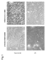

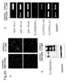
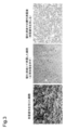
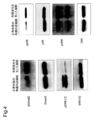
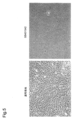
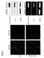

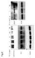
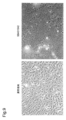


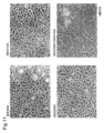
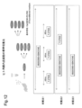


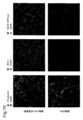


Classifications
-
- A—HUMAN NECESSITIES
- A61—MEDICAL OR VETERINARY SCIENCE; HYGIENE
- A61K—PREPARATIONS FOR MEDICAL, DENTAL OR TOILETRY PURPOSES
- A61K35/00—Medicinal preparations containing materials or reaction products thereof with undetermined constitution
- A61K35/12—Materials from mammals; Compositions comprising non-specified tissues or cells; Compositions comprising non-embryonic stem cells; Genetically modified cells
- A61K35/30—Nerves; Brain; Eyes; Corneal cells; Cerebrospinal fluid; Neuronal stem cells; Neuronal precursor cells; Glial cells; Oligodendrocytes; Schwann cells; Astroglia; Astrocytes; Choroid plexus; Spinal cord tissue
-
- A—HUMAN NECESSITIES
- A01—AGRICULTURE; FORESTRY; ANIMAL HUSBANDRY; HUNTING; TRAPPING; FISHING
- A01N—PRESERVATION OF BODIES OF HUMANS OR ANIMALS OR PLANTS OR PARTS THEREOF; BIOCIDES, e.g. AS DISINFECTANTS, AS PESTICIDES OR AS HERBICIDES; PEST REPELLANTS OR ATTRACTANTS; PLANT GROWTH REGULATORS
- A01N1/00—Preservation of bodies of humans or animals, or parts thereof
- A01N1/02—Preservation of living parts
- A01N1/0205—Chemical aspects
- A01N1/021—Preservation or perfusion media, liquids, solids or gases used in the preservation of cells, tissue, organs or bodily fluids
- A01N1/0226—Physiologically active agents, i.e. substances affecting physiological processes of cells and tissue to be preserved, e.g. anti-oxidants or nutrients
-
- A—HUMAN NECESSITIES
- A61—MEDICAL OR VETERINARY SCIENCE; HYGIENE
- A61K—PREPARATIONS FOR MEDICAL, DENTAL OR TOILETRY PURPOSES
- A61K31/00—Medicinal preparations containing organic active ingredients
- A61K31/33—Heterocyclic compounds
- A61K31/395—Heterocyclic compounds having nitrogen as a ring hetero atom, e.g. guanethidine or rifamycins
- A61K31/435—Heterocyclic compounds having nitrogen as a ring hetero atom, e.g. guanethidine or rifamycins having six-membered rings with one nitrogen as the only ring hetero atom
- A61K31/44—Non condensed pyridines; Hydrogenated derivatives thereof
- A61K31/4409—Non condensed pyridines; Hydrogenated derivatives thereof only substituted in position 4, e.g. isoniazid, iproniazid
-
- A—HUMAN NECESSITIES
- A61—MEDICAL OR VETERINARY SCIENCE; HYGIENE
- A61K—PREPARATIONS FOR MEDICAL, DENTAL OR TOILETRY PURPOSES
- A61K31/00—Medicinal preparations containing organic active ingredients
- A61K31/33—Heterocyclic compounds
- A61K31/395—Heterocyclic compounds having nitrogen as a ring hetero atom, e.g. guanethidine or rifamycins
- A61K31/435—Heterocyclic compounds having nitrogen as a ring hetero atom, e.g. guanethidine or rifamycins having six-membered rings with one nitrogen as the only ring hetero atom
- A61K31/44—Non condensed pyridines; Hydrogenated derivatives thereof
- A61K31/4427—Non condensed pyridines; Hydrogenated derivatives thereof containing further heterocyclic ring systems
- A61K31/4439—Non condensed pyridines; Hydrogenated derivatives thereof containing further heterocyclic ring systems containing a five-membered ring with nitrogen as a ring hetero atom, e.g. omeprazole
-
- A—HUMAN NECESSITIES
- A61—MEDICAL OR VETERINARY SCIENCE; HYGIENE
- A61K—PREPARATIONS FOR MEDICAL, DENTAL OR TOILETRY PURPOSES
- A61K31/00—Medicinal preparations containing organic active ingredients
- A61K31/33—Heterocyclic compounds
- A61K31/395—Heterocyclic compounds having nitrogen as a ring hetero atom, e.g. guanethidine or rifamycins
- A61K31/435—Heterocyclic compounds having nitrogen as a ring hetero atom, e.g. guanethidine or rifamycins having six-membered rings with one nitrogen as the only ring hetero atom
- A61K31/47—Quinolines; Isoquinolines
- A61K31/4709—Non-condensed quinolines and containing further heterocyclic rings
-
- A—HUMAN NECESSITIES
- A61—MEDICAL OR VETERINARY SCIENCE; HYGIENE
- A61K—PREPARATIONS FOR MEDICAL, DENTAL OR TOILETRY PURPOSES
- A61K31/00—Medicinal preparations containing organic active ingredients
- A61K31/33—Heterocyclic compounds
- A61K31/395—Heterocyclic compounds having nitrogen as a ring hetero atom, e.g. guanethidine or rifamycins
- A61K31/495—Heterocyclic compounds having nitrogen as a ring hetero atom, e.g. guanethidine or rifamycins having six-membered rings with two or more nitrogen atoms as the only ring heteroatoms, e.g. piperazine or tetrazines
- A61K31/505—Pyrimidines; Hydrogenated pyrimidines, e.g. trimethoprim
- A61K31/519—Pyrimidines; Hydrogenated pyrimidines, e.g. trimethoprim ortho- or peri-condensed with heterocyclic rings
-
- A—HUMAN NECESSITIES
- A61—MEDICAL OR VETERINARY SCIENCE; HYGIENE
- A61K—PREPARATIONS FOR MEDICAL, DENTAL OR TOILETRY PURPOSES
- A61K31/00—Medicinal preparations containing organic active ingredients
- A61K31/70—Carbohydrates; Sugars; Derivatives thereof
- A61K31/7088—Compounds having three or more nucleosides or nucleotides
- A61K31/713—Double-stranded nucleic acids or oligonucleotides
-
- A—HUMAN NECESSITIES
- A61—MEDICAL OR VETERINARY SCIENCE; HYGIENE
- A61P—SPECIFIC THERAPEUTIC ACTIVITY OF CHEMICAL COMPOUNDS OR MEDICINAL PREPARATIONS
- A61P27/00—Drugs for disorders of the senses
- A61P27/02—Ophthalmic agents
-
- A—HUMAN NECESSITIES
- A61—MEDICAL OR VETERINARY SCIENCE; HYGIENE
- A61P—SPECIFIC THERAPEUTIC ACTIVITY OF CHEMICAL COMPOUNDS OR MEDICINAL PREPARATIONS
- A61P43/00—Drugs for specific purposes, not provided for in groups A61P1/00-A61P41/00
-
- C—CHEMISTRY; METALLURGY
- C07—ORGANIC CHEMISTRY
- C07K—PEPTIDES
- C07K16/00—Immunoglobulins [IGs], e.g. monoclonal or polyclonal antibodies
- C07K16/18—Immunoglobulins [IGs], e.g. monoclonal or polyclonal antibodies against material from animals or humans
- C07K16/22—Immunoglobulins [IGs], e.g. monoclonal or polyclonal antibodies against material from animals or humans against growth factors ; against growth regulators
-
- C—CHEMISTRY; METALLURGY
- C12—BIOCHEMISTRY; BEER; SPIRITS; WINE; VINEGAR; MICROBIOLOGY; ENZYMOLOGY; MUTATION OR GENETIC ENGINEERING
- C12N—MICROORGANISMS OR ENZYMES; COMPOSITIONS THEREOF; PROPAGATING, PRESERVING, OR MAINTAINING MICROORGANISMS; MUTATION OR GENETIC ENGINEERING; CULTURE MEDIA
- C12N5/00—Undifferentiated human, animal or plant cells, e.g. cell lines; Tissues; Cultivation or maintenance thereof; Culture media therefor
- C12N5/06—Animal cells or tissues; Human cells or tissues
- C12N5/0602—Vertebrate cells
- C12N5/0618—Cells of the nervous system
- C12N5/0621—Eye cells, e.g. cornea, iris pigmented cells
-
- C—CHEMISTRY; METALLURGY
- C12—BIOCHEMISTRY; BEER; SPIRITS; WINE; VINEGAR; MICROBIOLOGY; ENZYMOLOGY; MUTATION OR GENETIC ENGINEERING
- C12N—MICROORGANISMS OR ENZYMES; COMPOSITIONS THEREOF; PROPAGATING, PRESERVING, OR MAINTAINING MICROORGANISMS; MUTATION OR GENETIC ENGINEERING; CULTURE MEDIA
- C12N2500/00—Specific components of cell culture medium
- C12N2500/05—Inorganic components
- C12N2500/10—Metals; Metal chelators
- C12N2500/12—Light metals, i.e. alkali, alkaline earth, Be, Al, Mg
- C12N2500/14—Calcium; Ca chelators; Calcitonin
-
- C—CHEMISTRY; METALLURGY
- C12—BIOCHEMISTRY; BEER; SPIRITS; WINE; VINEGAR; MICROBIOLOGY; ENZYMOLOGY; MUTATION OR GENETIC ENGINEERING
- C12N—MICROORGANISMS OR ENZYMES; COMPOSITIONS THEREOF; PROPAGATING, PRESERVING, OR MAINTAINING MICROORGANISMS; MUTATION OR GENETIC ENGINEERING; CULTURE MEDIA
- C12N2500/00—Specific components of cell culture medium
- C12N2500/30—Organic components
- C12N2500/38—Vitamins
-
- C—CHEMISTRY; METALLURGY
- C12—BIOCHEMISTRY; BEER; SPIRITS; WINE; VINEGAR; MICROBIOLOGY; ENZYMOLOGY; MUTATION OR GENETIC ENGINEERING
- C12N—MICROORGANISMS OR ENZYMES; COMPOSITIONS THEREOF; PROPAGATING, PRESERVING, OR MAINTAINING MICROORGANISMS; MUTATION OR GENETIC ENGINEERING; CULTURE MEDIA
- C12N2501/00—Active agents used in cell culture processes, e.g. differentation
- C12N2501/10—Growth factors
- C12N2501/11—Epidermal growth factor [EGF]
-
- C—CHEMISTRY; METALLURGY
- C12—BIOCHEMISTRY; BEER; SPIRITS; WINE; VINEGAR; MICROBIOLOGY; ENZYMOLOGY; MUTATION OR GENETIC ENGINEERING
- C12N—MICROORGANISMS OR ENZYMES; COMPOSITIONS THEREOF; PROPAGATING, PRESERVING, OR MAINTAINING MICROORGANISMS; MUTATION OR GENETIC ENGINEERING; CULTURE MEDIA
- C12N2501/00—Active agents used in cell culture processes, e.g. differentation
- C12N2501/10—Growth factors
- C12N2501/15—Transforming growth factor beta (TGF-β)
-
- C—CHEMISTRY; METALLURGY
- C12—BIOCHEMISTRY; BEER; SPIRITS; WINE; VINEGAR; MICROBIOLOGY; ENZYMOLOGY; MUTATION OR GENETIC ENGINEERING
- C12N—MICROORGANISMS OR ENZYMES; COMPOSITIONS THEREOF; PROPAGATING, PRESERVING, OR MAINTAINING MICROORGANISMS; MUTATION OR GENETIC ENGINEERING; CULTURE MEDIA
- C12N2501/00—Active agents used in cell culture processes, e.g. differentation
- C12N2501/10—Growth factors
- C12N2501/155—Bone morphogenic proteins [BMP]; Osteogenins; Osteogenic factor; Bone inducing factor
-
- C—CHEMISTRY; METALLURGY
- C12—BIOCHEMISTRY; BEER; SPIRITS; WINE; VINEGAR; MICROBIOLOGY; ENZYMOLOGY; MUTATION OR GENETIC ENGINEERING
- C12N—MICROORGANISMS OR ENZYMES; COMPOSITIONS THEREOF; PROPAGATING, PRESERVING, OR MAINTAINING MICROORGANISMS; MUTATION OR GENETIC ENGINEERING; CULTURE MEDIA
- C12N2501/00—Active agents used in cell culture processes, e.g. differentation
- C12N2501/70—Enzymes
- C12N2501/72—Transferases [EC 2.]
- C12N2501/727—Kinases (EC 2.7.)
-
- C—CHEMISTRY; METALLURGY
- C12—BIOCHEMISTRY; BEER; SPIRITS; WINE; VINEGAR; MICROBIOLOGY; ENZYMOLOGY; MUTATION OR GENETIC ENGINEERING
- C12N—MICROORGANISMS OR ENZYMES; COMPOSITIONS THEREOF; PROPAGATING, PRESERVING, OR MAINTAINING MICROORGANISMS; MUTATION OR GENETIC ENGINEERING; CULTURE MEDIA
- C12N2501/00—Active agents used in cell culture processes, e.g. differentation
- C12N2501/90—Polysaccharides
-
- C—CHEMISTRY; METALLURGY
- C12—BIOCHEMISTRY; BEER; SPIRITS; WINE; VINEGAR; MICROBIOLOGY; ENZYMOLOGY; MUTATION OR GENETIC ENGINEERING
- C12N—MICROORGANISMS OR ENZYMES; COMPOSITIONS THEREOF; PROPAGATING, PRESERVING, OR MAINTAINING MICROORGANISMS; MUTATION OR GENETIC ENGINEERING; CULTURE MEDIA
- C12N2501/00—Active agents used in cell culture processes, e.g. differentation
- C12N2501/998—Proteins not provided for elsewhere
-
- C—CHEMISTRY; METALLURGY
- C12—BIOCHEMISTRY; BEER; SPIRITS; WINE; VINEGAR; MICROBIOLOGY; ENZYMOLOGY; MUTATION OR GENETIC ENGINEERING
- C12N—MICROORGANISMS OR ENZYMES; COMPOSITIONS THEREOF; PROPAGATING, PRESERVING, OR MAINTAINING MICROORGANISMS; MUTATION OR GENETIC ENGINEERING; CULTURE MEDIA
- C12N2501/00—Active agents used in cell culture processes, e.g. differentation
- C12N2501/999—Small molecules not provided for elsewhere
Definitions
- the present invention relates to a technique and method for culturing corneal endothelial cells in a normal state, and a drug and a medium therefor.
- Visual information is transmitted from the cornea, the transparent tissue in the foreground of the eyeball, to reach the retina and excite the neurons in the retina. To be recognized.
- the cornea needs to be transparent.
- the transparency of the cornea is maintained by keeping the water content constant by the pump function and the barrier function of corneal endothelial cells.
- Human corneal endothelial cells exist at a density of about 3000 cells per square millimeter at birth, but do not have the ability to regenerate once damaged.
- bullous keratopathy caused by corneal endothelial degeneration or corneal endothelial dysfunction due to various causes, the cornea becomes edema and turbidity, resulting in a significant loss of visual acuity.
- full-thickness corneal transplantation is performed in which all three layers of the corneal epithelium, parenchyma and endothelium are transplanted.
- the cornea donation in Japan is insufficient, and the number of corneal transplants performed in Japan is about 1700 per year for about 2600 waiting patients for corneal transplantation.
- corneal transplantation in which only damaged tissues are transplanted, has attracted attention.
- deep surface corneal transplantation which is transplantation of parenchymal tissue
- Descemet's Stripping Automated Endothelial Keratoplasty which is transplantation of corneal endothelial tissue
- cultured mucosal epithelial transplantation that transplants corneal epithelium or oral mucosa cultured in vitro instead of corneal epithelium has already been clinically applied, and a method of transplanting corneal endothelium cultured in vitro is also being studied. Yes.
- a corneal endothelium-like sheet comprising a corneal endothelium layer cultured on a collagen layer
- Patent Document 1 a corneal endothelial cell, especially corneal endothelial cells derived from humans, are limited in the number of donors of the cornea and are difficult to culture in vitro, and it takes time and money to obtain the number of cultured cells necessary for transplantation. It is necessary.
- ES cells Human embryonic stem (ES) cells have high self-renewal ability and pluripotency, and are attracting attention from the viewpoint of medical application, but because cell death easily occurs by the operation of dispersing cells in the culture process, There was a practical problem that the number of cells was significantly impaired. In recent years, it has been found that cell death that occurs when human ES cells are cultured is caused by activation of Rho kinase (ROCK), and that cell death is greatly suppressed by inhibition of ROCK. It has been reported that large-scale culture of human ES cells and production of cerebral cells using Y-27632 or the like can be performed (Non-patent Document 1). Therefore, the present inventors disclosed a method for mass culture of corneal endothelial cells using Y-27632 or the like (Patent Document 2).
- ROCK Rho kinase
- Patent Document 3 discloses a neurosphere method using corneal endothelial progenitor cells.
- Patent Document 4 discloses the use of a TGF- ⁇ kinase inhibitor and a p38 MAPK inhibitor for culturing epithelial cells.
- Non-Patent Documents 2 and 4 describe the involvement of TGF- ⁇ , p38 MAPK, and Smad in specific severe corneal endothelial diseases.
- Non-Patent Document 3 describes a perspective on the proliferation of human corneal endothelial cells using ROCK inhibitors.
- Non-Patent Document 5 shows that fibrosis during severe injury of the cornea is due to activation of p38 MAPK by IL-1 ⁇ .
- Non-Patent Document 6 shows that the fibrosis observed during excessive freezing trauma in rabbits can be suppressed with an inhibitor by activation of p38 MAPK using rabbits.
- Non-Patent Document 7 describes that a conventional corneal endothelial cell culture medium cannot be proliferated while maintaining a normal state when subcultured.
- Non-Patent Document 8 discloses a medium for corneal endothelial cells. This medium contains FBS, EGF and NGF, but it is described that cells to be cultured in this medium cannot be cultured well unless they are derived from young organisms.
- Non-Patent Document 9 discloses a medium for corneal endothelial cells using basic FGF.
- Non-Patent Document 10 discloses a corneal endothelial cell lysis medium using collagenase.
- Non-Patent Document 11 discloses a corneal endothelial cell lysis medium using a conditioned medium.
- Non-Patent Documents 8 to 11 Various media have been developed as in Non-Patent Documents 8 to 11, but as shown in Non-Patent Document 7, the conventional corneal endothelial cell culture medium can be grown while maintaining a normal state when subcultured. It is known that it cannot.
- Non-patent documents 12 to 14 also describe the production of cultured corneal endothelial sheets.
- Non-patent documents 9 to 12 and 15 disclose human ocular tissue-derived stem cells and autologous corneal endothelial transplantation.
- Non-patent documents 16 and 17 also describe the production of cultured corneal endothelial sheets.
- the present inventors have found a technique capable of proliferating corneal endothelial cells while maintaining normal function by inhibiting the tumor necrosis factor ⁇ (TGF- ⁇ ) pathway. As a result, corneal endothelial cells having normal functions can be grown in a relatively large amount. That is, the present invention provides the following. (1) A culture normalizing agent for corneal endothelial cells containing a fibrosis inhibitor. (2) The culture normalizing agent according to item 1, wherein the fibrosis inhibitor comprises a transforming growth factor (TGF) ⁇ signal inhibitor. (3) The culture normalizing agent according to item 1 or 2, wherein the culture normalization includes a normal cell function selected from the group consisting of ZO-1 and Na + / K + -ATPase.
- TGF tumor necrosis factor
- the TGF- ⁇ signal inhibitor is SB431542 (4- [4- (1,3-benzodioxol-5-yl) 2-pyridinyl)]-1H-imidazol-2-yl] benzamide), BMP-7, anti-TGF- ⁇ antibody, anti-TGF- ⁇ receptor antibody, TGF- ⁇ siRNA, TGF- ⁇ receptor siRNA, TGF- ⁇ antisense oligonucleotide, 6,7-dimethoxy-2-((2E ) -3- (1-Methyl-2-phenyl-1H-pyrrolo [2,3-b] pyridin-3-yl-prop-2-enoyl))-1,2,3,4-tetrahydroisoquinolone, A83 -01 (3- (6-methyl-2-pyridinyl) -N-phenyl-4- (4-quinolinyl) -1H-pyrazole-1-carbothioamide), Stemolecule TM TLK Inhibitor (2- (3- (6-methylpyri
- the culture normalizing agent according to any one of items 2 to 7, comprising at least one solvate of a pharmaceutically acceptable salt thereof.
- the TGF- ⁇ signal inhibitor is SB431542 (4- [4- (1,3-benzodioxol-5-yl) 2-pyridinyl)]-1H-imidazol-2-yl] benzamide), BMP-7, anti-TGF- ⁇ antibody, anti-TGF- ⁇ receptor antibody, TGF- ⁇ siRNA, TGF- ⁇ receptor siRNA, TGF- ⁇ antisense oligonucleotide, A83-01 (3- (6-methyl- 2-pyridinyl) -N- phenyl-4- (4-quinolinyl)-1H-pyrazole-1-carbothioamide), stearyl leakage CURE TM TLK inhibitor (2- (3- (6-methylpyridin-2-yl) - 1H- pyrazol-4-yl) -1,5-naphthyridine), stearyl leakage C
- the TGF- ⁇ signal inhibitor is SB431542 (4- [4- (1,3-benzodioxol-5-yl) 2-pyridinyl) -1H-imidazol-2-yl] benzamide) or its Item 9.
- the MAP kinase inhibitor is SB203580 (4- [4- (4-fluorophenyl) -2- (4-methylsulfinylphenyl) -1H-imidazol-5-yl] pyridine) or a pharmaceutically acceptable salt thereof.
- the culture normalizing agent according to Item 10 comprising a suitable salt.
- the aging inhibitor includes SB203580 (4- [4- (4-fluorophenyl) -2- (4-methylsulfinylphenyl) -1H-imidazol-5-yl] pyridine). Culture normalizing agent.
- SB431542 (4- [4- (1,3-benzodioxol-5-yl) 2-pyridinyl) -1H-imidazol-2-yl] benzamide) and SB203580 (4- [4- (4 -Fluorophenyl) -2- (4-methylsulfinylphenyl) -1H-imidazol-5-yl] pyridine) or a pharmaceutically acceptable salt thereof, items 1-8, 8A, 9, 9A, 9B , 9C, 9D, 9E, 10 or 11 to 14, the culture normalizing agent.
- the culture normalizing agent according to any one of items 1 to 8, 8A, 9, 9A, 9B, 9C, 9D, 9E, 10 or 11 to 15, further comprising a cell adhesion promoter.
- the cell adhesion promoter is (R)-(+)-trans- (4-pyridyl) -4- (1-aminoethyl) -cyclohexanecarboxamide or a pharmaceutically acceptable salt thereof (for example, Y-
- the culture normalizing agent according to Item 16 comprising 27632 (R)-(+)-trans- (4-pyridyl) -4- (1-aminoethyl) -cyclohexanecarboxamide dihydrochloride monohydrate).
- the fibrosis inhibitor is always present during the culturing of the corneal endothelial cells, while the adhesion promoter is allowed to exist for a certain period of time, and then the adhesion promoter is once depleted, and then the cell adhesion is repeated.
- Item 18 The culture normalizing agent according to Item 16 or 17, wherein the promoter is present for a certain period.
- the cell for transplant is any one of items 4 to 8, 8A, 9, 9A, 9B, 9C, 9D, 9E, 10 and 11 to 19 for preventing or treating corneal endothelial injury
- Corneal endothelial cells comprising the culture normalizing agent according to any one of items 1 to 8, 8A, 9, 9A, 9B, 9C, 9D, 9E, 10 or 11 to 20 and a culture component of corneal endothelium A medium for culturing normally.
- a method of normally culturing corneal endothelial cells comprising a step of culturing cells.
- a corneal endothelial cell preservation solution comprising the culture normalizing agent according to any one of items 1 to 8, 8A, 9, 9A, 9B, 9C, 9D, 9E, 10 and 11 to 20.
- the medicament according to item 25, wherein the treatment or prevention is for primate corneal endothelium.
- the cell adhesion promoter is (R)-(+)-trans- (4-pyridyl) -4- (1-aminoethyl) -cyclohexanecarboxamide or a pharmaceutically acceptable salt thereof (for example, Y-
- a cell adhesion promoter comprises a Rho kinase inhibitor.
- the cell adhesion promoter is (R)-(+)-trans- (4-pyridyl) -4- (1-aminoethyl) -cyclohexanecarboxamide or a pharmaceutically acceptable salt thereof (for example, Y-
- the pharmaceutical according to Item 35 or 35A which is 27632 (R)-(+)-trans- (4-pyridyl) -4- (1-aminoethyl) -cyclohexanecarboxamide dihydrochloride monohydrate)).
- the medicine comprises the culture normalizing agent according to any one of items 1 to 8, 8A, 9, 9A, 9B, 9C, 9D, 9E, 10 or 11 to 20, or the medium according to item 21.
- the medicament according to item 35, 35A or 36 which is used together with a corneal endothelial cell produced by a method for normally culturing corneal endothelial cells, comprising a step of culturing corneal endothelial cells.
- a method for the treatment or prevention of a human corneal endothelial disease, disorder or condition comprising the step of administering a cell adhesion promoter to a subject in need of treatment or prevention.
- 39A A method according to item 39, wherein the method has at least one of the characteristics according to any one of items 35A and 36 to 38.
- the present invention provides a technique capable of proliferating corneal endothelial cells while maintaining normal functions that have been difficult to achieve in the past.
- Normal functions include biochemical functions of corneal endothelial cells such as ZO-1 and Na + / K + -ATPase, transplantation ability to primates, etc., and include functions for realizing corneal transplantation. .
- FIG. 1 shows morphological changes when cynomolgus monkeys and human cells are cultured by a conventional method.
- the upper side shows cynomolgus monkeys and the lower side shows humans.
- Cynomolgus monkey is DMEM + 10% FBS + 2 ng / ml basic FGF
- human is Opti-MEM I Reduced-Serum Medium, Liquid + 8% FBS + 200 mg / ml CaCl 2 ⁇ 2H 2 O + 0.08% chondroitin sulfate + 20 ⁇ g / ml Ascorbic acid + 50 ⁇ g / ml It is a culture result on condition of EGF.
- the left shows the morphology of normal corneal endothelium, but morphological changes easily occur as shown on the right by long-term culture or subculture.
- FIG. 2 shows that normal function is lost when cultured with the prior art.
- the left panel shows the results of immunostaining.
- the left side shows monkey corneal endothelial cells (MCECs) that can be cultured in a normal form, and the right side shows MCECs that have undergone a fibroblast-like shape change by long-term culture.
- the upper side of the left panel shows staining with ZO-1, and the lower side shows staining with Na + / K + -ATPase.
- the upper right panel shows the results of the Western blot.
- the lower right panel shows the results of real-time PCR analysis.
- FIG. 2A shows that fibroblast primate corneal endothelial cells (CEC) produce an abnormal extracellular matrix, ie, lose normal function when cultured in the prior art.
- CEC fibroblast primate corneal endothelial cells
- the left side shows the normal cell phenotype and the right side shows the fibroblast phenotype.
- the fibroblast phenotype showed an excess of extracellular matrix such as fibronectin and collagen type 1.
- the normal cell phenotype completely lost staining ability.
- (B) Western blot of protein expression in fibronectin fibroblast phenotype and normal cell phenotype. GAPDH is a control. Fibronectin protein expression levels were strongly up-regulated in the fibroblast phenotype than in the normal phenotype.
- C Semi-quantification of collagen type 1, type 4, type 8 fibronectin, integrin ⁇ 5, and integrin ⁇ 1 (listed in order from the top) in fibroblast phenotype (right) and normal cell phenotype (left). The result of PCR is shown. GAPDH is a control.
- type 1 collagen transcript ( ⁇ 1 (I) mRNA) was abundantly expressed in the fibroblast phenotype, whereas in the normal phenotype, ⁇ 1 (I) mRNA expression was It was decreasing.
- FIG. 3 shows fibrosis with the conventional method.
- the conditioned medium for 3T3 feeder cells suppresses fibroblastic transformation (transformation) (middle), but shows that it is still transformed when subcultured (right).
- FIG. 4 is a diagram showing that the Smad pathway, the p38 MAPK pathway, and the JNK pathway are activated in the fibrotic cells of monkey corneal endothelial cells.
- the left shows normal forms of monkey corneal endothelial cells (MCEC), and the right shows MCECs that have undergone morphological changes like fibroblasts.
- FIG. 5 shows that the transformation of monkey corneal endothelium could be suppressed by inhibiting the TGF- ⁇ signal with a receptor phosphorylation inhibitor.
- the left shows that the corneal endothelium of cynomolgus monkey was cultured in DMEM + 10% FBS + 2 ng / ml basic FGF (also referred to as a normal medium in the present specification), and the corneal endothelium changed its shape like a fibroblast.
- FIG. 6 shows that TGF- ⁇ signal inhibition suppresses loss of function-related protein due to fibrosis of monkey corneal endothelial cells.
- the left side shows the MCEC that has undergone cynomolgus monkey corneal endothelium culturing with DMEM + 10% FBS + 2 ng / ml basic FGF (normal medium).
- the immuno-staining image by the function related marker ZO-1 (upper side) and Na ⁇ + > / K ⁇ + > - ATPase (lower side) is shown.
- the upper right panel shows the results of the Western blot.
- the lower right panel shows the results of real-time PCR analysis.
- FIG. 7 is a diagram showing that TGF- ⁇ is added to induce transformation and loss of function-related proteins in order to confirm that the TGF- ⁇ signal is involved in the transformation of monkey corneal endothelium.
- the upper side shows control MCEC, and the lower side shows the result of cells treated with TGF- ⁇ .
- FIG. 8 is a diagram showing that monkey corneal endothelial cells become fibrotic and morphologically change due to TGF- ⁇ and lose function-related proteins. The change with the dose of TGF- ⁇ is shown (the left panel shows 0 ng / ml, 1 ng / ml, 3 ng / ml, 10 ng / ml, 30 ng / ml from the left.
- FIG. 9 is a diagram showing that, even in human corneal endothelium, it is possible to suppress transformation and inhibit normal endothelium by inhibiting the TGF- ⁇ signal with a receptor phosphorylation inhibitor.
- the left is a control (normal medium), and the right shows the result of staining with SB431542.
- SB431542 maintains the function of human corneal endothelial cells (HCEC) and suppresses fibroblast-like changes in human corneal endothelial cells (A, B).
- HCEC human corneal endothelial cells
- A, B human corneal endothelial cells
- the left shows human corneal endothelial cells cultured in a normal medium
- the right shows that cultured in a medium in which SB431542 is added to the normal medium at a concentration of 1 ⁇ M.
- Blocking TGF receptor signaling by SB431542 allowed Na + / K + -ATPase and ZO-1 to be subcellularly localized at the cell membrane and maintained their protein expression.
- the scale bar indicates 100 ⁇ m.
- C shows an ELISA assay.
- the concentration of collagen type 1 when SB431542 is added or not is shown as the concentration in the cell supernatant.
- An ELISA assay showed that SB431542 significantly downregulated the secretion of collagen type 1 into the cell supernatant. ** P ⁇ 0.05.
- D, E shows quantitative PCR.
- FIG. 5 shows that SB431542 significantly reduces collagen type 1 and fibronectin expression at the mRNA level. * P ⁇ 0.01, ** P ⁇ 0.05.
- FIG. 10 is a diagram showing that a method other than SB431542 could antagonize the TGF- ⁇ signal and suppress the transformation of human corneal endothelium. The upper result shows the result with a phase contrast microscope.
- the lower side shows the results of phalloidin staining (green staining (the network around the cell is phalloidin, and the red staining (particulate) is PI.)
- the left side is Opti-MEM.
- FIG. 10A shows the result of examining the effect of each concentration of BMP-7, which is shown in FIG.
- BMP7 suppresses the change of human corneal endothelial cells to fibroblast-like and maintains its function.
- A It is a photograph of a phase contrast microscope. The upper left is a control without BMP-7 (displayed as Control). Upper left is 10 ng / mL, lower right is 100 ng / mL, lower left is 1000 ng / mL BMP-7.
- the elongated cell shape of the fibroblast phenotype was converted to a polygonal cell shape in response to the presence of BMP-7 in a concentration-dependent manner.
- the scale bar is 100 ⁇ m.
- the scale bar is 100 ⁇ m.
- C) and (D) are photographs of Na + / K + -ATPase and ZO-1 staining, respectively.
- the upper left is a control without addition of BMP-7.
- Upper left is 10 ng / mL
- lower right is 100 ng / mL
- lower left is 1000 ng / mL
- BMP-7 maintained the intracellular localization of Na + / K + -ATPase and ZO-1 in the cell membrane.
- the scale bar is 100 ⁇ m.
- (E) is a graph showing the ratio of Na + / K + -ATPase positive cells (E) and ZO-1 positive cells (F) when cultured using three concentrations of control and BMP-7. is there. No control is added. Both Na + / K + -ATPase positive cells and ZO-1 positive cells were significantly increased in proportion compared to controls when treated with BMP-7.
- FIG. 11 shows that when an inhibitor of p38 MAPK is used in addition to SB431542, p38 MAPK inhibition + TGF- ⁇ signal inhibition enables culturing with corneal endothelial cells retaining high density even after repeated passages.
- FIG. The upper left is the control (indicated as a normal medium in the figure), and the upper right is the result with only SB431542.
- the lower left is a result with only SB203580, and the lower right is a diagram showing the results with both SB431542 and SB203580.
- FIG. 12 is an example of a standard culture method for human corneal endothelial cells established in the present invention.
- the upper panel shows a schematic diagram of subculture, and shows schematic diagrams of culture methods 1 to 3 carried out in Example 8.
- SB203580 and SB431432 are present.
- Culture method 1 is a method in which Y-27632 is allowed to exist for 48 hours, then once deleted, and then added again. In the culture method 2, Y-27632 is always present together. In culture method 3, Y-27632 is not present.
- the result in the human corneal endothelial cell culture finally established is shown. As shown in the photograph on the left, it is understood that fibrosis is suppressed and proliferation is also smooth.
- Example 9 it is shown that transparent corneal healing can be obtained by culturing human corneal endothelial cells cultured while maintaining their functions in a normal form on a collagen sheet and transplanting them into a cynomolgus monkey bullous keratopathy model.
- the left side shows the result of transplanting only the cells cultured by the culture method of the present invention
- the right side shows the result when Y-27632, which is a ROCK inhibitor, was injected at the time of transplantation of the cells cultured by the culture method of the present invention. .
- Example 9 after euthanization after 2.5 months, the cornea was removed and the tissue was fixed, and then immunostaining was performed for phalloidin and Na + / K + -ATPase and ZO-1 in the same manner as in Example 2.
- photographed with the fluorescence microscope is shown.
- the upper row shows the result of the cell + ROCK inhibitor, and the lower row shows the result of the cell alone.
- phalloidin primary color is green, on the gray scale, it is meshed on the upper side and appears to be diffused on the lower side
- DAPI primary color is blue.
- the number is reduced compared to the upper side, showing staining of the phalloidin stain, and the center is ZO-1 (primary color is green. In the lower part, it appears to remain partly in the upper left and lower right.)
- DAPI primary color is blue.
- the gray scale the inside of the cell is stained in the form of particles, and in the lower part, the particulate staining disappears and becomes light.
- the right side is Na + / K + -ATPase (the primary color is green.
- the gray scale is mesh-like on the upper side and the lower side is partially left in the center)
- DAPI primary color is blue.
- FIG. 16 shows normalization of culture in the case of using the anti-TGF- ⁇ neutralizing antibody performed in Example 10. The left shows the results with the normal medium, and the right shows the results with the anti-TGF- ⁇ neutralizing antibody.
- FIG. 17 shows 6,7-dimethoxy-2-((2E) -3- (1-methyl-2-phenyl-1H-pyrrolo [2, sold by Calbiochem, an Smad3 inhibitor used in Example 11).
- Fibrosis inhibitor refers to any drug that inhibits fibrosis.
- Fibrosis inhibitors used in the present invention include transforming growth factor (TGF) - ⁇ signal inhibitor, mitogen-activated protein kinase (MAPK) 38 inhibitor, interleukin (IL) -12, Examples thereof include cytokines known to have an antifibrotic effect, such as IL-10, interferon (IFN) - ⁇ , or BMP-7 (OP-1). Information on such cytokines and the like can be obtained from public databases such as GenBank and magazine publications.
- the present invention achieves a significant increase in corneal endothelial cells, which conventionally had difficulty in proliferating cells with normal function, by suppressing fibrosis. I was able to. Therefore, it is understood that any agent can be used as the fibrosis inhibitor used in the present invention as long as it causes proliferation of cells having normal functions.
- IFN- ⁇ polypeptides can be used to treat human diseases, but generally human proteins are used for human corneal endothelial cells.
- Human IFN- ⁇ coding sequences can be found in GenBank accession numbers P01579 and CAA00375.
- Corresponding genomic sequences can be found in GenBank accession numbers J00219, M37265, and V00536.
- a calcium channel blocker such as verapamil can be used as a fibrosis inhibitor.
- fibrosis inhibitors can have an antifibrotic effect by stimulating the degradation of collagen type I fibers as well as the ability to reduce collagen type I synthesis.
- In vitro tests on fibroblasts show that the extracellular transport of collagen depends on the presence of calcium.
- Verapamil a calcium channel blocker, decreases intracellular calcium levels and increases collagenase activity. It also inhibits fibroblast proliferation.
- transforming growth factor- ⁇ transformed growth factor- ⁇ ; also abbreviated as TGF- ⁇
- TGF- ⁇ transformed growth factor- ⁇
- TGF- ⁇ is produced as an inactive latent type having a molecular weight of about 300 kD that cannot bind to the receptor, and is activated on the surface of the target cell and its surroundings to become an active type that can bind to the receptor and exerts its action.
- TGF- ⁇ in the target cell is transmitted by a series of protein phosphorylation pathways responsible for information transmission called Smad.
- Smad protein phosphorylation pathways responsible for information transmission.
- a receptor complex consisting of two type II receptor molecules and two type I TGF- ⁇ receptor molecules is formed. Phosphorylates type I receptors.
- phosphorylated type I receptor phosphorylates Smad2 or Smad3
- phosphorylated Smad2 and Smad3 form a complex with Smad4 and move to the nucleus, and are called CAGA boxes that exist in the target gene promoter region. It is said to bind to the target sequence and induce transcriptional expression of the target gene together with the coactivator.
- TGF- ⁇ The transforming growth factor- ⁇ signaling pathway is regulated by many target cells, such as cell proliferation and differentiation, growth arrest, apoptosis, and epithelial-mesenchymal transdifferentiation (EMT).
- EMT epithelial-mesenchymal transdifferentiation
- the activity can be modulated.
- TGF- ⁇ family including TGF- ⁇ itself (eg, TGF- ⁇ 1, TGF- ⁇ 2 and TGF- ⁇ 3), activin and bone morphogenetic protein (BMP) are cell proliferation, differentiation, migration and apoptosis It is a powerful regulator.
- TGF- ⁇ is an approximately 24 Kd protein produced by many cells, including B lymphocytes, T lymphocytes and activated macrophages, and by many other cell types.
- TGF- ⁇ effects of TGF- ⁇ on the immune system are IL-2 receptor induction, inhibition of IL-1-induced thymocyte proliferation, and blockade of IFN- ⁇ -induced macrophage activation.
- TGF- ⁇ is thought to be involved in a variety of pathological conditions (Border et al. (1992) J. Clin. Invest. 90: 1) and functions as either a tumor suppressor or tumor promoter. Is well supported.
- TGF- ⁇ mediates its signaling by two serine / threonine kinase cell surface receptors, TGF- ⁇ RII and ALK5.
- TGF- ⁇ signaling is initiated by ligand-induced receptor dimerization that allows TGF- ⁇ RII to phosphorylate the ALK5 receptor.
- the phosphorylation activates ALK5 kinase activity, which in turn activates downstream effector Smad protein (vertebrate homologue of MAD, or “Mothers against DPP” protein), Smad2 or 3 Is phosphorylated.
- Smad protein verebrate homologue of MAD, or “Mothers against DPP” protein
- Smad2 or 3 Is phosphorylated The p-Smad2 / 3 complex with Smad4 enters the nucleus and activates transcription of the target gene.
- Smad3 is a member of the R-Smad (receptor-activated Smad) subgroup of Smad and is a direct mediator of transcriptional activation by the TGF- ⁇ receptor.
- TGF- ⁇ stimulation results in phosphorylation and activation of Smad2 and Smad3, which form a complex with Smad4 (“common Smad” or “co-Smad” in vertebrates), which together with the nucleus Accumulate and regulate transcription of target genes.
- R-Smad localizes in the cytoplasm and, upon ligand-induced phosphorylation by the TGF- ⁇ receptor, forms a complex with co-Smad and translocates to the nucleus where they are chromatin and synergistic Regulates gene expression associated with transcription factors.
- Smad6 and Smad7 are inhibitory Smads (“I-Smad”), ie, are transcriptionally induced by TGF- ⁇ and function as inhibitors of TGF- ⁇ signaling (Feng et al. (2005) Annu. Rev. Cell. Dev.Biol.21: 659).
- Smad6 / 7 exerts their inhibitory effects by preventing receptor-mediated activation of R-Smad; they are associated with type I receptors that competitively prevent R-Smad mobilization and phosphorylation.
- Smad6 and Smad7 are known to recruit E3 ubiquitin ligases that result in ubiquitination and degradation of Smad6 / 7 interacting proteins.
- TGF- ⁇ signaling pathway In addition to the TGF- ⁇ signaling pathway, there are also pathways transmitted by BMP-7, etc., which function via ALK-1 / 2/3/6 and via Smad1 / 5/8. It is supposed to be expressed.
- TGF- ⁇ signaling pathway see J.A. Massagu'e, Annu. Rev. Biochem. 1998.67: 753-91; Vilar JMG, Jansen R, Sander C (2006) PLoS Compute Biol 2 (1): e3; , Abraham, D .; J. et al. FASEB J. 18, 816-827 (2004); Coert Margadand & Arnaud Sonnenberg EMBO reports (2010) 11, 97-105; Joel Rosenblum et al. , Ann Internet Med. 2010; 152: 159-166 etc.
- TGF- ⁇ signal inhibitor refers to any factor that inhibits TGF signaling.
- TGF- ⁇ transforming growth factor
- antagonizing TGF- ⁇ it may be referred to as an antagonist, but when referring to the present invention, a TGF- ⁇ antagonist is included in a TGF- ⁇ signal inhibitor.
- the TGF- ⁇ signal inhibitor used in the present invention includes, but is not limited to, an antagonist of TGF- ⁇ , an antagonist of a receptor of TGF- ⁇ , or an inhibitor of Smad3.
- TGF- ⁇ signal inhibitors that can be used in the present invention include SB431542 (4- [4- (1,3-benzodioxol-5-yl) 2-pyridinyl)]-1H-imidazole-2. -Yl] benzamide), BMP-7, anti-TGF- ⁇ antibody, anti-TGF- ⁇ receptor antibody, TGF- ⁇ siRNA, TGF- ⁇ receptor siRNA, TGF- ⁇ antisense oligonucleotide, 6,7-dimethoxy -2-((2E) -3- (1-methyl-2-phenyl-1H-pyrrolo [2,3-b] pyridin-3-yl-prop-2-enoyl))-1,2,3,4 -Tetrahydroisoquinolone, A83-01 (3- (6-methyl-2-pyridinyl) -N-phenyl-4- (4-quinolinyl) -1H-pyrazole-1-carbothioamide), Morekyuru TM TLK inhibitor (2-
- TGF- ⁇ signal inhibitors include monoclonal and polyclonal antibodies against one or more isoforms of TGF- ⁇ (US Pat. No. 5,571,714; WO 97/13844 and WO No. 00/66631), TGF- ⁇ receptors, soluble forms of such receptors (eg, soluble TGF- ⁇ type III receptor), or antibodies directed against TGF- ⁇ receptors (US Pat. 693,607, US Pat. No. 6,001,969, US Pat. No. 6,010,872, US Pat. No. 6,086,867, US Pat. No.
- a TGF- ⁇ inhibitor can be a TGF- ⁇ antagonist, a human or humanized monoclonal antibody that blocks TGF- ⁇ binding to its receptor (or F (ab) 2 fragment, Fv fragment, single chain antibody, And other forms of antibodies or fragments thereof such as fragments that retain the ability to bind to TGF- ⁇ .
- TGF- ⁇ receptors and TGF- ⁇ binding fragments of TGF- ⁇ receptors, especially soluble fragments, are useful TGF- ⁇ antagonists in the methods of the invention.
- preferred inhibitors of TGF- ⁇ function include soluble TGF- ⁇ receptors, especially, for example, the extracellular domain of TGBIIIR or TGFBIIIR, preferably a recombinant soluble TGF- ⁇ receptor (rsTGFBIIR or rsTGFBIIIR), TGF- ⁇ type II receptor (TGBIIIR) or TGF- ⁇ type III receptor (TGFBIIIR or beta glycan).
- TGF- ⁇ receptors and TGF- ⁇ binding fragments of TGF- ⁇ receptors, especially soluble fragments, are useful TGF- ⁇ antagonists in the methods of the invention.
- TGF- ⁇ receptors and the nucleic acids that encode them are well known in the art. Nucleic acid sequences encoding TGF- ⁇ 1 type receptors are disclosed in GenBank Accession No. L15436 and US Pat. No. 5,538,892 (Donahoe et al.). The nucleic acid sequence of the TGF- ⁇ type 2 receptor is publicly available under GenBank accession numbers AW236001, AI35790, AI279787, AI074706, and AA808255. The nucleic acid sequence of the TGF- ⁇ 3 type receptor is also publicly available under GenBank accession numbers NM003243, AI887852, AI817295, and AI681599.
- TGF- ⁇ signal inhibitors or antagonists and methods for their production are well known in the art, along with more currently under development.
- the specific TGF- ⁇ signal inhibitor or antagonist used is not of a limiting character, as any effective TGF- ⁇ antagonist can be useful in the methods of the invention.
- Examples of such antagonists include monoclonal and polyclonal antibodies against one or more isotypes of TGF- ⁇ (US Pat. No. 5,571,714 and WO 97/13844), TGF- ⁇ receptor, fragments thereof , Derivatives thereof, and antibodies against the TGF- ⁇ receptor (US Pat. Nos.
- antagonists include somatostatin (WO 98/08529), mannose-6-phosphate or mannose-1-phosphate (US Pat. No. 5,520,926), prolactin (WO 97/926). 40848), insulin-like growth factor II (WO 98/17304), IP-10 (WO 97/00691), arginine (arg) -glycine (gly) -aspartic acid (asp) -containing peptide (US Pat. 958,411 and WO 93/10808), extracts of plants, fungi and bacteria (European Patent Application No. 813875, JP-A-8-119984 and US Pat. No.
- Suitable TGF- ⁇ antagonists for use in the present invention are also functional variants, mutants of the aforementioned TGF- ⁇ antagonists as long as their ability to inhibit the amount or activity of TGF- ⁇ is retained.
- Derivatives and analogs are also included.
- variants “derivatives”, and “analogs” are molecules that have a similar shape or structure to the parent compound and retain the ability to act as a TGF- ⁇ antagonist. Point to.
- any of the TGF- ⁇ antagonists disclosed herein may be crystallized, and useful analogs can be rationalized based on the coordinates responsible for the active site shape (s). Can be engineered.
- TGF- ⁇ antagonist is a polypeptide
- polypeptide fragments and variants can be produced to increase ease of delivery, activity, half-life, etc. (eg, humanized as discussed above).
- Antibody or functional antibody fragment Given the level of skill in the art of synthetic and recombinant polypeptide production, such variants may be achieved without undue experimentation.
- One skilled in the art may also design novel inhibitors based on knowledge of the crystal structure and / or active site of the TGF- ⁇ inhibitors described herein.
- Polypeptide inhibitors such as the soluble TGF- ⁇ receptor can also be effectively introduced via gene transfer.
- certain embodiments of the methods of the invention include the use of a suitable vector for expression of a TGF- ⁇ receptor or binding partner, preferably a soluble receptor or soluble binding partner.
- administration of a soluble TGF- ⁇ antagonist comprises: a cDNA encoding a soluble antagonist; or a cDNA encoding the extracellular domain of a TGF- ⁇ type II receptor (rsTGBIIIR) or a TGF- ⁇ type III receptor (rsTGFBIIIR).
- a vector comprising, which causes in situ expression of a soluble TGF- ⁇ antagonist in cells transfected with the vector, inhibits the activity of TGF- ⁇ , Inhibits TGF- ⁇ mediated fibrosis.
- Any suitable vector can be used.
- Preferred vectors include adenoviral vectors, lentiviral vectors, Epstein Barr virus (EBV) vectors, adeno-associated virus (AAV) vectors, and retroviral vectors developed for gene transfer purposes.
- ESV Epstein Barr virus
- AAV adeno-associated virus
- Other non-vector methods of gene transfer such as lipid / DNA complexes, protein / DNA conjugates, naked DNA transfer methods, etc. can also be used.
- a further suitable TGF- ⁇ antagonist developed for delivery via adenovirus gene transfer is a chimeric cDNA (Isaka et al.) Encoding the extracellular domain of the TGF- ⁇ type II receptor fused to the Ig Fc domain. , 1999, Kidney Int., 55: pp. 465-475), adenoviral gene transfer vector of dominant negative mutant of TGF- ⁇ type II receptor (Zhao et al., 1998, Mech. Dev., 72: pp. 89-100), and adenovirus gene transfer vector of decorin which is a TGF- ⁇ binding proteoglycan (Zhao et al., 1999, Am. J. Physiol., 277: pp. L412 to L422), but is not limited thereto. Not . Adenovirus-mediated gene transfer is very efficient compared to other gene delivery modalities.
- TGF- ⁇ receptors and TGF- ⁇ binding fragments, soluble fragments, etc. of TGF- ⁇ receptors are TGF- ⁇ antagonists useful in the present invention.
- TGF- ⁇ receptors and the nucleic acids that encode them are well known in the art. Nucleic acid sequences encoding TGF- ⁇ type 1 receptors are disclosed in GenBank accession number L15436 and Donahoe et al. US Pat. No. 5,538,892. The nucleic acid sequence of the TGF- ⁇ type 2 receptor is publicly available under GenBank accession numbers AW236001; AI35790; AI279787; AI074706; and AA808255.
- the nucleic acid sequence of the TGF- ⁇ 3 type receptor is also publicly available under GenBank accession numbers NM003243; AI888852; AI817295; and AI681599.
- the TGF- ⁇ antagonist is its receptor, or F (ab) 2 fragment, Fv fragment, single chain antibody, and other “antibody” forms that retain the ability to bind TGF- ⁇ .
- the antibody can be chimerized or humanized.
- a chimerized antibody includes the constant region of a human antibody and the variable region of a non-human antibody such as a mouse antibody.
- Humanized antibodies comprise the constant and framework variable regions of human antibodies (ie, variable regions other than the hypervariable regions), and the hypervariable regions of non-human antibodies such as mouse antibodies.
- the antibody can be any other type of antibody derivative, such as a human antibody selected or selected from a phage display system or produced from a xenomouse.
- TGF- ⁇ signaling pathway binds to a heterodimeric cell surface complex where the molecule consists of type I (TbRI) and type II (TbRII) serine / threonine kinase receptors and the heterodimeric cells Initiated when inducing a surface complex.
- the heterodimeric receptor then propagates the signal through phosphorylation of the downstream target Smad protein.
- R-Smad receptor-controlled Smad
- Smad4 also known as Smad4
- Smad2 and Smad3 Co-Smad
- I-Smad Inhibition Smad
- the R-Smad forms a complex with the Co-Smad and moves to the nucleus, in cooperation with each other protein, Regulates transcription of target genes (Delynck, R., et al. (1998) Cell 95: 737-740); and Wotton, D.C. (2000) EMBO J.M. 19: 1745).
- the nucleotide sequence and amino acid sequence of human Smad3 are described in, for example, GenBank Accession No. gi: 42476202.
- the nucleotide sequence and amino acid sequence of murine Smad3 are described in, for example, GenBank Accession No. gi: 3153221.
- TGF- ⁇ stimulation results in phosphorylation and activation of Smad2 and Smad3, which form a complex with Smad4 (also referred to as “common Smad” or “co-Smad”), which is in the nucleus. Accumulate with and regulate transcription of target genes. Therefore, TGF- ⁇ signal inhibition can also be achieved by inhibition of Smad2, 3 or co-Smad (Smad4).
- Smad2 and Smad3 also referred to as “common Smad” or “co-Smad”
- Smad4 also referred to as “common Smad” or “co-Smad”
- Smad4 also referred to as “common Smad” or co-Smad4
- TGF- ⁇ signal inhibition can also be achieved by inhibiting R-Smad directly or indirectly.
- Smad6 and Smad7 are inhibitory Smad (I-Smad), ie, transcriptionally induced by TGF- ⁇ and function as inhibitors of TGF- ⁇ signaling (Feng et al. (2005) Annu. Rev. Cell). Dev.Biol.21: 659).
- Smad6 / 7 exerts their inhibitory effect by preventing receptor-mediated activation of R-Smad. They are associated with type I receptors that competitively prevent R-Smad mobilization and phosphorylation.
- Smad6 and Smad7 are known to recruit E3 ubiquitin ligases that result in ubiquitination and degradation of Smad6 / 7 interacting proteins. Therefore, Smad6 and 7 can function as TGF- ⁇ signal inhibitors in the present invention.
- Inhibitors of Smad3 that can be used in the present invention include antisense nucleotides, siRNAs, antibodies, and the like, as low molecular compounds, 6,7-dimethoxy-2-((2E) -3- (s) sold by Calbiochem. 1-methyl-2-phenyl-1H-pyrrolo [2,3-b] pyridin-3-yl-prop-2-enoyl))-1,2,3,4-tetrahydroisoquinolone It is not limited to them.
- culture normalization of corneal endothelial cells has at least one characteristic such as a function inherent in corneal endothelial cells (also referred to as “normal function” in the present specification). It means culturing while maintaining.
- functions include ZO-1 and Na + / K + -ATPase, adaptability to corneal transplantation (Matsubara M, Tanishima T: Wound-healing of the endogenous endothermic chemistry, 198). 26: 264-273; Matsubara M, Tanishima T: Wound-healing of corneal endothermic in monkey: anatradiologic study, Jpn J Othalmol 1983,27: 444-450 k.
- the adaptability to corneal transplantation can be carried out by transplanting cultured cells by mechanically scraping the corneal endothelium as a bullous keratosis model even in laboratory animals such as rabbits.
- rabbit corneal endothelial cells proliferate in vivo, the possibility of spontaneous healing due to proliferation of host corneal endothelial cells cannot be denied (Matsubara M, et al., Jpn J Ophthalmol 1982, 26: 264-273; Matsubara M, et al., Jpn J Ophthalmol 1983, 27: 444-450; Van Horn DL, et al., Exp Eye Res 1975, 21: 113-124 and Van Horn DL, et al., Invest Ophthalmol 77 16: 597-613).
- cynomolgus monkeys such as primates
- at least 1 month preferably at least 2 months, more preferably at least 3 months, more preferably at least 6 months, even more preferably Assess adaptability after at least 12 months.
- Confirmation of transplantation adaptability in primates such as monkeys is particularly important in human application.
- culture normalizing agent refers to an agent for preventing loss of characteristics such as normal function of corneal endothelial cells and the like that can occur during culture.
- the normal function of corneal endothelial cells as described in the present specification is maintained, or even if the decrease is reduced at least. This can be confirmed by testing.
- changes in expression are observed using functional proteins in corneal endothelial cells such as ZO-1 and Na + / K + -ATPase as an index, or engraftment by transplantation into monkeys or the like. And can be done by checking to see if it works.
- the determination method by transplantation can be performed as follows.
- corneal endothelium is cultured on type I collagen to produce a cultured corneal endothelium sheet.
- the corneal limbus of cynomolgus monkey is incised 1.5 mm, a surgical instrument made of silicon is inserted into the anterior chamber, and the corneal endothelial cells are mechanically scraped to produce a bullous keratopathy model.
- the corneal limbus is incised 5-6 mm, the cultured corneal endothelium sheet is inserted into the anterior chamber, and the anterior chamber is replaced with air to adhere the sheet to the corneal endothelium surface.
- the therapeutic effect of bullous keratopathy due to transplantation of cultured corneal endothelium sheet is evaluated by evaluating corneal transparency with a slit lamp microscope.
- a mitogen-activated protein (MAP) kinase inhibitor refers to any inhibitor that directly or indirectly inhibits the signal transduction pathway of MAP kinase.
- a MAP kinase inhibitor relates to a compound that targets, decreases or inhibits mitogen-activated protein.
- MAP kinase is a protein serine / threonine kinase group that is activated in response to various extracellular stimuli and mediates signal transduction from the cell surface to the nucleus. They control several physiological and pathological cellular phenomena, including inflammation, cell death by apoptosis, oncogenic transformation, tumor cell invasion, and metastasis.
- Useful MAP kinase inhibitors according to the present invention include any MAP kinase factor, including, but not limited to, MAPK, ERK, MEK, MEKK, ERK1, ERK2, Raf, MOS, p21ras, GRB2, SOS, JNK, It may be one that inhibits c-jun, SAPK, JNKK, PAK, RAC, and p38.
- MAP kinase inhibitors include, but are not limited to, PD184352, VX-745, SB202190, anisomycin, PD98059, SB203580, U0126, AG126, apigenin, HSP25 kinase inhibitor, 5-iodotuberucidine, MAP kinase antisense oligonucleotides, control MAP kinase oligonucleotide, MAP kinase cascade inhibitors, MAP kinase inhibitors set 1, MAP kinase inhibitors set 2, MEK inhibitors set, olomoucine (olomoucine), Isooromoushin, N 9 isopropyl Oro Mo Shin, p38 MAP kinase inhibitor, PD169316, SB202474, SB202190 hydrochloride, SB202474 dihydrochloride, SB203580 sulfo Includes Ioto-SB203580, SB220025
- MAP kinase is a generic name used to describe the serine / threonine kinase family.
- MAP kinase also called extracellular signal-regulated protein kinases or ERK, is a terminal enzyme of the 3-kinase cascade. Repetition of the 3-kinase cascade to related but delimited signaling pathways gives rise to the concept of the MAPK pathway as a modular multifunctional signaling element that acts sequentially within a pathway, where each enzyme is phosphorylated It is thus characterized by activating the next member of the sequence.
- the standard MAPK module consists of three protein kinases.
- a certain MAPK kinase activates a certain MAPK kinase (or MEK), which in turn activates a certain MAPK / ERK enzyme.
- the MAPK / ERK, JNK (c-jun amino terminal protein kinase (or SAPK)), and p38 cascade are composed of three enzyme modules, including MEKK, MEK and ERK, or MAPK superfamily members, respectively.
- Various extracellular signals when associated with their respective cell surface receptors, trigger an early event that is then transmitted inside the cell where it activates the appropriate cascade.
- MAPK is a mitogen-activated protein kinase (or ERK) superfamily and has a TXY consensus sequence in the catalytic core.
- ERK1 / 2, p38HOG, and JNK / SAPK are related but distinct terminal enzymes in the parallel pathway.
- MAP kinase constitutive activation of MAP kinase is associated with a number of cancer cell lines (pancreas, colon, lung, ovary, and kidney) and primary tumors from various human organs (kidney, colon, and lung) ( Hoshino et al., Oncogene, 18 (3): 813-22 (Jan. 1999)).
- p38 MAP kinase regulates the production of two cytokines, TNF ⁇ and IL-1, associated with the onset and progression of inflammation.
- p38 MAP kinase inhibitors may also play a promising role in the treatment of heart failure, stroke, neurological diseases, and other diseases in addition to inflammatory diseases such as rheumatoid arthritis.
- MAP kinase inhibitors are useful for treating a variety of medical conditions from cancer to inflammation.
- ERK is the only substrate as far as MEK1 is concerned, this close selectivity combined with the central role of the MAP kinase pathway and the enhanced expression of its essential components in tumor cells makes inhibition of this pathway It shows an important route for both radiation and chemosensitization of tumor cells and a potential target for pharmacological intervention in proliferative diseases.
- Sebolt-Leopold et al. Nat. Med. , 5 (7): 810-6 (Jul, 1999) describes an in vitro cascade assay system for identifying small molecule inhibitors of the MAP kinase (MAPK) pathway.
- Glutathione-S-transferase (GST) -MEK1 and GST-MAPK fusion proteins were prepared from bacterial cells and sequentially in this assay system to MEK1 MAPK and MBP (myelin basic protein). Used for phosphorylation.
- PD 184352 [2- (2-chloro-4-iodo-phenylamino) -N-cyclopropylmethoxy-3,4-difluoro-benzamide], which directly inhibits MEK1, has also been found.
- MAP kinase inhibitors include: MAP kinase inhibitor: AG126, Apigenin, HSP25 kinase inhibitor, 5-iodotuberucidine, MAP kinase antisense oligonucleotide, control MAP kinase oligonucleotide, MAP kinase cascade inhibitor , MAP kinase inhibitors set 1, MAP kinase inhibitors set 2, MEK inhibitors set, olomoucine (olomoucine), Isooromoushin, N 9 isopropyl Oro Mo thin, p38 MAP kinase inhibitors, PD98059 (2'-amino-3' Methoxyflavone), PD98059 solution, PD169316 (Calbiochem), SB202474, SB202190 (4- [4- (4-fluorophenyl) -5- (4-pyridy Nil) -1H-imidazol-2-yl
- MAP kinase inhibitors that can be used in the present invention include, for example, neutralizing antibodies against MAP kinase, compounds that inhibit the activity of MAP kinase, compounds that inhibit transcription of a gene encoding MAP kinase (for example, , Antisense nucleic acids, RNAi, ribozymes), peptides, and compounds such as plant components (eg, polyphenols, flavonoids, glycosides).
- concentration used examples include SB203580, SB202190, PD169316, FR167653, BIRB796BS, etc., and are about 50 nmol / l to 100 ⁇ mol / l, usually about 0.001 to 100 ⁇ mol / l, preferably about 0.01 75 ⁇ mol / l, about 0.05 to 50 ⁇ mol / l, about 1 to 10 ⁇ mol / l, about 0.01 to 10 ⁇ mol / l, about 0.05 to 10 ⁇ mol / l, about 0.075 to 10 ⁇ mol / l, about 0.
- aging inhibitor or “anti-aging agent” of corneal endothelial cells refers to any drug capable of suppressing cell aging.
- a normal human cell loses division ability by repeating division more than a certain number of times, and becomes senescent (replicative senescence).
- Senescent cells induce specific genes through specific morphological and physiological changes.
- normal cells exhibit the same phenomenon as described above by various treatments (precure sensence).
- inhibiting aging” of a cell means having an effect of increasing the density of the cell.
- “aging inhibitor” or “anti-aging agent” refers to any agent that increases the density of cells.
- the degree of aging of cells is the observation of cell morphology (flattening and hypertrophy occurs when cells aging) and ⁇ -galactosidase staining image known as an aging marker ( ⁇ -galactosidase staining image increases as aging progresses) ) By observing. Therefore, any anti-aging agent used in the present invention can be used as long as it has the anti-aging action.
- an action to suppress aging it is an action to suppress the deterioration of normal cell function caused by aging, for example, an action to suppress cell cycle arrest, an action to reduce normal cell division life, and a normal cell survival.
- SB203580 can not only suppress the fibrosis but also suppress the decrease in cell density and culture high-density corneal endothelial cells. Therefore, when used in the present invention, it is understood that any antiaging agent can suppress the decrease in cell density and improve the culture of high-density corneal endothelial cells.
- the judgment of “aging inhibition” is based on the fact that the decrease in corneal endothelial cell density is suppressed and the high density can be maintained. It is known that the corneal endothelium density also decreases in vivo in accordance with aging (Kunitoshi Ohara, Tadahiko Mizuru, Inada Shigeru: Parameters of corneal endothelial cell morphology. Jinkai 91: 1073-1078, 1987). It is a good indicator of aging judgment from the viewpoint. In addition, as a general index of cell aging, there is a decrease in the nucleus / cytoplasm ratio, but it is considered that it can be used similarly in the corneal endothelium. Examples of other aging inhibitors include, but are not limited to, other p38 MAP kinase inhibitors.
- p38 MAP kinase inhibitor refers to any drug that inhibits MAP kinase signaling related to p38.
- a p38 MAP kinase inhibitor relates to a compound that targets, decreases or inhibits p38-MAPK, which is a MAPK family member.
- P38 is a member of the mammalian MAPK superfamily and is activated by stress, ultraviolet light, and inflammatory cytokines.
- the catalyst core has a TGY consensus sequence.
- dysregulated kinases are a major etiology in many diseases, particularly proliferative and inflammatory disorders.
- One of the first oncogenes identified in the oncology area was for epidermal growth factor receptor kinase (EGFR), but its overexpression is associated with lung, breast, brain, prostate, GI and ovarian cancer. Yes.
- EGFR epidermal growth factor receptor kinase
- constitutive activation of MAP kinase is associated with primary tumors from many cancer cell lines (pancreas, colon, lung, ovary, and kidney) and various human organs (kidney, colon, and lung) ( Hoshino et al., Oncogene, 18 (3): 813-22 (Jan. 1999)).
- p38 MAP kinase regulates the production of two cytokines, TNF ⁇ and IL-1, associated with the onset and progression of inflammation.
- p38 MAP kinase inhibitors may also play a promising role in the treatment of heart failure, stroke, neurological diseases, and other diseases in addition to inflammatory diseases such as rheumatoid arthritis.
- MAP kinase inhibitors are useful for treating a variety of medical conditions from cancer to inflammation.
- the p38 MAP kinase inhibitor that can be used in the present invention is not particularly limited as long as it is a compound having p38 MAP kinase inhibitory activity in addition to VX-745 (Vertex Pharmaceuticals Inc.). JP 2000-503304, JP 2001-522357, JP 2003-535023, JP 2001-506266, JP 9-508123, WO 01/56553, International Publication No. 93/14081, International Publication No. 01/35959, International Publication No. 03/68229, International Publication No. 03/85859, Special Publication 2002-534468, Special Publication 2001-526222 , Special Table 2001-52622 No. 6, U.S. Pat. No.
- Tocris Cookson (St Louis, USA) is available at http: // www. tocris. com / provides various MAP kinase inhibitors.
- SB202190 (4- [4- (4-fluorophenyl) -5- (4-pyridinyl) -1H-imidazol-2-yl] phenol) is a highly selective, potent and cell-permeable p38 MAP.
- a kinase inhibitor SmithKline Beecham, plc) (Jiang et al., J. Biol. Chem., 271: 17920 (1996); Frantz et al., Biochemistry, 37: 138-46 (1998); Nemoto et al., J. Biol.
- Anisomycin ((2R, 3S, 4S) -2-[(4-methoxyphenyl) methyl] -3,4-pyrrolidinediol-3-acetate) is a protein synthesis inhibitor (blocks translation). It is a potent activator of stress activated protein kinases (JNK / SAPK) and p38 MAP kinase, homologous desensitization induced by immediate early genes (c-fos, fosB, c-jun, junB, and junD) Acts as a powerful signaling agonist that selectively induces the product.
- JNK / SAPK stress activated protein kinases
- p38 MAP kinase homologous desensitization induced by immediate early genes (c-fos, fosB, c-jun, junB, and junD) Acts as a powerful signaling agonist that selectively induces the product.
- SB203580 (4- [5- (4-fluorophenyl) -2- [4- (methylsulfonyl) phenyl] -1H-imidazol-4-yl] pyridine) is a highly selective of p38 mitogen-activated protein kinase. Inhibitor (SmithKlineBeecham, plc).
- the SB203580 hydrochloride (4- [5- (4-fluorophenyl) -2- [4- (methylsulfonyl) phenyl] -1H-imidazol-4-yl] pyridine) compound is a highly selective p38 mitogen activation It is a water-soluble salt of a protein kinase inhibitor. It has been shown to inhibit interleukin-2-induced T cell proliferation, cyclooxygenase-1 and -2, and thromboxane synthase.
- U0126 (1,4-diamino-2,3-dicyano-1,4-bis [2-aminophenylthio] butadiene) is a potent and selective non-competitive inhibitor of MAP kinase kinase.
- Preferred p38 MAPK inhibitors include, but are not limited to, SB203580 (4- [4- (4-fluorophenyl) -2- (4-methylsulfinylphenyl) -1H-imidazol-5-yl] pyridine). .
- cell adhesion promoter or “adhesion promoter” of corneal endothelial cells refers to an agent that imparts or improves cell adhesion, and if it has such a function, Any drug may be used.
- exemplary adhesion promoters for corneal endothelial cells include, but are not limited to, Rho kinase inhibitors.
- Rho kinase means a serine / threonine kinase that is activated with the activation of Rho.
- ROK ⁇ ROCK-II: Leung, T. et al., J. Biol. Chem., 270, 29051-29054, 1995
- p160 ROCK ROCK ⁇ , ROCK-I: Ishizaki, T. et al., The EMBO J., 15 (8), 1885-1893, 1996) and other proteins having serine / threonine kinase activity.
- Rho kinase inhibitor examples include the following documents: US Pat. International Publication No. 2002/083175, International Publication No. 02/100833, International Publication No. 03/059913, International Publication No. 03/062227, International Publication No. 2004/009555, International Publication No. 2004/022541, International Publication No. 2004/108724, International Publication No.
- 1- (5-isoquinolinesulfonyl) homopiperazine or a salt thereof eg, fasudil (1- (5-isoquinolinesulfonyl) homopiperazine)
- (+)-trans-4- (1-aminoethyl)- 1- (4-pyridylcarbamoyl) cyclohexane ((R)-(+)-trans- (4-pyridyl) -4- (1-aminoethyl) -cyclohexanecarboxamide) or a salt thereof (eg, Y-27632 ((R )-(+)-Trans- (4-pyridyl) -4- (1-aminoethyl) -cyclohexanecarboxamide dihydrochloride monohydrate)), and the like.
- Yakuhin Co., Ltd., Asahi Kasei Pharma, etc. can also be suitably used.
- (+)-Trans-4- (1-aminoethyl) -1- (4-pyridylcarbamoyl) cyclohexane, 1- (5-isoquinolinesulfonyl) homopiperazine and pharmaceutically acceptable salts thereof are Since it is particularly excellent in promoting cell adhesion, it is preferably used.
- the salt of the compound is preferably a pharmaceutically acceptable acid addition salt, and these acids are inorganic acids such as hydrochloric acid, hydrobromic acid, sulfuric acid, methanesulfonic acid, fumaric acid, maleic acid, mandelic acid, Organic acids such as citric acid, tartaric acid and salicylic acid are listed.
- (+)-Trans-4- (1-aminoethyl) -1- (4-pyridylcarbamoyl) cyclohexane ((R)-(+)-trans- (4-pyridyl) -4- (1-aminoethyl)- Cyclohexanecarboxamide) dihydrochloride (which may be a monohydrate) and 1- (5-isoquinolinesulfonyl) homopiperazine hydrochloride are more preferred.
- examples of “promotion of adhesion of corneal endothelial cells” include both promotion of adhesion between corneal endothelial cells and promotion of adhesion between corneal endothelial cells and a culture substrate.
- the (cell) adhesion promoter that can be used in the present invention is a corneal endothelium separated from a corneal tissue derived from a mammal (eg, human, mouse, rat, hamster, rabbit, cat, dog, cow, sheep, monkey, etc.). It exerts an adhesion promoting action on cells or isolated and passaged corneal endothelial cells. Since the adhesion promoter of the present invention is particularly excellent in the adhesion promoting action of human-derived corneal endothelial cells that are difficult to culture and subculture, it is preferable to target human-derived corneal endothelial cells.
- Corneal endothelial cells play a role in maintaining the transparency of the cornea. When the density of the endothelial cells decreases beyond a certain limit, swelling of the cornea occurs, the transparency of the cornea cannot be maintained, and corneal endothelial damage is caused.
- the adhesion promoter that can be used in the present invention promotes the adhesion of corneal endothelial cells and enables the formation of a corneal endothelial cell layer having good cell morphology and high cell density.
- “substance (eg, nucleic acid) that suppresses expression refers to a substance that suppresses transcription of mRNA of a target gene, or a substance (eg, nucleic acid) that degrades the transcribed mRNA. ) Or a substance that suppresses translation of protein from mRNA (for example, nucleic acid), is not particularly limited. Examples of such substances include siRNA, antisense oligonucleotides, ribozymes or nucleic acids such as expression vectors thereof. Among these, siRNA and its expression vector are preferable, and siRNA is particularly preferable.
- “substances that suppress gene expression” include proteins, peptides, and other small molecules.
- the target gene is any gene involved in the TGF- ⁇ signaling pathway.
- a method for inhibiting the expression of a specific endogenous gene such as TGF- ⁇ targeted in the present invention a method using an antisense technique is well known to those skilled in the art. There are several factors as described below for the action of the antisense nucleic acid to inhibit the expression of the target gene.
- transcription initiation inhibition by triplex formation transcription inhibition by hybridization with a site where an open loop structure is locally created by RNA polymerase, transcription inhibition by hybridization with RNA that is undergoing synthesis, intron and exon Splicing inhibition by hybrid formation at the junction with nuclease, splicing inhibition by hybridization with spliceosome formation site, inhibition of transition from nucleus to cytoplasm by hybridization with mRNA, hybridization with capping site and poly (A) addition site Inhibition of splicing by RNA, inhibition of translation initiation by hybridization with a translation initiation factor binding site, inhibition of translation by hybridization with a ribosome binding site in the vicinity of the initiation codon, hybridization with mRNA translation region and polysome binding site Outgrowth inhibitory peptide chain by de formation, and gene expression inhibition by hybrid formation at sites of interaction between nucleic acids and proteins, and the like.
- antisense nucleic acids inhibit the expression of target genes by inhibiting various processes such as transcription, splicing or translation (Hirashima and Inoue, Shinsei Kagaku Kenkyu 2, Nucleic acid IV gene replication and expression, Japan biochemicalization) Academic Society, Tokyo Kagaku Dojin, 1993, 319-347.).
- the antisense nucleic acid used in the present invention may inhibit the expression and / or function of a gene (nucleic acid) encoding the above-described TGF- ⁇ signal transduction pathway member or the like by any of the above-described actions.
- a gene nucleic acid
- an antisense sequence complementary to the untranslated region near the 5 ′ end of the mRNA of the gene encoding TGF- ⁇ or the like described above is designed, it is considered effective for inhibiting translation of the gene. .
- a sequence complementary to the coding region or the 3 'untranslated region can also be used.
- the antisense nucleic acid used in the present invention is linked downstream of a suitable promoter, and preferably a sequence containing a transcription termination signal is linked on the 3 'side.
- the nucleic acid thus prepared can be transformed into a desired animal (cell) using a known method.
- the sequence of the antisense nucleic acid is preferably a sequence complementary to a gene encoding TGF- ⁇ or the like possessed by the animal (cell) to be transformed, or a part thereof, as long as the gene expression can be effectively suppressed.
- the transcribed RNA preferably has a complementarity of 90% or more, most preferably 95% or more, to the transcript of the target gene.
- the length of the antisense nucleic acid is preferably at least 12 bases and less than 25 bases, but the antisense nucleic acid of the present invention is not necessarily of this length. For example, it may be 11 bases or less, 100 bases or more, or 500 bases or more.
- the antisense nucleic acid may be composed only of DNA, but may contain a nucleic acid other than DNA, for example, a locked nucleic acid (LNA).
- LNA locked nucleic acid
- the antisense nucleic acid used in the present invention may be an LNA-containing antisense nucleic acid containing LNA at the 5 'end and LNA at the 3' end.
- an antisense nucleic acid for example, Hirashima and Inoue, Shinsei Kagaku Kogaku Kenkyu 2 (Replication and Expression of Nucleic Acid IV Gene, edited by the Japanese Biochemical Society, Tokyo Chemical Dojin, 1993, 319-347. Can be used to design antisense sequences based on nucleic acid sequences such as TGF- ⁇ .
- Inhibition of expression of TGF- ⁇ or the like can also be carried out using a ribozyme or a DNA encoding a ribozyme.
- a ribozyme refers to an RNA molecule having catalytic activity. Although ribozymes have various activities, research focusing on ribozymes as enzymes that cleave RNA has made it possible to design ribozymes that cleave RNA in a site-specific manner. Some ribozymes have a size of 400 nucleotides or more, such as group I intron type and M1 RNA contained in RNase P, but some have an active domain of about 40 nucleotides called hammerhead type or hairpin type. (Makoto Koizumi and Eiko Otsuka, protein nucleic acid enzyme, 1990, 35, 2191.).
- the self-cleaving domain of hammerhead ribozyme cleaves 3 ′ of C15 in the sequence G13U14C15, but base pairing between U14 and A9 is important for its activity, and instead of C15, A15 or U15 However, it has been shown that it can be cleaved (Koizumi, M. et al., FEBS Lett, 1988, 228, 228.).
- a restriction enzyme-like RNA-cleaving ribozyme that recognizes the sequence UC, UU or UA in the target RNA can be created (Koizumi, M.
- Hairpin ribozymes are also useful for the purposes of the present invention.
- This ribozyme is found, for example, in the minus strand of tobacco ring spot virus satellite RNA (Buzayan, JM., Nature, 1986, 323, 349.). It has been shown that target-specific RNA-cleaving ribozymes can also be produced from hairpin ribozymes (Kikuchi, Y. & Sasaki, N., Nucl. Acids Res, 1991, 19, 6751., Hiroshi Kikuchi, Chemistry and Biology) , 1992, 30, 112.). Thus, the expression of the gene can be inhibited by specifically cleaving the transcription product of the gene encoding TGF- ⁇ or the like using a ribozyme.
- RNA interference RNA interference
- RNAi RNA interference
- dsRNA double-stranded RNA
- siRNA short-chain dsRNA
- siRNA is an RNA molecule having a double-stranded RNA portion consisting of 15 to 40 bases, cleaving the mRNA of a target gene having a sequence complementary to the antisense strand of the siRNA, It has a function of suppressing the expression of the target gene.
- the siRNA in the present invention comprises a sense RNA strand comprising a sequence homologous to a continuous RNA sequence in mRNA such as TGF- ⁇ , and an antisense RNA strand comprising a sequence complementary to the sense RNA sequence. It is RNA containing the double stranded RNA part which becomes.
- RNA region of mRNA that is a transcription product of a sequence such as TGF- ⁇ and to produce a double-stranded RNA corresponding to this region. It can be done as appropriate.
- selection of siRNA sequences having a stronger RNAi effect from mRNA sequences that are transcripts of the sequences can also be appropriately performed by those skilled in the art by known methods. If one strand is known, those skilled in the art can easily know the base sequence of the other strand (complementary strand). siRNA can be appropriately prepared by those skilled in the art using a commercially available nucleic acid synthesizer. In addition, for synthesis of a desired RNA, a general synthesis contract service can be used.
- the length of the double-stranded RNA portion is 15 to 40 bases, preferably 15 to 30 bases, more preferably 15 to 25 bases, still more preferably 18 to 23 bases, and most preferably 19 to 21 bases as a base. . It is understood that these upper and lower limits are not limited to these specific ones and may be any combination of those listed.
- the terminal structure of the sense strand or antisense strand of siRNA is not particularly limited and may be appropriately selected depending on the intended purpose. For example, it may have a blunt end or a protruding end (overhang) It is preferable that the 3 ′ end protrudes.
- the siRNA having an overhang consisting of several bases, preferably 1 to 3 bases, more preferably 2 bases, at the 3 ′ end of the sense RNA strand and the antisense RNA strand suppresses the expression of the target gene.
- the effect is often large and is preferable.
- the type of the overhanging base is not particularly limited, and may be either a base constituting RNA or a base constituting DNA.
- Preferred overhang sequences include dTdT (deoxy T 2 bp) at the 3 'end.
- preferred siRNAs include, but are not limited to, those in which dTdT (deoxy T is 2 bp) is attached to the 3 'end of the sense / antisense strands of all siRNAs.
- siRNA in which one to several nucleotides are deleted, substituted, inserted and / or added in one or both of the sense strand or antisense strand of the siRNA can also be used.
- the 1 to several bases are not particularly limited, but preferably 1 to 4 bases, more preferably 1 to 3 bases, and most preferably 1 to 2 bases.
- Such mutations include those in which the number of bases of the 3 ′ overhang portion is 0 to 3, or the base sequence of the 3 ′ overhang portion is changed to another base sequence, or insertion or addition of bases Or, there may be mentioned those in which the length of the sense RNA strand differs from that of the antisense RNA strand by 1 to 3 bases due to deletion, or in which the base is substituted with another base in the sense strand and / or antisense strand. However, it is not limited to these. However, it is necessary that the sense strand and the antisense strand can hybridize in these mutant siRNAs, and that these mutant siRNAs have the ability to suppress gene expression equivalent to siRNA having no mutation.
- the siRNA may be a molecule having a structure in which one end is closed, for example, an siRNA having a hairpin structure (Short Hairpin RNA; shRNA).
- shRNA is a RNA comprising a sense strand RNA of a specific sequence of a target gene, an antisense strand RNA consisting of a sequence complementary to the sense strand sequence, and a linker sequence connecting both strands, and a sense strand portion and an antisense strand The portions hybridize to form a double stranded RNA portion.
- siRNA does not show a so-called off-target effect in clinical use.
- the off-target effect refers to the action of suppressing the expression of another gene that is partially homologous to the siRNA used in addition to the target gene.
- NCBI National Center for Biotechnology Information
- RNA of the present invention In order to produce the siRNA of the present invention, a known method such as a method using chemical synthesis or a method using a gene recombination technique can be appropriately used.
- double-stranded RNA can be synthesized by a conventional method based on sequence information.
- an expression vector encoding a sense strand sequence or an antisense strand sequence is constructed, and the sense strand RNA or antisense strand RNA generated by transcription after introducing the vector into a host cell. It can also be produced by acquiring each of the above.
- RNA that comprises a sense strand of a specific sequence of a target gene, an antisense strand consisting of a sequence complementary to the sense strand sequence, and a linker sequence that connects both strands to form a hairpin structure.
- the whole or a part of the nucleic acid constituting the siRNA may be a natural nucleic acid or a modified nucleic acid.
- the siRNA in the present invention does not necessarily need to be a pair of double-stranded RNAs for the target sequence, and a plurality of sets for the region containing the target sequence (this “plurality” is not particularly limited, but preferably 2 to 5) It may be a mixture of double-stranded RNA.
- siRNA as a nucleic acid mixture corresponding to the target sequence can be appropriately prepared by a person skilled in the art using a commercially available nucleic acid synthesizer and a DICER enzyme. Synthetic contract service can be used.
- the siRNA of the present invention includes so-called “cocktail siRNA”. In the siRNA of the present invention, not all nucleotides are necessarily ribonucleotides (RNA).
- the one or more ribonucleotides constituting the siRNA may be corresponding deoxyribonucleotides.
- This “corresponding” refers to the same base species (adenine, guanine, cytosine, thymine (uracil)) although the structures of the sugar moieties are different.
- deoxyribonucleotide corresponding to ribonucleotide having adenine refers to deoxyribonucleotide having adenine.
- a DNA (vector) capable of expressing the RNA of the present invention is also included in a preferred embodiment of a nucleic acid capable of suppressing the expression of TGF- ⁇ and the like.
- the DNA (vector) capable of expressing the double-stranded RNA of the present invention is a DNA encoding one strand of the double-stranded RNA and a DNA encoding the other strand of the double-stranded RNA, Each DNA has a structure linked to a promoter so that it can be expressed.
- the expression vector of the present invention can be prepared by appropriately inserting DNA encoding the RNA of the present invention into various known expression vectors.
- a modified nucleic acid may be used as the nucleic acid that suppresses the expression of the target gene.
- the modified nucleic acid means one having a structure different from that of a natural nucleic acid, in which a nucleoside (base site, sugar site) and / or internucleoside binding site is modified.
- Examples of the “modified nucleoside” constituting the modified nucleic acid include an abasic nucleoside; an arabino nucleoside, 2′-deoxyuridine, ⁇ -deoxyribonucleoside, ⁇ -L-deoxyribonucleoside, and other sugars.
- nucleosides having modifications include peptide nucleic acids (PNA), peptide nucleic acids to which phosphate groups are bound (PHONA), locked nucleic acids (LNA), morpholino nucleic acids and the like.
- PNA peptide nucleic acids
- PONA peptide nucleic acids to which phosphate groups are bound
- LNA locked nucleic acids
- nucleoside having a sugar modification include substituted pentose monosaccharides such as 2′-O-methylribose, 2′-deoxy-2′-fluororibose, and 3′-O-methylribose; 1 ′, 2′-deoxyribose Arabinose; substituted arabinose sugars; nucleosides with hexose and alpha-anomeric sugar modifications are included.
- These nucleosides may be modified bases with modified base sites. Examples of such modified bases include pyrimidines such as 5-hydroxycytosine, 5-fluorouraci
- modified internucleoside linkage constituting the modified nucleic acid
- examples of the “modified internucleoside linkage” constituting the modified nucleic acid include, for example, alkyl linker, glyceryl linker, amino linker, poly (ethylene glycol) linkage, methylphosphonate internucleoside linkage; methylphosphonothioate, phosphotriester , Phosphothiotriester, phosphorothioate, phosphorodithioate, triester prodrug, sulfone, sulfonamide, sulfamate, formacetal, N-methylhydroxylamine, carbonate, carbamate, morpholino, boranophosphonate, phosphoramidate, etc.
- Non-natural internucleoside linkages include, for example, alkyl linker, glyceryl linker, amino linker, poly (ethylene glycol) linkage, methylphosphonate internucleoside linkage; methylphosphonothioate
- Examples of the nucleic acid sequence contained in the double-stranded siRNA of the present invention include siRNA for TGF- ⁇ or other TGF- ⁇ signal members.
- siRNA shows very excellent specific post-transcriptional repression effect in vitro, but in vivo it is rapidly degraded by the nuclease activity in serum, so its duration is limited and more optimal and effective delivery. System development has been demanded.
- One example is Ochiya, T et al., Nature Med.
- atelocollagen a biocompatible material
- a nucleic acid when atelocollagen, a biocompatible material, is mixed with a nucleic acid to form a complex, it has an action of protecting the nucleic acid from degrading enzymes in the living body and is very suitable as a carrier of siRNA.
- the method of introducing the nucleic acid or medicament of the present invention is not limited thereto. In this way, in the living body, it is rapidly degraded by the action of the nucleolytic enzyme in the serum, so that a long-term effect can be achieved. For example, Takeshita F.I. PNAS.
- bovine skin-derived atelocollagen forms a complex with nucleic acid and protects the nucleic acid from in vivo degrading enzymes. Have been reported to be highly suitable as a carrier of siRNA, and such a technique can be used.
- medium refers to any medium that can maintain or proliferate corneal endothelial cells, and if necessary, any medium such as a liquid medium (culture solution), suspension medium, solid medium, etc. It can take the form of Examples of the components of the medium used for such corneal endothelial cells include DMEM (GIBCO BRL), OptiMEM (Life Technologies), serum (eg, fetal bovine serum (FBS)), growth factor / growth factor (eg, B-FGF), antibiotics (for example, penicillin, streptomycin, gentamicin) and the like.
- DMEM fetal bovine serum
- FBS fetal bovine serum
- growth factor / growth factor eg, B-FGF
- antibiotics for example, penicillin, streptomycin, gentamicin
- the present invention provides a culture normalizing agent for corneal endothelial cells containing a fibrosis inhibitor.
- corneal endothelial cells were difficult to proliferate while maintaining a morphology suitable for transplantation.
- transplantation became difficult after repeated passages.
- An index that makes transplantation difficult is the loss of functional protein.
- a morphological change occurs in a normal culture method, but in the present invention, it is considered as a fibrotic change because it is morphologically fibroblast-like, and this is an activity of TGF- ⁇ signal. It was discovered that it was accompanied by crystallization.
- the activation of the TGF- ⁇ signal is not intended to be limited, but as illustrated in the Examples, extracellular such as fibronectin and collagen type 1, type 4, type 8 fibronectin, integrin ⁇ 5, and integrin ⁇ 1 This can be determined by examining the amount, level, etc. of the matrix or integrin. Without intending to be limiting, the protein expression level of fibronectin is up-regulated more strongly in the fibroblast phenotype than in the normal phenotype. Conventionally, it has been found that fibrosis of corneal endothelial cells is accompanied in extremely rare diseases such as congenital syphilis in the living body, but there is no development of a treatment for suppressing fibrosis.
- the present inventors used a drug known to suppress fibrosis as a representative example of a TGF- ⁇ signal inhibitor in other cells, which enabled culture that suppressed morphological changes and was unexpectedly normal. It was found that passage can be performed while maintaining the function (that is, culture normalization is possible), and that corneal endothelial cells can be greatly proliferated. Conventionally, it has been impossible to culture a large amount of corneal endothelial cells while maintaining normal function. Therefore, it should be said that the effect achieved by the present invention is quite remarkable.
- a fibrosis inhibitor can be included alone, or several kinds can be used in combination as required.
- the concentration of the fibrosis inhibitor used in the present invention is usually about 0.1 to 100 ⁇ mol / l, preferably about 0.1 to 30 ⁇ mol / l, more preferably about 1 ⁇ mol / l.
- the other concentration ranges can be appropriately changed, for example, usually about 0.001 to 100 ⁇ mol / l, preferably about 0.01 to 75 ⁇ mol / l, about 0.05 to 50 ⁇ mol / l, about 1 ⁇ 10 ⁇ mol / l, about 0.01-10 ⁇ mol / l, about 0.05-10 ⁇ mol / l, about 0.075-10 ⁇ mol / l, about 0.1-10 ⁇ mol / l, about 0.5-10 ⁇ mol / l, About 0.75 to 10 ⁇ mol / l, about 1.0 to 10 ⁇ mol / l, about 1.25 to 10 ⁇ mol / l, about 1.5 to 10 ⁇ mol / l, about 1.75 to 10 ⁇ mol / l, about
- a culture normalizing agent when used in a medium or the like, can be included alone, or several types can be used in combination as necessary, and the culture normalizing agent itself contains a single active ingredient. It can also be included in combination of several types as required.
- the concentration of the culture normalizing agent of the present invention used in a medium or the like is usually about 0.1 to 100 ⁇ mol / l, preferably about 0.1 to 30 ⁇ mol / l, more preferably about 1 ⁇ mol / l.
- the concentration range can be appropriately changed.
- the concentration range is usually about 0.001 to 100 ⁇ mol / l, preferably about 0.01 to 75 ⁇ mol / l, about 0.05 to 50 ⁇ mol / l.
- TGF- ⁇ signaling pathways are broadly classified into the Smad2 / 3 system via ALK4, 5 or 7, and the Smad1 / 5/8 system via ALK1, 2, 3 or 6, both of which are related to fibrosis (J. Massagu'e, Annu. Rev. Biochem. 1998.67: 753-91; Villa JMG, Jansen R, Sander C (2006) PLoS Compute Biol 2 (1): e3; Leak, A., Abraham, D.J.FASEB J.18, 816-827 (2004); Coert Margadant & Arnaud Sonnenberg EMBO reports (2010) 11, 97-105; . Al, Ann Intern Med.2010; 152: 159-166).
- Non-Patent Documents 2 and 4 a state that is accompanied by a membranous tissue that is actually made of an extracellular matrix such as collagen due to syphilitic keratitis, which is a very special disease, or a severely artificially created disorder
- Non-patent document 5 shows that fibrosis during severe injury of the cornea is caused by IL-1 ⁇ and activation of p38 MAPK in the middle, and non-patent document 6 is severe in the living body due to excessive freezing trauma in rabbits. It is shown by using rabbits that the fibrosis observed when the inflammation of the tumor occurs is accompanied by the activation of p38 MAPK and the fibrosis can be partially suppressed by the inhibitor. These documents show that extremely strong inflammation occurs in the living body and accompanies membranous tissue consisting of extracellular matrix, and that p38 MAPK activation is involved, and fibrosis occurs in normal culture conditions.
- the fibrosis inhibitor used in the present invention comprises a transforming growth factor (TGF) ⁇ signal inhibitor. Therefore, the present invention also has an aspect of providing a culture normalizing agent for corneal endothelial cells containing a TGF- ⁇ signal inhibitor.
- TGF- ⁇ signal inhibitor used in the present invention any agent may be used as long as it can inhibit the TGF- ⁇ signal pathway.
- TGF- ⁇ signaling pathways to be inhibited include those directly associated with TGF- ⁇ and TGF- ⁇ receptors, and finally TGF- ⁇ signaling pathways such as BMP-7. Any signal-related factor may be used as long as it exhibits the same effect as described above (the opposite is true for inhibitors and antagonists).
- a TGF- ⁇ signal inhibitor can be included alone, or several types can be used in combination as required.
- the concentration of the TGF- ⁇ signal inhibitor used in the present invention is usually about 0.1 to 100 ⁇ mol / l, preferably about 0.1 to 30 ⁇ mol / l, more preferably about 1 ⁇ mol / l. In this case, the concentration range can be appropriately changed.
- Examples of other concentration ranges are usually about 0.001 to 100 ⁇ mol / l, preferably about 0.01 to 75 ⁇ mol / l, about 0.05 to 50 ⁇ mol / l, About 1 to 10 ⁇ mol / l, about 0.01 to 10 ⁇ mol / l, about 0.05 to 10 ⁇ mol / l, about 0.075 to 10 ⁇ mol / l, about 0.1 to 10 ⁇ mol / l, about 0.5 to 10 ⁇ mol / l 1, about 0.75 to 10 ⁇ mol / l, about 1.0 to 10 ⁇ mol / l, about 1.25 to 10 ⁇ mol / l, about 1.5 to 10 ⁇ mol / l, about 1.75 to 10 ⁇ mol / l, about 2.
- ⁇ mol / l about 1.25 to 3.0 ⁇ mol / l, about 1.5 to 3.0 ⁇ mol / l, about 1.75 to 3.0 ⁇ mol / l, about 2.0 to 3.0 ⁇ mol / l, about 0 .01-1.0 ⁇ mol / l, about 0.05-1.0 ⁇ mol / l, about 0.075-1.0 ⁇ mol / l, about 0.1-1.0 ⁇ mol / l, about 0.5-1.0 ⁇ mol / L, about 0.75 to 1.0 ⁇ mol / l, about 0.09 to 35 ⁇ mol / l, about 0.09 to 3.2 ⁇ mol / l, more preferably about 0.05 to 1.0 ⁇ mol / l.
- culture normalization expresses ZO-1 and Na + / K + -ATPase and has a cellular function selected from the group consisting of morphologically polygonal and non-stratified. Including being normal.
- normalization of culture is for producing transplanted cells adapted for corneal transplantation.
- the transplant cell is a primate cell.
- the transplant cell is a human cell.
- the TGF- ⁇ signal inhibitor is an antagonist of TGF- ⁇ , an antagonist of the receptor of TGF- ⁇ , or an inhibitor of Smad3, other components exemplified herein, their pharmaceutically It includes at least one acceptable salt or solvate, or solvate of a pharmaceutically acceptable salt thereof. Any of the TGF- ⁇ antagonists, TGF- ⁇ receptor antagonists, and Smad3 inhibitors described elsewhere herein may be utilized.
- the TGF- ⁇ signal inhibitor that can be used in the present invention is SB431542 (4- [4- (1,3-benzodioxol-5-yl) 2-pyridinyl] -1H-imidazole- 2-yl] benzamide), BMP-7, anti-TGF- ⁇ antibody, anti-TGF- ⁇ receptor antibody, TGF- ⁇ siRNA, TGF- ⁇ receptor siRNA, TGF- ⁇ antisense oligonucleotide, 6,7- Dimethoxy-2-((2E) -3- (1-methyl-2-phenyl-1H-pyrrolo [2,3-b] pyridin-3-yl-prop-2-enoyl))-1,2,3 4-tetrahydroisoquinolone, A83-01 (3- (6-methyl-2-pyridinyl) -N-phenyl-4- (4-quinolinyl) -1H-pyrazole-1-carbothioa De), stearyl leakage CURE TM TLK inhibitor
- SB431542 Smad1 / 5/8 (related to ALK1, 2, 3 and 6), which works through Smad2 / 3 (related to ALK4, 5 and 7) Since normalization is observed in both of BMP-7 that exerts an effect through this, it is understood that the effect of the present invention can be achieved even with a TGF- ⁇ signal inhibitor of any of these pathways. Is done.
- the TGF- ⁇ signal inhibitor used in the present invention is SB431542 (4- [4- (1,3-benzodioxol-5-yl) 2-pyridinyl) -1H-imidazole-2- Yl] benzamide). This is because fibrosis was suppressed and it was shown that proteins responsible for normal function were retained, and it also withstood primate transplantation.
- SB431542 is included to be present at a concentration of about 0.1 ⁇ M to about 10 ⁇ M at the time of use, preferably at a concentration of about 1 ⁇ M to about 10 ⁇ M at the time of use, more preferably Included to be present at a concentration of about 1 ⁇ M when used.
- the TGF- ⁇ signal inhibitor used in the present invention comprises BMP-7.
- BMP-7 is included such that it is present at a concentration of about 10 ng / ml to about 1000 ng / ml when used, more preferably at a concentration of about 100 ng / ml to about 1000 ng / ml when used. Included to be. BMP-7 may be included to be present at a concentration of about 100 ng / ml at the time of use, or may be included to be present at a concentration of about 1000 ng / ml.
- the fibrosis inhibitor used in the present invention further comprises a MAP kinase inhibitor.
- a MAP kinase inhibitor any agent may be used as long as it can inhibit the MAP kinase signal pathway.
- the MAP kinase signal to be inhibited is involved in phosphorylating MAP kinase, and there is a pathway in which the signal is transmitted upstream or downstream, or there is a pathway that merges from other pathways as a sidestream. It may be a signal.
- one type of MAP kinase inhibitor can be included alone, or several types can be used in combination as required.
- the concentration of the MAP kinase agent used in the present invention is usually about 0.1 to 100 ⁇ mol / l, preferably about 0.1 to 30 ⁇ mol / l, more preferably about 1 ⁇ mol / l.
- the other concentration ranges may be, for example, usually about 0.001 to 100 ⁇ mol / l, preferably about 0.01 to 75 ⁇ mol / l, about 0.05 to 50 ⁇ mol / l, about 1 to 10 ⁇ mol / l, about 0.01 to 10 ⁇ mol / l, about 0.05 to 10 ⁇ mol / l, about 0.075 to 10 ⁇ mol / l, about 0.1 to 10 ⁇ mol / l, about 0.5 to 10 ⁇ mol / l, about 0.75 to 10 ⁇ mol / l, about 1.0 to 10 ⁇ mol / l, about 1.25 to 10 ⁇ mol / l, about 1.5 to 10 ⁇ mol / l, about 1.75 to 10 ⁇ mol
- the MAP kinase inhibitor used in the present invention is SB203580 (4- [4- (4-fluorophenyl) -2- (4-methylsulfinylphenyl) -1H-imidazol-5-yl] pyridine).
- SB203580 (4- [4- (4-fluorophenyl) -2- (4-methylsulfinylphenyl) -1H-imidazol-5-yl] pyridine.
- other components exemplified herein are included.
- the culture normalizing agent of the present invention further comprises an aging inhibitor.
- an aging inhibitor any anti-aging agent that can be used can be used as long as it is known to suppress cell aging.
- one type of aging inhibitor can be included alone, or several types can be used in combination as required.
- the concentration of the aging inhibitor used in the present invention is usually about 0.1 to 100 ⁇ mol / l, preferably about 0.1 to 30 ⁇ mol / l, more preferably about 1 ⁇ mol / l.
- the other concentration ranges can be changed, for example, usually about 0.001 to 100 ⁇ mol / l, preferably about 0.01 to 75 ⁇ mol / l, about 0.05 to 50 ⁇ mol / l, about 1 to 10 ⁇ mol / l, about 0.01 to 10 ⁇ mol / l, about 0.05 to 10 ⁇ mol / l, about 0.075 to 10 ⁇ mol / l, about 0.1 to 10 ⁇ mol / l, about 0.5 to 10 ⁇ mol / l, about 0.75-10 ⁇ mol / l, about 1.0-10 ⁇ mol / l, about 1.25-10 ⁇ mol / l, about 1.5-10 ⁇ mol / l, about 1.75-10 ⁇ mol / l, about 2.0-10 ⁇
- the aging inhibitor used in the present invention comprises a p38 MAP kinase inhibitor.
- one type of p38 MAP kinase inhibitor can be included alone, or several types can be used in combination as required.
- the concentration of the p38 MAP kinase agent used in the present invention is usually about 0.1 to 100 ⁇ mol / l, preferably about 0.1 to 30 ⁇ mol / l, more preferably about 1 ⁇ mol / l.
- the other concentration ranges can be appropriately changed, for example, usually about 0.001 to 100 ⁇ mol / l, preferably about 0.01 to 75 ⁇ mol / l, about 0.05 to 50 ⁇ mol / l, about 1 ⁇ 10 ⁇ mol / l, about 0.01-10 ⁇ mol / l, about 0.05-10 ⁇ mol / l, about 0.075-10 ⁇ mol / l, about 0.1-10 ⁇ mol / l, about 0.5-10 ⁇ mol / l, About 0.75 to 10 ⁇ mol / l, about 1.0 to 10 ⁇ mol / l, about 1.25 to 10 ⁇ mol / l, about 1.5 to 10 ⁇ mol / l, about 1.75 to 10 ⁇ mol
- the aging inhibitor used in the present invention is SB203580 (4- [4- (4-fluorophenyl) -2- (4-methylsulfinylphenyl) -1H-imidazol-5-yl] pyridine). including.
- the present invention relates to SB431542 (4- [4- (1,3-benzodioxol-5-yl) 2-pyridinyl) -1H-imidazol-2-yl] benzamide) and SB203580 ( And 4- [4- (4-fluorophenyl) -2- (4-methylsulfinylphenyl) -1H-imidazol-5-yl] pyridine).
- the combination of these two drugs improves the culture while maintaining normalization, increasing the growth rate and sufficient cell density.
- the culture normalizing agent of the present invention further comprises a cell adhesion promoter.
- a cell adhesion promoter used in the present invention, any drug may be used as long as it is a drug capable of promoting cell adhesion.
- the cell adhesion promoter used in the present invention is 1- (5-isoquinolinesulfonyl) homopiperazine or a salt thereof (for example, fasudil (1- (5-isoquinolinesulfonyl) homopiperazine)), ( +)-Trans-4- (1-aminoethyl) -1- (4-pyridylcarbamoyl) cyclohexanecarboxamide or a salt thereof (for example, Y-27632 ((R)-(+)-trans- (4-pyridyl)- 4- (1-aminoethyl) -cyclohexanecarboxamide dihydrochloride monohydrate)) and the like.
- fasudil (1- (5-isoquinolinesulfonyl) homopiperazine
- +)-Trans-4- (1-aminoethyl) -1- (4-pyridylcarbamoyl) cyclohexanecarboxamide or a salt thereof
- the adhesion promoter that can be used in the present invention can be added to a culture normalizing agent or a medium such as a culture solution when corneal endothelial cells are cultured in a test tube.
- a Rho kinase inhibitor By adding a Rho kinase inhibitor to the culture normalizing agent or medium and continuing the culture, the Rho kinase inhibitor and the corneal endothelial cell come into contact with each other in vitro, and adhesion of the corneal endothelial cell is promoted.
- Examples of the medium that can be used in the present invention include a medium usually used for culturing endothelial cells (for example, DMEM (GIBCO BRL)), serum (for example, fetal bovine serum (FBS)), growth factor (for example, basic (b -) FGF), antibiotics (eg penicillin, streptomycin) and the like.
- DMEM fetal bovine serum
- FBS fetal bovine serum
- growth factor for example, basic (b -) FGF
- antibiotics eg penicillin, streptomycin
- Rho kinase inhibitor in the culture component of the present invention, cell adhesion is prevented by enhancing adhesion of corneal endothelial cells, and formation of a corneal endothelial cell layer having good cell morphology and high cell density is achieved.
- it is suitably used in the method for producing the corneal endothelial preparation of the present invention described in the present specification.
- the culture medium of the present invention is also used for maintaining corneal endothelial cells.
- the culture normalizing agent of the present invention may further contain a Rho kinase inhibitor.
- the Rho kinase inhibitor included in the present invention is as described above.
- the “corneal preservation solution” is a solution for preserving corneal endothelial cells that have been extracted or proliferated or preserved during the period until transplantation to a recipient. .
- the culture normalizing agent of the present invention may be used as a corneal preservation solution.
- a corneal preservation solution to which the culture normalizing agent of the present invention can be added a preservation solution usually used at the time of corneal transplantation (Optisol GS: registered trademark), an eye preservation solution for corneal transplantation (EPII) : Registered trademark)), physiological saline, phosphate buffered saline (PBS), and the like.
- Rho kinase inhibitor one type of Rho kinase inhibitor can be included alone, or several types can be used in combination as required.
- the concentration of the Rho kinase inhibitor in the present invention is usually 1 to 100 ⁇ mol / l, preferably 5 to 20 ⁇ mol / l, more preferably 10 ⁇ mol / l, and can be changed as appropriate when several types are used.
- the concentration range is, for example, usually about 0.001 to 100 ⁇ mol / l, preferably about 0.01 to 75 ⁇ mol / l, about 0.05 to 50 ⁇ mol / l, about 1 to 10 ⁇ mol / l, about 0.01 ⁇ 10 ⁇ mol / l, about 0.05-10 ⁇ mol / l, about 0.075-10 ⁇ mol / l, about 0.1-10 ⁇ mol / l, about 0.5-10 ⁇ mol / l, about 0.75-10 ⁇ mol / l, About 1.0 to 10 ⁇ mol / l, about 1.25 to 10 ⁇ mol / l, about 1.5 to 10 ⁇ mol / l, about 1.75 to 10 ⁇ mol / l, about 2.0 to 10 ⁇ mol / l, about 2.5 to 10 ⁇ mol / l, approx.
- 75-1.0 ⁇ mol / l, about 0.09-35 ⁇ mol / l, about 0.09-3.2 ⁇ mol / l, more preferably about 0.05-1.0 ⁇ mol / l, about 0.075- Examples include 1.0 ⁇ mol / l, about 0.1 to 1.0 ⁇ mol / l, about 0.5 to 1.0 ⁇ mol / l, and about 0.75 to 1.0 ⁇ mol / l. But it is not limited to, et al.
- the present invention prevents corneal transformation and enables a normal culture, or prevents detachment of cells by enhancing adhesion of corneal endothelial cells, and has good cell morphology and high cell density.
- a corneal preservation solution used for organ transplantation and the like.
- the culture normalizing agent of the present invention is also used as a preservation solution or a component thereof for cryopreserving corneal endothelial cells.
- glycerol, dimethyl sulfoxide, propylene glycol, acetamide or the like may be further added to the preservation solution of the present invention.
- a plurality of drugs can be used separately as the culture normalizing agent.
- the fibrosis inhibitor is always present during the culturing of the corneal endothelial cell, whereas the adhesion promoter is present for a certain period of time and then once depleted of the adhesion promoter, Again, the cell adhesion promoter can be present for a period of time.
- both the fibrosis inhibitor and the cell adhesion promoter can always be present during the culture of the corneal endothelial cells.
- the corneal endothelial cells cultured with the culture normalizing agent of the present invention are derived from primates. In a preferred embodiment, the corneal endothelial cells cultured with the culture normalizing agent of the present invention are human.
- the culture targeted by the present invention is a cell culture for the prevention or treatment of corneal endothelial injury.
- the present invention provides a medium for normally culturing corneal endothelial cells, comprising the culture normalizing agent of the present invention and a culture component of corneal endothelium.
- the culture normalizing agent used in the culture medium of the present invention can be in any form described herein.
- the culture component that can be used in the present invention may be any component that can be used for corneal endothelium culture, and may be a medium component that has been sold and used conventionally, or Alternatively, it may be a component separately developed for corneal endothelium. Examples of such medium components include, but are not limited to, OptiMEM, DMEM, M199, MEM and the like (available from INVITROGEN and the like).
- the present invention provides a method for normally culturing corneal endothelial cells, comprising the step of culturing corneal endothelial cells using the culture normalizing agent of the present invention or the medium of the present invention.
- the culture normalizing agent used in the method of the present invention can be in any form described herein.
- any component can be used as long as it can be used for corneal endothelium culture (in the medium for normal culture of corneal endothelial cells). What has been described can be exemplified.
- FIG. For example, in one exemplary culture method.
- a plurality of agents can be used separately.
- the fibrosis inhibitor is always present during the culture of the corneal endothelial cells, while the adhesion promoter is used for a certain period (for example, 24 hours to 72 hours, or 48 hours, etc.), then the adhesion promoter is deleted once again, and the cell adhesion promoter is used again for a certain period of time (for example, 24 hours to 72 hours, 48 hours, etc.) This period may vary each time or may be the same).
- this exemplary culture method is shown at the bottom of FIG.
- a fibrosis inhibitor can always be present during the culture of the corneal endothelial cells.
- the culture normalizing agent used includes both a fibrosis inhibitor and the cell adhesion promoter, and these are always present during the culturing of the corneal endothelial cells. it can.
- the corneal endothelial cells cultured with the culture normalizing agent of the present invention are derived from primates. In a preferred embodiment, the corneal endothelial cells cultured with the culture normalizing agent of the present invention are human.
- the culture targeted by the method of the present invention is a cell culture for the prevention or treatment of corneal endothelial injury, and can be used to produce cells or tissues for transplantation in particular.
- the present invention provides corneal endothelial cells cultured by the method of the present invention.
- the present invention is a cell that does not become fibrotic and does not lose its normal function even if it is subcultured after normal culture. The most important property is that it has a normal corneal endothelium property. Therefore, since the corneal endothelial cells provided by the present invention can be provided as a preparation, the present invention provides a corneal endothelial preparation.
- the present invention provides a method for producing a corneal endothelial preparation comprising the step of culturing corneal endothelial cells using a culture solution containing the culture normalizing agent of the present invention.
- the corneal endothelial preparation of the present invention contains a base material and a corneal endothelial cell layer cultured in a test tube on the base material.
- the substrate used in the present invention is not particularly limited as long as it supports a cultured corneal endothelial cell layer and can maintain its shape in vivo for a certain period of time, preferably at least 3 days after transplantation.
- the base material used in the present invention may have a role as a scaffold when corneal endothelial cells are cultured in a test tube, and only serves to carry a corneal endothelial cell layer after culture. You may have.
- the substrate used in the present invention is used for culturing corneal endothelial cells and has a role as a scaffold that can be directly used for transplantation after completion of the culture.
- Examples of the substrate used in the present invention include polymer materials derived from natural products such as collagen, gelatin, and cellulose, synthetic polymer materials such as polystyrene, polyester, polycarbonate, and poly (N-isopropylacrylamide), and polylactic acid. And biodegradable polymer materials such as polyglycolic acid, hydroxyapatite, and amniotic membrane.
- the shape of the substrate used in the present invention is not particularly limited as long as it supports the corneal endothelial cell layer and is suitable for transplantation, but is preferably a sheet.
- the preparation of the present invention is in the form of a sheet, it can be used by cutting into a size suitable for the application site at the time of transplantation. It is also possible to insert the sheet from the wound after rounding the sheet small.
- a preferable specific example is a circular shape covering about 80% of the area of the damaged corneal endothelium. It is also preferable to make a cut in this circular periphery so that it can be in close contact with the application site.
- an example of a substrate used in the present invention is collagen.
- collagen the collagen sheet of Unexamined-Japanese-Patent No. 2004-24852 can be used conveniently.
- Such a collagen sheet can be prepared, for example, from amniotic membrane according to the method described in JP-A-2004-24852.
- preparation of a corneal endothelial cell layer will be described as an example of a corneal endothelial preparation.
- the corneal endothelial cell layer used in the present invention preferably has at least one of the following characteristics. More preferably, it comprises two or more of the following features, and even more preferably all.
- the cell layer has a single layer structure. This is one of the features of a living corneal endothelial cell layer.
- the cell density in the cell layer is about 1,000 to about 4,000 cells / mm 2 . In particular, when an adult is a recipient (transplanter), it is preferably about 2,000 to about 3,000 cells / mm 2 .
- the planar view shape of the cell which comprises a cell layer is a substantially hexagon. This is one of the characteristics of the cells constituting the corneal endothelial cell layer in the living body.
- the preparation of the present invention is similar to a corneal endothelial cell layer in a living body, exhibits functions similar to those of a natural corneal endothelial cell layer, and can also exhibit proliferation ability in vivo.
- (4) Cells are regularly arranged in the cell layer.
- the cells constituting it are regularly arranged, and this maintains the normal function and high transparency of the corneal endothelial cell, and the water control function of the cornea is properly exhibited. It is considered. Therefore, by providing such morphological features, the preparation of the present invention is expected to exhibit the same function as the corneal endothelial cell layer in a living body.
- the production method of the present invention includes a step of culturing corneal endothelial cells using the culture normalizing agent or medium of the present invention, and can be carried out, for example, by the following method.
- Corneal endothelial cells are collected in a conventional manner from the recipient's own cornea or the cornea of an appropriate donor. Considering the transplantation conditions in the present invention, allogeneic corneal endothelial cells may be prepared. For example, after detaching the Descemet's membrane and endothelial cell layer of corneal tissue from the corneal stroma, it is transferred to a culture dish and treated with dispase or the like. As a result, corneal endothelial cells are detached from the Descemet's membrane. Corneal endothelial cells remaining on the Descemet's membrane can be removed by pipetting or the like.
- corneal endothelial cells are cultured in the culture medium of the present invention.
- the medium or culture solution include commercially available DMEM (Dulbecco's Modified Eagle's Medium) (for example, INVITROGEN, catalog number: 12320) and FBS (fetal calf serum) (for example, BIOWEST, catalog number: S1820-500). ), B-FGF (basic fibroblast growth factor) (for example, INVITROGEN, catalog number: 13256-029), and antibiotics such as penicillin and streptomycin are added as appropriate, and the components of the culture normalizing agent of the present invention What added can be used.
- DMEM Dulbecco's Modified Eagle's Medium
- FBS fetal calf serum
- BIOWEST fetal calf serum
- B-FGF basic fibroblast growth factor
- antibiotics such as penicillin and streptomycin
- a culture container (culture dish) whose surface is coated with type I collagen, type IV collagen, fibronectin, laminin, or an extracellular matrix of bovine corneal endothelial cells.
- an ordinary culture vessel treated with a commercially available coating agent such as FNC coating mix (registered trademark) (50 ml (AES-0407), ATHENA, catalog number: 0407) may be used. This is because, by using such a coating in combination with the culture solution of the present invention, adhesion of corneal endothelial cells to the surface of the culture container is promoted, and favorable growth is performed.
- the temperature conditions for culturing the corneal endothelial cells are not particularly limited as long as the corneal endothelial cells grow. For example, about 25 ° C. to about 45 ° C., and considering the proliferation efficiency, preferably about 30 ° C. to about 40 ° C. More preferably, it is about 37 ° C.
- the culture method is carried out in a normal cell culture incubator under humidification in an environment with a CO 2 concentration of about 5 to 10%.
- Subculture can be performed after the corneal endothelial cells subjected to the culture have proliferated. Preferably, subculture is performed when sub-confluent or confluent. Subculture can be performed as follows. First, the cells are detached from the surface of the culture container by treating with trypsin-EDTA or the like, and then the cells are recovered. The culture normalizing agent or medium of the present invention is added to the collected cells to obtain a cell suspension. Centrifugation is preferably performed when cells are collected or after collection. A cell suspension having a high cell density can be prepared by such centrifugation treatment. A preferred cell density is about 1-2 ⁇ 10 6 cells / mL.
- examples of the conditions for the centrifugal treatment include 500 rpm (30 g) to 1000 rpm (70 g) and 1 to 10 minutes.
- the cell suspension is seeded in a culture vessel in the same manner as in the initial culture described above and used for culture.
- the dilution factor at the time of passage varies depending on the state of the cells, it is about 1: 2 to 1: 4, preferably about 1: 3.
- Subculture can be performed under the same culture conditions as the above-mentioned initial culture.
- the culture time varies depending on the state of the cells used, but is, for example, 7 to 30 days.
- the above subculture can be performed multiple times as necessary. If a cell adhesion promoter is used in the culture normalizing agent or medium of the present invention, the culture period can be shortened by enhancing cell adhesion at the initial stage of culture.
- ⁇ 3> Preparation of Corneal Endothelial Cell Layer
- the cell suspension is seeded on a base material such as a collagen sheet and subjected to culture.
- the number of cells to be seeded is adjusted so that a cell layer having a desired cell density is formed in the corneal endothelial preparation finally produced.
- the cells are seeded so that a cell layer having a cell density of about 1,000 to about 4,000 cells / mm 2 is formed.
- the culture can be performed under the same conditions as in the initial culture. Although the culture time varies depending on the state of the cells used, it is, for example, 3 to 30 days.
- a corneal endothelial preparation in which a corneal endothelial cell layer cultured in a test tube is formed on a base material is obtained.
- the corneal endothelial preparation may contain the culture normalizing agent of the present invention or a medium containing the same in order to maintain corneal endothelial cells.
- the corneal endothelial preparation may contain the culture normalizing agent of the present invention or a medium containing the same before being used for transplantation.
- the present invention also provides a combination of a corneal endothelial preparation and the culture normalizing agent of the present invention or a medium containing the same.
- the corneal endothelium preparation obtained by the production method of the present invention is a corneal endothelium caused by diseases requiring transplantation of the corneal endothelium, such as bullous keratopathy, corneal edema, corneal vitiligo, in particular corneal dystrophy, trauma or intraocular surgery. It can be used as a graft in the treatment of bullous keratopathy caused by a disorder. Examples of causes of bullous keratopathy and corneal endothelial dysfunction include Fuchs corneal endothelial dystrophy, false desquamation syndrome, corneal endotheliitis and the like in addition to surgery.
- Examples of administration targets of the corneal endothelial preparation of the present invention include mammals (eg, humans, mice, rats, hamsters, rabbits, cats, dogs, cows, sheep, monkeys, etc.), preferably primates (eg, humans). It is.
- the present invention includes a corneal endothelial cell produced by a method for normally culturing corneal endothelial cells, comprising the step of culturing corneal endothelial cells using the culture normalizing agent of the present invention or the medium of the present invention, Provided is a medicament for the treatment or prevention of corneal endothelial disease, disorder or condition.
- the medium or culture normalizing agent of the present invention can use any form described herein, for example, (culture normalizing agent), (medium for normal culture of corneal endothelial cells ), (Method of culturing corneal endothelial cells normally) can be taken into consideration.
- corneal endothelial cells used as pharmaceuticals can take any form used in the present specification. Can do.
- the medicament of the present invention is intended for the treatment or prevention of primate corneal endothelium.
- the subject of treatment or prevention is human corneal endothelium.
- the corneal endothelial cells used in the medicament of the present invention are derived from primates.
- the corneal endothelial cell used in the medicament of the present invention is derived from a human.
- the corneal endothelial disease, disorder or condition targeted by the medicament of the present invention is bullous keratopathy, corneal endotheliitis, corneal edema, corneal vitiligo and the like.
- the medicament of the present invention is provided in the form of a sheet or a suspension.
- the medicament of the present invention further comprises a cell adhesion promoter.
- the cell adhesion promoter exerts an adhesion promoting action on corneal endothelial cells separated from corneal tissue or separated and passaged corneal endothelial cells.
- This cell adhesion promoter can be provided together with or separately from corneal endothelial cells provided as a medicament.
- the cell adhesion promoter used in the medicament of the present invention can include a Rho kinase inhibitor. Examples of Rho kinase inhibitors include the following documents: US Pat. No. 4,678,783, Patent No. 3,342,217, International Publication No.
- Such a compound can be produced by a method described in each disclosed document, for example, 1- (5-isoquinolinesulfonyl) homopiperazine or a salt thereof (for example, fasudyl (1- (5-isoquinolinesulfonyl) homopiperazine).
- (+)-Trans-4- (1-aminoethyl) -1- (4-pyridylcarbamoyl) cyclohexanecarboxamide or a salt thereof eg, Y-27632 ((R)-(+)-trans- (4 -Pyridyl) -4- (1-aminoethyl) -cyclohexanecarboxamide dihydrochloride monohydrate)
- Y-27632 ((R)-(+)-trans- (4 -Pyridyl) -4- (1-aminoethyl) -cyclohexanecarboxamide dihydrochloride monohydrate
- Examples of administration (transplantation) subjects of the pharmaceutical or method of the present invention include clear milk animals (eg, humans, mice, rats, hamsters, rabbits, cats, dogs, cows, horses, sheep, monkeys, etc.), but primates. Are preferred, and humans are particularly preferred.
- the corneal endothelium treatment in primates has not achieved satisfactory results so far, and in this sense, the present invention provides an innovative treatment method and medicine.
- the present invention relates to a cornea produced by a method for normally culturing corneal endothelial cells, comprising the step of culturing corneal endothelial cells using the culture normalizing agent of the present invention or the medium of the present invention.
- the present invention provides a medicament for the treatment or prevention of human corneal endothelial disease, disorder or condition, comprising a cell adhesion promoter.
- a cell adhesion promoter comprising a cell adhesion promoter.
- the adhesion promoting action of the cell adhesion promoter on corneal endothelial cells separated from corneal tissue or separated and passaged corneal endothelial cells is used.
- the cell adhesion promoter used in the medicament of the present invention may include a Rho kinase inhibitor. Examples of Rho kinase inhibitors include the following documents: US Pat. No. 4,678,783, Japanese Patent No. 3,342,217, International Publication No. 95/28387, International Publication No. 99/20620, International Publication No.
- Such a compound can be produced by a method described in each disclosed document, for example, 1- (5-isoquinolinesulfonyl) homopiperazine or a salt thereof (for example, fasudyl (1- (5-isoquinolinesulfonyl) homopiperazine).
- (+)-Trans-4- (1-aminoethyl) -1- (4-pyridylcarbamoyl) cyclohexanecarboxamide or a salt thereof eg, Y-27632 ((R)-(+)-trans- (4 -Pyridyl) -4- (1-aminoethyl) -cyclohexanecarboxamide dihydrochloride monohydrate)
- the present invention is the first to achieve good therapeutic results using cell adhesion promoters in primate models and cases using human cells.
- a medicament containing the cell adhesion promoter of the present invention is produced by a method for normally culturing corneal endothelial cells, comprising the step of culturing corneal endothelial cells using the culture normalizing agent of the present invention or the medium of the present invention. Used with cultured corneal endothelial cells.
- the medicament containing the cell adhesion promoter of the present invention may be administered or transplanted together with the corneal endothelial cells, or may be administered or transplanted separately.
- the corneal endothelial disease, disorder or condition targeted by the medicament containing the cell adhesion promoter of the present invention is bullous keratopathy, corneal endotheliitis, corneal edema, corneal vitiligo and the like.
- the present invention provides a method for the treatment or prevention of a human corneal endothelial disease, disorder or condition comprising the step of administering a cell adhesion promoter to a subject in need of treatment or prevention.
- the subject of administration (transplantation) of the medicament or method of the present invention is a clear mammal (for example, human, mouse, rat, hamster, rabbit, cat, dog, cow, horse, sheep, Monkeys, etc.), primates are preferred, and humans are particularly preferred.
- the corneal endothelium treatment in primates has not achieved satisfactory results so far, and in this sense, the present invention provides an innovative treatment method and medicine.
- Cell adhesion promotion for use in a medicament for treating or preventing a corneal endothelial disease, disorder or condition, comprising a medicament comprising the cell adhesion promoter of the present invention or a corneal endothelial cell produced using the method of the present invention
- concentration at which the agent or Rho kinase inhibitor is used is not limited, but is usually about 0.00001 to 1 w / v%, preferably about 0.00001 to 0.1 w / v%, more preferably about 0.
- 0.05 w / v% about 0.001 to 0.05 w / v%, about 0.002 to 0.05 w / v%, about 0.003 to 0.05 w / v%, about 0.004 ⁇ 0.05 w / v%, about 0.005 to 0.05 w / v%, about 0.006 to 0.05 w / v%, about 0.007 to 0.05 w / v%, about 0.008 to 0 .05 w / v%, about 0.009 to 0.05 w / v%, about 0.01 to 0.0 5 w / v%, about 0.02 to 0.05 w / v%, about 0.03 to 0.05 w / v%, about 0.04 to 0.05 w / v%, about 0.003 to 0.04 w / v%, about 0.004 to 0.04 w / v%, about 0.005 to 0.04 w / v%,
- the active ingredient is preferably about 0.0001 to 0.1 w / v%, preferably Is a preparation containing about 0.003 to 0.03 w / v%, 1 to 10 times per day, preferably 1 to 6 times, more preferably 1 to 3 times per day, about 0.01 to 0. 1 mL can be administered.
- a concentration of 1/10 to 1000 times the above concentration can be used.
- a person skilled in the art can appropriately select the type and concentration of the cell adhesion promoter depending on the disease state.
- Cynomolgus monkey corneal endothelial cells (Materials and methods) Cynomolgus monkey corneal endothelial cells (MCEC; source and culture method): MCECs were cultured with a modified protocol previously described [Koizumi N, et al. (2007) Invest Ophthalmol Vis Sci 48: 4519-4526], [Li W, et al. (2007) Invest Ophthalmol Vis Sci 48: 614-620].
- a cynomolgus monkey eyeball euthanized for another purpose was purchased (Nissei Bilis Co., Ltd., Ohtsu, Japan and Keari Co., Ltd., Wakayama, Japan) (method described above) Then, the Descemet's membrane containing corneal endothelial cells was detached, the corneal endothelial cells were mechanically detached together with the basement membrane, and primary culture was performed after treatment with dispase or collagenase (ROCHE catalog number: 10 103 586 001). Typically, 1 mg / mL collagenase A (Roche Applied Science, Penzberg, Germany) was used for 2 hours at 37 ° C.
- the medium used was DMEM (INVITROGEN catalog number 12320) supplemented with 10% FBS (BIOWEST, catalog number: S1820-500) and 2 ng / ml basic FGF (INVITROGEN, catalog number: 13256-029).
- FBS biOWEST, catalog number: S1820-500
- 2 ng / ml basic FGF INVITROGEN, catalog number: 13256-029.
- FNC Coating MIX registered trademark
- MCEC was cultured in a humidified atmosphere at 37 ° C. in 5% CO 2 and the culture medium was changed every two days.
- MCEC reaches confluence in 10-14 days, these, Ca 2+ and Mg 2+ free Dulbecco's were rinsed in phosphate buffered saline (PBS), 5 minutes with 0.05% trypsin at 37 ° C. - Trypsinized with EDTA (Life Technologies) and passaged at a ratio of 1: 2-4.
- SB431542 Merck Millipore, Billerica, Mass.
- TGF- ⁇ transforming growth factor- ⁇
- the Descemet's membrane containing corneal endothelial cells is exfoliated, and the corneal endothelial cells are mechanically exfoliated together with the basement membrane, and collagenase (ROCHE catalog number: 10 103 586 001) (typically treated with 1 mg / mL collagenase A (Roche Applied Science) at 37 ° C. for 2 hours), and then primary culture was performed.
- collagenase typically treated with 1 mg / mL collagenase A (Roche Applied Science) at 37 ° C. for 2 hours
- the medium is Opti-MEM I Reduced-Serum Medium, Liquid (INVITROGEN catalog number: 31985-070) + 8% fetal bovine serum (FBS) (BIOEST, catalog number: S1820-500) + 200 mg / ml CaCl 2 ⁇ 2H 2 O (SIGMA catalog number: C7902-500G) + 0.08% chondroitin sulfate (SIGMA catalog number: C9819-5G) +20 ⁇ g / ml ascorbic acid (SIGMA catalog number: A4544-25G) +50 ⁇ g / ml gentamicin (INVITROGEN catalog number: 15710-064 ) +5 ng / ml EGF (INVITROGEN catalog number: PHG0311) conditioned for 3T3 feeder cells It was.
- FBS fetal bovine serum
- HCEC obtained from individual corneas were resuspended in culture medium and plated into 1 well of a 12-well plate coated with FNC Coating Mix®. .
- the culture medium was prepared according to published protocols with some modifications. Briefly, OptiMEM-I (Life Technologies), 8% FBS, 5 ng / mL epidermal growth factor (EGF) (Sigma-Aldrich Co., St.
- HCEC was cultured in a humidified atmosphere at 37 ° C. in 5% CO 2 and the culture medium was changed every two days. When HCECs reached confluence in 14-28 days, they were rinsed in Ca 2+ and Mg 2+ free PBS, trypsinized with 0.05% trypsin-EDTA for 5 minutes at 37 ° C., and 1: Passage at a ratio of 2.
- SB431542 (Merck Millipore), neutralizing antibody against TGF- ⁇ (R & D Systems, Inc., Minneapolis, MN), Smad3 inhibitor (Merck Millipore) and bone morphogenetic protein (BMP) BMP-7 (R & D Systems), anti-fibroblasts Cell-like effects were examined.
- -Cell observation method such as staining (histological examination): Cell observation was performed with a phase contrast microscope. In addition, after cells were fixed, immunostaining was performed using ZO-1, Na + / K + -ATPase as a function-related marker, and observation was performed with a fluorescence microscope.
- cultured MCEC or HCEC were placed in Lab-Tek TM Chamber Slides TM (NUNC A / S, Roskilde, Denmark), fixed with 4% formaldehyde for 10 minutes at room temperature (RT), and 1% bovine Incubated with serum albumin (BSA) for 30 minutes.
- cultured MCEC or HCEC on Lab-Tek TM Chamber Slides TM (NUNC A / S, Roskilde, Denmark) was fixed in 4% formaldehyde for 10 minutes at room temperature and with 1% bovine serum albumin (BSA) Incubated for 30 minutes.
- ZO-1 Zymed Laboratories, Inc., South San Francisco, Calif.
- a tight junction-related protein Na + / K + -ATPase (Upstate Biotec), a protein related to pump function. , Inc., Lake Placid, NY)
- immunohistochemical analysis of fibronectin BD, Franklin Lakes, NJ
- fibronectin and type 1 collagen was used to do this.
- Staining of ZO-1, Na + / K + -ATPase, type 1 collagen and fibronectin uses a 1: 200 dilution of ZO-1 polyclonal antibody, Na + / K + -ATPase monoclonal antibody, and fibronectin monoclonal antibody, respectively. Carried out. Secondary antibodies used were Alexa Fluor® 488 labeled or 1: 2000 dilution of Alexa Fluor® 594 labeled goat anti-mouse IgG (Life Technologies). Actin staining was performed using a 1: 400 dilution of Alexa Fluor® 488-labeled phalloidin (Life Technologies).
- the cell nuclei were then stained with DAPI (Vector Laboratories, Inc., Burlingame, CA) or PI (Sigma-Aldrich). The slides were then observed with a fluorescence microscope (TCS SP2 AOBS; Leica Microsystems, Welzlar, Germany).
- DAPI Vector Laboratories, Inc., Burlingame, CA
- PI Sigma-Aldrich
- FIG. 1 shows the results of culturing by conventional cell culture methods in cynomolgus monkeys and humans. As is apparent from the culture results, monkeys and human corneal endothelium are transformed by the usual culture method, that is, all cells are fibrotic, and are different from normal cells that are polygonal single-layer cells. It can be seen that it is in an unsuitable state for transplantation.
- Comparative Example 2 Comparative Example 2
- an experiment was conducted to show that normal function is lost when cultured by the conventional technique.
- whether or not monkey corneal endothelium loses expression of function-related protein was demonstrated by immunostaining and Western blotting and real-time PCR. The details are shown below.
- phosphatase inhibitor cocktail 2 Sigma-Aldrich
- protease inhibitor cocktail Nacalai Tesque, Kyoto
- BCA PROTEIN ASSAY KIT manufactured by PIERCE (catalog number: 23227)
- Tris-buffered saline (10 mM Tris-HCl, pH 7 supplemented with 0.1% (vol / vol) polyethylene sorbitan monolaurate (Nacalai Tesque, catalog number: 28353-85) (TBS-T) and 10% fetal calf serum .4; 100 mM NaCl) for 1 hour for blocking.
- Rabbit anti-human ZO-1 antibody (1: 200) and mouse anti-human Na + / K + -ATPase antibody (1: 200) were used as primary antibodies and reacted at room temperature for 1 hour.
- An antibody against fibronectin and an antibody against collagen type 1 were used in the same manner.
- a dilution ratio of 1: 200 or 1: 1000 was used as appropriate.
- the separated protein was transferred to a PVDF membrane (manufactured by PALL LIFE SCIENCE (catalog number: EH-2222)). 0.1% (vol / vol) polyethylene sorbitan monolaurate (Nacalai Tesque, catalog number: 28353-85) supplemented with 5% non-fat dry milk (5% NON FAT DRY MILK, CELL SIGNALING, catalog number: 9999) Blocking was performed by incubating the blotted membrane with Tris-buffered saline solution (10 mM Tris-HCl, pH 7.4; 100 mM NaCl) (TBS-T) for 1 hour.
- Tris-buffered saline solution 10 mM Tris-HCl, pH 7.4; 100 mM NaCl
- ZO-1 antibody and Na + / K + -ATPase antibody diluted 1000 times with TBS-T supplemented with 5% non-fat dry milk were immersed in a membrane and reacted at room temperature for 1 hour. After washing 3 times with TBS-T, it was incubated with a mouse-IgG antibody HRP complex (CELL SIGNALING (catalog number: 7074P2)), washed, and then ECL-ADVAVCE Western Blotting Detection Kit (GE Healthcare Japan (catalog)) No .: RPN2135V)).
- An antibody against fibronectin and an antibody against collagen type 1 were used in the same manner.
- RNA samples were exposed by ECL Advance Western Blotting Detection Kit (GE Healthcare, Piscataway, NJ) and then examined using the LAS4000S imaging system (Fuji Film Co., Tokyo).
- Real-time PCR (semi-quantitative reverse transcriptase polymerase chain reaction (RT-PCR)): Also, a PCR method for Na + / K + -ATPase, ZO-1, and GAPDH was performed by the following method. The primer used was purchased from INVITROGEN, an oligonucleotide synthesis company, and desalted.
- RNA levels of Na + / K + -ATPase and ZO-1 were examined by a semiquantitative PCR method.
- RNEasy QIAGEN, catalog number: 74106
- the extracted RNA is subjected to reverse transcription reaction (42 ° C., 60 minutes) by RiverTra Ace (TOYOBO (catalog number: TRT-101)), and GAPDH is internally contained by TAKARA Taq HotStart Version (Takara Bio, catalog number: RR001A).
- Na + / K + -ATPase and ZO-1 were amplified as standards.
- the same amount of cDNA was amplified by a PCR machine (GeneAmp 9700; Applied Biosystems) and the following primer pair. The following primers were used for the PCR reaction. The following primers were also used in the same manner for PCR reactions against antibodies against fibronectin, collagen type 1, type 4, integrin ⁇ 5, and integrin beta1.
- the amplified DNA fragment was electrophoresed on a 1.5% agarose gel (Nacalai Tesque, catalog number: 01149-76) and detected by staining with ethidium bromide (Nacalai Tesque, catalog number: 14603-51).
- PCR was performed using the following TaqMan (registered trademark) (Invitrogen) primers.
- Collagen type 1 Hs00164004_m1; Fibronectin: Hs01554976_m1; GAPDH: Hs00266705_g1.
- PCR was performed using a StepOne TM (Applied Biosystems) real-time PCR system. GAPDH was used as an internal standard.
- primate CEC in culture showed two different phenotypes as determined by cell morphology and characteristic contact-inhibited phenotypes. Approximately 60% of the cells maintained a characteristic polygonal cell morphology and contact-inhibited phenotype, and these cells were referred to as normal phenotypes. On the other hand, 40% of cells showed a fibroblast-like shape with multiple layers, and these cells were referred to as a fibroblast-like phenotype (FIG. 1).
- FIG. 2A shows that fibroblast primate CEC produces an abnormal extracellular matrix, ie, it loses normal function when cultured with the prior art.
- A Expression of fibronectin and collagen type 1 in the fibroblast phenotype and normal cell phenotype. The upper row shows fibronectin and the lower row shows collagen type 1. The left side shows the normal cell phenotype and the right side shows the fibroblast phenotype. The fibroblast phenotype showed an excess of extracellular matrix such as fibronectin and collagen type 1. On the other hand, the normal cell phenotype completely lost staining ability.
- B Western blot of protein expression in fibronectin fibroblast phenotype and normal cell phenotype. GAPDH is a control.
- Fibronectin protein expression levels were strongly up-regulated in the fibroblast phenotype than in the normal phenotype.
- C Semi-quantification of collagen type 1, type 4, type 8 fibronectin, integrin ⁇ 5, and integrin ⁇ 1 (listed in order from the top) in fibroblast phenotype (right) and normal cell phenotype (left). The result of PCR is shown. GAPDH is a control.
- type 1 collagen transcript ⁇ 1 (I) mRNA
- ⁇ 1 (I) mRNA type 1 collagen transcript ( ⁇ 1 (I) mRNA) was abundantly expressed in the fibroblast phenotype, whereas in the normal phenotype, ⁇ 1 (I) mRNA expression was It was decreasing.
- the fibroblast phenotype showed a fibronectin fibrotic ECM staining pattern, whereas the normal phenotype fully demonstrated fibronectin staining ability.
- FIG. 2A Fibronectin protein levels were more strongly upregulated in the fibroblast phenotype than in the normal phenotype (FIG. 2A, B).
- Collagen type 1 produced by the fibroblast phenotype showed dual site expression and was seen in both ECM and cytoplasm.
- the cytoplasmic site of collagen type 1 appears to be in the Golgi complex and its intracellular localization is essential for secretion.
- collagen type 1 staining in the normal phenotype was not clearly observed (FIG. 2A).
- RT-PCR analysis was used to measure the expression of the major ECM proteins.
- Collagen type 1 transcript ( ⁇ 1 (I) mRNA) was found to be abundantly expressed in the fibroblast phenotype.
- ⁇ 1 (I) mRNA expression was negligible in the normal phenotype (FIG. 2C).
- the basement membrane collagen phenotypes ⁇ 1 (IV) mRNA and ⁇ I (VIII) mRNA were expressed in both normal and fibroblast phenotypes, The degree of expression was less in the normal phenotype than in the fibroblast phenotype.
- Fibronectin and integrin ⁇ 5 expression were observed in the fibroblast phenotype. Conversely, in the normal phenotype, these two transcripts were not expressed (C in FIG. 2A). On the other hand, ⁇ 1 integrin mRNA was expressed at similar levels in both normal and fibroblast phenotypes (C in FIG. 2A).
- Conditioned medium for 3T3 feeder cells 10% FBS (BIOWEST, catalog number: NIH3T3 cells in a 150 mm dish (FALCON, catalog number: 3025) coated with 0.1% gelatin (SIGMA, catalog number: G1890-500G) S1820-500) / DMEM (INVITROGEN, catalog number: 12320), and cultured until subconfluent. Subsequently, the cells are incubated with a final concentration of 0.04 mg / mL mitomycin C solution (Kyowa Hakko Kirin, catalog number 874231) at 37 ° C. in a 5% CO 2 incubator for 2 hours.
- Example 1 In this example, focusing on the fact that transformation is in a fibroblast-like form, Western blotting is used for the activation of pathways activated during the induction of fibrosis, which is known in general cell types. It examined in.
- Cynomolgus monkey corneal endothelial cells Purchasing cynomolgus monkey eyeballs euthanized for another purpose (Nissei Bilis Co., Ltd., Ohtsu, Japan and Keari Co., Ltd., Wakayama, Japan) It peeled mechanically with the basement membrane, peeled off from the basement membrane using dispase or collagenase, and then collected, followed by primary culture.
- monkey corneal endothelial cells may be cultured in a normal form as shown in FIG. 1, but they often change in a fibroblast-like shape by the same culture method, long-term culture, and subculture. . Therefore, cells that could be cultured in a normal form and cells that changed in shape like a fibroblast were collected and used for Western blotting.
- -Antibodies to pSmad2 those obtained from CELL SIGNALING (catalog number: 3108P) were used.
- -Antibodies to pSmad those obtained from CELL SIGNALING (catalog number: 5339P) were used.
- -Antibodies to pp38 those obtained from BD TRANSDUCTIONL LABORATORIES (same as p38a / SAPK2a, catalog number: 61168) were used.
- -Antibodies to p38 those obtained from BD TRANSDUCTIONL LABORATORIES (catalog number: 61280) were used.
- -Antibodies to pERK1 / 2 those obtained from BD TRANSDUCTIONL LABORATORIES (catalog number: 61358) were used.
- -Antibodies to ERK1 / 2 those obtained from BD TRANSDUCTIONL LABORATORIES (catalog number: 610030) were used.
- -Antibodies to pJNK those obtained from BD TRANSDUCTIONL LABORATORIES (catalog number: 610627) were used.
- -Antibodies to JNK those obtained from BD TRANSDUCTIONL LABORATORIES (catalog number: 61540) were used.
- FIG. 4 shows the results of Western blotting using monkey corneal endothelium for the activities of main pathways that can cause transformation of fibrosis.
- phosphorylation of Smad2 activation of TGF- ⁇ pathway
- activation of p38 MAPK activation of p38 MAPK
- JNK pathway phosphorylation of ERK1 / 2 was suppressed.
- Smad2, p38, ERK1 / 2 and JNK are all reportedly involved in the EMT pathway [Chen KH, et al. (1999) Invest Ophthalmol Vis Sci 40: 2513-2519], [Kim TY, et al.
- Example 2 Inhibition of transformation of corneal endothelium
- Cynomolgus monkey corneal endothelial cells Purchasing cynomolgus monkey eyeballs euthanized for another purpose (Nissei Bilis Co., Ltd., Ohtsu, Japan and Keari Co., Ltd., Wakayama, Japan) It peeled mechanically together with the basement membrane, peeled off the basement membrane using collagenase, and then collected, followed by primary culture.
- a basic medium for culture of monkey corneal endothelium (10% FBS (BIOWEST, catalog number: S1820-500) and 2 ng / ml basic FGF (INVITROGEN, catalog number: 13256-029) was added.
- DMEM (INVITROGEN, catalog number: 12320)) and 1 ⁇ mol / l SB431542 (TOCRIS, catalog number: 1614) added to the basic medium were used.
- the phase contrast image shows that the primate CEC cultured in the presence of SB431542 shows a true polygonal cell shape and a contact-inhibited monolayer, while the control CEC is fibroblast-like.
- Fig. 4A Those cultured in the control basal medium are transformed into fibroblast-like cells and show stratification, while the TGF- ⁇ signal is inhibited by a phosphorylation inhibitor of the receptor to reduce the size of the polygonal small and large immobility. It showed the same form as the living body and was able to suppress the transformation of monkey corneal endothelium.
- Example 3 Demonstration of maintenance of normal function of corneal endothelium
- a function-related protein of corneal endothelium is maintained as a demonstration of normalization of culture. Details are shown below.
- Example 2 Bacterials and methods
- -SB431542 obtained from TOCRIS (catalog number: 1614).
- -An antibody against Na + / K + -ATPase manufactured by MILLIPORE (catalog number: 05-369) was used.
- -Antibody to ZO-1 Mouse INVITROGEN (catalog number: 339100), rabbit ZYMED LABORATORIES (catalog number: 61-7300) were used.
- -Antibodies to GAPDH those manufactured by ABCAM (catalog number: ab36840) were used.
- Example 2 Cells cultured as in Example 2 were fixed, immunostained for Na + / K + -ATPase and ZO-1, and photographed with a fluorescence microscope.
- Western blotting method The Western blotting method for Na + / K + -ATPase, ZO-1, and GAPDH was performed in the same manner as in Example 1.
- Real-time PCR method Also, a PCR method for Na + / K + -ATPase, ZO-1, and GAPDH was performed by the following method. The primer used was purchased from INVITROGEN, an oligonucleotide synthesis company, and desalted.
- RNA levels of Na + / K + -ATPase and ZO-1 were examined by a semiquantitative PCR method.
- RNEasy QIAGEN, catalog number: 74106
- the extracted RNA was subjected to reverse transcription reaction (42 ° C., 60 minutes) by River Tra Ace (TOYOBO, catalog number: TRT-101), and GAPDH was internally stored by TAKARA Taq HotStart Version (Takara Bio, catalog number: RR001A).
- Na + / K + -ATPase and ZO-1 were amplified as standards.
- the following primers were used for the PCR reaction.
- the amplified DNA fragment was electrophoresed on a 1.5% agarose gel and detected by ethidium bromide staining.
- TGF- ⁇ may be a direct mediator of the endothelial-mesenchymal transition observed in primate CEC cultures.
- Example 4 Ability to lose normalization of TGF- ⁇
- TGF- ⁇ was added to induce transformation and loss of function-related protein (immunity). staining).
- Western blotting showed that TGF- ⁇ was added to induce transformation and loss of function-related protein. Details are shown below.
- TGF- ⁇ R & D SYSTEMS (catalog number: 240-B) was used.
- -An antibody against Na + / K + -ATPase manufactured by MILLIPORE (catalog number: 05-369) was used.
- -Antibody to ZO-1 Mouse INVITROGEN (catalog number: 339100), rabbit ZYMED LABORATORIES (catalog number: 61-7300) were used.
- -Antibodies to GAPDH those manufactured by ABCAM (catalog number: ab36840) were used.
- -Antibodies to pSmad2 those from CELL SIGNALING (catalog number: 3108P) were used.
- -Antibodies to pSmad those from CELL SIGNALING (catalog number: 5339P) were used.
- Culture method Monkey corneal endothelial cells were cultured to subconfluence, TGF- ⁇ 1 was added to the medium so that the final concentration was 0, 1, and 10 ng / mL, and the culture was cultured at 37 ° C. until changes in morphology appeared.
- -Dyeing method as in the above example.
- -Cell fraction extraction method as in the above example.
- Western blot method as in the above example.
- Example 5 Demonstration in human cells
- Corneal endothelial cells were mechanically detached together with the basement membrane from the research cornea purchased from Seattle Eye Bank, peeled off from the basement membrane using collagenase, and then subjected to primary culture.
- the medium is Opti-MEM I Reduced-Serum Medium, Liquid (INVITROGEN, catalog number: 31985-070) + 8% FBS (BIOWEST, catalog number: S1820-500) +200 mg / ml CaCl 2 .2H 2 O (SIGMA, catalog number: C7902-500G) + 0.08% chondroitin sulfate (SIGMA, catalog number: C9819-5G) +20 ⁇ g / ml ascorbic acid (SIGMA, catalog number: A4544-25G) +50 ⁇ g / ml gentamicin (INVITROGEN, catalog number: 15710-064) +5 ng / Ml EGF (INVITROGEN, catalog number: PHG0311) conditioned for 3T3 feeder
- SB431542 has been demonstrated using various experiments to maintain HCEC function and suppress HCEC fibroblast-like changes. The results are shown in FIG. 9A.
- Blocking TGF receptor signaling by SB431542 enables Na + / K + -ATPase and ZO-1 to be subcellularly localized at the cell membrane, as shown in FIGS. 9A A and B. It became possible to maintain protein expression.
- the scale bar indicates 100 ⁇ m.
- the ELISA assay showed that SB431542 significantly downregulated secretion of collagen type 1 into the cell supernatant. ** P ⁇ 0.05.
- SB431542 was shown to significantly reduce collagen type 1 and fibronectin expression at the mRNA level.
- Example 6 Normalized culture of corneal endothelium by another method
- BMP-7 was used as a method other than SB431542 used in the above-described examples
- TGF- ⁇ signal could be antagonized and transformation of human corneal endothelium could be suppressed. The details are shown below.
- the culture was carried out in a medium in which BMP-7 (100 ng / ml) was added to the same conditioned medium, and the culture in the same conditioned medium as a control was compared. .
- BMP-7 100 ng / ml
- BMP-7 is known to be common as a factor related to the TGF- ⁇ signal, although SB431542 differs in other detailed transmission pathways. That is, TGF- ⁇ signaling pathways are broadly classified into Smad2 / 3 systems via ALK4, 5 or 7, and Smad1 / 5/8 systems via ALK1, 2, 3 or 6, both of which are fibrotic. It is well known that it is related to Therefore, it is understood that transformation of the corneal endothelium can be suppressed by substantially suppressing the overall TGF- ⁇ signal.
- TGF- ⁇ signaling pathways are broadly classified into the Smad2 / 3 system via ALK4 / 5/7 and the Smad1 / 5/8 system via ALK1 / 2/3/6, both of which are related to fibrosis (J. Massagu'e, Annu. Rev. Biochem.
- BMP-7 suppresses the change to HCEC fibroblast-like and maintains its function.
- BMP-7 promotes MET and specifically inhibits TGF- ⁇ -mediated epithelial-mesenchymal transition. This molecule is therefore used to antagonize the EMT process [ZeisbergM, et al. (2003) Nat Med 9: 964-968], [Simic P, et al. (2007) EMBO Rep 8: 327-331], [BuijsJT, et al. (2007) Am J Pathol 171: 1047-1057], [Zeisberg M, et al. (2007) J BiolChem 282: 23337-23347]. Therefore, we examined whether BMP-7 could antagonize the inevitable changes in HCEC. Fibroblast-like HCEC was treated with BMP-7 at concentrations ranging from 10 to 1000 ng / ml.
- FIG. 10A The results are shown in FIG. 10A.
- the elongated cell shape of the fibroblast phenotype was converted to a polygonal cell shape in response to the presence of BMP-7 in a concentration-dependent manner.
- the scale bar is 100 ⁇ m.
- B BMP-7 was observed in normal CEC [BarryPA, et al. (1995) Invest Ophthalmol Vis Sci 36: 1115-1124], allowing normal hexagonal cell morphology and allowing cytoskeleton distribution in the cell surface of actin.
- the scale bar is 100 ⁇ m.
- BMP-7 maintained the intracellular localization of Na + / K + -ATPase and ZO-1 in the cell membrane (plasma membrane).
- the scale bar is 100 ⁇ m.
- BMP-7 can maintain CEC in a polygonal contact-inhibiting phenotype with positive expression of function-related markers at a concentration of 1000 ng / ml. It was. In addition, control is no addition. Both Na + / K + -ATPase positive cells and ZO-1 positive cells were significantly increased in proportion compared to controls when treated with BMP-7. * P ⁇ 0.01, ** P ⁇ 0.05.
- BMP-7 was used to suppress the fibroblast-like change and maintain the endothelial cell function.
- BMP-7 bone morphogenetic protein 7
- Fibroblast-like HCEC was treated with BMP-7 at a concentration range of 10 ng / ml to 1000 ng / ml.
- the elongated cell shape of the fibroblast-like phenotype was converted to a polygonal cell morphology in response to the presence of BMP-7 in a concentration-dependent manner (A in FIG. 10A).
- BMP-7 allowed hexagonal cell morphology and maintained the cytoskeleton distribution of actin on the cell surface (B in FIG. 10A). This is the same condition as observed in normal CEC (Barry PA, Petrol WM, Andrews PM, Cavanag HD, Jester JV.
- BMP-7 can maintain CEC in a polygonal form at a concentration of 1000 ng / ml and maintain the phenotype by contact inhibition with positive expression of a function-related marker (Fig. E and F of 10A). This tendency was also observed at 10 ng / ml, the tendency increased at 100 ng / ml, and was more prominent at 1000 ng / ml.
- Example 7 Confirmation of additional effect
- SB203580 which is also an inhibitor of p38 MAPK, in addition to SB431542 in TGF- ⁇ signaling. Details are shown below.
- Corneal endothelial cells were mechanically detached together with the basement membrane from the research cornea purchased from Seattle Eye Bank, peeled off from the basement membrane using collagenase, and then subjected to primary culture.
- the medium is Opti-MEM I Reduced-Serum Medium, Liquid (INVITROGEN catalog number: 31985-070) + 8% FBS (BIOWEST, catalog number: S1820-500) +200 mg / ml CaCl 2 .2H 2 O (SIGMA catalog number: C7902-500G) + 0.08% chondroitin sulfate (SIGMA catalog number: C9819-5G) +20 ⁇ g / ml ascorbic acid (SIGMA catalog number: A4544-25G) +50 ⁇ g / ml gentamicin (INVITROGEN catalog number: 15710-064) +5 ng / ml EGF (INVITROGEN catalog number: PHG0311) conditioned for 3T3 feeder cells was used as bas
- SB431542 (1 ⁇ mol / l, TOCRIS, catalog number: 1614) added to the basic medium
- SB203580 (1 ⁇ mol / l, CALBIOCHEM, catalog number: 559389) added
- SB431542 (1 ⁇ mol / l)
- SB203580 1 ⁇ mol / l
- SB203580 1 ⁇ mol / l
- the collected human corneal endothelial cells were divided into two parts, one was cultured in a basic medium and used as a control, and the other was added to the basic medium and added with SB431542 (TOCRIS) to a final concentration of 1 ⁇ mol / l.
- SB203580 which is also an inhibitor of p38 MAPK in TGF- ⁇ signaling, has been demonstrated to enhance culture normalization by being used in addition to SB431542, and further activated by aging
- SB203580 an inhibitor of p38 MAPK, which is known to increase the corneal endothelial density, which is known to decrease with aging. From this, it is understood that the effect of SB203580 is enhanced by additionally exhibiting the effect of inhibiting aging (maintaining undifferentiation).
- TGF- ⁇ signal inhibition makes it possible to culture human corneal endothelial cells (HCEC) that retain their shape at high density even after repeated passages. It was found that normalization was further enhanced (indicating that HCEC with high density and shape retention can be obtained even after repeated passages).
- Non-Patent Document 7 describes that conventional corneal endothelial cell culture media cannot be proliferated while maintaining a normal state when subcultured.
- Non-Patent Documents 8 to 11 describe a medium containing FBS, EGF and NGF, a medium using b-FGF, a medium using collagenase, and a medium using a conditioned medium, respectively.
- none of the conventional media can proliferate corneal endothelial cells while maintaining normal function.
- Example 8 Establishment of preferred culture method
- Corneal endothelial cells were mechanically detached together with the basement membrane from the research cornea purchased from Seattle Eye Bank, peeled off from the basement membrane using collagenase, and then subjected to primary culture.
- the medium is Opti-MEM I Reduced-Serum Medium, Liquid (INVITROGEN catalog number: 31985-070) + 8% FBS (BIOWEST, catalog number: S1820-500) +200 mg / ml CaCl 2 .2H 2 O (SIGMA catalog number: C7902-500G) + 0.08% chondroitin sulfate (SIGMA catalog number: C9819-5G) +20 ⁇ g / ml ascorbic acid (SIGMA catalog number: A4544-25G) +50 ⁇ g / ml gentamicin (INVITROGEN catalog number: 15710-064) +5 ng / ml EGF (INVITROGEN Catalog Number: PHG0311) SB4 using conditioned medium for 3T3 feeder cells
- Rho-kinase inhibitor Y-27632 (WAKO, catalog number: 253-00513) was always added during the culture at a final concentration of 10 ⁇ mol / l.
- FIG. 13 shows an example of the finally established human corneal endothelial cell culture.
- human corneal endothelial cells are used in any of the culture methods 1 to 3. Was confirmed to be amplified at a high density as cells exhibiting a polygonal normal form while maintaining its normal function, and an example of a standard culture method could be established.
- Example 9 Example of cultured human corneal endothelium transplantation
- the result of having transplanted the human corneal endothelial cell cultured by the culture method 3 among the culture methods established in Example 8 to the corneal endothelial dysfunction model (bullous keratopathy model) using the primate cynomolgus monkey is shown. It shows that transparent healing of the cornea can be obtained by transplanting human corneal endothelial cells cultured with a ROCK inhibitor having adhesion promoting action. This indicates that the human corneal endothelial cells cultured in the present invention express normal functions even in living bodies and can be applied to regenerative medicine.
- the corneal limbus of cynomolgus monkey is incised 1.5 mm, a surgical instrument made of silicon is inserted into the anterior chamber, and the corneal endothelial cells are mechanically scraped to produce a bullous keratopathy model. Subsequently, 2.0 ⁇ 10 5 human corneal endothelial cells cultured in vitro according to the method of the present invention were suspended in a basal medium, and Y-27632, a ROCK inhibitor having an adhesion promoting effect, was added at a final concentration of 100 ⁇ mol / l was added and injected into the anterior chamber.
- Example 10 Example with anti-TGF- ⁇ neutralizing antibody
- Example 10 Example with anti-TGF- ⁇ neutralizing antibody
- Opti-MEM I Reduced-Serum Medium, Liquid (INVITROGEN catalog number: 31985-070) + 8% fetal bovine serum (FBS) (BIOWEST, catalog number: S1820) ⁇ 500) +200 mg / ml CaCl 2 .2H 2 O (SIGMA catalog number: C7902-500G) + 0.08% chondroitin sulfate (SIGMA catalog number: C9819-5G) +20 ⁇ g ascorbic acid (SIGMA catalog number: A4544-25G) +50 ⁇ g / Incubate with ml gentamicin (INVITROGEN catalog number: 15710-064) + 5 ng / ml EGF (INVITROGEN catalog number: PHG0311) Things and were compared it to those cultured in media supplemented with TGF-beta neutralizing antibodies (500ng / ml).
- FBS fetal bovine serum
- Example 11 Normalized culture of corneal endothelium in another method
- SGF3 inhibitor can be used as a method other than SB431542 used in the above-mentioned examples to inhibit TGF- ⁇ signal and suppress transformation of human corneal endothelium. The details are shown below.
- Smad3 inhibitor 6,7-dimethoxy-2-((2E) -3- (1-methyl-2-phenyl-1H-pyrrolo [2,3-b] pyridin-3-yl-prop- from Calbiochem) 2-Enoyl))-1,2,3,4-tetrahydroisoquinolone (catalog number: 564405) was used. Smad3 inhibitors are also available from Merck Millipore. Culture method: Human corneal endothelial cells were cultured by the same method as the method showing the results of FIG. 3 (see Comparative Example 3 and the like).
- Opti-MEM I Reduced-Serum Medium, Liquid (INVITROGEN catalog number: 31985-070) + 8% fetal bovine serum (FBS) (BIOWEST, catalog number: S1820) ⁇ 500) +200 mg / ml CaCl 2 .2H 2 O (SIGMA catalog number: C7902-500G) + 0.08% chondroitin sulfate (SIGMA catalog number: C9819-5G) +20 ⁇ g ascorbic acid (SIGMA catalog number: A4544-25G) +50 ⁇ g / Incubate with ml gentamicin (INVITROGEN catalog number: 15710-064) + 5 ng / ml EGF (INVITROGEN catalog number: PHG0311) And stuff, were compared those cultured in media supplemented with this Smad3 inhibitor (0.3 mM and 3 mM).
- Corneal endothelial dysfunction with visual impairment is a major indication for corneal transplant surgery [Darlington JK, et al. (2006) Ophthalmology 113: 2171-2175], [Price MO, et al. (2010) Clin Experiment Ophthalmol 38: 128-140].
- corneal transplantation is widely used for corneal endothelial dysfunction, researchers are currently exploring alternative methods for restoring healthy corneal endothelium.
- the corneal endothelium is cultured from a young donor and stocked as “master cells” to allow transplantation of cells with high functional capacity.
- HLA-compatible transplantation [Khaireddin R, et al.
- endothelial mesenchymal transition The conversion from endothelial cells to fibroblast-like cells is called endothelial mesenchymal transition. Such conversion is triggered by TGF- ⁇ via the Smad2 / 3 pathway [Saika S (2006) Lab Invest 86: 106-115]. Endothelial mesenchymal transition causes loss of characteristic endothelial phenotypes, such as contact-inhibited monolayer loss and apical binding protein loss in the plasma membrane. Furthermore, this causes the induction of fibrous proteins such as type 1 collagen and fibronectin.
- the fibroblast-like phenotype of cultured CEC greatly loses endothelium-like characteristics; the expression of Na + / K + -ATPase and ZO-1 is significantly reduced
- its intracellular localization was in the cytosol, not the true cell membrane.
- the fibroblast-like phenotype significantly enhances the production of fibrotic ECM proteins (type 1 collagen, fibronectin and integrin ⁇ 5) rather than the basement membrane phenotype (type IV and type VIII collagen). The presence of such undesirable cells will greatly hinder the success of transplanting cultured cells in a clinical setting.
- SB431542 completely abolishes undesirable cell changes, and when either primate or human CEC cultures were treated with SB431542, the unavoidable changes in cells to a fibroblast-like phenotype were completely abolished. It was. At the same time, the characteristic intracellular location of ZO-1 and Na + / K + -ATPase returns to the plasma membrane, and the expression of these two proteins is greatly increased at both the mRNA and protein levels, It is suggested that the function of barriers and pumps in culture is intact. In addition, the inventors have also found that the production of fibrous ECM protein is greatly reduced. The inventors further describe BMP-7 [Zeisberg M, et al. (2003) Nat Med 9: 964-968], [Zeisberg M, et al.
- Example 12 Example with other inhibitors
- the experiment can be performed in accordance with the above-described example.
- A83-01 (available from TOCRIS or Miltenyi Biotec): A83-01 is a selective inhibitor of type I TGF-b receptor ALK5, Activin / Nodal receptor ALK4, Nodal receptor ALK7 It is.
- Stemolecule TM ALK5 inhibitor (available from Miltenyi Biotec): Selective ATP competitive of activin receptor-like kinase (ALK5), a type I TGF-b receptor It is an inhibitor.
- LDN-193189 (available from Miltenyi Biotec): inhibits BMP Type I receptors ALK2 and ALK3.
- Smad siRNA (synthesized by standard methods) A similar experiment is performed using these in place of SB431542 or BMP-7 used in the above-mentioned examples, and normalization of culture is confirmed.
- Example 12 Formulation example: culture medium for preparing corneal endothelial sheet
- a culture solution for preparing a corneal endothelial sheet containing the culture normalizing agent of the present invention is produced as follows.
- Example 13 Formulation example: corneal preservation solution containing culture normalizing agent
- a corneal preservation solution containing the culture normalizing agent of the present invention is produced as follows.
- Example 14 Production of cultured corneal endothelial cell sheet for transplantation
- rabbit corneal endothelial cells prepared by the technique established in Example 8 or an equivalent method are used.
- a Rho kinase inhibitor or a control substance as a cell adhesion promoter prepared by the same method as in Example 9 is used.
- Rho kinase inhibitor such as Y-27632 is added, and ZO-1 and Na + which are functional proteins of corneal endothelial cells are used in the same manner as in the above-described Examples. Fluorescent immune cell staining of / K + ATPase is performed to confirm expression.
- the corneal endothelium sheet is fixed with 95% ethanol ( ⁇ 30 ° C.) for 10 minutes. After washing with PBS, treat with 0.5% Triton X-100 / PBS for 5 minutes. Thereafter, 1% BSA / PBS is treated for 1 hour. Thereafter, anti-ZO-1 antibody or anti-Na + / K + ATPase antibody is treated overnight. After washing with PBS, Alexa-488 labeled secondary antibody is treated for 1 hour. After washing with PBS, a DAPI-containing mounting agent is dropped and sealed with a cover glass. Photographs are taken with a fluorescence microscope to confirm the expression of ZO-1 and Na + / K + ATPase.
- Example 15 Preparation of injection
- Example of Eye Drops The composition of the test substance at each concentration is shown below.
- Y-27632 WAKO, catalog number: 253-00513
- Rho kinase inhibitor 0.003 g, 0.01 g, 0.03 g, 0.05 g or 0.1 g (dose as dehydrochlorinated form)
- Sodium chloride 0.85g Sodium dihydrogen phosphate dihydrate 0.1g
- Benzalkonium chloride 0.005g
- Purified water appropriate amount Total amount 100 mg (pH 7.0).
- O Eye drops can be diluted with a base.
- composition of the substrate is as follows.
- a normalization culture method for corneal endothelial cells is provided, and a technique that can be used in industries (cell culture industry, pharmaceuticals, etc.) related to techniques related to corneal transplantation is provided.
Landscapes
- Health & Medical Sciences (AREA)
- Life Sciences & Earth Sciences (AREA)
- Chemical & Material Sciences (AREA)
- Engineering & Computer Science (AREA)
- General Health & Medical Sciences (AREA)
- Biomedical Technology (AREA)
- Medicinal Chemistry (AREA)
- Pharmacology & Pharmacy (AREA)
- Animal Behavior & Ethology (AREA)
- Public Health (AREA)
- Veterinary Medicine (AREA)
- Epidemiology (AREA)
- Organic Chemistry (AREA)
- Zoology (AREA)
- Biotechnology (AREA)
- Wood Science & Technology (AREA)
- Bioinformatics & Cheminformatics (AREA)
- Genetics & Genomics (AREA)
- Cell Biology (AREA)
- Biochemistry (AREA)
- Ophthalmology & Optometry (AREA)
- Developmental Biology & Embryology (AREA)
- Neurosurgery (AREA)
- Neurology (AREA)
- Immunology (AREA)
- General Engineering & Computer Science (AREA)
- Molecular Biology (AREA)
- Biophysics (AREA)
- Microbiology (AREA)
- Virology (AREA)
- Physiology (AREA)
- Dentistry (AREA)
- Environmental Sciences (AREA)
- Chemical Kinetics & Catalysis (AREA)
- General Chemical & Material Sciences (AREA)
- Nuclear Medicine, Radiotherapy & Molecular Imaging (AREA)
- Proteomics, Peptides & Aminoacids (AREA)
- Pharmaceuticals Containing Other Organic And Inorganic Compounds (AREA)
- Micro-Organisms Or Cultivation Processes Thereof (AREA)
- Medicines Containing Material From Animals Or Micro-Organisms (AREA)
Abstract
Priority Applications (6)
| Application Number | Priority Date | Filing Date | Title |
|---|---|---|---|
| JP2013551879A JP6218229B2 (ja) | 2011-12-28 | 2012-12-27 | 角膜内皮細胞の培養正常化 |
| ES12862290T ES2897740T3 (es) | 2011-12-28 | 2012-12-27 | Normalización del cultivo de células endoteliales de la córnea |
| US14/369,050 US20150044178A1 (en) | 2011-12-28 | 2012-12-27 | Normalization of culture of corneal endothelial cells |
| EP19172604.1A EP3553169B1 (fr) | 2011-12-28 | 2012-12-27 | Normalisation de culture de cellules endothéliales cornéennes |
| EP12862290.9A EP2799537B1 (fr) | 2011-12-28 | 2012-12-27 | Normalisation d'une culture de cellules endothéliales de la cornée |
| US17/936,261 US20230057984A1 (en) | 2011-12-28 | 2022-09-28 | Normalization of culture of corneal endothelial cells |
Applications Claiming Priority (6)
| Application Number | Priority Date | Filing Date | Title |
|---|---|---|---|
| JP2011289666 | 2011-12-28 | ||
| JP2011-289666 | 2011-12-28 | ||
| JP2012030969 | 2012-02-15 | ||
| JP2012-030969 | 2012-02-15 | ||
| JP2012-151340 | 2012-02-15 | ||
| JP2012151340 | 2012-07-05 |
Related Child Applications (2)
| Application Number | Title | Priority Date | Filing Date |
|---|---|---|---|
| US14/369,050 A-371-Of-International US20150044178A1 (en) | 2011-12-28 | 2012-12-27 | Normalization of culture of corneal endothelial cells |
| US17/936,261 Continuation US20230057984A1 (en) | 2011-12-28 | 2022-09-28 | Normalization of culture of corneal endothelial cells |
Publications (1)
| Publication Number | Publication Date |
|---|---|
| WO2013100208A1 true WO2013100208A1 (fr) | 2013-07-04 |
Family
ID=48697664
Family Applications (1)
| Application Number | Title | Priority Date | Filing Date |
|---|---|---|---|
| PCT/JP2012/084320 WO2013100208A1 (fr) | 2011-12-28 | 2012-12-27 | Normalisation d'une culture de cellules endothéliales de la cornée |
Country Status (5)
| Country | Link |
|---|---|
| US (2) | US20150044178A1 (fr) |
| EP (2) | EP3553169B1 (fr) |
| JP (3) | JP6218229B2 (fr) |
| ES (2) | ES2917222T3 (fr) |
| WO (1) | WO2013100208A1 (fr) |
Cited By (15)
| Publication number | Priority date | Publication date | Assignee | Title |
|---|---|---|---|---|
| WO2014038639A1 (fr) * | 2012-09-07 | 2014-03-13 | 日本ケミカルリサーチ株式会社 | Milieu, pour cultiver des cellules endothéliales cornéennes, contenant un milieu conditionné provenant de cellules souches mésenchymateuses |
| WO2015016371A1 (fr) * | 2013-07-30 | 2015-02-05 | 京都府公立大学法人 | Marqueur de cellules endothéliales cornéennes |
| WO2015015655A1 (fr) * | 2013-07-30 | 2015-02-05 | 京都府公立大学法人 | Lignées cellulaires immortalisées de dystrophie de l'endothélium cornéen de fuchs et leur procédé de préparation |
| WO2015015654A1 (fr) * | 2013-07-30 | 2015-02-05 | 京都府公立大学法人 | Médicament thérapeutique pour la matrice extracellulaire (ecm) de l'endothélium cornéen |
| WO2015064768A1 (fr) * | 2013-10-31 | 2015-05-07 | 京都府公立大学法人 | Médicament thérapeutique pour des maladies associées à la mort cellulaire du réticulum endoplasmique dans l'endothélium de la cornée |
| WO2015072580A1 (fr) * | 2013-11-14 | 2015-05-21 | 学校法人同志社 | Médicament pour traiter un endothélium cornéen favorisant une prolifération cellulaire ou inhibant un dommage cellulaire |
| WO2015097920A1 (fr) * | 2013-12-27 | 2015-07-02 | 京都府公立大学法人 | Agent combiné pour la thérapie cellulaire de la cellule endothéliale cornéenne |
| WO2017110093A1 (fr) * | 2015-12-24 | 2017-06-29 | 学校法人同志社 | Médicament permettant le traitement ou la prévention d'un trouble provoqué par des signaux tgf-â et son application |
| WO2018230712A1 (fr) * | 2017-06-16 | 2018-12-20 | 学校法人同志社 | Procédé novateur de criblage à la recherche d'un inhibiteur du tgf-bêta |
| WO2020009248A1 (fr) * | 2018-07-04 | 2020-01-09 | 京都府公立大学法人 | Composition permettant d'inhiber la fibrose dans un tissu oculaire |
| JP2020073602A (ja) * | 2016-02-15 | 2020-05-14 | 京都府公立大学法人 | ヒト機能性角膜内皮細胞およびその応用 |
| JP2020073483A (ja) * | 2019-10-30 | 2020-05-14 | 京都府公立大学法人 | 角膜内皮ecm治療薬 |
| WO2020184605A1 (fr) | 2019-03-12 | 2020-09-17 | 日産化学株式会社 | Agent thérapeutique pour maladies cornéennes |
| US11433090B2 (en) | 2017-06-16 | 2022-09-06 | The Doshisha | mTOR-inhibitor-containing medicine for treating or preventing ophthalmic symptoms, disorders, or diseases, and application thereof |
| US12049642B2 (en) | 2011-12-06 | 2024-07-30 | Astellas Institute For Regenerative Medicine | Corneal endothelial cells with increased cell density and compositions thereof |
Families Citing this family (3)
| Publication number | Priority date | Publication date | Assignee | Title |
|---|---|---|---|---|
| WO2017066758A1 (fr) * | 2015-10-15 | 2017-04-20 | The Schepens Eye Research Institute, Inc. | Inhibiteurs de la map kinase p38 pour la cicatrisation des plaies |
| KR102482273B1 (ko) | 2022-07-05 | 2022-12-29 | 주식회사 정석케미칼 | 미끄럼 저항 성능이 우수한 노면 표지용 융착식 테이프 |
| IT202200020061A1 (it) * | 2022-09-29 | 2024-03-29 | Univ Degli Studi Di Modena E Reggio Emilia | Metodo per la reversione del fenotipo mesenchimale nelle cellule endoteliali corneali umane primarie |
Citations (108)
| Publication number | Priority date | Publication date | Assignee | Title |
|---|---|---|---|---|
| US4678783A (en) | 1983-11-04 | 1987-07-07 | Asahi Kasei Kogyo Kabushiki Kaisha | Substituted isoquinolinesulfonyl compounds |
| WO1991004748A1 (fr) | 1989-09-29 | 1991-04-18 | La Jolla Cancer Research Foundation | INHIBITION DE FACTEUR β DE CROISSANCE A TRANSFORMATION AFIN D'EMPECHER L'ACCUMULATION DE MATRICE EXTRACELLULAIRE |
| WO1991008291A2 (fr) | 1989-11-22 | 1991-06-13 | Genentech, Inc. | Peptide associe a la latence et utilisations |
| WO1991010727A1 (fr) | 1990-01-22 | 1991-07-25 | La Jolla Cancer Research Foundation | Inhibiteurs de facteurs regulateurs cellulaires |
| WO1992000330A1 (fr) | 1990-06-25 | 1992-01-09 | Oncogene Science, Inc. | Inhibiteurs de croissance de tumeurs derives de tissus, procedes de preparation et utilisations desdits inhibiteurs |
| WO1993009228A1 (fr) | 1991-10-31 | 1993-05-13 | Whitehead Institute For Biomedical Research | ADNc DE RECEPTEURS TYPES DU TGF-β ET LEURS APPLICATIONS |
| WO1993009800A1 (fr) | 1991-11-14 | 1993-05-27 | La Jolla Cancer Research Foundation | Inhibiteurs de facteurs regulateurs cellulaires et procedes de prevention ou de reduction de la formation de cicatrices |
| WO1993010808A1 (fr) | 1991-12-04 | 1993-06-10 | La Jolla Cancer Research Foundation | INHIBITION DU FACTEUR DE CROISSANCE TRANSFORMATEUR β AFIN DE PREVENIR L'ACCUMULATION DE LA MATRICE EXTRACELLULAIRE |
| WO1993014081A1 (fr) | 1992-01-13 | 1993-07-22 | Smithkline Beecham Corporation | Derives imidazole et leur utilisation comme inhibiteurs de cytokine |
| WO1994009812A1 (fr) | 1992-10-26 | 1994-05-11 | Kirin Brewery Company, Limited | PROCEDE DE PRODUCTION DE COMPLEXES DE GRANDS FACTEURS β TRANSFORMANTS DE CROISSANCE LATENTS ET GRAND PEPTIDE DE LATENCE ASSOCIE |
| WO1994010187A1 (fr) | 1992-10-30 | 1994-05-11 | Hsc Research And Development Limited Partnership | COMPOSITIONS ET PROCEDES PERMETTANT DE MODIFIER L'ACTIVITE REGULATRICE DU FACTEUR DE CROISSANCE TISSULAIRE-β (TGF-β) |
| WO1994025588A2 (fr) | 1993-04-30 | 1994-11-10 | Biognostik Gesellschaft für Biomolekulare Diagnostik mbH | OLIGONUCLEOTIDES ANTISENS DESTINES AU TRAITEMENT DES EFFETS IMMUNO-SUPPRESSEURS DU FACTEUR TRANSFORMANT DE CROISSANCE-β (TGF-β) |
| WO1995010610A1 (fr) | 1993-10-15 | 1995-04-20 | La Jolla Cancer Research Foundation | POLYPEPTIDES DE BETAGLYCANE DOTES D'UNE ACTIVITE DE FIXATION AU FACTEUR DE CROISSANCE TRANSFORMANT DE TYPE BETA TGF-$g(b) |
| WO1995028387A1 (fr) | 1994-04-18 | 1995-10-26 | Yoshitomi Pharmaceutical Industries, Ltd. | Compose benzamide et utilisation medicale dudit compose |
| JPH08119984A (ja) | 1994-08-31 | 1996-05-14 | Nkk Corp | ストレプトミセス属放線菌及びこの菌由来のTGF−β 阻害剤 |
| US5520926A (en) | 1992-03-17 | 1996-05-28 | British Technology Group Limited | Method of using mannose phosphates for the treatment of fibrotic disorders |
| US5538892A (en) | 1992-03-18 | 1996-07-23 | The General Hospital Corporation | Nucleic acids encoding a TGF-β type 1 receptor |
| US5571714A (en) | 1988-12-22 | 1996-11-05 | Celtrix Pharmaceuticals, Inc. | Monoclonal antibodies which bind both transforming growth factors β1 and β2 and methods of use |
| US5583103A (en) | 1988-06-28 | 1996-12-10 | La Jolla Cancer Research Foundation | Inhibition of transforming growth factor beta activity |
| WO1997000691A1 (fr) | 1995-06-22 | 1997-01-09 | President And Fellows Of Harvard College | Modulation de la proliferation de cellules endotheliales |
| WO1997013844A1 (fr) | 1995-10-06 | 1997-04-17 | Cambridge Antibody Technology Limited | Elements de fixation specifiques destines au facteur beta humain de croissance transformant, materiaux et procedes associes |
| US5654270A (en) | 1988-06-28 | 1997-08-05 | La Jolla Cancer Research Foundation | Use of fibromodulin to prevent or reduce dermal scarring |
| JPH09508123A (ja) | 1994-01-21 | 1997-08-19 | ザ ピコワー インスティチュート フォア メディカル リサーチ | 炎症性症状を治療するためのグアニルヒドラゾン |
| WO1997031020A1 (fr) | 1996-02-22 | 1997-08-28 | The General Hospital Corporation | PROCEDES ET COMPOSITIONS POUR AMELIORER LA REPONSE CELLULAIRE AUX LIGANDS DE TGF-$g(b) |
| US5670527A (en) | 1993-07-16 | 1997-09-23 | Smithkline Beecham Corporation | Pyridyl imidazole compounds and compositions |
| WO1997038729A1 (fr) | 1996-04-12 | 1997-10-23 | The Regents Of The University Of Michigan | Procede de transfert de genes in vivo pour la guerison de plaies |
| US5683988A (en) | 1993-06-15 | 1997-11-04 | Il-Yang Pharm. Co., Ltd. | Anti-sense oligodeoxynucleotide to fibrogenic cytokine TGF-β and use thereof |
| WO1997040848A1 (fr) | 1996-04-30 | 1997-11-06 | Genzyme Corporation | Utilisation de la prolactine comme un antagoniste de tgf-beta |
| US5693607A (en) | 1992-10-29 | 1997-12-02 | Segarini; Patricia R. | Uses of TGF-β receptor fragment as a therapeutic agent |
| US5693610A (en) | 1994-08-25 | 1997-12-02 | Kureha Chemical Industry Co., Ltd. | Binding agent for growth factor |
| EP0813875A2 (fr) | 1996-06-19 | 1997-12-29 | Institute For Advanced Skin Research Inc. | Inhibition de l'accumulation anormale de matrices extracellulaires |
| US5705609A (en) | 1988-06-28 | 1998-01-06 | La Jolla Cancer Research Foundation | Decorin fragments inhibiting cell regulatory factors |
| WO1998003663A1 (fr) | 1996-07-24 | 1998-01-29 | Chugai Seiyaku Kabushiki Kaisya | Tak1 humaine et adn codant celle-ci |
| WO1998007849A1 (fr) | 1996-08-22 | 1998-02-26 | Hsc Research And Development Limited Partnership | Gene suppresseur de tumeur madr2 |
| WO1998007735A1 (fr) | 1996-08-16 | 1998-02-26 | Human Genome Sciences, Inc. | Inhibiteur de l'activateur du plasminogene derive du pancreas |
| WO1998008529A1 (fr) | 1996-08-30 | 1998-03-05 | Biomeasure Incorporated | Procede d'inhibition de la fibrose a l'aide d'un agoniste de somatostatine |
| US5726149A (en) | 1988-06-28 | 1998-03-10 | La Jolla Cancer Research Foundation | Treatment of glomerulonephritis with decorin |
| WO1998017304A1 (fr) | 1996-10-25 | 1998-04-30 | Johnson & Johnson Medical, Inc. | Analyse d'agents s'opposant a la formation de tissu fibreux |
| US5772995A (en) | 1994-07-18 | 1998-06-30 | Sidney Kimmel Cancer Center | Compositions and methods for enhanced tumor cell immunity in vivo |
| US5807708A (en) | 1996-07-30 | 1998-09-15 | Millennium Pharmaceuticals, Inc. | Conservin nucleic acid molecules and compositions |
| US5821227A (en) | 1994-05-04 | 1998-10-13 | Mount Sinai Hospital Corporation | Modulators of cytokines of the tgf β superfamily |
| US5821234A (en) | 1992-09-10 | 1998-10-13 | The Board Of Trustees Of The Leland Stanford Junior University | Inhibition of proliferation of vascular smooth muscle cell |
| WO1998045467A1 (fr) | 1997-04-10 | 1998-10-15 | Millennium Pharmaceuticals, Inc. | NOUVEAUX GENES IMPLIQUES DANS LA VOIE DE TGF-$g(b) |
| US5824655A (en) | 1995-02-15 | 1998-10-20 | The University Of Utah | Anti-transforming growth factor-β gene therapy |
| EP0874046A1 (fr) | 1997-04-23 | 1998-10-28 | Biomolecular Engineering Research Institute | Nouveau facteur de la transduction d'un signal et gène codant pour cela |
| WO1998048024A1 (fr) | 1997-04-18 | 1998-10-29 | Biogen, Inc. | Proteines de fusion contenant le recepteur type ii du tgf-beta et proteines de fusion en tant que region constante d'une immunoglobuline |
| US5830847A (en) | 1992-10-30 | 1998-11-03 | Hsc Research & Development Limited Partnership | Soluble TGF-β-binding endoglin polypeptides and homodimers |
| US5834248A (en) | 1995-02-10 | 1998-11-10 | Millennium Pharmaceuticals Inc. | Compositions and methods using rchd534, a gene uregulated by shear stress |
| WO1998053068A1 (fr) | 1997-05-20 | 1998-11-26 | Ludwig Institute For Cancer Research | La smad7 et ses utilisations |
| WO1998053830A1 (fr) | 1997-05-28 | 1998-12-03 | President And Fellows Of Harvard College | PROCEDE ET REACTIFS PERMETTANT DE MODULER L'EMISSION DE SIGNAUX PAR LA SUPERFAMILLE DU TGF-$g(b) |
| WO1998055512A2 (fr) | 1997-06-02 | 1998-12-10 | Vlaams Interuniversitair Instituut Voor Biotechnologie | Polypeptides a interaction avec smad et leur utilisation |
| WO1998056913A1 (fr) | 1997-06-13 | 1998-12-17 | Ludwig Institute For Cancer Research | Smad6 et ses utilisations |
| US5869462A (en) | 1992-09-10 | 1999-02-09 | The Board Of Trustees Of The Leland Stanford Junior University | Inhibition of proliferation of vascular smooth muscle cell |
| WO1999020620A1 (fr) | 1997-10-22 | 1999-04-29 | Nippon Shinyaku Co Ltd | Derive d'isoquinoleine et medicament |
| US5958411A (en) | 1992-04-01 | 1999-09-28 | The Whittier Institute For Diabetes And Endocrinology | Methods of inhibiting ECM accumulation in the CNS by inhibition of TGF-β |
| WO1999050296A1 (fr) | 1998-03-27 | 1999-10-07 | Eli Lilly And Company | Traitement et prevention des affections vasculaires |
| WO1999061403A1 (fr) | 1998-05-25 | 1999-12-02 | Santen Pharmaceutical Co., Ltd. | Nouveaux derives vinylbenzene |
| US6001969A (en) | 1991-10-31 | 1999-12-14 | Whitehead Institute For Biomedical Research | Recombinant TGF-β type II receptor polypeptides |
| JP2000503304A (ja) | 1996-01-11 | 2000-03-21 | スミスクライン・ビーチャム・コーポレイション | 新規シクロアルキル置換イミダゾール |
| WO2000031063A1 (fr) | 1998-11-20 | 2000-06-02 | G.D. Searle & Co. | PYRAZOLES SUBSTITUES UTILISES COMME INHIBITEURS DE LA KINASE p38 |
| US6096753A (en) | 1996-12-05 | 2000-08-01 | Amgen Inc. | Substituted pyrimidinone and pyridone compounds and methods of use |
| WO2000066631A1 (fr) | 1999-04-30 | 2000-11-09 | Cambridge Antibody Technology Limited | ANTICORPS SPECIFIQUES ET FRAGMENTS D'ANTICORPS POUR TGFβ1 |
| JP2001506266A (ja) | 1996-12-18 | 2001-05-15 | バーテックス ファーマシューティカルズ インコーポレイテッド | p38タンパク質キナーゼのインヒビターとしての置換窒素含有ヘテロ環 |
| WO2001035959A1 (fr) | 1999-11-18 | 2001-05-25 | Astrazeneca Ab | Utilisation de derives de thiazole pour traiter/prevenir les troubles induits par la kinase p38 |
| WO2001042189A1 (fr) | 1999-12-06 | 2001-06-14 | Leo Pharma A/S | AMINOBENZOPHENONES INHIBITEURS DE L'IL-1β ET DU TNF-$g(a) |
| WO2001056553A2 (fr) | 2000-02-02 | 2001-08-09 | Axxima Pharmaceuticals Ag | Guanylhydrazones aromatiques, actives sur le plan pharmacologique |
| JP2001522357A (ja) | 1997-04-24 | 2001-11-13 | オーソ−マクニール・フアーマシユーチカル・インコーポレーテツド | 炎症性疾患の治療に有用な置換イミダゾール |
| JP2001526222A (ja) | 1997-12-22 | 2001-12-18 | バイエル コーポレイション | アリールおよびヘテロアリール置換複素環尿素を使用するp38キナーゼ活性の阻害 |
| JP2001526223A (ja) | 1997-12-22 | 2001-12-18 | バイエル コーポレイション | 置換複素環尿素を用いるp38キナーゼの阻害 |
| US6344476B1 (en) | 1997-05-23 | 2002-02-05 | Bayer Corporation | Inhibition of p38 kinase activity by aryl ureas |
| WO2002076976A2 (fr) | 2001-03-23 | 2002-10-03 | Bayer Corporation | Inhibiteurs de rho-kinase |
| WO2002076977A2 (fr) | 2001-03-23 | 2002-10-03 | Bayer Corporation | Inhibiteurs de rho-kinase |
| JP2002534468A (ja) | 1999-01-13 | 2002-10-15 | バイエル コーポレイション | p38キナーゼ阻害剤としてのω−カルボキシアリール置換ジフェニル尿素 |
| WO2002083175A1 (fr) | 2001-04-11 | 2002-10-24 | Senju Pharmaceutical Co., Ltd. | Agents ameliorant la fonction visuelle |
| JP2002363179A (ja) | 1999-04-23 | 2002-12-18 | Takeda Chem Ind Ltd | 5−ピリジル−1,3−アゾール化合物、その製造法及び用途 |
| WO2002100833A1 (fr) | 2001-06-12 | 2002-12-19 | Sumitomo Pharmaceuticals Company, Limited | Inhibiteurs de rho kinase |
| JP3421217B2 (ja) | 1995-11-20 | 2003-06-30 | 麒麟麦酒株式会社 | Rho標的タンパク質Rhoキナーゼ |
| WO2003059913A1 (fr) | 2002-01-10 | 2003-07-24 | Bayer Healthcare Ag | Inhibiteurs de la rho-kinase |
| WO2003062227A1 (fr) | 2002-01-23 | 2003-07-31 | Bayer Pharmaceuticals Corporation | Inhibiteurs de kinase rho |
| WO2003068229A1 (fr) | 2002-02-11 | 2003-08-21 | Bayer Pharmaceuticals Corporation | N-oxydes de pyridine, de quinoline, et d'isoquinoline en tant qu'inhibiteurs de kinase |
| WO2003085859A1 (fr) | 2002-04-10 | 2003-10-16 | Koninklijke Philips Electronics N.V. | Recepteur et son fonctionnement |
| JP2003535023A (ja) | 1999-01-19 | 2003-11-25 | ベーリンガー インゲルハイム ファーマシューティカルズ インコーポレイテッド | 抗炎症剤としての芳香族複素環化合物 |
| WO2003099796A1 (fr) | 2002-05-23 | 2003-12-04 | Cytopia Pty Ltd | Inhibiteurs de proteine kinase |
| WO2003099811A1 (fr) | 2002-05-23 | 2003-12-04 | Cytopia Pty Ltd | Inhibiteurs de la kinase |
| JP2004024852A (ja) | 2002-04-30 | 2004-01-29 | Amniotec:Kk | 角膜内皮様シート、及びその作製方法 |
| WO2004009555A1 (fr) | 2002-07-22 | 2004-01-29 | Asahi Kasei Pharma Corporation | Derive d'isoquinoline 5-substituee |
| JP2004506042A (ja) | 2000-08-11 | 2004-02-26 | バーテックス ファーマシューティカルズ インコーポレイテッド | P38のインヒビターとしてのピリジン誘導体 |
| WO2004022541A1 (fr) | 2002-09-03 | 2004-03-18 | Universite De Montreal | Derives de cyclohexane 1,4-substitue |
| JP2004107358A (ja) | 1996-01-11 | 2004-04-08 | Smithkline Beecham Corp | 新規置換イミダゾール化合物 |
| WO2004060286A2 (fr) | 2002-12-16 | 2004-07-22 | Renovis, Inc. | Methodes utilisant des benzamides et des compositions contenant ces benzamides pour le traitement de la douleur et des traumatismes, |
| WO2004108724A1 (fr) | 2003-06-06 | 2004-12-16 | Asahi Kasei Pharma Corporation | Compose tricyclique |
| WO2005003101A2 (fr) | 2003-07-02 | 2005-01-13 | Biofocus Discovery Limited | Composes se liant au site actif d'enzymes proteine kinases |
| WO2005035503A1 (fr) | 2003-10-15 | 2005-04-21 | Ube Industries, Ltd. | Nouveau derive isoquinoline |
| WO2005034866A2 (fr) | 2003-10-06 | 2005-04-21 | Glaxo Group Limited | Preparation d'azabenzimidazoles trisubstitues en position 1,6,7 servant d'inhibiteurs des kinases |
| WO2005035506A1 (fr) | 2003-10-15 | 2005-04-21 | Ube Industries, Ltd. | Nouveau derive d'indazole |
| WO2005035501A1 (fr) | 2003-10-15 | 2005-04-21 | Ube Industries, Ltd. | Nouveau derive d'olefine |
| WO2005037197A2 (fr) | 2003-10-06 | 2005-04-28 | Glaxo Group Limited | Preparation d'azabenzimidazoles 1,6-disubstitues comme inhibiteurs de kinases |
| WO2005037198A2 (fr) | 2003-10-06 | 2005-04-28 | Glaxo Group Limited | Preparation d'azabenzimidazoles 1,7-disubstitues comme inhibiteurs de kinases |
| WO2005038015A1 (fr) * | 2003-10-10 | 2005-04-28 | Cellular Bioengineering, Inc. | Cellules endotheliales corneennes humaines et procedes d'obtention de culture des cellules pour leur transplantation corneenne |
| WO2005039564A1 (fr) | 2003-10-02 | 2005-05-06 | Vertex Pharmaceuticals Incorporated | Composes phtalimide utiles en tant qu'inhibiteurs de proteine kinase |
| WO2005080394A1 (fr) | 2004-02-24 | 2005-09-01 | Bioaxone Therapeutique Inc. | Derives de piperidine substitues en position 4 |
| JP2005229869A (ja) | 2004-02-18 | 2005-09-02 | Satoshi Yamagami | ヒト角膜内皮細胞の培養物層積層体及びその作製方法 |
| WO2005103050A2 (fr) | 2004-04-02 | 2005-11-03 | Vertex Pharmaceuticals Incorporated | Azaindoles utiles en tant qu'inhibiteurs de la proteine serine/threonine kinase superhelice de la famille rho (rock) et d'autres proteines kinases |
| WO2006057270A1 (fr) | 2004-11-26 | 2006-06-01 | Asahi Kasei Pharma Corporation | Composé tricyclique azoté |
| JP2006187281A (ja) | 2004-12-09 | 2006-07-20 | Shiro Amano | ヒト角膜内皮細胞由来の前駆細胞、細胞凝集体及びそれら作製方法、並びに前駆細胞および細胞凝集体の移植方法 |
| US20060234911A1 (en) | 2005-03-24 | 2006-10-19 | Hoffmann F M | Method of reversing epithelial mesenchymal transition |
| WO2007026664A1 (fr) | 2005-08-30 | 2007-03-08 | Asahi Kasei Pharma Corporation | Composé sulfonamide |
| WO2009028631A1 (fr) | 2007-08-29 | 2009-03-05 | Senju Pharmaceutical Co., Ltd. | Agent pour favoriser l'adhérence cellulaire endothéliale cornéenne |
Family Cites Families (1)
| Publication number | Priority date | Publication date | Assignee | Title |
|---|---|---|---|---|
| WO2008002329A2 (fr) * | 2006-05-17 | 2008-01-03 | Tissuetech, Inc. | Isolement et extension de cellules animales dans des cultures cellulaires |
-
2012
- 2012-12-27 US US14/369,050 patent/US20150044178A1/en not_active Abandoned
- 2012-12-27 JP JP2013551879A patent/JP6218229B2/ja active Active
- 2012-12-27 ES ES19172604T patent/ES2917222T3/es active Active
- 2012-12-27 WO PCT/JP2012/084320 patent/WO2013100208A1/fr active Application Filing
- 2012-12-27 EP EP19172604.1A patent/EP3553169B1/fr active Active
- 2012-12-27 ES ES12862290T patent/ES2897740T3/es active Active
- 2012-12-27 EP EP12862290.9A patent/EP2799537B1/fr active Active
-
2017
- 2017-09-21 JP JP2017181383A patent/JP2018029600A/ja not_active Withdrawn
-
2019
- 2019-06-24 JP JP2019116562A patent/JP2019176870A/ja active Pending
-
2022
- 2022-09-28 US US17/936,261 patent/US20230057984A1/en active Pending
Patent Citations (116)
| Publication number | Priority date | Publication date | Assignee | Title |
|---|---|---|---|---|
| US4678783A (en) | 1983-11-04 | 1987-07-07 | Asahi Kasei Kogyo Kabushiki Kaisha | Substituted isoquinolinesulfonyl compounds |
| US4678783B1 (en) | 1983-11-04 | 1995-04-04 | Asahi Chemical Ind | Substituted isoquinolinesulfonyl compounds |
| US5726149A (en) | 1988-06-28 | 1998-03-10 | La Jolla Cancer Research Foundation | Treatment of glomerulonephritis with decorin |
| US5583103A (en) | 1988-06-28 | 1996-12-10 | La Jolla Cancer Research Foundation | Inhibition of transforming growth factor beta activity |
| US5654270A (en) | 1988-06-28 | 1997-08-05 | La Jolla Cancer Research Foundation | Use of fibromodulin to prevent or reduce dermal scarring |
| US5705609A (en) | 1988-06-28 | 1998-01-06 | La Jolla Cancer Research Foundation | Decorin fragments inhibiting cell regulatory factors |
| US5571714A (en) | 1988-12-22 | 1996-11-05 | Celtrix Pharmaceuticals, Inc. | Monoclonal antibodies which bind both transforming growth factors β1 and β2 and methods of use |
| WO1991004748A1 (fr) | 1989-09-29 | 1991-04-18 | La Jolla Cancer Research Foundation | INHIBITION DE FACTEUR β DE CROISSANCE A TRANSFORMATION AFIN D'EMPECHER L'ACCUMULATION DE MATRICE EXTRACELLULAIRE |
| WO1991008291A2 (fr) | 1989-11-22 | 1991-06-13 | Genentech, Inc. | Peptide associe a la latence et utilisations |
| WO1991010727A1 (fr) | 1990-01-22 | 1991-07-25 | La Jolla Cancer Research Foundation | Inhibiteurs de facteurs regulateurs cellulaires |
| WO1992000330A1 (fr) | 1990-06-25 | 1992-01-09 | Oncogene Science, Inc. | Inhibiteurs de croissance de tumeurs derives de tissus, procedes de preparation et utilisations desdits inhibiteurs |
| US6086867A (en) | 1991-10-31 | 2000-07-11 | Whitehead Institute For Biomedical Research | Modulation of TGF-β by TGF-β type III receptor polypeptides |
| US6201108B1 (en) | 1991-10-31 | 2001-03-13 | Whitehead Institute For Medical Research | TGF-β type receptor cDNAs encoded products and uses therefor |
| US6010872A (en) | 1991-10-31 | 2000-01-04 | Whitehead Institute For Biomedical Research | Recombinant production of TGF-β type III receptor (betaglycan) polypeptides |
| US6008011A (en) | 1991-10-31 | 1999-12-28 | Whitehead Institute For Biomedical Research | TGF-β type II receptor cDNAs |
| US6001969A (en) | 1991-10-31 | 1999-12-14 | Whitehead Institute For Biomedical Research | Recombinant TGF-β type II receptor polypeptides |
| WO1993009228A1 (fr) | 1991-10-31 | 1993-05-13 | Whitehead Institute For Biomedical Research | ADNc DE RECEPTEURS TYPES DU TGF-β ET LEURS APPLICATIONS |
| WO1993009800A1 (fr) | 1991-11-14 | 1993-05-27 | La Jolla Cancer Research Foundation | Inhibiteurs de facteurs regulateurs cellulaires et procedes de prevention ou de reduction de la formation de cicatrices |
| WO1993010808A1 (fr) | 1991-12-04 | 1993-06-10 | La Jolla Cancer Research Foundation | INHIBITION DU FACTEUR DE CROISSANCE TRANSFORMATEUR β AFIN DE PREVENIR L'ACCUMULATION DE LA MATRICE EXTRACELLULAIRE |
| WO1993014081A1 (fr) | 1992-01-13 | 1993-07-22 | Smithkline Beecham Corporation | Derives imidazole et leur utilisation comme inhibiteurs de cytokine |
| JP2002097189A (ja) | 1992-01-13 | 2002-04-02 | Smithkline Beecham Corp | イミダゾール誘導体およびそのサイトカイン阻害剤としての使用 |
| US5520926A (en) | 1992-03-17 | 1996-05-28 | British Technology Group Limited | Method of using mannose phosphates for the treatment of fibrotic disorders |
| US5538892A (en) | 1992-03-18 | 1996-07-23 | The General Hospital Corporation | Nucleic acids encoding a TGF-β type 1 receptor |
| US5958411A (en) | 1992-04-01 | 1999-09-28 | The Whittier Institute For Diabetes And Endocrinology | Methods of inhibiting ECM accumulation in the CNS by inhibition of TGF-β |
| US5821234A (en) | 1992-09-10 | 1998-10-13 | The Board Of Trustees Of The Leland Stanford Junior University | Inhibition of proliferation of vascular smooth muscle cell |
| US5869462A (en) | 1992-09-10 | 1999-02-09 | The Board Of Trustees Of The Leland Stanford Junior University | Inhibition of proliferation of vascular smooth muscle cell |
| WO1994009812A1 (fr) | 1992-10-26 | 1994-05-11 | Kirin Brewery Company, Limited | PROCEDE DE PRODUCTION DE COMPLEXES DE GRANDS FACTEURS β TRANSFORMANTS DE CROISSANCE LATENTS ET GRAND PEPTIDE DE LATENCE ASSOCIE |
| US5693607A (en) | 1992-10-29 | 1997-12-02 | Segarini; Patricia R. | Uses of TGF-β receptor fragment as a therapeutic agent |
| US5830847A (en) | 1992-10-30 | 1998-11-03 | Hsc Research & Development Limited Partnership | Soluble TGF-β-binding endoglin polypeptides and homodimers |
| WO1994010187A1 (fr) | 1992-10-30 | 1994-05-11 | Hsc Research And Development Limited Partnership | COMPOSITIONS ET PROCEDES PERMETTANT DE MODIFIER L'ACTIVITE REGULATRICE DU FACTEUR DE CROISSANCE TISSULAIRE-β (TGF-β) |
| US6015693A (en) | 1992-10-30 | 2000-01-18 | Telios Pharmaceuticals, Inc. | Recombinant production of soluble TGF-β-binding endoglin polypeptides |
| WO1994025588A2 (fr) | 1993-04-30 | 1994-11-10 | Biognostik Gesellschaft für Biomolekulare Diagnostik mbH | OLIGONUCLEOTIDES ANTISENS DESTINES AU TRAITEMENT DES EFFETS IMMUNO-SUPPRESSEURS DU FACTEUR TRANSFORMANT DE CROISSANCE-β (TGF-β) |
| US5683988A (en) | 1993-06-15 | 1997-11-04 | Il-Yang Pharm. Co., Ltd. | Anti-sense oligodeoxynucleotide to fibrogenic cytokine TGF-β and use thereof |
| US5670527A (en) | 1993-07-16 | 1997-09-23 | Smithkline Beecham Corporation | Pyridyl imidazole compounds and compositions |
| WO1995010610A1 (fr) | 1993-10-15 | 1995-04-20 | La Jolla Cancer Research Foundation | POLYPEPTIDES DE BETAGLYCANE DOTES D'UNE ACTIVITE DE FIXATION AU FACTEUR DE CROISSANCE TRANSFORMANT DE TYPE BETA TGF-$g(b) |
| JPH09508123A (ja) | 1994-01-21 | 1997-08-19 | ザ ピコワー インスティチュート フォア メディカル リサーチ | 炎症性症状を治療するためのグアニルヒドラゾン |
| WO1995028387A1 (fr) | 1994-04-18 | 1995-10-26 | Yoshitomi Pharmaceutical Industries, Ltd. | Compose benzamide et utilisation medicale dudit compose |
| US5821227A (en) | 1994-05-04 | 1998-10-13 | Mount Sinai Hospital Corporation | Modulators of cytokines of the tgf β superfamily |
| US5772995A (en) | 1994-07-18 | 1998-06-30 | Sidney Kimmel Cancer Center | Compositions and methods for enhanced tumor cell immunity in vivo |
| US5693610A (en) | 1994-08-25 | 1997-12-02 | Kureha Chemical Industry Co., Ltd. | Binding agent for growth factor |
| JPH08119984A (ja) | 1994-08-31 | 1996-05-14 | Nkk Corp | ストレプトミセス属放線菌及びこの菌由来のTGF−β 阻害剤 |
| US5834248A (en) | 1995-02-10 | 1998-11-10 | Millennium Pharmaceuticals Inc. | Compositions and methods using rchd534, a gene uregulated by shear stress |
| US5824655A (en) | 1995-02-15 | 1998-10-20 | The University Of Utah | Anti-transforming growth factor-β gene therapy |
| WO1997000691A1 (fr) | 1995-06-22 | 1997-01-09 | President And Fellows Of Harvard College | Modulation de la proliferation de cellules endotheliales |
| WO1997013844A1 (fr) | 1995-10-06 | 1997-04-17 | Cambridge Antibody Technology Limited | Elements de fixation specifiques destines au facteur beta humain de croissance transformant, materiaux et procedes associes |
| JP3421217B2 (ja) | 1995-11-20 | 2003-06-30 | 麒麟麦酒株式会社 | Rho標的タンパク質Rhoキナーゼ |
| JP2004107358A (ja) | 1996-01-11 | 2004-04-08 | Smithkline Beecham Corp | 新規置換イミダゾール化合物 |
| JP2000503304A (ja) | 1996-01-11 | 2000-03-21 | スミスクライン・ビーチャム・コーポレイション | 新規シクロアルキル置換イミダゾール |
| WO1997031020A1 (fr) | 1996-02-22 | 1997-08-28 | The General Hospital Corporation | PROCEDES ET COMPOSITIONS POUR AMELIORER LA REPONSE CELLULAIRE AUX LIGANDS DE TGF-$g(b) |
| WO1997038729A1 (fr) | 1996-04-12 | 1997-10-23 | The Regents Of The University Of Michigan | Procede de transfert de genes in vivo pour la guerison de plaies |
| WO1997040848A1 (fr) | 1996-04-30 | 1997-11-06 | Genzyme Corporation | Utilisation de la prolactine comme un antagoniste de tgf-beta |
| EP0813875A2 (fr) | 1996-06-19 | 1997-12-29 | Institute For Advanced Skin Research Inc. | Inhibition de l'accumulation anormale de matrices extracellulaires |
| WO1998003663A1 (fr) | 1996-07-24 | 1998-01-29 | Chugai Seiyaku Kabushiki Kaisya | Tak1 humaine et adn codant celle-ci |
| US5807708A (en) | 1996-07-30 | 1998-09-15 | Millennium Pharmaceuticals, Inc. | Conservin nucleic acid molecules and compositions |
| WO1998007735A1 (fr) | 1996-08-16 | 1998-02-26 | Human Genome Sciences, Inc. | Inhibiteur de l'activateur du plasminogene derive du pancreas |
| WO1998007849A1 (fr) | 1996-08-22 | 1998-02-26 | Hsc Research And Development Limited Partnership | Gene suppresseur de tumeur madr2 |
| WO1998008529A1 (fr) | 1996-08-30 | 1998-03-05 | Biomeasure Incorporated | Procede d'inhibition de la fibrose a l'aide d'un agoniste de somatostatine |
| WO1998017304A1 (fr) | 1996-10-25 | 1998-04-30 | Johnson & Johnson Medical, Inc. | Analyse d'agents s'opposant a la formation de tissu fibreux |
| US6096753A (en) | 1996-12-05 | 2000-08-01 | Amgen Inc. | Substituted pyrimidinone and pyridone compounds and methods of use |
| JP2001506266A (ja) | 1996-12-18 | 2001-05-15 | バーテックス ファーマシューティカルズ インコーポレイテッド | p38タンパク質キナーゼのインヒビターとしての置換窒素含有ヘテロ環 |
| US5948639A (en) | 1997-04-10 | 1999-09-07 | Millennium Pharmaceuticals, Inc. | TGF-β pathway genes |
| WO1998045467A1 (fr) | 1997-04-10 | 1998-10-15 | Millennium Pharmaceuticals, Inc. | NOUVEAUX GENES IMPLIQUES DANS LA VOIE DE TGF-$g(b) |
| WO1998048024A1 (fr) | 1997-04-18 | 1998-10-29 | Biogen, Inc. | Proteines de fusion contenant le recepteur type ii du tgf-beta et proteines de fusion en tant que region constante d'une immunoglobuline |
| EP0874046A1 (fr) | 1997-04-23 | 1998-10-28 | Biomolecular Engineering Research Institute | Nouveau facteur de la transduction d'un signal et gène codant pour cela |
| JP2001522357A (ja) | 1997-04-24 | 2001-11-13 | オーソ−マクニール・フアーマシユーチカル・インコーポレーテツド | 炎症性疾患の治療に有用な置換イミダゾール |
| WO1998053068A1 (fr) | 1997-05-20 | 1998-11-26 | Ludwig Institute For Cancer Research | La smad7 et ses utilisations |
| US6344476B1 (en) | 1997-05-23 | 2002-02-05 | Bayer Corporation | Inhibition of p38 kinase activity by aryl ureas |
| WO1998053830A1 (fr) | 1997-05-28 | 1998-12-03 | President And Fellows Of Harvard College | PROCEDE ET REACTIFS PERMETTANT DE MODULER L'EMISSION DE SIGNAUX PAR LA SUPERFAMILLE DU TGF-$g(b) |
| WO1998055512A2 (fr) | 1997-06-02 | 1998-12-10 | Vlaams Interuniversitair Instituut Voor Biotechnologie | Polypeptides a interaction avec smad et leur utilisation |
| WO1998056913A1 (fr) | 1997-06-13 | 1998-12-17 | Ludwig Institute For Cancer Research | Smad6 et ses utilisations |
| WO1999020620A1 (fr) | 1997-10-22 | 1999-04-29 | Nippon Shinyaku Co Ltd | Derive d'isoquinoleine et medicament |
| JP2001526223A (ja) | 1997-12-22 | 2001-12-18 | バイエル コーポレイション | 置換複素環尿素を用いるp38キナーゼの阻害 |
| JP2001526222A (ja) | 1997-12-22 | 2001-12-18 | バイエル コーポレイション | アリールおよびヘテロアリール置換複素環尿素を使用するp38キナーゼ活性の阻害 |
| WO1999050296A1 (fr) | 1998-03-27 | 1999-10-07 | Eli Lilly And Company | Traitement et prevention des affections vasculaires |
| WO1999061403A1 (fr) | 1998-05-25 | 1999-12-02 | Santen Pharmaceutical Co., Ltd. | Nouveaux derives vinylbenzene |
| WO2000031063A1 (fr) | 1998-11-20 | 2000-06-02 | G.D. Searle & Co. | PYRAZOLES SUBSTITUES UTILISES COMME INHIBITEURS DE LA KINASE p38 |
| JP2002534468A (ja) | 1999-01-13 | 2002-10-15 | バイエル コーポレイション | p38キナーゼ阻害剤としてのω−カルボキシアリール置換ジフェニル尿素 |
| JP2003535023A (ja) | 1999-01-19 | 2003-11-25 | ベーリンガー インゲルハイム ファーマシューティカルズ インコーポレイテッド | 抗炎症剤としての芳香族複素環化合物 |
| JP2002363179A (ja) | 1999-04-23 | 2002-12-18 | Takeda Chem Ind Ltd | 5−ピリジル−1,3−アゾール化合物、その製造法及び用途 |
| WO2000066631A1 (fr) | 1999-04-30 | 2000-11-09 | Cambridge Antibody Technology Limited | ANTICORPS SPECIFIQUES ET FRAGMENTS D'ANTICORPS POUR TGFβ1 |
| WO2001035959A1 (fr) | 1999-11-18 | 2001-05-25 | Astrazeneca Ab | Utilisation de derives de thiazole pour traiter/prevenir les troubles induits par la kinase p38 |
| WO2001042189A1 (fr) | 1999-12-06 | 2001-06-14 | Leo Pharma A/S | AMINOBENZOPHENONES INHIBITEURS DE L'IL-1β ET DU TNF-$g(a) |
| WO2001056553A2 (fr) | 2000-02-02 | 2001-08-09 | Axxima Pharmaceuticals Ag | Guanylhydrazones aromatiques, actives sur le plan pharmacologique |
| JP2004506042A (ja) | 2000-08-11 | 2004-02-26 | バーテックス ファーマシューティカルズ インコーポレイテッド | P38のインヒビターとしてのピリジン誘導体 |
| WO2002076977A2 (fr) | 2001-03-23 | 2002-10-03 | Bayer Corporation | Inhibiteurs de rho-kinase |
| WO2002076976A2 (fr) | 2001-03-23 | 2002-10-03 | Bayer Corporation | Inhibiteurs de rho-kinase |
| WO2002083175A1 (fr) | 2001-04-11 | 2002-10-24 | Senju Pharmaceutical Co., Ltd. | Agents ameliorant la fonction visuelle |
| WO2002100833A1 (fr) | 2001-06-12 | 2002-12-19 | Sumitomo Pharmaceuticals Company, Limited | Inhibiteurs de rho kinase |
| WO2003059913A1 (fr) | 2002-01-10 | 2003-07-24 | Bayer Healthcare Ag | Inhibiteurs de la rho-kinase |
| WO2003062227A1 (fr) | 2002-01-23 | 2003-07-31 | Bayer Pharmaceuticals Corporation | Inhibiteurs de kinase rho |
| WO2003068229A1 (fr) | 2002-02-11 | 2003-08-21 | Bayer Pharmaceuticals Corporation | N-oxydes de pyridine, de quinoline, et d'isoquinoline en tant qu'inhibiteurs de kinase |
| WO2003085859A1 (fr) | 2002-04-10 | 2003-10-16 | Koninklijke Philips Electronics N.V. | Recepteur et son fonctionnement |
| JP2004024852A (ja) | 2002-04-30 | 2004-01-29 | Amniotec:Kk | 角膜内皮様シート、及びその作製方法 |
| WO2003099796A1 (fr) | 2002-05-23 | 2003-12-04 | Cytopia Pty Ltd | Inhibiteurs de proteine kinase |
| WO2003099811A1 (fr) | 2002-05-23 | 2003-12-04 | Cytopia Pty Ltd | Inhibiteurs de la kinase |
| WO2004009555A1 (fr) | 2002-07-22 | 2004-01-29 | Asahi Kasei Pharma Corporation | Derive d'isoquinoline 5-substituee |
| WO2004022541A1 (fr) | 2002-09-03 | 2004-03-18 | Universite De Montreal | Derives de cyclohexane 1,4-substitue |
| WO2004060286A2 (fr) | 2002-12-16 | 2004-07-22 | Renovis, Inc. | Methodes utilisant des benzamides et des compositions contenant ces benzamides pour le traitement de la douleur et des traumatismes, |
| WO2004108724A1 (fr) | 2003-06-06 | 2004-12-16 | Asahi Kasei Pharma Corporation | Compose tricyclique |
| WO2005003101A2 (fr) | 2003-07-02 | 2005-01-13 | Biofocus Discovery Limited | Composes se liant au site actif d'enzymes proteine kinases |
| WO2005039564A1 (fr) | 2003-10-02 | 2005-05-06 | Vertex Pharmaceuticals Incorporated | Composes phtalimide utiles en tant qu'inhibiteurs de proteine kinase |
| WO2005037198A2 (fr) | 2003-10-06 | 2005-04-28 | Glaxo Group Limited | Preparation d'azabenzimidazoles 1,7-disubstitues comme inhibiteurs de kinases |
| WO2005034866A2 (fr) | 2003-10-06 | 2005-04-21 | Glaxo Group Limited | Preparation d'azabenzimidazoles trisubstitues en position 1,6,7 servant d'inhibiteurs des kinases |
| WO2005037197A2 (fr) | 2003-10-06 | 2005-04-28 | Glaxo Group Limited | Preparation d'azabenzimidazoles 1,6-disubstitues comme inhibiteurs de kinases |
| WO2005038015A1 (fr) * | 2003-10-10 | 2005-04-28 | Cellular Bioengineering, Inc. | Cellules endotheliales corneennes humaines et procedes d'obtention de culture des cellules pour leur transplantation corneenne |
| WO2005035503A1 (fr) | 2003-10-15 | 2005-04-21 | Ube Industries, Ltd. | Nouveau derive isoquinoline |
| WO2005035501A1 (fr) | 2003-10-15 | 2005-04-21 | Ube Industries, Ltd. | Nouveau derive d'olefine |
| WO2005035506A1 (fr) | 2003-10-15 | 2005-04-21 | Ube Industries, Ltd. | Nouveau derive d'indazole |
| JP2005229869A (ja) | 2004-02-18 | 2005-09-02 | Satoshi Yamagami | ヒト角膜内皮細胞の培養物層積層体及びその作製方法 |
| WO2005080394A1 (fr) | 2004-02-24 | 2005-09-01 | Bioaxone Therapeutique Inc. | Derives de piperidine substitues en position 4 |
| WO2005103050A2 (fr) | 2004-04-02 | 2005-11-03 | Vertex Pharmaceuticals Incorporated | Azaindoles utiles en tant qu'inhibiteurs de la proteine serine/threonine kinase superhelice de la famille rho (rock) et d'autres proteines kinases |
| WO2006057270A1 (fr) | 2004-11-26 | 2006-06-01 | Asahi Kasei Pharma Corporation | Composé tricyclique azoté |
| JP2006187281A (ja) | 2004-12-09 | 2006-07-20 | Shiro Amano | ヒト角膜内皮細胞由来の前駆細胞、細胞凝集体及びそれら作製方法、並びに前駆細胞および細胞凝集体の移植方法 |
| US20060234911A1 (en) | 2005-03-24 | 2006-10-19 | Hoffmann F M | Method of reversing epithelial mesenchymal transition |
| WO2007026664A1 (fr) | 2005-08-30 | 2007-03-08 | Asahi Kasei Pharma Corporation | Composé sulfonamide |
| WO2009028631A1 (fr) | 2007-08-29 | 2009-03-05 | Senju Pharmaceutical Co., Ltd. | Agent pour favoriser l'adhérence cellulaire endothéliale cornéenne |
Non-Patent Citations (134)
| Title |
|---|
| "Jikken Igaku Bessatsu [Experimental Medicine, Separate volume]", 1997, YODOSHA CO., LTD., article "Idenshi Dounyu & Hatsugen Kaiseki Jikken [Gene Introduction & Expression Analysis Experimental Technique]" |
| "Short Protocols in Molecular Biology: A Compendium of Methods from Current Protocols in Molecular Biology", 1989, GREENE PUB. ASSOCIATES AND WILEY-INTERSCIENCE |
| ADAMS, R. L. ET AL.: "The Biochemistry of the Nucleic Acids", 1992, CHAPMAN & HALL |
| AMANO S ET AL., CURR EYE RES., vol. 26, no. 6, June 2003 (2003-06-01), pages 313 - 318 |
| AUSUBEL, F. M.: "Current Protocols in Molecular Biology", 1987, GREENE PUB. ASSOCIATES AND WILEY-INTERSCIENCE |
| AUSUBEL, F. M.: "Short Protocols in Molecular Biology: A Compendium of Methods from Current Protocols in Molecular Biology", 1992, GREENE PUB. ASSOCIATES |
| AUSUBEL, F. M.: "Short Protocols in Molecular Biology: A Compendium of Methods from Current Protocols in Molecular Biology", 1995, GREENE PUB. ASSOCIATES |
| AUSUBEL, F. M.: "Short Protocols in Molecular Biology: A Compendium of Methods from Current Protocols in Molecular Biology", 1999, WILEY |
| BARRY PA; PETROLL WM; ANDREWS PM; CAVANAGH HD; JESTER JV: "The spatial organization of corneal endothelial cytoskeletal proteins and their relationship to the apical junctional complex", INVESTIGATIVE OPHTHALMOLOGY & VISUAL SCIENCE, vol. 36, 1995, pages 1115 - 24 |
| BARRYPA ET AL., INVEST OPHTHALMOL VIS SCI, vol. 36, 1995, pages 1115 - 1124 |
| BLACKBURN, G. M. ET AL.: "Nucleic Acids in Chemistry andBiology", 1996, OXFORD UNIVERSITY PRESS |
| BORDER ET AL., J. CLIN. INVEST., vol. 90, 1992, pages 1 |
| BUIJSJT ET AL., AMJPATHOL, vol. 171, 2007, pages 1047 - 1057 |
| BUZAYAN, JM, NATURE, vol. 323, 1986, pages 349 |
| CHEN KH ET AL., INVEST OPHTHALMOL VIS SCI, vol. 40, 1999, pages 2513 - 2519 |
| COERT MARGADANT; ARNOUD SONNENBERG, EMBO REPORTS, vol. 11, 2010, pages 97 - 105 |
| COERTMARGADANT; ARNOUD SONNENBERG, EMBO REPORTS, vol. 11, 2010, pages 97 - 105 |
| COSTER DJ ET AL., AM J OPHTHALMOL, vol. 140, 2005, pages 1112 - 1122 |
| CURR. GENE THER., vol. 1, 2001, pages 31 - 52 |
| DARLINGTON JK ET AL., OPHTHALMOLOGY, vol. 113, 2006, pages 2171 - 2175 |
| DAVIES ET AL., BIOCHEM. J., vol. 351, 2000, pages 95 |
| DERYNCK, R. ET AL., CELL, vol. 95, 1998, pages 737 - 740 |
| ECKSTEIN, F.: "Oligonucleotides and Analogues : A Practical Approach", 1991, IRL PRESS |
| ELISABETH M ZEISBERG ET AL., NATURE MEDICINE, vol. 13, 2007, pages 952 - 961 |
| ENGELMANN K ET AL., EXP EYE RES, vol. 78, 2004, pages 573 - 578 |
| FENG ET AL., ANNU. REV. CELL. DEV. BIOL., vol. 21, 2005, pages 659 |
| FRANTZ ET AL., BIOCHEMISTRY, vol. 37, 1998, pages 138 - 46 |
| FUNAKI T. ET AL.: "Smad7 suppresses the inhibitory effect of TGF-p2 on corneal endothelial cell proliferation and accelerates corneal endothelial wound closure in vitro.", CORNEA, vol. 22, no. 2, 2003, pages 153 - 159, XP008174412 * |
| FUNAYAMA R; ISHIKAWA F, CHROMOSOMA, vol. 116, 2007, pages 431 - 440 |
| G.VOGEL, SCIENCE, vol. 286, 1999, pages 665 |
| GAIT, M. J.: "Oligonucleotide Synthesis: A Practical Approach", 1985, IRLPRESS |
| GAIT, M. J.: "Oligonucleotide Synthesis: A Practical Approach", 1990, IRL PRESS |
| GRAY ET AL., NATURE, vol. 295, 1982, pages 501 |
| HERMANSON, G. T.: "Bioconjugate Techniques", 1996, ACADEMIC PRESS |
| HOSHINO ET AL., ONCOGENE, vol. 18, no. 3, January 1999 (1999-01-01), pages 813 - 22 |
| IDE T ET AL., BIOMATERIALS, vol. 27, no. 4, 15 August 2005 (2005-08-15), pages 607 - 14 |
| INMAN GJ ET AL., MOL PHARMACOL, vol. 62, 2002, pages 65 - 74 |
| INNIS, M. A. ET AL.: "PCR Strategies", 1995, ACADEMIC PRESS |
| INNIS, M. A.: "PCR Protocols: A Guide to Methods and Applications", 1990, ACADEMIC PRESS |
| ISAKA ET AL., KIDNEY INT., vol. 55, 1999, pages 465 - 475 |
| ISHINO Y ET AL., INVEST OPHTHALMOL VIS SCI, vol. 45, 2004, pages 800 - 806 |
| ISHIZAKI, T. ET AL., THEEMBOJ, vol. 15, no. 8, 1996, pages 1885 - 1893 |
| J. MASSAGU'E, ANNU. REV. BIOCHEM., vol. 67, 1998, pages 753 - 91 |
| JIANG ET AL., J. BIOL. CHEM., vol. 271, 1996, pages 17920 |
| JOEL ROSENBLOOM ET AL., ANN INTERN MED., vol. 152, 2010, pages 159 - 166 |
| JOEL ROSENBLOOM ET AL., ANN INTERN MED., vol. 152, 2010, pages 159,166 |
| JOURNAL OF BIOLOGICAL CHEMISTRY, vol. 272, no. 18, 1997, pages 12116 - 12121 |
| JOYCE N.C.: "Proliferative capacity of the corneal endothelium.", PROGRESS IN RETINAL AND EYE RESEARCH, vol. 22, 2003, pages 359 - 389, XP055078038 * |
| KHAIREDDIN R ET AL., GRAEFES ARCH CLIN EXP OPHTHALMOL, vol. 241, 2003, pages 1020 - 1028 |
| KIKUCHI, Y.; SASAKI,N., NUCL. ACIDS RES, vol. 19, 1991, pages 6751 |
| KIKUCHI, YO, KAGAKU TO SEIBUTU, vol. 30, 1992, pages 112 |
| KIM TY ET AL., INVEST OPHTHALMOL VIS SCI, vol. 42, 2001, pages 3142 - 3149 |
| KO MK; KAY EP: "Subcellular localization of procollagen I and prolyl 4-hydroxylase in corneal endothelial cells", EXPERIMENTAL CELL RESEARCH, vol. 264, 2001, pages 363 - 71 |
| KOIZUMI N ET AL., EXPEYERES, vol. 95, 2012, pages 60 - 67 |
| KOIZUMI N ET AL., INVEST OPHTHALMOL VIS SCI, vol. 48, 2007, pages 4519 - 4526 |
| KOIZUMI N. ET AL.: "Cultivated Corneal Endothelial Cell Sheet Transplantation in a Primate Model.", INVESTIGATIVE OPHTHALMOLOGY & VISUAL SCIENCE, vol. 48, no. 10, 2007, pages 4519 - 4526, XP008164101 * |
| KOIZUMI, M. ET AL., FEBS LETT, vol. 1988, pages 228 |
| KOIZUMI, M. ET AL., FEBS LETT, vol. 239, 1988, pages 285 |
| KOIZUMI, M. ET AL., NUCL. ACIDS RES., vol. 17, 1989, pages 7059 |
| KOIZUMI, SAISENTAN JISEDAI KENKYU KAIHATSU SHIEN PROGRAM, Retrieved from the Internet <URL:http://www.jsps.go.jp/j-jisedai/data/life/LS117 outline .pdf> |
| KUNITOSHI OHARA; TADAHIKO TSURU; SHIGERU INODA: "Kakumaku Naihi Saibou Keitai No Parameter [Parameter of Corneal Endothelial Cell Form]", vol. 91, 1987, NIPPAN GANKA GAKKAI ZASSHI, pages: 1073 - 1078 |
| LEASK, A.; ABRAHAM, D. J., FASEB J., vol. 18, 2004, pages 816 - 827 |
| LEASK, A.; ABRAHAM, D. J., FASEBJ., vol. 18, 2004, pages 816 - 827 |
| LEASK, A.; ABRAHAM, D., J. FASEB J., vol. 18, 2004, pages 816 - 827 |
| LEE JG. ET AL., INVEST OPHTHALMOL VIS SCI., vol. 50, 2009, pages 2067 - 2076 |
| LEUNG, T. ET AL., J. BIOL. CHEM., vol. 270, 1995, pages 29051 - 29054 |
| LI W ET AL., INVEST OPHTHALMOL VIS SCI, vol. 48, 2007, pages 614 - 620 |
| MA FY ET AL.: "Front Biosci (Schol Ed)", vol. 1, 2009, pages: 171 - 187 |
| MAKOTO KOIZUMI; EIKO OHTSUKA, TANPAKUSHITU KAKUSAN KOUSO, vol. 35, 1990, pages 2191 |
| MASSAGUE, J.; WOTTON, D., EMBO J., vol. 19, 2000, pages 1745 |
| MATSUBARA M ET AL., JPN J OPHTHALMOL, vol. 26, 1982, pages 264 - 273 |
| MATSUBARA M ET AL., JPN J OPHTHALMOL, vol. 27, 1983, pages 444 - 450 |
| MATSUBARA M; TANISHIMA T: "Wound-healing of the corneal endotheliumin the monkey: a morphometric study", JPN J OPHTHALMOL, vol. 26, 1982, pages 264 - 273 |
| MATSUBARAM; TANISHIMA T: "Wound-healing of corneal endothelium in monkey: anautoradiographic study", JPN J OPHTHALMOL, vol. 27, 1983, pages 444 - 450 |
| MICHAEL ZEISBERG ET AL., NATURE MEDICINE, vol. 9, 2003, pages 964 - 968 |
| MIMURA T ET AL., EXP EYE RES, vol. 76, 2003, pages 745 - 751 |
| MIMURA T ET AL., INVEST OPHTHALMOL VIS SCI, vol. 45, 2004, pages 2992 - 2997 |
| MIMURA T ET AL., INVEST OPHTHALMOL VIS SCI, vol. 46, 2005, pages 3128 - 3135 |
| MIMURA T. ET AL., EXP EYE RES., vol. 76, no. 6, June 2003 (2003-06-01), pages 745 - 751 |
| MIMURA T. ET AL., INVEST OPHTHALMOL VIS SCI., vol. 45, no. 9, September 2004 (2004-09-01), pages 2992 - 2997 |
| MINAKUCHI Y, NUCLEIC ACIDS RESEARCH, vol. 32, no. 13, 2004, pages E109 |
| MIYATAK. ET AL., CORNEA, vol. 20, no. 1, 2001, pages 59 - 63 |
| NANCY C. JOYCE ET AL., CORNEA, vol. 23, no. 1, 2004, pages S8 - S19 |
| NAOKI OKUMURA: "Shudankai Shoroku 3. Enhancement on Primate Corneal Endothelial Cell Survival in Vitro by a ROCK Inhibitor", JOURNAL OF KYOTO PREFECTURAL UNIVERSITY OF MEDICINE, vol. 119, no. 2, 2010, pages 125 - 126, XP008174825 * |
| NAUMANN GO ET AL., OPHTHALMOLOGY, vol. 107, 2000, pages 1111 - 1124 |
| NEMOTO ET AL., J. BIOL. CHEM., vol. 273, 1998, pages 16415 |
| NORIKO KOIZUMI: "Cultivated Corneal Endothelial Cell Sheet Transplantation in a Primate Model", JOURNAL OF JAPANESE OPHTHALMOLOGICAL SOCIETY, vol. 113, 2009, pages 1050 - 1059, XP008164101 * |
| OCHIYA, TOTAL., NATURE MED., vol. 5, 1999, pages 707 - 710 |
| OKUMURA N ET AL., AM J PATHOL, vol. 181, 2012, pages 268 - 277 |
| OKUMURA N. ET AL.: "Enhancement on Primate Corneal Endothelial Cell Survival in Vitro by a ROCK Inhibitor.", INVESTIGATIVE OPHTHALMOLOGY & VISUAL SCIENCE, vol. 50, no. 8, 2009, pages 3680 - 3687, XP009144489 * |
| PARSONS CJ ET AL., J GASTROENTEROL HEPATOL, vol. 22, no. 1, 2007, pages 79 - 84 |
| PATEL SV ET AL., INVEST OPHTHALMOL VIS SCI, vol. 50, 2009, pages 2123 - 2131 |
| PEH GS ET AL., TRANSPLANTATION, vol. 91, 2011, pages 811 - 819 |
| PEH GS. ET AL.: "Corenal Endothelium: Health and Diseases", ARVO 2011, vol. 561, 1 May 2011 (2011-05-01), pages 6595 |
| PETROLL W.M. ET AL.: "Myofibroblast transformation of cat corneal endothelium by transforming growth factor-beta1, -beta2, and -beta3.", INVESTIGATIVE OPHTHALMOLOGY & VISUAL SCIENCE, vol. 39, no. 11, 1998, pages 2018 - 2032, XP055077410 * |
| PRICE MO ET AL., CLIN EXPERIMENT OPHTHALMOL, vol. 38, 2010, pages 128 - 140 |
| RALF WEISKIRCHEN ET AL., FRONTIERS IN BIOSCIENCE, vol. 14, 1 June 2009 (2009-06-01), pages 4992 - 5012 |
| RINDERKNECHT ET AL., J. BIOL. CHEM., vol. 259, 1984, pages 6790 |
| SAIKA S, LAB INVEST, vol. 86, 2006, pages 106 - 115 |
| SAIKA S. ET AL.: "Fibrotic disorders in the eye: Targets of gene therapy.", PROGRESS IN RETINAL AND EYE RESEARCH, vol. 27, no. 2, 2008, pages 177 - 196, XP022518381 * |
| SAIKA S. ET AL.: "Inhibition of p38MAP kinase suppresses fibrotic reaction of retinal pigment epithelial cells.", LABORATORY INVESTIGATION, vol. 85, 2005, pages 838 - 850, XP055077411 * |
| SAIKA S.: "TGF beta pathobiology in the eye.", LABORATORY INVESTIGATION, vol. 86, 2006, pages 106 - 115, XP008114631 * |
| SAIKA: "Dai 324 Kai Kansai Ganshikkan Kenkyukai Tokubetsu Koen", JOUHI-KANYOUKEI IKOU TO GANSHIKKAN, 23 June 2010 (2010-06-23), Retrieved from the Internet <URL:http://ohpth.kpu-m.ac.jp/wp-content/uploads/2010/07/tit le20100623.doc> |
| SAMBROOK J. ET AL.: "Molecular Cloning: A Laboratory Manual", 1989, COLD SPRING HARBOR |
| SEBOLT-LEOPOLDETAL., NAT. MED., vol. 5, no. 7, July 1999 (1999-07-01), pages 810 - 6 |
| See also references of EP2799537A4 |
| SHABAROVA, Z. ET AL.: "Advanced Organic Chemistry of Nucleic Acids", 1994, WEINHEIM |
| SIMIC P ET AL., EMBO REP, vol. 8, 2007, pages 327 - 331 |
| SNINSKY, J. J. ET AL.: "PCR Applications: Protocols for Functional Genomics", 1999, ACADEMIC PRESS |
| SONG SK. ET AL., INVEST OPHTHALMOL VIS SCI., vol. 51, 2010, pages 822 - 829 |
| STROSCHEIN ET AL., SCIENCE, vol. 286, 1999, pages 771 - 74 |
| STROSCHEIN ET AL., SCIENCE, vol. 286, 1999, pages 771 - 774 |
| SUMIDE T ET AL., FASEB J., vol. 20, no. 2, 9 December 2005 (2005-12-09), pages 392 - 4 |
| SUMIOKA T. ET AL., MOLECULAR VISION, vol. 14, 2008, pages 2272 - 2281 |
| SUMIOKA T. ET AL.: "Inhibitory effect of blocking TGF-§/Smad signal on injury-induced fibrosis of corneal endothelium.", MOLECULAR VISION, vol. 14, 2008, pages 2272 - 2281, XP008148116 * |
| TAKESHITA F., PNAS, vol. 102, no. 34, 2003, pages 12177 - 82 |
| THE JAPANESE BIOCHEMICAL SOCIETY: "Nucleic Acid, IV Idenshi no Fukusei to Hatsugen [Duplication and Expression of Gene]", vol. 2, 1993, TOKYO KAGAKU DOZIN, article HIRASHIMA; INOUE: "Shinsei Kagaku Jikken Kouza [New Chemical Experiment Course]", pages: 319 - 347 |
| VAN HORN DL ET AL., EXP EYE RES, vol. 21, 1975, pages 113 - 124 |
| VAN HORN DL ET AL., INVEST OPHTHALMOL VIS SCI, vol. 16, 1977, pages 597 - 613 |
| VAN HORN DL; HYNDIUK RA: "Endothelial wound repair in primate cornea", EXP EYE RES, vol. 21, 1975, pages 113 - 124, XP026214808, DOI: doi:10.1016/0014-4835(75)90076-7 |
| VANHORN DL; SENDELE DD; SEIDEMAN S; BUCO PJ: "Regenerative capacity of the cornealendothelium in rabbit and cat", INVEST OPHTHALMOL VIS SCI, vol. 16, 1977, pages 597 - 613 |
| VILAR JMG; JANSEN R; SANDER C, PLOS COMPUT BIOL, vol. 2, no. 1, 2006, pages E3 |
| VILAR JMG; JANSEN R; SANDER C, PLOS COMPUTBIOL, vol. 2, no. 1, 2006, pages E3 |
| VOGEL, SCIENCE, vol. 286, 1999, pages 665 |
| WATANABE K. ET AL., NAT BIOTECHNOL., vol. 25, 2007, pages 681 |
| WEI LI ET AL., INVEST OPHTHALMOL VIS SCI., vol. 48, 2007, pages 614 - 620 |
| XIAOYAN LU ET AL., MOLECULAR VISION, vol. 16, 2010, pages 611 - 622 |
| YOKOO S. ET AL., INVEST OPHTHALMOL VIS SCI., vol. 46, no. 5, May 2005 (2005-05-01), pages 1626 - 1631 |
| ZEISBERG M ET AL., J BIOL CHEM, vol. 282, 2007, pages 23337 - 23347 |
| ZEISBERG M ET AL., J BIOLCHEM, vol. 282, 2007, pages 23337 - 23347 |
| ZEISBERG M ET AL., NAT MED, vol. 9, 2003, pages 964 - 968 |
| ZEISBERGM ET AL., NAT MED, vol. 9, 2003, pages 964 - 968 |
| ZHAO ET AL., AM. J. PHYSIOL., vol. 277, 1999, pages L412 - L422 |
| ZHAO ET AL., MECH. DEV., vol. 72, 1998, pages 89 - 100 |
Cited By (35)
| Publication number | Priority date | Publication date | Assignee | Title |
|---|---|---|---|---|
| US12049642B2 (en) | 2011-12-06 | 2024-07-30 | Astellas Institute For Regenerative Medicine | Corneal endothelial cells with increased cell density and compositions thereof |
| WO2014038639A1 (fr) * | 2012-09-07 | 2014-03-13 | 日本ケミカルリサーチ株式会社 | Milieu, pour cultiver des cellules endothéliales cornéennes, contenant un milieu conditionné provenant de cellules souches mésenchymateuses |
| US10174284B2 (en) | 2012-09-07 | 2019-01-08 | Jcr Pharmaceuticals Co., Ltd. | Medium, for culturing corneal endothelial cells, containing conditioned medium from mesenchymal stem cells |
| JPWO2015016371A1 (ja) * | 2013-07-30 | 2017-03-02 | 京都府公立大学法人 | 角膜内皮細胞マーカー |
| US10908161B2 (en) | 2013-07-30 | 2021-02-02 | Kyoto Prefectural Public University Corporation | Corneal endothelial cell marker |
| WO2015016371A1 (fr) * | 2013-07-30 | 2015-02-05 | 京都府公立大学法人 | Marqueur de cellules endothéliales cornéennes |
| US11730722B2 (en) | 2013-07-30 | 2023-08-22 | Kyoto Prefectural Public University Corporation | Corneal endothelium ECM therapeutic medicaments |
| JPWO2015015654A1 (ja) * | 2013-07-30 | 2017-03-02 | 京都府公立大学法人 | 角膜内皮ecm治療薬 |
| WO2015015654A1 (fr) * | 2013-07-30 | 2015-02-05 | 京都府公立大学法人 | Médicament thérapeutique pour la matrice extracellulaire (ecm) de l'endothélium cornéen |
| US12072334B2 (en) | 2013-07-30 | 2024-08-27 | Kyoto Prefectural Public University Corporation | Corneal endothelial cell marker |
| WO2015015655A1 (fr) * | 2013-07-30 | 2015-02-05 | 京都府公立大学法人 | Lignées cellulaires immortalisées de dystrophie de l'endothélium cornéen de fuchs et leur procédé de préparation |
| JP2018158937A (ja) * | 2013-07-30 | 2018-10-11 | 京都府公立大学法人 | 角膜内皮細胞マーカー |
| EP3804760A1 (fr) * | 2013-10-31 | 2021-04-14 | Kyoto Prefectural Public University Corporation | Médicament thérapeutique pour des maladies liées à la mort cellulaire du réticulum endoplasmique dans l'endothélium cornéen |
| WO2015064768A1 (fr) * | 2013-10-31 | 2015-05-07 | 京都府公立大学法人 | Médicament thérapeutique pour des maladies associées à la mort cellulaire du réticulum endoplasmique dans l'endothélium de la cornée |
| RU2712967C2 (ru) * | 2013-10-31 | 2020-02-03 | Киото Прифекчурал Паблик Юниверсити Корпорэйшн | Терапевтическое средство против заболеваний, связанных с гибелью клеток эндотелия роговицы, обусловленной состоянием эндоплазматического ретикулума |
| JPWO2015064768A1 (ja) * | 2013-10-31 | 2017-03-09 | 京都府公立大学法人 | 角膜内皮の小胞体細胞死関連疾患治療薬 |
| US11382904B2 (en) | 2013-10-31 | 2022-07-12 | Kyoto Prefectural Public University Corporation | Therapeutic drug for diseases related to endoplasmic reticulum cell death in corneal endothelium |
| JP2019123746A (ja) * | 2013-11-14 | 2019-07-25 | 学校法人同志社 | 細胞増殖促進または細胞障害抑制による角膜内皮治療薬 |
| WO2015072580A1 (fr) * | 2013-11-14 | 2015-05-21 | 学校法人同志社 | Médicament pour traiter un endothélium cornéen favorisant une prolifération cellulaire ou inhibant un dommage cellulaire |
| JPWO2015072580A1 (ja) * | 2013-11-14 | 2017-03-16 | 学校法人同志社 | 細胞増殖促進または細胞障害抑制による角膜内皮治療薬 |
| US10813920B2 (en) | 2013-11-14 | 2020-10-27 | The Doshisha | Drug for treating corneal endothelium by promoting cell proliferation or inhibiting cell damage |
| EP3069732A4 (fr) * | 2013-11-14 | 2017-08-16 | The Doshisha | Médicament pour traiter un endothélium cornéen favorisant une prolifération cellulaire ou inhibant un dommage cellulaire |
| WO2015097920A1 (fr) * | 2013-12-27 | 2015-07-02 | 京都府公立大学法人 | Agent combiné pour la thérapie cellulaire de la cellule endothéliale cornéenne |
| US10959997B2 (en) | 2013-12-27 | 2021-03-30 | Kyoto Prefectural Public University Corporation | Combined agent for cell therapy of corneal endothelial cell |
| US10980787B2 (en) | 2015-12-24 | 2021-04-20 | The Doshisha | Drug for treating or preventing disorder caused by TGF-B signals, and application thereof |
| JPWO2017110093A1 (ja) * | 2015-12-24 | 2018-10-11 | 学校法人同志社 | TGF−βシグナルに起因する障害を治療または予防するための医薬およびその応用 |
| WO2017110093A1 (fr) * | 2015-12-24 | 2017-06-29 | 学校法人同志社 | Médicament permettant le traitement ou la prévention d'un trouble provoqué par des signaux tgf-â et son application |
| JP2020073602A (ja) * | 2016-02-15 | 2020-05-14 | 京都府公立大学法人 | ヒト機能性角膜内皮細胞およびその応用 |
| JPWO2018230712A1 (ja) * | 2017-06-16 | 2020-04-16 | 学校法人同志社 | TGF−β阻害剤の新規スクリーニング法 |
| US11433090B2 (en) | 2017-06-16 | 2022-09-06 | The Doshisha | mTOR-inhibitor-containing medicine for treating or preventing ophthalmic symptoms, disorders, or diseases, and application thereof |
| WO2018230712A1 (fr) * | 2017-06-16 | 2018-12-20 | 学校法人同志社 | Procédé novateur de criblage à la recherche d'un inhibiteur du tgf-bêta |
| WO2020009248A1 (fr) * | 2018-07-04 | 2020-01-09 | 京都府公立大学法人 | Composition permettant d'inhiber la fibrose dans un tissu oculaire |
| WO2020184605A1 (fr) | 2019-03-12 | 2020-09-17 | 日産化学株式会社 | Agent thérapeutique pour maladies cornéennes |
| JP2022001600A (ja) * | 2019-10-30 | 2022-01-06 | 京都府公立大学法人 | 角膜内皮ecm治療薬 |
| JP2020073483A (ja) * | 2019-10-30 | 2020-05-14 | 京都府公立大学法人 | 角膜内皮ecm治療薬 |
Also Published As
| Publication number | Publication date |
|---|---|
| EP2799537A4 (fr) | 2015-12-30 |
| ES2897740T3 (es) | 2022-03-02 |
| ES2917222T3 (es) | 2022-07-07 |
| US20150044178A1 (en) | 2015-02-12 |
| US20230057984A1 (en) | 2023-02-23 |
| EP2799537B1 (fr) | 2021-09-22 |
| EP3553169A1 (fr) | 2019-10-16 |
| EP2799537A1 (fr) | 2014-11-05 |
| JP2018029600A (ja) | 2018-03-01 |
| JPWO2013100208A1 (ja) | 2015-05-11 |
| JP6218229B2 (ja) | 2017-10-25 |
| EP3553169B1 (fr) | 2021-11-03 |
| JP2019176870A (ja) | 2019-10-17 |
Similar Documents
| Publication | Publication Date | Title |
|---|---|---|
| JP6218229B2 (ja) | 角膜内皮細胞の培養正常化 | |
| EP3064222B1 (fr) | Médicament thérapeutique contenant un inhibiteur de tgf-beta signal pour des maladies associées à la mort cellulaire du réticulum endoplasmique dans l'endothélium de la cornée | |
| WO2017110093A1 (fr) | Médicament permettant le traitement ou la prévention d'un trouble provoqué par des signaux tgf-â et son application | |
| JP6273636B2 (ja) | カスパーゼ阻害剤を含む、TGF−βに起因する障害を治療または予防するための医薬およびその応用 | |
| JP6403217B2 (ja) | 角膜内皮ecm治療薬 | |
| JP2019123746A (ja) | 細胞増殖促進または細胞障害抑制による角膜内皮治療薬 | |
| WO2018230711A1 (fr) | Médicament contenant un inhibiteur de mtor destiné au traitement ou à la prévention de symptômes, de troubles ou de maladies ophtalmiques, et son application | |
| US10959997B2 (en) | Combined agent for cell therapy of corneal endothelial cell | |
| RU2782613C2 (ru) | СОДЕРЖАЩЕЕ ИНГИБИТОР mTOR ЛЕКАРСТВЕННОЕ СРЕДСТВО ДЛЯ ЛЕЧЕНИЯ ИЛИ ПРОФИЛАКТИКИ ГЛАЗНЫХ СИМПТОМОВ, НАРУШЕНИЙ ИЛИ ЗАБОЛЕВАНИЙ И ЕГО ПРИМЕНЕНИЕ | |
| RU2800931C2 (ru) | Терапевтические средства, направленные на ecm эндотелия роговицы | |
| JP2018138621A (ja) | 角膜内皮ecm治療薬 | |
| JP2023139279A (ja) | 角膜内皮ecm治療薬 |
Legal Events
| Date | Code | Title | Description |
|---|---|---|---|
| 121 | Ep: the epo has been informed by wipo that ep was designated in this application |
Ref document number: 12862290 Country of ref document: EP Kind code of ref document: A1 |
|
| ENP | Entry into the national phase |
Ref document number: 2013551879 Country of ref document: JP Kind code of ref document: A |
|
| WWE | Wipo information: entry into national phase |
Ref document number: 14369050 Country of ref document: US |
|
| NENP | Non-entry into the national phase |
Ref country code: DE |
|
| WWE | Wipo information: entry into national phase |
Ref document number: 2012862290 Country of ref document: EP |