US8211379B2 - Test strip with slot vent opening - Google Patents
Test strip with slot vent opening Download PDFInfo
- Publication number
- US8211379B2 US8211379B2 US13/236,753 US201113236753A US8211379B2 US 8211379 B2 US8211379 B2 US 8211379B2 US 201113236753 A US201113236753 A US 201113236753A US 8211379 B2 US8211379 B2 US 8211379B2
- Authority
- US
- United States
- Prior art keywords
- base substrate
- chamber
- sample
- test strip
- layer
- Prior art date
- Legal status (The legal status is an assumption and is not a legal conclusion. Google has not performed a legal analysis and makes no representation as to the accuracy of the status listed.)
- Expired - Lifetime
Links
- VRQGFVZIXIKMHK-QCHCAQIFSA-Q Cl.O=NC1=C(CO)C=C(N(CCO)CCO)C=C1.OCCN(CCO)C1=CC=C(NO)C(CO)=C1.[2H]C.[2H]C.[2H]P.[2H]P.[H+].[H+].[H+].[H]/N=C1\C=CC(=[N+](CCO)CCO)C=C1CO.[H]N([H])C1=CC=C(N(CCO)CCO)C=C1CO.[O-]/N=C1\C=CC(=[N+](CCO)CCO)C=C1CO.[OH-] Chemical compound Cl.O=NC1=C(CO)C=C(N(CCO)CCO)C=C1.OCCN(CCO)C1=CC=C(NO)C(CO)=C1.[2H]C.[2H]C.[2H]P.[2H]P.[H+].[H+].[H+].[H]/N=C1\C=CC(=[N+](CCO)CCO)C=C1CO.[H]N([H])C1=CC=C(N(CCO)CCO)C=C1CO.[O-]/N=C1\C=CC(=[N+](CCO)CCO)C=C1CO.[OH-] VRQGFVZIXIKMHK-QCHCAQIFSA-Q 0.000 description 1
Images


Classifications
-
- G—PHYSICS
- G01—MEASURING; TESTING
- G01N—INVESTIGATING OR ANALYSING MATERIALS BY DETERMINING THEIR CHEMICAL OR PHYSICAL PROPERTIES
- G01N27/00—Investigating or analysing materials by the use of electric, electrochemical, or magnetic means
- G01N27/26—Investigating or analysing materials by the use of electric, electrochemical, or magnetic means by investigating electrochemical variables; by using electrolysis or electrophoresis
- G01N27/28—Electrolytic cell components
- G01N27/30—Electrodes, e.g. test electrodes; Half-cells
- G01N27/327—Biochemical electrodes, e.g. electrical or mechanical details for in vitro measurements
- G01N27/3271—Amperometric enzyme electrodes for analytes in body fluids, e.g. glucose in blood
- G01N27/3272—Test elements therefor, i.e. disposable laminated substrates with electrodes, reagent and channels
-
- G—PHYSICS
- G01—MEASURING; TESTING
- G01N—INVESTIGATING OR ANALYSING MATERIALS BY DETERMINING THEIR CHEMICAL OR PHYSICAL PROPERTIES
- G01N27/00—Investigating or analysing materials by the use of electric, electrochemical, or magnetic means
- G01N27/26—Investigating or analysing materials by the use of electric, electrochemical, or magnetic means by investigating electrochemical variables; by using electrolysis or electrophoresis
- G01N27/28—Electrolytic cell components
- G01N27/30—Electrodes, e.g. test electrodes; Half-cells
- G01N27/327—Biochemical electrodes, e.g. electrical or mechanical details for in vitro measurements
- G01N27/3271—Amperometric enzyme electrodes for analytes in body fluids, e.g. glucose in blood
-
- Y—GENERAL TAGGING OF NEW TECHNOLOGICAL DEVELOPMENTS; GENERAL TAGGING OF CROSS-SECTIONAL TECHNOLOGIES SPANNING OVER SEVERAL SECTIONS OF THE IPC; TECHNICAL SUBJECTS COVERED BY FORMER USPC CROSS-REFERENCE ART COLLECTIONS [XRACs] AND DIGESTS
- Y10—TECHNICAL SUBJECTS COVERED BY FORMER USPC
- Y10T—TECHNICAL SUBJECTS COVERED BY FORMER US CLASSIFICATION
- Y10T156/00—Adhesive bonding and miscellaneous chemical manufacture
- Y10T156/10—Methods of surface bonding and/or assembly therefor
- Y10T156/1052—Methods of surface bonding and/or assembly therefor with cutting, punching, tearing or severing
- Y10T156/1059—Splitting sheet lamina in plane intermediate of faces
-
- Y—GENERAL TAGGING OF NEW TECHNOLOGICAL DEVELOPMENTS; GENERAL TAGGING OF CROSS-SECTIONAL TECHNOLOGIES SPANNING OVER SEVERAL SECTIONS OF THE IPC; TECHNICAL SUBJECTS COVERED BY FORMER USPC CROSS-REFERENCE ART COLLECTIONS [XRACs] AND DIGESTS
- Y10—TECHNICAL SUBJECTS COVERED BY FORMER USPC
- Y10T—TECHNICAL SUBJECTS COVERED BY FORMER US CLASSIFICATION
- Y10T156/00—Adhesive bonding and miscellaneous chemical manufacture
- Y10T156/10—Methods of surface bonding and/or assembly therefor
- Y10T156/1052—Methods of surface bonding and/or assembly therefor with cutting, punching, tearing or severing
- Y10T156/1084—Methods of surface bonding and/or assembly therefor with cutting, punching, tearing or severing of continuous or running length bonded web
Definitions
- the present invention relates generally to the testing of body fluids for concentration of analytes and more particularly to a test strip or biosensor for such testing.
- Test strips are often used to measure the presence and/or concentrations of selected analytes in test samples. For example, a variety of test strips are used to measure glucose concentrations in blood to monitor the blood sugar level of people with diabetes. These test strips include a reaction chamber into which a reagent composition has been deposited. Current trends in test strips require smaller test samples and faster analysis times. This provides a significant benefit to the patient, allowing the use of smaller blood samples that can be obtained from less sensitive areas of the body. Additionally, faster test times and more accurate results enable patients to better control their blood sugar level.
- test strips having a sufficiently small reaction chamber such that sample fluid is drawn therein by capillary action, which is a phenomenon resulting from the surface tension of the sample fluid and the thermodynamic tendency of a liquid to minimize its surface area.
- U.S. Pat. No. 5,141,868 discloses a test strip having a cavity sized sufficiently small to draw sample liquid therein by capillary action.
- the cavity is defined by two parallel plates spaced about 1 mm apart by two epoxy strips extending lengthwise along lateral sides of the plates.
- the cavity is open at both ends, one of which receives the sample, and the other of which allows air to escape.
- the cavity includes an electrode structure and carries a coating of a material appropriate to the test to be performed by the test strip.
- test strip designs include capillary cavities that draw sample fluid therein and include vent openings to allow air to escape.
- vent opening is punched or otherwise formed in either the top or bottom film that forms the sample receiving cavity. Manufacturing issues arise because of the need to precisely locate the vent hole relative to the cavity. For example, if the cavity is centrally disposed lengthwise within the test strip, a vent hole aligned left or right of center may not connect or communicate with the cavity. Since the strips are typically mass-produced from a continuous web, an error in alignment of the vent hole can affect hundreds or even thousands of test strips.
- vent opening punching a hole for the vent opening requires a separate process step and a cutting die or other equipment to form the opening.
- forming the vent opening has become a more delicate process step. It would be desirable to reduce the potential for error and to reduce the costs associated with forming the vent opening in test strips requiring the same.
- the present invention provides a test strip with a covering layer having a novel slot.
- the slot divides the covering layer into two parts and provides a vent opening that allows air to escape a cavity or sample receiving chamber formed in the test strip as fluid enters it.
- the covering layer is clear, such that the user can see through it and the slot doubles as a “fill line.” The user can thus watch the fluid sample enter the test strip, progress through the capillary cavity, and then stop at the slot or fill-line. This provides positive assurance to the user that the sample size is sufficient and the test strip has been filled properly.
- the inventive test strips can be mass-produced without having to align the slot laterally with respect to the test strip and without having to punch a vent opening.
- the present invention provides a test strip comprising a covering layer overlying a base substrate.
- the base substrate has a reagent layer disposed on it that reacts with the fluid sample and produces a measurable response that can be correlated to the concentration of the analyte being measured.
- the covering layer includes a chamber cover and a body cover having a slot therebetween. The slot is positioned over the reagent layer.
- a sample receiving chamber is disposed between the base substrate and the covering layer, and the slot communicates with the sample receiving chamber.
- the slot defines a vent opening in the covering layer that allows air to escape as fluid enters the sample receiving chamber.
- the slot comprises a gap which forms a space between the body cover and the chamber cover, although the covering layer can be of unitary construction, with the slot forming a recess or groove in the bottom thereof.
- the slot can also be formed by having one of the chamber cover and body cover overlap the other.
- the slot is preferably straight and extends across the width of the covering layer, oriented substantially perpendicular to the lengthwise or longitudinal axis of the test strip. This configuration of the slot provides advantages in mass-producing the test strips, as described below.
- the test strip includes a spacing layer disposed between the covering layer and the base substrate.
- the spacing layer includes a void that further defines the height, perimeter and length of the sample receiving chamber between the base substrate and the covering layer. That is, the sample receiving chamber is bounded on its sides by vertical walls created by the void, on its top by the covering layer, and on its bottom by a reagent layer that preferably overlies the base substrate.
- the void is shaped as an elongate channel that begins at a fluid receiving opening at an edge of the test strip, extends along the lengthwise direction of the strip, and terminates at a location that is substantially aligned with the vent opening or slot.
- the chamber cover is preferably sized to overlie substantially the entire length of the sample receiving chamber, whose length is established by the length of the void in the spacing layer, as just discussed.
- the interior end of the chamber cover which corresponds to the location of the slot, is substantially aligned with the interior end of the sample receiving chamber.
- the air space defined by the slot and the air space occupied by the interior end of the sample receiving chamber connect, or overlap, such that the sample receiving chamber is in communication with the slot or vent opening, and a means for air to escape the sample receiving chamber is provided.
- the sample receiving chamber communicates with ambient air from the fluid receiving opening at one end and from the vent opening at its other end.
- the small size of the sample receiving chamber produces a capillary effect that quickly draws fluid sample therein, displacing air through the vent opening.
- the slot is formed as a gap and at least a portion of the air displaced exits from the top of the test strip.
- Electrodes are preferably formed on the base substrate and are disposed in the sample receiving chamber.
- a reagent layer is disposed in the sample receiving chamber and covers at least one, and preferably both, electrodes. More preferably, the reagent layer extends under the spacing layer and is actually sandwiched between the spacing layer and the base substrate, extending to the lateral edges of the test strip. The reagent layer thus defines most or all of the bottom surface of the sample receiving chamber. This reagent stripe configuration provides advantages in manufacturing, as described in further detail below.
- the chamber cover being transparent or translucent above the sample receiving chamber. Fluid entering the sample receiving chamber is thus visible through the chamber cover. Further, before the test strip has been used, the sample receiving chamber is empty and the bottom thereof is visible through the chamber cover. If the inventive test strips are used for testing blood as the fluid sample, for example, it is desirable to color the reagent layer a color that contrasts the red color of blood.
- the spacing layer is preferably formed of an opaque color that contrasts with both the color of the fluid sample and that of the reagent layer.
- the user sees through the transparent chamber cover and sees the color of the floor of the sample receiving chamber bounded by the contrasting color of the spacing layer.
- the contrasting color may be provided, e.g., by printing on the transparent chamber cover.
- a blood sample is deposited at the fluid receiving opening at the edge of the strip and is quickly drawn into the sample receiving chamber. The user can easily watch the red colored blood moving into the sample receiving chamber against the contrasting background, which provides a positive indication to the user that a sufficient size sample of blood was provided.
- At least the underside of the chamber cover is hydrophilic, which promotes quick wicking of the sample into the elongated chamber at least as far as the vent opening.
- the body cover is hydrophobic, and since the body cover defines an edge of the slot, it prevents fluid sample from wicking beyond the slot or vent opening.
- the user When combined with the transparent chamber cover and other preferred features noted above, the user is thus provided with a clearly defined and visible “fill line” corresponding to the slot.
- the user can watch the fluid sample quickly enter the sample receiving chamber and then stop at the fill line, confirming that the sample size was sufficient and that the test strip was filled properly.
- contact pads are formed on the base substrate at a meter insertion end of the test strip and electrode traces extend along the base substrate and connect the electrodes to the contact pads.
- the covering and/or spacer layer described above preferably covers most of the length of the test strip, but terminates short of the meter insertion end, thereby exposing the contact pads at the meter insertion end of the strip. This allows the contact pads to mate with corresponding electrical connections in a meter that reads the test strips.
- the present invention provides a method of mass producing the novel test strips described above.
- a web of base substrate material is provided.
- a plurality of electrode sets is formed on the web.
- the electrode sets are formed by laser ablation, more preferably, by broad field laser ablation.
- a series of cavities is also formed in the web.
- the cavities are formed by providing a continuous web of spacing layer material having the shape of the cavities cut out and spaced equidistantly. Each one of the cavities is aligned with a respective one of the electrode sets.
- a reagent layer is provided and covers at least one electrode of each electrode set.
- the reagent layer is applied to the web before the cavities are formed, such that the reagent layer can be applied in a continuous “stripe” of uniform thickness.
- a covering layer preferably made from two pieces is placed over and laminated to the web such that the two pieces are separated by a gap and the gap is positioned over the series of cavities.
- both pieces of the covering layer are applied at the same time. The web is then cut into the plurality of test strips.
- this mass production method avoids the need to align the vent opening laterally relative to the test strips. Moreover, the inventive method is further advantageous because it avoids the need to otherwise form an aperture in the covering layer or base layer.
- the method is also well-suited to mass production of the test strips by roll processing techniques, as described herein.
- the present invention provides a test strip comprising a covering layer overlying a base substrate.
- the base substrate has a reagent layer disposed on it.
- the covering layer includes a chamber cover and a body cover having a slot therebetween.
- the body cover is thicker than the chamber cover.
- a sample receiving chamber is disposed between the base substrate and the covering layer, and the slot communicates with the sample receiving chamber.
- the slot defines a vent opening in the covering layer that allows air to escape as fluid enters the sample receiving chamber.
- a thicker body cover absorbs more of the pressure or force imparted to the web as the assembly is rewound and stored during processing.
- the adhesive will typically squeeze out around the body cover and not the chamber cover. This reduces the possibility of the adhesive squeezing out from under the chamber cover during roll processing and entering the capillary zone where it could degrade or destroy the test strips ultimately produced.
- FIG. 1 is a perspective view of a test strip or biosensor in accordance with the present invention.
- FIG. 1A is an enlarged fragmentary perspective view of the test strip shown in FIG. 1 , illustrating one embodiment of the novel vent opening or slot.
- FIG. 1B is an enlarged fragmentary perspective view illustrating an alternate embodiment of the vent opening or slot in accordance with the present invention.
- FIG. 1C is an enlarged fragmentary perspective view illustrating another alternate embodiment of the vent opening or slot and also illustrating an alternate configuration of the opening to the sample receiving chamber of the biosensor in accordance with the present invention.
- FIG. 2 is an exploded, perspective view of the biosensor of FIG. 1 .
- FIG. 3 is a cross-sectional view of a portion of the biosensor of FIG. 1 , additionally illustrating adhesive layers that have been omitted from FIGS. 1-2 .
- FIG. 4 is a top, plan view of a portion of the biosensor of FIG. 1 , with portions broken away to show underlying details.
- FIGS. 5 and 5A show a process flow diagram for a method for producing a biosensor in accordance with the present invention.
- FIG. 6 is a perspective view showing the reel-to-reel processing and cutting of a web material useful in forming the bottom substrate of the biosensor of the present invention.
- FIG. 7 is a perspective view of a portion of a webbing, showing an exemplary pattern of electrical components on the base substrate.
- FIG. 8 is a perspective view of a portion of the webbing of FIG. 7 and including a reagent composition coated thereon.
- FIG. 9 is an exploded, perspective view showing a spacing layer and the associated adhesive layers and release liners.
- FIG. 10 is an exploded perspective view of a portion of the spacing layer with pre-capillary chambers cut out and the spacing layer being aligned for lamination to a base substrate having electrode patterns thereon.
- FIG. 11 is a perspective view of an assembly of the base substrate with the spacing layer.
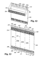
- FIG. 12 is an exploded, perspective view showing the combination of the body and chamber covers for assembly onto the base substrate and spacing layer.
- FIG. 13 is a perspective view of a portion of an assembly including the several layers comprising the biosensor.
- FIG. 14 is a perspective view of a portion of webbing including several detachable biosensors.
- FIG. 15 is a perspective view of a single biosensor separated from the assembled webbing.
- the present invention relates to a system that is useful for assessing an analyte in a sample fluid.
- the system includes devices and methods for evaluating the sample fluid for the target analyte. As more fully described hereafter, the evaluation may range from detecting the presence of the analyte to determining the concentration of the analyte.
- the analyte and the sample fluid may be any for which the test system is appropriate. For purposes of explanation only, a preferred embodiment is described in which the analyte is glucose and the sample fluid is blood or interstitial fluid. However, the present invention clearly is not so limited in scope.
- One component of the system is an electrochemical sensor including a sample-receiving chamber for the sample fluid, and a suitable reagent for producing an electrochemical signal in the presence of the test analyte.
- the sensor preferably comprises a disposable test strip, particularly one having a laminar construction providing an edge opening which communicates with the sample-receiving chamber.
- the reagent is disposed within the sample-receiving chamber in position to provide the electrochemical signal to a working electrode also positioned within the chamber.
- the reagent may contain an enzyme and optionally a mediator.
- the sensor is used in combination with a meter for determination of the analyte in the sample fluid.
- the meter conventionally includes a connection with the electrodes of the sensor and circuitry to evaluate the electrochemical signal corresponding to the concentration of the analyte.
- the meter may also include means for determining that the sample fluid has been received by the sensor, and that the amount of sample fluid is sufficient for testing.
- the meter typically will store and display the results of the analysis, or may alternatively provide the data to a separate device.
- the system can provide either a qualitative or quantitative indication for the analyte.
- the system indicates simply the presence of the analyte in the sample fluid.
- the system may also provide a reading of the quantity or concentration of the analyte in the sample fluid.
- the system is useful for the determination of a wide variety of analytes.
- the test strip for example, is readily adapted for use with any suitable chemistry that can be used to assess the presence of the analyte.
- the system is configured and used for the testing of an analyte in a biological fluid.
- Such analytes may include, for example, glucose, cholesterol, HDL cholesterol, triglycerides, lactates, lactate dehydrogenase, alcohol, uric acid, and 3-hydroxybutric acid (ketone bodies).
- Test methodologies may be variously affected by the presence of interferants in the sample fluid.
- the testing for glucose in a blood sample may be impacted by such factors as oxygen, bilirubin, hematocrit, uric acid, ascorbate, acetaminophen, galactose, maltose, and lipids.
- the present system is adaptable to minimize or eliminate the adverse effects of interferants that may also be present in the sample fluid. These effects may be addressed by appropriate selection of test materials and parameters, such as by the selection of chemistries that are known to be impacted less, or not at all, by possible interferants.
- the system is useful with a wide variety of sample fluids, and is preferably used for the detection of analytes in a biological fluid.
- biological fluid includes any bodily fluid in which the analyte can be measured, for example, interstitial fluid, dermal fluid, sweat, tears, urine, amniotic fluid, spinal fluid and blood.
- blood in the context of the invention includes whole blood and its cell-free components, namely plasma and serum.
- the system is useful in connection with control fluids that are used in conventional fashion to verify the integrity of the system for testing.
- the system is employed for the testing of glucose.
- the sample fluid in this instance may specifically include, for example, fresh capillary blood obtained from the finger tip or approved alternate sites (e.g., forearm, palm, ear lobe, upper arm, calf and thigh), fresh venous blood, and control solutions supplied with or for the system.
- the fluid may be acquired and delivered to the test strip in any fashion.
- a blood sample may be obtained in conventional fashion by incising the skin, such as with a lancet, and then contacting the test strip with fluid that appears at the skin surface.
- the test strip is useful with very small fluid samples. It is therefore a desirable feature of the invention that only a slight incising of the skin is necessary to produce the volume of fluid required for the test, and the pain and other concerns with such method can be minimized or eliminated.
- the test strip includes several basic components.
- the strip comprises a small body defining a chamber in which the sample fluid is received for testing.
- This “sample-receiving chamber” is filled with the sample fluid by suitable means, preferably by capillary action, but also optionally assisted by pressure or vacuum.
- the sample-receiving chamber includes electrodes and chemistry suitable for producing an electrochemical signal indicative of the analyte in the sample fluid.
- the test strip 10 includes a base substrate 12 , a spacing layer 14 and a covering layer 16 comprising body cover 18 and chamber cover 20 .
- the spacing layer 14 includes a void portion 22 to provide a sample-receiving chamber 24 extending between the base substrate 12 and the covering layer 16 .
- the base substrate 12 carries an electrode system 26 including a plurality of electrodes 28 and electrode traces 30 terminating in contact pads 32 .
- the electrodes are defined as those portions of electrode traces 30 that are positioned within the sample-receiving chamber 24 .
- a suitable reagent system 33 overlies at least a portion of the electrodes or electrode pairs 28 within the sample-receiving chamber.
- the body cover 18 and the chamber cover 20 overlying the spacing layer 14 define a slot 34 therebetween, the slot defining a vent opening communicating with the sample-receiving chamber to allow air to escape the chamber as a sample fluid enters the chamber from the edge opening or fluid receiving opening 35 .
- the test strip therefore includes a dosing end 36 and a meter insertion end 38 .
- the shape of the dosing end is typically distinguishable from the meter end so as to aid users.
- strip graphics are preferably used to further improve the intuitiveness of the strip design; e.g., arrow 31 indicates the direction of insertion of the strip into the meter.
- the test strip is a relatively small device that is dimensioned for compactness and ease of storage and use.
- the strip length is on the order of 20 to 50 mm, preferably about 33 to about 38 mm, in length, and 5 to 15 mm, preferably about 7 to about 9 mm, in width.
- the distance from the slot or vent opening 34 to the edge of the meter is sized to provide a “grab area” where there is no blood present, and to guard against blood contamination of the meter contact area, and therefore may be in the range of 5 to 35 preferably ⁇ 13 mm.
- the length of the test strip portion (from the meter insertion end 38 ) that is inserted into the meter is preferably ⁇ 6.0 mm along the long axis of the test strip.
- the preferred laminar construction of the test strip also provides a device that is relatively thin.
- the minimal thickness of the strip allows ready packaging of the strip in appropriate containers that are convenient for the user.
- the overall thickness of the test strip may be about 500 to 525 ⁇ m.
- the thickness of the test strip portion that is inserted into the meter contact may be about 250 ⁇ m.
- the test strip includes a base substrate 12 which comprises an insulating material supporting the electrode system and other components.
- plastics such as vinyl polymers, polyimides, polyesters, and styrenes provide the electrical and structural properties which are required.
- the base substrate can be selected as a flexible polymeric material such as polyester, especially high temperature polyester materials; polyethylene naphthalate (PEN); and polyimide, or mixtures of two or more of these.
- Polyimides are available commercially, for example under the trade name Kapton®, from E.I. duPont de Nemours and Company of Wilmington, Del. (duPont).
- a particularly preferred base substrate material is MELINEX® 329 available from duPont.
- the invention relates to an “electrochemical sensor”, which is a device configured to detect the presence of, and/or measure the concentration of, an analyte by way of electrochemical oxidation and reduction reactions within the sensor. These reactions are transduced to an electrical signal that can be correlated to an amount or concentration of the analyte.
- the test strip therefore includes an electrode system 26 comprising a set of measuring electrodes, e.g., at least a working electrode and a counter electrode, within the sample-receiving chamber.
- the sample-receiving chamber is configured such that sample fluid entering the chamber is placed in electrolytic contact with both the working electrode and the counter electrode. This allows electrical current to flow between the measuring electrodes to effect the electrooxidation or electroreduction of the analyte.
- a “working electrode” is an electrode at which analyte is electrooxidized or electroreduced with or without the agency of a redox mediator.
- the term “counter electrode” refers herein to an electrode that is paired with the working electrode and through which passes an electrochemical current equal in magnitude and opposite in sign to the current passed through the working electrode.
- the term “counter electrode” is meant to include counter electrodes which also function as reference electrodes (i.e., counter/reference electrodes).
- the working and counter electrodes, and the remaining portions of the electrode system may be formed from a variety of materials, as known in the art.
- the electrodes should have a relatively low electrical resistance and should be electrochemically inert over the operating range of the test strip.
- Suitable conductors for the working electrode include gold, palladium, platinum, carbon, titanium, ruthenium dioxide, and indium tin oxide, and iridium, as well as others.
- the counter electrode may be made of the same or different materials, e.g., silver/silver chloride. In a preferred embodiment, the working and counter electrodes are both gold electrodes.
- the electrodes may be applied to the base substrate in any fashion that yields electrodes of adequate conductivity and integrity. Exemplary processes are well known in the art, and include, for example, sputtering, printing, etc.
- gold electrodes are provided by coating the base substrate and then removing selected portions of the coating to yield the electrode system.
- a preferred removal method is laser ablation, and more preferably broad field laser ablation, as disclosed in U.S. patent application Ser. No. 10/601,144, filed on Jun. 20, 2003, entitled Method of Making a Biosensor, the disclosure of which is hereby incorporated by reference.
- Laser ablative techniques typically include ablating a single metallic layer or a multi-layer composition that includes an insulating material and a conductive material, e.g., a metallic-laminate of a metal layer coated on or laminated to an insulating material (discussed below).
- the metallic layer may contain pure metals, alloys, or other materials, which are metallic conductors.
- metals or metallic-like conductors include: aluminum, carbon (such as graphite), cobalt, copper, gallium, gold, indium, iridium, iron, lead, magnesium, mercury (as an amalgam), nickel, niobium, osmium, palladium, platinum, rhenium, rhodium, selenium, silicon (such as highly doped polycrystalline silicon), silver, tantalum, tin, titanium, tungsten, uranium, vanadium, zinc, zirconium, mixtures thereof, and alloys or solid solutions of these materials.
- the materials are selected to be essentially unreactive to biological systems; such materials include: gold, platinum, palladium, iridium, silver, or alloys of these metals or indium tin oxide.
- the metallic layer may be any desired thickness. In a preferred embodiment, the thickness is about 500 nm.
- the electrode system may have a variety of configurations suited to the operation of the test strip and meter.
- the working and counter electrodes preferably are positioned and dimensioned to minimize the volume of sample fluid required to cover them. It is also preferable that the electrodes be configured to maintain a current flux of sufficient magnitude as to be measurable using a relatively inexpensive hand-held meter.
- a preferred embodiment includes a counter electrode which extends around both sides of the working electrode.
- the counter electrode therefore has two elements, one in front of the working electrode and the other behind the working electrode, as the sample fluid enters the sample-receiving chamber. More specifically, the counter electrode includes elements 40 and 42 which extend across the sample-receiving chamber. Each of these elements is about 250 ⁇ m wide.
- the working electrode element 44 has a width of about 250 ⁇ m, and is spaced from each of the two counter electrode elements by about 255 ⁇ m. It will be appreciated that this is only one of a number of configurations for the measuring electrodes.
- the traces 30 and the contact pads 32 may be provided in a variety of fashions consistent with their intended function relative to the test strip.
- These components of the electrode system are preferably composed of the same material as the electrodes, and are preferably applied to the base substrate in the same manner and simultaneously with the application of the electrodes.
- the traces and contact pads are gold, and are formed by laser ablation, particularly as described in U.S. patent application Ser. No. 10/601,144, filed on Jun. 20, 2003, entitled Method of Making a Biosensor, the disclosure of which is hereby incorporated by reference. However, alternate materials and methods of application may be employed.
- the test strip includes a chemical reagent within the sample-receiving chamber for reacting with the test analyte to produce the electrochemical signal that represents the presence of the analyte in the sample fluid.
- the reagent layer can include a variety of active components selected to determine the presence and/or concentration of various analytes.
- the test chemistry is therefore selected in respect to the analyte to be assessed.
- the test strip of the present invention can include one or more enzymes, co-enzymes, and co-factors, which can be selected to determine the presence of glucose in blood.
- the selection of an appropriate chemistry is therefore well within the skill in the art, and further description herein is not required in order to enable one to make and use the test strips with various analytes.
- the reagent chemistry may include a variety of adjuvants to enhance the reagent properties or characteristics.
- the chemistry may include materials to facilitate the placement of the reagent composition onto the test strip and to improve its adherence to the strip, or for increasing the rate of hydration of the reagent composition by the sample fluid.
- the reagent layer can include components selected to enhance the physical properties of the resulting dried reagent layer, and the uptake of a liquid test sample for analysis.
- adjuvant materials to be used with the reagent composition include thickeners, viscosity modulators, film formers, stabilizers, buffers, detergents, gelling agents, fillers, film openers, coloring agents, and agents endowing thixotropy.
- the majority of the chamber is hollow before use.
- the reagent layer be thin and uniform. Since the sample-receiving chamber is very small, less than about 1 ⁇ l, the depth or vertical height of the chamber is small. Consequently, the reagent layer should not occupy the majority of the internal cavity of the chamber. The reagent layer should be sufficiently thin to leave ample space for the test sample in the chamber. Further, the liquid test sample will hydrate or dissolve the thin reagent layer more quickly. As discussed in the above reaction scheme, the mediator and mediator redox products diffuse through and within the reagent layer/gradient to the electrodes.
- the reactive components and intermediates will have a short distance to diffuse through a thin reagent, therefore, diffusion to the electrodes will occur in less time. Additionally, the capture efficiency of mediator redox products at an electrode will be greater for a thin layer of enzyme than a thick layer.
- a thick reagent layer will take more time for the liquid test sample to hydrate or dissolve, and a thick reagent layer will increase the time that it takes for the mediator/mediator redox products to approach the electrodes. This can delay the time to determine the analyte concentration and introduce errors into the determination.
- the reagent layer have a uniform thickness. Thickness inhomogeneity can lead to variability in determining the analyte concentration.
- the reagent layer has a uniform thickness throughout the entire sample receiving chamber. In this preferred embodiment, the reagent layer is not thicker around the perimeter of the sample receiving chamber adjacent the vertical side walls that define the chamber than in the central portion of the chamber. Consequently, the reagent layer does not exhibit a meniscus profile.
- the reagent composition is formulated as a viscous solution that can be deposited in a thin, uniform layer on the base layer.
- the reagent composition includes thickeners and thixotropic agents to enhance the physical properties of the reagent layer.
- the thickeners are selected to provide a thick, liquid matrix having the remaining components homogeneously dispersed therein.
- the thickening and thixotropic agents also inhibit the liquid or semi-paste material from running or spreading over the surface of the base layer after it has been deposited and before it dries. After the reagent composition is deposited, it quickly dries to a readily hydratable matrix.
- the reagent composition is provided to dry rapidly either with air drying or heat drying. After drying, the deposited reagent layer exhibits a thickness of between about 1 micron and about 20 microns. More preferably, the dried reagent layer exhibits a thickness of between about 2 microns and about 6 microns.
- the reagent composition can be deposited on the test strip surface using a variety of coating methods including curtain coating, hot melt coating, rotary screen coating, doctor blade or air knife coating, Meyer bar coating, and reverse roll coating techniques. These techniques are known to those skilled in the art.
- the reagent layer is deposited on the flexible web as a wet composition at a thickness of between about 40 ⁇ m and about 100 ⁇ m. More preferably, the reagent composition is deposited as a wet composition at a thickness of between about 60 ⁇ m and about 80 ⁇ m.
- the composition may be applied as a uniformly thin layer of a reagent directly on top of the measuring electrodes and along the length of a web of multiple test strips, as a continuous narrow band.
- the narrow band has a width of between about 7 mm and 8 mm and a dry thickness of between about 3 um and about 20 um.
- the composition may also be applied onto other electrodes that may reside in the sample-receiving chamber, depending on the desired functionality of such extraneous electrodes.
- the test strip includes a spacing layer 14 which overlies the base substrate and which in part defines the sample-receiving chamber.
- the spacing layer 14 includes a void portion 22 substantially defining the height and the perimeter of the sample-receiving chamber 24 .
- the void portion 22 is conveniently placed to have an edge opening whereby the sample fluid is contacted with the edge opening to enter into the sample-receiving chamber.
- the edge opening preferably is located at the end of the test strip, although it will be appreciated that placement on a side edge is also useful.
- the spacing layer 14 may be made of any material useful for fabrication with the test strip. Because the spacing layer partially defines the height of the sample-receiving chamber, the material should have sufficient strength at thicknesses appropriate to the desired height of the chamber. Another function of the spacing layer is to protect the electrode traces that extend along the upper surface of base substrate 12 . The material should also be readily attached to the base substrate and the cover materials, either by heat-sensitive or pressure-sensitive adhesives, or other means such as heat or laser welding. Examples of suitable materials include a 100 ⁇ m PET, PEN foil coated or combined with adhesives such as ARCare 90132 from Adhesives Research Inc.
- a covering layer 16 is received over and attached to the spacing layer 14 .
- One function of the covering layer is to form the top surface of the sample-receiving chamber. Another function is the provision of a hydrophilic surface to aid in acquisition of the test sample.
- the covering layer 16 preferably defines a vent opening 34 that allows air to escape from the interior of the chamber as the sample fluid enters and moves into the sample-receiving chamber.
- the covering layer can be formed as a unitary piece with slot 34 ′ formed as a recess on the underside thereof, as shown in FIG. 1B .
- slot 34 ′ would be substantially straight as shown and extend across the entire width of the test strip, such that air would vent from the sample receiving chamber 24 to the vent openings formed on opposite lateral sides of the test strip.
- the slot could comprise a groove or recess that only extends from the chamber 24 to one side of the test strip, although such configuration is not preferred for mass production purposes.
- FIG. 1C Another alternate embodiment is shown in FIG. 1C , in which chamber cover 20 “overlaps” body cover 18 .
- a small end portion 37 of cover layer 20 is bent upwardly and extends across the edge of body cover 18 .
- a slot 34 ′′ is thereby formed having roughly a triangular shaped cross section as can be seen at the edges of the strip, at which there are triangular shaped openings that allow air to escape.
- the precise placement of the chamber cover 20 with respect to body cover 18 along the lengthwise direction of the strip is not critically important. That is, the amount of chamber covering material overlapping body cover 18 can vary without affecting the dimensions or placement of the slot. This has advantages in manufacturing, as will become apparent with reference to the discussion below.
- body cover 18 and chamber cover 20 comprise two separate members for ease in fabrication and in forming the vent opening.
- Body cover 18 and chamber cover 20 are both disposed in substantially the same horizontal plane.
- the chamber cover 20 substantially covers the void portion 22 of the spacing layer, and forms the top of the sample-receiving chamber.
- the chamber cover preferably includes a hydrophilic coating or treatment 21 on its underside, as described in more detail below.
- the body cover and the chamber cover are positioned end to end in the lengthwise direction along the test strip and include slot 34 therebetween as shown in FIG. 1A .
- the slot is located adjacent the interior end of the void portion 22 of the spacing layer, and in the preferred embodiment in FIG. 1A , forms a small gap that spaces chamber cover 20 from body cover 18 .
- the gap constitutes the vent opening 34 in communication with the sample-receiving chamber.
- Slot 34 is substantially straight and extends across the width of test strip 10 .
- Slot 34 is oriented substantially perpendicular to the longitudinal or lengthwise axis of test strip 10 .
- Sample fluid entering the sample-receiving chamber will expel air through the vent opening defined by slot 34 . If the slot be formed as a gap, some or most of the air expelled will exit from the top of the test strip.
- the slot is located at a position relative to the sample-receiving chamber that is interior of the location of the electrode system 26 .
- Sample fluid entering the sample-receiving chamber will progress as far as the vent opening, but no further.
- the slot provides a visual indication of a “fill-line,” as described herein.
- the placement of the vent opening therefore assures that sufficient sample fluid can be received to fully cover the electrode system.
- the placement of the vent opening will inhibit continued wicking of the sample fluid beyond the region of the electrode system.
- the formation of the slot and vent opening by the spacing of the body cover and the chamber cover is further advantageous because it avoids the need to otherwise form an aperture in the covering layer or base layer.
- it has been an approach to form the vent opening by punching a hole in either the top or bottom film forming the sample-receiving chamber, which presents fabrication issues because of the need to precisely locate the hole relative to the sample-receiving chamber. While this approach is also suitable for a test strip, the preferred design described herein avoids the need to align the vent opening laterally relative to the test strip. Further, the present design is well suited to mass production of the test strips by roll processing techniques, as described hereafter.
- the vent construction may be made in a manner to inhibit the wicking of sample fluid laterally along the slot beyond the middle area that overlies the sample receiving chamber 24 .
- the body cover is preferably secured to the spacing layer by means of an adhesive 46 , as shown in FIG. 3 .
- the use of a hydrophobic adhesive will inhibit blood, interstitial fluid, and other aqueous liquids from moving along the laterally-extending slot by capillary action.
- the entire body cover, or portions adjacent to the vent opening may also be hydrophobic to inhibit wicking Materials and methods for providing hydrophobic properties for a surface of a material are well known in the art.
- the chamber cover may be secured to the spacing layer by the same or different adhesive than adhesive 46 , as explained below.
- Adhesive 49 secures the spacing layer to the base substrate 12 .
- Adhesive 46 as well as adhesive 49 and the material for spacing layer 14 , are all formed of hydrophobic material in the illustrated embodiment. As such the vertical walls of the capillary chamber formed in strip 10 are hydrophobic. By contrast, the floor of the chamber is covered with a hydrophilic reagent and the underside of layer 20 is coated with a hydrophilic coating 21 ( FIG. 2 ). In other words, the horizontal surfaces in the capillary are hydrophilic while the vertical surfaces are hydrophobic. This has been found to promote good wicking of the sample into the capillary chamber, yet prevents unwanted migration of the sample laterally from the chamber, e.g., between the spacer layer and the base substrate.
- the body cover and chamber cover may be made of any materials useful for fabrication with the test strip.
- the materials for the body cover and the chamber cover may be the same or different.
- the materials should be readily attached to the spacing layer, either by heat-sensitive or pressure-sensitive adhesives, or other means such as heat or laser welding.
- suitable materials for both the chamber cover and body cover include approximately 127 ⁇ m thick foil of PET.
- the chamber cover preferably includes a hydrophilic layer 21 as disclosed in WO 02/085185, ARFlow® 90191 from Adhesives Research Inc.
- the covering layer 16 may also be used to facilitate viewing of the sample fluid as it enters the sample-receiving chamber. This is accomplished by providing a contrast in color or shading between the chamber and the surrounding area.
- the portion of the spacing layer 14 that surrounds void 22 is provided with a color that contrasts with the color of the bottom of the sample-receiving chamber, e.g., the color of the chemical reagent layer positioned at the bottom of the chamber.
- This contrasting color may be provided, for example, by the application of an ink or other coloring agent to the portions of the spacing layer adjacent the sample-receiving chamber.
- a colored section 23 of layer 14 is pictured in FIG. 2 .
- the chamber cover 20 is then provided as a transparent or translucent material that allows the user to view the chamber and the adjacent spacing layer. As sample fluid enters from the edge of the test strip, the user is able to observe its progress as it moves by capillary action toward the vent opening. This type of feature is further described in U.S. Pat. No. 5,997,817, issued to Crismore et al. on Dec. 7, 1999, and is hereby incorporated by reference.
- the sample-receiving chamber formed by the base substrate, spacing layer and chamber cover essentially comprises several sections into which the sample fluid will travel.
- a first, entry section 48 extends from the edge opening to the area of the measuring electrode system.
- a second, test section 50 extends through the area of the electrode system.
- a third section 52 extends from the measuring electrode system to the vent opening. It will be appreciated that the testing of the sample fluid occurs in the area of the electrode system in the test section. However, the sample fluid will also fill the other sections of the chamber in the course of filling the test strip.
- the height and width of the sample-receiving chamber are selected based upon a variety of considerations, including the fluid being tested and the analyte at issue.
- the chamber dimensions are preferably sized to promote capillary flow of the test fluid into the chamber.
- Preferred chamber heights for use with blood for example, are from about 50 ⁇ m to about 200 ⁇ m, and most preferably from 120 to 180 ⁇ m. In a preferred embodiment, the chamber height is about 150 ⁇ m.
- the width of the chamber may similarly be selected to match a desired sample fluid and analyte.
- the chamber should be sufficiently wide to expose a desired amount of the working and counter electrodes, and should be narrow enough to avoid the requirement of an undue amount of sample fluid for testing.
- the width of the sample-receiving chamber and the width of the working electrode define the area of the working electrode. The area represents a further design consideration as it relates to signal amplitude and instrumentation design.
- the sample-receiving chamber is preferably provided as having a minimal volume, in order to reduce the amount of sample fluid needed for conducting a test.
- the overall sample-receiving chamber including all of the three sections extending from the edge opening to the vent opening, has a total volume that can be considered to be a factor of the area of the chamber from the edge to the vent, and the height of the chamber from the base substrate to the chamber cover 20 .
- the “net chamber volume” comprises the volume of sample fluid required to fill this space.
- the net chamber volume of the sample-receiving chamber will be the equivalent of the total chamber volume less the volume occupied by the electrodes, the reagent, and perhaps other items such as a sorbent material, if included.
- the volume of the overall sample-receiving chamber is comprised of the volumes attributable to the three sections of the chamber.
- Each of the sections is generally sized to be as small as practical for the operation of the test strip. However, there are considerations, and possibly other functions, that will impact on the size of each section.
- the chamber volumes are a factor of both height and area.
- the height is a result of the thickness of the spacing layer and the thickness of the adhesives used to secure the spacing layer to the other layers.
- the base substrate and the chamber cover are attached to opposite sides of the spacing layer.
- One method of attachment is the heat or laser sealing of the materials. It is preferred, however, to attach these layers by the use of suitable adhesives, such as heat-sensitive or pressure-sensitive adhesives.
- suitable adhesives such as heat-sensitive or pressure-sensitive adhesives.
- the height of the sample-receiving chamber i.e., the distance between the facing surfaces of the bottom substrate and the chamber cover, will be impacted by the thickness of the adhesive layers.
- chamber 24 is bounded on its bottom side by reagent layer 33 and its top side by chamber cover 20 .
- adhesive layers 46 and 49 as well as spacing layer 14 define the total height of chamber 24 .
- the reagent layer 33 extends between base substrate 12 and spacing layer 14 and indeed extends the entire width of the test strip, as described below.
- the height of the chamber may therefore also be increased due to the presence of the reagent layer underlying the spacing layer.
- the adhesive may combine with the test reagent, at least to an extent that causes the adhesive to fill somewhat into and around the reagent.
- the heights of the reagent and adhesive layers therefore are not necessarily additive in the final test strip. Rather, the height of the resulting space between the base substrate and the spacing layer is somewhat less than the combination of the heights of the separate reagent and adhesive layers prior to lamination.
- the combination of the adhesive and the reagent advantageously helps to create a seal along the edge of the sample-receiving chamber. This inhibits sample fluid from wicking into the reagent material present in the space between the base substrate and the spacing layer in the time frame necessary for performing a test.
- the first entry section is available to receive the sample fluid and direct it to the measuring electrodes.
- This section can be quite small in size, and may comprise only a short segment of the chamber.
- the length of this section is preferably less than 1200 ⁇ m.
- the second testing section includes the test or measuring electrodes, and is also sized to require a minimal volume of sample fluid.
- a primary factor controlling the size of this second section will be the type, number, size, signal strength, and configuration of the measuring electrodes.
- the length of this section is preferably about 1260 ⁇ m.
- a preferred volume is about 0.265 ⁇ L, based on a capillary height of 0.15 mm, and a capillary width of 1.4 mm.
- the sample fluid moves past the measuring electrodes and into the third section.
- This provides assurance, and preferably allows for specific confirmation, that the measuring electrodes are properly wetted.
- This confirmation may be by visual observation by the user, or by automatic detecting means.
- dose sufficiency electrodes may be placed in this section to detect when the sample fluid has progressed into this section to a point that the wetting of the measuring electrodes is assured. This can be used as a trigger for initiating the application of the potential to the electrodes.
- the length of this section is preferably 50 to 500 ⁇ m, and more preferably 255 to 400 ⁇ m.
- the volume is preferably 0.01 to 0.1 ⁇ L, and more preferably 0.05 to 0.08 ⁇ L.
- the overall net chamber volume of the sample-receiving chamber is less than about 1 ⁇ L, and is more preferably less than about 0.5 ⁇ l. Desirable ranges for the net chamber volume of the sample-receiving chamber include volumes from about 0.15 to about 1.4 ⁇ L, more preferably from about 0.4 to about 0.7 ⁇ l.
- the sample chamber may be otherwise empty, which is preferred, or may alternatively include a sorbent material.
- Suitable sorbent materials include polyester, nylon, cellulose, and cellulose derivatives such as nitrocellulose.
- a sorbent material could be included to facilitate the uptake of the sample fluid by assisting in wicking the fluid into the chamber. The use of a sorbent material would also serve to further reduce the void volume of the sample-receiving chamber for reception of the sample fluid.
- the preferred method of filling the sample chamber is by capillary action.
- the filling of the test strip can be augmented by other means, such as applying a pressure on the sample fluid to push it into the sample chamber, and/or creating a vacuum on the sample chamber to pull the sample fluid into the chamber.
- any or all of the surfaces defining the chamber may be selected or treated to improve hydrophilicity.
- Such treatment may comprise the use of known hydrophilic materials, application of a hydrophilic material onto the surface, or treatment of the surfaces to increase hydrophilicity, as described below.
- the reagent composition may be formulated to be readily hydrated and to encourage filling of the sample-receiving chamber.
- a sorbent may also be used.
- the electrochemical sensor is operated by applying a suitable potential or series of potentials across the working and counter electrodes, and across the dose sufficiency electrodes.
- a mediator When a mediator is used, the magnitude of the required potential across the working and counter electrodes will be dependent on the redox mediator.
- the potential at the electrode where the analyte is electrolyzed is typically large enough to drive the electrochemical reaction to or near completion, but the magnitude of the potential is preferably not large enough to induce significant electrochemical reaction of interferants.
- an applied potential difference typically is between about +100 mV and about +550 mV when using a DC potential. When using AC potentials these can be typically be 5 to 100 mV RMS.
- a potential may be applied before or after the sample begins to enter the sample-receiving chamber. However, a potential is preferably applied after the sample has entered the chamber, and more preferably after it has been determined that there is a sufficient amount of sample in the sample-receiving chamber for conducting a test.
- the timing of the application of a potential may be triggered in a variety of fashions, including visual observation by the user, a time delay following sampling of the fluid to the test strip, or upon electrical or other automated detection of a sufficient amount of sample fluid in the chamber.
- the visual and electrical alternatives also may act as redundant failsafes to assure proper operation of the device.
- the test strip and system utilize separate detecting means, such as dose sufficiency electrodes, for determining when the fluid sample has sufficiently filled the chamber.
- an electrical current will flow between the working electrode and the counter electrode.
- the current can be a result of the electrolysis of the analyte in the sample fluid when a potential of sufficient magnitude is applied.
- electrochemical reaction occurs via the redox mediator, generally as previously described.
- the current is produced not necessarily by electrolysis, but by ionic motion and response of the dielectric in the sample chamber.
- a test may be applied to the test strip after dosing to confirm that a control solution, and even that the correct control solution, has been administered.
- the control solutions aid the user in confirming that the entire system is functioning within design specifications, and that the test strips have not been stored improperly or otherwise mistreated. Acceptable strips will recover values within specified tolerance ranges for the particular strip lot being tested. The tolerance ranges in question will be published for each strip lot on the container label.
- the senor comprises a multi-layered, laminate test strip 10 .
- the laminate includes a base substrate 12 , a spacing layer 14 , and a covering layer 16 .
- These components may be assembled in various ways.
- the components may be assembled by use of adhesives, heat sealing, laser welding, and a variety of other suitable techniques appropriate for securing the adjacent materials.
- the test strips are preferably assembled in a large number on a single sheet or web, and the strips are thereafter separated for storage and use.
- the laminate test strip may be assembled sequentially by successively laying down one layer at a time.
- the test strip can be prepared by assembling and processing individual components or layers, which are then laminated together to provide the functional test strip.
- two or more basic components of the test strip are prepared simultaneously. Then in one or a series of assembly or laminating steps, the basic components are combined to produce the test strip, which may or may not require further processing.
- the test strip is assembled from three basic components: a metallized substrate preferably with a reagent layer coated on metallic electrodes defined on the substrate, a spacing layer having a cavity preformed therein, and one or more top or cover layers.
- the characteristics of the reagent layer can have a significant impact on the operation of the test strip, particularly in view of hydration and mixing characteristics.
- the reproducibility of the quantity, location, thickness and other properties of the reagent layer is therefore important. It is therefore desirable for the composition to include materials which specifically enhance the physical characteristics, such as the uniformity and flatness, of the applied layer.
- the test strip includes a unique manner of incorporating the reagent.
- the reagent is placed in the sample-receiving chamber at least on the working electrode, and preferably also on the counter electrode.
- the reagent may be applied to the test strip in a variety of fashions as is well understood in the art.
- the reagent composition is applied as a thin coating over the electrodes supported on the base substrate.
- the reagent is placed onto the base substrate in a manner that positions the reagent composition between the base substrate and the spacing layer.
- This manner of application helps to make the reagent layer more flat and uniform in thickness.
- a procedure of the prior art has been to first prepare the reaction well or cavity, and to then fill the reagent into the well.
- this can result in a more uneven reagent layer due to phenomena such as the formation of a meniscus at the perimeter of the well.
- This in turn can cause the reagent to have a different thickness adjacent to the side walls of the reaction well than in the interior portion, which can cause inconsistency in the filling of the chamber, prolonged dissolution intervals, and inconsistent mixing of the reagent with the sample fluid, and the ultimate test results.
- By placing the reagent onto the base substrate before the spacing layer is added there is no meniscus effect to disrupt the even layering of the reagent as it dries on the base substrate.
- this method of application facilitates the mass production of the test strips.
- the test strip 10 is shown as including a reagent layer 33 that extends between the bottom substrate 12 and the spacing layer 14 . More particularly, the reagent forms a layer 33 which covers both the top surface of the bottom substrate 12 and the electrodes 28 .
- the reagent covers at least the working electrode, and preferably also the counter electrode.
- the reagent layer extends the full width of the test strip.
- the reagent layer also preferably extends from the end edge to the vent opening. The reagent layer thus extends under the spacing layer and is sandwiched between the spacing layer and the base substrate.
- the reagent composition is applied to the bottom or base substrate in any suitable fashion that provides a desired and uniform layer which will ultimately extend under the spacing layer.
- the reagent is preferably applied in a continuous coating directly onto the bottom substrate, and onto the electrodes received thereon.
- the reagent composition is most preferably applied in the course of producing a large quantity of test strips on a webbing of material. In this manner, the reagent may be applied in the way of a continuous stripe of material that extends over a substrate roll that is later separated into individual test strips.
- the reagent composition is allowed to dry or otherwise set up and the spacing layer is applied thereover.
- a preferred manner of securing the spacing layer to the bottom substrate is the use of an adhesive.
- the adhesive will sufficiently engage with the reagent composition as to help to seal the space between the bottom substrate and the spacing layer.
- the adhesives preferably placed on the spacing layer, which is laminated onto the base substrate. The adhesive thereby contacts the portion of the reagent which extends under the spacing layer.
- spacing layer 14 is formed from Melinex® material with adhesives on both sides thereof, it is also possible to form spacing layer 14 as a continuous adhesive material, such as a double-sided tape.
- the analyte is glucose.
- the active components of the reagent composition will typically include an oxidoreductase, such as an enzyme for glucose; optionally a co-enzyme or co-factor; and a redox mediator. These components are typically dissolved or suspended in a matrix.
- the liquid test sample hydrates or dissolves the matrix, and the analyte diffuses through the matrix to react with one or more of the active components.
- the enzyme oxidizes the glucose in the test sample to gluconolactone and/or gluconic acid.
- the mediator reacts with or oxidizes the reduced enzyme, and consequently the mediator is reduced in the process.
- the reduced mediator can be detected at one of the electrodes on the test strip.
- a test sample containing glucose reacts with an enzyme such as Glucose-Di-Oxidoreductase (Gluc-Dor), and optionally a co-enzyme or cofactor such as pyrrolo-quinoline-quinone (PQQ), in the presence of a redox mediator.
- an enzyme such as Glucose-Di-Oxidoreductase (Gluc-Dor)
- PQQ pyrrolo-quinoline-quinone
- the mediator may include, for example, benzoquinone, transition metal complexes, e.g., potassium ferricyanide, osmium derivatives (e.g., osmium bipyridyl complexes such as described in WO 98/35225) and nitrosoanaline derivatives (see U.S. Pat. No. 5,286,362).
- transition metal complexes e.g., potassium ferricyanide
- osmium derivatives e.g., osmium bipyridyl complexes such as described in WO 98/35225
- nitrosoanaline derivatives see U.S. Pat. No. 5,286,362
- Equation 1 A representation of the reaction sequences for this reaction system using a nitrosoaniline derivative is provided below in Equation 1.
- the nitrosoaniline derivative o-methoxy-[N,N-bis-(2-hydroxyethyl)]-p-nitrosoaniline
- Reaction of Gluc-Dor with glucose in the test sample yields gluconolactone and the reduced form of Gluc-Dor (Gluc-Dor.2H + ).
- the reduced form of Gluc-Dor (Gluc-Dor.2H + ) reacts rapidly with the nitrosoaniline derivative, which is reduced and which regenerates Gluc-Dor.
- the reduced nitrosoaniline derivative then undergoes hydrolysis to form quinonediimine (QD).
- Gluc-Dor reacts with glucose to yield another molecule of Gluc-Dor.2H + and gluconolactone.
- the Gluc-Dor.2H + reacts with (is oxidized by) quinonediimine to regenerate Gluc-Dor, and produces a phenylene diamine derivative (PD).
- PD is then oxidized at the working electrode to produce a current related to glucose concentration. Additionally, at the counter electrode QD can be reduced to PD.
- the characteristics of the reagent layer can have a significant impact on the operation of the test strip, particularly in view of hydration and mixing characteristics.
- the control and reproducibility of the quantity, location, width, thickness, and other properties of the reagent layer become more important as the chamber volume decreases and test time diminishes. It is therefore desirable for the composition to include materials that specifically enhance the physical characteristics, such as the uniformity and flatness, of the applied layer. Additionally, the method of application can impact the physical characteristics, control, and reproducibility of the reagent layer.
- the reagent composition can therefore also include a variety of adjuvants to enhance the reagent properties or characteristics.
- the composition may include adjunct materials to facilitate the placement of the reagent composition onto the test strip and to improve its adherence to the strip.
- the composition can also include materials to increase its rate of hydration and/or its increase its influence on the capillary action to fill the chamber with the test sample.
- addjunct materials to be used with the reagent composition include thickeners, viscosity modulators, film formers, stabilizers, buffers, detergents, gelling agents, fillers, film opening agents, coloring agents, and agents endowing thixotropy.
- the adjuvant materials or components can impact the application, reproducibility and physical properties of the reagent layer.
- the adjunct materials can include one or more of the following:
- Thickeners may include, for example, (1) starches, gums (e.g., pectin, guar gum, locust bean (carob seed) gum, konjac gum, xanthan gum, alginates, and agar), casein, gelatin, and phycocolloids; (2) cellulose and semi-synthetic cellulose derivatives (carboxymethyl-cellulose, methyl cellulose, hydroxymethylcellulose, hydroxyethylcellulose, methylhydroxyethylcellulose); (3) polyvinyl alcohol and carboxy-vinylates; and (4) bentonite, silicates, and colloidal silica.
- gums e.g., pectin, guar gum, locust bean (carob seed) gum, konjac gum, xanthan gum, alginates, and agar
- casein gelatin
- phycocolloids e.g., cellulose and semi-synthetic cellulose derivatives (carboxymethyl-cellulose, methyl cellulose
- Preferred thickeners include a combination of a xanthan gum sold under the trade name Keltrol F by CP Kelco US, Inc., and carboxylmethyl cellulose sold under the trade name AQUALON® CMC 7F PH by Hercules Inc., Aqualon Division.
- Film forming and thixotropic agents useful in the reagent composition include polymers and silica.
- Preferred thixotropic agents include silica sold under the trade name Kieselchure Sipernate FK 320 DS by Degussa AG.
- Preferred film forming agents include polyvinylpyrrolidone, sold under the trademark polyvinylpyrrolidon Kollidon 25, by BASF, and polyvinyl propionate dispersion.
- Stabilizers for the enzyme in the reagent can be selected from sacchhrides and mono- or di-fatty acid salts.
- Preferred stabilizers include trehalose sold under the trade name D-(+)-Trehalose dihydrate by Sigma Chemical Co. and sodium succinate.
- Detergents can be selected from water-soluble soaps, as well as water-soluble synthetic surface-active compounds such as alkali, earth alkali or optionally substituted ammonium salts of higher fatty acids, e.g., oleic or stearic acid, mixtures of natural fatty acids, for example, from coconut or tallow oil, fatty sulphates, esters of sulphonic acids, salts of alkyl sulphonic acids taurine salts of fatty acids, fatty acid amides, and ester amides.
- water-soluble soaps as well as water-soluble synthetic surface-active compounds such as alkali, earth alkali or optionally substituted ammonium salts of higher fatty acids, e.g., oleic or stearic acid, mixtures of natural fatty acids, for example, from coconut or tallow oil, fatty sulphates, esters of sulphonic acids, salts of alkyl sulphonic acids taurine salts of fatty acids,
- Preferred detergents for the present invention include an ester amide, n-octanoyl-N-methylglucamide, sold under the trade name Mega-8 by Dojindo Molecular Technologies, Inc., and a fatty acid salt, N-methyl oleyl taurate sodium salt, sold under the trade name Geropon T77 by Rhodia HPCII (Home, Personal Care and Industrial Ingredients).
- a mediator for use in the reagent composition can be selected as any chemical species (generally electroactive) which can participate in a reaction scheme involving an enzyme, an analyte, and optionally a cofactor, and reaction products thereof, to produce a detectable electroactive reaction product.
- participation of the mediator in the reaction involves a change in its oxidation state (e.g., a reduction), upon interaction with any one of the analyte, the enzyme, or a cofactor, or a species that is a reaction product of one of these (e.g., a cofactor reacted to a different oxidation state).
- a variety of mediators exhibit suitable electrochemical behavior.
- a mediator can preferably also be stable in its oxidized form, may optionally exhibit reversible redox electrochemistry, can preferably exhibit good solubility in aqueous solutions, and preferably reacts rapidly to produce an electroactive reaction product.
- suitable mediators include benzoquinone, meldola blue, other transition metal complexes, potassium ferricyanide, osmium derivatives (see WO 98/35225) and nitrosoanaline-based mediators (see U.S. Pat. No. 5,286,362).
- the reagent composition utilizes a nitrosoaniline-based chemistry.
- Preferred mediators include N-(2-hydroxyethyl)-N′-p-nitrosophenyl-piperazine, N,N-bis-(2-hydroxyethyl)-p-nitrosoaniline, o-methoxy-[N,N-bis-(2-hydroxyethyl)]-p-nitrosoaniline, p-hydroxynitrosobenzene, N-methyl-N′-(4-nitrosophenyl)-piperazine, p-quinone dioxime, N,N-dimethyl-p-nitrosoaniline, N,N-diethyl-p-nitrosoaniline, N-(4-nitrosophenyl)-morpholine, N-benzyl-N-(5′-carboxypentyl)-p-nitrosoaniline, N,N-dimethyl-4-nitroso-1-naphthylamine, N,N,3-trimethyl-4-nitrosoaniline, N-(2-hydroxye
- mediators according to the present invention include N,N-bis-(2-hydroxyethyl)-p-nitrosoaniline, o-methoxy-[N,N-bis-(2-hydroxyethyl)]-p-nitrosoaniline, and N-(2-hydroxyethyl)-N-(2-(2-hydroxyethoxy)-ethyl)-4-nitrosoaniline.
- the components of the reagent composition are admixed with water to provide a homogeneous, viscous suspension.
- the order of addition is not critical for the invention.
- a sufficient amount of the buffer solution is added to maintain the reagent composition at a pH of about 7.
- the selected components are pre-mixed with water to provide a variety of stock solutions that can be combined to yield the final reagent composition.
- a buffer solution can be prepared by combining the phosphate salts and, optionally, the sodium succinate.
- Other stock solutions include: the thickening agents, i.e., Keltrol F and the carboxymethyl cellulose; the surfactants, i.e., Geropon T77 and Mega 8; the enzyme and co-enzyme or cofactor; and the mediator.
- the following provides an example of the preparation of a reagent composition.
- the reagent composition can be prepared by first preparing the following stock solutions:
- Carboxymethylcellulose (CMC) Solution Amount (gm) H 2 O 1334.76 Na CMC 1 27.24 1 Na CMC is a sodium salt of carboxymethyl cellulose sold by Hercules Inc., Aqualon Division
- PVP Polyvinylpyrrolidone
- Buffer Solution 226.03 Mega 8 1 13.23
- Geropon T77 2 1.405 PVP 3 89.33 1 .
- Mega 8 is n-octanoyl-N-methylglucamide sold by Dojindo Molecular Technologies Inc. 2 .
- Geropon T77 is N-methyl oleyl taurate sodium salt sold by Rhodia HPCII. 3 .
- PVP is Polyvinylpyrrolidone K25 sold by BASF.
- Trehalose Solution 1 Amount (gm) H 2 O 36.4 Trehalose 23.6 1 This trehalose solution is used only in preparing the “Enzyme Solution” listed below.
- the buffer solution, Keltrol F solution, CMC solution, and the Silica suspension were prepared a day before. These solutions can then be combined as listed below to prepare the reagent composition.
- FIGS. 5 and 5A present a flow chart illustrating a preferred process 100 for preparing a test strip useful in accordance with the present invention.
- Process 100 begins in the central process line 101 at stage 102 with selection of a film material for the base layer or base substrate.
- the film is provided as a continuous roll having a width and length suitable for fabricating a large number of test strips.
- the processed film can be subdivided to provide a single strip or web having a width that approximates the length of the test strip and includes a series of test strips, or can be die cut to provide individual test sensors.
- the film proceeds to stage 104 where it is pretreated to receive a metal coating and is coated with the metal in one continuous process.
- the pretreatment can be used to clean or modify the surface to provide a uniform coating thickness and better adhesion of the subsequent metallized layer.
- the pretreatment can include subjecting the film to corona discharge or Argon plasma. Immediately after this pre-treatment, a uniform conductive coating is applied to the film as shown at 106 .
- suitable substrates with metal coatings can be obtained commercially.
- the metallic layer may contain pure metals, alloys, or other materials, which are metallic conductors.
- metals or metallic-like conductors include: aluminum, carbon (such as graphite), cobalt, copper, gallium, gold, indium, iridium, iron, lead, magnesium, mercury (as an amalgam), nickel, niobium, osmium, palladium, platinum, rhenium, rhodium, selenium, silicon (such as highly doped polycrystalline silicon), silver, tantalum, tin, titanium, tungsten, uranium, vanadium, zinc, zirconium, mixtures thereof, and alloys or solid solutions of these materials.
- the materials are selected to be essentially unreactive to biological systems; such materials include: gold, platinum, palladium, iridium, or alloys of these metals.
- the metallic layer may be any desired thickness.
- the conductive coating is preferably a metal layer that is applied by a variety of methods, including but not limited to sputtering, physical vapor deposition (PVD), plasma assisted vapor deposition (PAVD), chemical vapor deposition (CVD), electron beam physical vapor deposition (EBPVD), and/or metal-organic chemical vapor deposition (MOCVD).
- Vapor deposition is typically performed under vacuum. These techniques are well known in the art and can be used to selectively provide a uniformly thin coating of metal onto a substrate. The resulting metallized film can be inspected to ensure that the metal coating is uniform and free of material defects.
- stage 108 The roll of metallized film next encounters stage 108 where it is subdivided and/or sized to provide webs having a width that approximates the final length of an individual test strip.
- the slicing can be accomplished using fixed-knife slitting equipment well-known in the art.
- a single web proceeds to stage 110 for patterning the electrodes, traces, and contacts or pads.
- the electrodes, traces, and contact pads are formed by removing metal from the surface of the web strip. The excess metal can be removed using a variety of techniques well known in the art.
- one or more indexing or registration marks can be formed either on a first edge proximate to the electrodes, the opposite second edge proximate to the electrode pad, on both edges or anywhere in between. The indexing marks, particularly those at an edge, can be used in subsequent operations to align layered components prefabricated in separate operations.
- the metal is laser ablated to eliminate undesired portions of the metal and leave the desired electrical components.
- selected areas are laser etched simultaneously, in a “broad field”, as opposed to using linear movement of a focused laser beam.
- This broad field laser ablation method provides a precise metal pattern rapidly and at reduced cost as compared to other approaches. Corona treatment of the patterned substrate is then conducted at stage 111 .
- a reagent layer is deposited onto the electrodes.
- the reagent layer is deposited as a continuous elongate stripe extending adjacent or close to the first edge, and overlying the measuring electrodes formed on the patterned web.
- the reagent is consequently located across the full width of the test strip, including the area laterally outside of the sample-receiving chamber and between the base substrate and the spacing layer. Also as noted, this will facilitate the drying of the reagent without discontinuities, edge effects, or other variances that would detract from providing a thin, flat, uniform reagent layer within the sample-receiving chamber.
- the reagent includes a combination of components, and is formulated to dry rapidly with minimal or no running after deposition.
- This stripe may be applied in any suitable fashion which provides the desired extent and uniformity of thickness, precision of the stripe edge, homogeneity, and the like. Preferred methods are capable of applying the desired coating at relatively high speed and high batch size. Suitable methods of application are well known in the art and therefore are not detailed herein.
- a material is selected to prepare a spacing layer for laminating on top of the reagent coated web prepared at stage 112 .
- the base film for the substrate can be selected from a variety of materials.
- the spacing layer material similar to the base layer, can be provided as an elongate roll which can be conveniently processed rapidly and with high efficiency.
- Preferred materials include a polyester film sold under the trade name MELINEX® by DuPont. Other materials suitable for use in the present invention could include PEN.
- the spacing layer material has a thickness specifically selected to provide a desired chamber depth (or height) in each of the test strips when combined with the thicknesses of any joining layers that are used to laminate the spacer to the other strip components.
- the spacing layer is selected to have a thickness between about 75 ⁇ m and about 150 ⁇ m, more preferably from about 100 ⁇ m to about 125 ⁇ m.
- the spacing layer can be formed of a double-sided adhesive.
- the spacing layer is preferably formed as a continuous film having a series of gaps that will align with the electrodes on the bottom substrate webbing.
- the manner of joining the spacing layer and bottom substrate will impact the method for preparing the spacing layer. For example, if the spacing layer is to be heat welded to the bottom substrate, then the spacing layer may simply be die cut to provide the appropriately spaced chamber gaps.
- a preferred method is the use of thin, non-interfering adhesives that join the adjacent layers.
- a spacing layer is prepared for combination with the previously described substrate webbing as set forth hereafter.
- the spacing layer film is prepared having the desired width for combination with the remainder of the test strip components.
- the spacing layer film may include an opaque portion, e.g., a section 23 of it is printed blue or another color for use in visualizing the sample-receiving chamber, as described elsewhere.
- the spacing layer film is laminated on the bottom side with a combination adhesive and release liner, and on the top side with a similar combination adhesive and liner.
- two transfer adhesives are laminated to the spacing layer material: the first transfer adhesive is laminated to the top surface of the spacing layer, and the second transfer adhesive is laminated to the bottom surface of the spacing layer.
- the transfer adhesives are the same adhesive; however, in alternative embodiments, the first and second transfer adhesives can be different from each other.
- the transfer adhesives are selected from commonly used and known adhesives, including pressure sensitive adhesives. Preferred adhesives exhibit sufficient hydrophobicity to prevent or inhibit the test sample in the chamber from wicking out between the spacing layer and the reagent layer or base substrate.
- An example of a suitable sensitive adhesive is ARCare 90132 from Adhesives Research Inc.
- the adhesives are provided with a release liner to prevent premature adhesion of the spacing layer during processing.
- the release liners are disposed on the exterior surface of the first and second transfer adhesives, facing outward from the spacing layer material.
- the spacing layer with the adhesive release liners on the top and bottom surfaces progresses to stage 120 .
- the cavity which will form the sample-receiving chamber is punched in the spacing layer.
- the cavity is punched using a “kiss cut” method. The kiss cut method cuts through the upper release liner, the upper adhesive, the spacing layer, and the lower adhesive, but not through the lower release liner. In subsequent operations, simply removing the lower release liner will then remove the punched out portions of the lower adhesive, the spacing layer, the upper adhesive, and the upper release liner from the punched spacing layer.
- the cavity can be punched through with a hollow die.
- the hollow die completely punches or cuts through the spacing layer, the two adhesives, and the two release liners, with the punched out portion subsequently removed in the hollow die.
- the spacing or pitch between each cavity is determined and accurately controlled to allow accurate mating of the punched spacing layer over the electrodes using one or both of the indexing marks patterned in the reagent-coated web.
- the lower release liner on the spacing layer is removed, taking with it the kiss cut portions and exposing the adhesive on the underside surface of the spacing layer.
- the spacing layer is laminated over the reagent-coated web using one or more of the indexing marks previously patterned on the web to correctly align each cavity formed in the punched spacing layer directly on top of an electrode set to provide a web-spacing layer laminate.
- the upper release liner covering the upper adhesive on the web-spacing layer laminate is removed in preparation for attaching the cover layer.
- a material for a body cover is introduced into the process.
- the material is a flexible polymeric material and may be selected, for example, MELINEX 454 or MELINEX 339 from du Pont.
- the material for the body cover is sized to have a width sufficient to overlay at least a portion of the electrode traces in the sample-receiving chamber of the test strip.
- a film material is selected to provide a chamber cover over the cavity, reagent, and measuring electrodes on the web-spacing layer laminate.
- the chamber cover material is provided as a clear poly(ethylene-terephthalate) (PET) or poly(ethylene-naphthalate) (PEN) film having a thickness between about 100 ⁇ m and about 200 ⁇ m.
- PET poly(ethylene-terephthalate)
- PEN poly(ethylene-naphthalate)
- the coating may preferably include a release liner, which can be removed immediately prior to laminating over the web-spacing layer.
- the chamber cover is preferably made from a hydrophilic material or the bottom surface of the chamber cover may be treated or coated to make it hydrophilic as indicated at 134 .
- the film material can be sized to a desired width sufficient to form the chamber cover to overlay the cavity and electrodes.
- the body cover from stage 128 and the chamber cover from stage 138 are laminated to the web-spacing layer laminate.
- the body cover and chamber cover are simultaneously laminated with the web-spacing layer laminate.
- the body cover is positioned over a portion of the electrode traces proximate to the electrodes formed on the base substrate.
- the chamber cover is positioned over the cavity, reagent, and measuring electrodes on the web-spacing layer laminate.
- the body cover and chamber cover are separated by a gap to form a vent at the interior end of the cavity formed in the test strip.
- the chamber cover is placed near the edge of the strip to overlie the cut out portion of the spacing layer, leaving the innermost portion of the cut out uncovered.
- this chamber cover preferably includes a hydrophilic underside to promote the wicking of fluid into the reagent chamber.
- the chamber cover is spaced slightly from the body cover to form a gap which thereby communicates with the sample-receiving chamber and serves as a vent opening for air to escape as fluid enters the chamber, as described above.
- the opacity of the spacing layer and the transparency of the chamber cover cooperate to allow a user of the final test strip to better view the progress of a test.
- the bottom substrate or reagent layer coated thereon is visible through the cut out in the spacing layer and through the transparent chamber cover.
- the bottom substrate and/or reagent has a light color, e.g., bright yellow, which contrasts with the opaque coloring of the spacing layer. Therefore, the progress of a fluid through the capillary channel can be easily monitored by the person using the test.
- slot 34 is configured to be hydrophobic on the body cover side and hydrophilic on the chamber cover side, fluid will abruptly stop when it reaches the slot, thus presenting a sharply defined fill-line which in turn provides a clear indication to the user that sufficient fluid sample has been received into the chamber.
- stage 140 the finish processing steps for the fabrication of test strips are performed.
- stage 142 a decision is made whether to manufacture a single, die-cut test strip similar to the single test strip 10 above discussed. If so, then the multi-layered laminate from stage 142 proceeds to stage 144 to be die cut into single test strips.
- the multi-layered laminate from stage 142 proceeds to stage 148 , where it is kiss cut to define individual test strips and to perforate or weaken the boundaries between adjacent test strips on the ribbon. Additionally, at stage 149 the ends of the test strips are die cut along the laminated ribbon. One end of the web is cut to form the fluid receiving end of the test sensor with a Y-shaped opening leading into the cavity.
- the test strips may be divided into cards comprising a number, e.g., 25, of strips which are only fence cut and then folded to be stacked in a vial or dispenser.
- stage 144 or 149 the processed strips or ribbon of strips are inspected and ultimately packaged for use by the consumer at stage 146 or 150 , respectively.
- FIGS. 6-16 illustrate in greater detail some of the components and/or process stages previously described with respect to FIGS. 5 and 5A .
- FIG. 6 illustrates a perspective view of one embodiment of a base film for use in forming the test strip.
- Base film 160 is preferably provided as a flexible film or web material that is rolled onto one or more rollers 162 with processes 164 , 166 proceeding on the material between the rollers.
- the pretreated upper surface of the film is metallized using a sputtering, PVD, CVD, EBPVD, MOCVD or another suitable process, illustrated by reference number 166 and described more fully above, to deposit a uniform coating of a metal or metal alloy.
- the processes can use a single or multiple target source for the metallic layer.
- the metallized film 168 can then be sectioned or subdivided into a plurality of metallized films, e.g., 170 a , 170 b , and 170 c , by cutting or slicing the film as illustrated by reference number 172 .
- Each separated roll of the conductive, metallized film 170 can then be rolled upon a single core or upon a plurality of different cores as preferably desired.
- the electrical components are formed from the conductive film, as shown in one embodiment in FIG. 7 .
- the metallic surface of film 170 is treated to remove any metallic component that is not desired to form the electrodes, traces, contact pads, or other intended features.
- This process can be precisely controlled using laser ablation or other technology.
- the process provides a plurality of sets of electrodes 182 , traces 184 , and contact pads 186 .
- the process can also provide a plurality of indexing or registration marks 176 along a first edge 178 and/or similar registration marks 177 along the opposite edge 180 .
- repeating features of the electrode pattern form registration markings 177 .
- each set of electrodes and/or contacts is associated with at least one index or registration mark, 176 and 177 , respectively.
- FIG. 8 illustrates a portion of a reagent-coated web 188 .
- the reagent composition is deposited on the surface of the flexible web material.
- the reagent layer 190 is deposited using a variety of coating methods including curtain coating, hot melt coating, rotary screen coating, doctor blade or air knife coating, Meyer bar coating, and reverse roll coating techniques.
- the reagent layer is deposited on the flexible web as a wet composition at a thickness of between about 50 ⁇ m and about 100 ⁇ m, more preferably, between about 60 ⁇ m and about 80 ⁇ m.
- Web 188 can be provided by coating a uniformly thin layer of reagent 190 directly on top of electrode sets 182 and along the length of web 188 as a continuous narrow band 192 .
- the narrow band 192 has a width of between about 5 mm and 9 mm and a dry thickness of between about 2 ⁇ m and about 10 ⁇ m.
- the reagent layer 190 is translucent.
- FIG. 9 is an exploded view of a spacing layer assembly 194 , which can be assembled in accordance with the present invention.
- Spacing layer assembly 194 comprises a spacing layer 196 preferably formed of a polymeric material.
- Spacing layer 196 includes a band or section 197 that is colored (corresponding to section 23 , FIG. 2 ).
- spacing layer 196 is provided in a roll 198 and is then overcoated with adhesives on top and bottom.
- the top adhesive is provided in a roll 200 which further comprises a top or “tight” release liner 202 , which is adapted to withstand further processing, an adhesive 204 , and a lower or “easy” release liner 206 .
- Preferred adhesives 204 for use in the present invention include a pressure-sensitive adhesive sold under the trade name ARCare 90132 by Adhesives Research Inc.
- bottom release liner 206 is removed and the resulting adhesive 208 having the top release liner 202 still present is adhered to spacing layer 196 as indicated in the top of FIG. 9 .
- the bottom adhesive is provided in a roll 210 which further comprises a top or “tight” release liner 212 , which is adapted to withstand further processing, an adhesive 214 , and a lower or “easy” release liner 216 .
- Preferred adhesives 214 for use in the present invention include a pressure-sensitive adhesive sold under the trade name ARCare 90132 by Adhesives Research Inc.
- bottom release liner 216 is removed and the resulting adhesive 218 having its top release liner 212 facing away from spacing layer 196 is adhered to spacing layer 196 as indicated in FIG. 9 .
- adhesive 204 can be the same or different from adhesive 214 .
- FIG. 10 illustrates spacing layer 196 that has been die cut to form pre-capillaries 220 a , 220 b , 220 c , etc., and is ready to be laminated to a web of base substrate material 188 as described with reference to FIG. 8 .
- Pre-capillaries 220 can be formed using a “kiss-cut” technique in which a die cuts through the top release liner 202 , adhesive 204 , spacing layer 196 , and adhesive 214 , but not release liner 212 , which, as noted above, faces away from spacing layer 196 . Release liner 212 is then removed along with portions of the top release liner 202 , adhesive 204 , spacing layer 196 , and adhesive 214 that had been cut through.
- capillary trim i.e., a sandwich of layers shaped like pre-capillaries 220 .
- This “trim” is removed along with release liner 212 , leaving the cavities 220 devoid of any material. As release liner 212 is removed, it can be inspected to ensure that it always contains the capillary trim just described.
- the resulting series of cavities 220 are spaced from each other a desired distance selected to position each one of the channels of the series of channels 220 directly over a measuring electrode set in the test strip.
- the spacing layer 196 having its lower adhesive exposed can then be aligned with web 188 by means of indexing marks 176 and laminated thereto.
- Each capillary channel of the series of channels 220 overlays one set of measuring electrodes 182 .
- FIG. 11 illustrates an assembly 230 formed by the lamination of spacing layer 196 to web 188 .
- the upper release liner 202 has been removed from the top adhesive 208 , which makes assembly 230 ready for assembly of additional material thereto.
- a web 240 of chamber covering layer material and a web 234 of body covering material are aligned over the exposed upper adhesive 208 of assembly 230 and are ready to be adhered thereto.
- chamber covering layer 240 is clear and includes a hydrophilic coating (see coating 21 , FIG. 2 ) on at least the side that faces cavities 220 .
- Body cover 234 is opaque, is colored as shown, and is preferably hydrophobic. Covering layer 240 and body cover 234 can be provided on reels like those described above with reference to FIG. 9 .
- chamber covering material 240 is slightly thinner than body covering material 234 .
- body covering material 234 is thicker than chamber covering material 240 , then body covering material 234 will absorb more of the pressure or force imparted to the web as it is rewound and stored.
- the thinner chamber cover thus reduces the possibility of the adhesive squeezing out from under it during roll processing and entering the capillary zone where it could degrade or destroy the test strips ultimately produced.
- Assembly 260 shown in FIG. 13 is produced by laminating webs 234 and 240 to the assembly 230 shown in FIG. 12 and then trimming the end of the web to form dosing edge 250 .
- Dosing edge 250 is preferably formed by a shear cut in which the cutting blade moves across the end of the web as indicated by arrow 252 . By contrast, it is more difficult to use a die punching technique without damaging the capillaries.
- the shear cut along dosing edge 250 also cuts away a portion of the pre-capillaries 220 and defines the final volume of capillaries 222 .
- Capillaries 222 preferably include a flared or Y-shaped opening as shown.
- a gap 262 is formed between the chamber covering web and the body covering web and this gap will ultimately provide a vent opening in the individual test strips.
- the gap has a width of between 1.0 mm and about 1.6 mm.
- the gap could be replaced by using a unitary covering layer having a notch formed on its underside ( FIG. 1B ) or by having the chamber cover overlap the body cover or vice versa. ( FIG. 1C ).
- assembly 260 is ready for further processing as indicated by dashed lines 262 and 264 .
- the kiss-cut strip 276 having a plurality of individual test strips, e.g., 278 a , 278 b , and 278 c , detachably connected together. It can be observed that kiss-cut strip 276 has been trimmed or cut at its upper end along lines 262 in FIG. 13 to have a profile and/or configuration suitable to facilitate capturing a very small fluid sample in each of the series of capillary channels 222 . In the illustrated embodiment, kiss-cut strip 276 has a flat working end 280 exposing the end of the sets of Y-cut capillary channels 222 .
- the resulting configuration of second edge 282 can be provided to facilitate insertion of a single strip into a meter (not shown).
- the second edge 282 can have a registration mark and/or tabs, cut slots, or other configurations designed to allow insertion of a single strip into the meter in only one direction (see arrow 31 , FIG. 1 )
- edges 177 of contact pads 288 are spaced by a constant pitch, “P” as shown, and edges 177 can therefore be used as registration marks.
- the indexing or registration marks 176 and 177 on either the first edge and/or the second edge can be used to accurately “kiss cut” and trim the individual test strips from the laminated structure 260 .
- FIG. 15 is a perspective view of one embodiment of a punched cut test strip 290 formed by cutting through the dashed lines 264 shown in FIGS. 13 and 14 .
- Strip 290 illustrated in FIG. 15 has been substantially described above as test strip 10 .
- Strip 290 is provided as an individual test strip separate from any other test strip.
- a test strip is formed based on the described method and using materials as follows.
- the bottom substrate is surface coated with a 50 nm layer of gold, and is slit to widths of 43-45 mm.
- Laser ablation (308 nm) is performed using a field size of approximately 40 mm ⁇ 10 mm.
- the spacing layer assembly includes a spacing layer film of white MelinexTM 339, and a thickness of 0.1016 or 0.127 mm (4 or 5 mil).
- the bottom and top adhesives are an Adhesive Research Arcare 90132 adhesive at 0.0254 or 0.0127 mm (1 or 1 ⁇ 2 mil), sandwiched between release liners having a thickness of 0.0508 mm (2 mil).
- the capillary channels are formed with a width of 1.500 mm, +/ ⁇ 0.050 mm, and a pitch (spacing) of 9 mm, +/ ⁇ 0.150 mm.
- the body cover 18 comprises a strip of Melinex 454, 453 or 339 material, 0.127 mm (5 mil) thick.
- the chamber cover 20 comprises a polyester or polyethylene naphthate material formed, for example, from Melinex 454 or 453, 0.1016 mm (4 mil) thick.
- the chamber cover may be preferably treated or coated to have a hydrophilic underside adjacent to the capillary channel to promote wicking of the blood specimen into the channel.
- a Melinex 453 foil (4 mil) for chamber cover 20 is coated on its underside with a hydrophilic material 21 , ARCare 90037 from Adhesives Research Inc.
- the chamber cover material is initially formed as a wider material, and is slit to the desired width after preparation.
- Strips may be packaged in a variety of ways. For example, strips may be packaged into flip-top plastic vials (e.g., 10, 25 or 50 count). All containers include desiccant materials necessary to ensure acceptable shelf-life. Test strips preferably display a minimum shelf life of 18 months when stored between 4°-32° C. in tightly closed containers as provided.
Landscapes
- Health & Medical Sciences (AREA)
- Life Sciences & Earth Sciences (AREA)
- Chemical & Material Sciences (AREA)
- Biochemistry (AREA)
- General Physics & Mathematics (AREA)
- Hematology (AREA)
- Chemical Kinetics & Catalysis (AREA)
- Electrochemistry (AREA)
- Physics & Mathematics (AREA)
- Analytical Chemistry (AREA)
- Pathology (AREA)
- General Health & Medical Sciences (AREA)
- Molecular Biology (AREA)
- Immunology (AREA)
- Biophysics (AREA)
- Investigating Or Analysing Biological Materials (AREA)
- Investigating Or Analysing Materials By The Use Of Chemical Reactions (AREA)
- Measuring Or Testing Involving Enzymes Or Micro-Organisms (AREA)
- Investigating Or Analyzing Non-Biological Materials By The Use Of Chemical Means (AREA)
- Application Of Or Painting With Fluid Materials (AREA)
- Sampling And Sample Adjustment (AREA)
Abstract
Description

| TABLE I | ||||
| % solids | ||||
| Components | Function | Amount Abs. | w/w. | Note. |
| Keltrol F | Thickener | 11.60 g | 0.24% | |
| Carboxy | Thickener | 27.24 g | 0.57% | |
| methylcellulose | ||||
| Kieselsäure | Film Opener | 97.01 g | 2.01% | |
| Sipernat 320 DS | ||||
| Polyvinyl- | Film Former | 89.33 g | 1.85% | |
| pyrrolidine | ||||
| PVP K25 | ||||
| Propiofan | Film Former | 257.09 | 5.34% | |
| GlucDOR | Apo-Enzyme | 19.127 g | 0.40% | 0.673 |
| MU/g | ||||
| pyrrolo-quinoline | Co-Factor | 0.5329 g | 0.01% | |
| quinine (PQQ) | ||||
| Na-Succinate | Stabilizer | 23.23 g | 0.48% | |
| Trehalose | Stabilizer | 23.6 g | 40.49% | |
| KH2PO4 | Buffer | 12.02 g | 0.39% | |
| K2HPO4 × 3 H2O | Buffer | 43.43 g | 0.90% | |
| Nitrosoaniline | Mediator | 41.26 g | 0.86% | |
| Mega 8 | Detergent | 13.23 g | 0.27% | |
| Geropon T77 | Detergent | 1.405 g | 0.03% | |
| KOH 5N | Adjust Buffer | 36.47 g | 0.76% | |
| Water total | 4114.52 g | |||
| Sum | 4817.80 g | |||
| Solids | 14.6% | |||
Mixing.
| Buffer Solution pH 6.9 to 7.1 |
| Amount (gm) | ||
| H2O | 1214.62 | |
| KH2PO4 | 18.27 | |
| K2HPO4 | 43.43 | |
| Na succinate | 23.23 | |
| Keltrol F Solution |
| Amount (gm) | ||
| H2O | 287.06 | |
| Buffer Solution | 101.35 | |
| Keltrol F | 11.60 | |
| Carboxymethylcellulose (CMC) Solution |
| Amount (gm) | ||
| H2O | 1334.76 | |
| Na CMC1 | 27.24 | |
| 1Na CMC is a sodium salt of carboxymethyl cellulose sold by Hercules Inc., Aqualon Division | ||
| Silica Suspension |
| Amount (gm) | ||
| H2O | 722.99 | |
| Sipemat 3201 | ||
| 1Kieselsäure Sipernat 320 DS (Silica) sold by Degussa AG. | ||
| Polyvinylpyrrolidone (PVP) Solution |
| Amount (gm) | ||
| Buffer Solution | 226.03 | |
| Mega 81 | 13.23 | |
| Geropon T772 | 1.405 | |
| PVP3 | 89.33 | |
| 1. Mega 8 is n-octanoyl-N-methylglucamide sold by Dojindo Molecular Technologies Inc. | ||
| 2. Geropon T77 is N-methyl oleyl taurate sodium salt sold by Rhodia HPCII. | ||
| 3. PVP is Polyvinylpyrrolidone K25 sold by BASF. | ||
| Trehalose Solution1 |
| Amount (gm) | ||
| H2O | 36.4 | |
| Trehalose | 23.6 | |
| 1This trehalose solution is used only in preparing the “Enzyme Solution” listed below. | ||
| PQQ Solution |
| Amount (gm) | ||
| Buffer Solution 1st | 101.59 | |
| addition | ||
| PQQ | 0.533 | |
| Buffer Solution 2nd | 30.0 | |
| addition | ||
| Enzyme Solution |
| Amount (gm) | ||
| PQQ Solution | 132.12 | |
| Gluc-Dor | 19.13 | |
| (673 U/mg Ly) | ||
| Trehalose Solution | 58.75 | |
| Mediator Solution |
| Amount (gm) | ||
| Buffer Solution | 782.27 | |
| Mediator | 41.26 | |
| 5 N KOH | 36.47 | |
| Final Reagent Composition |
| Thickener I | 331.51 g | |
| (Keltrol F solution) | ||
| Thickener II (CMC | 1262.9 g | |
| Solution) | ||
| PVP Solution | 315.05 g | |
| Silica suspension | 762.3 g | |
| Propiofan solution | 257.09 g | |
| Mediator Solution | 855.84 g | |
| Enzyme Solution | 196.65 g | |
| 5N KOH | as required to | |
| achieve final pH of | ||
| 6.9 to 7.1 | ||
| Water (bidistilled) | 518.69 g | |
For this reagent prior to coating, the final pH was 6.96 and did not need adjustment with 5N KOH solution. The measured viscosity was 111 mPas, which is in the correct range for coating of 105 to 115 mPas.
| | Melinex 329-9 mil or 329 - 10 |
| Conductive Layer | |
| 26 | Sputtered gold - 50 nm |
| | AR ARCare 90132 PSA - 1 to 0.5 |
| Spacing layer | |
| 14 | Melinex 329 or 339 - 4 to 5 |
| Adhesive Layer | |
| 46 | AR ARCare 90132 PSA - 1 to 0.5 |
| Body Cover | |
| 18 | Melinex 339 or 329 or 454 - 5 |
| Chamber Cover | |
| 20 | Melinex 339 or 329 or 454 - 4 mil |
| | ARCare 90037 |
Storage of Strips
Claims (7)
Priority Applications (1)
| Application Number | Priority Date | Filing Date | Title |
|---|---|---|---|
| US13/236,753 US8211379B2 (en) | 2003-06-20 | 2011-09-20 | Test strip with slot vent opening |
Applications Claiming Priority (4)
| Application Number | Priority Date | Filing Date | Title |
|---|---|---|---|
| US48039703P | 2003-06-20 | 2003-06-20 | |
| US10/871,468 US7829023B2 (en) | 2003-06-20 | 2004-06-18 | Test strip with vent opening |
| US12/882,828 US8119414B2 (en) | 2003-06-20 | 2010-09-15 | Test strip with slot vent opening |
| US13/236,753 US8211379B2 (en) | 2003-06-20 | 2011-09-20 | Test strip with slot vent opening |
Related Parent Applications (1)
| Application Number | Title | Priority Date | Filing Date |
|---|---|---|---|
| US12/882,828 Division US8119414B2 (en) | 2003-06-20 | 2010-09-15 | Test strip with slot vent opening |
Publications (2)
| Publication Number | Publication Date |
|---|---|
| US20120009095A1 US20120009095A1 (en) | 2012-01-12 |
| US8211379B2 true US8211379B2 (en) | 2012-07-03 |
Family
ID=33539293
Family Applications (8)
| Application Number | Title | Priority Date | Filing Date |
|---|---|---|---|
| US10/871,673 Active 2027-11-19 US7727467B2 (en) | 2003-06-20 | 2004-06-18 | Reagent stripe for test strip |
| US10/871,966 Active 2027-08-27 US7749437B2 (en) | 2003-06-20 | 2004-06-18 | Method and reagent for producing narrow, homogenous reagent stripes |
| US10/871,468 Active 2027-11-10 US7829023B2 (en) | 2003-06-20 | 2004-06-18 | Test strip with vent opening |
| US12/361,830 Expired - Lifetime US7879618B2 (en) | 2003-06-20 | 2009-01-29 | Method and reagent for producing narrow, homogenous reagent strips |
| US12/389,954 Expired - Lifetime US7892849B2 (en) | 2003-06-20 | 2009-02-20 | Reagent stripe for test strip |
| US12/882,828 Expired - Lifetime US8119414B2 (en) | 2003-06-20 | 2010-09-15 | Test strip with slot vent opening |
| US12/888,990 Expired - Lifetime US8142721B2 (en) | 2003-06-20 | 2010-09-23 | Test strip with slot vent opening |
| US13/236,753 Expired - Lifetime US8211379B2 (en) | 2003-06-20 | 2011-09-20 | Test strip with slot vent opening |
Family Applications Before (7)
| Application Number | Title | Priority Date | Filing Date |
|---|---|---|---|
| US10/871,673 Active 2027-11-19 US7727467B2 (en) | 2003-06-20 | 2004-06-18 | Reagent stripe for test strip |
| US10/871,966 Active 2027-08-27 US7749437B2 (en) | 2003-06-20 | 2004-06-18 | Method and reagent for producing narrow, homogenous reagent stripes |
| US10/871,468 Active 2027-11-10 US7829023B2 (en) | 2003-06-20 | 2004-06-18 | Test strip with vent opening |
| US12/361,830 Expired - Lifetime US7879618B2 (en) | 2003-06-20 | 2009-01-29 | Method and reagent for producing narrow, homogenous reagent strips |
| US12/389,954 Expired - Lifetime US7892849B2 (en) | 2003-06-20 | 2009-02-20 | Reagent stripe for test strip |
| US12/882,828 Expired - Lifetime US8119414B2 (en) | 2003-06-20 | 2010-09-15 | Test strip with slot vent opening |
| US12/888,990 Expired - Lifetime US8142721B2 (en) | 2003-06-20 | 2010-09-23 | Test strip with slot vent opening |
Country Status (13)
| Country | Link |
|---|---|
| US (8) | US7727467B2 (en) |
| EP (4) | EP1642117B1 (en) |
| JP (7) | JP4447009B2 (en) |
| KR (1) | KR100785670B1 (en) |
| CN (2) | CN1846131B (en) |
| CA (4) | CA2530297C (en) |
| ES (4) | ES2682450T3 (en) |
| HU (1) | HUE039852T2 (en) |
| MX (1) | MXPA05013635A (en) |
| PL (4) | PL1639354T3 (en) |
| PT (1) | PT1639352T (en) |
| TR (1) | TR201810169T4 (en) |
| WO (4) | WO2004113901A1 (en) |
Cited By (5)
| Publication number | Priority date | Publication date | Assignee | Title |
|---|---|---|---|---|
| US8617835B2 (en) | 2007-12-10 | 2013-12-31 | Bayer Healthcare Llc | Porous particle reagent compositions, devices, and methods for biosensors |
| US9518951B2 (en) | 2013-12-06 | 2016-12-13 | Changsha Sinocare Inc. | Disposable test sensor with improved sampling entrance |
| US9523653B2 (en) | 2013-05-09 | 2016-12-20 | Changsha Sinocare Inc. | Disposable test sensor with improved sampling entrance |
| US9897566B2 (en) | 2014-01-13 | 2018-02-20 | Changsha Sinocare Inc. | Disposable test sensor |
| US9939401B2 (en) | 2014-02-20 | 2018-04-10 | Changsha Sinocare Inc. | Test sensor with multiple sampling routes |
Families Citing this family (339)
| Publication number | Priority date | Publication date | Assignee | Title |
|---|---|---|---|---|
| US7885697B2 (en) | 2004-07-13 | 2011-02-08 | Dexcom, Inc. | Transcutaneous analyte sensor |
| US6036924A (en) | 1997-12-04 | 2000-03-14 | Hewlett-Packard Company | Cassette of lancet cartridges for sampling blood |
| US6391005B1 (en) | 1998-03-30 | 2002-05-21 | Agilent Technologies, Inc. | Apparatus and method for penetration with shaft having a sensor for sensing penetration depth |
| US20050103624A1 (en) * | 1999-10-04 | 2005-05-19 | Bhullar Raghbir S. | Biosensor and method of making |
| US6875327B1 (en) | 1999-11-15 | 2005-04-05 | Matsushita Electric Industrial Co., Ltd. | Biosensor, method of forming thin-film electrode, and method and apparatus for quantitative determination |
| DE10057832C1 (en) * | 2000-11-21 | 2002-02-21 | Hartmann Paul Ag | Blood analysis device has syringe mounted in casing, annular mounting carrying needles mounted behind test strip and being swiveled so that needle can be pushed through strip and aperture in casing to take blood sample |
| US8641644B2 (en) | 2000-11-21 | 2014-02-04 | Sanofi-Aventis Deutschland Gmbh | Blood testing apparatus having a rotatable cartridge with multiple lancing elements and testing means |
| US20070100255A1 (en) * | 2002-04-19 | 2007-05-03 | Pelikan Technologies, Inc. | Method and apparatus for body fluid sampling and analyte sensing |
| CA2448790C (en) | 2001-06-12 | 2010-09-07 | Pelikan Technologies, Inc. | Electric lancet actuator |
| ES2336081T3 (en) * | 2001-06-12 | 2010-04-08 | Pelikan Technologies Inc. | SELF-OPTIMIZATION PUNCTURE DEVICE WITH MEANS OF ADAPTATION TO TEMPORARY VARIATIONS IN CUTANEOUS PROPERTIES. |
| US7981056B2 (en) | 2002-04-19 | 2011-07-19 | Pelikan Technologies, Inc. | Methods and apparatus for lancet actuation |
| JP4157471B2 (en) | 2001-06-12 | 2008-10-01 | ペリカン テクノロジーズ インコーポレイテッド | Integrated blood sample analysis system with multi-purpose sampling module |
| EP1404235A4 (en) * | 2001-06-12 | 2008-08-20 | Pelikan Technologies Inc | METHOD AND APPARATUS FOR INTEGRATED LANCET LAUNCH DEVICE ON BLOOD SAMPLING CARTRIDGE |
| US9427532B2 (en) | 2001-06-12 | 2016-08-30 | Sanofi-Aventis Deutschland Gmbh | Tissue penetration device |
| US9226699B2 (en) | 2002-04-19 | 2016-01-05 | Sanofi-Aventis Deutschland Gmbh | Body fluid sampling module with a continuous compression tissue interface surface |
| CA2448905C (en) | 2001-06-12 | 2010-09-07 | Pelikan Technologies, Inc. | Blood sampling apparatus and method |
| US7041068B2 (en) | 2001-06-12 | 2006-05-09 | Pelikan Technologies, Inc. | Sampling module device and method |
| US7699791B2 (en) | 2001-06-12 | 2010-04-20 | Pelikan Technologies, Inc. | Method and apparatus for improving success rate of blood yield from a fingerstick |
| US8337419B2 (en) | 2002-04-19 | 2012-12-25 | Sanofi-Aventis Deutschland Gmbh | Tissue penetration device |
| US9795747B2 (en) | 2010-06-02 | 2017-10-24 | Sanofi-Aventis Deutschland Gmbh | Methods and apparatus for lancet actuation |
| US7344894B2 (en) | 2001-10-16 | 2008-03-18 | Agilent Technologies, Inc. | Thermal regulation of fluidic samples within a diagnostic cartridge |
| US20060006141A1 (en) * | 2001-11-16 | 2006-01-12 | Stefan Ufer | Biomedical electrochemical sensor array and method of fabrication |
| US8579831B2 (en) | 2002-04-19 | 2013-11-12 | Sanofi-Aventis Deutschland Gmbh | Method and apparatus for penetrating tissue |
| US7371247B2 (en) | 2002-04-19 | 2008-05-13 | Pelikan Technologies, Inc | Method and apparatus for penetrating tissue |
| US8702624B2 (en) * | 2006-09-29 | 2014-04-22 | Sanofi-Aventis Deutschland Gmbh | Analyte measurement device with a single shot actuator |
| US7331931B2 (en) * | 2002-04-19 | 2008-02-19 | Pelikan Technologies, Inc. | Method and apparatus for penetrating tissue |
| US8360992B2 (en) | 2002-04-19 | 2013-01-29 | Sanofi-Aventis Deutschland Gmbh | Method and apparatus for penetrating tissue |
| US7229458B2 (en) | 2002-04-19 | 2007-06-12 | Pelikan Technologies, Inc. | Method and apparatus for penetrating tissue |
| US7909778B2 (en) * | 2002-04-19 | 2011-03-22 | Pelikan Technologies, Inc. | Method and apparatus for penetrating tissue |
| US8267870B2 (en) | 2002-04-19 | 2012-09-18 | Sanofi-Aventis Deutschland Gmbh | Method and apparatus for body fluid sampling with hybrid actuation |
| US7524293B2 (en) * | 2002-04-19 | 2009-04-28 | Pelikan Technologies, Inc. | Method and apparatus for penetrating tissue |
| EP1501402A4 (en) | 2002-04-19 | 2008-07-02 | Pelikan Technologies Inc | DEVICE AND METHOD FOR USING A VARIABLE SPEED LANCET |
| US7717863B2 (en) * | 2002-04-19 | 2010-05-18 | Pelikan Technologies, Inc. | Method and apparatus for penetrating tissue |
| US7976476B2 (en) | 2002-04-19 | 2011-07-12 | Pelikan Technologies, Inc. | Device and method for variable speed lancet |
| US7374544B2 (en) | 2002-04-19 | 2008-05-20 | Pelikan Technologies, Inc. | Method and apparatus for penetrating tissue |
| US7892183B2 (en) | 2002-04-19 | 2011-02-22 | Pelikan Technologies, Inc. | Method and apparatus for body fluid sampling and analyte sensing |
| US7175642B2 (en) | 2002-04-19 | 2007-02-13 | Pelikan Technologies, Inc. | Methods and apparatus for lancet actuation |
| US7232451B2 (en) | 2002-04-19 | 2007-06-19 | Pelikan Technologies, Inc. | Method and apparatus for penetrating tissue |
| US9314194B2 (en) | 2002-04-19 | 2016-04-19 | Sanofi-Aventis Deutschland Gmbh | Tissue penetration device |
| US7582099B2 (en) | 2002-04-19 | 2009-09-01 | Pelikan Technologies, Inc | Method and apparatus for penetrating tissue |
| US7901362B2 (en) | 2002-04-19 | 2011-03-08 | Pelikan Technologies, Inc. | Method and apparatus for penetrating tissue |
| US7410468B2 (en) * | 2002-04-19 | 2008-08-12 | Pelikan Technologies, Inc. | Method and apparatus for penetrating tissue |
| US7674232B2 (en) | 2002-04-19 | 2010-03-09 | Pelikan Technologies, Inc. | Method and apparatus for penetrating tissue |
| US7547287B2 (en) | 2002-04-19 | 2009-06-16 | Pelikan Technologies, Inc. | Method and apparatus for penetrating tissue |
| US7291117B2 (en) | 2002-04-19 | 2007-11-06 | Pelikan Technologies, Inc. | Method and apparatus for penetrating tissue |
| US7141058B2 (en) | 2002-04-19 | 2006-11-28 | Pelikan Technologies, Inc. | Method and apparatus for a body fluid sampling device using illumination |
| US7563232B2 (en) | 2002-04-19 | 2009-07-21 | Pelikan Technologies, Inc. | Method and apparatus for penetrating tissue |
| US7297122B2 (en) * | 2002-04-19 | 2007-11-20 | Pelikan Technologies, Inc. | Method and apparatus for penetrating tissue |
| US7648468B2 (en) | 2002-04-19 | 2010-01-19 | Pelikon Technologies, Inc. | Method and apparatus for penetrating tissue |
| US7485128B2 (en) | 2002-04-19 | 2009-02-03 | Pelikan Technologies, Inc. | Method and apparatus for penetrating tissue |
| US9248267B2 (en) | 2002-04-19 | 2016-02-02 | Sanofi-Aventis Deustchland Gmbh | Tissue penetration device |
| US7491178B2 (en) * | 2002-04-19 | 2009-02-17 | Pelikan Technologies, Inc. | Method and apparatus for penetrating tissue |
| US7226461B2 (en) * | 2002-04-19 | 2007-06-05 | Pelikan Technologies, Inc. | Method and apparatus for a multi-use body fluid sampling device with sterility barrier release |
| US8372016B2 (en) | 2002-04-19 | 2013-02-12 | Sanofi-Aventis Deutschland Gmbh | Method and apparatus for body fluid sampling and analyte sensing |
| US8784335B2 (en) * | 2002-04-19 | 2014-07-22 | Sanofi-Aventis Deutschland Gmbh | Body fluid sampling device with a capacitive sensor |
| US7244265B2 (en) * | 2002-04-19 | 2007-07-17 | Pelikan Technologies, Inc. | Method and apparatus for penetrating tissue |
| US8221334B2 (en) | 2002-04-19 | 2012-07-17 | Sanofi-Aventis Deutschland Gmbh | Method and apparatus for penetrating tissue |
| US20070142748A1 (en) * | 2002-04-19 | 2007-06-21 | Ajay Deshmukh | Tissue penetration device |
| US9795334B2 (en) * | 2002-04-19 | 2017-10-24 | Sanofi-Aventis Deutschland Gmbh | Method and apparatus for penetrating tissue |
| US20040067481A1 (en) * | 2002-06-12 | 2004-04-08 | Leslie Leonard | Thermal sensor for fluid detection |
| US7244264B2 (en) * | 2002-12-03 | 2007-07-17 | Roche Diagnostics Operations, Inc. | Dual blade lancing test strip |
| US7265881B2 (en) * | 2002-12-20 | 2007-09-04 | Hewlett-Packard Development Company, L.P. | Method and apparatus for measuring assembly and alignment errors in sensor assemblies |
| US8574895B2 (en) * | 2002-12-30 | 2013-11-05 | Sanofi-Aventis Deutschland Gmbh | Method and apparatus using optical techniques to measure analyte levels |
| US7214200B2 (en) * | 2002-12-30 | 2007-05-08 | Roche Diagnostics Operations, Inc. | Integrated analytical test element |
| DE602004028463D1 (en) | 2003-05-30 | 2010-09-16 | Pelikan Technologies Inc | METHOD AND DEVICE FOR INJECTING LIQUID |
| WO2004107964A2 (en) | 2003-06-06 | 2004-12-16 | Pelikan Technologies, Inc. | Blood harvesting device with electronic control |
| WO2006001797A1 (en) | 2004-06-14 | 2006-01-05 | Pelikan Technologies, Inc. | Low pain penetrating |
| US7604592B2 (en) | 2003-06-13 | 2009-10-20 | Pelikan Technologies, Inc. | Method and apparatus for a point of care device |
| US8071030B2 (en) | 2003-06-20 | 2011-12-06 | Roche Diagnostics Operations, Inc. | Test strip with flared sample receiving chamber |
| JP4447009B2 (en) | 2003-06-20 | 2010-04-07 | エフ ホフマン−ラ ロッシュ アクチェン ゲゼルシャフト | Test strip with slot vent opening |
| US8679853B2 (en) | 2003-06-20 | 2014-03-25 | Roche Diagnostics Operations, Inc. | Biosensor with laser-sealed capillary space and method of making |
| KR20070100362A (en) * | 2003-06-20 | 2007-10-10 | 에프. 호프만-라 로슈 아게 | Apparatus and method for an electrochemical biosensor |
| US8148164B2 (en) * | 2003-06-20 | 2012-04-03 | Roche Diagnostics Operations, Inc. | System and method for determining the concentration of an analyte in a sample fluid |
| US7920906B2 (en) | 2005-03-10 | 2011-04-05 | Dexcom, Inc. | System and methods for processing analyte sensor data for sensor calibration |
| US8282576B2 (en) * | 2003-09-29 | 2012-10-09 | Sanofi-Aventis Deutschland Gmbh | Method and apparatus for an improved sample capture device |
| EP1680014A4 (en) | 2003-10-14 | 2009-01-21 | Pelikan Technologies Inc | METHOD AND DEVICE FOR A VARIABLE USER INTERFACE |
| US9247900B2 (en) | 2004-07-13 | 2016-02-02 | Dexcom, Inc. | Analyte sensor |
| WO2005065414A2 (en) | 2003-12-31 | 2005-07-21 | Pelikan Technologies, Inc. | Method and apparatus for improving fluidic flow and sample capture |
| US9012232B2 (en) | 2005-07-15 | 2015-04-21 | Nipro Diagnostics, Inc. | Diagnostic strip coding system and related methods of use |
| US7822454B1 (en) | 2005-01-03 | 2010-10-26 | Pelikan Technologies, Inc. | Fluid sampling device with improved analyte detecting member configuration |
| EP1751546A2 (en) | 2004-05-20 | 2007-02-14 | Albatros Technologies GmbH & Co. KG | Printable hydrogel for biosensors |
| US9775553B2 (en) * | 2004-06-03 | 2017-10-03 | Sanofi-Aventis Deutschland Gmbh | Method and apparatus for a fluid sampling device |
| US9820684B2 (en) | 2004-06-03 | 2017-11-21 | Sanofi-Aventis Deutschland Gmbh | Method and apparatus for a fluid sampling device |
| ATE427213T1 (en) * | 2004-06-23 | 2009-04-15 | Tesa Ag | MEDICAL BIOSENSOR USING BIOLOGICAL LIQUIDS ARE EXAMINED |
| WO2006127694A2 (en) | 2004-07-13 | 2006-11-30 | Dexcom, Inc. | Analyte sensor |
| US8211038B2 (en) * | 2004-09-17 | 2012-07-03 | Abbott Diabetes Care Inc. | Multiple-biosensor article |
| TWI385379B (en) | 2004-10-12 | 2013-02-11 | Bayer Healthcare Llc | Concentration determination in a diffusion barrier layer |
| DE102004058794A1 (en) * | 2004-12-07 | 2006-06-08 | Roche Diagnostics Gmbh | Process for coating membranes |
| CN103558369B (en) | 2004-12-13 | 2015-09-09 | 拜尔保健有限公司 | For measuring composition and the inspection machine of the self-limiting size of the analysis thing in biofluid |
| US8652831B2 (en) | 2004-12-30 | 2014-02-18 | Sanofi-Aventis Deutschland Gmbh | Method and apparatus for analyte measurement test time |
| US20080214917A1 (en) * | 2004-12-30 | 2008-09-04 | Dirk Boecker | Method and apparatus for analyte measurement test time |
| JP5289666B2 (en) * | 2005-01-24 | 2013-09-11 | 住友電気工業株式会社 | Sensor chip assembly and manufacturing method thereof |
| US20060184065A1 (en) * | 2005-02-10 | 2006-08-17 | Ajay Deshmukh | Method and apparatus for storing an analyte sampling and measurement device |
| WO2006096619A2 (en) | 2005-03-04 | 2006-09-14 | Bayer Healthcare Llc | Stabilizing the activity of pqq-dependent glucose dehydrogenase in electrochemical biosensors |
| JP4913355B2 (en) * | 2005-03-29 | 2012-04-11 | シーシーアイ株式会社 | Biosensor |
| US8192599B2 (en) * | 2005-05-25 | 2012-06-05 | Universal Biosensors Pty Ltd | Method and apparatus for electrochemical analysis |
| US8323464B2 (en) * | 2005-05-25 | 2012-12-04 | Universal Biosensors Pty Ltd | Method and apparatus for electrochemical analysis |
| US7922883B2 (en) * | 2005-06-08 | 2011-04-12 | Abbott Laboratories | Biosensors and methods of using the same |
| US8999125B2 (en) | 2005-07-15 | 2015-04-07 | Nipro Diagnostics, Inc. | Embedded strip lot autocalibration |
| US20070023285A1 (en) * | 2005-07-29 | 2007-02-01 | Health & Life Co., Ltd. | Three-dimensional bioelectrochemical sensor strip structure |
| AU2006279579A1 (en) * | 2005-08-16 | 2007-02-22 | Home Diagnostics, Inc. | Method for test strip manufacturing and analysis |
| US7846311B2 (en) * | 2005-09-27 | 2010-12-07 | Abbott Diabetes Care Inc. | In vitro analyte sensor and methods of use |
| US20070191736A1 (en) * | 2005-10-04 | 2007-08-16 | Don Alden | Method for loading penetrating members in a collection device |
| US7429865B2 (en) | 2005-10-05 | 2008-09-30 | Roche Diagnostics Operations, Inc. | Method and system for error checking an electrochemical sensor |
| CA2629492C (en) * | 2005-11-14 | 2013-03-26 | Bayer Healthcare Llc | Test sensor reagent having cellulose polymers |
| US7955484B2 (en) * | 2005-12-14 | 2011-06-07 | Nova Biomedical Corporation | Glucose biosensor and method |
| US20090143658A1 (en) * | 2006-02-27 | 2009-06-04 | Edwards Lifesciences Corporation | Analyte sensor |
| US7811430B2 (en) * | 2006-02-28 | 2010-10-12 | Abbott Diabetes Care Inc. | Biosensors and methods of making |
| JP2007268385A (en) * | 2006-03-30 | 2007-10-18 | Fujifilm Corp | Coating apparatus, coating method, and optical film manufacturing method |
| US8529751B2 (en) | 2006-03-31 | 2013-09-10 | Lifescan, Inc. | Systems and methods for discriminating control solution from a physiological sample |
| US8398443B2 (en) * | 2006-04-21 | 2013-03-19 | Roche Diagnostics Operations, Inc. | Biological testing system and connector therefor |
| WO2007133457A2 (en) | 2006-05-08 | 2007-11-22 | Bayer Healthcare Llc | Electrochemical test sensor with reduced sample volume |
| US20080020452A1 (en) * | 2006-07-18 | 2008-01-24 | Natasha Popovich | Diagnostic strip coding system with conductive layers |
| EP1884188A1 (en) * | 2006-08-02 | 2008-02-06 | F.Hoffmann-La Roche Ag | Packaging for an object with a hydrophilic surface coating |
| WO2008036516A1 (en) * | 2006-09-22 | 2008-03-27 | Bayer Healthcare Llc | Biosensor system having enhanced stability and hematocrit performance |
| US9046480B2 (en) | 2006-10-05 | 2015-06-02 | Lifescan Scotland Limited | Method for determining hematocrit corrected analyte concentrations |
| JP5044655B2 (en) * | 2006-10-05 | 2012-10-10 | ライフスキャン・スコットランド・リミテッド | Reagent formulations using ruthenium hexamine as a transmitter for electrochemical test strips |
| US7797987B2 (en) * | 2006-10-11 | 2010-09-21 | Bayer Healthcare Llc | Test sensor with a side vent and method of making the same |
| WO2008044179A1 (en) * | 2006-10-12 | 2008-04-17 | Koninklijke Philips Electronics N.V. | Biosensors and preparation thereof |
| BRPI0719825A2 (en) * | 2006-10-12 | 2014-05-06 | Koninkl Philips Electronics Nv | Detection system and method for detecting at least one target molecule |
| US7312042B1 (en) | 2006-10-24 | 2007-12-25 | Abbott Diabetes Care, Inc. | Embossed cell analyte sensor and methods of manufacture |
| KR101017279B1 (en) * | 2006-11-06 | 2011-02-28 | 아크레이 가부시키가이샤 | Cartridge and Analysis System |
| KR101165200B1 (en) * | 2006-11-10 | 2012-07-17 | 주식회사 인포피아 | Bio-sensor |
| US7802467B2 (en) | 2006-12-22 | 2010-09-28 | Abbott Diabetes Care Inc. | Analyte sensors and methods of use |
| EP1935843A1 (en) * | 2006-12-22 | 2008-06-25 | Nederlandse Organisatie voor Toegepast-Natuuurwetenschappelijk Onderzoek TNO | Device built by joining a plurality of layers |
| US8460524B2 (en) * | 2007-04-18 | 2013-06-11 | Nipro Diagnostics, Inc. | System and methods of chemistry patterning for a multiple well biosensor |
| TW200911205A (en) * | 2007-04-29 | 2009-03-16 | Arkray Inc | Analyzing system |
| US8702932B2 (en) | 2007-08-30 | 2014-04-22 | Pepex Biomedical, Inc. | Electrochemical sensor and method for manufacturing |
| US9044178B2 (en) | 2007-08-30 | 2015-06-02 | Pepex Biomedical, Llc | Electrochemical sensor and method for manufacturing |
| CN101680875A (en) * | 2007-09-05 | 2010-03-24 | 生命扫描苏格兰有限公司 | Strip for an electrochemical meter |
| US8778168B2 (en) * | 2007-09-28 | 2014-07-15 | Lifescan, Inc. | Systems and methods of discriminating control solution from a physiological sample |
| KR100890988B1 (en) * | 2007-10-29 | 2009-03-31 | 주식회사 아이센스 | Electrochemical biosensor equipped with sample introduction part that can uniformly introduce small amount of sample |
| PL2205757T3 (en) * | 2007-10-31 | 2017-10-31 | Hoffmann La Roche | Electrical patterns for biosensor and method of making |
| TW200925594A (en) * | 2007-12-07 | 2009-06-16 | Apex Biotechnology Corp | Biochemical test system, measurement device, biochemical test strip and method of making the same |
| EP3187866B1 (en) | 2007-12-10 | 2019-04-17 | Ascensia Diabetes Care Holdings AG | Electrochemical test sensor |
| US8603768B2 (en) | 2008-01-17 | 2013-12-10 | Lifescan, Inc. | System and method for measuring an analyte in a sample |
| US7678250B2 (en) * | 2008-01-22 | 2010-03-16 | Home Diagnostics, Inc. | Reagent compositions for use in electrochemical detection |
| DE102008006225A1 (en) | 2008-01-25 | 2009-07-30 | Tesa Ag | Biosensor and its production |
| CN101965225B (en) * | 2008-03-11 | 2014-04-30 | 皇家飞利浦电子股份有限公司 | Filtration device for filtering fluid |
| JP2009250806A (en) | 2008-04-07 | 2009-10-29 | Panasonic Corp | Biosensor system, sensor chip and measuring method of concentration of analyte in blood sample |
| US9386944B2 (en) | 2008-04-11 | 2016-07-12 | Sanofi-Aventis Deutschland Gmbh | Method and apparatus for analyte detecting device |
| US20100072062A1 (en) * | 2008-05-05 | 2010-03-25 | Edwards Lifesciences Corporation | Membrane For Use With Amperometric Sensors |
| US8551320B2 (en) | 2008-06-09 | 2013-10-08 | Lifescan, Inc. | System and method for measuring an analyte in a sample |
| EP2144061A1 (en) * | 2008-07-11 | 2010-01-13 | F. Hoffmann-Roche AG | Continuous method for inline application of tensides on coated sensor film |
| US20110306120A1 (en) | 2008-07-15 | 2011-12-15 | L3 Technology Limited | Assay device and methods |
| US9161714B2 (en) * | 2008-08-21 | 2015-10-20 | Palo Alto Research Center Incorporated | Specificity of analyte detection in etalons |
| US8900431B2 (en) * | 2008-08-27 | 2014-12-02 | Edwards Lifesciences Corporation | Analyte sensor |
| JP2012504233A (en) * | 2008-09-30 | 2012-02-16 | メナイ メディカル テクノロジーズ リミテッド | Sample measurement system |
| KR101003077B1 (en) * | 2008-10-16 | 2010-12-21 | 세종공업 주식회사 | Structure of Electrochemical Biosensor and Measurement Method Using Biosensor |
| US20100112612A1 (en) * | 2008-10-30 | 2010-05-06 | John William Dilleen | Method for determining an analyte using an analytical test strip with a minimal fill-error viewing window |
| US8012428B2 (en) * | 2008-10-30 | 2011-09-06 | Lifescan Scotland, Ltd. | Analytical test strip with minimal fill-error sample viewing window |
| CA2741853A1 (en) * | 2008-10-31 | 2010-05-06 | Edwards Lifesciences Corporation | Analyte sensor with non-working electrode layer |
| US8951377B2 (en) | 2008-11-14 | 2015-02-10 | Pepex Biomedical, Inc. | Manufacturing electrochemical sensor module |
| WO2010056876A2 (en) | 2008-11-14 | 2010-05-20 | Pepex Biomedical, Llc | Manufacturing electrochemical sensor module |
| US9445755B2 (en) | 2008-11-14 | 2016-09-20 | Pepex Biomedical, Llc | Electrochemical sensor module |
| EP2373805B1 (en) | 2008-12-08 | 2015-11-04 | Bayer HealthCare LLC | Low total salt reagent compositions and systems for biosensors |
| US20120011125A1 (en) | 2008-12-23 | 2012-01-12 | Roche Diagnostics Operations, Inc. | Management method and system for implementation, execution, data collection, and data analysis of a structured collection procedure which runs on a collection device |
| US9918635B2 (en) | 2008-12-23 | 2018-03-20 | Roche Diabetes Care, Inc. | Systems and methods for optimizing insulin dosage |
| US8849458B2 (en) | 2008-12-23 | 2014-09-30 | Roche Diagnostics Operations, Inc. | Collection device with selective display of test results, method and computer program product thereof |
| US10456036B2 (en) | 2008-12-23 | 2019-10-29 | Roche Diabetes Care, Inc. | Structured tailoring |
| JP5694948B2 (en) * | 2008-12-23 | 2015-04-01 | エフ.ホフマン−ラ ロシュ アーゲーF. Hoffmann−La Roche Aktiengesellschaft | Structured inspection method and apparatus for diagnosis support or treatment support for chronic disease patients |
| US10437962B2 (en) * | 2008-12-23 | 2019-10-08 | Roche Diabetes Care Inc | Status reporting of a structured collection procedure |
| US9117015B2 (en) | 2008-12-23 | 2015-08-25 | Roche Diagnostics Operations, Inc. | Management method and system for implementation, execution, data collection, and data analysis of a structured collection procedure which runs on a collection device |
| US20100187132A1 (en) * | 2008-12-29 | 2010-07-29 | Don Alden | Determination of the real electrochemical surface areas of screen printed electrodes |
| CN102439445A (en) * | 2009-01-16 | 2012-05-02 | 理德免疫诊断有限责任公司 | Detection devices and methods |
| US9375169B2 (en) | 2009-01-30 | 2016-06-28 | Sanofi-Aventis Deutschland Gmbh | Cam drive for managing disposable penetrating member actions with a single motor and motor and control system |
| US8608937B2 (en) | 2009-03-30 | 2013-12-17 | Roche Diagnostics Operations, Inc. | Biosensor with predetermined dose response curve and method of manufacturing |
| KR101032691B1 (en) * | 2009-04-17 | 2011-05-06 | (주)디지탈옵틱 | Biosensor for diagnosis of disease that can rapidly separate blood cells |
| US8025788B2 (en) | 2009-04-24 | 2011-09-27 | Lifescan Scotland Limited | Method for manufacturing an enzymatic reagent ink |
| KR101104400B1 (en) * | 2009-06-02 | 2012-01-16 | 주식회사 세라젬메디시스 | Biosensor for Measuring Biological Material |
| US9218453B2 (en) * | 2009-06-29 | 2015-12-22 | Roche Diabetes Care, Inc. | Blood glucose management and interface systems and methods |
| US20100331652A1 (en) | 2009-06-29 | 2010-12-30 | Roche Diagnostics Operations, Inc. | Modular diabetes management systems |
| WO2011003039A2 (en) | 2009-07-02 | 2011-01-06 | Dexcom, Inc. | Analyte sensors and methods of manufacturing same |
| EP2281900A1 (en) * | 2009-08-03 | 2011-02-09 | Roche Diagnostics GmbH | Fructosyl peptidyl oxidase and sensor for assaying a glycated protein |
| EP2287295A1 (en) | 2009-08-03 | 2011-02-23 | Roche Diagnostics GmbH | Mutant Fructosyl amino acid oxidase |
| US20110054284A1 (en) * | 2009-08-28 | 2011-03-03 | Edwards Lifesciences Corporation | Anti-Coagulant Calibrant Infusion Fluid Source |
| US8357276B2 (en) | 2009-08-31 | 2013-01-22 | Abbott Diabetes Care Inc. | Small volume test strips with large sample fill ports, supported test strips, and methods of making and using same |
| US8622231B2 (en) | 2009-09-09 | 2014-01-07 | Roche Diagnostics Operations, Inc. | Storage containers for test elements |
| EP2480884B1 (en) * | 2009-09-24 | 2018-08-08 | Roche Diabetes Care GmbH | Test strip stack and method for its production |
| US20120088989A1 (en) | 2009-12-21 | 2012-04-12 | Roche Diagnostic Operations, Inc. | Management Method And System For Implementation, Execution, Data Collection, and Data Analysis of A Structured Collection Procedure Which Runs On A Collection Device |
| US20110151571A1 (en) | 2009-12-23 | 2011-06-23 | Roche Diagnostics Operations, Inc. | Memory apparatus for multiuse analyte test element systems, and kits, systems, combinations and methods relating to same |
| US8101065B2 (en) * | 2009-12-30 | 2012-01-24 | Lifescan, Inc. | Systems, devices, and methods for improving accuracy of biosensors using fill time |
| US20110186428A1 (en) | 2010-01-29 | 2011-08-04 | Roche Diagnostics Operations, Inc. | Electrode arrangements for biosensors |
| JP5405361B2 (en) * | 2010-03-10 | 2014-02-05 | 翰沃生電科技股▲分▼有限公司 | Planar biometric sheet manufacturing method and product |
| GB201005357D0 (en) | 2010-03-30 | 2010-05-12 | Menai Medical Technologies Ltd | Sampling plate |
| GB201005359D0 (en) | 2010-03-30 | 2010-05-12 | Menai Medical Technologies Ltd | Sampling plate |
| US8965476B2 (en) | 2010-04-16 | 2015-02-24 | Sanofi-Aventis Deutschland Gmbh | Tissue penetration device |
| WO2011137208A1 (en) * | 2010-04-28 | 2011-11-03 | Siemens Healthcare Diagnostics Inc. | Method of forming reagent card |
| WO2011137172A1 (en) * | 2010-04-29 | 2011-11-03 | Siemens Healthcare Diagnostics Inc. | Method of forming a reagent card |
| US8532933B2 (en) | 2010-06-18 | 2013-09-10 | Roche Diagnostics Operations, Inc. | Insulin optimization systems and testing methods with adjusted exit criterion accounting for system noise associated with biomarkers |
| EP2402759B1 (en) * | 2010-06-29 | 2014-02-26 | Actherm Inc. | Testing strip for detecting a fluidic sample |
| JP5580934B2 (en) * | 2010-06-30 | 2014-08-27 | エフ ホフマン−ラ ロッシュ アクチェン ゲゼルシャフト | Method for manufacturing a dual biosensor test strip |
| CN103140755B (en) | 2010-07-22 | 2016-03-16 | 哈希公司 | For the lab on A Chip of alkalinity analyzing |
| WO2012010308A1 (en) | 2010-07-23 | 2012-01-26 | Roche Diagnostics Gmbh | Zwitterion buffer containing compositions and uses in electroanalytical devices and methods |
| WO2012028697A1 (en) | 2010-09-01 | 2012-03-08 | Eth Zürich, Institute Of Molecular Biology And Biophysics | Affinity purification system based on donor strand complementation |
| US20130172464A1 (en) * | 2010-09-20 | 2013-07-04 | 3M Innovative Properties Company | Nanoparticle Processing Aide For Extrusion And Injection Molding |
| US10114020B2 (en) * | 2010-10-11 | 2018-10-30 | Mbio Diagnostics, Inc. | System and device for analyzing a fluidic sample |
| US8427817B2 (en) | 2010-10-15 | 2013-04-23 | Roche Diagnostics Operations, Inc. | Handheld diabetes manager with touch screen display |
| US20120122197A1 (en) | 2010-11-12 | 2012-05-17 | Abner David Jospeh | Inkjet reagent deposition for biosensor manufacturing |
| US20120143085A1 (en) | 2010-12-02 | 2012-06-07 | Matthew Carlyle Sauers | Test element ejection mechanism for a meter |
| JP2013544363A (en) * | 2010-12-10 | 2013-12-12 | 紅電醫學科技股▲分▼有限公司 | Manufacturing method of test strip |
| WO2012084194A1 (en) | 2010-12-22 | 2012-06-28 | Roche Diagnostics Gmbh | Systems and methods to compensate for sources of error during electrochemical testing |
| US20120173151A1 (en) | 2010-12-29 | 2012-07-05 | Roche Diagnostics Operations, Inc. | Methods of assessing diabetes treatment protocols based on protocol complexity levels and patient proficiency levels |
| US8771793B2 (en) | 2011-04-15 | 2014-07-08 | Roche Diagnostics Operations, Inc. | Vacuum assisted slot die coating techniques |
| EP2518497A1 (en) * | 2011-04-29 | 2012-10-31 | Roche Diagnostics GmbH | Electrochemical sensor element for body fluids and method for its production |
| AP2013007272A0 (en) | 2011-05-05 | 2013-11-30 | Daktari Diagnostics Inc | Conductive patterns and methods for making conductive patterns |
| US8766803B2 (en) | 2011-05-13 | 2014-07-01 | Roche Diagnostics Operations, Inc. | Dynamic data collection |
| US8755938B2 (en) | 2011-05-13 | 2014-06-17 | Roche Diagnostics Operations, Inc. | Systems and methods for handling unacceptable values in structured collection protocols |
| US9504162B2 (en) | 2011-05-20 | 2016-11-22 | Pepex Biomedical, Inc. | Manufacturing electrochemical sensor modules |
| CN103842085B (en) * | 2011-05-24 | 2016-05-04 | 麦安迪股份有限公司 | Method and apparatus for fabricating flexible composites with reservoirs and capillaries |
| US9733205B2 (en) | 2011-06-16 | 2017-08-15 | Panasonic Healthcare Holdings Co., Ltd. | Sensor and sensor system equipped with same |
| US9754708B2 (en) | 2011-07-29 | 2017-09-05 | Roche Diabetes Care, Inc. | Encoded biosensors and methods of manufacture and use thereof |
| US8888973B2 (en) | 2011-07-29 | 2014-11-18 | Roche Diagnostics Operations, Inc. | Encoded biosensors and methods of manufacture and use thereof |
| EP2562251B1 (en) | 2011-08-25 | 2016-08-17 | Roche Diagnostics GmbH | Cholesterol oxidase |
| WO2013026575A2 (en) | 2011-08-25 | 2013-02-28 | Roche Diagnostics Gmbh | Glucose oxidase |
| US9914126B2 (en) | 2011-11-28 | 2018-03-13 | Roche Diabetes Care, Inc. | Storage container for biosensor test elements |
| US9903830B2 (en) | 2011-12-29 | 2018-02-27 | Lifescan Scotland Limited | Accurate analyte measurements for electrochemical test strip based on sensed physical characteristic(s) of the sample containing the analyte |
| EP2805158B8 (en) | 2012-01-16 | 2020-10-07 | Abram Scientific, Inc. | Methods and devices for measuring physical properties of fluid |
| FR2988736B1 (en) * | 2012-04-02 | 2014-03-07 | Onera (Off Nat Aerospatiale) | PROCESS FOR OBTAINING A NICKEL ALUMINUM COATING ON A METALLIC SUBSTRATE, AND PART HAVING SUCH A COATING |
| US9759677B2 (en) * | 2012-04-03 | 2017-09-12 | Joinsoon Medical Technology Co., Ltd. | Biosensor |
| US9180449B2 (en) | 2012-06-12 | 2015-11-10 | Hach Company | Mobile water analysis |
| US8877023B2 (en) * | 2012-06-21 | 2014-11-04 | Lifescan Scotland Limited | Electrochemical-based analytical test strip with intersecting sample-receiving chambers |
| KR101563486B1 (en) * | 2012-06-28 | 2015-10-27 | 지멘스 헬쓰케어 다이아그노스틱스 인크. | Reader device and method of signal amplification |
| US8992750B1 (en) | 2012-07-02 | 2015-03-31 | Roche Diagnostics Operations, Inc. | Biosensor and methods for manufacturing |
| US20140068487A1 (en) | 2012-09-05 | 2014-03-06 | Roche Diagnostics Operations, Inc. | Computer Implemented Methods For Visualizing Correlations Between Blood Glucose Data And Events And Apparatuses Thereof |
| EP2893027B1 (en) * | 2012-09-06 | 2016-10-19 | Roche Diabetes Care GmbH | Improved matrix stability compositions and methods |
| EP2899538B1 (en) * | 2012-09-19 | 2016-11-09 | Panasonic Healthcare Holdings Co., Ltd. | Biosensor |
| KR101413478B1 (en) * | 2012-10-10 | 2014-07-02 | 케이맥(주) | Loc and manufacturing method for the same |
| JPWO2014064978A1 (en) * | 2012-10-22 | 2016-09-08 | 株式会社村田製作所 | Biosensor and manufacturing method thereof |
| EP2911791A4 (en) | 2012-10-29 | 2016-11-02 | Mbio Diagnostics Inc | SYSTEM FOR IDENTIFYING BIOLOGICAL PARTICLES, CARTRIDGE AND ASSOCIATED METHODS |
| US8921061B2 (en) | 2012-11-02 | 2014-12-30 | Roche Diagnostics Operations, Inc. | Reagent materials and associated test elements |
| US8920628B2 (en) | 2012-11-02 | 2014-12-30 | Roche Diagnostics Operations, Inc. | Systems and methods for multiple analyte analysis |
| US11224367B2 (en) | 2012-12-03 | 2022-01-18 | Pepex Biomedical, Inc. | Sensor module and method of using a sensor module |
| USD768872S1 (en) | 2012-12-12 | 2016-10-11 | Hach Company | Cuvette for a water analysis instrument |
| CA2891285C (en) | 2013-01-28 | 2021-05-04 | F. Hoffmann-La Roche Ag | Novel glucose oxidases derived from aspergillus niger |
| GB2510371B (en) * | 2013-01-31 | 2016-01-06 | Lifescan Scotland Ltd | Electrochemical-based analytical test strip with soluble acidic material coating |
| JP6521525B2 (en) * | 2013-03-07 | 2019-05-29 | ラピッド パトゲン スクリーニング,インク. | Method and apparatus for integrated detection of viral and bacterial infections |
| WO2014160210A1 (en) * | 2013-03-13 | 2014-10-02 | The Regents Of The University Of Michigan | Method for determining tear glucose concentration with blood glucose test strips |
| US9097659B2 (en) * | 2013-03-14 | 2015-08-04 | Bayer Healthcare Llc | Maintaining electrode function during manufacture with a protective layer |
| US10041901B2 (en) | 2013-03-15 | 2018-08-07 | Roche Diabetes Care, Inc. | Electrode configuration for a biosensor |
| JP6356707B2 (en) * | 2013-03-15 | 2018-07-11 | エフ.ホフマン−ラ ロシュ アーゲーF. Hoffmann−La Roche Aktiengesellschaft | Method for detecting high antioxidant levels during electrochemical measurements and then fail-safe analyte concentration and devices, apparatus and systems incorporating the same |
| KR101727447B1 (en) | 2013-03-15 | 2017-04-14 | 에프. 호프만-라 로슈 아게 | Methods of scaling data used to construct biosensor algorithms as well as devices, apparatuses and systems incorporating the same |
| ES2634896T3 (en) | 2013-03-15 | 2017-09-29 | F. Hoffmann-La Roche Ag | Anti-error methods for electrochemical measurements of an analyte, as well as devices, devices and systems that incorporate them |
| JP6352954B2 (en) | 2013-03-15 | 2018-07-04 | エフ.ホフマン−ラ ロシュ アーゲーF. Hoffmann−La Roche Aktiengesellschaft | Method and device for using information from recovery pulses in electrochemical analyte measurement, apparatus and system incorporating them |
| JP6300057B2 (en) * | 2013-03-18 | 2018-03-28 | 大日本印刷株式会社 | Concentration measurement sensor and method for manufacturing the same, sheet for concentration measurement sensor and flash annealing method |
| CN103412012B (en) | 2013-03-28 | 2015-09-09 | 利多(香港)有限公司 | biological sensor |
| EP2796547B1 (en) | 2013-04-24 | 2016-09-14 | Fraunhofer-Gesellschaft zur Förderung der angewandten Forschung e.V. | Novel glucose oxidase variants |
| CN105263395B (en) * | 2013-05-02 | 2017-09-22 | 爱科电气株式会社 | liquid inspection kit |
| GB2514846B (en) * | 2013-06-07 | 2015-09-30 | Lifescan Scotland Ltd | Electrochemical-based analytical test strip with a soluble electrochemically-active coating opposite a bare electrode |
| EP3008459B1 (en) | 2013-06-10 | 2017-11-01 | Roche Diagnostics GmbH | Method and device for producing a test element |
| US10376880B2 (en) * | 2013-07-30 | 2019-08-13 | Carehealth America Corporation | Lateral flow devices and methods of manufacture and use |
| JP6311234B2 (en) * | 2013-08-09 | 2018-04-18 | 大日本印刷株式会社 | Biosensor electrode fabric, biosensor electrode and biosensor |
| US9459231B2 (en) | 2013-08-29 | 2016-10-04 | Lifescan Scotland Limited | Method and system to determine erroneous measurement signals during a test measurement sequence |
| US9243276B2 (en) | 2013-08-29 | 2016-01-26 | Lifescan Scotland Limited | Method and system to determine hematocrit-insensitive glucose values in a fluid sample |
| JP6657556B2 (en) * | 2013-09-19 | 2020-03-04 | 株式会社リコー | Fluid device, inspection apparatus, and method of manufacturing fluid device |
| EP3071970B1 (en) * | 2013-11-19 | 2018-08-29 | Tekinvest SPRL | Kit, solid support unit strip, holder, and method for detecting at least two analytes |
| EP3074204B1 (en) * | 2013-11-27 | 2017-12-27 | Roche Diabetes Care GmbH | A method for laser welding a disposable test-unit |
| ES2887222T3 (en) | 2014-04-24 | 2021-12-22 | Lucira Health Inc | Colorimetric detection of nucleic acid amplification |
| CN107003264B (en) | 2014-06-04 | 2020-02-21 | 普佩克斯生物医药有限公司 | Electrochemical sensors and methods of fabricating electrochemical sensors using advanced printing techniques |
| CN105277400A (en) * | 2014-07-07 | 2016-01-27 | 闳伟科技股份有限公司 | Batch preparation method of electrochemical biosensing test strips and the electrochemical biosensing test strips |
| US9737301B2 (en) * | 2014-09-05 | 2017-08-22 | Ethicon Llc | Monitoring device degradation based on component evaluation |
| US10197522B2 (en) | 2015-03-18 | 2019-02-05 | Materion Corporation | Multilayer constructs for metabolite strips providing inert surface and mechanical advantage |
| US10378098B2 (en) | 2015-03-18 | 2019-08-13 | Materion Corporation | Methods for optimized production of multilayer metal/transparent conducting oxide (TCO) constructs |
| US10934574B2 (en) * | 2015-07-01 | 2021-03-02 | Enicor Gmbh | Diagnostic kit for viscoelastic analysis and uses and methods thereof |
| TWI551860B (en) * | 2015-07-17 | 2016-10-01 | 台欣生物科技研發股份有限公司 | Test strip |
| EP3170453B1 (en) | 2015-11-19 | 2021-03-17 | Roche Diabetes Care GmbH | Sensor assembly for detecting at least one analyte in a body fluid and method of assembling a sensor assembly |
| EP3610791A1 (en) | 2015-11-19 | 2020-02-19 | Roche Diabetes Care GmbH | Sensor and sensor assembly for detecting an analyte in a body fluid |
| EP3170452B1 (en) | 2015-11-19 | 2021-01-27 | Roche Diabetes Care GmbH | Sensor assembly for detecting at least one analyte in a body fluid |
| ES2961001T3 (en) | 2016-01-19 | 2024-03-07 | Hoffmann La Roche | Sensor assembly and method for detecting at least one analyte in a body fluid |
| DK3199121T3 (en) | 2016-01-29 | 2018-12-10 | Hoffmann La Roche | FUNCTIONAL MEDICAL PACKAGING AND MEDICAL DEVICE FOR INSTALLING AT LEAST ONE SUB-SYSTEM IN A HOST |
| RS65949B1 (en) | 2016-02-05 | 2024-10-31 | Hoffmann La Roche | Medical device for detecting at least one analyte in a body fluid |
| DE202016009188U1 (en) | 2016-02-05 | 2023-08-30 | Roche Diabetes Care Gmbh | Medical device for detecting at least one analyte in a body fluid |
| DE202016009190U1 (en) | 2016-02-05 | 2023-08-23 | Roche Diabetes Care Gmbh | Medical device for detecting at least one analyte in a body fluid |
| RS66282B1 (en) | 2016-02-05 | 2025-01-31 | Hoffmann La Roche | Medical device for detecting at least one analyte in a body fluid |
| DE202016009189U1 (en) | 2016-02-05 | 2023-08-25 | Roche Diabetes Care Gmbh | Medical device for detecting at least one analyte in a body fluid |
| CA3240706A1 (en) | 2016-03-14 | 2017-09-21 | Pfizer Inc. | Systems and methods for performing biological assays |
| PT3430378T (en) * | 2016-03-14 | 2022-09-15 | Lucira Health Inc | Devices and methods for modifying optical properties |
| DK3220137T3 (en) | 2016-03-14 | 2019-05-06 | Hoffmann La Roche | METHOD OF DETECTING AN INTERFERENCE CONTRIBUTION IN A BIOS SENSOR |
| CA3015376C (en) | 2016-03-14 | 2023-11-14 | Diassess Inc. | Devices and methods for biological assay sample preparation and delivery |
| DK3429543T3 (en) | 2016-03-14 | 2025-01-27 | Pfizer | SELECTIVELY VENTILATED BIOLOGICAL ANALYSIS DEVICES AND ASSOCIATED METHODS |
| ES2842245T3 (en) | 2016-04-15 | 2021-07-13 | Enicor Gmbh | Pipette tip and its uses and methods |
| PT3242233T (en) | 2016-05-03 | 2022-06-30 | Hoffmann La Roche | Sensor device for detecting at least one analyte in a body fluid of a user |
| BR112018072336A8 (en) | 2016-06-22 | 2023-03-14 | Hoffmann La Roche | MEDICAL DEVICE, ANALYTE MEASUREMENT DEVICE, MEDICATION DEVICE AND METHOD FOR TRANSCUTANEOUS INSERTION OF AN INSERABLE ELEMENT |
| CN109640796B (en) | 2016-06-29 | 2022-12-06 | 豪夫迈·罗氏有限公司 | Method for providing a signal quality measure associated with an analyte value measured in a continuous monitoring system |
| EP3278729B1 (en) | 2016-08-04 | 2020-06-24 | Roche Diabetes Care GmbH | Medical device for detecting at least one analyte in a body fluid |
| EP3523639B1 (en) | 2016-10-05 | 2024-12-18 | F. Hoffmann-La Roche AG | Detection reagents and electrode arrangements for multi-analyte diagnostic test elements, as well as methods of using the same |
| EP3529612B1 (en) | 2016-10-24 | 2024-12-11 | F. Hoffmann-La Roche AG | Methods of correcting for uncompensated resistances in the conductive elements of biosensors, as well as devices and systems |
| US20200096470A1 (en) * | 2017-03-22 | 2020-03-26 | Aalto University Foundation Sr | Electrochemical assay for the detection of opioids |
| US11080848B2 (en) | 2017-04-06 | 2021-08-03 | Lucira Health, Inc. | Image-based disease diagnostics using a mobile device |
| EP3609625B1 (en) | 2017-04-10 | 2025-07-02 | Roche Diabetes Care GmbH | Slot die apparatus and method for applying a plurality of wet films to a substrate using said appratus |
| EP3609404B1 (en) | 2017-04-12 | 2026-03-25 | Roche Diabetes Care GmbH | Medical system |
| US10182750B1 (en) * | 2017-04-27 | 2019-01-22 | Verily Life Sciences Llc | Electrically-isolated and moisture-resistant designs for wearable devices |
| EP3406193B1 (en) | 2017-05-23 | 2021-12-08 | Roche Diabetes Care GmbH | Sensor system and method for manufacturing thereof |
| WO2019007842A1 (en) | 2017-07-03 | 2019-01-10 | Roche Diabetes Care Gmbh | Method and electronics unit for detecting in-vivo properties of a biosensor |
| CA3073868A1 (en) | 2017-09-14 | 2019-03-21 | Lucira Health, Inc. | Multiplexed biological assay device with electronic readout |
| US10549275B2 (en) | 2017-09-14 | 2020-02-04 | Lucira Health, Inc. | Multiplexed biological assay device with electronic readout |
| EP3457121B1 (en) | 2017-09-18 | 2025-03-12 | Roche Diabetes Care GmbH | Electrochemical sensor for detecting at least one analyte |
| WO2019059930A1 (en) * | 2017-09-22 | 2019-03-28 | Emmanuel Mpock | System for measuring total hemoglobin in blood and method of doing the same |
| CN111601540B (en) | 2017-10-27 | 2023-08-08 | 豪夫迈·罗氏有限公司 | Apparatus and method for detecting at least one analyte in a user's bodily fluid |
| ES2831076T3 (en) | 2017-11-08 | 2021-06-07 | Hoffmann La Roche | Sensor for detecting an analyte in a body fluid and method of manufacturing the same |
| RS66567B1 (en) | 2017-12-21 | 2025-03-31 | Hoffmann La Roche | Medical system and method of manufacturing thereof |
| CN109085218A (en) * | 2018-01-19 | 2018-12-25 | 上海荒岛科技有限公司 | A kind of reagent and electrochemical sensor |
| CN109085090B (en) * | 2018-01-19 | 2021-06-29 | 上海荒岛科技有限公司 | Method, measuring instrument and system for detecting urine specific gravity |
| JP7042140B2 (en) * | 2018-03-30 | 2022-03-25 | 株式会社Provigate | Sensor chip |
| DE102018114206A1 (en) | 2018-06-14 | 2019-12-19 | RUHR-UNIVERSITäT BOCHUM | Biosensor and method for producing one |
| USD910200S1 (en) | 2018-12-21 | 2021-02-09 | Lucira Health, Inc. | Test tube |
| EP3948249A4 (en) * | 2019-03-24 | 2023-11-22 | Baldwa, Mehul | Biosensor for detection of analytes in a fluid |
| WO2021138405A1 (en) * | 2019-12-30 | 2021-07-08 | Roche Diabetes Care, Inc. | Temperature compensated biosensors and methods of manufacture and use thereof |
| USD953561S1 (en) | 2020-05-05 | 2022-05-31 | Lucira Health, Inc. | Diagnostic device with LED display |
| USD962470S1 (en) | 2020-06-03 | 2022-08-30 | Lucira Health, Inc. | Assay device with LCD display |
| TWI751591B (en) * | 2020-06-23 | 2022-01-01 | 樂協生醫股份有限公司 | An electrochemical test strip and a face shield; and a method for making the same |
| EP3928697A1 (en) | 2020-06-23 | 2021-12-29 | Roche Diabetes Care GmbH | Analyte sensor and a method for producing an analyte sensor |
| WO2022065986A1 (en) * | 2020-09-28 | 2022-03-31 | 주식회사 동운아나텍 | Biosensor structure for sample measurement and sample measuring method using same |
| TW202233124A (en) | 2020-10-26 | 2022-09-01 | 瑞士商赫孚孟拉羅股份公司 | Analyte sensor system and a method for its producing |
| TWI770667B (en) * | 2020-11-11 | 2022-07-11 | 福永生物科技股份有限公司 | Blood glucose control solution packaging and blood glucose test kits |
| WO2022101239A1 (en) | 2020-11-12 | 2022-05-19 | F. Hoffmann-La Roche Ag | Method for manufacturing at least one electrode of an analyte sensor |
| EP4000514A1 (en) | 2020-11-12 | 2022-05-25 | Roche Diabetes Care GmbH | Method for producing an analyte sensor, an analyte sensor, and a use thereof |
| TWI768639B (en) * | 2021-01-05 | 2022-06-21 | 國立雲林科技大學 | Uric acid sensor and the method for manufacturing the same |
| US12162663B2 (en) | 2021-03-15 | 2024-12-10 | Lasx Industries, Inc. | Functional laminate packaging and method of manufacture |
| EP4710854A3 (en) | 2021-05-06 | 2026-03-25 | Roche Diabetes Care GmbH | Analyte sensor and a method for its producing |
| JP1706794S (en) * | 2021-06-17 | 2022-02-03 | Blood glucose test strip | |
| AU2022307657B2 (en) * | 2021-07-06 | 2024-11-21 | F. Hoffmann-La Roche Ag | Analyte sensor and method for manufacturing an analyte sensor |
| ES3010357T3 (en) | 2021-08-18 | 2025-04-02 | Hoffmann La Roche | Analyte sensor and a method for its producing |
| IL313724A (en) * | 2021-12-24 | 2024-08-01 | Hoffmann La Roche | Analyte sensor and method for manufacturing an analyte sensor |
| US20230279514A1 (en) * | 2022-03-01 | 2023-09-07 | Argonaut Manufacturing Services, Inc. | Devices that include a dried reagent:substrate complex and methods for generating such complexes and devices |
| CN114563557B (en) * | 2022-04-08 | 2022-09-13 | 艾康生物技术(杭州)有限公司 | Multi-row detection device |
| US20240115832A1 (en) * | 2022-10-11 | 2024-04-11 | Freudenberg Medical, Llc | Tape assembly for a smart catheter |
| WO2024226412A1 (en) * | 2023-04-27 | 2024-10-31 | Siemens Healthcare Diagnostics Inc. | Test strip devices containing low volumes of colorimetric reagents and methods of producing and using same |
| WO2025064581A1 (en) * | 2023-09-18 | 2025-03-27 | The Brigham And Women’S Hospital, Inc. | Biosensor platform for the rapid detection of analytes |
| WO2025068092A1 (en) | 2023-09-26 | 2025-04-03 | Roche Diabetes Care Gmbh | Medical device for detecting an analyte in a body fluid |
| US20250127428A1 (en) * | 2023-10-19 | 2025-04-24 | Arkray, Inc. | Biosensor |
| WO2025093608A1 (en) | 2023-11-02 | 2025-05-08 | Roche Diabetes Care Gmbh | Medical device and method of assembling the same |
| WO2025132678A1 (en) | 2023-12-19 | 2025-06-26 | F. Hoffmann-La Roche Ag | Qualitative test for direct oral anticoagulants (doacs) |
| WO2025132157A1 (en) | 2023-12-20 | 2025-06-26 | Roche Diabetes Care Gmbh | Continuous analyte monitoring device |
| KR102794544B1 (en) * | 2023-12-28 | 2025-04-14 | 주식회사 리바이오 | Assemblable module-type diagnostic kit |
| WO2025149300A1 (en) | 2024-01-10 | 2025-07-17 | Roche Diabetes Care Gmbh | Continuous analyte monitoring unit |
| WO2025162779A1 (en) | 2024-01-31 | 2025-08-07 | Roche Diabetes Care Gmbh | Continuous analyte monitoring device |
| WO2025257222A1 (en) | 2024-06-13 | 2025-12-18 | Roche Diabetes Care Gmbh | Analyte sensor system for detecting an analyte in a bodily fluid |
| WO2026046754A1 (en) | 2024-08-27 | 2026-03-05 | Roche Diabetes Care Gmbh | Continuous analyte sensor system |
Citations (355)
| Publication number | Priority date | Publication date | Assignee | Title |
|---|---|---|---|---|
| US3715192A (en) | 1969-08-12 | 1973-02-06 | Merck Patent Gmbh | Indicator strip |
| US3980437A (en) | 1974-12-21 | 1976-09-14 | Kabushiki Kaisha Kyoto Daiichi Kagaku | Test strips and methods and apparatus for using the same |
| US4065263A (en) | 1976-04-02 | 1977-12-27 | Woodbridge Iii Richard G | Analytical test strip apparatus |
| US4225410A (en) | 1978-12-04 | 1980-09-30 | Technicon Instruments Corporation | Integrated array of electrochemical sensors |
| US4420564A (en) | 1980-11-21 | 1983-12-13 | Fuji Electric Company, Ltd. | Blood sugar analyzer having fixed enzyme membrane sensor |
| US4476149A (en) | 1981-08-27 | 1984-10-09 | Boehringer Mannheim Gmbh | Process for the production of an analysis test strip |
| EP0034049B1 (en) | 1980-02-06 | 1985-01-02 | EASTMAN KODAK COMPANY (a New Jersey corporation) | Test device for analysis of a plurality of analytes |
| US4510383A (en) | 1981-09-18 | 1985-04-09 | Boehringer Mannheim Gmbh | Device for the optical identification of a coding on a diagnostic test strip |
| US4545382A (en) | 1981-10-23 | 1985-10-08 | Genetics International, Inc. | Sensor for components of a liquid mixture |
| EP0170375A2 (en) | 1984-06-13 | 1986-02-05 | Unilever Plc | Devices for use in chemical test procedures |
| US4571292A (en) | 1982-08-12 | 1986-02-18 | Case Western Reserve University | Apparatus for electrochemical measurements |
| US4578716A (en) | 1983-07-23 | 1986-03-25 | Boehringer Mannheim Gmbh | Method of and apparatus for making a test strip and a test strip made by such method |
| WO1986007632A1 (en) | 1985-06-21 | 1986-12-31 | Matsushita Electric Industrial Co., Ltd. | Biosensor and method of manufacturing same |
| US4671288A (en) | 1985-06-13 | 1987-06-09 | The Regents Of The University Of California | Electrochemical cell sensor for continuous short-term use in tissues and blood |
| US4679562A (en) | 1983-02-16 | 1987-07-14 | Cardiac Pacemakers, Inc. | Glucose sensor |
| US4713165A (en) | 1986-07-02 | 1987-12-15 | Ilex Corporation | Sensor having ion-selective electrodes |
| US4714874A (en) | 1985-11-12 | 1987-12-22 | Miles Inc. | Test strip identification and instrument calibration |
| EP0287883A1 (en) | 1987-04-13 | 1988-10-26 | Miles Inc. | Test strip device with volume metering capillary gap |
| US4789804A (en) | 1985-09-17 | 1988-12-06 | Seiko Instruments & Electronics Ltd. | Analytical device and method utilizing a piezoelectric crystal biosensor |
| US4797256A (en) | 1987-06-05 | 1989-01-10 | Boehringer Mannheim Corporation | Registration device for blood test strips |
| US4805624A (en) | 1985-09-09 | 1989-02-21 | The Montefiore Hospital Association Of Western Pa | Low-potential electrochemical redox sensors |
| US4877580A (en) | 1988-02-09 | 1989-10-31 | Technimed Corporation | Assay kit including an analyte test strip and a color comparator |
| EP0057110B2 (en) | 1981-01-28 | 1990-01-03 | EASTMAN KODAK COMPANY (a New Jersey corporation) | Reaction vessel and method for combining liquids and reagents |
| US4927516A (en) | 1986-06-27 | 1990-05-22 | Terumo Kabushiki Kaisha | Enzyme sensor |
| US4935105A (en) | 1987-02-24 | 1990-06-19 | Imperial Chemical Industries Plc | Methods of operating enzyme electrode sensors |
| US4938860A (en) | 1985-06-28 | 1990-07-03 | Miles Inc. | Electrode for electrochemical sensors |
| US4970145A (en) | 1986-05-27 | 1990-11-13 | Cambridge Life Sciences Plc | Immobilized enzyme electrodes |
| EP0186286B1 (en) | 1984-10-31 | 1991-01-02 | Unilever N.V. | Apparatus for use in electrical, e.g. electrochemical, measurement procedures, and its production and use and composite assemblies incorporating the apparatus |
| US5049487A (en) | 1986-08-13 | 1991-09-17 | Lifescan, Inc. | Automated initiation of timing of reflectance readings |
| US5059394A (en) | 1986-08-13 | 1991-10-22 | Lifescan, Inc. | Analytical device for the automated determination of analytes in fluids |
| US5066372A (en) | 1986-05-02 | 1991-11-19 | Ciba Corning Diagnostics Corp. | Unitary multiple electrode sensor |
| US5120420A (en) | 1988-03-31 | 1992-06-09 | Matsushita Electric Industrial Co., Ltd. | Biosensor and a process for preparation thereof |
| US5122244A (en) | 1990-02-03 | 1992-06-16 | Boehringer Mannheim Gmbh | Method and sensor electrode system for the electrochemical determination of an analyte or an oxidoreductase as well as the use of suitable compounds therefor |
| EP0255291B1 (en) | 1986-07-23 | 1992-06-24 | Unilever Plc | Method and apparatus for electrochemical measurements |
| US5141868A (en) | 1984-06-13 | 1992-08-25 | Internationale Octrooi Maatschappij "Octropa" Bv | Device for use in chemical test procedures |
| US5141850A (en) | 1990-02-07 | 1992-08-25 | Hygeia Sciences, Inc. | Porous strip form assay device method |
| US5143694A (en) | 1989-12-05 | 1992-09-01 | Boehringer Mannheim Gmbh | Test strip evaluating instrument for multiple test strips |
| EP0164180B2 (en) | 1984-03-15 | 1992-09-30 | Unilever Plc | Devices for carrying out chemical and clinical tests, and their use |
| US5179005A (en) | 1986-08-13 | 1993-01-12 | Lifescan, Inc. | Minimum procedure system for the determination of analytes |
| US5187100A (en) | 1990-05-29 | 1993-02-16 | Lifescan, Inc. | Dispersion to limit penetration of aqueous solutions into a membrane |
| US5192415A (en) | 1991-03-04 | 1993-03-09 | Matsushita Electric Industrial Co., Ltd. | Biosensor utilizing enzyme and a method for producing the same |
| EP0537761A2 (en) | 1991-10-18 | 1993-04-21 | Matsushita Electric Industrial Co., Ltd. | A biosensor and a method for measuring a concentration of a substrate in a sample |
| US5243516A (en) | 1989-12-15 | 1993-09-07 | Boehringer Mannheim Corporation | Biosensing instrument and method |
| US5250439A (en) | 1990-07-19 | 1993-10-05 | Miles Inc. | Use of conductive sensors in diagnostic assays |
| US5266179A (en) | 1990-07-20 | 1993-11-30 | Matsushita Electric Industrial Co., Ltd. | Quantitative analysis method and its system using a disposable sensor |
| US5281395A (en) | 1990-12-27 | 1994-01-25 | Boehringer Manheim Gmbh | Test carrier analysis system |
| US5286362A (en) | 1990-02-03 | 1994-02-15 | Boehringer Mannheim Gmbh | Method and sensor electrode system for the electrochemical determination of an analyte or an oxidoreductase as well as the use of suitable compounds therefor |
| US5288636A (en) | 1989-12-15 | 1994-02-22 | Boehringer Mannheim Corporation | Enzyme electrode system |
| US5296192A (en) | 1992-04-03 | 1994-03-22 | Home Diagnostics, Inc. | Diagnostic test strip |
| US5306623A (en) | 1989-08-28 | 1994-04-26 | Lifescan, Inc. | Visual blood glucose concentration test strip |
| US5312762A (en) | 1989-03-13 | 1994-05-17 | Guiseppi Elie Anthony | Method of measuring an analyte by measuring electrical resistance of a polymer film reacting with the analyte |
| US5344754A (en) | 1993-01-13 | 1994-09-06 | Avocet Medical, Inc. | Assay timed by electrical resistance change and test strip |
| US5366609A (en) | 1993-06-08 | 1994-11-22 | Boehringer Mannheim Corporation | Biosensing meter with pluggable memory key |
| US5379214A (en) | 1991-02-27 | 1995-01-03 | Boehringer Mannheim Corporation | Method for reading the concentration of a medically significant component of a biological fluid from a test strip |
| US5385846A (en) | 1993-06-03 | 1995-01-31 | Boehringer Mannheim Corporation | Biosensor and method for hematocrit determination |
| US5389215A (en) | 1992-11-05 | 1995-02-14 | Nippon Telegraph And Telephone Corporation | Electrochemical detection method and apparatus therefor |
| US5395504A (en) | 1993-02-04 | 1995-03-07 | Asulab S.A. | Electrochemical measuring system with multizone sensors |
| US5413764A (en) | 1989-04-08 | 1995-05-09 | Boehringer Mannheim Gmbh | Test carrier analysis system |
| US5413690A (en) | 1993-07-23 | 1995-05-09 | Boehringer Mannheim Corporation | Potentiometric biosensor and the method of its use |
| US5418142A (en) | 1989-08-28 | 1995-05-23 | Lifescan, Inc. | Glucose test strip for whole blood |
| US5421189A (en) | 1994-01-21 | 1995-06-06 | Ciba Corning Diagnostics Corp. | Electrical connection system for electrochemical sensors |
| US5424035A (en) | 1993-03-31 | 1995-06-13 | Boehringer Mannheim Gmbh | Test strip analysis system |
| US5437999A (en) | 1994-02-22 | 1995-08-01 | Boehringer Mannheim Corporation | Electrochemical sensor |
| US5438271A (en) | 1993-06-08 | 1995-08-01 | Boehringer Mannheim Corporation | Biosensing meter which detects proper electrode engagement and distinguishes sample and check strips |
| US5439826A (en) | 1988-12-02 | 1995-08-08 | Bio-Tek Instruments, Inc. | Method of distinguishing among strips for different assays in an automated instrument |
| US5469846A (en) | 1992-10-19 | 1995-11-28 | Duquesne University Of The Holy Ghost | Implantable non-enzymatic electrochemical glucose sensor |
| US5470533A (en) | 1992-11-11 | 1995-11-28 | Shindo; Isao | Test strip supply apparatus and analyzer using same |
| US5494638A (en) | 1991-06-19 | 1996-02-27 | Hypoguard (Uk) Limited | Support membrane |
| EP0127958B2 (en) | 1983-05-05 | 1996-04-10 | MediSense, Inc. | Sensor electrode systems |
| US5508171A (en) | 1989-12-15 | 1996-04-16 | Boehringer Mannheim Corporation | Assay method with enzyme electrode system |
| US5509410A (en) | 1983-06-06 | 1996-04-23 | Medisense, Inc. | Strip electrode including screen printing of a single layer |
| US5515170A (en) | 1994-09-08 | 1996-05-07 | Lifescan, Inc. | Analyte detection device having a serpentine passageway for indicator strips |
| US5526120A (en) | 1994-09-08 | 1996-06-11 | Lifescan, Inc. | Test strip with an asymmetrical end insuring correct insertion for measuring |
| US5547702A (en) | 1994-07-08 | 1996-08-20 | Polymer Technology International Corporation | Method for continuous manufacture of diagnostic test strips |
| US5552116A (en) | 1994-05-10 | 1996-09-03 | Bayer Corporation | Test strip pick-up mechanism in automated analyzer |
| EP0732406A1 (en) | 1995-03-17 | 1996-09-18 | Matsushita Electric Industrial Co., Ltd. | A method and a device for quantifying a substrate in a sample liquid using a biosensor |
| EP0736607A1 (en) | 1995-04-07 | 1996-10-09 | Kyoto Daiichi Kagaku Co., Ltd. | Sensor and production method of and measurement method using the same |
| US5569608A (en) | 1995-01-30 | 1996-10-29 | Bayer Corporation | Quantitative detection of analytes on immunochromatographic strips |
| US5575930A (en) | 1992-10-07 | 1996-11-19 | Tietje-Girault; Jordis | Method of making gas permeable membranes for amperometric gas electrodes |
| US5575895A (en) | 1994-06-02 | 1996-11-19 | Matsushita Electric Industrial Co., Ltd. | Biosensor and method for producing the same |
| US5580794A (en) | 1993-08-24 | 1996-12-03 | Metrika Laboratories, Inc. | Disposable electronic assay device |
| US5597532A (en) | 1994-10-20 | 1997-01-28 | Connolly; James | Apparatus for determining substances contained in a body fluid |
| US5605837A (en) | 1996-02-14 | 1997-02-25 | Lifescan, Inc. | Control solution for a blood glucose monitor |
| US5620863A (en) | 1989-08-28 | 1997-04-15 | Lifescan, Inc. | Blood glucose strip having reduced side reactions |
| US5620579A (en) | 1995-05-05 | 1997-04-15 | Bayer Corporation | Apparatus for reduction of bias in amperometric sensors |
| EP0383322B1 (en) | 1989-02-17 | 1997-05-02 | Fuji Photo Film Co., Ltd. | Biochemical analysis apparatus and method for correcting the results of biochemical analyses |
| US5627075A (en) | 1993-06-21 | 1997-05-06 | Boehringer Mannheim Corporation | Stable diagnostic reagent |
| US5628890A (en) | 1995-09-27 | 1997-05-13 | Medisense, Inc. | Electrochemical sensor |
| US5645798A (en) | 1993-04-23 | 1997-07-08 | Boehringer Mannheim Gmbh | Test elements in sealed chambers for analyzing compounds contained in liquid samples |
| US5650062A (en) | 1995-03-17 | 1997-07-22 | Matsushita Electric Industrial Co., Ltd. | Biosensor, and a method and a device for quantifying a substrate in a sample liquid using the same |
| US5656502A (en) | 1995-06-07 | 1997-08-12 | Diagnostic Chemicals Limited | Test strip holder and method of use |
| US5658443A (en) | 1993-07-23 | 1997-08-19 | Matsushita Electric Industrial Co., Ltd. | Biosensor and method for producing the same |
| US5665215A (en) | 1995-09-25 | 1997-09-09 | Bayer Corporation | Method and apparatus for making predetermined events with a biosensor |
| EP0546536B1 (en) | 1991-12-12 | 1997-09-17 | Kyoto Daiichi Kagaku Co., Ltd. | Biosensor and method of quantitative analysis using the same |
| US5670031A (en) | 1993-06-03 | 1997-09-23 | Fraunhofer-Gesellschaft Zur Angewandten Forschung E.V. | Electrochemical sensor |
| US5682884A (en) | 1983-05-05 | 1997-11-04 | Medisense, Inc. | Strip electrode with screen printing |
| US5691486A (en) | 1996-07-30 | 1997-11-25 | Bayer Corporation | Apparatus and methods for selecting a variable number of test sample aliquots to mix with respective reagents |
| US5695623A (en) | 1989-07-07 | 1997-12-09 | Disetronic Licensing Ag | Glucose measuring device |
| US5708247A (en) | 1996-02-14 | 1998-01-13 | Selfcare, Inc. | Disposable glucose test strips, and methods and compositions for making same |
| US5723284A (en) | 1996-04-01 | 1998-03-03 | Bayer Corporation | Control solution and method for testing the performance of an electrochemical device for determining the concentration of an analyte in blood |
| EP0840122A2 (en) | 1996-10-30 | 1998-05-06 | Bayer Corporation | Method and apparatus for calibrating a sensor element |
| US5757666A (en) | 1993-04-23 | 1998-05-26 | Boehringer Mannheim Gmbh | System for analyzing compounds contained liquid samples |
| US5762770A (en) | 1994-02-21 | 1998-06-09 | Boehringer Mannheim Corporation | Electrochemical biosensor test strip |
| US5780304A (en) | 1994-09-08 | 1998-07-14 | Lifescan, Inc. | Method and apparatus for analyte detection having on-strip standard |
| US5788833A (en) | 1995-03-27 | 1998-08-04 | California Institute Of Technology | Sensors for detecting analytes in fluids |
| US5789255A (en) | 1995-10-17 | 1998-08-04 | Lifescan, Inc. | Blood glucose strip having reduced sensitivity to hematocrit |
| US5798031A (en) | 1997-05-12 | 1998-08-25 | Bayer Corporation | Electrochemical biosensor |
| US5843691A (en) | 1993-05-15 | 1998-12-01 | Lifescan, Inc. | Visually-readable reagent test strip |
| US5846744A (en) | 1993-05-29 | 1998-12-08 | Cambridge Life Sciences Plc | Sensors based on polymer transformation |
| US5849174A (en) | 1994-08-01 | 1998-12-15 | Medisense, Inc. | Electrodes and their use in analysis |
| EP0887421A1 (en) | 1997-06-25 | 1998-12-30 | Lifescan, Inc. | Diagnostic test strip having on-strip calibration |
| EP0837320A3 (en) | 1996-10-21 | 1998-12-30 | Bayer Corporation | System for the optical indentification of coding on a diagnostic test strip |
| US5869972A (en) | 1996-02-26 | 1999-02-09 | Birch; Brian Jeffrey | Testing device using a thermochromic display and method of using same |
| US5890489A (en) | 1996-04-23 | 1999-04-06 | Dermal Therapy (Barbados) Inc. | Method for non-invasive determination of glucose in body fluids |
| US5904898A (en) | 1997-04-09 | 1999-05-18 | Lre Technology Partner Gmbh | Test strip |
| US5916156A (en) | 1996-02-15 | 1999-06-29 | Bayer Aktiengesellschaft | Electrochemical sensors having improved selectivity and enhanced sensitivity |
| US5921925A (en) | 1997-05-30 | 1999-07-13 | Ndm, Inc. | Biomedical electrode having a disposable electrode and a reusable leadwire adapter that interfaces with a standard leadwire connector |
| US5965380A (en) | 1993-12-02 | 1999-10-12 | E. Heller & Company | Subcutaneous glucose electrode |
| US5989917A (en) | 1996-02-13 | 1999-11-23 | Selfcare, Inc. | Glucose monitor and test strip containers for use in same |
| US5997817A (en) | 1997-12-05 | 1999-12-07 | Roche Diagnostics Corporation | Electrochemical biosensor test strip |
| EP0964059A2 (en) | 1998-06-11 | 1999-12-15 | Matsushita Electric Industrial Co., Ltd. | Biosensor |
| US6004441A (en) | 1996-01-10 | 1999-12-21 | Matsushita Electric Industrial Co., Ltd. | Biosensor |
| US6036919A (en) | 1996-07-23 | 2000-03-14 | Roche Diagnostic Gmbh | Diagnostic test carrier with multilayer field |
| EP0987544A1 (en) | 1998-04-02 | 2000-03-22 | Matsushita Electric Industrial Co., Ltd. | Substrate determining method |
| US6044285A (en) | 1997-11-12 | 2000-03-28 | Lightouch Medical, Inc. | Method for non-invasive measurement of an analyte |
| US6071391A (en) | 1997-09-12 | 2000-06-06 | Nok Corporation | Enzyme electrode structure |
| US6087182A (en) | 1998-08-27 | 2000-07-11 | Abbott Laboratories | Reagentless analysis of biological samples |
| US6091975A (en) | 1998-04-01 | 2000-07-18 | Alza Corporation | Minimally invasive detecting device |
| EP1024358A1 (en) | 1997-07-22 | 2000-08-02 | Kyoto Daiichi Kagaku Co., Ltd. | Densitometer, test piece for the densitometer, biosensor system, and method for forming terminal of the test piece |
| US6103033A (en) | 1998-03-04 | 2000-08-15 | Therasense, Inc. | Process for producing an electrochemical biosensor |
| US6102872A (en) | 1997-11-03 | 2000-08-15 | Pacific Biometrics, Inc. | Glucose detector and method |
| US6120676A (en) | 1997-02-06 | 2000-09-19 | Therasense, Inc. | Method of using a small volume in vitro analyte sensor |
| US6121050A (en) | 1997-08-29 | 2000-09-19 | Han; Chi-Neng Arthur | Analyte detection systems |
| US6129823A (en) | 1997-09-05 | 2000-10-10 | Abbott Laboratories | Low volume electrochemical sensor |
| US6134461A (en) | 1998-03-04 | 2000-10-17 | E. Heller & Company | Electrochemical analyte |
| US6144869A (en) | 1998-05-13 | 2000-11-07 | Cygnus, Inc. | Monitoring of physiological analytes |
| US6150124A (en) | 1999-05-20 | 2000-11-21 | Umm Electronics, Inc. | Method for passively determining the application of a sample fluid on an analyte strip |
| US6153069A (en) | 1995-02-09 | 2000-11-28 | Tall Oak Ventures | Apparatus for amperometric Diagnostic analysis |
| WO2000073778A1 (en) | 1999-06-02 | 2000-12-07 | Nova Biomedical Corporation | Disposable sub-microliter volume sensor and method of making |
| WO2000073785A2 (en) | 1999-06-02 | 2000-12-07 | Nova Biomedical Corporation | Low interference disposable sensor and method of making |
| US6159745A (en) | 1996-09-27 | 2000-12-12 | Cornell Research Foundation, Inc. | Interdigitated electrode arrays for liposome-enhanced immunoassay and test device |
| US6162639A (en) | 1997-12-19 | 2000-12-19 | Amira Medical | Embossed test strip system |
| WO2000078992A2 (en) | 1999-06-18 | 2000-12-28 | Therasense, Inc. | Mass transport limited in vivo analyte sensor |
| US6170318B1 (en) | 1995-03-27 | 2001-01-09 | California Institute Of Technology | Methods of use for sensor based fluid detection devices |
| US6175752B1 (en) | 1998-04-30 | 2001-01-16 | Therasense, Inc. | Analyte monitoring device and methods of use |
| US6174420B1 (en) | 1996-11-15 | 2001-01-16 | Usf Filtration And Separations Group, Inc. | Electrochemical cell |
| US6176988B1 (en) | 1996-05-25 | 2001-01-23 | Manfred Kessler | Membrane electrode for measuring the glucose concentration in fluids |
| US6179979B1 (en) | 1995-11-16 | 2001-01-30 | Usf Filtration & Separations Group, Inc. | Electrochemical cell |
| US6180062B1 (en) | 1996-04-26 | 2001-01-30 | Kyoto Daiichi Kagaku Co., Ltd. | Device for analyzing a sample |
| EP1074832A1 (en) | 1999-08-02 | 2001-02-07 | Bayer Corporation | Improved electrochemical-sensor design |
| US6193873B1 (en) | 1999-06-15 | 2001-02-27 | Lifescan, Inc. | Sample detection to initiate timing of an electrochemical assay |
| US6200773B1 (en) | 1998-09-28 | 2001-03-13 | Lifescan, Inc. | Diagnostics based on tetrazolium compounds |
| WO2001025775A1 (en) | 1999-10-04 | 2001-04-12 | Roche Diagnostics Corporation | Patterned laminates and electrodes with laser defined features |
| WO2001025776A1 (en) | 1999-10-05 | 2001-04-12 | Matsushita Electric Industrial Co., Ltd. | Glucose sensor |
| US6225078B1 (en) | 1997-07-29 | 2001-05-01 | Matsushita Electric Industrial Co., Ltd. | Method for quantitative measurement of a substrate |
| WO2001033216A1 (en) | 1999-11-04 | 2001-05-10 | Therasense, Inc. | Small volume in vitro analyte sensor and related methods |
| WO2001036953A1 (en) | 1999-11-15 | 2001-05-25 | Matsushita Electric Industrial Co., Ltd. | Biosensor, method of forming thin-film electrode, and method and apparatus for quantitative determination |
| US6241862B1 (en) | 1996-02-14 | 2001-06-05 | Inverness Medical Technology, Inc. | Disposable test strips with integrated reagent/blood separation layer |
| WO2001040788A1 (en) | 1999-12-03 | 2001-06-07 | Matsushita Electric Industrial Co., Ltd. | Biosensor |
| US6251260B1 (en) | 1998-08-24 | 2001-06-26 | Therasense, Inc. | Potentiometric sensors for analytic determination |
| WO2001046457A2 (en) | 1999-12-22 | 2001-06-28 | Roche Diagnostics Corporation | Amperometric biosensor test strip |
| US6259937B1 (en) | 1997-09-12 | 2001-07-10 | Alfred E. Mann Foundation | Implantable substrate sensor |
| US6258254B1 (en) | 1997-07-28 | 2001-07-10 | Matsushita Electric Industrial Co., Ltd. | Biosensor |
| US6271044B1 (en) | 1998-05-06 | 2001-08-07 | University Of Pittsburgh Of The Commonwealth System Of Higher Education | Method and kit for detecting an analyte |
| WO2001057239A2 (en) | 2000-02-02 | 2001-08-09 | Lifescan, Inc. | Reagent test strip for analyte determination |
| WO2001057238A2 (en) | 2000-02-02 | 2001-08-09 | Lifescan, Inc. | Electrochemical test strip for use in analyte determination |
| WO2001057510A2 (en) | 2000-02-02 | 2001-08-09 | Lifescan, Inc. | Electrochemical methods and devices for use in the determination of hematocrit corrected analyte concentrations |
| US6277641B1 (en) | 1997-09-26 | 2001-08-21 | University Of Washington | Methods for analyzing the presence and concentration of multiple analytes using a diffusion-based chemical sensor |
| US6281006B1 (en) | 1998-08-24 | 2001-08-28 | Therasense, Inc. | Electrochemical affinity assay |
| US6284125B1 (en) | 1995-06-19 | 2001-09-04 | Usf Filtration And Separations Group, Inc. | Electrochemical cell |
| US6284550B1 (en) | 1997-06-10 | 2001-09-04 | Home Diagnostics, Inc. | Diagnostic sanitary test strip |
| EP1130390A1 (en) | 1999-09-13 | 2001-09-05 | Matsushita Electric Industrial Co., Ltd. | Process for producing lipid-modified enzyme and biosensor |
| US6287595B1 (en) | 1998-06-10 | 2001-09-11 | Delsys Pharmaceuticals Corporation | Biomedical assay device |
| WO2001067099A1 (en) | 2000-03-08 | 2001-09-13 | Inverness Medical Limited | Measurement of substances in liquids |
| US6295506B1 (en) | 1997-10-27 | 2001-09-25 | Nokia Mobile Phones Limited | Measurement apparatus |
| WO2001071329A1 (en) | 2000-03-22 | 2001-09-27 | All Medicus Co., Ltd. | Electrochemical biosensor readout meter |
| WO2001071328A1 (en) | 2000-03-22 | 2001-09-27 | All Medicus Co., Ltd. | Electrochemical biosensor test strip with recognition electrode and readout meter using this test strip |
| WO2001073114A2 (en) | 2000-03-28 | 2001-10-04 | Lifescan, Inc. | Reagents and methods for detecting a reduced cofactor |
| WO2001073124A2 (en) | 2000-03-28 | 2001-10-04 | Diabetes Diagnostics, Inc. | Rapid response glucose sensor |
| WO2001073419A1 (en) | 2000-03-29 | 2001-10-04 | Matsushita Electric Industrial Co., Ltd. | Biosensor |
| WO2001073420A1 (en) | 2000-03-30 | 2001-10-04 | Matsushita Electric Industrial Co., Ltd. | Biosensor measuring device |
| WO2001072220A1 (en) | 2000-03-27 | 2001-10-04 | Usf Filtration And Separations Group, Inc. | Method and device for sampling and analyzing interstitial fluid and whole blood samples |
| WO2001073109A2 (en) | 2000-03-28 | 2001-10-04 | Diabetes Diagnostics, Inc. | Continuous process for manufacture of disposable electro-chemical sensor |
| US6299757B1 (en) | 1998-10-08 | 2001-10-09 | Therasense, Inc. | Small volume in vitro analyte sensor with diffusible or non-leachable redox mediator |
| US6300123B1 (en) | 1996-10-26 | 2001-10-09 | The Victoria University Of Manchester | Sensor employing impedance measurements |
| US6300142B1 (en) | 1997-11-28 | 2001-10-09 | Provalis Diagnostics Ltd | Device and apparatus for conducting an assay |
| US6300141B1 (en) | 1999-03-02 | 2001-10-09 | Helix Biopharma Corporation | Card-based biosensor device |
| WO2001075438A2 (en) | 2000-03-31 | 2001-10-11 | Lifescan, Inc. | Electrically-conductive patterns for monitoring the filling of medical devices |
| US20010028032A1 (en) | 1999-12-03 | 2001-10-11 | Church Kenneth H. | Biosensor |
| US6309526B1 (en) | 1997-07-10 | 2001-10-30 | Matsushita Electric Industrial Co., Ltd. | Biosensor |
| WO2001084133A1 (en) | 2000-04-27 | 2001-11-08 | Matsushita Electric Industrial Co., Ltd. | Biosensor |
| US6315951B1 (en) | 1998-05-20 | 2001-11-13 | Lre Technology Partner Gmbh | Test strip measuring system |
| US6316264B1 (en) | 1999-12-17 | 2001-11-13 | Bayer Corporation | Test strip for the assay of an analyte in a liquid sample |
| EP1156324A1 (en) | 1999-11-29 | 2001-11-21 | Matsushita Electric Industrial Co., Ltd. | Sample discriminating method |
| US6325917B1 (en) | 1997-08-13 | 2001-12-04 | Usf Filtration And Separations Group, Inc. | Method and apparatus for automatic analysis |
| US6331438B1 (en) | 1999-11-24 | 2001-12-18 | Iowa State University Research Foundation, Inc. | Optical sensors and multisensor arrays containing thin film electroluminescent devices |
| US20010053535A1 (en) | 2000-04-17 | 2001-12-20 | Purdue Research Foundation | Biosensor and related method |
| US20010052470A1 (en) | 1995-11-16 | 2001-12-20 | Hodges Alastair Mcindoe | Electrochemical cell |
| US20010055784A1 (en) | 2000-05-18 | 2001-12-27 | Arkray, Inc. | Quantitative analysis |
| US20010054319A1 (en) | 2000-05-12 | 2001-12-27 | Therasense, Inc. | Electrodes with multilayer membranes and methods of using and making the electrodes |
| US6335203B1 (en) | 1994-09-08 | 2002-01-01 | Lifescan, Inc. | Optically readable strip for analyte detection having on-strip orientation index |
| WO2002000112A2 (en) | 2000-06-26 | 2002-01-03 | Boston Medical Technologies, Inc. | Glucose metering system |
| US20020004196A1 (en) | 2000-07-10 | 2002-01-10 | Bayer Corporation | Thin lance and test sensor having same |
| WO2002008753A2 (en) | 2000-07-20 | 2002-01-31 | Hypoguard Limited | Device for measuring analyte concentration in a fluid |
| WO2002008750A1 (en) | 2000-07-20 | 2002-01-31 | Hypoguard Limited | Test member |
| WO2002010728A1 (en) | 2000-07-31 | 2002-02-07 | Lifescan, Inc. | Method and apparatus for detecting the presence of a fluid on a test strip |
| US6349230B1 (en) | 1998-02-26 | 2002-02-19 | Kyoto Daiichi Kagaku Co., Ltd. | Blood measuring instrument |
| WO2002014535A2 (en) | 2000-08-11 | 2002-02-21 | Cambridge Sensors Limited | Electrochemical strip test for small volumes |
| WO2002022855A2 (en) | 2000-09-12 | 2002-03-21 | Lifescan, Inc. | Test-strips with positively charged membranes for tetrazolium based assays |
| EP0851224B1 (en) | 1996-12-24 | 2002-03-27 | Matsushita Electric Industrial Co., Ltd. | Biosensor with C-shaped counter electrode |
| US20020044890A1 (en) | 2000-07-20 | 2002-04-18 | Hypoguard Limited | Test member |
| US6377896B1 (en) | 1998-01-06 | 2002-04-23 | Kyoto Daiichi Kagaku Co., Ltd. | Method and apparatus for determination of a substance coexisting with another substance |
| US6377894B1 (en) | 1998-11-30 | 2002-04-23 | Abbott Laboratories | Analyte test instrument having improved calibration and communication processes |
| US6379513B1 (en) | 1997-03-21 | 2002-04-30 | Usf Filtration And Separations Group Inc. | Sensor connection means |
| US6395227B1 (en) | 1989-08-28 | 2002-05-28 | Lifescan, Inc. | Test strip for measuring analyte concentration over a broad range of sample volume |
| US20020072784A1 (en) | 2000-10-10 | 2002-06-13 | Sheppard Norman F. | Microchip reservoir devices using wireless transmission of power and data |
| WO2002050609A2 (en) | 2000-12-20 | 2002-06-27 | Lifescan, Inc. | Electrochemical test strip cards that include an integral dessicant |
| US20020081588A1 (en) | 1998-06-24 | 2002-06-27 | Therasense, Inc. | Multi-sensor array for electrochemical recognition of nucleotide sequences and methods |
| WO2002049507A1 (en) | 2000-12-19 | 2002-06-27 | Inverness Medical Limited | Analyte measurement |
| US6413410B1 (en) | 1996-06-19 | 2002-07-02 | Lifescan, Inc. | Electrochemical cell |
| US6413395B1 (en) | 1999-12-16 | 2002-07-02 | Roche Diagnostics Corporation | Biosensor apparatus |
| WO2002054055A1 (en) | 2001-01-04 | 2002-07-11 | Tyson Bioresearch, Inc. | Biosensors having improved sample application |
| US20020092612A1 (en) | 2000-03-28 | 2002-07-18 | Davies Oliver William Hardwicke | Rapid response glucose sensor |
| WO2002057781A2 (en) | 2000-12-13 | 2002-07-25 | Lifescan, Inc. | Methods of manufacturing reagent test strips |
| WO2002057767A1 (en) | 2001-01-17 | 2002-07-25 | Matsushita Electric Industrial Co., Ltd. | Biosensor |
| WO2002057768A1 (en) | 2001-01-17 | 2002-07-25 | Arkray, Inc. | Quantitative analyzing method and quantitative analyzer using sensor |
| US20020102739A1 (en) | 2000-12-02 | 2002-08-01 | Hiroshi Nomura | Surface-modified wick for diagnostic test strip |
| US20020100685A1 (en) | 1999-11-11 | 2002-08-01 | Apex Biotechnology Corporation | Biosensor with multiple sampling ways |
| WO2002058537A2 (en) | 2001-01-02 | 2002-08-01 | Therasense, Inc. | Analyte monitoring device and methods of use |
| EP1235069A1 (en) | 1999-11-16 | 2002-08-28 | Matsushita Electric Industrial Co., Ltd. | Biosensor |
| US6444115B1 (en) | 2000-07-14 | 2002-09-03 | Lifescan, Inc. | Electrochemical method for measuring chemical reaction rates |
| WO2002067768A2 (en) | 2001-02-28 | 2002-09-06 | Home Diagnostics, Inc. | Improved methods of strip insertion detection |
| US6447657B1 (en) | 2000-12-04 | 2002-09-10 | Roche Diagnostics Corporation | Biosensor |
| WO2002070734A1 (en) | 2001-02-28 | 2002-09-12 | Home Diagnostics, Inc. | Method for determining concentration of an analyte in a test strip |
| WO2002071044A1 (en) | 2001-02-28 | 2002-09-12 | Home Diagnostics, Inc. | Distinguishing test types through spectral analysis |
| US20020133064A1 (en) | 2001-03-14 | 2002-09-19 | Matsushita Electric Industrial Co., Ltd. | Blood sugar lever measuring device and semiconductor integrated circuit |
| US6454921B1 (en) | 1997-03-25 | 2002-09-24 | Usf Filtration And Separations Group, Inc. | Electrochemical cell |
| US20020137200A1 (en) | 2000-06-01 | 2002-09-26 | Mie Takahashi | Biosensor and blood component analyzing method |
| US20020137230A1 (en) | 2000-05-26 | 2002-09-26 | Masataka Nadaoka | Biosensor |
| US6458258B2 (en) | 1999-12-27 | 2002-10-01 | Matsushita Electric Industrial Co., Ltd. | Biosensor |
| WO2002078533A2 (en) | 2001-03-29 | 2002-10-10 | Inverness Medical Limited | Integrated sample testing meter |
| WO2002078512A2 (en) | 2001-04-02 | 2002-10-10 | Therasense, Inc. | Blood glucose tracking apparatus and methods |
| WO2002086483A1 (en) | 2001-04-16 | 2002-10-31 | Matsushita Electric Industrial Co., Ltd. | Biosensor |
| US6475360B1 (en) | 1998-03-12 | 2002-11-05 | Lifescan, Inc. | Heated electrochemical cell |
| US20020164822A1 (en) | 2000-06-28 | 2002-11-07 | Mie Takahashi | Biosensor |
| EP1102991B1 (en) | 1998-06-10 | 2002-11-13 | Abbott Laboratories | Diagnostic assay requiring a small sample of biological fluid |
| EP1256798A1 (en) | 2000-11-30 | 2002-11-13 | Matsushita Electric Industrial Co., Ltd. | Biosensor, measuring instrument for biosensor, and method of quantifying substrate |
| US20020168290A1 (en) | 2002-05-09 | 2002-11-14 | Yuzhakov Vadim V. | Physiological sample collection devices and methods of using the same |
| EP1260589A2 (en) | 2001-05-25 | 2002-11-27 | Boehringer Mannheim Gmbh | Biosensor |
| US20020177763A1 (en) | 2001-05-22 | 2002-11-28 | Burns David W. | Integrated lancets and methods |
| US6488827B1 (en) | 2000-03-31 | 2002-12-03 | Lifescan, Inc. | Capillary flow control in a medical diagnostic device |
| US20020179440A1 (en) | 2000-01-25 | 2002-12-05 | Hiroyuki Tokunaga | Measuring device using biosensor and biosensor used for it, and dedicated standard liquid |
| WO2002097418A1 (en) | 2001-05-29 | 2002-12-05 | Matsushita Electric Industrial Co., Ltd. | Biosensor |
| US6491803B1 (en) | 2001-05-18 | 2002-12-10 | Apex Biotechnology Corporation | Test strip and biosensor incorporating with nanometer metal particles |
| US20020185385A1 (en) | 2001-06-11 | 2002-12-12 | Bayer Corporation | Underfill detection system for a test sensor |
| US20020189941A1 (en) | 1999-11-15 | 2002-12-19 | Arkray, Inc. | Biosensor |
| WO2002103343A1 (en) | 2001-06-14 | 2002-12-27 | Matsushita Electric Industrial Co., Ltd. | Biosensor |
| US6501976B1 (en) | 2001-06-12 | 2002-12-31 | Lifescan, Inc. | Percutaneous biological fluid sampling and analyte measurement devices and methods |
| US20030000834A1 (en) | 1998-06-11 | 2003-01-02 | Matsushita Electric Industrial Co., Ltd. | Method for analyzing a biological sample |
| US20030004403A1 (en) | 2001-06-29 | 2003-01-02 | Darrel Drinan | Gateway platform for biological monitoring and delivery of therapeutic compounds |
| WO2003005015A1 (en) | 2001-07-07 | 2003-01-16 | Infopia Co., Ltd. | Glucose strip sensor and glucose measurement method using the glucose strip sensor |
| US6514769B2 (en) | 1999-07-29 | 2003-02-04 | Jin Po Lee | Multiple analyte assay device with sample integrity monitoring system |
| WO2003012422A1 (en) | 2001-07-31 | 2003-02-13 | Nova Biomedical Corporation | Improved biosensor and method |
| US20030032875A1 (en) | 2000-07-24 | 2003-02-13 | Yuko Taniike | Biosensor |
| US6521110B1 (en) | 1995-11-16 | 2003-02-18 | Lifescan, Inc. | Electrochemical cell |
| US6521182B1 (en) | 1998-07-20 | 2003-02-18 | Lifescan, Inc. | Fluidic device for medical diagnostics |
| WO2003014740A1 (en) | 2001-08-09 | 2003-02-20 | Matsushita Electric Industrial Co., Ltd. | Biosensors and measurement method |
| WO2003014741A1 (en) | 2001-08-10 | 2003-02-20 | Matsushita Electric Industrial Co., Ltd. | Biosensor and method for analyzing blood components using it |
| US6526298B1 (en) | 1998-05-18 | 2003-02-25 | Abbott Laboratories | Method for the non-invasive determination of analytes in a selected volume of tissue |
| US6525549B1 (en) | 1999-08-04 | 2003-02-25 | Lre Technology Partner Gmbh | Method for determining the concentration of a substance in a liquid by measuring electrical current in a test strip |
| US20030042137A1 (en) | 2001-05-15 | 2003-03-06 | Therasense, Inc. | Biosensor membranes composed of polymers containing heterocyclic nitrogens |
| US6531322B1 (en) | 1998-08-13 | 2003-03-11 | Lifescan, Inc. | Visual blood glucose test strip |
| US20030046811A1 (en) | 2001-09-10 | 2003-03-13 | Eumed Biotechnology Co., Ltd. | New spacer forming method used for a biosensor |
| US6540891B1 (en) | 1998-05-08 | 2003-04-01 | Abbott Laboratories | Test strip |
| US6540890B1 (en) | 2000-11-01 | 2003-04-01 | Roche Diagnostics Corporation | Biosensor |
| US20030062263A1 (en) | 2000-10-03 | 2003-04-03 | Stanford Thomas B. | Sensors with variable response behavior |
| US6544474B2 (en) | 1997-09-30 | 2003-04-08 | Amira Medical | Device for determination of an analyte in a body fluid using small sample sizes |
| US6549796B2 (en) | 2001-05-25 | 2003-04-15 | Lifescan, Inc. | Monitoring analyte concentration using minimally invasive devices |
| WO2003032411A2 (en) | 2001-10-10 | 2003-04-17 | Lifescan Inc. | Electrochemical cell |
| US6555061B1 (en) | 2000-10-05 | 2003-04-29 | Lifescan, Inc. | Multi-layer reagent test strip |
| EP1308720A1 (en) | 2001-05-15 | 2003-05-07 | Matsushita Electric Industrial Co., Ltd. | Biosensor |
| US6561978B1 (en) | 1999-02-12 | 2003-05-13 | Cygnus, Inc. | Devices and methods for frequent measurement of an analyte present in a biological system |
| US6565738B1 (en) | 1999-01-28 | 2003-05-20 | Abbott Laboratories | Diagnostic test for the measurement of analyte in abiological fluid |
| JP2003149192A (en) | 2001-08-29 | 2003-05-21 | F Hoffmann La Roche Ag | Biosensor |
| EP1312919A2 (en) | 2001-11-20 | 2003-05-21 | Lifescan, Inc. | Sample volume metering apparatus |
| WO2003042680A1 (en) | 2001-11-14 | 2003-05-22 | Matsushita Electric Industrial Co., Ltd. | Biosensor |
| WO2003042679A1 (en) | 2001-11-14 | 2003-05-22 | Matsushita Electric Industrial Co., Ltd. | Biosensor |
| US20030094384A1 (en) | 2001-09-14 | 2003-05-22 | Vreeke Mark S. | Reagents and methods for detecting analytes, and devices comprising reagents for detecting analytes |
| US20030098233A1 (en) | 2001-10-10 | 2003-05-29 | Kermani Mahyar Z. | Determination of sample volume adequacy in biosensor devices |
| US20030097981A1 (en) | 2001-11-28 | 2003-05-29 | Dick Kenneth W. | Solution striping system |
| US20030100030A1 (en) | 2000-05-29 | 2003-05-29 | Nadaoka Masakata . | Biosensor and method for its preparation |
| WO2003044511A2 (en) | 2001-11-16 | 2003-05-30 | Roche Diagnostics Gmbh | Construction and use of micro-electrode arrays |
| US6571651B1 (en) | 2000-03-27 | 2003-06-03 | Lifescan, Inc. | Method of preventing short sampling of a capillary or wicking fill device |
| US6576416B2 (en) | 2001-06-19 | 2003-06-10 | Lifescan, Inc. | Analyte measurement device and method of use |
| US6576117B1 (en) | 1998-05-20 | 2003-06-10 | Arkray | Method and apparatus for electrochemical measurement using statistical technique |
| EP1318396A1 (en) | 2001-05-22 | 2003-06-11 | Matsushita Electric Industrial Co., Ltd. | Biosensor |
| WO2003048756A1 (en) | 2001-12-05 | 2003-06-12 | Matsushita Electric Industrial Co., Ltd. | Biosensor |
| US20030109798A1 (en) | 2001-12-12 | 2003-06-12 | Kermani Mahyar Zardoshti | Biosensor apparatus and method with sample type and volume detection |
| US20030106809A1 (en) | 2001-12-10 | 2003-06-12 | Kermani Mahyar Z. | Passive sample detection to initiate timing of an assay |
| US6579690B1 (en) | 1997-12-05 | 2003-06-17 | Therasense, Inc. | Blood analyte monitoring through subcutaneous measurement |
| EP1324038A2 (en) | 2001-11-28 | 2003-07-02 | Lifescan, Inc. | Method for producing reagent coated test strip |
| US6591125B1 (en) | 2000-06-27 | 2003-07-08 | Therasense, Inc. | Small volume in vitro analyte sensor with diffusible or non-leachable redox mediator |
| EP1129211B1 (en) | 1998-11-11 | 2003-07-09 | Cambridge Sensors Ltd. | Electrode strips for testing small volumes |
| EP0876506B1 (en) | 1995-12-18 | 2003-07-09 | Abbott Laboratories | Interference free biosensor |
| WO2003056345A1 (en) | 2001-12-24 | 2003-07-10 | I-Sens, Inc. | Electrochemical biosensors |
| US20030132110A1 (en) | 2000-07-31 | 2003-07-17 | Matsushita Electric Industrial Co., Ltd. | Biosensor |
| WO2003060154A2 (en) | 2002-01-15 | 2003-07-24 | Agamatrix, Inc. | Method and apparatus for processing electrochemical signals |
| US6599407B2 (en) | 1999-12-27 | 2003-07-29 | Matsushita Electric Industrial Co., Ltd. | Biosensor |
| US20030146110A1 (en) | 2002-02-01 | 2003-08-07 | Karinka Shirdhara Alva | Electrochemical biosensor strip for analysis of liquid samples |
| US20030146436A1 (en) | 2000-06-20 | 2003-08-07 | Parker Ian D. | Multilayer structures as stable hole-injecting electrodes for use in high efficiency organic electronic devices |
| US6605200B1 (en) | 1999-11-15 | 2003-08-12 | Therasense, Inc. | Polymeric transition metal complexes and uses thereof |
| WO2003069304A2 (en) | 2002-02-10 | 2003-08-21 | Agamatrix, Inc | Method and apparatus for assay of electrochemical properties |
| US6618603B2 (en) | 2000-05-08 | 2003-09-09 | Menarini Industrie Farmaceutiche Riunite S.R.L. | Apparatus for measurement and control of the content of glucose, lactate or other metabolites in biological fluids |
| US20030180183A1 (en) | 1995-10-30 | 2003-09-25 | Takao Fukuoka | Method for measuring substance and testing piece |
| US6627057B1 (en) | 1999-12-23 | 2003-09-30 | Roche Diagnostic Corporation | Microsphere containing sensor |
| WO2003083469A2 (en) | 2002-04-02 | 2003-10-09 | Inverness Medical Limited | Integrated sample testing meter |
| US6632349B1 (en) | 1996-11-15 | 2003-10-14 | Lifescan, Inc. | Hemoglobin sensor |
| EP1352611A1 (en) | 2002-04-02 | 2003-10-15 | Inverness Medical Limited | Test strip vial |
| US20030199893A1 (en) | 2002-04-19 | 2003-10-23 | Pelikan Technologies, Inc. | Method and apparatus for a multi-use body fluid sampling device with analyte sensing |
| US6638716B2 (en) | 1998-08-24 | 2003-10-28 | Therasense, Inc. | Rapid amperometric verification of PCR amplification of DNA |
| US6638415B1 (en) | 1995-11-16 | 2003-10-28 | Lifescan, Inc. | Antioxidant sensor |
| US20030203498A1 (en) | 2002-04-25 | 2003-10-30 | Home Diagnostics, Inc. | System and methods for blood glucose sensing |
| US6645359B1 (en) | 2000-10-06 | 2003-11-11 | Roche Diagnostics Corporation | Biosensor |
| US20030214304A1 (en) | 2002-05-14 | 2003-11-20 | Karinka Shridhara Alva | Sensor having electrode for determining the rate of flow of a fluid |
| US6656702B1 (en) | 1998-07-03 | 2003-12-02 | Matsushita Electric Industrial Co., Ltd. | Biosensor containing glucose dehydrogenase |
| EP1369687A1 (en) | 2002-06-03 | 2003-12-10 | Matsushita Electric Industrial Co., Ltd. | Biosensor |
| WO2004005908A1 (en) | 2002-07-02 | 2004-01-15 | Matsushita Electric Industrial Co., Ltd. | Biosensor, biosensor chip, and biosensor device |
| JP2004020465A (en) | 2002-06-19 | 2004-01-22 | Matsushita Electric Ind Co Ltd | Biosensor |
| EP1391716A2 (en) | 2002-08-23 | 2004-02-25 | Lifescan, Inc. | Electrochemical test strip with an integrated micro-needle and associated methods |
| US6707554B1 (en) | 1998-09-29 | 2004-03-16 | Roche Diagnostics Gmbh | Method for the photometric analysis of test elements |
| US20040067166A1 (en) | 2002-10-08 | 2004-04-08 | Karinka Shridhara Alva | Device having a flow channel |
| US6719923B2 (en) | 2000-10-19 | 2004-04-13 | Inverness Medical Limited | Paste, which can undergo screen printing for producing a porous polymer membrane for a biosensor |
| US6719887B2 (en) | 2000-12-27 | 2004-04-13 | Matsushita Electric Industrial Co., Ltd. | Biosensor |
| US6723371B2 (en) | 2000-06-01 | 2004-04-20 | Bioptik Technology, Inc. | Process for preparing an electrochemical test strip |
| US20040087034A1 (en) * | 2002-10-30 | 2004-05-06 | Ching Ho Lien | Test strip |
| US20040106941A1 (en) | 2002-12-03 | 2004-06-03 | Roe Steven N. | Dual blade lancing test strip |
| US20040118681A1 (en) | 2002-10-16 | 2004-06-24 | Hellinga Homme W. | Biosensor |
| US20040127819A1 (en) | 2002-12-30 | 2004-07-01 | Roe Steven N. | Blood acquisition suspension system |
| US20040127818A1 (en) | 2002-12-27 | 2004-07-01 | Roe Steven N. | Precision depth control lancing tip |
| US6764581B1 (en) | 1997-09-05 | 2004-07-20 | Abbott Laboratories | Electrode with thin working layer |
| US20040157337A1 (en) | 1997-12-22 | 2004-08-12 | Burke David W. | System and method for analyte measurement using AC phase angle measurements |
| US6776888B2 (en) | 2000-07-31 | 2004-08-17 | Matsushita Electric Industrial Co., Ltd. | Biosensor |
| US20040182703A1 (en) | 2002-04-25 | 2004-09-23 | Home Diagnostics, Inc. | Systems and methods for blood glucose sensing |
| US20040186394A1 (en) | 2003-01-29 | 2004-09-23 | Roe Steven N. | Integrated lancing test strip |
| US20040206625A1 (en) | 2001-04-24 | 2004-10-21 | Bhullar Raghbir S. | Biosensor |
| US20040224369A1 (en) * | 2002-04-19 | 2004-11-11 | Xiaohua Cai | Disposable sensor with enhanced sample port inlet |
| US6818180B2 (en) | 1996-04-05 | 2004-11-16 | Roche Diagnostics Operations, Inc. | Devices for testing for the presence and/or concentration of an analyte in a body fluid |
| WO2004113901A1 (en) | 2003-06-20 | 2004-12-29 | Roche Diagnostics Gmbh | Test strip with slot vent opening |
| US6841389B2 (en) | 2001-02-05 | 2005-01-11 | Glucosens, Inc. | Method of determining concentration of glucose in blood |
| US20050019212A1 (en) | 2003-06-20 | 2005-01-27 | Bhullar Raghbir S. | Test strip with flared sample receiving chamber |
| US6860978B2 (en) | 2000-03-08 | 2005-03-01 | Matsushita Electric Industrial Co., Ltd. | Biosensor and method of producing the same |
| US7008799B1 (en) | 1997-12-04 | 2006-03-07 | Roche Diagnostics Gmbh | Analytical test element with a capillary channel |
| US20060057707A1 (en) | 2000-10-30 | 2006-03-16 | Sru Biosystmes, Inc. | Optical detection of label-free biomolecular interactions using microreplicated plastic sensor elements |
| US7041206B2 (en) | 2000-03-09 | 2006-05-09 | Clinical Analysis Corporation | Medical diagnostic system |
| US7491303B2 (en) | 2001-09-28 | 2009-02-17 | Arkray, Inc. | Measuring instrument and concentration measuring device |
Family Cites Families (88)
| Publication number | Priority date | Publication date | Assignee | Title |
|---|---|---|---|---|
| US3058079A (en) | 1959-07-23 | 1962-10-09 | Frank E Jones | Hygrometer elements |
| US3501976A (en) * | 1966-02-24 | 1970-03-24 | Thompson Mfg Co Earl A | Camshaft |
| US3703696A (en) | 1971-11-23 | 1972-11-21 | Gen Electric | Humidity sensor |
| US4081653A (en) | 1976-12-27 | 1978-03-28 | Western Electric Co., Inc. | Removal of thin films from substrates by laser induced explosion |
| US4131484A (en) | 1978-02-13 | 1978-12-26 | Western Electric Company, Inc. | Frequency adjusting a piezoelectric device by lasering |
| WO1982000362A1 (en) | 1980-07-21 | 1982-02-04 | Kinjo N | Moisture-sensitive element,moisture-sensitive material and manufacturing method for same |
| US4455530A (en) | 1982-03-30 | 1984-06-19 | Westinghouse Electric Corp. | Conductivity sensor for use in steam turbines |
| US4414059A (en) | 1982-12-09 | 1983-11-08 | International Business Machines Corporation | Far UV patterning of resist materials |
| FI65674C (en) | 1982-12-21 | 1984-06-11 | Vaisala Oy | CAPACITIVE FUEL FARTIGHETSGIVARE OCH FOERFARANDE FOER FRAMSTAELLNINGDAERAV |
| US5185256A (en) | 1985-06-21 | 1993-02-09 | Matsushita Electric Industrial Co., Ltd. | Method for making a biosensor |
| CA1249333A (en) | 1985-06-28 | 1989-01-24 | James E. Jones | Electrochemical sensor and membrane therefor |
| US4684437A (en) | 1985-10-31 | 1987-08-04 | International Business Machines Corporation | Selective metal etching in metal/polymer structures |
| US4865873A (en) | 1986-09-15 | 1989-09-12 | General Electric Company | Electroless deposition employing laser-patterned masking layer |
| GB8626081D0 (en) * | 1986-10-31 | 1986-12-03 | Unilever Plc | Printing processes |
| DE3720189C1 (en) | 1987-06-16 | 1988-12-29 | Endress Hauser Gmbh Co | Dew point sensor |
| US4761710A (en) | 1987-06-23 | 1988-08-02 | Industrial Technology Research Institute | Polyimide capacitive humidity sensing element |
| US4874500A (en) | 1987-07-15 | 1989-10-17 | Sri International | Microelectrochemical sensor and sensor array |
| DE3922478A1 (en) | 1989-07-06 | 1991-01-17 | Schering Ag | Excimer laser structuring or etching process - for plastics-coated copper-clad base materials |
| US5018164A (en) | 1989-09-12 | 1991-05-21 | Hughes Aircraft Company | Excimer laser ablation method and apparatus for microcircuit fabrication |
| US5089103A (en) | 1989-12-01 | 1992-02-18 | Hewlett-Packard Company | Electrophoresis capillary with agarose |
| GB8927377D0 (en) | 1989-12-04 | 1990-01-31 | Univ Edinburgh | Improvements in and relating to amperometric assays |
| US5036704A (en) | 1990-03-23 | 1991-08-06 | Gas Research Institute | Moisture sensor |
| US5104480A (en) | 1990-10-12 | 1992-04-14 | General Electric Company | Direct patterning of metals over a thermally inefficient surface using a laser |
| WO1992017994A1 (en) | 1991-04-01 | 1992-10-15 | Filial Vsesojuznogo Nauchno-Issledovatelskogo Instituta Elektromekhaniki | Multilayer printed circuit board and method of manufacture |
| JP3118015B2 (en) | 1991-05-17 | 2000-12-18 | アークレイ株式会社 | Biosensor and separation and quantification method using the same |
| EP0545328B1 (en) | 1991-11-29 | 1997-03-19 | Hitachi Chemical Co., Ltd. | Printed circuit board manufacturing process |
| US5336388A (en) | 1991-12-26 | 1994-08-09 | Ppg Industries, Inc. | Analyte and pH measuring sensor assembly and method |
| JP2541081B2 (en) | 1992-08-28 | 1996-10-09 | 日本電気株式会社 | Biosensor and method of manufacturing and using biosensor |
| US5498414A (en) | 1992-10-05 | 1996-03-12 | University Of Victoria | Attenuated strains of Aeromonas salmonicida useful as fish vaccines |
| DE4310106C1 (en) | 1993-03-27 | 1994-10-06 | Bruker Saxonia Analytik Gmbh | Manufacturing process for switching grids of an ion mobility spectrometer and switching grids manufactured according to the process |
| US5390412A (en) | 1993-04-08 | 1995-02-21 | Gregoire; George D. | Method for making printed circuit boards |
| US5589326A (en) | 1993-12-30 | 1996-12-31 | Boehringer Mannheim Corporation | Osmium-containing redox mediator |
| DE69500415T2 (en) | 1994-02-22 | 1998-02-05 | Philips Electronics Nv | LASER ETCHING PROCESS |
| US5391250A (en) | 1994-03-15 | 1995-02-21 | Minimed Inc. | Method of fabricating thin film sensors |
| DE4417245A1 (en) | 1994-04-23 | 1995-10-26 | Lpkf Cad Cam Systeme Gmbh | High resolution structured metallisation prodn. |
| US5494562A (en) | 1994-06-27 | 1996-02-27 | Ciba Corning Diagnostics Corp. | Electrochemical sensors |
| KR0151203B1 (en) | 1994-12-08 | 1998-12-01 | 이헌조 | Multi-electrode Biosensor |
| US5533393A (en) | 1995-01-13 | 1996-07-09 | Honeywell Inc. | Determination of dew point or absolute humidity |
| EP0727925A1 (en) | 1995-02-14 | 1996-08-21 | Lpkf Cad/Cam Systeme Gmbh | Process for structured metallizing of the surface of substrates |
| US5677546A (en) | 1995-05-19 | 1997-10-14 | Uniax Corporation | Polymer light-emitting electrochemical cells in surface cell configuration |
| KR970705839A (en) | 1995-06-27 | 1997-10-09 | 제이.쥐.에이.롤페즈 | METHOD OF MANUFACTURING MULTILAYER ELECTRONIC COMPONENTS |
| US5698083A (en) | 1995-08-18 | 1997-12-16 | Regents Of The University Of California | Chemiresistor urea sensor |
| DE19535068C2 (en) | 1995-09-21 | 1997-08-21 | Lpkf Cad Cam Systeme Gmbh | Coating for the structured production of conductor tracks on the surface of electrically insulating substrates, method for producing the coating and of structured conductor tracks |
| US5766789A (en) | 1995-09-29 | 1998-06-16 | Energetics Systems Corporation | Electrical energy devices |
| JPH09207343A (en) | 1995-11-29 | 1997-08-12 | Matsushita Electric Ind Co Ltd | Laser processing method |
| US5897522A (en) | 1995-12-20 | 1999-04-27 | Power Paper Ltd. | Flexible thin layer open electrochemical cell and applications of same |
| US5830341A (en) | 1996-01-23 | 1998-11-03 | Gilmartin; Markas A. T. | Electrodes and metallo isoindole ringed compounds |
| US5800690A (en) | 1996-07-03 | 1998-09-01 | Caliper Technologies Corporation | Variable control of electroosmotic and/or electrophoretic forces within a fluid-containing structure via electrical forces |
| JP3510740B2 (en) | 1996-08-26 | 2004-03-29 | シャープ株式会社 | Manufacturing method of integrated thin-film solar cell |
| US5767687A (en) | 1996-11-29 | 1998-06-16 | Geist; Jon | Surface-capacitor type condensable-vapor sensor |
| KR100243656B1 (en) | 1996-12-05 | 2000-02-01 | 정선종 | A vertical-cavity surface-emitting LASER with hydrogenation treatment and method for fabricating the same |
| AU5820798A (en) * | 1997-01-09 | 1998-08-03 | Mercury Diagnostics Inc. | Methods for applying a reagent to an analytical test device |
| US5743691A (en) * | 1997-02-03 | 1998-04-28 | Textron Inc. | Clinch-type fastener member |
| GB9708585D0 (en) | 1997-04-29 | 1997-06-18 | Eastman Kodak Co | Apparatus, magazine and method for electrochemical detection or measurement |
| US5759364A (en) | 1997-05-02 | 1998-06-02 | Bayer Corporation | Electrochemical biosensor |
| JP3060991B2 (en) * | 1997-05-08 | 2000-07-10 | 日本電気株式会社 | Biosensor |
| GB9711395D0 (en) | 1997-06-04 | 1997-07-30 | Environmental Sensors Ltd | Improvements to electrodes for the measurement of analytes in small samples |
| US6165594A (en) | 1998-01-15 | 2000-12-26 | 3M Innovative Properties Company | Multilayer, temperature resistant, composite label |
| US6274326B1 (en) | 1998-02-17 | 2001-08-14 | Umm Electronics, Inc. | Method and apparatus for detecting proper strip insertion into an optical reflectance meter |
| JPH11304748A (en) * | 1998-04-23 | 1999-11-05 | Omron Corp | Biosensor |
| JPH11337514A (en) * | 1998-05-26 | 1999-12-10 | Omron Corp | Biosensor |
| ATE222915T1 (en) | 1998-06-01 | 2002-09-15 | Roche Diagnostics Corp | REDOX-REVERSIBLE BIPYRIDYL-OSMIUM COMPLEX CONJUGATES |
| JP2000081407A (en) * | 1998-06-25 | 2000-03-21 | Omron Corp | Biosensor, manufacturing method thereof and measurement method using biosensor |
| JP2000019147A (en) | 1998-07-01 | 2000-01-21 | Nok Corp | Reaction product measuring device |
| GB9825992D0 (en) | 1998-11-28 | 1999-01-20 | Moorlodge Biotech Ventures Lim | Electrochemical sensor |
| US6203952B1 (en) | 1999-01-14 | 2001-03-20 | 3M Innovative Properties Company | Imaged article on polymeric substrate |
| DE19951721A1 (en) | 1999-10-27 | 2000-06-15 | Lpkf Laser & Electronics Ag | Very fine circuit production on polymer involves structurization by erosion with short wavelength electromagnetic radiation using metal coat penetrated by photons and erosion by plasma formed at interface with polymer |
| US6319719B1 (en) | 1999-10-28 | 2001-11-20 | Roche Diagnostics Corporation | Capillary hematocrit separation structure and method |
| US6528254B1 (en) * | 1999-10-29 | 2003-03-04 | Stratagene | Methods for detection of a target nucleic acid sequence |
| US20020139668A1 (en) | 1999-11-03 | 2002-10-03 | Raghbir Singh Bhullar | Embedded metallic deposits |
| JP2001296267A (en) * | 2000-04-14 | 2001-10-26 | Matsushita Electric Ind Co Ltd | Method for forming thin film electrode and biosensor provided with the thin film electrode |
| JP4184572B2 (en) * | 2000-04-27 | 2008-11-19 | 松下電器産業株式会社 | Biosensor |
| JP2001249103A (en) * | 1999-12-27 | 2001-09-14 | Matsushita Electric Ind Co Ltd | Biosensor |
| EP1172653A4 (en) * | 2000-02-18 | 2003-07-16 | Matsushita Electric Industrial Co Ltd | CONTROL CHIP FOR SENSOR MEASURING INSTRUMENT |
| JP4467711B2 (en) * | 2000-04-28 | 2010-05-26 | グンゼ株式会社 | Enzyme sensor and enzyme sensor device using the same |
| US6696008B2 (en) | 2000-05-25 | 2004-02-24 | Westar Photonics Inc. | Maskless laser beam patterning ablation of multilayered structures with continuous monitoring of ablation |
| ATE279552T1 (en) | 2000-09-26 | 2004-10-15 | Enthone Omi Deutschland Gmbh | METHOD FOR SELECTIVE METALLIZATION OF DIELECTRIC MATERIALS |
| JP3713522B2 (en) * | 2000-10-27 | 2005-11-09 | アークレイ株式会社 | Biosensor |
| US6620310B1 (en) * | 2000-12-13 | 2003-09-16 | Lifescan, Inc. | Electrochemical coagulation assay and device |
| IL156245A0 (en) * | 2000-12-22 | 2004-01-04 | Dca Design Int Ltd | Drive mechanism for an injection device |
| ATE337726T1 (en) | 2001-05-18 | 2006-09-15 | Polymer Technology Systems Inc | DEVICE FOR EXAMINING BODY FLUID WITH A DETACHABLE, PORTABLE TEST DEVICE |
| JP2003107031A (en) * | 2001-09-28 | 2003-04-09 | Arkray Inc | Method of manufacturing concentration measuring sensor |
| US6881578B2 (en) | 2002-04-02 | 2005-04-19 | Lifescan, Inc. | Analyte concentration determination meters and methods of using the same |
| WO2003087215A1 (en) | 2002-04-12 | 2003-10-23 | Mba Polymers, Inc. | Compositions of materials containing recycled plastics |
| US6946299B2 (en) * | 2002-04-25 | 2005-09-20 | Home Diagnostics, Inc. | Systems and methods for blood glucose sensing |
| US6978518B2 (en) * | 2003-06-09 | 2005-12-27 | Peacock Apparel Group, Inc. | Retail display strap for securing a tie to a shirt |
| US7452457B2 (en) | 2003-06-20 | 2008-11-18 | Roche Diagnostics Operations, Inc. | System and method for analyte measurement using dose sufficiency electrodes |
| WO2004113915A1 (en) | 2003-06-20 | 2004-12-29 | Roche Diagnostics Gmbh | System and method for coding information on a biosensor test strip |
-
2004
- 2004-06-18 JP JP2006517460A patent/JP4447009B2/en not_active Expired - Lifetime
- 2004-06-18 ES ES04755693.1T patent/ES2682450T3/en not_active Expired - Lifetime
- 2004-06-18 HU HUE04755635A patent/HUE039852T2/en unknown
- 2004-06-18 EP EP04755695.6A patent/EP1642117B1/en not_active Expired - Lifetime
- 2004-06-18 EP EP04755693.1A patent/EP1639354B8/en not_active Expired - Lifetime
- 2004-06-18 CA CA2530297A patent/CA2530297C/en not_active Expired - Lifetime
- 2004-06-18 US US10/871,673 patent/US7727467B2/en active Active
- 2004-06-18 PL PL04755693T patent/PL1639354T3/en unknown
- 2004-06-18 WO PCT/US2004/019685 patent/WO2004113901A1/en not_active Ceased
- 2004-06-18 WO PCT/US2004/019591 patent/WO2004113917A2/en not_active Ceased
- 2004-06-18 ES ES04755635.2T patent/ES2675787T3/en not_active Expired - Lifetime
- 2004-06-18 US US10/871,966 patent/US7749437B2/en active Active
- 2004-06-18 PL PL04755695T patent/PL1642117T3/en unknown
- 2004-06-18 PT PT47556352T patent/PT1639352T/en unknown
- 2004-06-18 CN CN2004800239248A patent/CN1846131B/en not_active Expired - Lifetime
- 2004-06-18 US US10/871,468 patent/US7829023B2/en active Active
- 2004-06-18 CA CA2529657A patent/CA2529657C/en not_active Expired - Lifetime
- 2004-06-18 JP JP2006517462A patent/JP4800209B2/en not_active Expired - Lifetime
- 2004-06-18 JP JP2006517459A patent/JP4619359B2/en not_active Expired - Lifetime
- 2004-06-18 CA CA2529378A patent/CA2529378C/en not_active Expired - Lifetime
- 2004-06-18 PL PL04755635T patent/PL1639352T3/en unknown
- 2004-06-18 EP EP04755692.3A patent/EP1639353B8/en not_active Expired - Lifetime
- 2004-06-18 JP JP2006517430A patent/JP2007524816A/en active Pending
- 2004-06-18 WO PCT/US2004/019684 patent/WO2004113900A2/en not_active Ceased
- 2004-06-18 PL PL04755692T patent/PL1639353T3/en unknown
- 2004-06-18 CA CA2529651A patent/CA2529651C/en not_active Expired - Lifetime
- 2004-06-18 EP EP04755635.2A patent/EP1639352B8/en not_active Expired - Lifetime
- 2004-06-18 KR KR1020057024485A patent/KR100785670B1/en not_active Expired - Lifetime
- 2004-06-18 ES ES04755695.6T patent/ES2683013T3/en not_active Expired - Lifetime
- 2004-06-18 TR TR2018/10169T patent/TR201810169T4/en unknown
- 2004-06-18 WO PCT/US2004/019687 patent/WO2004113902A1/en not_active Ceased
- 2004-06-18 MX MXPA05013635A patent/MXPA05013635A/en active IP Right Grant
- 2004-06-18 ES ES04755692.3T patent/ES2681398T3/en not_active Expired - Lifetime
- 2004-06-18 CN CNB2004800238052A patent/CN100504369C/en not_active Expired - Lifetime
-
2009
- 2009-01-29 US US12/361,830 patent/US7879618B2/en not_active Expired - Lifetime
- 2009-02-20 US US12/389,954 patent/US7892849B2/en not_active Expired - Lifetime
-
2010
- 2010-08-06 JP JP2010177438A patent/JP4912489B2/en not_active Expired - Lifetime
- 2010-09-15 US US12/882,828 patent/US8119414B2/en not_active Expired - Lifetime
- 2010-09-23 US US12/888,990 patent/US8142721B2/en not_active Expired - Lifetime
-
2011
- 2011-01-04 JP JP2011000051A patent/JP5064576B2/en not_active Expired - Lifetime
- 2011-06-07 JP JP2011127587A patent/JP5159920B2/en not_active Expired - Lifetime
- 2011-09-20 US US13/236,753 patent/US8211379B2/en not_active Expired - Lifetime
Patent Citations (518)
| Publication number | Priority date | Publication date | Assignee | Title |
|---|---|---|---|---|
| US3715192A (en) | 1969-08-12 | 1973-02-06 | Merck Patent Gmbh | Indicator strip |
| US3980437A (en) | 1974-12-21 | 1976-09-14 | Kabushiki Kaisha Kyoto Daiichi Kagaku | Test strips and methods and apparatus for using the same |
| US4065263A (en) | 1976-04-02 | 1977-12-27 | Woodbridge Iii Richard G | Analytical test strip apparatus |
| US4225410A (en) | 1978-12-04 | 1980-09-30 | Technicon Instruments Corporation | Integrated array of electrochemical sensors |
| EP0034049B1 (en) | 1980-02-06 | 1985-01-02 | EASTMAN KODAK COMPANY (a New Jersey corporation) | Test device for analysis of a plurality of analytes |
| US4420564A (en) | 1980-11-21 | 1983-12-13 | Fuji Electric Company, Ltd. | Blood sugar analyzer having fixed enzyme membrane sensor |
| EP0057110B2 (en) | 1981-01-28 | 1990-01-03 | EASTMAN KODAK COMPANY (a New Jersey corporation) | Reaction vessel and method for combining liquids and reagents |
| US4592893A (en) | 1981-08-27 | 1986-06-03 | Boehringer Mannheim Gmbh | Analysis test strip |
| US4476149A (en) | 1981-08-27 | 1984-10-09 | Boehringer Mannheim Gmbh | Process for the production of an analysis test strip |
| US4510383A (en) | 1981-09-18 | 1985-04-09 | Boehringer Mannheim Gmbh | Device for the optical identification of a coding on a diagnostic test strip |
| US4545382A (en) | 1981-10-23 | 1985-10-08 | Genetics International, Inc. | Sensor for components of a liquid mixture |
| US4571292A (en) | 1982-08-12 | 1986-02-18 | Case Western Reserve University | Apparatus for electrochemical measurements |
| US4679562A (en) | 1983-02-16 | 1987-07-14 | Cardiac Pacemakers, Inc. | Glucose sensor |
| US5727548A (en) | 1983-05-05 | 1998-03-17 | Medisense, Inc. | Strip electrode with screen printing |
| EP0127958B2 (en) | 1983-05-05 | 1996-04-10 | MediSense, Inc. | Sensor electrode systems |
| US5682884A (en) | 1983-05-05 | 1997-11-04 | Medisense, Inc. | Strip electrode with screen printing |
| US5509410A (en) | 1983-06-06 | 1996-04-23 | Medisense, Inc. | Strip electrode including screen printing of a single layer |
| US4578716A (en) | 1983-07-23 | 1986-03-25 | Boehringer Mannheim Gmbh | Method of and apparatus for making a test strip and a test strip made by such method |
| EP0164180B2 (en) | 1984-03-15 | 1992-09-30 | Unilever Plc | Devices for carrying out chemical and clinical tests, and their use |
| EP0170375A2 (en) | 1984-06-13 | 1986-02-05 | Unilever Plc | Devices for use in chemical test procedures |
| US5141868A (en) | 1984-06-13 | 1992-08-25 | Internationale Octrooi Maatschappij "Octropa" Bv | Device for use in chemical test procedures |
| EP0171148B1 (en) | 1984-06-13 | 1991-04-17 | ARES-SERONO RESEARCH & DEVELOPMENT LIMITED PARTNERSHIP | Devices for use in chemical test procedures |
| EP0186286B1 (en) | 1984-10-31 | 1991-01-02 | Unilever N.V. | Apparatus for use in electrical, e.g. electrochemical, measurement procedures, and its production and use and composite assemblies incorporating the apparatus |
| US4671288A (en) | 1985-06-13 | 1987-06-09 | The Regents Of The University Of California | Electrochemical cell sensor for continuous short-term use in tissues and blood |
| WO1986007632A1 (en) | 1985-06-21 | 1986-12-31 | Matsushita Electric Industrial Co., Ltd. | Biosensor and method of manufacturing same |
| US4897173A (en) | 1985-06-21 | 1990-01-30 | Matsushita Electric Industrial Co., Ltd. | Biosensor and method for making the same |
| EP0230472B2 (en) | 1985-06-21 | 2000-12-13 | Matsushita Electric Industrial Co., Ltd. | Biosensor and method of manufacturing same |
| US4938860A (en) | 1985-06-28 | 1990-07-03 | Miles Inc. | Electrode for electrochemical sensors |
| US4805624A (en) | 1985-09-09 | 1989-02-21 | The Montefiore Hospital Association Of Western Pa | Low-potential electrochemical redox sensors |
| US4789804A (en) | 1985-09-17 | 1988-12-06 | Seiko Instruments & Electronics Ltd. | Analytical device and method utilizing a piezoelectric crystal biosensor |
| US4714874A (en) | 1985-11-12 | 1987-12-22 | Miles Inc. | Test strip identification and instrument calibration |
| US5066372A (en) | 1986-05-02 | 1991-11-19 | Ciba Corning Diagnostics Corp. | Unitary multiple electrode sensor |
| US4970145A (en) | 1986-05-27 | 1990-11-13 | Cambridge Life Sciences Plc | Immobilized enzyme electrodes |
| US4927516A (en) | 1986-06-27 | 1990-05-22 | Terumo Kabushiki Kaisha | Enzyme sensor |
| US4713165A (en) | 1986-07-02 | 1987-12-15 | Ilex Corporation | Sensor having ion-selective electrodes |
| EP0255291B1 (en) | 1986-07-23 | 1992-06-24 | Unilever Plc | Method and apparatus for electrochemical measurements |
| US20030073152A1 (en) | 1986-08-13 | 2003-04-17 | Roger Phillips | Minimum procedure system for the determination of analytes |
| US5968760A (en) | 1986-08-13 | 1999-10-19 | Lifescan, Inc. | Temperature-Independent Blood Glucose Measurement |
| US20030073153A1 (en) | 1986-08-13 | 2003-04-17 | Roger Phillips | Minimum procedure system for the determination of analytes |
| US6268162B1 (en) | 1986-08-13 | 2001-07-31 | Lifescan, Inc. | Reflectance measurement of analyte concentration with automatic initiation of timing |
| US6489133B2 (en) | 1986-08-13 | 2002-12-03 | Lifescan, Inc. | Apparatus for determinating the concentration of glucose in whole blood |
| US5179005A (en) | 1986-08-13 | 1993-01-12 | Lifescan, Inc. | Minimum procedure system for the determination of analytes |
| US5059394A (en) | 1986-08-13 | 1991-10-22 | Lifescan, Inc. | Analytical device for the automated determination of analytes in fluids |
| US5049487A (en) | 1986-08-13 | 1991-09-17 | Lifescan, Inc. | Automated initiation of timing of reflectance readings |
| US5304468A (en) | 1986-08-13 | 1994-04-19 | Lifescan, Inc. | Reagent test strip and apparatus for determination of blood glucose |
| US6881550B2 (en) | 1986-08-13 | 2005-04-19 | Roger Phillips | Method for the determination of glucose employing an apparatus emplaced matrix |
| US6821483B2 (en) | 1986-08-13 | 2004-11-23 | Lifescan, Inc. | Reagents test strip with alignment notch |
| US5426032A (en) | 1986-08-13 | 1995-06-20 | Lifescan, Inc. | No-wipe whole blood glucose test strip |
| US5563042A (en) | 1986-08-13 | 1996-10-08 | Lifescan, Inc. | Whole blood glucose test strip |
| US4935105A (en) | 1987-02-24 | 1990-06-19 | Imperial Chemical Industries Plc | Methods of operating enzyme electrode sensors |
| EP0287883A1 (en) | 1987-04-13 | 1988-10-26 | Miles Inc. | Test strip device with volume metering capillary gap |
| US4797256A (en) | 1987-06-05 | 1989-01-10 | Boehringer Mannheim Corporation | Registration device for blood test strips |
| US4877580A (en) | 1988-02-09 | 1989-10-31 | Technimed Corporation | Assay kit including an analyte test strip and a color comparator |
| US5120420B1 (en) | 1988-03-31 | 1999-11-09 | Matsushita Electric Industrial Co Ltd | Biosensor and a process for preparation thereof |
| EP0359831B1 (en) | 1988-03-31 | 1995-08-30 | Matsushita Electric Industrial Co., Ltd. | Biosensor and process for its production |
| US5120420A (en) | 1988-03-31 | 1992-06-09 | Matsushita Electric Industrial Co., Ltd. | Biosensor and a process for preparation thereof |
| US5439826A (en) | 1988-12-02 | 1995-08-08 | Bio-Tek Instruments, Inc. | Method of distinguishing among strips for different assays in an automated instrument |
| EP0383322B1 (en) | 1989-02-17 | 1997-05-02 | Fuji Photo Film Co., Ltd. | Biochemical analysis apparatus and method for correcting the results of biochemical analyses |
| US5312762A (en) | 1989-03-13 | 1994-05-17 | Guiseppi Elie Anthony | Method of measuring an analyte by measuring electrical resistance of a polymer film reacting with the analyte |
| US5413764A (en) | 1989-04-08 | 1995-05-09 | Boehringer Mannheim Gmbh | Test carrier analysis system |
| US5695623A (en) | 1989-07-07 | 1997-12-09 | Disetronic Licensing Ag | Glucose measuring device |
| US5418142A (en) | 1989-08-28 | 1995-05-23 | Lifescan, Inc. | Glucose test strip for whole blood |
| US6395227B1 (en) | 1989-08-28 | 2002-05-28 | Lifescan, Inc. | Test strip for measuring analyte concentration over a broad range of sample volume |
| US5306623A (en) | 1989-08-28 | 1994-04-26 | Lifescan, Inc. | Visual blood glucose concentration test strip |
| US5620863A (en) | 1989-08-28 | 1997-04-15 | Lifescan, Inc. | Blood glucose strip having reduced side reactions |
| US5143694A (en) | 1989-12-05 | 1992-09-01 | Boehringer Mannheim Gmbh | Test strip evaluating instrument for multiple test strips |
| US5243516A (en) | 1989-12-15 | 1993-09-07 | Boehringer Mannheim Corporation | Biosensing instrument and method |
| US5508171A (en) | 1989-12-15 | 1996-04-16 | Boehringer Mannheim Corporation | Assay method with enzyme electrode system |
| US5288636A (en) | 1989-12-15 | 1994-02-22 | Boehringer Mannheim Corporation | Enzyme electrode system |
| US5122244A (en) | 1990-02-03 | 1992-06-16 | Boehringer Mannheim Gmbh | Method and sensor electrode system for the electrochemical determination of an analyte or an oxidoreductase as well as the use of suitable compounds therefor |
| US5286362A (en) | 1990-02-03 | 1994-02-15 | Boehringer Mannheim Gmbh | Method and sensor electrode system for the electrochemical determination of an analyte or an oxidoreductase as well as the use of suitable compounds therefor |
| US5141850A (en) | 1990-02-07 | 1992-08-25 | Hygeia Sciences, Inc. | Porous strip form assay device method |
| US5187100A (en) | 1990-05-29 | 1993-02-16 | Lifescan, Inc. | Dispersion to limit penetration of aqueous solutions into a membrane |
| US5250439A (en) | 1990-07-19 | 1993-10-05 | Miles Inc. | Use of conductive sensors in diagnostic assays |
| US5266179A (en) | 1990-07-20 | 1993-11-30 | Matsushita Electric Industrial Co., Ltd. | Quantitative analysis method and its system using a disposable sensor |
| US5281395A (en) | 1990-12-27 | 1994-01-25 | Boehringer Manheim Gmbh | Test carrier analysis system |
| US5379214A (en) | 1991-02-27 | 1995-01-03 | Boehringer Mannheim Corporation | Method for reading the concentration of a medically significant component of a biological fluid from a test strip |
| US5192415A (en) | 1991-03-04 | 1993-03-09 | Matsushita Electric Industrial Co., Ltd. | Biosensor utilizing enzyme and a method for producing the same |
| US6514718B2 (en) | 1991-03-04 | 2003-02-04 | Therasense, Inc. | Subcutaneous glucose electrode |
| US6881551B2 (en) | 1991-03-04 | 2005-04-19 | Therasense, Inc. | Subcutaneous glucose electrode |
| US5494638A (en) | 1991-06-19 | 1996-02-27 | Hypoguard (Uk) Limited | Support membrane |
| EP0537761A2 (en) | 1991-10-18 | 1993-04-21 | Matsushita Electric Industrial Co., Ltd. | A biosensor and a method for measuring a concentration of a substrate in a sample |
| US5264103A (en) | 1991-10-18 | 1993-11-23 | Matsushita Electric Industrial Co., Ltd. | Biosensor and a method for measuring a concentration of a substrate in a sample |
| EP0546536B1 (en) | 1991-12-12 | 1997-09-17 | Kyoto Daiichi Kagaku Co., Ltd. | Biosensor and method of quantitative analysis using the same |
| US5296192A (en) | 1992-04-03 | 1994-03-22 | Home Diagnostics, Inc. | Diagnostic test strip |
| US5575930A (en) | 1992-10-07 | 1996-11-19 | Tietje-Girault; Jordis | Method of making gas permeable membranes for amperometric gas electrodes |
| US5469846A (en) | 1992-10-19 | 1995-11-28 | Duquesne University Of The Holy Ghost | Implantable non-enzymatic electrochemical glucose sensor |
| US5389215A (en) | 1992-11-05 | 1995-02-14 | Nippon Telegraph And Telephone Corporation | Electrochemical detection method and apparatus therefor |
| US5470533A (en) | 1992-11-11 | 1995-11-28 | Shindo; Isao | Test strip supply apparatus and analyzer using same |
| US5344754A (en) | 1993-01-13 | 1994-09-06 | Avocet Medical, Inc. | Assay timed by electrical resistance change and test strip |
| US5554531A (en) | 1993-01-13 | 1996-09-10 | Avocet Medical, Inc. | Device for performing timed assay by electrical resistance change |
| US5395504A (en) | 1993-02-04 | 1995-03-07 | Asulab S.A. | Electrochemical measuring system with multizone sensors |
| US5424035A (en) | 1993-03-31 | 1995-06-13 | Boehringer Mannheim Gmbh | Test strip analysis system |
| US5757666A (en) | 1993-04-23 | 1998-05-26 | Boehringer Mannheim Gmbh | System for analyzing compounds contained liquid samples |
| US5645798A (en) | 1993-04-23 | 1997-07-08 | Boehringer Mannheim Gmbh | Test elements in sealed chambers for analyzing compounds contained in liquid samples |
| US5843691A (en) | 1993-05-15 | 1998-12-01 | Lifescan, Inc. | Visually-readable reagent test strip |
| US5846744A (en) | 1993-05-29 | 1998-12-08 | Cambridge Life Sciences Plc | Sensors based on polymer transformation |
| US5670031A (en) | 1993-06-03 | 1997-09-23 | Fraunhofer-Gesellschaft Zur Angewandten Forschung E.V. | Electrochemical sensor |
| US5385846A (en) | 1993-06-03 | 1995-01-31 | Boehringer Mannheim Corporation | Biosensor and method for hematocrit determination |
| US5438271A (en) | 1993-06-08 | 1995-08-01 | Boehringer Mannheim Corporation | Biosensing meter which detects proper electrode engagement and distinguishes sample and check strips |
| US5366609A (en) | 1993-06-08 | 1994-11-22 | Boehringer Mannheim Corporation | Biosensing meter with pluggable memory key |
| US5627075A (en) | 1993-06-21 | 1997-05-06 | Boehringer Mannheim Corporation | Stable diagnostic reagent |
| US5413690A (en) | 1993-07-23 | 1995-05-09 | Boehringer Mannheim Corporation | Potentiometric biosensor and the method of its use |
| US5658443A (en) | 1993-07-23 | 1997-08-19 | Matsushita Electric Industrial Co., Ltd. | Biosensor and method for producing the same |
| US5580794A (en) | 1993-08-24 | 1996-12-03 | Metrika Laboratories, Inc. | Disposable electronic assay device |
| US6162611A (en) | 1993-12-02 | 2000-12-19 | E. Heller & Company | Subcutaneous glucose electrode |
| US6329161B1 (en) | 1993-12-02 | 2001-12-11 | Therasense, Inc. | Subcutaneous glucose electrode |
| US5965380A (en) | 1993-12-02 | 1999-10-12 | E. Heller & Company | Subcutaneous glucose electrode |
| US6121009A (en) | 1993-12-02 | 2000-09-19 | E. Heller & Company | Electrochemical analyte measurement system |
| US5421189A (en) | 1994-01-21 | 1995-06-06 | Ciba Corning Diagnostics Corp. | Electrical connection system for electrochemical sensors |
| EP0740786B1 (en) | 1994-01-21 | 2000-04-12 | Bayer Corporation | Electrical connection system for electrochemical sensors |
| US5762770A (en) | 1994-02-21 | 1998-06-09 | Boehringer Mannheim Corporation | Electrochemical biosensor test strip |
| US5437999A (en) | 1994-02-22 | 1995-08-01 | Boehringer Mannheim Corporation | Electrochemical sensor |
| US5552116A (en) | 1994-05-10 | 1996-09-03 | Bayer Corporation | Test strip pick-up mechanism in automated analyzer |
| US5575895A (en) | 1994-06-02 | 1996-11-19 | Matsushita Electric Industrial Co., Ltd. | Biosensor and method for producing the same |
| US5547702A (en) | 1994-07-08 | 1996-08-20 | Polymer Technology International Corporation | Method for continuous manufacture of diagnostic test strips |
| US5849174A (en) | 1994-08-01 | 1998-12-15 | Medisense, Inc. | Electrodes and their use in analysis |
| US5526120A (en) | 1994-09-08 | 1996-06-11 | Lifescan, Inc. | Test strip with an asymmetrical end insuring correct insertion for measuring |
| US5515170A (en) | 1994-09-08 | 1996-05-07 | Lifescan, Inc. | Analyte detection device having a serpentine passageway for indicator strips |
| US5780304A (en) | 1994-09-08 | 1998-07-14 | Lifescan, Inc. | Method and apparatus for analyte detection having on-strip standard |
| US6491870B2 (en) | 1994-09-08 | 2002-12-10 | Lifescan, Inc. | Optically readable strip for analyte detection having on-strip orientation index |
| US6335203B1 (en) | 1994-09-08 | 2002-01-01 | Lifescan, Inc. | Optically readable strip for analyte detection having on-strip orientation index |
| US5597532A (en) | 1994-10-20 | 1997-01-28 | Connolly; James | Apparatus for determining substances contained in a body fluid |
| US5569608A (en) | 1995-01-30 | 1996-10-29 | Bayer Corporation | Quantitative detection of analytes on immunochromatographic strips |
| US20030159944A1 (en) | 1995-02-09 | 2003-08-28 | Tall Oak Ventures | Apparatus for amperometric diagnostic anagnostic analysis |
| US6153069A (en) | 1995-02-09 | 2000-11-28 | Tall Oak Ventures | Apparatus for amperometric Diagnostic analysis |
| US5650062A (en) | 1995-03-17 | 1997-07-22 | Matsushita Electric Industrial Co., Ltd. | Biosensor, and a method and a device for quantifying a substrate in a sample liquid using the same |
| US5582697A (en) | 1995-03-17 | 1996-12-10 | Matsushita Electric Industrial Co., Ltd. | Biosensor, and a method and a device for quantifying a substrate in a sample liquid using the same |
| EP0732406A1 (en) | 1995-03-17 | 1996-09-18 | Matsushita Electric Industrial Co., Ltd. | A method and a device for quantifying a substrate in a sample liquid using a biosensor |
| EP1352969A1 (en) | 1995-03-17 | 2003-10-15 | Matsushita Electric Industrial Co., Ltd. | A method and a device for quantifying a substrate in a sample liquid using a biosensor |
| US5788833A (en) | 1995-03-27 | 1998-08-04 | California Institute Of Technology | Sensors for detecting analytes in fluids |
| US6170318B1 (en) | 1995-03-27 | 2001-01-09 | California Institute Of Technology | Methods of use for sensor based fluid detection devices |
| US5720862A (en) | 1995-04-07 | 1998-02-24 | Kyoto Daiichi Kagaku Co., Ltd. | Sensor and production method of and measurement method using the same |
| EP0736607A1 (en) | 1995-04-07 | 1996-10-09 | Kyoto Daiichi Kagaku Co., Ltd. | Sensor and production method of and measurement method using the same |
| US5620579A (en) | 1995-05-05 | 1997-04-15 | Bayer Corporation | Apparatus for reduction of bias in amperometric sensors |
| US5653863A (en) | 1995-05-05 | 1997-08-05 | Bayer Corporation | Method for reducing bias in amperometric sensors |
| US5656502A (en) | 1995-06-07 | 1997-08-12 | Diagnostic Chemicals Limited | Test strip holder and method of use |
| US6284125B1 (en) | 1995-06-19 | 2001-09-04 | Usf Filtration And Separations Group, Inc. | Electrochemical cell |
| EP0873514B1 (en) | 1995-06-19 | 2003-04-09 | Lifescan, Inc. | Method of determining an analyte using an electrochemical cell |
| EP1327881A1 (en) | 1995-06-19 | 2003-07-16 | Lifescan, Inc. | Hollow electrochemical cell |
| US5665215A (en) | 1995-09-25 | 1997-09-09 | Bayer Corporation | Method and apparatus for making predetermined events with a biosensor |
| US5628890A (en) | 1995-09-27 | 1997-05-13 | Medisense, Inc. | Electrochemical sensor |
| US5789255A (en) | 1995-10-17 | 1998-08-04 | Lifescan, Inc. | Blood glucose strip having reduced sensitivity to hematocrit |
| US20030180183A1 (en) | 1995-10-30 | 2003-09-25 | Takao Fukuoka | Method for measuring substance and testing piece |
| US20030203503A1 (en) | 1995-10-30 | 2003-10-30 | Takao Fukuoka | Method for measuring substance and testing piece |
| US6777243B2 (en) | 1995-10-30 | 2004-08-17 | Arkray Inc. | Method for measuring substance and testing piece |
| US6638415B1 (en) | 1995-11-16 | 2003-10-28 | Lifescan, Inc. | Antioxidant sensor |
| EP0882226B1 (en) | 1995-11-16 | 2003-06-04 | LifeScan, Inc. | Electrochemical method |
| EP1236995A1 (en) | 1995-11-16 | 2002-09-04 | USF Filtration and Separations Group Inc. | Repetitive potential step method for amperometric detection |
| US6179979B1 (en) | 1995-11-16 | 2001-01-30 | Usf Filtration & Separations Group, Inc. | Electrochemical cell |
| EP0967480B1 (en) | 1995-11-16 | 2003-07-02 | Lifescan, Inc. | Electrochemical cell |
| US20010052470A1 (en) | 1995-11-16 | 2001-12-20 | Hodges Alastair Mcindoe | Electrochemical cell |
| US6521110B1 (en) | 1995-11-16 | 2003-02-18 | Lifescan, Inc. | Electrochemical cell |
| EP0876506B1 (en) | 1995-12-18 | 2003-07-09 | Abbott Laboratories | Interference free biosensor |
| US6004441A (en) | 1996-01-10 | 1999-12-21 | Matsushita Electric Industrial Co., Ltd. | Biosensor |
| US5989917A (en) | 1996-02-13 | 1999-11-23 | Selfcare, Inc. | Glucose monitor and test strip containers for use in same |
| EP1225448A2 (en) | 1996-02-13 | 2002-07-24 | Inverness Medical Technology, Inc. | Improved glucose monitor and test strip containers for use in same |
| US6241862B1 (en) | 1996-02-14 | 2001-06-05 | Inverness Medical Technology, Inc. | Disposable test strips with integrated reagent/blood separation layer |
| US5605837A (en) | 1996-02-14 | 1997-02-25 | Lifescan, Inc. | Control solution for a blood glucose monitor |
| US5951836A (en) | 1996-02-14 | 1999-09-14 | Selfcare, Inc. | Disposable glucose test strip and method and compositions for making same |
| US5708247A (en) | 1996-02-14 | 1998-01-13 | Selfcare, Inc. | Disposable glucose test strips, and methods and compositions for making same |
| US5916156A (en) | 1996-02-15 | 1999-06-29 | Bayer Aktiengesellschaft | Electrochemical sensors having improved selectivity and enhanced sensitivity |
| US5869972A (en) | 1996-02-26 | 1999-02-09 | Birch; Brian Jeffrey | Testing device using a thermochromic display and method of using same |
| US5723284A (en) | 1996-04-01 | 1998-03-03 | Bayer Corporation | Control solution and method for testing the performance of an electrochemical device for determining the concentration of an analyte in blood |
| US6818180B2 (en) | 1996-04-05 | 2004-11-16 | Roche Diagnostics Operations, Inc. | Devices for testing for the presence and/or concentration of an analyte in a body fluid |
| US5890489A (en) | 1996-04-23 | 1999-04-06 | Dermal Therapy (Barbados) Inc. | Method for non-invasive determination of glucose in body fluids |
| US6180062B1 (en) | 1996-04-26 | 2001-01-30 | Kyoto Daiichi Kagaku Co., Ltd. | Device for analyzing a sample |
| US6176988B1 (en) | 1996-05-25 | 2001-01-23 | Manfred Kessler | Membrane electrode for measuring the glucose concentration in fluids |
| US6413410B1 (en) | 1996-06-19 | 2002-07-02 | Lifescan, Inc. | Electrochemical cell |
| US20020112969A1 (en) | 1996-06-19 | 2002-08-22 | Hodges Alastair Mcindoe | Electrochemical cell |
| US6036919A (en) | 1996-07-23 | 2000-03-14 | Roche Diagnostic Gmbh | Diagnostic test carrier with multilayer field |
| US5691486A (en) | 1996-07-30 | 1997-11-25 | Bayer Corporation | Apparatus and methods for selecting a variable number of test sample aliquots to mix with respective reagents |
| US5911872A (en) | 1996-08-14 | 1999-06-15 | California Institute Of Technology | Sensors for detecting analytes in fluids |
| US6159745A (en) | 1996-09-27 | 2000-12-12 | Cornell Research Foundation, Inc. | Interdigitated electrode arrays for liposome-enhanced immunoassay and test device |
| US5945341A (en) | 1996-10-21 | 1999-08-31 | Bayer Corporation | System for the optical identification of coding on a diagnostic test strip |
| EP0837320A3 (en) | 1996-10-21 | 1998-12-30 | Bayer Corporation | System for the optical indentification of coding on a diagnostic test strip |
| US6300123B1 (en) | 1996-10-26 | 2001-10-09 | The Victoria University Of Manchester | Sensor employing impedance measurements |
| EP0840122A2 (en) | 1996-10-30 | 1998-05-06 | Bayer Corporation | Method and apparatus for calibrating a sensor element |
| US5856195A (en) | 1996-10-30 | 1999-01-05 | Bayer Corporation | Method and apparatus for calibrating a sensor element |
| US6174420B1 (en) | 1996-11-15 | 2001-01-16 | Usf Filtration And Separations Group, Inc. | Electrochemical cell |
| US6632349B1 (en) | 1996-11-15 | 2003-10-14 | Lifescan, Inc. | Hemoglobin sensor |
| EP0851224B1 (en) | 1996-12-24 | 2002-03-27 | Matsushita Electric Industrial Co., Ltd. | Biosensor with C-shaped counter electrode |
| US6551494B1 (en) | 1997-02-06 | 2003-04-22 | Therasense, Inc. | Small volume in vitro analyte sensor |
| US6143164A (en) | 1997-02-06 | 2000-11-07 | E. Heller & Company | Small volume in vitro analyte sensor |
| US6607658B1 (en) | 1997-02-06 | 2003-08-19 | Therasense, Inc. | Integrated lancing and measurement device and analyte measuring methods |
| US6576101B1 (en) | 1997-02-06 | 2003-06-10 | Therasense, Inc. | Small volume in vitro analyte sensor |
| US6120676A (en) | 1997-02-06 | 2000-09-19 | Therasense, Inc. | Method of using a small volume in vitro analyte sensor |
| US20030201194A1 (en) | 1997-02-06 | 2003-10-30 | Therasense, Inc. | Small volume in vitro analyte sensor |
| EP0958495B1 (en) | 1997-02-06 | 2002-11-13 | Therasense, Inc. | Small volume in vitro analyte sensor |
| US6379513B1 (en) | 1997-03-21 | 2002-04-30 | Usf Filtration And Separations Group Inc. | Sensor connection means |
| US6454921B1 (en) | 1997-03-25 | 2002-09-24 | Usf Filtration And Separations Group, Inc. | Electrochemical cell |
| US20030164293A1 (en) | 1997-03-25 | 2003-09-04 | Hodges Alastair M. | Method of filling an amperometric cell |
| US5904898A (en) | 1997-04-09 | 1999-05-18 | Lre Technology Partner Gmbh | Test strip |
| US5798031A (en) | 1997-05-12 | 1998-08-25 | Bayer Corporation | Electrochemical biosensor |
| US5921925A (en) | 1997-05-30 | 1999-07-13 | Ndm, Inc. | Biomedical electrode having a disposable electrode and a reusable leadwire adapter that interfaces with a standard leadwire connector |
| US6284550B1 (en) | 1997-06-10 | 2001-09-04 | Home Diagnostics, Inc. | Diagnostic sanitary test strip |
| US6168957B1 (en) | 1997-06-25 | 2001-01-02 | Lifescan, Inc. | Diagnostic test strip having on-strip calibration |
| EP0887421A1 (en) | 1997-06-25 | 1998-12-30 | Lifescan, Inc. | Diagnostic test strip having on-strip calibration |
| US6309526B1 (en) | 1997-07-10 | 2001-10-30 | Matsushita Electric Industrial Co., Ltd. | Biosensor |
| US20030150724A1 (en) | 1997-07-22 | 2003-08-14 | Kyoto Daiichi Kagaku Co., Ltd. | Concentration measuring apparatus, test strip for the concentration measuring apparatus biosensor system and method for forming terminal on the test strip |
| US6827829B2 (en) | 1997-07-22 | 2004-12-07 | Kyoto Daiichi Kagaku Co., Ltd. | Test strip for a concentration measuring apparatus biosensor system |
| EP1024358A1 (en) | 1997-07-22 | 2000-08-02 | Kyoto Daiichi Kagaku Co., Ltd. | Densitometer, test piece for the densitometer, biosensor system, and method for forming terminal of the test piece |
| US6599406B1 (en) | 1997-07-22 | 2003-07-29 | Kyoto Daiichi Kagaku Co., Ltd. | Concentration measuring apparatus, test strip for the concentration measuring apparatus, biosensor system and method for forming terminal on the test strip |
| US6258254B1 (en) | 1997-07-28 | 2001-07-10 | Matsushita Electric Industrial Co., Ltd. | Biosensor |
| US6225078B1 (en) | 1997-07-29 | 2001-05-01 | Matsushita Electric Industrial Co., Ltd. | Method for quantitative measurement of a substrate |
| US6325917B1 (en) | 1997-08-13 | 2001-12-04 | Usf Filtration And Separations Group, Inc. | Method and apparatus for automatic analysis |
| US6121050A (en) | 1997-08-29 | 2000-09-19 | Han; Chi-Neng Arthur | Analyte detection systems |
| EP1009850B1 (en) | 1997-09-05 | 2003-03-05 | Abbott Laboratories | Low volume electrochemical sensor |
| US6764581B1 (en) | 1997-09-05 | 2004-07-20 | Abbott Laboratories | Electrode with thin working layer |
| US6129823A (en) | 1997-09-05 | 2000-10-10 | Abbott Laboratories | Low volume electrochemical sensor |
| US20030102213A1 (en) | 1997-09-12 | 2003-06-05 | Therasense, Inc. | Biosensor |
| US6503381B1 (en) | 1997-09-12 | 2003-01-07 | Therasense, Inc. | Biosensor |
| US6071391A (en) | 1997-09-12 | 2000-06-06 | Nok Corporation | Enzyme electrode structure |
| US6259937B1 (en) | 1997-09-12 | 2001-07-10 | Alfred E. Mann Foundation | Implantable substrate sensor |
| US6156173A (en) | 1997-09-12 | 2000-12-05 | Nok Corporation | Enzyme electrode structure |
| US6277641B1 (en) | 1997-09-26 | 2001-08-21 | University Of Washington | Methods for analyzing the presence and concentration of multiple analytes using a diffusion-based chemical sensor |
| US6544474B2 (en) | 1997-09-30 | 2003-04-08 | Amira Medical | Device for determination of an analyte in a body fluid using small sample sizes |
| US6295506B1 (en) | 1997-10-27 | 2001-09-25 | Nokia Mobile Phones Limited | Measurement apparatus |
| US6102872A (en) | 1997-11-03 | 2000-08-15 | Pacific Biometrics, Inc. | Glucose detector and method |
| US6044285A (en) | 1997-11-12 | 2000-03-28 | Lightouch Medical, Inc. | Method for non-invasive measurement of an analyte |
| US6300142B1 (en) | 1997-11-28 | 2001-10-09 | Provalis Diagnostics Ltd | Device and apparatus for conducting an assay |
| US7008799B1 (en) | 1997-12-04 | 2006-03-07 | Roche Diagnostics Gmbh | Analytical test element with a capillary channel |
| US6579690B1 (en) | 1997-12-05 | 2003-06-17 | Therasense, Inc. | Blood analyte monitoring through subcutaneous measurement |
| US5997817A (en) | 1997-12-05 | 1999-12-07 | Roche Diagnostics Corporation | Electrochemical biosensor test strip |
| US6270637B1 (en) | 1997-12-05 | 2001-08-07 | Roche Diagnostics Corporation | Electrochemical biosensor test strip |
| US6162639A (en) | 1997-12-19 | 2000-12-19 | Amira Medical | Embossed test strip system |
| US20040157337A1 (en) | 1997-12-22 | 2004-08-12 | Burke David W. | System and method for analyte measurement using AC phase angle measurements |
| US6377896B1 (en) | 1998-01-06 | 2002-04-23 | Kyoto Daiichi Kagaku Co., Ltd. | Method and apparatus for determination of a substance coexisting with another substance |
| US6349230B1 (en) | 1998-02-26 | 2002-02-19 | Kyoto Daiichi Kagaku Co., Ltd. | Blood measuring instrument |
| US20030188427A1 (en) | 1998-03-04 | 2003-10-09 | Therasense, Inc. | Process for producing an electrochemical biosensor |
| US6484046B1 (en) | 1998-03-04 | 2002-11-19 | Therasense, Inc. | Electrochemical analyte sensor |
| US6134461A (en) | 1998-03-04 | 2000-10-17 | E. Heller & Company | Electrochemical analyte |
| US20030088166A1 (en) | 1998-03-04 | 2003-05-08 | Therasense, Inc. | Electrochemical analyte sensor |
| US6103033A (en) | 1998-03-04 | 2000-08-15 | Therasense, Inc. | Process for producing an electrochemical biosensor |
| US6475360B1 (en) | 1998-03-12 | 2002-11-05 | Lifescan, Inc. | Heated electrochemical cell |
| US6091975A (en) | 1998-04-01 | 2000-07-18 | Alza Corporation | Minimally invasive detecting device |
| US20020043471A1 (en) | 1998-04-02 | 2002-04-18 | Matsushita Electric Industrial Co., Ltd. | Device and method for determining the concentration of a substrate |
| EP0987544A1 (en) | 1998-04-02 | 2000-03-22 | Matsushita Electric Industrial Co., Ltd. | Substrate determining method |
| US6340428B1 (en) | 1998-04-02 | 2002-01-22 | Matsushita Electric Industrial Co., Inc. | Device and method for determining the concentration of a substrate |
| US6565509B1 (en) | 1998-04-30 | 2003-05-20 | Therasense, Inc. | Analyte monitoring device and methods of use |
| US20030187338A1 (en) | 1998-04-30 | 2003-10-02 | Therasense, Inc. | Analyte monitoring device and methods of use |
| US6175752B1 (en) | 1998-04-30 | 2001-01-16 | Therasense, Inc. | Analyte monitoring device and methods of use |
| US6271044B1 (en) | 1998-05-06 | 2001-08-07 | University Of Pittsburgh Of The Commonwealth System Of Higher Education | Method and kit for detecting an analyte |
| US6540891B1 (en) | 1998-05-08 | 2003-04-01 | Abbott Laboratories | Test strip |
| US6594514B2 (en) | 1998-05-13 | 2003-07-15 | Cygnus, Inc. | Device for monitoring of physiological analytes |
| US6144869A (en) | 1998-05-13 | 2000-11-07 | Cygnus, Inc. | Monitoring of physiological analytes |
| US6526298B1 (en) | 1998-05-18 | 2003-02-25 | Abbott Laboratories | Method for the non-invasive determination of analytes in a selected volume of tissue |
| US6315951B1 (en) | 1998-05-20 | 2001-11-13 | Lre Technology Partner Gmbh | Test strip measuring system |
| US6576117B1 (en) | 1998-05-20 | 2003-06-10 | Arkray | Method and apparatus for electrochemical measurement using statistical technique |
| EP1102991B1 (en) | 1998-06-10 | 2002-11-13 | Abbott Laboratories | Diagnostic assay requiring a small sample of biological fluid |
| US6287595B1 (en) | 1998-06-10 | 2001-09-11 | Delsys Pharmaceuticals Corporation | Biomedical assay device |
| EP0964059A2 (en) | 1998-06-11 | 1999-12-15 | Matsushita Electric Industrial Co., Ltd. | Biosensor |
| US20030000834A1 (en) | 1998-06-11 | 2003-01-02 | Matsushita Electric Industrial Co., Ltd. | Method for analyzing a biological sample |
| US20020081588A1 (en) | 1998-06-24 | 2002-06-27 | Therasense, Inc. | Multi-sensor array for electrochemical recognition of nucleotide sequences and methods |
| US6656702B1 (en) | 1998-07-03 | 2003-12-02 | Matsushita Electric Industrial Co., Ltd. | Biosensor containing glucose dehydrogenase |
| US6521182B1 (en) | 1998-07-20 | 2003-02-18 | Lifescan, Inc. | Fluidic device for medical diagnostics |
| US6572822B2 (en) | 1998-08-13 | 2003-06-03 | Lifescan, Inc. | Visual blood glucose test strip |
| US6531322B1 (en) | 1998-08-13 | 2003-03-11 | Lifescan, Inc. | Visual blood glucose test strip |
| US6251260B1 (en) | 1998-08-24 | 2001-06-26 | Therasense, Inc. | Potentiometric sensors for analytic determination |
| US6638716B2 (en) | 1998-08-24 | 2003-10-28 | Therasense, Inc. | Rapid amperometric verification of PCR amplification of DNA |
| US6281006B1 (en) | 1998-08-24 | 2001-08-28 | Therasense, Inc. | Electrochemical affinity assay |
| US6576461B2 (en) | 1998-08-24 | 2003-06-10 | Therasense, Inc. | Electrochemical affinity assay |
| US6087182A (en) | 1998-08-27 | 2000-07-11 | Abbott Laboratories | Reagentless analysis of biological samples |
| US6200773B1 (en) | 1998-09-28 | 2001-03-13 | Lifescan, Inc. | Diagnostics based on tetrazolium compounds |
| US6707554B1 (en) | 1998-09-29 | 2004-03-16 | Roche Diagnostics Gmbh | Method for the photometric analysis of test elements |
| US6299757B1 (en) | 1998-10-08 | 2001-10-09 | Therasense, Inc. | Small volume in vitro analyte sensor with diffusible or non-leachable redox mediator |
| US6592745B1 (en) | 1998-10-08 | 2003-07-15 | Therasense, Inc. | Method of using a small volume in vitro analyte sensor with diffusible or non-leachable redox mediator |
| US6338790B1 (en) | 1998-10-08 | 2002-01-15 | Therasense, Inc. | Small volume in vitro analyte sensor with diffusible or non-leachable redox mediator |
| EP1119637B1 (en) | 1998-10-08 | 2004-03-24 | Therasense, Inc. | Small volume in vitro analyte sensor with diffusible or non-leachable redox mediator |
| US6618934B1 (en) | 1998-10-08 | 2003-09-16 | Therasense, Inc. | Method of manufacturing small volume in vitro analyte sensor |
| US6461496B1 (en) | 1998-10-08 | 2002-10-08 | Therasense, Inc. | Small volume in vitro analyte sensor with diffusible or non-leachable redox mediator |
| US20030199744A1 (en) | 1998-10-08 | 2003-10-23 | Therasense, Inc. | Small volume in vitro analyte sensor with diffusible or non-leachable redox mediator |
| EP1129211B1 (en) | 1998-11-11 | 2003-07-09 | Cambridge Sensors Ltd. | Electrode strips for testing small volumes |
| US20020082797A1 (en) | 1998-11-30 | 2002-06-27 | Deweese Marshall D. | Analyte test instrument having improved calibration and communication processes |
| US6377894B1 (en) | 1998-11-30 | 2002-04-23 | Abbott Laboratories | Analyte test instrument having improved calibration and communication processes |
| US6600997B2 (en) | 1998-11-30 | 2003-07-29 | Abbott Laboratories | Analyte test instrument having improved calibration and communication processes |
| JP2002535615A (en) | 1999-01-12 | 2002-10-22 | インバネス メディカル テクノロジー、インコーポレイテッド | Disposable test strip with integrated reagent / blood separation layer |
| US6565738B1 (en) | 1999-01-28 | 2003-05-20 | Abbott Laboratories | Diagnostic test for the measurement of analyte in abiological fluid |
| US6561978B1 (en) | 1999-02-12 | 2003-05-13 | Cygnus, Inc. | Devices and methods for frequent measurement of an analyte present in a biological system |
| US6300141B1 (en) | 1999-03-02 | 2001-10-09 | Helix Biopharma Corporation | Card-based biosensor device |
| US6150124A (en) | 1999-05-20 | 2000-11-21 | Umm Electronics, Inc. | Method for passively determining the application of a sample fluid on an analyte strip |
| US6287451B1 (en) | 1999-06-02 | 2001-09-11 | Handani Winarta | Disposable sensor and method of making |
| US6258229B1 (en) | 1999-06-02 | 2001-07-10 | Handani Winarta | Disposable sub-microliter volume sensor and method of making |
| WO2000073785A2 (en) | 1999-06-02 | 2000-12-07 | Nova Biomedical Corporation | Low interference disposable sensor and method of making |
| WO2000073778A1 (en) | 1999-06-02 | 2000-12-07 | Nova Biomedical Corporation | Disposable sub-microliter volume sensor and method of making |
| US6193873B1 (en) | 1999-06-15 | 2001-02-27 | Lifescan, Inc. | Sample detection to initiate timing of an electrochemical assay |
| EP1067384B1 (en) | 1999-06-15 | 2003-12-17 | Lifescan, Inc. | Sample detection to initiate timing of an electrochemical assay |
| US6654625B1 (en) | 1999-06-18 | 2003-11-25 | Therasense, Inc. | Mass transport limited in vivo analyte sensor |
| WO2000078992A2 (en) | 1999-06-18 | 2000-12-28 | Therasense, Inc. | Mass transport limited in vivo analyte sensor |
| US6514769B2 (en) | 1999-07-29 | 2003-02-04 | Jin Po Lee | Multiple analyte assay device with sample integrity monitoring system |
| US6531040B2 (en) | 1999-08-02 | 2003-03-11 | Bayer Corporation | Electrochemical-sensor design |
| US20010042683A1 (en) | 1999-08-02 | 2001-11-22 | Bayer Corporation | Electrochemical-sensor design |
| EP1074832A1 (en) | 1999-08-02 | 2001-02-07 | Bayer Corporation | Improved electrochemical-sensor design |
| US6525549B1 (en) | 1999-08-04 | 2003-02-25 | Lre Technology Partner Gmbh | Method for determining the concentration of a substance in a liquid by measuring electrical current in a test strip |
| EP1130390A1 (en) | 1999-09-13 | 2001-09-05 | Matsushita Electric Industrial Co., Ltd. | Process for producing lipid-modified enzyme and biosensor |
| JP2003511851A (en) | 1999-10-04 | 2003-03-25 | ロッシュ ダイアグノスティクス コーポレーション | Patterned laminate, and electrode having laser formed features |
| US6662439B1 (en) | 1999-10-04 | 2003-12-16 | Roche Diagnostics Corporation | Laser defined features for patterned laminates and electrodes |
| WO2001025775A1 (en) | 1999-10-04 | 2001-04-12 | Roche Diagnostics Corporation | Patterned laminates and electrodes with laser defined features |
| WO2001025776A1 (en) | 1999-10-05 | 2001-04-12 | Matsushita Electric Industrial Co., Ltd. | Glucose sensor |
| US6749740B2 (en) | 1999-11-04 | 2004-06-15 | Therasense, Inc. | Small volume in vitro analyte sensor and methods |
| US6616819B1 (en) | 1999-11-04 | 2003-09-09 | Therasense, Inc. | Small volume in vitro analyte sensor and methods |
| US20020053523A1 (en) | 1999-11-04 | 2002-05-09 | Therasense, Inc. | Small volume in vitro analyte sensor and methods |
| WO2001033216A1 (en) | 1999-11-04 | 2001-05-10 | Therasense, Inc. | Small volume in vitro analyte sensor and related methods |
| US20020157948A2 (en) | 1999-11-04 | 2002-10-31 | Therasense, Inc. | Small Volume in Vitro Analyte Sensor and Methods |
| US20020148739A2 (en) | 1999-11-04 | 2002-10-17 | Therasense, Inc. | Small Volume in Vitro Analyte Sensor and Methods |
| US20020084196A1 (en) | 1999-11-04 | 2002-07-04 | Therasense, Inc. | Small volume in vitro analyte sensor and methods |
| US20020100685A1 (en) | 1999-11-11 | 2002-08-01 | Apex Biotechnology Corporation | Biosensor with multiple sampling ways |
| US20020189941A1 (en) | 1999-11-15 | 2002-12-19 | Arkray, Inc. | Biosensor |
| WO2001036953A1 (en) | 1999-11-15 | 2001-05-25 | Matsushita Electric Industrial Co., Ltd. | Biosensor, method of forming thin-film electrode, and method and apparatus for quantitative determination |
| EP1152239A1 (en) | 1999-11-15 | 2001-11-07 | Matsushita Electric Industrial Co., Ltd. | Biosensor, method of forming thin-film electrode, and method and apparatus for quantitative determination |
| US6605200B1 (en) | 1999-11-15 | 2003-08-12 | Therasense, Inc. | Polymeric transition metal complexes and uses thereof |
| US6605201B1 (en) | 1999-11-15 | 2003-08-12 | Therasense, Inc. | Transition metal complexes with bidentate ligand having an imidazole ring and sensor constructed therewith |
| EP1235069A1 (en) | 1999-11-16 | 2002-08-28 | Matsushita Electric Industrial Co., Ltd. | Biosensor |
| US6331438B1 (en) | 1999-11-24 | 2001-12-18 | Iowa State University Research Foundation, Inc. | Optical sensors and multisensor arrays containing thin film electroluminescent devices |
| EP1156324A1 (en) | 1999-11-29 | 2001-11-21 | Matsushita Electric Industrial Co., Ltd. | Sample discriminating method |
| US20020139692A1 (en) | 1999-11-29 | 2002-10-03 | Hiroyuki Tokunaga | Sample discriminating method |
| US20040016642A1 (en) | 1999-12-03 | 2004-01-29 | Shoji Miyazaki | Biosensor |
| WO2001040788A1 (en) | 1999-12-03 | 2001-06-07 | Matsushita Electric Industrial Co., Ltd. | Biosensor |
| US20010028032A1 (en) | 1999-12-03 | 2001-10-11 | Church Kenneth H. | Biosensor |
| US6830669B2 (en) | 1999-12-03 | 2004-12-14 | Matsushita Electric Industrial Co., Ltd. | Biosensor |
| US6413395B1 (en) | 1999-12-16 | 2002-07-02 | Roche Diagnostics Corporation | Biosensor apparatus |
| US6316264B1 (en) | 1999-12-17 | 2001-11-13 | Bayer Corporation | Test strip for the assay of an analyte in a liquid sample |
| WO2001046457A2 (en) | 1999-12-22 | 2001-06-28 | Roche Diagnostics Corporation | Amperometric biosensor test strip |
| US6627057B1 (en) | 1999-12-23 | 2003-09-30 | Roche Diagnostic Corporation | Microsphere containing sensor |
| US6458258B2 (en) | 1999-12-27 | 2002-10-01 | Matsushita Electric Industrial Co., Ltd. | Biosensor |
| US6599407B2 (en) | 1999-12-27 | 2003-07-29 | Matsushita Electric Industrial Co., Ltd. | Biosensor |
| US6881322B2 (en) | 2000-01-25 | 2005-04-19 | Matsushita Electric Industrial Co., Ltd. | Measuring device using biosensor and biosenor used for it, and dedicated standard liquid |
| US20020179440A1 (en) | 2000-01-25 | 2002-12-05 | Hiroyuki Tokunaga | Measuring device using biosensor and biosensor used for it, and dedicated standard liquid |
| US6475372B1 (en) | 2000-02-02 | 2002-11-05 | Lifescan, Inc. | Electrochemical methods and devices for use in the determination of hematocrit corrected analyte concentrations |
| WO2001057510A2 (en) | 2000-02-02 | 2001-08-09 | Lifescan, Inc. | Electrochemical methods and devices for use in the determination of hematocrit corrected analyte concentrations |
| US20020125145A1 (en) | 2000-02-02 | 2002-09-12 | Timothy Ohara | Electrochemical methods and devices for use in the determination of hematocrit corrected analyte concentrations |
| US6485923B1 (en) | 2000-02-02 | 2002-11-26 | Lifescan, Inc. | Reagent test strip for analyte determination having hemolyzing agent |
| WO2001057239A2 (en) | 2000-02-02 | 2001-08-09 | Lifescan, Inc. | Reagent test strip for analyte determination |
| WO2001057238A2 (en) | 2000-02-02 | 2001-08-09 | Lifescan, Inc. | Electrochemical test strip for use in analyte determination |
| WO2001067099A1 (en) | 2000-03-08 | 2001-09-13 | Inverness Medical Limited | Measurement of substances in liquids |
| US6860978B2 (en) | 2000-03-08 | 2005-03-01 | Matsushita Electric Industrial Co., Ltd. | Biosensor and method of producing the same |
| US7041206B2 (en) | 2000-03-09 | 2006-05-09 | Clinical Analysis Corporation | Medical diagnostic system |
| WO2001071328A1 (en) | 2000-03-22 | 2001-09-27 | All Medicus Co., Ltd. | Electrochemical biosensor test strip with recognition electrode and readout meter using this test strip |
| WO2001071329A1 (en) | 2000-03-22 | 2001-09-27 | All Medicus Co., Ltd. | Electrochemical biosensor readout meter |
| US20030042150A1 (en) | 2000-03-22 | 2003-03-06 | Jun-Oh Ryu | Electrochemical biosensor test strip with recognition electrode and readout meter using this test strip |
| US20020177788A1 (en) | 2000-03-27 | 2002-11-28 | Alastair Hodges | Method and device for sampling and analyzing interstitial fluid and whole blood samples |
| US6571651B1 (en) | 2000-03-27 | 2003-06-03 | Lifescan, Inc. | Method of preventing short sampling of a capillary or wicking fill device |
| WO2001072220A1 (en) | 2000-03-27 | 2001-10-04 | Usf Filtration And Separations Group, Inc. | Method and device for sampling and analyzing interstitial fluid and whole blood samples |
| US20030217918A1 (en) | 2000-03-28 | 2003-11-27 | Davies Oliver William Hardwicke | Rapid response glucose sensor |
| WO2001073114A2 (en) | 2000-03-28 | 2001-10-04 | Lifescan, Inc. | Reagents and methods for detecting a reduced cofactor |
| US20020092612A1 (en) | 2000-03-28 | 2002-07-18 | Davies Oliver William Hardwicke | Rapid response glucose sensor |
| EP1394535A1 (en) | 2000-03-28 | 2004-03-03 | Diabetes Diagnostics, Inc. | Rapid response glucose sensor |
| US20030024811A1 (en) | 2000-03-28 | 2003-02-06 | Davies Oliver William Hardwicke | Continuous process for manufacture of disposable electro-chemical sensor |
| WO2001073124A2 (en) | 2000-03-28 | 2001-10-04 | Diabetes Diagnostics, Inc. | Rapid response glucose sensor |
| WO2001073109A2 (en) | 2000-03-28 | 2001-10-04 | Diabetes Diagnostics, Inc. | Continuous process for manufacture of disposable electro-chemical sensor |
| US20020179442A1 (en) | 2000-03-29 | 2002-12-05 | Shoji Miyazaki | Biosensor |
| WO2001073419A1 (en) | 2000-03-29 | 2001-10-04 | Matsushita Electric Industrial Co., Ltd. | Biosensor |
| WO2001073420A1 (en) | 2000-03-30 | 2001-10-04 | Matsushita Electric Industrial Co., Ltd. | Biosensor measuring device |
| WO2001075438A2 (en) | 2000-03-31 | 2001-10-11 | Lifescan, Inc. | Electrically-conductive patterns for monitoring the filling of medical devices |
| US7050843B2 (en) | 2000-03-31 | 2006-05-23 | Lifescan, Inc. | Electrically-conductive patterns for monitoring the filling of medical devices |
| US6488827B1 (en) | 2000-03-31 | 2002-12-03 | Lifescan, Inc. | Capillary flow control in a medical diagnostic device |
| US6716620B2 (en) | 2000-04-17 | 2004-04-06 | Purdue Research Foundation | Biosensor and related method |
| US20010053535A1 (en) | 2000-04-17 | 2001-12-20 | Purdue Research Foundation | Biosensor and related method |
| WO2001084133A1 (en) | 2000-04-27 | 2001-11-08 | Matsushita Electric Industrial Co., Ltd. | Biosensor |
| US6618603B2 (en) | 2000-05-08 | 2003-09-09 | Menarini Industrie Farmaceutiche Riunite S.R.L. | Apparatus for measurement and control of the content of glucose, lactate or other metabolites in biological fluids |
| US6746582B2 (en) | 2000-05-12 | 2004-06-08 | Therasense, Inc. | Electrodes with multilayer membranes and methods of making the electrodes |
| US20010054319A1 (en) | 2000-05-12 | 2001-12-27 | Therasense, Inc. | Electrodes with multilayer membranes and methods of using and making the electrodes |
| US20010055784A1 (en) | 2000-05-18 | 2001-12-27 | Arkray, Inc. | Quantitative analysis |
| US20020137230A1 (en) | 2000-05-26 | 2002-09-26 | Masataka Nadaoka | Biosensor |
| US20030100030A1 (en) | 2000-05-29 | 2003-05-29 | Nadaoka Masakata . | Biosensor and method for its preparation |
| US6723371B2 (en) | 2000-06-01 | 2004-04-20 | Bioptik Technology, Inc. | Process for preparing an electrochemical test strip |
| US20020137200A1 (en) | 2000-06-01 | 2002-09-26 | Mie Takahashi | Biosensor and blood component analyzing method |
| US20030146436A1 (en) | 2000-06-20 | 2003-08-07 | Parker Ian D. | Multilayer structures as stable hole-injecting electrodes for use in high efficiency organic electronic devices |
| WO2002000112A2 (en) | 2000-06-26 | 2002-01-03 | Boston Medical Technologies, Inc. | Glucose metering system |
| US20020019707A1 (en) | 2000-06-26 | 2002-02-14 | Cohen Alan M. | Glucose metering system |
| US6591125B1 (en) | 2000-06-27 | 2003-07-08 | Therasense, Inc. | Small volume in vitro analyte sensor with diffusible or non-leachable redox mediator |
| US20020164822A1 (en) | 2000-06-28 | 2002-11-07 | Mie Takahashi | Biosensor |
| US20020004196A1 (en) | 2000-07-10 | 2002-01-10 | Bayer Corporation | Thin lance and test sensor having same |
| US20020175087A1 (en) | 2000-07-14 | 2002-11-28 | Alastair Hodges | Electrochemical method for measuring chemical reaction rates |
| US6444115B1 (en) | 2000-07-14 | 2002-09-03 | Lifescan, Inc. | Electrochemical method for measuring chemical reaction rates |
| WO2002008750A1 (en) | 2000-07-20 | 2002-01-31 | Hypoguard Limited | Test member |
| GB2365123A (en) | 2000-07-20 | 2002-02-13 | Hypoguard Ltd | Test strip |
| US6833110B2 (en) | 2000-07-20 | 2004-12-21 | Hypoguard Limited | Test member |
| WO2002008753A2 (en) | 2000-07-20 | 2002-01-31 | Hypoguard Limited | Device for measuring analyte concentration in a fluid |
| US20020044890A1 (en) | 2000-07-20 | 2002-04-18 | Hypoguard Limited | Test member |
| US20030032875A1 (en) | 2000-07-24 | 2003-02-13 | Yuko Taniike | Biosensor |
| US20030132110A1 (en) | 2000-07-31 | 2003-07-17 | Matsushita Electric Industrial Co., Ltd. | Biosensor |
| WO2002010728A1 (en) | 2000-07-31 | 2002-02-07 | Lifescan, Inc. | Method and apparatus for detecting the presence of a fluid on a test strip |
| US6776888B2 (en) | 2000-07-31 | 2004-08-17 | Matsushita Electric Industrial Co., Ltd. | Biosensor |
| WO2002014535A2 (en) | 2000-08-11 | 2002-02-21 | Cambridge Sensors Limited | Electrochemical strip test for small volumes |
| WO2002022855A2 (en) | 2000-09-12 | 2002-03-21 | Lifescan, Inc. | Test-strips with positively charged membranes for tetrazolium based assays |
| US6420128B1 (en) | 2000-09-12 | 2002-07-16 | Lifescan, Inc. | Test strips for detecting the presence of a reduced cofactor in a sample and method for using the same |
| US20030062263A1 (en) | 2000-10-03 | 2003-04-03 | Stanford Thomas B. | Sensors with variable response behavior |
| US6555061B1 (en) | 2000-10-05 | 2003-04-29 | Lifescan, Inc. | Multi-layer reagent test strip |
| US6645359B1 (en) | 2000-10-06 | 2003-11-11 | Roche Diagnostics Corporation | Biosensor |
| US20020072784A1 (en) | 2000-10-10 | 2002-06-13 | Sheppard Norman F. | Microchip reservoir devices using wireless transmission of power and data |
| US6719923B2 (en) | 2000-10-19 | 2004-04-13 | Inverness Medical Limited | Paste, which can undergo screen printing for producing a porous polymer membrane for a biosensor |
| US20060057707A1 (en) | 2000-10-30 | 2006-03-16 | Sru Biosystmes, Inc. | Optical detection of label-free biomolecular interactions using microreplicated plastic sensor elements |
| US6540890B1 (en) | 2000-11-01 | 2003-04-01 | Roche Diagnostics Corporation | Biosensor |
| EP1256798A1 (en) | 2000-11-30 | 2002-11-13 | Matsushita Electric Industrial Co., Ltd. | Biosensor, measuring instrument for biosensor, and method of quantifying substrate |
| US20030159945A1 (en) | 2000-11-30 | 2003-08-28 | Shoji Miyazaki | Biosensor, measuring instrument for biosensor, and method of quantifying substrate |
| US20020102739A1 (en) | 2000-12-02 | 2002-08-01 | Hiroshi Nomura | Surface-modified wick for diagnostic test strip |
| US6447657B1 (en) | 2000-12-04 | 2002-09-10 | Roche Diagnostics Corporation | Biosensor |
| WO2002057781A2 (en) | 2000-12-13 | 2002-07-25 | Lifescan, Inc. | Methods of manufacturing reagent test strips |
| US6800488B2 (en) | 2000-12-13 | 2004-10-05 | Lifescan, Inc. | Methods of manufacturing reagent test strips |
| WO2002049507A1 (en) | 2000-12-19 | 2002-06-27 | Inverness Medical Limited | Analyte measurement |
| WO2002050609A2 (en) | 2000-12-20 | 2002-06-27 | Lifescan, Inc. | Electrochemical test strip cards that include an integral dessicant |
| US6558528B1 (en) | 2000-12-20 | 2003-05-06 | Lifescan, Inc. | Electrochemical test strip cards that include an integral dessicant |
| US6719887B2 (en) | 2000-12-27 | 2004-04-13 | Matsushita Electric Industrial Co., Ltd. | Biosensor |
| US6560471B1 (en) | 2001-01-02 | 2003-05-06 | Therasense, Inc. | Analyte monitoring device and methods of use |
| WO2002058537A2 (en) | 2001-01-02 | 2002-08-01 | Therasense, Inc. | Analyte monitoring device and methods of use |
| WO2002054055A1 (en) | 2001-01-04 | 2002-07-11 | Tyson Bioresearch, Inc. | Biosensors having improved sample application |
| WO2002057767A1 (en) | 2001-01-17 | 2002-07-25 | Matsushita Electric Industrial Co., Ltd. | Biosensor |
| EP1369684A1 (en) | 2001-01-17 | 2003-12-10 | ARKRAY, Inc. | Quantitative analyzing method and quantitative analyzer using sensor |
| WO2002057768A1 (en) | 2001-01-17 | 2002-07-25 | Arkray, Inc. | Quantitative analyzing method and quantitative analyzer using sensor |
| US6841389B2 (en) | 2001-02-05 | 2005-01-11 | Glucosens, Inc. | Method of determining concentration of glucose in blood |
| US6525330B2 (en) | 2001-02-28 | 2003-02-25 | Home Diagnostics, Inc. | Method of strip insertion detection |
| WO2002071044A1 (en) | 2001-02-28 | 2002-09-12 | Home Diagnostics, Inc. | Distinguishing test types through spectral analysis |
| US6541266B2 (en) | 2001-02-28 | 2003-04-01 | Home Diagnostics, Inc. | Method for determining concentration of an analyte in a test strip |
| WO2002070734A1 (en) | 2001-02-28 | 2002-09-12 | Home Diagnostics, Inc. | Method for determining concentration of an analyte in a test strip |
| WO2002067768A2 (en) | 2001-02-28 | 2002-09-06 | Home Diagnostics, Inc. | Improved methods of strip insertion detection |
| US20020133064A1 (en) | 2001-03-14 | 2002-09-19 | Matsushita Electric Industrial Co., Ltd. | Blood sugar lever measuring device and semiconductor integrated circuit |
| WO2002078533A2 (en) | 2001-03-29 | 2002-10-10 | Inverness Medical Limited | Integrated sample testing meter |
| WO2002078512A2 (en) | 2001-04-02 | 2002-10-10 | Therasense, Inc. | Blood glucose tracking apparatus and methods |
| US20030176183A1 (en) | 2001-04-02 | 2003-09-18 | Therasense, Inc. | Blood glucose tracking apparatus and methods |
| EP1281955A1 (en) | 2001-04-16 | 2003-02-05 | Matsushita Electric Industrial Co., Ltd. | Biosensor |
| WO2002086483A1 (en) | 2001-04-16 | 2002-10-31 | Matsushita Electric Industrial Co., Ltd. | Biosensor |
| US20030175946A1 (en) | 2001-04-16 | 2003-09-18 | Hiroyuki Tokunaga | Biosensor |
| US20040206625A1 (en) | 2001-04-24 | 2004-10-21 | Bhullar Raghbir S. | Biosensor |
| WO2003085372A2 (en) | 2001-05-15 | 2003-10-16 | Therasense, Inc. | Biosensor membranes composed of polymers containing heterocyclic nitrogens |
| US20030175841A1 (en) | 2001-05-15 | 2003-09-18 | Motokazu Watanabe | Biosensor |
| EP1308720A1 (en) | 2001-05-15 | 2003-05-07 | Matsushita Electric Industrial Co., Ltd. | Biosensor |
| US20030042137A1 (en) | 2001-05-15 | 2003-03-06 | Therasense, Inc. | Biosensor membranes composed of polymers containing heterocyclic nitrogens |
| US6491803B1 (en) | 2001-05-18 | 2002-12-10 | Apex Biotechnology Corporation | Test strip and biosensor incorporating with nanometer metal particles |
| US20020177763A1 (en) | 2001-05-22 | 2002-11-28 | Burns David W. | Integrated lancets and methods |
| EP1318396A1 (en) | 2001-05-22 | 2003-06-11 | Matsushita Electric Industrial Co., Ltd. | Biosensor |
| US7473398B2 (en) | 2001-05-25 | 2009-01-06 | Roche Diagnostics Operations, Inc. | Biosensor |
| US6549796B2 (en) | 2001-05-25 | 2003-04-15 | Lifescan, Inc. | Monitoring analyte concentration using minimally invasive devices |
| JP2003014687A (en) | 2001-05-25 | 2003-01-15 | F Hoffmann La Roche Ag | Biosensor |
| EP1260589A2 (en) | 2001-05-25 | 2002-11-27 | Boehringer Mannheim Gmbh | Biosensor |
| EP1288654A1 (en) | 2001-05-29 | 2003-03-05 | Matsushita Electric Industrial Co., Ltd. | Biosensor |
| US20040005721A1 (en) | 2001-05-29 | 2004-01-08 | Yuko Tanike | Biosensor |
| WO2002097418A1 (en) | 2001-05-29 | 2002-12-05 | Matsushita Electric Industrial Co., Ltd. | Biosensor |
| US20020185385A1 (en) | 2001-06-11 | 2002-12-12 | Bayer Corporation | Underfill detection system for a test sensor |
| EP1275732A1 (en) | 2001-06-11 | 2003-01-15 | Bayer Corporation | Electrochemical test sensor with an underfill detection system |
| US6501976B1 (en) | 2001-06-12 | 2002-12-31 | Lifescan, Inc. | Percutaneous biological fluid sampling and analyte measurement devices and methods |
| WO2002103343A1 (en) | 2001-06-14 | 2002-12-27 | Matsushita Electric Industrial Co., Ltd. | Biosensor |
| US6576416B2 (en) | 2001-06-19 | 2003-06-10 | Lifescan, Inc. | Analyte measurement device and method of use |
| US20030004403A1 (en) | 2001-06-29 | 2003-01-02 | Darrel Drinan | Gateway platform for biological monitoring and delivery of therapeutic compounds |
| WO2003005015A1 (en) | 2001-07-07 | 2003-01-16 | Infopia Co., Ltd. | Glucose strip sensor and glucose measurement method using the glucose strip sensor |
| WO2003012422A1 (en) | 2001-07-31 | 2003-02-13 | Nova Biomedical Corporation | Improved biosensor and method |
| WO2003014740A1 (en) | 2001-08-09 | 2003-02-20 | Matsushita Electric Industrial Co., Ltd. | Biosensors and measurement method |
| WO2003014741A1 (en) | 2001-08-10 | 2003-02-20 | Matsushita Electric Industrial Co., Ltd. | Biosensor and method for analyzing blood components using it |
| JP2003149192A (en) | 2001-08-29 | 2003-05-21 | F Hoffmann La Roche Ag | Biosensor |
| US6814844B2 (en) | 2001-08-29 | 2004-11-09 | Roche Diagnostics Corporation | Biosensor with code pattern |
| US20030046811A1 (en) | 2001-09-10 | 2003-03-13 | Eumed Biotechnology Co., Ltd. | New spacer forming method used for a biosensor |
| US6787013B2 (en) | 2001-09-10 | 2004-09-07 | Eumed Biotechnology Co., Ltd. | Biosensor |
| US20030094384A1 (en) | 2001-09-14 | 2003-05-22 | Vreeke Mark S. | Reagents and methods for detecting analytes, and devices comprising reagents for detecting analytes |
| US7491303B2 (en) | 2001-09-28 | 2009-02-17 | Arkray, Inc. | Measuring instrument and concentration measuring device |
| US20030098233A1 (en) | 2001-10-10 | 2003-05-29 | Kermani Mahyar Z. | Determination of sample volume adequacy in biosensor devices |
| WO2003032411A2 (en) | 2001-10-10 | 2003-04-17 | Lifescan Inc. | Electrochemical cell |
| US20040096928A1 (en) | 2001-11-14 | 2004-05-20 | Miwa Hasegawa | Biosensor |
| WO2003042679A1 (en) | 2001-11-14 | 2003-05-22 | Matsushita Electric Industrial Co., Ltd. | Biosensor |
| WO2003042680A1 (en) | 2001-11-14 | 2003-05-22 | Matsushita Electric Industrial Co., Ltd. | Biosensor |
| US20030155237A1 (en) | 2001-11-16 | 2003-08-21 | Surridge Nigel A. | Electrodes, methods, apparatuses comprising micro-electrode arrays |
| WO2003044511A2 (en) | 2001-11-16 | 2003-05-30 | Roche Diagnostics Gmbh | Construction and use of micro-electrode arrays |
| EP1312919A2 (en) | 2001-11-20 | 2003-05-21 | Lifescan, Inc. | Sample volume metering apparatus |
| US20030094383A1 (en) | 2001-11-20 | 2003-05-22 | Kermani Mahyar Z. | Determination of sample volume adequacy in biosensor devices |
| EP1324038A2 (en) | 2001-11-28 | 2003-07-02 | Lifescan, Inc. | Method for producing reagent coated test strip |
| US6689411B2 (en) | 2001-11-28 | 2004-02-10 | Lifescan, Inc. | Solution striping system |
| US6676995B2 (en) | 2001-11-28 | 2004-01-13 | Lifescan, Inc. | Solution striping system |
| US20030097981A1 (en) | 2001-11-28 | 2003-05-29 | Dick Kenneth W. | Solution striping system |
| US20030099773A1 (en) | 2001-11-28 | 2003-05-29 | Dick Kenneth W. | Solution striping system |
| WO2003048756A1 (en) | 2001-12-05 | 2003-06-12 | Matsushita Electric Industrial Co., Ltd. | Biosensor |
| US20030106809A1 (en) | 2001-12-10 | 2003-06-12 | Kermani Mahyar Z. | Passive sample detection to initiate timing of an assay |
| EP1324025A2 (en) | 2001-12-12 | 2003-07-02 | Lifescan, Inc. | Biosensor apparatus and method with sample type and volume detection |
| US6856125B2 (en) | 2001-12-12 | 2005-02-15 | Lifescan, Inc. | Biosensor apparatus and method with sample type and volume detection |
| US20030109798A1 (en) | 2001-12-12 | 2003-06-12 | Kermani Mahyar Zardoshti | Biosensor apparatus and method with sample type and volume detection |
| WO2003056345A1 (en) | 2001-12-24 | 2003-07-10 | I-Sens, Inc. | Electrochemical biosensors |
| WO2003060154A2 (en) | 2002-01-15 | 2003-07-24 | Agamatrix, Inc. | Method and apparatus for processing electrochemical signals |
| US20030146110A1 (en) | 2002-02-01 | 2003-08-07 | Karinka Shirdhara Alva | Electrochemical biosensor strip for analysis of liquid samples |
| WO2003067252A2 (en) | 2002-02-01 | 2003-08-14 | Abbott Laboratories | Electrochemical biosensor strip for analysis of liquid samples |
| US6863800B2 (en) | 2002-02-01 | 2005-03-08 | Abbott Laboratories | Electrochemical biosensor strip for analysis of liquid samples |
| WO2003069304A2 (en) | 2002-02-10 | 2003-08-21 | Agamatrix, Inc | Method and apparatus for assay of electrochemical properties |
| EP1352611A1 (en) | 2002-04-02 | 2003-10-15 | Inverness Medical Limited | Test strip vial |
| WO2003083469A2 (en) | 2002-04-02 | 2003-10-09 | Inverness Medical Limited | Integrated sample testing meter |
| US20040224369A1 (en) * | 2002-04-19 | 2004-11-11 | Xiaohua Cai | Disposable sensor with enhanced sample port inlet |
| US20030199893A1 (en) | 2002-04-19 | 2003-10-23 | Pelikan Technologies, Inc. | Method and apparatus for a multi-use body fluid sampling device with analyte sensing |
| US20040104131A1 (en) | 2002-04-25 | 2004-06-03 | Home Diagnostics, Inc. | Systems and methods for blood glucose sensing |
| US6743635B2 (en) * | 2002-04-25 | 2004-06-01 | Home Diagnostics, Inc. | System and methods for blood glucose sensing |
| US20040182703A1 (en) | 2002-04-25 | 2004-09-23 | Home Diagnostics, Inc. | Systems and methods for blood glucose sensing |
| US20040094432A1 (en) | 2002-04-25 | 2004-05-20 | Home Diagnostics, Inc. | Systems and methods for blood glucose sensing |
| US20030203498A1 (en) | 2002-04-25 | 2003-10-30 | Home Diagnostics, Inc. | System and methods for blood glucose sensing |
| US20040094433A1 (en) | 2002-04-25 | 2004-05-20 | Home Diagnostics, Inc. | Systems and methods for blood glucose sensing |
| US20040099540A1 (en) | 2002-04-25 | 2004-05-27 | Home Diagnostics, Inc. | Systems and methods for blood glucose sensing |
| US20020168290A1 (en) | 2002-05-09 | 2002-11-14 | Yuzhakov Vadim V. | Physiological sample collection devices and methods of using the same |
| US20030143113A2 (en) | 2002-05-09 | 2003-07-31 | Lifescan, Inc. | Physiological sample collection devices and methods of using the same |
| US20030214304A1 (en) | 2002-05-14 | 2003-11-20 | Karinka Shridhara Alva | Sensor having electrode for determining the rate of flow of a fluid |
| US20040020777A1 (en) | 2002-06-03 | 2004-02-05 | Yoshiko Miyamoto | Biosensor |
| EP1369687A1 (en) | 2002-06-03 | 2003-12-10 | Matsushita Electric Industrial Co., Ltd. | Biosensor |
| CN1467496A (en) | 2002-06-03 | 2004-01-14 | 松下电器产业株式会社 | biological sensor |
| JP2004020465A (en) | 2002-06-19 | 2004-01-22 | Matsushita Electric Ind Co Ltd | Biosensor |
| US20040251131A1 (en) | 2002-07-02 | 2004-12-16 | Hiroya Ueno | Biosensor, biosensor chip, and biosensor device |
| WO2004005908A1 (en) | 2002-07-02 | 2004-01-15 | Matsushita Electric Industrial Co., Ltd. | Biosensor, biosensor chip, and biosensor device |
| EP1391716A2 (en) | 2002-08-23 | 2004-02-25 | Lifescan, Inc. | Electrochemical test strip with an integrated micro-needle and associated methods |
| US6939450B2 (en) | 2002-10-08 | 2005-09-06 | Abbott Laboratories | Device having a flow channel |
| US20040067166A1 (en) | 2002-10-08 | 2004-04-08 | Karinka Shridhara Alva | Device having a flow channel |
| JP2006509187A (en) | 2002-10-08 | 2006-03-16 | アボット・ラボラトリーズ | Sensor strip having a capillary flow path including a flow termination interface |
| US20040118681A1 (en) | 2002-10-16 | 2004-06-24 | Hellinga Homme W. | Biosensor |
| US20040087034A1 (en) * | 2002-10-30 | 2004-05-06 | Ching Ho Lien | Test strip |
| US20040106941A1 (en) | 2002-12-03 | 2004-06-03 | Roe Steven N. | Dual blade lancing test strip |
| US20040127818A1 (en) | 2002-12-27 | 2004-07-01 | Roe Steven N. | Precision depth control lancing tip |
| US20040127819A1 (en) | 2002-12-30 | 2004-07-01 | Roe Steven N. | Blood acquisition suspension system |
| US20040186394A1 (en) | 2003-01-29 | 2004-09-23 | Roe Steven N. | Integrated lancing test strip |
| WO2004113901A1 (en) | 2003-06-20 | 2004-12-29 | Roche Diagnostics Gmbh | Test strip with slot vent opening |
| WO2004113902A1 (en) | 2003-06-20 | 2004-12-29 | Roche Diagnostics Gmbh | Reagent stripe for test strip |
| US20050008537A1 (en) | 2003-06-20 | 2005-01-13 | Dan Mosoiu | Method and reagent for producing narrow, homogenous reagent stripes |
| US20050019212A1 (en) | 2003-06-20 | 2005-01-27 | Bhullar Raghbir S. | Test strip with flared sample receiving chamber |
| US20050013731A1 (en) | 2003-06-20 | 2005-01-20 | Burke David W. | Test strip with slot vent opening |
| US20050016844A1 (en) | 2003-06-20 | 2005-01-27 | Burke David W. | Reagent stripe for test strip |
| US7727467B2 (en) | 2003-06-20 | 2010-06-01 | Roche Diagnostics Operations, Inc. | Reagent stripe for test strip |
| US7829023B2 (en) | 2003-06-20 | 2010-11-09 | Roche Diagnostics Operations, Inc. | Test strip with vent opening |
| US7892849B2 (en) | 2003-06-20 | 2011-02-22 | Roche Diagnostics Operations, Inc. | Reagent stripe for test strip |
Non-Patent Citations (22)
| Title |
|---|
| Anderson, Larry B. And Reilley, Charles N.; Thin-Layer Electrochemistry: Steady-State Methods of Studying Rate Processes; J. Electroanal. Chem.; 1965; 295-305; 10; Chapel Hill, NC, US. |
| Aoki, Koichi et al.; Theory of Chronoamperometric Curves at Microband Electrodes; J. Electroanal. Chem.; 1987; 19-32; 225; The Netherlands. |
| Brian a. Gregg et al., "Cross-Linked Redox Gels Containing Glucose Oxidase for Amperometric Biosensor Applications", Analytical Chemistry, Feb. 1, 1990, pp. 258-263, vol. 62, No. 3. |
| Cosimino Malitesta et al., "Glucose Fast-Response Amperometric Sensor Based on Glucose Oxidase Immobilized in an Electropolymerized Poly (o-phenylenediamine) Film", Analytical Chemistry, Dec. 15, 1990, pp. 2735-2740, vol. 62, No. 24. |
| David L. Williams et al., "Electrochemical-Enzymatic Analysis of Blood Glucose and Lactate", Analytical Chemistry, Jan. 1970, pp. 118-121, vol. 42, No. 1. |
| http://www.industrial-lasers.com "Industrial Laser Solutions Articles, RP shifts into gear-Micro-welding plastic" Oct. 13, 2008 (XP-002499486). |
| JP 2000-081407 Machine Translation. |
| JP 2003-149192 A Machine Translation. |
| JP 2004-020465 A Machine Translation. |
| Koichi Aoki, "Quantitative analysis of reversible diffusion-controlled currents of redox soluble species at interdigitated array electrodes under steady-state conditions", Journal of Electroanalytical Chemistry, pp. 269-282, issue/volume. vol. 256, No. 2, Elsevier Sequoia S.A., Lausanne. |
| Leonard M. Tender et al.,"Electrochemical Patterning of Self-Assembled Monolayers onto Microscopic Arrays of Gold Electrodes Fabricated by Laser Ablation", Langmuir, pp. 5515-5518 (1996), vol. 12, No. 23. |
| M. Beyer et al., "Development and application of a new enzyme sensor type based on the EIS-capacitance structure for bioprocess control", Biosensors & Bioelectronics, 1994, pp. 17-21. |
| N.A. Morris et al., "An Electrochemical Capillary Fill Device for the Analysis of Glucose Incorporating Glucose Oxidase and Ruthenium (III) Hexamine as Mediator", Electroanalysis, 1992, pp. 1-9, vol. 4. |
| Nishizawa, M. "Penicillin Sensor Based on a Microarray Electrode Coated with pH Responsive Polypyrrole" Analytical Chemistry 1992, vol. 64 pp. 2642-2644. |
| Niwa, Osamu et al.; Fabrication and Characteristics of Vertically Separated Interdigitated Array Electrodes; J. Electroanal. Chem.; 1989; 291-297; 267; The Netherlands. |
| Richard F. Taylor et al., "Art Acetylcholine Receptor-Based Biosensor for the Detection of Cholinergic Agents", Analytica Chimica Acta, 1988, pp. 131-138. |
| T. Boltshauser et al., "Capacitive Humidity Sensors in SACMOS Technology with Moisture Absorbing Photosensitive Polyimide", Sensors and Actuators, 1991, pp. 509-512. |
| Tsutomu Horiuchi et al., "Limiting Current Enhancement by Self-Induced Redox Cycling on a Micro-Macro Twin Electrode", Journal of the Electrochemical Society, Dec. 1991, pp. 3549-3553, vol. 138, No. 12. |
| Vasile V. Cosofret et al., "Microfabricated Sensor Arrays Sensitive to pH and K+ for Ionic Distribution Measurements in the Beating Heart", Analytical Chemistry, May 15, 1995, pp. 1647-1653, vol. 67, No. 10. |
| W. Eberhardt et al., "Low Cost Fabrication Technology for Microfluidic Devices Based on Micro Injection Moulding", XP-002499487. |
| W. Pfleging et al., "Laser Patterning and Welding of Transparent Polymers for Microfluidic Device Fabrication", XP-002499485, SPIE vol. 6107 610705, pp. 1-11 (2006). |
| W. Preidel et al., "In vitro measurements with electrocatalytic glucose sensor in blood", Biomed. Biochem. Acta, 1989, pp. 897-903. |
Cited By (12)
| Publication number | Priority date | Publication date | Assignee | Title |
|---|---|---|---|---|
| US8617835B2 (en) | 2007-12-10 | 2013-12-31 | Bayer Healthcare Llc | Porous particle reagent compositions, devices, and methods for biosensors |
| US9382573B2 (en) | 2007-12-10 | 2016-07-05 | Ascensia Diabetes Care Holdings Ag | Biosensors and porous particle reagent compositions |
| US10150981B2 (en) | 2007-12-10 | 2018-12-11 | Ascensia Diabetes Care Holdings Ag | Method of determining an analyte concentration |
| US9523653B2 (en) | 2013-05-09 | 2016-12-20 | Changsha Sinocare Inc. | Disposable test sensor with improved sampling entrance |
| US10247693B2 (en) | 2013-05-09 | 2019-04-02 | Changsha Sinocare Inc. | Disposable test sensor with improved sampling entrance |
| US10571425B2 (en) | 2013-05-09 | 2020-02-25 | Changsha Sinocare Inc. | Disposable test sensor with improved sampling entrance |
| US9518951B2 (en) | 2013-12-06 | 2016-12-13 | Changsha Sinocare Inc. | Disposable test sensor with improved sampling entrance |
| US10088444B2 (en) | 2013-12-06 | 2018-10-02 | Changsha Sinocare Inc. | Disposable test sensor with improved sampling entrance |
| US10527576B2 (en) | 2013-12-06 | 2020-01-07 | Changsha Sinocare Inc. | Disposable test sensor with improved sampling entrance |
| US9897566B2 (en) | 2014-01-13 | 2018-02-20 | Changsha Sinocare Inc. | Disposable test sensor |
| US9939401B2 (en) | 2014-02-20 | 2018-04-10 | Changsha Sinocare Inc. | Test sensor with multiple sampling routes |
| US10386323B2 (en) | 2014-02-20 | 2019-08-20 | Sinocare Inc. | Test sensor with multiple sampling routes |
Also Published As
Similar Documents
| Publication | Publication Date | Title |
|---|---|---|
| US8211379B2 (en) | Test strip with slot vent opening | |
| US8071030B2 (en) | Test strip with flared sample receiving chamber | |
| US8679853B2 (en) | Biosensor with laser-sealed capillary space and method of making | |
| JP2007524821A5 (en) | ||
| US20070240984A1 (en) | Biosensors comprising heat sealable spacer materials | |
| EP2016400A1 (en) | Biosensors comprising heat sealable spacer materials |
Legal Events
| Date | Code | Title | Description |
|---|---|---|---|
| AS | Assignment |
Owner name: ROCHE DIAGNOSTICS GMBH, GERMANY Free format text: ASSIGNMENT OF ASSIGNORS INTEREST;ASSIGNOR:MARQUANT, MICHAEL;REEL/FRAME:027561/0245 Effective date: 20040913 Owner name: ROCHE DIAGNOSTICS OPERATIONS, INC., INDIANA Free format text: ASSIGNMENT OF ASSIGNORS INTEREST;ASSIGNOR:ROCHE DIAGNOSTICS GMBH;REEL/FRAME:027561/0293 Effective date: 20040915 Owner name: CORANGE INTERNATIONAL LIMITED, BERMUDA Free format text: UNDIVIDED ONE-HALF INTEREST IN THE APPLICATION;ASSIGNOR:ROCHE DIAGNOSTICS OPERATIONS, INC.;REEL/FRAME:027561/0464 Effective date: 20070611 Owner name: ROCHE DIAGNOSTICS OPERATIONS, INC., INDIANA Free format text: ASSIGNMENT OF ASSIGNORS INTEREST;ASSIGNOR:BURKE, DAVID W.;REEL/FRAME:027561/0188 Effective date: 20040909 Owner name: ROCHE OPERATIONS LTD., BERMUDA Free format text: CHANGE OF NAME;ASSIGNOR:CORANGE INTERNATIONAL LIMITED;REEL/FRAME:027561/0407 Effective date: 20080704 |
|
| STCF | Information on status: patent grant |
Free format text: PATENTED CASE |
|
| AS | Assignment |
Owner name: ROCHE DIABETES CARE, INC., INDIANA Free format text: ASSIGNMENT OF ASSIGNORS INTEREST;ASSIGNOR:ROCHE DIAGNOSTICS OPERATIONS, INC.;REEL/FRAME:036008/0670 Effective date: 20150302 |
|
| FPAY | Fee payment |
Year of fee payment: 4 |
|
| MAFP | Maintenance fee payment |
Free format text: PAYMENT OF MAINTENANCE FEE, 8TH YEAR, LARGE ENTITY (ORIGINAL EVENT CODE: M1552); ENTITY STATUS OF PATENT OWNER: LARGE ENTITY Year of fee payment: 8 |
|
| MAFP | Maintenance fee payment |
Free format text: PAYMENT OF MAINTENANCE FEE, 12TH YEAR, LARGE ENTITY (ORIGINAL EVENT CODE: M1553); ENTITY STATUS OF PATENT OWNER: LARGE ENTITY Year of fee payment: 12 |
|
| AS | Assignment |
Owner name: ROCHE DIABETES CARE, INC., INDIANA Free format text: ASSIGNMENT OF ASSIGNORS INTEREST;ASSIGNOR:ROCHE OPERATIONS LTD.;REEL/FRAME:067204/0694 Effective date: 20201215 |