JP5367574B2 - X線ct装置および該方法 - Google Patents
X線ct装置および該方法 Download PDFInfo
- Publication number
- JP5367574B2 JP5367574B2 JP2009528104A JP2009528104A JP5367574B2 JP 5367574 B2 JP5367574 B2 JP 5367574B2 JP 2009528104 A JP2009528104 A JP 2009528104A JP 2009528104 A JP2009528104 A JP 2009528104A JP 5367574 B2 JP5367574 B2 JP 5367574B2
- Authority
- JP
- Japan
- Prior art keywords
- ray
- rays
- transmitted
- thickness
- subject
- Prior art date
- Legal status (The legal status is an assumption and is not a legal conclusion. Google has not performed a legal analysis and makes no representation as to the accuracy of the status listed.)
- Expired - Fee Related
Links
Images


Classifications
-
- A—HUMAN NECESSITIES
- A61—MEDICAL OR VETERINARY SCIENCE; HYGIENE
- A61B—DIAGNOSIS; SURGERY; IDENTIFICATION
- A61B6/00—Apparatus or devices for radiation diagnosis; Apparatus or devices for radiation diagnosis combined with radiation therapy equipment
- A61B6/02—Arrangements for diagnosis sequentially in different planes; Stereoscopic radiation diagnosis
- A61B6/03—Computed tomography [CT]
- A61B6/032—Transmission computed tomography [CT]
-
- A—HUMAN NECESSITIES
- A61—MEDICAL OR VETERINARY SCIENCE; HYGIENE
- A61B—DIAGNOSIS; SURGERY; IDENTIFICATION
- A61B6/00—Apparatus or devices for radiation diagnosis; Apparatus or devices for radiation diagnosis combined with radiation therapy equipment
- A61B6/40—Arrangements for generating radiation specially adapted for radiation diagnosis
- A61B6/4035—Arrangements for generating radiation specially adapted for radiation diagnosis the source being combined with a filter or grating
- A61B6/4042—K-edge filters
-
- A—HUMAN NECESSITIES
- A61—MEDICAL OR VETERINARY SCIENCE; HYGIENE
- A61B—DIAGNOSIS; SURGERY; IDENTIFICATION
- A61B6/00—Apparatus or devices for radiation diagnosis; Apparatus or devices for radiation diagnosis combined with radiation therapy equipment
- A61B6/40—Arrangements for generating radiation specially adapted for radiation diagnosis
- A61B6/405—Source units specially adapted to modify characteristics of the beam during the data acquisition process
-
- A—HUMAN NECESSITIES
- A61—MEDICAL OR VETERINARY SCIENCE; HYGIENE
- A61B—DIAGNOSIS; SURGERY; IDENTIFICATION
- A61B6/00—Apparatus or devices for radiation diagnosis; Apparatus or devices for radiation diagnosis combined with radiation therapy equipment
- A61B6/42—Arrangements for detecting radiation specially adapted for radiation diagnosis
- A61B6/4208—Arrangements for detecting radiation specially adapted for radiation diagnosis characterised by using a particular type of detector
- A61B6/4241—Arrangements for detecting radiation specially adapted for radiation diagnosis characterised by using a particular type of detector using energy resolving detectors, e.g. photon counting
-
- A—HUMAN NECESSITIES
- A61—MEDICAL OR VETERINARY SCIENCE; HYGIENE
- A61B—DIAGNOSIS; SURGERY; IDENTIFICATION
- A61B6/00—Apparatus or devices for radiation diagnosis; Apparatus or devices for radiation diagnosis combined with radiation therapy equipment
- A61B6/48—Diagnostic techniques
- A61B6/481—Diagnostic techniques involving the use of contrast agents
-
- A—HUMAN NECESSITIES
- A61—MEDICAL OR VETERINARY SCIENCE; HYGIENE
- A61B—DIAGNOSIS; SURGERY; IDENTIFICATION
- A61B6/00—Apparatus or devices for radiation diagnosis; Apparatus or devices for radiation diagnosis combined with radiation therapy equipment
- A61B6/48—Diagnostic techniques
- A61B6/482—Diagnostic techniques involving multiple energy imaging
-
- A—HUMAN NECESSITIES
- A61—MEDICAL OR VETERINARY SCIENCE; HYGIENE
- A61B—DIAGNOSIS; SURGERY; IDENTIFICATION
- A61B6/00—Apparatus or devices for radiation diagnosis; Apparatus or devices for radiation diagnosis combined with radiation therapy equipment
- A61B6/54—Control of apparatus or devices for radiation diagnosis
- A61B6/542—Control of apparatus or devices for radiation diagnosis involving control of exposure
- A61B6/544—Control of apparatus or devices for radiation diagnosis involving control of exposure dependent on patient size
-
- A—HUMAN NECESSITIES
- A61—MEDICAL OR VETERINARY SCIENCE; HYGIENE
- A61B—DIAGNOSIS; SURGERY; IDENTIFICATION
- A61B6/00—Apparatus or devices for radiation diagnosis; Apparatus or devices for radiation diagnosis combined with radiation therapy equipment
- A61B6/58—Testing, adjusting or calibrating thereof
- A61B6/582—Calibration
- A61B6/583—Calibration using calibration phantoms
-
- G—PHYSICS
- G01—MEASURING; TESTING
- G01N—INVESTIGATING OR ANALYSING MATERIALS BY DETERMINING THEIR CHEMICAL OR PHYSICAL PROPERTIES
- G01N23/00—Investigating or analysing materials by the use of wave or particle radiation, e.g. X-rays or neutrons, not covered by groups G01N3/00 – G01N17/00, G01N21/00 or G01N22/00
- G01N23/02—Investigating or analysing materials by the use of wave or particle radiation, e.g. X-rays or neutrons, not covered by groups G01N3/00 – G01N17/00, G01N21/00 or G01N22/00 by transmitting the radiation through the material
- G01N23/04—Investigating or analysing materials by the use of wave or particle radiation, e.g. X-rays or neutrons, not covered by groups G01N3/00 – G01N17/00, G01N21/00 or G01N22/00 by transmitting the radiation through the material and forming images of the material
- G01N23/046—Investigating or analysing materials by the use of wave or particle radiation, e.g. X-rays or neutrons, not covered by groups G01N3/00 – G01N17/00, G01N21/00 or G01N22/00 by transmitting the radiation through the material and forming images of the material using tomography, e.g. computed tomography [CT]
-
- G—PHYSICS
- G01—MEASURING; TESTING
- G01N—INVESTIGATING OR ANALYSING MATERIALS BY DETERMINING THEIR CHEMICAL OR PHYSICAL PROPERTIES
- G01N23/00—Investigating or analysing materials by the use of wave or particle radiation, e.g. X-rays or neutrons, not covered by groups G01N3/00 – G01N17/00, G01N21/00 or G01N22/00
- G01N23/02—Investigating or analysing materials by the use of wave or particle radiation, e.g. X-rays or neutrons, not covered by groups G01N3/00 – G01N17/00, G01N21/00 or G01N22/00 by transmitting the radiation through the material
- G01N23/06—Investigating or analysing materials by the use of wave or particle radiation, e.g. X-rays or neutrons, not covered by groups G01N3/00 – G01N17/00, G01N21/00 or G01N22/00 by transmitting the radiation through the material and measuring the absorption
- G01N23/083—Investigating or analysing materials by the use of wave or particle radiation, e.g. X-rays or neutrons, not covered by groups G01N3/00 – G01N17/00, G01N21/00 or G01N22/00 by transmitting the radiation through the material and measuring the absorption the radiation being X-rays
- G01N23/087—Investigating or analysing materials by the use of wave or particle radiation, e.g. X-rays or neutrons, not covered by groups G01N3/00 – G01N17/00, G01N21/00 or G01N22/00 by transmitting the radiation through the material and measuring the absorption the radiation being X-rays using polyenergetic X-rays
-
- A—HUMAN NECESSITIES
- A61—MEDICAL OR VETERINARY SCIENCE; HYGIENE
- A61B—DIAGNOSIS; SURGERY; IDENTIFICATION
- A61B6/00—Apparatus or devices for radiation diagnosis; Apparatus or devices for radiation diagnosis combined with radiation therapy equipment
- A61B6/50—Apparatus or devices for radiation diagnosis; Apparatus or devices for radiation diagnosis combined with radiation therapy equipment specially adapted for specific body parts; specially adapted for specific clinical applications
- A61B6/504—Apparatus or devices for radiation diagnosis; Apparatus or devices for radiation diagnosis combined with radiation therapy equipment specially adapted for specific body parts; specially adapted for specific clinical applications for diagnosis of blood vessels, e.g. by angiography
Landscapes
- Health & Medical Sciences (AREA)
- Life Sciences & Earth Sciences (AREA)
- Engineering & Computer Science (AREA)
- Medical Informatics (AREA)
- General Health & Medical Sciences (AREA)
- Physics & Mathematics (AREA)
- Pathology (AREA)
- Nuclear Medicine, Radiotherapy & Molecular Imaging (AREA)
- Radiology & Medical Imaging (AREA)
- Heart & Thoracic Surgery (AREA)
- Public Health (AREA)
- High Energy & Nuclear Physics (AREA)
- Biophysics (AREA)
- Biomedical Technology (AREA)
- Veterinary Medicine (AREA)
- Molecular Biology (AREA)
- Surgery (AREA)
- Animal Behavior & Ethology (AREA)
- Optics & Photonics (AREA)
- Pulmonology (AREA)
- Theoretical Computer Science (AREA)
- Chemical & Material Sciences (AREA)
- Analytical Chemistry (AREA)
- Biochemistry (AREA)
- General Physics & Mathematics (AREA)
- Immunology (AREA)
- Toxicology (AREA)
- Apparatus For Radiation Diagnosis (AREA)
- Analysing Materials By The Use Of Radiation (AREA)
Description
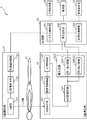
図1は、本発明の一実施形態におけるX線CT装置の構成を示すブロック図である。図1において、X線CT装置1は、X線照射部11と、X線測定部12と、処理部13と、画像記憶部14と、操作部15と、画像表示部16とを備えて構成される。
Versatile Disc Recordable)等の記録媒体との間でデータを書き込みおよび/または読み込みを行う装置であり、例えば、CD−RドライブやDVD−Rドライブ等である。
例えば、ヨウ素造影剤、バリウム造影剤および金造影剤等のX線造影剤のうちの例えば診断目的や診断部位に応じた適宜な造影剤が被検体Hに投与される。そして、ユーザによって操作部15から撮影開始の指示が入力されると、処理部13のシステム制御部131の制御によって、X線照射部11が制御され、X線照射部11からX線が被検体Hに向けて照射され、そして、X線測定部12が制御され、被検体Hを透過した透過X線がX線測定部12で測定される。すなわち、X線照射部11では、システム制御部13から制御信号が入力されると、電源制御回路113がX線電源回路112を制御することによって所定のタイミングで所定の電圧の電力がX線電源回路112からX線管111へ供給され、X線管111が被検体Hに向けてX線を照射する。被検体Hに向けて照射されたX線は、被検体Hを通過してX線測定部12に入射される。X線測定部12では、被検体Hを透過した透過X線がX線検出器121に入射され、透過X線がX線検出器121で検出される。X線検出器121からの検出出力は、前置増幅回路122および主増幅回路123で増幅され、AD変換回路124でAD変換される。AD変換回路124でAD変換されたディジタル出力は、積分回路125の第1および第2積分回路125−1、125−2にそれぞれ入力され、第1および第2積分回路125−1、125−2でそれぞれ積分され、処理部13に入力される。

次に、本発明の実施例およびその比較例について説明する。
X-ray contrast agent”,The British Journal of Radiology,79(2006),248-253」等に開示されている。
Claims (6)
- 被検体にX線を照射するX線照射部と、
前記被検体を介して前記X線照射部と対向し、前記被検体を透過した透過X線のうちの前記被検体の内部の検査対象物に応じた特定のエネルギ範囲における前記透過X線の個数を測定するX線測定部と、
前記X線測定部で測定した前記透過X線の個数に基づいて前記検査対象物の厚さを演算する厚さ演算部と、
前記厚さ演算部で演算された前記検査対象物の厚さに基づいてCT画像を再構成する画像再構成部とを備え、
前記検査対象物は、X線造影剤であり、
前記特定のエネルギ範囲は、前記X線造影剤のK吸収端上下に設定され、
前記厚さ演算部は、前記検査対象物のK吸収端よりも小さい所定のエネルギ範囲Φ1における透過X線の個数と、前記検査対象物のK吸収端よりも大きい所定のエネルギ範囲Φ2における透過X線の個数との比Φ1/Φ2から前記検査対象物の厚さtIを求め、
前記画像再構成部は、前記厚さ演算部で演算された前記検査対象物の厚さを投影データとして、前記投影データと所定の再構成関数とをコンボリューションし、この結果を逆投影することによって、前記被検体のCT画像を生成し、
比Φ1/Φ2は、エネルギ範囲En(n=1、2)のX線に対する前記検査対象物および水の平均減衰係数をそれぞれ上バー付きのμI(En)および上バー付きのμW(En)とし、前記検査対象物および水の厚さをtIおよびtwとする場合に、下記式であること
を特徴とするX線CT装置。
- 前記X線測定部は、前記被検体を透過した透過X線から付与されたエネルギによって電荷を発生する検出媒体と、前記検出媒体における前記透過X線の入射端からの距離が互いに異なる位置で、前記検出媒体に設置された複数の電極とを備えること
を特徴とする請求項1に記載のX線CT装置。 - 前記X線測定部は、前記被検体を透過した透過X線の進行方向に並べられた複数の検出媒体から成り、前記透過X線から付与されたエネルギによって個々の前記検出媒体において電荷を発生するとともに、前記検出媒体が前記透過X線の吸収体ともなるため、個々の前記検出媒体に前記透過X線が到達するまでに透過する吸収体の厚さが異なること
を特徴とする請求項1に記載のX線CT装置。 - 前記X線造影剤は、ヨウ素造影剤、バリウム造影剤および金造影剤のうちのいずれかであること
を特徴とする請求項1ないし請求項3のいずれか1項に記載のX線CT装置。 - 前記X線照射部は、X線をフィルタに透過させたフィルタX線を被検体に照射すること
を特徴とする請求項1ないし請求項4のいずれか1項に記載のX線CT装置。 - 生体を除く被検体にX線を照射するX線照射工程と、
前記被検体を透過した透過X線のうちの前記被検体の内部の検査対象物に応じた特定のエネルギ範囲における前記透過X線の個数を測定するX線測定工程と、
前記X線測定工程で測定した前記透過X線の個数に基づいて前記検査対象物の厚さを演算する厚さ演算工程と、
前記厚さ演算工程で演算された前記検査対象物の厚さに基づいてCT画像を再構成する画像再構成工程とを備え、
前記検査対象物は、X線造影剤であり、
前記特定のエネルギ範囲は、前記X線造影剤のK吸収端上下に設定され、
前記厚さ演算工程は、前記検査対象物のK吸収端よりも小さい所定のエネルギ範囲Φ1における透過X線の個数と、前記検査対象物のK吸収端よりも大きい所定のエネルギ範囲Φ2における透過X線の個数との比Φ1/Φ2から前記検査対象物の厚さtIを求め、
前記画像再構成工程は、前記厚さ演算工程で演算された前記検査対象物の厚さを投影データとして、前記投影データと所定の再構成関数とをコンボリューションし、この結果を逆投影することによって、前記被検体のCT画像を生成し、
比Φ1/Φ2は、エネルギ範囲En(n=1、2)のX線に対する前記検査対象物および水の平均減衰係数をそれぞれ上バー付きのμI(En)および上バー付きのμW(En)とし、前記検査対象物および水の厚さをtIおよびtwとする場合に、下記式であること
を特徴とするX線CT方法。
Priority Applications (1)
| Application Number | Priority Date | Filing Date | Title |
|---|---|---|---|
| JP2009528104A JP5367574B2 (ja) | 2007-08-15 | 2008-08-07 | X線ct装置および該方法 |
Applications Claiming Priority (4)
| Application Number | Priority Date | Filing Date | Title |
|---|---|---|---|
| JP2007211948 | 2007-08-15 | ||
| JP2007211948 | 2007-08-15 | ||
| JP2009528104A JP5367574B2 (ja) | 2007-08-15 | 2008-08-07 | X線ct装置および該方法 |
| PCT/JP2008/064229 WO2009022625A1 (ja) | 2007-08-15 | 2008-08-07 | X線ct装置および該方法 |
Publications (2)
| Publication Number | Publication Date |
|---|---|
| JPWO2009022625A1 JPWO2009022625A1 (ja) | 2010-11-11 |
| JP5367574B2 true JP5367574B2 (ja) | 2013-12-11 |
Family
ID=40350678
Family Applications (1)
| Application Number | Title | Priority Date | Filing Date |
|---|---|---|---|
| JP2009528104A Expired - Fee Related JP5367574B2 (ja) | 2007-08-15 | 2008-08-07 | X線ct装置および該方法 |
Country Status (5)
| Country | Link |
|---|---|
| US (1) | US8180016B2 (ja) |
| EP (2) | EP2189115A4 (ja) |
| JP (1) | JP5367574B2 (ja) |
| CN (1) | CN102215754B (ja) |
| WO (1) | WO2009022625A1 (ja) |
Cited By (1)
| Publication number | Priority date | Publication date | Assignee | Title |
|---|---|---|---|---|
| US10451568B2 (en) | 2014-08-22 | 2019-10-22 | Canon Medical Systems Corporation | Photon counting X-ray CT apparatus |
Families Citing this family (16)
| Publication number | Priority date | Publication date | Assignee | Title |
|---|---|---|---|---|
| US20140072108A1 (en) * | 2010-07-16 | 2014-03-13 | David P. Rohler | Methods and apparatus for extended low contrast detectability for radiographic imaging systems |
| GB201004121D0 (en) | 2010-03-12 | 2010-04-28 | Durham Scient Crystals Ltd | Detector device, inspection apparatus and method |
| US9224240B2 (en) * | 2010-11-23 | 2015-12-29 | Siemens Medical Solutions Usa, Inc. | Depth-based information layering in medical diagnostic ultrasound |
| US20120215095A1 (en) * | 2011-02-22 | 2012-08-23 | Amit Mordechai Av-Shalom | X-Ray radiation reduction system |
| US20120236995A1 (en) * | 2011-03-17 | 2012-09-20 | Christian Eusemann | Automated Imaging Contrast Agent Determination System |
| WO2014171487A1 (ja) * | 2013-04-16 | 2014-10-23 | 株式会社 東芝 | X線ct装置 |
| JP6234708B2 (ja) * | 2013-05-28 | 2017-11-22 | 東芝メディカルシステムズ株式会社 | X線診断装置、医用画像処理装置及び医用画像処理プログラム |
| JP6305692B2 (ja) | 2013-05-28 | 2018-04-04 | キヤノンメディカルシステムズ株式会社 | X線診断装置 |
| US8965095B2 (en) * | 2013-05-30 | 2015-02-24 | Kabushiki Kaisha Toshiba | Noise balance pre-reconstruction data decomposition in spectral CT |
| JP6595154B2 (ja) * | 2013-06-12 | 2019-10-23 | キヤノンメディカルシステムズ株式会社 | X線ctを用いた画像診断装置 |
| CN107430778A (zh) | 2015-03-18 | 2017-12-01 | 棱镜传感器公司 | 基于来自光子计数多仓检测器的能量分辨的图像数据的图像重建 |
| US11039805B2 (en) * | 2017-01-05 | 2021-06-22 | General Electric Company | Deep learning based estimation of data for use in tomographic reconstruction |
| EP3547254A1 (de) * | 2018-03-29 | 2019-10-02 | Siemens Healthcare GmbH | Analyse-verfahren und analyseeinheit zur ermittlung radiologischer ergebnisdaten |
| CN112666194B (zh) * | 2020-12-22 | 2022-12-20 | 上海培云教育科技有限公司 | 一种虚拟数字dr图像的生成方法及dr虚拟仿真仪器 |
| KR102593937B1 (ko) | 2021-08-23 | 2023-10-25 | (주)에이트마진 | 방사선 저감 필터의 제조 방법 |
| CN114271841B (zh) * | 2021-12-17 | 2025-04-22 | 乐普(北京)医疗装备有限公司 | 一种锥形束ct图像重建方法 |
Citations (2)
| Publication number | Priority date | Publication date | Assignee | Title |
|---|---|---|---|---|
| JP2004223158A (ja) * | 2003-01-27 | 2004-08-12 | Reitekku:Kk | X線撮像方法 |
| JP2007071602A (ja) * | 2005-09-05 | 2007-03-22 | Kyoto Univ | 放射線検出器 |
Family Cites Families (9)
| Publication number | Priority date | Publication date | Assignee | Title |
|---|---|---|---|---|
| US3854049A (en) * | 1973-12-10 | 1974-12-10 | Wisconsin Alumni Res Found | Compensation for patient thickness variations in differential x-ray transmission imaging |
| US4686695A (en) * | 1979-02-05 | 1987-08-11 | Board Of Trustees Of The Leland Stanford Junior University | Scanned x-ray selective imaging system |
| US5485492A (en) * | 1992-03-31 | 1996-01-16 | Lunar Corporation | Reduced field-of-view CT system for imaging compact embedded structures |
| JP3449561B2 (ja) * | 1993-04-19 | 2003-09-22 | 東芝医用システムエンジニアリング株式会社 | X線ct装置 |
| US5953444A (en) * | 1997-10-22 | 1999-09-14 | University Of Pennsylvania | Method for improved correction of spectrum hardening artifacts in computed tomography images |
| US6507633B1 (en) * | 2001-02-15 | 2003-01-14 | The Regents Of The University Of Michigan | Method for statistically reconstructing a polyenergetic X-ray computed tomography image and image reconstructor apparatus utilizing the method |
| JP4131934B2 (ja) * | 2003-01-27 | 2008-08-13 | 独立行政法人 日本原子力研究開発機構 | 化合物半導体InSb単結晶を用いた半導体放射線検出器 |
| US7391844B2 (en) * | 2005-01-14 | 2008-06-24 | General Electric Company | Method and apparatus for correcting for beam hardening in CT images |
| JP4614001B2 (ja) * | 2006-04-13 | 2011-01-19 | 株式会社島津製作所 | 透過x線を用いた三次元定量方法 |
-
2008
- 2008-08-07 US US12/673,197 patent/US8180016B2/en not_active Expired - Fee Related
- 2008-08-07 CN CN2008801031103A patent/CN102215754B/zh not_active Expired - Fee Related
- 2008-08-07 WO PCT/JP2008/064229 patent/WO2009022625A1/ja not_active Ceased
- 2008-08-07 EP EP08827357A patent/EP2189115A4/en not_active Withdrawn
- 2008-08-07 EP EP16000657.3A patent/EP3090686A1/en not_active Withdrawn
- 2008-08-07 JP JP2009528104A patent/JP5367574B2/ja not_active Expired - Fee Related
Patent Citations (2)
| Publication number | Priority date | Publication date | Assignee | Title |
|---|---|---|---|---|
| JP2004223158A (ja) * | 2003-01-27 | 2004-08-12 | Reitekku:Kk | X線撮像方法 |
| JP2007071602A (ja) * | 2005-09-05 | 2007-03-22 | Kyoto Univ | 放射線検出器 |
Cited By (2)
| Publication number | Priority date | Publication date | Assignee | Title |
|---|---|---|---|---|
| US10451568B2 (en) | 2014-08-22 | 2019-10-22 | Canon Medical Systems Corporation | Photon counting X-ray CT apparatus |
| US11327031B2 (en) | 2014-08-22 | 2022-05-10 | Canon Medical Systems Corporation | Photon counting X-ray CT apparatus |
Also Published As
| Publication number | Publication date |
|---|---|
| CN102215754A (zh) | 2011-10-12 |
| WO2009022625A1 (ja) | 2009-02-19 |
| US20110194668A1 (en) | 2011-08-11 |
| EP2189115A1 (en) | 2010-05-26 |
| EP2189115A4 (en) | 2010-08-18 |
| JPWO2009022625A1 (ja) | 2010-11-11 |
| US8180016B2 (en) | 2012-05-15 |
| CN102215754B (zh) | 2013-06-05 |
| EP3090686A1 (en) | 2016-11-09 |
Similar Documents
| Publication | Publication Date | Title |
|---|---|---|
| JP5367574B2 (ja) | X線ct装置および該方法 | |
| CN101375798B (zh) | 具有第二射线管/检测器修补的ct成像系统和方法 | |
| US20200292475A1 (en) | X-Ray Imaging with a Detector Capable of Resolving Photon Energy | |
| US8000434B2 (en) | Energy spectrum reconstruction | |
| Taguchi et al. | Vision 20/20: single photon counting x‐ray detectors in medical imaging | |
| US9579075B2 (en) | Detector array comprising energy integrating and photon counting cells | |
| EP2731504B1 (en) | Imaging system detector calibration | |
| US20100012845A1 (en) | Energy-resolving detection system and imaging system | |
| JP5779819B2 (ja) | 放射線検出器 | |
| CN101228437A (zh) | 采用多色光谱的x射线探测器成像 | |
| CN102088907A (zh) | K边缘成像 | |
| JP2016131884A (ja) | X線ct装置、光子計数型検出装置及び二重積層光子計数型検出器 | |
| CN103959097A (zh) | 包括用于探测x射线辐射的两个闪烁体的探测装置 | |
| JP2008510132A (ja) | 放射線検出器用の抗散乱グリッド | |
| JP2016061655A (ja) | シンチレータ、放射線検出装置および放射線検査装置 | |
| JP7624831B2 (ja) | X線コンピュータ断層撮影装置、陽極劣化推定方法、および陽極劣化推定プログラム | |
| JP2010243395A (ja) | X線・ガンマ線撮像装置 | |
| JP2007512081A (ja) | 検査方法および装置 | |
| JP2015031683A (ja) | 放射線検出装置、放射線分析装置及び放射線検出方法 | |
| JP2020030097A (ja) | 感度補正方法及び光子計数型検出器 | |
| Arrigoni et al. | X-Ray Sensors in Computed Tomography | |
| JP2015152356A (ja) | ダークカウントレス放射線検出エネルギー弁別イメージングシステム | |
| JP2007071602A (ja) | 放射線検出器 | |
| JP2025169792A (ja) | X線コンピュータ断層撮影装置及び制御方法 | |
| Watanabe et al. | Characteristics of a ceramic-substrate x-ray diode and its application to computed tomography |
Legal Events
| Date | Code | Title | Description |
|---|---|---|---|
| A621 | Written request for application examination |
Free format text: JAPANESE INTERMEDIATE CODE: A621 Effective date: 20110801 |
|
| A521 | Request for written amendment filed |
Free format text: JAPANESE INTERMEDIATE CODE: A523 Effective date: 20110809 |
|
| A131 | Notification of reasons for refusal |
Free format text: JAPANESE INTERMEDIATE CODE: A131 Effective date: 20121030 |
|
| A521 | Request for written amendment filed |
Free format text: JAPANESE INTERMEDIATE CODE: A523 Effective date: 20121221 |
|
| TRDD | Decision of grant or rejection written | ||
| A01 | Written decision to grant a patent or to grant a registration (utility model) |
Free format text: JAPANESE INTERMEDIATE CODE: A01 Effective date: 20130903 |
|
| A61 | First payment of annual fees (during grant procedure) |
Free format text: JAPANESE INTERMEDIATE CODE: A61 Effective date: 20130911 |
|
| R150 | Certificate of patent or registration of utility model |
Ref document number: 5367574 Country of ref document: JP Free format text: JAPANESE INTERMEDIATE CODE: R150 Free format text: JAPANESE INTERMEDIATE CODE: R150 |
|
| S111 | Request for change of ownership or part of ownership |
Free format text: JAPANESE INTERMEDIATE CODE: R313114 |
|
| R350 | Written notification of registration of transfer |
Free format text: JAPANESE INTERMEDIATE CODE: R350 |
|
| S111 | Request for change of ownership or part of ownership |
Free format text: JAPANESE INTERMEDIATE CODE: R313115 |
|
| R350 | Written notification of registration of transfer |
Free format text: JAPANESE INTERMEDIATE CODE: R350 |
|
| R250 | Receipt of annual fees |
Free format text: JAPANESE INTERMEDIATE CODE: R250 |
|
| R250 | Receipt of annual fees |
Free format text: JAPANESE INTERMEDIATE CODE: R250 |
|
| LAPS | Cancellation because of no payment of annual fees |