JP2005296653A - コンピュータ断層撮影装置によるコンピュータ断層撮影画像形成方法およびコンピュータ断層撮影装置 - Google Patents
コンピュータ断層撮影装置によるコンピュータ断層撮影画像形成方法およびコンピュータ断層撮影装置 Download PDFInfo
- Publication number
- JP2005296653A JP2005296653A JP2005110741A JP2005110741A JP2005296653A JP 2005296653 A JP2005296653 A JP 2005296653A JP 2005110741 A JP2005110741 A JP 2005110741A JP 2005110741 A JP2005110741 A JP 2005110741A JP 2005296653 A JP2005296653 A JP 2005296653A
- Authority
- JP
- Japan
- Prior art keywords
- focus
- jump
- ray tube
- positions
- detector
- Prior art date
- Legal status (The legal status is an assumption and is not a legal conclusion. Google has not performed a legal analysis and makes no representation as to the accuracy of the status listed.)
- Pending
Links
Images


Classifications
-
- G—PHYSICS
- G06—COMPUTING OR CALCULATING; COUNTING
- G06T—IMAGE DATA PROCESSING OR GENERATION, IN GENERAL
- G06T12/00—Tomographic reconstruction from projections
- G06T12/10—Image preprocessing, e.g. calibration, positioning of sources or scatter correction
-
- A—HUMAN NECESSITIES
- A61—MEDICAL OR VETERINARY SCIENCE; HYGIENE
- A61B—DIAGNOSIS; SURGERY; IDENTIFICATION
- A61B6/00—Apparatus or devices for radiation diagnosis; Apparatus or devices for radiation diagnosis combined with radiation therapy equipment
- A61B6/02—Arrangements for diagnosis sequentially in different planes; Stereoscopic radiation diagnosis
- A61B6/03—Computed tomography [CT]
- A61B6/032—Transmission computed tomography [CT]
-
- A—HUMAN NECESSITIES
- A61—MEDICAL OR VETERINARY SCIENCE; HYGIENE
- A61B—DIAGNOSIS; SURGERY; IDENTIFICATION
- A61B6/00—Apparatus or devices for radiation diagnosis; Apparatus or devices for radiation diagnosis combined with radiation therapy equipment
- A61B6/40—Arrangements for generating radiation specially adapted for radiation diagnosis
- A61B6/4021—Arrangements for generating radiation specially adapted for radiation diagnosis involving movement of the focal spot
- A61B6/4028—Arrangements for generating radiation specially adapted for radiation diagnosis involving movement of the focal spot resulting in acquisition of views from substantially different positions, e.g. EBCT
-
- A—HUMAN NECESSITIES
- A61—MEDICAL OR VETERINARY SCIENCE; HYGIENE
- A61B—DIAGNOSIS; SURGERY; IDENTIFICATION
- A61B6/00—Apparatus or devices for radiation diagnosis; Apparatus or devices for radiation diagnosis combined with radiation therapy equipment
- A61B6/40—Arrangements for generating radiation specially adapted for radiation diagnosis
- A61B6/4064—Arrangements for generating radiation specially adapted for radiation diagnosis specially adapted for producing a particular type of beam
- A61B6/4085—Cone-beams
-
- G—PHYSICS
- G01—MEASURING; TESTING
- G01N—INVESTIGATING OR ANALYSING MATERIALS BY DETERMINING THEIR CHEMICAL OR PHYSICAL PROPERTIES
- G01N23/00—Investigating or analysing materials by the use of wave or particle radiation, e.g. X-rays or neutrons, not covered by groups G01N3/00 – G01N17/00, G01N21/00 or G01N22/00
- G01N23/02—Investigating or analysing materials by the use of wave or particle radiation, e.g. X-rays or neutrons, not covered by groups G01N3/00 – G01N17/00, G01N21/00 or G01N22/00 by transmitting the radiation through the material
- G01N23/04—Investigating or analysing materials by the use of wave or particle radiation, e.g. X-rays or neutrons, not covered by groups G01N3/00 – G01N17/00, G01N21/00 or G01N22/00 by transmitting the radiation through the material and forming images of the material
- G01N23/046—Investigating or analysing materials by the use of wave or particle radiation, e.g. X-rays or neutrons, not covered by groups G01N3/00 – G01N17/00, G01N21/00 or G01N22/00 by transmitting the radiation through the material and forming images of the material using tomography, e.g. computed tomography [CT]
-
- A—HUMAN NECESSITIES
- A61—MEDICAL OR VETERINARY SCIENCE; HYGIENE
- A61B—DIAGNOSIS; SURGERY; IDENTIFICATION
- A61B6/00—Apparatus or devices for radiation diagnosis; Apparatus or devices for radiation diagnosis combined with radiation therapy equipment
- A61B6/02—Arrangements for diagnosis sequentially in different planes; Stereoscopic radiation diagnosis
- A61B6/027—Arrangements for diagnosis sequentially in different planes; Stereoscopic radiation diagnosis characterised by the use of a particular data acquisition trajectory, e.g. helical or spiral
-
- G—PHYSICS
- G01—MEASURING; TESTING
- G01N—INVESTIGATING OR ANALYSING MATERIALS BY DETERMINING THEIR CHEMICAL OR PHYSICAL PROPERTIES
- G01N2223/00—Investigating materials by wave or particle radiation
- G01N2223/40—Imaging
- G01N2223/419—Imaging computed tomograph
Landscapes
- Health & Medical Sciences (AREA)
- Life Sciences & Earth Sciences (AREA)
- Engineering & Computer Science (AREA)
- Medical Informatics (AREA)
- Physics & Mathematics (AREA)
- General Health & Medical Sciences (AREA)
- Radiology & Medical Imaging (AREA)
- Pathology (AREA)
- Nuclear Medicine, Radiotherapy & Molecular Imaging (AREA)
- Biomedical Technology (AREA)
- Animal Behavior & Ethology (AREA)
- Optics & Photonics (AREA)
- Biophysics (AREA)
- Veterinary Medicine (AREA)
- Theoretical Computer Science (AREA)
- Heart & Thoracic Surgery (AREA)
- Molecular Biology (AREA)
- Surgery (AREA)
- High Energy & Nuclear Physics (AREA)
- Public Health (AREA)
- General Physics & Mathematics (AREA)
- Pulmonology (AREA)
- Chemical & Material Sciences (AREA)
- Analytical Chemistry (AREA)
- Biochemistry (AREA)
- Immunology (AREA)
- Apparatus For Radiation Diagnosis (AREA)
- Analysing Materials By The Use Of Radiation (AREA)
Abstract
【解決手段】X線管が検出器(D)との組合せでz軸の周りを円形状またはスパイラル状に移動させられて被検体(B)を走査し、X線管がX線管に対して相対的な2つ以上の異なる跳躍焦点位置(F1〜F6)を有する1つの跳躍焦点を持ち、取得された検出器データから複数のパラレルデータセットが形成され、このパラレルデータセットから断層画像が再構成される、コンピュータ断層撮影装置によるコンピュータ断層撮影画像形成方法において、パラレルデータセットの形成時に、その都度の現在の跳躍焦点(F)のX線管に対する半径方向の異なる相対位置(=跳躍焦点位置)が考慮される。
【選択図】図2
Description
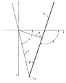
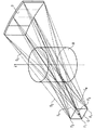
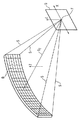
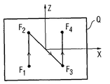
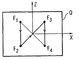
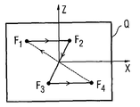
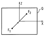
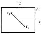
図1は使用されたジオメトリの説明図、
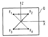
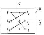
図2は4つの異なる焦点位置を有する跳躍焦点によって形成された4つのファンビームの説明図、
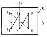
図3は図4乃至図19の跳躍焦点位置の手引きをするための説明図、
図4乃至図19は異なる跳躍焦点位置の変形例の説明図、
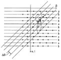
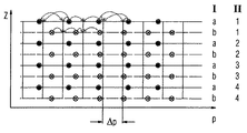
図20は一定のΔθラスタへのリビニングの概略図、
図21は一定のΔpラスタへのリビニングの概略図である。
これらにおいては本発明の直接的な理解のために重要な要素だけが示されている。
b z方向の跳躍焦点位置
B 被検体
D 検出器
F 焦点
F1〜F6 焦点位置
p z軸に対するX線の距離
Q 中心X線上の垂直面
r z軸からの焦点の半径つまり距離
S X線
Sz 中心X線
t 時間的前進
z システム軸線
α 回転角
β ファン角
φ z軸に対するX線の傾斜角
θ ビーム角
Claims (10)
- X線管が検出器(D)との組合せでz軸の周りを円形状またはスパイラル状に移動させられて被検体(B)を走査し、X線管がX線管に対して相対的な2つ以上の異なる跳躍焦点位置(F1〜F6)を有する1つの跳躍焦点を持ち、取得された検出器データから複数のパラレルデータセットが形成され、このパラレルデータセットから断層画像が再構成される、コンピュータ断層撮影装置によるコンピュータ断層撮影画像形成方法において、
パラレルデータセットの形成時に、その都度の現在の跳躍焦点(F)のX線管に対する半径方向の異なる相対位置(=跳躍焦点位置)が考慮される
ことを特徴とするコンピュータ断層撮影装置によるコンピュータ断層撮影画像形成方法。 - パラレルデータセットの形成時に、その都度の現在の跳躍焦点のX線管に対するz方向の異なる相対位置(F1〜F6)が考慮されることを特徴とする請求項1記載の方法。
- パラレルデータセットの形成時に、第1ステップにおいて、跳躍焦点位置(F1〜F6)に従っておよび検出器行に従って別々に同じビーム角θへの方位パラレルリビニングが行なわれ、その都度の跳躍焦点(F)の実際の半径(r)がビーム角θの算出に用いられ、複数のデータセットが等しいΔθラスタで補間されることを特徴とする請求項1又は2記載の方法。
- 第2ステップにおいて、等しいz座標および等しいビーム角θを有する複数のデータセットが焦点位置から交互配置され、より狭い等間隔のΔθラスタで複数の新しいデータセットに合成および/または補間されることを特徴とする請求項1乃至3の1つに記載の方法。
- 第3ステップにおいて、全ての跳躍焦点位置(F1〜F6)のデータセットを用いて同じ等間隔のΔpラスタ(p=r・sinβ)への半径方向リビニングが行なわれることを特徴とする請求項1乃至4の1つに記載の方法。
- 第4ステップにおいて、z方向の異なる跳躍焦点位置(F1〜F6)および等しいビーム角θを持つ複数のデータセットが交互配置され、より小さい等間隔のΔzラスタで1つのデータセットに補間されることを特徴とする請求項1乃至5の1つに記載の方法。
- 請求項1乃至6の1つによるデータ処理後に断層画像の算出のために再構成法が実行されることを特徴とする請求項1乃至6の1つに記載の方法。
- 走査のために、方位角方向に少なくとも2つの跳躍焦点位置とz方向に少なくとも2つの跳躍焦点位置とを有する1つの跳躍焦点(F)が使用されることを特徴とする請求項1乃至7の1つに記載の方法。
- 個々の各跳躍焦点位置(F1〜F6)が異なる方位角座標およびz座標を有する1つの跳躍焦点(F)が使用されることを特徴とする請求項1乃至8の1つに記載の方法。
- 複数行検出器(D)との組合せでz軸の周りを円形状またはスパイラル状に移動させられて被検体(B)を走査するX線管を備え、X線管がX線管に対して相対的な2つ以上の異なる跳躍焦点位置(F1〜F6)を有する1つの跳躍焦点を持ち、取得された検出器データから事前にパラレルデータセットを形成して断層画像を再構成する手段が設けられているコンピュータ断層撮影装置において、請求項1乃至9の少なくとも1つによる方法ステップを実施するための手段、とりわけプログラムまたはプログラムモジュールが設けられていることを特徴とするコンピュータ断層撮影装置。
Applications Claiming Priority (1)
| Application Number | Priority Date | Filing Date | Title |
|---|---|---|---|
| DE102004017540A DE102004017540B4 (de) | 2004-04-08 | 2004-04-08 | Verfahren zur Erstellung von computertomographischen Aufnahmen mit einem CT-Gerät und CT-Gerät |
Publications (1)
| Publication Number | Publication Date |
|---|---|
| JP2005296653A true JP2005296653A (ja) | 2005-10-27 |
Family
ID=35062296
Family Applications (1)
| Application Number | Title | Priority Date | Filing Date |
|---|---|---|---|
| JP2005110741A Pending JP2005296653A (ja) | 2004-04-08 | 2005-04-07 | コンピュータ断層撮影装置によるコンピュータ断層撮影画像形成方法およびコンピュータ断層撮影装置 |
Country Status (4)
| Country | Link |
|---|---|
| US (1) | US7505553B2 (ja) |
| JP (1) | JP2005296653A (ja) |
| CN (1) | CN1680808A (ja) |
| DE (1) | DE102004017540B4 (ja) |
Cited By (6)
| Publication number | Priority date | Publication date | Assignee | Title |
|---|---|---|---|---|
| JP2009136518A (ja) * | 2007-12-07 | 2009-06-25 | Canon Inc | X線撮影装置及びx線撮影方法 |
| JP2010508080A (ja) * | 2006-10-31 | 2010-03-18 | コーニンクレッカ フィリップス エレクトロニクス エヌ ヴィ | 掃引アノードctスキャナ |
| JP2011019802A (ja) * | 2009-07-17 | 2011-02-03 | Ge Medical Systems Global Technology Co Llc | X線ct装置 |
| JP2013173015A (ja) * | 2013-05-09 | 2013-09-05 | Canon Inc | X線撮影装置及びx線撮影方法 |
| JP2013242204A (ja) * | 2012-05-18 | 2013-12-05 | Shimadzu Corp | X線検査装置 |
| WO2014115625A1 (ja) * | 2013-01-28 | 2014-07-31 | 株式会社日立メディコ | X線ct装置及び画像再構成方法 |
Families Citing this family (13)
| Publication number | Priority date | Publication date | Assignee | Title |
|---|---|---|---|---|
| WO2007026273A2 (en) * | 2005-09-02 | 2007-03-08 | Koninklijke Philips Electronics, N.V. | Improved rebinning for computed tomography imaging |
| US7746974B2 (en) * | 2006-09-29 | 2010-06-29 | Siemens Medical Solutions Usa, Inc. | Radiographic and fluoroscopic CT imaging |
| WO2009128063A1 (en) * | 2008-04-14 | 2009-10-22 | Arineta Ltd. | Ct cone beam scanner |
| CN102176866B (zh) | 2008-11-24 | 2013-10-16 | 霍罗吉克公司 | 控制用于体层合成和乳房x射线照相术成像的x射线焦斑特性的方法和系统 |
| US20100202583A1 (en) * | 2009-02-03 | 2010-08-12 | Ge Wang | Systems and Methods for Exact or Approximate Cardiac Computed Tomography |
| JP5433334B2 (ja) * | 2009-07-27 | 2014-03-05 | 株式会社東芝 | X線ct装置 |
| US9271689B2 (en) * | 2010-01-20 | 2016-03-01 | General Electric Company | Apparatus for wide coverage computed tomography and method of constructing same |
| US20120087464A1 (en) * | 2010-10-09 | 2012-04-12 | Fmi Technologies, Inc. | Multi-source low dose x-ray ct imaging aparatus |
| CN102727231B (zh) * | 2011-04-02 | 2016-08-03 | 沈阳东软医疗系统有限公司 | 飞焦点ct机扫描数据采集方法及装置 |
| CN102727230B (zh) * | 2011-04-02 | 2014-06-04 | 沈阳东软医疗系统有限公司 | Ct扫描图像重建方法及装置 |
| CN103961128B (zh) * | 2013-03-03 | 2016-03-02 | 李宝生 | 变焦点锥形束ct成像设备 |
| JP6246936B2 (ja) * | 2014-07-28 | 2017-12-13 | 株式会社日立製作所 | X線撮像装置および画像再構成方法 |
| US11076820B2 (en) | 2016-04-22 | 2021-08-03 | Hologic, Inc. | Tomosynthesis with shifting focal spot x-ray system using an addressable array |
Citations (7)
| Publication number | Priority date | Publication date | Assignee | Title |
|---|---|---|---|---|
| JPH07299058A (ja) * | 1994-04-30 | 1995-11-14 | Shimadzu Corp | X線ct装置 |
| JPH0810251A (ja) * | 1994-06-28 | 1996-01-16 | Hitachi Medical Corp | X線断層撮影方法および装置 |
| JPH11318879A (ja) * | 1998-05-18 | 1999-11-24 | Aloka Co Ltd | Ct装置及び検出素子配列方法 |
| JP2000139893A (ja) * | 1998-11-09 | 2000-05-23 | Siemens Ag | Ct装置 |
| JP2000287960A (ja) * | 1999-03-31 | 2000-10-17 | Analogic Corp | 縦方向フライング・フォーカルスポットを備えたコンピューター断層撮影スキャナー |
| JP2002224099A (ja) * | 2000-12-07 | 2002-08-13 | Koninkl Philips Electronics Nv | 螺旋状の相対運動を含むコンピュータ断層撮影方法 |
| JP2002233524A (ja) * | 2000-12-01 | 2002-08-20 | Marconi Medical Systems Inc | 画像作成装置及び方法 |
Family Cites Families (13)
| Publication number | Priority date | Publication date | Assignee | Title |
|---|---|---|---|---|
| WO1998030980A1 (en) * | 1997-01-14 | 1998-07-16 | Edholm, Paul | Technique and arrangement for tomographic imaging |
| DE19953613A1 (de) * | 1999-11-08 | 2001-05-17 | Siemens Ag | CT-Gerät sowie Verfahren zum Betrieb eines CT-Geräts |
| DE29923967U1 (de) * | 1999-11-08 | 2001-09-06 | Siemens AG, 80333 München | CT-Gerät |
| DE10048775B4 (de) * | 2000-09-29 | 2006-02-02 | Siemens Ag | Röntgen-Computertomographieeinrichtung |
| JP4298205B2 (ja) * | 2001-02-12 | 2009-07-15 | シーメンス アクチエンゲゼルシヤフト | コンピュータトモグラフィのための方法ならびにコンピュータトモグラフィ装置 |
| DE10126638B4 (de) * | 2001-02-12 | 2004-03-18 | Siemens Ag | Verfahren für die Computertomographie sowie Computertomographie(CT)-Gerät |
| DE10127269B4 (de) * | 2001-06-05 | 2015-09-24 | Siemens Aktiengesellschaft | Verfahren für die Computertomographie sowie Computertomographie (CT)-Gerät |
| DE10159927B4 (de) * | 2001-12-06 | 2005-04-21 | Siemens Ag | Verfahren zur Bildrekonstruktion für die Computertomographie |
| DE10244181A1 (de) * | 2002-09-23 | 2004-04-01 | Siemens Ag | Verfahren zur Bilderstellung in der Computertomographie und CT-Gerät zur Durchführung des Verfahrens |
| DE10245578A1 (de) | 2002-09-27 | 2004-04-08 | Siemens Ag | CT-Gerät mit einer Strahlungsquelle und einem während der Aufnahme von Projektionen zwischen einer ersten Endposition und einer zweiten Endposition bewegten Fokus der Strahlungsquelle |
| US6963631B2 (en) * | 2002-10-25 | 2005-11-08 | Koninklijke Philips Electronics N.V. | Dynamic detector interlacing for computed tomography |
| DE10320882B4 (de) * | 2003-05-09 | 2005-09-29 | Siemens Ag | Verfahren zur Erzeugung von Bildern in der Spiral-Computertomographie und Spiral-CT-Gerät |
| US7065179B2 (en) * | 2003-11-07 | 2006-06-20 | General Electric Company | Multiple target anode assembly and system of operation |
-
2004
- 2004-04-08 DE DE102004017540A patent/DE102004017540B4/de not_active Expired - Fee Related
-
2005
- 2005-04-07 US US11/100,445 patent/US7505553B2/en not_active Expired - Fee Related
- 2005-04-07 JP JP2005110741A patent/JP2005296653A/ja active Pending
- 2005-04-08 CN CN200510063844.1A patent/CN1680808A/zh active Pending
Patent Citations (7)
| Publication number | Priority date | Publication date | Assignee | Title |
|---|---|---|---|---|
| JPH07299058A (ja) * | 1994-04-30 | 1995-11-14 | Shimadzu Corp | X線ct装置 |
| JPH0810251A (ja) * | 1994-06-28 | 1996-01-16 | Hitachi Medical Corp | X線断層撮影方法および装置 |
| JPH11318879A (ja) * | 1998-05-18 | 1999-11-24 | Aloka Co Ltd | Ct装置及び検出素子配列方法 |
| JP2000139893A (ja) * | 1998-11-09 | 2000-05-23 | Siemens Ag | Ct装置 |
| JP2000287960A (ja) * | 1999-03-31 | 2000-10-17 | Analogic Corp | 縦方向フライング・フォーカルスポットを備えたコンピューター断層撮影スキャナー |
| JP2002233524A (ja) * | 2000-12-01 | 2002-08-20 | Marconi Medical Systems Inc | 画像作成装置及び方法 |
| JP2002224099A (ja) * | 2000-12-07 | 2002-08-13 | Koninkl Philips Electronics Nv | 螺旋状の相対運動を含むコンピュータ断層撮影方法 |
Cited By (7)
| Publication number | Priority date | Publication date | Assignee | Title |
|---|---|---|---|---|
| JP2010508080A (ja) * | 2006-10-31 | 2010-03-18 | コーニンクレッカ フィリップス エレクトロニクス エヌ ヴィ | 掃引アノードctスキャナ |
| JP2009136518A (ja) * | 2007-12-07 | 2009-06-25 | Canon Inc | X線撮影装置及びx線撮影方法 |
| JP2011019802A (ja) * | 2009-07-17 | 2011-02-03 | Ge Medical Systems Global Technology Co Llc | X線ct装置 |
| JP2013242204A (ja) * | 2012-05-18 | 2013-12-05 | Shimadzu Corp | X線検査装置 |
| WO2014115625A1 (ja) * | 2013-01-28 | 2014-07-31 | 株式会社日立メディコ | X線ct装置及び画像再構成方法 |
| JP5978516B2 (ja) * | 2013-01-28 | 2016-08-24 | 株式会社日立製作所 | X線ct装置及び画像再構成方法 |
| JP2013173015A (ja) * | 2013-05-09 | 2013-09-05 | Canon Inc | X線撮影装置及びx線撮影方法 |
Also Published As
| Publication number | Publication date |
|---|---|
| US20050238136A1 (en) | 2005-10-27 |
| CN1680808A (zh) | 2005-10-12 |
| US7505553B2 (en) | 2009-03-17 |
| DE102004017540A1 (de) | 2005-10-27 |
| DE102004017540B4 (de) | 2008-02-28 |
Similar Documents
| Publication | Publication Date | Title |
|---|---|---|
| JP2005296653A (ja) | コンピュータ断層撮影装置によるコンピュータ断層撮影画像形成方法およびコンピュータ断層撮影装置 | |
| JP3168824B2 (ja) | X線ct装置 | |
| US9977137B2 (en) | X-ray image pickup device and image reconstruction method | |
| US8116426B2 (en) | Computed tomography device and method using circular-pixel position-adaptive interpolation | |
| US7424089B2 (en) | System and method for reconstructing image by using straight-line trajectory scan | |
| JP3682308B2 (ja) | 計算機式断層写真装置及び撮像されるべき物体の像を発生する方法 | |
| US6560308B1 (en) | Method and system for approximating missing data in cone beam x-ray CT reconstruction | |
| JP5376902B2 (ja) | コンピュータ断層撮影装置、再構成処理方法 | |
| JP4813681B2 (ja) | コンピュータ断層撮影方法 | |
| JP2000081318A (ja) | 3次元コンピュ―タトモグラフィイメ―ジングのためのスキャニングおよびデ―タ収集方法およびイメ―ジング装置 | |
| JPH04231940A (ja) | 物体を撮像するct装置 | |
| JP2005152638A (ja) | 断層撮影による断層像作成方法および断層撮影装置 | |
| JP4557321B2 (ja) | 画像再構成装置 | |
| JP2001057976A (ja) | 立体画像再構成方法及び装置並びにctスキャナー | |
| CN1315436C (zh) | 荧光检查计算机断层方法 | |
| CN104757988A (zh) | 一种电子直线扫描微纳焦点ct扫描系统及方法 | |
| US20050265523A1 (en) | C-arm device with adjustable detector offset for cone beam imaging involving partial circle scan trajectories | |
| JP2004113785A (ja) | コンピュータ断層撮影法における画像形成方法およびこの方法を実施するためのct装置 | |
| US7058156B2 (en) | Imaging method for a multi-slice spiral CT scan with 3D reconstruction, and a computed tomography unit for carrying out this method | |
| CN104665860B (zh) | 用于定位片扫描的方法和ct系统 | |
| US7809100B2 (en) | Rebinning for computed tomography imaging | |
| JP4025530B2 (ja) | X線ct装置 | |
| JP4701038B2 (ja) | X線ct装置 | |
| JP2007185358A (ja) | X線ct装置 | |
| JP4006451B2 (ja) | X線ct装置及びそのミスアライメント補正方法 |
Legal Events
| Date | Code | Title | Description |
|---|---|---|---|
| A621 | Written request for application examination |
Free format text: JAPANESE INTERMEDIATE CODE: A621 Effective date: 20080328 |
|
| A131 | Notification of reasons for refusal |
Free format text: JAPANESE INTERMEDIATE CODE: A131 Effective date: 20100406 |
|
| A601 | Written request for extension of time |
Free format text: JAPANESE INTERMEDIATE CODE: A601 Effective date: 20100706 |
|
| RD03 | Notification of appointment of power of attorney |
Free format text: JAPANESE INTERMEDIATE CODE: A7423 Effective date: 20100706 |
|
| A602 | Written permission of extension of time |
Free format text: JAPANESE INTERMEDIATE CODE: A602 Effective date: 20100709 |
|
| A521 | Request for written amendment filed |
Free format text: JAPANESE INTERMEDIATE CODE: A523 Effective date: 20100804 |
|
| A131 | Notification of reasons for refusal |
Free format text: JAPANESE INTERMEDIATE CODE: A131 Effective date: 20101012 |
|
| A521 | Request for written amendment filed |
Free format text: JAPANESE INTERMEDIATE CODE: A523 Effective date: 20110111 |
|
| A131 | Notification of reasons for refusal |
Free format text: JAPANESE INTERMEDIATE CODE: A131 Effective date: 20110308 |
|
| A601 | Written request for extension of time |
Free format text: JAPANESE INTERMEDIATE CODE: A601 Effective date: 20110608 |
|
| A602 | Written permission of extension of time |
Free format text: JAPANESE INTERMEDIATE CODE: A602 Effective date: 20110613 |
|
| A02 | Decision of refusal |
Free format text: JAPANESE INTERMEDIATE CODE: A02 Effective date: 20111004 |