JP2005292320A - 画像顕微鏡装置 - Google Patents
画像顕微鏡装置 Download PDFInfo
- Publication number
- JP2005292320A JP2005292320A JP2004104957A JP2004104957A JP2005292320A JP 2005292320 A JP2005292320 A JP 2005292320A JP 2004104957 A JP2004104957 A JP 2004104957A JP 2004104957 A JP2004104957 A JP 2004104957A JP 2005292320 A JP2005292320 A JP 2005292320A
- Authority
- JP
- Japan
- Prior art keywords
- image display
- display device
- imaging device
- image
- arm
- Prior art date
- Legal status (The legal status is an assumption and is not a legal conclusion. Google has not performed a legal analysis and makes no representation as to the accuracy of the status listed.)
- Pending
Links
- 238000003384 imaging method Methods 0.000 claims abstract description 140
- NJPPVKZQTLUDBO-UHFFFAOYSA-N novaluron Chemical compound C1=C(Cl)C(OC(F)(F)C(OC(F)(F)F)F)=CC=C1NC(=O)NC(=O)C1=C(F)C=CC=C1F NJPPVKZQTLUDBO-UHFFFAOYSA-N 0.000 claims 1
- 238000010586 diagram Methods 0.000 description 6
- 238000001356 surgical procedure Methods 0.000 description 6
- 230000000694 effects Effects 0.000 description 5
- 238000011282 treatment Methods 0.000 description 5
- 238000000034 method Methods 0.000 description 3
- 230000003287 optical effect Effects 0.000 description 2
- 206010002091 Anaesthesia Diseases 0.000 description 1
- 230000037005 anaesthesia Effects 0.000 description 1
- 230000004907 flux Effects 0.000 description 1
- 238000009434 installation Methods 0.000 description 1
- 239000000696 magnetic material Substances 0.000 description 1
- 238000000926 separation method Methods 0.000 description 1
Images
Classifications
-
- G—PHYSICS
- G02—OPTICS
- G02B—OPTICAL ELEMENTS, SYSTEMS OR APPARATUS
- G02B21/00—Microscopes
- G02B21/0004—Microscopes specially adapted for specific applications
- G02B21/0012—Surgical microscopes
-
- A—HUMAN NECESSITIES
- A61—MEDICAL OR VETERINARY SCIENCE; HYGIENE
- A61B—DIAGNOSIS; SURGERY; IDENTIFICATION
- A61B90/00—Instruments, implements or accessories specially adapted for surgery or diagnosis and not covered by any of the groups A61B1/00 - A61B50/00, e.g. for luxation treatment or for protecting wound edges
- A61B90/20—Surgical microscopes characterised by non-optical aspects
- A61B90/25—Supports therefor
-
- A—HUMAN NECESSITIES
- A61—MEDICAL OR VETERINARY SCIENCE; HYGIENE
- A61B—DIAGNOSIS; SURGERY; IDENTIFICATION
- A61B90/00—Instruments, implements or accessories specially adapted for surgery or diagnosis and not covered by any of the groups A61B1/00 - A61B50/00, e.g. for luxation treatment or for protecting wound edges
- A61B90/36—Image-producing devices or illumination devices not otherwise provided for
-
- A—HUMAN NECESSITIES
- A61—MEDICAL OR VETERINARY SCIENCE; HYGIENE
- A61B—DIAGNOSIS; SURGERY; IDENTIFICATION
- A61B90/00—Instruments, implements or accessories specially adapted for surgery or diagnosis and not covered by any of the groups A61B1/00 - A61B50/00, e.g. for luxation treatment or for protecting wound edges
- A61B90/50—Supports for surgical instruments, e.g. articulated arms
-
- G—PHYSICS
- G02—OPTICS
- G02B—OPTICAL ELEMENTS, SYSTEMS OR APPARATUS
- G02B21/00—Microscopes
- G02B21/36—Microscopes arranged for photographic purposes or projection purposes or digital imaging or video purposes including associated control and data processing arrangements
- G02B21/362—Mechanical details, e.g. mountings for the camera or image sensor, housings
-
- G—PHYSICS
- G02—OPTICS
- G02B—OPTICAL ELEMENTS, SYSTEMS OR APPARATUS
- G02B21/00—Microscopes
- G02B21/36—Microscopes arranged for photographic purposes or projection purposes or digital imaging or video purposes including associated control and data processing arrangements
- G02B21/368—Microscopes arranged for photographic purposes or projection purposes or digital imaging or video purposes including associated control and data processing arrangements details of associated display arrangements, e.g. mounting of LCD monitor
-
- A—HUMAN NECESSITIES
- A61—MEDICAL OR VETERINARY SCIENCE; HYGIENE
- A61B—DIAGNOSIS; SURGERY; IDENTIFICATION
- A61B17/00—Surgical instruments, devices or methods
- A61B2017/00477—Coupling
-
- A—HUMAN NECESSITIES
- A61—MEDICAL OR VETERINARY SCIENCE; HYGIENE
- A61B—DIAGNOSIS; SURGERY; IDENTIFICATION
- A61B90/00—Instruments, implements or accessories specially adapted for surgery or diagnosis and not covered by any of the groups A61B1/00 - A61B50/00, e.g. for luxation treatment or for protecting wound edges
- A61B90/36—Image-producing devices or illumination devices not otherwise provided for
- A61B90/37—Surgical systems with images on a monitor during operation
- A61B2090/372—Details of monitor hardware
-
- A—HUMAN NECESSITIES
- A61—MEDICAL OR VETERINARY SCIENCE; HYGIENE
- A61B—DIAGNOSIS; SURGERY; IDENTIFICATION
- A61B90/00—Instruments, implements or accessories specially adapted for surgery or diagnosis and not covered by any of the groups A61B1/00 - A61B50/00, e.g. for luxation treatment or for protecting wound edges
- A61B90/20—Surgical microscopes characterised by non-optical aspects
Landscapes
- Health & Medical Sciences (AREA)
- Surgery (AREA)
- Life Sciences & Earth Sciences (AREA)
- Physics & Mathematics (AREA)
- Engineering & Computer Science (AREA)
- General Health & Medical Sciences (AREA)
- General Physics & Mathematics (AREA)
- Medical Informatics (AREA)
- Nuclear Medicine, Radiotherapy & Molecular Imaging (AREA)
- Oral & Maxillofacial Surgery (AREA)
- Pathology (AREA)
- Analytical Chemistry (AREA)
- Chemical & Material Sciences (AREA)
- Biomedical Technology (AREA)
- Heart & Thoracic Surgery (AREA)
- Optics & Photonics (AREA)
- Molecular Biology (AREA)
- Animal Behavior & Ethology (AREA)
- Public Health (AREA)
- Veterinary Medicine (AREA)
- Multimedia (AREA)
- Microscoopes, Condenser (AREA)
- Apparatus For Radiation Diagnosis (AREA)
- Endoscopes (AREA)
Abstract
【解決手段】被検部4を撮像する撮像装置1と、上記撮像装置1によって撮像した画像を表示する画像表示装置2と、上記撮像装置1及び上記画像表示装置2同士を着脱自在となるよう接続する着脱式接続部15を具備した画像顕微鏡装置である。
【選択図】 図1
Description
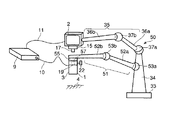
通常、撮像装置と画像表示装置を接続した状態で手術を行い、術中に撮像装置を大きく傾ける(被検部を大きく傾斜した方向から観察する)必要が生じた場合、撮像装置と画像表示装置とを分離させることができる。従って、術者は辛い姿勢から解放され手術を続けることが可能となる。また、術者の好みによって装着或いは分離を選択して使用することも可能である。
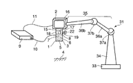
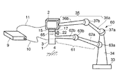
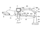
図1〜図2に基づいて本発明の第1の実施形態に係る画像顕微鏡装置を説明する。図1は撮像装置と画像表示装置が接続された使用状態の説明図であり、図2は撮像装置と画像表示装置が分離された使用状態の説明図である。
図1中、符号1で示すものは画像顕微鏡装置の撮像装置(撮像手段)であり、符号2で示すものは画像顕微鏡装置の画像表示装置(画像表示手段)である。撮像装置1と画像表示装置2は後述する着脱自在な接続手段によって着脱が自在である。
次に、本実施形態の画像顕微鏡装置の使用例について説明する。図1は撮像装置1と画像表示装置2とが一体的に接続された使用状態である。この接続状態では上記画像表示装置2の嵌合軸17を上記撮像装置1の嵌合穴19に嵌着させるとともに、上記ツマミ22を締め込むことによって嵌合軸17を締結することで固定的になる。このとき、撮像装置1を画像表示装置2に装着したので、第1保持架台31の使用のみで済み、第2保持架台32を使用しないで済む。
本実施形態によれば、撮像装置(撮像部)1の観察方向の変更に伴って画像表示装置2の位置状態を追従させる使用形態と、撮像装置(撮像部)1の観察方向の変更に拘わらず、画像表示装置2はそのままの位置状態を保つ使用形態の両方が可能である。従って、術前のセッティングによって使用状況または術者の好み等に応じた使用法の選択が可能となる。
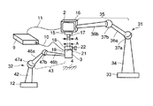
図3及び図4に基づいて本発明の第2の実施形態に係る画像顕微鏡装置を説明する。ただし、上述した第1の実施形態と同一の構成のものについては同一の符号を付し、その詳細な説明を省略する。図4は撮像装置と画像表示装置が接続された使用状態の説明図であり、図5は撮像装置と画像表示装置が分離された使用状態の説明図である。
本実施形態での撮像装置1及び画像表示装置2は上述した第1の実施形態のものと略同様に構成されるが、本実施形態では一台の保持架台50で撮像装置1及び画像表示装置2の両方を支持できるようにしたものである。
図3に示すように、上記撮像装置1と上記画像表示装置2を一体的に接続して使用する場合では、上述した実施形態の場合と同様、支持アーム35の先端に画像表示装置2を支持し、撮像装置1はその画像表示装置2に一体的に接続された状態にある。このとき、別のアーム51は図3に示すように折り畳まれ、収納状態にある。
本実施形態によれば、上述した第1の実施形態によって得られる効果に加えて、保持架台が1台で済む。このため、システム全体の価格を下げ得る。さらには手術室内における装置の占有スペースが少なくて済むという利点も有する。手術室内は様々な治療機器や麻酔装置などが多々置かれており、助手や看護婦の立つ、スペースが中々確保できないという問題に対して貢献することができる。
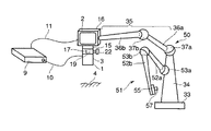
図5及び図6に基づいて本発明の第3の実施形態に係る画像顕微鏡装置を説明する。ただし、上述した第1の実施形態と同一の構成のものには同一の符号を付し、その詳細な説明を省略する。図4は撮像装置と画像表示装置が接続された使用状態の説明図であり、図5は撮像装置と画像表示装置が分離された使用状態の説明図である。
本実施形態での撮像装置1及び画像表示装置2は上述した第1の実施形態のものと略同様に構成されるが、本実施形態では一台の保持架台50で撮像装置1及び画像表示装置2の両方を支持できるようにしたものである。
次に、本実施形態の一使用例について述べる。まず、図5に示すように、上記撮像装置1と、上記画像表示装置2を一体的に組み合わせて使用する場合では、上述した第1の実施形態の場合と同様、画像表示装置用支持アーム35の先端に画像表示装置2を支持し、撮像装置1はその画像表示装置2に対して一体的に組み付けた接続状態にある。さらに、撮像装置1は別の撮像装置用支持アーム61の先端に支持された状態にある。
本実施形態によれば、ツマミ22を緩めて嵌合軸17を抜き出すだけで簡単に撮像装置1と画像表示装置2を接続された状態から分離された状態に変更することができるので、上述した第1の実施形態及び第2の実施形態によって得られる効果に加えて、術中であっても容易に両者の使い分けが可能となる。
付記2.上記撮像装置と上記画像表示装置は各々異なる支持アームにて支持されることを特徴とする付記2に記載の画像顕微鏡装置。
付記3.上記各支持アームは共通の支柱に配設されていることを特徴とする付記2に記載の画像顕微鏡装置。
付記4.上記着脱手段は上記支持アームの少なくとも1つと着脱の互換性を有することを特徴とする付記2に記載の画像顕微鏡装置。
付記5.上記支持アームの少なくとも1つは上記撮像装置と上記画像表示装置の何れか一方と一体或いは着脱可能であると共に、他方と着脱可能であることを特徴とする付記2に記載の画像顕微鏡装置。
付記6.上記支持アームの少なくとも1つは先端に支持する装置の重量に相当する反力を有するバランスアームからなると共に、該バランスアームは上記撮像装置と上記画像表示装置との着脱の有無に対して上記反力を変更する反力変更手段を備えることを特徴とする付記2に記載の画像顕微鏡装置。
16…装置本体、17…嵌合軸、18…着脱部、19…嵌合穴
31…保持架台、32…保持架台、35…画像表示装置用支持アーム
61…撮像装置用支持アーム。
Claims (3)
- 被検部を撮像する撮像装置と、
上記撮像装置によって撮像した画像を表示する画像表示装置と、
上記撮像装置及び上記画像表示装置同士を着脱自在となるよう接続する着脱式接続手段と、
を具備したことを特徴とする画像顕微鏡装置。 - 上記撮像装置を移動自在に支持する第1の支持装置と、上記画像表示装置を移動自在に支持する第2の支持装置とを備えることを特徴とする請求項1に記載の画像顕微鏡装置。
- 上記撮像装置を移動自在に支持する第1の支持アームと、上記画像表示装置を移動自在に支持する第2の支持アームとを備え、上記第1の支持アームと上記第2の支持アームを一台の架台に取り付けたことを特徴とする請求項1に記載の画像顕微鏡装置。
Priority Applications (3)
| Application Number | Priority Date | Filing Date | Title |
|---|---|---|---|
| JP2004104957A JP2005292320A (ja) | 2004-03-31 | 2004-03-31 | 画像顕微鏡装置 |
| US11/095,389 US8265734B2 (en) | 2004-03-31 | 2005-03-31 | Imaging and displaying system with imaging unit and display unit which are supported by movable arm |
| EP05007074A EP1582167B1 (en) | 2004-03-31 | 2005-03-31 | Imaging and displaying system with imaging unit and display unit which are supported by movable arm |
Applications Claiming Priority (1)
| Application Number | Priority Date | Filing Date | Title |
|---|---|---|---|
| JP2004104957A JP2005292320A (ja) | 2004-03-31 | 2004-03-31 | 画像顕微鏡装置 |
Publications (1)
| Publication Number | Publication Date |
|---|---|
| JP2005292320A true JP2005292320A (ja) | 2005-10-20 |
Family
ID=34880063
Family Applications (1)
| Application Number | Title | Priority Date | Filing Date |
|---|---|---|---|
| JP2004104957A Pending JP2005292320A (ja) | 2004-03-31 | 2004-03-31 | 画像顕微鏡装置 |
Country Status (3)
| Country | Link |
|---|---|
| US (1) | US8265734B2 (ja) |
| EP (1) | EP1582167B1 (ja) |
| JP (1) | JP2005292320A (ja) |
Cited By (3)
| Publication number | Priority date | Publication date | Assignee | Title |
|---|---|---|---|---|
| JP2010511902A (ja) * | 2006-12-07 | 2010-04-15 | スイス メディカル テヒノロギー ゲーエムベーハー | 表示ユニットの目視者依存的な位置決め及び位置合わせのための支持システム及び方法 |
| WO2016043046A1 (ja) * | 2014-09-16 | 2016-03-24 | ソニー・オリンパスメディカルソリューションズ株式会社 | 医療用観察装置および医療用観察システム |
| JPWO2016152987A1 (ja) * | 2015-03-25 | 2018-01-18 | ソニー・オリンパスメディカルソリューションズ株式会社 | 医療用観察装置、手術用観察装置及び医療用観察システム |
Families Citing this family (15)
| Publication number | Priority date | Publication date | Assignee | Title |
|---|---|---|---|---|
| KR101155258B1 (ko) * | 2005-09-30 | 2012-06-13 | 레스토레이션 로보틱스, 인코포레이티드 | 모낭 유닛들을 채취 및 이식하기 위한 장치 및 방법들 |
| CA2667015C (en) * | 2006-10-20 | 2013-04-23 | Cardiorobotics, Inc. | Apparatus for positioning a device |
| US7593219B2 (en) * | 2007-04-26 | 2009-09-22 | Hewlett-Packard Development Company, L.P. | Display support system and method |
| US8982203B2 (en) | 2007-06-06 | 2015-03-17 | Karl Storz Gmbh & Co. Kg | Video system for viewing an object on a body |
| US8632064B2 (en) * | 2009-07-15 | 2014-01-21 | The Board Of Trustees Of The Leland Stanford Junior University | Positioning apparatus with lockable joints and method of use |
| CN102778750B (zh) * | 2012-07-20 | 2014-06-25 | 中国地质大学(武汉) | 一种显微镜实验教学显示系统 |
| JP6214186B2 (ja) * | 2013-03-29 | 2017-10-18 | キヤノン株式会社 | 放射線発生用装置及び放射線撮影装置 |
| EP3033244B1 (de) * | 2013-08-12 | 2019-05-08 | Schunk Bahn- und Industrietechnik GmbH | Stromübertragungsvorrichtung |
| DE102013226288A1 (de) * | 2013-12-17 | 2015-06-18 | Carl Zeiss Meditec Ag | Operationsmikroskop |
| JP6858593B2 (ja) * | 2017-03-02 | 2021-04-14 | ソニー・オリンパスメディカルソリューションズ株式会社 | 医療用観察装置、および制御方法 |
| CN110131532B (zh) * | 2018-02-08 | 2024-09-03 | 泰科电子(上海)有限公司 | 成像设备 |
| CN110068919A (zh) * | 2019-05-29 | 2019-07-30 | 苏州四海通仪器有限公司 | 一种便拆式手术显微镜 |
| US11448868B2 (en) | 2020-05-07 | 2022-09-20 | Kgg Inc | Ergonomic EZ scope digital imaging system |
| IT202000022843A1 (it) * | 2020-09-28 | 2022-03-28 | Pont Franco Dal | Microscopio chirurgico |
| US12467489B2 (en) * | 2022-03-17 | 2025-11-11 | Mako Surgical Corp. | Techniques for securing together components of one or more surgical carts |
Citations (3)
| Publication number | Priority date | Publication date | Assignee | Title |
|---|---|---|---|---|
| JPH09102899A (ja) * | 1995-10-05 | 1997-04-15 | Scala Kk | 顕微鏡用ビデオカメラ及び顕微鏡用ビデオ装置 |
| JPH10179503A (ja) * | 1996-12-26 | 1998-07-07 | Asahi Optical Co Ltd | 手術用内視鏡装置 |
| JP2003250812A (ja) * | 2002-02-28 | 2003-09-09 | Olympus Optical Co Ltd | 医療用立体表示装置 |
Family Cites Families (29)
| Publication number | Priority date | Publication date | Assignee | Title |
|---|---|---|---|---|
| DE7930125U1 (de) | 1979-07-24 | 1980-01-24 | Contraves Ag, Zuerich (Schweiz) | Zusatzvorrichtung an einem stativ fuer ein optisches beobachtungsgeraet |
| JPS6070350A (ja) * | 1983-09-28 | 1985-04-22 | Hitachi Ltd | 光学顕微鏡を併設した超音波顕微鏡 |
| JPS6098352A (ja) * | 1983-11-02 | 1985-06-01 | Olympus Optical Co Ltd | 超音波顕微鏡 |
| US4741607A (en) | 1986-03-17 | 1988-05-03 | Contraves Ag | Supporting device for an optical observation instrument |
| US4863133A (en) * | 1987-05-26 | 1989-09-05 | Leonard Medical | Arm device for adjustable positioning of a medical instrument or the like |
| JPS63296746A (ja) | 1987-05-29 | 1988-12-02 | Mitaka Koki Kk | 医療用光学機器のスタンド装置 |
| FR2651668B1 (fr) * | 1989-09-12 | 1991-12-27 | Leon Claude | Ensemble microscope-endoscope utile notamment en chirurgie. |
| US5762458A (en) * | 1996-02-20 | 1998-06-09 | Computer Motion, Inc. | Method and apparatus for performing minimally invasive cardiac procedures |
| US5579772A (en) * | 1993-06-14 | 1996-12-03 | Olympus Optical Co., Ltd. | Surgical microscope system |
| DE59609939D1 (de) * | 1995-02-20 | 2003-01-16 | Sybill Storz | Vorrichtung zur untersuchung von hohlräumen mit einem endoskop |
| AU7517096A (en) * | 1995-10-20 | 1997-05-07 | Urohealth Systems Inc. | Hand-held imaging apparatus for use with endoscopes |
| US5855583A (en) * | 1996-02-20 | 1999-01-05 | Computer Motion, Inc. | Method and apparatus for performing minimally invasive cardiac procedures |
| JPH09238951A (ja) * | 1996-03-06 | 1997-09-16 | Topcon Corp | 手術用顕微鏡装置 |
| US6575908B2 (en) * | 1996-06-28 | 2003-06-10 | Sonosite, Inc. | Balance body ultrasound system |
| US5999837A (en) * | 1997-09-26 | 1999-12-07 | Picker International, Inc. | Localizing and orienting probe for view devices |
| AU3893299A (en) | 1998-05-13 | 1999-11-29 | Inbae Yoon | Penetrating endoscope and endoscopic surgical instrument with cmos image sensor and display |
| US6419626B1 (en) | 1998-08-12 | 2002-07-16 | Inbae Yoon | Surgical instrument endoscope with CMOS image sensor and physical parameter sensor |
| JP4101951B2 (ja) * | 1998-11-10 | 2008-06-18 | オリンパス株式会社 | 手術用顕微鏡 |
| US6398721B1 (en) * | 1999-02-19 | 2002-06-04 | Olympus Optical Co., Ltd. | Surgical microscope apparatus |
| US6447451B1 (en) * | 1999-05-04 | 2002-09-10 | Sonosite, Inc. | Mobile ultrasound diagnostic instrument and docking stand |
| JP4611491B2 (ja) | 1999-05-31 | 2011-01-12 | Hoya株式会社 | ビデオ型立体顕微鏡 |
| JP4245750B2 (ja) * | 1999-10-15 | 2009-04-02 | オリンパス株式会社 | 立体観察装置 |
| JP2001224595A (ja) * | 1999-12-08 | 2001-08-21 | Olympus Optical Co Ltd | 顕微鏡下手術用超音波プローブ |
| AU2597501A (en) * | 1999-12-23 | 2001-07-03 | Hill-Rom Services, Inc. | Surgical theater system |
| JP2001276092A (ja) | 2000-03-30 | 2001-10-09 | Konan Medical Inc | 簡易型医療用顕微鏡 |
| JP2002006228A (ja) | 2000-06-23 | 2002-01-09 | Asahi Optical Co Ltd | 手術用ビデオ型顕微鏡 |
| JP3791907B2 (ja) * | 2002-02-12 | 2006-06-28 | オリンパス株式会社 | 観察装置 |
| JP4073249B2 (ja) * | 2002-05-17 | 2008-04-09 | オリンパス株式会社 | 手術システム |
| JP4398352B2 (ja) * | 2004-12-02 | 2010-01-13 | オリンパス株式会社 | 医療用立体撮像装置 |
-
2004
- 2004-03-31 JP JP2004104957A patent/JP2005292320A/ja active Pending
-
2005
- 2005-03-31 EP EP05007074A patent/EP1582167B1/en not_active Ceased
- 2005-03-31 US US11/095,389 patent/US8265734B2/en not_active Expired - Fee Related
Patent Citations (3)
| Publication number | Priority date | Publication date | Assignee | Title |
|---|---|---|---|---|
| JPH09102899A (ja) * | 1995-10-05 | 1997-04-15 | Scala Kk | 顕微鏡用ビデオカメラ及び顕微鏡用ビデオ装置 |
| JPH10179503A (ja) * | 1996-12-26 | 1998-07-07 | Asahi Optical Co Ltd | 手術用内視鏡装置 |
| JP2003250812A (ja) * | 2002-02-28 | 2003-09-09 | Olympus Optical Co Ltd | 医療用立体表示装置 |
Cited By (7)
| Publication number | Priority date | Publication date | Assignee | Title |
|---|---|---|---|---|
| JP2010511902A (ja) * | 2006-12-07 | 2010-04-15 | スイス メディカル テヒノロギー ゲーエムベーハー | 表示ユニットの目視者依存的な位置決め及び位置合わせのための支持システム及び方法 |
| WO2016043046A1 (ja) * | 2014-09-16 | 2016-03-24 | ソニー・オリンパスメディカルソリューションズ株式会社 | 医療用観察装置および医療用観察システム |
| US20170227753A1 (en) | 2014-09-16 | 2017-08-10 | Sony Olympus Medical Solutions Inc. | Medical observation apparatus and medical observation system |
| US10114208B2 (en) | 2014-09-16 | 2018-10-30 | Sony Olympus Medical Solutions Inc. | Medical observation apparatus and medical observation system |
| US10634896B2 (en) | 2014-09-16 | 2020-04-28 | Sony Olympus Medical Solutions Inc. | Medical observation apparatus and medical observation system that convert an imaging signal to an optical signal |
| JPWO2016152987A1 (ja) * | 2015-03-25 | 2018-01-18 | ソニー・オリンパスメディカルソリューションズ株式会社 | 医療用観察装置、手術用観察装置及び医療用観察システム |
| US10925685B2 (en) | 2015-03-25 | 2021-02-23 | Sony Olympus Medical Solutions Inc. | Medical observation device, surgical observation device, and medical observation system |
Also Published As
| Publication number | Publication date |
|---|---|
| US8265734B2 (en) | 2012-09-11 |
| US20050228257A1 (en) | 2005-10-13 |
| EP1582167A3 (en) | 2010-04-21 |
| EP1582167A2 (en) | 2005-10-05 |
| EP1582167B1 (en) | 2013-02-27 |
Similar Documents
| Publication | Publication Date | Title |
|---|---|---|
| JP2005292320A (ja) | 画像顕微鏡装置 | |
| EP3420878B1 (en) | Information processing device for medical use, information processing method, information processing system for medical use | |
| JP3402797B2 (ja) | 内視鏡用画像表示システム | |
| JP2004181229A (ja) | 遠隔手術支援システム及び支援方法 | |
| JPWO2012117922A1 (ja) | 医療用保持装置 | |
| JP7115467B2 (ja) | 手術用画像処理装置、画像処理方法、及び、手術システム | |
| JP4716545B2 (ja) | 手術用顕微鏡装置 | |
| EP3603562B1 (en) | Medical observation apparatus and observation field correction method | |
| WO2017169650A1 (ja) | 医療用観察装置、映像移動補正方法及び医療用観察システム | |
| JP4383188B2 (ja) | 立体観察システム | |
| JP2014076204A (ja) | 医療用観察システム | |
| JP6656237B2 (ja) | 手術用顕微鏡装置及び手術用顕微鏡システム | |
| JP3806568B2 (ja) | 手術用顕微鏡装置 | |
| JP4426770B2 (ja) | 内視鏡保持装置 | |
| JPH0970406A (ja) | 手術用観察装置 | |
| JP2002125219A (ja) | システム制御装置 | |
| JP2000262458A (ja) | 手術観察用撮像システム | |
| JP2001145003A (ja) | 内視鏡用撮像装置 | |
| JP7307986B1 (ja) | マイクロスコープ | |
| JPH0716238A (ja) | 保持装置 | |
| JP2001111991A (ja) | 顔面装着型映像表示装置 | |
| WO2018043205A1 (ja) | 医療用画像処理装置、医療用画像処理方法、プログラム | |
| JP4077682B2 (ja) | 手術用観察装置 | |
| JP2006014825A (ja) | 医療用観察装置 | |
| JP2004016282A (ja) | 内視鏡用画像処理装置 |
Legal Events
| Date | Code | Title | Description |
|---|---|---|---|
| A621 | Written request for application examination |
Free format text: JAPANESE INTERMEDIATE CODE: A621 Effective date: 20070119 |
|
| A131 | Notification of reasons for refusal |
Free format text: JAPANESE INTERMEDIATE CODE: A131 Effective date: 20100316 |
|
| A521 | Written amendment |
Free format text: JAPANESE INTERMEDIATE CODE: A523 Effective date: 20100514 |
|
| A02 | Decision of refusal |
Free format text: JAPANESE INTERMEDIATE CODE: A02 Effective date: 20101124 |
|
| A521 | Written amendment |
Free format text: JAPANESE INTERMEDIATE CODE: A523 Effective date: 20110223 |
|
| A911 | Transfer to examiner for re-examination before appeal (zenchi) |
Free format text: JAPANESE INTERMEDIATE CODE: A911 Effective date: 20110303 |
|
| A912 | Re-examination (zenchi) completed and case transferred to appeal board |
Free format text: JAPANESE INTERMEDIATE CODE: A912 Effective date: 20110415 |
|
| A521 | Written amendment |
Free format text: JAPANESE INTERMEDIATE CODE: A523 Effective date: 20120406 |