EP3401391A1 - Reifung von aus menschlichen pluripotenten stammzellen abgeleiteten hepatozytenartigen zellen - Google Patents
Reifung von aus menschlichen pluripotenten stammzellen abgeleiteten hepatozytenartigen zellen Download PDFInfo
- Publication number
- EP3401391A1 EP3401391A1 EP18169827.5A EP18169827A EP3401391A1 EP 3401391 A1 EP3401391 A1 EP 3401391A1 EP 18169827 A EP18169827 A EP 18169827A EP 3401391 A1 EP3401391 A1 EP 3401391A1
- Authority
- EP
- European Patent Office
- Prior art keywords
- cells
- hepatocyte
- retinoic acid
- activator
- exposed
- Prior art date
- Legal status (The legal status is an assumption and is not a legal conclusion. Google has not performed a legal analysis and makes no representation as to the accuracy of the status listed.)
- Withdrawn
Links
Images


Classifications
-
- C—CHEMISTRY; METALLURGY
- C12—BIOCHEMISTRY; BEER; SPIRITS; WINE; VINEGAR; MICROBIOLOGY; ENZYMOLOGY; MUTATION OR GENETIC ENGINEERING
- C12N—MICROORGANISMS OR ENZYMES; COMPOSITIONS THEREOF; PROPAGATING, PRESERVING, OR MAINTAINING MICROORGANISMS; MUTATION OR GENETIC ENGINEERING; CULTURE MEDIA
- C12N5/00—Undifferentiated human, animal or plant cells, e.g. cell lines; Tissues; Cultivation or maintenance thereof; Culture media therefor
- C12N5/06—Animal cells or tissues; Human cells or tissues
- C12N5/0602—Vertebrate cells
- C12N5/067—Hepatocytes
-
- C—CHEMISTRY; METALLURGY
- C12—BIOCHEMISTRY; BEER; SPIRITS; WINE; VINEGAR; MICROBIOLOGY; ENZYMOLOGY; MUTATION OR GENETIC ENGINEERING
- C12N—MICROORGANISMS OR ENZYMES; COMPOSITIONS THEREOF; PROPAGATING, PRESERVING, OR MAINTAINING MICROORGANISMS; MUTATION OR GENETIC ENGINEERING; CULTURE MEDIA
- C12N2501/00—Active agents used in cell culture processes, e.g. differentation
- C12N2501/06—Anti-neoplasic drugs, anti-retroviral drugs, e.g. azacytidine, cyclophosphamide
-
- C—CHEMISTRY; METALLURGY
- C12—BIOCHEMISTRY; BEER; SPIRITS; WINE; VINEGAR; MICROBIOLOGY; ENZYMOLOGY; MUTATION OR GENETIC ENGINEERING
- C12N—MICROORGANISMS OR ENZYMES; COMPOSITIONS THEREOF; PROPAGATING, PRESERVING, OR MAINTAINING MICROORGANISMS; MUTATION OR GENETIC ENGINEERING; CULTURE MEDIA
- C12N2501/00—Active agents used in cell culture processes, e.g. differentation
- C12N2501/30—Hormones
- C12N2501/38—Hormones with nuclear receptors
- C12N2501/385—Hormones with nuclear receptors of the family of the retinoic acid recptor, e.g. RAR, RXR; Peroxisome proliferator-activated receptor [PPAR]
-
- C—CHEMISTRY; METALLURGY
- C12—BIOCHEMISTRY; BEER; SPIRITS; WINE; VINEGAR; MICROBIOLOGY; ENZYMOLOGY; MUTATION OR GENETIC ENGINEERING
- C12N—MICROORGANISMS OR ENZYMES; COMPOSITIONS THEREOF; PROPAGATING, PRESERVING, OR MAINTAINING MICROORGANISMS; MUTATION OR GENETIC ENGINEERING; CULTURE MEDIA
- C12N2501/00—Active agents used in cell culture processes, e.g. differentation
- C12N2501/40—Regulators of development
- C12N2501/405—Cell cycle regulated proteins, e.g. cyclins, cyclin-dependant kinases
-
- C—CHEMISTRY; METALLURGY
- C12—BIOCHEMISTRY; BEER; SPIRITS; WINE; VINEGAR; MICROBIOLOGY; ENZYMOLOGY; MUTATION OR GENETIC ENGINEERING
- C12N—MICROORGANISMS OR ENZYMES; COMPOSITIONS THEREOF; PROPAGATING, PRESERVING, OR MAINTAINING MICROORGANISMS; MUTATION OR GENETIC ENGINEERING; CULTURE MEDIA
- C12N2501/00—Active agents used in cell culture processes, e.g. differentation
- C12N2501/40—Regulators of development
- C12N2501/415—Wnt; Frizzeled
-
- C—CHEMISTRY; METALLURGY
- C12—BIOCHEMISTRY; BEER; SPIRITS; WINE; VINEGAR; MICROBIOLOGY; ENZYMOLOGY; MUTATION OR GENETIC ENGINEERING
- C12N—MICROORGANISMS OR ENZYMES; COMPOSITIONS THEREOF; PROPAGATING, PRESERVING, OR MAINTAINING MICROORGANISMS; MUTATION OR GENETIC ENGINEERING; CULTURE MEDIA
- C12N2501/00—Active agents used in cell culture processes, e.g. differentation
- C12N2501/70—Enzymes
- C12N2501/72—Transferases (EC 2.)
- C12N2501/727—Kinases (EC 2.7.)
-
- C—CHEMISTRY; METALLURGY
- C12—BIOCHEMISTRY; BEER; SPIRITS; WINE; VINEGAR; MICROBIOLOGY; ENZYMOLOGY; MUTATION OR GENETIC ENGINEERING
- C12N—MICROORGANISMS OR ENZYMES; COMPOSITIONS THEREOF; PROPAGATING, PRESERVING, OR MAINTAINING MICROORGANISMS; MUTATION OR GENETIC ENGINEERING; CULTURE MEDIA
- C12N2501/00—Active agents used in cell culture processes, e.g. differentation
- C12N2501/999—Small molecules not provided for elsewhere
-
- C—CHEMISTRY; METALLURGY
- C12—BIOCHEMISTRY; BEER; SPIRITS; WINE; VINEGAR; MICROBIOLOGY; ENZYMOLOGY; MUTATION OR GENETIC ENGINEERING
- C12N—MICROORGANISMS OR ENZYMES; COMPOSITIONS THEREOF; PROPAGATING, PRESERVING, OR MAINTAINING MICROORGANISMS; MUTATION OR GENETIC ENGINEERING; CULTURE MEDIA
- C12N2506/00—Differentiation of animal cells from one lineage to another; Differentiation of pluripotent cells
- C12N2506/02—Differentiation of animal cells from one lineage to another; Differentiation of pluripotent cells from embryonic cells
-
- C—CHEMISTRY; METALLURGY
- C12—BIOCHEMISTRY; BEER; SPIRITS; WINE; VINEGAR; MICROBIOLOGY; ENZYMOLOGY; MUTATION OR GENETIC ENGINEERING
- C12N—MICROORGANISMS OR ENZYMES; COMPOSITIONS THEREOF; PROPAGATING, PRESERVING, OR MAINTAINING MICROORGANISMS; MUTATION OR GENETIC ENGINEERING; CULTURE MEDIA
- C12N2506/00—Differentiation of animal cells from one lineage to another; Differentiation of pluripotent cells
- C12N2506/03—Differentiation of animal cells from one lineage to another; Differentiation of pluripotent cells from non-embryonic pluripotent stem cells
-
- C—CHEMISTRY; METALLURGY
- C12—BIOCHEMISTRY; BEER; SPIRITS; WINE; VINEGAR; MICROBIOLOGY; ENZYMOLOGY; MUTATION OR GENETIC ENGINEERING
- C12N—MICROORGANISMS OR ENZYMES; COMPOSITIONS THEREOF; PROPAGATING, PRESERVING, OR MAINTAINING MICROORGANISMS; MUTATION OR GENETIC ENGINEERING; CULTURE MEDIA
- C12N2506/00—Differentiation of animal cells from one lineage to another; Differentiation of pluripotent cells
- C12N2506/45—Differentiation of animal cells from one lineage to another; Differentiation of pluripotent cells from artificially induced pluripotent stem cells
-
- C—CHEMISTRY; METALLURGY
- C12—BIOCHEMISTRY; BEER; SPIRITS; WINE; VINEGAR; MICROBIOLOGY; ENZYMOLOGY; MUTATION OR GENETIC ENGINEERING
- C12N—MICROORGANISMS OR ENZYMES; COMPOSITIONS THEREOF; PROPAGATING, PRESERVING, OR MAINTAINING MICROORGANISMS; MUTATION OR GENETIC ENGINEERING; CULTURE MEDIA
- C12N2533/00—Supports or coatings for cell culture, characterised by material
- C12N2533/50—Proteins
- C12N2533/52—Fibronectin; Laminin
-
- C—CHEMISTRY; METALLURGY
- C12—BIOCHEMISTRY; BEER; SPIRITS; WINE; VINEGAR; MICROBIOLOGY; ENZYMOLOGY; MUTATION OR GENETIC ENGINEERING
- C12N—MICROORGANISMS OR ENZYMES; COMPOSITIONS THEREOF; PROPAGATING, PRESERVING, OR MAINTAINING MICROORGANISMS; MUTATION OR GENETIC ENGINEERING; CULTURE MEDIA
- C12N2533/00—Supports or coatings for cell culture, characterised by material
- C12N2533/50—Proteins
- C12N2533/54—Collagen; Gelatin
Definitions
- the present invention relates to directed differentiation and maturation of hepatocyte-like cells.
- the hepatocyte-like cells obtained in accordance with the present invention show a phenotype which is more similar to that of primary hepatocytes than previously shown.
- the present invention relates to exposure of hepatocyte-like cells to an activator of a retinoic acid responsive receptor, such as retinoic acid (RA), optionally in combination with an inhibitor of GSK-3 (Glycogen synthase kinase 3) or activator of Wnt signalling and/or with the overlay of the cells with one or more components characteristic of the mammalian extracellular matrix (matrix overlay).
- RA retinoic acid
- GSK-3 Glycogen synthase kinase 3
- the present invention also relates to exposure of hepatocyte-like cells to an activator of a retinoic acid responsive receptor, such as retinoic acid (RA), optionally in combination with an inhibitor of a cyclin dependent kinase (CDK) and/or with the overlay of the cells with one or more components characteristic of the mammalian extracellular matrix (matrix overlay).
- a retinoic acid responsive receptor such as retinoic acid (RA)
- CDK cyclin dependent kinase
- matrix overlay one or more components characteristic of the mammalian extracellular matrix
- hES human embryonic-derived stem
- hiPS human induced pluripotent stem
- hepatic cell lines which often contain very low levels of (or totally lack) metabolising enzymes and have expression of other important proteins substantially different from the native hepatocyte in vivo. This is of particular relevance in relation to drug metabolism since one of the major deficiencies in hepatic cell lines is the absence or abnormally high expression of drug transporter proteins which are essential for drug screening purposes.
- Other available hepatic cell lines suffer from having a morphology and physiology which is more pronounced of fetal or juvenile hepatocytes than the more clinically relevant adult hepatocytes. For these reasons there is a strong need to develop hepatocyte cell lines which are not only easy to culture and propagate but which also possess a more mature phenotype and which behave in a manner more akin to adult primary hepatocytes.
- hepatocyte-like cells from pluripotent stem cells.
- hES cells Hue et al., 2007; Hay et al., 2008; Brolen et al. 2010; Funakoshi et al. 2011
- hiPS cells US8148151B ; Song et al. 2009; Sullivan et al. 2010; Si-Tayeb et al. 2010; Chen et al. 2012.
- common to all of these is a specific low mRNA and protein expression of genes typical for mature hepatocytes, like phase I and II genes (e.g.
- CYP1A2, 2B6, 2C9, 2D6, 3A4 nuclear receptors (e.g. CAR and PXR), and other adult hepatic markers (e.g. Albumin).
- these hESC- and hiPSC-derived hepatocyte-like cells have high expression of fetal hepatic genes like ⁇ -fetoprotein (AFP) and CYP3A7, with the result that the cell types described therein have a fetal and not adult phenotype (for overview see e.g. Baxter et al. 2010).Furthermore, in most of the published studies on hESC- and hiPSC-derived hepatocyte-like cells, expression and functionality of drug transporters has not been investigated at all.
- AFP ⁇ -fetoprotein
- RA-response elements have been identified in a number of genes important during early hepatocyte specification such as AFP and HNF4 ⁇ (see Qian et al 2000; Magee et al 1998 and Hatzis et al 2001).
- AFP and HNF4 ⁇ are important during early hepatocyte specification
- RA is known to have diverse effects and has also been found to be important in the derivation of pancreatic endoderm from pluripotent stem cells (Mfopou et al 2010).
- US Patent Application Publication US2012/0143316A1 discloses the use of all-trans retinoic acid in inducing hepatic differentiation from endoderm-like cells. As becomes evident, all of these disclosures relate to the modulation of RA signalling during endodermal and early hepatocyte differentiation. However, none of these documents teaches or suggests the applicability of retinoic acid as an hepatocyte maturation promoting agent, let alone its use at a late stage in hepatocyte differentiation.
- WO08094597 (Dalton ) describes a method of producing mesendoderm from primate pluripotent stem cells (pPSC) by contacting the pPSC with an effective amount of GSK3 inhibitor in a differentiation media.
- WO2007050043 (Stanton ) describes a method for producing a mesodermal or an endodermal cell from a pluripotent stem cell, comprising a Wnt-signalling pathway in the pluripotent stem cell.
- US2006003446 (Keller ) describes a way of making a cell population enriched for endoderm cells culturing embryonic stem cells in the absence of serum and in the presence of activin and an inhibitor of Wnt-signalling. Modulation of Wnt signalling through the use of a GSK3 inhibitor has also been shown to be beneficial in specifying hepatocyte cell fate when DE cells are exposed to this treatment ( WO2011/116930 ). Again, all of these disclosures relate to modulation of GSK3 signalling at relatively early stages in endodermal or hepatic specification.
- Culturing of cells on certain matrix components has been known to affect their growth and, in the case of multipotent cells, to affect their ultimate differentiation.
- pluripotent stem cells have been shown to undergo epithelial to mesenchymal transition and thence develop into cardiac cell types through overlaying of the stem cells with certain matrix components ( WO2011060342 ).
- culturing of adult primary hepatocytes in defined "sandwich" of matrix components has long been known to help them maintain their phenotype and metabolic activity (Dunn et al 1991), (Page et al 2007).
- hepatocyte-like cells derived from human pluripotent stem (hPS) cells such as but not limited to hiPS-cells and hES-cells, may be further matured into hepatocyte-like cells possessing a phenotype more closely resembling that of ex vivo primary liver hepatocytes.
- hPS human pluripotent stem
- the present invention provides in a first aspect a method for promoting the maturation of human hepatocyte-like cells whereby said hepatocyte-like cells are exposed to an activator of a retinoic acid responsive receptor, such as retinoic acid, optionally in combination with exposure to an inhibitor of GSK3 signalling or activator of Wnt signalling and/or with an overlay of the cells with one or more components characteristic of the mammalian extracellular matrix (matrix overlay).
- a retinoic acid responsive receptor such as retinoic acid
- a method for promoting the maturation of human hepatocyte-like cells comprising: Exposing said human hepatocyte-like cells to an activator of a retinoic acid responsive receptor.
- the method for promoting the maturation of human hepatocyte-like cells may further comprise culturing human hepatic progenitor cells under differentiation conditions to obtain said hepatocyte-like cells.
- the present invention provides in a second aspect a method for producing human hepatocyte-like cells whereby human hepatic progenitor cells are cultured under differentiation conditions to obtain hepatocyte-like cells, and the obtained hepatocyte-like cells are exposed to an activator of a retinoic acid responsive receptor, such as retinoic acid, optionally in combination with exposure to an inhibitor of GSK3 signalling or activator of Wnt signalling and/or with an overlay of the cells with one or more components characteristic of the mammalian extracellular matrix (matrix overlay).
- a retinoic acid responsive receptor such as retinoic acid
- a method for producing human hepatocyte-like cells comprising:
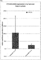
- the present inventors have surprisingly found that the exposure of hepatocyte-like cells to an activator of a retinoic acid responsive receptor, such as retinoic acid, improves the gene and protein expression of a number of markers for mature hepatocytes, notably adult isoforms of HNF4 ⁇ , CYP1A2, CYP2B6, CYP2C9, CYP3A4, CYP3A5, CAR, GSTA1-1, and NTCP, and thus leads to hepatocyte-like cells with a phenotype more closely resembling that of primary hepatocytes.
- a retinoic acid responsive receptor such as retinoic acid
- a surprising synergistic effect was found for exposure to an activator of a retinoic acid responsive receptor, a GSK3-inhibitor and an overlay with one or more components characteristic of the mammalian extracellular matrix making the phenotype of the hepatocyte-like cells even more similar to human primary hepatocytes.
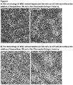
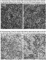
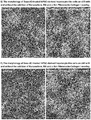
- the morphology of the hepatocyte-like cells is improved, e.g. the cell-cell contacts are enhanced, and the life span of the hepatocyte-like cells is prolonged by 7-10 days ( Figure 8 ).
- the activator of a retinoic acid responsive receptor such as retinoic acid
- a retinoic acid responsive receptor may be present throughout the differentiation of the hepatic progenitor cells into hepatocyte-like cells and further maturation of the obtained hepatocyte-like cells ("differentiation and maturation"), which may take up to 35 days.
- the differentiating and maturing hepatic cells may be continuously/ long term exposed to the activator of a retinoic acid responsive receptor during the differentiation and maturation.
- the hepatocyte-like cells may be exposed to said activator of a retinoic acid responsive receptor for a continuous period of time longer than 4 hours and no longer than 72 hours, such as, e.g., for a continuous period of 5, 24 or 48 hours.
- the hepatocyte-like cells may also be exposed to said activator of a retinoic acid responsive receptor for at least two, such as at least three, at least four or at least 5, continuous periods of time longer than 4 hours and no longer than 72 hours, such for continuous periods of 5, 24 or 48 hours.
- the at least two continuous periods of time are normally separated by a period of non-exposure to said activator of a retinoic acid responsive receptor.
- Such period of non-exposure may have a duration from several hours to several days, such as from 12 to 24 hours or 1 to 10 day, such as from 1 to 2 days.
- the activator of a retinoic acid responsive receptor may be added to the culture medium at any time point during the maturation of the hepatocyte-like cells.
- the hepatocyte-like cells may be exposed to the activator of a retinoic acid responsive receptor at a time t ⁇ 7 days after initiation of the differentiation of hepatic progenitor cells into hepatocyte-like cells.
- hepatocyte-like cells may be exposed to the activator of a retinoic acid responsive receptor at day 7, 9 or 12 after initiation of the differentiation of hepatic progenitor cells into hepatocyte-like cells.
- the methods of the present invention may also comprise the initial generation of hepatic progenitor cells by culturing hPS cells under differentiation conditions (also referred herein as "initial hepatic differentiation").
- hPS cells are initially differentiated into said hepatic progenitor cells.
- This initial culturing or differentiation may include the culturing of the hPS cells under differentiation conditions to obtain cells of the definitive endoderm (DE cells) (pre-endodermal step), and further culturing the obtained DE cells under differentiation conditions to obtain hepatic progenitor cells (pre-hepatic step).
- hPS cells may thus be first differentiated into definitive endoderm, followed by the further differentiation of the definitive endoderm into hepatic progenitor cells.
- the differentiating hPS cells may be exposed to a DNA demethylating agent, such as 5-aza-2-deoxycytidine or 5-azacytidine , to demethylate sections of the genome and allow transcriptional activation of genes.
- a DNA demethylating agent such as 5-aza-2-deoxycytidine or 5-azacytidine
- the exposure to said DNA demethylating agent may take place during the differentiation of the hPS cells into DE cells, i.e. during the pre-endodermal step.
- the cells are then cultured through endodermal stage until hepatic progenitor stage is reached, i.e.
- hepatic progenitor cells are obtained, at which point the further differentiation and maturation of hepatocyte-like cells including the exposure to the activator of a retinoic acid responsive receptor, either alone or in combination with GSK-3 inhibition or activation of Wnt signalling and/or matrix overlay, is carried out.
- the treatment of differentiating hPS cells with a DNA demethylating agent has surprisingly been found to lead to an improved morphology and yield of DE cells.
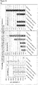
- the exposure to the demethylating agent provides for more pure and homogenous DE populations with lower expression of stem cell markers like Oct4, compared to currently available state of the art methods (see Figure13A to D ).
- an increased gene expression of a number of markers characteristic for definitive endoderm, such as sox17, cxcr4 and hhex is seen for these endodermal cells.
- hepatocyte-like cells are obtained having a phenotype more closely resembling that of primary hepatocytes.
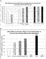
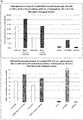
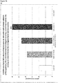
- Analysis of cells subsequent to the maturation period reveals a distinct increase in the expression levels of certain markers for mature hepatocytes, notably but not limited to adult isoforms of HNF4 ⁇ , CYP1A2, CYP2B6, CYP2C9, CYP3A4, CYP3A5, CAR, GSTA1-1, and NTCP (see for example Figures 1B , 4B+C , 6 , 9 , 10 , 12 ).
- the hepatocyte-like cells obtained by an early stage demethylation treatment and late stage exposure to an activator of a retinoic acid responsive receptor, a GSK3-inhibitor (or activator of Wnt signalling) and a matrix overlay display a stable or increasing expression of hepatic genes like CYPs over time in culture.
- CYP activity and mRNA expression is rapidly decreasing over time whereas the opposite is observed for hepatocyte-like cells according to the invention (see Figure 16 ).
- Another widely used hepatic cell type are HepG2 which display much lower CYP activity than hepatocyte-like cells according to the invention.
- the present invention provides a method for promoting the maturation of human hepatocyte-like cells whereby said hepatocyte-like cells are exposed to an activator of a retinoic acid responsive receptor, such as retinoic acid, optionally in combination with exposure to an CDK inhibitor and/or with an overlay of the cells with one or more components characteristic of the mammalian extracellular matrix (matrix overlay).
- a retinoic acid responsive receptor such as retinoic acid
- a method for promoting the maturation of human hepatocyte-like cells comprising: Exposing said human hepatocyte-like cells to an activator of a retinoic acid responsive receptor.
- the method for promoting the maturation of human hepatocyte-like cells may further comprise culturing human hepatic progenitor cells under differentiation conditions to obtain said hepatocyte-like cells.
- the present invention further provides a method for producing human hepatocyte-like cells whereby human hepatic progenitor cells are cultured under differentiation conditions to obtain hepatocyte-like cells, and the obtained hepatocyte-like cells are exposed to an activator of a retinoic acid responsive receptor, such as retinoic acid, optionally in combination with exposure to an CDK inhibitor and/or with an overlay of the cells with one or more components characteristic of the mammalian extracellular matrix (matrix overlay).
- a retinoic acid responsive receptor such as retinoic acid
- a method for producing human hepatocyte-like cells comprising:
- the present inventors have surprisingly found that the exposure of hepatocyte-like cells to an activator of a retinoic acid responsive receptor, such as retinoic acid, improves the gene and protein expression of a number of markers for mature hepatocytes, notably adult isoforms of HNF4 ⁇ , CYP1A2, CYP2B6, CYP2C9, CYP3A4, CYP3A5, CAR, GSTA1-1, and NTCP, and thus leads to hepatocyte-like cells with a phenotype more closely resembling that of primary hepatocytes.
- a retinoic acid responsive receptor such as retinoic acid
- a surprising synergistic effect was found for exposure to an activator of a retinoic acid responsive receptor, a CDK inhibitor and an overlay with one or more components characteristic of the mammalian extracellular matrix making the phenotype of the hepatocyte-like cells even more similar to human primary hepatocytes.
- the morphology of the hepatocyte-like cells is improved, e.g. the cell-cell contacts are enhanced, and the life span of the hepatocyte-like cells is prolonged by 7-10 days ( Figure 8 ).
- the activator of a retinoic acid responsive receptor such as retinoic acid
- the differentiating and maturing hepatic cells may be continuously/ long term exposed to the activator of a retinoic acid responsive receptor during the differentiation and maturation.
- the hepatocyte-like cells may be exposed to said activator of a retinoic acid responsive receptor for a continuous period of time longer than 4 hours and no longer than 72 hours, such as, e.g., for a continuous period of 5, 24 or 48 hours.
- the hepatocyte-like cells may also be exposed to said activator of a retinoic acid responsive receptor for at least two, such as at least three, at least four or at least 5, continuous periods of time longer than 4 hours and no longer than 72 hours, such for continuous periods of 5, 24 or 48 hours.
- the at least two continuous periods of time are normally separated by a period of non-exposure to said activator of a retinoic acid responsive receptor.
- Such period of non-exposure may have a duration from several hours to several days, such as from 12 to 24 hours or 1 to 10 day, such as from 1 to 2 days.
- the activator of a retinoic acid responsive receptor may be added to the culture medium at any time point during the maturation of the hepatocyte-like cells.
- the hepatocyte-like cells may be exposed to the activator of a retinoic acid responsive receptor at a time t ⁇ 7 days after initiation of the differentiation of hepatic progenitor cells into hepatocyte-like cells.
- hepatocyte-like cells may be exposed to the activator of a retinoic acid responsive receptor at day 7, 9 or 12 after initiation of the differentiation of hepatic progenitor cells into hepatocyte-like cells.
- the methods of the present invention may also comprise the initial generation of hepatic progenitor cells by culturing hPS cells under differentiation conditions (also referred herein as "initial hepatic differentiation").
- hPS cells are initially differentiated into said hepatic progenitor cells.
- This initial culturing or differentiation may include the culturing of the hPS cells under differentiation conditions to obtain cells of the definitive endoderm (DE cells) (pre-endodermal step), and further culturing the obtained DE cells under differentiation conditions to obtain hepatic progenitor cells (pre-hepatic step).
- hPS cells may thus be first differentiated into definitive endoderm, followed by the further differentiation of the definitive endoderm into hepatic progenitor cells.
- the differentiating hPS cells may be exposed to a DNA demethylating agent, such as 5-aza-2-deoxycytidine or 5-azacytidine , to demethylate sections of the genome and allow transcriptional activation of genes.
- a DNA demethylating agent such as 5-aza-2-deoxycytidine or 5-azacytidine
- the exposure to said DNA demethylating agent may take place during the differentiation of the hPS cells into DE cells, i.e. during the pre-endodermal step.
- the cells are then cultured through endodermal stage until hepatic progenitor stage is reached, i.e.
- hepatic progenitor cells are obtained, at which point the further differentiation and maturation of hepatocyte-like cells including the exposure to the activator of a retinoic acid responsive receptor, either alone or in combination with CDK inhibition and/or matrix overlay, is carried out.
- the treatment of differentiating hPS cells with a DNA demethylating agent has surprisingly been found to lead to an improved morphology and yield of DE cells.
- the exposure to the demethylating agent provides for more pure and homogenous DE populations with lower expression of stem cell markers like Oct4, compared to currently available state of the art methods (see Figure13A to D ).
- an increased gene expression of a number of markers characteristic for definitive endoderm, such as sox17, cxcr4 and hhex is seen for these endodermal cells.
- hepatocyte-like cells are obtained having a phenotype more closely resembling that of primary hepatocytes.
- Analysis of cells subsequent to the maturation period reveals a distinct increase in the expression levels of certain markers for mature hepatocytes, notably but not limited to adult isoforms of HNF4 ⁇ , CYP1A2, CYP2B6, CYP2C9, CYP3A4, CYP3A5, CAR, GSTA1-1, and NTCP (see for example Figures 1B , 4B+C , 6 , 9 , 10 , 12 ).
- the hepatocyte-like cells obtained by an early stage demethylation treatment and late stage exposure to an activator of a retinoic acid responsive receptor, a CDK inhibitor and a matrix overlay display a stable or increasing expression of hepatic genes like CYPs over time in culture.
- CYP activity and mRNA expression is rapidly decreasing over time whereas the opposite is observed for hepatocyte-like cells according to the invention (see Figure 16 ).
- Another widely used hepatic cell type are HepG2 which display much lower CYP activity than hepatocyte-like cells according to the invention.
- the invention relates to a hepatocyte-like cell(s) obtained by the methods of the invention and to a cell composition(s) comprising, or consisting of, said hepatocyte-like cell(s),
- the present invention relates to the further use of the hepatocyte-like cell(s) or cell composition(s) of the invention in medicine, in particular regenerative medicine.
- the hepatocyte-like cell(s) or cell composition(s) of the invention are for use in medicine, in particular for use in regenerative medicine.
- the hepatocyte-like cell(s) or cell composition(s) of the invention are for use in the prevention and/or treatment of pathologies and/or disorders caused by tissue degeneration.
- the hepatocyte-like cell(s) or cell composition(s) of the invention are also for use in the prevention and/or treatment of liver disorders.
- the hepatocyte-like cell(s) or cell composition(s) of the invention are also for use in the prevention and/or treatment of metabolic pathologies and/or diseases.
- the hepatocyte-like cell(s) or cell composition(s) of the invention may be used for the manufacture of a medicament or medicinal product, such as in the form of replacement tissue or cell injection, in particular for the prevention and/or treatment of pathologies and/or disorders caused by tissue degeneration.
- the hepatocyte-like cell(s) or cell composition(s) of the invention may also be used for the manufacture of a medicament or medicinal product /or for the prevention and/or treatment of liver disorders.
- the hepatocyte-like cell(s) or cell composition(s) of the invention may be used for the manufacture of a medicament or medicinal product for the prevention and/or treatment of metabolic pathologies and/or diseases. Also included in this aspect of the invention are methods for treatment of pathologies and/or disorders mentioned herein, comprising the administration of an effective amount of the hepatocyte-like cell(s) or cell composition(s) of the invention to a subject in need thereof.
- the invention provides the further uses of the hepatocyte-like cell(s) or cell composition(s) of the invention in pharmaceutical and toxicological screening, such as drug discovery processes or toxicity testing; for studying drug transporters or drug metabolizing enzymes, as in vitro models for studying hepatogenesis; and for studying human hepatoregenerative disorders.
- the invention relates to the use of an activator of a retinoic acid responsive receptor for maturing human hepatocyte-like cells. Also included in this aspect is the use of an activator of a retinoic acid responsive receptor in combination with an inhibitor of GSK3 signalling and/or a matrix overlay for maturing human hepatocyte-like cells. Further included in this aspect is the use of an activator of a retinoic acid responsive receptor in combination with an activator of Wnt signalling and/or a matrix overlay for maturing human hepatocyte-like cells. Also included in this aspect is the use of an activator of a retinoic acid responsive receptor in combination with a CDK inhibitor and/or a matrix overlay for maturing human hepatocyte-like cells.
- kits useful in carrying out the methods of the invention comprise at least one activator of a retinoic acid responsive receptor and at least one selected from GSK3 inhibitor, activator of Wnt signalling, CDK inhibitor and extracellular matrix (ECM) component or ECM component mixture.
- ECM extracellular matrix
- compositions are particularly useful for maturing human hepatocyte-like cells in accordance with the invention.
- compositions which comprise at least one activator of a retinoic acid responsive receptor and at least one selected from GSK3 inhibitor, activator of Wnt signalling and CDK inhibitor. It is understood that the details given herein with respect to the components employed in the methods of the invention also apply to the components comprised by the compositions of the invention.
- the invention provides methods for maturing human hepatocyte-like cells by exposing the cells to an activator of a retinoic acid receptor, either alone or in combination with exposure to an inhibitor of GSK3 signalling or an activator of Wnt signalling and/or with overlaying of the cells with one or more components of the mammalian extracellular matrix (matrix overlay).
- the methods may further comprise culturing of human hepatic progenitor cells in a supportive culture and differentiation medium to obtain said hepatocyte-like cells where the cells are exposed to an activator of a retinoic acid responsive receptor, either alone or in combination with exposure to an inhibitor of GSK3 signalling or activator of Wnt signalling and/or with overlaying of the cells with one or more components of the mammalian extracellular matrix (matrix overlay).
- the invention also provides methods for maturing human hepatocyte-like cells by exposing the cells to an activator of a retinoic acid receptor, either alone or in combination with exposure to a CDK inhibitor and/or with overlaying of the cells with one or more components of the mammalian extracellular matrix (matrix overlay).
- the methods may further comprise culturing of human hepatic progenitor cells in a supportive culture and differentiation medium to obtain said hepatocyte-like cells where the cells are exposed to an activator of a retinoic acid responsive receptor, either alone or in combination with exposure to a CDK inhibitor and/or with overlaying of the cells with one or more components of the mammalian extracellular matrix (matrix overlay).
- the starting material in the present invention may be any cell having a hepatic cell fate, developed to any stage beyond the endodermal stage, such as but not limited to fetal hepatocytes and hepatic progenitor cells.
- the present invention provides a method for promoting the maturation of human hepatocyte-like cells whereby said hepatocyte-like cells are exposed to an activator of a retinoic acid responsive receptor, such as retinoic acid, optionally in combination with exposure to an inhibitor of GSK3 signalling or activator of Wnt signalling and/or with an overlay of the cells with one or more components characteristic of the mammalian extracellular matrix (matrix overlay).
- a retinoic acid responsive receptor such as retinoic acid
- the method for promoting the maturation of human hepatocyte-like cells may thus be described as comprising the step: Exposing said human hepatocyte-like cells to an activator of a retinoic acid responsive receptor.
- the method for promoting the maturation of human hepatocyte-like cells may further comprise the step of culturing human hepatic progenitor cells under differentiation conditions to obtain said hepatocyte-like cells.
- the present invention also provides a method for producing human hepatocyte-like cells whereby human hepatic progenitor cells are cultured under differentiation conditions to obtain hepatocyte-like cells, and the obtained hepatocyte-like cells are exposed to an activator of a retinoic acid responsive receptor, such as retinoic acid, optionally in combination with exposure to an inhibitor of GSK3 signalling or activator of Wnt signalling and/or with an overlay of the cells with one or more components characteristic of the mammalian extracellular matrix (matrix overlay).
- a retinoic acid responsive receptor such as retinoic acid
- the method for producing human hepatocyte-like cells may thus be described as comprising the following steps:
- the present invention also provides a method for promoting the maturation of human hepatocyte-like cells whereby said hepatocyte-like cells are exposed to an activator of a retinoic acid responsive receptor, such as retinoic acid, optionally in combination with exposure to a CDK inhibitor and/or with an overlay of the cells with one or more components characteristic of the mammalian extracellular matrix (matrix overlay).
- a retinoic acid responsive receptor such as retinoic acid
- the method for promoting the maturation of human hepatocyte-like cells may thus be described as comprising the step: Exposing said human hepatocyte-like cells to an activator of a retinoic acid responsive receptor.
- the method for promoting the maturation of human hepatocyte-like cells may further comprise the step of culturing human hepatic progenitor cells under differentiation conditions to obtain said hepatocyte-like cells.
- the present invention also provides a method for producing human hepatocyte-like cells whereby human hepatic progenitor cells are cultured under differentiation conditions to obtain hepatocyte-like cells, and the obtained hepatocyte-like cells are exposed to an activator of a retinoic acid responsive receptor, such as retinoic acid, optionally in combination with exposure to CDK inhibitor and/or with an overlay of the cells with one or more components characteristic of the mammalian extracellular matrix (matrix overlay).
- a retinoic acid responsive receptor such as retinoic acid
- the method for producing human hepatocyte-like cells may thus be described as comprising the following steps:
- Human hepatic progenitor cells may thus be used as starting material according to the invention.
- the hepatic progenitor starting material may, for example, be an established cell line of hepatic progenitor cells or may be prepared de novo, such as from hPS cells or endodermal cells.
- the differentiation and maturation of hepatocyte-like cells may be divided into two phases, i.e. a first phase where the hepatic progenitor cells differentiate into hepatocyte-like cells ("hepatic progenitor phase"), and a second phase where the obtained hepatocyte-like cells further mature (maturation phase) During the maturation phase the obtained hepatocyte-like cells exhibit an increased gene and protein expression of characteristic markers for hepatocytes.
- Suitable conditions for differentiating hepatic progenitor cells into hepatocyte-like cells from hES cells (Hay et al., 2007; Hay et al., 2008; Brolen et al. 2010; Funakoshi et al. 2011) and from hiPS cells ( US8148151B ; Song et al. 2009; Sullivan et al. 2010; Si-Tayeb et al. 2010; Chen et al. 2012) are known.
- WO 2009/013254 A1 describes suitable basic protocols to obtain hepatocyte-like cells from hepatic progenitor cells (Embodiments 1 to 4).
- hepatic progenitor cells are cultured in a differentiation medium comprising one or more growth factors, such as HGF, and/or one or more differentiation inducer, such as dimethylsulfoxide (DMSO), dexamethazone (DexM), omeprazole, Oncostatin M (OSM), rifampicin, desoxyphenobarbital, ethanol or isoniazide.
- DMSO dimethylsulfoxide
- DexM dexamethazone
- OSM Oncostatin M
- rifampicin desoxyphenobarbital
- desoxyphenobarbital ethanol or isoniazide.
- the concentration of the one or more growth factors, such as HGF, is usually in the range of about 10 to about 300 ng/ml, such as about 20 to about 250 ng/ml, about 50 to about 250 ng/ml, about 100 to about 250 ng/ml, about 150 to about 250 ng/ml, about 50 to about 200 ng/ml, about 50 to about 150 ng/ ml or about 50 to about 100 ng/ml; or may be about 100 ng/ml, about 150 ng/ml, about 200 ng/ml, about 250 ng/ml or about 300 ng/ml.
- the concentration of the one or more differentiation inducer may vary depending on the particular compound used.
- the concentration of DMSO for example, is usually in the range of about 0,1 to about 2% v/v, such as about 0,1 to about 1,5% v/v, about 0,1 to about 1% v/v, about 0,25 to about 1% v/v, about 0,25 to about 0,75% v/v, about 0,5 to about 1,5% v/v, or about 0,5 to about 1% v/v.
- the concentration of OSM for example, is usually in the range of about 1 to about 20 ng/ml, such as about 1 to about 15 ng/ml, about 5 to about 15 ng/ml, or about 7,5 to about 12,5 ng/ml.
- the concentration of DexM is usually in the range of about 0,05 to about 1 ⁇ M, such as about 0,05 to about 0,5 ⁇ M, about 0,05 to about 0,2 ⁇ M, about 0,05 to about 0,1 ⁇ M or about 0,1 to about 0,5 ⁇ M.
- the differentiation medium may further comprise serum, such as FBS or FCS, and/or one or more bone morphogenetic proteins (BMPs), such as bone morphogenetic protein 2 (BMP2) and/or bone morphogenetic protein 4 (BMP4).
- BMPs bone morphogenetic proteins
- the concentration of serum, if present, is usually in the range of about 0,1 to about 5% v/v, such as about 0.1 to about 0,5%, 0,2 to 3% v/v, about 0,5 to about 2,5% v/v, about 0,5 to 1% v/v or about 1 to about 2,5% v/v.
- the concentration of the one or more BMPs, if present, is usually in the range of about 50 to about 300 ng/ml, such as about 50 to about 250 ng/ml, about 100 to about 250 ng/ml, about 150 to about 250 ng/ml, about 50 to about 200 ng/ml, about 100 to about 200 ng/ml or about 150 to about 200 ng/ml.
- the differentiation medium may further comprise other supplements such as PEST and/or GlutaMAX.
- the concentration of PEST is usually in the range of about 0,1 to about 0,5% v/v, such as about 0,1 to about 0,25% v/v.
- the concentration of GlutaMAX is usually in the range of about 0,5 to about 1,5% v/v, such as about 0,75 to 1,25% v/v, e.g. about 1% v/v.
- the culture medium forming the basis for the differentiation medium may be any culture medium suitable for culturing human hepatic progenitor cells such as RPMI 1640 or advanced medium, Dulbecco's Modified Eagle Medium (DMEM), HCM medium, HBM medium, Waymouth medium or Williams E based medium.
- the differentiation medium may be RPMI 1640 or advanced medium comprising or supplemented with the above-mentioned components.
- the differentiation medium may be DMEM comprising or supplemented with the above-mentioned components.
- the differentiation medium may thus be HCM or HBM medium comprising or supplemented with the above-mentioned components.
- the differentiation medium may thus be Waymouth medium or Williams E based medium comprising or supplemented with the above-mentioned components.
- the human hepatic progenitor cells are cultured in differentiation medium for up to 35 days.
- the human hepatic progenitor cells may be cultured in differentiation medium for any time between about 7 to about 35 days. They may thus also be cultured for about 10 to about 30 days. They may also be cultured for about 10 to about 25 days. Alternatively, they may be cultured for about 10 to about 20 days or for about 10 to about 15 days. They may also be cultured for about 15 to about 35 days.
- they may also be cultured for about 15 to about 30 days. Alternatively, they may be cultured for about 15 to about 25 days. They may also be cultured for about 15 to about 20 days. During the culturing the differentiation medium is usually exchanged for fresh medium every second or third day.
- hepatocyte-like cells are obtained from hepatic progenitor cells on or after 7 days of culture.
- the differentiation and maturation of hepatocyte-like cells may be divided into a hepatic progenitor phase of 7 days, whereby hepatic progenitor cells differentiate into hepatocyte-like cells, and a maturation phase lasting until the end of the total culture period (e.g., until day 35), whereby the obtained hepatocyte-like cells further mature.
- the activator of a retinoic acid responsive receptor employed in the methods of the invention may be any compound capable of binding to and activating a human retinoic acid receptor (RAR) and/or human retinoid X receptor (RXR), such as, e.g., a compound capable of binding to and activating both RAR and RXR.
- RAR human retinoic acid receptor
- RXR human retinoid X receptor
- a suitable activator of a retinoic acid responsive receptor for use in the invention is retinoic acid, such as 9-cis-retinoic acid and 13-cis-retinoic acid or other retinoic isomers, including all-trans-retinoic acid, 7-cis retinoic acid and 11-cis-retinoic acid, or an analogue of retinoic acid, such as TTNPB, AM580, retilloic acid or CBS-211A, or a retinoid.
- retinoic acid such as 9-cis-retinoic acid and 13-cis-retinoic acid or other retinoic isomers, including all-trans-retinoic acid, 7-cis retinoic acid and 11-cis-retinoic acid, or an analogue of retinoic acid, such as TTNPB, AM580, retilloic acid or CBS-211A, or a retinoid.
- 9-cis-retinoic acid may be used as the activator of a retinoic acid responsive receptor in accordance with the present invention.
- 13-cis-retinoic acid may also be used as the activator of a retinoic acid responsive receptor in accordance with the present invention.
- 13-cis retinoic acid may also be used as the activator of a retinoic acid responsive receptor in accordance with the present invention.
- 9-cis-retinoic acid for example, has been reported to be the only retinoic acid stereoisomer that binds to and activates both RXR and RAR (Allenby et al.; Idres et al.). However, another report stated that also 11-cis-retinoic acid, 13-cis-retinoic acid and all trans retinoic acid can bind to RXR but with much lower affinity than 9-cis-retinoic acid (Heyman et al.). Taken together, these reports suggest that 9-cis-retinoic acid may be the major RXR activator compared to other RA isomers.
- the activator of a retinoic acid responsive receptor for use in the present invention may be a retinoic acid, an analogue of retinoic acid or a retinoid capable of binding to and activating both RAR and RXR, such as, e.g., 9-cis-retinoic acid or an analogue thereof.
- all-trans-retinoic acid, 7-cis retinoic acid, 11-cis retinoic acid or 13-cis retinoic acid may be used as activator of a retinoic acid responsive receptor.
- Those isomers have been shown to specifically bind RAR, but not to RXR (Allenby et al.; Idres et al.) or with much lower affinity to RXR than 9-cis-retinoic acid (Heyman et al).
- the activator of a retinoic acid responsive receptor for use in the present invention may be a retinoic acid, an analogue of retinoic acid or a retinoid capable of binding to and activating RAR and not or weakly RXR, such as, e.g. all trans retinoic acid or an analogue of all-trans-retinoic acid, or 7-cis retinoic acid or an analogue of 7-cis retinoic acid, or 11-cis retinoic acid or an analogue of 11-cis retinoic acid, or 13-cis retinoic acid or an analogue of 13-cis retinoic acid.
- RXR weakly RXR
- the activator of a retinoic acid responsive receptor for use in the present invention may also be a retinoid capable of binding to and activating only RXR and not RAR, such as, e.g. Bexarotene (LGD1069), LG100268 or SR11237.
- an analogue of retinoic acid such as, e.g., TTNPB, AM580, retilloic acid or CBS-211A, may also be used as the activator of a retinoic acid responsive receptor.
- TTNPB may be used as the activator of a retinoic acid responsive receptor.
- AM580 may be used as the activator of a retinoic acid responsive receptor.
- a small molecule, lipid, polypeptide or protein which binds to and activates a human retinoic acid receptor (RAR) and/or retinoid X receptor (RXR), as activator of a retinoic acid responsive receptor.
- RAR human retinoic acid receptor
- RXR retinoid X receptor
- Non-limiting examples of such compounds are Ch 55, AC 261066, AC 55649, CD1530, CD437, CD3254, AM80, BMS 753, BMS 961, Adapalene, Tazarotene, Docosahexaenoic acid, and Fluorobexarotene, which are all known agonists of RAR or RXR.
- the hepatocyte-like cells may be exposed to one or more further activators of a retinoic acid responsive receptor.
- the hepatocyte-like cells may not only be exposed to one activator of a retinoic acid responsive receptor, but may also be exposure to one or more further activators of a retinoic acid responsive receptor, such as to a combination of two, three or four of those mentioned above.
- the hepatocyte-like cells may, for instance, be exposed to both 9-cis-retinoic acid and 13-cis-retinoic acid.
- the differentiating and maturing hepatic cells are continuously/long term exposed to the activator of a retinoic acid responsive receptor during the differentiation and maturation of the hepatocyte-like cells.
- the activator of a retinoic acid responsive receptor may be present in the differentiation medium throughout the differentiation and maturation period.
- the differentiating and maturing hepatic cells may, for example, be exposed to the activator of a retinoic acid responsive receptor for up to about 35 days. They may, for example, be exposed to the activator of a retinoic acid responsive receptor for about 10 days to about 30 days. They may also be exposed to the activator of a retinoic acid responsive receptor for about 10 days to about 25 days. They may also be exposed to the activator of a retinoic acid responsive receptor for about 10 days to about 20 days. They may also be exposed to the activator of a retinoic acid responsive receptor for about 10 days to about 15 days. They may also be exposed to the activator of a retinoic acid responsive receptor for about 15 days to about 35 days.
- They may also be exposed to the activator of a retinoic acid responsive receptor for about 15 days to about 30 days. They may also be exposed to the activator of a retinoic acid responsive receptor for about 15 days to about 25 days. They may also be exposed to the activator of a retinoic acid responsive receptor for about 15 days to about 20 days.
- the hepatocyte-like cells are exposed to the activator of a retinoic acid responsive receptor for a continuous period of time longer than 4 hours and no longer than 72 hours.
- the activator of a retinoic acid responsive receptor may be added to, and thus is present, in the differentiation medium for a continuous period of time longer than 4 hours and no longer than 72 hours during the differentiation and maturation period.
- the continuous period of time of exposure may be for about 5 to about 10 hours.
- the continuous period of time of exposure may also be for about 5 to about 12 hours.
- the continuous period of time of exposure may also be for about 5 to about 18 hours.
- the continuous period of time of exposure may also be for about 5 to about 24 hours.
- the continuous period of time of exposure may also be for about 5 to about 48 hours.
- the continuous period of time of exposure may be for about 5 hours.
- the continuous period of time of exposure may also be for about 12 to about 18 hours.
- the continuous period of time of exposure may also be for about 12 to about 24 hours.
- the continuous period of time of exposure may also be for about 12 to about 48 hours.
- the continuous period of time of exposure may also be for about 12 to about 72 hours.
- the continuous period of time of exposure may be for about 12 hours.
- the continuous period of time of exposure may also be for about 18 to about 24 hours.
- the continuous period of time of exposure may also be for about 18 to about 48 hours.
- the continuous period of time of exposure may also be for about 18 to about 72 hours.
- the continuous period of time of exposure may be for about 18 hours.
- the continuous period of time of exposure may also be for about 24 to about 48 hours.
- the continuous period of time of exposure may also be for about 24 to about 72 hours.
- the continuous period of time of exposure may be for about 24 hours.
- the continuous period of time of exposure may also be for about 48 to 72 hours.
- the continuous period of time of exposure may be for about 48 hours or about 72 hours.
- the continuous period of time of exposure may, for example, be for about 5, about 10, about 12, about 18, about 24, about 48 or about 72 hours.
- the continuous period of time of exposure may be for about 5. It may also be for about 24.
- the continuous period of time of exposure may be for about 48.
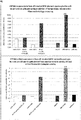
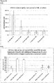
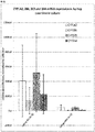
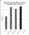
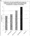
- Example 4 exposing hepatocyte-like cells to an activator of a retinoic acid responsive receptor, here 9-cis-retinoic acid, for, e.g., 5, 24 and 48 hours, leads to an increase in CYP1A, CYP2C9 and 3A activities when compared to untreated cells. Further, after exposing hepatocyte-like cells to an activator of a retinoic acid responsive receptor for 5 or 24 hours, an increase of mRNA expression of the adult hepatic gene CYP3A4 and a strong decrease of the fetal hepatic gene CYP3A7 is immediately observed (see Example 5, Figure 3 ).
- a retinoic acid responsive receptor here 9-cis-retinoic acid
- the hepatocyte-like cells may not only be exposed to an activator of a retinoic acid responsive receptor once for continuous period of time longer than 4 hours and no longer than 72 hours, but may also be exposed to said activator of a retinoic acid responsive receptor for at least two, such as at least three, at least four or at least five, continuous periods of time longer than 4 hours and no longer than 72 hours, such as, e.g., for continuous periods of 5, 24 or 48 hours.
- the hepatocyte-like cells may, for example, be exposed to an activator of a retinoic acid responsive receptor for two continuous periods of time longer than 4 hours and no longer than 72 hours.
- the hepatocyte-like cells may also be exposed to an activator of a retinoic acid responsive receptor for three continuous periods of time longer than 4 hours and no longer than 72 hours.
- the hepatocyte-like cells may also be exposed to an activator of a retinoic acid responsive receptor for four continuous periods of time longer than 4 hours and no longer than 72 hours.
- the hepatocyte-like cells may also be exposed to an activator of a retinoic acid responsive receptor for five continuous periods of time longer than 4 hours and no longer than 72 hours.
- Example 4 repeated exposure to an activator of a retinoic acid responsive receptor has a stronger increasing effect on, e.g., CYP1A and CYP2C9 than a single exposure.
- the differentiation medium is exchanged with one lacking the activator of a retinoic acid responsive receptor, and the cultivation of the differentiating cells is continued.
- the at least two continuous periods of time or exposure are separated by a period of non-exposure to said activator of a retinoic acid responsive receptor.
- Such period of non-exposure may have a duration from several hours to several days, such as from 12 to 24 hours or from 1 to 10 day.
- the period of non-exposure may have a duration of from 1 to 2 days.
- the period of non-exposure may also have a duration from 1 to 5 days.
- the period of non-exposure may also have a duration from 2 to 5 days.
- the period of non-exposure may, for instance, have a duration of 1 day.
- the period of non-exposure may also have a duration of 2 days.
- the period of non-exposure may, for instance, have a duration of 5 days.
- the activator of a retinoic acid responsive receptor may be added to the differentiation medium at any time point once hepatocyte-like cells have been obtained, such as, e.g., after 7, 9, 11, 13, 15, 20, 25 and/or 30 days of culturing.
- the activator of a retinoic acid responsive receptor may, for instance, be added to the differentiation medium for the continuous period of time longer than 4 hours and no longer than 72 hours between day 7 and day 30 of the differentiation and maturation, such as, e.g., between day 7 and day 15.
- the activator of a retinoic acid responsive receptor may thus be added to the differentiation medium for the continuous period of time longer than 4 hours and no longer than 72 hours between day 7 and day 9 of the differentiation and maturation.
- the activator of a retinoic acid responsive receptor may also be added to the differentiation medium for the continuous period of time longer than 4 hours and no longer than 72 hours at day 7 of the differentiation and maturation.
- the activator of a retinoic acid responsive receptor may also be added to the differentiation medium for the continuous period of time longer than 4 hours and no longer than 72 hours at day 9 of the differentiation and maturation.
- the activator of a retinoic acid responsive receptor may also be added to the differentiation medium for the continuous period of time longer than 4 hours and no longer than 72 hours at day 11 of the differentiation and maturation.
- the activator of a retinoic acid responsive receptor may also be added to the differentiation medium for the continuous period of time longer than 4 hours and no longer than 72 hours at day 13 of the differentiation and maturation.
- the activator of a retinoic acid responsive receptor may also be added to the differentiation medium for the continuous period of time longer than 4 hours and no longer than 72 hours at day 15 of the differentiation and maturation.
- the activator of a retinoic acid responsive receptor may also be added to the differentiation medium for the continuous period of time longer than 4 hours and no longer than 72 hours at day 20 of the differentiation and maturation.
- the activator of a retinoic acid responsive receptor may also be added to the differentiation medium for the continuous period of time longer than 4 hours and no longer than 72 hours at day 25 of the differentiation and maturation.
- the activator of a retinoic acid responsive receptor may also be added to the differentiation medium for the continuous period of time longer than 4 hours and no longer than 72 hours at day 30 of the differentiation and maturation.
- the activator of a retinoic acid responsive receptor may also be added to the differentiation medium for the continuous period of time longer than 4 hours and no longer than 72 hours at days 7 and 9 of the differentiation and maturation.
- the activator of a retinoic acid responsive receptor may also be added to the differentiation medium for the continuous period of time longer than 4 hours and no longer than 72 hours at days 7, 9 and 11 of the differentiation and maturation.
- the activator of a retinoic acid responsive receptor may also be added to the differentiation medium for the continuous period of time longer than 4 hours and no longer than 72 hours at days 1, 6, 9, 11 and 16 of the differentiation and maturation.
- the activator of a retinoic acid responsive receptor may also be added to the differentiation medium for the continuous period of time longer than 4 hours and no longer than 72 hours at days 7, 9, 11 and 16 of the differentiation and maturation.
- the activator of a retinoic acid responsive receptor may also be added to the differentiation medium for the continuous period of time longer than 4 hours and no longer than 72 hours at days 7, 9, 11, 13 and 16 of the differentiation and maturation.
- the activator of a retinoic acid responsive receptor may also be added to the differentiation medium for the continuous period of time longer than 4 hours and no longer than 72 hours at days 1, 7, 9, 11 and 16 of the differentiation and maturation.
- the activator of a retinoic acid responsive receptor may also be added to the differentiation medium for the continuous period of time longer than 4 hours and no longer than 72 hours at days 1, 3, 7, 9, 11 and 16 of the differentiation and maturation.
- the hepatocyte-like cells are generally to be exposed to the activator of a retinoic acid responsive receptor at a concentration in the range of about 0,1 to about 5 ⁇ M, such as , e.g, in the range of about 0,5 to about 1,5 ⁇ M.
- the hepatocyte-like cells may thus be exposed to the activator of a retinoic acid responsive receptor at a concentration in the range of about 0,1 to about 2,5 ⁇ M.
- the hepatocyte-like cells may also be exposed to the activator of a retinoic acid responsive receptor at a concentration in the range of about 0,1 to about 1,5 ⁇ M.
- the hepatocyte-like cells may also be exposed to the activator of a retinoic acid responsive receptor at a concentration in the range of about 0,1 to about 1 ⁇ M.
- the hepatocyte-like cells may also be exposed to the activator of a retinoic acid responsive receptor at a concentration in the range of about 0,1 to about 0.5 ⁇ M.
- the hepatocyte-like cells may also be exposed to the activator of a retinoic acid responsive receptor at a concentration in the range of about 0,1 to about 0,3 ⁇ M.
- the hepatocyte-like cells may also be exposed to the activator of a retinoic acid responsive receptor at a concentration in the range of about 0,2 to about 5 ⁇ M.
- the hepatocyte-like cells may also be exposed to the activator of a retinoic acid responsive receptor at a concentration in the range of about 0,2 to about 2,5 ⁇ M.
- the hepatocyte-like cells may also be exposed to the activator of a retinoic acid responsive receptor at a concentration in the range of about 0,2 to about 1,5 ⁇ M.
- the hepatocyte-like cells may also be exposed to the activator of a retinoic acid responsive receptor at a concentration in the range of about 0,5 to about 5 ⁇ M.
- the hepatocyte-like cells may also be exposed to the activator of a retinoic acid responsive receptor at a concentration in the range of about 0,5 to about 3 ⁇ M.
- the hepatocyte-like cells may also be exposed to the activator of a retinoic acid responsive receptor at a concentration in the range of about 0,5 to about 2,5 ⁇ M.
- the hepatocyte-like cells may also be exposed to the activator of a retinoic acid responsive receptor at a concentration in the range of about 0,5 to about 2 ⁇ M.
- the hepatocyte-like cells may also be exposed to the activator of a retinoic acid responsive receptor at a concentration in the range of about 0,5 to about 1,5 ⁇ M.
- the hepatocyte-like cells may also be exposed to the activator of a retinoic acid responsive receptor at a concentration in the range of about 0,5 to about 1 ⁇ M.
- the hepatocyte-like cells may also be exposed to the activator of a retinoic acid responsive receptor at a concentration in the range of about 0,75 to about 5 ⁇ M.
- the hepatocyte-like cells may also be exposed to the activator of a retinoic acid responsive receptor at a concentration in the range of about 0,75 to about 3 ⁇ M.
- the hepatocyte-like cells may also be exposed to the activator of a retinoic acid responsive receptor at a concentration in the range of about 0,75 to about 2,5 ⁇ M.
- the hepatocyte-like cells may also be exposed to the activator of a retinoic acid responsive receptor at a concentration in the range of about 0,75 to about 2 ⁇ M.
- the hepatocyte-like cells may also be exposed to the activator of a retinoic acid responsive receptor at a concentration in the range of about 0,75 to about 1,5 ⁇ M.
- the hepatocyte-like cells may also be exposed to the activator of a retinoic acid responsive receptor at a concentration in the range of about 0,75 to about 1,25 ⁇ M.
- the hepatocyte-like cells may also be exposed to the activator of a retinoic acid responsive receptor at a concentration in the range of about 1 to about 2,5 ⁇ M.
- the hepatocyte-like cells may also be exposed to the activator of a retinoic acid responsive receptor at a concentration in the range of about 1 to about 2 ⁇ M.
- the hepatocyte-like cells may also be exposed to the activator of a retinoic acid responsive receptor at a concentration in the range of about 1 to about 1,5 ⁇ M.
- the hepatocyte-like cells may be exposed to the activator of a retinoic acid responsive receptor at a concentration of about 0,1 ⁇ M.
- the hepatocyte-like cells may also be exposed to the activator of a retinoic acid responsive receptor at a concentration of about 0,2 ⁇ M.
- the hepatocyte-like cells may also be exposed to the activator of a retinoic acid responsive receptor at a concentration of about 0,5 ⁇ M.
- the hepatocyte-like cells may also be exposed to the activator of a retinoic acid responsive receptor at a concentration of about 0,75 ⁇ M.
- the hepatocyte-like cells may also be exposed to the activator of a retinoic acid responsive receptor at a concentration of about 1 ⁇ M.
- the hepatocyte-like cells may also be exposed to the activator of a retinoic acid responsive receptor at a concentration of about 1,25 ⁇ M.
- the hepatocyte-like cells may also be exposed to the activator of a retinoic acid responsive receptor at a concentration of about 1,5 ⁇ M.
- the hepatocyte-like cells may also be exposed to the activator of a retinoic acid responsive receptor at a concentration of about 1,75 ⁇ M.
- the hepatocyte-like cells may also be exposed to the activator of a retinoic acid responsive receptor at a concentration of about 2 ⁇ M.
- the hepatocyte-like cells may also be exposed to the activator of a retinoic acid responsive receptor at a concentration of about 2,5 ⁇ M.
- the hepatocyte-like cells may also be exposed to the activator of a retinoic acid responsive receptor at a concentration of about 3 ⁇ M.
- the hepatocyte-like cells may also be exposed to the activator of a retinoic acid responsive receptor at a concentration of about 3,5 ⁇ M.
- the hepatocyte-like cells may also be exposed to the activator of a retinoic acid responsive receptor at a concentration of about 4 ⁇ M.
- the hepatocyte-like cells may also be exposed to the activator of a retinoic acid responsive receptor at a concentration of about 5 ⁇ M.
- 9-cis-retinoic acid is employed as the activator of a retinoic acid responsive receptor according to the invention, it may be exposed to the hepatocyte-like cells at a concentration in the range of about 0,1 to about 2,5 ⁇ M, such as, e.g., in the range of about 0,1 to about 0,5 ⁇ M, such as, e.g., at about 0,2 ⁇ M.
- the hepatocyte-like cells may thus be exposed to 9-cis-retinoic acid at a concentration in the range of about 0,1 to about 2 ⁇ M.
- the hepatocyte-like cells may also be exposed to 9-cis-retinoic acid at a concentration in the range of about 0,1 to about 1,5 ⁇ M.
- the hepatocyte-like cells may thus be exposed to 9-cis-retinoic acid at a concentration in the range of about 0,1 to about 1 ⁇ M.
- the hepatocyte-like cells may thus be exposed to 9-cis-retinoic acid at a concentration in the range of about 0,1 to about 0,75 ⁇ M.
- the hepatocyte-like cells may thus be exposed to 9-cis-retinoic acid at a concentration in the range of about 0,1 to about 0,5 ⁇ M.
- the hepatocyte-like cells may thus be exposed to 9-cis-retinoic acid at a concentration in the range of about 0,1 to about 0,3 ⁇ M.
- the hepatocyte-like cells may also be exposed to 9-cis-retinoic acid at a concentration in the range of about 0,2 to about 2,5 ⁇ M.
- the hepatocyte-like cells may also be exposed to 9-cis-retinoic acid at a concentration in the range of about 0,2 to about 1,5 ⁇ M.
- the hepatocyte-like cells may also be exposed to 9-cis-retinoic acid at a concentration in the range of about 0,5 to about 2,5 ⁇ M.
- the differentiating hepatic progenitor cells may also be exposed to 9-cis-retinoic acid at a concentration in the range of about 0,5 to about 2 ⁇ M.
- the hepatocyte-like cells may also be exposed to 9-cis-retinoic acid at a concentration in the range of about 0,5 to about 1,5 ⁇ M.
- the hepatocyte-like cells may also be exposed to 9-cis-retinoic acid at a concentration in the range of about 0,5 to about 1 ⁇ M.
- the hepatocyte-like cells may also be exposed to 9-cis-retinoic acid at a concentration in the range of about 0,75 to about 2,5 ⁇ M.
- the hepatocyte-like cells may also be exposed to 9-cis-retinoic acid at a concentration in the range of about 0,75 to about 2 ⁇ M.
- the hepatocyte-like cells may also be exposed to 9-cis-retinoic acid at a concentration in the range of about 0,75 to about 1,5 ⁇ M.
- the hepatocyte-like cells may also be exposed to 9-cis-retinoic acid at a concentration in the range of about 0,75 to about 1,25 ⁇ M.
- the hepatocyte-like cells may also be exposed to 9-cis-retinoic acid at a concentration in the range of about 1 to about 2,5 ⁇ M.
- the hepatocyte-like cells may also be exposed to 9-cis-retinoic acid at a concentration in the range of about 1 to about 2 ⁇ M.
- the hepatocyte-like cells may also be exposed to 9-cis-retinoic acid at a concentration in the range of about 1 to about 1,5 ⁇ M.
- the hepatocyte-like cells may be exposed to 9-cis-retinoic acid at a concentration of about 0,1 ⁇ M.
- the hepatocyte-like cells may also be exposed to 9-cis-retinoic acid at a concentration of about 0,2 ⁇ M.
- the hepatocyte-like cells may also be exposed to 9-cis-retinoic acid at a concentration of about 0,5 ⁇ M.
- the differentiating hepatic progenitor cells may also be exposed to 9-cis-retinoic acid at a concentration of about 0,75 ⁇ M.
- the hepatocyte-like cells may also be exposed to 9-cis-retinoic acid at a concentration of about 1 ⁇ M.
- the hepatocyte-like cells may also be exposed to 9-cis-retinoic acid at a concentration of about 1,25 ⁇ M.
- the hepatocyte-like cells may also be exposed to 9-cis-retinoic acid at a concentration of about 1,5 ⁇ M.
- the differentiating hepatic progenitor cells may also be exposed to 9-cis-retinoic acid at a concentration of about 1,75 ⁇ M.
- the hepatocyte-like cells may also be exposed to 9-cis-retinoic acid at a concentration of about 2 ⁇ M.
- the hepatocyte-like cells may optionally also be exposed to a GSK-3 inhibitor or activator of Wnt signalling and/or to an overlay of one or more components characteristic of the mammalian extracellular matrix (matrix overlay).
- a GSK-3 inhibitor or activator of Wnt signalling is combined with the exposure to a GSK-3 inhibitor or with the exposure to a matrix overlay, or both.
- the exposure to the activator of a retinoic acid responsive receptor may also be combined with the exposure to an activator of Wnt signalling or with the exposure to a matrix overlay, or both.
- the GSK-3 inhibitor employed in the methods of the invention may be any compound capable of inhibiting the GSK-3 signalling.
- Suitable GSK-3 inhibitors for use in the invention are 9-Bromo-7,12-dihydro-indolo [3,2-d][1]benzazepin-6(5H)-one, also known as Kenpaullone or NSC 664704; 1- Aza-Kenpaullone (9-Bromo-7,12-dihydro-pyrido[3',2':2,3]azepino[4,5-b]indol-6(5H)-one); Alsterpaullone (9-Nitro-7,12-dihydroindolo-[3,2-d][1]benzazepin-6(5)-one); 4-(2,6-dichlorobenzamido)-N-(piperidin-4-yl)-1H-pyrazole-3-carboxamide also known as AT-7519; N-(5-((5-ter
- GSK-3 inhibitors which may be employed in the methods of the invention are 3F8 (5-Ethyl-7,8-dimethoxy-1H-pyrrolo[3,4-c]-isoquinoline-1,3-(2H)-dione), A1070722, anorganic ions like Beryllium, Copper, Lithium, Mercury, Tungstate (Wolfram), and Zinc, AR-A 014418, AZD2858, Axin GID-25 residues (peptide), bisindolylmaleimides, CHIR98014 (CT98014), CHIR98023 (CT98023), FRATide-39 residues (peptide), Halomethylketone derivatives, e.g.
- HMK-32 KT5720, L803-mts (peptide) and variants, LY20900314, NP-12 (Tideglusib, NP031112), NP00111, NP031115, Polyoxygenated bis-7-azaindolyl-maleimides, RO31-8220, SB415286 (maleimide), TC-G24, TCS2002, TCS21311, TDZD-8, TOS119 and TWS119 (difluoroacetate).
- the GSK-3 inhibitor may be selected from the above group
- the GSK-3 inhibitor may, for instance, be one selected from Kenpaullone, 1-Aza-Kenpaullone, Alsterpaullone, Aminopyrimidine CHIR99021 and Indirubin-3'-monoxime.
- the GSK-3 inhibitor employed in the methods of the invention may be Kenpaullone 9-Bromo-7,12-dihydro-indolo[3,2-d][1]benzazepin-6(5H)-one.
- the GSK-3 inhibitor may also be 1- Aza-Kenpaullone.
- the GSK-3 inhibitor may also be Alsterpaullone.
- the GSK-3 inhibitor may also be AT-7519.
- the GSK-3 inhibitor may also be SNS-032 (BMS-387032).
- the GSK-3 inhibitor may also be AZD5438.
- the GSK-3 inhibitor may also be BIO (2'Z,3'£)-6-Bromoindirubin-3'-oxime.
- the GSK-3 inhibitor may also be BIO-Acetoxime (2'Z,3'E)-6-Bromoindirubin-3'-acetoxime.
- the GSK-3 inhibitor may also be (5-Methyl-IH-pyrazol-3-yl)-(2-phenylquinazolin-4-yl)amine.
- the GSK-3 inhibitor may also be Pyridocarbazole-cyclopenadienylruthenium complex.
- the GSK-3 inhibitor may also be TDZD-8 4-Benzyl-2-methyl-1,2,4-thiadiazolidine-3,5-dione.
- the GSK-3 inhibitor may also be 2-Thio(3-iodobenzyl)-5-(I-pyridyl)-[I,3,4]-oxadiazole.
- the GSK-3 inhibitor may also be OTDZT 2,4-Dibenzyl-5-oxothiadiazolidine-3-thione.
- the GSK-3 inhibitor may also be alpha-4-Dibromoacetophenone.
- the GSK-3 inhibitor may also be AR-AO 14418 N-(4-Methoxybenzyl)-N'-(5-nitro-1,3-thiazol-2-yl)urea.
- the GSK-3 inhibitor may also be 3-(I-(3-Hydroxypropyl)-IH-pyrrolo[2,3-b]pyridin-3-yl]-4-pyrazin-2-yl-pyrrole-2,5-dione.
- the GSK-3 inhibitor may also be TWSI 19 pyrrolopyrimidine compound.
- the GSK-3 inhibitor may also be L803 H-KEAPPAPPQSpP-NH2 or its myristoylated form.
- the GSK-3 inhibitor may also be 2-Chloro-I-(4,5-dibromo-thiophen-2-yl)-ethanone.
- the GSK-3 inhibitor may also be Aminopyrimidine CHIR99021.
- the GSK-3 inhibitor may also be SB216763 3-(2,4-Dichlorophenyl)-4-(1-methyl-1H-indol-3-yl)-1H-pyrrole-2,5-dione.
- the GSK-3 inhibitor may also be Indirubin-3'-monoxime.
- the hepatocyte-like cells may not only be exposed to one GSK-3 inhibitor, but may also be exposed to one or more further GSK-3 inhibitors, such as to a combination of two, three or four of those mentioned above.
- the hepatocyte-like cells may generally be exposed to the GSK-3 inhibitor at a concentration in the range of about 0,01 to about 10 ⁇ M.
- the hepatocyte-like cells may be exposed to the GSK-3 inhibitor at a concentration in the range of about 0,05 to about 5 ⁇ M.
- the hepatocyte-like cells may thus be exposed to the GSK-3 inhibitor at a concentration in the range of about 0,05 to about 2,5 ⁇ M.
- the hepatocyte-like cells may also be exposed to the GSK-3 inhibitor at a concentration in the range of about 0,05 to about 2 ⁇ M.
- the hepatocyte-like cells may also be exposed to the GSK-3 inhibitor at a concentration in the range of about 0,05 to about 1,5 ⁇ M.
- the hepatocyte-like cells may also be exposed to the GSK-3 inhibitor at a concentration in the range of about 0,05 to about 1 ⁇ M.
- the hepatocyte-like cells may also be exposed to the GSK-3 inhibitor at a concentration in the range of about 0,05 to about 0,5 ⁇ M.
- the hepatocyte-like cells may be exposed to the GSK-3 inhibitor at a concentration in the range of about 0,1 to about 5 ⁇ M.
- the hepatocyte-like cells may thus be exposed to the GSK-3 inhibitor at a concentration in the range of about 0,1 to about 2,5 ⁇ M.
- the hepatocyte-like cells may also be exposed to the GSK-3 inhibitor at a concentration in the range of about 0,1 to about 2 ⁇ M.
- the hepatocyte-like cells may also be exposed to the GSK-3 inhibitor at a concentration in the range of about 0,1 to about 1,5 ⁇ M.
- the hepatocyte-like cells may also be exposed to the GSK-3 inhibitor at a concentration in the range of about 0,1 to about 1 ⁇ M.
- the hepatocyte-like cells may also be exposed to the GSK-3 inhibitor at a concentration in the range of about 0,1 to about 0,5 ⁇ M.
- the hepatocyte-like cells may also be exposed to the GSK-3 inhibitor at a concentration in the range of about 0,25 to about 5 ⁇ M.
- the hepatocyte-like cells may also be exposed to the GSK-3 inhibitor at a concentration in the range of about 0,25 to about 2,5 ⁇ M.
- the hepatocyte-like cells may also be exposed to the GSK-3 inhibitor at a concentration in the range of about 0,25 to about 1,5 ⁇ M.
- the hepatocyte-like cells may also be exposed to the GSK-3 inhibitor at a concentration in the range of about 0,25 to about 1 ⁇ M.
- the hepatocyte-like cells may also be exposed to the GSK-3 inhibitor at a concentration in the range of about 0,25 to about 0,75 ⁇ M.
- the hepatocyte-like cells may also be exposed to the GSK-3 inhibitor at a concentration in the range of about 0,25 to about 0,5 ⁇ M.
- the hepatocyte-like cells may also be exposed to the GSK-3 inhibitor at a concentration in the range of about 0,5 to about 5 ⁇ M.
- the hepatocyte-like cells may also be exposed to the GSK-3 inhibitor at a concentration in the range of about 0,5 to about 2,5 ⁇ M.
- the hepatocyte-like cells may also be exposed to the GSK-3 inhibitor at a concentration in the range of about 0,5 to about 2 ⁇ M.
- the hepatocyte-like cells may also be exposed to the GSK-3 inhibitor at a concentration in the range of about 0,5 to about 1,5 ⁇ M.
- the hepatocyte-like cells may also be exposed to the GSK-3 inhibitor at a concentration in the range of about 0,5 to about 1 ⁇ M.
- the hepatocyte-like cells may also be exposed to the GSK-3 inhibitor at a concentration in the range of about 0,75 to about 5 ⁇ M.
- the hepatocyte-like cells may also be exposed to the GSK-3 inhibitor at a concentration in the range of about 0,75 to about 2,5 ⁇ M.
- the hepatocyte-like cells may also be exposed to the GSK-3 inhibitor at a concentration in the range of about 0,75 to about 2,0 ⁇ M.
- the hepatocyte-like cells may also be exposed to the GSK-3 inhibitor at a concentration in the range of about 0,75 to about 1,5 ⁇ M.
- the hepatocyte-like cells may also be exposed to the GSK-3 inhibitor at a concentration in the range of about 0,75 to about 1 ⁇ M.
- the hepatocyte-like cells may also be exposed to the GSK-3 inhibitor at a concentration in the range of about 1 to about 5 ⁇ M.
- the hepatocyte-like cells may also be exposed to the GSK-3 inhibitor at a concentration in the range of about 1 to about 4 ⁇ M.
- the hepatocyte-like cells may also be exposed to the GSK-3 inhibitor at a concentration in the range of about 1 to about 3 ⁇ M.
- the hepatocyte-like cells may also be exposed to the GSK-3 inhibitor at a concentration in the range of about 1 to about 2,5 ⁇ M.
- the hepatocyte-like cells may also be exposed to the GSK-3 inhibitor at a concentration in the range of about 1 to about 2 ⁇ M.
- the hepatocyte-like cells may also be exposed to the GSK-3 inhibitor at a concentration in the range of about 1 to about 1,5 ⁇ M.
- the hepatocyte-like cells may be exposed to it at a concentration in the range of about 0,05 to about 5 ⁇ M, such as, e.g., in the range of about 0,5 to about 1,5 ⁇ M.
- the hepatocyte-like cells may be exposed to Kenpaullone at a concentration in the range of about 0,05 to about 2 ⁇ M.
- the hepatocyte-like cells may also be exposed to Kenpaullone at a concentration in the range of about 0,05 to about 1,5 ⁇ M.
- the hepatocyte-like cells may also be exposed to Kenpaullone at a concentration in the range of about 0,05to about 1 ⁇ M.
- the hepatocyte-like cells may also be exposed to Kenpaullone at a concentration in the range of about 0,05 to about 0,5 ⁇ M.
- the hepatocyte-like cells may be exposed to Kenpaullone at a concentration in the range of about 0,1 to about 2 ⁇ M.
- the hepatocyte-like cells may also be exposed to Kenpaullone at a concentration in the range of about 0,1 to about 1,5 ⁇ M.
- the hepatocyte-like cells may also be exposed to Kenpaullone at a concentration in the range of about 0,1 to about 1 ⁇ M.
- the hepatocyte-like cells may also be exposed to Kenpaullone at a concentration in the range of about 0,1 to about 0,5 ⁇ M.
- the hepatocyte-like cells may also be exposed to Kenpaullone at a concentration in the range of about 0,25 to about 2,5 ⁇ M.
- the hepatocyte-like cells may also be exposed to Kenpaullone at a concentration in the range of about 0,25 to about 1,5 ⁇ M.
- the hepatocyte-like cells may also be exposed to Kenpaullone at a concentration in the range of about 0,25 to about 1 ⁇ M.
- the hepatocyte-like cells may also be exposed to Kenpaullone at a concentration in the range of about 0,25 to about 0,75 ⁇ M.
- the hepatocyte-like cells may also be exposed to Kenpaullone at a concentration in the range of about 0,25 to about 0,5 ⁇ M.
- the hepatocyte-like cells may also be exposed to Kenpaullone at a concentration in the range of about 0,5 to about 2,5 ⁇ M.
- the hepatocyte-like cells may also be exposed to Kenpaullone at a concentration in the range of about 0,5 to about 2 ⁇ M.
- the hepatocyte-like cells may also be exposed to Kenpaullone at a concentration in the range of about 0,5 to about 1,5 ⁇ M.
- the hepatocyte-like cells may also be exposed to Kenpaullone at a concentration in the range of about 0,5 to about 1 ⁇ M.
- the hepatocyte-like cells may also be exposed to Kenpaullone at a concentration in the range of about 0,75 to about 2,5 ⁇ M.
- the hepatocyte-like cells may also be exposed to Kenpaullone at a concentration in the range of about 0,75 to about 2,0 ⁇ M.
- the hepatocyte-like cells may also be exposed to Kenpaullone at a concentration in the range of about 0,75 to about 1,5 ⁇ M.
- the hepatocyte-like cells may also be exposed to Kenpaullone at a concentration in the range of about 0,75 to about 1 ⁇ M.
- the hepatocyte-like cells may also be exposed to Kenpaullone at a concentration in the range of about 1 to about 2,5 ⁇ M.
- the hepatocyte-like cells may also be exposed to Kenpaullone at a concentration in the range of about 1 to about 2 ⁇ M.
- the hepatocyte-like cells may also be exposed to Kenpaullone at a concentration in the range of about 1 to about 1,5 ⁇ M.
- the GSK-3 inhibitor used according to the invention may further exhibit inhibitory activity towards a cyclin dependent kinase (CDK), such as CDK2.
- CDK cyclin dependent kinase
- Examples of such dual inhibitor are Kenpaullone, AT-7519, SNS-032 (BMS-387032) and AZD5438. Further examples of such dual inhibitor are 1-Aza-Kenpaullone, Alsterpaullone and Indirubin-3'-monoxime.
- Such dual inhibitor are 2-bromo-9-nitropaullone, 2-bromo-9-trifluoromethylpaullone, 2-bromopaullone, 2-iodo-9-trifluoromethylpaullone, 2-iodopaullone, 2-phenyl-4-(2-thienyl)-5H-pyrido[2 ⁇ 3-d][1]benzazepine-6(7H)-thione, 2-[2-(1-hydroxycyclohexyl)-ethinyl]-9-trifluoromethylpaullone, 2,3-dimethoxy-9-nitropaullone, 2,3-dimethoxy-9-trifluormethylpaullone, 2,3-dimethoxypaullone, 2-(3-hydroxy-1-propinyl)-9-trifluoromethylpaullone, 2,9-dibromo-paullone, 3-(6-oxo-9-trifluoromethyl-5,6,7,12-te
- Aloisine A AZD1080, bis-indole indirubin, Debromohymenialdisine, Dibromocantharelline, (E)-3-(6-oxo-9-trifluoromethyl-5,6,7,12-tetrahydro-indolo[2 ⁇ 3-d][1]benzazepin-2-yl)-acrylic acid methyl ester, (E)-2-(3-oxo-1-butenyl)-9-trifluoromethylpaullone, (E)-3-(6-oxo-9-trifluoromethyl-5,6,7,12-tetrahydro-indolo[2 ⁇ 3-d][1]benzazepin-2-yl)-acrylonitrile, Indirubin, Hymenidin, Hymenialdisine, Manzamines, e.g.
- Manzamine A Meridiamins, Paullone, Pyrazolo[3,4-b]pyridine derivatives, Pryazolo[3,4-b]quinoxaline derivatives and thiazolo[5,4-f]quinazolino-9-one derivatives.
- the hepatocyte-like cells may be exposed to an activator of Wnt signalling.
- the activator of Wnt signalling employed in the methods of the present invention may be any compound activating Wnt signalling.
- Suitable activators of Wnt signalling for use in the present invention are Wnt proteins, such as, e.g., Wnt1 , Wnt2, Wnt2B/13, Wnt3A, Wnt3, Wnt4, Wnt5A, Wnt5B, Wnt6, Wnt7A, Wnt7B, Wnt8A, Wnt8B, Wnt9A, Wnt9B, Wnt10A, Wnt10B, Wnt11 and Wnt16, which may be recombinant in nature.
- Wnt proteins such as, e.g., Wnt1 , Wnt2, Wnt2B/13, Wnt3A, Wnt3, Wnt4, Wnt5A, Wnt5B, Wnt6, Wnt7A, Wnt7B, Wnt8A, Wnt8B, Wnt9A, Wnt9B, Wnt10A, W
- the activator of Wnt signalling may thus be selected from the above group of Wnt proteins.
- the activator of Wnt signalling may, for instance, be Wnt3A.
- the activator of Wnt signalling may also be Wnt5A.
- the hepatocyte-like cells may generally be exposed to the activator of Wnt signalling at a concentration in the range of about 0,05 to about 10ng/ml.
- the hepatocyte-like cells may be exposed to the activator of Wnt signalling at a concentration in the range of about 0,05 to about 5ng/ml.
- the hepatocyte-like cells may also be exposed to the activator of Wnt signalling at a concentration in the range of about 0,1 to about 5 ng/ml.
- the hepatocyte-like cells may also be exposed to the activator of Wnt signalling at a concentration in the range of about 0,5 to about 5 ng/ml.
- the hepatocyte-like cells may also be exposed to the activator of Wnt signalling at a concentration in the range of about 1 to about 5 ng/ml.
- the hepatocyte-like cells may also be exposed to the activator of Wnt signalling at a concentration in the range of about 2 to about 5 ng/ml.
- the hepatocyte-like cells may also be exposed to the activator of Wnt signalling at a concentration in the range of about 0,1 to about 2,5 ng/ml.
- the hepatocyte-like cells may also be exposed to the activator of Wnt signalling at a concentration in the range of about 0,1 to about 2 ng/ml.
- the hepatocyte-like cells may also be exposed to the activator of Wnt signalling at a concentration in the range of about 0,5 to about 2,5 ng/ml.
- the hepatocyte-like cells may also be exposed to the activator of Wnt signalling at a concentration in the range of about 0,5 to about 2 ng/ml.
- the hepatocyte-like cells may also be exposed to the activator of Wnt signalling at a concentration in the range of about 0,5 to about 1,5 ng/ml.
- the hepatocyte-like cells may also be exposed to the activator of Wnt signalling at a concentration in the range of about 1 to about 2,5 ng/ml.
- the hepatocyte-like cells may be exposed to it at a concentration in the range of about 0, 05 to about 10 ng/ml, such as, e.g., in the range of about 0,5 to about 2,5 ⁇ M.
- the hepatocyte-like cells may be exposed to Wnt3A at a concentration in the range of about 0,05 to about 5ng/ml.
- the hepatocyte-like cells may also be exposed to Wnt3A at a concentration in the range of about 0,1 to about 5 ng/ml.
- the hepatocyte-like cells may also be exposed to Wnt3A at a concentration in the range of about 0,5 to about 5 ng/ml.
- the hepatocyte-like cells may also be exposed to Wnt3A at a concentration in the range of about 1 to about 5 ng/ml.
- the hepatocyte-like cells may also be exposed Wnt3A at a concentration in the range of about 2 to about 5 ng/ml.
- the hepatocyte-like cells may also be exposed to Wnt3A at a concentration in the range of about 0,1 to about 2,5 ng/ml.
- the hepatocyte-like cells may also be exposed to Wnt3A at a concentration in the range of about 0,1 to about 2 ng/ml.
- the hepatocyte-like cells may also be exposed to Wnt3A at a concentration in the range of about 0,5 to about 2,5 ng/ml.
- the hepatocyte-like cells may also be exposed to Wnt3A at a concentration in the range of about 0,5 to about 2 ng/ml.
- the hepatocyte-like cells may also be exposed to Wnt3A at a concentration in the range of about 0,5 to about 1,5 ng/ml.
- the hepatocyte-like cells may also be exposed to Wnt3A at a concentration in the range of about 1 to about 2,5 ng/ml.
- the hepatocyte-like cells may not only be exposed to one activator of Wnt signalling, but may also be exposure to one or more further activators of Wnt signalling, such as to a combination of two, three or four of those mentioned above.
- the hepatocyte-like cells may optionally also be exposed to a CDK inhibitor and/or to an overlay of one or more components characteristic of the mammalian extracellular matrix (matrix overlay).
- matrix overlay the exposure to the activator of a retinoic acid responsive receptor is combined with the exposure to a CDK inhibitor or with the exposure to a matrix overlay, or both.
- the additional exposure of the differentiating hepatic progenitor cells to a CDK inhibitor and/or to a matrix overlay has shown to further improve the mature and functional features for the hepatocyte-like cells ( Figure 4 ).
- the CDK inhibitor employed in the methods of the invention may be any compound capable of inhibiting the function (e.g., the activity) of a cyclin dependent kinase (CDK).
- the CDK inhibitor employed in the methods of the invention may, for instance, be an inhibitor of one or more of CDK1, CDK2, CDK4, CDK5, CDK6, CDK7, and CDK9.
- the CDK inhibitor employed in the methods of the invention may be an inhibitor of cyclin dependent kinase 2 (CDK2).
- the CDK inhibitor employed in the methods of the invention may, for instance, be an inhibitor of CDK1 and CDK2.
- the CDK inhibitor employed in the methods of the invention may, for instance, be an inhibitor of CDK2 and CDK5.
- the CDK inhibitor employed in the methods of the invention may, for instance, be an inhibitor of CDK1, CDK2 and CDK5.
- Suitable CDK inhibitors for use in the invention are 9-Bromo-7,12-dihydro-indolo [3,2-d]-[1]benzazepin-6(5H)-one, also known as Kenpaullone or NSC 664704; (R)-2-(6-(benzylamino)-9-isopropyl-9H-purin-2-ylamino)butan-1-ol also known as Roscovitine; 2-(2-chlorophenyl)-5,7-dihydroxy-8-((3S,4R)-3-hydroxy-1-methylpiperidin-4-yl)-4H-chromen-4-one also known as Flavopiridol; 4-(2,6-dichlorobenzamido)-N-(piperidin-4-yl)-1H-pyrazole-3-carboxamide also known as AT-7519; 6-acetyl-8-cyclopentyl-5-methyl-2-(5-(piperazin-1-yl)pyridin-2-
- CDK inhibitors which may be employed in the methods of the invention are 2-hydroxybohemine, A674563, Aminopurvanolol, BAY1000394, BMS-265246, BS-181 Butyrolactone I, CR8 S-isomer, Diaciclib (SCH727965), JNJ-7706621, N9-isopropyl-olomoucine, NU6140, NU6102, Olomoucine II, Oxindole I, P276-00, PD332991, PHA-793887, PHA-767491, PHA-848125, PNU112455A, Purvanolol A and B, R547, (R)-DRF053 and SCH900776 (MK-8776).
- the CDK inhibitor may be selected from the above group.
- the CDK inhibitor employed in the methods of the invention may, for instance, be Kenpaullone. Kenpaullone is known to be an inhibitor of CDK2.
- the CDK inhibitor may also be Roscovitine.
- the CDK inhibitor may also be Flavopiridol.
- the CDK inhibitor may also be AT-7519.
- the CDK inhibitor may also be PD 0332991 HCI.
- the CDK inhibitor may also be SNS-032 (BMS-387032).
- the CDK inhibitor may also be JNJ-7706621.
- the CDK inhibitor may also be PHA-793887.
- the CDK inhibitor may also be Dinaciclib (SCH727965).
- the CDK inhibitor may also be BMS-265246.
- the CDK inhibitor may also be PHA-848125.
- the CDK inhibitor may also be PHA-767491.
- the CDK inhibitor may also be SCH 900776.
- the CDK inhibitor may also be Flavopiridol HCI.
- the CDK inhibitor may also be R547.
- the CDK inhibitor may also be A-674563.
- the CDK inhibitor may also be AZD5438.
- the CDK inhibitor may also be BS-181 HCI.
- the CDK inhibitor may also be CY-202.
- the CDK inhibitor may also be AG-024322.
- the CDK inhibitor may also be P276-00.
- the CDK inhibitor may also be ZK 304709.
- the CDK inhibitor may also be GPC-286199.
- the CDK inhibitor may also be BAY 80-3000.
- the CDK inhibitor may be selected from the group consisting of: Kenpaullone, SNS-032(BMS-387032), AT-7519 and AZD5438.
- the CDK inhibitor may be selected from the group consisting of: Kenpaullone, 1-Aza-Kenpaullone, Indirubin-3'-monoxime, Alsterpaullone, SNS-032(BMS-387032), AT-7519 and AZD5438.
- the hepatocyte-like cells may not only be exposed to one CDK inhibitor, but may also be exposure to one or more further CDK inhibitors, such as to a combination of two, three or four of those mentioned above.
- the hepatocyte-like cells may generally be exposed to the CDK inhibitor at a concentration in the range of about 0,01 to about 10 ⁇ M.
- the hepatocyte-like cells may be exposed to the CDK inhibitor at a concentration in the range of about 0,05 to about 5 ⁇ M.
- the hepatocyte-like cells may thus be exposed to the CDK inhibitor at a concentration in the range of about 0,05 to about 2,5 ⁇ M.
- the hepatocyte-like cells may also be exposed to the CDK inhibitor at a concentration in the range of about 0,05 to about 2 ⁇ M.
- the hepatocyte-like cells may also be exposed to the CDK inhibitor at a concentration in the range of about 0,05 to about 1,5 ⁇ M.
- the hepatocyte-like cells may also be exposed to the CDK inhibitor at a concentration in the range of about 0,05 to about 1 ⁇ M.
- the hepatocyte-like cells may also be exposed to the CDK inhibitor at a concentration in the range of about 0,05 to about 0,5 ⁇ M.
- the hepatocyte-like cells may be exposed to the CDK inhibitor at a concentration in the range of about 0,1 to about 5 ⁇ M.
- the hepatocyte-like cells may thus be exposed to the CDK inhibitor at a concentration in the range of about 0,1 to about 2,5 ⁇ M.
- the hepatocyte-like cells may also be exposed to the CDK inhibitor at a concentration in the range of about 0,1 to about 2 ⁇ M.
- the hepatocyte-like cells may also be exposed to the CDK inhibitor at a concentration in the range of about 0,1 to about 1,5 ⁇ M.
- the hepatocyte-like cells may also be exposed to the CDK inhibitor at a concentration in the range of about 0,1 to about 1 ⁇ M.
- the hepatocyte-like cells may also be exposed to the CDK inhibitor at a concentration in the range of about 0,1 to about 0,5 ⁇ M.
- the hepatocyte-like cells may also be exposed to the CDK inhibitor at a concentration in the range of about 0,25 to about 5 ⁇ M.
- the hepatocyte-like cells may also be exposed to the CDK inhibitor at a concentration in the range of about 0,25 to about 2,5 ⁇ M.
- the hepatocyte-like cells may also be exposed to the CDK inhibitor at a concentration in the range of about 0,25 to about 1,5 ⁇ M.
- the hepatocyte-like cells may also be exposed to the CDK inhibitor at a concentration in the range of about 0,25 to about 1 ⁇ M.
- the hepatocyte-like cells may also be exposed to the CDK inhibitor at a concentration in the range of about 0,25 to about 0,75 ⁇ M.
- the hepatocyte-like cells may also be exposed to the CDK inhibitor at a concentration in the range of about 0,25 to about 0,5 ⁇ M.
- the hepatocyte-like cells may also be exposed to the CDK inhibitor at a concentration in the range of about 0,5 to about 5 ⁇ M.
- the hepatocyte-like cells may also be exposed to the CDK inhibitor at a concentration in the range of about 0,5 to about 2,5 ⁇ M.
- the hepatocyte-like cells may also be exposed to the CDK inhibitor at a concentration in the range of about 0,5 to about 2 ⁇ M.
- the hepatocyte-like cells may also be exposed to the CDK inhibitor at a concentration in the range of about 0,5 to about 1,5 ⁇ M.
- the hepatocyte-like cells may also be exposed to the CDK inhibitor at a concentration in the range of about 0,5 to about 1 ⁇ M.
- the hepatocyte-like cells may also be exposed to the CDK inhibitor at a concentration in the range of about 0,75 to about 5 ⁇ M.
- the hepatocyte-like cells may also be exposed to the CDK inhibitor at a concentration in the range of about 0,75 to about 2,5 ⁇ M.
- the hepatocyte-like cells may also be exposed to the CDK inhibitor at a concentration in the range of about 0,75 to about 2,0 ⁇ M.
- the hepatocyte-like cells may also be exposed to the CDK inhibitor at a concentration in the range of about 0,75 to about 1,5 ⁇ M.
- the hepatocyte-like cells may also be exposed to the CDK inhibitor at a concentration in the range of about 0,75 to about 1 ⁇ M.
- the hepatocyte-like cells may also be exposed to the CDK inhibitor at a concentration in the range of about 1 to about 5 ⁇ M.
- the hepatocyte-like cells may also be exposed to the CDK inhibitor at a concentration in the range of about 1 to about 4 ⁇ M.
- the hepatocyte-like cells may also be exposed to the CDK inhibitor at a concentration in the range of about 1 to about 3 ⁇ M.
- the hepatocyte-like cells may also be exposed to the CDK inhibitor at a concentration in the range of about 1 to about 2,5 ⁇ M.
- the hepatocyte-like cells may also be exposed to the CDK inhibitor at a concentration in the range of about 1 to about 2 ⁇ M.
- the hepatocyte-like cells may also be exposed to the CDK inhibitor at a concentration in the range of about 1 to about 1,5 ⁇ M.
- the hepatocyte-like cells may be exposed to it at a concentration in the range of about 0,05 to about 5 ⁇ M, such as, e.g., in the range of about 0,5 to about 1,5 ⁇ M.
- the hepatocyte-like cells may be exposed to Kenpaullone at a concentration in the range of about 0,05 to about 2 ⁇ M.
- the hepatocyte-like cells may also be exposed to Kenpaullone at a concentration in the range of about 0,05 to about 1,5 ⁇ M.
- the hepatocyte-like cells may also be exposed to Kenpaullone at a concentration in the range of about 0,05to about 1 ⁇ M.
- the hepatocyte-like cells may also be exposed to Kenpaullone at a concentration in the range of about 0,05 to about 0,5 ⁇ M.
- the hepatocyte-like cells may be exposed to Kenpaullone at a concentration in the range of about 0,1 to about 2 ⁇ M.
- the hepatocyte-like cells may also be exposed to Kenpaullone at a concentration in the range of about 0,1 to about 1,5 ⁇ M.
- the hepatocyte-like cells may also be exposed to Kenpaullone at a concentration in the range of about 0,1 to about 1 ⁇ M.
- the hepatocyte-like cells may also be exposed to Kenpaullone at a concentration in the range of about 0,1 to about 0,5 ⁇ M.
- the hepatocyte-like cells may also be exposed to Kenpaullone at a concentration in the range of about 0,25 to about 2,5 ⁇ M.
- the hepatocyte-like cells may also be exposed to Kenpaullone at a concentration in the range of about 0,25 to about 1,5 ⁇ M.
- the hepatocyte-like cells may also be exposed to Kenpaullone at a concentration in the range of about 0,25 to about 1 ⁇ M.
- the hepatocyte-like cells may also be exposed to Kenpaullone at a concentration in the range of about 0,25 to about 0,75 ⁇ M.
- the hepatocyte-like cells may also be exposed to Kenpaullone at a concentration in the range of about 0,25 to about 0,5 ⁇ M.
- the hepatocyte-like cells may also be exposed to Kenpaullone at a concentration in the range of about 0,5 to about 2,5 ⁇ M.
- the hepatocyte-like cells may also be exposed to Kenpaullone at a concentration in the range of about 0,5 to about 2 ⁇ M.
- the hepatocyte-like cells may also be exposed to Kenpaullone at a concentration in the range of about 0,5 to about 1,5 ⁇ M.
- the hepatocyte-like cells may also be exposed to Kenpaullone at a concentration in the range of about 0,5 to about 1 ⁇ M.
- the hepatocyte-like cells may also be exposed to Kenpaullone at a concentration in the range of about 0,75 to about 2,5 ⁇ M.
- the hepatocyte-like cells may also be exposed to Kenpaullone at a concentration in the range of about 0,75 to about 2,0 ⁇ M.
- the hepatocyte-like cells may also be exposed to Kenpaullone at a concentration in the range of about 0,75 to about 1,5 ⁇ M.
- the hepatocyte-like cells may also be exposed to Kenpaullone at a concentration in the range of about 0,75 to about 1 ⁇ M.
- the hepatocyte-like cells may also be exposed to Kenpaullone at a concentration in the range of about 1 to about 2,5 ⁇ M.
- the hepatocyte-like cells may also be exposed to Kenpaullone at a concentration in the range of about 1 to about 2 ⁇ M.
- the hepatocyte-like cells may also be exposed to Kenpaullone at a concentration in the range of about 1 to about 1,5 ⁇ M.
- the CDK inhibitor used according to the invention may further exhibit inhibitory activity towards Glycogen synthase kinase 3 (GSK-3), especially GSK-3 beta.
- GSK-3 Glycogen synthase kinase 3
- Examples of such dual inhibitor are Kenpaullone, SNS-032 (BMS-387032), AT-7519 and AZD5438 which inhibit both CDK2 and GSK3.
- Further examples of such dual inhibitor are 1-Aza-Kenpaullone, Alsterpaullone and Indirubin-3'-monoxime.
- Further examples of such dual inhibitor are 1-Aza-Kenpaullone, Alsterpaullone and Indirubin-3'-monoxime.
- Such dual inhibitor are 2-bromo-9-nitropaullone, 2-bromo-9-trifluoromethylpaullone, 2-bromopaullone, 2-iodo-9-trifluoromethylpaullone, 2-iodopaullone, 2-phenyl-4-(2-thienyl)-5H-pyrido[2 ⁇ 3-d][1]benzazepine-6(7H)-thione, 2-[2-(1-hydroxycyclohexyl)-ethinyl]-9-trifluoromethyl-paullone, 2,3-dimethoxy-9-nitropaullone, 2,3-dimethoxy-9-trifluormethylpaullone, 2,3-dimethoxypaullone, 2-(3-hydroxy-1-propinyl)-9-trifluoromethylpaullone, 2,9-dibromo-paullone, 3-(6-oxo-9-trifluoromethyl-5,6,7,12-t
- Aloisine A AZD1080, bis-indole indirubin, Debromohymenialdisine, Dibromocantharelline, (E)-3-(6-oxo-9-trifluoromethyl-5,6,7,12-tetrahydro-indolo[2 ⁇ 3-d][1]benzazepin-2-yl)-acrylic acid methyl ester, (E)-2-(3-oxo-1-butenyl)-9-trifluoromethylpaullone, (E)-3-(6-oxo-9-trifluoromethyl-5,6,7,12-tetrahydro-indolo[2 ⁇ 3-d][1]benzazepin-2-yl)-acrylonitrile, Indirubin, Hymenidin, Hymenialdisine, Manzamines, e.g.
- Manzamine A Meridiamins, Paullone, Pyrazolo[3,4-b]pyridine derivatives, Pryazolo[3,4-b]quinoxaline derivatives and thiazolo[5,4-f]quinazolino-9-one derivatives.
- the hepatocyte-like cells being expose to a GSK3 inhibitor or activator of Wnt signalling are further exposed to a CDK inhibitor.
- a CDK inhibitor cyclin dependent kinase
- the hepatocyte-like cells being expose to a CDK inhibitor are further exposed to a GSK3 inhibitor or activator of Wnt signalling. This is particularly of interest if the CDK inhibitor employed does not exhibit inhibitory activity towards Glycogen synthase kinase 3 (GSK-3),
- the present invention further provides a method for promoting the maturation of human hepatocyte-like cells whereby said hepatocyte-like cells are exposed to an activator of a retinoic acid responsive receptor, such as retinoic acid, optionally in combination with exposure to an inhibitor of GSK3 signalling or activator of Wnt signalling and a CDK inhibitor, further optionally in combination with an overlay of the cells with one or more components characteristic of the mammalian extracellular matrix (matrix overlay).
- a retinoic acid responsive receptor such as retinoic acid
- the method for promoting the maturation of human hepatocyte-like cells may thus be described as comprising the step: Exposing said human hepatocyte-like cells to an activator of a retinoic acid responsive receptor, optionally in combination with exposure to a CDK inhibitor and a GSK3 inhibitor or activator of Wnt signalling, further optionally in combination with exposure to an overlay of the cells with one or more components characteristic of the mammalian extracellular matrix (matrix overlay).
- the present invention further provides a method for producing human hepatocyte-like cells whereby human hepatic progenitor cells are cultured under differentiation conditions to obtain hepatocyte-like cells, and the obtained hepatocyte-like cells are exposed to an activator of a retinoic acid responsive receptor, such as retinoic acid, optionally in combination with exposure to an inhibitor of GSK3 signalling or activator of Wnt signalling and a CDK inhibitor, further optionally in combination with an overlay of the cells with one or more components characteristic of the mammalian extracellular matrix (matrix overlay).
- a retinoic acid responsive receptor such as retinoic acid
- the method for producing human hepatocyte-like cells may thus be described as comprising the following steps:
- the GSK3 inhibitor may be CHIR99021 and the CDK inhibitor may be Roscovitine or Flavopiridol.
- any other GSK3 inhibitor or CDK inhibitor especially of those specifically mentioned herein, may be employed instead.
- Matrix overlays consisting of Collagen I or Matrigel (a basement membrane mix extracted from the Engelbreth-Holm-Swarm mouse sarcoma) have been used for culturing primary hepatocytes for several decades (e.g. Dunn et al. 1991; Page et al. 2007), since it was found that primary hepatocytes maintain a better functionality and live longer in a so called sandwich configuration, with one extracellular matrix (ECM) layer below the cells and one ECM layer on top of the cells.
- ECM extracellular matrix
- Collagen I and Matrigel overlays are thick, containing e.g. 125 ⁇ g Matrigel /cm 2 or 50 ⁇ g Collagen I /cm 2 . However, this is not reflecting the physiological composition or thickness of the liver ECM (compare e.g. Turner et al. 2011; Wang et al. 2011).
- the matrix overlay employed in the methods of the invention is a novel, more physiological combination of component present in the ECM of the adult liver and comprises, or is composed of, one or more ECM components, which form part of the normal mammalian extracellular matrix environment.
- ECM components for use as matrix overlay in the present invention are collagen, such as collagen I, II, III, IV, V or VI, fibronectin, elastin, chondroitin sulfate proteoglycan, dermatan sulfate proteoglycan, heparin proteoglycan, heparan sulfate proteoglycan, such as glypicans, syndecans or perlecans, glycosaminoglycans, nidogen /entactin, laminins, biglycan, tenascin, hyaluronans, or other ECM components, or ECM component mixtures comprising, or consisting of, e.g., collagens, laminin, fibronect
- the hepatocyte-like cells may be exposed to a matrix overlay comprising, or composed of, one or more, such as two, three, four, five, six, seven, eight, nine or ten, or up to 20 of the above mentioned ECM components.
- the hepatocyte-like cells may be exposed to a matrix overlay comprising, or composed of, two of the above mentioned ECM components.
- the hepatocyte-like cells may also be exposed to a matrix overlay comprising, or composed of, three of the above mentioned ECM components.
- the hepatocyte-like cells may also be exposed to a matrix overlay comprising, or composed of, four of the above mentioned ECM components.
- the hepatocyte-like cells may also be exposed to a matrix overlay comprising, or composed of, five of the above mentioned ECM components.
- the hepatocyte-like cells may also be exposed to a matrix overlay comprising, or composed of, six of the above mentioned ECM components.
- the hepatocyte-like cells may also be exposed to a matrix overlay comprising, or composed of, seven of the above mentioned ECM components.
- the hepatocyte-like cells may also be exposed to a matrix overlay comprising, or composed of, eight of the above mentioned ECM components.
- the hepatocyte-like cells may also be exposed to a matrix overlay comprising, or composed of, nine of the above mentioned ECM components.
- the hepatocyte-like cells may also be exposed to a matrix overlay comprising, or composed of, ten of the above mentioned ECM components.
- the hepatocyte-like cells may be exposed to a matrix overlay comprising, or composed of, collagen and fibronectin (collagen-fibronectin-matrix overlay), such as a matrix overlay comprising, or composed of, collagen I and fibronectin (collagen I-fibronectin-matrix overlay).
- the hepatocyte-like cells may also be exposed to a matrix overlay comprising , or composed of, collagen, such as collagen I, and laminin (collagen-laminin-matrix overlay).
- the hepatocyte-like may also be exposed to a matrix overlay comprising, or composed of, collagen, such as collagen I, nidogen and laminin (collagen-nidogen-laminin-matrix overlay).
- the hepatocyte-like cells may also be exposed to a matrix overlay comprising, or composed of, collagen, such as collagen I, fibronectin and laminin (collagen-fibronectin-laminin-matrix overlay).
- the hepatocyte-like cells may also be exposed to a matrix overlay comprising, or composed of, fibronectin and laminin (fibronectin-laminin-matrix overlay).
- the hepatocyte-like cells may also be exposed to a matrix overlay comprising, or composed of, Fibronectin, nidogen and laminin (Fibronectin-nidogen-laminin-matrix overlay).
- the hepatocyte-like cells may also be exposed to a matrix overlay comprising, or composed of, fibronectin, collagen I, collagen IV, collagen VI, nidogen, biglycan and laminin (fibronectin-collagen I, IV, VI-Nidogen-biglycan-laminin -matrix overlay).
- the hepatocyte-like cells may also be exposed to a matrix overlay comprising, or composed of, fibronectin, collagen I, collagen IV, nidogen, biglycan and laminin (fibronectin-collagen I, IV -nidogen-biglycan-laminin -matrix overlay).
- the hepatocyte-like cells may also be exposed to a matrix overlay comprising collagen, such as collagen I, laminin, fibronectin, proteoglycans and glycosaminoglycans (collagen, laminin, fibronectin, proteoglycans and glycosaminoglycans matrix overlay).
- the hepatocyte-like cells may also be exposed to a matrix overlay comprising collagen, such as collagen I, laminin, fibronectin, and proteoglycans (collagen, laminin, fibronectin, and proteoglycans matrix overlay).
- the hepatocyte-like cells may also be exposed to a matrix overlay comprising collagen, such as collagen I, laminin, fibronectin, tenascin, elastin, proteoglycans and glycosaminoglycans (collagen, laminin, fibronectin, tenascin, elastin, proteoglycans and glycosaminoglycans matrix overlay).
- collagen such as collagen I, laminin, fibronectin, tenascin, elastin, proteoglycans and glycosaminoglycans matrix overlay.
- the matrix overlay employed in the methods of the invention is thin compared to the thick matrices so far used.
- the thickness of the matrix overlay thereby correlates with the concentration of the ECM components employed.
- Suitable concentrations for the present matrix overlay are e.g. 0,01-30 ⁇ g, such as 0,01-20 ⁇ g, ECM component /cm 2 .
- each ECM component may be present in the matrix overlay at a concentration from about 0,1 to about 20 ⁇ g ECM component/ cm 2 .
- Each ECM component may also be present in the matrix overlay at a concentration from about 0,1 to about 15 ⁇ g ECM component/ cm 2 .
- Each ECM component may also be present in the matrix overlay at a concentration from about 0,1 to about 12,5 ⁇ g protein component/ cm 2 .
- Each ECM component may also be present in the matrix overlay at a concentration from about 0,1 to about 10 ⁇ g ECM component/ cm 2 .
- Each ECM component may also be present in the matrix overlay at a concentration from about 0,1 to about 5 ⁇ g ECM component/ cm 2 .
- Each ECM component may also be present in the matrix overlay at a concentration from about 0,1 to about 2,5 ⁇ g ECM component/ cm 2 .
- Each ECM component may also be present in the matrix overlay at a concentration from about 0,2 to about 20 ⁇ g ECM component/ cm 2 .
- Each ECM component may also be present in the matrix overlay at a concentration from about 0,2 to about 15 ⁇ g ECM component/ cm 2 .
- Each ECM component may also be present in the matrix overlay at a concentration from about 0,2 to about 12,5 ⁇ g ECM component/ cm 2 .
- Each ECM component may also be present in the matrix overlay at a concentration from about 0,2 to about 10 ⁇ g ECM component/ cm 2 .
- Each ECM component may also be present in the matrix overlay at a concentration from about 0,2 to about 5 ⁇ g ECM component/ cm 2 . Each ECM component may also be present in the matrix overlay at a concentration from about 0,5 to about 25 ⁇ g ECM component/ cm 2 . Each ECM component may also be present in the matrix overlay at a concentration from about 0,5 to about 20 ⁇ g ECM component/ cm 2 . Each protein component may also be present in the matrix overlay at a concentration from about 0,5 to about 15 ⁇ g protein component/ cm 2 . Each protein component may also be present in the matrix overlay at a concentration from about 0,5 to about 10 ⁇ g protein component/ cm 2 .
- Each protein component may also be present in the matrix overlay at a concentration from about 0,5 to about 7,5 ⁇ g protein component/ cm 2 . Each protein component may also be present in the matrix overlay at a concentration from about 0,5 to about 5 ⁇ g protein component/ cm 2 . Each protein component may also be present in the matrix overlay at a concentration from about 0,5 to about 2,5 ⁇ g protein component/ cm 2 . Each protein component may also be present in the matrix overlay at a concentration from about 0,5 to about 1,5 ⁇ g protein component/ cm 2 . Each protein component may also be present in the matrix overlay at a concentration from about 0,5 to about 1 ⁇ g protein component/ cm 2 .
- Each ECM component may be present in the matrix overlay at a concentration from about 1 to about 20 ⁇ g ECM component/ cm 2 . Each ECM component may also be present in the matrix overlay at a concentration from about 1 to about 15 ⁇ g ECM component/ cm 2 . Each ECM component may also be present in the matrix overlay at a concentration from about 1 to about 12,5 ⁇ g protein component/ cm 2 . Each ECM component may also be present in the matrix overlay at a concentration from about 1 to about 10 ⁇ g ECM component/ cm 2 . Each ECM component may also be present in the matrix overlay at a concentration from about 1 to about 5 ⁇ g ECM component/ cm 2 .
- Each ECM component may also be present in the matrix overlay at a concentration from about 1 to about 2,5 ⁇ g ECM component/ cm 2 .
- Each ECM component may also be present in the matrix overlay at a concentration from about 2 to about 20 ⁇ g ECM component/ cm 2 .
- Each ECM component may also be present in the matrix overlay at a concentration from about 2 to about 15 ⁇ g ECM component/ cm 2 .
- Each ECM component may also be present in the matrix overlay at a concentration from about 2 to about 12,5 ⁇ g ECM component/ cm 2 .
- Each ECM component may also be present in the matrix overlay at a concentration from about 2 to about 10 ⁇ g ECM component/ cm 2 .
- Each ECM component may also be present in the matrix overlay at a concentration from about 2 to about 5 ⁇ g ECM component/ cm 2 . Each ECM component may also be present in the matrix overlay at a concentration from about 5 to about 25 ⁇ g ECM component/ cm 2 . Each ECM component may also be present in the matrix overlay at a concentration from about 5 to about 20 ⁇ g ECM component/ cm 2 . Each protein component may also be present in the matrix overlay at a concentration from about 5 to about 15 ⁇ g protein component/ cm 2 . Each protein component may also be present in the matrix overlay at a concentration from about 5 to about 10 ⁇ g protein component/ cm 2 . Each protein component may also be present in the matrix overlay at a concentration from about 5 to about 7,5 ⁇ g protein component/ cm 2 .
- Collagen I may, for example, be present in the matrix overlay at a concentration from about 2 to about 20 ⁇ g/ cm 2 , such as from about 5 to about 15 ⁇ g/ cm 2 .
- Collagen IV may, for example, be present in the matrix overlay at a concentration from about 0,01 to 10 ⁇ g/cm 2 , such as from about 0,05 to about 10 ⁇ g/cm 2 .
- Collagen VI may, for example, be present in the matrix overlay at a concentration form about 0,01 to about 15 ⁇ g/cm 2 , such as from about 0,01 to about 10 ⁇ g/cm 2 or from about 0,1 to about 15 ⁇ g/cm 2 .
- Fibronectin may, for example, be present in the matrix overlay at a concentration from about 2 to about 30 ⁇ g/cm 2 , such as from about 2 to about 20 ⁇ g/cm 2 .
- Nidogen may, for example, be present in the matrix overlay at a concentration from about 0,01 to about 10 ⁇ g/cm 2 , such as from about 0,05 to about 10 ⁇ g/cm 2 .
- Laminin may, for example, be present in the matrix overlay at a concentration from about 0,01 to about 10 ⁇ g/cm 2 , such as from about 0,05 to about 10 ⁇ g/cm 2 .
- Biglycan may, for example, be present in the matrix overlay at a concentration from about 0,01 to about 10 ⁇ g/cm 2 , such as from about 0,1 to about 10 ⁇ g/cm 2 .
- cells may be cultured on a coating as growth support which covers the surface of the culture vessel.
- Gelatine or fibronectin based coating are widely used as growth support.
- the cells in particular the hepatic progenitor cells and hepatocyte-like cells, may be cultured on a gelatin or fibronectin based coating.
- the cells may also be cultured on a coating which has a composition similar or identical to a matrix overlay as defined above.
- the cells may be cultured on a coating which has a composition which is identical to that of the employed matrix overlay. Accordingly, a so-called "sandwich" type culture environment is provided.
- the hepatic progenitor cells used in the methods of the invention may initially be derived from human pluripotent stem (hPS) cells, such as human embryonic stem (hES) cells or human induced pluripotent stem cells (hiPS).
- the methods of the invention may thus further comprise as an initial step the culturing of hPS cells under differentiation conditions to obtain said hepatic progenitor cells.
- hPS cells are initially differentiated into said hepatic progenitor cells. This step is referred to herein as initial hepatic differentiation.
- the human pluripotent stem cells which may also be used as starting material to obtain the endodermal and/or hepatic progenitor cells may be human embryonic stem cells.
- hES cells for use according to the invention are ones which have been derived (or obtained) without destruction of the human embryo, such as by employing the single blastomere removal technique described in e.g. Chung et al (2008), further described by Mercader et al. in Essential Stem Cell Methods (First Edition, 2009 ).
- Suitable hES cell lines for use are, for example, the cell lines SA167, SA181, SA461 (Cellartis AB, Göteborg, Sweden) which are listed in the NIH stem cell registry, the UK Stem Cell bank and the European hESC registry and are available on request.
- Other suitable cell lines for use are those established by Klimanskaya et al. (2006), such as cell lines MA01 and MA09, and Chung et al. (2008), such as cell lines MA126, MA127, MA128 and MA129, which all are listed with the International Stem Cell Registry (assigned to Advanced Cell Technology, Inc. Worcester, MA, USA).
- the human pluripotent stem cells which may be used as starting material to obtain the endodermal and/or hepatic progenitor cells may be human induced pluripotent stem cells.
- Various techniques for obtaining such hiPS cells have been described in the scientific literature, and are thus known to the skilled person [see, e.g., Takahashi et al. (2007); Zhou et al. (2009); Yu and Thomson in Essentials of Stem Cell Biology (2nd Editi on].
- endodermal and/or hepatic progenitor cells may also be derived from other pluripotent stem cells such as adult stem cells, cancer stem cells or from other embryonic, fetal, juvenile or adult sources.
- Suitable conditions for differentiating hPS cells into hepatic progenitor cells are known (see, e.g., Hay 2008, Brolen 2010 and Duan 2010).
- WO 2009/013254 A1 describes suitable protocols to obtain hepatic progenitor cells (Embodiments 1 to 4).
- the hPS cells are normally cultured for up to 14 days in suitable differentiation medium in order to obtain hepatic progenitor cells.
- the hPS cells may be cultured in suitable differentiation medium for about 10 to about 14 days, such as for about 11 to 14 days.
- the initial hepatic differentiation may be defined by including a pre-endodermal step, i.e. the culturing of the hPS cells under differentiation conditions to obtain cells of the definitive endoderm (DE cells), which is followed by a pre-hepatic step, i.e. the culturing of the obtained DE cells under differentiation conditions to obtain the hepatic progenitor cells.
- hPS cells are first differentiated into definitive endoderm, followed by the further differentiation of the definitive endoderm into hepatic progenitor cells.
- hPS cells are cultured in a differentiation medium comprising activin, such as activin A or B.
- the differentiation medium may further include a histone deacetylase (HDAC) inhibitor, such as Sodium Butyrate (NaB), Phenylbutyrate (PB), valproate, trichostatin A, Entinostat or Panobinstat .
- HDAC histone deacetylase
- the differentiation medium may further comprise one or more growth factors, such as FGF1, FGF2 and FGF4, and/or serum, such as FBS or FCS.
- the differentiation medium may comprise a GSK3-inhibitor, such as, e.g., CHIR99021, or an activator of Wnt signalling, such as Wnt3A.
- the differentiation medium may further comprise a PI3K (Phosphoinositide 3-kinase) inhibitor, such as LY294002.
- the concentration of activin is usually in the range of about 50 to about 150 ng/ml, such as about 80 to about 120 ng/ml. Activin may, for example, be present in the differentiation medium at a concentration of about 50 ng/ml or about 100 ng/ml.
- the concentration of the HDAC inhibitor is usually in the range of about 0,5 to about 2 mM.
- the HDAC inhibitor may, for example, be present in the differentiation medium at a concentration of about 0,5 mM or about 1 mM.
- the concentration of the one or more growth factors may vary depending on the particular compound used.
- the concentration of FGF2, for example, is usually in the range of about 2 to about 50 ng/ml, such as about 2 to about 10 ng/ml.
- FGF2 may, for example, be present in the differentiation medium at a concentration of about 4 or about 5 ng/ml.
- concentration of FGF1 for example, is usually in the range of about 50 to about 200 ng/ml, such as about 80 to about 120 ng/ml.
- FGF1 may, for example, be present in the differentiation medium at a concentration of about 100 ng/ml.
- concentration of FGF4, for example, is usually in the range of about 20 to about 40 ng/ml.
- FGF4 may, for example, be present in the differentiation medium at a concentration of about 30 ng/ml.
- the concentration of serum is usually in the range of about 0,1 to about 2% v/v, such as about 0.1 to about 0,5%, about 0,2 to about 1,5% v/v, about 0,2 to about 1% v/v, about 0,5 to 1% v/v or about 0,5 to about 1,5% v/v.
- Serum may, for example, be present in the differentiation medium at a concentration of about 0,2% v/v, about 0,5% v/v or about 1% v/v.
- the concentration of the GSK3 inhibitor if present, is usually in the range of about 0,1 to about 10 ⁇ M, such as about 0,05 to about 5 ⁇ M..
- the concentration of the activator of Wnt signalling is usually in the range of about 0,05 to about 10 ng/ml, such as about 0, 5 to about 5 ⁇ M.
- concentration of the PI3K inhibitor for example, is usually in the range of about 0,1 to 10 ⁇ M, such as about 1 to 5 ⁇ M.
- the differentiation medium may further comprise other supplements such as PEST and/or GlutaMAX.
- the differentiation medium may also further comprise a ROCK inhibitor.
- the concentration of PEST is usually in the range of about 0,1 to about 0,5% v/v, such as about 0,1 to about 0,25% v/v.
- the concentration of GlutaMAX is usually in the range of about 0,5 to about 1,5% v/v, such as about 0,75 to 1,25% v/v, e.g. about 1% v/v.
- the differentiation medium may also further comprise a ROCK inhibitor.
- the concentration of the ROCK inhibitor is usually in the range of about 1 to about 10 ⁇ M , such as about 2,5 to about 7,5 ⁇ M, e.g., about 5 ⁇ M.
- the culture medium forming the basis for the differentiation medium may be any culture medium suitable for culturing hPS cells such as RPMI 1640 or advanced medium, Dulbecco's Modified Eagle Medium (DMEM), HCM medium, HBM medium or Williams E based medium.
- the differentiation medium may be RPMI 1640 or advanced medium comprising or supplemented with the above-mentioned components.
- the differentiation medium may be DMEM comprising or supplemented with the above-mentioned components.
- the differentiation medium may thus also be HCM medium comprising or supplemented with the above-mentioned components.
- the differentiation medium may thus also be HBM medium comprising or supplemented with the above-mentioned components.
- the differentiation medium may thus also be Williams E based medium comprising or supplemented with the above-mentioned components.
- hPS cells are normally cultured for up to 10 days in an activin containing differentiation medium as described above.
- the hPS cells may, for example, be cultured in said differentiation medium for about 4 to about 10 days, such as for about 7 to about 9 days.
- the obtained DE cells are further cultured in a differentiation medium comprising DMSO to obtain hepatic progenitor cells.
- the obtained DE cells may be cultured in a differentiation medium comprising one or more growth factors, such as FGF1, FGF2 and FGF4, and one or more bone morphogenic proteins, such as BMP2 and BMP4.
- the differentiation medium may further comprise HGF, EGF and/or serum.
- the concentration of DMSO is usually in the range of about 0,1% to about 2% v/v, such as about 0,5% to about 1,5 %v/v.
- DMSO may, for example, be present in the differentiation medium at a concentration of about 1%.
- the concentration of the one or more growth factors may vary depending on the particular compound used.
- the concentration of FGF2, for example, is usually in the range of about 2 to about 50 ng/ml, such as about 2 to about 10 ng/ml.
- FGF2 may, for example, be present in the differentiation medium at a concentration of 4 or 5 ng/ml.
- the concentration of FGF1, for example, is usually in the range of about 50 to about 200 ng/ml, such as about 80 to about 120 ng/ml.
- FGF1 may, for example, be present in the differentiation medium at a concentration of about 100 ng/ml.
- concentration of FGF4, for example, is usually in the range of about 20 to about 40 ng/ml.
- FGF4 may, for example, be present in the differentiation medium at a concentration of about 30 ng/ml.
- concentration of HGF, if present, is usually in the range of about 10 to about 30 ng/ml.
- HGF may, for example, be present in the differentiation medium at a concentration of about 20 ng/ml.
- the concentration of EGF if present is usually in the range of about 5 to about 15 ng/ml.
- EGF may, for example, be present in the differentiation medium at a concentration of about 10 ng/ml.
- the concentration of serum is usually in the range of about 0,1 to about 2% v/v, such as such as about 0.1 to about 0,5%, about 0,2 to about 1,5% v/v, about 0,2 to about 1% v/v, about 0,5 to 1% v/v or about 0,5 to about 1,5% v/v.
- Serum may, for example, be present in the differentiation medium at a concentration of about 0,2% v/v, about 0,5% v/v or about 1% v/v.
- the differentiation medium may further comprise other supplements such as PEST and/or GlutaMAX.
- the concentration of PEST is usually in the range of about 0,1 to about 0,5% v/v, such as about 0,1 to about 0,25% v/v.
- the concentration of GlutaMAX is usually in the range of about 0,5 to about 1,5% v/v, such as about 0,75 to 1,25% v/v, e.g. about 1% v/v.
- the culture medium forming the basis for the differentiation medium may be any culture medium suitable for culturing human endodermal cells such as RPMI 1640 or advanced medium, Dulbecco's Modified Eagle Medium (DMEM), HCM medium, HBM medium or Williams E based medium.
- the differentiation medium may be RPMI 1640 or advanced medium comprising or supplemented with the above-mentioned components.
- the differentiation medium may be DMEM comprising or supplemented with the above-mentioned components.
- the differentiation medium may thus also be HCM medium comprising or supplemented with the above-mentioned components.
- the differentiation medium may thus also be HBM medium comprising or supplemented with the above-mentioned components.
- the differentiation medium may thus also be Williams E based medium comprising or supplemented with the above-mentioned components.
- DE cells are normally cultured for up to 7 days in differentiation medium as described above.
- the DE cells may, for example, be cultured in differentiation medium for about 4 to about 7 days.
- Example 2 Basic, non-limiting culture conditions for obtaining DE cells, hepatic progenitor cells and hepatocyte-like cells are provided in Example 2 herein.
- the differentiating hPS cells may also be exposed to a DNA demethylating agent.
- Cells may be exposed to (or treated with) said agent at any stage between pluripotent stem cell stage and definitive endodermal stage.
- the exposure to said DNA demethylating agent may take place during the differentiation of the hPS cells into DE cells, i.e. during the pre-endodermal step.
- the cells are then cultured through endodermal stage until hepatic progenitor stage is reached, i.e.
- hepatic progenitor cells are obtained, at which point the further differentiation and maturation into hepatocyte-like cells including the exposure to the activator of a retinoic acid responsive receptor, either alone or in combination with GSK-3 inhibition or activation of Wnt signalling and/ or a CDK inhibitor and/or a matrix overlay, is carried out.
- the DNA demethylating agent employed in the methods according to the invention may be any compound that interferes with DNA methyltransferase enzyme activity.
- Suitable DNA demethylating agents are ones of the nucleoside-analog type, such as cytidine analogues, e.g. 5-aza-2-deoxycytidine (decitabine), 5-azacytidine (azacitidine) or zebularine, and of the non-nucleoside type, such as procaine, RG108, S-5-adenosyl-L-homocysteine, Caffeic acid, Chlorogenic acid, Epogallocatechin gallate, Hydralazine hydrochloride, Procainamide hydrochloride or Psammaplin A.
- cytidine analogues e.g. 5-aza-2-deoxycytidine (decitabine), 5-azacytidine (azacitidine) or zebularine
- non-nucleoside type such
- the DNA demethylating agent employed in the methods of the invention may be one of the nucleoside-analogue type.
- the DNA demethylating agent employed in the methods of the invention may be one of the non-nucleoside type.
- the DNA demethylating agent employed in the methods of the invention may be a cytidine analogue, such as e.g. 5-aza-2-deoxycytidine (decitabine), 5-azacytidine (azacitidine), zebularine, Pseudoisocytidine, 5-fluoro-2-deoxycytidine, 5,6-dihydro-5-azacytidine, 2'-deoxy-5,6-dihydro-5-azacytidine, 6-azacytidine, 2',2'-Difluoro-deoxycytidine (gemcitabine), or Cytosine-beta-D-arabinofurasonide.
- a cytidine analogue such as e.g. 5-aza-2-deoxycytidine (decitabine), 5-azacytidine (azacitidine), zebularine, Pseudoisocytidine, 5-fluoro-2-deoxycytidine,
- the DNA demethylating agent employed in the methods of the invention may thus be a cytidine analogue selected from the group consisting of 5-aza-2-deoxycytidine (decitabine), 5-azacytidine (azacitidine), 5-fluoro-2-deoxycytidine, 5,6-dihydro-5-azacytidine, 2'-deoxy-5,6-dihydro-5-azacytidine, 6-azacytidine and 2',2'-Difluoro-deoxycytidine (gemcitabine).
- decitabine 5-aza-2-deoxycytidine
- 5-azacytidine azacitidine
- 5-fluoro-2-deoxycytidine 5-fluoro-2-deoxycytidine
- 5,6-dihydro-5-azacytidine 2,6-dihydro-5-azacytidine
- 6-azacytidine 6-azacytidine
- the DNA demethylating agent employed in the methods of the invention may thus be a cytidine analogue selected from the group consisting of 5-aza-2-deoxycytidine (decitabine) and 5-azacytidine (azacitidine), Alternatively, the DNA demethylating agent employed in the methods of the invention may be a cytidine analogue which is not 5-aza-2-deoxycytidine (decitabine) or 5-azacytidine (azacitidine).
- the DNA demethylating agent employed in the methods of the invention may be 5-aza-2-deoxycytidine.
- the DNA demethylating agent may also be 5-azacytidine.
- the DNA demethylating agent may also be zebularine.
- the DNA demethylating agent may also be Pseudoisocytidine.
- the DNA demethylating agent may also be 5-fluoro-2-deoxycytidine.
- the DNA demethylating agent may also be 5,6-dihydro-5-azacytidine.
- the DNA demethylating agent may also be 2'-deoxy-5,6-dihydro-5-azacytidine.
- the DNA demethylating agent may also be 6-azacytidine.
- the DNA demethylating agent may also be 2',2'-Difluoro-deoxycytidine (gemcitabine).
- the DNA demethylating agent may also be Cytosine-beta-D-arabinofurasonide.
- the DNA demethylating agent may also be procaine.
- the DNA demethylating agent may also be RG108.
- the DNA demethylating agent may also be S-5-adenosyl-L-homocysteine.
- the DNA demethylating agent may also be Caffeic acid.
- the DNA demethylating agent may also be Chlorogenic acid.
- the DNA demethylating agent may also be Epogallocatechin gallate.
- the DNA demethylating agent may also be Hydralazine hydrochloride.
- the DNA demethylating agent may also be Procainamide hydrochloride.
- the DNA demethylating agent may also be Psammaplin A.
- the differentiating hPS cells may not only be exposed to one DNA demethylating agent, but may also be exposure to one or more further DNA demethylating agents, such as to a combination of two, three or four of those mentioned above.
- the differentiating hPS cells may generally be exposed to the DNA demethylating agent at a concentration in the range of about 1 nM to about 10 ⁇ M, such as in the range of about 1 nM to about 5 ⁇ M.
- the differentiating hPS cells may be exposed to the DNA demethylating agent at a concentration in the range of about 1 nM to about 1 ⁇ M.
- the differentiating hPS cells may thus be exposed to the DNA demethylating agent at a concentration in the range of about 1 nM to about 500 nM.
- the differentiating hPS cells may also be exposed to the DNA demethylating agent at a concentration in the range of about 1 nM to about 250 nM.
- the differentiating hPS cells may also be exposed to the DNA demethylating agent at a concentration in the range of about 1 nM to about 100 nM.
- the differentiating hPS cells may thus be exposed to the DNA demethylating agent at a concentration in the range of about 1 nM to about 50 nM.
- the differentiating hPS cells may thus be exposed to the DNA demethylating agent at a concentration in the range of about 1 nM to about 25 nM.
- the differentiating hPS cells may thus be exposed to the DNA demethylating agent at a concentration in the range of about 1 nM to about 15 nM.
- the differentiating hPS cells may also be exposed to the DNA demethylating agent at a concentration in the range of about 1 nM to about 10 nM.
- the differentiating hPS cells may also be exposed to the DNA demethylating agent at a concentration in the range of about 5 nM to about 500 nM.
- the differentiating hPS cells may thus be exposed to the DNA demethylating agent at a concentration in the range of about 5 nM to about 250 nM.
- the differentiating hPS cells may also be exposed to the DNA demethylating agent at a concentration in the range of about 5 nM to about 100 nM.
- the differentiating hPS cells may also be exposed to the DNA demethylating agent at a concentration in the range of about 5 nM to about 50 nM.
- the differentiating hPS cells may also be exposed to the DNA demethylating agent at a concentration in the range of about 5 nM to about 25 nM.
- the differentiating hPS cells may also be exposed to the DNA demethylating agent at a concentration in the range of about 5 nM to about 15 nM, such as in the range of about 10 nM.
- the differentiating hPS cells may also be exposed to the DNA demethylating agent at a concentration in the range of about 7,5 nM to about 250 nM.
- the differentiating hPS cells may also be exposed to the DNA demethylating agent at a concentration in the range of about 7,5 nM to about 100 nM.
- the differentiating hPS cells may also be exposed to the DNA demethylating agent at a concentration in the range of about 7,5 nM to about 50 nM.
- the differentiating hPS cells may also be exposed to the DNA demethylating agent at a concentration in the range of about 7,5 nM to about 25 nM.
- the differentiating hPS cells may also be exposed to the DNA demethylating agent at a concentration in the range of about 7,5 nM to about 12,5 nM.
- the differentiating hPS cells may be exposed to it at a concentration in the range of 1 nM to about 1 ⁇ M.
- the differentiating hPS cells may thus be exposed to 5-aza-2-deoxycytidine at a concentration in the range of about 1 nM to about 500 nM.
- the differentiating hPS cells may also be exposed to 5-aza-2-deoxycytidine at a concentration in the range of about 1 nM to about 250 nM.
- the differentiating hPS cells may also be exposed to 5-aza-2-deoxycytidine at a concentration in the range of about 1 nM to about 100 nM.
- the differentiating hPS cells may thus be exposed to 5-aza-2-deoxycytidine at a concentration in the range of about 1 nM to about 50 nM.
- the differentiating hPS cells may thus be exposed to 5-aza-2-deoxycytidine at a concentration in the range of about 1 nM to about 25 nM.
- the differentiating hPS cells may thus be exposed to 5-aza-2-deoxycytidine at a concentration in the range of about 1 nM to about 15 nM.
- the differentiating hPS cells may also be exposed to 5-aza-2-deoxycytidine at a concentration in the range of about 1 nM to about 10 nM.
- the differentiating hPS cells may also be exposed to 5-aza-2-deoxycytidine at a concentration in the range of about 5 nM to about 500 nM.
- the differentiating hPS cells may thus be exposed to 5-aza-2-deoxycytidine at a concentration in the range of about 5 nM to about 250 nM.
- the differentiating hPS cells may also be exposed to 5-aza-2-deoxycytidine at a concentration in the range of about 5 nM to about 100 nM.
- the differentiating hPS cells may also be exposed to 5-aza-2-deoxycytidine at a concentration in the range of about 5 nM to about 50 nM.
- the differentiating hPS cells may also be exposed to 5-aza-2-deoxycytidine at a concentration in the range of about 5 nM to about 25 nM.
- the differentiating hPS cells may also be exposed to 5-aza-2-deoxycytidine at a concentration in the range of about 5 nM to about 15 nM, such as in the range of about 10 nM.
- the differentiating hPS cells may also be exposed to 5-aza-2-deoxycytidine at a concentration in the range of about 7,5 nM to about 250 nM.
- the differentiating hPS cells may also be exposed to 5-aza-2-deoxycytidine at a concentration in the range of about 7,5 nM to about 100 nM.
- the differentiating hPS cells may also be exposed to 5-aza-2-deoxycytidine at a concentration in the range of about 7,5 nM to about 50 nM.
- the differentiating hPS cells may also be exposed to 5-aza-2-deoxycytidine at a concentration in the range of about 7,5 nM to about 25 nM.
- the differentiating hPS cells may also be exposed to 5-aza-2-deoxycytidine at a concentration in the range of about 7,5 nM to about 12,5 nM.
- Similar concentrations may be used in case that 5-azacytidine or zebularine are employed as the DNA demethylating agent. Similar concentrations may also be used in case of other cytidine analogues, such as, e.g. Pseudoisocytidine, 5-fluoro-2-deoxycytidine, 5,6-dihydro-5-azacytidine, 2'-deoxy-5,6-dihydro-5-azacytidine, 6-azacytidine, 2',2'-Difluoro-deoxycytidine (gemcitabine), or Cytosine-beta-D-arabinofurasonide, in particular 5-fluoro-2-deoxycytidine, 5,6-dihydro-5-azacytidine, 2'-deoxy-5,6-dihydro-5-azacytidine, 6-azacytidine or 2',2'-Difluoro-deoxycytidine (gemcitabine).
- the differentiating hPS cells are usually exposed to the DNA demethylating agent of the nucleoside-analog type (e.g. 5aza-2deoxycytidine, 5-azacytidine, zebularine) when they show greatest proliferative capacity as evidenced by cell doubling time, such as between day 2 and 7 of differentiation.
- the DNA demethylating agent may be added to the differentiation medium on day 2 of differentiation.
- the DNA demethylating agent may also be added to the differentiation medium on day 3 of differentiation.
- the DNA demethylating agent may also be added to the differentiation medium on day 4 of differentiation.
- the DNA demethylating agent may also be added to the differentiation medium on day 5 of differentiation.
- the DNA demethylating agent may also be added to the differentiation medium on day 6 of differentiation.
- DNA demethylation agents of the non-nucleoside type e.g. procaine, RG108, S-5-adenosyl-L-homocysteine
- DNA demethylation agents of the non-nucleoside type can be added at any time in the differentiation protocol since they do not require cell proliferation to have an effect.
- the hepatocyte-like cells of the present invention may be obtained under xeno-free conditions.
- the starting material employed in the methods of the invention may thus be xeno-free, such as xeno-free hPS cells or cell lines, or xeno-free hepatic progenitor cells or cell lines which have been obtained or established under animal-free conditions.
- cells may be cultured completely under xeno-free conditions, giving rise to truly xeno-free hepatocyte-like cells.
- Such cells or cell line would be better suited to therapeutic or regenerative medicine applications and could be distinguished from a non-xeno free composition by the presence in non-xeno free cells of the non-human sialic acid Neu5Gc or other non-human markers (Martin et al 2005).
- hepatocyte-like cells obtained with more mature and functional features compared to currently available state of the art methods.
- the hepatocyte-like cell(s) obtained by employing the methods of the invention show elevated expression of hepatocyte-associated genes such as e.g. CYP1A2, CYP3A4, CYP2C9, CYP7A1,, CYP2B6, CYP3A5, MRP2, CAR, NTCP, GSTA-1, PXR and adult isoforms of HNF4a.
- hepatocyte-associated genes such as e.g. CYP1A2, CYP3A4, CYP2C9, CYP7A1,, CYP2B6, CYP3A5, MRP2, CAR, NTCP, GSTA-1, PXR and adult isoforms of HNF4a.
- the hepatocyte-like cell (s) of the invention show also a prolonged longevity compared to untreated control cells.
- the hepatocyte-like cell(s) obtained by employing the methods of the invention also show cytochrome P450 activities exceeding a fold change of at least 1.5, such as e.g. 2, when compared to cultures where 5aza-deoxycytodine, retinoic acid, Kenpaullone and a matrix overlay is not used.
- the obtained populations of hepatocyte-like cells or cell compositions are more pure and homogenous compared to ones obtained by following currently available state of the art methods.
- the cell composition(s) of the invention may further be characterized in that at least 70 % such as e.g. 75%, 80%, 90% or 95% of the cells are hepatocyte-like cells of the present invention.
- the hepatocyte-like cells obtained by employing the methods of the invention and principles as laid out in present invention may be used to a multitude of purposes comprising drug discovery processes, toxicity test, for studying drug transporters, drug metabolizing enzyme, as in vitro models for studying hepatogenesis, such as, e.g., early hepatogenesis, for studying human hepatoregenerative disorders, for in vitro hepatotoxicity testing.
- hepatocyte-like cells obtained by employing the methods of the invention may be used for therapeutic purposes comprising: in a medicament, for the manufacture of a medicament or medicinal product for the prevention and/or treatment of pathologies and/or diseases caused by tissue degeneration, such as, e.g., the degeneration of liver tissue.
- tissue degeneration such as, e.g., the degeneration of liver tissue.
- the hepatocyte-like cells of the present invention may also be used for the manufacture of a medicament or medicinal product for the treatment of liver disorders.
- Liver disorders are, for example, auto immune disorders including primary biliary cirrhosis; metabolic disorders including dyslipidemia; liver disorders caused by e.g.
- the hepatocyte-like cells obtained by employing the methods of the invention may be used for the manufacture of a medicament or medicinal product for the treatment and/or prevention of metabolic pathologies and/or diseases.
- the medicament or medicinal product may, for example, be in the form of a replacement tissue or cell injection.
- the differentiation and maturation of hepatocyte-like cells in accordance to the invention may be useful for obtaining metabolically improved hepatocyte-like cells, for studying maturation towards hepatocyte-like cells or for screening a compound for its ability to modulate hepatocellular function, comprising exposing in vitro derived hepatocyte-like cells obtained according to the directions provided herein to the compound, determining any phenotypic or metabolic changes in the cells that result from contact with the compound, and correlating the change with an ability to modulate hepatocellular function.
- kits are particularly useful in carrying out the methods of the invention, e.g, for maturing human hepatocyte-like cells in accordance with the invention.
- a kit according to the invention comprises at least one activator of a retinoic acid responsive receptor and at least one selected from GSK3 inhibitor, activator of Wnt signalling, CDK inhibitor and extracellular matrix (ECM) component or ECM component mixture.
- kits of the invention may comprise at least one activator of a retinoic acid responsive receptor, at least one GSK3 inhibitor, and optionally at least one extracellular matrix (ECM) component or ECM component mixture.
- ECM extracellular matrix
- a kit of the invention may also comprise at least one activator of a retinoic acid responsive receptor, at least one activator of Wnt signalling, and optionally at least one extracellular matrix (ECM) component or ECM component mixture.
- ECM extracellular matrix
- a kit of the invention may also comprise at least one activator of a retinoic acid responsive receptor, at least one CDK inhibitor, and optionally at least one extracellular matrix (ECM) component or ECM component mixture.
- ECM extracellular matrix
- a kit of the invention may also comprise at least one activator of a retinoic acid responsive receptor, at least one CDK inhibitor and at least one GSK3 inhibitor or activator of Wnt signalling, and optionally at least one extracellular matrix (ECM) component or ECM component mixture.
- ECM extracellular matrix
- a kit of the invention may also comprise at least one activator of a retinoic acid responsive receptor and at least one extracellular matrix (ECM) component or ECM component mixture.
- ECM extracellular matrix
- the at least one activator of a retinoic acid responsive receptor comprised by a kit of the invention may, for instance, be a retinoic acid, such as 9-cis-retinoic acid.
- the at least one GSK-3 inhibitor comprised by a kit of the invention may, for instance, be one selected from Kenpaullone 1-Aza-Kenpaullone, Alsterpaullone, Aminopyrimidine CHIR99021 and Indirubin-3'-monoxime.
- the at least one GSK-3 inhibitor comprised by a kit of the invention may, for instance, be one selected from Kenpaullone, 1-Aza-Kenpaullone, Alsterpaullone, Indirubin-3'-monoxime. 2-bromo-9-nitropaullone, 2-bromo-9-trifluoromethylpaullone, 2-bromopaullone, 2-iodo-9-trifluoromethylpaullone, 2-iodopaullone, 2-phenyl-4-(2-thienyl)-5H-pyrido[2 ⁇ 3-d][1]benzazepine-6(7H)-thione, 2-[2-(1-hydroxycyclohexyl)-ethinyl]-9-trifluoromethylpaullone, 2,3-dimethoxy-9-nitropaullone, 2,3-dimethoxy-9-trifluormethylpaullone, 2,3-dimethoxypaullone, 2-(3-hydroxy-1-prop
- Aloisine A AZD1080, bis-indole indirubin, Debromohymenialdisine, Dibromocantharelline, (E)-3-(6-oxo-9-trifluoromethyl-5,6,7,12-tetrahydro-indolo[2 ⁇ 3-d][1]benzazepin-2-yl)-acrylic acid methyl ester, (E)-2-(3-oxo-1-butenyl)-9-trifluoromethylpaullone, (E)-3-(6-oxo-9-trifluoromethyl-5,6,7,12-tetrahydro-indolo[2 ⁇ 3-d][1]benzazepin-2-yl)-acrylonitrile, Indirubin, Hymenidin, Hymenialdisine, Manzamines, e.g.
- Manzamine A Meridiamins, Paullone, Pyrazolo[3,4-b]pyridine derivatives, Pryazolo[3,4-b]quinoxaline derivatives and thiazolo[5,4-f]quinazolino-9-one derivatives.
- the at least one activator of Wnt signalling comprised by a kit of the invention may, for instance, be one selected from the group of Wnt proteins dislosed above. It may, for instance, be Wnt3A or Wnt5A.
- the at least one CDK inhibitor comprised by a kit of the invention may, for instance, be one selected from Kenpaullone, 1-Aza-Kenpaullone, Alsterpaullone, Indirubin-3'-monoxime, SNS-032(BMS-387032), AT-7519 and AZD5438.
- the at least one CDK inhibitor comprised by a kit of the invention may, for instance, be one selected from Kenpaullone, 1-Aza-Kenpaullone, Alsterpaullone, Indirubin-3'-monoxime. 2-bromo-9-nitropaullone, 2-bromo-9-trifluoromethylpaullone, 2-bromopaullone, 2-iodo-9-trifluoromethylpaullone, 2-iodopaullone, 2-phenyl-4-(2-thienyl)-5H-pyrido[2 ⁇ 3-d][1]benzazepine-6(7H)-thione, 2-[2-(1-hydroxycyclohexyl)-ethinyl]-9-trifluoromethylpaullone, 2,3-dimethoxy-9-nitropaullone, 2,3-dimethoxy-9-trifluormethylpaullone, 2,3-dimethoxypaullone, 2-(3-hydroxy-1-propin
- Aloisine A AZD1080, bis-indole indirubin, Debromohymenialdisine, Dibromocantharelline, (E)-3-(6-oxo-9-trifluoromethyl-5,6,7,12-tetrahydro-indolo[2 ⁇ 3-d][1]benzazepin-2-yl)-acrylic acid methyl ester, (E)-2-(3-oxo-1-butenyl)-9-trifluoromethylpaullone, (E)-3-(6-oxo-9-trifluoromethyl-5,6,7,12-tetrahydro-indolo[2 ⁇ 3-d][1]benzazepin-2-yl)-acrylonitrile, Indirubin, Hymenidin, Hymenialdisine, Manzamines, e.g.
- Manzamine A Meridiamins, Paullone, Pyrazolo[3,4-b]pyridine derivatives, Pryazolo[3,4-b]quinoxaline derivatives and thiazolo[5,4-f]quinazolino-9-one derivatives.
- the at least one ECM component comprised by a kit of the invention may, for instance, be one selected from collagen, such as collagen I, II, III, IV, V or VI, fibronectin, elastin, chondroitin sulfate proteoglycan, dermatan sulfate proteoglycan, heparin proteoglycan, heparan sulfate proteoglycan, such as glypicans, syndecans or perlecans, glycosaminoglycans, nidogen /entactin, laminins, biglycan, tenascin, and hyaluronans.
- a kit of the invention may comprise a ECM component mixture comprising, or composed of, two, three, four, five, six, seven, eight, nine or ten, or up to 20 of the ECM components mentioned herein.
- a kit of the invention may comprise a ECM component mixture comprising, or composed of, two of the ECM components mentioned herein.
- a kit of the invention may also comprise a ECM component mixture comprising, or composed of, three of the ECM components mentioned herein.
- a kit of the invention may also comprise a ECM component mixture comprising, or composed of, four of the ECM components mentioned herein.
- a kit of the invention may also comprise a ECM component mixture comprising, or composed of, five of the ECM components mentioned herein.
- a kit of the invention may also comprise a ECM component mixture comprising, or composed of, six of the ECM components mentioned herein.
- a kit of the invention may also comprise a ECM component mixture comprising, or composed of, seven of the ECM components mentioned herein.
- a kit of the invention may also comprise a ECM component mixture comprising, or composed of, eight of the ECM components mentioned herein.
- a kit of the invention may also comprise a ECM component mixture comprising, or composed of, nine of the ECM components mentioned herein.
- a kit of the invention may also comprise a ECM component mixture comprising, or composed of, ten of the ECM components mentioned herein.
- a kit of the invention may comprise a ECM component mixture comprising, or composed of, collagen and fibronectin, such as a ECM component mixture comprising, or composed of, collagen I and fibronectin.
- a kit of the invention may comprise a ECM component mixture comprising, or composed of, collagen, such as collagen I, and laminin.
- a kit of the invention may comprise a ECM component mixture comprising, or composed of, collagen, such as collagen I, nidogen and laminin.
- a kit of the invention may comprise a ECM component mixture comprising, or composed of, collagen, such as collagen I, fibronectin and laminin.
- a kit of the invention may comprise a ECM component mixture comprising, or composed of, fibronectin and laminin.
- a kit of the invention may comprise a ECM component mixture comprising, or composed of, Fibronectin, nidogen and laminin.
- a kit of the invention may comprise a ECM component mixture comprising, or composed of, fibronectin, collagen I, collagen IV, collagen VI, nidogen, biglycan and laminin.
- a kit of the invention may comprise a ECM component mixture comprising, or composed of, fibronectin, collagen I, collagen IV, nidogen, biglycan and laminin.
- a kit of the invention may comprise a ECM component mixture comprising, or composed of, collagen, such as collagen I, laminin, fibronectin, proteoglycans and glycosaminoglycans.
- a kit of the invention may comprise a ECM component mixture comprising, or composed of, collagen, such as collagen I, laminin, fibronectin, and proteoglycans.
- a kit of the invention may comprise a ECM component mixture comprising, or composed of, collagen, such as collagen I, laminin, fibronectin, tenascin, elastin, proteoglycans and glycosaminoglycans.
- collagen such as collagen I, laminin, fibronectin, tenascin, elastin, proteoglycans and glycosaminoglycans.
- kits of the invention may comprise 9-cis-retinoic acid and Kenpaullone.
- kit may further comprise a ECM component mixture as disclosed above, such as, a ECM component mixture comprising, or composed of, collagen and fibronectin, such as a ECM component mixture comprising, or composed of, collagen I and fibronectin.
- kits of the invention may comprise 9-cis-retinoic acid and Indirubin-3'-monoxime.
- kit may further comprise a ECM component mixture as disclosed above, such as, a ECM component mixture comprising, or composed of, collagen and fibronectin, such as a ECM component mixture comprising, or composed of, collagen I and fibronectin.
- a kit of the invention may further comprise at least one DNA demethylating agent.
- the DNA demthylating agent if comprised by the kit of the invention, may be a cytidine analogue, such as e.g. 5-aza-2-deoxycytidine (decitabine), 5-azacytidine (azacitidine), zebularine, Pseudoisocytidine, 5-fluoro-2-deoxycytidine, 5,6-dihydro-5-azacytidine, 2'-deoxy-5,6-dihydro-5-azacytidine, 6-azacytidine, 2',2'-Difluoro-deoxycytidine (gemcitabine), or Cytosine-beta-D-arabinofurasonide.
- a cytidine analogue such as e.g. 5-aza-2-deoxycytidine (decitabine), 5-azacytidine (azacitidine), zebularine,
- the at least one DNA demthylating agent may, for instance, be 5-aza-2-deoxycytidine (decitabine).
- the at least one DNA demethylating agent may also be 5-azacytidine (azacitidine).
- kits of the invention may further comprise human pluripotent stem cells.
- a kit of the invention may comprise human embryonic stem cells or human induced pluripotent stem cells.
- the human pluripotent stem cells may suitably be provided as a cell suspension, and may be provided in a frozen state.
- a kit of the invention may further comprise definitive endoderm cells (DE cells).
- DE cells may suitably be provided as a cell suspension, and may be provided in a frozen state.
- kits of the invention may be provided in the same or separate containers.
- the at least one activator of a retinoic acid responsive receptor and the at least one GSK3 inhibitor, the at least one activator of Wnt signalling or the at least one CDK inhibitor may be provided in the same container.
- an at least one extracellular matrix (ECM) component or ECM component mixture is also comprised by the kit, such ECM component or ECM component mixture may be generally provided in a separate container.
- the at least one activator of a retinoic acid responsive receptor and the at least one GSK3 inhibitor may be provided in the same container, at least one extracellular matrix (ECM) component or ECM component mixture is provided in a different container.
- ECM extracellular matrix
- human pluripotent stem cells or definitive endoderm cells are comprised by a kit
- the human pluripotent stem cells or DE cells are generally provide in a container which is different from the container(s) containing the other components.
- compositions are particularly useful for maturing human hepatocyte-like cells in accordance with the invention.
- a composition of the invention comprises at least one activator of a retinoic acid responsive receptor and at least one selected from GSK3 inhibitor, activator of Wnt signalling and CDK inhibitor.
- composition of the invention may comprise at least one activator of a retinoic acid responsive receptor and at least one GSK-3 inhibitor, and optionally at least one CDK inhibitor.
- a composition of the invention may also comprise at least one activator of a retinoic acid responsive receptor and at least one activator of Wnt signalling, and optionally at least one CDK inhibitor.
- a composition of the invention may also comprise at least one activator of a retinoic acid responsive receptor and at least one CDK inhibitor, and optionally at least one GSK3 inhibitor or at least one activator of Wnt signalling.
- the at least one activator of a retinoic acid responsive receptor comprised by a composition of the invention may, for instance, be a retinoic acid, such as 9-cis-retinoic acid.
- the at least one GSK-3 inhibitor comprised by a composition of the invention may, for instance, be one selected from Kenpaullone, 1-Aza-Kenpaullone, Alsterpaullone, Aminopyrimidine CHIR99021 and Indirubin-3'-monoxime.
- the at least one GSK-3 inhibitor comprised by a composition of the invention may, for instance, be one selected from Kenpaullone, 1-Aza-Kenpaullone, Alsterpaullone, Indirubin-3'-monoxime. 2-bromo-9-nitropaullone, 2-bromo-9-trifluoromethylpaullone, 2-bromopaullone, 2-iodo-9-trifluoromethylpaullone, 2-iodopaullone, 2-phenyl-4-(2-thienyl)-5H-pyrido[2 ⁇ 3-d][1]benzazepine-6(7H)-thione, 2-[2-(1-hydroxycyclohexyl)-ethinyl]-9-trifluoromethyl-paullone, 2,3-dimethoxy-9-nitropaullone, 2,3-dimethoxy-9-trifluormethylpaullone, 2,3-dimethoxypaullone, 2-(3-hydroxy-1-
- Aloisine A AZD1080, bis-indole indirubin, Debromohymenialdisine, Dibromocantharelline, (E)-3-(6-oxo-9-trifluoromethyl-5,6,7,12-tetrahydro-indolo[2 ⁇ 3-d][1]benzazepin-2-yl)-acrylic acid methyl ester, (E)-2-(3-oxo-1-butenyl)-9-trifluoromethylpaullone, (E)-3-(6-oxo-9-trifluoromethyl-5,6,7,12-tetrahydro-indolo[2 ⁇ 3-d][1]benzazepin-2-yl)-acrylonitrile, Indirubin, Hymenidin, Hymenialdisine, Manzamines, e.g.
- Manzamine A Meridiamins, Paullone, Pyrazolo[3,4-b]pyridine derivatives, Pryazolo[3,4-b]quinoxaline derivatives and thiazolo[5,4-f]quinazolino-9-one derivatives.
- the at least one activator of Wnt signalling comprised by a composition of the invention may, for instance, be one selected from the group of Wnt proteins dislosed above. It may, for instance, be Wnt3A or Wnt5A.
- the at least one CDK inhibitor comprised by a composition of the invention may, for instance, be one selected from Kenpaullone, 1-Aza-Kenpaullone, Alsterpaullone, Indirubin-3'-monoxime, SNS-032(BMS-387032), AT-7519 and AZD5438.
- the at least one CDK inhibitor comprised by a composition of the invention may, for instance, be one selected from Kenpaullone, 1-Aza-Kenpaullone, Alsterpaullone, Indirubin-3'-monoxime. 2-bromo-9-nitropaullone, 2-bromo-9-trifluoromethylpaullone, 2-bromopaullone, 2-iodo-9-trifluoromethylpaullone, 2-iodopaullone, 2-phenyl-4-(2-thienyl)-5H-pyrido[2 ⁇ 3-d][1]benzazepine-6(7H)-thione, 2-[2-(1-hydroxycyclohexyl)-ethinyl]-9-trifluoromethyl-paullone, 2,3-dimethoxy-9-nitropaullone, 2,3-dimethoxy-9-trifluormethylpaullone, 2,3-dimethoxypaullone, 2-(3-hydroxy-1-prop
- Aloisine A AZD1080, bis-indole indirubin, Debromohymenialdisine, Dibromocantharelline, (E)-3-(6-oxo-9-trifluoromethyl-5,6,7,12-tetrahydro-indolo[2 ⁇ 3-d][1]benzazepin-2-yl)-acrylic acid methyl ester, (E)-2-(3-oxo-1-butenyl)-9-trifluoromethylpaullone, (E)-3-(6-oxo-9-trifluoromethyl-5,6,7,12-tetrahydro-indolo[2 ⁇ 3-d][1]benzazepin-2-yl)-acrylonitrile, Indirubin, Hymenidin, Hymenialdisine, Manzamines, e.g.
- Manzamine A Meridiamins, Paullone, Pyrazolo[3,4-b]pyridine derivatives, Pryazolo[3,4-b]quinoxaline derivatives and thiazolo[5,4-f]quinazolino-9-one derivatives.
- composition of the invention may comprise 9-cis-retinoic acid and Kenpaullone.
- a composition of the invention may comprise 9-cis-retinoic acid and Indirubin-3'-monoxime.
- a composition of the invention may comprise 13-cis-retinoic acid and kenpaullone.
- a composition of the invention may comprise 13-cis-retinoic acid and Indirubin-3'-monoxime.
- pluripotency refers to the potential to form all types of specialized cells of the three germ layers (endoderm, mesoderm, and ectoderm); and is to be distinguished from “totipotent” or “totipotency” , that is the ability to form a complete embryo capable of giving rise to offsprings.
- human pluripotent stem cells refers to human cells that have the capacity, under appropriate conditions, to self-renew as well as the ability to form any type of specialized cells of the three germ layers (endoderm, mesoderm, and ectoderm). hPS cells may have the ability to form a teratoma in 8-12 week old SCID mice and/or the ability to form identifiable cells of all three germ layers in tissue culture. Included in the definition of human pluripotent stem cells are embryonic cells of various types including human embryonic stem (hES) cells, (see, e.g., Thomson et al. (1998), Heins et.al.
- hES human embryonic stem
- hPS cells suitable for use may have been obtained from developing embryos by use of a nondestructive technique such as by employing the single blastomere removal technique described in e.g. Chung et al (2008), further described by Mercader et al. in Essential Stem Cell Methods (First Edition, 2009 ). Additionally or alternatively, suitable hPS cells may be obtained from established cell lines or may be adult stem cells.
- hiPS cells refers to human induced pluripotent stem cells.
- hiPS cells are a type of pluripotent stem cells derived from non-pluripotent cells - typically adult somatic cells - by induction of the expression of genes associated with pluripotency, such as SSEA-3, SSEA-4,TRA-1-60,TRA-1-81,Oct-4, Sox2, Nanog and Lin28.
- DE definitive endoderm
- DE cells definitive endoderm cells
- the definitive endoderm is the germ cell layer which gives rise to cells of the intestine, pancreas, liver and lung.
- DE cells may generally be characterized, and thus identified, by a positive gene and protein expression of the endodermal markers FOXA2, CXCR4, HHEX, SOX17, GATA4 and GATA6.
- the two markers SOX17 and CXCR4 are specific for DE and not detected in hPSC, hepatic progenitor cells or hepatocytes. Lastly, DE cells do not exhibit gene and protein expression of the undifferentiated cell markers Oct4, SSEA-3, SSEA-4, TRA-1-60, TRA-1-81, but can show low Nanog expression.
- hepatic progenitors or “hepatic progenitor cells” refers to cells which have entered the hepatic cell path and give rise to hepatocyte.
- Hepatic progenitors are thus distinguished from “endodermal cells” in that they have lost the potential to develop into cells of the intestine, pancreas and lung.
- Hepatic progenitors may generally be characterized, and thus identified, by a positive gene and protein expression of the early hepatic markers EpCAM, c-Met (HGF-receptor), AFP, CK19, HNF6, C/EBP ⁇ and ⁇ . They do not exhibit gene and protein expression of the DE-markers CXCR4 and SOX17.
- hepatic progenitors do not exhibit gene and protein expression of the undifferentiated cell markers Oct4, SSEA-3, SSEA-4, TRA-1-60 and TRA-1-81 nor the mature hepatic markers CYP1A2, CYP2C9, CYP19, CYP3A4, CYP2B6 and PXR.
- hepatocyte refers to fully differentiated hepatic cells.
- Hepatocytes or “hepatocytes-like cells” may generally be described, and thus identified, by a positive gene and protein expression of the mature hepatic markers CYP1A2, CYP3A4, CYP2C9, CYP2C19, CYP2B6, GSTA1-1, OATP-2, NTCP, Albumin, PXR, CAR, and HNF4a (isoforms 1+2) among others.
- hepatocytes or “hepatocyte-like cells do not exhibit gene and protein expression of the undifferentiated cell markers Oct4, SSEA-3, SSEA-4, TRA-1-60 and TRA-1-81.
- hepatocytes or “hepatocyte-like cells do not exhibit gene and protein expression of the DE cell markers SOX17 and CXCR4.
- hepatic progenitors Compared to “hepatic progenitors”, “hepatocytes” or “hepatocyte-like cells do not exhibit gene and protein expression of the hepatic progenitor markers Cytokeratin 19 and AFP.
- a gene or protein shall be interpreted as being “expressed”, if in an experiment measuring the expression level of said gene or protein, the determined expression level is higher than three times the standard deviation of the determination, wherein the expression level and the standard deviation are determined in 10 separate determinations of the expression level.
- the determination of the expression level in the 10 separate determinations is preferably corrected for background-signal.
- HDAC inhibitors refers to Histone deacetylase inhibitors, such as Sodium Butyrate (“NaB”), Phenyl Butyrate (“PB”), Trichostatin A and Valproic Acid (“VA”).
- Histone deacetylase inhibitors such as Sodium Butyrate (“NaB”), Phenyl Butyrate (“PB”), Trichostatin A and Valproic Acid (“VA”).
- GSK inhibitor refers to a compound which inhibits GSK (especially GSK3, including GSK3alpha or GSK3beta).
- activator of Wnt signalling refers to a compound which activates Wnt signalling.
- a DNA demethylating agent is intended to mean a compound that interferes with DNA methyltransferase enzyme activity, such as nucleoside analogues, like cytidine analogs, notably 5-aza-2-deoxycytidine (decitabine) and 5-azacytidine (azacitidine), and non-nucleoside types, such as RG108, S-5-Adenosyl-L-homocysteine, and procaine.
- nucleoside analogues like cytidine analogs, notably 5-aza-2-deoxycytidine (decitabine) and 5-azacytidine (azacitidine)
- non-nucleoside types such as RG108, S-5-Adenosyl-L-homocysteine, and procaine.
- CYP is intended to mean Cytochrome P, and more specifically Cytochrome P 450, the major phase I metabolizing enzyme of the liver constituting of many different isoenzymes, such as CYP1A1, CYP1A2, CYP1B1, CYP2A6/2A7/2A13, CYP2B6, CYP2C8, CYP2C9, CYP2C19, CYP2D6, CYP2E1, CYP3A4, CYP3A5, CYP3A7 and CYP7A1.
- GST glutathione transferase
- examples of subtypes thereof are GST A1-1, GST M1-1, and GST P1-1.
- UGT uridine diphosphoglucuronosyltransferase, which is a group of liver enzymes catalyzing glucuronidation activities.
- NTCP Na+-taurocholate cotransporting polypeptide, a Sodium/bile acid co-transporter encoded by the gene SLC10A1
- CAR is taken to mean Constitutive androstane receptor
- the term "functional drug metabolising enzymes” is intended to mean functional enzymes belonging to the phase I and phase II enzymes that perform chemical modifications of xenobiotics and drugs, so called drug or xenobiotic metabolism.
- the term "functional activity” means effective measurable hepatic cell function, such as a measurable transportation of drugs for drug transporters and a measurable metabolism of enzymes for the Cytochrome P450s (CYPs), commonly detected in primary human hepatocytes.
- CYPs Cytochrome P450s
- extraembryonic endoderm As used herein, the term "extraembryonic endoderm (ExE)” is intended to mean the differentiated endodermal cells that, as to the opposite of the definitive endoderm, will constitute the compartments outside the embryo in the human development, such as the yolk sac.
- AAT is intended to mean the liver marker alpha-anti-trypsin.
- AFP is intended to mean the liver marker alpha-fetoprotein.
- BSEP is intended to mean the bile transporter bile salt export pump.
- CK is intended to mean the liver marker cytokeratin (used interchangeably) with different subtypes such as Cytokeratin 18 (CK18/KRT18), Cytokeratin 19 (CK19/ KRT19), Cytokeratin 8 (CK8) and Cytokeratin 7 (CK7).
- FGF fibroblast growth factor
- bFGF basic fibroblast growth factor
- FGF4 acidic fibroblast growth factor
- BMP Bone Morphogenic Protein, preferably of human and/or recombinant origin, and subtypes belonging thereto are e.g. BMP4 and BMP2.
- HGF Hepatocyte Growth Factor, preferably of human and/or recombinant origin.
- EGF Epidermal Growth Factor
- HNF4alpha As used herein, the "HNF4alpha”, or “HNF4a”, used interchangeably are intended to mean hepatocyte nuclear factor 4 also known as NR2A1 (nuclear receptor subfamily 2, group A, member 1), a transcription factor regulating gene expression in endodermal derived tissue, e.g. the liver, pancreatic islets, and adipocytes.
- NR2A1 nuclear receptor subfamily 2, group A, member 1
- the encoded protein controls the expression of several genes, including hepatocyte nuclear factor 1 alpha.
- MDR multi-drug resistance transporter
- MDR 1 and 3 are members of the ATP-binding cassette (ABC) family of transporters and both are drug efflux transporters.
- ABSC ATP-binding cassette
- MDR 1 is important in regulating the traffic of drugs, peptides and xenobiotics into the body and in protecting the body against xenobiotic insults and drug toxicity, while MDR 3 is essential for phospholipid secretion into bile.
- Activin is intended to mean a TGF-beta family member that exhibits a wide range of biological activities including regulation of cellular proliferation and differentiation such as "Activin A” or "Activin B". Activin belongs to the common TGF-beta superfamiliy of ligands.
- activator of a retinoic acid responsive receptor is intended to mean a compound capable of binding to and activating a human retinoic acid receptor (RAR) and/or retinoid X receptor (RXRs).
- retinoic acid receptor or "RAR” is intended to mean a member of the family of retinoic acid receptors, in particular RAR-alpha, RAR-beta, and RAR-gamma, which are encoded by the RARA, RARB, RARG genes, respectively.
- RAR-alpha, RAR-beta, and RAR-gamma which are encoded by the RARA, RARB, RARG genes, respectively.
- Each receptor isoform has several splice variants: two for alpha, four for beta, and two for gamma. These isoforms are also included in the definition of a "retinoic acid receptor”.
- retinoid X receptor is intended to mean a member of the family of retinoid X receptors, in particular RXR-alpha, RXR-beta, and RXR-gamma, which are encoded by the RXRA, RXRB, RXRG genes, respectively.
- retinoic acid is intended to mean a retinoic acid isomer, including but not limited to all-trans-retinoic acid, 7-cis-retinoic acid, 9-cis retinoic acid, 11-cis-retinoic acid and 13-cis retinoic.
- inhibitor of a cyclin dependent kinase or "CDK inhibitor” is intended to mean a compound capable of inhibiting the function (e.g., the activity) of a cyclin dependent kinase, such as cyclin dependent kinase 2 (CDK2).
- CDK2 cyclin dependent kinase 2
- ROCK inhibitor is intended to mean an inhibitor of ROCK Rho-associated protein kinase activity
- matrix is intended to refer to any component, either isolated or in combination, which forms part of the normal mammalian extracellular matrix environment.
- matrix components include, but are not limited to, collagen, fibronectin, and laminin and may be from natural or synthetic sources.
- overlay is intended to refer to a layer of, e.g., extracellular matrix components, which is applied on top of the cultured cells.
- coating is intended to refer to a layer of, e.g., extracellular matrix components, which covers the surface of a culture vessel and on which the cells are cultured.
- xeno-free is intended to mean complete circumvention of direct or in-direct exposure to non-human animal components.
- hepatocellular toxicity indicates cellular responses such as necrotic toxicity, apoptosis, mitochondrial toxicity, phospholipidosis, steatosis and bile acid transport.
- the starting material may comprise any hepatic progenitor cell type, particularly one derived through an initial differentiation towards a definitive or extraembryonic lineage from a human pluripotent stem cell.
- the starting material may also be any cell of hepatic progenitor lineage.
- Example 1 Maintenance of hPS cell types
- hPS cells can be used as staring material for this invention.
- hepatocyte-like cells were derived in vitro from undifferentiated human embryonic stem cells (hESC) established on mEF feeder cells (Heins et al 2004) and maintained under feeder-free conditions.
- the cell lines used for this experiment could be, but are not limited to the hES cell lines SA167, SA181, SA461 (Cellartis AB, Göteborg, Sweden) and they can be propagated as described by Heins et al. 2004. These cell lines are listed in the NIH stem cell registry, the UK Stem Cell bank and the European hESC registry and are available on request.
- hiPS human induced pluripotent stem
- the hiPSC line used in this invention was derived as followed: Human dermal fibroblasts (CRL2429, ATCC) were maintained in DMEM supplemented with 10% fetal bovine serum, 1x glutamax, 5 U/ml penicillin and 5 ⁇ g/ml streptomycin at 37°C in a humidified atmosphere of 5% CO 2 in air. Fibroblasts were tranduced with recombinant lentiviruses encoding mouse Oct4, Sox2, Klf4 and c-myc and cultured for 5 days. The transduced cells were then dispersed with trypsin and seeded onto mitomycin C treated human dermal fibroblast feeder cells at a density of 5x10 3 cells/cm 2 in their normal growth medium.
- the medium was replaced with knockout DMEM supplemented with 20% knockout serum replacement, 1x non-essential amino acids, 1x glutamax, 5 U/ml penicillin, 5 ⁇ g/ml streptomycin, 100 ⁇ M 2-mercaptoethanol and 30 ng/ml bFGF at 37°C in a humidified atmosphere of 5% CO 2 in air.
- Half of the volume of medium was replaced every day and colonies of iPS cells emerged after approximately 30 days. iPS colonies were picked, expanded in DEF-CSTM, and cell banks prepared.
- the banked cells were then characterised to check for the expression of endogenous Oct4, Sox2, Klf4 and c-Myc, silencing of transgenes, potential to differentiate into cell types representative of all three germ layers in vitro, and to confirm their authenticity by STR profiling and comparison with the parental fibroblast cell line (ATCC).
- ATCC parental fibroblast cell line
- hiPSC lines can also be reprogrammed using retrovirus, Sendai virus, adenovirus, episomal plasmid vectors, proteins and mRNAs or other techniques.
- Other suitable cell lines for use are those established by Chung et al. (2008), such as cell lines MA126, MA127, MA128 and MA129 (Advanced Cell Technology, Inc. Worcester, MA, USA), which all are listed with the International stem cell registry. These cell lines have been derived (or obtained) without destruction of the human embryo by employing a single blastomere removal technique.
- Example 2 Differentiation of hPS cell types to produce hepatocyte-like
- Hepatocyte-like cells may be derived from hPS cells by employing the following exemplary basic protocols A, B, C, and D:
- Undifferentiated hPS cells are dissociated and seeded directly in freshly prepared day 0 - medium.
- the different mediums were prepared freshly and added day 0, 1, 2, 3, 4, 5, 7 and then every second or third day during the pre-hepatic phase, and differentiation and maturation phase.
- the cells are passaged.
- the cells are incubated for 3-7 minutes with TrypLE Select at 37°C, the same volume of VitroHES is added and the cell suspension is centrifuged at 200-300g, 5-6 min. Thereafter, the cells are replated onto a Gelatine-based coating at a cell density of 50 000-350 000 cells/cm 2 such as e.g. 100 000-300 000 cells/cm 2 , preferably 150 000 cells/cm 2 .
- Undifferentiated hPS cells are dissociated and seeded directly in freshly prepared day 0 - medium.
- the different mediums were prepared freshly and added day 0, 1, 2, 3, 4, 5, 7 and then every second or third day during the pre-hepatic phase, and differentiation and maturation phase.
- the cells are passaged.
- the cells are incubated for 3-7 minutes with TrypLE Select at 37°C, the same volume of VitroHES is added and the cell suspension is centrifuged at 200-300g, 5-6 min. Thereafter, the cells are replated onto a Fibronectin-based coating at a cell density of 50 000-350 000 cells/cm 2 such as e.g. 100 000-300 000 cells/cm 2 , preferably 150 000 cells/cm 2 .
- a Fibronectin-based coating at a cell density of 50 000-350 000 cells/cm 2 such as e.g. 100 000-300 000 cells/cm 2 , preferably 150 000 cells/cm 2 .
- Undifferentiated hPS cells are dissociated and seeded directly in freshly prepared day 0 - medium.
- the different mediums were prepared freshly and added day 0, 1, 2, 3, 4, 5, 7 and then every second or third day during the pre-hepatic phase, and differentiation and maturation phase.
- the pre-treatment medium is available from Cellectis AB (Arvid Wallgrens Backe 20, 41346 Gothenburg, Sweden).
- Protocol B For passage d7, media d7-14 (pre-hepatic) and 14-45 (differentiation and maturation) see Protocol B.
- hepatocyte-like cells derived from hES cells cultured on a Gelatin-based coating were treated with 1 or 2 ⁇ M 9 cis-retinoic acid for 5hr, 24hr or 48hr on day 23 of the protocol (i.e. on day 9 of the differentiation and maturation) or long term from day 14 (i.e. starting day 1 of the differentiation and maturation phase) and onwards and analysed immediately after the exposure ( Fig.1A, B , C ) or 7 days later (Fig. C).
- HNF4 ⁇ -TaqMan assays were used for analysis of HNF4 ⁇ expression: one assay detecting all 9 HNF4 ⁇ isoforms, one assay detecting isoforms 1-3 (including the adult isoforms 1 and 2) and one assay detecting isoforms 4-9 (including the fetal isoforms 7 and 8). Isoforms 3, 4, 5, 6 and 9 are not expressed at all in vivo or at very low levels and can therefore be neglected.
- the optimal culture conditions for producing an increase in the adult isoforms of HNF4a with minimal increase or decrease in fetal isoforms on day 23 involve the continuous treatment or 24hr or 48hr exposures of 1 or 2 ⁇ M RA on d23, corresponding to an expression profile closest to that of primary human hepatocytes (hp hep).
- the skilled person wishing to produce cells with an unchanged expression profile might instead select a 5hr exposure to RA.
- CYP activity assay consisting of warm Williams medium E w/o phenol red (+ 0.1% PEST), 2mM L-Glutamine, 25mM HEPES, 26 ⁇ M Phenacetin (model substrate for CYP1A), 9 ⁇ M Diclofenac (model substrate for CYP2C9) and 3 ⁇ M Midazolam (model substrate for CYP3A), is added to the cells (e.g. 220 ⁇ l/24 well) and incubated for 16hr at 37°C.
- CYP2C9 The strongest increase in expression of CYP2C9 is observed with repeated 5hr exposure commencing on d24 (i.e. day 12 of the differentiation and maturation phase). Therefore the skilled person wishing to effect an increase in a particular CYP gene may select from these pulse conditions according to their gene of interest.
- Example 5 Treatment with 9 cis-retinoic acid (RA) induces a more adult phenotype in hESC-derived hepatocyte-like cells.
- RA cis-retinoic acid
- hES cell derived hepatocyte-like cells cultured on a Gelatin-based coating were treated with 1 ⁇ M 9-cis retinoic acid for 5hr on day 21 of the protocol (i.e. on day 7 of the differentiation and maturation phase) or 24hr on day 22-23 of the protocol (i.e. on day 8-9 of the differentiation and maturation phase).
- CYP activity assay For analysing functional expression of CYP enzymes, the cell cultures are subjected to a CYP activity assay according to the following protocol: Cells are washed twice with warm Williams medium E w/o phenol red (+ 0.1% PEST). Then CYP activity assay, consisting of warm Williams medium E w/o phenol red (+ 0.1% PEST), 2mM L-Glutamine, 25mM HEPES, 26 ⁇ M Phenacetin (model substrate for CYP1A), 9 ⁇ M Diclofenac (model substrate for CYP2C9) and 3 ⁇ M Midazolam (model substrate for CYP3A), is added to the cells (e.g. 220 ⁇ l/24 well) and incubated for 16hr at 37°C.
- CYP activity assay consisting of warm Williams medium E w/o phenol red (+ 0.1% PEST), 2mM L-Glutamine, 25mM HEPES, 26 ⁇ M Phenacetin (model substrate for CYP
- Example 7 Increase in expression of hepatic Phase I and Phase II enzymes, Drug Transporters and Nuclear receptors in hepatocyte-like cells
- hPS cell derived hepatic progenitor cells and hepatocyte-like cells cultured on a Fibronectin-based coating were treated with continuous/ long term treatment with 0.2 ⁇ M 9cis-RA and 0.5 ⁇ M Kenpaullone starting on day 14 of the protocol (i.e. on day 1 of the differentiation and maturation phase) and received thin Fibronectin-Collagen I-overlays on day 14 and 16 of the protocol (i.e. on day 1 and 3 of the differentiation and maturation phase) and is refreshed thereafter once a week on day 23, 30, 37 and so on (i.e. on day 9, 16, 23 and so on of the differentiation and maturation phase).
- Figures 5 and 6 summarise results from several experiments where several hESC lines ( Figure 5 ) or hESC and hiPSC ( Figure 6 ) from several independent lines were used to generate hepatocyte-like cells. Those were then exposed to a combination of retinoic acid, Kenpaullone and matrix overlay before being assayed by QRT-PCR for mRNA expression of a number of markers for mature hepatocytes including NTCP, GSTA1-1, CAR, CYP2B6, CYP2C9, CYP3A4, CYP3A5, CYP1A2, CYP2D6.
- FIG. 5A-F Upon treatment with a combination of RA, Kenpaullone and matrix overlay, hepatocyte-like cells derived from three different hESC-lines (SA181, SA167 and SA461; using basic protocol B) showed similar tendencies of increased mRNA expression of the adult hepatic markers NTCP, GSTA1-1, CAR, CYP2B6, CYP2C9, and CYP3A4 on day 23, 30 and 37 of the protocol (i.e. on day 9, 16 and 23 of the differentiation and maturation phase).
- Kenpaullone matrix overlay and RA shows a synergistic effect over exposures with RA alone (see e.g. CYP3A4 mRNA expression in Figure 3A versus Figure 5F , or CYP2C9 and 3A activity in Figure 4A ).
- the effect is consistent across several independent hESC lines and across an hiPS cell line, as illustrated in the paired images of Figure 6 which compares gene expression in hESC- and hiPS-derived hepatocyte-like cells for a number of genes.
- the skilled person may therefore select from a number of sources of pluripotent stem cell lines as starting material to implement the invention.
- the skilled person may also employ the results obtained in Figures 3 , 5 and 6 to selectively upregulate one or more gene markers by selecting a treatment (RA exposure alone, or RA+ matrix overlay +GSK-3 inhibitor or CDK inhibitor) and a specified time point(s) for treatment according to which markers they wish to upregulate.
- a treatment RA exposure alone, or RA+ matrix overlay +GSK-3 inhibitor or CDK inhibitor
- Example 8 Increase in expression of hepatic Phase I and Phase II enzymes, Drug Transporters and Nuclear receptors in hepatocyte-like cells: pre-exposure to DNA demethylation agent
- hES and hiPS cells in the pre-endodermal phase were treated with 10nM of the DNA demethylating agent 5-aza-2-deoxycytidine for 24hr on day 2 to 3 of the protocol.
- hPS cell derived hepatic progenitor cells and hepatocyte-like cells cultured on a Fibronectin-based coating were treated with continuous/ long term treatment with 0.2 ⁇ M 9cis-RA and 0.5 ⁇ M Kenpaullone starting on day 14 of the protocol (i.e. on day 1 of the differentiation and maturation phase) and received thin Fibronectin-Collagen I-overlays on day 14 and 16 of the protocol (i.e. on day 1 and 3 of the differentiation and maturation phase) and is refreshed thereafter once a week on day 23, 30, 37 and so on (i.e. on day 9, 16, 23 and so on of the differentiation and maturation phase).
- CYP activity assay For analysing functional expression of CYP enzymes on day 36 (i.e. on day 22 of the maturation step; Figure 10 ), the cell cultures are subjected to a CYP activity assay according to the following protocol: Cells are washed twice with warm Williams medium E w/o phenol red (+ 0.1% PEST). Then CYP activity assay, consisting of warm Williams medium E w/o phenol red (+ 0.1% PEST), 2mM L-Glutamine, 25mM HEPES, 26 ⁇ M Phenacetin (model substrate for CYP1A), 9 ⁇ M Diclofenac (model substrate for CYP2C9) and 3 ⁇ M Midazolam (model substrate for CYP3A), is added to the cells (e.g.
- Figure 9A-G Upon treatment with the demethylating agent 5-aza-2-deoxycytidine during the pre-endodermal phase and a combination of RA, Kenpaullone and matrix overlay during the differentiation and maturation phase, both hepatocyte-like cells derived from hESC and hiPSC showed similar increases of mRNA expression of the adult hepatic markers CYP2B6, CYP3A4, CYP3A5, CAR, GSTA1-1, NTCP and CYP1A2 on day 29 and 36 of the protocol (i.e. on day 15 and 22 of the differentiation and maturation phase).
- Figure 10A , B Treatment with a combination of RA, Kenpaullone and matrix overlay during the differentiation and maturation phase reproducibly increases CYP1A, 2C9 and 3A activity in hESC-derived and hiPSC-derived hepatocytes which were derived from hPS cells treated with the demethylating agent 5-aza-2-deoxycytidine during the pre-endodermal phase.
- Figure 11 Corresponding morphology of cells can be seen in Figures 11 which displays cell morphology of hESC- and hiPSC-derived hepatocyte-like cells where differentiating hPS cells were treated with DNA demethylation agent during pre-endodermal phase and obtained hepatocyte-like cells then exposed to matrix overlay in combination with Kenpaullone and RA. Images show that hepatic morphology is maintained for up to and beyond 42 days after initiation of hPS cell differentiation in the hepatocyte-like cells obtained by cell treatment with DNA demethylation agent, matrix overlay in combination with Kenpaullone and RA whereas untreated control cells start to die off or de-differentiate
- Figure 12A , B Here a comparison of CYP activity in hESC- and hiPSC-derived hepatocytes obtained with or without treatment with the demethylating agent 5-aza-2-deoxycytidine on day 2-3 of the protocol and with or without continuous treatment with 0.2 ⁇ M 9cis-RA and 0.5 ⁇ M Kenpaullone during the differentiation and maturation phase is presented.
- Example 9 Stable CYP expression in hepatocyte-like cells derived from hESC and hiPSC treated with a demethylating agent, retinoic acid, Kenpaullone and a matrix overlay
- hPS cell derived hepatic progenitor cells and hepatocyte-like cells cultured on a Fibronectin-based coating were treated with continuous/ long term treatment with 0.2 ⁇ M 9cis-RA and 0.5 ⁇ M Kenpaullone starting on day 14 of the protocol (i.e. on day 1 of the maturation step) and received thin Fibronectin-Collagen I-overlays on day 14 and 16 of the protocol (i.e. on day 1 and 3 of the differentiation and maturation phase) and is refreshed thereafter once a week on day 23, 30, 37 and so on (i.e. on day 9, 16, 23 and so on of the differentiation and maturation phase).
- hESC- and hiPSC-derived hepatocytes were harvested on day 22, 29 and 36 of the protocol (i.e. on day 8, 15 and 22 of the maturation step) and human primary hepatocytes 4 and 48hr after plating.
- Gene expression was analysed using qRT-PCR, normalised to the house-keeping gene CREBBP, and the results presented as relative quantification normalised to a calibrator.
- CYP activity assay For analysing CYP enzyme activity in hESC- and hiPSC-derived hepatocytes on day 22, 29 and 36 (i.e. on day 8, 15 and 22 of the maturation step) and human primary hepatocytes 4, 24, 48, 72 and 96hr after plating, cell cultures were subjected to a CYP activity assay according to the following protocol: Cells are washed twice with warm Williams medium E w/o phenol red (+ 0.1% PEST).
- CYP activity assay consisting of warm Williams medium E w/o phenol red (+ 0.1% PEST), 2mM L-Glutamine, 25mM HEPES, 26 ⁇ M Phenacetin (model substrate for CYP1A), 9 ⁇ M Diclofenac (model substrate for CYP2C9) and 3 ⁇ M Midazolam (model substrate for CYP3A), is added to the cells (e.g. 220 ⁇ l/24 well) and incubated for 16hr at 37°C. Then supernatant is collected and centrifuged for 20 min at 10.000 rcf at 4°C.
- the inventors have further investigated the long-term effects of early DNA-demethylation treatment combined with late RA +matrix overlay +Kenpaullone to determine whether hepatic phenotype and expression of hepatic markers genes is maintained after long periods in culture.
- Figure 16A , B HESC and hiPSC-derived hepatocyte-like cells obtained by treatment with a DNA demethylating agent during early endodermal development and exposure to retinoic acid, Kenpaullone and matrix overlay during the differentiation and maturation phase show a surprisingly stable or increasing level of CYP1A, 2C9 and 3A activity ( Fig.16A-2 ) as well as a stable or increasing mRNA expression of several CYPs ( Fig. 16B ) in contrast to human primary hepatocytes which typically quickly lose CYP activity and mRNA expression in culture.
- HepG2 have very low or no expression of many adult hepatic genes.
- the skilled person may employ this treatment technique and be assured that long-term maintenance of hepatic phenotype is possible; they may further tailor a treatment programme based on this and on previous examples should they wish to generate long-term expression of just one or more specific markers.
- Example 10 Validation of improved definitive endoderm phenotype in hESC- and hiPSC-derived DE treated with a DNA demethylation agent
- hESC- and hiPSC-derived DE-cells were harvested on day 7 of the protocol and gene expression was analysed using qRT-PCR, normalised to the house-keeping gene CREBBP, and the results presented as relative quantification normalised to a calibrator.
- HiPSC-derived DE treated with 10nM 5-aza-2-deoxycytidine on day 2-3 ( Figure 13 B2) is more confluent and has more pronounced cell-cell contacts than control DE ( Figure 13 B1). Similar results were obtained when treating cells days 2-4, 3-4 and 4-6 and with 100nM 5-aza-2-deoxycytidine. 1nM 5-aza-2-deoxycytidine had less effect (data not shown).
- HiPSC-derived DE treated with 10nM 5-aza-2-deoxycytidine on day 2-3 has much less Oct4-immunopositive cells at day 7 compared to untreated controls, i.e. less undifferentiated cells are left and the DE is more homogeneous after treatment with a demethylating agent.
- stem cell marker Oct4 is much lower in hESC- and hiPSC-derived DE treated with 10nM 5azadC on day 2-3, 3-4, 2-4, and 4-6 than in untreated controls ( Figure 13 D1).
- 5azadC-treated hESC-derived DE mRNA expression of the stem cell marker Nanog is strongly decreased whereas it remains mainly unaffected in hiPSC-derived DE ( Figure 13 D1).
- Expression of the DE markers Sox17, Cxcr4, FoxA2 and hHex is up-regulated in 5azadC-treated hESC- and hiPSC-derived DE compared to untreated controls while the effect is stronger in hESC-derived DE than in hiPSC-derived DE ( Figure 13 D3-6).
- Expression of the extraembryonic marker Sox7 is very low both in control and 5azadC-treated hESC- and hiPSC-derived DE with the exception of 5azadC-treatment on days 2-4 and 4-6 which increases Sox7 mRNA levels.
- Example 11 Highly homogeneous definitive endoderm derived from a panel of 27 hPSC lines upon treatment with a DNA demethylating agent during DE differentiation
- protocol C ChiPSC14, ChiPSC19, ChiPSC22, P11015, SA167, SA181, SA461, and Val9; protocol D: ChiPSC4, ChiPSC6b, ChiPSC7, ChiPSC8, ChiPSC9, ChiPSC10, ChiPSC11, ChiPSC13, ChiPSC15, ChiPSC17, ChiPSC18, ChiPSC19, ChiPSC20, ChiPSC21, ChiPSC23, ChiPSC24, P11012, P11021, P11025, and SA121).
- protocol D ChiPSC4, ChiPSC6b, ChiPSC7, ChiPSC8, ChiPSC9, ChiPSC10, ChiPSC11, ChiPSC13, ChiPSC15, ChiPSC17, ChiPSC18, ChiPSC19, ChiPSC20, ChiPSC21, ChiPSC23, ChiPSC24, P11012, P11021, P11025, and SA121).
- hESC- and hiPSC-derived DE-cells were harvested on day 7 of the protocol and gene expression was analysed using qRT-PCR, normalised to the house-keeping gene CREBBP, and the results presented as relative quantification normalised to a calibrator.
- undifferentiated stem cells from 27 different hPSC lines could be differentiated into highly homogeneous DE displaying low mRNA expression levels of the stem cell markers Oct-4 and Nanog ( Fig. 14A, B ) and high levels of the DE markers Sox17 and Cxcr4 ( Fig. 14C, D ) compared to undifferentiated hESC (SA181) and hiPSC (ChiPSC4).
- the treatment of the cells during the pre-endodermal phase with a DNA demethylating agent allows derivation of homogeneous DE with low expression levels of stem cell markers and high expression levels of DE markers from all hPSC lines tested.
- the derivation of homogeneous DE is crucial for derivation of homogeneous hepatocyte cultures which could be obtained from all lines tested (data not shown).
- the skilled person wishing to produce a homogeneous population of definitive endoderm cells (and hepatocytes) from any given hPSC line can include a treatment with a DNA demethylating agent, for instance, on day 2-3 during the pre-endodermal phase.
- hESC- and hiPSC-derived DE-cells were harvested on day 7 of the protocol and gene expression was analysed using qRT-PCR, normalised to the house-keeping gene CREBBP, and the results presented as relative quantification normalised to a calibrator.
- 5-aza-2-deoxycytidine (5azadC) and 5-azacytidine (5azaC) allows derivation of homogeneous DE from hPSC lines, giving otherwise heterogeneous DE if untreated.
- the skilled person wishing to produce a homogeneous population of definitive endoderm cells can select from one or more DNA-demethylation agents and employ them e.g. at days 2-3 during differentiation of pluripotent stem cell types.
- Example 13 Further improvement of functionality in hepatocyte-like cells derived from hESC and hiPSC treated with a demethylating agent, two activators of a retinoic acid responsive receptor, Kenpaullone and a matrix overlay
- hPSC-derived hepatocytes differentiating hPS cells in the pre-endodermal phase were treated with 10nM of the DNA demethylating agent 5-aza-2-deoxycytidine for 24hr on day 2 to 3 of the protocol.
- hPS cell derived hepatic progenitor cells and hepatocyte-like cells cultured on a Fibronectin-based coating were treated with continuous/ long term treatment with 0.2 ⁇ M 9cis-RA and 0.5 ⁇ M Kenpaullone starting on day 14 of the protocol (i.e. on day 1 of the differentiation and maturation phase) and received thin Fibronectin-Collagen I-overlays on day 14 and 16 of the protocol (i.e.
- hESC- and hiPSC-derived hepatocytes were harvested on day 36 of the protocol (i.e. on day 22 of the differentiation and maturation phase) and human primary hepatocytes 48hr after plating.
- Gene expression was analysed using qRT-PCR, normalised to the house-keeping gene CREBBP, and the results presented as relative quantification normalised to a calibrator.
- CYP activity assay For analysing CYP enzyme activity in hiPSC-derived hepatocytes on day 36 (i.e. on day 22 of the differentiation and maturation phase), HepG2 and human primary hepatocytes 48hr after plating, cell cultures were subjected to a CYP activity assay according to the following protocol: Cells are washed twice with warm Williams medium E w/o phenol red (+ 0.1% PEST).
- CYP activity assay consisting of warm Williams medium E w/o phenol red (+ 0.1% PEST), 2mM L-Glutamine, 25mM HEPES, 10 ⁇ M Phenacetin (model substrate for CYP1A), 10 ⁇ M Bupropion (model substrate for CYP2B6), 10 ⁇ M Diclofenac (model substrate for CYP2C9), 10 ⁇ M Bufuralol (model substrate for CYP2D6) and 5 ⁇ M Midazolam (model substrate for CYP3A), is added to the cells (e.g. 220 ⁇ l/24 well) and incubated for 16hr at 37°C.
- the inventors have further investigated the effects of treatment with an additional activator of a retinoic acid responsive receptor in addition to early DNA-demethylation treatment and with late RA +matrix overlay +Kenpaullone treatment to determine if this further improved hepatocyte maturation.
- HiPSC-derived hepatocyte-like cells obtained by treatment with a DNA demethylating agent during early endodermal development and exposure to 9cis RA, Kenpaullone and matrix overlay together with the additional activator of a retinoic acid responsive receptor 13cis RA during the differentiation and maturation phase showed the highest mRNA expression levels of CYP2C9, CYP3A4, CYP3A5 and PXR.
- the skilled person may employ treatment with more than one activator of a retinoic acid responsive receptor, e.g. with two, three, four or more activators, in order to obtain hepatocyte-like cells with the most mature characteristics.
- Example 14 Further improvement of functionality in hepatocyte-like cells derived from hESC and hiPSC treated with a demethylating agent, retinoic acid, Kenpaullone and more complex matrix overlay
- hPS cell derived hepatic progenitor cells and hepatocyte-like cells derived from hPS cells were treated with continuous/ long term treatment with 0.2 ⁇ M 9cis-RA and 0.5 ⁇ M Kenpaullone starting on day 14 of the protocol (i.e. on day 1 of the differentiation and maturation phase) and received thin Fibronectin-Collagen I-overlays on day 14 and 16 of the protocol (i.e. on day 1 and 3 of the differentiation and maturation phase) and which are refreshed thereafter once a week on day 23, 30, 37 and so on (i.e. on day 9, 16, 23 and so on of the differentiation and maturation phase).
- liver matrix like-coating consisting of Fibronectin, Collagen I, Collagen IV, Laminin, Nidogen/Entactin and Biglycan
- a thin liver matrix like- overlay consisting of Fibronectin, Collagen I, Collagen IV, Laminin, Nidogen/Entactin and Biglycan.
- Fibronectin-Collagen I-basal lamina mix-coating consisting of Fibronectin, Collagen I and a preparation of human extracellular matrix (including collagens, laminin, fibronectin, tenascin, elastin, proteoglycans and glycosaminoglycans)
- a thin Fibronectin-Collagen I-basal lamina mix-overlay consisting of Fibronectin, Collagen I and a preparation of human extracellular matrix (including collagens, laminin, fibronectin, tenascin, elastin, proteoglycans and glycosaminoglycans).
- control group was grown on the standard Fibronectin-based coating and received thin Fibronectin-Collagen I-overlays on day 14 and 16 of the protocol (i.e. on day 1 and 3 of the differentiation and maturation phase) and which are refreshed thereafter once a week on day 23, 30, 37 and so on (i.e. on day 9, 16, 23 and so on of the differentiation and maturation phase).
- CYP activity assay For analysing CYP enzyme activity in hESC-and hiPSC-derived hepatocytes on day 36 (i.e. on day 22 of the differentiation and maturation phase), HepG2 and human primary hepatocytes 48hr after plating, cell cultures were subjected to a CYP activity assay according to the following protocol: Cells are washed twice with warm Williams medium E w/o phenol red (+ 0.1% PEST).
- CYP activity assay consisting of warm Williams medium E w/o phenol red (+ 0.1% PEST), 2mM L-Glutamine, 25mM HEPES, 10 ⁇ M Phenacetin (model substrate for CYP1A), 10 ⁇ M Bupropion (model substrate for CYP2B6), 10 ⁇ M Diclofenac (model substrate for CYP2C9), 10 ⁇ M Bufuralol (model substrate for CYP2D6) and 5 ⁇ M Midazolam (model substrate for CYP3A), is added to the cells (e.g. 220 ⁇ l/24 well) and incubated for 16hr at 37°C.
- the inventors have further investigated the effects of treatment with more complex, ECM-like coatings and overlays in addition to early DNA-demethylation treatment and late retinoic acid and Kenpaullone treatment to determine if this further improved hepatocyte maturation compared to the standard Fibronectin-based coating and the thin Fibronectin-Collagen I-overlay.
- the skilled person may employ treatment with more complex coatings and overlays resembling the liver matrix in order to obtain hepatocyte-like cells with the most mature characteristics.
- Example 15 Improvement of functionality in hepatocyte-like cells derived from hiPSC treated with a demethylating agent, Kenpaullone and 9 cis retinoic acid or analogs of 9 cis retinoic acid
- hPS cell derived hepatic progenitor cells and hepatocyte-like cells derived from hPS cells were treated with continuous/ long term treatment with 0.2 ⁇ M 9cis-RA and 0.5 ⁇ M Kenpaullone starting on day 14 of the protocol (i.e. on day 1 of the differentiation and maturation phase) and received thin Fibronectin-Collagen I-overlays on day 14 and 16 of the protocol (i.e. on day 1 and 3 of the differentiation and maturation phase) and which are refreshed thereafter once a week on day 23, 30, 37 and so on (i.e. on day 9, 16, 23 and so on of the differentiation and maturation phase).
- CYP activity assay For analysing CYP enzyme activity in hESC-derived hepatocytes on day 36 (i.e. on day 22 of the differentiation and maturation phase), cell cultures were subjected to a CYP activity assay according to the following protocol: Cells are washed twice with warm Williams medium E w/o phenol red (+ 0.1% PEST).
- CYP activity assay consisting of warm Williams medium E w/o phenol red (+ 0.1% PEST), 2mM L-Glutamine, 25mM HEPES, 10 ⁇ M Phenacetin (model substrate for CYP1A), 10 ⁇ M Bupropion (model substrate for CYP2B6), 10 ⁇ M Diclofenac (model substrate for CYP2C9), 10 ⁇ M Bufuralol (model substrate for CYP2D6) and 5 ⁇ M Midazolam (model substrate for CYP3A), is added to the cells (e.g. 220 ⁇ l/24 well) and incubated for 16hr at 37°C.
- the inventors have investigated if other RXR- and RAR-agonist besides 9cis-RA can induce an improved functionality of hiPSC-derived hepatocytes.
- Treatment with 13cis-RA increases CYP2C9 activity to similar levels as upon treatment with 9cis-RA whereas treatment with SR11237, ATRA, AM580, LGD1069 and LG100268 leads to a slightly smaller increase ( Fig.20 ).
- RXR- and RAR-agonist e.g. 13cis-RA, ATRA, AM580, LGD1069, LG100268 and SR11237, besides 9cis-RA for obtaining more mature hepatocyte-like cells.
- Example 16 Improvement of functionality in hepatocyte-like cells derived from hESC and hiPSC treated with a demethylating agent, 9 cis retinoic and Kenpaullone acid or analogs of Kenpaullone
- hPS cell derived hepatic progenitor cells and hepatocyte-like cells derived from hPS cells were treated with continuous/ long term treatment with 0.2 ⁇ M 9cis-RA and 0.5 ⁇ M Kenpaullone starting on day 14 of the protocol (i.e. on day 1 of the differentiation and maturation phase) and received thin Fibronectin-Collagen I-overlays on day 14 and 16 of the protocol (i.e. on day 1 and 3 of the differentiation and maturation phase) and which are refreshed thereafter once a week on day 23, 30, 37 and so on (i.e. on day 9, 16, 23 and so on of the differentiation and maturation phase).
- CYP activity assay For analysing CYP enzyme activity in hESC-and hiPSC-derived hepatocytes on day 36 (i.e. on day 22 of the differentiation and maturation phase), cell cultures were subjected to a CYP activity assay according to the following protocol: Cells are washed twice with warm Williams medium E w/o phenol red (+ 0.1% PEST).
- CYP activity assay consisting of warm Williams medium E w/o phenol red (+ 0.1% PEST), 2mM L-Glutamine, 25mM HEPES, 10 ⁇ M Phenacetin (model substrate for CYP1A), 10 ⁇ M Bupropion (model substrate for CYP2B6), 10 ⁇ M Diclofenac (model substrate for CYP2C9), 10 ⁇ M Bufuralol (model substrate for CYP2D6) and 5 ⁇ M Midazolam (model substrate for CYP3A), is added to the cells (e.g. 220 ⁇ l/24 well) and incubated for 16hr at 37°C.
- the inventors have investigated if other CDK- and GSK3-inhibitors besides Kenpaullone can induce an improved functionality of hiPSC-derived hepatocytes.
- Indirubin-3-oxime increases CYP2C9 and 3A activity to similar levels as upon treatment with Kenpaullone both in hiPSC-derived hepatocytes ( Fig.21 A1, A2) and in hESC-derived hepatocytes ( Fig.21 B1, B2).
- CDK- and GSK3-inhibitors may be used besides Kenpaullone for obtaining more mature hepatocyte-like cells.
Landscapes
- Health & Medical Sciences (AREA)
- Engineering & Computer Science (AREA)
- Life Sciences & Earth Sciences (AREA)
- Biomedical Technology (AREA)
- Wood Science & Technology (AREA)
- Zoology (AREA)
- Organic Chemistry (AREA)
- Bioinformatics & Cheminformatics (AREA)
- Genetics & Genomics (AREA)
- Chemical & Material Sciences (AREA)
- Biotechnology (AREA)
- Gastroenterology & Hepatology (AREA)
- Microbiology (AREA)
- Biochemistry (AREA)
- General Engineering & Computer Science (AREA)
- General Health & Medical Sciences (AREA)
- Cell Biology (AREA)
- Micro-Organisms Or Cultivation Processes Thereof (AREA)
- Pharmaceuticals Containing Other Organic And Inorganic Compounds (AREA)
- Medicines Containing Material From Animals Or Micro-Organisms (AREA)
Applications Claiming Priority (6)
| Application Number | Priority Date | Filing Date | Title |
|---|---|---|---|
| US201261731266P | 2012-11-29 | 2012-11-29 | |
| US201261731281P | 2012-11-29 | 2012-11-29 | |
| DKPA201270741 | 2012-11-29 | ||
| DKPA201270740 | 2012-11-29 | ||
| PCT/EP2013/075017 WO2014083132A1 (en) | 2012-11-29 | 2013-11-28 | Maturation of hepatocyte-like cells derived from human pluripotent stem cells |
| EP13805795.5A EP2925859B1 (de) | 2012-11-29 | 2013-11-28 | Reifung von aus menschlichen pluripotenten stammzellen abgeleiteten hepatozytenartigen zellen |
Related Parent Applications (1)
| Application Number | Title | Priority Date | Filing Date |
|---|---|---|---|
| EP13805795.5A Division EP2925859B1 (de) | 2012-11-29 | 2013-11-28 | Reifung von aus menschlichen pluripotenten stammzellen abgeleiteten hepatozytenartigen zellen |
Publications (1)
| Publication Number | Publication Date |
|---|---|
| EP3401391A1 true EP3401391A1 (de) | 2018-11-14 |
Family
ID=50827209
Family Applications (2)
| Application Number | Title | Priority Date | Filing Date |
|---|---|---|---|
| EP13805795.5A Not-in-force EP2925859B1 (de) | 2012-11-29 | 2013-11-28 | Reifung von aus menschlichen pluripotenten stammzellen abgeleiteten hepatozytenartigen zellen |
| EP18169827.5A Withdrawn EP3401391A1 (de) | 2012-11-29 | 2013-11-28 | Reifung von aus menschlichen pluripotenten stammzellen abgeleiteten hepatozytenartigen zellen |
Family Applications Before (1)
| Application Number | Title | Priority Date | Filing Date |
|---|---|---|---|
| EP13805795.5A Not-in-force EP2925859B1 (de) | 2012-11-29 | 2013-11-28 | Reifung von aus menschlichen pluripotenten stammzellen abgeleiteten hepatozytenartigen zellen |
Country Status (5)
| Country | Link |
|---|---|
| US (2) | US10294457B2 (de) |
| EP (2) | EP2925859B1 (de) |
| JP (2) | JP6476127B2 (de) |
| CN (1) | CN105143445B (de) |
| WO (1) | WO2014083132A1 (de) |
Cited By (1)
| Publication number | Priority date | Publication date | Assignee | Title |
|---|---|---|---|---|
| WO2020227711A1 (en) * | 2019-05-09 | 2020-11-12 | FUJIFILM Cellular Dynamics, Inc. | Methods for the production of hepatocytes |
Families Citing this family (22)
| Publication number | Priority date | Publication date | Assignee | Title |
|---|---|---|---|---|
| ES2908026T3 (es) | 2013-03-14 | 2022-04-27 | Brigham & Womens Hospital Inc | Composiciones y procedimientos para la expansión y el cultivo de células madre epiteliales |
| WO2015069736A1 (en) | 2013-11-08 | 2015-05-14 | The Mclean Hospital Corporation | METHODS FOR EFFICIENT GENERATION OF GABAergic INTERNEURONS FROM PLURIPOTENT STEM CELLS |
| US10568883B2 (en) | 2014-09-03 | 2020-02-25 | Massachusetts Institute Of Technology | Compositions, systems, and methods for generating inner ear hair cells for treatment of hearing loss |
| US10711249B2 (en) | 2014-12-26 | 2020-07-14 | Kyoto University | Method for inducing hepatocytes |
| EP3878947A3 (de) | 2015-06-03 | 2021-11-17 | Takara Bio Europe AB | Reifung von säugerhepatozyten |
| JP6902025B2 (ja) * | 2015-06-18 | 2021-07-14 | ティン セラピューティックス エルエルシー | 聴覚損失の予防および治療のための方法および組成物 |
| CN106554936B (zh) * | 2015-09-30 | 2020-05-05 | 海门雨霖细胞科技有限责任公司 | 诱导人干细胞向肝细胞定向分化的新方法 |
| GB201521179D0 (en) | 2015-12-01 | 2016-01-13 | Univ Leuven Kath | Methods for differentiating cells into hepatic stellate cells |
| CA3010610A1 (en) | 2016-01-08 | 2017-07-13 | The Brigham And Women's Hospital, Inc. | Production of differentiated enteroendocrine cells and insulin producing cells |
| CN108779439B (zh) * | 2016-01-08 | 2023-05-30 | 埃维亚生命科学有限公司 | 利用低分子化合物的由成熟肝细胞制作肝干细胞/前体细胞的制作方法 |
| US11260130B2 (en) | 2016-03-02 | 2022-03-01 | Frequency Therapeutics, Inc. | Solubilized compositions for controlled proliferation of stem cells / generating inner ear hair cells using a GSK3 inhibitor: IV |
| US10201540B2 (en) | 2016-03-02 | 2019-02-12 | Frequency Therapeutics, Inc. | Solubilized compositions for controlled proliferation of stem cells / generating inner ear hair cells using GSK3 inhibitors: I |
| US10213511B2 (en) | 2016-03-02 | 2019-02-26 | Frequency Therapeutics, Inc. | Thermoreversible compositions for administration of therapeutic agents |
| KR101918817B1 (ko) * | 2016-10-06 | 2018-11-14 | (주) 넥셀 | 인간 줄기세포 유래 간세포 분화 방법 및 간세포 |
| AU2017386417B2 (en) | 2016-12-30 | 2022-07-14 | Frequency Therapeutics, Inc. | 1H-pyrrole-2,5-dione compounds and methods of using them to induce self-renewal of stem/progenitor supporting cells |
| JP6954531B2 (ja) * | 2017-09-15 | 2021-10-27 | 国立大学法人大阪大学 | 高機能分化誘導細胞の濃縮方法及び高機能分化誘導細胞集団 |
| CN108588008A (zh) * | 2018-04-18 | 2018-09-28 | 广西壮族自治区畜牧研究所 | 一种德保黑猪手工克隆重构胚胎体外发育的药物及应用 |
| US11617745B2 (en) | 2018-08-17 | 2023-04-04 | Frequency Therapeutics, Inc. | Compositions and methods for generating hair cells by downregulating FOXO |
| EP3837352A1 (de) | 2018-08-17 | 2021-06-23 | Frequency Therapeutics, Inc. | Zusammensetzungen und verfahren zur erzeugung von haarzellen durch hochregulieren von jag-1 |
| US20210340502A1 (en) * | 2018-09-28 | 2021-11-04 | The Mclean Hospital Corporation | Generation of Post-Mitotic Migratory Cortical Interneurons |
| WO2020163812A1 (en) | 2019-02-08 | 2020-08-13 | Frequency Therapeutics, Inc. | Valproic acid compounds and wnt agonists for treating ear disorders |
| JP2023516484A (ja) | 2020-03-11 | 2023-04-19 | ビット バイオ リミテッド | 肝細胞作製方法 |
Citations (7)
| Publication number | Priority date | Publication date | Assignee | Title |
|---|---|---|---|---|
| WO2003046141A2 (en) * | 2001-11-26 | 2003-06-05 | Advanced Cell Technology, Inc. | Methods for making and using reprogrammed human somatic cell nuclei and autologous and isogenic human stem cells |
| WO2005017131A2 (en) * | 2003-08-14 | 2005-02-24 | THE GOUVERNMENT OF THE UNITED STATES OF AMERICA as represented by THE SECRETARY OF THE DEPARTMENT F HEALTH AND HUMAN SERVICES | Methods for the differentiation of human stem cells |
| US20070254359A1 (en) * | 2006-04-28 | 2007-11-01 | Lifescan, Inc. | Differentiation of human embryonic stem cells |
| WO2009027654A1 (en) * | 2007-08-24 | 2009-03-05 | University Court Of The University Of Edinburgh | Regionalised endoderm cells and uses thereof |
| US20100143313A1 (en) * | 2008-12-10 | 2010-06-10 | The General Hospital Corporation | Homogeneous differentiation of hepatocyte-like cells from embryonic stem cells |
| WO2011050476A1 (en) * | 2009-10-31 | 2011-05-05 | New World Laboratories Inc . | Methods for reprogramming cells and uses thereof |
| EP2436775A1 (de) * | 2009-05-26 | 2012-04-04 | Takeda Pharmaceutical Company Limited | Screening-verfahren für die regenerative medizin |
Family Cites Families (22)
| Publication number | Priority date | Publication date | Assignee | Title |
|---|---|---|---|---|
| AU621972B2 (en) * | 1988-12-14 | 1992-03-26 | United States of America, as represented by the Secretary, U.S. Department of Commerce, The | Cell culture medium for human liver epithelial cell line |
| ES2216062T3 (es) | 1995-09-18 | 2004-10-16 | Ligand Pharmaceuticals, Inc. | Tratamiento de la niddm con agonistas del rxr. |
| US8579966B2 (en) | 1999-11-17 | 2013-11-12 | Medtronic Corevalve Llc | Prosthetic valve for transluminal delivery |
| US7732199B2 (en) | 2001-07-12 | 2010-06-08 | Geron Corporation | Process for making transplantable cardiomyocytes from human embryonic stem cells |
| US20050095703A1 (en) | 2001-12-28 | 2005-05-05 | Henrik Semb | Method for the establishment of a pluripotent human blastocyst - derived stem cell line |
| US20060003446A1 (en) | 2002-05-17 | 2006-01-05 | Gordon Keller | Mesoderm and definitive endoderm cell populations |
| JP5426073B2 (ja) | 2003-05-08 | 2014-02-26 | セルアーティス アーベー | ヒト胚盤胞由来幹細胞(hBS細胞)のフィーダー支持からフィーダーなしの培養系への効率的な移送方法 |
| JP2005095138A (ja) * | 2003-08-19 | 2005-04-14 | Effector Cell Institute Inc | 多分化能を有する細胞の分化誘導方法 |
| EP1885454A2 (de) * | 2005-05-04 | 2008-02-13 | DeveloGen Aktiengesellschaft | Verwendung von gsk-3-hemmern zur prävention und behandlung von pankreatischen autoimmunkrankheiten |
| WO2007042225A2 (en) | 2005-10-07 | 2007-04-19 | Cellartis Ab | A method for obtaining a xeno-free hbs cell line |
| CN101365784A (zh) | 2005-10-24 | 2009-02-11 | 科学技术研究公司 | 确定中胚层、内胚层和中内胚层细胞命运的方法 |
| US20070196514A1 (en) * | 2006-02-21 | 2007-08-23 | Benyi Li | Prostate cancer treatment with glycogen synthase kinase-3beta inhibitors |
| WO2007143117A2 (en) | 2006-06-02 | 2007-12-13 | Geron Corporation | Differentiation of primate pluripotent cells to hepatocyte-lineage cells |
| JP2009539358A (ja) | 2006-06-04 | 2009-11-19 | セルアーティス アーベー | hBS細胞に由来する新規な肝細胞様細胞及び肝芽細胞様細胞 |
| AU2008211103B2 (en) * | 2007-01-30 | 2014-05-08 | University Of Georgia Research Foundation, Inc. | Early mesoderm cells, a stable population of mesendoderm cells that has utility for generation of endoderm and mesoderm lineages and multipotent migratory cells (MMC) |
| WO2008148105A1 (en) * | 2007-05-25 | 2008-12-04 | Medistem Laboratories, Inc. | Endometrial stem cells and methods of making and using same |
| US20100190202A1 (en) | 2007-07-20 | 2010-07-29 | Cellartis Ab | Novel population of hepatocytes derived via definitive endoderm (de-hep) from human blastocysts stem cells |
| EP2367931B1 (de) | 2008-12-03 | 2017-05-31 | International Stem Cell Corporation | Verfahren zur gewinnung von differenzierten zellen aus stammzellen |
| US20120121546A1 (en) * | 2009-02-05 | 2012-05-17 | Vishal Bhasin | Method of Producing Progenitor Cells from Differentiated Cells |
| US9068167B2 (en) | 2009-11-13 | 2015-06-30 | Wisconsin Alumni Research Foundation | Cardiac differentiation of human pluripotent stem cells under defined conditions using matrix overlay methods |
| JP5897543B2 (ja) * | 2010-03-22 | 2016-03-30 | セルアーティス アーベー | Wntシグナル経路の調節による多能性細胞から肝細胞様細胞への分化誘導と成熟 |
| US9157069B2 (en) * | 2010-08-09 | 2015-10-13 | Takeda Pharmaceutical Company Limited | Method of producing pancreatic hormone-producing cells |
-
2013
- 2013-11-28 US US14/648,365 patent/US10294457B2/en not_active Expired - Fee Related
- 2013-11-28 CN CN201380062656.XA patent/CN105143445B/zh not_active Expired - Fee Related
- 2013-11-28 EP EP13805795.5A patent/EP2925859B1/de not_active Not-in-force
- 2013-11-28 EP EP18169827.5A patent/EP3401391A1/de not_active Withdrawn
- 2013-11-28 JP JP2015544465A patent/JP6476127B2/ja not_active Expired - Fee Related
- 2013-11-28 WO PCT/EP2013/075017 patent/WO2014083132A1/en active Application Filing
-
2018
- 2018-12-19 US US16/225,051 patent/US20190194609A1/en not_active Abandoned
-
2019
- 2019-02-04 JP JP2019017638A patent/JP6780216B2/ja active Active
Patent Citations (7)
| Publication number | Priority date | Publication date | Assignee | Title |
|---|---|---|---|---|
| WO2003046141A2 (en) * | 2001-11-26 | 2003-06-05 | Advanced Cell Technology, Inc. | Methods for making and using reprogrammed human somatic cell nuclei and autologous and isogenic human stem cells |
| WO2005017131A2 (en) * | 2003-08-14 | 2005-02-24 | THE GOUVERNMENT OF THE UNITED STATES OF AMERICA as represented by THE SECRETARY OF THE DEPARTMENT F HEALTH AND HUMAN SERVICES | Methods for the differentiation of human stem cells |
| US20070254359A1 (en) * | 2006-04-28 | 2007-11-01 | Lifescan, Inc. | Differentiation of human embryonic stem cells |
| WO2009027654A1 (en) * | 2007-08-24 | 2009-03-05 | University Court Of The University Of Edinburgh | Regionalised endoderm cells and uses thereof |
| US20100143313A1 (en) * | 2008-12-10 | 2010-06-10 | The General Hospital Corporation | Homogeneous differentiation of hepatocyte-like cells from embryonic stem cells |
| EP2436775A1 (de) * | 2009-05-26 | 2012-04-04 | Takeda Pharmaceutical Company Limited | Screening-verfahren für die regenerative medizin |
| WO2011050476A1 (en) * | 2009-10-31 | 2011-05-05 | New World Laboratories Inc . | Methods for reprogramming cells and uses thereof |
Non-Patent Citations (10)
| Title |
|---|
| BEHBAHAN IMAN SARAMIPOOR ET AL: "New approaches in the differentiation of human embryonic stem cells and induced pluripotent stem cells toward hepatocytes.", STEM CELL REVIEWS SEP 2011, vol. 7, no. 3, September 2011 (2011-09-01), pages 748 - 759, XP019927867, ISSN: 1558-6804 * |
| CAI SHI-YING ET AL: "Retinoic acid represses CYP7A1 expression in human hepatocytes and HepG2 cells by FXR/RXR-dependent and independent mechanisms", JOURNAL OF LIPID RESEARCH, vol. 51, no. 8, August 2010 (2010-08-01), pages 2265 - 2274, XP055074482, ISSN: 0022-2275 * |
| HUANG JIAYI ET AL: "Retinoic acid signalling induces the differentiation of mouse fetal liver-derived hepatic progenitor cells.", LIVER INTERNATIONAL : OFFICIAL JOURNAL OF THE INTERNATIONAL ASSOCIATION FOR THE STUDY OF THE LIVER NOV 2009, vol. 29, no. 10, November 2009 (2009-11-01), pages 1569 - 1581, XP055074258, ISSN: 1478-3231 * |
| ISHII TAKAMICHI ET AL: "Effects of extracellular matrixes and growth factors on the hepatic differentiation of human embryonic stem cells.", AMERICAN JOURNAL OF PHYSIOLOGY. GASTROINTESTINAL AND LIVER PHYSIOLOGY AUG 2008, vol. 295, no. 2, August 2008 (2008-08-01), pages G313 - G321, XP055074242, ISSN: 0193-1857 * |
| N. IDRES ET AL: "Activation of Retinoic Acid Receptor-dependent Transcription by All-trans-retinoic Acid Metabolites and Isomers", JOURNAL OF BIOLOGICAL CHEMISTRY, vol. 277, no. 35, 23 August 2002 (2002-08-23), US, pages 31491 - 31498, XP055361220, ISSN: 0021-9258, DOI: 10.1074/jbc.M205016200 * |
| TOUBOUL THOMAS ET AL: "Generation of functional hepatocytes from human embryonic stem cells under chemically defined conditions that recapitulate liver development.", HEPATOLOGY (BALTIMORE, MD.) MAY 2010, vol. 51, no. 5, May 2010 (2010-05-01), pages 1754 - 1765, XP002665442, ISSN: 1527-3350 * |
| V MORENO MANZANO ET AL: "Human renal mesangial cells are a target for the anti-in?ammatory action of 9-cis retinoic acid 1,4", BRITISH JOURNAL OF PHARMACOLOGY, vol. 131, 1 January 2000 (2000-01-01), pages 1673 - 1683, XP055361200 * |
| YU YUE ET AL: "Hepatocyte-like cells differentiated from human induced pluripotent stem cells: relevance to cellular therapies.", STEM CELL RESEARCH NOV 2012, vol. 9, no. 3, 28 June 2012 (2012-06-28), pages 196 - 207, XP055074423, ISSN: 1876-7753 * |
| ZHANG ZHENZHEN ET AL: "Generation, characterization and potential therapeutic applications of mature and functional hepatocytes from stem cells.", JOURNAL OF CELLULAR PHYSIOLOGY FEB 2013, vol. 228, no. 2, 27 June 2012 (2012-06-27), pages 298 - 305, XP055074422, ISSN: 1097-4652 * |
| ZHAO DONGXIN ET AL: "Promotion of the efficient metabolic maturation of human pluripotent stem cell-derived hepatocytes by correcting specification defects.", CELL RESEARCH, vol. 23, no. 1, 16 October 2012 (2012-10-16), pages 157 - 161, XP055074418, ISSN: 1748-7838 * |
Cited By (1)
| Publication number | Priority date | Publication date | Assignee | Title |
|---|---|---|---|---|
| WO2020227711A1 (en) * | 2019-05-09 | 2020-11-12 | FUJIFILM Cellular Dynamics, Inc. | Methods for the production of hepatocytes |
Also Published As
| Publication number | Publication date |
|---|---|
| JP6780216B2 (ja) | 2020-11-04 |
| CN105143445B (zh) | 2020-07-07 |
| WO2014083132A1 (en) | 2014-06-05 |
| JP6476127B2 (ja) | 2019-02-27 |
| CN105143445A (zh) | 2015-12-09 |
| EP2925859B1 (de) | 2018-05-02 |
| EP2925859A1 (de) | 2015-10-07 |
| US20190194609A1 (en) | 2019-06-27 |
| JP2016503304A (ja) | 2016-02-04 |
| US20150307839A1 (en) | 2015-10-29 |
| JP2019088308A (ja) | 2019-06-13 |
| US10294457B2 (en) | 2019-05-21 |
Similar Documents
| Publication | Publication Date | Title |
|---|---|---|
| US20190194609A1 (en) | Maturation of hepatocyte-like cells derived from human pluripotent stem cells | |
| US10106777B2 (en) | Directed differentiation and maturation of pluripotent cells into hepatocyte like cells by modulation of Wnt-signalling pathway | |
| US20210189332A1 (en) | Maturation of mammalian hepatocytes | |
| KR102215508B1 (ko) | 다능성 줄기세포로부터 간세포 및 담관세포의 생성 방법 | |
| JP2016171820A (ja) | 多能性幹細胞の肝細胞への分化を向上する三次元スキャホールド | |
| WO2016056999A1 (en) | Methods of differentiating stem cells into liver cell lineages | |
| JP2017516476A (ja) | 前駆細胞を指向性分化によって胃組織に変換するための方法及びシステム | |
| CN107075467B (zh) | 由干细胞分化肝细胞样细胞 | |
| JP6728541B2 (ja) | 哺乳類多能性幹細胞由来の内胚葉細胞を産生するための改良された方法 | |
| KR20210013079A (ko) | 내배엽 세포 및 이를 포함하는 세포 조성물로부터 간 계통의 세포 집단을 만드는 공정 | |
| WO2023017848A1 (ja) | 腎間質前駆細胞の製造方法並びにエリスロポエチン産生細胞、およびレニン産生細胞の製造方法 |
Legal Events
| Date | Code | Title | Description |
|---|---|---|---|
| PUAI | Public reference made under article 153(3) epc to a published international application that has entered the european phase |
Free format text: ORIGINAL CODE: 0009012 |
|
| STAA | Information on the status of an ep patent application or granted ep patent |
Free format text: STATUS: THE APPLICATION HAS BEEN PUBLISHED |
|
| AC | Divisional application: reference to earlier application |
Ref document number: 2925859 Country of ref document: EP Kind code of ref document: P |
|
| AK | Designated contracting states |
Kind code of ref document: A1 Designated state(s): AL AT BE BG CH CY CZ DE DK EE ES FI FR GB GR HR HU IE IS IT LI LT LU LV MC MK MT NL NO PL PT RO RS SE SI SK SM TR |
|
| STAA | Information on the status of an ep patent application or granted ep patent |
Free format text: STATUS: REQUEST FOR EXAMINATION WAS MADE |
|
| 17P | Request for examination filed |
Effective date: 20190514 |
|
| RBV | Designated contracting states (corrected) |
Designated state(s): AL AT BE BG CH CY CZ DE DK EE ES FI FR GB GR HR HU IE IS IT LI LT LU LV MC MK MT NL NO PL PT RO RS SE SI SK SM TR |
|
| STAA | Information on the status of an ep patent application or granted ep patent |
Free format text: STATUS: EXAMINATION IS IN PROGRESS |
|
| STAA | Information on the status of an ep patent application or granted ep patent |
Free format text: STATUS: EXAMINATION IS IN PROGRESS |
|
| 17Q | First examination report despatched |
Effective date: 20201104 |
|
| STAA | Information on the status of an ep patent application or granted ep patent |
Free format text: STATUS: THE APPLICATION IS DEEMED TO BE WITHDRAWN |
|
| 18D | Application deemed to be withdrawn |
Effective date: 20230601 |