EP3347039B1 - "immune checkpoint intervention" in cancer - Google Patents
"immune checkpoint intervention" in cancer Download PDFInfo
- Publication number
- EP3347039B1 EP3347039B1 EP16763526.7A EP16763526A EP3347039B1 EP 3347039 B1 EP3347039 B1 EP 3347039B1 EP 16763526 A EP16763526 A EP 16763526A EP 3347039 B1 EP3347039 B1 EP 3347039B1
- Authority
- EP
- European Patent Office
- Prior art keywords
- cancer
- clonal
- neo
- immune checkpoint
- antigens
- Prior art date
- Legal status (The legal status is an assumption and is not a legal conclusion. Google has not performed a legal analysis and makes no representation as to the accuracy of the status listed.)
- Active
Links
- 206010028980 Neoplasm Diseases 0.000 title claims description 278
- 201000011510 cancer Diseases 0.000 title claims description 105
- 102000037982 Immune checkpoint proteins Human genes 0.000 title claims description 54
- 108091008036 Immune checkpoint proteins Proteins 0.000 title claims description 54
- 239000000427 antigen Substances 0.000 claims description 208
- 210000004027 cell Anatomy 0.000 claims description 66
- 238000000034 method Methods 0.000 claims description 66
- 230000014509 gene expression Effects 0.000 claims description 40
- 238000011282 treatment Methods 0.000 claims description 40
- 229940123309 Immune checkpoint modulator Drugs 0.000 claims description 38
- 230000004044 response Effects 0.000 claims description 29
- 229940126546 immune checkpoint molecule Drugs 0.000 claims description 27
- 108090000623 proteins and genes Proteins 0.000 claims description 27
- 102100040678 Programmed cell death protein 1 Human genes 0.000 claims description 19
- 101710089372 Programmed cell death protein 1 Proteins 0.000 claims description 18
- 239000013074 reference sample Substances 0.000 claims description 18
- 102100024216 Programmed cell death 1 ligand 1 Human genes 0.000 claims description 16
- 208000002154 non-small cell lung carcinoma Diseases 0.000 claims description 15
- 208000029729 tumor suppressor gene on chromosome 11 Diseases 0.000 claims description 15
- 108010074708 B7-H1 Antigen Proteins 0.000 claims description 14
- 206010058467 Lung neoplasm malignant Diseases 0.000 claims description 12
- 201000005202 lung cancer Diseases 0.000 claims description 12
- 208000020816 lung neoplasm Diseases 0.000 claims description 12
- 238000004393 prognosis Methods 0.000 claims description 12
- 201000001441 melanoma Diseases 0.000 claims description 10
- 230000002265 prevention Effects 0.000 claims description 10
- 102100039498 Cytotoxic T-lymphocyte protein 4 Human genes 0.000 claims description 9
- 241000282414 Homo sapiens Species 0.000 claims description 9
- 229960002621 pembrolizumab Drugs 0.000 claims description 8
- 101100510617 Caenorhabditis elegans sel-8 gene Proteins 0.000 claims description 7
- 238000010195 expression analysis Methods 0.000 claims description 7
- 230000009274 differential gene expression Effects 0.000 claims description 6
- 241000699666 Mus <mouse, genus> Species 0.000 claims description 5
- 101000889276 Homo sapiens Cytotoxic T-lymphocyte protein 4 Proteins 0.000 claims description 4
- 241000283973 Oryctolagus cuniculus Species 0.000 claims description 4
- 102100029822 B- and T-lymphocyte attenuator Human genes 0.000 claims description 3
- 102100034458 Hepatitis A virus cellular receptor 2 Human genes 0.000 claims description 3
- 101000864344 Homo sapiens B- and T-lymphocyte attenuator Proteins 0.000 claims description 3
- 101000831007 Homo sapiens T-cell immunoreceptor with Ig and ITIM domains Proteins 0.000 claims description 3
- 102100024834 T-cell immunoreceptor with Ig and ITIM domains Human genes 0.000 claims description 3
- 206010003571 Astrocytoma Diseases 0.000 claims description 2
- 206010005003 Bladder cancer Diseases 0.000 claims description 2
- 241000283690 Bos taurus Species 0.000 claims description 2
- 208000003174 Brain Neoplasms Diseases 0.000 claims description 2
- 206010006187 Breast cancer Diseases 0.000 claims description 2
- 208000026310 Breast neoplasm Diseases 0.000 claims description 2
- 241000283707 Capra Species 0.000 claims description 2
- 241000700199 Cavia porcellus Species 0.000 claims description 2
- 206010008342 Cervix carcinoma Diseases 0.000 claims description 2
- 206010009944 Colon cancer Diseases 0.000 claims description 2
- 208000001333 Colorectal Neoplasms Diseases 0.000 claims description 2
- 206010014733 Endometrial cancer Diseases 0.000 claims description 2
- 206010014759 Endometrial neoplasm Diseases 0.000 claims description 2
- 241000283074 Equus asinus Species 0.000 claims description 2
- 241000283073 Equus caballus Species 0.000 claims description 2
- 241000282326 Felis catus Species 0.000 claims description 2
- 201000003741 Gastrointestinal carcinoma Diseases 0.000 claims description 2
- 206010018338 Glioma Diseases 0.000 claims description 2
- 101001068133 Homo sapiens Hepatitis A virus cellular receptor 2 Proteins 0.000 claims description 2
- 208000008839 Kidney Neoplasms Diseases 0.000 claims description 2
- 206010025323 Lymphomas Diseases 0.000 claims description 2
- 241000124008 Mammalia Species 0.000 claims description 2
- 206010027406 Mesothelioma Diseases 0.000 claims description 2
- 208000034176 Neoplasms, Germ Cell and Embryonal Diseases 0.000 claims description 2
- 206010030155 Oesophageal carcinoma Diseases 0.000 claims description 2
- 206010033128 Ovarian cancer Diseases 0.000 claims description 2
- 206010061535 Ovarian neoplasm Diseases 0.000 claims description 2
- 206010061902 Pancreatic neoplasm Diseases 0.000 claims description 2
- 241001494479 Pecora Species 0.000 claims description 2
- 241000009328 Perro Species 0.000 claims description 2
- 206010060862 Prostate cancer Diseases 0.000 claims description 2
- 208000000236 Prostatic Neoplasms Diseases 0.000 claims description 2
- 241000700159 Rattus Species 0.000 claims description 2
- 206010038389 Renal cancer Diseases 0.000 claims description 2
- 208000005718 Stomach Neoplasms Diseases 0.000 claims description 2
- 241000282898 Sus scrofa Species 0.000 claims description 2
- 208000024770 Thyroid neoplasm Diseases 0.000 claims description 2
- 208000007097 Urinary Bladder Neoplasms Diseases 0.000 claims description 2
- 208000006105 Uterine Cervical Neoplasms Diseases 0.000 claims description 2
- 229960003852 atezolizumab Drugs 0.000 claims description 2
- 201000010881 cervical cancer Diseases 0.000 claims description 2
- 230000002183 duodenal effect Effects 0.000 claims description 2
- 206010017758 gastric cancer Diseases 0.000 claims description 2
- 208000005017 glioblastoma Diseases 0.000 claims description 2
- 201000010536 head and neck cancer Diseases 0.000 claims description 2
- 208000014829 head and neck neoplasm Diseases 0.000 claims description 2
- 210000002865 immune cell Anatomy 0.000 claims description 2
- 201000002313 intestinal cancer Diseases 0.000 claims description 2
- 229960005386 ipilimumab Drugs 0.000 claims description 2
- 201000010982 kidney cancer Diseases 0.000 claims description 2
- 208000032839 leukemia Diseases 0.000 claims description 2
- 208000015486 malignant pancreatic neoplasm Diseases 0.000 claims description 2
- 229960003301 nivolumab Drugs 0.000 claims description 2
- 201000002528 pancreatic cancer Diseases 0.000 claims description 2
- 208000008443 pancreatic carcinoma Diseases 0.000 claims description 2
- 201000011549 stomach cancer Diseases 0.000 claims description 2
- 201000002510 thyroid cancer Diseases 0.000 claims description 2
- 201000009657 thyroid sarcoma Diseases 0.000 claims description 2
- 201000005112 urinary bladder cancer Diseases 0.000 claims description 2
- 230000035772 mutation Effects 0.000 description 80
- 210000001744 T-lymphocyte Anatomy 0.000 description 26
- 230000004083 survival effect Effects 0.000 description 26
- 210000001266 CD8-positive T-lymphocyte Anatomy 0.000 description 25
- 238000004458 analytical method Methods 0.000 description 22
- 210000001519 tissue Anatomy 0.000 description 21
- 108090000765 processed proteins & peptides Proteins 0.000 description 17
- 239000000523 sample Substances 0.000 description 16
- 101000946843 Homo sapiens T-cell surface glycoprotein CD8 alpha chain Proteins 0.000 description 15
- 102100034922 T-cell surface glycoprotein CD8 alpha chain Human genes 0.000 description 15
- 210000004881 tumor cell Anatomy 0.000 description 14
- 102000017578 LAG3 Human genes 0.000 description 13
- 238000010200 validation analysis Methods 0.000 description 13
- 101150030213 Lag3 gene Proteins 0.000 description 12
- 230000008901 benefit Effects 0.000 description 12
- 102000001398 Granzyme Human genes 0.000 description 11
- 108060005986 Granzyme Proteins 0.000 description 11
- 210000004602 germ cell Anatomy 0.000 description 11
- 102000004196 processed proteins & peptides Human genes 0.000 description 11
- 238000012163 sequencing technique Methods 0.000 description 11
- 238000010186 staining Methods 0.000 description 11
- 102100028796 Chromosome transmission fidelity protein 18 homolog Human genes 0.000 description 10
- 108020004414 DNA Proteins 0.000 description 10
- 101000916387 Homo sapiens Chromosome transmission fidelity protein 18 homolog Proteins 0.000 description 10
- 208000037265 diseases, disorders, signs and symptoms Diseases 0.000 description 10
- 210000003819 peripheral blood mononuclear cell Anatomy 0.000 description 10
- 101001022726 Homo sapiens Myeloid-associated differentiation marker Proteins 0.000 description 9
- 102100035050 Myeloid-associated differentiation marker Human genes 0.000 description 9
- 201000005243 lung squamous cell carcinoma Diseases 0.000 description 9
- 201000010099 disease Diseases 0.000 description 8
- 230000037361 pathway Effects 0.000 description 8
- 238000002560 therapeutic procedure Methods 0.000 description 8
- 108700028369 Alleles Proteins 0.000 description 7
- 239000003112 inhibitor Substances 0.000 description 7
- 102000004169 proteins and genes Human genes 0.000 description 7
- 230000008685 targeting Effects 0.000 description 7
- 229940045513 CTLA4 antagonist Drugs 0.000 description 6
- 206010027476 Metastases Diseases 0.000 description 6
- 230000004186 co-expression Effects 0.000 description 6
- 239000002773 nucleotide Substances 0.000 description 6
- 125000003729 nucleotide group Chemical group 0.000 description 6
- 108010021064 CTLA-4 Antigen Proteins 0.000 description 5
- 101000623681 Homo sapiens Mitochondrial fission regulator 2 Proteins 0.000 description 5
- 102100023199 Mitochondrial fission regulator 2 Human genes 0.000 description 5
- 238000013459 approach Methods 0.000 description 5
- 238000005516 engineering process Methods 0.000 description 5
- 230000001394 metastastic effect Effects 0.000 description 5
- 206010061289 metastatic neoplasm Diseases 0.000 description 5
- 238000007482 whole exome sequencing Methods 0.000 description 5
- IJGRMHOSHXDMSA-UHFFFAOYSA-N Atomic nitrogen Chemical compound N#N IJGRMHOSHXDMSA-UHFFFAOYSA-N 0.000 description 4
- 238000003559 RNA-seq method Methods 0.000 description 4
- 108091007433 antigens Proteins 0.000 description 4
- 102000036639 antigens Human genes 0.000 description 4
- 210000004369 blood Anatomy 0.000 description 4
- 239000008280 blood Substances 0.000 description 4
- 238000010790 dilution Methods 0.000 description 4
- 239000012895 dilution Substances 0.000 description 4
- 238000000684 flow cytometry Methods 0.000 description 4
- 238000003364 immunohistochemistry Methods 0.000 description 4
- 238000000338 in vitro Methods 0.000 description 4
- 210000004072 lung Anatomy 0.000 description 4
- 210000004698 lymphocyte Anatomy 0.000 description 4
- 210000005105 peripheral blood lymphocyte Anatomy 0.000 description 4
- 238000012545 processing Methods 0.000 description 4
- 230000000392 somatic effect Effects 0.000 description 4
- 238000007619 statistical method Methods 0.000 description 4
- 102000017420 CD3 protein, epsilon/gamma/delta subunit Human genes 0.000 description 3
- 108050005493 CD3 protein, epsilon/gamma/delta subunit Proteins 0.000 description 3
- 108091028043 Nucleic acid sequence Proteins 0.000 description 3
- 238000001574 biopsy Methods 0.000 description 3
- 210000004556 brain Anatomy 0.000 description 3
- 230000008859 change Effects 0.000 description 3
- 239000003153 chemical reaction reagent Substances 0.000 description 3
- 230000001747 exhibiting effect Effects 0.000 description 3
- 230000005746 immune checkpoint blockade Effects 0.000 description 3
- 150000002500 ions Chemical class 0.000 description 3
- 230000002045 lasting effect Effects 0.000 description 3
- 230000009401 metastasis Effects 0.000 description 3
- 210000002741 palatine tonsil Anatomy 0.000 description 3
- 230000009257 reactivity Effects 0.000 description 3
- 102000005962 receptors Human genes 0.000 description 3
- 108020003175 receptors Proteins 0.000 description 3
- 230000001105 regulatory effect Effects 0.000 description 3
- 238000011160 research Methods 0.000 description 3
- 230000035945 sensitivity Effects 0.000 description 3
- 210000002966 serum Anatomy 0.000 description 3
- 230000037432 silent mutation Effects 0.000 description 3
- 208000024891 symptom Diseases 0.000 description 3
- 230000001225 therapeutic effect Effects 0.000 description 3
- 208000010507 Adenocarcinoma of Lung Diseases 0.000 description 2
- 102100024222 B-lymphocyte antigen CD19 Human genes 0.000 description 2
- 102100027207 CD27 antigen Human genes 0.000 description 2
- 101150013553 CD40 gene Proteins 0.000 description 2
- IAZDPXIOMUYVGZ-UHFFFAOYSA-N Dimethylsulphoxide Chemical compound CS(C)=O IAZDPXIOMUYVGZ-UHFFFAOYSA-N 0.000 description 2
- WSFSSNUMVMOOMR-UHFFFAOYSA-N Formaldehyde Chemical compound O=C WSFSSNUMVMOOMR-UHFFFAOYSA-N 0.000 description 2
- 101000980825 Homo sapiens B-lymphocyte antigen CD19 Proteins 0.000 description 2
- 101000914511 Homo sapiens CD27 antigen Proteins 0.000 description 2
- 101001137987 Homo sapiens Lymphocyte activation gene 3 protein Proteins 0.000 description 2
- 101001117317 Homo sapiens Programmed cell death 1 ligand 1 Proteins 0.000 description 2
- 101000716102 Homo sapiens T-cell surface glycoprotein CD4 Proteins 0.000 description 2
- 101000801234 Homo sapiens Tumor necrosis factor receptor superfamily member 18 Proteins 0.000 description 2
- 101000851370 Homo sapiens Tumor necrosis factor receptor superfamily member 9 Proteins 0.000 description 2
- 229940076838 Immune checkpoint inhibitor Drugs 0.000 description 2
- 102100037850 Interferon gamma Human genes 0.000 description 2
- 108010023335 Member 2 Subfamily B ATP Binding Cassette Transporter Proteins 0.000 description 2
- 101100407308 Mus musculus Pdcd1lg2 gene Proteins 0.000 description 2
- 108700030875 Programmed Cell Death 1 Ligand 2 Proteins 0.000 description 2
- 102100024213 Programmed cell death 1 ligand 2 Human genes 0.000 description 2
- 108010090804 Streptavidin Proteins 0.000 description 2
- 102100036011 T-cell surface glycoprotein CD4 Human genes 0.000 description 2
- 102100033728 Tumor necrosis factor receptor superfamily member 18 Human genes 0.000 description 2
- 102100040245 Tumor necrosis factor receptor superfamily member 5 Human genes 0.000 description 2
- 102100036856 Tumor necrosis factor receptor superfamily member 9 Human genes 0.000 description 2
- 208000009956 adenocarcinoma Diseases 0.000 description 2
- 238000009098 adjuvant therapy Methods 0.000 description 2
- 150000001413 amino acids Chemical class 0.000 description 2
- 238000004364 calculation method Methods 0.000 description 2
- 230000003915 cell function Effects 0.000 description 2
- 238000002512 chemotherapy Methods 0.000 description 2
- 210000005266 circulating tumour cell Anatomy 0.000 description 2
- 150000001875 compounds Chemical class 0.000 description 2
- 238000002591 computed tomography Methods 0.000 description 2
- 208000035475 disorder Diseases 0.000 description 2
- 210000003162 effector t lymphocyte Anatomy 0.000 description 2
- 230000000694 effects Effects 0.000 description 2
- 238000011156 evaluation Methods 0.000 description 2
- 238000003384 imaging method Methods 0.000 description 2
- 210000000987 immune system Anatomy 0.000 description 2
- 239000012274 immune-checkpoint protein inhibitor Substances 0.000 description 2
- 208000015181 infectious disease Diseases 0.000 description 2
- 230000003993 interaction Effects 0.000 description 2
- 238000002955 isolation Methods 0.000 description 2
- 239000003446 ligand Substances 0.000 description 2
- 239000007788 liquid Substances 0.000 description 2
- 201000005249 lung adenocarcinoma Diseases 0.000 description 2
- 208000037841 lung tumor Diseases 0.000 description 2
- 229910052757 nitrogen Inorganic materials 0.000 description 2
- 239000012188 paraffin wax Substances 0.000 description 2
- 230000007170 pathology Effects 0.000 description 2
- 230000002062 proliferating effect Effects 0.000 description 2
- 238000001959 radiotherapy Methods 0.000 description 2
- 238000012552 review Methods 0.000 description 2
- 238000002864 sequence alignment Methods 0.000 description 2
- 206010041823 squamous cell carcinoma Diseases 0.000 description 2
- 238000001356 surgical procedure Methods 0.000 description 2
- 230000005945 translocation Effects 0.000 description 2
- 230000003827 upregulation Effects 0.000 description 2
- AWNBSWDIOCXWJW-WTOYTKOKSA-N (2r)-n-[(2s)-1-[[(2s)-1-(2-aminoethylamino)-1-oxopropan-2-yl]amino]-3-naphthalen-2-yl-1-oxopropan-2-yl]-n'-hydroxy-2-(2-methylpropyl)butanediamide Chemical compound C1=CC=CC2=CC(C[C@H](NC(=O)[C@@H](CC(=O)NO)CC(C)C)C(=O)N[C@@H](C)C(=O)NCCN)=CC=C21 AWNBSWDIOCXWJW-WTOYTKOKSA-N 0.000 description 1
- 102100031585 ADP-ribosyl cyclase/cyclic ADP-ribose hydrolase 1 Human genes 0.000 description 1
- 206010069754 Acquired gene mutation Diseases 0.000 description 1
- 102100030346 Antigen peptide transporter 1 Human genes 0.000 description 1
- 102100030343 Antigen peptide transporter 2 Human genes 0.000 description 1
- 102100021723 Arginase-1 Human genes 0.000 description 1
- 102100027205 B-cell antigen receptor complex-associated protein alpha chain Human genes 0.000 description 1
- 102100027203 B-cell antigen receptor complex-associated protein beta chain Human genes 0.000 description 1
- 102100021631 B-cell lymphoma 6 protein Human genes 0.000 description 1
- 102100022005 B-lymphocyte antigen CD20 Human genes 0.000 description 1
- 102100032367 C-C motif chemokine 5 Human genes 0.000 description 1
- 102100028990 C-X-C chemokine receptor type 3 Human genes 0.000 description 1
- 102100031658 C-X-C chemokine receptor type 5 Human genes 0.000 description 1
- 102100025248 C-X-C motif chemokine 10 Human genes 0.000 description 1
- 102100025277 C-X-C motif chemokine 13 Human genes 0.000 description 1
- 102100036150 C-X-C motif chemokine 5 Human genes 0.000 description 1
- 102100036170 C-X-C motif chemokine 9 Human genes 0.000 description 1
- 102100038078 CD276 antigen Human genes 0.000 description 1
- 102100025221 CD70 antigen Human genes 0.000 description 1
- 239000012275 CTLA-4 inhibitor Substances 0.000 description 1
- 108010037462 Cyclooxygenase 2 Proteins 0.000 description 1
- 238000007400 DNA extraction Methods 0.000 description 1
- 102100025137 Early activation antigen CD69 Human genes 0.000 description 1
- 102100030751 Eomesodermin homolog Human genes 0.000 description 1
- 102100027581 Forkhead box protein P3 Human genes 0.000 description 1
- 102100020997 Fractalkine Human genes 0.000 description 1
- 102100031351 Galectin-9 Human genes 0.000 description 1
- 102100039619 Granulocyte colony-stimulating factor Human genes 0.000 description 1
- 102100039620 Granulocyte-macrophage colony-stimulating factor Human genes 0.000 description 1
- 102100021186 Granulysin Human genes 0.000 description 1
- 102100030386 Granzyme A Human genes 0.000 description 1
- 102100030385 Granzyme B Human genes 0.000 description 1
- 102100038393 Granzyme H Human genes 0.000 description 1
- 102100034221 Growth-regulated alpha protein Human genes 0.000 description 1
- 102100028972 HLA class I histocompatibility antigen, A alpha chain Human genes 0.000 description 1
- 102100028976 HLA class I histocompatibility antigen, B alpha chain Human genes 0.000 description 1
- 102100028971 HLA class I histocompatibility antigen, C alpha chain Human genes 0.000 description 1
- 102100028970 HLA class I histocompatibility antigen, alpha chain E Human genes 0.000 description 1
- 102100028966 HLA class I histocompatibility antigen, alpha chain F Human genes 0.000 description 1
- 102100028967 HLA class I histocompatibility antigen, alpha chain G Human genes 0.000 description 1
- 108010075704 HLA-A Antigens Proteins 0.000 description 1
- 108010058607 HLA-B Antigens Proteins 0.000 description 1
- 108010052199 HLA-C Antigens Proteins 0.000 description 1
- 108010024164 HLA-G Antigens Proteins 0.000 description 1
- 108010007707 Hepatitis A Virus Cellular Receptor 2 Proteins 0.000 description 1
- 102100027893 Homeobox protein Nkx-2.1 Human genes 0.000 description 1
- 101000777636 Homo sapiens ADP-ribosyl cyclase/cyclic ADP-ribose hydrolase 1 Proteins 0.000 description 1
- 101000752037 Homo sapiens Arginase-1 Proteins 0.000 description 1
- 101000914489 Homo sapiens B-cell antigen receptor complex-associated protein alpha chain Proteins 0.000 description 1
- 101000914491 Homo sapiens B-cell antigen receptor complex-associated protein beta chain Proteins 0.000 description 1
- 101000971234 Homo sapiens B-cell lymphoma 6 protein Proteins 0.000 description 1
- 101000897405 Homo sapiens B-lymphocyte antigen CD20 Proteins 0.000 description 1
- 101000797762 Homo sapiens C-C motif chemokine 5 Proteins 0.000 description 1
- 101000916050 Homo sapiens C-X-C chemokine receptor type 3 Proteins 0.000 description 1
- 101000922405 Homo sapiens C-X-C chemokine receptor type 5 Proteins 0.000 description 1
- 101000858088 Homo sapiens C-X-C motif chemokine 10 Proteins 0.000 description 1
- 101000858064 Homo sapiens C-X-C motif chemokine 13 Proteins 0.000 description 1
- 101000947186 Homo sapiens C-X-C motif chemokine 5 Proteins 0.000 description 1
- 101000947172 Homo sapiens C-X-C motif chemokine 9 Proteins 0.000 description 1
- 101000934356 Homo sapiens CD70 antigen Proteins 0.000 description 1
- 101000934374 Homo sapiens Early activation antigen CD69 Proteins 0.000 description 1
- 101001064167 Homo sapiens Eomesodermin homolog Proteins 0.000 description 1
- 101000861452 Homo sapiens Forkhead box protein P3 Proteins 0.000 description 1
- 101000854520 Homo sapiens Fractalkine Proteins 0.000 description 1
- 101001130151 Homo sapiens Galectin-9 Proteins 0.000 description 1
- 101000746367 Homo sapiens Granulocyte colony-stimulating factor Proteins 0.000 description 1
- 101000746373 Homo sapiens Granulocyte-macrophage colony-stimulating factor Proteins 0.000 description 1
- 101001040751 Homo sapiens Granulysin Proteins 0.000 description 1
- 101001009599 Homo sapiens Granzyme A Proteins 0.000 description 1
- 101001009603 Homo sapiens Granzyme B Proteins 0.000 description 1
- 101001033000 Homo sapiens Granzyme H Proteins 0.000 description 1
- 101001069921 Homo sapiens Growth-regulated alpha protein Proteins 0.000 description 1
- 101000986085 Homo sapiens HLA class I histocompatibility antigen, alpha chain E Proteins 0.000 description 1
- 101000986080 Homo sapiens HLA class I histocompatibility antigen, alpha chain F Proteins 0.000 description 1
- 101000632178 Homo sapiens Homeobox protein Nkx-2.1 Proteins 0.000 description 1
- 101001037256 Homo sapiens Indoleamine 2,3-dioxygenase 1 Proteins 0.000 description 1
- 101001043764 Homo sapiens Inhibitor of nuclear factor kappa-B kinase subunit alpha Proteins 0.000 description 1
- 101000599852 Homo sapiens Intercellular adhesion molecule 1 Proteins 0.000 description 1
- 101000599940 Homo sapiens Interferon gamma Proteins 0.000 description 1
- 101000598002 Homo sapiens Interferon regulatory factor 1 Proteins 0.000 description 1
- 101001011442 Homo sapiens Interferon regulatory factor 5 Proteins 0.000 description 1
- 101001002634 Homo sapiens Interleukin-1 alpha Proteins 0.000 description 1
- 101001033249 Homo sapiens Interleukin-1 beta Proteins 0.000 description 1
- 101001003149 Homo sapiens Interleukin-10 receptor subunit beta Proteins 0.000 description 1
- 101001003142 Homo sapiens Interleukin-12 receptor subunit beta-1 Proteins 0.000 description 1
- 101001010600 Homo sapiens Interleukin-12 subunit alpha Proteins 0.000 description 1
- 101000852992 Homo sapiens Interleukin-12 subunit beta Proteins 0.000 description 1
- 101000917858 Homo sapiens Low affinity immunoglobulin gamma Fc region receptor III-A Proteins 0.000 description 1
- 101000917839 Homo sapiens Low affinity immunoglobulin gamma Fc region receptor III-B Proteins 0.000 description 1
- 101000946889 Homo sapiens Monocyte differentiation antigen CD14 Proteins 0.000 description 1
- 101001059662 Homo sapiens Mucosal addressin cell adhesion molecule 1 Proteins 0.000 description 1
- 101000973618 Homo sapiens NF-kappa-B essential modulator Proteins 0.000 description 1
- 101001109501 Homo sapiens NKG2-D type II integral membrane protein Proteins 0.000 description 1
- 101001128158 Homo sapiens Nanos homolog 2 Proteins 0.000 description 1
- 101001124991 Homo sapiens Nitric oxide synthase, inducible Proteins 0.000 description 1
- 101001109700 Homo sapiens Nuclear receptor subfamily 4 group A member 1 Proteins 0.000 description 1
- 101000987581 Homo sapiens Perforin-1 Proteins 0.000 description 1
- 101000872514 Homo sapiens Probable E3 ubiquitin-protein ligase HERC1 Proteins 0.000 description 1
- 101000687808 Homo sapiens Suppressor of cytokine signaling 2 Proteins 0.000 description 1
- 101000713602 Homo sapiens T-box transcription factor TBX21 Proteins 0.000 description 1
- 101000946860 Homo sapiens T-cell surface glycoprotein CD3 epsilon chain Proteins 0.000 description 1
- 101000738413 Homo sapiens T-cell surface glycoprotein CD3 gamma chain Proteins 0.000 description 1
- 101000946833 Homo sapiens T-cell surface glycoprotein CD8 beta chain Proteins 0.000 description 1
- 101000914514 Homo sapiens T-cell-specific surface glycoprotein CD28 Proteins 0.000 description 1
- 101000914484 Homo sapiens T-lymphocyte activation antigen CD80 Proteins 0.000 description 1
- 101000845269 Homo sapiens Transcription termination factor 1 Proteins 0.000 description 1
- 101000635938 Homo sapiens Transforming growth factor beta-1 proprotein Proteins 0.000 description 1
- 101000800287 Homo sapiens Tubulointerstitial nephritis antigen-like Proteins 0.000 description 1
- 101000830600 Homo sapiens Tumor necrosis factor ligand superfamily member 13 Proteins 0.000 description 1
- 101000597779 Homo sapiens Tumor necrosis factor ligand superfamily member 18 Proteins 0.000 description 1
- 101000764263 Homo sapiens Tumor necrosis factor ligand superfamily member 4 Proteins 0.000 description 1
- 101000638251 Homo sapiens Tumor necrosis factor ligand superfamily member 9 Proteins 0.000 description 1
- 101000795167 Homo sapiens Tumor necrosis factor receptor superfamily member 13B Proteins 0.000 description 1
- 101000795169 Homo sapiens Tumor necrosis factor receptor superfamily member 13C Proteins 0.000 description 1
- 101000648507 Homo sapiens Tumor necrosis factor receptor superfamily member 14 Proteins 0.000 description 1
- 101000679851 Homo sapiens Tumor necrosis factor receptor superfamily member 4 Proteins 0.000 description 1
- 101000955999 Homo sapiens V-set domain-containing T-cell activation inhibitor 1 Proteins 0.000 description 1
- 101000622304 Homo sapiens Vascular cell adhesion protein 1 Proteins 0.000 description 1
- 101000808011 Homo sapiens Vascular endothelial growth factor A Proteins 0.000 description 1
- 101000742579 Homo sapiens Vascular endothelial growth factor B Proteins 0.000 description 1
- 108060006678 I-kappa-B kinase Proteins 0.000 description 1
- 102000001284 I-kappa-B kinase Human genes 0.000 description 1
- 102000026633 IL6 Human genes 0.000 description 1
- 102100040061 Indoleamine 2,3-dioxygenase 1 Human genes 0.000 description 1
- 102100021892 Inhibitor of nuclear factor kappa-B kinase subunit alpha Human genes 0.000 description 1
- 102100037877 Intercellular adhesion molecule 1 Human genes 0.000 description 1
- 102100036981 Interferon regulatory factor 1 Human genes 0.000 description 1
- 102100030131 Interferon regulatory factor 5 Human genes 0.000 description 1
- 108010074328 Interferon-gamma Proteins 0.000 description 1
- 102100020881 Interleukin-1 alpha Human genes 0.000 description 1
- 102100039065 Interleukin-1 beta Human genes 0.000 description 1
- 102000003814 Interleukin-10 Human genes 0.000 description 1
- 108090000174 Interleukin-10 Proteins 0.000 description 1
- 102100020788 Interleukin-10 receptor subunit beta Human genes 0.000 description 1
- 102100020790 Interleukin-12 receptor subunit beta-1 Human genes 0.000 description 1
- 102100030698 Interleukin-12 subunit alpha Human genes 0.000 description 1
- 102100036701 Interleukin-12 subunit beta Human genes 0.000 description 1
- 102000003812 Interleukin-15 Human genes 0.000 description 1
- 108090000172 Interleukin-15 Proteins 0.000 description 1
- 102100030704 Interleukin-21 Human genes 0.000 description 1
- 108010052014 Liberase Proteins 0.000 description 1
- 102100029185 Low affinity immunoglobulin gamma Fc region receptor III-B Human genes 0.000 description 1
- 102100020862 Lymphocyte activation gene 3 protein Human genes 0.000 description 1
- 108091054438 MHC class II family Proteins 0.000 description 1
- 102000043131 MHC class II family Human genes 0.000 description 1
- 238000007476 Maximum Likelihood Methods 0.000 description 1
- 102000011202 Member 2 Subfamily B ATP Binding Cassette Transporter Human genes 0.000 description 1
- 102100035877 Monocyte differentiation antigen CD14 Human genes 0.000 description 1
- 102100028793 Mucosal addressin cell adhesion molecule 1 Human genes 0.000 description 1
- 102100022219 NF-kappa-B essential modulator Human genes 0.000 description 1
- 102100022680 NKG2-D type II integral membrane protein Human genes 0.000 description 1
- 102100029438 Nitric oxide synthase, inducible Human genes 0.000 description 1
- 102100022679 Nuclear receptor subfamily 4 group A member 1 Human genes 0.000 description 1
- 239000012270 PD-1 inhibitor Substances 0.000 description 1
- 239000012668 PD-1-inhibitor Substances 0.000 description 1
- 239000012271 PD-L1 inhibitor Substances 0.000 description 1
- 102100024894 PR domain zinc finger protein 1 Human genes 0.000 description 1
- 102100028467 Perforin-1 Human genes 0.000 description 1
- 108010009975 Positive Regulatory Domain I-Binding Factor 1 Proteins 0.000 description 1
- 101800001357 Potential peptide Proteins 0.000 description 1
- 102400000745 Potential peptide Human genes 0.000 description 1
- 102100034747 Probable E3 ubiquitin-protein ligase HERC1 Human genes 0.000 description 1
- 102100038280 Prostaglandin G/H synthase 2 Human genes 0.000 description 1
- 239000012980 RPMI-1640 medium Substances 0.000 description 1
- 108010044012 STAT1 Transcription Factor Proteins 0.000 description 1
- 108010017324 STAT3 Transcription Factor Proteins 0.000 description 1
- 101150058731 STAT5A gene Proteins 0.000 description 1
- 102100029904 Signal transducer and activator of transcription 1-alpha/beta Human genes 0.000 description 1
- 102100024040 Signal transducer and activator of transcription 3 Human genes 0.000 description 1
- 102100024481 Signal transducer and activator of transcription 5A Human genes 0.000 description 1
- 101150045565 Socs1 gene Proteins 0.000 description 1
- 102100035533 Stimulator of interferon genes protein Human genes 0.000 description 1
- 108700027336 Suppressor of Cytokine Signaling 1 Proteins 0.000 description 1
- 102100024779 Suppressor of cytokine signaling 1 Human genes 0.000 description 1
- 102100024784 Suppressor of cytokine signaling 2 Human genes 0.000 description 1
- 102100036840 T-box transcription factor TBX21 Human genes 0.000 description 1
- 102100035794 T-cell surface glycoprotein CD3 epsilon chain Human genes 0.000 description 1
- 102100037911 T-cell surface glycoprotein CD3 gamma chain Human genes 0.000 description 1
- 102100034928 T-cell surface glycoprotein CD8 beta chain Human genes 0.000 description 1
- 102100027213 T-cell-specific surface glycoprotein CD28 Human genes 0.000 description 1
- 102100027222 T-lymphocyte activation antigen CD80 Human genes 0.000 description 1
- 210000000662 T-lymphocyte subset Anatomy 0.000 description 1
- 102100033456 TGF-beta receptor type-1 Human genes 0.000 description 1
- 108091021474 TMEM173 Proteins 0.000 description 1
- 101800000849 Tachykinin-associated peptide 2 Proteins 0.000 description 1
- 108010011702 Transforming Growth Factor-beta Type I Receptor Proteins 0.000 description 1
- 102100030742 Transforming growth factor beta-1 proprotein Human genes 0.000 description 1
- 102000056172 Transforming growth factor beta-3 Human genes 0.000 description 1
- 108090000097 Transforming growth factor beta-3 Proteins 0.000 description 1
- 102100024585 Tumor necrosis factor ligand superfamily member 13 Human genes 0.000 description 1
- 102100035283 Tumor necrosis factor ligand superfamily member 18 Human genes 0.000 description 1
- 102100026890 Tumor necrosis factor ligand superfamily member 4 Human genes 0.000 description 1
- 102100032101 Tumor necrosis factor ligand superfamily member 9 Human genes 0.000 description 1
- 102100029675 Tumor necrosis factor receptor superfamily member 13B Human genes 0.000 description 1
- 102100029690 Tumor necrosis factor receptor superfamily member 13C Human genes 0.000 description 1
- 102100028785 Tumor necrosis factor receptor superfamily member 14 Human genes 0.000 description 1
- 102100022153 Tumor necrosis factor receptor superfamily member 4 Human genes 0.000 description 1
- 102100038929 V-set domain-containing T-cell activation inhibitor 1 Human genes 0.000 description 1
- 102100023543 Vascular cell adhesion protein 1 Human genes 0.000 description 1
- 102100039037 Vascular endothelial growth factor A Human genes 0.000 description 1
- 102100038217 Vascular endothelial growth factor B Human genes 0.000 description 1
- 230000003213 activating effect Effects 0.000 description 1
- 230000004913 activation Effects 0.000 description 1
- 239000011543 agarose gel Substances 0.000 description 1
- 230000000735 allogeneic effect Effects 0.000 description 1
- 125000003275 alpha amino acid group Chemical group 0.000 description 1
- 230000005809 anti-tumor immunity Effects 0.000 description 1
- 230000030741 antigen processing and presentation Effects 0.000 description 1
- 238000003556 assay Methods 0.000 description 1
- 230000033228 biological regulation Effects 0.000 description 1
- 238000004422 calculation algorithm Methods 0.000 description 1
- 230000015556 catabolic process Effects 0.000 description 1
- 210000000170 cell membrane Anatomy 0.000 description 1
- 230000005859 cell recognition Effects 0.000 description 1
- 239000006285 cell suspension Substances 0.000 description 1
- 238000012512 characterization method Methods 0.000 description 1
- 230000002759 chromosomal effect Effects 0.000 description 1
- 238000011509 clonal analysis Methods 0.000 description 1
- 238000011260 co-administration Methods 0.000 description 1
- 238000010276 construction Methods 0.000 description 1
- 108091008034 costimulatory receptors Proteins 0.000 description 1
- 210000000805 cytoplasm Anatomy 0.000 description 1
- 238000007405 data analysis Methods 0.000 description 1
- 230000003247 decreasing effect Effects 0.000 description 1
- 238000012217 deletion Methods 0.000 description 1
- 230000037430 deletion Effects 0.000 description 1
- 230000001419 dependent effect Effects 0.000 description 1
- FOCAHLGSDWHSAH-UHFFFAOYSA-N difluoromethanethione Chemical compound FC(F)=S FOCAHLGSDWHSAH-UHFFFAOYSA-N 0.000 description 1
- 238000010494 dissociation reaction Methods 0.000 description 1
- 230000005593 dissociations Effects 0.000 description 1
- 230000037437 driver mutation Effects 0.000 description 1
- 230000006862 enzymatic digestion Effects 0.000 description 1
- 230000017188 evasion or tolerance of host immune response Effects 0.000 description 1
- 230000005284 excitation Effects 0.000 description 1
- 210000001808 exosome Anatomy 0.000 description 1
- 239000012997 ficoll-paque Substances 0.000 description 1
- MHMNJMPURVTYEJ-UHFFFAOYSA-N fluorescein-5-isothiocyanate Chemical compound O1C(=O)C2=CC(N=C=S)=CC=C2C21C1=CC=C(O)C=C1OC1=CC(O)=CC=C21 MHMNJMPURVTYEJ-UHFFFAOYSA-N 0.000 description 1
- 239000012737 fresh medium Substances 0.000 description 1
- 230000004927 fusion Effects 0.000 description 1
- 238000012268 genome sequencing Methods 0.000 description 1
- 238000007490 hematoxylin and eosin (H&E) staining Methods 0.000 description 1
- 230000008102 immune modulation Effects 0.000 description 1
- 230000028993 immune response Effects 0.000 description 1
- 238000012744 immunostaining Methods 0.000 description 1
- 238000009169 immunotherapy Methods 0.000 description 1
- 238000001727 in vivo Methods 0.000 description 1
- 238000011534 incubation Methods 0.000 description 1
- 230000008595 infiltration Effects 0.000 description 1
- 238000001764 infiltration Methods 0.000 description 1
- 230000002401 inhibitory effect Effects 0.000 description 1
- 230000005764 inhibitory process Effects 0.000 description 1
- 108091008042 inhibitory receptors Proteins 0.000 description 1
- 238000003780 insertion Methods 0.000 description 1
- 230000037431 insertion Effects 0.000 description 1
- 108010074108 interleukin-21 Proteins 0.000 description 1
- 230000003834 intracellular effect Effects 0.000 description 1
- 230000004068 intracellular signaling Effects 0.000 description 1
- 238000010212 intracellular staining Methods 0.000 description 1
- 238000002372 labelling Methods 0.000 description 1
- 210000000265 leukocyte Anatomy 0.000 description 1
- 230000007774 longterm Effects 0.000 description 1
- 238000013507 mapping Methods 0.000 description 1
- 239000000463 material Substances 0.000 description 1
- 230000007246 mechanism Effects 0.000 description 1
- 238000002625 monoclonal antibody therapy Methods 0.000 description 1
- 230000000877 morphologic effect Effects 0.000 description 1
- 230000000869 mutational effect Effects 0.000 description 1
- 238000011275 oncology therapy Methods 0.000 description 1
- 239000013610 patient sample Substances 0.000 description 1
- 229940121655 pd-1 inhibitor Drugs 0.000 description 1
- 229940121656 pd-l1 inhibitor Drugs 0.000 description 1
- 210000005259 peripheral blood Anatomy 0.000 description 1
- 239000011886 peripheral blood Substances 0.000 description 1
- 230000002093 peripheral effect Effects 0.000 description 1
- 230000008823 permeabilization Effects 0.000 description 1
- 238000013081 phylogenetic analysis Methods 0.000 description 1
- 238000003752 polymerase chain reaction Methods 0.000 description 1
- 208000037920 primary disease Diseases 0.000 description 1
- 230000008569 process Effects 0.000 description 1
- 208000037821 progressive disease Diseases 0.000 description 1
- 238000011321 prophylaxis Methods 0.000 description 1
- 238000003908 quality control method Methods 0.000 description 1
- 239000002096 quantum dot Substances 0.000 description 1
- 238000009877 rendering Methods 0.000 description 1
- 238000002271 resection Methods 0.000 description 1
- 238000007480 sanger sequencing Methods 0.000 description 1
- 230000037439 somatic mutation Effects 0.000 description 1
- 230000037436 splice-site mutation Effects 0.000 description 1
- 238000002626 targeted therapy Methods 0.000 description 1
- 238000012360 testing method Methods 0.000 description 1
- 230000000451 tissue damage Effects 0.000 description 1
- 231100000827 tissue damage Toxicity 0.000 description 1
- 238000012546 transfer Methods 0.000 description 1
- 238000012070 whole genome sequencing analysis Methods 0.000 description 1
Images


Classifications
-
- G—PHYSICS
- G01—MEASURING; TESTING
- G01N—INVESTIGATING OR ANALYSING MATERIALS BY DETERMINING THEIR CHEMICAL OR PHYSICAL PROPERTIES
- G01N33/00—Investigating or analysing materials by specific methods not covered by groups G01N1/00 - G01N31/00
- G01N33/48—Biological material, e.g. blood, urine; Haemocytometers
- G01N33/50—Chemical analysis of biological material, e.g. blood, urine; Testing involving biospecific ligand binding methods; Immunological testing
- G01N33/53—Immunoassay; Biospecific binding assay; Materials therefor
- G01N33/574—Immunoassay; Biospecific binding assay; Materials therefor for cancer
- G01N33/57407—Specifically defined cancers
- G01N33/57411—Specifically defined cancers of cervix
-
- G—PHYSICS
- G01—MEASURING; TESTING
- G01N—INVESTIGATING OR ANALYSING MATERIALS BY DETERMINING THEIR CHEMICAL OR PHYSICAL PROPERTIES
- G01N33/00—Investigating or analysing materials by specific methods not covered by groups G01N1/00 - G01N31/00
- G01N33/48—Biological material, e.g. blood, urine; Haemocytometers
- G01N33/50—Chemical analysis of biological material, e.g. blood, urine; Testing involving biospecific ligand binding methods; Immunological testing
- G01N33/68—Chemical analysis of biological material, e.g. blood, urine; Testing involving biospecific ligand binding methods; Immunological testing involving proteins, peptides or amino acids
- G01N33/6893—Chemical analysis of biological material, e.g. blood, urine; Testing involving biospecific ligand binding methods; Immunological testing involving proteins, peptides or amino acids related to diseases not provided for elsewhere
-
- C—CHEMISTRY; METALLURGY
- C07—ORGANIC CHEMISTRY
- C07K—PEPTIDES
- C07K16/00—Immunoglobulins [IGs], e.g. monoclonal or polyclonal antibodies
- C07K16/18—Immunoglobulins [IGs], e.g. monoclonal or polyclonal antibodies against material from animals or humans
- C07K16/28—Immunoglobulins [IGs], e.g. monoclonal or polyclonal antibodies against material from animals or humans against receptors, cell surface antigens or cell surface determinants
- C07K16/2803—Immunoglobulins [IGs], e.g. monoclonal or polyclonal antibodies against material from animals or humans against receptors, cell surface antigens or cell surface determinants against the immunoglobulin superfamily
- C07K16/2818—Immunoglobulins [IGs], e.g. monoclonal or polyclonal antibodies against material from animals or humans against receptors, cell surface antigens or cell surface determinants against the immunoglobulin superfamily against CD28 or CD152
-
- A—HUMAN NECESSITIES
- A61—MEDICAL OR VETERINARY SCIENCE; HYGIENE
- A61K—PREPARATIONS FOR MEDICAL, DENTAL OR TOILETRY PURPOSES
- A61K39/00—Medicinal preparations containing antigens or antibodies
- A61K39/395—Antibodies; Immunoglobulins; Immune serum, e.g. antilymphocytic serum
-
- A—HUMAN NECESSITIES
- A61—MEDICAL OR VETERINARY SCIENCE; HYGIENE
- A61P—SPECIFIC THERAPEUTIC ACTIVITY OF CHEMICAL COMPOUNDS OR MEDICINAL PREPARATIONS
- A61P35/00—Antineoplastic agents
-
- A—HUMAN NECESSITIES
- A61—MEDICAL OR VETERINARY SCIENCE; HYGIENE
- A61P—SPECIFIC THERAPEUTIC ACTIVITY OF CHEMICAL COMPOUNDS OR MEDICINAL PREPARATIONS
- A61P35/00—Antineoplastic agents
- A61P35/02—Antineoplastic agents specific for leukemia
-
- A—HUMAN NECESSITIES
- A61—MEDICAL OR VETERINARY SCIENCE; HYGIENE
- A61P—SPECIFIC THERAPEUTIC ACTIVITY OF CHEMICAL COMPOUNDS OR MEDICINAL PREPARATIONS
- A61P43/00—Drugs for specific purposes, not provided for in groups A61P1/00-A61P41/00
-
- C—CHEMISTRY; METALLURGY
- C12—BIOCHEMISTRY; BEER; SPIRITS; WINE; VINEGAR; MICROBIOLOGY; ENZYMOLOGY; MUTATION OR GENETIC ENGINEERING
- C12Q—MEASURING OR TESTING PROCESSES INVOLVING ENZYMES, NUCLEIC ACIDS OR MICROORGANISMS; COMPOSITIONS OR TEST PAPERS THEREFOR; PROCESSES OF PREPARING SUCH COMPOSITIONS; CONDITION-RESPONSIVE CONTROL IN MICROBIOLOGICAL OR ENZYMOLOGICAL PROCESSES
- C12Q1/00—Measuring or testing processes involving enzymes, nucleic acids or microorganisms; Compositions therefor; Processes of preparing such compositions
- C12Q1/68—Measuring or testing processes involving enzymes, nucleic acids or microorganisms; Compositions therefor; Processes of preparing such compositions involving nucleic acids
- C12Q1/6876—Nucleic acid products used in the analysis of nucleic acids, e.g. primers or probes
- C12Q1/6883—Nucleic acid products used in the analysis of nucleic acids, e.g. primers or probes for diseases caused by alterations of genetic material
- C12Q1/6886—Nucleic acid products used in the analysis of nucleic acids, e.g. primers or probes for diseases caused by alterations of genetic material for cancer
-
- G—PHYSICS
- G01—MEASURING; TESTING
- G01N—INVESTIGATING OR ANALYSING MATERIALS BY DETERMINING THEIR CHEMICAL OR PHYSICAL PROPERTIES
- G01N33/00—Investigating or analysing materials by specific methods not covered by groups G01N1/00 - G01N31/00
- G01N33/48—Biological material, e.g. blood, urine; Haemocytometers
- G01N33/50—Chemical analysis of biological material, e.g. blood, urine; Testing involving biospecific ligand binding methods; Immunological testing
- G01N33/53—Immunoassay; Biospecific binding assay; Materials therefor
- G01N33/574—Immunoassay; Biospecific binding assay; Materials therefor for cancer
-
- G—PHYSICS
- G16—INFORMATION AND COMMUNICATION TECHNOLOGY [ICT] SPECIALLY ADAPTED FOR SPECIFIC APPLICATION FIELDS
- G16B—BIOINFORMATICS, i.e. INFORMATION AND COMMUNICATION TECHNOLOGY [ICT] SPECIALLY ADAPTED FOR GENETIC OR PROTEIN-RELATED DATA PROCESSING IN COMPUTATIONAL MOLECULAR BIOLOGY
- G16B40/00—ICT specially adapted for biostatistics; ICT specially adapted for bioinformatics-related machine learning or data mining, e.g. knowledge discovery or pattern finding
-
- C—CHEMISTRY; METALLURGY
- C07—ORGANIC CHEMISTRY
- C07K—PEPTIDES
- C07K2317/00—Immunoglobulins specific features
- C07K2317/20—Immunoglobulins specific features characterized by taxonomic origin
- C07K2317/24—Immunoglobulins specific features characterized by taxonomic origin containing regions, domains or residues from different species, e.g. chimeric, humanized or veneered
-
- C—CHEMISTRY; METALLURGY
- C07—ORGANIC CHEMISTRY
- C07K—PEPTIDES
- C07K2317/00—Immunoglobulins specific features
- C07K2317/70—Immunoglobulins specific features characterized by effect upon binding to a cell or to an antigen
- C07K2317/76—Antagonist effect on antigen, e.g. neutralization or inhibition of binding
-
- C—CHEMISTRY; METALLURGY
- C12—BIOCHEMISTRY; BEER; SPIRITS; WINE; VINEGAR; MICROBIOLOGY; ENZYMOLOGY; MUTATION OR GENETIC ENGINEERING
- C12Q—MEASURING OR TESTING PROCESSES INVOLVING ENZYMES, NUCLEIC ACIDS OR MICROORGANISMS; COMPOSITIONS OR TEST PAPERS THEREFOR; PROCESSES OF PREPARING SUCH COMPOSITIONS; CONDITION-RESPONSIVE CONTROL IN MICROBIOLOGICAL OR ENZYMOLOGICAL PROCESSES
- C12Q2600/00—Oligonucleotides characterized by their use
- C12Q2600/106—Pharmacogenomics, i.e. genetic variability in individual responses to drugs and drug metabolism
-
- C—CHEMISTRY; METALLURGY
- C12—BIOCHEMISTRY; BEER; SPIRITS; WINE; VINEGAR; MICROBIOLOGY; ENZYMOLOGY; MUTATION OR GENETIC ENGINEERING
- C12Q—MEASURING OR TESTING PROCESSES INVOLVING ENZYMES, NUCLEIC ACIDS OR MICROORGANISMS; COMPOSITIONS OR TEST PAPERS THEREFOR; PROCESSES OF PREPARING SUCH COMPOSITIONS; CONDITION-RESPONSIVE CONTROL IN MICROBIOLOGICAL OR ENZYMOLOGICAL PROCESSES
- C12Q2600/00—Oligonucleotides characterized by their use
- C12Q2600/118—Prognosis of disease development
-
- C—CHEMISTRY; METALLURGY
- C12—BIOCHEMISTRY; BEER; SPIRITS; WINE; VINEGAR; MICROBIOLOGY; ENZYMOLOGY; MUTATION OR GENETIC ENGINEERING
- C12Q—MEASURING OR TESTING PROCESSES INVOLVING ENZYMES, NUCLEIC ACIDS OR MICROORGANISMS; COMPOSITIONS OR TEST PAPERS THEREFOR; PROCESSES OF PREPARING SUCH COMPOSITIONS; CONDITION-RESPONSIVE CONTROL IN MICROBIOLOGICAL OR ENZYMOLOGICAL PROCESSES
- C12Q2600/00—Oligonucleotides characterized by their use
- C12Q2600/158—Expression markers
-
- G—PHYSICS
- G01—MEASURING; TESTING
- G01N—INVESTIGATING OR ANALYSING MATERIALS BY DETERMINING THEIR CHEMICAL OR PHYSICAL PROPERTIES
- G01N2800/00—Detection or diagnosis of diseases
- G01N2800/50—Determining the risk of developing a disease
-
- G—PHYSICS
- G01—MEASURING; TESTING
- G01N—INVESTIGATING OR ANALYSING MATERIALS BY DETERMINING THEIR CHEMICAL OR PHYSICAL PROPERTIES
- G01N2800/00—Detection or diagnosis of diseases
- G01N2800/52—Predicting or monitoring the response to treatment, e.g. for selection of therapy based on assay results in personalised medicine; Prognosis
-
- G—PHYSICS
- G16—INFORMATION AND COMMUNICATION TECHNOLOGY [ICT] SPECIALLY ADAPTED FOR SPECIFIC APPLICATION FIELDS
- G16B—BIOINFORMATICS, i.e. INFORMATION AND COMMUNICATION TECHNOLOGY [ICT] SPECIALLY ADAPTED FOR GENETIC OR PROTEIN-RELATED DATA PROCESSING IN COMPUTATIONAL MOLECULAR BIOLOGY
- G16B30/00—ICT specially adapted for sequence analysis involving nucleotides or amino acids
Definitions
- the present invention relates to methods for identifying a subject with cancer who is suitable for treatment with an immune checkpoint modulator, and to immune checkpoint modulators for use in the treatment or prevention of cancer in such subjects.
- the invention further relates to a method for predicting or determining the prognosis of a subject with cancer.
- Immune checkpoints are inhibitory pathways in the immune system that are crucial for maintaining self-tolerance and modulating the duration and amplitude of physiological immune responses in peripheral tissues in order to minimize collateral tissue damage. It is now clear that tumours co-opt certain immune-checkpoint pathways as a major mechanism of immune resistance, particularly against T cells that are specific for tumour antigens. Because many of the immune checkpoints are initiated by ligand-receptor interactions, they can be readily blocked by antibodies or modulated by recombinant forms of ligands or receptors.
- CTLA4 Current approaches to immune checkpoint regulation in cancers involve a level of guesswork and serendipity based mostly in the order these compounds have been made available.
- CTLA4 PD-1 and PDL1 were discovered and produced in this order, and that is how they have been administered so far.
- WO 2015/103037 provides a method for identifying a subject as likely to respond to treatment with an immune checkpoint modulator, based on the discovery that cancer cells may harbour somatic mutations that result in neoepitopes that are recognisable by a patient's immune system as non-self.
- the identification of one or more neoepitopes in a cancer sample may be useful for determining which cancer patients are likely to respond favourably to treatment with an immune checkpoint modulator.
- the present inventors have made the important and surprising determination that cancer patients with higher numbers of clonal neo-antigens, and/or a higher ratio of clonal:sub-clonal neoantigens or a low sub-clonal neo-antigen fraction, are more likely to respond to treatment with an immune checkpoint internvention.
- patients with tumours with a high clonal neo-antigen burden and/or a low subclonal neo-antigen burden have a better response to immunotherapy with checkpoint blockade (e.g. anti-PD1 therapy).
- checkpoint blockade e.g. anti-PD1 therapy
- therapeutic and preventative interventions can be targeted to the individual and to the particular context of the cancer.
- tumour cells with high numbers of clonal neo-antigens exhibit similar expression profiles of immune checkpoint molecules, that is they exhibit a common expression profile of immune checkpoint molecules.
- the present inventors have shown this for the first time, and this finding facilitates more directed approaches to treating or preventing particular cancers.
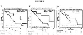
- the present inventors have also surprisingly found that patients with higher numbers of clonal mutations, and a higher ratio of clonal:sub-clonal mutations, have improved prognosis.
- the present invention therefore addresses a need in the art for new, alternative and/or more effective ways of treating and preventing cancer.
- the present invention provides a method for identifying a subject with cancer who is suitable for treatment with an immune checkpoint modulator, said method comprising:
- the invention provides a method for predicting or determining the prognosis of a subject with cancer, the method comprising:
- Described herein is a method of treating or preventing cancer in a subject, wherein said method comprises the following steps:
- Described herein is a method of treating or preventing cancer in a subject which comprises treating a subject with cancer with an immune checkpoint intervention, wherein the subject has been determined to have
- the invention also provides an immune checkpoint modulator for use in the treatment or prevention of cancer in a subject, wherein said subject with cancer has been identified as suitable for treatment with an immune checkpoint modulator according to the method of the invention.
- the invention further provides an immune checkpoint modulator for use in the treatment or prevention of cancer in a subject, wherein the subject has
- a "neo-antigen” is a tumour-specific antigen which arises as a consequence of a mutation within a cancer cell. Thus, a neo-antigen is not expressed by healthy cells in a subject.
- the neo-antigen described herein may be caused by any non-silent mutation which alters a protein expressed by a cancer cell compared to the non-mutated protein expressed by a wild-type, healthy cell.
- the mutated protein may be a translocation or fusion.
- a “mutation” refers to a difference in a nucleotide sequence (e.g. DNA or RNA) in a tumour cell compared to a healthy cell from the same individual.
- the difference in the nucleotide sequence can result in the expression of a protein which is not expressed by a healthy cell from the same individual.
- the mutation may be a single nucleotide variant (SNV), multiple nucleotide variants, a deletion mutation, an insertion mutation, a translocation, a missense mutation or a splice site mutation resulting in a change in the amino acid sequence (coding mutation).
- SNV single nucleotide variant
- multiple nucleotide variants a deletion mutation, an insertion mutation, a translocation, a missense mutation or a splice site mutation resulting in a change in the amino acid sequence (coding mutation).
- the mutations may be identified by Exome sequencing, RNA-seq, whole genome sequencing and/or targeted gene panel sequencing and or routine Sanger sequencing of single genes. Suitable methods are known in the art.
- Targeted gene sequencing panels are also commercially available (e.g. as summarised by Biocompare ((http://www.biocompare.com/ Editorial-Articles/161194-Build-Your-Own-Gene-Panels-with-These-Custom-NGS-Targeting-Tools/)).
- Sequence alignment to identify nucleotide differences may be performed using methods which are known in the art.
- nucleotide differences compared to a reference sample may be performed using the method described by Koboldt et al. (Genome Res. 2012; 22: 568-576 ).
- the reference sample may be the germline DNA and/or RNA sequence.
- the present inventors have determined that intratumour heterogeneity (ITH) can cause variation between the neo-antigens expressed in different regions of a tumour and between different cells in a tumour.
- ITH intratumour heterogeneity
- the inventors have determined that, within a tumour, certain neo-antigens are expressed in all regions and essentially all cells of the tumour whilst other neo-antigens are only expressed in a subset of tumour regions and cells.
- a "clonal” or “truncal” neo-antigen is a neo-antigen which is expressed effectively throughout a tumour and encoded within essentially every tumour cell.
- a "sub-clonal” or “branched” neo-antigen is a neo-antigen which is expressed in a subset or a proportion of cells or regions in a tumour.
- references herein to "essentially all" are intended to encompass the majority of tumour cells in a subject.
- this may comprise 60-100% of cells, e.g. 60, 61, 62, 63, 64, 65, 66, 67, 68, 69, 70, 71, 72, 73, 74, 75, 76, 77, 78, 79 80, 81, 82, 83, 84, 85, 86, 87, 88, 89, 90, 91, 92, 93, 94, 95, 96, 97, 98, 99 or 100% of tumour cells in a subject.
- Present throughout a tumour may mean that the clonal neo-antigen is expressed in all regions of the tumour from which samples are analysed.
- a determination that a mutation is "encoded within essentially every tumour cell” refers to a statistical calculation and is therefore subject to statistical analysis and thresholds.
- a determination that a clonal neo-antigen is "expressed effectively throughout a tumour” refers to a statistical calculation and is therefore subject to statistical analysis and thresholds.
- “Expressed effectively in essentially every tumour cell or essentially all tumour cells” may mean that the mutation is present all tumour cells analysed in a sample, as determined using appropriate statistical methods.
- the cancer cell fraction (CCF), describing the proportion of cancer cells that harbour a mutation may be used to determine whether mutations are clonal or branched.
- the cancer cell fraction may be determined by integrating variant allele frequencies with copy numbers and purity estimates as described by Landau et al. (Cell. 2013 Feb 14;152(4):714-26 ).
- determining a clonal mutation is subject to statistical analysis and threshold.
- the method of this aspect may comprise, for example, determining the mutations present in cancer cells from one or more tumour regions isolated from a tumour. For example, the mutations present in a single biopsy, or alternatively, at least two, at least three, at least four, at least five, at least six, at least seven, at least eight, at least nine or at least ten or more biopsies isolated from a tumour may be determined.
- the individual tumour samples may be isolated from different regions located throughout a tumour within a primary site or between primary and metastases or within a metastasis or between metastases. For example, determining the mutations present in tumours which are known to display morphological disparate histology in different regions may involve determining the mutations present in a number of individual samples isolated from morphologically disparate regions.
- the sample may be a blood sample.
- the blood sample may comprise circulating tumour DNA, circulating tumour cells or exosomes comprising tumour DNA.
- the invention provides a method for identifying a subject with cancer who is suitable for treatment with an immune checkpoint modulator, said method comprising determining the number of clonal neo-antigens in one or more cancer cells from said subject, wherein a higher number of clonal neo-antigens in comparison to a reference sample is indicative of response to an immune checkpoint modulator.
- suitable for treatment may refer to a subject who is more likely to respond to treatment with an immune checkpoint intervention, or who is a candidate for treatment with an immune checkpoint intervention.
- a subject suitable for treatment may be more likely to respond to said treatment than a subject who is determined not to be suitable using the present invention.
- a subject who is determined to be suitable for treatment according to the present invention may demonstrate a durable clinical benefit (DCB), which may be defined as a partial response or stable disease lasting for at least 6 months, in response to treatment with an immune checkpoint intervention.
- DCB durable clinical benefit
- the number of clonal neo-antigens identified or predicted in the cancer cells obtained from the subject may be compared to one or more pre-determined thresholds. Using such thresholds, subjects may be stratified into categories which are indicative of the degree of response to treatment.
- a threshold may be determined in relation to a reference cohort of cancer patients.
- the cohort may comprise 10, 25, 50, 75, 100, 150, 200, 250, 500 or more cancer patients.
- the cohort may be any cancer cohort. Alternatively the patients may all have the relevant or specific cancer type of the subject in question.
- a "high" number of clonal neo-antigens means a number greater than the median number of clonal neo-antigens predicted in a reference cohort of cancer patients, such as the minimum number of clonal neo-antigens predicted to be in the upper quartile of the reference cohort.
- a "high" number of clonal neo-antigens may be defined as 10, 20, 30, 40, 50, 55, 60, 65, 70, 75, 80, 85, 90, 95, 100, 110, 120, 130, 140, 150, 160, 170, 180, 190 or 200 or more clonal neo-antigens.
- references to "high” or “higher” numbers of clonal neo-antigens may be context specific, and could carry out the appropriate analysis accordingly.
- the invention further provides a method for identifying a subject with cancer who is suitable for treatment with an immune checkpoint modulator, said method comprising determining the ratio of clonal:sub-clonal neo-antigens and/or sub-clonal neo-antigen fraction in more than one cancer cell from said subject, wherein a higher ratio of clonal:sub-clonal neo-antigens or lower/low sub-clonal neo-antigen fraction in comparison to a reference sample is indicative of to response to an immune checkpoint modulator.
- the clonal:sub-clonal ratio may be within the context of a cohort of subjects, either with any cancer or with the relevant/specific cancer. Accordingly, the clonal:sub-clonal neo-antigen ratio may be determined by applying methods discussed above to a reference cohort. A "high” or “higher” clonal:sub-clonal ratio may therefore correspond to a number greater than the median clonal:sub-clonal ratio predicted in a reference cohort of cancer patients, such as the minimum clonal:sub-clonal ratio predicted to be in the upper quartile of the reference cohort.
- a "high” or “higher” clonal:sub-clonal ratio means a ratio in the range of 3:1 to 100:1, such as a ratio of at least 3:1, 5:1, 10:1, 15:1, 20:1, 25:1, 50:1, 75:1 or 100:1.
- a ratio of at least 3:1 5:1, 10:1, 15:1, 20:1, 25:1, 50:1, 75:1 or 100:1.
- the values may depend on the cohort in question.
- the fraction of subclonal neo-antigens may also be defined in relation to a reference cohort, as discussed above.
- a "lower" or “low” fraction of subclonal neo-antigens may correspond to a fraction smaller than the median fraction of subclonal neo-antigens predicted in a reference cohort of cancer patients, such as the maximum number predicted to be in the bottom quartile of the cohort.
- a sub-clonal neo-antigen fraction can be determined (for example for each patient) by dividing the number of subclonal neoantigens (for example that are predicted in the one or more cancer cells from said subject) by the number of total neoantigens (for example that are predicted in the one or more cancer cells from said subject).
- a "lower” or “low” fraction of subclonal neo-antigens may mean a fraction of 25% or less, such as a fraction of 20, 15, 10, 5, 3, 2 or 1% or less.
- the method may comprise determining both the number of clonal neo-antigens and the ratio of clonal:sub-clonal neo-antigens or the fraction of of sub-clonal neo-antigens.
- combining measures of both neo-antigen burden and neo-antigen sub-clonal fraction was able to predict sensitivity to pembrolizumab better than either measure alone (see Fig. 4C ), and outcome could be predicted in almost all cases ( Fig 4G-H ).
- the invention provides a method for identifying a subject with cancer who is suitable for treatment with an immune checkpoint modulator, said method comprising:
- tumour cells with high numbers of clonal neo-antigens exhibit similar expression profiles of immune checkpoint molecules, that is they exhibit a common expression profile of immune checkpoint molecules.
- approaches to identify particular immune checkpoint molecules whose expression is increased or decreased relative to non-cancerous cells can also be used to identify patients likely to respond to checkpoint blockade therapies.
- the invention provides the method for identifying subjects who have cancer who are more likely to respond to immune checkpoint modulators, further comprising determining the expression profile of immune checkpoint molecules in cancer cells from said subject, or tumour type, wherein differential immune checkpoint molecule expression in comparison to a reference sample is indicative of response to an immune checkpoint modulator.
- the method comprises determining the expression profile of immune checkpoint molecules in the tumour, for example by identifying differentially expressed genes, e.g. relative to a suitable reference sample.
- the reference sample in respect of differential immune checkpoint molecule expression may be a non-cancerous cell or tumour, (e.g. with low clonal neoantigen burden) or peripheral blood lymphocytes.
- the expression profile of the immune checkpoint molecules may be determined by:
- the invention further provides a method in which determining the expression profile of immune checkpoint molecules in a particular cancer type comprises the steps of:
- the invention thus provides the method for identifying subjects who have cancer who are more likely to respond to immune checkpoint interventions, further comprising determining the expression profile of immune checkpoint molecules in cancer cells from said subject, or tumour type, using said method.
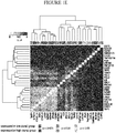
- differentially expressed genes between tumours with high clonal neo-antigen burden and low clonal neo-antigen burden are identified (see e.g. Figure 1E ).
- information regarding the number of clonal neo-antigens is informative and facilitates the combining of the two approaches, namely identifying and targeting subjects/tumours with a high number of clonal neo-antigens, and further investigating the gene expression of immune checkpoint molecules in those subjects/tumours with a high level of clonal neo-antigens. This facilitates a "double-pronged" therapeutic attack.
- said differential immune expression is upregulation or high expression of an immune checkpoint molecule which is an inhibitory receptor or costimulatory receptor compared to a suitable reference sample, wherein such upregulation or high expression is indicative of a response to immune checkpoint interventions targeting the immune checkpoint molecule that has been upregulated or shown high expression.
- Gene expression profiles may, for example, be determined by a method as described in present Example 1.
- the immune checkpoint molecule is PD-1 and/or LAG-3.
- the subject has lung cancer, preferably non small-cell lung cancer.
- the immune checkpoint molecule is CTLA4.
- the cancer is lung cancer or melanoma, preferably non small-cell lung cancer or melanoma.
- This method may also be used in combination with the previously described methods for identifying a subject with cancer who is likely to respond to treatment with an immune checkpoint intervention.
- the invention provides a method for identifying a subject with cancer who is suitable for treatment with an immune checkpoint modulator, said method comprising:
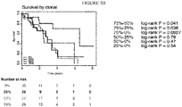
- the present inventors have made the important and surprising determination that cancer patients with higher numbers of clonal neo-antigens, and/or a higher ratio of clonal:sub-clonal neoantigens or a low sub-clonal neo-antigen fraction, have improved prognosis.
- subjects with high or higher numbers of clonal neo-antigens may have improved survival relative to subjects with lower numbers of clonal neo-antigens.
- a reference value for the number of clonal neo-antigens could be determined using the following method, with a "high number” or “higher number” being anything above that.
- Said method may involve determining the number of clonal neo-antigens predicted in a cohort of cancer subjects and either:
- Such a “median number” or “minimum number to be in the upper quartile” could be determined in any cancer cohort per se , or alternatively in the relevant / specific cancer types.
- a "high" or “higher” number of clonal neo-antigens may be defined as 50, 55, 60, 65, 70, 75, 80, 85, 90, 95, 100, 110, 120, 130, 140, 150, 160, 170, 180, 190 or 200 or more clonal neo-antigens.
- references to "high” or “higher” numbers of clonal neo-antigens may be context specific, and could carry out the appropriate analysis accordingly.
- the present invention also provides a method for predicting or determining the prognosis of a subject with cancer, comprising determining the number of clonal neo-antigens in one or more cancer cells from the subject, wherein a higher number of clonal neo-antigens, for example relative to a cohort as discussed above, is indicative of improved prognosis.
- the cancer is lung cancer or melanoma, preferably non small-cell lung cancer or melanoma.
- the invention comprises a method for predicting or determining the prognosis of a subject with cancer, the method comprising determining the clonal:sub-clonal ratio and/or sub-clonal neo-antigen fraction in more than one cancer cell from said subject, wherein a higher clonal:sub-clonal ratio or a lower/low sub-clonal neo-antigen fraction, for example relative to a cohort as discussed above, is indicative of improved prognosis.
- the cancer is melanoma or lung cancer, preferably melanoma or non small-cell lung cancer.
- Described herein is a method of treating or preventing cancer in a subject, wherein said method comprises the following steps:
- the present invention provides an immune checkpoint modulator for use in the treatment or prevention of cancer in a subject, wherein said subject with cancer has been identified as suitable for treatment with an immune checkpoint modulator according to the method of the invention.
- the present invention also provides an immune checkpoint intervention for use in the treatment or prevention of cancer in a subject, wherein the subject has:
- treatment refers to reducing, alleviating or eliminating one or more symptoms of the disease, disorder or infection which is being treated, relative to the symptoms prior to treatment.
- Prevention refers to delaying or preventing the onset of the symptoms of the disease, disorder or infection. Prevention may be absolute (such that no disease occurs) or may be effective only in some individuals or for a limited amount of time.
- immune checkpoint intervention is used herein to refer to any therapy which interacts with or modulates an immune checkpoint molecule.
- an immune checkpoint intervention may also be referred to herein as a “checkpoint blockade therapy", “checkpoint modulator” or “checkpoint inhibitor”.
- inhibitor any means to prevent inhibition of T cell activity by these pathways. This can be achieved by antibodies or molecules that block receptor ligand interaction, inhibitors of intracellular signalling pathways, and compounds preventing the expression of immune checkpoint molecules on the T cell surface.
- Checkpoint inhibitors include, but are not limited to, CTLA-4 inhibitors, PD-1 inhibitors, PD-L1 inhibitors, Lag-3 inhibitors, Tim-3 inhibitors, TIGIT inhibitors and BTLA inhibitors, for example.
- Co-stimulatory antibodies deliver positive signals through immune-regulatory receptors including but not limited to ICOS, CD137, CD27 OX-40 and GITR.
- Suitable immune checkpoint interventions include pembrolizumab, nivolumab, atezolizumab and ipilimumab.
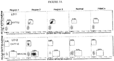
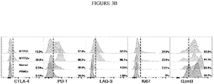
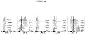
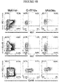
- lung tumours with a high number of clonal neoantigens express high levels of PD-1 and Lag-3, and in keeping, T cells reactive to clonal neoantigens in lung cancer subjects also express high levels of PD-1 and LAG-3.
- the co-expression of PD-1 and Lag-3 in tumours with high clonal neo-antigen burden versus low clonal burden suggests that simultaneous targeting of both pathways may generate maximal benefit.
- the invention relates to co-targeting PD-1 and Lag-3 pathways, for example in lung cancer, either by co-administration of inhibitors targeting each pathway or by administration of a single reagent targeting both pathways.
- bispecific antibodies are able to bind to PD-1 and Lag-3, or PD-L1 and Lag-3.
- the subject is a mammal, preferably a cat, dog, horse, donkey, sheep, pig, goat, cow, mouse, rat, rabbit or guinea pig, but most preferably the subject is a human.
- the treatment or prevention of cancer according to the invention comprises the step of identifying a patient in need of said treatment or therapy.
- the cancer may be selected from, for example, bladder cancer, gastric cancer, oesophageal cancer, breast cancer, colorectal cancer, cervical cancer, ovarian cancer, endometrial cancer, kidney cancer (renal cell), lung cancer (small cell, non-small cell and mesothelioma), brain cancer (e.g. gliomas, astrocytomas, glioblastomas), melanoma, lymphoma, small bowel cancers (duodenal and jejunal), leukemia, pancreatic cancer, hepatobiliary tumours, germ cell cancers, prostate cancer, head and neck cancers, thyroid cancer and sarcomas.
- bladder cancer gastric cancer, oesophageal cancer, breast cancer, colorectal cancer, cervical cancer, ovarian cancer, endometrial cancer, kidney cancer (renal cell), lung cancer (small cell, non-small cell and mesothelioma), brain cancer (e.g. gliomas, astrocytomas
- the cancer is lung cancer.
- the lung cancer is non-small cell lung cancer.
- the cancer is melanoma.
- the subject has pre-invasive disease, or is a subject who has had their primary disease resected who might require or benefit from adjuvant therapy, such as that provided by the present invention.
- Treatment using the present invention may also encompass targeting circulating tumour cells and/or metastases derived from the tumour.
- the uses for treating cancer according to the present invention may be performed in combination with additional cancer therapies.
- the immune checkpoint modulators according to the present invention may be administered in combination with co-stimulatory antibodies, chemotherapy and/or radiotherapy, targeted therapy or monoclonal antibody therapy.
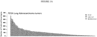
- Samples for sequencing were obtained from patients diagnosed with non-small cell lung cancer (NSCLC) who underwent definitive surgical resection prior to receiving any form of adjuvant therapy, such as chemotherapy or radiotherapy. Informed consent allowing for genome sequencing had been obtained. Both samples were collected from University College London Hospital, London (UCLHRTB 10/H1306/42) and were subjected to pathology review to establish the histological subtype: one tumour was classified with CK7+/TTF1+ adenocarcinoma (L011) and one tumour (L012) with squamous cell carcinoma histology. Detailed clinical characteristics are provided in table S1.
- Samples obtained from (1) reflected a patient cohort of stage IV NSCLC, and a detailed description of this patient cohort, including tumour processing, can be found in supplementary material of (1). Detailed clinical characteristics of this cohort are provided in table S3.
- Clinical efficacy analysis was performed as in ( 1 ).
- objective response to pembrolizumab was assessed by investigator-assessed immunerelated response criteria (irRC) by a study radiologist.
- irRC immunerelated response criteria
- CT scans were performed every nine weeks. Partial and complete responses were confirmed by a repeat imaging occurring a minimum of 4 weeks after the initial identification of response; unconfirmed responses were considered stable or progressive disease dependent on results of the second CT scan.
- Durable clinical benefit (DCB) was defined as stable disease or partial response lasting longer than 6 months (week 27, the time of third protocol-scheduled response assessment).
- No durable benefit (NDB) was defined as progression of disease ⁇ 6 months of beginning therapy.
- progression-free survival was censored at the date of the most recent imaging evaluation.
- overall survival was censored at the date of last known contact. Details regarding response for each patient can be found in table S2.
- One LUAD patient, TCGA-05-4396 was excluded for having over 7000 low quality mutations called, mostly in a C[C>G]G context.
- a LUSC patient, TCGA-18-3409 was excluded for bearing a strong UV signature, uncharacteristic of a LUSC tumour.
- FFPE tissue For the FFPE tissue, manual blade macrodissection was used to remove tumour-rich areas of tissue from 10-40 ⁇ m unstained slides, and DNA was extracted from this using the DNeasy Blood and Tissue kit (Qiagen) DNA was quantified by Qubit (Invitrogen) and DNA integrity was examined by agarose gel eletrophoresis. Details regarding processing of validation and discovery cohort can be found in supplementary material of ( 1 ).
- Raw paired end reads (100bp) in FastQ format generated by the Illumina pipeline were aligned to the full hg19 genomic assembly (including unknown contigs) obtained from GATK bundle 2.8 ( 5 ), using bwa mem (bwa-0.7.7) ( 6 ).
- Picard tools v1.107 was used to clean, sort and merge files from the same patient region and to remove duplicate reads (http://broadinstitute.github.io/picard).
- Quality control metrics were obtained using a combination of picard tools (1.107), GATK (2.8.1) and FastQC (0.10.1) (http://www.bioinformatics.babraham.ac.uk/projects/fastqc/).
- SAMtools mpileup (0.1.16) ( 7 ) was used to locate non-reference positions in tumour and germ-line samples. Bases with a phred score of ⁇ 20 or reads with a mapping-quality ⁇ 20 were skipped. BAQ computation was disabled and the coefficient for downgrading mapping quality was set to 50. Somatic variants between tumour and matched germ-line were determined using VarScan2 somatic (v2.3.6) ( 8 ) utilizing the output from SAMtools mpileup. Default parameters were used with the exception of minimum coverage for the germ-line sample that was set to 10, minimum variant frequency was changed to 0.01 and tumour purity was set to 0.5. VarScan2 processSomatic was used to extract the somatic variants.
- Varscan2's associated fpfilter.pl script having first run the data through bam-readcount (0.5.1). Only INDEL calls classed as 'high confidence' by VarScan2 processSomatic were kept for further analysis.
- Ion Torrent PGM sequencer Life Technologies
- Ion AmpliSeqTM custom panel Life Technologies
- Multiplex PCRs were performed on DNA from each region according to the manufacturer's protocol.
- Barcoded sequencing libraries were TM constructed, which were sequenced with 200 bp read length on the Ion Torrent PGM sequencer (Life Technologies). Sequence alignment to target regions from the hg19 genome was performed using the IonTorrent TorrentSuiteTM software.
- Variants for which the coverage was 50 in at least one region were selected. A variant was considered to be present in a region if the variant frequency was ⁇ 0.01 for SNVs and ⁇ 0.02 for INDELS. Again manual review in IGV was performed and variants that passed this stage were used for subsequent analyses. All variants were annotated using ANNOVAR ( 11 ) and potential driver mutations were defined as described in ( 12 ).
- the clonal status of each mutation was estimated by integrating the wild-type and mutant allele counts, absolute major and minor copy numbers, and tumour purity estimates as previously described ( 14 ).
- L011 and L012 clonal status of each mutation was estimated based on multiregion sequencing analysis. In brief, each mutation was classified as clonal if identified and present in each and every tumour region sequenced within the tumour. Conversely, any mutations not ubiquitously present in every tumour region was classified as subclonal.
- tumour For discovery and validation cohort tumour, encompassing data obtained from ( 1 ), the cancer cell fraction of each mutation was estimated by integrating the local copy number (obtained from ASCAT, see below), tumour purity (also obtained from ASCAT), and variant allele frequency.
- nmut the observed mutation copy number, describing the fraction of tumour cells carrying a given mutation multiplied by the number of chromosomal copies at that locus using the following formula: where VAF corresponds to the variant allele frequency at the mutated base, and p, CN t , CN n are respectively the tumour purity, the tumour locus specific copy number, and the normal locus specific copy number.
- mutations were first further filtered to ensure reliable clustering.
- a Varscan2 somatic p-value threshold 0.01.
- a minimum of 5 alternate reads was required for each variant, as well as a minimum tumour variant allele frequency of 1%.
- Mutations were also filtered such that a maximum of 2 germline reads, and 2% germline variant allele frequency was permitted.
- VarScan2 For data obtained from ( 1 ) processed sample exome SNP and copy number data from paired tumour-normal was generated using VarScan2 (v2.3.6). Varscan2 copy number was run using default parameters with the exception of min-coverage (21221095)and data-ratio. The data-ratio was calculated on a per-sample basis as described in (22300766). The output from Varscan was processed using the ASCAT v2.3 (20837533) to provide segmented copy number data and cellularity and ploidy estimates for all samples based on the exome sequence data. The following setting was altered from its default value: Threshold for setting ACF to 1 was adjusted from 0.2 to 0.15 and the package was run with gamma setting of 1. For TCGA samples, SNP6.0 data was processed to yield copy number information, as described in McGranahan, 2015.
- tumour L011 the primary tumour data was reanalyzed using the method described for L012 and the L011 metastatic regions, allowing for a combined tree featuring both primary and metastatic regions.
- the 4-digit HLA type was determined using POLYSOLVER (POLYmorphic loci reSOLVER)( 16 ).
- Patients L011 and L012 were serotyped and simultaneously genotyped using Optitype ( 17 ), which produced concordant results.
- Identified non-silent mutations were used to generate a comprehensive list of peptides 9-11 amino acids in length with the mutated amino acid represented in each possible position.
- the binding affinity of every mutant peptide and its corresponding wild-type peptide to the patient's germline HLA alleles was predicted using netMHCpan-2.8 ( 18, 19 ).
- Candidate neo-antigens were identified as those with a predicted binding strength of ⁇ 500 nM.
- Clinical data for the TCGA patients was accessed through the TCGA data portal and downloaded from https://tcaadata.nci.nih.aov/tcaafiles/ftp_auth/distro_ftpusers/anonymous/tumour/CANCER.T YPE /bcr/biotab/clin/. Survival analyses were performed in R using the survival package.
- RNA-sequencing data was downloaded from the TCGA data portal. For each LUAD patient, all available 'Level_3' gene-level data was obtained. The raw read counts were used as input into the R package DESeq2 for analysis. A transcriptomewide differential gene expression analysis was performed and significantly differentially expressed (adjusted p ⁇ 0.05) immune related genes (listed in Table S1) were identified. These genes were clustered on their co-expression using the metric 1-r 2 .
- TILs tumour-infiltrating lymphocytes
- Tumours were taken directly from the operating theatre to the department of pathology where the sample was divided into regions. Samples were subsequently minced under sterile conditions followed by enzymatic digestion (RPMI-1640 (Sigma) with Liberase TL research grade (Roche) and DNAse I (Roche)) at 37°C for 30 minutes before mechanical dissociation using gentleMACS (Miltenyi Biotech). Resulting single cell suspensions were enriched for leukocytes by passage through a Ficoll-paque (GE Healthcare) gradient. Live cells were counted and frozen in human AB serum (Sigma) with 10% dimethyl sulfoxide at - 80°C before transfer to liquid nitrogen.
- TILs were expanded using a rapid expansion protocol (REP) in T25 flasks containing EX-VIVO media (Lonza) supplemented with 10% human AB serum (Sigma), soluble anti-CD3 (OKT3, BioXCell), 6000IU/mL recombinant human (rhlL-2, PeproTech) and 2x10 7 irradiated PBMCs (30Gy) pooled from 3 allogeneic healthy donors. Fresh media containing rhlL-2 at 3000IU/mL was added every three days as required. Following 2 weeks of expansion, TILs were counted, phenotyped by flow cytometry and frozen in human AB serum (Sigma) at-80°C before use in relevant assays or long-term storage in liquid nitrogen.
- REP rapid expansion protocol
- HLA molecules matching the HLA-expression of L011 HLA-A1101, A2402, and B3501
- L012 HLA-A1101, A2402, and B0702
- HLA complexes loaded with UV-sensitive peptide were subjected to 366-nm UV light (CAMAG) for one hour at 4°C in the presence of candidate neo-antigen peptide in a 384-well plate.
- Peptide-MHC multimers were generated using a total of 9 different fluorescent streptavidin (SA) conjugates: PE, APC, PE-Cy7, PE-CF594, Brilliant Violet (BV)421, BV510, BV605, BV650, Brilliant Ultraviolet (BUV)395 (BioLegend).
- MHC-multimers were generated with two different streptavidin-conjugates for each peptide-specificity to allow a combinatorial encoding of each antigen responsive T cells, enabling analyses for reactivity against up to 36 different peptides in parallel ( 24, 25 ).
- MHC-multimer analysis was performed on in-vitro expanded CD8+ T lymphocytes isolated from region-specific lung cancer samples and adjacent normal lung tissue.
- 290 and 355 candidate mutant peptides (with predicted HLA binding affinity ⁇ 500nM, including multiple potential peptide variations from the same missense mutation) were synthesized and used to screen expanded L011 and L012 TILs respectively.
- samples were thawed, treated with DNAse for 10 min, washed and stained with MHC multimer panels for 15 min at 37°C.
- HLA-B3501 MTFR2-derived multimers were found to bind the mutated sequence FAFQEYDSF (netMHC binding score: 22) but not the wild type sequence FAFQEDDSF (netMHC binding score: 10) ( Fig 11B and D , Fig 9C ). No responses were found against overlapping peptides AFQEYDSFEK and KFAFQEYDSF.
- HLA-A1101 CHTF18-derived multimers bound the mutated sequence LLLDIVAPK (netMHC binding score: 37) but not the wild type sequence: LLLDILAPK (netMHC binding score: 41) ( Fig 11C and E , Fig 9C ).
- Tumour samples were thawed, washed and first stained with custom-made MHCmultimers for 10-15 minutes at 37°C in the dark. Cells were thereafter transferred onto wet ice and stained for 30 minutes, in the dark, with a panel of surface antibodies used at the manufacturer's recommended dilution: CD8-V500, SK1 clone (BD Biosciences), PD-1-BV605, EH12.2H7 clone (Biolegend), CD3-BV785, OKT3 clone (Biolegend), LAG-3-PE, 3DS223H clone (eBioscience). Cells were permeablized for 20 minutes with use of the intracellular fixation and permeabilization buffer set from eBioscience.
- An intracellular staining panel was applied for 30 minutes, on ice, in the dark, and consisted of the following antibodies used at the manufacturers recommended dilution: granzyme B-V450, GB11 clone (BD Biosciences), FoxP3-PerCP-Cy5.5, PCH101 clone (eBioscience), Ki67-FITC, clone B56 (BD Biosciences)and CTLA-4 - APC, L3D10 clone (Biolegend). Data acquisition was performed on a BD FACSAria III flow cytometer (BD Biosciences) and analysed in Flowjo version 10.0.8 (Tree Star Inc.).
- tumour neo-antigen burden may increase tumour recognition by T cells, reducing the potential for immune-evasion ( 12 ).
- neo-antigen clonal status the presence of a neo-antigen in all tumour (clonal) compared to a subset of tumour cells (subclonal)
- cancer cell fraction proportion of cancer cells harboring each mutation
- each putative neo-antigen was classified as either clonal or subclonal ( 13 ).
- the LUSC cohort had a narrower range of putative neo-antigens ( Fig 7A ), with a median absolute deviation of 50 and interquartile range of 71 and a statistically significant association between overall survival and neo-antigen load was not observed in this cohort ( Fig 7 B-G ). This might reflect difficulties in dissecting the clonal architecture of tumours from single samples ( 14 ).
- PD-L1 was also significantly up-regulated (p ⁇ 0.001) in the high clonal cohort, clustering with PD-L2.
- PD-L1 was identified as the most significantly differentially expressed immune gene ( Fig 8 , p ⁇ 0.001).
- L011 and L012 Two early stage tumours, L011 and L012, subjected to multi-region exome sequencing ( 13 ), permitted phylogenetic analysis and prediction of neo-antigens within each primary tumour region ( Fig 2A ).
- L011 included a brain metastasis, resected 14 months following primary surgery, subjected to multi-region sequencing. While both tumours were derived from female smokers (>40 pack-years), their mutation burden and extent of heterogeneity was distinct ( Fig 2A ).
- L011 an adenocarcinoma, exhibited a homogenous primary tumour and metastatic dissemination to the brain (M1-M4), likely originating from tumour region R3 ( Fig 2A ). A total of 313 neo-antigens were predicted within the primary tumour, 88% of which were clonal, identified in every region of the primary tumour ( Fig 2B ). Conversely, L012, a squamous cell carcinoma, exhibited a low mutation burden and extensive heterogeneity, with 75% of the predicted neo-antigens being subclonal ( Fig 2A , C ).
- MHC-multimers loaded with predicted neo-antigens were used to screen CD8+ T cells expanded from different tumour regions and adjacent normal lung tissue (13).
- CD8+ T cells reactive to mutant MTFR2D326Y FAFQEYDSF
- a clonal mutation with high predicted HLA binding in wild type (10nM) and mutant (22nM) forms ( Fig 2B )
- CD8+ T cells reactive to mutant CHTF18L769V (LLDIVAPK) and MYADMR30W (SPMIVGSP W ) were identified in all tumour regions and at lower frequencies in normal tissue ( Fig 2E ).
- MTFR2-reactive CD8+ T cells could also be detected in non-expanded TILs (tumour infiltrating lymphocytes) ( Fig 3A ) from all primary tumour regions (0.79-1.35%), and at lower frequencies in normal tissue (0.16%) and peripheral blood mononuclear cells (PBMCs) (0.02%).
- TILs tumor infiltrating lymphocytes
- PBMCs peripheral blood mononuclear cells
- CHTF18-reactive and MYADM-reactive CD8+ T cells were identified in non-expanded samples from all tumour regions in L012 (CHTF18 0.16-0.58%, MYADM 2.25- 2.31%) and at a lower frequency in normal lung tissue (CHTF18 0.02%, MYADM 0.17%) and PBMCs (CHTF18 0.02%, MYADM 0.01%) ( Fig 3A ).
- neo-antigen-reactive T cells in non-expanded samples were characterized by flow cytometry. Although at low levels, CTLA-4 expression was confined to tumour-infiltrating CD8+ T cells for both L011 and L012, with highest levels identified on MTFR2, CHTF18 and MYADM-reactive T cells ( Fig 3B , Fig 9A ). High levels of PD-1 were expressed by >99% of MTFR2-, CHTF18- and MYADM-reactive tumour-infiltrating CD8+ T cells ( Fig 3B , Fig 9A ), whilst lower levels were observed on CD8+ MHC-multimer negative T cells in tumour, normal tissue and PBMCs.
- LAG-3 expression was higher on all tumour infiltrating CD8+ T cells, including MTFR2-reactive cells, relative to normal tissue and PBMCs ( Fig 3B ). LAG-3 expression was also observed in L012, although at lower levels ( Fig 9A ). IHC studies further supported these findings, identifying CD8+ T cells co-expressing LAG-3 in both L011 and L012 primary tumours ( Fig 3D ). Ki67 was expressed at higher levels on tumour infiltrating CD8+ T cells than in normal tissue or PBMCs ( Fig 3B , Fig 9A ), however the fraction of proliferating cells was low for both neo-antigen-reactive and MHC-multimer negative cells ( ⁇ 25%).
- GzmB granzyme B
- Fig. 4AH The relationship was also contingent upon the clonal architecture of each tumour ( Fig. 4AH ).
- DCB durable clinical benefit
- a high clonal neo-antigen burden defined as above or equal to the median number of clonal neo-antigens in the discovery cohort, 91
- a neo-antigen subclonal fraction lower than 5% Fig. 4A-B
- NDB non-durable benefit
- neo-antigen repertoire and neo-antigen heterogeneity i.e. the ratio of clonal:sub-clonal neo-antigens or mutations
- Fig 4C the ratio of clonal:sub-clonal neo-antigens or mutations
Landscapes
- Health & Medical Sciences (AREA)
- Life Sciences & Earth Sciences (AREA)
- Engineering & Computer Science (AREA)
- Chemical & Material Sciences (AREA)
- Immunology (AREA)
- General Health & Medical Sciences (AREA)
- Physics & Mathematics (AREA)
- Molecular Biology (AREA)
- Medicinal Chemistry (AREA)
- Hematology (AREA)
- Urology & Nephrology (AREA)
- Biomedical Technology (AREA)
- Biotechnology (AREA)
- Organic Chemistry (AREA)
- Analytical Chemistry (AREA)
- Public Health (AREA)
- Biochemistry (AREA)
- Pathology (AREA)
- Microbiology (AREA)
- Bioinformatics & Cheminformatics (AREA)
- Medical Informatics (AREA)
- Proteomics, Peptides & Aminoacids (AREA)
- Biophysics (AREA)
- Oncology (AREA)
- Food Science & Technology (AREA)
- General Physics & Mathematics (AREA)
- Cell Biology (AREA)
- Hospice & Palliative Care (AREA)
- Pharmacology & Pharmacy (AREA)
- Veterinary Medicine (AREA)
- Animal Behavior & Ethology (AREA)
- Bioinformatics & Computational Biology (AREA)
- Evolutionary Biology (AREA)
- Spectroscopy & Molecular Physics (AREA)
- Theoretical Computer Science (AREA)
- Epidemiology (AREA)
- Nuclear Medicine, Radiotherapy & Molecular Imaging (AREA)
- General Chemical & Material Sciences (AREA)
- Chemical Kinetics & Catalysis (AREA)
- Genetics & Genomics (AREA)
Applications Claiming Priority (2)
| Application Number | Priority Date | Filing Date | Title |
|---|---|---|---|
| GBGB1516047.6A GB201516047D0 (en) | 2015-09-10 | 2015-09-10 | Method |
| PCT/EP2016/071471 WO2017042394A1 (en) | 2015-09-10 | 2016-09-12 | "immune checkpoint intervention" in cancer |
Publications (2)
| Publication Number | Publication Date |
|---|---|
| EP3347039A1 EP3347039A1 (en) | 2018-07-18 |
| EP3347039B1 true EP3347039B1 (en) | 2021-08-11 |
Family
ID=54362973
Family Applications (1)
| Application Number | Title | Priority Date | Filing Date |
|---|---|---|---|
| EP16763526.7A Active EP3347039B1 (en) | 2015-09-10 | 2016-09-12 | "immune checkpoint intervention" in cancer |
Country Status (11)
| Country | Link |
|---|---|
| US (1) | US11098121B2 (ja) |
| EP (1) | EP3347039B1 (ja) |
| JP (1) | JP7073254B2 (ja) |
| KR (1) | KR20180081490A (ja) |
| CN (1) | CN108135985A (ja) |
| AU (1) | AU2016319316B2 (ja) |
| BR (1) | BR112018004878B1 (ja) |
| CA (1) | CA2997651A1 (ja) |
| GB (1) | GB201516047D0 (ja) |
| RU (1) | RU2739036C2 (ja) |
| WO (1) | WO2017042394A1 (ja) |
Families Citing this family (45)
| Publication number | Priority date | Publication date | Assignee | Title |
|---|---|---|---|---|
| US9424392B2 (en) | 2005-11-26 | 2016-08-23 | Natera, Inc. | System and method for cleaning noisy genetic data from target individuals using genetic data from genetically related individuals |
| US11939634B2 (en) | 2010-05-18 | 2024-03-26 | Natera, Inc. | Methods for simultaneous amplification of target loci |
| US11332793B2 (en) | 2010-05-18 | 2022-05-17 | Natera, Inc. | Methods for simultaneous amplification of target loci |
| US11339429B2 (en) | 2010-05-18 | 2022-05-24 | Natera, Inc. | Methods for non-invasive prenatal ploidy calling |
| US9677118B2 (en) | 2014-04-21 | 2017-06-13 | Natera, Inc. | Methods for simultaneous amplification of target loci |
| US10316362B2 (en) | 2010-05-18 | 2019-06-11 | Natera, Inc. | Methods for simultaneous amplification of target loci |
| US11326208B2 (en) | 2010-05-18 | 2022-05-10 | Natera, Inc. | Methods for nested PCR amplification of cell-free DNA |
| US11408031B2 (en) | 2010-05-18 | 2022-08-09 | Natera, Inc. | Methods for non-invasive prenatal paternity testing |
| US11322224B2 (en) | 2010-05-18 | 2022-05-03 | Natera, Inc. | Methods for non-invasive prenatal ploidy calling |
| US20190010543A1 (en) | 2010-05-18 | 2019-01-10 | Natera, Inc. | Methods for simultaneous amplification of target loci |
| US11332785B2 (en) | 2010-05-18 | 2022-05-17 | Natera, Inc. | Methods for non-invasive prenatal ploidy calling |
| EP2854058A3 (en) | 2010-05-18 | 2015-10-28 | Natera, Inc. | Methods for non-invasive pre-natal ploidy calling |
| JP6153874B2 (ja) | 2011-02-09 | 2017-06-28 | ナテラ, インコーポレイテッド | 非侵襲的出生前倍数性呼び出しのための方法 |
| EP3561075A1 (en) | 2014-04-21 | 2019-10-30 | Natera, Inc. | Detecting mutations in tumour biopsies and cell-free samples |
| US11236139B2 (en) | 2014-11-05 | 2022-02-01 | The Regents Of The University Of California | Combination immunotherapy |
| EP3288581B1 (en) * | 2015-04-27 | 2020-11-11 | Cancer Research Technology Limited | Method for treating cancer |
| EP3294906B1 (en) | 2015-05-11 | 2024-07-10 | Natera, Inc. | Methods for determining ploidy |
| GB201516047D0 (en) | 2015-09-10 | 2015-10-28 | Cancer Rec Tech Ltd | Method |
| WO2018067517A1 (en) | 2016-10-04 | 2018-04-12 | Natera, Inc. | Methods for characterizing copy number variation using proximity-litigation sequencing |
| US10011870B2 (en) | 2016-12-07 | 2018-07-03 | Natera, Inc. | Compositions and methods for identifying nucleic acid molecules |
| WO2018136664A1 (en) * | 2017-01-18 | 2018-07-26 | Ichan School Of Medicine At Mount Sinai | Neoantigens and uses thereof for treating cancer |
| JP7155470B2 (ja) * | 2017-03-31 | 2022-10-19 | エーシーティー ジェノミックス (アイピー) カンパニー リミテッド | 免疫原性がん特異的エピトープのためのランク付けシステム |
| GB201710815D0 (en) * | 2017-07-05 | 2017-08-16 | Francis Crick Inst Ltd | Method |
| AU2018298971A1 (en) * | 2017-07-14 | 2020-01-16 | Cancer Research Technology Limited | Analysis of HLA alleles in tumours and the uses thereof |
| US11293066B2 (en) * | 2017-07-18 | 2022-04-05 | Institut Gustave Roussy | Method for assessing the response to PD-1/PDL-1 targeting drugs |
| JP7072825B2 (ja) * | 2017-09-13 | 2022-05-23 | 三菱電機ソフトウエア株式会社 | コピー数計測装置、コピー数計測プログラムおよびコピー数計測方法 |
| USD844652S1 (en) * | 2017-11-26 | 2019-04-02 | Jan Magnus Edman | Display screen with graphical user interface |
| CA3067229A1 (en) * | 2017-12-01 | 2019-06-06 | Illumina, Inc. | Methods and systems for determining somatic mutation clonality |
| JP2021506342A (ja) | 2017-12-14 | 2021-02-22 | ティーエーアイ ダイアグノスティックス インコーポレイテッドTai Diagnostics,Inc. | 移植のための移植片適合性の評価 |
| CA3090426A1 (en) | 2018-04-14 | 2019-10-17 | Natera, Inc. | Methods for cancer detection and monitoring by means of personalized detection of circulating tumor dna |
| EP3790895A4 (en) * | 2018-05-10 | 2022-05-11 | The Regents of the University of California | CCL21 AND CHECKPOINT INHIBITORS FOR THE TREATMENT OF CANCER |
| US11525159B2 (en) | 2018-07-03 | 2022-12-13 | Natera, Inc. | Methods for detection of donor-derived cell-free DNA |
| WO2020033866A1 (en) * | 2018-08-10 | 2020-02-13 | Omniseq, Inc. | Methods and systems for assessing proliferative potential and resistance to immune checkpoint blockade |
| KR20210148077A (ko) * | 2018-12-21 | 2021-12-07 | 에이전시 포 사이언스, 테크놀로지 앤드 리서치 | 면역 체크포인트 억제 요법으로부터의 수혜를 예측하는 방법 |
| CN111383713B (zh) * | 2018-12-29 | 2023-08-01 | 北京安诺优达医学检验实验室有限公司 | ctDNA检测分析装置及方法 |
| JP2022523672A (ja) * | 2019-01-28 | 2022-04-26 | ボード オブ レジェンツ,ザ ユニバーシティ オブ テキサス システム | がんを治療するための金属キレート化剤併用療法 |
| US20220177978A1 (en) * | 2019-04-02 | 2022-06-09 | INSERM (Institut National de la Santé et de la Recherche Médicale) | Methods of predicting and preventing cancer in patients having premalignant lesions |
| WO2020218322A1 (ja) * | 2019-04-23 | 2020-10-29 | 国立大学法人東北大学 | 血中ケモカインを用いた免疫チェックポイント阻害薬の治療効果予測 |
| CN111394454B (zh) * | 2020-01-06 | 2023-03-14 | 江苏省肿瘤防治研究所(江苏省肿瘤医院) | 一种免疫相关生物标志物及其在头颈部鳞状细胞癌预后诊断中的应用 |
| US20230037966A1 (en) * | 2020-01-07 | 2023-02-09 | The Trustees Of The University Of Pennsylvania | Source specific exosomes for determining avoidance of cancer treatment and avoidance of checkpoint inhibitor therapies |
| US20230047716A1 (en) * | 2020-01-07 | 2023-02-16 | Korea Advanced Institute Of Science And Technology | Method and system for screening neoantigens, and uses thereof |
| EP3868897A1 (en) * | 2020-02-20 | 2021-08-25 | Worldwide Innovative Network | Method for improving the treatment with immune checkpoint blockade therapy |
| CN112735513B (zh) * | 2021-01-04 | 2021-11-19 | 江苏先声医疗器械有限公司 | 基于dna甲基化谱的肿瘤免疫检查点抑制剂治疗有效性评估模型的构建方法 |
| GB202104715D0 (en) * | 2021-04-01 | 2021-05-19 | Achilles Therapeutics Uk Ltd | Identification of clonal neoantigens and uses thereof |
| WO2023277086A1 (ja) * | 2021-06-30 | 2023-01-05 | ノバセラム株式会社 | 血中循環腫瘍細胞を含む試料を調製する方法 |
Family Cites Families (32)
| Publication number | Priority date | Publication date | Assignee | Title |
|---|---|---|---|---|
| AU7696487A (en) | 1986-07-03 | 1988-01-29 | Bio-Rad Laboratories, Inc. | Peptide fragments of organ-specific neoantigens |
| US5670325A (en) * | 1996-08-14 | 1997-09-23 | Exact Laboratories, Inc. | Method for the detection of clonal populations of transformed cells in a genomically heterogeneous cellular sample |
| EP1762575A1 (en) | 2005-09-12 | 2007-03-14 | Ganymed Pharmaceuticals AG | Identification of tumor-associated antigens for diagnosis and therapy |
| RU2011122459A (ru) | 2008-11-03 | 2012-12-10 | Стихтинг Санквин Блудворзининг | Определение в образце клеток, отвечающих на антиген |
| WO2010144192A1 (en) | 2009-05-05 | 2010-12-16 | Children's Medical Center Corporation | Prognosis indicators for solid human tumors |
| US8176660B2 (en) | 2009-07-30 | 2012-05-15 | Nike, Inc. | Customizable stud for an article of footwear |
| CN104826106B (zh) | 2009-09-30 | 2019-01-11 | 斯隆凯特林防癌纪念中心 | 用于治疗癌症的组合免疫疗法 |
| BR112012029066A2 (pt) | 2010-05-14 | 2020-09-01 | The General Hospital Corporation | composições e processos de identificação de neoantígenos específicos de tumor. |
| HUE062102T2 (hu) | 2011-05-24 | 2023-09-28 | BioNTech SE | Individualizált vakcinák a rák ellen |
| WO2012159643A1 (en) * | 2011-05-24 | 2012-11-29 | Biontech Ag | Individualized vaccines for cancer |
| EP3511425A1 (en) | 2012-07-12 | 2019-07-17 | Persimmune, Inc. | Personalized cancer vaccines and adoptive immune cell therapies |
| WO2014026096A1 (en) | 2012-08-10 | 2014-02-13 | The Broad Institute, Inc. | Methods and apparatus for analyzing and quantifying dna alterations in cancer |
| US20160002731A1 (en) | 2012-10-01 | 2016-01-07 | Adaptive Biotechnologies Corporation | Immunocompetence assessment by adaptive immune receptor diversity and clonality characterization |
| EP2906684B8 (en) | 2012-10-10 | 2020-09-02 | Sangamo Therapeutics, Inc. | T cell modifying compounds and uses thereof |
| KR102341899B1 (ko) | 2013-04-07 | 2021-12-21 | 더 브로드 인스티튜트, 인코퍼레이티드 | 개인맞춤화 신생물 백신을 위한 조성물 및 방법 |
| WO2015014375A1 (en) | 2013-07-30 | 2015-02-05 | Biontech Ag | Tumor antigens for determining cancer therapy |
| WO2015014869A1 (en) | 2013-07-30 | 2015-02-05 | Biontech Ag | Tumor antigens for determining cancer therapy |
| GB201319446D0 (en) | 2013-11-04 | 2013-12-18 | Immatics Biotechnologies Gmbh | Personalized immunotherapy against several neuronal and brain tumors |
| EP3076992B1 (en) | 2013-12-06 | 2022-04-06 | The Broad Institute, Inc. | Formulations for neoplasia vaccines |
| AU2014368898B2 (en) | 2013-12-20 | 2020-06-11 | Dana-Farber Cancer Institute, Inc. | Combination therapy with neoantigen vaccine |
| CA2935214A1 (en) | 2014-01-02 | 2015-07-09 | Memorial Sloan Kettering Cancer Center | Determinants of cancer response to immunotherapy |
| KR20220054710A (ko) * | 2014-11-13 | 2022-05-03 | 더 존스 홉킨스 유니버시티 | 관문 차단 및 미소부수체 불안정성 |
| MA40737A (fr) | 2014-11-21 | 2017-07-04 | Memorial Sloan Kettering Cancer Center | Déterminants de la réponse d'un cancer à une immunothérapie par blocage de pd-1 |
| AU2015374385B2 (en) | 2014-12-31 | 2020-08-06 | Checkmate Pharmaceuticals, Inc. | Combination tumor immunotherapy |
| WO2016145578A1 (en) | 2015-03-13 | 2016-09-22 | Syz Cell Therapy Co. | Methods of cancer treatment using activated t cells |
| CR20210232A (es) | 2015-03-17 | 2022-06-24 | Immatics Biotechnologies Gmbh | NUEVOS PÉPTIDOS Y NUEVA COMBINACIÓN DE PÉPTIDOS PARA EL USO EN LA INMUNOTERAPIA CONTRA EL CÁNCER DE PÁNCREAS Y OTROS TIPOS DE CÁNCER (Divisional 2017-0419) |
| CN114907481A (zh) | 2015-03-23 | 2022-08-16 | 震动疗法股份有限公司 | Icos的抗体 |
| EP3288581B1 (en) | 2015-04-27 | 2020-11-11 | Cancer Research Technology Limited | Method for treating cancer |
| CN108351916A (zh) | 2015-07-14 | 2018-07-31 | 个人基因组诊断公司 | 新生抗原分析 |
| GB201516047D0 (en) | 2015-09-10 | 2015-10-28 | Cancer Rec Tech Ltd | Method |
| US10597466B2 (en) | 2015-12-02 | 2020-03-24 | Fred Hutchinson Cancer Research Center | Circular tandem repeat proteins |
| CA3048193A1 (en) | 2015-12-23 | 2017-06-29 | Moonshot Pharma Llc | Methods for inducing an immune response by promoting premature termination codon read-through |
-
2015
- 2015-09-10 GB GBGB1516047.6A patent/GB201516047D0/en not_active Ceased
-
2016
- 2016-09-12 US US15/758,165 patent/US11098121B2/en active Active
- 2016-09-12 BR BR112018004878-0A patent/BR112018004878B1/pt active IP Right Grant
- 2016-09-12 EP EP16763526.7A patent/EP3347039B1/en active Active
- 2016-09-12 WO PCT/EP2016/071471 patent/WO2017042394A1/en active Application Filing
- 2016-09-12 AU AU2016319316A patent/AU2016319316B2/en active Active
- 2016-09-12 KR KR1020187009173A patent/KR20180081490A/ko active IP Right Grant
- 2016-09-12 CA CA2997651A patent/CA2997651A1/en active Pending
- 2016-09-12 JP JP2018512548A patent/JP7073254B2/ja active Active
- 2016-09-12 RU RU2018112516A patent/RU2739036C2/ru active
- 2016-09-12 CN CN201680052459.3A patent/CN108135985A/zh active Pending
Non-Patent Citations (1)
| Title |
|---|
| None * |
Also Published As
| Publication number | Publication date |
|---|---|
| AU2016319316B2 (en) | 2022-09-08 |
| BR112018004878B1 (pt) | 2022-07-05 |
| CA2997651A1 (en) | 2017-03-16 |
| JP2018527935A (ja) | 2018-09-27 |
| RU2739036C2 (ru) | 2020-12-21 |
| AU2016319316A1 (en) | 2018-02-22 |
| EP3347039A1 (en) | 2018-07-18 |
| JP7073254B2 (ja) | 2022-05-23 |
| BR112018004878A2 (ja) | 2018-10-02 |
| US20180251553A1 (en) | 2018-09-06 |
| RU2018112516A3 (ja) | 2020-02-10 |
| GB201516047D0 (en) | 2015-10-28 |
| RU2018112516A (ru) | 2019-10-10 |
| US11098121B2 (en) | 2021-08-24 |
| KR20180081490A (ko) | 2018-07-16 |
| CN108135985A (zh) | 2018-06-08 |
| WO2017042394A1 (en) | 2017-03-16 |
Similar Documents
| Publication | Publication Date | Title |
|---|---|---|
| EP3347039B1 (en) | "immune checkpoint intervention" in cancer | |
| Clarke et al. | Single-cell transcriptomic analysis of tissue-resident memory T cells in human lung cancer | |
| Danilova et al. | The mutation-associated neoantigen functional expansion of specific T cells (MANAFEST) assay: a sensitive platform for monitoring antitumor immunity | |
| Gettinger et al. | A dormant TIL phenotype defines non-small cell lung carcinomas sensitive to immune checkpoint blockers | |
| Braun et al. | Interplay of somatic alterations and immune infiltration modulates response to PD-1 blockade in advanced clear cell renal cell carcinoma | |
| Finotello et al. | Molecular and pharmacological modulators of the tumor immune contexture revealed by deconvolution of RNA-seq data | |
| Rodrigues et al. | Immunogenomic analyses associate immunological alterations with mismatch repair defects in prostate cancer | |
| Cui Zhou et al. | Spatially restricted drivers and transitional cell populations cooperate with the microenvironment in untreated and chemo-resistant pancreatic cancer | |
| Jia et al. | Local mutational diversity drives intratumoral immune heterogeneity in non-small cell lung cancer | |
| Liu et al. | Efficient identification of neoantigen-specific T-cell responses in advanced human ovarian cancer | |
| Masucci et al. | Validation of biomarkers to predict response to immunotherapy in cancer: volume I—pre-analytical and analytical validation | |
| Chen et al. | PD-L1 expression and tumor mutational burden status for prediction of response to chemotherapy and targeted therapy in non-small cell lung cancer | |
| Kiyotani et al. | Integrated analysis of somatic mutations and immune microenvironment in malignant pleural mesothelioma | |
| Llosa et al. | The vigorous immune microenvironment of microsatellite instable colon cancer is balanced by multiple counter-inhibitory checkpoints | |
| Lee et al. | Comprehensive immunoproteogenomic analyses of malignant pleural mesothelioma | |
| George et al. | The discovery of biomarkers in cancer immunotherapy | |
| Höllein et al. | The combination of WGS and RNA-Seq is superior to conventional diagnostic tests in multiple myeloma: Ready for prime time? | |
| US11644467B2 (en) | Prediction of response to immune-modulatory therapies | |
| JP2020525030A (ja) | 癌の免疫療法への適合性を評価する方法 | |
| Mitra et al. | Spatially resolved analyses link genomic and immune diversity and reveal unfavorable neutrophil activation in melanoma | |
| Addala et al. | Computational immunogenomic approaches to predict response to cancer immunotherapies | |
| Vos et al. | Nivolumab plus ipilimumab in advanced salivary gland cancer: a phase 2 trial | |
| McCann et al. | Targeting the tumor mutanome for personalized vaccination in a TMB low non-small cell lung cancer | |
| Meršaková et al. | Genomic profile and immune contexture in colorectal cancer—relevance for prognosis and immunotherapy | |
| Huang et al. | The development and validation of a novel senescence-related long-chain non-coding RNA (lncRNA) signature that predicts prognosis and the tumor microenvironment of patients with hepatocellular carcinoma |
Legal Events
| Date | Code | Title | Description |
|---|---|---|---|
| STAA | Information on the status of an ep patent application or granted ep patent |
Free format text: STATUS: UNKNOWN |
|
| STAA | Information on the status of an ep patent application or granted ep patent |
Free format text: STATUS: THE INTERNATIONAL PUBLICATION HAS BEEN MADE |
|
| PUAI | Public reference made under article 153(3) epc to a published international application that has entered the european phase |
Free format text: ORIGINAL CODE: 0009012 |
|
| STAA | Information on the status of an ep patent application or granted ep patent |
Free format text: STATUS: REQUEST FOR EXAMINATION WAS MADE |
|
| 17P | Request for examination filed |
Effective date: 20180410 |
|
| AK | Designated contracting states |
Kind code of ref document: A1 Designated state(s): AL AT BE BG CH CY CZ DE DK EE ES FI FR GB GR HR HU IE IS IT LI LT LU LV MC MK MT NL NO PL PT RO RS SE SI SK SM TR |
|
| AX | Request for extension of the european patent |
Extension state: BA ME |
|
| STAA | Information on the status of an ep patent application or granted ep patent |
Free format text: STATUS: EXAMINATION IS IN PROGRESS |
|
| DAV | Request for validation of the european patent (deleted) | ||
| DAX | Request for extension of the european patent (deleted) | ||
| 17Q | First examination report despatched |
Effective date: 20181126 |
|
| REG | Reference to a national code |
Ref country code: HK Ref legal event code: DE Ref document number: 1255677 Country of ref document: HK |
|
| RIN1 | Information on inventor provided before grant (corrected) |
Inventor name: MCGRANAHAN, NICHOLAS Inventor name: SWANTON, CHARLES Inventor name: ROSENTHAL, RACHEL Inventor name: QUEZADA, SERGIO Inventor name: PEGGS, KARL |
|
| RAP1 | Party data changed (applicant data changed or rights of an application transferred) |
Owner name: CANCER RESEARCH TECHNOLOGY LIMITED |
|
| GRAP | Despatch of communication of intention to grant a patent |
Free format text: ORIGINAL CODE: EPIDOSNIGR1 |
|
| RIC1 | Information provided on ipc code assigned before grant |
Ipc: A61P 35/02 20060101ALI20201210BHEP Ipc: A61K 35/15 20150101ALI20201210BHEP Ipc: C12Q 1/68 20180101ALI20201210BHEP Ipc: A61K 39/00 20060101AFI20201210BHEP Ipc: A61K 39/395 20060101ALI20201210BHEP Ipc: G16B 30/00 20190101ALI20201210BHEP Ipc: A61P 35/00 20060101ALI20201210BHEP Ipc: A61P 43/00 20060101ALI20201210BHEP Ipc: G01N 33/574 20060101ALI20201210BHEP Ipc: A61K 35/17 20150101ALI20201210BHEP Ipc: G16B 40/00 20190101ALI20201210BHEP |
|
| STAA | Information on the status of an ep patent application or granted ep patent |
Free format text: STATUS: GRANT OF PATENT IS INTENDED |
|
| INTG | Intention to grant announced |
Effective date: 20210114 |
|
| GRAJ | Information related to disapproval of communication of intention to grant by the applicant or resumption of examination proceedings by the epo deleted |
Free format text: ORIGINAL CODE: EPIDOSDIGR1 |
|
| STAA | Information on the status of an ep patent application or granted ep patent |
Free format text: STATUS: EXAMINATION IS IN PROGRESS |
|
| GRAS | Grant fee paid |
Free format text: ORIGINAL CODE: EPIDOSNIGR3 |
|
| STAA | Information on the status of an ep patent application or granted ep patent |
Free format text: STATUS: GRANT OF PATENT IS INTENDED |
|
| GRAP | Despatch of communication of intention to grant a patent |
Free format text: ORIGINAL CODE: EPIDOSNIGR1 |
|
| INTC | Intention to grant announced (deleted) | ||
| INTG | Intention to grant announced |
Effective date: 20210611 |
|
| GRAA | (expected) grant |
Free format text: ORIGINAL CODE: 0009210 |
|
| STAA | Information on the status of an ep patent application or granted ep patent |
Free format text: STATUS: THE PATENT HAS BEEN GRANTED |
|
| AK | Designated contracting states |
Kind code of ref document: B1 Designated state(s): AL AT BE BG CH CY CZ DE DK EE ES FI FR GB GR HR HU IE IS IT LI LT LU LV MC MK MT NL NO PL PT RO RS SE SI SK SM TR |
|
| REG | Reference to a national code |
Ref country code: CH Ref legal event code: EP |
|
| REG | Reference to a national code |
Ref country code: DE Ref legal event code: R096 Ref document number: 602016062030 Country of ref document: DE |
|
| REG | Reference to a national code |
Ref country code: IE Ref legal event code: FG4D Ref country code: AT Ref legal event code: REF Ref document number: 1418687 Country of ref document: AT Kind code of ref document: T Effective date: 20210915 |
|
| REG | Reference to a national code |
Ref country code: NL Ref legal event code: FP |
|
| REG | Reference to a national code |
Ref country code: GR Ref legal event code: EP Ref document number: 20210402600 Country of ref document: GR Effective date: 20211111 |
|
| REG | Reference to a national code |
Ref country code: LT Ref legal event code: MG9D |
|
| PG25 | Lapsed in a contracting state [announced via postgrant information from national office to epo] |
Ref country code: NO Free format text: LAPSE BECAUSE OF FAILURE TO SUBMIT A TRANSLATION OF THE DESCRIPTION OR TO PAY THE FEE WITHIN THE PRESCRIBED TIME-LIMIT Effective date: 20211111 Ref country code: PT Free format text: LAPSE BECAUSE OF FAILURE TO SUBMIT A TRANSLATION OF THE DESCRIPTION OR TO PAY THE FEE WITHIN THE PRESCRIBED TIME-LIMIT Effective date: 20211213 Ref country code: HR Free format text: LAPSE BECAUSE OF FAILURE TO SUBMIT A TRANSLATION OF THE DESCRIPTION OR TO PAY THE FEE WITHIN THE PRESCRIBED TIME-LIMIT Effective date: 20210811 Ref country code: ES Free format text: LAPSE BECAUSE OF FAILURE TO SUBMIT A TRANSLATION OF THE DESCRIPTION OR TO PAY THE FEE WITHIN THE PRESCRIBED TIME-LIMIT Effective date: 20210811 Ref country code: FI Free format text: LAPSE BECAUSE OF FAILURE TO SUBMIT A TRANSLATION OF THE DESCRIPTION OR TO PAY THE FEE WITHIN THE PRESCRIBED TIME-LIMIT Effective date: 20210811 Ref country code: LT Free format text: LAPSE BECAUSE OF FAILURE TO SUBMIT A TRANSLATION OF THE DESCRIPTION OR TO PAY THE FEE WITHIN THE PRESCRIBED TIME-LIMIT Effective date: 20210811 Ref country code: BG Free format text: LAPSE BECAUSE OF FAILURE TO SUBMIT A TRANSLATION OF THE DESCRIPTION OR TO PAY THE FEE WITHIN THE PRESCRIBED TIME-LIMIT Effective date: 20211111 Ref country code: RS Free format text: LAPSE BECAUSE OF FAILURE TO SUBMIT A TRANSLATION OF THE DESCRIPTION OR TO PAY THE FEE WITHIN THE PRESCRIBED TIME-LIMIT Effective date: 20210811 Ref country code: SE Free format text: LAPSE BECAUSE OF FAILURE TO SUBMIT A TRANSLATION OF THE DESCRIPTION OR TO PAY THE FEE WITHIN THE PRESCRIBED TIME-LIMIT Effective date: 20210811 |
|
| PG25 | Lapsed in a contracting state [announced via postgrant information from national office to epo] |
Ref country code: PL Free format text: LAPSE BECAUSE OF FAILURE TO SUBMIT A TRANSLATION OF THE DESCRIPTION OR TO PAY THE FEE WITHIN THE PRESCRIBED TIME-LIMIT Effective date: 20210811 Ref country code: LV Free format text: LAPSE BECAUSE OF FAILURE TO SUBMIT A TRANSLATION OF THE DESCRIPTION OR TO PAY THE FEE WITHIN THE PRESCRIBED TIME-LIMIT Effective date: 20210811 |
|
| REG | Reference to a national code |
Ref country code: AT Ref legal event code: UEP Ref document number: 1418687 Country of ref document: AT Kind code of ref document: T Effective date: 20210811 |
|
| PG25 | Lapsed in a contracting state [announced via postgrant information from national office to epo] |
Ref country code: DK Free format text: LAPSE BECAUSE OF FAILURE TO SUBMIT A TRANSLATION OF THE DESCRIPTION OR TO PAY THE FEE WITHIN THE PRESCRIBED TIME-LIMIT Effective date: 20210811 |
|
| REG | Reference to a national code |
Ref country code: DE Ref legal event code: R097 Ref document number: 602016062030 Country of ref document: DE |
|
| PG25 | Lapsed in a contracting state [announced via postgrant information from national office to epo] |
Ref country code: SM Free format text: LAPSE BECAUSE OF FAILURE TO SUBMIT A TRANSLATION OF THE DESCRIPTION OR TO PAY THE FEE WITHIN THE PRESCRIBED TIME-LIMIT Effective date: 20210811 Ref country code: SK Free format text: LAPSE BECAUSE OF FAILURE TO SUBMIT A TRANSLATION OF THE DESCRIPTION OR TO PAY THE FEE WITHIN THE PRESCRIBED TIME-LIMIT Effective date: 20210811 Ref country code: RO Free format text: LAPSE BECAUSE OF FAILURE TO SUBMIT A TRANSLATION OF THE DESCRIPTION OR TO PAY THE FEE WITHIN THE PRESCRIBED TIME-LIMIT Effective date: 20210811 Ref country code: MC Free format text: LAPSE BECAUSE OF FAILURE TO SUBMIT A TRANSLATION OF THE DESCRIPTION OR TO PAY THE FEE WITHIN THE PRESCRIBED TIME-LIMIT Effective date: 20210811 Ref country code: EE Free format text: LAPSE BECAUSE OF FAILURE TO SUBMIT A TRANSLATION OF THE DESCRIPTION OR TO PAY THE FEE WITHIN THE PRESCRIBED TIME-LIMIT Effective date: 20210811 Ref country code: CZ Free format text: LAPSE BECAUSE OF FAILURE TO SUBMIT A TRANSLATION OF THE DESCRIPTION OR TO PAY THE FEE WITHIN THE PRESCRIBED TIME-LIMIT Effective date: 20210811 Ref country code: AL Free format text: LAPSE BECAUSE OF FAILURE TO SUBMIT A TRANSLATION OF THE DESCRIPTION OR TO PAY THE FEE WITHIN THE PRESCRIBED TIME-LIMIT Effective date: 20210811 |
|
| PLBE | No opposition filed within time limit |
Free format text: ORIGINAL CODE: 0009261 |
|
| STAA | Information on the status of an ep patent application or granted ep patent |
Free format text: STATUS: NO OPPOSITION FILED WITHIN TIME LIMIT |
|
| 26N | No opposition filed |
Effective date: 20220512 |
|
| PG25 | Lapsed in a contracting state [announced via postgrant information from national office to epo] |
Ref country code: LU Free format text: LAPSE BECAUSE OF NON-PAYMENT OF DUE FEES Effective date: 20210912 |
|
| PG25 | Lapsed in a contracting state [announced via postgrant information from national office to epo] |
Ref country code: SI Free format text: LAPSE BECAUSE OF FAILURE TO SUBMIT A TRANSLATION OF THE DESCRIPTION OR TO PAY THE FEE WITHIN THE PRESCRIBED TIME-LIMIT Effective date: 20210811 |
|
| PG25 | Lapsed in a contracting state [announced via postgrant information from national office to epo] |
Ref country code: HU Free format text: LAPSE BECAUSE OF FAILURE TO SUBMIT A TRANSLATION OF THE DESCRIPTION OR TO PAY THE FEE WITHIN THE PRESCRIBED TIME-LIMIT; INVALID AB INITIO Effective date: 20160912 |
|
| P01 | Opt-out of the competence of the unified patent court (upc) registered |
Effective date: 20230519 |
|
| PG25 | Lapsed in a contracting state [announced via postgrant information from national office to epo] |
Ref country code: CY Free format text: LAPSE BECAUSE OF FAILURE TO SUBMIT A TRANSLATION OF THE DESCRIPTION OR TO PAY THE FEE WITHIN THE PRESCRIBED TIME-LIMIT Effective date: 20210811 |
|
| PGFP | Annual fee paid to national office [announced via postgrant information from national office to epo] |
Ref country code: NL Payment date: 20230915 Year of fee payment: 8 Ref country code: IT Payment date: 20230915 Year of fee payment: 8 Ref country code: IE Payment date: 20230906 Year of fee payment: 8 Ref country code: GB Payment date: 20230918 Year of fee payment: 8 Ref country code: AT Payment date: 20230906 Year of fee payment: 8 |
|
| PGFP | Annual fee paid to national office [announced via postgrant information from national office to epo] |
Ref country code: GR Payment date: 20230919 Year of fee payment: 8 Ref country code: FR Payment date: 20230912 Year of fee payment: 8 Ref country code: DE Payment date: 20230907 Year of fee payment: 8 Ref country code: BE Payment date: 20230915 Year of fee payment: 8 |
|
| PGFP | Annual fee paid to national office [announced via postgrant information from national office to epo] |
Ref country code: CH Payment date: 20231001 Year of fee payment: 8 |
|
| REG | Reference to a national code |
Ref country code: DE Ref legal event code: R081 Ref document number: 602016062030 Country of ref document: DE Owner name: ACHILLES THERAPEUTICS UK LTD., GB Free format text: FORMER OWNER: CANCER RESEARCH TECHNOLOGY LIMITED, LONDON, GB |
|
| PG25 | Lapsed in a contracting state [announced via postgrant information from national office to epo] |
Ref country code: MK Free format text: LAPSE BECAUSE OF FAILURE TO SUBMIT A TRANSLATION OF THE DESCRIPTION OR TO PAY THE FEE WITHIN THE PRESCRIBED TIME-LIMIT Effective date: 20210811 |
|
| REG | Reference to a national code |
Ref country code: GB Ref legal event code: 732E Free format text: REGISTERED BETWEEN 20240530 AND 20240605 |
|
| REG | Reference to a national code |
Ref country code: BE Ref legal event code: PD Owner name: ACHILLES THERAPEUTICS UK LIMITED; GB Free format text: DETAILS ASSIGNMENT: CHANGE OF OWNER(S), ASSIGNMENT; FORMER OWNER NAME: CANCER RESEARCH TECHNOLOGY LIMITED Effective date: 20240709 |