EP3333254B1 - Exosom aus mesenchymalen stammzellen - Google Patents
Exosom aus mesenchymalen stammzellen Download PDFInfo
- Publication number
- EP3333254B1 EP3333254B1 EP16833088.4A EP16833088A EP3333254B1 EP 3333254 B1 EP3333254 B1 EP 3333254B1 EP 16833088 A EP16833088 A EP 16833088A EP 3333254 B1 EP3333254 B1 EP 3333254B1
- Authority
- EP
- European Patent Office
- Prior art keywords
- corneal
- corneal epithelial
- stem cells
- cells
- mesenchymal stem
- Prior art date
- Legal status (The legal status is an assumption and is not a legal conclusion. Google has not performed a legal analysis and makes no representation as to the accuracy of the status listed.)
- Active
Links
Images



Classifications
-
- A—HUMAN NECESSITIES
- A61—MEDICAL OR VETERINARY SCIENCE; HYGIENE
- A61K—PREPARATIONS FOR MEDICAL, DENTAL OR TOILETRY PURPOSES
- A61K35/00—Medicinal preparations containing materials or reaction products thereof with undetermined constitution
- A61K35/12—Materials from mammals; Compositions comprising non-specified tissues or cells; Compositions comprising non-embryonic stem cells; Genetically modified cells
- A61K35/28—Bone marrow; Haematopoietic stem cells; Mesenchymal stem cells of any origin, e.g. adipose-derived stem cells
-
- A—HUMAN NECESSITIES
- A61—MEDICAL OR VETERINARY SCIENCE; HYGIENE
- A61K—PREPARATIONS FOR MEDICAL, DENTAL OR TOILETRY PURPOSES
- A61K35/00—Medicinal preparations containing materials or reaction products thereof with undetermined constitution
- A61K35/12—Materials from mammals; Compositions comprising non-specified tissues or cells; Compositions comprising non-embryonic stem cells; Genetically modified cells
-
- A—HUMAN NECESSITIES
- A61—MEDICAL OR VETERINARY SCIENCE; HYGIENE
- A61P—SPECIFIC THERAPEUTIC ACTIVITY OF CHEMICAL COMPOUNDS OR MEDICINAL PREPARATIONS
- A61P27/00—Drugs for disorders of the senses
- A61P27/02—Ophthalmic agents
-
- C—CHEMISTRY; METALLURGY
- C12—BIOCHEMISTRY; BEER; SPIRITS; WINE; VINEGAR; MICROBIOLOGY; ENZYMOLOGY; MUTATION OR GENETIC ENGINEERING
- C12N—MICROORGANISMS OR ENZYMES; COMPOSITIONS THEREOF; PROPAGATING, PRESERVING, OR MAINTAINING MICROORGANISMS; MUTATION OR GENETIC ENGINEERING; CULTURE MEDIA
- C12N5/00—Undifferentiated human, animal or plant cells, e.g. cell lines; Tissues; Cultivation or maintenance thereof; Culture media therefor
- C12N5/0012—Cell encapsulation
-
- C—CHEMISTRY; METALLURGY
- C12—BIOCHEMISTRY; BEER; SPIRITS; WINE; VINEGAR; MICROBIOLOGY; ENZYMOLOGY; MUTATION OR GENETIC ENGINEERING
- C12N—MICROORGANISMS OR ENZYMES; COMPOSITIONS THEREOF; PROPAGATING, PRESERVING, OR MAINTAINING MICROORGANISMS; MUTATION OR GENETIC ENGINEERING; CULTURE MEDIA
- C12N5/00—Undifferentiated human, animal or plant cells, e.g. cell lines; Tissues; Cultivation or maintenance thereof; Culture media therefor
- C12N5/06—Animal cells or tissues; Human cells or tissues
- C12N5/0602—Vertebrate cells
- C12N5/0652—Cells of skeletal and connective tissues; Mesenchyme
- C12N5/0662—Stem cells
- C12N5/0663—Bone marrow mesenchymal stem cells (BM-MSC)
-
- C—CHEMISTRY; METALLURGY
- C12—BIOCHEMISTRY; BEER; SPIRITS; WINE; VINEGAR; MICROBIOLOGY; ENZYMOLOGY; MUTATION OR GENETIC ENGINEERING
- C12Q—MEASURING OR TESTING PROCESSES INVOLVING ENZYMES, NUCLEIC ACIDS OR MICROORGANISMS; COMPOSITIONS OR TEST PAPERS THEREFOR; PROCESSES OF PREPARING SUCH COMPOSITIONS; CONDITION-RESPONSIVE CONTROL IN MICROBIOLOGICAL OR ENZYMOLOGICAL PROCESSES
- C12Q1/00—Measuring or testing processes involving enzymes, nucleic acids or microorganisms; Compositions therefor; Processes of preparing such compositions
- C12Q1/02—Measuring or testing processes involving enzymes, nucleic acids or microorganisms; Compositions therefor; Processes of preparing such compositions involving viable microorganisms
Definitions
- the present invention relates to a preventive or therapeutic agent for use in preventing or treating corneal epithelial diseases containing mesenchymal stem cell-derived microparticles as an active ingredient thereof.
- Said mesenchymal stem cell-derived microparticles have activity that promotes the growth of corneal epithelial stem cells and/or corneal epithelial cells, activity that maintains corneal epithelial stem cells in an undifferentiated state or promotes the formation of colonies by corneal epithelial stem cells, or function that protects corneal epithelium.
- Corneal epithelial stem cells are present in the limbus between the cornea and conjunctiva, and play an important role in the cause and course of treatment of corneal epithelial stem cell deficiency, corneal epithelial disorders and diabetic keratopathy.
- examples of conventional treatment methods for these diseases include the administration of artificial lacrimal fluid, instillation of antibiotics and corneal transplant, there have been problems such as the existence of cases in which ophthalmic solutions are ineffective and allogenic corneal transplants have the risk of rejection along with a shortage of donors.
- a method has been reported that consists of inducing iPS cells derived from ocular epidermal cells to differentiate into corneal epithelial cells and corneal epithelial stem cells (Patent Document 1).
- corneal epithelial cells and corneal epithelial stem cells differentiated from iPS cells are still in the process of being developed for clinical application.
- Corneal epithelial stem cells are known to form colonies by co-culturing using NIH/3T3 cells as feeder cells.
- markers of corneal epithelial stem cells include K14 (keratin 14), K15 (keratin 15), p63 and N-cadherin.
- markers of differentiated corneal epithelial cells include ocular tissue-specific markers (such as pax6) and corneal epithelium-specific markers (such as K3 (keratin 3) or K12 (keratin 12)).
- exosomes are vesicles having a diameter of about 30 nm to 150 nm formed with a lipid bilayer membrane that are secreted from various cells.
- tetraspanins such as CD63, CD81 or CD9
- exosomes have been reported to be able to be obtained from, for example, cell culture supernatant by ultracentrifugation (Non-Patent Document 1).
- MSC Mesenchymal stem cells
- adipocyte cell lines chondrocyte cell lines
- osteocyte cell lines derived from various sources such as bone marrow, blood, fat and other somatic tissue.
- Patent Document 2 describes that exosomes of mesenchymal stem cells derived from embryonic stem cells can be used for therapeutic means including a cardioprotective effect.
- Patent Document 3 describes that exosomes derived from neural stem cells are effective in healing wounds and stimulating vascularization and neurite outgrowth.
- Corneal epithelial diseases are diseases in which the corneal epithelium has been damaged by some cause, and examples of causes of corneal epithelial stem cell deficiency, for example, include congenital factors such as aniridia or sclerocornea, extrinsic factors such as alkali corrosion or burns, intrinsic factors such as Stevens-Johnson syndrome or ocular pemphigoid, and other idiopathic factors.
- Non-Patent Documents 2 to 4 Although research on therapeutic drugs for diseases such as corneal epithelial stem cell deficiency has recently been conducted focusing on the colony formation promoting action of corneal epithelial stem cells, exosomes have not been reported to have function that promotes colony formation by corneal epithelial stem cells (Non-Patent Documents 2 to 4).
- An object of the present invention is to provide mesenchymal stem cell-derived microparticles that are useful in the prevention and/or treatment of corneal epithelial diseases, and has activity that promotes the growth of corneal epithelial stem cells and/or corneal epithelial cells, activity that maintains corneal epithelial stem cells in an undifferentiated state or promotes the formation of colonies by corneal epithelial stem cells, or function that protects corneal epithelium.
- mesenchymal stem cell-derived microparticles have activity that promotes the growth of corneal epithelial stem cells and/or corneal epithelial cells, activity that maintains corneal epithelial stem cells in an undifferentiated state or promotes the formation of colonies by corneal epithelial stem cells, or function that protects corneal epithelium, thereby leading to completion of the present invention.
- the present invention relates to that indicated below.
- the growth of corneal epithelial stem cells and/or corneal epithelial cells can be promoted, activity that maintains corneal epithelial stem cells in an undifferentiated state or activity that promotes the formation of colonies by corneal epithelial stem cells can be increased, or corneal epithelium can be protected in patients with various corneal epithelial diseases. Consequently, the mesenchymal stem cell-derived microparticles for use in the present invention can be used to prevent and/or treat various corneal epithelial diseases.
- the following provides an explanation of the mesenchymal stem cell-derived microparticles for use in the present invention.
- mesenchymal stem cells disclosed herein include mesenchymal stem cells derived from bone marrow, fat, muscle, nerves, skin, amnion, placenta, chorionic membrane, decidua and umbilical cord, preferably include those derived from bone marrow, fat and placenta, and more preferably fat-derived mesenchymal stem cells.
- Mesenchymal stem cells disclosed herein may be characterized by growth characteristics (such as population doubling capacity or doubling time from subculturing to aging), karyotype analysis (such as normal karyotype, maternal lineage or neonatal lineage), expression of surface markers as determined by flow cytometry (such as FACS analysis), immunohistochemistry and/or immunocytochemistry (such as epitope detection), gene expression profiling (such as a gene chip array or polymerase chain reaction in the manner of reverse transcription PCR, real-time PCR or conventional PCR), miRNA expression profiling, protein array, cytokine and other protein secretion (such as plasma coagulation analysis, ELISA or cytokine array), metabolites (such as metabolome analysis), or other methods known in the art.
- growth characteristics such as population doubling capacity or doubling time from subculturing to aging
- karyotype analysis such as normal karyotype, maternal lineage or neonatal lineage
- expression of surface markers as determined by flow cytometry (
- mesenchymal stem cells disclosed herein can be cultured by, for example, the method indicated below. Namely, mesenchymal stem cells such as tissue-derived mesenchymal stem cells or established mesenchymal stem cells can be cultured in conditioned medium followed by culturing the mesenchymal stem cells in non-conditioned medium.
- conditioned media include mesenchymal stem cell culture media available from PromoCell GmbH, Lifeline Cell Technology, LLC, or Lonza Group Ltd.
- the conditioned medium may contain biological raw materials (such as animal serum), when considering that the resulting cells and culture supernatants thereof are used to treat diseases of animals (including humans), medium not containing biological raw materials (such as serum-free medium) is preferably used whenever possible.
- Non-conditioned medium refers to medium used after cells have adequately been conditioned, and may be a medium having the same composition as conditioned medium or a medium having a different composition. There are no particular limitations on the non-conditioned medium provided it is suitable for culturing the mesenchymal stem cells disclosed herein.
- Media respectively selected for each type of mesenchymal stem cell can be used for the aforementioned non-conditioned medium, and examples of media that can be used include media normally used to culture mesenchymal stem cells, media normally used to culture cells other than mesenchymal stem cells, media obtained by removing specific components from conditioned media, and media obtained by adding specific components to conditioned media.
- media not containing as much as possible biological raw materials for the non-conditioned medium when considering that the resulting microparticle is used to treat diseases of animals (including humans), biological raw materials such as albumin or FCS can be added as necessary.
- biological raw materials such as albumin or FCS can be added as necessary. Examples thereof include MSCGM-CD medium and medium containing FCS within a range of 0.1% to 10% in this or other basal media.
- the aforementioned serum-free medium is only required to be a medium that does not contain animal serum as an additive thereof, and there are no particular limitations thereon.
- Media having a composition containing other additives except for animal serum in a known basal medium can be used for the serum-free medium.
- the composition of the basal medium can be suitably selected according to the type of cells to be cultured.
- MEM minimum essential medium
- DMEM Dulbecco's modified Eagle's medium
- MSCBM mesenchymal stem cell basal medium
- Ham's F-12 and F-10 medium DMEM/F12 medium
- Williams' medium E RPMI-1640 medium
- MCDB 199 medium
- Fisher's medium Iscove's modified Dulbecco's medium (IMDM) and McCoy's modified medium.
- additives added to the basal medium include amino acids, inorganic salts, vitamins, carbon sources, antibiotics and other additives. There are no particular limitations on the concentrations at which these additives are used, and can be used at a concentration normally used in mammalian cell media.
- amino acids examples include glycine, L-alanine, L-arginine, L-asparagine, L-aspartic acid, L-cysteine, L-cystine, L-glutamic acid, L-glutamine, L-histidine, L-isoleucine, L-leucine, L-lysine, L-methionine, L-phenylalanine, L-proline, L-serine, L-threonine, L-tryptophan, L-tyrosine and L-valine.
- inorganic salts include calcium chloride, copper sulfate, iron (III) nitrate, iron sulfate, magnesium chloride, magnesium sulfate, potassium chloride, sodium bicarbonate, sodium chloride, disodium hydrogen phosphate and sodium dihydrogen phosphate.
- vitamins examples include choline, vitamin A, vitamin B1, vitamin B2, vitamin B3, vitamin B4, vitamin B5, vitamin B6, vitamin B7, vitamin B 12, vitamin B13, vitamin B15, vitamin B17, vitamin Bh, vitamin Bt, vitamin Bx, vitamin C, vitamin D, vitamin E, vitamin F, vitamin K, vitamin M and vitamin P.
- FGF fibroblast growth factor
- EGF endothelial cell growth factor
- PDGF platelet-derived growth factor
- EGF insulin-like growth factor
- TGF insulin-like growth factor
- TGF nerve growth factor
- BDNF brain-derived neurotrophic factor
- VEGF vascular endothelial growth factor
- G-CSF granulocyte colony-stimulating factor
- GM-CSF granulocyte-macrophage colony-stimulating factor
- EPO erythropoietin
- TPO thrombopoietin
- HGF hepatocyte growth factor
- antibiotics such as penicillin, streptomycin, gentamycin or kanamycin
- carbon sources such as glucose, galactose, fructose or sucrose
- trace metals such as magnesium, iron, zinc, calcium, potassium, sodium, copper, selenium, cobalt, tin, molybdenum, nickel
- serum-free medium preferable for the mesenchymal stem cells disclosed herein is a commercially available serum-free medium.
- This serum-free medium may further contain components selected from antioxidants, animal serum albumin, growth factors, surfactants, Edg ligands, serotonin ligands, Wnt signaling activators, ROCK inhibitors, growth factors, steroidal compounds, PTEN inhibitors, p53 inhibitors and p38 inhibitors.
- mesenchymal stem cells there are no particular limitations on the culturing conditions of the mesenchymal stem cells disclosed herein provided they are each suitable for mesenchymal stem cells, and conditions similar to those used in conventional methods are used. Normally, culturing is carried out at a temperature of 30°C to 37°C in an environment containing 2% to 7% CO 2 and 5% to 21% O 2 .
- timing and method used to subculture the mesenchymal stem cells provided they are each suitable for the cells, and those similar to the prior art can be used while monitoring the status of the cells.
- the mesenchymal stem cell-derived microparticles for use in the present invention are microparticles obtained from a mesenchymal stem cell.
- Mesenchymal stem cell-derived microparticles are microparticles produced by mesenchymal stem cells.
- the microparticles are typically secreted from mesenchymal stem cells.
- the mesenchymal stem cell-derived microparticles for use in the present invention are vesicles that are released from mesenchymal stem cells and can be confirmed with an electron microscope.
- the mesenchymal stem cell microparticle can have a lipid bilayer surrounding biomolecules.
- the mesenchymal stem cell microparticle includes, for example, membrane particles, membrane vesicles, microvesicles, exosome-like vesicles, exosomes, ectosome-like vesicles, ectosomes or exovesicles.
- mesenchymal stem cell microparticles are distinguished on the basis of diameter, intracellular origin, microparticle density in sucrose, morphology, sedimentation rate, lipid composition, protein markers and form of secretion (namely, after a signal (by being induced) or spontaneously (based on composition)).
- Exosomes are fractionated at 1.13 g/mL to 1.19 g/mL by density gradient centrifugation, for example, and the particle diameter thereof can be measured by dynamic light scattering and the like.
- the mesenchymal stem cell-derived microparticles for use in the present invention contain one or more of CD9, CD63 and/or CD81 and, for example, GAPDH, PKM, enolase-1, 40S ribosomal protein S2, S5, SA, S13, S23, S4, S16 and S9, 60S acidic ribosomal protein PO and P1, 60S ribosomal protein L9, HSPB 1 and HSP7C, 14-3-3 protein zeta/delta, epsilon, theta, beta/alpha, gamma and eta, syntenin, TGS101, actin cytoplasmic-1, cofilin-1, annexin A1, A2, A5, A6, A7 and A11, Rab-1B, 7a, 8B, 11A, 13 and 35, ICAM-1, integrin alpha-V, alpha-2, alpha-4, alpha-5, beta-1 and beta-5, MMP-14, brain acid-soluble protein 1, lysy
- the mesenchymal stem cell-derived microparticles for use in the present invention can be recovered by carrying out expansion culturing on the mesenchymal stem cells in conditioned medium to subconfluency (or confluency), replacing the medium with fresh conditioned medium, culturing for normally 1 to 5 days (e.g., 1 day, 2 to 3 days or 3 to 4 days) and recovering the microparticle from the culture supernatant.
- Examples of methods used to recover the mesenchymal stem cell-derived microparticles for use in the present invention include ultracentrifugation, density gradient centrifugation and the use of various types of exosome separation kits (such as the formation of a pellet by centrifugation, immunoprecipitation, purification with magnetic beads, fractionation according to particle size or column adsorption).
- the method used to recover the mesenchymal stem cell-derived microparticles for use in the present invention includes subjecting a culture supernatant of mesenchymal stem cells to ultracentrifugation for 0.5 hours to 2 hours at about 50,000 G to 150,000 G.
- the method can also include centrifuging the culture supernatant of the mesenchymal stem cells to 0.1 hours to 2 hours at about 100 G to 20,000 G prior to carrying out ultracentrifugation.
- the mesenchymal stem cell-derived microparticles for use in the present invention can be stored for about 1 week at 4°C, for about 1 month at -20°C or for about 6 months at -80°C provided it is stored dissolved in a solution such as PBS, and can be stored for about 3 years at 4°C provided it has been freeze-dried.
- the mesenchymal stem cell-derived microparticles for use in the present invention have activity that promotes the growth of corneal epithelial stem cells and/or corneal epithelial cells, activity that maintains corneal epithelial stem cells in an undifferentiated state or promotes the formation of colonies by corneal epithelial stem cells, or function that protects corneal epithelium.
- Corneal epithelial stem cells have the ability to proliferate (ability to remain undifferentiated or form colonies) and express markers specific to corneal epithelial stem cells. Corneal epithelial stem cells are able to differentiate into corneal epithelial cells. Corneal epithelial cells and/or corneal epithelial stem cells are present in the limbus between the cornea and conjunctiva.
- the mesenchymal stem cell-derived microparticles for use in the present invention can be evaluated for the activity that promotes the growth of corneal epithelial stem cells and/or corneal epithelial cells, the activity that maintains corneal epithelial stem cells in an undifferentiated state or promotes the formation of colonies by corneal epithelial stem cells, and the action that protects corneal epithelium thereof using means known among persons with ordinary skill in the art.
- activity that promotes growth refers to the promotion of cell growth, and in the case of corneal epithelial stem cells, for example, can be confirmed by measuring the number and size of colonies by inducing the formation of colonies during subculturing, while in the case of corneal epithelial cells, includes promoting the growth of corneal epithelial stem cells followed by differentiation of the corneal epithelial stem cells into corneal epithelial cells.
- the ability to promote the formation of colonies refers to allowing cultured cells to proliferate and promoting the formation of colonies, and in the case of corneal epithelial stem cells, for example, can be confirmed by measuring the number and size of colonies by inducing the formation of colonies during subculturing.
- undifferentiated cell maintenance activity refers to maintaining cultured cells in an undifferentiated state, and in the case of corneal epithelial stem cells, for example, refers to maintaining the cells in an undifferentiated state, and whether or not corneal epithelial stem cells have differentiated during culturing can be evaluated by measuring a marker such as K14 (keratin 14), K15 (keratin 15) and/or p63 and/or N-cadherin.
- the "action that protects corneal epithelium” refers to a corneal epithelium protecting action, and enables corneal epithelium to be protected by replenishing corneal epithelial stem cells and/or corneal epithelial cells that have decreased or are no longer present due to some cause by allowing the corneal epithelial stem cells and/or corneal epithelial cells to proliferate.
- the proliferative capacity of corneal epithelial stem cells can be evaluated by, for example, disseminating corneal epithelial stem cells in a culture system in the presence of the mesenchymal stem cell-derived microparticles for use in the present invention and subculturing the cells followed by confirming the growth of the corneal epithelial stem cells, although not limited thereto. Growth of the corneal epithelial stem cells can be confirmed by the formation of colonies during subculturing. Although subculturing for the purpose of confirming the formation of colonies is preferably carried out by co-culturing with feeder cells (such as NIH/3T3 cells) or carried out in medium for epithelial cells or epithelial stem cells, the subculturing is not limited thereto.
- feeder cells such as NIH/3T3 cells
- the aforementioned proliferative capacity is preferably the capacity to self-replicate.
- corneal epithelial stem cells having the ability to proliferate can be confirmed with the corneal epithelial stem cell markers to be subsequently described, confirmation is not limited thereto.
- corneal epithelial stem cell markers include, but are not limited to, markers specific to stratified epithelial stem cells (and progenitor cells) consisting of K14 (keratin 14), K15 (keratin 15) and/or p63 and/or N-cadherin.
- ocular tissue-specific markers such as pax6
- corneal epithelium-specific differentiation markers such as K3 (keratin 3) or K12 (keratin 12)
- cell surface markers specifically expressed by corneal epithelial stem cells can also be detected after having detected a corneal epithelium-specific differentiation marker (K12).
- Specific methods are known for detecting the expression of corneal epithelial stem cell markers, and consist of detecting expression of a reporter gene and detecting expression by immunocytochemistry. Expression of a reporter gene following induction of differentiation may also be detected by introducing a reporter gene of the aforementioned corneal epithelial stem cells (such as a reporter gene having a promoter region of a gene encoding a corneal epithelial stem cell marker coupled with a fluorescent protein such as green fluorescent protein (GFP)) into pluripotent stem cells induced to differentiate.
- a reporter gene of the aforementioned corneal epithelial stem cells such as a reporter gene having a promoter region of a gene encoding a corneal epithelial stem cell marker coupled with a fluorescent protein such as green fluorescent protein (GFP)
- GFP green fluorescent protein
- Other examples of detection methods include detection by microscopic observation using antibody staining, and detection of cell surface markers by a cell sorter (flow cytometer).
- a method for culturing corneal epithelial stem cells that is characterized by culturing in the presence of mesenchymal stem cell-derived microparticles.
- the method for culturing corneal epithelial stem cells disclosed herein includes the steps indicated below: (1) a step for culturing corneal epithelial cells harvested from the corneal limbus in the presence of mesenchymal stem cell-derived microparticles, and (2) a step for selecting corneal epithelial stem cells from the cultured cells.
- the aforementioned step (1) includes co-culturing of corneal epithelial cells harvested from the limbus of an imported human cornea with NIH/3T3 as feeder cells.
- Step (2) includes selecting corneal epithelial stem cells by using as indicators the ability to self-proliferate from the cells (cell population) cultured in step (1) and/or the expression of a marker specific to corneal epithelial stem cells.
- corneal epithelial stem cells obtained according to the production method disclosed herein may include corneal epithelial progenitor cells.
- the aforementioned corneal epithelial cells include corneal epithelial cells and/or corneal epithelial stem cells.
- the medium used to culture corneal epithelial stem cells allows the culturing of corneal epithelial stem cells (including media known among persons with ordinary skill in the art such as keratinocyte-conditioned medium (KCM medium), keratinocyte serum-free medium (KSFM medium available from Invitrogen Corp.), CnT-20 medium (CELLnTEC Advanced Cell Systems AG) or CnT-50 medium (CELLnTEC Advanced Cell Systems AG).
- KCM medium keratinocyte-conditioned medium
- KSFM medium keratinocyte serum-free medium
- CnT-20 medium CELLnTEC Advanced Cell Systems AG
- CnT-50 medium CELLnTEC Advanced Cell Systems AG
- the corneal epithelial cell sheet disclosed herein is a corneal epithelial cell sheet derived from corneal epithelial stem cells and/or corneal epithelial progenitor cells obtained according to the culture method disclosed herein.
- the aforementioned corneal epithelial cell sheet may be a multilayered sheet of corneal epithelial cell sheets.
- the method used to produce the corneal epithelial cell sheet can be suitably selected using a method known among persons with ordinary skill in the art or a method to be developed in the future.
- an example of a method used to produce the corneal epithelial cell sheet includes a step for culturing the aforementioned corneal epithelial stem cells in a temperature-responsive culture dish or carrier in the presence of feeder cells, and a step for recovering the cell sheet obtained in the aforementioned step.
- the method may include culturing the aforementioned corneal epithelial stem cells in the presence of the mesenchymal stem cell-derived microparticles for use in the present invention depending on the case.
- the present invention also relates to a preventive or therapeutic agent for use in preventing or treating corneal epithelial diseases containing mesenchymal stem cell-derived microparticles comprising one or more of CD9, CD63 and/or CD81 and having a density as determined by density gradient centrifugation of 1.13 g/mL to 1.19 g/mL, wherein the microparticles are exosomes which are isolated by ultracentrifugation from the cell culture supernatant of human mesenchymal stem cells, as an active ingredient thereof, and wherein the microparticles have an activity that promotes the growth of corneal epithelial stem cells and/or corneal epithelial cells, activity that maintains corneal epithelial stem cells in an undifferentiated state or promotes the formation of colonies by corneal epithelial stem cells, or function that protects corneal epithelium.
- the present invention also relates to a pharmaceutical composition for use in preventing or treating corneal epithelial diseases containing mesenchymal stem cell-derived microparticles comprising one or more of CD9, CD63 and/or CD81 and having a density as determined by density gradient centrifugation of 1.13 g/mL to 1.19 g/mL, wherein the microparticles are exosomes which are isolated by ultracentrifugation from the cell culture supernatant of human mesenchymal stem cells, as an active ingredient thereof, and wherein the microparticles have an activity that promotes the growth of corneal epithelial stem cells and/or corneal epithelial cells, activity that maintains corneal epithelial stem cells in an undifferentiated state or promotes the formation of colonies by corneal epithelial stem cells, or function that protects corneal epithelium.
- corneal epithelial diseases refer to diseases resulting in damage to the corneal epithelium by some cause, and examples thereof include heat corrosion, alkali corrosion, acid corrosion, chemical toxicity, Stevens-Johnson syndrome, ocular pemphigoid, (recurrent) pterygium, persistent corneal epithelial defect, corneal puncture, corneal marginal ulcer, corneal ulcer, epithelial detachment following excimer laser surgery, radiation keratopathy, aniridia, post-trachoma corneal opacification, Salzmann's corneal degeneration, corneal erosion, symblepharon, cryptogenic diseases associated with loss of corneal epithelial stem cells, limbus tumor, graft versus host disease (GVHD), keratitis, superficial punctate keratopathy, dry eye, keratoconjunctivitis sicca, corneal epithelial stem cell deficiency, corneal dystrophy, diabetic keratopathy and corneal epithelial disorders.
- corneal epithelial diseases include corneal epithelial disorders, superficial punctate keratopathy, corneal erosion, corneal marginal ulcer, persistent corneal epithelial defect, dry eye, epithelial detachment following excimer laser surgery, heat corrosion, alkali corrosion, acid corrosion, chemical toxicity, diabetic keratopathy, corneal epithelial stem cell deficiency, Stevens-Johnson syndrome, ocular pemphigoid, aniridia, cryptogenic diseases associated with loss of corneal epithelial stem cells and graft versus host disease (GVHD), while more preferable examples include corneal epithelial disorders, superficial punctate keratopathy, corneal erosion, corneal marginal ulcer, persistent corneal epithelial defect, dry eye, epithelial detachment following excimer laser surgery, heat corrosion, alkali corrosion, acid corrosion, chemical toxicity and diabetic keratopathy.
- GVHD graft versus host disease
- the aforementioned corneal epithelial stem cell deficiency is caused by extrinsic factors (such as heat, chemical trauma, drug toxicity, etc), intrinsic factors (such as Stevens-Johnson syndrome, ocular pemphigoid or GVHD), congenital defects (such as hypoplasia of the limbus due to developmental abnormalities during the embryonic stage such as aniridia or sclerocornea), or neoplastic diseases (such as stratified squamous cell carcinoma originating in the limbus).
- extrinsic factors such as heat, chemical trauma, drug toxicity, etc
- intrinsic factors such as Stevens-Johnson syndrome, ocular pemphigoid or GVHD
- congenital defects such as hypoplasia of the limbus due to developmental abnormalities during the embryonic stage such as aniridia or sclerocornea
- neoplastic diseases such as stratified squamous cell carcinoma originating in the limbus.
- the preventive or therapeutic agent and pharmaceutical composition for use in the present invention may be administered simultaneously with other pharmaceuticals or may be administered at a suitable time before or after the administration of other pharmaceuticals.
- the preventive or therapeutic agent and pharmaceutical composition for use in the present invention can be formulated into a suitable preparation in accordance with ordinary methods.
- the preparation form may be a solid preparation such as a powder or granules
- the preparation form is preferably a liquid such as solution, emulsion or suspension from the viewpoint obtaining superior preventive and/or therapeutic effects.
- the preparation is more preferably in the form of a solution in the case of using in the form of an ophthalmic solution in particular.
- Examples of methods used to produce the aforementioned ophthalmic solution include a method consisting of mixing a preliminarily prepared culture supernatant of mesenchymal stem cell-derived microparticles and mesenchymal stem cells with a solvent, and a method consisting of further mixing in a suspending agent or emulsifier.
- a suitable pharmaceutically acceptable carrier can be optionally incorporated in the preparation of the pharmaceutical composition for use in the present invention as necessary, examples of which include a vehicle, binder, solvent, solubilizing agent, suspending agent, emulsifier, isotonic agent, buffer, stabilizer, analgesic agent, preservative, antioxidant, colorant, lubricant, disintegrating agent, humectant, adsorbent, sweetener and diluent.
- the pharmaceutical composition for use in the present invention contains cells, ingredients acceptable to the cell preparation can be incorporated.
- intravascular administration and preferably intravenous administration
- intraperitoneal administration and preferably intraperitoneal administration
- intestinal administration and preferably intracutaneous administration and instillation.
- the dosage of preparations of the preventive or therapeutic agent and pharmaceutical composition for use in the present invention can be preferably exemplified to be within a range of 1 pg/kg to 100 mg/kg per day as the amount of mesenchymal stem cell-derived microparticles, and can be more preferably exemplified within a range of 100 pg/kg to 10 mg/kg.
- administration of the preventive or therapeutic agent and pharmaceutical composition for use in the present invention may be carried out once per day or divided among multiple administrations per day.
- administration of the preventive or therapeutic agent and pharmaceutical composition for use in the present invention may be in the form of a single administration or continuous administration.
- the preventive or therapeutic agent and pharmaceutical composition for use in the present invention is administered at a frequency of one or more times in 3 days successively carried out 2 or more times, preferably administered at a frequency of one or more times in 2 days successively carried out 3 or more times, and more preferably administered at a frequency of one or more times in 1 day successively carried out 4 or more times.
- the ophthalmic solution can be formulated using technology widely used to prepare ophthalmic solutions along with pharmaceutically acceptable additives as necessary.
- examples of animals targeted for administration of a preparation of the preventive or treatment agent and pharmaceutical composition for use in the present invention preferably include humans, monkeys, mice, rats, hamsters, guinea pigs, cows, pigs, horses, rabbits, sheep, goats, cats and dogs, and more preferably include humans.
- the preventive or therapeutic agent and pharmaceutical composition for use in the present invention contains cells and/or a supernatant thereof, it is preferable that the preventive or therapeutic agent and pharmaceutical composition for use in the present invention matches the type of animal targeted for administration of the preventive or therapeutic agent and pharmaceutical composition for use in the present invention from the viewpoint of obtaining more stable and superior preventive and/or therapeutic effects for disease.
- adipose-derived mesenchymal stem cells (PT-5006, Lonza Group Ltd.) were used for the adipose-derived mesenchymal stem cells(AD-MSC).
- Human corneal epithelial cells were prepared in the manner indicated below. Human corneal epithelial cells were harvested from the limbus of an imported human cornea (SightLife, WA) and cultured to a density of 70% to 90% in an incubator set to an environment of 37°C and 5% CO 2 using CnT-Prime Epithelial Cell Culture Medium (CnT-PR, CELLnTEC Advanced Cell Systems AG) in a 10 cm dish (353003, Falcon).
- CnT-PR CELLnTEC Advanced Cell Systems AG
- the cells were subjected to an enzyme reaction for 5 to 10 minutes using 2 ml of TrypLE Express (12605-010, Gibco Corp.) in an incubator set to 37°C and 5% CO2 followed by the addition of 8 ml of PBS, centrifuging for 5 minutes at 1300 rpm with a centrifuge (LC-230, Tomy Co., Ltd.) and removing the supernatant.
- the pellet was suspended to 1 ⁇ 10 6 cells/ml using Stem-Cell Banker (CB046, Nippon Zenyaku Kogyo Co., Ltd.) and stored in a freezer at -150°C until the time of use.
- MSCGM-CD Bullet Kit (00190632, Lonza Group Ltd.) was used for the medium for culturing AD-MSC and recovering exosomes.
- DMEM High Glucose, Pyruvate (11995-065, Gibco Corp.) containing 10% FBS and 1% Antibiotic-Antimycotic (15240-062, Gibco Corp.) was used as medium to culture NIH/3T3 cells.
- CnT-Prime CnT-PR, CELLnTEC Advanced Cell Systems AG
- KCM medium containing 5% FBS Hayashi, Ryuhei, et al.: "Validation system of tissue-engineered epithelial cell sheets for corneal regenerative medicine", Tissue Engineering Part C: Methods 16.4 (2009): 553-560
- AD-MSC (PT-5008, Lonza Group Ltd., passages: 4-6) were cultured in an incubator set to an environment of 37°C and 5% CO 2 using 13 ml of MSCGM-CD at a density of 5000 cells/cm 2 in a T-75 flask (353136, Falcon). The medium was replaced with 13 ml of fresh MSCGM-CD when the cell density reached 70% to 90% followed by culturing for 3 more days in the same environment and recovering the culture supernatant for isolating exosomes. The culture supernatant was dispensed into a 15 ml tube (352096, Falcon) and stored at -80°C until it is used to isolate exosomes.
- the Hitachi CP80WX was used for the centrifuge, the PA90AT rotor was used, and a 10 PC bottle assembly (325952A, Hitachi, Ltd.) subjected to autoclaving treatment for 20 minutes at 121°C was used for the ultracentrifugation tubes. After 8 ml of supernatant/tube was centrifuged for 10 minutes at 300 G at room temperature, the supernatant was recovered and the pellet comprised of live cells was removed followed by centrifuging for 10 minutes at 4°C and 2,000 G, recovering 8 ml of supernatant and removing the pellet comprised of dead cells.
- This supernatant was further centrifuged for 30 minutes at 4°C and 10,000 G followed by recovering 8 ml of supernatant and removing the pellet comprised of cell fragments and the like. Subsequently, the supernatant was transferred to an ultracentrifuge tube and ultracentrifuged for 70 minutes at 4°C and 100,000 G followed by removing the supernatant to obtain a pellet comprised of exosomes, re-suspending the pellet with 8 ml of PBS, ultracentrifuging again for 70 minutes at 4°C and 100,000 G, and removing the supernatant to ultimately obtain a pellet comprised of exosomes.
- NIH/3T3 cells treated for 2 hours with Mitomycin C (Mitomycin for Injection, 2 mg, Kyowa Hakko Kirin Co., Ltd.) at 8 ⁇ g/ml in a 24-well plate (353047, Falcon), were disseminated using DMEM containing 10% FBS at 1 ⁇ 10 4 cells/cm 2 for use as feeder cells.
- DMEM fetal calf serum
- the cells were added 8 ml of PBS, followed by transferring to a 15 ml tube, centrifuging for 15 minutes at room temperature and 1300 rpm with a centrifuge (LC-230, Tomy Co., Ltd.) to obtain an HCEC pellet.
- the pellet was suspended in KCM containing 5% FBS followed by disseminating onto the NIH/3T3 feeder cells at 200 cells/well.
- the amount of medium was made to be a total of 300 ⁇ l/well.
- the medium was replaced a total of three times at a frequency of once every 2 to 3 days, the exosomes were replaced with fresh exosomes each time, and culturing was continued for 10 days in the same environment.
- the exosomes were treated by adjusting with KCM medium containing 5% FBS to a concentration of 20 ⁇ g/ml or 30 ⁇ g/ml as the amount of protein measured by BCA assay.
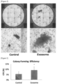
- the culture broth was washed once with 500 ⁇ l of PBS followed by the addition of 300 ⁇ l of 10% formaldehyde buffer and fixing for 2 hours or more at RT. Subsequently, the wells were washed once with 500 ⁇ l of ultrapure water, and after adding 300 ⁇ l of 2% Rhodamine B and allowing to react for 30 minutes or more at room temperature, the wells were washed one to three times with 0.2 M HCl and dried at room temperature followed by scanning the wells with a scanner (GT-F740, Epson Corp.) or photographing with EVOS FL AUTO (Thermo Fisher Scientific Inc.) to obtain images thereof ( FIG. 2 ). Colonies were then counted based on these images.
- a scanner GT-F740, Epson Corp.
- EVOS FL AUTO Thermo Fisher Scientific Inc.
- Colony-forming efficiency (CFE, %) was calculated using the formula: "100 ⁇ no. of colonies/no. of disseminated cells". As shown in FIG. 3 , CFE of the control cells was 5.6% while CFE of cells treated with exosomes was 9.1%, demonstrating that exosome-treatment significantly increased colony formation. On the basis of the above results, AD-MSC-derived exosomes were confirmed to have a colony formation promoting action on HCEC.
- the formed colonies were washed once with 500 ⁇ l of PBS and fixed at room temperature for 30 minutes with 300 ⁇ l of MeOH cooled to -30°C. Subsequently, after carrying out blocking for 1 hour at room temperature with 300 ⁇ l of 5% NST (5% normal donkey serum, 0.3% Triton/TBS), 300 ⁇ l of Cytokeratin 12 Antibody (N-16) (SC-17098, Santa Cruz Biotechnology, Inc.) which was diluted 100-fold with 1% NST (1% normal donky serum, 0.3% Triton/TBS) were added followed by treating for 3 hours at room temperature.
- NST 5% normal donkey serum, 0.3% Triton/TBS
- corneal epithelial cell differentiation marker K3, or PAX6, which plays an important role during differentiation
- corneal epithelial stem cell markers consisting of K14, KI5, p63 and N-cadherin was determined to increase.
- exosome pellet was obtained according to the previously described method. 2% sodium deoxycholate (190-08313, Wako Pure Chemical Industries, Ltd.) and 1X Protease Inhibitor Cocktail Set I (165-26021, Wako Pure Chemical Industries, Ltd.) added to 10 ⁇ l of 100 mM Tris-HCl (pH 8.0) were added to the exosome pellet followed by suspending therein by vortexing and then allowing to stand undisturbed at 4°C and O/N. Protein was assayed with a BCA assay kit (Pierce, 23227) and 10 ⁇ l of protein diluted to 1 ⁇ g/ml was used in a shotgun analysis. QTRAP 5500 (AB Sciex) was used for analysis.
- exosomes were confirmed to contain CD9, CD63, CD81, GAPDH, PKM, Enolase-1, 40S ribosomal protein S2, S5, SA, S13, S23, S4, S16 and S9, 60S acidic ribosomal protein PO and P1, 60S ribosomal protein L9, HSPB1 and HSP7C, 14-3-3 protein zeta/delta, epsilon, theta, beta/alpha, gamma and eta, syntenin, TGS101, actin cytoplasmic-1, cofilin-1, annexin A1, A2, A5, A6, A7 and A11, Rab-1B, 7a, 8B, 11A, 13 and 35, ICAM-1, and integrin alpha-V, alpha-2, alpha-4, alpha-5, beta-1 and beta-5.
- exosomes were isolated from each culture supernatant thereof containing AD-MSC according to the previously described method. Exosomes obtained by carrying out the same treatment on MSCGM-CD without culturing the cells were used in a control (CTL) group.
- CTL control
- the exosome pellets were suspended using 10 ⁇ l of RIPA Lysis and Buffer (89900, Thermo Fisher Scientific Inc.) containing Protease Inhibitor Cocktail Set I (Wako Pure Chemical Industries, Ltd., 165-26021).
- the exosomes were homogenized by treating at 20% for 5 seconds using the QSONICA Q125 small sample ultrasonic homogenizer, and the supernatant was recovered by centrifuging for 15 minutes at 4°C and 14,000 G.
- TBS-T TBS containing 0.05% Tween 20
- Anti-CD63 antibody 10628D, Thermo Fisher Scientific Inc., diluted 1:1000 (TBS)
- HRP-labeled anti-mouse IgG antibody diluted 1: 10,000 (PBS)
- ECL Prime GE Healthcare Biosciences Corp.
- ChemiDoc XRS Bio-Rad Laboratories, Inc.
- AD-MSC-derived exosomes expressed CD63, and exosomes derived from UC-MSC (umbilical cord-derived mesenchymal stem cells) and BM-MSC (bone marrow-derived mesenchymal stem cells) also expressed CD63, albeit at lower levels than in the case of AD-MSC-derived exosomes. These results suggested that AD-MSC has a possibility to secrete more exosomes as compared to other cells.
- the preventive or therapeutic agent of the present invention has industrial applicability since it has activity that promotes the growth of corneal epithelial stem cells and/or corneal epithelial cells, activity that maintains corneal epithelial stem cells in an undifferentiated state or promotes colony formation thereby, and action that protects corneal epithelium.
Landscapes
- Health & Medical Sciences (AREA)
- Life Sciences & Earth Sciences (AREA)
- Chemical & Material Sciences (AREA)
- Engineering & Computer Science (AREA)
- Zoology (AREA)
- Organic Chemistry (AREA)
- Biotechnology (AREA)
- General Health & Medical Sciences (AREA)
- Biomedical Technology (AREA)
- Immunology (AREA)
- Bioinformatics & Cheminformatics (AREA)
- Wood Science & Technology (AREA)
- Cell Biology (AREA)
- Developmental Biology & Embryology (AREA)
- Genetics & Genomics (AREA)
- Veterinary Medicine (AREA)
- Pharmacology & Pharmacy (AREA)
- Public Health (AREA)
- Animal Behavior & Ethology (AREA)
- Medicinal Chemistry (AREA)
- General Engineering & Computer Science (AREA)
- Microbiology (AREA)
- Biochemistry (AREA)
- Epidemiology (AREA)
- Virology (AREA)
- Proteomics, Peptides & Aminoacids (AREA)
- Hematology (AREA)
- Ophthalmology & Optometry (AREA)
- Nuclear Medicine, Radiotherapy & Molecular Imaging (AREA)
- General Chemical & Material Sciences (AREA)
- Chemical Kinetics & Catalysis (AREA)
- Physics & Mathematics (AREA)
- Analytical Chemistry (AREA)
- Molecular Biology (AREA)
- Biophysics (AREA)
- Rheumatology (AREA)
- Medicines Containing Material From Animals Or Micro-Organisms (AREA)
- Micro-Organisms Or Cultivation Processes Thereof (AREA)
- Measuring Or Testing Involving Enzymes Or Micro-Organisms (AREA)
- Materials For Medical Uses (AREA)
Claims (5)
- Vorbeugendes oder therapeutisches Agens zur Verwendung bei der Vorbeugung oder Behandlung von Erkrankungen des Hornhautepithels, das von mesenchymalen Stammzellen abstammende Mikropartikel enthält, die ein oder mehr aus CD9, CD63 und/oder CD81 umfassen, und eine wie durch Dichtegradientenzentrifugation bestimmte Dichte von 1,13 g/ml bis 1,19 g/ml aufweisen, wobei die Mikropartikel Exosome sind, die durch Ultrazentrifugation aus dem Zellkulturüberstand humaner mesenchymaler Stammzellen isoliert sind, als ein Wirkstoff davon, und wobei die Mikropartikel eine Aktivität aufweisen, die das Wachstum von Hornhautepithelstammzellen und/oder Hornhautepithelzellen fördert, eine Aktivität, die Hornhautepithelstammzellen in einem undifferenzierten Zustand hält oder die Bildung von Kolonien durch Hornhautepithelstammzellen fördert, oder eine Funktion, die das Hornhautepithelium schützt.
- Vorbeugendes oder therapeutisches Agens zur Verwendung nach Anspruch 1, wobei die Erkrankung des Hornhautepithels ausgewählt ist aus der Gruppe bestehend aus Hitzekorrosion, Alkalikorrosion, Säurekorrosion, chemischer Toxizität, Stevens-Johnson-Syndrom, okularem Pemphigoid, (rezidivierendem) Pterygium, persistierender Hornhautepitheldefekte, Hornhautpunktion, marginalem Hornhautgeschwür, Hornhautgeschwür, Epithelablösung in Folge von Excimer-Laser-Chirurgie, Strahlenkeratopathie, Aniridie, Hornhauttrübung nach einem Trachom, Salzmannscher Hornhautdegeneration, Hornhauterosion, Symblepharon, kryptogenen Erkrankungen, die mit dem Verlust von Hornhautepithelstammzellen assoziiert sind, Limbustumor, Transplantat-gegen-Wirt-Erkrankung (graft versus host disease; GVHD), Keratitis, oberflächlicher punktförmiger Keratopathie, trockenem Auge, Keratoconjunctivitis sicca, Mangel an Hornhauteptithelstammzellen, Hornhautdystrophie, diabetischer Keratopathie und Störungen des Hornhautepithels.
- Vorbeugendes oder therapeutisches Agens zur Verwendung nach Anspruch 2, wobei die Erkrankung des Hornhautepithels ausgewählt ist aus der Gruppe bestehend aus Störungen des Hornhautepithels, oberflächlicher punktförmiger Keratopathie, Hornhauterosion, marginalem Hornhautgeschwür, persistierendem Hornhautepitheldefekt, trockenem Auge, Epithelablösung in Folge von Excimer-Laser-Chirurgie, Hitzekorrosion, Alkalikorrosion, Säurekorrosion, chemischer Toxizität, diabetischer Keratopathie, Mangel an Hornhauteptithelstammzellen, Stevens-Johnson-Syndrom, okularem Pemphigoid, Aniridie, kryptogenen Erkrankungen, die mit dem Verlust von Hornhautepithelstammzellen assoziiert sind, und Transplantat-gegen-Wirt-Erkrankung (graft versus host disease; GVHD).
- Vorbeugendes oder therapeutisches Agens zur Verwendung nach Anspruch 3, wobei die Erkrankung des Hornhautepithels ausgewählt ist aus der Gruppe bestehend aus Störungen des Hornhautepithels, oberflächlicher punktförmiger Keratopathie, Hornhauterosion, marginalem Hornhautgeschwür, persistierendem Hornhautepitheldefekt, trockenem Auge, Epithelablösung in Folge von Excimer-Laser-Chirurgie, Hitzekorrosion, Alkalikorrosion, Säurekorrosion, chemischer Toxizität und diabetischer Keratopathie.
- Vorbeugendes oder therapeutisches Agens zur Verwendung nach Anspruch 4, wobei der Mangel an Hornhauteptithelstammzellen von einem extrinsischen Faktor, einem intrinsischen Faktor, einem angeborenen Defekt oder einer neoplastischen Erkrankung verursacht ist.
Applications Claiming Priority (2)
| Application Number | Priority Date | Filing Date | Title |
|---|---|---|---|
| JP2015153412 | 2015-08-03 | ||
| PCT/JP2016/072855 WO2017022809A1 (ja) | 2015-08-03 | 2016-08-03 | 間葉系幹細胞由来エキソソーム |
Publications (4)
| Publication Number | Publication Date |
|---|---|
| EP3333254A1 EP3333254A1 (de) | 2018-06-13 |
| EP3333254A4 EP3333254A4 (de) | 2019-05-08 |
| EP3333254B1 true EP3333254B1 (de) | 2025-05-14 |
| EP3333254C0 EP3333254C0 (de) | 2025-05-14 |
Family
ID=57944207
Family Applications (1)
| Application Number | Title | Priority Date | Filing Date |
|---|---|---|---|
| EP16833088.4A Active EP3333254B1 (de) | 2015-08-03 | 2016-08-03 | Exosom aus mesenchymalen stammzellen |
Country Status (5)
| Country | Link |
|---|---|
| US (1) | US11357798B2 (de) |
| EP (1) | EP3333254B1 (de) |
| JP (1) | JP6923134B2 (de) |
| CN (1) | CN107849535B (de) |
| WO (1) | WO2017022809A1 (de) |
Families Citing this family (25)
| Publication number | Priority date | Publication date | Assignee | Title |
|---|---|---|---|---|
| US11801268B2 (en) * | 2016-03-14 | 2023-10-31 | Capricor, Inc. | Methods of treating ocular inflammation and chemical injuries of the eye with extracellular vesicles |
| JP7598699B2 (ja) * | 2017-06-01 | 2024-12-12 | ロート製薬株式会社 | 視覚機能の再活性化用眼科組成物 |
| CN111527199A (zh) | 2017-11-16 | 2020-08-11 | 得克萨斯大学体系董事会 | 用于制备来源于msc的外泌体的方法 |
| WO2019139137A1 (ja) * | 2018-01-12 | 2019-07-18 | 国立大学法人大阪大学 | 重層扁平上皮細胞の正常分化・成熟促進剤、上皮疾患治療剤及び重層扁平上皮細胞の正常分化・成熟促進方法 |
| CN110339147A (zh) * | 2018-11-27 | 2019-10-18 | 浙江梵泊细胞工程有限公司 | 一种皮肤外用组合物及其用途 |
| CN109431985A (zh) * | 2018-12-14 | 2019-03-08 | 西安市第四医院 | 一种含有外泌体用于干眼治疗的滴眼液及其制备方法 |
| WO2020204161A1 (ja) | 2019-04-04 | 2020-10-08 | 日産化学株式会社 | 細胞外小胞の分泌を促進するための組成物 |
| KR102132457B1 (ko) * | 2019-05-10 | 2020-07-09 | 주식회사 바이오솔루션 | 세포외 소포를 고농도로 배양하기 위한 세포 배양배지 및 상기 세포 배양배지를 이용한 세포외 소포 고 함유 조건화 배양액의 제조방법 |
| CN110283776A (zh) * | 2019-06-26 | 2019-09-27 | 上海交通大学医学院附属第九人民医院 | 一种胞外囊泡的分离方法 |
| GB2604239A (en) * | 2019-07-26 | 2022-08-31 | Pandorum Tech Private Limited | Bioengineered formulation, process for preparing and implementations thereof |
| CN112795535B (zh) * | 2021-01-14 | 2022-06-28 | 北京臻膳美科技有限公司 | 诱导间充质干细胞释放促进皮肤上皮细胞分化特定功能外泌体的组合物及其应用 |
| CN113151162A (zh) * | 2021-04-13 | 2021-07-23 | 广东唯泰生物科技有限公司 | 外泌体在促进壁蜕膜间充质干细胞生长中的应用 |
| KR102704051B1 (ko) | 2021-08-30 | 2024-09-09 | 가톨릭대학교 산학협력단 | 세포 밖 소포체 함유 안표면 염증 질환 예방 또는 치료용 조성물 |
| WO2023064390A1 (en) * | 2021-10-17 | 2023-04-20 | ELEVAI Labs, Inc. | Exosome-based skincare product |
| WO2023080413A1 (ko) * | 2021-11-02 | 2023-05-11 | 주식회사 디자인셀 | 줄기세포 배양액 및 이로부터 분리된 엑소좀을 유효성분으로 포함하는 안구 질환의 예방 또는 치료용 약학적 조성물 |
| DE22865897T1 (de) * | 2021-12-02 | 2025-01-23 | Celligenics Pte Ltd. | Neues therapeutikum mit einem sekretom aus mesenchymalen stammzellen aus umbilicordgewebe oder wharton-sulze |
| CN114099547B (zh) * | 2021-12-07 | 2023-11-21 | 洛阳市三启生物技术有限公司 | 一种用于皮肤修复的干细胞外泌体制剂 |
| CN114591901B (zh) * | 2022-03-24 | 2023-11-24 | 和携科技有限公司 | 动物脐带间充质干细胞外泌体提取方法 |
| CN115216442B (zh) * | 2022-09-05 | 2024-04-16 | 杭州易文赛生物技术有限公司 | 外泌体的制备方法和培养基及其应用 |
| CN115590884B (zh) * | 2022-09-19 | 2023-09-29 | 四川大学华西医院 | 干细胞微粒在制备预防和/或治疗干眼症的药物中的应用 |
| CN115491352B (zh) * | 2022-11-16 | 2023-03-24 | 广东先康达生物科技有限公司 | 促进干细胞外泌体分泌的培养液、外泌体制备及应用 |
| JP2026034872A (ja) * | 2022-11-30 | 2026-03-04 | 国立大学法人広島大学 | 悪性腫瘍を治療または予防するための医薬組成物 |
| CN116083353B (zh) * | 2022-12-02 | 2025-10-17 | 唐颐生物医学(天津)有限公司 | 一种过表达Anxa1的脐带间充质干细胞外泌体及其制备方法和应用 |
| CN116751747B (zh) * | 2023-08-22 | 2023-11-07 | 北京葆来生物科技有限公司 | 一种促进间充质干细胞分化为神经元的无血清诱导培养基及诱导方法 |
| WO2025240923A1 (en) * | 2024-05-16 | 2025-11-20 | The George Washington University | Compositions derived from mesenchymal stromal cells and methods of use thereof |
Family Cites Families (13)
| Publication number | Priority date | Publication date | Assignee | Title |
|---|---|---|---|---|
| WO2005035739A1 (ja) * | 2003-10-14 | 2005-04-21 | Kohji Nishida | 再生治療システム |
| JP5409359B2 (ja) | 2007-07-13 | 2014-02-05 | 田辺三菱製薬株式会社 | 細胞の単離方法、細胞用無血清培養培地および細胞の培養方法 |
| EP2254586B1 (de) | 2008-02-22 | 2015-04-08 | Agency For Science, Technology And Research (A*star) | Mesenchym-stammzellenteilchen |
| EP2629782B1 (de) | 2010-10-18 | 2019-02-20 | Agency For Science, Technology And Research | Verwendung von exosomen von mesenchymalen stammzellen zur förderung oder verstärkung des haarwuchses |
| JP5759536B2 (ja) | 2011-04-20 | 2015-08-05 | 国立大学法人大阪大学 | 角膜上皮分化指向性iPS細胞 |
| US9446075B2 (en) * | 2011-05-06 | 2016-09-20 | Bioregenerative Sciences | Compositions derived from stem cell released molecules and methods for formulation thereof |
| US9427450B2 (en) * | 2012-01-31 | 2016-08-30 | Xon Cells, Inc. | Therapeutic immune modulation by stem cell secreted exosomes |
| EP2833893B1 (de) | 2012-04-03 | 2018-08-29 | Reneuron Limited | Stammzellenmikropartikel |
| GB201302468D0 (en) | 2013-02-12 | 2013-03-27 | Reneuron Ltd | Stem cell product |
| SG11201500329WA (en) | 2012-07-19 | 2015-02-27 | Reneuron Ltd | Stem cell microparticles |
| CN103767985A (zh) * | 2012-10-22 | 2014-05-07 | 吉林省霍普金斯药物研究院有限责任公司 | 人源血液或间充质干细胞分泌exosome的制备与应用 |
| US20160002597A1 (en) | 2013-02-12 | 2016-01-07 | Reneuron Limited | Method of producing microparticles |
| CN105308175B (zh) | 2013-03-11 | 2018-09-21 | 日本化学研究株式会社 | 人角膜上皮片的制造法 |
-
2016
- 2016-08-03 EP EP16833088.4A patent/EP3333254B1/de active Active
- 2016-08-03 JP JP2017533112A patent/JP6923134B2/ja active Active
- 2016-08-03 CN CN201680045165.8A patent/CN107849535B/zh active Active
- 2016-08-03 US US15/749,766 patent/US11357798B2/en active Active
- 2016-08-03 WO PCT/JP2016/072855 patent/WO2017022809A1/ja not_active Ceased
Also Published As
| Publication number | Publication date |
|---|---|
| HK1251903A1 (en) | 2019-04-26 |
| EP3333254A4 (de) | 2019-05-08 |
| EP3333254A1 (de) | 2018-06-13 |
| CN107849535B (zh) | 2022-11-22 |
| JP6923134B2 (ja) | 2021-08-18 |
| JPWO2017022809A1 (ja) | 2018-05-24 |
| CN107849535A (zh) | 2018-03-27 |
| US20190015452A1 (en) | 2019-01-17 |
| WO2017022809A1 (ja) | 2017-02-09 |
| EP3333254C0 (de) | 2025-05-14 |
| US11357798B2 (en) | 2022-06-14 |
Similar Documents
| Publication | Publication Date | Title |
|---|---|---|
| EP3333254B1 (de) | Exosom aus mesenchymalen stammzellen | |
| JP7407864B2 (ja) | Cd201、cd46、cd56、cd147及びcd165からなる群より選択される少なくとも1種の細胞表面マーカーを発現する間葉系幹細胞及びその調製方法、並びに上記間葉系幹細胞を含む医薬組成物及びその調製方法 | |
| US11707488B2 (en) | ROR1-positive mesenchymal stem cells and method for preparing same, pharmaceutical composition containing ROR1-positive mesenchymal stem cells and method for preparing same, and method for preventing or treating disease using ROR1-positive mesenchymal stem cells | |
| EP3593809A1 (de) | Ror1-positive mesenchymale stammzellenhaltige pharmazeutische zusammensetzung zur prävention oder behandlung von mit fibrose assoziierten krankheiten, verfahren zur herstellung davon und verfahren zur prävention oder behandlung von mit fibrose assoziierten krankheiten unter verwendung von ror1-positiven mesenchymalen stammzellen | |
| US11957719B2 (en) | Phenotype profile of human retinal progenitor cells | |
| EP3683306A1 (de) | Verfahren zur herstellung von retinagewebe | |
| US20250145949A1 (en) | Low Oxygen Culture Conditions for Maintaining Retinal Progenitor Cell Multipotency | |
| JP2018502576A (ja) | Rpe細胞集団およびそれを作製する方法 | |
| KR20180137488A (ko) | 간엽 마커와 뉴런 마커를 발현하는 줄기 세포, 이의 조성물 및 이의 제조 방법 | |
| JP2018502575A (ja) | 網膜色素上皮細胞集団の評価法 | |
| Park et al. | Vascular progenitors generated from tankyrase inhibitor-regulated naïve diabetic human iPSC potentiate efficient revascularization of ischemic retina | |
| JP6960120B2 (ja) | 肝疾患治療剤及び肝疾患を治療する方法 | |
| EP3738598A1 (de) | Mittel zur förderung der normalen differenzierung und reifung von stratifizierten plattenepithelzellen und verfahren zur förderung der normalen differenzierung und reifung von stratifizierten plattenepithelzellen | |
| CN112538458A (zh) | 用于重编程细胞的方法 | |
| WO2017170925A1 (ja) | Egfr及びmic-abからなる群より選択される少なくとも1種の細胞表面マーカーを高発現する間葉系幹細胞及びその調製方法、並びに上記間葉系幹細胞を含む医薬組成物及びその調製方法 | |
| Ramsden et al. | Neural retinal regeneration with pluripotent stem cells | |
| HK1251903B (en) | Mesenchymal stem cell-derived exosome | |
| US20250325591A1 (en) | Therapy for hepatic disorders using adipose-derived mesenchymal stem cell line | |
| CN1981030B (zh) | 使细胞治疗中使用的细胞长期生长和存活的卵泡液 | |
| Belfiore et al. | Combinatorial expression of glial transcription factors induces Schwann cell‐specific gene expression in mouse embryonic fibroblasts | |
| US12616721B2 (en) | Stratified squamous epithelial cell normal differentiation and maturation promoting agent, epithelial disease therapeutic agent, and stratified squamous epithelial cell normal differentiation and maturation promoting method | |
| JP2026511969A (ja) | Rpe細胞を分化させるための組成物及び方法 | |
| TW202216158A (zh) | 網膜色素上皮細胞之移植用存活促進劑 |
Legal Events
| Date | Code | Title | Description |
|---|---|---|---|
| STAA | Information on the status of an ep patent application or granted ep patent |
Free format text: STATUS: THE INTERNATIONAL PUBLICATION HAS BEEN MADE |
|
| PUAI | Public reference made under article 153(3) epc to a published international application that has entered the european phase |
Free format text: ORIGINAL CODE: 0009012 |
|
| STAA | Information on the status of an ep patent application or granted ep patent |
Free format text: STATUS: REQUEST FOR EXAMINATION WAS MADE |
|
| 17P | Request for examination filed |
Effective date: 20180223 |
|
| AK | Designated contracting states |
Kind code of ref document: A1 Designated state(s): AL AT BE BG CH CY CZ DE DK EE ES FI FR GB GR HR HU IE IS IT LI LT LU LV MC MK MT NL NO PL PT RO RS SE SI SK SM TR |
|
| AX | Request for extension of the european patent |
Extension state: BA ME |
|
| DAV | Request for validation of the european patent (deleted) | ||
| DAX | Request for extension of the european patent (deleted) | ||
| REG | Reference to a national code |
Ref country code: HK Ref legal event code: DE Ref document number: 1251903 Country of ref document: HK |
|
| RIC1 | Information provided on ipc code assigned before grant |
Ipc: A61K 35/12 20150101ALI20190327BHEP Ipc: A61P 27/02 20060101ALI20190327BHEP Ipc: C12Q 1/02 20060101ALI20190327BHEP Ipc: C12N 5/0775 20100101AFI20190327BHEP Ipc: A61K 35/28 20150101ALI20190327BHEP |
|
| A4 | Supplementary search report drawn up and despatched |
Effective date: 20190405 |
|
| STAA | Information on the status of an ep patent application or granted ep patent |
Free format text: STATUS: EXAMINATION IS IN PROGRESS |
|
| 17Q | First examination report despatched |
Effective date: 20200305 |
|
| TPAC | Observations filed by third parties |
Free format text: ORIGINAL CODE: EPIDOSNTIPA |
|
| GRAP | Despatch of communication of intention to grant a patent |
Free format text: ORIGINAL CODE: EPIDOSNIGR1 |
|
| STAA | Information on the status of an ep patent application or granted ep patent |
Free format text: STATUS: GRANT OF PATENT IS INTENDED |
|
| INTG | Intention to grant announced |
Effective date: 20241209 |
|
| GRAS | Grant fee paid |
Free format text: ORIGINAL CODE: EPIDOSNIGR3 |
|
| GRAA | (expected) grant |
Free format text: ORIGINAL CODE: 0009210 |
|
| STAA | Information on the status of an ep patent application or granted ep patent |
Free format text: STATUS: THE PATENT HAS BEEN GRANTED |
|
| AK | Designated contracting states |
Kind code of ref document: B1 Designated state(s): AL AT BE BG CH CY CZ DE DK EE ES FI FR GB GR HR HU IE IS IT LI LT LU LV MC MK MT NL NO PL PT RO RS SE SI SK SM TR |
|
| REG | Reference to a national code |
Ref country code: GB Ref legal event code: FG4D |
|
| REG | Reference to a national code |
Ref country code: CH Ref legal event code: EP |
|
| REG | Reference to a national code |
Ref country code: DE Ref legal event code: R096 Ref document number: 602016092254 Country of ref document: DE |
|
| REG | Reference to a national code |
Ref country code: IE Ref legal event code: FG4D |
|
| U01 | Request for unitary effect filed |
Effective date: 20250604 |
|
| U07 | Unitary effect registered |
Designated state(s): AT BE BG DE DK EE FI FR IT LT LU LV MT NL PT RO SE SI Effective date: 20250612 |
|
| U20 | Renewal fee for the european patent with unitary effect paid |
Year of fee payment: 10 Effective date: 20250822 |
|
| PG25 | Lapsed in a contracting state [announced via postgrant information from national office to epo] |
Ref country code: ES Free format text: LAPSE BECAUSE OF FAILURE TO SUBMIT A TRANSLATION OF THE DESCRIPTION OR TO PAY THE FEE WITHIN THE PRESCRIBED TIME-LIMIT Effective date: 20250514 |
|
| PG25 | Lapsed in a contracting state [announced via postgrant information from national office to epo] |
Ref country code: NO Free format text: LAPSE BECAUSE OF FAILURE TO SUBMIT A TRANSLATION OF THE DESCRIPTION OR TO PAY THE FEE WITHIN THE PRESCRIBED TIME-LIMIT Effective date: 20250814 Ref country code: GR Free format text: LAPSE BECAUSE OF FAILURE TO SUBMIT A TRANSLATION OF THE DESCRIPTION OR TO PAY THE FEE WITHIN THE PRESCRIBED TIME-LIMIT Effective date: 20250815 |
|
| PG25 | Lapsed in a contracting state [announced via postgrant information from national office to epo] |
Ref country code: PL Free format text: LAPSE BECAUSE OF FAILURE TO SUBMIT A TRANSLATION OF THE DESCRIPTION OR TO PAY THE FEE WITHIN THE PRESCRIBED TIME-LIMIT Effective date: 20250514 |
|
| PGFP | Annual fee paid to national office [announced via postgrant information from national office to epo] |
Ref country code: GB Payment date: 20250828 Year of fee payment: 10 |
|
| PG25 | Lapsed in a contracting state [announced via postgrant information from national office to epo] |
Ref country code: HR Free format text: LAPSE BECAUSE OF FAILURE TO SUBMIT A TRANSLATION OF THE DESCRIPTION OR TO PAY THE FEE WITHIN THE PRESCRIBED TIME-LIMIT Effective date: 20250514 |
|
| PG25 | Lapsed in a contracting state [announced via postgrant information from national office to epo] |
Ref country code: RS Free format text: LAPSE BECAUSE OF FAILURE TO SUBMIT A TRANSLATION OF THE DESCRIPTION OR TO PAY THE FEE WITHIN THE PRESCRIBED TIME-LIMIT Effective date: 20250814 |
|
| PG25 | Lapsed in a contracting state [announced via postgrant information from national office to epo] |
Ref country code: IS Free format text: LAPSE BECAUSE OF FAILURE TO SUBMIT A TRANSLATION OF THE DESCRIPTION OR TO PAY THE FEE WITHIN THE PRESCRIBED TIME-LIMIT Effective date: 20250914 |
|
| PG25 | Lapsed in a contracting state [announced via postgrant information from national office to epo] |
Ref country code: SM Free format text: LAPSE BECAUSE OF FAILURE TO SUBMIT A TRANSLATION OF THE DESCRIPTION OR TO PAY THE FEE WITHIN THE PRESCRIBED TIME-LIMIT Effective date: 20250514 |
|
| PG25 | Lapsed in a contracting state [announced via postgrant information from national office to epo] |
Ref country code: CZ Free format text: LAPSE BECAUSE OF FAILURE TO SUBMIT A TRANSLATION OF THE DESCRIPTION OR TO PAY THE FEE WITHIN THE PRESCRIBED TIME-LIMIT Effective date: 20250514 |
|
| PG25 | Lapsed in a contracting state [announced via postgrant information from national office to epo] |
Ref country code: SK Free format text: LAPSE BECAUSE OF FAILURE TO SUBMIT A TRANSLATION OF THE DESCRIPTION OR TO PAY THE FEE WITHIN THE PRESCRIBED TIME-LIMIT Effective date: 20250514 |
|
| PLBE | No opposition filed within time limit |
Free format text: ORIGINAL CODE: 0009261 |
|
| STAA | Information on the status of an ep patent application or granted ep patent |
Free format text: STATUS: NO OPPOSITION FILED WITHIN TIME LIMIT |
|
| REG | Reference to a national code |
Ref country code: CH Ref legal event code: H13 Free format text: ST27 STATUS EVENT CODE: U-0-0-H10-H13 (AS PROVIDED BY THE NATIONAL OFFICE) Effective date: 20260324 |
|
| REG | Reference to a national code |
Ref country code: CH Ref legal event code: L10 Free format text: ST27 STATUS EVENT CODE: U-0-0-L10-L00 (AS PROVIDED BY THE NATIONAL OFFICE) Effective date: 20260325 |
|
| PG25 | Lapsed in a contracting state [announced via postgrant information from national office to epo] |
Ref country code: MC Free format text: LAPSE BECAUSE OF FAILURE TO SUBMIT A TRANSLATION OF THE DESCRIPTION OR TO PAY THE FEE WITHIN THE PRESCRIBED TIME-LIMIT Effective date: 20250514 |
|
| 26N | No opposition filed |
Effective date: 20260217 |
|
| PG25 | Lapsed in a contracting state [announced via postgrant information from national office to epo] |
Ref country code: CH Free format text: LAPSE BECAUSE OF NON-PAYMENT OF DUE FEES Effective date: 20250831 |