EP3182987B1 - Humanized anti-tau antibodies - Google Patents
Humanized anti-tau antibodies Download PDFInfo
- Publication number
- EP3182987B1 EP3182987B1 EP15810923.1A EP15810923A EP3182987B1 EP 3182987 B1 EP3182987 B1 EP 3182987B1 EP 15810923 A EP15810923 A EP 15810923A EP 3182987 B1 EP3182987 B1 EP 3182987B1
- Authority
- EP
- European Patent Office
- Prior art keywords
- antibody
- tau
- binding
- human
- antibodies
- Prior art date
- Legal status (The legal status is an assumption and is not a legal conclusion. Google has not performed a legal analysis and makes no representation as to the accuracy of the status listed.)
- Active
Links
- 125000003275 alpha amino acid group Chemical group 0.000 claims 2
- 230000027455 binding Effects 0.000 description 126
- 102000013498 tau Proteins Human genes 0.000 description 69
- 108010026424 tau Proteins Proteins 0.000 description 69
- 239000012634 fragment Substances 0.000 description 65
- 150000001413 amino acids Chemical group 0.000 description 52
- 239000000427 antigen Substances 0.000 description 51
- 108091007433 antigens Proteins 0.000 description 51
- 102000036639 antigens Human genes 0.000 description 51
- 101000891579 Homo sapiens Microtubule-associated protein tau Proteins 0.000 description 42
- 201000002212 progressive supranuclear palsy Diseases 0.000 description 39
- 102000057063 human MAPT Human genes 0.000 description 35
- 108090000623 proteins and genes Proteins 0.000 description 32
- 102000004169 proteins and genes Human genes 0.000 description 31
- 235000018102 proteins Nutrition 0.000 description 30
- 210000001175 cerebrospinal fluid Anatomy 0.000 description 29
- 238000000034 method Methods 0.000 description 26
- 241001529936 Murinae Species 0.000 description 25
- 235000001014 amino acid Nutrition 0.000 description 25
- 208000034799 Tauopathies Diseases 0.000 description 24
- 238000012216 screening Methods 0.000 description 23
- 108090000765 processed proteins & peptides Proteins 0.000 description 19
- 238000003556 assay Methods 0.000 description 18
- 208000024827 Alzheimer disease Diseases 0.000 description 15
- 241000699666 Mus <mouse, genus> Species 0.000 description 15
- 239000012491 analyte Substances 0.000 description 14
- 210000001519 tissue Anatomy 0.000 description 14
- 238000011282 treatment Methods 0.000 description 14
- 108010047041 Complementarity Determining Regions Proteins 0.000 description 13
- 239000003814 drug Substances 0.000 description 13
- 229940079593 drug Drugs 0.000 description 12
- 201000011240 Frontotemporal dementia Diseases 0.000 description 11
- 108060003951 Immunoglobulin Proteins 0.000 description 11
- 210000004556 brain Anatomy 0.000 description 11
- 210000004027 cell Anatomy 0.000 description 11
- 102000018358 immunoglobulin Human genes 0.000 description 11
- 230000007170 pathology Effects 0.000 description 11
- 238000002965 ELISA Methods 0.000 description 10
- 210000001744 T-lymphocyte Anatomy 0.000 description 10
- 238000001802 infusion Methods 0.000 description 10
- 230000035772 mutation Effects 0.000 description 10
- 238000004458 analytical method Methods 0.000 description 9
- 210000004369 blood Anatomy 0.000 description 9
- 239000008280 blood Substances 0.000 description 9
- 239000002131 composite material Substances 0.000 description 9
- 230000003993 interaction Effects 0.000 description 9
- 208000024891 symptom Diseases 0.000 description 9
- 101100480714 Macaca mulatta MAPT gene Proteins 0.000 description 8
- 241000699670 Mus sp. Species 0.000 description 8
- 208000017004 dementia pugilistica Diseases 0.000 description 8
- 208000037265 diseases, disorders, signs and symptoms Diseases 0.000 description 8
- 238000002595 magnetic resonance imaging Methods 0.000 description 8
- 239000000523 sample Substances 0.000 description 8
- 235000004279 alanine Nutrition 0.000 description 7
- 238000005516 engineering process Methods 0.000 description 7
- 238000002474 experimental method Methods 0.000 description 7
- 230000005847 immunogenicity Effects 0.000 description 7
- 238000002347 injection Methods 0.000 description 7
- 239000007924 injection Substances 0.000 description 7
- 238000012933 kinetic analysis Methods 0.000 description 7
- 239000003446 ligand Substances 0.000 description 7
- 210000002682 neurofibrillary tangle Anatomy 0.000 description 7
- 239000000902 placebo Substances 0.000 description 7
- 229940068196 placebo Drugs 0.000 description 7
- 230000004044 response Effects 0.000 description 7
- 241000282412 Homo Species 0.000 description 6
- 201000010099 disease Diseases 0.000 description 6
- 238000010494 dissociation reaction Methods 0.000 description 6
- 230000005593 dissociations Effects 0.000 description 6
- 230000000694 effects Effects 0.000 description 6
- 230000014509 gene expression Effects 0.000 description 6
- 238000009593 lumbar puncture Methods 0.000 description 6
- 238000005259 measurement Methods 0.000 description 6
- 241000894007 species Species 0.000 description 6
- 230000000087 stabilizing effect Effects 0.000 description 6
- 238000002198 surface plasmon resonance spectroscopy Methods 0.000 description 6
- 208000011990 Corticobasal Degeneration Diseases 0.000 description 5
- 206010012289 Dementia Diseases 0.000 description 5
- 239000000370 acceptor Substances 0.000 description 5
- 230000002776 aggregation Effects 0.000 description 5
- 238000004220 aggregation Methods 0.000 description 5
- 238000011156 evaluation Methods 0.000 description 5
- 230000007717 exclusion Effects 0.000 description 5
- 125000005647 linker group Chemical group 0.000 description 5
- 239000013642 negative control Substances 0.000 description 5
- 230000001575 pathological effect Effects 0.000 description 5
- 102000004196 processed proteins & peptides Human genes 0.000 description 5
- 230000000750 progressive effect Effects 0.000 description 5
- 239000000243 solution Substances 0.000 description 5
- 238000010186 staining Methods 0.000 description 5
- 230000001225 therapeutic effect Effects 0.000 description 5
- 208000004051 Chronic Traumatic Encephalopathy Diseases 0.000 description 4
- 208000020406 Creutzfeldt Jacob disease Diseases 0.000 description 4
- 208000003407 Creutzfeldt-Jakob Syndrome Diseases 0.000 description 4
- 208000010859 Creutzfeldt-Jakob disease Diseases 0.000 description 4
- 208000003736 Gerstmann-Straussler-Scheinker Disease Diseases 0.000 description 4
- 206010072075 Gerstmann-Straussler-Scheinker syndrome Diseases 0.000 description 4
- DHMQDGOQFOQNFH-UHFFFAOYSA-N Glycine Chemical compound NCC(O)=O DHMQDGOQFOQNFH-UHFFFAOYSA-N 0.000 description 4
- QNAYBMKLOCPYGJ-REOHCLBHSA-N L-alanine Chemical compound C[C@H](N)C(O)=O QNAYBMKLOCPYGJ-REOHCLBHSA-N 0.000 description 4
- 102100040243 Microtubule-associated protein tau Human genes 0.000 description 4
- 208000012902 Nervous system disease Diseases 0.000 description 4
- 208000000609 Pick Disease of the Brain Diseases 0.000 description 4
- 240000004808 Saccharomyces cerevisiae Species 0.000 description 4
- 230000002411 adverse Effects 0.000 description 4
- 210000005013 brain tissue Anatomy 0.000 description 4
- 230000001149 cognitive effect Effects 0.000 description 4
- 229940000406 drug candidate Drugs 0.000 description 4
- 230000006870 function Effects 0.000 description 4
- 238000013507 mapping Methods 0.000 description 4
- 230000004770 neurodegeneration Effects 0.000 description 4
- 238000002818 protein evolution Methods 0.000 description 4
- 230000008929 regeneration Effects 0.000 description 4
- 238000011069 regeneration method Methods 0.000 description 4
- 238000005070 sampling Methods 0.000 description 4
- 238000012360 testing method Methods 0.000 description 4
- 108020004414 DNA Proteins 0.000 description 3
- WQZGKKKJIJFFOK-GASJEMHNSA-N Glucose Natural products OC[C@H]1OC(O)[C@H](O)[C@@H](O)[C@@H]1O WQZGKKKJIJFFOK-GASJEMHNSA-N 0.000 description 3
- 201000002832 Lewy body dementia Diseases 0.000 description 3
- 241000282560 Macaca mulatta Species 0.000 description 3
- 101710115937 Microtubule-associated protein tau Proteins 0.000 description 3
- 208000018737 Parkinson disease Diseases 0.000 description 3
- 230000002159 abnormal effect Effects 0.000 description 3
- 229940127219 anticoagulant drug Drugs 0.000 description 3
- 238000002820 assay format Methods 0.000 description 3
- 239000000872 buffer Substances 0.000 description 3
- 238000012512 characterization method Methods 0.000 description 3
- 239000003153 chemical reaction reagent Substances 0.000 description 3
- 239000011248 coating agent Substances 0.000 description 3
- 238000000576 coating method Methods 0.000 description 3
- 238000001514 detection method Methods 0.000 description 3
- 238000003745 diagnosis Methods 0.000 description 3
- 239000008103 glucose Substances 0.000 description 3
- 238000003384 imaging method Methods 0.000 description 3
- 230000003834 intracellular effect Effects 0.000 description 3
- 238000009533 lab test Methods 0.000 description 3
- 210000000265 leukocyte Anatomy 0.000 description 3
- 238000012423 maintenance Methods 0.000 description 3
- 238000004519 manufacturing process Methods 0.000 description 3
- 238000012544 monitoring process Methods 0.000 description 3
- 208000005264 motor neuron disease Diseases 0.000 description 3
- 208000015122 neurodegenerative disease Diseases 0.000 description 3
- 210000002569 neuron Anatomy 0.000 description 3
- 108020004707 nucleic acids Proteins 0.000 description 3
- 102000039446 nucleic acids Human genes 0.000 description 3
- 150000007523 nucleic acids Chemical class 0.000 description 3
- 230000008569 process Effects 0.000 description 3
- 208000020016 psychiatric disease Diseases 0.000 description 3
- 238000011160 research Methods 0.000 description 3
- 238000003118 sandwich ELISA Methods 0.000 description 3
- 230000009870 specific binding Effects 0.000 description 3
- 238000002560 therapeutic procedure Methods 0.000 description 3
- 230000001988 toxicity Effects 0.000 description 3
- 231100000419 toxicity Toxicity 0.000 description 3
- 238000012546 transfer Methods 0.000 description 3
- 238000010200 validation analysis Methods 0.000 description 3
- FWMNVWWHGCHHJJ-SKKKGAJSSA-N 4-amino-1-[(2r)-6-amino-2-[[(2r)-2-[[(2r)-2-[[(2r)-2-amino-3-phenylpropanoyl]amino]-3-phenylpropanoyl]amino]-4-methylpentanoyl]amino]hexanoyl]piperidine-4-carboxylic acid Chemical compound C([C@H](C(=O)N[C@H](CC(C)C)C(=O)N[C@H](CCCCN)C(=O)N1CCC(N)(CC1)C(O)=O)NC(=O)[C@H](N)CC=1C=CC=CC=1)C1=CC=CC=C1 FWMNVWWHGCHHJJ-SKKKGAJSSA-N 0.000 description 2
- BSYNRYMUTXBXSQ-UHFFFAOYSA-N Aspirin Chemical compound CC(=O)OC1=CC=CC=C1C(O)=O BSYNRYMUTXBXSQ-UHFFFAOYSA-N 0.000 description 2
- BPYKTIZUTYGOLE-IFADSCNNSA-N Bilirubin Chemical compound N1C(=O)C(C)=C(C=C)\C1=C\C1=C(C)C(CCC(O)=O)=C(CC2=C(C(C)=C(\C=C/3C(=C(C=C)C(=O)N\3)C)N2)CCC(O)=O)N1 BPYKTIZUTYGOLE-IFADSCNNSA-N 0.000 description 2
- 208000028698 Cognitive impairment Diseases 0.000 description 2
- 206010010144 Completed suicide Diseases 0.000 description 2
- 229920002307 Dextran Polymers 0.000 description 2
- 101000854943 Enterobacteria phage T4 Valyl-tRNA ligase modifier Proteins 0.000 description 2
- 206010056696 Gaze palsy Diseases 0.000 description 2
- 239000004471 Glycine Substances 0.000 description 2
- 101000746373 Homo sapiens Granulocyte-macrophage colony-stimulating factor Proteins 0.000 description 2
- 208000023105 Huntington disease Diseases 0.000 description 2
- 206010020751 Hypersensitivity Diseases 0.000 description 2
- 108010021625 Immunoglobulin Fragments Proteins 0.000 description 2
- 102000008394 Immunoglobulin Fragments Human genes 0.000 description 2
- XEEYBQQBJWHFJM-UHFFFAOYSA-N Iron Chemical compound [Fe] XEEYBQQBJWHFJM-UHFFFAOYSA-N 0.000 description 2
- 102000003855 L-lactate dehydrogenase Human genes 0.000 description 2
- 108700023483 L-lactate dehydrogenases Proteins 0.000 description 2
- 208000009829 Lewy Body Disease Diseases 0.000 description 2
- 102000043131 MHC class II family Human genes 0.000 description 2
- 108091054438 MHC class II family Proteins 0.000 description 2
- -1 MgCl2 hexahydrate Chemical class 0.000 description 2
- 208000001089 Multiple system atrophy Diseases 0.000 description 2
- 208000025966 Neurological disease Diseases 0.000 description 2
- VYPSYNLAJGMNEJ-UHFFFAOYSA-N Silicium dioxide Chemical compound O=[Si]=O VYPSYNLAJGMNEJ-UHFFFAOYSA-N 0.000 description 2
- 208000006011 Stroke Diseases 0.000 description 2
- 238000009825 accumulation Methods 0.000 description 2
- 239000008351 acetate buffer Substances 0.000 description 2
- 229960001138 acetylsalicylic acid Drugs 0.000 description 2
- 125000003295 alanine group Chemical group N[C@@H](C)C(=O)* 0.000 description 2
- 150000001412 amines Chemical class 0.000 description 2
- 239000003146 anticoagulant agent Substances 0.000 description 2
- 238000013459 approach Methods 0.000 description 2
- 230000015572 biosynthetic process Effects 0.000 description 2
- 238000004364 calculation method Methods 0.000 description 2
- 238000006243 chemical reaction Methods 0.000 description 2
- HVYWMOMLDIMFJA-DPAQBDIFSA-N cholesterol Chemical compound C1C=C2C[C@@H](O)CC[C@]2(C)[C@@H]2[C@@H]1[C@@H]1CC[C@H]([C@H](C)CCCC(C)C)[C@@]1(C)CC2 HVYWMOMLDIMFJA-DPAQBDIFSA-N 0.000 description 2
- 210000000349 chromosome Anatomy 0.000 description 2
- 230000001684 chronic effect Effects 0.000 description 2
- 208000010877 cognitive disease Diseases 0.000 description 2
- 230000000295 complement effect Effects 0.000 description 2
- 230000001010 compromised effect Effects 0.000 description 2
- 230000008878 coupling Effects 0.000 description 2
- 238000010168 coupling process Methods 0.000 description 2
- 238000005859 coupling reaction Methods 0.000 description 2
- DDRJAANPRJIHGJ-UHFFFAOYSA-N creatinine Chemical compound CN1CC(=O)NC1=N DDRJAANPRJIHGJ-UHFFFAOYSA-N 0.000 description 2
- 230000007850 degeneration Effects 0.000 description 2
- 239000012895 dilution Substances 0.000 description 2
- 238000010790 dilution Methods 0.000 description 2
- 208000035475 disorder Diseases 0.000 description 2
- 231100000371 dose-limiting toxicity Toxicity 0.000 description 2
- 239000013604 expression vector Substances 0.000 description 2
- 230000002519 immonomodulatory effect Effects 0.000 description 2
- 229940127121 immunoconjugate Drugs 0.000 description 2
- 229940072221 immunoglobulins Drugs 0.000 description 2
- 210000003000 inclusion body Anatomy 0.000 description 2
- 201000010901 lateral sclerosis Diseases 0.000 description 2
- 239000007788 liquid Substances 0.000 description 2
- 210000004962 mammalian cell Anatomy 0.000 description 2
- 239000000463 material Substances 0.000 description 2
- 230000007246 mechanism Effects 0.000 description 2
- 230000006996 mental state Effects 0.000 description 2
- 239000000203 mixture Substances 0.000 description 2
- 239000000178 monomer Substances 0.000 description 2
- 231100000706 no observed effect level Toxicity 0.000 description 2
- 238000010899 nucleation Methods 0.000 description 2
- 239000008194 pharmaceutical composition Substances 0.000 description 2
- 230000002974 pharmacogenomic effect Effects 0.000 description 2
- 230000036470 plasma concentration Effects 0.000 description 2
- 108091033319 polynucleotide Proteins 0.000 description 2
- 102000040430 polynucleotide Human genes 0.000 description 2
- 239000002157 polynucleotide Substances 0.000 description 2
- 239000000843 powder Substances 0.000 description 2
- 238000002360 preparation method Methods 0.000 description 2
- 238000012552 review Methods 0.000 description 2
- 239000012146 running buffer Substances 0.000 description 2
- 231100000279 safety data Toxicity 0.000 description 2
- 230000006641 stabilisation Effects 0.000 description 2
- 238000011105 stabilization Methods 0.000 description 2
- 231100000041 toxicology testing Toxicity 0.000 description 2
- 230000010474 transient expression Effects 0.000 description 2
- 239000013598 vector Substances 0.000 description 2
- PJVWKTKQMONHTI-UHFFFAOYSA-N warfarin Chemical compound OC=1C2=CC=CC=C2OC(=O)C=1C(CC(=O)C)C1=CC=CC=C1 PJVWKTKQMONHTI-UHFFFAOYSA-N 0.000 description 2
- 229960005080 warfarin Drugs 0.000 description 2
- PGOHTUIFYSHAQG-LJSDBVFPSA-N (2S)-6-amino-2-[[(2S)-5-amino-2-[[(2S)-2-[[(2S)-2-[[(2S)-2-[[(2S)-4-amino-2-[[(2S)-2-[[(2S)-2-[[(2S)-2-[[(2S)-2-[[(2S)-5-amino-2-[[(2S)-5-amino-2-[[(2S)-2-[[(2S)-2-[[(2S)-2-[[(2S,3R)-2-[[(2S)-5-amino-2-[[(2S)-2-[[(2S)-2-[[(2S,3R)-2-[[(2S)-2-[[(2S)-2-[[(2S)-2-[[(2S)-2-[[(2S)-5-amino-2-[[(2S)-1-[(2S,3R)-2-[[(2S)-2-[[(2S)-2-[[(2R)-2-[[(2S)-2-[[(2S)-2-[[2-[[(2S)-2-[[(2S)-2-[[(2S)-2-[[(2S)-1-[(2S)-2-[[(2S)-2-[[(2S)-2-[[(2S)-2-amino-4-methylsulfanylbutanoyl]amino]-3-(1H-indol-3-yl)propanoyl]amino]-5-carbamimidamidopentanoyl]amino]propanoyl]pyrrolidine-2-carbonyl]amino]-3-methylbutanoyl]amino]-4-methylpentanoyl]amino]-4-methylpentanoyl]amino]acetyl]amino]-3-hydroxypropanoyl]amino]-4-methylpentanoyl]amino]-3-sulfanylpropanoyl]amino]-4-methylsulfanylbutanoyl]amino]-5-carbamimidamidopentanoyl]amino]-3-hydroxybutanoyl]pyrrolidine-2-carbonyl]amino]-5-oxopentanoyl]amino]-3-hydroxypropanoyl]amino]-3-hydroxypropanoyl]amino]-3-(1H-imidazol-5-yl)propanoyl]amino]-4-methylpentanoyl]amino]-3-hydroxybutanoyl]amino]-3-(1H-indol-3-yl)propanoyl]amino]-5-carbamimidamidopentanoyl]amino]-5-oxopentanoyl]amino]-3-hydroxybutanoyl]amino]-3-hydroxypropanoyl]amino]-3-carboxypropanoyl]amino]-3-hydroxypropanoyl]amino]-5-oxopentanoyl]amino]-5-oxopentanoyl]amino]-3-phenylpropanoyl]amino]-5-carbamimidamidopentanoyl]amino]-3-methylbutanoyl]amino]-4-methylpentanoyl]amino]-4-oxobutanoyl]amino]-5-carbamimidamidopentanoyl]amino]-3-(1H-indol-3-yl)propanoyl]amino]-4-carboxybutanoyl]amino]-5-oxopentanoyl]amino]hexanoic acid Chemical compound CSCC[C@H](N)C(=O)N[C@@H](Cc1c[nH]c2ccccc12)C(=O)N[C@@H](CCCNC(N)=N)C(=O)N[C@@H](C)C(=O)N1CCC[C@H]1C(=O)N[C@@H](C(C)C)C(=O)N[C@@H](CC(C)C)C(=O)N[C@@H](CC(C)C)C(=O)NCC(=O)N[C@@H](CO)C(=O)N[C@@H](CC(C)C)C(=O)N[C@@H](CS)C(=O)N[C@@H](CCSC)C(=O)N[C@@H](CCCNC(N)=N)C(=O)N[C@@H]([C@@H](C)O)C(=O)N1CCC[C@H]1C(=O)N[C@@H](CCC(N)=O)C(=O)N[C@@H](CO)C(=O)N[C@@H](CO)C(=O)N[C@@H](Cc1cnc[nH]1)C(=O)N[C@@H](CC(C)C)C(=O)N[C@@H]([C@@H](C)O)C(=O)N[C@@H](Cc1c[nH]c2ccccc12)C(=O)N[C@@H](CCCNC(N)=N)C(=O)N[C@@H](CCC(N)=O)C(=O)N[C@@H]([C@@H](C)O)C(=O)N[C@@H](CO)C(=O)N[C@@H](CC(O)=O)C(=O)N[C@@H](CO)C(=O)N[C@@H](CCC(N)=O)C(=O)N[C@@H](CCC(N)=O)C(=O)N[C@@H](Cc1ccccc1)C(=O)N[C@@H](CCCNC(N)=N)C(=O)N[C@@H](C(C)C)C(=O)N[C@@H](CC(C)C)C(=O)N[C@@H](CC(N)=O)C(=O)N[C@@H](CCCNC(N)=N)C(=O)N[C@@H](Cc1c[nH]c2ccccc12)C(=O)N[C@@H](CCC(O)=O)C(=O)N[C@@H](CCC(N)=O)C(=O)N[C@@H](CCCCN)C(O)=O PGOHTUIFYSHAQG-LJSDBVFPSA-N 0.000 description 1
- NFGXHKASABOEEW-UHFFFAOYSA-N 1-methylethyl 11-methoxy-3,7,11-trimethyl-2,4-dodecadienoate Chemical compound COC(C)(C)CCCC(C)CC=CC(C)=CC(=O)OC(C)C NFGXHKASABOEEW-UHFFFAOYSA-N 0.000 description 1
- QTBSBXVTEAMEQO-UHFFFAOYSA-M Acetate Chemical compound CC([O-])=O QTBSBXVTEAMEQO-UHFFFAOYSA-M 0.000 description 1
- 108010088751 Albumins Proteins 0.000 description 1
- 102000009027 Albumins Human genes 0.000 description 1
- 102000002260 Alkaline Phosphatase Human genes 0.000 description 1
- 108020004774 Alkaline Phosphatase Proteins 0.000 description 1
- 108700028369 Alleles Proteins 0.000 description 1
- 239000004382 Amylase Substances 0.000 description 1
- 102000013142 Amylases Human genes 0.000 description 1
- 108010065511 Amylases Proteins 0.000 description 1
- 208000037259 Amyloid Plaque Diseases 0.000 description 1
- 102000013455 Amyloid beta-Peptides Human genes 0.000 description 1
- 108010090849 Amyloid beta-Peptides Proteins 0.000 description 1
- 208000035143 Bacterial infection Diseases 0.000 description 1
- 238000012492 Biacore method Methods 0.000 description 1
- 208000014644 Brain disease Diseases 0.000 description 1
- 102000014914 Carrier Proteins Human genes 0.000 description 1
- 108010078791 Carrier Proteins Proteins 0.000 description 1
- 206010051290 Central nervous system lesion Diseases 0.000 description 1
- 108091035707 Consensus sequence Proteins 0.000 description 1
- 238000007400 DNA extraction Methods 0.000 description 1
- 208000019505 Deglutition disease Diseases 0.000 description 1
- 206010067889 Dementia with Lewy bodies Diseases 0.000 description 1
- BWGNESOTFCXPMA-UHFFFAOYSA-N Dihydrogen disulfide Chemical compound SS BWGNESOTFCXPMA-UHFFFAOYSA-N 0.000 description 1
- 102000004190 Enzymes Human genes 0.000 description 1
- 108090000790 Enzymes Proteins 0.000 description 1
- 206010017533 Fungal infection Diseases 0.000 description 1
- 206010017577 Gait disturbance Diseases 0.000 description 1
- 102000001554 Hemoglobins Human genes 0.000 description 1
- 108010054147 Hemoglobins Proteins 0.000 description 1
- 208000032843 Hemorrhage Diseases 0.000 description 1
- 102000009786 Immunoglobulin Constant Regions Human genes 0.000 description 1
- 108010009817 Immunoglobulin Constant Regions Proteins 0.000 description 1
- WTDRDQBEARUVNC-LURJTMIESA-N L-DOPA Chemical compound OC(=O)[C@@H](N)CC1=CC=C(O)C(O)=C1 WTDRDQBEARUVNC-LURJTMIESA-N 0.000 description 1
- WTDRDQBEARUVNC-UHFFFAOYSA-N L-Dopa Natural products OC(=O)C(N)CC1=CC=C(O)C(O)=C1 WTDRDQBEARUVNC-UHFFFAOYSA-N 0.000 description 1
- 150000008575 L-amino acids Chemical class 0.000 description 1
- 208000034800 Leukoencephalopathies Diseases 0.000 description 1
- 102000004882 Lipase Human genes 0.000 description 1
- 108090001060 Lipase Proteins 0.000 description 1
- 239000004367 Lipase Substances 0.000 description 1
- 239000012901 Milli-Q water Substances 0.000 description 1
- 208000026072 Motor neurone disease Diseases 0.000 description 1
- 101100480715 Mus musculus Mapt gene Proteins 0.000 description 1
- 101100462505 Mus musculus Pik3c2a gene Proteins 0.000 description 1
- 206010062207 Mycobacterial infection Diseases 0.000 description 1
- 208000031888 Mycoses Diseases 0.000 description 1
- 206010028980 Neoplasm Diseases 0.000 description 1
- 208000027089 Parkinsonian disease Diseases 0.000 description 1
- 206010034010 Parkinsonism Diseases 0.000 description 1
- 102000057297 Pepsin A Human genes 0.000 description 1
- 108090000284 Pepsin A Proteins 0.000 description 1
- 108010067902 Peptide Library Proteins 0.000 description 1
- 108010001267 Protein Subunits Proteins 0.000 description 1
- 102000002067 Protein Subunits Human genes 0.000 description 1
- 102100027378 Prothrombin Human genes 0.000 description 1
- 108010094028 Prothrombin Proteins 0.000 description 1
- 229910003798 SPO2 Inorganic materials 0.000 description 1
- 101100478210 Schizosaccharomyces pombe (strain 972 / ATCC 24843) spo2 gene Proteins 0.000 description 1
- 238000012300 Sequence Analysis Methods 0.000 description 1
- 206010042458 Suicidal ideation Diseases 0.000 description 1
- 108010000499 Thromboplastin Proteins 0.000 description 1
- 102000002262 Thromboplastin Human genes 0.000 description 1
- 101710120037 Toxin CcdB Proteins 0.000 description 1
- LEHOTFFKMJEONL-UHFFFAOYSA-N Uric Acid Chemical compound N1C(=O)NC(=O)C2=C1NC(=O)N2 LEHOTFFKMJEONL-UHFFFAOYSA-N 0.000 description 1
- TVWHNULVHGKJHS-UHFFFAOYSA-N Uric acid Natural products N1C(=O)NC(=O)C2NC(=O)NC21 TVWHNULVHGKJHS-UHFFFAOYSA-N 0.000 description 1
- 201000004810 Vascular dementia Diseases 0.000 description 1
- PNNCWTXUWKENPE-UHFFFAOYSA-N [N].NC(N)=O Chemical compound [N].NC(N)=O PNNCWTXUWKENPE-UHFFFAOYSA-N 0.000 description 1
- 230000005856 abnormality Effects 0.000 description 1
- 230000009471 action Effects 0.000 description 1
- 150000001295 alanines Chemical group 0.000 description 1
- 208000026935 allergic disease Diseases 0.000 description 1
- 208000030961 allergic reaction Diseases 0.000 description 1
- 230000004075 alteration Effects 0.000 description 1
- DKNWSYNQZKUICI-UHFFFAOYSA-N amantadine Chemical compound C1C(C2)CC3CC2CC1(N)C3 DKNWSYNQZKUICI-UHFFFAOYSA-N 0.000 description 1
- 229960003805 amantadine Drugs 0.000 description 1
- 235000019418 amylase Nutrition 0.000 description 1
- 230000003941 amyloidogenesis Effects 0.000 description 1
- 206010002026 amyotrophic lateral sclerosis Diseases 0.000 description 1
- 238000003491 array Methods 0.000 description 1
- 230000001174 ascending effect Effects 0.000 description 1
- 230000002238 attenuated effect Effects 0.000 description 1
- 230000001580 bacterial effect Effects 0.000 description 1
- 208000022362 bacterial infectious disease Diseases 0.000 description 1
- 210000004227 basal ganglia Anatomy 0.000 description 1
- 230000003542 behavioural effect Effects 0.000 description 1
- WQZGKKKJIJFFOK-VFUOTHLCSA-N beta-D-glucose Chemical compound OC[C@H]1O[C@@H](O)[C@H](O)[C@@H](O)[C@@H]1O WQZGKKKJIJFFOK-VFUOTHLCSA-N 0.000 description 1
- 239000011230 binding agent Substances 0.000 description 1
- 230000004071 biological effect Effects 0.000 description 1
- 239000000090 biomarker Substances 0.000 description 1
- 238000001815 biotherapy Methods 0.000 description 1
- 238000004820 blood count Methods 0.000 description 1
- 230000036765 blood level Effects 0.000 description 1
- 230000036772 blood pressure Effects 0.000 description 1
- 238000010241 blood sampling Methods 0.000 description 1
- 230000037396 body weight Effects 0.000 description 1
- 210000000133 brain stem Anatomy 0.000 description 1
- 210000004899 c-terminal region Anatomy 0.000 description 1
- 201000011510 cancer Diseases 0.000 description 1
- 230000007211 cardiovascular event Effects 0.000 description 1
- 239000000969 carrier Substances 0.000 description 1
- 230000015556 catabolic process Effects 0.000 description 1
- 239000003543 catechol methyltransferase inhibitor Substances 0.000 description 1
- 230000001413 cellular effect Effects 0.000 description 1
- 210000001638 cerebellum Anatomy 0.000 description 1
- 210000003710 cerebral cortex Anatomy 0.000 description 1
- 230000008859 change Effects 0.000 description 1
- 235000012000 cholesterol Nutrition 0.000 description 1
- 230000001713 cholinergic effect Effects 0.000 description 1
- 239000000544 cholinesterase inhibitor Substances 0.000 description 1
- 238000003776 cleavage reaction Methods 0.000 description 1
- 230000015271 coagulation Effects 0.000 description 1
- 238000005345 coagulation Methods 0.000 description 1
- 230000007278 cognition impairment Effects 0.000 description 1
- 231100000026 common toxicity Toxicity 0.000 description 1
- 229940124301 concurrent medication Drugs 0.000 description 1
- 230000021615 conjugation Effects 0.000 description 1
- 239000000356 contaminant Substances 0.000 description 1
- 230000002596 correlated effect Effects 0.000 description 1
- 229940109239 creatinine Drugs 0.000 description 1
- 230000007423 decrease Effects 0.000 description 1
- 230000003247 decreasing effect Effects 0.000 description 1
- 230000006735 deficit Effects 0.000 description 1
- 238000006731 degradation reaction Methods 0.000 description 1
- 230000003111 delayed effect Effects 0.000 description 1
- 238000012217 deletion Methods 0.000 description 1
- 230000037430 deletion Effects 0.000 description 1
- 238000013461 design Methods 0.000 description 1
- 238000011161 development Methods 0.000 description 1
- 230000018109 developmental process Effects 0.000 description 1
- 239000000539 dimer Substances 0.000 description 1
- 238000011979 disease modifying therapy Methods 0.000 description 1
- 238000009826 distribution Methods 0.000 description 1
- 229940052760 dopamine agonists Drugs 0.000 description 1
- 239000003136 dopamine receptor stimulating agent Substances 0.000 description 1
- 230000003291 dopaminomimetic effect Effects 0.000 description 1
- 239000003937 drug carrier Substances 0.000 description 1
- 230000009977 dual effect Effects 0.000 description 1
- 239000012636 effector Substances 0.000 description 1
- 239000003792 electrolyte Substances 0.000 description 1
- 229940088598 enzyme Drugs 0.000 description 1
- 206010015037 epilepsy Diseases 0.000 description 1
- 210000003527 eukaryotic cell Anatomy 0.000 description 1
- 210000001723 extracellular space Anatomy 0.000 description 1
- 239000012530 fluid Substances 0.000 description 1
- 125000001153 fluoro group Chemical group F* 0.000 description 1
- 230000008717 functional decline Effects 0.000 description 1
- 230000002538 fungal effect Effects 0.000 description 1
- 230000003371 gabaergic effect Effects 0.000 description 1
- 102000054766 genetic haplotypes Human genes 0.000 description 1
- 210000004602 germ cell Anatomy 0.000 description 1
- 150000002333 glycines Chemical class 0.000 description 1
- 239000001963 growth medium Substances 0.000 description 1
- 238000005534 hematocrit Methods 0.000 description 1
- 208000027700 hepatic dysfunction Diseases 0.000 description 1
- 230000009610 hypersensitivity Effects 0.000 description 1
- 230000028993 immune response Effects 0.000 description 1
- 238000002649 immunization Methods 0.000 description 1
- 238000010166 immunofluorescence Methods 0.000 description 1
- 238000009169 immunotherapy Methods 0.000 description 1
- 238000012405 in silico analysis Methods 0.000 description 1
- 238000000126 in silico method Methods 0.000 description 1
- 238000013101 initial test Methods 0.000 description 1
- 230000000977 initiatory effect Effects 0.000 description 1
- 238000003780 insertion Methods 0.000 description 1
- 230000037431 insertion Effects 0.000 description 1
- 230000002452 interceptive effect Effects 0.000 description 1
- 238000002075 inversion recovery Methods 0.000 description 1
- 238000011835 investigation Methods 0.000 description 1
- 229910052742 iron Inorganic materials 0.000 description 1
- 230000002427 irreversible effect Effects 0.000 description 1
- 229960004502 levodopa Drugs 0.000 description 1
- 210000004558 lewy body Anatomy 0.000 description 1
- 235000019421 lipase Nutrition 0.000 description 1
- 210000004185 liver Anatomy 0.000 description 1
- 239000006166 lysate Substances 0.000 description 1
- 108010026228 mRNA guanylyltransferase Proteins 0.000 description 1
- TWRXJAOTZQYOKJ-UHFFFAOYSA-L magnesium chloride Substances [Mg+2].[Cl-].[Cl-] TWRXJAOTZQYOKJ-UHFFFAOYSA-L 0.000 description 1
- 229910001629 magnesium chloride Inorganic materials 0.000 description 1
- 208000024714 major depressive disease Diseases 0.000 description 1
- 230000036210 malignancy Effects 0.000 description 1
- 238000002483 medication Methods 0.000 description 1
- BUGYDGFZZOZRHP-UHFFFAOYSA-N memantine Chemical compound C1C(C2)CC3(C)CC1(C)CC2(N)C3 BUGYDGFZZOZRHP-UHFFFAOYSA-N 0.000 description 1
- 229960004640 memantine Drugs 0.000 description 1
- 230000003340 mental effect Effects 0.000 description 1
- 210000001259 mesencephalon Anatomy 0.000 description 1
- 102000021160 microtubule binding proteins Human genes 0.000 description 1
- 108091011150 microtubule binding proteins Proteins 0.000 description 1
- 238000002156 mixing Methods 0.000 description 1
- 230000009149 molecular binding Effects 0.000 description 1
- 230000004879 molecular function Effects 0.000 description 1
- 238000002625 monoclonal antibody therapy Methods 0.000 description 1
- 230000036651 mood Effects 0.000 description 1
- 208000027531 mycobacterial infectious disease Diseases 0.000 description 1
- 210000000653 nervous system Anatomy 0.000 description 1
- 210000002241 neurite Anatomy 0.000 description 1
- 230000007138 neurofibrillary change Effects 0.000 description 1
- 210000004498 neuroglial cell Anatomy 0.000 description 1
- 238000002610 neuroimaging Methods 0.000 description 1
- 230000000926 neurological effect Effects 0.000 description 1
- 238000010984 neurological examination Methods 0.000 description 1
- 230000006764 neuronal dysfunction Effects 0.000 description 1
- 230000002981 neuropathic effect Effects 0.000 description 1
- 238000011859 neuroprotective therapy Methods 0.000 description 1
- 239000002858 neurotransmitter agent Substances 0.000 description 1
- 239000002547 new drug Substances 0.000 description 1
- 230000000683 nonmetastatic effect Effects 0.000 description 1
- 230000009871 nonspecific binding Effects 0.000 description 1
- 231100000252 nontoxic Toxicity 0.000 description 1
- 230000003000 nontoxic effect Effects 0.000 description 1
- 230000002474 noradrenergic effect Effects 0.000 description 1
- 210000004940 nucleus Anatomy 0.000 description 1
- 230000000474 nursing effect Effects 0.000 description 1
- 238000006384 oligomerization reaction Methods 0.000 description 1
- 210000000056 organ Anatomy 0.000 description 1
- 230000036961 partial effect Effects 0.000 description 1
- 238000005192 partition Methods 0.000 description 1
- 229940111202 pepsin Drugs 0.000 description 1
- 235000020030 perry Nutrition 0.000 description 1
- 230000000704 physical effect Effects 0.000 description 1
- 229920001184 polypeptide Polymers 0.000 description 1
- 239000013641 positive control Substances 0.000 description 1
- 230000004481 post-translational protein modification Effects 0.000 description 1
- 230000001144 postural effect Effects 0.000 description 1
- 239000002243 precursor Substances 0.000 description 1
- 238000004321 preservation Methods 0.000 description 1
- 208000037920 primary disease Diseases 0.000 description 1
- 210000001236 prokaryotic cell Anatomy 0.000 description 1
- 238000000159 protein binding assay Methods 0.000 description 1
- 229940039716 prothrombin Drugs 0.000 description 1
- 230000009467 reduction Effects 0.000 description 1
- 230000002829 reductive effect Effects 0.000 description 1
- 230000036387 respiratory rate Effects 0.000 description 1
- 230000004434 saccadic eye movement Effects 0.000 description 1
- 229920006395 saturated elastomer Polymers 0.000 description 1
- 238000009738 saturating Methods 0.000 description 1
- 230000007017 scission Effects 0.000 description 1
- 210000002966 serum Anatomy 0.000 description 1
- 239000000377 silicon dioxide Substances 0.000 description 1
- 208000020352 skin basal cell carcinoma Diseases 0.000 description 1
- 238000001179 sorption measurement Methods 0.000 description 1
- 230000007480 spreading Effects 0.000 description 1
- 238000003892 spreading Methods 0.000 description 1
- 238000011301 standard therapy Methods 0.000 description 1
- 238000013179 statistical model Methods 0.000 description 1
- 239000000126 substance Substances 0.000 description 1
- 238000006467 substitution reaction Methods 0.000 description 1
- 230000003319 supportive effect Effects 0.000 description 1
- 230000004083 survival effect Effects 0.000 description 1
- 230000007470 synaptic degeneration Effects 0.000 description 1
- 238000003786 synthesis reaction Methods 0.000 description 1
- 230000009885 systemic effect Effects 0.000 description 1
- 231100000331 toxic Toxicity 0.000 description 1
- 230000002588 toxic effect Effects 0.000 description 1
- 231100000048 toxicity data Toxicity 0.000 description 1
- 238000011830 transgenic mouse model Methods 0.000 description 1
- 229940116269 uric acid Drugs 0.000 description 1
- 238000002562 urinalysis Methods 0.000 description 1
Images


Classifications
-
- C—CHEMISTRY; METALLURGY
- C07—ORGANIC CHEMISTRY
- C07K—PEPTIDES
- C07K16/00—Immunoglobulins [IGs], e.g. monoclonal or polyclonal antibodies
- C07K16/18—Immunoglobulins [IGs], e.g. monoclonal or polyclonal antibodies against material from animals or humans
-
- A—HUMAN NECESSITIES
- A61—MEDICAL OR VETERINARY SCIENCE; HYGIENE
- A61K—PREPARATIONS FOR MEDICAL, DENTAL OR TOILETRY PURPOSES
- A61K39/00—Medicinal preparations containing antigens or antibodies
- A61K39/395—Antibodies; Immunoglobulins; Immune serum, e.g. antilymphocytic serum
-
- A—HUMAN NECESSITIES
- A61—MEDICAL OR VETERINARY SCIENCE; HYGIENE
- A61K—PREPARATIONS FOR MEDICAL, DENTAL OR TOILETRY PURPOSES
- A61K51/00—Preparations containing radioactive substances for use in therapy or testing in vivo
- A61K51/02—Preparations containing radioactive substances for use in therapy or testing in vivo characterised by the carrier, i.e. characterised by the agent or material covalently linked or complexing the radioactive nucleus
- A61K51/04—Organic compounds
- A61K51/08—Peptides, e.g. proteins, carriers being peptides, polyamino acids, proteins
- A61K51/10—Antibodies or immunoglobulins; Fragments thereof, the carrier being an antibody, an immunoglobulin or a fragment thereof, e.g. a camelised human single domain antibody or the Fc fragment of an antibody
-
- A—HUMAN NECESSITIES
- A61—MEDICAL OR VETERINARY SCIENCE; HYGIENE
- A61P—SPECIFIC THERAPEUTIC ACTIVITY OF CHEMICAL COMPOUNDS OR MEDICINAL PREPARATIONS
- A61P25/00—Drugs for disorders of the nervous system
- A61P25/02—Drugs for disorders of the nervous system for peripheral neuropathies
-
- A—HUMAN NECESSITIES
- A61—MEDICAL OR VETERINARY SCIENCE; HYGIENE
- A61P—SPECIFIC THERAPEUTIC ACTIVITY OF CHEMICAL COMPOUNDS OR MEDICINAL PREPARATIONS
- A61P25/00—Drugs for disorders of the nervous system
- A61P25/14—Drugs for disorders of the nervous system for treating abnormal movements, e.g. chorea, dyskinesia
- A61P25/16—Anti-Parkinson drugs
-
- A—HUMAN NECESSITIES
- A61—MEDICAL OR VETERINARY SCIENCE; HYGIENE
- A61P—SPECIFIC THERAPEUTIC ACTIVITY OF CHEMICAL COMPOUNDS OR MEDICINAL PREPARATIONS
- A61P25/00—Drugs for disorders of the nervous system
- A61P25/28—Drugs for disorders of the nervous system for treating neurodegenerative disorders of the central nervous system, e.g. nootropic agents, cognition enhancers, drugs for treating Alzheimer's disease or other forms of dementia
-
- G—PHYSICS
- G01—MEASURING; TESTING
- G01N—INVESTIGATING OR ANALYSING MATERIALS BY DETERMINING THEIR CHEMICAL OR PHYSICAL PROPERTIES
- G01N33/00—Investigating or analysing materials by specific methods not covered by groups G01N1/00 - G01N31/00
- G01N33/48—Biological material, e.g. blood, urine; Haemocytometers
- G01N33/50—Chemical analysis of biological material, e.g. blood, urine; Testing involving biospecific ligand binding methods; Immunological testing
- G01N33/68—Chemical analysis of biological material, e.g. blood, urine; Testing involving biospecific ligand binding methods; Immunological testing involving proteins, peptides or amino acids
- G01N33/6893—Chemical analysis of biological material, e.g. blood, urine; Testing involving biospecific ligand binding methods; Immunological testing involving proteins, peptides or amino acids related to diseases not provided for elsewhere
- G01N33/6896—Neurological disorders, e.g. Alzheimer's disease
-
- A—HUMAN NECESSITIES
- A61—MEDICAL OR VETERINARY SCIENCE; HYGIENE
- A61K—PREPARATIONS FOR MEDICAL, DENTAL OR TOILETRY PURPOSES
- A61K39/00—Medicinal preparations containing antigens or antibodies
- A61K2039/505—Medicinal preparations containing antigens or antibodies comprising antibodies
-
- A—HUMAN NECESSITIES
- A61—MEDICAL OR VETERINARY SCIENCE; HYGIENE
- A61K—PREPARATIONS FOR MEDICAL, DENTAL OR TOILETRY PURPOSES
- A61K39/00—Medicinal preparations containing antigens or antibodies
- A61K2039/545—Medicinal preparations containing antigens or antibodies characterised by the dose, timing or administration schedule
-
- C—CHEMISTRY; METALLURGY
- C07—ORGANIC CHEMISTRY
- C07K—PEPTIDES
- C07K2317/00—Immunoglobulins specific features
- C07K2317/20—Immunoglobulins specific features characterized by taxonomic origin
- C07K2317/24—Immunoglobulins specific features characterized by taxonomic origin containing regions, domains or residues from different species, e.g. chimeric, humanized or veneered
-
- C—CHEMISTRY; METALLURGY
- C07—ORGANIC CHEMISTRY
- C07K—PEPTIDES
- C07K2317/00—Immunoglobulins specific features
- C07K2317/30—Immunoglobulins specific features characterized by aspects of specificity or valency
- C07K2317/31—Immunoglobulins specific features characterized by aspects of specificity or valency multispecific
-
- C—CHEMISTRY; METALLURGY
- C07—ORGANIC CHEMISTRY
- C07K—PEPTIDES
- C07K2317/00—Immunoglobulins specific features
- C07K2317/30—Immunoglobulins specific features characterized by aspects of specificity or valency
- C07K2317/33—Crossreactivity, e.g. for species or epitope, or lack of said crossreactivity
-
- C—CHEMISTRY; METALLURGY
- C07—ORGANIC CHEMISTRY
- C07K—PEPTIDES
- C07K2317/00—Immunoglobulins specific features
- C07K2317/30—Immunoglobulins specific features characterized by aspects of specificity or valency
- C07K2317/34—Identification of a linear epitope shorter than 20 amino acid residues or of a conformational epitope defined by amino acid residues
-
- C—CHEMISTRY; METALLURGY
- C07—ORGANIC CHEMISTRY
- C07K—PEPTIDES
- C07K2317/00—Immunoglobulins specific features
- C07K2317/50—Immunoglobulins specific features characterized by immunoglobulin fragments
- C07K2317/52—Constant or Fc region; Isotype
-
- C—CHEMISTRY; METALLURGY
- C07—ORGANIC CHEMISTRY
- C07K—PEPTIDES
- C07K2317/00—Immunoglobulins specific features
- C07K2317/50—Immunoglobulins specific features characterized by immunoglobulin fragments
- C07K2317/56—Immunoglobulins specific features characterized by immunoglobulin fragments variable (Fv) region, i.e. VH and/or VL
-
- C—CHEMISTRY; METALLURGY
- C07—ORGANIC CHEMISTRY
- C07K—PEPTIDES
- C07K2317/00—Immunoglobulins specific features
- C07K2317/90—Immunoglobulins specific features characterized by (pharmaco)kinetic aspects or by stability of the immunoglobulin
- C07K2317/92—Affinity (KD), association rate (Ka), dissociation rate (Kd) or EC50 value
-
- G—PHYSICS
- G01—MEASURING; TESTING
- G01N—INVESTIGATING OR ANALYSING MATERIALS BY DETERMINING THEIR CHEMICAL OR PHYSICAL PROPERTIES
- G01N2800/00—Detection or diagnosis of diseases
- G01N2800/28—Neurological disorders
- G01N2800/2814—Dementia; Cognitive disorders
Definitions
- the present invention relates to the field of humanized antibodies and antigen-binding fragments thereof that bind to tau and methods of using such antibodies to treat tauopathies.
- the present invention relates to a humanized antibody and antigen-binding fragments that bind to specific epitopes of tau and prevent tau seeding.
- Tauopathies have in common the accumulation of insoluble, hyperphosphorylated tau protein in the brain. More than 20 different neurodegenerative disorders are characterized by some degree of neurofibrillary degeneration and can be classified as tauopathies (Williams 2006). Prototypical tauopathies, such as progressive supranuclear palsy (PSP) and corticobasal degeneration (CBD) are characterized by tau inclusions being the sole or predominant central nervous system lesions. Prototypical tauopathies differ from other tauopathies where tau aggregates are found in the presence of other neuropathological features, like the amyloid beta (A ⁇ ) plaques found in Alzheimer's disease (AD) or the Lewy bodies found in Parkinson's disease (PD). In these non-prototypical tauopathies, it is more uncertain if the tau pathology represents the primary disease driver or if it is secondary to other protein misfolding and neurodegeneration.
- a ⁇ amyloid beta
- PSP Progressive supranuclear palsy
- SPP Progressive supranuclear palsy
- the disease affects approximately 20,000 individuals.
- PSP can initially present with clinical symptoms similar to other brain disorders, including idiopathic Parkinson's disease. For this reason, correct diagnosis of PSP is sometimes delayed, usually taking place 1 to 3 years after the initial onset of clinical symptoms.
- Symptom onset is most often between the ages of 50 to 70 years and although the clinical course is variable, the typical survival from time of symptom onset is 5 to 9 years (Houghton, 2007).
- the most common and initially described PSP syndrome now referred to as Richardson's Syndrome, is characterized by the presence of prominent postural instability and axial rigidity leading to falls, supranuclear gaze palsy causing range of vision impairment, frontal-subcortical dementia, and dysphagia leading to aspiration.
- the course of disease is progressive and uniformly fatal (Williams and Lees 2009).
- PSP is characterized by the abnormal accumulation of hyperphosphorylated, insoluble aggregates of tau protein in neurons and glia in the brainstem, cerebellum, basal ganglia, and cerebral cortex (Williams and Lees 2009).
- the degree and distribution of tau aggregation in PSP is strongly correlated with PSP symptomatology during life (Schofield et al. 2012).
- NINDS-SPSP National Institute of Neurological Disorders and the Society for Progressive Supranuclear Palsy research criteria which describe Richardson's Syndrome are highly predictive of underlying PSP pathology (Litvan et al. 1996).
- Neuronal loss in various regions of the brain accompanies neurofibrillary tangles (NFTs) that are composed of tau aggregates.
- NFTs neurofibrillary tangles
- Multiple neurotransmitter abnormalities arise as well, including those affecting specific dopaminergic, cholinergic, GABAergic, and noradrenergic systems.
- AD Alzheimer's disease
- the neurofibrillary tangles are composed, e.g., of the microtubule-binding protein tau, which is assembled into paired helical and straight filaments. It has been suggested that these entities may be functionally linked, although the mechanisms by which amyloid deposition promotes pathological tau filament assembly, or vice versa, is not clear.
- the intracellular neurofibrillary structures of tauopathies have paired helical filaments (PHFs).
- PHFs paired helical filaments
- the major protein subunit of the PHFs is microtubule associated protein tau in abnormally hyperphosphorylated form. Neurons with neurofibrillary changes degenerate, and the degree of this degeneration directly correlates with the degree of dementia in the affected individuals.
- PiD Pick's disease
- FTDP-17 amyotropic lateral sclerosis
- CJD Creutzfeldt-Jakob disease
- DP dementia pugilistica
- GSSD Gerstmann-Straussler-Scheinker disease
- CTE chronic traumatic encephalopathy
- WO 2014/028777 describes methods of treating a tauopathy, involving administering an anti-Tau antibody identified as IPN001 or IPN002.
- US 2013/295021 describes human tau-specific antibodies, such as recombinant bispecific or multispecific constructs of an antibody.
- Yanamandra et al., Neuron, (2013), vol. 80, p. 402-414 describes screening anti-tau monoclonal antibodies for their ability to block seeding activity present in P301S brain lysates.
- WO 2014/008404 describes the murine HJ8.5 antibody.
- the present invention provides an isolated monoclonal anti-tau antibody comprising a light chain comprising an amino acid sequence of SEQ ID NO: 18, and a heavy chain comprising an amino acid sequence of SEQ ID NO: 13.
- An isolated antibody or antigen-binding fragment that specifically binds tau is also described herein.
- the antibody or fragment comprises a heavy chain variable (VH) region and a light chain variable (VL) region, and each of the VH and VL regions have a sequence selected from amino acid sequences set forth in FIG. 1 and 2 .
- the VL region can have an amino acid sequence selected from the group consisting of SEQ ID NOs: 1, 2, 3 and 4 [VK1, VK2, VK3, and VK4], and the VH region can have an amino acid sequence selected from the group consisting of SEQ ID NOs: 5, 6, 7 and 8 [VH1, VH2, VH3, and VH4].
- the anti-tau antibody of the invention comprises a VL region which has an amino acid sequence of SEQ ID NO: 2 [VK2] and a VH region which has an amino acid sequence of SEQ ID NO: 5 [VH1].
- the antibody may comprise an Fc region, which may be of human IgG1, IgG2, IgG3, IgG4 or variants thereof, such as a human IgG4 containing a S241P hinge stabilizing mutation.
- the antibody can comprise a light chain constant region of human isotype kappa or variants thereof.
- the antibody or fragment may be an scFv or a Fab.
- the antibody or fragment may be a humanized antibody or fragment or a chimeric antibody or fragment.
- the antibody or fragment may be a monoclonal antibody.
- the antibody or fragment competes with HJ8.5 for specific binding to human tau protein.
- the antibody or fragment binds human tau protein with an equilibrium dissociation constant (Kd) of at least 10 -4 M.
- a multi-specific antibody or antigen-binding fragment having a plurality of antigen-binding regions is described herein. At least one antigen-binding region of the multi-specific antibody or fragment binds to human tau protein. Alternatively, a bispecific antibody or antigen-binding fragment having two antigen-binding regions is described herein. One of the antigen-binding regions of the bispecific antibody or fragment binds to human tau protein. Alternatively, a bispecific antibody or antigen-binding fragment is provided where one arm of the antibody or antigen-binding fragment competes with HJ8.5 for specific binding to human tau protein.
- a bispecific antibody or antigen-binding fragment is described herein where one arm of the antibody or antigen-binding fragment is comprised of a heavy chain variable (VH) region and a light chain variable (VL) region, wherein each of the VH and VL regions have a sequence selected from amino acid sequences set forth in FIG. 1 and 2 .
- VH heavy chain variable
- VL light chain variable
- any of the foregoing antibodies or antigen-binding fragments may further comprise a toxic payload, optionally a drug conjugate, or a radionuclide.
- an isolated nucleic acid molecule which encodes any of the foregoing antibodies or antigen-binding fragment, or a VH region or VL region set forth in FIG.s 1 or 2 .
- a vector (such as an expression vector) comprising such a nucleic acid molecule is described herein.
- An isolated host cell comprising such a vector is described herein.
- the host cell may be a prokaryotic or eukaryotic cell, such as a mammalian cell.
- a pharmaceutical composition is described herein.
- the pharmaceutical composition comprises any of the foregoing antibodies or antigen-binding fragments, or a nucleic acid molecule as described herein, and a pharmaceutically acceptable carrier.
- an isolated amino acid sequence is described herein containing the sequence of one of the light chains as set forth in FIG.s 1 and 2 .
- an isolated amino acid sequence is provided containing the sequence of one of heavy chains as set forth in FIG.s 1 and 2 .
- the antibody or antigen-binding fragment may contain CDRs of the VH and VL regions from a donor antibody.
- the antibody may comprise an Fc region, such as the Fc region is of IgG1, IgG2, IgG3, IgG4 or variant thereof.
- the Fc region may be a human IgG4 or variant thereof, such a human IgG4 containing the S241P hinge stabilizing mutation.
- the antibody can comprise a light chain constant region of human isotype kappa or variants thereof.
- the antibody or fragment may be an scFv or a Fab.
- the antibody or fragment may be a humanized antibody or fragment or a chimeric antibody or fragment.
- the antibody or fragment may be a monoclonal antibody.
- the antibody or fragment may be a bispecific antibody or antigen-binding fragment where one arm of the antibody or fragment specifically binds an epitope comprising the amino acid sequence DQGGYT (SEQ ID NO. 9).
- An immunoconjugate is described herein comprising one of the foregoing antibodies or fragments linked to a detectable or therapeutic moiety.
- the antibody or fragment can have CDRs of the VH and VL regions from a donor antibody.
- the antibody or fragment may comprise an Fc region, such as an Fc region of IgG1, IgG2, IgG3, IgG4 or a variant thereof.
- the Fc region may be a human IgG4 and variants thereof containing the S241P hinge stabilizing mutation.
- the antibody may comprise a light chain constant region.
- the antibody or fragment may be an scFv or Fab.
- a bispecific antibody or antigen-binding fragment is also described herein where one arm of the antibody specifically binds an epitope comprising the amino acid sequence GYTMHQDQ (SEQ ID NO. 10). Further described herein is an immunoconjugate comprising any of the foregoing antibodies or fragments is linked to a detectable or therapeutic moiety.
- a method of preventing or treating a tauopathy in a subject comprising administering to a human in need of therapy for a tauopathy with one or more of the antibodies or fragments is described herein.
- the antibodies or antigen-binding fragment are administered under conditions and in an amount effective to prevent or treat the tauopathy.
- the tauopathy may be one or more of Alzheimer's disease (AD), progressive supranuclear palsy (PSP), corticobasal degeneration (CBD), Pick's disease (PiD), a group of related disorders collectively termed frontotemporal dementia with Parkinsonism linked to chromosome 17 (FTDP-17), amyotropic lateral sclerosis (ALS), Creutzfeldt-Jakob disease (CJD), dementia pugilistica (DP), Gerstmann-Straussler-Scheinker disease (GSSD), Lewy body disease, chronic traumatic encephalopathy (CTE), or Huntington disease.
- AD Alzheimer's disease
- PSP progressive supranuclear palsy
- CBD corticobasal degeneration
- PiD Pick's disease
- FTDP-17 amyotropic lateral sclerosis
- DP Alzheimerfeldt-Jakob disease
- DP Gerstmann-Straussler-Scheinker disease
- CTE chronic traumatic encephalopathy
- a method for treating a tauopathy comprising administering an anti-tau antibody or fragment to a subject in need of treatment, wherein the antibody or antigen-binding fragment specifically binds tau and comprises a heavy chain variable (VH) region and a light chain variable (VL) region, wherein each of the VH and VL regions have a sequence selected from amino acid sequences set forth in FIG. 1 and 2 , and the antibody or fragment is administered in a dose of from about 0.1 mg/kg to about 250 mg/kg to the subject, alternatively from about 1 mg/kg to about 25 mg/kg.
- VH heavy chain variable
- VL light chain variable
- the antibody or fragment may have a VL region comprising an amino acid sequence selected from the group consisting of SEQ ID NOs: 1, 2, 3 and 4 [VK1, VK2, VK3, and VK4]; alternatively or additionally, the antibody or fragment has a VH region comprising an amino acid sequence selected from the group consisting of SEQ ID NOs: 5, 6, 7 and 8 [VH1, VH2, VH3, and VH4].
- tau is normally a highly soluble, natively unfolded, and intracellular protein, so an extracellular antibody is unlikely to affect the normal functions of tau.
- the burden of tau pathology correlates with progressive neuronal dysfunction, synaptic loss, and functional decline in humans and transgenic mouse models of tauopathy.
- tau becomes misfolded and aggregates into intraneuronal neurofibrillary tangles (NFTs) composed of pathological tau fibrils. In human tauopathies, this pathology progresses from one brain region to another in disease-specific patterns.
- NFTs intraneuronal neurofibrillary tangles
- tau aggregates can spread from cell to cell to induce further tau aggregation and spreading of tau pathology in brain. This data suggests that aggregates produced in one cell are released into the extracellular space and can promote aggregation in neighboring or connected cells. Finally, prior art exists demonstrating that anti-tau antibodies can prevent or slow the progression of tau pathology in the brain of mice that carry a mutated human form of tau.
- a “humanized antibody” is an antibody or a variant, derivative, analog or fragment thereof which has been modified to reduce the risk of the non-human antibody eliciting an immune response in humans following administration.
- a humanized antibody as used herein, immunospecifically binds to the same or similar epitope as a non-human antibody (donor antibody).
- a humanized antibody may comprise a framework (FR) region having substantially the amino acid sequence of a human antibody and a complementary determining region (CDR) having substantially the amino acid sequence of a non-human antibody.
- substantially in the context of a CDR refers to a CDR having an amino acid sequence at least 80%, preferably at least 85%, at least 90%, at least 95%, at least 98% or at least 99% identical to the amino acid sequence of a non- human antibody CDR.
- a humanized antibody comprises substantially all of at least one, and typically two, variable domains (Fab, Fab', F(ab') 2 , FabC, Fv) in which all or substantially all of the CDR regions correspond to those of a non-human immunoglobulin (i.e., donor antibody) and all or substantially all of the framework regions are those of a human immunoglobulin consensus sequence.
- a humanized antibody also comprises at least a portion of an immunoglobulin constant region (Fc), typically that of a human immunoglobulin.
- Fc immunoglobulin constant region
- a humanized antibody may contain both the light chain as well as at least the variable domain of a heavy chain.
- the antibody also may include the CH1, hinge, CH2, CH3, and CH4 regions of the heavy chain.
- a humanized antibody may only contain a humanized light chain.
- a humanized antibody may only contain a humanized heavy chain.
- a humanized antibody may only contain a humanized variable domain of a light chain and/or humanized heavy chain.
- the antibody can be selected from any class of immunoglobulins, including IgM, IgG, IgD, IgA and IgE, and any isotype, including without limitation IgG1, IgG2, IgG3 and IgG4.
- the humanized antibody may comprise sequences from more than one class or isotype, and particular constant domains may be selected to optimize desired effector functions using techniques well- known in the art.
- the antibody or antigen-binding fragment thereof is selected from the group consisting of: a disulfide linked Fv, a monoclonal antibody, a single-chain variable fragment (scFv), a chimeric antibody, a CDR-grafted antibody, a diabody, a humanized antibody, a multispecific antibody, a Fab (fragment antigen-binding), a bispecific antibody, a F(ab') 2 (a dual arm, antigen-binding fragment typically prepared by cleavage of an antibody with pepsin), a Fab' (the result of splitting a F(ab') 2 into two antigen-binding fragments, typically by mild reduction), or a Fv (an antigen-binding variable fragment).
- a disulfide linked Fv a monoclonal antibody
- scFv single-chain variable fragment
- a chimeric antibody a CDR-grafted antibody
- a diabody a humanized antibody
- a multispecific antibody
- chimeric antibody refers to antibodies which comprise heavy and light chain variable region sequences from one species and constant region sequences from another species, such as antibodies having murine heavy and light chain variable regions linked to human constant regions.
- VH region refers to the variable region of the heavy chain (VH), the variable region of the light lambda chain (VL) or the variable region of the light kappa chain (VK), respectively.
- the VH and VL regions can be further subdivided into regions of hypervariability, termed complementarity determining regions (CDR), interspersed with regions that are more conserved, termed framework regions (FR).
- CDR complementarity determining regions
- FR framework regions
- Each VH and VL is composed of three CDRs and four FRs, arranged from amino-terminus to carboxy-terminus in the following order: FR1, CDR1, FR2, CDR2, FR3, CDR3, FR4.
- Immunoglobulin molecules can be of any type (e.g., IgG, IgE, IgM, IgD, IgA and IgY), class (e.g., IgG1, IgG2, IgG 3, IgG4, IgAl and IgA2) or subclass.
- type e.g., IgG, IgE, IgM, IgD, IgA and IgY
- class e.g., IgG1, IgG2, IgG 3, IgG4, IgAl and IgA2 or subclass.
- framework refers to the remaining sequences of a variable region minus the CDRs. Because the exact definition of a CDR sequence can be determined by different systems, the meaning of a framework sequence is subject to correspondingly different interpretations.
- the six CDRs (CDR-L1, -L2, and -L3 of light chain and CDR-H1, -H2, and -H3 of heavy chain) also divide the framework regions on the light chain and the heavy chain into four sub-regions (FR1, FR2, FR3 and FR4) on each chain, in which CDR1 is positioned between FR1 and FR2, CDR2 between FR2 and FR3, and CDR3 between FR3 and FR4.
- a framework region represents the combined FR's within the variable region of a single, naturally occurring immunoglobulin chain.
- a FR represents one of the four sub-regions, and FRs represents two or more of the four sub-regions constituting a framework region.
- humanized immunoglobulins that have been previously described (Jones et al., Verhoeyen et al., Riechmann et al.) have comprised a framework that is identical to the framework of a particular human immunoglobulin chain, the acceptor, and three CDR's from a non-human donor immunoglobulin chain.
- a "humanized anti-tau” antibody refers to an antibody that has been generated from a non-human (donor) antibody capable of binding tau and said binding is transferred to a human antibody (acceptor).
- CDR refers to the complementarity determining region within antibody variable sequences. There are three CDRs in each of the variable regions of the heavy chain and the light chain, which are designated CDR1, CDR2 and CDR3, for each of the variable regions.
- CDR1, CDR2 and CDR3 The amino acid sequences of the CDRs of the VH and VL/K regions of the antibodies described herein are set forth in FIG. 1 .
- single-chain Fv also termed single-chain antibody
- single-chain antibody refers to engineered antibody constructs prepared by isolating the binding domains (both heavy and light chain) of a binding antibody, and supplying a linking moiety which permits preservation of the binding function.
- a linker peptide inserted between the two chains allows for the stabilization of the variable domains without interfering with the proper folding and creation of an active binding site.
- This linker can be between 5 and 30 amino acids long and typically consist of repeats of "GGGGS" ((Gly) 4 Ser) amino acid sequence. This forms, in essence, a radically abbreviated antibody, having only the variable domain necessary for binding the antigen.
- Diabodies, triabodies, and tetrabodies and higher order variants are typically created by varying the length of the linker peptide referred to above, from zero to several amino acids.
- the variants are multivalent, multispecific antibodies in which VH and VL domains are expressed on a polypeptide chain, but using a linker that is too short to allow for pairing between the two domains on the same chain, thereby forcing the domains to pair with complementary domains of another chain and creating two antigen binding sites (see e.g., Holliger, P., et al. (1993) Proc. Natl. Acad. Sci. USA 90:6444-6448 ; Poljak, R. J., et al.
- bispecific, trispecific, or antibodies of multiple specificities are created by combining the heavy and light chains of one antibody with the heavy and light chains of one or more other antibodies. These chains can be covalently linked.
- the term "bispecific antibody” refers to full-length antibodies that are generated by quadroma technology (see Milstein and Cuello (1983) Nature 305(5934): 537-40 ), by chemical conjugation of two different monoclonal antibodies (see Staerz et al. (1985) Nature 314(6012): 628-31 ), or by knob-into-hole or similar approaches which introduces mutations in the Fc region (see Holliger et al. (1993) Proc. Natl. Acad. Sci.
- a bispecific antibody binds one antigen (or epitope) on one of its two binding arms (one pair of HC/LC), and binds a different antigen (or epitope) on its second arm (a different pair of HC/LC).
- a bispecific antibody has two distinct antigen binding arms (in both specificity and CDR sequences), and is monovalent for each antigen to which it binds.
- a series of murine antibodies capable of bind tau have been raised using methods known in the art. See Holtzman et al., WO2014/08404 . Further, these antibodies have been screened to identify antibodies with specific biological activity that may them suitable candidates for therapeutic uses.
- Composite Human Antibody TM technology generates humanized antibodies by identifying potential T cell epitopes in the variable region (V region) sequences of the donor antibody and engineering antibodies or antigen-binding fragments in such a way that binding to the potential T cell epitopes are eliminated ( See EP2,388,871 ).
- V region variable region
- Composite Human Antibodies TM comprise multiple sequence segments ("composites") derived from V regions of unrelated multiple human antibodies.
- Sequence segments derived from databases of unrelated human V regions are selected after determining amino acids that are considered critical for antigen binding of the starting antibody. All selected sequence segments derived from human V region databases are filtered for the presence of potential CD4+ T cell epitopes using in silica tools known in the art.
- Composite Human Antibodies TM retain affinity and specificity better than standard humanized antibodies due to the close fit of human sequence segments with all sections of the starting antibody V regions.
- Composite Human Antibodies TM are depleted of T cell epitopes and therefore considered both humanized and de-immunized.
- the murine variable regions from a donor antibody may replace human variable regions in a human acceptor IgG resulting in a chimeric antibody.
- the murine CDR sequences from a donor antibody may replace the CDR sequences in a human acceptor IgG, to create a humanized antibody. Further changes are incorporated into the humanized antibody to remove potential T cell epitopes and framework residues considered critical to maintaining the binding characteristics of the donor antibody.
- CDR grafting can be used to humanize an antibody.
- Non-human antibodies capable of binding to human tau may be humanized.
- the present antibodies may exhibit altered binding affinity and/or altered immunogenicity as compared to donor antibodies.
- Chimeric or humanized antibodies may have substantially the same binding affinity as the donor antibody with respect to an epitope of tau.
- a single-chain variable fragment based on a humanized antibody as described herein, e.g., humanized anti-tau antibody, may bind as a monomer.
- Multivalent binding, using antibody fragments can be achieved by using diabodies, triabodies, tetrabodies, and other higher order variants, which may be prepared.
- the heavy and light chain of the humanized anti-tau antibody may be combined with the heavy and light chains of other antibodies to form bispecific or other additional multi specific antibodies.
- humanized antibodies described herein may also be in the form of an antibody fragment, e.g., a Fab, a Fab' monomer, a F(ab)'2 dimer, or a whole immunoglobulin molecule.
- an isolated peptide consisting of the amino acid sequence, DQGGYT.
- This peptide is a core epitope for the antibodies described herein as C 2 N-8EI2 or HJ8.5.
- the peptide may include X (0-8) DQGGYTX (0-8) wherein X is any amino acid. While the illustrative example shows 15mers (see FIG. 9 ), one of skill in the art would recognize that a peptide of different lengths are included in the present disclosure. Accordingly, the present antibodies or fragments may specifically bind an epitope containing the amino acid sequence DQGGYT.
- the epitope can be a linear or conformational epitopes and can be from about 6 to 22 amino acids in length.
- the methods described herein relate to treating a tauopathy with the antibody or antigen-binding fragment, wherein the antibody or fragment is administered in a dose to a subject having a tauopathy.
- Suitable doses of the antibody or antigen-binding fragment may be express in terms of mg of drug per kg of subject's body weight. Suitable doses of the antibody or antigen-binding fragment include at least about 0.1 mg/kg, alternatively about 0.2 mg/kg, alternatively about 0.25 mg/kg, alternatively about 0.3 mg/kg, alternatively about 0.5 mg/kg, alternatively about 0.75 mg/kg, alternatively about 1 mg/kg, alternatively about 1.25 mg/kg, alternatively about 1.5 mg/kg, alternatively about 2 mg/kg, alternatively about 5 mg/kg, alternatively about 7.5 mg/kg, alternatively about 10 mg/kg, alternatively about 12.5 mg/kg, alternatively about 15 mg/kg, alternatively about 20 mg/kg, alternatively about 25 mg/kg, alternatively about 30 mg/kg, alternatively about 50 mg/kg, alternatively about 100 mg/kg.
- Suitable doses of the antibody or antigen-binding fragment may be at most about 250 mg/kg, alternatively at most about 200 mg/kg, alternatively at most about 175 mg/kg, alternatively at most about 150 mg/kg, alternatively at most about 125 mg/kg, alternatively at most about 100 mg/kg, alternatively at most about 75 mg/kg, alternatively at most about 50 mg/kg, alternatively at most about 25 mg/kg, alternatively at most about 20 mg/kg, alternatively at most about 15 mg/kg. Any of the foregoing minima and maxima may be put together to define a range (for example, from about 0.1 mg/kg to about 250 mg/kg), so long as the minimum value of the range is lower than the maximum value of the range.
- Suitable doses of the antibody or antigen-binding fragment may be express in terms of mg of drug administered to a subject.
- Suitable doses of the humanized antibody or antigen-binding fragment include at least about 2.5 mg, alternatively at least about 5 mg, alternatively at least about 10 mg, alternatively at least about 15 mg, alternatively at least about 20 mg, alternatively at least about 25 mg, alternatively at least about 30 mg, alternatively at least about 40 mg, alternatively at least about 50 mg, alternatively at least about 60 mg, alternatively at least about 70 mg, alternatively at least about 80 mg, alternatively at least about 90 mg, alternatively at least about 100 mg, alternatively at least about 125 mg, alternatively at least about 150 mg, alternatively at least about 175 mg, alternatively at least about 200 mg, alternatively at least about 250 mg, alternatively at least about 100 mg, alternatively at least about 125 mg, alternatively at least about 300 mg.
- Suitable doses of the antibody or antigen-binding fragment may be at most about 2500 mg, alternatively at most about 2000 mg, alternatively at most about 1500 mg, alternatively at most about 1000 mg, alternatively at most about 750 mg, alternatively at most about 500 mg, alternatively at most about 400 mg, alternatively at most about 300 mg, alternatively at most about 275 mg, alternatively at most about 250 mg, alternatively at most about 200 mg, alternatively at most about 150 mg. Any of the foregoing minima and maxima may be put together to define a range (for example, from about 5 mg to about 2500 mg, so long as the minimum value of the range is lower than the maximum value of the range.
- C 2 N-8E12 is a humanized recombinant IgG4 anti-human tau antibody.
- the IgG4 backbone of C 2 N-8E12 contains a S241P hinge stabilizing mutation that minimizes the formation of half-antibodies.
- C 2 N-8E12 binds to amino acids 25-30 in human tau (DQGGYT), a sequence that is present in all human tau splice variants as well as in amino-terminal fragments of tau.
- the antibody binds to both monomeric tau and aggregated tau in human brain tissue from tauopathies.
- C 2 N-8E12 is highly stable with very little aggregation or degradation.
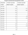
- General physical properties of C 2 N-8E12 are listed in Table 1. Table 1 Molecular weight 145.72 kDa Stereochemistry L-amino acids Appearance Clear, colorless to light yellow liquid Solubility -130 mg/mL
- This example describes efforts and results for humanization of the murine anti-tau antibody HJ8.5.
- the efforts yielded four humanized light chain variable regions (VL or VK) and four humanized heavy chain variable regions (VH).
- Humanization generally refers to techniques of reducing the potential immunogenicity associated with using a non-human monoclonal antibody for chronic treatment. Two methods typically used to reduce immunogenicity are CDR grafting and deimmunization. Murine antibody HJ8.5 was de-immunized using a method developed by Antitope.
- CDR grafting is a protein engineering approach. Briefly, it relies on both an understanding of the basic architecture of an antibody and its conservation across species. Murine and human antibodies share a common / conserved architecture. Antibody structure is divided into constant and variable regions (See Fig B-1). The variable region can be further divided into so called framework regions and CDR regions (Fig B-2). It can be seen that the variable region is composed of four frameworks (Fwk) and three CDR. The arrangement of frameworks and CDRs are the same in light and heavy variable domains.
- the non-human constant regions are replaced with human constant regions, giving rise to a so called chimeric antibody.
- the murine CDR regions are transferred into human framework regions; the resulting variable domain is a mix of human frameworks and murine CDR's (see Fig B-2).
- a number of the murine framework residues, thought to play a critical role in maintaining the affinity are transferred (not shown).
- Composite Human Antibody TM technology from Antitope is said to be a deimmunization technology that is used in conjunction with identifying both CDRs and key amino acids in the framework thought to play a role in binding.
- the resulting fully-humanized antibodies retain the binding affinity and specificity of the starting monoclonal antibody and are also devoid of CD4+ T cell epitopes, which avoids undesirable immunogenicity in humans.
- Composite Human Antibodies TM are generated by combining multiple segments of human antibody sequences from Antitope's database comprising 100,000's of unrelated fully-human antibody variable region sequences. Initial modeling of variable region sequences of HJ8.5 antibody is used to identify amino acids critical to antibody binding, which are then used to constrain the selection of human sequence segments. Individual sequence segments and the junctions between adjacent segments are then analyzed using two proprietary in silico technologies (iTope TM and TCED TM ) for selection of fully-human variable region sequences that are devoid of CD4+ T cell epitopes. DNA encoding variable regions for Composite Human Antibodies are synthesized, cloned onto an expression vector with human constant regions and transfected into mammalian cells for production of the humanized antibodies.
- HJ8.5 Humanization of HJ8.5: Structural models of the HJ8.5 murine anti-Tau412 antibody V regions were produced using Swiss PDB and analyzed in order to identify important "constraining" amino acids in the V regions that were likely to be essential for the binding properties of the antibody. From the analysis, a number of constraining framework residues were identified as candidates for inclusion in the fully humanized V regions. Segments of human variable region sequences were selected to include one or more of these residues.
- a preliminary set of human sequence segments that could be used to create the fully humanized HJ8.5 antibodies were selected and analyzed using iTope TM technology for in silico analysis of peptide binding to human MHC class II alleles (Perry et al 2008), and using the TCED TM (T Cell Epitope Database) of known antibody sequence-related T cell epitopes (Bryson et al 2010). Sequence segments that were identified as significant non-human germline binders to human MHC class II or that scored significant hits against the TCED TM were discarded. Combinations of sequence segments were also analyzed to ensure that the junctions between segments did not contain potential T cell epitopes. Selected segments were then combined to produce heavy and light chain V region sequences for synthesis. For HJ8.5, four VH chains and four V ⁇ chains were designed and constructed.
- FIG. 1 shows the variable region sequences of the murine HJ8.5 antibody as well as 4 humanized variant sequences for each of the heavy and light chains (4 VH and 4 VL/VK sequences).
- the amino acid sequences of those four VH chains and four V ⁇ chains are set forth in FIG. 1 .
- the CDR sequences, as defined by Kabat et al are highlighted in red (underlined). Framework changes from the original mouse sequence are highlighted in blue and in bold.
- Table B-1 summarizes the number of framework changes introduced in each variant of the heavy and light chain variable domains.
- Table B-1 Variable Domain Number of Framework changes VH1 4 VH2 5 VH3 10 VH4 11 VK1 6 VK2 7 VK3 11 VK4 12
- FIG. 2 shows the sequences of the humanized variable and constant region sequences for each of the heavy and light chains (4 VH and 4 VL/VK sequences).
- the variable heavy chain is grafted to the constant heavy chain of human IgG4 containing the S241P hinge stabilizing mutation.
- the variable light chain is grafted to the constant light chain of human Kappa light chain.
- This table also lists the theoretical isoelectric point (PI) and molecular weight (Mw).
- FIG. 3 shows expression data from 2 rounds of transient expression of cells transfected with polynucleotides encoding VH and VK regions. Results for 13 humanized anti tau antibodies are summarized. Different combinations of heavy and light chains resulted in markedly different levels of expression being observed. In Round 1, all variants of VH and VL regions were combined with each other (only results for 13 are shown of the 16 that were tested. In Round 2, the 6 best expressing combinations observed in Round 1 were tested. Expression is shown as ⁇ g of antibody measured per mL of culture media. Higher levels of expression is advantageous since it suggests that the antibody is correctly folded, secreted as expected, nontoxic and generally stable.
- FIG. 4 shows data from a potency assay.
- the potency assay evaluates the ability of antibodies to compete with the original murine HJ8.5 (parent antibody) for binding to human tau in an ELISA type format.
- the assay format involves coating the ELISA plate with human tau and then allowing the test antibodies as well as biotinylated HJ8.5 to compete for binding to tau.
- the assay enables the relative IC50 value for each humanized antibody variant to be measured. IC50 values are normalized to that of chimeric HJ8.5 to enable comparisons to be made between plates. This data demonstrates that the humanization process has not significantly changed the binding of the humanized antibodies to human tau.
- This study describes the use of the Biacore T200 to measure and compare the binding characteristics of the interaction between six fully humanized (VH1/VK2, VH1/VK3, VH2/VK2, VH2/VK3, VH3/VK2 and VH3/VK3, described above in Example 1) monoclonal antibodies and one chimeric monoclonal antibody based on HJ8.5 with recombinant human Tau-412 protein.
- the aim of this study was to use the Biacore T200 surface plasmon resonance instrument for the high resolution kinetic characterization of the interactions between Tau-412 and these seven mAbs.
- the antibodies were stored at 4 °C.
- Tau-412 was stored at -20 °C as per the manufacturer's instructions. Once reconstituted the Tau-412 solution was stored on ice and used within 24 hours. Aliquots of reconstituted Tau-412 were frozen within 30 minutes of reconstitution and stored at -20 °C.
- Biacore Preventative Maintenance Kit 2 BR-1006-51 Series S CM5 Sensor Chips BR-1006-68 Amine Coupling Kit BR-1000-50 10 mM Acetate pH 4.5 BR-1003-51 HBS-EP Running buffer ⁇ BR-1006-69 lOmM Glycine-HCI pH 1.5 BR-1003-54 lOmM Glycine-HCI pH 2.0 BR-1003-55 Protein A (Sigma) P6031 4 M MgCl 2 hexahydrate (Sigma) M9272-500G
- CM5/Protein A chip Upon insertion of a CM5/Protein A chip the system was primed and then normalized with BIA normalizing solution (Biacore Preventative Maintenance Kit 2). All samples were run at 25 °C with a sample rack incubated at 5 °C. The chip was added to the system with HBS-EP used as the running buffer.
- the mAbs were stored as supplied and diluted to 100 nM for all immobilization (capture) runs.
- the antigen Tau-412 was reconstituted from the dry powder using Milli-Q water to a final concentration of 1 mg/mL; further dilutions were performed for the kinetics runs.
- the mass and molecular weight of Tau-412 used in the concentration calculation was provided by the reagent manufacture (100 ⁇ g/vial and 42.9 kDa). No carrier protein was added to this solution. Vials of the antigen were only reconstituted when required and were stored in their powder form at -20 °C until use. Once reconstituted, the antigen solution was kept on ice and used within 24 hours.
- a capture assay with protein A was selected for this study.
- the performance of the Protein A surface was superior to the anti-human, protein A/G, protein G and protein L surfaces that were also tested.
- the Protein A chip was prepared through immobilization using standard amine coupling chemistry. Immobilization was carried out at a protein concentration of 5 ⁇ g/mL in 10 mM Acetate buffer pH 4.5 to a target response level of 500 RUs on a CM5 Series S sensor chip (Biacore).
- analyte binding capacity RU analyteMW ligandMW • immobilized ligand RU • Sm
- a target of 300 RUs was set for capture of all the trial antibodies.
- the capture levels obtained within the study varied from ⁇ 280-400 RU's.
- the amount of injected antibody was adjusted to get closer to the desired 300 RU capture level.
- Non-specific binding can be due to either the analyte or analyte contaminants interacting with either the ligand (non-specific and difficult to detect), capture protein, or the sensor chip surface.
- Regeneration scouting was performed and the optimum conditions for the regeneration of chimeric and VH1/VK2 antibodies on the Protein A surface were as follows. Three 240 second injections of 10 mM Glycine-HCI pH 1.7 followed by one 300 second injection of 4M all at 40 ⁇ L/min. A 600 second wait step was introduced after the last regeneration injection to allow the surface to stabilize before starting the next binding cycle.
- the performance of the surface was analyzed by repeated control injections of 2.5 nM Tau-412 at the start, interspaced and at the end of a kinetic run. Stable binding was observed throughout the kinetic run highlighting the suitability of the system for kinetic analysis.
- Mass transport limitation occurs when the rate of association contains a significant component associated with the rate of transport of the analyte to and from the chip surface. Where mass transfer is found to be significant the resulting kinetic analysis could be inaccurate. Lowering the density of immobilized ligand, or increasing the flow rate, can reduce mass transport limitations. From previous experience of using low density surfaces and similar Mw antigens, a flow rate of 40 ⁇ L/min was selected for this study.
- the linked reaction control experiment is used to assess the ligand-analyte interaction to check for deviations from a 1-to-1 binding model.
- the analyte is injected over the surface for different periods of time (contact times) and the dissociation rate is analyzed to determine if it varies with the contact time. If such a relationship is observed, it indicates that a second interaction event is taking place after the initial binding event that results in a stabilized complex at the surface.
- Equation 2 A 1-to-1 binding model was used to fit the resulting kinetic data (Equation 2). Due to variations in the amount of antibody captured the parameter R max was set to local as opposed to global analysis for each antibody kinetic analysis.
- the characterization and the control experiments performed for the Protein A capture surface suggested this was a suitable system to determine kinetic values for the Tau-412 interactions.
- the binding stoichiometry was assessed by injecting a saturating concentration of Tau-412 (1000 nM) over 277 RU's of captured VH1/VK2 on a trial Protein A surface. Two sequential injections of 1000 nM Tau-412 appeared to result in saturated binding at 122 RU's. This resulted in a binding stoichiometry of 150%, which is higher than expected for one antibody molecule binding to one Tau-412 molecule. Reasons for this could include binding of the antibody to two molecules of Tau-412 or Tau-412 oligomerization on the surface of the chip.
- Kinetic data was obtained at a flow rate of 40 ⁇ L/min to minimize any potential mass transfer effects.
- Two repeats of the blank (no antigen) and the 2.5 nM concentration of the analyte were programmed into the kinetic run in order to check the stability of both the surface and analyte over the kinetic cycles.
- 2-fold dilutions of Tau-412 from 40 nM to 0.156 nM were run.
- an analyte range of 20 nM to 0.625 nM was selected. This range covered multiple analyte concentrations both above and below the reported K D .
- the association phase was monitored for 500 seconds to allow some of the higher concentrations of analyte to reach steady state. In order to observe a sufficient signal decrease (> 10%) during the dissociation phase of the kinetic cycle, dissociation was measured for 1200 seconds. As discussed in Section 5, the F c s were allowed to stabilize for 600 seconds after each regeneration step. The signal from the reference channel F C 1 was subtracted from that of F c 2, F c 3 and F c 4.
- the kinetic parameters for the interaction of Tau-412 with the 7 mAbs as measured using the Protein A capture system on the Biacore T200 are shown in Table F-2.
- a local R max parameter was used in the 1-to-1 binding model.
- Kinetic analysis was performed in three independent runs using fresh preparations of Tau-412 and antibody. Run 1 and runs 2+3 used different vials of the antigen Tau-412; therefore the reported errors associated with the mean response probably represent variation in preparation of the analyte and differences in assay setup and run. From run 1 to runs 2+3, the amount of antibody injected was adjusted to try to get closer to the target 300 RU capture levels.
- the chimeric antibody was run in triplicate and an analysis of all three data sets is shown in Table F-2. The % CV for the K D derived from these three data sets was 4.3% indicating that the results were within assay variability.
- the Chi 2 values show how well the association and dissociation data fits the proposed 1-to-1 binding model - the lower the value the better the fit.
- the associated SE values for the rate constants represent the uncertainty associated with fitting the data to the model described, and do not represent the total uncertainty for the true kinetic values.
- the mean response data represents the average kinetic values and the associated SD from 2, or 3 independent analyses.
- the antibodies can be ranked based on affinity as follows: VH3/VK2 > VH3/VK3 > Chimeric > VH2/VK3 > VH1/VK3 > VH1/VK2 > VH2/VK2.
- the % CV associated with the mean kinetic parameters ranged from 10-20%, and thus it is likely that all antibodies have very similar affinities and that differences are purely a result of assay variation. In general, the differences in binding between the antibodies may be attributable to assay variation, and it is believed there are no significant differences in KD values of the humanized antibodies compared to the chimeric antibody.
- FIG. F-1 A comparison of the kinetic values determined using the protein A capture assay on the Biacore T200 for the interaction between the antibodies and Tau-412 are shown in FIG. F-1.
- the chimeric antibody appears to display a significantly different binding profile when compared to the humanized antibodies, although the affinities are similar.
- FIG. F-1 is a k d versus k a plot showing the relative kinetic values of the tested antibodies and Tau-412 interactions as determined using the Protein A capture assay on the Biacore T200.
- the dashed diagonals represent isoaffinity lines. Please note the axes display different data ranges, with the aim of improving the clarity of the humanized antibodies on the plot.
- FIG. 5 summarizes the results from surface plasmon resonance (SPR) analysis, determining the binding kinetics of the 6 best expressing humanized constructs against human tau.
- the test antibody is immobilized on the SPR chip with different concentrations of human tau then allowed to flow over the chip. Association and dissociation rates as well as the affinity based on the measured binding events is calculated for each of the variants. The chimeric variant was also tested.
- SPR surface plasmon resonance
- FIG. 6 shows the binding of four humanized antibody variants to soluble human tau in a sandwich style ELISA.
- Assay methods that rely on passive adsorption have the potential to create artifactual binding results.
- a solution based method to measure the binding activity of the humanized antibody variants was employed.
- antigen human tau
- HJ8.5 monoclonal anti-human tau antibody that recognizes a different epitope than HJ8.5.
- Subsequent binding of the humanized anti-tau antibodies to the captured human tau depends on antigen concentration, while IgG4 isotype controls shows no binding at all.
- This assay demonstrates that binding of the humanized anti-tau antibodies to human tau is specific and that the antibodies bind to soluble human tau.
- FIG. 7 shows binding of humanized and control antibodies to tissue from wild type mice (negative control tissue), P301S mice (which express human tau having a P301S mutation and develop age associated tau pathology), and humans with either Alzheimer's disease or Progressive Supranuclear Palsy (PSP).
- the aim of this study was to confirm that humanized antibodies retain the ability to bind to aggregated tau in tissue as compared to the chimeric form of HJ8.5.
- the figures show representative images of staining human and mouse brains with different variants of humanized HJ8.5 antibody.
- P301S mice at 4 and 9 month old were tested, and both time points show pathologic aggregates of tau, with the 9 month old mice having more tau pathology than the 4 month old mice.
- a sample of brain tissue from one subject with PSP, and a sample of brain tissue from one subject with AD was examined.
- A illustrates staining with chimeric HJ8.5 for the mouse and human AD tissue.
- B illustrates the staining with a negative control antibody (non-specific human IgG4).
- C-H illustrate the staining with the six humanized antibodies. All humanized variants of the murine HJ8.5 antibody bind to tau aggregates found in P301S mouse brain as well as tau aggregates found in the brain tissue of the subjects diagnosed with either AD or PSP.
- FIG. 8A shows the epitopes of HJ8.5 in human tau.
- the epitope was mapped using yeast display. For this method, various peptides covering the sequence of human tau were expressed using by yeast. Binding of the HJ8.5 antibody to yeast in culture was measured by immunofluorescence. Binding to yeast, expressing variants of tau that included the first 34 amino acids was observed, but no binding, if the yeast only expressed the first 32 amino acids of tau (FIG. 8B). This suggests that the epitope is within the first 34 amino acids. Additionally, HJ8.5 binds if the peptide includes amino acids 27-135 but not if the peptide spans amino acids 30-135. This suggests that the epitope includes amino acids greater than amino acid 27. Based on this data the epitope is contained within the 27-34 sequence of human tau (GYTMHQDQ). FIG. 8A also shows the rhesus monkey and mouse tau sequences and highlights in red the amino acid changes from human tau.
- FIG. 9 shows more detailed, peptide-based epitope mapping of HJ8.5 and C 2 N-8EI2.
- a peptide library of linear 15mers spanning the full sequence of human tau (IN4R, 412 amino acids) was created. Additionally double alanine versions of these peptides where amino acids 10 and 11 were changed to alanine were also produced.
- any naturally occurring alanines at position 10 or 11 were mutated to glycine. All peptides were spotted onto a peptide array and then probed with HJ8.5 or C 2 N-8E12 and binding measured. The tau binding epitope(s) of both antibodies were reliably mapped using these peptide arrays.
- the binding epitope of C 2 N-8E12 is 25 DQGGYT 30 and matches the epitope of the murine parent, HJ8.5.
- the binding of HJ8.5 and C 2 N-8E12 to tau peptides is severely compromised when amino acids D, Q, Y, or T in the epitope were replaced with alanine, suggesting that they play a crucial rule in the antibody binding.
- the central two glycines in the epitope were replaced with alanine, the binding of antibodies to the tau peptides was not as severely compromised (PEP 2875811). This is likely not an indication that these amino acids are not important for binding but rather due to the conservative nature of substitutions between Alanine and Glycine amino acids.
- the epitope mapped using these more detailed methods is slightly different from what was mapped using yeast display (FIG. 8A). This difference is attributed to the difference in the binding assays, with larger peptides being used on the yeast display system.
- the 15mer peptide array methodology is considered to be superior to the yeast display methodology.
- FIG. 10 illustrates the binding results for different anti human tau antibodies to either human or rhesus monkey tau.
- Murine anti human tau antibodies HJ8.5 and HJ8.7 alongside humanized variant VH1/VK2 also referred to as C 2 N-8EI2 were tested.
- FIG. 8A shows that there is a single amino acid difference at position 32 between human and rhesus tau, in the claimed binding epitope sequence GYTM(H/L)QDQ.
- FIG. 8A shows that there is a single amino acid difference at position 27 between human and rhesus tau, in the claimed binding epitope sequence DQ(G/E)GYT.
- FIG. 11 shows binding of humanized anti-tau antibody to tau in CSF from human subjects with various tauopathies.
- the binding of C 2 N-8E12 to tau in CSF samples from subjects diagnosed with a variety of tauopathies as well as age matched and young normal control subjects was evaluated.
- a sandwich ELISA was used to demonstrate binding of C 2 N-8E12 to tau in human CSF from subjects with AD, CBD, FTD, or PSP as well as age matched and young/adult controls.
- C 2 N-8E12 was used as the coating antibody to capture tau in CSF samples.
- Biotinylated murine monoclonal tau antibody HJ8.7 was used as the detection antibody.
- Wells coated with control human IgG4 acted as the negative control for the experiments.
- This study is a randomized, double blind, placebo controlled, single ascending dose (SAD) phase 1 study to be conducted in up to ten (10) centers. It is designed to evaluate the safety, tolerability, immunogenicity, and PK of single-dose administration of C 2 N-8E12 and to establish the MTD to be used in future repeat dosing studies.
- SAD single ascending dose
- the primary objective of this study is to determine the safety, tolerability, immunogenicity, and maximally tolerated dose (MTD) of a single dose of C 2 N-8E12 in subjects with PSP.
- Safety assessments will include physical and neurologic examination, clinical safety laboratory studies, immunogenicity, adverse events, vital signs and concomitant medication review.
- the secondary objectives are to determine: Single-dose systemic pharmacokinetics including; Maximum plasma concentration after single infusion; Area under the curve (AUC) after single infusion; Time at which the maximum concentration after infusion is achieved; Terminal half-life of C 2 N-8E12; Partition of C2N-8E12 into cerebrospinal fluid (CSF); and Biologic target engagement through the measurement of soluble tau levels in blood and CSF as well as assessing the presence of C 2 N-8E12-tau complexes.
- AUC Area under the curve
- CSF cerebrospinal fluid
- This study intends to enroll 32 subjects with PSP (24 in the treatment arm and 8 in the placebo arm). Subjects will be enrolled in 8 blocks of 4 patients, with one patient in each block randomized to placebo and 3 to the current estimate of the MTD. Additional subjects may be enrolled if DLTs occur. No dose may be skipped, however, during the dose escalation process.
- a continual reassessment method (CRM) for dose escalation will be used as described in the statistical design section.
- a logistic model will be used to identify the probability of DLT by dose.
- C 2 N-8E12 will be shipped to the clinical site as a frozen liquid in single use bottles at a nominal concentration of 20 mg/mL. Each bottle contains 300 mg C 2 N-8E12 and must be stored frozen at -70°C to -80°C.
- Patients will undergo screening to assess whether inclusion and exclusion criteria are met. Screening will also include assessments of blood and CSF, and MRI.
- assessments of blood and CSF On the day of dosing (Day 0), a single dose of C 2 N-8E12 will be administered through an IV line and subjects will be closely monitored at a clinical facility for 24 hours after dose administration. This includes blood samples for safety and PK assessments. During the following 3 days, as well as at one and two weeks after the infusion, additional clinical examination and blood sampling will occur. An additional safety MRI and CSF sampling will be performed 4 days post-infusion. Subjects will be followed every 28 days, for no less than two months from the date of dosing (e.g., day 56).
- phase 1 study includes establishment of an MTD as assessed by safety evaluations including clinical laboratory tests, physical and neurologic examinations, and occurrences of adverse events to determine a recommended range of doses for evaluation in the subsequent phase 2/MAD study. Random assignment of subjects and inclusion of a placebo arm avoids bias and increases the likelihood that both known and unknown risk factors are distributed evenly between treatment groups.
- a data safety monitoring committee will review safety data on an ongoing basis.
- the safety monitoring committee will be minimally comprised of two independent physicians, one biostatistician, one physician with expertise in PSP, and one non-voting member from the Sponsor. If any individual study subject experiences SAEs, all available safety data for the subject will be reviewed to determine if the event meets the definition of DLT, and whether the MTD has been established. If MTD has not been established, and patient enrollment continues, the DSMC will provide recommendations to the Sponsor whether any further actions or protocol amendments are necessary to ensure the safety of subsequently enrolled patients. The Sponsor will make final determinations on any amendments to or preliminary termination of the study.
- a dose limiting toxicity is defined as: (i) any Grade 3 or higher AE per Rheumatology Common Toxicity Criteria v2 for which there is reasonable possibility that C 2 N-8E12 caused the event; (ii) any Grade 2 AE in the NCI's Common Terminology Criteria for Adverse Events v4.0. (CTCAE) system organ class of nervous system and psychiatric disorders that is considered clinically significant and for which there is reasonable possibility that C 2 N-8E12 caused the event; or (iii) any infusion-related toxicities (e.g., allergic reaction / hypersensitivity) occurring during the infusion of C 2 N-8EI2 or within 24 hours after completing the infusion that do not resolve promptly with a reduced infusion rate and/or supportive care.
- CCAE system organ class of nervous system and psychiatric disorders that is considered clinically significant and for which there is reasonable possibility that C 2 N-8E12 caused the event
- any infusion-related toxicities e.g., allergic reaction / hypersensitivity
- Dose Escalation The assignment of subjects to dose cohorts is governed by the following rules: (1) Within each block of 4 patients, 1 patient will be allocated to the placebo arm; (2) Complete toxicity information is required for at least 3 patients at a dose level before escalation to a higher dose level; (3) The maximum increment of escalation from one cohort to the next is 1 dose level; and (4) At least 12 subjects (3 blocks) should be dosed at the MTD dose level.
- patients will be dosed sequentially, with a minimum interval of at least two days between dosing of consecutive subjects in order to provide an additional measure of safety assurance.
- the first cohort will be allocated to d 1 .
- the statistical model will be updated after complete toxicity information is available for each cohort. By rule, one additional cohort may be enrolled before complete information is available for all subjects on the most recent cohort. Incomplete toxicity data is allowed for no more than 3 patients before the next cohort is enrolled and randomized.
- Each subsequent cohort will be assigned to the dose that is estimated to be the MTD according to the definition above.
- the model may be updated as each patient enrolls and each subsequent patient dosed to the current estimated MTD.
- Study Population This study will enroll male and female subjects with progressive supranuclear palsy (PSP) aged 50 to 85 years.
- PSP posterior supranuclear palsy
- Inclusion Criteria For inclusion into the study, each subject must be willing and able to provide informed consent. Prior to initiation of the treatment protocol, it will be confirmed that each subject is able to provide consent for the treatment protocol. Subjects will be invited to participate in the study, and after signing the informed consent form, screening procedures will take place. If subjects fail to fulfill the inclusion criteria or meet any of the exclusion criteria, the subjects will not be enrolled into the screening assessment or treatment schedule.
- Each subject must meet the following criteria to be enrolled in this study: Male or female; Between 50 and 85 years of age; Meets NINDS-SPSP possible or probable criteria as modified for NNIPPS and AL-108-231 clinical trials, including: (d) supranuclear gaze palsy or decreased saccade velocity, (ii) gait instability or falls within the first 3 years of symptoms; Brain MRI at Screening is consistent with PSP ( ⁇ 4 microhemorrhages, and no large strokes or severe white matter disease); Score on the PSP rating scale between 20 and 50; Able to provide informed consent to participation at baseline or if unable to provide informed consent can provide assent to participation and has an authorized medical representative who can provide consent; Has study partner who sees the patient at least 5 hours per week, who can accompany the patient to visits and consents to study participation; Other concurrent non-biologic therapies are allowed but the dose must have been stable for at least 30 days prior to enrollment; Able to walk 5 steps with minimal assistance (stabilization of one arm or use of cane/walker); Stable medications for Parkinson
- Subjects who meet any of the following criteria will be excluded from the study: Signs of a progressive neurological disorder that better meets the criteria for types of neurological disorders other than PSP, including: (a) meets criteria for probable Alzheimer's disease or (b) meets research criteria for Parkinson's Dementia with Lewy Bodies, multiple system atrophy (MSA), or amyotrophic lateral sclerosis (ALS); Any malignancy (other than non-metastatic basal cell carcinoma of the skin) within 5 years of screening; Clinically significant renal or hepatic dysfunction at screening based on professional judgment of Investigator; Clinically significant cardiovascular event within three months prior to study entry, based on professional judgment of Investigator; Clinically significant abnormal hematology or chemistry laboratory test results during screening, based on professional judgment of Investigator; Have received any prior monoclonal antibody therapy for any reason within the last 90 days or received any other investigational agent within the previous 30 days or 5 half-lives (whichever is longer).
- MSA multiple system atrophy
- ALS amyotrophic lateral sclerosis
- C 2 N-8E12 Prior administration of C 2 N-8E12 does not apply to this exclusion criteria and, therefore, does not disqualify a subject from participating in the phase 2/MAD study; Currently on any other biologic or immunomodulatory therapy; Disease duration of greater than 5 years since onset of symptoms; Midbrain volume> 8,600 mm 3 on screening MRI scan; Subjects that reside at a skilled nursing or dementia care facility; Has clear evidence of motor neuron disease on examination, consistent with ALS pathology (this has been described in C90RF72 carriers with CBS presentation); Diagnosis of any other significant unrelated neurological or psychiatric disorders that could account for cognitive deficits (e.g., active seizure disorder, stroke, vascular dementia), based on professional judgment of Investigator; Untreated major depression at baseline evaluation, based on clinical judgment and results in GDS; History of other major psychiatric illness; Any prior history of suicidal attempts; Severe cognitive impairment as assessed by MMSE ( ⁇ 17) that in the Investigator's opinion would preclude collection of outcome measures;
- Subjects must agree to use (and/or have their partner use) acceptable methods of contraception beginning at the baseline visit throughout the study and until 56 days after the last dose of study drug in the last treatment period. Acceptable methods of contraception are listed below.
- the phase 1/SAD study will use C 2 N-8E12 from DP Lot # 1018775 - the Research Cell Bank (RCB) material currently being used in the Expanded Access IND 119404. It is formulated in 25 mM acetate buffer at pH 5.5, and it is provided at a concentration of 20 mg/mL. Placebo: Placebo is formulated identically to C 2 N-8E12 without the active study drug.
- MRSD maximum recommended starting dose
- FDA Food and Drug Administration
- NOEL No Observed Effect Level
- the starting dose for phase 1/SAD study will be 2.5 mg/kg dosed IV. This starting dose is 10 times lower than the maximally allowed starting dose based on a 4-week mouse toxicology study and 6 times lower than the current maximal dose (15 mg/kg) administered in the Expanded Access and compassionate use human treatment protocols involving C 2 N-8E12 (see Investigator's Brochure).
- each subject After providing informed consent, each subject will undergo screening assessments to reconfirm that the inclusion criteria can be fulfilled, and that no contraindications exist to receiving treatment under this protocol. Specifically, each subject will be assessed at screening with a clinical and neurological examination to confirm the diagnosis. A brief cognitive screening with PSP ratings scale and a depression scale with interview will be made and a complete medical and drug/medication history will be obtained. If the subject fulfills all inclusion criteria and lacks exclusion criteria, further investigations will be performed. The Screening visit occurs 28 days to 7 days before the Baseline / Day 0 Visit.
- Adverse Events Assessments The following safety assessment will be conducted in order to monitor for AEs: Vital signs (blood pressure, pulse/heart rate, temperature, respiratory rate, SPO2); Complete neurologic exam, including a cognitive assessment (mental status tests); Laboratory tests: (1) Hematology panel: complete blood count (CBC) with differential, hematocrit, and hemoglobin (Hb), platelet count; (2) Chemistry panel: serum electrolytes, glucose, uric acid, blood urea nitrogen (BUN), creatinine, total protein, albumin, bilirubin (total, direct and indirect), alkaline phosphatase, lactate dehydrogenase (LDH), liver enzymes (AST, ALT and GGT), iron, cholesterol panel, CPK, amylase, and lipase; (3) Coagulation panel: Prothrombin Time (PT), INR, and Partial Thromboplastin Time (PTT); Urinalysis, including measurement of Hb, WBC, and protein content; ECG - continuous monitoring or 12 lead ECG;
- Colombia-Suicide Severity Rating Scale As a safety parameter, the Columbia-Suicide Severity Rating Scale (C-SSRS) for suicidal ideation will be used (Posner et al. 2011).
- Geriatric Depression Scale Similar to the C-SSRS, the Geriatric Depression Scale (GDS) will be used to assess overall mood during the study.
- the Geriatric Depression Scale (GDS) is a 30-item self-report assessment used to identify depression in the elderly (Yesavage et al. 1983).
- PSP Rating Scale The PSP Rating Scale (PSPRS) will be used at screening for inclusion as well as baseline and end of the study to assess changes in the scale over time (Golbe and Ohman-Strickland 2007).
- Clinical Global Impressions The clinical Global Impressions rate of change (CGIc) and severity (CGIs) scales will be used to assess severity of symptoms.
- Schwab and England Activities of Daily Living The Schwab and England Activities of Daily Living (SEADL) scale will be used as a means of assessing the subjects ability to perform daily activities (Schwab and England 1969).
- CDR-SB-FTLD Clinical Dementia Rating Sum of Boxes Frontotemporal Lobe Dementia
- MMSE Mini Mental State Examination
- Cerebrospinal fluid CSF will be drawn from the L3-4 interspace by lumbar puncture. If CSF sampling is not successful CT/fluoro guided lumbar puncture can be used at the discretion of the local clinical site staff as per local protocol. Safety labs on CSF will be analyzed locally at the applicable clinical site after each lumbar puncture/CSF collection. These measures include: cell counts (WBC and RBC), total protein and glucose. Other CSF measurements (e.g., C2N-8E12 concentration, target engagement and other exploratory biomarkers) will be analyzed by the applicable designated laboratory.
- CSF measurements e.g., C2N-8E12 concentration, target engagement and other exploratory biomarkers
- the subject will also receive a baseline MRI scan with structural, FLAIR, diffusion-weighted and susceptibility weighted imaging. Post-dosing imaging analyses will be performed according to the Schedule of Events.
- a blood sample will be collected for DNA extraction at baseline. All individuals will undergo an extended MAPT haplotype sequence analysis to determine Hl(A-D) and H2 carrier status. DNA will be extracted from the samples and the DNA shipped to the designated pharmacogenomic core for this study.
- Subjects will be enrolled in this study until the earlier occurrence of any of the following events: (i) they complete their participation and the entire Schedule of Events; (ii) they or the Investigator decide(s) to terminate their participation; or (iii) the applicable subject experiences any DLT or any SAE that an Investigator deems to preclude further participation in this study and precludes eligibility for the subsequent phase 2/MAD study. Additionally, at the discretion of the Investigator, subjects who cease to meet any inclusion criterion, or meet one or more exclusion criterion during the study, may be determined ineligible to continue participating in the study or for subsequent participation in the phase 2/MAD study.
Landscapes
- Health & Medical Sciences (AREA)
- Chemical & Material Sciences (AREA)
- Life Sciences & Earth Sciences (AREA)
- Organic Chemistry (AREA)
- Engineering & Computer Science (AREA)
- Medicinal Chemistry (AREA)
- General Health & Medical Sciences (AREA)
- Biomedical Technology (AREA)
- Neurology (AREA)
- Neurosurgery (AREA)
- Bioinformatics & Cheminformatics (AREA)
- Animal Behavior & Ethology (AREA)
- Pharmacology & Pharmacy (AREA)
- Public Health (AREA)
- Veterinary Medicine (AREA)
- Immunology (AREA)
- Proteomics, Peptides & Aminoacids (AREA)
- General Chemical & Material Sciences (AREA)
- Chemical Kinetics & Catalysis (AREA)
- Nuclear Medicine, Radiotherapy & Molecular Imaging (AREA)
- Molecular Biology (AREA)
- Biochemistry (AREA)
- Genetics & Genomics (AREA)
- Biophysics (AREA)
- Psychology (AREA)
- Hematology (AREA)
- Urology & Nephrology (AREA)
- Microbiology (AREA)
- Physics & Mathematics (AREA)
- Psychiatry (AREA)
- Epidemiology (AREA)
- Hospice & Palliative Care (AREA)
- Biotechnology (AREA)
- General Physics & Mathematics (AREA)
- Pathology (AREA)
- Analytical Chemistry (AREA)
- Food Science & Technology (AREA)
- Cell Biology (AREA)
- Optics & Photonics (AREA)
- Mycology (AREA)
Applications Claiming Priority (4)
| Application Number | Priority Date | Filing Date | Title |
|---|---|---|---|
| US201462018436P | 2014-06-27 | 2014-06-27 | |
| US201462080903P | 2014-11-17 | 2014-11-17 | |
| US201562170036P | 2015-06-02 | 2015-06-02 | |
| PCT/US2015/038002 WO2015200806A2 (en) | 2014-06-27 | 2015-06-26 | Humanized anti-tau antibodies |
Publications (4)
| Publication Number | Publication Date |
|---|---|
| EP3182987A2 EP3182987A2 (en) | 2017-06-28 |
| EP3182987A4 EP3182987A4 (en) | 2018-01-24 |
| EP3182987B1 true EP3182987B1 (en) | 2023-09-27 |
| EP3182987C0 EP3182987C0 (en) | 2023-09-27 |
Family
ID=54938957
Family Applications (1)
| Application Number | Title | Priority Date | Filing Date |
|---|---|---|---|
| EP15810923.1A Active EP3182987B1 (en) | 2014-06-27 | 2015-06-26 | Humanized anti-tau antibodies |
Country Status (27)
| Country | Link |
|---|---|
| US (2) | US9957317B2 (es) |
| EP (1) | EP3182987B1 (es) |
| JP (3) | JP6626892B2 (es) |
| KR (1) | KR102431596B1 (es) |
| CN (1) | CN106659759B (es) |
| AU (2) | AU2015279643B2 (es) |
| BR (1) | BR112016030686A2 (es) |
| CA (2) | CA3094638A1 (es) |
| CL (1) | CL2016003346A1 (es) |
| CR (1) | CR20170026A (es) |
| DO (1) | DOP2016000335A (es) |
| EC (1) | ECSP17005649A (es) |
| ES (1) | ES2959528T3 (es) |
| IL (3) | IL249734B (es) |
| MA (1) | MA40229A (es) |
| MX (2) | MX2017000205A (es) |
| MY (1) | MY184540A (es) |
| NZ (1) | NZ727919A (es) |
| PE (1) | PE20170665A1 (es) |
| PH (2) | PH12016502608A1 (es) |
| RU (1) | RU2743152C2 (es) |
| SG (2) | SG10201914067XA (es) |
| TW (3) | TWI664190B (es) |
| UA (1) | UA122771C2 (es) |
| UY (1) | UY36194A (es) |
| WO (1) | WO2015200806A2 (es) |
| ZA (1) | ZA201608875B (es) |
Families Citing this family (35)
| Publication number | Priority date | Publication date | Assignee | Title |
|---|---|---|---|---|
| RU2018132044A (ru) | 2012-07-03 | 2018-10-19 | Вашингтон Юниверсити | Антитела против тау |
| CN105121465B (zh) | 2013-03-13 | 2020-09-08 | 普罗塞纳生物科技公司 | Tau免疫疗法 |
| TWI664190B (zh) | 2014-06-27 | 2019-07-01 | 美商C2N醫療診斷有限責任公司 | 人類化抗-tau抗體 |
| SG10201911349YA (en) | 2015-06-05 | 2020-01-30 | Genentech Inc | Anti-tau antibodies and methods of use |
| US20200030445A1 (en) * | 2015-06-12 | 2020-01-30 | C2N Diagnostics, Llc | Stable formulations of humanized anti-tau antibody |
| EP3445785B1 (en) | 2016-04-18 | 2022-06-22 | Faron Pharmaceuticals OY | Humanized anti clever-1 antibodies and their use |
| US10889638B2 (en) | 2016-05-02 | 2021-01-12 | Prothena Biosciences Limited | Antibodies recognizing tau |
| KR20230070336A (ko) | 2016-05-02 | 2023-05-22 | 프로테나 바이오사이언시즈 리미티드 | 타우 인식 항체 |
| FI3452507T3 (fi) | 2016-05-02 | 2022-12-15 | Tau-immuunihoito | |
| US10570196B2 (en) * | 2016-07-20 | 2020-02-25 | Anahit Ghochikyan | Humanized anti-tau antibodies and compositions for and methods of making and using in treatment, diagnosis and monitoring of tauopathies |
| IL300982B1 (en) * | 2016-09-10 | 2024-04-01 | Yeda Res & Dev | Reduction of the level of regulatory T cells or their activity for the treatment of diseases and damages in the central nervous system |
| CN110036033B (zh) | 2016-09-27 | 2023-12-08 | 森罗治疗公司 | 嵌合吞噬受体分子 |
| KR102645073B1 (ko) | 2016-12-07 | 2024-03-11 | 제넨테크, 인크. | 항-타우 항체 및 이의 이용 방법 |
| EP3551220A1 (en) | 2016-12-07 | 2019-10-16 | Genentech, Inc. | Anti-tau antibodies and methods of use |
| BR112019017021A2 (pt) | 2017-02-17 | 2020-04-14 | Denali Therapeutics Inc | anticorpos anti-tau e métodos de uso dos mesmos |
| JP2018139530A (ja) | 2017-02-27 | 2018-09-13 | 帝人ファーマ株式会社 | 認知症治療又は予防のためのヒト化抗体及びその製造方法、並びにそれを用いた認知症治療剤又は予防剤 |
| KR20190133162A (ko) | 2017-03-28 | 2019-12-02 | 제넨테크, 인크. | 신경퇴행성 질병의 치료 방법 |
| WO2018204546A2 (en) | 2017-05-02 | 2018-11-08 | Prothena Biosciences Limited | Antibodies recognizing tau |
| KR20200100598A (ko) | 2017-09-26 | 2020-08-26 | 세로 테라퓨틱스, 인코포레이티드 | 키메라 포식작용 수용체 분자 및 사용 방법 |
| KR102425959B1 (ko) | 2017-10-02 | 2022-07-27 | 머크 샤프 앤드 돔 코포레이션 | 박테리아 감염의 치료를 위한 크로만 모노박탐 화합물 |
| TWI809562B (zh) | 2017-10-16 | 2023-07-21 | 日商衛材R&D企管股份有限公司 | 抗tau抗體及其用途 |
| TW202003036A (zh) * | 2018-03-23 | 2020-01-16 | 德商艾伯維德國有限及兩合公司 | 穩定水性抗-tau抗體調配物 |
| WO2019191332A1 (en) | 2018-03-28 | 2019-10-03 | Cero Therapeutics, Inc. | Chimeric engulfment receptors and uses thereof for neurodegenerative diseases |
| US11987631B2 (en) * | 2018-08-01 | 2024-05-21 | INSERM (Institut National de la Santé et de la Recherche Médicale) | Anti-BTN3A antibodies and their use in treating cancer or infectious disorders |
| BR112021016947A2 (pt) | 2019-03-03 | 2021-11-03 | Prothena Biosciences Ltd | Anticorpos que reconhecem tau |
| JP2022541539A (ja) * | 2019-07-15 | 2022-09-26 | アデル,インコーポレーテッド | 抗タウ抗体およびその使用 |
| TWI832183B (zh) | 2019-08-06 | 2024-02-11 | 香港商新旭生技股份有限公司 | 結合至病理性tau種類之抗體及其用途 |
| CA3183835A1 (en) | 2020-06-25 | 2021-12-30 | Jeanne E. Baker | High affinity antibodies targeting tau phosphorylated at serine 413 |
| IL310382A (en) | 2021-08-27 | 2024-03-01 | Genentech Inc | Methods for treating tau pathologies |
| WO2023041805A1 (en) | 2021-09-20 | 2023-03-23 | INSERM (Institut National de la Santé et de la Recherche Médicale) | Methods for improving the efficacy of hdac inhibitor therapy and predicting the response to treatment with hdac inhibitor |
| WO2023092004A1 (en) | 2021-11-17 | 2023-05-25 | Voyager Therapeutics, Inc. | Compositions and methods for the treatment of tau-related disorders |
| WO2023178049A1 (en) | 2022-03-14 | 2023-09-21 | Genentech, Inc. | Predicting neurodegenerative diseases based on speech analyses |
| WO2023250326A1 (en) | 2022-06-21 | 2023-12-28 | Genentech, Inc. | Detecting longitudinal progression of alzheimer's disease (ad) based on speech analyses |
| CN115724970B (zh) * | 2022-07-27 | 2023-10-20 | 生工生物工程(上海)股份有限公司 | 一种特异性结合e-cad多肽的结合蛋白及其应用 |
| CN116375857A (zh) * | 2023-03-30 | 2023-07-04 | 天津鸿宇泰生物科技有限公司 | 一种用于检测Tau蛋白的单链抗体及其制备方法和应用 |
Family Cites Families (83)
| Publication number | Priority date | Publication date | Assignee | Title |
|---|---|---|---|---|
| FI884505A (fi) | 1987-10-02 | 1989-04-03 | Du Pont | Immunoanalys, i vilken anvaends igg-uppsnappande antikroppar och detekteringssystem foer flera monoklonala antikroppar. |
| AU2414092A (en) | 1991-08-01 | 1993-03-02 | Paul H. Voorheis | Diagnostic method for alzheimer's disease |
| US5733734A (en) | 1991-08-14 | 1998-03-31 | The Trustees Of The University Of Pennsylvania | Method of screening for Alzheimer's disease or disease associated with the accumulation of paired helical filaments |
| EP0610330B1 (en) | 1991-10-25 | 1997-06-18 | N.V. Innogenetics S.A. | Monoclonal antibodies directed against the microtubule-associated protein tau |
| US5266331A (en) | 1991-11-27 | 1993-11-30 | Euroceltique, S.A. | Controlled release oxycodone compositions |
| US7408027B1 (en) | 1991-12-06 | 2008-08-05 | Max-Planck-Gesellschaft Zur Forderung Der Wissenchaften | Tools for the diagnosis and treatment of Alzheimer's disease |
| DE69233723T2 (de) | 1991-12-06 | 2009-02-05 | MAX-PLANCK-Gesellschaft zur Förderung der Wissenschaften e.V. | Verwendung von Proteinkinasen zur Diagnose und Behandlung der Alzheimer Krankheit |
| JPH06239899A (ja) | 1993-02-12 | 1994-08-30 | Teijin Ltd | ヒトタウ蛋白に対する抗体、並びに該抗体を利用する体液中のヒトタウ蛋白の測定方法 |
| ATE234324T1 (de) | 1994-07-29 | 2003-03-15 | Innogenetics Nv | Monoklonale antikörper gegen ein epitop einer gewissen unterklasse oder form von phosphoryliertem tau, hybridome die diese sezernieren,antigenerkennung durch diese antikörper und deren verwendung |
| WO1997034145A1 (fr) | 1996-03-13 | 1997-09-18 | Mitsubishi Chemical Corporation | Anticorps a proteine tau antiphosphorylee et procede de detection de la maladie d'alzheimer l'utilisant |
| AU5508798A (en) | 1996-11-19 | 1998-06-10 | Trustees Of The University Of Pennsylvania, The | Diagnostic and therapeutic reagents for alzheimer's disease |
| US6797478B1 (en) | 1998-03-05 | 2004-09-28 | University Of Cincinnati | Method of detecting axonal damage, from associated disease states using tau monoclonal antibodies |
| BR9913112A (pt) | 1998-09-08 | 2001-05-08 | Innogenetics Nv | Tau como um marcador para dano inicial de cns |
| US6972125B2 (en) * | 1999-02-12 | 2005-12-06 | Genetics Institute, Llc | Humanized immunoglobulin reactive with B7-2 and methods of treatment therewith |
| US6589746B1 (en) | 1999-10-21 | 2003-07-08 | University Of Cincinnati | Method of detecting axonally-derived protein tau in patients with traumatic CNS injury |
| JP4582874B2 (ja) | 2000-07-13 | 2010-11-17 | 富士通株式会社 | 偏波モード分散補償方法および偏波モード分散補償装置 |
| AT500379B8 (de) | 2001-02-02 | 2009-08-15 | Axon Neuroscience | Tau-proteine |
| AR035119A1 (es) | 2001-08-16 | 2004-04-14 | Lilly Co Eli | Anticuerpos humanos antagonistas anti-htnfsf13b |
| JP4063769B2 (ja) | 2001-12-28 | 2008-03-19 | 中外製薬株式会社 | タンパク質安定化方法 |
| GB0210121D0 (en) | 2002-05-02 | 2002-06-12 | Celltech R&D Ltd | Biological products |
| BR122019027974B1 (pt) | 2002-05-02 | 2022-06-14 | Wyeth Holdings Corporation | Composição compreendendo conjugado monomérico de derivado de caliqueamicina/anticorpo anti-cd22 e seu uso |
| AU2003246664B2 (en) | 2002-07-12 | 2007-06-14 | Axon Neuroscience Se | Transgenic animal expressing Alzheimer's tau protein |
| AU2003257850A1 (en) | 2002-08-14 | 2004-03-03 | Mitsubishi Kagaku Iatron, Inc. | ANTIBODY SPECIFIC TO CENTRAL Tau-PROTEIN |
| FI20030652A0 (fi) | 2003-04-30 | 2003-04-30 | Susann Eriksson | Parannettu immunomääritys |
| AU2004266159A1 (en) | 2003-08-22 | 2005-03-03 | Biogen Idec Ma Inc. | Improved antibodies having altered effector function and methods for making the same |
| ATE440949T1 (de) * | 2004-01-30 | 2009-09-15 | Maxygen Holdings Ltd | Gesteuertes überlesen von stopcodons |
| US7238788B2 (en) | 2004-02-18 | 2007-07-03 | University Of Iowa Foundation | Antibodies to phosphorylated tau, methods of making and methods of use |
| CA2598321A1 (en) | 2005-02-19 | 2006-08-24 | Peoplebio, Inc. | Method for differentially detecting multimeric form from monomeric form of multimer-forming polypeptides |
| WO2007019273A2 (en) | 2005-08-04 | 2007-02-15 | Albert Einstein College Of Medicine Of Yeshiva University | Phosphorylation of tau by abl |
| ES2321996B1 (es) | 2006-01-26 | 2010-03-05 | Consejo Superior Investig. Cientificas | Uso de compuestos que se unen al dominio de union a microtubulos de tau en la elaboracion de composiciones farmaceuticas, dichas composiciones farmaceuticas y su aplicacion en el tratamiento de tauopatias. |
| US8012936B2 (en) | 2006-03-29 | 2011-09-06 | New York University | Tau fragments for immunotherapy |
| JP5622390B2 (ja) * | 2006-07-18 | 2014-11-12 | サノフイ | 癌治療用対epha2アンタゴニスト抗体 |
| US20080220449A1 (en) | 2007-02-08 | 2008-09-11 | Oligomerix, Inc. | Biomarkers and assays for Alzheimer's disease |
| CA2722371C (en) | 2008-04-24 | 2016-06-21 | Bristol-Myers Squibb Company | Use of epothelone d in treating tau-associated diseases including alzheimer's disease |
| CN101307108B (zh) | 2008-06-25 | 2012-04-04 | 南京川博生物技术有限公司 | 抗磷酸化Tau蛋白抗体及其在AD症异常磷酸化Tau蛋白水平测定上的用途 |
| SI2342226T1 (sl) * | 2008-09-26 | 2016-11-30 | Dana-Farber Cancer Institute Inc. | Humana protitelesa proti PD-1, PD-L1 in PD-L2 in njihove uporabe |
| US20110305706A1 (en) | 2009-02-23 | 2011-12-15 | Scott Thomas Brady | Compositions and Methods for Treating a Disease Mediated by Soluble Oligomeric Amyloid Beta |
| US9605054B2 (en) | 2009-02-23 | 2017-03-28 | The Board Of Trustees Of The University Of Illinois | Composition and method for treating a tauopathy |
| UA107571C2 (xx) | 2009-04-03 | 2015-01-26 | Фармацевтична композиція | |
| WO2010129674A2 (en) | 2009-05-05 | 2010-11-11 | New York University | Immunotherapy targeting of the shared abnormal conformational state of amyloidogenic peptides/proteins |
| EP3329932A1 (en) | 2009-06-10 | 2018-06-06 | New York University | Immunological targeting of pathological tau proteins |
| US8609097B2 (en) * | 2009-06-10 | 2013-12-17 | Hoffmann-La Roche Inc. | Use of an anti-Tau pS422 antibody for the treatment of brain diseases |
| EP2388871B1 (en) | 2009-06-17 | 2020-11-18 | Fujikura Ltd. | Multiclad optical fiber, optical fiber module, fiber laser, and fiber amplifier |
| IN2012DN00446A (es) | 2009-07-30 | 2015-05-15 | Pfizer Vaccines Llc | |
| CA2772379C (en) | 2009-08-28 | 2019-09-24 | Rakez Kayed | Antibodies that bind tau oligomers |
| EP3139174B1 (en) | 2009-09-24 | 2018-11-07 | Etienne Baulieu | Fkbp52-tau interaction as a novel therapeutical target for treating the neurological disorders involving tau dysfunction |
| WO2011109112A2 (en) | 2010-03-05 | 2011-09-09 | Albert Einstein College Of Medicine Of Yeshiva University | Method of detecting tau protein and tau fragments in serum |
| WO2011127324A2 (en) * | 2010-04-08 | 2011-10-13 | JN Biosciences, LLC | Antibodies to cd122 |
| JP6371526B2 (ja) | 2010-10-07 | 2018-08-08 | エーシー イミューン エス.エー. | タウを認識するリン酸化部位特異的抗体 |
| AU2013205313B2 (en) | 2010-10-07 | 2016-05-19 | Ac Immune S.A. | Phosphospecific antibodies recognising tau |
| PL2627672T3 (pl) * | 2010-10-11 | 2018-11-30 | Biogen International Neuroscience Gmbh | Ludzkie przeciwciała anty-tau |
| US8703137B2 (en) | 2011-01-31 | 2014-04-22 | Intellect Neurosciences Inc. | Treatment of tauopathies |
| WO2012149365A2 (en) | 2011-04-27 | 2012-11-01 | Northwestern University | Antibodies selective for pathological tau dimers and prefibrillar pathological tau oligomers and their uses in treatment, diagnosis and monitoring of tauopathies |
| US20120321594A1 (en) | 2011-05-06 | 2012-12-20 | New York University | Methods of controlling axon or dendrite development of neuronal cells |
| US9506051B2 (en) | 2011-05-20 | 2016-11-29 | Oligomerix, Inc. | Tau protease compositions and methods of use |
| GB201111361D0 (en) | 2011-07-04 | 2011-08-17 | Nordic Bioscience As | Biochemical markers for neurodegenerative conditions |
| GB201112056D0 (en) | 2011-07-14 | 2011-08-31 | Univ Leuven Kath | Antibodies |
| WO2013012811A2 (en) | 2011-07-19 | 2013-01-24 | New York University | Immunotherapeutic modulation of amyloidogenic disease using non-fibrillogenic, non-amyloidogenic polymerized proteins and peptides |
| SG10201703771WA (en) | 2011-09-19 | 2017-06-29 | Axon Neuroscience Se | Protein-based therapy and diagnosis of tau-mediated pathology in alzheimer's disease |
| EP2764022B9 (en) | 2011-10-07 | 2017-02-22 | AC Immune S.A. | Phosphospecific antibodies recognising tau |
| US20140294724A1 (en) | 2011-10-24 | 2014-10-02 | Intellect Neurosciences, Inc. | Compositions and methods for treatment of proteinopathies |
| CA2853719A1 (en) * | 2011-10-27 | 2013-05-02 | Nkt Therapeutics Inc. | Humanized antibodies to inkt |
| SG11201403106SA (en) | 2011-12-20 | 2014-12-30 | Janssen Biotech Inc | Anti-phf-tau antibodies and their uses |
| US20130251731A1 (en) * | 2012-03-22 | 2013-09-26 | The Trustees Of The University Of Pennsylvania | Tau Acetylation in the Pathogenesis of Alzheimers Disease and Other Related Tauopathies |
| SG11201406347TA (en) | 2012-04-05 | 2014-11-27 | Ac Immune Sa | Humanized tau antibody |
| WO2013177104A2 (en) | 2012-05-21 | 2013-11-28 | Felder Mitchell S | Treatment for tauopathies |
| KR102133610B1 (ko) | 2012-05-31 | 2020-07-14 | 고리쓰다이가쿠호진 오사카 시리쓰다이가쿠 | 인지증 치료제 또는 예방제 |
| RU2018132044A (ru) * | 2012-07-03 | 2018-10-19 | Вашингтон Юниверсити | Антитела против тау |
| AU2013302540B2 (en) | 2012-08-16 | 2018-02-15 | Ipierian, Inc. | Methods of treating a tauopathy |
| WO2014031697A2 (en) | 2012-08-21 | 2014-02-27 | The Institute Of Molecular Medicine | COMPOSITIONS AND METHODS RELATED TO DISEASES ASSOCIATED WITH DEPOSITS OF AMYLOID, TAU, AND α-SYNUCLEIN |
| US20140056901A1 (en) | 2012-08-21 | 2014-02-27 | The Institute For Molecular Medicine | Anti-tau antibodies and compositions for and methods of making and using in treatment, diagnosis and monitoring of tauopathies |
| JP6324974B2 (ja) | 2012-10-12 | 2018-05-23 | アリゾナ ボード オブ リージェンツ オン ビハーフ オブ アリゾナ ステート ユニバーシティ | タウの毒性オリゴマー形態を特異的に認識する抗体ベースの試薬 |
| US9910048B2 (en) | 2012-12-03 | 2018-03-06 | Washington University | Method for detection of aggregates in biological samples |
| US8921150B2 (en) | 2012-12-06 | 2014-12-30 | Taiwan Semiconductor Manufacturing Co., Ltd. | Process to achieve contact protrusion for single damascene via |
| US9200068B2 (en) | 2012-12-18 | 2015-12-01 | Regents Of The University Of Minnesota | Compositions and methods related to tauopathy |
| WO2014096321A1 (en) | 2012-12-21 | 2014-06-26 | INSERM (Institut National de la Santé et de la Recherche Médicale) | Antibodies specific to tau phosphorylated at serine 422 and uses for the treatment and diagnosis of tauopathies |
| ES2816700T3 (es) | 2012-12-21 | 2021-04-05 | Biogen Ma Inc | Anticuerpos anti-tau humanos |
| US8980270B2 (en) | 2013-01-18 | 2015-03-17 | Ipierian, Inc. | Methods of treating a tauopathy |
| CN105121465B (zh) | 2013-03-13 | 2020-09-08 | 普罗塞纳生物科技公司 | Tau免疫疗法 |
| WO2014159244A2 (en) | 2013-03-14 | 2014-10-02 | Merck Patent Gmbh | O-GlcNAc TAU ANTIBODY AND USE THEREOF |
| EP2970452A2 (en) | 2013-03-15 | 2016-01-20 | AC Immune S.A. | Anti-tau antibodies and methods of use |
| AU2014240063B2 (en) | 2013-03-15 | 2019-04-11 | Beth Israel Deaconess Medical Center, Inc. | Methods and compositions for the generation and use of conformation-specific antibodies |
| TWI664190B (zh) | 2014-06-27 | 2019-07-01 | 美商C2N醫療診斷有限責任公司 | 人類化抗-tau抗體 |
-
2015
- 2015-06-25 TW TW104120634A patent/TWI664190B/zh not_active IP Right Cessation
- 2015-06-25 TW TW109146224A patent/TW202136296A/zh unknown
- 2015-06-25 TW TW108111964A patent/TWI734975B/zh active
- 2015-06-26 KR KR1020167036625A patent/KR102431596B1/ko active IP Right Grant
- 2015-06-26 CA CA3094638A patent/CA3094638A1/en not_active Abandoned
- 2015-06-26 SG SG10201914067XA patent/SG10201914067XA/en unknown
- 2015-06-26 ES ES15810923T patent/ES2959528T3/es active Active
- 2015-06-26 NZ NZ727919A patent/NZ727919A/en not_active IP Right Cessation
- 2015-06-26 CA CA2953809A patent/CA2953809C/en active Active
- 2015-06-26 SG SG11201610862VA patent/SG11201610862VA/en unknown
- 2015-06-26 MY MYPI2016704772A patent/MY184540A/en unknown
- 2015-06-26 MA MA040229A patent/MA40229A/fr unknown
- 2015-06-26 WO PCT/US2015/038002 patent/WO2015200806A2/en active Application Filing
- 2015-06-26 EP EP15810923.1A patent/EP3182987B1/en active Active
- 2015-06-26 CN CN201580046263.9A patent/CN106659759B/zh active Active
- 2015-06-26 AU AU2015279643A patent/AU2015279643B2/en active Active
- 2015-06-26 PE PE2016002780A patent/PE20170665A1/es unknown
- 2015-06-26 BR BR112016030686A patent/BR112016030686A2/pt not_active Application Discontinuation
- 2015-06-26 MX MX2017000205A patent/MX2017000205A/es unknown
- 2015-06-26 CR CR20170026A patent/CR20170026A/es unknown
- 2015-06-26 RU RU2017102514A patent/RU2743152C2/ru active
- 2015-06-26 JP JP2017520755A patent/JP6626892B2/ja active Active
- 2015-06-26 UY UY0001036194A patent/UY36194A/es not_active Application Discontinuation
- 2015-06-26 UA UAA201700707A patent/UA122771C2/uk unknown
-
2016
- 2016-09-06 US US15/257,086 patent/US9957317B2/en active Active
- 2016-12-22 ZA ZA2016/08875A patent/ZA201608875B/en unknown
- 2016-12-22 IL IL249734A patent/IL249734B/en active IP Right Grant
- 2016-12-23 PH PH12016502608A patent/PH12016502608A1/en unknown
- 2016-12-26 DO DO2016000335A patent/DOP2016000335A/es unknown
- 2016-12-27 CL CL2016003346A patent/CL2016003346A1/es unknown
-
2017
- 2017-01-04 MX MX2021008888A patent/MX2021008888A/es unknown
- 2017-01-27 EC ECIEPI20175649A patent/ECSP17005649A/es unknown
-
2018
- 2018-03-23 US US15/934,413 patent/US20180371066A1/en not_active Abandoned
-
2019
- 2019-12-02 JP JP2019217837A patent/JP2020054356A/ja active Pending
-
2020
- 2020-03-27 IL IL273656A patent/IL273656B/en unknown
-
2021
- 2021-01-04 PH PH12021550023A patent/PH12021550023A1/en unknown
- 2021-07-08 AU AU2021204800A patent/AU2021204800A1/en active Pending
- 2021-12-02 IL IL288649A patent/IL288649A/en unknown
- 2021-12-02 JP JP2021195942A patent/JP2022050392A/ja active Pending
Also Published As
Similar Documents
| Publication | Publication Date | Title |
|---|---|---|
| US9957317B2 (en) | Humanized anti-tau antibodies | |
| JP2017521097A5 (es) | ||
| US20230047413A1 (en) | Anti-PHF-Tau Antibodies and Uses Thereof | |
| US20220298231A1 (en) | Novel Molecules for Therapy and Diagnosis | |
| US20230151083A1 (en) | Anti-phf-tau antibodies and uses thereof | |
| KR20220110539A (ko) | 치료 및 진단용 신규 분자 | |
| US20240150451A1 (en) | Anti-tau antibodies and uses thereof | |
| CA3199648A1 (en) | Il-7 binding proteins and their use in medical therapy | |
| NZ765680A (en) | Humanized anti-tau antibodies |
Legal Events
| Date | Code | Title | Description |
|---|---|---|---|
| STAA | Information on the status of an ep patent application or granted ep patent |
Free format text: STATUS: THE INTERNATIONAL PUBLICATION HAS BEEN MADE |
|
| PUAI | Public reference made under article 153(3) epc to a published international application that has entered the european phase |
Free format text: ORIGINAL CODE: 0009012 |
|
| STAA | Information on the status of an ep patent application or granted ep patent |
Free format text: STATUS: REQUEST FOR EXAMINATION WAS MADE |
|
| 17P | Request for examination filed |
Effective date: 20170126 |
|
| AK | Designated contracting states |
Kind code of ref document: A2 Designated state(s): AL AT BE BG CH CY CZ DE DK EE ES FI FR GB GR HR HU IE IS IT LI LT LU LV MC MK MT NL NO PL PT RO RS SE SI SK SM TR |
|
| AX | Request for extension of the european patent |
Extension state: BA ME |
|
| A4 | Supplementary search report drawn up and despatched |
Effective date: 20171221 |
|
| RIC1 | Information provided on ipc code assigned before grant |
Ipc: C12N 5/16 20060101ALI20171215BHEP Ipc: A61K 39/00 20060101ALI20171215BHEP Ipc: A61K 38/00 20060101AFI20171215BHEP |
|
| REG | Reference to a national code |
Ref country code: HK Ref legal event code: DE Ref document number: 1233163 Country of ref document: HK |
|
| STAA | Information on the status of an ep patent application or granted ep patent |
Free format text: STATUS: EXAMINATION IS IN PROGRESS |
|
| 17Q | First examination report despatched |
Effective date: 20181102 |
|
| REG | Reference to a national code |
Ref country code: DE Ref legal event code: R079 Ref document number: 602015085858 Country of ref document: DE Free format text: PREVIOUS MAIN CLASS: A61K0038000000 Ipc: C07K0016180000 |
|
| GRAP | Despatch of communication of intention to grant a patent |
Free format text: ORIGINAL CODE: EPIDOSNIGR1 |
|
| STAA | Information on the status of an ep patent application or granted ep patent |
Free format text: STATUS: GRANT OF PATENT IS INTENDED |
|
| RIC1 | Information provided on ipc code assigned before grant |
Ipc: A61P 25/02 20060101ALI20200909BHEP Ipc: A61P 25/16 20060101ALI20200909BHEP Ipc: A61P 25/14 20060101ALI20200909BHEP Ipc: A61K 39/395 20060101ALI20200909BHEP Ipc: A61P 25/28 20060101ALI20200909BHEP Ipc: C07K 16/18 20060101AFI20200909BHEP |
|
| INTG | Intention to grant announced |
Effective date: 20201014 |
|
| GRAJ | Information related to disapproval of communication of intention to grant by the applicant or resumption of examination proceedings by the epo deleted |
Free format text: ORIGINAL CODE: EPIDOSDIGR1 |
|
| STAA | Information on the status of an ep patent application or granted ep patent |
Free format text: STATUS: EXAMINATION IS IN PROGRESS |
|
| GRAP | Despatch of communication of intention to grant a patent |
Free format text: ORIGINAL CODE: EPIDOSNIGR1 |
|
| STAA | Information on the status of an ep patent application or granted ep patent |
Free format text: STATUS: GRANT OF PATENT IS INTENDED |
|
| INTC | Intention to grant announced (deleted) | ||
| GRAJ | Information related to disapproval of communication of intention to grant by the applicant or resumption of examination proceedings by the epo deleted |
Free format text: ORIGINAL CODE: EPIDOSDIGR1 |
|
| STAA | Information on the status of an ep patent application or granted ep patent |
Free format text: STATUS: EXAMINATION IS IN PROGRESS |
|
| INTG | Intention to grant announced |
Effective date: 20210330 |
|
| INTC | Intention to grant announced (deleted) | ||
| GRAP | Despatch of communication of intention to grant a patent |
Free format text: ORIGINAL CODE: EPIDOSNIGR1 |
|
| STAA | Information on the status of an ep patent application or granted ep patent |
Free format text: STATUS: GRANT OF PATENT IS INTENDED |
|
| INTG | Intention to grant announced |
Effective date: 20210812 |
|
| GRAJ | Information related to disapproval of communication of intention to grant by the applicant or resumption of examination proceedings by the epo deleted |
Free format text: ORIGINAL CODE: EPIDOSDIGR1 |
|
| STAA | Information on the status of an ep patent application or granted ep patent |
Free format text: STATUS: EXAMINATION IS IN PROGRESS |
|
| INTC | Intention to grant announced (deleted) | ||
| GRAP | Despatch of communication of intention to grant a patent |
Free format text: ORIGINAL CODE: EPIDOSNIGR1 |
|
| STAA | Information on the status of an ep patent application or granted ep patent |
Free format text: STATUS: GRANT OF PATENT IS INTENDED |
|
| INTG | Intention to grant announced |
Effective date: 20220516 |
|
| GRAJ | Information related to disapproval of communication of intention to grant by the applicant or resumption of examination proceedings by the epo deleted |
Free format text: ORIGINAL CODE: EPIDOSDIGR1 |
|
| STAA | Information on the status of an ep patent application or granted ep patent |
Free format text: STATUS: EXAMINATION IS IN PROGRESS |
|
| GRAP | Despatch of communication of intention to grant a patent |
Free format text: ORIGINAL CODE: EPIDOSNIGR1 |
|
| STAA | Information on the status of an ep patent application or granted ep patent |
Free format text: STATUS: GRANT OF PATENT IS INTENDED |
|
| INTC | Intention to grant announced (deleted) | ||
| INTG | Intention to grant announced |
Effective date: 20230109 |
|
| GRAS | Grant fee paid |
Free format text: ORIGINAL CODE: EPIDOSNIGR3 |
|
| GRAA | (expected) grant |
Free format text: ORIGINAL CODE: 0009210 |
|
| STAA | Information on the status of an ep patent application or granted ep patent |
Free format text: STATUS: THE PATENT HAS BEEN GRANTED |
|
| AK | Designated contracting states |
Kind code of ref document: B1 Designated state(s): AL AT BE BG CH CY CZ DE DK EE ES FI FR GB GR HR HU IE IS IT LI LT LU LV MC MK MT NL NO PL PT RO RS SE SI SK SM TR |
|
| REG | Reference to a national code |
Ref country code: GB Ref legal event code: FG4D |
|
| REG | Reference to a national code |
Ref country code: CH Ref legal event code: EP |
|
| REG | Reference to a national code |
Ref country code: DE Ref legal event code: R096 Ref document number: 602015085858 Country of ref document: DE |
|
| REG | Reference to a national code |
Ref country code: IE Ref legal event code: FG4D |
|
| U01 | Request for unitary effect filed |
Effective date: 20231010 |
|
| U07 | Unitary effect registered |
Designated state(s): AT BE BG DE DK EE FI FR IT LT LU LV MT NL PT SE SI Effective date: 20231019 |
|
| PG25 | Lapsed in a contracting state [announced via postgrant information from national office to epo] |
Ref country code: GR Free format text: LAPSE BECAUSE OF FAILURE TO SUBMIT A TRANSLATION OF THE DESCRIPTION OR TO PAY THE FEE WITHIN THE PRESCRIBED TIME-LIMIT Effective date: 20231228 |
|
| REG | Reference to a national code |
Ref country code: HK Ref legal event code: WD Ref document number: 1233163 Country of ref document: HK |
|
| PG25 | Lapsed in a contracting state [announced via postgrant information from national office to epo] |
Ref country code: RS Free format text: LAPSE BECAUSE OF FAILURE TO SUBMIT A TRANSLATION OF THE DESCRIPTION OR TO PAY THE FEE WITHIN THE PRESCRIBED TIME-LIMIT Effective date: 20230927 Ref country code: NO Free format text: LAPSE BECAUSE OF FAILURE TO SUBMIT A TRANSLATION OF THE DESCRIPTION OR TO PAY THE FEE WITHIN THE PRESCRIBED TIME-LIMIT Effective date: 20231227 Ref country code: HR Free format text: LAPSE BECAUSE OF FAILURE TO SUBMIT A TRANSLATION OF THE DESCRIPTION OR TO PAY THE FEE WITHIN THE PRESCRIBED TIME-LIMIT Effective date: 20230927 Ref country code: GR Free format text: LAPSE BECAUSE OF FAILURE TO SUBMIT A TRANSLATION OF THE DESCRIPTION OR TO PAY THE FEE WITHIN THE PRESCRIBED TIME-LIMIT Effective date: 20231228 |
|
| REG | Reference to a national code |
Ref country code: ES Ref legal event code: FG2A Ref document number: 2959528 Country of ref document: ES Kind code of ref document: T3 Effective date: 20240226 |
|
| PG25 | Lapsed in a contracting state [announced via postgrant information from national office to epo] |
Ref country code: IS Free format text: LAPSE BECAUSE OF FAILURE TO SUBMIT A TRANSLATION OF THE DESCRIPTION OR TO PAY THE FEE WITHIN THE PRESCRIBED TIME-LIMIT Effective date: 20240127 |
|
| PG25 | Lapsed in a contracting state [announced via postgrant information from national office to epo] |
Ref country code: SM Free format text: LAPSE BECAUSE OF FAILURE TO SUBMIT A TRANSLATION OF THE DESCRIPTION OR TO PAY THE FEE WITHIN THE PRESCRIBED TIME-LIMIT Effective date: 20230927 Ref country code: RO Free format text: LAPSE BECAUSE OF FAILURE TO SUBMIT A TRANSLATION OF THE DESCRIPTION OR TO PAY THE FEE WITHIN THE PRESCRIBED TIME-LIMIT Effective date: 20230927 Ref country code: IS Free format text: LAPSE BECAUSE OF FAILURE TO SUBMIT A TRANSLATION OF THE DESCRIPTION OR TO PAY THE FEE WITHIN THE PRESCRIBED TIME-LIMIT Effective date: 20240127 Ref country code: CZ Free format text: LAPSE BECAUSE OF FAILURE TO SUBMIT A TRANSLATION OF THE DESCRIPTION OR TO PAY THE FEE WITHIN THE PRESCRIBED TIME-LIMIT Effective date: 20230927 Ref country code: SK Free format text: LAPSE BECAUSE OF FAILURE TO SUBMIT A TRANSLATION OF THE DESCRIPTION OR TO PAY THE FEE WITHIN THE PRESCRIBED TIME-LIMIT Effective date: 20230927 |