EP3124624B1 - Methylierungsassay - Google Patents
Methylierungsassay Download PDFInfo
- Publication number
- EP3124624B1 EP3124624B1 EP16188579.3A EP16188579A EP3124624B1 EP 3124624 B1 EP3124624 B1 EP 3124624B1 EP 16188579 A EP16188579 A EP 16188579A EP 3124624 B1 EP3124624 B1 EP 3124624B1
- Authority
- EP
- European Patent Office
- Prior art keywords
- methylation
- genes
- markers
- dna
- cancer
- Prior art date
- Legal status (The legal status is an assumption and is not a legal conclusion. Google has not performed a legal analysis and makes no representation as to the accuracy of the status listed.)
- Active
Links
- 230000011987 methylation Effects 0.000 title claims description 99
- 238000007069 methylation reaction Methods 0.000 title claims description 99
- 238000003556 assay Methods 0.000 title description 12
- 108020004414 DNA Proteins 0.000 claims description 1156
- 239000000523 sample Substances 0.000 claims description 1144
- 108090000623 proteins and genes Proteins 0.000 claims description 220
- 206010028980 Neoplasm Diseases 0.000 claims description 154
- 208000026310 Breast neoplasm Diseases 0.000 claims description 82
- 206010006187 Breast cancer Diseases 0.000 claims description 79
- 201000010099 disease Diseases 0.000 claims description 77
- 208000037265 diseases, disorders, signs and symptoms Diseases 0.000 claims description 77
- 238000000034 method Methods 0.000 claims description 69
- 201000011510 cancer Diseases 0.000 claims description 63
- 239000003550 marker Substances 0.000 claims description 60
- 101000993380 Homo sapiens Hypermethylated in cancer 1 protein Proteins 0.000 claims description 56
- 102100031612 Hypermethylated in cancer 1 protein Human genes 0.000 claims description 56
- 238000009396 hybridization Methods 0.000 claims description 45
- 238000004458 analytical method Methods 0.000 claims description 42
- 210000000988 bone and bone Anatomy 0.000 claims description 33
- 102100038595 Estrogen receptor Human genes 0.000 claims description 27
- 101000882584 Homo sapiens Estrogen receptor Proteins 0.000 claims description 27
- 210000004185 liver Anatomy 0.000 claims description 26
- 210000000481 breast Anatomy 0.000 claims description 24
- 210000004072 lung Anatomy 0.000 claims description 22
- 208000024770 Thyroid neoplasm Diseases 0.000 claims description 16
- 201000002510 thyroid cancer Diseases 0.000 claims description 16
- 102000039446 nucleic acids Human genes 0.000 claims description 15
- 108020004707 nucleic acids Proteins 0.000 claims description 15
- 150000007523 nucleic acids Chemical class 0.000 claims description 15
- 230000029087 digestion Effects 0.000 claims description 14
- 208000037280 Trisomy Diseases 0.000 claims description 13
- 210000001685 thyroid gland Anatomy 0.000 claims description 11
- 201000010374 Down Syndrome Diseases 0.000 claims description 10
- 208000032839 leukemia Diseases 0.000 claims description 10
- 206010044688 Trisomy 21 Diseases 0.000 claims description 9
- 239000012634 fragment Substances 0.000 claims description 7
- 230000002068 genetic effect Effects 0.000 claims description 7
- 210000004556 brain Anatomy 0.000 claims description 6
- 239000007787 solid Substances 0.000 claims description 6
- 238000012408 PCR amplification Methods 0.000 claims description 5
- 210000002307 prostate Anatomy 0.000 claims description 5
- 208000006332 Choriocarcinoma Diseases 0.000 claims description 4
- 206010027406 Mesothelioma Diseases 0.000 claims description 4
- 208000034578 Multiple myelomas Diseases 0.000 claims description 4
- 208000034176 Neoplasms, Germ Cell and Embryonal Diseases 0.000 claims description 4
- 206010029260 Neuroblastoma Diseases 0.000 claims description 4
- 206010035226 Plasma cell myeloma Diseases 0.000 claims description 4
- 206010039491 Sarcoma Diseases 0.000 claims description 4
- 208000021712 Soft tissue sarcoma Diseases 0.000 claims description 4
- 208000008383 Wilms tumor Diseases 0.000 claims description 4
- 210000000013 bile duct Anatomy 0.000 claims description 4
- 210000001198 duodenum Anatomy 0.000 claims description 4
- 230000002124 endocrine Effects 0.000 claims description 4
- 230000002357 endometrial effect Effects 0.000 claims description 4
- 210000000232 gallbladder Anatomy 0.000 claims description 4
- 230000002496 gastric effect Effects 0.000 claims description 4
- 201000003115 germ cell cancer Diseases 0.000 claims description 4
- 210000003734 kidney Anatomy 0.000 claims description 4
- 210000000867 larynx Anatomy 0.000 claims description 4
- 201000008026 nephroblastoma Diseases 0.000 claims description 4
- 230000000955 neuroendocrine Effects 0.000 claims description 4
- 230000002611 ovarian Effects 0.000 claims description 4
- 210000000496 pancreas Anatomy 0.000 claims description 4
- 210000003079 salivary gland Anatomy 0.000 claims description 4
- 210000001550 testis Anatomy 0.000 claims description 4
- 238000007855 methylation-specific PCR Methods 0.000 claims description 3
- 108700026244 Open Reading Frames Proteins 0.000 claims description 2
- 238000011144 upstream manufacturing Methods 0.000 claims description 2
- 238000012360 testing method Methods 0.000 description 93
- -1 C130rf15 Proteins 0.000 description 64
- 101000595669 Homo sapiens Pituitary homeobox 2 Proteins 0.000 description 57
- 102100036090 Pituitary homeobox 2 Human genes 0.000 description 57
- 239000000203 mixture Substances 0.000 description 51
- 206010027476 Metastases Diseases 0.000 description 47
- 210000001519 tissue Anatomy 0.000 description 44
- 238000002790 cross-validation Methods 0.000 description 41
- 101001053896 Homo sapiens Embryonal Fyn-associated substrate Proteins 0.000 description 33
- 102100032062 Neurogenic differentiation factor 2 Human genes 0.000 description 31
- 238000003745 diagnosis Methods 0.000 description 29
- 102100021334 Bcl-2-related protein A1 Human genes 0.000 description 28
- 101000894929 Homo sapiens Bcl-2-related protein A1 Proteins 0.000 description 28
- 101000894428 Homo sapiens Transcriptional repressor CTCFL Proteins 0.000 description 26
- 102100021393 Transcriptional repressor CTCFL Human genes 0.000 description 26
- 101150028074 2 gene Proteins 0.000 description 25
- 101000785523 Homo sapiens Tight junction protein ZO-2 Proteins 0.000 description 25
- 102100026637 Tight junction protein ZO-2 Human genes 0.000 description 25
- 210000002966 serum Anatomy 0.000 description 25
- 108010009392 Cyclin-Dependent Kinase Inhibitor p16 Proteins 0.000 description 24
- 102100033254 Tumor suppressor ARF Human genes 0.000 description 24
- 230000003321 amplification Effects 0.000 description 23
- 238000003199 nucleic acid amplification method Methods 0.000 description 23
- 102100028541 Guanylate-binding protein 2 Human genes 0.000 description 22
- 101001058858 Homo sapiens Guanylate-binding protein 2 Proteins 0.000 description 22
- 239000000090 biomarker Substances 0.000 description 22
- 102100029974 GTPase HRas Human genes 0.000 description 21
- 101000584633 Homo sapiens GTPase HRas Proteins 0.000 description 21
- 102100032420 Protein S100-A9 Human genes 0.000 description 21
- 210000004027 cell Anatomy 0.000 description 21
- 238000002493 microarray Methods 0.000 description 21
- 102100037948 GTP-binding protein Di-Ras3 Human genes 0.000 description 20
- 101000951235 Homo sapiens GTP-binding protein Di-Ras3 Proteins 0.000 description 20
- 101001130862 Homo sapiens Oligoribonuclease, mitochondrial Proteins 0.000 description 20
- 239000008280 blood Substances 0.000 description 20
- 108091032973 (ribonucleotides)n+m Proteins 0.000 description 19
- 108010052495 Calgranulin B Proteins 0.000 description 19
- 101000756632 Homo sapiens Actin, cytoplasmic 1 Proteins 0.000 description 19
- 150000001875 compounds Chemical class 0.000 description 19
- 102100025278 Coxsackievirus and adenovirus receptor Human genes 0.000 description 18
- 102100024125 Embryonal Fyn-associated substrate Human genes 0.000 description 18
- 101000858031 Homo sapiens Coxsackievirus and adenovirus receptor Proteins 0.000 description 18
- 230000001613 neoplastic effect Effects 0.000 description 18
- 102100027310 Bromodomain adjacent to zinc finger domain protein 1A Human genes 0.000 description 17
- 102100040995 Collagen alpha-1(XXI) chain Human genes 0.000 description 17
- 230000007067 DNA methylation Effects 0.000 description 17
- 102100032610 Guanine nucleotide-binding protein G(s) subunit alpha isoforms XLas Human genes 0.000 description 17
- 102100028707 Homeobox protein MSX-1 Human genes 0.000 description 17
- 101000937778 Homo sapiens Bromodomain adjacent to zinc finger domain protein 1A Proteins 0.000 description 17
- 101000748976 Homo sapiens Collagen alpha-1(XXI) chain Proteins 0.000 description 17
- 101000985653 Homo sapiens Homeobox protein MSX-1 Proteins 0.000 description 17
- 101000994375 Homo sapiens Integrin alpha-4 Proteins 0.000 description 17
- 101000998027 Homo sapiens Keratin, type I cytoskeletal 17 Proteins 0.000 description 17
- 101001036686 Homo sapiens Melanoma-associated antigen B2 Proteins 0.000 description 17
- 101001014610 Homo sapiens Neuroendocrine secretory protein 55 Proteins 0.000 description 17
- 101001001560 Homo sapiens Piwi-like protein 4 Proteins 0.000 description 17
- 101001132698 Homo sapiens Retinoic acid receptor beta Proteins 0.000 description 17
- 102100032818 Integrin alpha-4 Human genes 0.000 description 17
- 102100033511 Keratin, type I cytoskeletal 17 Human genes 0.000 description 17
- 102100039479 Melanoma-associated antigen B2 Human genes 0.000 description 17
- 102100036145 Piwi-like protein 4 Human genes 0.000 description 17
- 102100033909 Retinoic acid receptor beta Human genes 0.000 description 17
- 230000009401 metastasis Effects 0.000 description 17
- 102100034224 Armadillo repeat-containing X-linked protein 2 Human genes 0.000 description 16
- 102100032957 C5a anaphylatoxin chemotactic receptor 1 Human genes 0.000 description 16
- 101000925939 Homo sapiens Armadillo repeat-containing X-linked protein 2 Proteins 0.000 description 16
- 101000867983 Homo sapiens C5a anaphylatoxin chemotactic receptor 1 Proteins 0.000 description 16
- 101001014590 Homo sapiens Guanine nucleotide-binding protein G(s) subunit alpha isoforms XLas Proteins 0.000 description 16
- 101001014594 Homo sapiens Guanine nucleotide-binding protein G(s) subunit alpha isoforms short Proteins 0.000 description 16
- 101001076292 Homo sapiens Insulin-like growth factor II Proteins 0.000 description 16
- 101000669513 Homo sapiens Metalloproteinase inhibitor 1 Proteins 0.000 description 16
- 101000797903 Homo sapiens Protein ALEX Proteins 0.000 description 16
- 101000884271 Homo sapiens Signal transducer CD24 Proteins 0.000 description 16
- 102100025947 Insulin-like growth factor II Human genes 0.000 description 16
- 102100039364 Metalloproteinase inhibitor 1 Human genes 0.000 description 16
- 102100038081 Signal transducer CD24 Human genes 0.000 description 16
- 238000013459 approach Methods 0.000 description 16
- 238000001514 detection method Methods 0.000 description 16
- 101001023271 Homo sapiens Laminin subunit gamma-2 Proteins 0.000 description 15
- 101000602167 Homo sapiens Neuroserpin Proteins 0.000 description 15
- 101000709305 Homo sapiens Replication protein A 14 kDa subunit Proteins 0.000 description 15
- 102100035159 Laminin subunit gamma-2 Human genes 0.000 description 15
- 102100037591 Neuroserpin Human genes 0.000 description 15
- 102100034372 Replication protein A 14 kDa subunit Human genes 0.000 description 15
- 108010091356 Tumor Protein p73 Proteins 0.000 description 15
- 102100021038 Arrestin domain-containing protein 4 Human genes 0.000 description 14
- 108700020463 BRCA1 Proteins 0.000 description 14
- 102000036365 BRCA1 Human genes 0.000 description 14
- 101150072950 BRCA1 gene Proteins 0.000 description 14
- 102100025588 Calcitonin gene-related peptide 1 Human genes 0.000 description 14
- 102100036213 Collagen alpha-2(I) chain Human genes 0.000 description 14
- 102100039315 Cytoplasmic polyadenylation element-binding protein 4 Human genes 0.000 description 14
- 102100034157 DNA mismatch repair protein Msh2 Human genes 0.000 description 14
- 102100027700 DNA-directed RNA polymerase I subunit RPA2 Human genes 0.000 description 14
- 102100030309 Homeobox protein Hox-A1 Human genes 0.000 description 14
- 101000784133 Homo sapiens Arrestin domain-containing protein 4 Proteins 0.000 description 14
- 101000741445 Homo sapiens Calcitonin Proteins 0.000 description 14
- 101000932890 Homo sapiens Calcitonin gene-related peptide 1 Proteins 0.000 description 14
- 101000875067 Homo sapiens Collagen alpha-2(I) chain Proteins 0.000 description 14
- 101000745636 Homo sapiens Cytoplasmic polyadenylation element-binding protein 4 Proteins 0.000 description 14
- 101001134036 Homo sapiens DNA mismatch repair protein Msh2 Proteins 0.000 description 14
- 101000650600 Homo sapiens DNA-directed RNA polymerase I subunit RPA2 Proteins 0.000 description 14
- 101001031607 Homo sapiens Four and a half LIM domains protein 1 Proteins 0.000 description 14
- 101001083156 Homo sapiens Homeobox protein Hox-A1 Proteins 0.000 description 14
- 101000634196 Homo sapiens Neurotrophin-3 Proteins 0.000 description 14
- 101000609261 Homo sapiens Plasminogen activator inhibitor 2 Proteins 0.000 description 14
- 101000712958 Homo sapiens Ras association domain-containing protein 1 Proteins 0.000 description 14
- 101001092206 Homo sapiens Replication protein A 32 kDa subunit Proteins 0.000 description 14
- 101000739160 Homo sapiens Secretoglobin family 3A member 1 Proteins 0.000 description 14
- 101000610609 Homo sapiens Tumor necrosis factor receptor superfamily member 10D Proteins 0.000 description 14
- 102100029268 Neurotrophin-3 Human genes 0.000 description 14
- 102100039419 Plasminogen activator inhibitor 2 Human genes 0.000 description 14
- 102100033243 Ras association domain-containing protein 1 Human genes 0.000 description 14
- 102100037268 Secretoglobin family 3A member 1 Human genes 0.000 description 14
- 102100040110 Tumor necrosis factor receptor superfamily member 10D Human genes 0.000 description 14
- 210000004369 blood Anatomy 0.000 description 14
- 102100027833 14-3-3 protein sigma Human genes 0.000 description 13
- 102100034540 Adenomatous polyposis coli protein Human genes 0.000 description 13
- 101000723509 Homo sapiens 14-3-3 protein sigma Proteins 0.000 description 13
- 229910015837 MSH2 Inorganic materials 0.000 description 13
- 108010005173 SERPIN-B5 Proteins 0.000 description 13
- 102100030333 Serpin B5 Human genes 0.000 description 13
- 102100030018 Tumor protein p73 Human genes 0.000 description 13
- 238000010195 expression analysis Methods 0.000 description 13
- 102000014572 CHFR Human genes 0.000 description 12
- 102100025805 Cadherin-1 Human genes 0.000 description 12
- 108091007854 Cdh1/Fizzy-related Proteins 0.000 description 12
- 102100038503 Cellular retinoic acid-binding protein 1 Human genes 0.000 description 12
- 101001099865 Homo sapiens Cellular retinoic acid-binding protein 1 Proteins 0.000 description 12
- 101000942970 Homo sapiens E3 ubiquitin-protein ligase CHFR Proteins 0.000 description 12
- 101000835083 Homo sapiens Tissue factor pathway inhibitor 2 Proteins 0.000 description 12
- 102100026134 Tissue factor pathway inhibitor 2 Human genes 0.000 description 12
- 230000014509 gene expression Effects 0.000 description 12
- 238000004393 prognosis Methods 0.000 description 12
- 108700020462 BRCA2 Proteins 0.000 description 11
- 102000052609 BRCA2 Human genes 0.000 description 11
- 101150008921 Brca2 gene Proteins 0.000 description 11
- 102100035353 Cyclin-dependent kinase 2-associated protein 1 Human genes 0.000 description 11
- 102100037573 Dual specificity protein phosphatase 12 Human genes 0.000 description 11
- 102100021710 Endonuclease III-like protein 1 Human genes 0.000 description 11
- 101710105178 F-box/WD repeat-containing protein 7 Proteins 0.000 description 11
- 102100028138 F-box/WD repeat-containing protein 7 Human genes 0.000 description 11
- 102100031106 Fatty acid hydroxylase domain-containing protein 2 Human genes 0.000 description 11
- 102100037156 Gap junction beta-2 protein Human genes 0.000 description 11
- 102100022377 Homeobox protein DLX-2 Human genes 0.000 description 11
- 101000737813 Homo sapiens Cyclin-dependent kinase 2-associated protein 1 Proteins 0.000 description 11
- 101000924017 Homo sapiens Dual specificity protein phosphatase 1 Proteins 0.000 description 11
- 101000881110 Homo sapiens Dual specificity protein phosphatase 12 Proteins 0.000 description 11
- 101000970385 Homo sapiens Endonuclease III-like protein 1 Proteins 0.000 description 11
- 101001066086 Homo sapiens Fatty acid hydroxylase domain-containing protein 2 Proteins 0.000 description 11
- 101000954092 Homo sapiens Gap junction beta-2 protein Proteins 0.000 description 11
- 101000901635 Homo sapiens Homeobox protein DLX-2 Proteins 0.000 description 11
- 101001038440 Homo sapiens Leucine zipper putative tumor suppressor 1 Proteins 0.000 description 11
- 101000587058 Homo sapiens Methylenetetrahydrofolate reductase Proteins 0.000 description 11
- 101001023043 Homo sapiens Myoblast determination protein 1 Proteins 0.000 description 11
- 101000822093 Homo sapiens Neuronal acetylcholine receptor subunit alpha-9 Proteins 0.000 description 11
- 101000601664 Homo sapiens Paired box protein Pax-8 Proteins 0.000 description 11
- 101001113440 Homo sapiens Poly [ADP-ribose] polymerase 2 Proteins 0.000 description 11
- 101001087372 Homo sapiens Securin Proteins 0.000 description 11
- 101000730643 Homo sapiens Zinc finger protein PLAGL1 Proteins 0.000 description 11
- 102100040275 Leucine zipper putative tumor suppressor 1 Human genes 0.000 description 11
- 102100029684 Methylenetetrahydrofolate reductase Human genes 0.000 description 11
- 102100035077 Myoblast determination protein 1 Human genes 0.000 description 11
- 102100021520 Neuronal acetylcholine receptor subunit alpha-9 Human genes 0.000 description 11
- 102100037502 Paired box protein Pax-8 Human genes 0.000 description 11
- 108010022233 Plasminogen Activator Inhibitor 1 Proteins 0.000 description 11
- 102100039418 Plasminogen activator inhibitor 1 Human genes 0.000 description 11
- 102100023652 Poly [ADP-ribose] polymerase 2 Human genes 0.000 description 11
- 102100037136 Proteinase-activated receptor 1 Human genes 0.000 description 11
- 102100033004 Securin Human genes 0.000 description 11
- 102100034803 Small nuclear ribonucleoprotein-associated protein N Human genes 0.000 description 11
- 208000033781 Thyroid carcinoma Diseases 0.000 description 11
- 108010082684 Transforming Growth Factor-beta Type II Receptor Proteins 0.000 description 11
- 102000004060 Transforming Growth Factor-beta Type II Receptor Human genes 0.000 description 11
- 108091007416 X-inactive specific transcript Proteins 0.000 description 11
- 108091035715 XIST (gene) Proteins 0.000 description 11
- 102100032570 Zinc finger protein PLAGL1 Human genes 0.000 description 11
- 238000012216 screening Methods 0.000 description 11
- 230000035945 sensitivity Effects 0.000 description 11
- 108010039827 snRNP Core Proteins Proteins 0.000 description 11
- 238000012706 support-vector machine Methods 0.000 description 11
- 208000013077 thyroid gland carcinoma Diseases 0.000 description 11
- 208000004463 Follicular Adenocarcinoma Diseases 0.000 description 10
- 102100039849 Histone H2A type 1 Human genes 0.000 description 10
- 101001035431 Homo sapiens Histone H2A type 1 Proteins 0.000 description 10
- 101001045218 Homo sapiens Peroxisomal multifunctional enzyme type 2 Proteins 0.000 description 10
- 208000037196 Medullary thyroid carcinoma Diseases 0.000 description 10
- 102100022587 Peroxisomal multifunctional enzyme type 2 Human genes 0.000 description 10
- 230000003211 malignant effect Effects 0.000 description 10
- 208000023356 medullary thyroid gland carcinoma Diseases 0.000 description 10
- 210000000056 organ Anatomy 0.000 description 10
- 208000030901 thyroid gland follicular carcinoma Diseases 0.000 description 10
- 208000003200 Adenoma Diseases 0.000 description 9
- 208000032791 BCR-ABL1 positive chronic myelogenous leukemia Diseases 0.000 description 9
- 208000010833 Chronic myeloid leukaemia Diseases 0.000 description 9
- 102100033601 Collagen alpha-1(I) chain Human genes 0.000 description 9
- 102100035432 Complement factor H Human genes 0.000 description 9
- 102100023954 Guanine nucleotide-binding protein subunit alpha-15 Human genes 0.000 description 9
- 101000737574 Homo sapiens Complement factor H Proteins 0.000 description 9
- 101000904080 Homo sapiens Guanine nucleotide-binding protein subunit alpha-15 Proteins 0.000 description 9
- 101001128163 Homo sapiens Nanos homolog 1 Proteins 0.000 description 9
- 101001092185 Homo sapiens Regulator of cell cycle RGCC Proteins 0.000 description 9
- 102100031887 Nanos homolog 1 Human genes 0.000 description 9
- 206010033701 Papillary thyroid cancer Diseases 0.000 description 9
- 102100035542 Regulator of cell cycle RGCC Human genes 0.000 description 9
- 108700020467 WT1 Proteins 0.000 description 9
- 108010029483 alpha 1 Chain Collagen Type I Proteins 0.000 description 9
- 208000030045 thyroid gland papillary carcinoma Diseases 0.000 description 9
- YBJHBAHKTGYVGT-ZKWXMUAHSA-N (+)-Biotin Chemical compound N1C(=O)N[C@@H]2[C@H](CCCCC(=O)O)SC[C@@H]21 YBJHBAHKTGYVGT-ZKWXMUAHSA-N 0.000 description 8
- 102000017919 ADRB2 Human genes 0.000 description 8
- 102100038820 Actin-related protein 2/3 complex subunit 1B Human genes 0.000 description 8
- 108091093088 Amplicon Proteins 0.000 description 8
- 108010004586 Ataxia Telangiectasia Mutated Proteins Proteins 0.000 description 8
- 102100027990 Beta/gamma crystallin domain-containing protein 2 Human genes 0.000 description 8
- 102100026548 Caspase-8 Human genes 0.000 description 8
- 102100031868 DNA excision repair protein ERCC-8 Human genes 0.000 description 8
- 102100022474 DNA repair protein complementing XP-A cells Human genes 0.000 description 8
- 102100037373 DNA-(apurinic or apyrimidinic site) endonuclease Human genes 0.000 description 8
- 102100035619 DNA-(apurinic or apyrimidinic site) lyase Human genes 0.000 description 8
- 102100038199 Desmoplakin Human genes 0.000 description 8
- 102100029715 DnaJ homolog subfamily A member 4 Human genes 0.000 description 8
- 108091010837 Glial cell line-derived neurotrophic factor Proteins 0.000 description 8
- 102000034615 Glial cell line-derived neurotrophic factor Human genes 0.000 description 8
- 102100022630 Glutamate receptor ionotropic, NMDA 2B Human genes 0.000 description 8
- 102100031672 Homeobox protein CDX-1 Human genes 0.000 description 8
- 102100030339 Homeobox protein Hox-A10 Human genes 0.000 description 8
- 101000792933 Homo sapiens AT-rich interactive domain-containing protein 4A Proteins 0.000 description 8
- 101000809459 Homo sapiens Actin-related protein 2/3 complex subunit 1B Proteins 0.000 description 8
- 101000959437 Homo sapiens Beta-2 adrenergic receptor Proteins 0.000 description 8
- 101000859450 Homo sapiens Beta/gamma crystallin domain-containing protein 2 Proteins 0.000 description 8
- 101000983528 Homo sapiens Caspase-8 Proteins 0.000 description 8
- 101000920778 Homo sapiens DNA excision repair protein ERCC-8 Proteins 0.000 description 8
- 101000806846 Homo sapiens DNA-(apurinic or apyrimidinic site) endonuclease Proteins 0.000 description 8
- 101001137256 Homo sapiens DNA-(apurinic or apyrimidinic site) lyase Proteins 0.000 description 8
- 101000866014 Homo sapiens DnaJ homolog subfamily A member 4 Proteins 0.000 description 8
- 101000972850 Homo sapiens Glutamate receptor ionotropic, NMDA 2B Proteins 0.000 description 8
- 101000777808 Homo sapiens Homeobox protein CDX-1 Proteins 0.000 description 8
- 101001083164 Homo sapiens Homeobox protein Hox-A10 Proteins 0.000 description 8
- 101001011441 Homo sapiens Interferon regulatory factor 4 Proteins 0.000 description 8
- 101000614436 Homo sapiens Keratin, type I cytoskeletal 14 Proteins 0.000 description 8
- 101000663639 Homo sapiens Kunitz-type protease inhibitor 2 Proteins 0.000 description 8
- 101000587539 Homo sapiens Metallothionein-1A Proteins 0.000 description 8
- 101000615488 Homo sapiens Methyl-CpG-binding domain protein 2 Proteins 0.000 description 8
- 101000964484 Homo sapiens Palmitoyltransferase ZDHHC11 Proteins 0.000 description 8
- 101000615933 Homo sapiens Phosphoserine aminotransferase Proteins 0.000 description 8
- 101000647571 Homo sapiens Pre-mRNA-splicing factor SYF1 Proteins 0.000 description 8
- 101001003584 Homo sapiens Prelamin-A/C Proteins 0.000 description 8
- 101001056707 Homo sapiens Proepiregulin Proteins 0.000 description 8
- 101000652321 Homo sapiens Protein SOX-15 Proteins 0.000 description 8
- 101000720958 Homo sapiens Protein artemis Proteins 0.000 description 8
- 101000768466 Homo sapiens Protein unc-13 homolog B Proteins 0.000 description 8
- 101001111656 Homo sapiens Retinol dehydrogenase 10 Proteins 0.000 description 8
- 101000756373 Homo sapiens Retinol-binding protein 1 Proteins 0.000 description 8
- 101001095431 Homo sapiens Rhox homeobox family member 1 Proteins 0.000 description 8
- 101000684730 Homo sapiens Secreted frizzled-related protein 5 Proteins 0.000 description 8
- 101000879840 Homo sapiens Serglycin Proteins 0.000 description 8
- 101000653735 Homo sapiens Transcriptional enhancer factor TEF-1 Proteins 0.000 description 8
- 101000851892 Homo sapiens Tropomyosin beta chain Proteins 0.000 description 8
- 101000835790 Homo sapiens Tudor domain-containing protein 6 Proteins 0.000 description 8
- 101000717428 Homo sapiens UV excision repair protein RAD23 homolog A Proteins 0.000 description 8
- 101000782483 Homo sapiens Zinc finger protein 462 Proteins 0.000 description 8
- 101000964759 Homo sapiens Zinc finger protein 573 Proteins 0.000 description 8
- 102100030126 Interferon regulatory factor 4 Human genes 0.000 description 8
- 108010011185 KCNQ1 Potassium Channel Proteins 0.000 description 8
- 102100040445 Keratin, type I cytoskeletal 14 Human genes 0.000 description 8
- 102100039020 Kunitz-type protease inhibitor 2 Human genes 0.000 description 8
- 102100029698 Metallothionein-1A Human genes 0.000 description 8
- 102100021299 Methyl-CpG-binding domain protein 2 Human genes 0.000 description 8
- 208000033761 Myelogenous Chronic BCR-ABL Positive Leukemia Diseases 0.000 description 8
- 102100040760 Palmitoyltransferase ZDHHC11 Human genes 0.000 description 8
- 102100021768 Phosphoserine aminotransferase Human genes 0.000 description 8
- 102100037444 Potassium voltage-gated channel subfamily KQT member 1 Human genes 0.000 description 8
- 102100025391 Pre-mRNA-splicing factor SYF1 Human genes 0.000 description 8
- 102100026531 Prelamin-A/C Human genes 0.000 description 8
- 102100025498 Proepiregulin Human genes 0.000 description 8
- 102100030244 Protein SOX-15 Human genes 0.000 description 8
- 102100025918 Protein artemis Human genes 0.000 description 8
- 102100027897 Protein unc-13 homolog B Human genes 0.000 description 8
- 102100023918 Retinol dehydrogenase 10 Human genes 0.000 description 8
- 102100022941 Retinol-binding protein 1 Human genes 0.000 description 8
- 102100037751 Rhox homeobox family member 1 Human genes 0.000 description 8
- 102100023744 Secreted frizzled-related protein 5 Human genes 0.000 description 8
- 102100037344 Serglycin Human genes 0.000 description 8
- 238000000692 Student's t-test Methods 0.000 description 8
- 102100029898 Transcriptional enhancer factor TEF-1 Human genes 0.000 description 8
- 102100036471 Tropomyosin beta chain Human genes 0.000 description 8
- 102100026366 Tudor domain-containing protein 6 Human genes 0.000 description 8
- 102100038183 Tyrosine-protein kinase SYK Human genes 0.000 description 8
- 102100020845 UV excision repair protein RAD23 homolog A Human genes 0.000 description 8
- 102100035849 Zinc finger protein 462 Human genes 0.000 description 8
- 102100040656 Zinc finger protein 573 Human genes 0.000 description 8
- 238000003491 array Methods 0.000 description 8
- 238000006243 chemical reaction Methods 0.000 description 8
- 238000002405 diagnostic procedure Methods 0.000 description 8
- 230000004069 differentiation Effects 0.000 description 8
- 201000004260 follicular adenoma Diseases 0.000 description 8
- 208000030878 follicular thyroid adenoma Diseases 0.000 description 8
- 108091008146 restriction endonucleases Proteins 0.000 description 8
- 238000012353 t test Methods 0.000 description 8
- 108010052500 Calgranulin A Proteins 0.000 description 7
- 102100023508 Chloride intracellular channel protein 4 Human genes 0.000 description 7
- 208000037162 Ductal Breast Carcinoma Diseases 0.000 description 7
- 206010018498 Goitre Diseases 0.000 description 7
- 101000906636 Homo sapiens Chloride intracellular channel protein 4 Proteins 0.000 description 7
- 101001098529 Homo sapiens Proteinase-activated receptor 1 Proteins 0.000 description 7
- 101000742859 Homo sapiens Retinoblastoma-associated protein Proteins 0.000 description 7
- 101000864786 Homo sapiens Secreted frizzled-related protein 2 Proteins 0.000 description 7
- 101000964741 Homo sapiens Zinc finger protein 711 Proteins 0.000 description 7
- 101710196500 Metallothionein-1A Proteins 0.000 description 7
- 102100032442 Protein S100-A8 Human genes 0.000 description 7
- 102100038042 Retinoblastoma-associated protein Human genes 0.000 description 7
- 102100030054 Secreted frizzled-related protein 2 Human genes 0.000 description 7
- 102100040724 Zinc finger protein 711 Human genes 0.000 description 7
- 208000014581 breast ductal adenocarcinoma Diseases 0.000 description 7
- 201000003714 breast lobular carcinoma Diseases 0.000 description 7
- 238000005516 engineering process Methods 0.000 description 7
- 102100021569 Apoptosis regulator Bcl-2 Human genes 0.000 description 6
- 102000004363 Aquaporin 3 Human genes 0.000 description 6
- 108090000991 Aquaporin 3 Proteins 0.000 description 6
- 108091012583 BCL2 Proteins 0.000 description 6
- 102000009512 Cyclin-Dependent Kinase Inhibitor p15 Human genes 0.000 description 6
- 108010009356 Cyclin-Dependent Kinase Inhibitor p15 Proteins 0.000 description 6
- 101100226017 Dictyostelium discoideum repD gene Proteins 0.000 description 6
- 101150105460 ERCC2 gene Proteins 0.000 description 6
- 102100035184 General transcription and DNA repair factor IIH helicase subunit XPD Human genes 0.000 description 6
- 101000924577 Homo sapiens Adenomatous polyposis coli protein Proteins 0.000 description 6
- 102100025825 Methylated-DNA-protein-cysteine methyltransferase Human genes 0.000 description 6
- 102100025748 Mothers against decapentaplegic homolog 3 Human genes 0.000 description 6
- 101710143111 Mothers against decapentaplegic homolog 3 Proteins 0.000 description 6
- 108700031763 Xeroderma Pigmentosum Group D Proteins 0.000 description 6
- 238000000605 extraction Methods 0.000 description 6
- 108040008770 methylated-DNA-[protein]-cysteine S-methyltransferase activity proteins Proteins 0.000 description 6
- 230000008569 process Effects 0.000 description 6
- 238000012549 training Methods 0.000 description 6
- CDKIEBFIMCSCBB-UHFFFAOYSA-N 1-(6,7-dimethoxy-3,4-dihydro-1h-isoquinolin-2-yl)-3-(1-methyl-2-phenylpyrrolo[2,3-b]pyridin-3-yl)prop-2-en-1-one;hydrochloride Chemical compound Cl.C1C=2C=C(OC)C(OC)=CC=2CCN1C(=O)C=CC(C1=CC=CN=C1N1C)=C1C1=CC=CC=C1 CDKIEBFIMCSCBB-UHFFFAOYSA-N 0.000 description 5
- 102100031599 2-(3-amino-3-carboxypropyl)histidine synthase subunit 1 Human genes 0.000 description 5
- 102100026400 ADP/ATP translocase 4 Human genes 0.000 description 5
- 102100021407 ATP-dependent RNA helicase DDX18 Human genes 0.000 description 5
- 102100033350 ATP-dependent translocase ABCB1 Human genes 0.000 description 5
- 102100022781 ATP-sensitive inward rectifier potassium channel 15 Human genes 0.000 description 5
- 102100034618 Annexin A3 Human genes 0.000 description 5
- 102000004219 Brain-derived neurotrophic factor Human genes 0.000 description 5
- 108090000715 Brain-derived neurotrophic factor Proteins 0.000 description 5
- 102100032146 Carbohydrate sulfotransferase 11 Human genes 0.000 description 5
- 102100035180 Coiled-coil domain-containing protein 62 Human genes 0.000 description 5
- 108010079362 Core Binding Factor Alpha 3 Subunit Proteins 0.000 description 5
- 108010037462 Cyclooxygenase 2 Proteins 0.000 description 5
- 102100035186 DNA excision repair protein ERCC-1 Human genes 0.000 description 5
- 102100028849 DNA mismatch repair protein Mlh3 Human genes 0.000 description 5
- 102100029094 DNA repair endonuclease XPF Human genes 0.000 description 5
- 102100022477 DNA repair protein complementing XP-C cells Human genes 0.000 description 5
- 108010086291 Deubiquitinating Enzyme CYLD Proteins 0.000 description 5
- 102100030215 Diacylglycerol kinase eta Human genes 0.000 description 5
- 102100037569 Dual specificity protein phosphatase 10 Human genes 0.000 description 5
- 102100037734 Dynein regulatory complex protein 9 Human genes 0.000 description 5
- 102100040620 Dynein regulatory complex subunit 6 Human genes 0.000 description 5
- 102100029211 E3 ubiquitin-protein ligase TTC3 Human genes 0.000 description 5
- 101150084967 EPCAM gene Proteins 0.000 description 5
- 102100024848 Epidermal retinol dehydrogenase 2 Human genes 0.000 description 5
- 102100031940 Epithelial cell adhesion molecule Human genes 0.000 description 5
- 102100038644 Four and a half LIM domains protein 2 Human genes 0.000 description 5
- 108010021779 GATA5 Transcription Factor Proteins 0.000 description 5
- 102100035902 Glutamate decarboxylase 1 Human genes 0.000 description 5
- 102100021889 Helix-loop-helix protein 2 Human genes 0.000 description 5
- 102100029284 Hepatocyte nuclear factor 3-beta Human genes 0.000 description 5
- 102100027893 Homeobox protein Nkx-2.1 Human genes 0.000 description 5
- 101000866191 Homo sapiens 2-(3-amino-3-carboxypropyl)histidine synthase subunit 1 Proteins 0.000 description 5
- 101001041703 Homo sapiens ATP-dependent RNA helicase DDX18 Proteins 0.000 description 5
- 101001047184 Homo sapiens ATP-sensitive inward rectifier potassium channel 15 Proteins 0.000 description 5
- 101000924454 Homo sapiens Annexin A3 Proteins 0.000 description 5
- 101000775587 Homo sapiens Carbohydrate sulfotransferase 11 Proteins 0.000 description 5
- 101000737082 Homo sapiens Coiled-coil domain-containing protein 62 Proteins 0.000 description 5
- 101000876529 Homo sapiens DNA excision repair protein ERCC-1 Proteins 0.000 description 5
- 101000577867 Homo sapiens DNA mismatch repair protein Mlh3 Proteins 0.000 description 5
- 101000618531 Homo sapiens DNA repair protein complementing XP-A cells Proteins 0.000 description 5
- 101000864599 Homo sapiens Diacylglycerol kinase eta Proteins 0.000 description 5
- 101000881127 Homo sapiens Dual specificity protein phosphatase 10 Proteins 0.000 description 5
- 101000880811 Homo sapiens Dynein regulatory complex protein 9 Proteins 0.000 description 5
- 101000816907 Homo sapiens Dynein regulatory complex subunit 6 Proteins 0.000 description 5
- 101000633723 Homo sapiens E3 ubiquitin-protein ligase TTC3 Proteins 0.000 description 5
- 101000687614 Homo sapiens Epidermal retinol dehydrogenase 2 Proteins 0.000 description 5
- 101001031714 Homo sapiens Four and a half LIM domains protein 2 Proteins 0.000 description 5
- 101000873546 Homo sapiens Glutamate decarboxylase 1 Proteins 0.000 description 5
- 101000897700 Homo sapiens Helix-loop-helix protein 2 Proteins 0.000 description 5
- 101001062347 Homo sapiens Hepatocyte nuclear factor 3-beta Proteins 0.000 description 5
- 101000632178 Homo sapiens Homeobox protein Nkx-2.1 Proteins 0.000 description 5
- 101000840577 Homo sapiens Insulin-like growth factor-binding protein 7 Proteins 0.000 description 5
- 101001050559 Homo sapiens Kinesin-1 heavy chain Proteins 0.000 description 5
- 101001139134 Homo sapiens Krueppel-like factor 4 Proteins 0.000 description 5
- 101000972489 Homo sapiens Laminin subunit alpha-1 Proteins 0.000 description 5
- 101000578476 Homo sapiens Latexin Proteins 0.000 description 5
- 101001027938 Homo sapiens Metallothionein-1G Proteins 0.000 description 5
- 101000968674 Homo sapiens MutS protein homolog 4 Proteins 0.000 description 5
- 101000603763 Homo sapiens Neurogenin-1 Proteins 0.000 description 5
- 101000604058 Homo sapiens Neuronal pentraxin-1 Proteins 0.000 description 5
- 101000992164 Homo sapiens One cut domain family member 2 Proteins 0.000 description 5
- 101001129850 Homo sapiens Paired immunoglobulin-like type 2 receptor beta Proteins 0.000 description 5
- 101000617546 Homo sapiens Presenilin-2 Proteins 0.000 description 5
- 101000685726 Homo sapiens Protein S100-A2 Proteins 0.000 description 5
- 101001012157 Homo sapiens Receptor tyrosine-protein kinase erbB-2 Proteins 0.000 description 5
- 101000740180 Homo sapiens Sal-like protein 3 Proteins 0.000 description 5
- 101000864793 Homo sapiens Secreted frizzled-related protein 4 Proteins 0.000 description 5
- 101001059443 Homo sapiens Serine/threonine-protein kinase MARK1 Proteins 0.000 description 5
- 101000663635 Homo sapiens Sphingosine kinase 1 Proteins 0.000 description 5
- 101000763314 Homo sapiens Thrombomodulin Proteins 0.000 description 5
- 101000772267 Homo sapiens Thyrotropin receptor Proteins 0.000 description 5
- 101000834948 Homo sapiens Tomoregulin-2 Proteins 0.000 description 5
- 101000819111 Homo sapiens Trans-acting T-cell-specific transcription factor GATA-3 Proteins 0.000 description 5
- 101000679903 Homo sapiens Tumor necrosis factor receptor superfamily member 25 Proteins 0.000 description 5
- 101000823316 Homo sapiens Tyrosine-protein kinase ABL1 Proteins 0.000 description 5
- 101000604583 Homo sapiens Tyrosine-protein kinase SYK Proteins 0.000 description 5
- 101000818779 Homo sapiens Zinc finger protein 256 Proteins 0.000 description 5
- 101000744915 Homo sapiens Zinc finger protein 502 Proteins 0.000 description 5
- 102100029228 Insulin-like growth factor-binding protein 7 Human genes 0.000 description 5
- 102100023422 Kinesin-1 heavy chain Human genes 0.000 description 5
- 102100020677 Krueppel-like factor 4 Human genes 0.000 description 5
- 102100022746 Laminin subunit alpha-1 Human genes 0.000 description 5
- 108010047230 Member 1 Subfamily B ATP Binding Cassette Transporter Proteins 0.000 description 5
- 102100037512 Metallothionein-1G Human genes 0.000 description 5
- 101710143123 Mothers against decapentaplegic homolog 2 Proteins 0.000 description 5
- 102100025751 Mothers against decapentaplegic homolog 2 Human genes 0.000 description 5
- 102100030607 Mothers against decapentaplegic homolog 9 Human genes 0.000 description 5
- 102000013609 MutL Protein Homolog 1 Human genes 0.000 description 5
- 108010026664 MutL Protein Homolog 1 Proteins 0.000 description 5
- 102100021157 MutS protein homolog 4 Human genes 0.000 description 5
- 102100038550 Neurogenin-1 Human genes 0.000 description 5
- 102100038436 Neuronal pentraxin-1 Human genes 0.000 description 5
- 102100031943 One cut domain family member 2 Human genes 0.000 description 5
- 101710096745 Opioid-binding protein/cell adhesion molecule Proteins 0.000 description 5
- 102100026742 Opioid-binding protein/cell adhesion molecule Human genes 0.000 description 5
- 108010011536 PTEN Phosphohydrolase Proteins 0.000 description 5
- 102100031652 Paired immunoglobulin-like type 2 receptor beta Human genes 0.000 description 5
- 102100032543 Phosphatidylinositol 3,4,5-trisphosphate 3-phosphatase and dual-specificity protein phosphatase PTEN Human genes 0.000 description 5
- 108010064218 Poly (ADP-Ribose) Polymerase-1 Proteins 0.000 description 5
- 102100023712 Poly [ADP-ribose] polymerase 1 Human genes 0.000 description 5
- 102100022036 Presenilin-2 Human genes 0.000 description 5
- 102100038280 Prostaglandin G/H synthase 2 Human genes 0.000 description 5
- 102100023089 Protein S100-A2 Human genes 0.000 description 5
- 102100030086 Receptor tyrosine-protein kinase erbB-2 Human genes 0.000 description 5
- 102100025369 Runt-related transcription factor 3 Human genes 0.000 description 5
- 108091006461 SLC25A31 Proteins 0.000 description 5
- 101700031501 SMAD9 Proteins 0.000 description 5
- 102100037191 Sal-like protein 3 Human genes 0.000 description 5
- 102100030052 Secreted frizzled-related protein 4 Human genes 0.000 description 5
- 102100028921 Serine/threonine-protein kinase MARK1 Human genes 0.000 description 5
- 102100039024 Sphingosine kinase 1 Human genes 0.000 description 5
- 101150057140 TACSTD1 gene Proteins 0.000 description 5
- 102100026966 Thrombomodulin Human genes 0.000 description 5
- 102100029337 Thyrotropin receptor Human genes 0.000 description 5
- 102100026160 Tomoregulin-2 Human genes 0.000 description 5
- 102100021386 Trans-acting T-cell-specific transcription factor GATA-3 Human genes 0.000 description 5
- 102100021379 Transcription factor GATA-5 Human genes 0.000 description 5
- 102100022203 Tumor necrosis factor receptor superfamily member 25 Human genes 0.000 description 5
- 108010083162 Twist-Related Protein 1 Proteins 0.000 description 5
- 102100030398 Twist-related protein 1 Human genes 0.000 description 5
- 102100022596 Tyrosine-protein kinase ABL1 Human genes 0.000 description 5
- 102100024250 Ubiquitin carboxyl-terminal hydrolase CYLD Human genes 0.000 description 5
- 102100037111 Uracil-DNA glycosylase Human genes 0.000 description 5
- 102000040856 WT1 Human genes 0.000 description 5
- 101150084041 WT1 gene Proteins 0.000 description 5
- 102100021370 Zinc finger protein 256 Human genes 0.000 description 5
- 102100039965 Zinc finger protein 502 Human genes 0.000 description 5
- 238000013461 design Methods 0.000 description 5
- 238000011161 development Methods 0.000 description 5
- 230000036210 malignancy Effects 0.000 description 5
- 201000008482 osteoarthritis Diseases 0.000 description 5
- 210000002700 urine Anatomy 0.000 description 5
- 102000009310 vitamin D receptors Human genes 0.000 description 5
- 108010073629 xeroderma pigmentosum group F protein Proteins 0.000 description 5
- 102100032219 Cathepsin D Human genes 0.000 description 4
- 108091029523 CpG island Proteins 0.000 description 4
- 102100024098 Deleted in lung and esophageal cancer protein 1 Human genes 0.000 description 4
- 102100030943 Glutathione S-transferase P Human genes 0.000 description 4
- 101000869010 Homo sapiens Cathepsin D Proteins 0.000 description 4
- 101001053992 Homo sapiens Deleted in lung and esophageal cancer protein 1 Proteins 0.000 description 4
- 101001010139 Homo sapiens Glutathione S-transferase P Proteins 0.000 description 4
- 101001033249 Homo sapiens Interleukin-1 beta Proteins 0.000 description 4
- 101000651309 Homo sapiens Retinoic acid receptor responder protein 1 Proteins 0.000 description 4
- 101001128483 Homo sapiens Unconventional myosin-Vc Proteins 0.000 description 4
- 102100039065 Interleukin-1 beta Human genes 0.000 description 4
- 102100026447 LIM domain-containing protein ajuba Human genes 0.000 description 4
- 102100027682 Retinoic acid receptor responder protein 1 Human genes 0.000 description 4
- 102100031833 Unconventional myosin-Vc Human genes 0.000 description 4
- 229960002685 biotin Drugs 0.000 description 4
- 235000020958 biotin Nutrition 0.000 description 4
- 239000011616 biotin Substances 0.000 description 4
- 239000000872 buffer Substances 0.000 description 4
- 210000000845 cartilage Anatomy 0.000 description 4
- 238000007403 mPCR Methods 0.000 description 4
- 238000007726 management method Methods 0.000 description 4
- 206010061289 metastatic neoplasm Diseases 0.000 description 4
- 238000010606 normalization Methods 0.000 description 4
- 239000013610 patient sample Substances 0.000 description 4
- 230000004083 survival effect Effects 0.000 description 4
- 102000000872 ATM Human genes 0.000 description 3
- 206010001233 Adenoma benign Diseases 0.000 description 3
- LSNNMFCWUKXFEE-UHFFFAOYSA-M Bisulfite Chemical compound OS([O-])=O LSNNMFCWUKXFEE-UHFFFAOYSA-M 0.000 description 3
- 102100031866 DNA excision repair protein ERCC-5 Human genes 0.000 description 3
- 108010035476 DNA excision repair protein ERCC-5 Proteins 0.000 description 3
- 238000000018 DNA microarray Methods 0.000 description 3
- 102100024829 DNA polymerase delta catalytic subunit Human genes 0.000 description 3
- IAZDPXIOMUYVGZ-UHFFFAOYSA-N Dimethylsulphoxide Chemical compound CS(C)=O IAZDPXIOMUYVGZ-UHFFFAOYSA-N 0.000 description 3
- 206010061818 Disease progression Diseases 0.000 description 3
- 101000909198 Homo sapiens DNA polymerase delta catalytic subunit Proteins 0.000 description 3
- 101000618535 Homo sapiens DNA repair protein complementing XP-C cells Proteins 0.000 description 3
- 101000691574 Homo sapiens Junction plakoglobin Proteins 0.000 description 3
- 101000687346 Homo sapiens PR domain zinc finger protein 2 Proteins 0.000 description 3
- 101001099423 Homo sapiens Proenkephalin-A Proteins 0.000 description 3
- 101000762425 Homo sapiens Protein boule-like Proteins 0.000 description 3
- 102100026153 Junction plakoglobin Human genes 0.000 description 3
- 102100027919 Latexin Human genes 0.000 description 3
- 206010054107 Nodule Diseases 0.000 description 3
- 102100024885 PR domain zinc finger protein 2 Human genes 0.000 description 3
- 108091093037 Peptide nucleic acid Proteins 0.000 description 3
- 102100038931 Proenkephalin-A Human genes 0.000 description 3
- 102100024493 Protein boule-like Human genes 0.000 description 3
- 101710160987 Uracil-DNA glycosylase Proteins 0.000 description 3
- 230000002917 arthritic effect Effects 0.000 description 3
- 238000009534 blood test Methods 0.000 description 3
- 238000005119 centrifugation Methods 0.000 description 3
- 230000000875 corresponding effect Effects 0.000 description 3
- 238000007405 data analysis Methods 0.000 description 3
- 230000009615 deamination Effects 0.000 description 3
- 238000006481 deamination reaction Methods 0.000 description 3
- 230000001419 dependent effect Effects 0.000 description 3
- 230000005750 disease progression Effects 0.000 description 3
- 201000007270 liver cancer Diseases 0.000 description 3
- 208000014018 liver neoplasm Diseases 0.000 description 3
- 238000011068 loading method Methods 0.000 description 3
- 230000001394 metastastic effect Effects 0.000 description 3
- 238000005192 partition Methods 0.000 description 3
- 238000003752 polymerase chain reaction Methods 0.000 description 3
- 230000001225 therapeutic effect Effects 0.000 description 3
- 238000002560 therapeutic procedure Methods 0.000 description 3
- 208000013818 thyroid gland medullary carcinoma Diseases 0.000 description 3
- 108050000156 vitamin D receptors Proteins 0.000 description 3
- 101150029062 15 gene Proteins 0.000 description 2
- 101150078635 18 gene Proteins 0.000 description 2
- 108010073030 25-Hydroxyvitamin D3 1-alpha-Hydroxylase Proteins 0.000 description 2
- 102000009069 25-Hydroxyvitamin D3 1-alpha-Hydroxylase Human genes 0.000 description 2
- 101710099902 Acid-sensing ion channel 2 Proteins 0.000 description 2
- 102100035886 Adenine DNA glycosylase Human genes 0.000 description 2
- 102100038778 Amphiregulin Human genes 0.000 description 2
- 102100029647 Apoptosis-associated speck-like protein containing a CARD Human genes 0.000 description 2
- 101100030589 Arabidopsis thaliana PRA1B6 gene Proteins 0.000 description 2
- 102100035682 Axin-1 Human genes 0.000 description 2
- 102100024507 BMP-2-inducible protein kinase Human genes 0.000 description 2
- 102100021971 Bcl-2-interacting killer Human genes 0.000 description 2
- 102100026480 Beta-1,4-N-acetylgalactosaminyltransferase 3 Human genes 0.000 description 2
- 102100037674 Bis(5'-adenosyl)-triphosphatase Human genes 0.000 description 2
- 102000014837 CACNA1G Human genes 0.000 description 2
- 102100024154 Cadherin-13 Human genes 0.000 description 2
- 102100028914 Catenin beta-1 Human genes 0.000 description 2
- 102100026657 Cathepsin Z Human genes 0.000 description 2
- 108010072135 Cell Adhesion Molecule-1 Proteins 0.000 description 2
- 102100024649 Cell adhesion molecule 1 Human genes 0.000 description 2
- 102100040836 Claudin-1 Human genes 0.000 description 2
- 108010058546 Cyclin D1 Proteins 0.000 description 2
- 102100025176 Cyclin-A1 Human genes 0.000 description 2
- 108010016788 Cyclin-Dependent Kinase Inhibitor p21 Proteins 0.000 description 2
- 102000004480 Cyclin-Dependent Kinase Inhibitor p57 Human genes 0.000 description 2
- 108010017222 Cyclin-Dependent Kinase Inhibitor p57 Proteins 0.000 description 2
- 102100033270 Cyclin-dependent kinase inhibitor 1 Human genes 0.000 description 2
- 102100027417 Cytochrome P450 1B1 Human genes 0.000 description 2
- 102100039455 Cytochrome b-c1 complex subunit 6, mitochondrial Human genes 0.000 description 2
- 102100020756 D(2) dopamine receptor Human genes 0.000 description 2
- 102000012698 DDB1 Human genes 0.000 description 2
- 102100021147 DNA mismatch repair protein Msh6 Human genes 0.000 description 2
- 102100038587 Death-associated protein kinase 1 Human genes 0.000 description 2
- 102100036462 Delta-like protein 1 Human genes 0.000 description 2
- 101100170004 Dictyostelium discoideum repE gene Proteins 0.000 description 2
- 102100034115 DnaJ homolog subfamily C member 15 Human genes 0.000 description 2
- 101100170005 Drosophila melanogaster pic gene Proteins 0.000 description 2
- 102100034675 E3 ubiquitin-protein ligase HECW2 Human genes 0.000 description 2
- 102100035218 Epidermal growth factor receptor kinase substrate 8-like protein 2 Human genes 0.000 description 2
- 102100036443 Epiplakin Human genes 0.000 description 2
- 108700039887 Essential Genes Proteins 0.000 description 2
- 102100029951 Estrogen receptor beta Human genes 0.000 description 2
- 238000001134 F-test Methods 0.000 description 2
- 102100027279 FAS-associated factor 1 Human genes 0.000 description 2
- 208000007659 Fibroadenoma Diseases 0.000 description 2
- WSFSSNUMVMOOMR-UHFFFAOYSA-N Formaldehyde Chemical compound O=C WSFSSNUMVMOOMR-UHFFFAOYSA-N 0.000 description 2
- ZHNUHDYFZUAESO-UHFFFAOYSA-N Formamide Chemical compound NC=O ZHNUHDYFZUAESO-UHFFFAOYSA-N 0.000 description 2
- 102100024165 G1/S-specific cyclin-D1 Human genes 0.000 description 2
- 102100024185 G1/S-specific cyclin-D2 Human genes 0.000 description 2
- 102000017695 GABRA2 Human genes 0.000 description 2
- 102100033299 Glia-derived nexin Human genes 0.000 description 2
- 102100033417 Glucocorticoid receptor Human genes 0.000 description 2
- 102100033039 Glutathione peroxidase 1 Human genes 0.000 description 2
- 102100028967 HLA class I histocompatibility antigen, alpha chain G Human genes 0.000 description 2
- 108010024164 HLA-G Antigens Proteins 0.000 description 2
- 102100028515 Heat shock-related 70 kDa protein 2 Human genes 0.000 description 2
- 101001000351 Homo sapiens Adenine DNA glycosylase Proteins 0.000 description 2
- 101000728679 Homo sapiens Apoptosis-associated speck-like protein containing a CARD Proteins 0.000 description 2
- 101000874566 Homo sapiens Axin-1 Proteins 0.000 description 2
- 101000762370 Homo sapiens BMP-2-inducible protein kinase Proteins 0.000 description 2
- 101000766010 Homo sapiens Beta-1,4-N-acetylgalactosaminyltransferase 3 Proteins 0.000 description 2
- 101000762243 Homo sapiens Cadherin-13 Proteins 0.000 description 2
- 101000916173 Homo sapiens Catenin beta-1 Proteins 0.000 description 2
- 101000910979 Homo sapiens Cathepsin Z Proteins 0.000 description 2
- 101000749331 Homo sapiens Claudin-1 Proteins 0.000 description 2
- 101000934314 Homo sapiens Cyclin-A1 Proteins 0.000 description 2
- 101000725164 Homo sapiens Cytochrome P450 1B1 Proteins 0.000 description 2
- 101000746783 Homo sapiens Cytochrome b-c1 complex subunit 6, mitochondrial Proteins 0.000 description 2
- 101000931901 Homo sapiens D(2) dopamine receptor Proteins 0.000 description 2
- 101000968658 Homo sapiens DNA mismatch repair protein Msh6 Proteins 0.000 description 2
- 101000956145 Homo sapiens Death-associated protein kinase 1 Proteins 0.000 description 2
- 101000928537 Homo sapiens Delta-like protein 1 Proteins 0.000 description 2
- 101000870172 Homo sapiens DnaJ homolog subfamily C member 15 Proteins 0.000 description 2
- 101001017408 Homo sapiens Dual specificity protein phosphatase 23 Proteins 0.000 description 2
- 101000966403 Homo sapiens Dynein light chain 1, cytoplasmic Proteins 0.000 description 2
- 101000872871 Homo sapiens E3 ubiquitin-protein ligase HECW2 Proteins 0.000 description 2
- 101000876686 Homo sapiens Epidermal growth factor receptor kinase substrate 8-like protein 2 Proteins 0.000 description 2
- 101000851943 Homo sapiens Epiplakin Proteins 0.000 description 2
- 101001010910 Homo sapiens Estrogen receptor beta Proteins 0.000 description 2
- 101000914654 Homo sapiens FAS-associated factor 1 Proteins 0.000 description 2
- 101000980741 Homo sapiens G1/S-specific cyclin-D2 Proteins 0.000 description 2
- 101000893333 Homo sapiens Gamma-aminobutyric acid receptor subunit alpha-2 Proteins 0.000 description 2
- 101000997803 Homo sapiens Glia-derived nexin Proteins 0.000 description 2
- 101000926939 Homo sapiens Glucocorticoid receptor Proteins 0.000 description 2
- 101001014936 Homo sapiens Glutathione peroxidase 1 Proteins 0.000 description 2
- 101000985806 Homo sapiens Heat shock-related 70 kDa protein 2 Proteins 0.000 description 2
- 101001044927 Homo sapiens Insulin-like growth factor-binding protein 3 Proteins 0.000 description 2
- 101000852255 Homo sapiens Interleukin-1 receptor-associated kinase-like 2 Proteins 0.000 description 2
- 101000998011 Homo sapiens Keratin, type I cytoskeletal 19 Proteins 0.000 description 2
- 101000717987 Homo sapiens LIM domain-containing protein ajuba Proteins 0.000 description 2
- 101001051093 Homo sapiens Low-density lipoprotein receptor Proteins 0.000 description 2
- 101001038037 Homo sapiens Lysophosphatidic acid receptor 5 Proteins 0.000 description 2
- 101000977270 Homo sapiens MMS19 nucleotide excision repair protein homolog Proteins 0.000 description 2
- 101000962483 Homo sapiens Max dimerization protein 1 Proteins 0.000 description 2
- 101000615492 Homo sapiens Methyl-CpG-binding domain protein 4 Proteins 0.000 description 2
- 101000968663 Homo sapiens MutS protein homolog 5 Proteins 0.000 description 2
- 101001108364 Homo sapiens Neuronal cell adhesion molecule Proteins 0.000 description 2
- 101000572981 Homo sapiens POU domain, class 3, transcription factor 1 Proteins 0.000 description 2
- 101001090065 Homo sapiens Peroxiredoxin-2 Proteins 0.000 description 2
- 101000741790 Homo sapiens Peroxisome proliferator-activated receptor gamma Proteins 0.000 description 2
- 101000738757 Homo sapiens Phosphatidylglycerophosphatase and protein-tyrosine phosphatase 1 Proteins 0.000 description 2
- 101001072715 Homo sapiens PiggyBac transposable element-derived protein 3 Proteins 0.000 description 2
- 101000617536 Homo sapiens Presenilin-1 Proteins 0.000 description 2
- 101000892338 Homo sapiens Protein AF1q Proteins 0.000 description 2
- 101000882233 Homo sapiens Protein FAM43A Proteins 0.000 description 2
- 101000962438 Homo sapiens Protein MAL2 Proteins 0.000 description 2
- 101000994437 Homo sapiens Protein jagged-1 Proteins 0.000 description 2
- 101001098560 Homo sapiens Proteinase-activated receptor 2 Proteins 0.000 description 2
- 101000717459 Homo sapiens RCC1 and BTB domain-containing protein 2 Proteins 0.000 description 2
- 101001089248 Homo sapiens Receptor-interacting serine/threonine-protein kinase 4 Proteins 0.000 description 2
- 101001106322 Homo sapiens Rho GTPase-activating protein 7 Proteins 0.000 description 2
- 101000666607 Homo sapiens Rho-related BTB domain-containing protein 3 Proteins 0.000 description 2
- 101000728860 Homo sapiens Ribonuclease T2 Proteins 0.000 description 2
- 101000693721 Homo sapiens SAM and SH3 domain-containing protein 1 Proteins 0.000 description 2
- 101000864743 Homo sapiens Secreted frizzled-related protein 1 Proteins 0.000 description 2
- 101000685296 Homo sapiens Seizure 6-like protein Proteins 0.000 description 2
- 101000628562 Homo sapiens Serine/threonine-protein kinase STK11 Proteins 0.000 description 2
- 101001092910 Homo sapiens Serum amyloid P-component Proteins 0.000 description 2
- 101000828537 Homo sapiens Synaptic functional regulator FMR1 Proteins 0.000 description 2
- 101000659879 Homo sapiens Thrombospondin-1 Proteins 0.000 description 2
- 101000809797 Homo sapiens Thymidylate synthase Proteins 0.000 description 2
- 101000712600 Homo sapiens Thyroid hormone receptor beta Proteins 0.000 description 2
- 101000819074 Homo sapiens Transcription factor GATA-4 Proteins 0.000 description 2
- 101000610604 Homo sapiens Tumor necrosis factor receptor superfamily member 10B Proteins 0.000 description 2
- 101000610602 Homo sapiens Tumor necrosis factor receptor superfamily member 10C Proteins 0.000 description 2
- 101000607320 Homo sapiens UL16-binding protein 2 Proteins 0.000 description 2
- 101000860430 Homo sapiens Versican core protein Proteins 0.000 description 2
- 101000867850 Homo sapiens Voltage-dependent T-type calcium channel subunit alpha-1G Proteins 0.000 description 2
- 101000965719 Homo sapiens Volume-regulated anion channel subunit LRRC8C Proteins 0.000 description 2
- 101000723746 Homo sapiens Zinc finger protein 22 Proteins 0.000 description 2
- 101000788752 Homo sapiens Zinc finger protein 350 Proteins 0.000 description 2
- 101000802334 Homo sapiens Zinc finger protein 559 Proteins 0.000 description 2
- 101000633054 Homo sapiens Zinc finger protein SNAI2 Proteins 0.000 description 2
- 101001026573 Homo sapiens cAMP-dependent protein kinase type I-alpha regulatory subunit Proteins 0.000 description 2
- 101150002416 Igf2 gene Proteins 0.000 description 2
- 208000026350 Inborn Genetic disease Diseases 0.000 description 2
- 102100022708 Insulin-like growth factor-binding protein 3 Human genes 0.000 description 2
- 102100036433 Interleukin-1 receptor-associated kinase-like 2 Human genes 0.000 description 2
- 102100033420 Keratin, type I cytoskeletal 19 Human genes 0.000 description 2
- 101710159002 L-lactate oxidase Proteins 0.000 description 2
- 102100024640 Low-density lipoprotein receptor Human genes 0.000 description 2
- 102100040404 Lysophosphatidic acid receptor 5 Human genes 0.000 description 2
- 102100023474 MMS19 nucleotide excision repair protein homolog Human genes 0.000 description 2
- 102100039185 Max dimerization protein 1 Human genes 0.000 description 2
- 102100026261 Metalloproteinase inhibitor 3 Human genes 0.000 description 2
- 102100021290 Methyl-CpG-binding domain protein 4 Human genes 0.000 description 2
- 101710099430 Microtubule-associated protein RP/EB family member 3 Proteins 0.000 description 2
- 102100030590 Mothers against decapentaplegic homolog 6 Human genes 0.000 description 2
- 101710143114 Mothers against decapentaplegic homolog 6 Proteins 0.000 description 2
- 102100021156 MutS protein homolog 5 Human genes 0.000 description 2
- 108010082695 NADPH Oxidase 5 Proteins 0.000 description 2
- 102000004080 NADPH Oxidase 5 Human genes 0.000 description 2
- 101150079937 NEUROD1 gene Proteins 0.000 description 2
- 108700020297 NeuroD Proteins 0.000 description 2
- 102100032063 Neurogenic differentiation factor 1 Human genes 0.000 description 2
- 102100021852 Neuronal cell adhesion molecule Human genes 0.000 description 2
- 101150031628 PITX2 gene Proteins 0.000 description 2
- 102100026458 POU domain, class 3, transcription factor 1 Human genes 0.000 description 2
- 102100034763 Peroxiredoxin-2 Human genes 0.000 description 2
- 102100038825 Peroxisome proliferator-activated receptor gamma Human genes 0.000 description 2
- 102100037408 Phosphatidylglycerophosphatase and protein-tyrosine phosphatase 1 Human genes 0.000 description 2
- 102100036687 PiggyBac transposable element-derived protein 3 Human genes 0.000 description 2
- 102100035194 Placenta growth factor Human genes 0.000 description 2
- 229920001213 Polysorbate 20 Polymers 0.000 description 2
- 102100022033 Presenilin-1 Human genes 0.000 description 2
- 102100040665 Protein AF1q Human genes 0.000 description 2
- 102100038924 Protein FAM43A Human genes 0.000 description 2
- 102100039191 Protein MAL2 Human genes 0.000 description 2
- 102100032702 Protein jagged-1 Human genes 0.000 description 2
- 102100026858 Protein-lysine 6-oxidase Human genes 0.000 description 2
- 102100037132 Proteinase-activated receptor 2 Human genes 0.000 description 2
- 102100020834 RCC1 and BTB domain-containing protein 2 Human genes 0.000 description 2
- 102100033734 Receptor-interacting serine/threonine-protein kinase 4 Human genes 0.000 description 2
- 102100038342 Rho-related BTB domain-containing protein 3 Human genes 0.000 description 2
- 102100029683 Ribonuclease T2 Human genes 0.000 description 2
- 102100025543 SAM and SH3 domain-containing protein 1 Human genes 0.000 description 2
- 101150038529 SAP1 gene Proteins 0.000 description 2
- 108060007760 SLC6A20 Proteins 0.000 description 2
- 102000005027 SLC6A20 Human genes 0.000 description 2
- 108010044012 STAT1 Transcription Factor Proteins 0.000 description 2
- 102100030058 Secreted frizzled-related protein 1 Human genes 0.000 description 2
- 102100023160 Seizure 6-like protein Human genes 0.000 description 2
- 101710181917 Serine proteinase inhibitor 1 Proteins 0.000 description 2
- 102100026715 Serine/threonine-protein kinase STK11 Human genes 0.000 description 2
- 102100036202 Serum amyloid P-component Human genes 0.000 description 2
- 102100029904 Signal transducer and activator of transcription 1-alpha/beta Human genes 0.000 description 2
- 101150045565 Socs1 gene Proteins 0.000 description 2
- 108700027336 Suppressor of Cytokine Signaling 1 Proteins 0.000 description 2
- 102100024779 Suppressor of cytokine signaling 1 Human genes 0.000 description 2
- 102100023532 Synaptic functional regulator FMR1 Human genes 0.000 description 2
- 108700012457 TACSTD2 Proteins 0.000 description 2
- 102100033456 TGF-beta receptor type-1 Human genes 0.000 description 2
- 108091007178 TNFRSF10A Proteins 0.000 description 2
- 102100036034 Thrombospondin-1 Human genes 0.000 description 2
- 102100038618 Thymidylate synthase Human genes 0.000 description 2
- 102100033451 Thyroid hormone receptor beta Human genes 0.000 description 2
- 108010031429 Tissue Inhibitor of Metalloproteinase-3 Proteins 0.000 description 2
- 102100024200 Transcription factor COE3 Human genes 0.000 description 2
- 102100021380 Transcription factor GATA-4 Human genes 0.000 description 2
- 108010011702 Transforming Growth Factor-beta Type I Receptor Proteins 0.000 description 2
- 108010088412 Trefoil Factor-1 Proteins 0.000 description 2
- 102100039175 Trefoil factor 1 Human genes 0.000 description 2
- 102100040113 Tumor necrosis factor receptor superfamily member 10A Human genes 0.000 description 2
- 102100040112 Tumor necrosis factor receptor superfamily member 10B Human genes 0.000 description 2
- 102100040115 Tumor necrosis factor receptor superfamily member 10C Human genes 0.000 description 2
- 102100027212 Tumor-associated calcium signal transducer 2 Human genes 0.000 description 2
- 102100039989 UL16-binding protein 2 Human genes 0.000 description 2
- 102100028437 Versican core protein Human genes 0.000 description 2
- 102100040984 Volume-regulated anion channel subunit LRRC8C Human genes 0.000 description 2
- 102100035336 Werner syndrome ATP-dependent helicase Human genes 0.000 description 2
- 102000002258 X-ray Repair Cross Complementing Protein 1 Human genes 0.000 description 2
- 108010000443 X-ray Repair Cross Complementing Protein 1 Proteins 0.000 description 2
- 102100028356 Zinc finger protein 22 Human genes 0.000 description 2
- 102100025434 Zinc finger protein 350 Human genes 0.000 description 2
- 102100034662 Zinc finger protein 559 Human genes 0.000 description 2
- 102100029570 Zinc finger protein SNAI2 Human genes 0.000 description 2
- 102100027904 Zinc finger protein basonuclin-1 Human genes 0.000 description 2
- 230000001594 aberrant effect Effects 0.000 description 2
- 230000002159 abnormal effect Effects 0.000 description 2
- 206010003246 arthritis Diseases 0.000 description 2
- 230000008901 benefit Effects 0.000 description 2
- 108010005713 bis(5'-adenosyl)triphosphatase Proteins 0.000 description 2
- 210000001124 body fluid Anatomy 0.000 description 2
- 239000010839 body fluid Substances 0.000 description 2
- 201000003149 breast fibroadenoma Diseases 0.000 description 2
- 102100037490 cAMP-dependent protein kinase type I-alpha regulatory subunit Human genes 0.000 description 2
- 210000000349 chromosome Anatomy 0.000 description 2
- 230000002596 correlated effect Effects 0.000 description 2
- 101150077768 ddb1 gene Proteins 0.000 description 2
- UQLDLKMNUJERMK-UHFFFAOYSA-L di(octadecanoyloxy)lead Chemical compound [Pb+2].CCCCCCCCCCCCCCCCCC([O-])=O.CCCCCCCCCCCCCCCCCC([O-])=O UQLDLKMNUJERMK-UHFFFAOYSA-L 0.000 description 2
- 238000002474 experimental method Methods 0.000 description 2
- 208000016361 genetic disease Diseases 0.000 description 2
- 239000011521 glass Substances 0.000 description 2
- 230000012010 growth Effects 0.000 description 2
- 208000014829 head and neck neoplasm Diseases 0.000 description 2
- 230000002779 inactivation Effects 0.000 description 2
- 238000002372 labelling Methods 0.000 description 2
- 239000000463 material Substances 0.000 description 2
- 239000011159 matrix material Substances 0.000 description 2
- 230000009826 neoplastic cell growth Effects 0.000 description 2
- 230000000683 nonmetastatic effect Effects 0.000 description 2
- 235000010486 polyoxyethylene sorbitan monolaurate Nutrition 0.000 description 2
- 239000000256 polyoxyethylene sorbitan monolaurate Substances 0.000 description 2
- 230000002062 proliferating effect Effects 0.000 description 2
- 238000011160 research Methods 0.000 description 2
- 108010063961 serpin-2 Proteins 0.000 description 2
- 238000007619 statistical method Methods 0.000 description 2
- 238000000528 statistical test Methods 0.000 description 2
- 210000005065 subchondral bone plate Anatomy 0.000 description 2
- 230000008685 targeting Effects 0.000 description 2
- 238000013518 transcription Methods 0.000 description 2
- 230000035897 transcription Effects 0.000 description 2
- XLYOFNOQVPJJNP-UHFFFAOYSA-N water Substances O XLYOFNOQVPJJNP-UHFFFAOYSA-N 0.000 description 2
- BUZXDCRSKZTALT-REXRDBMLSA-N (2Z)-2-[(4E)-4-[(E)-4-(2,6,6-trimethylcyclohexen-1-yl)but-3-en-2-ylidene]cyclohept-2-en-1-ylidene]acetaldehyde Chemical compound CC1=C(C(CCC1)(C)C)/C=C/C(C)=C1/C=C/C(/CCC1)=C\C=O BUZXDCRSKZTALT-REXRDBMLSA-N 0.000 description 1
- 101150040471 19 gene Proteins 0.000 description 1
- 101150005355 36 gene Proteins 0.000 description 1
- 101150033839 4 gene Proteins 0.000 description 1
- 108020003589 5' Untranslated Regions Proteins 0.000 description 1
- 101150044182 8 gene Proteins 0.000 description 1
- 101150106774 9 gene Proteins 0.000 description 1
- 208000010839 B-cell chronic lymphocytic leukemia Diseases 0.000 description 1
- 208000032800 BCR-ABL1 positive blast phase chronic myelogenous leukemia Diseases 0.000 description 1
- 208000004860 Blast Crisis Diseases 0.000 description 1
- 206010005949 Bone cancer Diseases 0.000 description 1
- 208000018084 Bone neoplasm Diseases 0.000 description 1
- 101000944273 Bos taurus Inward rectifier potassium channel 2 Proteins 0.000 description 1
- 208000003174 Brain Neoplasms Diseases 0.000 description 1
- 108010014491 CSD 16 Proteins 0.000 description 1
- 208000005623 Carcinogenesis Diseases 0.000 description 1
- 206010008342 Cervix carcinoma Diseases 0.000 description 1
- 206010009944 Colon cancer Diseases 0.000 description 1
- 208000001333 Colorectal Neoplasms Diseases 0.000 description 1
- 208000035473 Communicable disease Diseases 0.000 description 1
- 102100024462 Cyclin-dependent kinase 4 inhibitor B Human genes 0.000 description 1
- 108010061982 DNA Ligases Proteins 0.000 description 1
- 102000012410 DNA Ligases Human genes 0.000 description 1
- 102100031867 DNA excision repair protein ERCC-6 Human genes 0.000 description 1
- 238000007399 DNA isolation Methods 0.000 description 1
- 101800000026 Dentin sialoprotein Proteins 0.000 description 1
- 102000004190 Enzymes Human genes 0.000 description 1
- 108090000790 Enzymes Proteins 0.000 description 1
- 241000282326 Felis catus Species 0.000 description 1
- 101000809450 Homo sapiens Amphiregulin Proteins 0.000 description 1
- 101000970576 Homo sapiens Bcl-2-interacting killer Proteins 0.000 description 1
- 101000851684 Homo sapiens Chimeric ERCC6-PGBD3 protein Proteins 0.000 description 1
- 101000920783 Homo sapiens DNA excision repair protein ERCC-6 Proteins 0.000 description 1
- 101000595923 Homo sapiens Placenta growth factor Proteins 0.000 description 1
- 101000712974 Homo sapiens Ras association domain-containing protein 7 Proteins 0.000 description 1
- 101000587811 Homo sapiens SPRY domain-containing protein 7 Proteins 0.000 description 1
- 101000868709 Homo sapiens Sperm equatorial segment protein 1 Proteins 0.000 description 1
- 101000804798 Homo sapiens Werner syndrome ATP-dependent helicase Proteins 0.000 description 1
- 206010058467 Lung neoplasm malignant Diseases 0.000 description 1
- 208000031422 Lymphocytic Chronic B-Cell Leukemia Diseases 0.000 description 1
- 101710163270 Nuclease Proteins 0.000 description 1
- 108091028043 Nucleic acid sequence Proteins 0.000 description 1
- 206010061902 Pancreatic neoplasm Diseases 0.000 description 1
- 102100037170 Phosphate carrier protein, mitochondrial Human genes 0.000 description 1
- 206010036790 Productive cough Diseases 0.000 description 1
- 206010060862 Prostate cancer Diseases 0.000 description 1
- 208000000236 Prostatic Neoplasms Diseases 0.000 description 1
- 238000002123 RNA extraction Methods 0.000 description 1
- 239000013614 RNA sample Substances 0.000 description 1
- 101150071661 SLC25A20 gene Proteins 0.000 description 1
- 108091006710 SLC25A3 Proteins 0.000 description 1
- 102100031123 SPRY domain-containing protein 7 Human genes 0.000 description 1
- 101100149588 Saccharomyces cerevisiae (strain ATCC 204508 / S288c) SLM2 gene Proteins 0.000 description 1
- 208000032383 Soft tissue cancer Diseases 0.000 description 1
- 102100032398 Sperm equatorial segment protein 1 Human genes 0.000 description 1
- 208000024799 Thyroid disease Diseases 0.000 description 1
- 102000018252 Tumor Protein p73 Human genes 0.000 description 1
- XSQUKJJJFZCRTK-UHFFFAOYSA-N Urea Chemical compound NC(N)=O XSQUKJJJFZCRTK-UHFFFAOYSA-N 0.000 description 1
- 208000006105 Uterine Cervical Neoplasms Diseases 0.000 description 1
- 241000251539 Vertebrata <Metazoa> Species 0.000 description 1
- 230000005856 abnormality Effects 0.000 description 1
- 239000011543 agarose gel Substances 0.000 description 1
- 150000001299 aldehydes Chemical class 0.000 description 1
- 238000012197 amplification kit Methods 0.000 description 1
- 210000001188 articular cartilage Anatomy 0.000 description 1
- 230000033228 biological regulation Effects 0.000 description 1
- 230000000903 blocking effect Effects 0.000 description 1
- 201000007295 breast benign neoplasm Diseases 0.000 description 1
- 101150102633 cact gene Proteins 0.000 description 1
- 238000004422 calculation algorithm Methods 0.000 description 1
- 230000036952 cancer formation Effects 0.000 description 1
- 208000035269 cancer or benign tumor Diseases 0.000 description 1
- 239000004202 carbamide Substances 0.000 description 1
- 231100000504 carcinogenesis Toxicity 0.000 description 1
- 230000015556 catabolic process Effects 0.000 description 1
- 201000010881 cervical cancer Diseases 0.000 description 1
- 230000008859 change Effects 0.000 description 1
- 239000003153 chemical reaction reagent Substances 0.000 description 1
- 210000004252 chorionic villi Anatomy 0.000 description 1
- 230000002759 chromosomal effect Effects 0.000 description 1
- 230000001684 chronic effect Effects 0.000 description 1
- 208000032852 chronic lymphocytic leukemia Diseases 0.000 description 1
- 238000007635 classification algorithm Methods 0.000 description 1
- 238000010224 classification analysis Methods 0.000 description 1
- 230000000295 complement effect Effects 0.000 description 1
- 230000002559 cytogenic effect Effects 0.000 description 1
- OPTASPLRGRRNAP-UHFFFAOYSA-N cytosine Chemical group NC=1C=CNC(=O)N=1 OPTASPLRGRRNAP-UHFFFAOYSA-N 0.000 description 1
- 230000003247 decreasing effect Effects 0.000 description 1
- 238000006731 degradation reaction Methods 0.000 description 1
- AAOVKJBEBIDNHE-UHFFFAOYSA-N diazepam Chemical compound N=1CC(=O)N(C)C2=CC=C(Cl)C=C2C=1C1=CC=CC=C1 AAOVKJBEBIDNHE-UHFFFAOYSA-N 0.000 description 1
- 238000007598 dipping method Methods 0.000 description 1
- 238000001035 drying Methods 0.000 description 1
- 230000009977 dual effect Effects 0.000 description 1
- 230000008030 elimination Effects 0.000 description 1
- 238000003379 elimination reaction Methods 0.000 description 1
- 238000001976 enzyme digestion Methods 0.000 description 1
- ZMMJGEGLRURXTF-UHFFFAOYSA-N ethidium bromide Chemical compound [Br-].C12=CC(N)=CC=C2C2=CC=C(N)C=C2[N+](CC)=C1C1=CC=CC=C1 ZMMJGEGLRURXTF-UHFFFAOYSA-N 0.000 description 1
- 238000013401 experimental design Methods 0.000 description 1
- 210000001508 eye Anatomy 0.000 description 1
- 208000024519 eye neoplasm Diseases 0.000 description 1
- 239000012530 fluid Substances 0.000 description 1
- 239000000499 gel Substances 0.000 description 1
- 238000007429 general method Methods 0.000 description 1
- 208000035474 group of disease Diseases 0.000 description 1
- 210000004209 hair Anatomy 0.000 description 1
- 229940022353 herceptin Drugs 0.000 description 1
- 238000003384 imaging method Methods 0.000 description 1
- 230000006872 improvement Effects 0.000 description 1
- 208000015181 infectious disease Diseases 0.000 description 1
- 201000005202 lung cancer Diseases 0.000 description 1
- 210000005265 lung cell Anatomy 0.000 description 1
- 208000020816 lung neoplasm Diseases 0.000 description 1
- 239000012139 lysis buffer Substances 0.000 description 1
- 208000015486 malignant pancreatic neoplasm Diseases 0.000 description 1
- 208000026037 malignant tumor of neck Diseases 0.000 description 1
- 238000002844 melting Methods 0.000 description 1
- 230000008018 melting Effects 0.000 description 1
- 238000002705 metabolomic analysis Methods 0.000 description 1
- 230000001431 metabolomic effect Effects 0.000 description 1
- 239000002184 metal Substances 0.000 description 1
- 208000037819 metastatic cancer Diseases 0.000 description 1
- 208000011575 metastatic malignant neoplasm Diseases 0.000 description 1
- 239000008267 milk Substances 0.000 description 1
- 210000004080 milk Anatomy 0.000 description 1
- 235000013336 milk Nutrition 0.000 description 1
- 235000015097 nutrients Nutrition 0.000 description 1
- 201000008106 ocular cancer Diseases 0.000 description 1
- 201000002528 pancreatic cancer Diseases 0.000 description 1
- 208000008443 pancreatic carcinoma Diseases 0.000 description 1
- 239000012188 paraffin wax Substances 0.000 description 1
- 210000005259 peripheral blood Anatomy 0.000 description 1
- 239000011886 peripheral blood Substances 0.000 description 1
- 239000000843 powder Substances 0.000 description 1
- 238000009598 prenatal testing Methods 0.000 description 1
- 238000002360 preparation method Methods 0.000 description 1
- 102000003998 progesterone receptors Human genes 0.000 description 1
- 108090000468 progesterone receptors Proteins 0.000 description 1
- 238000003753 real-time PCR Methods 0.000 description 1
- 238000002271 resection Methods 0.000 description 1
- 230000001177 retroviral effect Effects 0.000 description 1
- 230000002441 reversible effect Effects 0.000 description 1
- 238000005096 rolling process Methods 0.000 description 1
- 238000009666 routine test Methods 0.000 description 1
- 210000003296 saliva Anatomy 0.000 description 1
- 210000003491 skin Anatomy 0.000 description 1
- 201000010700 sporadic breast cancer Diseases 0.000 description 1
- 210000003802 sputum Anatomy 0.000 description 1
- 208000024794 sputum Diseases 0.000 description 1
- 238000001356 surgical procedure Methods 0.000 description 1
- 238000005382 thermal cycling Methods 0.000 description 1
- 208000021510 thyroid gland disease Diseases 0.000 description 1
- 239000000439 tumor marker Substances 0.000 description 1
- 238000010200 validation analysis Methods 0.000 description 1
- 230000003612 virological effect Effects 0.000 description 1
Images


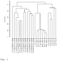
Classifications
-
- C—CHEMISTRY; METALLURGY
- C12—BIOCHEMISTRY; BEER; SPIRITS; WINE; VINEGAR; MICROBIOLOGY; ENZYMOLOGY; MUTATION OR GENETIC ENGINEERING
- C12Q—MEASURING OR TESTING PROCESSES INVOLVING ENZYMES, NUCLEIC ACIDS OR MICROORGANISMS; COMPOSITIONS OR TEST PAPERS THEREFOR; PROCESSES OF PREPARING SUCH COMPOSITIONS; CONDITION-RESPONSIVE CONTROL IN MICROBIOLOGICAL OR ENZYMOLOGICAL PROCESSES
- C12Q1/00—Measuring or testing processes involving enzymes, nucleic acids or microorganisms; Compositions therefor; Processes of preparing such compositions
- C12Q1/68—Measuring or testing processes involving enzymes, nucleic acids or microorganisms; Compositions therefor; Processes of preparing such compositions involving nucleic acids
- C12Q1/6876—Nucleic acid products used in the analysis of nucleic acids, e.g. primers or probes
- C12Q1/6883—Nucleic acid products used in the analysis of nucleic acids, e.g. primers or probes for diseases caused by alterations of genetic material
- C12Q1/6886—Nucleic acid products used in the analysis of nucleic acids, e.g. primers or probes for diseases caused by alterations of genetic material for cancer
-
- C—CHEMISTRY; METALLURGY
- C12—BIOCHEMISTRY; BEER; SPIRITS; WINE; VINEGAR; MICROBIOLOGY; ENZYMOLOGY; MUTATION OR GENETIC ENGINEERING
- C12Q—MEASURING OR TESTING PROCESSES INVOLVING ENZYMES, NUCLEIC ACIDS OR MICROORGANISMS; COMPOSITIONS OR TEST PAPERS THEREFOR; PROCESSES OF PREPARING SUCH COMPOSITIONS; CONDITION-RESPONSIVE CONTROL IN MICROBIOLOGICAL OR ENZYMOLOGICAL PROCESSES
- C12Q2523/00—Reactions characterised by treatment of reaction samples
- C12Q2523/10—Characterised by chemical treatment
- C12Q2523/125—Bisulfite(s)
-
- C—CHEMISTRY; METALLURGY
- C12—BIOCHEMISTRY; BEER; SPIRITS; WINE; VINEGAR; MICROBIOLOGY; ENZYMOLOGY; MUTATION OR GENETIC ENGINEERING
- C12Q—MEASURING OR TESTING PROCESSES INVOLVING ENZYMES, NUCLEIC ACIDS OR MICROORGANISMS; COMPOSITIONS OR TEST PAPERS THEREFOR; PROCESSES OF PREPARING SUCH COMPOSITIONS; CONDITION-RESPONSIVE CONTROL IN MICROBIOLOGICAL OR ENZYMOLOGICAL PROCESSES
- C12Q2600/00—Oligonucleotides characterized by their use
- C12Q2600/112—Disease subtyping, staging or classification
-
- C—CHEMISTRY; METALLURGY
- C12—BIOCHEMISTRY; BEER; SPIRITS; WINE; VINEGAR; MICROBIOLOGY; ENZYMOLOGY; MUTATION OR GENETIC ENGINEERING
- C12Q—MEASURING OR TESTING PROCESSES INVOLVING ENZYMES, NUCLEIC ACIDS OR MICROORGANISMS; COMPOSITIONS OR TEST PAPERS THEREFOR; PROCESSES OF PREPARING SUCH COMPOSITIONS; CONDITION-RESPONSIVE CONTROL IN MICROBIOLOGICAL OR ENZYMOLOGICAL PROCESSES
- C12Q2600/00—Oligonucleotides characterized by their use
- C12Q2600/118—Prognosis of disease development
-
- C—CHEMISTRY; METALLURGY
- C12—BIOCHEMISTRY; BEER; SPIRITS; WINE; VINEGAR; MICROBIOLOGY; ENZYMOLOGY; MUTATION OR GENETIC ENGINEERING
- C12Q—MEASURING OR TESTING PROCESSES INVOLVING ENZYMES, NUCLEIC ACIDS OR MICROORGANISMS; COMPOSITIONS OR TEST PAPERS THEREFOR; PROCESSES OF PREPARING SUCH COMPOSITIONS; CONDITION-RESPONSIVE CONTROL IN MICROBIOLOGICAL OR ENZYMOLOGICAL PROCESSES
- C12Q2600/00—Oligonucleotides characterized by their use
- C12Q2600/154—Methylation markers
-
- C—CHEMISTRY; METALLURGY
- C12—BIOCHEMISTRY; BEER; SPIRITS; WINE; VINEGAR; MICROBIOLOGY; ENZYMOLOGY; MUTATION OR GENETIC ENGINEERING
- C12Q—MEASURING OR TESTING PROCESSES INVOLVING ENZYMES, NUCLEIC ACIDS OR MICROORGANISMS; COMPOSITIONS OR TEST PAPERS THEREFOR; PROCESSES OF PREPARING SUCH COMPOSITIONS; CONDITION-RESPONSIVE CONTROL IN MICROBIOLOGICAL OR ENZYMOLOGICAL PROCESSES
- C12Q2600/00—Oligonucleotides characterized by their use
- C12Q2600/16—Primer sets for multiplex assays
Definitions
- the present invention relates to cancer diagnostic methods and means therefore.
- Neoplasms and cancer are abnormal growths of cells. Cancer cells rapidly reproduce despite restriction of space, nutrients shared by other cells, or signals sent from the body to stop reproduction. Cancer cells are often shaped differently from healthy cells, do not function properly, and can spread into many areas of the body. Abnormal growths of tissue, called tumors, are clusters of cells that are capable of growing and dividing uncontrollably. Tumors can be benign (noncancerous) or malignant (cancerous). Benign tumors tend to grow slowly and do not spread. Malignant tumors can grow rapidly, invade and destroy nearby normal tissues, and spread throughout the body. Malignant cancers can be both locally invasive and metastatic. Locally invasive cancers can invade the tissues surrounding it by sending out "fingers" of cancerous cells into the normal tissue.
- Metastatic cancers can send cells into other tissues in the body, which may be distant from the original tumor. Cancers are classified according to the kind of fluid or tissue from which they originate, or according to the location in the body where they first developed. All of these parameters can effectively have an influence on the cancer characteristics, development and progression and subsequently also cancer treatment. Therefore, reliable methods to classify a cancer state or cancer type, taking diverse parameters into consideration is desired. Since cancer is predominantly a genetic disease, trying to classify cancers by genetic parameters is one extensively studied route.
- RNA-expression studies have been used for screening to identify genetic biomarkers. Over recent years it has been shown that changes in the DNA-methylation pattern of genes could be used as biomarkers for cancer diagnostics. In concordance with the general strategy identifying RNA-expression based biomarkers, the most convenient and prospering approach would start to identify marker candidates by genome-wide screening of methylation changes.
- RNA expression profiling for elucidation of class differences for distinguishing the "good” from the "bad” situation like diseased vs. healthy, or clinical differences between groups of diseased patients.
- RNA based markers are more promising markers and expected to give robust assays for diagnostics.
- Many of clinical markers in oncology are more or less DNA based and are well established, e.g. cytogenetic analyses for diagnosis and classification of different tumor-species. However, most of these markers are not accessible using the cheap and efficient molecular-genetic PCR routine tests.
- RNA-expression changes range over some orders of magnitudes and these changes can be easily measured using genome-wide expression microarrays. These expression arrays are covering the entire translated transcriptome by 20000-45000 probes. Elucidation of DNA changes via microarray techniques requires in general more probes depending on the requested resolution. Even order(s) of magnitude more probes are required than for standard expression profiling to cover the entire 3x10 9 bp human genome.
- DNA-based biomarkers relies on elucidation of the changes in the DNA methylation pattern of (malignant; neoplastic) disease.
- methylation affects exclusively the cytosine residues of CpG dinucleotides, which are clustered in CpG islands.
- CpG islands are often found associated with gene-promoter sequences, present in the 5'-untranslated gene regions and are per default unmethylated.
- an unmethylated CpG island in the associated gene-promoter enables active transcription, but if methylated gene transcription is blocked.
- the DNA methylation pattern is tissue- and clone-specific and almost as stable as the DNA itself. It is also known that DNA-methylation is an early event in tumorigenesis which would be of interest for early and initial diagnosis of disease.
- Microarray for human genome-wide hybridization testings are known, e.g. the Affymetrix Human Genome U133A Array (NCBl Database, Acc. No. GLP96 ).
- Parrella et al., Clinical Cancer Sesearch 10 (16) (2004): 5349-5354 relates to promoter methylation in breast cancer.
- WO 03/052135 A2 realtes to methods and nucleic acids for the analysis of a lung cell proliferation disorder.
- a goal of the present invention is to provide suitable markers for cancer diagnostics.
- the present invention provides a set of nucleic acid primers or hybridization probes being specific for a potentially methylated region of marker genes of the combination HIC1, such as comprising SEQ ID NO: 1207; KL, such as comprising SEQ ID NO: 1163; ESR1, such as comprising SEQ ID NO: 1353; or a set of at least 50% of the markers of HIC1, KL and ESR1, with at least marker KL; wherein the set comprises not more than 20000 probes or primer pairs.
- HIC1 such as comprising SEQ ID NO: 1207
- KL such as comprising SEQ ID NO: 1163
- ESR1 such as comprising SEQ ID NO: 1353
- the set comprises not more than 20000 probes or primer pairs.
- a method of identifying or predicting a disease or tumor type in a sample comprising DNA from a patient comprising obtaining a set of nucleic acid primers or hybridization probes being specific for a potentially methylated region of marker genes of the combination HIC1, such as comprising SEQ ID NO: 1207; KL, such as comprising SEQ ID NO: 1163; ESR1, such as comprising SEQ ID NO: 1353; or a set of at least 50% of the markers of HIC1, KL and ESR1, with at least marker KL; determining the methylation status of the genes in the sample which the members of the set are specific for and comparing the methylation status of the genes with the status of a confirmed cancer type positive and/or negative state, thereby identifying the disease or tumor type in the sample.
- HIC1 such as comprising SEQ ID NO: 1207
- KL such as comprising SEQ ID NO: 1163
- ESR1 such as comprising SEQ ID NO: 1353
- a master set of 359 genetic markers which has been surprisingly found to be highly relevant for aberrant methylation in the diagnosis or prognosis of diseases. It is possible to determine a multitude of marker subsets from this master set which can be used to differentiate between various disease or tumor type.
- the inventive 359 marker genes of table 1 are: NHLH2, MTHFR, PRDM2, MLLT11, S100A9 (control), S100A9, S100A8 (control), S100A8, S100A2, LMNA, DUSP23, LAMC2, PTGS2, MARK1, DUSP10, PARP1, PSEN2, CLIC4, RUNX3, AIM1L, SFN, RPA2, TP73, TP73 (p73), POU3F1, MUTYH, UQCRH, FAF1, TACSTD2, TNFRSF25, DIRAS3, MSH4, GBP2, GBP2, LRRC8C, F3, NANOS1, MGMT, EBF3, DCLRE1C, KIF5B, ZNF22, PGBD3, SRGN, GATA3, PTEN, MMS19, SFRP5, PGR, ATM, DRD2, CADM1, TEAD1, OPCML, CALCA, CTSD, MYOD1, IGF2, BDNF, CDK
- Table 1 lists some marker genes in the double such as for different loci and control sequences. It should be understood that any methylation specific region which is readily known to the skilled man in the art from prior publications or available databases (e.g. PubMeth at www.pubmeth.org) can be used according to the present invention. Of course, double listed genes only need to be represented once in an inventive marker set (or set of probes or primers therefor) but preferably a second marker, such as a control region is included (IDs given in the list above relate to the gene ID (or gene loci ID) given in table 1 of the example section).
- DNA methylation an attractive target for biomarker development, is the fact that cell free methylated DNA can be detected in body-fluids like serum, sputum, and urine from patients with cancerous neoplastic conditions and disease.
- clinical samples have to be available.
- archived (tissue) samples Preferably these materials should fulfill the requirements to obtain intact RNA and DNA, but most archives of clinical samples are storing formalin fixed paraffin embedded (FFPE) tissue blocks. This has been the clinic-pathological routine done over decades, but that fixed samples are if at all only suitable for extraction of low quality of RNA.
- FFPE formalin fixed paraffin embedded
- any such samples can be used for the method of generating an inventive subset, including fixed samples.
- the samples can be of lung, gastric, colorectal, brain, liver, bone, breast, prostate, ovarian, bladder, cervical, pancreas, kidney, thyroid, oesophaegeal, head and neck, neuroblastoma, skin, nasopharyngeal, endometrial, bile duct, oral, multiple myeloma, leukemia, soft tissue sarcoma, anal, gall bladder, endocrine, mesothelioma, wilms tumor, testis, bone, duodenum, neuroendocrine, salivary gland, larynx, choriocarcinoma, cardial, small bowel, eye, germ cell cancer. These cancers can then be subsequently diagnosed by the inventive set (or subsets).
- the present invention provides a multiplexed methylation testing method which 1) outperforms the "classification" success when compared to genomewide screenings via RNA-expression profiling, 2) enables identification of biomarkers for a wide variety of diseases, without the need to prescreen candidate markers on a genomewide scale, and 3) is suitable for minimal invasive testing and 4) is easily scalable.
- the invention presents a targeted multiplexed DNA-methylation test which outperforms genome-scaled approaches (including RNA expression profiling) for disease diagnosis, classification, and prognosis.
- the inventive set of 359 markers enables selection of a subset of markers from this 359 set which is highly characteristic of a given disease or tumor type.
- the disease is a neoplastic condition.
- cancer can be diagnosed with the inventive set or given selective subsets thereof, but a wide range of other diseases detected via the DNA methylation changes of the patient.
- Diseases can be genetic diseases of few, many or all cells in a subject patient (including cancer), or infectious diseases, which lead to altered gene regulation via DNA methylation, e.g. viral, in particular retroviral, infections.
- the disease is a trisomy, such as trisomy 21.
- neoplastic conditions, or tumor types include, without being limited thereto, cancer of different origin such as lung, gastric, colorectal, brain, liver, bone, breast, prostate, ovarian, bladder, cervical, pancreas, kidney, thyroid, oesophaegeal, head and neck, neuroblastoma, skin, nasopharyngeal, endometrial, bile duct, oral, multiple myeloma, leukemia, soft tissue sarcoma, anal, gall bladder, endocrine, mesothelioma, wilms tumor, testis, bone, duodenum, neuroendocrine, salivary gland, larynx, choriocarcinoma, cardial, small bowel, eye, germ cell cancer.
- cancer of different origin such as lung, gastric, colorectal, brain, liver, bone, breast, prostate, ovarian, bladder, cervical, pancreas, kidney, thyroid, oesophaegeal,
- neoplastic conditions or tumor types are e.g. benign (non (or limited) proliferative) or malignant, metastatic or non-metastatic tumors or nodules. It is sometimes possible to differentiate the sample type from which the methylated DNA is isolated, e.g. urine, blood, tissue samples.
- the present invention is suitable to differentiate diseases, in particular neoplastic conditions, or tumor types. Diseases and neoplastic conditions should be understood in general including benign and malignant conditions. According to the present invention benign nodules (being at least the potential onset of malignancy) are included in the definition of a disease. After the development of a malignancy the condition is a preferred disease to be diagnosed by the markers screened for or used according to the present invention.
- the present invention is suitable to distinguish benign and malignant tumors (both being considered a disease according to the present invention). In particular the invention can provide markers (and their diagnostic or prognostic use) distinguishing between a normal healthy state together with a benign state on one hand and malignant states on the other hand.
- the invention is also suitable to differentiate between non-solid cancers including leukemia and healthy states.
- a diagnosis of a disease may include identifying the difference to a normal healthy state, e.g. the absence of any neoplastic nodules or cancerous cells.
- the present invention can also be used for prognosis of such conditions, in particular a prediction of the progression of a disease, such as a neoplastic condition, or tumor type.
- a particularly preferred use of the invention is to perform a diagnosis or prognosis of a metastasising neoplastic disease (distinguished from non-metastasising conditions).
- prognosis should not be understood in an absolute sense, as in a certainty that an individual will develop cancer or a disease or tumor type (including cancer progression), but as an increased risk to develop cancer or the disease or tumor type or of cancer progression.
- Prognosis is also used in the context of predicting disease progression, in particular to predict therapeutic results of a certain therapy of the disease, in particular neoplastic conditions, or tumor types.
- the prognosis of a therapy can e.g. be used to predict a chance of success (i.e. curing a disease) or chance of reducing the severity of the disease to a certain level.
- markers screened for this purpose are preferably derived from sample data of patients treated according to the therapy to be predicted.
- the inventive marker sets may also be used to monitor a patient for the emergence of therapeutic results or positive disease progressions.
- DNA methylation analyses in principle rely either on bisulfite deamination-based methylation detection or on using methylation sensitive restriction enzymes.
- the restriction enzyme-based strategy is used for elucidation of DNA-methylation changes.
- Further methods to determine methylated DNA are e.g. given in EP 1 369 493 A1 or US 6,605,432 .
- Combining restriction digestion and multiplex PCR amplification with a targeted microarray-hybridization is a particular advantageous strategy to perform the inventive methylation test using the inventive marker sets (or subsets).
- a microarray-hybridization step can be used for reading out the PCR results.
- statistical approaches for class comparisons and class prediction can be used. Such statistical methods are known from analysis of RNA-expression derived microarray data.
- an amplification protocol can be used enabling selective amplification of the methylated DNA fraction prior methylation testing. Subjecting these amplicons to the methylation test, it was possible to successfully distinguish DNA from sensitive cases, e.g. distinguishing leukemia (CML) from normal healthy controls. In addition it was possible to distinguish breast-cancer patients from healthy normal controls using DNA from serum by the inventive methylation test upon preamplification. Both examples clearly illustrate that the inventive multiplexed methylation testing can be successfully applied when only limiting amounts of DNA are available. Thus, this principle might be the preferred method for minimal invasive diagnostic testing.
- CML leukemia
- the 359 marker set test is not a genome-wide test and might be used as it is for diagnostic testing, running a subset of markers - comprising the classifier which enables best classification - would be easier for routine applications.
- the test is easily scalable.
- the selected subset of primers/probes could be applied directly to set up of the lower multiplexed test (or single PCR-test). This was confirmed when serum DNA using a classifier for distinguishing healthy females from individuals with breast-tumors (or other specific tumors) was tested. Only the specific primers comprising the gene-classifier obtained from the methylation test were set up together in multiplexed PCR reactions. Data derived upon hybridization of PCR amplicons were in line with initial classification. Thus, correct classification with the down-scaled test using only a subset was possible.
- inventive methylation test is a suitable tool for differentiation and classification of neoplastic disease.
- This assay can be used for diagnostic purposes and for defining biomarkers for clinical relevant issues to improve diagnosis of disease, and to classify patients at risk for disease progression, thereby improving disease treatment and patient management.
- the first step of the disclosed method of generating a subset, step a) of obtaining data of the methylation status preferably comprises determining data of the methylation status, preferably by methylation specific PCR analysis, methylation specific digestion analysis.
- Methylation specific digestion analysis can include either or both of hybridization of suitable probes for detection to non-digested fragments or PCR amplification and detection of non-digested fragments.
- the selection can be made by any (known) classification method to obtain a set of markers with the given diagnostic (or also prognostic) value to categorize a certain disease or tumor type.
- classification methods include class comparisons wherein a specific p-value is selected, e.g. a p-value below 0.1, preferably below 0.08, more preferred below 0.06, in particular preferred below 0.05, below 0.04, below 0.02, most preferred below 0.01.
- the correlated results for each gene b) are rated by their correct correlation to the disease or tumor type positive state, preferably by p-value test or t-value test or F-test.
- Rated (best first, i.e. low p- or t-value) markers are the subsequently selected and added to the subset until a certain diagnostic value is reached, e.g. the herein mentioned at least 70% (or more) correct classification of the disease or tumor type.
- Class Comparison procedures include identification of genes that were differentially methylated among the two classes using a random-variance t-test.
- the random-variance t-test is an improvement over the standard separate t-test as it permits sharing information among genes about within-class variation without assuming that all genes have the same variance (Wright G.W. and Simon R, Bioinformatics 19:2448-2455,2003).
- Genes were considered statistically significant if their p value was less than a certain value, e.g. 0.1 or 0.01.
- a stringent significance threshold can be used to limit the number of false positive findings.
- a global test can also be performed to determine whether the expression profiles differed between the classes by permuting the labels of which arrays corresponded to which classes.
- the p-values can be re-computed and the number of genes significant at the e.g. 0.01 level can be noted. The proportion of the permutations that give at least as many significant genes as with the actual data is then the significance level of the global test. If there are more than 2 classes, then the "F-test" instead of the "t-test” should be used.
- Class Prediction includes the step of specifying a significance level to be used for determining the genes that will be included in the subset. Genes that are differentially methylated between the classes at a univariate parametric significance level less than the specified threshold are included in the set. It doesn't matter whether the specified significance level is small enough to exclude enough false discoveries. In some problems better prediction can be achieved by being more liberal about the gene sets used as features. The sets may be more biologically interpretable and clinically applicable, however, if fewer genes are included. Similar to cross-validation, gene selection is repeated for each training set created in the cross-validation process. That is for the purpose of providing an unbiased estimate of prediction error. The final model and gene set for use with future data is the one resulting from application of the gene selection and classifier fitting to the full dataset.
- Models for utilizing gene methylation profile to predict the class of future samples can also be used. These models may be based on the Compound Covariate Predictor ( Radmacher et al. Journal of Computational Biology 9:505-511, 2002 ), Diagonal Linear Discriminant Analysis ( Dudoit et al. Journal of the American Statistical Association 97:77-87, 2002 ), Nearest Neighbor Classification (also Dudoit et al.), and Support Vector Machines with linear kernel ( Ramaswamy et al. PNAS USA 98:15149-54, 2001 ). The models incorporated genes that were differentially methylated among genes at a given significance level (e.g.
- the prediction error of each model using cross validation is preferably estimated.
- the entire model building process was repeated, including the gene selection process. It may also be evaluated whether the cross-validated error rate estimate for a model was significantly less than one would expect from random prediction.
- the class labels can be randomly permuted and the entire leave-one-out cross-validation process is then repeated.
- the significance level is the proportion of the random permutations that gave a cross-validated error rate no greater than the cross-validated error rate obtained with the real methylation data. About 1000 random permutations may be usually used.
- Another classification method is the greedy-pairs method described by Bo and Jonassen (Genome Biology 3(4):research0017.1-0017.11, 2002 ).
- the greedy-pairs approach starts with ranking all genes based on their individual t-scores on the training set.
- the procedure selects the best ranked gene g i and finds the one other gene g j that together with g i provides the best discrimination using as a measure the distance between centroids of the two classes with regard to the two genes when projected to the diagonal linear discriminant axis.
- These two selected genes are then removed from the gene set and the procedure is repeated on the remaining set until the specified number of genes have been selected. This method attempts to select pairs of genes that work well together to discriminate the classes.
- a binary tree classifier for utilizing gene methylation profile can be used to predict the class of future samples.
- the first node of the tree incorporated a binary classifier that distinguished two subsets of the total set of classes.
- the individual binary classifiers were based on the "Support Vector Machines" incorporating genes that were differentially expressed among genes at the significance level (e.g. 0.01, 0.05 or 0.1) as assessed by the random variance t-test ( Wright G.W. and Simon R. Bioinformatics 19:2448-2455, 2003 ). Classifiers for all possible binary partitions are evaluated and the partition selected was that for which the cross-validated prediction error was minimum. The process is then repeated successively for the two subsets of classes determined by the previous binary split.
- the prediction error of the binary tree classifier can be estimated by cross-validating the entire tree building process. This overall cross-validation included re-selection of the optimal partitions at each node and re-selection of the genes used for each cross-validated training set as described by Simon et al. ( Simon et al. Journal of the National Cancer Institute 95:14-18, 2003 ). 10-fold cross validation in which one-tenth of the samples is withheld can be utilized, a binary tree developed on the remaining 9/10 of the samples, and then class membership is predicted for the 10% of the samples withheld. This is repeated 10 times, each time withholding a different 10% of the samples. The samples are randomly partitioned into 10 test sets( Simon R and Lam A. BRB-ArrayTools User Guide, version 3.2. Biometric Research Branch, National Cancer Institute ).
- the correlated results for each gene b) are rated by their correct correlation to the disease or tumor type positive state, preferably by p-value test. It is also possible to include a step in that the genes are selected d) in order of their rating.
- the subset selection preferably results in a subset with at least 60%, preferably at least 65%, at least 70%, at least 75%, at least 80% or even at least 85%, at least 90%, at least 92%, at least 95%, in particular preferred 100% correct classification of test samples of the disease or tumor type.
- Such levels can be reached by repeating c) steps a) and b) of the inventive method, if necessary.
- marker genes with at least a significance value of at most 0.1, preferably at most 0.8, even more preferred at most 0.6, at most 0.5, at most 0.4, at most 0.2, or more preferred at most 0.01 are selected.
- the at least 50 genes of step a) can be at least 70, preferably at least 90, at least 100, at least 120, at least 140, at least 160, at least 180, at least 190, at least 200, at least 220, at least 240, at least 260, at least 280, at least 300, at least 320, at least 340, at least 350 or all, genes.
- the subset should be small it is preferred that not more than 60, or not more than 40, preferably not more than 30, in particular preferred not more than 20, marker genes are selected in step d) for the subset.
- the methylation status can be determined by any method known in the art including methylation dependent bisulfite deamination (and consequently the identification of mC - methylated C - changes by any known methods, including PCR and hybridization techniques).
- the methylation status is determined by methylation specific PCR analysis, methylation specific digestion analysis and either or both of hybridisation analysis to non-digested or digested fragments or PCR amplification analysis of non-digested fragments.
- the methylation status can also be determined by any probes suitable for determining the methylation status including DNA, RNA, PNA, LNA probes which optionally may further include methylation specific moieties.
- methylation status can be particularly determined by using hybridisation probes or amplification primer (preferably PCR primers) specific for methylated regions of the inventive marker genes. Discrimination between methylated and non-methylated genes, including the determination of the methylation amount or ratio, can be performed by using e.g. either one of these tools.
- the determination using only specific primers aims at specifically amplifying methylated (or in the alternative non-methylated) DNA. This can be facilitated by using (methylation dependent) bisulfite deamination, methylation specific enzymes or by using methylation specific nucleases to digest methylated (or alternatively non-methylated) regions - and consequently only the non-methylated (or alternatively methylated) DNA is obtained.
- a genome chip or simply a gene chip including hybridization probes for all genes of interest such as all 359 marker genes
- all amplification or non-digested products are detected. I.e. discrimination between methylated and non-methylated states as well as gene selection (the inventive set or subset) is before the step of detection on a chip.
- Either set, a set of probes or a set of primers can be used to obtain the relevant methylation data of the genes of the present invention. Of course, both sets can be used.
- the method according to the present invention may be performed by any method suitable for the detection of methylation of the marker genes.
- the determination of the gene methylation is preferably performed with a DNA-chip, real-time PCR, or a combination thereof.
- the DNA chip can be a commercially available general gene chip (also comprising a number of spots for the detection of genes not related to the present method) or a chip specifically designed for the method according to the present invention (which predominantly comprises marker gene detection spots).
- the methylated DNA of the sample is detected by a multiplexed hybridization reaction.
- a methylated DNA is preamplified prior to hybridization, preferably also prior to methylation specific amplification, or digestion.
- the amplification reaction is multiplexed (e.g. multiplex PCR).
- the inventive methods are particularly suitable to detect low amounts of methylated DNA of the inventive marker genes.
- the DNA amount in the sample is below 500ng, below 400ng, below 300ng, below 200ng, below 100ng, below 50ng or even below 25ng.
- the inventive method is particularly suitable to detect low concentrations of methylated DNA of the inventive marker genes.
- the DNA amount in the sample is below 500ng, below 400ng, below 300ng, below 200ng, below 100ng, below 50ng or even below 25ng, per ml sample.
- nucleic acid primers or hybridization probes being specific for a potentially methylated region of at least marker genes selected from one of the following groups
- Each of these marker subsets is particularly suitable to diagnose a certain disease or tumor type or distinguish between a certain disease or tumor type in a methylation specific assay of these genes.
- nucleic acid primers or hybridization probes being specific for a potentially methylated region of marker genes selected from at least 180, preferably at least 200, more preferred at least 220, in particular preferred at least 240, even more preferred at least 260, most preferred at least 280, or even at least 300, preferably at least 320 or at least 340, or at least 360, marker genes of table 1.
- the set may comprise even more primers or hybridization probes not given in table 1.
- the inventive primers or probes may be of any nucleic acid, including RNA, DNA, PNA (peptide nucleic acids), LNA (locked nucleic acids).
- the probes might further comprise methylation specific moieties.
- the present invention provides a (master) set of 360 marker genes, further also specific gene locations by the PCR products of these genes wherein significant methylation can be detected, as well as subsets therefrom with a certain diagnostic value to distinguish specific disease or tumor type.
- the set is optimized for a certain disease or tumor type.
- Cancer types include, without being limited thereto, cancer of different origin such as leukemia, a soft tissue cancer, for example breast cancer, colorectal cancer, head or neck cancer, cervical, prostate, thyroid, brain, eye or pancreatic cancer. Further indicators differentiating between disease or tumor type are e.g. benign (non (or limited) proliferative) or malignant, metastatic or non-metastatic.
- the set can also be optimized for a specific sample type in which the methylated DNA is tested.
- samples include blood, urine, saliva, hair, skin, tissues, in particular tissues of the cancer origin mentioned above, in particular breast or thyroid tissue.
- the sample my be obtained from a patient to be diagnosed.
- the test sample to be used in the method of identifying a subset is from the same type as a sample to be used in the diagnosis.
- probes specific for potentially aberrant methylated regions are provided, which can then be used for the diagnostic method.
- primers suitable for a specific amplification like PCR, of these regions in order to perform a diagnostic test on the methylation state.
- Such probes or primers are provided in the context of a set corresponding to the inventive marker genes or marker gene loci as given in table 1.
- Such a set of primers or probes may have all 359 inventive markers present and can then be used for a multitude of different cancer detection methods. Of course, not all markers would have to be used to diagnose a certain disease or tumor type. It is also possible to use certain subsets (or combinations thereof) with a limited number of marker probes or primers for diagnosis of certain categories of cancer.
- sets of primers or probes comprising primers or probes for any single marker subset or any combination of marker subsets disclosed herein.
- sets of marker genes should be understood to include sets of primer pairs and probes therefore, which can e.g. be provided in a kit.
- normal or benign states including struma nodosa and follicular adenoma
- malign states in particular follicular thyroid carcinoma, papillary thyroid carcinoma.
- benign states including struma nodosa and follicular adenoma
- malign states in particular follicular thyroid carcinoma, papillary thyroid carcinoma and medullary thyroid carcinoma.
- SMAD3, NANOS1, TERT, BCL2, SPARC, SFRP2, MGMT, MYOD1, LAMA1 sets with at least 50%, preferably at least 60%, at least 70%, at least 80% or at least 90% of these markers can be used to diagnose thyroid carcinoma and distinguish between normal or benign states (including struma nodosa and follicular adenoma) together with malign states (in particular follicular thyroid carcinoma and papillary thyroid carcinoma) against medullary thyroid carcinoma.
- benign states including struma nodosa and follicular adenoma
- malign states in particular follicular thyroid carcinoma and papillary thyroid carcinoma
- TJP2, CALCA, PITX2, TFPI2, CDKN2B and sets with at least 50%, preferably at least 60%, at least 70%, at least 80% or at least 90% of these markers can be used to diagnose thyroid carcinoma and distinguish between malign states (in particular follicular thyroid carcinoma and papillary thyroid carcinoma) together with follicular adenoma against struma nodosa.
- Set k ARRDC4, DUSP1, SMAD9, HOXA10, C3, ADRB2, BRCA2, SYK and sets with at least 50%, preferably at least 60%, at least 70%, at least 80% or at least 90% of these markers can be used to diagnose thyroid carcinoma and distinguish between follicular thyroid carcinoma and papillary thyroid carcinoma.
- NTHL1 sets with at least 50%, preferably at least 60%, at least 70%, at least 80% or at least 90% of these markers can be used to diagnose thyroid carcinoma and distinguish between follicular adenoma (benign) and follicular thyroid carcinoma (malign).
- NTHL1 sets with at least 50%, preferably at least 60%, at least 70%, at least 80% or at least 90% of these markers can be used to diagnose thyroid carcinoma and distinguish between follicular adenoma (benign) and follicular thyroid carcinoma (malign).
- Set r IGF2, KCNQ1, SCGB3A1, EFS, BRCA1, ITGA4, H19, PTTG1 and sets with at least 50%, preferably at least 60%, at least 70%, at least 80% or at least 90% of these markers can be used to diagnose breast cancer, distinguish between breast cancer and healthy breast tissue and additionally to distinguish lobular breast carcinoma from ductal breast carcinoma.
- Set t, NCL, XPA, MYOD1, Pitx2 and sets with at least 50%, preferably at least 60%, at least 70%, at least 80% or at least 90% of these markers can be used to diagnose breast cancer, distinguish between breast cancer and healthy breast tissue and additionally to distinguish lobular breast carcinoma from ductal breast carcinoma.
- TIMP1, COL21A1, COL1A2, KL, CDKN2A and sets with at least 50%, preferably at least 60%, at least 70%, at least 80% or at least 90% of these markers can be used to diagnose breast cancer and is additionally particularly suitable to distinguish between metastasising and non-metastasising cancer.
- Set x, TIMP1, COL21A1, COL1A2 and sets with at least 50%, preferably at least 60%, at least 70%, at least 80% or at least 90% of these markers can be used to diagnose breast cancer and is additionally particularly suitable to distinguish between metastasising and non-metastasising cancer.
- Set z, TDRD6, XIST, LZTS1, IRF4 and sets with at least 50%, preferably at least 60%, at least 70%, at least 80% or at least 90% of these markers can be used to diagnose breast cancer and is additionally particularly suitable to distinguish between metastasising and non-metastasising cancer.
- Set aa, TIMP1, COL21A1, COL1A2, KL, CDKN2A and sets with at least 50%, preferably at least 60%, at least 70%, at least 80% or at least 90% of these markers can be used to diagnose cancerous metastases in bone, liver and lung and is additionally particularly suitable to distinguish between metastasising and non-metastasising cancer, in particular from primary breast cancer.
- Set bb, DSP, AR, IGF2, MSX1, SERPINE1, and sets with at least 50%, preferably at least 60%, at least 70%, at least 80% or at least 90% of these markers can be used to diagnose cancerous metastases in bone, liver and lung and is additionally particularly suitable to distinguish between metastasising cancer in liver from metastasising cancer in bone and lung, in particular from primary beast cancer.
- Set cc, FHL1, LMNA, GDNF and sets with at least 50%, preferably at least 60%, at least 70%, at least 80% or at least 90% of these markers can be used to diagnose cancer in bone, liver and lung and to distinguish between metastasising and non-metastasising cancer, in particular to distinguish metastases in liver from metastases in bone, and lung.
- Set dd, FBXW7, GNAS, KRT14 and sets with at least 50%, preferably at least 60%, at least 70%, at least 80% or at least 90% of these markers can be used to diagnose cancer in bone, liver and lung and to distinguish between metastasising and non-metastasising cancer, in particular to distinguish metastases in liver and bone from metastases in lung.
- Set ee, CHFR, AR, RBP1, MSX1, COL21A1, FHL1, RARB and sets with at least 50%, preferably at least 60%, at least 70%, at least 80% or at least 90% of these markers can be used to diagnose cancer in bone and liver and to distinguish between metastasising and non-metastasising cancer, in particular to distinguish metastases in bone from metastases in liver.
- Set gg, FHL1, LMNA, GDNF and sets with at least 50%, preferably at least 60%, at least 70%, at least 80% or at least 90% of these markers can be used to diagnose cancer in bone, liver and lung and to distinguish between metastasising and non-metastasising cancer, in particular to distinguish metastases in liver from metastases in bone, and lung.
- Set hh, FBXW7, GNAS, KRT14 and sets with at least 50%, preferably at least 60%, at least 70%, at least 80% or at least 90% of these markers can be used to diagnose cancer in bone, liver and lung and to distinguish between metastasising and non-metastasising cancer, in particular to distinguish metastases in liver and bone from metastases in lung.
- Set ii, CHFR, AR, RBP1, MSX1, COL21A1, FHL1, RARB and sets with at least 50%, preferably at least 60%, at least 70%, at least 80% or at least 90% of these markers can be used to diagnose cancer in bone and liver and to distinguish between metastasising and non-metastasising cancer, in particular to distinguish metastases in bone from metastases in liver.
- genes common to sets qq), rr), ss), tt) and uu) are used to diagnose trisomy, in particular trisomy 21.
- this set is suitable to distinguish between cancerous cells of breast cancer and normal blood samples.
- This set allows an easy blood test, which may comprise disseminated cancerous cells.
- the present invention furthermore provides additional subsets suitable to detect and diagnose breast cancer by using any at least 3, at least 4, at least 5, at least 6, at least 7, at least 8, at least 9, at least 10 or more markers of the above set ww.
- sub-subsets have been preferably validated according to any methods disclosed therein, in particular any cross-validation methods providing a positive classification for the diagnosis of breast cancer (in comparison to non cancerous samples) as mentioned above for step d), in particular having a p-value of less than 0.1, preferably less than 0.05, even more preferred less than 0.01, in a random-variance t-test.
- HIC1, LAMC2, SPARC, WT1, PITX2, GNA15, KL, HIC1 and sets with at least 50%, preferably at least 60%, at least 70%, at least 80% or at least 90% of these markers can be used to diagnose breast cancer, in particular by using blood samples or samples derived from blood, including serum.
- this set is suitable to distinguish between cancerous cells of breast cancer and normal blood samples.
- This set allows an easy blood test, which may comprise disseminated cancerous cells.
- the set is used in a test together with control markers such as MARK1, PARP1, NHLH2, PSEN2, MTHFR, POS Biotin Control RET, DUSP10.
- Set yy, HIC1, KL, ESR1 and sets with at least 50%, preferably at least 60%, at least 70%, at least 80% or at least 90% of these markers can be used to diagnose breast cancer, in particular by using blood samples or samples derived from blood, including serum.
- this set is suitable to distinguish between cancerous cells of breast cancer and normal blood samples. This set allows an easy blood test, which may comprise disseminated cancerous cells.
- subsets a) to yy in particular sets comprising markers of 2, 3, 4, 5, 6, 7, 8, 9, 10, 11, 12, 13, 14, 15 or more of these subsets, preferably for the same disease or tumor type like breast, lung, liver, bone or thyroid cancer or trisomy 21 or arthritis, preferably complete sets a) to yy).
- the methylation of at least two genes is determined.
- the present invention is provided as an array test system, at least ten, especially at least fifteen genes, are preferred.
- test set-ups for example in microarrays ("gene-chips")
- preferably at least 20, even more preferred at least 30, especially at least 40 genes are provided as test markers.
- these markers or the means to test the markers can be provided in a set of probes or a set of primers, preferably both.
- the set comprises up to 100000, up to 90000, up to 80000, up to 70000, up to 60000 or 50000 probes or primer pairs (set of two primers for one amplification product), preferably up to 40000, up to 35000, up to 30000, up to 25000, up to 20000, up to 15000, up to 10000, up to 7500, up to 5000, up to 3000, up to 2000, up to 1000, up to 750, up to 500, up to 400, up to 300, or even more preferred up to 200 probes or primers of any kind, particular in the case of immobilized probes on a solid surface such as a chip.
- primer pairs and probes are specific for a methylated upstream region of the open reading frame of the marker genes.
- the probes or primers are specific for a methylation in the genetic regions defined by SEQ ID NOs 1081 to 1440, including the adjacent up to 500 base pairs, preferably up to 300, up to 200, up to 100, up to 50 or up to 10 adjacent, corresponding to gene marker IDs 1 to 359 of table 1, respectively.
- probes or primers of the inventive set are specific for the regions and gene loci identified in table 1, last column with reference to the sequence listing, SEQ ID NOs: 1081 to 1440.
- these SEQ IDs correspond to a certain gene, the latter being a member of the inventive sets, in particular of the subsets a) to yy), e.g..
- the set of the present invention comprises probes or primers for at least one gene or gene product of the list according to table 1, wherein at least 10%, at least 15%, at least 20%, at least 25%, at least 30%, at least 35%, at least 40%, at least 45%, at least 50%, at least 55%, at least 60%, at least 65%, at least 70%, at least 75%, at least 80%, at least 85%, at least 90%, at least 95%, especially preferred at least 100%, of the total probes or primers are probes or primers for genes of the list according to table 1.
- the set in particular in the case of a set of hybridization probes, is provided immobilized on a solid surface, preferably a chip or in form of a microarray.
- gene chips using DNA molecules for detection of methylated DNA in the sample
- Such gene chips also allow detection of a large number of nucleic acids.
- the set is provided on a solid surface, in particular a chip, whereon the primers or probes can be immobilized.
- Solid surfaces or chips may be of any material suitable for the immobilization of biomolecules such as the moieties, including glass, modified glass (aldehyde modified) or metal chips.
- the primers or probes can also be provided as such, including lyophilized forms or being in solution, preferably with suitable buffers.
- the probes and primers can of course be provided in a suitable container, e.g. a tube or micro tube.
- a method of identifying a disease or tumor type in a sample comprising DNA from a subject or patient comprising obtaining a set of nucleic acid primers (or primer pairs) or hybridization probes as defined above (comprising each specific subset or combinations thereof), determining the methylation status of the genes in the sample for which the members of the set are specific for and comparing the methylation status of the genes with the status of a confirmed disease or tumor type positive and/or negative state, thereby identifying the disease or tumor type in the sample.
- inventive method has been described above and all preferred embodiments of such methods also apply to the method using the set provided herein.
- inventive marker set including certain disclosed subsets and subsets, which can be identified with the methods disclosed herein, are suitable to distinguish between lung, gastric, colorectal, brain, liver, bone, breast, prostate, ovarian, bladder, cervical, pancreas, kidney, thyroid, oesophaegeal, head and neck, neuroblastoma, skin, nasopharyngeal, endometrial, bile duct, oral, multiple myeloma, leukemia, soft tissue sarcoma, anal, gall bladder, endocrine, mesothelioma, wilms tumor, testis, bone, duodenum, neuroendocrine, salivary gland, larynx, choriocarcinoma, cardial, small bowel, eye, germ cell cancer, cancer from benign conditions, in particular for diagnostic or prognostic uses.
- the markers used e.g. by utilizing primers or probes of the inventive set
- the inventive diagnostic or prognostic method may be used in smaller amounts than e.g. in the set (or kit) or chip as such, which may be designed for more than one fine tuned diagnosis or prognosis.
- the markers used for the diagnostic or prognostic method may be up to 100000, up to 90000, up to 80000, up to 70000, up to 60000 or 50000, preferably up to 40000, up to 35000, up to 30000, up to 25000, up to 20,000, up to 15000, up to 10000, up to 7500, up to 5000, up to 3000, up to 2000, up to 1000, up to 750, up to 500, up to 400, up to 300, up to 200, up to 100, up to 80, or even more preferred up to 60.
- inventive marker set including certain disclosed subsets, which can be identified with the methods disclosed herein, are suitable to distinguish between thyroid cancer from benign thyroid tissue, in particular for diagnostic or prognostic uses.
- inventive marker set including certain disclosed subsets, which can be identified with the methods disclosed herein, are suitable to distinguish between breast cancer from normal tissue and benign breast tumors, in particular for diagnostic or prognostic uses.
- inventive marker set including certain disclosed subsets, which can be identified with the methods disclosed herein, are suitable to distinguish between hereditary from sporadic breast cancer, in particular for diagnostic or prognostic uses.
- inventive marker set including certain disclosed subsets, which can be identified with the methods disclosed herein, are suitable to distinguish between breast cancer responsive to herceptin treatment from likely non-responders, in particular for diagnostic or prognostic uses.
- Table 1 360 master set (with the 359 marker genes and one control) and sequence annotation gene ID Gene Symbol alt. Gene Symbol hybridisation probe (SEQ ID NO:) primer 1 (lp) (SEQ ID NO:) primer 2 (rp) (SEQ ID NO:) PCR product (SEQ ID NO:) 1 NHLH2 NHLH2 1 361 721 1081 2 MTHFR MTHFR 2 362 722 1082 3 PRDM2 RIZ1(PRDM2) 3 363 723 1083 4 MLLT11 MLLT11 4 364 724 1084 5 S100A9 control_S100A9 5 365 725 1085 6 S100A9 S100A9 6 366 726 1086 7 S100A8 S100A8 7 367 727 1087 8 S100A8 control_S100A8 8 368 728 1088 9 S100A2 S100A2 9 369 729 1089 10 LMNA LMNA 10 370 730 1090 11 DUSP23 DUSP23 11 371 731 1091 12 LAMC
- Samples from solid tumors were derived from initial surgical resection of primary tumors. Tumor tissue sections were derived from histopathology and histopathological data as well clinical data were monitored over the time of clinical management of the patients and/or collected from patient reports in the study center. Anonymised data were provided.
- Tissue samples were homogenized in a FASTPREP homogenizer (MP Biomedicals, Eschwege, Germany) in lysis buffer provided with the Qiagen "All Prep" nucleic acid preparation kit (Qiagen, Hilden, Germany). DNA and RNA concentrations were measured on a Nanodrop photometer. RNA quality was controlled using a BioAnalyser (Agilent, Waldbronn, Germany). All conditions were according to manufacturer's recommendations.
- RNA samples derived from breast cancer tissue were analyzed with 44k human whole genome oligo microarrays (Agilent Technologies) .
- RNA expression levels from different samples were analyzed on a single microarray using the Single-Color Low RNA Input Linear Amplification Kit PLUS (Agilent Technologies, Waldbronn, Germany). For each amplification, 200ng of total RNA were employed and amplified samples were prepared for hybridization using the Gene Expression Hybridization Kit (Agilent Technologies). Hybridization was performed over night at 65°C in a rotating hybridization oven (Agilent Technologies). Stringency washes, image aquisition and feature extraction was performed according to the manufacturer's protocol (Agilent Technologies, Waldbronn, Germany).
- the invention assay is a multiplexed assay for DNA methylation testing of up to (or even more than) 360 methylation candidate markers, enabling convenient methylation analyses for tumor-marker definition.
- the test is a combined multiplex-PCR and microarray hybridization technique for multiplexed methylation testing.
- inventive marker genes, PCR primer sequences, hybridization probe sequences and expected PCR products are given in table 1, above.
- methylation analysis is performed via methylation dependent restriction enzyme (MSRE) digestion of 500ng of starting DNA.
- MSRE methylation dependent restriction enzyme
- a combination of several MSREs warrants complete digestion of unmethylated DNA. All targeted DNA regions have been selected in that way that sequences containing multiple MSRE sites are flanked by methylation independent restriction enzyme sites.
- This strategy enables pre-amplification of the methylated DNA fraction before methylation analyses.
- the design and pre-amplification would enable methylation testing on serum, urine, stool etc. when DNA is limiting.
- the methylated DNA fraction is amplified within 16 multiplex PCRs and detected via microarray hybridization. Within these 16 multiplex-PCR reactions 360 different human DNA products can be amplified. From these about 20 amplicons serve as digestion & amplification controls and are either derived from known differentially methylated human DNA regions, or from several regions without any sites of MSREs used in this system.
- the primer set (every reverse primer is biotinylated) used is targeting 347 different sites located in the 5'UTR of 323 gene regions.
- PCR amplicons are pooled and positives are detected using strepavidin-Cy3 via microarray hybridization.
- the melting temperature of CpG rich DNA is very high, primer and probe-design as well as hybridization conditions have been optimized, thus this assay enables unequivocal multiplexed methylation testing of human DNA samples.
- the assay has been designed such that 24 samples can be run in parallel using 384well PCR plates. Handling of many DNA samples in several plates in parallel can be easily performed enabling completion of analyses within 1-2 days.
- the entire procedure provides the user to setup a specific PCR test and subsequent gel-based or hybridization-based testing of selected markers using single primer-pairs or primer-subsets as provided herein or identified by the inventive method from the 360 marker set.
- MSRE digestion of DNA (about 500ng) was performed at 37°C over night in a volume of 30 ⁇ l in 1x Tango-restriction enzyme digestion buffer (MBI Fermentas) using 8 units of each MSREs AciI (New England Biolabs), Hin 6 I and Hpa II (both from MBI Fermentas). Digestions were stopped by heat inactivation (10 min, 75°C) and subjected to PCR amplification.
- MBI Fermentas Tango-restriction enzyme digestion buffer
- Example 8 Microarray hybridization and detection:
- Microarrays with the probes of the 360 marker set are blocked for 30 min in 3M Urea containing 0.1%SDS, at room temperature submerged in a stirred choplin char. After blocking slides are washed in 0.1x SSC/0.2%SDS for 5 min, dipped into water and dried by centrifugation.
- the PCR-amplicon-pool of each sample is mixed with an equal amount of 2x hybridization buffer (7xSSC, 0,6%SDS, 50% formamide), denaturated for 5min at 95°C and held at 70°C until loading an aliqout of 100 ⁇ l onto an array covered by a gasket slide (Agilent). Arrays are hybridized under maximum speed of rotation in an Agilent-hybridization oven for 16h at 52°C.
- 2x hybridization buffer 7xSSC, 0,6%SDS, 50% formamide
- microarray-slides After removal of gasket-slides microarray-slides are washed at room temperature in wash-solution I (1xSSC,0.2%SDS) for 5 min and wash solution II (0.1xSSC,0.2%SDS) for 5 min, and a final wash by dipping the slides 3 times into wash solution III (0.1xSSC), the slides are dried by centrifugation.
- streptavidin-Cy3-conjugate (Caltag Laboratories) is diluted 1:400 in PBST-MP (1xPBS, 0.1% Tween 20; 1% skimmed dry milk powder [Sucofin; Germany]), pipetted onto microarrays covered with a coverslip and incubated 30 min at room temperature in the dark. Then coverslips are washed off from the slides using PBST (1xPBS, 0.1% Tween 20) and then slides are washed in fresh PBST for 5 min, rinsed with water and dried by centrifugation.
- DNA amount is limited. Although the inventive methylation test is performing well with low amounts of DNA (see above), especially minimal invasive testing using cell free DNA from serum, stool, urine, and other body fluids is of diagnostic relevance.
- RCA rolling circle amplification
- the preamplified DNA which is enriched for methylated DNA regions can be directly subjected to flourescent-labelling and the labeled products can be hybridized onto the microarrays using the same conditions as described above for hybridization of PCR products. Then the streptavidin-Cy3 detection step has to be omitted and slides should be scanned directly upon stringency washes and drying the slides. Based on the experimental design for microarray analyses, either single labeled or dual-labeled hybridizations might be generated. From our experiences we successfully used the single label-design for class comparisons. Although the preamplification protocol enables analyses of spurious amounts of DNA, it is also suited for performing genomic methylation screens.
- Hybridizations performed on a chip with probes for the inventive 360 marker genes were scanned using a GenePix 4000A scanner (Molecular Devices, Ismaning, Germany) with a PMT setting to 700V/cm (equal for both wavelengths).
- Raw image data were extracted using GenePix 6.0 software (Molecular Devices, Ismaning, Germany).
- Hybridizations performed on whole genome arrays were scanned using an Agilent DNA microarray scanner and raw image data were extracted using the Agilent Feature Extraction Software (v9.5.3.1) .
- P-values (p) used for feature selection for classification and prediction were based on the univariate significance levels (alpha).
- P-values (p) and mis-classification rate during cross validation (MCR) were given along the result data.
- Example 11 Multiplexed methylation testing outper-forms the "classification" success when compared to genomewide a nd targeted screenings via RNA expression profiling
- Example 12 Multiplexed Methylation Testing enables identification of biomarkers for a wide variety of (neoplastic) diseases
- Table 2 Cross-validation error rates for a fixed tree structure shown below Node Group 1 Classes Group 2 Classes Mis-classification rate (%) 1 Bre Duct, Lob 30 2 Duct Lob 12,5
- Table 3 Composition of classifier (12 genes) - Sorted by p-value: Parametric p-value t-value % CV support Geom mean of intensities in group 1 Geom mean of intensities in group 2 Gene symbol 1 5.82e-05 -5.217 35 1343.4132985 21174.1289278 CHRNA9 2 0.0006143 4.141 95 1599.9071878 824.7000158 RPA2 3 0.0007052 4.079 85 2063.3995618 350.2638104 CPEB4 4 0.0018077 -3.656 25 1072.6323837 2314.8492042 CASP8 5 0.0023551 3.537 95 491.7930375 277.2894179 MSH2 6 0.0024582 3.518 90 2139.495605 407.0543144 ACTB 7 0.0024956 3.511 90 513.7445949 283.0545393 CTCFL 8 0.0025706 -3.498 25 211.6684594 272.432498 TPM2 9 0.00346 -
- Table 4 Composition of classifier (8 genes)- Sorted by p-value: Parametric p-value t-value % CV support Geom mean of intensities in group 1 Geom mean of intensities in group 2 Gene symbol 1 0.0001136 -5.293 30 4902.6591549 26841.3804373 IGF2 2 0.0001792 -5.044 35 2927.8176757 12908.2539897 KCNQ1 3 0.0013497 -3.987 80 651.8291224 3430.8937904 SCGB3A1 4 0.0014207 -3.961 15 3942.4418214 30572.6979298 EFS 5 0.0017484 -3.856 80 562.2147524 10348.9103352 BRCA1 6 0.0032727 3.539 80 981.9197084 533.1453151 ITGA4 7 0.0033906 -3.521 80 290.4681662 518.4060518 H19 8 0.0039029 -3.45 80 249.580905 595.0171743
- Example 13 Classification of diseased versus healthy on minimal amounts of initial DNA samples upon preamplification confirms suitability of the test for diagnosis of neoplastic diseas e
- CML chronic myeloid leukemia
- DNA samples were derived from 8 CML patients at diagnosis, 13 patients within their chronic phase of disease, 3 patients were in the accelerated phase and 3 were blast crisis patients.
- DNA 100ng
- CML-patients and controls were subjected to preamplification outlined in example 6.
- the amplicons derived from the preamplification procedure were directly subjected to the inventive methylation test.
- Table 5 Composition of classifier (36 genes) - Sorted by p-value: Parametric p-value t-value % CV sup port Geom mean of intensities in group 1 Geom mean of intensities in group 2 Fold-change Gene symbol 1 1.1e-06 -5.793 95 1263.9802574 4806.832556 0.2629549 KRT17 2 5.1e-06 -5.303 100 1463.3193409 1920.7523898 0.7618469 IGFBP7 3 9.1e-06 -5.121 100 1511.2662562 2955.7308068 0.5113004 RHOXF1 4 1.72e-05 -4.918 95 20637.941078 46618.878139 0.4426949 CLIC4 5 2.71e-05 4.771 92 14820.0920262 1079.0081152 13.7349217 TP53 6 4.23e-05 -4.627 35 790.262932 1029.0132426 0.7679813 DLX2 7
- the minute amounts of serum DNA (about 10-100ng/ml) derived from patients and controls were subjected to preamplification of the methylated DNA fraction as outlined in the methods. Derived amplicons were subjected to methylation testing using the inventive methylation test.
- Example 14 Thyroid-Cancer-Diagnostics: diagnostic methylation markers for elucidation of nodular thyroid disease
- Table 9 Sorted by p-value of the univariate test.
- Class 1 benign; Class 2: FTC or PTC.
- the first 7 genes are significant at the nominal 0.01 level of the univariate test
- Parametric p-value FDR Geom mean of intensities in class 1
- Geom mean of intensities in class 2 Ratio of geom means Gene symbol 1 0.0004498 0.1632774 15390.3295197 33003.877353 0.4663188 PITX2 2 0.0026894 0.3850159 12719.7083391 7482.6386146 1.6998961 TJP2 3 0.0037242 0.3850159 93.6738594 412.2559349 0.2272226 CD24 4 0.0043096 0.3850159 1279.8569969 3807.3051919 0.3361582 ESR1 5 0.0059178 0.3850159 4082.770499 8974.2819351 0.4549412 TN-FRSF10D 6 0.0063639
- the support vector machine classifier was used for class prediction. There were 5 nodes in the classification tree.
- Table 15 Composition of classifier - Sorted by t-value (Sorted by gene pairs) Class 1: FTA; Class 2: FTC Parametric p-value t-value % CV support Geom mean of intensities in class 1 Geom mean of intensities in class 2 Ratio of geom means Gene symbol 1 0.0007423 -3.67 100 10627.1525329 26519.7470807 0.400726 PITX2 2 0.0034319 -3.121 100 328.8689371 979.8430866 0.3356343 MT3 3 0.0074763 -2.826 100 207.6507576 338.9036062 0.6127133 RPA3 4 0.0088903 -2.758 100 3488.3504846 8791.8091348 0.3967728 TN-FRSF10D 5 0.0332087 -2.21 100 3258.5247053 5669.669334 0.5747292 PTEN 6 0.0567379 -1.965 100 29707.6166671
- Table 16 Composition of classifier - Sorted by t-value: Class 1: FTA; Class 2: FTC Parametric p-value t-value % CV support Geom mean of intensities in class 1 Geom mean of intensities in class 2 Ratio of geom means Gene symbol 1 0.0034319 -3.121 100 328.8689371 979.8430866 0.3356343 MT3 2 0.0074763 -2.826 100 207.6507576 338.9036062 0.6127133 RPA3 3 0.0088903 -2.758 100 3488.3504846 8791.8091348 0.3967728 TNFRSF10D 4 0.0183961 -2.464 100 500.9652123 3828.0253981 0.1308678 HOXA1 5 0.0461873 -2.061 100 382.1794446 1120.8851782 0.3409622 C13orf15 6 0.1254796 -1.567 100 42.1725109 86.8790615 0.48541
- Geom mean of intensities in class 1 Geom mean of intensities in class 2
- Ratio of geom means Gene symbol 1 9e-06 0.003267 ⁇ 1e-07 10627.1525329 41548.9754391 0.2557741
- PITX2 2 0.0011266 0.2044779 0.004 4579.1636841 12649.089991 0.3620153
- PAX8 3 0.0030935 0.3304224 0.005 72.7004543 605.7807776 0.1200112 CD24 4 0.0040729 0.3304224 0.004 14001.0650248 35757.8144724 0.3915526 TP53 5 0.0047034 0.3304224 0.004 1301.897851 5982.0029003 0.2176358
- ESR1 6 0.0067281 0.3304224 0.011 3488.3504846 9170.4514823 0.3803903 TN-FRSF10D 7 0.0080894 0.3304224
- Table 17 Sorted by p-value of the univariate test.
- Class 1 FTC
- Class 2 SN.
- Parametric p-value FDR Permutation p-value
- Geom mean of intensities in class 1 Geom mean of intensities in class 2
- Ratio of geom means Gene symbol 1 0.0001902 0.0690426 0.001 617.2317683 77.9996257 7.9132658 DUSP1 2 0.0009414 0.1708641 0.001 1598.7129792 725.1613837 2.2046306 TFPI2 3 0.002919 0.353199 0.001 7596.7179137 17030.8826818 0.4460554
- TJP2 4 0.0062938 0.3685067 ⁇ 1e-07 30706.7471674 46583.449712 0.6591772 S100A9 5 0.0074514 0.3685067
- Table 18 Genes which discriminate among classes - Sorted by p-value of the univariate test.
- Class 1 PTC
- Class 2 SN.
- the first 16 genes are significant at the nominal 0.05 level of the univariate test
- Parametric p-value FDR Permutation p-value
- Geom mean of intensities in class 1 Geom mean of intensities in class 2
- Ratio of geom means Gene symbol 1 0.0004834 0.1754742 0.001 7364.4053912 17030.8826818 0.4324148 TJP2 2 0.0029185 0.5297078 0.001 850.5244086 196.5548657 4.3271603
- CALCA 3 0.0082748 0.7365111 0.002 527.4006737 305.058238 1.7288524
- PITX2 4 0.0088548 0.7365111 0.011 392.9370376 107.8943318 3.6418691 PITX2 5 0.0110205 0.7365111
- Table 19 Composition of classifier - Sorted by p-value Parametric p-value t-value % CV support Geom mean of intensities in group 1 Geom mean of intensities in group 2 Gene symbol 1 5.82E-05 -5.217 35 1343.413299 21174.12893 CHRNA9 2 0.0006143 4.141 95 1599.907188 824.7000158 RPA2 3 0.0007052 4.079 85 2063.399562 350.2638104 CPEB4 4 0.0018077 -3.656 25 1072.632384 2314.849204 CASP8 5 0.0023551 3.537 95 491.7930375 277.2894179 MSH2 6 0.0024582 3.518 90 2139.495605 407.0543144 ACTB 7 0.0024956 3.511 90 513.7445949 283.0545393 CTCFL 8 0.0025706 -3.498 25 211.6684594 272.432498 TPM2 9 0.00346 -3.364 85 726.4778494
- Table 20 Composition of classifier - Sorted by p-value Parametric p-value t-value % CV sup por t Geom mean of intensities in group 1 Geom mean of intensities in group 2 Gene symbol 1 0.0001136 -5.293 30 4902.659155 26841.38044 IGF2 2 0.0001792 -5.044 35 2927.817676 12908.25399 KCNQ1 3 0.0013497 -3.987 80 651.8291224 3430.89379 SCGB3A1 4 0.0014207 -3.961 15 3942.441821 30572.69793 EFS 5 0.0017484 -3.856 80 562.2147524 10348.91034 BRCA1 6 0.0032727 3.539 80 981.9197084 533.1453151 ITGA4 7 0.0033906 -3.521 80 290.4681662 518.4060518 H19 8 0.0039029 -3.45 80 249.580905 595.0171743 PTTG1
- Table 21 Composition of classifier - Sorted by p-value Parametric p-value t-value % CV support Geom mean of intensities in group 1 Geom mean of intensities in group 2 Gene symbol 1 0.0001584 -4.906 95 1225.466145 2352.054843 KRT17 2 0.0010777 -3.931 80 7629.719066 25785.92874 AQP3 3 0.0011404 -3.863 65 3075.245649 7924.300179 TP53_CGI23_1kb 4 0.001804 -3.657 85 12069.49535 23941.78937 ZNF462 5 0.0020403 -3.602 90 16292.52677 37090.74165 NEUROG1 6 0.002325 -3.543 35 1207.584347 3266.184892 GATA3 7 0.0026734 -3.48 100 1094.800331 2308.87233 MT1A 8 0.0036363 -3.341 95 8217.05373 20320.073
- Table 22 Composition of classifier - Sorted by t-value Parametric p-value t-value % CV support Geom mean of intensities in group 1 Geom mean of intensities in group 2 Gene symbol 1 0.0005054 -7.957 75 535.29315 1030.403284 NCL 2 0.0007899 -4.262 80 2247.443537 4043.09435 XPA 3 0.0013895 3.972 80 14070.47463 4143.73097 MYOD1 4 0.0030063 3.582 45 14993.90774 7184.50258 hy_41-Pitx2
- Table 23 Composition of classifier - Sorted by t -value: Class 1: m; Class 2: nm.
- Parametric p-value t-value % CV support Geom mean of intensities in class 1
- Ratio of geom means Gene symbol 1 0.0046263 --3.152 92 4618.6739493 22964.4785573 0.2011225 SPARC 2 0.0053288 3.394 188 1444.9276316 646.777971 2.2340396 PIWIL4 3 0.0004492 4.555 100 27438.2922955 13506.1416447 2.0315419 SERPINB5 4 0.0001677 4.728 100 783.1275118 498.3793173 1.5713483 TEAD1 5 0.0005684 4.962 100 9591.5219686 1035.8974395 9.2591425 EREG 6 8e-07 8.333 100 7422.5296339 2919.4183827 2.5424686 Z
- Table 24 Composition of classifier:- Sorted by t -value: Class 1: m; Class 2: nm.
- Parametric p-value t-value % CV support Geom mean of intensities in class 1 Geom mean of intensities in class 2 Fold-change Gene symbol 1 0.0001415 -14.234 96 816.9876694 22923.064881 0.0356404 HSD17B4 2 0.0083213 -4.853 79 666.243811 1526.6299468 0.4364147 DSP 3 0.007109 -2.968 29 9716.9365382 49855.3171528 0.1949027 SPARC 4 0.0064307 3.132 67 1836.3963711 722.3717675 2.5421763 KRT17 5 0.002913 3.348 92 59846.4011636 52021.1516203 1.1504244 SRGN 6 0.0032404 3.599 92 5491.6264846 757.0224107 7.2542456 C5orf4
- Parametric p-value t-value % CV support Geom mean of intensities in class 1
- Geom mean of intensities in class 2 Fold-change Gene symbol 1 6.8e-06 -8.508 100 699.2454811 3384.966489 0.2065738
- BCL2A1 2 1.24e-05 -7.956
- 1144.4907092 6068.1628967 0.1886058 SERPINB2 3 4.68e-05 -6.81 100 1612.7663831 6041.3773778 0.2669534
- SERPINE1 4 5.75e-05 -6.644 100 2910.0453562 9519.5437319 0.3056917 ICLIC4 5 0.0002064 -5.671 100 1599.5692432 5858.0250012 0.1023501 IBCL2A1 6 0.0009722 4.6
- Table 28 Composition of classifier - Sorted by t-value: Class 1: m; Class 2: nm.
- Parametric p-value t-value % CV support Geom mean of intensities in class 1
- Geom mean of intensities in class 2 Fold-change Gene symbol 1 2.9e-05 -7.206 100 4505.2826317 10969.8418903 0.4106971 TDRD6 2 20.0001947 -5.713 100 966.693001 4664.3939694 0.2072494 XIST 3 0.0006817 -4.84 17 1735.0546548 10070.7577787 0.1722864 LZTS1 4 0.0009291 -4.635 8 1817.5569529 14443.2023065 0.4090646 IRF4
- Example 17 genelists for prediction of organ of metastases
- Optimal Binary Tree Cross-validation error rates for a fixed tree structure shown below Node Group 1 Classes Group 2 Classes s-classification rate (%) 1 bone, liver, lung nm 17.4 2 bone, lung liver 38.9
- Table 29 Composition of classifier (6 genes) - Sorted by p-value: Parametric p-value t-value % CV support Geom mean of intensities in group 1 Geom mean of intensities in group 2 Gene symbol 1 ⁇ 1e-07 -1e+07 87 885.547163 1982.5964469 TIMP1 2 ⁇ 1e-07 1e+07 100 1453.0368811 685.1212898 COL21A1 3 ⁇ 1e-07 1e+07 91 658.5014611 551.9419886 COL1A2 4 0.0014608 -5.539 87 471.8007783 761.7592596 KL 5 0.0031096 3.87 100 1080.7271447 802.5161476 CDKN2A
- Table 30 Composition of classifier (5 genes) - Sorted by p-value: Parametric p-value t-value % CV support Geom mean of intensities in group 1 Geom mean of intensities in group 2 Gene symbol 1 ⁇ 1e-07 -1e+07 74 994.3330391 2108.8274433 DSP 2 ⁇ 1e-07 -1e+07 74 687.6303563 866.946662 AR 3 ⁇ 1e-07 -1e+07 65 691.236265 940.8722888 IGF2 4 ⁇ 1e-07 1e+07 39 5211.29731 3933.2245813 MSX1 5 0.0072751 3.073 74 1005.7377143 322.8701475 SERPINE1
- Table 31 Composition of classifier (3 genes) - Sorted by p-value: Parametric p-value t-value % CV support Geom mean of intensities in group 1 Geom mean of intensities in group 2 Gene symbol 1 0.0044929 -2.995 97 1134.7396026 4430.9188978 FHL1 2 0.0061218 -2.835 97 1110.6532166 4041.6310709 LMNA 3 0.0093569 -10.265 96 569.1243774 1809.4934445 GDNF
- Table 32 Composition of classifier (3 genes) - Sorted by p-value: Parametric p-value t-value % CV support Geom mean of intensities in group 1 Geom mean of intensities in group 2 Gene symbol 1 0.0042145 -3.015 91 1086.2923057 4114.6963495 FBXW7 2 0.0051555 2.902 91 1004.7376123 747.4623897 GNAS 3 0.0070253 -2.807 91 17268.610511 36023.2972547 KRT14
- Table 33 Composition of classifier (7 genes) - Sorted by p-value: Parametric p-value t-value % CV support Geom mean of intensities in group 1 Geom mean of intensities in group 2 Gene symbol 1 ⁇ 1e-07 1e+07 82 607.7955262 569.3935126 CHFR 2 ⁇ 1e-07 -1e+07 85 1309.6825512 1651.2140652 AR 3 ⁇ 1e-07 1e+07 85 1398.6556505 806.4521355 RBP1 4 ⁇ 1e-07 1e+07 87 960.7559369 725.130163 MSX1 5 7.52e-05 5.181 87 1664.6672496 972.9860114 COL21A1 6 0.0010054 3.572 82 830.8587676 413.1576982 FHL1 7 0.0021561 -4.721 87 756.3671177 1687.904231 RARB
- Table 34 Composition of classifier (6 genes) - Sorted by p-value: Parametric p-value t-value % CV support Geom mean of intensities in group 1 Geom mean of intensities in group 2 Gene symbol 1 ⁇ 1e-07 -1e+07 34 443.8813201 503.5217148 DCLRE1C 2 ⁇ 1e-07 -1e+07 37 725.130163 1044.2751516 MLH1 3 ⁇ 1e-07 1e+07 22 1229.1322892 634.3496625 RARB 4 0.000336 5.966 37 1360.7555882 586.6980829 OGG1 5 0.0014183 -3.605 39 15781.5120063 28485.4843024 SNRPN 6 0.0077342 3.073 37 730.2708489 578.3124727 ITGA4
- GENEFILTERS ON Exclude a gene under any of the following conditions: Less than 20 % of methylation data have at least a 1.5-fold change in either direction from gene's median value
- Table 35 Composition of classifier (3 genes) - Sorted by p-value: Parametric p-value t-value % CV support Geom mean of intensities in group 1 Geom mean of intensities in group 2 Gene symbol 1 0.0044929 -2.995 97 1134.7396026 4430.9188978 FHL1 2 0.0061218 -2.835 97 1110.6532166 4041.6310709 LMNA 3 0.0093569 -10.265 96 569.1243774 1809.4934445 GDNF
- Table 36 Composition of classifier (3 genes) - Sorted by p-value: Parametric p-value t-value % CV support Geom mean of intensities in group 1 Geom mean of intensities in group 2 Gene symbol 1 0.0042145 -3.015 91 1086.2923057 4114.6963495 FBXW7 2 0.0051555 2.902 91 1004.7376123 747.4623897 GNAS 3 0.0070253 -2.807 91 17268.610511 36023.2972547 KRT14
- Table 37 Composition of classifier (7 genes) - Sorted by p-value: Parametric p-value t-value % CV support Geom mean of intensities in group 1 Geom mean of intensities in group 2 Gene symbol 1 ⁇ 1e-07 1e+07 82 607.7955262 569.3935126 CHFR 2 ⁇ 1e-07 -1e+07 85 1309.6825512 1651.2140652 AR 3 ⁇ 1e-07 1e+07 85 1398.6556505 806.4521355 RBP1 4 ⁇ 1e-07 1e+07 87 960.7559369 725.130163 MSX1 5 7.52e-05 5.181 87 1664.6672496 972.9860114 COL21A1 6 0.0010054 3.572 82 830.8587676 413.1576982 FHL1 7 0.0021561 -4.721 87 756.3671177 1687.904231 RARB
- Table 38 Composition of classifier (6 genes) - Sorted by p-value: Parametric p-value t-value % CV support Geom mean of intensities in group 1 Geom mean of intensities in group 2 Gene symbol 1 ⁇ 1e-07 -1e+07 34 443.8813201 503.5217148 DCLRE1C 2 ⁇ 1e-07 -1e+07 37 725.130163 1044.2751516 MLH1 3 ⁇ 1e-07 1e+07 22 1229.1322892 634.3496625 RARB 4 0.000336 5.966 37 1360.7555882 586.6980829 OGG1 5 0.0014183 -3.605 39 15781.5120063 28485.4843024 SNRPN 6 0.0077342 3.073 37 730.2708489 578.3124727 ITGA4
- Example 17.4.1 Classifier defined using the inventive methylation test can be used for correct diagnosis and confirms scalability of the test
- Table 39 Composition of classifier - Sorted by t-value: Class 1: Norm; Class 2: T.
- Parametric p-value t-value % CV support Geom mean of intensities in class 1
- Ratio of geom means Gene symbol 1 0.0009924 -3.795 100 182.6826576 864.2335483 0.2113811 SFN 2 0.0017215 -3.567 100 430.8637112 756.3752883 0.5696428 DIRAS3 3 0.0039912 3.215 33 9327.2785112 1851.017692 5.0390002 HRAS 4 0.0032818 3.297 67 2315.42463 492.0942152 4.7052466 ARMCX2 5 0.0011568 3.732 100 2533.773004 367.7411692 6.8900988 MAGEB2 6 0.0004392 4.13 100 539.5397658 182.6961002 2.953209 GBP2 7 0.0002572 4.349 100 210.
- Table 40 Composition of classifier - Sorted by t-value: Class 1: N; Class 2: T. Parametric p-value t-value % CV support Geom mean intensities in class 1 Geom mean of intensities in class 2 Ratio of geom means Gene symbol 1 0.0028166 -3.404 82 182.0198994 864.2335483 0.2106142 SFN 2 0.0039431 -3.257 18 542.4776162 3257.6193811 0.1665258 BAZ1A 3 0.004628 -3.187 36 424.1334102 756.3752883 0.5607447 DIRAS3 4 0.0040079 3.25 23 173.3967213 103.7366691 1.6715085 CTCFL 5 0.0025889 3.44 86 2890.0918027 492.0942152 5.8730457 ARMCX2 6 0.0025277 3.451 82 514.9868762 182.6961002 2.8188
- Table 41 Composition of classifier - Sorted by t -value: (Sorted by gene pairs): Class 1: control; Class 2: nodule.
- Geom mean of intensities in class 2 Ratio of geom means Gene symbol 1 0.0040902 -3.204 100 408.6274761 723.1907249 0.5650342 DIRAS3 2 0.0102828 -2.806 88 146.3029435 291.7333957 0.5014954 C5AR1 3 0.0187014 -2.539 29 674.9273185 2949.7082217 0.2288116 BAZ1A 4 0.0310794 -2.303 75 211.5738315 692.4423152 0.3055472 SFN 5 0.0153636 2.628 67 909.4898258 414.8518794 2.1923242 ERCC1 6 0.0117751 2.747 58 5872
- Table 42 Composition of classifier - Sorted by t-value: (Sorted by gene pairs): Class 1: control; Class 2: nodule.
- Parametric p-value t-value % CV support Geom mean of intensities in class 1
- Ratio of geom means Gene symbol 1 0.002777 -3.41 100 408.6274761 756.3752883 0.5402444
- DIRAS3 2 0.0046055 -3.189
- 146.3029435 307.9355304 0.475109 C5AR1 3 0.0069521 -3.008 100 211.5738315 864.2335483 0.2448109 SFN 4 0.0119639 -2.764 100 674.9273185 3257.6193811 0.2071842
- BAZ1A 5 0.0145723 -2.674 100 95.6598704 1248.1664664 0.0766403 HIST1H2 6 0.034443 -2.27 100 228.3104891
- Tumor-DNA from patients should be tested by the following markers for elucidating metastases already present, which might be not detectable by routine clinical examination or imaging. patient groups:
- Table 43 Composition of classifier (5 genes) sorted by p-value: Parametric p-value t-value % CV support Geom mean of intensities in group 1 Geom mean of intensities in group 2 Gene symbol 1 0.0002927 -3.92 98 149.7469303 1031.3845804 TFPI2 2 0.0049604 2.952 98 221.1562041 133.2523039 NEUROD2 3 0.0057474 -2.897 94 639.3980244 6450.0516594 DLX2 4 0.006399 -2.857 92 99.6970101 112.7940118 TTC3 5 0.0066379 -2.843 98 99.6970101 109.6402212 TWIST1
- Table 44 Genes used in classifier of risk groups: 15 genes selected by fitting Cox proportional hazards models (alpha equals to 0.05) The coefficients of the fitted Cox proportional hazards model using the principal components from the training dataset is (25.622, -19.237) The percent of variability explained by the first 2 principal components is 75.797 The p-value in the table is testing the hypothesis if expression data is predictive of survival.
- the prognostic index can be computed by the simple formula ⁇ iwi xi - 149.6498 where wi and xi are the weight and logged gene expression for the i-th gene.
- Example 19 Methylation Markers in Non-Tumor / Non-Neoplastic Disease: Trisomy diagnosis
- DNA derived from Cytogen fixed cells of Healthy Controls (5 females ...46XX; 5 males....46XY) and Trisomy-Patients (5 females....47XX+21; 6...males 47XY+21; and single samples with trisomy of chrl3 ... 47XX+13, and trisomy of chr 9...47XX+9 and one blinded sample with trisomy) were used for DNA Methylation testing.
- Optimal Binary Tree BinTree pred. (p ⁇ 0.01) Group 1 Classes Group 2 Classes Mis-classification rate (%) Node 1 46,XX, 46,XY 47,XX, +21, 47,XY, +21 9.5 2 46,XX 46,XY 0.0 3 47,XX, +21 47,XY, +21 18.2
- Leave-one-out cross-validation method was used to compute mis-classification rate.
- Equal class prevalences is used in the Bayesian compound covariate predictor.
- Sensitivity n 11 / n 11 + n 12
- Specificity n 22 / n 21 + n 22
- Negative Predictive Value NPV n 22 / n 12 + n 22
- Sensitivity is the probability for a class A sample to be correctly predicted as class A
- Specificity is the probability for a non class A sample to be correctly predicted as non-A
- PPV is the probability that a sample predicted as class A actually belongs to class A
- NPV is the probability that a sample predicted as non class A actually does not belong to class A.
- Performance of the Compound Covariate Predictor Classifier Class Sensitivity Specificity PPV NPV 46 0.9 0.818 0.818 0.9 47 0.818 0.9 0.9 0.818 Performance of the Diagonal Linear Discriminant Analysis Classifier: Class Sensitivity Specificity PPV NPV 46 0.9 0.909 0.9 0.909 47 0.909 0.909 0.9 Performance of the 1-Nearest Neighbor Classifier: Class Sensitivity Specificity PPV NPV 46 0.9 0.818 0.818 0.9 47 0.818 0.9 0.9 818 Performance of the 3-Nearest Neighbors Classifier: Class Sensitivity Specificity PPV NPV 46 0.9 0.818 0.818 0.9 47 0.818 0.9 0.9 0.818 Performance of the Nearest Centroid Classifier: Class Sensitivity Specificity PPV NPV 46 0.8 0.818 0.818 47 0.818 0.818 0.818 0.818 0.818 0.818 0.818
- the area under the curve is 0.882.
- classification rule used above is different from the class prediction.
- a sample's posterior probability is greater than the threshold, it is predicted as Class 1. Otherwise, it is predicted as Class 2.
- Osteoarthritis also known as degenerative arthritis, degenerative joint disease
- OA also known as degenerative arthritis, degenerative joint disease
- joints [1] including articular cartilage and the subchondral bone next to it.
- Example 21.1 Class Prediction using "grid of alpha levels”: resulted in 100% correct classification
- Leave-one-out cross-validation method was used to compute mis-classification rate.
- Table 56 - Composition of classifier Sorted by t-value Class 1: BrCa; Class 2: norm_blood.
- Parametric p-value t-value % CV support Geom mean of intensities in class 1 Geom mean of intensities in class 2 Fold-change Gene symbol 1 ⁇ 1e-07 -10.192 100 581.9607527 2066.8543933 0.2815683 TP53 2 ⁇ 1e-07 -10.007 100 30.3630111 390.9291534 0.0776688 PTTG1 3 ⁇ 1e-07 -7.138 100 10.2807227 24.2271482 0.4243472 VHL 4 ⁇ 1e-07 -6.863 100 2433.1393149 11571.0250817 0.2102786 TP53 5 ⁇ 1e-07 -6.745 100 113.8926251 213.2313311 0.5341271 S100A2 6 ⁇ 1e-07 -6.3 100 78.7543028 241.62311
- Example 21.2 Class Prediction: ⁇ 8 gene-pairs 100% correct
- Table 57 Composition of classifiers from Class Prediction Analysis - Sorted by gene pairs Pair Parametric p-value t-value % CV support Geom mean of intensities in class 1 Geom mean of intensities in class 2 Fold-change Gene symbol 1 1 ⁇ 1e-07 12.205 100 4609.2719639 104.5974377 44.066777 WT1 2 1 0.065477 1.868 100 17937.6116667 15996.4869627 1.1213469 POS Biotin Control RET 3 2 ⁇ 1e-07 16.193 100 8358.7943596 230.2362876 36.3052864 KL 4 2 0.0233866 2.313 100 18.6345896 14.6005275 1.2762956 blank 5 3 ⁇ 1e-07 17.733 100 22781.9857745 663.1165477 34.3559301 HIC1 6 3 0.9280704 0.091 100 107.9277389 106.2344604 1.0159391 NHLH2 7 4 ⁇ 1e-07 14.974
- Example 21.3 Class Prediction using PAMR ⁇ 100% correct Concept: define minial set of genes using PAM (prediction analysis of microarrays) elucidates 3 genes sufficient for 100% correct diagnostic testing
- Prediction Table a cross-tabulation of true (rows) versus predicted (columns) classes for the PAM fit ( Fig. 4a and b ) BrCa norm_blood BrCa 47 0 norm_blood 0 30
Landscapes
- Chemical & Material Sciences (AREA)
- Life Sciences & Earth Sciences (AREA)
- Health & Medical Sciences (AREA)
- Organic Chemistry (AREA)
- Proteomics, Peptides & Aminoacids (AREA)
- Engineering & Computer Science (AREA)
- Immunology (AREA)
- Pathology (AREA)
- Analytical Chemistry (AREA)
- Zoology (AREA)
- Wood Science & Technology (AREA)
- Genetics & Genomics (AREA)
- Hospice & Palliative Care (AREA)
- Biochemistry (AREA)
- Microbiology (AREA)
- Molecular Biology (AREA)
- Biophysics (AREA)
- Physics & Mathematics (AREA)
- Oncology (AREA)
- Biotechnology (AREA)
- Bioinformatics & Cheminformatics (AREA)
- General Engineering & Computer Science (AREA)
- General Health & Medical Sciences (AREA)
- Measuring Or Testing Involving Enzymes Or Micro-Organisms (AREA)
- Investigating Or Analysing Biological Materials (AREA)
- Apparatus For Radiation Diagnosis (AREA)
Claims (10)
- Set von Nukleinsäure-Primern oder Hybridisationssonden, das für eine potenziell methylierte Region von Markergenen spezifisch ist, umfassend die Kombination HIC1, wie beispielsweise umfassend SEQ ID NR: 1207; KL, wie beispielsweise umfassend SEQ ID NR: 1163; ESR1, wie beispielsweise umfassend SEQ ID NR: 1353;
oder umfassend ein Set von mindestens 50% aus den Markern HIC1, KL und ESR1, mit zumindest dem Marker KL;
wobei das Set nicht mehr als 20000 Sonden oder Primer-Paare umfasst. - Set von Nukleinsäure-Primern, das für eine potenziell methylierte Region von Markergenen spezifisch ist, umfassend die Kombination HIC1, wie beispielsweise umfassend SEQ ID NR: 1207; KL, wie beispielsweise umfassend SEQ ID NR: 1163; ESR1, wie beispielsweise umfassend SEQ ID NR: 1353;
oder umfassend ein Set von mindestens 50% aus den Markern HIC1, KL und ESR1, mit zumindest dem Marker KL. - Set gemäß Ansprüchen 1 oder 2, umfassend Nukleinsäure-Primer oder Hybridisationssonden, das für eine potenziell methylierte Region von Markergenen, ausgewählt aus mindestens 180, vorzugsweise mindestens 200, mehr bevorzugt mindestens 220, besonders bevorzugt mindestens 240, sogar mehr bevorzugt mindestens 260, am meisten bevorzugt mindestens 280 Markergenen aus Tabelle 1, spezifisch ist, umfassend die Marker aus Anspruch 1.
- Set gemäß Anspruch 1 oder 3, dadurch gekennzeichnet, dass das Set nicht mehr als 10000 Sonden oder Primer-Paare umfasst, insbesondere im Falle von an einer festen Oberfläche immobilisierten Sonden.
- Set gemäß einem der Ansprüche 1 bis 4, dadurch gekennzeichnet, dass die Primer-Paare und Sonden für eine methylierte Stromaufwärts-Region des offenen Leserahmens der Markergene spezifisch sind.
- Set gemäß einem der Ansprüche 1 bis 5, dadurch gekennzeichnet, dass die Sonden oder Primer für eine Methylierung in den genetischen Regionen spezifisch sind, die durch SEQ ID NRn. 1081 bis 1440, einschließlich der angrenzenden bis zu 500, vorzugsweise bis zu 300, bis zu 200, bis zu 100, bis zu 50 oder bis zu 10 aufeinander folgenden Basenpaare, die jeweils den Genmarker-IDs 1 bis 359 entsprechen, definiert sind.
- Set gemäß Anspruch 6, dadurch gekennzeichnet, dass die Sonden oder Primer die SEQ ID NRn. 1 bis 1080 aufweisen.
- Verfahren zur Identifizierung oder Vorhersage einer Erkrankung oder eines Tumortyps in einer Probe, die DNA von einem Patienten umfasst, umfassend das Erhalten eines Sets von Nukleinsäure-Primern oder Hybridisationssonden, das für eine potenziell methylierte Region von Markergenen spezifisch ist, umfassend die Kombination HIC1, wie beispielsweise umfassend SEQ ID NR: 1207; KL, wie beispielsweise umfassend SEQ ID NR: 1163; ESR1, wie beispielsweise umfassend SEQ ID NR: 1353; oder umfassend ein Set von mindestens 50% aus den Markern HIC1, KL und ESR1, mit zumindest dem Marker KL; wobei das Set vorzugsweise gemäß Ansprüchen 1 bis 7 definiert ist; Bestimmen des Methylierungsstatus der Gene in der Probe, für die die Mitglieder des Sets spezifisch sind, und Vergleichen des Methylierungsstatus der Gene mit dem Status eines bestätigten Krebstyp-positiven und/oder -negativen Status, dadurch Identifizieren der Erkrankung oder des Tumortyps in der Probe.
- Verfahren gemäß Anspruch 8, dadurch gekennzeichnet, dass der Methylierungsstatus durch methylierungsspezifische PCR-Analyse, methylierungsspezifische Verdauanalyse und einem oder beidem von einer Hybridisationsanalyse an nicht-verdaute oder verdaute Fragmente oder einer PCR-Amplifikationsanalyse von nicht-verdauten oder verdauten Fragmenten bestimmt wird.
- Verfahren gemäß Ansprüchen 8 oder 9, dadurch gekennzeichnet, dass die Erkrankung oder der Tumortyp ausgewählt ist aus Brust-, Lungen-, Magen-, Dickdarm-, Gehirn-, Leber-, Knochen-, Prostata-, Eierstock-, Blasen-, Gebärmutterhals-, Bauchspeicheldrüsen-, Nieren-, Schilddrüsen-, Speiseröhren-, Kopf-und-Nacken-, Neuroblastom, Haut-, Nasenrachen-, Endometrium-, Gallengangs-, Mund-, multiplem Myelom, Leukämie, Weichgewebe-Sarkom, Anus-, Gallenblasen-, endokrinem Drüsen-, Mesotheliom, Wilms-Tumor, Hoden-, Knochen-, Zwölffingerdarm-, neuroendokrinem Drüsen-, Speicheldrüsen-, Kehlkopf-, Choriokarzinom, Kardial-, Dünndarm-, Augen-, Keimzellenkrebs, vorzugsweise Schilddrüsen- oder Brustkrebs oder einer Trisomie, insbesondere Trisomie 21.
Applications Claiming Priority (3)
| Application Number | Priority Date | Filing Date | Title |
|---|---|---|---|
| EP09450020A EP2233590A1 (de) | 2009-01-28 | 2009-01-28 | Methylierungsassay |
| EP10701378.1A EP2391729B1 (de) | 2009-01-28 | 2010-01-28 | Methylierungsassay |
| PCT/EP2010/051033 WO2010086389A1 (en) | 2009-01-28 | 2010-01-28 | Methylation assay |
Related Parent Applications (1)
| Application Number | Title | Priority Date | Filing Date |
|---|---|---|---|
| EP10701378.1A Division EP2391729B1 (de) | 2009-01-28 | 2010-01-28 | Methylierungsassay |
Publications (3)
| Publication Number | Publication Date |
|---|---|
| EP3124624A2 EP3124624A2 (de) | 2017-02-01 |
| EP3124624A3 EP3124624A3 (de) | 2017-04-12 |
| EP3124624B1 true EP3124624B1 (de) | 2019-11-20 |
Family
ID=40578874
Family Applications (4)
| Application Number | Title | Priority Date | Filing Date |
|---|---|---|---|
| EP09450020A Withdrawn EP2233590A1 (de) | 2009-01-28 | 2009-01-28 | Methylierungsassay |
| EP10701377A Withdrawn EP2391728A1 (de) | 2009-01-28 | 2010-01-28 | Lungenkrebs-methylierungsmarker |
| EP16188579.3A Active EP3124624B1 (de) | 2009-01-28 | 2010-01-28 | Methylierungsassay |
| EP10701378.1A Active EP2391729B1 (de) | 2009-01-28 | 2010-01-28 | Methylierungsassay |
Family Applications Before (2)
| Application Number | Title | Priority Date | Filing Date |
|---|---|---|---|
| EP09450020A Withdrawn EP2233590A1 (de) | 2009-01-28 | 2009-01-28 | Methylierungsassay |
| EP10701377A Withdrawn EP2391728A1 (de) | 2009-01-28 | 2010-01-28 | Lungenkrebs-methylierungsmarker |
Family Applications After (1)
| Application Number | Title | Priority Date | Filing Date |
|---|---|---|---|
| EP10701378.1A Active EP2391729B1 (de) | 2009-01-28 | 2010-01-28 | Methylierungsassay |
Country Status (4)
| Country | Link |
|---|---|
| US (3) | US20110287967A1 (de) |
| EP (4) | EP2233590A1 (de) |
| CA (2) | CA2750978A1 (de) |
| WO (2) | WO2010086388A1 (de) |
Families Citing this family (60)
| Publication number | Priority date | Publication date | Assignee | Title |
|---|---|---|---|---|
| EP2233590A1 (de) | 2009-01-28 | 2010-09-29 | AIT Austrian Institute of Technology GmbH | Methylierungsassay |
| EP2396408B1 (de) | 2009-02-12 | 2017-09-20 | CuRNA, Inc. | Behandlung von erkrankungen im zusammenhang mit dem neurotrophen faktor gdnf mittels hemmung des natürlichen antisense-transkripts gegen gdnf |
| ES2633111T3 (es) | 2010-03-26 | 2017-09-19 | Hongzhi Zou | Métodos y materiales para detectar neoplasia colorrectal |
| SG186940A1 (en) | 2010-07-27 | 2013-02-28 | Genomic Health Inc | Method for using gene expression to determine prognosis of prostate cancer |
| JP5693895B2 (ja) * | 2010-08-26 | 2015-04-01 | 公益財団法人ヒューマンサイエンス振興財団 | 子宮頸がん検査用マーカー及び子宮頸がんの検査方法 |
| WO2012031329A1 (en) * | 2010-09-10 | 2012-03-15 | Murdoch Childrens Research Institute | Assay for detection and monitoring of cancer |
| CA2815209A1 (en) | 2010-10-19 | 2012-04-26 | Oslo Universitetssykehus Hf | Methods and biomarkers for detection of bladder cancer |
| US10316361B2 (en) | 2011-07-15 | 2019-06-11 | The John Hopkins University | Genome-wide methylation analysis and use to identify genes specific to breast cancer hormone receptor status and risk of recurrence |
| ITPD20110324A1 (it) * | 2011-10-12 | 2013-04-13 | Innovation Factory S C A R L | Metodo per la rilevazione di galfa15 come marcatore tumorale nel carcinoma pancreatico |
| WO2013120222A1 (zh) | 2012-02-13 | 2013-08-22 | 北京市肿瘤防治研究所 | 体外估测肿瘤发生、转移或生存时间长短的方法及所用人工核苷酸 |
| EP2636752A1 (de) * | 2012-03-06 | 2013-09-11 | Universiteit Maastricht | In-vitro-Verfahren zur Bestimmung des Krankheitsausgangs bei Lungenkarzinoiden |
| CN102787174B (zh) * | 2012-09-06 | 2013-08-07 | 南京大学 | 胃肠肿瘤发病相关抑癌基因表观突变检测试剂盒及其用途 |
| PL2904398T3 (pl) * | 2012-10-02 | 2018-11-30 | Sphingotec Gmbh | Sposób przewidywania ryzyka zachorowania na raka u osobnika żeńskiego |
| WO2014062218A1 (en) * | 2012-10-16 | 2014-04-24 | University Of Southern California | Colorectal cancer dna methylation markers |
| AU2014216355A1 (en) | 2013-02-12 | 2015-09-03 | Texas Tech University System | Composition and method for diagnosis and immunotherapy of lung cancer |
| US9506116B2 (en) | 2013-03-14 | 2016-11-29 | Mayo Foundation For Medical Education And Research | Detecting neoplasm |
| EP2886659A1 (de) * | 2013-12-20 | 2015-06-24 | AIT Austrian Institute of Technology GmbH | Genmethylierung basierend auf der Diagnose von Kolorektalkrebs |
| EP4234722A3 (de) | 2014-03-31 | 2023-09-20 | Mayo Foundation for Medical Education and Research | Detektion von kolorektalem neoplasma |
| US10184154B2 (en) | 2014-09-26 | 2019-01-22 | Mayo Foundation For Medical Education And Research | Detecting cholangiocarcinoma |
| US10030272B2 (en) | 2015-02-27 | 2018-07-24 | Mayo Foundation For Medical Education And Research | Detecting gastrointestinal neoplasms |
| CN107532124B (zh) | 2015-03-27 | 2022-08-09 | 精密科学公司 | 检测食管疾病 |
| EP3344784A4 (de) | 2015-08-31 | 2019-07-10 | Mayo Foundation for Medical Education and Research | Nachweis von gastrischen neoplasmen |
| CN105154542B (zh) * | 2015-09-01 | 2018-04-17 | 杭州源清生物科技有限公司 | 一组用于肺癌分子分型的基因及其应用 |
| CN108350485A (zh) | 2015-10-30 | 2018-07-31 | 精密科学发展有限责任公司 | 血浆dna的多重扩增检测测定以及分离和检测 |
| WO2017136482A1 (en) | 2016-02-01 | 2017-08-10 | The Board Of Regents Of The University Of Nebraska | Method of identifying important methylome features and use thereof |
| US10370726B2 (en) | 2016-04-14 | 2019-08-06 | Mayo Foundation For Medical Education And Research | Detecting colorectal neoplasia |
| CN116024334A (zh) | 2016-04-14 | 2023-04-28 | 梅约医学教育与研究基金会 | 检测DMR试剂筛选IPMN-HGD、PanIN-3或PDAC的用途及试剂盒 |
| AU2017260630B2 (en) * | 2016-05-05 | 2023-07-13 | Exact Sciences Corporation | Detection of lung neoplasia by analysis of methylated DNA |
| US11685955B2 (en) | 2016-05-16 | 2023-06-27 | Dimo Dietrich | Method for predicting response of patients with malignant diseases to immunotherapy |
| DE102016005947B3 (de) | 2016-05-16 | 2017-06-08 | Dimo Dietrich | Verfahren zur Abschätzung der Prognose und zur Prädiktion des Ansprechens auf eine Immuntherapie von Patienten mit malignen Erkrankungen |
| WO2017210662A1 (en) * | 2016-06-03 | 2017-12-07 | Castle Biosciences, Inc. | Methods for predicting risk of recurrence and/or metastasis in soft tissue sarcoma |
| JP6876304B2 (ja) * | 2016-06-10 | 2021-05-26 | 国立研究開発法人国立がん研究センター | 癌に対する薬物療法の効果の予測方法 |
| US10093986B2 (en) | 2016-07-06 | 2018-10-09 | Youhealth Biotech, Limited | Leukemia methylation markers and uses thereof |
| EP3481403B1 (de) * | 2016-07-06 | 2022-02-09 | Youhealth Biotech, Limited | Feste tumormethylierungsmarker und verwendungen davon |
| EP3481941A4 (de) * | 2016-07-06 | 2020-04-15 | Youhealth Biotech, Limited | Methylierungsmarker für brust- und ovarialkrebs und deren verwendung |
| WO2018009709A1 (en) * | 2016-07-06 | 2018-01-11 | Youhealth Biotech, Limited | Lung cancer methylation markers and uses thereof |
| EP3481951A4 (de) * | 2016-07-06 | 2020-08-05 | Youhealth Biotech, Limited | Methylierungsmarker für kolonkarzinom und verwendungen davon |
| US20190300965A1 (en) * | 2016-07-06 | 2019-10-03 | Youhealth Biotech, Limited | Liver cancer methylation markers and uses thereof |
| CN106282347A (zh) * | 2016-08-17 | 2017-01-04 | 中南大学 | HoxC11作为生物标志物在制备肺腺癌的预诊断试剂中的应用 |
| ES2956257T3 (es) * | 2016-09-02 | 2023-12-18 | Mayo Found Medical Education & Res | Detección de carcinoma hepatocelular |
| WO2018045509A1 (zh) * | 2016-09-07 | 2018-03-15 | 武汉华大吉诺因生物科技有限公司 | 多肽及其应用 |
| WO2018045510A1 (zh) | 2016-09-07 | 2018-03-15 | 武汉华大吉诺因生物科技有限公司 | 多肽及其应用 |
| AU2016432602A1 (en) * | 2016-12-14 | 2019-07-04 | Shanghaitech University | Compositions and methods for treating cancer by inhibiting PIWIL4 |
| CN106755466A (zh) * | 2017-01-12 | 2017-05-31 | 宁夏医科大学 | 一种特异性dna甲基化谱其建立方法及应用 |
| US10934592B2 (en) | 2017-02-28 | 2021-03-02 | Mayo Foundation For Medical Education And Research | Detecting prostate cancer |
| WO2018174860A1 (en) * | 2017-03-21 | 2018-09-27 | Mprobe Inc. | Methods and compositions for detection early stage lung adenocarcinoma with rnaseq expression profiling |
| WO2018214249A1 (zh) * | 2017-05-22 | 2018-11-29 | 立森印迹诊断技术(无锡)有限公司 | 一种印记基因分级模型及其组成的系统和应用 |
| CN109528749B (zh) * | 2017-09-22 | 2023-04-28 | 上海交通大学医学院附属瑞金医院 | 长链非编码rna-h19在制备治疗垂体瘤药物中的用途 |
| KR20200105661A (ko) | 2017-11-30 | 2020-09-08 | 메이오 파운데이션 포 메디칼 에쥬케이션 앤드 리써치 | 유방암 검출방법 |
| EP3768694A4 (de) | 2018-03-22 | 2021-12-29 | Ionis Pharmaceuticals, Inc. | Verfahren zur modulierung der fmr1-expression |
| JP2021524758A (ja) * | 2018-05-18 | 2021-09-16 | リセン インプリンティング ダイアグノステックス,インク.Lisen Imprinting Diagnostics,Inc. | 生検細胞サンプルによるがんを診断する方法 |
| CN110714075B (zh) * | 2018-07-13 | 2024-05-03 | 立森印迹诊断技术(无锡)有限公司 | 一种用于检测肺肿瘤良恶性程度的分级模型及其应用 |
| CA3111887A1 (en) | 2018-09-27 | 2020-04-02 | Grail, Inc. | Methylation markers and targeted methylation probe panel |
| KR102637032B1 (ko) * | 2020-01-28 | 2024-02-15 | 주식회사 젠큐릭스 | 특정 유전자의 CpG 메틸화 변화를 이용한 방광암 진단용 조성물 및 이의 용도 |
| CN114075603A (zh) * | 2020-09-01 | 2022-02-22 | 闫池 | 一种确定AJUBA基因启动子CpG岛差异甲基化位点的方法 |
| CN112301132A (zh) * | 2020-11-18 | 2021-02-02 | 中国医学科学院肿瘤医院 | 一种多基因联合检测癌症的试剂盒及其检测方法 |
| CN113528667B (zh) * | 2021-07-20 | 2022-09-20 | 中国科学院上海营养与健康研究所 | 一种骨巨细胞瘤的诊断方法 |
| KR102404750B1 (ko) * | 2021-10-14 | 2022-06-07 | 주식회사 엔도믹스 | 세포 유리 dna를 이용한 대장암 진단을 위한 메틸화 마커 유전자 및 이의 용도 |
| CN114438188A (zh) * | 2021-11-29 | 2022-05-06 | 中国辐射防护研究院 | 高甲基化CDK2AP1基因作为α辐射损伤预测的分子标志物的用途 |
| CN114395623B (zh) * | 2021-12-16 | 2024-03-08 | 上海市杨浦区中心医院(同济大学附属杨浦医院) | 一种基因甲基化检测引物组合物及其试剂盒和应用 |
Family Cites Families (13)
| Publication number | Priority date | Publication date | Assignee | Title |
|---|---|---|---|---|
| US6582908B2 (en) * | 1990-12-06 | 2003-06-24 | Affymetrix, Inc. | Oligonucleotides |
| US6605432B1 (en) | 1999-02-05 | 2003-08-12 | Curators Of The University Of Missouri | High-throughput methods for detecting DNA methylation |
| DE10161625A1 (de) | 2001-12-14 | 2003-07-10 | Epigenomics Ag | Verfahren und Nukleinsäuren für die Analyse einer Lungenzell-Zellteilungsstörung |
| ES2272636T3 (es) | 2002-06-05 | 2007-05-01 | Epigenomics Ag | Procedimiento para la determinacion cuantitativa del grado de metilac ion de citosinas en posiciones cpg. |
| CA2526862A1 (en) * | 2003-05-30 | 2005-03-31 | Temple University - Of The Commonwealth System Of Higher Education | Methods of diagnosing, prognosing and treating breast cancer |
| EP1869212A4 (de) * | 2005-02-01 | 2009-09-23 | Wayne John Cancer Inst | Verwendung von id4 zur diagnose und behandlung von krebs |
| EP1869222A4 (de) * | 2005-04-15 | 2010-01-20 | Oncomethylome Sciences S A | Methylierungsmarker zur diagnose und behandlung von krebs |
| WO2007008693A2 (en) * | 2005-07-09 | 2007-01-18 | Lovelace Respiratory Research Institute | Gene methylation as a biomarker in sputum |
| WO2007032748A1 (en) * | 2005-09-15 | 2007-03-22 | Agency For Science, Technology & Research | Method for detecting dna methylation |
| US7829283B2 (en) * | 2006-03-29 | 2010-11-09 | John Wayne Cancer Institute | Methylation of estrogen receptor alpha and uses thereof |
| EP2442108B1 (de) * | 2006-07-14 | 2016-11-16 | The Regents of The University of California | Krebsbiomarker und Anwendungsverfahren dafür |
| US8911937B2 (en) * | 2007-07-19 | 2014-12-16 | Brainreader Aps | Method for detecting methylation status by using methylation-independent primers |
| EP2233590A1 (de) | 2009-01-28 | 2010-09-29 | AIT Austrian Institute of Technology GmbH | Methylierungsassay |
-
2009
- 2009-01-28 EP EP09450020A patent/EP2233590A1/de not_active Withdrawn
-
2010
- 2010-01-28 CA CA2750978A patent/CA2750978A1/en not_active Abandoned
- 2010-01-28 US US13/146,901 patent/US20110287967A1/en not_active Abandoned
- 2010-01-28 US US13/146,903 patent/US10718026B2/en active Active
- 2010-01-28 EP EP10701377A patent/EP2391728A1/de not_active Withdrawn
- 2010-01-28 CA CA2750979A patent/CA2750979A1/en not_active Abandoned
- 2010-01-28 EP EP16188579.3A patent/EP3124624B1/de active Active
- 2010-01-28 WO PCT/EP2010/051032 patent/WO2010086388A1/en active Application Filing
- 2010-01-28 EP EP10701378.1A patent/EP2391729B1/de active Active
- 2010-01-28 WO PCT/EP2010/051033 patent/WO2010086389A1/en active Application Filing
-
2016
- 2016-04-12 US US15/096,848 patent/US20160281175A1/en not_active Abandoned
Non-Patent Citations (1)
| Title |
|---|
| None * |
Also Published As
| Publication number | Publication date |
|---|---|
| US20110287967A1 (en) | 2011-11-24 |
| CA2750978A1 (en) | 2010-08-05 |
| EP3124624A3 (de) | 2017-04-12 |
| EP2233590A1 (de) | 2010-09-29 |
| EP3124624A2 (de) | 2017-02-01 |
| EP2391729A1 (de) | 2011-12-07 |
| US20160281175A1 (en) | 2016-09-29 |
| WO2010086389A1 (en) | 2010-08-05 |
| WO2010086388A1 (en) | 2010-08-05 |
| CA2750979A1 (en) | 2010-08-05 |
| EP2391729B1 (de) | 2016-09-14 |
| US10718026B2 (en) | 2020-07-21 |
| US20110287968A1 (en) | 2011-11-24 |
| EP2391728A1 (de) | 2011-12-07 |
Similar Documents
| Publication | Publication Date | Title |
|---|---|---|
| EP3124624B1 (de) | Methylierungsassay | |
| CN109153993B (zh) | 检测胰腺高度异型增生 | |
| DK3034628T3 (en) | DIAGNOSTICATION OF CANCER USING METHYLATION MARKER | |
| US8110361B2 (en) | DNA methylation markers associated with the CpG island methylator phenotype (CIMP) in human colorectal cancer | |
| US8150627B2 (en) | Methods and compositions for diagnosing lung cancer with specific DNA methylation patterns | |
| EP3083993B1 (de) | Auf genmethylierung basierende diagnose von kolorektalkarzinom | |
| EP2250287B1 (de) | Nachweis und prognose von lungenkrebs | |
| US20200172963A1 (en) | Dna methylation in colorectal and breast cancer diagnostic methods | |
| US20080221056A1 (en) | Early Detection and Prognosis of Colon Cancers | |
| CN116064795A (zh) | 确定差异甲基化区域的甲基化状态的方法和试剂盒 | |
| EP1996729A2 (de) | MOLEKULARANORDNUNG ZUR VORHERSAGE VON DUKESý B DARMKREBSREZIDIV | |
| JP2005204651A (ja) | 乳房細胞増殖障害の患者の治療応答及び/又は生存期間の予測のための予後診断マーカー | |
| CN113557308A (zh) | 检测子宫内膜癌 | |
| CN114729399A (zh) | 检测卵巢癌 | |
| JP2023524224A (ja) | 大腸癌の早期検出、治療応答性および予後の予測のための方法 | |
| US20080085867A1 (en) | Early detection and prognosis of colon cancers | |
| KR101504069B1 (ko) | 담관선암 검출 또는 진단용 메틸화 마커 및 방법 | |
| WO2023168345A2 (en) | Methylated dna markers and assays thereof for use in detecting colorectal cancer | |
| JP2005224238A (ja) | 乳房細胞増殖障害の患者の治療応答及び/又は生存期間の予測のための予後診断マーカー |
Legal Events
| Date | Code | Title | Description |
|---|---|---|---|
| PUAI | Public reference made under article 153(3) epc to a published international application that has entered the european phase |
Free format text: ORIGINAL CODE: 0009012 |
|
| STAA | Information on the status of an ep patent application or granted ep patent |
Free format text: STATUS: THE APPLICATION HAS BEEN PUBLISHED |
|
| AC | Divisional application: reference to earlier application |
Ref document number: 2391729 Country of ref document: EP Kind code of ref document: P |
|
| AK | Designated contracting states |
Kind code of ref document: A2 Designated state(s): AT BE BG CH CY CZ DE DK EE ES FI FR GB GR HR HU IE IS IT LI LT LU LV MC MK MT NL NO PL PT RO SE SI SK SM TR |
|
| PUAL | Search report despatched |
Free format text: ORIGINAL CODE: 0009013 |
|
| AK | Designated contracting states |
Kind code of ref document: A3 Designated state(s): AT BE BG CH CY CZ DE DK EE ES FI FR GB GR HR HU IE IS IT LI LT LU LV MC MK MT NL NO PL PT RO SE SI SK SM TR |
|
| RIC1 | Information provided on ipc code assigned before grant |
Ipc: C12Q 1/68 20060101AFI20170309BHEP |
|
| RAP1 | Party data changed (applicant data changed or rights of an application transferred) |
Owner name: AIT AUSTRIAN INSTITUTE OF TECHNOLOGY GMBH |
|
| STAA | Information on the status of an ep patent application or granted ep patent |
Free format text: STATUS: REQUEST FOR EXAMINATION WAS MADE |
|
| STAA | Information on the status of an ep patent application or granted ep patent |
Free format text: STATUS: EXAMINATION IS IN PROGRESS |
|
| 17P | Request for examination filed |
Effective date: 20171012 |
|
| RBV | Designated contracting states (corrected) |
Designated state(s): AT BE BG CH CY CZ DE DK EE ES FI FR GB GR HR HU IE IS IT LI LT LU LV MC MK MT NL NO PL PT RO SE SI SK SM TR |
|
| 17Q | First examination report despatched |
Effective date: 20171108 |
|
| GRAP | Despatch of communication of intention to grant a patent |
Free format text: ORIGINAL CODE: EPIDOSNIGR1 |
|
| STAA | Information on the status of an ep patent application or granted ep patent |
Free format text: STATUS: GRANT OF PATENT IS INTENDED |
|
| RAP1 | Party data changed (applicant data changed or rights of an application transferred) |
Owner name: AIT AUSTRIAN INSTITUTE OF TECHNOLOGY GMBH |
|
| INTG | Intention to grant announced |
Effective date: 20181010 |
|
| GRAJ | Information related to disapproval of communication of intention to grant by the applicant or resumption of examination proceedings by the epo deleted |
Free format text: ORIGINAL CODE: EPIDOSDIGR1 |
|
| STAA | Information on the status of an ep patent application or granted ep patent |
Free format text: STATUS: EXAMINATION IS IN PROGRESS |
|
| GRAP | Despatch of communication of intention to grant a patent |
Free format text: ORIGINAL CODE: EPIDOSNIGR1 |
|
| STAA | Information on the status of an ep patent application or granted ep patent |
Free format text: STATUS: GRANT OF PATENT IS INTENDED |
|
| INTC | Intention to grant announced (deleted) | ||
| INTG | Intention to grant announced |
Effective date: 20190312 |
|
| GRAS | Grant fee paid |
Free format text: ORIGINAL CODE: EPIDOSNIGR3 |
|
| GRAJ | Information related to disapproval of communication of intention to grant by the applicant or resumption of examination proceedings by the epo deleted |
Free format text: ORIGINAL CODE: EPIDOSDIGR1 |
|
| GRAL | Information related to payment of fee for publishing/printing deleted |
Free format text: ORIGINAL CODE: EPIDOSDIGR3 |
|
| STAA | Information on the status of an ep patent application or granted ep patent |
Free format text: STATUS: EXAMINATION IS IN PROGRESS |
|
| GRAP | Despatch of communication of intention to grant a patent |
Free format text: ORIGINAL CODE: EPIDOSNIGR1 |
|
| STAA | Information on the status of an ep patent application or granted ep patent |
Free format text: STATUS: GRANT OF PATENT IS INTENDED |
|
| INTC | Intention to grant announced (deleted) | ||
| INTG | Intention to grant announced |
Effective date: 20190730 |
|
| GRAA | (expected) grant |
Free format text: ORIGINAL CODE: 0009210 |
|
| STAA | Information on the status of an ep patent application or granted ep patent |
Free format text: STATUS: THE PATENT HAS BEEN GRANTED |
|
| AC | Divisional application: reference to earlier application |
Ref document number: 2391729 Country of ref document: EP Kind code of ref document: P |
|
| AK | Designated contracting states |
Kind code of ref document: B1 Designated state(s): AT BE BG CH CY CZ DE DK EE ES FI FR GB GR HR HU IE IS IT LI LT LU LV MC MK MT NL NO PL PT RO SE SI SK SM TR |
|
| REG | Reference to a national code |
Ref country code: GB Ref legal event code: FG4D |
|
| REG | Reference to a national code |
Ref country code: CH Ref legal event code: EP |
|
| REG | Reference to a national code |
Ref country code: IE Ref legal event code: FG4D |
|
| REG | Reference to a national code |
Ref country code: DE Ref legal event code: R096 Ref document number: 602010062097 Country of ref document: DE |
|
| REG | Reference to a national code |
Ref country code: AT Ref legal event code: REF Ref document number: 1204241 Country of ref document: AT Kind code of ref document: T Effective date: 20191215 |
|
| REG | Reference to a national code |
Ref country code: NL Ref legal event code: MP Effective date: 20191120 |
|
| REG | Reference to a national code |
Ref country code: LT Ref legal event code: MG4D |
|
| PG25 | Lapsed in a contracting state [announced via postgrant information from national office to epo] |
Ref country code: NO Free format text: LAPSE BECAUSE OF FAILURE TO SUBMIT A TRANSLATION OF THE DESCRIPTION OR TO PAY THE FEE WITHIN THE PRESCRIBED TIME-LIMIT Effective date: 20200220 Ref country code: GR Free format text: LAPSE BECAUSE OF FAILURE TO SUBMIT A TRANSLATION OF THE DESCRIPTION OR TO PAY THE FEE WITHIN THE PRESCRIBED TIME-LIMIT Effective date: 20200221 Ref country code: LT Free format text: LAPSE BECAUSE OF FAILURE TO SUBMIT A TRANSLATION OF THE DESCRIPTION OR TO PAY THE FEE WITHIN THE PRESCRIBED TIME-LIMIT Effective date: 20191120 Ref country code: BG Free format text: LAPSE BECAUSE OF FAILURE TO SUBMIT A TRANSLATION OF THE DESCRIPTION OR TO PAY THE FEE WITHIN THE PRESCRIBED TIME-LIMIT Effective date: 20200220 Ref country code: NL Free format text: LAPSE BECAUSE OF FAILURE TO SUBMIT A TRANSLATION OF THE DESCRIPTION OR TO PAY THE FEE WITHIN THE PRESCRIBED TIME-LIMIT Effective date: 20191120 Ref country code: SE Free format text: LAPSE BECAUSE OF FAILURE TO SUBMIT A TRANSLATION OF THE DESCRIPTION OR TO PAY THE FEE WITHIN THE PRESCRIBED TIME-LIMIT Effective date: 20191120 Ref country code: LV Free format text: LAPSE BECAUSE OF FAILURE TO SUBMIT A TRANSLATION OF THE DESCRIPTION OR TO PAY THE FEE WITHIN THE PRESCRIBED TIME-LIMIT Effective date: 20191120 Ref country code: FI Free format text: LAPSE BECAUSE OF FAILURE TO SUBMIT A TRANSLATION OF THE DESCRIPTION OR TO PAY THE FEE WITHIN THE PRESCRIBED TIME-LIMIT Effective date: 20191120 |
|
| PG25 | Lapsed in a contracting state [announced via postgrant information from national office to epo] |
Ref country code: IS Free format text: LAPSE BECAUSE OF FAILURE TO SUBMIT A TRANSLATION OF THE DESCRIPTION OR TO PAY THE FEE WITHIN THE PRESCRIBED TIME-LIMIT Effective date: 20200320 Ref country code: HR Free format text: LAPSE BECAUSE OF FAILURE TO SUBMIT A TRANSLATION OF THE DESCRIPTION OR TO PAY THE FEE WITHIN THE PRESCRIBED TIME-LIMIT Effective date: 20191120 |
|
| PG25 | Lapsed in a contracting state [announced via postgrant information from national office to epo] |
Ref country code: ES Free format text: LAPSE BECAUSE OF FAILURE TO SUBMIT A TRANSLATION OF THE DESCRIPTION OR TO PAY THE FEE WITHIN THE PRESCRIBED TIME-LIMIT Effective date: 20191120 Ref country code: DK Free format text: LAPSE BECAUSE OF FAILURE TO SUBMIT A TRANSLATION OF THE DESCRIPTION OR TO PAY THE FEE WITHIN THE PRESCRIBED TIME-LIMIT Effective date: 20191120 Ref country code: CZ Free format text: LAPSE BECAUSE OF FAILURE TO SUBMIT A TRANSLATION OF THE DESCRIPTION OR TO PAY THE FEE WITHIN THE PRESCRIBED TIME-LIMIT Effective date: 20191120 Ref country code: RO Free format text: LAPSE BECAUSE OF FAILURE TO SUBMIT A TRANSLATION OF THE DESCRIPTION OR TO PAY THE FEE WITHIN THE PRESCRIBED TIME-LIMIT Effective date: 20191120 Ref country code: EE Free format text: LAPSE BECAUSE OF FAILURE TO SUBMIT A TRANSLATION OF THE DESCRIPTION OR TO PAY THE FEE WITHIN THE PRESCRIBED TIME-LIMIT Effective date: 20191120 Ref country code: PT Free format text: LAPSE BECAUSE OF FAILURE TO SUBMIT A TRANSLATION OF THE DESCRIPTION OR TO PAY THE FEE WITHIN THE PRESCRIBED TIME-LIMIT Effective date: 20200412 |
|
| REG | Reference to a national code |
Ref country code: AT Ref legal event code: MK05 Ref document number: 1204241 Country of ref document: AT Kind code of ref document: T Effective date: 20191120 |
|
| REG | Reference to a national code |
Ref country code: DE Ref legal event code: R097 Ref document number: 602010062097 Country of ref document: DE |
|
| PG25 | Lapsed in a contracting state [announced via postgrant information from national office to epo] |
Ref country code: SK Free format text: LAPSE BECAUSE OF FAILURE TO SUBMIT A TRANSLATION OF THE DESCRIPTION OR TO PAY THE FEE WITHIN THE PRESCRIBED TIME-LIMIT Effective date: 20191120 Ref country code: MC Free format text: LAPSE BECAUSE OF FAILURE TO SUBMIT A TRANSLATION OF THE DESCRIPTION OR TO PAY THE FEE WITHIN THE PRESCRIBED TIME-LIMIT Effective date: 20191120 Ref country code: SM Free format text: LAPSE BECAUSE OF FAILURE TO SUBMIT A TRANSLATION OF THE DESCRIPTION OR TO PAY THE FEE WITHIN THE PRESCRIBED TIME-LIMIT Effective date: 20191120 |
|
| REG | Reference to a national code |
Ref country code: CH Ref legal event code: PL |
|
| PLBE | No opposition filed within time limit |
Free format text: ORIGINAL CODE: 0009261 |
|
| STAA | Information on the status of an ep patent application or granted ep patent |
Free format text: STATUS: NO OPPOSITION FILED WITHIN TIME LIMIT |
|
| REG | Reference to a national code |
Ref country code: BE Ref legal event code: MM Effective date: 20200131 |
|
| 26N | No opposition filed |
Effective date: 20200821 |
|
| PG25 | Lapsed in a contracting state [announced via postgrant information from national office to epo] |
Ref country code: LU Free format text: LAPSE BECAUSE OF NON-PAYMENT OF DUE FEES Effective date: 20200128 |
|
| PG25 | Lapsed in a contracting state [announced via postgrant information from national office to epo] |
Ref country code: AT Free format text: LAPSE BECAUSE OF FAILURE TO SUBMIT A TRANSLATION OF THE DESCRIPTION OR TO PAY THE FEE WITHIN THE PRESCRIBED TIME-LIMIT Effective date: 20191120 Ref country code: PL Free format text: LAPSE BECAUSE OF FAILURE TO SUBMIT A TRANSLATION OF THE DESCRIPTION OR TO PAY THE FEE WITHIN THE PRESCRIBED TIME-LIMIT Effective date: 20191120 Ref country code: LI Free format text: LAPSE BECAUSE OF NON-PAYMENT OF DUE FEES Effective date: 20200131 Ref country code: SI Free format text: LAPSE BECAUSE OF FAILURE TO SUBMIT A TRANSLATION OF THE DESCRIPTION OR TO PAY THE FEE WITHIN THE PRESCRIBED TIME-LIMIT Effective date: 20191120 Ref country code: CH Free format text: LAPSE BECAUSE OF NON-PAYMENT OF DUE FEES Effective date: 20200131 Ref country code: BE Free format text: LAPSE BECAUSE OF NON-PAYMENT OF DUE FEES Effective date: 20200131 |
|
| PG25 | Lapsed in a contracting state [announced via postgrant information from national office to epo] |
Ref country code: IE Free format text: LAPSE BECAUSE OF NON-PAYMENT OF DUE FEES Effective date: 20200128 Ref country code: IT Free format text: LAPSE BECAUSE OF FAILURE TO SUBMIT A TRANSLATION OF THE DESCRIPTION OR TO PAY THE FEE WITHIN THE PRESCRIBED TIME-LIMIT Effective date: 20191120 |
|
| PG25 | Lapsed in a contracting state [announced via postgrant information from national office to epo] |
Ref country code: TR Free format text: LAPSE BECAUSE OF FAILURE TO SUBMIT A TRANSLATION OF THE DESCRIPTION OR TO PAY THE FEE WITHIN THE PRESCRIBED TIME-LIMIT Effective date: 20191120 Ref country code: MT Free format text: LAPSE BECAUSE OF FAILURE TO SUBMIT A TRANSLATION OF THE DESCRIPTION OR TO PAY THE FEE WITHIN THE PRESCRIBED TIME-LIMIT Effective date: 20191120 Ref country code: CY Free format text: LAPSE BECAUSE OF FAILURE TO SUBMIT A TRANSLATION OF THE DESCRIPTION OR TO PAY THE FEE WITHIN THE PRESCRIBED TIME-LIMIT Effective date: 20191120 |
|
| PG25 | Lapsed in a contracting state [announced via postgrant information from national office to epo] |
Ref country code: MK Free format text: LAPSE BECAUSE OF FAILURE TO SUBMIT A TRANSLATION OF THE DESCRIPTION OR TO PAY THE FEE WITHIN THE PRESCRIBED TIME-LIMIT Effective date: 20191120 |
|
| PGFP | Annual fee paid to national office [announced via postgrant information from national office to epo] |
Ref country code: FR Payment date: 20230123 Year of fee payment: 14 |
|
| PGFP | Annual fee paid to national office [announced via postgrant information from national office to epo] |
Ref country code: DE Payment date: 20240119 Year of fee payment: 15 Ref country code: GB Payment date: 20240124 Year of fee payment: 15 |