EP2981605B1 - Méthodes et compositions destinées à cultiver des cellules progénitrices de l'endoderme en suspension - Google Patents
Méthodes et compositions destinées à cultiver des cellules progénitrices de l'endoderme en suspension Download PDFInfo
- Publication number
- EP2981605B1 EP2981605B1 EP14724590.6A EP14724590A EP2981605B1 EP 2981605 B1 EP2981605 B1 EP 2981605B1 EP 14724590 A EP14724590 A EP 14724590A EP 2981605 B1 EP2981605 B1 EP 2981605B1
- Authority
- EP
- European Patent Office
- Prior art keywords
- cells
- cell
- culturing
- inhibitor
- culture
- Prior art date
- Legal status (The legal status is an assumption and is not a legal conclusion. Google has not performed a legal analysis and makes no representation as to the accuracy of the status listed.)
- Active
Links
- 238000000034 method Methods 0.000 title claims description 110
- 210000000130 stem cell Anatomy 0.000 title claims description 105
- 210000001900 endoderm Anatomy 0.000 title claims description 94
- 238000012258 culturing Methods 0.000 title claims description 83
- 239000000725 suspension Substances 0.000 title claims description 14
- 239000000203 mixture Substances 0.000 title description 17
- 210000004027 cell Anatomy 0.000 claims description 618
- 210000000227 basophil cell of anterior lobe of hypophysis Anatomy 0.000 claims description 138
- 239000003112 inhibitor Substances 0.000 claims description 119
- 230000014509 gene expression Effects 0.000 claims description 94
- NOESYZHRGYRDHS-UHFFFAOYSA-N insulin Chemical compound N1C(=O)C(NC(=O)C(CCC(N)=O)NC(=O)C(CCC(O)=O)NC(=O)C(C(C)C)NC(=O)C(NC(=O)CN)C(C)CC)CSSCC(C(NC(CO)C(=O)NC(CC(C)C)C(=O)NC(CC=2C=CC(O)=CC=2)C(=O)NC(CCC(N)=O)C(=O)NC(CC(C)C)C(=O)NC(CCC(O)=O)C(=O)NC(CC(N)=O)C(=O)NC(CC=2C=CC(O)=CC=2)C(=O)NC(CSSCC(NC(=O)C(C(C)C)NC(=O)C(CC(C)C)NC(=O)C(CC=2C=CC(O)=CC=2)NC(=O)C(CC(C)C)NC(=O)C(C)NC(=O)C(CCC(O)=O)NC(=O)C(C(C)C)NC(=O)C(CC(C)C)NC(=O)C(CC=2NC=NC=2)NC(=O)C(CO)NC(=O)CNC2=O)C(=O)NCC(=O)NC(CCC(O)=O)C(=O)NC(CCCNC(N)=N)C(=O)NCC(=O)NC(CC=3C=CC=CC=3)C(=O)NC(CC=3C=CC=CC=3)C(=O)NC(CC=3C=CC(O)=CC=3)C(=O)NC(C(C)O)C(=O)N3C(CCC3)C(=O)NC(CCCCN)C(=O)NC(C)C(O)=O)C(=O)NC(CC(N)=O)C(O)=O)=O)NC(=O)C(C(C)CC)NC(=O)C(CO)NC(=O)C(C(C)O)NC(=O)C1CSSCC2NC(=O)C(CC(C)C)NC(=O)C(NC(=O)C(CCC(N)=O)NC(=O)C(CC(N)=O)NC(=O)C(NC(=O)C(N)CC=1C=CC=CC=1)C(C)C)CC1=CN=CN1 NOESYZHRGYRDHS-UHFFFAOYSA-N 0.000 claims description 52
- VOUAQYXWVJDEQY-QENPJCQMSA-N 33017-11-7 Chemical compound OC(=O)CC[C@H](N)C(=O)N[C@@H](C)C(=O)N[C@@H](CCC(O)=O)C(=O)N[C@@H](CC(O)=O)C(=O)N[C@@H](CC(C)C)C(=O)N[C@@H](CCC(N)=O)C(=O)N[C@@H](C(C)C)C(=O)NCC(=O)N[C@@H](CCC(N)=O)C(=O)N[C@@H](C(C)C)C(=O)N[C@@H](CCC(O)=O)C(=O)N[C@@H](CC(C)C)C(=O)NCC(=O)NCC(=O)NCC(=O)N1CCC[C@H]1C(=O)NCC(=O)N[C@@H](C)C(=O)NCC(=O)N[C@@H](CO)C(=O)N[C@@H](CC(C)C)C(=O)N[C@@H](CCC(N)=O)C(=O)N1[C@H](C(=O)N[C@@H](CC(C)C)C(=O)N[C@@H](C)C(=O)N[C@@H](CC(C)C)C(=O)N[C@@H](CCC(O)=O)C(=O)NCC(=O)N[C@@H](CO)C(=O)N[C@@H](CC(C)C)C(=O)N[C@@H](CCC(N)=O)C(O)=O)CCC1 VOUAQYXWVJDEQY-QENPJCQMSA-N 0.000 claims description 50
- 108010075254 C-Peptide Proteins 0.000 claims description 50
- 210000004263 induced pluripotent stem cell Anatomy 0.000 claims description 50
- 206010021143 Hypoxia Diseases 0.000 claims description 45
- WQZGKKKJIJFFOK-GASJEMHNSA-N Glucose Natural products OC[C@H]1OC(O)[C@H](O)[C@@H](O)[C@@H]1O WQZGKKKJIJFFOK-GASJEMHNSA-N 0.000 claims description 44
- 210000001671 embryonic stem cell Anatomy 0.000 claims description 43
- 239000008103 glucose Substances 0.000 claims description 42
- 230000001146 hypoxic effect Effects 0.000 claims description 40
- 238000004114 suspension culture Methods 0.000 claims description 40
- 230000000694 effects Effects 0.000 claims description 38
- 210000004153 islets of langerhan Anatomy 0.000 claims description 36
- 210000001778 pluripotent stem cell Anatomy 0.000 claims description 36
- 108090000379 Fibroblast growth factor 2 Proteins 0.000 claims description 31
- 102000003974 Fibroblast growth factor 2 Human genes 0.000 claims description 30
- 102000051325 Glucagon Human genes 0.000 claims description 30
- 108060003199 Glucagon Proteins 0.000 claims description 30
- 239000005557 antagonist Substances 0.000 claims description 30
- MASNOZXLGMXCHN-ZLPAWPGGSA-N glucagon Chemical compound C([C@@H](C(=O)N[C@H](C(=O)N[C@@H](CCC(N)=O)C(=O)N[C@@H](CC=1C2=CC=CC=C2NC=1)C(=O)N[C@@H](CC(C)C)C(=O)N[C@@H](CCSC)C(=O)N[C@@H](CC(N)=O)C(=O)N[C@@H]([C@@H](C)O)C(O)=O)C(C)C)NC(=O)[C@H](CC(O)=O)NC(=O)[C@H](CCC(N)=O)NC(=O)[C@H](C)NC(=O)[C@H](CCCNC(N)=N)NC(=O)[C@H](CCCNC(N)=N)NC(=O)[C@H](CO)NC(=O)[C@H](CC(O)=O)NC(=O)[C@H](CC(C)C)NC(=O)[C@H](CC=1C=CC(O)=CC=1)NC(=O)[C@H](CCCCN)NC(=O)[C@H](CO)NC(=O)[C@H](CC=1C=CC(O)=CC=1)NC(=O)[C@H](CC(O)=O)NC(=O)[C@H](CO)NC(=O)[C@@H](NC(=O)[C@H](CC=1C=CC=CC=1)NC(=O)[C@@H](NC(=O)CNC(=O)[C@H](CCC(N)=O)NC(=O)[C@H](CO)NC(=O)[C@@H](N)CC=1NC=NC=1)[C@@H](C)O)[C@@H](C)O)C1=CC=CC=C1 MASNOZXLGMXCHN-ZLPAWPGGSA-N 0.000 claims description 30
- 229960004666 glucagon Drugs 0.000 claims description 30
- 230000015572 biosynthetic process Effects 0.000 claims description 29
- 210000004039 endoderm cell Anatomy 0.000 claims description 29
- 102100024505 Bone morphogenetic protein 4 Human genes 0.000 claims description 28
- 102100031650 C-X-C chemokine receptor type 4 Human genes 0.000 claims description 28
- 102000005157 Somatostatin Human genes 0.000 claims description 28
- 108010056088 Somatostatin Proteins 0.000 claims description 28
- NHXLMOGPVYXJNR-ATOGVRKGSA-N somatostatin Chemical compound C([C@H]1C(=O)N[C@H](C(N[C@@H](CO)C(=O)N[C@@H](CSSC[C@@H](C(=O)N[C@@H](CCCCN)C(=O)N[C@@H](CC(N)=O)C(=O)N[C@@H](CC=2C=CC=CC=2)C(=O)N[C@@H](CC=2C=CC=CC=2)C(=O)N[C@@H](CC=2C3=CC=CC=C3NC=2)C(=O)N[C@@H](CCCCN)C(=O)N[C@H](C(=O)N1)[C@@H](C)O)NC(=O)CNC(=O)[C@H](C)N)C(O)=O)=O)[C@H](O)C)C1=CC=CC=C1 NHXLMOGPVYXJNR-ATOGVRKGSA-N 0.000 claims description 28
- 229960000553 somatostatin Drugs 0.000 claims description 28
- 102000004877 Insulin Human genes 0.000 claims description 26
- 108090001061 Insulin Proteins 0.000 claims description 26
- 229940125396 insulin Drugs 0.000 claims description 26
- 101001008874 Homo sapiens Mast/stem cell growth factor receptor Kit Proteins 0.000 claims description 25
- 102100027754 Mast/stem cell growth factor receptor Kit Human genes 0.000 claims description 25
- 230000006698 induction Effects 0.000 claims description 25
- 102000005789 Vascular Endothelial Growth Factors Human genes 0.000 claims description 24
- 108010019530 Vascular Endothelial Growth Factors Proteins 0.000 claims description 24
- 108010023082 activin A Proteins 0.000 claims description 24
- 101000762379 Homo sapiens Bone morphogenetic protein 4 Proteins 0.000 claims description 23
- 101000922348 Homo sapiens C-X-C chemokine receptor type 4 Proteins 0.000 claims description 22
- 102100031573 Hematopoietic progenitor cell antigen CD34 Human genes 0.000 claims description 21
- 101000777663 Homo sapiens Hematopoietic progenitor cell antigen CD34 Proteins 0.000 claims description 21
- -1 IndolactamV Proteins 0.000 claims description 19
- 108090001012 Transforming Growth Factor beta Proteins 0.000 claims description 19
- 102000004887 Transforming Growth Factor beta Human genes 0.000 claims description 19
- QVGXLLKOCUKJST-UHFFFAOYSA-N atomic oxygen Chemical compound [O] QVGXLLKOCUKJST-UHFFFAOYSA-N 0.000 claims description 19
- 230000001939 inductive effect Effects 0.000 claims description 19
- 239000001301 oxygen Substances 0.000 claims description 19
- 229910052760 oxygen Inorganic materials 0.000 claims description 19
- 239000012679 serum free medium Substances 0.000 claims description 18
- 239000011435 rock Substances 0.000 claims description 17
- 101001062349 Xenopus tropicalis Forkhead box protein A1 Proteins 0.000 claims description 15
- 239000003102 growth factor Substances 0.000 claims description 14
- 108700007229 noggin Proteins 0.000 claims description 14
- 102000045246 noggin Human genes 0.000 claims description 14
- 101100310648 Mus musculus Sox17 gene Proteins 0.000 claims description 13
- FHYUGAJXYORMHI-UHFFFAOYSA-N SB 431542 Chemical group C1=CC(C(=O)N)=CC=C1C1=NC(C=2C=C3OCOC3=CC=2)=C(C=2N=CC=CC=2)N1 FHYUGAJXYORMHI-UHFFFAOYSA-N 0.000 claims description 13
- 101001062354 Xenopus tropicalis Forkhead box protein A2 Proteins 0.000 claims description 13
- DFPAKSUCGFBDDF-UHFFFAOYSA-N Nicotinamide Chemical compound NC(=O)C1=CC=CN=C1 DFPAKSUCGFBDDF-UHFFFAOYSA-N 0.000 claims description 12
- 239000003540 gamma secretase inhibitor Substances 0.000 claims description 12
- ZRKFYGHZFMAOKI-QMGMOQQFSA-N tgfbeta Chemical compound C([C@H](NC(=O)[C@H](C(C)C)NC(=O)CNC(=O)[C@H](CCC(O)=O)NC(=O)[C@H](CCCNC(N)=N)NC(=O)[C@H](CC(N)=O)NC(=O)[C@H](CC(C)C)NC(=O)[C@H]([C@@H](C)O)NC(=O)[C@H](CCC(O)=O)NC(=O)[C@H]([C@@H](C)O)NC(=O)[C@H](CC(C)C)NC(=O)CNC(=O)[C@H](C)NC(=O)[C@H](CO)NC(=O)[C@H](CCC(N)=O)NC(=O)[C@@H](NC(=O)[C@H](C)NC(=O)[C@H](C)NC(=O)[C@@H](NC(=O)[C@H](CC(C)C)NC(=O)[C@@H](N)CCSC)C(C)C)[C@@H](C)CC)C(=O)N[C@@H]([C@@H](C)O)C(=O)N[C@@H](C(C)C)C(=O)N[C@@H](CC=1C=CC=CC=1)C(=O)N[C@@H](C)C(=O)N1[C@@H](CCC1)C(=O)N[C@@H]([C@@H](C)O)C(=O)N[C@@H](CC(N)=O)C(=O)N[C@@H](CCC(O)=O)C(=O)N[C@@H](C)C(=O)N[C@@H](CC=1C=CC=CC=1)C(=O)N[C@@H](CCCNC(N)=N)C(=O)N[C@@H](C)C(=O)N[C@@H](CC(C)C)C(=O)N1[C@@H](CCC1)C(=O)N1[C@@H](CCC1)C(=O)N[C@@H](CCCNC(N)=N)C(=O)N[C@@H](CCC(O)=O)C(=O)N[C@@H](CCCNC(N)=N)C(=O)N[C@@H](CO)C(=O)N[C@@H](CCCNC(N)=N)C(=O)N[C@@H](CC(C)C)C(=O)N[C@@H](CC(C)C)C(O)=O)C1=CC=C(O)C=C1 ZRKFYGHZFMAOKI-QMGMOQQFSA-N 0.000 claims description 12
- 102000009024 Epidermal Growth Factor Human genes 0.000 claims description 11
- 102100028412 Fibroblast growth factor 10 Human genes 0.000 claims description 11
- 102100041030 Pancreas/duodenum homeobox protein 1 Human genes 0.000 claims description 11
- 101710098940 Pro-epidermal growth factor Proteins 0.000 claims description 11
- 102000044880 Wnt3A Human genes 0.000 claims description 11
- 108700013515 Wnt3A Proteins 0.000 claims description 11
- SHGAZHPCJJPHSC-YCNIQYBTSA-N all-trans-retinoic acid Chemical compound OC(=O)\C=C(/C)\C=C\C=C(/C)\C=C\C1=C(C)CCCC1(C)C SHGAZHPCJJPHSC-YCNIQYBTSA-N 0.000 claims description 11
- 230000012010 growth Effects 0.000 claims description 11
- 238000002360 preparation method Methods 0.000 claims description 11
- 229930002330 retinoic acid Natural products 0.000 claims description 11
- 229960001727 tretinoin Drugs 0.000 claims description 11
- AQGNHMOJWBZFQQ-UHFFFAOYSA-N CT 99021 Chemical group CC1=CNC(C=2C(=NC(NCCNC=3N=CC(=CC=3)C#N)=NC=2)C=2C(=CC(Cl)=CC=2)Cl)=N1 AQGNHMOJWBZFQQ-UHFFFAOYSA-N 0.000 claims description 10
- AWDORCFLUJZUQS-ZDUSSCGKSA-N (S)-2-methyl-1-(4-methylisoquinoline-5-sulfonyl)-1,4-diazepane Chemical compound C[C@H]1CNCCCN1S(=O)(=O)C1=CC=CC2=CN=CC(C)=C12 AWDORCFLUJZUQS-ZDUSSCGKSA-N 0.000 claims description 9
- 101710144033 Pancreas/duodenum homeobox protein 1 Proteins 0.000 claims description 9
- DWJXYEABWRJFSP-XOBRGWDASA-N DAPT Chemical group N([C@@H](C)C(=O)N[C@H](C(=O)OC(C)(C)C)C=1C=CC=CC=1)C(=O)CC1=CC(F)=CC(F)=C1 DWJXYEABWRJFSP-XOBRGWDASA-N 0.000 claims description 8
- 241000027355 Ferocactus setispinus Species 0.000 claims description 8
- 210000002469 basement membrane Anatomy 0.000 claims description 8
- 238000011977 dual antiplatelet therapy Methods 0.000 claims description 8
- 239000002243 precursor Substances 0.000 claims description 8
- 230000001737 promoting effect Effects 0.000 claims description 8
- 229940125373 Gamma-Secretase Inhibitor Drugs 0.000 claims description 7
- 101000917237 Homo sapiens Fibroblast growth factor 10 Proteins 0.000 claims description 7
- IYOZTVGMEWJPKR-IJLUTSLNSA-N Y-27632 Chemical group C1C[C@@H]([C@H](N)C)CC[C@@H]1C(=O)NC1=CC=NC=C1 IYOZTVGMEWJPKR-IJLUTSLNSA-N 0.000 claims description 7
- 102100029379 Follistatin-related protein 3 Human genes 0.000 claims description 6
- 101001062529 Homo sapiens Follistatin-related protein 3 Proteins 0.000 claims description 6
- 102100024616 Platelet endothelial cell adhesion molecule Human genes 0.000 claims description 6
- 101100008069 Rattus norvegicus Cuzd1 gene Proteins 0.000 claims description 6
- 101100517253 Rattus norvegicus Nsf gene Proteins 0.000 claims description 6
- 101150076597 SQLE gene Proteins 0.000 claims description 6
- 238000004115 adherent culture Methods 0.000 claims description 6
- XHBVYDAKJHETMP-UHFFFAOYSA-N dorsomorphin Chemical group C=1C=C(C2=CN3N=CC(=C3N=C2)C=2C=CN=CC=2)C=CC=1OCCN1CCCCC1 XHBVYDAKJHETMP-UHFFFAOYSA-N 0.000 claims description 6
- WDHRPWOAMDJICD-FOAQWNCLSA-N n-[2-[(3'r,3'as,6's,6as,6bs,7'ar,9r,11as,11br)-3',6',10,11b-tetramethyl-3-oxospiro[1,2,4,6,6a,6b,7,8,11,11a-decahydrobenzo[a]fluorene-9,2'-3,3a,5,6,7,7a-hexahydrofuro[3,2-b]pyridine]-4'-yl]ethyl]-6-(3-phenylpropanoylamino)hexanamide Chemical group C([C@@H](C)C[C@@H]1[C@@H]2[C@H]([C@]3(C(=C4C[C@@H]5[C@@]6(C)CCC(=O)CC6=CC[C@H]5[C@@H]4CC3)C)O1)C)N2CCNC(=O)CCCCCNC(=O)CCC1=CC=CC=C1 WDHRPWOAMDJICD-FOAQWNCLSA-N 0.000 claims description 6
- 229960003966 nicotinamide Drugs 0.000 claims description 6
- 235000005152 nicotinamide Nutrition 0.000 claims description 6
- 239000011570 nicotinamide Substances 0.000 claims description 6
- 210000002438 upper gastrointestinal tract Anatomy 0.000 claims description 6
- 241000289669 Erinaceus europaeus Species 0.000 claims description 5
- 239000003636 conditioned culture medium Substances 0.000 claims description 5
- 230000002124 endocrine Effects 0.000 claims description 5
- 230000003068 static effect Effects 0.000 claims description 5
- 102000014156 AMP-Activated Protein Kinases Human genes 0.000 claims description 4
- 108010011376 AMP-Activated Protein Kinases Proteins 0.000 claims description 4
- 102100030074 Dickkopf-related protein 1 Human genes 0.000 claims description 3
- 101710099518 Dickkopf-related protein 1 Proteins 0.000 claims description 3
- 101150023475 Gfi1 gene Proteins 0.000 claims description 3
- 108090000723 Insulin-Like Growth Factor I Proteins 0.000 claims description 3
- 102000014429 Insulin-like growth factor Human genes 0.000 claims description 3
- 108010049807 Latent TGF-beta Binding Proteins Proteins 0.000 claims description 3
- 102000008121 Latent TGF-beta Binding Proteins Human genes 0.000 claims description 3
- 108010018070 Proto-Oncogene Proteins c-ets Proteins 0.000 claims description 3
- 102000004053 Proto-Oncogene Proteins c-ets Human genes 0.000 claims description 3
- 101100445499 Neurospora crassa (strain ATCC 24698 / 74-OR23-1A / CBS 708.71 / DSM 1257 / FGSC 987) erg-1 gene Proteins 0.000 claims description 2
- DFPAKSUCGFBDDF-ZQBYOMGUSA-N [14c]-nicotinamide Chemical compound N[14C](=O)C1=CC=CN=C1 DFPAKSUCGFBDDF-ZQBYOMGUSA-N 0.000 claims description 2
- WQZGKKKJIJFFOK-VFUOTHLCSA-N beta-D-glucose Chemical compound OC[C@H]1O[C@@H](O)[C@H](O)[C@@H](O)[C@@H]1O WQZGKKKJIJFFOK-VFUOTHLCSA-N 0.000 claims description 2
- 229960001031 glucose Drugs 0.000 claims description 2
- 230000003389 potentiating effect Effects 0.000 claims description 2
- 102000001267 GSK3 Human genes 0.000 claims 5
- 108060006662 GSK3 Proteins 0.000 claims 5
- NFVJNJQRWPQVOA-UHFFFAOYSA-N n-[2-chloro-5-(trifluoromethyl)phenyl]-2-[3-(4-ethyl-5-ethylsulfanyl-1,2,4-triazol-3-yl)piperidin-1-yl]acetamide Chemical compound CCN1C(SCC)=NN=C1C1CN(CC(=O)NC=2C(=CC=C(C=2)C(F)(F)F)Cl)CCC1 NFVJNJQRWPQVOA-UHFFFAOYSA-N 0.000 claims 5
- 102100024785 Fibroblast growth factor 2 Human genes 0.000 claims 1
- ZWQVYZXPYSYPJD-RYUDHWBXSA-N Glu-Gly-Phe Chemical compound OC(=O)CC[C@H](N)C(=O)NCC(=O)N[C@H](C(O)=O)CC1=CC=CC=C1 ZWQVYZXPYSYPJD-RYUDHWBXSA-N 0.000 claims 1
- 230000004069 differentiation Effects 0.000 description 78
- 239000002609 medium Substances 0.000 description 49
- 108090000623 proteins and genes Proteins 0.000 description 40
- 230000024245 cell differentiation Effects 0.000 description 34
- 238000000684 flow cytometry Methods 0.000 description 34
- 108010082117 matrigel Proteins 0.000 description 31
- 102000002254 Glycogen Synthase Kinase 3 Human genes 0.000 description 30
- 108010014905 Glycogen Synthase Kinase 3 Proteins 0.000 description 30
- 241000699666 Mus <mouse, genus> Species 0.000 description 30
- 230000001351 cycling effect Effects 0.000 description 28
- 108010041788 rho-Associated Kinases Proteins 0.000 description 27
- 102000000568 rho-Associated Kinases Human genes 0.000 description 27
- 238000003556 assay Methods 0.000 description 25
- 210000001519 tissue Anatomy 0.000 description 25
- 101000844802 Lacticaseibacillus rhamnosus Teichoic acid D-alanyltransferase Proteins 0.000 description 23
- 102000004169 proteins and genes Human genes 0.000 description 23
- 108010073929 Vascular Endothelial Growth Factor A Proteins 0.000 description 22
- 230000008569 process Effects 0.000 description 22
- 238000010186 staining Methods 0.000 description 21
- 238000004113 cell culture Methods 0.000 description 20
- 238000000338 in vitro Methods 0.000 description 20
- 210000002950 fibroblast Anatomy 0.000 description 19
- CIWBSHSKHKDKBQ-JLAZNSOCSA-N Ascorbic acid Chemical compound OC[C@H](O)[C@H]1OC(=O)C(O)=C1O CIWBSHSKHKDKBQ-JLAZNSOCSA-N 0.000 description 18
- 102100032063 Neurogenic differentiation factor 1 Human genes 0.000 description 17
- 108050000588 Neurogenic differentiation factor 1 Proteins 0.000 description 17
- 150000001875 compounds Chemical class 0.000 description 17
- 239000010410 layer Substances 0.000 description 17
- 239000000758 substrate Substances 0.000 description 17
- 230000019491 signal transduction Effects 0.000 description 16
- 101000595198 Homo sapiens Podocalyxin Proteins 0.000 description 15
- 102100036031 Podocalyxin Human genes 0.000 description 15
- 101001131993 Pseudomonas putida Putidaredoxin Proteins 0.000 description 15
- 238000010494 dissociation reaction Methods 0.000 description 15
- 230000005593 dissociations Effects 0.000 description 15
- 241001465754 Metazoa Species 0.000 description 14
- 230000018109 developmental process Effects 0.000 description 14
- 238000002474 experimental method Methods 0.000 description 14
- 230000003834 intracellular effect Effects 0.000 description 14
- 238000003753 real-time PCR Methods 0.000 description 14
- 108091032973 (ribonucleotides)n+m Proteins 0.000 description 13
- 238000004458 analytical method Methods 0.000 description 13
- 238000011161 development Methods 0.000 description 13
- 206010012601 diabetes mellitus Diseases 0.000 description 13
- 238000004519 manufacturing process Methods 0.000 description 13
- 239000003550 marker Substances 0.000 description 13
- 238000011160 research Methods 0.000 description 13
- 210000002966 serum Anatomy 0.000 description 13
- 108091000080 Phosphotransferase Proteins 0.000 description 12
- 102000020233 phosphotransferase Human genes 0.000 description 12
- 230000008672 reprogramming Effects 0.000 description 12
- 230000037361 pathway Effects 0.000 description 11
- 230000011664 signaling Effects 0.000 description 11
- 108090000631 Trypsin Proteins 0.000 description 10
- 102000004142 Trypsin Human genes 0.000 description 10
- 210000004369 blood Anatomy 0.000 description 10
- 239000008280 blood Substances 0.000 description 10
- 210000000496 pancreas Anatomy 0.000 description 10
- RXWNCPJZOCPEPQ-NVWDDTSBSA-N puromycin Chemical compound C1=CC(OC)=CC=C1C[C@H](N)C(=O)N[C@H]1[C@@H](O)[C@H](N2C3=NC=NC(=C3N=C2)N(C)C)O[C@@H]1CO RXWNCPJZOCPEPQ-NVWDDTSBSA-N 0.000 description 10
- 239000000523 sample Substances 0.000 description 10
- 239000012588 trypsin Substances 0.000 description 10
- 102000002659 Amyloid Precursor Protein Secretases Human genes 0.000 description 9
- 108010043324 Amyloid Precursor Protein Secretases Proteins 0.000 description 9
- 102000004058 Leukemia inhibitory factor Human genes 0.000 description 9
- 108090000581 Leukemia inhibitory factor Proteins 0.000 description 9
- 101710126211 POU domain, class 5, transcription factor 1 Proteins 0.000 description 9
- 102100022807 Potassium voltage-gated channel subfamily H member 2 Human genes 0.000 description 9
- 108091023040 Transcription factor Proteins 0.000 description 9
- 102000040945 Transcription factor Human genes 0.000 description 9
- 229960005070 ascorbic acid Drugs 0.000 description 9
- 235000010323 ascorbic acid Nutrition 0.000 description 9
- 239000011668 ascorbic acid Substances 0.000 description 9
- 239000006285 cell suspension Substances 0.000 description 9
- 230000001413 cellular effect Effects 0.000 description 9
- 229960004927 neomycin Drugs 0.000 description 9
- 108010085895 Laminin Proteins 0.000 description 8
- 102000007547 Laminin Human genes 0.000 description 8
- 229930193140 Neomycin Natural products 0.000 description 8
- 210000004102 animal cell Anatomy 0.000 description 8
- 238000010171 animal model Methods 0.000 description 8
- 238000013459 approach Methods 0.000 description 8
- 210000002459 blastocyst Anatomy 0.000 description 8
- 230000006870 function Effects 0.000 description 8
- 239000001963 growth medium Substances 0.000 description 8
- 238000001727 in vivo Methods 0.000 description 8
- 238000010212 intracellular staining Methods 0.000 description 8
- 238000010899 nucleation Methods 0.000 description 8
- 239000000047 product Substances 0.000 description 8
- 210000001082 somatic cell Anatomy 0.000 description 8
- 239000013598 vector Substances 0.000 description 8
- 241000766026 Coregonus nasus Species 0.000 description 7
- 241000252212 Danio rerio Species 0.000 description 7
- 239000006144 Dulbecco’s modified Eagle's medium Substances 0.000 description 7
- 102100031181 Glyceraldehyde-3-phosphate dehydrogenase Human genes 0.000 description 7
- 230000029087 digestion Effects 0.000 description 7
- 210000002257 embryonic structure Anatomy 0.000 description 7
- 108020004445 glyceraldehyde-3-phosphate dehydrogenase Proteins 0.000 description 7
- 238000003364 immunohistochemistry Methods 0.000 description 7
- 238000011534 incubation Methods 0.000 description 7
- 210000001161 mammalian embryo Anatomy 0.000 description 7
- 239000000463 material Substances 0.000 description 7
- 238000007747 plating Methods 0.000 description 7
- 230000002062 proliferating effect Effects 0.000 description 7
- 230000035755 proliferation Effects 0.000 description 7
- 230000004044 response Effects 0.000 description 7
- 230000001225 therapeutic effect Effects 0.000 description 7
- 238000010361 transduction Methods 0.000 description 7
- 230000026683 transduction Effects 0.000 description 7
- DGVVWUTYPXICAM-UHFFFAOYSA-N β‐Mercaptoethanol Chemical compound OCCS DGVVWUTYPXICAM-UHFFFAOYSA-N 0.000 description 7
- 108010059616 Activins Proteins 0.000 description 6
- HJCMDXDYPOUFDY-WHFBIAKZSA-N Ala-Gln Chemical compound C[C@H](N)C(=O)N[C@H](C(O)=O)CCC(N)=O HJCMDXDYPOUFDY-WHFBIAKZSA-N 0.000 description 6
- 108010040422 Bone Morphogenetic Protein Receptors Proteins 0.000 description 6
- 102000001893 Bone Morphogenetic Protein Receptors Human genes 0.000 description 6
- 101710082513 C-X-C chemokine receptor type 4 Proteins 0.000 description 6
- 102100026818 Inhibin beta E chain Human genes 0.000 description 6
- 102000005640 Myosin Type II Human genes 0.000 description 6
- 108010045128 Myosin Type II Proteins 0.000 description 6
- 102000015499 Presenilins Human genes 0.000 description 6
- 108010050254 Presenilins Proteins 0.000 description 6
- 239000000488 activin Substances 0.000 description 6
- 230000002068 genetic effect Effects 0.000 description 6
- 210000003716 mesoderm Anatomy 0.000 description 6
- 239000008188 pellet Substances 0.000 description 6
- 108090000765 processed proteins & peptides Proteins 0.000 description 6
- 230000004043 responsiveness Effects 0.000 description 6
- 239000007787 solid Substances 0.000 description 6
- 230000004083 survival effect Effects 0.000 description 6
- 238000012360 testing method Methods 0.000 description 6
- 238000002560 therapeutic procedure Methods 0.000 description 6
- 108010049955 Bone Morphogenetic Protein 4 Proteins 0.000 description 5
- 101800001318 Capsid protein VP4 Proteins 0.000 description 5
- 102000004190 Enzymes Human genes 0.000 description 5
- 108090000790 Enzymes Proteins 0.000 description 5
- 102100029283 Hepatocyte nuclear factor 3-alpha Human genes 0.000 description 5
- 102100029284 Hepatocyte nuclear factor 3-beta Human genes 0.000 description 5
- 101001062353 Homo sapiens Hepatocyte nuclear factor 3-alpha Proteins 0.000 description 5
- 101001062347 Homo sapiens Hepatocyte nuclear factor 3-beta Proteins 0.000 description 5
- 101000578254 Homo sapiens Homeobox protein Nkx-6.1 Proteins 0.000 description 5
- 241000699670 Mus sp. Species 0.000 description 5
- 102000016349 Myosin Light Chains Human genes 0.000 description 5
- 108010067385 Myosin Light Chains Proteins 0.000 description 5
- 238000011529 RT qPCR Methods 0.000 description 5
- 101100247004 Rattus norvegicus Qsox1 gene Proteins 0.000 description 5
- 102100027609 Rho-related GTP-binding protein RhoD Human genes 0.000 description 5
- 108091005735 TGF-beta receptors Proteins 0.000 description 5
- 206010043276 Teratoma Diseases 0.000 description 5
- 102000016715 Transforming Growth Factor beta Receptors Human genes 0.000 description 5
- 239000000427 antigen Substances 0.000 description 5
- 108091007433 antigens Proteins 0.000 description 5
- 102000036639 antigens Human genes 0.000 description 5
- 230000008901 benefit Effects 0.000 description 5
- 230000008859 change Effects 0.000 description 5
- 239000003795 chemical substances by application Substances 0.000 description 5
- 229940079593 drug Drugs 0.000 description 5
- 239000003814 drug Substances 0.000 description 5
- 229940088598 enzyme Drugs 0.000 description 5
- 229960002435 fasudil Drugs 0.000 description 5
- 210000001654 germ layer Anatomy 0.000 description 5
- 210000003958 hematopoietic stem cell Anatomy 0.000 description 5
- 230000007954 hypoxia Effects 0.000 description 5
- 238000011532 immunohistochemical staining Methods 0.000 description 5
- 230000001965 increasing effect Effects 0.000 description 5
- 210000004185 liver Anatomy 0.000 description 5
- 239000011159 matrix material Substances 0.000 description 5
- 108020004999 messenger RNA Proteins 0.000 description 5
- YAEMHJKFIIIULI-UHFFFAOYSA-N n-(4-methoxybenzyl)-n'-(5-nitro-1,3-thiazol-2-yl)urea Chemical compound C1=CC(OC)=CC=C1CNC(=O)NC1=NC=C([N+]([O-])=O)S1 YAEMHJKFIIIULI-UHFFFAOYSA-N 0.000 description 5
- 239000013642 negative control Substances 0.000 description 5
- 238000010606 normalization Methods 0.000 description 5
- 230000026731 phosphorylation Effects 0.000 description 5
- 238000006366 phosphorylation reaction Methods 0.000 description 5
- 239000013641 positive control Substances 0.000 description 5
- 102000004196 processed proteins & peptides Human genes 0.000 description 5
- 230000000644 propagated effect Effects 0.000 description 5
- 229950010131 puromycin Drugs 0.000 description 5
- 230000002441 reversible effect Effects 0.000 description 5
- 238000012216 screening Methods 0.000 description 5
- 239000002356 single layer Substances 0.000 description 5
- 239000000243 solution Substances 0.000 description 5
- 230000000638 stimulation Effects 0.000 description 5
- 238000002054 transplantation Methods 0.000 description 5
- UPTYCYWTFGTCCG-UHFFFAOYSA-N 5-(1-piperazinylsulfonyl)isoquinoline Chemical compound C=1C=CC2=CN=CC=C2C=1S(=O)(=O)N1CCNCC1 UPTYCYWTFGTCCG-UHFFFAOYSA-N 0.000 description 4
- 102100027211 Albumin Human genes 0.000 description 4
- 108010088751 Albumins Proteins 0.000 description 4
- 108091003079 Bovine Serum Albumin Proteins 0.000 description 4
- 101100257359 Caenorhabditis elegans sox-2 gene Proteins 0.000 description 4
- 108010035532 Collagen Proteins 0.000 description 4
- 102000008186 Collagen Human genes 0.000 description 4
- 238000002965 ELISA Methods 0.000 description 4
- 108090001047 Fibroblast growth factor 10 Proteins 0.000 description 4
- 102100028098 Homeobox protein Nkx-6.1 Human genes 0.000 description 4
- 101000971533 Homo sapiens Killer cell lectin-like receptor subfamily G member 1 Proteins 0.000 description 4
- 101000979205 Homo sapiens Transcription factor MafA Proteins 0.000 description 4
- 101000652324 Homo sapiens Transcription factor SOX-17 Proteins 0.000 description 4
- 101001059220 Homo sapiens Zinc finger protein Gfi-1 Proteins 0.000 description 4
- 101100257363 Mus musculus Sox2 gene Proteins 0.000 description 4
- 102100038895 Myc proto-oncogene protein Human genes 0.000 description 4
- 101710135898 Myc proto-oncogene protein Proteins 0.000 description 4
- 206010028980 Neoplasm Diseases 0.000 description 4
- 239000006146 Roswell Park Memorial Institute medium Substances 0.000 description 4
- JDSJDASOXWCHPN-UHFFFAOYSA-N TDZD-8 Chemical compound O=C1N(C)SC(=O)N1CC1=CC=CC=C1 JDSJDASOXWCHPN-UHFFFAOYSA-N 0.000 description 4
- 102100023237 Transcription factor MafA Human genes 0.000 description 4
- 102100030243 Transcription factor SOX-17 Human genes 0.000 description 4
- 101710150448 Transcriptional regulator Myc Proteins 0.000 description 4
- 102000013814 Wnt Human genes 0.000 description 4
- 108050003627 Wnt Proteins 0.000 description 4
- 102100029004 Zinc finger protein Gfi-1 Human genes 0.000 description 4
- 201000011510 cancer Diseases 0.000 description 4
- 230000021164 cell adhesion Effects 0.000 description 4
- 230000032823 cell division Effects 0.000 description 4
- 230000010261 cell growth Effects 0.000 description 4
- 230000004663 cell proliferation Effects 0.000 description 4
- 229920001436 collagen Polymers 0.000 description 4
- 229960003722 doxycycline Drugs 0.000 description 4
- XQTWDDCIUJNLTR-CVHRZJFOSA-N doxycycline monohydrate Chemical compound O.O=C1C2=C(O)C=CC=C2[C@H](C)[C@@H]2C1=C(O)[C@]1(O)C(=O)C(C(N)=O)=C(O)[C@@H](N(C)C)[C@@H]1[C@H]2O XQTWDDCIUJNLTR-CVHRZJFOSA-N 0.000 description 4
- 210000003981 ectoderm Anatomy 0.000 description 4
- 238000010195 expression analysis Methods 0.000 description 4
- NGOGFTYYXHNFQH-UHFFFAOYSA-N fasudil Chemical compound C=1C=CC2=CN=CC=C2C=1S(=O)(=O)N1CCCNCC1 NGOGFTYYXHNFQH-UHFFFAOYSA-N 0.000 description 4
- 230000001605 fetal effect Effects 0.000 description 4
- 230000005764 inhibitory process Effects 0.000 description 4
- 229940043355 kinase inhibitor Drugs 0.000 description 4
- 101150111214 lin-28 gene Proteins 0.000 description 4
- 150000002632 lipids Chemical class 0.000 description 4
- 238000002826 magnetic-activated cell sorting Methods 0.000 description 4
- 238000012423 maintenance Methods 0.000 description 4
- 230000000877 morphologic effect Effects 0.000 description 4
- 210000000056 organ Anatomy 0.000 description 4
- 239000003757 phosphotransferase inhibitor Substances 0.000 description 4
- 229920001184 polypeptide Polymers 0.000 description 4
- 238000000746 purification Methods 0.000 description 4
- 238000011002 quantification Methods 0.000 description 4
- 238000012546 transfer Methods 0.000 description 4
- 230000001052 transient effect Effects 0.000 description 4
- BPQMGSKTAYIVFO-UHFFFAOYSA-N vismodegib Chemical compound ClC1=CC(S(=O)(=O)C)=CC=C1C(=O)NC1=CC=C(Cl)C(C=2N=CC=CC=2)=C1 BPQMGSKTAYIVFO-UHFFFAOYSA-N 0.000 description 4
- 229960004449 vismodegib Drugs 0.000 description 4
- BTUOOXPZOVNPMF-UHFFFAOYSA-N 2-[4-(1,3-benzodioxol-5-yl)-2-tert-butyl-1h-imidazol-5-yl]-6-methylpyridine;hydrochloride Chemical compound Cl.CC1=CC=CC(C2=C(N=C(N2)C(C)(C)C)C=2C=C3OCOC3=CC=2)=N1 BTUOOXPZOVNPMF-UHFFFAOYSA-N 0.000 description 3
- 102000004127 Cytokines Human genes 0.000 description 3
- 108090000695 Cytokines Proteins 0.000 description 3
- 108010073385 Fibrin Proteins 0.000 description 3
- 102000009123 Fibrin Human genes 0.000 description 3
- BWGVNKXGVNDBDI-UHFFFAOYSA-N Fibrin monomer Chemical compound CNC(=O)CNC(=O)CN BWGVNKXGVNDBDI-UHFFFAOYSA-N 0.000 description 3
- 108010067306 Fibronectins Proteins 0.000 description 3
- 102000016359 Fibronectins Human genes 0.000 description 3
- 108010010803 Gelatin Proteins 0.000 description 3
- 102100022054 Hepatocyte nuclear factor 4-alpha Human genes 0.000 description 3
- 101001045740 Homo sapiens Hepatocyte nuclear factor 4-alpha Proteins 0.000 description 3
- 108700021430 Kruppel-Like Factor 4 Proteins 0.000 description 3
- 102100038553 Neurogenin-3 Human genes 0.000 description 3
- 101710096141 Neurogenin-3 Proteins 0.000 description 3
- 108010004729 Phycoerythrin Proteins 0.000 description 3
- 108010029485 Protein Isoforms Proteins 0.000 description 3
- 102000001708 Protein Isoforms Human genes 0.000 description 3
- 238000013381 RNA quantification Methods 0.000 description 3
- 108010031318 Vitronectin Proteins 0.000 description 3
- 102100035140 Vitronectin Human genes 0.000 description 3
- 239000000853 adhesive Substances 0.000 description 3
- 230000001070 adhesive effect Effects 0.000 description 3
- 210000004504 adult stem cell Anatomy 0.000 description 3
- 230000003321 amplification Effects 0.000 description 3
- 239000012298 atmosphere Substances 0.000 description 3
- 210000004556 brain Anatomy 0.000 description 3
- 230000003833 cell viability Effects 0.000 description 3
- 238000012512 characterization method Methods 0.000 description 3
- 210000004748 cultured cell Anatomy 0.000 description 3
- 201000010099 disease Diseases 0.000 description 3
- 208000037265 diseases, disorders, signs and symptoms Diseases 0.000 description 3
- 238000009826 distribution Methods 0.000 description 3
- 230000013020 embryo development Effects 0.000 description 3
- 230000002255 enzymatic effect Effects 0.000 description 3
- 230000003203 everyday effect Effects 0.000 description 3
- 229950003499 fibrin Drugs 0.000 description 3
- 229920000159 gelatin Polymers 0.000 description 3
- 239000008273 gelatin Substances 0.000 description 3
- 235000019322 gelatine Nutrition 0.000 description 3
- 235000011852 gelatine desserts Nutrition 0.000 description 3
- 238000001415 gene therapy Methods 0.000 description 3
- 210000004602 germ cell Anatomy 0.000 description 3
- ZDXPYRJPNDTMRX-UHFFFAOYSA-N glutamine Natural products OC(=O)C(N)CCC(N)=O ZDXPYRJPNDTMRX-UHFFFAOYSA-N 0.000 description 3
- 238000003306 harvesting Methods 0.000 description 3
- 238000009396 hybridization Methods 0.000 description 3
- 210000000936 intestine Anatomy 0.000 description 3
- OGQSCIYDJSNCMY-UHFFFAOYSA-H iron(3+);methyl-dioxido-oxo-$l^{5}-arsane Chemical compound [Fe+3].[Fe+3].C[As]([O-])([O-])=O.C[As]([O-])([O-])=O.C[As]([O-])([O-])=O OGQSCIYDJSNCMY-UHFFFAOYSA-H 0.000 description 3
- 238000002955 isolation Methods 0.000 description 3
- 210000003734 kidney Anatomy 0.000 description 3
- 239000007788 liquid Substances 0.000 description 3
- 238000003199 nucleic acid amplification method Methods 0.000 description 3
- 230000001105 regulatory effect Effects 0.000 description 3
- 230000001177 retroviral effect Effects 0.000 description 3
- 238000005070 sampling Methods 0.000 description 3
- 238000000926 separation method Methods 0.000 description 3
- 210000003491 skin Anatomy 0.000 description 3
- 239000000126 substance Substances 0.000 description 3
- 239000006228 supernatant Substances 0.000 description 3
- 230000009469 supplementation Effects 0.000 description 3
- 238000011282 treatment Methods 0.000 description 3
- 238000011144 upstream manufacturing Methods 0.000 description 3
- PKXWXXPNHIWQHW-RCBQFDQVSA-N (2S)-2-hydroxy-3-methyl-N-[(2S)-1-[[(5S)-3-methyl-4-oxo-2,5-dihydro-1H-3-benzazepin-5-yl]amino]-1-oxopropan-2-yl]butanamide Chemical compound C1CN(C)C(=O)[C@@H](NC(=O)[C@H](C)NC(=O)[C@@H](O)C(C)C)C2=CC=CC=C21 PKXWXXPNHIWQHW-RCBQFDQVSA-N 0.000 description 2
- QSHGISMANBKLQL-OWJWWREXSA-N (2s)-2-[[2-(3,5-difluorophenyl)acetyl]amino]-n-[(7s)-5-methyl-6-oxo-7h-benzo[d][1]benzazepin-7-yl]propanamide Chemical compound N([C@@H](C)C(=O)N[C@@H]1C(N(C)C2=CC=CC=C2C2=CC=CC=C21)=O)C(=O)CC1=CC(F)=CC(F)=C1 QSHGISMANBKLQL-OWJWWREXSA-N 0.000 description 2
- SYTBZMRGLBWNTM-SNVBAGLBSA-N (R)-flurbiprofen Chemical compound FC1=CC([C@H](C(O)=O)C)=CC=C1C1=CC=CC=C1 SYTBZMRGLBWNTM-SNVBAGLBSA-N 0.000 description 2
- IAKHMKGGTNLKSZ-INIZCTEOSA-N (S)-colchicine Chemical compound C1([C@@H](NC(C)=O)CC2)=CC(=O)C(OC)=CC=C1C1=C2C=C(OC)C(OC)=C1OC IAKHMKGGTNLKSZ-INIZCTEOSA-N 0.000 description 2
- ZRHRPGSSSVYBRG-UHFFFAOYSA-N 2-[(3-iodophenyl)methylthio]-5-pyridin-4-yl-1,3,4-oxadiazole Chemical compound IC1=CC=CC(CSC=2OC(=NN=2)C=2C=CN=CC=2)=C1 ZRHRPGSSSVYBRG-UHFFFAOYSA-N 0.000 description 2
- LFWHFZJPXXOYNR-RQZCQDPDSA-N 2-[(3e)-6-fluoro-2-methyl-3-[(4-methylsulfanylphenyl)methylidene]inden-1-yl]acetic acid Chemical compound C1=CC(SC)=CC=C1\C=C/1C2=CC=C(F)C=C2C(CC(O)=O)=C\1C LFWHFZJPXXOYNR-RQZCQDPDSA-N 0.000 description 2
- KYVDGDMQERQKOY-UHFFFAOYSA-N 2-chloro-1-(4,5-dibromo-2-thiophenyl)ethanone Chemical compound ClCC(=O)C1=CC(Br)=C(Br)S1 KYVDGDMQERQKOY-UHFFFAOYSA-N 0.000 description 2
- BDCBRQYHYNUWAM-UHFFFAOYSA-N 2-phenyl-4-(5-pyridin-2-yl-1h-pyrazol-4-yl)pyridine Chemical compound C1=NNC(C=2N=CC=CC=2)=C1C(C=1)=CC=NC=1C1=CC=CC=C1 BDCBRQYHYNUWAM-UHFFFAOYSA-N 0.000 description 2
- HIJMSZGHKQPPJS-UHFFFAOYSA-N 3-(6-methylpyridin-2-yl)-n-phenyl-4-quinolin-4-ylpyrazole-1-carbothioamide Chemical compound CC1=CC=CC(C=2C(=CN(N=2)C(=S)NC=2C=CC=CC=2)C=2C3=CC=CC=C3N=CC=2)=N1 HIJMSZGHKQPPJS-UHFFFAOYSA-N 0.000 description 2
- XCGJIFAKUZNNOR-UHFFFAOYSA-N 3-[4-(4-chlorophenyl)sulfonyl-4-(2,5-difluorophenyl)cyclohexyl]propanoic acid Chemical compound C1CC(CCC(=O)O)CCC1(S(=O)(=O)C=1C=CC(Cl)=CC=1)C1=CC(F)=CC=C1F XCGJIFAKUZNNOR-UHFFFAOYSA-N 0.000 description 2
- RYKSGWSKILPDDY-UHFFFAOYSA-N 3-[[5-(6-methylpyridin-2-yl)-4-quinoxalin-6-yl-1h-imidazol-2-yl]methyl]benzamide Chemical compound CC1=CC=CC(C2=C(N=C(CC=3C=C(C=CC=3)C(N)=O)N2)C=2C=C3N=CC=NC3=CC=2)=N1 RYKSGWSKILPDDY-UHFFFAOYSA-N 0.000 description 2
- SZBGQDXLNMELTB-UHFFFAOYSA-N 4-fluoro-n-methyl-n-[1-[4-(2-methylpyrazol-3-yl)phthalazin-1-yl]piperidin-4-yl]-2-(trifluoromethyl)benzamide Chemical compound C=1C=C(F)C=C(C(F)(F)F)C=1C(=O)N(C)C(CC1)CCN1C(C1=CC=CC=C11)=NN=C1C1=CC=NN1C SZBGQDXLNMELTB-UHFFFAOYSA-N 0.000 description 2
- UZOVYGYOLBIAJR-UHFFFAOYSA-N 4-isocyanato-4'-methyldiphenylmethane Chemical compound C1=CC(C)=CC=C1CC1=CC=C(N=C=O)C=C1 UZOVYGYOLBIAJR-UHFFFAOYSA-N 0.000 description 2
- IJGRMHOSHXDMSA-UHFFFAOYSA-N Atomic nitrogen Chemical compound N#N IJGRMHOSHXDMSA-UHFFFAOYSA-N 0.000 description 2
- XEAOPVUAMONVLA-QGZVFWFLSA-N Avagacestat Chemical compound C=1C=C(Cl)C=CC=1S(=O)(=O)N([C@H](CCC(F)(F)F)C(=O)N)CC(C(=C1)F)=CC=C1C=1N=CON=1 XEAOPVUAMONVLA-QGZVFWFLSA-N 0.000 description 2
- 210000002237 B-cell of pancreatic islet Anatomy 0.000 description 2
- 102100024506 Bone morphogenetic protein 2 Human genes 0.000 description 2
- 101100356682 Caenorhabditis elegans rho-1 gene Proteins 0.000 description 2
- 241000202252 Cerberus Species 0.000 description 2
- 102100025745 Cerberus Human genes 0.000 description 2
- 101710010675 Cerberus Proteins 0.000 description 2
- 108091026890 Coding region Proteins 0.000 description 2
- KDXKERNSBIXSRK-RXMQYKEDSA-N D-lysine Chemical compound NCCCC[C@@H](N)C(O)=O KDXKERNSBIXSRK-RXMQYKEDSA-N 0.000 description 2
- 102100026233 DAN domain family member 5 Human genes 0.000 description 2
- 102000016911 Deoxyribonucleases Human genes 0.000 description 2
- 108010053770 Deoxyribonucleases Proteins 0.000 description 2
- KCXVZYZYPLLWCC-UHFFFAOYSA-N EDTA Chemical compound OC(=O)CN(CC(O)=O)CCN(CC(O)=O)CC(O)=O KCXVZYZYPLLWCC-UHFFFAOYSA-N 0.000 description 2
- 238000008157 ELISA kit Methods 0.000 description 2
- 102000010834 Extracellular Matrix Proteins Human genes 0.000 description 2
- 108010037362 Extracellular Matrix Proteins Proteins 0.000 description 2
- 238000012413 Fluorescence activated cell sorting analysis Methods 0.000 description 2
- 102000016970 Follistatin Human genes 0.000 description 2
- 108010014612 Follistatin Proteins 0.000 description 2
- 108010001483 Glycogen Synthase Proteins 0.000 description 2
- 102000019058 Glycogen Synthase Kinase 3 beta Human genes 0.000 description 2
- 108010051975 Glycogen Synthase Kinase 3 beta Proteins 0.000 description 2
- 229930186217 Glycolipid Natural products 0.000 description 2
- 102100038367 Gremlin-1 Human genes 0.000 description 2
- 102100038353 Gremlin-2 Human genes 0.000 description 2
- 101000762366 Homo sapiens Bone morphogenetic protein 2 Proteins 0.000 description 2
- 101000912351 Homo sapiens DAN domain family member 5 Proteins 0.000 description 2
- 101001032872 Homo sapiens Gremlin-1 Proteins 0.000 description 2
- 101001047090 Homo sapiens Potassium voltage-gated channel subfamily H member 2 Proteins 0.000 description 2
- ULSSJYNJIZWPSB-CVRXJBIPSA-N LY-411575 Chemical compound C1([C@H](O)C(=O)N[C@@H](C)C(=O)N[C@@H]2C(N(C)C3=CC=CC=C3C3=CC=CC=C32)=O)=CC(F)=CC(F)=C1 ULSSJYNJIZWPSB-CVRXJBIPSA-N 0.000 description 2
- 102100025751 Mothers against decapentaplegic homolog 2 Human genes 0.000 description 2
- 101710143123 Mothers against decapentaplegic homolog 2 Proteins 0.000 description 2
- 102100025748 Mothers against decapentaplegic homolog 3 Human genes 0.000 description 2
- 101710143111 Mothers against decapentaplegic homolog 3 Proteins 0.000 description 2
- 101001032860 Mus musculus Gremlin-2 Proteins 0.000 description 2
- 229930040373 Paraformaldehyde Natural products 0.000 description 2
- 101000579647 Penaeus vannamei Penaeidin-2a Proteins 0.000 description 2
- 102100032543 Phosphatidylinositol 3,4,5-trisphosphate 3-phosphatase and dual-specificity protein phosphatase PTEN Human genes 0.000 description 2
- 101710132081 Phosphatidylinositol 3,4,5-trisphosphate 3-phosphatase and dual-specificity protein phosphatase PTEN Proteins 0.000 description 2
- NBIIXXVUZAFLBC-UHFFFAOYSA-N Phosphoric acid Chemical compound OP(O)(O)=O NBIIXXVUZAFLBC-UHFFFAOYSA-N 0.000 description 2
- 102000010995 Pleckstrin homology domains Human genes 0.000 description 2
- 108050001185 Pleckstrin homology domains Proteins 0.000 description 2
- 102100036547 Protein phosphatase 1 regulatory subunit 12A Human genes 0.000 description 2
- 101710081954 Protein phosphatase 1 regulatory subunit 12A Proteins 0.000 description 2
- 101710183548 Pyridoxal 5'-phosphate synthase subunit PdxS Proteins 0.000 description 2
- LCTONWCANYUPML-UHFFFAOYSA-N Pyruvic acid Chemical compound CC(=O)C(O)=O LCTONWCANYUPML-UHFFFAOYSA-N 0.000 description 2
- 101150111584 RHOA gene Proteins 0.000 description 2
- OJPLJFIFUQPSJR-INIZCTEOSA-N RO4929097 Chemical compound N1C(=O)[C@@H](NC(=O)C(C)(C(=O)NCC(F)(F)C(F)(F)F)C)C2=CC=CC=C2C2=CC=CC=C21 OJPLJFIFUQPSJR-INIZCTEOSA-N 0.000 description 2
- 238000011579 SCID mouse model Methods 0.000 description 2
- 102100034201 Sclerostin Human genes 0.000 description 2
- 108050006698 Sclerostin Proteins 0.000 description 2
- 102100022432 Sclerostin domain-containing protein 1 Human genes 0.000 description 2
- 101710169324 Sclerostin domain-containing protein 1 Proteins 0.000 description 2
- 108700019146 Transgenes Proteins 0.000 description 2
- HCHKCACWOHOZIP-UHFFFAOYSA-N Zinc Chemical compound [Zn] HCHKCACWOHOZIP-UHFFFAOYSA-N 0.000 description 2
- 239000012080 ambient air Substances 0.000 description 2
- 230000003466 anti-cipated effect Effects 0.000 description 2
- 101150089041 aph-1 gene Proteins 0.000 description 2
- CKLJMWTZIZZHCS-REOHCLBHSA-L aspartate group Chemical class N[C@@H](CC(=O)[O-])C(=O)[O-] CKLJMWTZIZZHCS-REOHCLBHSA-L 0.000 description 2
- LZAXPYOBKSJSEX-UHFFFAOYSA-N blebbistatin Chemical compound C1CC2(O)C(=O)C3=CC(C)=CC=C3N=C2N1C1=CC=CC=C1 LZAXPYOBKSJSEX-UHFFFAOYSA-N 0.000 description 2
- 210000000988 bone and bone Anatomy 0.000 description 2
- 239000002775 capsule Substances 0.000 description 2
- 239000006143 cell culture medium Substances 0.000 description 2
- 210000000170 cell membrane Anatomy 0.000 description 2
- 238000005119 centrifugation Methods 0.000 description 2
- 239000013043 chemical agent Substances 0.000 description 2
- 102000006533 chordin Human genes 0.000 description 2
- 108010008846 chordin Proteins 0.000 description 2
- 238000010367 cloning Methods 0.000 description 2
- 230000001143 conditioned effect Effects 0.000 description 2
- 238000012136 culture method Methods 0.000 description 2
- 230000002950 deficient Effects 0.000 description 2
- 238000001514 detection method Methods 0.000 description 2
- 235000014113 dietary fatty acids Nutrition 0.000 description 2
- 231100000673 dose–response relationship Toxicity 0.000 description 2
- 239000012636 effector Substances 0.000 description 2
- 210000002308 embryonic cell Anatomy 0.000 description 2
- 210000002744 extracellular matrix Anatomy 0.000 description 2
- 239000000284 extract Substances 0.000 description 2
- 239000000194 fatty acid Substances 0.000 description 2
- 229930195729 fatty acid Natural products 0.000 description 2
- 150000004665 fatty acids Chemical class 0.000 description 2
- 239000012091 fetal bovine serum Substances 0.000 description 2
- 239000012894 fetal calf serum Substances 0.000 description 2
- 210000000604 fetal stem cell Anatomy 0.000 description 2
- 239000012530 fluid Substances 0.000 description 2
- 239000012737 fresh medium Substances 0.000 description 2
- 239000007789 gas Substances 0.000 description 2
- 238000012239 gene modification Methods 0.000 description 2
- 238000010363 gene targeting Methods 0.000 description 2
- 230000005017 genetic modification Effects 0.000 description 2
- 235000013617 genetically modified food Nutrition 0.000 description 2
- 238000003365 immunocytochemistry Methods 0.000 description 2
- 238000012744 immunostaining Methods 0.000 description 2
- 230000003993 interaction Effects 0.000 description 2
- 230000007774 longterm Effects 0.000 description 2
- 230000035800 maturation Effects 0.000 description 2
- 230000007246 mechanism Effects 0.000 description 2
- 238000001000 micrograph Methods 0.000 description 2
- 230000003278 mimic effect Effects 0.000 description 2
- 230000004048 modification Effects 0.000 description 2
- 238000012986 modification Methods 0.000 description 2
- KVQVEZQDNHMQJV-UHFFFAOYSA-N n-[(3-benzamidophenyl)carbamothioyl]-3,4,5-trimethoxybenzamide Chemical compound COC1=C(OC)C(OC)=CC(C(=O)NC(=S)NC=2C=C(NC(=O)C=3C=CC=CC=3)C=CC=2)=C1 KVQVEZQDNHMQJV-UHFFFAOYSA-N 0.000 description 2
- BKTMNLKJWHVZRN-UHFFFAOYSA-N n-[amino-[4-methyl-3-[(4-phenylbenzoyl)amino]anilino]methylidene]-3,4,5-trimethoxybenzamide Chemical compound COC1=C(OC)C(OC)=CC(C(=O)NC(=N)NC=2C=C(NC(=O)C=3C=CC(=CC=3)C=3C=CC=CC=3)C(C)=CC=2)=C1 BKTMNLKJWHVZRN-UHFFFAOYSA-N 0.000 description 2
- 102000046701 nicastrin Human genes 0.000 description 2
- 108700022821 nicastrin Proteins 0.000 description 2
- 102000039446 nucleic acids Human genes 0.000 description 2
- 108020004707 nucleic acids Proteins 0.000 description 2
- 150000007523 nucleic acids Chemical class 0.000 description 2
- 210000004940 nucleus Anatomy 0.000 description 2
- 210000000287 oocyte Anatomy 0.000 description 2
- 229920002866 paraformaldehyde Polymers 0.000 description 2
- 238000000059 patterning Methods 0.000 description 2
- 239000000816 peptidomimetic Substances 0.000 description 2
- 230000000144 pharmacologic effect Effects 0.000 description 2
- 210000002826 placenta Anatomy 0.000 description 2
- 239000013612 plasmid Substances 0.000 description 2
- 229920000729 poly(L-lysine) polymer Polymers 0.000 description 2
- 102000040430 polynucleotide Human genes 0.000 description 2
- 108091033319 polynucleotide Proteins 0.000 description 2
- 239000002157 polynucleotide Substances 0.000 description 2
- 230000001902 propagating effect Effects 0.000 description 2
- 238000010791 quenching Methods 0.000 description 2
- 239000001397 quillaja saponaria molina bark Substances 0.000 description 2
- 230000001172 regenerating effect Effects 0.000 description 2
- 238000009256 replacement therapy Methods 0.000 description 2
- 229930182490 saponin Natural products 0.000 description 2
- 150000007949 saponins Chemical class 0.000 description 2
- 230000028327 secretion Effects 0.000 description 2
- 229950001900 semagacestat Drugs 0.000 description 2
- 150000003384 small molecules Chemical class 0.000 description 2
- 238000010561 standard procedure Methods 0.000 description 2
- 210000002784 stomach Anatomy 0.000 description 2
- 210000003518 stress fiber Anatomy 0.000 description 2
- 230000008685 targeting Effects 0.000 description 2
- 208000001608 teratocarcinoma Diseases 0.000 description 2
- 210000000143 trophectoderm cell Anatomy 0.000 description 2
- 210000004291 uterus Anatomy 0.000 description 2
- 108700026220 vif Genes Proteins 0.000 description 2
- 239000011701 zinc Substances 0.000 description 2
- 229910052725 zinc Inorganic materials 0.000 description 2
- MTCFGRXMJLQNBG-REOHCLBHSA-N (2S)-2-Amino-3-hydroxypropansäure Chemical compound OC[C@H](N)C(O)=O MTCFGRXMJLQNBG-REOHCLBHSA-N 0.000 description 1
- LAQPKDLYOBZWBT-NYLDSJSYSA-N (2s,4s,5r,6r)-5-acetamido-2-{[(2s,3r,4s,5s,6r)-2-{[(2r,3r,4r,5r)-5-acetamido-1,2-dihydroxy-6-oxo-4-{[(2s,3s,4r,5s,6s)-3,4,5-trihydroxy-6-methyloxan-2-yl]oxy}hexan-3-yl]oxy}-3,5-dihydroxy-6-(hydroxymethyl)oxan-4-yl]oxy}-4-hydroxy-6-[(1r,2r)-1,2,3-trihydrox Chemical compound O[C@H]1[C@H](O)[C@H](O)[C@H](C)O[C@H]1O[C@H]([C@@H](NC(C)=O)C=O)[C@@H]([C@H](O)CO)O[C@H]1[C@H](O)[C@@H](O[C@]2(O[C@H]([C@H](NC(C)=O)[C@@H](O)C2)[C@H](O)[C@H](O)CO)C(O)=O)[C@@H](O)[C@@H](CO)O1 LAQPKDLYOBZWBT-NYLDSJSYSA-N 0.000 description 1
- CRDZYJSQHCXHEG-XLBFCUQGSA-N (4Z,7Z,10S,11E,13Z,15E,17S,19Z)-10,17-dihydroxydocosahexaenoic acid Chemical compound CC\C=C/C[C@H](O)\C=C\C=C/C=C/[C@@H](O)C\C=C/C\C=C/CCC(O)=O CRDZYJSQHCXHEG-XLBFCUQGSA-N 0.000 description 1
- UOFGSWVZMUXXIY-UHFFFAOYSA-N 1,5-Diphenyl-3-thiocarbazone Chemical compound C=1C=CC=CC=1N=NC(=S)NNC1=CC=CC=C1 UOFGSWVZMUXXIY-UHFFFAOYSA-N 0.000 description 1
- LBPKYPYHDKKRFS-UHFFFAOYSA-N 1,5-naphthyridine, 2-[3-(6-methyl-2-pyridinyl)-1h-pyrazol-4-yl]- Chemical compound CC1=CC=CC(C2=C(C=NN2)C=2N=C3C=CC=NC3=CC=2)=N1 LBPKYPYHDKKRFS-UHFFFAOYSA-N 0.000 description 1
- NTSBZVCEIVPKBJ-UHFFFAOYSA-N 1-azakenpaullone Chemical compound C1C(=O)NC2=CC=CN=C2C2=C1C1=CC(Br)=CC=C1N2 NTSBZVCEIVPKBJ-UHFFFAOYSA-N 0.000 description 1
- RUJZRLFQRAMGNM-UHFFFAOYSA-N 2,4-dibenzyl-3-sulfanylidene-1,2,4-thiadiazolidin-5-one Chemical compound S=C1N(CC=2C=CC=CC=2)C(=O)SN1CC1=CC=CC=C1 RUJZRLFQRAMGNM-UHFFFAOYSA-N 0.000 description 1
- FKJSFKCZZIXQIP-UHFFFAOYSA-N 2-bromo-1-(4-bromophenyl)ethanone Chemical compound BrCC(=O)C1=CC=C(Br)C=C1 FKJSFKCZZIXQIP-UHFFFAOYSA-N 0.000 description 1
- VHVPQPYKVGDNFY-UHFFFAOYSA-N 2-butan-2-yl-4-[4-[4-[4-[[2-(2,4-dichlorophenyl)-2-(1,2,4-triazol-1-ylmethyl)-1,3-dioxolan-4-yl]methoxy]phenyl]-1-piperazinyl]phenyl]-1,2,4-triazol-3-one Chemical compound O=C1N(C(C)CC)N=CN1C1=CC=C(N2CCN(CC2)C=2C=CC(OCC3OC(CN4N=CN=C4)(OC3)C=3C(=CC(Cl)=CC=3)Cl)=CC=2)C=C1 VHVPQPYKVGDNFY-UHFFFAOYSA-N 0.000 description 1
- ZDEJZKULWCZIHL-UHFFFAOYSA-N 3-[1-(3-hydroxypropyl)pyrrolo[2,3-b]pyridin-3-yl]-4-pyrazin-2-ylpyrrole-2,5-dione Chemical compound C12=CC=CN=C2N(CCCO)C=C1C(C(NC1=O)=O)=C1C1=CN=CC=N1 ZDEJZKULWCZIHL-UHFFFAOYSA-N 0.000 description 1
- HVCOBJNICQPDBP-UHFFFAOYSA-N 3-[3-[3,5-dihydroxy-6-methyl-4-(3,4,5-trihydroxy-6-methyloxan-2-yl)oxyoxan-2-yl]oxydecanoyloxy]decanoic acid;hydrate Chemical compound O.OC1C(OC(CC(=O)OC(CCCCCCC)CC(O)=O)CCCCCCC)OC(C)C(O)C1OC1C(O)C(O)C(O)C(C)O1 HVCOBJNICQPDBP-UHFFFAOYSA-N 0.000 description 1
- VPVLEBIVXZSOMQ-UHFFFAOYSA-N 3-[[6-(3-aminophenyl)-7H-pyrrolo[2,3-d]pyrimidin-4-yl]oxy]phenol Chemical compound NC1=CC=CC(C=2NC3=NC=NC(OC=4C=C(O)C=CC=4)=C3C=2)=C1 VPVLEBIVXZSOMQ-UHFFFAOYSA-N 0.000 description 1
- 125000003349 3-pyridyl group Chemical group N1=C([H])C([*])=C([H])C([H])=C1[H] 0.000 description 1
- CDOVNWNANFFLFJ-UHFFFAOYSA-N 4-[6-[4-(1-piperazinyl)phenyl]-3-pyrazolo[1,5-a]pyrimidinyl]quinoline Chemical compound C1CNCCN1C1=CC=C(C2=CN3N=CC(=C3N=C2)C=2C3=CC=CC=C3N=CC=2)C=C1 CDOVNWNANFFLFJ-UHFFFAOYSA-N 0.000 description 1
- HFDKKNHCYWNNNQ-YOGANYHLSA-N 75976-10-2 Chemical compound C([C@@H](C(=O)N[C@@H]([C@@H](C)CC)C(=O)N[C@@H](CC(N)=O)C(=O)N[C@@H](CCSC)C(=O)N[C@@H](CC(C)C)C(=O)N[C@@H]([C@@H](C)O)C(=O)N[C@@H](CCCNC(N)=N)C(=O)N1[C@@H](CCC1)C(=O)N[C@@H](CCCNC(N)=N)C(=O)N[C@@H](CC=1C=CC(O)=CC=1)C(N)=O)NC(=O)[C@H](CCCNC(N)=N)NC(=O)[C@H](CCCNC(N)=N)NC(=O)[C@H](CC(C)C)NC(=O)[C@H](CC(O)=O)NC(=O)[C@H](C)NC(=O)[C@H](C)NC(=O)[C@H](CC=1C=CC(O)=CC=1)NC(=O)[C@H](CCC(N)=O)NC(=O)[C@H](C)NC(=O)[C@H](CCSC)NC(=O)[C@H](CCC(N)=O)NC(=O)[C@H](CCC(O)=O)NC(=O)[C@H]1N(CCC1)C(=O)[C@@H](NC(=O)[C@H](C)NC(=O)[C@H](CC(N)=O)NC(=O)[C@H](CC(O)=O)NC(=O)CNC(=O)[C@H]1N(CCC1)C(=O)[C@H](CC=1C=CC(O)=CC=1)NC(=O)[C@@H](NC(=O)[C@H]1N(CCC1)C(=O)[C@H](CCC(O)=O)NC(=O)[C@H](CC(C)C)NC(=O)[C@H]1N(CCC1)C(=O)[C@H](C)N)C(C)C)[C@@H](C)O)C1=CC=C(O)C=C1 HFDKKNHCYWNNNQ-YOGANYHLSA-N 0.000 description 1
- FHCSBLWRGCOVPT-UHFFFAOYSA-N AZD2858 Chemical compound C1CN(C)CCN1S(=O)(=O)C1=CC=C(C=2N=C(C(N)=NC=2)C(=O)NC=2C=NC=CC=2)C=C1 FHCSBLWRGCOVPT-UHFFFAOYSA-N 0.000 description 1
- 102100034111 Activin receptor type-1 Human genes 0.000 description 1
- 102100034134 Activin receptor type-1B Human genes 0.000 description 1
- 102100034135 Activin receptor type-1C Human genes 0.000 description 1
- 229920001817 Agar Polymers 0.000 description 1
- 208000024827 Alzheimer disease Diseases 0.000 description 1
- 239000004382 Amylase Substances 0.000 description 1
- 102000013142 Amylases Human genes 0.000 description 1
- 108010065511 Amylases Proteins 0.000 description 1
- 108010079054 Amyloid beta-Protein Precursor Proteins 0.000 description 1
- 102100022704 Amyloid-beta precursor protein Human genes 0.000 description 1
- 208000023275 Autoimmune disease Diseases 0.000 description 1
- 108060000903 Beta-catenin Proteins 0.000 description 1
- 102000015735 Beta-catenin Human genes 0.000 description 1
- 102100026189 Beta-galactosidase Human genes 0.000 description 1
- 108010007726 Bone Morphogenetic Proteins Proteins 0.000 description 1
- 102000007350 Bone Morphogenetic Proteins Human genes 0.000 description 1
- 102100025423 Bone morphogenetic protein receptor type-1A Human genes 0.000 description 1
- 102100027052 Bone morphogenetic protein receptor type-1B Human genes 0.000 description 1
- MDZCSIDIPDZWKL-UHFFFAOYSA-N CHIR-98014 Chemical compound C1=C([N+]([O-])=O)C(N)=NC(NCCNC=2N=C(C(=CN=2)N2C=NC=C2)C=2C(=CC(Cl)=CC=2)Cl)=C1 MDZCSIDIPDZWKL-UHFFFAOYSA-N 0.000 description 1
- 101150066398 CXCR4 gene Proteins 0.000 description 1
- 101100257372 Caenorhabditis elegans sox-3 gene Proteins 0.000 description 1
- 241000288950 Callithrix jacchus Species 0.000 description 1
- 244000025254 Cannabis sativa Species 0.000 description 1
- 241000700199 Cavia porcellus Species 0.000 description 1
- 108010077544 Chromatin Proteins 0.000 description 1
- 241000193155 Clostridium botulinum Species 0.000 description 1
- 102100023755 Coiled-coil domain-containing protein 192 Human genes 0.000 description 1
- 102000029816 Collagenase Human genes 0.000 description 1
- 108060005980 Collagenase Proteins 0.000 description 1
- QASFUMOKHFSJGL-LAFRSMQTSA-N Cyclopamine Chemical class C1C=C2C[C@@H](O)CC[C@]2(C)[C@@H](CC2=C3C)[C@@H]1[C@@H]2CC[C@@]13O[C@@H]2C[C@H](C)CN[C@H]2[C@H]1C QASFUMOKHFSJGL-LAFRSMQTSA-N 0.000 description 1
- 108020004414 DNA Proteins 0.000 description 1
- 229920002307 Dextran Polymers 0.000 description 1
- 108010016626 Dipeptides Proteins 0.000 description 1
- 108700006830 Drosophila Antp Proteins 0.000 description 1
- 101150029838 ERG gene Proteins 0.000 description 1
- 241000196324 Embryophyta Species 0.000 description 1
- 102100031780 Endonuclease Human genes 0.000 description 1
- 102100030013 Endoribonuclease Human genes 0.000 description 1
- 101710199605 Endoribonuclease Proteins 0.000 description 1
- 108700039887 Essential Genes Proteins 0.000 description 1
- 101150021185 FGF gene Proteins 0.000 description 1
- 102100027286 Fanconi anemia group C protein Human genes 0.000 description 1
- 108090000386 Fibroblast Growth Factor 1 Proteins 0.000 description 1
- 102000003971 Fibroblast Growth Factor 1 Human genes 0.000 description 1
- JYCUVOXSZBECAY-UHFFFAOYSA-N GSK3-XIII Chemical compound N1C(C)=CC(NC=2C3=CC=CC=C3N=C(N=2)C=2C=CC=CC=2)=N1 JYCUVOXSZBECAY-UHFFFAOYSA-N 0.000 description 1
- 102000013446 GTP Phosphohydrolases Human genes 0.000 description 1
- 108091006109 GTPases Proteins 0.000 description 1
- 102000003886 Glycoproteins Human genes 0.000 description 1
- 108090000288 Glycoproteins Proteins 0.000 description 1
- 108010043121 Green Fluorescent Proteins Proteins 0.000 description 1
- 102000004144 Green Fluorescent Proteins Human genes 0.000 description 1
- 108090000031 Hedgehog Proteins Proteins 0.000 description 1
- 102000003693 Hedgehog Proteins Human genes 0.000 description 1
- 108700005087 Homeobox Genes Proteins 0.000 description 1
- 102100028096 Homeobox protein Nkx-6.2 Human genes 0.000 description 1
- 101000799140 Homo sapiens Activin receptor type-1 Proteins 0.000 description 1
- 101000799189 Homo sapiens Activin receptor type-1B Proteins 0.000 description 1
- 101000799193 Homo sapiens Activin receptor type-1C Proteins 0.000 description 1
- 101100381516 Homo sapiens BCL2 gene Proteins 0.000 description 1
- 101000934638 Homo sapiens Bone morphogenetic protein receptor type-1A Proteins 0.000 description 1
- 101000984546 Homo sapiens Bone morphogenetic protein receptor type-1B Proteins 0.000 description 1
- 101000978235 Homo sapiens Coiled-coil domain-containing protein 192 Proteins 0.000 description 1
- 101000578258 Homo sapiens Homeobox protein Nkx-6.2 Proteins 0.000 description 1
- 101001046870 Homo sapiens Hypoxia-inducible factor 1-alpha Proteins 0.000 description 1
- 101000669921 Homo sapiens Rho-associated protein kinase 2 Proteins 0.000 description 1
- 101001059454 Homo sapiens Serine/threonine-protein kinase MARK2 Proteins 0.000 description 1
- 108010070875 Human Immunodeficiency Virus tat Gene Products Proteins 0.000 description 1
- 102100022875 Hypoxia-inducible factor 1-alpha Human genes 0.000 description 1
- 206010061598 Immunodeficiency Diseases 0.000 description 1
- 102100023915 Insulin Human genes 0.000 description 1
- 108091006975 Iron transporters Proteins 0.000 description 1
- CLEXYFLHGFJONT-DNMILWOZSA-N Jervine Chemical compound C1C=C2C[C@@H](O)CC[C@]2(C)[C@@H](C(=O)C2=C3C)[C@@H]1[C@@H]2CC[C@@]13O[C@@H]2C[C@H](C)CN[C@H]2[C@H]1C CLEXYFLHGFJONT-DNMILWOZSA-N 0.000 description 1
- 108010025815 Kanamycin Kinase Proteins 0.000 description 1
- 229920000288 Keratan sulfate Polymers 0.000 description 1
- 239000005517 L01XE01 - Imatinib Substances 0.000 description 1
- 102000004882 Lipase Human genes 0.000 description 1
- 108090001060 Lipase Proteins 0.000 description 1
- 239000004367 Lipase Substances 0.000 description 1
- 108090001030 Lipoproteins Proteins 0.000 description 1
- 102000004895 Lipoproteins Human genes 0.000 description 1
- WHXSMMKQMYFTQS-UHFFFAOYSA-N Lithium Chemical compound [Li] WHXSMMKQMYFTQS-UHFFFAOYSA-N 0.000 description 1
- 241000282560 Macaca mulatta Species 0.000 description 1
- 102000018697 Membrane Proteins Human genes 0.000 description 1
- 108010052285 Membrane Proteins Proteins 0.000 description 1
- YXOLAZRVSSWPPT-UHFFFAOYSA-N Morin Chemical compound OC1=CC(O)=CC=C1C1=C(O)C(=O)C2=C(O)C=C(O)C=C2O1 YXOLAZRVSSWPPT-UHFFFAOYSA-N 0.000 description 1
- 102100025744 Mothers against decapentaplegic homolog 1 Human genes 0.000 description 1
- 102100025725 Mothers against decapentaplegic homolog 4 Human genes 0.000 description 1
- 101710143112 Mothers against decapentaplegic homolog 4 Proteins 0.000 description 1
- 102100030610 Mothers against decapentaplegic homolog 5 Human genes 0.000 description 1
- 101710143113 Mothers against decapentaplegic homolog 5 Proteins 0.000 description 1
- 102100030590 Mothers against decapentaplegic homolog 6 Human genes 0.000 description 1
- 101710143114 Mothers against decapentaplegic homolog 6 Proteins 0.000 description 1
- 102100030608 Mothers against decapentaplegic homolog 7 Human genes 0.000 description 1
- 101100310657 Mus musculus Sox1 gene Proteins 0.000 description 1
- 101100310645 Mus musculus Sox15 gene Proteins 0.000 description 1
- 101100310650 Mus musculus Sox18 gene Proteins 0.000 description 1
- 101100257376 Mus musculus Sox3 gene Proteins 0.000 description 1
- 102000003505 Myosin Human genes 0.000 description 1
- 108060008487 Myosin Proteins 0.000 description 1
- 150000007945 N-acyl ureas Chemical class 0.000 description 1
- 238000000636 Northern blotting Methods 0.000 description 1
- 208000014245 Ocular vascular disease Diseases 0.000 description 1
- 241000283973 Oryctolagus cuniculus Species 0.000 description 1
- 101150111723 PDX1 gene Proteins 0.000 description 1
- 229910019142 PO4 Inorganic materials 0.000 description 1
- 102000018886 Pancreatic Polypeptide Human genes 0.000 description 1
- 102000052651 Pancreatic hormone Human genes 0.000 description 1
- 101800001268 Pancreatic hormone Proteins 0.000 description 1
- 102000035195 Peptidases Human genes 0.000 description 1
- 108091005804 Peptidases Proteins 0.000 description 1
- 102000004160 Phosphoric Monoester Hydrolases Human genes 0.000 description 1
- 108090000608 Phosphoric Monoester Hydrolases Proteins 0.000 description 1
- 239000010103 Podophyllin Substances 0.000 description 1
- 241000288906 Primates Species 0.000 description 1
- 239000004365 Protease Substances 0.000 description 1
- 108010009736 Protein Hydrolysates Proteins 0.000 description 1
- 108010014608 Proto-Oncogene Proteins c-kit Proteins 0.000 description 1
- 102000016971 Proto-Oncogene Proteins c-kit Human genes 0.000 description 1
- 108010092799 RNA-directed DNA polymerase Proteins 0.000 description 1
- 238000011530 RNeasy Mini Kit Methods 0.000 description 1
- 239000012979 RPMI medium Substances 0.000 description 1
- 239000012980 RPMI-1640 medium Substances 0.000 description 1
- 238000010240 RT-PCR analysis Methods 0.000 description 1
- 101000729528 Rattus norvegicus Rho-associated protein kinase 2 Proteins 0.000 description 1
- 102000042463 Rho family Human genes 0.000 description 1
- 108091078243 Rho family Proteins 0.000 description 1
- 102100039314 Rho-associated protein kinase 2 Human genes 0.000 description 1
- 101150054980 Rhob gene Proteins 0.000 description 1
- 101700032040 SMAD1 Proteins 0.000 description 1
- 101700026522 SMAD7 Proteins 0.000 description 1
- 101100439027 Schizosaccharomyces pombe (strain 972 / ATCC 24843) cdc2 gene Proteins 0.000 description 1
- MTCFGRXMJLQNBG-UHFFFAOYSA-N Serine Natural products OCC(N)C(O)=O MTCFGRXMJLQNBG-UHFFFAOYSA-N 0.000 description 1
- 101710113029 Serine/threonine-protein kinase Proteins 0.000 description 1
- 102100028904 Serine/threonine-protein kinase MARK2 Human genes 0.000 description 1
- 108020004459 Small interfering RNA Proteins 0.000 description 1
- UIIMBOGNXHQVGW-DEQYMQKBSA-M Sodium bicarbonate-14C Chemical compound [Na+].O[14C]([O-])=O UIIMBOGNXHQVGW-DEQYMQKBSA-M 0.000 description 1
- 229920002472 Starch Polymers 0.000 description 1
- ZSJLQEPLLKMAKR-UHFFFAOYSA-N Streptozotocin Natural products O=NN(C)C(=O)NC1C(O)OC(CO)C(O)C1O ZSJLQEPLLKMAKR-UHFFFAOYSA-N 0.000 description 1
- 101100226845 Strongylocentrotus purpuratus EGF2 gene Proteins 0.000 description 1
- 101000983124 Sus scrofa Pancreatic prohormone precursor Proteins 0.000 description 1
- 210000001744 T-lymphocyte Anatomy 0.000 description 1
- 102000043168 TGF-beta family Human genes 0.000 description 1
- 108091085018 TGF-beta family Proteins 0.000 description 1
- 101710192266 Tegument protein VP22 Proteins 0.000 description 1
- AYFVYJQAPQTCCC-UHFFFAOYSA-N Threonine Natural products CC(O)C(N)C(O)=O AYFVYJQAPQTCCC-UHFFFAOYSA-N 0.000 description 1
- 239000004473 Threonine Substances 0.000 description 1
- 108090000190 Thrombin Proteins 0.000 description 1
- 102000014104 Transcription factor MafA Human genes 0.000 description 1
- 108050003977 Transcription factor MafA Proteins 0.000 description 1
- 102000004338 Transferrin Human genes 0.000 description 1
- 108090000901 Transferrin Proteins 0.000 description 1
- 102000009618 Transforming Growth Factors Human genes 0.000 description 1
- 108010009583 Transforming Growth Factors Proteins 0.000 description 1
- 206010067584 Type 1 diabetes mellitus Diseases 0.000 description 1
- 102000010571 Type II Bone Morphogenetic Protein Receptors Human genes 0.000 description 1
- 108010063130 Type II Bone Morphogenetic Protein Receptors Proteins 0.000 description 1
- 230000004156 Wnt signaling pathway Effects 0.000 description 1
- HUDSYNWJCPDHLL-CJLVFECKSA-N [(E)-[2-(6-bromo-2-hydroxy-1H-indol-3-yl)indol-3-ylidene]amino] acetate Chemical compound CC(=O)O\N=C1\C(=Nc2ccccc12)c1c(O)[nH]c2cc(Br)ccc12 HUDSYNWJCPDHLL-CJLVFECKSA-N 0.000 description 1
- YOLMUJOPRUSEGJ-QRPNPIFTSA-N [2-(4-amino-1,2,5-oxadiazol-3-yl)-1-ethylimidazo[4,5-c]pyridin-7-yl]-[(3s)-3-aminopyrrolidin-1-yl]methanone;hydrochloride Chemical compound Cl.C=12N(CC)C(C=3C(=NON=3)N)=NC2=CN=CC=1C(=O)N1CC[C@H](N)C1 YOLMUJOPRUSEGJ-QRPNPIFTSA-N 0.000 description 1
- 239000012190 activator Substances 0.000 description 1
- 230000001464 adherent effect Effects 0.000 description 1
- 210000000577 adipose tissue Anatomy 0.000 description 1
- 239000008272 agar Substances 0.000 description 1
- 238000004220 aggregation Methods 0.000 description 1
- 238000013019 agitation Methods 0.000 description 1
- 239000000556 agonist Substances 0.000 description 1
- 229910000147 aluminium phosphate Inorganic materials 0.000 description 1
- 150000001413 amino acids Chemical class 0.000 description 1
- 210000004381 amniotic fluid Anatomy 0.000 description 1
- 235000019418 amylase Nutrition 0.000 description 1
- 230000033115 angiogenesis Effects 0.000 description 1
- 230000000692 anti-sense effect Effects 0.000 description 1
- 239000003963 antioxidant agent Substances 0.000 description 1
- 230000003078 antioxidant effect Effects 0.000 description 1
- 235000006708 antioxidants Nutrition 0.000 description 1
- 230000006907 apoptotic process Effects 0.000 description 1
- 239000007640 basal medium Substances 0.000 description 1
- KXDAEFPNCMNJSK-UHFFFAOYSA-N benzene carboxamide Natural products NC(=O)C1=CC=CC=C1 KXDAEFPNCMNJSK-UHFFFAOYSA-N 0.000 description 1
- 108010005774 beta-Galactosidase Proteins 0.000 description 1
- 230000004071 biological effect Effects 0.000 description 1
- 230000008827 biological function Effects 0.000 description 1
- 229920001222 biopolymer Polymers 0.000 description 1
- 210000004204 blood vessel Anatomy 0.000 description 1
- 230000037396 body weight Effects 0.000 description 1
- 210000001185 bone marrow Anatomy 0.000 description 1
- 229940112869 bone morphogenetic protein Drugs 0.000 description 1
- 239000006172 buffering agent Substances 0.000 description 1
- 210000004900 c-terminal fragment Anatomy 0.000 description 1
- 210000004899 c-terminal region Anatomy 0.000 description 1
- 235000013877 carbamide Nutrition 0.000 description 1
- 150000001720 carbohydrates Chemical class 0.000 description 1
- 235000014633 carbohydrates Nutrition 0.000 description 1
- IYRWEQXVUNLMAY-UHFFFAOYSA-N carbonyl fluoride Chemical compound FC(F)=O IYRWEQXVUNLMAY-UHFFFAOYSA-N 0.000 description 1
- 231100000357 carcinogen Toxicity 0.000 description 1
- 239000003183 carcinogenic agent Substances 0.000 description 1
- 239000000969 carrier Substances 0.000 description 1
- 210000000845 cartilage Anatomy 0.000 description 1
- 238000012832 cell culture technique Methods 0.000 description 1
- 230000022131 cell cycle Effects 0.000 description 1
- 230000030833 cell death Effects 0.000 description 1
- 230000003915 cell function Effects 0.000 description 1
- 230000006727 cell loss Effects 0.000 description 1
- 230000011748 cell maturation Effects 0.000 description 1
- 230000009087 cell motility Effects 0.000 description 1
- 238000002659 cell therapy Methods 0.000 description 1
- 230000033077 cellular process Effects 0.000 description 1
- 230000036755 cellular response Effects 0.000 description 1
- 230000005754 cellular signaling Effects 0.000 description 1
- 239000002738 chelating agent Substances 0.000 description 1
- 238000006243 chemical reaction Methods 0.000 description 1
- 239000003153 chemical reaction reagent Substances 0.000 description 1
- 210000001136 chorion Anatomy 0.000 description 1
- 210000003483 chromatin Anatomy 0.000 description 1
- 210000000349 chromosome Anatomy 0.000 description 1
- 238000003776 cleavage reaction Methods 0.000 description 1
- 229960001338 colchicine Drugs 0.000 description 1
- 229960002424 collagenase Drugs 0.000 description 1
- 230000000052 comparative effect Effects 0.000 description 1
- 230000000295 complement effect Effects 0.000 description 1
- 239000002299 complementary DNA Substances 0.000 description 1
- 238000011109 contamination Methods 0.000 description 1
- 230000002079 cooperative effect Effects 0.000 description 1
- 230000000875 corresponding effect Effects 0.000 description 1
- 210000000805 cytoplasm Anatomy 0.000 description 1
- 230000003436 cytoskeletal effect Effects 0.000 description 1
- 230000001086 cytosolic effect Effects 0.000 description 1
- 231100000433 cytotoxic Toxicity 0.000 description 1
- 230000001472 cytotoxic effect Effects 0.000 description 1
- 230000002354 daily effect Effects 0.000 description 1
- 230000032459 dedifferentiation Effects 0.000 description 1
- 230000007812 deficiency Effects 0.000 description 1
- 230000030609 dephosphorylation Effects 0.000 description 1
- 238000006209 dephosphorylation reaction Methods 0.000 description 1
- 238000013461 design Methods 0.000 description 1
- 238000013118 diabetic mouse model Methods 0.000 description 1
- 238000007865 diluting Methods 0.000 description 1
- 238000010790 dilution Methods 0.000 description 1
- 239000012895 dilution Substances 0.000 description 1
- 108010007093 dispase Proteins 0.000 description 1
- 238000006073 displacement reaction Methods 0.000 description 1
- 238000009509 drug development Methods 0.000 description 1
- 238000007877 drug screening Methods 0.000 description 1
- 238000007878 drug screening assay Methods 0.000 description 1
- 230000008406 drug-drug interaction Effects 0.000 description 1
- 230000009977 dual effect Effects 0.000 description 1
- 230000002500 effect on skin Effects 0.000 description 1
- 210000002242 embryoid body Anatomy 0.000 description 1
- 230000023117 embryonic morphogenesis Effects 0.000 description 1
- 238000005538 encapsulation Methods 0.000 description 1
- 210000003890 endocrine cell Anatomy 0.000 description 1
- 210000003989 endothelium vascular Anatomy 0.000 description 1
- 238000005516 engineering process Methods 0.000 description 1
- 230000007613 environmental effect Effects 0.000 description 1
- 210000002919 epithelial cell Anatomy 0.000 description 1
- 210000000981 epithelium Anatomy 0.000 description 1
- 239000003797 essential amino acid Substances 0.000 description 1
- 235000020776 essential amino acid Nutrition 0.000 description 1
- 210000003020 exocrine pancreas Anatomy 0.000 description 1
- 239000013604 expression vector Substances 0.000 description 1
- 210000002219 extraembryonic membrane Anatomy 0.000 description 1
- 210000004700 fetal blood Anatomy 0.000 description 1
- 210000003754 fetus Anatomy 0.000 description 1
- 210000001650 focal adhesion Anatomy 0.000 description 1
- 239000012634 fragment Substances 0.000 description 1
- 238000002825 functional assay Methods 0.000 description 1
- 210000001035 gastrointestinal tract Anatomy 0.000 description 1
- 210000001647 gastrula Anatomy 0.000 description 1
- 239000000499 gel Substances 0.000 description 1
- 238000010353 genetic engineering Methods 0.000 description 1
- 229940080856 gleevec Drugs 0.000 description 1
- 239000003572 glycogen synthase kinase 3 inhibitor Substances 0.000 description 1
- 239000005090 green fluorescent protein Substances 0.000 description 1
- 230000003394 haemopoietic effect Effects 0.000 description 1
- 230000036541 health Effects 0.000 description 1
- 210000002216 heart Anatomy 0.000 description 1
- 230000011132 hemopoiesis Effects 0.000 description 1
- 230000002440 hepatic effect Effects 0.000 description 1
- 238000013537 high throughput screening Methods 0.000 description 1
- 230000013632 homeostatic process Effects 0.000 description 1
- 238000002744 homologous recombination Methods 0.000 description 1
- 230000006801 homologous recombination Effects 0.000 description 1
- 230000003054 hormonal effect Effects 0.000 description 1
- 229940088597 hormone Drugs 0.000 description 1
- 239000005556 hormone Substances 0.000 description 1
- 210000005260 human cell Anatomy 0.000 description 1
- 210000004408 hybridoma Anatomy 0.000 description 1
- KTUFNOKKBVMGRW-UHFFFAOYSA-N imatinib Chemical compound C1CN(C)CCN1CC1=CC=C(C(=O)NC=2C=C(NC=3N=C(C=CN=3)C=3C=NC=CC=3)C(C)=CC=2)C=C1 KTUFNOKKBVMGRW-UHFFFAOYSA-N 0.000 description 1
- 230000001900 immune effect Effects 0.000 description 1
- 238000002513 implantation Methods 0.000 description 1
- 230000001976 improved effect Effects 0.000 description 1
- 230000006872 improvement Effects 0.000 description 1
- 238000012606 in vitro cell culture Methods 0.000 description 1
- 230000000415 inactivating effect Effects 0.000 description 1
- 238000010348 incorporation Methods 0.000 description 1
- JYGFTBXVXVMTGB-UHFFFAOYSA-N indolin-2-one Chemical class C1=CC=C2NC(=O)CC2=C1 JYGFTBXVXVMTGB-UHFFFAOYSA-N 0.000 description 1
- 208000015181 infectious disease Diseases 0.000 description 1
- 230000002401 inhibitory effect Effects 0.000 description 1
- 230000000977 initiatory effect Effects 0.000 description 1
- 238000002347 injection Methods 0.000 description 1
- 239000007924 injection Substances 0.000 description 1
- 230000003914 insulin secretion Effects 0.000 description 1
- 230000010354 integration Effects 0.000 description 1
- 230000035990 intercellular signaling Effects 0.000 description 1
- 230000000968 intestinal effect Effects 0.000 description 1
- CLEXYFLHGFJONT-UHFFFAOYSA-N jervine Natural products C1C=C2CC(O)CCC2(C)C(C(=O)C2=C3C)C1C2CCC13OC2CC(C)CNC2C1C CLEXYFLHGFJONT-UHFFFAOYSA-N 0.000 description 1
- QRXOCOSLDOPPKH-UHFFFAOYSA-N jervine sulfate Natural products CC1CNC2C(C1)OC3(CCC4=C(C3C)C(=O)C5C4CC=C6CC(O)CCC56C)C2C QRXOCOSLDOPPKH-UHFFFAOYSA-N 0.000 description 1
- QQUXFYAWXPMDOE-UHFFFAOYSA-N kenpaullone Chemical compound C1C(=O)NC2=CC=CC=C2C2=C1C1=CC(Br)=CC=C1N2 QQUXFYAWXPMDOE-UHFFFAOYSA-N 0.000 description 1
- KXCLCNHUUKTANI-RBIYJLQWSA-N keratan Chemical compound CC(=O)N[C@@H]1[C@@H](O)C[C@@H](COS(O)(=O)=O)O[C@H]1O[C@@H]1[C@@H](O)[C@H](O[C@@H]2[C@H](O[C@@H](O[C@H]3[C@H]([C@@H](COS(O)(=O)=O)O[C@@H](O)[C@@H]3O)O)[C@H](NC(C)=O)[C@H]2O)COS(O)(=O)=O)O[C@H](COS(O)(=O)=O)[C@@H]1O KXCLCNHUUKTANI-RBIYJLQWSA-N 0.000 description 1
- 210000002510 keratinocyte Anatomy 0.000 description 1
- 210000001985 kidney epithelial cell Anatomy 0.000 description 1
- 238000000021 kinase assay Methods 0.000 description 1
- 238000002372 labelling Methods 0.000 description 1
- 235000019421 lipase Nutrition 0.000 description 1
- 229910052744 lithium Inorganic materials 0.000 description 1
- 210000005229 liver cell Anatomy 0.000 description 1
- 230000033001 locomotion Effects 0.000 description 1
- 210000004962 mammalian cell Anatomy 0.000 description 1
- 230000001404 mediated effect Effects 0.000 description 1
- 230000002175 menstrual effect Effects 0.000 description 1
- 210000002901 mesenchymal stem cell Anatomy 0.000 description 1
- 230000002503 metabolic effect Effects 0.000 description 1
- 229950005555 metelimumab Drugs 0.000 description 1
- 230000011987 methylation Effects 0.000 description 1
- 238000007069 methylation reaction Methods 0.000 description 1
- 230000000394 mitotic effect Effects 0.000 description 1
- 238000012544 monitoring process Methods 0.000 description 1
- UXOUKMQIEVGVLY-UHFFFAOYSA-N morin Natural products OC1=CC(O)=CC(C2=C(C(=O)C3=C(O)C=C(O)C=C3O2)O)=C1 UXOUKMQIEVGVLY-UHFFFAOYSA-N 0.000 description 1
- 235000007708 morin Nutrition 0.000 description 1
- 210000000472 morula Anatomy 0.000 description 1
- 210000002894 multi-fate stem cell Anatomy 0.000 description 1
- 210000003205 muscle Anatomy 0.000 description 1
- 239000003471 mutagenic agent Substances 0.000 description 1
- 231100000707 mutagenic chemical Toxicity 0.000 description 1
- YOVNFNXUCOWYSG-UHFFFAOYSA-N n-[3-[2-(4-amino-1,2,5-oxadiazol-3-yl)-1-ethylimidazo[4,5-c]pyridin-6-yl]oxyphenyl]-4-(2-morpholin-4-ylethoxy)benzamide Chemical compound C1=C2N(CC)C(C=3C(=NON=3)N)=NC2=CN=C1OC(C=1)=CC=CC=1NC(=O)C(C=C1)=CC=C1OCCN1CCOCC1 YOVNFNXUCOWYSG-UHFFFAOYSA-N 0.000 description 1
- 210000004898 n-terminal fragment Anatomy 0.000 description 1
- 230000001537 neural effect Effects 0.000 description 1
- 210000001178 neural stem cell Anatomy 0.000 description 1
- 210000002569 neuron Anatomy 0.000 description 1
- 229910052757 nitrogen Inorganic materials 0.000 description 1
- 230000007959 normoxia Effects 0.000 description 1
- 238000005457 optimization Methods 0.000 description 1
- 239000004025 pancreas hormone Substances 0.000 description 1
- 230000004203 pancreatic function Effects 0.000 description 1
- 229940032957 pancreatic hormone Drugs 0.000 description 1
- 210000004923 pancreatic tissue Anatomy 0.000 description 1
- 239000000813 peptide hormone Substances 0.000 description 1
- 210000005259 peripheral blood Anatomy 0.000 description 1
- 239000011886 peripheral blood Substances 0.000 description 1
- 239000000546 pharmaceutical excipient Substances 0.000 description 1
- NBIIXXVUZAFLBC-UHFFFAOYSA-K phosphate Chemical compound [O-]P([O-])([O-])=O NBIIXXVUZAFLBC-UHFFFAOYSA-K 0.000 description 1
- 239000010452 phosphate Substances 0.000 description 1
- 229940080469 phosphocellulose Drugs 0.000 description 1
- 230000000865 phosphorylative effect Effects 0.000 description 1
- 238000003752 polymerase chain reaction Methods 0.000 description 1
- 230000037452 priming Effects 0.000 description 1
- 239000003531 protein hydrolysate Substances 0.000 description 1
- 238000001243 protein synthesis Methods 0.000 description 1
- 230000017854 proteolysis Effects 0.000 description 1
- 230000006337 proteolytic cleavage Effects 0.000 description 1
- GPTFURBXHJWNHR-UHFFFAOYSA-N protopine Chemical compound C1=C2C(=O)CC3=CC=C4OCOC4=C3CN(C)CCC2=CC2=C1OCO2 GPTFURBXHJWNHR-UHFFFAOYSA-N 0.000 description 1
- 229940083082 pyrimidine derivative acting on arteriolar smooth muscle Drugs 0.000 description 1
- 150000003230 pyrimidines Chemical class 0.000 description 1
- 229940107700 pyruvic acid Drugs 0.000 description 1
- 230000006798 recombination Effects 0.000 description 1
- 238000005215 recombination Methods 0.000 description 1
- 238000007430 reference method Methods 0.000 description 1
- 102000037983 regulatory factors Human genes 0.000 description 1
- 108091008025 regulatory factors Proteins 0.000 description 1
- 238000009877 rendering Methods 0.000 description 1
- 230000008439 repair process Effects 0.000 description 1
- 230000003362 replicative effect Effects 0.000 description 1
- 230000000717 retained effect Effects 0.000 description 1
- 239000000790 retinal pigment Substances 0.000 description 1
- 108010056030 retronectin Proteins 0.000 description 1
- 238000003757 reverse transcription PCR Methods 0.000 description 1
- 238000012552 review Methods 0.000 description 1
- 238000011808 rodent model Methods 0.000 description 1
- 230000022932 ruffle assembly Effects 0.000 description 1
- 150000003839 salts Chemical class 0.000 description 1
- 230000007017 scission Effects 0.000 description 1
- 230000003248 secreting effect Effects 0.000 description 1
- 239000004017 serum-free culture medium Substances 0.000 description 1
- 108091006024 signal transducing proteins Proteins 0.000 description 1
- 102000034285 signal transducing proteins Human genes 0.000 description 1
- 210000002027 skeletal muscle Anatomy 0.000 description 1
- 230000025175 skeletal muscle hypertrophy Effects 0.000 description 1
- 229940126586 small molecule drug Drugs 0.000 description 1
- 230000016160 smooth muscle contraction Effects 0.000 description 1
- 239000001509 sodium citrate Substances 0.000 description 1
- NLJMYIDDQXHKNR-UHFFFAOYSA-K sodium citrate Chemical compound O.O.[Na+].[Na+].[Na+].[O-]C(=O)CC(O)(CC([O-])=O)C([O-])=O NLJMYIDDQXHKNR-UHFFFAOYSA-K 0.000 description 1
- 239000002904 solvent Substances 0.000 description 1
- 210000002325 somatostatin-secreting cell Anatomy 0.000 description 1
- 208000020431 spinal cord injury Diseases 0.000 description 1
- 210000000952 spleen Anatomy 0.000 description 1
- 210000004989 spleen cell Anatomy 0.000 description 1
- 230000002269 spontaneous effect Effects 0.000 description 1
- 230000006641 stabilisation Effects 0.000 description 1
- 238000011105 stabilization Methods 0.000 description 1
- 235000019698 starch Nutrition 0.000 description 1
- 239000008107 starch Substances 0.000 description 1
- 230000023895 stem cell maintenance Effects 0.000 description 1
- 210000000603 stem cell niche Anatomy 0.000 description 1
- 238000003860 storage Methods 0.000 description 1
- ZSJLQEPLLKMAKR-GKHCUFPYSA-N streptozocin Chemical compound O=NN(C)C(=O)N[C@H]1[C@@H](O)O[C@H](CO)[C@@H](O)[C@@H]1O ZSJLQEPLLKMAKR-GKHCUFPYSA-N 0.000 description 1
- 229960001052 streptozocin Drugs 0.000 description 1
- 210000002536 stromal cell Anatomy 0.000 description 1
- 230000001629 suppression Effects 0.000 description 1
- 230000001360 synchronised effect Effects 0.000 description 1
- MURCDOXDAHPNRQ-PJRHAEEISA-N tert-butyl n-[(2s,3r,5s)-6-[[(2s)-1-[[(2s)-1-amino-1-oxo-3-phenylpropan-2-yl]amino]-4-methyl-1-oxopentan-2-yl]amino]-5-benzyl-3-hydroxy-6-oxo-1-phenylhexan-2-yl]carbamate Chemical compound C([C@@H]([C@H](O)C[C@@H](C(=O)N[C@@H](CC(C)C)C(=O)N[C@@H](CC=1C=CC=CC=1)C(N)=O)CC=1C=CC=CC=1)NC(=O)OC(C)(C)C)C1=CC=CC=C1 MURCDOXDAHPNRQ-PJRHAEEISA-N 0.000 description 1
- 229960004072 thrombin Drugs 0.000 description 1
- 230000001988 toxicity Effects 0.000 description 1
- 231100000419 toxicity Toxicity 0.000 description 1
- 238000002723 toxicity assay Methods 0.000 description 1
- 239000011573 trace mineral Substances 0.000 description 1
- 235000013619 trace mineral Nutrition 0.000 description 1
- 230000002103 transcriptional effect Effects 0.000 description 1
- 239000012581 transferrin Substances 0.000 description 1
- 230000001131 transforming effect Effects 0.000 description 1
- 230000007704 transition Effects 0.000 description 1
- 230000014616 translation Effects 0.000 description 1
- 230000032258 transport Effects 0.000 description 1
- SYHDSBBKRLVLFF-UHFFFAOYSA-N triparanol Chemical compound C1=CC(OCCN(CC)CC)=CC=C1C(O)(C=1C=CC(C)=CC=1)CC1=CC=C(Cl)C=C1 SYHDSBBKRLVLFF-UHFFFAOYSA-N 0.000 description 1
- 229950005498 triparanol Drugs 0.000 description 1
- 210000004881 tumor cell Anatomy 0.000 description 1
- 230000007306 turnover Effects 0.000 description 1
- 208000001072 type 2 diabetes mellitus Diseases 0.000 description 1
- 210000002444 unipotent stem cell Anatomy 0.000 description 1
- 230000002792 vascular Effects 0.000 description 1
- 229940088594 vitamin Drugs 0.000 description 1
- 229930003231 vitamin Natural products 0.000 description 1
- 235000013343 vitamin Nutrition 0.000 description 1
- 239000011782 vitamin Substances 0.000 description 1
- 150000003722 vitamin derivatives Chemical class 0.000 description 1
- 238000001262 western blot Methods 0.000 description 1
Images


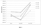
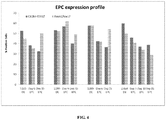

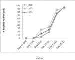
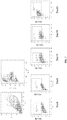
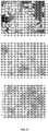
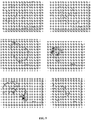
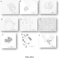


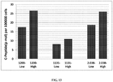


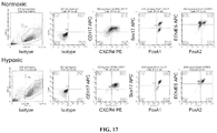


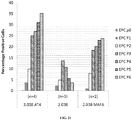


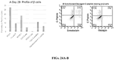

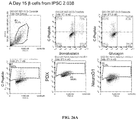

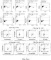
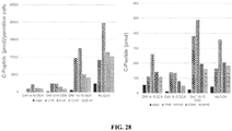
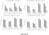
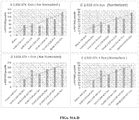
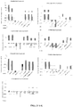
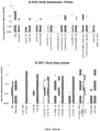
Classifications
-
- C—CHEMISTRY; METALLURGY
- C12—BIOCHEMISTRY; BEER; SPIRITS; WINE; VINEGAR; MICROBIOLOGY; ENZYMOLOGY; MUTATION OR GENETIC ENGINEERING
- C12N—MICROORGANISMS OR ENZYMES; COMPOSITIONS THEREOF; PROPAGATING, PRESERVING, OR MAINTAINING MICROORGANISMS; MUTATION OR GENETIC ENGINEERING; CULTURE MEDIA
- C12N5/00—Undifferentiated human, animal or plant cells, e.g. cell lines; Tissues; Cultivation or maintenance thereof; Culture media therefor
- C12N5/06—Animal cells or tissues; Human cells or tissues
- C12N5/0602—Vertebrate cells
- C12N5/0676—Pancreatic cells
-
- C—CHEMISTRY; METALLURGY
- C12—BIOCHEMISTRY; BEER; SPIRITS; WINE; VINEGAR; MICROBIOLOGY; ENZYMOLOGY; MUTATION OR GENETIC ENGINEERING
- C12N—MICROORGANISMS OR ENZYMES; COMPOSITIONS THEREOF; PROPAGATING, PRESERVING, OR MAINTAINING MICROORGANISMS; MUTATION OR GENETIC ENGINEERING; CULTURE MEDIA
- C12N5/00—Undifferentiated human, animal or plant cells, e.g. cell lines; Tissues; Cultivation or maintenance thereof; Culture media therefor
- C12N5/06—Animal cells or tissues; Human cells or tissues
- C12N5/0602—Vertebrate cells
- C12N5/0607—Non-embryonic pluripotent stem cells, e.g. MASC
-
- C—CHEMISTRY; METALLURGY
- C12—BIOCHEMISTRY; BEER; SPIRITS; WINE; VINEGAR; MICROBIOLOGY; ENZYMOLOGY; MUTATION OR GENETIC ENGINEERING
- C12N—MICROORGANISMS OR ENZYMES; COMPOSITIONS THEREOF; PROPAGATING, PRESERVING, OR MAINTAINING MICROORGANISMS; MUTATION OR GENETIC ENGINEERING; CULTURE MEDIA
- C12N5/00—Undifferentiated human, animal or plant cells, e.g. cell lines; Tissues; Cultivation or maintenance thereof; Culture media therefor
- C12N5/06—Animal cells or tissues; Human cells or tissues
- C12N5/0602—Vertebrate cells
- C12N5/0696—Artificially induced pluripotent stem cells, e.g. iPS
-
- C—CHEMISTRY; METALLURGY
- C12—BIOCHEMISTRY; BEER; SPIRITS; WINE; VINEGAR; MICROBIOLOGY; ENZYMOLOGY; MUTATION OR GENETIC ENGINEERING
- C12N—MICROORGANISMS OR ENZYMES; COMPOSITIONS THEREOF; PROPAGATING, PRESERVING, OR MAINTAINING MICROORGANISMS; MUTATION OR GENETIC ENGINEERING; CULTURE MEDIA
- C12N2500/00—Specific components of cell culture medium
- C12N2500/02—Atmosphere, e.g. low oxygen conditions
-
- C—CHEMISTRY; METALLURGY
- C12—BIOCHEMISTRY; BEER; SPIRITS; WINE; VINEGAR; MICROBIOLOGY; ENZYMOLOGY; MUTATION OR GENETIC ENGINEERING
- C12N—MICROORGANISMS OR ENZYMES; COMPOSITIONS THEREOF; PROPAGATING, PRESERVING, OR MAINTAINING MICROORGANISMS; MUTATION OR GENETIC ENGINEERING; CULTURE MEDIA
- C12N2500/00—Specific components of cell culture medium
- C12N2500/90—Serum-free medium, which may still contain naturally-sourced components
- C12N2500/92—Medium free of human- or animal-derived components
-
- C—CHEMISTRY; METALLURGY
- C12—BIOCHEMISTRY; BEER; SPIRITS; WINE; VINEGAR; MICROBIOLOGY; ENZYMOLOGY; MUTATION OR GENETIC ENGINEERING
- C12N—MICROORGANISMS OR ENZYMES; COMPOSITIONS THEREOF; PROPAGATING, PRESERVING, OR MAINTAINING MICROORGANISMS; MUTATION OR GENETIC ENGINEERING; CULTURE MEDIA
- C12N2500/00—Specific components of cell culture medium
- C12N2500/98—Xeno-free medium and culture conditions
-
- C—CHEMISTRY; METALLURGY
- C12—BIOCHEMISTRY; BEER; SPIRITS; WINE; VINEGAR; MICROBIOLOGY; ENZYMOLOGY; MUTATION OR GENETIC ENGINEERING
- C12N—MICROORGANISMS OR ENZYMES; COMPOSITIONS THEREOF; PROPAGATING, PRESERVING, OR MAINTAINING MICROORGANISMS; MUTATION OR GENETIC ENGINEERING; CULTURE MEDIA
- C12N2501/00—Active agents used in cell culture processes, e.g. differentation
- C12N2501/10—Growth factors
- C12N2501/11—Epidermal growth factor [EGF]
-
- C—CHEMISTRY; METALLURGY
- C12—BIOCHEMISTRY; BEER; SPIRITS; WINE; VINEGAR; MICROBIOLOGY; ENZYMOLOGY; MUTATION OR GENETIC ENGINEERING
- C12N—MICROORGANISMS OR ENZYMES; COMPOSITIONS THEREOF; PROPAGATING, PRESERVING, OR MAINTAINING MICROORGANISMS; MUTATION OR GENETIC ENGINEERING; CULTURE MEDIA
- C12N2501/00—Active agents used in cell culture processes, e.g. differentation
- C12N2501/10—Growth factors
- C12N2501/115—Basic fibroblast growth factor (bFGF, FGF-2)
-
- C—CHEMISTRY; METALLURGY
- C12—BIOCHEMISTRY; BEER; SPIRITS; WINE; VINEGAR; MICROBIOLOGY; ENZYMOLOGY; MUTATION OR GENETIC ENGINEERING
- C12N—MICROORGANISMS OR ENZYMES; COMPOSITIONS THEREOF; PROPAGATING, PRESERVING, OR MAINTAINING MICROORGANISMS; MUTATION OR GENETIC ENGINEERING; CULTURE MEDIA
- C12N2501/00—Active agents used in cell culture processes, e.g. differentation
- C12N2501/10—Growth factors
- C12N2501/119—Other fibroblast growth factors, e.g. FGF-4, FGF-8, FGF-10
-
- C—CHEMISTRY; METALLURGY
- C12—BIOCHEMISTRY; BEER; SPIRITS; WINE; VINEGAR; MICROBIOLOGY; ENZYMOLOGY; MUTATION OR GENETIC ENGINEERING
- C12N—MICROORGANISMS OR ENZYMES; COMPOSITIONS THEREOF; PROPAGATING, PRESERVING, OR MAINTAINING MICROORGANISMS; MUTATION OR GENETIC ENGINEERING; CULTURE MEDIA
- C12N2501/00—Active agents used in cell culture processes, e.g. differentation
- C12N2501/10—Growth factors
- C12N2501/15—Transforming growth factor beta (TGF-β)
-
- C—CHEMISTRY; METALLURGY
- C12—BIOCHEMISTRY; BEER; SPIRITS; WINE; VINEGAR; MICROBIOLOGY; ENZYMOLOGY; MUTATION OR GENETIC ENGINEERING
- C12N—MICROORGANISMS OR ENZYMES; COMPOSITIONS THEREOF; PROPAGATING, PRESERVING, OR MAINTAINING MICROORGANISMS; MUTATION OR GENETIC ENGINEERING; CULTURE MEDIA
- C12N2501/00—Active agents used in cell culture processes, e.g. differentation
- C12N2501/10—Growth factors
- C12N2501/155—Bone morphogenic proteins [BMP]; Osteogenins; Osteogenic factor; Bone inducing factor
-
- C—CHEMISTRY; METALLURGY
- C12—BIOCHEMISTRY; BEER; SPIRITS; WINE; VINEGAR; MICROBIOLOGY; ENZYMOLOGY; MUTATION OR GENETIC ENGINEERING
- C12N—MICROORGANISMS OR ENZYMES; COMPOSITIONS THEREOF; PROPAGATING, PRESERVING, OR MAINTAINING MICROORGANISMS; MUTATION OR GENETIC ENGINEERING; CULTURE MEDIA
- C12N2501/00—Active agents used in cell culture processes, e.g. differentation
- C12N2501/10—Growth factors
- C12N2501/16—Activin; Inhibin; Mullerian inhibiting substance
-
- C—CHEMISTRY; METALLURGY
- C12—BIOCHEMISTRY; BEER; SPIRITS; WINE; VINEGAR; MICROBIOLOGY; ENZYMOLOGY; MUTATION OR GENETIC ENGINEERING
- C12N—MICROORGANISMS OR ENZYMES; COMPOSITIONS THEREOF; PROPAGATING, PRESERVING, OR MAINTAINING MICROORGANISMS; MUTATION OR GENETIC ENGINEERING; CULTURE MEDIA
- C12N2501/00—Active agents used in cell culture processes, e.g. differentation
- C12N2501/10—Growth factors
- C12N2501/165—Vascular endothelial growth factor [VEGF]
-
- C—CHEMISTRY; METALLURGY
- C12—BIOCHEMISTRY; BEER; SPIRITS; WINE; VINEGAR; MICROBIOLOGY; ENZYMOLOGY; MUTATION OR GENETIC ENGINEERING
- C12N—MICROORGANISMS OR ENZYMES; COMPOSITIONS THEREOF; PROPAGATING, PRESERVING, OR MAINTAINING MICROORGANISMS; MUTATION OR GENETIC ENGINEERING; CULTURE MEDIA
- C12N2501/00—Active agents used in cell culture processes, e.g. differentation
- C12N2501/30—Hormones
- C12N2501/33—Insulin
-
- C—CHEMISTRY; METALLURGY
- C12—BIOCHEMISTRY; BEER; SPIRITS; WINE; VINEGAR; MICROBIOLOGY; ENZYMOLOGY; MUTATION OR GENETIC ENGINEERING
- C12N—MICROORGANISMS OR ENZYMES; COMPOSITIONS THEREOF; PROPAGATING, PRESERVING, OR MAINTAINING MICROORGANISMS; MUTATION OR GENETIC ENGINEERING; CULTURE MEDIA
- C12N2501/00—Active agents used in cell culture processes, e.g. differentation
- C12N2501/40—Regulators of development
- C12N2501/415—Wnt; Frizzeled
-
- C—CHEMISTRY; METALLURGY
- C12—BIOCHEMISTRY; BEER; SPIRITS; WINE; VINEGAR; MICROBIOLOGY; ENZYMOLOGY; MUTATION OR GENETIC ENGINEERING
- C12N—MICROORGANISMS OR ENZYMES; COMPOSITIONS THEREOF; PROPAGATING, PRESERVING, OR MAINTAINING MICROORGANISMS; MUTATION OR GENETIC ENGINEERING; CULTURE MEDIA
- C12N2501/00—Active agents used in cell culture processes, e.g. differentation
- C12N2501/999—Small molecules not provided for elsewhere
-
- C—CHEMISTRY; METALLURGY
- C12—BIOCHEMISTRY; BEER; SPIRITS; WINE; VINEGAR; MICROBIOLOGY; ENZYMOLOGY; MUTATION OR GENETIC ENGINEERING
- C12N—MICROORGANISMS OR ENZYMES; COMPOSITIONS THEREOF; PROPAGATING, PRESERVING, OR MAINTAINING MICROORGANISMS; MUTATION OR GENETIC ENGINEERING; CULTURE MEDIA
- C12N2506/00—Differentiation of animal cells from one lineage to another; Differentiation of pluripotent cells
- C12N2506/45—Differentiation of animal cells from one lineage to another; Differentiation of pluripotent cells from artificially induced pluripotent stem cells
Definitions
- the present invention relates generally to the fields of endocrinology, stem cells, and differentiated cells. More particularly, it concerns the production of self-renewing endoderm progenitor cells from undifferentiated cells in a suspension culture.
- Endoderm-derived tissues including pancreas, are potentially useful for cell replacement therapies. It is possible to generate definitive endoderm, which forms the primitive gut tube during development, and its derivative lineages from pluripotent stem cells (PSCs) in vitro through sequential exposure to cytokines that mimic embryonic morphogenesis. In this fashion, pancreatic cells can be produced from embryonic stem cells (ESCs) and induced pluripotent stem cells (iPSCs) (D'Amour et al ., 2006; Gouon-Evans et al ., 2006). While these studies highlight the promise of PSC-derived endodermal tissues for transplantation therapies, several obstacles remain.
- PSCs pluripotent stem cells
- Endodermal cells generated from PSCs tend to display immature phenotypes and in many instances are not fully functional.
- most pancreatic beta cells currently generated in vitro from human ESCs are poly-hormonal and not glucose responsive (D'Amour et al ., 2006; Nostro et al ., 2011).
- An additional obstacle in the production of endodermal cells for use in transplantation is the ability to scale the production to produce the required number of cells, which for pancreatic cells is currently limited by the necessity for production on a solid surface. It is an objective of the present invention to provide culture and isolation methods that avoid these limitations.
- WO 2012/178215 relates to a method of producing self-renewing endoderm progenitor cells or cell aggregates.
- EPCs self-renewing endoderm progenitor cells
- EPs endoderm progenitor cells
- a method for producing self-renewing endoderm progenitor cells comprising
- the serum-free medium in step a) may further comprise Wnt3A or a GSK3 inhibitor. In other aspects, the serum-free medium in step a) may not contain Wnt3A or a GSK3 inhibitor.
- a preferred example of a GSK3 inhibitor is CHIR 99021.
- the culturing in step a) may be carried out for at least or about 1, 2, 3, 4, 6, 7, 8, 9, 10, 11, 12, 13, 14, 15, 16, 17, 18, 19, 20 days, or any range derivable therein. Preferably, said culturing is carried out for about 4-12 days, more preferably for about 5-8 days. In some aspects, the culturing in step a) may be carried out as an adherent culture.
- the endoderm progenitor promoting growth factors used in step b) may further comprise TGF- ⁇ , latent TGF- ⁇ binding proteins, Nodal, a GSK3 inhibitor, Follistatin-related protein (FSRP), Dickkopf-related protein 1, IndolactamV, and/or insulin-like growth factor.
- a GSK3 inhibitor is CHIR 99021.
- the culturing in step b) may be carried out for at least or about 1, 2, 3, 4, 6, 7, 8, 9, 10, 11, 12, 13, 14, 15, 16, 17, 18, 19, 20, 21, 22, 23, 24, 25 days, or any range derivable therein.
- said culturing in step b) may be carried out for at least 1, 2, 3, 4, 5, 6 months, or any range derivable therein. Preferably, said culturing is carried out for at least about 5 to about 40 days, more preferably for about 10 to about 15 days. In some aspects, the culturing in step b) may occur in the presence of a ROCK inhibitor.
- the ROCK inhibitor may be Y-27632, H1152, or HA-100. In some aspects, the ROCK inhibitor may be removed after the first about 12 hours of culture.
- the suspension culture in step b) may further comprise matrix components, such as basement membrane preparations, to support culture of the cell population.
- matrix components include collagen, gelatin, poly-L-lysine, poly-D-lysine, vitronectin, laminin, PLO laminin, fibrin clot, fibronectin and mixtures thereof, for example, MatrigelTM and lysed cell membrane preparations.
- the basement membrane preparation may be present at a final concentration of about 0.06-0.6 mg/ml, preferably about 0.3-0.6 mg/ml.
- the suspension cultures in step b) may be dissociated and reaggregated about every 1, 2, 3, 4, 6, 7, 8, 9, 10, 11, 12, 13, 14, 15, or any range derivable therein. Preferably, the suspension cultures may be dissociated and reaggregated about every 4-5 days.
- the suspensions cultures in step b) may be maintained in spinner flasks. The spinner flasks may be operated at about 40-70 rpm. In some aspects, the suspension cultures in step b) may be maintained as static suspension cultures.
- the endoderm progenitor cells may express CXCR4, CD 117 (c-KIT), and CD34. In some aspects, the endoderm progenitor cells may further express Sox17, FOXA1, FOXA2, and CD31. Said expression may be detected by protein expression analysis, for example ELISA or FACS analysis, or RNA expression analysis, for example qRT-PCR. In some aspects, the formation of self-renewing endoderm progenitor cells does not require the presence of MEF feeder layers or MEF-conditioned medium. In some aspects, the culturing occurs under normoxic conditions. In some aspects, the culturing occurs under hypoxic conditions. In some aspects, the pluripotent stem cells may be maintained under hypoxic conditions prior to step a).
- the pluripotent stem cells may be embryonic stem cells or induced pluripotent stem cells.
- the stem cells may also include multipotent stem cells, oligopotent stem cells, or unipotent stem cells.
- the stem cells may also include fetal stem cells or adult stem cells, such as hematopoietic stem cells, mesenchymal stem cells, neural stem cells, epithelial stem cells, and skin stem cells.
- the stem cells may be isolated from umbilical, placenta, amniotic fluid, chorion villi, blastocysts, bone marrow, adipose tissue, brain, peripheral blood, cord blood, menstrual blood, blood vessels, skeletal muscle, skin, and liver.
- the method further comprises culturing the endoderm progenitor cells with islet beta cell promoting growth factors to effect the formation of mono-hormonal pancreatic islet beta cells.
- the endoderm progenitor cells may be purified prior to culturing. Said purification may be based on expression of markers, such as CD34, CD 117, and CXCR4.
- the culturing may be performed as a suspension culture. In some aspects, the culturing may be performed as an adherent culture.
- the islet beta cell promoting growth factors may comprise a potent inhibitor of AMP-activated protein kinase (AMPK), a BMP antagonist, a hedgehog inhibitor, a gamma-secretase inhibitor, an ALK5 inhibitor, retinoic acid, FGF10, B27, and Wnt3A or a GSK3 inhibitor.
- AMPK AMP-activated protein kinase
- BMP antagonist BMP antagonist
- hedgehog inhibitor a hedgehog inhibitor
- a gamma-secretase inhibitor a gamma-secretase inhibitor
- ALK5 inhibitor retinoic acid
- FGF10 FGF10
- B27 retinoic acid
- Wnt3A retinoic acid
- GSK3 inhibitor a preferred example of a GSK3 inhibitor
- said further culturing may occur in the presence of a ROCK inhibitor.
- the ROCK inhibitor may be Y-27632, H1152, or HA-100.
- the method of effecting the formation of mono-hormonal pancreatic islet beta cells further comprises i) culturing the endoderm progenitor cells with a BMP antagonist, Wnt3A, and FGF10 to effect the formation of foregut endoderm cells; ii) culturing the foregut endoderm cells with B27, a BMP antagonist, a Hedgehog inhibitor, retinoic acid, and FGF10 to effect the formation of pancreatic endoderm cells; iii) culturing the pancreatic endoderm cells with B27, a BMP antagonist, a gamma-secretase inhibitor, and an ALK5 inhibitor to effect the formation of endocrine precursor cells; and iv) culturing said endocrine precursor cells with a BMP antagonist, an ALK5 inhibitor, insulin, glucose, and nicotinamide to effect the formation of mono-hormonal pancreatic islet beta cells.
- the BMP antagonist used in step i) may be dorsomorphin or LDN-193189.
- the BMP antagonist is dorsomorphin.
- the BMP antagonist used in steps ii), iii), and iv) may be Noggin, Gremlin, USAG-1, Follistatin, PRDC, Cerberus, Coco, Sclerostin, or Chordin.
- the BMP antagonist is Noggin.
- the gamma-secretase inhibitor used in step iii) may be DAPT, RO4929097, BMS-708163, Semagacestat, MK-0752, YO-01027, LY-411575, or (R)-flurbiprofen.
- the gamma-secretase inhibitor is DAPT.
- the ALK5 inhibitor used in steps iii) and iv) may be SB431542, ALX-270-448, A 83-01, EW-7195, KI26894, LY2109761, LY-364947, SB-525334, SB-505124, SD-208, IN-1233, or SKI2162.
- the ALK5 inhibitor is SB431542.
- the Hedgehog inhibitor used in step ii) may be KAAD-cyclopamine, vismodegib, LY2940680, MRT-10, MRT-83, or GDC-0449.
- the Hedgehog inhibitor is KAAD-cyclopamine.
- the cells may comprise an inducible expression cassette encoding Erg1.
- step iv) of the method of effecting the formation of mono-hormonal pancreatic islet beta cells may further comprise inducing the cells to express Erg1.
- the cells may comprise an inducible expression cassette encoding Gfil.
- step iv) of the method of effecting the formation of mono-hormonal pancreatic islet beta cells may further comprise inducing the cells to express Gfil.
- the cells may comprise inducible expression cassettes encoding both Erg1 and Gfil.
- step iv) of the method of effecting the formation of mono-hormonal pancreatic islet beta cells may further comprise inducing the cells to express both Erg1 and Gfil.
- the inducible expression cassette may be integrated into the genome of the cells.
- the inducible expression cassette may be comprised in a transposon ( e . g . piggyBac).
- the inducible expression cassette may be comprised in an episomal vector.
- the culturing in steps i), ii), and iii) may be carried out for at least or about 1, 2, 3, 4, 6, 7, 8, 9, 10, 11, 12, 13, 14, 15, 16, 17, 18, 19, 20 days, or any range derivable therein.
- said culturing is carried out for about 3-5 days.
- the culturing in step iv) may be carried out for at least or about 1, 2, 3, 4, 6, 7, 8, 9, 10, 11, 12, 13, 14, 15, 16, 17, 18, 19, 20 days, or any range derivable therein.
- said culturing is carried out for about 3-40 days.
- the culture may comprise a basement membrane preparation. In some aspects, the culture may occur under hypoxic conditions. In some aspects, the culture may be devoid of MEF feeder cells and MEF-conditioned medium.
- the mono-hormonal pancreatic islet beta cells may express PDX, insulin, C-peptide, and NeuroD1, low levels of somatostatin and Nkx6.1, and no glucagon. Said expression may be detected by protein expression analysis, such as ELISA or FACS analysis, or RNA expression analysis, such as qRT-PCR.
- the mono-hormonal pancreatic islet beta cells are at least 1%, 5%, 10%, 15%, 20%, 25%, 30%, 35%, 40%, 45%, 50%, 60%, 70%, 80%, 85%, 90%, 91%, 92%, 93%, 94%, 95%, 96%, 97%, 98%, 99%, 99.9% (or any intermediate ranges) of the cell population, or any range derivable therein.
- the mono-hormonal pancreatic islet beta cells are about 12% pure or about 20% pure. Additional characteristic of mono-hormonal pancreatic islet beta cells include, but are not limited to, glucose-responsive C-peptide release and the ability to revert diabetes in animal models thereof by pancreatic engraftment.
- the mono-hormonal pancreatic islet beta cells may be cultured for one to four weeks.
- said culturing may comprise a suspension culture.
- the present disclosure provides a method of producing self-renewing endoderm progenitor cell aggregates comprising a) culturing pluripotent stem cells in serum-free medium comprising Activin A or Nodal to effect the induction of endoderm cells and b) culturing the endoderm cells in a suspension culture with BMP4, VEGF, FGF2, and EGF or homologs thereof in serum-free medium to effect the formation of self-renewing endoderm progenitor cell aggregates.
- the present disclosure provides a method of producing self-renewing endoderm progenitor cell aggregates comprising a) culturing pluripotent stem cells in serum-free medium comprising Activin A to effect the induction of endoderm cells and b) culturing the endoderm cells in a suspension culture with Activin A, BMP4, VEGF, FGF2, EGF , and TGF ⁇ or homologs thereof in serum-free medium to effect the formation of self-renewing endoderm progenitor cell aggregates.
- the cells of the embodiments may be contacted with differentiation factors in an amount sufficient to cause differentiation of the stem cells to endoderm progenitor cells.
- the differentiation factors may comprise gene products of the differentiation factor genes.
- the gene products may be polypeptides or RNA transcripts of the differentiation factor genes.
- the differentiation factors may comprise one or more protein transduction domains to facilitate their intracellular entry and/or nuclear entry.
- protein transduction domains are well known in the art, such as an HIV TAT protein transduction domain, HSV VP22 protein transduction domain, Drosophila Antennapedia homeodomain, or variants thereof.
- starting cells for the present methods may comprise at least or about 10 4 , 10 5 , 10 6 , 10 7 , 10 8 , 10 9 , 10 10 , 10 11 , 10 12 , 10 13 cells or any range derivable therein.
- the starting cell population may have a seeding density of at least or about 10, 10 1 , 10 2 , 10 3 , 10 4 , 10 5 , 10 6 , 10 7 , 10 8 cells/ml, or any range derivable therein.
- Embodiments discussed in the context of methods and/or compositions of the invention may be employed with respect to any other method or composition described herein. Thus, an embodiment pertaining to one method or composition may be applied to other methods and compositions of the invention as well.
- ESCs Human embryonic stem cells
- iPSCs induced pluripotent stem cells
- ESCs Human embryonic stem cells
- iPSCs induced pluripotent stem cells
- EPCs endoderm progenitor cells
- Definitive endoderm is a transient state that quickly differentiates, cannot be proliferated, does not have a distinct morphology, and may not be fully committed to an endoderm fate.
- endoderm progenitor (EP) cells or EPCs are not transient, have a distinct morphology, and may be cultured indefinitely (Cheng et al ., 2013).
- Endoderm progenitor cells are also characterized as expressing CXCR4, CD117, FOXA1, FOXA2, CD31, CD34, and SOX17.
- Endoderm progenitor cells can generate cells in the endoderm lineage, such as liver, pancreas, and intestine, but cannot generate mesoderm or ectoderm either in vitro or in vivo.
- EP cells do not form teratomas in immune-deficient mice. Therefore, DE and EP cells may represent distinct developmental intermediaries and may have different development potentials.
- iPSC-derived EP cells express CXCR-4, CD117, SOX17, FOXA1, FOXA2, CD34, CD31, and HNF4A and can be maintained in suspension cultures for 5-40 days.
- EPs expand and maintain the phenotypic expression of markers as 3D aggregates in spinner or static conditions in the presence of Matrigel, BMP4, VEGF, FGF, EGF2, Activin, and optionally TGF ⁇ .
- EP cultures when placed in culture conditions for generating cells of the pancreatic lineage, generate mono-hormonal beta cells functionally responsive to stimulation with D-glucose.
- EP cells retain their phenotypic characteristics and expand as 3D cultures for at least 30 days.
- EP cells placed in pancreatic differentiation media generate mono-hormonal beta cells. These cells will serve as an ideal cell type for high-throughput screening applications and hold potential for use in therapeutic applications.
- “Differentiation” is a process by which a less specialized cell becomes a more specialized cell to form progeny of at least one new cell type, either in culture or in vivo, than it would have under the same conditions without differentiation.
- “Dedifferentiation” is a cellular process in which a partially or terminally differentiated cell reverts to an earlier developmental stage, such as pluripotency or multipotency.
- “Transdifferentiation” is a process of transforming one differentiated cell type into another differentiated cell type. Under certain conditions, the proportion of progeny with characteristics of the new cell type may be at least about 1%, 5%, 25% or more in order of increasing preference.
- Multipotent implies that a cell is capable, through its progeny, of giving rise to several different cell types found in an adult animal.
- Pluripotent implies that a cell is capable, through its progeny, of giving rise to all the cell types that comprise the adult animal, including the germ cells. Embryonic stem cells, induced pluripotent stem cells, and embryonic germ cells are pluripotent cells under this definition.
- autologous cells refers to donor cells that are genetically compatible with the recipient.
- totipotent can refer to a cell that gives rise to a live born animal.
- the term “totipotent” can also refer to a cell that gives rise to all of the cells in a particular animal.
- a totipotent cell can give rise to all of the cells of an animal when it is utilized in a procedure for developing an embryo from one or more nuclear transfer steps.
- Totipotent cells may also be used to generate incomplete animals, such as those useful for organ harvesting, e.g ., having genetic modifications to eliminate growth of an organ or appendage by manipulation of a homeotic gene. Additionally, genetic modification rendering oocytes, such as those derived from ES cells, incapable of development in utero would ensure that human-derived ES cells could not be used to derive human oocytes for reproduction and only for applications such as therapeutic cloning.
- embryonic stem cell can, for reference only, refer to pluripotent stem cells isolated from an embryo that are maintained in in vitro cell culture. Such cells are rapidly dividing cultured cells isolated from cultured embryos that retain in culture the ability to give rise, in vivo, to all the cell types that comprise the adult animal, including the germ cells. Embryonic stem cells may be cultured with or without feeder cells. For reference, embryonic stem cells can be established from embryonic cells isolated from embryos at any stage of development, including blastocyst stage embryos and pre-blastocyst stage embryos. Embryonic stem cells may have a rounded cell morphology and may grow in rounded cell clumps on feeder layers.
- Embryonic stem cells are well known to a person of ordinary skill in the art. See, e.g., WO 97/37009 ; Yang & Anderson (1992); Piedrahita et al . (1998); Wianny et al . (1997); Moore & Piedrahita (1997); Moore & Piedrahita (1996); Wheeler (1994); Hochereau-de Reviers & Perreau (1993); Strojek et al . (1990); Piedrahita et al . (1990); and Evans et al . (1990).
- reprogramming or “reprogrammed” as used herein may refer to materials and methods that can convert a more specialized cell into a pluripotent cell.
- isolated can refer to a cell that is mechanically separated from another group of cells. Examples of a group of cells are a developing cell mass, a cell culture, a cell line, and an animal.
- differentiated cell can refer to a precursor cell that has developed from an unspecialized phenotype to a specialized phenotype.
- embryonic cells can differentiate into an epithelial cell lining of the intestine.
- Differentiated cells can be isolated from a fetus or a live born animal, for example.
- undifferentiated cell can refer to a precursor cell that has an unspecialized phenotype and is capable of differentiating.
- An example of an undifferentiated cell is a stem cell.
- the cells may be stem cells, including but not limited to, embryonic stem cells, fetal stem cells, adult stem cells, or induced pluripotent stem cells.
- Stem cells are cells found in most, if not all, multi-cellular organisms. They are characterized by the ability to self-renew through mitotic cell division and the ability to differentiate into a diverse range of specialized cell types.
- the two broad types of mammalian stem cells are embryonic stem cells that are found in blastocysts and adult stem cells that are found in adult tissues. In a developing embryo, stem cells can differentiate into all of the specialized embryonic tissues. In adult organisms, stem cells and progenitor cells act as a repair system for the body, replenishing specialized cells, and also maintain the normal turnover of regenerative organs, such as blood, skin, or intestinal tissues.
- Human embryonic stem cells (ESCs) and induced pluripotent stem cells (iPSCs) are capable of long-term proliferation in vitro, while retaining the potential to differentiate into all cell types of the body, including endoderm progenitor cells and pancreatic islet beta cells. Endoderm progenitor cells could potentially provide an unlimited supply of patient-specific functional pancreatic islet beta cells for both drug development and therapeutic uses.
- the differentiation of human ESCs/iPSCs to pancreatic islet beta cells through endoderm progenitor cells in vitro recapitulates normal in vivo development, i.e. they undergo the normal sequential developmental stages including endoderm differentiation and endocrine specification. That sequential developmental process requires the addition of different growth factors at different stages of differentiation.
- pancreatic islet beta cells by differentiating human ESCs/iPSCs. This approach generates pancreatic islet beta cells with functions highly similar, if not identical, to human primary adult beta cells.
- the endoderm progenitor cells of the invention with their unlimited proliferation ability, have unique advantages as the starting cell population for beta cell differentiation.
- Embryonic stem cells are pluripotent stem cells that, for reference, can be derived from the epiblast tissue of the inner cell mass of a blastocyst or earlier morula stage embryo. ES cells are distinguished by two distinctive properties: their pluripotency and their capability to self-renew indefinitely. ES cells are pluripotent, that is, they are able to differentiate into all derivatives of the three primary germ layers: ectoderm, endoderm, and mesoderm. Additionally, under defined conditions, embryonic stem cells are capable of propagating themselves indefinitely. This allows embryonic stem cells to be employed as useful tools for both research and regenerative medicine, because they can produce limitless numbers of themselves for continued research or clinical use. ES cells can develop into each of the more than 200 cell types of the adult body when given sufficient and necessary stimulation for a specific cell type. They do not, however, contribute to the extra-embryonic membranes or the placenta.
- Mouse embryonic stem cells mES
- human embryonic stem cells hES Both have the essential stem cell characteristics, yet they require very different environments in order to maintain an undifferentiated state.
- Mouse ES cells may be grown on a layer of gelatin and require the presence of Leukemia Inhibitory Factor (LIF).
- LIF Leukemia Inhibitory Factor
- Human ES cells could be grown on a feeder layer of mouse embryonic fibroblasts (MEFs) and often require the presence of basic Fibroblast Growth Factor (bFGF or FGF-2). Without optimal culture conditions or genetic manipulation (Chambers et al . , 2003), embryonic stem cells will rapidly differentiate.
- LIF Leukemia Inhibitory Factor
- bFGF or FGF-2 basic Fibroblast Growth Factor
- a human embryonic stem cell may also be defined by the presence of several transcription factors and cell surface proteins.
- the transcription factors Oct-4, Nanog, and Sox-2 form the core regulatory network that ensures the suppression of genes that lead to differentiation and the maintenance of pluripotency (Boyer et al ., 2005).
- the cell surface antigens most commonly used to identify hES cells include the glycolipids SSEA3 and SSEA4 and the keratan sulfate antigens Tra-1-60 and Tra-1-81.
- mouse ES cells Methods for obtaining mouse ES cells are well known.
- a preimplantation blastocyst from the 129 strain of mice is treated with mouse antiserum to remove the trophoectoderm, and the inner cell mass is cultured on a feeder cell layer of chemically inactivated mouse embryonic fibroblasts in medium containing fetal calf serum. Colonies of undifferentiated ES cells that develop are subcultured on mouse embryonic fibroblast feeder layers in the presence of fetal calf serum to produce populations of ES cells.
- mouse ES cells can be grown in the absence of a feeder layer by adding the cytokine leukemia inhibitory factor (LIF) to serum-containing culture medium (Smith, 2000).
- LIF cytokine leukemia inhibitory factor
- mouse ES cells can be grown in serum-free medium in the presence of bone morphogenetic protein and LIF (Ying et al ., 2003).
- human ES cells can be obtained from blastocysts using previously described methods (Thomson et al ., 1995; Thomson et al ., 1998; Thomson and Marshall, 1998; Reubinoff et al ., 2000).
- day-5 human blastocysts are exposed to rabbit anti-human spleen cell antiserum, and then exposed to a 1:5 dilution of Guinea pig complement to lyse trophectoderm cells. After removing the lysed trophectoderm cells from the intact inner cell mass, the inner cell mass is cultured on a feeder layer of gamma-inactivated mouse embryonic fibroblasts and in the presence of fetal bovine serum.
- clumps of cells derived from the inner cell mass can be chemically (i . e . exposed to trypsin) or mechanically dissociated and replated in fresh medium containing fetal bovine serum and a feeder layer of mouse embryonic fibroblasts.
- colonies having an undifferentiated morphology are selected by micropipette, mechanically dissociated into clumps, and replated (see U.S. Patent No. 6,833,269 ).
- ES-like morphology is characterized as compact colonies with apparently high nucleus to cytoplasm ratio and prominent nucleoli. Resulting ES cells can be routinely passaged by brief trypsinization or by selection of individual colonies by micropipette.
- human ES cells can be grown without serum by culturing the ES cells on a feeder layer of fibroblasts in the presence of basic fibroblast growth factor (Amit et al ., 2000).
- human ES cells can be grown without a feeder cell layer by culturing the cells on a protein matrix such as MatrigelTM or laminin in the presence of "conditioned" medium containing basic fibroblast growth factor (Xu et al ., 2001). The medium is previously conditioned by coculturing with fibroblasts.
- ES cell lines Another disclosed source of ES cells is established ES cell lines.
- Various mouse cell lines and human ES cell lines are known and conditions for their growth and propagation have been defined.
- the mouse CGR8 cell line was established from the inner cell mass of mouse strain 129 embryos, and cultures of CGR8 cells can be grown in the presence of LIF without feeder layers.
- human ES cell lines H1, H7, H9, H13, and H14 were established by Thompson et al . (1995).
- subclones H9.1 and H9.2 of the H9 line have been developed. It is anticipated that virtually any ES or stem cell line known in the art may be used with the present invention, such as, e.g ., those described in Yu and Thompson (2008).
- Induced pluripotent stem cells are a type of pluripotent stem cell artificially derived from a non-pluripotent cell, typically an adult somatic cell. Induced pluripotent stem cells are believed to be similar if not identical to natural pluripotent stem cells, such as embryonic stem cells in many respects, such as in terms of the expression of certain stem cell genes and proteins, chromatin methylation patterns, doubling time, embryoid body formation, teratoma formation, viable chimera formation, and potency and differentiability, but the full extent of their relation to natural pluripotent stem cells is still being assessed.
- iPS cells are obtained by reprogramming of differentiated somatic cells. Generation of induced pluripotent cells derived from human tissue other than of embryonic origin is desired to alleviate ethical concerns regarding experimental use of embryos and embryonic tissue. The promise of therapeutic applications from induced pluripotent cells has been advocated. Medical applications include treatments for Alzheimer's disease, diabetes and spinal cord injuries to name a few. Other applications include disease modeling and pharmaceutical drug screening.
- Induced pluripotent stem cells have been obtained by various methods. Human fetal or newborn fibroblasts are transfected with four genes, Oct4, Sox2, Nanog and Lin28 using lentiviral transduction (Yu et al. , 2007). At 12-20 days post infection, colonies with human ES cell morphology become visible. The colonies are picked and expanded. The induced pluripotent stem cells making up the colonies are morphologically similar to human ES cells, express various human ES cell markers, and form teratomas having neural tissue, cartilage, and gut epithelium after injection into mice.
- adult human dermal fibroblasts are transfected with transcription factors Oct4, Sox2, c-Myc and Klf4 using retroviral transduction (Takahashi et al ., 2007).
- the transfected cells are plated on SNL feeder cells (a mouse cell fibroblast cell line that produces LIF) in medium supplemented with basic fibroblast growth factor (bFGF). After approximately 25 days, colonies resembling human ES cell colonies appear in culture. The ES cell-like colonies are picked and expanded on feeder cells in the presence of bFGF.
- SNL feeder cells a mouse cell fibroblast cell line that produces LIF
- bFGF basic fibroblast growth factor
- iPS cells were first produced in 2006 (Takahashi et al ., 2006) from mouse cells and in 2007 from human cells (Takahashi et al ., 2007; Yu et al, 2007). This has been cited as an important advancement in stem cell research, as it may allow researchers to obtain pluripotent stem cells, which are important in research and potentially have therapeutic uses, without the controversial use of embryos.
- the first successful demonstration of generating induced pluripotent cells (iPS cells) from mouse or human tissue involved the use of retroviral vectors expressing a specific set of transcription factors.
- cells of the ES cell-like colonies are induced pluripotent stem cells.
- the induced pluripotent stem cells are morphologically similar to human ES cells, and express various human ES cell markers. Also, when grown under conditions that are known to result in differentiation of human ES cells, the induced pluripotent stem cells differentiate accordingly.
- the induced pluripotent stem cells can differentiate into cells having endoderm progenitor cell or pancreatic islet beta cell structures and markers. It is anticipated that virtually any iPS cells or cell lines may be used with the present invention, including, e.g., those described in Yu and Thompson, 2008.
- Sox may be Sox-1, Sox-2, Sox-3, Sox-15, or Sox-18; Oct may be Oct-4.
- Additional factors may increase the reprogramming efficiency, like Nanog, Lin28, Klf4, or c-Myc; specific sets of reprogramming factors may be a set comprising Sox-2, Oct-4, Nanog, and optionally, Lin-28; or comprising Sox-2, Oct4, Klf, and optionally, c-Myc.
- iPS cells like ES cells, have characteristic antigens that can be identified or confirmed by immunohistochemistry or flow cytometry, using antibodies for SSEA-1, SSEA-3 and SSEA-4 (Developmental Studies Hybridoma Bank, National Institute of Child Health and Human Development, Bethesda Md.), and TRA-1-60 and TRA-1-81 (Andrews et al ., 1987). Pluripotency of iPS cells can be confirmed by injecting approximately 0.5-10 ⁇ 10 6 cells into the rear leg muscles of 8-12 week old male SCID mice. Teratomas develop that demonstrate at least one cell type of each of the three germ layers.
- iPS cells are made from reprogramming somatic cells using reprogramming factors comprising an Oct family member and a Sox family member, such as Oct4 and Sox2 in combination with Klf and/or Nanog as described above and as described in WO2009/149233 .
- the somatic cell for reprogramming may be any somatic cell that can be induced to pluripotency, such as a fibroblast, a keratinocyte, a hematopoietic cell, a mesenchymal cell, a liver cell, a stomach cell, or a ⁇ cell.
- T cells may also be used as source of somatic cells for reprogramming (see WO2010/141801 ).
- Reprogramming factors may be expressed from expression cassettes comprised in one or more vectors, such as an integrating vector or an episomal vector, e.g ., an EBV element-based system (see UWO2009/149233 ; Yu et al ., 2009).
- reprogramming proteins could be introduced directly into somatic cells by protein transduction.
- AT4 cells are human iPSCs designed to induce hematopoiesis. They are engineered to constitutively express rtTET protein for inducible gene expression.
- the human Rosa26 locus on chromosome 3 was selected to allow the expression of rtTET.
- the LoxP recombination sites (LOX71 and LOX2272) were introduced into the first intron of the human ROSA26 gene via homologous recombination.
- SA human BCL2 gene
- the neomycin phosphotransferase (Neo) was used for positive selection.
- ERG1 and GFI1 were cloned into the PiggyBac transposon-based expression vector under the control of the TET-inducible promoter (Ptight), which is an rtTET-responsive inducible promoter. ERG1 and GFI1 are under the control of the inducible promoter Ptight.
- TET-inducible promoter Ptight
- DOX Doxycycline
- Neo+ neomycin
- 2.038 MAFA cells are human iPSCs designed to overexpress the transcription factor MaFA.
- the plasmid contained the coding regions of MaFA fused to neomycin was electroporated in iPSCs. The emerging clones were selected using Neomycin selection.
- Exogenous MaFA is under the control of the TET-inducible promoter (Ptight), which is an rtTET-responsive inducible promoter present in the MaFa_Neomycin vector.
- TET-inducible promoter Ptight
- rtTET-responsive inducible promoter present in the MaFa_Neomycin vector.
- Doxycycline DOX; 0.1-10 ⁇ g/ml
- the correctly recombined inducible cells are resistant to neomycin (Neo+).
- the polyclonal population was subcloned further to derive 2.038Mafa cells.
- Cells can be characterized according to a number of phenotypic criteria.
- the criteria include but are not limited to the detection or quantitation of expressed cell markers, enzymatic activity, and the characterization of morphological features and intercellular signaling.
- Endodermal cells embodied in certain aspects of this invention have morphological features characteristic of endoderm cells in nature. The features are readily appreciated by those skilled in evaluating such things, and include a doughnut-like morphology when grown in 2D cultures. One or more such features present in a single cell are consistent with the cell being a member of the endodermal cell lineage.
- Unbiased determination of whether cells have morphologic features characteristic of endodermal cells can be made by coding micrographs of differentiated progeny cells, adult or fetal endodermal cells, and one or more negative control cells, such as a fibroblast, or RPE (Retinal pigment epithelial) cells and then evaluating the micrographs in a blinded fashion, and breaking the code to determine if the endodermal cells from differentiation are accurately identified.
- a fibroblast or RPE (Retinal pigment epithelial) cells
- Definitive endoderm is a transient state that quickly differentiates, cannot be proliferated, does not have a distinct morphology, and may not be fully committed to an endoderm fate.
- endoderm progenitors are not transient, have a distinct morphology, and may be cultured indefinitely (Cheng et al ., 2013).
- Endoderm progenitor cells can generate cells in the endoderm lineage, such as liver, pancreas, and intestine, but cannot generate mesoderm or ectoderm either in vitro or in vivo.
- EP cells do not form teratomas in immune-deficient mice. Therefore, DE and EP cells may represent distinct developmental intermediaries and may have different development potentials.
- the pancreas an organ about the size of a hand, is located behind the lower part of the stomach. It comprises two structures that are both morphologically and physiologically different: the exocrine pancreas, which produces the enzymes involved in digestion (amylase, lipase, etc.) and sodium bicarbonate, and the endocrine pancreas, which produces the hormones involved in the control of blood glucose (insulin, glucagon, somatostatin, and pancreatic polypeptide).
- the cells of the endocrine pancreas are organized as micro-organs dispersed in the pancreas in the form of islets (islets of Langerhans or pancreatic islets).
- Each pancreatic islet is made up of four cell types: alpha cells, beta cells, delta cells and PP (gamma) cells and epsilon cells.
- alpha cells beta cells
- beta cells delta cells
- PP cells gamma cells
- epsilon cells The beta cells are found at the center of the islet and are the only cells capable of secreting insulin in response to glucose.
- Cells of this invention can also be characterized according to whether they express phenotypic markers characteristic of the pancreatic endodermal lineage. Endoderm progenitor cells are also characterized as expressing CXCR4, CD117, FOXA1, FOXA2, CD31, CD34, and SOX17. Non-limiting examples of cell markers useful in distinguishing pancreatic islet beta cells include PDX-1, neurogenin3, neuroD1, insulin, glucagon, somatostatin, and C-peptide. Mature pancreatic islet beta cell markers include, but are not limited to, PDX1, C-peptide, and insulin. Immature pancreatic islet beta cells, which are akin to immature cells of the fetal pancreas, are poly-hormonal, while mature pancreatic islet beta cells are mono-hormonal and glucose-responsive.
- Endoderm progenitor protein determinants listed in this disclosure can be detected using any suitable immunological technique-such as flow immunocytochemistry for cell-surface markers, immunohistochemistry (for example, of fixed cells or tissue sections) for intracellular or cell-surface markers, Western blot analysis of cellular extracts, and enzyme-linked immunoassay, for cellular extracts or products secreted into the medium.
- suitable immunological technique such as flow immunocytochemistry for cell-surface markers, immunohistochemistry (for example, of fixed cells or tissue sections) for intracellular or cell-surface markers, Western blot analysis of cellular extracts, and enzyme-linked immunoassay, for cellular extracts or products secreted into the medium.
- an antigen by a cell is said to be "antibody-detectable” if a significantly detectable amount of antibody will bind to the antigen in a standard immunocytochemistry or flow cytometry assay, optionally after fixation of the cells, and optionally using a labeled secondary antibody or other conjugate (such as a biotin-avidin conjugate) to amplify labeling.
- a labeled secondary antibody or other conjugate such as a biotin-avidin conjugate
- the expression of specific markers can also be detected at the mRNA level by Northern blot analysis, dot-blot hybridization analysis, or by real-time polymerase chain reaction (PCR) using sequence-specific primers in standard amplification methods ( U.S. Pat. No. 5,843,780 ).
- Sequence data for the particular markers listed in this disclosure can be obtained from public databases, such as GenBank.
- Expression at the mRNA level is said to be "detectable" according to one of the assays described in this disclosure if the performance of the assay on cell samples according to standard procedures in a typical controlled experiment results in clearly discernable hybridization or amplification product within a standard time window.
- expression of a particular marker is indicated if the corresponding mRNA is detectable by RT-PCR.
- Expression of endoderm progenitor or beta cell-specific markers as detected at the protein or mRNA level is considered positive if the level is at least 2-fold, and preferably more than 10- or 50-fold above that of a control cell, such as an undifferentiated pluripotent stem cell, a fibroblast, or other unrelated cell type.
- Cells can also be characterized according to whether they display enzymatic activity that is characteristic of cells of the pancreatic islet beta cell lineage.
- GSIS Glucose-Stimulated Insulin Secretion
- beta cell provided by differentiation
- the biological function of a beta cell provided by differentiation is evaluated, for example, by determining the level of insulin produced in response to glucose stimulation and in vivo rescue of diabetic mouse models upon transplantation of differentiated cells.
- beta cells may be identified by their high zinc content (Shiroi et al ., 2002).
- Endoderm progenitor cells may be testing using functional assays.
- the cells may be transplanted into the endoderm compartment of a mouse embryo to test their ability to identify and interact with endogenous endoderm.
- EP cells of the invention can be characterized as essentially free of some or all contaminating cell types if less than 0.1% (preferably less than 100 or 10 ppm) bear markers or other features of the undesired cell type, as determined by immunostaining and fluorescence-activated quantitation, or other appropriate techniques. Moreover, EP cells may be free or essentially free of mesenchymal cells or hematopoietic cells.
- Cells provided according to certain aspects of this invention can have a number of the features of cells obtained from primary sources. The more of these features that are present in a particular cell, the more it can be characterized as a cell of the endoderm progenitor cell lineage. Cells having at least 2, 3, 5, 7, or 9 of these features are increasingly more preferred. In reference to a particular cell population as may be present in a culture vessel or a preparation for administration, uniformity between cells in the expression of these features is often advantageous. In this circumstance, populations in which at least about 40%, 60%, 80%, 90%, 95%, or 98% of the cells have the desired features are increasingly more preferred.
- Certain aspects of the invention provide endoderm progenitor or beta cell differentiation factors.
- the inventors also contemplate that all the isoforms and variants of the genes listed in this section are included in this invention.
- the cells may be maintained in the presence of one or more signaling inhibitors that inhibit a signal transducer involved in a signaling cascade.
- signaling inhibitors that inhibit a signal transducer involved in a signaling cascade.
- other signaling inhibitors that inhibit a signaling component of the same signaling pathway may be substituted where desired. This may include inhibition of an upstream stimulus.
- the inhibitor may be substituted where desired for other inhibitors of related signaling pathways.
- Such a signaling inhibitor e.g ., a GSK3 inhibitor, an ALK5 inhibitor, a BMP antagonist
- a GSK3 inhibitor e.g ., an ALK5 inhibitor, a BMP antagonist
- a number of assays for identifying kinase inhibitors are known. For example, Davies et al. (2000) describes kinase assays in which a kinase is incubated in the presence of a peptide substrate and radiolabeled ATP. Phosphorylation of the substrate by the kinase results in incorporation of the label into the substrate. Aliquots of each reaction are immobilized on phosphocellulose paper and washed in phosphoric acid to remove free ATP. The activity of the substrate following incubation is then measured and provides an indication of kinase activity. The relative kinase activity in the presence and absence of candidate kinase inhibitors can be readily determined using such an assay. Downey et al. (1996) also describes assays for kinase activity which can be used to identify kinase inhibitors.
- Glycogen synthase kinase 3 is a serine/threonine protein kinase that mediates the addition of phosphate molecules on certain serine and threonine amino acids in particular cellular substrates.
- the phosphorylation of these other proteins by GSK3 usually inhibits the target protein (also called the "substrate").
- GSK3 is known for phosphorylating and thus inactivating glycogen synthase. It has also been implicated in the control of cellular response to damaged DNA and Wnt signaling.
- GSK3 also phosphorylates Ci in the Hedgehog (Hh) pathway, targeting it for proteolysis to an inactive form.
- Hh Hedgehog
- GSK3 has many other substrates. However, GSK3 is unusual among the kinases in that it usually requires a "priming kinase" to first phosphorylate a substrate.
- GSK3 phosphorylation is usually inhibition of the substrate.
- the NFAT family of transcription factors these transcription factors cannot translocate to the nucleus and are therefore inhibited.
- GSK3 is also critical for the protein synthesis that is induced in settings such as skeletal muscle hypertrophy. Its role as an NFAT kinase also places it as a key regulator of both differentiation and cellular proliferation.
- GSK3 inhibition may refer to inhibition of one or more GSK3 enzymes.
- the family of GSK3 enzymes is well-known and a number of variants have been described (see e.g. Schaffer et al ., 2003).
- GSK3- ⁇ is inhibited.
- GSK3- ⁇ inhibitors are also suitable, and in certain aspects inhibitors for use in the invention inhibit both GSK3- ⁇ and GSK3- ⁇ .
- GSK3 inhibitors can activate, for example, the Wnt/ ⁇ -catenin pathway.
- GSK3 inhibitors include, but are not limited to, Kenpaullone, 1-Azakenpaullone, CHIR99021, CHIR98014, AR-A014418 (see, e.g., Gould et al ., 2004), CT 99021 (see, e.g., Wagman, 2004), CT 20026 (see, e.g., Wagman, 2004), SB415286, SB216763 (see, e.g., Martin et al ., 2005), AR-A014418 (see, e.g., Noble et al ., 2005), lithium (see, e.g., Gould et al ., 2003), SB 415286 (see, e.g., Frame et al ., 2001) and TDZD-8 (see, e.g., Chin et al ., 2005).
- GSK3 inhibitors available from Calbiochem (see, e.g ., WO2008/094597 , herein incorporated by reference) include but are not limited to BIO (2'Z,3'£)-6-Bromomdirubm-3'-oxime (GSK3 Inhibitor IX); BIO-Acetoxime (2'Z,3'E)-6-Bromoindirubin-3'-acetoxime (GSK3 Inhibitor X); (5-Methyl-1H-pyrazol-3-yl)-(2-phenylquinazolin-4-yl)amine (GSK3-Inhibitor XIII); Pyridocarbazole-cyclopenadienylruthenium complex (GSK3 Inhibitor XV); TDZD-8 4-Benzyl-2-methyl-1,2,4-thiadiazolidine-3,5-dione (GSK3beta Inhibitor I); 2-Thio(3-iodobenzyl)-5
- the inhibitors used herein are preferably specific for the kinase to be targeted. Good results have been obtained with CHIR99021 which is specific for GSK3. Suitable concentrations for use of CHIR99021 are in the range 0.01 to 100, preferably 0.1 to 20, more preferably 0.3 to 10 micromolar.
- TGF- ⁇ receptor inhibitors may include any inhibitors of TGF signaling in general or inhibitors specific for TGF- ⁇ receptor (e.g ., ALK5) inhibitors, which can include antibodies to, dominant negative variants of, and siRNA and antisense nucleic acids that suppress expression of, TGF- ⁇ receptors.
- ALK5 inhibitors which can include antibodies to, dominant negative variants of, and siRNA and antisense nucleic acids that suppress expression of, TGF- ⁇ receptors.
- TGF- ⁇ receptor/ALK5 inhibitors include, but are not limited to, SB431542 (see, e.g., Inman et al ., 2002), A-83-01, also known as 3-(6-Methyl-2-pyridinyl)-N-phenyl-4-(4-quinolinyl)-1H-pyrazole-1-carbothioamide (see, e.g., Tojo et al ., 2005, and commercially available from, e.g., Tocris Bioscience); 2-(3-(6-Methylpyridin-2-yl)-1H-pyrazol-4-yl)-1, 5-naphthyridine, Wnt3a/BIO (see, e.g., WO 2008/094597 , herein incorporated by reference), BMP4 (see, e.g., WO 2008/094597 ), GW788388 (- (4-[3-(pyridin-2-yl)-1
- an "ALK5 inhibitor” is not intended to encompass nonspecific kinase inhibitors, an “ALK5 inhibitor” should be understood to encompass inhibitors that inhibit ALK4 and/or ALK7 in addition to ALK5, such as, for example, SB-431542 (see, e.g., Inman et al ., 2002).
- any inhibitor (e.g ., upstream or downstream) of the TGF- ⁇ /activin pathway can be used in combination with, or instead of, TGF- ⁇ /ALK5 inhibitors as described herein.
- Exemplary TGF- ⁇ /activin pathway inhibitors include but are not limited to: TGF- ⁇ receptor inhibitors, inhibitors of SMAD 2/3 phosphorylation, inhibitors of the interaction of SMAD 2/3 and SMAD 4, and activators/agonists of SMAD 6 and SMAD 7.
- the categorizations described herein are merely for organizational purposes and one of skill in the art would know that compounds can affect one or more points within a pathway, and thus compounds may function in more than one of the defined categories.
- Pluripotent stem cells are vulnerable to apoptosis upon cellular detachment and dissociation, which are important for clonal isolation or expansion and differentiation induction.
- a small class of molecules has been found to increase clonal efficiency and survival of dissociated pluripotent stem cells, such as Rho-associated kinase (ROCK) inhibitors, which are inhibitors for ROCK-related signaling pathways, for example, Rho-specific inhibitors, ROCK-specific inhibitors or myosin II-specific inhibitors.
- ROCK inhibitors may be used for culturing and passaging of pluripotent stem cells and/or differentiation of the stem cells.
- ROCK inhibitors could be present in any cell culture medium in which pluripotent stem cells grow, dissociate, form aggregates, or undergo differentiation, such as an adherent culture or suspension culture.
- myosin II inhibitors such as blebbistatin, can substitute for the experimental use of ROCK inhibitors.
- ROCK signaling pathways may include Rho family GTPases; ROCK, a major effector kinase downstream of Rho; Myosin II, the predominant effector downstream of ROCK (Harb et al ., 2008); and any intermediate, upstream, or downstream signal processors.
- ROCK may phosphorylate and inactivate myosin phosphatase target subunit 1 (MYPT1), one of the major downstream targets of ROCK that negatively regulates myosin function through dephosphorylation of myosin regulatory light chain (MRLC).
- MYPT1 myosin phosphatase target subunit 1
- ROCKs are serine/threonine kinases that serve as target proteins for Rho (of which three isoforms exist--RhoA, RhoB and RhoC). Theses kinases were initially characterized as mediators of the formation of RhoA-induced stress fibers and focal adhesions.
- ROCKs are cytoskeletal regulators, mediating RhoA effects on stress fiber formation, smooth muscle contraction, cell adhesion, membrane ruffling and cell motility.
- ROCKs may exert their biological activity by targeting downstream molecules, such as myosin II, myosin light chain (MLC), MLC phosphatase (MLCP) and the phosphatase and tensin homolog (PTEN).
- MLC myosin light chain
- MLCP MLC phosphatase
- PTEN phosphatase and tensin homolog
- ROCK inhibitors include HA-100, Y-27632, H-1152, Fasudil (also referred to as HA1077), Y-30141 (described in U.S. Patent 5,478,838 ), Wf-536, HA-1077, hydroxyl-HA-1077, GSK269962A, SB-772077-B, and derivatives thereof. Further, since other low molecular compounds are known as ROCK inhibitors, such compounds or derivatives thereof can be also used in embodiments (for example, refer to U.S. Patent Publication Nos.
- ROCK inhibitors a combination of one or two or more of the ROCK inhibitors can also be used.
- Rho-specific inhibitors such as Clostridium botulinum C3 exoenzyme, and/or Myosin II-specific inhibitors may also be used as a ROCK inhibitor in certain aspects of the invention.
- the antagonist of the BMP signaling pathway inhibits or down regulates a constitutively active BMP signaling pathway.
- the BMP signal pathway is a BMP2 or BMP4-mediated signaling pathway.
- the antagonist of BMP signaling inhibits type I BMP receptor signal transduction.
- the antagonist of the BMP signaling pathway inhibits the type I BMP receptor signal transduction by binding to the type I BMP receptor.
- the type I BMP receptor includes ALK2, ALK3 or ALK6.
- the antagonist of the BMP signaling pathway inhibits phosphorylation of SMAD 1, 5 or 8.
- the antagonist of the BMP signaling pathway inhibits binding of BMP2 or 4 to a BMP receptor or interaction of a type II BMP receptor with a type I BMP receptor.
- the antagonist of the BMP signaling pathway is a chemical agent.
- Chemical agents may include, but are not limited to, dorsomorphin, LDN1 93189 and an analogue thereof.
- Polypeptide-derived BMP pathway antagonists include, but are not limited to, noggin, gremlin, chordin, follistatin, cerberus, PRDC, USAG-1, Coco, and sclerostin.
- Hedgehog inhibitors include, but are not limited to, KAAD-cyclopamine; GDC-0449; acylthiourea, acylurea, and acylguanidine derivates reported in (Solinas et al ., 2012); phenyl quinazolinone ureas reported in (Brunton et al ., 2008); cyclopamine analogues reported in (Winkler et al ., 2009); AY9944, LY2940680, MRT-10, MRT-83, GDC-0449, triparanol, jervine, itraconzaole, sulfisomadine, podophyllum resin, colchicine.
- hedgehog inhibitor is meant to refer to an agent that will reduce the activity of smoothened, and reduce the activity of Hh pathway targets, patched and GIi 1.
- hedgehog inhibitor refers not only to any agent that may act by directly inhibiting the normal function of the hedgehog protein, but also to any agent that inhibits hedgehog pathway activity, and thus recapitulates the function of ptc.
- Active ⁇ -secretase is a complex of four proteins, of which presenilin (PS) is thought to provide the active site through two highly-conserved aspartates, D257 and D385, located within transmembrane domains of the protein.
- PS presenilin
- immature PS must be processed and incorporated into a complex with other proteins to become stabilized. This includes a proteolytic cleavage by an enzyme termed "presenilinase" that produces N-terminal fragment and C-terminal fragments that remain associated with one another in the mature protease, with each fragment containing one of the two essential aspartates. Even this mature PS, however, is insufficient to cleave APP in the absence of the other members of the complex.
- Pen-2 proteins, identified as nicastrin, Aph-1, and Pen-2, regulate maturation, stabilization and trafficking of the complex.
- Pen-2 for example, is required for the presenilinase cleavage of immature PS once it is incorporated into a complex with Aph- 1 and nicastrin.
- the complex of four proteins can reconstitute the ⁇ -secretase activity, with PS alone sometimes itself referred to as "y-secretase” based on its proposed role as the active core of the complex.
- DAPT N-tN-fS.S-Difluorophenacetyl-L-alanyll-S-phenylglycine-f-butyl ester
- Peptidomimetic inhibitors include L-685,458 ((5S)-(t-Butoxycarbonylamino)-6-phenyl-(4R)hydroxy-(2R)benzylhexanoyl)-L-leu-L-phe-amide), described by Shearmen et al . (2000).
- ALX-260-127 is a reversible difluoro ketone peptidomimetic inhibitor of ⁇ -secretase, described by Wolfe et al . (1998).
- Photoactivated gamma-secretase inhibitors directed to the active site of ⁇ -secretase are described by Li et al . (2000).
- Sulindac sulfide directly acts on gamma-secretase and preferentially inhibits the gamma-secretase activity in an in vitro gamma-secretase assay using recombinant amyloid beta precursor protein C100 as a substrate, Takahashi et al . (2003).
- gamma-secretase inhibitors include, but are not limited to, RO4929097, BMS-708163, Semagacestat, MK-0752, YO-01027, LY-411575, LY2811376, and (R)-flurbiprofen.
- Endoderm progenitor promoting growth factors illustrated in this disclosure may include soluble growth factors (peptide hormones, cytokines, ligand-receptor complexes, and other compounds) that are capable of promoting the growth of cells that can be further differentiated into mono-hormonal pancreatic beta cells.
- soluble growth factors peptide hormones, cytokines, ligand-receptor complexes, and other compounds
- growth factors include, but are not limited to, growth factors, such as basic FGF (bFGF), BMP-4, EGF, Activin A, TGFbeta, and VEGF, or isoforms or variants thereof.
- the one or more differentiation factor is BMP-4, which is important for the modulation of the proliferative and differentiation potential of hematopoietic progenitor cells (Bhardwaj et al ., 2001; Bhatia et al ., 1999; Chadwick 2003). BMP-4 can also promote the self-renewal of endoderm progenitor cells.
- the one or more differentiation factor is vascular endothelial growth factor (VEGF), which is an important signaling protein that is involved in formation of the embryonic circulatory system and angiogenesis.
- VEGF vascular endothelial growth factor
- VEGF can affect a variety of cell types, including vascular endothelium and other cell types (e.g. , neurons, cancer cells, kidney epithelial cells).
- VEGF function has also been shown to be important in a variety of disease states including cancer, diabetes, autoimmune diseases, and ocular vascular diseases.
- Differentiation to provide endoderm progenitor and pancreatic islet beta cells may be accomplished by contacting cells with any one or more of the factors described in this section, including, but not limited to, Activin A, Nodal, Wnt3A, BMP4, VEGF, FGF2/bFGF, EGF, FGF10, B27, TGFbeta, nicotinamide, and retinoic acid.
- Ets transcription factors such as Erg-1, as well as GFI are known to be directly and/or indirectly involved in beta cell differentiation (Kobberup et al ., 2007; Shroyer et al ., 2005).
- HIF-1 ⁇ of which expression is induced by hypoxia, is known to be regulated beta cell function (Cheng et al ., 2010).
- cultured as used herein in reference to cells can refer to one or more cells that are undergoing cell division or not undergoing cell division in an in vitro environment.
- An in vitro environment can be any medium known in the art that is suitable for maintaining cells in vitro, such as suitable liquid media or agar, for example. Specific examples of suitable in vitro environments for cell cultures are described in Culture of Animal Cells: a manual of basic techniques (1994); Cells: a laboratory manual (1998); and Animal Cells: culture and media (1994).
- cell line can refer to cultured cells that can be passaged at least one time without terminating.
- the invention relates to cell lines that can be passaged indefinitely. Cell passaging is defined hereafter.
- suspension can refer to cell culture conditions in which cells are not attached to a solid support. Cells proliferating in suspension can be stirred while proliferating using apparatus well known to those skilled in the art.
- the term "monolayer” as used herein can refer to cells that are attached to a solid support while proliferating in suitable culture conditions. A small portion of cells proliferating in a monolayer under suitable growth conditions may be attached to cells in the monolayer but not to the solid support.
- plated or “plating” as used herein in reference to cells can refer to establishing cell cultures in vitro.
- cells can be diluted in cell culture media and then added to a cell culture plate, dish, or flask.
- Cell culture plates are commonly known to a person of ordinary skill in the art. Cells may be plated at a variety of concentrations and/or cell densities.
- cell plating can also extend to the term “cell passaging.”
- Cells of the invention can be passaged using cell culture techniques well known to those skilled in the art.
- the term “cell passaging” can refer to a technique that involves the steps of (1) releasing cells from a solid support or substrate and disassociation of these cells, and (2) diluting the cells in media suitable for further cell proliferation.
- Cell passaging may also refer to removing a portion of liquid medium containing cultured cells and adding liquid medium to the original culture vessel to dilute the cells and allow further cell proliferation.
- cells may also be added to a new culture vessel that has been supplemented with medium suitable for further cell proliferation.
- hypoxic conditions can refer to conditions characterized by a lower oxygen concentration as compared to the oxygen concentration of ambient air (approximately 15%-20% oxygen).
- hypoxic conditions are characterized by an oxygen concentration less than about 10%.
- hypoxic conditions are characterized by an oxygen concentration of about 1% to 10%, 1% to 9%, 1% to 8%, 1% to 7%, 1% to 6%, 1% to 5%, 1% to 4%, 1% to 3%, or 1% to 2%, or any range derivable therein.
- proliferation as used herein in reference to cells can refer to a group of cells that can increase in number over a period of time.
- a permanent cell line may double over 10 times before a significant number of cells terminate in culture.
- a permanent cell line may double over 20 times or over 30 times before a significant number of cells terminate in culture.
- a permanent cell line may double over 40 times or 50 times before a significant number of cells terminate in culture.
- a permanent cell line may double over 60 times before a significant number of cells die in culture.
- the starting cell and the differentiated cell generally have differing requirements for culture medium and conditions. It is usual to carry out at least an initial stage of culture, after introduction of the differentiation factors, in the presence of medium and under culture conditions known to be suitable for growth of the starting cell. This is followed by a subsequent period of culture in the presence of a differentiation medium and under conditions known to be suitable for the differentiated cell. After a sufficient time for differentiation, the differentiated cells may be further cultured for expansion of the differentiated cells in an expansion medium.
- an expansion medium may comprise one or more signaling inhibitors as described above or comprise a culture medium essentially free of these inhibitors.
- the initial stage of culture is preferably for a period of up to 6 days, more preferably up to 4 days and in particular embodiments, described below for not more than 3 days, and more particularly up to or about one day.
- the subsequent stage of culture in differentiation medium comprising one or more signaling inhibitors is suitably for a period of at least or about 2, 3, 4, 5, 6, 7, 8, 9, 10, 11, 12, 13, 14, 15, 16, 17, 18, 19, 20, 21, 22, 23, 24, 25, 26, 27, 28, 29, 30, 31, 32, 33 days, or any range derivable therein, and can be for a period of up to 120 days, preferably up to 10 days.
- the initial stage of culture was for a period of about 5 days and the subsequent stages were for about 26 to 44 days by culture in the presence of various differentiation media.
- the differentiation conditions may be essentially free of feeder cells.
- the differentiation medium may be chemically defined.
- the differentiation medium may further comprise high concentration of FGF and may be essentially free of TGF- ⁇ .
- the medium may comprise TGF- ⁇ .
- the culturing conditions according to the present invention will be appropriately defined depending on the medium and stem cells used.
- the medium according to certain aspects of the present invention can be prepared using a medium used for culturing animal cells as its basal medium, such as any of TeSR, Essential 8 medium, BME, BGJb, CMRL 1066, Glasgow MEM, Improved MEM Zinc Option, IMDM, Medium 199, Eagle MEM, ⁇ MEM, DMEM, Ham, RPMI 1640, and Fischer's media, as well as any combinations thereof, but the medium is not particularly limited thereto as far as it can be used for culturing animal cells.
- a medium used for culturing animal cells as its basal medium, such as any of TeSR, Essential 8 medium, BME, BGJb, CMRL 1066, Glasgow MEM, Improved MEM Zinc Option, IMDM, Medium 199, Eagle MEM, ⁇ MEM, DMEM, Ham, RPMI 1640, and Fischer's media, as well as
- the medium according to the present invention can be a serum-containing or serum-free medium.
- the serum-free medium refers to media with no unprocessed or unpurified serum, and accordingly can include media with purified blood-derived components or animal tissue-derived components (such as growth factors). From the aspect of preventing contamination with heterogeneous animal-derived components, serum can be derived from the same animal as that of the stem cell(s).
- the medium according to the present invention may contain or may not contain any alternatives to serum.
- the alternatives to serum can include materials that appropriately contain albumin (such as lipid-rich albumin, albumin substitutes such as recombinant albumin, plant starch, dextrans and protein hydrolysates), transferrin (or other iron transporters), fatty acids, insulin, collagen precursors, trace elements, 2-mercaptoethanol, 3'-thiolgiycerol, or equivalents thereto.
- the alternatives to serum can be prepared by the method disclosed in International Publication No. 98/30679 , for example.
- any commercially available materials can be used for more convenience.
- the commercially available materials include knockout Serum Replacement (KSR), Chemically-defined Lipid concentrated (Gibco), and Glutamax (Gibco).
- the medium of the present invention can also contain fatty acids or lipids, amino acids (such as non-essential amino acids), vitamin(s), growth factors, cytokines, antioxidant substances, 2-mercaptoethanol, pyruvic acid, buffering agents, and inorganic salts.
- concentration of 2-mercaptoethanol can be, for example, about 0.05 to 1.0 mM, and particularly about 0.1 to 0.5 mM, but the concentration is particularly not limited thereto as long as it is appropriate for culturing the stem cell(s).
- a culture vessel used for culturing the cell(s) of the present invention can include, but is particularly not limited to: flask, flask for tissue culture, dish, petri dish, dish for tissue culture, multi dish, micro plate, micro-well plate, multi plate, multi-well plate, micro slide, chamber slide, tube, tray, CellSTACK® Chambers, culture bag, and roller bottle, as long as it is capable of culturing the cells therein.
- the stem cells may be cultured in a volume of at least or about 0.2, 0.5, 1, 2, 5, 10, 20, 30, 40, 50 ml, 100 ml, 150 ml, 200 ml, 250 ml, 300 ml, 350 ml, 400 ml, 450 ml, 500 ml, 550 ml, 600 ml, 800 ml, 1000 ml, 1500 ml, or any range derivable therein, depending on the needs of the culture.
- the culture vessel may be a bioreactor, which may refer to any device or system that supports a biologically active environment.
- the bioreactor may have a volume of at least or about 2, 4, 5, 6, 8, 10, 15, 20, 25, 50, 75, 100, 150, 200, 500 liters, 1, 2, 4, 6, 8, 10, 15 cubic meters, or any range derivable therein.
- the culture vessel can be cellular adhesive or non-adhesive and selected depending on the purpose.
- the cellular adhesive culture vessel can be coated with any of substrates for cell adhesion such as extracellular matrix (ECM) to improve the adhesiveness of the vessel surface to the cells.
- the substrate for cell adhesion can be any material intended to attach stem cells or feeder cells (if used).
- the substrate for cell adhesion includes collagen, gelatin, poly-L-lysine, poly-D-lysine, vitronectin, laminin, fibronectin, PLO laminin, fibrin, thrombin, and RetroNectin and mixtures thereof, for example, MatrigelTM, and lysed cell membrane preparations (Klimanskaya et al ., 2005).
- the culturing temperature can be about 30-40°C, for example, at least or about 31, 32, 33, 34, 35, 36, 37, 38, 39°C but particularly not limited to them.
- the CO 2 concentration can be about 1 to 10%, for example, about 2 to 5%, or any range derivable therein.
- the oxygen tension can be at least or about 1, 5, 8, 10, 20%, or any range derivable therein. The oxygen tension is preferably 20% for normoxic cultures. Normoxic and hypoxic culture conditions are described further below.
- the methods of the present invention in certain aspects can be used for adhesion culture of cells, for example.
- the cells can be cultured in the presence of feeder cells.
- stromal cells such as fetal fibroblasts can be used as feeder cells (for example, refer to; Hogan et al., Manipulating the Mouse Embryo, A Laboratory Manual (1994 ); Gene Targeting, A Practical Approach (1993 ); Martin (1981); Evans and Kaufman (1981); Jainchill et al ., (1969); Nakano et al . (1996); Kodama et al . (1982); and International Publication Nos. 01/088100 and 2005/080554 ).
- the methods of the present invention in certain aspects can also be used for a suspension culture of cells, including suspension culture on carriers (Fernandes et al ., 2004) or gel/biopolymer encapsulation ( U.S. Publication 2007/0116680 ).
- suspension culture of the cells means that the cells are cultured under non-adherent condition with respect to the culture vessel or feeder cells (if used) in a medium.
- the suspension culture of cells includes a dissociation culture of cells and an aggregate suspension culture of cells.
- dissociation culture of cells means that suspended cells are cultured, and the dissociation culture of cells include those of single cells or those of small cell aggregates composed of a plurality of cells (for example, about 2 to 400 cells).
- the aggregate suspension culture includes an embryoid culture method (see Keller et al ., 1995), and a SFEB method (Watanabe et al ., 2005; International Publication No. 2005/123902 ).
- the culture vessel used for culturing cells in suspension according to the method of some embodiments of the invention can be any tissue culture vessel with a suitable purity grade having an internal surface designed such that cells cultured therein are unable to adhere or attach to such a surface (e.g ., non-tissue culture treated cells, to prevent attachment or adherence to the surface).
- culturing according to some embodiments of the invention is effected using a controlled culturing system (preferably a computer-controlled culturing system) in which culture parameters such as temperature, agitation, pH, and pO 2 is automatically performed using a suitable device. Once the culture parameters are recorded, the system is set for automatic adjustment of culture parameters as needed for promotion of cell expansion.
- Cells may be cultured under dynamic conditions (i.e., under conditions in which the cells are subject to constant movement while in the suspension culture) or under non-dynamic conditions (i.e., a static culture) while preserving their proliferative capacity.
- non-dynamic culturing of cells the cells can be cultured in uncoated 58 mm Petri dishes (Greiner, Frickenhausen, Germany).
- dynamic culturing of cells the cells can be cultured in spinner flasks (e.g. , of 200 ml to 1000 ml, for example 250 ml; of 100 ml; or in 125 ml Erlenmeyer) which can be connected to a control unit and thus present a controlled culturing system.
- the culture vessel e.g. , a spinner flask, an Erlenmeyer
- the culture vessels are shaken continuously.
- the culture vessels are shaken at 90 rounds per minute (rpm) using a shaker.
- the culture medium is changed daily.
- Undifferentiated ES or iPS cells are recognized by those skilled in the art, and typically appear in the two dimensions of a microscopic view in colonies of cells with high nuclear/cytoplasmic ratios and prominent nucleoli. It is understood that colonies of undifferentiated cells can have neighboring cells that are differentiated.
- ES cells can be maintained in an undifferentiated state by culturing the cells in the presence of serum and a feeder layer, typically mouse embryonic fibroblasts. Other methods for maintaining stem cells in an undifferentiated state are also known.
- mouse ES cells can be maintained in an undifferentiated state by culturing in the presence of LIF without a feeder layer.
- pre-existing human ES cells do not respond to LIF.
- Human ES cells can be maintained in an undifferentiated state by culturing ES cells on a feeder layer of fibroblasts in the presence of basic fibroblast growth factor (Amit et al, 2000), or by culturing on a protein matrix, such as MatrigelTM or laminin, without a feeder layer and in the presence of fibroblast-conditioned medium plus basic fibroblast growth factor ( Xu et al., 2001; U.S. Patent No. 6,833,269 ).
- basic fibroblast growth factor Amit et al, 2000
- a protein matrix such as MatrigelTM or laminin
- somatic cells After somatic cells are introduced or contacted with reprogramming factors, these cells may be cultured in a medium sufficient to maintain the pluripotency and the undifferentiated state.
- Culturing of induced pluripotent stem (iPS) cells generated in this invention can use various medium and techniques developed to culture primate pluripotent stem cells, more specially, embryonic stem cells, as described in U.S. Pat. Publications 2007/0238170 , 2003/0211603 , and 2008/0171385 . It is appreciated that additional methods for the culture and maintenance of pluripotent stem cells, as would be known to one of skill, may be used with the present invention.
- undefined conditions may be used; for example, pluripotent cells may be cultured on fibroblast feeder cells or a medium that has been exposed to fibroblast feeder cells in order to maintain the stem cells in an undifferentiated state.
- pluripotent cells may be cultured and maintained in an essentially undifferentiated state using defined, feeder-independent culture system, such as a TeSR medium (Ludwig et al ., 2006a; Ludwig et al ., 2006b) or Essential 8 medium (Chen et al ., 2011).
- Feeder-independent culture systems and media may be used to culture and maintain pluripotent cells.
- MatrigelTM collagen IV, fibronectin, laminin, PLO laminin, collagenI, collagenIV, fibrin clot, and vitronectin in combination may be used to coat a culturing surface as a means of providing a solid support for pluripotent cell growth, as described in Ludwig et al . (2006a; 2006b).
- MatrigelTM may be used to provide a substrate for cell culture and maintenance of human pluripotent stem cells.
- MatrigelTM is a gelatinous protein mixture secreted by mouse tumor cells and is commercially available from BD Biosciences (New Jersey, USA). This mixture resembles the complex extracellular environment found in many tissues and is used by cell biologists as a substrate for cell culture.
- Certain aspects of the present invention can further involve a step of dissociating cells.
- Cell dissociation can be performed using any known procedures. These procedures include treatments with a chelating agent (such as EDTA), an enzyme (such as trypsin, collagenase), or the like, and operations such as mechanical dissociation (such as pipetting).
- a chelating agent such as EDTA
- an enzyme such as trypsin, collagenase
- operations such as pipetting
- the cell(s) can be treated with the ROCK inhibitor or myosin II inhibitor before and/or after dissociation. For example, the cell(s) may be treated only after dissociation.
- the colony may be split into aggregated cells or even single cells by any method suitable for dissociation, which cells are then placed into new culture containers for passaging.
- Cell passaging is a technique that enables one to keep cells alive and growing under cultured conditions for extended periods of time. Cells usually would be passed when they are about 70%-100% confluent.
- Single-cell dissociation of cells followed by single cell passaging may be used in the present methods with several advantages, like facilitating cell expansion, cell sorting, and defined seeding for differentiation and enabling automation of culture procedures and clonal expansion.
- progeny cell clonally derivable from a single cell may be homogenous in genetic structure and/or synchronized in cell cycle, which may increase targeted differentiation.
- Exemplary methods for single cell passaging may be as described in U.S. Pat. Publication 2008/0171385 .
- cells may be dissociated into single individual cells, or a combination of single individual cells and small cell clusters comprising 2, 3, 4, 5, 6, 7, 8, 9, 10 cells or more.
- the dissociation may be achieved by mechanical force, or by a cell dissociation agent, such as sodium citrate, or an enzyme, for example, trypsin, trypsin-EDTA, TrypLE Select, or the like.
- the dissociated cells may be transferred individually or in small clusters to new culture containers in a splitting ratio such as at least or about 1:2, 1:4, 1:5, 1:6, 1:8, 1:10, 1:20, 1:40, 1:50, 1:100, 1:150, 1:200, or any range derivable therein.
- Suspension cell line split ratios may be done on volume of culture cell suspension.
- the passage interval may be at least or about every 1, 2, 3, 4, 5, 6, 7, 8, 9, 10, 11, 12, 13, 14, 15, 16, 17, 18, 19, 20 days or any range derivable therein.
- the achievable split ratios for the different enzymatic passaging protocols may be 1:2 every 3-7 days, 1:3 every 4-7 days, and 1:5 to 1:10 approximately every 7 days, 1:50 to 1:100 every 7 days.
- the passage interval may be extended to at least 12-14 days or any time period without cell loss due to excessive spontaneous differentiation or cell death.
- single cell passaging may be in the presence of a small molecule effective for increasing cloning efficiency and cell survival, such as a ROCK inhibitor as described above.
- a ROCK inhibitor e.g ., Y-27632, HA-1077, H-1152, HA-100, or blebbistatin
- a ROCK inhibitor may be used at an effective concentration, for example, at least or about 0.02, 0.05, 0.1, 0.2, 0.5, 1, 2, 3, 4, 5, 10, 15, 20, 25, 30, 35, 40, 45, 50 to about 100 ⁇ M, or any range derivable therein.
- Hypoxic culture can be accomplished with any of a variety of culture chambers known in the art, such as, for example, ProOxC (BioSpherix, Lacona, NY); Hypoxic Glove Box (Coy Laboratory Products, Inc., Grass Lake, MI); HypOxystation (HypOxygen, Frederick MD); or Hypoxia Chamber (StemCell Technologies, Inc., Vancouver, BC).
- ProOxC BioSpherix, Lacona, NY
- Hypoxic Glove Box Coy Laboratory Products, Inc., Grass Lake, MI
- HypOxystation HypOxygen, Frederick MD
- Hypoxia Chamber StemCell Technologies, Inc., Vancouver, BC.
- Normoxic conditions generally include oxygen levels normative for culturing of cells. Except as otherwise provided herein, culture of cells under normoxic conditions can utilize methods, apparatuses and components known to persons of skill in the art. Such methods can be utilized directly or adapted for use as normoxic culture conditions.
- a hypoxic atmosphere in which cells are grown or maintained can be replaced with a normoxic atmosphere.
- cells can grow under hypoxic conditions and can differentiate under normoxic conditions. Duration of maintaining a culture under hypoxic conditions can be determined by routine experimentation by a person of skill in the art. Similarly, duration of maintaining a culture under normoxic conditions following hypoxic culture can be determined by routine experimentation by a person of skill in the art.
- hypoxic conditions are characterized by a lower oxygen concentration as compared to the oxygen concentration of ambient air (approximately 15%-20% oxygen).
- O 2 concentrations in the human body range from 0.5%-7% in the brain; 1%-5% in the eyes; 4%-12% in the liver, heart and kidneys, and 3%-5% in the uterus.
- hypoxic conditions are characterized by an oxygen concentration less than about 10%.
- hypoxic conditions are characterized by an oxygen concentration of about 1% to 10%, 1% to 9%, 1% to 8%, 1% to 7%, 1% to 6%, 1% to 5%, 1% to 4%, 1% to 3%, or 1% to 2%.
- the system maintains about 1-3% oxygen within the culture vessel. Hypoxic conditions can be created and maintained by using a culture apparatus that allows one to control ambient gas concentrations, for example, an anaerobic chamber.
- Incubation of cell cultures is typically performed in normal atmosphere with 15%-20% oxygen and 5% CO 2 for expansion and seeding, at which point low oxygen cultures are split to an airtight chamber that is flooded with 95% nitrogen/5% CO 2 so that a hypoxic environment is created within the culture medium.
- the growth or culture media used in any of the culturing steps of the present invention may include serum, or be serum free. Additionally, the same media can be used for both hypoxic and aerobic cultivation.
- Incubation conditions will be under appropriate conditions of pH, temperature, and gas (e . g ., O 2 , CO 2 , etc .) to maintain hypoxic growth conditions.
- gas e . g ., O 2 , CO 2 , etc .
- markers whose presence, absence, and/or relative expression levels are specific for certain cell types made according to the present invention are provided as are methods for detecting and determining the expression of such markers.
- expression refers to the production of a material or substance as well as the level or amount of production of a material or substance.
- determining the expression of a specific marker refers to detecting either the relative or absolute amount of the marker that is expressed or simply detecting the presence or absence of the marker.
- marker refers to any molecule that can be observed or detected.
- a marker can include, but is not limited to, a nucleic acid, such as a transcript of a specific gene, a polypeptide product of a gene, a non-gene product polypeptide, a glycoprotein, a carbohydrate, a glycolipid, a lipid, a lipoprotein, or a small molecule.
- the presence, absence, and/or level of expression of a marker may be determined by quantitative PCR (qPCR). For example, the amount of transcript produced by certain genetic markers, such as CXCR-4, CD117, SOX17, FOXA2, FOXA1, CD31, CD34, and HNF4A, is determined by quantitative qPCR. Additionally, immunohistochemistry or flow cytometry may be used to detect the proteins expressed by the above-mentioned genes. qPCR, flow cytometry, and immunohistochemistry may be used to identify and determine the amount or relative proportions of such markers.
- the endoderm progenitor and beta cells provided by methods and compositions of certain aspects of the invention can be used in a variety of applications. These include but are not limited to transplantation or implantation of the beta cells in vivo ; screening cytotoxic compounds, carcinogens, mutagens growth/regulatory factors, pharmaceutical compounds, etc. , in vitro ; elucidating the mechanism of diabetes; studying the mechanism by which drugs and/or growth factors operate; diagnosing and monitoring diabetes in a patient; gene therapy; and the production of biologically active products, to name but a few.
- Differentiation-derived endoderm progenitor or beta cells of this invention can be used to screen for factors (such as solvents, small molecule drugs, peptides, and polynucleotides) or environmental conditions (such as culture conditions or manipulation) that affect the characteristics of beta cells provided herein.
- factors such as solvents, small molecule drugs, peptides, and polynucleotides
- environmental conditions such as culture conditions or manipulation
- stem cells differentiated or undifferentiated are used to screen factors that promote maturation of cells along the pancreatic islet beta cell lineage, or promote proliferation and maintenance of such cells in long-term culture.
- candidate beta cell maturation factors or growth factors are tested by adding them to endoderm progenitor cells in different wells, and then determining any phenotypic change that results, according to desirable criteria for further culture and use of the cells.
- Particular screening applications of this invention relate to the testing of pharmaceutical compounds in drug research.
- the reader is referred generally to the standard textbook In vitro Methods in Pharmaceutical Research, Academic Press, 1997 , and U.S. Pat. No. 5,030,015 ).
- cells differentiated to the endoderm progenitor or beta cell lineage play the role of test cells for standard drug screening and toxicity assays, as have been previously performed on endodermal cell lines or primary endodermal cells in short-term culture.
- Assessment of the activity of candidate pharmaceutical compounds generally involves combining the endoderm progenitor or beta cells provided in certain aspects of this invention with the candidate compound, determining any change in the morphology, marker phenotype, or metabolic activity of the cells that is attributable to the compound (compared with untreated cells or cells treated with an inert compound), and then correlating the effect of the compound with the observed change.
- the screening may be done either because the compound is designed to have a pharmacological effect on endoderm progenitor cells or beta cells, or because a compound designed to have effects elsewhere may have unintended side effects on beta cells. Two or more drugs can be tested in combination (by combining with the cells either simultaneously or sequentially), to detect possible drug-drug interaction effects.
- compounds are screened for toxicity to beta cells. See, e.g., Kuzuya et al ., 2001.
- pancreatic islet beta cells and endodermal progenitor cells derived by methods disclosed here may be used to treat diabetes (such as, e.g., by engineering of grafts).
- beta cells can first be tested in a suitable animal model. At one level, cells are assessed for their ability to survive and maintain their phenotype in vivo. Beta cells provided herein are administered to immunodeficient animals (such as SCID mice, or animals rendered immunodeficient chemically or by irradiation) at a site amenable for further observation, such as under the kidney capsule, into the spleen, or into a liver lobule. Tissues are harvested after a period of a few days to several weeks or more, and assessed as to whether starting cell types, such as pluripotent stem cells are still present.
- immunodeficient animals such as SCID mice, or animals rendered immunodeficient chemically or by irradiation
- a detectable label such as green fluorescent protein, or ⁇ -galactosidase
- the presence and phenotype of the administered cells can be assessed by immunohistochemistry or ELISA using human-specific antibody, or by RT-PCR analysis using primers and hybridization conditions that cause amplification to be specific for human polynucleotide sequences.
- Suitable markers for assessing gene expression at the mRNA or protein level are provided in elsewhere in this disclosure.
- Beta cells and endoderm progenitor cells may be tested in various animal models for their ability to treat diabetes.
- Various such animal models that may find use in certain aspects of the present invention are discussed in, for example, Srinivasan and Ramarao (2007) and King (2012).
- a preferred animal model is immunocompromised mice treated with streptozotocin to induce experimental diabetes.
- Differentiated beta cells may be implanted within the kidney capsule, and if the cells are functional then the diabetes will be reversed. This assay is commonly used to test the function of cadaveric islets.
- Beta cells and endoderm progenitor cells provided in certain aspects of this invention that demonstrate desirable functional characteristics according to their profile of enzymes, or efficacy in animal models, may also be suitable for direct administration to human subjects in need thereof. Beta cells may also be delivered directly to the pancreas.
- the beta cells or endoderm progenitors provided in certain aspects of this invention can be used for therapy of any subject in need thereof.
- a preferred human condition that may be appropriate for such therapy is diabetes.
- the dose is generally between about 10 9 and 10 12 cells, and typically between about 5 ⁇ 10 9 and 5 ⁇ 10 10 cells, making adjustments for the body weight of the subject, nature and severity of the affliction, and the replicative capacity of the administered cells.
- the ultimate responsibility for determining the mode of treatment and the appropriate dose lies with the managing clinician.
- Certain aspects of the invention include beta cells or endoderm progenitor cells provided herein that form part of a bioengineered tissue graft.
- a tissue graft may be a pancreatic tissue graft.
- the beta cell lineage cells, including endoderm progenitors, of this invention are typically supplied in the form of a cell culture or suspension in an isotonic excipient or culture medium, optionally frozen to facilitate transportation or storage.
- This invention also includes different reagent systems, comprising a set or combination of cells that exist at any time during manufacture, distribution, or use.
- the cell sets comprise any combination of two or more cell populations described in this disclosure, exemplified but not limited to differentiation-derived cells (beta cell lineage cells, their precursors and subtypes), in combination with undifferentiated stem cells or other differentiated cell types.
- the cell populations in the set sometimes share the same genome or a genetically modified form thereof.
- Each cell type in the set may be packaged together, or in separate containers in the same facility, or at different locations, at the same or different times, under control of the same entity or different entities sharing a business relationship.
- Human iPSCs were maintained under feeder-free conditions in the presence of TeSR or Essential 8 media.
- PSCs were plated onto Matrigel-coated dishes at 80% confluency.
- Undifferentiated iPSCs were cultured in the presence of Activin A (100 ng/ml) or Nodal (100 ng/ml) in RPMI medium supplemented with 10% SFD, 1x glutamine, MTG (4.5x10 -4 M) under serum-free conditions for day zero, followed by two days in the RPMI/10% SFD medium further supplemented with ascorbic acid (50 ⁇ g/mL), BMP4 (0.5 ng/ml), bFGF (10 ng/ml), and VEGF (10 ng/ml) under serum-free conditions.
- Activin A 100 ng/ml
- Nodal 100 ng/ml
- RPMI medium supplemented with 10% SFD, 1x glutamine, MTG (4.5x10 -4 M) under serum-free conditions
- SFD medium was made as per Cheng et al . (2012b).
- SFD medium was made as per Cheng et al . (2012b).
- a portion of the cells were harvested, individualized, and stained for the surface expression of CXCR4 and CD117 and intracellular expression of FoxA1, Sox17, and FoxA2 using specific antibodies and visualized using flow cytometry ( FIGs. 1 and 2A-F ).
- This cellular population as defined by the coexpression of CXCR4, CD117, FoxA2, and FoxA1, was generated between days 5 and 7. Further optimization and additional embodiments of DE generation are provided below.
- Undifferentiated iPSCs maintained using Essential 8 on Matrigel were plated at different seeding densities/cm 2 (e.g. , 5000, 15,000, and 20,000) on Matrigel.
- the cells seeded at 15,000 and 20,000 cells/cm 2 were placed in 90% RPMI/10% SFD media containing 100 ng/mL Activin A (T0) for 24 h.
- the cultures were placed in media containing 100 ng/mL Activin A, 2.5 ng/mL BMP4, 10 ng/mL VEGF, and 10 ng/mL zebrafish FGF2 for 48 h (T1/T2).
- the cultures were placed in SFD media supplemented with 100 ng/mL Activin A, 2.5 ng/mL BMP4, 10 ng/mL VEGF, and 10 ng/mL zebrafish FGF2 for the next 4-5 days (T3-T6).
- the cells can optionally be incubated for up to 12 days.
- the cells were harvested from day 3 to day 6 using TrypLE.
- the total viable cell number post digestion was determined at each time point ( FIG. 16A ).
- the percentage of CXCR4, CD117 co-expressing cells was determined by surface staining ( FIG. 16D ), while the percentage of FoxA2 and FoxA1 ( FIG. 16C ) expression was quantified by intracellular flow cytometry.
- the efficiency of the process was determined as a ratio of absolute number of DE expressing cells/Total cell number at each time point.
- Example 3 Supplementation with CHIR is not essential for the generation of DE
- Undifferentiated iPSCs maintained using Essential 8 on Matrigel were plated at a seeding density of 13,000 cells/cm 2 on Matrigel.
- the cells were placed in 90% RPMI/10% SFD media containing 100 ng/mL Activin A for one day followed by incubation with 90% RPMI/10% SFD supplemented with 100 ng/mL Activin A, 2.5 ng/mL BMP4, 10 ng/mL VEGF, and 10 ng/mL zebrafish FGF2 for the next two days.
- the cells were placed in SFD media supplemented with 100 ng/mL Activin A, 2.5 ng/mL BMP4, 10 ng/mL VEGF, and 10 ng/mL zebrafish FGF2. Supplementation with 2 ⁇ M CHIR was included in one set at day 1 of differentiation while it was omitted in another set. The cells were harvested on day 7 using TrypLE. The total viable cell number post digestion was determined at each time point. The percentage of CXCR4, CD117 co-expressing cells was determined by cell surface staining by flow cytometry (Table 1). The absolute number of CXCR4/CD117 double-positive cells was calculated. The ratio of input iPSC to output DE was also quantified (Table 1).
- Oxygen concentration in the stem cell niche plays a vital role in maintaining homeostasis, facilitates embryonic development, regulates multiple signaling cascades, and maintains stem cell pluripotency to induce differentiation (Langner et al. , 2010).
- Undifferentiated iPSCs (2.038 AT4EGN) were maintained using Essential 8 on Matrigel under hypoxic conditions (5% O 2 ) in the presence of neomycin (100 ⁇ g/mL) and puromycin (600 ng/mL) for at least five passages. The cells were propagated using 0.5 mM EDTA and Essential 8 media. The seeding density of cells varied from 15,000-30,000 cells/cm 2 on Matrigel.
- the cells were plated at a density of 15,000 cells/cm 2 on Matrigel.
- the cells were placed in SFD media containing 100 ng/mL Activin A, 2.5 ng/mL BMP4, 10 ng/mL VEGF, and 10 ng/mL zebrafish FGF2 for the next eight days.
- a parallel differentiation was set up using iPSCs maintained on Matrigel using Essential 8 under normoxic conditions. Both sets were harvested on day 8 using TrypLE.
- the total viable cell number post digestion was determined and the percentage of CXCR4, CD117 co-expressing cells was determined by cell surface staining by flow cytometry.
- the percentage of FoxA1/FoxA2/Sox17 was quantified using intracellular flow cytometry ( FIG. 17 ).
- iPSCs maintained under hypoxic conditions generate DE with higher purity when compared to iPSCs maintained under normoxic conditions.
- Normoxic cells demonstrated about 50% DE-positive staining while hypoxic cells expressed 80%-90% DE-positive staining.
- Hypoxic conditions offer a process improvement for generating DE, as well as increase the duration of cycling of EPCs, as is discussed below. Efficiency of the process was around 1:20 (iPSC:DE).
- DE cells were harvested using TrypLE, and the individualized cells were washed with quench media. The total viable cell count of the cultures was determined. The cells were washed again and the resulting cell pellet was placed on ice. Cold, prechilled neat Matrigel solution was added to the cell pellet at a concentration between 0.3 and 0.6 mg/ml.
- the spinner was placed at regular oxygen (20%) and 5% CO 2 and the spinner speed varied between 40 and 70 RPM. The aggregates formed within 12-18 hours. On the next day, the cellular aggregates were allowed to settle and the supernatant media from the spinner was discarded. The cellular aggregates were placed in serum-free SFD media containing glutamine (1x), MTG (4.5x10 -4 M), ascorbic acid (50 ⁇ g/ml), 100 ng/ml BMP4, and 20 ng/ml each of VEGF, EGF and FGF2. The spinner was half fed every day or a complete media change was performed every 48 hrs. In other related experiments, the suspension aggregates were dissociated and reaggregated about every 5 days. The EP aggregates were sampled at various time points throughout the process.
- FIG. 2G provides representative images of intact 15 day EPC aggregates in suspension cultures, embedded in histogel, and stained for the presence of HNF3 alpha, HNF4 alpha, and Sox17 expression. The presence of viable cells was quantified as depicted in FIG. 3 . The levels of CXCR4/CD117/FoxA1/Sox17 expression were quantified by flow cyotmetry ( FIG. 4 ).
- Example 6 - Matrigel preserves DE while MEFs are not essential for EPC cycling
- Definitive endoderm cells generated from undifferntiatied iPSCs maintained using Matrigel and Essential 8 media were harvested using TrypLE. Post harvest, the individualized cells were washed with quench media. The total viable cell count of the cultures was determined. The cells were washed again and the resulting cell pellet was placed on ice. Cold, prechilled neat Matrigel solution was added to the cell pellet at a concentration of 0.3 mg/ml.
- Prechilled serum free EPC cycling media (IMDM:Ham's F12 (3:1), 0.5% B27, 0.5% N2, 0.1% BSA, 1% Glutamax, 0.45 ⁇ M MTG, 50 ⁇ g/mL ascorbic acid, 50 ng/mL BMP4, 10 ng/mL VEGF, 10 ng/mL EGF, 50 ng/mL FGF (either zebrafish FGF2 or heat stable FGF1), 2.5 ng/mL Activin, and 0.1 ng/mL TGF ⁇ ) supplemented with a Rock inhibitor, either 1 ⁇ M H1152 or 10 ⁇ M Y-27632, was added to the mixture of cells and Matrigel on ice with a final density between 0.25-0.5 million cells/mL. The resulting cell suspension was transferred to a 125 spinner flask or placed in an Ultra low attachment vessel under static conditions. The cultures were spun down and placed in fresh EPC cycling media devoid of the Rock inhibitor after 12 hours.
- EPC aggregates were spun down and fed fresh EPC cycling media every other day.
- the cultures were reaggregated on day 4, day 8, and day 12.
- the aggregates were individualized using TrypLE for 5-10 minutes, quenched, and washed.
- the cultures were reaggregated in the presence of fresh Matrigel and Rock inhibitor.
- the media was changed after 12-18 hours at which point the cultures were placed in EPC cycling media devoid of Rock inhibitor.
- EPC cultures were maintained for 15 days with or without 0.3 mg/mL Matrigel and with or without 1 MEF per 100 EPCs. At the end of 15 days, the EPC cultures were harvested using TrypLE or 0.5% Trypsin or a combination of dispase (2 mg/ml) and Trypsin. The individualized cell suspensions were quenched and washed, and the total viable cell number post-digestion was determined. The percentage of DE markers, CXCR4, CD 117, FoxA2, and FoxA1, was quantified by flow cytometry. The efficiency of the process was determined as a ratio of absolute number of viable DE cells/Total viable cell number obtained at the end of the experiment.
- Example 7 EPC cycling under defined feeder-free hypoxic conditions
- Definitive endoderm cells generated from various undifferentiated iPSCS maintained using Matrigel and Essential 8 media were harvested using TrypLE for 5-10 minutes. Individualized DEs were quenched and washed. The total viable cell count of the cultures was determined. The cells were washed again and the resulting cell pellet was placed on ice. Cold, prechilled neat Matrigel solution was added to the cell pellet at a concentration of 0.3 mg/ml.
- T3/T6 media (SFD media supplemented with 100 ng/mL Activin A, 2.5 ng/mL BMP4, 10 ng/mL VEGF, and 10 ng/mL zebrafish FGF2) supplemented with a Rock inhibitor (1 ⁇ M H1152) was added to the mixture of cells and Matrigel on ice with a final density at between 0.25-0.5 million cells/mL.
- the resulting cell suspension was transferred to a 125 spinner flask. The spinner speed was set to 70 rpm under hypoxic conditions.
- the cultures were spun down and placed in fresh media comprising 50% T3/T6 media and 50% serum-free EPC cycling media devoid of the Rock inhibitor after 12-18 hours.
- EPC aggregates were spun down and fed fresh EPC cycling media every other day thereafter with serum-free EPC cycling media.
- the cultures were reaggregated on day 4, day 8, and day 12 following TrypLE digestion as described above with 0.3 mg/ml Matrigel. All cell lines, expect 2.038 AT4, were reaggregated in the presence of a Rock inhibitor (H1152).
- the total viable cell count was estimated at every passage of EPCs.
- the spinner cultures remained under hypoxic conditions.
- 2.038AT4 (maintained in the presence of 100 ⁇ g/mL neomycin and 600 ng/mL puromycin) was the most proliferative compared to parental iPSCs.
- This cell line comprises Erg and GFI1 expression cassettes, whose expression is controlled by doxycycline induction.
- 2.038MAFA cells (maintained in the presence of G418 except during EPC cycling) comprise a MAFA expression cassette, whose expression is controlled by doxycycline induction.
- the purity of EPC cultures were determined by flow cytometry. The average expression of CXCR4/CD117/FoxA1/FoxA2/Sox17 was quantified. The purity of EPCs from various experimental runs of the same iPSC clone were averaged and the standard error was calculated ( FIG. 19B ). The results reveal average purity of about 50% for various iPSC clones cycled under feeder-free hypoxic conditions.
- RNA quantification was performed using the Nanodrop 2000.
- rt-qPCR was performed using TaqMan Gene Expression Master Mix (Catalog #4369016) and TaqMan probes (Catalog # 4331182; CXCR4: Hs00237052_m1; Ckit: Hs00174029_m1; Sox17: Hs00751752_s1; FoxA1: Hs00270129_m1; FoxA2: Hs00232764_m1; GAPDH: Hs99999905_m1) with technical triplicates and run on the Roche LightCycler for 40 cycles. Fold induction was calculated by DeltaCp with normalization to GAPDH. No gene expression was seen in undifferentiated iPSC of the same genetic background (negative control). Positive controls, W2 P16 and 2.038 P2, demonstrated similar profiles to unknowns.
- CD34 was found to be a marker of late EPC cultures.
- 3D EPC cultures generated from 2.038AT4, 2.038 MAFA, and 2.038 parental cells were harvested at various passages during EPC cycling and stained for the presence of CD34.
- the EPC aggregates were individualized using Trypsin, quenched, washed, and stained for the presence of CD34 along with CXCR4 and CD117.
- the percentage of CD34 positive cells at different stages of EPC cycling was quantified using flow cytometry.
- the extent of CD34 expression varied for different iPSC clones.
- EPCs derived from 2.038 express 2%-15% CD34
- EPCs derived from 2.038 (MAFA) express between 2%-25% CD34 positive cells while 2.038AT4 express between 4%-35% CD34 ( FIG. 21 ). Emerging CD34 cells are CD117/CXCR4 positive.
- End stage EPC cultures were harvested and individualized using 0.5% Trypsin solution.
- the cell suspension was quenched, washed, and incubated with CXCR-4/CD117/CD34 conjugated to PE for 20 minutes at 4°C.
- the cells were washed and incubated with magnetic anti-PE antibody for 20 minutes at 4°C.
- the cells were washed and purified using a MACS column according to the manufacturer's instructions. The purity and the cell number of the positive fraction were determined ( FIG. 22 ).
- the cells were placed on neat Matrigel-coated plates in EPC modified media supplemented with a Rock inhibitor. After 12-18 hrs post-plating, the cells were placed in fresh EPC modified media without a Rock inhibitor. Five days-post plating, beta cell differentiation was initiated.
- EPCs generated in suspension culture were placed in differentiation medium ( FIG. 8 ) for the generation of pancreatic cell types.
- differentiation medium FIG. 8
- a protocol described by Nostro et al. (2011) further modified by Cheng et al. (2012a) was utilized. The entire process was continued as a 3D culture.
- EPC cultures were expanded for 15 days and cultured in SFD media containing Wnt3A (3 ng/ml), FGF-10 (50 ng/ml) and Dorsomorphin (0.75 ⁇ M) for three days to generate foregut/midgut endoderm cells.
- the cells were cultured in high glucose DMEM media containing GlutaMAX (1%), B27 without retinoic acid (1%), ascorbic acid (50 ⁇ g/ml), Pen/Strep (1%), KAAD-Cyclopamine (0.25 ⁇ M), trans-Retinoic acid (2 ⁇ M), Noggin (50 ng/ml), and FGF-10 (50 ng/ml) for three days to generate pancreatic endoderm cells.
- the cells were cultured in high glucose DMEM media containing GlutaMAX (1%), B27 without retinoic acid (1%), ascorbic acid (50 ⁇ g/ml), Pen/Strep (1%), SB431542 (6 ⁇ M), and Noggin (50 ng/ml) for three days.
- the cells were cultured in the previous high glucose DMEM medium supplemented with a ySecretase inhibitor (DAPT at 2 ⁇ M) for one to two days. From this stage the cells were cultured in SFD media containing glucose (40 mM), nicotinamide (10 mM), SB431542 (6 ⁇ M), and Noggin (50 ng/ml) for one to two days.
- DAPT ySecretase inhibitor
- the cells were cultured in SFD medium containing SB431542 (5.4 ⁇ M), Noggin (50 ng/ml), insulin (800 pM, 0.47 ⁇ l/ml), and nicotinamide (10 mM) that was alternated every day between such medium with additional glucose added (40 mM) and no additional glucose added for the next 10 days.
- a desired volume of aggregates were removed from the spinner and the aggregates were spun down by centrifugation (1200 rpm, 5 minutes).
- the aggregates were individualized by Trypsin digestion, quenched, washed, and the viable cell count at various days in the process was determined ( FIG. 5 ).
- the individualized cells were stained for the presence of PDX-1 by performing intracellular staining and accessing the percentage positive cells by flow cytometry ( FIGs. 6 and 7 ).
- Intact, 15-day-old EPC aggregates were embedded in histogel on day 15 of the pancreatic differentiation process and stained for the presence of PDX-1 by immunohistochemistry ( FIG. 8 ).
- C-peptide release assays were performed as previously described (D'Amour et al., 2006) using a Mercodia Ultrasensitive C-peptide ELISA kit (Mercodia). Briefly, aggregates that were beyond day 25 in the beta cell differentiation protocol from each iPSC clone were placed in a millicell PCF insert within a 24 well plate. The aggregates were equilibrated in 1 ml KRBH medium for one hour at 37°C and 5% CO 2 , 20% O 2 . The aggregates were next placed in a second well containing 1 ml KRBH medium supplemented with 2.8 mM glucose and the incubation continued for one hour at 37°C and 5% CO 2 , 20% O 2 .
- the aggregates were placed in 1 ml KRBH medium supplemented with 28 mM glucose and the incubation continued for one hour at 37°C and 5% CO 2 , 20% O 2 . Extreme care was taken to drain the aggregates of all residual fluid between well transfers. The supernatant left behind was collected and stored at -20°C. About 30 human adult islets were also used as positive controls for this assay. After the iPSC-derived ⁇ -cells and cadaveric adult islets were stimulated with D-glucose at 2.8 mM (Basal) or 28 mM (Glucose), the aggregates were digested with TryPLE and the total viable cell count per sample was determined. C-peptide secretion was normalized based on the total viable cell number taken for each sample ( FIG. 13 ). The primary human islets were purchased from University of Wisconsin Madison islet core division.
- Example 11 Generating beta cells from EPCs propagated as 3D aggregates without MEFs
- Day 28 end-stage beta cells derived from 2.038 AT4 cells were analyzed by flow cytometric analysis.
- the cells were harvested and fixed using 4% paraformaldehyde, permeabilized using 0.1% saponin, and stained for the presence of PDX, NeuroD1, Nkx6.1, Somatostatin, Glucagon, and C-peptide by flow cytometry.
- the percentage positive cells are depicted in FIG. 24A .
- Dot plots generated post-staining for Somatostatin/C-peptide and Glucagon/C-peptide are shown in FIG. 24B .
- Example 12 Generating beta cells from EPCs propagated as 3D aggregates without MEFs
- EPCs generated in suspension culture from 2.038 cells were purified by MACS separation as described above and placed in differentiation medium for the generation of pancreatic cell types.
- a protocol described by Nostro et al. (2011) further modified by Cheng et al. (2012a) was utilized in a 2D format.
- a scheme of the 2D approach is provided as FIG. 25 .
- EPC cultures were purified and plated on neat Matrigel in the presence of EPC cycling media for five days.
- Beta cell differentiation was initiated by placing the cells in SFD media containing Wnt3A (3 ng/ml), FGF-10 (50 ng/ml) and Dorsomorphin (0.75 ⁇ M) for three days to generate foregut/midgut endoderm cells.
- the cells were cultured in high glucose DMEM media containing GlutaMAX (1%), B27 without retinoic acid (1%), ascorbic acid (50 ⁇ g/ml), Pen/Strep (1%), KAAD-Cyclopamine (0.25 ⁇ M), trans-Retinoic acid (2 ⁇ M), Noggin (50 ng/ml), and FGF-10 (50 ng/ml) for three days to generate pancreatic endoderm cells.
- GlutaMAX 1%
- B27 without retinoic acid 1%
- ascorbic acid 50 ⁇ g/ml
- Pen/Strep 1%
- KAAD-Cyclopamine (0.25 ⁇ M)
- trans-Retinoic acid 2 ⁇ M
- Noggin 50 ng/ml
- FGF-10 50 ng/ml
- the cells were cultured in high glucose DMEM media containing GlutaMAX (1%), B27 without retinoic acid (1%), ascorbic acid (50 ⁇ g/ml), Pen/Strep (1%), SB431542 (6 ⁇ M), and Noggin (50 ng/ml) for three days.
- the cells were cultured in the previous high glucose DMEM medium supplemented with a ySecretase inhibitor (DAPT at 2 ⁇ M) for one to two days. From this stage the cells were cultured in SFD media containing glucose (40 mM), nicotinamide (10 mM), SB431542 (6 ⁇ M), and Noggin (50 ng/ml) for one to two days.
- DAPT ySecretase inhibitor
- the cells were cultured in SFD medium containing SB431542 (5.4 ⁇ M), Noggin (50 ng/ml), insulin (800 pM, 0.47 ⁇ l/ml), and nicotinamide (10 mM) that was alternated every day between such medium with additional glucose added (40 mM) and no additional glucose added for the next 10 days.
- SFD medium containing SB431542 (5.4 ⁇ M), Noggin (50 ng/ml), insulin (800 pM, 0.47 ⁇ l/ml), and nicotinamide (10 mM) that was alternated every day between such medium with additional glucose added (40 mM) and no additional glucose added for the next 10 days.
- the presence of mono hormonal beta cells was detected between day 10-15 of differentiation in a 2D format.
- Beta cell cultures were harvested on day 15 of differentiation, fixed, and stained for quantitation of PDX/NeuroD1/C-Peptide/Glucagon/Somatostatin by intracellular flow cytometry ( FIG. 26A ).
- the end-stage cultures revealed the presence of 12% mono-hormonal cells.
- the end-stage cultures were positive for PDX and NeuroD1.
- the end-stage aggregates revealed responsiveness to glucose ( FIG. 26B ).
- the glucose response assay was performed as described and the data were normalized to cell number.
- C-peptide release assays were performed as previously described (D'Amour et al. , 2006) using a Mercodia Ultrasensitive C-peptide ELISA kit (Mercodia). Briefly, aggregates that were beyond day 25 in the beta cell differentiation protocol from each iPSC clone were placed in a millicell PCF insert within a 24 well plate. The aggregates were equilibrated in 1 ml KRBH medium for one hour at 37°C and 5% CO 2 , 20% O 2 . The aggregates were next placed in a second well containing 0.5 ml KRBH medium supplemented with serially increasing levels of glucose from 1-20 mM.
- Example 13 Generating beta cells from 2.038AT4 EPCs propagating as 3D aggregates without MEFs
- 2.038AT4.14EGN (also called 2.038AT4) cells were maintained in Essential 8 under hypoxic conditions during DE induction without CHIR using 2.5 ng/ml BMP4.
- DE were maintained in EPC cycling media without MEFs under hypoxic conditions.
- Media was supplemented with neomycin (100 ug/ml) and puromycin (600 ng/ml) for iPSC culture, DE culture, and EPC cycling.
- EPC were harvested and MACS purification was performed using CD34/CXCR4/CD117. Purified cells were plated on neat Matrigel in the presence of a Rock inhibitor.
- Beta cell differentiation was initiated 5 days post re-aggregation according to the protocol described by Nostro et al. (2011) further modified by Cheng et al. (2012a) in a 2D format.
- Dox was added at different stages of the beta cell differentiation process.
- Dox (1.5 ⁇ g/ml) induction was performed between days 6-15 of beta cell differentiation, between days 8-15 of beta cell differentiation, or between days 10-15 of beta cell differentiation.
- the cells were harvested on day 15 of beta cell differentiation, fixed, and stained for quantification of PDX/NeuroD1/C-peptide/Glucagon/Somatostatin by intracellular flow cytometry. Analysis of emerging beta cells on day 15 revealed the presence of PDX ( FIGs. 27A-D ), NeuroD1 ( FIGs.
- Beta cells on day 15 of differentiation revealed the presence of PDX, NeuroD1, and C-peptide positive cells, along with low Somatostatin positive cells and a lack of Glucagon positive cells.
- Addition of Dox between days 10-15 of the differentiation process revealed the highest levels of mono-hormonal C-peptide positive cells.
- Example 14 Glucose responsive end-stage beta cells derived from 2.038AT4 cells
- C-peptide release assays were performed as described in Example 12. Beta cell cultures induced with Dox induction between days 10-15 generated a response to between 1-5 mM glucose ( FIG. 28 ). The cell number was greater in Dox induced cultures compared to the uninduced conditions.
- the cells were harvested on day 15 of beta cell differentiation, fixed, and stained for quantification of PDX/NeuroD1/C-Peptide/Glucagon/Somatostatin by intracellular flow cytometry.
- the beta cells were placed in end-stage media for an extended analysis of end-stage beta cell cultures.
- the cultures revealed the presence of monohormonal cells for up to 4 weeks in culture ( FIG. 29 ).
- the cells were harvested on day 15 of beta cell differentiation and assayed for PDX/NeuroD1/C-Peptide/Glucagon/Somatostatin expression levels by intracellular flow cytometry.
- the cells revealed 25%-30% PDX/NeuroD1 expression.
- the total C-Peptide levels were around 10% of which 5% of cells appeared monohormonal. Glucagon expression was not recorded.
- C-peptide release assays were performed as described above. The results revealed a dose-dependent response between 1-15 mM glucose. Both the Dox induced and un-induced beta cell cultures were capable of eliciting a dose-dependent response to glucose ( FIG. 30 ).
- rt-qPCR was performed using TaqMan Gene Expression Master Mix (Catalog # 4369016) and TaqMan probes (Catalog # 4331182; INS: Hs02741908_m1; GCG: Hs01031536_m1; SST: Hs00356144_m1; Neurogenin3: Hs01875204_s1; NKX6.1: Hs00232355_m1; PDX1: Hs00236830_m1; NeuroD1: Hs01922995_s1; GAPDH: Hs99999905_m1) with technical triplicates and run on the Roche LightCycler for 45 cycles ( FIG. 31 ).
- Undifferentiated iPSCs of the same genetic background were used as negative controls.
- Whole pancreas RNA (Ambion Catalog # AM7954) and human islets provided by the UW Islet Core were used as positive controls.
- Undifferentiated iPSCs of the same genetic background were used as negative controls. Fold induction was calculated by DeltaCp with normalization to GAPDH.
- ERG ERG
- GFI GFI
- RNA was reverse transcribed with the ImProm-II RT System from Promega (Catalog # A3800) according to manufacturer's directions. rt-qPCR was performed using TaqMan Gene Expression Master Mix (Catalog # 4369016) and TaqMan probes (ERG: Hs01554629_m1; GFI1: Hs00382207_m1; GAPDH: Hs99999905_m1) with technical triplicates and run on the Roche LightCycler for 45 cycles. Fold induction was calculated by DeltaCp with normalization to GAPDH. Undifferentiated iPSCs of the same genetic background were used as negative controls. Whole pancreas RNA (Ambion Catalog # AM7954) and human islets provided by the UW Islet Core were used as positive controls.
Claims (15)
- Procédé de production de cellules progénitrices de l'endoderme auto-renouvelables comprenant :a) la culture de cellules souches pluripotentes dans un milieu sans sérum comprenant de l'activine A ou Nodal, pour effectuer l'induction de cellules définitives de l'endoderme et de cellules progénitrices de l'endoderme,
dans lequel lesdites cellules souches pluripotentes comprennent de préférence des cellules souches embryonnaires ou des cellules souches pluripotentes induites, et
dans lequel le milieu sans sérum comprend éventuellement, en outre,• Wnt3A• un inhibiteur de GSK3, l'inhibiteur de GSK3 étant de préférence CHIR 99021, ou• TGF-β, des protéines de liaison au TGF-β latent, Nodal, un inhibiteur de GSK3, une protéine apparentée à la follistatine (FSRP), une protéine apparentée à Dickkopf-1, de l'indolactame V et le facteur de croissance analogue à l'insuline,la culture étant de préférence réalisée en tant que culture adhérente ; etb) la culture des cellules résultantes issues de l'étape a) dans une culture en suspension comprenant une préparation de membrane basale avec BMP4, VEGF, FGF2 et EGF dans un milieu sans sérum pour promouvoir sélectivement la croissance de cellules progénitrices de l'endoderme auto-renouvelables,
la culture en suspension comprenant en outre, de préférence,• activine A, ouet la préparation de membrane basale étant de préférence présente à une concentration finale d'environ 0,06 à 0,6 mg/mL, de préférence à une concentration finale entre environ 0,3 mg/mL et environ 0,6 mg/L, et
la culture en suspension étant de préférence dissociée et réagrégée tous les 5 jours environ,
ladite culture en suspension étant de préférence maintenue dans des fioles à agitation qui sont utilisées de préférence d'environ 40 à 70 tr/min, et
les cellules de l'endoderme de l'étape (b) étant de préférence cultivées en suspension à une concentration initiale d'environ 250 000 à 2 000 000 ou 250 000 à 500 000 cellules/ml. - Procédé selon la revendication 1, dans lequel la culture en suspension de l'étape (b) est maintenue en tant que culture en suspension statique.
- Procédé de production de cellules β d'îlots de Langerhans mono-hormonales, dans lequel les cellules progénitrices de l'endoderme générées par le procédé selon la revendication 1 comprennent en outre une étape de culture (c) avec des facteurs de croissance promouvant les cellules β d'îlots pour effectuer la formation de cellules bêta d'îlots de Langerhans mono-hormonales,
dans lequel les facteurs de croissance promouvant les cellules β d'îlots sont choisis dans le groupe constitué par un inhibiteur puissant de la protéine kinase activée par l'AMP (AMPK), un antagoniste de BMP, un inhibiteur de la voie Hedgehog, un inhibiteur de la gamma-sécrétase, un inhibiteur d'ALK5, l'acide rétinoïque, FGF10, B27, Wnt3A, un inhibiteur de GSK3, l'activine A, Nodal, BMP4, VEGF, FGF2/bFGF, EGF, TGFbêta, le nicotinamide et des facteurs de transcription Ets,
des facteurs de transcription Ets préférés étant Erg-1 et GFI, et un inhibiteur de GSK3 préféré étant CHIR 99021, où en outre
la culture se déroule de préférence dans des conditions hypoxiques,
la culture comprend de préférence une culture adhérente,
la culture comprend de préférence une préparation de membrane basale, ou
la culture est de préférence exempte de cellules d'alimentation MEF et de milieu conditionné par MEF. - Procédé selon la revendication 1, dans lequel la culture de l'étape a) est réalisée pendant environ 4 à 12 jours, de préférence pendant environ 4 à 8 jours.
- Procédé selon la revendication 1, dans lequel la culture de l'étape b) est réalisée pendant au moins environ 7 à 40 jours, de préférence pendant au moins environ 7 à 25 jours.
- Procédé selon la revendication 1, dans lequel la culture de l'étape b) se déroule en présence d'un inhibiteur de ROCK, l'inhibiteur de ROCK étant de préférence Y-27632 ou H1152, ou l'inhibiteur de ROCK étant de préférence retiré au bout d'environ 12 heures.
- Procédé selon la revendication 1, dans lequel les cellules progénitrices de l'endoderme expriment CXCR4, CD34 et CD117 (c-KIT), exprimant de préférence en outre l'un au moins parmi Sox17, FoxA1, FoxA2 et CD31.
- Procédé selon la revendication 1, dans lequel la formation des cellules progénitrices d'endoderme auto-renouvelables ne nécessite pas la présence de couches d'alimentation à MEF ou de milieu conditionné par MEF.
- Procédé selon la revendication 1, dans lequel la culture se déroule dans sous 20 % d'oxygène ou dans des conditions hypoxiques, les cellules souches pluripotentes étant de préférence maintenues dans des conditions hypoxiques avant l'étape a).
- Procédé selon la revendication 3, dans lequel les cellules progénitrices de l'endoderme sont purifiées avant la culture de l'étape c).
- Procédé selon la revendication 3, comprenant en outre :i) la culture de cellules progénitrices de l'endoderme avec un antagoniste de BMP, Wnt3A et FGF10 pour effectuer la formation de cellules de l'endoderme de l'intestin antérieur, l'antagoniste de BMP étant de préférence de la dorsomorphine, la culture étant de préférence réalisée pendant environ 3 à 5 jours ;ii) la culture des cellules de l'endoderme de l'intestin antérieur avec B27, un antagoniste de BMP, un inhibiteur de la voie Hedgehog, de l'acide rétinoïque et FGF10 pour effectuer la formation de cellules de l'endoderme pancréatique, l'antagoniste de BMP étant de préférence Noggin, l'inhibiteur de la voie Hedgehog étant de préférence la KAAD-cyclopamine, la culture étant de préférence réalisée pendant environ 3 à 5 jours ;iii) la culture de cellules de l'endoderme pancréatique avec B27, un antagoniste de BMP, un inhibiteur de la gamma-sécrétase et un inhibiteur d'ALK5, l'inhibiteur d'ALK-5 étant de préférence SB431542, pour effectuer la formation de cellules précurseurs endocrines, l'antagoniste de BMP étant de préférence Noggin, l'inhibiteur de la gamma-sécrétase étant de préférence DAPT, la culture étant de préférence réalisée pendant environ 3 à 5 jours ; etiv) la culture desdites cellules précurseurs endocrines avec un antagoniste de BMP, l'antagoniste de BMP étant de préférence Noggin, un inhibiteur d'ALK5, l'inhibiteur d'ALK-5 étant de préférence SB431542, de l'insuline, du glucose et du nicotinamide pour effectuer la formation de cellules bêta d'îlots de Langerhans mono-hormonales, la culture étant de préférence réalisée pendant environ 4 à 10 jours ;
les cellules comprenant de préférence une cassette d'expression inductible codant pour Erg1 et l'étape iv) comprenant, en outre et de préférence, le fait d'induire les cellules à exprimer Erg1 ; ou
les cellules comprenant de préférence une cassette d'expression inductible codant pour Gfi1 et l'étape iv) comprenant, en outre et de préférence, le fait d'induire les cellules à exprimer Gfi1. - Procédé selon la revendication 11, dans lequel les cellules bêta d'îlots de Langerhans mono-hormonales sont cultivées pendant une à quatre semaines, et sont de préférence cultivées dans une culture en suspension.
- Procédé selon la revendication 3, dans lequel les cellules bêta d'îlots de Langerhans mono-hormonales expriment PDX-1, de l'insuline et le peptide C, mais pas le glucagon, et de préférence n'expriment pas de somastatine, ou des niveaux faibles de somastatine, ou sont pures à au moins environ 12 %, de préférence pures à au moins environ 20 %.
- Procédé selon la revendication 1, dans lequel les cellules progénitrices de l'endoderme auto-renouvelables sont des agrégats de cellules progénitrices de l'endoderme auto-renouvelables.
- Procédé selon la revendication 1, dans lequel la culture en suspension de l'étape b) comprend en outre de l'activine A, dans un milieu sans sérum pour promouvoir sélectivement la croissance de cellules progénitrices de l'endoderme auto-renouvelables, la culture en suspension comprenant en outre, de préférence, TGF-β.
Applications Claiming Priority (2)
| Application Number | Priority Date | Filing Date | Title |
|---|---|---|---|
| US201361808003P | 2013-04-03 | 2013-04-03 | |
| PCT/US2014/032804 WO2014165663A1 (fr) | 2013-04-03 | 2014-04-03 | Méthodes et compositions destinées à cultiver des cellules progénitrices de l'endoderme en suspension |
Publications (2)
| Publication Number | Publication Date |
|---|---|
| EP2981605A1 EP2981605A1 (fr) | 2016-02-10 |
| EP2981605B1 true EP2981605B1 (fr) | 2019-06-19 |
Family
ID=50732305
Family Applications (1)
| Application Number | Title | Priority Date | Filing Date |
|---|---|---|---|
| EP14724590.6A Active EP2981605B1 (fr) | 2013-04-03 | 2014-04-03 | Méthodes et compositions destinées à cultiver des cellules progénitrices de l'endoderme en suspension |
Country Status (7)
| Country | Link |
|---|---|
| US (1) | US10266807B2 (fr) |
| EP (1) | EP2981605B1 (fr) |
| JP (1) | JP6602288B2 (fr) |
| CN (1) | CN105358680B (fr) |
| AU (1) | AU2014248167B2 (fr) |
| IL (1) | IL241882B (fr) |
| WO (1) | WO2014165663A1 (fr) |
Cited By (1)
| Publication number | Priority date | Publication date | Assignee | Title |
|---|---|---|---|---|
| WO2022072883A1 (fr) * | 2020-10-02 | 2022-04-07 | Fate Therapeutics, Inc. | Reprogrammation, entretien et conservation améliorés pour des cellules souches pluripotentes induites |
Families Citing this family (41)
| Publication number | Priority date | Publication date | Assignee | Title |
|---|---|---|---|---|
| US9080145B2 (en) | 2007-07-01 | 2015-07-14 | Lifescan Corporation | Single pluripotent stem cell culture |
| PL2310492T3 (pl) | 2008-06-30 | 2015-12-31 | Janssen Biotech Inc | Różnocowanie pluripotencjalnych komórek macierzystych |
| USD933584S1 (en) | 2012-11-08 | 2021-10-19 | Sunpower Corporation | Solar panel |
| USD1009775S1 (en) | 2014-10-15 | 2024-01-02 | Maxeon Solar Pte. Ltd. | Solar panel |
| JP6358713B2 (ja) * | 2014-01-14 | 2018-07-18 | 国立大学法人 鹿児島大学 | 幹細胞における腫瘍化原因細胞の新たな標識法と治療法 |
| USD933585S1 (en) | 2014-10-15 | 2021-10-19 | Sunpower Corporation | Solar panel |
| USD999723S1 (en) | 2014-10-15 | 2023-09-26 | Sunpower Corporation | Solar panel |
| USD896747S1 (en) | 2014-10-15 | 2020-09-22 | Sunpower Corporation | Solar panel |
| USD913210S1 (en) | 2014-10-15 | 2021-03-16 | Sunpower Corporation | Solar panel |
| IL251855B2 (en) | 2014-10-24 | 2023-09-01 | Sumitomo Pharma Co Ltd | A method for producing nerve tissue |
| JP5777127B1 (ja) * | 2014-12-09 | 2015-09-09 | 公立大学法人横浜市立大学 | 原始腸内胚葉細胞及びその作製方法 |
| WO2016104541A1 (fr) * | 2014-12-24 | 2016-06-30 | 国立大学法人京都大学 | Procédé de production de cellules endodermiques, procédé de production de cellules hépatiques, procédé de production de cellules pancréatiques, promoteur d'induction de cellules endodermiques, kit de promotion d'induction de cellules hépatiques, kit de promotion d'induction de cellules pancréatiques, et dispositif microfluidique |
| EP3245289B1 (fr) * | 2015-01-16 | 2021-07-07 | Agency For Science, Technology And Research | Différenciation de macrophages à partir de cellules souches pluripotentes |
| DK3277799T3 (en) | 2015-04-03 | 2020-12-14 | Propagenix Inc | Ex vivo proliferation af epithelceller |
| EP3347450B1 (fr) | 2015-09-11 | 2021-03-17 | Propagenix Inc. | Prolifération ex vivo de cellules épithéliales |
| EP3359645A1 (fr) * | 2015-10-05 | 2018-08-15 | Max-Planck-Gesellschaft zur Förderung der Wissenschaften e.V. | Procédé et milieu de culture pour la culture ex vivo de cellules souches dérivées de l'épiderme |
| JP6909230B2 (ja) * | 2015-11-30 | 2021-07-28 | カディマステム リミテッド | 膵臓内分泌細胞の分化および精製のための方法 |
| JP2019506861A (ja) | 2016-02-01 | 2019-03-14 | シーダーズ−サイナイ メディカル センター | マイクロ流体装置における腸細胞の成長のためのシステム及び方法 |
| CN105670987B (zh) * | 2016-03-21 | 2019-04-02 | 杭州市萧山区中医院 | 一种毛囊干细胞诱导分化为血管内皮细胞的抑制方法 |
| MA45479A (fr) * | 2016-04-14 | 2019-02-20 | Janssen Biotech Inc | Différenciation de cellules souches pluripotentes en cellules de l'endoderme de l'intestin moyen |
| WO2017183732A1 (fr) * | 2016-04-22 | 2017-10-26 | 大日本住友製薬株式会社 | Procédé de production de tissu rétinien |
| CA3022365A1 (fr) * | 2016-04-28 | 2017-11-02 | Takeda Pharmaceutical Company Limited | Procede de purification de cellules precurseurs pancreatiques derivees de cellules souches pluripotentes et procede d'amplification associe |
| AU2017269364B2 (en) | 2016-05-25 | 2023-08-31 | Salk Institute For Biological Studies | Compositions and methods for organoid generation and disease modeling |
| JP7005528B2 (ja) * | 2016-06-03 | 2022-01-21 | 中国人民解放軍軍事医学科学院野戦輸血研究所 | 消化管由来上皮細胞を内胚葉幹細胞/前駆細胞に初期化するための低分子化合物の組み合わせ、初期化方法および適用 |
| TW201823454A (zh) * | 2016-11-16 | 2018-07-01 | 美商綠陽生物科技及製藥公司 | 以RNA藉由幹細胞分化誘導胰臟β細胞 |
| US11913022B2 (en) | 2017-01-25 | 2024-02-27 | Cedars-Sinai Medical Center | In vitro induction of mammary-like differentiation from human pluripotent stem cells |
| EP4194549A1 (fr) * | 2017-01-27 | 2023-06-14 | Kaneka Corporation | Population de cellules endodermiques et procédé de production d'une population de cellules de l'une quelconque des trois couches germinales à partir de cellules pluripotentes |
| CN106754653B (zh) * | 2017-03-06 | 2019-09-27 | 广州润虹医药科技股份有限公司 | iPS细胞分化成内胚层祖细胞的无血清诱导培养基及诱导方法 |
| US11767513B2 (en) | 2017-03-14 | 2023-09-26 | Cedars-Sinai Medical Center | Neuromuscular junction |
| WO2018176001A2 (fr) | 2017-03-24 | 2018-09-27 | Cedars-Sinai Medical Center | Procédés et compositions pour la production d'épithélium de trompes de fallope |
| US10767164B2 (en) | 2017-03-30 | 2020-09-08 | The Research Foundation For The State University Of New York | Microenvironments for self-assembly of islet organoids from stem cells differentiation |
| WO2018195175A1 (fr) * | 2017-04-18 | 2018-10-25 | FUJIFILM Cellular Dynamics, Inc. | Lymphocytes effecteurs immunitaires spécifiques de l'antigène |
| CN110079496A (zh) * | 2018-01-25 | 2019-08-02 | 左凤琼 | 一种低氧培养经血来源子宫内膜干细胞的方法 |
| WO2019156200A1 (fr) | 2018-02-09 | 2019-08-15 | 公立大学法人名古屋市立大学 | Méthode d'induction d'une différenciation de cellules souches pluripotentes en cellules épithéliales intestinales |
| WO2019183597A1 (fr) * | 2018-03-23 | 2019-09-26 | Cedars-Sinai Medical Center | Procédés d'utilisation de cellules des îlots pancréatiques |
| WO2019195773A1 (fr) * | 2018-04-05 | 2019-10-10 | The George Washington University | Méthode de traitement d'un mammifère atteint d'une maladie rénale associée au diabète à l'aide d'une administration locale de cellules souches avec une p53 à réduction transitoire |
| CN112639079A (zh) * | 2018-07-17 | 2021-04-09 | 加利福尼亚大学董事会 | 从免疫工程化多能细胞分化的细胞 |
| WO2020022483A1 (fr) | 2018-07-27 | 2020-01-30 | 富士フイルム株式会社 | Procédé de production de cellules épithéliales intestinales et cellules épithéliales intestinales |
| CN109251884A (zh) * | 2018-10-09 | 2019-01-22 | 刘卫辉 | 一种三步法序贯式诱导胎肝干细胞向成熟肝细胞分化的方法 |
| CN115768874A (zh) | 2020-06-25 | 2023-03-07 | 富士胶片株式会社 | 肠上皮细胞的制造方法及其利用 |
| WO2024020326A1 (fr) * | 2022-07-19 | 2024-01-25 | Khufu Therapeutics, Inc. | Systèmes et méthodes pour la croissance et la différenciation d'organoïdes hépatiques in vitro et in vivo |
Family Cites Families (62)
| Publication number | Priority date | Publication date | Assignee | Title |
|---|---|---|---|---|
| GB2197915B (en) | 1986-11-19 | 1990-11-14 | Rolls Royce Plc | Improvements in or relating to fluid bearings |
| ES2113344T3 (es) | 1989-08-03 | 1998-05-01 | Univ California | Metodo para aislar celulas de citotrofoblastos fetales. |
| US5061620A (en) | 1990-03-30 | 1991-10-29 | Systemix, Inc. | Human hematopoietic stem cell |
| US5635387A (en) | 1990-04-23 | 1997-06-03 | Cellpro, Inc. | Methods and device for culturing human hematopoietic cells and their precursors |
| US5811094A (en) | 1990-11-16 | 1998-09-22 | Osiris Therapeutics, Inc. | Connective tissue regeneration using human mesenchymal stem cell preparations |
| US5837539A (en) | 1990-11-16 | 1998-11-17 | Osiris Therapeutics, Inc. | Monoclonal antibodies for human mesenchymal stem cells |
| US5486359A (en) | 1990-11-16 | 1996-01-23 | Osiris Therapeutics, Inc. | Human mesenchymal stem cells |
| US5478838A (en) | 1991-09-06 | 1995-12-26 | Yoshitomi Pharmaceutical Industries, Ltd. | 4-amino(alkyl)cyclohexane-1-carboxamide compound and use thereof |
| US5460964A (en) | 1992-04-03 | 1995-10-24 | Regents Of The University Of Minnesota | Method for culturing hematopoietic cells |
| AU4543193A (en) | 1992-06-22 | 1994-01-24 | Henry E. Young | Scar inhibitory factor and use thereof |
| GB9223084D0 (en) | 1992-11-04 | 1992-12-16 | Imp Cancer Res Tech | Compounds to target cells |
| US5523226A (en) * | 1993-05-14 | 1996-06-04 | Biotechnology Research And Development Corp. | Transgenic swine compositions and methods |
| US5409813A (en) | 1993-09-30 | 1995-04-25 | Systemix, Inc. | Method for mammalian cell separation from a mixture of cell populations |
| US5677136A (en) | 1994-11-14 | 1997-10-14 | Systemix, Inc. | Methods of obtaining compositions enriched for hematopoietic stem cells, compositions derived therefrom and methods of use thereof |
| US5843780A (en) | 1995-01-20 | 1998-12-01 | Wisconsin Alumni Research Foundation | Primate embryonic stem cells |
| US5736396A (en) | 1995-01-24 | 1998-04-07 | Case Western Reserve University | Lineage-directed induction of human mesenchymal stem cell differentiation |
| US7410773B2 (en) | 1995-02-02 | 2008-08-12 | Ghazi Jaswinder Dhoot | Method of preparing an undifferentiated cell |
| US5837670A (en) | 1995-04-18 | 1998-11-17 | Hartshorn; Richard Timothy | Detergent compositions having suds suppressing properties |
| US5905042A (en) | 1996-04-01 | 1999-05-18 | University Of Massachusetts, A Public Institution Of Higher Education Of The Commonwealth Of Massachusetts, As Represented By Its Amherst Campus | Cultured inner cell mass cell lines derived from bovine or porcine embryos |
| US5827740A (en) | 1996-07-30 | 1998-10-27 | Osiris Therapeutics, Inc. | Adipogenic differentiation of human mesenchymal stem cells |
| JP2001508302A (ja) | 1997-01-10 | 2001-06-26 | ライフ テクノロジーズ,インコーポレイテッド | 胚性幹細胞血清置換 |
| US7410798B2 (en) | 2001-01-10 | 2008-08-12 | Geron Corporation | Culture system for rapid expansion of human embryonic stem cells |
| ES2343222T3 (es) | 1998-12-07 | 2010-07-26 | Duke University | Bodipy aminoacetaldehido dietil acetal. |
| AUPQ147799A0 (en) | 1999-07-07 | 1999-07-29 | Medvet Science Pty. Ltd. | Mesenchymal precursor cell |
| US7015037B1 (en) | 1999-08-05 | 2006-03-21 | Regents Of The University Of Minnesota | Multiponent adult stem cells and methods for isolation |
| CA2409703A1 (fr) | 2000-05-16 | 2001-11-22 | Shin-Ichi Nishikawa | Nouvelle methode permettant de declencher une differentiation de cellules embryonnaires dans des cellules de l'ectoderme et leurs applications |
| AU6319901A (en) | 2000-05-17 | 2001-11-26 | Geron Corp | Neural progenitor cell populations |
| UY27224A1 (es) | 2001-03-23 | 2002-10-31 | Bayer Corp | Inhibidores de la rho-quinasa |
| CA2441501C (fr) | 2001-03-23 | 2010-09-14 | Bayer Corporation | Inhibiteurs de rho-kinase |
| US20030211603A1 (en) | 2001-08-14 | 2003-11-13 | Earp David J. | Reprogramming cells for enhanced differentiation capacity using pluripotent stem cells |
| CA2468171C (fr) | 2001-11-15 | 2015-10-06 | Children's Medical Center Corporation | Techniques d'isolation, de developpement et de differenciation de cellules souches provenant de villosites choriales, de liquide amniotique ainsi que de placenta et applications therapeutiques |
| WO2003059913A1 (fr) | 2002-01-10 | 2003-07-24 | Bayer Healthcare Ag | Inhibiteurs de la rho-kinase |
| MXPA04007191A (es) | 2002-01-23 | 2005-03-31 | Bayer Pharmaceuticals Corp | Derivados de pirimidina como inhibidores de rho-quinasa. |
| ATE381557T1 (de) | 2002-01-23 | 2008-01-15 | Bayer Pharmaceuticals Corp | Rho-kinase inhibitoren |
| US20060003446A1 (en) * | 2002-05-17 | 2006-01-05 | Gordon Keller | Mesoderm and definitive endoderm cell populations |
| US7422736B2 (en) | 2002-07-26 | 2008-09-09 | Food Industry Research And Development Institute | Somatic pluripotent cells |
| EP1562935B1 (fr) | 2002-10-28 | 2006-09-06 | Bayer HealthCare AG | Phenylaminopyrimidine substituee par heteroaryloxy et utilisee en tant qu'inhibiteur de kinase |
| US7625753B2 (en) * | 2003-12-23 | 2009-12-01 | Cythera, Inc. | Expansion of definitive endoderm cells |
| WO2005063971A2 (fr) | 2003-12-23 | 2005-07-14 | Cythera, Inc | Endoderme definitif |
| WO2005080554A1 (fr) | 2004-02-23 | 2005-09-01 | Kyoto University | Procédé de contrôle de la différenciation de cellules souche embryonnaires |
| JP5141016B2 (ja) | 2004-06-18 | 2013-02-13 | 独立行政法人理化学研究所 | 無血清浮遊培養による胚性幹細胞の神経分化誘導法 |
| ES2708384T3 (es) | 2004-09-08 | 2019-04-09 | Wisconsin Alumni Res Found | Cultivo de células madre embrionarias humanas |
| EP1957636B1 (fr) * | 2005-10-27 | 2018-07-04 | Viacyte, Inc. | Endoderme d'intestin anterieur dorsal et ventral exprimant pdx1 |
| US20070116680A1 (en) | 2005-11-18 | 2007-05-24 | Rensselaer Polytechnic Institute | Stem cells within gel microenvironments |
| DK2650360T3 (da) | 2006-03-02 | 2019-10-07 | Viacyte Inc | Endokrine prekursorceller, pancreatiske hormon udtrykkende celler og fremgangsmåder til fremstilling |
| US8685730B2 (en) * | 2006-05-02 | 2014-04-01 | Wisconsin Alumni Research Foundation | Methods and devices for differentiating pluripotent stem cells into cells of the pancreatic lineage |
| AU2007254766A1 (en) * | 2006-06-02 | 2007-12-13 | University Of Georgia Research Foundation, Inc. | Pancreatic and liver endoderm cells and tissue by differentiation of definitive endoderm cells obtained from human embryonic stems |
| PT2044056E (pt) | 2006-07-14 | 2012-12-05 | Novartis Ag | Derivados de pirimidina como inibidores de alk-5 |
| JP5419279B2 (ja) | 2007-01-17 | 2014-02-19 | ウィスコンシン アラムニ リサーチ ファンデーション | 改良された幹細胞の培養 |
| AU2008211103B2 (en) | 2007-01-30 | 2014-05-08 | University Of Georgia Research Foundation, Inc. | Early mesoderm cells, a stable population of mesendoderm cells that has utility for generation of endoderm and mesoderm lineages and multipotent migratory cells (MMC) |
| WO2009027654A1 (fr) * | 2007-08-24 | 2009-03-05 | University Court Of The University Of Edinburgh | Cellules d'endoderme regionalisees et utilisations associees |
| US7939322B2 (en) * | 2008-04-24 | 2011-05-10 | Centocor Ortho Biotech Inc. | Cells expressing pluripotency markers and expressing markers characteristic of the definitive endoderm |
| WO2009149233A1 (fr) | 2008-06-04 | 2009-12-10 | Stem Cell Products, Inc. | Procédés pour la production de cellules spi à l’aide d’une approche non virale |
| WO2010011352A2 (fr) * | 2008-07-25 | 2010-01-28 | The University Of Georgia Research Foundation, Inc. | Compositions pour des cellules multipotentes isl1+ issues du mésoderme (imps), des cellules progénitrices épicardiques (epcs) et des cellules cxcr4+cd56+ multipotentes (c56cs) et leurs procédés d'utilisation |
| EP2233566A1 (fr) | 2009-03-17 | 2010-09-29 | Vrije Universiteit Brussel | Génération de cellules souches pancréatiques |
| KR101772860B1 (ko) | 2009-06-05 | 2017-08-30 | 셀룰러 다이내믹스 인터내셔널, 인코포레이티드 | T 세포 및 조혈 세포의 재프로그래밍 |
| WO2011139628A1 (fr) * | 2010-04-25 | 2011-11-10 | Mount Sinai School Of Medicine | Génération d'endoderme de l'intestin antérieur à partir de cellules pluripotentes |
| GB201014169D0 (en) | 2010-08-25 | 2010-10-06 | Cambridge Entpr Ltd | In vitro hepatic differentiation |
| US9574179B2 (en) | 2011-02-08 | 2017-02-21 | Cellular Dynamics International, Inc. | Hematopoietic precursor cell production by programming |
| JP2014519854A (ja) * | 2011-06-23 | 2014-08-21 | ザ チルドレンズ ホスピタル オブ フィラデルフィア | ヒト多能性幹細胞から生じる自己複製する内胚葉前駆細胞株とその使用方法 |
| EP3594323A1 (fr) | 2012-04-30 | 2020-01-15 | University Health Network | Procédés et compositions pour la génération de cellules souches pancréatiques et de cellules bêta fonctionnelles à partir de hpscs |
| US11207908B2 (en) | 2016-09-09 | 2021-12-28 | Hewlett-Packard Development Company, L.P. | Fabric print medium |
-
2014
- 2014-04-03 JP JP2016506610A patent/JP6602288B2/ja active Active
- 2014-04-03 AU AU2014248167A patent/AU2014248167B2/en active Active
- 2014-04-03 WO PCT/US2014/032804 patent/WO2014165663A1/fr active Application Filing
- 2014-04-03 US US14/244,396 patent/US10266807B2/en active Active
- 2014-04-03 CN CN201480029686.5A patent/CN105358680B/zh active Active
- 2014-04-03 EP EP14724590.6A patent/EP2981605B1/fr active Active
-
2015
- 2015-10-06 IL IL241882A patent/IL241882B/en not_active IP Right Cessation
Non-Patent Citations (1)
| Title |
|---|
| None * |
Cited By (1)
| Publication number | Priority date | Publication date | Assignee | Title |
|---|---|---|---|---|
| WO2022072883A1 (fr) * | 2020-10-02 | 2022-04-07 | Fate Therapeutics, Inc. | Reprogrammation, entretien et conservation améliorés pour des cellules souches pluripotentes induites |
Also Published As
| Publication number | Publication date |
|---|---|
| JP2016514481A (ja) | 2016-05-23 |
| AU2014248167B2 (en) | 2019-10-10 |
| EP2981605A1 (fr) | 2016-02-10 |
| JP6602288B2 (ja) | 2019-11-06 |
| AU2014248167A1 (en) | 2015-10-29 |
| CN105358680B (zh) | 2019-06-25 |
| US20140329321A1 (en) | 2014-11-06 |
| WO2014165663A1 (fr) | 2014-10-09 |
| IL241882B (en) | 2020-04-30 |
| CN105358680A (zh) | 2016-02-24 |
| US10266807B2 (en) | 2019-04-23 |
Similar Documents
| Publication | Publication Date | Title |
|---|---|---|
| EP2981605B1 (fr) | Méthodes et compositions destinées à cultiver des cellules progénitrices de l'endoderme en suspension | |
| US11104883B2 (en) | SC-beta cells and compositions and methods for generating the same | |
| EP3119881B1 (fr) | Derivation de neurones dopaminergiques du mesencephale | |
| US20090298169A1 (en) | Pancreatic and Liver Endoderm Cells and Tissue by Differentiation of Definitive Endoderm Cells Obtained from Human Embryonic Stems | |
| KR20160070157A (ko) | 다능성 세포의 처리 | |
| US20180142206A1 (en) | Reversion of primed pluripotent stem cells to naive pluripotent stem cells | |
| US20200199540A1 (en) | Enrichment of nkx6.1 and c-peptide co-expressing cells derived in vitro from stem cells |
Legal Events
| Date | Code | Title | Description |
|---|---|---|---|
| PUAI | Public reference made under article 153(3) epc to a published international application that has entered the european phase |
Free format text: ORIGINAL CODE: 0009012 |
|
| 17P | Request for examination filed |
Effective date: 20151102 |
|
| AK | Designated contracting states |
Kind code of ref document: A1 Designated state(s): AL AT BE BG CH CY CZ DE DK EE ES FI FR GB GR HR HU IE IS IT LI LT LU LV MC MK MT NL NO PL PT RO RS SE SI SK SM TR |
|
| AX | Request for extension of the european patent |
Extension state: BA ME |
|
| DAX | Request for extension of the european patent (deleted) | ||
| STAA | Information on the status of an ep patent application or granted ep patent |
Free format text: STATUS: EXAMINATION IS IN PROGRESS |
|
| 17Q | First examination report despatched |
Effective date: 20170306 |
|
| RAP1 | Party data changed (applicant data changed or rights of an application transferred) |
Owner name: FUJIFILM CELLULAR DYNAMICS, INC. |
|
| GRAP | Despatch of communication of intention to grant a patent |
Free format text: ORIGINAL CODE: EPIDOSNIGR1 |
|
| STAA | Information on the status of an ep patent application or granted ep patent |
Free format text: STATUS: GRANT OF PATENT IS INTENDED |
|
| INTG | Intention to grant announced |
Effective date: 20190221 |
|
| GRAS | Grant fee paid |
Free format text: ORIGINAL CODE: EPIDOSNIGR3 |
|
| GRAA | (expected) grant |
Free format text: ORIGINAL CODE: 0009210 |
|
| STAA | Information on the status of an ep patent application or granted ep patent |
Free format text: STATUS: THE PATENT HAS BEEN GRANTED |
|
| AK | Designated contracting states |
Kind code of ref document: B1 Designated state(s): AL AT BE BG CH CY CZ DE DK EE ES FI FR GB GR HR HU IE IS IT LI LT LU LV MC MK MT NL NO PL PT RO RS SE SI SK SM TR |
|
| REG | Reference to a national code |
Ref country code: GB Ref legal event code: FG4D |
|
| REG | Reference to a national code |
Ref country code: CH Ref legal event code: EP |
|
| REG | Reference to a national code |
Ref country code: IE Ref legal event code: FG4D |
|
| REG | Reference to a national code |
Ref country code: DE Ref legal event code: R096 Ref document number: 602014048605 Country of ref document: DE |
|
| REG | Reference to a national code |
Ref country code: AT Ref legal event code: REF Ref document number: 1145542 Country of ref document: AT Kind code of ref document: T Effective date: 20190715 |
|
| REG | Reference to a national code |
Ref country code: NL Ref legal event code: FP |
|
| PG25 | Lapsed in a contracting state [announced via postgrant information from national office to epo] |
Ref country code: FI Free format text: LAPSE BECAUSE OF FAILURE TO SUBMIT A TRANSLATION OF THE DESCRIPTION OR TO PAY THE FEE WITHIN THE PRESCRIBED TIME-LIMIT Effective date: 20190619 Ref country code: LT Free format text: LAPSE BECAUSE OF FAILURE TO SUBMIT A TRANSLATION OF THE DESCRIPTION OR TO PAY THE FEE WITHIN THE PRESCRIBED TIME-LIMIT Effective date: 20190619 Ref country code: SE Free format text: LAPSE BECAUSE OF FAILURE TO SUBMIT A TRANSLATION OF THE DESCRIPTION OR TO PAY THE FEE WITHIN THE PRESCRIBED TIME-LIMIT Effective date: 20190619 Ref country code: HR Free format text: LAPSE BECAUSE OF FAILURE TO SUBMIT A TRANSLATION OF THE DESCRIPTION OR TO PAY THE FEE WITHIN THE PRESCRIBED TIME-LIMIT Effective date: 20190619 Ref country code: AL Free format text: LAPSE BECAUSE OF FAILURE TO SUBMIT A TRANSLATION OF THE DESCRIPTION OR TO PAY THE FEE WITHIN THE PRESCRIBED TIME-LIMIT Effective date: 20190619 Ref country code: NO Free format text: LAPSE BECAUSE OF FAILURE TO SUBMIT A TRANSLATION OF THE DESCRIPTION OR TO PAY THE FEE WITHIN THE PRESCRIBED TIME-LIMIT Effective date: 20190919 |
|
| REG | Reference to a national code |
Ref country code: LT Ref legal event code: MG4D |
|
| PG25 | Lapsed in a contracting state [announced via postgrant information from national office to epo] |
Ref country code: GR Free format text: LAPSE BECAUSE OF FAILURE TO SUBMIT A TRANSLATION OF THE DESCRIPTION OR TO PAY THE FEE WITHIN THE PRESCRIBED TIME-LIMIT Effective date: 20190920 Ref country code: LV Free format text: LAPSE BECAUSE OF FAILURE TO SUBMIT A TRANSLATION OF THE DESCRIPTION OR TO PAY THE FEE WITHIN THE PRESCRIBED TIME-LIMIT Effective date: 20190619 Ref country code: RS Free format text: LAPSE BECAUSE OF FAILURE TO SUBMIT A TRANSLATION OF THE DESCRIPTION OR TO PAY THE FEE WITHIN THE PRESCRIBED TIME-LIMIT Effective date: 20190619 Ref country code: BG Free format text: LAPSE BECAUSE OF FAILURE TO SUBMIT A TRANSLATION OF THE DESCRIPTION OR TO PAY THE FEE WITHIN THE PRESCRIBED TIME-LIMIT Effective date: 20190919 |
|
| REG | Reference to a national code |
Ref country code: AT Ref legal event code: MK05 Ref document number: 1145542 Country of ref document: AT Kind code of ref document: T Effective date: 20190619 |
|
| PG25 | Lapsed in a contracting state [announced via postgrant information from national office to epo] |
Ref country code: RO Free format text: LAPSE BECAUSE OF FAILURE TO SUBMIT A TRANSLATION OF THE DESCRIPTION OR TO PAY THE FEE WITHIN THE PRESCRIBED TIME-LIMIT Effective date: 20190619 Ref country code: SK Free format text: LAPSE BECAUSE OF FAILURE TO SUBMIT A TRANSLATION OF THE DESCRIPTION OR TO PAY THE FEE WITHIN THE PRESCRIBED TIME-LIMIT Effective date: 20190619 Ref country code: CZ Free format text: LAPSE BECAUSE OF FAILURE TO SUBMIT A TRANSLATION OF THE DESCRIPTION OR TO PAY THE FEE WITHIN THE PRESCRIBED TIME-LIMIT Effective date: 20190619 Ref country code: EE Free format text: LAPSE BECAUSE OF FAILURE TO SUBMIT A TRANSLATION OF THE DESCRIPTION OR TO PAY THE FEE WITHIN THE PRESCRIBED TIME-LIMIT Effective date: 20190619 Ref country code: AT Free format text: LAPSE BECAUSE OF FAILURE TO SUBMIT A TRANSLATION OF THE DESCRIPTION OR TO PAY THE FEE WITHIN THE PRESCRIBED TIME-LIMIT Effective date: 20190619 Ref country code: PT Free format text: LAPSE BECAUSE OF FAILURE TO SUBMIT A TRANSLATION OF THE DESCRIPTION OR TO PAY THE FEE WITHIN THE PRESCRIBED TIME-LIMIT Effective date: 20191021 |
|
| PG25 | Lapsed in a contracting state [announced via postgrant information from national office to epo] |
Ref country code: IT Free format text: LAPSE BECAUSE OF FAILURE TO SUBMIT A TRANSLATION OF THE DESCRIPTION OR TO PAY THE FEE WITHIN THE PRESCRIBED TIME-LIMIT Effective date: 20190619 Ref country code: SM Free format text: LAPSE BECAUSE OF FAILURE TO SUBMIT A TRANSLATION OF THE DESCRIPTION OR TO PAY THE FEE WITHIN THE PRESCRIBED TIME-LIMIT Effective date: 20190619 Ref country code: IS Free format text: LAPSE BECAUSE OF FAILURE TO SUBMIT A TRANSLATION OF THE DESCRIPTION OR TO PAY THE FEE WITHIN THE PRESCRIBED TIME-LIMIT Effective date: 20191019 Ref country code: ES Free format text: LAPSE BECAUSE OF FAILURE TO SUBMIT A TRANSLATION OF THE DESCRIPTION OR TO PAY THE FEE WITHIN THE PRESCRIBED TIME-LIMIT Effective date: 20190619 |
|
| PG25 | Lapsed in a contracting state [announced via postgrant information from national office to epo] |
Ref country code: TR Free format text: LAPSE BECAUSE OF FAILURE TO SUBMIT A TRANSLATION OF THE DESCRIPTION OR TO PAY THE FEE WITHIN THE PRESCRIBED TIME-LIMIT Effective date: 20190619 |
|
| PG25 | Lapsed in a contracting state [announced via postgrant information from national office to epo] |
Ref country code: DK Free format text: LAPSE BECAUSE OF FAILURE TO SUBMIT A TRANSLATION OF THE DESCRIPTION OR TO PAY THE FEE WITHIN THE PRESCRIBED TIME-LIMIT Effective date: 20190619 Ref country code: PL Free format text: LAPSE BECAUSE OF FAILURE TO SUBMIT A TRANSLATION OF THE DESCRIPTION OR TO PAY THE FEE WITHIN THE PRESCRIBED TIME-LIMIT Effective date: 20190619 |
|
| PG25 | Lapsed in a contracting state [announced via postgrant information from national office to epo] |
Ref country code: IS Free format text: LAPSE BECAUSE OF FAILURE TO SUBMIT A TRANSLATION OF THE DESCRIPTION OR TO PAY THE FEE WITHIN THE PRESCRIBED TIME-LIMIT Effective date: 20200224 |
|
| REG | Reference to a national code |
Ref country code: DE Ref legal event code: R097 Ref document number: 602014048605 Country of ref document: DE |
|
| PLBE | No opposition filed within time limit |
Free format text: ORIGINAL CODE: 0009261 |
|
| STAA | Information on the status of an ep patent application or granted ep patent |
Free format text: STATUS: NO OPPOSITION FILED WITHIN TIME LIMIT |
|
| PG2D | Information on lapse in contracting state deleted |
Ref country code: IS |
|
| 26N | No opposition filed |
Effective date: 20200603 |
|
| PG25 | Lapsed in a contracting state [announced via postgrant information from national office to epo] |
Ref country code: SI Free format text: LAPSE BECAUSE OF FAILURE TO SUBMIT A TRANSLATION OF THE DESCRIPTION OR TO PAY THE FEE WITHIN THE PRESCRIBED TIME-LIMIT Effective date: 20190619 |
|
| PG25 | Lapsed in a contracting state [announced via postgrant information from national office to epo] |
Ref country code: MC Free format text: LAPSE BECAUSE OF FAILURE TO SUBMIT A TRANSLATION OF THE DESCRIPTION OR TO PAY THE FEE WITHIN THE PRESCRIBED TIME-LIMIT Effective date: 20190619 |
|
| REG | Reference to a national code |
Ref country code: CH Ref legal event code: PL |
|
| PG25 | Lapsed in a contracting state [announced via postgrant information from national office to epo] |
Ref country code: LU Free format text: LAPSE BECAUSE OF NON-PAYMENT OF DUE FEES Effective date: 20200403 Ref country code: LI Free format text: LAPSE BECAUSE OF NON-PAYMENT OF DUE FEES Effective date: 20200430 Ref country code: CH Free format text: LAPSE BECAUSE OF NON-PAYMENT OF DUE FEES Effective date: 20200430 |
|
| REG | Reference to a national code |
Ref country code: BE Ref legal event code: MM Effective date: 20200430 |
|
| PG25 | Lapsed in a contracting state [announced via postgrant information from national office to epo] |
Ref country code: BE Free format text: LAPSE BECAUSE OF NON-PAYMENT OF DUE FEES Effective date: 20200430 |
|
| PG25 | Lapsed in a contracting state [announced via postgrant information from national office to epo] |
Ref country code: IE Free format text: LAPSE BECAUSE OF NON-PAYMENT OF DUE FEES Effective date: 20200403 |
|
| PG25 | Lapsed in a contracting state [announced via postgrant information from national office to epo] |
Ref country code: MT Free format text: LAPSE BECAUSE OF FAILURE TO SUBMIT A TRANSLATION OF THE DESCRIPTION OR TO PAY THE FEE WITHIN THE PRESCRIBED TIME-LIMIT Effective date: 20190619 Ref country code: CY Free format text: LAPSE BECAUSE OF FAILURE TO SUBMIT A TRANSLATION OF THE DESCRIPTION OR TO PAY THE FEE WITHIN THE PRESCRIBED TIME-LIMIT Effective date: 20190619 |
|
| PG25 | Lapsed in a contracting state [announced via postgrant information from national office to epo] |
Ref country code: MK Free format text: LAPSE BECAUSE OF FAILURE TO SUBMIT A TRANSLATION OF THE DESCRIPTION OR TO PAY THE FEE WITHIN THE PRESCRIBED TIME-LIMIT Effective date: 20190619 |
|
| PGFP | Annual fee paid to national office [announced via postgrant information from national office to epo] |
Ref country code: FR Payment date: 20230309 Year of fee payment: 10 |
|
| PGFP | Annual fee paid to national office [announced via postgrant information from national office to epo] |
Ref country code: GB Payment date: 20230302 Year of fee payment: 10 |
|
| P01 | Opt-out of the competence of the unified patent court (upc) registered |
Effective date: 20230404 |
|
| PGFP | Annual fee paid to national office [announced via postgrant information from national office to epo] |
Ref country code: NL Payment date: 20230314 Year of fee payment: 10 |
|
| PGFP | Annual fee paid to national office [announced via postgrant information from national office to epo] |
Ref country code: DE Payment date: 20230307 Year of fee payment: 10 |
|
| PGFP | Annual fee paid to national office [announced via postgrant information from national office to epo] |
Ref country code: NL Payment date: 20240315 Year of fee payment: 11 |