EP2817625B1 - Chromatographic isolation of cells and other complex biological materials - Google Patents
Chromatographic isolation of cells and other complex biological materials Download PDFInfo
- Publication number
- EP2817625B1 EP2817625B1 EP13709791.1A EP13709791A EP2817625B1 EP 2817625 B1 EP2817625 B1 EP 2817625B1 EP 13709791 A EP13709791 A EP 13709791A EP 2817625 B1 EP2817625 B1 EP 2817625B1
- Authority
- EP
- European Patent Office
- Prior art keywords
- binding
- reagent
- receptor
- cells
- sample
- Prior art date
- Legal status (The legal status is an assumption and is not a legal conclusion. Google has not performed a legal analysis and makes no representation as to the accuracy of the status listed.)
- Active
Links
Images


Classifications
-
- B—PERFORMING OPERATIONS; TRANSPORTING
- B01—PHYSICAL OR CHEMICAL PROCESSES OR APPARATUS IN GENERAL
- B01D—SEPARATION
- B01D15/00—Separating processes involving the treatment of liquids with solid sorbents; Apparatus therefor
- B01D15/08—Selective adsorption, e.g. chromatography
- B01D15/26—Selective adsorption, e.g. chromatography characterised by the separation mechanism
- B01D15/38—Selective adsorption, e.g. chromatography characterised by the separation mechanism involving specific interaction not covered by one or more of groups B01D15/265 and B01D15/30 - B01D15/36, e.g. affinity, ligand exchange or chiral chromatography
- B01D15/3804—Affinity chromatography
- B01D15/3809—Affinity chromatography of the antigen-antibody type, e.g. protein A, G or L chromatography
-
- B—PERFORMING OPERATIONS; TRANSPORTING
- B01—PHYSICAL OR CHEMICAL PROCESSES OR APPARATUS IN GENERAL
- B01D—SEPARATION
- B01D15/00—Separating processes involving the treatment of liquids with solid sorbents; Apparatus therefor
- B01D15/08—Selective adsorption, e.g. chromatography
- B01D15/26—Selective adsorption, e.g. chromatography characterised by the separation mechanism
- B01D15/38—Selective adsorption, e.g. chromatography characterised by the separation mechanism involving specific interaction not covered by one or more of groups B01D15/265 and B01D15/30 - B01D15/36, e.g. affinity, ligand exchange or chiral chromatography
- B01D15/3804—Affinity chromatography
- B01D15/3823—Affinity chromatography of other types, e.g. avidin, streptavidin or biotin
-
- G—PHYSICS
- G01—MEASURING; TESTING
- G01N—INVESTIGATING OR ANALYSING MATERIALS BY DETERMINING THEIR CHEMICAL OR PHYSICAL PROPERTIES
- G01N1/00—Sampling; Preparing specimens for investigation
- G01N1/28—Preparing specimens for investigation including physical details of (bio-)chemical methods covered elsewhere, e.g. G01N33/50, C12Q
- G01N1/40—Concentrating samples
- G01N1/405—Concentrating samples by adsorption or absorption
-
- G—PHYSICS
- G01—MEASURING; TESTING
- G01N—INVESTIGATING OR ANALYSING MATERIALS BY DETERMINING THEIR CHEMICAL OR PHYSICAL PROPERTIES
- G01N33/00—Investigating or analysing materials by specific methods not covered by groups G01N1/00 - G01N31/00
- G01N33/48—Biological material, e.g. blood, urine; Haemocytometers
- G01N33/50—Chemical analysis of biological material, e.g. blood, urine; Testing involving biospecific ligand binding methods; Immunological testing
- G01N33/53—Immunoassay; Biospecific binding assay; Materials therefor
- G01N33/543—Immunoassay; Biospecific binding assay; Materials therefor with an insoluble carrier for immobilising immunochemicals
- G01N33/54306—Solid-phase reaction mechanisms
-
- G—PHYSICS
- G01—MEASURING; TESTING
- G01N—INVESTIGATING OR ANALYSING MATERIALS BY DETERMINING THEIR CHEMICAL OR PHYSICAL PROPERTIES
- G01N33/00—Investigating or analysing materials by specific methods not covered by groups G01N1/00 - G01N31/00
- G01N33/48—Biological material, e.g. blood, urine; Haemocytometers
- G01N33/50—Chemical analysis of biological material, e.g. blood, urine; Testing involving biospecific ligand binding methods; Immunological testing
- G01N33/53—Immunoassay; Biospecific binding assay; Materials therefor
- G01N33/569—Immunoassay; Biospecific binding assay; Materials therefor for microorganisms, e.g. protozoa, bacteria, viruses
- G01N33/56966—Animal cells
- G01N33/56972—White blood cells
-
- G—PHYSICS
- G01—MEASURING; TESTING
- G01N—INVESTIGATING OR ANALYSING MATERIALS BY DETERMINING THEIR CHEMICAL OR PHYSICAL PROPERTIES
- G01N2333/00—Assays involving biological materials from specific organisms or of a specific nature
- G01N2333/435—Assays involving biological materials from specific organisms or of a specific nature from animals; from humans
- G01N2333/705—Assays involving receptors, cell surface antigens or cell surface determinants
- G01N2333/70503—Immunoglobulin superfamily, e.g. VCAMs, PECAM, LFA-3
- G01N2333/7051—T-cell receptor (TcR)-CD3 complex
-
- G—PHYSICS
- G01—MEASURING; TESTING
- G01N—INVESTIGATING OR ANALYSING MATERIALS BY DETERMINING THEIR CHEMICAL OR PHYSICAL PROPERTIES
- G01N2333/00—Assays involving biological materials from specific organisms or of a specific nature
- G01N2333/435—Assays involving biological materials from specific organisms or of a specific nature from animals; from humans
- G01N2333/705—Assays involving receptors, cell surface antigens or cell surface determinants
- G01N2333/70503—Immunoglobulin superfamily, e.g. VCAMs, PECAM, LFA-3
- G01N2333/70517—CD8
Definitions
- the present invention relates to the chromatographic isolation of a target cell or a different (complex) biological material, in particular by column chromatography such as affinity chromatography or gel permeations chromatography.
- the invention employs a receptor binding reagent that binds to a receptor molecule that is located on the surface of a target cell.
- the method discloses herein can also be described as (traceless) cell affinity chromatography technology (CATCH).
- CATCH traceless cell affinity chromatography technology
- the invention in general provides novel methods for the traceless isolation of biologic materials such as cells, cell organelles, viruses and the like. An apparatus for the isolation of cells and other complex biological materials is disclosed but not claimed.
- Isolation of pure and functional cell populations of a desired cell type is a prerequisite in a variety of therapeutic, diagnostic, and biotechnological applications.
- Bonnafous et al. J. Immunol. Methods. 1983 Mar 11;58 (1-2):93-107 describe a cell affinity chromatography with ligands immobilized through cleavable mercury-sulfur bonds, that means ligands that are immobilized via covalent bonds.
- Bonnafous et al conjugate the organomercurial mersalyl to trisacryl beads bearing primary amino groups.
- thiolated ligands can be covalently immobilized on this matrix through cleavable Hg-S bonds.
- chromatography is a well-established technique for the separation of low molecular weight and high molecular weight molecules, including proteins.
- This technique has also been applied to cell separation, in particular in the form of affinity chromatography using immobilized ligands specific to a desired cell type, such as immunoligands.
- immobilized ligands specific to a desired cell type, such as immunoligands.
- different T cell subsets have been separated by labelling with monoclonal immunoglobulins and loading onto a column with polyacrylamide beads, to which rabbit anti-mouse IgG was covalently bound ( Braun, R., et al., Journal of Immunological Methods (1982) 54, 251-258 ).
- lectin-affinity column chromatography using Sepharose 6MB covalently conjugated to Dolichos biflorus agglutinin, has been used to separate leukemic cells from healthy leukocytes ( Ohba, H., et al, Cancer Letters (2002) 184, 207-214 ).
- FACS TM fluorescence-assisted cell sorting
- Magnet-assisted isolation of cells is a widely used system for research and therapeutic application. Although yield and purity of isolated cells are moderate compared to the FACS TM technology the selection procedure is robust and does not require sophisticated automatization.
- the major drawbacks of the magnet-assisted isolation are the remaining staining reagents including the magnetic beads on the isolated cells which may compromise effector function of isolated cell populations.
- no serial positive selection processes are possible due to these remaining magnetic reagents on the isolated cells.
- Serial positive selection procedures are mandatory for selecting cell populations defined by a set of markers.
- Streptamer ® technology While still making use of a magnetic or fluorescent label, a significant advancement in the isolation of cells is the "Streptamer ® technology that is, for example, described in International Patent Application WO 02/054065 and US Patent 7,776,562 and in which a receptor binding reagent exhibiting a low affinity binding to a receptor located on a surface of cell is used for the reversible staining and isolation of cells.
- serial positive selection using the Streptamer ® technology with removal of the low affinity receptor binding reagent after each selection generate cell populations of very high purity and yield.
- the present invention aims to provide a rapid, efficient and gentle cell selection procedure especially enabling serial positive cell selections for isolating complex cell populations such as regulatory T cells or central memory T-cells for research, diagnostic and especially therapeutic purposes.
- this new method should also be suitable for isolation of other complex biological materials than cells.
- the invention provides a method of isolating a target cell, wherein the target cell has a receptor molecule on the target cell surface, the method comprising:
- the sample is or comprises a fluid, preferably a body fluid, wherein the body fluid is preferably blood or a blood component.
- the monovalent antibody fragment is a Fab fragment, a Fv fragment or a single chain Fv fragment.
- the binding between the binding site B of the receptor binding reagent and the receptor molecule has a k off rate of about 1 ⁇ 10 -4 sec -1 or greater, about 2.0 ⁇ 10 -4 sec -1 or greater, about 3 ⁇ 10 -4 sec - 1 or greater, about 4 ⁇ 10 -4 sec -1 of greater, about 5 ⁇ 10 -4 sec -1 or greater, about 1 ⁇ 10 -3 sec -1 or greater, about 5 ⁇ 10 -3 sec -1 or greater, about 1 ⁇ 10 -2 sec -1 or greater, about 5 ⁇ 10 -2 sec -1 or greater, about 1 ⁇ 10 -1 sec -1 or greater or about 5 ⁇ 10 -1 sec -1 or greater.
- the binding partner C comprised in the receptor binding reagent comprises a streptavidin-binding peptide Trp-Ser-His-Pro-Gln-Phe-Glu-Lys and the affinity reagent comprises the streptavidin mutein Val44-Thr45-Ala46-Arg47 or the streptavidin mutein Ile44-Gly45-Ala46-Arg47.
- the exposing the sample to column chromatography comprises allowing the sample to pass through the stationary phase of the chromatography column and washing the stationary phase with a fluid mobile phase, wherein the fluid mobile phase is at least essentially void of the receptor binding reagent.
- the present invention provides methods of performing a fluid chromatographic separation of cells and other biologic entities such as cell organelles, viruses, liposomes and the like (the reference to target cells in the following thus also includes a reference to all other biological entities).
- a target cell or a population of target cells is isolated from a sample that, for example, may include a variety of different cells or cell populations. Virtually any said target cell that has at least one common receptor molecule on its surface can be separated from other components contained in a sample.
- the receptor molecule is typically present in two or more copies on the surface of the target cell.
- (target) cell encompasses all biological entities/vesicles in which a membrane (which can also be a lipid bilayer) separates the interior from the outside environment (ambience) and which comprise one or more kinds of specific receptor molecule(s) on the surface of the biological entity/vesicle.
- a membrane which can also be a lipid bilayer
- isolation as used herein means that the target cell is enriched in a sample that is obtained as a result of a method of the invention compared to the content (concentration) of the sample that was for the isolation of the target cell.
- the target cell might be enriched in a sample, for example from about a content of about 0.1% of the entire amount of cells in a sample to say about 10% or more, or 20% or more, 30% or more, 40% or more, in a sample collected from a method of the invention.
- isolated also means that the sample obtained contains the target cell as essentially only kind of cell (cell population), for example, the target cells represents more than 75% , or more than 80%, or more than 85%, or more than 90%, or more than 95% or more than 97% or more than 99% of the cells present in a sample.
- isolated also includes that a sample containing the target cell is devoid of reactants (for example, receptor binding reagents or competition reagents as defined herein) after having undergone an isolation/purification method of the invention.
- the term “isolation” also includes the detection of the presence of non-presence of target cells in a sample. Accordingly, the isolation of target cells of can be used either for analytical or preparative purposes (for example, for detecting the presence of a target cell population but also for quantification of cells present in a sample or for isolation of cells on a large scale for cell-based therapy).
- Analytical purposes include diagnostic applications as well as applications in basic research in which for example, an isolation method of the invention is used for screening purposes, for example, whether a particular receptor molecule, for example, a G-protein coupled receptor (GPCR) or any other physiologically relevant receptor (e.g. insulin receptor) is recombinantly expressed in a chosen host cells (see also below).
- GPCR G-protein coupled receptor
- any other physiologically relevant receptor e.g. insulin receptor
- the target cell is a T cell.
- T cells include a CMV-specific CD8+ T-lymphocyte, a cytotoxic T-cell a, memory T-cell (an illustrative example of memory T-cells are CD62L + CD8 + specific central memory T-cells) or a regulatory T-cell (an illustrative example of Treg are CD4 + CD25 + CD45RA+ Treg cells), a T-helper cell, for example, a CD4 + T-helper cell, to mention only a few illustrative examples.
- the target cell population or, as mentioned above, any other population of a biological entity in which a membrane (which can also be a lipid bilayer) separates the interior from the outside environment and that is further characterized to comprise a common specific receptor molecule on the surface can be purified by the methods of the invention under subsequent removal of any used purification reagent (receptor binding reagent; competition reagent, affinity/multimerization reagent) offers - beyond the advantage that, if the target is a cell or an organelle, the physiological status is not altered - the regulatory advantage that the purification reagents are not administered to the patient during the use of such purified biological entities as medicaments.
- receptor binding reagent competition reagent, affinity/multimerization reagent
- the T cells may be mammalian cells.
- mammals include, but are not limited to, a rat, a mouse, a rabbit, a guinea pig, a squirrel, a hamster, a hedgehog, a cat, a platypus, an American pika, an armadillo, a dog, a lemur, a goat, a pig, an opossum, a horse, an elephant, a bat, a woodchuck, an orang-utan, a rhesus monkey, a woolly monkey, a macaque, a chimpanzee, a tamarin (saguinus oedipus), a marmoset and a human.
- the cell is a stem cell.
- a sample from which the target cell is to be isolated may be of any origin. It may for instance, but not limited to, be derived from humans or animals. Accordingly, any of the following samples selected from, but not limited to, the group consisting of a blood sample (including whole blood), a serum sample, a plasma sample, an urine sample, a stool sample, a semen sample, a lymphatic fluid sample , a cerebrospinal fluid sample, a nasopharyngeal wash sample, a sputum sample, a mouth swab sample, a throat swab sample, a nasal swab sample, a bronchoalveolar lavage sample, a bronchial secretion sample, a milk sample, an amniotic fluid sample, a biopsy sample, a cancer sample, a tumour sample, a tissue sample, a cell sample, a cell culture sample, a cell lysate sample, a virus culture sample, a nail sample, a hair sample, a
- a respective sample may have been preprocessed to any degree.
- a tissue sample may have been digested, homogenised or centrifuged prior to being used in a method according to the present invention.
- a sample of a body fluid such as blood might be obtained by standard isolation of blood cells. If an isolation method described here is used in basic research, the sample might be cells of in vitro cell culture experiments. The sample will typically have been prepared in form of a fluid, such as a solution or dispersion.
- a chromatographic method according to the invention is a fluid chromatography, typically a liquid chromatography.
- the chromatography can be carried out in a flow through mode in which a fluid sample containing the cells to be isolated is applied, for example, by gravity flow or by a pump on one end of a column containing the chromatography matrix and in which the fluid sample exists the column at the other end of the column (cf. also Examples 1 to 7 in this regard).
- the chromatography can be carried out in an "up and down" mode in which a fluid sample containing the cells to be isolated is applied, for example, by a pipette on one end of a column containing the chromatography matrix packed within a pipette tip and in which the fluid sample enters and exists the chromatography matrix /pipette tip at the other end of the column (cf. Examples 8 to 10 in this regard).
- the chromatography can also be carried out in a batch mode in which the chromatography material (stationary phase) is incubated with the sample that contains the cells, for example, under shaking, rotating or repeated contacting and removal of the fluid sample, for example, by means of a pipette.
- any material may be employed as chromatography matrix in the context of the invention, as long as the material is suitable for the chromatographic isolation of cells and the matrix (stationary phase) is a non-magnetic material or non-magnetisable material.
- a suitable chromatography material is at least essentially innocuous, i.e. not detrimental to cell viability (or the viability or stability of the biological entity), when used in a packed chromatography column under desired conditions for cell isolation and/or cell separation.
- a chromatography matrix as used in the present invention remains in a predefined location, typically in a predefined position, whereas the location of the sample to be separated and of components included therein, is being altered.
- the chromatography matrix is a "stationary phase" in line with the regular understanding of the person skilled in the art that the stationary phase is the part of a chromatographic system through which the mobile phase flows (either by flow through or in a batch mode) and where distribution of the components contained in the liquid phase (either dissolved or dispersed) between the phases occurs.
- the terms "chromatography matrix” and "stationary phase” are thus used interchangeable herein.
- particles such as freely movable magnetic beads that are added to a liquid sample, mixed with the sample and are then removed from the sample, for example, by discarding the supernatant (liquid) while holding the beads temporarily in place (for example, by an external magnetic or by centrifugation) are not a stationary phase as used herein.
- a method in which such (magnetic) beads are added to a sample containing the target cells for immobilization of the target cells (via a complex formed between the target cells, the receptor binding reagent and the affinity/multimerization reagent) on such beads, and the beads are then separated from the sample, for example by temporarily holding the beads in place, while discarding the supernatant is not a method of the invention.
- the respective chromatography matrix has the form of a solid or semisolid phase, whereas the sample that contains the target cell to be isolated/separated is a fluid phase.
- the mobile phase used to achieve chromatographic separation is likewise a fluid phase.
- the chromatography matrix can be a particulate material (of any suitable size and shape) or a monolithic chromatography material, including a paper substrate or membrane (cf. the Example Section).
- the chromatography can be both column chromatography as well as planar chromatography.
- columns allowing a bidirectional flow such as PhyTip ® columns available from PhyNexus, Inc. San Jose, CA, U.S.A.
- the particulate matrix material may, for example, have a mean particle size of about 5 ⁇ m to about 200 ⁇ m, or from about 5 ⁇ m to about 400 ⁇ m, or from about 5 ⁇ m to about 600 ⁇ m.
- the chromatography matrix may, for example, be or include a polymeric resin or a metal oxide or a metalloid oxide.
- the matrix material may be any material suitable for planar chromatography, such as conventional cellulose-based or organic polymer based membranes (for example, a paper membrane, a nitrocellulose membrane or a polyvinylidene difluoride (PVDF) membrane) or silica coated glass plates.
- the chromatography matrix/stationary phase is a non-magnetic material or non-magnetisable material.
- Non-magnetic or non-magnetisable chromatography stationary phases that are used in the art, and that are also suitable in the present invention, include derivatized silica or a crosslinked gel.
- a crosslinked gel (which is typically manufactured in a bead form) may be based on a natural polymer, i.e. on a polymer class that occurs in nature.
- a natural polymer on which a chromatography stationary phase is based is a polysaccharide.
- a respective polysaccharide is generally crosslinked.
- polysaccharide matrix is an agarose gel (for example, Superflow TM agarose or a Sepharose ® material such as Superflow TM Sepharose ® that are commercially available in different bead and pore sizes) or a gel of crosslinked dextran(s).
- agarose gel for example, Superflow TM agarose or a Sepharose ® material such as Superflow TM Sepharose ® that are commercially available in different bead and pore sizes
- a further illustrative example is a particulate cross-linked agarose matrix, to which dextran is covalently bonded, that is commercially available (in various bead sizes and with various pore sizes) as Sephadex ® or Superdex ® , both available from GE Healthcare.
- Sephacryl ® which is also available in different bead and pore sizes from GE Healthcare.
- a crosslinked gel may also be based on a synthetic polymer, i.e. on a polymer class that does not occur in nature.
- a synthetic polymer on which a chromatography stationary phase for cell separation is based is a polymer that has polar monomer units, and which is therefore in itself polar.
- Such a polar polymer is hydrophilic.
- Hydrophilic (“water-loving”) molecules also termed lipophobic (“fat hating”), contain moieties that can form dipole-dipole interactions with water molecules.
- Hydrophobic (“water hating”) molecules also termed lipophilic, have a tendency to separate from water.
- Suitable synthetic polymers are polyacrylamide(s), a styrene-divinylbenzene gel and a copolymer of an acrylate and a diol or of an acrylamide and a diol.
- An illustrative example is a polymethacrylate gel, commercially available as a Fractogel ® .
- a further example is a copolymer of ethylene glycol and methacrylate, commercially available as a Toyopearl ® .
- a chromatography stationary phase may also include natural and synthetic polymer components, such as a composite matrix or a composite or a co-polymer of a polysaccharide and agarose, e.g.
- a derivatized silica may include silica particles that are coupled to a synthetic or to a natural polymer.
- Examples of such embodiments include, but are not limited to, polysaccharide grafted silica, polyvinylpyrrolidone grafted silica, polyethylene oxide grafted silica, poly(2-hydroxyethylaspartamide) silica and poly(N-isopropylacrylamide) grafted silica.
- a chromatography matrix employed in the present invention is in some embodiments a gel filtration (also known as size exclusion) matrix, for example, when used in a removal cartridge as described herein.
- a gel filtration can be characterized by the property that it is designed to undergo, at least essentially, no interaction with the cells to be separated.
- a gel filtration matrix allows the separation of cells or other biological entities as defined herein largely on the basis of their size.
- a respective chromatography matrix is typically a particulate porous material as mentioned above.
- the chromatography matrix may have a certain exclusion limit, which is typically defined in terms of a molecular weight above which molecules are entirely excluded from entering the pores.
- the respective molecular weight defining the size exclusion limit may be selected to be below the weight corresponding to the weight of a target cell (or biological entity) to be isolated. In such an embodiment the target cell is prevented from entering the pores of the size exclusion chromatography matrix.
- a stationary phase that is an affinity chromatography matrix may have pores that are of a size that is smaller than the size of a chosen target cell.
- the affinity chromatography matrix and/or the gel filtration matrix has a mean pore size of 0 to about 500 nm.
- Other components present in a sample such as receptor binding molecules or a competition reagent may have a size that is below the exclusion limit of the pores and this can enter the pores of the size exclusion chromatography matrix.
- larger molecules, with less access to the pore volume will usually elute first, whereas the smallest molecules elute last.
- the exclusion limit of the size exclusion chromatography matrix is selected to be below the maximal width of the target cell. Hence, components that have access to the pore volume will usually remain longer in/on the size exclusion chromatography matrix than target cell.
- target cells can be collected in the eluate of a chromatography column separately from other matter/components of a sample.
- the gel permeation matrix comprises an affinity reagent (usually covalently bound thereon) that comprises binding sites, for example binding sites Z that are able to bind reagents such as a receptor binding reagent and/or a competition reagent present in a sample.
- the receptor binding reagent and/or the competition reagent will be bound by the binding sites Z of the affinity reagent and thereby immobilized on the gel permeation matrix.
- This method is usually carried out in a removal cartridge as used in the present invention and in some embodiments a method according to the invention include and/or employ such a gel filtration matrix. In a respective method cells are accordingly separated on the basis of size.
- a chromatography matrix is void of any magnetically attractable matter.
- a chromatography matrix is employed as an affinity chromatography matrix.
- An affinity chromatography matrix itself includes permanently bonded (usually covalently bonded) moieties that are capable to specifically bind a selected target.
- a conventional affinity chromatography matrix may include an antibody that binds a particular given target.
- a chromatography matrix that is used for Immobilized Metal-chelate Affinity Chromatography (IMAC) is modified with a chelating ligand agent such as tridentate iminodiacetic acid to be able to form coordination bonds between metal ions and certain exposed side chains of a protein or with oligohistidine tags, for example.
- IMAC Immobilized Metal-chelate Affinity Chromatography
- an affinity chromatography matrix is generally designed such that itself is able to specifically bind the analyte or target that is to be isolated.
- the affinity chromatography matrix itself is not designed to be capable of specifically binding the target cell that is to be isolated.
- the affinity chromatography matrix (stationary phase) used in the present invention comprises an affinity reagent that has two or more binding sites Z that are able to specifically bind to a receptor binding reagent that is also employed in the present invention.
- the affinity reagent comprises two or more binding sites Z for the binding partner C.
- two or more receptor binding reagents are immobilized on the affinity chromatography matrix closely arranged to each other such that an avidity effect can take place if a target cell having (at least two copies of) a receptor molecule is present in the sample, is brought into contact with the receptor binding reagent that have one or more binding sites B being able to bind the particular receptor molecule.
- an avidity (multimerization) effect similar to the one described in US patent 7,776,562 , US patent 8,298,782 or International Patent application WO02/054065 can take place for allowing a reversible immobilization of the target cells on the affinity chromatography matrix.
- the target cells can be subsequently eluted under mild conditions under which the receptor binding reagent completely dissociates from the target cell, thereby avoiding that the receptor binding reagent affects the functional status of the target cell.
- This isolation of target cells via this affinity chromatography method thus does not only have the advantage that it allows for the isolation/purification of target cell population (or any other biological entity described herein) without altering the functional status of the target cell population that is defined by a common specific receptor molecule. Rather, this method also has the added advantage that it entirely abolishes the need to use magnetic beads for cell purification and thereby simplifies any further handling of the cell and opens the way to automatization of the isolation of target cells, as also described herein.
- a chromatography matrix is used that has an affinity reagent immobilized thereon.
- the affinity reagent is able to bind a binding partner C that is included in a receptor binding reagent (see below).
- a chromatography matrix may be an affinity chromatography matrix. It may also be a gel filtration matrix, to which the affinity reagent has been coupled.
- the chromatography matrix is included in a chromatography column, for example packed therein.
- the immobilized affinity reagent the chromatography matrix can deplete a mobile phase of the receptor binding reagent.
- a sample that is contacted with the chromatography matrix for example, loaded onto a column packed therewith, can likewise be depleted of the receptor binding reagent.
- the receptor binding reagent is included in a sample that is contacted with a respective stationary phase, i.e. chromatography matrix.
- the chromatography matrix (regardless of being used for affinity chromatography or for gel permeation) may subsequently be washed with a mobile phase, such as an aqueous medium, e.g. a buffer, in order to remove any matter that has not been immobilized on the chromatography matrix.
- a mobile phase such as an aqueous medium, e.g. a buffer
- Dissociation of the above described non-covalent complex, the formation of which immobilizes the target cell on the affinity chromatography matrix may then be induced, for example, by a change in conditions.
- a competition reagent is employed in order to induce dissociation of the reversible non-covalent complex between receptor, receptor binding reagent and affinity reagent.
- the competition reagent is able to associate to the affinity reagent by occupying or blocking the binding site of the affinity reagent for the binding partner included in the receptor binding reagent.
- a competition reagent with a particularly high affinity for the affinity reagent or by using an excess of the competition reagent relative to at least one of the target cell and the receptor binding reagent in this case, the competition reagent might also have a lower affinity to the binding site Z of the affinity reagent than the binding partner C of the receptor binding reagent
- the target cell is allowed to elute from the chromatography matrix, e.g. from the column into which the chromatography matrix is packed. The eluate is collected and the target cell thereby collected.
- a source sample is used, which includes or is suspected to include the target cell, and to which the receptor binding reagent is added in order to allow the formation of the above described non-covalent complex that involves the target cell and the affinity reagent on the affinity chromatography matrix.
- a blood sample for example a whole blood sample
- a lymph sample may define such a source sample (cf. the Example Section).
- a receptor binding reagent may be selected that has a binding site for a desired target cell, which is present in blood or lymph, respectively.
- the receptor binding reagent optionally also some buffer, may be added to the blood sample or the lymph sample.
- the buffer used may be at least essentially identical to a buffer used for equilibrating the chromatography matrix and used for subsequent washing.
- the sample may be loaded onto the chromatography column.
- This chromatography column has an affinity reagent immobilized on its matrix, which can bind the receptor binding reagent.
- the receptor binding reagent can already be immobilized on the affinity chromatography matrix before the sample of the target cell is applied to the affinity chromatography matrix.
- the chromatography matrix may be washed with a mobile phase.
- a competition reagent which may be included in a buffer used for washing of the chromatography matrix, may then be loaded onto the chromatography column. Subsequently the chromatography matrix may be washed with a mobile phase. The elution of the target cell may be monitored using standard detection techniques such as an optical detection device. The target cell may then be collected. Such an eluate may thus include a receptor binding reagent and/or a competition reagent.
- a chromatography matrix (for example a size exclusion chromatography matrix) may include an affinity reagent, for example, a molecule immobilized on the chromatography matrix, that has binding sites Z that are able to specifically bind to the binding partner B that is included in the receptor binding reagent and/or to the competition reagent.
- an affinity reagent for example, a molecule immobilized on the chromatography matrix, that has binding sites Z that are able to specifically bind to the binding partner B that is included in the receptor binding reagent and/or to the competition reagent.
- a size exclusion chromatography matrix used herein may have an affinity reagent immobilized thereon. Since a respective chromatography matrix is also able to separate matter according to size and/or shape, it can be addressed as a mixed mode chromatography matrix. Thus, in embodiments where the affinity reagent immobilized on such a size exclusion chromatography matrix does not match a receptor binding reagent in that the affinity reagent has a binding site, which cannot form a complex with the selected receptor binding reagent, the mixed mode chromatography matrix can still be employed as a size exclusion chromatography matrix. In embodiments where the immobilized affinity reagent has a binding site, which does have the capability to form a complex with the selected receptor binding reagent, the affinity reagent can serve in reversibly immobilizing the target cell on the chromatography matrix.
- the fluid phase used as the mobile phase in chromatography may be any fluid suitable for preserving the biological activity of the target cell.
- the fluid is a liquid.
- the respective liquid is or includes water, for example in the form of an aqueous solution.
- Further components may be included in a respective aqueous solution, for example dissolved or suspended therein.
- an aqueous solution may include one or more buffer compounds. Numerous buffer compounds are used in the art and may be used to carry out the various processes described herein.
- buffers include, but are not limited to, solutions of salts of phosphate such as phosphate buffered saline (PBS), carbonate, succinate, carbonate, citrate, acetate, formate, barbiturate, oxalate, lactate, phthalate, maleate, cacodylate, borate, N-(2-acetamido)-2-amino-ethanesulfonate (also called (ACES), N-(2-hydroxyethyl)-piperazine-N'-2-ethanesulfonic acid (also called HEPES), 4-(2-hydroxyethyl)-1-piperazine-propanesulfonic acid (also called HEPPS), piperazine-1,4-bis(2-ethanesulfonic acid) (also called PIPES), (2-[Tris(hydroxymethyl)-methylamino]-1-ethansulfonic acid (also called TES), 2-cyclohexylamino-ethanesulfonic acid (also called P
- buffers include, but are not limited to, tri-ethanolamine, diethanolamine, zwitterionic buffers such as betaine, ethylamine, triethylamine, glycine, glycylglycine, histidine, tris-(hydroxymethyl)aminomethane (also called TRIS), bis-(2-hydroxyethyl)-imino-tris(hydroxymethyl)-methane (also called BIS-TRIS), and N-[Tris(hydroxymethyl)-methyl]-glycine (also called TRICINE), to name only a few.
- TRIS tris-(hydroxymethyl)aminomethane
- BIS-TRIS bis-(2-hydroxyethyl)-imino-tris(hydroxymethyl)-methane
- TRICINE N-[Tris(hydroxymethyl)-methyl]-glycine
- the buffer may further include components that stabilize the target cell to be isolated, for example proteins such as (serum) albumin, growth factors, trace elements and the like.
- proteins such as (serum) albumin, growth factors, trace elements and the like.
- suitable mobile phase is within the knowledge of the person of average skill in the art and can be carried out empirically.
- the strength of the binding between the receptor binding reagent and a receptor molecule on a target cell may not be not essential for the reversibility of the binding of the target cell to the affinity reagent via the receptor binding reagent.
- a target cell can be reversibly stained as long as the dissociation of the binding of the receptor binding reagent via the binding site B and the receptor molecule occurs sufficiently fast.
- the dissociation rate constant (k off ) for the binding between the receptor binding reagent via the binding site B and the receptor molecule has a value of about 3 ⁇ 10 -5 sec -1 or greater (this dissociation rate constant is the constant characterizing the dissociation reaction of the complex formed between the binding site B of the receptor binding reagent and the receptor molecule on the surface of the target cell).
- the association rate constant (k on ) for the association reaction between the binding site B of the receptor binding reagent and the receptor molecule on the surface of the target cell may have any value.
- the k off value of the binding equilibrium is advantageous to select the k off value of the binding equilibrium to have a value of about 3 ⁇ 10 -5 sec -1 or greater, of about 5 ⁇ 10 -5 sec -1 or greater, such as about 1 ⁇ 10 -4 sec -1 or greater, about 1.5 ⁇ 10 -4 sec -1 or greater, about 2.0 ⁇ 10 -4 sec -1 or greater, about 2.5 ⁇ 10 -4 sec -1 or greater, about 3 ⁇ 10 -4 sec -1 or greater, about 3.5 ⁇ 10 -4 sec -1 or greater, about 4 ⁇ 10 -4 sec -1 of greater, about 5 ⁇ 10 -4 sec -1 or greater, about 7.5 ⁇ 10 -4 sec -1 or greater, about 1 ⁇ 10 -3 sec -1 or greater, about 1.5 ⁇ 10 -3 sec -1 or greater, about 2 ⁇ 10 -3 sec -1 or greater, about 2.5 ⁇ 10 -3 sec -1 or greater, about 3 ⁇ 10 -3 sec -1 or greater,
- the term "about" when used herein in relation to the k off rate, the k on rate or the K D is meant to include an error margin of ⁇ 20.0%, including ⁇ 15.0%, ⁇ 10.0%, ⁇ 8.0%, ⁇ 9.0%, ⁇ 7.0%, ⁇ 6.0%, ⁇ 5.0%, ⁇ 4.5%, ⁇ 4.0.%, ⁇ 3.5%, ⁇ 3.0%, ⁇ 2.8%, ⁇ 2.6%, ⁇ 2,4,%, ⁇ 2.2%, ⁇ 2.0%, ⁇ 1.8,%, ⁇ 1.6%, ⁇ 1.4%, ⁇ 1.2%, ⁇ 1.0, %, ⁇ 0.9 %, ⁇ 0.8 %, ⁇ 0.7 %, ⁇ 0.6 %, ⁇ 0.5%, ⁇ 0.4%, ⁇ 0.3%, ⁇ 0.2%, ⁇ 0.1%, or ⁇ 0.01%. It is noted here that the values of the kinetic and thermodynamic constants as used herein, refer to conditions of atmospheric pressure, i.e. 1.013
- the dissociation constant K d defines a state where an equilibrium has been reached. No equilibrium may, however, be formed under conditions of chromatographic separation. This may explain why in some embodiments it is the constant of the off-rate (k off ) rather than the dissociation constant K d that may determine the reversible binding might be equal to or greater than - i.e. in numerative terms (sec -1 ) to be at least as high as - 3 ⁇ 10 -5 sec -1 in the context of the invention.
- the receptor binding reagent has a single (monovalent) binding site B capable of specifically binding to the receptor molecule.
- the binding of the receptor molecule via the binding site B has a k off value of about 3 ⁇ 10 -5 sec -1 or greater.
- the receptor binding reagent is a monovalent antibody fragment.
- a complex between the two or more binding sites Z of the affinity reagent and the binding partner C of at least two receptor binding reagents forms allowing a reversible immobilization and subsequent elution of the target cells from the affinity chromatography matrix (via addition of the competing agent that will disrupt the binding (complex) formed between the binding partner C and the binding sites Z which in turn leads to the dissociation of the receptor binding reagent from the target cell.
- a low binding affinity may be characterized by a dissociation constant (K D ) in the range from about 1.0 ⁇ 10 -3 M to about 1.0 ⁇ 10 -7 M for the binding of the receptor binding reagent via the binding site B and the receptor molecule on the target cell surface.
- a method according to the present invention may in some embodiments be used to deplete a sample of reagents that have previously been used in cell separation.
- the receptor binding reagent and a competition agent may, for instance, be present included in the eluate of an affinity chromatography method in a selection cartridge as described above.
- such reagents may be at least essentially, including entirely removed from a sample, e.g. from a cell population.
- a receptor binding reagent as defined above may be depleted from a sample to levels that are below the detection limit of e.g. FACS or Western Blot.
- a competition reagent may have been used in order to elute the target cell from an affinity purification medium such as an affinity chromatography bead.
- This competition reagent has a binding site that is capable of specifically binding to the binding site Z of the affinity reagent.
- the respective method of the invention may serve in depleting the receptor binding reagent and the competition reagent, including removing the same.
- the receptor molecule that is located on the target cell surface may be any molecule as long as it remains covalently or non-covalently bonded to the cell surface during a chromatographic separation process in a method according to the invention.
- the receptor molecule is a molecule against which a receptor binding reagent may be directed.
- the receptor is a peptide or a protein, such as a membrane receptor protein.
- the receptor is a lipid, a polysaccharide or a nucleic acid.
- a receptor that is a protein may be a peripheral membrane protein or an integral membrane protein. It may in some embodiments have one or more domains that span the membrane.
- a membrane protein with a transmembrane domain may be a G-protein coupled receptor, such as an odorant receptors, a rhodopsin receptor, a rhodopsin pheromone receptor, a peptide hormone receptor, a taste receptor, a GABA receptor, an opiate receptor, a serotonin receptor, a Ca 2+ receptor, melanopsin, a neurotransmitter receptor, such as a ligand gated, a voltage gated or a mechanically gated receptor, including the acetylcholine, the nicotinic, the adrenergic, the norepinephrine, the catecholamines, the L-DOPA-, a dopamine and serotonin (biogenic amine, endorphin/enkephalin) neuropeptide receptor, a receptor kinase such as serin/threonin kinase, a tyros
- the receptor molecule may be an antigen defining a desired cell population or subpopulation, for instance a population or subpopulation of T cells e.g.T-helper cells, for example, CD4 + T-helper cells or stem cells, e. g. CD34-positive peripheral stem cells or Nanog or Oct-4 expressing stem cells.
- T-cells include cells such as CMV-specific CD8 + T-lymphocytes, cytotoxic T-cells, memory T-cells and regulatory T-cells (Treg).
- An illustrative example of Treg are CD4 + CD25 + CD45RA Treg cells and an illustrative example of memory T-cells are CD62L + CD8+ specific central memory T-cells.
- the receptor may also be a marker for a tumour cell.
- the receptor binding reagent has, in addition to the binding site B that is able to bind the receptor molecule, a binding partner C.
- This binding partner C is able to bind to a binding site Z of the affinity reagent, wherein the multimerization reagent has two or more binding sites for the binding partner C.
- the dissociation constant (K D ) of the binding between the binding partner C that is included in the receptor binding reagent and the binding site Z of the affinity reagent has a value in the range from about 10 -2 M to about 10 -13 M. or from about 10 -3 M to about 10 -12 M or from about 10 -4 M to about 10 -11 M, or from about 10 -5 M to about 10 -10 M.
- any combination of a binding partner C and an affinity agent with one or more corresponding binding sites Z can be chosen, as long as the binding partner C and the binding site Z of the affinity agent are able to reversibly bind or multimerize in a (multivalent) complex to cause an avidity effect.
- the binding partner C that is included in the receptor binding reagent includes biotin and the affinity reagent includes a streptavidin analog or an avidin analog that reversibly binds to biotin.
- the binding partner C that is included in the receptor binding reagent includes a biotin analog that reversibly binds to streptavidin or avidin
- the affinity reagent includes streptavidin, avidin, a streptavidin analog or an avidin analog that reversibly binds to the respective biotin analog.
- the binding partner C that is included in the receptor binding reagent includes a streptavidin or avidin binding peptide and the affinity reagent includes streptavidin, avidin, a streptavidin analog or an avidin analog that reversibly binds to the respective streptavidin or avidin binding peptide.
- streptavidin binding peptides might, for example, be single peptides such as the "Strep-tag ® " described in US patent 5,506,121 , for example, or streptavidin binding peptides having a sequential arrangement of two or more individual binding modules as described in International Patent Publication WO 02/077018 or US patent 7,981,632 .
- the binding partner C of the receptor binding reagent includes a moiety known to the skilled artisan as an affinity tag.
- the affinity reagent includes a corresponding binding partner, for example, an antibody or an antibody fragment, known to bind to the affinity tag.
- the binding partner that is included in the receptor binding reagent may include dinitrophenol or digoxigenin, oligohistidine, polyhistidine, an immunoglobulin domain, maltose-binding protein, glutathione-S-transferase (GST), chitin binding protein (CBP) or thioredoxin, calmodulin binding peptide (CBP), FLAG'-peptide, the HA-tag (sequence: Tyr-Pro-Tyr-Asp-Val-Pro-Asp-Tyr-Ala), the VSV-G-tag (sequence: Tyr-Thr-Asp-Ile-Glu-Met-Asn-Arg-Leu-Gly-Lys), the HSV-tag (sequence: Gln-Pro-Glu-Leu-Ala-Pro-Glu-Asp-Pro-Glu-Asp), the T7 epitope (Ala-Ser
- the complex formed between the one or more binding sites of the affinity reagent, in this case an antibody or antibody fragment, and the antigen can be disrupted competitively by adding the free antigen, i.e. the free peptide (epitope tag) or the free protein (such as MBP or CBP).
- the affinity tag might also be an oligonucleotide tag. Such an oligonucleotide tag may, for instance, be used to hybridize to an oligonucleotide with a complementary sequence, linked to or included in the affinity reagent.
- a suitable binding partner include, but are not limited to, a lectin, protein A, protein G, a metal, a metal ion, nitrilo triacetic acid derivates (NTA), RGD-motifs, a dextrane, polyethyleneimine (PEI), a redox polymer, a glycoproteins, an aptamers, a dye, amylose, maltose, cellulose, chitin, glutathione, calmodulin, gelatine, polymyxin, heparin, NAD, NADP, lysine, arginine, benzamidine, poly U, or oligo-dT.
- Lectins such as Concavalin A are known to bind to polysaccharides and glycosylated proteins.
- An illustrative example of a dye is a triazine dye such as Cibacron blue F3G-A (CB) or Red HE-3B, which specifically bind NADH-dependent enzymes.
- Green A binds to CoA proteins, human serum albumin, and dehydrogenases.
- the dyes 7-aminoactinomycin D and 4',6-diamidino-2-phenylindole bind to DNA.
- the binding between the binding partner C that is included in the receptor binding reagent and one or more binding sites of the affinity reagent occurs in the presence of a divalent, a trivalent or a tetravalent cation.
- the affinity/multimerization reagent includes a divalent, a trivalent or a tetravalent cation, typically held, e.g. complexed, by means of a suitable chelator.
- the binding partner that is included in the receptor binding reagent may in such an embodiment include a moiety that includes, e.g. complexes, a divalent, a trivalent or a tetravalent cation.
- Examples of a respective metal chelator include, but are not limited to, ethylenediamine, ethylenediaminetetraacetic acid (EDTA), ethylene glycol tetraacetic acid (EGTA), diethylenetriaminepentaacetic acid (DTPA), N,N-bis(carboxymethyl)glycine (also called nitrilotriacetic acid, NTA), 1,2-bis(o-aminophen-oxy)ethane-N,N,N',N'-tetraacetic acid (BAPTA), 2,3-dimercapto-1-propanol (dimercaprol), porphine and heme.
- EDTA ethylenediaminetetraacetic acid
- EGTA ethylene glycol tetraacetic acid
- DTPA diethylenetriaminepentaacetic acid
- NTA N,N-bis(carboxymethyl)glycine
- BAPTA 1,2-bis(o-aminophen-oxy)ethane-N,
- a standard method used in the art is the formation of a complex between an oligohistidine tag and copper (Cu 2+ ), nickel (Ni 2+ ), cobalt (Co 2+ ), or zinc (Zn 2+ ) ions, which are presented by means of the chelator nitrilotriacetic acid (NTA).
- NTA chelator nitrilotriacetic acid
- the binding partner C that is included in the receptor binding reagent includes an oligohistidine tag and the affinity reagent includes an antibody or a transition metal ion binding the oligohistidine tag.
- the disruption of all these binding complexes may be accomplished by metal ion chelation, e.g. calcium chelation, for instance by adding EDTA or EGTA (supra).
- Calmodulin, antibodies such as 4E11 or chelated metal ions or free chelators may be multimerized by conventional methods, e.g. by biotinylation and complexation with streptavidin or avidin or multimers thereof or by the introduction of carboxyl residues into a polysaccharide, e.g.
- dextran essentially as described in Noguchi, A, et al. Bioconjugate Chemistry (1992) 3, 132-137 in a first step and linking calmodulin or antibodies or chelated metal ions or free chelators via primary amino groups to the carboxyl groups in the polysaccharide, e.g. dextran, backbone using conventional carbodiimide chemistry in a second step.
- the binding between the binding partner C that is included in the receptor binding reagent and the one or more binding sites Z of the multimerization reagent can be disrupted by metal ion chelation.
- the metal chelation may, for example, be accomplished by addition of EGTA or EDTA.
- streptavidin or avidin or analogs thereof may be linked via primary amino groups of internal lysine residue and/or the free N-terminus to the carboxyl groups in the dextran backbone using conventional carbodiimide chemistry in a second step.
- cross-linked oligomers or polymers of streptavidin or avidin or of any analog of streptavidin or avidin may also be obtained by crosslinking via bifunctional molecules, serving as a linker, such as glutardialdehyde or by other methods described in the art.
- the receptor molecule binding reagent is monovalent, namely a monovalent antibody fragment.
- a proteinaceous binding molecule with antibody-like binding properties or an MHC molecule are examples of monovalent receptor binding reagents that are included for informative purposes but are not claimed.
- Examples of monovalent antibody fragments include, but are not limited to a Fab fragment, a Fv fragment, and a single-chain Fv fragment (scFv), including a divalent single-chain Fv fragment.
- an example of a proteinaceous binding molecule with antibody-like functions is a mutein based on a polypeptide of the lipocalin family (see for example, WO 03/029462 , Beste et al., Proc. Natl. Acad. Sci. U.S.A. (1999) 96, 1898-1903 ).
- Lipocalins such as the bilin binding protein, the human neutrophil gelatinase-associated lipocalin, human Apolipoprotein D or human tear lipocalin possess natural ligand-binding sites that can be modified so that they bind a given target.
- a proteinaceous binding molecule with antibody-like binding properties that are disclosed but not claimed, and that can be used as a receptor binding reagent that specifically binds to the receptor molecule include, but are not limited to, the so-called glubodies (see e.g. international patent application WO 96/23879 ), proteins based on the ankyrin scaffold ( Mosavi, L.K., et al., Protein Science (2004) 13, 6, 1435-1448 ) or crystalline scaffold (e.g. international patent application WO 01/04144 ) the proteins described in Skerra, J. Mol. Recognit. (2000) 13, 167-187 , AdNectins, tetranectins and avimers.
- glubodies see e.g. international patent application WO 96/23879
- proteins based on the ankyrin scaffold Mosavi, L.K., et al., Protein Science (2004) 13, 6, 1435-1448
- crystalline scaffold e.g. international patent application WO 01/04144
- Peptoids which can act as protein ligands, are oligo(N-alkyl) glycines that differ from peptides in that the side chain is connected to the amide nitrogen rather than the ⁇ carbon atom. Peptoids are typically resistant to proteases and other modifying enzymes and can have a much higher cell permeability than peptides (see e.g. Kwon, Y-U., and Kodadek, T., J. Am. Chem. Soc. (2007) 129, 1508-1509 ).
- suitable proteinaceous binding molecules which are disclosed but not claimed are an EGF-like domain, a Kringle-domain, a fibronectin type I domain, a fibronectin type II domain, a fibronectin type III domain, a PAN domain, a G1a domain, a SRCR domain, a KunitzBovine pancreatic trypsin Inhibitor domain, tendamistat, a Kazal-type serine protease inhibitor domain, a Trefoil (P-type) domain, a von Willebrand factor type C domain, an Anaphylatoxin-like domain, a CUB domain, a thyroglobulin type I repeat, LDL-receptor class A domain, a Sushi domain, a Link domain, a Thrombospondin type I domain, an immunoglobulin domain or a an immunoglobulin-like domain (for example, domain antibodies or camel heavy chain antibodies), a C-type lectin domain, a MAM domain,
- a nanobody a microbody, an affilin, an affibody, a knottin, ubiquitin, a zinc-finger protein, an autofluorescent protein or a leucine-rich repeat protein.
- An example of a nucleic acid molecule with antibody-like functions is an aptamer. An aptamer folds into a defined three-dimensional motif and shows high affinity for a given target structure.
- nucleic acid molecule refers to any nucleic acid in any possible configuration, such as single stranded, double stranded or a combination thereof.
- Nucleic acids include for instance DNA molecules, RNA molecules, analogues of the DNA or RNA generated using nucleotide analogues or using nucleic acid chemistry, locked nucleic acid molecules (LNA), PNA molecules (supra) and tecto-RNA molecules (e.g. Liu, B., et al., J. Am. Chem. Soc. (2004) 126, 4076-4077 ).
- a PNA molecule is a synthetic nucleic acid analogue with a pseudopeptide backbone in which the phosphodiester backbone present in e.g. DNA or RNA is replaced by repetitive units of short aliphatic moieties with an amino end and a carboxylic end, forming an amide bond in the oligomer or polymer.
- An LNA molecule has a modified RNA backbone with a methylene bridge between C4' and O2', which locks the furanose ring in a N-type configuration, providing the respective molecule with a higher duplex stability and nuclease resistance.
- an LNA molecule has a charged backbone.
- DNA or RNA may be of genomic or synthetic origin and may be single or double stranded.
- Such nucleic acid can be e.g. mRNA, cRNA, synthetic RNA, genomic DNA, cDNA synthetic DNA, a copolymer of DNA and RNA, oligonucleotides, etc.
- a respective nucleic acid may furthermore contain non-natural nucleotide analogues and/or be linked to an affinity tag or a label.
- a method according to the invention is carried out at a temperature of about 20 °C or below, such as about 14 °C or below, about 9 °C or below or about 6 °C or below.
- a suitable temperature range may for instance be from about 2 °C to about 45 °C, including from about 2 °C to about 40 °C, from about 3 °C to about 35 °C, or from about 4 °C to about 30 °C if an aqueous medium is used to encompass the target cell.
- a method according to the invention is carried out at a constant temperature value, or at a selected temperature value ⁇ about 5 °C, ⁇ about 4 °C, ⁇ about 3 °C, ⁇ about 2 °C, ⁇ about 1 °C or ⁇ about 0.5 °C.
- the temperature may, for example, be selected to have a value of about 5 °C, about 10 °C, about 15 °C, about 20 °C or about 25 °C.
- the temperature is altered, i.e. increased, decreased or varied by combinations thereof, during a method according to the present invention.
- the temperature may for example be altered within a range as defined above, e.g.
- temperature insensitive cells such as cancer cells might isolated at room temperature or even elevated temperature such as 37 °C.
- the kit may include a receptor binding reagent as defined above.
- the kit may for example include a container filled with the receptor binding reagent, e.g. in solution.
- the kit may also include a chromatography matrix as defined above, which may be (pre)packed into a column, such as a cartridge. Associated with such chromatography matrix and/or container(s) there is in some embodiments provided a notice in the form of instructions on how to use the kit to carry out a method according to the present invention.
- streptavidin, a streptavidin mutein (analogue), avidin, an avidin mutein (analogue) or a mixture thereof for isolation of a target cell via chromatography, wherein the chromatography is a gel filtration chromatography.
- streptavidin, a streptavidin mutein, avidin, an avidin mutein or a mixture of any of these, for example, a mixture of both streptavidin and a streptavidin mutein are immobilized as affinity reagent on a stationary phase of a "removal cartridge" as disclosed herein.
- streptavidin as used herein includes wild-type streptavidin, streptavidin muteins and streptavidin-like polypeptides.
- avidin as used herein includes wild-type avidin as well as muteins of avidin such as neutravidin, a deglycosylated avidin with modified arginines that exhibits a more neutral pI and is available as an alternative to native avidin.
- Deglycosylated, neutral forms of avidin include those commercially available forms such as "Extravidin", available through Sigma-Aldrich, or "NeutrAvidin” available from Thermo Scientific or Invitrogen, for example.
- Streptavidin muteins are polypeptides which are distinguished from the sequence of wild-type streptavidin by one or more amino acid substitutions, deletions or additions and which retain the binding properties of wt-streptavidin.
- Streptavidin-like polypeptides and streptavidin muteins are polypeptides which essentially are immunologically equivalent to wild-type streptavidin and are in particular capable of binding biotin, biotin derivative or biotin analogues with the same or different affinity as wt-streptavidin.
- Streptavidin-like polypeptides or streptavidin muteins may contain amino acids which are not part of wild-type streptavidin or they may include only a part of wild-type streptavidin.
- Streptavidin-like polypeptides are also polypeptides which are not identical to wild-type streptavidin, since the host does not have the enzymes which are required in order to transform the host-produced polypeptide into the structure of wild-type streptavidin.
- the term "streptavidin” also includes streptavidin tetramers and streptavidin dimers, in particular streptavidin homotetramers, streptavidin homodimers, streptavidin heterotetramers and strepavidin heterodimers. Each subunit normally has a binding site for biotin or biotin analogues or for streptavidin-binding peptides.
- streptavidins or streptavidin muteins are mentioned, for example, in WO 86/02077 , DE 19641876 Al, US 6,022,951 , WO 98/40396 or WO 96/24606 .
- streptavidin muteins that are used for isolation of a target cell via chromatography, wherein the chromatography is a gel filtration chromatography are those streptavidin muteins which are described in US Patent 6,103,493 and also in DE 196 41 876.3 .
- These streptavidin muteins have at least one mutation within the region of amino acid positions 44 to 53, based on the amino acid sequence of wild-type streptavidin. Preference is given to muteins of a minimal streptavidin, which start N-terminally in the region of amino acids 10 to 16 of wild-type streptavidin and end C-terminally in the region of amino acids 133 to 142 of wild-type streptavidin.
- streptavidin muteins have a hydrophobic aliphatic amino acid instead of Glu at position 44, any amino acid at position 45, a hydrophobic aliphatic amino acid at position 46 or/and a basic amino acid instead of Val at position 47.
- the streptavidin mutein may be the mutein Va144-Thr45-A1a46-Arg47 or the streptavidin mutein (analog) Ile44-Gly45-Ala46-Arg47, both of which are described in US patent 6,103,493 , for example, and which are commercially available under the trademark Strep-Tactin ® .
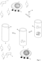
- an apparatus for purification of target cells wherein the apparatus comprises at least one arrangement of a first and a second stationary phase for chromatography as explained above, that means a chromatography column for selection of cells (a selection cartridge) and a second chromatography column (a removal cartridge) for removal of reagents of the isolation or the staining of target cells.
- a selection cartridge for selection of cells
- a removal cartridge for removal of reagents of the isolation or the staining of target cells.
- Carrying out such a two step isolation procedure yields target cells which can be directly subjected to the next desired application or selection cycle.
- FACS and MACS selections in the inventive chromatographic selection method no further procedures such as washing and centrifugation are necessary between two selection cycles and the cells are not functionally compromised by bound isolation reagents such as receptor binding reagents or magnetic beads. Therefore, the invention provides for the first time a reliable, simple to construct and yet effective apparatus for target cell purification.
- an apparatus may comprise a plurality of arrangements of first and second stationary phases (chromatography columns) being fluidly connected in series.
- the apparatus may comprise a sample inlet being fluidly connected to the first stationary phase of the first arrangement of a first and a second stationary phases for chromatography.
- the apparatus may also comprise a sample outlet for purified target cells, the sample outlet being fluidly connected to the second stationary phase of the last of the at least one arrangement of a first and second stationary phases for chromatography.
- the apparatus may also comprise a competition reagent container that is fluidly connected to at least one of the first stationary phases of the arrangements of a first and second stationary phases for chromatography.
- compositions of matter, means, uses, methods, or steps, presently existing or later to be developed that perform substantially the same function or achieve substantially the same result as the corresponding exemplary embodiments described herein may likewise be utilized according to the present invention.
- Fab fragments that are directed against cell surface markers are used as receptor binding reagent.
- the Fab fragments are recombinantly expressed in E. coli or other hosts and contain a streptavidin binding peptide as binding partner C. Tetrameric streptavidin or tetrameric streptavidin muteins provides the one or more binding site(s) Z.
- Fab fragments are bound to streptavidin, which itself is covalently linked to beads. These beads are used for affinity column chromatography of cell suspensions with a subpopulation of cells having an extracellular protein (receptor molecule) that is able to be bound by the Fab fragments.
- Unbound cells are washed away in this "selection cartridge" while bound cells are subsequently eluted with biotin containing buffer disrupting the binding of streptavidin mutein with the streptavidin binding peptide of the Fab fragment (acting as receptor binding reagent).
- biotin containing buffer disrupting the binding of streptavidin mutein with the streptavidin binding peptide of the Fab fragment (acting as receptor binding reagent).
- Fab fragment with the cell bound thereon are released from the column and, due to the missing avidity effect, Fab fragments dissociate from the target cells.
- the suspension can now be purified from the remaining Fab fragments and biotin by a second column chromatography (the removal cartridge) using another gel (chromatography) matrix with covalently bound streptavidin whereby cells elute in the void volume and Fab fragments and biotin are quantitatively bound on the chromatography matrix.
- the cells can now be exposed to a further cycle of purification using a different Fab fragment or any other receptor binding reagent in a similar way.
- Columns with Fab fragments (selection cartridge) and a subsequent column for Fab and biotin removal (removal cartridge) can be combined in a serial manner by simply arranging the columns linearly one after the other.
- Example 1 single step purification of CD8+ cells via column chromatography
- Sephadex G50 (Sigma) was used as stationary phase and was covalently coupled with Strep-tactin ® (a recombinant streptavidin variant, IBA GmbH, Germany) using the CNBr method.
- a 50% suspension of Sephadex G50 contained covalently coupled 70 microgram Strep-tactin ® /ml of the bead suspension.
- Strep-tactin ® served as affinity reagent which was immobilized to the affinity matrix before addition of receptor binding reagent and the sample containing the target cells.
- Pellet 1 contained about 3.9 million cells
- pellet 2 contained 0.7 million cells.
- CD8+ target cells can be isolated via reversible immobilisation/affinity chromatography.
- Example 2 Removal of biotin and Fab from C8+ cells.
- CD8+ cells were isolated as described above except that cells after biotin elution (6 ml buffer) were directly passed through a column of Superflow TM Sepharose ® beads (bed volume of 6 ml) that had Strep-Tactin ® (IBA GmbH, Göttingen, Germany) covalently attached thereto with a binding capacity of 300 nanomol biotin/ml. While the Superflow TM Sepharose ® beads served as gel permeation matrix for the isolation/enrichment of target cells, the Strep-Tactin ® immobilized on the beads has binding affinity to both the CD8+ Fab fragments that are equipped with the streptavidin binding peptide and for biotin.
- Strep-Tactin ® served as affinity/removal reagent for biotin and the CD8+ Fab fragments.
- the eluate (6ml) of this second column (which acted as removal cartridge) filled with Superflow beads was collected.
- Biotin and Fab fragments were not detectable in the eluate that contained the target cells using a biotin assay and Fab fragment assay using Western blotting (results not shown).
- a similar experiment was carried out using FITC labeled biotin (Sigma) and fluorescently labeled CD8+ Fab (IBA GmbH). The fact that also this final eluate contained no Fab fragments or biotin was confirmed when measured with a sensitive fluorimeter. Thus, it was found, that whereas biotin and Fab were completely removed, the eluate after Superflow TM / Strep-Tactin ® chromatography contained 95% to 100% of the CD8+ cells, with no obvious loss of cells.
- the purified CD8+ cells can go to another cycle of purification using, for example, a CD25+ Fab fragment (or any other receptor molecule that is present on the surface of the isolated CD8+ target cells).
- a serial purification of T-cells can for example be carried out using the apparatus depicted in Fig. 6a or Fig. 6b .
- Example 4 Purification of cells by chromatography on a planar matrix (Strep-Tactin ® coated nitrocellulose membrane)
- Strep-Tactin ® For non-covalent attachment of Strep-Tactin ® on the membrane, a piece of nitrocellulose membrane (24 cm 2 , Whatman, UK) was put in a petri dish and incubated with 4 mg Strep-tactin ® (IBA GmbH) in 10 ml PBS for 10 minutes and then washed 5 times with 20 ml PBS . Then 5 microgramm CD8+ Fab fragment (catalogue number: 6-8003-005, IBA GmbH, Göttingen, supra) was added in 5ml PBS and incubated for 5min at 4°C. Then 5 million cells (PBMCs) in FACS buffer (0.5 % BSA (w/v) in PBS pH 7.4) were added and incubated at 4°C for 10min.
- PBMCs PBMCs
- FACS buffer 0.5 % BSA (w/v) in PBS pH 7.4
- the membrane was washed five times in 10 ml FACS buffer and the wash fractions were collected for FACS analysis. The membrane was then incubated for 5 min in 10 ml FACS buffer containing 1mmol biotin. The resulting fraction was collected for FACS analysis.
- Example 5 Single step purification of human CD8+ cells via column chromatography with Superflow TM Agarose
- the wash fraction (12ml) of the FACS buffer contained only 1.77% CD8+ cells in comparison to the starting fraction (A) which contained 7.98% CD8+ cells, thus 77.8% CD8+ cells were retarded on the column.
- the elution with biotin containing buffer fraction (B, 12ml) resulted in approximately 65% of the bound CD8+ cells with a purity of 98.5%. Also this experiment shows that CD8+ target cells can be isolated via reversible immobilisation/affinity chromatography as described herein using commercially available chromatography matrices.
- Example 6 Single step purification of human CD8+ cells via column chromatography
- Human CD8+ cells were purified from density gradient (Ficoll) purified PBMCs by the use of a column prepared from 500 ⁇ 1 Strep-Tactin ® -agarose (cross-linked agarose was obtained from Agarose Beads Technologies, Madrid, Spain with a reduced exclusion size compared to Superflow TM Agarose) bead resin functionalized with 10 ⁇ g anti-CD8 Fab-fragments (catalogue number: 6-8003, IBA GmbH, Göttingen).
- the Fab fragment carrying the sequential arrangement of the two streptavidin binding modules SAWSHPQFEK(GGGS) 2 GGSAWSHPQFEK at the C-terminus of the heavy chain was loaded (immobilized) onto the Strep-Tactin ® -agarose matrix by pumping 1000 ⁇ l Fab containing washing buffer (PBS plus 0.5% bovine serum albumin) over the column at a speed of 300 ⁇ l/min prior to the cell purification.
- 1000 ⁇ l Fab containing washing buffer PBS plus 0.5% bovine serum albumin
- For purification of the target cells 1 ⁇ 10 8 freshly prepared PBMCs (in 2ml washing buffer) were automatically loaded on the column at a flow rate of 300 ⁇ l/min with a peristaltic pump.
- Human CD8+ cells were purified from whole blood by the use of a column prepared from 1200 ⁇ l Strep-Tactin ® -agarose (cross-linked agarose was obtained from Agarose Beads Technologies Madrid, Spain with a reduced exclusion size compared to Superflow TM agarose) bead resin functionalized with 30 ⁇ g anti-CD8 Fab-fragment (catalogue number: 6-8003, IBA GmbH, Göttingen).
- the Fab fragment carrying the sequential arrangement of the two streptavidin binding modules SAWSHPQFEK(GGGS) 2 GGSAWSHPQFEK at the C-terminus of the heavy chain were immobilized on the Strep-Tactin ® -agarose matrix by pumping 1500 ⁇ l Fab fragment containing washing buffer (PBS plus 0.5% bovine serum albumin) over the column at a speed of 300 ⁇ l/min prior to the cell purification.
- washing buffer PBS plus 0.5% bovine serum albumin
- Example 8 Pipette based single step purification of murine CD4+ cells from splenocytes
- CD4+ cells were isolated from splenocytes by the use of a pipette tip loaded with 80 ⁇ l Strep-Tactin ® -agarose bead resin (cross-linked agarose was obtained from Agarose Beads Technologies , Madrid, Spain with a reduced exclusion size compared to Superflow TM agarose) functionalized with 2 ⁇ g anti-CD4 Fab-fragment.
- the pipette tips were filled with the agarose material by Phynexus Inc., USA.
- the Fab fragment used comprised the wild-type variable domains of the CD4 binding antibody GK1.5 ( Dialynas DP et al., Immunol Rev.
- Loading/immobilizing the Fab fragment onto the Streptactin ® -agarose matrix was achieved by pipetting with a handheld electric pipette 400 ⁇ l Fab fragment containing washing buffer (PBS plus 0.5% bovine serum albumin) at a speed of 300 ⁇ l/min onto the agarose chromatography matrix prior to the cell purification.
- the CD8+ target cells were purified with a yield of 95 % and a purity of 85 %. Dot plots of the respective starting-, negative- and positive fractions as well as the corresponding purity and yield of a representative selection are shown in Fig.9 .
- Example 9 Single step purification of human CD4+ cells via column chromatography
- Human CD4+ cells were isolated from density gradient (Ficoll) purified PBMCs by the use of a pipette tip loaded with 80 ⁇ l Strep-Tactin ® -agarose (cross-linked agarose was obtained from Agarose Beads Technologies , Madrid, Spain with a reduced exclusion size compared to Superflow TM Agarose) bead resin functionalized with 2 ⁇ g anti-CD4 Fab-fragment.
- the CD4 Fab fragment used was a mutant of the 13B8.2 Fab fragment described in US Patent 7,482,000 and Bes, C., et al. J Biol Chem 278, 14265-14273 (2003 )).
- the mutant Fab fragment termed "m13B8.2” carries the variable domain of the CD4 binding murine antibody 13B8.2 and a constant domain consisting of constant human CH1 domain of type gamma1 for the heavy chain and the constant human light chain domain of type kappa, as described in US Patent 7,482,000 .
- the His residue at position 91 of the light chain (position 93 in SEQ ID NO: 2) is mutated to Ala
- the Arg residue at position 53 of the heavy chain is mutated to Ala.
- the Fab fragment m13B8.2 carries a sequential arrangement of the two streptavidin binding modules SAWSHPQFEK(GGGS) 2 GGSAWSHPQFEK at the C-terminus of the heavy chain.
- the Fab fragment was immobilized on the Strep-Tactin ® -agarose matrix with a handheld electric pipette by pipetting 200 ⁇ l Fab fragment containing washing buffer at a speed of 300 ⁇ l/min prior to the cell purification.
- PBMCs in 0.5ml washing buffer
- PBS plus 0.5% bovine serum albumin PBS plus 0.5% bovine serum albumin
- Unbound (CD4-negative) cells were subsequently removed from the tip by triple repetitive washing (by pipetting the wash buffer up and down) with 1ml washing buffer at a speed of 2ml/min.
- Example 10 Pipette based single step purification of human CD4+ cells from whole blood
- CD4+ cells were isolated from whole blood by the use of a pipette tip loaded with 80 ⁇ l Strep-Tactin ® -agarose (cross-linked agarose was obtained from Agarose Beads Technologies, Madrid, Spain with a reduced exclusion size compared to Superflow TM Agarose) bead resin functionalized with 0.5 ⁇ g anti-CD4 Fab-fragment.
- the CD4 binding Fab fragment m13B8.2 used in Example 9 was also in Example 10.
- the Fab fragment was immobilized on the Strep-Tactin ® -agarose matrix with a handheld electric pipette by pipetting 200 ⁇ l Fab containing washing buffer at a speed of 300 ⁇ l/min prior to the cell isolation.
- biotin as the eluent and the Fab fragment as the respective receptor binding reagent can be removed from the target cell sample by means of the "removal cartridge" as described in Example 2.
Landscapes
- Health & Medical Sciences (AREA)
- Life Sciences & Earth Sciences (AREA)
- Immunology (AREA)
- Chemical & Material Sciences (AREA)
- Engineering & Computer Science (AREA)
- Hematology (AREA)
- Molecular Biology (AREA)
- Biomedical Technology (AREA)
- Urology & Nephrology (AREA)
- Cell Biology (AREA)
- Analytical Chemistry (AREA)
- Pathology (AREA)
- General Health & Medical Sciences (AREA)
- General Physics & Mathematics (AREA)
- Biochemistry (AREA)
- Physics & Mathematics (AREA)
- Medicinal Chemistry (AREA)
- Food Science & Technology (AREA)
- Biotechnology (AREA)
- Microbiology (AREA)
- Virology (AREA)
- Tropical Medicine & Parasitology (AREA)
- Zoology (AREA)
- Chemical Kinetics & Catalysis (AREA)
- Peptides Or Proteins (AREA)
- Apparatus Associated With Microorganisms And Enzymes (AREA)
- Micro-Organisms Or Cultivation Processes Thereof (AREA)
- Solid-Sorbent Or Filter-Aiding Compositions (AREA)
- Measuring Or Testing Involving Enzymes Or Micro-Organisms (AREA)
- Treatment Of Liquids With Adsorbents In General (AREA)
- Preparation Of Compounds By Using Micro-Organisms (AREA)
- Investigating Or Analysing Biological Materials (AREA)
Priority Applications (1)
| Application Number | Priority Date | Filing Date | Title |
|---|---|---|---|
| EP24222803.9A EP4545964A3 (en) | 2012-02-23 | 2013-02-25 | Chromatographic isolation of cells and other complex biological materials |
Applications Claiming Priority (2)
| Application Number | Priority Date | Filing Date | Title |
|---|---|---|---|
| US201261602150P | 2012-02-23 | 2012-02-23 | |
| PCT/EP2013/053650 WO2013124474A2 (en) | 2012-02-23 | 2013-02-25 | Chromatographic isolation of cells and other complex biological materials |
Related Child Applications (1)
| Application Number | Title | Priority Date | Filing Date |
|---|---|---|---|
| EP24222803.9A Division EP4545964A3 (en) | 2012-02-23 | 2013-02-25 | Chromatographic isolation of cells and other complex biological materials |
Publications (2)
| Publication Number | Publication Date |
|---|---|
| EP2817625A2 EP2817625A2 (en) | 2014-12-31 |
| EP2817625B1 true EP2817625B1 (en) | 2025-01-01 |
Family
ID=47891608
Family Applications (2)
| Application Number | Title | Priority Date | Filing Date |
|---|---|---|---|
| EP13709791.1A Active EP2817625B1 (en) | 2012-02-23 | 2013-02-25 | Chromatographic isolation of cells and other complex biological materials |
| EP24222803.9A Pending EP4545964A3 (en) | 2012-02-23 | 2013-02-25 | Chromatographic isolation of cells and other complex biological materials |
Family Applications After (1)
| Application Number | Title | Priority Date | Filing Date |
|---|---|---|---|
| EP24222803.9A Pending EP4545964A3 (en) | 2012-02-23 | 2013-02-25 | Chromatographic isolation of cells and other complex biological materials |
Country Status (11)
| Country | Link |
|---|---|
| US (4) | US10228312B2 (enExample) |
| EP (2) | EP2817625B1 (enExample) |
| JP (4) | JP6362543B2 (enExample) |
| CN (2) | CN104254780B (enExample) |
| AU (4) | AU2013224027B2 (enExample) |
| CA (1) | CA2865033C (enExample) |
| ES (1) | ES3011307T3 (enExample) |
| IN (1) | IN2014DN06475A (enExample) |
| RU (1) | RU2746407C1 (enExample) |
| SG (2) | SG10201606970UA (enExample) |
| WO (1) | WO2013124474A2 (enExample) |
Families Citing this family (66)
| Publication number | Priority date | Publication date | Assignee | Title |
|---|---|---|---|---|
| US9637719B2 (en) | 2013-12-06 | 2017-05-02 | Douglas T. Gjerde | Devices and methods for purification of biological cells |
| RU2746407C1 (ru) | 2012-02-23 | 2021-04-13 | Стейдж Селл Терапеутикс Гмбх | Хроматографическое выделение клеток и других сложных биологических материалов |
| PT3083671T (pt) | 2013-12-20 | 2020-12-24 | Hutchinson Fred Cancer Res | Moléculas efetoras quiméricas etiquetadas e recetores destas |
| EP3126027A4 (en) * | 2014-04-03 | 2017-11-01 | Douglas T. Gjerde | Devices and methods for purification, detection and use of biological cells |
| IL293587B2 (en) | 2014-04-16 | 2025-02-01 | Juno Therapeutics Gmbh | Methods, kits and devices for increasing cell populations |
| CN106459917B (zh) * | 2014-04-23 | 2021-03-09 | 朱诺治疗学股份有限公司 | 分离、培养和遗传工程改造用于过继治疗的免疫细胞群的方法 |
| JP6678593B2 (ja) * | 2014-04-30 | 2020-04-08 | イーベーアー ゲーエムベーハー | 標的細胞を単離する方法 |
| EP2955521A1 (en) * | 2014-06-11 | 2015-12-16 | Centre Hospitalier Universitaire Vaudois (CHUV) | Methods for separating cells |
| HUE050264T2 (hu) * | 2014-11-05 | 2020-11-30 | Juno Therapeutics Inc | Eljárások transzdukálásra és sejtfeldolgozásra |
| DE202014105965U1 (de) | 2014-12-10 | 2015-01-26 | Cell.Copedia GmbH | Spritzengehäuse zum Pipettieren eines biologischen Materials mit integrierten Membranen |
| WO2016166568A1 (en) | 2015-04-16 | 2016-10-20 | Juno Therapeutics Gmbh | Methods, kits and apparatus for expanding a population of cells |
| US11827904B2 (en) | 2015-04-29 | 2023-11-28 | Fred Hutchinson Cancer Center | Modified stem cells and uses thereof |
| CN105606815A (zh) * | 2015-05-18 | 2016-05-25 | 烟台载通生物技术有限公司 | 一种用于免疫检测的多单元装置 |
| MA45426A (fr) | 2015-10-22 | 2019-05-01 | Juno Therapeutics Gmbh | Procédés, kits, agents et appareils de transduction |
| MA45488A (fr) | 2015-10-22 | 2018-08-29 | Juno Therapeutics Gmbh | Procédés, kits et appareil de culture de cellules |
| MA45489A (fr) * | 2015-10-22 | 2018-08-29 | Juno Therapeutics Gmbh | Procédés de culture de cellules, kits et appareil associés |
| GB201521784D0 (en) * | 2015-12-10 | 2016-01-27 | Univ Birmingham | Cell purification |
| JP7062640B2 (ja) | 2016-07-29 | 2022-05-06 | ジュノー セラピューティクス インコーポレイテッド | 抗cd19抗体に対する抗イディオタイプ抗体 |
| US10294454B2 (en) | 2016-08-24 | 2019-05-21 | General Electric Company | Methods and kits for cell activation |
| JP7376091B2 (ja) | 2016-12-23 | 2023-11-08 | エクソファーマ リミテッド | 微小胞およびエキソソームの精製または単離のための方法および組成物 |
| MX2019008538A (es) | 2017-01-20 | 2019-11-05 | Juno Therapeutics Gmbh | Conjugados de superficie celular y composiciones y métodos celulares relacionados. |
| CN111032850B (zh) * | 2017-04-27 | 2024-08-13 | 朱诺治疗学有限公司 | 寡聚粒子试剂及其使用方法 |
| KR20200064060A (ko) | 2017-07-29 | 2020-06-05 | 주노 쎄러퓨티크스 인코퍼레이티드 | 재조합 수용체를 발현하는 세포 증폭용 시약 |
| MA49979A (fr) | 2017-08-09 | 2020-06-17 | Juno Therapeutics Inc | Procédés de production de compositions de cellules génétiquement modifiées et compositions associées |
| EP3704230B1 (en) | 2017-11-01 | 2024-10-23 | Juno Therapeutics, Inc. | Process for generating therapeutic compositions of engineered cells |
| CN112004824B (zh) | 2017-12-08 | 2024-09-13 | 朱诺治疗学股份有限公司 | 生产工程化t细胞组合物的过程 |
| WO2019133842A1 (en) * | 2017-12-29 | 2019-07-04 | The United States Of America, As Represented By The Secretary, Department Of Health And Human Services | Purification and labeling of extracellular vesicles using a mixed mode resin composition |
| CN119881293A (zh) * | 2018-03-21 | 2025-04-25 | 沃特世科技公司 | 基于非抗体高亲和力的样品制备、吸附剂、装置和方法 |
| BR112020026067A2 (pt) | 2018-06-18 | 2021-03-23 | Exopharm Limited | métodos e composições para purificação ou isolamento de microvesículas e exossomas |
| JP2021530230A (ja) * | 2018-07-17 | 2021-11-11 | ユニヴァーシティ オブ ワシントン | アプタマーベースの可逆的細胞選択に関連する組成物および方法 |
| CN113474450A (zh) | 2018-08-09 | 2021-10-01 | 朱诺治疗学股份有限公司 | 产生工程化细胞的方法以及所述工程化细胞的组合物 |
| US12366580B2 (en) | 2018-09-11 | 2025-07-22 | Juno Therapeutics, Inc. | Methods for mass spectrometry analysis of engineered cell compositions |
| EP3874024A1 (en) | 2018-10-31 | 2021-09-08 | Juno Therapeutics GmbH | Methods for selection and stimulation of cells and apparatus for same |
| WO2020097132A1 (en) | 2018-11-06 | 2020-05-14 | Juno Therapeutics, Inc. | Process for producing genetically engineered t cells |
| BR112021024404A2 (pt) | 2019-06-07 | 2022-04-19 | Juno Therapeutics Inc | Cultura de célula t automatizada |
| US12194221B2 (en) * | 2019-06-27 | 2025-01-14 | Bioimmunate Technologies Ltd. | Filter for removal of multiple sclerosis-associated t-cells |
| EP4022637A2 (en) | 2019-08-30 | 2022-07-06 | Juno Therapeutics, Inc. | Machine learning methods for classifying cells |
| BR112022002969A2 (pt) * | 2019-08-30 | 2022-05-17 | Flodesign Sonics Inc | Seleção de células de afinidade acústica para múltiplos receptores alvo |
| JP2022547309A (ja) * | 2019-09-10 | 2022-11-11 | ピアス・バイオテクノロジー・インコーポレイテッド | サンプル調製組成物、デバイス、システムおよび方法 |
| WO2021084050A1 (en) | 2019-10-30 | 2021-05-06 | Juno Therapeutics Gmbh | Cell selection and/or stimulation devices and methods of use |
| WO2021118873A1 (en) * | 2019-12-09 | 2021-06-17 | St. Jude Children's Research Hospital, Inc. | Method for preparing cd7-negative, cd3-positive t cells |
| AU2021214142A1 (en) | 2020-01-28 | 2022-09-08 | Juno Therapeutics, Inc. | Methods for T cell transduction |
| AU2021213439B2 (en) * | 2020-01-31 | 2023-01-05 | Cell.Copedia GmbH | Methods of isolating a biological entity |
| KR20220152220A (ko) | 2020-02-12 | 2022-11-15 | 주노 쎄러퓨티크스 인코퍼레이티드 | Cd19-지시된 키메라 항원 수용체 t 세포 조성물 및 이의 방법 및 용도 |
| EP4103203A1 (en) | 2020-02-12 | 2022-12-21 | Juno Therapeutics, Inc. | Bcma-directed chimeric antigen receptor t cell compositions and methods and uses thereof |
| US20230181641A1 (en) | 2020-05-13 | 2023-06-15 | Juno Therapeutics, Inc. | Process for producing donor-batched cells expressing a recombinant receptor |
| EP4334341A2 (en) | 2021-05-06 | 2024-03-13 | Juno Therapeutics GmbH | Methods for stimulating and transducing t cells |
| WO2023017115A1 (en) * | 2021-08-11 | 2023-02-16 | Iba Lifesciences Gmbh | Fractionation of cells based on a marker protein |
| CN113908589B (zh) * | 2021-10-08 | 2022-09-27 | 天津工业大学 | 一种表面印迹抗体的疏水电荷诱导模式膜层析介质及其制备方法 |
| WO2023147510A1 (en) | 2022-01-28 | 2023-08-03 | Juno Therapeutics, Inc. | Chromatography arrays and related methods and kits |
| ES3024941T3 (en) * | 2022-02-02 | 2025-06-05 | Sartorius Bia Separations D O O | Method of purifying or removing a target component from full plasma |
| WO2023213969A1 (en) | 2022-05-05 | 2023-11-09 | Juno Therapeutics Gmbh | Viral-binding protein and related reagents, articles, and methods of use |
| EP4532695A1 (en) | 2022-05-25 | 2025-04-09 | Celgene Corporation | Methods of manufacturing t cell therapies |
| WO2024100604A1 (en) | 2022-11-09 | 2024-05-16 | Juno Therapeutics Gmbh | Methods for manufacturing engineered immune cells |
| CN121100267A (zh) | 2022-12-09 | 2025-12-09 | 朱诺治疗学股份有限公司 | 使用全息成像预测细胞表型的机器学习方法 |
| CN121002052A (zh) | 2023-02-03 | 2025-11-21 | C3S2 有限公司 | 用于非病毒生产工程化免疫细胞的方法 |
| CN121127253A (zh) | 2023-02-28 | 2025-12-12 | 朱诺治疗学股份有限公司 | 治疗系统性自身免疫性疾病的细胞疗法 |
| WO2025049254A1 (en) | 2023-08-25 | 2025-03-06 | Juno Therapeutics, Inc. | Pyrazolopyridine based inhibitors of dna-dependent protein kinase and compositions and application in gene editing |
| WO2025049250A1 (en) | 2023-08-25 | 2025-03-06 | Juno Therapeutics, Inc. | 6,6-core silacycle based inhibitors of dna-dependent protein kinase and compositions and application in gene editing |
| WO2025049253A1 (en) | 2023-08-25 | 2025-03-06 | Juno Therapeutics, Inc. | Bridged cycle‑based inhibitors of dna‑dependent protein kinase and compositions and application in gene editing |
| WO2025049247A1 (en) | 2023-08-25 | 2025-03-06 | Juno Therapeutics, Inc. | 5,6-core silacycle based inhibitors of dna-dependent protein kinase and compositions and application in gene editing |
| WO2025083196A1 (en) | 2023-10-18 | 2025-04-24 | Tq Therapeutics Gmbh | A method of manufacturing a cellular therapy product and respective kits and devices |
| US12434218B2 (en) | 2023-11-28 | 2025-10-07 | Phenomenex, Inc. | Pore structure for separation of adeno-associated viruses (AAVS) from their aggregates |
| US20250241912A1 (en) | 2024-01-25 | 2025-07-31 | Juno Therapeutics, Inc. | Use of bridged cycle-based inhibitors of dna-dependent protein kinase in combination of dna polymerase theta inhibitor and compositions and application in gene editing |
| WO2025179188A1 (en) | 2024-02-22 | 2025-08-28 | Juno Therapeutics, Inc. | Pyrazole-based inhibitors of dna-dependent protein kinase and compositions and applications in gene editing |
| US20250345431A1 (en) | 2024-05-10 | 2025-11-13 | Juno Therapeutics, Inc. | Genetically engineered t cells expressing a cd19 chimeric antigen receptor (car) and uses thereof for allogeneic cell therapy |
Citations (1)
| Publication number | Priority date | Publication date | Assignee | Title |
|---|---|---|---|---|
| US6103493A (en) * | 1996-10-10 | 2000-08-15 | Institut Fur Bioanalytic | Streptavidin muteins |
Family Cites Families (167)
| Publication number | Priority date | Publication date | Assignee | Title |
|---|---|---|---|---|
| US2467434A (en) | 1947-05-10 | 1949-04-19 | Air Associates Inc | Servomotor and pressure responsive valve therefor |
| US4235871A (en) | 1978-02-24 | 1980-11-25 | Papahadjopoulos Demetrios P | Method of encapsulating biologically active materials in lipid vesicles |
| US4361549A (en) | 1979-04-26 | 1982-11-30 | Ortho Pharmaceutical Corporation | Complement-fixing monoclonal antibody to human T cells, and methods of preparing same |
| US4816567A (en) | 1983-04-08 | 1989-03-28 | Genentech, Inc. | Recombinant immunoglobin preparations |
| JP2624470B2 (ja) | 1984-10-02 | 1997-06-25 | バイオジェン インコーポレイテッド | ストレプトアビジン様ポリペプチドの製造 |
| US4851341A (en) | 1986-12-19 | 1989-07-25 | Immunex Corporation | Immunoaffinity purification system |
| US6534055B1 (en) | 1988-11-23 | 2003-03-18 | Genetics Institute, Inc. | Methods for selectively stimulating proliferation of T cells |
| US6352694B1 (en) | 1994-06-03 | 2002-03-05 | Genetics Institute, Inc. | Methods for inducing a population of T cells to proliferate using agents which recognize TCR/CD3 and ligands which stimulate an accessory molecule on the surface of the T cells |
| GB9215540D0 (en) | 1992-07-22 | 1992-09-02 | Celltech Ltd | Protein expression system |
| DE4237113B4 (de) | 1992-11-03 | 2006-10-12 | "Iba Gmbh" | Peptide und deren Fusionsproteine, Expressionsvektor und Verfahren zur Herstellung eines Fusionsproteins |
| DE4417598A1 (de) | 1994-05-19 | 1995-12-14 | Max Planck Gesellschaft | Verwendung des Tetracyclinpromotors zur stringent regulierten Produktion von rekombinanten Proteinen in prokaryontischen Zellen |
| US7074904B2 (en) | 1994-07-29 | 2006-07-11 | Altor Bioscience Corporation | MHC complexes and uses thereof |
| DK0776339T4 (da) | 1994-07-29 | 2010-05-25 | Sunol Molecular Corp | MHC-komplekser og anvendelser deraf |
| AU5132096A (en) | 1995-01-30 | 1996-08-21 | Terrapin Technologies, Inc. | Glubodies - multiplicities of proteins capable of binding a variety of small molecules |
| CA2211951A1 (en) | 1995-02-09 | 1996-08-15 | University Of Washington | Modified-affinity streptavidin |
| WO1997011183A1 (en) | 1995-04-11 | 1997-03-27 | Trustees Of Boston University | Streptavidin mutants |
| US5629205A (en) | 1995-05-19 | 1997-05-13 | Allelix Biopharmaceuticals Inc. | Promoters for gene expression |
| WO1997005266A1 (en) | 1995-07-25 | 1997-02-13 | Introgene B.V. | Methods and means for targeted gene delivery |
| AR005035A1 (es) | 1995-12-11 | 1999-04-07 | Merck Patent Ges Mit Beschränkter Haftung | Procedimiento para preparar proteinas recombinantes en e. coli, mediante fermentacion con gran concentracion de celulas. |
| US5869270A (en) | 1996-01-31 | 1999-02-09 | Sunol Molecular Corporation | Single chain MHC complexes and uses thereof |
| US5773224A (en) | 1996-02-12 | 1998-06-30 | Grandics; Peter | Immunoselection system for cell elution |
| DE19637718A1 (de) | 1996-04-01 | 1997-10-02 | Boehringer Mannheim Gmbh | Rekombinante inaktive Core-Streptavidin Mutanten |
| US6022688A (en) | 1996-05-13 | 2000-02-08 | Sequenom, Inc. | Method for dissociating biotin complexes |
| US20050003431A1 (en) | 1996-08-16 | 2005-01-06 | Wucherpfennig Kai W. | Monovalent, multivalent, and multimeric MHC binding domain fusion proteins and conjugates, and uses therefor |
| AU730457B2 (en) | 1996-08-16 | 2001-03-08 | President And Fellows Of Harvard College | Soluble monovalent and multivalent MHC class II fusion proteins, and uses therefor |
| US6368813B1 (en) | 1997-03-14 | 2002-04-09 | The Trustees Of Boston University | Multiflavor streptavidin |
| US6391571B1 (en) | 1997-04-01 | 2002-05-21 | Roche Diagnostics Gmbh | Recombinant inactive avidin mutants |
| CA2302779C (en) | 1997-09-16 | 2010-02-02 | Oregon Health Sciences University | Recombinant mhc molecules useful for manipulation of antigen-specific t-cells |
| US6232445B1 (en) | 1997-10-29 | 2001-05-15 | Sunol Molecular Corporation | Soluble MHC complexes and methods of use thereof |
| US5985658A (en) | 1997-11-14 | 1999-11-16 | Health Research Incorporated | Calmodulin-based cell separation technique |
| NZ506071A (en) | 1998-02-19 | 2003-05-30 | Harvard College | Monovalent, multivalent, and multimeric major histocompatibility complex (MHC) binding domain fusion proteins and conjugates, and uses therefore |
| US6664042B1 (en) | 1999-01-26 | 2003-12-16 | Cornell Research Foundation, Inc. | Determining viral load in double negative T cells |
| KR20020013496A (ko) | 1999-02-04 | 2002-02-20 | 추후제출 | 조혈 간세포 및(또는) 전구세포의 유지 및 증대 방법 및장치 |
| EP1048732A1 (de) | 1999-04-26 | 2000-11-02 | F. Hoffmann-La Roche Ag | Verfahren zur Herstellung von natürlich gefalteten und sekretierten Proteinen |
| EP1669129A1 (en) | 1999-05-14 | 2006-06-14 | Pall Corporation | Charged membrane |
| WO2000069549A1 (en) | 1999-05-14 | 2000-11-23 | Pall Corporation | Charged membrane |
| US6849185B1 (en) | 1999-05-14 | 2005-02-01 | Pall Corp | Charged membrane |
| DE60043889D1 (en) | 1999-06-07 | 2010-04-08 | Tet Systems Holding Gmbh & Co | Tet repressor basierte transkriptionale regulatorische proteine |
| DE19932688B4 (de) | 1999-07-13 | 2009-10-08 | Scil Proteins Gmbh | Design von Beta-Faltblatt-Proteinen des gamma-II-kristallins antikörperähnlichen |
| US20030235908A1 (en) | 2000-02-24 | 2003-12-25 | Xcyte Therapies, Inc. | Activation and expansion of cells |
| DE60127113T2 (de) | 2000-04-25 | 2007-06-14 | Otsuka Pharmaceutical Co., Ltd. | Gd3-mimetische peptide |
| US6638728B1 (en) | 2000-06-16 | 2003-10-28 | Pierce Biotechnology, Inc. | Coated surfaces with high capacity for capturing target molecules |
| JP4878724B2 (ja) | 2000-11-03 | 2012-02-15 | ジェネンテック, インコーポレイテッド | 組み換えタンパク質を発現する発酵における代謝速度シフト |
| US6979556B2 (en) | 2000-12-14 | 2005-12-27 | Genentech, Inc. | Separate-cistron contructs for secretion of aglycosylated antibodies from prokaryotes |
| EP1227321A1 (en) | 2000-12-28 | 2002-07-31 | Institut für Bioanalytik GmbH | Reversible MHC multimer staining for functional purification of antigen-specific T cells |
| EP1349569B1 (en) | 2001-01-12 | 2007-04-18 | Becton Dickinson and Company | Intrinsically fluorescent, self-multimerizing mhc fusion proteins and complexes thereof |
| GB0102568D0 (en) | 2001-02-01 | 2001-03-21 | Magnetic Biosolutions Sweden A | Method |
| DE10113776B4 (de) | 2001-03-21 | 2012-08-09 | "Iba Gmbh" | Isoliertes streptavidinbindendes, kompetitiv eluierbares Peptid, dieses umfassendes Fusionspeptid, dafür codierende Nukleinsäure, Expressionsvektor, Verfahren zur Herstellung eines rekombinanten Fusionsproteins und Verfahren zum Nachweis und/oder zur Gewinnung des Fusionsproteins |
| EP1908769A1 (en) | 2001-08-27 | 2008-04-09 | Genentech, Inc. | A system for antibody expression and assembly |
| KR100968664B1 (ko) | 2001-08-27 | 2010-07-06 | 제넨테크, 인크. | 항체 발현 및 조립을 위한 시스템 |
| WO2003029462A1 (en) | 2001-09-27 | 2003-04-10 | Pieris Proteolab Ag | Muteins of human neutrophil gelatinase-associated lipocalin and related proteins |
| US7094579B2 (en) | 2002-02-13 | 2006-08-22 | Xoma Technology Ltd. | Eukaryotic signal sequences for prokaryotic expression |
| JP2005536184A (ja) * | 2002-03-01 | 2005-12-02 | エルドマン,ヴォルカー,エー. | ストレプトアビジン結合ペプチド |
| US7754155B2 (en) * | 2002-03-15 | 2010-07-13 | Ross Amelia A | Devices and methods for isolating target cells |
| WO2003090781A1 (en) | 2002-04-23 | 2003-11-06 | Meir Strahilevitz | Methods and devices for targeting a site in a mammal and for removing species from a mammal |
| ATE345502T1 (de) | 2002-06-24 | 2006-12-15 | Profos Ag | Verfahren zum nachweis und zur entfernung von endotoxin |
| FR2841905B1 (fr) | 2002-07-05 | 2004-09-03 | Centre Nat Rech Scient | Fragments fab mutants de l'anticorps anti-cd4 chimere 13b8.2 et leurs applications |
| CN102061288A (zh) | 2002-07-17 | 2011-05-18 | 希托斯生物技术股份公司 | 使用衍生自ap205外壳蛋白的病毒样颗粒的分子抗原阵列 |
| GB2392158B (en) | 2002-08-21 | 2005-02-16 | Proimmune Ltd | Chimeric MHC protein and oligomer thereof |
| EP1569510B1 (en) | 2002-09-27 | 2011-11-02 | The General Hospital Corporation | Microfluidic device for cell separation and uses thereof |
| EP1588166A4 (en) | 2003-01-09 | 2007-06-27 | Macrogenics Inc | VECTOR SYSTEM WITH DOUBLE EXPRESSION FOR EXPRESSING ANTIBODIES IN BACTERIAL AND MAMMAL CELLS |
| WO2004096975A2 (en) | 2003-05-02 | 2004-11-11 | Insception Bioscience, Inc. | Apparatus and methods for amplification of blood stem cell numbers |
| EP1623017B1 (en) | 2003-05-08 | 2010-09-15 | Life Technologies Corporation | Generation and isolation of antigen-specific t cells |
| US7943393B2 (en) * | 2003-07-14 | 2011-05-17 | Phynexus, Inc. | Method and device for extracting an analyte |
| ES2391457T3 (es) | 2003-08-13 | 2012-11-26 | Sandoz Ag | Procedimiento para la purificación de polipéptidos recombinantes |
| GB0319601D0 (en) | 2003-08-20 | 2003-09-24 | Sandoz Ag | Production process |
| GB0321100D0 (en) | 2003-09-09 | 2003-10-08 | Celltech R&D Ltd | Biological products |
| GB2408507B (en) | 2003-10-06 | 2005-12-14 | Proimmune Ltd | Chimeric MHC protein and oligomer thereof for specific targeting |
| KR100570422B1 (ko) | 2003-10-16 | 2006-04-11 | 한미약품 주식회사 | 대장균 분비서열을 이용하여 항체 단편을 분비·생산하는 발현벡터 및 이를 이용하여 항체 단편을 대량 생산하는 방법 |
| AU2004292131A1 (en) | 2003-11-20 | 2005-06-02 | Biosensor Applications Sweden (Publ) | Mixture of at least two different antibodies specific for predetermined antigens and use of the mixture |
| BRPI0416807A (pt) | 2003-11-21 | 2007-01-09 | Dow Global Technologies Inc | sistemas de expressão aperfeiçoados com secreção de sistema sec |
| SE0400181D0 (sv) | 2004-01-29 | 2004-01-29 | Gyros Ab | Segmented porous and preloaded microscale devices |
| US7592431B2 (en) | 2004-02-26 | 2009-09-22 | Immunovative Therapies, Ltd. | Biodegradable T-cell Activation device |
| DK1733037T3 (en) | 2004-03-11 | 2015-01-12 | Genentech Inc | Process for the preparation of polypeptides |
| WO2005121798A1 (en) * | 2004-06-03 | 2005-12-22 | Meso Scale Technologies, Llc | Methods and apparatuses for conducting assays |
| GB0422431D0 (en) * | 2004-10-08 | 2004-11-10 | Affitech As | Method |
| US20110098184A1 (en) | 2004-10-15 | 2011-04-28 | Day Anthony G | Competitve differntial screeing |
| WO2006058226A2 (en) | 2004-11-24 | 2006-06-01 | The Trustees Of Boston University | Modified dimeric streptavidins and uses thereof |
| US20060252087A1 (en) | 2005-01-18 | 2006-11-09 | Biocept, Inc. | Recovery of rare cells using a microchannel apparatus with patterned posts |
| RU2441067C2 (ru) | 2005-09-28 | 2012-01-27 | Цитос Биотехнологи Аг | Конъюгаты интерлейкина-1 и их применение |
| US7704708B2 (en) | 2006-02-13 | 2010-04-27 | Uti Limited Partnership | Monomeric streptavidin muteins |
| US7855057B2 (en) * | 2006-03-23 | 2010-12-21 | Millipore Corporation | Protein splice variant/isoform discrimination and quantitative measurements thereof |
| US20090208471A1 (en) | 2006-04-07 | 2009-08-20 | Yun Theodore J | Isolation and Use of Human Regulatory T Cells |
| US20070238169A1 (en) | 2006-04-11 | 2007-10-11 | The Board Of Trustees Of The Leland Stanford Junior University | Cell sorter and culture system |
| GB2442048B (en) | 2006-07-25 | 2009-09-30 | Proimmune Ltd | Biotinylated MHC complexes and their uses |
| US20080085532A1 (en) | 2006-09-18 | 2008-04-10 | Jorn Gorlach | Method for determining the immune status of a subject |
| DE102006044841A1 (de) | 2006-09-22 | 2008-04-03 | Wacker Chemie Ag | Signalpeptid zur Produktion von rekombinanten Proteinen |
| DE502006006800D1 (de) | 2006-09-22 | 2010-06-02 | Wacker Chemie Ag | Verfahren zur fermentativen Herstellung von Proteinen |
| DE502006009060D1 (de) | 2006-09-22 | 2011-04-21 | Wacker Chemie Ag | Verfahren zur fermentativen Herstellung von Antikörpern |
| EP1905839B2 (de) | 2006-09-22 | 2019-07-10 | Wacker Chemie AG | Verfahren zur fermentativen Herstellung von Proteinen |
| KR101454287B1 (ko) | 2006-10-17 | 2014-11-04 | 온코세라피 사이언스 가부시키가이샤 | Mphosph1 또는 depdc1 폴리펩티드를 발현하는 암에 대한 펩티드 백신 |
| US8043822B2 (en) * | 2006-11-02 | 2011-10-25 | Kyowa Medex Co., Ltd. | Method of immunoassaying a component to be measured |
| CA2671326A1 (en) * | 2006-11-15 | 2008-11-20 | Invitrogen Dynal As | Methods of reversibly binding a biotin compound to a support |
| US20080131415A1 (en) | 2006-11-30 | 2008-06-05 | Riddell Stanley R | Adoptive transfer of cd8 + t cell clones derived from central memory cells |
| CN101226118B (zh) * | 2007-01-19 | 2010-06-16 | 中国医学科学院肿瘤研究所 | 一种兼容免疫荧光分析的细胞化学染色方法及其用途 |
| CN102517280A (zh) | 2007-01-31 | 2012-06-27 | 菲尼克斯股份有限公司 | 用于提高表达的细菌前导序列 |
| KR20080076622A (ko) | 2007-02-16 | 2008-08-20 | 포항공과대학교 산학협력단 | 올리고머화된 단백질 전달체와 이를 이용한 세포내바이러스 벡터 전달 방법 |
| WO2008118476A2 (en) | 2007-03-26 | 2008-10-02 | Codon Devices, Inc. | Cell surface display, screening and production of proteins of interest |
| EP2155782A2 (en) | 2007-03-26 | 2010-02-24 | Dako Denmark A/S | Mhc peptide complexes and uses thereof in infectious diseases |
| EP3620465B1 (en) | 2007-07-03 | 2025-02-19 | Dako Denmark A/S | Improved methods for generation, labeling and use of mhc multimers |
| US8735330B2 (en) | 2007-08-20 | 2014-05-27 | Nextera As | pVII phage display |
| JP4962215B2 (ja) | 2007-08-28 | 2012-06-27 | パナソニック株式会社 | 墨出し器の精度確認装置及び墨出し器の精度確認方法 |
| ES2319061B1 (es) | 2007-09-11 | 2010-02-10 | Biomedal, S.L. | Metodo de conservacion de peptidos o proteinas. |
| US10611818B2 (en) | 2007-09-27 | 2020-04-07 | Agilent Technologies, Inc. | MHC multimers in tuberculosis diagnostics, vaccine and therapeutics |
| EP2433713B1 (en) | 2007-12-07 | 2017-07-26 | Miltenyi Biotec GmbH | Cell processing systems and methods |
| CA2712303C (en) * | 2008-01-18 | 2015-03-31 | President And Fellows Of Harvard College | Methods of detecting signatures of disease or conditions in bodily fluids |
| CA2713462C (en) | 2008-01-29 | 2020-04-14 | Fred Hutchinson Cancer Research Center | Identification of cd8+ t cells that are cd161hi and/or il18r(alpha)hi and have rapid drug efflux capacity |
| SG172737A1 (en) | 2008-01-30 | 2011-07-28 | Pieris Ag | Muteins of tear lipocalin having affinity to human c-met receptor tyrosine kinaseand methods for obtaining the same |
| EP2254592B1 (en) | 2008-02-28 | 2019-06-05 | Dako Denmark A/S | Mhc multimers in borrelia diagnostics and disease |
| WO2010037395A2 (en) | 2008-10-01 | 2010-04-08 | Dako Denmark A/S | Mhc multimers in cancer vaccines and immune monitoring |
| CN101446576B (zh) | 2008-12-29 | 2011-06-22 | 江苏省苏微微生物研究有限公司 | 一种微囊藻毒素-lr单抗免疫亲和柱的制备和使用方法 |
| WO2010080032A2 (en) | 2009-01-09 | 2010-07-15 | Stichting Het Nederlands Kanker Instituut | Bead-assisted viral transduction |
| DE102009018647A1 (de) | 2009-04-23 | 2010-10-28 | Osram Opto Semiconductors Gmbh | Strahlungsemittierende Vorrichtung |
| US20110070581A1 (en) | 2009-04-27 | 2011-03-24 | Amit Gupta | Separation of Leukocytes |
| US20120214187A1 (en) | 2009-11-02 | 2012-08-23 | Ffina Biolutions, Llc | Method for Enhancing the Sensitivity of Antibody Based Assays |
| KR101968766B1 (ko) | 2009-11-05 | 2019-04-12 | 제넨테크, 인크. | 이종 폴리펩티드의 분비를 위한 방법 및 조성물 |
| MY159680A (en) | 2009-11-17 | 2017-01-13 | Janssen Biotech Inc | Improved bacterial membrane protein secrection |
| US20120321665A1 (en) | 2009-12-14 | 2012-12-20 | Benaroya Research Institute At Virginia Mason | Compositions and methods for treating airway inflammatory diseases |
| US9242244B2 (en) | 2010-02-09 | 2016-01-26 | Douglas T. Gjerde | Method and apparatus for pipette tip columns |
| US9370732B2 (en) | 2013-02-15 | 2016-06-21 | Douglas T. Gjerde | Methods for purifying biological cells |
| US9637719B2 (en) | 2013-12-06 | 2017-05-02 | Douglas T. Gjerde | Devices and methods for purification of biological cells |
| US9891148B2 (en) | 2010-02-09 | 2018-02-13 | Douglas T. Gjerde | Method and apparatus for pipette tip columns |
| GB201002730D0 (en) | 2010-02-18 | 2010-04-07 | Uni I Oslo | Product |
| EP2363501A1 (en) * | 2010-03-02 | 2011-09-07 | Universitätsklinikum Hamburg-Eppendorf | Method for isolating target cells |
| JP5665021B2 (ja) | 2010-03-08 | 2015-02-04 | 国立大学法人東京農工大学 | 融合mhc分子連結磁気微粒子、抗原ペプチドのスクリーニング方法、組換えベクター、及び磁性細菌の形質転換体 |
| WO2011161088A2 (en) | 2010-06-23 | 2011-12-29 | Stobbe Tech. A/S | Biopharmaceutical process apparatuses assembled into a column |
| GB201012603D0 (en) | 2010-07-27 | 2010-09-08 | Ucb Pharma Sa | Protein purification |
| US9410157B2 (en) | 2010-07-30 | 2016-08-09 | Wisconsin Alumni Research Foundation | Systems and methods for the secretion of recombinant proteins in gram negative bacteria |
| US9678061B2 (en) * | 2010-08-06 | 2017-06-13 | Ludwig-Maximilians-Universität München | Identification of T cell target antigens |
| WO2012044999A2 (en) | 2010-10-01 | 2012-04-05 | Ludwig Institute For Cancer Research Ltd. | Reversible protein multimers, methods for their production and use |
| WO2012058627A2 (en) | 2010-10-29 | 2012-05-03 | Miqin Zhang | Pre-targeted nanoparticle system and method for labeling biological particles |
| MX359513B (es) | 2011-03-23 | 2018-10-01 | Hutchinson Fred Cancer Res | Metodo y composiciones para inmunoterapia celular. |
| JP5840857B2 (ja) | 2011-04-08 | 2016-01-06 | 国立大学法人 東京大学 | 細胞傷害性t細胞誘導用組成物 |
| CN103797028B (zh) | 2011-07-18 | 2017-08-25 | Iba 股份有限公司 | 使靶细胞可逆染色的方法 |
| US9353161B2 (en) | 2011-09-13 | 2016-05-31 | Uti Limited Partnership | Streptavidin mutein exhibiting reversible binding for biotin and streptavidin binding peptide tagged proteins |
| WO2013062365A2 (ko) | 2011-10-26 | 2013-05-02 | 국립암센터 | 변이 ctla4 유전자 이입 t 세포 및 이를 포함하는 항암 면역치료용 조성물 |
| RU2746407C1 (ru) * | 2012-02-23 | 2021-04-13 | Стейдж Селл Терапеутикс Гмбх | Хроматографическое выделение клеток и других сложных биологических материалов |
| UA114108C2 (uk) | 2012-07-10 | 2017-04-25 | Борд Оф Ріджентс, Дзе Юніверсіті Оф Техас Сістем | Моноклональне антитіло для застосування в діагностиці і терапії злоякісних пухлин і аутоімунного захворювання |
| EP3584256A1 (en) | 2012-07-13 | 2019-12-25 | The Trustees Of The University Of Pennsylvania | Methods of assessing the suitability of transduced t cells for administration |
| IL293944A (en) | 2012-08-20 | 2022-08-01 | Hutchinson Fred Cancer Res | Method and preparations for cellular immunotherapy |
| EP2711418B1 (en) | 2012-09-25 | 2017-08-23 | Miltenyi Biotec GmbH | Method for polyclonal stimulation of T cells by flexible nanomatrices |
| US10065996B2 (en) | 2012-11-16 | 2018-09-04 | Iba Gmbh | Streptavidin muteins and methods of using them |
| KR20150113105A (ko) | 2013-01-31 | 2015-10-07 | 글락소 그룹 리미티드 | 단백질을 생성하는 방법 |
| US10107729B2 (en) | 2013-02-15 | 2018-10-23 | Douglas T. Gjerde | Isolation, detection and use of biological cells |
| US9920294B2 (en) | 2013-02-15 | 2018-03-20 | Douglas T. Gjerde | Devices and methods for purification, detection and use of biological cells |
| US10220332B2 (en) | 2013-02-15 | 2019-03-05 | Douglas T. Gjerde | Columns for isolation, detection and use of biological cells |
| CN103305464B (zh) | 2013-06-05 | 2015-04-15 | 南昌大学 | 直接分离cd4+和cd8+淋巴细胞的方法 |
| PT3083671T (pt) | 2013-12-20 | 2020-12-24 | Hutchinson Fred Cancer Res | Moléculas efetoras quiméricas etiquetadas e recetores destas |
| IL293587B2 (en) | 2014-04-16 | 2025-02-01 | Juno Therapeutics Gmbh | Methods, kits and devices for increasing cell populations |
| CN106459917B (zh) | 2014-04-23 | 2021-03-09 | 朱诺治疗学股份有限公司 | 分离、培养和遗传工程改造用于过继治疗的免疫细胞群的方法 |
| ES2708584T3 (es) | 2014-04-24 | 2019-04-10 | Miltenyi Biotec Gmbh | Método para la generación automatizada de células T modificadas genéticamente |
| RS60544B1 (sr) | 2014-04-25 | 2020-08-31 | Bluebird Bio Inc | Poboljšani metodi za proizvodnju adoptivnih ćelijskih terapija |
| JP6678593B2 (ja) | 2014-04-30 | 2020-04-08 | イーベーアー ゲーエムベーハー | 標的細胞を単離する方法 |
| EP2955196A1 (en) | 2014-06-10 | 2015-12-16 | Effimune | Antibodies directed against CD127 |
| EP3172228B1 (en) | 2014-07-25 | 2019-02-27 | Theravectys | Lentiviral vectors for regulated expression of a chimeric antigen receptor molecule |
| GB201415344D0 (en) | 2014-08-29 | 2014-10-15 | Ucl Business Plc | Protein |
| WO2016166568A1 (en) | 2015-04-16 | 2016-10-20 | Juno Therapeutics Gmbh | Methods, kits and apparatus for expanding a population of cells |
| MA45426A (fr) | 2015-10-22 | 2019-05-01 | Juno Therapeutics Gmbh | Procédés, kits, agents et appareils de transduction |
| MA45488A (fr) | 2015-10-22 | 2018-08-29 | Juno Therapeutics Gmbh | Procédés, kits et appareil de culture de cellules |
| MA45489A (fr) | 2015-10-22 | 2018-08-29 | Juno Therapeutics Gmbh | Procédés de culture de cellules, kits et appareil associés |
| AU2016363025B2 (en) | 2015-12-03 | 2021-04-08 | Juno Therapeutics, Inc. | Modified chimeric receptors and related compositions and methods |
| JP2019510503A (ja) | 2016-04-07 | 2019-04-18 | ブルーバード バイオ, インコーポレイテッド | キメラ抗原受容体t細胞組成物 |
| MX2019008538A (es) | 2017-01-20 | 2019-11-05 | Juno Therapeutics Gmbh | Conjugados de superficie celular y composiciones y métodos celulares relacionados. |
| CN111032850B (zh) | 2017-04-27 | 2024-08-13 | 朱诺治疗学有限公司 | 寡聚粒子试剂及其使用方法 |
| US20190247846A1 (en) | 2018-02-09 | 2019-08-15 | Chris Suh | Method and Apparatus for Pipette Tip Columns |
| CN113474450A (zh) | 2018-08-09 | 2021-10-01 | 朱诺治疗学股份有限公司 | 产生工程化细胞的方法以及所述工程化细胞的组合物 |
| EP3874024A1 (en) | 2018-10-31 | 2021-09-08 | Juno Therapeutics GmbH | Methods for selection and stimulation of cells and apparatus for same |
-
2013
- 2013-02-25 RU RU2014138172A patent/RU2746407C1/ru active
- 2013-02-25 US US14/380,699 patent/US10228312B2/en active Active
- 2013-02-25 CN CN201380010911.6A patent/CN104254780B/zh active Active
- 2013-02-25 CN CN201710137379.4A patent/CN106940268B/zh active Active
- 2013-02-25 ES ES13709791T patent/ES3011307T3/es active Active
- 2013-02-25 SG SG10201606970UA patent/SG10201606970UA/en unknown
- 2013-02-25 AU AU2013224027A patent/AU2013224027B2/en active Active
- 2013-02-25 SG SG11201404991YA patent/SG11201404991YA/en unknown
- 2013-02-25 WO PCT/EP2013/053650 patent/WO2013124474A2/en not_active Ceased
- 2013-02-25 CA CA2865033A patent/CA2865033C/en active Active
- 2013-02-25 JP JP2014558137A patent/JP6362543B2/ja active Active
- 2013-02-25 EP EP13709791.1A patent/EP2817625B1/en active Active
- 2013-02-25 EP EP24222803.9A patent/EP4545964A3/en active Pending
-
2014
- 2014-08-01 IN IN6475DEN2014 patent/IN2014DN06475A/en unknown
-
2018
- 2018-02-28 JP JP2018034273A patent/JP6857626B2/ja active Active
- 2018-12-21 US US16/231,193 patent/US12135265B2/en active Active
- 2018-12-21 US US16/231,188 patent/US12066365B2/en active Active
-
2019
- 2019-01-11 AU AU2019200178A patent/AU2019200178B2/en active Active
-
2021
- 2021-03-22 JP JP2021046729A patent/JP7192014B2/ja active Active
- 2021-12-08 AU AU2021282451A patent/AU2021282451B2/en active Active
-
2022
- 2022-12-07 JP JP2022195658A patent/JP7710427B2/ja active Active
-
2024
- 2024-10-01 US US18/904,030 patent/US20250020556A1/en active Pending
-
2025
- 2025-01-22 AU AU2025200426A patent/AU2025200426A1/en active Pending
Patent Citations (1)
| Publication number | Priority date | Publication date | Assignee | Title |
|---|---|---|---|---|
| US6103493A (en) * | 1996-10-10 | 2000-08-15 | Institut Fur Bioanalytic | Streptavidin muteins |
Non-Patent Citations (5)
| Title |
|---|
| JARMOSKAITE INGA ET AL: "How to measure and evaluate binding affinities", ELIFE, vol. 9, 6 August 2020 (2020-08-06), XP093031279, Retrieved from the Internet <URL:https://cdn.elifesciences.org/articles/57264/elife-57264-v2.xml> DOI: 10.7554/eLife.57264 * |
| JENNY BOSTROM ET AL: "High Affinity Antigen Recognition of the Dual Specific Variants of Herceptin Is Entropy-Driven in Spite of Structural Plasticity", PLOS ONE, vol. 6, no. 4, 1 January 2011 (2011-01-01), pages e17887 - e17887, XP055038984, ISSN: 1932-6203, DOI: 10.1371/journal.pone.0017887 * |
| KUMAR ASHOK ET AL: "Cell separation using cryogel-based affinity chromatography", NATURE PROTOCOLS, NATURE PUBLISHING GROUP, GB, vol. 5, no. 11, 1 November 2010 (2010-11-01), pages 1737 - 1747, XP009170682, ISSN: 1750-2799, [retrieved on 20101007], DOI: 10.1038/NPROT.2010.135 * |
| QIAGEN: "Strep-tagged Protein Purification Handbook For expressing, purifying, and detecting proteins carrying a Strep-tag II or a 6xHis tag and a Strep-tag II Two-step protein purification system His.Strep pQE-TriSystem Vector Set pQE-TriSystem Strep Vector Strep-Tactin Superflow and Superflow Cartridge", 1 April 2007 (2007-04-01), XP055407631, Retrieved from the Internet <URL:http://wolfson.huji.ac.il/purification/PDF/Tag_Protein_Purification/StrepTag/QIAGEN_StreptaggedProteinPurif.pdf> [retrieved on 20170918] * |
| XINCHAO WANG ET AL: "Streptamer-based selection of WT1-specific CD8+ T cells for specific donor lymphocyte infusions", EXPERIMENTAL HEMATOLOGY, vol. 38, no. 11, 1 November 2010 (2010-11-01), US, pages 1066 - 1073, XP055407648, ISSN: 0301-472X, DOI: 10.1016/j.exphem.2010.07.002 * |
Also Published As
Similar Documents
| Publication | Publication Date | Title |
|---|---|---|
| AU2021282451B2 (en) | Chromatographic isolation of cells and other complex biological materials | |
| AU2015254588B2 (en) | Method of isolating a target cell | |
| AU2021213439B2 (en) | Methods of isolating a biological entity | |
| HK1202335B (en) | Chromatographic isolation of cells and other complex biological materials |
Legal Events
| Date | Code | Title | Description |
|---|---|---|---|
| PUAI | Public reference made under article 153(3) epc to a published international application that has entered the european phase |
Free format text: ORIGINAL CODE: 0009012 |
|
| 17P | Request for examination filed |
Effective date: 20140917 |
|
| AK | Designated contracting states |
Kind code of ref document: A2 Designated state(s): AL AT BE BG CH CY CZ DE DK EE ES FI FR GB GR HR HU IE IS IT LI LT LU LV MC MK MT NL NO PL PT RO RS SE SI SK SM TR |
|
| AX | Request for extension of the european patent |
Extension state: BA ME |
|
| DAX | Request for extension of the european patent (deleted) | ||
| RAP1 | Party data changed (applicant data changed or rights of an application transferred) |
Owner name: JUNO THERAPEUTICS GMBH |
|
| REG | Reference to a national code |
Ref country code: HK Ref legal event code: DE Ref document number: 1202335 Country of ref document: HK |
|
| STAA | Information on the status of an ep patent application or granted ep patent |
Free format text: STATUS: EXAMINATION IS IN PROGRESS |
|
| 17Q | First examination report despatched |
Effective date: 20170926 |
|
| RAP1 | Party data changed (applicant data changed or rights of an application transferred) |
Owner name: JUNO THERAPEUTICS GMBH |
|
| GRAP | Despatch of communication of intention to grant a patent |
Free format text: ORIGINAL CODE: EPIDOSNIGR1 |
|
| STAA | Information on the status of an ep patent application or granted ep patent |
Free format text: STATUS: GRANT OF PATENT IS INTENDED |
|
| INTG | Intention to grant announced |
Effective date: 20240722 |
|
| GRAS | Grant fee paid |
Free format text: ORIGINAL CODE: EPIDOSNIGR3 |
|
| GRAA | (expected) grant |
Free format text: ORIGINAL CODE: 0009210 |
|
| STAA | Information on the status of an ep patent application or granted ep patent |
Free format text: STATUS: THE PATENT HAS BEEN GRANTED |
|
| P01 | Opt-out of the competence of the unified patent court (upc) registered |
Free format text: CASE NUMBER: APP_61635/2024 Effective date: 20241119 |
|
| AK | Designated contracting states |
Kind code of ref document: B1 Designated state(s): AL AT BE BG CH CY CZ DE DK EE ES FI FR GB GR HR HU IE IS IT LI LT LU LV MC MK MT NL NO PL PT RO RS SE SI SK SM TR |
|
| REG | Reference to a national code |
Ref country code: GB Ref legal event code: FG4D |
|
| REG | Reference to a national code |
Ref country code: DE Ref legal event code: R096 Ref document number: 602013086409 Country of ref document: DE |
|
| REG | Reference to a national code |
Ref country code: CH Ref legal event code: EP |
|
| REG | Reference to a national code |
Ref country code: IE Ref legal event code: FG4D |
|
| REG | Reference to a national code |
Ref country code: ES Ref legal event code: FG2A Ref document number: 3011307 Country of ref document: ES Kind code of ref document: T3 Effective date: 20250407 |
|
| PGFP | Annual fee paid to national office [announced via postgrant information from national office to epo] |
Ref country code: DE Payment date: 20250131 Year of fee payment: 13 |
|
| PGFP | Annual fee paid to national office [announced via postgrant information from national office to epo] |
Ref country code: ES Payment date: 20250303 Year of fee payment: 13 |
|
| PGFP | Annual fee paid to national office [announced via postgrant information from national office to epo] |
Ref country code: FR Payment date: 20250129 Year of fee payment: 13 |
|
| REG | Reference to a national code |
Ref country code: LT Ref legal event code: MG9D |
|
| PGFP | Annual fee paid to national office [announced via postgrant information from national office to epo] |
Ref country code: IT Payment date: 20250325 Year of fee payment: 13 Ref country code: GB Payment date: 20250129 Year of fee payment: 13 |
|
| REG | Reference to a national code |
Ref country code: NL Ref legal event code: MP Effective date: 20250101 |
|
| REG | Reference to a national code |
Ref country code: AT Ref legal event code: MK05 Ref document number: 1756739 Country of ref document: AT Kind code of ref document: T Effective date: 20250101 |
|
| PG25 | Lapsed in a contracting state [announced via postgrant information from national office to epo] |
Ref country code: NL Free format text: LAPSE BECAUSE OF FAILURE TO SUBMIT A TRANSLATION OF THE DESCRIPTION OR TO PAY THE FEE WITHIN THE PRESCRIBED TIME-LIMIT Effective date: 20250101 |
|
| PG25 | Lapsed in a contracting state [announced via postgrant information from national office to epo] |
Ref country code: FI Free format text: LAPSE BECAUSE OF FAILURE TO SUBMIT A TRANSLATION OF THE DESCRIPTION OR TO PAY THE FEE WITHIN THE PRESCRIBED TIME-LIMIT Effective date: 20250101 |
|
| PG25 | Lapsed in a contracting state [announced via postgrant information from national office to epo] |
Ref country code: PL Free format text: LAPSE BECAUSE OF FAILURE TO SUBMIT A TRANSLATION OF THE DESCRIPTION OR TO PAY THE FEE WITHIN THE PRESCRIBED TIME-LIMIT Effective date: 20250101 |
|
| PG25 | Lapsed in a contracting state [announced via postgrant information from national office to epo] |
Ref country code: IS Free format text: LAPSE BECAUSE OF FAILURE TO SUBMIT A TRANSLATION OF THE DESCRIPTION OR TO PAY THE FEE WITHIN THE PRESCRIBED TIME-LIMIT Effective date: 20250501 Ref country code: NO Free format text: LAPSE BECAUSE OF FAILURE TO SUBMIT A TRANSLATION OF THE DESCRIPTION OR TO PAY THE FEE WITHIN THE PRESCRIBED TIME-LIMIT Effective date: 20250401 |
|
| PGFP | Annual fee paid to national office [announced via postgrant information from national office to epo] |
Ref country code: LU Payment date: 20250627 Year of fee payment: 13 Ref country code: BE Payment date: 20250627 Year of fee payment: 13 |
|
| PG25 | Lapsed in a contracting state [announced via postgrant information from national office to epo] |
Ref country code: HR Free format text: LAPSE BECAUSE OF FAILURE TO SUBMIT A TRANSLATION OF THE DESCRIPTION OR TO PAY THE FEE WITHIN THE PRESCRIBED TIME-LIMIT Effective date: 20250101 |
|
| PG25 | Lapsed in a contracting state [announced via postgrant information from national office to epo] |
Ref country code: LV Free format text: LAPSE BECAUSE OF FAILURE TO SUBMIT A TRANSLATION OF THE DESCRIPTION OR TO PAY THE FEE WITHIN THE PRESCRIBED TIME-LIMIT Effective date: 20250101 Ref country code: PT Free format text: LAPSE BECAUSE OF FAILURE TO SUBMIT A TRANSLATION OF THE DESCRIPTION OR TO PAY THE FEE WITHIN THE PRESCRIBED TIME-LIMIT Effective date: 20250502 |
|
| PG25 | Lapsed in a contracting state [announced via postgrant information from national office to epo] |
Ref country code: GR Free format text: LAPSE BECAUSE OF FAILURE TO SUBMIT A TRANSLATION OF THE DESCRIPTION OR TO PAY THE FEE WITHIN THE PRESCRIBED TIME-LIMIT Effective date: 20250402 Ref country code: BG Free format text: LAPSE BECAUSE OF FAILURE TO SUBMIT A TRANSLATION OF THE DESCRIPTION OR TO PAY THE FEE WITHIN THE PRESCRIBED TIME-LIMIT Effective date: 20250101 |
|
| PG25 | Lapsed in a contracting state [announced via postgrant information from national office to epo] |
Ref country code: AT Free format text: LAPSE BECAUSE OF FAILURE TO SUBMIT A TRANSLATION OF THE DESCRIPTION OR TO PAY THE FEE WITHIN THE PRESCRIBED TIME-LIMIT Effective date: 20250101 |
|
| PG25 | Lapsed in a contracting state [announced via postgrant information from national office to epo] |
Ref country code: CZ Free format text: LAPSE BECAUSE OF FAILURE TO SUBMIT A TRANSLATION OF THE DESCRIPTION OR TO PAY THE FEE WITHIN THE PRESCRIBED TIME-LIMIT Effective date: 20250101 |
|
| PGFP | Annual fee paid to national office [announced via postgrant information from national office to epo] |
Ref country code: IE Payment date: 20250627 Year of fee payment: 13 |
|
| RAP2 | Party data changed (patent owner data changed or rights of a patent transferred) |
Owner name: C3S2 GMBH |
|
| PG25 | Lapsed in a contracting state [announced via postgrant information from national office to epo] |
Ref country code: SE Free format text: LAPSE BECAUSE OF FAILURE TO SUBMIT A TRANSLATION OF THE DESCRIPTION OR TO PAY THE FEE WITHIN THE PRESCRIBED TIME-LIMIT Effective date: 20250101 |
|
| REG | Reference to a national code |
Ref country code: DE Ref legal event code: R081 Ref document number: 602013086409 Country of ref document: DE Owner name: C3S2 GMBH, DE Free format text: FORMER OWNER: JUNO THERAPEUTICS GMBH, 81675 MUENCHEN, DE |
|
| REG | Reference to a national code |
Ref country code: DE Ref legal event code: R097 Ref document number: 602013086409 Country of ref document: DE |
|
| PG25 | Lapsed in a contracting state [announced via postgrant information from national office to epo] |
Ref country code: SM Free format text: LAPSE BECAUSE OF FAILURE TO SUBMIT A TRANSLATION OF THE DESCRIPTION OR TO PAY THE FEE WITHIN THE PRESCRIBED TIME-LIMIT Effective date: 20250101 |
|
| PG25 | Lapsed in a contracting state [announced via postgrant information from national office to epo] |
Ref country code: DK Free format text: LAPSE BECAUSE OF FAILURE TO SUBMIT A TRANSLATION OF THE DESCRIPTION OR TO PAY THE FEE WITHIN THE PRESCRIBED TIME-LIMIT Effective date: 20250101 |
|
| PG25 | Lapsed in a contracting state [announced via postgrant information from national office to epo] |
Ref country code: MC Free format text: LAPSE BECAUSE OF FAILURE TO SUBMIT A TRANSLATION OF THE DESCRIPTION OR TO PAY THE FEE WITHIN THE PRESCRIBED TIME-LIMIT Effective date: 20250101 |
|
| PGFP | Annual fee paid to national office [announced via postgrant information from national office to epo] |
Ref country code: CH Payment date: 20250718 Year of fee payment: 13 |
|
| PG25 | Lapsed in a contracting state [announced via postgrant information from national office to epo] |
Ref country code: EE Free format text: LAPSE BECAUSE OF FAILURE TO SUBMIT A TRANSLATION OF THE DESCRIPTION OR TO PAY THE FEE WITHIN THE PRESCRIBED TIME-LIMIT Effective date: 20250101 |
|
| PG25 | Lapsed in a contracting state [announced via postgrant information from national office to epo] |
Ref country code: RO Free format text: LAPSE BECAUSE OF FAILURE TO SUBMIT A TRANSLATION OF THE DESCRIPTION OR TO PAY THE FEE WITHIN THE PRESCRIBED TIME-LIMIT Effective date: 20250101 |
|
| PG25 | Lapsed in a contracting state [announced via postgrant information from national office to epo] |
Ref country code: SK Free format text: LAPSE BECAUSE OF FAILURE TO SUBMIT A TRANSLATION OF THE DESCRIPTION OR TO PAY THE FEE WITHIN THE PRESCRIBED TIME-LIMIT Effective date: 20250101 |
|
| REG | Reference to a national code |
Ref country code: BE Ref legal event code: PD Owner name: C3S2 GMBH; DE Free format text: DETAILS ASSIGNMENT: CHANGE OF OWNER(S), ASSIGNMENT; FORMER OWNER NAME: JUNO THERAPEUTICS GMBH Effective date: 20250902 |
|
| PLBE | No opposition filed within time limit |
Free format text: ORIGINAL CODE: 0009261 |
|
| STAA | Information on the status of an ep patent application or granted ep patent |
Free format text: STATUS: NO OPPOSITION FILED WITHIN TIME LIMIT |
|
| 26N | No opposition filed |
Effective date: 20251002 |