EP2683736B1 - Verfahren und reagenzien zur erzeugung monoklonaler antikörper - Google Patents
Verfahren und reagenzien zur erzeugung monoklonaler antikörper Download PDFInfo
- Publication number
- EP2683736B1 EP2683736B1 EP12711501.2A EP12711501A EP2683736B1 EP 2683736 B1 EP2683736 B1 EP 2683736B1 EP 12711501 A EP12711501 A EP 12711501A EP 2683736 B1 EP2683736 B1 EP 2683736B1
- Authority
- EP
- European Patent Office
- Prior art keywords
- antigen
- nucleic acid
- animal
- sequences
- immunoglobulin
- Prior art date
- Legal status (The legal status is an assumption and is not a legal conclusion. Google has not performed a legal analysis and makes no representation as to the accuracy of the status listed.)
- Active
Links
Images


Classifications
-
- C—CHEMISTRY; METALLURGY
- C07—ORGANIC CHEMISTRY
- C07K—PEPTIDES
- C07K16/00—Immunoglobulins [IGs], e.g. monoclonal or polyclonal antibodies
-
- C—CHEMISTRY; METALLURGY
- C07—ORGANIC CHEMISTRY
- C07K—PEPTIDES
- C07K16/00—Immunoglobulins [IGs], e.g. monoclonal or polyclonal antibodies
- C07K16/08—Immunoglobulins [IGs], e.g. monoclonal or polyclonal antibodies against material from viruses
- C07K16/081—Immunoglobulins [IGs], e.g. monoclonal or polyclonal antibodies against material from viruses from DNA viruses
- C07K16/082—Hepadnaviridae, e.g. hepatitis B virus
-
- C—CHEMISTRY; METALLURGY
- C07—ORGANIC CHEMISTRY
- C07K—PEPTIDES
- C07K16/00—Immunoglobulins [IGs], e.g. monoclonal or polyclonal antibodies
- C07K16/18—Immunoglobulins [IGs], e.g. monoclonal or polyclonal antibodies against material from animals or humans
-
- C—CHEMISTRY; METALLURGY
- C07—ORGANIC CHEMISTRY
- C07K—PEPTIDES
- C07K16/00—Immunoglobulins [IGs], e.g. monoclonal or polyclonal antibodies
- C07K16/44—Immunoglobulins [IGs], e.g. monoclonal or polyclonal antibodies against material not provided for elsewhere, e.g. haptens, metals, DNA, RNA, amino acids
-
- C—CHEMISTRY; METALLURGY
- C07—ORGANIC CHEMISTRY
- C07K—PEPTIDES
- C07K2317/00—Immunoglobulins specific features
- C07K2317/20—Immunoglobulins specific features characterized by taxonomic origin
- C07K2317/21—Immunoglobulins specific features characterized by taxonomic origin from primates, e.g. man
-
- C—CHEMISTRY; METALLURGY
- C07—ORGANIC CHEMISTRY
- C07K—PEPTIDES
- C07K2317/00—Immunoglobulins specific features
- C07K2317/50—Immunoglobulins specific features characterized by immunoglobulin fragments
- C07K2317/56—Immunoglobulins specific features characterized by immunoglobulin fragments variable (Fv) region, i.e. VH and/or VL
- C07K2317/565—Complementarity determining region [CDR]
Definitions
- This disclosure relates to biology, and more specifically, to molecular biology and immunology.
- Antibodies are biologically and commercially significant polypeptides that bind with great specificity and affinity to a particular target molecule, called an antigen.
- Antibodies are produced by immune cells of vertebrate animals, and all naturally-occurring antibodies share the same basic structure, namely two identical heavy chains covalently bonded to two identical light chains.
- the N-terminal regions of a single heavy chain and a single light chain form an antigen-binding site that is particular to each individual antibody.
- the C- terminal region of the heavy chains determines the particular isotype of the antibody, and the same antibody-producing cell can produce antibodies of different isotypes, where all the antibodies produced by the cell have the same antigen-binding site.
- the different isotypes typically perform different functions in the animal.
- antibodies of the E isotype are involved in the allergic response while antibodies of the A isotype (i.e., in IgA antibodies) can be found in mucosal membrane, saliva, and breast milk.
- the four-chain antibody molecule can exist by itself (e.g., an IgG antibody) or with additional monomers to form dimers (e.g., an IgA antibody) or even pentamers (e.g., an IgM antibody).
- an animal During an immune response, an animal will generate numerous different antibodies, each with a different antigen-binding specificity. This population of antibodies is called a polyclonal population of antibodies. If the immune response is directed toward a particular antigen, most (but not all) of the polyclonal antibodies made by the animal will specifically bind that antigen. However, with differences in binding affinity and binding sites on the antigen, some of the polyclonal antibodies are more favored than other polyclonal antibodies.
- the Kohler and Milstein hybridoma method has numerous drawbacks. For example, it is very time-consuming and labor-intensive. More relevantly, given how time-consuming and labor-intensive it is, only a small fraction of the antibody-producing cells of the animal are immortalized and screened for their ability to produce an antibody that specifically binds to the antigen. Finally, even once a hybridoma with the desired antigen specificity is isolated, obtaining the amino acid sequence of the antibody to facilitate further manipulation, such as humanization, of the antibody, is arduous and time-consuming.
- the various aspects and embodiments of the invention provide methods to rapidly and accurately create monoclonal antibodies that specifically bind to an antigen of interest.
- the disclosure provides reagents and compositions for performing the various methods of the invention, and reagents and compositions resulting from the performance of the various methods of the invention.
- the methods, reagents, and compositions disclosed herein are useful to create monoclonal antibodies from the circulation of a subject.
- the invention provides a method for obtaining the sequences of an immunoglobulin (or variable regions thereof) that specifically binds to an antigen comprising: (a) providing nucleic acid sequences encoding immunoglobulin chains (or variable regions thereof) of multiple immunoglobulins of at least one animal; (b) obtaining mass spectra information of peptide fragments of heavy immunoglobulin chains and light immunoglobulin chains of a population of polyclonal immunoglobulins that specifically bind to an antigen; (c) correlating mass spectra information of the peptide fragments with predicted mass spectra information of the nucleic acid sequences, said predicted mass spectra information derived from predicted amino acid sequences encoded by nucleotide sequences of said nucleic acid sequences, to identify nucleotide sequences encoding immunoglobulin chains (or variable regions thereof) which comprise the peptide fragments; and (d) selecting from the identified nucleotide sequences or amino acid sequences of immunoglobulin chains
- a heavy immunoglobulin chain and a light immunoglobulin chain (or variable regions thereof) selected in step (d) are assembled to create an immunoglobulin (or variable regions thereof) that specifically binds to the antigen.
- the nucleotide sequences or amino acid sequences of the immunoglobulin chain variable regions obtained in step (d) are synthesized by recombinant molecular biology techniques or gene synthesis techniques prior to assembly.
- the method further comprises: screening with an immunoassay the immunoglobulin (or variable regions thereof) created to confirm said immunoglobulin (or variable regions thereof) specifically binds to the antigen.
- the immunoassay is selected from the group consisting of a flow cytometry assay, an enzyme-linked immunosorbent assay (ELISA), a Western blotting assay, an immunohistochemistry assay, an immunofluorescence assay, a radioimmunoassay, a neutralization assay, a binding assay, an affinity assay, or a protein or peptide immunoprecipitation assay.
- the selection of heavy immunoglobulin chains and light immunoglobulin chains in step (d) is made based on amino acid sequence coverage of a portion of the chains (e.g., the variable region or a complementarity determining region) by the peptide fragments.
- the selection of heavy or light immunoglobulin chains in step (d) is made based on the amino acid sequence coverage of the immunoglobulin chains or fragments thereof by the peptide fragments, in combination with at least one parameter selected from the group consisting of the number of unique peptides mapped, spectrum share, total peptide count, unique peptide count, frequency of the encoding nucleic acid 20 sequences, and clonal relatedness.
- the nucleic acid sequences and information derived from the nucleic acid sequences are located in a genetic material database.
- the animal from which the nucleic acid sequences are obtained is an animal exposed to the antigen.
- the predicted amino acid sequences encoded by said nucleic acid sequences encoding immunoglobulin chains (or variable regions thereof) of multiple immunoglobulins from the animal are obtained by: (1) isolating nucleic acid molecules from white blood cells from said animal; (2) amplifying immunoglobulin chain (or variable region thereof)-encoding nucleic acid molecules using primers specific for polynucleotide sequences adjacent to said immunoglobulin chain (or variable region thereof)-encoding nucleic acid molecules; (3) obtaining nucleotide sequences of said amplified nucleic acid molecules encoding immunoglobulin chains (or variable regions thereof) of multiple immunoglobulins from the animal; and (4) using the genetic code to translate the nucleotide sequences into predicted amino acid sequences.
- the nucleic acid sequences are expressed nucleic acid sequences (e.g., transcribed into RNA and/or translated into protein in cells of the animal).
- the predicted amino acid sequences encoded by the nucleic acid molecules encoding immunoglobulin chains (or variable regions thereof) of multiple immunoglobulins from the animal are obtained by: (1) isolating nucleic acid molecules from white blood cells from said animal; (2) sequencing immunoglobulin chain (or variable region thereof)-encoding nucleic acid molecules using primers specific for polynucleotide sequences adjacent to said immunoglobulin chain (or variable region thereof)-encoding 15 nucleic acid molecules to obtain the nucleotide sequences encoding immunoglobulin chains (or variable regions thereof) of multiple immunoglobulins from the animal; and (3) using the genetic code to translate the nucleic acid sequences into amino acid sequences.
- the nucleic acid molecules are RNA molecules and said amplification step includes an initial reverse transcription step.
- the polynucleotide sequences adjacent to the immunoglobulin chain (or variable region thereof)-encoding nucleic acid molecules are selected from the group consisting of genomic DNA flanking immunoglobulin genes, immunoglobulin chain constant region-encoding polynucleotide sequences, and immunoglobulin chain framework region-encoding polynucleotide sequences.
- the predicted mass spectra information is obtained using a method comprising the steps of: (i) performing a theoretical digest of predicted amino acid sequences encoded by the nucleotide sequences of the nucleic acid molecules with one or more proteases and/or one or more chemical protein cleavage reagents to generate virtual peptide fragments; and (ii) creating predicted mass spectra of said virtual peptide fragments.
- the observed mass spectra information of the peptide fragments are obtained using a method comprising the steps of: (i) isolating a population of polyclonal immunoglobulins that specifically bind to the antigen; (ii) digesting the population with one or more proteases and/or one or more chemical protein cleavage reagents to generate fragments; and (iii) obtaining mass spectra information of said peptide fragments.
- the population of polyclonal antibodies is isolated using a method comprising the steps of: (1) obtaining body fluid or a fraction thereof (e.g., blood, serum and/or plasma) from an animal; (2) passing the body fluid or a fraction thereof over the antigen under conditions whereby immunoglobulins that specifically bind to the antigen will become attached the antigen; and (3) collecting said immunoglobulins attached to said antigen to obtain the population of polyclonal immunoglobulins that specifically bind to the antigen.
- the antigen is attached to a solid support (e.g., the antigen is covalently or non-covalently bound to the solid support).
- the solid support may be ad (e.g., an agarose or a magnetic bead), a wall of a column, or a bottom of a plate (e.g., a tissue culture plate).
- the animal is an animal previously exposed to the antigen. In some embodiments, the animal previously exposed to the antigen is an animal previously immunized with the antigen.
- the invention provides a method for obtaining the amino acid sequences of the immunoglobulin chain variable region of an immunoglobulin that specifically binds to an antigen, comprising: (a) providing nucleic acid sequences encoding immunoglobulin variable regions of multiple immunoglobulins of an animal; (b) obtaining mass spectra information of peptide fragments of immunoglobulin chain variable regions of a population of polyclonal immunoglobulins that specifically bind to an antigen; (c) correlating mass spectra information of the peptide fragments with predicted mass spectra information of the nucleic acid sequences, said predicted mass spectra information derived from predicted amino acid sequences encoded by said nucleic acid sequences, to obtain amino acid sequences of immunoglobulin chain variable regions comprising the peptide fragments; and (d) selecting from the identified nucleotide sequences or amino acid sequences of immunoglobulin chain variable regions based on the amino acid sequence coverage of the variable regions by the peptide fragments, to obtain
- the method further comprises step (e) screening the amino acid sequences of said immunoglobulin chain variable regions with an immunoassay to isolate an immunoglobulin chain variable region of an immunoglobulin that specifically binds to the antigen.
- the nucleotide sequences or amino acid sequences of the immunoglobulin chain variable regions obtained in step (d) are synthesized by recombinant molecular biology techniques or gene synthesis techniques prior to the step (e) screening step.
- the immunoglobulin chain variable region produced in step (d) is assembled with a second immunoglobulin chain variable region to create an antibody binding domain of an immunoglobulin that specifically binds to the antigen.
- the immunoassay is selected from the group consisting of a flow cytometry assay, an enzyme-linked immunosorbent assay (ELISA), a Western blotting assay, and immunohistochemistry assay, an immunofluorescence assay, a radioimmunoassay, a neutralization assay, a binding assay, an affinity assay, or a protein or peptide immunoprecipitation assay.
- ELISA enzyme-linked immunosorbent assay
- Western blotting assay Western blotting assay
- immunohistochemistry assay an immunofluorescence assay, a radioimmunoassay, a neutralization assay, a binding assay, an affinity assay, or a protein or peptide immunoprecipitation assay.
- the immunoglobulin chain variable region is a heavy chain variable region or a light chain variable region.
- the disclosure provides a method for creating an antigen binding domain of an immunoglobulin that specifically binds to an antigen comprising: (a) providing nucleic acid sequences encoding immunoglobulin heavy chain variable regions and light chain variable regions of multiple immunoglobulins from an animal; (b) obtaining mass spectra information of peptide fragments of heavy immunoglobulin chains and light immunoglobulin chains of a population of polyclonal immunoglobulins that specifically bind to an antigen; (c) correlating mass spectra information of the peptide fragments with predicted mass spectra information of the nucleic acid sequences, said predicted mass spectra information derived from predicted amino acid sequences encoded by said nucleic acid sequences, to obtain nucleotide sequences or amino acid sequences of immunoglobulin chain variable regions comprising the peptide fragments; (d) selecting from the identified nucleotide sequences or amino acid sequences of immunoglobulin chain variable regions based on the amino acid sequence coverage of the variable
- the animal is a vertebrate animal. In various embodiments, the animal is a mammal. In some embodiments, the animal is a human. In some embodiments, the animal is a rat, a rabbit or a mouse. In some embodiments, the animal is a bird, domesticated animal, a companion animal, a livestock animal, a rodent, or a primate. In some embodiments, the animal is a transgenic non-human animal, e.g., a transgenic non-human animal that expresses human antibody sequences and/or produces antibodies that are at least partly human.
- the disclosure also provides an immunoglobulin (or variable region thereof), or an immunoglobulin chain variable region or an antigen binding domain of an immunoglobulin that specifically binds to an antigen isolated or created in accordance with the various non-limiting embodiments of the disclosure.
- the immunoglobulin (or variable region thereof), or an immunoglobulin chain variable region or an antigen binding domain of an immunoglobulin that specifically binds to an antigen are isolated or recombinant.
- the disclosure also provides a pharmaceutically acceptable carrier and an immunoglobulin (or variable region thereof), or an immunoglobulin chain variable region or an antigen binding domain of an immunoglobulin that specifically binds to an antigen isolated or created in accordance with the various non-limiting embodiments of the disclosure.
- the disclosure provides a method of treating an animal having or suspected of having a disease characterized by a disease antigen, wherein the method comprising administering an effective amount of a composition in accordance with various embodiments of the disclosure, wherein the antigen specifically bound by the immunoglobulin (or variable region thereof), or immunoglobulin chain variable region or an antigen binding domain of the composition and the disease antigen are the same.
- the animal is a human.
- the animal is a rodent, a livestock animal, a domesticated animal, a companion animal, or a primate.
- the disclosure provides a method of reducing the likelihood of occurrence in an animal of a disease characterized by the presence in the animal of a disease antigen, wherein the method comprising administering an effective amount of a composition in accordance with various embodiments of the disclosure, wherein the antigen specifically bound by the immunoglobulin (or variable region thereof), or immunoglobulin chain variable region or an antigen binding domain of the composition and the disease antigen are the same.
- the composition further comprises an adjuvant.
- the animal is a human.
- the animal is a rodent, a livestock animal, a domesticated animal, a companion animal, or a primate.
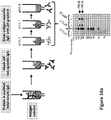
- This disclosure is directed to methods and systems for rapidly and accurately obtaining the amino acid sequences (and encoding nucleic acid sequences) of monoclonal antibodies that specifically bind to an antigen of interest. More specifically, the present methodology involves a direct, mass spectrometry-based proteomic investigation of circulating polyclonal antibodies from the serum of an animal, against a genetic material database which is comprised of nucleic acid molecules encoding full length immunoglobulin chains or variable regions.
- the genetic material database is generated from the B cell repertoire of an animal (e.g., the same animal whose serum was collected to obtain the polyclonal antibodies) by utilizing nucleic acid sequencing technologies.
- the present approach essentially involves correlating (i.e., cross-comparing or cross-referencing) the information from two sources: mass spectra information from the actual circulating polyclonal antibodies of an animal, and information (including, e.g., predicted mass spectra) from the genetic material database.
- mass spectra information from the actual circulating polyclonal antibodies of an animal
- information including, e.g., predicted mass spectra
- a list of heavy and light chain DNA sequences can then be identified from the genetic material database that correspond to actual antibodies from the serum. Such heavy and light chains can be expressed in pairs to obtain functional monoclonal antibodies.
- the present methodology does not require B cell immortalization, single cell sorting and molecular cloning, or phage display, and does not involve assembly of antibody sequences based on guesswork.
- the approach of this invention can significantly reduce the amount of time needed to isolate the sequences of antigen-specific monoclonal antibodies from a polyclonal population, thereby enabling a faster transition to recombinant antibodies such as fully human antibodies or humanized antibodies (e.g., humanized murine antibodies) that may be used therapeutically.
- the present methodology is capable of identifying rare antibodies likely missed by existing technologies.
- the inventors have surprisingly found that individual antibodies with very selective specificity (e.g., an antibody that specifically binds to a phosphorylated tyrosine residue within a polypeptide) may occur very rarely within a polyclonal population. Methods that rely on the frequencies of antibody-encoding mRNAs and PGR amplification may miss these antibodies because their variable chains occur with low frequency.
- the present methodology utilizes, for example, mass spectrometry based proteomics analysis of actual peptide fragments derived from a polyclonal antibody population, and therefore does not suffer from the errors of frequency following PGR amplification.
- the present methodology allows for the rapid creation of novel antigen-specific antibodies that may not exist in the starting polyclonal population.
- the created immunoglobulin molecule that has the highest desired qualities e.g., highest binding affinity (or lowest KD) for the antigen or a desired isotype (e.g., IgG2a)
- the created immunoglobulin molecule that has the highest desired qualities may be the result of a light chain from a first antibody in the polyclonal population assembled with a heavy chain of a second antibody (i.e., different from the first antibody) in the polyclonal population.
- the methods described herein have applications in basic immunology and therapeutics.
- the methods can provide the basis for understanding central questions in the field of immunology, including serum antibody diversity, dynamics, kinetics, clonality, and migration of B cells following antigen exposure.
- the methods can also be used to pursue therapeutically relevant human monoclonal antibodies from immunized, naturally infected, or diseased individuals.
- this disclosure further provides isolated recombinant monoclonal antibodies specific for an antigen, including therapeutic antibodies specific for a disease antigen, as well as therapeutic methods for treating a disease based on administration of therapeutic monoclonal antibodies.
- peptide or “peptide fragment” is meant a short polymer formed from the linking individual amino acid residues together, where the link between one amino acid residue and the second amino acid residue is called an amide bond or a peptide bond.
- a peptide comprises at least two amino acid residues.
- a peptide is distinguished from a polypeptide in that it is shorter.
- polypeptide is meant a long polymer formed from the linking individual amino acid residue, where the link between one amino acid residue and the second amino acid residue is called an amide bond or a peptide bond.
- a polypeptide comprises at least four amino acid residues; however, multiple polypeptides can be linked together via amide or peptide bonds to form an even longer polypeptide.
- nucleic acid molecule is meant a polymer formed from linking individual nucleotides (e.g., deoxyribonucleotides or ribonucleotides) together, where the link between one nucleotide and the other nucleotide is a covalent bond including, for example, a phosphodiester bond.
- link between one nucleotide and the other nucleotide is a covalent bond including, for example, a phosphodiester bond.
- the term includes, without limitation, DNA, RNA, and DNA-RNA hybrids.
- nucleic acid sequence is meant a nucleic acid sequence (or nucleotide sequence complementary thereto) that includes nucleotides that encode all or part of an immunoglobulin chain (e.g., a heavy chain or a light chain).
- the nucleic acid sequence is genomic DNA (e.g., exonic DNA with or without intronic DNA).
- the nucleic acid sequence is cDNA or some form of RNA (e.g., hn RNA, mRNA, etc.).
- the nucleic acid sequence is an expressed nucleic acid sequence that will be either transcribed into a nucleic acid molecule (e.g., DNA transcribed into RNA) or translated into a polypeptide in a cell containing that nucleic acid sequence.
- an expressed nucleic acid molecule includes, without limitation, hnRNA, mRNA, cDNA, and genomic exon sequences.
- complementary in terms of nucleic acid molecules simply means that two single-stranded nucleic acid molecules contain nucleotides that will form standard Watson-Crick basepairs to form a double- stranded nucleic acid molecule, whether that double-stranded molecule is DNA, RNA, or a DNA-RNA hybrid.
- B lymphocyte any white blood cell in which gene recombination (or gene rearrangement) has begun to occur at a locus containing an immunoglobulin chain-encoding gene.
- human immunoglobulin genes occur on chromosome 14 (heavy chain locus), chromosome 2 (kappa light chain locus), and chromosome 22 (lambda light chain locus). If a human white blood cell has undergone a gene rearrangement event in an immunoglobulin chain locus (e.g., on chromosome 14, chromosome 2, or chromosome 22), that cell is considered a B lymphocyte.
- B lymphocytes include, without limitation, B cells, pre-B cells, pro-B cells including early pro-B cells (e.g., where the D and J regions of the heavy chain genes have undergone rearrangement but the light chain gene are germline (i.e., are not rearranged)) and late pro-B cells (e.g., where the V, D, and J regions of the heavy chain gene is rearranged but the light chain gene is still germline and where no immunoglobulin proteins are expressed on the cell surface), pre-B cells including large pre-B cells and small pre-B cells, immature B cells, active B cells, germinal center B cells, plasma cells (including plasmablasts), and memory B cells.
- pro-B cells including early pro-B cells (e.g., where the D and J regions of the heavy chain genes have undergone rearrangement but the light chain gene are germline (i.e., are not rearranged)) and late pro-B cells (e.g., where the V, D, and J regions of the heavy chain gene is
- immunoglobulin and “immunoglobulin” are used interchangeably and are meant to include intact immunoglobulin polypeptide molecules of any isotype or sub-isotype (e.g., IgG, IgG1, IgG2, IgG2a, IgG2b, IgG3, IgG4, IgM, IgD, IgE, IgEl, IgE2, IgA) from any species of animal such as primates (e.g., human or chimpanzees), rodents (e.g., mice or rats), lagomorphs (e.g., rabbits or hares), livestock animals (e.g., cows, horses, goats, pigs, and sheep), fish (e.g., sharks), birds (e.g., chickens) or camelids (e.g., camels or llamas) or from transgenic non-human animals (e.g., rodents) genetically engineered to any species of animal such as primates (
- Non-limiting antibodies of various embodiments of the disclosure include but are not limited to polyclonal, monoclonal, monospecific, polyspecific antibodies and fragments thereof, chimeric antibodies comprising an immunoglobulin binding domain fused to another polypeptide, and humanized antibodies such as a non-human antibody (e.g., a rabbit antibody) whose constant and/or FR domains have been replaced with constant and/or FR domains from a human antibody (see, e.g., U.S. Pat. Nos: 5,530,101 ; 5,585,089 ; 5,693,761 ; 5,693,762 ; 6,180,370 ; and 6,548,640 ).
- a non-human antibody e.g., a rabbit antibody
- Transgenic non-human animals genetically engineered to produce human (e.g., at least partially human) antibodies are available from Harbour Antibodies (Rotterdam, The Netherlands), Ablexis (San Francisco, CA), Kymab Ltd (Cambridge, UK), OMT, Inc. (Palo Alto, CA), Amgen (Thousand Oaks, CA), Medarex (Princeton, NJ), and Regeneron (Tarrytown, NY).
- Naturally-occurring intact antibodies are made up of two classes of polypeptide chains, light chains and heavy chains.
- a non-limiting antibody of various aspects of the disclosure can be an intact, four immunoglobulin chain antibody comprising two heavy chains and two light chains.
- the heavy chain of the antibody can be of any isotype including IgM, IgG, IgE, IgA or IgD or sub-isotype including IgG1, IgG2, IgG2a, IgG2b, IgG3, IgG4, IgEl, IgE2, etc.
- the light chain can be a kappa light chain or a lambda light chain.
- a single IgG naturally-occurring (or intact) antibody comprises two identical copies of a light chain and two identical copies of an IgG heavy chain.
- the heavy chains of all naturally-occurring antibodies where each heavy chain contains one variable domain (VH) and one constant domain (CH, which itself comprises the CHI region, the hinge region, the CH2 region, and the CHS region), bind to one another via multiple disulfide bonds within their constant domains to form the "stem" of the antibody.
- the light chains of all naturally-occurring antibodies where each light chain contains one variable domain (VL) and one constant domain (CL), each bind through its constant domain to one heavy chain constant domain via disulfide binding.
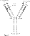
- FIG. 1 A schematic of a four immunoglobulin chain antibody (e.g., an IgG antibody) is shown in Figure 1 .
- the three CH domains are shown in light blue
- the single VH domain is shown in dark blue
- the single CL domain is shown in light pink
- the single VL domain is shown in dark pink.
- the VL and the VH domains of the light and heavy chains, respectively come together to form the antibody binding domain.
- an intact immunoglobulin chain (e.g., a heavy chain or a light chain) may comprise in order from 5' to 3' (for a nucleic acid sequence encoding the chain) or from the amino terminus to the carboxy terminus (for the amino acid sequence of the chain): a variable domain and a constant domain.
- the variable domain may comprise three complementarity determining regions (CDRs; also called hypervariable regions or HVs), with interspersed framework (FR) regions.
- variable domains of both the light chains and heavy chains contain three hypervariable regions sandwiched between four more conserved framework regions (FR), for a structure of 5' (or N')- FR1, CDR1, FR2, CDR2, FR3, CDRS, FR4 3' (or C), with the constant region 3' (or C) to the FR4 region.
- the CDRs form loops that comprise the principal antigen binding surface of the antibody (see Kabat, E. A. et al., Sequences of Proteins of Immunological Interest, National Institutes of Health, Bethesda, Md., (1987 ) and Wu, T.T. and Kabat, E.A. (1970) J. Exp. Med.
- an antibody e.g., a polypeptide or a carbohydrate
- the portion of an antigen that is specifically bound by the antibody is referred to as an "epitope".
- An “epitope” is smallest portion of a target molecule capable being specifically bound by the antigen binding domain of an antibody.
- the minimal size of an epitope may be about three to seven amino acids (e.g., five or six amino acids).
- a single antigen can be specifically bound by multiple different antibodies, all of which antibodies specifically bind the antigen (i.e., all of these antibodies are antigen-specific antibodies) even though each individual antibody specifically binds to a different epitope on the antigen.
- disease antigen an antigen which arises in an animal during a disease state.
- a viral antigen e.g., an antigen encoded by a nucleic acid molecule of a virus's genetic material
- a disease antigen in animal infected with that virus.
- some diseases e.g., cancer
- chimeric proteins e.g., BCR-ABL
- a BCR-ABL protein is a disease antigen. It should be understood that a disease antigen is not necessarily seen only in an animal suffering from that disease.
- disease is simply meant any abnormal condition affecting an animal.
- diseases include, without limitation, autoimmune disease (e.g., rheumatoid arthritis or type I diabetes), cancer (e.g., leukemia, colon cancer, or prostate cancer, etc.), viral infections (e.g., AIDS caused by infection of the HIV virus or chicken pox caused by infection of the varicella zoster virus), parasitic infection (e.g., schistosomiasis or scabies), and bacterial infection (e.g., tuberculosis or diptheria).
- autoimmune disease e.g., rheumatoid arthritis or type I diabetes
- cancer e.g., leukemia, colon cancer, or prostate cancer, etc.
- viral infections e.g., AIDS caused by infection of the HIV virus or chicken pox caused by infection of the varicella zoster virus
- parasitic infection e.g., schistosomiasis or scabies
- bacterial infection
- an immunoglobulin or antibody interacts with its antigen (i.e., its specific antigen), where the interaction is dependent upon the presence of a particular structure (e.g., an epitope) on the antigen; in other words, the antibody is recognizing and binding to a specific structure rather than to all molecules or structures in general.
- An antibody that specifically binds to the antigen may be referred to as an "antigen-specific antibody” or an "antibody specific for the antigen”.
- an antibody that specifically binds to antigen can immunoprecipitate that antigen from a solution containing the antigen as well as other molecules (e.g., a cell lysate).
- an antibody that specifically binds to its antigen has a K D for its antigen of 1 x 10 -6 M or less. In some embodiments, an antibody that specifically binds to its antigen has a K D for its antigen of 1 x 10 -7 M or less, or a K D of 1 x 10 -8 M or less, or a K D of 1 x 10 -9 M or less, or a K D of 1 x 10 -10 M or less, of a K D of 1 x 10 -11 M or less, of a K D of 1 x 10 -12 M or less.
- the K D of an antibody that specifically binds to its antigen for its specific antigen is between 1 pM to 500 pM, or between 500 pM to 1 (iM, or between 1 (iM to 100 nM, or between 100 mM to 10 nM.
- K D is intended to refer to the dissociation constant of an interaction between two molecules (e.g., the dissociation constant between an antibody and its specific antigen).
- variable region of an immunoglobulin chain or an “immunoglobulin chain variable region” is a polypeptide comprising at least a portion of the variable domain of a heavy (i.e., the VH domain) or a light chain (i.e., the VL domain) of an immunoglobulin, where the portion of the VL and the VH domains form an antigen binding domain of an immunoglobulin (see Fig. 1 ).
- variable region of an immunoglobulin may include, without limitation, a single CDR (e.g., CDR1), two CDRs interspersed with a single FR (e.g., CDR1, FR2, and CDR2), three CDRs interspersed with two FRs (e.g., CDR1, FR2, CDR2, FRS, and CDRS), or three CDRs flanked by either or both of FR1 and FR4 (e.g., FR1, CDR1, FR2, CDR2, FRS, CDRS, FR4).
- CDR1 and FR4 e.g., FR1, CDR1, FR2, CDR2, FRS, CDRS, FR4
- the immunoglobulin chain variable region is the region on one of either the heavy or the light chain which, when combined with the immunoglobulin chain variable region of the other chain (i.e., the light or the heavy chain) of the intact immunoglobulin, forms the antigen binding domain.
- antigen binding domain is meant the region of a single heavy chain assembled with a single light chain in an immunoglobulin, which retains the specific binding activity of the intact antibody for its specific antigen.
- an intact IgG immunoglobulin which comprises two heavy chains and two light chains, has two antigen binding domains.
- fragmentation of an intact antibody which retains a covalent bond between the heavy chain and the light chain will also result in an immunoglobulin fragment having an antigen binding domain.
- digestion of an immunoglobulin with the enzyme papain will generate F(ab) fragments, each of which has a single antigen binding domain.
- the entire F(ab) is not the antigen binding domain; rather, only the portion of the F(ab) fragment which retains the ability to specifically bind the antigen is the antigen binding domain.
- this invention is directed to a method for obtaining the amino acid and/or nucleic acid sequences of immunoglobulin chains (or variable regions thereof) of a single immunoglobulin from a population of polyclonal antibodies.
- a population of polyclonal antibodies of interest is obtained from an animal and fragmented to generate peptide fragments which are analyzed by mass spectrometry.
- the mass spectra information observed from the peptide fragments is then correlated with predicted mass spectra information derived from a genetic material database comprised of nucleic acid sequences that encode full-length immunoglobulin heavy and/or light chains (or variable regions thereof).
- immunoglobulin heavy and/or light chains (or variable regions thereof) can be identified from the genetic material database that correspond to immunoglobulin heavy and/or light chains (or variable regions thereof) of immunoglobulin molecules within the starting polyclonal antibody population.
- Immunoglobulins that specifically bind to an antigen of interest may be collected from an animal, which includes any mammal, such as human. Immunoglobulins can be collected from a body fluid sample of the animal including, for example, blood, serum or plasma of the blood, cerebrospinal fluid, synovial fluid, peritoneal fluid, mucosal secretions, tears, nasal secretions, saliva, milk, and genitourinary secretions.
- immunoglobulins need not come from a single individual animal but, rather, may be a cocktail of different antibodies (monoclonal or polyclonal) taken from different individuals.
- the immunoglobulins are collected from a transgenic non-human animal, e.g., a transgenic non-human animal that expresses human antibody sequences and/or produces antibodies that are at least partly human.
- these immunoglobulins are specific for an antigen of interest, either because the animal from whom the immunoglobulins are collected was previously immunized with the antigen, or because the animal from whom the immunoglobulins are collected was previously exposed to a condition whereby the animal was likely to generate antigen-specific antibodies.
- the animal may have been infected with a virus (e.g., Epstein Barr Virus), where the antigen of interest is the EBNA1 protein, which is encoded by the genome of the Epstein Barr Virus.
- Epstein Barr Virus e.g., Epstein Barr Virus
- the animal whose immunoglobulins are collected is of the same species as the animal whose B lymphocyte nucleic acid sequences are collected to create the reference database.
- the animal whose immunoglobulins are collected for the peptide database and the animal whose B lymphocyte nucleic acids are collected for the reference database are the same animal.
- blood taken from the animal can provide both the nucleic acid sequences (e.g., from the cells in the blood) and the polyclonal antibodies (e.g., from the sera or plasma of the blood).
- nucleic acid sequences e.g., from the cells in the blood
- polyclonal antibodies e.g., from the sera or plasma of the blood
- the immunoglobulins collected from the animal form a polyclonal population of immunoglobulins, because different B lymphocytes produced members of the population. It should be noted that in such a polyclonal population, not all of the individual antibodies within that polyclonal population will specifically bind the same antigen. In fact, each of the antibodies within the population may bind a different antigen. However, this polyclonal population still is said to specifically bind a particular antigen if at least one individual antibody, preferably multiple antibodies, of the polyclonal population binds that antigen (see, e.g., Example 3 below). In another example, some antibodies in the polyclonal population may bind the antigen with low affinity. However, a polyclonal population is said to specifically bind an antigen if some (e.g., at least one or more) of the antibodies in that 30 population specifically bind the antigen.
- polyclonal antibody or immunoglobulin that specifically binds to an antigen
- polyclonal antibody or immunoglobulin that specifically binds to an antigen
- at least one antibody specifically binds to the antigen, however that one antibody is not necessarily isolated from the other antibodies within the polyclonal population that do not specifically bind to the antigen.
- more than one different antibody within the polyclonal population specifically binds to the antigen.
- different antibody molecules are antibody molecules produced by a different B cell.
- a polyclonal population of 1000 antibody molecules may be isolated from the sera (e.g., using the antibodies' adherence to a protein A column to isolate the antibodies from the other sera components).
- 900 may be identical (i.e., secreted by the same B cell) and thus there are really only 101 different antibodies within that polyclonal population.
- the polyclonal population of 1000 antibody molecules is a polyclonal antibody that specifically binds to the antigen.
- an additional 5 different antibody molecules of the remaining 100 different antibody molecules also specifically bind to the antigen the polyclonal population of 1000 antibody molecules is likewise is a polyclonal antibody that specifically binds to the antigen.
- the majority of antibody molecules within a polyclonal population need not specifically bind to an antigen for that population to be referred to as a "polyclonal antibody that specifically binds to the antigen". For example, if within a polyclonal population of 1000 antibody molecules, even if only 1 antibody molecule specifically binds to the antigen and 999 antibody molecules do not, that population of 1000 antibody molecules is still a "polyclonal antibody that specifically binds to the antigen" as the term is used herein. Note also that all of the antibodies in a polyclonal antibody population need not bind the same epitope on the antigen. For example, a polyclonal population can be specific for the antigen where every different antibody within the population specifically binds a different epitope on the antigen.
- the population of polyclonal immunoglobulins may have, for example, at least two different immunoglobulins within the population, or at least three, or at least five, or at least ten, or at least twenty, or at least fifty, or at least one hundred or at least five hundred different immunoglobulins within the population.
- the invention also contemplates collecting a polyclonal population of immunoglobulins from the tissue culture supernatants of B cells grown in vitro (e.g., where the nucleic acid sequences are collected from the B cells themselves).
- a population of B cells may be collected from an animal that has been subjected to the Epstein Barr virus.
- the population can be expanded, e.g., to enrich B lymphocytes in the population as compared to other white blood cells. From this cultured media of these cells (into which the polyclonal antibodies are secreted by the cells), the polyclonal population of antibodies can be isolated.
- the polyclonal population of immunoglobulins collected can be first purified prior to digestion into peptide fragments.
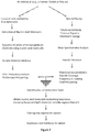
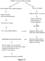
- the collected polyclonal antibodies can be subjected to a protein A or protein G sepharose column, which can separate antibodies from other blood sera proteins, for example. See, for example, Figure 2 and Figure 3 .
- the collected polyclonal antibodies are subjected to antigen affinity purification to enrich for antibodies with high specific activity. While not entirely necessary, a purification step, especially antigen affinity purification, can reduce the complexity of a 20 polyclonal mixture and ultimately reduce the number of potential false positive or negative candidate immunoglobulins.
- the collected polyclonal antibodies may be concentrated or buffer exchanged or both, either before or after purification.
- peripheral blood is drawn from the animal, and serum and/or plasma antibodies are collected according to standard methods (e.g., adherence of the antibodies to protein A).
- the serum and/or plasma antibodies are then purified or screened to enrich for immunoglobulins that specifically bind to the antigen.
- This screen can be, for example, by coating a solid-phase surface (e.g., a sepharose bead or bottom of a plastic well) with antigen and pass the serum and/or plasma over the antigen-coated surface under conditions where immunoglobulins that specifically bind to the antigen will bind.
- the bound antibodies may be treated with a protease (e.g., papain) or a chemical protein cleavage reagent that specifically cuts near the hinge region of the immunoglobulin to remove the non-adherent Fc portions. After rinsing away non-binding serum and/or plasma proteins (including non-specific immunoglobulins), the antigen-specific immunoglobulins can be collected and their quantities thus enriched as compared to antibodies that do not specifically bind to the antigen.
- a protease e.g., papain
- a chemical protein cleavage reagent that specifically cuts near the hinge region of the immunoglobulin to remove the non-adherent Fc portions.
- the collected polyclonal antibodies are analyzed by protein analysis methods (e.g., mass spectrometry, liquid chromatography, etc.).
- observed mass spectra information is obtained from peptide fragments which are generated from the polyclonal antibodies.
- the polyclonal antibodies can be fragmented, for example, with one or more proteases, and/or a chemical protein cleavage reagent, such as cyanogen bromide.
- proteases are known to cleave their substrates at specific sites.
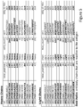
- Table 1 provides a non-comprehensive list of commonly used proteases and their cleavage sites (in 3 letter amino acid code).
- Table 1 Protease Cleavage Site Trypsin cleaves after (i.e.. on the carboxyl side of) Arg or Lys, unless followed by Pro Chymotrypsin cleaves after Phe, Trp, or Tyr, unless followed by Pro Elastase cleaves after Ala, Gly, Ser, or Val, unless followed by Pro. Endoproteinase Lys-C cleaves after Lys Pepsin cleaves after Phe or Leu. Thermolysin cleaves before Ile, Met, Phe, Trp, Tyr, or Val, unless preceded by Pro. Endopeptidase V8 (alias Glu-C) cleaves after Glu.
- multiple (i.e., two or more) proteases are used (e.g., independently or together) to digest the polyclonal antibodies to maximize V-region coverage and account for highly variable amino acid compositions of immunoglobulins.
- a combination of chymotrypsin, elastase, pepsin and trypsin can be used, as illustrated in Example 7 herein.
- a protease or proteases are chosen on the basis that they do not cleave within predicted CDR3 regions based on analysis of the nucleic acid molecules in the genetic material database.
- Proteins may be digested to smaller fragments that are amenable to mass spectrometry by treatment with particular chemical protein cleavage reagents rather than proteolytic enzymes. See for example chapter 3 of G. Allen. Sequencing of Proteins and Peptides, Laboratory Techniques in Biochemistry and Molecular Biology, Vol. 9. Elsevier 1989 .
- chemical protein cleavage reagents include, without limitation, cyanogen bromide, BNPS-skatole, o-iodosobenzoic acid, dilute acid (e.g., dilute HQ), and so forth.
- proteins can be cleaved at Met residues with cyanogen bromide, at Cys residues after cyanylation, after Trp residues with BNPS-skatole or o-iodosobenzoic acid, etc. Protein fragments can also be generated by exposure to dilute acid, e.g., HC1.
- dilute acid e.g., HC1.
- An example of the use of partial acid hydrolysis to determine protein sequences by mass spectrometry is given by Zhong et al. ( Zhong H, et al., J. Am. Soc. Mass Spectrom. 16(4):471-81, 2005 . PubMed PMID: 15792716).
- Proteins can be fragmented to make them more amenable for mass spectrometry by treatment with one protease, by treatment with more than one protease in combination, by treatment with a chemical cleavage reagent, by treatment with more than one chemical cleavage reagent in combination, or by treatment with a combination of proteases and chemical cleavage reagents.
- the reactions may occur at elevated temperatures or elevated pressures. See for example Lopez-Ferrer D, et al., J. Proteome. Res. 7(8):3276-81, 2008. PubMed PMID: 18605748 ; PubMed Central PMCID: PMC2744211.
- the fragmentation can be allowed to go to completion so the protein is cleaved at all bonds that the digestion reagent is capable of cleaving; or the digest conditions can be adjusted so that fragmentation does not go to completion deliberately, to produce larger fragments that may be particularly helpful in deciphering antibody variable region sequences; or digest conditions may be adjusted so the protein is partially digested into domains, e.g., as is done with E. coli DNA polymerase I to make Klenow fragment.
- the conditions that may be varied to modulate digestion level include duration, temperature, pressure, pH, absence or presence of protein denaturing reagent, the specific protein denaturant (e.g., urea, guanidine HC1, detergent, acid-cleavable detergent, methanol, acetonitrile, other organic solvents), the concentration of denaturant, the amount or concentration of cleavage reagent or its weight ratio relative to the protein to be digested, among other things.
- the specific protein denaturant e.g., urea, guanidine HC1, detergent, acid-cleavable detergent, methanol, acetonitrile, other organic solvents
- concentration of denaturant e.g., urea, guanidine HC1, detergent, acid-cleavable detergent, methanol, acetonitrile, other organic solvents
- concentration of denaturant e.g., urea, guanidine HC1, detergent, acid-cleavable detergent, methanol
- the reagent i.e., the protease or the chemical protein cleavage reagents
- the reagent used to cleave the proteins is a completely non-specific reagent.
- no constraints are made may be made at the N-terminus of the peptide, the C-terminus of the peptide, or both of the N- and C-termini.
- a partially proteolyzed sequence that is constrained to have a tryptic cleavage site at one end of the peptide sequence or the other, but not both, may be used in the various methods described herein.
- the resulting peptide fragments can be detected and analyzed using an HPLC coupled to a mass spectrometer from which observed mass spectra are generated.
- This method may be referred to as a "bottom up” proteomics approach, where proteome components are separated and identified after reducing the proteins to relatively small peptides, e.g., 3 to 45 residues in length.
- an alternative, "top down” proteomics approach can be employed to obtain observed mass spectra, which involves mass spectrometry analysis of intact proteins or large protein fragments or protein domains or large polypeptides. For example, to identify the parts of the antibody variable regions that bestow specific antigen recognition to a particular polyclonal antibody molecule, it is helpful to sequence large portions of the variable regions to identify its CDRs, by direct analysis of fragments large enough that the CDRs remain linked together.
- the immunoglobulins can be digested with either papain or pepsin to generate F(ab) and F(ab)2 fragments, respectively. Since the entirety of an immunoglobulin chain variable region is located on a chain of an F(ab) fragment, this pre-treatment with papain and/or pepsin will enrich for immunoglobulin chain variable regions. After rinsing away the non-binding portions of the immunoglobulins, the immunoglobulin chain variable regions can be collected.
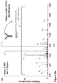
- the observed mass spectrum of a single peptide fragment can be correlated with the predicted mass spectra of the nucleic acid sequence to obtain the amino acid (and underlying nucleotide) sequence of the entire immunoglobulin chain (or variable region thereof) of an immunoglobulin that specifically binds to an antigen from a starting polyclonal immunoglobulin population. This correlating step is further described hereinbelow.
- additional information derived from the peptide fragments of the polyclonal antibodies is useful in various embodiments of the invention.
- This information includes, without limitation, the mass of each peptide, the length (in amino acid residues) of each peptide, the observed mass spectrum of each peptide (e.g., from tandem mass spectrometry such as the MS2 or MS3 spectrum), the mass to charge ratio of each peptide, the ionic charge of each peptide, the chromatographic profile of each peptide, and the amino acid sequence of each peptide.
- mass spectra information can be obtained by mass spectrometry analysis of collected immunoglobulins or fragments generated therefrom.
- a mass spectrometer is an instrument capable of measuring the mass-to-charge (m/z) ratio of individual ionized molecules, allowing researchers to identify unknown compounds, to quantify known compounds, and to elucidate the structure and chemical properties of molecules.
- one begins mass spectrometry analysis by isolating and loading a sample onto the instrument. Once loaded, the sample is vaporized and then ionized. Subsequently, the ions are separated according to their mass-to-charge ratio via exposure to a magnetic field.
- a sector instrument is used, and the ions are quantified according to the magnitude of the deflection of the ion's trajectory as it passes through the instrument's electromagnetic field, which is directly correlated to the ions mass-to-charge ratio.
- ion mass-to-charge ratios are measured as the ions pass through quadrupoles, or based on their motion in three dimensional or linear ion traps or Orbitrap, or in the magnetic field of a Fourier transform ion cyclotron resonance mass spectrometer. The instrument records the relative abundance of each ion, which is used to determine the chemical, molecular and/or isotopic composition of the original sample.
- a time-of-flight instrument is used, and an electric field is utilized to accelerate ions through the same potential, and measures the time it takes each ion to reach the detector.
- This approach depends on the charge of each ion being uniform so that the kinetic energy of each ion will be identical.
- the only variable influencing velocity in this scenario is mass, with lighter ions traveling at larger velocities and reaching the detector faster consequently.
- the resultant data is represented in a mass spectrum or a histogram, intensity vs. mass-to-charge ratio, with peaks representing ionized compounds or fragments.
- the sample is loaded onto the instrument and ionized. Ionization can be done by, e.g., electrospray ionization and matrix-assisted laser desorption/ionization ("MALDI"). See, e.g., Zenobi, "Ion Formation in MALDI Mass Spectrometry", 17 Mass Spectrometry Review, 337 (1998 ). Protein characterization can be done in one of two ways, top-down or bottom-up. The top-down approach involves ionizing intact proteins or larger protein fragments. See, e.g., Allison 15 Doerr, "Top-down Mass Spectrometry", 5 Nature Methods, 24 (2008 ).
- the bottom-up approach involves enzymatically or chemically digesting the protein into constituent peptides using a protease. See Biran Chait, "Mass Spectrometry: Bottom-Up or Top- Down?”, 6 Science 65 (2006 ). The resultant peptides are introduced into the instrument and ultimately identified by peptide mass fingerprinting or tandem mass spectrometry.
- mass spectrometry analysis may be combined with a chromatographic fractionation (e.g., liquid chromatography).
- chromatographic fractionation e.g., liquid chromatography
- Mass spectra data useful in this invention can be obtained by peptide mass fingerprinting.
- Peptide mass fingerprinting involves inputting the observed mass from a spectrum of the mixture of peptides generated by proteolytic digestion into a database and correlating the observed masses with the predicted masses of fragments arising from digestions of known proteins in silico. Known masses corresponding to sample masses provide evidence that the known protein is present in the sample tested.
- tandem mass spectrometry typically utilizes collision-induced-dissociation, which causes peptide ions to collide with gas and to fragment (e.g., due to vibrational energy imparted by the collision).
- the fragmentation process produces cleavage products that break at the peptide bonds at various sites along the protein.
- the observed fragments' masses may be matched with a database of predicted masses for one of many given peptide sequences, and the presence of a protein may be predicted. See, e.g., Eng, 5 An Approach to Correlate Tandem Mass Spectral Data of Peptides with Amino Acid Sequences in a Protein Database, JASMS, 976 (1994 ).
- tandem mass spectrometry is performed by higher-energy collision induced dissociation (HCD), which on some mass spectrometers shows fragment product ions closer to peptide termini than collision induced dissociation.
- HCD collision induced dissociation
- tandem mass spectrometry is performed by electron transfer dissociation (ETD), which is based on ion-ion reactions where a distinct reagent chemical ion donates a radical to a peptide ion, which then promptly fragments to form product ions.
- ETD electron transfer dissociation
- fragmentation methods such as ETD
- ETD post-source decay
- MALDI matrix-assisted laser desorption ionization
- the observed mass spectra information from the starting polyclonal immunoglobulin population is correlated with predicted mass spectra information derived from a genetic material database, in order to obtain the amino acid (and underlying nucleotide) sequences of immunoglobulin chains (or variable regions thereof) of immunoglobulins from the starting polyclonal immunoglobulin population.
- a genetic material database includes nucleic acid sequences encoding a plurality of immunoglobulin chains (or variable regions thereof).
- information which can be obtained or derived from such a genetic material database includes, for example, the nucleotide sequence information of each nucleic acid molecule, the length (in nucleotides) of each nucleic acid molecule, amino acid sequence information of the polypeptides or peptides encoded by each nucleic acid molecule, the mass of a polypeptide or peptide encoded by each nucleic acid molecule, the length (in amino acid residues) of a polypeptide or peptide encoded by each nucleic acid molecule, the mass spectra information of polypeptides or peptides encoded by each nucleic acid molecule (e.g., a predicted mass spectra information based on the amino acid sequence of the polypeptide or peptide), and the amino acid sequence of a polypeptide or peptide encoded by each nucleic acid molecule
- the genetic material database contains genetic information of nucleic acid sequences encoding full length immunoglobulin chains (and not just the variable regions thereof).
- the nucleic acid sequences are expressed (i.e., transcribed into RNA and/or translated into protein) by the cell from which said sequences are derived.
- the genetic material database includes expressed nucleic acid sequences encoding immunoglobulin chain variable regions of multiple immunoglobulins from an animal.
- the genetic material database contains at least one hundred different expressed nucleic acid sequences. In other embodiments, the genetic material database contains at least one thousand different expressed nucleic acid sequences.
- Nucleic acid molecules encoding immunoglobulin chains are readily obtainable from a population of cells (e.g., peripheral white blood cells) containing B lymphocytes.
- the nucleic acid molecules are obtained from splenocytes or mononuclear cells, such as peripheral blood mononuclear cells (PBMCs).
- PBMCs peripheral blood mononuclear cells
- the B lymphocytes are from a naive animal (e.g., an animal that has not been exposed to the antigen to which an antigen-specific antibody is sought).
- the naive animal has been exposed to very few antigens (e.g., an animal raised in sterile or pathogen-free environment).
- the naive animal is a typical animal that has been exposed to typical antigens, but has not been exposed to the antigen of choice.
- the animal from which the nucleic acid molecules encoding immunoglobulin chains (or the variable regions thereof) are obtained is an animal that has been previously exposed to the antigen.
- the animal may be an animal immunized with the antigen (e.g., the antigen mixed with an adjuvant or an antigen coupled to an immunogenic carrier such as keyhole limpet hemocyanin (KLH)), may be an animal infected with a pathogen comprising the antigen (e.g., an animal infected with HIV virus when the antigen of choice is the HIV p24 antigen), or may otherwise be previously exposed to the antigen.
- KLH keyhole limpet hemocyanin
- the animal is a bird (e.g., a chicken or turkey) or a mammal, such as a primate (e.g., a human or a chimpanzee), a rodent (e.g., a mouse, hamster, or rat), a lagomorph (e.g., a rabbit or hare), a camelid (e.g., a camel or a llama), or a domesticated mammal such as a companion animal (e.g., a cat, a dog, or a horse), or a livestock animal (e.g., a goat, sheep, or a cow).
- a primate e.g., a human or a chimpanzee
- a rodent e.g., a mouse, hamster, or rat
- a lagomorph e.g., a rabbit or hare
- a camelid e.g., a camel or a ll
- nucleic acid sequences of the various aspects and embodiments of the invention need not come from a single animal.
- some of the nucleic acid sequences of various embodiments of the invention may come from an animal previously exposed to an antigen, and some of the nucleic acid sequences may come from naive animal.
- nucleic acid sequences are from animals of a single species.
- all of those animals may be the same species (e.g., all are rabbits or all are humans).
- the nucleic acid sequences are obtained from animals of a single species. In other embodiments, nucleic acid sequences from more than one species of animal may be obtained.
- nucleic acid sequences may be obtained from mice and rats, and predicted mass spectra based from these sequences can be used to correlate with and/or compare to the actual mass spectra information of peptide fragment of polyclonal antibodies to create an immunoglobulin (or variable region, antigen binding domain, or chain thereof) that specifically binds to the antigen.
- the nucleic acid sequences are obtained from animals of a single gender (e.g., all animals are female).
- the animal from whom the polyclonal antibodies are collected and the animal from whom the nucleic acid sequences are collected may be the same animal, or the same species of animal, or syngenic animals (e.g., both are Balb/c mice), or from animals of the same gender (e.g., both are female animals).
- the MS2 spectra from the antigen-binding components of the polyclonal antibodies can thus be correlated to the theoretical MS2 spectra derived from the nucleic acid sequences obtained from an animal, in order to identify the nucleic acid sequences that encode antigen-binding antibodies.
- nucleic acid sequences and the polyclonal antibodies can be collected from cells of an animal where the cells were cultured in vitro following removal from the animal and prior to collection of the polyclonal antibodies (e.g., from the supernatant or cultured media of the cultured cells) and collection of the nucleic acid sequences from the cells.
- This culturing step is useful, e.g., to expand or enrich B lymphocytes as compared to other blood or tissue cells (e.g., to enrich B lymphocytes over red blood cells or epithelial cells).
- the number of individual nucleic acid sequences used to create theoretical mass spectra in the various embodiments of the invention is limitless.
- nucleic acid sequences may come from any source, and may be from a combination of sources.
- nucleic acid sequences can be obtained by sequencing expressed nucleic acid molecules encoding immunoglobulin chain variable regions (or the entire full length immunoglobulin chain including the variable regions and constant region) as described herein.
- Nucleic acid sequences can also be obtained from genomic DNA that may or may not have undergone full V(D)J recombination.
- Nucleic acid sequences can also be obtained from publicly available sources.
- the B lymphocytes from which nucleic acid sequences are obtained can be from any blood or tissue source including, without limitation, bone marrow, fetal blood, fetal liver, sites of inflammation (e.g. inflamed joints surrounding synovial fluid in rheumatoid patients), tumors (e.g., tumor-infiltrating lymphocytes), peripheral blood, in lymph nodes, in peyer's patches, in tonsils, and in the spleen or in any lymphoid organ.
- blood or tissue source including, without limitation, bone marrow, fetal blood, fetal liver, sites of inflammation (e.g. inflamed joints surrounding synovial fluid in rheumatoid patients), tumors (e.g., tumor-infiltrating lymphocytes), peripheral blood, in lymph nodes, in peyer's patches, in tonsils, and in the spleen or in any lymphoid organ.
- the entire tissue e.g., bone marrow or lymph node
- the entire tissue can be processed (e.g., cells separated from one another and lysed), genetic material removed, and the nucleic acid molecules encoding immunoglobulin chains (or variable regions thereof) sequenced.
- B lymphocytes are enriched from tissues or a population of cells (e.g., peripheral blood) containing them prior to isolating genetic material from the B lymphocytes.
- methods for enriching B lymphocytes from an animal are well known.
- B lymphocytes can be found in many organs and areas of the body including, without limitation, bone marrow, fetal blood, fetal liver, sites of inflammation (e.g. inflamed joints surrounding synovial fluid in rheumatoid patients), tumors (e.g., tumor-infiltrating lymphocytes), peripheral blood, in lymph nodes, and in the spleen.
- white blood cells may be isolated according to standard methods (e.g., using the Ficoll-Paque PLUS or Ficoll-Paque PREMIUM reagents commercially available from GE Healthcare, Piscataway, NJ, according to manufacturer's instructions). B lymphocytes themselves can then be further isolated from other white blood cells using, for example, cell surface markers found on B lymphocytes.
- B lymphocyte cell surface markers include, without limitation, cell surface expressed immunoglobulin chains (e.g., lambda light chain, kappa light chain, and heavy chain such as IgM or IgG).
- Additional B lymphocyte cell surface markers include, without limitation, CD21, CD27, CD138, CD20, CD19, CD22, CD72, and CD79A.
- Yet additional B lymphocyte cell surface markers include, without limitation, CD38, CD78, CD80, CD83, DPP4, FCER2, IL2RA, TNFRSF8, CD24, CD37, CD40, CD74, CD79B, CR2, IL1R2, ITGA2, ITGA3, MS4A1, ST6GAL1, CD1C, CD138, and CHST10.
- B lymphocyte surface markers can be used sequentially to enrich for B lymphocytes.
- antibodies specific to a B lymphocyte cell surface markers e.g., CD19
- magnetic beads e.g., Dynabeads commercially available from Invitrogen Corp., Carlsbad, CA
- cells adhering to the beads e.g., CD19 positive cells
- B lymphocytes can be further enriched from the CD 19 positive cells by, for example, flow cytometry sorting of cells expressing immunoglobulin chains at their cell surface.
- These enriched B lymphocytes can thus be isolated for use in the methods of various embodiments of the invention.
- Antigen specific B lymphocytes can also be purified directly using the desired antigen as bait to isolate B cells expressing the antigen specific B cell receptor (membrane immunoglobulin).
- B cells can be added to a column to which is adhered the desired antigen.
- the antigen-specific B cells will flow through the column more slowly than non-specific B cells or other cells (e.g., red blood cells, macrophages, etc.).
- the antigen-specific B cells can thus be enriched using this method.
- Enriched or non-enriched B lymphocytes from an animal can also be subjected to in vitro cell culture for 1 or 2 or 3 or 4 or more days prior to nucleic acid extraction. Such culture in vitro may expand the number of B lymphocytes and thus enrich them over non-B lymphocyte cells.
- CD27 isolated human B lymphocytes can be subjected to various cytokine and extracellular molecule cocktails (such as but not limited to activated T cell conditioned medium, or any combination of B cell growth, and/or differentiation factors) prior to nucleic acid extraction in order to stimulate growth and/or differentiation of the B lymphocytes prior to nucleic acid extraction from the B lymphocytes.
- Other biological molecules can also be added to the tissue culture media during the in vitro culturing to assist in growth, differentiation, and/or in vitro immunization, and/or any combination of the above.
- nucleic acid sequences e.g., genomic DNA, hnRNA, mRNA, etc.
- genomic DNA e.g., genomic DNA, hnRNA, mRNA, etc.
- mRNA e.g., mRNA sequences
- This nucleic acid can then be subjected to sequencing analysis using a variety of methods for sequencing.
- the nucleic acid sequences can be directly sequenced from the biological material (i.e., without being amplified prior to sequencing).
- Services and reagents for directly sequencing from nucleic acid sequences are commercially available, for example, from Helicos BioSicences Corp. (Cambridge, MA).
- Helicos' True Single Molecule Sequencing allows direct sequencing of DNA, cDNA, and RNA. See also U.S. Patent Nos. 7,645,596 ; 7,037,687 , 7,169,560 ; and publications Harris et al., Science 320: 106-109, 2008 ; Bowers et al., Nat. Methods 6: 493-494, 2009 ; and Thompson and Milos, Genome Biology 12: 217, 2011 .
- the nucleic sequences are amplified (e.g., by polymerase chain reaction (PCR)) prior to obtaining sequence information.
- PCR polymerase chain reaction
- an oligo dT PCR primer is used for RT-PCR.
- gene-specific RT-PCR is performed using the PCR primers described herein, such as the 454 specific fusion mouse primers, the 454 rabbit immunoglobulin chain fusion primers or the variable heavy and variable light region primers.
- PCR primers against heavy chain and light chain populations in a mouse have sequences set forth in PCT publication no. WO2010/097435 .
- purified genetic materials can be amplified (e.g., by PCR or RT-PCR) following standard procedures (see, e.g., Ausubel et al., supra) to prepare a library before NGS sequencing.
- Isolated B lymphocytes mentioned above by various means can also be subjected to single cell encapsulation by using method in the art such as oil emulsion encapsulation or by commercial instrument such as RainDance technology (RainDance Technologies, Inc., Lexington, MA). These encapsulated B lymphocytes can then be fused with an appropriate single cell RT-PCR reagent (e.g., the reagent sold by Qiagen, as Cat # 210210) with the appropriate amplification primers to generate linked Heavy and Light chain PCR products from each single B cells.
- Ligation or overlap PCR is known in the field and is practiced routinely for various molecular biology applications to stitch 2 DNA pieces into one (see, e.g., Meijer P.J. et al., J. Mol. Biol. 358(3):764-72, 2006 for overlap PCR). This approach allows for cognate pairing preservation and identification during sequencing.
- the methods may employ such enzymes as the Klenow fragment of DNA polymerase I, SEQUENASE® (US Biochemical Corp, Cleveland, Ohio), Taq polymerase (Invitrogen), thermostable T7 polymerase (Amersham, Chicago, 111.), DNA ligase (e.g., from T4) or combinations of recombinant polymerases and proofreading exonucleases such as the ELONGASE Amplification System marketed by Gibco BRL (Gaithersburg, Md.).
- the process may be automated with machines such as the Hamilton Micro Lab 2200 (Hamilton, Reno, Nev.), Peltier Thermal Cycler (PTC200; MJ Research, Watertown, Mass.) and the ABI 377 DNA sequencers (Applied Biosystems).
- Non-limiting methods to sequence nucleic acid molecules and thus generate nucleic acid sequences include the Sanger method (see, e.g., Sanger et al, Nature 24: 687-695, 1977 ), the Maxam-Gilbert method (see, e.g., Maxam and Gilbert, Proc. Natl. Acad. Sci. USA 74: 560-564, 1977 ), and pyrosequencing (see, e.g., Ronaghi et al., Science 281 (5375): 363, 1998 and Ronaghi et al., Analytical Biochemistry 242 (1): 84, 1996 ).
- Pyrosequencing another non-limiting sequencing method that can be used to obtain polynucleotide sequences, uses luciferase to generate light for detection of the individual nucleotides (either dATP, dTTP, dGTP, or dCTP, collectively "dNTPs") added to the nascent DNA, and the combined data are used to generate sequence read-outs.
- luciferase either dATP, dTTP, dGTP, or dCTP, collectively "dNTPs”
- the nucleic acid sequences are obtained using deep sequencing or next generation sequencing.
- One rate-limiting step in conventional DNA sequencing arises from the need to separate randomly terminated DNA polymers by gel electrophoresis.
- Next generation sequencing devices bypass this limitation, e.g., by physically arraying DNA molecules on solid surfaces and determining the DNA sequence in situ, without the need for gel separation.
- nucleic acid molecules can be sequenced simultaneously (see Church, G.M., Sci. Am. 294 (1): 46-54, 2006 ; Hall, N., J. Exp. Biol. 210(Pt. 9): 1518-1525, 2007 ; Schuster et al.. Nature Methods 5(1): 16-18, 2008 ; and MacLean et al.. Nature Reviews Microbiology 7: 287-296, 2009 ).
- a library of DNA fragments to be sequenced are amplified by emulsion PCR, and of the multiple fragments in the library, a single fragment species will be attached to a single magnetic bead (so called clonal beads).
- the fragments attached to the magnetic beads will have a universal PI adapter sequence attached so that the starting sequence of every fragment is both known and identical.
- Primers are then selected that hybridize to the PI adapter sequence within the library template.
- a set of four fluorescently labeled di-base probes compete for ligation to the sequencing primer. Specificity of the di-base probe is achieved by interrogating every 1st and 2nd base in each ligation reaction.
- oligonucleotide adaptors are ligated to fragmented nucleic acid molecules and are then immobilized to the surface of microscopic beads before PCR amplification in an oil-droplet emulsion. Beads are then isolated in multiple picolitre-volume wells, each containing a single bead, sequencing enzymes, and dNTPs. Incorporation of a dNTP into the complementary strand releases pyrophosphate, which produces ATP, which in turn generates light that can then be recorded as an image for analysis.
- the Genome Sequencer FLX System machine commercially available from 454 Life Sciences, a Roche company, Branford, CT
- U.S. Patent No. 7,115,400 describes another technique for solid-phase amplification of nucleic acid molecules. This allows a large number of different nucleic acid sequences to be arrayed and amplified simultaneously.
- This technology is embodied in the Genome Analyzer system commercially available from Solexa (Illumina, Inc.). In this technology, DNA molecules are first attached to primers on a slide and amplified so that local clonal colonies are formed (bridge amplification). Four types of ddNTPs are added, and non-incorporated nucleotides are washed away. Unlike pyrosequencing, the DNA can only be extended one nucleotide at a time. A camera takes images of the fluorescently labeled nucleotides then the dye along with the terminal 3' blocker is chemically removed from the DNA, allowing a next cycle.
- Polynucleotide sequences encoding immunoglobulin chain variable regions may be extended utilizing a partial nucleotide sequence and employing various methods known in the art to detect upstream sequences such as promoters and regulatory elements.
- one method that may be employed "restriction-site" PGR, uses universal primers to retrieve unknown sequence adjacent to a known locus ( Sarkar, G., PGR Methods Applic. 2: 318-322 (1993 )).
- genomic DNA is first amplified in the presence of primer to linker sequence and a primer specific to the known region.
- Exemplary primers are those described in Example 4 herein.
- amplified sequences are then subjected to a second round of PGR with the same linker primer and another specific primer internal to the first one.
- Products of each round of PGR are transcribed with an appropriate RNA polymerase and sequenced using reverse transcriptase.
- Inverse PGR may also be used to amplify or extend sequences using divergent primers based on a known region ( Triglia et al. Nucleic Acids Res. 16: 8186 (1988 )).
- the primers may be designed using OLIGO 4.06 Primer Analysis software (National Biosciences Inc., Madison, Minn.), or another appropriate program, to be 22-30 nucleotides in length, to have a GC content of 50% or more, and to anneal to the target sequence at temperatures about 68-72 0C.
- the method uses several restriction enzymes to generate a suitable fragment in the known region of a gene. The fragment is then circularized by intramolecular ligation and used as a PGR template.
- Another method which may be used is capture PGR which involves PGR amplification of DNA fragments adjacent to a known sequence in human and yeast artificial chromosome DNA ( Lagerstrom el al, PCR Methods Applic. 1: 111-119 (1991 )).
- multiple restriction enzyme digestions and ligations may also be used to place an engineered double-stranded sequence into an unknown portion of the DNA molecule before performing PCR.
- Another method which may be used to retrieve unknown sequences is that described in Parker et al.. Nucleic Acids Res. 19: 3055-3060 (1991 )).
- PCR, nested primers, and PROMOTERFINDER® libraries to walk in genomic DNA (Clontech, Palo Alto, Calif.). This process avoids the need to screen libraries and is useful in finding intron/exon junctions.
- nucleic acid from B lymphocytes may be further screened for those nucleic acid molecules encoding immunoglobulins prior to sequencing.
- primers specific for immunoglobulin-encoding nucleic acid molecules may be employed.
- primer is meant a nucleic acid sequence that may be at least about 15 nucleotides, or at least about 20 nucleotides, or at least about 30 nucleotides, or at least about 40 nucleotides in length.
- a primer specific for a particular nucleic acid molecule is meant to include a primer that hybridizes to a portion of the nucleic acid molecule under PCR annealing conditions (e.g., 60oC for thirty seconds).
- a primer specific for a particular nucleic acid molecule is one that is complementary to that nucleic acid molecule.
- Primers used for sequencing the nucleic acid sequence may be referred to as Sequencing Primers.
- Primers used for amplification of a target nucleic acid sequence by the polymerase chain reaction (PCR) may also be referred to as PCR primers or amplification primers (see description of PCR, for example, in Sambrook et al., supra and Ausubel et al., supra).
- total nucleic acid from B lymphocytes may be rendered single-stranded (e.g., by heating the nucleic acid to 94-980C for at least one minute.
- the single-stranded nucleic acid may then be passed over a solid support (e.g., a column or gel) to which are adhered single-stranded primers that are specific for non-variant regions of immunoglobulin-encoding nucleic acid molecules or non-coding regions adjacent thereto (e.g., immunoglobulin gene promoters, enhancers, and/or introns).
- non-limiting examples for these non-variant regions of immunoglobulins include the constant region of the heavy chain, and the constant region of the light chains, and the FR1 region of either the heavy chain or the light chain.
- the nucleic acid is allowed to hybridize to the solid-phase support-bound primers, and the non-hybridizing nucleic acid removed. After removal, the hybridized nucleic acid (which is enriched for immunoglobulin-encoding nucleic acid molecules) is released from the primers by, for example, addition of heat or increasing the concentration of EDTA in the buffer.
- the immunoglobulin-encoding nucleic acid molecules may be amplified to increase their copy number. This amplification can be performed, for example, by PCR amplification using primers specific for non-variant regions of immunoglobulin-encoding nucleic acid molecules or non-coding regions adjacent thereto.
- the primers e.g., sequencing or PCR primers
- the immunoglobulin chain variable region-encoding nucleic acid sequences may be universal (e.g., polyA tail) or may be specific to immunoglobulin-encoding sequences.
- the starting material from which the immunoglobulin gene-encoding nucleic acid sequence information is obtained is genomic DNA.
- primers e.g., sequencing primers and/or PCR primers
- an immunoglobulin chain gene promoter For example, the human genome sequence is known. Since the heavy chain-encoding gene occurs on chromosome 14 and the light chain-encoding gene occurs on chromosome 22 (lambda light chain) and 2 (kappa light chain), it would be routine for the ordinarily skilled biologist to design primers that hybridize to regulatory elements of the heavy chain-encoding gene and the light chain-encoding gene. Such regulatory elements include, without limitation, promoters, enhancers, and introns.
- Immunoglobulin variable region-specific primers can likewise be readily determined for mice immunoglobulins since the murine kappa light chain gene is known to be located on chromosome 6 and the murine heavy chain gene is known to be located on chromosome 12.
- the starting material from which the immunoglobulin gene-encoding nucleic acid sequence information is obtained is mRNA or cDNA reversed translated from the mRNA.
- primers can be selected to be identical to or hybridize to the polyA tail of an mRNA or the complementary TTTT (SEQ ID NO: 306)-rich sequence of the mRNA's corresponding cDNA.
- primers can also be selected to be identical to or hybridize to the FR1-encoding nucleic acid sequences.
- primers can also be selected to be identical to or hybridize to a portion of (or all of) one of the CH regions (i.e., CHI, CH2, or CHS) and/or the VH region-encoding nucleic acid sequences.
- Sequencing errors can arise from using universal degenerate primers to sequence nucleic acid molecules encoding immunoglobulins from hybridomas.
- Essono et al, Protein Engineering, Design and Selection, pp. 1-8, 2009 describe a method combining sequencing with peptide mass spectrometry fingerprinting of the corresponding Ig chain to determine the correct sequence of a monoclonal antibody produced by a hybridoma clone.
- the presence of sequencing errors will merely increase the number of different nucleic acid sequences.
- nucleotide sequence information can be translated into predicted amino acid sequences using the genetic code.
- the ordinarily skilled artisan can readily translate nucleotide sequences into amino acid sequences using the genetic code, several automated translation tools (which are publicly available) can be used, such as the ExPASy translate tool from the Swiss Institute of Bioinformatics or the EMBOSS Transeq translation tool from EMBL-EBI.
- the ExPASy translate tool from the Swiss Institute of Bioinformatics
- EMBOSS Transeq translation tool from EMBL-EBI.
- predicted mass spectra information of the predicted amino acid sequences encoded by the nucleic acid sequences can be readily determined by the ordinarily skilled artisan.
- predicted mass spectra of the peptide fragments can be generated by using standard publicly available software algorithm tools including, without limitation, the Sequest software (from Thermo Fisher Scientific, Inc., West Palm Beach, FL), the Sequest 3G software (from Sage-N Research, Inc., Milpitas, CA), the Mascot software (from Matrix Science, Inc., Boston, MA; see also Electrophoresis, 15 20(18) 3551-67 (1999 )), and the XlTandem software (opensource from The Global Proteome Machine Organization, whose use is described in Baerenfaller K. et al., Science 320:938-41, 2008 ).
- Sequest software from Thermo Fisher Scientific, Inc., West Palm Beach, FL
- Sequest 3G software from Sage-N Research, Inc., Milpitas, CA
- the Mascot software from Matrix Science, Inc., Boston, MA; see also Electrophoresis, 15 20(18) 3551-67 (1999 )
- nucleic acid sequences are derived from genomic nucleic acid molecules obtained from B lymphocytes as described herein.
- the nucleotide sequence of, for example, mRNA derived from genomic DNA is predicted following in silico translation of the genomic DNA.
- This predicted mRNA (or cDNA) may then be translated in silico to produce predicted amino acid sequences.
- the predicted amino acid sequences may then be digested in silico with proteases (e.g., trypsin) and/or chemical protein cleavage reagents (e.g., cyanogen bromide) to produce predicted (or theoretical or virtual) peptide fragments.
- the virtual peptide fragments can be then analyzed in silico to produce predicted mass spectra information.
- predicted mass spectra information, predicted peptide fragments, predicted amino acid sequences, and predicted mRNA or cDNA sequences can all be derived from the nucleic acid sequences collected from B lymphocytes (e.g., from an animal).
- the protease(s) and/or the chemical reagents used to digest predicted polypeptides to generate predicted peptides fragments and ultimately predicted mass spectra is the same protease(s) and/or reagent(s) used to digest the starting population of polyclonal antibodies, as described above.
- the observed mass spectra are correlated with the predicted mass spectra derived from the nucleic acid sequences of the genetic material database to obtain the amino acid (and underlying nucleotide) sequences of full-length immunoglobulin chains (or variable regions thereof) of an immunoglobulin that specifically binds to an antigen from a starting polyclonal immunoglobulin population.
- the genetic material database can be derived from nucleic acid molecules isolated from the B-cell repertoire of an immunized animal, including nucleic acid molecules encoding full length immunoglobulin heavy and light chains and variable regions thereof. Attempts to identify nucleic acids encoding antigen-specific immunoglobulins based solely on the information from the genetic material database (e.g., frequency rankings of variable region sequences) may miss those immunoglobulins that occur at low frequencies yet manifest superior antigen-specific activities.
- those immunoglobulin chains (or variable regions thereof) in the genetic material database can be selected that correspond to immunoglobulins within the circulating polyclonal antibodies.
- correlating it is meant that the observed mass spectra information derived from the starting polyclonal antibodies and the predicted mass spectra information derived from the genetic material database are cross-referenced and compared against each other, such that immunoglobulin heavy and/or light chains (or variable regions thereof) can be identified or selected from the genetic material database that correspond to immunoglobulin heavy and/or light chains (or variable regions thereof) of antigen-specific immunoglobulins in the starting polyclonal population.
- the correlating process involves comparing the observed mass spectra information with the predicted spectra information to identify matches.
- each of the observed spectra can be searched against the collection of predicted mass spectra derived from the genetic material database, with each predicted spectrum being identifiably associated with a peptide sequence from the genetic material database.
- a match is found, i.e., an observed mass spectrum is matched to a predicted mass spectrum, because each predicted mass spectrum is identifiably associated with a peptide sequence in the genetic material database, the observed mass spectrum is said to have found its matching peptide sequence - such match also referred to herein as "peptide spectrum match" or "PSM”.
- this search and matching process can be performed by computer-executed functions and softwares, such as the SEQUEST algorithm (Sage-N Research, Inc., Milpitas, CA).
- the search and matching is directed to functional domains or fragments of immunoglobulins, such as variable region sequences, constant region sequences, and/or one or more CDR sequences.
- the observed spectra are only searched against predicted mass spectra derived from V regions (and/or CDR3 sequences) of immunoglobulins to identify V-region (and/or CDR3) PSMs.
- the search and matching is directed to full-immunoglobulin heavy or light chain sequences.
- immunoglobulin heavy or light chains in the genetic material database are analyzed and selected based on one or more of the following parameters: the number of unique peptides, the spectrum share, the amino acid sequence coverage, the count of peptides (either total peptide count or unique peptide count), frequency of the encoding nucleic acid sequences, and clonal relatedness.
- coverage in referring to a sequence or region (e.g., a heavy or light chain sequence, a V-region sequence, or a CDR sequence) is defined as the total number of amino acids within the sequence that have been identified in peptides which map to the sequence or region and which have a matching observed spectrum, divided by the number of amino acids in the sequence or region. The higher the coverage, the more likely the sequence or region appears in the actual polyclonal population.
- number of unique peptides it is meant the number of distinct peptides observed mapping to a single protein sequence (e.g., a single immunoglobulin heavy or light chain or a variable region thereof). The higher the number, the more likely the immunoglobulin chain is present in the polyclonal population. In specific embodiments, selection of an immunoglobulin chain is made based on a number of unique peptides of at least 5, 6, 7, 8, 9, 10, 11, 12 or more in the immunoglobulin chain or its variable region.
- Spectrum share is determined by dividing the total number of peptides mapped to the sequence by the total number of confident PSMs mapped to the entire genetic database. Spectrum share provides a human readable count of peptides expressed as the percentage of PSMs that map to a specific V-region sequence.
- peptide count in referring to a protein sequence (e.g., a CDR3 region or a variable region) means the number of times a peptide is identified from the observed mass spectra that matches the protein sequence.
- the count of a CDR3 region means the number of times a peptide is identified from the observed mass spectra that matches the CDR3 region.
- the count of a variable region means the number of times a peptide is identified from the observed mass spectra that matches the variable region.
- Total peptide count in referring to a protein sequence means the number of times any peptide (unique or non-unique) is identified from the observed mass spectra that matches the protein sequence.
- Unique peptide count means the number of times a unique peptide is identified from the observed mass spectra that matches the protein sequence. If the same peptide has been identified multiple times from the observed mass spectra, the total number of times this peptide is observed will be considered in determining the total peptide count, yet this peptide will be counted only once for determining the unique peptide count.
- an immunoglobulin heavy or light chain is selected based on sequence coverage. In other embodiments, the selection is made based on a combination of sequence coverage with one or more other parameters, including the number of unique peptides, spectrum share, total peptide count, unique peptide count, frequency of the encoding nucleic acid sequence, or clonal relatedness.
- the above parameters can be independently determined with respect to a full-length heavy or light chain, or with respect to one or more portions of an immunoglobulin heavy or light chain, e.g., the variable region, and a CDR (e.g., CDR1, CDR2, or CDR3, especially CDR3).
- selection of immunoglobulin chains (or variable regions thereof) is made based on the V-region coverage and/or CDR coverage (e.g., CDR3 coverage).
- immunoglobulin heavy or light chains can be made based on the absolute value of one or more parameters, or based on the ranking of absolute values for a relevant parameter. Where ranking for a particular parameter is considered, the top ranked 10, 20, 30, 40, 50, 60, 70, 80, 90, 100 or more sequences can be selected irrespective of the absolute values of that parameter.
- selection of immunoglobulin chains is made based on a CDR coverage (such as CDR3 coverage) of at least 10%, 15%, 20%, 25%, 30%, 35%, 40%, 45%, 50%, 55%, 60%, 65%, 70%, 75%, 80%, 85%, 90%, 95%, 98% or higher; additionally or alternatively, based on a V-region coverage of at least 5%, 10%, 15%, 20%, 25%, 30%, 35%, 40%, 45%, 50%, 55%, 60%, 65%, 70%, 75%, 80%, 85%, 90% or higher.
- CDR coverage such as CDR3 coverage
- a phylogenetic analysis is performed to determine clonal relatedness of the heavy chain variable region, light chain variable region, or one or more CDR's (e.g., CDRH3 or CDRL3). Changes or mutations of nucleic sequence of heavy and light chains compared to germline sequence can provide evidence of affinity maturation of antibodies following antigen exposure. Clonal relatedness can be used as a factor in selection of antibody sequences.
- a phylogenetic analysis can be performed by methods known in the art, e.g., those described in Dereeper et al., 2008, Nucl. Acids Res., 36(Web Server issue):W456-459 ; Dereeper et al., 2010, BMC Evol.
- the entire heavy or light chain variable regions are grouped by homology, then further grouped by CDR (e.g., CDR3) homology.
- the selected heavy and light chain sequences can then be expressed in pairs to assemble into monoclonal antibodies which are analyzed to confirm antigen-specific functionality.
- the pairing of selected heavy and light chain sequences can be entirely random, or can take into consideration of one or more parameters described above, including sequence coverage, unique number of peptides, spectrum share, total peptide count, and unique peptide count.
- the abundance of a population of antibodies having a particular peptide sequence can be determined using a heavy isotope labeled (e.g., AQUA) peptide.
- AQUA heavy isotope labeled
- WO 03/016861 and Gerber et al., 2003, 100:6940-45 These methods employ the introduction of a known quantity of at least one heavy-isotope labeled peptide standard (which has a unique signature detectable by LC-SRM chromatography) into a digested biological sample in order to determine, by comparison to the peptide standard, the absolute quantity of a peptide with the same sequence and protein modification in the biological sample.
- the peptide can be unique to one species of antibody or found in multiple (e.g., clonally-related) antibodies.
- the peptide can include at least a portion of a CDR (e.g., CDR3). Quantitation of the abundance of antibody populations can be useful in methods of monitoring serum antibody composition, e.g., following vaccination of a subject.
- the immunoglobulin that specifically binds to the antigen whose amino acid sequence (or nucleic acid sequence) created using the non-limiting methods of various embodiments of the invention need not actually be present within the starting polyclonal population of immunoglobulins. Rather, the non-limiting methods of various embodiments of the invention simply allow the rapid creation of an immunoglobulin that specifically binds the antigen whether or not that immunoglobulin actually existed in the starting polyclonal population.
- the created immunoglobulin that has the highest desired qualities may be the result of a light chain from a first antibody in the polyclonal population assembled with a heavy chain of a second antibody (i.e., different from the first antibody) in the polyclonal population.
- the resulting created immunoglobulin can be further characterized (e.g., binding affinity for the antigen or isotype) according to standard methods.
- nucleic acid molecule comprising that sequence can be generated.
- the nucleic acid molecule comprising the elucidated sequence can be readily obtained from the library (e.g., by screening the library with a primer identical to or capable of hybridizing to a portion of the elucidated sequence) or by PGR amplifying the nucleic acid molecule from the library using primers designed to amplify the elucidated nucleic acid sequence.
- nucleic acid molecules comprising the elucidated nucleotide sequence can be generated by simply artificially generating the nucleic acid molecule using a standard DNA synthesis machine.
- DNA synthesis machines are commercially available including, without limitation, the MerMade series of synthesizers (e.g., MerMade 4, Mermade 6, MerMade 384, etc.) available from BioAutomation, Piano, TX; the various DNA/RNA synthesizers commercially available from Applied Biosystems (now part of Life Technologies, Corp., Carlsbad, CA).
- Several companies also offer DNA synthesis services (e.g., BioPioneer, Bio S&R, Biomatik, Epoch BioLabs, etc.)
- Recombinant immunoglobulins can be made in a variety of cells including, without limitation, insect cells (e.g., SF9 cells), hamster cells (e.g., CHO cells), murine cells (e.g., NIH-3T3 cells), primate cells (e.g., COS cells), human cells (e.g., Hela cells), and prokaryotic cells (e.g., E. coli cells).
- insect cells e.g., SF9 cells
- hamster cells e.g., CHO cells
- murine cells e.g., NIH-3T3 cells
- primate cells e.g., COS cells
- human cells e.g., Hela cells
- prokaryotic cells e.g., E. coli cells.
- the cells expressing the recombinant immunoglobulins of various embodiments of the disclosure are able to add secondary modifications (e.g., glycosylation) to the recombinant immunoglob
- the nucleic acid sequences of a recombinant immunoglobulin (or antigen binding fragment thereof) that specifically binds to the antigen in a cell may be ligated into a vector (e.g., a plasmid or a retroviral vector) containing appropriate regulatory sequences such that the inserted nucleic acid sequences are expressed in the cell into which the nucleic acid sequence are introduced.
- a vector e.g., a plasmid or a retroviral vector
- regulatory sequences include, for example, promoters, enhancers, intron acceptor elements, poly adenylation sites, etc.
- Any method can be employed to introduce the nucleic acid sequences of a recombinant immunoglobulin (or vector containing the same) into a cell including, without limitation, electroporation, transfection by chemical means (e.g., CaP04, 20 DEAE-dextran, polyethylenimine), infection, transduction, liposome fusion, etc. (see methods, e.g., in Ausubel et al., supra).
- the heavy immunoglobulin chain and the light immunoglobulin chain are randomly selected to be assembled into an immunoglobulin (or variable region or antibody binding domain thereof). For example, correlation of the actual mass spectra from a peptide fragment of the polyclonal antibodies with the predicted mass spectra of a predicted peptide encoded by the nucleic acid sequences will be used to obtain the nucleotide sequence or predicted amino acid sequence of an immunoglobulin chain comprising the peptide fragment.
- the obtained nucleotide sequence of the immunoglobulin chain can then be randomly co-expressed and/or with a second similarly obtained nucleotide sequence of an immunoglobulin chain, where the second nucleotide sequence encodes the other chain of an intact antibody under conditions where the two encoded immunoglobulin chains will assemble into an intact antibody.
- the assembled antibody (or fragment thereof) comprises a light chain (or fragment thereof) covalently bonded to a heavy chain (or fragment thereof). In some embodiments, the assembled antibody (or fragment thereof) comprises a light chain (or fragment thereof) non-covalently bonded to a heavy chain (or fragment thereof).
- the nucleotide sequences or amino acid sequences of the immunoglobulin chains (or variable regions thererof) identified in the proteomics analysis described above are synthesized by recombinant molecular biology techniques or gene synthesis techniques prior to assembly of recombinant antibodies.
- the nucleotide or amino acid sequences may be synthesized on a nucleotide or peptide synthesis machine prior to assembly.
- the nucleotide or amino acid sequences may be expressed recombinantly by cloning the nucleotide sequences into an expression vector (e.g., pCDNA3.1 from Invitrogen, Carlsbad, CA), and expressing the encoded polypeptide in a cell (e.g., HeLa cells, CHO cells, COS cells, etc.) transfected with the expression vector.
- an expression vector e.g., pCDNA3.1 from Invitrogen, Carlsbad, CA
- a cell e.g., HeLa cells, CHO cells, COS cells, etc.
- the assembly step occurs in the transfected cell (e.g., a single cell is transfected with one or more expression vectors comprising nucleic acid sequences encoding one heavy and one light chain, where the heavy and light chain will be expressed as polypeptides in the transfected cell).
- the recombinant antibodies are isolated.
- isolated or “purified” is meant an antibody is substantially free of other biological material with which it is naturally associated, or free from other biological materials derived, e.g., from a cell that has been genetically engineered to express the antibody of the disclosure.
- an isolated recombinant antibody is one that is physically separated from other components of the host cell (e.g., the endoplasmic reticulum or cytoplasmic proteins and RNA).
- an isolated antibody (or isolated immunoglobulin) of the present disclosure includes an antibody that is at least 70-100% pure, i.e., an antibody which is present in a composition wherein the antibody constitutes 70-100% by weight of the total composition.
- the isolated antibody of the present disclosure is 75%-99% by weight pure, 80%-99% by weight pure, 90-99% by weight pure, or 95% to 99% by weight pure.
- the relative degree of purity of an antibody various non-limiting embodiments of the disclosure is easily determined by well-known methods.
- the recombinant antibodies are further screened or analyzed in an immunoassay to confirm that the antibodies specifically bind to the antigen.
- the immunoassay is a standard immunoassay such as a flow cytometry assay (e.g., a FACS scan), an enzyme-linked immunosorbent assay (ELISA), a Western blotting assay, an immunohistochemistry assay, an immunofluorescence assay, a radioimmunoassay, a neutralization assay, a binding assay, an affinity assay, or a protein or peptide immunoprecipitation assay. All of these immunoassays are well known standard assays and have been well described in standard methods books (see, e.g., Ausubel et al., supra; Coligan et al., supra; Harlow and Lane, supra).
- the various non-limiting embodiments and methods of the invention are useful, for example, in isolating antibodies that have therapeutic value.
- antibodies with the highest specificity to an antigen of the pathogen may take weeks to arise. This is because the B lymphocyte producing the antibody must first be stimulated by the appropriate T lymphocyte that also recognize the antigen presented on an antigen presenting cell in context of the major histocompatibility complex expressed by every nucleated cell of an animal. B lymphocytes initially responding to the antigen produce antibodies that specifically bind to the antigen.
- the highest affinity antibodies are actually those that are produced by B lymphocytes that have bound their antigen (through cell surface expressed immunoglobulin complexed with other cell surface antigens to form the B cell receptor) and, upon stimulation through the B cell receptor and other cells (including T lymphocytes), undergo affinity maturation to produce antibodies with high affinity for their specific antigen.
- B lymphocytes that has undergone affinity maturation (or its progeny with the same antibody specificity) is available in the animal to quickly produce high affinity antibody should the animal encounter the same pathogen again.
- T lymphocytes and B lymphocytes responding to an antigen the first time that antigen is seen is necessary to prevent autoimmune or inappropriate immune response.
- one drawback is that by the time an antigen-specific B lymphocyte is secreting antibody of the highest affinity and specificity for the antigen, a quickly growing pathogen may have grown within the animal to the extent that it can no longer be easily cleared.
- the methods allow for the rapid development of an antigen-specific antibody that skips the time-consuming process of first isolating an antigen-specific B lymphocyte that is secreting the antibody and immortalizing that lymphocyte.
- the disclosure provides a therapeutic composition
- a therapeutic composition comprising a recombinant antibody with a pharmaceutically acceptable carrier.
- pharmaceutically acceptable carrier includes any material which, when combined with an active ingredient (e.g., a recombinant antibody made in accordance with various embodiments of the invention), allows the ingredient to retain biological activity and is non-reactive with the subject's immune system and non-toxic to the subject when delivered.
- active ingredient e.g., a recombinant antibody made in accordance with various embodiments of the invention
- examples include, but are not limited to, any of the standard pharmaceutical carriers such as a phosphate buffered saline solution, water, emulsions such as oil/water emulsion, and various types of wetting agents.
- diluents for aerosol or parenteral administration are phosphate buffered saline, normal (0.9%) saline, Ringer's solution and dextrose solution.
- the pH of the solution may be from about 5 to about 8, or from about 7 to about 7.5.
- Further carriers include sustained release preparations such as semipermeable matrices of solid hydrophobic polymers containing the antibody, which matrices are in the form of shaped articles, e.g., films, liposomes or microparticles. It will be apparent to those persons skilled in the art that certain carriers may be more preferable depending upon, for instance, the route of administration and concentration of antibody being administered. Compositions comprising such carriers are formulated by well known conventional methods (see, for example, Remington's Pharmaceutical Sciences, 18th edition, A. Gennaro, ed., Mack Publishing Co., Easton, Pa., 1990 ; and Remington, The Science and Practice of Pharmacy, 20th Ed. Mack Publishing, 2000 ).
- any suitable carrier known to those of ordinary skill in the art may be employed in the pharmaceutical compositions of this disclosure, the type of carrier will vary depending on the mode of administration.
- numerous delivery techniques for the non-limiting pharmaceutical compositions described herein e.g., containing a binding agent or a binding agent-encoding polynucleotide are well known in the art, such as those described by Rolland, 1998, Crit. Rev. Therap. Drug Carrier Systems 15:143-198 , and references cited therein.
- the disclosure provides a method oftreating an animal having or suspected of having disease a characterized by a disease antigen, wherein the method comprising administering an effective amount of a therapeutic composition comprising an immunoglobulin that specifically binds to an antigen made in accordance with the methods of various embodiments of the invention, wherein the antigen specifically bound by the immunoglobulin of the therapeutic composition and the disease antigen are the same.
- the animal is a human or a domesticated animal (e.g., a dog, cat, cow, goat, sheep, chicken, turkey, llama, emu, elephant, or ostrich).
- a domesticated animal e.g., a dog, cat, cow, goat, sheep, chicken, turkey, llama, emu, elephant, or ostrich.
- the phrase "characterized by" with respect to a disease and indicated disease antigen is meant a disease in which the indicated disease antigen is present in an animal with that disease.
- the disease antigen is encoded by nucleic acid from the disease's etiological agent (e.g., a virus).
- the disease antigen is encoded by the animal's genome (e.g., the BCR-ABL fusion disease antigen encoded by the Philadelphia chromosome in patients with chronic myelogenous leukemia (CML).
- treating is meant halting, retarding, or inhibiting progression of a disease or preventing development of disease in an animal.
- Methods of detecting whether the treatment is successful are known. For example, where the disease is a solid tumor, progression of the disease is inhibited, halted, or retarded if there is a regression of the tumor, reduction in metastases, reduction in tumor size and/or reduction in tumor cell count following administration of the effective amount of a therapeutic composition comprising a recombinant immunoglobulin produced using the methods of various embodiments of the invention.
- an "effective amount” is an amount or dosage sufficient to effect beneficial or desired results including halting, slowing, halting, retarding, or inhibiting progression of a disease in an animal or preventing development of disease in an animal.
- An effective amount will vary depending upon, e.g., an age and a body weight of a subject to which the therapeutic composition comprising the recombinant immunoglobulin is to be administered, a severity of symptoms and a route of administration, and thus administration is determined on an individual basis.
- the daily adult dosage for oral administration is about 0.1 to 1000 mg, given as a single dose or in divided doses.
- the compositions can be administered in the range of 0.01 ug/kg/min to 1.0 ug/kg/min, desirably 0.025 ug/kg/min to 0.1 ug/kg/min.
- an effective amount can be administered in one or more administrations.
- an effective amount of a recombinant immunoglobulin produced using the methods of various embodiments of the invention is an amount sufficient to ameliorate, stop, stabilize, reverse, slow and/or delay progression of a disease (e.g., a cancer) in an animal or is an amount sufficient to ameliorate, stop, stabilize, reverse, slow and/or delay growth of a diseased cell (e.g., a biospsied cancer cell) in vitro.
- an effective amount of a recombinant antibody of various embodiments of the disclosure may vary, depending on, inter alia, the animal's medical history as well as other factors such as the isotype (and/or dosage) of the recombinant antibody.
- Effective amounts and schedules for administering the compositions comprising a non-limiting recombinant antibody of various embodiments of the disclosure may be determined empirically, and making such determinations is within the skill in the art. Those skilled in the art will understand that the dosage that must be administered will vary depending on, for example, the animal that will receive the compositions of various embodiments of the disclosure, the route of administration, the particular type of compositions used (e.g., the isotype of the recombinant antibody within the composition) and other drugs being administered to the animal.
- the animal e.g., a human patient
- a composition comprising an antibody
- guidance in selecting appropriate doses for antibody is found in the literature on therapeutic uses of antibodies, e.g., Handbook of Monoclonal Antibodies, Ferrone et al., eds., Noges Publications, Park Ridge, N.J., 1985, ch. 22 and pp. 303-357 ; Smith et al., Antibodies in Human Diagnosis and Therapy, Haber et al., eds., Raven Press, New York, 1977, pp. 365-389 .
- a typical daily dosage of an effective amount of an antibody used alone might range from about 1 ug/kg to up to 100 mg/kg of body weight or more per day, depending on the factors mentioned above.
- any of the following doses may be used: a dose of at least about 50 mg/kg body weight; at least about 10 mg/kg body weight; at least about 3 mg/kg body weight; at least about 1 mg/kg body weight; at least about 750 ug/kg body weight; at least about 500 ug/kg body weight; at least about 250 ug/kg body weight; at least about 100 ug /kg body weight; at least about 50 ug/kg body weight; at least about 10 ug /kg body weight; at least about 1 ug/kg body weight, or more, is administered.
- a dose of a binding agent (e.g., antibody) provided herein is between about 0.01 mg/kg and about 50 mg/kg, between about 0.05 mg/kg and about 40 mg/kg, between about 0.1 mg and about 30 mg/kg, between about 0.1 mg and about 20 mg/kg, between about 0.5 mg and about 15 mg, or between about 1 mg and 10 mg.
- the dose is between about 1 mg and 5 mg. In some alternative embodiments, the dose is between about 5 mg and 10 mg.
- the methods described herein can be accomplished by a single direct injection at a single time point or multiple time points to a single or multiple sites. Administration can also be nearly simultaneous to multiple sites. Frequency of administration may be determined and adjusted over the course of therapy, and is based on accomplishing desired results. In some cases, sustained continuous release formulations of the recombinant immunoglobulins of various embodiments of the dislcosure may be appropriate. Various formulations and devices for achieving sustained release are known in the art.
- compositions comprising the recombinant antibodies of present disclosure may be formulated for any appropriate manner of administration, including for example, systemic, topical, oral, nasal, intravenous, intracranial, intraperitoneal, subcutaneous or intramuscular administration, or by other methods, such as infusion, which ensure its delivery to the bloodstream in an effective form.
- the composition may also be administered by isolated perfusion techniques, such as isolated tissue perfusion, to exert local therapeutic effects.
- the carrier preferably comprises water, saline, alcohol, a fat, a wax or a buffer.
- any of the above carriers or a solid carrier such as mannitol, lactose, starch, magnesium stearate, sodium saccharine, talcum, cellulose, glucose, sucrose, and magnesium carbonate, may be employed.
- the formulation of the compositions is resistant to decomposition in the digestive tract, for example, as microcapsules encapsulating the recombinant immunoglobulin of various embodiments of the disclosure within liposomes.
- Biodegradable microspheres e.g., polylactate polyglycolate
- Suitable biodegradable microspheres are disclosed, for example, in U.S. Pat. Nos. 4,897,268 and 5,075,109 .
- compositions may also comprise buffers (e.g., neutral buffered saline or phosphate buffered saline), carbohydrates (e.g., glucose, mannose, sucrose or dextran), mannitol, proteins, polypeptides or amino acids such as glycine, antioxidants, chelating agents such as EDTA or glutathione, adjuvants (e.g., aluminum hydroxide) and/or preservatives.
- buffers e.g., neutral buffered saline or phosphate buffered saline
- carbohydrates e.g., glucose, mannose, sucrose or dextran
- mannitol e.g., proteins, polypeptides or amino acids such as glycine
- antioxidants e.g., glycine
- chelating agents such as EDTA or glutathione
- adjuvants e.g., aluminum hydroxide
- preservatives e.g., aluminum hydroxide
- the recombinant immunoglobulins also may be entrapped in microcapsules prepared, for example, by coacervation techniques or by interfacial polymerization (for example, hydroxymethylcellulose or gelatin-microcapsules and poly(methylmethacylate) microcapsules, respectively), in colloidal drug delivery systems (for example, liposomes, albumin microspheres, microemulsions, nano-particles and nanocapsules), or in macroemulsions.
- colloidal drug delivery systems for example, liposomes, albumin microspheres, microemulsions, nano-particles and nanocapsules
- macroemulsions for example, liposomes, albumin microspheres, microemulsions, nano-particles and nanocapsules
- a salvage receptor binding epitope refers to an epitope of the Fc region of an IgG molecule (e.g., IgG1, IgG2, IgG3, and IgG4) that is responsible for increasing the in vivo serum half-life of the IgG molecule.
- the recombinant immunoglobulins may also be formulated as liposomes.
- Liposomes containing the recombinant immunoglobulins are prepared by methods known in the art, such as described in Epstein et al., 1985, Proc. Natl. Acad. Sci. USA 82:3688 ; Hwang et al., 1980, Proc. Natl Acad. Sci. USA 77:4030 ; and U.S. Pat. Nos. 4,485,045 and 4,544,545 . Liposomes with enhanced circulation time are disclosed in U.S. Pat. No. 5,013,556 .
- Particularly useful liposomes can be generated by the reverse phase evaporation method with a lipid composition comprising phosphatidylcholine, cholesterol and PEG-derivatized phosphatidylethanolamine (PEG-PE). Liposomes are extruded through filters of defined pore size to yield liposomes with the desired diameter.
- antibodies of various embodiments of the disclosure can be conjugated to the liposomes as described in Martin et al., 1982,./. Biol. Chem. 257:286-288 , via a disulfide interchange reaction.
- Administration of the recombinant antibodies of various embodiments of the disclosure includes local or systemic administration, including injection, oral administration, particle gun or catheterized administration, and topical administration.
- local or systemic administration including injection, oral administration, particle gun or catheterized administration, and topical administration.
- One skilled in the art is familiar with administration of expression vectors to obtain expression of an exogenous protein in vivo. See, e.g., U.S. Pat. Nos. 6,436,908 , 6,413,942 , and 6,376,471 .
- the disclosure provides a method of reducing the likelihood of occurrence in an animal of a disease characterized by the presence in the animal of a disease antigen, wherein the method comprising administering an effective amount of a therapeutic composition comprising a recombinant immunoglobulin of various embodiments of the disclosure, wherein the antigen specifically bound by the immunoglobulin of the therapeutic composition and the disease antigen are the same.
- the disclosure provides a kit for determining the amino acid sequence of an antibody from an animal comprising (a) a means for obtaining nucleic acid sequences encoding immunoglobulin chain variable regions of multiple immunoglobulins from an animal, and (b) instructions for correlating mass spectra information from an antibody analyzed by mass spectrometry with predicted mass spectra information derived from the nucleic acid sequences to determine the amino acid sequence of the antibody.
- the methods disclosed herein can be used to monitor circulating antibodies over time, e.g., in a subject immunized with an antigen.
- samples can be taken from the subject at a plurality of time points (e.g., before and after immunization) and the methods disclosed herein used to identify circulating antibodies at each time point.
- the composition of circulating antibodies can be compared at the plurality of time points to determine the efficacy and/or time course of the vaccination. This can be useful for monitoring immune responses in individual subjects and also in the development of vaccines.
- multiple monoclonal antibodies were derived from a polyclonal population of antibodies that specifically bound an antigen.
- the information from the genetic material database generated from nucleic acid molecules from the animal whose sera comprised the starting polyclonal population were compared to peptide database information from analysis of the monoclonal antibodies.
- the nucleic acid sequences were obtained from splenocytes from an animal immunized with the antigen according to the methods described herein using primers specific for rabbit immunoglobulin chain-encoding sequences (see, for example, the primer sequences in Example 6 below).
- the CDR3 regions from the heavy chains of the polyclonal antibodies were ranked based on the number of times they appeared in the database and the percentage of times each CDR3 appeared among all of the CDR3 regions in the database. Table 2 shows the top 25 CDR3 regions and their frequencies. These results show that the same CDR3 sequences were found in many different antibodies in the polyclonal mixture. This information shows that antibodies that specifically bind to the same antigen often share sequences in their CDR3 regions (and presumably in the other CDR regions).
- Peptide mixtures produced by digesting the antibody fraction with the proteases Lys-C, trypsin, chymotrypsin, Pepsin, or Glu-C were analyzed by LC-MS/MS individually using the LTQ Orbitrap Velos (Thermo-Fisher) hybrid mass spectrometer. Samples were loaded for 15 min using a Famos autosampler (LC Packings) onto a hand-poured fused silica capillary column (125 um internal diameter 18 cm) packed with MagicClSaQ resin (5 m, 200 ⁇ ) using an Agilent 1100 series binary pump with an in-line flow splitter.
- LC Packings LC Packings
- MagicClSaQ resin 5 m, 200 ⁇
- MS/MS spectra were searched using the SEQUEST algorithm against a genetic database. Search parameters included full enzyme specificity for Chymotrypsin, Glu-C, Lys-C, and trypsin, and no enzyme specificity for pepsin with a parent mass tolerance of 50 p.p.m., a static modification of 57.0214 on cysteine and dynamic modifications of 15.9949 on methionine. HCD spectra were searched with a fragment ion tolerance of ⁇ 0.02 Da, while CID spectra were searched with a fragment ion tolerance of ⁇ 1 Da. Peptides were filtered to a 1% peptide FDR via the target-decoy approach, using a linear discriminant function to score each peptide based on parameters such as Xcorr, ACn, and precursor mass error.
- Figure 4 schematically depicts the method followed in this example.
- the nucleic acid sequences were analyzed using the Kabat rules (see Kabat, E. A. et al., Sequences of Proteins of Immunological Interest, National Institutes of Health, Bethesda, Md., (1987 ) and Wu, T.T. and Kabat, E.A. J. Exp. Med. 132: 211-250 (1970 )) to determine where the variable and CDR3 region (and the sequences thereof) were located within the sequences.
- the percent coverage of CDR3 regions of the heavy chain of multiple monoclonal antibodies identified by Mass spectrometry was elucidated.
- each of the sixteen peptides comprised the entirety (i.e., 100%) of the CDR3 region of the corresponding sequence from the nucleic acid sequences collected from the animal.
- neutralizing antibodies may be cloned from humans previously exposed to a similar virus strain, and used as a composition to treat human patients currently suffering from the disease.
- B lymphocytes are isolated according to standard methods (see, for example, the methods described here) and nucleic acid molecules from the B lymphocytes are obtained.
- Immunoglobulin chain-encoding nucleic acid molecules are isolated from these cells by PCR amplifying genomic DNA using primers that hybridize to regions upstream and downstream of the human immunoglobulin heavy (VH)- and light (VL)-chain variable-region genes. Methods for making such primers are standard in the field of immunology (see, e.g., the methods described in Marks and Bradbury, "PCR Cloning of Human Immunoglobulin Genes" in Antibody Engineering: Methods and Protocols, 248: 117-134, 2003 .
- nucleic acid molecules obtained by using these primers for PCR amplification are used to populate the genetic material database.
- the nucleic acid sequences are further manipulated using standard software packages to determine the amino acid sequence of the polypeptide encoded by each nucleic acid sequence, and the encoded polypeptides are virtually digested with trypsin, where the predicted resulting peptides generated from such digest are used to generate predicted mass spectra.
- serum and/or plasma is collected.
- Antibodies present in the serum and/or plasma are isolated by standard methods. For example, serum proteins are passed through a protein A sepharose column, to which immunoglobulins adhere and non-immunoglobulin proteins do not. Because the individuals whose blood is collected are not newly exposed to the 1918 influenza virus, their serum antibodies are further enriched for antibodies that specifically bind to a 1918 viral antigen by passing the serum antibodies over a second column coated with 1918 virus (e.g., attenuated virus or fragments thereof).
- 1918 virus e.g., attenuated virus or fragments thereof.
- the bound antibodies are next treated with a protease (e.g., papain) or chemical protein cleavage reagent that specifically cuts near the hinge region of the immunoglobulin, and the non-adherent Fc portions removed.
- a protease e.g., papain
- chemical protein cleavage reagent that specifically cuts near the hinge region of the immunoglobulin
- the bound Fab or Fab2 fragments are treated with trypsin to generate peptide fragments, and all fragments are then fractionated using liquid chromatography, with the fragments then being analyzed by mass spectrometry.
- the Sequest program the observed tandem mass spectra of the peptides are correlated with the predicted mass spectra from the nucleic acid sequences extracted from the patients' B lymphocytes.
- At least one peptide found within the predicted amino acid sequence of a unique immunoglobulin chain of the genetic material database may be identified.
- the nucleic acid sequence encoding this immunoglobulin chain (or variable region thereof) is then retrieved from the genetic database and synthesized using standard DNA synthesis methods.
- the synthesized DNA sequences are then subcloned into expression vectors which are then transfected into CHO cells.
- the recombinant antibodies produced by the cells are next isolated and tested for the ability to bind to the 1918 virus (or fragments thereof).
- Recombinant antibodies produced using this method are then combined with a pharmaceutically acceptable carrier and administered to patients suffering from H1N1 virus infection. Because these recombinant antibodies are wholly human in origin, it is not expected that they will be rejected by the patients' immune systems.
- NGS next generation sequencing
- FLX+, FLX or junior; commercially available from 454 Life Sciences, a Roche company, Branford, CT Slight modifications will be needed for other high throughput NGS platforms and will be based on NGS manufacturing's instructions.
- Mice are immunized with antigen of interest (peptide(s), recombinant proteins, virus, toxin, etc) with standard immunization protocols (see, e.g., Coligan et al., supra). Immune responses are monitored by plasma immunoglobulins titer against the specific antigen. Blood, spleen, bone marrow, lymph nodes, or any lymphoid organs can be collected and processed to isolate B cells according to standard methods. This isolation procedure can also be reduced if material is limited and replaced with a direct RT-PCR procedure using immunoglobulin variable domain specific PGR primers against heavy and light chains populations from the animal.
- antigen of interest peptide(s), recombinant proteins, virus, toxin, etc
- standard immunization protocols see, e.g., Coligan et al., supra. Immune responses are monitored by plasma immunoglobulins titer against the specific antigen. Blood, spleen, bone marrow, lymph nodes,
- the nucleic acid sequences can be directly sequenced straight from the biological material (i.e., without being amplified prior to sequencing).
- Services and reagents for directly sequencing from nucleic acid sequences are commercially available, for example, from Helicos BioSicences Corp. (Cambridge, MA).
- Helicos' True Single Molecule Sequencing allows direct sequencing of DNA, cDNA, and RNA. See also US Patent Nos. 7,645,596 ; 7,037,687 , 7,169,560 ; and publications Harris et al., Science 320: 106-109, 2008 ; Bowers et al., Nat. Methods 6: 493-494, 2009 ; and Thompson and Milos, Genome Biology 12: 217, 2011 .
- the nucleic sequences are amplified (e.g., by polymerase chain reaction) prior to obtaining sequence information.
- an oligo dT PGR primer is used for RT-PCR.
- gene-specific RT-PCR is performed using the PCR primers described below are used.
- PCR primers against heavy chain and light chain populations in a mouse have sequences set forth in PCT publication no. WO2010/097435 .
- RT-PCR Reverse transcription
- Polymerase chain reaction procedure will apply immunoglobulin specific primers to amplify variable region of (rearranged or/and expressed) heavy and light chains from the sample.
- RNA is stored at -80°C.
- RNA-specific RT-PCR or standard RT-PCR using oligo dT
- 10uM CST mouse RT-Ig primer or Oligo dT 1ul 2.5ug total RNA (splenocytes) xul 10mM dNTP 2ul Sterile, distilled water to 14ul
- cDNA will then be subjected to PCR using CST 454 specific fusion mouse primers for Heavy and Light chains.
- the primers will have the following sequences:
- the underlined sequences are for the 454 sequencing
- the bolded sequences are barcodes for multiplexing
- the regular font sequences are mouse-specific sequences.
- the primers are used to amplify the above-described libraries as follows: Heavy chain PCR: CST454 mouse heavy chain primers mix 1ul cDNA 1ul 2x Phusion Master Mix 12.5ul H2O 10.5ul Light chain PCR: CST454 mouse light chain primers mix 1ul cDNA 1ul 2x Phusion Master Mix 12.5ul H2O 10.5ul
- the PCR condition cycle conditions may be as follows in Table 4: Table 4 Step Temperature Time (in minutes) 1: Denaturing Step 98°C 01:30 2: Denaturing Step 98°C 00:10 3: Annealing Step 60°C 00:30 4: Extension step 72°C 00:30
- PCR products will then be subjected to Agencourt Ampure DNA purification (commercially available from Beckman Coulter Genomics, Danvers, MA) 2 times, following manufacture's protocol (see, e.g., the protocols of Beckman Coulter Genomics' Agencourt AMPure XP system).
- Multiple samples can be combined at this stage into a single sequencing run. They will be distinguished by a unique barcode (or MID from 454 platform). For example, a barcode is incorporated into the PCR primer.
- the emPCR Amplification Method Manual - Lib-L, GS junior Titanium Series (May 2010 (rev. June 2010 ); 454 Life Sciences Corp.) is followed.
- the Sequencing Method Manual, GS Junior Titanium Series (May 2010 (rev. June 2010 ); 454 Life Sciences Corp.) is next followed.
- Sequencing data can be produced as FASTA files (or any standard file formats) and stored in a genetic material database. These sequence data will be used to generate the predicted mass spectra database to analyze the observed peptide mass spectra generated from the same animal's serum and/or plasma immunoglobulins. Standard programs can be used to do this. In this example, the predicted mass spectra were generated by the Sequest software package.
- Example 2 Using the methods described above in Example 2, three different polyclonal populations of antibodies that specifically bind to three different antigens were made into three different libraries. Deep sequencing using the 454 sequencing methods described above were performed using primers specific for rabbit immunoglobulin chain-encoding sequences to obtain three different genetic material databases.
- the results for the first antigen are shown in Tables 5 (light chain) and 6 (heavy chain); the second antigen are shown in Tables 7 (light chain) and 8 (heavy chain) and the third antigen are shown in Tables 9 (light chain) and 10 (heavy chain).
- Tables 5-10 show peptides identified with high confidence (>99% certainty) by mass spectrometry (CDR3 peptide) that correspond to sequences (specifically the CDR3 region) generated by deep sequencing from the antibody repertoire of the animal.
- CDR3 count 5 shows the number of times a peptide was identified from the polyclonal antibody mixture that matched the CDR3 region.
- CDR3 coverage indicated the percent of those amino acids in the CDR3 region (shown in the CDR3 column) that appear in the peptides identified by mass spectrometry relative to the total amino acids of the CDR3 region.
- Total peptides represent the total number of peptides by sequence identified by mass spectrometry corresponding to the full length variable region sequence determined by deep sequencing.
- Unique peptides represent the number of unique peptides by sequence identified by mass spectrometry corresponding to the full length variable region sequence determined by deep sequencing.
- the following protocols can be used to generate the nucleic acid sequences and the polyclonal antibodies.
- the results show success in generating an antigen-specific antibody using these methods.
- mice were immunized with an immunogenic P-ERK antigen.
- the genetic material database and peptide database can be generated using the following methods.
- Spleens from immunized mice were flushed 5 times with 5 mL of RPMI/10%FCS using a syringe and 21G needle. Cells were frozen in 90% FCS/10% DMSO. A total of 50 ⁇ 100 x 10A6 cells were isolated from each spleen.
- RNA was used for first-strand cDNA synthesis by reverse transcription using Thermoscript RT-PCR system (Invitrogen (part of Life Technologies), Carlsbad, CA cat#l 1146-024).
- cDNA was synthesized using 1.5ug of RNA and oligo dT primer according to manufacturer's protocol.
- a two-step PCR reaction was used to amplify the VH and VL genes.
- a mix of degenerate sense and anti-sense primers was used for the first round of PCR and a set of universal primers was used for the second round of PCR. Due to the large number of sense 10 degenerate primers the heavy chain PCR is divided up into 8 separate reactions. The sequences of the primers used are shown below.
- the underline and italic sequences are for 2 step PCR 20 amplification
- the underline sequences are for the 454 sequencing
- the bolded sequences are the 454 key
- the lower case sequences are barcode for multiplexing
- the regular font capitalized sequences are mouse-specific sequences.
- a 50 jiL heavy chain PCR reaction contained 0.2 (iM of each sense primer (5 sense primers per reaction) and 0.2 jiM of each anti-sense primer (4 anti-sense primers per reaction), 10 jiL of 5x Phusion HF reaction buffer (Finnzymes (part of Thermos Scientific), cat#F-518), 1 ⁇ L of cDNA, 0.2 ⁇ M dNTP (NEB, cat#N0447), 1 ( ⁇ L of Phusion Hot Start IIDNA polymerase (Finnzymes, cat#F-549L) and 28 ⁇ L RT-PCR Grade water (Ambion (a Life Technologies company), Austin, TX, cat#AM9935).
- a 50 ⁇ L light chain PGR reaction contained 0.2 ⁇ M of the sense primer and 0.2 ⁇ M of the anti-sense primer, 10 ⁇ L of 5x Phusion HF reaction buffer (Finnzymes, cat#F-518), 1 ( ⁇ L of cDNA, 0.2 ⁇ M dNTP (NEB, cat#N0447), 1 ⁇ L of Phusion Hot Start II DNA polymerase (Finnzymes, cat#F-549L) and 35 ⁇ L RT-PCR Grade water (Ambion, cat#AM9935).
- the PGR thermocycle program was as follows: 98 0C for 2 min; 15 cycles (98 °C for 0.5 min, 55 °C for 0.5 min, 72 °C for 1 min); 72 °C for 5 min; 4 °C storage.
- PGR products were purified according to manufacturer's protocol using DNA clean and Concentrator -5 kit (Zymo Research Co., Irvine, CA, cat#DR014).
- a 50 ⁇ L heavy chain PCR reaction contained 0.2 ⁇ M of universal sense and universal anti-sense primer 10 ( ⁇ L of 5x Phusion HF reaction buffer (Finnzymes, cat#F-518), 10 ⁇ L of the purified first round PCR product, 0.2 ⁇ M dNTP (NEB, cat#N0447), 1 ⁇ L of Phusion Hot Start II DNA polymerase (Finnzymes, cat#F-549L) and 19 ⁇ L RT-PCR Grade water (Ambion, cat#AM9935).
- the PCR thermocycle program was: 98 °C for 2 min; 10 cycles (98 °C for 0.5 min, 55 °C for 0.5 min, 72 °C for 1 min); 72 °C for 5 min; 4 °C storage.
- a 50 ⁇ L light chain PCR reaction contained 0.2 ⁇ M of universal sense and universal anti-sense primer 10 ⁇ L of 5x Phusion HF reaction buffer (Finnzymes, cat#F-518), 10 ⁇ L of the purified first round PCR product, 0.2 ⁇ M dNTP (NEB, cat#N0447), 1 ⁇ L of Phusion Hot Start II DNA polymerase (Finnzymes, cat#F-549L) and 19 ⁇ L RT-PCR Grade water (Ambion, cat#AM9935).
- PCR thermocycle program was: 98 °C for 2 min; 8 cycles (98 °C for 0.5 min, 55 °C for 0.5 min, 72 °C for 1 min); 72 °C for 5 min; 4 °C storage.
- PCR products were purified according to manufacturer's protocol using AMPure XP (Agencourt; Beckman Coulter Genomics, Brea, CA, cat#A63881) and analyzed using an Agilent 2100 BioAnalyzer.
- sequences of the PCR products are then translated into predicted amino acid sequences which are then theoretically digested (e.g., with a protease and/or a chemical protein cleavage reagent) to produce virtual peptide fragments. These virtual peptide fragments are then used to generate predicted mass spectra.
- Polyclonal antibodies are purified from the sera and/or plasma of an animal (e.g., from the sera and/or plasma of the animal from whom the nucleic acid sequences are obtained). To purify the antibodies, the following methods are used:
- Protein-G purified IgG from above was added to the SA-magnetic beads coupled with biotin P-Erk peptide. After overnight incubation at 40C the flow through was collected and the beads were washed with PBS-containing buffers.
- IgG was then eluted with 5 fractions of 1.5mL 0.1M Glycine pH 3.5, then 5 fractions of 1.5mL 0.1M Glycine pH 2.7, then 5 fractions of 1.5mL 0.1M Glycine pH 1.8 and neutralized with 1M Tris pH 8.5. Eluates were assayed for P-ERK (i.e., phosphorylated ERK kinase, the antigen used to immunize the mice) reactivity using 96-well plates coated with p-ERK -BSA peptide.
- P-ERK i.e., phosphorylated ERK kinase, the antigen used to immunize the mice
- Fractions with activity were quantitated by ELISA (Thermo, cat#23300) and assayed for p-ERK reactivity by Western blot using lysates from Jurkat T cells (e.g., commercially available from the American Type Culture Collection or ATCC, Manassas, VA) treated with either 20uM U0126 for 1 hour or 200nM Tetradecanoyl- Phorbol-Myristic Acid (TPA) for 15 minutes.
- TPA Tetradecanoyl- Phorbol-Myristic Acid
- the antibody-containing fractions were digested with at least one protease (e.g., trypsin) and/or at least one chemical protein cleavage reaction, and the resulting peptides subjected to analysis using mass spectrometry.
- protease e.g., trypsin
- chemical protein cleavage reaction e.g., cleavage reaction
- mass spectrometry analysis methods used to analyze the peptides are standard and have been described before in detail, (see, e.g., US Patent No. 7,300,753 ; Geiger et al., Nature Methods 7: 383-385, 2010 ; Elias and Gygi, Nature Methods 4: 207-214, 2007 ; Keshishian et al., Molecular and Cellular Proteomics 6: 2212-2229, 2007 ).
- the mass spectra were analyzed using as a reference the information in the genetic material database.
- MS2 spectra arecollected and then correlated one-by-one to predicted MS2 spectra from reference sequences (i.e., from the genetic material database) using a standard computational program that finds a match for every MS2 spectrum, even when it is not a good quality spectrum or a good match.
- Such programs are commercially available.
- the Sequest software can be obtained as part of the Sorcerer software package from Sage-N Research, Inc. (Milpitas, CA).
- the spectra that are identified as being good quality spectra or good matches to the genetic material database are mapped onto the reference sequences from the genetic material database. If a peptide MS2 can be mapped to more than one distinct component of the genetic database, it is unclear which component was present in the antigen-binding polyclonal antibody fraction as it could be one or more of those identified components. Thus, the process is repeated, and with repetition, evidence can be collected to show that some components correlate with collected MS spectra better than others. In other words, much of their variable region sequences are observed as MS2 spectra after enrichment by antigen binding.
- the correlation of the actual mass spectrometry results from the peptide fragments with the theoretical mass spectrometry information from the nucleic acid sequences allowed the identification the sequences of heavy and light chain fragments. Those peptides that had the highest degree of confidence as far as mass spectrometry coverage is concerned and correlation to the nucleic acid sequences are shown.
- the nucleic acid sequence encoding the full length chain comprising the actual peptide fragments was synthesized and cloned into a recombinant expression vector. By random pairing, heavy and light chains were combined and expressed together in a cell to produce (i.e., create) recombinant antibodies (see, e.g., method of US patent nos.
- Figure 6 is a table showing the results of an ELISA experiment using pERK-coated plates. As can be seen, several pairings of the chains identified in Fig. 5 resulted in antibodies that were able to specifically bind to the p-ERK-coated plates (positive antibodies shown in yellow in Fig. 6 , and the positive peptides are shown in red in Fig. 5 ).
- An antigen-specific rabbit antibody was generated in accordance with the methods described herein. To do this, the following protocols were followed.
- the p-MET antigen (Cell Signaling Technology, Inc., Danvers, MA Catalog # 1645) was used to immunize rabbits using standard methods. Immunized rabbits who had antigen-specific sera (i.e., sera containing polyclonal antibodies that specifically bound to the immunizing antigen) were sacrificed after a final antigen injection (boost). 50ml of blood was collected and spleen or other lymphoid organs was collected. 10 million splenocytes were used for RNA purification. Serum and/or plasma from the 50 ml collected blood was also set aside for antigen specific antibody affinity purification.
- RNA was purified from splenocytes using Qiagen's RNeasy kit (Qiagen cat# 74104) following the manufacturer's protocol. On column DNase I-treatment was conducted to eliminate contaminating genomic DNA by incorporating a DNase I digest step. After the RW1 buffer wash, DNase I (Qiagen cat# 79254) diluted in RDD buffer was applied to the RNA purification column and incubated for 20 minutes at room temperature. The column was then washed once more with RW 1 buffer, followed by two washes with RPE buffer, and the RNA was eluted with either 30 or 50
- RNA isolated from rabbit splenocytes was first reverse transcribed using Invitrogen's Thermo script reverse transcriptase (Invitrogen cat# 12236-022) as shown below: DNase treated RNA: 5 uL Oligo dT primer(50uM): 1 uL dNTP's (10mM): 2 uL dI H2O: 4 uL
- RNA strand was eliminated from the cDNA by adding 1 ⁇ l of RNase H (Invitrogen (Carlsbad, CA), cat#18021-071) and incubating at 37°C for 20 minutes.
- RNase H Invitrogen (Carlsbad, CA), cat#18021-071
- Amplicons of heavy, kappa and lambda chain variable regions for sequencing were generated by PCR as follows.
- the underline sequences are for the 454 sequencing
- the bolded sequences are 454 key
- the lower case sequences are barcode for multiplexing
- the regular font capitalized sequences are rabbit-specific sequences.
- PCR amplification was done using Finnzyme's Phusion Hot Start II polymerase (Thermo Scientific cat# F-540S) where the reaction mix and conditions were set up as shown below:
- amplicons were purified using Agentcourt Ampure magnetic beads (Beckman Coulter cat#A63881) following the manufacturer's protocol (000387v001). The eluted amplicons after Ampure purification were then analyzed for purity and absence of any contaminating DNA species on the Agilent 2100 Bioanalyzer using the high sensitivity DNA chip (Agilent Technologies cat# 5067-4626) by following the manufacturer's protocol.
- the concentration of the DNA was quantified on a fluorometer using the Quant-iT PicoGreen dsDNA Assay Kit (Invitrogen cat#P7589) as described in the manufacturer's protocol.
- the Lambda DNA provided in the kit was used as a concentration standard with which a standard curve was generated from 100ng/well to 1.56ng/well.
- the fluorescence of each amplicon diluted 100-fold in TE buffer was measured in duplicate, and the concentration of DNA was determined according to the linear portion of the standard curve. All fluorescence measurements were done in black 96-30 well plates.
- Each amplicon was normalized to 1 X 10 molecules/ ⁇ l, then mixed at a ratio of heavy chain: kappa chain: lambda chain at 3:3:1 by volume, vortexed, and finally diluted 1:10 to obtain a final concentration of the mixture at Ix10 6 molecules/ ⁇ l.
- Emulsion PCR was conducted following the 454 published protocol: " emPCR Amplification Method Manual - Lib-L" (Edition: May 2010 (Rev. April 2011 ) with the following modifications:
- the enriched beads from the emulsion PCR were sequenced on the 454 Sequencer (Roche) following the 454 sequencing protocol for GS FLX+ or GS Junior.
- the peptide fragments of the polyclonal antibodies collected from the sera of 5 immunized rabbits were generated as described above for mice (see, for example, Example 6). Briefly, the following protocol was used.
- LC-MS/MS Liquid chromatography-tandem mass spectrometry
- Fig. 9 A p-MET specific antibody (commercially available from Cell Signaling Technology, Inc., Danvers, MA, catalog no. 3126) was used as a control.
- the antibodies generated in accordance with the methods described herein that showed high specific binding to the antigen in the cell lysates are shown in bold red in Fig. 8 (i.e., heavy chain ref nos.
- FIG. 9 shows only two of the 6 different antibodies that specifically bound to antigen generated in this example (i.e., Fig. 9 shows only the two antibodies that use the GXRYQP201BIQD2 heavy chain coupled with the GXRYQP201A291T light chain and the GXRYQP201BRIWK light chain.
- This Example describes generation of monoclonal antibodies from rabbits immunized with four different antigens and from mice immunized with an additional different antigen (Table 12) using the approach described hereinabove, further demonstrating that the present approach is robust and reproducible in at least two laboratory animal species.
- New Zealand white rabbits were immunized with human Progesterone Receptor A/B specific (PR A/B) peptides conjugated to keyhole limpet hemocyanin (KLH).
- PR A/B Progesterone Receptor A/B specific peptides conjugated to keyhole limpet hemocyanin
- Antigen-specific antibody activity in the crude serum of each animal was screened to select the rabbit with the highest ELISA and Western blot signals to PR A/B.
- Serum from this animal was collected from 20 mL of blood, and RNA was obtained from splenic B cells.
- Total y immunoglobulin (IgG) was isolated from the serum using a protein A sepharose column, and antigen-specific polyclonal antibodies were purified by affinity chromatography using a custom column consisting of antigen-specific peptide conjugated to sepharose beads.
- Bound IgGs were washed extensively with PBS then subjected to sequential elutions with progressively acidic buffers (pH 3.5, pH 2.7 and pH 1.8) ( Fig. 10a ). Fractions from each elution were collected, neutralized, and screened by antigen specific ELISA and Western blotting of lysate from the PR A/B expressing cell line T47D and the PR A/B negative cell line HT1080 ( Fig. 10a ). It was found that PR A/B Western blot specific activity was greatly enriched in the pH 1.8 fraction, to a lesser extent in the pH 2.7 fraction, and was undetectable in the pH 3.5 fraction when the polyclonal fraction was concentration matched. The pH1.8 fraction was therefore used for LC-MS/MS analysis.
- Ig heavy and light chain variable region amplicons were generated using rabbit Ig-specific y and K chain primers to amplify the entire V-region.
- Primers contained barcodes and followed the specific requirements for 454 titanium fusion primer design for the Roche 454 NGS platform.
- 454 GS Junior sequencing runs consisting of y and K chains that resulted in a total of 80,000 passed filter reads, of which 44,363 contained the entire V-region and provided the basis for the proteomic approach described below.
- a database of antibody V-region sequences is analogous to a database of protein isoforms.
- traditional approaches using shotgun sequencing by LC-MS/MS in which only a few peptides are often used to confidently identify a protein are insufficient for identifying an antibody V-region sequence in a polyclonal antibody mixture.
- antibody V-region sequences can vary by as little as one amino acid, high mass accuracy helped provide additional confidence in PSMs.
- Each V-region PSM with a mass error >-5 and ⁇ 5ppm as determined by SEQUEST was mapped back to the entire V-region database to address PSM redundancy and coverage across the dataset ( Fig. 10d ).
- the total number of peptides, the unique number of peptides, spectrum share (total peptides mapping to sequence / total V-region PSMs), total V-region sequence coverage, and CDR3 coverage were determined for each V-region sequence.
- empirically stringent criteria were applied in the proteomics analysis including: a) overall high coverage (>65%), b) at least 12 unique peptides due to high degree of homology of V-region sequences, and c) high hyper-variable region coverage, specifically, >95% coverage of CDR3.
- V-region sequences could be identified using one protease alone, it was found that because of the high degree of variability in V- region sequences along with the unpredictable complexity of a polyclonal mixture, it was advantageous to use multiple proteases to increase V-region coverage. For example, as shown in Fig. 10e , multiple overlapping peptide fragments from different proteases contributed to the identification of the entire CDR3 of both heavy and light chain sequences.
- Each antigen-specific ELISA-positive clone was then tested by Western blot for specificity against endogenously expressed PR A/B in cell lysates ( FIG. 1 lb).
- Six clones were found that specifically bound to PR A/B ( Fig. 1 lb); two clones showed a much stronger signal compared to the original polyclonal mixture when assayed at the same antibody concentration.
- Antigen-specific clones positive by Western blot were further characterized in additional assays.
- One monoclonal antibody, clone F9 and clone CI exhibited superior signal and specificity in Western blotting and immunohistochemistry (IHC) ( Fig.
- New Zealand white rabbits were immunized by intradermal injection with four separate doses, each 3 weeks apart, with a mixture of keyhole limpet hemocyanin-conjugated peptides derived from the amino acid sequence of different regions of each human protein antigen. Peptides were conjugated to Imject maleimide-activated KLH (Thermo-Pierce). Mouse immunizations were carried out in the same manner, except the route of immunization was intraperitoneal and the injections were 2 weeks apart. Blood was drawn at 3 days after the final boost. Whole spleen from each animal was harvested at time of euthanasia following confirmation of desired polyclonal activity.
- RNA sequencing of rabbit and mouse B cell repertoires Splenocytes from hyperimmunized rabbits and mice were harvested and lysed for total RNA purification using Qiagen's RNeasy kit following the manufacturer's protocol. The RNA was on-column treated with DNase I (Qiagen cat# 79254) to eliminate genomic DNA using the provided protocol. To generate heavy and light chain amplicon libraries from this material to be sequenced with 454 Life Sciences platform (Roche), RT-PCR was carried out as follows. cDNA was generated from the splenocyte total RNA as template using Thermoscript reverse transcriptase (Invitrogen cat# 12236014) with oligo dT as primer.
- Thermoscript reverse transcriptase Invitrogen cat# 12236014
- variable regions of y, K1, K2, and X chains were amplified with sequence specific 454 fusion primers (hybridizing to the leader on the 5' end and containing sequences on the 3' end required for identification and bar-coding in the Lib-L format of 454 sequencing platoform) using Phusion® Hot Start II High-Fidelity DNA Polymerase (Finnzymes Oy, Finland) with the following steps: denaturation-980C for 90 seconds; 20 cycles of [denaturation-980C for 10 seconds; annealing-60oC for 30 seconds; extension-720C for 30 seconds].
- heavy and light chain amplicons were generated by a two-step PGR process.
- each reaction from the first round was column-purified with a commercial kit (Qiagen cat#28104) then further amplified by an additional 10 (y chain) and 8 cycles (K chain) in the second step using adaptor sequence-specific primers that contain sequences on the 3' end required for identification and bar-coding in the Lib-L format of 454 sequencing platform.
- IgG total IgG from the serum of the hyperimmunized rabbits (New Zealand white) was purified using Protein A sepharose beads (GE Healthcare), then was incubated rotating for 15 minutes in a column with the immunogen peptide covalently coupled to sepharose beads. By gravity flow, the unbound fraction was drained, and the column was washed extensively with Ix phosphate-buffered saline (PBS) to eliminate non-specific IgG.
- PBS Ix phosphate-buffered saline
- Antigen-specific polyclonal IgG pool was eluted sequentially with 0.1M glycine/HCl buffer at pH 3.5, followed by pH 2.7, and finally pH 1.8.
- LC-MS/MS was performed using the LTQ Orbitrap Velos (Thermo-Fisher) mass spectrometer. The samples were loaded for 7 min using a Famos autosampler (LC Packings) onto a hand-poured fused silica capillary column (125 ⁇ m internal diameter X 20 cm) packed with Magic C18aQ resin (5 ⁇ m, 200 ⁇ ) using an Agilent 1100 series binary pump with an in-line flow splitter. Chromatography was developed using 15 a binary gradient at 400 nl/min of 5-30% solvent B for 45 min (Solvent A, 0.25% formic acid (FA); Solvent B, 0.1% FA, 97% acetonitrile).
- MS/MS spectra were acquired in a data-dependent fashion from a preceding master spectrum in the Orbitrap (300-1,500 m/z at a resolution setting of 6x10⁴) with an automatic gain control (AGC) target of 106.
- AGC automatic gain control
- Charge-state screening was used to reject singly charged species, and a threshold of 500 counts was required to trigger an MS/MS spectrum.
- the LTQ and Orbitrap were operated in parallel processing mode.
- MS/MS spectra were searched using the SEQUEST algorithm (version 28 rev 12) ( Yates et al., Anal Chem 67: 1426-1436, 1995 ) against a custom hybrid database composed of 21,932 full length gamma and 22,431 full length kappa V-region sequences and gamma and kappa constant region sequences concatenated to 6,358 yeast proteins (S. cerevisiae, NCBI) and 42 common contaminants, including several human keratins, trypsin and chymotrypsin.
- yeast proteome artificially contributed more diverse sequences to the reference database ( Beausoleil et al., Nat Biotechnol 24: 1285-1292, 2006 ) and provided another source of confidence after filtering the final dataset since filtered data should not include peptides identified from yeast.
- Search parameters included partial specificity for chymotrypsin and trypsin and no specificity for elastase and pepsin, a mass tolerance of ⁇ 50 ppm, a static modification of 57.0214 on cysteine, and dynamic modification of 15.9949 on methionine.
- Post acquisition analysis was performed as described in the text. Briefly, passing peptides derived from V-region sequences were re-mapped to the NGS Ig database. For peptides that arose from chymotryptic and tryptic digests, matches were limited to those arising from expected cleavages (KR for trypsin, YWFLMA for chymotrypsin). CDR coverage was determined by identifying CDRs using the rules defined by Kabat ( Wu et al., J Exp Med 132: 211-250, 1970 ). In all cases, coverage was defined as the total number of amino acids identified from high confidence peptides divided by the number of amino acids in the mature V-region sequence.
- each heavy-light chain combination permutation was expressed with a viral 2A sequence that uses a ribosomal skip mechanism to generate two polypeptides from a single open reading frame ( Doronina et al., Mol Cell Biol 28: 4227-4239, 2008 , Donnelly et al., J Gen Virol 82: 1027-1041, 2001 ).
- a single open reading frame cassette of, in order from 5' to 3', light chain variable and constant regions, 2A peptide sequence from Thosea asigna virus, and heavy chain variable domain was cloned into a CMV-promoter driven mammalian expression plasmid containing in-frame rabbit y chain leader sequence and rabbit y chain constant regions, 5' and 3' of the cloning site, respectively.
- HEK293 were transfected with plasmid preps encoding each light-heavy chain combination assembled in this manner using polyethylenimine ( Boussif et al., Proc Natl Acad Sci USA 92: 7297-7301, 1995 ).
- the supernatant was screened 2 to 5 days post-transfection for secretion of antigen-specific antibody by ELISA using the immunogen peptide as the coating antigen, and light-heavy chain permutations that showed reactivity were further characterized.
- constant regions were of mouse IgG2a.
- phospho-Met (pMet) antibodies lysates from MKN45 cells untreated (pMet-i-) and treated (pMet-) with SU11274 Met kinase inhibitor were used.
- Soxl antibodies mouse brain extract (Soxl-i-) and lysate from NIH-3T3 (Soxl-) cells were used.
- HSAg Hepatitis B virus small surface antigen
- PBMC Peripheral blood mononuclear cells
- the leukocytes (buffy coat) were collected using a sterile pipette, washed in RPMI medium twice by resupending the cells in 50ml of RPMI, then centrifuged at 300xg for 10 minutes at 40C. After the washes, the PBMC were either cryopreserved in 20% DMSO in fetal bovine serum or processed immediately for B-cell isolation.
- B-cell isolation For B-cell isolation, a negative selection method was used to eliminate all non-B-cells from the PBMC using Invitrogen's Dynabeads Untouched B-cell Isolation kit (Invitrogen cat#1 13-5 ID) following the manufacturer's protocol. The resulting unlabeled B-cell population was further processed to isolate either antigen-specific or memory B-cells.
- RNA isolation For antigen-specific B-cell isolation, total unlabeled B-cells were incubated with biotinylated antigen that is immobilized on streptavidin magnetic beads (Pierce-Thermo Scientific cat#88816) on a rotator at room temperature for 20 minutes. The beads containing any antigen-binding B-cells were then washed twice with IxPBS. The washed beads were then resuspended in Qiagen's RNeasy kit RLT lysis buffer (supplemented with 1% ⁇ -mercaptoethanol) for RNA isolation.
- biotinylated antigen that is immobilized on streptavidin magnetic beads (Pierce-Thermo Scientific cat#88816) on a rotator at room temperature for 20 minutes.
- the beads containing any antigen-binding B-cells were then washed twice with IxPBS. The washed beads were then resuspended in Qiagen's RNeasy kit RLT
- CD27+ and surface IgG+ cells were isolated from total unlabeled B-cells using Miltenyi's MACS kits for CD27+ and surface IgG+ cell isolation (Miltenyi Biotec (Auburn, CA) cat#130-051-601 and 130-047-501).
- Miltenyi Biotec Align, CA cat#130-051-601 and 130-047-501.
- magnetic bead-conjugated antibodies to both cell surface markers were added at the same time during the incubation step.
- memory B-cells were spun down at 300xg for 10 minutes, and then lysed in RLT buffer for RNA as described above for RNA isolation.
- RNA was purified from selected cells using Qiagen's RNeasy kit (Qiagen cat# 74104) following the manufacturer's protocol. On column DNase I-treatment was conducted to eliminate contaminating genomic DNA by incorporating a DNase I digest step. After the RW1 buffer wash, DNase I (Qiagen cat# 79254) diluted in RDD buffer was applied to the RNA purification column and incubated for 20 minutes at room temperature. The column was then washed once more with RW 1 buffer, followed by two washes with RPE buffer, and the RNA was eluted with either 30 or 50
- RNA isolated from memory or antigen-specific B-cells was first reverse transcribed using Invitrogen's Thermoscript reverse transcriptase (Invitrogen cat#12236-022) as shown below: DNase treated RNA: 5uL Oligo dT primer(50uM): 1uL dNTP's (10uM): 2uL dI H2O: 4uL Incubated at 65°C for 5 min, placed o ice for 2 minutes, then added the following: 5X cDNA buffer: 4uL 0.1uM DTT: 1ul RNAse OUT: 1uL dI H2O 1uL Thermo Script: 1uL
- RNA strand was eliminated from the cDNA by adding 1 ⁇ l of RNase H (Invitrogen cat#! 8021-071) and incubating at 37°C for 20 minutes.
- Amplicons of heavy, kappa and lambda chain variable regions for sequencing were generated by PCR as follows.
- 4 independent reactions (each one specific to gene families of VHI and 7; VH2, 5 and 6; VH3; and VH4) were run for each cDNA sample using the below listed primers in order to preserve the natural distribution of VH gene transcript frequency in the pool of B-cells.
- For kappa and lambda chain amplification single reaction for each chain was run for each cDNA sample.
- an equimolar mixture of forward primers was used with the same concentration of reverse primer(s) as indicated below.
- Amplification was performed with fusion primers compatible for 454 Sequencing (Roche) by the Lib-L platform.
- Reverse primers were designed to hybridize to the 5' end of the constant region of each chain. These primers contain the Lib-L primer B and MID sequences so that sequencing reads would begin from the extreme 5' end of each constant region (in reverse sense) and into the 3' end of the variable region. For heavy and kappa chains, a single reverse primer was used for each MID, whereas for lambda chain, two distinct reverse primers were required for each MID.
- PCR amplification was done using Finnzyme's Phusion Hot Start n polymerase (Thermo Scientific cat# F-540S) where the reaction mix and conditions were set up as shown below:
- amplicons were purified using Agentcourt Ampure magnetic beads (Beckman Coulter cat#A63881) following the manufacturer's protocol (000387v001). For heavy chain, all four reactions (VH1/7, VH2/5/6, VH3, VH4) were pooled and purified as one sample, thus a total of 3 amplicon samples (heavy, kappa and lambda chains) were purified for each Cdna amplification.
- the protocol for ampure purification was modified in that purifications were done in single 1.5ml microtubes using a generic magnetic rack that is suitable for 1.5ml tubes instead of in a 96-well plate format.
- the concentration of the DNA was quantified on a fluorometer using the Quant-iT PicoGreen dsDNA Assay Kit (Invitrogen cat#P7589) as described in the manufacturer's protocol.
- the Lambda DNA provided in the kit was used as a concentration standard with which a standard curve was generated from 100ng/well to 1.56ng/well.
- the fluorescence of each amplicon diluted 100-fold in TE buffer was measured in duplicate, and the concentration of DNA was determined according to the linear portion of the standard curve. All fluorescence measurements were done in black 96-well plates.
- Each amplicon was normalized to 1 X 10 7 molecules/ ⁇ l, then mixed at a ratio of Hc:Kc:Lc at 3:3:1 by volume, vortexed, and finally diluted 1:10 to obtain a final concentration of the mixture at 1x10 6 molecules/ ⁇ l.
- Emulsion PCR was conducted following the 454 published protocol: " emPCR Amplification Method Manual - Lib-L" (Edition: May 2010 (Rev. April 2011 )) with the following modifications:
- the enriched beads from the emulsion PCR were sequenced on the 454 Sequencer (Roche) following the 454 sequencing protocol: " Sequencing Method Manual - GS Junior Titanium Series" - May 2010 (Rev. June 2010 ).
- Plasma samples were collected simultaneously and stored at -80°C. Reactivity of plasma IgG to various antigens was tested by ELISA. Briefly, high-binding 96-well plates (Costar cat#) were coated 100 ⁇ l/well of antigen at 2 ⁇ g/ml dissolved in carbonate buffer at 37°C for two hours or 4°C overnight. The plates were rinsed three times with PBS-Tween (0.1%), then blocked with 300 ⁇ l/well of 5% non-fat dry milk in PBS-Tween at 37°C for 1 hour.
- Plasma samples were diluted at 1/100, 1/500 and 1/1000 and 1/2000 in 5% milk PBS-Tween, and 100 ⁇ l of each dilution was added in duplicates of blocked wells of the 96-well plate and incubated for 2 hours at 37°C.
- the plates were washed 3 times with IxPBS-Tween, and horseradish peroxidase-conjugated anti-human IgG antibody (Southern Biotech 2040-05) diluted 1/4000 in PBS-Tween was added to each well (100
- the plates were washed 6 times with PBS-Tween and developed by addition of 50 ⁇ l TMB substrate solution (BioFX cat#TMBW-1000-01), followed by 50 ⁇ l of stop solution (BioFX cat# STPR1000-01).
- the signals were measured at optical density of 450nm. Donors whose plasma showed significant signal at 1/500 or greater dilution were selected for screening by NG-XMT procedure.
- Hepatitis B virus small surface antigen (HBsAg) adw subtype was purchased from Prospec (Rehovot, Israel, cat# HBS-872).
- Mass spectrometry analysis was performed as described above. Briefly, following digestion with a protease (e.g., trypsin) and/or a chemical protein cleavage reagent (e.g., cyanogen bromide), mass spectrometry analysis was performed on the peptides. The resulting MS2 spectra was correlated to the theoretical MS2 spectra derived from the information in the genetic material database, in order to identify the genetic sequences that encode antibodies that specifically bind to the Hepatitis B virus small surface antigen.
- a protease e.g., trypsin
- a chemical protein cleavage reagent e.g., cyanogen bromide
- HBsAg-adw subtype purchased from Prospec, Ness-Ziona, ISRAEL
- ELISA enzyme-linked immunosorbant assay
- the supernatant from the transiently transfected HEK293E cells were diluted five-fold in 5% milk in PBS with 0.05% Tween 20 (PBS-T), then 50 ⁇ l of the diluted supernatant was applied to each well of HBsAg-coated ELISA plates. To assess non-specific binding, the same supernatant was also applied to plates coated only with 5% milk in PBS. After addition of supernatant, the ELISA plates were incubated at 370C for 2 hours followed by 3 washes with 250 ⁇ l/well of PBS-T.
- HRP horse radish peroxidase
- PBS-T horse radish peroxidase
- a human subject is administered a vaccine comprising an antigen of interest, and blood samples are taken before vaccination (week 0) and then at weeks 1 and 2. Subsequent samples are taken at 4-week intervals up to week 52.
- PBMC are isolated as described in Example 8 and either cryopreserved in 20% DMSO in fetal bovine serum or processed immediately for B-cell isolation. Plasma samples are stored at -80°C for later analysis by mass spectrometry. For each sample, the PBMC and plasma are processed as described below to assess the antigen-specific antibody population over time following vaccination.
- B-cell isolation For B-cell isolation, a negative selection method is used to eliminate all non-B-cells from the PBMC using Invitrogen's Dynabeads Untouched B-cell Isolation kit (Invitrogen cat#1 13-5 ID) following the manufacturer's protocol. The resulting unlabeled B-cell population is further processed to isolate either antigen-specific or memory B-cells.
- Invitrogen cat#1 13-5 ID Invitrogen cat#1 13-5 ID
- RNA isolation For antigen-specific B-cell isolation, total unlabeled B-cells are incubated with biotinylated antigen that is immobilized on streptavidin magnetic beads (Pierce-Thermo Scientific cat#88816) on a rotator at room temperature for 20 minutes. The beads containing any antigen-binding B-cells are then washed twice with IxPBS. The washed beads are then resuspended in Qiagen's RNeasy kit RLT lysis buffer (supplemented with 1% ⁇ -mercaptoethanol) for RNA isolation.
- biotinylated antigen that is immobilized on streptavidin magnetic beads (Pierce-Thermo Scientific cat#88816) on a rotator at room temperature for 20 minutes.
- the beads containing any antigen-binding B-cells are then washed twice with IxPBS. The washed beads are then resuspended in Qiagen's RNeasy kit RLT
- CD27+ and surface IgG+ cells are isolated from total unlabeled B-cells using Miltenyi's MACS kits for CD27+ and surface IgG+ cell isolation (Miltenyi Biotec (Auburn, CA) cat#130-051-601 and 130-047-501).
- Miltenyi Biotec Align, CA
- magnetic bead-conjugated antibodies to both cell surface markers are added at the same time during the incubation step.
- memory B-cells are spun down at 300x g for 10 minutes, and then lysed in RLT buffer for RNA as described above for RNA isolation.
- RNA is purified from selected cells using Qiagen's RNeasy kit (Qiagen cat# 74104) following the manufacturer's protocol. On-column DNase I-treatment is conducted to eliminate contaminating genomic DNA by incorporating a DNase I digest step. After the RW1 buffer wash, DNase I (Qiagen cat# 79254) diluted in RDD buffer is applied to the RNA purification column and incubated for 20 minutes at room temperature. The column is then washed once more with RW1 buffer, followed by two washes with RPE buffer, and the RNA is eluted with either 30 or 50 ⁇ l water. The concentration of the RNA is determined by absorbance measured on a Nanodrop spectrophotometer (Thermo Scientific) at wavelength 450nm.
- Qiagen's RNeasy kit Qiagen cat# 74104
- RNA isolated from memory or antigen-specific B-cells is first reverse transcribed as described in Example 8.
- Amplicons of heavy, kappa and lambda chain variable regions for sequencing are generated by PCR as follows.
- For amplification of heavy chain four independent reactions (each one specific to gene families of V H 1 and 7; V H 2, 5 and 6; V H 3; and V H 4) are run for each cDNA sample using the primers described in Example 8 to preserve the natural distribution of V H gene transcript frequency in the pool of B-cells.
- kappa and lambda chain amplification a single reaction for each chain is run for each cDNA sample.
- an equimolar mixture of forward primers is used with the same concentration of reverse primer(s).
- Amplification is performed with fusion primers compatible for 454 Sequencing (Roche) by the Lib-L platform.
- Reverse primers are designed to hybridize to the 5' end of the constant region of each chain. These primers contain the Lib-L primer B and MID sequences so that sequencing reads begin from the extreme 5' end of each constant region (in reverse sense) and into the 3' end of the variable region.
- a single reverse primer is used for each MID, whereas for lambda chain, two distinct reverse primers were required for each MID.
- PCR amplification is performed using Finnzyme's Phusion Hot Start II polymerase (Thermo Scientific cat# F-540S) where the reaction mix and conditions are set up as described in Example 8.
- duplicate reactions are set up for each mixture (four separate reactions for heavy chain, and one for each light chain) where the cDNA template is substituted with water. These negative control reactions with no template are run at the same time as the samples containing template.
- 3 ⁇ l of each reaction (including the negative controls) is analyzed by electrophoresis on a 1.5% TAE agarose gel for the presence of the amplicons (approximately 540bp for heavy chain, approximately 485bp for kappa chain and approximately 510bp for lambda chain) when template is added to the reaction but not in the absence of cDNA.
- the isolated B cells are subjected to single cell encapsulation using single-cell microdroplet encapsulation (Raindance Technologies, Inc., Lexington, MA).
- the encapsulated B cells are then fused with a single cell RT-PCR reagent (the reagent sold by Qiagen, as Cat # 210210) with amplification primers to generate linked heavy and light chain PCR products from each single B cell.
- Overlap PCR Meijer P.J. et al., J. Mol. Biol. 358(3):764-72, 2006 ) is used to stitch the heavy and light chain PCR products into one DNA for preservation of antibody chain pairs through downstream sequencing.
- amplicons are purified using Agentcourt Ampure magnetic beads (Beckman Coulter cat#A63881) following the manufacturer's protocol (000387v001). For heavy chain, all four reactions (VH1/7, VH2/5/6, VH3, VH4) are pooled and purified as one sample, thus a total of three amplicon samples (heavy, kappa and lambda chains) are purified for each cDNA amplification.
- the protocol for Ampure purification is modified in that purifications are done in single 1.5ml microtubes using a generic magnetic rack that is suitable for 1.5ml tubes instead of in a 96-well plate format. All volumes and other procedures are as described in the protocol.
- the eluted amplicons after Ampure purification are then analyzed for purity and absence of any contaminating DNA species on the Agilent 2100 Bioanalyzer using the high sensitivity DNA chip (Agilent Technologies cat# 5067-4626) by following the manufacturer's protocol.
- the concentration of the DNA is quantified on a fluorometer using the Quant-iT PicoGreen dsDNA Assay Kit (Invitrogen cat#P7589) as described in the manufacturer's protocol.
- the Lambda DNA provided in the kit is used as a concentration standard with which a standard curve was generated from 100 ng/well to 1.56 ng/well.
- the fluorescence of each amplicon diluted 100-fold in TE buffer is measured in duplicate, and the concentration of DNA is determined according to the linear portion of the standard curve. All fluorescence measurements are done in black 96-well plates.
- Each amplicon is normalized to 1 X 10 7 molecules/ ⁇ l, then mixed at a ratio of Hc:Kc:Lc at 3:3:1 by volume, vortexed, and finally diluted 1:10 to obtain a final 15 concentration of the mixture at Ix10 6 molecules/ ⁇ l.
- Emulsion PCR is conducted following the 454 published protocol: " emPCR Amplification Method Manual - Lib-L" (Edition: May 2010 (Rev. April 2011 )) with the 20 modifications described in Example 8.
- the enriched beads from the emulsion PCR are sequenced on the 454 Sequencer (Roche) following the 454 sequencing protocol: " Sequencing Method Manual - GS Junior Titanium Series" - May 2010 (Rev. June 2010 )
- Reactivity of plasma IgG to the antigen(s) of interest is tested by ELISA. Briefly, high-binding 96-well plates (Costar cat#) are coated 100 ⁇ l/well of antigen at 2 ⁇ g/ml dissolved in carbonate buffer at 37°C for two hours or 4°C overnight. The plates are rinsed three times with PBS-Tween (0.1%), then blocked with 300 ⁇ l/well of 5% non-fat dry milk in PBS-Tween at 37°C for 1 hour.
- Plasma samples are diluted at 1/100, 1/500 and 1/1000 and 1/2000 in 5% milk PBS-Tween, and 100 ⁇ l of each dilution is added in duplicates of blocked wells of the 96-well plate and incubated for 2 hours at 37°C.
- the plates are washed three times with 1x PBS-TWEEN and horseradish peroxidase-conjugated anti-human IgG antibody (Southern Biotech 2040-05) diluted 1/4000 in PBS-Tween is added to each well (100 ⁇ l) and incubated at 37°C for one hour.
- the plates are washed 6 times with PBS- Tween and developed by addition of 50 ⁇ l TMB substrate solution (BioFX cat#TMBW- 1000-01), followed by 50 ⁇ l of stop solution (BioFX cat# STPR1000-01).
- the signals are measured at optical density of 450nm. Serum titers are observed to generally increase with time following vaccination.
- Total IgG are purified from each serum sample using Protein G as described in Example 8.
- the purified IgG are dialyzed twice against 4 liters of 1xPBS in a 10kDa cut-off dialysis cassette (Pierce cat#66456), and the IgG concentration is determined by measuring the absorbance at 280nm on a Nanodrop photo spectrometer (Thermo Scientific).
- the Protein G-purified IgG are then affinity purified using beads bound to the antigen as described in Example 8.
- the affinity-purified antibodies from each sample are collected for mass spectrometry analysis.
- Mass spectrometry analysis is performed as described above. Briefly, following digestion with a protease (e.g., trypsin) and/or a chemical protein cleavage reagent (e.g., cyanogen bromide), mass spectrometry analysis is performed on the peptides. The resulting MS2 spectra are correlated to the theoretical MS2 spectra derived from the information in the genetic material database, in order to identify the genetic sequences that encode antibodies that specifically bind to the antigen of interest. By determining the sequences of the antibodies in the samples, the composition of the antigen-specific antibody population in the subject at multiple points in time following vaccination is determined.
- a protease e.g., trypsin
- a chemical protein cleavage reagent e.g., cyanogen bromide
- This Example describes the production of antigen-specific human antibodies using a transgenic animal that expresses human antibody genes.
- mice (Amgen, Thousand Oaks, CA) have their endogenous mouse antibody machinery inactivated and contain human immunoglobulin heavy and light chain loci ( Jakobovits et al., 2007, Nature Biotechnol., 25:1134-43 ). These mice produce fully human IgGlK and IgG IX antibodies.
- the mice are immunized with a human antigen of interest, and a genetic material database and peptide database are generated using the following methods.
- Spleens from immunized mice are flushed 5 times with 5 mL of RPMI/10%FCS using a syringe and 21G needle. Cells are frozen in 90% FCS/10% DMSO. A total of 50 ⁇ 100 x 10 ⁇ 6 cells are isolated from each spleen.
- RNA is isolated from Splenocytes according to manufacturer's protocol using QIAshredder (Qiagen cat#79654) and RNeasy mini kit (Qiagen, Hilden, Germany; cat#74104). RNA is DNAse treated on column as per a standard next generation sequencing protocol. Total RNA concentration is measured using an ND-1000 spectrophotometer (NanoDrop; commercially available from Thermo Scientific, Wilmington, DE).
- RNA is used for first-strand cDNA synthesis by reverse transcription 30 using Thermoscript RT-PCR system (Invitrogen (part of Life Technologies), Carlsbad, CA cat#l 1146-024).
- cDNA is synthesized using 1.5ug of RNA and oligo dT primer according to manufacturer's protocol.
- Amplicons of heavy, kappa and lambda chain variable regions for sequencing are generated by PCR as follows using primers specific for human antibody sequences as described in Example 8.
- For amplification of heavy chain four independent reactions (each one specific to gene families of V H 1 and 7; V H 2, 5 and 6; V H 3; and V H 4) are run for each cDNA sample to preserve the natural distribution of VH gene transcript frequency in the pool of B-cells.
- kappa and lambda chain amplification a single reaction for each chain is run for each cDNA sample.
- an equimolar mixture of forward primers is used with the same concentration of reverse primer(s).
- Amplification is performed with fusion primers compatible for 454 Sequencing (Roche) by the Lib-L platform.
- Reverse primers are designed to hybridize to the 5' end of the constant region of each chain. These primers contain the Lib-L primer B and MID sequences so that sequencing reads begin from the extreme 5' end of each constant region (in reverse sense) and into the 3' end of the variable region.
- a single reverse primer is used for each MID, whereas for lambda chain, two distinct reverse primers were required for each MID.
- PCR amplification is performed using Finnzyme's Phusion Hot Start II polymerase (Thermo Scientific cat# F-540S) where the reaction mix and conditions are set up as described in Example 8.
- duplicate reactions are set up for each mixture (four separate reactions for heavy chain, and one for each light chain) where the cDNA template is substituted with water. These negative control reactions with no template are run at the same time as the samples containing template.
- 3 ⁇ l of each reaction (including the negative controls) is analyzed by electrophoresis on a 1.5% TAE agarose gel for the presence of the amplicons (approximately 540bp for heavy chain, approximately 485bp for kappa chain and approximately 510bp for lambda chain) when template is added to the reaction but not in the absence of cDNA.
- PCR products are purified according to manufacturer's protocol using AMPure XP (Agencourt; Beckman Coulter Genomics, Brea, CA, cat#A63881) and analyzed using an Agilent 2100 Bio Analyzer.
- sequences of the PCR products are then translated into predicted amino acid sequences, which are then theoretically digested (e.g., with a protease and/or a chemical protein cleavage reagent) to produce virtual peptide fragments. These virtual peptide fragments are then used to generate predicted mass spectra.
- Polyclonal antibodies are purified from the sera and/or plasma of the mice. To purify the antibodies, the following methods are used:
- the flow through is collected and put through the purification process another two times. After the flow through is collected, each tube is washed two times with 10mL PBST and three times with lOmL of PBS. Each tube is then incubated at 4°C for 30 minutes with 0.5mL of 0.1M pH 2.7 glycine to elute the IgG. The elution is 25 repeated five times. All eluates are neutralized with 1M Tris pH 8.5, dialyzed overnight against PBS and protein concentration was measured with an ND-1000 spectrophotometer (Nanodrop). In total, approximately 2.5mg of IgG is purified.
- Protein-G purified IgG from above is added to the SA-magnetic beads coupled with biotinylated antigen. After overnight incubation at 4°C, the flow through is collected and the beads are washed with a total of 10mL of each of the following buffers, in series:
- IgG is then eluted with 5 fractions of 1.5mL 0.1M Glycine pH 3.5, then 5 fractions of 1.5mL 0.1M Glycine pH 2.7, then 5 fractions of 1.5mL 0.1M Glycine pH 1.8 and neutralized with 1M Tris pH 8.5. Eluates are assayed for reactivity to the antigen of interest using 96-well plates coated with the antigen. Fractions with activity are quantitated by ELISA (Thermo, cat#23300) and assayed for antigen reactivity by western blot. The fractions with the cleanest reactivity are analyzed by mass spectrometry.
- Mass spectrometry analysis is performed as described above. Briefly, following digestion with a protease (e.g., trypsin) and/or a chemical protein cleavage reagent (e.g., cyanogen bromide), mass spectrometry analysis is performed on the peptides. The resulting MS2 spectra are correlated to the theoretical MS2 spectra derived from the information in the genetic material database, in order to identify the genetic sequences that encode antibodies that specifically bind to the antigen of interest.
- a protease e.g., trypsin
- a chemical protein cleavage reagent e.g., cyanogen bromide
- Distinct heavy (gamma) chain variable region clones, kappa chain variable region clones and lambda chain variable region clones are expressed in a combinatorial format and screened for antigen-specific binding activity.
- Each gamma chain is paired with every light (kappa and lambda) chain to express antibodies by transient transfection of HEK293E cells in standard 96-well tissue culture plates.
- Antibody that is secreted from the transfected cells in each well is screened for binding to purified, recombinant antigen by enzyme-linked immunosorbant assay (ELISA), as described above.
- ELISA enzyme-linked immunosorbant assay
- Several pairings of the heavy and light chains result in antibodies that specifically bind to the antigen-coated plates. These heavy and light chain pairs are selected, resulting in the production of fully human antibodies that specifically bind to the human antigen of interest.
Claims (14)
- Verfahren zum Erhalten von Nukleotidsequenzen oder Aminosäuresequenzen von schweren oder leichten Ketten von Immunglobulinen, die spezifisch an ein Antigen binden, umfassend:(a) Bereitstellen von Nukleinsäuresequenzen, die Immunglobulinketten oder variable Regionen davon kodieren, von mindestens einem Tier einer ausgewählten Spezies;(b) Erhalten einer Population von polyklonalen Immunglobulinen aus einem Tier der ausgewählten Spezies aus Schritt (a), die spezifisch an ein Antigen binden, und Erhalten, von der Population, von Massenspektreninformationen von Peptidfragmenten von schweren Ketten oder leichten Ketten oder variablen Regionen davon;(c) Korrelieren der Massenspektreninformationen der Peptidfragmente mit vorhergesagten Massenspektreninformationen aus den Nukleinsäuresequenzen, wobei die vorhergesagten Massenspektreninformationen aus vorhergesagten Aminosäuresequenzen, die von den Nukleinsäuresequenzen kodiert wurden, abgeleitet werden, um Nukleotidsequenzen oder Aminosäuresequenzen von Immunglobulinketten (oder variablen Regionen davon), die die Peptidfragmente umfassen, zu identifizieren; und(d) Auswählen aus den identifizierten Nukleotidsequenzen oder Aminosäuresequenzen von Immunglobulinketten (oder variablen Regionen davon) durch die Peptidfragmente basierend auf der Aminosäuresequenzabdeckung der Immunglobulinketten oder Fragmenten davon, um Nukleotidsequenzen oder Aminosäuresequenzen von schweren oder leichten Ketten von Immunglobulinen, die spezifisch an das Antigen binden, zu erhalten.
- Verfahren nach Anspruch 1, wobei das mindestens eine Tier aus Schritt (a) ein Tier ist, das dem Antigen ausgesetzt wird und/oder gegebenenfalls
wobei die Nukleinsäuresequenzen exprimierte Nukleinsäuresequenzen sind und/oder gegebenenfalls
wobei die Nukleinsäuresequenzen, die Immunglobulinketten (oder variable Regionen davon) kodieren, von dem mindestens einen Tier erhalten werden durch:(1) Isolieren von Nukleinsäuremolekülen aus weißen Blutzellen aus dem mindestens einen Tier; und(2) Amplifizieren von die Immunglobulinkette (oder einer variablen Region davon) -kodierenden Nukleinsäuremolekülen unter Verwendung von Primern, die spezifisch sind für Polynukleotidsequenzen, die zu den die Immunglobulinkette (oder einer variablen Region davon) -kodierenden Nukleinsäuremolekülen benachbart sind, und(3) Erhalten von Nukleotidsequenzen der amplifizierten Nukleinsäuremoleküle, die Immunglobulinketten (oder variable Regionen davon) kodieren und/oder gegebenenfallswobei die Polypeptid-kodierenden Nukleinsäuremoleküle RNA-Moleküle sind und wobei der Amplifikationsschritt einen anfänglichen Reverse-Transkriptionsschritt enthält und/oder gegebenenfalls
wobei die Polynukleotidsequenzen, die zu den die Immunglobulinkette (oder einer variablen Region davon) -kodierenden Nukleinsäuremolekülen benachbart sind, ausgewählt sind aus der Gruppe bestehend aus genomischen DNA-flankierenden Immunglobulin-Genen, Immunoglobulinketten-konstante-Region-kodierenden Polynukleotidsequenzen, und Immunoglobulinketten-Gerüst-Region-kodierenden Polynukleotidsequenzen. - Verfahren nach Anspruch 1, wobei die vorhergesagten Massenspektreninformationen unter Verwendung eines Verfahrens erhalten werden, das die folgenden Schritte umfasst:(i) Durchführen eines theoretischen Digests von vorhergesagten, durch Nukleotidsequenzen der Nukleinsäuresequenzen kodierten Aminosäuresequenzen mit einer oder mehreren Proteasen und/oder einer oder mehreren chemischen Proteinspaltungsreagenzien zur Erzeugung virtueller Peptidfragmente; und(ii) Erzeugen von vorhergesagten Massenspektren der virtuellen Peptidfragmente.
- Verfahren nach Anspruch 1, wobei die Nukleinsäuresequenzen aus Schritt (a), vorhergesagte Aminosäuresequenzen, und vorhergesagte, aus den Nukleinsäuresequenzen abgeleitete Massenspektren innerhalb einer genetischen Materialdatenbank liegen.
- Verfahren nach Anspruch 1, wobei die polyklonale Population von Immunglobulinen aus Schritt (b) aus einer Körperflüssigkeitsprobe oder einer Fraktion davon eines Tieres erhalten wird und/oder gegebenenfalls wobei das Körperfluid ausgewählt ist aus der Gruppe bestehend aus Blut, Zerebrospinalfluid, Synovialfluid, Peritonealfluid, mukosalen Sekreten, Tränen, nasalen Sekreten, Speichel, Milch und urogenitalen Sekreten.
- Verfahren nach Anspruch 5, wobei das Tier ein Tier ist, das zuvor dem Antigen ausgesetzt wurde, und/oder gegebenenfalls wobei das Tier, das zuvor dem Antigen ausgesetzt wurde, ein Tier ist, das zuvor mit dem Antigen immunisiert wurde.
- Verfahren nach Anspruch 5, wobei das Tier das gleiche wie das mindestens eine Tier in Schritt (a) ist.
- Verfahren nach Anspruch 5, wobei die polyklonale Population von Immunglobulinen aus Schritt (b) aus der Körperflüssigkeitsprobe oder Fraktionen davon gereinigt wird, und/oder gegebenenfalls wobei die Reinigung durch Protein A- oder Protein-G-Reinigung oder Affinitätsreinigung erreicht wird, basierend auf dem Antigen.
- Verfahren nach Anspruch 1, wobei die polyklonale Population von Immunglobulinen aus Schritt (b) aus dem Medium kultivierter Zellen in vitro gewonnen wird, und/oder gegebenenfalls wobei die Massenspektreninformationen von Peptidfragmenten aus Schritt (b) aus der polyklonalen Population von Immunglobulinen durch Verdauung der Population mit einer oder mehreren Proteasen und/oder einem oder mehreren chemischen Proteinspaltungsreagenzien erhalten wird, um Fragmente zu erzeugen, und um die erzeugten Fragmente der Massenspektrometrieanalyse zu unterwerfen.
- Verfahren nach Anspruch 1, wobei die Auswahl in Schritt (d) auf der Aminosäuresequenzabdeckung der CDR3-Sequenzen der Immunglobuline durch die Peptidfragmente basiert.
- Verfahren nach Anspruch 1 oder Anspruch 10, wobei die Auswahl in Schritt (d) unabhängig oder zusätzlich auf der Aminosäuresequenzabdeckung der variablen Regionen der Immunglobuline durch die Peptidfragmente basiert.
- Verfahren nach Anspruch 1, wobei die Auswahl in Schritt (d) zusätzlich auf mindestens einem Parameter basiert, der ausgewählt ist aus der Gruppe bestehend aus der Anzahl der kartierten einzigartigen Peptide, der Spektrumanteile, der Gesamtpeptidzahl, der Anzahl einzigartiger Peptide, der Häufigkeit der kodierenden Nukleinsäuresequenzen, und der klonalen Verwandtschaft.
- Verfahren nach Anspruch 1, wobei das Tier ein Mensch ist.
- Verfahren nach Anspruch 1, wobei das Tier ein Kaninchen oder eine Maus ist.
Priority Applications (1)
| Application Number | Priority Date | Filing Date | Title |
|---|---|---|---|
| EP17196465.3A EP3345922A1 (de) | 2011-03-09 | 2012-03-09 | Verfahren und reagenzien zur erzeugung monoklonaler antikörper |
Applications Claiming Priority (5)
| Application Number | Priority Date | Filing Date | Title |
|---|---|---|---|
| US201161450922P | 2011-03-09 | 2011-03-09 | |
| US201161560006P | 2011-11-15 | 2011-11-15 | |
| US201161566876P | 2011-12-05 | 2011-12-05 | |
| US201261594729P | 2012-02-03 | 2012-02-03 | |
| PCT/US2012/028501 WO2012122484A1 (en) | 2011-03-09 | 2012-03-09 | Methods and reagents for creating monoclonal antibodies |
Related Child Applications (2)
| Application Number | Title | Priority Date | Filing Date |
|---|---|---|---|
| EP17196465.3A Division-Into EP3345922A1 (de) | 2011-03-09 | 2012-03-09 | Verfahren und reagenzien zur erzeugung monoklonaler antikörper |
| EP17196465.3A Division EP3345922A1 (de) | 2011-03-09 | 2012-03-09 | Verfahren und reagenzien zur erzeugung monoklonaler antikörper |
Publications (2)
| Publication Number | Publication Date |
|---|---|
| EP2683736A1 EP2683736A1 (de) | 2014-01-15 |
| EP2683736B1 true EP2683736B1 (de) | 2018-01-17 |
Family
ID=45922811
Family Applications (2)
| Application Number | Title | Priority Date | Filing Date |
|---|---|---|---|
| EP12711501.2A Active EP2683736B1 (de) | 2011-03-09 | 2012-03-09 | Verfahren und reagenzien zur erzeugung monoklonaler antikörper |
| EP17196465.3A Withdrawn EP3345922A1 (de) | 2011-03-09 | 2012-03-09 | Verfahren und reagenzien zur erzeugung monoklonaler antikörper |
Family Applications After (1)
| Application Number | Title | Priority Date | Filing Date |
|---|---|---|---|
| EP17196465.3A Withdrawn EP3345922A1 (de) | 2011-03-09 | 2012-03-09 | Verfahren und reagenzien zur erzeugung monoklonaler antikörper |
Country Status (12)
| Country | Link |
|---|---|
| US (2) | US9920110B2 (de) |
| EP (2) | EP2683736B1 (de) |
| JP (3) | JP6211931B2 (de) |
| KR (3) | KR20190006083A (de) |
| CN (2) | CN108220297A (de) |
| AU (3) | AU2012225310B2 (de) |
| CA (1) | CA2829606C (de) |
| DK (1) | DK2683736T3 (de) |
| ES (1) | ES2666301T3 (de) |
| HK (1) | HK1256304A1 (de) |
| HU (1) | HUE037095T2 (de) |
| WO (1) | WO2012122484A1 (de) |
Cited By (1)
| Publication number | Priority date | Publication date | Assignee | Title |
|---|---|---|---|---|
| WO2023002402A1 (en) * | 2021-07-21 | 2023-01-26 | Universita' Degli Studi Di Pavia | Method for the identification of the whole sequence of the variable region of the heavy and light chains of immunoglobulins |
Families Citing this family (44)
| Publication number | Priority date | Publication date | Assignee | Title |
|---|---|---|---|---|
| EP2387627B1 (de) | 2009-01-15 | 2016-03-30 | Adaptive Biotechnologies Corporation | Adaptive immunitätsprofilierung und verfahren zur erzeugung monoklonaler antikörper |
| CA2887880A1 (en) | 2011-11-23 | 2013-05-30 | The Board Of Regents Of The University Of Texas System | Proteomic identification of antibodies |
| WO2013134162A2 (en) | 2012-03-05 | 2013-09-12 | Sequenta, Inc. | Determining paired immune receptor chains from frequency matched subunits |
| SG10201507700VA (en) | 2012-05-08 | 2015-10-29 | Adaptive Biotechnologies Corp | Compositions and method for measuring and calibrating amplification bias in multiplexed pcr reactions |
| CA2875695C (en) | 2012-06-15 | 2022-11-15 | The Board Of Regents Of The University Of Texas System | High throughput sequencing of multiple transcripts of a single cell |
| US10150996B2 (en) | 2012-10-19 | 2018-12-11 | Adaptive Biotechnologies Corp. | Quantification of adaptive immune cell genomes in a complex mixture of cells |
| WO2014145992A1 (en) * | 2013-03-15 | 2014-09-18 | Adaptive Biotechnologies Corporation | Uniquely tagged rearranged adaptive immune receptor genes in a complex gene set |
| US9708657B2 (en) | 2013-07-01 | 2017-07-18 | Adaptive Biotechnologies Corp. | Method for generating clonotype profiles using sequence tags |
| US10208125B2 (en) | 2013-07-15 | 2019-02-19 | University of Pittsburgh—of the Commonwealth System of Higher Education | Anti-mucin 1 binding agents and uses thereof |
| KR101826449B1 (ko) * | 2013-09-09 | 2018-02-06 | 가부시키가이샤 시마즈세이사쿠쇼 | 펩티드 단편의 조제 방법 및 그것에 사용되는 펩티드 단편 조제용 킷, 및 분석 방법 |
| GB201316644D0 (en) * | 2013-09-19 | 2013-11-06 | Kymab Ltd | Expression vector production & High-Throughput cell screening |
| US10066265B2 (en) | 2014-04-01 | 2018-09-04 | Adaptive Biotechnologies Corp. | Determining antigen-specific t-cells |
| EP3399052A3 (de) * | 2014-06-25 | 2018-12-26 | The Rockefeller University | Zusammensetzungen und verfahren zur schnellen herstellung von vielseitigen nanokörperrepertoires |
| CN105277638A (zh) * | 2014-07-22 | 2016-01-27 | 沈鹤霄 | 一种单克隆抗体IgG质谱测序方法 |
| AU2015339191A1 (en) | 2014-10-29 | 2017-05-18 | Adaptive Biotechnologies Corp. | Highly-multiplexed simultaneous detection of nucleic acids encoding paired adaptive immune receptor heterodimers from many samples |
| US10246701B2 (en) | 2014-11-14 | 2019-04-02 | Adaptive Biotechnologies Corp. | Multiplexed digital quantitation of rearranged lymphoid receptors in a complex mixture |
| CN104560980B (zh) * | 2015-01-20 | 2017-04-05 | 中国人民解放军第三军医大学 | 基于高通量测序构建鼠BCR轻链Lamda文库的多重PCR引物和方法 |
| CN104560979B (zh) * | 2015-01-20 | 2017-04-05 | 中国人民解放军第三军医大学 | 基于高通量测序构建鼠bcr重链文库的多重pcr引物和方法 |
| US11047008B2 (en) | 2015-02-24 | 2021-06-29 | Adaptive Biotechnologies Corporation | Methods for diagnosing infectious disease and determining HLA status using immune repertoire sequencing |
| GB2554206B (en) | 2015-03-06 | 2021-03-24 | Micromass Ltd | Spectrometric analysis of microbes |
| US11367605B2 (en) | 2015-03-06 | 2022-06-21 | Micromass Uk Limited | Ambient ionization mass spectrometry imaging platform for direct mapping from bulk tissue |
| WO2016142692A1 (en) | 2015-03-06 | 2016-09-15 | Micromass Uk Limited | Spectrometric analysis |
| GB2556436B (en) | 2015-03-06 | 2022-01-26 | Micromass Ltd | Cell population analysis |
| US11139156B2 (en) | 2015-03-06 | 2021-10-05 | Micromass Uk Limited | In vivo endoscopic tissue identification tool |
| CN107580675B (zh) * | 2015-03-06 | 2020-12-08 | 英国质谱公司 | 拭子和活检样品的快速蒸发电离质谱(“reims”)和解吸电喷雾电离质谱(“desi-ms”)分析 |
| EP3265822B1 (de) | 2015-03-06 | 2021-04-28 | Micromass UK Limited | Gewebeanalyse mittels massenspektrometrie oder ionenmobilitätsspektrometrie |
| US10513733B2 (en) | 2015-03-23 | 2019-12-24 | Board Of Regents, The University Of Texas System | High throughout sequencing of paired VH and VL transcripts from B cells secreting antigen-specific antibodies |
| EP3277294A4 (de) | 2015-04-01 | 2018-11-14 | Adaptive Biotechnologies Corp. | Verfahren zur identifizierung von menschlichen kompatiblen t-zell-rezeptoren spezifisch für ein antigen-ziel |
| WO2017044885A1 (en) * | 2015-09-09 | 2017-03-16 | uBiome, Inc. | Method and system for microbiome-derived diagnostics and therapeutics for conditions associated with cerebro-craniofacial health |
| EP3443354A1 (de) | 2016-04-14 | 2019-02-20 | Micromass UK Limited | Spektrometrische analyse von pflanzen |
| CN106047857B (zh) * | 2016-06-01 | 2020-04-03 | 苏州金唯智生物科技有限公司 | 一种发掘特异性功能抗体的方法 |
| CN107525934A (zh) * | 2016-06-21 | 2017-12-29 | 张效龙 | 一种测定血液中抗体氨基酸序列的方法 |
| US10428325B1 (en) | 2016-09-21 | 2019-10-01 | Adaptive Biotechnologies Corporation | Identification of antigen-specific B cell receptors |
| CA3070519A1 (en) * | 2017-07-21 | 2019-01-24 | Novimmune Sa | Generating multispecific antibody mixtures and methods of uses thereof |
| CN109425662A (zh) * | 2017-08-23 | 2019-03-05 | 深圳华大基因研究院 | 一种鉴定蛋白的方法及系统 |
| US11866785B2 (en) | 2017-10-27 | 2024-01-09 | Board Of Regents, The University Of Texas System | Tumor specific antibodies and T-cell receptors and methods of identifying the same |
| US11254980B1 (en) | 2017-11-29 | 2022-02-22 | Adaptive Biotechnologies Corporation | Methods of profiling targeted polynucleotides while mitigating sequencing depth requirements |
| CN111902720A (zh) | 2018-03-21 | 2020-11-06 | 沃特世科技公司 | 基于非抗体高亲和力的样品制备、吸附剂、装置和方法 |
| JP2020514681A (ja) * | 2018-03-29 | 2020-05-21 | 深▲セン▼▲達▼▲闥▼科技控股有限公司Cloudminds (Shenzhen) Holdings Co., Ltd. | 物質検出方法、装置、電子機器、およびコンピュータ可読記憶媒体 |
| CN108530533B (zh) * | 2018-04-13 | 2020-09-04 | 吉林大学 | 猪瘟病毒单克隆抗体hk24及医用用途 |
| US20220034902A1 (en) * | 2018-12-11 | 2022-02-03 | Merck Sharp & Dohme Corp. | Method for identifying high affinity monoclonal antibody heavy and light chain pairs from high throughput screens of b-cell and hybridoma libraries |
| CN110452296A (zh) * | 2019-07-16 | 2019-11-15 | 福州精锐生物技术有限公司 | 一种兔单克隆抗体制备方法 |
| CA3214755A1 (en) * | 2021-04-09 | 2022-10-13 | Natalie CASTELLANA | Method for antibody identification from protein mixtures |
| CN115894687A (zh) * | 2022-03-31 | 2023-04-04 | 台州恩泽医疗中心(集团) | 一种抗hla-g1,-g2,-g5及hla-g6异构体分子的单克隆抗体及其用途 |
Family Cites Families (63)
| Publication number | Priority date | Publication date | Assignee | Title |
|---|---|---|---|---|
| US4485045A (en) | 1981-07-06 | 1984-11-27 | Research Corporation | Synthetic phosphatidyl cholines useful in forming liposomes |
| GB8308235D0 (en) | 1983-03-25 | 1983-05-05 | Celltech Ltd | Polypeptides |
| US4816567A (en) | 1983-04-08 | 1989-03-28 | Genentech, Inc. | Recombinant immunoglobin preparations |
| US4544545A (en) | 1983-06-20 | 1985-10-01 | Trustees University Of Massachusetts | Liposomes containing modified cholesterol for organ targeting |
| US6548640B1 (en) | 1986-03-27 | 2003-04-15 | Btg International Limited | Altered antibodies |
| US5075109A (en) | 1986-10-24 | 1991-12-24 | Southern Research Institute | Method of potentiating an immune response |
| US4897268A (en) | 1987-08-03 | 1990-01-30 | Southern Research Institute | Drug delivery system and method of making the same |
| US5530101A (en) | 1988-12-28 | 1996-06-25 | Protein Design Labs, Inc. | Humanized immunoglobulins |
| US6673776B1 (en) | 1989-03-21 | 2004-01-06 | Vical Incorporated | Expression of exogenous polynucleotide sequences in a vertebrate, mammal, fish, bird or human |
| US5013556A (en) | 1989-10-20 | 1991-05-07 | Liposome Technology, Inc. | Liposomes with enhanced circulation time |
| ATE139258T1 (de) | 1990-01-12 | 1996-06-15 | Cell Genesys Inc | Erzeugung xenogener antikörper |
| US6150584A (en) | 1990-01-12 | 2000-11-21 | Abgenix, Inc. | Human antibodies derived from immunized xenomice |
| US5427908A (en) | 1990-05-01 | 1995-06-27 | Affymax Technologies N.V. | Recombinant library screening methods |
| GB9015198D0 (en) | 1990-07-10 | 1990-08-29 | Brien Caroline J O | Binding substance |
| US5545806A (en) | 1990-08-29 | 1996-08-13 | Genpharm International, Inc. | Ransgenic non-human animals for producing heterologous antibodies |
| AU3328493A (en) | 1991-12-17 | 1993-07-19 | Genpharm International, Inc. | Transgenic non-human animals capable of producing heterologous antibodies |
| SG44845A1 (en) | 1993-01-12 | 1997-12-19 | Biogen Inc | Recombitant anti-vla4 antibody molecules |
| US6436908B1 (en) | 1995-05-30 | 2002-08-20 | Duke University | Use of exogenous β-adrenergic receptor and β-adrenergic receptor kinase gene constructs to enhance myocardial function |
| US5641870A (en) | 1995-04-20 | 1997-06-24 | Genentech, Inc. | Low pH hydrophobic interaction chromatography for antibody purification |
| US5739277A (en) | 1995-04-14 | 1998-04-14 | Genentech Inc. | Altered polypeptides with increased half-life |
| US5750341A (en) | 1995-04-17 | 1998-05-12 | Lynx Therapeutics, Inc. | DNA sequencing by parallel oligonucleotide extensions |
| US6376471B1 (en) | 1997-10-10 | 2002-04-23 | Johns Hopkins University | Gene delivery compositions and methods |
| EP1082458A1 (de) | 1998-05-01 | 2001-03-14 | Arizona Board Of Regents | Verfahren zur bestimmung der nukleotidsequenz von oligonukleotiden und dns molekülen |
| US6780591B2 (en) | 1998-05-01 | 2004-08-24 | Arizona Board Of Regents | Method of determining the nucleotide sequence of oligonucleotides and DNA molecules |
| US7300753B2 (en) | 1998-09-04 | 2007-11-27 | John Rush | Immunoaffinity isolation of modified peptides from complex mixtures |
| AR021833A1 (es) | 1998-09-30 | 2002-08-07 | Applied Research Systems | Metodos de amplificacion y secuenciacion de acido nucleico |
| US7211390B2 (en) | 1999-09-16 | 2007-05-01 | 454 Life Sciences Corporation | Method of sequencing a nucleic acid |
| US7244559B2 (en) | 1999-09-16 | 2007-07-17 | 454 Life Sciences Corporation | Method of sequencing a nucleic acid |
| DE60239962D1 (de) | 2001-08-14 | 2011-06-16 | Harvard College | Absolute quantifizierung von proteinen und modifizierten formen davon mittels mehrstufiger massenspektrometrie |
| EP1458736A4 (de) * | 2001-12-14 | 2005-09-14 | Lilly Co Eli | Rekombinantes rinderthrombin |
| GB0130267D0 (en) | 2001-12-19 | 2002-02-06 | Neutec Pharma Plc | Focussed antibody technology |
| WO2005017149A1 (en) | 2003-06-03 | 2005-02-24 | Cell Genesys, Inc. | Compositions and methods for enhanced expression of recombinant polypeptides from a single vector using a peptide cleavage site |
| US7485291B2 (en) | 2003-06-03 | 2009-02-03 | Cell Genesys, Inc. | Compositions and methods for generating multiple polypeptides from a single vector using a virus derived peptide cleavage site, and uses thereof |
| US7169560B2 (en) | 2003-11-12 | 2007-01-30 | Helicos Biosciences Corporation | Short cycle methods for sequencing polynucleotides |
| AU2005211790B2 (en) | 2004-02-09 | 2010-03-11 | Northeastern University | Monoclonal antibody based biomarker discovery and development platform |
| CA2557841A1 (en) | 2004-02-27 | 2005-09-09 | President And Fellows Of Harvard College | Polony fluorescent in situ sequencing beads |
| US20070161001A1 (en) | 2004-03-04 | 2007-07-12 | Dena Leshkowitz | Quantifying and profiling antibody and t cell receptor gene expression |
| JO3058B1 (ar) | 2005-04-29 | 2017-03-15 | Applied Molecular Evolution Inc | الاجسام المضادة لمضادات -اي ال-6,تركيباتها طرقها واستعمالاتها |
| PT1891088E (pt) * | 2005-06-10 | 2011-12-19 | Ares Trading Sa | Processo para a purificação da proteína de ligação il-18 |
| JP5209505B2 (ja) | 2006-02-10 | 2013-06-12 | ジェネンテック, インコーポレイテッド | 抗fgf19抗体およびその使用方法 |
| GB0612928D0 (en) | 2006-06-29 | 2006-08-09 | Ucb Sa | Biological products |
| WO2008006017A1 (en) * | 2006-07-07 | 2008-01-10 | Pall Corporation | Use of denaturing agents during affinity capture |
| WO2008079914A1 (en) | 2006-12-21 | 2008-07-03 | Novartis Ag | Antibody quantitation |
| KR101676622B1 (ko) | 2007-03-01 | 2016-11-17 | 심포젠 에이/에스 | 재조합 항-표피 성장 인자 수용체 항체 조성물 |
| MX2009009226A (es) | 2007-03-06 | 2009-10-28 | Symphogen As | Anticuerpos recombinantes para el tratamiento de infecciones con el virus respiratorio sincitial. |
| CN103214577B (zh) | 2007-03-22 | 2015-09-02 | 生物基因Ma公司 | 特异性结合cd154的包括抗体、抗体衍生物和抗体片段在内的结合蛋白及其用途 |
| HUE037302T2 (hu) | 2007-06-01 | 2018-08-28 | Open Monoclonal Tech Inc | Készítmények és eljárások endogén immunglobulin gének gátlására és transzgenikus humán idiotipikus antianyagok elõállítása |
| US7887801B2 (en) | 2007-07-13 | 2011-02-15 | Topotarget Germany Ag | Optimized DNA and protein sequence of an antibody to improve quality and yield of bacterially expressed antibody fusion proteins |
| MX2010005244A (es) * | 2007-11-12 | 2010-10-25 | Theraclone Sciences Inc | Composiciones y métodos para la terapia y el diagnóstico de influenza. |
| SG186013A1 (en) * | 2007-11-22 | 2012-12-28 | Symphogen As | A method for characterization of a recombinant polyclonal protein |
| EP2088432A1 (de) | 2008-02-11 | 2009-08-12 | MorphoSys AG | Verfahren zur Identifikation eines Antikörpers oder eines Targets |
| US20110151494A1 (en) | 2008-06-30 | 2011-06-23 | H. Lee Moffit Cancer & Research Institute | Methods and materials for monitoring myeloma using quantitative mass spectrometry |
| DK2346994T3 (da) | 2008-09-30 | 2022-02-28 | Ablexis Llc | Knock-in-mus til fremstilling af kimære antistoffer |
| RU2011129459A (ru) | 2008-12-18 | 2013-01-27 | Эрасмус Юниверсити Медикал Сентр Роттердам | Трансгенные животные (не человек), экспрессирующие гуманизированные антитела, и их применение |
| EP2387627B1 (de) | 2009-01-15 | 2016-03-30 | Adaptive Biotechnologies Corporation | Adaptive immunitätsprofilierung und verfahren zur erzeugung monoklonaler antikörper |
| US20110313138A1 (en) | 2009-02-25 | 2011-12-22 | Ucb Pharma, S.A. | Method for Producing Proteins |
| SI3241435T1 (sl) | 2009-07-08 | 2021-11-30 | Kymab Limited, | Živalski modeli in terapevtske molekule |
| US20110045534A1 (en) | 2009-08-20 | 2011-02-24 | Cell Signaling Technology, Inc. | Nucleic Acid Cassette For Producing Recombinant Antibodies |
| US8293483B2 (en) | 2009-09-11 | 2012-10-23 | Epitomics, Inc. | Method for identifying lineage-related antibodies |
| KR20210010942A (ko) | 2010-03-31 | 2021-01-28 | 아블렉시스, 엘엘씨 | 키메라 항체의 제조를 위한 비-인간 동물의 유전적 조작 |
| US9234240B2 (en) | 2010-05-07 | 2016-01-12 | The Board Of Trustees Of The Leland Stanford Junior University | Measurement and comparison of immune diversity by high-throughput sequencing |
| KR20130135028A (ko) * | 2010-05-17 | 2013-12-10 | 보드 오브 리전츠, 더 유니버시티 오브 텍사스 시스템 | 동물로부터 단일클론 항체의 고속 분리 |
| SG186238A1 (en) | 2010-06-17 | 2013-01-30 | Kymab Ltd | Animal models and therapeutic molecules |
-
2012
- 2012-03-09 KR KR1020197000660A patent/KR20190006083A/ko not_active Application Discontinuation
- 2012-03-09 DK DK12711501.2T patent/DK2683736T3/en active
- 2012-03-09 CN CN201810154961.6A patent/CN108220297A/zh active Pending
- 2012-03-09 CA CA2829606A patent/CA2829606C/en active Active
- 2012-03-09 KR KR1020197026655A patent/KR20190107761A/ko not_active Application Discontinuation
- 2012-03-09 ES ES12711501.2T patent/ES2666301T3/es active Active
- 2012-03-09 KR KR1020137026647A patent/KR101938343B1/ko active IP Right Grant
- 2012-03-09 CN CN201280021326.1A patent/CN103842379A/zh active Pending
- 2012-03-09 HU HUE12711501A patent/HUE037095T2/hu unknown
- 2012-03-09 EP EP12711501.2A patent/EP2683736B1/de active Active
- 2012-03-09 AU AU2012225310A patent/AU2012225310B2/en active Active
- 2012-03-09 US US13/416,582 patent/US9920110B2/en active Active
- 2012-03-09 WO PCT/US2012/028501 patent/WO2012122484A1/en unknown
- 2012-03-09 EP EP17196465.3A patent/EP3345922A1/de not_active Withdrawn
- 2012-03-09 JP JP2013557906A patent/JP6211931B2/ja active Active
-
2017
- 2017-09-13 JP JP2017175320A patent/JP6557709B2/ja not_active Expired - Fee Related
- 2017-10-12 AU AU2017245395A patent/AU2017245395B2/en active Active
-
2018
- 2018-01-19 US US15/875,615 patent/US20190002532A1/en not_active Abandoned
- 2018-11-30 HK HK18115369.1A patent/HK1256304A1/zh unknown
-
2019
- 2019-07-11 JP JP2019128863A patent/JP2019195335A/ja active Pending
- 2019-08-14 AU AU2019216660A patent/AU2019216660A1/en not_active Abandoned
Cited By (1)
| Publication number | Priority date | Publication date | Assignee | Title |
|---|---|---|---|---|
| WO2023002402A1 (en) * | 2021-07-21 | 2023-01-26 | Universita' Degli Studi Di Pavia | Method for the identification of the whole sequence of the variable region of the heavy and light chains of immunoglobulins |
Also Published As
| Publication number | Publication date |
|---|---|
| HUE037095T2 (hu) | 2018-08-28 |
| EP2683736A1 (de) | 2014-01-15 |
| DK2683736T3 (en) | 2018-04-23 |
| KR20190107761A (ko) | 2019-09-20 |
| AU2017245395B2 (en) | 2019-05-16 |
| JP2019195335A (ja) | 2019-11-14 |
| KR20190006083A (ko) | 2019-01-16 |
| AU2012225310B2 (en) | 2017-08-03 |
| HK1256304A1 (zh) | 2019-09-20 |
| WO2012122484A1 (en) | 2012-09-13 |
| CA2829606A1 (en) | 2012-09-13 |
| ES2666301T3 (es) | 2018-05-03 |
| AU2019216660A1 (en) | 2019-09-05 |
| US20120308555A1 (en) | 2012-12-06 |
| AU2017245395A1 (en) | 2017-11-02 |
| AU2012225310A1 (en) | 2013-09-26 |
| CA2829606C (en) | 2020-06-02 |
| JP6557709B2 (ja) | 2019-08-07 |
| JP6211931B2 (ja) | 2017-10-11 |
| CN103842379A (zh) | 2014-06-04 |
| KR101938343B1 (ko) | 2019-01-14 |
| JP2018027090A (ja) | 2018-02-22 |
| AU2017245395A2 (en) | 2017-11-23 |
| EP3345922A1 (de) | 2018-07-11 |
| JP2014509853A (ja) | 2014-04-24 |
| KR20140010436A (ko) | 2014-01-24 |
| US20190002532A1 (en) | 2019-01-03 |
| CN108220297A (zh) | 2018-06-29 |
| US9920110B2 (en) | 2018-03-20 |
Similar Documents
| Publication | Publication Date | Title |
|---|---|---|
| AU2017245395B2 (en) | Methods and reagents for creating monoclonal antibodies | |
| US10175249B2 (en) | Proteomic identification of antibodies | |
| EP3421592A1 (de) | Identifizierung von mit einer probe assoziierten polynukleotiden | |
| Chassagne et al. | A high-affinity macaque antibody Fab with human-like framework regions obtained from a small phage display immune library | |
| US20220372551A1 (en) | Methods for identification of ligand-blocking antibodies and for determining antibody potency | |
| WO2011010717A1 (ja) | 抗psk抗体 | |
| Houimel | The analysis of VH and VL genes repertoires of Fab library built from peripheral B cells of human rabies virus vaccinated donors | |
| CN111201239A (zh) | 用于开发特异于表位翻译后修饰状态的抗体的方法和组合物 | |
| US20220185911A1 (en) | Therapeutic antibodies for treating lung cancer | |
| US20230228763A1 (en) | Process for direct readout of immunoglobulins | |
| US20130109586A1 (en) | Generation of antibodies to an epitope of interest that contains a phosphomimetic amino acid | |
| CN116406375A (zh) | 抗SARS-CoV-2刺突蛋白的三聚体抗体 | |
| CN117015302A (zh) | 抗原特异性抗体的鉴定和产生 | |
| Wang | High-throughput analysis of native antibody repertoires for therapeutics discovery |
Legal Events
| Date | Code | Title | Description |
|---|---|---|---|
| PUAI | Public reference made under article 153(3) epc to a published international application that has entered the european phase |
Free format text: ORIGINAL CODE: 0009012 |
|
| 17P | Request for examination filed |
Effective date: 20130906 |
|
| AK | Designated contracting states |
Kind code of ref document: A1 Designated state(s): AL AT BE BG CH CY CZ DE DK EE ES FI FR GB GR HR HU IE IS IT LI LT LU LV MC MK MT NL NO PL PT RO RS SE SI SK SM TR |
|
| DAX | Request for extension of the european patent (deleted) | ||
| 17Q | First examination report despatched |
Effective date: 20140829 |
|
| REG | Reference to a national code |
Ref country code: HK Ref legal event code: DE Ref document number: 1193825 Country of ref document: HK |
|
| GRAP | Despatch of communication of intention to grant a patent |
Free format text: ORIGINAL CODE: EPIDOSNIGR1 |
|
| INTG | Intention to grant announced |
Effective date: 20170606 |
|
| RIN1 | Information on inventor provided before grant (corrected) |
Inventor name: CHEUNG, WAN CHEUNG Inventor name: BEAUSOLEIL, SEAN ANDRE Inventor name: RUSH, JOHN EDWARD Inventor name: POLAKIEWICZ, ROBERTO |
|
| GRAJ | Information related to disapproval of communication of intention to grant by the applicant or resumption of examination proceedings by the epo deleted |
Free format text: ORIGINAL CODE: EPIDOSDIGR1 |
|
| INTC | Intention to grant announced (deleted) | ||
| GRAR | Information related to intention to grant a patent recorded |
Free format text: ORIGINAL CODE: EPIDOSNIGR71 |
|
| GRAS | Grant fee paid |
Free format text: ORIGINAL CODE: EPIDOSNIGR3 |
|
| GRAA | (expected) grant |
Free format text: ORIGINAL CODE: 0009210 |
|
| INTG | Intention to grant announced |
Effective date: 20171207 |
|
| AK | Designated contracting states |
Kind code of ref document: B1 Designated state(s): AL AT BE BG CH CY CZ DE DK EE ES FI FR GB GR HR HU IE IS IT LI LT LU LV MC MK MT NL NO PL PT RO RS SE SI SK SM TR |
|
| REG | Reference to a national code |
Ref country code: GB Ref legal event code: FG4D |
|
| REG | Reference to a national code |
Ref country code: CH Ref legal event code: EP |
|
| REG | Reference to a national code |
Ref country code: IE Ref legal event code: FG4D |
|
| REG | Reference to a national code |
Ref country code: AT Ref legal event code: REF Ref document number: 964337 Country of ref document: AT Kind code of ref document: T Effective date: 20180215 |
|
| REG | Reference to a national code |
Ref country code: DE Ref legal event code: R096 Ref document number: 602012042102 Country of ref document: DE |
|
| REG | Reference to a national code |
Ref country code: DK Ref legal event code: T3 Effective date: 20180420 |
|
| REG | Reference to a national code |
Ref country code: NL Ref legal event code: FP |
|
| REG | Reference to a national code |
Ref country code: ES Ref legal event code: FG2A Ref document number: 2666301 Country of ref document: ES Kind code of ref document: T3 Effective date: 20180503 |
|
| REG | Reference to a national code |
Ref country code: FR Ref legal event code: PLFP Year of fee payment: 7 |
|
| REG | Reference to a national code |
Ref country code: LT Ref legal event code: MG4D |
|
| REG | Reference to a national code |
Ref country code: NO Ref legal event code: T2 Effective date: 20180117 |
|
| PG25 | Lapsed in a contracting state [announced via postgrant information from national office to epo] |
Ref country code: HR Free format text: LAPSE BECAUSE OF FAILURE TO SUBMIT A TRANSLATION OF THE DESCRIPTION OR TO PAY THE FEE WITHIN THE PRESCRIBED TIME-LIMIT Effective date: 20180117 Ref country code: LT Free format text: LAPSE BECAUSE OF FAILURE TO SUBMIT A TRANSLATION OF THE DESCRIPTION OR TO PAY THE FEE WITHIN THE PRESCRIBED TIME-LIMIT Effective date: 20180117 Ref country code: CY Free format text: LAPSE BECAUSE OF FAILURE TO SUBMIT A TRANSLATION OF THE DESCRIPTION OR TO PAY THE FEE WITHIN THE PRESCRIBED TIME-LIMIT Effective date: 20180117 |
|
| REG | Reference to a national code |
Ref country code: HU Ref legal event code: AG4A Ref document number: E037095 Country of ref document: HU |
|
| PG25 | Lapsed in a contracting state [announced via postgrant information from national office to epo] |
Ref country code: BG Free format text: LAPSE BECAUSE OF FAILURE TO SUBMIT A TRANSLATION OF THE DESCRIPTION OR TO PAY THE FEE WITHIN THE PRESCRIBED TIME-LIMIT Effective date: 20180417 Ref country code: PL Free format text: LAPSE BECAUSE OF FAILURE TO SUBMIT A TRANSLATION OF THE DESCRIPTION OR TO PAY THE FEE WITHIN THE PRESCRIBED TIME-LIMIT Effective date: 20180117 Ref country code: IS Free format text: LAPSE BECAUSE OF FAILURE TO SUBMIT A TRANSLATION OF THE DESCRIPTION OR TO PAY THE FEE WITHIN THE PRESCRIBED TIME-LIMIT Effective date: 20180517 Ref country code: RS Free format text: LAPSE BECAUSE OF FAILURE TO SUBMIT A TRANSLATION OF THE DESCRIPTION OR TO PAY THE FEE WITHIN THE PRESCRIBED TIME-LIMIT Effective date: 20180117 Ref country code: LV Free format text: LAPSE BECAUSE OF FAILURE TO SUBMIT A TRANSLATION OF THE DESCRIPTION OR TO PAY THE FEE WITHIN THE PRESCRIBED TIME-LIMIT Effective date: 20180117 Ref country code: SE Free format text: LAPSE BECAUSE OF FAILURE TO SUBMIT A TRANSLATION OF THE DESCRIPTION OR TO PAY THE FEE WITHIN THE PRESCRIBED TIME-LIMIT Effective date: 20180117 |
|
| REG | Reference to a national code |
Ref country code: GR Ref legal event code: EP Ref document number: 20180401019 Country of ref document: GR Effective date: 20180829 |
|
| REG | Reference to a national code |
Ref country code: HK Ref legal event code: GR Ref document number: 1193825 Country of ref document: HK |
|
| REG | Reference to a national code |
Ref country code: DE Ref legal event code: R097 Ref document number: 602012042102 Country of ref document: DE |
|
| PG25 | Lapsed in a contracting state [announced via postgrant information from national office to epo] |
Ref country code: RO Free format text: LAPSE BECAUSE OF FAILURE TO SUBMIT A TRANSLATION OF THE DESCRIPTION OR TO PAY THE FEE WITHIN THE PRESCRIBED TIME-LIMIT Effective date: 20180117 Ref country code: AL Free format text: LAPSE BECAUSE OF FAILURE TO SUBMIT A TRANSLATION OF THE DESCRIPTION OR TO PAY THE FEE WITHIN THE PRESCRIBED TIME-LIMIT Effective date: 20180117 Ref country code: EE Free format text: LAPSE BECAUSE OF FAILURE TO SUBMIT A TRANSLATION OF THE DESCRIPTION OR TO PAY THE FEE WITHIN THE PRESCRIBED TIME-LIMIT Effective date: 20180117 |
|
| PLBE | No opposition filed within time limit |
Free format text: ORIGINAL CODE: 0009261 |
|
| STAA | Information on the status of an ep patent application or granted ep patent |
Free format text: STATUS: NO OPPOSITION FILED WITHIN TIME LIMIT |
|
| PG25 | Lapsed in a contracting state [announced via postgrant information from national office to epo] |
Ref country code: SM Free format text: LAPSE BECAUSE OF FAILURE TO SUBMIT A TRANSLATION OF THE DESCRIPTION OR TO PAY THE FEE WITHIN THE PRESCRIBED TIME-LIMIT Effective date: 20180117 Ref country code: CZ Free format text: LAPSE BECAUSE OF FAILURE TO SUBMIT A TRANSLATION OF THE DESCRIPTION OR TO PAY THE FEE WITHIN THE PRESCRIBED TIME-LIMIT Effective date: 20180117 Ref country code: SK Free format text: LAPSE BECAUSE OF FAILURE TO SUBMIT A TRANSLATION OF THE DESCRIPTION OR TO PAY THE FEE WITHIN THE PRESCRIBED TIME-LIMIT Effective date: 20180117 Ref country code: MC Free format text: LAPSE BECAUSE OF FAILURE TO SUBMIT A TRANSLATION OF THE DESCRIPTION OR TO PAY THE FEE WITHIN THE PRESCRIBED TIME-LIMIT Effective date: 20180117 |
|
| REG | Reference to a national code |
Ref country code: DE Ref legal event code: R082 Ref document number: 602012042102 Country of ref document: DE Representative=s name: HOFFMANN - EITLE PATENT- UND RECHTSANWAELTE PA, DE |
|
| 26N | No opposition filed |
Effective date: 20181018 |
|
| PG25 | Lapsed in a contracting state [announced via postgrant information from national office to epo] |
Ref country code: SI Free format text: LAPSE BECAUSE OF FAILURE TO SUBMIT A TRANSLATION OF THE DESCRIPTION OR TO PAY THE FEE WITHIN THE PRESCRIBED TIME-LIMIT Effective date: 20180117 |
|
| PG25 | Lapsed in a contracting state [announced via postgrant information from national office to epo] |
Ref country code: MT Free format text: LAPSE BECAUSE OF NON-PAYMENT OF DUE FEES Effective date: 20180309 |
|
| PG25 | Lapsed in a contracting state [announced via postgrant information from national office to epo] |
Ref country code: TR Free format text: LAPSE BECAUSE OF FAILURE TO SUBMIT A TRANSLATION OF THE DESCRIPTION OR TO PAY THE FEE WITHIN THE PRESCRIBED TIME-LIMIT Effective date: 20180117 |
|
| PGFP | Annual fee paid to national office [announced via postgrant information from national office to epo] |
Ref country code: NO Payment date: 20200310 Year of fee payment: 9 Ref country code: NL Payment date: 20200312 Year of fee payment: 9 Ref country code: AT Payment date: 20200225 Year of fee payment: 9 Ref country code: DK Payment date: 20200310 Year of fee payment: 9 Ref country code: HU Payment date: 20200203 Year of fee payment: 9 Ref country code: IT Payment date: 20200221 Year of fee payment: 9 Ref country code: IE Payment date: 20200309 Year of fee payment: 9 Ref country code: GR Payment date: 20200212 Year of fee payment: 9 Ref country code: FI Payment date: 20200309 Year of fee payment: 9 |
|
| PG25 | Lapsed in a contracting state [announced via postgrant information from national office to epo] |
Ref country code: PT Free format text: LAPSE BECAUSE OF FAILURE TO SUBMIT A TRANSLATION OF THE DESCRIPTION OR TO PAY THE FEE WITHIN THE PRESCRIBED TIME-LIMIT Effective date: 20180117 |
|
| PGFP | Annual fee paid to national office [announced via postgrant information from national office to epo] |
Ref country code: BE Payment date: 20200217 Year of fee payment: 9 Ref country code: CH Payment date: 20200313 Year of fee payment: 9 Ref country code: LU Payment date: 20200224 Year of fee payment: 9 |
|
| PG25 | Lapsed in a contracting state [announced via postgrant information from national office to epo] |
Ref country code: MK Free format text: LAPSE BECAUSE OF NON-PAYMENT OF DUE FEES Effective date: 20180117 |
|
| PGFP | Annual fee paid to national office [announced via postgrant information from national office to epo] |
Ref country code: ES Payment date: 20200401 Year of fee payment: 9 |
|
| REG | Reference to a national code |
Ref country code: FI Ref legal event code: MAE |
|
| REG | Reference to a national code |
Ref country code: NO Ref legal event code: MMEP |
|
| PG25 | Lapsed in a contracting state [announced via postgrant information from national office to epo] |
Ref country code: FI Free format text: LAPSE BECAUSE OF NON-PAYMENT OF DUE FEES Effective date: 20210309 |
|
| REG | Reference to a national code |
Ref country code: CH Ref legal event code: PL |
|
| REG | Reference to a national code |
Ref country code: DK Ref legal event code: EBP Effective date: 20210331 |
|
| REG | Reference to a national code |
Ref country code: NL Ref legal event code: MM Effective date: 20210401 |
|
| REG | Reference to a national code |
Ref country code: AT Ref legal event code: MM01 Ref document number: 964337 Country of ref document: AT Kind code of ref document: T Effective date: 20210309 |
|
| REG | Reference to a national code |
Ref country code: BE Ref legal event code: MM Effective date: 20210331 |
|
| PG25 | Lapsed in a contracting state [announced via postgrant information from national office to epo] |
Ref country code: HU Free format text: LAPSE BECAUSE OF NON-PAYMENT OF DUE FEES Effective date: 20210310 Ref country code: IE Free format text: LAPSE BECAUSE OF NON-PAYMENT OF DUE FEES Effective date: 20210309 Ref country code: NO Free format text: LAPSE BECAUSE OF NON-PAYMENT OF DUE FEES Effective date: 20210331 Ref country code: NL Free format text: LAPSE BECAUSE OF NON-PAYMENT OF DUE FEES Effective date: 20210401 Ref country code: LU Free format text: LAPSE BECAUSE OF NON-PAYMENT OF DUE FEES Effective date: 20210309 Ref country code: LI Free format text: LAPSE BECAUSE OF NON-PAYMENT OF DUE FEES Effective date: 20210331 Ref country code: AT Free format text: LAPSE BECAUSE OF NON-PAYMENT OF DUE FEES Effective date: 20210309 Ref country code: CH Free format text: LAPSE BECAUSE OF NON-PAYMENT OF DUE FEES Effective date: 20210331 |
|
| PG25 | Lapsed in a contracting state [announced via postgrant information from national office to epo] |
Ref country code: GR Free format text: LAPSE BECAUSE OF NON-PAYMENT OF DUE FEES Effective date: 20211008 |
|
| PG25 | Lapsed in a contracting state [announced via postgrant information from national office to epo] |
Ref country code: IT Free format text: LAPSE BECAUSE OF NON-PAYMENT OF DUE FEES Effective date: 20210309 Ref country code: DK Free format text: LAPSE BECAUSE OF NON-PAYMENT OF DUE FEES Effective date: 20210331 |
|
| REG | Reference to a national code |
Ref country code: ES Ref legal event code: FD2A Effective date: 20220523 |
|
| PG25 | Lapsed in a contracting state [announced via postgrant information from national office to epo] |
Ref country code: ES Free format text: LAPSE BECAUSE OF NON-PAYMENT OF DUE FEES Effective date: 20210310 Ref country code: BE Free format text: LAPSE BECAUSE OF NON-PAYMENT OF DUE FEES Effective date: 20210331 |
|
| PGFP | Annual fee paid to national office [announced via postgrant information from national office to epo] |
Ref country code: FR Payment date: 20230110 Year of fee payment: 12 |
|
| PGFP | Annual fee paid to national office [announced via postgrant information from national office to epo] |
Ref country code: GB Payment date: 20230119 Year of fee payment: 12 Ref country code: DE Payment date: 20230110 Year of fee payment: 12 |
|
| P01 | Opt-out of the competence of the unified patent court (upc) registered |
Effective date: 20230504 |